Polypeptide Modulators of Caspase Recruitment Domain (CARD)-CARD-mediated Protein-Protein Interactions * Received for publication, April 27, 2011, and in revised form, October 4, 2011 Published, JBC Papers in Press, November 7, 2011, DOI 10.1074/jbc.M111.255364 Yadira Palacios-Rodríguez ‡§1,2 , Guillermo García-Laínez ‡§2,3 , Mónica Sancho ‡ , Anna Gortat ‡ , Mar Orzáez ‡4 , and Enrique Pérez-Payá ‡§5 From the ‡ Laboratory of Peptide and Protein Chemistry, Centro de Investigación Príncipe Felipe, E-46012 Valencia, Spain and the § Instituto de Biomedicina de Valencia-Consejo Superior de Investigaciones Científicas, E-46010 Valencia, Spain Background: CARD-containing proteins participate in immune responses, inflammation, and apoptosis. Results: CARD-derived polypeptides displayed selective inhibition of apoptosome activation of procaspase-9 and/or of inflam- masome activation of procaspase-1. Conclusion: Peptides from the CARDs of CARD-containing proteins are useful tools to analyze the role of the CARD in signaling. Significance: CARD-CARD interactions represent targets for inhibition to understand cellular function and pathology. The caspase recruitment domain (CARD) is present in a large number of proteins. Initially, the CARD was recognized as part of the caspase activation machinery. CARD-CARD interactions play a role in apoptosis and are responsible for the Apaf-1-me- diated activation of procaspase-9 in the apoptosome. CARD- containing proteins mediate the inflammasome-dependent activation of proinflammatory caspase-1. More recently, new roles for CARD-containing proteins have been reported in sig- naling pathways associated with immune responses. The func- tional role of CARD-containing proteins and CARDs in coordi- nating apoptosis and inflammatory and immune responses is not completely understood. We have explored the putative cross-talk between apoptosis and inflammation by analyzing the modulatory activity on both the Apaf-1/procaspase-9 interac- tion and the inflammasome-mediated procaspase-1 activation of CARD-derived polypeptides. To this end, we analyzed the activity of individual recombinant CARDs, rationally designed CARD-derived peptides, and peptides derived from phage display. Protein-protein interactions and the formation of multipro- tein complexes in signaling pathways are involved in most physiological processes. Proteins often use complex interac- tions to produce a sophisticated signaling network that will ensure the most appropriate cellular response to environmen- tal changes or pathogen invasion attempts. Signaling network understanding will facilitate the analysis of cellular pathways and their intricate cross-connectivity. The unregulated induc- tion of signaling complexes and its cellular consequences is now an active area of research, given its pharmaceutical relevance. However, it is still unclear how multiple signaling pathways interact and how this communication is regulated; the postu- lated cross-talk between cell death, innate immunity, and inflammatory response is of particular interest (1– 6). The formation of apoptotic and inflammatory multiprotein complexes together with defined signaling episodes in innate immunity heavily relies on members of the death domain family and particularly on the family subclass of the caspase recruit- ment domain (CARD). 6 The interaction between the CARD of Apaf-1 (apoptotic protease-activating factor) and the CARD of procaspase-9 (PC9) in the mitochondria-mediated apoptotic intrinsic pathway is essential for the recruitment of PC9 into the apoptosome and its subsequent activation (7). On the other hand, proteins like those of the NOD-like receptor (NLR) fam- ily (in particular NOD-1, NOD-2, and NLRP-1) act as intracel- lular scrutiny devices and signaling initiators to face microbial aggressions (8). The NLR proteins utilize the CARD for binding to downstream signaling molecules through CARD-CARD interactions in order to ultimately initiate the innate immune and inflammatory responses (9, 10). As a general organization, the CARD module is characterized by a Greek key of six antipa- rallel amphipathic closely packed -helices (11–13). However, primary sequence alignments reveal a very low sequence iden- tity (20%), emphasizing that some critical conserved residues could mediate initial CARD-CARD electrostatic interactions and that the surrounding residues in this region would define the specificity of CARD-CARD interactions (8, 12). Such inter- actions therefore represent challenging targets for inhibition, * This work has been supported by Spanish Ministry of Science and Innova- tion (MICINN) Grants BIO2007-60066 and SAF2010 15512, Laboratorios Salvat, SA, Generalitat Valenciana Prometeo 2010/005 (partially funded with the European Regional Development Fund), and Consolider-Ingenio 2010 (MICINN Grant CSD2008-00005C) (to E. P.-P). 1 Supported by a postdoctoral fellowship from the National Autonomous University of Mexico and Consejo Superior de Investigaciones Científicas, Spain (UNAM-CSIC). 2 Both authors contributed equally to this work. 3 Supported through JAE-Pre from the CSIC. 4 To whom correspondence may be addressed: Laboratory of Peptide and Protein Chemistry, Centro de Investigación Príncipe Felipe, Avda Autopista Saler 16, E-46012 Valencia, Spain. Tel.: 34-963289680; E-mail: morzaez@cipf. es. 5 To whom correspondence may be addressed: Laboratory of Peptide and Protein Chemistry, Centro de Investigación Príncipe Felipe, Avda Autopista Saler 16, E-46012 Valencia, Spain. Tel.: 34-963289680; E-mail: eperez@cipf. es. 6 The abbreviations used are: CARD, caspase recruitment domain; TFE, 2,2,2- trifluoroethanol; NLR, NOD-like receptor; CDDP, cis-diammineplatinum(II) dichloride; afc, 7-amino-4-trifluoromethylcoumarin. THE JOURNAL OF BIOLOGICAL CHEMISTRY VOL. 286, NO. 52, pp. 44457–44466, December 30, 2011 © 2011 by The American Society for Biochemistry and Molecular Biology, Inc. Printed in the U.S.A. DECEMBER 30, 2011 • VOLUME 286 • NUMBER 52 JOURNAL OF BIOLOGICAL CHEMISTRY 44457 by guest on September 4, 2015 http://www.jbc.org/ Downloaded from

J. Biol. Chem. 2011 Palacios Rodríguez 44457 66
Jan 25, 2016
rodrigues
Welcome message from author
This document is posted to help you gain knowledge. Please leave a comment to let me know what you think about it! Share it to your friends and learn new things together.
Transcript
Polypeptide Modulators of Caspase Recruitment Domain(CARD)-CARD-mediated Protein-Protein Interactions*
Received for publication, April 27, 2011, and in revised form, October 4, 2011 Published, JBC Papers in Press, November 7, 2011, DOI 10.1074/jbc.M111.255364
Yadira Palacios-Rodríguez‡§1,2, Guillermo García-Laínez‡§2,3, Mónica Sancho‡, Anna Gortat‡, Mar Orzáez‡4,and Enrique Pérez-Payᇧ5
From the ‡Laboratory of Peptide and Protein Chemistry, Centro de Investigación Príncipe Felipe, E-46012 Valencia, Spain and the§Instituto de Biomedicina de Valencia-Consejo Superior de Investigaciones Científicas, E-46010 Valencia, Spain
Background: CARD-containing proteins participate in immune responses, inflammation, and apoptosis.Results:CARD-derived polypeptides displayed selective inhibition of apoptosome activation of procaspase-9 and/or of inflam-masome activation of procaspase-1.Conclusion: Peptides from the CARDs of CARD-containing proteins are useful tools to analyze the role of the CARD insignaling.Significance: CARD-CARD interactions represent targets for inhibition to understand cellular function and pathology.
The caspase recruitment domain (CARD) is present in a largenumber of proteins. Initially, the CARD was recognized as partof the caspase activation machinery. CARD-CARD interactionsplay a role in apoptosis and are responsible for the Apaf-1-me-diated activation of procaspase-9 in the apoptosome. CARD-containing proteins mediate the inflammasome-dependentactivation of proinflammatory caspase-1. More recently, newroles for CARD-containing proteins have been reported in sig-naling pathways associated with immune responses. The func-tional role of CARD-containing proteins and CARDs in coordi-nating apoptosis and inflammatory and immune responses isnot completely understood. We have explored the putativecross-talk between apoptosis and inflammationby analyzing themodulatory activity on both the Apaf-1/procaspase-9 interac-tion and the inflammasome-mediated procaspase-1 activationof CARD-derived polypeptides. To this end, we analyzed theactivity of individual recombinant CARDs, rationally designedCARD-derived peptides, and peptides derived from phagedisplay.
Protein-protein interactions and the formation of multipro-tein complexes in signaling pathways are involved in mostphysiological processes. Proteins often use complex interac-
tions to produce a sophisticated signaling network that willensure the most appropriate cellular response to environmen-tal changes or pathogen invasion attempts. Signaling networkunderstanding will facilitate the analysis of cellular pathwaysand their intricate cross-connectivity. The unregulated induc-tion of signaling complexes and its cellular consequences is nowan active area of research, given its pharmaceutical relevance.However, it is still unclear how multiple signaling pathwaysinteract and how this communication is regulated; the postu-lated cross-talk between cell death, innate immunity, andinflammatory response is of particular interest (1–6).The formation of apoptotic and inflammatory multiprotein
complexes together with defined signaling episodes in innateimmunity heavily relies onmembers of the death domain familyand particularly on the family subclass of the caspase recruit-ment domain (CARD).6 The interaction between the CARD ofApaf-1 (apoptotic protease-activating factor) and the CARD ofprocaspase-9 (PC9) in the mitochondria-mediated apoptoticintrinsic pathway is essential for the recruitment of PC9 intothe apoptosome and its subsequent activation (7). On the otherhand, proteins like those of the NOD-like receptor (NLR) fam-ily (in particular NOD-1, NOD-2, and NLRP-1) act as intracel-lular scrutiny devices and signaling initiators to face microbialaggressions (8). TheNLR proteins utilize the CARD for bindingto downstream signaling molecules through CARD-CARDinteractions in order to ultimately initiate the innate immuneand inflammatory responses (9, 10). As a general organization,the CARDmodule is characterized by aGreek key of six antipa-rallel amphipathic closely packed �-helices (11–13). However,primary sequence alignments reveal a very low sequence iden-tity (�20%), emphasizing that some critical conserved residuescould mediate initial CARD-CARD electrostatic interactionsand that the surrounding residues in this region would definethe specificity of CARD-CARD interactions (8, 12). Such inter-actions therefore represent challenging targets for inhibition,
* This work has been supported by Spanish Ministry of Science and Innova-tion (MICINN) Grants BIO2007-60066 and SAF2010 15512, LaboratoriosSalvat, SA, Generalitat Valenciana Prometeo 2010/005 (partially fundedwith the European Regional Development Fund), and Consolider-Ingenio2010 (MICINN Grant CSD2008-00005C) (to E. P.-P).
1 Supported by a postdoctoral fellowship from the National AutonomousUniversity of Mexico and Consejo Superior de Investigaciones Científicas,Spain (UNAM-CSIC).
2 Both authors contributed equally to this work.3 Supported through JAE-Pre from the CSIC.4 To whom correspondence may be addressed: Laboratory of Peptide and
Protein Chemistry, Centro de Investigación Príncipe Felipe, Avda AutopistaSaler 16, E-46012 Valencia, Spain. Tel.: 34-963289680; E-mail: [email protected].
5 To whom correspondence may be addressed: Laboratory of Peptide andProtein Chemistry, Centro de Investigación Príncipe Felipe, Avda AutopistaSaler 16, E-46012 Valencia, Spain. Tel.: 34-963289680; E-mail: [email protected].
6 The abbreviations used are: CARD, caspase recruitment domain; TFE, 2,2,2-trifluoroethanol; NLR, NOD-like receptor; CDDP, cis-diammineplatinum(II)dichloride; afc, 7-amino-4-trifluoromethylcoumarin.
THE JOURNAL OF BIOLOGICAL CHEMISTRY VOL. 286, NO. 52, pp. 44457–44466, December 30, 2011© 2011 by The American Society for Biochemistry and Molecular Biology, Inc. Printed in the U.S.A.
DECEMBER 30, 2011 • VOLUME 286 • NUMBER 52 JOURNAL OF BIOLOGICAL CHEMISTRY 44457
by guest on September 4, 2015
http://ww
w.jbc.org/
Dow
nloaded from

not only to understand their function but also in pathology. Themain issue ahead is to achieve a high degree of specificity withinsuch a complex and large number of CARD proteins and totarget protein-protein interactions that are not yet well charac-terized. Accordingly, if the CARD-CARD interaction was sup-pressed pharmacologically, the downstream signaling would beinterrupted and allow a chance to control the progression of theprocess. In this sense, small molecule inhibitors of the apopto-some formation have been described to inhibit mitochondria-mediated apoptosis (14–17). Less information has beenreleased on the pharmacological modulation of CARD-CARD-mediated protein-protein interactions that mediate innateimmunity and inflammation, probably due to the intrinsic com-plexity of the protein complexes. Hence, it is difficult to definea chemical biology-based approach to explore a putative cross-connection between the biological processes implying CARD-CARD interactions. Proteins involved in signal transductionutilize adaptor domains to modulate protein-protein interac-tions during the formation of multiprotein signaling com-plexes.When these domains are overexpressed in defined cells,they behave like dominant negative inhibitors of protein-pro-tein interactions (18–21). This strategy together with othersthat produce a complete loss-of-function or a null phenotype,such as those based on siRNA technologies, help to understandthe function of defined proteins in specific signaling pathways(22–24). However, quantitative evaluation is difficult with suchapproaches. Protein domains produced as recombinant pro-teins or synthetic peptides derived from such domains coulddefine useful analysis tools when analyzed in the appropriate invitro assays. Therefore, the still unexplored design and evalua-tion of polypeptides defined as short protein domains and syn-thetic peptides targeting CARD-CARD interactions could pro-vide a first level of ground information in this direction. Wehave therefore adopted an integrated strategy to define poly-peptide modulators of a representative CARD-CARD-medi-ated protein interaction by first producing individual CARDs inbacteria; second, devising a rational approach based on thedesign of peptide mimics of the interacting proteins (25); andthird, using phage display technology (26). In particular, weinitially paid attention to the Apaf-1/PC9 interaction and to thedifferent in vitro formats described to analyze this interaction.An initial attempt to explore the putative cross-talk betweenapoptosis and inflammation was also made by analyzing theinflammasome-mediated procaspase-1 (PC1) activation in cellextracts. The results show that the CARDs of Apaf-1, PC9, andNLRP-1 and defined CARD-derived peptides from both apop-tosome-related (Apaf-1 and PC9) and -unrelated (NOD-1,NLRP-1, andASC)CARD-containing proteins displayed inhib-itory activity on the Apaf-1-mitochondria-dependent intrinsicpathway of apoptosis. However, only the CARD of NLRP-1 andselected peptides derived from the CARDs of Apaf-1, NOD-1,NLRP-1, and ASC showed inhibitory activity in PC1 activation.
EXPERIMENTAL PROCEDURES
Peptide Synthesis—Peptides were prepared by Fmoc (N-(9-fluorenyl)methoxycarbonyl)-based solid phase synthesis in a433A Applied Biosystems peptide synthesizer as reported pre-viously (27). Purification was performed in a C18 preparative
RP-HPLC system up to 95% of peptide purity as determined byanalytical RP-HPLC. Identity was confirmed by MALDI-TOFmass spectroscopy. A Tyr residue followed by a Gly-Gly spacerwas incorporated into theN terminus of those peptideswithoutaromatic amino acid in the designed sequence to facilitate pep-tide concentration determination by UV spectroscopy. Toobtain the cyclic peptides, the Cys-containing linear peptideswere dissolved in HOAc/DMSO/H2O (1:3:16) at 0.5 mg/ml. Aneutral pH was achieved after treatment with ammonium car-bonate. The solution was then stirred at room temperature for24 h to allow air oxidation (28).Circular dichroism (CD) Measurements—CD spectra were
recorded at 25 °C on a Jasco J-810 spectropolarimeter in quartzcells of 0.1-cm path length. Peptides were dissolved at 10 �M inphosphate buffer (50 mM, pH 7.0), and their ability to adopt asecondary conformation was analyzed with a 2,2,2-trifluoro-ethanol (TFE) titration. Each CD spectrum was the average of20 scans performed at 1-nm intervals. CD spectra were inter-pretedwith theK2D software provided byDichroweb (availableon the World Wide Web). The results are expressed as meanmolar residue ellipticities (degrees cm2 dmol�1). The Apaf-1,PC9, and NLRP1 CARDs were evaluated under similar condi-tions at 4 �M.Protein and Protein Domain Production—Overexpression
and purification of His-taggedApaf 1–591 and PC9were devel-oped as previously reported (29, 30). Recombinant Apaf-1 XLwas obtained from a baculovirus expression system asdescribed previously (14).The CARDs from Apaf-1, PC9, NLRP-1, NOD-1, and ASC
were cloned into pET15b plasmid and expressed inBl21(DE3)pLysS codon�. Briefly, cells were grown at 37 °C,and protein expression was induced at A600 0.6 with 0.5 mM ofIPTG overnight at 30 °C. Bacterial pellets were resuspended inlysis buffer (5 mM Tris-HCl, pH 8, 300 mM NaCl, 10% glycerol)and sonicated. After centrifugation, proteins were placed in aBD Talon affinity column and recovered in elution buffer (25mM Tris-HCl, pH 8, 300 mM NaCl, 10% glycerol, 250 mM imid-azole). The Apaf-1 CARD protein was dialyzed in a molecularweight 6000–8000 membrane against the maintenance buffer(20 mM Hepes, pH 7.5, 100 mM KCl). NLRP-1 CARD and PC9CARDpreparations were fractionated by anion exchange chro-matography (HiTrapQ Sepharose) andmaintained in the samebuffer at pH 11, where they remained soluble. NOD-1 CARDand ASC CARDwere insoluble proteins under our experimen-tal conditions.Caspase-9 Activation Assays (LEHD Activity)—Activation of
PC9 by Apaf-1 1–591 was done as described elsewhere (29)with minor modifications. The protein concentrations in theassaywere 0.5�M forApaf-1 1–591 and 100 nM for PC9, and thereactionwas performed in the presence of 10�MATP. Peptideswere preincubated with Apaf-1 1–591 for 25 min at room tem-perature. Then 100 �M Ac-LEHD-afc was added, and fluores-cent afc release was continuously monitored using a VictorWallace 1420 workstation spectrofluorimeter at 30 °C (�ex �390 nm; �em � 510 nm).
In the sodium citrate-dependent PC9 activation, 5 �M PC9was preactivated for 20min in SC buffer (50mMNa2HPO4, 150mM NaCl, 1.5% sucrose, 0.05% CHAPS, 10 mM DTT, 0.7 M
Polypeptide Modulators of CARD-CARD Interactions
44458 JOURNAL OF BIOLOGICAL CHEMISTRY VOLUME 286 • NUMBER 52 • DECEMBER 30, 2011
by guest on September 4, 2015
http://ww
w.jbc.org/
Dow
nloaded from

sodium citrate, pH 7.4) at room temperature. Once preacti-vated, the complete reactionwas diluted to 100 nM PC9 in assaybuffer without sodium citrate (S buffer) and incubated for 25min at room temperature in the presence or absence of pep-tides. Ac-LEHD-afc substrate was added at 40 �M, and afcrelease was monitored at 25 °C as described above.Cell Lines and Cultures—The human embryonic kidney
(HEK) 293, human cervix adenocarcinoma (HeLa), and humanacute monocytic leukemia (THP-1) cell lines were obtainedfrom the American Type Culture Collection (Manassas, VA).HEK 293 and HeLa were grown in Dulbecco’s modified Eagle’smedium supplemented with 10% fetal calf serum (FCS). THP-1was cultured in RPMI 1640 supplemented with 10% FCS, 1 mM
sodium pyruvate, and 50 �M �-mercaptoethanol. All of the celllines were maintained at 37 °C in an atmosphere of 5% carbondioxide. LipofectamineTM 2000 (Invitrogen) was used accord-ing to the manufacturer’s instructions.Cell-free Caspase Activation Assays (DEVDase Activity)—
Cytosolic extracts from HEK293 were depleted of Apaf-1 asdescribed previously (31). rApaf-1 (20 nM) was incubated witheither CARDs or peptides for 20 min at 37 °C, in 20 mMHepes-KOH, pH 7.5, 10 mM KCl, 1.5 mM MgCl2, 1 mM EDTA, 1 mM
EGTA, 1mMDTT, 0.1mMPMSFbuffer. After incubation, cyto-solic extract and dATP were added, and the mixture was incu-bated at 37 °C for 30 min. Ac-DEVD-afc substrate (20 �M) wasused to measure caspase 3 activity.Cell-based Caspase Activation Assay—HeLa cells (2 � 105
cells seeded in 6-well plates)were treatedwith a Lipofectamine/peptide (100 �M) mixture for 4 h followed by administration of20 �M cis-diammineplatinum(II) dichloride (cisplatin, CDDP).Cells were harvested after 24 h, and cytosolic extracts wereobtained as described previously (14). The cell extract was pro-tein-quantified, and 50-�g protein equivalents of cell extractwere supplemented with caspase assay buffer containing 20 �M
Ac-DEVD-afc substrate for caspase-3 activity evaluation.Cell-free Inflammasome-dependent Procaspase-1-activating
Activity—Extracts from THP-1 cells were prepared asdescribed previously (32). Peptides were incubated with 25 �gof THP-1 extract for 25 min at 4 °C. Extract activation wasperformed at 37 °C and continuously monitored in the pres-ence of 100 �M of the fluorogenic caspase-1 substrateAc-WEDH-afc.PhageDisplay Libraries and Screening—Thepeptide libraries
(12-mer, 7-mer, and 7-mer cysteine-constrained) were pur-chased from New England BioLabs Inc. (Beverly, MA). All ofthe libraries have complexities on the order of 109 independentclones; the random peptides are fused to a minor coat protein(pIII) of the M13 phage and expressed at its N terminus sepa-rated by a Gly-Gly-Gly-Ser spacer. The His6-tagged Apaf-1CARD was used as the target protein. The selection procedurewas developed as described previously (33) with some adjust-ments. Briefly, for each library, a single well of Ni2�-NTAHisSorb plates (Qiagen) was coated with the purified protein at150 �g/ml (20 mM Hepes, pH 7.5, 100 mM KCl) in the firstround and at 75 and 37.5 �g/ml for the second and thirdrounds, respectively. The plate was kept overnight at 4 °C withgentle shaking. The unbound protein was discarded, and wellswere washed 6 times with PBS-T (PBS plus 0.1%Tween 20) and
blockedwith PBS plus 1%BSA for 1 h at 4 °C. After five washingsteps, 10 �l of the corresponding library (2 � 1010 pfu) dilutedin PBST up to 100 �l/well were added, and the plate was incu-bated for 1 h at room temperature while shaking gently to allowphages to bind. The unbound phages were discarded, and wellswere washed 10 times with PBS-T at room temperature. Thebound phages were eluted by stirring with 100 �l of 0.1 N HCl-glycine, pH 2.2, for 10 min. The eluted solution was immedi-ately neutralized using 2 M Tris-HCl, pH 9.1. The eluted phageswere amplified by infecting the Escherichia coli strain ER2738.The amplified phages were precipitated, purified, and titeredfor the next round of panning using a phage number of 2� 1011pfu. During the third round, the stringency of the wash wasincreased by using 0.5%Tween 20 in thewash buffer. At the endof the third panning, the eluted phages were used to infectE. coli ER2738 to isolate individual clones. For each clone,ssDNA was purified and sequenced.
RESULTS
Recombinant CARDs as Modulators of Apaf-1-dependentPC9 Activation—We initially approached the analysis ofCARD-mediated protein-protein interactions using the recom-binant CARDs of Apaf-1 (Apaf-1 CARD), procaspase-9 (PC9CARD), NOD-1 (NOD-1 CARD), ASC (ASC CARD), andNLRP-1 (NLRP-1 CARD). NOD-1 CARD and ASC CARD pre-cipitated out from the solution in the required experimentalconditions. The secondary structure of the recombinantCARDs was investigated by CD spectroscopy. All three (Apaf-1CARD, PC9 CARD, and NLRP-1 CARD) domains showed thedouble minima at 207 and 220 nm that are characteristic of�-helical conformation (Fig. 1). The in vitro activity of theCARDs modulating the CARD-CARD-mediated Apaf-1/PC9interaction was investigated in an assay based on the reconsti-tution of a functional apoptosome from the recombinant pro-teins. The apoptosome is a multiprotein complex formed uponthe release of cytochrome c frommitochondria when cells acti-vated the intrinsic pathway of apoptosis (34, 35). Cytochrome cbinds to Apaf-1 and, in the presence of ATP/dATP, the com-plex recruits PC9 to proceed with caspase activation. Apaf-1consists of an N-terminal CARD, a central nucleotide-bindingdomain, and multiple C-terminal WD40 repeats employed tobind to cytochrome c. The last repeats are dispensable in apo-
FIGURE 1. Recombinant CARDs adopt �-helical conformation. Circulardichroism spectra for the Apaf-1 CARD (�), PC9 CARD (�), and NLRP-1 CARD(E) domains are shown. CD spectra were recorded at 4 �M in 50 mM phos-phate, pH 7.0, and the average of 20 scans is represented. The percentages of�-helical structure (inset) were evaluated by the K2D software available inDichroweb.
Polypeptide Modulators of CARD-CARD Interactions
DECEMBER 30, 2011 • VOLUME 286 • NUMBER 52 JOURNAL OF BIOLOGICAL CHEMISTRY 44459
by guest on September 4, 2015
http://ww
w.jbc.org/
Dow
nloaded from
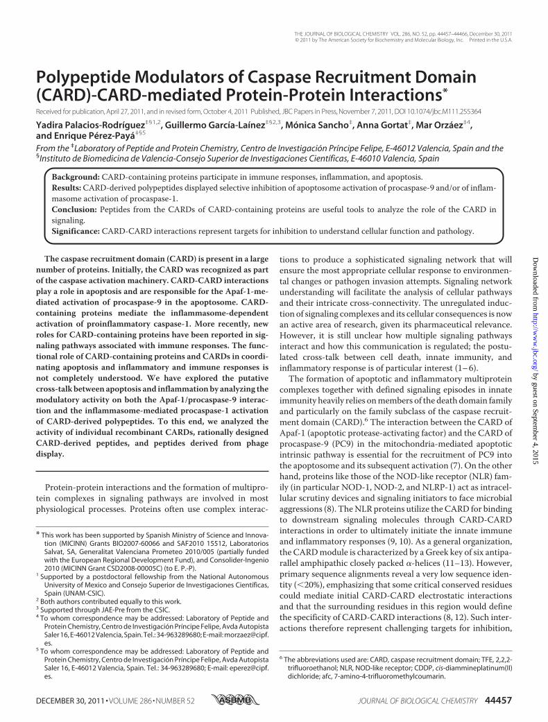
ptosome in vitro reconstitution assays (35, 36). RecombinantWD40 repeat-depleted Apaf-1 (Apaf-1 1–591) and recombi-nant PC9 were produced to set up the apoptosome in vitroreconstitution assay (29). Apaf-1 1–591 is able to promote theprocessing and activation of PC9 as followed by the release ofthe fluorophore afc from caspase-9-specific peptide substrateAc-LEHD-afc (37). We tested the ability of the recombinantCARDs to inhibit the Apaf-1 1–591-mediated PC9 activation.PC9 was incubated in the presence or absence of the CARDs atdifferent concentrations for 10 min before the addition ofApaf-1 1–591. After another 10-min period, the caspase-9 sub-strate was added, and PC9 activity was measured as a functionof time. The inhibition percentages of caspase-9 activity werecalculated and used to estimate the relative IC50 values (definedas the recombinant CARD concentration required inhibiting50% of Apaf-1 1–591-mediated PC9 activation). The recombi-nant CARDs of Apaf-1, PC9, and NLRP-1 exhibited a similarinhibitory activity (Table 1). The inhibitory activity of theCARDs was also evaluated in a cell extract-based assay (seebelow), which confirmed that the CARDs are capable of inhib-iting the apoptosome-mediated activation of the apoptoticpathway (Table 1).In order to gain further knowledge of the cross-interactions
and the nature of the CARD-CARD pathway connections, weevaluated the ability of the different CARDs to mediate PC9activation. The isolated recombinant Apaf-1 CARD has beenreported to be able to activate PC9 (37, 38). PC9 was preincu-bated with the different CARDs at increasing concentrations.After 10 min of preincubation, Ac-LEHD-afc was added to themedium, and PC9 activity was measured as a function of time.Recombinant PC9 showed a basal activity (38) as evidenced bythe cleavage of the fluorogenic substrate and increased fluores-cence. This activity was notably enhanced in the presence ofApaf-1 CARDbut not in the presence of PC9CARDorNLRP-1CARD (Fig. 2).CARD-derived Peptides as Modulators of Apaf-1-dependent
Activation of PC9—Peptides generated as copies of proteinfragments, in particular those sequences that participate in pro-tein-protein interactions, have been shown to be effectivemod-ulators of signaling events (39–42). In order to design CARD-derived peptides that could modulate CARD-CARD-mediatedprotein-protein interactions, we analyzed the primarysequence of the CARDs of Apaf-1, PC9, NOD-1, ASC, andNLRP-1 (PDB accession numbers: 3YGS for Apaf-1 and PC9and 2NZ7, 2KN6, and 3KAT for NOD-1, ASC, and NLRP-1,respectively). Previous studies have evidenced the common six-
helix bundle structural organization of CARDs (8, 12), espe-cially those selected for this study (Fig. 3A). The multiple-se-quence alignment (Fig. 3B) reveals the strongly conservedamino acids among the five selected domains. The CARD con-tains a basic and acidic patch on opposite sides, including crit-ical residues that mediate the postulated initial electrostaticinteraction (13, 43). In Apaf-1 CARD, residues Asp-27, Glu-39,Glu-40, and Glu-41 are localized within the acidic region andcould be the equivalents of residues Asp-42, Asp-48, Glu-53,Asp-54, and Glu-56 in NOD-1 CARD as well as of residuesAsp-1401,Glu-1411, andGlu-1414 inNLRP-1.On the oppositeside, the basic and concave region of PC9 includes residuesArg-6, Arg-7, Arg-10, Arg-13, Arg-52, and Arg-56 (8, 13). Toaccomplish the design, we analyzed the CARD-CARD complexdescribed for the target Apaf-1/PC9 interaction (13). Then thepeptides derived from helices 2 and 3 of Apaf-1 CARD wereselected (peptides A2.Y24G33 and A3.F34V43; see Fig. 3B andTable 2). These peptides included some of the abovementionedcritical residues, such as Asp-27 from helix 2 and Glu-39, Glu-40, andGlu-41 fromhelix 3. From counterpart protein PC9, thepeptides derived from helices 1 and 4, which included residuesArg-10 and Arg-13 and residues Arg-52 and Arg-56, respec-tively, were then selected (peptides C1.L8E17 and C4.R52I60;see Fig. 3B and Table 2). In an attempt to further analyze pep-tides derived from the apoptosis-unrelated CARD-containingproteins, a similar exercisewas applied toNOD-1,NLRP-1, andASC. Although the CARD-CARD-mediated Apaf-1�PC9 struc-ture has been solved (13), less is known about theCARD-CARDinteraction that participates in inflammasome formation. Thenwe explored peptides derived from both the acidic and basicamino acid patches of these CARDs. From the acidic patch, thepeptides designedwereN2.T37D48 (including residues Asp-42and Asp-48) and N3.Y49A60 (containing residues Glu-53 andAsp-54) from NOD-1 CARD helices 2 and 3, respectively; pep-tide Np2.V1398Q1406 containing the residue Asp-1401 fromthe NLRP-1 CARD helix 2; peptide As2.N128L136 containingresidue Asp-134 (helix 2); and As3.D143A151 containing resi-dues Asp-143 andGlu-144 (helix 3) from theASCCARD. From
TABLE 1CARDs inhibit Apaf-1-dependent PC9 activationIC50 values are defined as the recombinant CARD concentration required for inhib-iting 50% of PC9 activation. ND, not determined.
CARD IC50a IC50
b
�M �M
Apaf-1 1.7 � 0.2 5.6� 0.4PC9 0.8 � 0.1 0.8 � 0.1NLRP-1 0.9 � 0.1 1.6 � 0.1NOD-1 ND NDASC ND ND
a Apaf-1 1–591-mediated PC9 activation in an in vitro reconstituted apoptosome.bApaf-1XL cell extract-based assay.
FIGURE 2. CARD-mediated activation of procaspase-9. Apaf-1 CARD, PC9CARD, and NLRP-1 CARD were incubated at 1, 2, and 5 �M with recombinantnon-active PC-9 in S buffer. Caspase-9 activity was measured in the presenceof the fluorogenic Ac-LEHD-afc substrate. Caspase-9 activity is represented asincrease in fluorescence arbitrary units/s (AU/s). Control (C) refers to the basalactivity of PC-9. Data correspond to the mean � S.D. (error bars) of threeindependent experiments.
Polypeptide Modulators of CARD-CARD Interactions
44460 JOURNAL OF BIOLOGICAL CHEMISTRY VOLUME 286 • NUMBER 52 • DECEMBER 30, 2011
by guest on September 4, 2015
http://ww
w.jbc.org/
Dow
nloaded from

the basic patch, we synthesized the following peptides:N4.Q64V75 (helix 4 NOD-1 CARD), Np1.L1379R1392 (helix 1NLRP-1 CARD), Np4.R1422W1436 (helix 4 NLRP-1 CARD),As1.D116R125 (helix 1 ASC CARD), and As4.N155T166 (helix4 ASC CARD).CD spectroscopy revealed that all of the designed peptides
displayed low helicity in solution and that they thus predomi-nantly exist as a random coil (Table 2). However, in the pres-ence of the �-helix stabilizer 2,2,2-trifluoroethanol (44), pep-tides demonstrated helical stabilization (Table 2). Therefore,the full CARD-derived peptide set displayed environment-de-pendent conformational adaptability and was seen to be capa-ble of populating the CARD native helical conformationbeyond the tertiary context of the protein domain.The in vitro biological activity of the peptides was investi-
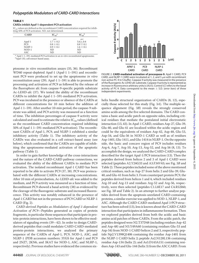
gated in the functional apoptosome assay. Peptides A2.Y24G33andA3.F34V43 and peptides C1.L8E17 andC4.R52I60, derivedfrom the CARDs of Apaf-1 and PC9, respectively, exerted amoderate inhibitory effect on the enzymatic activity of the apo-ptosome, unlike those peptides derived from the acidic patch ofthe CARDs of NOD-1, NLRP-1, and ASC, which did not (Table2). However, all of the peptides derived from the basic patchfrom the CARDs of NOD-1, NLRP-1, and ASC except forNp4.R1422W1436, which derived from helix 4 of NLRP-1CARD, inhibited apoptosome activity (Table 2).The in vitro results suggested that the peptides derived from
the CARD-CARD interacting surface of Apaf-1�PC9 and those
derived from the basic patch of the CARDs of NOD-1, NLRP-1,and ASC could be potential hits as selective inhibitors ofCARD/CARD-mediated protein-protein interactions in theapoptosome. However, in a cellular milieu, there are a largenumber of proteins and cellular metabolites that could com-promise the activity of peptides and their accessibility to themolecular target. To further explore the efficacy of the peptidehits in a cellular environment, their activity was initially evalu-ated in mammalian cellular extracts. Cytosolic extracts fromhuman embryonic kidney 293 cells (HEK 293) were depletedfrom endogenous Apaf-1 by chromatography (FT fraction; see“Experimental Procedures”). Cellular extracts were activated toenter the apoptotic pathway by the addition of baculovirus-produced recombinant full-length Apaf-1XL and incubationwith ATP and cytochrome c, which induced caspase-3 activity(14, 31). All of the peptides that inhibited the in vitro activity ofrecombinant apoptosome, except C4.R52I60, inhibited theapoptosome-mediated caspase-3 activity in cellular extracts(Table 2).As explained above, CARD-derived peptides were designed
and inspired by the reported high resolution structure of theCARD-CARD-mediated Apaf-1/PC9 interaction (13, 43). Pep-tides A2.Y24G33 and A3.F34V43 contained those proposed askey residues for the interaction and also covered most of theApaf-1 CARD surface defined as the Apaf-1 interactive surfacewith PC9 CARD. Similar premises were used for the design ofPC9 CARD-derived peptides C1.L8E17 and C4.R52I60. Thenwe wondered whether the active peptides would show an addi-tive or a synergic effect as inhibitors of apoptosome activity. Tothis end, we evaluated the activity of peptide combinations atsub-IC50 concentrations (Fig. 4). Only the Apaf-1 CARD-de-rived peptides had a synergic effect, in fact a combination ofpeptide A2.Y24G33 and peptide A3.F34V43 at 50 �M eachcame close to a 100% inhibition of the apoptosome-mediatedcaspase-3 activity in the cell extract assay (Fig. 4A; the expectedpercentage of inhibition for a simple additive effect came closeto 25%). Furthermore, the assay output confirmed theincreased activity of peptide A2.Y24G33 in the overall inhibi-tory activity. Actually, when peptides A2.Y24G33 andA3.F34V43 were combined at ratios of 1:2 or 2:1, the later com-bination displayed increased activity. These results agree withpreviously reported observations from the Apaf-1 CARD-PC9CARD interaction analysis (13), which highlighted the predom-inant role of Apaf-1 CARD helix 2 over the other helices (heli-ces 3 and 5) encompassing the binding surface that Apaf-1CARD offers to PC9 CARD for a productive interaction tooccur. Peptide A2.Y24G33 upon binding to PC9 CARD wouldinduce minor conformational changes that facilitated theaccessibility of other inhibitory peptides. In contrast, the PC9CARD-derived peptides (C1.L8E17 and C4.R52I60) did notshow a synergic inhibitory effect (Fig. 4B).According to the in vitro and cell extract results, peptides
A2.Y24G33, A3.F34V43, C1.L8E17, C4.R52I60, N4.Q64V75,Np1.L1379R1392, As1.D116R125, and As4.N155T166 inhib-ited the CARD-dependent activation of PC9 in the apopto-some. The peptides showed a dose-dependent activity, whichallowed the determination of IC50 values (Table 2). It is note-worthy that the experimental procedures required for an
FIGURE 3. Multiple-protein alignment of CARDs. A, structural overlappingof CARDs. The coordinates were obtained from the Protein Data Bank (acces-sion number 3YGS for Apaf-1 and PC9 CARDs, in black and yellow respectively;2NZ7 for NOD1 CARD in green; 3KAT for NLRP-1 CARD in blue; and 2KN6 forASC CARD in gray). Structural modeling was performed with MISTRAL (theMultiple Protein Structure Alignment Server), and PyMOL (Schrodinger LLC)was used to obtain the graphical representation. B, multiple-sequence align-ment (ClustalW2). Underlined fragments represent the sequences consideredfor the synthetic peptides. Conserved amino acids are indicated in red.
Polypeptide Modulators of CARD-CARD Interactions
DECEMBER 30, 2011 • VOLUME 286 • NUMBER 52 JOURNAL OF BIOLOGICAL CHEMISTRY 44461
by guest on September 4, 2015
http://ww
w.jbc.org/
Dow
nloaded from
unequivocal determination of the type of inhibition exertedwere not possible, given the complexity of the apoptosome as amacromolecular enzyme. Therefore, in order to corroboratethe mechanism of action of the peptides, we turned out our
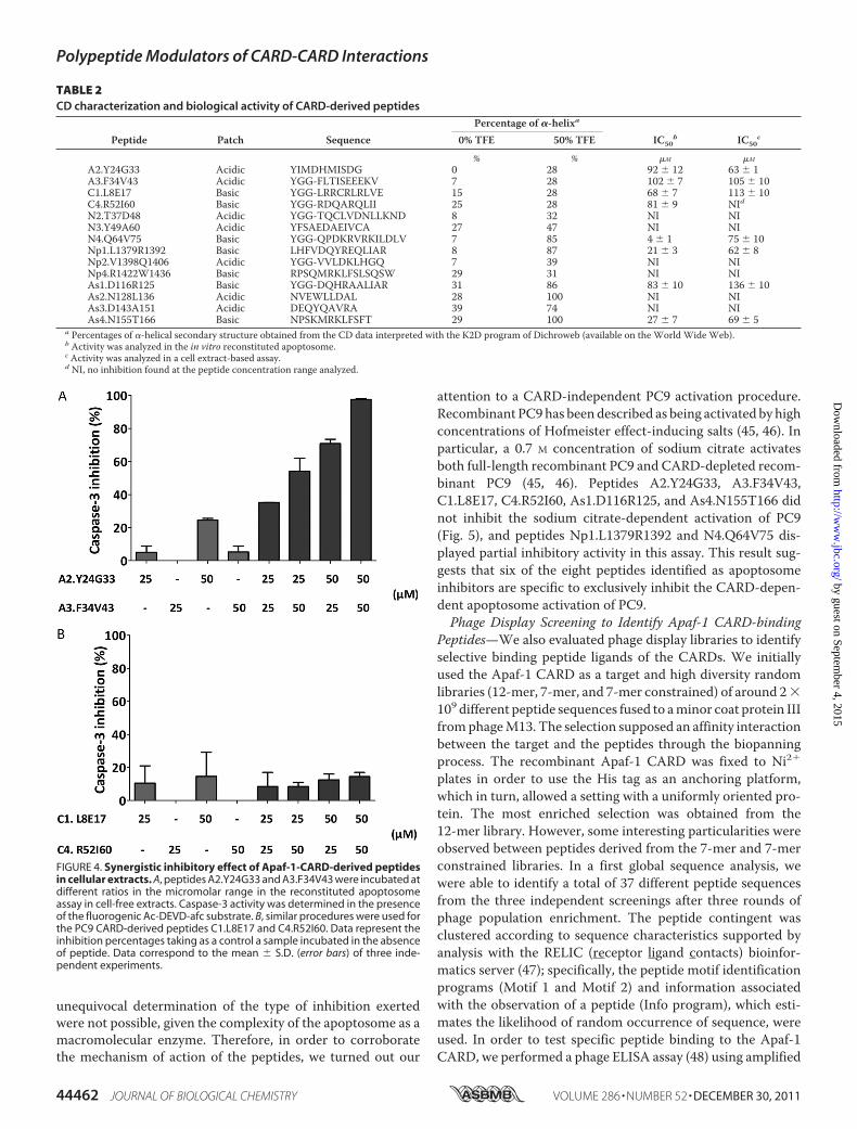
attention to a CARD-independent PC9 activation procedure.Recombinant PC9has beendescribed as being activated by highconcentrations of Hofmeister effect-inducing salts (45, 46). Inparticular, a 0.7 M concentration of sodium citrate activatesboth full-length recombinant PC9 and CARD-depleted recom-binant PC9 (45, 46). Peptides A2.Y24G33, A3.F34V43,C1.L8E17, C4.R52I60, As1.D116R125, and As4.N155T166 didnot inhibit the sodium citrate-dependent activation of PC9(Fig. 5), and peptides Np1.L1379R1392 and N4.Q64V75 dis-played partial inhibitory activity in this assay. This result sug-gests that six of the eight peptides identified as apoptosomeinhibitors are specific to exclusively inhibit the CARD-depen-dent apoptosome activation of PC9.Phage Display Screening to Identify Apaf-1 CARD-binding
Peptides—We also evaluated phage display libraries to identifyselective binding peptide ligands of the CARDs. We initiallyused the Apaf-1 CARD as a target and high diversity randomlibraries (12-mer, 7-mer, and 7-mer constrained) of around 2�109 different peptide sequences fused to aminor coat protein IIIfromphageM13. The selection supposed an affinity interactionbetween the target and the peptides through the biopanningprocess. The recombinant Apaf-1 CARD was fixed to Ni2�
plates in order to use the His tag as an anchoring platform,which in turn, allowed a setting with a uniformly oriented pro-tein. The most enriched selection was obtained from the12-mer library. However, some interesting particularities wereobserved between peptides derived from the 7-mer and 7-merconstrained libraries. In a first global sequence analysis, wewere able to identify a total of 37 different peptide sequencesfrom the three independent screenings after three rounds ofphage population enrichment. The peptide contingent wasclustered according to sequence characteristics supported byanalysis with the RELIC (receptor ligand contacts) bioinfor-matics server (47); specifically, the peptide motif identificationprograms (Motif 1 and Motif 2) and information associatedwith the observation of a peptide (Info program), which esti-mates the likelihood of random occurrence of sequence, wereused. In order to test specific peptide binding to the Apaf-1CARD, we performed a phage ELISA assay (48) using amplified
TABLE 2CD characterization and biological activity of CARD-derived peptides
Peptide Patch SequencePercentage of �-helixa
IC50b IC50
c0% TFE 50% TFE
% % �M �M
A2.Y24G33 Acidic YIMDHMISDG 0 28 92 � 12 63 � 1A3.F34V43 Acidic YGG-FLTISEEEKV 7 28 102 � 7 105 � 10C1.L8E17 Basic YGG-LRRCRLRLVE 15 28 68 � 7 113 � 10C4.R52I60 Basic YGG-RDQARQLII 25 28 81 � 9 NIdN2.T37D48 Acidic YGG-TQCLVDNLLKND 8 32 NI NIN3.Y49A60 Acidic YFSAEDAEIVCA 27 47 NI NIN4.Q64V75 Basic YGG-QPDKRVRKILDLV 7 85 4 � 1 75 � 10Np1.L1379R1392 Basic LHFVDQYREQLIAR 8 87 21 � 3 62 � 8Np2.V1398Q1406 Acidic YGG-VVLDKLHGQ 7 39 NI NINp4.R1422W1436 Basic RPSQMRKLFSLSQSW 29 31 NI NIAs1.D116R125 Basic YGG-DQHRAALIAR 31 86 83 � 10 136 � 10As2.N128L136 Acidic NVEWLLDAL 28 100 NI NIAs3.D143A151 Acidic DEQYQAVRA 39 74 NI NIAs4.N155T166 Basic NPSKMRKLFSFT 29 100 27 � 7 69 � 5
a Percentages of �-helical secondary structure obtained from the CD data interpreted with the K2D program of Dichroweb (available on the World Wide Web).b Activity was analyzed in the in vitro reconstituted apoptosome.c Activity was analyzed in a cell extract-based assay.d NI, no inhibition found at the peptide concentration range analyzed.
FIGURE 4. Synergistic inhibitory effect of Apaf-1-CARD-derived peptidesin cellular extracts. A, peptides A2.Y24G33 and A3.F34V43 were incubated atdifferent ratios in the micromolar range in the reconstituted apoptosomeassay in cell-free extracts. Caspase-3 activity was determined in the presenceof the fluorogenic Ac-DEVD-afc substrate. B, similar procedures were used forthe PC9 CARD-derived peptides C1.L8E17 and C4.R52I60. Data represent theinhibition percentages taking as a control a sample incubated in the absenceof peptide. Data correspond to the mean � S.D. (error bars) of three inde-pendent experiments.
Polypeptide Modulators of CARD-CARD Interactions
44462 JOURNAL OF BIOLOGICAL CHEMISTRY VOLUME 286 • NUMBER 52 • DECEMBER 30, 2011
by guest on September 4, 2015
http://ww
w.jbc.org/
Dow
nloaded from
individual phage clones. The phage-peptide binding to theApaf-1 CARD was evaluated through anti-M13 antibodies.From the contingent phage-peptide population, only a fewsequences interacted with the target with different affinities(results not shown), and a series of peptides were selected forchemical synthesis. This series included the most reacting linearsequences (WPTPPYA, ASLRTLTSLLPA, NFMESLPRLGMH,and AHLELRSNNMYF) and the constrained sequences cyclo-CSWFEASYC and cyclo-CLPTLHLLC. The peptides’ biologi-cal activity was investigated in both the in vitro functional apo-ptosome assay and themammalian cellular extract-based assay.Only the constrained peptide cyclo-CSWFEASYC exhibitedmoderate inhibitory activity in the two assays, whereas the lin-ear sequence ASLRTLTSLLPA showed inhibitory activity inthe in vitro assay but not in the cell extract-based assay (Table3). The peptide cyclo-CSWFEASYC did not inhibit the sodiumcitrate-dependent activation of PC9 (results not shown).Biological Activity of CARD-derived Peptides in Cell-based
Assays—Apaf-1-dependent apoptosome formation is requiredto induce apoptosis in response to the DNA-damaging agentcisplatin (CDDP) (15). We therefore studied the response ofHeLa cells to CDDP in the presence of not only the CARD-derived apoptosome peptide inhibitors that were active in thetwo in vitro assays shown in Table 2 but also the most activepeptide derived from the phage display approach (Table 3). Ingeneral terms, synthetic peptides have low cell permeability,and different strategies have been developed for the cytosolicdelivery of peptides. Here we have used the lipid Lipofectamineas a vehicle for peptide delivery. After 24 h of treatment,caspase-3 activity was monitored by evaluating the enzymaticactivity of the cell extract on the caspase-3 fluorogenic sub-strate Ac-DEVD-afc. The peptides (in particular C1.L8E17,As1.D116R125, As4.N155T166, and cyclo-CSWFEASYC) rele-vantly induced inhibition of CDDP apoptosome-mediatedcaspase-3 activity (Fig. 6).CARD-derived Peptides as Modulators of Procaspase-1-acti-
vating Inflammasome Complexes—In order to initially explorethe influence of the CARD-related peptides on the inflam-masome-mediated PC1 activation, we used a mammalian cel-
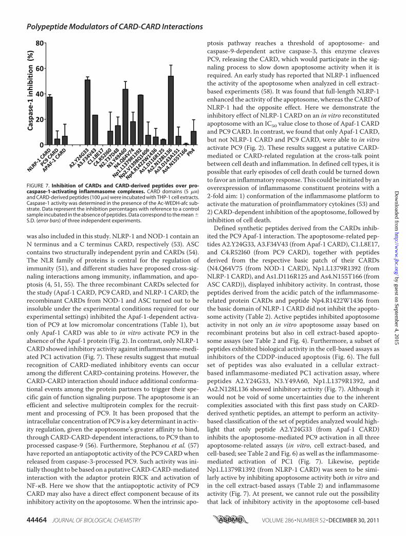
lular extract-based assay (32). Cytosolic extracts from thehuman acute monocytic leukemia cell line (THP-1) were pre-pared, and inflammasome-dependent PC1-activating activitywas induced by incubation at 37 °C in the presence of the dif-ferent CARDs and CARD-derived peptides. NLRP-1 CARDand peptides A2.Y24G33, N3.Y49A60, Np1.L1379R1392, andAs2.N128L136 inhibitedmore than 25% of inflammasome-me-diated PC1 activation (Fig. 7).
DISCUSSION
In this report, we initiated the in vitro evaluation of the role ofpolypeptides derived from CARD-containing proteins in themodulation of CARD-CARD-mediated protein-protein inter-actions. From the different CARD proteins, for the presentstudy, we selected Apaf-1 and PC9, with a well characterizedsignaling role in the mitochondria-mediated pathway (intrin-sic) of apoptosis (7, 49, 50) and NLRP-1 (NLR family, pyrindomain-containing 1; also namedNALP-1) andNOD-1 (nucle-otide-binding oligomerization domain-containing 1) as repre-sentative members of the inflammasome constituent NLRfamily of proteins (51). Protein ASC (apoptosis-associatedspeck-like protein containing a CARD), described as an adapterprotein in assembly of different inflammasome complexes (52),
FIGURE 5. Apaf-1, PC9, and ASC CARD-derived peptides do not inhibitcaspase-9 enzymatic activity. Caspase-9 preactivated in SC buffer was incu-bated with CARD-derived peptides (70 �M). Caspase-9 activity was deter-mined in the presence of the Ac-LEHD-afc substrate. Data represent the inhi-bition percentages with reference to a control sample incubated in theabsence of peptides. Data correspond to the mean � S.D. (error bars) of threeindependent experiments.
TABLE 3Biological activity of synthetic peptides derived from Apaf-1 CARDphage display
Peptide Sequence IC50a IC50
b
�M �M
Phg1 WPTPPYA NIc NIPhg2 ASLRTLTSLLPA 92 � 10 NIPhg3 NFMESLPRLGMH NI NIPhg4d Cyclo-CSWFEASYC 65 � 7 90 � 10Phg5d Cyclo-CLPTLHLLC NI NIPhg6 AHLELRSNNMYF NI NI
a Activity was analyzed in the in vitro reconstituted apoptosome.b Activity was analyzed in a cell extract-based assay.c NI, no inhibition found at the peptide concentration range analyzed.d Peptides Phg4 and Phg5, which contain two Cys residues in the N and the C ter-minus, were cycled (see “Experimental Procedures”).
FIGURE 6. Inhibition of cisplatin-dependent apoptosis in HeLa cells byCARD-derived peptides. HeLa cells were treated with a Lipofectamine/pep-tide (100 �M) mixture for 4 h, followed by administration of 20 �M CDDP. Cellswere harvested after 24 h, and cytosolic extracts were supplemented withcaspase assay buffer containing 20 �M Ac-DEVD-afc substrate for caspase-3activity evaluation. Data correspond to the mean � S.D. (error bars) of threeindependent experiments and represent the inhibition percentagesobtained with reference to a control sample incubated in the absence ofpeptides.
Polypeptide Modulators of CARD-CARD Interactions
DECEMBER 30, 2011 • VOLUME 286 • NUMBER 52 JOURNAL OF BIOLOGICAL CHEMISTRY 44463
by guest on September 4, 2015
http://ww
w.jbc.org/
Dow
nloaded from
was also included in this study. NLRP-1 and NOD-1 contain anN terminus and a C terminus CARD, respectively (53). ASCcontains two structurally independent pyrin and CARDs (54).The NLR family of proteins is central for the regulation ofimmunity (51), and different studies have proposed cross-sig-naling interactions among immunity, inflammation, and apo-ptosis (4, 51, 55). The three recombinant CARDs selected forthe study (Apaf-1 CARD, PC9 CARD, and NLRP-1 CARD; therecombinant CARDs from NOD-1 and ASC turned out to beinsoluble under the experimental conditions required for ourexperimental settings) inhibited the Apaf-1-dependent activa-tion of PC9 at low micromolar concentrations (Table 1), butonly Apaf-1 CARD was able to in vitro activate PC9 in theabsence of the Apaf-1 protein (Fig. 2). In contrast, only NLRP-1CARD showed inhibitory activity against inflammasome-medi-ated PC1 activation (Fig. 7). These results suggest that mutualrecognition of CARD-mediated inhibitory events can occuramong the different CARD-containing proteins. However, theCARD-CARD interaction should induce additional conforma-tional events among the protein partners to trigger their spe-cific gain of function signaling purpose. The apoptosome is anefficient and selective multiprotein complex for the recruit-ment and processing of PC9. It has been proposed that theintracellular concentration of PC9 is a key determinant in activ-ity regulation, given the apoptosome’s greater affinity to bind,through CARD-CARD-dependent interactions, to PC9 than toprocessed caspase-9 (56). Furthermore, Stephanou et al. (57)have reported an antiapoptotic activity of the PC9 CARDwhenreleased from caspase-3-processed PC9. Such activity was ini-tially thought to be based on a putativeCARD-CARD-mediatedinteraction with the adaptor protein RICK and activation ofNF-�B. Here we show that the antiapoptotic activity of PC9CARD may also have a direct effect component because of itsinhibitory activity on the apoptosome.When the intrinsic apo-
ptosis pathway reaches a threshold of apoptosome- andcaspase-9-dependent active caspase-3, this enzyme cleavesPC9, releasing the CARD, which would participate in the sig-naling process to slow down apoptosome activity when it isrequired. An early study has reported that NLRP-1 influencedthe activity of the apoptosome when analyzed in cell extract-based experiments (58). It was found that full-length NLRP-1enhanced the activity of the apoptosome, whereas the CARD ofNLRP-1 had the opposite effect. Here we demonstrate theinhibitory effect of NLRP-1 CARD on an in vitro reconstitutedapoptosome with an IC50 value close to those of Apaf-1 CARDand PC9 CARD. In contrast, we found that only Apaf-1 CARD,but not NLRP-1 CARD and PC9 CARD, were able to in vitroactivate PC9 (Fig. 2). These results suggest a putative CARD-mediated or CARD-related regulation at the cross-talk pointbetween cell death and inflammation. In defined cell types, it ispossible that early episodes of cell death could be turned downto favor an inflammatory response. This could be initiated by anoverexpression of inflammasome constituent proteins with a2-fold aim: 1) conformation of the inflammasome platform toactivate the maturation of proinflammatory cytokines (53) and2) CARD-dependent inhibition of the apoptosome, followed byinhibition of cell death.Defined synthetic peptides derived from the CARDs inhib-
ited the PC9 Apaf-1 interaction. The apoptosome-related pep-tides A2.Y24G33, A3.F34V43 (from Apaf-1 CARD), C1.L8E17,and C4.R52I60 (from PC9 CARD), together with peptidesderived from the respective basic patch of their CARDs(N4.Q64V75 (from NOD-1 CARD), Np1.L1379R1392 (fromNLRP-1 CARD), and As1.D116R125 and As4.N155T166 (fromASC CARD)), displayed inhibitory activity. In contrast, thosepeptides derived from the acidic patch of the inflammasome-related protein CARDs and peptide Np4.R1422W1436 fromthe basic domain of NLRP-1 CARD did not inhibit the apopto-some activity (Table 2). Active peptides inhibited apoptosomeactivity in not only an in vitro apoptosome assay based onrecombinant proteins but also in cell extract-based apopto-some assays (see Table 2 and Fig. 4). Furthermore, a subset ofpeptides exhibited biological activity in the cell-based assays asinhibitors of the CDDP-induced apoptosis (Fig. 6). The fullset of peptides was also evaluated in a cellular extract-based inflammasome-mediated PC1 activation assay, wherepeptides A2.Y24G33, N3.Y49A60, Np1.L1379R1392, andAs2.N128L136 showed inhibitory activity (Fig. 7). Although itwould not be void of some uncertainties due to the inherentcomplexities associated with this first pass study on CARD-derived synthetic peptides, an attempt to perform an activity-based classification of the set of peptides analyzed would high-light that only peptide A2.Y24G33 (from Apaf-1 CARD)inhibits the apoptosome-mediated PC9 activation in all threeapoptosome-related assays (in vitro, cell extract-based, andcell-based; see Table 2 and Fig. 6) as well as the inflammasome-mediated activation of PC1 (Fig. 7). Likewise, peptideNp1.L1379R1392 (from NLRP-1 CARD) was seen to be simi-larly active by inhibiting apoptosome activity both in vitro andin the cell extract-based assays (Table 2) and inflammasomeactivity (Fig. 7). At present, we cannot rule out the possibilitythat lack of inhibitory activity in the apoptosome cell-based
FIGURE 7. Inhibition of CARDs and CARD-derived peptides over pro-caspase-1-activating inflammasome complexes. CARD domains (5 �M)and CARD-derived peptides (100 �M) were incubated with THP-1 cell extracts.Caspase-1 activity was determined in the presence of the Ac-WEDH-afc sub-strate. Data represent the inhibition percentages with reference to a controlsample incubated in the absence of peptides. Data correspond to the mean �S.D. (error bars) of three independent experiments.
Polypeptide Modulators of CARD-CARD Interactions
44464 JOURNAL OF BIOLOGICAL CHEMISTRY VOLUME 286 • NUMBER 52 • DECEMBER 30, 2011
by guest on September 4, 2015
http://ww
w.jbc.org/
Dow
nloaded from

assay can be ascribed to the low cell permeability of this partic-ular peptide. Peptides N3.Y49A60 (from NOD-1 CARD) andAs2.N128L136 (fromASCCARD) inhibited the inflammasomebut were inactive as apoptosome inhibitors. Besides their ownactivity, these peptides could be valuable tools to drive thedesign of more pharmacologically relevant inhibitors, such aspeptidomimetics or small molecules, of CARD-CARD-depen-dent multiprotein signaling complexes.We also approached the identification of CARD-modulat-
ing peptides by exploring the phage display approach. Afterthree selection rounds, RELIC-aided analysis of peptidesequences, and a further phage-ELISA-based selection, weobtained only one peptide with a moderate ability to inhibitthe apoptosome activity in the three apoptosome-relatedassays (in vitro, cell extract-based, and cell-based), cyclo-CSWFEASYC (Table 3 and Fig. 6). In a future study, we willanalyze the interaction site of these peptides with the Apaf-1CARD, which should expose a binding site of low relevance forthe CARD-CARD interaction.The peptides described herein, togetherwith those described
by Marasco et al. (59) derived from the CARD of BCL10, arepioneering tools to interrogate not only the initial systems thatinspired the design (the apoptosome and BCL10-interactingproteins, respectively) but also the complex and still poorlyunderstood role of the CARD as signaling device. CARD-con-taining proteins exert relevant functions as scaffolds to build upkeymultiprotein complexes that amplify signaling in apoptosis,inflammation, and immunity (53, 60). Although other proteindomain-dependent signaling pathways have been studiedmoreand have helped draw pictures with cross-connections in theintricate cell signaling process (61), the contribution of CARD-dependent signaling complexes to the important decisions thatcells, and probably tissues, have to make to prioritize apoptosisor inflammation is still in its initial stages and poses importantopen questions. The development of research tools, such assynthetic peptides derived from CARDs and the set up of reli-able in vitro assays, will probably facilitate future research inthis field.
Acknowledgments—We thank Alicia García-Jareño, Rebeca Mon-tava, and Eliana Sirvent for technical assistance.
REFERENCES1. Seth, R. B., Sun, L., Ea, C. K., and Chen, Z. J. (2005) Cell 122, 669-
6822. Hiscott, J., Lin, R., Nakhaei, P., and Paz, S. (2006) Trends Mol. Med. 12,
53–563. Fernandes-Alnemri, T., Yu, J. W., Datta, P., Wu, J., and Alnemri, E. S.
(2009) Nature 458, 509–5134. Inohara, N., Koseki, T., del Peso, L., Hu, Y., Yee, C., Chen, S., Carrio, R.,
Merino, J., Liu, D., Ni, J., and Núñez, G. (1999) J. Biol. Chem. 274,14560–14567
5. Mahoney, D. J., Cheung, H. H., Mrad, R. L., Plenchette, S., Simard, C.,Enwere, E., Arora, V.,Mak, T.W., Lacasse, E. C.,Waring, J., and Korneluk,R. G. (2008) Proc. Natl. Acad. Sci. U.S.A. 105, 11778–11783
6. Varfolomeev, E., Blankenship, J.W.,Wayson, S.M., Fedorova, A. V., Kaya-gaki, N., Garg, P., Zobel, K., Dynek, J. N., Elliott, L. O., Wallweber, H. J.,Flygare, J. A., Fairbrother, W. J., Deshayes, K., Dixit, V. M., and Vucic, D.(2007) Cell 131, 669–681
7. Acehan, D., Jiang, X., Morgan, D. G., Heuser, J. E., Wang, X., and Akey,
C. W. (2002)Mol. Cell 9, 423–4328. Proell, M., Riedl, S. J., Fritz, J. H., Rojas, A. M., and Schwarzenbacher, R.
(2008) PLoS One 3, e21199. Inohara, N., and Nuñez, G. (2003) Nat. Rev. Immunol. 3, 371–38210. Park, H. H., Lo, Y. C., Lin, S. C., Wang, L., Yang, J. K., and Wu, H. (2007)
Annu. Rev. Immunol. 25, 561–58611. Coussens, N. P., Mowers, J. C., McDonald, C., Nuñez, G., and Ramas-
wamy, S. (2007) Biochem. Biophys. Res. Commun. 353, 1–512. Manon, F., Favier, A., Núñez, G., Simorre, J. P., and Cusack, S. (2007) J.
Mol. Biol. 365, 160–17413. Zhou, P., Chou, J., Olea, R. S., Yuan, J., and Wagner, G. (1999) Proc. Natl.
Acad. Sci. U.S.A. 96, 11265–1127014. Malet, G., Martín, A. G., Orzáez, M., Vicent, M. J., Masip, I., Sanclimens,
G., Ferrer-Montiel, A., Mingarro, I., Messeguer, A., Fearnhead, H. O., andPérez-Payá, E. (2006) Cell Death Differ. 13, 1523–1532
15. Mondragón, L., Galluzzi, L., Mouhamad, S., Orzáez, M., Vicencio, J. M.,Vitale, I., Moure, A., Messeguer, A., Perez-Paya, E., Kroemer, G. (2009)Apoptosis 14, 182–190
16. Mondragón, L., Orzáez, M., Sanclimens, G., Moure, A., Armiñán, A.,Sepúlveda, P., Messeguer, A., Vicent, M. J., and Pérez-Payá, E. (2008)J. Med. Chem. 51, 521–529
17. Vicent, M. J., and Pérez-Payá, E. (2006) J. Med. Chem. 49, 3763–376518. Caratù, G., Allegra, D., Bimonte, M., Schiattarella, G. G., D’Ambrosio, C.,
Scaloni, A., Napolitano, M., Russo, T., and Zambrano, N. (2007)Mol. CellProteomics 6, 333–345
19. Fenske, S. A., Yesilaltay, A., Pal, R., Daniels, K., Rigotti, A., Krieger,M., andKocher, O. (2008) J. Biol. Chem. 283, 22097–22104
20. Schlegel, B. P., Starita, L. M., and Parvin, J. D. (2003) Oncogene 22,983–991
21. Várnai, P., Bondeva, T., Tamás, P., Tóth, B., Buday, L., Hunyady, L., andBalla, T. (2005) J. Cell Sci. 118, 4879–4888
22. Day, T. W., and Safa, A. R. (2009)Mini Rev. Med. Chem. 9, 741–74823. Paddison, P. J., and Hannon, G. J. (2002) Cancer Cell 2, 17–2324. Zender, L., and Kubicka, S. (2004) Apoptosis 9, 51–5425. Rubinstein, M., and Niv, M. Y. (2009) Biopolymers 91, 505–51326. Scott, J. K., and Smith, G. P. (1990) Science 249, 386–39027. Orzáez, M., Mondragón, L., Marzo, I., Sanclimens, G., Messeguer, A.,
Pérez-Payá, E., and Vicent, M. J. (2007) Peptides 28, 958–96828. Mora, P., Mas-Moruno, C., Tamborero, S., Cruz, L. J., Pérez-Payá, E., and
Albericio, F. (2006) J. Pept. Sci. 12, 491–49629. Riedl, S. J., Li,W., Chao, Y., Schwarzenbacher, R., and Shi, Y. (2005)Nature
434, 926–93330. Stennicke, H. R., and Salvesen, G. S. (1999)Methods 17, 313–31931. Fearnhead, H. O. (2001)Methods Cell Biol. 66, 167–18532. Gong, Y.N.,Wang, X.,Wang, J., Yang, Z., Li, S., Yang, J., Liu, L., Lei, X., and
Shao, F. (2010) Cell Res. 20, 1289–130533. Palacios-Rodríguez, Y., Gazarian, T., Rowley, M., Majluf-Cruz, A., and
Gazarian, K. (2007) J. Microbiol. Methods 68, 225–23534. Li, P., Nijhawan,D., Budihardjo, I., Srinivasula, S.M., Ahmad,M., Alnemri,
E. S., and Wang, X. (1997) Cell 91, 479–48935. Zou, H., Henzel, W. J., Liu, X., Lutschg, A., and Wang, X. (1997) Cell 90,
405–41336. Saleh, A., Srinivasula, S. M., Acharya, S., Fishel, R., and Alnemri, E. S.
(1999) J. Biol. Chem. 274, 17941–1794537. Pérez-Payá, E., Orzáez, M., Mondragón, L., Wolan, D., Wells, J. A.,
Messeguer, A., and Vicent, M. J. (2011)Med. Res. Rev. 31, 649–67538. Shiozaki, E. N., Chai, J., and Shi, Y. (2002) Proc. Natl. Acad. Sci. U.S.A. 99,
4197–420239. Agopian, A., Gros, E., Aldrian-Herrada, G., Bosquet, N., Clayette, P., Di-
vita, G., Caputo, G. A., Litvinov, R. I., Li, W., Bennett, J. S., Degrado,W. F.,and Yin, H. (2009) J. Biol. Chem. 284, 254–264
40. Churchill, E. N., Qvit, N., and Mochly-Rosen, D. (2009) Trends Endocri-nol. Metab. 20, 25–33
41. Walensky, L. D., Kung, A. L., Escher, I., Malia, T. J., Barbuto, S., Wright,R. D., Wagner, G., Verdine, G. L., and Korsmeyer, S. J. (2004) Science 305,1466–1470
42. Yeon, S. W., Jeon, Y. J., Hwang, E. M., and Kim, T. Y. (2007) Peptides 28,838–844
Polypeptide Modulators of CARD-CARD Interactions
DECEMBER 30, 2011 • VOLUME 286 • NUMBER 52 JOURNAL OF BIOLOGICAL CHEMISTRY 44465
by guest on September 4, 2015
http://ww
w.jbc.org/
Dow
nloaded from
43. Qin, H., Srinivasula, S. M., Wu, G., Fernandes-Alnemri, T., Alnemri, E. S.,and Shi, Y. (1999) Nature 399, 549–557
44. Pérez-Payá, E., Houghten, R.A., andBlondelle, S. E. (1994)Biochem. J.299,587–591
45. Boatright, K.M., Renatus,M., Scott, F. L., Sperandio, S., Shin,H., Pedersen,I. M., Ricci, J. E., Edris, W. A., Sutherlin, D. P., Green, D. R., and Salvesen,G. S. (2003)Mol. Cell 11, 529–541
46. Pop, C., Timmer, J., Sperandio, S., and Salvesen, G. S. (2006)Mol. Cell 22,269–275
47. Mandava, S., Makowski, L., Devarapalli, S., Uzubell, J., and Rodi, D. J.(2004) Proteomics 4, 1439–1460
48. Serasinghe, M. N., Seneviratne, A. M., Smrcka, A. V., and Yoon, Y. (2010)J. Biol. Chem. 285, 620–630
49. Hu, Y., Benedict, M. A., Ding, L., and Núñez, G. (1999) EMBO J. 18,3586–3595
50. Yu, X., Acehan,D.,Ménétret, J. F., Booth, C. R., Ludtke, S. J., Riedl, S. J., Shi,Y., Wang, X., and Akey, C. W. (2005) Structure 13, 1725–1735
51. Ting, J. P.,Willingham, S. B., and Bergstralh, D. T. (2008)Nat. Rev. Immu-nol. 8, 372–379
52. Ippagunta, S. K., Malireddi, R. K., Shaw, P. J., Neale, G. A., Walle, L. V.,Green, D. R., Fukui, Y., Lamkanfi, M., and Kanneganti, T. D. (2011) Nat.Immunol. 12, 1010–1016
53. Schroder, K., and Tschopp, J. (2010) Cell 140, 821–83254. de Alba, E. (2009) J. Biol. Chem. 284, 32932–3294155. Bruey, J. M., Bruey-Sedano, N., Luciano, F., Zhai, D., Balpai, R., Xu, C.,
Kress, C. L., Bailly-Maitre, B., Li, X., Osterman, A., Matsuzawa, S., Ter-skikh, A. V., Faustin, B., and Reed, J. C. (2007) Cell 129, 45–56
56. Malladi, S., Challa-Malladi,M., Fearnhead, H. O., and Bratton, S. B. (2009)EMBO J. 28, 1916–1925
57. Stephanou, A., Scarabelli, T. M., Knight, R. A., and Latchman, D. S. (2002)J. Biol. Chem. 277, 13693–13699
58. Chu, Z. L., Pio, F., Xie, Z., Welsh, K., Krajewska, M., Krajewski, S., Godzik,A., and Reed, J. C. (2001) J. Biol. Chem. 276, 9239–9245
59. Marasco, D., Stilo, R., Sandomenico, A., Monti, S. M., Tizzano, B., deCapua, A., Varricchio, E., Liguoro, D., Zotti, T., Formisano, S., Ruvo, M.,and Vito, P. (2009) Biochem. J. 422, 553–561
60. Bouchier-Hayes, L., and Martin, S. J. (2002) EMBO Rep. 3, 616–62161. Smith, F. D., and Scott, J. D. (2002) Curr. Biol. 12, R32–40
Polypeptide Modulators of CARD-CARD Interactions
44466 JOURNAL OF BIOLOGICAL CHEMISTRY VOLUME 286 • NUMBER 52 • DECEMBER 30, 2011
by guest on September 4, 2015
http://ww
w.jbc.org/
Dow
nloaded from
Mar Orzáez and Enrique Pérez-PayáGarcía-Laínez, Mónica Sancho, Anna Gortat, Yadira Palacios-Rodríguez, Guillermo Interactions(CARD)-CARD-mediated Protein-ProteinRecruitment Domain Polypeptide Modulators of CaspaseSignal Transduction:
doi: 10.1074/jbc.M111.255364 originally published online November 7, 20112011, 286:44457-44466.J. Biol. Chem.
10.1074/jbc.M111.255364Access the most updated version of this article at doi:
.JBC Affinity SitesFind articles, minireviews, Reflections and Classics on similar topics on the
Alerts:
When a correction for this article is posted•
When this article is cited•
to choose from all of JBC's e-mail alertsClick here
http://www.jbc.org/content/286/52/44457.full.html#ref-list-1
This article cites 61 references, 19 of which can be accessed free at
by guest on September 4, 2015
http://ww
w.jbc.org/
Dow
nloaded from
Related Documents