It's All In Your Brain: Graded Motor Imagery for Pain Modulation Susan W . Stralka , PT, DPT , DPT, MS, ACHE Baptist Rehab Germantown, Germantown, TN Jane Fedorczyk , PT, PhD , CHT, ATC Physical Therapy, Drexel University, Philadelphia, PA DESCRIPTION: Graded Motor Imagery is an emerging therapeutic strategy for treating complex pain patients. It integrates established principles of graded exposure and response prevention with current theories in the neuroscience of pain. Grade Motor Imagery consists of laterality training, imagery and mirror therapy with the aim of exposing the brain to movement related therapies to induce positive reorganization of the brain. OBJECTIVES Upon completion of this course, you will be able to: 1. Discuss current theories on normal and abnormal peripheral and central pain mechanisms. 2. Explain the cortical reorganization associated with complex and/or chronic pain. 3. Describe the neuroscience principles behind the use of a graded motor imagery program (GMIP). 4. Identify the practical applications of a GMIP with various patient populations in which pain is a primary impairment. 3. Develop treatment strategies using GMIP to improve function in the involved upper limb. 4. Integrate the use of laterality reconstruction, visual and motor imagery, and mirror therapy into the plan of care for complex pain patients to achieve pain management and functional outcomes. SELECTED REFERENCES: 1. Acerra N, Moseley GL. Dysynchiria: Watching the mirror image of the unaffected limb elicits pain on the affected side. Neurol. 2005; 65:751-753. 2. Daly A, Bialocerkowski A. Does evidence support physiotherapy management of adult complex regional pain syndrome type one? A systemic review. Euro J Pain. 2008. 3. Flor H, Denke C, Schaefer M, Russer S. Effect of sensory discrimination training on cortical reorganization and phantom limb pain. Lancet. 2001; 357: 1763-1764. 4. Maihofner C, Handwerker HO, Neundorfer B, Birklein F. Patterns of cortical reorganization in complex regional pain syndrome. Neurol. 2003; 61:1707-1715. 5. McCabe CS, Haigh RC, Ring EFJ, et al. A controlled pilot study of the utility of mirror visual feedback in the treatment of complex regional pain syndrome (type 1). Rheumatol. 2003; 42:97-101. 6. Moseley GL. Graded motor imagery is effective for long-standing complex regional pain syndrome: a randomized controlled trial. Pain. 2004; 108:192-198. 7. Moseley GL. Is successful rehabilitation of complex regional pain syndrome due to sustained attention to the affected limb? A randomized controlled trial. Pain. 2005; 114:54-61.

Welcome message from author
This document is posted to help you gain knowledge. Please leave a comment to let me know what you think about it! Share it to your friends and learn new things together.
Transcript

It's All In Your Brain: Graded Motor Imagery for Pain Modulation
Susan W . Stralka , PT, DPT , DPT, MS, ACHEBaptist Rehab Germantown, Germantown, TN
Jane Fedorczyk , PT, PhD , CHT, ATCPhysical Therapy, Drexel University, Philadelphia, PA
DESCRIPTION: Graded Motor Imagery is an emerging therapeutic strategy for treatingcomplex pain patients. It integrates established principles of graded exposure andresponse prevention with current theories in the neuroscience of pain. Grade MotorImagery consists of laterality training, imagery and mirror therapy with the aim ofexposing the brain to movement related therapies to induce positive reorganization of thebrain.
OBJECTIVESUpon completion of this course, you will be able to:1. Discuss current theories on normal and abnormal peripheral and central pain
mechanisms.2. Explain the cortical reorganization associated with complex and/or chronic pain.3. Describe the neuroscience principles behind the use of a graded motor imagery
program (GMIP).4. Identify the practical applications of a GMIP with various patient populations in which
pain is a primary impairment.3. Develop treatment strategies using GMIP to improve function in the involved upper
limb.4. Integrate the use of laterality reconstruction, visual and motor imagery, and mirror
therapy into the plan of care for complex pain patients to achieve pain managementand functional outcomes.
SELECTED REFERENCES:
1. Acerra N, Moseley GL. Dysynchiria: Watching the mirror image of the unaffected limbelicits pain on the affected side. Neurol. 2005; 65:751-753.
2. Daly A, Bialocerkowski A. Does evidence support physiotherapy management of adultcomplex regional pain syndrome type one? A systemic review. Euro J Pain. 2008.
3. Flor H, Denke C, Schaefer M, Russer S. Effect of sensory discrimination training on corticalreorganization and phantom limb pain. Lancet. 2001; 357: 1763-1764.
4. Maihofner C, Handwerker HO, Neundorfer B, Birklein F. Patterns of cortical reorganizationin complex regional pain syndrome. Neurol. 2003; 61:1707-1715.
5. McCabe CS, Haigh RC, Ring EFJ, et al. A controlled pilot study of the utility of mirror visualfeedback in the treatment of complex regional pain syndrome (type 1). Rheumatol. 2003;42:97-101.
6. Moseley GL. Graded motor imagery is effective for long-standing complex regional painsyndrome: a randomized controlled trial. Pain. 2004; 108:192-198.
7. Moseley GL. Is successful rehabilitation of complex regional pain syndrome due to sustainedattention to the affected limb? A randomized controlled trial. Pain. 2005; 114:54-61.

8. Moseley GL, Parsons TJ, Spence c. Visual distortion of a limb modulates the pain andwelling evoked by movement. Curr Biol. 2008; 18:R1047-R1048.
9. Sluka KA: Mechanisms and Management of Pain for the Physical Therapist. Seattle:IASP Press, 2009. www.iasp-pain.org
10. Mersky, H, Bogduk, N,eds. Classification of chronic pain: descriptions of chronicpain syndromes and definitions of pain terms, 2nd ed. Seattle: IASP Press, 1994.
11. Gifford LS, Butler DS. The integration of pain sciences into clinical practice. Journalof Hand Therapy, 1997; 10:86-95.
12. Urban MO, Gebhart GF. Central mechanisms of pain. Medical Clinics of NorthAmerica, 1999; 83: 585-596.
13. Melzack R and Wall PD: Pain mechanisms: a new theory. Science, 1965; 150: 971-979.
14. Harris GH, Susman JL: Managing musculoskeletal complaints with rehabilitationtherapy: summary of the Philadelphia Panel evidence-based clinical practiceguidelines on musculoskeletal rehabilitation interventions. The Journal of FamilyPractice, 2002; 51: 1042-1046.
15. Wright A, Sluka K: Nonpharmacological treatments for musculoskeletal pain. TheClinical Journal of Pain, 2001; 17: 33-46.
16. Fedorczyk JM: Pain Management: Principles of Therapist's Intervention. In SkirvenTM, Osterman AL, Fedorczyk JM and Amadio PC (eds): Rehabilitation of the Handand Upper Extremity, 6th edition, Mosby, St. Louis, 2011
17. Elliott M, Barbe MF: Understanding Pain Mechanisms: The Basis of ClinicalDecision Making for Pain Modulation. In Skirven TM, Osterman AL, Fedorczyk JMand Amadio PC (eds): Rehabilitation of the Hand and Upper Extremity, 6`h edition,Mosby, St. Louis, 2011
Normal and Abnormal Pain Mechanisms:Concepts for Clinical Decision Making
Jane Fedorczyk, PT, PhD, CHT, ATCDrexel University Philadelphia, PA
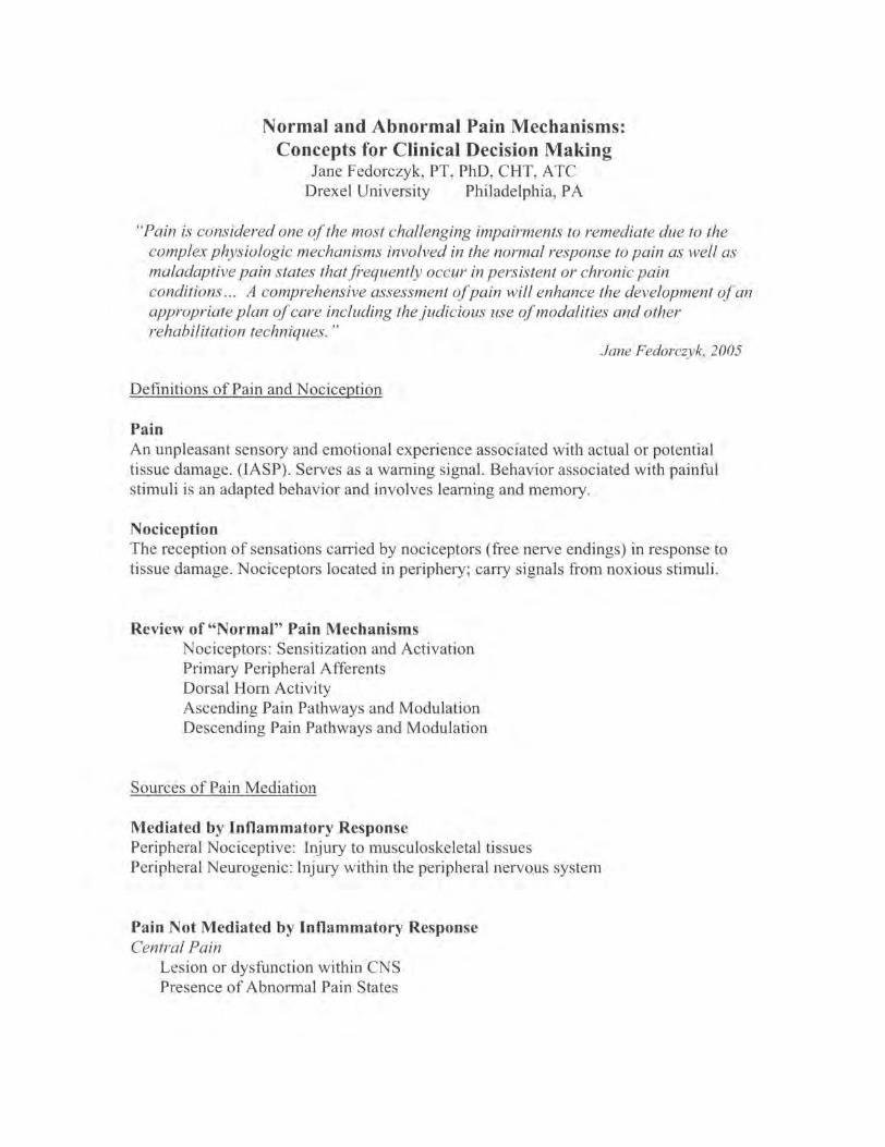
"Pain is considered one of the most challenging impairments to remediate due to thecomplex physiologic mechanisms involved in the normal response to pain as well asmaladaptive pain states that frequently occur in persistent or chronic painconditions... A comprehensive assessment of pain will enhance the development of anappropriate plan of care including the judicious use of modalities and otherrehabilitation techniques. "
Jane Fedorczyk, 2005
Definitions of Pain and Nociception
PainAn unpleasant sensory and emotional experience associated with actual or potentialtissue damage. (IASP). Serves as a warning signal. Behavior associated with painfulstimuli is an adapted behavior and involves learning and memory.
NociceptionThe reception of sensations carried by nociceptors (free nerve endings) in response totissue damage. Nociceptors located in periphery; carry signals from noxious stimuli.
Review of "Normal" Pain MechanismsNociceptors: Sensitization and ActivationPrimary Peripheral AfferentsDorsal Horn ActivityAscending Pain Pathways and ModulationDescending Pain Pathways and Modulation
Sources of Pain Mediation
Mediated by Inflammatory ResponsePeripheral Nociceptive: Injury to musculoskeletal tissuesPeripheral Neurogenic: Injury within the peripheral nervous system
Pain Not Mediated by Inflammatory ResponseCentral Pain
Lesion or dysfunction within CNSPresence of Abnormal Pain States
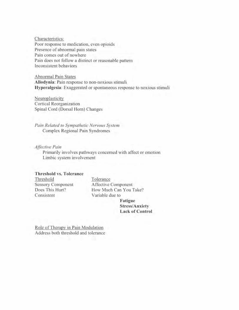
Characteristics:Poor response to medication , even opioidsPresence of abnormal pain statesPain comes out of nowherePain does not follow a distinct or reasonable patternInconsistent behaviors
Abnormal Pain StatesAllodynia : Pain response to non-noxious stimuliHyperalgesia : Exaggerated or spontaneous response to noxious stimuli
NeuroplasticityCortical ReorganizationSpinal Cord (Dorsal Horn) Changes
Pain Related to Sympathetic Nervous SystemComplex Regional Pain Syndromes
Affective Pain
Primarily involves pathways concerned with affect or emotionLimbic system involvement
Threshold vs. ToleranceThreshold ToleranceSensory Component Affective ComponentDoes This Hurt? How Much Can You Take?Consistent Variable due to
FatigueStress/AnxietyLack of Control
Role of Therapy in Pain ModulationAddress both threshold and tolerance
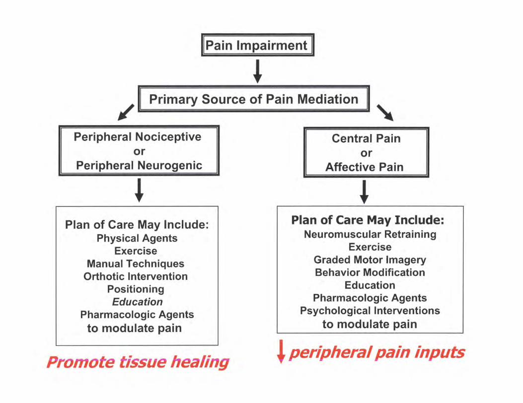
Pain impairment11
Jrd
Primary Source of Pain Mediation
Peripheral Nociceptiveor
Peripheral Neurogenic
JrPlan of Care May Include:
Physical AgentsExercise
Manual TechniquesOrthotic Intervention
PositioningEducation
Pharmacologic Agentsto modulate pain
tiCentral Pain
orAffective Pain
JrPlan of Care May Include:
Neuromuscular RetrainingExercise
Graded Motor ImageryBehavior Modification
EducationPharmacologic Agents
Psychological Interventionsto modulate pain
I peripheral pain inputsPromote tissue healing

Global Tactics for Nerve and Tissue Healing
Global Tactics
Increase Increase ^L 1 PromoteBloodflow Oxygen Ap4 Stressfree Use
of Hand and UE
Aerobic Drink Water AvoidExercise Static Postures
Avoid It, It Diaphragmatic Joint ProtectioinCaffiene 0 A Breathing Energy Conservation
and Nicotine

It's All In Your BrainGraded Motor Imagery
Susan W . Straika , PT, DPT, MS, ACHEsusan . [email protected]
Neuro Science Revisited• Cortical change- chronic pain
Up - peripheral nociceptive stimuliwn - cortical central processing
• GMI - retrain the brain
CSM 2011 1
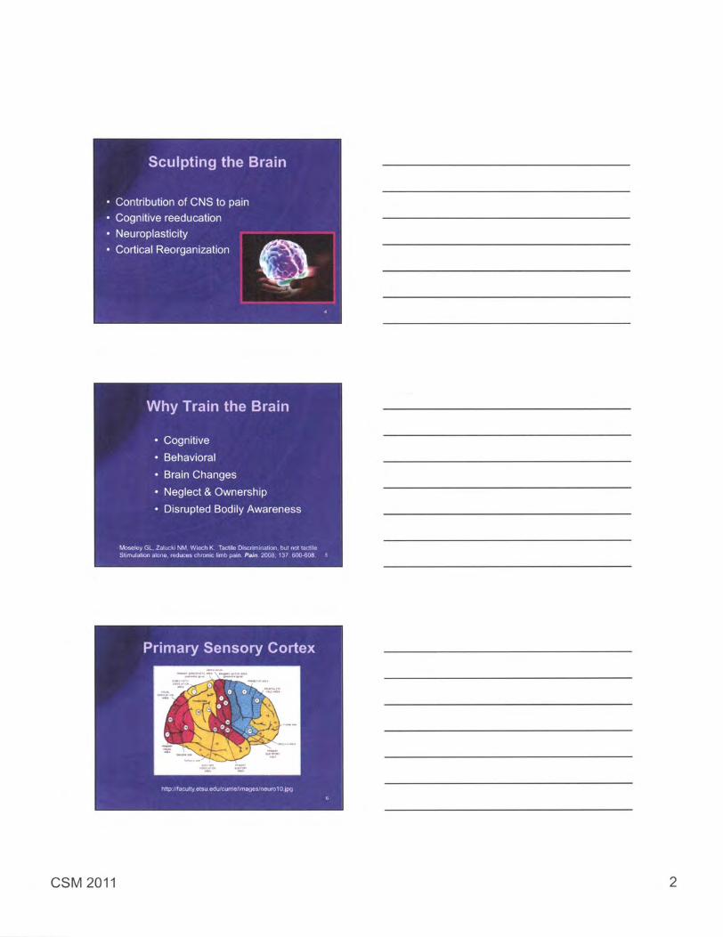
Why Train the Brain
• Cognitive
• Behavioral
• Brain Changes
• Neglect & Ownership• Disrupted Bodily Awareness
Moseley GL, Zalucki NM, Wiech K. Tactile Discrimination, but not tactileStimulation alone , reduces chronic limb pain . Pain . 2008; 137: 600-608. 5
Primary Sensory Cortex
http://faculty,etsu.edutcurrie/images /neural O.jpg
CSM 2011 2

Brain Neuroplasticity
• Persistent anatomical or physiological changes in a neuronthat occurs during development , regeneration , experimentalmanipulation or repeated activity across a synapse
• Throughout life, the brain is able to restructure itself tochange by adapting
CSM 2011 3
Neural Plasticity
http://thesituationist . wordpress . com/2007/04/02/your-brain-and-morality/ +0
Good Neuroplasticity
• Persistent anatomical or physiologicalchanges in a neuron that occurs duringdevelopment , regeneration,experimental manipulation or repeatedactivity across a synapse
• Throughout life, the brain is able torestructure itself to change by adapting
Principle : NeuralPlasticity
• Not purely motor but partially due to a sensorydeficit or a sensory motor disconnection
• Maximize neural adaptation behaviors whichdrive changes in central nervous system require
attention and repetition over time positive
feedback
• Behaviors goal directed
• Accurate and progressed in complexity
• Goal increases progressive firing of neurons is
CSM 2011 4
Rewiring the BrainGraded Motor Imagery Program
• Laterality Reconstruction- Restoration of brain's concept of left and right- Try to imagine your hand in that position.
• Visual and Motor Imagery- Conscious access to brain which are involved in
intention, preparation and then carrying out themovement
• Mirror Therapy- The brain is tricked into thinking that the limb is better
than the brain thinks it is
Sp
Mirror Therapy
• Mirror conveys visual stimuli to the brain
• Observation of one 's unaffected part
• Principle states affected limb can bestimulated by visual cues originating fromthe opposite side of the body.
R
Graded Motor Imagery ProgramLaterality Reconstruction v.. Visual & Motor Imagery
Mirror Therapy
• Non-threatening• Normal somatosensory input to over-come altered motor
control or dysfunctional movement pattern.• Emphasis is on intervention of the non -painful movement.• Helps to restore the disruption of normal interaction
between intention to move the limb in absence ofappropriate sensory feedback
CSM 2011 5
Graded Motor Imagery (GMI)
• Sequentially graded , progressing from easy todifficult and non-threatening to threatening
• Hand representation in minor will changesynapses in the brain
• With hand in box , begin with less aggressivemovement then the outside hand can movemore aggressively and review symptoms
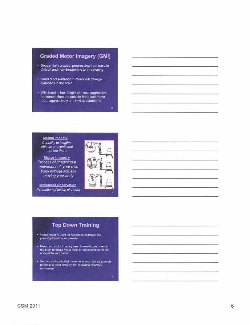
Mental InnaaervCapacity to imagine
objects or events thatare not there
Motor ImagervProcess of imagining amovement of your own
body without actuallymoving your body
Movement ObservationPerception of action of others
Top Down Training
• Visual imagery used for relearning cognitive andplanning aspect of movement
• Mirror and motor imagery used to re-educate or retrainthe brain for basic motor skills by concentrating on thenon-painful movement
RU
y,
• Smooth and controlled movements must act as examplefor brain to reset circuitry that mediates voluntarymovement
i
CSM 2011 6

Neural Plasticity Happens
• Sensory discrimination and • Specificity of feedback isfine motor task enhanced
Neuron show structuralchange Myelination is increased
• Cortical representations are • Synapse input isexpanded strengthened
• Receptive fields smallerthan normal ' Integration time is shortened
• Number of excitable„e,,- ,^ Complexity of dendritic
branching is enhancedis
Rebirth-ImagingFunctional Magnetic Resonance Imaging (fMRI)
fMRI provides a means to observe whichstructures participate in specific functions.
• Increase blood flow of the brain areas that are recruited for atask
• 30% neuron can be recruited to fire when one thinks aboutthe image of the task
• Visual imaging recruits neuronal activity• Magnetic resonance imaging can be used to map changes in
brain hemodynamics that correspond to manta! operations ofneural activity as detected by a blood oxygen level dependentsignal.
FE
CSM 2011 7

Mirror Neurons
• 1987 - Rizzolotti, et.al. found that when amonkey reached for apeanut or watched anexperimenter reachingfor a peanut , that themirror neuron involvedin coding of goal usingactions.
FX
Mirror Neurons• Ramachandran VS: Plasticity and functional recovery
in neurology. Clinical Medicine . July/Aug 2005;Vol 5 (4):369.
• Ramachandran VS, Rogers-Ramachandran D.Synaesthesia in phantom limbs induced with mirrors.Proc Roy Sac Lond. 1996; 263:377-386.
• Ramachandran VS, Hirstein W. The D 0 Hebb lecture:Perception of phantom limbs. Brain. 1998;121:1603-1630.
CSM 2011 8

Mirror Neurons
• Activated by observing and executing movement• Located in premotor cortex and inferior parietal lobe
Mirror Neuron System
fires neurons when both observationand execution of movement occur
• In humans, this is the mechanism that
intentionsPremotor neurons- Fire when you observe someone
doing a task- Imagining a task- Mirror imaging
ms - imitate and understand their
Riuolattl G, Craighero L. The Mirror-Neuron System. Annual Review ofNeuroscience . 2004; 27:169-192.
PR
m
Research
• Activated by performanceLocated in premotor cortex and inferior parietal lobe
• Monkey - premotor cortex- Discharge when performs a given motor act and when
observe the same motor act• Human - ample evidence
- Cortical network that discharges in some way - observingand executing movement
Riuolatti G, Craighero L. The Minor- Neuron System . Annual Review ofNeuroscience . 2004; 27:169-192.
CSM 2011 9

Mirror Neuron Role in Rehab
• Improve motorperformance by usingvisual & motor imagery
• Motor imitation andmotor execution excitethe corticospinalpathway
Hand Therapy and theMirror Neuron System
K-1
• Movement observation may be analternative way to activity the motorsystem based on the mirror neuron system
• May be used during immobilization ordeafferentiation
• Hand transplants - retraining
ResearchThe Netherlands
• Mirror neurons fire not only when action isexecuted , but also when one observes anotherperson performing the same action
• Encode both our actions and actions of others
deVries S, Mulder T. Motor Imagery and StrokeRehabilitation : A Critical Discussion . J Rehabil Med2007; 39:5-13.
P9
CSM 2011 10
Research by Stefan• Mirror neuron system firing is instrumental in motor
learning
• Overt motor practice may not be totally necessaryfor implicit motor learning
• Observing movements may facilitate motorperformance
Stefan K , at al. Formation of a motor memory by action observation.J Neurosci . 2005 ; 25: 9339-9346.
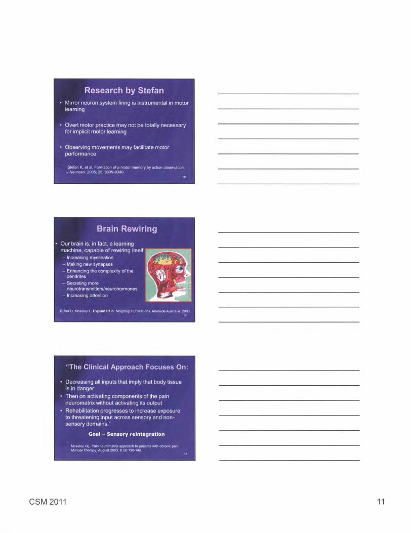
Brain Rewiring
• Our brain is, in fact , a learningmachine, capable of rewiring itself- Increasing myelination- Making new synapses- Enhancing the complexity of the
dendrites- Secreting more
neurotransmitters/neurohormones- Increasing attention
Butler D , Moseley L. Explain Pain . Noigroup Publications. Adelaide Australia. 2003.
32
",The Clinical Approach Focuses On:
• Decreasing all inputs that imply that body tissueis in danger
• Then on activating components of the painneuromatrix without activating its output
• Rehabilitation progresses to increase exposureto threatening input across sensory and non-sensory domains."
Goal - Sensory reintegration
Moseley GL . Pain neuromatrix approach to patients with chronic pain.Manual Therapy. August 2003 ; 8 (3):130-140.
in]
CSM 2011 11
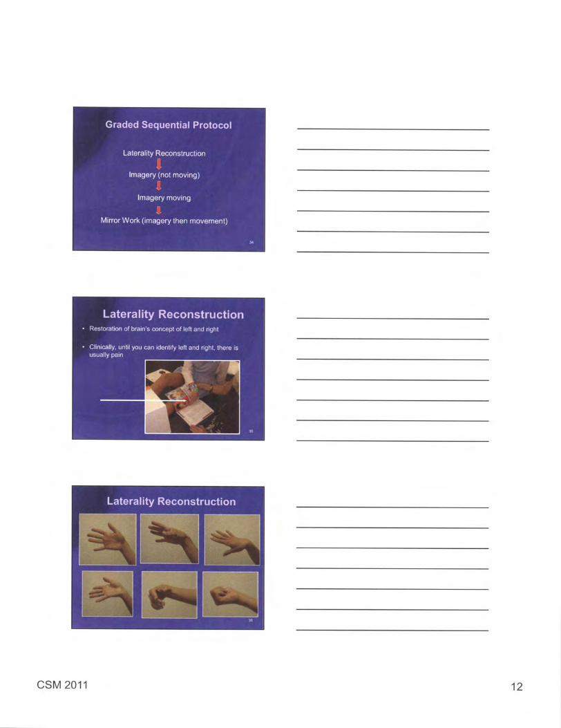
Graded Sequential Protocol
Laterality Reconstruction
UImagery (not moving)
UImagery moving
1Minor Work (imagery then movement)
lu
Laterality Reconstruction• Restoration of brain ' s concept of left and right
• Clinically, until you can identify left and right , there isusually pain
CSM 2011 12

Motor Imagery
• Conscious access to brain• Think - preparation and carrying out
movement
• Imaging or watching an activity• Start static posture then imagine it
moving
CSM 2011 13

Mirror ImageryTreatment Regime
• Mirror to view healthy limb andreflection of same healthy limb mimicsthe involved limb
• Told to concentrate hard on the imageas if both limbs were normal
Mirror Imagery andMirror Therapy
Tricks the brain into correcting its distorted image of the body
• Pain results from a mis-match in the way the brain perceivesthe body and the actual condition of the body
• Brain is tricked into thinking that the limb is actually betterthan the brain thinks it is
• Affected limb inside box
• Unaffected limb outside box
CSM 2011 14

Top - Down Therapy
• The mirror reflection permits thesubject to rehearse movements ofthe affected limb without having todirectly activate those parts ofmaladaptive central process thattypically produce pain
F-I
Pain is Not a Happy Tune
• Dominates every aspect of life, work,family, relationships, emotions and beliefs
CSM 2011 15
NeuroMatrix Model of Pain
Models the theory that the brain has a neural networkthat integrates information from multiple sources toproduce the
"experience that is labeledPain and cortical mechanisms are involved not just onesingle pain center- Anterior cingulate cortex- Thalamus- Sensorimotor
Melzack R . From the gate to the neuromatrix . Pain, 1999.
M
• "The NeuroMatrix Theory integrates newfindings from brain imaging studies,including:- Pain brain mapping- Pain and pharmaceutical interventions- Cortical reorganization and pain- Studies on the effect of stress on pain- Research on cognitive-behavioral factors and
pain
The Paths of Pain , Merskey, Loeser, Dubner, 2005
l ExperienceThe Brain is in Control = no brain no pain
Pain:- "A multiple system output, activated by and
specific pain neuromatrix . This neuromatrix isactivated whenever the brain concludes it is indanger and action is required and pain isallocated an anatomical reference in the virtualbody."
Moseley GL Pain neuromatrix approach to patients with chronic pain . ManualTherapy. August 2003; 8 (3):130-140.
as
CSM 2011 16

Pain experiences cancreate change in the Brain
• Pain memories-through the same mechanisms thatenable humans to learn and retain memories.
• Pain memories can form through sensitization of thenervous system and is apparent on brain imagingstudies.
• The pathways that transport painful stimuli, may change,the structural changes may lead to increased excitationin the brain in the presence of non-painful stimulus.
Tracy Hampton PhD, JAMA, June 2005
Mind and Body
m
1 is
a `feeling". via
activation of variousneurotags.
Butler D, Moseley L. Explain Pain. Noigroup Publications. Adelaide Australia. 2003.50
Pain experience is not hard-wired
• It is more complex than the traditional painpathway theories
-ain:-"A multiple system %Ltput, activated by and
specific pain neuromatrix. This neuromatrix isactivated whenever the brain concludes it is indanger and action is required and in i sallocated an anatomical reference in thevirtual body."
RE
CSM 2011 17

Pain and Associative Learning
• Conditioned Response
• Pain memories can form when movements andsensations that signal onset of pain elicitanticipation of pain.
• Eventually , this can work in reverse- Anticipation of pain can elicit a pain response
• Just thinking about a movement may hurt
M
.^,rh,.m.n^ ("nets heath
a jnmer^ ^
--Chronic Pain •^bwMn and
/ * ^^ ^1<JI.-mom
Rehabilitation of Pain PatientsFundamental Principles
Pain is an output of the brain that is produced wheneverthe brain concludes that body tissue is in danger andaction is required.
• Pain is a multisystem output that is produced when anindividual -specific cortical pain neuromatrix is activated.
• Pain becomes chronic , the efficacy of the painneuromatrix is strengthened via nociceptive and non-nociceptive mechanisms , which means that less input,both nociceptive and non-nociceptive , is required toproduce pain
CSM 2011 18
Why Train the Brain
• Cognitive - patient must understand the problem
• Behavioral- function and movement hierarch
• Brain changes - S1 reorganization
• Neglect & Ownership - it doesn't feel like mine
• Disrupted bodily awareness - change and
disruption in higher order due to cognitive
representation m
Pain Research
• Not single hard -wired dedicated pathway
• Converging evidence- physiological andfunctional imaging studies
• Move diffuse and plastic system-- Cord , brainstem , thalamus and cortex
• psychological studies-- Attention , anticipation , preparation for action
58
CSM 2011 19

Chronic Pain• 3 independent mechanisms contribute to
chronicity- Nociceptive- Non-nociceptive
Chronic Pain
• Associated with reduced tactile acuity
• Relationship = pain intensity, tactile acuity andcortical reorganization
• Research by Moseley , Zalucki , etc., tactilestimulation can decrease pain and increasetactile acuity when patients are required todiscriminate between type and location of tactilestimuli
W.
Fear Avoidance ModelMind - Body ConnectionPain causes altered motor control which leads todevelopment of dysfunctional movement patterns
• Developing of protective movement and fear ofmovement causes musculoskeletal impairment
ROM
ZMuscle length changes
ZStrength
Treat the uninvolved side
CSM 2011 20

Cognitive RestructuringThrough Education
• Help patients understand that pain maynot be giving an accurate account of theirtissues
• There are clear physiological effects andchanges in the brain when subjects thinkdifferently about their pain. (Flor. AdvNeurol. 2003)
Brain Maps• The brain maps our experience and the maps
represent our skills and our knowledge• When a skill develops or changes the neuro pools,
s;_rr c.;; ; will change and the brain maps willchange.
• When we approach learning or an experienceseriously, we:- Attend to the task- We practice- And we become emotionally involved
Pain Treatment(Multi-disciplinary)
• Shift from pain focusedapproach to function-based approach
• Motivation = explain,encourage , motivate
• Coping strategies -anxiety/depression
• Empower the patient
Educate on brain and painso family and patientschange their belief systemabout pain
Understand mind and brainare connected
Family education betweenhurt and harm
Relaxation-Relaxation--Relaxation m
CSM 2011 21

Effective Treatment
• Treat the WHOLE person• It is stored in the nervous system as
emotional and physical wants
Goal:Learn your way out of the disabilitybecause you learned your way in
UM
Clinical Value ofPhysical and Emotional Dualism
• Both need integrating and managing early on inpatient care
Can't have one without the other
Understanding Pain and Motor ImpairmentCognitive Restructuring through Education
• Help patients understand that pain maynot be giving an accurate account oftheir tissues
• There are clear physiological effects andchanges in the brain when subjects thinkdifferently about their pain.
m
CSM 2011 22

Work-Related MusculoskeletalDisorder (WMSD)
• Systemic response
• Neurological reorganization centrally
• In spinal cord and cerebral cortex
• Neuroplastic reorganization mayprecede onset of motor decrements
m
Why Patients Don't Get Wellwith Repetitive Stress Injuries
• Signs: abnormality of the normal homuncularorganization of the fingers representationin primary somatosensory cortex
- Chronic pain, intermittent and vague controlproblems or somatosensory dysfunction may beearly signs of focal dystonia
- Treatment must consist of discriminative sensorymotor skills
m
Outcomes : Negative Learning
• Important cortical representations shrink and• Adjacent cortical areas expand and become dysfunctional• Imbalances develop in neurotransmitters and
neurohormones• Focused attention can be reduced• Imbalance develops between sensory inputs (feed forward
and feedback ) and motor outputs• Sensory system becomes abnormally sensitive (decreased
threshold for excitation , chronic pain and/or neuropathicpain)
CSM 2011 23

Aberrant Learning
• Cortical sensory changes after excessiverepetitive movement
• Cortical representation reduced receptive field,now, very large and overlap adjacent digits anddorsal glabrous surface hand
• Brain can NIQ longer differentiate individual digitsand control their movement
m
Byl, Merzenich , et al. Research
• Change neural structure by attended repetitivepractice
• Expansion of cortical representation of digits aftersensory attended training program
• Normal receptive fields on hand become smallerwith training , more dense and numerous sorepresentation is more specific
Byl NN , Merzenlch MM, et al . A Primate Model for Studying Focal Dyslonia andRepetitive Strain Injury Effects on the Primary SomatosensoryCortex . PhysicalTherapy. March 1997 ; Vol 77 ( 3)269-284.
CSM 2011 24

Risk Factors Dystonia
• Type A• Perfectionists• Long working,
under-stress, poorbiomechanics
ft D
Neurologically Induced Behavior inHighly Repetitive Task
• Centrally mediated
• Maybe unresponsive to intervention thataddress only localized injury - Add to program:- Laterality- Mirror imaging- Motor control
Byl's 5 Phases forTreatment of Dystonia
1. Imagine hand or foot are normal
2. Improve sensory discrimination
3. Perform and concentrate on small gradedcontrolled movements
4. Work on sensory motor skills
5. Restore fine motor control
d
E
rk
CSM 2011 25
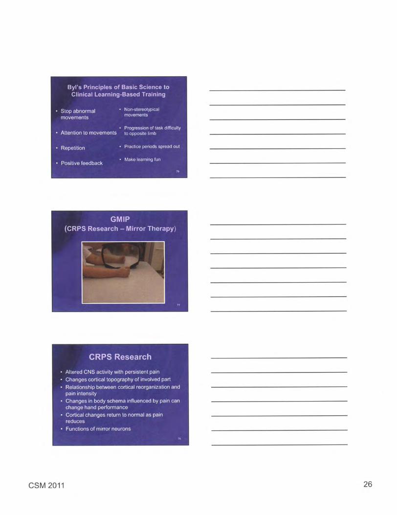
Byl's Principles of Basic Science toClinical Learning -Based Training
• Stop abnormalmovements
• Non-stereotypicalmovements
• Progression of task difficulty• Attention to movements to opposite limb
• Repetition
• Positive feedback
• Practice periods spread out
• Make learning fun
W.
CRPS Research
• Altered CNS activity with persistent pain• Changes cortical topography of involved part• Relationship between cortical reorganization and
pain intensity• Changes in body schema influenced by pain can
change hand performance• Cortical changes return to normal as pain
reduces• Functions of mirror neurons
CSM 2011 26

CRPS I and 11
• Persistent pain, which isdisproportionate to the originalinjury
• Patient reports- Extra skin sensitivity
(hyperesthesia)
- Color or skin temperature change
- Sweating - increase or decrease
- Edema
- Decreased ROM- Motor planning difficulty
• Non-painful stimuli is painful-- (allodynia)
• Swelling or sweating(asymmetry)
• Abnormal movement• Trophic changes in hair, nail
or skin
CRPS Patients
• Often tend to neglect their affected limb despite the pain• Often position it out of field of vision similar to
neurological neglect or motor neglect• Some view limb as foreign or strange or larger than it is• Recent brain imaging studies show CRPS patients have
disrupted cortical reorganization . This can effect how anindividual perceives their own body
• Amount of pain and degree of cortical disorganization
CSM 2011 27

CRPS
• Strong negative feeling or strong bodydysmorphic
• Novel treatment CRPS -
- mirror visual feedback- motor imagery
RN
Delay in Hand ActionsRecognition in People with CRPS
• Disrupted body schema
• Neglect like effect
• Cortical change and perceptual aspectsof body sense
Laterality TestCRPS in Hand Takes Longer to Recognize
• Mentally maneuver own hand to match• Imagined and actual movement is reaction time
(RT)• Brain activity changes in excitability of spinal
motor neuron pool and EMG• Hand laterality relies on body schema or real
time dynamic representation from sensory inputends integrates with motor systems for control ofaction
CSM 2011 28

Graded Motor Imagery for PathologicPain: A Randomized Controlled Trial
N = 51 patients with phantom limb pain or CRPS I
Performed 2 weeks each of laterality recognition
Imagined movements and mirror movements thenprogressed to PT and ongoing medical care
Moseley GL. Neurology. 2006: Dec 26; 67(12):2129-2134.
ResearchGraded motor imagery for pathologic pain: a
randomized controlled trial.Moseley GL. Neurology. 2006 ; Dec 26; 67(12):2129-2134
EIR
• Result:- Pain and Function : There was a main statistical effect on
the treatment group but not the diagnostic group- Mean decrease in pain between pre- and post -treatment
for the two groups:• Motor Imagery group- 23.4 mm• Control group - 10.5 mm
- Improvement function for both groups was similar and wasmaintained at the 6 -month follow-up
"Motor imagery reduced patients ' pain and disability withCRPS but the mechanisms of the effect are not clear**as
Graded Motor Imagery for PathologicPain: A Randomized Controlled Trial
Limb laterality recognitionImagined movementsMirror movements
5
.rr
Day e-u
OR=
N porwfapo.t.program I 3(244
NNTtog.ta4-POWk" ""
In funWon
®
NNT forboth criteria
Moseley GL . Neurology . 2006; Dec 26 ; 67(12):2129-2134.[:E
CSM 2011 29

Graded Motor Imagery is Effectivefor Long-Standing CRPS: ARandomized Controlled Trial
N = 13 chronic CRPS 1 patients.
Non-moving , MIP or ongoing management for12 weeks.
MIP group - 2 weeks of hand laterality,recognition tasks, imagined hand movement andmirror therapy.
Moseley GL . Pain. 2004 ; 108:192-198. LL
Results• Mirror Imaging Program (MIP) without movement support.
The hypothesis that MIP is more effective than otherongoing medical management.
• 50% of the patients no longer met the criteria for CRPSafter 6 weeks of MIP.
• During the program of the MIP and maintained status forat least 6 weeks after completion of treatment.
• There was no change in the controls in any measure.• Crossover gains in the control group were the same as
above for the experiment group.
,nll.xI ul,. N,urning,: 21-.; 6'(12)212n 21
Rrnic nd Ly Swan V. Svalka . nd p^LG.hc i on I L
Progression of Program
• Progression based on:- Reaction time is equal to that of the unaffected
time for 2 consecutive days- Imagined motions do not increase symptoms- Imagine moving the limb slowly and to adopt the
position in the picture- Smoothly return to resting position- Imagine doing this with out pain- Graded exposure to activity ; i.e. Quota based
therapy
CSM 2011 30

Tactile discrimination, but nottactile stimulation alone , reduces
chronic limb painMoseley GL , Zalucki NM, Wiech K . Pain. 2008 137: 600-608.
• Chronic pain causes reduced tactile acuity.
• Relationship exist between pain intensity , tactile acuityand cortical reorganization.
• Results : Tactile stimulation at 3 months causeddecreased pain and increased tactile acuity whenpatients were asked to discriminate between the typeand location of tactile stimuli.
15
Tactile Discrimination , but not tactile
stimulation alone , reduces chronic limb pain.
Moseley GL, Zalucki NM, Wiech K. Tactile Discrimination, but not tactileStimulation alone , reduces chronic limb pain . Pain . 2008; 137: 600-608.
0I
CRPS
ON 1111111=1111119=1nmr.
W!!l^
, t *lllllll ll
rr"Sn""Ej
®®ll®i_
gu
denotes that the subject reduced analgesic medication during the review period
Moseley GL , at al. Pain . 2008; 137:600.608.
CSM 2011 31

CRPS Research - Motor Imagery Program(MIP)
Graded Motor Imagery is effective for long-standing CRPS:A randomized controlled study:- 26 CRPS Type I chronic patients s/p non-complicated
wrist fx. 6 monthsAllocated to 2 groups
Traditional ongoing therapyMIP program: 2 weeks of laterality, 2 weeks of imagery
2 weeks of mirror therapyAssessments: Initial - 2,4,6 and 12 week intervalsOutcome measures:
Neuropathic Pain Scale (NPS)SwellingResponse times to recognize the affected hand
Exercise and Function
• Introduce exercise slowly and start with the leastfeared movements
• Patient needs to feel in control of what they aredoing
• As a therapist, do not harshly challenge thepatient's beliefs about movement and pain
• Explain what happens in a normal limb that isimmobilized and then starts to move again
• Understanding information like this helps improvepatient confidence with movement
WR
Object IdentificationActive Stimulation
• Read Braille and play card game in Braille• Match symbols, letters, figures, shapes
• Play games with eyes closed - dominos• Put shapes into matched holes - eyes open
and closed
• Choose letters and alphabet in sand• Find matching objects on floor, sand and
beans
Adapted from Nancy By]
CSM 2011 32

Nontarget Sensory Motor Tasks
• Identify different alphabet letters with eyes opened andclosed
• Spell words with alphabet letters - write letters with toes• Remove small objects from a box and identify and time the
task - move objects with toes and feet• Eyes closed- feel pegs , sense the touch by holding them
and put pegs in holes without (no pain or abnormalmovement) - attempt with feet
• Increase speed• With arms resting on thighs , shoulder and hands relaxed -
move finger to identify part of the body with light touch oruse toe to touch and identify parts of the body.
Research
• CRPS Type I- Visual input from moving unaffected limb
reestablishes the pain-free relationshipbetween sensory feedback and motorexecution
PI]
Laterality and CRPS
• Delayed recognition of hand laterality is relatedto the duration of symptoms and to the pain thatwould be invoked by executing the movement
• Both involve cortical reorganization of bodyschema
• Guarding type response occurs upstream fromthe motor cortex at a motor planning level
M.
CSM 2011 33
CRPS Theoretical Models
Disruption of sensory cortical processing andshrinkage of cortical representation in the primarysensory cortex. (Juttonen 2002)
Disinhibition of the motor cortex (Schwenkreis 2003)
• Disrupted Body Schema (Schoebel 2009 )
CRPS Research - Mirror Therapy
• A controlled study of the utility of mirrorvisual feedback in the treatment of CRPSType I
• Hypothesis:- CRPS is a consequence of disruption of central
sensory processing and that congruent visualfeedback from the moving unaffected limb asprovided by a mirror would restore the integrity ofcortical processing thereby relieving pain andrestoring function in the affected limb
McCabe CS . at al., Rheumatology 2003 'Is
CRPS Type arch - Mirror Therapy
A control utility of Mirror VisualFeedback
• Eight subjects diseases duration a 3 weeks to :5 3 years,albdynla- Six weeks study- 2 controls:
• No device to view• Non-reflective surface
- Intervention: Mirror visual feedback- Measured outcomes: Pain severity, Visual Analog scales,
Vasomotor changes (infrared thermography)
McCabe CS. et al., Rheumatology 2003 ME
CSM 2011 34
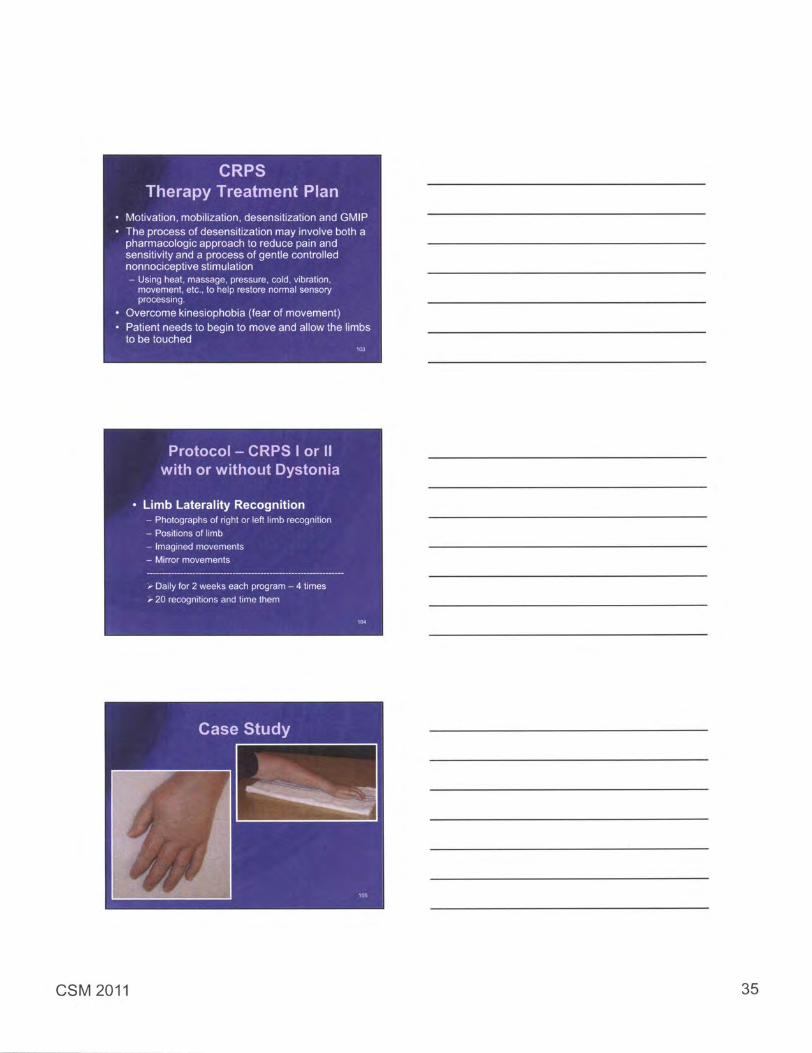
CRPSTherapy Treatment Plan
• Motivation , mobilization , desensitization and GMIP• The process of desensitization may involve both a
pharmacologic approach to reduce pain andsensitivity and a process of gentle controllednonnociceptive stimulation- Using heat , massage , pressure , cold, vibration,
movement , etc., to help restore normal sensoryprocessing.
• Overcome kinesiophobia (fear of movement)• Patient needs to begin to move and allow the limbs
to be touchedIM
Protocol - CRPS I or 11with or without Dystonia
• Limb Laterality Recognition- Photographs of right or left limb recognition- Positions of limb- Imagined movements- Mirror movements
Daily for 2 weeks each program - 4 times20 recognitions and time them
0 M,
CSM 2011 35

CSM 2011 36

CSM 2011 37
CSM 2011 38
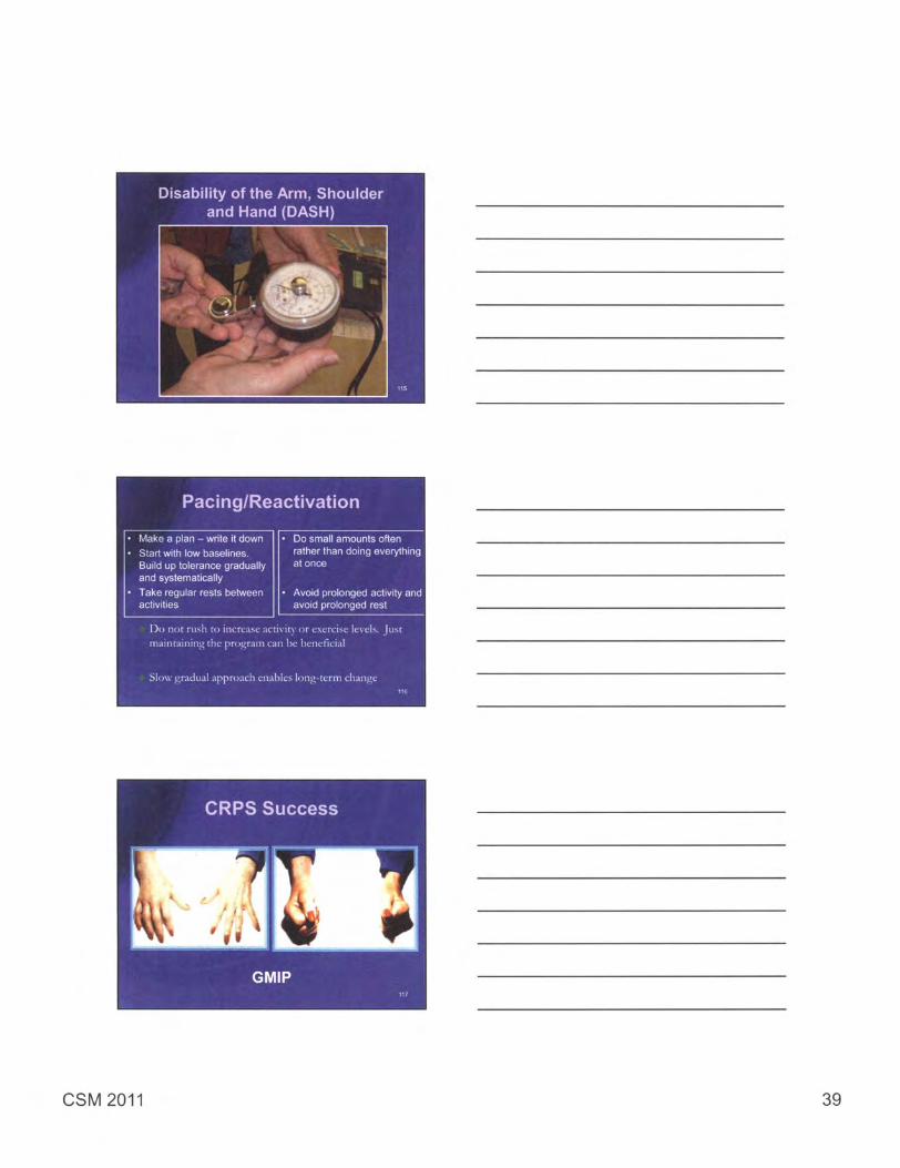
Disability of the Arm , Shoulderand Hand (DASH
Pacing /Reactivation
• Make a plan - write it down • Do small amounts often• Start with low baselines. rather than doing everything
Build up tolerance gradually at onceand systematically
• Take regular rests between • Avoid prolonged activity andactivities avoid prolonged rest
Do not rush to increase activity or exercise levels. Justmaintaining the program can be beneficial
Slow gradual approach enables long-term changeare
CSM 2011 39
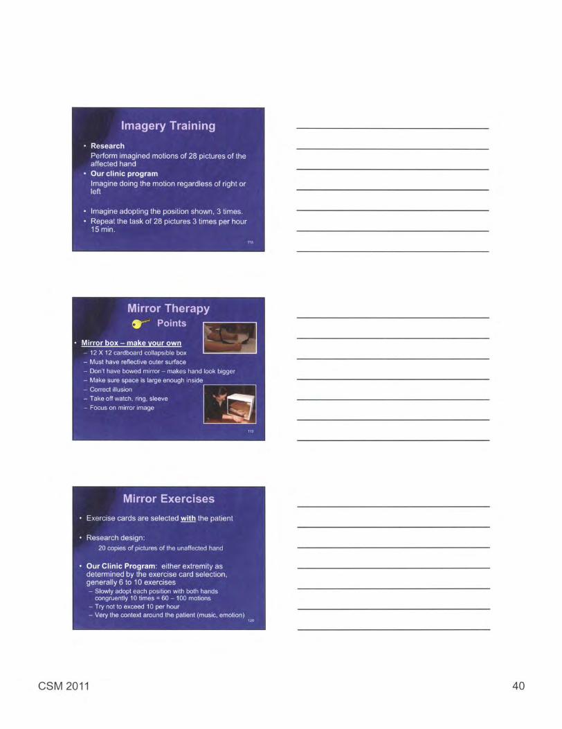
Imagery Training
• ResearchPerform imagined motions of 28 pictures of theaffected hand
• Our clinic programImagine doing the motion regardless of right orleft
• Imagine adopting the position shown , 3 times.• Repeat the task of 28 pictures 3 times per hour
15 min.
HE
Mirror Therapyt•-'4 Points r
• Mirror box - make your own- 12 X 12 cardboard collapsible box- Must have reflective outer surface
Mirror Exercises
• Exercise cards are selected with the patient
• Research design:20 copies of pictures of the unaffected hand
• Our Clinic Program : either extremity asdetermined by the exercise card selection,generally 6 to 10 exercises- Slowly adopt each position with both hands
congruently 10 times = 60 - 100 motions- Try not to exceed 10 per hour- Very the context around the patient (music, emotion)
CSM 2011 40

Phantom LimbResearch on Brain 's Paradigm Shift
• Monkeys and human studies• Sensory inputs from one sense can
substitute for another sense• Cold and vibration on face can mimic the
response on the phantom limb• Why - if phantom hand cortex is
denervated , then face input activates
In
Phantom Limb Pain
Touching specific areas
on the face of a person
with an amputated arm
will often evoke precisely
localized sensations in
the fingers.
Ramachandran VS Plasticity and functional recovery in neurology. ClinicalMedicine . July/Aug 2005; Vol 5 (4):369. 122
Phantom Limb• 3 weeks post amputation upper extremity -
sensations from ipsilateral face are referred tothe amputated limb- Ice will elicit cold- Vibration will elicit vibration
• This effect caused by sensory input from faceinvading and activating deafferented handzones in cortex and thalamus
• See that the phantom limb is moving inresponse to brain command from the non-involved side
CSM 2011 41

Research
• Ramachaandran hypothesized that thedisruption of the normal interaction of motorintention to move the limb and the absence ofappropriate sensory feedback resulted inphantom limb pain.
• They speculated that visual feed back wouldinterrupt this pathological cycle.
Ramachandran Research• N=9
• Mirror- use normal hand
• Eyes open is KEY- later on...eyes closed
• 7/9 - felt limb move with imagery and therewas no pain
*Ramachandra n VS, Rogers -Ramachandran D. Synaeslhesia in phantomlimbs induced With mirrors Proc Roy Soc Land. 1996; 263:377-386
"Ramachandran VS, Hirslein W. The D O Hebb lecture Perception ofphantom limbs Brain. 1998 ; 1211603-1630.
1861
Phantom Limb PainNeurorehabilitation and
Neural Repair
• Arm amputees use mirror reflection of intactarm or leg to get movement of the other limb
• Used for somatosensory deficits
• Increased functional use• Blinded rating
"Sathian K, Greespan AI, Wolf SL Doing I t with Mirrors: A Case Study of a NovelApproach to Neurorehabililalion ; Neurorehabllltafion and Neural Repair. 2000;14(1)73-76 .
CSM 2011 42

Phantom Limb Pain• Strong relationship between the amount of
plastic change in the primary sensory cortex andthe amount of phantom pain. (Flora 1995).
• Interventions that activate cortical areas thatsubserve the affected limb lead to symptomaticand functional improvements and observablecortical reorganization. (Flora, 2001)
Moseley GL. Graded motor imagery is effective for long standing CRPSa randomized controlled study. Pain . 2004; 108(1-2):192-198..
Research
Neurons in the brainthat use to representsensation in the lostlimb were functionalbut driven by otherbody parts . Usuallyparts closest to theamputated limb
• Patients experiencingphantom pain thesensation can berecreated withstimulating in the brain.
• Phantom sensationcould not be elicited inamputees withoutphantom sensation
IPE
Phantom Limb Pain
• Sensations relate cortical representation ormap inside the brain
• Neurological- Illusory body experience since change in
amputated area
• Migration of neighboring somatosensoryreceptor sites into these vacant areas
IF
CSM 2011 43
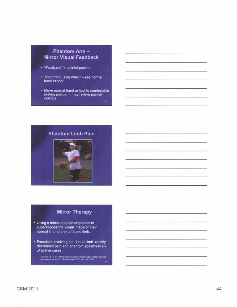
Phantom Arm -Mirror Visual Feedback
• "Paralyzed " in painful position
• Treatment using mirror - see normalhand or foot
• Move normal hand or foot to comfortableresting position - may relieve painfulcramps
Mirror Therapy
• Using a mirror enabled amputees tosuperimpose the visual image of theirnormal limb to their affected limb.
• Exercises involving the "virtual limb " rapidlydecreased pain and phantom spasms in sixof twelve cases.
McCabe CS. at al. Referred sensations in patients with complex regionalpain syndrome type 1. Rheumatology. 2003; 42:1067-1073.
132
CSM 2011 44

Phantom Limb Pain Intervention"Imaging"
1. Slowly straighten and then bend your arms or
legs
2. Point your fingers and toes upward, and thenpoint your fingers and toes downward at thesame time
3. Turn your hands or feet in toward each otherand then away from each other at the sametime
4. Move your hands and feet around in a circleto the left and to the right 134
Phantom Limb Pain InterventionImaging
5. Lift your hands and fee off the table or stool
6. Clench and unclench your fist and toe
7. Spread your fingers and toes and then relaxthem
8. Point one thumb or toe up and one thumb or toedown then reverse it
MacLachlan M, et.al. Mirror treatment of lower limb phantom pain: Acase study. Disability And Rehabilitation . 2004 ; 26(14-15 ): 901-904.
we
CSM 2011 45
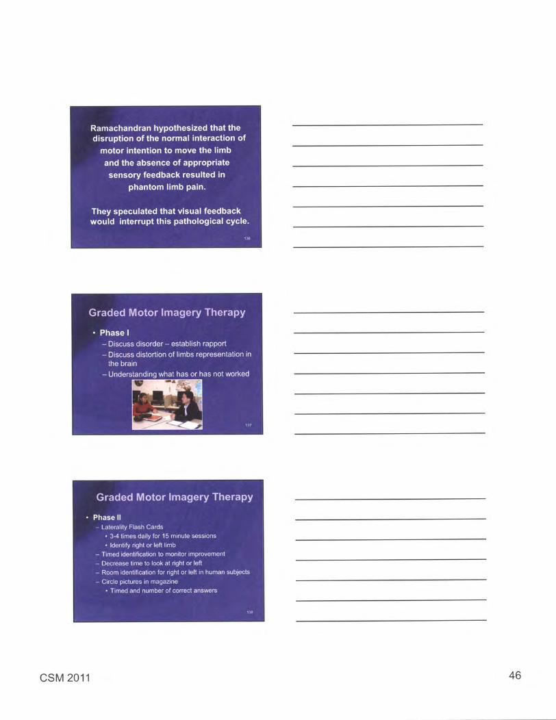
Ramachandran hypothesized that thedisruption of the normal interaction of
motor intention to move the limb
and the absence of appropriate
sensory feedback resulted inphantom limb pain.
They speculated that visual feedbackwould interrupt this pathological cycle.
Graded Motor Imagery Therapy
• Phase I- Discuss disorder - establish rapport
- Discuss distortion of limbs representation inthe brain
- Understanding what has or has not worked
Graded Motor Imagery Therapy
• Phase II- Laterality Flash Cards
• 3-4 times daily for 15 minute sessions
• Identify right or left limb- Timed identification to monitor improvement
- Decrease time to look at right or left- Room identification for right or left in human subjects
- Circle pictures in magazine• Timed and number of correct answers
D]
CSM 2011 46
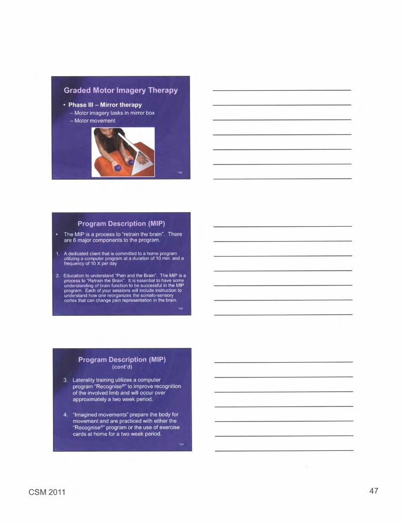
Graded Motor Imagery Therapy
• Phase III - Mirror therapy- Motor imagery tasks in mirror box
- Motor movement
M
Program Description (MIP)
• The MIP is a process to "retrain the brain". Thereare 6 major components to the program.
1. A dedicated client that is committed to a home programutilizing a computer program at a duration of 10 min. and afrequency of 10 X per day
Education to understand "Pain and the Brain" . The MIP is aprocess to "Retrain the Brain" . It is essential to have someunderstanding of brain function to be successful in the MIPprogram . Each of your sessions will include instruction tounderstand how one reorganizes the somato-sensorycortex that can change pain representation in the brain.
55
Program Description (MIP)(cont'd)
3. Laterality training utilizes a computerprogram 'Recognise®" to improve recognitionof the involved limb and will occur overapproximately a two week period.
4. "Imagined movements" prepare the body formovement and are practiced with either the"Recognise®" program or the use of exercisecards at home for a two week period.
ME
CSM 2011 47

Program Description (MIP)
5. Mirror box exercise home program• When using the mirror , one can trick the brain into
believing that an injured part is actually okay.• For example , if the left hand was the problem , it would
be hidden and by using the mirror image of the righthand , the brain would construct that the left hand wassomehow okay . It signals the brain that the hand isfine and now its time to represent it properly.
6. Progressive functional exercise home program.
3 Clients will generally attend therapy one time per week,for approximately 10 sessions.
3 Equipment requirements : Computer, "Recognises" CD-ROM, Mirror Box
5 Steps to Teach and Upgrade thePatient's Imagery Technique
• Assess mental capacity to learn imagerytechnique
• Establish nature of mental practice
• Teach imagery technique
• Evaluate and monitor imagery technique
• Develop self-generated treatment EW
Implications on Neuroplasticityfor Clinical Practice
1. Client must be committed to learning thateducation of patients is critical to carry out thetask at home
2. Drive spontaneous and purposeful change byattended , repetitive and rewarded behavior
3. By focused , selective and goal-oriented repetitivebehaviors , we should be able to reverse negativeoutput
EM
CSM 2011 48

Recognise= Limb laterality recognition program . Developedand published by Noigroup Publications. 146www.noigroup.com
CSM 2011 49

CSM 2011 50
Related Documents


