POSITION STATEMENT Italian Association of Clinical Endocrinologists (AME) position statement: a stepwise clinical approach to the diagnosis of gastroenteropancreatic neuroendocrine neoplasms Franco Grimaldi • Nicola Fazio • Roberto Attanasio • Andrea Frasoldati • Enrico Papini • Francesco Angelini • Roberto Baldelli • Debora Berretti • Sara Bianchetti • Giancarlo Bizzarri • Marco Caputo • Roberto Castello • Nadia Cremonini • Anna Crescenzi • Maria Vittoria Davı ` • Angela Valentina D’Elia • Antongiulio Faggiano • Stefano Pizzolitto • Annibale Versari • Michele Zini • Guido Rindi • Kjell O ¨ berg Received: 10 February 2014 / Accepted: 29 March 2014 / Published online: 20 July 2014 Ó The Author(s) 2014. This article is published with open access at Springerlink.com Keywords Neuroendocrine tumors Diagnostic work- up Markers Imaging Incidental findings Non- functioning tumors Carcinoid syndrome Gastrinoma Insulinoma NET NEC NEN Abbreviations 5-HIAA 5-Hydroxy-indolacetic acid ACE Angiotensin-converting enzyme ACTH Adrenocorticotropin AJCC American Joint Committee on Cancer AKT A protein-serine-threonine kinase that is activated by phosphorylation in response to growth factors or insulin CD117 Antigen specific for the proto-oncogene c-kit CD56 Antigen expressed by all lymphocytes CD99 Cluster of differentiation CDX-2 Transcription factor expressed specifically in gut epithelium CEACAM1 Cell adhesion molecule CEUS Contrast-enhanced US CgA Chromogranin A CK19 Cytokeratin 19 CS Carcinoid syndrome CT Computerized tomography DBE Double balloon enteroscopy On behalf of AME. Other members of AME oncologic endocrinology group are listed in the conclusions. Franco Grimaldi and Nicola Fazio contributed equally as first authors. F. Grimaldi (&) Endocrinology and Metabolic Disease Unit, Azienda Ospedaliero-Universitaria ‘‘S. Maria della Misericordia’’, P.le S.M. della Misericordia, 15-33100, Udine, Italy e-mail: [email protected] N. Fazio Unit of Gastrointestinal and Neuroendocrine Tumors, European Institute of Oncology, Milan, Italy e-mail: [email protected] R. Attanasio Endocrinology Service, Galeazzi Institute IRCCS, Milan, Italy e-mail: [email protected] A. Frasoldati M. Zini Endocrinology Unit, Arcispedale S. Maria Nuova IRCCS, Reggio Emilia, Italy e-mail: [email protected] M. Zini e-mail: [email protected] E. Papini Endocrinology Unit, Regina Apostolorum Hospital, Albano Laziale, Rome, Italy e-mail: [email protected] F. Angelini S. Bianchetti Oncology and Hematology Unit, Regina Apostolorum Hospital, Albano Laziale, Rome, Italy e-mail: [email protected] S. Bianchetti e-mail: [email protected] R. Baldelli Endocrinology Section, Regina Elena National Cancer Institute, Rome, Italy e-mail: [email protected] D. Berretti Gastroenterology Unit, Azienda Ospedaliero-Universitaria ‘‘S. Maria della Misericordia’’, Udine, Italy e-mail: [email protected] 123 J Endocrinol Invest (2014) 37:875–909 DOI 10.1007/s40618-014-0119-0

Welcome message from author
This document is posted to help you gain knowledge. Please leave a comment to let me know what you think about it! Share it to your friends and learn new things together.
Transcript

POSITION STATEMENT
Italian Association of Clinical Endocrinologists (AME) positionstatement: a stepwise clinical approach to the diagnosisof gastroenteropancreatic neuroendocrine neoplasms
Franco Grimaldi • Nicola Fazio • Roberto Attanasio • Andrea Frasoldati • Enrico Papini • Francesco Angelini •
Roberto Baldelli • Debora Berretti • Sara Bianchetti • Giancarlo Bizzarri • Marco Caputo • Roberto Castello •
Nadia Cremonini • Anna Crescenzi • Maria Vittoria Davı • Angela Valentina D’Elia • Antongiulio Faggiano •
Stefano Pizzolitto • Annibale Versari • Michele Zini • Guido Rindi • Kjell Oberg
Received: 10 February 2014 / Accepted: 29 March 2014 / Published online: 20 July 2014
� The Author(s) 2014. This article is published with open access at Springerlink.com
Keywords Neuroendocrine tumors � Diagnostic work-
up � Markers � Imaging � Incidental findings � Non-
functioning tumors � Carcinoid syndrome � Gastrinoma �Insulinoma � NET � NEC � NEN
Abbreviations
5-HIAA 5-Hydroxy-indolacetic acid
ACE Angiotensin-converting enzyme
ACTH Adrenocorticotropin
AJCC American Joint Committee on Cancer
AKT A protein-serine-threonine kinase that is
activated by phosphorylation in response to
growth factors or insulin
CD117 Antigen specific for the proto-oncogene
c-kit
CD56 Antigen expressed by all lymphocytes
CD99 Cluster of differentiation
CDX-2 Transcription factor expressed specifically
in gut epithelium
CEACAM1 Cell adhesion molecule
CEUS Contrast-enhanced US
CgA Chromogranin A
CK19 Cytokeratin 19
CS Carcinoid syndrome
CT Computerized tomography
DBE Double balloon enteroscopy
On behalf of AME.
Other members of AME oncologic endocrinology group are listed in
the conclusions.
Franco Grimaldi and Nicola Fazio contributed equally as first authors.
F. Grimaldi (&)
Endocrinology and Metabolic Disease Unit, Azienda
Ospedaliero-Universitaria ‘‘S. Maria della Misericordia’’, P.le
S.M. della Misericordia, 15-33100, Udine, Italy
e-mail: [email protected]
N. Fazio
Unit of Gastrointestinal and Neuroendocrine Tumors, European
Institute of Oncology, Milan, Italy
e-mail: [email protected]
R. Attanasio
Endocrinology Service, Galeazzi Institute IRCCS, Milan, Italy
e-mail: [email protected]
A. Frasoldati � M. Zini
Endocrinology Unit, Arcispedale S. Maria Nuova IRCCS,
Reggio Emilia, Italy
e-mail: [email protected]
M. Zini
e-mail: [email protected]
E. Papini
Endocrinology Unit, Regina Apostolorum Hospital, Albano
Laziale, Rome, Italy
e-mail: [email protected]
F. Angelini � S. Bianchetti
Oncology and Hematology Unit, Regina Apostolorum Hospital,
Albano Laziale, Rome, Italy
e-mail: [email protected]
S. Bianchetti
e-mail: [email protected]
R. Baldelli
Endocrinology Section, Regina Elena National Cancer Institute,
Rome, Italy
e-mail: [email protected]
D. Berretti
Gastroenterology Unit, Azienda Ospedaliero-Universitaria ‘‘S.
Maria della Misericordia’’, Udine, Italy
e-mail: [email protected]
123
J Endocrinol Invest (2014) 37:875–909
DOI 10.1007/s40618-014-0119-0

DOPA Dihydroxyphenylalanine
DOTA 1,4,7,10-Tetra-azacyclo-dodecane-
tetraacetic acid
DOTANOC DOTA-Nal3-octreotide
DOTATATE DOTA-octreotate
DOTATOC DOTA-edotreotide
ECLomas Enterochromaffin-like cell carcinoids
EGDS Esophago-gastro-duodenoscopy
ELISA Enzyme-linked immunosorbent assay
ENETS European Neuroendocrine Tumor Society
ERCC-1 Excision repair cross-complementing
ERCP Endoscopic-retrograde-cholangio-
pancreatography
EUS Endoscopic ultrasonography
FDG Fluoro-deoxy-glucose
FGF Fibroblast growth factor
FNA Fine needle aspiration
FNB Fine needle biopsy
FSG Fasting serum gastrin
GEP Gastroenteropancreatic
GERD Gastroesophageal reflux disease
GI Gastrointestinal
GRADE Grading of recommendations, assessment,
development, and evaluation
hCG Human chorionic gonadotropin
hHAS-1 Human achaete-scute homolog 1
H2RAs Histamine H2-receptor antagonists
Her/2 A cell surface protein-tyrosine kinase
receptor overexpressed in
adenocarcinomas
HPF High-power field
HPLC High-pressure liquid chromatography
HTP Hydroxy-L-tryptophan
IHC Immunohistochemistry
IRMA Immunoradiometric assay
Ki-67 Nuclear antigen present only in the nuclei
of cycling cells
LoE Level of evidence
MANEC Mixed adeno-neuroendocrine carcinoma
MAO Monoamine oxidase
MDCT Multidetector CT
MEN-1 Multiple endocrine neoplasms
MGMT Methylguanine-DNA methyltransferase
MIB-1 A monoclonal antibody used to detect KI-
67 antigen
MRI Magnetic resonance imaging
mTOR Mammalian target of rapamycin
NEC Neuroendocrine carcinoma
NEN Neuroendocrine neoplasm
NET Neuroendocrine tumor
NF Non-functioning
NF1 Neurofibromatosis type 1
NIH National Institutes of Health
G. Bizzarri
Diagnostic Imaging Unit, Regina Apostolorum Hospital, Albano
Laziale, Rome, Italy
e-mail: [email protected]
M. Caputo
Dipartimento Servizi di Diagnosi e Cura, AUSL 22 Regione
Veneto, Bussolengo, VR, Italy
e-mail: [email protected]
R. Castello
Medicina Interna ad indirizzo Endocrinologico, Azienda
Ospedaliera Universitaria Integrata, Verona, Italy
e-mail: [email protected]
N. Cremonini
Endocrinology Unit, Maggiore and Bellaria Hospital, Bologna,
Italy
e-mail: [email protected]
A. Crescenzi
Pathology Unit, Regina Apostolorum Hospital, Albano Laziale,
Rome, Italy
e-mail: [email protected]
M. V. Davı
Medicina Interna D, Azienda Ospedaliera Universitaria
Integrata, Verona, Italy
e-mail: [email protected]
A. V. D’Elia
Genetic Service, Azienda Ospedaliero-Universitaria ‘‘S. Maria
della Misericordia’’, Udine, Italy
e-mail: [email protected]
A. Faggiano
Department of Clinical Medicine and Surgery, Federico II
University, Naples, Italy
e-mail: [email protected]
S. Pizzolitto
Pathology Unit, Azienda Ospedaliero-Universitaria ‘‘S. Maria
della Misericordia’’, Udine, Italy
e-mail: [email protected]
A. Versari
Nuclear Medicine Service, Arcispedale S. Maria Nuova IRCCS,
Reggio Emilia, Italy
e-mail: [email protected]
G. Rindi
Institute of Pathology, Policlinico A. Gemelli, Universita
Cattolica del Sacro Cuore, Rome, Italy
e-mail: [email protected]
K. Oberg
Department of Endocrine Oncology, University Hospital,
Uppsala, Sweden
e-mail: [email protected]
876 J Endocrinol Invest (2014) 37:875–909
123

NPV Negative predictive value
NSE Neuron-specific enolase
NSP-55 Neuroendocrine-specific protein
OS Overall survival
PAX-8 Paired box gene 8
PET Positron emission tomography
PGP.9.5 Pan neuronal marker protein in the Islets of
Langerhans
PIK3 Phosphoinositide-3-kinase
PLGF Placental growth factor
pNEN Pancreatic NEN
PPIs Proton pump inhibitors
PPV Positive predictive value
PTEN Phosphatase and tensin homolog (mutated
in multiple advanced cancers)
RIA Radioimmunoassay
SA Somatostatin analog
SEER Surveillance, epidemiology and end results
SPECT Single photon emission computed
tomography
SRS Somatostatin receptor scintigraphy
SSTR Somatostatin subtype receptor
TNM Tumor-node-metastases
TSC Tuberous sclerosis complex
TTF-1 Thyroid transcription factor 1
UICC Union for International Cancer Control
UPN Unknown primary NEN
US Ultrasonography
VCE Video-capsule endoscopy
VHL von Hippel-Lindau disease
VIP Vasoactive intestinal peptide
ZES Zollinger–Ellison syndrome
Table of contents
1 Introduction............................................................................ 878
1.1 Why this document....................................................... 878
1.2 Methodology ................................................................. 878
1.3 Definitions..................................................................... 878
1.4 Classification................................................................. 878
1.4.1 Grading assessment ...................................... 879
1.4.2 Pathologic staging ........................................ 879
2 Diagnostic tools ..................................................................... 879
2.1 Histology, cytology, immunohistochemistry, and molecular
biology........................................................................... 879
2.1.1 Morphologic criteria..................................... 879
2.1.2 Immunohistochemistry and molecular biology
techniques ..................................................... 880
2.1.3 Working with the pathologist and his pathologic
report............................................................. 881
2.1.4 Genetic assessment....................................... 881
2.2 Laboratory assessment.................................................. 882
2.2.1 ‘‘Unspecific markers’’ .................................. 882
2.2.1.1 Chromogranin A.......................... 882
2.2.1.2 Other unspecific markers ............ 883
2.2.2 ‘‘Specific markers’’ ...................................... 883
2.2.2.1 5-HIAA ........................................ 883
2.2.2.2 Gastrin.......................................... 883
2.2.2.3 Insulin .......................................... 884
2.2.2.4 Other specific markers ................ 884
2.3 Imaging procedures ...................................................... 885
2.3.1 Radiologic procedures.................................. 885
2.3.1.1 Ultrasonography .......................... 885
2.3.1.2 Multislice triple phase CT .......... 885
2.3.1.3 MRI.............................................. 885
2.3.2 Nuclear medicine procedures....................... 886
2.3.2.1 SSTR functional imaging............ 886
2.3.2.2 PET with other tracers ................ 886
2.3.3 Endoscopic procedures................................. 887
2.3.3.1 Upper and lower gastrointestinal
NENs............................................ 887
2.3.3.2 Small-bowel NENs...................... 887
2.3.3.3 Pancreatic NENs ......................... 887
3 A step-by-step multidisciplinary approach to clinical diagno-
sis ........................................................................................... 888
3.1 Incidental finding.......................................................... 888
3.1.1 GEP-NENs suspected at endoscopy ............ 888
3.1.2 GEP-NEN suspected at morphological (US/CT/
MR) imaging ................................................ 888
3.1.3 GEP-NEN suspected after elevated serum CgA
levels ............................................................. 889
3.2 Symptomatic patient with symptoms due to GEP-NEN-
related local effects....................................................... 890
3.2.1 When to suspect a GEP-NEN...................... 890
3.2.2 Work-up in the patient with local compressive
symptoms...................................................... 890
3.2.2.1 Isolated abdominal pain .............. 890
3.2.2.2 Subocclusive picture ................... 890
3.2.2.3 Jaundice ....................................... 891
3.2.2.4 Gastrointestinal bleeding............. 891
3.3 Symptomatic patient with syndromes .......................... 892
3.3.1 Diarrhea and flushing................................... 892
3.3.1.1 Clinical approach: when to suspect a
GEP-NEN .................................... 892
3.3.1.2 Work-up in the patient with suspected
carcinoid syndrome ..................... 893
3.3.2 Resistant/relapsing ulcer disease ................. 894
3.3.2.1 Clinical approach: when to suspect a
GEP-NEN .................................... 894
3.3.2.2 Work-up in the patient with suspected
gastrinoma ................................... 895
3.3.3 Spontaneous hypoglycemia.......................... 896
3.3.3.1 Clinical approach: when to suspect a
GEP-NEN .................................... 896
3.3.3.2 Work-up in the patient with suspected
insulinoma ................................... 897
3.4 Work-up in the patient with metastatic disease and unknown
primary tumor ............................................................... 899
3.5 When and how to stage a previously diagnosed GEP-
NEN............................................................................... 899
4 Conclusions............................................................................ 900
5 References.............................................................................. 901
J Endocrinol Invest (2014) 37:875–909 877
123
1 Introduction
1.1 Why this document
Neuroendocrine neoplasms (NENs) can arise almost
throughout the entire body and share common morpho-
logical, ultrastructural, and immunohistochemical
characteristics.
Neuroendocrine neoplasms are an emerging entity that
can occur at any age, with the median age at diagnosis in
the late fifth decade and an age-related incidence increase.
About two-thirds involve the gastro-entero-pancreatic
(GEP) tract and epidemiological studies show their
increasing incidence [1]. In the last decades, the overall
reported incidence of GEP-NENs increased from 1.0 to
5.25/100.000 persons/year, with a present estimated prev-
alence of 35/100.000 [1–10]. Physicians’ awareness,
endoscopic screening and increased sensitivity of diag-
nostic tools may at least in part explain this growing trend.
Most guidelines are focused on staging, treatment and
follow-up of NENs. However, an appropriate clinical sus-
picion and a correct diagnostic work-up are critical starting
points. A multidisciplinary approach, moreover, is crucial
to provide a timely and integrated care. Hence, this docu-
ment is neither a review, nor a guideline; rather, it is a
clinical guide for a stepwise and integrated diagnostic
work-up of GEP-NENs. Hopefully, this will result in a
correct utilization of resources and optimization of the cost/
benefit ratio.
1.2 Methodology
The grading of recommendations, assessment, develop-
ment, and evaluation (GRADE) system was adopted for the
present position statement [11–14]. Briefly, the GRADE
system classifies evidence into four quality levels (high,
moderate, low, or very low), and recommendations into
two grades (strong or weak).
Whenever possible, the level of evidence (LoE) has
been ranked as follows: very low (�sss), low (��ss),
moderate (���s), and high (����). ‘‘Very low qual-
ity’’ evidence corresponds to unsystematic clinical obser-
vations (case report, case series) or indirect evidence (e.g.,
surrogate end points); ‘‘low quality’’ evidence corre-
sponds to observational studies or randomized controlled
trials (RCT) with major limits; ‘‘moderate quality evi-
dence’’ corresponds to RCTs with limitations or rigorous
observational studies; and ‘‘high quality evidence’’ corre-
sponds to well performed RCTs and strong evidence from
unbiased observational studies [13].
We labeled as ‘‘recommendations’’ and ‘‘suggestions’’
the strong and weak recommendations, respectively. Each
recommendation/suggestion is based on the quality of
supporting evidence, downgraded or upgraded according to
adjunctive factors (e.g., inconsistency of results, indirect-
ness of evidence, lack of precision and limited number of
relevant publications downgrade the recommendation/
suggestion; large effect size, narrow confidence intervals,
clinically very significant end points upgrade the recom-
mendation/suggestion), and the level of panel agreement
[13].
1.3 Definitions
Neuroendocrine neoplasms neoplastic cells possess fea-
tures of both neural and epithelial cells. Therefore, in line
with the WHO classification, the term neuroendocrine will
be adopted throughout this document [15].
WHO recommends the use of the term ‘‘neuroendocrine
neoplasm’’ (NEN) to indicate low- to high-grade lesions.
The term ‘‘neuroendocrine tumor’’ (NET) will be used
throughout this document, due to its widespread diffusion,
to indicate low- to intermediate-grade lesions and the term
‘‘neuroendocrine carcinoma’’ (NEC) to indicate high-grade
lesions. Terms like ‘‘carcinoids’’ and the embryological
classification of GEP-NENs in tumors of foregut (thymus,
esophagus, lung, stomach, duodenum, pancreas), midgut
(appendix, ileum, cecum, ascending colon) and hindgut
(distal colon and rectum) will be avoided.
1.4 Classification
In the last 10 years WHO has repeatedly revised the
pathologic classification of GEP-NENs (Table 1) [16].
According to the 2010 classification, NET G1 includes
the ‘‘carcinoids’’ or ‘‘well-differentiated tumors’’ of the
Table 1 WHO classifications of GEP-NENs
WHO 1980 WHO 2000 WHO 2010
I. Carcinoid Well-differentiated
endocrine tumor
Well-differentiated
endocrine carcinoma
Poorly differentiated
endocrine carcinoma/
small-cell carcinoma
Neuroendocrine
tumors
NET G1 (Grade 1)
NET G2 (Grade 2)
Neuroendocrine
carcinoma
NEC G3 (Grade 3):
Large-cell NEC
small-cell NEC
II. Mucocarcinoid
III. Mixed
carcinoid-
adenocarcinoma
forms
Mixed exocrine–
endocrine carcinoma
Mixed adeno-
neuroendocrine
carcinoma
(MANEC)
IV. Pseudotumor
lesions
Tumor-like lesions Hyperplastic and
preneoplastic
lesions
878 J Endocrinol Invest (2014) 37:875–909
123

1980 and 2000 WHO classifications. These tumors are
usually indolent, but can occasionally behave as malignant.
NET G2 may be considered a ‘‘grey zone’’, with het-
erogeneous behavior, and requires a tailored management.
NEC (G3) is a malignant neoplasm with an aggressive
clinical course.
MANEC has a malignant phenotype with features of
both adenocarcinoma and NET. This definition requires the
presence of at least 25 % of each component. Neuroen-
docrine cells are usually interspersed and the two popula-
tions may be identified only by immunohistochemistry
(IHC). Less frequently, neuroendocrine cells may be
grouped in distinct regions that are recognized by light
microscopy.
The WHO 2010 classification strongly relies on tumor
grading. Grading relates to the biological aggressiveness of
the neoplasm, whereas differentiation indicates its simi-
larity to the tissue of origin [15]. The clinical behavior of
NENs may be basically predicted by their grading, staging,
and evidence of hormonal syndromes. All these data should
be collected and weighted to establish the prognosis and
management of the patient.
1.4.1 Grading assessment
The grade of a tumor is the primary predictor of its clinical
outcome. Grading is based on the proliferation rate of the
tumor, as assessed by the Ki-67 cell labeling and by the
mitotic count (number of mitosis 9 10 high power fields—
HPF) (Table 2) [15–23].
Visual estimates are currently used as the standard
technique for evaluating both Ki-67 and the mitotic count
[24, 25]. Several areas should be assessed within the tumor
to reduce the risk of evaluation bias due to intratumoral
heterogeneity. Densely stained regions (‘‘hot spots’’)
should be preferentially evaluated. Results from these areas
should be reported as a single percentage reflecting the
highest identified count [16, 21, 22].
Potential pitfalls and limitations are:
a. technical problems (e.g., tissue processing, differences
in Ki-67 antibodies, etc.);
b. intratumoral heterogeneity and sampling limitations
(e.g., a single biopsy sample may not be representative
of the tumor grade within the whole neoplastic mass)
[24, 26];
c. discordances:
I. between the proliferative rate and the degree of
differentiation (e.g., a morphologically well-dif-
ferentiated NEN may exhibit a high proliferative
rate);
II. between the predictive value for prognosis and
that for treatment response (e.g., Ki-67 is a
reliable predictor of disease progression and
overall survival (OS), but seems a less efficient
predictor of response to medical treatment) [27].
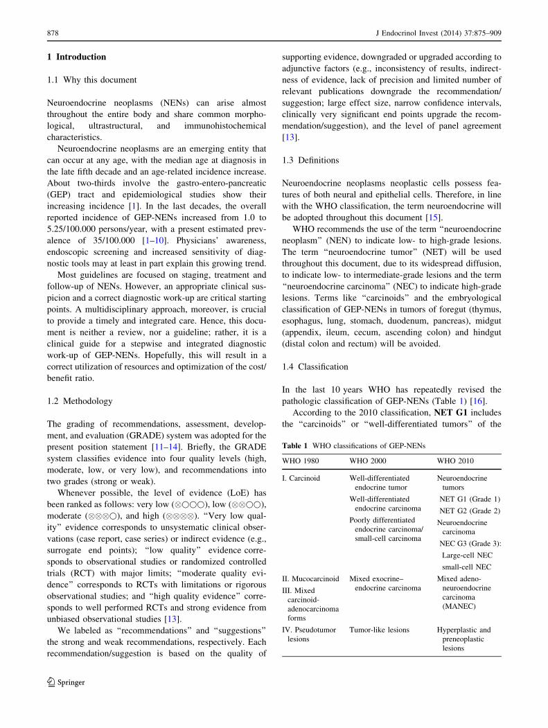
1.4.2 Pathologic staging
GEP-NENs are staged according to tumor size, site of
origin, and locoregional or distant spreading [21–23]. The
staging information is integrated with the 2010 WHO
classification to stratify the prognostic risk and optimize
the therapeutic and follow-up strategies (Fig. 1).
We recommend the use of the 2010 WHOclassification.We recommend for staging the use of the AJCC-TNM 2009, and just for pancreas and appendixAJCC-TNM 2009 and ENETS-TNM 2006/07. Theselected system should be specified in the pathologicreport.We recommend NEN classification and clinicalactions be based on the less favorable data in case ofconflicting findings.
2 Diagnostic tools
2.1 Histology, cytology, immunohistochemistry,
and molecular biology
2.1.1 Morphologic criteria
Pathologic assessment is required for the diagnosis, clas-
sification and staging of NENs.
GEP-NENs present a broad architectural spectrum [28].
Well-differentiated tumors show an organoid pattern that
ranges from solid nests to micro–macrotrabecular/gyriform
pattern. A rich sinusoidal vascularity is usually observed.
Stromal fibrosis, amyloid deposition, and calcification may
be present. Necrosis can be present either as large infarct-
Table 2 Grading system for GEP-NENs (adapted from 19)
Ki-67 index (%)a Mitotic count/10 HPFb
NET G1 B2 \2
NET G2 3–20 2–20
NEC G3 [20 [20
a Assessed by MIB-1 labeling in at least 2,000 tumor cells in high
nuclear density (‘‘hot spot’’) areasb 10 HPF = 2 mm2, at least 50 optical fields in high-density mitotic
areas
J Endocrinol Invest (2014) 37:875–909 879
123
like areas or as punctate foci in the center of neoplastic
nests. Regardless of their growth pattern, NEN cells have a
similar cytological appearance: small- to medium-size cells
with round to oval shape and eosinophilic, lightly granular,
cytoplasm. The nuclei are usually centrally placed, fairly
uniform, with a finely dispersed (‘‘salt and pepper’’)
chromatin pattern. Rarely, the neoplastic cells have a
‘‘plasmocytoid appearance’’ due to peripherally located
nuclei. Nucleoli are usually inconspicuous or absent. In-
tracytoplasmatic hyaline globules or nuclear pseudoinclu-
sions may be seen.
High-grade NENs are composed of small or large-to-
intermediate cells with high-grade features (marked
nuclear atypia, multifocal necrosis, high mitotic index) and
diffuse growth, sometimes with organoid feature resem-
bling NEN.
The subgroup of GEP-NENs with Ki67 [20 % (and
therefore G3 according to WHO 2010), but with a mor-
phology of well-/moderately differentiated tumor should be
considered low/intermediate rather than high-grade NENs
[29].
Cytological specimens, which may be the only source of
diagnostic material, pose some problems for clinical
management. Cytology effectively separates high-grade
NENs from low-grade NENs, but the distinction between
low- and intermediate-grade NENs may be impossible. The
diagnostic accuracy of aspiration techniques may be lim-
ited by the small sample size, the suboptimal
reproducibility and the risk of contamination from contig-
uous tissues.
Cytology is gaining a major role for the diagnosis in duo-
deno-pancreatic tumors. The endoscopic ultrasonography
(EUS) fine needle aspiration (FNA) technique appears reli-
able, with a reported specificity of about 75 %, sensitivity of
87.5 %, accuracy of 89 %, positive predictive value (PPV) of
93 %, and negative predictive value (NPV) of 60 % [30–32].
2.1.2 Immunohistochemistry and molecular biology
techniques
Neuroendocrine differentiation Synaptophysin (a small
vesicle-associated marker) and Chromogranin A (CgA, a
large secretory granule-associated marker) are useful IHC
markers for the diagnosis of NENs. In NEC, the staining
for both these markers is required to confirm the diagnosis,
because CgA may be negative [15]. Routine IHC staining
for peptide hormones and bioamines is not recommended.
Other neuroendocrine markers, such as PGP.9.5, NSE,
CD56, NSP-55, are of questionable specificity and clinical
usefulness.
Prognostic markers proposed in addition to Ki-67 are
CK19, CD117, CD99, p53, Her/2, CEACAM1, E-cadherin,
b-catenin, hHAS-1, FGF13, PLGF, PAX-8, PTEN. None is
presently recommended for clinical practice. The research
of circulating tumor cells or the use of microRNAs is not
indicated for routine use [33, 34].
Fig. 1 Integrated pathologic
and biologic classification
(modified from 15)
880 J Endocrinol Invest (2014) 37:875–909
123

Markers of primary site These markers may be a key for
determining the unknown primary tumor in metastatic
lesions. The most useful are [35]:
• TTF-1, indicative of pulmonary or thyroid origin;
• serotonin and CDX-2, indicative of intestinal origin;
• PAX-8 and histidine-decarboxylase, indicative of pan-
creatic origin;
• xenin, indicative of duodenal origin.
Markers predictive of response to specific treatments
These biomarkers are not indicated for routine diagnostic
practice. They include [19]:
• somatostatin receptors (SSTR)-2A (IHC determination
at the cell membrane level), for planning the treatment
with somatostatin analogs (SA);
• Akt/mTOR pathway molecules (PIK3, PTEN, TSC2),
for treatment with everolimus;
• thymidylate synthase, for treatment with antifolates;
• ERCC-1, for treatment with platinum;
• topoisomerase Iia, for treatment with etoposide;
• epigenetic events, as methylation of MGMT promoter,
for treatment with alkylating agents.
We recommend routine IHC assessment of synap-tophysin and CgA.We suggest IHC assessment of peptide hormones orbioamines as optional in selected cases.We recommend against routine use of other IHCmarkers in clinical practice.
2.1.3 Working with the pathologist and his pathologic
report
The modality and timing of sampling techniques should be
planned by a multidisciplinary team.
The pathologist should be provided with accurate clin-
ical information including signs and symptoms, laboratory
findings and imaging studies [36].
The ideal pathologic report should include:
• description of the macroscopic specimen;
• tumor size (three dimensions);
• description of cell features and histologic architecture;
• differentiation (well or poorly differentiated);
• IHC findings (CgA and synaptophysin routinely,
SSTR2A when appropriate (e.g., when functional
imaging for SSTR2 is negative);
• Ki-67 and mitotic count;
• completeness of resection, distance of the surgical
margins from the tumoral edge, depth of invasion;
• signs of malignancy (angiolymphatic and/or perineural
invasion, necrosis, infiltration of the capsule and/or of
gastrointestinal (GI) wall and/or surrounding tissues);
• number of examined lymph nodes, and number of
lymph node metastases; presence of micrometastases;
diameter of largest metastasis;
• presence of distant metastases, if demonstrated;
• functional activity (if appropriate).
The report should be concluded with the WHO diag-
nosis and classification of the lesion (NET G1–G2 or NEC
G3) based on proliferative index (Ki-67 and/or mitotic
count), and with the tumor stage (the staging system should
be specified).
The minimum pathology data set for resected specimens
(both primary and metastatic) should include [37]:
• site;
• diagnosis (e.g., pure neuroendocrine neoplasm);
• differentiation (i.e., well or poor);
• proliferation (i.e., G1 or G2 or G3).
We recommend histology as the diagnostic standard,cytology if histology is not available.We recommend classification according to WHO2010.We recommend grading according to Ki-67 indexand/or mitotic count.We recommend staging according to AJCC/UICCTNM and ENETS.
2.1.4 Genetic assessment
Approximately 5–10 % of GEP-NENs have a hereditary
background as part of tumor susceptibility syndromes:
multiple endocrine neoplasia type 1 (MEN-1), von Hippel-
Lindau disease (VHL), neurofibromatosis type 1 (von
Recklinghausen disease, NF1) and the tuberous sclerosis
complex (TSC). All are inherited autosomal dominant
disorders [38].
MEN-1 GEP-NENs are the second most common mani-
festation of MEN-1, reported in 30–70 % of cases in dif-
ferent series [mostly non-functioning (NF)] [39, 40]. A
germ-line MEN-1 mutation is identifiable in about 80–90 %
of familial cases [41] and in about 42 % of sporadic cases
[42]. Germline mutations arise de novo without any family
history in approximately 10 % of patients [43]. MEN-1
J Endocrinol Invest (2014) 37:875–909 881
123
mutation testing should be offered to index cases and to their
first-degree relatives, even if asymptomatic [40]. Genetic
counseling is recommended [40]. The family members who
carry the MEN-1 mutation require routine surveillance for
early detection of endocrine tumors, whereas those who do
not carry the mutation can be reassured. When molecular
genetic testing is not available locally, patients highly sus-
pected for MEN-1 should be addressed to a referral centers.
No genotype/phenotype correlations have been demon-
strated in MEN-1 syndrome [44, 45].
VHL Endocrine pancreatic NF tumors occur in 11–17 %
of patients with VHL disease [46]. The penetrance of VHL
mutations is almost complete by age 65 years [47]. Genetic
testing detects mutations in virtually all affected individ-
uals [48] and should be offered to all individuals with
clinical evidence of VHL and to first-degree relatives. As
ophthalmologic screening for those at risk for VHL disease
begins before age five, molecular genetic testing is sug-
gested also in young asymptomatic children [49, 50].
NF1 GEP-NENs occur in 1 % of the NF1 patients [51].
Half of affected individuals have NF1 as the result of a de
novo mutation. The offspring of an affected individual is at
a 50 % risk of inheriting the altered NF1 gene, and the
disease manifestations are extremely variable, even within
the same family [52]. Molecular testing for NF1 is not
usually recommended in the clinical practice: screening for
NF1 mutations is useful only in individuals who do not
completely fulfill the NIH diagnostic criteria.
TSC A few cases of pancreatic (p)NENs have been
described in patients with TSC [53–55]. The diagnosis of
TSC is usually based on clinical findings and mutations can
be identified in approximately 85 % of individuals who
meet the diagnostic criteria [56]. Two-thirds of affected
individuals have TSC as the result of a de novo mutation.
We recommend germ-line DNA testing only inpresence of a family history or clinical findingssuggestive of MEN-1 or VHL. Genetic testing shouldinclude mutational screening and sequencing. Apreliminary genetic counseling is needed.We suggest the routine determination of serum cal-cium and PTH levels in patients with duodeno-pan-creatic NEN as a first-line screening for MEN-1.We recommend against routine somatic (tumor tis-sue) DNA testing.
2.2 Laboratory assessment
The determination of GEP-NENs serum markers should
not be used as a first-line diagnostic tool whereas it is
appropriate for monitoring the response to treatment and
for long-term follow-up [57, 58].
Serum markers should be determined after:
1. an established diagnosis or strong clinical suspicion of
GEP-NEN;
2. exclusion of physiologic and pathologic confounding
conditions.
NEN markers may be regarded as ‘‘unspecific’’ or
‘‘disease-specific’’.
2.2.1 ‘‘Unspecific markers’’
2.2.1.1 Chromogranin A Chromogranin A is a widely
employed serum marker for GEP-NENs, but its use pre-
sents limitations [59]. CgA circulates under different
antigenic forms and no universal calibration standard is
available [60]. IRMA and RIA results may be considered
roughly equivalent [61], but the reference intervals are
variable and results obtained with different assays cannot
be compared.
Chromogranin A level may be increased in a number of
pathologic conditions (Table 3), and in healthy subjects
after eating or physical exercise. Accordingly, CgA levels
are highly variable in the general population [62], and may
partially overlap between GEP-NEN patients and controls.
Hence, CgA has a poor first-line diagnostic value [5, 60,
62–66].
Proton pump inhibitors (PPIs) increase (up to sevenfold)
CgA levels. The effects of PPIs persist for several days
after drug discontinuation. Therefore, CgA testing should
be performed after an at least 2-week PPIs withdrawal [62,
67]. The effects of H2-receptor antagonists (H2RAs) on
CgA are still controversial [68].
Table 3 Potential confounders causing CgA increase [64]
Neoplastic (other than GEP-NENs)
Breast cancer
Prostate cancer
Ovarian cancer
Hepatocarcinoma
Pancreas adenocarcinoma
Colon cancer
Non-neoplastic
Kidney or heart failure
Endocrine diseases (hyperthyroidism, hyperparathyroidism)
Local or systemic inflammatory disease
Chronic obstructive broncho-pulmonary disease
Gastro-enteric pathologies: chronic atrophic gastritis,
pancreatitis, inflammatory bowel disease, cirrhosis, chronic
hepatitis
882 J Endocrinol Invest (2014) 37:875–909
123
Diagnostic accuracy of CgA depends on different
variables:
• tumor burden (sensitivity 60–100 vs. 29–50 % in
metastatic and localized disease, respectively) [62, 64,
69];
• type and site of tumor (sensitivity 96 vs. 75 % in
functioning and NF tumors, respectively) [63, 70].
2.2.1.2 Other unspecific markers Neuron-specific eno-
lase (NSE) is an enzyme found in neuroectoderm-derived
cells. The presence of NSE has been reported in thyroid
and prostate carcinomas, neuroblastomas, small-cell lung
carcinomas, and pheochromocytomas. The clinical useful-
ness of this marker is hampered by its poor specificity [71].
NSE level is elevated in 30–50 % of patients with NEN,
particularly those with poor differentiation. The combined
determination of NSE and CgA may improve sensitivity in
GEP-NEN diagnosis [72].
Pancreatic polypeptide (PP) is secreted by specialized
pancreatic islet cells and inhibits gut motility and pancre-
atic exocrine secretion. PP has been proposed for the
diagnosis and monitoring of NF pNENs, as its combination
with CgA increases sensitivity up to 93 % [69]. Its routine
use is not recommended due to the low diagnostic perfor-
mance (sensitivity 63 % and specificity 81 %). PP levels
may increase in old age, diarrhea, laxative abuse, gut
inflammatory processes and chronic renal disease.
Beta subunit of human chorionic gonadotropin (hCG), a
glycoprotein synthesized by the syncytiotrophoblastic cells
of the placenta during pregnancy, may be increased in
patients with pNENs [73], but has no use in every day
practice.
As a whole, the clinical usefulness of the above reported
markers is limited.
2.2.2 ‘‘Specific markers’’
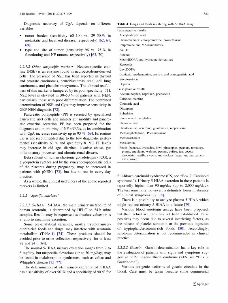
2.2.2.1 5-HIAA 5-HIAA, the main urinary metabolite of
human serotonin, is determined by HPLC on 24 h urine
samples. Results may be expressed as absolute values or as
a ratio to creatinine excretion.
Some pre-analytical variables, mostly tryptophan/ser-
otonin-rich foods and drugs, may interfere with serotonin
metabolism (Table 4) [74]. These products should be
avoided prior to urine collection, respectively, for at least
72 and 24 h [64].
The normal 5-HIAA urinary excretion ranges from 2 to
8 mg/day, but unspecific elevations (up to 30 mg/day) may
be found in malabsorption syndromes, such as celiac and
Whipple’s disease [75–77].
The determination of 24-h urinary excretion of 5HIAA
has a sensitivity of over 90 % and a specificity of 90 % for
full-blown carcinoid syndrome (CS, see ‘‘Box 2, Carcinoid
syndrome’’). Urinary 5-HIAA excretion in these patients is
reportedly higher than 90 mg/day (up to 2,000 mg/day).
The test sensitivity, however, is definitely lower in absence
of clinical symptoms [77, 78].
There is a possibility to analyze plasma 5-HIAA which
might replace urinary-5-HIAA in a future [79].
Various blood serotonin assays have been proposed,
but their actual accuracy has not been established. False
positives may occur due to several interfering factors, as
the release of platelet serotonin or the previous ingestion
of tryptophan/serotonin-rich foods [80]. Accordingly,
serotonin determination is not recommended in clinical
practice.
2.2.2.2 Gastrin Gastrin determination has a key role in
the evaluation of patients with signs and symptoms sug-
gestive of Zollinger–Ellison syndrome (ZES, see ‘‘Box 3,
Gastrinoma’’).
Various antigenic isoforms of gastrin circulate in the
blood. Care must be taken because some commercial
Table 4 Drugs and foods interfering with 5-HIAA assay
False negative results
Acetylsalicylic acid
Phenothiazines: chlorpromazine, promethazine
Imipramine and MAO-inhibitors
ACTH
Ethanol
MethylDOPA and hydrazine derivatives
Ketoacids
LevoDOPA
Isoniazid, methenamine, gentisic and homogentisic acid
Streptozotocin
Heparin
False positive results
Acetaminophen, naproxen, phenacetin
Caffeine, nicotine
Coumaric acid
Diazepam
Ephedrine
Fluorouracil, melphalan
Phenobarbital
Phentolamine, reserpine, guaifenesin, mephenesin
Methamphetamine, Phenmetrazine
Methocarbamol
Mesalamine
Foods: bananas, avocados, kiwi, pineapples, peanuts, tomatoes,
plums, eggplants, walnuts, pecans, coffee, tea, cocoa/
chocolate, vanilla, sweets, and cookies (sugar and marmalade
are allowed)
J Endocrinol Invest (2014) 37:875–909 883
123
immunoassay kits detect only the gastrin-17 molecule [81]
and may cause false positive results.
Hypergastrinemia is commonly defined as a fasting
serum gastrin above 100 pg/mL. Simultaneous measure-
ment of gastric pH on a single sample is needed to rule out
secondary hypergastrinemia due to other causes. In
achlorhydria, pernicious anemia or atrophic gastritis high
gastrin levels are usually associated to high (i.e., [4) pH
values. On the contrary, serum gastrin levels [1,000 pg/
mL combined with a \2 gastric pH are virtually diagnostic
of ZES. Falsely elevated gastrin levels may be due to a few
drugs (Table 5) that should be discontinued at least
2 weeks before the test [77, 82–86].
In general, gastrin levels are higher in pancreatic than in
duodenal NENs, and are proportional to tumor burden and
in patients with metastatic disease, exceedingly high gas-
trin levels may be observed. However, the majority of
patients with ZES show mildly elevated (e.g.,
150–1,000 pg/mL) gastrin levels, partially overlapping
those of patients with renal insufficiency, small-bowel
resection, retained gastric antrum, or on potent antisecre-
tory drugs [87]. When the diagnosis is equivocal, a secretin
stimulation test is needed. A gastrin increase [120 pg/mL
over basal level is considered diagnostic [88].
2.2.2.3 Insulin The occurrence of repeated symptomatic
hypoglycemia (\60 mg/dL) is suspicious for insulinoma
(see ‘‘Box 4’’) in subjects without diabetes. The diagnosis
is confirmed by the presence of non-suppressed insulin
levels in presence of low glucose levels (see ‘‘Spontaneous
hypoglycemia’’). To rule out a spurious hypoglycemia,
laboratory processing should not be delayed. In subjects
with leucocytosis glucose determination should be repeated
with a collection tube that contains an inhibitor of
glycolysis.
In presence of an episode of spontaneous severe hypo-
glycemia with hyperinsulinism, the simultaneous mea-
surement of serum C-peptide and beta-hydroxybutyrate is
appropriate. If factitious hypoglycemia is suspected, uri-
nary sulfonylureas should be tested as well. The work-up
may be completed with the measurement of serum proin-
sulin [89]. This test, even if not widely available, is diag-
nostic of insulinomas secreting immature forms of insulin.
In selected patients with endogenous hyperinsulinism,
autoimmune hypoglycemia, suspected on the basis of
negative imaging tests and coexistence of autoimmune
disorders, should be ruled out with the determination of
insulin autoantibodies [90].
If the patient is not hypoglycemic when observed, the
association of severe hypoglycemia with non-suppressed
insulin levels should be seeked under the conditions in
which hypoglycemia would be expected (see provocative
testing, ‘‘Spontaneous hypoglycemia’’) [89].
2.2.2.4 Other specific markers Glucagon: Glucagonoma
is associated with serum glucagon concentrations higher
than 500 pg/mL and a characteristic clinical syndrome
(diabetes mellitus and cutaneous manifestations, such as
migratory necrolytic erythema, nail dystrophies, stomatitis,
etc.) [91]. Glucagon concentrations higher than 1,000 pg/
mL are virtually diagnostic for the disease, but some
patients may exhibit levels within the physiologically ele-
vated range. Moderate elevations in serum glucagon may
be caused by protracted fasting in normal subjects or by
renal and hepatic failure, trauma, sepsis, pancreatitis,
abdominal surgery, and Cushing’s syndrome.
Due to the fast degradation of glucagon in vitro, blood
must be collected in test tubes containing aprotinin and
should be rapidly delivered to the laboratory. Results
obtained with different glucagon assays may profoundly
differ, due to the different calibration standards and the
variable cross-reactivity with glucagon isoforms.
Vasointestinal peptide (VIP): Vasointestinal peptide-
secreting tumors cause the Verner–Morrison syndrome,
characterized by variable combination of watery diarrhea
([700 mL/day even during fasting, with tea-colored,
odorless stools), hypokalemia, achlorhydria, weight loss,
metabolic acidosis, hypercalcemia, glucose intolerance,
and flushing. The diagnosis is established by high-volume
secretory diarrhea associated with VIP levels higher than
75 pg/mL (to be confirmed by a second RIA determina-
tion) [92, 93]. VIP blood concentration is, in fact, extre-
mely low in healthy subjects. Commercial kits are
available, but their use is usually limited to tertiary referral
centers.
Table 5 Main drugs and foods that may interfere in gastrin assay
False negative results
Acetylsalicylic acid
LevoDOPA
False positive results
Hypochlorhydria/achlorhydria due to chronic use of PPIs and
H2RAs or chronic atrophic gastritis (often associated with
pernicious anemia)
Helicobacter pylori infection
Gastric outlet obstruction
Renal failure
Antral G-cell syndromes
Short-bowel syndrome
Retained antrum
884 J Endocrinol Invest (2014) 37:875–909
123

We recommend against the use of biochemicalmarkers as the initial diagnostic step for potentialGEP-NEN patients.We recommend the determination of the appropriatebiochemical marker only after the diagnosis or strongclinical suspicion of GEP-NEN. The panel of markersshould take into account the clinical picture and localavailability/expertise.We recommend considering all possible clinical andanalytical interfering factors in presence of elevatedserum or urinary levels of GEP-NEN markers. Thedetermination should be repeated, if possible, aftertheir timely withdrawal.We suggest PPIs discontinuation at least 2 weeksbefore CgA and gastrin measurements.We recommend, after the finding of an elevatedgastrin level, its repeated determination together withthe assessment of gastric pH.We recommend for the follow-up of the markersexpressed by GEP-NENs a serial measurement withthe same laboratory assay.
2.3 Imaging procedures
2.3.1 Radiologic procedures
2.3.1.1 Ultrasonography Transabdominal US is an
inexpensive, safe, rapid and non-invasive tool. US accu-
racy is, however, operator dependent and its sensitivity is
generally low (13–27 %), when compared with MultiDe-
tector CT (MDCT) and magnetic resonance imaging (MRI)
[94]. In case of pNEN, a mean 39 % US detection rate has
been reported [95, 96].
Contrast-enhanced US (CEUS) enables identification of
hypervascular lesions, even in case of fast-flow tumor
circulation, as in NF pNENs. Therefore, CEUS is signifi-
cantly superior to B-mode US both in the detection of NF
pNENs and in the diagnosis of liver metastases, visualized
as hyperenhancing non-homogeneous lesions [96–98], with
a reported sensitivity of 82 % [99, 100]. US may help in
defining complications of advanced disease (i.e., biliary
stricture) and/or guide diagnostic or therapeutic procedures
[101].
Endoscopic ultrasonography and EUS-guided FNA, a
fundamental procedure for the diagnosis of pNENs [96,
102, 103], will be treated in ‘‘Pancreatic NENs’’.
2.3.1.2 Multislice triple phase CT Multidetector CT is
considered the first choice imaging modality for detection,
staging and follow-up of GEP-NENs. When compared to
conventional CT, MDCT allows a markedly higher spatial
and temporal resolution. MDCT sensitivity and specificity
are increased due to multiphase scanning. Images should be
acquired in precontrast, arterial, portal and equilibrium
phases.
Non-functioning pNENs and NEN liver metastases
typically appear as hypervascular lesions. In the evaluation
of NF pNENs, the combination of arterial dominant-phase
and portal venous-phase CT improves the detection of
primary tumors and hepatic metastases [96].
Reported mean sensitivity and specificity of MDCT are
73 % (63–82 %) and 96 % (83–100 %) for pNENs, and
82 % (78–100 %) and 92 % (83–100 %) for liver metas-
tases, respectively [104–106].
When a small ileum lesion is suspected, MDCT enter-
ography can be performed by distending the small bowel
with a large volume of neutral or low-attenuating oral
contrast medium [107–109]. The reported sensitivity and
specificity of MDCT enterography are variable, ranging
from 50 to 85 % and from 25 to 97 %, respectively.
Due to radiation exposure, MDCT examination should
be tailored, particularly in young people, to reduce the
scanned volume and the number of phases.
2.3.1.3 MRI Like MDCT, MRI offers a high spatial and
time resolution with the possibility of multiplanar acqui-
sition and reconstruction and multiphase examination after
contrast injection. Along with the absence of ionizing
radiations, an advantage of MRI over MDCT is the
intrinsic signal difference (contrast) between the neoplasm
and the healthy parenchyma. This characteristic is
increased with imaging sequences based on proton diffu-
sion. If compared with MDCT, the major drawbacks of
MRI are the higher cost, lower accessibility and longer
scanning time. Furthermore, MRI is more dependent on
patient cooperation. At MRI, GEP-NENs show the same
enhancement characteristic described for MDCT. As for
contrast medium, Gadolinium-based (Gd-EOB DTPA)
agents (Primovist for MRI) should not be used in patients
with advanced renal function impairment.
Magnetic resonance imaging demonstrates a particular
sensitivity for liver, bone, soft-tissue, and central nervous
system metastases [87, 95]. Multiphase CT scan and MRI
have similar effectiveness in the detection of islet cell
tumors if fat-saturated T1-weighted and delayed enhanced
T1-weighted sequences are included.
In clinical practice, MRI should be used when MDCT
does not offer clear-cut results or when contrast medium is
contraindicated [95]. Due to the absence of radiation
exposure, MRI is used, in association with US, either as a
screening image modality in young patients or in long-term
surveillance [110].
J Endocrinol Invest (2014) 37:875–909 885
123

We recommend chest-abdomen MDCT as the rou-tine morphologic imaging modality for the detectionand staging of GEP-NENs.We recommend MRI when the evaluation of boneand CNS is required. In all the other cases MRIshould be used as a second-line imaging study, whenMDCT is not conclusive or contraindicated.We suggest CEUS or MRI for a better character-ization of liver involvement.
2.3.2 Nuclear medicine procedures
2.3.2.1 SSTR functional imaging Up to 80 % of GEP-
NENs express primarily SSTR2 and SSTR5: this feature
enables imaging with SA compounds, labeled with radio-
active tracers.
The most common radiopharmaceutical SA is 111In-
pentetreotide (commercially available as Octreoscan�)
used for scintigraphy, SPECT and SPECT/CT [111, 112].
Modern hybrid acquisition systems as SPECT/CT allow a
coregistration of functional and morphologic imaging,
which improves the localization of lesions [113].
Due to its high affinity to SSTR2, Octreoscan� shows a
higher detection rate of NEN lesions as compared to con-
ventional imaging, with a sensitivity ranging from 67 to
near 100 % [114–119].
Among other radiolabeled SA, 68Ga-DOTA-D-Phe1-
Tyr3-octreotide (DOTATOC) binds SSTR2 and SSTR5
with higher affinity than Octreoscan� [120]. In light of
higher spatial resolution (3–5 mm) and better quantifica-
tion of tracer uptake offered by PET in comparison with
scintigraphy, PET and PET/CT scan with 68Ga-DOTATOC
have significant advantages over SRS imaging, particularly
in organs with high physiologic uptake (e.g., liver) and in
case of small lesions (\1.5 cm) [121–123]. Furthermore,68Ga-DOTATOC has proven to be superior to CT and bone
scintigraphy in the detection of bone metastases from GEP-
NENs [124].
Similar results have been obtained with PET imaging
using other 68Ga-labeled peptides (e.g., 68Ga-DOTATATE
and 68Ga-DOTANOC) [125–130]. PET/CT with 68Ga-
labeled SA is quite effective, both in terms of diagnostic
accuracy and impact on clinical management [131–134].
Accordingly, this imaging procedure is recommended for
routine use [73]. PET/CT with 68Ga-labeled SA is presently
available at a limited number of institutions, but will
hopefully become diffusely adopted worldwide in the next
future (Table 6).
Clinical indications for nuclear imaging based on radi-
olabeled SA are [135]:
• primary tumor localization and staging;
• restaging (detection of residual, recurrent or progres-
sive disease);
• SSTR status evaluation (patients with high positivity
are more likely to respond to octreotide therapy);
• response to therapy monitoring;
• selection of patients eligible for peptide receptor
radionuclide therapy.
As octreotide therapy can theoretically interfere with111In-pentetreotide uptake, a brief (1–2 months) with-
drawal of long-acting SA or a transient switch to short-
acting SA should be considered [135].
2.3.2.2 PET with other tracers 18F-FDG-PET/CT has
been traditionally thought to play a minor role in GEP-
NENs imaging due to the expected low FDG uptake of
low-grade GEP-NENs [136]. As FDG uptake is greater in
high-grade tumors, 18F-FDG-PET/CT has been proposed in
patients with advanced, metastatic GEP-NENs with
promising results [137, 138]. In addition, combined func-
tional imaging with both 68Ga-DOTATATE and 18F-FDG
may be useful for a more comprehensive tumor assessment
in intermediate and high-grade tumors [125]. Two recent
studies confirm that FDG-PET is a sensitive technique for
staging GEP-NENs with high (C10–15 %) Ki-67 [139,
140]. As for other tumors, it has been suggested that FDG
positivity points to a worse prognosis [141–143].18F- and 11C-labeled amine precursors L-dihydroxy-
phenylalanine (DOPA) [144–148] and 5-hydroxy-L-tryp-
tophan [146, 149, 150] have been utilized for PET imaging
of GEP-NENs in a limited number of studies with prom-
ising results. A still investigational tool is 18F-fluorot-
hymidine PET that seems to provide non-invasive
assessment of cell proliferation. Finally, there is the pos-
sibility of utilizing glucagon-like peptide-1 receptor
imaging for the localization of insulinomas [151]. Clinical
application of these radiopharmaceuticals is not for routine
use and needs confirmation.
Table 6 Comparison between
Octreoscan and Ga-DOTA-
peptides
Availability Duration Accuracy NPV PPV
111In-pentetreotide (Octreoscan�) Widespread 2 days ?? ?? ???68Ga-DOTA-conjugate peptides Low 2 h ??? ??? ???
886 J Endocrinol Invest (2014) 37:875–909
123
We recommend the use of SSTR functional imagingfor localization and staging of G1-G2 GEP-NENs.We recommend PET/CT with 68Ga-labeled SA asthe procedure of choice. When not available, 111In-pentetreotide (Octreoscan ) scintigraphy may beused.We recommend against the routine use of 18F-FDGPET/CT.We suggest 18F-FDG PET/CT for staging high grade(G3) and selected G2 GEP-NENs.
2.3.3 Endoscopic procedures
2.3.3.1 Upper and lower gastrointestinal NENs Upper
gastrointestinal endoscopy (EGDS) with gastric biopsy is
required for the detection of gastric NENs.
Esophago-gastro-duodenoscopy is the only recom-
mended imaging procedure in small (\1 cm) enterochro-
maffin-like cell tumors (ECLomas). Type 1 and 2 gastric
NENs generally present (in 65–77 % of cases) as small
(\2 cm) multifocal polypoid mucosal protrusions in the
body and/or fundus of the stomach. Type 3 tumors are
usually solitary, ulcerated and larger than 2 cm. In addition
to biopsies of the largest polyps, samples should be taken
from the antrum (two biopsies) and body/fundus (four
biopsies) [152, 153]. Regardless of the type of gastric
NEN, EUS may help to determine the presence of tumor
invasion of the gastric wall and it is recommended before
the resection of polyps [1–2 cm in diameter. EUS is useful
for the assessment of the regional lymph node involvement
and for cyto-histologic confirmation by FNA [154].
Duodenal NENs are approached in the same manner,
namely EGDS with biopsies and EUS [155, 156].
The majority of rectal NENs are diagnosed endoscopi-
cally. Most lesions present as polyps, which are completely
removed by snare polypectomy, but their diagnosis may be
established only after histologic evaluation. Full colono-
scopic assessment is required to exclude concomitant
colonic disease as part of staging, and the possibility of
synchronous carcinoma must be excluded. EUS is very
useful in assessing rectal NENs extension preoperatively
and it accurately assesses tumor size, depth of invasion and
perirectal lymph node metastases. Hence, EUS provides
information critical for the choice of final treatment
(endoscopic vs. surgical) [157, 158].
2.3.3.2 Small-bowel NENs Direct visualization of small-
bowel NENs may be obtained by standard colonoscopy if
the tumor is prolapsed through the ileocecal valve into the
colon, or if intubation of the ileum via the ileocecal valve is
performed. Newer modalities to investigate the proximal
parts of the ileum or the jejunum include video-capsule
endoscopy (VCE) and enteroscopy. Small-scale studies
have reported successful detection of occult small-bowel
NENs by VCE where other techniques have failed. It is
advisable to use a dissolvable ‘‘patency’’ capsule to avoid
capsule ‘‘retention’’ within strictures. Major VCE limita-
tions are as follows: (a) precise localization of the tumor is
not usually possible; (b) in case of predominantly extra-
luminal GEP-NEN, the evaluation of the tumor cannot be
accurate; and (c) cost and operating time. VCE revealed a
sensitivity of 60 % and a specificity of 100 % as compared
to CT enteroclysis [107, 159].
In selected cases, double balloon enteroscopy (DBE)
seems to be a valuable method. It allows histologic con-
firmation by luminal biopsy and accurate preoperative
localization by tumor marking with ink injection. A 33 %
diagnostic yield of DBE for primary tumor detection in
patients with metastatic or suspected GEP-NEN has been
reported [160].
2.3.3.3 Pancreatic NENs Endoscopic ultrasonography is
an effective tool to identify pNENs, which typically appear
as well-defined hypoechoic, hypervascular masses. Cystic
change, calcifications, and necrosis are common in large
tumors. EUS-guided FNA (or biopsy, FNAB) is useful to
confirm the diagnosis of pNEN. EUS sensitivity is quite
high (79–100 %) with a PPV close to 100 % [161–164].
The accuracy decreases in case of lesions located in the
pancreatic tail [165]. While EUS shows a higher sensitivity
than cross sectional imaging in the diagnosis of small,
multiple pNENs in MEN-1 or VHL syndromes, its accu-
racy in the detection of small duodenal tumor is contro-
versial. The combination of dual-phase thin-section
multidetector CT and EUS has been reported as the most
accurate procedure to detect insulinomas [166]. EUS plus
FNA is highly cost-effective when used early in the pre-
operative work-up, reducing the need for additional inva-
sive tests [167, 168]; complication rate is quite low (\1 %)
[168]. A close correlation between aspiration cytology and
the final histology after resection has been demonstrated
[169]. EUS is thus useful in the preoperative setting as it
provides information that significantly influences the ther-
apeutic planning [170].
J Endocrinol Invest (2014) 37:875–909 887
123

We recommend endoscopy with biopsy in gastro-duodenal and colorectal NENs. If an ileal involve-ment is suspected, colonoscopy should possibly beextended to terminal ileum.We recommend EUS to locally stage the diseasebefore resection of gastric, duodenal and rectalpolypoid lesions.We suggest VCE and/or DBE as second-line tools forthe diagnosis of small bowel NENs.We recommend EUS plus FNA for the diagnosis ofsuspected pNENs.
3 A step-by-step multidisciplinary approach to clinical
diagnosis
The suspicion of GEP-NEN can be raised in four different
scenarios: (1) incidental finding either in a totally asymp-
tomatic patient or in a patient with symptoms unrelated to
GEP-NEN; (2) symptomatic patient with GEP-NEN-rela-
ted local effects, (3) syndromes, and (4) metastases from
unknown primary GEP-NEN. The first two scenarios are
typical of NF GEP-NENs.
3.1 Incidental finding
GEP-NENs are often suspected following incidental
imaging (e.g., US, CT, MRI) or endoscopic findings, in
patients without signs or symptoms related to GEP-NEN
[1, 3, 6].
The patient should be checked for minor GI complains
(diarrhea, constipation, peptic disease, gastroesophageal
reflux), any palpable mass, skin and metabolic signs/
symptoms, possibly suggesting a functioning syndrome.
An accurate clinical history of the patient’s family should
also be collected to confirm or rule out a hereditary syn-
drome [6].
3.1.1 GEP-NENs suspected at endoscopy
Incidental diagnosis of GEP-NENs often follows the his-
tologic examination of polypoid lesions found during
endoscopic procedures in an asymptomatic patient.
Otherwise, gastroduodenal and colorectal NENs may be
suspected in case of single or multifocal polypoid mucosal
protrusions [152, 155, 158], even though no endoscopic
finding is highly specific of NEN.
An endoscopic biopsy of the suspected lesion is man-
datory. In case, the endoscopic biopsy is either not feasible
or non-diagnostic, morphologic imaging studies should be
programmed as the second step. Image-guided or laparo-
scopic biopsy should be discussed by the multidisciplinary
team. Functional imaging could subsequently be performed
as a complementary staging-prognostic tool.
No lab tests are indicated in the diagnostic work-up. The
finding of hypergastrinemia, achlorhydria, macrocytic
anemia, B12 deficiency and/or intrinsic factor antibodies
may be useful to categorize a gastric NEN (Fig. 2).
3.1.2 GEP-NEN suspected at morphological (US/CT/MR)
imaging
This incidental finding is usually related to primary pan-
creatic tumor or liver metastases from a GEP-NEN.
A pNEN might be suspected in case of hypoechoic,
hypervascular, and/or well-defined lesions at US/CEUS
and of enhancing hypervascular lesions at CT scan or MRI.
Cystic changes, calcifications, and necrosis are frequently
observed in large lesions [171].
Endoscopy ± biopsy
68Ga-PET-CT or SRS (G1-G2)18F-FDG-PET-CT if G3 or high G2
small bowel NEN
CT, MRI ± biopsy
gastric or duodenal or rectal NEN
EUS ± FNA
Dia
gnos
isSt
agin
g
Fig. 2 Diagnostic flow-chart
for GEP-NEN suspected at
endoscopy
888 J Endocrinol Invest (2014) 37:875–909
123
False positives, especially in case of US imaging, like
hemangiomas, hepatocellular and pancreatic carcinomas,
intraductal pancreatic mucinous tumors, adenomas and
metastasis from other tumors [94, 95, 97–101] should be
ruled out by the multidisciplinary team.
A histologic/cytological specimen should possibly be
obtained [96, 172].
Once the diagnosis of GEP-NEN is pathologically
confirmed, proceed to morphologic and functional staging
(see below, ‘‘When and how to stage a previously diag-
nosed GEP-NEN’’). If biopsy is unfeasible or inconclusive,
a second imaging technique (e.g., EUS, CEUS, liver-spe-
cific contrast-enhanced MRI, etc.) should be performed
according to local expertise and availability [6].
Metastatic lesion(s) from occult primary may require a
specific work-up (see below, ‘‘Work-up in the patient with
metastatic disease and unknown primary tumor’’).
No lab tests are recommended in the diagnostic work-
up. Nevertheless, elevated 5-HIAA urinary excretion is
highly specific of GEP-NEN liver metastases and may,
therefore, be a strong diagnostic clue in case of a non-
diagnostic biopsy. In patients with pNENs, the occurrence
of subclinical, vague functional signs/symptoms possibly
indicating a functional syndrome should always be care-
fully checked. Accordingly, specific hormonal assays may
be required in selected cases (Fig. 3).
We recommend biopsy as the first diagnostic step inall lesions suspected for GEP-NEN.We recommend diagnostic work-up to be routinelydiscussed within a NEN multidisciplinary team.We recommend against the use of laboratory assaysor functional imaging as a first-line diagnosticprocedure.
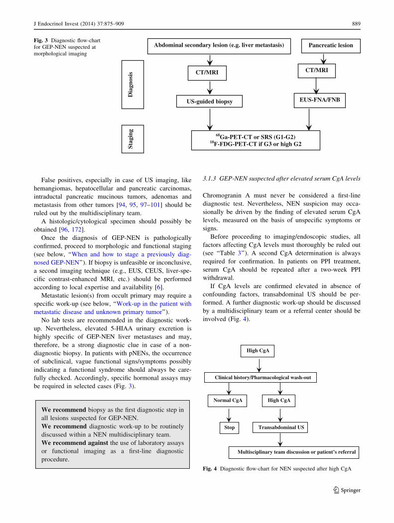
3.1.3 GEP-NEN suspected after elevated serum CgA levels
Chromogranin A must never be considered a first-line
diagnostic test. Nevertheless, NEN suspicion may occa-
sionally be driven by the finding of elevated serum CgA
levels, measured on the basis of unspecific symptoms or
signs.
Before proceeding to imaging/endoscopic studies, all
factors affecting CgA levels must thoroughly be ruled out
(see ‘‘Table 3’’). A second CgA determination is always
required for confirmation. In patients on PPI treatment,
serum CgA should be repeated after a two-week PPI
withdrawal.
If CgA levels are confirmed elevated in absence of
confounding factors, transabdominal US should be per-
formed. A further diagnostic work-up should be discussed
by a multidisciplinary team or a referral center should be
involved (Fig. 4).
High CgA
Clinical history/Pharmacological wash-out
Transabdominal US
High CgA
Multisciplinary team discussion or patient’s referral
Normal CgA
Stop
Fig. 4 Diagnostic flow-chart for NEN suspected after high CgA
CT/MRI
US-guided biopsy
68Ga-PET-CT or SRS (G1-G2)18F-FDG-PET-CT if G3 or high G2
Abdominal secondary lesion (e.g. liver metastasis) Pancreatic lesion
CT/MRI
EUS-FNA/FNB
Dia
gnos
isSt
agin
g
Fig. 3 Diagnostic flow-chart
for GEP-NEN suspected at
morphological imaging
J Endocrinol Invest (2014) 37:875–909 889
123

We recommend careful exclusion of all potentiallyinterfering factors in patients with elevated serumCgA levels and no previous NEN diagnosis.We suggest transabdominal US as first step in case ofconfirmed CgA increase.We recommend discussion of further work-up in amultidisciplinary team, involving a referral centerwhen required.
3.2 Symptomatic patient with symptoms due to GEP-
NEN-related local effects
3.2.1 When to suspect a GEP-NEN
Non-functioning GEP-NENs (Box 1) may become symp-
tomatic when they compress or invade adjacent structures or
when they metastasize. The suspicion of GEP-NEN might be
raised by suggestive imaging findings (see above) and/or by
the apparently slow progression of the disease [73]. Lab
findings (e.g., frankly elevated CgA levels in absence of
confounding factors) may reinforce the suspicion. As previ-
ously stated, only pathology (cytological or histologic char-
acterization), however, will establish the diagnosis [6].
3.2.2 Work-up in the patient with local compressive
symptoms
A detailed history and complete physical examination are
required.
Abdominal pain is the most common presenting symp-
tom of NF GEP-NENs and may be related to the primary
tumor or metastatic lesions [1, 3]. Pain localization and
characteristics should be carefully examined. Four different
scenarios can be distinguished.
3.2.2.1 Isolated abdominal pain A persistent and
oppressive upper-abdominal pain may signal a pancreatic
or retroperitoneal mass (pattern 1a) [96, 172], while a
discontinuous cramping pain usually refers to an intestinal
origin (pattern 1b) [73, 173]. In the former case, a radio-
logical imaging should be performed first, followed by
endoscopy/EUS as second step for pancreatic and duodenal
lesions. In the latter case, endoscopy is recommended [73,
173]. A cytologic/histologic sampling should be obtained
whenever possible (Fig. 5) [96, 172].
An ill-defined and diffuse abdominal pain (pattern 1c)
can also be related to liver or nodal metastases. Abdominal
US followed by a whole-body CT scan and a US-guided
biopsy should be performed (Fig. 5).
3.2.2.2 Subocclusive picture It may be due to a large,
often metastatic, ileal NEN and/or peritoneal carcinoma-
tosis. Depending on the severity of the clinical picture, a
direct abdomen-X-ray and/or an endoscopy could be per-
formed [73, 173]. If an extrinsic obstruction is suspected,
then an abdomen CT scan should be performed. If a peri-
toneal carcinomatosis is suspected, a transit evaluation
water-soluble contrast medium X-ray could be useful
(Fig. 6). If possible, histological specimens should be
obtained through endoscopy. If not, a US/CT-guided biopsy
Box 1
Non-functioning GEP-NENs
Definition: NF GEP-NENs are tumors that do not show
symptoms related to hormonal hypersecretion. Intracellular
hormones or peptides may be demonstrated by IHC, but they
are either not secreted, or secreted in quantities unable to
elicit a clinical syndrome and/or in an inactive form [3].
Clinical presentation of NF GEP-NENs depends upon the
site of origin and metastases. They can be incidentally
discovered when asymptomatic due to the widespread use of
diagnostic imaging [1, 3]. Clinical presentations according
to the site of origin are listed below.
Pancreas: Up to 60% of pNENs is NF. Most NF pNENs
are well differentiated. Annual incidence is 1.8 and 2.6
per million in females and males, respectively [3]. NF
pNEN were traditionally diagnosed late in the course of
the disease, with metastases in 46 to 73% of cases, but
presently the number of incidentally found small lesions
is steeply increasing. Presenting symptoms and signs are
abdominal pain (35–78%), weight loss (20–35%),
anorexia and nausea (45%), intra-abdominal hemor-
rhage (4–20%), jaundice (17–50%), and a palpable
mass (7–40%) [96, 172]. NF pNEN may occur in
familiar syndromes such as MEN-1, VHL, and TSC.
Gastrointestinal: NENs are frequently detected during a
screening program or an imaging exam performed to
search the primary tumor in an asymptomatic but meta-
static patient [1, 3]. Alternatively, a common clinical
presentation is abdominal pain that may be caused by
gastro-intestinal dysmotility or obstruction (associated or
not to nausea, vomiting or constipation), or by bacterial
overgrowth. Less common symptoms and signs are jaun-
dice, weight loss, fatigue, fever and bleeding (massive or
dripping). Clinical presentation of appendiceal NEN may
mimic acute appendicitis [1, 3]. Obstructive symptoms are
typical of small bowel, whereas minor bleeding is frequent
in rectal disease [6, 73, 173].
890 J Endocrinol Invest (2014) 37:875–909
123

of the liver or other site lesions or laparoscopy-guided
biopsy should be discussed in a multidisciplinary team.
3.2.2.3 Jaundice This clinical presentation points to the
involvement of the liver, biliary tract or pancreas. Liver
function and structure should be assessed by blood tests
and US, to rule out the obstruction of the biliary tract.
Compressive effects of lymphadenopathies or a pancreatic
mass may cause an extra-hepatic tract dilatation, whereas
liver metastases are more likely related to an intra-hepatic
tract dilatation [96, 172]. In case of obstructive jaundice, a
cholangio-MRI and endoscopic-retrograde-cholangio-pan-
creatography (ERCP) can be considered. Cytology by
means of brushing or histology can be obtained through
ERCP. Whole-body CT scan and endoscopy should be
used to define the primary site of the tumor and for staging
purpose (Fig. 7).
3.2.2.4 Gastrointestinal bleeding It can be related to the
compressive and infiltrating effects of a tumor mass.
Bleeding can be massive (hematemesis, melena and rectal
bleeding) or dripping and occult. Blood tests, iron assess-
ment and endoscopy must be performed. Massive bleeding
always requires hospitalization and may require angiogra-
phy [73, 173]. In case of lesions located in the stomach-
duodenum or in terminal ileum-colon tract, a histologic
diagnosis may be obtained through biopsy during EGDS or
ileo-colonoscopy. If upper and lower endoscopy is nega-
tive, enteroscopy, enteroCT/MRI, VCE should be dis-
cussed in the multidisciplinary team according to the local
Persistent, oppressiveor vague and diffuse
Endoscopy/EUS ± biopsyCT, MRI, US ± biopsy
68Ga-PET-CT or SRS18F-FDG PET-CT if G3 or high G2
IF PRIMARY NOT FOUND
Dia
gnos
isSt
agin
g
Discontinuous cramping
CT, MRI, US ± biopsy
Abdominal painFig. 5 Diagnostic flow-chart
for GEP-NEN suspected after
pattern 1a and 1b
Obstructive symptoms
Endoscopy, EUS ± FNA/B
CT, MRI, US ± FNA/B
68Ga-PET-CT or SRS18F-FDG PET-CT if G3 or high G2
Dia
gnos
isSt
agin
g
Abdomen X-ray: obstruction?
Surgery
YES
NO
EXTRINSIC OBSTRUCTION
SUSPECTEDPERITONEAL CARCINOMATOSIS
SUSPECTED
Transit evaluation X-Ray
Fig. 6 Diagnostic flow-chart
for GEP-NEN suspected after
subocclusive picture
J Endocrinol Invest (2014) 37:875–909 891
123
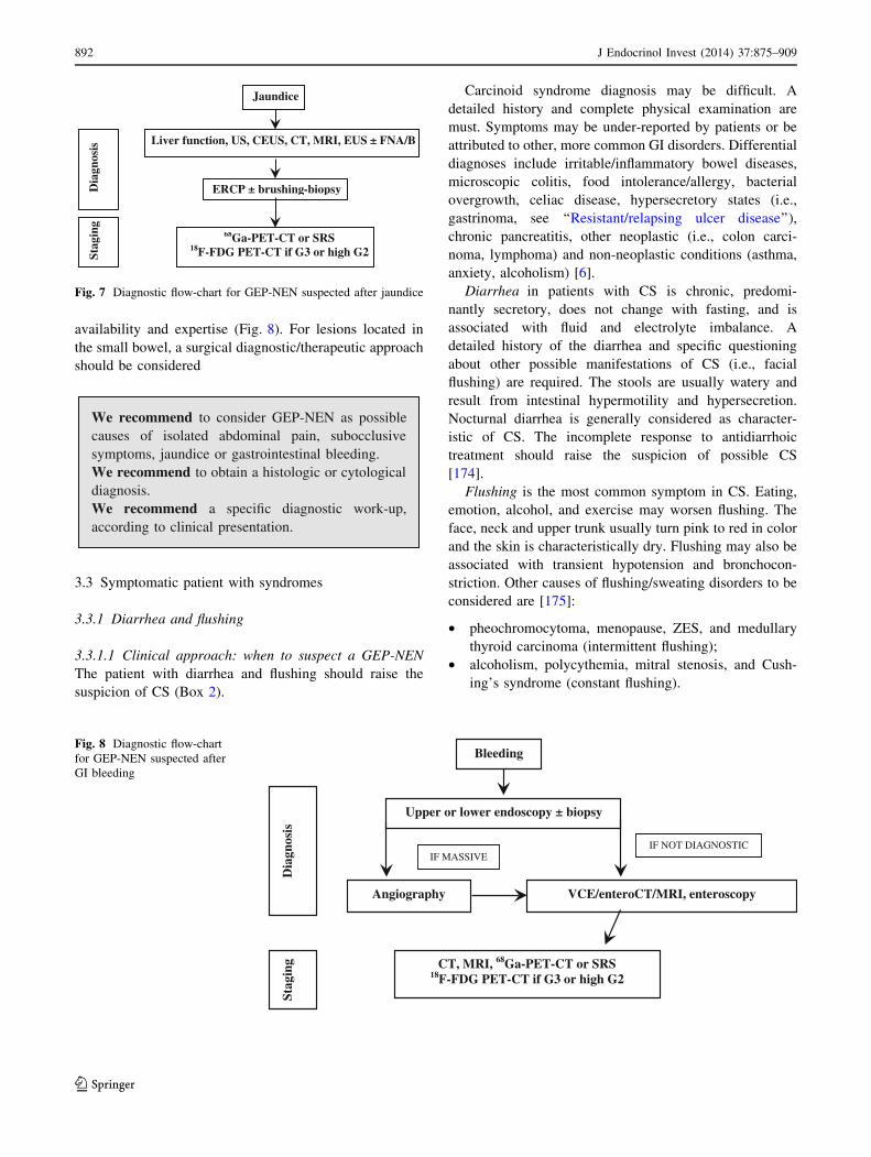
availability and expertise (Fig. 8). For lesions located in
the small bowel, a surgical diagnostic/therapeutic approach
should be considered
We recommend to consider GEP-NEN as possiblecauses of isolated abdominal pain, subocclusivesymptoms, jaundice or gastrointestinal bleeding.We recommend to obtain a histologic or cytologicaldiagnosis.We recommend a specific diagnostic work-up,according to clinical presentation.
3.3 Symptomatic patient with syndromes
3.3.1 Diarrhea and flushing
3.3.1.1 Clinical approach: when to suspect a GEP-NEN
The patient with diarrhea and flushing should raise the
suspicion of CS (Box 2).
Carcinoid syndrome diagnosis may be difficult. A
detailed history and complete physical examination are
must. Symptoms may be under-reported by patients or be
attributed to other, more common GI disorders. Differential
diagnoses include irritable/inflammatory bowel diseases,
microscopic colitis, food intolerance/allergy, bacterial
overgrowth, celiac disease, hypersecretory states (i.e.,
gastrinoma, see ‘‘Resistant/relapsing ulcer disease’’),
chronic pancreatitis, other neoplastic (i.e., colon carci-
noma, lymphoma) and non-neoplastic conditions (asthma,
anxiety, alcoholism) [6].
Diarrhea in patients with CS is chronic, predomi-
nantly secretory, does not change with fasting, and is
associated with fluid and electrolyte imbalance. A
detailed history of the diarrhea and specific questioning
about other possible manifestations of CS (i.e., facial
flushing) are required. The stools are usually watery and
result from intestinal hypermotility and hypersecretion.
Nocturnal diarrhea is generally considered as character-
istic of CS. The incomplete response to antidiarrhoic
treatment should raise the suspicion of possible CS
[174].
Flushing is the most common symptom in CS. Eating,
emotion, alcohol, and exercise may worsen flushing. The
face, neck and upper trunk usually turn pink to red in color
and the skin is characteristically dry. Flushing may also be
associated with transient hypotension and bronchocon-
striction. Other causes of flushing/sweating disorders to be
considered are [175]:
• pheochromocytoma, menopause, ZES, and medullary
thyroid carcinoma (intermittent flushing);
• alcoholism, polycythemia, mitral stenosis, and Cush-
ing’s syndrome (constant flushing).
Jaundice
ERCP ± brushing-biopsy
68Ga-PET-CT or SRS18F-FDG PET-CT if G3 or high G2
Liver function, US, CEUS, CT, MRI, EUS ± FNA/B
Dia
gnos
isSt
agin
g
Fig. 7 Diagnostic flow-chart for GEP-NEN suspected after jaundice
Upper or lower endoscopy ± biopsy
CT, MRI, 68Ga-PET-CT or SRS 18F-FDG PET-CT if G3 or high G2
IF NOT DIAGNOSTIC
VCE/enteroCT/MRI, enteroscopy
IF MASSIVE
Angiography
Bleeding
Dia
gnos
is
Stag
ing
Fig. 8 Diagnostic flow-chart
for GEP-NEN suspected after
GI bleeding
892 J Endocrinol Invest (2014) 37:875–909
123
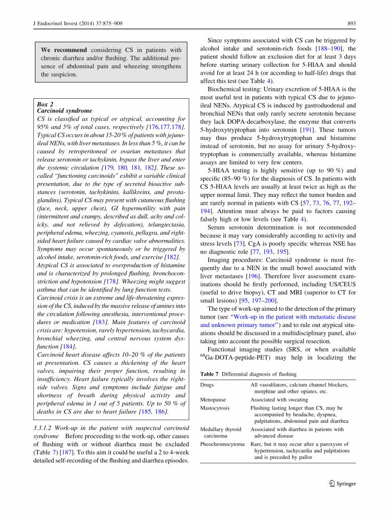
We recommend considering CS in patients withchronic diarrhea and/or flushing. The additional pre-sence of abdominal pain and wheezing strengthensthe suspicion.
3.3.1.2 Work-up in the patient with suspected carcinoid
syndrome Before proceeding to the work-up, other causes
of flushing with or without diarrhea must be excluded
(Table 7) [187]. To this aim it could be useful a 2 to 4-week
detailed self-recording of the flushing and diarrhea episodes.
Since symptoms associated with CS can be triggered by
alcohol intake and serotonin-rich foods [188–190], the
patient should follow an exclusion diet for at least 3 days
before starting urinary collection for 5-HIAA and should
avoid for at least 24 h (or according to half-life) drugs that
affect this test (see Table 4).
Biochemical testing: Urinary excretion of 5-HIAA is the
most useful test in patients with typical CS due to jejuno-
ileal NENs. Atypical CS is induced by gastroduodenal and
bronchial NENs that only rarely secrete serotonin because
they lack DOPA-decarboxylase, the enzyme that converts
5-hydroxytryptophan into serotonin [191]. These tumors
may thus produce 5-hydroxytryptophan and histamine
instead of serotonin, but no assay for urinary 5-hydroxy-
tryptophan is commercially available, whereas histamine
assays are limited to very few centers.
5-HIAA testing is highly sensitive (up to 90 %) and
specific (85–90 %) for the diagnosis of CS. In patients with
CS 5-HIAA levels are usually at least twice as high as the
upper normal limit. They may reflect the tumor burden and
are rarely normal in patients with CS [57, 73, 76, 77, 192–
194]. Attention must always be paid to factors causing
falsely high or low levels (see Table 4).
Serum serotonin determination is not recommended
because it may vary considerably according to activity and
stress levels [73]. CgA is poorly specific whereas NSE has
no diagnostic role [77, 193, 195].
Imaging procedures: Carcinoid syndrome is most fre-
quently due to a NEN in the small bowel associated with
liver metastases [196]. Therefore liver assessment exam-
inations should be firstly performed, including US/CEUS
(useful to drive biopsy), CT and MRI (superior to CT for
small lesions) [95, 197–200].
The type of work-up aimed to the detection of the primary
tumor (see ‘‘Work-up in the patient with metastatic disease
and unknown primary tumor’’) and to rule out atypical situ-
ations should be discussed in a multidisciplinary panel, also
taking into account the possible surgical resection.
Functional imaging studies (SRS, or when available68Ga-DOTA-peptide-PET) may help in localizing the
Box 2Carcinoid syndrome
CS is classified as typical or atypical, accounting for
95% and 5% of total cases, respectively [176,177,178].
Typical CS occurs in about 15-20 % of patients with jejuno-
ileal NENs, with liver metastases. In less than 5 %, it can be
caused by retroperitoneal or ovarian metastases that
release serotonin or tachykinin, bypass the liver and enter
the systemic circulation [179, 180, 181, 182]. These so-
called ‘‘functioning carcinoids’’ exhibit a variable clinical
presentation, due to the type of secreted bioactive sub-
stances (serotonin, tachykinins, kallikreins, and prosta-
glandins). Typical CS may present with cutaneous flushing
(face, neck, upper chest), GI hypermotility with pain
(intermittent and crampy, described as dull, achy and col-
icky, and not relieved by defecation), telangiectasia,
peripheral edema, wheezing, cyanosis, pellagra, and right-
sided heart failure caused by cardiac valve abnormalities.
Symptoms may occur spontaneously or be triggered by
alcohol intake, serotonin-rich foods, and exercise [182].
Atypical CS is associated to overproduction of histamine
and is characterized by prolonged flushing, bronchocon-
striction and hypotension [178]. Wheezing might suggest
asthma that can be identified by lung function tests.
Carcinoid crisis is an extreme and life-threatening expres-
sion of the CS, induced by the massive release of amines into
the circulation following anesthesia, interventional proce-
dures or medication [183]. Main features of carcinoid
crisis are: hypotension, rarely hypertension, tachycardia,
bronchial wheezing, and central nervous system dys-
function [184].
Carcinoid heart disease affects 10–20 % of the patients
at presentation. CS causes a thickening of the heart
valves, impairing their proper function, resulting in
insufficiency. Heart failure typically involves the right-
side valves. Signs and symptoms include fatigue and
shortness of breath during physical activity and
peripheral edema in 1 out of 5 patients. Up to 50 % of
deaths in CS are due to heart failure [185, 186].
Table 7 Differential diagnosis of flushing
Drugs All vasodilators, calcium channel blockers,
morphine and other opiates, etc.
Menopause Associated with sweating
Mastocytosis Flushing lasting longer than CS, may be
accompanied by headache, dyspnea,
palpitations, abdominal pain and diarrhea
Medullary thyroid
carcinoma
Associated with diarrhea in patients with
advanced disease
Pheochromocytoma Rare, but it may occur after a paroxysm of
hypertension, tachycardia and palpitations
and is preceded by pallor
J Endocrinol Invest (2014) 37:875–909 893
123
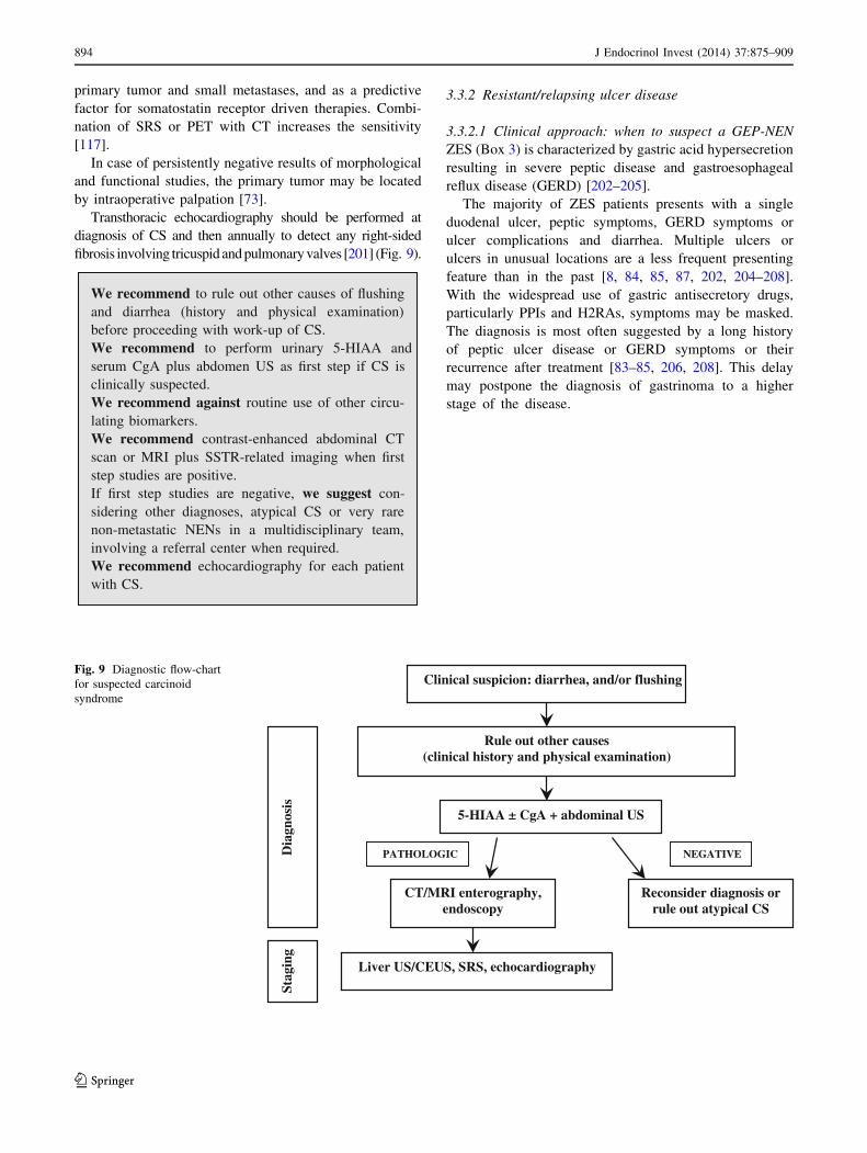
primary tumor and small metastases, and as a predictive
factor for somatostatin receptor driven therapies. Combi-
nation of SRS or PET with CT increases the sensitivity
[117].
In case of persistently negative results of morphological
and functional studies, the primary tumor may be located
by intraoperative palpation [73].
Transthoracic echocardiography should be performed at
diagnosis of CS and then annually to detect any right-sided
fibrosis involving tricuspid and pulmonary valves [201] (Fig. 9).
We recommend to rule out other causes of flushingand diarrhea (history and physical examination)before proceeding with work-up of CS.We recommend to perform urinary 5-HIAA andserum CgA plus abdomen US as first step if CS isclinically suspected.We recommend against routine use of other circu-lating biomarkers.We recommend contrast-enhanced abdominal CTscan or MRI plus SSTR-related imaging when firststep studies are positive.If first step studies are negative, we suggest con-sidering other diagnoses, atypical CS or very rarenon-metastatic NENs in a multidisciplinary team,involving a referral center when required.We recommend echocardiography for each patientwith CS.
3.3.2 Resistant/relapsing ulcer disease
3.3.2.1 Clinical approach: when to suspect a GEP-NEN
ZES (Box 3) is characterized by gastric acid hypersecretion
resulting in severe peptic disease and gastroesophageal
reflux disease (GERD) [202–205].
The majority of ZES patients presents with a single
duodenal ulcer, peptic symptoms, GERD symptoms or
ulcer complications and diarrhea. Multiple ulcers or
ulcers in unusual locations are a less frequent presenting
feature than in the past [8, 84, 85, 87, 202, 204–208].
With the widespread use of gastric antisecretory drugs,
particularly PPIs and H2RAs, symptoms may be masked.
The diagnosis is most often suggested by a long history
of peptic ulcer disease or GERD symptoms or their
recurrence after treatment [83–85, 206, 208]. This delay
may postpone the diagnosis of gastrinoma to a higher
stage of the disease.
Clinical suspicion: diarrhea, and/or flushing
Rule out other causes(clinical history and physical examination)
5-HIAA ± CgA + abdominal US
CT/MRI enterography, endoscopy
Liver US/CEUS, SRS, echocardiography
Dia
gnos
isSt
agin
g
PATHOLOGIC NEGATIVE
Reconsider diagnosis or rule out atypical CS
Fig. 9 Diagnostic flow-chart
for suspected carcinoid
syndrome
894 J Endocrinol Invest (2014) 37:875–909
123
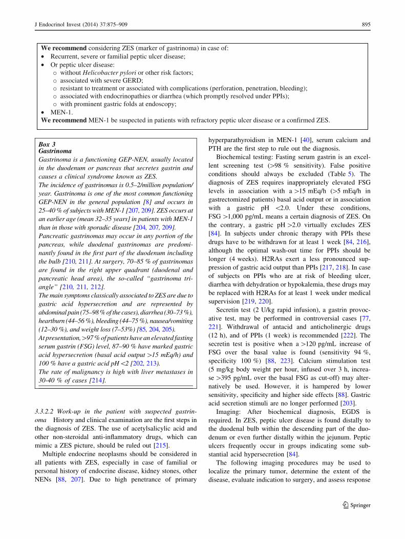
3.3.2.2 Work-up in the patient with suspected gastrin-
oma History and clinical examination are the first steps in
the diagnosis of ZES. The use of acetylsalicylic acid and
other non-steroidal anti-inflammatory drugs, which can
mimic a ZES picture, should be ruled out [215].
Multiple endocrine neoplasms should be considered in
all patients with ZES, especially in case of familial or
personal history of endocrine disease, kidney stones, other
NENs [88, 207]. Due to high penetrance of primary
hyperparathyroidism in MEN-1 [40], serum calcium and
PTH are the first step to rule out the diagnosis.
Biochemical testing: Fasting serum gastrin is an excel-
lent screening test ([98 % sensitivity). False positive
conditions should always be excluded (Table 5). The
diagnosis of ZES requires inappropriately elevated FSG
levels in association with a [15 mEq/h ([5 mEq/h in
gastrectomized patients) basal acid output or in association
with a gastric pH \2.0. Under these conditions,
FSG [1,000 pg/mL means a certain diagnosis of ZES. On
the contrary, a gastric pH [2.0 virtually excludes ZES
[84]. In subjects under chronic therapy with PPIs these
drugs have to be withdrawn for at least 1 week [84, 216],
although the optimal wash-out time for PPIs should be
longer (4 weeks). H2RAs exert a less pronounced sup-
pression of gastric acid output than PPIs [217, 218]. In case
of subjects on PPIs who are at risk of bleeding ulcer,
diarrhea with dehydration or hypokalemia, these drugs may
be replaced with H2RAs for at least 1 week under medical
supervision [219, 220].
Secretin test (2 U/kg rapid infusion), a gastrin provoc-
ative test, may be performed in controversial cases [77,
221]. Withdrawal of antacid and anticholinergic drugs
(12 h), and of PPIs (1 week) is recommended [222]. The
secretin test is positive when a [120 pg/mL increase of
FSG over the basal value is found (sensitivity 94 %,
specificity 100 %) [88, 223]. Calcium stimulation test
(5 mg/kg body weight per hour, infused over 3 h, increa-
se [395 pg/mL over the basal FSG as cut-off) may alter-
natively be used. However, it is hampered by lower
sensitivity, specificity and higher side effects [88]. Gastric
acid secretion stimuli are no longer performed [203].
Imaging: After biochemical diagnosis, EGDS is
required. In ZES, peptic ulcer disease is found distally to
the duodenal bulb within the descending part of the duo-
denum or even further distally within the jejunum. Peptic
ulcers frequently occur in groups indicating some sub-
stantial acid hypersecretion [84].
The following imaging procedures may be used to
localize the primary tumor, determine the extent of the
disease, evaluate indication to surgery, and assess response
We recommend considering ZES (marker of gastrinoma) in case of:• Recurrent, severe or familial peptic ulcer disease;• Or peptic ulcer disease:
without Helicobacter pylori or other risk factors;associated with severe GERD;resistant to treatment or associated with complications (perforation, penetration, bleeding);associated with endocrinopathies or diarrhea (which promptly resolved under PPIs);with prominent gastric folds at endoscopy;
• MEN-1.We recommend MEN-1 be suspected in patients with refractory peptic ulcer disease or a confirmed ZES.
Box 3Gastrinoma
Gastrinoma is a functioning GEP-NEN, usually located
in the duodenum or pancreas that secretes gastrin and
causes a clinical syndrome known as ZES.
The incidence of gastrinomas is 0.5–2/million population/
year. Gastrinoma is one of the most common functioning
GEP-NEN in the general population [8] and occurs in
25–40 % of subjects with MEN-1 [207, 209]. ZES occurs at
an earlier age (mean 32–35 years] in patients with MEN-1
than in those with sporadic disease [204, 207, 209].
Pancreatic gastrinomas may occur in any portion of the
pancreas, while duodenal gastrinomas are predomi-
nantly found in the first part of the duodenum including
the bulb [210, 211]. At surgery, 70–85 % of gastrinomas
are found in the right upper quadrant (duodenal and
pancreatic head area), the so-called ‘‘gastrinoma tri-
angle’’ [210, 211, 212].
The main symptoms classically associated to ZES are due to
gastric acid hypersecretion and are represented by
abdominal pain (75–98 % of the cases), diarrhea (30–73 %),
heartburn (44–56 %), bleeding (44–75 %), nausea/vomiting
(12–30 %), and weight loss (7–53%) [85, 204, 205).
At presentation,[97 % of patients have an elevated fasting
serum gastrin (FSG) level, 87–90 % have marked gastric
acid hypersecretion (basal acid output [15 mEq/h) and
100 % have a gastric acid pH\2 [202, 213).
The rate of malignancy is high with liver metastases in
30-40 % of cases [214].
J Endocrinol Invest (2014) 37:875–909 895
123
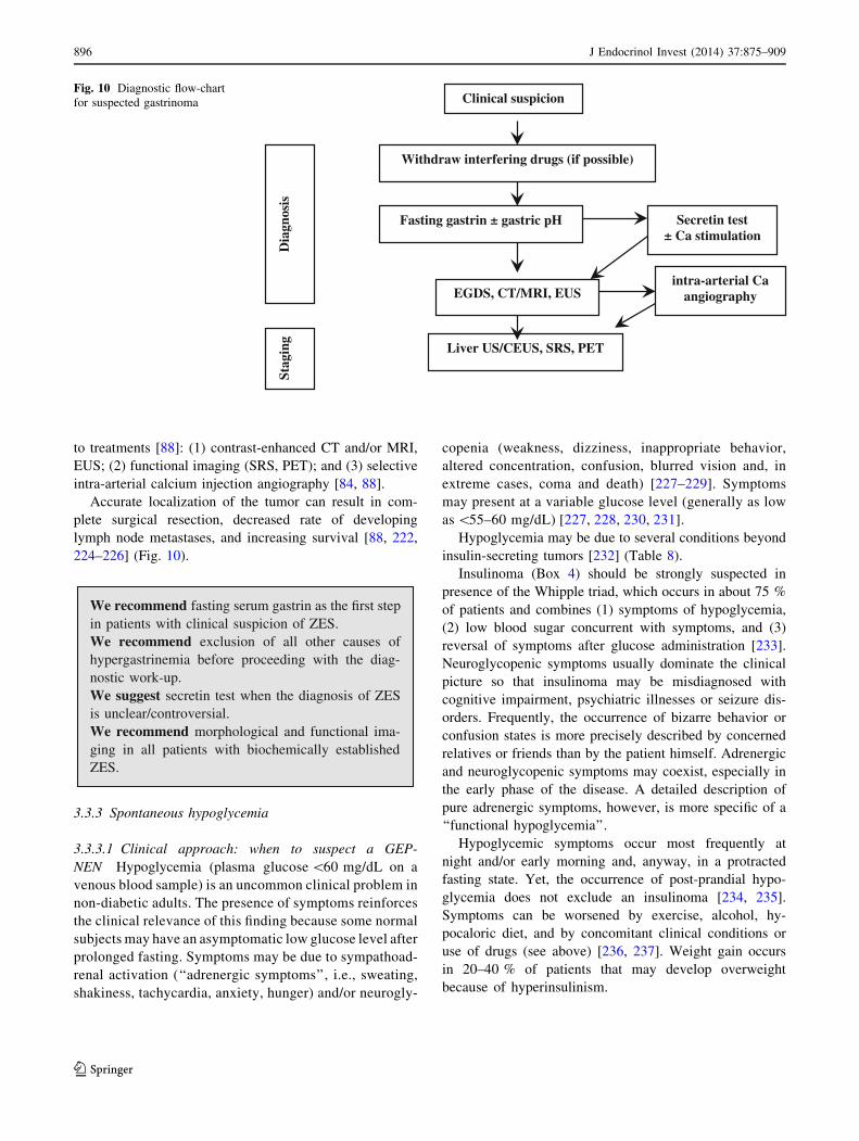
to treatments [88]: (1) contrast-enhanced CT and/or MRI,
EUS; (2) functional imaging (SRS, PET); and (3) selective
intra-arterial calcium injection angiography [84, 88].
Accurate localization of the tumor can result in com-
plete surgical resection, decreased rate of developing
lymph node metastases, and increasing survival [88, 222,
224–226] (Fig. 10).
We recommend fasting serum gastrin as the first stepin patients with clinical suspicion of ZES.We recommend exclusion of all other causes ofhypergastrinemia before proceeding with the diag-nostic work-up.We suggest secretin test when the diagnosis of ZESis unclear/controversial.We recommend morphological and functional ima-ging in all patients with biochemically establishedZES.
3.3.3 Spontaneous hypoglycemia
3.3.3.1 Clinical approach: when to suspect a GEP-
NEN Hypoglycemia (plasma glucose \60 mg/dL on a
venous blood sample) is an uncommon clinical problem in
non-diabetic adults. The presence of symptoms reinforces
the clinical relevance of this finding because some normal
subjects may have an asymptomatic low glucose level after
prolonged fasting. Symptoms may be due to sympathoad-
renal activation (‘‘adrenergic symptoms’’, i.e., sweating,
shakiness, tachycardia, anxiety, hunger) and/or neurogly-
copenia (weakness, dizziness, inappropriate behavior,
altered concentration, confusion, blurred vision and, in
extreme cases, coma and death) [227–229]. Symptoms
may present at a variable glucose level (generally as low
as \55–60 mg/dL) [227, 228, 230, 231].
Hypoglycemia may be due to several conditions beyond
insulin-secreting tumors [232] (Table 8).
Insulinoma (Box 4) should be strongly suspected in
presence of the Whipple triad, which occurs in about 75 %
of patients and combines (1) symptoms of hypoglycemia,
(2) low blood sugar concurrent with symptoms, and (3)
reversal of symptoms after glucose administration [233].
Neuroglycopenic symptoms usually dominate the clinical
picture so that insulinoma may be misdiagnosed with
cognitive impairment, psychiatric illnesses or seizure dis-
orders. Frequently, the occurrence of bizarre behavior or
confusion states is more precisely described by concerned
relatives or friends than by the patient himself. Adrenergic
and neuroglycopenic symptoms may coexist, especially in
the early phase of the disease. A detailed description of
pure adrenergic symptoms, however, is more specific of a
‘‘functional hypoglycemia’’.
Hypoglycemic symptoms occur most frequently at
night and/or early morning and, anyway, in a protracted
fasting state. Yet, the occurrence of post-prandial hypo-
glycemia does not exclude an insulinoma [234, 235].
Symptoms can be worsened by exercise, alcohol, hy-
pocaloric diet, and by concomitant clinical conditions or
use of drugs (see above) [236, 237]. Weight gain occurs
in 20–40 % of patients that may develop overweight
because of hyperinsulinism.
Clinical suspicion
Withdraw interfering drugs (if possible)
Fasting gastrin ± gastric pH
EGDS, CT/MRI, EUS
Liver US/CEUS, SRS, PET
Dia
gnos
isSt
agin
g
Secretin test± Ca stimulation
intra-arterial Ca angiography
Fig. 10 Diagnostic flow-chart
for suspected gastrinoma
896 J Endocrinol Invest (2014) 37:875–909
123
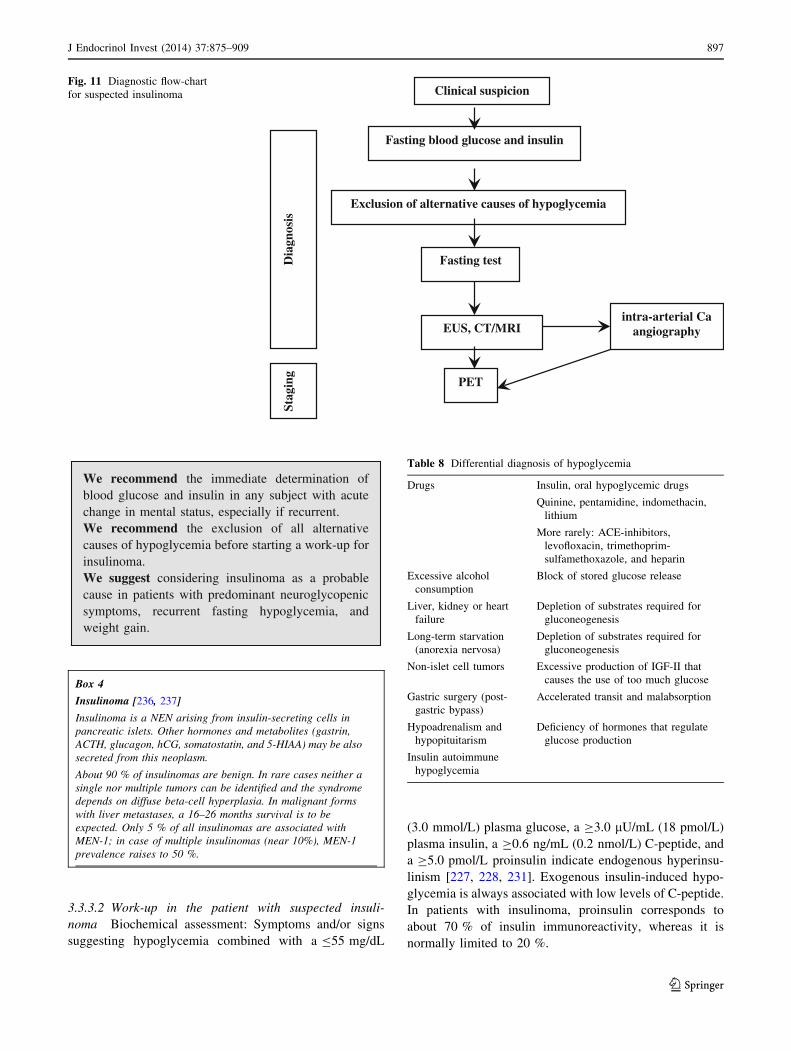
We recommend the immediate determination ofblood glucose and insulin in any subject with acutechange in mental status, especially if recurrent.We recommend the exclusion of all alternativecauses of hypoglycemia before starting a work-up forinsulinoma.We suggest considering insulinoma as a probablecause in patients with predominant neuroglycopenicsymptoms, recurrent fasting hypoglycemia, andweight gain.
3.3.3.2 Work-up in the patient with suspected insuli-
noma Biochemical assessment: Symptoms and/or signs
suggesting hypoglycemia combined with a B55 mg/dL
(3.0 mmol/L) plasma glucose, a C3.0 lU/mL (18 pmol/L)
plasma insulin, a C0.6 ng/mL (0.2 nmol/L) C-peptide, and
a C5.0 pmol/L proinsulin indicate endogenous hyperinsu-
linism [227, 228, 231]. Exogenous insulin-induced hypo-
glycemia is always associated with low levels of C-peptide.
In patients with insulinoma, proinsulin corresponds to
about 70 % of insulin immunoreactivity, whereas it is
normally limited to 20 %.
Clinical suspicion
Exclusion of alternative causes of hypoglycemia
EUS, CT/MRI
PET
Dia
gnos
isSt
agin
g
Fasting test
intra-arterial Ca angiography
Fasting blood glucose and insulin
Fig. 11 Diagnostic flow-chart
for suspected insulinoma
Table 8 Differential diagnosis of hypoglycemia
Drugs Insulin, oral hypoglycemic drugs
Quinine, pentamidine, indomethacin,
lithium
More rarely: ACE-inhibitors,
levofloxacin, trimethoprim-
sulfamethoxazole, and heparin
Excessive alcohol
consumption
Block of stored glucose release
Liver, kidney or heart
failure
Depletion of substrates required for
gluconeogenesis
Long-term starvation
(anorexia nervosa)
Depletion of substrates required for
gluconeogenesis
Non-islet cell tumors Excessive production of IGF-II that
causes the use of too much glucose
Gastric surgery (post-
gastric bypass)
Accelerated transit and malabsorption
Hypoadrenalism and
hypopituitarism
Deficiency of hormones that regulate
glucose production
Insulin autoimmune
hypoglycemia
Box 4
Insulinoma [236, 237]
Insulinoma is a NEN arising from insulin-secreting cells in
pancreatic islets. Other hormones and metabolites (gastrin,
ACTH, glucagon, hCG, somatostatin, and 5-HIAA) may be also
secreted from this neoplasm.
About 90 % of insulinomas are benign. In rare cases neither a
single nor multiple tumors can be identified and the syndrome
depends on diffuse beta-cell hyperplasia. In malignant forms
with liver metastases, a 16–26 months survival is to be
expected. Only 5 % of all insulinomas are associated with
MEN-1; in case of multiple insulinomas (near 10%), MEN-1
prevalence raises to 50 %.
J Endocrinol Invest (2014) 37:875–909 897
123
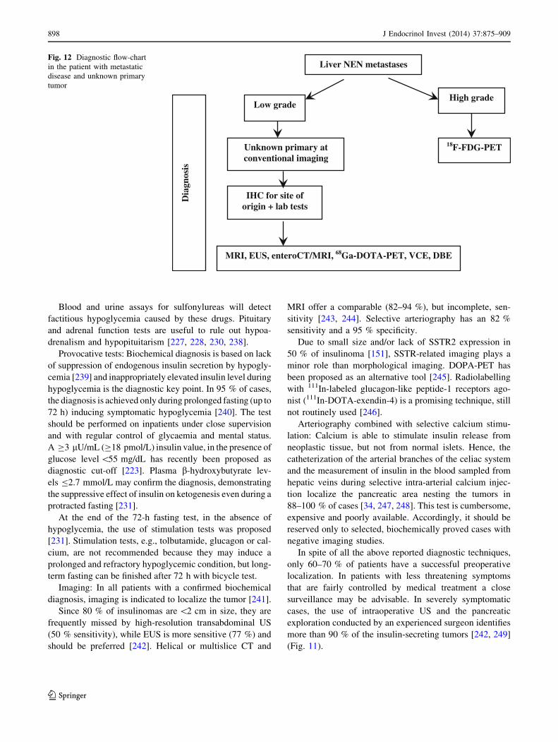
Blood and urine assays for sulfonylureas will detect
factitious hypoglycemia caused by these drugs. Pituitary
and adrenal function tests are useful to rule out hypoa-
drenalism and hypopituitarism [227, 228, 230, 238].
Provocative tests: Biochemical diagnosis is based on lack
of suppression of endogenous insulin secretion by hypogly-
cemia [239] and inappropriately elevated insulin level during
hypoglycemia is the diagnostic key point. In 95 % of cases,
the diagnosis is achieved only during prolonged fasting (up to
72 h) inducing symptomatic hypoglycemia [240]. The test
should be performed on inpatients under close supervision
and with regular control of glycaemia and mental status.
A C3 lU/mL (C18 pmol/L) insulin value, in the presence of
glucose level \55 mg/dL has recently been proposed as
diagnostic cut-off [223]. Plasma b-hydroxybutyrate lev-
els B2.7 mmol/L may confirm the diagnosis, demonstrating
the suppressive effect of insulin on ketogenesis even during a
protracted fasting [231].
At the end of the 72-h fasting test, in the absence of
hypoglycemia, the use of stimulation tests was proposed
[231]. Stimulation tests, e.g., tolbutamide, glucagon or cal-
cium, are not recommended because they may induce a
prolonged and refractory hypoglycemic condition, but long-
term fasting can be finished after 72 h with bicycle test.
Imaging: In all patients with a confirmed biochemical
diagnosis, imaging is indicated to localize the tumor [241].
Since 80 % of insulinomas are \2 cm in size, they are
frequently missed by high-resolution transabdominal US
(50 % sensitivity), while EUS is more sensitive (77 %) and
should be preferred [242]. Helical or multislice CT and
MRI offer a comparable (82–94 %), but incomplete, sen-
sitivity [243, 244]. Selective arteriography has an 82 %
sensitivity and a 95 % specificity.
Due to small size and/or lack of SSTR2 expression in
50 % of insulinoma [151], SSTR-related imaging plays a
minor role than morphological imaging. DOPA-PET has
been proposed as an alternative tool [245]. Radiolabelling
with 111In-labeled glucagon-like peptide-1 receptors ago-
nist (111In-DOTA-exendin-4) is a promising technique, still
not routinely used [246].
Arteriography combined with selective calcium stimu-
lation: Calcium is able to stimulate insulin release from
neoplastic tissue, but not from normal islets. Hence, the
catheterization of the arterial branches of the celiac system
and the measurement of insulin in the blood sampled from
hepatic veins during selective intra-arterial calcium injec-
tion localize the pancreatic area nesting the tumors in
88–100 % of cases [34, 247, 248]. This test is cumbersome,
expensive and poorly available. Accordingly, it should be
reserved only to selected, biochemically proved cases with
negative imaging studies.
In spite of all the above reported diagnostic techniques,
only 60–70 % of patients have a successful preoperative
localization. In patients with less threatening symptoms
that are fairly controlled by medical treatment a close
surveillance may be advisable. In severely symptomatic
cases, the use of intraoperative US and the pancreatic
exploration conducted by an experienced surgeon identifies
more than 90 % of the insulin-secreting tumors [242, 249]
(Fig. 11).
Liver NEN metastases
IHC for site of origin + lab tests
MRI, EUS, enteroCT/MRI, 68Ga-DOTA-PET, VCE, DBE
Dia
gnos
is
Unknown primary at conventional imaging
Low gradeHigh grade
18F-FDG-PET
Fig. 12 Diagnostic flow-chart
in the patient with metastatic
disease and unknown primary
tumor
898 J Endocrinol Invest (2014) 37:875–909
123
We recommend the simultaneous evaluation ofblood glucose, insulin and C-peptide to detect endo-genous hyperinsulinemia in all patients with sponta-neous hypoglycemia.We recommend a prolonged fasting test (up to 72 h)in patients referred for a previous hypoglycemicepisode who are free of symptoms at the moment ofmedical examination.We recommend against the use of stimulation testsfor the diagnosis of insulinoma.We recommend the use of localization tests (CT/CEUS and EUS) only after the biochemical diagnosisof insulinoma is established.
3.4 Work-up in the patient with metastatic disease
and unknown primary tumor
Unknown primary NEN (UPN) is a condition of metastatic
histologic or cytological confirmed NEN without evidence of a
primary site after a first diagnostic work-up, including chest-
abdomen CT scan, SRS, and upper and lower endoscopy.
The frequency of well-differentiated UPNs ranges from
9 to 19 % [250, 251]. The presence of liver metastases
largely influences prognosis in all types of NENs and is
dependent on primary tumor site, tumor extent (T-stage),
and histologic differentiation (NET vs. NEC). Reported
survival rate at 5 years of G1–G2 small intestinal and
pancreatic NENs in the SEER database is 54 and 27 %,
respectively [252]. Furthermore, survival is reportedly
worse in UPN patients as compared to patients with liver
metastases whose primary NEN is known [253]. In liver
metastatic patients survival rate is influenced by the pre-
sence of obstructive symptoms or symptoms related to
peptide secretion.
The evaluation of a patient with UPN should encompass
a detailed clinical history, including family history to
identify affected relatives and a patient’s increased risk for
endocrine tumors (i.e., MEN type 1 or 2), laboratory and
radiographic studies [254].
Histologic preparations should be reevaluated by IHC to
guide the search for the primary tumor: TTF-1 (pulmonary
or medullary thyroid carcinoma), CDX-2 (intestinal), PAX-
8, histidine-decarboxylase (pancreatic), xenin (duodenal),
gastrin (occult gastrinoma), and PP/glucagon (pancreatic)
[38, 255]. Biochemical work-up may include 5-HIAA,
gastrin, and other locally available tumor markers [256].
It has been recently reported that most UPNs are derived
from pancreas and small bowel [257]. Accordingly, further
investigations for localizing the primary site in well-dif-
ferentiated NENs might include abdomen MRI, EUS,
enteroCT/MR, 68Ga-PET, VCE, DBE to be shared within a
multidisciplinary team according to clinics, local avail-
ability and expertise [124, 258, 259]. In NECs 18F-FDG-
PET may be useful (Fig. 12)
We recommend biopsy at the metastatic site withhistologic and IHC examination as a first step toconfirm NEN diagnosis.We recommend a detailed clinical history to elicitsigns or symptoms that could point to the primary site(carcinoid syndrome, asthma, diarrhea, etc.), as wellas a complete family history.In all cases of unknown primary of low grade meta-static liver NEN after a conventional imaging,including chest-abdomen CT scan, upper and lowerendoscopy and SRS, we recommend an IHC for siteof origin, and imaging according to results.
3.5 When and how to stage a previously diagnosed
GEP-NEN
Evaluation of disease extension has a pivotal role in
treatment planning.
Pre-treatment staging should include morphologic and
functional imaging. Morphological imaging is required for
all GEP-NENs, irrespectively of their grade. SSTR-based
functional imaging (SRS or 68Ga-DOTA-peptide-PET)
should be used for low-/intermediate-grade GEP-NENs
(WHO 2010 G1-G2), whereas 18F-FDG-PET should be
preferentially used in G3 GEP-NENs and in some G2
cases.
For morphologic staging, a chest-abdomen-pelvis mul-
tidetector CT or a chest basal CT plus abdomen-pelvis MRI
should be used [87]. For functional staging, SRS using111In-pentetreotide (Octreoscan�) is presently regarded as
the gold standard. However, if available, 68Ga-DOTA-
peptide-PET with simultaneous CT should be preferred to
SRS. In facts, PET lacks the anatomic details required for
therapeutic stratification (surgical planning or dose calcu-
lation for radioembolization with radiolabeled micro-
spheres). Recently, MRI with liver-specific contrast
combined with 68Ga-DOTA-peptides-PET has been
reported to be more accurate than PET-CT to detect GEP-
NEN hepatic metastases [260].18F-DOPA-PET-CT and 11C-5HTP-PET-CT are prom-
ising tools. Their use might be considered if results of SRS
or 68Ga-DOTA-peptides-PET are negative [261].
Gastric NENs In small (\1 cm) type 1 and type 2
tumors, EGDS is usually the only recommended imaging
J Endocrinol Invest (2014) 37:875–909 899
123
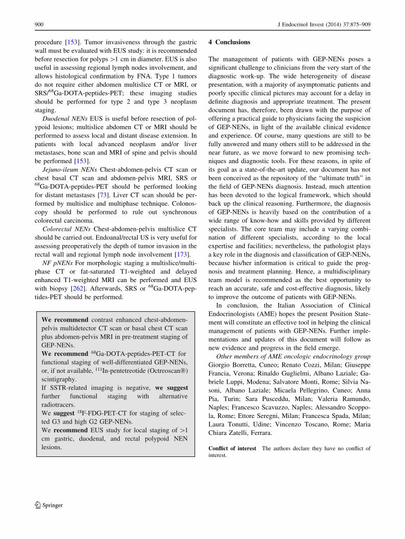
procedure [153]. Tumor invasiveness through the gastric
wall must be evaluated with EUS study: it is recommended
before resection for polyps [1 cm in diameter. EUS is also
useful in assessing regional lymph nodes involvement, and
allows histological confirmation by FNA. Type 1 tumors
do not require either abdomen multislice CT or MRI, or
SRS/68Ga-DOTA-peptides-PET; these imaging studies
should be performed for type 2 and type 3 neoplasm
staging.
Duodenal NENs EUS is useful before resection of pol-
ypoid lesions; multislice abdomen CT or MRI should be
performed to assess local and distant disease extension. In
patients with local advanced neoplasm and/or liver
metastases, bone scan and MRI of spine and pelvis should
be performed [153].
Jejuno-ileum NENs Chest-abdomen-pelvis CT scan or
chest basal CT scan and abdomen-pelvis MRI, SRS or68Ga-DOTA-peptides-PET should be performed looking
for distant metastases [73]. Liver CT scan should be per-
formed by multislice and multiphase technique. Colonos-
copy should be performed to rule out synchronous
colorectal carcinoma.
Colorectal NENs Chest-abdomen-pelvis multislice CT
should be carried out. Endoanal/rectal US is very useful for
assessing preoperatively the depth of tumor invasion in the
rectal wall and regional lymph node involvement [173].
NF pNENs For morphologic staging a multislice/multi-
phase CT or fat-saturated T1-weighted and delayed
enhanced T1-weighted MRI can be performed and EUS
with biopsy [262]. Afterwards, SRS or 68Ga-DOTA-pep-
tides-PET should be performed.
We recommend contrast enhanced chest-abdomen-pelvis multidetector CT scan or basal chest CT scanplus abdomen-pelvis MRI in pre-treatment staging ofGEP-NENs.We recommend 68Ga-DOTA-peptides-PET-CT forfunctional staging of well-differentiated GEP-NENs,or, if not available, 111In-pentetreotide (Octreoscan®)scintigraphy.If SSTR-related imaging is negative, we suggestfurther functional staging with alternativeradiotracers.We suggest 18F-FDG-PET-CT for staging of selec-ted G3 and high G2 GEP-NENs.We recommend EUS study for local staging of 1cm gastric, duodenal, and rectal polypoid NENlesions.
4 Conclusions
The management of patients with GEP-NENs poses a
significant challenge to clinicians from the very start of the
diagnostic work-up. The wide heterogeneity of disease
presentation, with a majority of asymptomatic patients and
poorly specific clinical pictures may account for a delay in
definite diagnosis and appropriate treatment. The present
document has, therefore, been drawn with the purpose of
offering a practical guide to physicians facing the suspicion
of GEP-NENs, in light of the available clinical evidence
and experience. Of course, many questions are still to be
fully answered and many others still to be addressed in the
near future, as we move forward to new promising tech-
niques and diagnostic tools. For these reasons, in spite of
its goal as a state-of-the-art update, our document has not
been conceived as the repository of the ‘‘ultimate truth’’ in
the field of GEP-NENs diagnosis. Instead, much attention
has been devoted to the logical framework, which should
back up the clinical reasoning. Furthermore, the diagnosis
of GEP-NENs is heavily based on the contribution of a
wide range of know-how and skills provided by different
specialists. The core team may include a varying combi-
nation of different specialists, according to the local
expertise and facilities; nevertheless, the pathologist plays
a key role in the diagnosis and classification of GEP-NENs,
because his/her information is critical to guide the prog-
nosis and treatment planning. Hence, a multidisciplinary
team model is recommended as the best opportunity to
reach an accurate, safe and cost-effective diagnosis, likely
to improve the outcome of patients with GEP-NENs.
In conclusion, the Italian Association of Clinical
Endocrinologists (AME) hopes the present Position State-
ment will constitute an effective tool in helping the clinical
management of patients with GEP-NENs. Further imple-
mentations and updates of this document will follow as
new evidence and progress in the field emerge.
Other members of AME oncologic endocrinology group
Giorgio Borretta, Cuneo; Renato Cozzi, Milan; Giuseppe
Francia, Verona; Rinaldo Guglielmi, Albano Laziale; Ga-
briele Luppi, Modena; Salvatore Monti, Rome; Silvia Na-
soni, Albano Laziale; Micaela Pellegrino, Cuneo; Anna
Pia, Turin; Sara Pusceddu, Milan; Valeria Ramundo,
Naples; Francesco Scavuzzo, Naples; Alessandro Scoppo-
la, Rome; Ettore Seregni, Milan; Francesca Spada, Milan;
Laura Tonutti, Udine; Vincenzo Toscano, Rome; Maria
Chiara Zatelli, Ferrara.
Conflict of interest The authors declare they have no conflict of
interest.
900 J Endocrinol Invest (2014) 37:875–909
123
Open Access This article is distributed under the terms of the
Creative Commons Attribution License which permits any use, dis-
tribution, and reproduction in any medium, provided the original
author(s) and the source are credited.
References
1. Modlin IR, Lye KD, Kidd M (2003) A 5-decade analysis of
13.715 carcinoid tumors. Cancer 97:934–959 (LoE ���s)
2. Hauso O, Gustafsson BI, Kidd M, Waldum HL, Drozdov I, Chan
AKC, Modlin IM (2008) Neuroendocrine tumor epidemiology.
Contrasting Norway and north America. Cancer 113:2655–2664
(LoE ��ss)
3. Yao JC, Hassan M, Phan A, Dagohoy C, Leary C, Mares JE,
Abdalla EK, Fleming JB, Vauthey JN, Rashid A, Evans DB
(2008) One hundred years after ‘carcinoid’: epidemiology of and
prognostic factors for neuroendocrine tumors in 35,825 cases in
the United States. J Clin Oncol 26:3063–3072 (LoE ��ss)
4. Garcia-Carbonero R, Capdevila J, Crespo-Herrero G, Dıaz-
Perez JA, Martınez Del Prado MP, Alonso Orduna V, Sevilla-
Garcıa I, Villabona-Artero C, Beguiristain-Gomez A, Llanos-
Munoz M, Marazuela M, Alvarez-Escola C, Castellano D, Vilar
E, Jimenez-Fonseca P, Teule A, Sastre-Valera J, Benavent-Vi-
nuelas M, Monleon A, Salazar R (2010) Incidence, patterns of
care and prognostic factors for outcome of gastroenteropancre-
atic neuroendocrine tumors (GEP-NETs): results from National
Cancer Registry of Spain (RGETNE). Ann Oncol 21:1794–1803
(LoE ���s)
5. Lawrence B, Gustafsson BJ, Kidd M, Pavel M, Svejda B,
Modlin IM (2011) The clinical relevance of chromogranin A as
a biomarker for gastroenteropancreatic neuroendocrine tumors.
Endocrinol Metab Clin North Am 40:111–134
6. Oberg K, Knigge U, Kwekkeboom D, Perren A, ESMO
Guidelines Working Group (2012) Neuroendocrine gastro-en-
tero-pancreatic tumors: ESMO clinical practice guidelines for
diagnosis, treatment and follow-up. Ann Oncol 23(Suppl
7):124–130
7. Cho M-Y, Kim JM, Sohn JH, Kim MJ, Kim KM, Kim WH, Kim
H, Kook MC, Park Y, Lee JH, Chang H, Jung ES, Kim HK, Jin
SY, Choi JH, Gu MJ, Kim S, Kang MS, Cho CH, Park do MI,
Kang YK, Kim YW, Yoon SO, Bae HI, Joo M, Moon WS, Kang
DY, Chang SJ (2012) Current trends of the incidence and
pathological diagnosis of gastroenteropancreatic neuroendocrine
tumors (GEP-NETs) in Korea 2000-2009: multicentre study.
Cancer Res Treat 44:157–165
8. Faggiano A, Ferolla P, Grimaldi F, Campana D, Manzoni M,
Davı MV, Bianchi A, Valcavi R, Papini E, Giuffrida D, Ferone
D, Fanciulli G, Arnaldi G, Franchi GM, Francia G, Fasola G,
Crino L, Pontecorvi A, Tomassetti P, Colao A, Natural history
of gastro-entero-pancreatic and thoracic neuroendocrine tumors
(2012) Data from a large prospective and retrospective Italian
epidemiological study: the NET management study. J Endocri-
nol Invest 35:817–823 (LoE ���s)
9. Lepage C, Bouvier AM, Faivre J (2013) Epidemiology of
malignant digestive neuroendocrine tumours. Eur J Endocrinol
168:R77–R83
10. Niederle MB, Hackl M, Kaserer K, Niederle B (2010) Gastro-
enteropancreatic neuroendocrine tumours: the current incidence
and staging based on the WHO and European Neuroendocrine
Tumour Society classification: an analysis based on prospec-
tively collected parameters. Endocr Relat Cancer 17:909–918
(LoE ��ss)
11. GRADE Working Group website. http://www.grade
workinggroup.org (accessed 21 January 2014)
12. Atkins D, Best D, Briss PA, Eccles M, Falck-Ytter Y, Flottorp S,
Guyatt GH, Harbour RT, Haugh MC, Henry D, Hill S, Jaeschke
R, Leng G, Liberati A, Magrini N, Mason J, Middleton P,
Mrukowicz J, O’Connell D, Oxman AD, Phillips B, Schune-
mann HJ, Edejer T, Varonen H, Vist GE, GRADE Working
Group (2004) Grading quality of evidence and strength of rec-
ommendations. Br Med J 328:1490–1497
13. Swiglo BA, Murad MH, Schunemann HJ, Kunz R, Vigersky
RA, Guyatt GH, Montori VM (2008) A case for clarity, con-
sistency, and helpfulness: state-of-the-art clinical practice
guidelines in endocrinology using the Grading of Recommen-
dations, Assessment, Development, and Evaluation System.
J Clin Endocrinol Metab 93:666–673
14. Guyatt GH, Oxman AD, Vist GE, Kunz R, Falck-Ytter Y, Alonso-
Coello P, Schunemann HJ, GRADE Working Group (2008)
GRADE: an emerging consensus on rating quality of evidence and
strength of recommendations. Br Med J 336:924–926
15. Klimstra DS, Modlin IR, Coppola D, Lloyd RV, Suster S (2010)
The pathologic classification of neuroendocrine tumors. A
review of nomenclature, grading, and staging systems. Pancreas
39:707–712
16. Bosman F, Carneiro F, Hruban R, Theise ND (2010) WHO
classification of tumours of the digestive system, 4th edn. IARC
Press, Lyon
17. Singh S, Rowsell C, Ingbe N, et al (2010) The role of Ki-67 in
the prognosis and management of neuroendocrine (NET)
patients in a multidisciplinary cancer center. ASCO Gastroin-
testinal Cancers Symposium. Abstract no. 266
18. Singh S, Feinberg Y, Rowsell C, et al (2011) Significance and
reliability of pathologic marker Ki-67 in patients with neuro-
endocrine cancers. J Clin Oncol 29(suppl 4):abstr 264
19. Klimstra DS (2013) Pathology reporting of neuroendocrine
tumors: essential elements for accurate diagnosis, classification,
and staging. Semin Oncol 40:23–36
20. Jann H, Roll S, Couvelard A, Hentic O, Pavel M, Muller-Nordhorn
J, Koch M, Rocken C, Rindi G, Ruszniewski P, Wiedenmann B,
Pape UF (2011) Neuroendocrine tumors of midgut and hindgut
origin: tumor-node-metastasis classification determines clinical
outcome. Cancer 117:3332–3341 (LoE ���s)
21. Rindi G, Kloppel G, Alhman H, Caplin M, Couvelard A, de
Herder WW, Eriksson B, Falchetti A, Falconi M, Komminoth P,
Korner M, Lopes JM, McNicol AM, Nilsson O, Perren A,
Scarpa A, Scoazec JY, Wiedenmann B, all other Frascati Con-
sensus Conference participants, European Neuroendocrine
Tumor Society (ENETS) (2006) TNM staging of foregut
(neuro)endocrine tumors: a consensus proposal including a
grading system. Virchows Arch 449:395–401
22. Rindi G, Kloppel G, Couvelard A, Komminoth P, Korner M,
Lopes JM, McNicol AM, Nilsson O, Perren A, Scarpa A, Sco-
azec JY, Wiedenmann B (2007) TNM staging of hindgut
(neuro)endocrine tumors: a consensus proposal including a
grading system. Virchows Arch 451:757–762
23. Sobin LH, Gospodarowicz M, Wittekind C (2009) TNM clas-
sification of malignant tumours. 7th edn. Wiley Blackwell,
Oxford
24. Yang Z, Tang LH, Klimstra DS (2011) Effect of tumor heter-
ogeneity on the assessment of Ki-67 labeling index in well-
differentiated neuroendocrine tumors metastatic to the liver:
implications for prognostic stratification. Am J Surg Pathol
35:853–860 (LoE ��ss)
25. Tang LH, Gonen M, Hedvat C, Modlin I, Klimstra DS (2012)
Objective quantification of the Ki-67 proliferation index in
neuroendocrine tumors of the gastroenteropancreatic system.
Am J Surg Pathol 36:1761–1770 (LoE ��ss)
26. Kahn MS, Luong TV, Watkins J, Toumpanakis C, Caplin ME,
Meyer T (2013) A comparison of Ki-67 and mitotic count as
J Endocrinol Invest (2014) 37:875–909 901
123
prognostic markers for metastatic pancreatic and midgut neu-
roendocrine neoplasms. Br J Cancer 108:1838–1845 (LoE
��ss)
27. Sorbye H, Welin S, Langer SW, Westermark LW, Holt N,
Osterlund P, Duelend S, Hofsli E, Guren MG, Ohrling K,
Birkemayer E, Thiis-Evensen E, Biagini M, Gronbaek H, Soveri
ML, Olsen IH, Federspiel B, Assmus J, Janson ET, Knigge U
(2012) Predictive and prognostic factors for treatment and sur-
vival in 305 patients with advanced gastrointestinal neuroen-
docrine carcinoma (WHO G3): the NORDIC NEC study. Ann
Oncol 11:1–9 (LoE ��ss)
28. Chetty R (2008) An overwiew of practical issues in the diag-
nosis of gastroenteropancreatic neuroendocrine pathology. Arch
Pathol Lab Med 132:1285–1289
29. Velayoudom-Cephise FL, Duvillard P, Foucan L, Hadoux J,
Chougnet CN, Leboulleux S, Malka D, Guigay J, Goere D,
Debaere T, Caramella C, Schlumberger M, Planchard D, Elias
D, Ducreux M, Scoazec JY, Baudin E (2013) Are G3 ENETS
neuroendocrine neoplasms heterogeneous? Endocr Relat Cancer
20:649–657 (LoE ��ss)
30. Petrone M, Testoni SGG, Carrara S, Balzano G, Manzoni M,
Doglioni C, Testoni PA, Arcidiacono PA (2012) EUS-Guided
FNA for proliferative rate in pancreatic neuroendocrine tumors:
a single center experience over a 11-year period. JOP 13(5
Suppl):569
31. Larghi A, Capurso G, Carnuccio A, Ricci R, Alfieri S, Galasso
D, Lugli F, Bianchi A, Panzuto F, De Marinis L, Falconi M,
Delle Fave G, Doglietto GB, Costamagna G, Rindi G (2011) KI-
67 grading of nonfunctioning pancreatic neuroendocrine tumors
on histologic samples obtaining by EUS-guide fine-needle tissue
acquisition: a prospective study. Gastrointest Endosc
76:570–577 (LoE ��ss)
32. Iqbal S, Friedel D, Gupta M, Ogden L, Stavroulos SN (2012)
Endoscopic-ultrasound-guided fine needle aspiration and the
role of the cytopathologist in solid pancreatic lesion diagnosis.
Pathol Res Intern 2012:1–17
33. Ramage J, Ahmed A, Ardill J, Bax N, Breen DJ, Caplin ME,
Corrie P, Davar J, Davies AH, Lewington V, Meyer T, Newell-
Price J, Poston G, Reed N, Rockall A, Steward W, Thakker RV,
Toubanakis C, Valle J, Verbeke C, Grossman AB, UK and
Ireland Neuroendocrine Tumour Society (2012) Guidelines for
the management of gastroenteropancreatic neuroendocrine
(including carcinoid) tumours (NETs). Gut 61:6–32
34. Salazar R, Wiedenmann B, Rindi G, Ruszniewskj P (2012)
ENETS 2011 consensus guidelines for the management of
patients with digestive neuroendocrine tumours: an update.
Neuroendocrinology 95:71–73
35. Bellizzi A (2013) Assigning site of origin in metastatic neuro-
endocrine neoplasms: a clinically significant application of
diagnostic immunohistochemistry. Adv Anat Pathol 20:285–314
36. Metz DC, Choi J, Strasberg J, Heaney AP, Howden CW,
Klimstra D, Yao JC (2012) A rationale for multidisciplinary care
in treating neuroendocrine tumors. Curr Opin Endocrinol Dia-
betes Obes 19:306–313
37. Klimstra DS, Modlin I, Adsay NV, Chetty R, Deshpande V,
Gonen M, Jensen RT, Kidd M, Kulke MH, Lloyd RV, Moran C,
Moss SF, Oberg K, O’Toole D, Rindi G, Robert ME, Suster S,
Tang LH, Tzen CY, Washington MK, Wiedenmann B, Yao J
(2010) Pathology reporting of neuroendocrine tumors: applica-
tion of the Delphic consensus process to the development of a
minimum pathology data set. Am J Surg Pathol 34:300–313
38. Anlauf M, Garbrecht N, Bauersfeld J, Schmitt A, Henopp T,
Komminoth P, Heitz PU, Perren A, Kloppel G (2007) Hereditary
neuroendocrine tumors of the gastroenteropancreatic system.
Virchows Arch 451(Suppl 1):S29–S38
39. Starker LF, Carling T (2008) Molecular genetics of gastroente-
ropancreatic neuroendocrine tumors. Curr Opin Oncol 21:29–33
40. Thakker RV, Newey PJ, Walls GV, Bilezikian J, Dralle H,
Ebeling PR, Melmed S, Sakurai A, Tonelli F, Brandi ML,
Endocrine Society (2012) Clinical practice guidelines for mul-
tiple endocrine neoplasia type 1 (MEN1). J Clin Endocrinol
Metab 97:2990–3011
41. Brandi ML, Gagel RF, Angeli A, Bilezikian JP, Beck-Peccoz P,
Bordi C, Conte-Devolx B, Falchetti A, Gheri RG, Libroia A,
Lips CJ, Lombardi G, Mannelli M, Pacini F, Ponder BA, Raue
F, Skogseid B, Tamburrano G, Thakker RV, Thompson NW,
Tomassetti P, Tonelli F, Wells SA, Marx SJ (2001) Guidelines
for diagnosis and therapy of MEN type 1 and type 2. J Clin
Endocrinol Metab 86:5658–5671
42. Ellard S, Hattersley AT, Brewer CM, Vaidya B (2005) Detec-
tion of an MEN1 gene mutation depends on clinical features and
supports current referral criteria for diagnostic molecular genetic
testing. Clin Endocrinol 62:169–175 (LoE ��ss)
43. Bassett JHD, Forbes SA, Pannett AAJ, Lloyd SE, Christie PT,
Wooding C, Harding B, Besser GM, Edwards CR, Monson JP,
Sampson J, Wass JAH, Wheeler MH, Thakker RV (1998)
Characterization of mutations in patients with multiple endo-
crine neoplasia type I. Am J Hum Genet 62:232–244
44. Lemos MC, Thakker RV (2008) Multiple endocrine neoplasia type
1 (MEN1): analysis of 1336 mutations reported in the first decade
following identification of the gene. Hum Mutat 29:22–32
45. Wautot V, Vercherat C, Lespinasse J, Chambe B, Lenoir GM,
Zhang CX, Porchet N, Cordier M, Beroud C, Calender A (2002)
Germline mutation profile of MEN1 in multiple endocrine neo-
plasia type 1: search for correlation between phenotype and the
functional domains of the MEN1 protein. Hum Mutat 20:35–47
46. Corcos O, Couvelard A, Giraud S, Vullierme MP, O’Toole
Dermot, Rebours V, Stievenart JL, Penfornis A, Niccoli-Sire P,
Baudin E, Sauvanet A, Levy P, Ruszniewski P, Richard S,
Hammel P (2008) Endocrine pancreatic tumors in von Hippel-
Lindau disease: clinical, histological, and genetic features.
Pancreas 37:85–93
47. Maher ER, Iselius L, Yates JR, Littler M, Benjamin C, Harris R,
Sampson J, Williams A, Ferguson-Smith MA, Morton N (1991)
Von Hippel-Lindau disease: a genetic study. J Med Genet
28:443–447
48. Lonser RR, Glenn GM, Walther M, Chew EY, Libutti SK,
Linehan WM, Oldfield EH (2003) von Hippel-Lindau disease.
Lancet 361:2059–2067
49. Rasmussen A, Nava-Salazar S, Yescas P, Alonso E, Revuelta R,
Ortiz I, Canizales-Quinteros S, Tusie-Luna MT, Lopez-Lopez M
(2006) von Hippel-Lindau disease germline mutations in Mex-
ican patients with cerebellar hemangioblastoma. J Neurosurg
104:389–394 (LoE ��ss)
50. American Society of Clinical Oncology (2003) Policy statement
update: genetic testing for cancer susceptibility. J Clin Oncol
21:2397–2406
51. Dayal Y, Tallberg KA, Nunnemacher G, DeLellis RA, Wolfe HJ
(1986) Duodenal carcinoids in patients with and without neu-
rofibromatosis. A comparative study. Am J Surg Pathol
10:348–357 (LoE �sss)
52. Williams VC, Lucas J, Babcock MA, Gutmann DH, Korf B,
Maria BL (2009) Neurofibromatosis type 1 revisited. Pediatrics
123:124–133
53. Davoren PM, Epstein MT (1992) Insulinoma complicating
tuberous sclerosis. J Neurol Neurosurg Psychiatry 55:1209
54. Francalanci P, Diomedi-Camassei F, Purificato C, Santorelli
FM, Giannotti A, Dominici C, Inserra A, Boldrini R (2003)
Malignant pancreatic endocrine tumor in a child with tuberous
sclerosis. Am J Surg Pathol 27:1386–1389
902 J Endocrinol Invest (2014) 37:875–909
123
55. van Slegtenhorst M, de Hoogt R, Hermans C, Nellist M, Janssen
B, Verhoef S, Lindhout D, van den Ouweland A, Halley D,
Young J, Burley M, Jeremiah S, Woodward K, Nahmias J, Fox
M, Ekong R, Osborne J, Wolfe J, Povey S, Snell RG, Cheadle
JP, Jones AC, Tachataki M, Ravine D, Sampson JR, Reeve MP,
Richardson P, Wilmer F, Munro C, Hawkins TL, Sepp T, Ali JB,
Ward S, Green AJ, Yates JR, Kwiatkowska J, Henske EP, Short
MP, Haines JH, Jozwiak S, Kwiatkowski DJ (1997) Identifica-
tion of the tuberous sclerosis gene TSC1 on chromosome 9q34.
Science 277:805–808
56. Sancak O, Nellist M, Goedbloed M, Elfferich P, Wouters C,
Maat-Kievit A, Zonnenberg B, Verhoef S, Halley D, van den
Ouweland A (2005) Mutational analysis of the TSC1 and TSC2
genes in a diagnostic setting: genotype–phenotype correlations
and comparison of diagnostic DNA techniques in tuberous
sclerosis complex. Eur J Hum Genet 13:731–741
57. Ardill JE, Eriksson B (2003) The importance of the measure-
ment of circulating markers in patients with neuroendocrine
tumours of the pancreas and gut. Endocr Relat Cancer
10:459–462
58. Ardill JES, O’Dorisio TM (2010) Circulating biomarkers in
neuroendocrine tumors of the enteropancreatic tract: application
to diagnosis, monitoring disease, and as prognostic indicators.
Endocrinol Metab Clin North Am 40:777–790
59. Stridsberg M, Oberg K, Li Q, Engstrom U, Lundqvist G (1995)
Measurements of chromogranin A, chromogranin B (secretogr-
anin I), chromogranin C (secretogranin II) and pancreastatin in
plasma and urine from patients with carcinoid tumours and
endocrine pancreatic tumours. J Endocrinol 144:49–59 (LoE
�sss)
60. Stridsberg M, Eriksson B, Oberg K, Janson ET (2003) A com-
parison between three commercial kits for chromogranin A
measurements. J Endocrinol 177:337–341 (LoE �sss)
61. Zatelli MC, Torta M, Leon A, Ambrosio MR, Gion M, Tom-
assetti P, De Braud F, Delle Fave G, Dogliotti L, Degli Uberti
EC, Italian CromaNet Working Group (2007) Chromogranin A
as a marker of neuroendocrine neoplasia: an Italian Multicenter
Study. Endocr Relat Cancer 14:473–482 (LoE ��ss)
62. Granberg D, Stridsberg M, Seensalu R, Eriksson B, Lundqvist
G, Oberg K, Skogseid B (1999) Plasma chromogranin A in
patients with multiple endocrine neoplasia type 1. J Clin
Endocrinol Metan 84:2712–2717 (LoE �sss)
63. Campana D, Nori F, Piscitelli L, Morselli-Labate AM, Pezzilli
R, Corinaldesi R, Tomassetti P (2007) Chromogranin A: is it a
useful marker of neuroendocrine tumors? J Clin Oncol
25:1967–1973 (LoE �sss)
64. Modlin IM, Gustafsson BI, Moss SF, Pavel M, Tsolakis AV,
Kidd M (2010) Chromogranin A—biological function and
clinical utility in neuro endocrine tumor disease. Ann Surg
Oncol 17:2427–2443
65. Singh S, Law C (2012) Chromogranin A: a sensitive biomarker
for the detection and post-treatment monitoring of gastroente-
ropancreatic neuroendocrine tumors. Expert Rev Gastroenterol
Hepatol 6:313–334
66. Marotta V, Nuzzo V, Ferrara T, Zuccoli A, Masone M, Nocerino
L, Del Prete M, Marciello F, Ramundo V, Lombardi G, Vitale
M, Colao A, Faggiano A (2012) Limitations of Chromogranin A
in clinical practice. Biomarkers 17:186–191 (LoE �sss)
67. Mosli HH, Dennis A, Kocha W, Asher LJ, Van Uum SH (2012)
Effect of short-term proton pump inhibitor treatment and its
discontinuation on chromogranin A in healthy subjects. J Clin
Endocrinol Metab 97:E1731–E1735 (LoE ��ss)
68. Korse CM, Muller M, Taal BG (2011) Discontinuation of proton
pump inhibitors during assessment of chromogranin A levels in
patients with neuroendocrine tumours. Br J Cancer
105:1173–1175 (LoE �sss)
69. Nikou GC, Marinou K, Thomakos P, Papageorgiou D, Sanza-
nidis V, Nikolaou P, Kosmidis C, Moulakakis A, Mallas E
(2008) Chromogranin A levels in diagnosis, treatment and fol-
low-up of 42 patients with non-functioning pancreatic endocrine
tumours. Pancreatology 8:510–519 (LoE ��ss)
70. Panzuto F, Severi C, Cannizzaro R, Falconi M, Angeletti S,
Pasquali A, Corleto VD, Annibale B, Buonadonna A, Pederzoli
P, Delle Fave G (2004) Utility of combined use of plasma levels
of chromogranin A and pancreatic polypeptide in the diagnosis
of gastrointestinal and pancreatic endocrine tumors. J Endocri-
nol Invest 27:6–11 (LoE ��ss)
71. Seregni E, Ferrari L, Stivanello M, Dogliotti L (2000) Labora-
tory tests for neuroendocrine tumours. Q J Nucl Med 44:22–41
72. Korse CM, Taal BG, Vincent A, van Velthuysen ML, Baas P,
Buning-Kager JC, Linders TC, Bonfrer JM (2012) Choice of
tumour markers in patients with neuroendocrine tumours is
dependent on the histological grade. A marker study of chro-
mogranin A, neuron specific enolase, progastrin-releasing peptide
and cytokeratin fragments. Eur J Cancer 48:662–671 (LoE
���s)
73. Shah T, Srirajaskanthan R, Bhogal M, Toubanakis C, Meyer T,
Noonan A, Witney-Smith C, Amin T, Bhogal P, Sivathasan N,
Warner B, Hochhauser D, Caplin ME (2008) Alpha-fetoprotein and
human chorionic gonadotrophin-beta as prognostic markers in
neuroendocrine tumour patients. Br J Cancer 99:72–77 (LoE
��ss)
74. Pape UF, Perren A, Niederle B, Gross D, Gress T, Costa F,
Arnold R, Denecke T, Plockinger U, Salazar R, Grossman A,
Barcelona Consensus Conference participants (2012) ENETS
Consensus Guidelines for the management of patients with
neuroendocrine neoplasms from the jejuno-ileum and the
appendix including goblet cell carcinomas. Neuroendocrinology
95:135–156
75. Kema IP, de Vries EG, Muskiet FA (1995) Measurement of
5-HIAA in urine. Ann Clin Biochem 32:102–104
76. Kema IP, de Vries EG, Muskiet FA (2000) Clinical chemistry of
serotonin and metabolites. J Chromatogr B Biomed Sci Appl
747:33–48
77. O’Toole D, Grossman A, Gross D, Delle Fave G, Barkmanova J,
O’Connor J, Pape UF, Plockinger U (2009) ENETS consensus
guidelines for the standards of care in neuroendocrine tumors:
biochemical markers. Neuroendocrinology 90:194–202
78. Sjoblom SM (1988) Clinical presentation and prognosis of gas-
trointestinal carcinoid tumours. Scand J Gastroenterol 23:779–787
79. Tellez MR, Mamikunian G, O’Dorisio TM, Vinik AI, Woltering
EA (2013) A single fasting plasma 5-HIAA value correlates
with 24-hour urinary 5-HIAA values and other biomarkers in
midgut neuroendocrine tumors (NETs). Pancreas 42:405–410
(LoE ��ss)
80. Allen KR, Degg TJ, Anthoney DA, Fitzroy-Smith D (2007)
Monitoring the treatment of carcinoid disease using blood
serotonin and plasma 5-hydroxyindoleacetic acid: three case
examples. Ann Clin Biochem 44:300–307 (LoE �sss)
81. Rehfeld JF, Gingras MH, Bardram L, Hilsted L, Goetze JP,
Poitras P (2011) The Zollinger-Ellison syndrome and mismea-
surement of gastrin. Gastroenterology 140:1444–1453
82. Dhillo WS, Jayasena CN, Lewis CJ, Martin NM, Tang KC,
Meeran K, Todd JF (2006) Plasma gastrin measurement cannot
be used to diagnose a gastrinoma in patients on either proton
pump inhibitors or histamine type-2 receptor antagonists. Ann
Clin Biochem 43:153–155 (LoE ��ss)
83. Arnold R (2007) Diagnosis and differential diagnosis of hy-
pergastrinemia. Wien Klin Wochenschr 119:564–569
84. Banasch M, Schmitz F (2007) Diagnosis and treatment of gas-
trinoma in the era of proton pump inhibitors. Wien Klin Woc-
henschr 119:573–578
J Endocrinol Invest (2014) 37:875–909 903
123

85. Osefo N, Ito T, Jensen RT (2009) Gastric acid hypersecretory
states: recent insights and advances. Curr Gastroenterol Rep
11:433–441
86. Rehfeld JF, Bardram L, Hilsted L, Poitras P, Goetze JP (2012)
Pitfalls in diagnostic gastrin measurements. Clin Chem
58:831–836
87. Berna MJ, Hoffmann KM, Serrano J, Gibril F, Jensen RT (2006)
Serum gastrin in Zollinger-Ellison syndrome. Prospective study
of fasting serum gastrin in 309 patients from the National
Institutes of Health and comparison with 2,229 cases from the
literature. Medicine (Baltimore) 85:295–330 (LoE ����)
88. Jensen RT, Cadiot G, Brandi ML, de Herder WW, Kaltsas G,
Komminoth P, Scoazec JY, Salazar R, Sauvanet A, Kianmanesh
R, Barcelona Consensus Conference participants (2012) ENETS
Consensus Guidelines for the management of patients with
digestive neuroendocrine neoplasms: functional pancreatic
endocrine tumor syndromes. Neuroendocrinology 95:98–119
89. Service F (1999) Diagnostic approach to adults with hypogly-
caemic disorders. Endocrinol Metab Clin North Am 28:519–532
90. Vezzosi D, Bennet A, Fauvel J, Boulanger C, Tazi O, Louvet JP,
Caron P (2003) Insulin levels measured with an insulin-specific
assay in patients with fasting hypoglycaemia related to endog-
enous hyperinsulinism. Eur J Endocrinol 149:413–419 (LoE
�sss)
91. Wermers RA, Fatourechi V, Kvols LK (1996) Clinical spectrum
of hyperglucagonemia associated with malignant neuroendo-
crine tumors. Mayo Clin Proc 71:1030–1038
92. Krejs GJ (1987) Vipoma syndrome. Am J Med 82:37–48
93. Grier JF (1995) WDHA (watery diarrhea, hypokalemia,
achlorhydria) syndrome: clinical features, diagnosis, and treat-
ment. South Med J 88:22–24
94. Sahani DV, Bonaffini PA, Fernandez-Del Castillo C, Blake MA
(2013) Gastroenteropancreatic neuroendocrine tumors: role of
imaging in diagnosis and management. Radiology 266:38–61
95. Sundin A, Vullierme MP, Kaltsas G, Plockinger U, Mallorca
Consensus Conference participants, European Neuroendocrine
Tumor Society (2009) ENETS consensus guidelines for the
standards of care in neuroendocrine tumors: radiological
examinations. Neuroendocrinology 90:167–183
96. Falconi M, Bartsch DK, Eriksson B, Kloppel G, Lopes JM,
O’Connor JM, Salazar R, Taal BG, Vullierme MP, O’Toole D,
Barcelona Consensus Conference participants (2012) ENETS
consensus guidelines for the management of patients with
digestive neuroendocrine neoplasms of the digestive system:
well-differentiated pancreatic non-functioning tumors. Neuro-
endocrinology 95:120–134
97. Malago R, D’Onofrio M, Zamboni GA, Faccioli N, Falconi M,
Boninsegna L, Mucelli RP (2009) Contrast-enhanced sonogra-
phy of non-functioning pancreatic neuroendocrine tumors. AJR
Am J Roentgenol 192:424–430 (LoE ��ss)
98. Quaia E, Stacul F, Gaiani S, Ricci P, Passariello R, Curzio D,
Pozzi Mucelli R (2004) Comparison of diagnostic performance
of unenhanced vs. SonoVue-enhanced ultrasonography in focal
liver lesions characterization. The experience of three Italian
centers. Radiol Med 108:71–81 (LoE ��ss)
99. Mork H, Ignee A, Schuessler G, Ott M, Dietrich CF (2007)
Analysis of neuroendocrine tumour metastases in the liver using
contrast-enhanced ultrasonography. Scand J Gastroenterol
42:652–662 (LoE ��ss)
100. Hoeffel C, Job L, Ladam-Marcus V, Vitry F, Cadiot G, Marcus
C (2009) Detection of hepatic metastases from carcinoid tumor:
prospective evaluation of contrast-enhanced ultrasonography.
Dig Dis Sci 54:2040–2046 (LoE ���s)
101. Wu W, Chen MH, Yin SS, Yan K, Fan ZH, Yang W, Dai Y,
Huo L, Li JY (2006) The role of contrast enhanced sonography
of focal liver lesions before percutaneous biopsy. AJR Am J
Roentgenol 187:752–761 (LoE ��ss)
102. Chatzipantelis P, Salla C, Konstantinou P, Karoumpalis I, Sak-
ellariou S, Doumani I (2008) Endoscopic ultrasound-guided
fine-needle aspiration cytology of pancreatic neuroendocrine
tumors: a study of 48 cases. Cancer 114:255–262 (LoE ��ss)
103. Jani N, Khalid A, Kaushik N, Brody D, Bauer K, Schoedel K,
Ohori NP, Moser AJ, Lee K, McGrath K (2008) EUS guided
FNA diagnosis of pancreatic endocrine tumors: new trends
identified. Gastrointest Endosc 67:44–50 (LoE �sss)
104. Fidler JL, Fletcher JG, Reading CC, Andrews JC, Thompson
GB, Grant CS, Service FJ (2003) Preoperative detection of
pancreatic insulinomas on multiphasic helical CT. AJR Am J
Roentgenol 181:775–780 (LoE ��ss)
105. Cwikła JB, Buscombe JR, Caplin ME, Watkinson AF, Walecki
J, Gorczyca-Wisniewska E, Hilson AJ (2004) Diagnostic
imaging of carcinoid metastases to the abdomen and pelvis. Med
Sci Monit 10(suppl 3):9–16 (LoE ��ss)
106. Kumbasar B, Kamel IR, Tekes A, Eng J, Fishman EK, Wahl RL
(2004) Imaging of neuroendocrine tumors: accuracy of helical
CT versus SRS. Abdom Imaging 29:696–702 (LoE ��ss)
107. Johanssen S, Boivin M, Lochs H, Voderholzer W (2006) The
yield of wireless capsule endoscopy in the detection of NET in
comparison with CT enteroclysis. Gastrointest Endosc
63:660–665 (LoE ���s)
108. Pilleul F, Penigaud M, Milot L, Saurin JC, Chayvialle JA, Va-
lette PJ (2006) Possible small bowel neoplasms: contrast-
enhanced and water-enhanced multidetector CT enteroclysis.
Radiology 241:796–801 (LoE ��ss)
109. Kamaoui I, De Luca V, Ficarelli S, Mennesson N, Lombard-
Bohas C, Pilleul F (2010) Value of CT enteroclysis in suspected
small-bowel carcinoid tumors. Am J Roentgenol 194:629–633
(LoE �sss)
110. Waldmann J, Fendrich V, Habbe N, Bartsch DK, Slater EP,
Kann PH, Rothmund M, Langer P (2009) Screening of patients
with multiple endocrine neoplasia type 1 (MEN-1): a critical
analysis of its value. World J Surg 33:1208–1218 (LoE ��ss)
111. van der Lely AJ, de Herder WW, Krenning EP, Kwekkeboom DJ
(2003) Octreoscan radioreceptor imaging. Endocrine 20:307–311
112. Kwekkeboom DJ, Krenning EP, Scheidhauer K, Lewington V,
Lebtahi R, Grossman A, Vitek P, Sundin A, Plockinger U,
Mallorca Consensus Conference participants, European Neuro-
endocrine Tumor Society (2009) ENETS consensus guidelines
for the standards of care in neuroendocrine tumors: somatostatin
receptor imaging with 111In-pentetreotide. Neuroendocrinology
90:184–189
113. Perri M, Erba P, Volterrani D, Lazzeri E, Boni G, Grosso M,
Mariani G (2008) Octreo-SPECT/CT imaging for accurate
detection and localization of suspected neuroendocrine tumors.
Q J Nucl Med Mol Imaging 52:323–333 (LoE ���s)
114. Gibril F, Reynolds JC, Doppmann JL, Chen CC, Venzon DJ,
Termanini B, Weber HC, Stewart CA, Jensen RT (1996)
Somatostatin receptor scintigraphy: its sensitivity compared
with that of other imaging methods in detecting primary and
metastatic gastrinomas: a prospective study. Ann Int Med
125:26–34 (LoE ���s)
115. Shi W, Johnston CF, Buchanan KD, Ferguson WR, Laird JD,
Crothers JG, McIlrath EM (1998) Localization of neuroendo-
crine tumours with [111In]-DTPA-octreotide scintigraphy (Oct-
reoscan): a comparative study with CT and MRI imaging. QJM
91:295–301 (LoE ��ss)
116. Kaltsas G, Mukherjee JJ, Grossman AB (2001) The value of
radiolabeled MIBG and octreotide in the diagnosis and man-
agement of neuroendocrine tumours. Ann Oncol 12(Suppl
2):S47–S50
904 J Endocrinol Invest (2014) 37:875–909
123
117. Kaltsas G, Rockall A, Papadogias D, Reznek R, Grossman AB
(2004) Recent advances in radiological and radionuclide imag-
ing and therapy of neuroendocrine tumours. Eur J Endocrinol
151:15–27
118. de Herder WW, Kwekkeboom DJ, Feelders RA, van Aken MO,
Lamberts SW, van der Lely AJ, Krenning EP (2006) Somato-
statin receptor imaging for neuroendocrine tumors. Pituitary
9:243–248
119. Krenning EP, Kwekkeboom DJ, Bakker WH, Breeman WA,
Kooij PP, Oei HY, van Hagen M, Postema PT, de Jong M,
Reubi JC et al (1993) Somatostatin receptor scintigraphy with
[111In-DTPA-D-Phe1]- and [123I-Tyr3]- octreotide: the Rotter-
dam experience with more than 1,000 patients. Eur J Nucl Med
20:716–731
120. Reubi JC, Schar JC, Waser B, Wenger S, Heppeler A, Schmitt
JS, Macke HR (2000) Affinity profiles for human somatostatin
receptor subtypes SST1–SST5 of somatostatin radiotracers
selected for scintigraphic and radiotherapeutic use. Eur J Nucl
Med Mol Imaging 27:273–282
121. Hofmann M, Maecke H, Borner R, Weckesser E, Schoffski P,
Oei L, Schumacher J, Henze M, Heppeler A, Meyer J, Knapp H
(2001) Biokinetics and imaging with the somatostatin receptor
PET radioligand 68Ga-DOTATOC: preliminary data. Eur J Nucl
Med Mol Imaging 28:1751–1757 (LoE �sss)
122. Kowalski J, Henze M, Schumacher J, Maecke HR, Hofmann H,
Haberkorn U (2003) Evaluation of positron emission tomogra-
phy imaging using 68Ga-DOTA-Phe1-Tyr3-octreotide in com-
parison to 111In-DTPAOC SPECT. First results in patients with
neuroendocrine tumors. Mol Imag Biol 5:42–48 (LoE �sss)
123. Buchmann I, Henze M, Engelbrecht S, Eisenhut S, Runz A,
Schafer M, Schilling T, Haufe S, Herrmann T, Haberkorn U
(2007) Comparison of 68Ga-DOTATOC PET and 111In-
DTPAOC (Octreoscan) SPECT in patients with neuroendocrine
tumours. Eur J Nucl Med Mol Imaging 34:1617–1626 (LoE
���s)
124. Putzer D, Gabriel M, Henninger B, Kendler D, Uprimny C,
Dobrozemsky G, Decristoforo C, Bale RJ, Jaschke W, Virgolini
IJ (2009) Bone metastases in patients with neuroendocrine
tumor: 68Ga-DOTA-Tyr3-octreotide PET in comparison to CT
and bone scintigraphy. J Nucl Med 50:1214–1221 (LoE���s)
125. Kayani I, Bomanji JB, Groves A, Conway G, Gacinovic S, Win
T, Dickson J, Caplin M, Ell PJ (2008) Functional imaging of
neuroendocrine tumors with combined PET/CT using 68 Ga-
DOTATATE (DOTA-DPhe1, Tyr3-octreotate) and 18F-FDG.
Cancer 112:2447–2455 (LoE ��ss)
126. Prasad V, Ambrosini V, Hommann M, Hoersch D, Fanti S,
Baum RP (2010) Detection of unknown primary neuroendocrine
tumours (CUP-NET) using 68Ga-DOTANOC receptor PET/CT.
Eur J Nucl Med Mol Imaging 37:67–77 (LoE ��ss)
127. Srirajaskanthan R, Kayani I, Quigley AM, Soh J, Caplin ME,
Bomanji J (2010) The role of 68Ga-DOTATATE PET in patients
with neuroendocrine tumors and negative or equivocal findings
on 111In-DTPA-octreotide scintigraphy. J Nucl Med 51:875–878
(LoE ��ss)
128. Poeppel TD, Binse I, Petersenn S, Lahner H, Schott M, Antoch G,
Brandau W, Bockisch A, Boy C (2011) 68Ga-DOTATOC versus68Ga-DOTATATE PET/CT in functional imaging of neuroendo-
crine tumors. J Nucl Med 52:1864–1870 (LoE ���s)
129. Kabasakal L, Demirci E, Ocak M, Decristoforo C, Araman A,
Ozsoy Y, Uslu I, Kanmaz B (2012) Comparison of 68Ga-
DOTATATE and 68Ga-DOTANOC PET/CT imaging in the
same patient group with neuroendocrine tumours. Eur J Nucl
Med Mol Imaging 39:1271–1277 (LoE ���s)
130. Haug AR, Cindea-Drimus R, Auernhammer CJ, Reincke M,
Wangler B, Uebleis C, Schmidt GP, Goke B, Bartenstein P,
Hacker M (2012) The role of 68Ga-DOTATATE PET/CT in
suspected neuroendocrine tumors. J Nucl Med 53:1686–1692
(LoE ��ss)
131. Gabriel M, Oberauer A, Dobrozemsky G, Decristoforo C, Putzer
D, Kendler D, Uprimny C, Kovacs P, Bale R, Virgolini IJ (2009)68Ga-DOTA-Tyr3-octreotide PET for assessing response to
somatostatin-receptor-mediated radionuclide therapy. J Nucl
Med 50:1427–1434 (LoE ���s)
132. Frilling A, Sotiropoulos GC, Radtke A, Malago M, Bockisch A,
Kuehl H, Li J, Broelsch CE (2010) The impact of 68Ga-
DOTATOC positron emission tomography/computed tomogra-
phy on the multimodal management of patients with neuroen-
docrine tumors. Ann Surg 252:850–856 (LoE ��ss)
133. Ruf J, Heuck F, Schiefer J, Denecke T, Elgeti F, Pascher A,
Pavel M, Stelter L, Kropf S, Wiedenmann B, Amthauer H
(2010) Impact of multiphase 68Ga-DOTATOC-PET/CT on
therapy management in patients with neuroendocrine tumors.
Neuroendocrinology 91:101–109 (LoE ��ss)
134. Kumar R, Sharma P, Garg P, Karunanithi S, Naswa N, Sharma
R, Thulkar S, Lata S, Malhotra A (2011) Role of 68Ga-
DOTATOC PET-CT in the diagnosis and staging of pancreatic
neuroendocrine tumours. Eur Radiol 21:2408–2416 (LoE
��ss)
135. Bombardieri E, Ambrosini V, Aktolun C, Baum RP, Bishof-
Delaloye A, Del Vecchio S, Maffioli L, Mortelmans L, Oyen W,
Pepe G, Chiti A, Oncology Committee of the EANM (2010)111In-pentetreotide scintigraphy: procedure guidelines for
tumour imaging. Eur J Nucl Med Mol Imaging 37:1441–1448
136. Adams S, Baum R, Rink T, Schumm-Drager PM, Usadel KH,
Hor G (1998) Limited value of fluorine-18 fluorodeoxyglucose
positron emission tomography for the imaging of neuroendo-
crine tumours. Eur J Nucl Med 25:79–83 (LoE ���s)
137. Pasquali C, Rubello D, Sperti C, Gasparoni P, Liessi G, Chie-
richetti F, Ferlin G, Pedrazzoli S (1998) Neuroendocrine tumor
imaging: can 18F-fluorodeoxyglucose positron emission
tomography detect tumors with poor prognosis and aggressive
behavior? World J Surg 22:588–592 (LoE �sss)
138. Giammarile F, Billotey C, Lombard-Bohas C, Le Bars D,
Bournaud C, Masson S, Walter T, Houzard C, Scoazec JY,
Hervieu V, Vuillez JP, Cornu C, Janier M, Borson-Chazot F
(2011) 18F-FLT and 18F-FDG positron emission tomography
for the imaging of advanced well-differentiated gastro-entero-
pancreatic endocrine tumours. Nucl Med Commun 32:91–97
(LoE ���s)
139. Binderup T, Knigge U, Loft A, Mortensen J, Pfeifer A, Feder-
spiel B, Hansen CP, Højgaard L, Kjaer A (2010) Functional
imaging of neuroendocrine tumors: a head-to-head comparison
of somatostatin receptor scintigraphy, 123I-MIBG scintigraphy,
and 18F-FDG PET. J Nucl Med 51:704–712 (LoE ���s)
140. Abgral R, Leboulleux S, Deandreis D, Auperin A, Lumbroso J,
Dromain C, Duvillard P, Elias D, de Baere T, Guigay J, Ducreux
M, Schlumberger M, Baudin E (2011) Performance of18Fluorodeoxyglucose positron emission tomography and
somatostatin receptor scintigraphy for high Ki-67 (C10%) well-
differentiated endocrine carcinoma staging. J Clin Endocrinol
Metab 96:665–671 (LoE ���s)
141. Garin E, Le Jeune F, Devillers A, Cuggia M, de Lajarte-Thi-
rouard AS, Bouriel C, Boucher E, Raoul JL (2009) Predictive
value of 18F-FDG PET and somatostatin receptor scintigraphy in
patients with metastatic endocrine tumors. J Nucl Med
50:858–864 (LoE ���s)
142. Fuccio C, Musto A, Cambioli S, Castellucci P, Pantaleo MA,
Nanni C, Montini GC, Campana D, Tomassetti P, Bellan E,
Massaro A, Grassetto G, Rubello D, Fanti S (2011) When should
F-18 FDG PET/CT be used instead of 68Ga-DOTA-peptides to
investigate metastatic neuroendocrine tumors? Clin Nucl Med
36:1109–1111 (LoE �sss)
J Endocrinol Invest (2014) 37:875–909 905
123
143. Binderup T, Knigge U, Loft A, Federspiel B, Kjaer A (2010)18F-fluorodeoxyglucose positron emission tomography predicts
survival of patients with neuroendocrine tumors. Clin Cancer
Res 16:978–985 (LoE ���s)
144. Becherer A, Szabo M, Karanikas G, Wunderbaldinger P, An-
gelberger P, Raderer M, Kurtaran A, Dudczak R, Kletter K
(2004) Imaging of advanced neuroendocrine tumors with 18F-
FDOPA PET. J Nucl Med 45:1161–1167 (LoE ���s)
145. Haug A, Auernhammer CJ, Wangler B, Tiling R, Schmidt G,
Goke B, Bartenstein P, Popperl G (2009) Intraindividual com-
parison of 68Ga-DOTA-TATE and 18F-DOPA PET in patients
with well-differentiated metastatic neuroendocrine tumours. Eur
J Nucl Med Mol Imaging 36:765–770 (LoE ���s)
146. Koopmans KP, Neels OC, Kema IP, Elsinga PH, Sluiter WJ,
Vanghillewe K, Brouwers AH, Jager PL, de Vries EG (2008)
Improved staging of patients with carcinoid and islet cell tumors
with 18F-dihydroxy-phenyl-alanine and 11C-5-hydroxy-trypto-
phan positron emission tomography. J Clin Oncol 26:1489–1495
(LoE ��ss)
147. Jager PL, Chirakal R, Marriott CJ, Brouwers AH, Koopmans
KP, Gulenchyn KY (2008) 6-L-18F-fluorodihydrox-
yphenylalanine PET in neuroendocrine tumors: basic aspects
and emerging clinical applications. J Nucl Med 49:573–586
148. Kauhanen S, Seppanen M, Ovaska J, Minn H, Bergman J,
Korsoff P, Salmela P, Saltevo J, Sane T, Valimaki M, Nuutila P
(2009) The clinical value of [18F]fluoro-dihydroxy-phenylala-
nine positron emission tomography in primary diagnosis, stag-
ing, and restaging of neuroendocrine tumors. Endocr Relat
Cancer 16:255–265 (LoE ��ss)
149. Orlefors H, Sundin A, Garske U, Juhlin C, Oberg K, Skogseid B,
Langstrom B, Bergstrom M, Eriksson B (2005) Whole-body 11C-
5-hydroxytryptophan positron emission tomography as a uni-
versal imaging technique for neuroendocrine tumors: comparison
with somatostatin receptor scintigraphy and computed tomogra-
phy. J Clin Endocrinol Metab 90:3392–3400 (LoE ���s)
150. Sundin A, Garske U, Orlefors H (2007) Nuclear imaging of
neuroendocrine tumours. Best Pract Res Clin Endocrinol Metab
21:69–85
151. Christ E, Wild D, Forrer F, Brandle M, Sahli R, Clerici T, Gloor
B, Martius F, Maecke H, Reubi JC (2009) Glucagon-Like
Peptide-1 receptor imaging for localization of insulinomas.
J Clin Endocrinol Metab 94:4398–4405 (LoE ��ss)
152. Vannella L, Lahner E, Annibale B (2012) Risk for gastric
neoplasias in patients with chronic atrophic gastritis: a critical
reappraisal. World J Gastroenterol 18:1279–1285
153. Delle Fave G, Kwekkeboom DJ, Van Cutsem E, Rindi G, Kos-
Kudla B, Knigge U, Sasano H, Tomassetti P, Salazar R, Rusz-
niewski P (2012) ENETS consensus guidelines for the man-
agement of patients with gastroduodenal neoplasms.
Neuroendocrinology 95:74–87
154. Merola E, Sbrozzi-Vanni A, Panzuto F, D’Ambra G, Di Giulio
E, Pilozzi E, Capurso G, Lahner E, Bordi C, Annibale B, Delle
Fave G (2012) Type I gastric carcinoids: a prospective study on
endoscopic management and recurrence rate. Neuroendocrinol-
ogy 95:207–213 (LoE ��ss)
155. Ruszniewski P, Amouyal P, Amouyal G, Grange JD, Mignon M,
Bouch O, Bernades P (1995) Localization of gastrinomas by
endoscopic ultrasonography in patients with Zollinger–Ellison
syndrome. Surgery 117:629–635 (LoE ��ss)
156. Yoshikane H, Suzuki T, Yoshioka N, Ogawa Y, Hamajima E,
Hasegawa N, Hasegawa C (1995) Duodenal carcinoid tumor:
endosonographic imaging and endoscopic resection. Am J
Gastroenterol 90:642–644 (LoE �sss)
157. Ramage JK, Goretzki PE, Manfredi R, Komminoth P, Ferone D,
Hyrdel R, Kaltsas G, Kelestimur F, Kvols L, Scoazec JY, Garcia
MI, Caplin ME, Frascati Consensus Conference participants
(2008) Consensus guidelines for the management of patients
with digestive neuroendocrine tumors: well differentiated colon
and rectum tumor/carcinoma. Neuroendocrinology 87:31–39
158. Scherubl H (2009) Rectal carcinoids are on the rise: early
detection by screening endoscopy. Endoscopy 41:162–165
159. Hara AK, Leighton JA, Sharma VK, Heigh RI, Fleischer DE
(2005) Imaging of small bowel disease: comparison of capsule
endoscopy, standard endoscopy, barium examination, and CT.
Radiographics 25:697–711
160. Bellutti M, Fry L, Malfertheiner P, Monkemuller K (2009)
Detection of NET of the small bowel by double balloon enter-
oscopy. Dig Dis Sci 54:1050–1058 (LoE ��ss)
161. Pais SA, Al-Haddad M, Mohamadnejad M, Leblanc JK, Sher-
man S, McHenry L, DeWitt JM (2010) EUS for pancreatic
neuroendocrine tumors: a single-center, 11-year experience.
Gastrointest Endosc 71:1185–1193 (LoE ��ss)
162. Atiq M, Bhutani MS, Bektas M, Lee JE, Gong Y, Tamm EP,
Shah CP, Ross WA (2012) EUS-FNA for pancreatic neuroen-
docrine tumors: a tertiary cancer center experience. Dig Dis Sci
57:791–800 (LoE ���s)
163. Puli SR, Bechtold ML, Buxbaum JL, Eloubeidi MA (2013) How
good is endoscopic ultrasound-guided fine-needle aspiration in
diagnosing the correct etiology for a solid pancreatic mass? A
meta-analysis and systematic review. Pancreas 42:20–26 (LoE
���s)
164. Puli SR, Kalva N, Bechtold ML, Pamulaparthy SR, Cashman
MD, Estes NC, Pearl RH, Volmar FH, Dillon S, Shekleton MF,
Forcione D (2013) Diagnostic accuracy of endoscopic ultra-
sound in pancreatic neuroendocrine tumors: a systematic review
and metaanalysis. World J Gastroenterol 19:3678–3684 (LoE
���s)
165. Shumacher B, Lubke HJ, Frieling T, Strohmeyer G, Starke AAR
(1996) Prospective study on the detection of insulinomas by
endoscopic ultrasonography. Endoscopy 28:273–276 (LoE
�sss)
166. Gouya H, Vignaux O, Augui J, Dousset B, Palazzo L, Louvel A,
Chaussade S, Legmann P (2003) CT, endoscopic sonography,
and a combined protocol for preoperative evaluation of pan-
creatic insulinomas. AJR Am J Roentgenol 181:987–992 (LoE
���s)
167. Bansal R, Tierney W, Carpenter S, Thompson N, Scheiman JM
(1999) Cost effectiveness of EUS for preoperative localization
of pancreatic endocrine tumors. Gastrointest Endosc 49:19–25
(LoE ��ss)
168. Hooper K, Mukhtar F, Li S, Eltoum IA (2013) Diagnostic error
assessment and associated harm of endoscopic ultrasound-gui-
ded fine-needle aspiration of neuroendocrine neoplasms of the
pancreas. Cancer Cytopathol 121:653–660 (LoE ��ss)
169. Chang F, Chandra A, Culora G, Mahadeva U, Meenan J, Herbert
A (2006) Cytologic diagnosis of pancreatic endocrine tumors by
endoscopic ultrasound-guided fine-needle aspiration: a review.
Diagn Cytopathol 34:649–658
170. Alsohaibani F, Bigam D, Kneteman N, Shapiro AMJ, Sandha
GS (2008) The impact of preoperative endoscopic ultrasound on
the surgical management of pancreatic neuroendocrine tumours.
Can J Gastroenterol 22:817–820 (LoE ���s)
171. Haynes AB, Deshpande V, Ingkakul T, Vagefi PA, Szymonifka J,
Thayer SP, Ferrone CR, Wargo JA, Warshaw AL, Fernandez-del
Castillo C (2011) Implications of incidentally discovered, non-
functioning pancreatic endocrine tumors. Short-term and long-
term patient outcomes. Arch Surg 146:534–538 (LoE �sss)
172. Cheslyn-Curtis S, Sitaram V, Williamson RC (1993) Manage-
ment of non-functioning neuro-endocrine tumours of the pan-
creas. Br J Surg 80:625–627
173. Caplin M, Sundin A, Nillson O, Baum RP, Klose KJ, Kelestimur
F, Plockinger U, Papotti M, Salazar R, Pascher A (2012) ENETS
906 J Endocrinol Invest (2014) 37:875–909
123

consensus guidelines for the management of patients with
digestive neuroendocrine neoplasms: colorectal neuroendocrine
neoplasms. Neuroendocrinology 95:88–97
174. Modlin IM, Kidd M, Latich I, Zikusoka MN, Shapiro MD
(2005) Current status of gastrointestinal carcinoids. Gastroen-
terology 128:1717–1751
175. Van der Lely AJ, de Herder WW (2005) Carcinoid syndrome:
diagnosis and medical management. Arq Bras Endocrinol Metab
49:850–860
176. Vinik AI, McLeod MK, Fig LM, Shapiro B, Lloyd RV, Cho K (1989)
Clinical features, diagnosis, and localization of carcinoid tumors and
their management. Gastroenterol Clin North Am 18:865–896
177. Kulke MH, Mayer RJ (1999) Carcinoid tumors. N Engl J Med
340:859–868
178. Janson ET, Holmberg L, Stridsberg M, Eriksson B, Theodorsson
E, Wilander E, Oberg K (1997) Carcinoid tumors: analysis of
prognostic factors and survival in 301 patients from a referral
center. Ann Oncol 8:685–690 (LoE ���s)
179. Soga J, Yakuwa Y, Osaka M (1999) Carcinoid syndrome: a
statistical evaluation of 748 reported cases. J Exp Clin Cancer
Res 18:133–141 (LoE ��ss)
180. Tomassetti P, Migliori M, Lalli S, Campana D, Tomassetti V,
Corinaldesi R (2001) Epidemiology, clinical features and diag-
nosis of gastroenteropancreatic endocrine tumours. Ann Oncol
12(Suppl 2):S95–S99
181. Kaltsas G, Besser GM, Grossman AB (2004) The diagnosis and
medical management of advanced neuroendocrine tumors.
Endocr Rev 25:458–511
182. Creutzfeldt W (1996) Carcinoid tumours: development of our
knowledge. World J Surg 20:126–131
183. Davı MV, Bodei L, Francia G, Bartolomei M, Oliani C, Scilanga
L, Reghellin D, Falconi M, Paganelli G, Lo Cascio V, Fer-
deghini M (2006) Carcinoid crisis induced by receptor radio-
nuclide therapy with 90Y-DOTATOC in a case of liver
metastases from bronchial neuroendocrine tumor (atypical car-
cinoid). J Endocrinol Invest 29:563–567 (LoE �sss)
184. Vinik AI, Gonzales MRC (2011) New and emerging syndromes
due to neuroendocrine tumors. Endocrinol Metab Clin North
Am 40:19–63
185. McCormick D (2002) Carcinoid tumors and syndrome. Gas-
troenterol Nurs 25:105–111
186. Gustafsson BI, Hauso O, Drosdo I, Kidd M, Modlin IM (2008)
Carcinoid heart disease. Int J Cardiol 129:318–324
187. Izikson L, English JC 3rd, Zirwas MJ (2006) The flushing
patient: differential diagnosis, workup, and treatment. J Am
Acad Dermatol 55:193–208
188. Feldman J, Lee E (1985) Serotonin content of foods: effect on
urinary excretion of 5-hydroxyindoleacetic acid. Am J Clin Nutr
42:639–643
189. Richter G, Stockmann F, Conlon JM, Creutzfeldt W (1986)
Serotonin release into blood after food and pentagastrin. Studies
in healthy subjects and in patients with metastatic carcinoid
tumors. Gastroenterology 91:612–618 (LoE �sss)
190. Mashige F, Matsushima Y, Kanazawa H, Sakuma I, Takai N,
Bessho F, Ohkubo A (1996) Acidic catecholamine metabolites
and 5-hydroxyindoleacetic acid in urine: the influence of diet.
Ann Clin Biochem 33:43–49
191. Feldman JM (1987) Carcinoid tumors and syndrome. Semin
Oncol 14:237–246
192. Feldman JM, O’Dorisio TM (1986) Role of neuropeptides and
serotonin in the diagnosis of carcinoid tumors. Am J Med 81:41–48
193. Eriksson B, Oberg K, Stridsberg M (2000) Tumor markers in
neuroendocrine tumors. Digestion 62(Suppl 1):33–38
194. Oberg K (2011) Circulating biomarkers in gastroenteropancre-
atic neuroendocrine tumours. Endocr Relat Cancer 18(Suppl
1):S17–S25
195. Tomassetti P, Migliori M, Simoni P, Casadei R, De Iasio R,
Corinaldesi R, Gullo L (2001) Diagnostic value of plasma
chromogranin A in neuroendocrine tumours. Eur J Gastroenterol
Hepatol 13:55–58
196. Rockall AG, Reznek RH (2007) Imaging of neuroendocrine
tumours (CT/MR/US). Best Pract Res Clin Endocrinol Metab
21:43–68
197. Dromain C, de Baere T, Lumbroso J, Caillet H, Laplanche A,
Boige V, Ducreux M, Duvillard P, Elias D, Schlumberger M,
Sigal R, Baudin E (2005) Detection of liver metastases from
endocrine tumors: a prospective comparison of somatostatin
receptor scintigraphy, computed tomography, and magnetic
resonance imaging. J Clin Oncol 23:70–78 (LoE ���s)
198. Dromain C, de Baere T, Baudin E, Galline J, Ducreux M, Boige
V, Duvillard P, Laplanche A, Caillet H, Lasser P, Schlumberger
M, Sigal R (2003) MR imaging of hepatic metastases caused by
neuroendocrine tumors: comparing four techniques. AJR Am J
Roentgenol 180:121–128 (LoE ��ss)
199. Boudreaux JP, Klimstra DS, Hassan MM, Woltering EA, Jensen
RT, Goldsmith SJ, Nutting C, Bushnell DL, Caplin ME, Yao JC,
North American Neuroendocrine Tumor Society (NANETS)
(2010) The NANETS consensus guideline for the diagnosis and
management of neuroendocrine tumors: well-differentiated
neuroendocrine tumors of the Jejunum, Ileum, Appendix, and
Cecum. Pancreas 39:753–766
200. Modlin IM, Latich I, Zikusoka M, Kidd M, Eick G, Chan AK
(2006) Gastrointestinal carcinoids: the evolution of diagnostic
strategies. J Clin Gastroenterol 40:572–582
201. Bhattacharyya S, Toumpanakis C, Caplin ME, Davar J (2008)
Analysis of 150 patients with carcinoid syndrome seen in a
single year at one institution in the first decade of the twenty-
first century. Am J Cardiol 101:378–381 (LoE ���s)
202. Roy P, Venzon DJ, Shojamanesh H, Abou-Saif A, Peghini P,
Doppman JL, Gibril F, Jensen RT (2000) Zollinger-Ellison
syndrome: clinical presentation in 261 patients. Medicine
(Baltimore) 79:379–411 (LoE ��ss)
203. Roy P, Venzon DJ, Feigenbaum KM, Koviack PD, Bashir S,
Ojeaburu JV, Gibril F, Jensen RT (2001) Gastric secretion in
Zollinger-Ellison syndrome. Correlation with clinical expres-
sion, tumor extent and role in diagnosis—a prospective NIH
study of 235 patients and a review of 984 cases in the literature.
Medicine (Baltimore) 80:189–222 (LoE ��ss)
204. Jensen RT, Niederle B, Mitry E, Ramage JK, Steinmuller T,
Lewington V, Scarpa A, Sundin A, Perren A, Gross D, O’Connor
JM, Pauwels S, Kloppel G, Frascati Consensus Conference;
European Neuroendocrine Tumor Society (2006) Gastrinoma
(duodenal and pancreatic). Neuroendocrinology 84:173–182
205. Ellison EC, Johnson JA (2009) The Zollinger-Ellison syndrome:
a comprehensive review of historical, scientific, and clinical
considerations. Curr Probl Surg 46:13–106
206. Corleto VD, Annibale B, Gibril F, Angeletti S, Serrano J,
Venzon DJ, Delle Fave G, Jensen RT (2001) Does the wide-
spread use of proton pump inhibitors mask, complicate and/or
delay the diagnosis of Zollinger-Ellison syndrome? Aliment
Pharmacol Ther 15:1555–1561
207. Gibril F, Schumann M, Pace A, Jensen RT (2004) Multiple
endocrine neoplasia type 1 and Zollinger-Ellison syndrome. A
prospective study of 107 cases and comparison with 1,009
patients from the literature. Medicine (Baltimore) 83:43–83
(LoE ���s)
208. Kulke MH, Anthony LB, Bushnell DL, de Herder WW, Gold-
smith SJ, Klimstra DS, Marx SJ, Pasieka JL, Pommier RF, Yao
JC, Jensen RT, North American Neuroendocrine Tumor Society
(NANETS) (2010) NANETS treatment guidelines: well-differ-
entiated neuroendocrine tumors of the stomach and pancreas.
Pancreas 39:735–752
J Endocrinol Invest (2014) 37:875–909 907
123
209. Ramundo V, Milone F, Severino R, Savastano S, Di Somma C,
Vuolo L, De Luca L, Lombardi G, Colao A, Faggiano A (2011)
Clinical and prognostic implications of the genetic diagnosis of
hereditary NET syndromes in asymptomatic patients. Horm
Metab Res 43:794–800
210. Gibril F, Jensen RT (2005) Advances in evaluation and man-
agement of gastrinoma in patients with Zollinger-Ellison syn-
drome. Curr Gastroenterol Rep 7:114–121
211. Kloppel G, Anlauf M (2007) Gastrinoma–morphological
aspects. Wien Klin Wochenschr 119:579–584
212. Stabile BE, Morrow DJ, Passaro E Jr (1984) The gastrinoma
triangle: operative implications. Am J Surg 147:25–31 (LoE
�sss)
213. Vinik AI, Woltering EA, Warner RR, Caplin M, O’Dorisio TM,
Wiseman GA, Coppola D, Go VL (2010) NANETS consensus
guidelines for the diagnosis of neuroendocrine tumor. Pancreas
39:713–734
214. Jensen RT (2001) Zollinger-Ellison syndrome. In: Doherty GM,
Skogseid B (eds) Surgical endocrinology: clinical syndromes.
Lippincott Williams & Wilkins, Philadelphia, pp 291–344
215. Lau JY, Sung J, Hill C, Henderson C, Howden CW, Metz DC
(2011) Systematic review of the epidemiology of complicated
peptic ulcer disease: incidence, recurrence, risk factors and
mortality. Digestion 84:102–113
216. Jensen RT (2006) Consequences of long-term proton pump
blockade: highlighting insights from studies of patients with
gastrinomas. Basic Clin Pharmacol Toxicol 98:4–19
217. Agreus L, Storskrubb T, Aro P, Ronkainen J, Talley NJ, Sippo-
nen P (2009) Clinical use of proton-pump inhibitors but not H2-
blockers or antacid/alginates raises the serum levels of amidated
gastrin-17, pepsinogen I and pepsinogen II in a random adult
population. Scand J Gastroenterol 44:564–570 (LoE ��ss)
218. Kim BW, Lee BI, Kim HK, Cho YS, Chae HS, Lee HK, Kim
HJ, Han SW (2009) Influence of long-term gastric acid sup-
pression therapy on the expression of serum gastrin, chro-
mogranin A, and ghrelin. Korean J Gastroenterol 53:84–89 (LoE
��ss)
219. Campana D, Piscitelli L, Mazzotta E, Bonora M, Serra C, Sal-
omone L, Corinaldesi R, Tomassetti P (2005) Zollinger-Ellison
syndrome. Diagnosis and therapy. Minerva Med 96:187–206
220. Poitras P, Gingras MH, Rehfeld JF (2012) The Zollinger-Ellison
syndrome: dangers and consequences of interrupting antisecre-
tory treatment. Clin Gastroenterol Hepatol 10:199–202 (LoE
�sss)
221. Poitras P, Gingras MH, Rehfeld JF (2013) Secretin stimulation
test for gastrin release in Zollinger-Ellison syndrome: to do or
not to do? Pancreas 42:903–904
222. Desir B, Poltras P (2001) Oral pantoprazole for acid suppression
in the treatment of patients with Zollinger-Ellison syndrome.
Can J Gastroenterol 15:795–798 (LoE �sss)
223. Berna MJ, Hoffmann KM, Long SH, Serrano J, Gibril F, Jensen
RT (2006) Serum gastrin in Zollinger-Ellison syndrome. II.
Prospective study of gastrin provocative testing in 293 patients
from the National Institutes of Health and comparison with 537
cases from the literature. Evaluation of diagnostic criteria, pro-
posal of new criteria, and correlations with clinical and tumoral
features. Medicine (Baltimore) 85:331–364 (LoE ����)
224. Fraker DL, Norton JA, Alexander HR, Venzon DJ, Jensen RT
(1994) Surgery in Zollinger-Ellison syndrome alters the natural
history of gastrinoma. Ann Surg 220:320–330 (LoE ��ss)
225. Norton JA, Fraker DL, Alexander HR, Gibril F, Liewehr DJ,
Venzon DJ, Jensen RT (2006) Surgery increases survival in
patients with gastrinoma. Ann Surg 244:410–419 (LoE ��ss)
226. Morrow EH, Norton JA (2009) Surgical management of Zol-
linger-Ellison syndrome; state of the art. Surg Clin North Am
89:1091–1103
227. Senniappan S, Shanti B, James C, Hussain K (2012) Hyperins-
ulinaemic hypoglycaemia: genetic mechanisms, diagnosis and
management. J Inherit Metab Dis 35:589–601
228. McAulay V, Deary IJ, Frier BM (2001) Symptoms of hypo-
glycaemia in people with diabetes. Diabet Med 18:690–705
229. Cox D, Gonder-Frederick L, McCall A, Kovatchev B, Clarke W
(2002) The effects of glucose fluctuation on cognitive function
and QOL: the functional costs of hypoglycaemia and hyper-
glycaemia among adults with type 1 or type 2 diabetes. Int J Clin
Pract 56 (Suppl Jul 129):20–26
230. Okabayashi T, Shima Y, Sumiyoshi T, Kozuki A, Ito S, Ogawa
Y, Kobayashi M, Hanazaki K (2013) Diagnosis and manage-
ment of insulinoma. World J Gastroenterol 19:829–837
231. Cryer PE, Axelrod L, Grossman AB, Heller SR, Montori VM,
Seaquist ER, Service FJ, Endocrine Society (2009) Evaluation
and management of adult hypoglycemic disorders: an Endocrine
Society Clinical Practice Guideline. J Clin Endocrinol Metab
94:709–728
232. Davda R, Seddon BM (2007) Mechanisms and management of
non-islet cell tumour hypoglycaemia in gastrointestinal stromal
tumour: case report and a review of published studies. Clin
Oncol (R Coll Radiol) 19:265–268 (LoE �sss)
233. Waickus CM, de Bustros A, Shakil A (1999) Recognizing fac-
titious hypoglycemia in the family practice setting. J Am Board
Fam Pract 12:133–136 (LoE �sss)
234. Placzkowski KA, Vella A, Thompson GB, Grant CS, Reading
CC, Charboneau JW, Andrews JC, Lloyd RV, Service FJ (2009)
Secular trends in the presentation and management of func-
tioning insulinoma at the Mayo Clinic, 1987–2007. J Clin
Endocrinol Metab 94:1069–1073 (LoE ��ss)
235. Toaiari M, Davı MV, Dalle Carbonare L, Boninsegna L, Cas-
tellani C, Falconi M, Francia G (2013) Presentation, diagnostic
features and glucose handling in a monocentric series of insu-
linoma. J Endocrinol Invest 36:753–758 (LoE �sss)
236. Phan GQ, Yeo CJ, Hruban RH, Lillemoe KD, Pitt HA, Cameron
JL (1998) Surgical experience with pancreatic and peripancre-
atic neuroendocrine tumors: review of 125 patients. J Gastroin-
test Surg 2:473–482 (LoE ��ss)
237. Mathur A, Gorden P, Libutti SK (2009) Insulinoma. Surg Clin
North Am 89:1105–1121
238. Dizon AM, Kowalyk S, Hoogwerf BJ (1999) Neuroglycopenic
and other symptoms in patients with insulinomas. Am J Med
106:307–310 (LoE ��ss)
239. Starke A, Saddig C, Kirch B, Tschahargane C, Goretzki P
(2006) Islet hyperplasia in adults: challenge to preoperatively
diagnose non-insulinoma pancreatogenic hypoglycemia syn-
drome. World J Surg 30:670–679 (LoE ��ss)
240. van Bon AC, Benhadi N, Endert E, Fliers E, Wiersinga WM
(2009) Evaluation of endocrine tests. D: the prolonged fasting
test for insulinoma. Neth J Med 67:274–278 (LoE �sss)
241. Baudin E, Caron P, Lombard-Bohas C, Tabarin A, Mitry E,
Reznick Y, Taieb D, Pattou F, Goudet P, Vezzosi D, Scoazec
JY, Cadiot G, Borson-Chazot F, Do Cao C, on behalf of the
Societe francaise d’endocrinologie and the Groupe d’etude des
tumeurs endocrines (2013) Malignant insulinoma: recommen-
dations for characterisation and treatment. Ann Endocrinol
74:523–533
242. McLean A (2004) Endoscopic ultrasound in the detection of
pancreatic islet cell tumours. Cancer Imaging 4:84–91
243. Liu Y, Song Q, Jin HT, Lin XZ, Chen KM (2009) The value of
multidetector-row CT in the preoperative detection of pancreatic
insulinomas. Radiol Med 114:1232–1238 (LoE ��ss)
244. Anaye A, Mathieu A, Closset J, Bali MA, Metens T, Matos C
(2009) Successful preoperative localization of a small pancreatic
insulinoma by diffusion-weighted MRI. JOP 10:528–531 (LoE
�sss)
908 J Endocrinol Invest (2014) 37:875–909
123

245. Minn H, Kauhanen S, Seppanen M, Nuutila P (2009) 18F-
FDOPA: a multiple-target molecule. J Nucl Med 50:1915–1918
246. Wild D, Christ E, Caplin ME, Kurzawinski TR, Forrer F,
Brandle M, Seufert J, Weber WA, Bomanji J, Perren A, Ell PJ,
Reubi JC (2011) Glucagon-like peptide-1 versus somatostatin
receptor targeting reveals 2 distinct forms of malignant insuli-
nomas. J Nucl Med 52:1073–1078 (LoE ��ss)
247. Wiesli P, Uthoff H, Perren A, Pfammatter T, Zwimpfer C, Seiler
H, Kindhauser R, Spinas GA, Schmid C (2011) Are biochemical
markers of neuroendocrine tumors coreleased with insulin fol-
lowing local calcium stimulation in patients with insulinomas?
Pancreas 40:995–999 (LoE �sss)
248. Guettier JM, Kam A, Chang R, Skarulis MC, Cochran C,
Alexander HR, Libutti SK, Pingpank JF, Gorden P (2009)
Localization of insulinomas to regions of the pancreas by
intraarterial calcium stimulation: the NIH experience. J Clin
Endocrinol Metab 94:1074–1080 (LoE ��ss)
249. Proye C, Malvaux P, Pattou F, Filoche B, Godchaux JM, Maunoury
V, Palazzo L, Huglo D, Lefebvre J, Paris JC (1998) Noninvasive
imaging of insulinomas and gastrinomas with endoscopic ultraso-
nography and somatostatin receptor scintigraphy. Surgery
124:1134–1143 discussion 1143-1144 (LoE ���s)
250. Bellizzi AM (2013) Assigning site of origin in metastatic neu-
roendocrine neoplasms: a clinically significant application of
diagnostic immunohistochemistry. Adv Anath Pathol
20:285–314
251. Kirshbom PM, Kherani AR, Onaitis MW, Feldman JM, Tyler
DS (1998) Carcinoids of unknown origin: comparative analysis
with foregut, midgut, and hindgut carcinoids. Surgery
124:1063–1070 (LoE ��ss)
252. Pavel M, Baudin E, Couvelard A, Krenning A, Oberg K,
Steinmuller T, Anlauf M, Wiedenmann B, Salazar R, all other
Barcelona Consensus Conference participants (2012) ENETS
consensus guidelines for the management of patients with liver
and other distant metastases from neuroendocrine neoplasms of
foregut, midgut, hindgut, and unknown primary. Neuroendo-
crinology 95:157–176
253. Catena L, Bichisao E, Milione M, Valente M, Platania M,
Pusceddu S, Ducceschi M, Zilembo N, Formisano B, Bajetta E
(2011) Neuroendocrine tumors of unknown primary site: gold
dust or misdiagnosed neoplasms? Tumori 97:564–567
254. NCCN clinical practice guidelines in oncology. Neuroendocrine
tumors. Version 2.2014. http://www.nccn.org/professionals/phy
sician_gls/pdf/neuroendocrine.pdf. Accessed 21 Jan 2014
255. Srivastava A, Hornick JL (2009) Immunohistochemical staining
for CDX-2, PDX-1, NESP-55, and TTF-1 can help distinguish
gastrointestinal carcinoid tumors from pancreatic endocrine and
pulmonary carcinoid tumors. Am J Surg Pathol 33:626–632
(LoE ��ss)
256. Joseph S, Wang YZ, Boudreaux JP, Anthony LB, Campeau R,
Raines D, O’Dorisio T, Go VL, Vinik AI, Cundiff J, Woltering
EA (2011) Neuroendocrine tumors: current recommendations
for diagnosis and surgical management. Endocrinol Metab Clin
N Am 40:205–231
257. Wang SC, Parekh JR, Zuraek MB, Venook AP, Bergsland EK,
Warren RS, Nakakura EK (2010) Identification of unknown
primary tumors in patients with neuroendocrine liver metastases.
Arch Surg 145:276–280
258. Gabriel M, Decristoforo C, Kendler D, Dobrozemsky G, Heute
D, Uprimny C, Kovacs P, Von Guggenberg E, Bale R, Virgolini
IJ (2007) 68Ga-DOTA-Tyr3-octreotide PET in neuroendocrine
tumors: comparison with somatostatin receptor scintigraphy and
CT. J Nucl Med 48:508–518 (LoE ���s)
259. Wang SC, Fidelman N, Nakakura EK (2013) Management of
well-differentiated gastrointestinal neuroendocrine tumors to the
liver. Semin Oncol 40:69–74
260. Giesel FL, Kratochwil C, Mehndiratta A, Wulfert S, Moltz JH,
Zechmann CM, Kauczor HU, Haberkorn U, Ley S (2012)
Comparison of neuroendocrine tumor detection and character-
ization using DOTATOC-PET in correlation with contrast
enhanced CT and delayed contrast enhanced MRI. Eur J Radiol
81:2820–2825 (LoE ��ss)
261. Lindholm DP, Oberg K (2011) Biomarkers and molecular
imaging in gastroenteropancreatic neuroendocrine tumors. Horm
Metab Res 43:832–837
262. Ichikawa T, Peterson MS, Federle MP, Baron RL, Haradome H,
Kawamori Y, Nawano S, Araki T (2000) Islet cell tumor of the
pancreas: biphasic CT versus MR imaging in tumor detection.
Radiology 216:163–171 (LoE ��ss)
J Endocrinol Invest (2014) 37:875–909 909
123
Related Documents