DEPARTMENT OF HISTOLOGY UNIVERSITY SRIWIJAYA Special senses

IT 12 _ 13 - Histology of Sensory System (Special Senses) - ZH
Jan 01, 2016
Welcome message from author
This document is posted to help you gain knowledge. Please leave a comment to let me know what you think about it! Share it to your friends and learn new things together.
Transcript

DEPARTMENT OF HISTOLOGY UNIVERSITY SRIWIJAYA
Special senses
The Special Senses
• Taste, smell, sight, hearing, and balance
• Special sensory receptors• Localized – confined to the head region
• Receptors are not free endings of sensory neurons
• Special receptor cells
The Chemical Senses: Taste and Smell
• Taste – gustation
• Smell – olfaction
• Receptors – classified as chemoreceptors
• Respond to chemicals
Smell (Olfaction)
• Receptors are part of the olfactory epithelium
• Olfactory epithelium composed of:• Cell bodies of olfactory receptor cells
• Supporting cells – columnar cells
• Basal cells – form new olfactory receptor cells
Smell (Olfaction)
• Axons of olfactory epithelium• Gather into bundles – filaments of the olfactory
nerve
• Pass through the cribriform plate of the ethmoid bone
• Attach to the olfactory bulbs

Olfactory Receptors

The Eye and Vision
• Visual organ – the eye
• 70% of all sensory receptors are in the eyes
• 40% of the cerebral cortex is involved in processing visual information
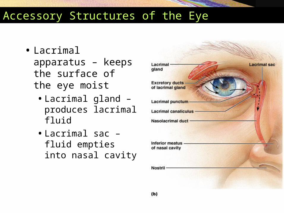
Accessory Structures of the Eye
• Lacrimal apparatus – keeps the surface of the eye moist• Lacrimal gland –
produces lacrimal fluid
• Lacrimal sac – fluid empties into nasal cavity
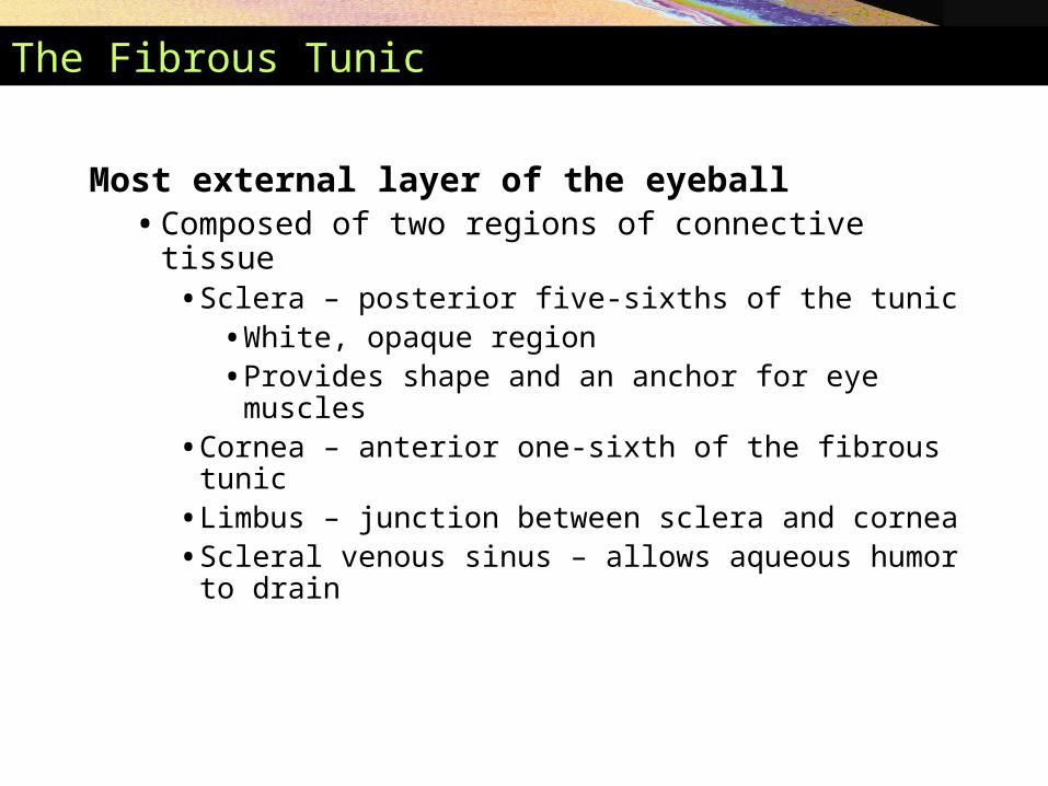
The Fibrous Tunic
Most external layer of the eyeball• Composed of two regions of connective tissue
• Sclera – posterior five-sixths of the tunic• White, opaque region• Provides shape and an anchor for eye muscles
• Cornea – anterior one-sixth of the fibrous tunic• Limbus – junction between sclera and cornea• Scleral venous sinus – allows aqueous humor to
drain
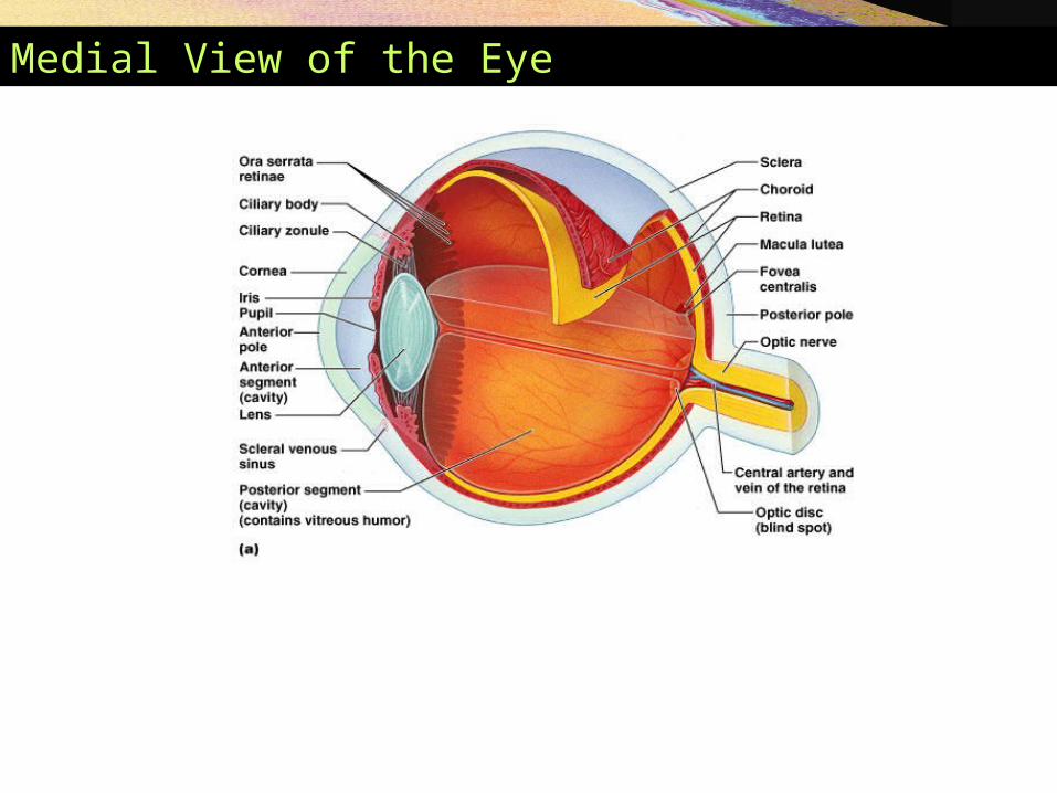
Medial View of the Eye

The Vascular Tunic
The middle coat of the eyeball
• Composed of choroid, ciliary body, and iris
• Choroid – vascular, darkly pigmented membrane• Forms posterior five-sixths of the vascular tunic
• Brown color – from melanocytes
• Prevents scattering of light rays within the eye
• Choroid corresponds to the arachnoid and pia maters

Posterior View of the Anterior Half of the Eye
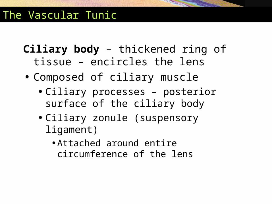
The Vascular Tunic
Ciliary body – thickened ring of tissue – encircles the lens
• Composed of ciliary muscle• Ciliary processes – posterior surface of the ciliary
body
• Ciliary zonule (suspensory ligament) • Attached around entire circumference of the lens

The Vascular Tunic
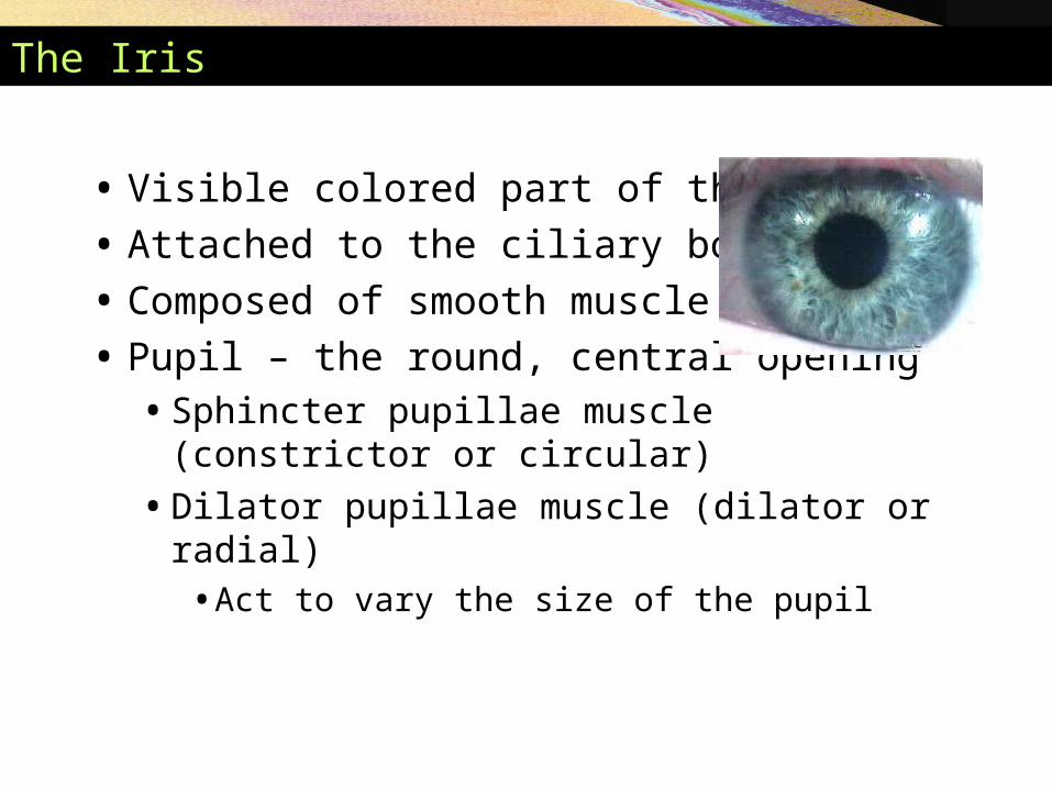
The Iris
• Visible colored part of the eye
• Attached to the ciliary body
• Composed of smooth muscle
• Pupil – the round, central opening• Sphincter pupillae muscle (constrictor or circular)
• Dilator pupillae muscle (dilator or radial)• Act to vary the size of the pupil
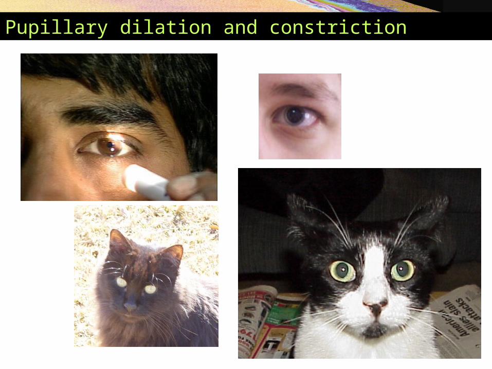
Pupillary dilation and constriction
The Sensory Tunic (Retina)
Retina – the deepest tunic• Composed of two layers
• Pigmented layer – single layer of melanocytes • Neural layer – sheet of nervous tissue
• Contains three main types of neurons• Photoreceptor cells• Bipolar cells• Ganglion cells
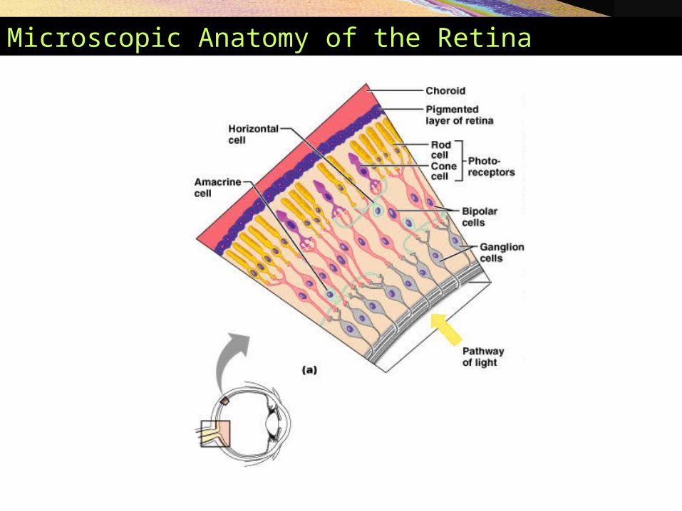
Microscopic Anatomy of the Retina
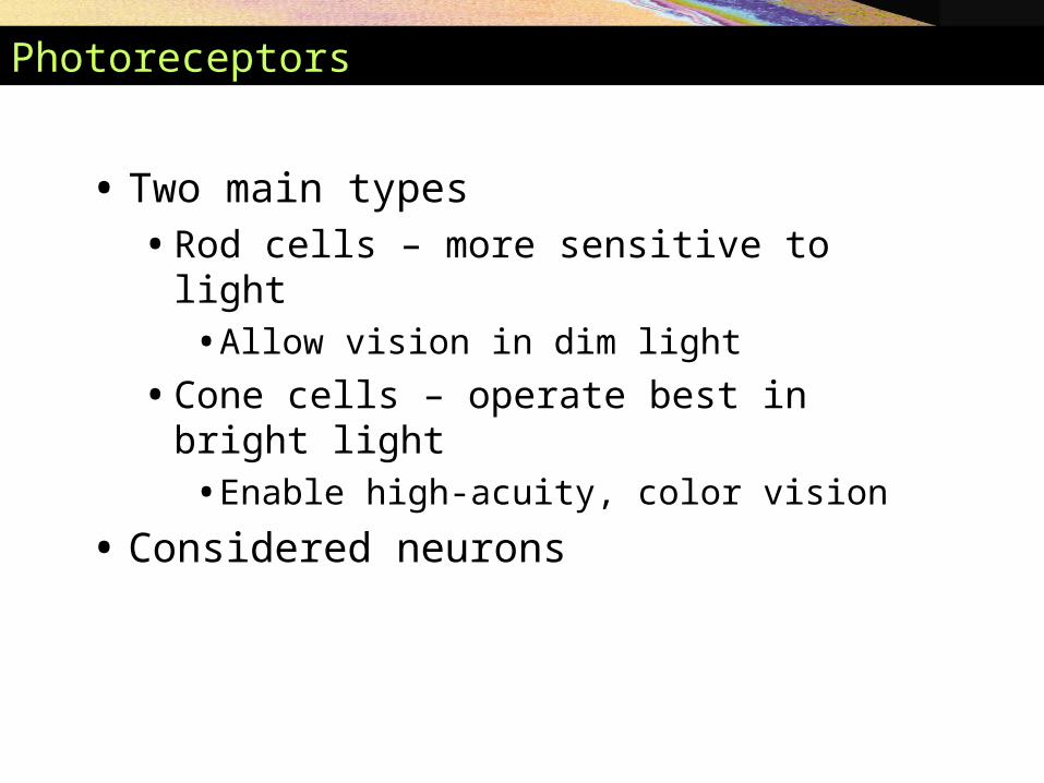
Photoreceptors
• Two main types• Rod cells – more sensitive to light
• Allow vision in dim light
• Cone cells – operate best in bright light• Enable high-acuity, color vision
• Considered neurons

Photoreceptors

Regional Specializations of the Retina
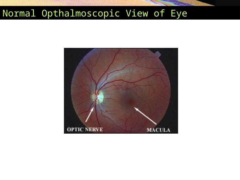
• Macula lutea – contains mostly cones
• Fovea centralis – contains only cones• Region of highest visual acuity
• Optic disc – blind spot
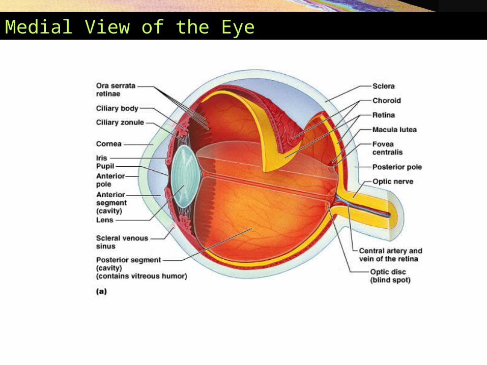
Medial View of the Eye
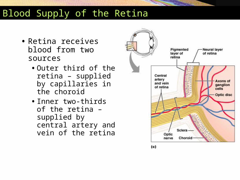
Blood Supply of the Retina
• Retina receives blood from two sources• Outer third of the
retina – supplied by capillaries in the choroid
• Inner two-thirds of the retina – supplied by central artery and vein of the retina
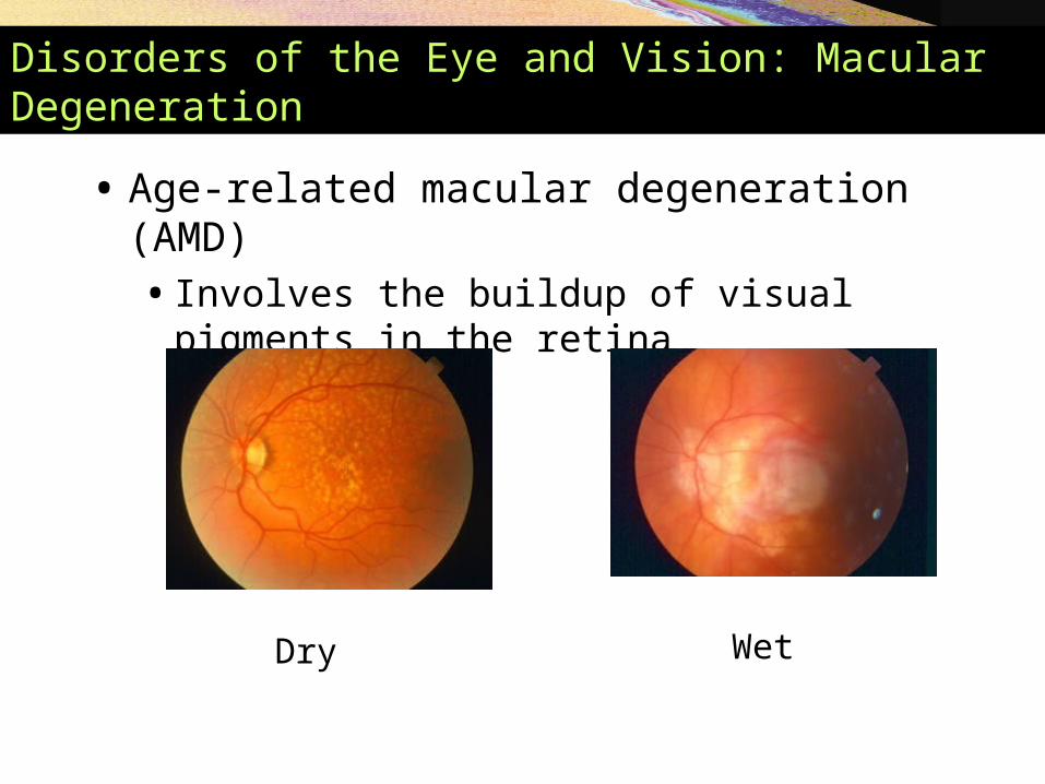
Disorders of the Eye and Vision: Macular Degeneration
• Age-related macular degeneration (AMD)• Involves the buildup of visual pigments in the
retina
Dry Wet
Copyright © 2005 Pearson Education, Inc., publishing as Benjamin Cummings
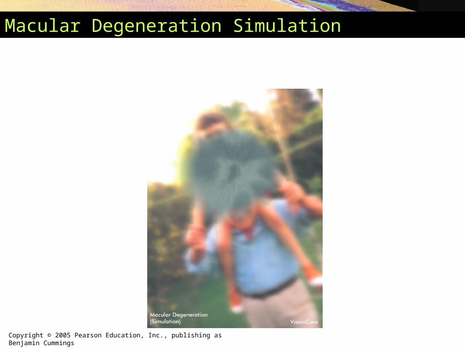
Macular Degeneration Simulation
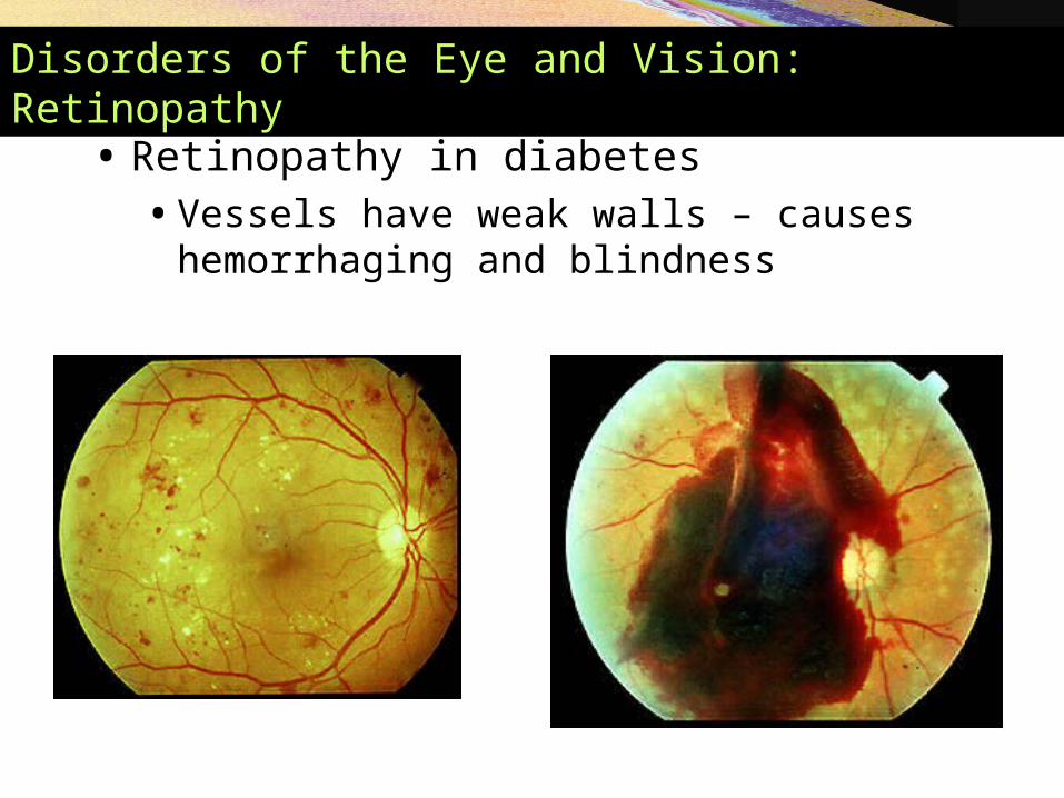
Disorders of the Eye and Vision: Retinopathy
• Retinopathy in diabetes • Vessels have weak walls – causes hemorrhaging
and blindness

Internal Chambers and Fluids
• The lens and ciliary zonules divide the eye
• Posterior segment (cavity)• Filled with vitreous humor
• Clear, jelly-like substance
• Transmits light
• Supports the posterior surface of the lens
• Helps maintain intraocular pressure
Disorders of the Eye and Vision: Glucoma
• Glaucoma is a condition of abnormally high intraocular pressure.
• It is caused by obstruction that prevent drainage of aqueous humor from the eye
• Chronic glaucoma, the most common form of glaucoma, may be associated with few symptoms except for a gradual loss of peripheral vision.

Internal Chambers and Fluids
• Anterior segment• Divided into anterior and posterior chambers
• Anterior chamber – between the cornea and iris
• Posterior chamber – between the iris and lens
• Filled with aqueous humor
• Renewed continuously
• Formed as a blood filtrate
• Supplies nutrients to the lens and cornea
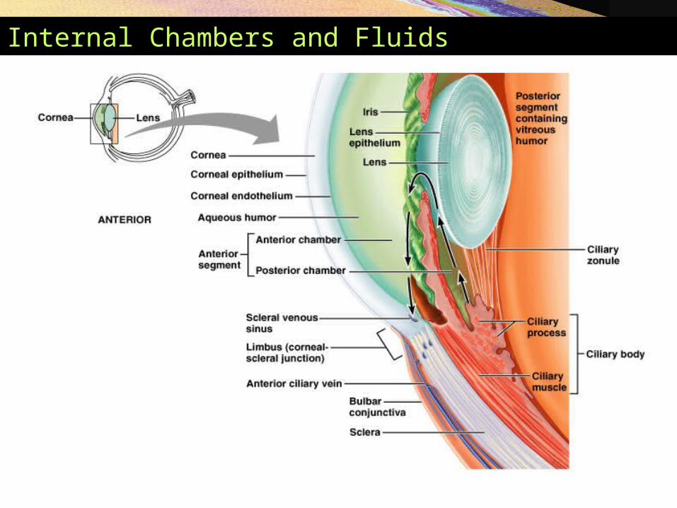
Internal Chambers and Fluids
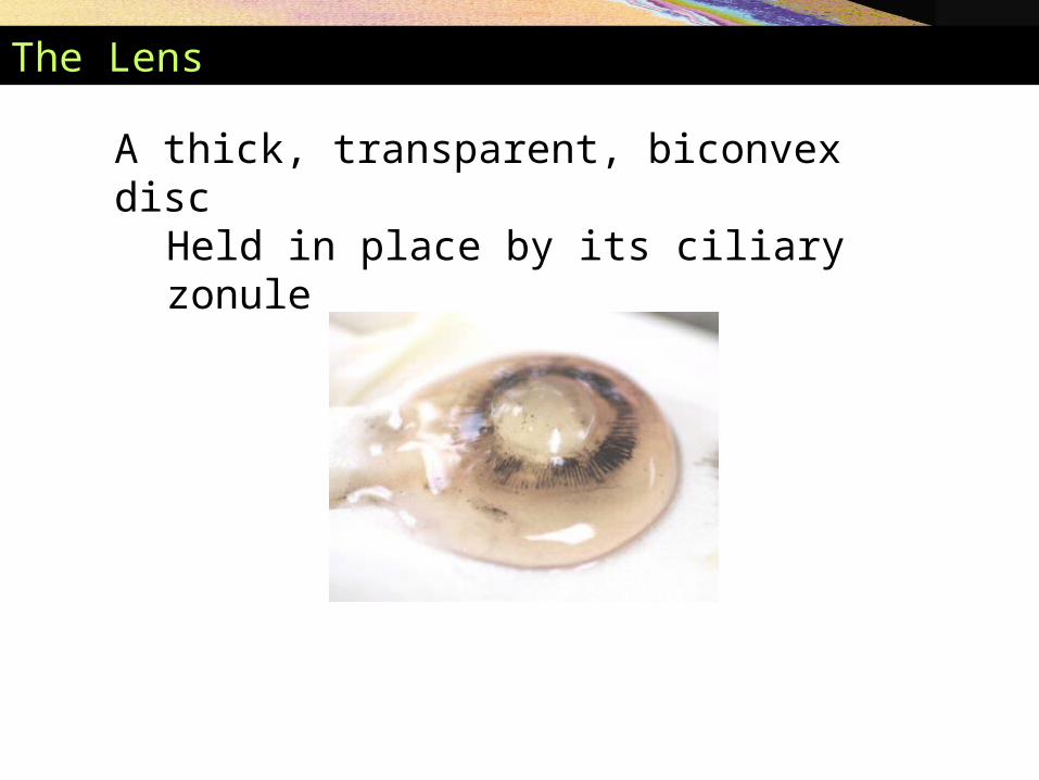
The Lens
A thick, transparent, biconvex discHeld in place by its ciliary zonule
Copyright © 2005 Pearson Education, Inc., publishing as Benjamin Cummings
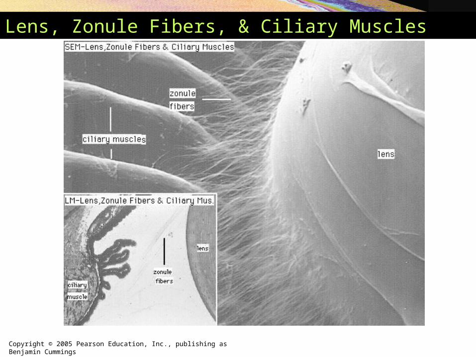
Lens, Zonule Fibers, & Ciliary Muscles
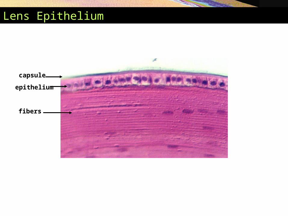
Lens Epithelium
capsule
epithelium
fibers
Disorders of the Eye and Vision: Cataract
• A cataract is an opacity of the lens resulting from the accumulation of pigment or other substances in the lens fibers
• Often associated with aging
• If untreated gradual loss of vision
Copyright © 2005 Pearson Education, Inc., publishing as Benjamin Cummings
The Eye as an Optical Device
• Structures in the eye bend light rays
• Light rays converge on the retina at a single focal point
• Light bending structures (refractory media)• The lens, cornea, and humors
• Accommodation – curvature of the lens is adjustable • Allows for focusing on nearby objects
Visual Pathways
• Most visual information travels to the cerebral cortex
• Responsible for conscious “seeing”
• Other pathways travel to nuclei in the midbrain and diencephalon

Visual Pathways to the Cerebral Cortex
• Pathway begins at the retina• Light activates photoreceptors
• Photoreceptors signal bipolar cells
• Bipolar cells signal ganglion cells
• Axons of ganglion cells exit eye as the optic nerve
Visual Pathways to the Cerebral Cortex
• Optic tracts send axons to:• Lateral geniculate nucleus of the thalamus
• Synapse with thalamic neurons
• Fibers of the optic radiation reach the primary visual cortex
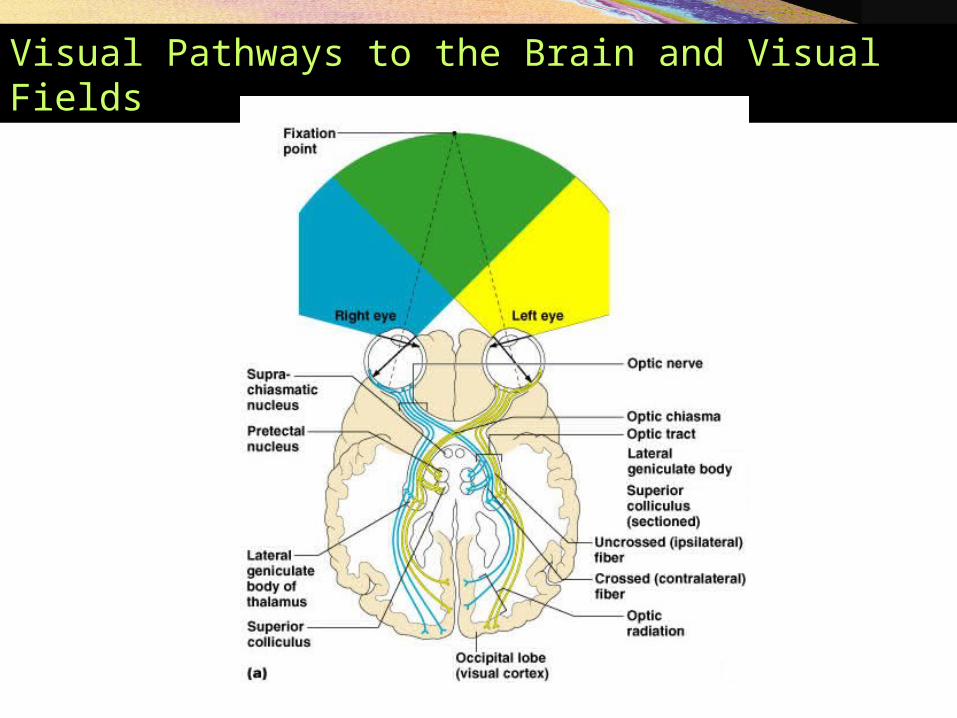
Visual Pathways to the Brain and Visual Fields
Visual Pathways to Other Parts of the Brain
• Some axons from the optic tracts• Branch to midbrain
• Superior colliculi
• Pretectal nuclei
• Other branches from the optic tracts• Branch to the suprachiasmatic nucleus
The Ear: Hearing and Equilibrium
• The ear – receptor organ for hearing and equilibrium
• Composed of three main regions• Outer ear – functions in hearing
• Middle ear – functions in hearing
• Inner ear – functions in both hearing and equilibrium
The Outer (External) Ear
• Composed of:• The auricle (pinna)
• Helps direct sounds
• External acoustic meatus• Lined with skin
• Contains hairs, sebaceous glands, and ceruminous glands
• Tympanic membrane• Forms the boundary between the external and
middle ear

The Outer (External) Ear

The Middle Ear
• The tympanic cavity • A small, air-filled space
• Located within the petrous portion of the temporal bone
• Medial wall is penetrated by:• Oval window
• Round window
• Pharyngotympanic tube (auditory or eustachian tube) • Links the middle ear and pharynx
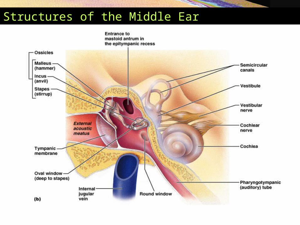
Structures of the Middle Ear
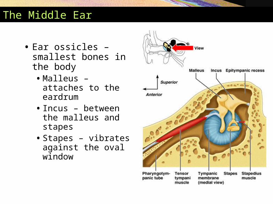
The Middle Ear
• Ear ossicles – smallest bones in the body• Malleus – attaches to
the eardrum • Incus – between the
malleus and stapes• Stapes – vibrates
against the oval window
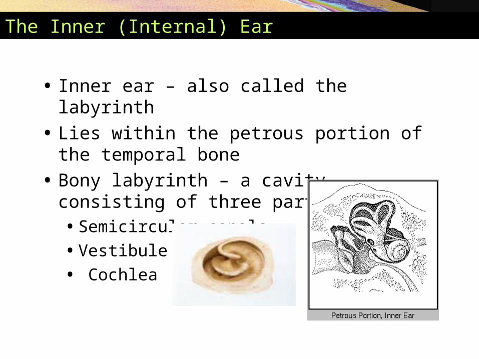
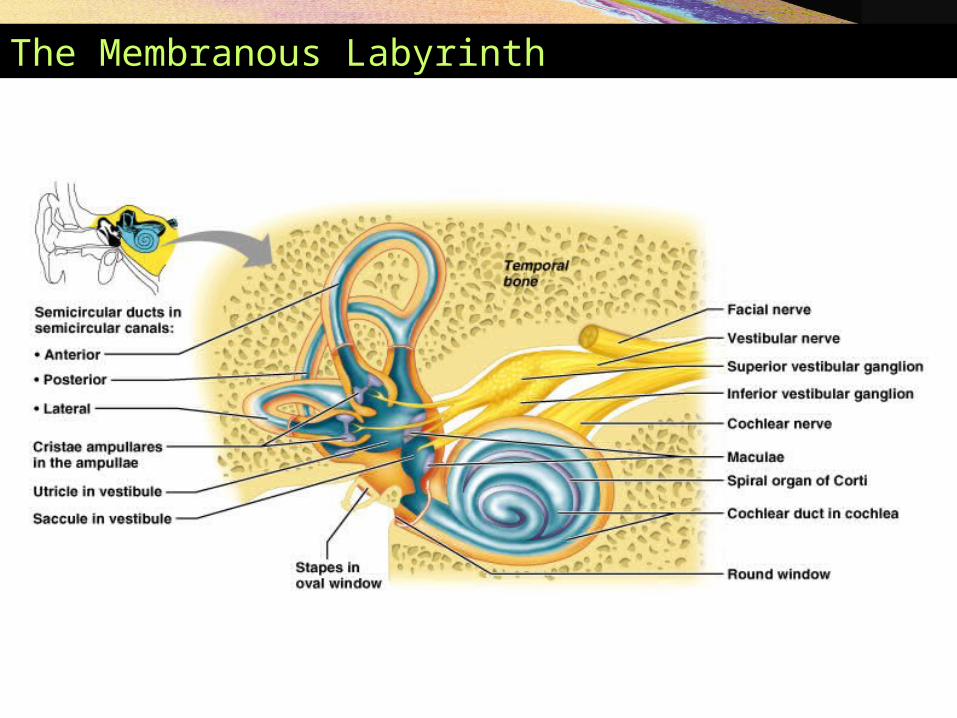
The Inner (Internal) Ear
• Inner ear – also called the labyrinth
• Lies within the petrous portion of the temporal bone
• Bony labyrinth – a cavity consisting of three parts• Semicircular canals
• Vestibule
• Cochlea
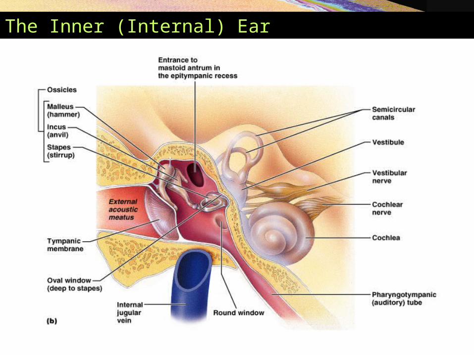
The Inner (Internal) Ear

The Inner (Internal) Ear
• Membranous labyrinth • Series of membrane-walled sacs and ducts
• Fit within the bony labyrinth
• Consists of three main parts• Semicircular ducts
• Utricle and saccule
• Cochlear duct
The Inner (Internal) Ear
• Membranous labyrinth (continued)• Filled with a clear fluid – endolymph
• Confined to the membranous labyrinth
• Bony labyrinth is filled with perilymph • Continuous with cerebrospinal fluid
The Membranous Labyrinth
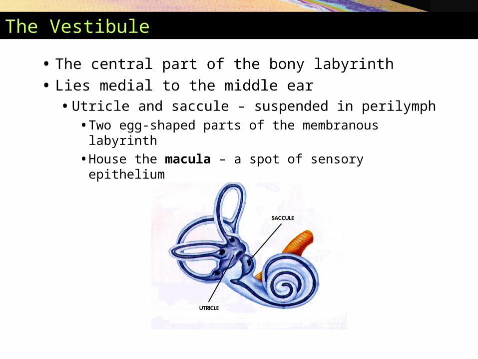
The Vestibule
• The central part of the bony labyrinth
• Lies medial to the middle ear• Utricle and saccule – suspended in perilymph
• Two egg-shaped parts of the membranous labyrinth
• House the macula – a spot of sensory epithelium
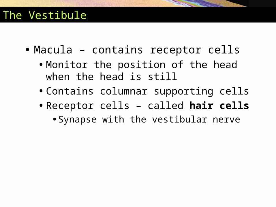
The Vestibule
• Macula – contains receptor cells • Monitor the position of the head when the head is
still
• Contains columnar supporting cells
• Receptor cells – called hair cells• Synapse with the vestibular nerve

Anatomy and Function of the Maculae
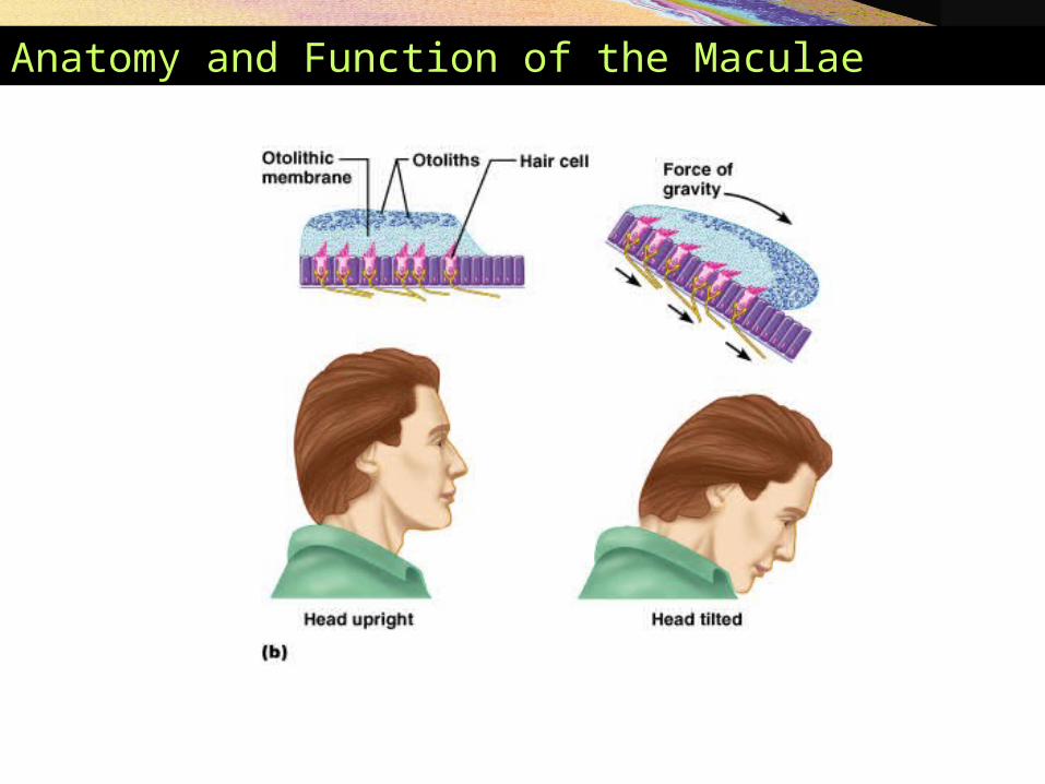
Anatomy and Function of the Maculae
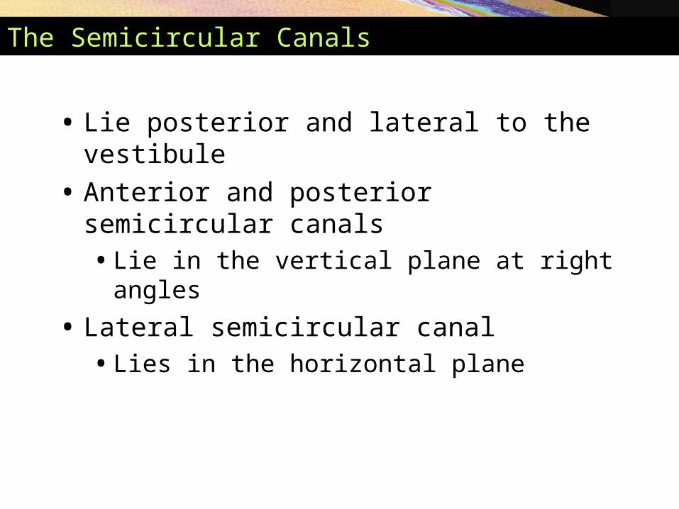
The Semicircular Canals
• Lie posterior and lateral to the vestibule
• Anterior and posterior semicircular canals• Lie in the vertical plane at right angles
• Lateral semicircular canal • Lies in the horizontal plane
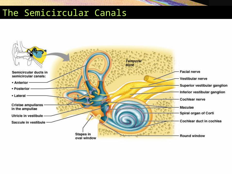
The Semicircular Canals

The Semicircular Canals
• Semicircular duct – snakes through each semicircular canal
• Membranous ampulla – located within bony ampulla• Houses a structure called a crista ampullaris
• Cristae contain receptor cells of rotational acceleration
• Epithelium contains supporting cells and receptor hair cells
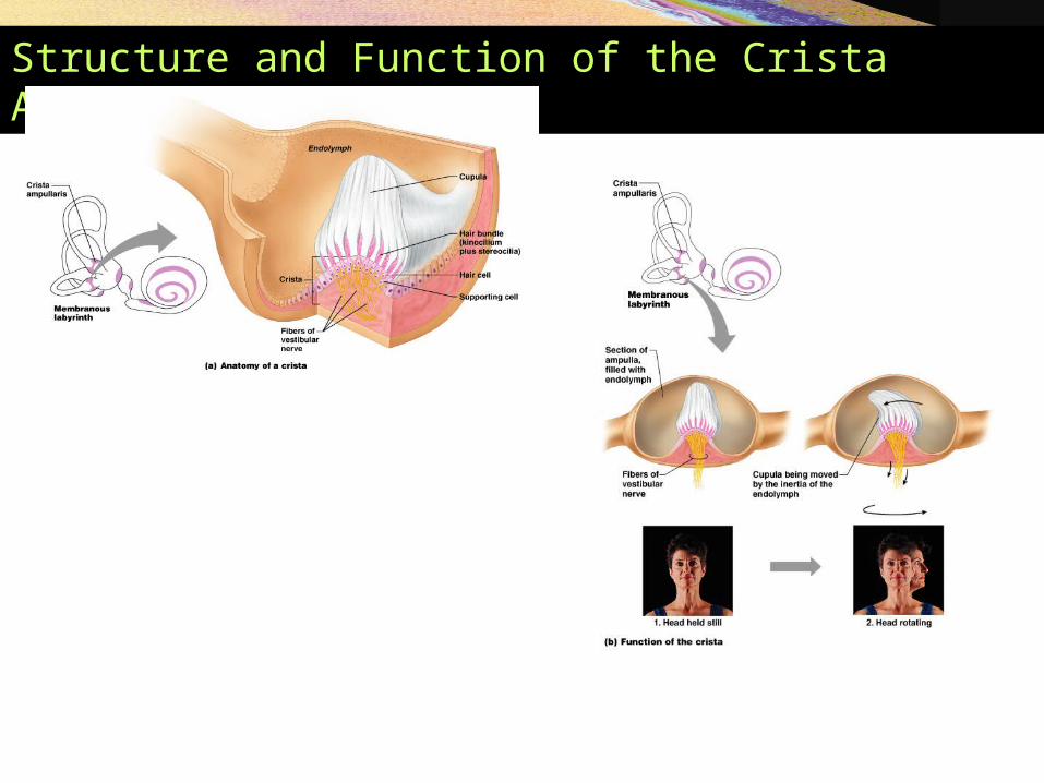
Structure and Function of the Crista Ampullaris
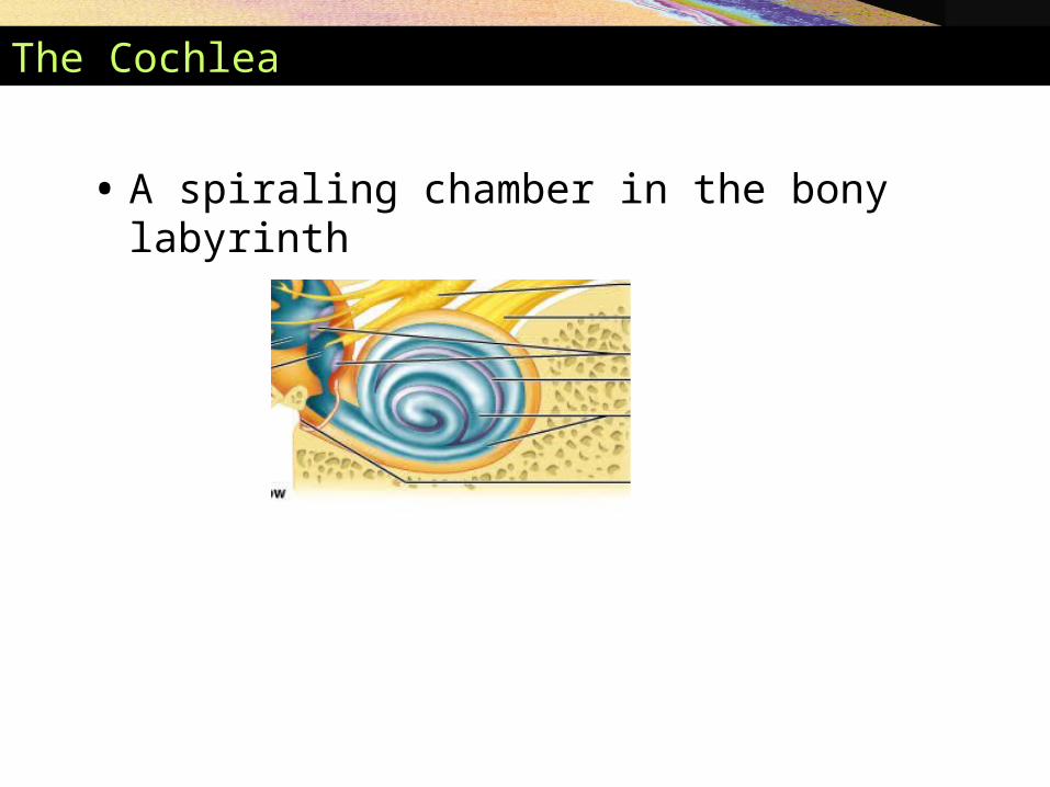
The Cochlea
• A spiraling chamber in the bony labyrinth
The Cochlea
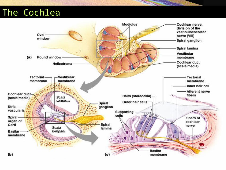
The Cochlea
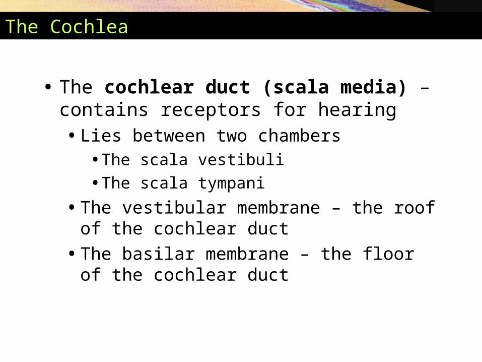
• The cochlear duct (scala media) – contains receptors for hearing• Lies between two chambers
• The scala vestibuli
• The scala tympani
• The vestibular membrane – the roof of the cochlear duct
• The basilar membrane – the floor of the cochlear duct
The Cochlea
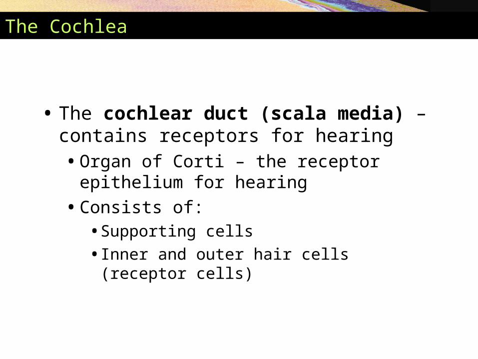
• The cochlear duct (scala media) – contains receptors for hearing• Organ of Corti – the receptor epithelium for
hearing
• Consists of: • Supporting cells
• Inner and outer hair cells (receptor cells)
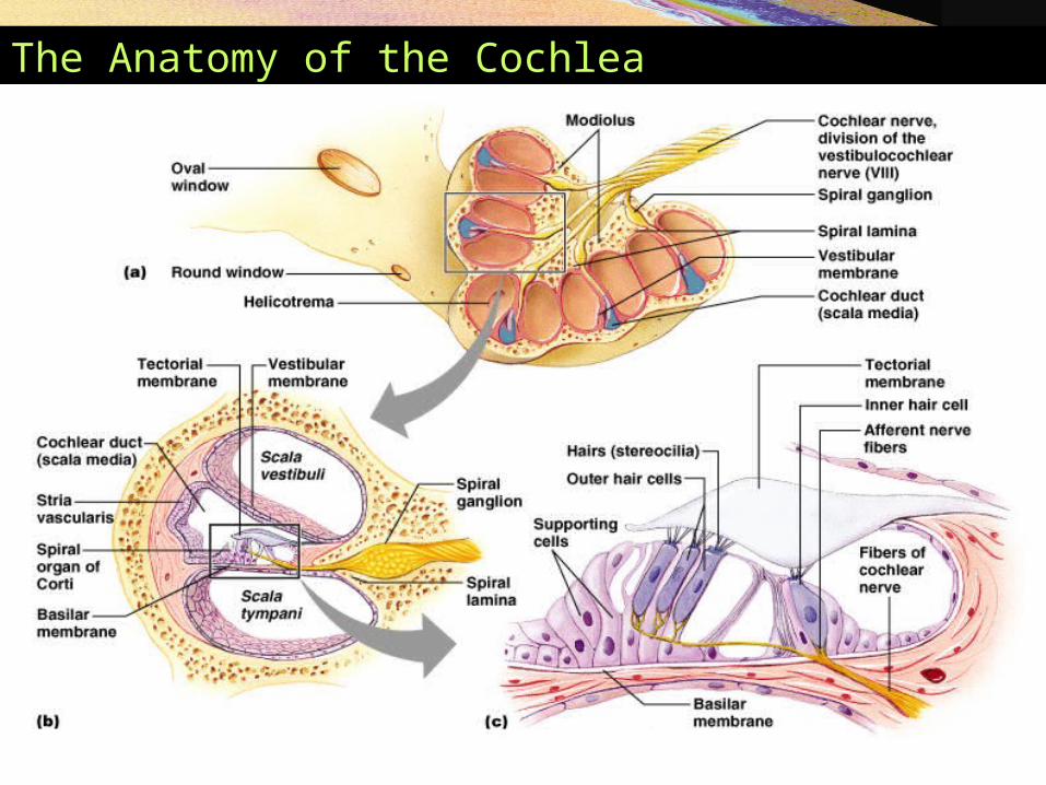
The Anatomy of the Cochlea
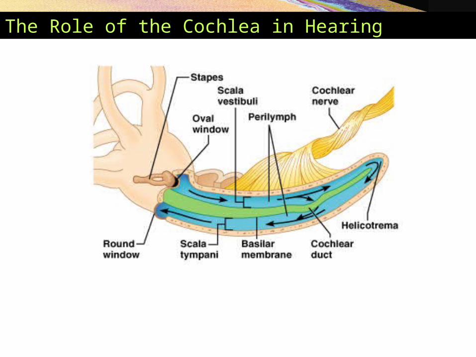
The Role of the Cochlea in Hearing
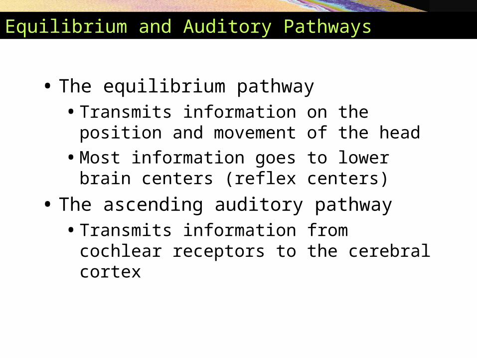
Equilibrium and Auditory Pathways
• The equilibrium pathway • Transmits information on the position and
movement of the head
• Most information goes to lower brain centers (reflex centers)
• The ascending auditory pathway • Transmits information from cochlear receptors to
the cerebral cortex
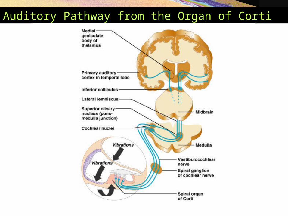
Auditory Pathway from the Organ of Corti
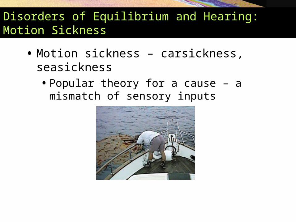
Disorders of Equilibrium and Hearing: Motion Sickness
• Motion sickness – carsickness, seasickness• Popular theory for a cause – a mismatch of sensory
inputs
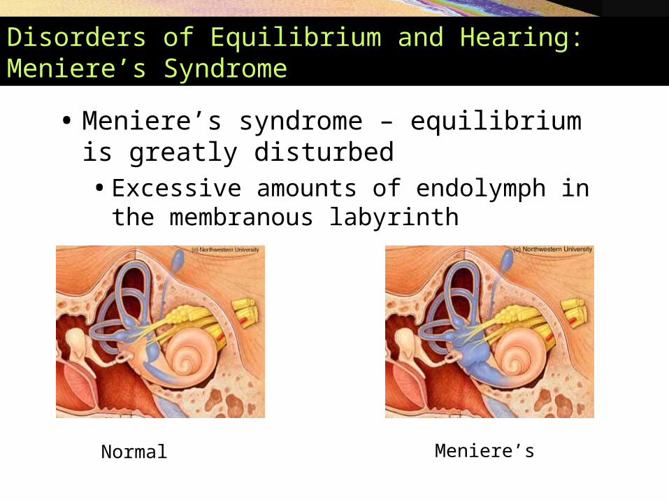
Disorders of Equilibrium and Hearing: Meniere’s Syndrome
• Meniere’s syndrome – equilibrium is greatly disturbed• Excessive amounts of endolymph in the
membranous labyrinth
Normal Meniere’s

Disorders of Equilibrium and Hearing: Conduction Deafness
• Deafness • Conduction deafness
• Sound vibrations cannot be conducted to the inner ear
• Ruptured tympanic membrane, otitis media, otosclerosis
Normal tympanicmembrane
Otitis mediaRuptured tympanicmembrane
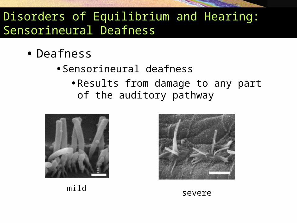
Disorders of Equilibrium and Hearing: Sensorineural Deafness
• Deafness • Sensorineural deafness
• Results from damage to any part of the auditory pathway
mild severe

THANKS FOR THE ATTENTIONAND
GOOD LUCK
Related Documents