İstanbul Tıp Fakültesi Dergisi Journal of İstanbul Faculty of Medicine EISSN 1305-6441 jmed.istanbul.edu.tr Cilt-Volume:82 • Sayı-Issue:1 • Mart-March 2019 Indexed in Web of Science

Welcome message from author
This document is posted to help you gain knowledge. Please leave a comment to let me know what you think about it! Share it to your friends and learn new things together.
Transcript
İstanbul Tıp Fakültesi
Dergisi
Journal of İstanbul Faculty of Medicine
EISSN 1305-6441
jmed.istanbul.edu.tr
Cilt-Volume:82 • Sayı-Issue:1 • Mart-March 2019
Indexed in
Web of Science

İstanbul Tıp Fakültesi Dergisi
SAHİBİ VE SORUMLU MÜDÜR/OWNER AND RESPONSIBLE MANAGER
Bahaüddin ÇOLAKOĞLUİstanbul Tıp Fakültesi Dekanı
Dean of Istanbul Faculty of Medicine
EDİTÖR/EDITOR
M. Lale ÖCALİstanbul Üniversitesi, TÜRKİYE
Istanbul University, TURKEY
EDİTÖR YARDIMCILARI/ASSOCIATE EDITORS
Funda GÜNGÖR UĞURLUCANİstanbul Üniversitesi, TÜRKİYEIstanbul University, TURKEY
Birsen KARAMANİstanbul Üniversitesi, TÜRKİYEIstanbul University, TURKE
Ayşe KUBAT ÜZÜMİstanbul Üniversitesi, TÜRKİYEIstanbul University, TURKEY
İsmail Cem SORMAZİstanbul Üniversitesi, TÜRKİYEIstanbul University, TURKEY
Deniz TUĞCUİstanbul Üniversitesi, TÜRKİYEIstanbul University, TURKEY
Halil YAZICIİstanbul Üniversitesi, TÜRKİYEIstanbul University, TURKEY
İSTATİSTİK DANIŞMANI/STATISTICS ADVISORHalim İŞSEVERİstanbul Üniversitesi, TÜRKİYEIstanbul University Istanbul TURKEY
Atilla ARINCIİstanbul Üniversitesi, TÜRKİYEIstanbul University, TURKEY
Nilgün BOZBUĞA İstanbul Üniversitesi, TÜRKİYEIstanbul University, TURKEY
Şükrü H. EMREYale Üniversitesi, ABDYale University, USA
Haluk ERAKSOYİstanbul Üniversitesi, TÜRKİYEIstanbul University, TURKEY
Hakan ERTİNİstanbul Üniversitesi, TÜRKİYEIstanbul University, TURKEY
Simin GÖRALPennsylvania Üniversitesi, ABDUniversity of Pennsylvania, USA
Nilüfer GÖZÜMİstanbul Üniversitesi, TÜRKİYEIstanbul University, TURKEY
Hülya GÜLİstanbul Üniversitesi, TÜRKİYEIstanbul University, TURKEY
Çiğdem KEKİK ÇINAR İstanbul Üniversitesi, TÜRKİYEIstanbul University, TURKEY
Fahrettin KELEŞTEMURTürkiye Sağlık Enstitüleri (TÜSEB), TÜRKİYEHealth Institutes of Turkey, TURKEY
Abdullah KUTLARAugusta Üniversitesi, ABDAugusta University, USA
Sacit Bülent OMAYYale Üniversitesi, ABDYale University, USA
Betigül ÖNGENİstanbul Üniversitesi, TÜRKİYEIstanbul University, TURKEY
Beyza ÖZÇINARİstanbul Üniversitesi, TÜRKİYEIstanbul University, TURKEY
Altay SENCERİstanbul Üniversitesi, TÜRKİYEIstanbul University, TURKEY
Yasemin ŞANLIİstanbul Üniversitesi, TÜRKİYEIstanbul University, TURKEY
M. Öner ŞANLIİstanbul Üniversitesi,TÜRKİYEIstanbul University, TURKEY
E. Murat TUZCUCleveland Clinic, BAECleveland Clinic, UAE
Pınar YAMANTÜRK ÇELİKİstanbul Üniversitesi, TÜRKİYEIstanbul University, TURKEY
YAYIN KURULU/EDITORIAL BOARD
I
II
İstanbul Tıp Fakültesi Dergisi (İst Tıp Fak Derg); bağım-sız, önyargısız ve çift-kör hakemlik ilkeleri çerçevesinde yayın yapan İstanbul Üniversitesi, İstanbul Tıp Fakülte-si’nin uluslararası ve açık erişimli bilimsel yayın organıdır. Dergi Mart, Haziran, Eylül ve Aralık aylarında olmak üze-re üç ayda bir yayınlanır ve dört sayıda bir cildi tamamla-nır. Yayın dili Türkçe ve İngilizce’dir.
İstanbul Tıp Fakültesi Dergisi (İst Tıp Fak Derg), tıbbın tüm alanlarında klinik ve deneysel özgün araştırmalar, ender görülebilecek olgu sunumları, özel ve aktüel konularda literatür derlemeleri, editöre mektuplar ile yayın tanıtımları ve haberleri yayınlamaktadır. Orijinal metot geliştirme, yeni bir girişim tekniği ve orijinal çalış-maların ön sonuçlarını içeren kısa raporlara da dergide yer verilmektedir.
Derginin hedef kitlesi; sağlık alanındaki tüm disiplinler-de çalışan uzman hekimler ve akademisyenlerdir.
Derginin editörlük ve yayın süreçleri International Committee of Medical Journal Editors (ICMJE), Wor-ld Association of Medical Editors (WAME), Council of Science Editors (CSE), Committee on Publication Et-hics (COPE), European Association of Science Editors (EASE) ve National Information Standards Organization (NISO) kılavuzlarına uygun olarak biçimlendirilmiştir ve Principles of Transparency and Best Practice in Scholar-ly Publishing (doaj.org/ bestpractice) ilkelerine uygun olarak yürütülmektedir.
İstanbul Tıp Fakültesi Dergisi (İst Tıp Fak Derg), Web of Science-Emerging Sources Citation Index ve TÜBİTAK ULAKBİM TR Dizin tarafından indekslenmektedir.
Makale değerlendirme ve yayın işlemleri için yazarlar-dan ücret talep edilmemektedir. Tüm makaleler http://jmed.istanbul.edu.tr sayfasındaki online makale değer-
lendirme sistemi kullanılarak dergiye gönderilmelidir. Derginin yazım kurallarına, gerekli formlara ve dergiyle ilgili diğer bilgilere web sayfasından erişilebilir.
Derginin tüm masrafları İstanbul Üniversitesi Yayınevi tarafından karşılanmaktadır. Basılı kopyalarda tıbbi ilaç, malzeme ve cihaz üreticilerinin reklamları yayınlanabilir. Reklam vermek isteyenlerin Editör ile iletişime geçme-leri gerekmektedir. Reklam görselleri sadece Editör onayı ile yayınlanmaktadır.
Dergide yayınlanan makalelerde ifade edilen bilgi, fi-kir ve görüşler İstanbul Tıp Fakültesi Dergisi’nin Editör, Editör Yardımcıları, Yayın Kurulu ve Yayıncısının değil, yazar(lar)ın bilgi ve görüşlerini yansıtır. Editör, Editör Yar-dımcıları, Yayın Kurulu ve Yayıncı, bu gibi yazarlara ait bilgi ve görüşler için hiçbir sorumluluk ya da yükümlülük kabul etmemektedir.
Yayınlanan tüm içeriğe http://jmed.istanbul.edu.tr ad-resinden ücretsiz olarak erişilebilir.
Dergide yayınlanan içeriğin tüm telif hakları İstanbul Üniversitesi’ne aittir.
Editör: M. Lale ÖcalAdres: İstanbul Üniversitesi, İstanbul Tıp Fakültesi Dekanlığı, Turgut Özal Cad. 34093 Çapa, Fatih, İstanbul, TürkiyeTel.: +90 212 414 21 61E-posta: [email protected]
Yayıncı: İstanbul Üniversitesi YayıneviAdres: İstanbul Üniversitesi Merkez Kampüsü, 34452 Beyazıt, Fatih, İstanbul, TürkiyeTel.: +90 212 440 00 00Faks: +90 212 217 22 92
AMAÇ VE KAPSAM
İstanbul Tıp Fakültesi Dergisi

4III
Journal of İstanbul Faculty of Medicine (J Ist Faculty Med) is an international, scientific, open access period-ical published in accordance with independent, unbi-ased, and double-blinded peer-review principles. The journal is the official publication of İstanbul University, İstanbul Faculty of Medicine and it is published quarter-ly on March, June, September and December. The pub-lication languages of the journal are Turkish and English.
Journal of İstanbul Faculty of Medicine (J Ist Faculty Med) aims to contribute to the literature by publishing manuscripts at the highest scientific level on all fields of medicine. The journal publishes original experimental and clinical research articles, reports of rare cases, re-views that contain sufficient amount of source data con-veying the experiences of experts in a particular field, and letters to the editors as well as brief reports on a recently established method or technique or prelimi-nary results of original studies related to all disciplines of medicine from all countries.
The journal’s target audience includes researchers, phy-sicians and healthcare professionals who are interested or working in all medical disciplines.
The editorial and publication processes of the journal are shaped in accordance with the guidelines of the In-ternational Committee of Medical Journal Editors (IC-MJE), World Association of Medical Editors (WAME), Council of Science Editors (CSE), Committee on Publi-cation Ethics (COPE), European Association of Science Editors (EASE), and National Information Standards Or-ganization (NISO). The journal is in conformity with the Principles of Transparency and Best Practice in Scholar-ly Publishing (doaj.org/bestpractice).
Journal of İstanbul Faculty of Medicine is currently in-dexed in Web of Science-Emerging Sources Citation Index and TUBITAK ULAKBIM TR Index.
Processing and publication are free of charge with the journal. No fees are requested from the authors at any point throughout the evaluation and publication pro-cess. All manuscripts must be submitted via the online submission system, which is available at http://jmed.istanbul.edu.tr. The journal guidelines, technical infor-mation, and the required forms are available on the journal’s web page.
All expenses of the journal are covered by the İstanbul University Press. Potential advertisers should contact the Editorial Office. Advertisement images are pub-lished only upon the Editor’s approval.
Statements or opinions expressed in the manuscripts published in the journal reflect the views of the author(s) and not the opinions of the İstanbul University, İstanbul Faculty of Medicine, editors, editorial board, and/or publisher; the editors, editorial board, and publisher disclaim any responsibility or liability for such materials.
All published content is available online, free of charge at jmed.istanbul.edu.tr.
İstanbul University holds the international copyright of all the content published in the journal.
Editor: M. Lale ÖcalAddress: İstanbul University, İstanbul Faculty of Medicine Deanery, Turgut Özal Cad. 34093, Çapa, Fatih, İstanbul, TurkeyPhone: +90 212 414 21 61E-mail: [email protected]
Publisher: Istanbul University PressAddress: İstanbul Üniversitesi Merkez Kampüsü, 34452 Beyazıt, Fatih / Istanbul - TurkeyPhone: +90 212 440 00 00
AIMS & SCOPES
İstanbul Tıp Fakültesi Dergisi
IV
İstanbul Tıp Fakültesi Dergisi (İst Tıp Fak Derg); ba-ğımsız, önyargısız ve çift-kör hakemlik ilkeleri çerçe-vesinde yayın yapan İstanbul Üniversitesi, İstanbul Tıp Fakültesi’nin uluslararası ve açık erişimli bilimsel yayın organıdır. Dergi Mart, Haziran, Eylül ve Aralık ayların-da olmak üzere üç ayda bir yayınlanır ve dört sayıda bir cildi tamamlanır. Yayın dili Türkçe ve İngilizce’dir.
İstanbul Tıp Fakültesi Dergisi (İst Tıp Fak Derg), tıbbın tüm alanlarında klinik ve deneysel özgün araştırmalar, ender görülebilecek olgu sunumları, özel ve aktüel konularda literatür derlemeleri, editöre mektuplar ile yayın tanıtımları ve haberleri yayınlamaktadır. Orijinal metot geliştirme, yeni bir girişim tekniği ve orijinal çalışmaların ön sonuçlarını içeren kısa raporlara da dergide yer verilmektedir.
Derginin editörlük ve yayın süreçleri International Committee of Medical Journal Editors (ICMJE), Wor-ld Association of Medical Editors (WAME), Council of Science Editors (CSE), Committee on Publication Et-hics (COPE), European Association of Science Editors (EASE), ve National Information Standards Organizati-on (NISO) kılavuzlarına uygun olarak biçimlendirilmiş-tir ve Principles of Transparency and Best Practice in Scholarly Publishing (doaj.org/bestpractice) ilkelerine uygun olarak yürütülmektedir.
Özgünlük, yüksek bilimsel kalite ve atıf potansiyeli bir makalenin yayına kabulü için en önemli kriterlerdir. Gönderilen yazıların daha önce başka bir elektronik ya da basılı dergide, kitapta veya farklı bir mecra-da sunulmamış ya da yayınlanmamış olması gerekir. Daha önce başka bir dergiye gönderilen ancak yayı-na kabul edilmeyen yazılar hakkında dergi önceden bilgilendirilebilir. Bu yazıların eski hakem raporlarının Yayın Kuruluna gönderilmesi değerlendirme süresi-nin hızlanmasını sağlayacaktır. Toplantılarda sunulan çalışmalar için, sunum yapılan organizasyonun tam adı, tarihi, şehri ve ülkesi belirtilmelidir.
İstanbul Tıp Fakültesi Dergisi’ne gönderilen tüm ma-kaleler çift-kör hakem değerlendirme sürecinden geç-mektedir. Tarafsız değerlendirme sürecini sağlamak için her makale alanlarında uzman en az iki dış-bağımsız hakem tarafından değerlendirilir. Dergi Yayın Kurulu üyeleri tarafından gönderilecek makalelerin değerlen-dirme süreçleri, davet edilecek dış bağımsız editörler
tarafından yönetilecektir. Bütün makalelerin karar ver-me süreçlerinde nihai karar yetkisi Editördedir.
Klinik ve deneysel çalışmalar, ilaç araştırmaları ve bazı olgu sunumları için World Medical Association Dec-laration of Helsinki “Ethical Principles for Medical Research Involving Human Subjects”, (amended in October 2013, www.wma.net) çerçevesinde hazırlan-mış Etik Komisyon raporu gerekmektedir. Gerekli gö-rülmesi halinde Etik Komisyon raporu veya eşdeğeri olan resmi bir yazı yazarlardan talep edilebilir. İnsan-lar üzerinde yapılmış deneysel çalışmaların sonuçla-rını bildiren yazılarda, çalışmanın yapıldığı kişilere uygulanan prosedürlerin niteliği tümüyle açıklandık-tan sonra, onaylarının alındığına ilişkin bir açıklama-ya metin içinde yer verilmelidir. Hayvanlar üzerinde yapılan çalışmalarda ise ağrı, acı ve rahatsızlık veril-memesi için yapılmış olanlar açık olarak makalede belirtilmelidir. Hasta onamları, Etik Kurul raporunun alındığı kurumun adı, onay belgesinin numarası ve tarihi ana metin dosyasında yer alan Gereç ve Yön-tem başlığı altında yazılmalıdır. Hastaların kimlikleri-nin gizliliğini korumak yazarların sorumluluğundadır. Hastaların kimliğini açığa çıkarabilecek fotoğraflar için hastadan ya da yasal temsilcilerinden alınan im-zalı izinlerin de gönderilmesi gereklidir.
Bütün makalelerin benzerlik denetimi, iThenticate yazılımı aracılığıyla yapılmaktadır.
Yayın Kurulu, dergimize gönderilen çalışmalar hak-kındaki intihal, atıf manipülasyonu ve veri sahteciliği iddia ve şüpheleri karşısında COPE kurallarına uygun olarak hareket edecektir.
Yazar olarak listelenen herkesin ICMJE (www.icmje.org) tarafından önerilen yazarlık koşullarını karşılama-sı gerekmektedir.
ICMJE, yazarların aşağıdaki 4 koşulu karşılamasını önermektedir:1. Çalışmanın konseptine/tasarımına; ya da çalışma
için verilerin toplanmasına, analiz edilmesine ve yorumlanmasına önemli katkı sağlamış olmak;
2. Yazı taslağını hazırlamış ya da önemli fikirsel içe-riğin eleştirel incelemelerini yapmış olmak;
3. Yazının yayından önceki son halini gözden geçir-miş ve onaylamış olmak;
YAZARLARA BİLGİ
İstanbul Tıp Fakültesi Dergisi
V
4. Çalışmanın herhangi bir bölümünün geçerliliği ve doğruluğuna ilişkin soruların uygun şekilde soruşturulduğunun ve çözümlendiğinin garanti-sini vermek amacıyla çalışmanın her yönünden sorumlu olmayı kabul etmek.
Bir yazar, çalışmada katkı sağladığı kısımların sorum-luluğunu almasına ek olarak, diğer yazarların çalış-manın hangi kısımlarından sorumlu olduğunu da ta-nımlayabilmelidir. Ayrıca,her yazar diğerlerinin katkı bütünlüğüne güven duymalıdır.
Yazar olarak belirtilen her kişi yazarlığın dört koşulunu karşılamalıdır ve bu dört koşulu karşılayan her kişi ya-zar olarak tanımlanmalıdır. Dört kriterin tümünü karşı-lamayan kişilere makalenin başlık sayfasında teşekkür edilmelidir.
Yazarlık haklarına uygun hareket etmek ve hayalet ya da lütuf yazarlığın önlenmesini sağlamak amacıyla sorumlu yazarlar makale yükleme sürecinde jmed.istanbul.edu.tr adresinden erişilebilen Yazar Katkı Formu’nu imzala-malı ve taranmış versiyonunu yazıyla birlikte gönderme-lidir. Yayın Kurulu’nun gönderilen bir makalede “lütuf yazarlık” olduğundan şüphelenmesi durumunda söz konusu makale değerlendirme yapılmaksızın reddedi-lecektir. Makale gönderimi kapsamında; sorumlu yazar makale gönderim ve değerlendirme süreçleri boyunca yazarlık ile ilgili tüm sorumluluğu kabul ettiğini bildiren kısa bir ön yazı göndermelidir.
İstanbul Tıp Fakültesi Dergisi, gönderilen makalele-rin değerlendirme sürecine dahil olan yazarların ve bireylerin, potansiyel çıkar çatışmasına ya da önyargı-ya yol açabilecek finansal, kurumsal ve diğer ilişkiler dahil mevcut ya da potansiyel çıkar çatışmalarını be-yan etmelerini talep ve teşvik eder.
Bir çalışma için bir birey ya da kurumdan alınan her tür-lü finansal destek ya da diğer destekler Yayın Kurulu’na beyan edilmeli ve potansiyel çıkar çatışmalarını beyan et-mek amacıyla ICMJE Potansiyel Çıkar Çatışmaları Formu katkı sağlayan tüm yazarlar tarafından ayrı ayrı doldurul-malıdır. Editör, yazarlar ve hakemler ile ilgili potansiyel çıkar çatışması vakaları derginin Yayın Kurulu tarafından COPE ve ICMJE rehberleri kapsamında çözülmektedir.Derginin Yayın Kurulu, itiraz ve şikayet vakalarını, COPE rehberleri kapsamında işleme almaktadır. Ya-
zarlar, itiraz ve şikayetleri için doğrudan Yayıncılık Biri-mi ile temasa geçebilirler. İhtiyaç duyulduğunda Yayın Kurulu’nun kendi içinde çözemediği konular için ta-rafsız bir temsilci atanmaktadır. İtiraz ve şikayetler için karar verme süreçlerinde nihai kararı Editör verecektir.
İstanbul Tıp Fakültesi Dergisi’ne makale gönderen ya-zarlar, makalelerinin telif haklarını İstanbul Üniversite-si, İstanbul Tıp Fakültesi’ne devretmeyi kabul ederler. Reddedilen makalelerin telif hakları yazarlarına geri iade edilecektir. İstanbul Tıp Fakültesi Dergisi her ma-kalenin jmed.istanbul.edu.tr adresinden erişebileceği-niz Yayın Hakkı Devir Formu ile beraber gönderilmesini talep eder. Yazarlar, basılı ya da elektronik formatta yer alan görs, tablolar ya da diğer her türlü içerik da-hil daha önce yayınlanmış içeriği kullanırken telif hakkı sahibinden izin almalıdırlar. Bu konudaki yasal, mali ve cezai sorumluluk yazarlara aittir.
Dergide yayınlanan makalelerde ifade edilen görüşler ve fikirler İstanbul Tıp Fakültesi Dergisi’nin Editör, Edi-tör Yardımcıları, Yayın Kurulu ve Yayıncısının değil, ya-zar(lar)ın bakış açılarını yansıtır. Editör, Editör Yardımcı-ları, Yayın Kurulu ve Yayıncı bu gibi durumlar için hiçbir sorumluluk ya da yükümlülük kabul etmemektedir. Ya-yınlanan içerik ile ilgili tüm sorumluluk yazarlara aittir.
MAKALE HAZIRLAMA
Makaleler, ICMJE-Recommendations for the Con-duct, Reporting, Editing and Publication of Scholarly Work in Medical Journals (updated in December 2015 - http://www.icmje. org/icmje-recommendations.pdf) ile uyumlu olarak hazırlanmalıdır. Randomize çalışma-lar CONSORT, gözlemsel çalışmalar STROBE, tanısal değerli çalışmalar STARD, sistematik derleme ve me-ta-analizler PRISMA, hayvan deneyli çalışmalar ARRIVE ve randomize olmayan davranış ve halk sağlığıyla ilgili çalışmalar TREND kılavuzlarına uyumlu olmalıdır.
Makaleler sadece jmed.istanbul.edu.tr adresinde yer alan derginin online makale yükleme ve değerlendir-me sistemi üzerinden gönderilebilir. Diğer mecralar-dan gönderilen makaleler değerlendirilmeye alınma-yacaktır.
Gönderilen makalelerin dergi yazım kurallarına uy-gunluğu ilk olarak yayıncı ofisi tarafından kontrol edi-
YAZARLARA BİLGİ
İstanbul Tıp Fakültesi Dergisi
VI
lecek, dergi yazım kurallarına uygun hazırlanmamış makaleler teknik düzeltme talepleri ile birlikte yazar-larına geri gönderilecektir.
Yazarların; Yayın Hakkı Devir Formu,Yazar Katkı For-mu ve ICMJE Potansiyel Çıkar Çatışmaları Formu’nu (bu form, tüm yazarlar tarafından doldurulmalıdır) ilk gönderim sırasında online makale sistemine yük-lemeleri gerekmektedir. Bu formlarajmed.istanbul.edu.tr adresinden erişilebilmektedir.
Başlık sayfası: Gönderilen tüm makalelerle birlikte ayrı bir başlık sayfası da gönderilmelidir. Bu sayfa;
- Makalenin Türkçe ve İngilizce başlığını ve 50 karak-teri geçmeyen Türkçe ve İngilizce kısa başlığını,
- Yazarların isimlerini, kurumlarını, eğitim derece-lerini ve ORCID numaralarını
- Finansal destek bilgisi ve diğer destek kaynakları hakkında detaylı bilgiyi,
- Sorumlu yazarın ismi, adresi, telefon (cep telefo-nu dahil), numarası ve e-posta adresini,
- Makale hazırlama sürecine katkıda bulunan ama yazarlık kriterlerini karşılamayan bireylerle ilgili bilgileri içermelidir.
Özet: Editöre Mektup türündeki yazılar dışında kalan tüm makalelerin Türkçe ve İngilizce özetleri olmalı-dır. Özgün Araştırma makalelerinin özetleri “Amaç”, “Gereç ve Yöntem”, “Bulgular”ve “Sonuç” alt baş-lıklarını içerecek biçimde hazırlanmalıdır. Olgu sunu-mu ve derleme türündeki yazıların Özet bölümleri alt başlık içermemelidir.
Anahtar Sözcükler: Tüm makaleler en az 3 en fazla 6 anahtar kelimeyle birlikte gönderilmeli, anahtar söz-
cükler özetin hemen altına yazılmalıdır. Kısaltmalar anahtar sözcük olarak kullanılmamalıdır. Anahtar söz-cükler National Library of Medicine (NLM) tarafından hazırlanan Medical Subject Headings (MeSH) verita-banından seçilmelidir.
Makale TürleriÖzgün Araştırma: Ana metin “Giriş”, “Gereç ve Yön-tem”, “Bulgular”ve “Tartışma”alt başlıklarını içerme-lidir. Özgün Araştırmalarla ilgili kısıtlamalar için lütfen Tablo 1’i inceleyiniz.
Sonucu desteklemek için istatistiksel analiz genellikle gereklidir. İstatistiksel analiz, tıbbi dergilerdeki istatis-tik verilerini bildirme kurallarına göre yapılmalıdır (Alt-man DG, Gore SM, Gardner MJ, Pocock SJ. Statisti-cal guidelines for contributors to medical journals. Br Med J 1983: 7; 1489-93). İstatiksel analiz ile ilgili bilgi, Yöntemler bölümü içinde ayrı bir alt başlık olarak ya-zılmalı ve kullanılan yazılım kesinlikle tanımlanmalıdır.
Birimler, uluslararası birim sistemi olan International System of Units (SI)’a uygun olarak hazırlanmadır.
Editör Yorumu: Dergide yayınlanan bir araştırmanın, o konunun uzmanı olan veya üst düzeyde değerlen-dirme yapan bir hakemi tarafından kısaca yorumlan-ması amacını taşımaktadır. Yazarları, dergi tarafından seçilip davet edilir. Özet, anahtar sözcük, tablo, şekil, resim ve diğer görseller kullanılmaz.
Derleme: Yazının konusunda birikimi olan ve bu bi-rikimleri uluslararası literatüre yayın ve atıf sayısı ola-rak yansımış uzmanlar tarafından hazırlanmış yazılar değerlendirmeye alınır. Yazarları dergi tarafından da davet edilebilir. Bir bilgi ya da konunun klinikte
YAZARLARA BİLGİ
Tablo 1. Makale türleri için kısıtlamalar
Makale türüSözcük limiti
Özet sözcük limiti
Kaynak limiti
Tablo limiti Resim limiti
Özgün Araştırma 3500 250 (Alt başlıklı) 30 6 7 ya da toplamda 15 görsel
Derleme 5000 250 50 6 10 ya da toplamda 20 görsel
Olgu Sunumu 1000 200 15 Tablo yok 10 ya da toplamda 20 görsel
Editöre Mektup 500 Uygulanamaz 5 Tablo yok Görsel yok
İstanbul Tıp Fakültesi Dergisi
VII
kullanılması için vardığı son düzeyi anlatan, tartışan, değerlendiren ve gelecekte yapılacak olan çalışmala-ra yön veren bir formatta hazırlanmalıdır. Ana metin “Giriş”, “Klinik ve Araştırma Etkileri” ve “Sonuç” bö-lümlerini içermelidir. Derleme türündeki yazılarla ilgili kısıtlamalar için lütfen Tablo 1’i inceleyiniz.
Olgu Sunumu: Olgu sunumları için sınırlı sayıda yer ayrılmakta ve sadece ender görülen, tanı ve tedavisi güç olan hastalıklarla ilgili, yeni bir yöntem öneren, ki-taplarda yer verilmeyen bilgileri yansıtan, ilgi çekici ve öğretici özelliği olan olgular yayına kabul edilmekte-dir. Ana metin; “Giriş”, “Olgu Sunumu”, “Tartışma” ve Sonuç” alt başlıklarını içermelidir. Olgu Sunumlarıyla ilgili kısıtlamalar için lütfen Tablo 1’i inceleyiniz.
Editöre Mektup: Dergide daha önce yayınlanan bir yazının önemini, gözden kaçan bir ayrıntısını ya da ek-sik kısımlarını tartışabilir. Ayrıca derginin kapsamına gi-ren alanlarda okurların ilgisini çekebilecek konular ve özellikle eğitici olgular hakkında da Editöre Mektup formatında yazılar yayınlanabilir. Okuyucular da yayın-lanan yazılar hakkında yorum içeren Editöre Mektup formatında yazılarını sunabilirler. Özet, anahtar sözcük, tablo, şekil, resim ve diğer görseller kullanılmaz. Ana metin alt başlıksız olmalıdır. Hakkında mektup yazılan yayına ait cilt, yıl, sayı, sayfa numaraları, yazı başlığı ve yazarların adları açık bir şekilde belirtilmeli, kaynak lis-tesinde yazılmalı ve metin içinde atıfta bulunulmalıdır.
TablolarTablolar ana dosyaya eklenmeli, kaynak listesi son-rasında sunulmalı, ana metin içerisindeki geçiş sıra-larına uygun olarak numaralandırılmadır. Tabloların üzerinde tanımlayıcı bir başlık yer almalı ve tablo içerisinde geçen kısaltmaların açılımları tablo altına tanımlanmalıdır. Tablolar Microsoft Office Word dos-yası içinde “Tablo Ekle” komutu kullanılarak hazırlan-malı ve kolay okunabilir şekilde düzenlenmelidir. Tab-lolarda sunulan veriler ana metinde sunulan verilerin tekrarı olmamalı; ana metindeki verileri destekleyici nitelikte olmalıdır.
Resim ve Resim AltyazılarıResimler, grafikler ve fotoğraflar (TIFF ya da JPEG for-matında) ayrı dosyalar halinde sisteme yüklenmelidir. Görseller bir Word dosyası dokümanı ya da ana do-küman içerisinde sunulmamalıdır. Alt birimlere ayrılan
görseller olduğunda, alt birimler tek bir görsel içeri-sinde verilmemelidir. Her bir alt birim sisteme ayrı bir dosya olarak yüklenmelidir. Resimler alt birimleri belli etme amacıyla etiketlenmemelidir (a, b, c vb.). Resim-lerde altyazıları desteklemek için kalın ve ince oklar, ok başları, yıldızlar, asteriksler ve benzer işaretler kullanı-labilir. Makalenin geri kalanında olduğu gibi resimler de kör olmalıdır. Bu sebeple, resimlerde yer alan kişi ve kurum bilgileri de körleştirilmelidir. Görsellerin mi-nimum çözünürlüğü 300 DPI olmalıdır. Değerlendirme sürecindeki aksaklıkları önlemek için gönderilen bütün görsellerin çözünürlüğü net ve boyutu büyük (mini-mum boyutlar 100x100 mm) olmalıdır. Resim altyazıları ana metnin sonunda yer almalıdır.
Makale içerisinde geçen tüm kısaltmalar, ana metin ve özette ayrı ayrı olmak üzere ilk kez kullanıldıkları yerde tanımlanarak kısaltma tanımın ardından paran-tez içerisinde verilmelidir.
Ana metin içerisinde cihaz, yazılım, ilaç vb. ürünler-den bahsedildiğinde ürünün ismi, üreticisi, üretildiği şehir ve ülke bilgisini içeren ürün bilgisi parantez için-de verilmelidir; “Discovery St PET/CT scanner (Gene-ral Electric, Milwaukee, WI, USA)”.
Tüm kaynaklar, tablolar ve resimlere ana metin için-de uygun olan yerlerde sırayla numara verilerek atıf yapılmalıdır.
Özgün araştırmaların kısıtlamaları, engelleri ve yeter-sizliklerinden Sonuç paragrafı öncesi “Tartışma” bö-lümünde bahsedilmelidir.
KAYNAKLAR
Atıf yapılırken en son ve en güncel yayınlar tercih edilmelidir. Atıf yapılan erken çevrimiçi makalelerin DOI numaraları mutlaka sağlanmalıdır. Kaynakların doğruluğundan yazarlar sorumludur. Dergi isimleri Index Medicus/Medline/PubMed’de yer alan dergi kısaltmaları ile uyumlu olarak kısaltılmalıdır. Altı ya da daha az yazar olduğunda tüm yazar isimleri listelen-melidir. Eğer 7 ya da daha fazla yazar varsa ilk 6 yazar yazıldıktan sonra “et al.” konulmalıdır. Ana metinde kaynaklara atıf yapılırken parantez içinde Arap ra-kamları kullanılmalıdır. Farklı yayın türleri için kaynak düzenlemeleri aşağıdaki örneklerde sunulmuştur:
YAZARLARA BİLGİ
İstanbul Tıp Fakültesi Dergisi
VIII
Dergi makalesi: Blasco V, Colavolpe JC, Antonini F, Zieleskiewicz L, Nafati C, Albanèse J, et al. Long-term outcome in kidney recipients from donors tre-ated with hydroxyethylstarch 130/0.4 and hydroxyet-hylstarch 200/0.6. Br J Anaesth 2015;115(5):797-8.
Kitap bölümü: Sherry S. Detection of thrombi. In: Strauss HE, Pitt B, James AE, editors. Cardiovascular Medicine. St Louis: Mosby; 1974.p.273-85.
Tek yazarlı kitap: Cohn PF. Silent myocardial ischemia and infarction. 3rd ed. New York: Marcel Dekker; 1993.
Yazar olarak editör(ler): Norman IJ, Redfern SJ, editors. Mental health care for elderly people. New York: Churchill Livingstone; 1996.
Toplantıda sunulan yazı: Bengisson S. Sothemin BG. Enforcement of data protection, privacy and se-curity in medical informatics. In: Lun KC, Degoulet P, Piemme TE, Rienhoff O, editors. MEDINFO 92. Proceedings of the 7th World Congress on Medical Informatics; 1992 Sept 6-10; Geneva, Switzerland. Amsterdam: North-Holland; 1992.p.1561-5.
Bilimsel veya teknik rapor: Smith P. Golladay K. Payment for durable medical equipment billed du-ring skilled nursing facility stays. Final report. Dallas (TX) Dept. of Health and Human Services (US). Office of Evaluation and Inspections: 1994 Oct. Report No: HHSIGOE 169200860.
Tez: Kaplan SI. Post-hospital home health care: the elderly access and utilization (dissertation). St. Louis (MO): Washington Univ. 1995.
Yayına kabul edilmiş ancak henüz basılmamış ya-zılar: Leshner AI. Molecular mechanisms of cocaine addiction. N Engl J Med In press 1997.
Erken Çevrimiçi Yayın: Aksu HU, Ertürk M, Gül M, Uslu N. Successful treatment of a patient with pulmo-nary embolism and biatrial thrombus. Anadolu Kar-diyol Derg 2012 Dec 26. doi: 10.5152/akd.2013.062. [Epub ahead of print]
Elektronik formatta yayınlanan yazı: Morse SS. Fa-ctors in the emergence of infectious diseases. Emerg Infect Dis (serial online) 1995 Jan-Mar (cited 1996 June 5): 1(1): (24 screens). Available from: URL: http:/ www.cdc.gov/ncidodlElD/cid.htm.
REVİZYONLAR
Yazarlar makalelerinin revizyon dosyalarını gönderir-ken, ana metin üzerinde yaptıkları değişiklikleri işa-retlemeli ,ek olarak, hakemler tarafından öne sürülen önerilerle ilgili notlarını “Hakemlere Cevap” dosya-sında göndermelidir. Hakemlere Cevap dosyasında her hakemin yorumunun ardından yazarın cevabı gel-meli ve değişikliklerin yapıldığı satır numaraları da ay-rıca belirtilmelidir. Revize makaleler karar mektubunu takip eden 30 gün içerisinde dergiye gönderilmelidir. Makalenin revize versiyonu belirtilen süre içerisinde yüklenmezse, revizyon seçeneği iptal olabilir. Yazar-ların revizyon için ek süreye ihtiyaç duymaları duru-munda uzatma taleplerini ilk 30 gün sona ermeden dergiye iletmeleri gerekmektedir.
Yayına kabul edilen makaleler dil bilgisi, noktalama ve biçim açısından kontrol edilir. Yayın süreci tamam-lanan makaleler, yayın planına dahil edildikleri sayıyla birlikte yayınlanmadan önce erken çevrimiçi forma-tında dergi web sitesinde yayına alınır. Kabul edilen makalelerin baskıya hazır PDF dosyaları sorumlu ya-zarlara iletilir ve yayın onaylarının 2 gün içerisinde dergiye iletilmesi istenir.
Editör: M. Lale ÖcalAdres: İstanbul Üniversitesi, İstanbul Tıp Fakültesi Dekanlığı, Turgut Özal Cad. 34093 Çapa, Fatih, İstan-bul, TürkiyeTelefon: +90 212 414 21 61E-mail: [email protected] Yayıncı: İstanbul Üniversitesi YayıneviAdres: İstanbul Üniversitesi Merkez Kampüsü, 34452 Beyazıt, Fatih, İstanbul, TürkiyeTel.: +90 212 440 00 00Faks: +90 212 217 22 92
YAZARLARA BİLGİ
İstanbul Tıp Fakültesi Dergisi

IX
Journal of İstanbul Faculty of Medicine (J IstFaculty-Med) is an international, scientific, open access pe-riodical published in accordance with independent, unbiased, and double-blinded peer-review princip-les. The journal is the official publication of İstanbul University, İstanbul Faculty of Medicine and it is pub-lished quarterly on March, June, September and De-cember. The publication languages of the journal are Turkish and English.
Journal of İstanbul Faculty of Medicine (J Ist Faculty Med) aims to contribute to the literature by publis-hing manuscripts at the highest scientific level on all fields of medicine. The journal publishes original experimental and clinical research articles, reports of rare cases, reviews that contain sufficient amount of source data conveying the experiences of experts in a particular field, and letters to the editors as well as brief reports on a recently established method or te-chnique or preliminary results of original studies re-lated to all disciplines of medicine from all countries.
The editorial and publication processes of the jour-nal are shaped in accordance with the guidelines of the International Council of Medical Journal Editors (ICMJE), the World Association of Medical Editors (WAME), the Council of Science Editors (CSE), the Committee on Publication Ethics (COPE), the Euro-pean Association of Science Editors (EASE), and Na-tional Information Standards Organization (NISO). The journal conforms to the Principles of Transpa-rency and Best Practice in Scholarly Publishing (doaj.org/bestpractice). Originality, high scientific quality, and citation poten-tial are the most important criteria for a manuscript to be accepted for publication. Manuscripts submit-ted for evaluation should not have been previously presented or already published in an electronic or printed medium. The journal should be informed of manuscripts that have been submitted to another journal for evaluation and rejected for publication. The submission of previous reviewer reports will ex-pedite the evaluation process. Manuscripts that have been presented in a meeting should be submitted with detailed information on the organization, inclu-ding the name, date, and location of the organiza-tion.
Manuscripts submitted to Journal of İstanbul Faculty of Medicine will go through a double-blind peer-re-view process. Each submission will be reviewed by at least two external, independent peer reviewers who are experts in their fields in order to ensure an unbiased evaluation process. The editorial board will invite an external and independent editor to manage the evaluation processes of manuscripts submitted by editors or by the editorial board members of the journal. The Editor in Chief is the final authority in the decision-making process for all submissions.
An approval of research protocols by the Ethics Com-mittee in accordance with international agreements (World Medical Association Declaration of Helsinki “Ethical Principles for Medical Research Involving Hu-man Subjects,” amended in October 2013, www.wma.net) is required for experimental, clinical, and drug studies and for some case reports. If required, ethics committee reports or an equivalent official document will be requested from the authors. For manuscripts concerning experimental research on humans, a sta-tement should be included that shows that written informed consent of patients and volunteers was obtained following a detailed explanation of the pro-cedures that they may undergo. For studies carried out on animals, the measures taken to prevent pain and suffering of the animals should be stated clearly. Information on patient consent, the name of the et-hics committee, and the ethics committee approval number should also be stated in the Materials and Methods section of the manuscript. It is the authors’ responsibility to carefully protect the patients’ anony-mity. For photographs that may reveal the identity of the patients, signed releases of the patient or of their legal representative should be enclosed.
All submissions are screened by a similarity detection software (iThenticate by CrossCheck).
In the event of alleged or suspected research mis-conduct, e.g., plagiarism, citation manipulation, and data falsification/fabrication, the Editorial Board will follow and act in accordance with COPE guidelines.
Each individual listed as an author should fulfill the authorship criteria recommended by the In-ternational Committee of Medical Journal Editors
INTRUCTIONS TO AUTHORS
İstanbul Tıp Fakültesi Dergisi

X
(ICMJE - www.icmje.org). The ICMJE recommends that authorship be based on the following 4 criteria:
1 Substantial contributions to the conception or design of the work; or the acquisition, analysis, or interpretation of data for the work; AND
2 Drafting the work or revising it critically for im-portant intellectual content; AND
3 Final approval of the version to be published; AND4 Agreement to be accountable for all aspects of
the work in ensuring that questions related to the accuracy or integrity of any part of the work are appropriately investigated and resolved.
In addition to being accountable for the parts of the work he/she has done, an author should be able to identify which co-authors are responsible for specific other parts of the work. In addition, authors should have confidence in the integrity of the contributions of their co-authors. All those designated as authors should meet all four criteria for authorship, and all who meet the four cri-teria should be identified as authors. Those who do not meet all four criteria should be acknowledged in the title page of the manuscript. Journal of İstanbul Faculty of Medicine requires cor-responding authors to submit a signed and scanned version of the authorship contribution form (available for download through jmed.istanbul.edu.tr) during the initial submission process in order to act appropriately on authorship rights and to prevent ghost or honorary authorship. If the editorial board suspects a case of “gift authorship,” the submission will be rejected wit-hout further review. As part of the submission of the manuscript, the corresponding author should also send a short statement declaring that he/she accepts to un-dertake all the responsibility for authorship during the submission and review stages of the manuscript. Journal of İstanbul Faculty of Medicine requires and encourages the authors and the individuals involved in the evaluation process of submitted manuscripts to disclose any existing or potential conflicts of inte-rests, including financial, consultant, and institutional, that might lead to potential bias or a conflict of in-terest. Any financial grants or other support received
for a submitted study from individuals or institutions should be disclosed to the Editorial Board. To disclo-se a potential conflict of interest, the ICMJE Potential Conflict of Interest Disclosure Form should be filled in and submitted by all contributing authors. Cases of a potential conflict of interest of the editors, authors, or reviewers are resolved by the journal’s Editorial Board within the scope of COPE and ICMJE guidelines. The Editorial Board of the journal handles all appeal and complaint cases within the scope of COPE gu-idelines. In such cases, authors should get in direct contact with the editorial office regarding their ap-peals and complaints. When needed, an ombuds-person may be assigned to resolve cases that cannot be resolved internally. The Editor in Chief is the final authority in the decision-making process for all ap-peals and complaints. When submitting a manuscript to Journal of İstan-bul Faculty of Medicine, authors accept to assign the copyright of their manuscript to İstanbul Faculty of Medicine. If rejected for publication, the copyright of the manuscript will be assigned back to the aut-hors. Journal of İstanbul Faculty of Medicine requires each submission to be accompanied by a Copyright Transfer Form (available for download at jmed.istan-bul.edu.tr). When using previously published con-tent, including figures, tables, or any other material in both print and electronic formats, authors must obtain permission from the copyright holder. Legal, financial and criminal liabilities in this regard belong to the author(s). Statements or opinions expressed in the manuscripts published in Journal of İstanbul Faculty of Medicine reflect the views of the author(s) and not the opinions of the editors, the editorial board, or the publisher; the editors, the editorial board, and the publisher disclaim any responsibility or liability for such materi-als. The final responsibility in regard to the published content rests with the authors. MANUSCRIPT PREPARATION The manuscripts should be prepared in accordance with ICMJE-Recommendations for the Conduct, Re-porting, Editing, and Publication of Scholarly Work in
INTRUCTIONS TO AUTHORS
İstanbul Tıp Fakültesi Dergisi
XI
Medical Journals (updated in December 2015 - http://www.icmje.org/icmje-recommendations.pdf). Authors are required to prepare manuscripts in accordance with the CONSORT guidelines for randomized resear-ch studies, STROBE guidelines for observational origi-nal research studies, STARD guidelines for studies on diagnostic accuracy, PRISMA guidelines for systematic reviews and meta-analysis, ARRIVE guidelines for ex-perimental animal studies, and TREND guidelines for non-randomized public behavior. Manuscripts can only be submitted through the jour-nal’s online manuscript submission and evaluation system, available at jmed.istanbul.edu.tr. Manuscrip-ts submitted via any other medium will not be eva-luated. Manuscripts submitted to the journal will first go th-rough a technical evaluation process where the edi-torial office staff will ensure that the manuscript has been prepared and submitted in accordance with the journal’s guidelines. Submissions that do not conform to the journal’s guidelines will be returned to the submitting author with technical correction requests. Authors are required to submit the following:
• Copyright Transfer Form,• Author Contributions Form, and• ICMJE Potential Conflict of Interest Disclosure
Form (should be filled in by all contributing aut-hors)
during the initial submission. These forms are avai-lable for download at http://jmed.istanbul.edu.tr.
Preparation of the ManuscriptTitle page: A separate title page should be submitted with all submissions and this page should include:
• The full title of the manuscript as well as a short title (running head) of no more than 50 charac-ters,
• Name(s), affiliations, highest academic degree(s) and ORCID ID(s) of the author(s),
• Grant information and detailed information on the other sources of support,
• Name, address, telephone (including the mobile phone number) numbers, and email address of the corresponding author,
• Acknowledgment of the individuals who contri-buted to the preparation of the manuscript but who do not fulfil the authorship criteria.
Abstract: A Turkish and an English abstract should be submitted with all submissions except for Letters to the Editor. Submitting a Turkish abstract is not compulsory for international authors. The abstract of Original Articles should be structured with subhe-adings (Objective, Materials and Methods, Results, and Conclusion). Abstracts of Case Reports and Re-views should be unstructured. Please check Table 1 below for word count specifications. Keywords: Each submission must be accompanied by a minimum of three to a maximum of six keywords for subject indexing at the end of the abstract. The keywords should be listed in full without abbreviati-ons. The keywords should be selected from the Nati-onal Library of Medicine, Medical Subject Headings database (https://www.nlm.nih.gov/mesh/MBrowser.html).
Manuscript TypesOriginal Articles: This is the most important type of article since it provides new information based on original research. The main text of original articles should be structured with Introduction, Materials and Methods, Results, Discussion, and Conclusion sub-headings. Please check Table 1 for the limitations of Original Articles.
Statistical analysis to support conclusions is usually necessary. Statistical analyses must be conducted in accordance with international statistical repor-ting standards. Statistical guidelines for contribu-tors to medical journals. Br Med J 1983: 7; 1489-93). Information on statistical analyses should be provided with a separate subheading under the Materials and Methods section and the statistical software that was used during the process must be specified. Units should be prepared in accordance with the In-ternational System of Units (SI).
INTRUCTIONS TO AUTHORS
İstanbul Tıp Fakültesi Dergisi

XII
Editorial Comments: Editorial comments aim to provide a brief critical commentary by reviewers with expertise or with high reputation in the topic of the research article published in the journal. Authors are selected and invited by the journal to provide such comments. Abstract, Keywords, and Tables, Figures, Images, and other media are not included. Review Articles: Reviews prepared by authors who have extensive knowledge on a particular field and whose scientific background has been translated into a high volume of publications with a high citation poten-tial are welcomed. These authors may even be invited by the journal. Reviews should describe, discuss, and evaluate the current level of knowledge of a topic in clinical practice and should guide future studies. The main text should contain Introduction, Clinical and Re-search Consequences, and Conclusion sections. Please check Table 1 for the limitations for Review Articles. Case Reports: There is limited space for case reports in the journal and reports on rare cases or conditi-ons that constitute challenges in diagnosis and tre-atment, those offering new therapies or revealing knowledge not included in the literature, and inte-resting and educative case reports are accepted for publication. The text should include Introduction, Case Presentation, Discussion, and Conclusion sub-headings. Please check Table 1 for the limitations for Case Reports. Letters to the Editor: This type of manuscript dis-cusses important parts, overlooked aspects, or lac-king parts of a previously published article. Articles
on subjects within the scope of the journal that mi-ght attract the readers’ attention, particularly educa-tive cases, may also be submitted in the form of a “Letter to the Editor.” Readers can also present their comments on the published manuscripts in the form of a “Letter to the Editor.” Abstract, Keywords, and Tables, Figures, Images, and other media should not be included. The text should be unstructured. The manuscript that is being commented on must be properly cited within this manuscript.
TablesTables should be included in the main document, presented after the reference list, and they should be numbered consecutively in the order they are re-ferred to within the main text. A descriptive title must be placed above the tables. Abbreviations used in the tables should be defined below the tables by footnotes (even if they are defined within the main text). Tables should be created using the “insert tab-le” command of the word processing software and they should be arranged clearly to provide easy re-ading. Data presented in the tables should not be a repetition of the data presented within the main text but should be supporting the main text.
Figures and Figure LegendsFigures, graphics, and photographs should be su-bmitted as separate files (in TIFF or JPEG format) through the submission system. The files should not be embedded in a Word document or the main do-cument. When there are figure subunits, the subunits should not be merged to form a single image. Each subunit should be submitted separately through the
INTRUCTIONS TO AUTHORS
Table 1. Limitations for each manuscript type
Type of manuscript Word limitAbstract
word limitReference
limit Table limit Figure limit
Original Article 3500 250 (Structured) 30 6 7 or tatal of 15 images
Review Article 5000 250 50 6 10 or total of 20 images
Case Report 1000 200 15 No tables 10 or total of 20 images
Technical Note 1500 No abstract 15 No tables 10 or total of 20 images
Letter to the Editor 500 No abstract 5 No tables No media
İstanbul Tıp Fakültesi Dergisi

XIII
submission system. Images should not be labeled (a, b, c, etc.) to indicate figure subunits. Thick and thin arrows, arrowheads, stars, asterisks, and similar marks can be used on the images to support figure legends. Like the rest of the submission, the figu-res too should be blind. Any information within the images that may indicate an individual or institution should be blinded. The minimum resolution of each submitted figure should be 300 DPI. To prevent de-lays in the evaluation process, all submitted figures should be clear in resolution and large in size (mi-nimum dimensions: 100×100 mm). Figure legends should be listed at the end of the main document. All acronyms and abbreviations used in the manusc-ript should be defined at first use, both in the abst-ract and in the main text. The abbreviation should be provided in parentheses following the definition. When a drug, product, hardware, or software program is mentioned within the main text, product informati-on, including the name of the product, the producer of the product, and city and the country of the company (including the state if in USA), should be provided in parentheses in the following format: “Discovery St PET/CT scanner (General Electric, Milwaukee, WI, USA)” All references, tables, and figures should be referred to within the main text, and they should be numbe-red consecutively in the order they are referred to within the main text. Limitations, drawbacks, and the shortcomings of ori-ginal articles should be mentioned in the Discussion section before the conclusion paragraph.
References
While citing publications, preference should be gi-ven to the latest, most up-to-date publications. If an ahead-of-print publication is cited, the DOI number should be provided. Authors are responsible for the accuracy of references. Journal titles should be abb-reviated in accordance with the journal abbreviations in Index Medicus/ MEDLINE/PubMed. When there are six or fewer authors, all authors should be listed. If there are seven or more authors, the first six aut-hors should be listed followed by “et al.” In the main
text of the manuscript, references should be cited using Arabic numbers in parentheses. The reference styles for different types of publications are presen-ted in the following examples. Journal Article: Blasco V, Colavolpe JC, Antonini F, Zieleskiewicz L, Nafati C, Albanèse J, et al. Long-ter-moutcome in kidneyrecipientsfromdonorstreate-dwithhydroxyethylstarch 130/0.4 andhydroxyethyls-tarch 200/0.6. Br J Anaesth 2015;115(5):797-8.
Book Section: Suh KN, Keystone JS. Malaria and babesiosis. Gorbach SL, Barlett JG, Blacklow NR, editors. Infectious Diseases. Philadelphia: Lippincott Williams; 2004.p.2290-308. Books with a Single Author: Sweetman SC. Mar-tindale the Complete Drug Reference. 34th ed. Lon-don: Pharmaceutical Press; 2005. Editor(s) as Author: Huizing EH, de Groot JAM, edi-tors. Functional reconstructive nasal surgery. Stutt-gart-New York: Thieme; 2003. Conference Proceedings: Bengisson S. Sothemin BG. Enforcement of data protection, privacy and se-curity in medical informatics. In: Lun KC, Degoulet P, Piemme TE, Rienhoff O, editors. MEDINFO 92. Proceedings of the 7th World Congress on Medical Informatics; 1992 Sept 6-10; Geneva, Switzerland. Amsterdam: North-Holland; 1992. pp.1561-5. Scientific or Technical Report: Cusick M, Chew EY, Hoogwerf B, Agrón E, Wu L, Lindley A, et al. Early Treatment Diabetic Retinopathy Study Research Group. Risk factors for renal replacement therapy in the Early Treatment Diabetic Retinopathy Study (ETDRS), Early Treatment Diabetic Retinopathy Study KidneyInt: 2004. Report No: 26. Thesis: Yılmaz B. Ankara Üniversitesindeki Öğrenci-lerin Beslenme Durumları, Fiziksel Aktiviteleri ve Be-den Kitle İndeksleri Kan Lipidleri Arasındaki İlişkiler. H.Ü. Sağlık Bilimleri Enstitüsü, DoktoraTezi. 2007. Manuscripts Accepted for Publication, Not Publis-hed Yet: Slots J. The microflora of black stain on hu-man primary teeth. Scand J Dent Res. 1974.
INTRUCTIONS TO AUTHORS
İstanbul Tıp Fakültesi Dergisi
XIV
Epub Ahead of Print Articles: Cai L, Yeh BM, West-phalen AC, Roberts JP, Wang ZJ. Adult living donor liver imaging. DiagnIntervRadiol. 2016 Feb 24. doi: 10.5152/dir.2016.15323. [Epub ahead of print]. Manuscripts Published in Electronic Format: Mor-se SS. Factors in the emergence of infectious dise-ases. Emerg Infect Dis (serial online) 1995 Jan-Mar (cited 1996 June 5): 1(1): (24 screens). Available from: URL: http:/ www.cdc.gov/ncidodlElD/cid.htm. REVISIONS
When submitting a revised version of a paper, the author must submit a detailed “Response to the re-viewers” that states point by point how each issue ra-ised by the reviewers has been covered and where it can be found (each reviewer’s comment, followed by the author’s reply and line numbers where the chan-ges have been made) as well as an annotated copy of the main document. Revised manuscripts must be submitted within 30 days from the date of the decision letter. If the revised version of the manuscript is not su-bmitted within the allocated time, the revision option
may be canceled. If the submitting author(s) believe that additional time is required, they should request this extension before the initial 30-day period is over. Accepted manuscripts are copy-edited for gram-mar, punctuation, and format. Once the publication process of a manuscript is completed, it is published online on the journal’s webpage as an ahead-of-print publication before it is included in its scheduled is-sue. A PDF proof of the accepted manuscript is sent to the corresponding author and their publication approval is requested within 2 days of their receipt of the proof. Editor: M. Lale ÖcalAddress: İstanbul University, İstanbul Faculty of Medicine Deanery, Turgut Özal Cad. 34093, Çapa, Fatih, İstanbul, TurkeyPhone: +90 212 414 21 61E-mail: [email protected] Publisher: Istanbul University PressAddress: İstanbul Üniversitesi Merkez Kampüsü, 34452 Beyazıt, Fatih / Istanbul - TurkeyPhone: +90 212 440 00 00
INTRUCTIONS TO AUTHORS
İstanbul Tıp Fakültesi Dergisi

16XV
ARAŞTIRMA / RESEARCH
Comparison of Overall Survival and Disease-Free Survival in Serous Endometrial Cancer and Uterine Carcinosarcoma
Seröz Endometrial Kanser ve Uterin Karsinosarkomda Genel Sağkalım ve Hastalıksız Sağkalımın Karşılaştırılması
Alpaslan KABAN, Samet TOPUZ, Hamdullah SOZEN, Yavuz SALİHOGLU
Application of MLPA (Multiplex Ligation-Dependent Probe Amplification) in Fetuses with an Abnormal Sonogram and Normal Karyotype
Normal Karyotipli Patolojik Ultrason Bulgusu Olan Fetuslarda MLPA (Multiplex Ligation-Dependent Probe Amplification) Uygulamaları
Güven TOKSOY, Birsen KARAMAN, Zehra Oya UYGUNER, Kader YILMAZ, Recep HAS, Hülya KAYSERİLİ, Peter MINY,Seher BAŞARAN
Supin ve Pron Pozisyonlarda Seri Manyetik Rezonans Görüntüleme ile Konus Medullaris Hareket Aralığının Analizi: 40 Ardışık Sağlıklı İnsan Denekte Elde Edilen Sonuçlar
The Motion Range of the Conus Medullaris Movement with Serial Magnetic Resonance Imaging in Supine and Prone Positions: Results of Fourty Healthy Subjects
Cengiz GÖMLEKSIZ, Halil CAN, Umut YAKA, Burcu GÖKER, Aydın AYDOSELI, Mehmet BARBUROĞLU, Altay SENCER,Utku ÖZGEN, Serra SENCER, Ali Güven YÖRUKOĞLU, Talat KIRIŞ
Serum Ürik Asit Düzeyinin Kararlı Koroner Arter Hastalığının Ciddiyeti ile İlişkisi
The Relationship Between Serum Uric Acid Level and Severity of Stable Coronary Artery Disease
Samim EMET
Yaşlılarda Kırılganlık ve Kanser Taranma Oranları
Frailty and Cancer Screening Rates in Older Adults
Birkan İLHAN, Oğuz Kağan BAKKALOĞLU
Standardization of the Numbers of Work Accidents, Occupational Diseases and Mortality Rates According to Social Security Institution’s 2010-2015 Years Data Based Upon Cities
Türkiye Sosyal Güvenlik Kurumu 2010-2015 Yılları İş Kazası, Meslek Hastalığı ve Mortalite Sayılarının İllere göre Standardizasyonu
Osman Faruk BAYRAMLAR, Elif EZİRMİK, Halim İŞSEVER, Zeynep BAYRAMLAR
Computerised Designing of Doxorubicin with Breast Cancer Cells
Doksorubisinin Meme Kanseri Hücreleri ile Bilgisayarlı Tasarımı
Leyla TÜRKER ŞENER, Serda KECEL GÜNDÜZ, Aziz ŞENER, Bilge BIÇAK, Yağmur KÖKCÜ, Ayşen E. ÖZEL, Işıl ALBENİZ
Scopolamine-Induced Convulsions in Fasted Rats after Food Intake: The Effect of Duration of Food Deprivation
Aç Sıçanlara Skopolamin Uygulanması ve Yem Verilmesi ile Oluşan Konvülsiyonlar: Yem Yoksunluğu Süresinin Etkisi
Aslı ZENGİN TÜRKMEN, Asiye NURTEN, Nurhan ENGİNAR
İlaç Uyumunu Bildirim Ölçeği’nin Türkçe Uyarlamasının Geçerlik ve Güvenirlik Çalışması
The Validity and Reliability Study of the Turkish Adaptation of Medical Adherence Report Scale
Esin TEMELOĞLU ŞEN, Özlem SERTEL BERK, Dilşad SİNDEL
Publication Rate of Specialization in Medicine Theses in Medical Ecology and Hydroclimatology in Turkey: A Cross-Sectional Study
Türkiye’de Tıbbi Ekoloji ve Hidroklimatoloji Tıpta Uzmanlık Tezlerinin Yayınlanma Oranı: Kesitsel Bir Çalışma
Sinan KARDEŞ
İÇİNDEKİLER / CONTENTS
İstanbul Tıp Fakültesi Dergisi
1
18
24
40
47
62
52
29
5
12
Cilt-Volume:82 • Sayı-Issue:1 • Mart-March 2019
1
Survival analysis of endometrial cancer and uterine carcinosarcomaİstanbul Tıp Fakültesi Dergisi • J Ist Faculty Med 2019;82(1):1-4
İletişim kurulacak yazar/Corresponding author: [email protected]
Geliş tarihi/Received Date: 19.11.2018 • Kabul tarihi/Accepted Date: 30.01.2019
©Telif Hakkı 2019 J Ist Faculty Med - Makale metnine jmed.istanbul.edu.tr web sayfasından ulaşılabilir.©Copyright 2019 by J Ist Faculty Med - Available online at jmed.istanbul.edu.tr
ARAŞTIRMA / RESEARCH DOI: 10.26650/IUITFD.0027
İst Tıp Fak Derg 2019 / J Ist Faculty Med 2019
COMPARISON OF OVERALL SURVIVAL AND DISEASE-FREE SURVIVAL IN SEROUS ENDOMETRIAL CANCER AND UTERINE CARCINOSARCOMA
SERÖZ ENDOMETRİAL KANSER VE UTERİN KARSİNOSARKOMDA GENEL SAĞKALIM VE HASTALIKSIZ SAĞKALIMIN KARŞILAŞTIRILMASI
Alpaslan KABAN1 , Samet TOPUZ2 , Hamdullah SOZEN2 , Yavuz SALİHOGLU2 1Istanbul Training and Research Hospital, Gynecologic Oncology, Istanbul, Turkey 2Istanbul University, Istanbul Faculty of Medicine, Department of Gynecology and Obstetrics, Division of Gynecologic Oncology, Istanbul, Turkey
ORCID IDs of the authors: A.K. 0000-0002-3623-7240; S.T. 0000-0002-9069-0185; H.S. 0000-0003-1894-1688; Y.S. 0000-0002-1097-0727
Cite this article as: Kaban A, Topuz S, Sozen H, Salihoglu Y. Comparison of overall survival and disease-free survival in serous endometrial cancer and uterine carcinosarcoma. J Ist Faculty Med 2019;82(1):1-4. doi: 10.26650/IUITFD.0027
ABSTRACTObjective: The aim of this study was to compare the survival du-rations of patients with uterine carcinosarcoma with the survival of patients with serous endometrial cancer.
Method: Patients who were operated for endometrial cancer be-tween 2002 and 2014 were screened from clinic archive. Patients with histological tumor type carcinosarcoma or serous type were included in the study.
Results: Total number of patients was 99; 53 of them had carcino-sarcoma and 46 had serous histology. Median disease-free surviv-al was 20 (2-27) months in carcinosarcoma and 36 (3-180) months in serous type (Log-Rank test p=0.156). Median overall survival duration was 28 (4-170) months for carcinosarcoma and 46 (3-180) months for serous (p=0.105). The 5-year overall survival rate was 41.2% in carcinosarcoma histology and 55.4% in serous histology. After recurrence, patients with carcinosarcoma histology survived for an average of 8 (1-19) months, while those with serous histolo-gy survived for an average of 14 (2-43) months (p=0.090).
Conclusion: According to the results of Log-Rank test analysis, no statistically significant survival difference between the pa-tients with uterin carcinosarcoma and with serous endometrial cancer was found. However, disease-free survival, overall survival, and survival duration after recurrence are longer in serous histol-ogy than in carcinosarcoma.
Keywords: Endometrium cancer, survival, carcinoma, carcinosar-coma
ÖZETAmaç: Bu çalışmanın amacı uterin karsinosarkomlu hastaların ya-şam süresini, seröz endometrial kanserli hastaların yaşam süreleri ile karşılaştırmaktı.
Yöntem: 2002-2014 yılları arasında endometriyal kanser nede-niyle opere edilen hastalar klinik arşivinden tarandı. Çalışmaya histolojik tümör tipi karsinosarkom veya seröz tip olan hastalar dahil edildi.
Bulgular: Toplam hasta sayısı 99; bunların 53’ünde karsinosar-kom, 46’sında seröz histoloji saptandı. Medyan hastalıksız sağka-lım süresi karsinosarkomda 20 (2-27) ay ve seröz tipte 36 (3-180) aydı (Log-Rank testi p=0,156). Medyan sağkalım süresi karsino-sarkom için 28 (4-170) ay, seröz için 46 (3-180) aydı (p=0,105). Beş yıllık genel sağkalım oranı, karsinosarkom histolojisinde %41,2 iken, seröz histolojide %55,4 idi. Nüksten sonra, karsinosarkom histolojisi olan hastalar ortalama 8 (1-19) ay, seröz histolojisi olan-lar ortalama 14 (2-43 ay) yaşamışlardı (p=0,090).
Sonuç: Log-Rank test analizi sonuçlarına göre, uterin karsinosar-komlu ve seröz endometriyal kanserli hastalar arasında istatistik-sel olarak anlamlı bir sağkalım farkı bulunmadı. Bununla birlikte, hastalıksız sağkalım, genel sağkalım ve nüks sonrası sağkalım sü-resi seröz histolojide karsinosarkomdan daha uzundur.
Anahtar Kelimeler: Endometrium kanseri, sağkalım, karsinom, karsinosarkom
2
Survival analysis of endometrial cancer and uterine carcinosarcomaİstanbul Tıp Fakültesi Dergisi • J Ist Faculty Med 2019;82(1):1-4
INTRODUCTION
Histologic types of serous and carcinosarcoma of the en-dometrium cancers are less common than endometrioid histology and have a worse prognosis. Approximately 8-10% of all patients with endometrial carcinoma are se-rous and 3-5% are carcinosarcoma histology (1, 2).
In this study, we compared the survival time of serous and carcinosarcoma subtypes of patients with endome-trial carcinoma. Our aim was to analyze whether the sur-vival of these two histologies with poor prognosis was different. It may be helpful to the clinician in terms of follow-up protocols.
METHODS
Patients who were operated for endometrial cancer be-tween 2002 and 2014 at Gynecological Oncology Clinic of Istanbul Medical Faculty Hospital were screened from the clinical archive. According to the post-operative pa-thology report, patients whose tumor histology was de-fined as serous or carcinosarcoma were selected for the study. Data were obtained from patient file records. The age of the patients, histological type of tumor, stage of the disease, date of operation, date of the last examina-tion, relapse (no / yes), date of recurrence, and wheth-er the patient lived or not were recorded. The period from the date of operation to the final examination date was defined as the follow-up period. The duration from surgery to the date of relapse was determined as the
duration of disease-free survival, and the duration until the patient was last seen was determined as the overall survival time. The survival time according to histological type criteria was compared with Kaplan-Meier survival analysis.
MATERIAL
All patients underwent hysterectomy and salpigo-oforec-tomy and omentectomy. Histological types were report-ed as serous or carcinosarcoma according to postop-erative pathology report. Lymphadenectomy (pelvic ± paraaortic) was performed in 68% of the patients in the carcinosarcoma group and 76% in the serous group. Pathological examinations were performed by the same gyneco-pathology team. All patients were re-staged ac-cording to the FIGO 2010 revised staging system.
Statistical analysis Median, mean, standard deviation were used for de-scriptive data. Survival analysis was evaluated by Kaplan Meier analysis. Survivals of serous and carcinosarcoma types were compared with log rank test. The level of sig-nificance was considered as 5%.
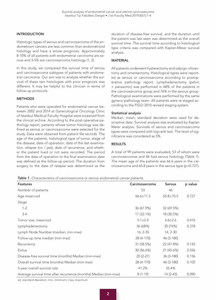
RESULTS
A total of 99 patients were evaluated, 53 of whom were carcinosarcomas and 46 had serous histology (Table 1). The mean age of the patients was 66.6 years in the car-cinosarcoma and 65.8 years in the serous type (p=0.727).
Table 1. Characteristics of carcinosarcoma or serous endometrial cancer patients
Features Carcinosarcoma Serous p value
Number of patients 53 46
Age mean±sd 66.6±11.2 65.8±10.5 0.727
Stage
1-2 36 (67.9%) 32 (69.5%)
3-4 17 (32.1%) 18 (30.5%)
Tumor size, mean±sd 5.1±2.4 4.6±2.6 0.416
Lymphadenectomy 36 (68%) 35 (76%) 0.318
Lymph Node Number (median; min-max) 16; 2-35 14; 2-30
Follow-up time median (min-max) 28 (4-170) 46 (3-180)
Recurrence 31 (58.5%) 22 (47.8%) 0.142
Exitus 30 (56.6%) 21 (45.6%) 0.556
Disease-free survival time (months) Median (min-max) 20 (2-27) 36 (3-180) 0.156
Overall survival time (months) Median (min-max) 28 (4-170) 46 (3-180) 0.105
5-year overall survival rate 41.2% 55.4%
Average survival time after recurrence (months) Median (min-max) 8 (1-19) 14 (2-43) 0.090
sd, standard deviation; min, minimum; max, maximum

3
Survival analysis of endometrial cancer and uterine carcinosarcomaİstanbul Tıp Fakültesi Dergisi • J Ist Faculty Med 2019;82(1):1-4
67.9% of patients with carcinosarcoma and 69.5% of patients with serous tumors were in early stage (FIGO I-II). The mean tumor size was 5.1±2.4 cm in the carci-nosarcoma and 4.6±2.6 cm in the serous type. The rates of patients who underwent lymphadenectomy (68% and 76%) and removed lymph node numbers [median (min-max) 16 (2-35) and 14 (2-30)] were similar in both groups. The median follow-up period was 28 months (4-170) for carcinosarcoma, and 46 (3-180) months for serous type. During the follow-up period, 58.5% (n=31) of the patients in the carcinosarcoma group and 47.8% (n=22) of the serous group had recurrence (p=0.142). Median disease-free survival was 20 (2-27) months in carcinosar-coma and 36 (3-180) months in serous type (Log-Rank test p=0.156). 56.6% (n=30) of the carcinosarcoma group and 45.6% (n=21) of the serous group died (p=0.556).
No death was observed for other reasons. Patients with carcinosarcoma survived for an average of 8 months af-ter relapse, while serous patients survived for 14 months (p=0.090) (Figure 3). Median overall survival was 28 months for carcinosarcoma and 46 months for serous (p=0.105). The 5-year overall survival rate was 41.2% for carcinosarcoma and 55.4% for serous (Figure 1, 2).
DISCUSSION
In this study, survival times of patients with carcinosar-coma or serous adenocarcinoma were analyzed. Log-Rank test was used to compare the survival of these two groups and no significant difference was observed. However, overall survival, disease-free survival and mean survival time after relapse were longer in patients with serous adenocarcinoma. The surgical approach was sim-ilar in both groups (rates of patients undergoing lymph-adenectomy, as well as the number of lymph nodes re-moved). The 5-year overall survival rate was also higher in the serous group (41.2% and 55.4%).
Carcinosarcoma and serous type histology have worse prognosis than endometrioid type histology. The Na-tional Comprehensive Cancer Network (NCCN) guide accepts carcinosarcoma, serous or clear cell endometrial cancers in the same category (non-enddometrioid) and suggests the same treatment approach (3). In the litera-ture, the survival time of these patients is reported quite differently. The median survival time for uterine carcino-sarcoma is reported to be 18-36 months in general (4, 5). In a study by Zhang et al., 83% of carcinosarcoma pa-tients recurred in 1 year, they have worse survival than patients with serous endometrial cancer (6). Piver et al. reported a five-year survival rate of 36% in early-stage uterine carcinosarcoma (7). Gadduci et al. reported a five-year survival rate of 33% in patients with early uterine
Figure 1: Overall survival graph of carcinosarcoma and serous type tumor
Figure 2: Disease-free survival in carcinosarcoma and serous type tumor
Figure 3: Survival time after recurrence in carcinosarcoma and serous type tumor

4
Survival analysis of endometrial cancer and uterine carcinosarcomaİstanbul Tıp Fakültesi Dergisi • J Ist Faculty Med 2019;82(1):1-4
carcinosarcoma treated by combination of radiotherapy and surgery (8). The survival time of patients with carci-nosarcoma seems to be low even in the early stages.
Endometrial serous carcinomas constitute less than 10% of endometrial adenocarcinomas, but serous adenocar-cinoma is the most common cause of death due to en-dometrial cancer (9). Ureyen et al. found a 5-year overall survival rate of 67% and a disease-free survival rate of 45% in their studies (10). Felix et al. reported similar sur-vival in patients with serous and carcinosarcoma (11).
In our study, survival times of 99 patients with serous or carcinosarcoma histology were evaluated. The median duration to relapse was 20 months in carcinosarcoma and 36 months in serous type (Log-Rank p=0.156). The mean survival time after recurrence was slightly longer in serous type (8 and 14 months, p=0.090).
As a result, the prognosis is not good in serous and carci-nosarcoma tumors of the endometrium. It may be useful to develop new treatment modalities in these aggressive histological types and to perform the studies to identify patients at an earlier stage.
Informed Consent: Informed consent was not received due to the retrospective nature of the study.
Peer Review: Externally peer-reviewed.
Author Contributions: Conception/Design of Study- A.K., S.T, H.S., Y.S.; Data Acquisition- A.K., S.T., H.S., Y.S.; Data Analysis/Interpretation- A.K., S.T., H.S., Y.S.; Drafting Manuscript- A.K., S.T.; Critical Revision of Manuscript- A.K., S.T., H.S., Y.S.; Final Approval and Accountability- A.K., S.T., H.S., Y.S.; Technical or Material Support- H.S., A.K., S.T.
Conflict of Interest: Authors declared no conflict of interest.
Financial Disclosure: Authors declared no financial support.
Bilgilendirilmiş Onamı: Retrospektif bir çalışma olduğundan bil-gilendirilmiş onam alınmamıştır.
Hakem Değerlendirmesi: Dış bağımsız.
Yazar Katkıları: Çalışma Konsepti/Tasarım- A.K., S.T, H.S., Y.S.; Veri Toplama- A.K., S.T., H.S., Y.S.; Veri Analizi/Yorumlama- A.K., S.T., H.S., Y.S.; Yazı Taslağı- A.K., S.T.; İçeriğin Eleştirel İnceleme-
si- A.K., S.T., H.S., Y.S.; Son Onay ve Sorumluluk- A.K., S.T., H.S., Y.S.; Malzeme ve Teknik Destek- H.S., A.K., S.T.
Çıkar Çatışması: Yazarlar çıkar çatışması beyan etmemişlerdir.
Finansal Destek: Yazarlar finansal destek beyan etmemişlerdir.
REFERENCES
1. Moore KN, Fader AN. Uterine papillary serous carcinoma. Clin Obstet Gynecol 2011;54(2):278–91. [CrossRef]
2. Ferrandina G, Zannoni GF, Martinelli E, Vellone V, Prisco MG, Scambia G. Endometrial carcinoma recurring as carcinosarcoma: Report of two cases. Pathol Res Pract 2007;203(9):677–81. [CrossRef]
3. NCCN. NCCN Clinical Practice Guidelines in Oncology: Uterine Neoplasms. Nccn. 2015; https://www.nccn.org/professionals/physician_gls/default.aspx
4. Spaziani E, Picchio M, Petrozza V, Briganti M, Ceci F, Di Filippo A, et al. Carcinosarcoma of the uterus: A case report and review of the literature. Eur J Gynaecol Oncol 2008;29(5):531–4.
5. Singh R. Review literature on uterine carcinosarcoma. J Cancer Res Ther 2014;10(3):461–8.
6. Zhang C, Hu W, Jia N, Li Q, Hua K, Tao X, et al. Uterine carcinosarcoma and high-risk endometrial carcinomas: A clinicopathological comparison. Int J Gynecol cancer 2015;25(30973185):629–36. [CrossRef]
7. Piver MS, Lele SB, Marchetti DL, Emrich LJ. Effect of adjuvant chemotherapy on time to recurrence and survival of stage I uterine sarcomas. J Surg Oncol 1988;38(4):233–9. [CrossRef]
8. Gadducci A, Fabrini MG, Facchini V, Ducci F, Colosimo S, Dell’Arciprete T, et al. Surgery and radiotherapy in the treatment of early stage uterine sarcomas. Eur J Gynaecol Oncol 1989;10(4):276–80.
9. Hamilton C, Cheung M, Osann K, Chen L, Teng N, Longacre T, et al. Uterine papillary serous and clear cell carcinomas predict for poorer survival compared to grade 3 endometrioid corpus cancers. Br J Cancer 2006;94(5):642–6. [CrossRef]
10. Üreyen I, Karalok A, Cırık DA, Taşçı T, Türkmen O, Cömert GK, et al. A comparison of clinico-pathologic characteristics of patients with serous and clear cell carcinoma of the uterus. Turk J Obs Gynecol 2016;13(3):137–43. [CrossRef]
11. Felix AS, Stone RA, Bowser R, Chivukula M, Edwards RP, Weissfeld JL, et al. Comparison of survival outcomes between patients with malignant mixed mullerian tumors and high-grade endometrioid, clear cell, and papillary serous endometrial cancers. Int J Gynecol Cancer 2011;21(5):877–84. [CrossRef]
5
Prenatal apllication of MLPAİstanbul Tıp Fakültesi Dergisi • J Ist Faculty Med 2019;82(1):5-11
İletişim kurulacak yazar/Corresponding author: [email protected]; gü[email protected]
Geliş tarihi/Received Date: 09.04.2018 • Kabul tarihi/Accepted Date: 08.10.2018
©Telif Hakkı 2019 J Ist Faculty Med - Makale metnine jmed.istanbul.edu.tr web sayfasından ulaşılabilir.©Copyright 2019 by J Ist Faculty Med - Available online at jmed.istanbul.edu.tr
ARAŞTIRMA / RESEARCH DOI: 10.26650/IUITFD.413596
İst Tıp Fak Derg 2019 / J Ist Faculty Med 2019
APPLICATION OF MLPA (MULTIPLEX LIGATION-DEPENDENT PROBE AMPLIFICATION) IN FETUSES WITH AN ABNORMAL SONOGRAM AND NORMAL KARYOTYPE
NORMAL KARYOTİPLİ PATOLOJİK ULTRASON BULGUSU OLAN FETUSLARDA MLPA (MULTİPLEX LİGATİON-DEPENDENT PROBE AMPLİFİCATİON) UYGULAMALARI
Güven TOKSOY1 , Birsen KARAMAN1 , Zehra Oya UYGUNER1 , Kader YILMAZ1 , Recep HAS2 ,Hülya KAYSERİLİ1,3 , Peter MINY4 , Seher BAŞARAN1
1Istanbul University, Istanbul Faculty of Medicine, Department of Medical Genetics, 2Department of Obstetrics and Gynecology, Istanbul, Turkey3Koç University, School of Medicine (KUSoM), Medical Genetics Department, Istanbul, Turkey 4University Children’s Hospital, Division of Medical Genetics, Basel, Switzerland
ORCID IDs of the authors: G.T. 0000-0002-8103-9980; B.K. 0000-0001-8640-0176; Z.O.U. 0000-0002-2035-4338; K.Y. 0000-0002-4203-3893; R.H. 0000-0002-1372-8506; H.K. 0000-0003-0376-499X; P.M. 0000-0001-8015-156X; S.B. 0000-0001-8668-4746
Cite this article as: Toksoy G, Karaman B, Uyguner ZO, Yılmaz K, Has R, Kayserili H, et al. Application of MLPA (Multiplex ligation-dependent probe amplification) in fetuses with an abnormal sonogram and normal karyotype. J Ist Faculty Med 2019;82(1):5-11. doi: 10.26650/IUITFD.413596
ABSTRACTObjective/Material and Method: Cryptic chromosomal imbal-ances contribute significantly to the etiology of multiple con-genital anomalies with or without mental retardation (MCA/MR). Current approaches in prenatal diagnosis include targeted high resolution analyses by MLPA and some microarray platforms or a genomewide screening at maximal resolution using oligonucle-otide or SNP arrays. The major disadvantages of the latter ap-proach are cost and the inadvertent detection of copy number variation of unknown clinical significance.
In this prospective work, fetal DNA samples from 66 fetuses who had pathological antenatal ultrasonography findings with normal karyotype and Multiprobe T-FISH results were tested using com-mercially available targeted MLPA probe-sets to compare the efficacy and the impact of MLPA testing at prenatal setting.
Results: Three submicroscopic deletions (3.66; 4.5%) were detect-ed in the cohort. Two of them were de novo deletions, 18ptel and 7q11.23. The third finding was a 75 kb duplication at 18q, which was maternally inherited and probably a benign copy number variation unrelated to the pathological ultarsonography findings.
Conclusion: The observed detection rate by MLPA testing can be considered within the expected range. Furthermore, benign copy number variation was identified with the targeted diagnos-tic approach as an unexpected finding. This study shows that MLPA is a practical and cost-effective technique to investigate submicroscobic chromosomal aberrations in fetuses.
Keywords: MLPA, subtelomeric anomalies, prenatal diagnosis microdeletion/microduplication
ÖZETAmaç/Gereç/Yöntem: Multiple konjenital anomaliler ve bilişsel yetersizliklerin (MKA/MR) etiyolojisinde kriptik kromozom dü-zensizlikler önemli yer tutmaktadır. Yeni teknolojilerin gelişmesi, MLPA veya genom boyu SNP ve oligonükleotidlerle tarama ya-pabilen mikrodizin ya da yeni nesil dizileme teknolojileri yüksek düzeyde çözünürlükle bu anomalilerin prenatal dönemde sapta-nabilmesine olanak sağlamıştır. Yüksek çözünürlüklü çalışmaların en büyük dezavantajı maliyet ile beklenmeyen ve/veya klinik öne-mi bilinmeyen kopya sayısı varyantlarının saptanmasıdır.
Bu prospektif çalışmada patolojik ultrason bulgusu saptanan ve normal karyotipe sahip çoklu telomerik FISH ile normal sonuç alınmış 66 fetusa ait DNA örnekleri ticari olarak satılan MLPA prob setleri ile değerlendirilerek MLPA nın tanı akış şemasındaki etkinliği araştırıldı.
Bulgular: Çalışmada toplam üç olguda (3:66; 4,5%) delesyon be-lirlendi. İki olguda submikroskobik de novo delesyon saptandı ve bunlardan biri 18ptel ve diğeri 7q11.23 delesyonu idi. Bir diğer olguda klinik bulgularla ilişkisiz, yüksek olasılıkla zararsız 75 kb büyüklüğünde anneden kalıtılan 18q duplikasyonu belirlendi.
Sonuç: Patojenik mutasyon saptama oranı beklenti ile uyumlu idi. Ek olarak, hedefe yönelik tanıda sıra dışı kabul edilen bir durum olarak bir olguda maternal kalıtımlı yüksek olasılıkla zararsız bir kop-ya sayısı değişikliği saptandı. Bu çalışma prenatal uygulamalarda submikroskobik kromozomal anomalilerin araştırılmasında MLPA tekniğinin uygun maliyetli ve kullanılabilir olduğunu göstermiştir.
Anahtar Kelimeler: MLPA, subtelomerik değişimler, doğum ön-cesi tanı mikrodelesyon/mikroduplikasyon
6
Prenatal apllication of MLPAİstanbul Tıp Fakültesi Dergisi • J Ist Faculty Med 2019;82(1):5-11
INTRODUCTION
Chromosomal imbalances are important in the etiology of congenital malformations of the newborn period (1, 2). When major malformations are detected by fetal ultraso-nography (USG), the rate of chromosomal aberrations can be as high as 29% depending on the week of pregnan-cy, tissue type or technique applied (3-7). Conventional karyotyping allows the genome-wide detection of chro-mosome anomalies at a rather low resolution (>8-10 Mb) depending on the banding level. In the presence of dis-tinct phenotypes in postnatal cases, the fluorescence in situ hybridisation (FISH) technique using syndrome spe-cific probes may be applied to diagnose known deletion/duplications smaller than 6Mb. Unbalanced rearrange-ments in gene-rich subtelomeric regions, have been identified as a significant etiological contributor to MCA/MR (8-12). Due to the nonspecific banding pattern, even larger unbalanced cryptic subtelomeric rearrangements can be easily missed by conventional karyotyping, espe-cially when the banding level is lower than 500 bands per haploid set. MLPA and array-CGH techniques/microarray developed during the last decade have been very effec-tive at overcoming those limitations (12-17). The rate of clinically relevant copy number changes in prenatal cases with MCA after a normal result in conventional karyotyp-ing was reported to be 4% by FISH [12] and 5%-10 % by a-CGH technique (12, 15). While microarrays became the first-tier test in postnatal cytogenetic diagnosis (15, 16), their application in the prenatal setting are still met with reservations, mainly due to the concomitant diagnosis of variants of unknown significance (VOUS) - especially in high resolution arrays. MLPA with probe sets covering known disease associated critical regions may offer an acceptable practical compromise, trading off cost-effec-tiveness, sensitivity and the risk of encountering VOUS (14, 16, 17, 18-26).
Sixty-six DNA samples from fetuses with pathological USG findings and normal karyotype and subtelomeric FISH results were tested using MLPA probe sets SALSA P070 and P245 to identify the efficacy and the impact of MLPA testing at prenatal setting.
MATERIAL AND METHODS
Sixty-six archived DNA samples consecutively collected between November 2007 and April 2010 met the inclusion criteria of pathological USG findings (at least one major anomaly diagnosed by an experinced perinatologist), as well as a normal karyotype (500 bands at minimum) and normal Multiprobe T-FISH (Cytocell) analysis. Five cases with congenital heart defects along with normal TUPLE1 (Cytocell) test results and one case with lissencephaly and normal LIS1, FLI1 (Cytocell) test results were includ-ed in the study. The DNA had been extracted from fresh materials and/or cell cultures and kept frozen at -200C
using the QIAamp DNA Blood Mini Kit (Qiagen, CA, USA) in 58 samples (25 fetal blood, 23 amniotic fluid and 10 chorionic villi samples) and by an automatic nucleic acid purification system (Magna Pure Compact – Roche) in 8 samples (fetal blood). All cases were tested with both SALSA P070 specific for subtelomeres and SALSA P245 specific for known microdeletion syndromes. Coffalyser v9.4 software was used for data analysis. Anomalies de-tected by MLPA were confirmed by FISH using commer-cially available probes (Cytocell and Vysis) or array-CGH (Affymetrix or Nimblegene platforms).
All the clinical examinations on the fetuses and newborns and the genetic testing were performed at the Depart-ment of Medical Genetics, Istanbul Medical Faculty of Istanbul University. Fetal USG and invasive procedures were performed at the Perinatology Division of the Ob-stetrics and Gynecology Department of the same faculty. Families were informed about the study and written in-formed consent was obtained.
RESULTS
The mean maternal age was 28.22 years (range 19-43) and the mean gestational age was 23. 8 weeks (range 16-35). Fetal sex distribution was 1:1 (33 males/33 females).
In addition to the abnormal USG findings 11 mothers out of 66 were 35 years or older, in five pregnancies maternal serum screening showed an increased risk and two moth-ers had a history of recurrent fetal losses.
The MLPA study revealed chromosomal imbalances in three cases (3:66; 4.5%). Two anomalies were de novo and detected with SALSA MLPA P070 probe mix and one familial anomaly with SALSA MLPA P245 probe mix.
Case-1 (MLPA#10)
The mother aged 35 was referred due to antenatal ultra-sonographic finding of IUGR and diaphragmatic hernia, and an increased risk for trisomy 18 in the first trimes-ter screening test (>1:50). Conventional karyotyping and subtelomeric FISH following amniocentesis at 17 weeks gestation revealed normal results.
The MLPA SALSA P070 probe mix showed a gain at 18q23 (Figure 1a). Although FISH analysis using 18q sub-telomeric probe (Cytocell) showed normal signals on both chromosomes 18 (Figure 1b), the microarray (Affy-metrix, Cyto 2.7) analysis confirmed the ~75 kb gain on the region (46,XY.ish subtel(41x2).mlpa (P070)x2, 18q sub-tel (P245)x3). arr18q23(75,516,228-75,590,562*)x3 mat). A parental array-CGH study using NimbleGen CGX-3 re-vealed that the phenotypically normal mother was also a carrier for the duplication (Figure 1 c,d) and the change was considered as “clinically not relevant” (46,XX. arr18q23(75,516,228-75,590,562x3). The child was delivered at
7
Prenatal apllication of MLPAİstanbul Tıp Fakültesi Dergisi • J Ist Faculty Med 2019;82(1):5-11
the 34th week of gestation and died 4 days later, following surgery for the diaphragmatic hernia.
Case- 2 (MLPA#15)
The mother was referred due to the ultrasonographic finding of holoprosencephaly, single-nostril, thalamic fusion, hypotelorism, absence of nasal bone, and bilat-eral echogenic kidneys. At the 24th week of gestation, a fetal blood sample was obtained by fetal cord blood sampling. Routine karyotyping and subtelomeric FISH analysis revealed normal results. The pregnancy was ter-minated due to the possible severe, lethal outcome of antenatal USG findings.
MLPA SALSA P070 probe mix showed a deletion at subtel 18p (Figure 2a). 46,XX. ish subtel(41x2).18p subtel(P070)x1, (P245)x2. ish.del(18)(p11.3-)(D18S552-) FISH with a subtelomeric 18p probe (Telvision-Vysis) confirmed the deletion (Figure 2b), which was missed by the Multiprobe T System.
Parental cytogenetic and FISH studies revealed normal results.
Case-3 (MLPA#20)
The mother was referred at the 18th week of gestation, due to the USG findings of IUGR, short femurs, decreased dimensions of stomach and single umbilical artery. A fetal
Figure 1: a) MLPA SALSA P070 probemix result ratio mapview bar graphic; for case MLPA#10, b) FISH signals with subtelomeric probe for 18q (Cytocell), c) Overlapping gained part and CTDP1 gene on Array-CGH result of the case#10, d) Array-CGH result of the mother with the same gain at the same localization.

8
Prenatal apllication of MLPAİstanbul Tıp Fakültesi Dergisi • J Ist Faculty Med 2019;82(1):5-11
blood sample was obtained by cordocentesis and rou-tine karyotyping and subtelomeric FISH analysis revealed normal results.
A female baby was born by cesarean section due to the intrauterine fetal distress at 35 weeks of gestation. The birth weight was 1430 gr, dysmorphic features includ-ed periorbital oedema, long philtrum, thin upper lip, everted lover lip, micrognathia, pointed chin, posteriorly rotated ears and cutis marmorata. Cardiac echography revealed a small ventricular septal defect at the age of 6 months and peripheral pulmonary stenosis at the age of 14 months.
MLPA test SALSA P245 probe mix revealed a deletion on Williams Syndrome (WS) region for exon1 and exon 20 of the ELN gene and LIMK gene locus (Figure 3a) (46,XX. ish subtel(41x2). mlpa (P070)x2, 7q11.23 (P245)x1. ish.del7(q11.23-)(ELN-)). This deletion was confirmed by ELN locus specific FISH probe (Cytocell) (3b).
Parental cytogenetic and FISH studies revealed normal results.
DISCUSSION
Although the investigations carried out on children with MCA/MR underlined the importance of the subtelomer-ic imbalances, it is well known that the resolution of the classical karyotyping is not sufficient to detect the chro-mosomal imbalances of these regions. The diagnosis of the microdeletion/duplication syndromes can be ascer-tained in postnatal cases in the presence of distinct phe-notypes for well-known syndromes. At the beginning of the 2000’s, there were some reports using FISH analysis to diagnose the subtelomeric imbalances in fetuses with pathological USG findings. However, it was noted that the technique was relatively expensive and time consum-ing (12, 16, 22, 24). Later, MLPA was accepted as an alter-native technique to identify known microdeletion/dupli-cations and subtelomeric imbalances (14, 15, 22-24, 26).
Microarray/a-CGH techniques are able to screen the whole genome, which leads to higher detection rates, 1 to 8%, depending on the patient selection criteria and sensitivity of the test applied. These techniques are rec-ommended, especially in the presence of pathological fetal USG findings (14, 15, 22, 25, 27).
Figure 3: a) MLPA SALSA P245 probemix result ratio mapview bar graphic of case MLPA#20, b) FISH signals with subtelomeric probe of 18q (Cytocell)
Figure 2: a) MLPA SALSA P070 probemix result ratio mapview bar graphic of case MLPA#15, b) FISH signals with subtelomeric probe for 18q (Cytocell)

9
Prenatal apllication of MLPAİstanbul Tıp Fakültesi Dergisi • J Ist Faculty Med 2019;82(1):5-11
The purpose of the study was to asses the efficacy and the impact of MLPA technique for the antenatal diagno-sis in our patient cohort. In 66 fetuses with at least one major malformation with normal karyotype and subtelo-meric FISH results, three chromosomal imbalances (4.5%) have been identified by MLPA. The rate of clinically rel-evant imbalances was 3.03% (2/66) and one imbalance was maternally inherited CNV (1.51% - 1/66). The report-ed rate of imbalances for MLPA in the literature varies between 0-6.5% depending on the selection criteria (16, 23, 24, 26). When specific tests are performed and the resolution of the karyotypes is high, it could be expected that the detected anomaly rate by MLPA or array stud-ies, will be decreased (24-26). In the study from Konialis (28), MLPA was applied parallel to the karyotyping and microarray technique in all prenatal cases without any indication bias. The rate of clinically relevant imbalanc-es was 1.2% (3/249) in cases with pathological USG find-ings and this rate was about 0.4% in cases with normal USG. However, in the study from Goumy (24), the rate of unbalanced rearrangements was 6.5% (4/61) in a series of cases with specific USG findings. The rate was 4.1% (33/139) in fetuses presented with major malformations and 1.6% (23/104) in fetuses with having minor, including soft markers, USG anomalies in the series of Kjaergaard (13). Our result, 3.03% in cases with major malformation is in accordance with the previously published series.
Disadvantages of the FISH analysis are the need of cell culture and high quality metaphases, which are laborious and lead to high costs. The disadvantage of the MLPA technique is that it does not cover the whole genome and multiple samples are needed to decrease the cost. Although microarray and a-CGH studies are more com-prehensive and informative, the need for special equip-ment and consumables leads to high costs. Furthermore using experienced bioinformaticians is a must to carry out the analysis. Well known microdeletion at 7q11.23 causing WS was identified in one case. The pathological USG findings were IUGR, short femurs, decreased volume of stomach, and single umbilical artery, which were not specific for WS, and consequently FISH testing specific for WS was not performed at antenatal period. The case was retrospectively studied by MLPA using SALSA P245 probe mix, a deletion covering exon1 and 20 of ELN gene responsible for WS and LIMK gene was detected. The karyotype from the antenatal period was reanalyzed after the detection of the deletion by MLPA and no change leading to a suspicion of a deletion was observed. Clini-cal findings from the newborns were in accordance with WS. There are only four antenatally diagnosed WS cases in the literature, and three of them had the characteris-tic ultrasonographic findings of WS leading to specific testing for definite diagnosis. One case presented with VSD, and the diagnosis was established by MLPA with P064 probe mix (26), a second case reported to have su-
pravalvular aortic stenosis (SVAS), leading to testing by FISH analysis WS specific probe (29) and the third case presented with SVAS, IUGR, interhemispheric cyst, and nasal bone hypoplasia and the deletion on 7q11.23 was identified using array-CGH (30). The fourth case reported by Lee (31), had normal USG findings and was identified using BAC array-CGH technique in the research study of 3171 consecutive prenatal samples. Our case is the sec-ond case diagnosed antenatally without any characteris-tic USG findings of WS. When specific fetal USG findings like SVAS, VSD are present, the FISH technique can be the first choice in the PD, otherwise MLPA or a-CGH tech-niques are the most powerful techniques to identify the microdeletion/duplication syndromes.
In another case, MLPA using a SALSA P070 probe mix showed a deletion on 18p, which was subsequently con-firmed by subtelomeric 18p FISH prob (Telvision-Vysis). When the karyotype was reevaluated, it was considered that this microdeletion could have easily been missed by routine analysis. A false negative result in Multiprobe FISH analysis could be explained by the low-quality metaphases and false positive signals. Johnson and Bachman (32) published the first case of 18p deletion with holoprosencephaly. Gripp (33) identified the fourth gene, TGIF gene, responsible for the holoprosencephaly phenotype located on 18p at a distance of 3.5 Mb from the telomere. The distance between the TGIF gene and the THOC gene is about 3.2 Mb and SALSA P070 MLPA probe mix and subtelomeric FISH probe (Telvision-Vysis) both target the THOC gene. Therefore, it could be sug-gested that the deletion in our case was also covering the TGIF gene locus. There are many reports with microscop-ic monosomy 18p in the literature, but to our knowledge, this is the first submicroscopic deletion on 18p detected at antenatal period.
The size of the duplication on 18q telomere detected by MLPA was determined as ~75 kb by array technique. Since a phenotypically normal mother was also a carrier of the same duplication, it was interpreted as a previously unreported copy number variation (34). There were only four postnatal cases with duplication at 18q23 reported as unspecified pathogenicity in DECIPHER v5.1 (35). The gains of those cases covered the whole q arm of 18 in one, and it was a 37 Mb duplication in another. However, the duplicated region in our case was very small and only the C-terminal Domain of RNA Polymerase II subunitA, phosphatase of subunit1 (CTDP1) gene was located in this region. Homozygous loss of function mutation of the CTDP1 gene is associated with Congenital Cataracts, Fa-cial Dysmorphism, and Neuropathy syndrome (CCFDN) in the literature (36). There is only one report presenting the gain as a polymorphic variation (37). Our findings in the family further support that the duplication of CTDP1 gene has no phenotypic effect.
10
Prenatal apllication of MLPAİstanbul Tıp Fakültesi Dergisi • J Ist Faculty Med 2019;82(1):5-11
Although 22q11.2 microdeletion (DiGeorge Syndrome region) is the most common submicroscopic anomaly de-tected by MLPA in the literature, it was not encountered in our cohort. All cases with cardiac malformations had been screened for 22q11.2 deletion by FISH analysis at the antenatal period and screened positive ones, 22q11 microdeletion cases, were not included for MLPA testing. This is the reason why this common microdeletion was not detected in our cohort.
Our study shows that MLPA is an efficient, safe, rapid and sensitive technique for the detection of submicroscopic imbalances for targeted regions of the genome. It is cost effective in comparison to FISH, since a single MLPA test can provide data for many loci concurrently for multiple samples. However, the confirmation of MLPA findings (e.g. single probe imbalances) may require FISH testing, STR analysis, qPCR or array CGH. Although, MLPA provides lower resolution than array, due to the organization of the probes, it is targeted and therefore the genotype-phe-notype correlation is more predictable. The identification of de novo CNV’s with uncertain significance (about 4%) leads to further testing increasing the costs, and uncer-tain results might also cause parental anxiety. Therefore, the testing strategy should be planned according to the relevant indication and suspected chromosomal abnor-malities. If telomeric and syndrome specific analysis is sought after, MLPA can be the first-tier and the most suit-able technique also at the antenatal period.
Ethics Commitee Approval: Ethics committee approval was re-ceived for this study from the ethics committee of the Istanbul Faculty of Medicine (26.09.2007_2007/2105).
Informed Consent: Written consent was obtained from the par-ticipants.
Peer Review: Externally peer-reviewed.
Author Contributions: Conception/Design of Study- G.T., B.K., S.B., H.K., R.H.; Data Acquisition- K.Y., G.T., B.K., Z.O.U.; Data Analysis/Interpretation- G.T., B.K., K.Y., Z.O.U., P.M.; Drafting Manuscript- G.T., B.K., Z.O.U., S.B., P.M.; Critical Revision of Manuscript- G.T., B.K., P.M., S.B., Z.O.U., H.K., R.H.; Final Ap-proval and Accountability- G.T., B.K., Z.O.U., K.Y., R.H., H.K., P.M., S.B.; Technical or Material Support- G.T., B.K., S.B., Z.O.U., H.K.; K.Y.
Conflict of Interest: Authors declared no conflict of interest.
Financial Disclosure: The present work was supported by the Research Fund of Istanbul University. Project No. 3751 and 1515.
Etik Komite Onayı: Bu çalışma için etik komite onayı İstan-bul Tıp Fakültesi Yerel Etik Kurulu’ndan alınmıştır (Karar Tarihi: 26.09.2007_2007/2105).
Bilgilendirilmiş Onam: Katılımcılardan bilgilendirilmiş onam alınmıştır.
Hakem Değerlendirmesi: Dış bağımsız.
Yazar Katkıları: Çalışma Konsepti/Tasarım- G.T., B.K., S.B., H.K., R.H.; Veri Toplama- K.Y., G.T., B.K., Z.O.U.; Veri Analizi/Yorumla-ma- G.T., B.K., K.Y., Z.O.U., P.M.; Yazı Taslağı- G.T., B.K., Z.O.U., S.B., P.M.; İçeriğin Eleştirel İncelemesi- G.T., B.K., P.M., S.B., Z.O.U., H.K., R.H.; Son Onay ve Sorumluluk- G.T., B.K., Z.O.U., K.Y., R.H., H.K., P.M., S.B.; Malzeme ve Teknik Destek- G.T., B.K., S.B., Z.O.U., H.K.; K.Y.
Çıkar Çatışması: Yazarlar çıkar çatışması beyan etmemişlerdir.
Finansal Destek: Bu çalışma İstanbul Üniversitesi Araştırma Fonu tarafından desteklenmiştir. Proje No 3751 ve 1515.
REFERENCES
1. Nussbaum RL, McInnes RR, Willard HF, Hamosh A. Principles of clinical cytogenetics, Thompson&Thompson Genetics in Medicine, Saunders, Elsevier. Philadelphia, PA, 7th ed., 2007;59-113.
2. Adams-Chapman I, Hansen NI, Shankaran S, Bell EF, Boghossian NS, Murray JC, et al.; Eunice Kennedy Shriver National Institute of Child Health and Human Development Neonatal Research Network. Ten-year review of major birth defect s in VLBW infants. Pediatrics 2013;132(1):49-61. [CrossRef]
3. Eydoux P, Choiset A, Le Porrier N, Thépot F, Szpiro-Tapia S, Alliet J, et al. Chromosomal prenatal diagnosis: study of 936 cases of intrauterine abnormalities after ultrasound assessment. Prenat Diagn 1989;9(4):255-69. [CrossRef]
4. Daniel A, Athayde N, Ogle R, George AM, Michael J, Pertile MD, et al. Prospective ranking of the sonographic markers for aneuploidy: data of 2143 prenatal cytogenetic diagnoses referred for abnormalities on ultrasound. Aust N Z J Obstet Gynaecol 2003;43(1):16-26. [CrossRef]
5. Zhang L, Zhang XH, Liang MY, Ren MH. Prenatal cytogenetic diagnosis study of 2782 cases of high-risk pregnant women. Chin Med J (Engl) 2010;123(4):423-30.
6. Lichtenbelt KD, Alizadeh BZ, Scheffer PG, Stoutenbeek P, Schielen PC, Page-Christiaens LC, et al. Trends in the utilization of invasive prenatal diagnosis in The Netherlands during 2000-2009. Prenat Diagn 2011;31(8):765-72. [CrossRef]
7. D’Amours G, Kibar Z, Mathonnet G, Fetni R, Tihy F, De´silets V, et al. Whole-genome array CGH identifies pathogenic copy number variations in fetuses with major malformations and a normal karyotype. Clin Genet 2012;81(2):128-41. [CrossRef]
8. Flint J, Wilkie AO, Buckle VJ, Winter RM, Holland AJ, McDermid HE. The detection of subtelomeric chromosomal rearrangements in idiopathic mental retardation. Nat Genet 1995;9(2):132-40. [CrossRef]
9. Knight SJ, Regan R, Nicod A, Horsley SW, Kearney L, Homfray T, et al. Subtle chromosomal rearrangements in children with unexplained mental retardation. Lancet 1999;354(9191):1676-81. [CrossRef]
10. Rossi E, Piccini F, Zollino M, Neri G, Caselli D, Tenconi R, et al. Cryptic telomeric rearrangements in subjects with mental retardation associated with dysmorphism and congenital malformations. J Med Genet 2001;38(6):417-20. [CrossRef]

11
Prenatal apllication of MLPAİstanbul Tıp Fakültesi Dergisi • J Ist Faculty Med 2019;82(1):5-11
11. Baker E, Hinton L, Callen DF, Altree M, Dobbie A, Eyre HJ, et al. Study of 250 children with idiopathic mental retardation reveals nine cryptic and diverse subtelomeric chromosome anomalies. Am J Med Genet 2002;107(4):285-93. [CrossRef]
12. Gignac J, Danis K, Tihy F, Lemyre E. Prenatal detection of subtelomeric rearrangements by multi-subtelomere FISH in a cohort of fetuses with major malformations. Am J Med Genet A 2006;140(24):2768-75. [CrossRef]
13. Kjaergaard S, Sundberg K, Jørgensen FS, Rohde MD, Lind AM, Gerdes T, et al. Diagnostic yield by supplementing prenatal metaphase karyotyping with MLPA for microdeletion syndromes and subtelomere imbalances. Prenat Diagn 2010;30(10):995-9. [CrossRef]
14. Shaffer LG, Bejjani BA. A cytogeneticist’s perspective on genomic microarrays. Hum Reprod Update 2004;10(3):221-6. [CrossRef]
15. Shaffer LG, Coppinger J, Alliman S, Torchia BA, Theisen A, Ballif BC, et al. Comparison of microarray-based detection rates for cytogenetic abnormalities in prenatal and neonatal specimens. Prenat Diagn 2008;28(9):789-95. [CrossRef]
16. Schou KV, Kirchhoff M, Nygaard U, Jorgersen C, Sundberg K. Increased nuchal translucency with normal karyotype: afollow-up study of 100 cases supplemented with CGH and MLPA analyses. Ultrasound Obstet Gynecol 2009;34(6):618-22. [CrossRef]
17. Schaffer LG, Bejjani BA. Medical applications of array CGH and the transformation of clinical cytogenetics. Cytogenet Genome Res 2006;115(3-4):303-9. [CrossRef]
18. Vialard F, Molina Gomes D, Leroy B, Quarello E, Escalona A, Le Sciellour C, et al. Array comparative genomic hybridization in prenatal diagnosis: another experience. Fetal Diagn Ther 2009;25(2):277-84. [CrossRef]
19. Thuresson AC, Bondeson ML, Edeby C, Ellis P, Langford C, Dumanski JP, et al. Whole-genome array-CGH for detection of submicroscopic chromosomal imbalances in children with mental retardation. Cytogenet Genome Res 2007;118(1):1-7. [CrossRef]
20. Slavotinek AM. Novel microdeletion syndromes detected by chromosome microarrays. Hum Genet 2008;124(1):1-17. [CrossRef]
21. Engels H, Wohlleber E, Zink A, Hoyer J, Ludwig KU, Brockschmidt FF, et al. A novel microdeletion syndrome involving 5q14.3-q15: clinical and molecular cytogenetic characterization of three patients. Eur J Hum Genet 2009;17(12):1592-9. [CrossRef]
22. Gross SJ, Bajaj K, Garry D, Klugman S, Karpel BM, Marie Roe A, et al. Rapid and novel prenatal molecular assay for detecting aneuploidies and microdeletion syndromes. Prenat Diagn 2011;31(3):259-66. [CrossRef]
23. Mademont-Soler I, Morales C, Bruguera C, Madrigal I,Clusellas N, Margarit E, et al. Subtelomeric MLPA: is it really useful in prenatal diagnosis? Prenat Diagn 2010;30(12-13):1165-9. [CrossRef]
24. Goumy C, Gouas L, Pebrel-Richard C, Véronèse L, Eymard-Pierre E, Debost-Legrand A, et al. Prenatal detection of cryptic rearrangements by multiplex ligation probe amplification in fetuses with ultrasound abnormalities. Genet Med 2010;12(6):376-80. [CrossRef]
25. Kleeman L, Bianchi DW, Shaffer LG, Rorem E, Cowan J, Craigo SD, et al. Use of array comparative genomic hybridization for prenatal diagnosis of fetuses with sonographic anomalies and normal metaphase karyotype. Prenat Diagn 2009;29(13):1213-7. [CrossRef]
26. Kontos H, Manolakos E, Malligiannis P, Plachouras N, Ploumis N, Mihalatos M, et al. Prenatal diagnosis of a fetus with 7q11.23 deletion detected by multiplex ligation-dependent probe amplification (MLPA) screening. Prenat Diagn 2008;28(6):556-8. [CrossRef]
27. Van den Veyver IB, Patel A, Shaw CA, Pursley AN, Kang SH, Simovich MJ, et al. Clinical use of array comparative genomic hybridization (aCGH) for prenatal diagnosis in 300 cases. Prenat Diagn 2009;29(1):29-39. [CrossRef]
28. Konialis C, Hagnefelt B, Sevastidou S, Karapanou S, Pispili K, Markaki A, et al. Uncovering recurrent microdeletion syndromes and subtelomeric deletions/duplications through non-selective application of a MLPA based extended prenatal panel in routine prenatal diagnosis. Prenat Diagn 2011;31(6):571-7. [CrossRef]
29. Krzeminska D, Steinfeld C, Cloez JL, Vibert M, Chery M, Menziesat D, et al. Prenatal diagnosis of Williams syndrome based on ultrasound signs. Prenat Diagn 2009;29(7):710-2. [CrossRef]
30. Popowski T, Vialard F, Leroy B, Bault JP, Molina-Gomes D. Williams-Beuren syndrome: the prenatal phenotype. Am J Obstet Gynecol 2011;205(6):e6-8. [CrossRef]
31. Lee CN, Lin SY, Lin CH, Shih JC, Lin TH, Su YN. Clinical utility of array comparative genomic hybridization for prenatal diagnosis:a cohort study of 3171 pregnancies. BJOG 2012; 119(5):614–25. [CrossRef]
32. Johnson G, Bachman R. A 46,XY,del(18)(pter-p1100:) cebocephalic child from a 46,XX,t(12;18)(18pter-18p1100::12qter-12pter) normal parent. Hum Genet 1976;34(1):103-6. [CrossRef]
33. Gripp KW, Wotton D, Edwards MC, Roessler E, Ades L, Meinecke P, et al. Mutations in TGIF cause holoprosencephaly and link NODAL signalling to human neural axis determination. Nat Genet 2000;25(2):205-8. [CrossRef]
34. Database of genomic variation, “A curated catalogue of human genomic structural variation”, 2018/09/21, Available from: http://dgv.tcag.ca/dgv/app/variant?id= dgv3407n100&ref=GRCh37/hg19
35. DECIPHER, “Database of Chromosomal Imbalance and Phenotype in Humans Using Ensembl Resources”, 2018/09/21 Available from: https://decipher.sanger.ac.uk/patient/291627#genotype/cnv/67391/browser/18:76996166-77946488
36. Varon R, Gooding R, Steglich C, Marns L, Tang H, Angelicheva D et al. Partial deficiency of the C-terminal-domain phosphatase of RNA polymerase II is associated with congenital cataracts facial dysmorphism neuropathy syndrome. Nat Genet 2003;35(2):185-9. [CrossRef]
37. Choi JS, Lee WJ, Baik SH, Yoon HK, Lee KH, Kim YH. Array CGH reveals genomic aberrations in human emphysema. Lung 2009;187(3):165-72. [CrossRef]
12
Konus hareketinin seri manyetik rezonans görüntüleme ile analiziİstanbul Tıp Fakültesi Dergisi • J Ist Faculty Med 2019;82(1):12-7
İletişim kurulacak yazar/Corresponding author: [email protected]
Geliş tarihi/Received Date: 22.06.2018 • Kabul tarihi/Accepted Date: 26.10.2018
©Telif Hakkı 2019 J Ist Faculty Med - Makale metnine jmed.istanbul.edu.tr web sayfasından ulaşılabilir.©Copyright 2019 by J Ist Faculty Med - Available online at jmed.istanbul.edu.tr
ARAŞTIRMA / RESEARCH DOI: 10.26650/IUITFD.435784
İst Tıp Fak Derg 2019 / J Ist Faculty Med 2019
SUPİN VE PRON POZİSYONLARDA SERİ MANYETİK REZONANS GÖRÜNTÜLEME İLE KONUS MEDULLARİS HAREKET ARALIĞININ ANALİZİ: 40 ARDIŞIK SAĞLIKLI İNSAN DENEKTE ELDE EDİLEN SONUÇLAR
THE MOTION RANGE OF THE CONUS MEDULLARIS MOVEMENT WITH SERIAL MAGNETIC RESONANCE IMAGING IN SUPINE AND PRONE POSITIONS: RESULTS OF FOURTY HEALTHY SUBJECTS
Cengiz GÖMLEKSIZ1 , Halil CAN1 , Umut YAKA2 , Burcu GÖKER3 , Aydın AYDOSELI2 ,Mehmet BARBUROĞLU4 , Altay SENCER2 , Utku ÖZGEN2 , Serra SENCER5 , Ali Güven YÖRUKOĞLU6 ,Talat KIRIŞ3
1Kolan Hastanesi, Beyin ve Sinir Cerrahisi, İstanbul, Türkiye2İstanbul Üniversitesi, İstanbul Tıp Fakültesi, Beyin ve Sinir Cerrahisi Anabilim Dalı, İstanbul, Türkiye3İstinye Üniversitesi, Tıp Fakültesi, Liv Hastanesi, Beyin ve Sinir Cerrahisi, İstanbul, Türkiye4Koç Üniversitesi, Tıp Fakültesi, Radyoloji Anabilim Dalı, İstanbul, Türkiye5İstanbul Üniversitesi, İstanbul Tıp Fakültesi, Radyoloji Anabilim Dalı, İstanbul, Türkiye6Gaziosmanpaşa Avrasya Hastanesi, Beyin ve Sinir Cerrahisi, İstanbul, Türkiye
ORCID IDs of the authors: C.G. 0000-0002-7884-4639; H.C. 0000-0002-6792-9987; U.Y. 0000-0002-2405-9504; B.G. 0000-0002-3916-7540; A.A. 0000-0002-4695-8295; M.B. 0000-0001-8551-4796; A.S. 0000-0001-9925-5422; U.Ö. 0000-0001-8381-0700; S.S. 0000-0002-3942-093X; A.G.Y. 0000-0002-2388-5839; T.K. 0000-0002-0938-5221
Cite this article as: Gömleksiz C, Yaka U, Can H, Yörükoğlu HC, Göker B, Aydoseli A, et al. The motion range of the Conus medullaris movement with serial magnetic resonance imaging in Supine and Prone positions: Results of fourty healthy subjects. J Ist Faculty Med 2019;82(1):12-7. doi: 10.26650/IUITFD.435784
ÖZETAmaç: Bu çalışmada seri halde supin ve pron pozisyonlarda T1 ve T2 ağırlıklı aksiyal ve sagital manyetik rezonans görüntü-leme yapılan 40 sağlıklı insanda meduller konusun supin-pron pozisyon değişikliği ile uyarılan hareket aralığı değerlendirildi.Yöntem: Her bir denekte konus medullarisin ucu seviyesinde elektronik ölçekler kullanılarak konus medullaris ve ön dural yüzey arasındaki mesafeyi ve dural kesenin genişliğini ölçtük. Elde edilen veriler SPSS 15 kullanılarak değerlendirildi. Bulgular: Konus ucunun, denekler supin pozisyondan pron po-zisyona getirildiğinde ortalama değer olarak spinal kanal geniş-liğinin %30±14’ü oranında yer değiştirdiği saptandı (p:0,0001). Yaş (0-18 ve >19 yaş) ve cinsiyet farklılığı konus ucunun hareket-liliğinde istatistiksel olarak anlamlı faktörler değildi. Sonuç: Çalışmamız klinik olarak gergin omurilik şüphesi bulu-nan normal uzanımlı konus vakalarında güvenilir bir diyagnostik referans aralığı sunmaktadır.Anahtar Kelimeler: Supin, pron, manyetik rezonans görüntü-leme, gergin omurilik sendromu, meduller konus
ABSTRACTObjective: In this study, using a series of supine and prone po-sitions T1 and T2 with weighted axial and sagittal magnetic res-onance imaging, the movement interval of the conus medullaris stimulated by the supine-prone position change was assessed in 40 healthy subjects.Method: We measured the distance between the conus medularis and the front dural surface and the width of the dural sac using electronic scales at the tip level of the conus medullaris in each subject. The data obtained was evaluated using SPSS 15. Results: It was determined that the spinal canal width was de-creased by 30% (p:0.0001) as the average value when the conical tip was brought to the prone position from supine. Age (0-18 and >19 years) and gender diversity were not statistically significant fac-tors in the movement of the conus medullaris. Conclusion: Our study offers a reliable diagnostic reference range in cases of normal extension conus with clinically tense spinal cord suspicion.Keywords: Supine, prone, magnetic resonance imaging, tense spinal cord syndrome, medullary conus

13
Konus hareketinin seri manyetik rezonans görüntüleme ile analiziİstanbul Tıp Fakültesi Dergisi • J Ist Faculty Med 2019;82(1):12-7
GİRİŞ
Omurilik, sinir kökleri, filum terminale (FT) ve her biri kollajenöz yapılardan oluşan ve subpial bağ dokusu ile devamlılık gösteren ve aralıklı olarak duraya yapışan den-tat ligamanlar tarafından askıda tutulmaktadır (1). Dentat ligamanlar, subpial kolljeni duraya bağlar ve omuriliği stabil halde tutar. En aşağı yerleşimli dentat ligaman 11. torakal vertebra seviyesinde bulunur ve bu anatomik yer-leşim konus medullarisin (KM), omuriliğin diğer kısımla-rına göre daha rahat hareket edebilmesine imkân sağlar (1). Bununla birlikte özellikle fleksiyon-ekstansiyon veya supin-pron gibi pozisyon değişimleri sırasında fizyolojik şartlarda konus medullarisin hareketlerini değerlendir-mek zordur. Kadavra çalışmaları sadece KM ve sakral kök-lerin yerleşimleri hakkında fikir vermektedir. Aynı şekilde, konus medullarisin hareketini non-invaziv şekilde değer-lendirmeyi amaçlayan birçok dinamik manyetik rezonans görüntüleme (MRG) çalışması, büyük ihtimalle yetersiz hasta/sağlıklı denek sayısından dolayı bilimsel olarak ka-bul edilebilir değildir (2-4). Bu çalışmada, bir insan denek supin pozisyondan pron pozisyona getirildiğinde konus medullarisin hareketi temel alınarak dinamik MRG kulla-nılarak konus medullarisin hareket aralığını kantitatif ola-rak değerlendirdik. Bu veriler klinik olarak gergin omurilik sendromu (GOS) olduğu düşünülen normal uzanımlı ko-nus medullarisi olan hastaların ayırıcı tanısında bir refe-rans aralığı sağlayabilir.
GEREÇ VE YÖNTEM
Fakültemiz Radyodiagnostik Anabilim Dalına lomber MR çekimi için yönlendirilen, gergin omurilik sendro-muna ait bir belirti veya bulgusu olmayan ardışık 40 hasta çalışmaya dahil edildi. Bütün katılımcılar çalışma-nın protokolü hakkında ayrıntılı bir şekilde bilgilendirildi ve onamları alındı. Bütün MRG çalışmaları 1,5 Tesla ana manyetik alan gücü olan bir süperkondüktif magnet (Sie-mens, Vision, Symphony, Erlangen, Germany) kullanılarak gerçekleştirildi. Tarama protokolü supin ve pron pozis-yonlarda KM seviyesinde T-1 ağırlıklı (TR/TE/NEX/FOV; 1200msn/600msn/2/120) ve T-2 ağırlıklı (TR/TE/NEX/FOV; 4000msn/800msn/2/120) sagital ve aksiyal taramalardan oluşuyordu. Çocuk hastalara ihtiyaç durumunda sedas-yon için kloralhidrat (0,3 ml/kg) verildi. Yetişkinlerde pron pozisyon sırasında yumuşak doku kalınlığından ve/veya solunumdan kaynaklanan artefaktlar, uygun tarama para-metreleri kullanılarak yok edildi. Elde edilen görüntüler bağımsız bir üniteye (konsola) gönderildi ve KM ve filum terminale (FT) seviyeleri tespit edildi. KM seviyesinde su-pin (SA) ve pron (PA) pozisyonlarda dural kesenin ön sını-rı ile KM arasındaki mesafe aksiyal T2 ağırlıklı kesitlerde elektronik araçlar kullanılarak milimetre cinsiden ölçüldü (Resim 1 ve 2). SA değerleri PA değerlerinden çıkarıldı ve elde edilen değerler KM hareketini radyolojik olarak gös-termek için kullanıldı. Ayrıca aşağıdaki formül kullanılarak
hareket yüzdesi hesaplandı: KM hareketinin %= SA-PA/dural kese genişliği x 100.
İstatistiksel veri analizi SPSS 15.0 (Statistical Package for the Social Sciences) kullanılarak gerçekleştirildi. Wilcoxon sıra toplamı ve Mann-Whitney U testleri sırasıyla 2 bağ-lantılı ve 2 bağımsız değişken için uygulandı. Anlamlılık düzeyi p<0.005 idi.
BULGULAR
SA ve PA ortalama değerleri arasında belirgin fark mev-cuttu (p<0.0001). SA için ortalama 9,18±1,74 mm (5,2 mm’den 13 mm’ye değişen aralıkta) iken PA için ortalama 4,65±1,68 mm (2,3 mm’den 12 mm’ye değişen aralıkta) idi. Pron pozisyondan supin pozisyona geçişte konus ucu hareketinin ortalama değeri 4,40±1.90 mm (0,3 mm’den 9,1 mm’ye değişen aralıkta) ve KM hareketinin ortala-
Resim 1a ve b: Konus Medullaris- supin (a) ve pron (b) pozisyonlarda T2-W Aksiyal MR görüntüleri

14
Konus hareketinin seri manyetik rezonans görüntüleme ile analiziİstanbul Tıp Fakültesi Dergisi • J Ist Faculty Med 2019;82(1):12-7
ma yüzdesi %30±14 (%2’den %76’ya değişen aralıkta) idi (Tablo 1). Ayrıca dişi cinsiyette ve 19 yaşından büyük grupta konus ucunun hareketi daha fazla gibi görünse de bu farklar cinsiyet ve yaş dikkate alındığında istatistiksel olarak anlamlı değildi (Tablo 2 ve Tablo 3).
TARTIŞMA
Mevcut klasik çekim protokolleri ve görüntüleme teknik-leri omuriliğin hareketini gerçek zamanlı olarak gösterme kapasitesine sahip değildir. Bununla birlikte MRG, omuri-liğin ve onun en distal ucu olan konus medullarisin hare-ketinin dinamik olarak ve aynı zamanda non-invaziv şekil-de incelemek için çok kullanışlı ve en gelişmiş araç olarak ortaya çıkmaktadır. Manyetik rezonans görüntülemedeki teknolojik ilerlemeler son zamanlarda dinamik çalışmaları mümkün kılmıştır. Bu çalışmada da bir yüksek alanlı mag-net ve uygun bir protokol kullanarak seri dinamik MRG ile sağlıklı insan deneklerde konus medullarisin hareketi öl-çüldü. Elde ettiğimiz veriler, denekler supin pozisyondan pron pozisyona döndüğünde ortaya çıkan KM hareketini (%30±14) açık bir şekilde ortaya koymuştur. Aynı şekilde Witkamp ve ark. 15 sağlıklı kişi ile yaptıkları dinamik MRG
çalışmasında konus medullarisin benzer hareket oranlarını (%33) ortaya koymuşlardır (5). Konus medullaris hareketi hakkındaki bulgularımız Witkamp ve ark.’nın bulgularıyla benzer özellikler taşıyordu. Dolayısıyla bizim çalışmamız, daha önceki anatomik, ultrasonografik, miyelografik ve dinamik MRG çalışmaları ile ortaya atılan KM’nin, omuri-liğin, diğer segmentlerine kıyasla daha serbestçe hareket etmesi gerektiği hipotezini desteklemektedir (6-8). Bu hi-potezi desteklemenin dışında, elde ettiğimiz veriler ileride yapılacak benzer çalışmalarda konus medullaris hareketi için bir referans aralığı belirlenmesinde faydalı olabilir. Bu bağlamda dinamik MRG, normal konumlu konusu bulunan şüpheli GOS hastalarında klinik tanıda önemli bir rol oy-nayabilir. Aslında, rutin supin MRG düşük yerleşimli konus (L1-2 mesafesinin altında) ve/veya kalın filum terminale (>2 mm) gibi durumların varlığında sıklıkla tanı koymak için ye-terlidir. Bununla birlikte, konjenital gerginlik veya cerrahi müdahale sonrası yeniden gerilme gibi değerlendirmenin ve karar vermenin zor olduğu sınırda vakalar da mevcuttur. Bu noktada, pron incelemeyi de içeren dinamik MRG, KM ve filum terminalenin ikisinin birden hareketini gözlemle-meye imkân verdiğinden çok kullanışlı bir uygulamadır. Biz
Resim 2a ve b: Konus Medullaris- Supin (a) ve pron (b) pozisyonlarda T2-W Sagital MR görüntüleri
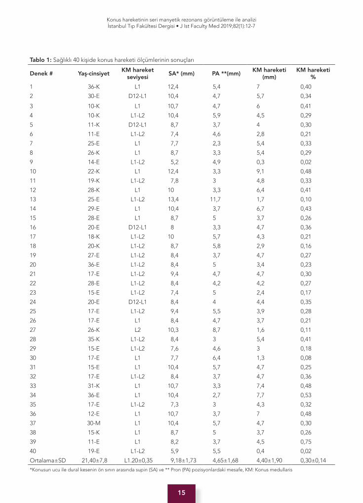
15
Konus hareketinin seri manyetik rezonans görüntüleme ile analiziİstanbul Tıp Fakültesi Dergisi • J Ist Faculty Med 2019;82(1):12-7
Tablo 1: Sağlıklı 40 kişide konus hareketi ölçümlerinin sonuçları
Denek # Yaş-cinsiyetKM hareket
seviyesiSA* (mm) PA **(mm)
KM hareketi (mm)
KM hareketi %
1 36-K L1 12,4 5,4 7 0,40
2 30-E D12-L1 10,4 4,7 5,7 0,34
3 10-K L1 10,7 4,7 6 0,41
4 10-K L1-L2 10,4 5,9 4,5 0,29
5 11-K D12-L1 8,7 3,7 4 0,30
6 11-E L1-L2 7,4 4,6 2,8 0,21
7 25-E L1 7,7 2,3 5,4 0,33
8 26-K L1 8,7 3,3 5,4 0,29
9 14-E L1-L2 5,2 4,9 0,3 0,02
10 22-K L1 12,4 3,3 9,1 0,48
11 19-K L1-L2 7,8 3 4,8 0,33
12 28-K L1 10 3,3 6,4 0,41
13 25-E L1-L2 13,4 11,7 1,7 0,10
14 29-E L1 10,4 3,7 6,7 0,43
15 28-E L1 8,7 5 3,7 0,26
16 20-E D12-L1 8 3,3 4,7 0,36
17 18-K L1-L2 10 5,7 4,3 0,21
18 20-K L1-L2 8,7 5,8 2,9 0,16
19 27-E L1-L2 8,4 3,7 4,7 0,27
20 36-E L1-L2 8,4 5 3,4 0,23
21 17-E L1-L2 9,4 4,7 4,7 0,30
22 28-E L1-L2 8,4 4,2 4,2 0,27
23 15-E L1-L2 7,4 5 2,4 0,17
24 20-E D12-L1 8,4 4 4,4 0,35
25 17-E L1-L2 9,4 5,5 3,9 0,28
26 17-E L1 8,4 4,7 3,7 0,21
27 26-K L2 10,3 8,7 1,6 0,11
28 35-K L1-L2 8,4 3 5,4 0,41
29 15-E L1-L2 7,6 4,6 3 0,18
30 17-E L1 7,7 6,4 1,3 0,08
31 15-E L1 10,4 5,7 4,7 0,25
32 17-E L1-L2 8,4 3,7 4,7 0,36
33 31-K L1 10,7 3,3 7,4 0,48
34 36-E L1 10,4 2,7 7,7 0,53
35 17-E L1-L2 7,3 3 4,3 0,32
36 12-E L1 10,7 3,7 7 0,48
37 30-M L1 10,4 5,7 4,7 0,30
38 15-K L1 8,7 5 3,7 0,26
39 11-E L1 8,2 3,7 4,5 0,75
40 19-E L1-L2 5,9 5,5 0,4 0,02
Ortalama±SD 21,40±7,8 L1.20±0,35 9,18±1,73 4,65±1,68 4,40±1,90 0,30±0,14
*Konusun ucu ile dural kesenin ön sınırı arasında supin (SA) ve ** Pron (PA) pozisyonlardaki mesafe, KM: Konus medullaris

16
Konus hareketinin seri manyetik rezonans görüntüleme ile analiziİstanbul Tıp Fakültesi Dergisi • J Ist Faculty Med 2019;82(1):12-7
çalışmamızda denekler supin pozisyondan pron pozisyona döndürüldüğünde KM ve filum terminalenin bütün çalışma grubunda birlikte hareket ettiğini gözlemledik. Bu bulgu, KM ve filum terminalenin normal anatomisi ve fizyolojisiyle uyumludur, dolayısıyla KM ve FT subaraknoid boşlukta ser-bestçe yüzmektedir. Warder ve Oakes yaklaşık 12 yıllık bir periyod boyunca GOS’lu 73 hastada supin MRG’de %18 oranında normal yerleşimli konus bildirmişlerdir. Bundan dolayı, bu özel vakalarda KM ve filum terminalenin hare-ketinin araştırılması GOS’nun teşhisinde anahtar rol oyna-yabilir (9, 10). Her ne kadar pron pozisyonda ek çekimin 10 dakikalık bir ek süreye ve muhtemelen ilave maliyete sebep olacağı düşünülse de potansiyel faydaları bu göre-celi dezavantajlardan çok daha fazla görünmektedir. Wit-kamp ve ark. klinik olarak GOS olduğundan şüphelenilen 25 hastada pron MRG sekanslarında konus hareketinde anlamlı bir değişiklik saptamadıklarını belirtmiş ve yazarlar GOS şüphesi bulunan fakat rutin supin MRG’nin konus se-viyesi ve/veya intradural lipom hakkında herhengi bir pa-tolojiye işaret etmediği hastalarda pron MRG’nin ek veriler sağlayabileceğini belirtmişlerdir (5). Bauer ve ark. Rhesus (Macaca mulatta) maymunlarında omurganın fleksiyonu ve ekstansiyonu sırasında konus hareketini gözlemlemek için MR görüntülemeyi kullanarak bir çalışma yapmışlardır (2). Yazarlar fleksiyon veya ekstansiyonda konusun nötral pozisyonundan herhangi bir hareketlenme olmadığını gözlemlemiş ve insan dışı primatların omurgasındaki flek-siyon ve ekstansiyonun konus medullarisin pozisyonunda herhangi bir değişikliğe sebep olmadığı sonucuna ulaş-mışlardır. Aynı şekilde Bauer ve ark. beş tane taze yetiş-kin kadavrada T12-L3 laminektomi yapıp konus ve kauda equinayı ortaya koymuş ve bütün deneklerde pron pozis-yonda konusun T12 ve L1 seviyeleri arasında sonlandığını gözlemlemişlerdir (3). Yazarlar ayrıca fleksiyon esnasında konus pozisyonunun stabil kaldığını bildirmişlerdir. Dolayı-sıyla supin-pron pozisyon değişimleri temel alınarak konus hareketinin ölçülmesi daha güvenilirdir.
SONUÇ
Çalışmamızda, konus hareketinin aralığı literatürdeki en geniş sağlıklı popülasyonda tespit edilmiş olup, bu bulgu-lar klinik olarak GOS şüphesi bulunan normal yerleşimli ko-nus vakalarında teşhis koyucu bir kriter olarak kullanılabilir.
Bilgilendirilmiş Onam: Katılımcılardan bilgilendirilmiş onam alınmıştır.
Hakem Değerlendirmesi: Dış bağımsız.
Yazar Katkıları: Çalışma Konsepti/Tasarım- C.G., U.Y., H.C., A.G.Y., B.G., A.A., M.B., U.Ö., A.S., T.K., S.S.; Veri Toplama- A.G.Y., U.Ö., A.A.; Veri Analizi/Yorumlama- U.Y., H.C., A.G.Y.; Yazı Taslağı- A.G.Y., U.Ö., A.S., T.K., S.S.; İçeriğin Eleştirel İncelemesi- A.S., T.K., S.S.; Son Onay ve Sorumluluk- C.G., U.Y., H.C., A.G.Y., B.G., A.A., M.B., U.Ö., A.S., T.K., S.S.; Malzeme ve Teknik Destek- C.G., B.G., M.B., A.S., T.K., S.S.
Çıkar Çatışması: Yazarlar çıkar çatışması beyan etmemişlerdir.
Finansal Destek: Yazarlar finansal destek beyan etmemişlerdir.
Informed Consent: Written consent was obtained from the par-ticipants.
Peer Review: Externally peer-reviewed.
Author Contributions: Conception/Design of Study- C.G., U.Y., H.C., A.G.Y., B.G., A.A., M.B., U.Ö., A.S., T.K., S.S.; Data Acquisi-tion- A.G.Y., U.Ö., A.A.; Data Analysis/Interpretation- U.Y., H.C., A.G.Y.; Drafting Manuscript- A.G.Y., U.Ö., A.S., T.K., S.S.; Critical Revision of Manuscript- A.S., T.K., S.S.; Final Approval and Ac-countability- C.G., U.Y., H.C., A.G.Y., B.G., A.A., M.B., U.Ö., A.S., T.K., S.S.; Technical or Material Support- C.G., B.G., M.B., A.S., T.K., S.S.
Tablo 2: Cinsiyete göre konus medullaris (KM) hareketi
İnsan Denekler Cinsiyet # Ortalama ST Mann-Whitney U p
KM’nin HareketiKadın 14 5,18 1,94
122,5 0,091Erkek 26 4,03 1,82
KM’nin Hareket %Kadın 14 0,33 0,11
141 0,245Erkek 26 0,29 0,16
Tablo 3: Yaşa göre konus medullaris (KM) hareketi
İnsan Denekler Yaş # Ortalama SD Mann-Whitney U p
KM’nin Hareketi0-18 18 3,88 1,55
127 0,053 >19 22 4,88 2,10
KM’nin Hareket %
0-18 18 0,29 0,16151 0,201
>19 22 0,32 0,13

17
Konus hareketinin seri manyetik rezonans görüntüleme ile analiziİstanbul Tıp Fakültesi Dergisi • J Ist Faculty Med 2019;82(1):12-7
Conflict of Interest: Authors declared no conflict of interest.
Financial Disclosure: Authors declared no financial support.
KAYNAKLAR
1. Tubbs RS, Salter G, Grabb PA, Oakes WJ. The denticulate ligament: anatomy and functional significance. J Neurosurg 2001;94(2):271-5. [CrossRef]
2. Bauer DF, Tubbs RS, Chambers MR. Study of the effects of flexion on the position of the conus medullaris: follow-up study using MR imaging in non-human primates. Childs Nerv Syst 2009;25(8):977-9. [CrossRef]
3. Bauer DF, Shoja MM, Loukas M, Oakes WJ, Tubbs RS. Study of the effects of flexion on the position of the conus medullaris. Childs Nerv Syst 2008;24(9):1043-5. [CrossRef]
4. Fettes PD, Leslie K, McNabb S, Smith PJ. Effect of spinal flexion on the conus medullaris: a case series using magnetic resonance imaging. Anaesthesia 2006;61(6):521-3. [CrossRef]
5. Witkamp TD, Vandertop WP, Beek FJ, Notermans NC, Gooskens RH, van Waes PF. Medullary cone movement in subjects with a normal spinal cord and in patients with a tethered spinal cord. Radiology 2001;220(1):208-12. [CrossRef]
6. Naidich TP, Radkowski MA, Britton J. Real-time sonographic display of caudal spinal anomalies. Neuroradiology 1986;28(5-6):512-27. [CrossRef]
7. Schumacher R, Kroll B, Schwarz M, Ermert JA. M-mode sonography of the caudal spinal cord in patients with meningomyelocele. Work in progress. Radiology 1992;184(1):263-5. [CrossRef]
8. Yamamura A, Niwa J, Hashi K, Nakamura T. [Tethered cord syndrome of adult onset: report of a case and a review of the literature]. No Shinkei Geka 1989;17(1):69-73. (Article in Japanese).
9. Warder DE, Oakes WJ. Tethered cord syndrome: the low-lying and normally positioned conus. Neurosurgery 1994;34(4):597-600. [CrossRef]
10. Warder DE, Oakes WJ. Tethered cord syndrome and the conus in a normal position. Neurosurgery 1993;33(3):374-8.
18
Koroner arter hastalığında serum ürik asit düzeyleri İstanbul Tıp Fakültesi Dergisi • J Ist Faculty Med 2019;82(1):18-23
İletişim kurulacak yazar/Corresponding author: [email protected]
Geliş tarihi/Received Date: 05.01.2019 • Kabul tarihi/Accepted Date: 04.03.2019
©Telif Hakkı 2019 J Ist Faculty Med - Makale metnine jmed.istanbul.edu.tr web sayfasından ulaşılabilir.©Copyright 2019 by J Ist Faculty Med - Available online at jmed.istanbul.edu.tr
ARAŞTIRMA / RESEARCH DOI: 10.26650/IUITFD.2019.0002
İst Tıp Fak Derg 2019 / J Ist Faculty Med 2019
SERUM ÜRİK ASİT DÜZEYİNİN KARARLI KORONER ARTER HASTALIĞININ CİDDİYETİ İLE İLİŞKİSİ
THE RELATIONSHIP BETWEEN SERUM URIC ACID LEVEL AND SEVERITY OF STABLE CORONARY ARTERY DISEASE
Samim EMET1
1İstanbul Üniversitesi, İstanbul Tıp Fakültesi, Kardiyoloji Anabilim Dalı, İstanbul, Türkiye
ORCID IDs of the author: S.E. 0000-0002-2806-4335
Cite this article as: Emet S. The relationship between serum uric acid level and severity of stable coronary artery disease. J Ist Faculty Med 2019;82(1):18-23. doi: 10.26650/IUITFD.2019.0002
ÖZETAmaç: İleri yaş, diyabet, hipertansiyon, insulin direnci, hipertrig-liseridemi ve metobolik sendrom gibi kardiyovasküler hastalık risk faktörlerinin yüksek serum ürik asit düzeyleri ile ilişkili oldu-ğu bilinmektedir. Çalışmamızın amacı bilinen kararlı iskemik kalp hastalığı nedeniyle koroner anjiyografi yapılan hastalarda serum ürik asit düzeyleri ile koroner arter hastalığının yaygınlığını karşı-laştırmaktır.
Gereç ve Yöntem: Kararlı anginası bulunan ve objektif iskemisi gösterilerek koroner anjiyografisi yapılan 76 hasta yazılı ve sözlü onamları alınarak çalışmaya dahil edildi. Tüm hastalardan rutin biyokimyasal tetkik için anjiyografi öncesi kan alındı. Bağımsız gözlemciler tarafından Gensini skorları hesaplandı. Verilerin ista-tistiksel analizinde IBM SPSS istatistik versiyon 21 paket progra-mı kullanıldı. Tüm testlerde istatistiksel önem düzeyi 0,05 olarak alındı.
Bulgular: Hastaların yaş ortalaması 62,2±8,93, erkek cinsiyet ora-nı %86 idi. Serum ürik asit düzeyi ortalaması 6,58±1,76 mg/dl, Gensini skoru ortalaması ise 73,8±28,7 idi. Serum nötrofil, lenfo-sit, hs-CRP ve ürik asit düzeyleri gibi inflamatuvar parametreler ile Gensini skorunun korelasyonuna bakıldığında ise sadece serum ürik asit düzeyi Gensini skoru ile istatistiksel anlamlı korelasyon göstermekteydi (r:0,44, p<0,001).
Sonuç: Serum ürik asit seviyeleri kararlı iskemik kalp hastalığının yaygınlığını ve ciddiyetini göstermede başarılı bir belirteç olabilir.
Anahtar Kelimeler: Ürik asit, Gensini skoru, koroner arter has-talığı
ABSTRACTObjective: Cardiovascular disease risk factors are known to be associated with elevated serum uric acid levels. The aim of our study was to evaluate the relationship between the severity of coronary artery disease and serum uric acid levels in patients un-dergoing coronary angiography for known stable ischemic heart disease.
Material and Method: A total of 76 patients who had stable an-gina and underwent coronary angiography with objective isch-emia were consecutively included in the study. Blood samples were collected from all patients prior to angiography for routine biochemical analysis. Gensini scores were calculated by inde-pendent observers. In the statistical analysis of the data, IBM SPSS statistical version 21 package program was used. Statistical significance level was taken as 0.05 in all tests.
Results: The mean age of the patients was 62.2±8.93 years and the male gender was 86% of the study group. The mean serum uric acid level was 6.58±1.76 mg/dl, the mean Gensini score was 73.8±28.7. When the correlation of inflammatory parameters such as serum neutrophil, lymphocyte, hs-CRP and uric acid lev-els with Gensini score was examined, only serum uric acid level was statistically correlated with Gensini score (r:0.44, p<0.001).
Conclusion: Serum uric acid levels may be a successful predictor of the widespread and severity of stable ischemic heart disease.
Keywords: Uric acid, Gensini scores, coronary artery disease
19
Koroner arter hastalığında serum ürik asit düzeyleri İstanbul Tıp Fakültesi Dergisi • J Ist Faculty Med 2019;82(1):18-23
GİRİŞ
İleri yaş, diyabet, hipertansiyon, insulin direnci, hipert-rigliseridemi ve metabolik sendrom gibi kardiyovasküler hastalık risk faktörlerinin yüksek serum ürik asit düzeyleri ile ilişkili olduğu bilinmektedir (1). Yine yüksek serum ürik asit düzeyleri bilinen kardiyovasküler hastalığı bulunan ki-şilerde kötü prognoz ile ilişkilidir (2).
Ürik asit purin metbolizmasının son ürünüdür ve ksantin-den, ksantin oksidoredüktaz enzimi ile ürik asit oluşmak-tadır (3).
Daha önceden yapılmış çalışmalarda yüksek serum ürik asit seviyelerinde endotel disfonksiyonunun, yüksek oksi-datif stresin, serbest radikal oluşumunun ve trombüs for-masyonunun daha kolay geliştiği ve tüm bunların sonu-cunda da aterosklerozun daha sık gözlendiği izlenmiştir (4-6).
Tüm bu bilgilere rağmen ürik asit henüz bilinen kardi-yovasküler hastalık risk faktörlerinden sayılmamakta ve asemptomatik, yüksek serum ürik asit düzeyi bulunan hastaların tedavisi hala tartışmalı olarak netlik gösterme-mektedir.
Çalışmamızın amacı bilinen kararlı iskemik kalp hastalığı nedeniyle koroner anjiyografi yapılan hastalarda serum ürik asit düzeyleri ile koroner arter hastalığının yaygınlığı-nı karşılaştırmaktır.
GEREÇ VE YÖNTEM
Hasta popülasyonu seçimiÇalışmaya İstanbul Tıp Fakültesi Kardiyoloji Anabilim Da-lı’nda 2013-2014 tarihleri arasında kararlı anginası bulu-nan ve objektif iskemisi gösterilerek koroner anjiyografisi yapılan ve aşağıda belirtilen dışlama kriterleri saptanma-yan 76 hasta alındı. Tüm hastaların öyküleri alınarak fizik muayeneleri yapıldı. Sigara, alkol kullanımı ve ailede kalp hastalığı öyküsü gibi risk faktörleri kaydedildi. Hastalar angina pektoris süresi, miyokard enfarktüsü öyküsü, kul-landıkları ilaçlar açısından sorgulandı. Hastaların sistolik ve diyastolik kan basınçları ölçüldü ve kaydedildi. Hasta-ların elektrokardiyografileri çekildi ve ekokardiyografileri yapıldı.
Sistolik kan basıncı 140 mmHg’nın, diyastolik kan basıncı 90 mmHg’nın üzerinde olanlar ya da antihipertansif kulla-nım hikâyesi olanlar hipertansif olarak kabul edildi. Açlık LDL seviyesi 100 mg/dl’nin üzerinde olanlar ya da statin kullanım öyküsü olanlar hiperkolesterolemik, açlık trigli-serid düzeyi 150 mg/dl’nin üzerinde olanlar ya da antili-pidemik ilaç kullanım öyküsü olanlar hipertrigliseridemik olarak belirlendi. Açlık kan şekeri 126 mg/dl’nin üzerinde olanlar ile bilinen diabetes mellitus tanısı nedeniyle teda-vi görmekte olanlar diyabetik olarak kabul edildi. Sigara kullanımı; sigara içenler ve hiç içmemiş olanlar olarak be-
lirlendi. Tüm hastalar yazılı izinleri alındıktan sonra çalış-maya dahil edildi.
Çalışmaya dahil edilme kriterleri 18 yaşından büyük , kararlı angina nedeniyle koroner an-jiyografisi yapılacak olan hastaları kapsamaktadır.
Çalışmadan dışlama kriterleri• Miyokardiyal iskeminin koroner arter hastalığı (KAH)
dışı bir nedene bağlı olması (ör: ciddi anemi, hipoksi, tirotoksikoz, kokain, ciddi valvuler hastalık, hipotansi-yon, aritmi gibi)
• Geçirilmiş koroner stent implantasyonu veya by-pass cerrahisi olan hastalar,
• Kronik böbrek yetersizliği bulunan hastalar (MDRD4 veya Cockcroft-Gault ile 60 ml/dk.1,73 m2 altında ve ya diyaliz hastaları)
• Maligniteler ve kronik/akut inflamatuar hastalığı olan hastalar
Koroner anjiyografi ve Gensini skoru hesaplamasıBütün hastalara Judkins tekniği ile 6F diyagnostik kateter kullanılarak sağ veya sol femoral arter yolu ile selektif ko-roner anjiyografi yapıldı. Tüm hastaların ölçümleri koroner lezyonun en iyi görüldüğü ve lümeni en fazla daralttığı kabul edilen pozisyonda, diyastol sonunda yapıldı. Koro-ner arter hastalığının şiddeti, modifiye Gensini skorlama sistemi (7) kullanılarak iki deneyimli bağımsız gözlemci ta-rafından değerlendirildi. Bu gözlemciler hem hastanın kli-nik özelliklerine hem de ürik asit seviyelerinden haberdar değildi. Gensini skoru, her bir koroner darlığa luminal da-ralma derecesine ve anatomik yerleşim önemine göre bir puan verilerek hesaplandı. Lümen çapındaki azalma, eş merkezli lezyonların ve eksantrik plakların röntgenografik görünümü değerlendirildi. Lümen çapındaki azalma yüz-de olarak 25, 50, 75, 90, 99 azalması ve tam tıkanıklıkların sırasıyla 1, 2, 4, 8, 16 ve 32 Gensini skorları ile bu segment tarafından sağlanan miyokardiyal alanın fonksiyonel öne-mi temelinde her ana vasküler segmente bir çarpan tahsis edilmiştir. Buna göre çarpanlar sol ana koroner arter için 5, sol ön inen (LAD) koroner arterin proksimal segmenti için 2,5, sirkumfleks arter proksimal için 2,5; sirkumfleks arter orta segmenti ve LAD orta segmenti için 1,5; sağ koroner arter için 1,0; LAD distal segmenti, posterolate-ral arter ve geniş marjinal arter, ve diğer segmentler için 0,5 olarak belirlenmiştir. Analizde 2 gözlemcinin ortalama skorları kullanılmıştır.
EkokardiyografiÇalışmaya alınan her hastanın ekokardiyografisi yapılarak mekanik miyokard işlevleri incelendi. Ekokardiyografik in-celeme en az 15 dakika dinlenme sonrasında, sol lateral pozisyonda (2-boyutlu, M-mod, Doppler ekokardiyografi) Philips İE33 cihazı ile X5-1 transtorasik prob kullanılarak

20
Koroner arter hastalığında serum ürik asit düzeyleri İstanbul Tıp Fakültesi Dergisi • J Ist Faculty Med 2019;82(1):18-23
parasternal ve apikal pencerelerden yapıldı. Her katılım-cıya Amerikan ve Avrupa Ekokardiyografi Cemiyeti’nin (AEC) kılavuzunda (8) yer alan standart görüntüler ve tek-niklere uyularak ekokardiyografi yapıldı. Bütün görüntüler dijital ortama kaydedildi ve sonra tekrar analiz edildi.
Labaratuvar İncelemesiTüm hastalardan koroner anjiyografinin yapıldığı gün 10 ml periferik kan örneği alınmıştır.
İstatistiksel AnalizVerilerin istatistiksel analizinde IBM SPSS istatistik versi-yon 21 paket programı kullanıldı. Kategorik ölçümler sayı ve yüzde olarak, sürekli ölçümlerse ortalama ve standart sapma (gerekli yerlerde ortanca ve minimum-maksimum) olarak özetlendi. Normal dağılımı test etmek amacıy-la Kolmogorov-Smirnov testi kullanıldı. Mann-Whitney U test veya Student t test’lerinden uygun olan sürekli ölçümleri değerlendirmek için kullanıldı. Pearson ko-relasyon testi Gensini skoru ve ürik asit korelasyonunu saptamak için kullanıldı. Tüm testlerde istatistiksel önem düzeyi 0,05 olarak alındı.
BULGULAR
Hasta grubunun bazal demografik, klinik ve laboratuvar özellikleri Tablo 1’de detaylı olarak gösterilmektedir. Has-taların yaş ortalaması 62,2±8,93, erkek cinsiyet oranı %86 idi. Serum ürik asit düzeyi ortalaması 6,58±1,76 mg/dl, Gensini skoru ortalaması ise 73,8±28,7 idi. Kreatinin düze-yi ve angina süreleri dışında parametreler normal dağılım özelliği göstermekteydi. Serum nötrofil, lenfosit, hs-CRP ve ürik asit düzeyleri gibi inflamatuvar parametreler ile Gensini skorunun korelasyonuna bakıldığında ise sadece serum ürik asit düzeyi Gensini skoru ile istatistiksel anlamlı korelasyon göstermekteydi (r: 0,44, p 0,001) (Tablo 2, Şekil 1). Serum ürik asit düzeyi; diyabet, sigara kullanımı, hiper-tansiyon, erkek cinsiyet ve dislipidemi gibi diğer aterosk-
Tablo 1: Hasta grubunun bazal demografik, klinik ve laboratuvar özellikleri
Değişkenler Veriler (n=76)
Yaş (yıl) 62,2±8,93
Erkek [n (%)] 65 (86)
Sistolik kan basıncı (mmHg) 128,76±20,9
AP süresi (yıl) 1,93±1,64
Hipertansiyon [n (%)] 49 (65)
Diabetes Mellitus [n (%)] 33 (43)
Dislipidemi [n (%)] 59 (78)
Sigara [n (%)] 54 (71)
LVEF (%) 52,1±8,9
Trigliserid (mg/dl) 168,4±76,5
LDL (mg/dl) 118,4±33,7
HDL (mg/dl) 38,3±11, 4
Total Kolesterol (mg/dl) 195,7±38,5
HbA1C (%) 7,3±1,6
hsCRP (mg/L) 3,53±2,02
Ürik asid (mg/dl) 6,58±1,76
Açlık Glukozu (mg/dl) 121±37
GFR (mL/min/1,73m2) 80±16,9
Nötrofil (x103/mm3) 5611±1652
Lenfosit (x103/mm3) 2509±870
Gensini skoru 73,8±28,7
İlaçlar [n (%)]
• ACEi/ARB 54 (71)
• B Blokerler 48 (63)
• KKB 21 (28)
• Antiagregan 55 (72)
• Statin 50 (66)
Sürekli değişkenler ortlama + Standart sapma. Kategorik veriler yüzde olarak verilmiştir. ACEi, angiotension-converting enzim inhibitörü; AP, angina pektoris; ARB, angiotension II reseptör blokeri; KKB, kalsiyum kanal blokeri; LVEF, sol ventrikül ejeksiyon fraksiyonu; hs-CRP, high sensitif C-reaktif protein; GFR, glomerular filtrasyon oranı
Tablo 2: Serum nötrofil, lenfosit, hs-CRP ve ürik asit düzeyleri ile Gensini skorunun korelasyonu
Lenfosit Ürik asit hs-CRP Nötrofil
Gensini skoru
r -0,14 0,44 -0,16 -0,03
p 0,23 <0,001 0,88 0,75
Şekil 1: Serum ürik asit düzeyleri ile Gensini skoru arasındaki korelasyon grafiği

21
Koroner arter hastalığında serum ürik asit düzeyleri İstanbul Tıp Fakültesi Dergisi • J Ist Faculty Med 2019;82(1):18-23
leroz risk faktörlerinin bulunduğu hasta gruplarında bu-lunmayan hasta gruplarına göre istatistiksel olarak anlamlı fark göstermemiştir (Tablo 3). Lineer regresyon analizinde Gensini skorunu öngörmede serum ürik asit düzeyleri, dis-lipidemi, tip 2 DM, hipertansiyon varlığı, sigara kullanımı, cinsiyet ve sol ventrikül ejeksiyon fraksiyonu gibi paramet-reler karşılaştırılmış olup sadece serum ürik asitleri bağım-sız değişken olarak saptanmıştır (Tablo 4). Serum ürik asit düzeyinin 5,75 mg/dl cut-off değeri yüksek Gensini skoru-nu (≥60) %84 sensitivite ve %40 spesifite ile öngörmekte olup ROC eğrisi Şekil 2’de gösterilmiştir.
TARTIŞMA
Çalışmamızda serum ürik asit düzeyleri ile koroner arter hastalığının yaygınlığı ve ciddiyetini gösteren Gensini skorlaması arasında belirgin korelasyon saptandı. Koro-
ner arter hastalığının varlığını ve ciddiyetini göstermede altın standart yöntem olan koroner anjiyografi sonrası yaptığımız Gensini skorlamasında, skoru yüksek olan has-talarda belirgin serum ürik asit düzeyi yüksekliği mevcut-tu. Gensini skorlaması ve Syntax skorlaması, koroner arter hastalığının yaygınlığını ve prognozu da göstermesi bakı-mından en sık kullanılan iki skorlama sistemidir (7).
Duran M ve arkadaşlarının yaptığı çalışmada 246 diyabeti ve hipertansiyonu olmayan akut koroner sendrom hasta-sında serum ürik asit düzeyleri ile koroner arter hastalı-ğının ciddiyetini gösteren Gensini skorlaması arasında ciddi bir pozitif korelasyon bulunmuştur (9). Yine sadece erkek ve akut koroner sendromlu hastalarda Qureshi AE ve arkadaşlarının yaptığı çalışmada da benzer sonuçlar bulunmuştur (10). Bu çalışmalardaki sonuçlar bizim çalış-mamız ile benzer şekilde çıkması bakımından önemlidir
Tablo 3: Serum ürik asit düzeylerinin diğer risk faktörlerinin bulunma durumunda karşılaştırması
Serum ürik asit düzeyleri (mg/dl) Ortalama±SS p değeri
Diyabetik hastalar 6,67±1,820,46
Non-diyabetik hastalar 6,45±1,69
Sigara içen hastalar 6,7±1,720,84
Sigara içmeyen hastalar 6,27±1,86
Hipertansif hastalar 6,56±1,5960,17
Non-hipertansif hastalar 6,58±2,09
Erkek hastalar 6,51±1,790,82
Kadın hastalar 6,99±1,61
Dislipidemik hastalar 6,57±1,650,22
Dislipidemik olmayan hastalar 6,58±2,16
Tablo 4: Gensini skorunu öngörmede ürik asit düzeylerinin bağımsız değişken olduğunu gösteren lineer regresyon analizi tablosu
Katsayılara
ModelStandardize olmayan
katsayılarStandardize katsayılar t p
B Standart Hata Beta
1
(Sabit) 39,862 23,085 1,727 0,08
Urikasit 7,924 1,719 0,48 4,61 <0,01
Dislipidemi 7,702 7,771 0,11 0,99 0,32
Tip2DM 6,110 6,481 0,10 0,94 0,34
HT -2,964 2,718 -0,11 -1,09 0,27
Sigara -9,594 6,866 -0,15 -1,39 0,16
Cinsiyet -8,212 9,240 -0,10 -0,88 0,37
LVEF -0,155 0,36 -0,04 -0,43 0,66
a. Bağımlı değişken: Gensini
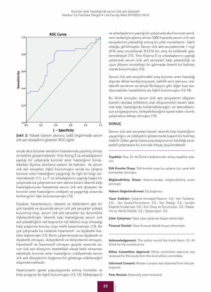
22
Koroner arter hastalığında serum ürik asit düzeyleri İstanbul Tıp Fakültesi Dergisi • J Ist Faculty Med 2019;82(1):18-23
ancak akut koroner sendrom hastalarında yapılmış olması ile farklılık göstermektedir. Yine Xiong Z ve arkadaşlarının yaptığı bir çalışmada koroner arter hastalığının komp-leksitesi (Syntax skorlama sistemi ile bakılan) ile serum ürik asit seviyeleri ilişkili bulunmuştur ancak bu çalışma koroner arter hastalığının yaygınlığı ile ilgili bir bilgi ver-memektedir (11). Lu P. ve arkadaşlarının yaptığı başka bir çalışmada ise çalışmamızın tam aksine kararlı iskemik kalp hastalığıbulunan hastalarda serum ürik asit düzeyleri ile koroner arter hastalığının ciddiyeti ve yaygınlığı arasında herhangi bir ilişki bulunamamıştır (12).
Diyabet, hipertansiyon, obezite ve dislipidemi gibi bir çok hastalık ve durumda serum ürik asit seviyeleri yüksek bulunmuş olup, serum ürik asit seviyeleri bu durumlarla ilişkilendirilmiştir. İskemik kalp hastalığında serum ürik asit yüksekliğinin tek başına bir risk faktörü olup olmadığı hala araştırma konusu olup netlik kazanmamıştır (13). Bir çok çalışmada bu nedenle hipertansif ve diyabetik has-talar dışlanmıştır (12). Bizim çalışmamızda ise diyabetik ve diyabetik olmayan, dislipidemik ve dislipidemik olmayan, hipertansif ve hipertansif olmayan gruplar arasında se-rum ürik asit düzeyinin istatistiksel olarak farklı olmaması sebebiyle koroner arter hastalığının ciddiyetinde serum ürik asit düzeylerinin bağımsız bir gösterge olabileceğini düşündürmektedir.
Hiperürisemi genel populasyonda artmış mortalite ve kötü prognoz ile ilişkili bulunmuştur (13, 14). Ndrepepa G
ve arkadaşlarının yaptığı bir çalışmada akut koroner send-rom nedeniyle işleme alınan 5000 hastada serum ürik asit seviyelerinin yüksekliği artmış bir yıllık mortalitenin ilişkili olduğu görülmüştür. Serum ürik asit seviyelerinde 1 mg/dl’lik artış mortalitede %12’lik bir artış ile birliktelik gös-termekteydi (15). Yine Kojima S ve arkadaşlarının yaptığı çalışmada serum ürik asit seviyeleri kalp yetersizliği ve uzun dönem mortaliteyi ön görmede önemli bir belirteç olarak bulunmuştur (16).
Serum ürik asit seviylerindeki artış koroner arter hastalığı dışında dilate kardiyomiyopati, kalsifik aort stenozu, me-tabolik sendrom ve atriyal fibrilasyon gibi diğer bazı kar-diyovasküler hastalıklarla da ilişkili bulunmuştur (16-18).
Bu klinik sonuçlar, serum ürik asit seviyelerini düşüren ksantin oksidaz inhibitörü olan allopurinolün kararlı iske-mik kalp hastalığında kullanılabileceğini ve aterosklero-zun progresyonunu önleyebileceğine işaret eden olumlu çalışmalara sebep olmuştur (19).
SONUÇ
Serum ürik asit seviyeleri kararlı iskemik kalp hastalığının yaygınlığını ve ciddiyetini göstermede başarılı bir belirteç olabilir. Daha geniş hasta populasyonunun katıldığı pros-pektif çalışmalara bu konuda ihtiyaç duyulmaktadır.
Teşekkür: Doç. Dr. Ali Elitok’a katkılarından dolayı teşekkür ede-rim.
Etik Komite Onayı: Etik komite onayı bu çalışma için, yerel etik komiteden alınmıştır.
Bilgilendirilmiş Onam: Katılımcılardan bilgilendirilmiş onam alınmıştır.
Hakem Değerlendirmesi: Dış bağımsız.
Yazar Katkıları: Çalışma Konsepti/Tasarım- S.E.; Veri Toplama- S.E.; Veri Analizi/Yorumlama- S.E.; Yazı Taslağı- S.E.; İçeriğin Eleştirel İncelemesi- S.E.; Son Onay ve Sorumluluk- S.E.; Malze-me ve Teknik Destek- S.E.; Süpervizyon- S.E.
Çıkar Çatışması: Yazar çıkar çatışması beyan etmemiştir.
Finansal Destek: Yazar finansal destek beyan etmemiştir.
Acknowledgement: The author would like thank Assoc. Dr. Ali Elitok for his contributions.
Ethics Committee Approval: Ethics committee approval was received for this study from the local ethics committee.
Informed Consent: Written consent was obtained from the par-ticipants.
Peer Review: Externally peer-reviewed.
Şekil 2: Yüksek Gensini skorunu (≥60) öngörmede serum ürik asit düzeylerini gösteren ROC eğrisi

23
Koroner arter hastalığında serum ürik asit düzeyleri İstanbul Tıp Fakültesi Dergisi • J Ist Faculty Med 2019;82(1):18-23
Author Contributions: Conception/Design of Study- S.E.; Data Acquisition- S.E.; Data Analysis/Interpretation- S.E.; Drafting Manuscript- S.E.; Critical Revision of Manuscript- S.E.; Final Ap-proval and Accountability- S.E.; Technical or Material Support- S.E.; Supervision- S.E.
Conflict of Interest: Author declared no conflict of interest.
Financial Disclosure: Author declared no financial support.
KAYNAKLAR
1. Pan H, Yan D, Xu M, Li F, Ren M, Zhang J, Wu M.Interaction Between Lactate and Uric Acid is Associated With a Higher Prevalence of Metabolic Syndrome: A Community-Based Study. Exp Clin Endocrinol Diabetes. 2018 Sep 10. [CrossRef]
2. Xia X, He F, Wu X, Peng F, Huang F, Yu X. Relationship between serum uric acid and all-cause and cardiovascular mortality in patients treated with peritoneal dialysis. Am J Kidney Dis 2014;64(2):257-64. [CrossRef]
3. Gertler MM, Garn SM, Levine SA. Serum uric acid in relation to age and physique in health and in coronary heart disease. Ann Intern Med 1951;34:1421-31. [CrossRef]
4. Diaz MN, Frei B, Vita JA, Keaney JF. Antioxidants and atherosclerosis heart disease. N Engl J Med1997;337(6):408-16. [CrossRef]
5. Saito Y, Kitahara H, Nakayama T, Fujimoto Y, Kobayashi Y. Relation of Elevated Serum Uric Acid Level to Endothelial Dysfunction in Patients with Acute Coronary Syndrome. J Atheroscler Thromb. 2018 Sep 13. [CrossRef]
6. Kim SY, Guevara JP, Kim KM, Choi HK, Heitjan DF, Albert DA. Hyperuricemia and coronary heart disease: a systematic review and meta-analysis. Arthritis Care Res 2010;62(2):170-80. [CrossRef]
7. Gensini GG. A more meaningful scoring system for determining the severity of coronary heart disease. Am J Cardiol 1983;51(3):606. [CrossRef]
8. Roberto ML, Luigi PB, Victor M, Jonathan A, Anderson A, Laura E, et al. Recommendations for Cardiac Chamber Quantification by Echocardiography in Adults: An Update from the American Society of Echocardiography and the European Association of Cardiovascular Imaging. Eur Heart J Cardiovasc Imaging 2016;17(4):412. [CrossRef]
9. Duran M, Kalay N, Akpek M, Orscelik O, Elcik D, Ocak A. High Levels of Serum Uric Acid predict severity of ischemic heart disease in Patients With Acute Coronary Syndrome. Angiology 2012;63(6):448-52. [CrossRef]
10. Qureshi AE, Hameed S, Noeman A. Relationship of serum uric acid level and angiographic severity of coronary artery disease in male patients with acute coronary syndrome. Pak J Med Sci 2013;29(5):1137-41. [CrossRef]
11. Xiong Z, Zhu C, Qian X, Zhu J, Wu Z, Chen L. Predictors of clinical SYNTAX score in coronary artery disease: serum uric acid, smoking, and Framingham risk strati cation. J Invasive Cardiol 2011;23(12):501-4.
12. Lu P, Hu D, Lu J, Wang W, Chen B. The association between uric acid and coronary heart disease. Zhonghua Nei Ke Za Zhi 2002;41(8):526-9.
13. Puig JG, Martínez MA. Hyperuricemia, gout and the metabolic syndrome. Curr Opin Rheumatol 2008;20:187-91. [CrossRef]
14. Freedman DS, Williamson DF, Gunter EW, Byers T. Relation of serum uric acid to mortality and ischemic heart disease. The NHANES I Epidemiologic Follow-up Study. Am J Epidemiol 1995;141(7):637-44. [CrossRef]
15. Ndrepepa G, Braun S, King L, Fusaro M, Tada T, Cassese S. Uric acid and prognosis in angiography-proven coronary artery disease. Eur J Clin Invest 2013;43(3):256-66. [CrossRef]
16. Kojima S, Sakamoto T, Ishihara M, Kimura K, Miyazaki S, Yamagishi M. Prognostic usefulness of serum uric acid after acute myocardial infarction (the Japanese Acute Coronary Syndrome Study). Am J Cardiol 2005;96(4):489-95. [CrossRef]
17. Tamariz L, Agarwal S, Soliman EZ, Chamberlain AM, Prineas R, Folsom AR. Association of serum uric acid with incident atrial brillation (from the Atherosclerosis Risk in Communities [ARIC] study). Am J Cardiol 2011;108(9):1272-6. [CrossRef]
18. Kwon CH, Lee SH, Lee JY, Ryu S, Sung KC. Uric Acid and Risk of Atrial Fibrillation in the Korean General Population. Circ J 2018 25;82(11):2728-35.
19. Noman A, Ang DS, Ogston S. Effect of high-dose allopurinol on exercise in patients with chronic stable angina: a randomised, placebo controlled crossover trial. Lancet 2010;375:2161-7. [CrossRef]

24
Kırılganlık ve kanser taraması İstanbul Tıp Fakültesi Dergisi • J Ist Faculty Med 2019;82(1):24-8
İletişim kurulacak yazar/Corresponding author: [email protected]
Geliş tarihi/Received Date: 17.11.2018 • Kabul tarihi/Accepted Date: 29.01.2019
©Telif Hakkı 2019 J Ist Faculty Med - Makale metnine jmed.istanbul.edu.tr web sayfasından ulaşılabilir.©Copyright 2019 by J Ist Faculty Med - Available online at jmed.istanbul.edu.tr
ARAŞTIRMA / RESEARCH DOI: 10.26650/IUITFD.2018.0030
İst Tıp Fak Derg 2019 / J Ist Faculty Med 2019
YAŞLILARDA KIRILGANLIK VE KANSER TARANMA ORANLARI
FRAILTY AND CANCER SCREENING RATES IN OLDER ADULTS
Birkan İLHAN1 , Oğuz Kağan BAKKALOĞLU2
1Dr. Ersin Arslan Eğitim ve Araştırma Hastanesi, İç Hastalıkları Kliniği, Geriatri Bölümü, Gaziantep, Türkiye2Dr. Ersin Arslan Eğitim ve Araştırma Hastanesi, İç Hastalıkları Kliniği, Gaziantep, Türkiye
ORCID IDs of the authors: B.İ. 0000-0001-6039-5866; O.K.B. 0000-0001-8661-5791
Cite this article as: İlhan B, Bakkaloğlu OK. Frailty and cancer screening rates in older adults. J Ist Faculty Med 2019;82(1):24-8. doi: 10.26650/IUITFD.2018.0030
ÖZETAmaç: Son zamanlarda yaşlılarda kanser taramalarına kişinin kı-rılganlık düzeyi veya beklenen yaşam süresine göre karar veril-mesi önerilmektedir. Yaşlılardaki kanser tarama oranları ile ilgili veriler kısıtlıdır. Bu çalışmada bir geriatri polikliniğine başvuran yaşlılarda kırılganlık durumları ile birlikte kolon, meme, prostat ve akciğer kanser taraması oranlarının bildirilmesi amaçlanmıştır.
Gereç ve Yöntem: Eylül 2017-Mayıs 2018 tarihleri arasında bir geriatri polikliniğine ilk kez başvuran 60 yaş ve üzeri hastaların verileri retrospektif olarak incelendi. Kapsamlı geriatrik değerlen-dirme sırasında sorgulanmış olan kolon, meme, prostat ve risk gruplarında akciğer kanser taramalarının yapılıp yapılmadığı bil-gisi kaydedildi. Kırılganlık durumlarını FRAIL ölçeği ile değerlen-dirilerek hastalar kırılgan olan veya olmayan olarak sınıflandırıldı.
Bulgular: Yüz elli yedi (%73) kadın, 57 (%27) erkek hastanın ve-rileri incelendi. Yaş ortalaması 76±7,3 yıl idi. Kırılganlık sorgusu yapılmış olan 203 hastanın 17’si (%8,4) fit; 55’i (%27,1) kırılganlık öncesi dönemde; 131’i (%64,5) kırılgan yaşlı idi. Kırılgan olmayan yaşlılarda (kırılganlık öncesi dönemdeki veya fit) kolon, meme ve prostat kanseri taranma sıklığı sırasıyla %8,3, %10,4 ve %33,3 idi.Kırılgan grupta kolon, meme ve prostat kanseri taranma sıklığı sırasıyla %5,3, %4 ve %33,3 idi. Akciğer kanseri taraması yapılmış olan hasta yoktu.
Sonuç: Çalışmamızda kolon ve meme kanser taranma sıklığının kırılgan olan ve olmayan her iki grupta da düşük olduğunu; bu-nunla birlikte prostat kanserinin rutinde taranması önerilmeme-sine rağmen en yüksek taranma sıklığının bu kanser grubunda olduğunu gördük. Akciğer kanseri taraması yapılmış olan hasta yoktu.
Anahtar Kelimeler: Yaşlılık, kanser, kırılganlık
ABSTRACTObjective: It has been suggested that the decision of when to implement cancer screening should be based on the frailty sta-tus or life expectancy in older adults. There is a limited number of studies reporting cancer screening prevalence in older adults. The aim of this study was to examine the patterns of colorectal, breast, prostate, and lung cancer screening in geriatric outpa-tients with different frailty statuses.
Material and Methods: Data from our geriatric outpatient clinic from September 2017 to May 2018 was examined retrospectively and included first time admission outpatients aged 60 years or older. The screening situations for colorectal, breast, prostate, and lung cancer (in at risk groups) were obtained from analysis of comprehensive geriatric assessment forms. Frailty was assessed using the FRAIL-scale. Participants were subsequently classified as frail or not frail.
Results: One hundred and fifty seven participants (73%) were fe-male and 57 (27%) were male with a mean age of 76±7.3 years. Of the 203 participants who underwent frailty screening 17 (8.4%) were fit, 55 (27.1%) were prefrail, 131 (64.5%) were frail. Of the participants without frailty, 8.3%, 10.4% ve 33.3% had received recent cancer screening for colorectal, breast, or prostate can-cer, respectively. Of the frail participants, 5.3%, 4% and 33.3% had received recent cancer screening for colorectal, breast, or prostate cancer respectively. No participants had been screened for lung cancer.
Conclusion: We observed that the prevalences of colorectal and breast cancer screening are lower in all groups. However the prevalence of prostate cancer screening was higher even though it is not routinely recommended for screening. Nobody was screened for lung cancer.
Keywords: Older adults, cancer, frailty
25
Kırılganlık ve kanser taraması İstanbul Tıp Fakültesi Dergisi • J Ist Faculty Med 2019;82(1):24-8
GİRİŞ
Ülkemizde yaşlı nüfus giderek artış göstermektedir. Tür-kiye İstatistik Kurumu’nun 2017 yılına ait verilerine göre 65 yaş üstü bireyler son beş yılda %17 artarak nüfusun %8,5’ini oluşturmuştur (1). Yaşlı nüfusun artışı kronik has-talıklar ve maligniteler gibi sorunları da beraberinde ge-tirmektedir. Yaşlılarda en önemli koruma stratejilerinden biri, erken tanı için rutin taramaların yapılmasıdır. T.C. Sağlık Bakanlığı ve Amerika Birleşik Devletleri Koruyucu Hizmetler Grubu (USPSTF) yaşlılarda bazı kanser tarama-larının yapılmasını önermektedir. Bunlar meme kanseri, kolon kanseri ve bazı özel gruplarda akciğer ve prostat kanserleridir.
Meme kanserli hastaların %50’den fazlası 65 yaş üze-rinde, %30’u 70 yaş üzerindedir (2). Hastalar genellikle geç tanı almakta olup, 65 yaş üzeri meme kanseri olan hastaların %48’inin tanı anında metastatik olduğu bildi-rilmiştir (3). Tarama ile mortalitede yaklaşık %30 azalma olduğu bildirilmiştir. Bu çalışmalar 70-74 yaşına kadar olan hastaları kapsamaktadır (4). Amerika Kanser Cemi-yeti, taramaya kişi sağlıklı olduğu sürece ve 10 yıldan uzun yaşam beklentisi olduğu sürece meme kanseri ta-ramasına devam edilmesini önermektedir (5). Ülkemiz Sağlık Bakanlığı’nca 40-69 yaş arası kadınlarda iki yılda bir mamografi ile tarama önerilmektedir. Yetmiş yaş ve üstü kadınlarda, takip gerektiren herhangi bir patoloji yoksa kişinin isteği ve kar-zarar oranı hesaba katılarak tarama planlanmasını önermektedir (6). USPSTF, 50-74 yaş arası kadınlara iki yılda bir mamografi ile tarama önermektedir (Kanıt B) (7).
Kolorektal kanser için ileri yaş bir risk faktörüdür. Kolorek-tal kanser kaynaklı ölümlerin %80’i 55 yaş üzeri kişilerde meydana gelmektedir (8). Taramada kullanılan yöntemler gaitada gizli kan testi, sigmoidoskopi veya kolonoskopi olup tarama yöntemlerinin kolon kanserinden ölümleri 50-70 yaş grubunda önemli ölçüde azalttığı kanıtlanmıştır (Kanıt B) (9). Sağlık bakanlığı ve USPSTF 50-75 yaş arası kişilerde tarama önermektedir (Kanıt A).
Prostat kanserinin erken tanı ve tedavisinin yaşam süresi-ni uzattığına dair yeterli kanıt bulunmamaktadır. Prostat spesifik antijen (PSA) testinin yaygınlaşmasıyla prostat biyopsi sıklığının arttığı ve sessiz gidiş gösteren prostat kanserinin de erken tanı aldığı görülmüştür. Dolayısıyla tüm erkeklerin taranmasının kişilerin “aşırı” tanı ve tedavi almasına yol açarak bunlarla ilişkili zararlı etkilerin artışına sebep olduğu gözükmektedir. Bu nedenle prostat kanse-rinin rutin olarak tüm erkeklerde taranması önerilmemek-tedir. USPSTF hasta ile olası yararları ve zararlarını tartış-maları neticesinde buna karar vermelerini önermektedir (10). Ülkemizde sağlık bakanlığı da rutin olarak prostat kanserinin rutin taranmasını önermemekte, kişisel olarak karar verilmesini önermektedir (11).
USPTF akciğer kanserinin risk gruplarında taranmasını önermektedir. 50-80 yaş grubunda olan 30 paket yıl siga-ra içmiş olan, bıraktığından itibaren 15 yıldan uzun süre geçmemiş olan, sağlıklı kişilere düşük doz bilgisayarlı tomografi taramasını önermektedir. Ülkemizde sağlık ba-kanlığının akciğer kanseri çalıştayı raporuna göre; akciğer kanserinin özellikle erkeklerde görülme sıklığı ve ölümcül bir kanser olması yönü ile toplumsal tarama için uygun bir kanser olduğu, ancak mevcut olanaklarla (alt yapı-in-san kaynakları yetersizliği, zararın daha fazla olabileceği, overdiagnosis, yalancı pozitiflik gibi nedenlerden ötürü) ülke çapında tarama için uygulanabilir olmadığı bildiril-miştir (12).
Geriatrik yaklaşımda genel olarak hastanın tarama ve tedavi planı yapılırken kişinin kronolojik yaşından ziyade genel sağlık durumu ve kırılganlığı gözetilir. Kırılganlık ilerleyen yaşla birlikte nöromüsküler, metabolik ve immün sistemlerde fizyolojik rezervlerin azalmasına bağlı ortaya çıkan güçsüzlük hali ve stres durumlarına adaptasyonun bozulması olarak tanımlanır (13). Kırılganlık kalça kırıkları, engellilik durumu, hastaneye yatış riskinde artış ve mor-talite ile ilişkilidir (13). Yaşlılarda kanser taramasının irde-lendiği bir derlemede taramaların kişinin yaşından ziya-de kırılganlık durumuna göre ve hasta tercihlerine göre yapılmasının daha uygun olacağı vurgulanmıştır (14, 15). Öte yandan taramaya son verilecek olan yaş sınırı da net değildir. Rehberler yine bunun kararının da hastaya göre verilmesi gerektiğini önermektedir (15, 16). Yaşlılarda kan-ser tarama sıklığını kişinin kırılganlık durumu ile beraber bildiren çalışmaya literatürde rastlamadık. Ülkemizde de yaşlılarda kanser tarama sıklığını bildiren herhangi bir ya-yına rastlamadık. Bu çalışmada geriatri polikliniğine ilk kez başvuran yaşlılarda kırılganlık durumları ile birlikte kolon, meme, prostat ve akciğer kanserlerinin taranma sıklığına bakılması amaçlanmıştır.
GEREÇ VE YÖNTEM
Çalışma retrospektif olarak tasarlandı. Eylül 2017-Mayıs 2018 tarihleri arasında bir geriatri polikliniğine ilk kez başvuran 60 yaş ve üzeri hastaların verileri incelendi.De-mografik verileri kaydedildi. Hastaların kapsamlı geriatrik değerlendirme sırasında not alınmış olan, kullanmakta oldukları ilaç sayıları kaydedildi. Öyküde kanser tanısı olan hastalar çalışmaya dahil edilmedi. Polikliniğe ilk baş-vuruları sırasında hastaların kırılganlık değerlendirmesi için kullanılan FRAIL ölçeği (17) verileri kaydedildi. FRAIL ölçeğinde yorgunluk, dayanıklılık, ambulasyon, kilo kaybı ve hastalık sayısı sorgulanır. Ölçek 0-5 arasında puanlan-dırılır. 0: fit, 1-2 puan: kırılganlık öncesi, 3-5 puan: kırılgan yaşlıyı ifade eder. Polikliniğimizde kapsamlı geriatrik de-ğerlendirme sırasında hastalara kolon, meme, prostat ve akciğer kanseri taramaları rutin olarak sorulmaktadır. Kan-ser taramaları sorgusu şu şekillerde yapılmaktadır: Kolon kanseri taraması için son 1 yıl içerisinde gaytada gizli kan
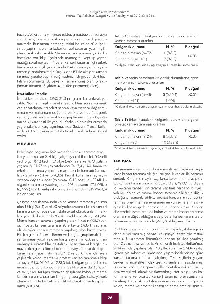
26
Kırılganlık ve kanser taraması İstanbul Tıp Fakültesi Dergisi • J Ist Faculty Med 2019;82(1):24-8
testi ve/veya son 5 yıl içinde rektosigmoidoskopi ve/veya son 10 yıl içinde kolonoskopi yaptırıp yaptırmadığı sorul-maktadır. Bunlardan herhangi birini belirtilen süre içeri-sinde yaptırmış olanlar kolon kanseri taraması yapılmış ki-şiler olarak kabul edildi. Meme kanseri taraması için kadın hastalara son iki yıl içerisinde mamografi yaptırıp yaptır-madığı sorulmaktadır. Prostat kanseri taraması için erkek hastalara son 2 yıl içinde kanda PSA ölçümü yaptırıp yap-tırmadığı sorulmaktadır. Düşük doz BT ile akciğer kanseri taraması yapılıp yapılmadığı sadece risk grubundaki has-talara sorulmakta (30 paket yıl sigara içmiş olan, bıraktı-ğından itibaren 15 yıldan uzun süre geçmemiş olan).
İstatistiksel Analizİstatistiksel analizler SPSS 21,0 programı kullanılarak ya-pıldı. Normal dağılım analizi yapıldıktan sonra numerik veriler ortalama±standart sapma veya ortanca değer mi-nimum ve maksimum değer ile birlikte verildi. Kategorik veriler yüzde şeklide verildi ve gruplar arasındaki kıyasla-maları ki-kare testi ile yapıldı. Kadın ve erkekler arasında yaş ortalaması karşılaştırılmasında Student T-testi kulla-nıldı. <0,05 p değerleri istatistiksel olarak anlamlı kabul edildi.
BULGULAR
Polikliniğe başvuran 562 hastadan kanser tarama sorgu-ları yapılmış olan 214 kişi çalışmaya dahil edildi. Yüz elli yedi olgu (%73) kadın, 57 olgu (%27) ise erkekti. Olguların yaş aralığı 61-97 ve yaş ortalaması 76±7,3 yıl idi. Kadın ve erkekler arasında yaş ortalaması farklı bulunmadı (sırasıy-la 77,2 yıl ve 76,4 yıl; p>0,05). Kronik kullanılan ilaç sayısı ortanca değeri 4 adet (min-max. 0-16 adet) idi. FRAIL kı-rılganlık taraması yapılmış olan 203 hastanın 17’si (%8,4) fit; 55’i (%27,1) kırılganlık öncesi dönemde; 131’i (%64,5) kırılgan yaşlı idi.
Çalışma populasyonunda kolon kanseri taraması yapılmış olan 13 kişi (%6,1) vardı. Cinsiyetler arasında kolon kanseri taranma sıklığı açısından istatistiksel olarak anlamlı fark-lılık yok idi (kadınlarda %6,4, erkeklerde %5,3; p>0,05). Meme kanseri taraması yapılmış olan 9 kadın (%5,7) var-dı. Prostat kanseri taraması 20 erkekte (%35,1) yapılmış idi. Akciğer kanseri taraması yapılmış olan hasta yoktu. Fit, kırılganlık öncesi dönem ve kırılgan gruplardaki kan-ser taraması yapılmış olan hasta sayılarının çok az olması nedeniyle, istatistikler, hastalar kırılgan olan ve kırılgan ol-mayan (kırılganlık öncesi dönemde veya fit) olarak iki gru-ba ayrılarak yapılmıştır (Tablo 1, 2 ve 3). Kırılgan olmayan yaşlılarda kolon, meme ve prostat kanseri taranma sıklığı sırasıyla %8,3, %10,4 ve %33,3 idi. Kırılgan grupta kolon, meme ve prostat kanseri taranma sıklığı sırasıyla %5,3, %4 ve %33,3 idi. Kırılgan olmayan gruplarda kolon ve meme kanseri taranma oranları kırılgan gruba göre daha yüksek olmakla birlikte bu fark istatistiksel olarak anlamlı saptan-madı (p>0,05).
TARTIŞMA
Çalışmamızda geriatri polikliniğine ilk kez başvuran yaşlı-larda kanser taranma sıklığını kırılganlık verileri ile beraber sunduk. Kırılgan olmayan yaşlılarda kolon, meme ve pros-tat kanseri taranma sıklığı sırasıyla %8,3, %10,4 ve %33,3 idi. Akciğer kanseri için tarama yapılmış herhangi bir yaşlı yok idi. Kolon ve meme kanser taranma sıklığının düşük olduğunu; bununla birlikte prostat kanserinin rutinde ta-ranması önerilmemesine rağmen en yüksek taranma sıklı-ğının bu kanser grubunda olduğunu görmekteyiz. Kırılgan dönemdeki hastalarda da kolon ve meme kanser taranma oranlarının düşük olduğunu ve prostat kanser taranma sık-lığının ise yine aynı oranda (%33,3) olduğunu gördük.
Poliklinik oranlarımızı ülkemizde kıyaslayabileceğimiz daha evvel yapılmış benzer çalışmaya literatürde rastla-madık. Uluslararası literatürde benzer şekilde yapılmış olan 2 çalışmaya rastladık. Amerika Birleşik Devletleri’nde 2014 yılında yapılmış olan 10 yıllık süreli ve 27404 yaşlıyı içeren bir kohort çalışmasında yaşam beklentisine göre kanser tarama oranları çalışılmış (18). Kişilerin yaşam beklentisi mortalite index testi kullanılarak hesaplanmış. Bu index sonucuna göre 5 yıllık mortalite riskleri düşük, orta ve yüksek olarak sınıflandırılmış. Her bir grupta ko-lon, meme ve prostat kanseri taranma prevalanslarına bakılmış. Beş yıllık mortalite riskinin düşük olduğu grupta kolon, meme ve prostat kanseri taranma oranları sırasıy-
Tablo 1: Hastaların kırılganlık durumlarına göre kolon kanseri taraması oranları
Kırılganlık durumu N, % P değeri
Kırılgan olmayan (n=72) 6 (%8,3)>0,05
Kırılgan olan (n=131) 7 (%5,3)
*Kırılganlık testi verilerine ulaşılamayan 11 hasta bulunmaktadır.
Tablo 2: Kadın hastaların kırılganlık durumlarına göre meme kanseri taraması oranları
Kırılganlık durumu N, % P değeri
Kırılgan olmayan (n=48) 5 (%10,4) >0,05
Kırılgan (n=101) 4 (%4)
*Kırılganlık testi verilerine ulaşılamayan 8 kadın hasta bulunmaktadır.
Tablo 3: Erkek hastaların kırılganlık durumlarına göre prostat kanseri taraması oranları
Kırılganlık durumu N, % P değeri
Kırılgan olmayan (n=24) 8 (%33,3) >0,05
Kırılgan (n=30) 10 (%33,3)
*Kırılganlık testi verilerine ulaşılamayan 3 erkek hasta bulunmaktadır.

27
Kırılganlık ve kanser taraması İstanbul Tıp Fakültesi Dergisi • J Ist Faculty Med 2019;82(1):24-8
la %48,5, %67,9, %66,9; orta riskli olan grupta sırasıyla %40,1, %44,4 ve %58,5 saptanırken yüksek riskli grupta sı-rasıyla %40,8, %34,2, %51,6 saptanmış. Çalışma sonunda yaşam beklentisi düşük olan grupta dahi taramaların ya-pılma oranının yüksek olması eleştirilmiş. Çünkü ek fayda sağlamayacağı görüşü savunulmuş. Bizim çalışmamızdaki fit, kırılganlık öncesi dönem ve kırılgan gruptaki tüm yaş-lılardaki taranma oranlarını bu çalışmadaki mortalite riski açısından düşük, orta ve yüksek olan grup ile birebir kı-yaslamak her ne kadar uygun olmasa da yine de oranların daha düşük olduğunu görmekteyiz.
Literatürde bildirilmiş olan diğer çalışma ise yine Ameri-ka Birleşik Devletleri’nde 60 yaş ve üzerindeki 42 yaşlıda taramaların incelendiği ve hastaların kanser taramasına devam etmek isteyip istemediklerinin sorgulandığı bir çalışmaya yapılmış (19). Bu çalışmada son 5 yıl içinde ko-lon kanseri taraması yaptıran yaşlıların oranı %58; son 2 yıl içinde meme kanseri taraması yaptırmış olan kadınların oranı %78; son 2 yıl içinde prostat kanseri taraması yaptır-mış olan erkeklerin oranı %33 olarak bildirilmiş. Çalışma-da hastaların kırılganlık durumları hakkında bilgi verilme-miş. Fakat demanslı hastalar çalışmadan dışlanmış. Mini mental test skorları 20’nin üzerindeki hastalar çalışmaya dahil edilmiş. Hastaların %39’unun sosyoekonomik düze-yi yetersiz, kalan %61’i yeterli olarak bildirilmiş. Bizim ça-lışma grubu ile kıyaslandığında Amerika’dan bildirilen bu çalışmada prostat kanseri taranma oranının benzer; kolon kanseri ve meme kanseri taranma oranlarının daha yüksek olduğunu görmekteyiz. Bunun nedenleri arasında çalış-ma populasyonunun genel sağlık durumu ve kırılganlık özelliklerinin farklı olması olabilir. Ayrıca sosyoekonomik düzey farklılığı da olabilir. Polikliniğimize başvuran hasta-lar genel olarak kırsal alandan gelmekte. Çalışmamızda sosyoekonomik düzey ayrıntılı sorgulanmamış olmasına rağmen Amerika’daki çalışma populasyonunun bildiril-miş olan sosyoekonomik yeterlilik oranından (%71) daha düşük olduğunu düşünüyoruz. Düşük sosyoekonomik dü-zey de literatürde (15) düşük taranma oranları ile birlikte bildirilmiş. Çalışmamızda özellikle kırılgan olmayan grup-taki taranma oranlarının düşük olmasının bir diğer olası nedeni de hastaların yaptırmak istememiş olması olabilir. Nitekim yukarıda bahsedilen, Amerika’da 42 kişide yapıl-mış olan çalışmada (19) hastalara yüz yüze görüşmede kanser taramasına devam edip etmedikleri sorulduğunda kimileri taramanın artık kendilerine faydası olmayacağını, olası zararlarının daha fazla olacağını düşünerek taramaya devam etmek istemediklerini belirtmişler. Öte yandan bir grup hasta da hekimleri kendilerine artık kanser taraması-na devam etmenin gerekli olmadığını belirttiğinde bunu duyduklarına üzüldüklerini bildirmişler. Bu konu ülkemiz-de planlanacak başka bir çalışmanın konusu olabilir.
Çalışmamızın bazı olumlu yanları ile birlikte kısıtlılıkları da mevcut. Bildiğimiz kadarıyla bu çalışma ülkemizde yaşlı-larda kanser taraması oranını bildiren ilk çalışma. Ayrıca
çalışmamız; literatürde son zamanlarda gündeme gelen kanser taramalarının yapılmasına kişinin yaşından ziyade genel sağlık durumunun değerlendirilerek karar veril-mesi (14, 15) hususunu göz önünde bulundurarak kan-ser tarama prevalansını “kırılganlık durumları” ile birlikte sunan, bildiğimiz kadarıyla, uluslararası literatürdeki de ilk çalışmadır. Öte yandan çalışmanın retrospektif olma-sı, polikliniğe başvurmuş olan fakat kapsamlı geriatrik değerlendirmeleri yapılmamış olan hastaların verilerinin incelenememiş olması yöntemsel bir kısıtlılık oluşturmak-ta. Çalışma populasyonunda kırılganlık oranının yüksek olduğu görülmektedir. Çalışmanın yapıldığı poliklinik çoğunlukla diğer bölümlerden yönlendirilen daha düş-kün bir hasta profiline hizmet verdiğinden ve yine ret-rospektif dizayndan ötürü tüm hastaların verilerinin in-celenememiş olması nedeniyle kırılganlık daha yüksek oranda saptanmış olabilir. Kanser taranma sonuçlarına kayıtlı herhangi bir yazılı veya dijital ortamda ulaşmanın mümkün olmaması nedeniyle taranma sorgusu hasta be-yanına dayalı olarak yapılmıştır. Bu durum verilerin gü-venilirliği açısından bir kısıtlılık oluşturmaktadır. Kişilerin kırılganlık sorguları polikliniğimize başvurdukları sırada yapılmış olup taramanın yapılmış olduğu dönemdeki kı-rılganlık durumunu tam olarak yansıtmayabilir. Ayrıca ça-lışma populasyonu bir geriatri polikliniğine başvuran 214 yaşlıdan oluşmaktadır, bütün toplumu yansıtmayabilir. Fakat bu çalışma, bu konuda toplumsal bazlı daha büyük bir çalışma için fikir oluşturabilir.
Yaşlılarda kanser taramalarına ne zaman son verilmesi gerekliliğine dair herhangi net bir öneri bulunmamakta-dır. Sonuç olarak çalışmamızda, polikliniğimize başvuran yaşlılarda, hem kırılgan olmayan grupta hem de kırılgan olan grupta kanser taranma oranlarının düşük olduğunu gördük. Prostat kanser taraması rutinde önerilmemesine rağmen diğer iki kanser türlerinden daha fazla tarandığını görmekteyiz.
Bilgilendirilmiş Onam: Retrospektif bir çalışma olduğundan bil-gilendirilmiş onam alınmamıştır.
Hakem Değerlendirmesi: Dış bağımsız.
Yazar Katkıları: Çalışma Konsepti/Tasarım- B.İ.; Veri Toplama- B.İ, O.K.B.; Veri Analizi/Yorumlama- B.İ., O.K.B.; Yazı Taslağı- B.İ.; İçeriğin Eleştirel İncelemesi- B.İ., O.K.B.; Son Onay ve Sorumlu-luk- B.İ., O.K.B.; Malzeme ve Teknik Destek- B.İ., O.K.B.; Süper-vizyon- B.İ.
Çıkar Çatışması: Yazarlar çıkar çatışması beyan etmemişlerdir.
Finansal Destek: Yazarlar finansal destek beyan etmemişlerdir.
Informed Consent: Informed consent was not received due to the retrospective nature of the study.
28
Kırılganlık ve kanser taraması İstanbul Tıp Fakültesi Dergisi • J Ist Faculty Med 2019;82(1):24-8
Peer Review: Externally peer-reviewed.
Author Contributions: Conception/Design of Study- B.İ.; Data Acquisition- B.İ., O.K.B.; Data Analysis/Interpretation- B.İ., O.K.B.; Drafting Manuscript- B.İ.; Critical Revision of Manuscript- B.İ., O.K.B.; Final Approval and Accountability- B.İ., O.K.B.; Tech-nical or Material Support- B.İ., O.K.B.; Supervision- B.İ.
Conflict of Interest: Authors declared no conflict of interest.
Financial Disclosure: Authors declared no financial support.
KAYNAKLAR
1. İstatistiklerle Yaşlılar 2017. TÜİK Haber Bülteni 2018:27595. Available from http://www.tuik.gov.tr/PreHaberBultenleri.do?id=24644
2. Belot A, Grosclaude P, Bossard N, Jougla E, Benhamou E, Delafosse P, et al. Cancer incidence and mortality in France over the period 1980-2005. Rev Epidemiol Sante Publique. 2008;56(3):159-75. [CrossRef]
3. Ries LAG, Harkins D, Krapcho M, Mariotto A, Miller BA, Feuer EJ, et al. SEER cancer statistics review 1975–2014: Introduction. National Cancer Institute. Available from https://seer.cancer.gov/archive/csr/1975_2014/results_merged/sect_01_overview.pdf
4. Nyström L, Rutqvist LE, Wall S, Lindgren A, Lindqvist M, Rydén S, et al. Breast cancer screening with mammography: overview of Swedish randomised trials. Lancet 1993;341(8851):973-8. Erratum in: Lancet 1993 Nov 27;342(8883):1372. [CrossRef]
5. Oeffinger KC, Fontham ET, Etzioni R, Herzig A, Michaelson JS, Shih YC, et al., Breast Cancer Screening for Women at Average Risk: 2015 Guideline Update From the American Cancer Society. JAMA 2015;314(15):1599-614. [CrossRef]
6. Özkan S, Keskinkılıç B, Gültekin M, Karaca A.S, Öztürk C, Boztaş G et al. editors. Meme Kanseri Taramaları; Ulusal Kanser kontrol Planı 2013-2018. T.C. Sağlık Bakanlığı, Türkiye Halk Sağlığı Kurumu; 2014.
7. Breast Cancer Recommendation Statement From the U.S. Preventive Services Task Force. Ann Intern Med 2016;164(6):448.
8. Horner M, Ries L, Krapcho M, Neyman N, Aminou R, Howlader N, et al. SEER cancer statistics review 1975–2006: Introduction. National Cancer Institute. Available from https://seer.cancer.gov/archive/csr/1975_2006/results_merged/sect_01_overview.pdf.
9. Sağlık Bakanlığı Temel Sağlık Hizmetleri Genel Müdürlüğü “Birinci Basamak Sağlık Hizmetlerinde Çalışan Hekimler için Yaşlı Sağlığı Tanı ve Tedavi Rehberi 2010”, 78:(37-46).
10. Grossman DC, Curry SJ, Owens DK, Bibbins-Domingo K, Caughey AB, Davidson KW, et al. Screening for Prostate Cancer: US Preventive Services Task Force Recommendation Statement. JAMA 2018;319(18):1901-13. [CrossRef]
11. Prostat kanseri klinik protokolü, Tarama, Türk Üroloji Derneği ve Sağlık Teknolojisi Değerlendirme Daire Başkanlığı, TC Sağlık Bakanlığı Sağlık Araştırmaları Genel Müdürlüğü, 2017:(6). Available from http://hta.gov.tr/pdf/prostat_ca_20_09_1.pdf
12. Akciğer Kanseri Çalıştayı Sonuç Raporu; T.C. Sağlık Bakanlığı, Türkiye Halk Sağlığı Kurumu. 2016:(13-21). Available from https://hsgm.saglik.gov.tr/depo/birimler/kanser-db/yayinlar/raporlar/Akciger_Kanseri_Calistay_Raporu28042016.pdf
13. Clegg A, Young J, Iliffe S, Rikkert MO, Rockwood K. Frailty in elderly people.Lancet 2013;381(9868):752-62. [CrossRef]
14. Eckstrom E, Feeny DH, Walter LC, Perdue LA, Whitlock EP. Individualizing cancer screening in older adults: a narrative review and framework for future research. J Gen Intern Med 2013;28(2):292-8. [CrossRef]
15. Kotwal AA, Schonberg MA. Cancer Screening in the Elderly: A Review of Breast, Colorectal, Lung, and Prostate Cancer Screening. Cancer J 2017;23(4):246-53. [CrossRef]
16. Walter LC, Lewis CL, Barton MB. Screening for colorectal, breast, and cervical cancer in the elderly: A review of the evidence. Am J Med 2005;118(10):1078-86. [CrossRef]
17. Morley JE, Malmstrom TK, Miller DK. A simple frailty questionnaire (FRAIL) predicts outcomes in middle aged African Americans. J Nutr Health Aging 2012;16(7):601-8. [CrossRef]
18. Royce TJ, Hendrix LH, Stokes WA, Allen IM, Chen RC. Cancer screening rates in individuals with different life expectancies. JAMA Intern Med 2014;174(10):1558-65. [CrossRef]
19. Torke AM, Schwartz PH, Holtz LR, Montz K, Sachs GA. Older adults and forgoing cancer screening: “I think it would be strange”. JAMA Intern Med 2013;173(7):526-31. [CrossRef]
29
Work accidents, occupational diseases and mortality in Turkeyİstanbul Tıp Fakültesi Dergisi • J Ist Faculty Med 2019;82(1):29-39
İletişim kurulacak yazar/Corresponding author: [email protected]
Geliş tarihi/Received Date: 31.05.2018 • Kabul tarihi/Accepted Date: 21.12.2018
©Telif Hakkı 2019 J Ist Faculty Med - Makale metnine jmed.istanbul.edu.tr web sayfasından ulaşılabilir.©Copyright 2019 by J Ist Faculty Med - Available online at jmed.istanbul.edu.tr
ARAŞTIRMA / RESEARCH DOI: 10.26650/IUITFD.428998
İst Tıp Fak Derg 2019 / J Ist Faculty Med 2019
STANDARDIZATION OF THE NUMBERS OF WORK ACCIDENTS, OCCUPATIONAL DISEASES AND MORTALITY RATES ACCORDING TO SOCIAL SECURITY INSTITUTION’S 2010-2015 YEARS DATA BASED UPON CITIES
TÜRKİYE SOSYAL GÜVENLİK KURUMU 2010-2015 YILLARI İŞ KAZASI, MESLEK HASTALIĞI VE MORTALİTE SAYILARININ İLLERE GÖRE STANDARDİZASYONU
Osman Faruk BAYRAMLAR1 , Elif EZİRMİK1 , Halim İŞSEVER1 , Zeynep BAYRAMLAR2 1Istanbul University, Istanbul Faculty of Medicine, Department of Public Health, Istanbul, Turkey 2Istanbul Medeniyet University, Faculty of Medicine, Department of Physiology, Istanbul, Turkey
ORCID IDs of the authors: O.F.B. 0000-0001-7311-3258; E.E. 0000-0001-6828-4378; H.İ. 0000-0002-5435-706X; Z.B. 0000-0002-4408-4870
Cite this article as: Bayramlar OF, Ezirmik E, İşsever H, Bayramlar Z. Standardization of the numbers of work accidents, occupational diseases and mortality rates according to Social Security Institution’s 2010-2015 years data based upon cities. J Ist Faculty Med 2019;82(1):29-39. doi: 10.26650/IUITFD.428998
ABSTRACTObjective: To evaluate Turkey according to the number of work accidents, occupational diseases and related mortality rates, in-sured workers, establishments and cities, which are important issues in terms of showing countries’ situation.
Material and Method: The data of 2010-2015 years according to the numbers of insured labourers and workplaces based upon cities were obtained, plotted and interpreted from Republic of Turkey Social Security Institution (RTSSI)’s statistical yearbooks. Indirect standardization technique was used to check the effects of mixed variables when interpreting. This study is an epidemio-logical, observational and descriptive research. The results were compared to national occupational health and safety targets.
Results: Bilecik, Zonguldak and Manisa, which mining is the frontline, take the top in the standardization of work accidents and occupational diseases. In the standardization of occupa-tional diseases, it has been evaluated that Kutahya has risen to the top in recent years, and in the standardization of mortality, Southeastern Anatolian Regions are at the top.
Conclusion: The results of mining accidents in Soma / Manisa and Ermenek / Karaman affected the data of 2014 standardiza-tion of mortality. We investigated the standardization of work accidents, occupational diseases and their associated mortality rates in Turkey from 2010 to 2015 and it constitutes a benchmark for further studies on this issue. For better evaluation and analy-sis of notifications, it is important to keep records more system-atically and comprehensively.
Keywords: Republic of Turkey Social Security Institution, stan-dardization, insured labourer, work accident, occupational dis-ease, mortality
ÖZETAmaç: Ülkelerin durumunu göstermesi bakımından önemli konu-lardan olan iş kazaları, meslek hastalıkları ve buna bağlı ölüm hız-ları verilerini; Türkiye’de sigortalı işçiler ve işyerleri sayısına göre şehirler bazında değerlendirmeyi amaçladık.
Gereç ve Yöntem: 2010-2015 yılları arasında, Türkiye Cumhuri-yeti (T.C.) Sosyal Güvenlik Kurumu (SGK) istatistik yıllıklarından, şehirler bazında sigortalı işçi ve işyerleri verileri çizilmiş ve yo-rumlanmıştır. Karıştırıcı etkenlerin etkilerini yorumlarken kontrol edebilmek amaçlı dolaylı (indirekt) standardizasyon tekniği kul-lanılmıştır. Çalışma epidemiyolojik bir araştırma olup, gözlem-sel-tanımlayıcı tiptedir. Sonuçlar Ulusal Mesleki Sağlık ve Güven-lik Hedefleri ile karşılaştırılmıştır.
Bulgular: Madenciliğin ön planda olduğu Bilecik, Zonguldak ve Manisa illerinin iş kazaları ve meslek hastalıklarının standardizas-yonunda ilk sırayı aldıkları görülmektedir. Meslek hastalıklarının standardizasyonunda özellikle son yıllarda Kütahya ve mortali-tede de Güneydoğu Anadolu Bölgeleri en üst basamaklardadır.
Sonuç: Soma / Manisa ve Ermenek / Karaman’daki madencilik iş kazası sonuçlarının, 2014’deki mortalite standardizasyon verileri-ne etki ettiği gözlenebilmektedir. Türkiye’de 2010 yılından 2015’e kadar gerçekleşen iş kazalarının, meslek hastalıklarının ve buna bağlı ölüm hızlarının standardize edilmesi işini yürütüyoruz ve bu konuda daha ileri çalışmalar yapılabilmesi için kriterler oluşturu-yoruz. Daha iyi bir değerlendirme ve analiz için bildirimlerin daha sistematik ve kapsamlı tutulmasının önemli olduğu görülmektedir.
Anahtar Kelimeler: Sosyal Güvenlik Kurumu, standardizasyon, sigortalı işçi, iş kazası, meslek hastalıkları, mortalite

30
Work accidents, occupational diseases and mortality in Turkeyİstanbul Tıp Fakültesi Dergisi • J Ist Faculty Med 2019;82(1):29-39
BACKGROUND AND OBJECTIVES
In globalizing world, circumstances that seems to belong to any country are, in fact, matters for the whole world. The countries are affecting each other in a way (1, 2). In this re-spect, it seems that the significance of data, which seemed to be only of itself in Turkey, is actually much greater when looking at the bigger picture (3). Therefore, we think that the findings obtained by comparing Turkish cities could be related to a large mass of people around the world.
Work accidents, occupational diseases and the mortality that results from them are becoming increasingly import-ant every day. In 2003, 360,000 fatal accidents occurred around the world and in 2002 about 2 million people died of work-related diseases (4). It is estimated that more than 960,000 people are injured due to a work accident every day, and 5,330 employees lose their life due to oc-cupational diseases (5).
Work accidents can be defined as unplanned events that occur due to insecure motions & conditions. These en-danger the safety of employees, cause injuries and do damage to machines and equipment. In addition, these conditions can lead to a setback in production (6). Today, it is possible to see work accidents in almost every busi-ness sector, and there is a risk inversely proportional to the level of development of the country (1, 5).
Occupational diseases as well as work accidents are seri-ous problems in all countries of the world, which include temporary or permanent illnesses, disabilities or psycho-logical malfunctions due to repeated reasons depending on the nature of the work or due to the conditions of exe-cution of the work (7). Unfortunately, many people are not even aware that they have an occupational disease. Occu-pational diseases are often not recognized by employees and doctors because they have emerged after leaving work, and people do not foresee that a recognized occu-pational disease may be present. This shifts occupational diseases to a position different to the work accident.
Mortality rates resulting in work accidents or occupational diseases include those who die during the course of their treatment and who die permanently incapacitated because they lose more than 50% of their vocational power (8).
According to the Acts 5510 and 6331 in Turkey, the em-ployer owns responsibility for reporting all work incidents of insured employees to RTSSI. These notifications are compiled annually by the RTSSI based on Internation-al Labour Organization (ILO) definitions and European Union (EU) statistical methodology and are also shared with related parties (9). However, sources indicate that the scope of occupational health and safety in 33 coun-tries, including Turkey, is insufficient or information is lacking (4). Statistical Yearbooks of the RTSSI are the sole
official source of data on work accidents and occupation-al diseases (10).
In the measurement and evaluation system, standard-ization techniques have an important place and pres-ent a different viewpoint on the database as well. When detailed reviews are made, apart from what is already known, extraordinary findings can be obtained, and ex-isting results may need to be reinterpreted. Standardiza-tion techniques, which are the statistical processes aim-ing to control the effects of confounding variables, are divided into two subgroups: Direct and indirect (11-13). Techniques include many details about direct or indirect standardization such as Rate Comparison Factor, Index Rates, Standardization Factor (14, 15). One of the most important differences in choosing between the two is whether there is age-specific rate (16).
In this study, it was aimed to standardize, evaluate, in-terpret and plot work accidents, occupational diseases and mortality rates with statistical data of number of work places and number of insured workers in 2010-2015. We think that we can set an example for further studies on this topic. It is also one of our aims to be able to set an example of further research on this topic.
MATERIALS AND METHODS
Research methodThis study is an epidemiological, observational and de-scriptive research.
Standardization methodThe data categorized according to the cities were stan-dardized first as “active insured workers (Article 4-1/a of Act 5510)”, and then standardized according to the number of work places. Active insured persons include “Compulsorily Insured”, “Apprenticies”, “Insured in Ag-ricultural Sector”, “Partial Duration Insured”, “Collective Insured” workers. When determining the standardization technique, consideration was given to the absence of con-founding factors such as age-specific rate, and the indirect standardization technique was chosen for this study.
TablesWe could show the application of the above criteria by creating 6 tables. Tables 1-6, which are prepared in Micro-soft Word 2016, compare the numbers of work accidents, occupational diseases and deaths caused by illnesses in the years 2010-2015 according to insured worker - work-place numbers.
FiguresWe think that the most effective way to embody the data is to address visual memory, and that this can be done by way of the best mapping. That’s why we mapped the last year’s data. Figures 1-6, which are prepared in Microsoft
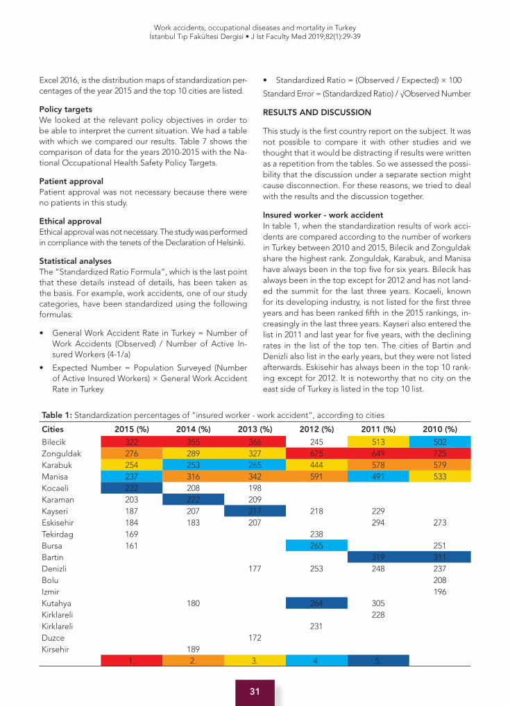
31
Work accidents, occupational diseases and mortality in Turkeyİstanbul Tıp Fakültesi Dergisi • J Ist Faculty Med 2019;82(1):29-39
Excel 2016, is the distribution maps of standardization per-centages of the year 2015 and the top 10 cities are listed.
Policy targetsWe looked at the relevant policy objectives in order to be able to interpret the current situation. We had a table with which we compared our results. Table 7 shows the comparison of data for the years 2010-2015 with the Na-tional Occupational Health Safety Policy Targets.
Patient approvalPatient approval was not necessary because there were no patients in this study.
Ethical approvalEthical approval was not necessary. The study was performed in compliance with the tenets of the Declaration of Helsinki.
Statistical analysesThe “Standardized Ratio Formula”, which is the last point that these details instead of details, has been taken as the basis. For example, work accidents, one of our study categories, have been standardized using the following formulas:
• General Work Accident Rate in Turkey = Number of Work Accidents (Observed) / Number of Active In-sured Workers (4-1/a)
• Expected Number = Population Surveyed (Number of Active Insured Workers) × General Work Accident Rate in Turkey
• Standardized Ratio = (Observed / Expected) × 100
Standard Error = (Standardized Ratio) / √Observed Number
RESULTS AND DISCUSSION
This study is the first country report on the subject. It was not possible to compare it with other studies and we thought that it would be distracting if results were written as a repetition from the tables. So we assessed the possi-bility that the discussion under a separate section might cause disconnection. For these reasons, we tried to deal with the results and the discussion together.
Insured worker - work accidentIn table 1, when the standardization results of work acci-dents are compared according to the number of workers in Turkey between 2010 and 2015, Bilecik and Zonguldak share the highest rank. Zonguldak, Karabuk, and Manisa have always been in the top five for six years. Bilecik has always been in the top except for 2012 and has not land-ed the summit for the last three years. Kocaeli, known for its developing industry, is not listed for the first three years and has been ranked fifth in the 2015 rankings, in-creasingly in the last three years. Kayseri also entered the list in 2011 and last year for five years, with the declining rates in the list of the top ten. The cities of Bartin and Denizli also list in the early years, but they were not listed afterwards. Eskisehir has always been in the top 10 rank-ing except for 2012. It is noteworthy that no city on the east side of Turkey is listed in the top 10 list.
Table 1: Standardization percentages of "insured worker - work accident", according to cities
Cities 2015 (%) 2014 (%) 2013 (%) 2012 (%) 2011 (%) 2010 (%)
Bilecik 322 355 366 245 513 502Zonguldak 276 289 327 675 649 725Karabuk 254 253 265 444 578 579Manisa 237 316 342 591 491 533Kocaeli 222 208 198 Karaman 203 222 209 Kayseri 187 207 217 218 229 Eskisehir 184 183 207 294 273Tekirdag 169 238 Bursa 161 265 251Bartin 319 311Denizli 177 253 248 237Bolu 208Izmir 196Kutahya 180 264 305 Kirklareli 228 Kirklareli 231 Duzce 172 Kirsehir 189
1. 2. 3. 4. 5.
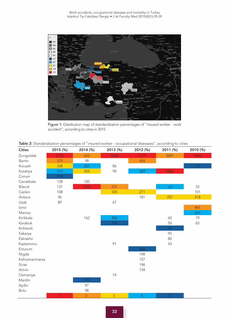
32
Work accidents, occupational diseases and mortality in Turkeyİstanbul Tıp Fakültesi Dergisi • J Ist Faculty Med 2019;82(1):29-39
Figure 1: Distribution map of standardization percentages of “insured worker - work accident”, according to cities in 2015
Table 2: Standardization percentages of "insured worker - occupational disesases", according to cities
Cities 2015 (%) 2014 (%) 2013 (%) 2012 (%) 2011 (%) 2010 (%)
Zonguldak 2725 652 1838 7219 2601 1645Bartin 375 98 454 Kocaeli 338 201 86 131Kutahya 270 205 90 269 5860 Corum 178 Canakkale 138 185 Bilecik 131 2045 531 169 55Cankiri 108 505 271 101Ankara 96 181 257 418Usak 89 67 Izmir 461Manisa 224Kirikkale 162 486 60 79Karabuk 100 50 65Kirklareli 95 Sakarya 95 Eskisehir 80 Kastamonu 91 43 Erzurum 266 Nigde 198 Kahramanmaras 157 Sivas 146 Artvin 134 Osmaniye 74 Mardin 195 Aydin 97 Bolu 98
1. 2. 3. 4. 5.

33
Work accidents, occupational diseases and mortality in Turkeyİstanbul Tıp Fakültesi Dergisi • J Ist Faculty Med 2019;82(1):29-39
Insured worker - occupational diseasesTable 2 shows the rates of occupational diseases stan-dardized according to the number of insured workers ex-amined. It is seen that almost every year a different city takes place at the summit. However, the common feature of these cities is that they are all frontline to the mining sector. Zonguldak is the top with 2725 (%) in 2015. Bartin and Kocaeli cities are listed in the top three in 2015. Bi-lecik is peaking in the 2014 order but not the top in 2015. In 2011, Kutahya was able to bring its ratios to a relatively low level in the years following the peak of 5860 (%). Nev-ertheless, it was always in the top 10.
Insured worker - mortalityWhen table 3, in which standardized mortality rates are included according to the number of insured workers, is examined, it is seen that there are different orders almost every year. When this table is compared with the above tables, the high count of the cities in the top 10 list may indicate that deaths are not as localized as work accidents or occupational diseases, instead they are widespread throughout the country. It is important to note that al-most 60% of these cities are located in the Eastern and Southeastern Anatolian Regions. Batman in 2011, Hakkari in 2012, Nevşehir in 2013, ranks first, but these cities are not in the top 10 in any of the tables above. It can also be emphasized that Zonguldak and Bilecik, which are always on the top in other tables, are hardly listed in this list. We see that the miners’ accidents, which resulted in death of 301 workers in Soma / Manisa in 2014, 18 workers in Er-menek / Karaman and 4 workers in Dagkonak - Kemerli / Sirnak, directly determined the top three in the sequence of standardization results for cities. In 2015, Manisa and
Karaman did not enter the top 10 list; Sirnak is the top with a rate of 1086 (%).
Workplace - work accidentTable 4 shows the Zonguldak and Bilecik leads in the stan-dardization of work accident numbers according to the number of workplaces in existing cities. It is known that work accidents, especially in the mining sector, have in-creased the rates in these cities. It is noteworthy that Kara-buk, known for its iron and steel factory, has entered the top 10 in all years and has not been in the top five rankings in the last three years. While Bartin ranked as fifth in 2010-2011, it did not take place in the top 10 list in the following years. Unlike previous years, it is remarkable that Kocaeli, where the chemical industry is the frontline, and Karaman, which is the frontline of mining, have been included in the top five in the last three years. In Tekirdag and Kayseri, it is known that the organized industry business line accidents are at the foreground. It is noteworthy that no city on the east side of Turkey is listed in the top 10 list again.
Workplace - occupational diseasesWhen table 5, where occupational diseases rates are standardized according to workplace numbers, is exam-ined, Zonguldak, Bilecik and Kutahya rank at the top. In particular, Zonguldak, of which the mining sector is the frontline, has always been listed in the top two with 675 - 8827 (%). Kutahya and Bilecik, with the same sector as the frontline, have the highest standardization rate of oc-cupational diseases in 2011 and 2014, respectively. The standardization rates of Kocaeli and Bartin cities have increased year by year, and in 2015, the top three were ranked with 450 and 339 (%), respectively.
Figure 2: Distribution map of standardization percentages of “insured worker - occupational diseases”, according to cities in 2015
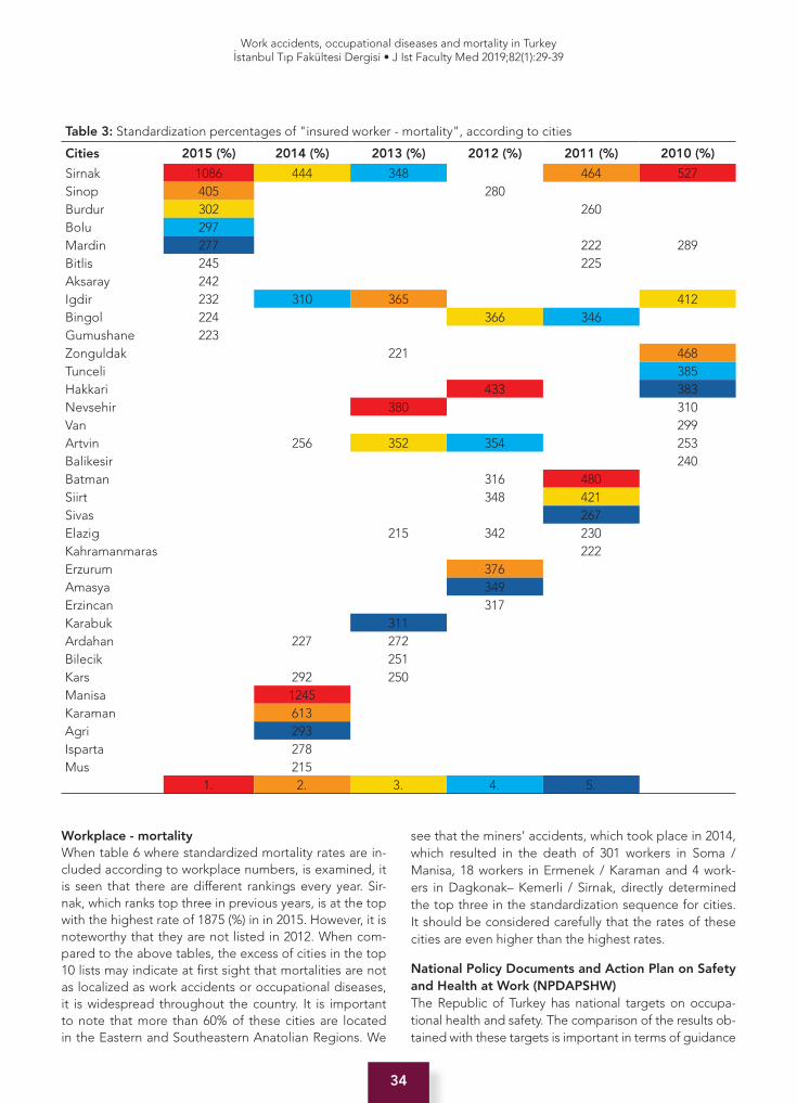
34
Work accidents, occupational diseases and mortality in Turkeyİstanbul Tıp Fakültesi Dergisi • J Ist Faculty Med 2019;82(1):29-39
Workplace - mortalityWhen table 6 where standardized mortality rates are in-cluded according to workplace numbers, is examined, it is seen that there are different rankings every year. Sir-nak, which ranks top three in previous years, is at the top with the highest rate of 1875 (%) in in 2015. However, it is noteworthy that they are not listed in 2012. When com-pared to the above tables, the excess of cities in the top 10 lists may indicate at first sight that mortalities are not as localized as work accidents or occupational diseases, it is widespread throughout the country. It is important to note that more than 60% of these cities are located in the Eastern and Southeastern Anatolian Regions. We
see that the miners’ accidents, which took place in 2014, which resulted in the death of 301 workers in Soma / Manisa, 18 workers in Ermenek / Karaman and 4 work-ers in Dagkonak– Kemerli / Sirnak, directly determined the top three in the standardization sequence for cities. It should be considered carefully that the rates of these cities are even higher than the highest rates.
National Policy Documents and Action Plan on Safety and Health at Work (NPDAPSHW)The Republic of Turkey has national targets on occupa-tional health and safety. The comparison of the results ob-tained with these targets is important in terms of guidance
Table 3: Standardization percentages of "insured worker - mortality", according to cities
Cities 2015 (%) 2014 (%) 2013 (%) 2012 (%) 2011 (%) 2010 (%)
Sirnak 1086 444 348 464 527Sinop 405 280 Burdur 302 260 Bolu 297 Mardin 277 222 289Bitlis 245 225 Aksaray 242 Igdir 232 310 365 412Bingol 224 366 346 Gumushane 223Zonguldak 221 468Tunceli 385Hakkari 433 383Nevsehir 380 310Van 299Artvin 256 352 354 253Balikesir 240Batman 316 480 Siirt 348 421 Sivas 267 Elazig 215 342 230 Kahramanmaras 222 Erzurum 376 Amasya 349 Erzincan 317 Karabuk 311 Ardahan 227 272 Bilecik 251 Kars 292 250 Manisa 1245 Karaman 613 Agri 293 Isparta 278 Mus 215
1. 2. 3. 4. 5.
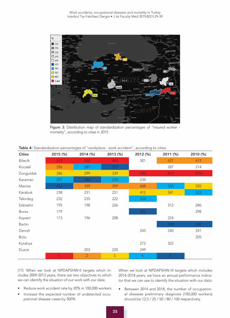
35
Work accidents, occupational diseases and mortality in Turkeyİstanbul Tıp Fakültesi Dergisi • J Ist Faculty Med 2019;82(1):29-39
(17). When we look at NPDAPSHW-II targets which in-cludes 2009-2013 years, there are two objectives to which we can identify the situation of our work with our data:
• Reduce work accident rate by 20% in 100,000 workers
• Increase the expected number of undetected occu-pational disease cases by 500%
When we look at NPDAPSHW-III targets which includes 2014-2018 years, we have an annual performance indica-tor that we can use to identify the situation with our data:
• Between 2014 and 2018, the number of occupation-al diseases preliminary diagnosis (100,000 workers) should be 12,5 / 25 / 50 / 80 / 100 respectively.
Figure 3: Distribution map of standardization percentages of “insured worker - mortality“, according to cities in 2015
Table 4: Standardization percentages of "workplace - work accident", according to cities
Cities 2015 (%) 2014 (%) 2013 (%) 2012 (%) 2011 (%) 2010 (%)
Bilecik 373 420 443 301 627 612
Kocaeli 296 287 273 287 214
Zonguldak 286 299 339 783 813 1034
Karaman 257 286 274 234
Manisa 252 339 359 608 508 555
Karabuk 238 231 251 412 541 522
Tekirdag 232 235 222 333
Eskisehir 195 198 226 312 286
Bursa 179 305 298
Kayseri 173 196 208 224
Bartin 338 352
Denizli 245 240 241
Bolu 205
Kutahya 272 322
Duzce 203 220 249
1. 2. 3. 4. 5.
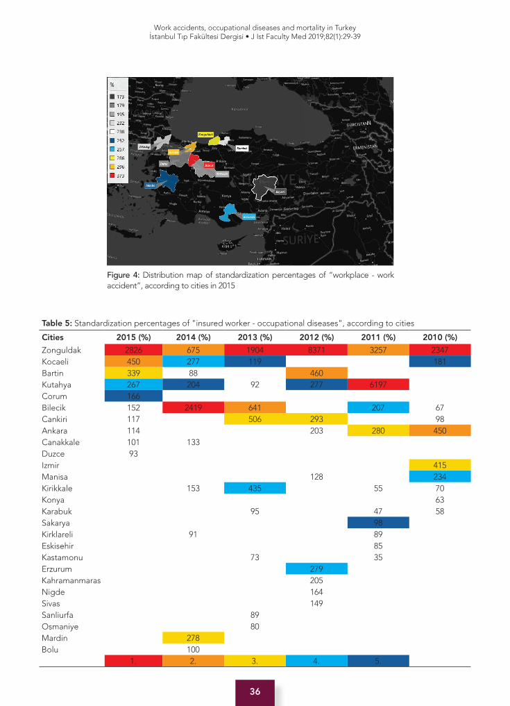
36
Work accidents, occupational diseases and mortality in Turkeyİstanbul Tıp Fakültesi Dergisi • J Ist Faculty Med 2019;82(1):29-39
Figure 4: Distribution map of standardization percentages of “workplace - work accident”, according to cities in 2015
Table 5: Standardization percentages of "insured worker - occupational diseases", according to cities
Cities 2015 (%) 2014 (%) 2013 (%) 2012 (%) 2011 (%) 2010 (%)
Zonguldak 2826 675 1904 8371 3257 2347Kocaeli 450 277 119 181Bartin 339 88 460 Kutahya 267 204 92 277 6197 Corum 166 Bilecik 152 2419 641 207 67Cankiri 117 506 293 98Ankara 114 203 280 450Canakkale 101 133 Duzce 93 Izmir 415Manisa 128 234Kirikkale 153 435 55 70Konya 63Karabuk 95 47 58Sakarya 98 Kirklareli 91 89 Eskisehir 85 Kastamonu 73 35 Erzurum 279 Kahramanmaras 205 Nigde 164 Sivas 149 Sanliurfa 89 Osmaniye 80 Mardin 278 Bolu 100
1. 2. 3. 4. 5.

37
Work accidents, occupational diseases and mortality in Turkeyİstanbul Tıp Fakültesi Dergisi • J Ist Faculty Med 2019;82(1):29-39
Figure 5: Distribution map of standardization percentages of “workplace - occupational diseases”, according to cities in 2015
Table 6: Standardization percentages of "insured worker - mortality", according to cities
Cities 2015 (%) 2014 (%) 2013 (%) 2012 (%) 2011 (%) 2010 (%)Sirnak 1875 783 644 876 1116Mardin 413 253 332 452Bingol 329 547 481 Sinop 322 Bolu 295 Bitlis 277 Siirt 276 246 478 525 Igdir 249 310 372 454Burdur 240 Agri 232 317 Zonguldak 276 667Hakkari 607 268 507Van 268 329 402Tunceli 327Batman 271 526 757 311Nevsehir 332 285Karaman 793 277Sanliurfa 252 289 Kahramanmaras 289 Elazig 240 404 270 Erzurum 395 Erzincan 339 Artvin 324 329 Amasya 321 Bilecik 303 Karabuk 294 Manisa 1336 Mus 276 Kars 275 Isparta 240
1. 2. 3. 4. 5.

38
Work accidents, occupational diseases and mortality in Turkeyİstanbul Tıp Fakültesi Dergisi • J Ist Faculty Med 2019;82(1):29-39
When we evaluate NPDAPSHW targets as given in table 7; it is not possible to fully assess NPDAPSHW-II since 2009 is not available in the time range of our work. How-ever, in 2010-2013, it is observed that “work accidents” increased by 145% and the number of “anticipated but undetected” occupational diseases cases decreased by 34.1%. When the rates of occupational diseases in 100,000 workers were examined in NPDAPSHW-III Tar-gets in 2014, the target of 12.5 out of 100,000 workers was 3.73; in 2015, it was observed that the target of 25 out of 100,000 workers was not reached with 3.64 results. The number of preliminary diagnoses for the 2016 target occupational disease was 50 in 100,000 workers.
CONCLUSION
According to the results of the six years examined, it is seen that the rates of work accidents and occupational diseases are highest in Zonguldak, Bilecik and Karabuk, where the
mining sector is in the forefront. When mortality rates are examined, especially Eastern and Southeastern Anatolian Cities such as Sirnak, Batman and Igdir have been in the frontline. This shows that, in addition to the measures to be taken nationwide in order to prevent mortal work acci-dents, various measures must be taken locally.
The fact that only those who have been diagnosed with occu-pational disease after the insurance has been included in the statistics for the years 2013-2014 show how important it is to watch one’s health after retirement. It was also removed from the tables again in 2015. What affects the results on a one-to-one basis are the records that are kept incomplete / inad-equate. These data must be recorded in a healthy manner.
In this study, the years 2010-2015 were studied. Future work could be done in 2005-2010 or 2015-2020 and so on. If the years are studied in such 6-year periods, it may be useful to make period comparisons.
Figure 6: Distribution map of standardization percentages of “workplace - mortality“, according to cities in 2015
Table 7: National Policy Documents and Action Plan on Safety and Health at Work (NPDAPSHW) targets
YearsNumber of work
accidents
Number of work accidents (per 100,000
labourers)
Work accidents by
2010 (%)
Number of occupational
diseases
Occupational diseases by
2010 (%)
Occupational diseases
(per 100,000labourers)
Target of NPDAPSHW-III
2010 62903 595 100 533 100%
NPDAPSHW-II2011 69227 600 100,8 697 131%
2012 74871 598 100,5 395 57%
2013 191389 1457 245 351 89%
2014 221366 1672NPDAPSHW-III
494NPDAPSHW-III
3,73 12,5
2015 241547 1725 510 3,64 25
39
Work accidents, occupational diseases and mortality in Turkeyİstanbul Tıp Fakültesi Dergisi • J Ist Faculty Med 2019;82(1):29-39
The inadequacy of the records is the biggest obstacle in determining the actual dimensions of the problem of work security. On the other hand, with the enactment of the Law No. 6331 on Occupational Health and Safe-ty, it is expected that the number of notifications from public institutions to RTSSI will increase. For better eval-uation and analysis of notifications, it is important to keep records more systematically and comprehensively.
Ethics Commitee Approval: Ethical issues including plagiarism, data fabrication and/or falsification, double publication and/or submission, redundancy, etc. have been completely observed by the authors. Ethical approval was not necessary. The study was per-formed in compliance with the tenets of the Declaration of Helsinki.
Informed Consent: Informed consent was not received due to the retrospective nature of the study.
Peer Review: Externally peer-reviewed.
Author Contributions: Conception/Design of Study- O.F.B., E.E., H.I., Z.B.; Data Acquisition- O.F.B., E.E., H.I., Z.B.; Data Analysis/Interpretation- O.F.B., E.E., H.I., Z.B.; Drafting Manu-script- O.F.B., E.E., Z.B.; Critical Revision of Manuscript- O.F.B., E.E., H.I., Z.B.; Final Approval and Accountability- O.F.B., E.E., H.I., Z.B.; Technical or Material Support- O.F.B., E.E., H.I., Z.B.
Conflict of Interest: Authors declared no conflict of interest.
Financial Disclosure: Authors declared no financial support.
Etik Komite Onayı: Etik konular (intihal, veri üretimi ve / veya tahrifat, çifte yayın ve / veya teslim, fazlalık, vb. dahil) yazarlar tarafından eksiksiz olarak gözetilmiş ve denetlenmişti. Etik onay gerekli değildi. Çalışma, Helsinki Bildirgesi’nin ilkelerine uygun olarak gerçekleştirildi.
Bilgilendirilmiş Onam: Retrospektif bir çalışma olduğundan bil-gilendirilmiş onam alınmamıştır.
Hakem Değerlendirmesi: Dış bağımsız.
Yazar Katkıları: Çalışma Konsepti/Tasarım- O.F.B., E.E., H.I., Z.B.; Veri Toplama- O.F.B., E.E., H.I., Z.B.; Veri Analizi/Yorumlama- O.F.B., E.E., H.I., Z.B.; Yazı Taslağı- O.F.B., E.E., Z.B.; İçeriğin Eleştirel İn-celemesi- O.F.B., E.E., H.I., Z.B.; Son Onay ve Sorumluluk- O.F.B., E.E., H.I., Z.B.; Malzeme ve Teknik Destek- O.F.B., E.E., H.I., Z.B.
Çıkar Çatışması: Yazarlar çıkar çatışması beyan etmemişlerdir.
Finansal Destek: Yazarlar finansal destek beyan etmemişlerdir.
REFERENCES
1. Loewenson R. Globalization and occupational health: a perspective from southern Africa. Bull World Health Organ 2001;79(9):863-8.
2. Rantanen J. Grand Challenges for Occupational Health from Globalization. Journal of Labor, Occupational Safety and Health 2010;18(2):167-71. [CrossRef]
3. Karadeniz O. Dünya’da ve Türkiye’de iş kazaları ve meslek hastalıkları ve sosyal koruma yetersizliği. Calisma ve Toplum 2012;34(3):15-72.
4. ILO. Standards on occupational safety and health Promoting a safe and healthy working environment, General Survey concerning the Occupational Safety and Health Convention, 1981 (No. 155), the Occupational Safety and Health Recommendation, 1981 (No. 164), and the Protocol of 2002 to the Occupational Safety and Health Convention, 1981, International Labour Conference, 98th Session, 2009. Report III (Part 1B), Third item on the agenda: Information and reports on the application of Conventions and Recommendations. Report of the Committee of Experts on the Application of Conventions and Recommendations (articles 19, 22 and 35 of the Constitution) 2009. Available from: http://www.ilo.org/wcmsp5/groups/public/@ed_norm/@relconf/documents/meetingdocument/wcms_103485.pdf (accessible date: 05.02.2019).
5. Hamalainen P, Leena Saarela K, Takala J. Global trend according to estimated number of occupational accidents and fatal work-related diseases at region and country level. J Safety Res 2009;40(2):125-39. [CrossRef]
6. Aydemir M. İşçi sağlığı ve iş güvenliğini sağlama açısından işverenin yükümlülüğü kamu - iş. İş Hukuku ve İktisat Dergisi 1995;4(1):79-99.
7. The Laws of Republic of Turkey No. 5502, 2th Section, 11th Article. Available from: www.mevzuat.gov.tr/MevzuatMetin/1.5.5502.pdf (accessible date: 05.02.2019).
8. The Laws of Republic of Turkey No. 5510, 2th Section, 20th Article. Available from: www.mevzuat.gov.tr/MevzuatMetin/1.5.5510.pdf (accessible date: 05.02.2019).
9. Ucuncu K. 2014 yılı SGK İş Kazası İstatistiklerinin Analizi. Available from: http://www.isteguvenlik.tc/2014%20SGK%20Analiz.pdf (accessible date: 05.02.2019).
10. Social Security Institution’s Statistical Yearbooks, The Ministry of Labour and Social Security, Republic of Turkey: Sosyal Güvenlik Kurumu Yayınları; 2010 - 2015. Available from: http://www.sgk.gov.tr/wps/portal/sgk/tr/kurumsal/istatistik/sgk_istatistik_yilliklari (accessible date: 05.02.2019).
11. Paneth N. EPI 810: Epidemiyolojiye Giriş. Available from: http://sbu.saglik.gov.tr/Ekutuphane/kitaplar/epidemiyoloji8.pdf (accessible date: 05.02.2019).
12. Hayran O. Sağlık Bilimlerinde Araştırma ve İstatistik Yöntemler. İstanbul: Nobel Tip Kitabevleri; 2012. p. 336.
13. Control CfD, Prevention. Principles of epidemiology in public health practice: an introduction to applied epidemiology and biostatistics. Atlanta, GA: US Dept. of Health and Human Services, Centers for Disease Control and Prevention (CDC), Office of Workforce and Career Development; 2006.
14. Lwanga SK, Tye C-Y, Ayeni O. Teaching Health Statistics: Lesson and Seminar Outlines: ERIC; 1999.
15. Akbulut I, Sabuncu H. Sağlık Bilimlerinde Araştırma Yöntemi, Epidemiyoloji Prensip ve Uygulamalar. 1 ed: Sistem Yayıncılık; 1993. p. 81-111.
16. Yaffee RA. Elementary Epidemiological Analysis with STATA-class notes New York. Available from: https://www.researchgate.net/profile/Robert_Yaffee/publication/228772429_Elementary_Epidemiological_Analysis_with_STATA- class_notes/links/0c960520a60581c148000000/Elementary-Epidemiological-Analysis-with-STATA-class-notes.pdf (accessible date: 05.02.2019).
17. Strategic Plan, The Ministry of Labour and Social Security, Republic of Turkey. Available from: http://www.sp.gov.tr/tr/stratejik-plan/kurum/180/Aile+ve+ Sosyal+Politikalar+Bakanligi (accessible date: 05.02.2019).

40
Molecular structure of doxorubicin and Galectin-3İstanbul Tıp Fakültesi Dergisi • J Ist Faculty Med 2019;82(1):40-6
İletişim kurulacak yazar/Corresponding author: [email protected]
Geliş tarihi/Received Date: 03.08.2018 • Kabul tarihi/Accepted Date: 09.01.2019
©Telif Hakkı 2019 J Ist Faculty Med - Makale metnine jmed.istanbul.edu.tr web sayfasından ulaşılabilir.©Copyright 2019 by J Ist Faculty Med - Available online at jmed.istanbul.edu.tr
ARAŞTIRMA / RESEARCH DOI: 10.26650/IUITFD.0005
İst Tıp Fak Derg 2019 / J Ist Faculty Med 2019
COMPUTERISED DESIGNING OF DOXORUBICIN WITH BREAST CANCER CELLS
DOKSORUBİSİNİN MEME KANSERİ HÜCRELERİ İLE BİLGİSAYARLI TASARIMI
Leyla TÜRKER ŞENER1 , Serda KECEL GÜNDÜZ2 , Aziz ŞENER3 , Bilge BIÇAK2,4 , Yağmur KÖKCÜ4 ,Ayşen E. ÖZEL2 , Işıl ALBENİZ1
1Istanbul University, Istanbul Faculty of Medicine, Department of Biophysics, Istanbul, Turkey2Istanbul University, Faculty of Sciences, Department of Physics, Istanbul, Turkey 3Kanuni Sultan Suleyman Training and Research Hospital, Department of General Surgery, Istanbul, Turkey 4Cerrahpasa - Istanbul University, Graduate School of Engineering and Sciences, Istanbul, Turkey
ORCID IDs of the authors: L.T.Ş. 0000-0002-7317-9086; S.K.G. 0000-0003-0973-8223; A.Ş. 0000-0003-0507-647X; B.B. 0000-0003-1147-006X; Y.K. 0000-0003-1570-2390; A.E.Ö. 0000-0002-8680-8830; I.A. 0000-0002-6005-5164
Cite this article as: Türker Şener L, Kecel Gündüz S, Şener A, Bıçak B, Kökcü Y, Özel AE, et al. Computerised designing of doxorubicin with breast cancer cells. J Ist Faculty Med 2019;82(1):40-6. doi: 10.26650/IUITFD.0005
ABSTRACTObjective: Breast cancer is the type of cancer that starts in breast cells and has the highest incidence in the world after lung cancer. Doxorubicin is widely used in the treatment of some leu-kemia and Hodgkin’s lymphoma, as well as in the treatment of bladder, breast, stomach, lung, ovary, thyroid, soft tissue sarco-ma, multiple myeloma and other cancers. We want to determine the binding interaction of Doxorubicin in the active site of the Galectin-3 with ASN-48, ARG-32, ASN-62 and GLU-72 residues, conformation and docking score energies.
Material and Method: We used docking methods to detect the efficiency of Doxorubicin at breast cancer cells, clarify the role of Galectin-3 and elucidate the interaction between Galectin-3 and Doxorubicin.
Results: The docking score obtained for Doxorubicin in the ac-tive side of the Galectin-3 protein was -5.32 kcal/mol. In to the active site of protein, Doxorubicin was bound with strong hydro-gen bond by the residue ASN-48, ARG-32, ASN-62 and GLU-72, and a salt bridge with the same amino acid residue were estab-lished and stability was achieved.
Conclusion: The development of specific therapies targeting cancer stem cells may provide hope for prolonging the life span and improving quality.
Keywords: Docking, breast cancer, Galectin-3, Doxorubicin
ÖZETAmaç: Meme kanseri, meme hücrelerinde başlayan ve akciğer kanserinden sonra dünyada en yüksek insidansa sahip olan kanser türüdür. Doksorubisin, bazı lösemi ve Hodgkin lenfoma-sının yanı sıra mesane, meme, mide, akciğer, yumurtalık, tiroid, yumuşak doku sarkomu, multipl miyeloma ve diğer kanserlerin tedavisinde yaygın olarak kullanılır. Çalışmamızda; Doksorubi-sin’in Galektin 3’ün aktif bölgesinde yer alan, ASN-48, ARG-32, ASN-62 ve GLU-72 rezidüleri ile yaptığı bağlanma etkileşimlerini, konformasyonlarını ve doking skor enerjilerini belirlemek istedik.
Gereç ve Yöntem: Meme kanseri hücrelerinde Doxorubicin et-kinliğini tespit etmek, Galectin-3 rolünü netleştirmek ve Galek-tin-3 ve Doksorubisin arasındaki etkileşimi aydınlatmak için do-king yöntemi kullanılmıştır.
Bulgular: Tüm konformerler arasında Doksorubisin Galektin-3 proteini ile en iyi kenetlenme sonucunu -5,32 kcal/mol docking skoruna sahip konformer vermiştir. Doksorubisin güçlü hidrojen bağları ile ASN-48, ARG-32, ASN-62 ve GLU-72 rezidülerine bağ-lanmış ve aynı amino asit rezidüleri ile bir tuz köprüsü de kurarak, kararlılık sağlanmıştır.
Sonuç: Kanser kök hücrelerini hedef alan spesifik tedavilerin geliştirilmesi açısından, yaşam süresinin uzatılması ve kalitenin arttırılması için bu çalışmanın umut sağlayabilir olduğunu düşün-mekteyiz.
Anahtar Kelimeler: Doking, meme kanseri, Galektin-3, Dokso-rubisin

41
Molecular structure of doxorubicin and Galectin-3İstanbul Tıp Fakültesi Dergisi • J Ist Faculty Med 2019;82(1):40-6
INTRODUCTION
The highest incidence and mortality rate of all cancers in the female population is breast cancer (1, 2). It is esti-mated that there are approximately 2.5 million survivors of breast cancer in the US (3). This figure will expand to 3.4 million in 2015, representing an increase of 31% (4). The millions more worldwide are probably grossly under-estimated because of the poor or inefficient reporting systems and the lack of reliable cancer registries in third-world countries (5, 6).
Molecular characteristics of the tumor are important for breast cancer treatment (6, 7). Anthracyline drugs such as doxorubicin are first extracted from Streptomyces peu-cetius var. caesius. Doxorubicin is used in the treatment of several cancers including breast, lung, gastric, ovarian, thyroid, non-Hodgkin’s and Hodgkin’s lymphoma, multi-ple myeloma, sarcoma, and pediatric cancers (8–10). The biggest side effect of doxorubicin is cardiotoxicity (11-15).
Galectin-3 is an endogenous carbohydrate-binding pro-tein that has been shown to have a variety of cellular func-tions (16). Galectin-3 may increase or decrease apoptosis depending on cell type and stimulus. Overexpression of galectin-3 in breast carcinoma cells makes cells resistant to chemotherapeutic drugs (17, 18).
Various in vitro assays and theoretical calculation meth-ods have been used to detect the efficiency of Doxorubi-cin in various breast cancer cells, clarify the role of Galec-tin-3 and elucidate the interaction between Galectin-3 and Doxorubicin.
Molecular docking is one of this methods to detect inter-action of proteins. This method has become an increas-ingly important tool for new effective drug discovery. The molecular docking can be used to make model for the interaction between a small molecule (ligand) and a protein (receptor) at an atomic level; which allows us to understand the basic biochemical processes, enabling us to characterize the behavior of small molecules in the binding site of target proteins (19, 20).
MATERIALS AND METHODS
Molecular DockingUsing the Glide SP (standard precision) module of the Maestro version 11.4 in the Schrodinger Software pro-gram (Schrödinger Release 2017-4: Maestro, Schröding-er, LLC, New York, NY, 2017) (21-23) the docking calcu-lations were performed. To prepare ligand for docking calculations, first, the most stable conformation of Doxo-rubicin was generated from the result of the molecular dynamic calculation, and then was prepared to optimi-zation by the Lig Prep tool in the Maestro 11.4 version of the Schrödinger Software program using the OPLS force
field (24). After selecting the ionization states at pH 7.0 ± 2.0, possible stereoisomers were produced for the li-gand. Because of Doxorubicin induces the expression of galectin-3 in breast cancer cell line and in primary tumors from breast cancer patients, we choose galectin-3 (25) (PDB code: 2XG3) which could be a potential target to prevent Doxorubicin induced chemo resistance in breast cancer. By using SWISS-MODEL server, the crystal struc-ture obtained from the protein data bank was arranged to get a better protein homology model (26). All water was removed, polar hydrogens were added, bond orders were assigned, charges were defined using PROPKA (27) at pH 7.0, and galectin-3 was optimized by using the Pro-tein Preparation Wizard tool (28). Energy minimization was carried out by preferring 0.3A˚ RMSD and the OPLS3 force field to converge heavy atoms. To generate a grid, receptor grid generation tool was used; a cubic box was formed that centered on the centroid of the ligand with specific dimensions. By defining routable groups of an active site of protein, ligand-receptor docking was oc-curred. Absorption, distribution, metabolism, and excre-tion (ADME) properties were also calculated using the Qik-Prop module of the Schrodinger software to specify the physicochemical and biological functional properties such as the molecular weight (MW), percent human oral absorption, predicted octanol/water partition coefficient (QPlogPo/w), polar surface area (PSA), and number of vi-olations of Lipinski’s rule of five (29), which is important for generating an effective drug in new drug development.
RESULTS
Molecular Docking ResultsThe docking score obtained for Doxorubicin in the ac-tive side of the Galectin-3 protein was -5.32 kcal/mol, as shown in Table 1 and Figure 1. The binding pocket of the Galectin-3 (2XG3.pdb) protein has a hydrophobic, polar, positive charged and negative charged regions as indicat-ed by the green, blue, dark blue and orange respective-ly. The most likely binding position between Galectin-3
Table 1. The conformation and docking score energies
Ligand Docking Score (kcal/mol)
1 -5.32
2 -5.19
3 -4.75
4 -4.72
5 -4.59
6 -4.57
7 -4.55
8 -4.50
42
Molecular structure of doxorubicin and Galectin-3İstanbul Tıp Fakültesi Dergisi • J Ist Faculty Med 2019;82(1):40-6
Figure 1: The docked molecular structure of Doxorubucin and Galectin-3
Figure 2: 2D ligand interaction of Doxorubucin in the active side of Galectin-3
43
Molecular structure of doxorubicin and Galectin-3İstanbul Tıp Fakültesi Dergisi • J Ist Faculty Med 2019;82(1):40-6
Figure 3: The binding interaction of Doxorubucin in the active side of the Galectin-3 with ASN-48, ARG-32, ASN-62 and GLU-72 residues
Figure 4: The electrostatic potential of Galectin-3 and Doxorubicin
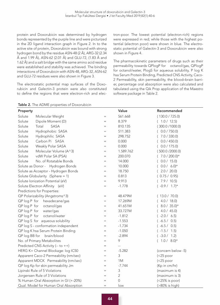
44
Molecular structure of doxorubicin and Galectin-3İstanbul Tıp Fakültesi Dergisi • J Ist Faculty Med 2019;82(1):40-6
protein and Doxorubicin was determined by hydrogen bonds represented by the purple line and were picturized in the 2D ligand interaction graph in Figure 2. In to the active site of protein, Doxorubicin was bound with strong hydrogen bond by the residue ASN-48 (2 Å), ARG-32 (2.34 Å and 1.99 Å), ASN-62 (2.01 Å) and GLU-72, (1.83 Å and 1.62 Å) and a salt bridge with the same amino acid residue were established and stability was achieved. The binding interactions of Doxorubicin with ASN-48, ARG-32, ASN-62 and GLU-72 residues were also shown in Figure 3.
The electrostatic potential map surfaces of the Doxo-rubicin and Galectin-3 protein were also constituted to define the regions that were electron-rich and elec-
tron-poor. The lowest potential (electron-rich) regions were expressed in red; while those with the highest po-tential (electron poor) were shown in blue. The electro-static potential of Galectin-3 and Doxorubicin were also shown in Figure 4.
The pharmacokinetic parameters of drugs such as their permeability towards QPlogP for octanol/gas, QPlogP for octanol/water, PlogS for aqueous solubility, P log K hsa Serum Protein Binding, Predicted CNS Activity, Caco-2 Permeability, skin permeability, the blood–brain barri-er, percentage oral absorption were also calculated and tabulated using the Qik Prop application of the Maestro software package in Table 2.
Table 2. The ADME properties of Doxorubicin
Property Value Recommended
Solute Molecular Weight = 561.668 ( 130.0 / 725.0)Solute Dipole Moment (D) = 8.379 ( 1.0 / 12.5)Solute Total SASA = 810.135 ( 300.0 /1000.0)Solute Hydrophobic SASA = 511.383 ( 0.0 / 750.0)Solute Hydrophilic SASA = 298.752 ( 7.0 / 330.0)Solute Carbon Pi SASA = 0.000 ( 0.0 / 450.0)Solute Weakly Polar SASA = 0.000 ( 0.0 / 175.0)Solute Molecular Volume (A^3) = 1.589.762 ( 500.0 /2000.0)Solute vdW Polar SA (PSA) = 200.070 ( 7.0 / 200.0)*Solute No. of Rotatable Bonds = 14.000 ( 0.0 / 15.0)Solute as Donor - Hydrogen Bonds = 10.000 ( 0.0 / 6.0)*Solute as Acceptor - Hydrogen Bonds = 18.750 ( 2.0 / 20.0)Solute Globularity (Sphere = 1) = 0.813 ( 0.75 / 0.95)Solute Ionization Potential (eV) = 9.913 ( 7.9 / 10.5)Solute Electron Affinity (eV) = -1.778 ( -0.9 / 1.7)*Predictions for Properties:QP Polarizability (Angstroms^3) = 48.479M ( 13.0 / 70.0)QP log P for hexadecane/gas = 17.269M ( 4.0 / 18.0)QP log P for octanol/gas = 41.651M ( 8.0 / 35.0)*QP log P for water/gas = 33.727M ( 4.0 / 45.0)QP log P for octanol/water = -1.812 ( -2.0 / 6.5)QP log S for aqueous solubility = -1.553 ( -6.5 / 0.5)QP log S - conformation independent = -1.734 ( -6.5 / 0.5)QP log K hsa Serum Protein Binding = -1.050 ( -1.5 / 1.5)QP log BB for brain/blood = -2.894 ( -3.0 / 1.2)No. of Primary Metabolites = 9 ( 1.0 / 8.0)*Predicted CNS Activity (-- to ++) = --HERG K+ Channel Blockage: log IC50 = -5.282 (concern below -5)Apparent Caco-2 Permeability (nm/sec) = 3 (<25 poorApparent MDCK Permeability (nm/sec) = 1M (<25 poorQP log Kp for skin permeability Jm = -7.744 (Kp in cm/hr)Lipinski Rule of 5 Violations = 3 (maximum is 4)Jorgensen Rule of 3 Violations = 2 (maximum is 3)% Human Oral Absorption in GI (+-20%) = 0 (<25% is poor)Qual. Model for Human Oral Absorption = low (>80% is high)

45
Molecular structure of doxorubicin and Galectin-3İstanbul Tıp Fakültesi Dergisi • J Ist Faculty Med 2019;82(1):40-6
CONCLUSIONS
The molecular docking approach can be used to model the interaction between a small molecule (ligand) and a protein (receptor) at an atomic level; which allows us to understand the basic biochemical processes, enabling us to characterize the behavior of small molecules in the binding site of target proteins.
In this study; by examining the interaction between doxo-rubicin and Galectin-3, the most stable binding exposure between all binding poses were determined. Characteri-zation of the interaction between ligand and protein and ADME profiles were also obtained as a result of molecu-lar docking calculations.
When this tables are examined; The octanol / water value is -1.812, and this value (suggested values-2, 6.5) appears to be within the acceptable range, although the molec-ular weight of doxorubicin is acceptable at 561,668 (rec-ommended values 130-725). The development of spe-cific therapies targeting cancer stem cells may provide hope for prolonging the life span and improving quality.
Peer Review: Externally peer-reviewed.
Author Contributions: Conception/Design of Study- L.T.Ş., S.K.G.; Data Acquisition- L.T.Ş., S.K.G., Y.K.; Data Analysis/Inter-pretation- L.T.Ş., S.K.G.; Drafting Manuscript- L.T.Ş., A.Ş., S.K.G.; Critical Revision of Manuscript- I.A., S.K.G.; Final Approval and Accountability- L.T.Ş., S.K.G., A.Ş., B.B., Y.K., A.E.Ö., I.A.; Techni-cal or Material Support- L.T.Ş., S.K.G., I.A., A.E.O.
Conflict of Interest: Authors declared no conflict of interest.
Financial Disclosure: This study is supported by Istanbul Uni-versity Scientific Research Projects Coordination Unit. Project Number: 25945.
Hakem Değerlendirmesi: Dış bağımsız.
Yazar Katkıları: Çalışma Konsepti/Tasarım- L.T.Ş., S.K.G.; Veri Toplama- L.T.Ş., S.K.G., Y.K.; Veri Analizi/Yorumlama- L.T.Ş., S.K.G.; Yazı Taslağı- L.T.Ş., A.Ş., S.K.G.; İçeriğin Eleştirel İncele-mesi- I.A., S.K.G.; Son Onay ve Sorumluluk- L.T.Ş., S.K.G., A.Ş., B.B., Y.K., A.E.Ö., I.A.; Malzeme ve Teknik Destek- L.T.Ş., S.K.G., I.A., A.E.O.
Çıkar Çatışması: Yazarlar çıkar çatışması beyan etmemişlerdir.
Finansal Destek: Bu çalışma İstanbul Üniversitesi Bilimsel Araştırma Projeleri Koordinasyon Birimi tarafından desteklen-miştir. Proje numarası 25945.
REFERENCES
1. Lovitt CJ, Shelper TB, Avery, VM. Doxorubicin resistance in breast cancer cells is mediated by extracellular matrix proteins. BMC Cancer 2018;18(1):41. [CrossRef]
2. Jemal A, Bray F, Center MM, Ferlay J, Ward E, Forman D. Global cancer statistics. CA Cancer J Clin 2011;61(2):69-90. [CrossRef]
3. Demark-Wahnefried W, Campbell KL, Hayes SC. Weight management and its role in breast cancer rehabilitation. Cancer 2012;118(Suppl. 8):2277-87. [CrossRef]
4. Pierce JP, Stefanick ML, Flatt SW, Natarajan L, Sternfeld B, Madlensky L, et al. Greater survival after breast cancer in physically active women with high vegetable-fruit intake regardless of obesity. J Clin Oncol 2007;25(17):2345-51. [CrossRef]
5. Gonzales JF, Barnard ND, Jenkins DJ, Lanou AJ, Davis B, Saxe G, Levin S. Applying the precautionary principle to nutrition and cancer. J Am Coll Nutr 2014;33(3):239-46. [CrossRef]
6. Bodai BI, Tuso P. Breast cancer survivorship: a comprehensive review of long-term medical issues and lifestyle recommendations. Perm J 2015;19(2):48-79. [CrossRef]
7. Kontoyannis A, Sweetland H. Adjuvant therapy for breast cancer. Surgery (Oxford) 2007;25(6):272-5. [CrossRef]
8. Hernandez-Aya LF, Gonzalez-Angulo AM. Adjuvant systemic therapies in breast cancer. Surg Clin North Am 2013;93(2):473-91. [CrossRef]
9. Berman, AT, Thukral AD, Hwang WT, Solin LJ, Vapiwala N. Incidence and patterns of distant metastases for patients with early-stage breast cancer after breast conservation treatment. Clin Breast Cancer 2013;13(2):88-94. [CrossRef]
10. Carvalho C, Santos RX, Cardoso S, Correia S, Oliveira PJ, Santos MS, et al. Doxorubicin: the good, the bad and the ugly effect. Curr med Chem 2009;16(25):3267-85. [CrossRef]
11. Arcamone F, Cassinelli G, Fantini G, Grein A, Oerezzi P, Pol C, et al. Adriamycin, 14-hydroxydaimomycin, a new antitumor antibiotic from S. Peucetius var. caesius. Biotechno Bioeng 1969;11(6):1101-10. [CrossRef]
12. Cortés-Funes H, Coronado C. Role of anthracyclines in the era of targeted therapy. Cardiovasc Toxicol 2007;7(2):56-60. [CrossRef]
13. Weiss RB, The anthracyclines: will we ever find a better doxorubicin? In: Seminars in oncology. Elsevier 1992: p. 670-86.
14. Swain SM, Whaley FS, Ewer MS. Congestive heart failure in patients treated with doxorubicin: a retrospective analysis of three trials. Cancer 2003;97(11):2869-79. [CrossRef]
15. Thorn CF, Oshiro C, Marsh S, Hernandez-Boussard T, McLeod H, Klein TE, et al. Doxorubicin pathways: pharmacodynamics and adverse effects. Pharmacogenetics and Genomics 2011;21(7):440-6. [CrossRef]
16. Hsu DK, Liu FT. Regulation of cellular homeostasis by galectins. Glycoconj J 2002;19(7-9):507-15. [CrossRef]
17. Choi JH, Chun KH, Raz A, Lotan R. Inhibition of N-(4-hydroxyphenyl) retinamide-induced apoptosis in breast cancer cells by galectin-3. Cancer Biol Ther 2004;3(5):447-52. [CrossRef]
18. Takenaka Y, Fukumori T, Yoshii T, Oka N, Inohara H, Kim HR, et al. Nuclear export of phosphorylated galectin-3 regulates its antiapoptotic activity in response to chemotherapeutic drugs. J Mol Sci 2004;24(10):4395-406. [CrossRef]
19. Meng Xuan-Yu, Zhang HX, Mezei M, Cui M. Molecular docking: a powerful approach for structure-based drug discovery. Curr Comput Aided Drug Des 2011;7(2):146-57. [CrossRef]
46
Molecular structure of doxorubicin and Galectin-3İstanbul Tıp Fakültesi Dergisi • J Ist Faculty Med 2019;82(1):40-6
20. McConkey BJ, Sobolev V, Edelman M. The performance of current methods in ligand–protein docking. Curr Sci 2002;83(7):845-56.
21. Friesner RA, Murphy RB, Repasky MP, Frye LL, Greenwood JR, Halgren TA, et al. Extra precision glide: Docking and scoring incorporating a model of hydrophobic enclosure for protein− ligand complexes. J Med Chem 2006;49(21):6177-96. [CrossRef]
22. Halgren TA, Murphy RB, Friesner RA, Beard HS, Frye LL, Pollard WT, et al. Glide: a new approach for rapid, accurate docking and scoring. 2. Enrichment factors in database screening. J Med Chem 2004;47(7):1750-9. [CrossRef]
23. Friesner RA, Banks JL, Murphy RB, Halgren TA, Klicic JJ, Mainz DT, Repasky MP, et al. Glide: a new approach for rapid, accurate docking and scoring. 1. Method and assessment of docking accuracy. J Med Chem 2004;47(7):1739-49. [CrossRef]
24. Harder E, Damm W, Maple J, Wu C, Reboul M, Yu Xiang J, et al. OPLS3: a force field providing broad coverage of drug-like small molecules and proteins. J Chem Theory Comput 2015;12(1):281-96. [CrossRef]
25. Guha P, Bandyopadhyaya G, Polumuri SK, Chumsri S, Gade P, Kalvakolanu DV, et al. Nicotine promotes apoptosis resistance of breast cancer cells and enrichment of side population cells with cancer stem cell-like properties via a signaling cascade involving galectin-3, α9 nicotinic acetylcholine receptor and STAT3. Breast Cancer Res Treat 2014;145(1):5-22. [CrossRef]
26. Bienert S, Waterhouse A, de Beer, Tjaart A. P. Tauriello G, Studer G, Bordoli L et al. The SWISS-MODEL Repository—new features and functionality. Nucleic Acids Res 2016;45(D1):D313-D319. [CrossRef]
27. Søndergaard CR, Olsson MH, Rostkowski M, Jensen JH. Improved treatment of ligands and coupling effects in empirical calculation and rationalization of pKa values. J Chem Theory Comput 2011;7(7):2284-95. [CrossRef]
28. Sastry GM, Adzhigirey M, Day T, Annabhimoju R, Sherman W. Protein and ligand preparation: parameters, protocols, and influence on virtual screening enrichments. J Computer-Aided Mol Design 2013;27(3):221-34. [CrossRef]
29. Oprea TI, Gottfries J. Toward minimalistic modeling of oral drug absorption. J Mol Graph Model 1999;17(5-6): 61-74. [CrossRef]

47
Scopolamine-induced convulsions in fasted ratsİstanbul Tıp Fakültesi Dergisi • J Ist Faculty Med 2019;82(1):47-51
İletişim kurulacak yazar/Corresponding author: [email protected]
Geliş tarihi/Received Date: 30.03.2018 • Kabul tarihi/Accepted Date: 21.12.2018
©Telif Hakkı 2019 J Ist Faculty Med - Makale metnine jmed.istanbul.edu.tr web sayfasından ulaşılabilir.©Copyright 2019 by J Ist Faculty Med - Available online at jmed.istanbul.edu.tr
ARAŞTIRMA / RESEARCH DOI: 10.26650/IUITFD.411169
İst Tıp Fak Derg 2019 / J Ist Faculty Med 2019
SCOPOLAMINE-INDUCED CONVULSIONS IN FASTED RATS AFTER FOOD INTAKE: THE EFFECT OF DURATION OF FOOD DEPRIVATION
AÇ SIÇANLARA SKOPOLAMİN UYGULANMASI VE YEM VERİLMESİ İLE OLUŞAN KONVÜLSİYONLAR: YEM YOKSUNLUĞU SÜRESİNİN ETKİSİ
Aslı ZENGİN TÜRKMEN1 , Asiye NURTEN1 , Nurhan ENGİNAR2
1Istanbul Yeni Yuzyil University, Faculty of Medicine, Department of Physiology, Istanbul, Turkey2Istanbul University, Istanbul Faculty of Medicine, Department of Pharmacology, Istanbul, Turkey
ORCID IDs of the authors: A.Z.T. 0000-0002-8309-8431; A.N. 0000-0001-7847-1716; N.E. 0000-0002-2225-0460
Cite this article as: Zengin Türkmen A, Nurten A, Enginar N. Scopolamine-induced convulsions in fasted rats after food intake: The effect of duration of food deprivation. J Ist Faculty Med 2019;82(1):47-51. doi: 10.26650/IUITFD.411169
ABSTRACTObjective: Mice and rats fasted for 2 days and then treated with antimuscarinic drugs, scopolamine or atropine, develop con-vulsions soon after food intake. It has been demonstrated that deprivation of food for less than 48 h also causes convulsions in mice. Since there are differences in characteristics of the seizures between mice and rats, the present study evaluated whether rats also develop convulsions after being deprived of food for 3-24 h.
Material and Method: Rats were deprived of food for 3, 6, 18, 24 and 52 h. At the time of testing, the animals were treated i.p. with saline or 3 mg/kg scopolamine and given food 20 min later. All animals were observed for 30 min, checking for the incidence and onset of convulsions.
Results: The fasted animals treated with scopolamine devel-oped convulsions after food intake. The incidence of convulsions was statistically significant in the 6, 18, 24 and 52 h deprived ani-mals. However, neither the incidence nor the latency to seizures showed differences with respect to duration of food deprivation or fasting-induced weight loss.
Conclusion: The present results demonstrate that the scopol-amine treated rats that fasted for 24 h or less developed seizures after refeeding intake, indicating that food deprivation in itself, rather than its duration, seems to be the principal factor in the development of these convulsions.
Keywords: Scopolamine, convulsion, food deprivation, rat
ÖZETAmaç: İki gün aç bırakıldıktan sonra antimuskarinik, skopolamin veya atropin, uygulanan fare ve sıçanlar yeniden yedikten hemen sonra konvülsiyon geçirmektedirler. Farelerin 48 saatten daha kısa süre yemden yoksun bırakılmasıyla da konvülsiyon oluştuğu gösterilmiştir. Fare ve sıçanlar arasında nöbet özelliklerinde fark-lar olduğu için, bu çalışmada 3-24 saat açlık sonrası sıçanlarda konvülsiyon oluşumu araştırılmıştır.
Gereç ve Yöntem: Sıçanlar 3, 6, 18, 24 ve 52 saat yemden yoksun bırakıldı. Açlık sonrası, hayvanlara serum fizyolojik veya 3 mg/kg skopolamin i.p. uygulandı ve 20 dakika sonra yem verildi. Tüm hayvanlar konvülsiyon sıklığı ve konvülsiyon başlama süresi için 30 dakika izlendi.
Bulgular: Skopolamin uygulanan aç hayvanlar yem yedikten sonra konvülsiyon geçirdi. Konvülsiyon sıklığı 6, 18, 24 ve 52 saat yemden yoksun bırakılan hayvanlarda istatistiksel olarak anlamlı bulundu. Ancak nöbet sıklığı ve başlama süresi açısından, yem yoksunluğu süresi veya açlığa bağlı ağırlık kaybı ile ilişkili bir fark-lılık ortaya çıkmadı.
Sonuç: Bu sonuçlar, 24 saat veya daha kısa süre aç bırakıldıktan sonra skopolamin uygulanan sıçanlarda yeniden yedikten sonra konvülsiyon oluştuğunu göstererek; sürenin değil, yemden yok-sun kalmanın konvülsiyon oluşumunda ana etken olabileceğini işaret etmektedir.
Anahtar Kelimeler: Skopolamin, konvülsiyon, yem yoksunluğu, sıçan
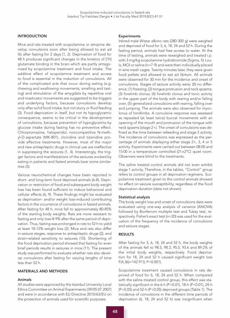
48
Scopolamine-induced convulsions in fasted ratsİstanbul Tıp Fakültesi Dergisi • J Ist Faculty Med 2019;82(1):47-51
INTRODUCTION
Mice and rats treated with scopolamine or atropine de-velop convulsions soon after being allowed to eat ad lib after fasting for 2 days (1, 2). Deprivation of food for 48 h produces significant changes in the kinetics of [3H]glutamate binding in the brain which are partly antago-nized by scopolamine treatment and food intake. The additive effect of scopolamine treatment and access to food is essential in the induction of convulsions. All of the complicated acts that occur during eating (e.g. chewing and swallowing movements, smelling and tast-ing) and stimulation of the amygdala by repetitive oral and masticator movements are suggested to be triggers and underlying factors, because convulsions develop only after solid food intake, but not slurry or fluid feeding (3). Food deprivation in itself, but not its hypoglycemic consequence, seems to be critical in the development of convulsions, because prevention of hypoglycemia by glucose intake during fasting has no preventive effect. Chlorpromazine, haloperidol, noncompetitive N-meth-yl-D-aspartate (MK-801), clonidine and tizanidine pro-vide effective treatments. However, most of the major and new antiepileptic drugs in clinical use are ineffective in suppressing the seizures (1, 4). Interestingly, the trig-ger factors and manifestations of the seizures evoked by eating in patients and fasted animals bear some similar-ities (5).
Various neurochemical changes have been reported in short- and long-term food deprived animals (6-8). Depri-vation or restriction of food and subsequent body weight loss has been found sufficient to induce behavioral and cellular effects (6, 9). These findings might be evaluated as deprivation- and/or weight loss-induced contributing factors in the occurrence of convulsions in fasted animals. After fasting for 48 h, mice fall to approximately 80-85% of the starting body weights. Rats are more resistant to fasting and only lose 8-9% after the same period of depri-vation. Thus, fasting was prolonged in rats to 52 h to yield at least 10-12% weight loss (2). Mice and rats also differ in seizure stages, response to antiepileptic drugs (2), and strain-related sensitivity to seizures (10). Shortening of the food deprivation period showed that fasting for even brief periods results in seizures in mice (11). The present study was performed to evaluate whether rats also devel-op convulsions after fasting for varying lengths of time less than 52 h.
MATERIALS AND METHODS
AnimalsAll studies were approved by the Istanbul University Local Ethics Committee on Animal Experiments (39/05.07.2007) and were in accordance with EU Directive 2010/63/EU on the protection of animals used for scientific purposes.
ExperimentsInbred male Wistar albino rats (280-300 g) were weighed and deprived of food for 3, 6, 18, 24 and 52 h. During the fasting period, animals had free access to water. At the time of testing, animals were reweighed and treated i.p. with 3 mg/kg scopolamine hydrobromide (Sigma, St Lou-is, MO) or saline (n=7-9) and were then individually placed in wire mesh cages. Twenty minutes later, they were given food pellets and allowed to eat ad libitum. All animals were observed for 30 min for the incidence and onset of convulsions. Stages of seizure activity were; (0) no differ-ence; (1) freezing; (2) tongue protrusion and neck spasms; (3) forelimb clonus; (4) forelimb clonus and tonic activity in the upper part of the body with rearing and/or falling over; (5) generalized convulsions with rearing, falling over and jumping. The animals were also observed for myoc-lonus of hindlimbs. A convulsive response was assessed as repeated (at least twice) buccal movements with an opening of the mouth and protrusion of the tongue with neck spasms (stage 2+). The onset of convulsions was de-fined as the time between refeeding and stage 2 activity. The incidence of convulsions was expressed as the per-centage of animals displaying either stage 2+, 3, 4 or 5 activity. Experiments were carried out between 08:00 and 15:00 in a temperature controlled (21±2ºC) quiet room. Observers were blind to the treatments.
The saline treated control animals did not even exhibit stage 1 activity. Therefore, in the tables, “Control” group refers to control groups in all deprivation regimens. Sco-polamine treatment given to the control animals showed no effect on seizure susceptibility, regardless of the food deprivation duration (data not shown).
Statistical analysisThe body weight loss and onset of convulsions data were evaluated using one-way analysis of variance (ANOVA) followed by Bonferroni multiple test and Tukey test, re-spectively. Fisher’s exact test (n<20) was used for the eval-uation of the frequency of the incidence of convulsions and seizure stages.
RESULTS
After fasting for 3, 6, 18, 24 and 52 h, the body weights of the animals fell to 98.5, 98.3, 95.5, 93.6 and 89.2% of the initial body weights, respectively. Food depriva-tion for 18, 24 and 52 h caused significant weight loss F(4,36)=142.913; P<0.001).
Scopolamine treatment caused convulsions in rats de-prived of food for 6, 18, 24 and 52 h. When compared with the saline-treated control group, this effect was sta-tistically significant in the 6-h (P<0.01), 18-h (P<0.01), 24-h (P<0.05) and 52-h (P<0.05) deprived groups (Table 1). The incidence of convulsions in the different time periods of deprivation (6, 18, 24 and 52 h) was insignificant when
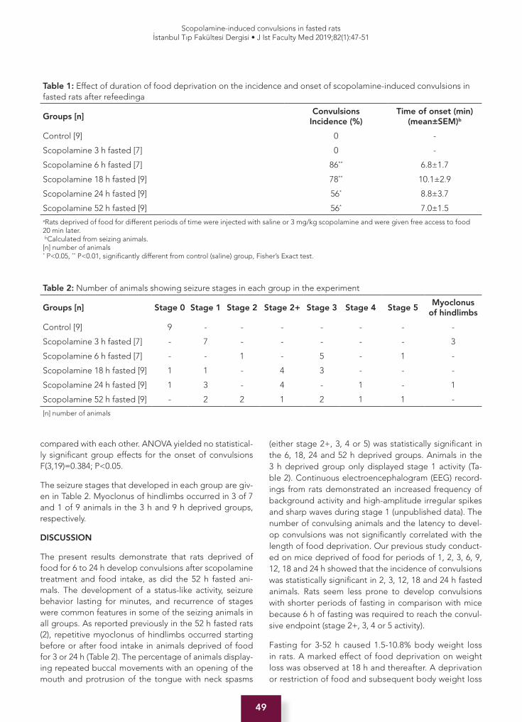
49
Scopolamine-induced convulsions in fasted ratsİstanbul Tıp Fakültesi Dergisi • J Ist Faculty Med 2019;82(1):47-51
compared with each other. ANOVA yielded no statistical-ly significant group effects for the onset of convulsions F(3,19)=0.384; P<0.05.
The seizure stages that developed in each group are giv-en in Table 2. Myoclonus of hindlimbs occurred in 3 of 7 and 1 of 9 animals in the 3 h and 9 h deprived groups, respectively.
DISCUSSION
The present results demonstrate that rats deprived of food for 6 to 24 h develop convulsions after scopolamine treatment and food intake, as did the 52 h fasted ani-mals. The development of a status-like activity, seizure behavior lasting for minutes, and recurrence of stages were common features in some of the seizing animals in all groups. As reported previously in the 52 h fasted rats (2), repetitive myoclonus of hindlimbs occurred starting before or after food intake in animals deprived of food for 3 or 24 h (Table 2). The percentage of animals display-ing repeated buccal movements with an opening of the mouth and protrusion of the tongue with neck spasms
(either stage 2+, 3, 4 or 5) was statistically significant in the 6, 18, 24 and 52 h deprived groups. Animals in the 3 h deprived group only displayed stage 1 activity (Ta-ble 2). Continuous electroencephalogram (EEG) record-ings from rats demonstrated an increased frequency of background activity and high-amplitude irregular spikes and sharp waves during stage 1 (unpublished data). The number of convulsing animals and the latency to devel-op convulsions was not significantly correlated with the length of food deprivation. Our previous study conduct-ed on mice deprived of food for periods of 1, 2, 3, 6, 9, 12, 18 and 24 h showed that the incidence of convulsions was statistically significant in 2, 3, 12, 18 and 24 h fasted animals. Rats seem less prone to develop convulsions with shorter periods of fasting in comparison with mice because 6 h of fasting was required to reach the convul-sive endpoint (stage 2+, 3, 4 or 5 activity).
Fasting for 3-52 h caused 1.5-10.8% body weight loss in rats. A marked effect of food deprivation on weight loss was observed at 18 h and thereafter. A deprivation or restriction of food and subsequent body weight loss
Table 1: Effect of duration of food deprivation on the incidence and onset of scopolamine-induced convulsions in fasted rats after refeedinga
Groups [n]Convulsions
Incidence (%) Time of onset (min)
(mean±SEM)b
Control [9] 0 -
Scopolamine 3 h fasted [7] 0 -
Scopolamine 6 h fasted [7] 86** 6.8±1.7
Scopolamine 18 h fasted [9] 78** 10.1±2.9
Scopolamine 24 h fasted [9] 56* 8.8±3.7
Scopolamine 52 h fasted [9] 56* 7.0±1.5aRats deprived of food for different periods of time were injected with saline or 3 mg/kg scopolamine and were given free access to food 20 min later. bCalculated from seizing animals.[n] number of animals* P<0.05, ** P<0.01, significantly different from control (saline) group, Fisher’s Exact test.
Table 2: Number of animals showing seizure stages in each group in the experiment
Groups [n] Stage 0 Stage 1 Stage 2 Stage 2+ Stage 3 Stage 4 Stage 5 Myoclonus
of hindlimbs
Control [9] 9 - - - - - - -
Scopolamine 3 h fasted [7] - 7 - - - - - 3
Scopolamine 6 h fasted [7] - - 1 - 5 - 1 -
Scopolamine 18 h fasted [9] 1 1 - 4 3 - - -
Scopolamine 24 h fasted [9] 1 3 - 4 - 1 - 1
Scopolamine 52 h fasted [9] - 2 2 1 2 1 1 -
[n] number of animals

50
Scopolamine-induced convulsions in fasted ratsİstanbul Tıp Fakültesi Dergisi • J Ist Faculty Med 2019;82(1):47-51
has been found sufficient to induce behavioral and cel-lular effects (6, 9, 12). However, in the present study, the convulsive response seemed to be unrelated to weight loss induced by fasting, because 6 h deprived animals develop convulsions without significant reductions in body weight. These results confirm previous findings that food deprivation or fasting stress, rather than the magnitude of weight loss, influences susceptibility to seizures (2, 11).
Based upon biochemical and behavioral data, we previ-ously suggested adaptive adjustments in glutamatergic, dopaminergic and cholinergic systems as ways to meet the demands of food deprivation (11). Accordingly, our ongoing studies in mice evaluate fasting-induced M1 and M2 muscarinic receptor expression in different brain re-gions. Various neuroadaptive changes in the brain have been reported in rats deprived of food for 24 or 16-18 h (13, 14). However, the findings in the present study im-ply neuroadaptations as underlying mechanisms of these convulsions, which may occur even after a few hours of food deprivation in animals. Acute stress elicits a prom-inent form of synaptic plasticity in midbrain dopamine neurons (15) where basal extracellular dopamine levels are decreased by food deprivation (6). It has been sug-gested that acetylcholine and glutamate possess regula-tory roles in dopaminergic function (16). Although specu-lative, food deprivation, which is regarded as a stressor, might also produce neuroadaptive changes in mesolim-bic dopamine release, which mediates convulsions in fasted animals.
In conclusion, the present study demonstrates that depri-vation of food for 6, 12, 18 and 24 h causes convulsions in scopolamine-treated and refed rats. In accordance with those observed in mice, the results indicate that convul-sions are primarily affected by food deprivation stress, but not by weight loss, which provides additional data for the underlying mechanism(s) of this convulsion method.
Acknowledgement: The authors would like thank Mr. David F. Chapman (Language Editor, Istanbul Faculty of Medicine) for editing the manuscript.
Ethics Commitee Approval: All studies were approved by the Istanbul University Local Ethics Committee on Animal Experi-ments (39/05.07.2007) and were in accordance with EU Directive 2010/63/EU on the protection of animals used for scientific pur-poses.
Informed Consent: Not required.
Peer Review: Externally peer-reviewed.
Author Contributions: Conception/Design of Study- N.E., A.N., A.Z.T.; Data Acquisition- N.E., A.N., A.Z.T.; Data Analysis/Inter-pretation- N.E., A.N., A.Z.T.; Drafting Manuscript- N.E., A.N.,
A.Z.T.; Critical Revision of Manuscript- N.E., A.N., A.Z.T.; Final Approval and Accountability- N.E., A.N., A.Z.T.; Technical or Ma-terial Support- N.E., A.N., A.Z.T.
Conflict of Interest: Authors declared no conflict of interest.
Financial Disclosure: Authors declared no financial support.
Teşekkür: Yazarlar, Sayın David F. Chapman’a (İstanbul Tıp Fakülte-si Dil Editörü) metindeki dil düzeltmeleri için teşekkür ederler.
Etik Komite Onayı: Tüm çalışmalar İstanbul Üniversitesi Hay-van Deneyleri Yerel Etik Komitesi (39/05.07.2007) tarafından onaylandı ve bilimsel amaçlı kullanılan hayvanların korunmasına ilişkin 2010/63 / EU sayılı AB Direktifine uygun olarak yapıldı.
Bilgilendirilmiş Onam: Gerekli değil.
Hakem Değerlendirmesi: Dış bağımsız.
Yazar Katkıları: Çalışma Konsepti/Tasarım- N.E., A.N., A.Z.T.; Veri Toplama- N.E., A.N., A.Z.T.; Veri Analizi/Yorumlama- N.E., A.N., A.Z.T.; Yazı Taslağı- N.E., A.N., A.Z.T.; İçeriğin Eleştirel İnce-lemesi- N.E., A.N., A.Z.T.; Son Onay ve Sorumluluk- N.E., A.N., A.Z.T.; Malzeme ve Teknik Destek- N.E., A.N., A.Z.T.
Çıkar Çatışması: Yazarlar çıkar çatışması beyan etmemişlerdir.
Finansal Destek: Yazarlar finansal destek beyan etmemişlerdir.
REFERENCES
1. Enginar N, Nurten A. Seizures triggered by food intake in antimuscarinic-treated fasted animals: Evaluation of the experimental findings in terms of similarities to eating-triggered epilepsy. Epilepsia 2010;51(Suppl. 3):80-4. [CrossRef]
2. Nurten A, Enginar N. The evaluation of antimuscarinic-induced convulsions in fasted rats after food intake. Epilepsy Res 2006;72(2-3):171-7. [CrossRef]
3. Nurten A, Özerman B, Özen İ, Kara İ. The role of solid fluid intake in antimuscarinic-induced convulsions in fasted mice. Epilepsy Behav 2009:15(2):142-5. [CrossRef]
4. Büget B, Zengin Türkmen A, Allahverdiyev A, Enginar N. Antimuscarinic-induced convulsions in fasted animals after food intake: Evaluation of the effects of levetiracetam, topiramate and different doses of atropine. Naunyn-Schmiedeberg’s Arch 2016;389(1):57-62. [CrossRef]
5. Italiano D, Ferlazzo E, Gasparini S, Spina E, Mondello S, Labate A, et al. Generalized versus partial reflex seizures: a review. Seizure 2014;23(7):512-20. [CrossRef]
6. Pothos EN, Creese I, Hoebel BG. Restricted eating with weight loss selectively decreases extracellular dopamine in the nucleus accumbens and alters dopamine response to amphetamine, morphine, and food intake. J Neurosci 1995;15(10):6640-50. [CrossRef]
7. Karami KJ, Coppola J, Krishnamurthy K, Llanos DJ, Mukherjee A, Venkatachalam KV. Effect of food deprivation and hormones of glucose homeostasis on the acetyl CoA carboxylase activity in mouse brain: a potential role of acc in the regulation of energy balance. Nutr Metabol (Lond) 2006;3:15. [CrossRef]
51
Scopolamine-induced convulsions in fasted ratsİstanbul Tıp Fakültesi Dergisi • J Ist Faculty Med 2019;82(1):47-51
8. Wu Q, Lemus MB, Stark R, Bayliss JA, Reichenbach A, Lockie SH, et al. The temporal pattern of cfos activation in hypothalamic, cortical, and brainstem nuclei in response to fasting and refeeding in male mice. Endocrinology 2014;155(3):840-53. [CrossRef]
9. Hwang BH, Guntz JM. Downregulation of corticotropin-releasing factor mRNA, but not vasopressin mRNA, in the paraventricular hypothalamic nucleus of rats following nutritional stress. Brain Res Bull 1997;43(5):509-14. [CrossRef]
10. Enginar N, Nurten A, Zengin Türkmen A, Büyüklü Ç. Scopolamine-induced convulsions in fasted animals after food intake: Sensitivity of C57BL/6J mice and Sprague-Dawley rats. Epilepsy Res 2015;112:150-3. [CrossRef]
11. Enginar N, Nurten A, Özünal ZG, Zengin A. Scopolamine-induced convulsions in fasted mice: The effect of duration of food deprivation. Epilepsia 2009;50(1):143-6. [CrossRef]
12. Squadrito F, Calapai G, Altavilla D, Cucinotta D, Zingarelli B, Campo GM, et al. Food deprivation increases brain nitric oxide synthase and depresses brain serotonin levels in rats. Neuropharmacology 1994;33(1):83-6. [CrossRef]
13. Maniscalco JW, Rinaman L. Overnight food deprivation markedly attenuates hindbrain noradrenergic, glucagon-like peptide-1 and hypothalamic neural responses to exogenous cholecystokinin in male rats. Physiol Behav 2013;121:35-42. [CrossRef]
14. Saal D, Dong Y, Bonci A, Malenka RC. Drugs of abuse and stress trigger a common synaptic adaptation in dopamine neurons. Neuron 2003;37(4):577-82. [CrossRef]
15. Adell A, Artigas F. The somatodentritic release of dopamine in the ventral tegmental area and its regulation by afferent transmitter systems. Neurosci Biobehav Rev 2004;28(4):415-31. [CrossRef]
52
İlaç Uyumunu Bildirim Ölçeği’nin Türkçe uyarlaması İstanbul Tıp Fakültesi Dergisi • J Ist Faculty Med 2019;82(1):52-61
İletişim kurulacak yazar/Corresponding author: [email protected]
Geliş tarihi/Received Date: 09.04.2018 • Kabul tarihi/Accepted Date: 18.01.2019
©Telif Hakkı 2019 J Ist Faculty Med - Makale metnine jmed.istanbul.edu.tr web sayfasından ulaşılabilir.©Copyright 2019 by J Ist Faculty Med - Available online at jmed.istanbul.edu.tr
ARAŞTIRMA / RESEARCH DOI: 10.26650/IUITFD.413637
İst Tıp Fak Derg 2019 / J Ist Faculty Med 2019
İLAÇ UYUMUNU BİLDİRİM ÖLÇEĞİ’NİN TÜRKÇE UYARLAMASININ GEÇERLİK VE GÜVENİRLİK ÇALIŞMASI
THE VALIDITY AND RELIABILITY STUDY OF THE TURKISH ADAPTATION OF MEDICAL ADHERENCE REPORT SCALE
Esin TEMELOĞLU ŞEN1 , Özlem SERTEL BERK1 , Dilşad SİNDEL2
1İstanbul Üniversitesi, Edebiyat Fakültesi, Psikoloji Bölümü, İstanbul, Türkiye2İstanbul Üniversitesi, İstanbul Tıp Fakültesi, Fiziksel Tıp ve Rehabilitasyon Anabilim Dalı, İstanbul, Türkiye
ORCID IDs of the authors: E.T.Ş. 0000-0002-1941-4786; Ö.S.B. 0000-0002-3045-3903; D.S. 0000-0002-3034-4635
Cite this article as: Temeloğlu-Şen E, Sertel-Berk Ö, Sindel D. The validity and reliability study of the Turkish adaptation of the medical adherence report scale. J Ist Faculty Med 2019;82(1):52-61. doi: 10.26650/IUITFD.413637
ÖZETAmaç: Kronik fiziksel hastalığı olan bireylerin ilaca uyum göstermesi tedavinin önemli bir parçasını oluşturmaktadır. Hastanın ilaç tedavisi-ne uyumunu değerlendirmek hem uyumun tanımlanmasının zorluğu hem de hastanın olumsuzu bildirmede zorlanması sebebiyle zor ol-maktadır. Klinik araştırmalarda ve uygulamada ilaç uyumunu değer-lendirmek için klinik ölçümler ve öz bildirim ölçüm araçları olmak üze-re iki farklı yöntem kullanılmaktadır. Hem ekonomik hem de tutarlı bir değerlendirme sunması sebebiyle öz bildirim ölçüm araçlarına alanda sıklıkla başvurulmaktadır. Türkçe literatürde ilaç uyumunu ölçen bazı ölçekler olmasına rağmen boyutsal ve jenerik değerlendirme sunan bir ölçeğin eksikliği dikkat çekmektedir. İlaç Uyumunu Bildirim Ölçeği (İUBÖ) medikal tedavide ilaç uyumunu kısa ve doğru değerlendirmek amacıyla geliştirilen bir öz bildirim ölçeğidir. Bu çalışmanın amacı İU-BÖ’nün kronik fiziksel hastalığı olan bireylerde Türkçe güvenirlik ve geçerliliğinin incelenmesidir.
Yöntemler: Araştırmanın örneklemini kronik fiziksel hastalığı nedeniy-le farklı tanılarla ayaktan tedavi gören ve ilaç kullanan yaşları 18 ile 65 arasında değişen 234 kişi oluşturmaktadır. Araştırmanın katılımcıların-dan Sosyodemografik Form, İlaç Uyumunu Bildirim Ölçeği (İUBÖ), Av-rupa Yaşam Kalitesi Ölçeği-5 Boyut (EQoL-5D), Morisky Uyum Ölçeği (MUÖ) ve Kısa Semptom Envanterini (KSE) doldurmaları istenmiştir.
Bulgular: Uygulanan açımlayıcı (KMO=0,79; Barlett p=0,000) ve doğ-rulayıcı faktör analizleri (GFI=0,98, AGFI=0,93, CFI=0,97, NFI=0,96, sRMR=0,040, RMSEA=0,089) ölçeğin orijinal formu ile tutarlı olarak tek faktörlü yapıyı desteklediğini göstermiştir. Ayrıca, Türkçe formun iç tutarlılık katsayısı (Cronbach α=0,78) ve toplam madde korelasyon-ları (0,35 ile 0,71 arasında) orijinal versiyonla benzer psikometrik özel-liklere sahip olduğunu desteklemiştir.
Sonuç: Yapılan çalışma, İUBÖ’nün Türkçe formunun güvenilir ve ge-çerli bir ölçüm aracı olduğuna dair yeterli kanıtlar sunmaktadır. Ölçüm aracı bu haliyle farklı kronik hastalık tanısı ile takip edilen hastalarda ilaç uyumunun değerlendirilmesi amacıyla kullanılabilir.
Anahtar Kelimeler: Kronik fiziksel hastalık, ilaç uyumu, ilaç uyumunu bildirim ölçeği, güvenirlik, geçerlik
ABSTRACTObjective: The medication adherence of patients with chronic phys-ical illnesses is an important aspect of the treatment. However it’s difficult to assess the patient’s adherence with medication treatment, both because of the difficulty of defining adherence and the patients reluctance for reporting nonadherence. Two different methods are used to evaluate medication adherence which employ clinical mea-sures and self-report measures. Due to the fact that it provides an economical and consistent evaluation, self-report tools are frequent-ly used in the field. Although there are some scales that measure medication adherence in the Turkish literature, the lack of a scale that provides dimensional and generic evaluation is noteworthy. Medica-tion Adherence Report Scale (MARS) is a generic self report scale developed to assess medication adherence in medical treatment. The aim of this study is to examine the Turkish reliability and validity of MARS in patients with chronic physical illnesses.
Methods: The sample of the study consisted of 234 patients who were treated with different diagnoses in chronic physical illness. Par-ticipants were asked to fill in the Sociodemographic Form, Medica-tion Adherence Report Scale (MARS), the European Quality of Life Scale-5 Dimension (EQoL-5D), the Morisky Adhedrence Scale (MAS), and the Brief Symptom Inventory (BSI).
Results: The exploratory (KMO=0.79; Barlett p=0.000) and confir-matory factor analysis (GFI=0.98, AGFI=0.93, CFI=0.97, NFI=0.96, srmR=0.040, RMSEA=0.089) revaled a uni-factor structure consistent with the original form. In addition, the internal consistency coefficient of the Turkish version (Cronbach α=0.78) and total item correlations (between 0.35 and 0.71) showed similar psychometric properties to those of the original version.
Conclusion: The study provides sufficient evidence that the Turkish version of the MARS is a reliable and valid measurement tool. Thus MARS can be used to assess medication adherence in cases of differ-ent chronic illnesses.
Keywords: Chronic physical disease, medication adherence, medi-cation adherence report scale, reliability, validity

53
İlaç Uyumunu Bildirim Ölçeği’nin Türkçe uyarlaması İstanbul Tıp Fakültesi Dergisi • J Ist Faculty Med 2019;82(1):52-61
GİRİŞ
Kronik hastalıklar, yavaş ilerleyen, üç ay veya daha uzun süren, birden fazla nedene sahip olabilen, yaşam kalitesi-ni etkileyen, sürekli tıbbi hizmet gerektiren ve böylece bir kişinin günlük yaşantısını kısıtlayan zorlayıcı durumlar ola-rak tanımlanır (1). Biyomedikal model, kronik hastalıkla-rın tedavisinde temel paradigma olarak kullanılmaktadır. Model, hastalığın nedenini bedeni oluşturan anatomik parçalarla açıklamaktadır. Tedavide ilaç, cerrahi müdaha-le ve teknolojik destekler kullanılmaktadır. İlaçla tedavi, kronik fiziksel hastalıklarda hem en sık hem de en uzun süreyle kullanılan yöntemdir (2). Uzun süren bu tedaviye uyum göstermek, hastalığın seyrini etkileyen önemli de-ğişkenlerden birisidir. Bir başka deyişle, kronik hastalığı olan hastalarda ilaç uyumu hastalığın tedavisi ve semp-tomların kontrolü için önemli bir sağlık davranışıdır (3, 4).
Dünya Sağlık Örgütü (DSÖ) tarafından, tedavi uyumu genel ya da medikal sağlık önerilerini içselleştirme, ilaç alma, diyete uyum sağlama ve yaşam stilindeki değişik-likleri devam ettirme gibi davranışların kesişimi olarak tanımlanmaktadır (1). Bu tanımdan da anlaşılacağı üzere uyum çok boyutlu bir kavramdır (5). İlaç tedavisine uyum ise hastanın ilaçlarının reçete edilmesi, ilaçlara başlama (ilk dozun alınacağı zamanlama), devam etme (kullanıldığı zaman içerisinde her dozun alınma özellikleri) ve ilaçları sonlandırma (ilacı almayı bırakma) süreçlerine ve bu sü-reçte aldığı önerileri uygulama kararlarının tümüne veri-len isimdir (6, 7).
Uyumla ilgili 1960’ların sonundan beri birçok çalışma yü-rütülmektedir. Yapılan çalışmalar uyumu doğru değerlen-direcek bir ölçek geliştirme, uyumla ilişkili faktörleri daha iyi anlamak ve uyumu arttıracak müdahaleler geliştirmeyi amaçlamaktadır. Ancak tüm bunlara rağmen hala kronik fiziksel hastalığı olan bireylerin neredeyse %50’sinin reçe-telerini tarif edildiği gibi kullanmadığı dikkat çekmektedir (8-10). İlaç uyumunun düşük olması, hasta memnuniyetini düşürebilmesinin, kişilerin sağlıklı olma şansını ellerin-den kaybetmesinin ve hastalanma/ölümlülük oranlarını arttırmasının yanında ülkelerin milli gelirine ciddi bir yük oluşturmaya ve kaynakların boşa harcanmasına neden olmaktadır. Yani ilaç uyumsuzluğunun bireysel ve küre-sel ölçekte çokça zararı bulunmaktadır. Bu nedenle hem teorik hem de pratik perspektifte ilaç uyumunun doğru değerlendirilmesi önem taşımaktadır (11, 12).
İlaç uyumu kavramı hem tanımının zorluğu ve karmaşıklı-ğı (13) hem de uyumsuzluğun yarattığı kişisel rahatsızlıkla baş etmekte zorlanma ve sosyal istenirlik kaygısı (9) sebe-biyle değerlendirilmesi zor bir kavramdır (14). İlaç uyumu-nu değerlendirmek amacıyla hasta günlüğü, öz bildirim ölçeklerinin kullanımı gibi hastaların bildirimine dayalı ya da hap sayımı, kan sayımındaki ilaç oranına bakma, elekt-ronik göstergeler gibi objektif ölçümler kullanılmaktadır. Bunca yönteme rağmen, bir yöntemin diğerine göre üs-
tün olduğunu gösteren bir çalışma literatürde yer alma-maktadır (9). Literatürde ilaç uyumunu değerlendirmede kısa ve doğru bir bilgilendirme sunan öz bildirim ölçekle-rinin sıklıkla kullanıldığı görülmektedir (14). Morisky Uyum Ölçeği (MUÖ) bu amaçla en sık kullanılan jenerik ve ilaç uyum davranışına kapsamlı bir tanım getirmesi açısından önemli bir ölçektir. Ancak ölçek ilaç uyumunu kategorik bir değerlendirme ile var/yok olarak ele alması ve iç tu-tarlılık katsayısının düşüklüğü açısından eleştirilmektedir (15). MUÖ’nün sözü edilen bu dezavantajlarını da dikkate alarak geliştirilmiş ölçeklerden biri olan İlaç Uyumunu Bil-dirim Ölçeği (İUBÖ) (orijinal adıyla Medication Adheren-ce Report Scale), olumsuz ilaç uyumu davranışını içeren beş farklı ifadeden (ilaç dozunu atlamaya karar vermek, ilacı almayı unutmak, ilaç dozunu değiştirmek, bir süre-liğine ilaç alımını durdurmak ve tarif edildiğinden az doz almak) oluşmaktadır (16). İUBÖ, geliştirilen pek çok ölçe-ğin aksine uyum davranışının sürekliliğine vurgu yapmak-tadır. Ancak bunun yanında ölçekten elde edilen toplam ilaç uyum puanına göre kategorik bir değerlendirme ya-pabilmek de mümkündür (16, 17).
Öte yandan ilaç uyumunu etkileyen pek çok değişken söz konusudur. Bunlardan ilkinin çoklu ilaç kullanımı olabile-ceği ifade edilmektedir. Çoklu ilaç kullanan bireylerin ilaç uyumlarının düşük olduğu belirtilmektedir (18, 19). İlaç uyumu ile ilişkili diğer değişkenler ise yaşam kalitesi ve psikolojik belirti düzeyidir. Literatürde ilaç uyumu yüksek olan bireylerin yaşam kalitelerinin yüksek ve psikolojik şi-kayetlerinin düşük olacağı belirtilmektedir (20,23). Yapı-lan çalışmalarda İUBÖ’nün yaşam kalitesi ile düşük ancak anlamlı ilişki gösterdiği (20, 22, 23), psikolojik belirtiler ile yüksek ve anlamlı ilişkide olduğu (21, 22) görülmüştür. Bu nedenle ölçeğin Türkçe adaptasyon çalışmasında yaşam kalitesi ve psikolojik belirtilerle olan ilişkisinin incelenme-sinin uygun olacağı düşünülmüştür.
İUBÖ’nün orijinal çalışmasında ölçeğin tek faktörlü bir ya-pıdan oluştuğu, kriter ve ayırt edici geçerlilik bakımından iyi ve yeterli güvenilirliğe sahip olduğu (Cronbach Alp-ha=0.85) görülmüştür (16). Ölçek astım (24, 25), KOAH (26), kardiyovasküler hastalıklar (23), diyabet (27), infla-matuvar bağırsak hastalığı (28), depresyon (27) ve bipolar bozukluklar (24) gibi kronik hasta popülasyonlarında ilaca uyumu değerlendirmek için kullanılmaktadır (17). Bunun yanında ölçeğin Almanca, Danca, Hintçe, Arapça, Lehçe, Yunanca, Fransızca, İtalyanca ve İspanyolca dillerine de uyarlaması mevcut olup, uyarlama çalışmalarında orijinal form ile benzer psikometrik özellikler gösterdiği görül-mektedir (11, 23).
Bu çalışmada İUBÖ’nün Türkçe uyarlamasının yapılması amaçlanmaktadır. Bu doğrultuda yapı geçerliği kapsa-mında ölçeğin faktör yapısının orijinalindeki gibi tek bo-yutlu olması, maddelerin ayırt edici olması, aynı zamanda yakınsak geçerliği için, ilaç uyumunu ölçen başka bir öl-

54
İlaç Uyumunu Bildirim Ölçeği’nin Türkçe uyarlaması İstanbul Tıp Fakültesi Dergisi • J Ist Faculty Med 2019;82(1):52-61
çüm aracı ile de güçlü ilişki gösteriyor olması beklenmek-tedir. Yanı sıra ilaç uyum davranışının yukarıda da özetlen-diği üzere ilişkili olabildiği önemli değişkenler olan yaşam kalitesi ve psikolojik sağlıkla da yine anlamlı ilişkiler gös-teriyor olması öngörülmektedir. Ayrıca, ayırt edici geçer-lilik kapsamında çoklu tanının ilaç uyumu toplam puanı üzerindeki etkisinin de değerlendirilmesi planlanmakta-dır. Ölçeğin güvenirliği açısından da yeterli iç tutarlık gös-termesi beklentiler arasındadır.
YÖNTEMLER
Katılımcı ÖzellikleriÖrneklemi en fazla iki kronik fiziksel hastalık tanısı almış, ayaktan tedavi gören ve sürekli ilaç kullanan yetişkin 234 hasta oluşturmaktadır. Kronik fiziksel hastalıklar ICD 10 kodları temel alınarak sınıflandırılmıştır (29). Katılımcıların hastalık türleri Tablo 1’de gösterilmektedir. Örneklemi oluşturan 61 (%26) hastanın birden fazla kronik fiziksel hastalığı olduğu görülmüştür. Katılımcıların yaşları 18 ile 65 (ort: 44,48; ss: 13,21) arasında değişmektedir. Örnekle-min %76’sı kadın (n=178), %24’ü (n=56) erkek; %68’i evli, %20’si bekar; %3’ü boşanmış ve %7’si dul; %7’si okuma yazma bilmeyen, %25’i ilkokul mezunu, %11’i ilköğretim mezunu, %33’ü lise mezunu, %6’sı yüksekokul mezu-nu, %14’ü üniversite mezunu ve %6’sı lisansüstü eğitimi almıştır. Katılımcılardan %6’sı gelir seviyesini alt, %12’si alt orta, %76’sı orta ve %6’sı üst orta olarak algılamakta; %34’ü çalışmakta, %63’ü çalışmamakta, %3’ü ise hastalık-tan dolayı işini bırakmak zorunda kaldığını bildirmektedir. Araştırmaya katılan kişilerin %6’sı 3-6 ay arasında, %10’u 6 ay-1 yıl aralığında, %13’ü 1-3 yıl arasında, %14’ü 3-5 yıl aralığında, %57’sinin ise 5 yıldan uzun süredir ilaç kullan-
dıkları ve %23’ünün psikiyatrik destek aldığı görülmüştür. Katılımcıların sosyodemografik özelliklerine Tablo 2’de yer verilmiştir.
Kanser tanısı ile tedavi görmek hem prognozu hem de klinik görüntüleri bakımından diğer hastalıklardan farklılık gösterebilmesi ve 65 yaş üstü bireylerde polifarmasinin daha sık görülmesi ve bu doğrultuda ilaç uyumunun de-ğerlendirilmesinin zorlaşması sebebiyle bu iki durum ça-lışmada dışlama kriteri olarak seçilmiştir.
ÖlçeklerÇalışmada Sosyodemografik ve Hastalıkla İlgili Bilgiler Formu, İlaç Uyumunu Bildirim Ölçeği, Avrupa Yaşam Kali-tesi Ölçeği-5 Boyut, Morisky Uyum Ölçeği ve Kısa Semp-tom Envanteri kullanılmıştır. Tüm ölçekler, katılımcıların araştırmayla ilgili bilgilendirilmiş onamlarının alınmasıyla uygulanmıştır.
Sosyodemografik ve Hastalıkla İlgili Bilgiler FormuKatılımcıların cinsiyetlerinin, yaşlarının, medeni durumları-nın, gelir durumlarının, eğitim durumlarının, tanılarının ne olduğu, kaç senedir bu tanı/lar ile tedavi gördüklerinin, kullandıkları ilaçların ve ilaç kullanma sürelerinin değer-lendirildiği form araştırmacılar tarafından geliştirilmiştir. Katılımcılardan alınan bilgiler forma kaydedilmiştir.
İlaç Uyumunu Bildirim Ölçeği- Orijinal Form (İUBÖ)Horne ve Hankins (2001) tarafından ilaç uyumunu değer-lendirmek amacıyla geliştirilen Medication Adherence Report Scale (MARS) jenerik bir ölçek olup hastalık türüne göre özelleştirilebilmektedir (30). İUBÖ’nün tansiyon, di-yabet, romatoid artrit, astım, COPD, iki uçlu duygu durum bozukluğu, inflamatuvar bağırsak hastalığı, hiperlipidemi
Tablo 1. Örneklem Özellikleri*
Kadın (N) Erkek (N) Toplam Yüzde
Kro
nik
hast
alık
lar
Endokrin, beslenme ve metabolik hastalıklar 45 14 59 25
Sindirim sistemi hastalıkları 0 2 2 1
Kas ve iskelet sistemi hastalıkları 52 14 66 28
Dolaşım sistemi bozuklukları hastalıkları 39 19 58 25
Kan ve kan oluşturan organların hastalıkları hematolojik ve immün sistemle bağışıklıkla ilgili hastalılar
12 1 13 6
Cilt ve derialtı doku hastalıkları 3 1 4 2
Tümörler 4 0 4 2
Kulak ve mestoid hastalıkları 3 0 3 1
Sinir sistemi hastalıkları 7 3 10 4
Bazı enfeksiyonlar ve paraziter hastalıklar 3 2 5 2
Solunum sistemi hastalıkları 10 0 10 4
Toplam 178 56 234 100
*Bazı katılımcıların birden fazla kronik hastalığı olup bu tabloda primer olarak bildirdikleri tanıları dikkate alınmıştır.

55
İlaç Uyumunu Bildirim Ölçeği’nin Türkçe uyarlaması İstanbul Tıp Fakültesi Dergisi • J Ist Faculty Med 2019;82(1):52-61
Tablo 2. Katılımcıların Sosyodemografik Dağılımları
N % Ort. ss Min. Max
Hastalık sayısı
Tek hastalık 173
Birden fazla hastalık 61 26,07
Yaş 44,48 13,21 18 65
Cinsiyet
Kadın 178 76,1
Erkek 56 23,9
Medeni durum
Evli 160 68,4
Bekâr 46 19,7
Boşanmış 6 2,6
Dul 22 9,4
Eğitim Durumu
Okuma yazma bilmiyor 16 6,8
İlkokul mezunu, 59 25,2
İlköğretim mezunu 25 10,7
Lise mezunu 76 32,5
Yüksekokul mezunu 13 5,6
Üniversite mezunu 32 13,7
Lisansüstü mezunu 13 5,6
Gelir seviyesi
Alt 14 6
Alt orta 27 11,5
Orta 178 76,1
Üst orta 15 6,4
Çalışma durumu
Çalışıyor 79 33,8
Çalışmıyor 147 62,8
Hastalık sebebiyle işini bırakma 8 3,4
İlaç Kullanma Süresi
3-6 ay aralığı 14 6
6 ay-1 yıl aralığı 22 9,4
1-3 yıl aralığı 30 12,8
3-5 yıl aralığı 33 14,1
5 yıldan uzun süre 135 57,9
Psikiyatrik eş tanı
Var 53 22,6
Yok 178 76,1
Toplam 234 100
56
İlaç Uyumunu Bildirim Ölçeği’nin Türkçe uyarlaması İstanbul Tıp Fakültesi Dergisi • J Ist Faculty Med 2019;82(1):52-61
ve kronik ağrı gibi pek çok hastalığa yönelik uyarlamaları literatürde yer almaktadır (17). Katılımcılardan 5 ifadenin her birisinin kendilerinde görülme sıklıklarını belirtmeleri istenmektedir. Ölçek 5=hiç bir zaman, 4=nadiren, 3=ba-zen, 2=sık sık ve 1=çok sık olacak şekilde 5’li likert tipi ile değerlendirilmektedir. Maddelerden elde edilen puanlar toplanarak toplam test skoru elde edilmektedir. Ölçek-ten alınan puanlar 5 ile 25 arasında değişmektedir. Elde edilen puanlardaki yükselme uyumluluğa, puanlardaki düşme ise uyumsuzluğa işaret etmektedir (30). İUBÖ’nün tek faktör yapısı ile değerlendirildiği, kriter ve ayırt edici geçerlilik ve güvenirlik (Cronbach Alpha=0,85) bakımın-dan iyi düzeyde olduğu bulunmuştur (16).
İlaç Uyumunu Bildirim Ölçeği - Türkçe FormuİUBÖ’nün Türkçe’ye adaptasyonu için orijinal formun yazarlarından izin alındıktan sonra ölçek 2 İngilizce dil uzmanı tarafından orijinal dilinden Türkçeye çevrilmiştir. Oluşturulan iki Türkçe metin alanında uzman 5 psikolog tarafından her bir maddesi üzerine tartışılarak tek bir me-tin haline getirilmiştir. Türkçe metin iki Türk dili uzmanı tarafından ele alınmıştır. Türk dili uzmanlarının değer-lendirdiği metinler, alanında uzman iki psikolog tarafın-dan tekrar ele alınarak tek bir metin haline getirilmiştir. Oluşturulan metin yaşları 20-65 yaş arasında değişen 10 katılımcıya uygulanmıştır. Pilot çalışma kapsamında ka-tılımcılardan maddelerin anlaşılırlık düzeylerinin değer-lendirilmesi istenmiştir. Pilot çalışmanın sonuçlarına göre yeniden düzenlenen son metin tekrar iki ayrı dil uzmanı tarafından orijinal diline geri çevrilerek orijinal ölçeği ge-liştiren yazarlardan geri çevirinin uygunluğuna dair onay alınmıştır. Ölçeğin bu son hali geçerlilik ve güvenirlik ça-lışmasının veri toplama aşamasında kullanılmıştır.
Morisky Uyum Ölçeği (MUÖ)Ölçek kronik hastalığı olan bireylerin ilaç uyumlarını kısa ve doğru değerlendirmek amacıyla Morisky, Gren ve Le-vine (1986) geliştirilmiştir. MUÖ, dört sorudan oluşan bir öz bildirim ölçeğidir (31). Sorular “evet (1 puan) ve hayır (0 puan) şeklinde yanıtlanmaktadır. 0 puan ilaç uyumu yük-sek, bir veya iki soru “evet” ise (1-2 puan arası) orta, üç veya dört soru “evet” (3-4 puan arası) ilaç uyumu düşük olarak değerlendirilir. Ölçeğin ilk çalışmadaki Cronba-ch alpha değeri 0,61 olarak bulunmuştur (31). Uyarlama çalışması Yılmaz (2004) tarafından psikiyatrik hastalarla yapılmış ve Cronbach alpha katsayısı 0,52 olarak bulun-muştur (32). Ölçek bu araştırmada İUBÖ’nün yapı geçer-liliği kapsamında yakınsak geçerliğinin değerlendirilmesi amacıyla kullanılmıştır.
Avrupa Yaşam Kalitesi Ölçeği- 5 boyut (EQoL-5D)EuroQoL Grup tarafından 1987 senesinde geliştirilen, 1990 yılında yayınlanan ölçek fiziksel hastalığı olan birey-lerin 5 alandaki (sağlık, hareketlilik, kişisel bakım, günlük aktivite, ağrı ve anksiyete/depresyon) işlevselliğini ve VAS (Visual Analogue Scale) ile değerlendirilen genel sağlık
inancını sorgulayan 2 kısımdan oluşmaktadır. İndeks kısmı 3’lü likert tipi ile değerlendirilmektedir (33). EQoL-5D’nin indeks puan hesaplanmasında Dolan ve arkadaşlarının (1996) çalışmalarında kullandıkları katsayılar kullanılmak-tadır (34). Ölçeğin Türkçe geçerlilik güvenirlik çalışması Kahyaoğlu-Süt (2009) tarafından akut koroner sendrom-lu (AKS) hastalar ile yapılmıştır. Çalışmada EQoL-5D’nin güvenirlik katsayısı Cronbach α=0,86 olduğu ve alt bo-yutların güvenirlik katsayıları ise 0,87 ile 0,89 arasında değiştiği görülmüştür (34). Buna göre EQoL-5D’nin akut kalp sendromu hastalarında güvenilir bir ölçek olduğu söylenebilmektedir (34). Ölçek bu araştırmada İUBÖ’nün yapı geçerliliği kapsamında diğer ölçeklerle olan ilişkisi-nin değerlendirilmesi amacıyla kullanılmıştır.
Kısa Semptom Envanteri (KSE)KSE, Derogatis (1977) tarafından, kişilerin belirti düzeyini ve seyrini değerlendirme ve tanıya yardımcı olması ama-cıyla geliştirilmiş olan Psikolojik Belirti Tarama Testi’nin (SCL-90) formunun kısaltılmış halidir (35). KSE, 53 madde-den oluşan bir öz bildirim ölçeğidir. KSE, 5 dereceli Li-kert tipi ile değerlendirilmektedir. 9 alt test ve 1 ek skala olmak üzere toplam 10 semptom gurubundaki psikolojik belirtilerin zorlanma düzeylerini ele almaktadır. Her alt ölçek ayrı ayrı değerlendirileceği gibi genel durum ‘ge-nel semptom ortalaması’ olarak da puanlanabilmekte-dir. Genel Semptom Ortalaması (GSO) tüm maddelerin toplanıp madde sayısına bölünmesi ile elde edilmektedir (35). Türkçe güvenirlik ve geçerliliği Şahin ve Durak (1994) tarafından yapılmıştır. Güvenirliğine ilişkin sonuçlar, testin Cronbach Alpha iç tutarlılık katsayılarının 0,95 ve 0,96 ara-sında değiştiğini göstermiştir (36). Ölçek bu araştırmada İUBÖ’nün yapı geçerliliği kapsamında diğer ölçeklerle olan ilişkisinin değerlendirilmesi amacıyla kullanılmıştır.
İşlem Çalışma için İstanbul Üniversitesi Sosyal Bilimler Etik Ku-rulundan ve hasta alımı için İstanbul Üniversitesi İstanbul Tıp Fakültesi Fiziksel Tıp ve Rehabilitasyon (FTR) Anabilim Dalı Kürsü Kurulundan onaylar alınmıştır. Gerekli izinler alındıktan sonra veri toplama süreci iki şekilde devam et-miştir. Bir yandan İstanbul Üniversitesi İstanbul Tıp Fakül-tesi Fiziksel Tıp ve Rehabilitasyon (FTR) Anabilim Dalına başvuran hastalardan herhangi bir kronik fiziksel hastalığı olan katılımcılara ulaşılmıştır. Bir yandan da İstanbul Üni-versitesi Edebiyat Fakültesi Psikoloji Bölümü öğrencileri-nin yakınlarından kronik fiziksel hastalığı olanlara ulaşma-ları istenmiştir. Her iki süreçte de araştırma konusunda ve ölçeğin uygulanmasında eğitilmiş 30 öğrenciden destek alınmıştır. Uygulamada katılımcılardan Bilgilendirilmiş Onam Formunu incelemeleri istenmiş ve gönüllü olanlar çalışmaya dâhil edilmiştir.
İstatistiksel AnalizVerilerin istatistiksel olarak değerlendirilmesinde IBM SPSS for Windows 21.0 (ABD) paket programı kullanılmış
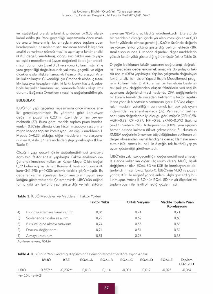
57
İlaç Uyumunu Bildirim Ölçeği’nin Türkçe uyarlaması İstanbul Tıp Fakültesi Dergisi • J Ist Faculty Med 2019;82(1):52-61
ve istatistiksel olarak anlamlılık p değeri p<0,05 olarak kabul edilmiştir. Yapı geçerliliği kapsamında önce mad-de analizi incelenmiş, bu amaçla madde toplam puan korelasyonları hesaplanmıştır. Ardından temel bileşenler analizi ve varimax döndürmesi ile açımlayıcı faktör analizi (KMO değeri) yürütülmüş, doğrulayıcı faktör analizi yapı-sal eşitlik modellemesi (uyum değerleri) ile değerlendiril-miştir. Bunun için Lisrel 8,51 versiyonu kullanılmıştır. Yine yapı geçerliliği doğrultusunda yakınsak geçerlik ve diğer ölçeklerle olan ilişkileri amacıyla Pearson Korelasyon Ana-lizi kullanılmıştır. Güvenirliği için Cronbach alpha iç tutar-lılık katsayısı hesaplanmıştır. İki farklı kronik hastalık sebe-biyle ilaç kullanılmasının ilaç uyumunda farklılık oluşturma durumu Bağımsız Örneklem t testi ile değerlendirilmiştir.
BULGULAR
İUBÖ’nün yapı geçerliği kapsamında önce madde ana-lizi gerçekleştirilmiştir. Bu yönteme göre korelasyon değerinin pozitif ve 0,20’nin üzerinde olması beklen-mektedir (37). Buna göre, madde-toplam puan korelas-yonları 0,20’nin altında olan hiçbir maddeye rastlanma-mıştır. Madde toplam korelasyonu en düşük maddenin 1. Madde (r=0,35) olduğu, diğer maddelerin korelasyonu-nun ise 0,54 ile 0,71 arasında değiştiği görülmüştür (bknz. Tablo 3).
Ölçeğin yapı geçerliliğinin değerlendirilmesi amacıyla açımlayıcı faktör analizi yapılmıştır. Faktör analizinin de-ğerlendirilmesinde kullanılan Kaiser-Meyer-Olkin değeri 0,79 bulunmuş ve Barlett Küresellik testi sonucunda (Ki kare=341,295; p=0,000) anlamlı farklılık görülmüştür. Bu değerler verinin açımlayıcı faktör analizi için uyum sağ-ladığını göstermektedir. Çalışmamızda İUBÖ’nün orijinal formu gibi tek faktörlü yapı gösterdiği ve tek faktörün
varyansın %54’ünü açıkladığı görülmektedir. Literatürde bir maddenin ölçeğin içinde yer alabilmesi için en az 0,30 faktör yükünde olması gerektiği, 0,60’ın üstünde değerin ise yüksek faktör yükünü gösterdiği belirtilmektedir (38). Analiz sonucunda 1. Madde dışındaki diğer maddelerin yüksek faktör yükü gösterdiği görülmüştür (bknz Tablo 3).
Ölçeğin belirlenen faktör yapısının doğrulanıp doğrula-namayacağını değerlendirmek amacıyla doğrulayıcı fak-tör analizi (DFA) yapılmıştır. Yapılan çalışmada doğrulayıcı faktör analizi için Lisrel Yapısal Eşitlik Modellemesi prog-ramı kullanılmıştır. DFA kuramsal bir temelden beslene-rek pek çok değişkenden oluşan faktörlerin veri seti ile uyumunu değerlendirmeyi hedefler. DFA değişkenlerin bir kuram temelinde önceden belirlenmiş faktör ağırlık-larına yönelik hipotezin sınanmasını içerir. DFA’da oluştu-rulan modelin yeterliliğini belirlemek için pek çok uyum indeksinden yararlanılmaktadır (39). Veri setinin belirle-nen uyum değerlerinin iyi olduğu görülmüştür (GFI=0,98, AGFI=0,93, CFI=0,97, NFI=0,96, sRMR=0,040) (bakınız Şekil 1). Sadece RMSEA değerinin (=0,089) uyum eşiğinin hemen altında kalması dikkat çekmektedir. Bu durumun RMSEA değerinin örneklem büyüklüğünden etkilenen bir değer olmasından kaynaklandığına dair açıklamalar mev-cuttur (40). Ancak bu hali ile ölçeğin tek faktörlü yapıya uyum gösterdiği görülmektedir.
İUBÖ’nün yakınsak geçerliliğin değerlendirilmesi amacıy-la alanda kullanılan diğer ilaç uyum ölçeği MUÖ, ilişkili değişkenler olan EQoL-5D ve KSE ile korelasyonları de-ğerlendirilmiştir (bknz. Tablo 4). İUBÖ’nün MUÖ ile pozitif yönde, KSE ile negatif yönde anlamlı ilişki gösterdiği bu-lunmuştur. Ancak İUBÖ’nün EQoL-5D’nin alt ölçekleri ve toplam puanı ile ilişkili olmadığı gözlenmiştir.
Tablo 3. İUBÖ Maddeleri ve Maddelerin Faktör Yükleri
Faktör Yükü Ortak Varyans Madde Toplam Puan Korelasyonu
4) Bir dozu atlamaya karar veririm. 0,86 0,74 0,71
5) Söylenenden daha az alırım. 0,79 0,62 0,60
3) Bir süreliğine almayı bırakırım. 0,74 0,55 0,58
2) Dozunu değiştiririm. 0,74 0,54 0,54
1) Almayı unuturum. 0,51 0,26 0,35
Açıklanan varyans, %54,26
Tablo 4. İUBÖ’nün Yapı Geçerliği Kapsamında Pearson Momentler Korelasyon Analizi
MUÖ KSE EQoL-A EQoL-B EQoL-C EQoL-D EQoL-E Toplam EQoL-5D
İUBÖ 0,557** -0,232** 0,013 0,114 -0,001 0,017 -0,075 -0,064
**p<0.01, *p<0.05

58
İlaç Uyumunu Bildirim Ölçeği’nin Türkçe uyarlaması İstanbul Tıp Fakültesi Dergisi • J Ist Faculty Med 2019;82(1):52-61
İUBÖ’nün güvenirliğinin değerlendirilmesi amacıyla iç tutarlılık analizi yapılmıştır. Ölçeğin iç tutarlılığının yüksek (Cronbach α=0,78) olduğu görülmüştür.
Örneklemi oluşturan katılımcıların İUBÖ ile değerlendiri-len ilaç uyumlarının komorbidite durumuna göre (komor-bidite var/yok) değişip değişmediğini sorgulamak için ise Mann Withney U testi uygulanmıştır. Yapılan analiz so-nucunda komorbiditesi olan (n=61) ve olmayan (n=173) hastaların İUBÖ toplam puanları arasında anlamlı bir fark olmadığı görülmüştür (Z=-0,966, p>05).
TARTIŞMA
Hastaların tıbbi tedaviden yarar sağlamasının en temel şartı ilaç uyumunun gerçekleşmesidir (7). Bu nedenle ilaç uyumunun doğru değerlendirilmesi oldukça önemli ol-maktadır. İlaç uyumunun değerlendirilmesinde kullanılan en yaygın yöntem öz bildirime dayalı ölçeklerin kullanıl-masıdır. Öz bildirime dayalı ölçekler basit ve ekonomik değerlendirme sağlaması sebebiyle tercih edilmektedir. Ancak öz bildirim ölçekleri iki temel ön yargı içerebilmek-tedir. Bunlardan ilki hastalar doktorlarını memnun etmek ya da doktorlar/diğer sağlık uzmanları tarafından yargı-lanmaktan kaçınmak için uyumlarını olduğundan iyi gös-terme eğiliminde olabilmektedirler. İkincisi ise hastaların beyanlarının hatalı olma ihtimalidir. Bu iki olasılık sebebiy-le özellikle “uyum”u değerlendiren öz bildirim ölçekle-rinde daha da dikkatli olunmasını gerektirmektedir (41).
İki uçlu değerlendirme yapan ölçekler uyumsuzluğu ya-kalamada daha hızlı bir değerlendirme imkânı sağlayabil-mektedir. Ancak hastalar çeşitli derecelerde uyumsuzluk göstermektedir, uyumluluk ya da uyumsuzluğu tek bir derecede toplamak oldukça güç olmaktadır. Bunun ya-nında iki uçlu değerlendirme yapan ölçeklerin hastaları “uyumsuz” olarak etiketliyor olması da hastaların kendi-lerini dürüstlükle açmasını engelleyebilmektedir. Örneğin
bir hastanın “bazen” ilaçlarının dozunu değiştirdiğini dü-şünelim; bu soruya hasta “evet” derse klinisyen hastanın her zaman doz değiştirdiği sonucunu çıkarabilecektir. Hasta bu sonuçtan çekindiği için kendisini olduğundan iyi gösterme eğilimine girebilmektedir. Bu amaçla uyumu değerlendirmede boyutsal değerlendirme önerilmekte-dir (23). İlaç Uyumunu Bildirim Ölçeği (İUBÖ) ilaç uyumu-nu değerlendiren diğer ölçeklerden farklı olarak araştır-macılara boyutsal değerlendirme imkânı sağlamaktadır. İUBÖ’nün ilaç uyumundaki riski yakalamada iyi çalışan bir ölçek olduğu görülmüştür. Ölçek uyumsuzluk davranışını göstermesi ve ilaç kullanımları üzerine konuşmayı sağla-ması sebebiyle rutin tedavilerde kullanılabilecek kısa ve etkili bir ölçektir (30).
Yapılan çalışmada İUBÖ’nün orjinaldeki gibi tek faktörlü yapı gösterdiği ve faktör yüklerinin iyi ve yüksek düzey-de çıktığı görülmüştür (16). Aynı şekilde yapılan diğer adaptasyon çalışmalarında ölçeğin tek faktörlü olduğu ve faktör yüklerinin 0,60-0,76 arasında değiştiği görülmekte-dir (23, 42, 43). Bu özelliği bakımından İUBÖ’nün Türkçe formunun tek faktörlü ve faktör yükü bakımından orijinal forma ve diğer adaptasyonlara benzerlik gösterdiği dik-kat çekmiştir.
Türkçe formun doğrulayıcı faktör analizinde RMSEA de-ğeri dışındaki diğer uyum puanlarının iyi çıktığı görülmüş-tür. Lisrel uyum değerlerinin her birinin iyi çıkması modelin uyumunun değerlendirilmesi için çok önemli olmaktadır. Ancak, literatür incelendiğinde RMSEA değerinin örnek-lem büyüklüğünden etkilenebildiği dikkat çekmiştir (40). 5 soruluk bir ölçek olan İUBÖ’nün psikometrik incelemesi için 234 kişiye ulaşılabilmiş olması bir tarafıyla örnekle-min evreni temsil gücünü arttırmış ancak bir tarafıyla da örneklem büyüklüğünden etkilenen RMSEA değerinin sınırın altında kalmasına sebep olmuştur. Tek faktörlü mo-del diğer uyum değerlerinin iyi çıkmasından dolayı kabul edilebilir bulunmuştur. İUBÖ ile yapılan diğer modelleme çalışmalarında da ölçeğin tek faktörlü yapıyı desteklediği ve uyum değerlerinin iyi olduğu dikkat çekmiştir (14-16).
Ölçeğin orijinal iç tutarlılık katsayısı 0,85 iken Türkçe adaptasyon çalışmasında Cronbach alpha değeri 0,78 olarak bulunmuştur. Diğer adaptasyon çalışmaların iç tu-tarlılık katsayıları 0,60 ile 0,70 arasında değişmektedir (23, 42, 43). Diğer adaptasyon çalışmalarına göre oldukça iyi bir Cronbach Alpha değeri gösteren Türkçe formun iç tu-tarlılığının yeterli olduğu görülmüştür. Hem faktör yapısı hem de güvenirliği nedeniyle literatürle uyumlu özellikler gösteren İUBÖ’nün yapı geçerliliğine dair öte sınaması kapsamında yakınsak geçerliği ve diğer ölçeklerle ilişki-sine bakılmıştır. Yakınsak geçerliliğin değerlendirilmesi için hem ilaç uyumunu değerlendiren paralel form hem de ilişkili olduğu literatürde de desteklenen yaşam kali-tesi ve psikolojik belirti ölçeği ile İUBÖ’nün korelasyonu değerlendirilmiştir.
Şekil 1: İUBÖ’nün Lisrel testi tahmin değerleri

59
İlaç Uyumunu Bildirim Ölçeği’nin Türkçe uyarlaması İstanbul Tıp Fakültesi Dergisi • J Ist Faculty Med 2019;82(1):52-61
Araştırmada İUBÖ ile psikolojik belirti düzeyi yüksek dü-zeyde negatif yönde ilişkili bulunmuştur. Bir başka deyişle literatürle uyumlu olarak, araştırmada ilaç uyumundaki yükselmenin psikolojik belirti düzeyindeki düşüklükle ilişkili olduğu görülmüştür (21, 22). Düşük ilaç uyumunun psikiyatrik eş tanı durumu ile ilişkili olabileceği bilinmek-tedir (44, 45). Bu nedenle katılımcıların psikolojik belirti-lerinin şiddetinin değerlendirilmesinin önemli olabileceği ve bunun ilaç uyumunu etkileyebileceği düşünülebilir. Bu bulgu ölçeğin geçerliğine dair literatürle uyumlu bir kanıt sunmaktadır.
Diğer taraftan, orijinal çalışmanın aksine araştırmada ya-şam kalitesi ile ilaç uyumunun ilişkisi anlamlı bulunama-mıştır. İlaca uyum göstermeyen bir hastanın tedavisinin başarılı olamayacağı ve yaşam kalitesinin birçok alanında zorlanma yaşayacağı düşünülse de (23), literatür incelen-diğinde İUBÖ’nün yaşam kalitesi ile anlamlı fakat düşük düzeyde ve güçte korelasyon gösterdiği görülmüştür (20, 22, 23). Bu durumun üç nedenden kaynaklandığı düşünü-lebilir. Birincisi, araştırmada yaşam kalitesi Avrupa Yaşam Kalitesi Ölçeği ile ele alınmıştır. Ölçek, yaşam kalitesi-nin klinik ortamda kısa ve doğru bir şekilde ölçülmesini sağlasa da araştırmalarda klinik gözlem olmaksızın doğ-ru bir değerlendirme sağlamayabileceği ifade edilmek-tedir (46). Bu nedenle İUBÖ’nün farklı bir yaşam kalitesi ölçeği ile ilişkisine bakıldığında sonucun değişebileceği ön görülebilir. Bir diğer neden ise, kesitsel bir değer-lendirme sunan araştırmada, tedavi sürecine uyumdaki dalgalanmaların kısa vadede hastaların yaşam kaliteleri-ne olan etkisinin gözlenememiş olabileceği yönündedir. Boylamsal bir araştırma deseninde aynı ölçeklerin kulla-nılması durumunda değişimin daha anlamlı bir şekilde ölçümlenebileceği düşünülebilir. Son olarak örneklemde yer alan toplam 11 tane farklı kronik fiziksel hastalığın her birinin yaşam kalitesine etkisi de farklı düzeylerde olabilir. Örneğin dolaşım sistemi bozuklukları hastalıklarında ilaç uyumsuzluğu yaşam kalitesini daha uzun vadede ve dra-matik etkilerken kas ve iskelet sistemi hastalıklarında ilaç uyumsuzluğu ağrı ve hareketlerde zorlanma üzerinden yaşam kalitesini daha kısa vadede etkileyebilmektedir (2). Bu nedenle bu etkinin her hastalık için ayrı ayrı değerlen-dirilmesi daha uygun olacaktır.
Son olarak İUBÖ’nün yapı geçerliği kapsamında ayrıt edi-ci geçerliği de sınanmıştır. Bu amaçla örneklemde yer alan ve %26’sı iki kronik fiziksel hastalık tanısı bildirmiş olan bi-reyler ile geri kalan %74’ü yalnızca tek bir kronik fiziksel hastalık beyan etmiş olan katılımcıların İUBÖ puanlarının komorbidite durumuna göre farklılaşıp farklılaşmadığı in-celenmiştir. Ancak literatürün aksine (18, 19) çoklu tanı-nın varlığının ilaç uyumunda farklılaştırma oluşturmadığı görülmüştür. Bu durumun örneklemin sadece çeyreğini oluşturan çoklu ilaç kullanımı ile sayıca daha fazla olan tek tip ilaç kullanımının karşılaştırılmasından kaynaklandığı düşünülebilir. Bu noktada tek ya da birden fazla kronik
fiziksel hastalıkla tedavi gören bireylerin gruplara benzer sayılarda dağıtıldığı bir örneklemle, hipotezin tekrar test edilmesinin daha doğru değerlendirmeye hizmet edece-ği düşünülmüştür. Bununla birlikte bir diğer olası açıkla-ma ise çoklu tanı katılımcılarının yanında tek tanı bildiren katılımcıların içerisinde de çoklu tanıya sahip katılımcıların olabileceği düşünülmüştür. Ayrıca çoklu tanının sonucun-da çoklu ilaç kullanımı görülebilmesine rağmen bu araş-tırmada çoklu ilaç kullanımı sorgulanmamıştır. Dolayısıyla katılımcılar tek bir tedavi ile de birden fazla ilaç kullanıyor ve bu nedenle de İUBÖ puanları farklılaşmamış olabilir. İleriki araştırmalarda çoklu tanı ve ilaç kullanım seviyeleri-ne de dikkat edilerek çalışma yürütülmesinin uygun ola-bileceği görülmüştür.
Toplam 11 farklı kronik hastalık tanısı almış katılımcılar-dan oluşan bir örnekleme rağmen bu çalışmada farklı tanılardaki hastaların sayılarının eşit olmaması gruplar arası karşılaştırmalara imkân vermemiştir. İlerideki çalış-malarda bu sayılar artırıldığı takdirde İUBÖ puanları ile ölçülen ilaç uyumunun farklı tanılara göre nasıl değiştiği de sorgulanabilir. İkinci önemli kısıtlılık ise primer kronik fiziksel hastalıkların belirlenmesinde sadece hastaların bildirimlerinin dikkate alınmasıdır. Hastaların bildirimle-rinin dışında doktorlardan da bilgi alınmamış olması ça-lışmanın önemli bir sınırlılığıdır. İleriki çalışmalarda kronik fiziksel hastalık varlığının hastaların bildirimlerinin yanında doktorun bildirdiği tanılar kapsamında ele alınmasının ve bu doğrultuda da ilaç uyumunun değerlendirilmesinin al-ternatif ve daha geçerli bir yöntem olacağı düşünülmek-tedir. Bir diğer önemli husus çoklu tanı varlığının tespitine ilişkindir. Yine burada da yalnızca katılımcıların değerlen-dirmesine başvurulmuş olması hem öznellik unsurudur hem de ayırt edici geçerlik kapsamında çoklu ve tek tanılı grupların İÜBÖ puanları arasında fark çıkmamasında da rol oynamış olabilir. Dolayısı ile yine doktorlar tarafından bildirilen çoklu tanı varlığının ilaç uyumu üzerindeki etkisi gelecekte yapılacak çalışmalarda ele alınabilir. Son olarak hastaların ilaç uyumu üzerinde önemli bir etkisi olabile-ceği varsayılan çoklu ilaç kullanımı da bu çalışmada dik-kate alınamamıştır. İleriki araştırmalarda yine katılımcıdan değil doktordan elde edilen bilgilere göre çoklu ilaç kul-lanımının ilaç uyumu üzerindeki etkilerinin İÜBÖ ile de-ğerlendirilmesi mümkün olabilir. Son olarak araştırmaya katılım göstermede sadece gönüllüğün temel alınması-nın bir yanlılık oluşturabileceği düşünülmektedir. Bunun yanında değerlendirmenin sadece hastaların öz bildiri-mine dayanan ölçekler ile yapılmış olması da çalışmanın bir diğer kısıtlılığı olmaktadır. Bu noktada ilaç uyumu gibi hassas bir konunun sosyal istenirlikten etkilenme olasılığı da gelecekteki çalışmalarda kontrol edilebilir.
Bu çalışmada farklı kronik fiziksel hastalıklara sahip bir grup katılımcı ile İUBÖ’nün adaptasyonu yapılmıştır. Jenerik bir ölçek olan İUBÖ, hem psikomerik özellikleri açısından hem de ilaç uyumunu boyutsal olarak ele alma-
60
İlaç Uyumunu Bildirim Ölçeği’nin Türkçe uyarlaması İstanbul Tıp Fakültesi Dergisi • J Ist Faculty Med 2019;82(1):52-61
sından ötürü alanda ilaç uyumu ve ilişkili faktörleri değer-lendirmesi, klinik uygulama ve araştırmalarda kullanılabil-mesi açısından ümit vaat etmektedir.
SONUÇ
Bu çalışmada İlaç Uyumunu Bildirim Ölçeği’nin Türkçeye uyarlamasının yapılması hedeflenmiştir. Yapılan analizler neticesinde ölçeğin güvenirlik ve geçerlilik değerleri ba-kımından iyi olduğu ve bu hali ile kullanıma uygun olduğu görülmüştür. Gelecek araştırmalarda, farklı tanı grubun-daki hastaların ilaç uyumunun değerlendirilmesinde ölçe-ğin kullanılması önerilmektedir. Bunun yanında çalışmada yer alan tanı gruplarındaki katılımcıların sayıca artırılması ve gruplar arası karşılaştırılmanın yapılabilmesine imkân sağlanmasının da faydalı olacağı düşünülmektedir.
Etik Komite Onayı: Bu çalışma, İstanbul Üniversitesi Sosyal Bi-limler Etik Kurulu tarafından onaylandı.
Bilgilendirilmiş Onam: Katılımcılardan bilgilendirilmiş onam alınmıştır.
Hakem Değerlendirmesi: Dış bağımsız.
Yazar Katkıları: Çalışma Konsepti/Tasarım- E.T.Ş., Ö.S.B.; Veri Toplama- E.T.Ş., D.S.; Veri Analizi/Yorumlama- E.T.Ş., Ö.S.B., D.S.; Yazı Taslağı- E.T.Ş., Ö.S.B., D.S.; İçeriğin Eleştirel İncele-mesi- Ö.S.B., D.S.; Son Onay ve Sorumluluk- E.T.Ş., Ö.S.B., D.S.; Malzeme ve Teknik Destek- E.T.Ş., Ö.S.B., D.S.
Çıkar Çatışması: Yazarlar çıkar çatışması beyan etmemişlerdir.
Finansal Destek: Yazarlar finansal destek beyan etmemişlerdir.
Ethics Committee Approval: This study was approved by Ethi-cal Committee of Istanbul University, Social Sciences.
Informed Consent: Written consent was obtained from the par-ticipants.
Peer Review: Externally peer-reviewed.
Author Contributions: Conception/Design of Study- E.T.Ş., Ö.S.B.; Data Acquisition- E.T.Ş., D.S.; Data Analysis/Interpre-tation- E.T.Ş., Ö.S.B., D.S.; Drafting Manuscript- E.T.Ş., Ö.S.B., D.S.; Critical Revision of Manuscript- Ö.S.B., D.S.; Final Approv-al and Accountability- E.T.Ş., Ö.S.B., D.S.; Technical or Material Support- E.T.Ş., Ö.S.B., D.S.
Conflict of Interest: Authors declared no conflict of interest.
Financial Disclosure: Authors declared no financial support.
KAYNAKLAR
1. World Health Organisation. Global Status Report on Noncommunicable Diseases. 2010; Available from: http:// www.who.int/nmh/publications/ncd_report2010/en/
2. Lubkin IM, Larsen PD. Chronic Illness: Impact and Intervention. Burlington: Jones and Bartlett Publishers; 2013.
3. Altuntaş O, Akı E, Huri M. Kronik Hastalıklarda İlaç Kullanımının Yaşam Kalitesi ve Sosyal Katılıma Etkisi Üzerine Nitel Bir Çalışma. Ergoterapi ve Rehabilitasyon Dergisi 2015;3(2):79-86.
4. Kelly M, McCarthy S, Sahm LJ. Knowledge, Attitudes and Beliefs of Patients and Carers Regarding Medication Adherence: A Review of Qualitative Literature. Eur J Clin Pharmacol 2014;70:1423-31. [CrossRef]
5. Cook PF, Emiliozzi S, El-Hajj D, McCabe MM. Telephone nurse counseling for medication adherence in ulcerative colitis: a preliminary study. Patient Educ Couns 2010;81(2):182-6. [CrossRef]
6. Mäkelä MJ, Backer V, Hedegaard M, Larsson K. Adherence to inhaled therapies, health outcomes and costs in patients with asthma and COPD. Respir Med 2013;107(10):1481-90. [CrossRef]
7. Ruppar TM, Dobbels F, Lewek P, Matyjaszczyk M, Siebens K, De Geest SM. Systematic Review of Clinical Practice Guidelines for the Improvement of Medication Adherence. Int J Behav Med 2015;22:699-708. [CrossRef]
8. Cramer JA, Benedict A, Muszbek N, Keskinaslan A, Khan ZM. The significance of compliance and persistence in the treatment of diabetes, hypertension and dyslipidaemia: a review. Int J Clin Pract 2008;62(1):76-87. [CrossRef]
9. Gossec L, Tubach F, Dougados M, Ravaud P. Reporting of Adherence to Medication in Recent Randomized Controlled Trials of 6 Chronic Diseases: A Systematic Literature Review. Am J Med Sci 2007;334(4):248-54. [CrossRef]
10. World Health Organization. Adherence to long-term therapies: evidence for action. 2003, Geneva; Available from: http://www.who.int/chp/knowledge/publications/adherence_report/en/
11. Horne R, Chapman SCE, Parham R, Freemantle N, Forbes A, Cooper V. Understanding Patients’ Adherence-Related Beliefs about Medicines Prescribed for Long Term Conditions: A Meta Analytic Review of the Necessity-Concerns Framework. PLoS ONE 2013;8(12):1-24. [CrossRef]
12. Van Mierlo T, Fournier R, Ingham M. Targeting Medication Non-Adherence Behavior in Selected Autoimmune Diseases: A Systematic Approach to Digital Health Program Development. PLoS ONE 2015;10(6):1-17. [CrossRef]
13. Vrijens B, Geest SD, Hughes D, Przemyslaw K, Demonceau J, Ruppar T, et al. A new taxonomy for describing anda defining adherence to medication. Br J Clin Pharmacol 2012;73(5):691-705. [CrossRef]
14. Nicklas LB, Dunbar M, Wild M. Adherene to Pharmacological Treatment of Non-Malignant Chronic Pain: The Role of Illness Perceptions and Medication Beliefs. Psychol Health 2010;25(5):601-15. [CrossRef]
15. Morisky DE, Ang A, Krousel-Wood M, Ward HJ. Predictive validity of a medication adherence measure in an outpatient setting. J Clin Hypertens (Greenwich) 2008;10:348-54. [CrossRef]
16. Horne R, Weinman J. Self-regulation and self-management in asthma: exploring the role of illness perceptions and treatment beliefs in explaining non-adherence to preventer medication. Psychol Health 2002;17:17-32. [CrossRef]

61
İlaç Uyumunu Bildirim Ölçeği’nin Türkçe uyarlaması İstanbul Tıp Fakültesi Dergisi • J Ist Faculty Med 2019;82(1):52-61
17. Sandy R, Connor U. Variation in medication adherence across patient behavioral segments: a multi-country study in hypertension. Patient Prefer Adherence 2015;9:1539-48. [CrossRef]
18. Kutsal YG. Yaşlılarda Çoklu İlaç Kullanımı. Turk J Geriatr 2006;Özel Sayı:37-44.
19. Öztürk Z, Gülen-Uğraş K. Yaşlı hastalarda ilaç kullanımı ve polifarmasi. Tepecik Eğit Hast Derg 2017;27(2):103-8.
20. Ágh T, Dömötör P, Bártfai Z, Inotai A, Fujsz E, Mészáros Á. Relationship between medication adherence and health-related quality of life in subjects with COPD: a systematic review. Respir Care 2015;60:297-303. [CrossRef]
21. Ediger JP, Walker JR, Graff L, Lix L, Clara I, Rawsthorne Pet al. Predictors of Medication Adherence in Inflammatory Bowel Disease. Am J Gastroenterol 2007;102:1417-26. [CrossRef]
22. Hughes LD. Psychological Adjustment to the Onset of Rheumatoid Arthritis: A Longitudinal Evaluation of Perceptions of and Adherence to Medication (thesis). Hertfordshire: Hertfordshire Üniversitesi; 2011.
23. Mahler C, Hermann K, Horne R, Ludt S, Haefeli WE, Szecsenyi Jet al. Assessing reported adherence to pharmacological treatment recommendations. Translation and evaluation of the Medication Adherence Report Scale (MARS) in Germany. J Eval Clin Pract 2010;16(3):574-79.
24. Clatworthy J, Price D, Ryan D, Haughney J, Horne R. The value of self-report assessment of adherence, rhinitis and smoking in relation to asthma control. Prim Care Respir J 2009;18(4):300-5. [CrossRef]
25. Cohen RT, Canino GJ, Bird HR, Celedón JC. Violence, abuse, and asthma in Puerto Rican children. Am J Respir Crit Care Med 2008;178:453-9. [CrossRef]
26. George J, Kong DCM, Thoman R, Stewart K. Factors associated with medication nonadherence in patients with COPD. Chest 2005;128:3198-204. [CrossRef]
27. Tibaldi V, Isaia G, Scarafiotti C, et al. Hospital at home for elderly patients with acute decompensation of chronic heart failure: a prospective randomized controlled trial. Arch Intern Med 2009;169(17):1569-75. [CrossRef]
28. Horne R, Parham R, Driscoll R, Robinson A. Patients’ attitudes to medicines and adherence to maintenance treatment in inflammatory bowel disease. Inflamm Bowel Dis 2009:15(6):837-44. [CrossRef]
29. World Health Organization. The ICD-10 Classification of Mental and Behavioural Disorders: Clinical Descriptions and Diagnostic Guidelines. Geneva: World Health Organization, 1992.
30. Horne R, Hankins M. The Medication Adherence report Scale (MARS), 2001. (manuscript submitted for publication).
31. Morisky DE, Gren LW, Levine DM. Concurrent and predictive validity of a self-reported measure of medication adherence. Med Care 1986;24:67-74. [CrossRef]
32. Yılmaz, S. Psikiyatri hastalarında ilaç yan etkileri ve ilaç uyumu (thesis). İstanbul: İstanbul Üniversitesi Sağlık Bilimleri Enstitüsü; 2004.
33. Drummond MF, O’Brien BJ, Torrance GW, Stoddart GL. Methods for the Economic Evaluation of Health Care Programmes. 3rd ed. Oxford: Oxford University Press; 1997. p.39-70.
34. Kahyaoğlu-Süt, H. Akut Koroner Sendromlu Hastalarda Yaşam Kalitesi: EQ-5D Ölçeğinin Geçerlilik ve Güvenirlik Çalışması (thesis). Edirne: Trakya Üniversitesi Sağlık Bilimleri Enstitüsü; 2009.
35. Derogatis LR. SCL-90-R: Administration, scoring and procedures manual. 1st ed. Baltimore: Clinical Psychometric Research; 1977. p.1-61.
36. Şahin NH, Durak A. Kısa Semptom Envanteri: Türk gençleri için uyarlanması. Turk J Psychol 1994;9(31):44-56.
37. Büyüköztürk Ş. Sosyal Bilimler için Veri Analizi El Kitabı. 7th ed. Ankara: Pegema Yayıncılık; 2004. p.170-214.
38. Kline P. An easy guide to factor analysis. 1st ed. New York: Routledge; 1994. p.1-14.
39. Sümer, N. Yapısal eşitlik modelleri: Temel kavramlar ve örnek uygulama. Turk W Psychol 2000;3(6):49-73.
40. Fan X, Thompson B, Wang L. Effects of Sample Size, Estimation Methods, and Model Specification on Structural Equation Modeling Fit Indexes. Struct Equ Modeling 1999;6 (1):56-83. [CrossRef]
41. Horne R, Weinman J. Patients’ beliefs about prescribed medicines and their role in adherence to treatment in chronic physical illness. J Psychosom Res 1999;47:555–67. [CrossRef]
42. Alsous M, Alhalaiqa F, Abu Farha R, Abdel Jalil M, McElnay J, Horne R. Reliability and validity of Arabic translation of Medication Adherence Report Scale (MARS) and Beliefs about Medication Questionnaire (BMQ) specific for use in children and their parents. PLoS ONE 2017;12(2):1-14. [CrossRef]
43. Jacobsen R, Møldrup C, Christrup L, Sjøgren P, Hansen OB. The Danish version of the Medication Adherence Report Scale: Preliminary Validation in Cancer Pain Patients. Pain Pract 2009;9(1):1-7. [CrossRef]
44. Kane SV, Cohen RD, Aikens JE, Hanauer SB. Prevalence of nonadherence with maintenance mesalamine in quiescent ulcerative colitis. Am J Gastroenterol 2001;96:2929-33. [CrossRef]
45. Shale MJ, Riley A. Studies of compliance with delayedrelease mesalazine therapy in patients with inflammatory bowel disease. Aliment Pharmacol Ther 2003;18:191-8. [CrossRef]
46. Fitzsimmons D, Gilbert J, Howse F. et al. A systematic review of the use and validation of health-related quality of life instruments in older cancer patients. Eur J Cancer 2010;45(1):19-32. [CrossRef]
62
Theses in medical ecology and hydroclimatologyİstanbul Tıp Fakültesi Dergisi • J Ist Faculty Med 2019;82(1):62-8
İletişim kurulacak yazar/Corresponding author: [email protected]; [email protected]
Geliş tarihi/Received Date: 24.09.2018 • Kabul tarihi/Accepted Date: 30.01.2019
©Telif Hakkı 2019 J Ist Faculty Med - Makale metnine jmed.istanbul.edu.tr web sayfasından ulaşılabilir.©Copyright 2019 by J Ist Faculty Med - Available online at jmed.istanbul.edu.tr
ARAŞTIRMA / RESEARCH DOI: 10.26650/IUITFD.0015
İst Tıp Fak Derg 2019 / J Ist Faculty Med 2019
PUBLICATION RATE OF SPECIALIZATION IN MEDICINE THESES IN MEDICAL ECOLOGY AND HYDROCLIMATOLOGY IN TURKEY: A CROSS-SECTIONAL STUDY
TÜRKİYE’DE TIBBİ EKOLOJİ VE HİDROKLİMATOLOJİ TIPTA UZMANLIK TEZLERİNİN YAYINLANMA ORANI: KESİTSEL BİR ÇALIŞMA
Sinan KARDEŞ1
1Istanbul University, Istanbul Faculty of Medicine, Department of Medical Ecology and Hydroclimatology, Istanbul, Turkey
ORCID IDs of the author: S:K. 0000-0002-6311-8634
Cite this article as: Kardeş S. Publication rate of specialization in medicine theses in medical ecology and hydroclimatology in Turkey: A cross-sectional study. J Ist Faculty Med 2019;82(1):62-8. doi: 10.26650/IUITFD.0015
ABSTRACTObjective: To determine the conversion rate of specialization in medicine theses into full-text journal articles in medical ecology and hydroclimatology.
Material and Method: National Thesis Center was searched using “Medical Ecology and Climatology” as the subject and “Specialization in Medicine” as the thesis type. The Web of Sci-ence, TR Dizin, Dergi Park, Atıf Dizini, Pubmed, Google Scholar and Google databases were searched to identify the publica-tions produced from theses.
Results: A total of 24 theses were identified. The years of the the-ses ranged from 1980 to 2017. The 3 theses (20.00%) among the 15 theses between 1980 and 2002, and 4 (44.44%) theses among the 9 theses between 2009 and 2017 resulted in publication. Hence, overall, 7 (29.17%) out of 24 theses resulted in publication as a full-text article in a peer-reviewed journal. In all publications, the thesis author was the first author. Six theses were published in the journals indexed in Science Citation Index-Expanded (SCI-E). In these articles, the mean time to online publication was 3.00±1.83 years.
Conclusion: A quarter of the theses in medical ecology and hy-droclimatology specialty were published in SCI-E journals; how-ever, the rate has nearly reached to the half of the theses in the last decade.
Keywords: Thesis, academic dissertations, education, medical residency, medical ecology and hydroclimatology, balneology
ÖZETAmaç: Tıbbi ekoloji ve hidroklimatoloji tıpta uzmanlık tezlerinin tam metin dergi makalelerine dönüşüm oranını belirlemek.
Gereç ve Yöntem: Ulusal Tez Merkezi, tez konusu “Tıbbi Ekoloji ve Klimatoloji” ve tez türü “Tıpta Uzmanlık” kullanılarak arandı. Tezlerden üretilen yayınları tespit etmek için Web of Science, TR Dizin, Dergi Park, Atıf Dizini, Pubmed, Google Akademik ve Go-ogle veritabanları arandı.
Bulgular: Toplam 24 tez tespit edildi. Tezlerin yılı 1980 ile 2017 arasında değişmektedir. 1980-2002 yılları arasındaki 15 tezden 3 tez (%20,00) ve 2009-2017 yılları arasındaki 9 tezden 4 (%44,44) tez yayınlanmıştır. Dolayısıyla, toplamda, 24 tezin 7’si (%29,17), hakemli bir dergide tam metin makale olarak yayınlanmıştır. Tüm yayınlarda tez yazarı ilk yazardı. Altı tez Science Citation Index-Expanded (SCI-E) indeksli dergilerde yayınlanmıştır. Bu makalelerde, çevrimiçi yayına olan ortalama süre 3,00±1,83 yıldı.
Sonuç: Tıbbi ekoloji ve hidroklimatoloji uzmanlık tezlerinin dört-te biri SCI-E indeksli dergilerde yayınlanmıştır; ancak, bu oran son on yılda tezlerin neredeyse yarısına ulaşmıştır.
Anahtar Kelimeler: Tez, akademik tezler, eğitim, asistanlık, tıbbi ekoloji ve hidroklimatoloji, balneoloji
63
Theses in medical ecology and hydroclimatologyİstanbul Tıp Fakültesi Dergisi • J Ist Faculty Med 2019;82(1):62-8
INTRODUCTION
A thesis can be defined as a “dissertation involving per-sonal research, written by a candidate for a university degree” (1). According to the Turkish Regulation on the Education of Specialization in Medicine and Dentistry, a student of specialization in medicine must prepare a the-sis on a subject concerning relevant specialty, and must defend the thesis before the jury of 3 academician, and the thesis must be accepted by the jury in order to be able to take a specialization exam (2). Although publish-ing a thesis in scientific journals, and publishing or mak-ing publicly available the results of a thesis is not legally compulsory for the student or for the supervisor, as with any research, is an ethical responsibility of investigators (3). The publication of a thesis as a full-text article in a peer-reviewed journal is critical in the dissemination of thesis findings to the community and scientists by index-ing and availability (4-6). In addition, it is a central step to contribute to the medical knowledge, clinical practice, and the progression of science (4-6). On the other hand, publishing a thesis in a refereed journal can be viewed as an indirect indicator of its scientific value and quality (6).
In Turkey, several studies have investigated the publica-tion rate of specialization in medicine theses in a variety of departments including emergency medicine (7), family medicine (8), general surgery (9), gynecology and obstet-rics (10), medical microbiology (11), clinical microbiology and infectious diseases (11), neurosurgery (12), ophthal-mology (13), otorhinolaryngology (14), orthopedics and traumatology (15), public health (16), and urology (17). However, no study has yet examined the publication rate of theses in medical ecology and hydroclimatology spe-cialty.
The objective of this study was to determine the conver-sion rate of specialization in medicine theses into full-text journal articles in medical ecology and hydroclimatology.
MATERIALS AND METHODS
This was an observational cross-sectional study. Formal ethics approval was not required since the study involved no human subjects and comprised publicly available data. However, the study was conducted in accordance with the Turkish Council of Higher Education National Thesis Center’s legislation and legal notice (https://tez.yok.gov.tr).
National Thesis Center search and thesis collectionThe Council of Higher Education National Thesis Center provides electronic records of all postgraduate theses including specialization in medicine thesis conducted in universities affiliated to Council of Higher Education (https://tez.yok.gov.tr).
On August 18, 2018, Council of Higher Education Nation-al Thesis Center was searched using “Tıbbi Ekoloji ve Kli-matoloji = Medical Ecology and Climatology” as the sub-ject and “Specialization in Medicine” as the thesis type. No time period restriction was applied to the search. The theses conducted in other departments from the “Med-ical Ecology and Hydroclimatology” were excluded. The characteristics including the author, year, title, supervisor, and institution were obtained for each thesis.
Publication status and journal characteristicsOn August 18, 2018, multidisciplinary databases (i.e. Web of Science [https://webofknowledge.com], TR Dizin [http://trdizin.gov.tr], Dergi Park [http://dergipark.gov.tr], Atıf Dizini [http://www.atifdizini.com]), medical database (i.e. Pubmed [https://www.ncbi.nlm.nih.gov]), academic search engine (i.e. Google Scholar [https://scholar.goo-gle.com]) and search engine (i.e. Google [https://www.google.com/]) were searched by the combinations of the author, title, keywords, or supervisor to identify the pub-lications produced from the theses. A publication was considered as a pertinent if the publication content were retrieved from the thesis. The characteristics of the jour-nals including Journal Citation Reports 2017 impact fac-tor (https://webofknowledge.com) and the indexing sta-tus for Science Citation Index-Expanded (SCI-E) (http://mjl.clarivate.com), Medline (https://www.ncbi.nlm.nih.gov), and TR Dizin (http://trdizin.gov.tr) were obtained.
AnalysisDescriptive statistics are presented as frequency and per-centage for categorical variables and as mean and stan-dard deviation for continuous variables. The publication rate was calculated by dividing the number of the thesis published as an article by the total number of the thesis.
RESULTS
A total of 24 theses were identified and examined. The characteristics of these theses are detailed in Supple-mentary Table 1. The year of theses ranged from 1980 to 2017. There were no theses between the years 2003 to 2008. There was a mean of 0.65±1.00 theses submit-ted per year between 1980 and 2002, and a mean of 1.00±1.15 theses were submitted per year between 2009 and 2017. Among the theses, the great majority (n=22; 91.7%) were conducted in Istanbul Faculty of Medicine and the others (n=2; 8.3%) were conducted in Gulhane Military Medical Academy. Regarding the supervisors, the information was not available in 7 theses, and 6 su-pervisors directed the remaining 17 theses, with 5 theses Müfit Zeki Karagülle was the one who directed the most theses; Nurten Usman Özer supervised 4 theses; Nergis Yüzbaşıoğlu Erdoğan supervised 3 theses; Hatice Gürdal supervised 2 theses; Mine Karagülle supervised 2 theses; and Ersin Odabaşı supervised 1 thesis.

64
Theses in medical ecology and hydroclimatologyİstanbul Tıp Fakültesi Dergisi • J Ist Faculty Med 2019;82(1):62-8
Supplementary Table 1 The characteristics of the 24 theses.
Author Year Title (Turkish/ English) Supervisor Institution
Sinan Kardeş 2017 Osteoartritli geriatrik hastalarda balneolojik tedavilerin (hidroterapi ve peloidoterapi) etkinliği/ Effectiveness of balneological treatments (hydrotherapy and peloidoteraphy) in geriatric patients with osteoarthritis
Mine Karagülle
Istanbul Faculty of Medicine
Fatih Karaarslan 2015 Bolu Fizik Tedavi ve Rehabilitasyon Hastanesi'nde yatarak kaplıca kür tedavisi alan generalize osteoartritli hastaların plazma asimetrik di-metil arjinin (ADMA) düzeylerinde ve altı dakika yürüme testindeki değişimler ve klinik ölçeklerle korelasyonu: Non-randomize kontrollü bir çalışma/ Changes in plasma asymetric di-metil arginine (ADMA) levels and six minute walking test and correlation with clinical scale in patients with generalized osteoarthritis receiving Spa cure treatment in Bolu Physical Therapy and Rehabilitation Hospital: Non-randomized controlled study
Nergis Erdoğan
Istanbul Faculty of Medicine
Serap Seringeç Karabulut
2015 Fibromiyalji hastalarında balneolojik tedavinin uyku ve klinik semptomlar üzerine etkisinin kan leptin ve ghrelin düzeylerindeki değişim ile ilişisinin incelenmesi/ The effect of balneological treatment on sleep and clinical symptoms in fibromyalgia patients and this effect's relation with the alterations of serum ghrelin and leptin levels
Hatice Gürdal
Istanbul Faculty of Medicine
Ali Onay 2015 Diz osteoartritli hastalarda termomineralli su banyosu ile birlikte elektroterapi kullanımının ve peloidoterapinin etkinliğinin karşılaştırılması/ Comparison of the use of thermo-mineral water bath with electrotherapy and mud pack therapy in patients with knee osteoarthritis
Ersin Odabaşı
GATA, Faculty of Medicine
Fatma Begüm Berköz
2012 Generalize osteoartritte kaplıca tedavisi: Fransa, aix-les-bains ulusal kaplıca tesisinde 99 kişilik açık, gözlemsel çalışma/ Spa therapy in generalized osteoarthritis: an open observational study on 99 patients in Aix-les-bains national spa center-France
Nergis Erdoğan
Istanbul Faculty of Medicine
Fulya Demircioğlu
2012 Romatoid artritli hastalarda peloidoterapi uygulamasının ağrı, eklem fonksiyonları ve yaşam kalitesi üzerindeki etkileri; randomize kontrollü bir çalışma/ Short and middle term effects of peloidotherapy on pain, function and quality of life Assessments in patients with rheumatoid arthritis; a randomized controlled study
Mine Karagülle
Istanbul Faculty of Medicine
Mehmet Fırat Satak
2012 El osteoartriti hastalarında balneolojik yöntemlerden peloidoterapi ve termomineralli suya immersiyonun ağrı ve fonksiyona etkilerinin karşılaştırılması/ Comparing the effects of two balneological methods ? immersion in thermal mineral water and local mud pack therapy ? on pain and function of patients with osteoarthritis
M. Zeki Karagülle
Istanbul Faculty of Medicine
Kağan Özkuk 2010 Diz osteoartritli hastalarda aynı balneoterapötik yöntemlerin farklı süreler içinde uygulanmasının etkinliğinin karşılaştırılması/ The comparison of effectiveness of same balneotherapeutic method application within different time periods in patients with knee osteoarthritis
Hatice Gürdal
Istanbul Faculty of Medicine
Yasemin Barut 2009 Diz osteoartritinde balneoterapi ve peloidoterapi kombinasyonunun etkinliği/ NA
M. Zeki Karagülle
Istanbul Faculty of Medicine
Bilal Uysal 2002 Diz osteoartritinde balneoterapinin etkinliği/ NA NA Istanbul Faculty of Medicine
Kerem Gün 2002 Türkiye'deki ruhsatlı şişelenmiş mineralli suların balneolojik değerlendirilmesi/ NA
NA Istanbul Faculty of Medicine
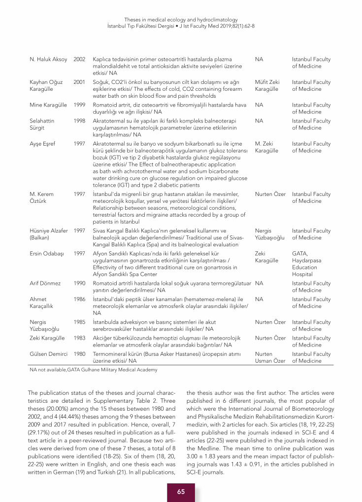
65
Theses in medical ecology and hydroclimatologyİstanbul Tıp Fakültesi Dergisi • J Ist Faculty Med 2019;82(1):62-8
The publication status of the theses and journal charac-teristics are detailed in Supplementary Table 2. Three theses (20.00%) among the 15 theses between 1980 and 2002, and 4 (44.44%) theses among the 9 theses between 2009 and 2017 resulted in publication. Hence, overall, 7 (29.17%) out of 24 theses resulted in publication as a full-text article in a peer-reviewed journal. Because two arti-cles were derived from one of these 7 theses, a total of 8 publications were identified (18-25). Six of them (18, 20, 22-25) were written in English, and one thesis each was written in German (19) and Turkish (21). In all publications,
the thesis author was the first author. The articles were published in 6 different journals, the most popular of which were the International Journal of Biometeorology and Physikalische Medizin Rehabilitationsmedizin Kurort-medizin, with 2 articles for each. Six articles (18, 19, 22-25) were published in the journals indexed in SCI-E and 4 articles (22-25) were published in the journals indexed in the Medline. The mean time to online publication was 3.00 ± 1.83 years and the mean impact factor of publish-ing journals was 1.43 ± 0.91, in the articles published in SCI-E journals.
N. Haluk Aksoy 2002 Kaplıca tedavisinin primer osteoartritli hastalarda plazma malondialdehit ve total antioksidan aktivite seviyeleri üzerine etkisi/ NA
NA Istanbul Faculty of Medicine
Kayhan Oğuz Karagülle
2001 Soğuk, CO2'li önkol su banyosunun cilt kan dolaşımı ve ağrı eşiklerine etkisi/ The effects of cold, CO2 containing forearm water bath on skin blood flow and pain thresholds
Müfit Zeki Karagülle
Istanbul Faculty of Medicine
Mine Karagülle 1999 Romatoid artrit, diz osteoartriti ve fibromiyaljili hastalarda hava duyarlılığı ve ağrı ilişkisi/ NA
NA Istanbul Faculty of Medicine
Selahattin Sürgit
1998 Akratotermal su ile yapılan iki farklı kompleks balneoterapi uygulamasının hematolojik parametreler üzerine etkilerinin karşılaştırılması/ NA
NA Istanbul Faculty of Medicine
Ayşe Eşref 1997 Akratotermal su ile banyo ve sodyum bikarbonatlı su ile içme kürü şeklinde bir balneoterapötik uygulamanın glukoz toleransı bozuk (IGT) ve tip 2 diyabetik hastalarda glukoz regülasyonu üzerine etkisi/ The Effect of balneotherapeutic application as bath with achrotothermal water and sodium bicarbonate water drinking cure on glucose regulation on impaired glucose tolerance (IGT) and type 2 diabetic patients
M. Zeki Karagülle
Istanbul Faculty of Medicine
M. Kerem Öztürk
1997 İstanbul'da migrenli bir grup hastanın atakları ile mevsimler, meteorolojik koşullar, yersel ve yerötesi faktörlerin ilişkileri/ Relationship between seasons, meteorological conditions, terrestrial factors and migraine attacks recorded by a group of patients in Istanbul
Nurten Özer Istanbul Faculty of Medicine
Hüsniye Alzafer (Balkan)
1997 Sivas Kangal Balıklı Kaplıca'nın geleneksel kullanımı ve balneolojik açıdan değerlendirilmesi/ Traditional use of Sivas-Kangal Balıklı Kaplıca (Spa) and its balneological evaluation
Nergis Yüzbaşıoğlu
Istanbul Faculty of Medicine
Ersin Odabaşı 1997 Afyon Sandıklı Kaplıcası`nda iki farklı geleneksel kür uygulamasının gonartrozda etkinliğinin karşılaştırılması / Effectivity of two different traditional cure on gonartrosis in Afyon Sandıklı Spa Center
Zeki Karagülle
GATA, Haydarpasa Education Hospital
Arif Dönmez 1990 Romatoid artritli hastalarda lokal soğuk uyarana termoregülatuar yanıtın değerlendirilmesi/ NA
NA Istanbul Faculty of Medicine
Ahmet Karaçallık
1986 İstanbul'daki peptik ülser kanamaları (hematemez-melena) ile meteorolojik elemanlar ve atmosferik olaylar arasındaki ilişkiler/ NA
NA Istanbul Faculty of Medicine
Nergis Yüzbaşıoğlu
1985 İstanbulda adveksiyon ve basınç sistemleri ile akut serebrovasküler hastalıklar arasındaki ilişkiler/ NA
Nurten Özer Istanbul Faculty of Medicine
Zeki Karagülle 1983 Akciğer tüberkülozunda hemoptizi oluşması ile meteorolojik elemanlar ve atmosferik olaylar arasındaki bağıntılar/ NA
Nurten Özer Istanbul Faculty of Medicine
Gülsen Demirci 1980 Termomineral kürün (Bursa Asker Hastanesi) üropepsin atımı üzerine etkisi/ NA
Nurten Usman Özer
Istanbul Faculty of Medicine
NA not available,GATA Gulhane Military Medical Academy

66
Theses in medical ecology and hydroclimatologyİstanbul Tıp Fakültesi Dergisi • J Ist Faculty Med 2019;82(1):62-8
Supplementary Table 2 The publication status of the theses and journal characteristics.
Thesis author/ year
Journal article title/ Journal/ Online first year Journal indexing†
Impact Factor‡
Sinan Kardeş/ 2017 Outpatient balneological treatment of osteoarthritis in older persons : A retrospective study/ Zeitschrift für Gerontologie und Geriatrie/ 2018 (25)
SCI-E, Medline
1.160
Fatih Karaarslan/ 2015
How does spa treatment affect cardiovascular function and vascular endothelium in patients with generalized osteoarthritis? A pilot study through plasma asymmetric di-methyl arginine (ADMA) and L-arginine/ADMA ratio/ International Journal of Biometeorology/ 2017 (24)
SCI-E, Medline
2.577
Serap Seringeç Karabulut/ 2015
-
Ali Onay/ 2015 -Fatma Begüm Berköz/ 2012
Spa therapy for generalized osteoarthritis: an open, observational, preliminary study/ Thérapie/ 2014 (23)
SCI-E, Medline
1.524
Fulya Demircioğlu/ 2012
-
Mehmet Fırat Satak/ 2012
-
Kağan Özkuk/ 2010
Balneological outpatient treatment for patients with knee osteoarthritis; an effective non-drug therapy option in daily routine?/ International Journal of Biometeorology/ 2016 (22)
SCI-E, Medline
2.577
An Alternative Approach To The Duration Of Balneological Treatment Of Knee Osteoarthritis Patients/ Aegean Journal of Medical Sciences/ 2018 (21)
- -
Yasemin Barut/ 2009
-
Bilal Uysal/ 2002 Effectiveness of balneotherapy in knee osteoarthritis/ Experimental Biomedical Research/ 2018 (20)
- -
Kerem Gün/ 2002 -N. Haluk Aksoy/ 2002
-
Kayhan Oğuz Karagülle/ 2001
The Acute Effects of Cold, CO2 Containing Forearm Water Bath on Skin Microcirculation and Pain Thresholds/ Physikalische Medizin Rehabilitationsmedizin Kurortmedizin/ 2003 (19)
SCI-E 0.358
Mine Karagülle/ 1999
-
Selahattin Sürgit/ 1998
-
Ayşe Eşref/ 1997 -M. Kerem Öztürk/ 1997
-
Hüsniye Alzafer (Balkan)/ 1997
-
Ersin Odabaşı/ 1997
Comparison of two traditional spa therapy regimens in patients with knee osteoarthritis/ Physikalische Medizin Rehabilitationsmedizin Kurortmedizin/ 2002 (18)
SCI-E 0.358
Arif Dönmez/ 1990 -Ahmet Karaçallık/ 1986
-
Nergis Yüzbaşıoğlu/ 1985
-
Zeki Karagülle/ 1983
-
Gülsen Demirci/ 1980
-
†Indexing status for Science Citation Index-Expanded (SCI-E), Medline and TR Dizin. ‡Journal Citation Reports 2017 impact factor.
67
Theses in medical ecology and hydroclimatologyİstanbul Tıp Fakültesi Dergisi • J Ist Faculty Med 2019;82(1):62-8
DISCUSSION
This study was the first of its kind at determining the conversion rate of specialization in medicine theses into full-text journal articles in medical ecology and hydro-climatology. The number of theses increased after 2009 compared to the past. In addition, a quarter of the theses were published in SCI-E journals; however, the rate has nearly reached to half of the theses in the last decade. In these publications, the mean time to online publication was 3 years. Furthermore, the thesis author was the first author in all publications.
No medical ecology and hydroclimatology vacancy was opened in Medical Specialty Exam (TUS) during six years, which explains the absence of thesis record between the years 2003 to 2008. Then, however, the number of theses increased over time. Similarly, the previous studies ob-served the increase of the number of theses by time in the field of emergency medicine (7) and otorhinolaryn-gology (14) as a reflection of the increasing vacancies in medical specialty exam.
Theses were found to be conducted in Istanbul Faculty of Medicine and Gulhane Military Medical Academy. This can be expected, as only these two institutions were the educational institutions for the medical ecology and hy-droclimatology specialty in the evaluated time frame.
During the past decades, several studies investigated the publication rate of specialization in medicine theses in SCI-E journals in a variety of departments (7-17). In emer-gency medicine 14.9% of the theses between 1998 and 2013 (7); 22% of the theses in general surgery between 2006-2008 (9); 19.9% of the theses in gynecology and ob-stetrics between 2007-2008 (10); 10.7% of the theses in medical microbiology between 1997 and 2007 (11); 10.2% of the theses in clinical microbiology and infectious diseas-es between 1997 and 2007 (11); 18% of the theses in neu-rosurgery between 2004 and 2013 (12); 20.4% of the theses in ophthalmology between 1986 and 2010 (13); 12.5% of the theses in orthopedics and traumatology between 1974 and 2014 (15); and 32.7% of the theses in urology between 2008-2011 (17) were published in SCI/SCI-E journals. It was found in the present study that 25.0% of the theses between 1980 and 2017 were published in SCI-E journals. However, the direct comparison of this study with earlier studies is limited by differences in study designs, article search methods, and evaluated time frame.
This study has some limitations. First, National Thesis Center provides only theses conducted in the universi-ties. Therefore, the theses that were conducted in educa-tion and training hospitals could not be included; howev-er, this is unlikely to influence the study results, as there was no specialty in medical ecology and hydroclimatol-ogy in any of the education and training hospital in Tur-
key. Additionally, the theses that were not either archived or correctly recorded in the National Thesis Center may have been missed. Second, although the study searched 7 databases, the articles that were not published in jour-nals indexed in these databases might be missed. Hence, the study might underrate the publication rate of theses. Despite these limitations, the insights generated from this study provide important information on the conver-sion rate of medical ecology and hydroclimatology spe-cialty theses into full-text journal articles.
In conclusion, a quarter of medical ecology and hydrocli-matology specialty theses were published in SCI-E jour-nals; however, the rate has nearly reached to half of the theses in the last decade. Further studies are needed to determine the barriers to the conversion of theses into full-text articles.
Acknowledgments: The author thanks medical writer Kadriye Gümüş, from Publication Support Unit, Istanbul Faculty of Med-icine, for English editing of the manuscript.
Peer Review: Externally peer-reviewed.
Author Contributions: Conception/Design of Study- S.K.; Data Acquisition- S.K.; Data Analysis/Interpretation- S.K.; Drafting Man-uscript- S.K.; Critical Revision of Manuscript- S.K.; Final Approval and Accountability- S.K.; Technical or Material Support- S.K.
Conflict of Interest: Author declared no conflict of interest.
Financial Disclosure: Author declared no financial support.
Teşekkür: Yazar, İstanbul Tıp Fakültesi Yayın Destek Birimi’nden tıbbi yazarı Kadriye Gümüş’e, yazının İngilizce düzenlemesi için teşekkür eder.
Hakem Değerlendirmesi: Dış bağımsız.
Yazar Katkıları: Çalışma Konsepti/Tasarım- S.K.; Veri Toplama- S.K.; Veri Analizi/Yorumlama- S.K.; Yazı Taslağı- S.K.; İçeriğin Eleştirel İncelemesi- S.K.; Son Onay ve Sorumluluk- S.K.; Mal-zeme ve Teknik Destek- S.K.
Çıkar Çatışması: Yazar çıkar çatışması beyan etmemiştir.
Finansal Destek: Yazar finansal destek beyan etmemiştir.
REFERENCES
1. Oxford English Dictionary. https://en.oxforddictionaries.com/. Accessed 16 August 2018.
2. Tıpta ve Diş Hekimliğinde Uzmanlık Eğitimi Yönetmeliği. http://www.mevzuat.gov.tr/. Accessed 16 August 2018.
3. World Medical Association. World Medical Association Declaration of Helsinki: ethical principles for medical research involving human subjects. JAMA 2013;310(20):2191-4. [CrossRef]

68
Theses in medical ecology and hydroclimatologyİstanbul Tıp Fakültesi Dergisi • J Ist Faculty Med 2019;82(1):62-8
4. Yilmaz Ö, Yüksel H. Thesis in medical specialty education at a glance: problems and solution proposals. Bulletin of Pleura 2013;7:33-36. [CrossRef]
5. Evereklioğlu C. How to write a thesis and turn it into publication. Erciyes Med J 2014;36(1):1-5. [CrossRef]
6. Al-Busaidi IS, Alamri Y. Publication rates and characteristics of undergraduate medical theses in New Zealand. N Z Med J 2016;129(1442):46-51.
7. Cevik E, Karakus Yilmaz B, Acar YA, Dokur M. Systematic Analysis of Theses in the Field of Emergency Medicine in Turkey. Turk J Emerg Med 2015;15(1):28-32. [CrossRef]
8. Yaman H, Kara İH, Baltacı D, Altuğ M, Akdeniz M, Kavukçu E. Qualitative evaluation of theses written in area of family medicine in Turkey. Konuralp Tıp Dergisi 2011;3(3):1-6.
9. Mayir B, Bilecik T, Çakır T, Doğan U, Gündüz UR, Aslaner A, et al. Analysis of the publishing rate and the number of citations of general surgery dissertations. Turk J Surg 2017;33(1):33-6. [CrossRef]
10. Akpınar Mayir A, Mayir B, Erkal N, Eren Yazıcı L. Üniversite hastanelerinin kadın hastalıkları ve doğum kliniklerinde yapılan tez çalışmalarının yayınlanma oranları ve atıf sayılarının değerlendirilmesi. Turkiye Klinikleri J Gynecol Obst 2016;26(4):232-6. [CrossRef]
11. Sipahi OR, Çaglayan Serin D, Pullukcu H, Taşbakan M, Köseli Ulu D, Yamazhan T, et al. Publication rates of Turkish medical specialty and doctorate theses on Medical Microbiology, Clinical Microbiology and Infectious Diseases disciplines in international journals. Mikrobiyol Bul 2014;48(2):341-5. [CrossRef]
12. Öğrenci A, Ekşi MŞ, Özcan-Ekşi EE, Koban O. From idea to publication: Publication rates of theses in neurosurgery from Turkey. Neurol Neurochir Pol 2016;50(1):45-7. [CrossRef]
13. Bayramlar H, Karadag R, Kanra Gurturk AY, Ocal A, Dag Y, et al. Publication patterns of ophthalmology residency dissertations in Turkey. Eur J Gen Med 2015;12:213-6.
14. Kalcıoğlu MT, Eğilmez OK, Karaca S, Hanege FM, İleri Y. Publication rates of otolaryngology theses from Turkey in peer-reviewed journals. Kulak Burun Bogaz Ihtis Derg 2016;26(3):143-51. [CrossRef]
15. Koca K, Ekinci S, Akpancar S, Gemci MH, Erşen Ö, Akyıldız F. An analysis of orthopaedic theses in Turkey: Evidence levels and publication rates. Acta Orthop Traumatol Turc 2016;50(5):562-6. [CrossRef]
16. Sipahi H, Durusoy R, Ergin I, Hassoy H, Davas A, Karababa A. Publication rates of public health theses in international and national peer-review journals in Turkey. Iran J Public Health 2012;41(9):31-5.
17. Yüksel M, İpekçi T, Tunçkıran A. Publication rates of dissertations written in medical faculties of Turkey in the field of urology between the years 2008, and 2011, and citation analysis: A cross-sectional study. Turk J Urol 2018;44(4):341-5. [CrossRef]
18. Odabaşı E, Karagulle MZ, Karagulle M, Turan M, Karagülle O. Comparison of two traditional spa therapy regimens in patients with knee osteoarthritis. Phys Med Rehabil Kuror 2002;12(6):337-41. [CrossRef]
19. Karagülle O, Candir F, Kalinin J, Gehrke A, Karagülle MZ, Gutenbrunner C. Akutwirkungen kalter CO2- Teilbäder auf Mikrozirkulation und Schmerzempfindlichkeit. Phys Med Rehab Kuror 2004;14(1):13-7. [CrossRef]
20. Uysal B, Ozkuk K. Effectiveness of balneotherapy in knee osteoarthritis. Exp Biomed Res 2018;1(2):46-54. [CrossRef]
21. Özkuk K, Gürdal H, Karagülle M, Karagülle MZ. An alternative approach to the duration of balneological treatment of knee osteoarthritis patients. Aegean J Med Sci 2018;1(1):1-6.
22. Özkuk K, Gürdal H, Karagülle M, Barut Y, Eröksüz R, Karagülle MZ. Balneological outpatient treatment for patients with knee osteoarthritis; an effective non-drug therapy option in daily routine? Int J Biometeorol 2017;61(4):719-28. [CrossRef]
23. Erol FB, Forestier RJ, Güneri FD, Karagülle MZ, Erdoğan N. Spa therapy for generalized osteoarthritis: an open, observational, preliminary study. Therapie 2015;70(3):273-81. [CrossRef]
24. Karaarslan F, Ozkuk K, Seringec Karabulut S, Bekpinar S, Karagulle MZ, Erdogan N. How does spa treatment affect cardiovascular function and vascular endothelium in patients with generalized osteoarthritis? A pilot study through plasma asymmetric di-methyl arginine (ADMA) and L-arginine/ADMA ratio. Int J Biometeorol 2018;62(5):833-842. [CrossRef]
25. Kardeş S, Karagülle M, Geçmen İ, Adıgüzel T, Yücesoy H, Karagülle MZ. Outpatient balneological treatment of osteoarthritis in older persons : A retrospective study. Z Gerontol Geriatr. 2018 [Epub ahead of print]. doi: 10.1007/s00391-018-1370-3 [CrossRef]
Related Documents


