RADBOOK 2017 €19.- • IT • CT • MRI • Interventional • Mammo • R / F • Nuc • Displays / Printers • Ultrasound • Injectors • Testing Devices The Guide to Imaging Technology and Informatics in Europe More precise diagnostics, low dose and accustomed workflow: Based on dual layer detector technology the IQon Spectral CT from Philips opens up new dimensions in CT imaging.

Welcome message from author
This document is posted to help you gain knowledge. Please leave a comment to let me know what you think about it! Share it to your friends and learn new things together.
Transcript
RAD
BOO
K 20
17· T
he G
uide
to Im
agin
g Te
chno
logy
and
Info
rmat
ics i
n Eu
rope
RADBOOK 2017
IT-SOLUTIONS IT-SOLUTIONS
RADBOOK 2017€ 19.-
• IT • CT • MRI • Interventional • Mammo • R / F • Nuc • Displays / Printers• Ultrasound • Injectors • Testing Devices
The Guide to Imaging Technology and Informatics in Europe
More precise diagnostics, low dose and accustomed workflow: Based on dual layer detector technology the IQon Spectral CT from Philips opens up new dimensions in CT imaging.
Visit us at ECR 2017EXPO X5 Booth #12
Visit us at ECR 2017EXPO X5 Booth #12
In today’s healthcare environment,excellent patient care and outstandingproductivity often compromise each other.Multitom Rax1) puts an end to “either/or”.Robotic Advanced X-ray – RAX – supportsyou in streamlining clinical pathwayswhile improving diagnostic insights and treatments.
How? With one scanner that offers amultitude of X-rays – in just one roomand at an expert level. With movementsthat create natural Real 3D2) images –even under weight-bearing conditions.
With an open design that reduces patient transfers, pain, and anxiety while offeringmore space to perform interventions.With twin robots to simplify, standardize,and speed up operations. All with the goalof accelerating productivity and allowinghumans to do what they do best – carefor patients.
Prepare yourself for the future with theworld’s first Twin Robotic X-ray scanner –achieving a new level of excellencewith regard to diagnostic insights andtreatments.
www.siemens.com/multitom-rax
Multitom Rax: Join the newmovement with robotic X-ray
Powered by
RAXtechnology
RAX – Robotic Advanced X-ray –opens up exciting new movementsfor enhanced patient care and out-standing productivity.
1) Multitom Rax is not commercially available in all countries. Due to regulatory reasons its future availability cannot be guaranteed.Please contact your local Siemens organization for further details. 2) Option, only in combination with additional workstation syngo X workplace.
A91X
P-94
57-A
1-76
00
Best-in-classEquipped with the largest available FPD at 43 x43 cm and Shimadzu’s digital imaging platform,the Sonialvision G4 covers the widest possiblerange of examinations with inter-departmentalhospital capability. In both functionality andoperability, the Sonialvision G4 multipurpose R/Ftable is far beyond other R/F systems. It provides“Best-in-class” features.
Smart system architecturesupports outstanding clinical flexibility for a wide range of examinations
Comprehensive dose management packageensures today’s highest safety of patients andoperators
Excellent image qualityprovided by the the state-of-the-art “SUREengineAdvance“ technology enhancing the entire imagefor clearer details
Premium application softwaresupporting useful applications, such as motion-tolerant DSA, slot radiography andtomosynthesis
www.shimadzu-medical.eu/sonialvision-g4
Next generation digital R/F system:Sonialvision G4
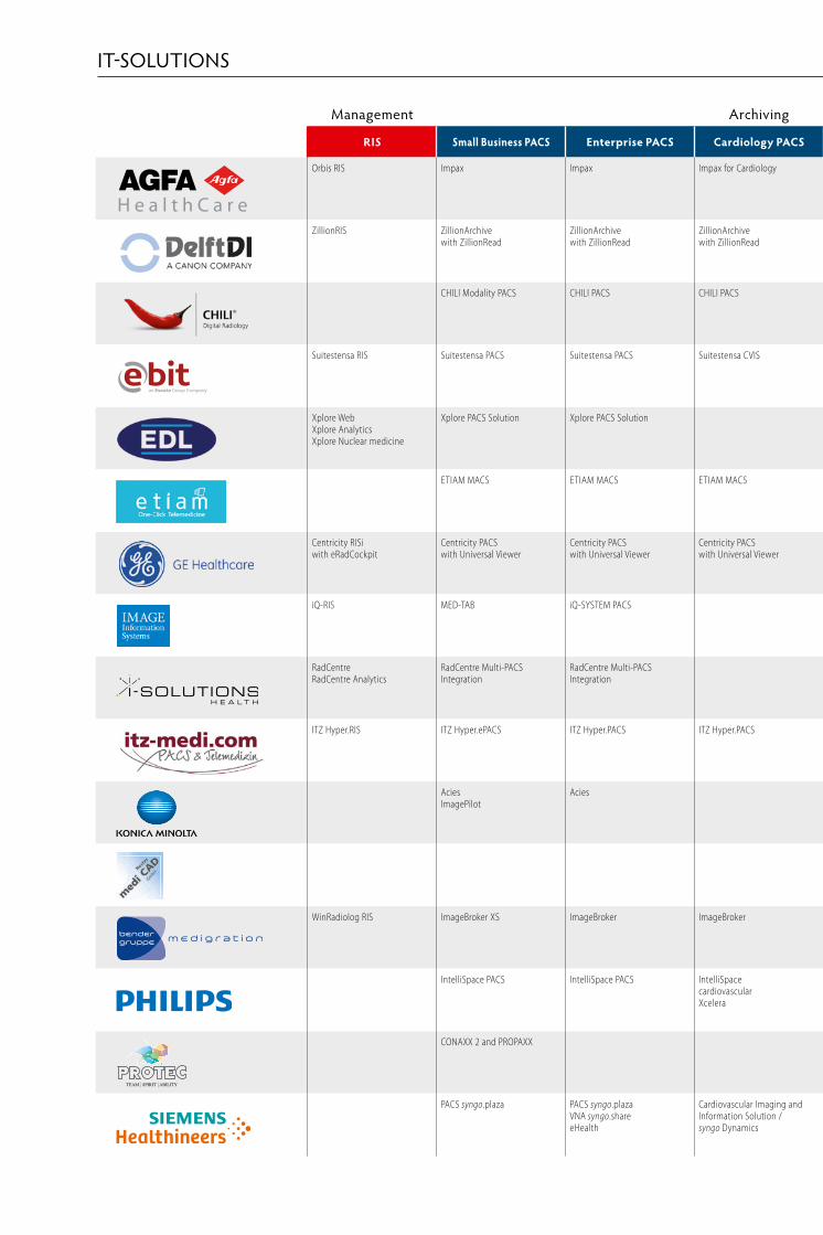
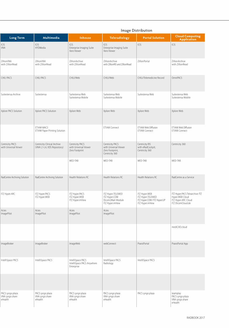
Management Archiving Image Distribution Workstations
RIS Small Business PACS Enterprise PACS Cardiology PACS Long Term Multimedia Inhouse Teleradiology Portal Solution Cloud Computing Application Multimodality Mammography Orthopedics Cardiology CAD Advanced Visualization
Orbis RIS Impax Impax Impax for Cardiology ICISVNA
ICISHYDMedia
ICISEnterprise Imaging SuiteXero Viewer
ICISEnterprise Imaging SuiteXero Viewer
ICIS ICIS Impax Clinical Applications Impax Mammography Impax Clinical Applications Impax Clinical Applications Impax Clinical Applications Agfa HealthCareSeptestraat 27 · 2640 Mortsel, Belgiumtel +32 3 444 94 [email protected] · www.agfa.com
ZillionRIS ZillionArchive with ZillionRead
ZillionArchive with ZillionRead
ZillionArchive with ZillionRead
ZillionVNA with ZillionRead
ZillionVNA with ZillionRead
ZillionArchive with ZillionRead
ZillionArchive with ZillionRIS and ZillionRead
ZillionPortal ZillionArchive with ZillionRead
ZillionRead ZillionRead ZillionRead ZillionRead CAD for TuberculosisCAD for Mammography
ZillionRead DelftDI, a Canon companyWiltonstraat 41, 3905 KW Veenendaal, The Netherlandstel +31 318 583 [email protected] · www.delftdi.com
CHILI Modality PACS CHILI PACS CHILI PACS CHILI PACS CHILI PACS CHILI/Web CHILI/Web CHILI/Telemedicine Record OmniPACS CHILI Diagnost CHILI Diagnost CHILI Diagnost CHILI Diagnost Partner-Solution Partner-Solution CHILI GmbH Friedrich-Ebert-Str. 2 · 69221 Dossenheim/Heidelberg, Germany tel +49 6221 1 80 79 10 [email protected] · www.chili-radiology.com
Suitestensa RIS Suitestensa PACS Suitestensa PACS Suitestensa CVIS Suitestensa Archive Suitestensa Suitestensa WebSuitestensa Mobile
Suitestensa WebSuitestensa Mobile
Suitestensa Web Suitestensa WebSuitestensa Mobile
Suitestensa Review Suitestensa MG Suitestensa Review Suitestensa Review Cardio Suitestensa 3D Suitestensa Vascular
EBIT S.r.l. – Esaote Group Via Siffredi 58 · 16153 Genoa, Italy tel +39 010 65 47-464 [email protected] · www.esaote.com/healthcare-it
Xplore WebXplore AnalyticsXplore Nuclear medicine
Xplore PACS Solution Xplore PACS Solution Xplore PACS Solution Xplore PACS Solution Xplore Web Xplore Web Xplore Web Xplore Web EDL SAS1031, chemin de la Seyne à Bastian, 83500 La Seyne-sur-mer, Francetel +33 4 94 10 99 [email protected] · www.xplore.eu
ETIAM MACS ETIAM MACS ETIAM MACS ETIAM MACSETIAM Paper Printing Solution
ETIAM Connect ETIAM WebETIAM Connect
ETIAM WebETIAM Connect
ETIAM WebETIAM Paper Printing Solution
ETIAM MACS ETIAM MACS ETIAMZA de la Hallerais, 11 rue du Bois de Soeuvres, 35770 Vern-sur-Seiche, Francetel : +33 2 99 14 33 [email protected] · www.etiam.com
Centricity RISi with eRadCockpit
Centricity PACS with Universal Viewer
Centricity PACS with Universal Viewer
Centricity PACS with Universal Viewer
Centricity PACS with Universal Viewer
Centricity Clinical Archive (VNA L1-L4, XDS Repository)
Centricity PACS with Universal Viewer Zero Footprint
Centricity PACS with Universal ViewerZero Footprint,Centricity 360
Centricity RIS with eRadCockpit,Centricity 360
Centricity 360 Centricity PACS Universal ViewerUniversal Viewer Zero Footprint XDS enabled
Centricity PACS Universal Viewer web client provides Breast Imaging tools powered by IDI
Centricity PACS Universal Viewer with integrated Traumacad by Voyant Health
Centricity Cardio Enterprise Centricity PACS Universal Viewer web client embeds advanced visualization powered by AW
Centricity PACS Universal Viewer web client embeds advanced visualization powered by AW
GE HealthcareLerchenbergstr. 15 · 89160 Dornstadt, Germanytel +49 7348 [email protected] · www.gehealthcare.com
iQ-RIS MED-TAB iQ-SYSTEM PACS MED-TAB MED-TAB MED-TAB MED-TAB iQ-VIEW PRO iQ-VIEW PRO MAMMO TOMO iQ-VIEW PRO OrthoView
iQ-VIEW PRO 4D iQ-VIEW PRO 4D IMAGE Information Systems Europe GmbHLange Str. 16 · 18055 Rostock, Germanytel +49 381 496 58 [email protected] · www.image-systems.biz
RadCentreRadCentre Analytics
RadCentre Multi-PACS Integration
RadCentre Multi-PACS Integration
RadCentre Archiving Solution RadCentre Archiving Solution Health Relations RC Health Relations RC Health Relations RC RadCentre as a Service i-SOLUTIONS Health GmbHAm Exerzierplatz 14 · 68167 Mannheim, Germanytel +49 621 39 [email protected] · www.i-solutions.de
ITZ Hyper.RIS ITZ Hyper.ePACS ITZ Hyper.PACS ITZ Hyper.PACS ITZ Hyper.ARC ITZ Hyper.PACSITZ Hyper.WEB
ITZ Hyper.PACSITZ Hyper.WEBITZ Hyper.mView
ITZ Hyper.TELEMEDITZ Hyper.COMDicom2Mail-ModuleITZ Hyper.mView
ITZ Hyper.WEBITZ Hyper.TELEMEDITZ Hyper.COM / ITZ Hyper.UPITZ Hyper.mView
ITZ Hyper.PACS Telearchive ITZ Hyper.WEB CloudITZ Hyper.ARC CloudITZ DicomCloud.de
ITZ Hyper.PACS ITZ Hyper.PACS ITZ Hyper.PACSHectecRSA-Biomedical Localite
Hyper.PACSPIE-Medical (Esaote)Tomtec
ITZ Hyper.PACS MPR / MIP / 3Dmint
ITZ Hyper.PACSmintTerareconMedian
ITZ Medicom GmbH & Co. KGSiemensring 44 a · 47877 Willich, Germanytel +49 2154 [email protected] · www.itz-medi.com
AciesImagePilot
Acies AciesImagePilot
AciesImagePilot
AciesImagePilot
AciesImagePilot
AciesImagePilot
Acies Acies Acies Acies Konica Minolta Medical & Graphic Imaging Europe B.V.Hoogoorddreef 9 · 1101 BA Amsterdam, The Netherlandstel +31 20 658 41 [email protected] · www.konicaminolta.eu/healthcare
mediCAD.cloud mediCAD ClassicmediCAD 3DmediCAD.cloudmediCAD Veterinary
mediCAD ClassicmediCAD 3DmediCAD.cloudmediCAD Veterinary
mediCAD Hectec GmbHOpalstr. 54 · 84032 Altdorf, Germanytel +49 871 33 02 [email protected] · www.mediCAD.eu
WinRadiolog RIS ImageBroker XS ImageBroker ImageBroker ImageBroker ImageBroker ImageWeb webConnect PraxisPortal PraxisPortal App ImageVision Diagnost MammoView ImageVision Basic ImageVision Diagnost MammoView CAD ImageVision Diagnost medigration GmbH Am Anger 2 · 91052 Erlangen, Germanytel +49 91 31 690 87-48 [email protected] · www.medigration.de
IntelliSpace PACS IntelliSpace PACS IntelliSpace cardiovascularXcelera
IntelliSpace PACS IntelliSpace PACS IntelliSpace PACSIntelliSpace PACS AnywhereEnterprise
IntelliSpace PACSRadiology
IntelliSpace PACS IntelliSpace PACSRadiologyIntelliSpace Portal
IntelliSpace PACSIntelliSpace Breast
IntelliSpacecardiovascular
IntelliSpace Portal Philips HealthcareP.O. Box 10.000 · 5680 DA Best, The Netherlandstel +31 40 278 56 [email protected] · www.philips.com/healthcare
CONAXX 2 and PROPAXX PROPAXX and / or CONAXX 2 PROPAXX and / or CONAXX 2 PROPAXX and / or CONAXX 2 PROTEC GmbH & Co. KGIn den Dorfwiesen 14 · 71720 Oberstenfeld, Germanytel +49 7062 [email protected] · www.protec-med.com
PACS syngo.plaza PACS syngo.plazaVNA syngo.shareeHealth
Cardiovascular Imaging and Information Solution / syngo Dynamics
PACS syngo.plazaVNA syngo.shareeHealth
PACS syngo.plazaVNA syngo.shareeHealth
PACS syngo.plazaVNA syngo.shareeHealth
PACS syngo.plazaVNA syngo.shareeHealth
PACS syngo.plaza teamplayPACS syngo.plazaVNA syngo.shareeHealth
syngo.viaPACS syngo.plaza
syngo.via PACS syngo.plaza
MediCAD (HECTEC) syngo.viaPACS syngo.plaza
Cardiovascular Imaging and Information Solution / syngo Dynamics syngo.via
syngo CAD Applicationssyngo.viaPACS syngo.plaza
syngo.via Siemens Healthineers HeadquartersSiemens Healthcare GmbHHenkestr. 127 · 91052 Erlangen, Germanytel +49 9131 84-0www.siemens.com/healthineers
IT-SOLUTIONS IT-SOLUTIONS
RADBOOK 2017 RADBOOK 2017
Management Archiving Image Distribution Workstations
RIS Small Business PACS Enterprise PACS Cardiology PACS Long Term Multimedia Inhouse Teleradiology Portal Solution Cloud Computing Application Multimodality Mammography Orthopedics Cardiology CAD Advanced Visualization
Orbis RIS Impax Impax Impax for Cardiology ICISVNA
ICISHYDMedia
ICISEnterprise Imaging SuiteXero Viewer
ICISEnterprise Imaging SuiteXero Viewer
ICIS ICIS Impax Clinical Applications Impax Mammography Impax Clinical Applications Impax Clinical Applications Impax Clinical Applications Agfa HealthCareSeptestraat 27 · 2640 Mortsel, Belgiumtel +32 3 444 94 [email protected] · www.agfa.com
ZillionRIS ZillionArchivewith ZillionRead
ZillionArchivewith ZillionRead
ZillionArchivewith ZillionRead
ZillionVNA with ZillionRead
ZillionVNA with ZillionRead
ZillionArchive with ZillionRead
ZillionArchive with ZillionRIS and ZillionRead
ZillionPortal ZillionArchive with ZillionRead
ZillionRead ZillionRead ZillionRead ZillionRead CAD for TuberculosisCAD for Mammography
ZillionRead DelftDI, a Canon companyWiltonstraat 41, 3905 KW Veenendaal, The Netherlandstel +31 318 583 [email protected] · www.delftdi.com
CHILI Modality PACS CHILI PACS CHILI PACS CHILI PACS CHILI PACS CHILI/Web CHILI/Web CHILI/Telemedicine Record OmniPACS CHILI Diagnost CHILI Diagnost CHILI Diagnost CHILI Diagnost Partner-Solution Partner-Solution CHILI GmbHFriedrich-Ebert-Str. 2 · 69221 Dossenheim/Heidelberg, Germanytel +49 6221 1 80 79 [email protected] · www.chili-radiology.com
Suitestensa RIS Suitestensa PACS Suitestensa PACS Suitestensa CVIS Suitestensa Archive Suitestensa Suitestensa WebSuitestensa Mobile
Suitestensa WebSuitestensa Mobile
Suitestensa Web Suitestensa WebSuitestensa Mobile
Suitestensa Review Suitestensa MG Suitestensa Review Suitestensa Review Cardio Suitestensa 3DSuitestensa Vascular
EBIT S.r.l. – Esaote GroupVia Siffredi 58 · 16153 Genoa, Italytel +39 010 65 [email protected] · www.esaote.com/healthcare-it
Xplore WebXplore AnalyticsXplore Nuclear medicine
Xplore PACS Solution Xplore PACS Solution Xplore PACS Solution Xplore PACS Solution Xplore Web Xplore Web Xplore Web Xplore Web EDL SAS1031, chemin de la Seyne à Bastian, 83500 La Seyne-sur-mer, Francetel +33 4 94 10 99 [email protected] · www.xplore.eu
ETIAM MACS ETIAM MACS ETIAM MACS ETIAM MACSETIAM Paper Printing Solution
ETIAM Connect ETIAM Web DiffusionETIAM Connect
ETIAM Web DiffusionETIAM Connect
ETIAM WebETIAM Paper Printing Solution
ETIAM MACS ETIAM MACS ETIAMZA de la Hallerais, 11 rue du Bois de Soeuvres, 35770 Vern-sur-Seiche, Francetel : +33 2 99 14 33 [email protected] · www.etiam.com
Centricity RISiwith eRadCockpit
Centricity PACSwith Universal Viewer
Centricity PACSwith Universal Viewer
Centricity PACSwith Universal Viewer
Centricity PACS with Universal Viewer
Centricity Clinical Archive (VNA L1-L4, XDS Repository)
Centricity PACS with Universal Viewer Zero Footprint
Centricity PACS with Universal ViewerZero Footprint,Centricity 360
Centricity RIS with eRadCockpit,Centricity 360
Centricity 360 Centricity PACS Universal ViewerUniversal Viewer Zero Footprint XDSenabled
Centricity PACS Universal Viewer webclient provides Breast Imaging toolspowered by IDI
Centricity PACS Universal Viewer withintegrated Traumacad by Voyant Health
Centricity Cardio Enterprise Centricity PACS Universal Viewer webclient embeds advanced visualizationpowered by AW
Centricity PACS Universal Viewer webclient embeds advanced visualizationpowered by AW
GE HealthcareLerchenbergstr. 15 · 89160 Dornstadt, Germanytel +49 7348 [email protected] · www.gehealthcare.com
iQ-RIS MED-TAB iQ-SYSTEM PACS MED-TAB MED-TAB MED-TAB MED-TAB iQ-VIEW PRO iQ-VIEW PRO MAMMO TOMO iQ-VIEW PROOrthoView
iQ-VIEW PRO 4D iQ-VIEW PRO 4D IMAGE Information Systems Europe GmbHLange Str. 16 · 18055 Rostock, Germanytel +49 381 496 58 [email protected] · www.image-systems.biz
RadCentreRadCentre Analytics
RadCentre Multi-PACSIntegration
RadCentre Multi-PACSIntegration
RadCentre Archiving Solution RadCentre Archiving Solution Health Relations RC Health Relations RC Health Relations RC RadCentre as a Service i-SOLUTIONS Health GmbHAm Exerzierplatz 14 · 68167 Mannheim, Germanytel +49 621 39 [email protected] · www.i-solutions.de
ITZ Hyper.RIS ITZ Hyper.ePACS ITZ Hyper.PACS ITZ Hyper.PACS ITZ Hyper.ARC ITZ Hyper.PACSITZ Hyper.WEB
ITZ Hyper.PACSITZ Hyper.WEBITZ Hyper.mView
ITZ Hyper.TELEMEDITZ Hyper.COMDicom2Mail-ModuleITZ Hyper.mView
ITZ Hyper.WEBITZ Hyper.TELEMEDITZ Hyper.COM / ITZ Hyper.UPITZ Hyper.mView
ITZ Hyper.PACS Telearchive ITZ Hyper.WEB CloudITZ Hyper.ARC CloudITZ DicomCloud.de
ITZ Hyper.PACS ITZ Hyper.PACS ITZ Hyper.PACSHectecRSA-Biomedical Localite
Hyper.PACSPIE-Medical (Esaote)Tomtec
ITZ Hyper.PACS MPR / MIP / 3Dmint
ITZ Hyper.PACSmintTerareconMedian
ITZ Medicom GmbH & Co. KGSiemensring 44 a · 47877 Willich, Germanytel +49 2154 [email protected] · www.itz-medi.com
AciesImagePilot
Acies AciesImagePilot
AciesImagePilot
AciesImagePilot
AciesImagePilot
AciesImagePilot
Acies Acies Acies Acies Konica Minolta Medical & Graphic Imaging Europe B.V.Hoogoorddreef 9 · 1101 BA Amsterdam, The Netherlandstel +31 20 658 41 [email protected] · www.konicaminolta.eu/healthcare
mediCAD.cloud mediCAD ClassicmediCAD 3DmediCAD.cloudmediCAD Veterinary
mediCAD ClassicmediCAD 3DmediCAD.cloudmediCAD Veterinary
mediCAD Hectec GmbHOpalstr. 54 · 84032 Altdorf, Germanytel +49 871 33 02 [email protected] · www.mediCAD.eu
WinRadiolog RIS ImageBroker XS ImageBroker ImageBroker ImageBroker ImageBroker ImageWeb webConnect PraxisPortal PraxisPortal App ImageVision Diagnost MammoView ImageVision Basic ImageVision Diagnost MammoView CAD ImageVision Diagnost medigration GmbHAm Anger 2 · 91052 Erlangen, Germanytel +49 91 31 690 [email protected] · www.medigration.de
IntelliSpace PACS IntelliSpace PACS IntelliSpacecardiovascularXcelera
IntelliSpace PACS IntelliSpace PACS IntelliSpace PACSIntelliSpace PACS AnywhereEnterprise
IntelliSpace PACSRadiology
IntelliSpace PACS IntelliSpace PACSRadiologyIntelliSpace Portal
IntelliSpace PACSIntelliSpace Breast
IntelliSpacecardiovascular
IntelliSpace Portal Philips HealthcareP.O. Box 10.000 · 5680 DA Best, The Netherlandstel +31 40 278 56 [email protected] · www.philips.com/healthcare
CONAXX 2 and PROPAXX PROPAXX and / or CONAXX 2 PROPAXX and / or CONAXX 2 PROPAXX and / or CONAXX 2 PROTEC GmbH & Co. KGIn den Dorfwiesen 14 · 71720 Oberstenfeld, Germanytel +49 7062 [email protected] · www.protec-med.com
PACS syngo.plaza PACS syngo.plazaVNA syngo.shareeHealth
Cardiovascular Imaging andInformation Solution /syngo Dynamics
PACS syngo.plazaVNA syngo.shareeHealth
PACS syngo.plazaVNA syngo.shareeHealth
PACS syngo.plazaVNA syngo.shareeHealth
PACS syngo.plazaVNA syngo.shareeHealth
PACS syngo.plaza teamplayPACS syngo.plazaVNA syngo.shareeHealth
syngo.viaPACS syngo.plaza
syngo.viaPACS syngo.plaza
MediCAD (HECTEC)syngo.viaPACS syngo.plaza
Cardiovascular Imaging and Information Solution / syngo Dynamics syngo.via
syngo CAD Applicationssyngo.viaPACS syngo.plaza
syngo.via Siemens Healthineers HeadquartersSiemens Healthcare GmbHHenkestr. 127 · 91052 Erlangen, Germanytel +49 9131 84-0www.siemens.com/healthineers
IT-SOLUTIONS IT-SOLUTIONS
RADBOOK 2017 RADBOOK 2017
RAD
BOO
K 20
17· T
he G
uide
to Im
agin
g Te
chno
logy
and
Info
rmat
ics i
n Eu
rope
RADBOOK 2017
IT-SOLUTIONS IT-SOLUTIONS
RADBOOK 2017€ 19.-
• IT •CT •MRI • Interventional •Mammo •R/F •Nuc •Displays/Printers•Ultrasound • Injectors • Testing Devices
The Guide to Imaging Technology and Informatics in Europe
More precise diagnostics, low dose and accustomed workflow: Based on dual layer detector technologythe IQon Spectral CT from Philips opens up new dimensions in CT imaging.
Visit us at ECR 2017EXPO X5 Booth #12
Visit us at ECR 2017EXPO X5 Booth #12
In today’s healthcare environment,excellent patient care and outstandingproductivity often compromise each other.Multitom Rax1) puts an end to “either/or”.Robotic Advanced X-ray – RAX – supportsyou in streamlining clinical pathwayswhile improving diagnostic insights and treatments.
How? With one scanner that offers amultitude of X-rays – in just one roomand at an expert level. With movementsthat create natural Real 3D2) images –even under weight-bearing conditions.
With an open design that reduces patient transfers, pain, and anxiety while offeringmore space to perform interventions.With twin robots to simplify, standardize,and speed up operations. All with the goalof accelerating productivity and allowinghumans to do what they do best – carefor patients.
Prepare yourself for the future with theworld’s first Twin Robotic X-ray scanner –achieving a new level of excellencewith regard to diagnostic insights andtreatments.
www.siemens.com/multitom-rax
Multitom Rax: Join the newmovement with robotic X-ray
Powered by
RAXtechnology
RAX – Robotic Advanced X-ray –opens up exciting new movementsfor enhanced patient care and out-standing productivity.
1) Multitom Rax is not commercially available in all countries. Due to regulatory reasons its future availability cannot be guaranteed.Please contact your local Siemens organization for further details. 2) Option, only in combination with additional workstation syngo X workplace.
A91X
P-94
57-A
1-76
00
Best-in-classEquipped with the largest available FPD at 43 x43 cm and Shimadzu’s digital imaging platform,the Sonialvision G4 covers the widest possiblerange of examinations with inter-departmentalhospital capability. In both functionality andoperability, the Sonialvision G4 multipurpose R/Ftable is far beyond other R/F systems. It provides“Best-in-class” features.
Smart system architecturesupports outstanding clinical flexibility for a wide range of examinations
Comprehensive dose management packageensures today’s highest safety of patients andoperators
Excellent image qualityprovided by the the state-of-the-art “SUREengineAdvance“ technology enhancing the entire imagefor clearer details
Premium application softwaresupporting useful applications, such as motion-tolerant DSA, slot radiography andtomosynthesis
www.shimadzu-medical.eu/sonialvision-g4
Next generation digital R/F system:Sonialvision G4
Best-in-classEquipped with the largest available FPD at 43 x43 cm and Shimadzu’s digital imaging platform,the Sonialvision G4 covers the widest possiblerange of examinations with inter-departmentalhospital capability. In both functionality andoperability, the Sonialvision G4 multipurpose R/Ftable is far beyond other R/F systems. It provides“Best-in-class” features.
Smart system architecturesupports outstanding clinical flexibility for a wide range of examinations
Comprehensive dose management packageensures today’s highest safety of patients andoperators
Excellent image qualityprovided by the the state-of-the-art “SUREengineAdvance“ technology enhancing the entire imagefor clearer details
Premium application softwaresupporting useful applications, such as motion-tolerant DSA, slot radiography andtomosynthesis
www.shimadzu-medical.eu/sonialvision-g4
Next generation digital R/F system:Sonialvision G4
RADBOOK 2017 5
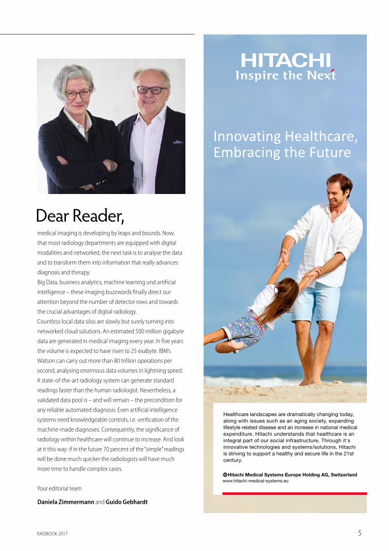
Dear Reader,medical imaging is developing by leaps and bounds. Now,
that most radiology departments are equipped with digital
modalities and networked, the next task is to analyse the data
and to transform them into information that really advances
diagnosis and therapy.
Big Data, business analytics, machine learning und artificial
intelligence – these imaging buzzwords finally direct our
attention beyond the number of detector rows and towards
the crucial advantages of digital radiology.
Countless local data silos are slowly but surely turning into
networked cloud solutions. An estimated 500 million gigabyte
data are generated in medical imaging every year. In five years
the volume is expected to have risen to 25 exabyte. IBM’s
Watson can carry out more than 80 trillion operations per
second, analysing enormous data volumes in lightning speed.
A state-of-the-art radiology system can generate standard
readings faster than the human radiologist. Nevertheless, a
validated data pool is – and will remain – the precondition for
any reliable automated diagnosis. Even artificial intelligence
systems need knowledgeable controls, i.e. verification of the
machine-made diagnoses. Consequently, the significance of
radiology within healthcare will continue to increase. And look
at it this way: if in the future 70 percent of the “simple” readings
will be done much quicker the radiologists will have much
more time to handle complex cases.
Your editorial team
Daniela Zimmermann and Guido Gebhardt
Innovating Healthcare,Embracing the Future
Healthcare landscapes are dramatically changing today,along with issues such as an aging society, expandinglifestyle related disease and an increase in national medicalexpenditure. Hitachi understands that healthcare is anintegral part of our social infrastructure. Through it'sinnovative technologies and systems/solutions, Hitachiis striving to support a healthy and secure life in the 21stcentury.
Hitachi Medical Systems Europe Holding AG, Switzerlandwww.hitachi-medical-systems.eu
RZ Ad_HMSE-Image_88x258mm_RADbook_15-02-17_RZ Ad_HMSE-Image_88x
RADBOOK 20176
CONTENTS
5 Editorial 6 Imprint
Trends & Topics22 Philips: Elevating neuro diagnostics for clarity and insight 30 MRI-tec: MRI safety in practice38 Bayer: Bayer’s comprehensive solution for integrated
diagnostic imaging46 Ziehm: 10 years of flat-panel technology77 DOTmed: Six ways DOTmed protects buyers and sellers
in 253 countries 90 Shimadzu: World-class technologies for healthcare and diagnosis 96 Konica Minolta: High-de finition imaging
102 Siemens Healthineers: Multitom Rax the first Twin Robotic X-Ray system
122 medical ECONET: Digital X-ray imaging for mobile medical applications
8 Computed Tomography9 Dual Source9 Volume CT
11 20 to 64 Slices13 2 to 16 Slices16 Oncology CT 17 Conebeam CT18 Accessories / Complementary Systems
20 Magnetic Resonance Imaging21 7 Tesla21 3 Tesla25 1.5 Tesla
28 Open 32 MR-PET
32 MRT Coils33 Accessories / Complementary Systems
35 Injectors
42 Interventional Systems 43 Hybrid-OPs 44 Bi-Plane
45 Single Plane51 Surgical II-C-Arms56 Surgical Flat Panel C-Arms57 Accessories / Complementary Systems
59 IT Systems3 IT-Solutions Table – pt1
60 RIS / PACS64 Portal Solution
65 CAD66 Mammo Workstation67 Mobile RIS / PACS Viewer68 Accessories / Complementary Systems
189 IT-Solutions Table – pt2
71 Mammography 72 Tomosynthesis
72 Digital Mammography75 Film-Screen Mammography76 Biopsy Tables
76 Radiowave-Imaging78 Accessories / Complementary Systems
80 R / F Film-Screen 81 Bucky 83 Fluoroscopy
85 Mobile X-Ray87 Accessories / Complementary Systems
88 R / F Digital 89 CR 92 DR
107 DR Retrofit112 Mobile DR119 Flatpanel Fluoro125 Accessories / Complementary Systems
129 Molecular Imaging 130 SPECT 131 SPECT-CT 132 PET-CT 134 PET-MR
135 Displays / Printers136 Displays – Mammography137 Displays – Grayscale138 Displays – Color140 Displays – Clinical Review142 Displays – Large Format
143 Printer144 CD- / DVD-Robot144 Accessories / Complementary Systems
145 Ultrasound170 Accessories / Complementary Systems
171 Testing Devices
182 Index of Advertisers184 Companies / Suppliers
IMPRINTPublished by:
EUROPEAN HOSPITAL Verlags GmbH Theodor-Althoff-Straße 45 · 45133 Essen · Germany · Phone +49-201-87126-851 · www.healthcare-in-europe.com and RADIUNDA Guido Gebhardt · Adalbert-Stifter-Weg 2b · 85661 Forstinning · Germany · Phone +49-8121-907630 · www.radiunda.com
Editor-in-Chief: Guido Gebhardt · Daniela Zimmermann · Executive Director: Daniela Zimmermann Advertising: Ralf Mateblowski (D, A, CH) · Eric Jund (E, F, I) · Simon Kramer (B, GB, L, NL) Gavin Hua (China) · Jane Park (Korea) · Hanna Politis (U.S.A, Canada)
Art Director: Christoph Muschiol · Agentur für Werbung, Beratung & Organisation · Dorfen · Germany · www.muschiol-online.de · Printing: Margreff Druck GmbH · Essen · Germany
Subscription: Liane Kaiser · Subscription rate: € 19.– plus postage
The information and opinions expressed in articles and product entries published in RADBOOK are solely those of the manufacturers / companies, their authors and contributors, for which the publisher holds no responsibility. Errors and omissions excepted.
Disclaimer: All company, brand and product names in this publication are the property of their respective holders. Not all products are available in all European countries. Please note that the manufacturers’ websites may contain further product disclaimers.
© 2017 by EUROPEAN HOSPITAL Verlags GmbH. All rights reserved.
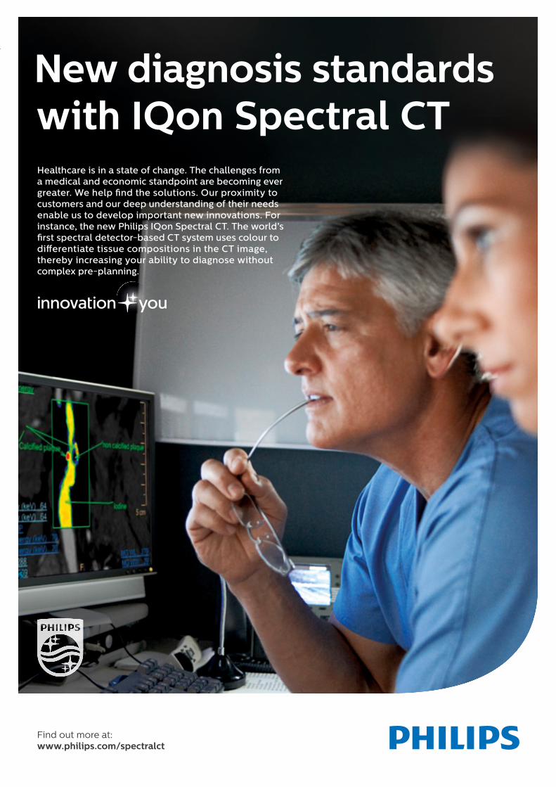
Find out more at: www.philips.com/spectralct
New diagnosis standards with IQon Spectral CTHealthcare is in a state of change. The challenges from a medical and economic standpoint are becoming ever greater. We help � nd the solutions. Our proximity to customers and our deep understanding of their needs enable us to develop important new innovations. For instance, the new Philips IQon Spectral CT. The world’s � rst spectral detector-based CT system uses colour to di erentiate tissue compositions in the CT image, thereby increasing your ability to diagnose without complex pre-planning.
5116112_Sprachadaption_AZ_IQonSpectralCT_DINA4_210x297_RZ.indd 1 11.02.16 12:32
RADBOOK 20178
◼ COMPUTED TOMOGRAPHY
A Division of Philips Healthcare
ComputedTomography
Dual Source
Volume CT
20 to 64 Slices
2 to 16 Slices
Oncology CT
Conebeam CT
Accessories / Complementary Systems
RADBOOK 2017 9
COMPUTED TOMOGRAPHY ◼
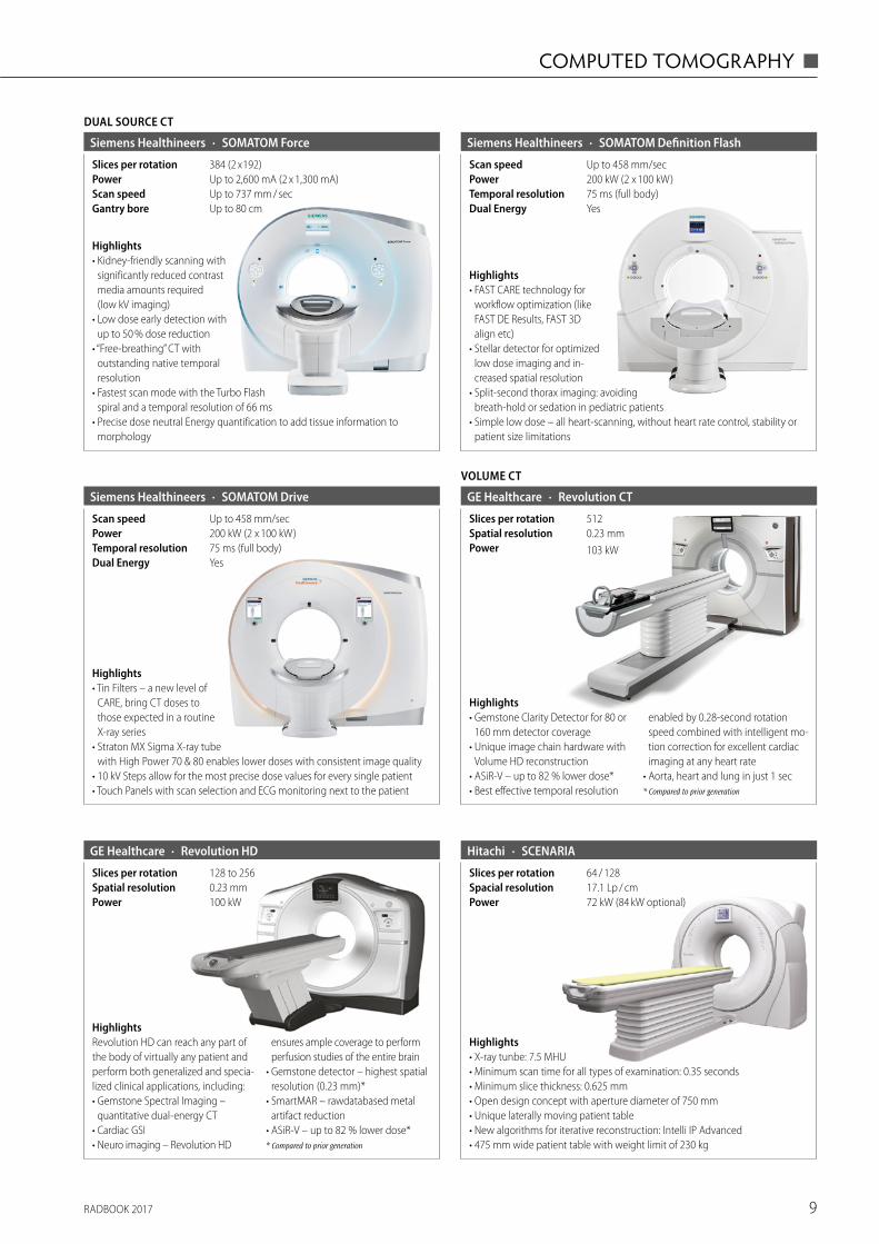
DUAL SOURCE CT
· SOMATOM ForceSiemens Healthineers
Up to 80 cmGantry boreUp to 737 mm / secScan speedUp to 2,600 mA (2 x 1,300 mA)Power384 (2 x 192)Slices per rotation
Highlights• Kidney-friendly scanning with
significantly reduced contrast media amounts required (low kV imaging)
• Low dose early detection with up to 50 % dose reduction
• “Free-breathing” CT with outstanding native temporal resolution
• Fastest scan mode with the Turbo Flash spiral and a temporal resolution of 66 ms
• Precise dose neutral Energy quantification to add tissue information to morphology
· SOMATOM Definition FlashSiemens Healthineers
YesDual Energy75 ms (full body)Temporal resolution200 kW (2 x 100 kW)PowerUp to 458 mm/secScan speed
Highlights• FAST CARE technology for
workflow optimization (like FAST DE Results, FAST 3D align etc)
• Stellar detector for optimized low dose imaging and in-creased spatial resolution
• Split-second thorax imaging: avoiding breath-hold or sedation in pediatric patients
• Simple low dose – all heart-scanning, without heart rate control, stability or patient size limitations
YesDual Energy75 ms (full body)Temporal resolution200 kW (2 x 100 kW)PowerUp to 458 mm/secScan speed
· SOMATOM DriveSiemens Healthineers
Highlights• Tin Filters – a new level of
CARE, bring CT doses to those expected in a routine X-ray series
• Straton MX Sigma X-ray tube with High Power 70 & 80 enables lower doses with consistent image quality
• 10 kV Steps allow for the most precise dose values for every single patient• Touch Panels with scan selection and ECG monitoring next to the patient
VOLUME CT
· Revolution CTGE Healthcare
103 kWPower0.23 mmSpatial resolution512Slices per rotation
Highlights• Gemstone Clarity Detector for 80 or
160 mm detector coverage• Unique image chain hardware with
Volume HD reconstruction • ASiR-V – up to 82 % lower dose*• Best effective temporal resolution
enabled by 0.28-second rotation speed combined with intelligent mo-tion correction for excellent cardiac imaging at any heart rate
• Aorta, heart and lung in just 1 sec* Compared to prior generation
· Revolution HDGE Healthcare
100 kWPower0.23 mmSpatial resolution128 to 256Slices per rotation
HighlightsRevolution HD can reach any part of the body of virtually any patient and perform both generalized and specia-lized clinical applications, including:• Gemstone Spectral Imaging –
quantitative dual-energy CT • Cardiac GSI• Neuro imaging – Revolution HD
ensures ample coverage to perform perfusion studies of the entire brain
• Gemstone detector – highest spatial resolution (0.23 mm)*
• SmartMAR – rawdatabased metal artifact reduction
• ASiR-V – up to 82 % lower dose** Compared to prior generation
Highlights• X-ray tunbe: 7.5 MHU• Minimum scan time for all types of examination: 0.35 seconds• Minimum slice thickness: 0.625 mm• Open design concept with aperture diameter of 750 mm• Unique laterally moving patient table• New algorithms for iterative reconstruction: Intelli IP Advanced• 475 mm wide patient table with weight limit of 230 kg
· SCENARIAHitachi
72 kW (84 kW optional)Power17.1 Lp / cmSpacial resolution64 / 128Slices per rotation
RADBOOK 201710
◼ COMPUTED TOMOGRAPHY
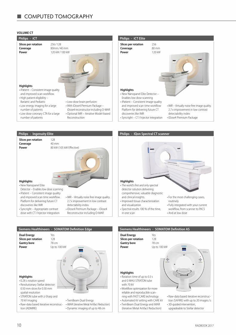
· iCT ElitePhilips
120 kWPower80 mmCoverage256Slices per rotation
Highlights• New Nanopanel Elite Detector –
Enables low dose scanning• iPatient – Consistent image quality
and improved scan time workflow Platform for delivering future CT discoveries like IMR
• Syncright – CT / Injector integration
• IMR – Virtually noise free image quality. 2.7 x improvement in low contrast detectability index
• iDose4 Premium Package
VOLUME CT
· iCTPhilips
120 kW / 100 kWPower80mm / 40 mmCoverage256 / 128Slices per rotation
Highlights• iPatient – Consistent image quality
and improved scan workflow. • High patient eligibility –
Bariatric and Pediatric• Low energy imaging for a large
number of patients• Low dose coronary CTA for a large
number of patients
• Low-dose brain perfusion• With iDose4 Premium Package –
iDose4 reconstructor including O-MAR• Optional IMR – Iterative Model-based
Reconstruction
· IQon Spectral CT scannerPhilips
Highlights• The world's first and only spectral
detector solution delivering comprehensive, valuable diagnostic and clinical insights.
• Improved tissue characterization and visualization
• Spectral results 100 % of the time, in one scan
• For the most challenging cases, routinely
• Fully integrated with your current workflow, from scanner to PACS
• And at low dose
· Ingenuity ElitePhilips
80 kW (105 kW Effective)Power40 mmCoverage128Slices per rotation
Highlights• New Nanopanel Elite
Detector – Enables low dose scanning• iPatient – Consistent image quality
and improved scan time workflow. Platform for delivering future CT discoveries like IMR
• Syncright – Appropriate contrast dose with CT / Injector integration
• IMR – Virtually noise free image quality. 2.7 x improvement in low contrast detectability index.
• iDose4 Premium Package – iDose4 Reconstructor including O-MAR
· SOMATOM Definition EdgeSiemens Healthineers
Up to 100 kWPower78 cmGantry bore128Slices per rotationYesDual Energy
Highlights• 0.28 s rotation speed• Revolutionary Stellar detector:
0.50 mm slices for 0.30 mm spatial resolution
• STRATON tube with z-Sharp and 70 kV imaging
• Raw-data based iterative reconstruc-tion (ADMIRE)
• TwinBeam Dual Energy • iMAR (iterative Metal Artifact Reduction)• Dynamic imaging of up to 48 cm
· SOMATOM Definition AS Siemens Healthineers
Up to 100 kWPower78 cmGantry bore128Slices per rotationYesDual Energy
Highlights• Rotation time of up to 0.3 s
and 0 MHU STRATON tube with 70 kV
• Workflow optimization for more reliable and reproducible scan-ning with FAST CARE technology
• Automated kV setting with CARE kV• TwinBeam Dual Energy and iMAR
(iterative Metal Artifact Reduction)
• Raw-data based iterative reconstruc-tion (SAFIRE) with up to 20 images / s
• 3D-guided intervention, upgradeable to Stellar detector
RADBOOK 2017 11
COMPUTED TOMOGRAPHY ◼
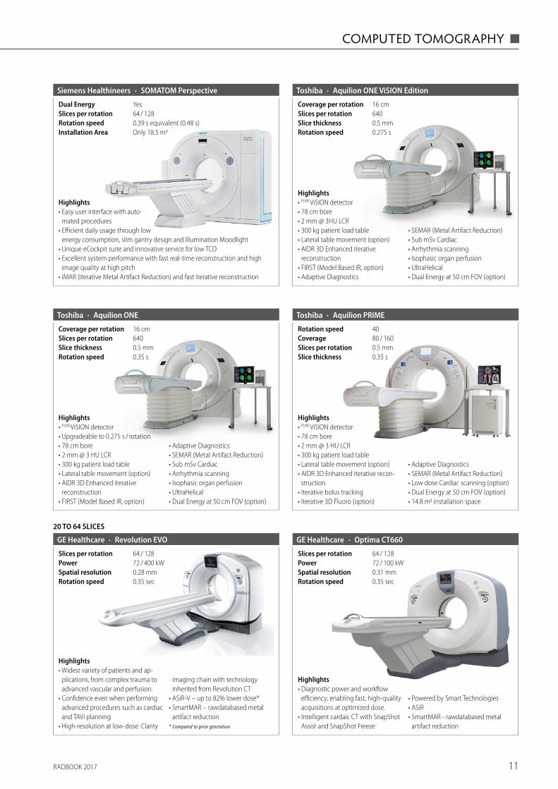
· SOMATOM PerspectiveSiemens Healthineers
Only 18.5 m2Installation Area0.39 s equivalent (0.48 s)Rotation speed64 / 128Slices per rotationYesDual Energy
Highlights• Easy user interface with auto-
mated procedures• Efficient daily usage through low
energy consumption, slim gantry design and Illumination Moodlight• Unique eCockpit suite and innovative service for low TCO• Excellent system performance with fast real-time reconstruction and high
image quality at high pitch• iMAR (iterative Metal Artifact Reduction) and fast iterative reconstruction
· Aquilion ONEToshiba
0.35 sRotation speed0.5 mmSlice thickness640Slices per rotation16 cmCoverage per rotation
Highlights• PURE ViSION detector• Upgradeable to 0.275 s / rotation• 78 cm bore• 2 mm @ 3 HU LCR• 300 kg patient load table• Lateral table movement (option)• AIDR 3D Enhanced iterative
reconstruction• FIRST (Model Based IR, option)
• Adaptive Diagnostics• SEMAR (Metal Artifact Reduction)• Sub mSv Cardiac• Arrhythmia scanning• Isophasic organ perfusion• UltraHelical• Dual Energy at 50 cm FOV (option)
· Aquilion ONE ViSION EditionToshiba
0.275 sRotation speed0.5 mmSlice thickness640Slices per rotation16 cmCoverage per rotation
Highlights• PURE ViSION detector• 78 cm bore• 2 mm @ 3HU LCR• 300 kg patient load table• Lateral table movement (option)• AIDR 3D Enhanced iterative
reconstruction• FIRST (Model Based IR, option)• Adaptive Diagnostics
• SEMAR (Metal Artifact Reduction)• Sub mSv Cardiac• Arrhythmia scanning • Isophasic organ perfusion• UltraHelical• Dual Energy at 50 cm FOV (option)
· Aquilion PRIMEToshiba
0.35 sSlice thickness0.5 mmSlices per rotation80 / 160Coverage40Rotation speed
Highlights• PURE ViSION detector• 78 cm bore• 2 mm @ 3 HU LCR• 300 kg patient load table• Lateral table movement (option)• AIDR 3D Enhanced iterative recon-
struction• Iterative bolus tracking• Iterative 3D Fluoro (option)
• Adaptive Diagnostics• SEMAR (Metal Artifact Reduction)• Low dose Cardiac scanning (option)• Dual Energy at 50 cm FOV (option)• 14.8 m2 installation space
· Optima CT660GE Healthcare
0.35 secRotation speed0.31 mmSpatial resolution72 / 100 kWPower64 / 128Slices per rotation
Highlights• Diagnostic power and workflow
efficiency, enabling fast, high-quality acquisitions at optimized dose.
• Intelligent cardaic CT with SnapShot Assist and SnapShot Freeze
• Powered by Smart Technologies• ASiR• SmartMAR - rawdatabased metal
artifact reduction
20 TO 64 SLICES
· Revolution EVOGE Healthcare
0.35 secRotation speed0.28 mmSpatial resolution72 / 400 kWPower64 / 128Slices per rotation
Highlights• Widest variety of patients and ap-
plications, from complex trauma to advanced vascular and perfusion.
• Confidence even when performing advanced procedures such as cardiac and TAVI planning
• High-resolution at low-dose: Clarity
imaging chain with technology inherited from Revolution CT
• ASiR-V – up to 82% lower dose* • SmartMAR – rawdatabased metal
artifact reduction* Compared to prior generation
RADBOOK 201712
◼ COMPUTED TOMOGRAPHY
20 TO 64 SLICES
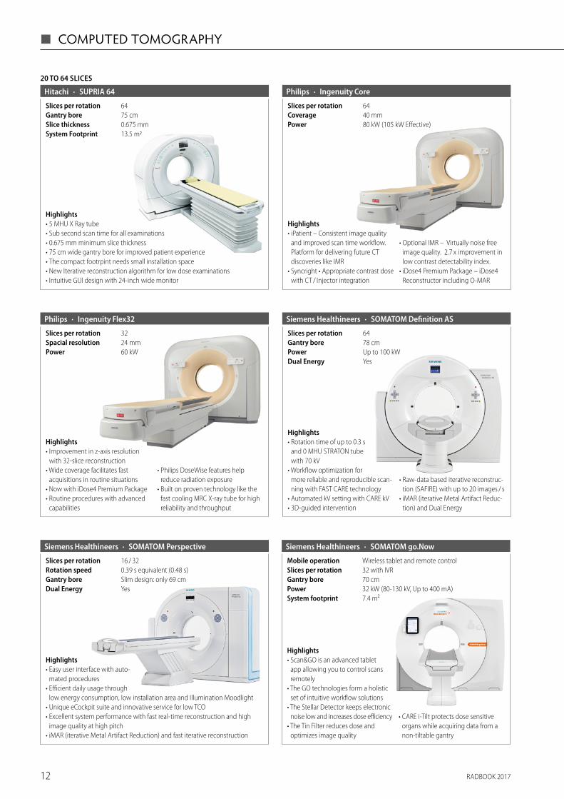
· SUPRIA 64Hitachi
0.675 mmSlice thickness75 cmGantry bore
13.5 m2System Footprint
64Slices per rotation
Highlights• 5 MHU X Ray tube• Sub second scan time for all examinations• 0.675 mm minimum slice thickness• 75 cm wide gantry bore for improved patient experience• The compact footrpint needs small installation space• New Iterative reconstruction algorithm for low dose examinations • Intuitive GUI design with 24-inch wide monitor
· Ingenuity CorePhilips
80 kW (105 kW Effective)Power40 mmCoverage64Slices per rotation
Highlights• iPatient – Consistent image quality
and improved scan time workflow. Platform for delivering future CT discoveries like IMR
• Syncright • Appropriate contrast dose with CT / Injector integration
• Optional IMR – Virtually noise free image quality. 2.7 x improvement in low contrast detectability index.
• iDose4 Premium Package – iDose4 Reconstructor including O-MAR
· Ingenuity Flex32Philips
60 kWPower24 mmSpacial resolution32Slices per rotation
Highlights• Improvement in z-axis resolution
with 32-slice reconstruction• Wide coverage facilitates fast
acquisitions in routine situations • Now with iDose4 Premium Package • Routine procedures with advanced
capabilities
• Philips DoseWise features help reduce radiation exposure
• Built on proven technology like the fast cooling MRC X-ray tube for high reliability and throughput
· SOMATOM Perspective Siemens Healthineers
YesDual EnergySlim design: only 69 cmGantry bore0.39 s equivalent (0.48 s)Rotation speed16 / 32Slices per rotation
Highlights• Easy user interface with auto-
mated procedures• Efficient daily usage through
low energy consumption, low installation area and Illumination Moodlight• Unique eCockpit suite and innovative service for low TCO• Excellent system performance with fast real-time reconstruction and high
image quality at high pitch• iMAR (iterative Metal Artifact Reduction) and fast iterative reconstruction
· SOMATOM Definition ASSiemens Healthineers
YesDual EnergyUp to 100 kWPower78 cmGantry bore64Slices per rotation
Highlights• Rotation time of up to 0.3 s
and 0 MHU STRATON tube with 70 kV
• Workflow optimization for more reliable and reproducible scan-ning with FAST CARE technology
• Automated kV setting with CARE kV• 3D-guided intervention
• Raw-data based iterative reconstruc-tion (SAFIRE) with up to 20 images / s
• iMAR (iterative Metal Artifact Reduc-tion) and Dual Energy
· SOMATOM go.NowSiemens Healthineers
7.4 m²System footprint32 kW (80-130 kV, Up to 400 mA)Power70 cmGantry bore32 with IVRWireless tablet and remote control
Slices per rotationMobile operation
Highlights• Scan&GO is an advanced tablet
app allowing you to control scans remotely
• The GO technologies form a holistic set of intuitive workflow solutions
• The Stellar Detector keeps electronic noise low and increases dose efficiency
• The Tin Filter reduces dose and optimizes image quality
• CARE i-Tilt protects dose sensitive organs while acquiring data from a non-tiltable gantry
RADBOOK 2017 13
COMPUTED TOMOGRAPHY ◼
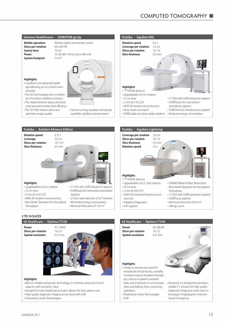
· SOMATOM go.UpSiemens Healthineers
7.4 m²System footprint32 kW (80-130 kV, Up to 400 mA)Power70 cmGantry bore64 with IVRWireless tablet and remote control
Slices per rotationMobile operation
Highlights• Scan&GO is an advanced tablet
app allowing you to control scans remotely
• The GO technologies form a holistic set of intuitive workflow solutions
• The Stellar Detector keeps electronic noise low and increases dose efficiency
• The Tin Filter reduces dose and optimizes image quality
• Calcium scoring visualizes and quickly quantifies calcified coronary lesions
· Aquilion RXLToshiba
0.5 mmSlice thickness16 / 32Slices per rotation3.2 cmCoverage per rotation0.5 sRotation speed
Highlights• PURE ViSION detector• Upgradeable to 0.4 s rotation• 72 cm bore• 2 mm @ 3 HU LCR• AIDR 3D iterative reconstruction• Dose check and report• SURECardio, low dose cardiac (option)
• CT DSA with SURESubtraction (option)• SUREFluoro for intervention
procedures (option)• SUREXtension, remote access (option)• Reduced energy consumption
· Astelion Advance EditionToshiba
0.5 mmSlice thickness16 / / 32Slices per rotation2.0 cmCoverage0.75 sRotation speed
Highlights• Upgradeable to 0.6 s rotation• 72 cm bore• 2 mm @ 3 HU LCR• AIDR 3D iterative reconstruction• Navi Mode Operation for fast patient
throughput
• CT DSA with SURESubtraction (option)• SUREFluoro for intervention procedures
(option)• 2.9 ton / year reduction of CO2 emission• Minimized energy consumption• Minimum foot print of 10.4 m2
· Aquilion LightningToshiba
0.5 sRotation speed0.5 mmSlice thickness16 / 32Slices per rotation2.0 cmCoverage per rotation
Highlights• PURE ViSION detector• Upgradeable to 0.5 s fast rotation• 78 cm bore• 2 mm @ 3HU LCR• AIDR 3D Enhanced iterative recon-
struction• Adaptive Diagnostics• vHP (option)
• SEMAR (Metal Artifact Reduction)• Navi Mode Operation for fast patient
throughput• CT DSA with SURESubtraction (option)• SUREFluoro (option)• Minimum foot print of 9.8 m2• 300 kg couch
Highlights• It helps to answer your need for
exceptional clinical results, a steadily increased volume of patient through-put, a focus on patient-centered tasks, and a reduction in unnecessary steps and tedious, time-consuming operations
• Powered by Smart Technologies • ASiR
• Moreover it is designed to provide a reliable CT solution for high quality diagnostic imaging at lower dose in: Oncology / Angiography / Interven-tional / Emergency
· Optima CT540GE Healthcare
0.31 mmSpatial resolution16 / 32Slices per rotation60 / 88 kWPower
2 TO 16 SLICES
· Optima CT520GE Healthcare
0.31 mmSpatial resolution16 / 32Slices per rotation42 / 70kWPower
Highlights• Built on reliable and proven technology, it combines advanced clinical
capacity with economic value • Designed to help healthcare providers deliver the best patient care• High quality diagnostic imaging at low dose with ASiR• Powered by Smart Technologies
RADBOOK 201714
◼ COMPUTED TOMOGRAPHY
2 TO 16 SLICES
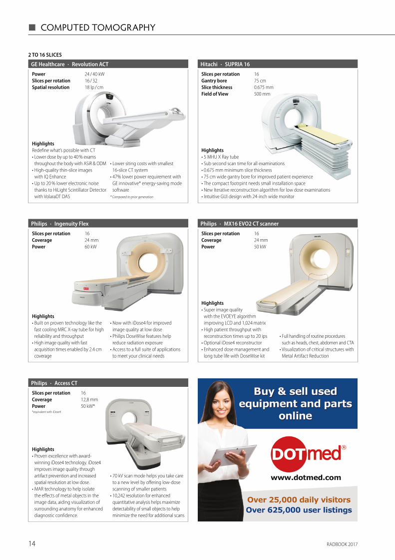
· Revolution ACTGE Healthcare
18 lp / cmSpatial resolution16 / 32Slices per rotation24 / 40 kWPower
HighlightsRedefine what’s possible with CT• Lower dose by up to 40 % exams
throughout the body with ASiR & ODM• High-quality thin-slice images
with IQ Enhance • Up to 20 % lower electronic noise
thanks to HiLight Scintillator Detector with VolaraDT DAS
• Lower siting costs with smallest 16-slice CT system
• 47% lower power requirement with GE innovative* energy-saving mode software
* Compared to prior generation
· SUPRIA 16Hitachi
16Slices per rotation
0.675 mmSlice thickness500 mmField of View
75 cmGantry bore
Highlights• 5 MHU X Ray tube• Sub second scan time for all examinations• 0.675 mm minimum slice thickness• 75 cm wide gantry bore for improved patient experience• The compact footrpint needs small installation space• New Iterative reconstruction algorithm for low dose examinations • Intuitive GUI design with 24-inch wide monitor
· Ingenuity FlexPhilips
60 kWPower24 mmCoverage16Slices per rotation
Highlights• Built on proven technology like the
fast cooling MRC X-ray tube for high reliability and throughput
• High image quality with fast acquisition times enabled by 2.4 cm coverage
• Now with iDose4 for improved image quality at low dose
• Philips DoseWise features help reduce radiation exposure
• Access to a full suite of applications to meet your clinical needs
· MX16 EVO2 CT scannerPhilips
50 kWPower24 mmCoverage16Slices per rotation
Highlights• Super image quality
with the EVOEYE algorithm improving LCD and 1,024 matrix
• High patient throughput with reconstruction times up to 20 ips
• Optional iDose4 reconstructor• Enhanced dose management and
long tube life with DoseWise kit
• Full handling of routine procedures such as heads, chest, abdomen and CTA
• Visualization of critical structures with Metal Artifact Reduction
· Access CTPhilips
*equivalent with iDose4
50 kW*Power12,8 mmCoverage16Slices per rotation
Highlights• Proven excellence with award-
winning iDose4 technology. iDose4 improves image quality through artifact prevention and increased spatial resolution at low dose.
• MAR technology to help isolate the effects of metal objects in the image data, aiding visualization of surrounding anatomy for enhanced diagnostic confidence.
• 70 kV scan mode helps you take care to a new level by offering low-dose scanning of smaller patients
• 10,242 resolution for enhanced quantitative analysis helps maximize detectability of small objects to help minimize the need for additional scans
15RADBOOK 2017
COMPUTED TOMOGRAPHY ◼
Highlights• Leading image quality from
high-quality UFC detector material and very small focal spot
• Outstanding image quality, at the right dose with CARE Dose4D and iterative reconstruction (IRIS and SAFIRE)
• iMAR (iterative Metal Artifact Reduction) and Dual Energy• Optimized total cost of ownership due to reduced overhead costs and
extended scanner lifetime with eCockpit
· SOMATOM Scope Siemens Healthineers
12 m2Installation Area8 m2System Footprint16 / 32 (both configurations)Slices per rotation26 / 50 kWPower
Highlights• Easy user interface provides simplicity and a fast learning curve• Outstanding overall system uptime due to robust design and stability• Exceptional patient throughput-to-investment ratio• Low heat dissipation and power consumption• Real-time dose modulation with CARE Dose4D for up 68 % dose reduction• Increased volume coverage with gantry rotation speed of up to 0.8 s
· SOMATOM SpiritSiemens Healthineers
15.5 Lp / mmSpatial resolution2Slices per rotation
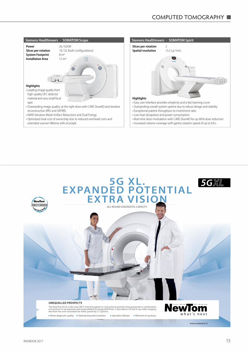
www.newtom.it
5G XL. EXPANDED POTENTIAL
EXTRA VISIONALL-ROUND DIAGNOSTIC CAPACITY
UNEQUALLED PROSPECTSThe NewTom 5G XL is the only CBCT with the patient in a lying down position that guarantees a combination of minimum X-ray exposure and unparalleled 3D image definition. It also allows 2D and X-ray video imaging. NewTom has now exceeded the limits posed by CT systems.
• Minimum X-ray doses.• Specialist software.• Optimal lying down position.• Better diagnostic quality.
RADBOOK 201716
◼ COMPUTED TOMOGRAPHY
ONCOLOGY CT
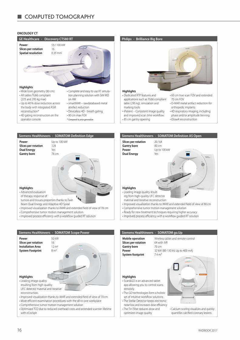
· Discovery CT580 RTGE Healthcare
0.35 mmSpatial resolution16Slices per rotation55 / 100 kWPower
Highlights• Wide bore geometry (80 cm)• All tables TG66 compliant
(225 and 295 kg max)• Up to 40 % dose reduction across
the body with integrated ASiR reconstruction*
• 4D gating reconstruction on the operator console
• Complete and easy to use RT simula-tion planning solution with SIM MD on AW
• smartMAR – rawdatabased metal atrefact reduction
• Deviceless 4D - breath gating• 80 cm max FOV* Compared to prior generation
· Brilliance Big BorePhilips
Highlights• Dedicated RTP features and
applications such as TG66 compliant table (295 kg), simulation and marking tools
• iPatient – Consistent image quality and improved scan time workflow.
• 85 cm gantry opening
• 60 cm true scan FOV and extended 70 cm FOV
• O-MAR metal artifact reduction for orthopedic implants
• 4D respiratory imaging, including phase and/or amplitude binning
• iDose4 reconstruction
· SOMATOM Scope PowerSiemens Healthineers
8 m2System Footprint12 m2Installation Area16Slices per rotation50 kWPower
Highlights• Leading image quality
resulting from high-quality UFC detector material and iterative reconstruction.
• Improved visualization thanks to iMAR and extended field of view of 70 cm• More efficient examination procedures with the all-in-one workplace• Comprehensive tumor motion management solution• Optimized TCO due to reduced overhead costs and extended scanner lifetime
with eCockpit
· SOMATOM Definition EdgeSiemens Healthineers
78 cmGantry boreYesDual Energy128Slices per rotationUp to 100 kWPower
Highlights• Advanced evaluation
of therapy response of tumors and tissues properties thanks to Twin Beam Dual Energy and Adaptive 4D Spiral
• Improved visualization thanks to iMAR and extended field of view of 78 cm • Comprehensive tumor motion management solution • Improved process efficiency with a workflow guided RT solution
· SOMATOM Definition AS OpenSiemens Healthineers
YesDual EnergyUp to 100 kWPower80 cmGantry bore20 / 64Slices per rotation
Highlights• Leading image quality result-
ing from high-quality UFC detector material and iterative reconstruction
• Improved visualization thanks to iMAR and extended field of view of 80 cm• Comprehensive tumor motion management solution• Ready for new treatment techniques requiring higher accuracy• Improved process efficiency with a workflow guided RT solution
· SOMATOM go.UpSiemens Healthineers
7.4 m²System footprint32 kW (80-130 kV, Up to 400 mA)Power70 cmGantry bore64 with IVRWireless tablet and remote control
Slices per rotationMobile operation
Highlights• Scan&GO is an advanced tablet
app allowing you to control scans remotely
• The GO technologies form a holistic set of intuitive workflow solutions
• The Stellar Detector keeps electronic noise low and increases dose efficiency
• The Tin Filter reduces dose and optimizes image quality
• Calcium scoring visualizes and quickly quantifies calcified coronary lesions
RADBOOK 2017 17
COMPUTED TOMOGRAPHY ◼
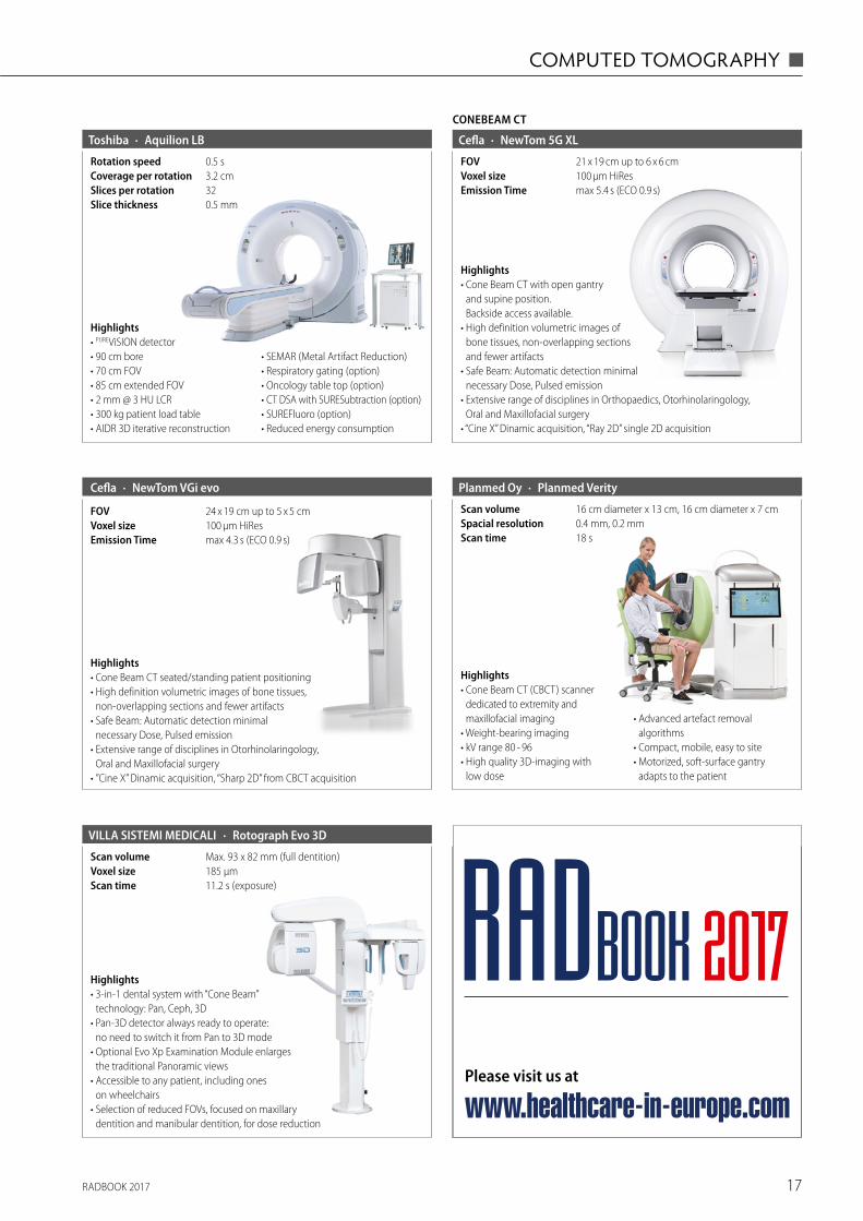
· Aquilion LBToshiba
0.5 mmSlice thickness32Slices per rotation3.2 cmCoverage per rotation0.5 sRotation speed
Highlights• PUREViSION detector• 90 cm bore• 70 cm FOV• 85 cm extended FOV• 2 mm @ 3 HU LCR• 300 kg patient load table• AIDR 3D iterative reconstruction
• SEMAR (Metal Artifact Reduction)• Respiratory gating (option)• Oncology table top (option)• CT DSA with SURESubtraction (option)• SUREFluoro (option)• Reduced energy consumption
Highlights• Cone Beam CT (CBCT) scanner
dedicated to extremity and maxillo facial imaging
• Weight-bearing imaging• kV range 80 - 96• High quality 3D-imaging with
low dose
• Advanced artefact removal algorithms
• Compact, mobile, easy to site • Motorized, soft-surface gantry
adapts to the patient
· Planmed VerityPlanmed Oy
18 sScan time0.4 mm, 0.2 mmSpacial resolution16 cm diameter x 13 cm, 16 cm diameter x 7 cmScan volume
· Rotograph Evo 3DVILLA SISTEMI MEDICALI
11.2 s (exposure)Scan time185 μmVoxel sizeMax. 93 x 82 mm (full dentition)Scan volume
Highlights• 3-in-1 dental system with “Cone Beam”
technology: Pan, Ceph, 3D• Pan-3D detector always ready to operate:
no need to switch it from Pan to 3D mode• Optional Evo Xp Examination Module enlarges
the traditional Panoramic views• Accessible to any patient, including ones
on wheelchairs• Selection of reduced FOVs, focused on maxillary
dentition and manibular dentition, for dose reduction
CONEBEAM CT
· NewTom 5G XLCefla
max 5.4 s (ECO 0.9 s)Emission Time100 μm HiResVoxel size21 x 19 cm up to 6 x 6 cmFOV
Highlights• Cone Beam CT with open gantry
and supine position. Backside access available.
• High definition volumetric images of bone tissues, non-overlapping sections and fewer artifacts
• Safe Beam: Automatic detection minimal necessary Dose, Pulsed emission
• Extensive range of disciplines in Orthopaedics, Otorhinolaringology, Oral and Maxillofacial surgery
• “Cine X” Dinamic acquisition, “Ray 2D” single 2D acquisition
· NewTom VGi evo
max 4.3 s (ECO 0.9 s)Emission Time100 μm HiResVoxel size24 x 19 cm up to 5 x 5 cmFOV
Highlights• Cone Beam CT seated/standing patient positioning• High definition volumetric images of bone tissues,
non-overlapping sections and fewer artifacts• Safe Beam: Automatic detection minimal
necessary Dose, Pulsed emission• Extensive range of disciplines in Otorhinolaringology,
Oral and Maxillofacial surgery• ”Cine X” Dinamic acquisition, “Sharp 2D” from CBCT acquisition
Cefla
Please visit us at
www.healthcare-in-europe.com
RADBOOK 2017
RADBOOK 201718
◼ COMPUTED TOMOGRAPHY
ACCESSORIES / COMPLEMENTARY SYSTEMS
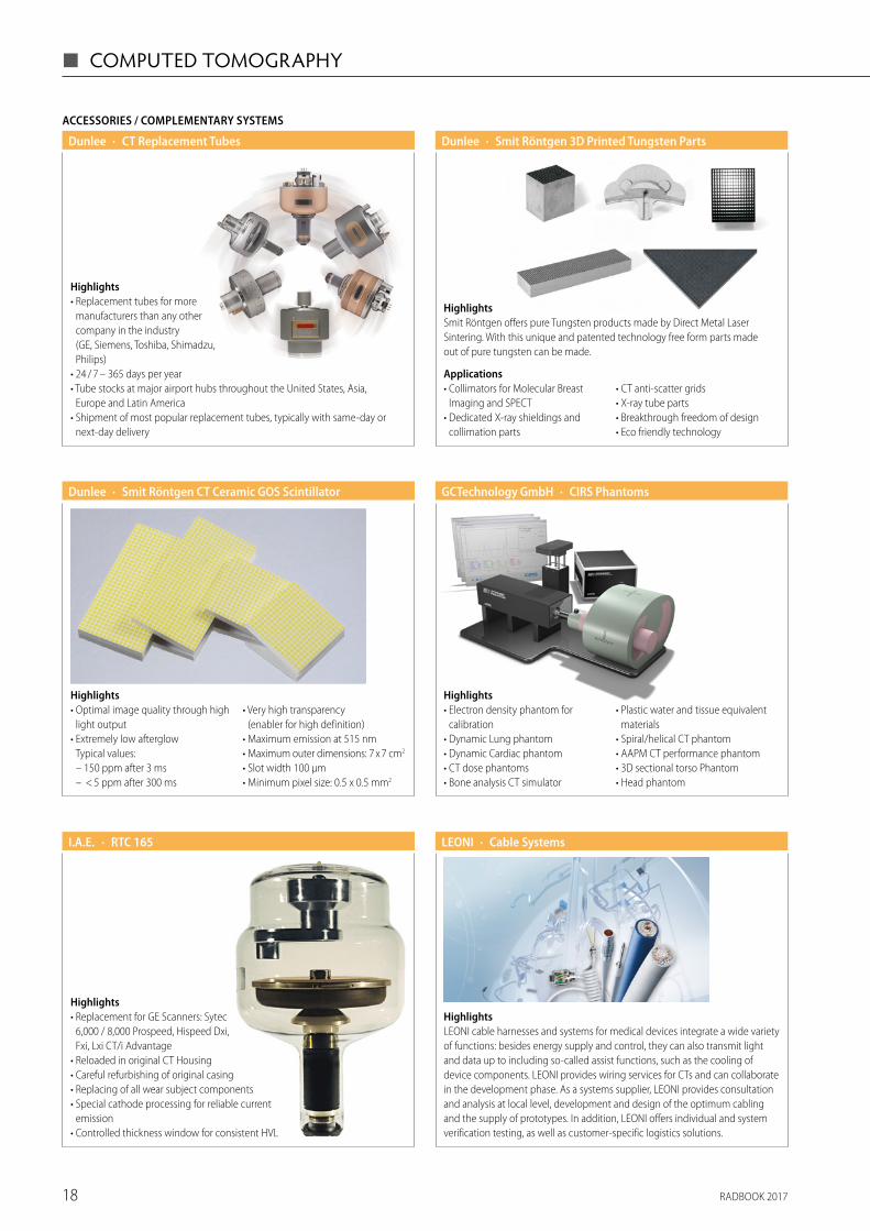
Highlights• Replacement tubes for more
manufacturers than any other company in the industry (GE, Siemens, Toshiba, Shimadzu, Philips)
• 24 / 7 – 365 days per year• Tube stocks at major airport hubs throughout the United States, Asia,
Europe and Latin America • Shipment of most popular replacement tubes, typically with same-day or
next-day delivery
· CT Replacement TubesDunlee
HighlightsSmit Röntgen offers pure Tungsten products made by Direct Metal Laser Sintering. With this unique and patented technology free form parts made out of pure tungsten can be made.
· Smit Röntgen 3D Printed Tungsten PartsDunlee
Applications• Collimators for Molecular Breast
Imaging and SPECT• Dedicated X-ray shieldings and
collimation parts
• CT anti-scatter grids• X-ray tube parts• Breakthrough freedom of design• Eco friendly technology
Highlights• Optimal image quality through high
light output• Extremely low afterglow
Typical values: – 150 ppm after 3 ms – < 5 ppm after 300 ms
• Very high transparency (enabler for high definition)
• Maximum emission at 515 nm• Maximum outer dimensions: 7 x 7 cm2
• Slot width 100 μm• Minimum pixel size: 0.5 x 0.5 mm2
· Smit Röntgen CT Ceramic GOS Scintillator Dunlee · CIRS PhantomsGCTechnology GmbH
Highlights• Electron density phantom for
calibration• Dynamic Lung phantom• Dynamic Cardiac phantom• CT dose phantoms• Bone analysis CT simulator
• Plastic water and tissue equivalent materials
• Spiral/helical CT phantom• AAPM CT performance phantom • 3D sectional torso Phantom• Head phantom
Highlights• Replacement for GE Scanners: Sytec
6,000 / 8,000 Prospeed, Hispeed Dxi, Fxi, Lxi CT/i Advantage
• Reloaded in original CT Housing• Careful refurbishing of original casing• Replacing of all wear subject components• Special cathode processing for reliable current
emission• Controlled thickness window for consistent HVL
· RTC 165I.A.E. · Cable SystemsLEONI
HighlightsLEONI cable harnesses and systems for medical devices integrate a wide variety of functions: besides energy supply and control, they can also transmit light and data up to including so-called assist functions, such as the cooling of device components. LEONI provides wiring services for CTs and can collaborate in the development phase. As a systems supplier, LEONI provides consultation and analysis at local level, development and design of the optimum cabling and the supply of prototypes. In addition, LEONI offers individual and system verification testing, as well as customer-specific logistics solutions.
RADBOOK 2017 19
COMPUTED TOMOGRAPHY ◼
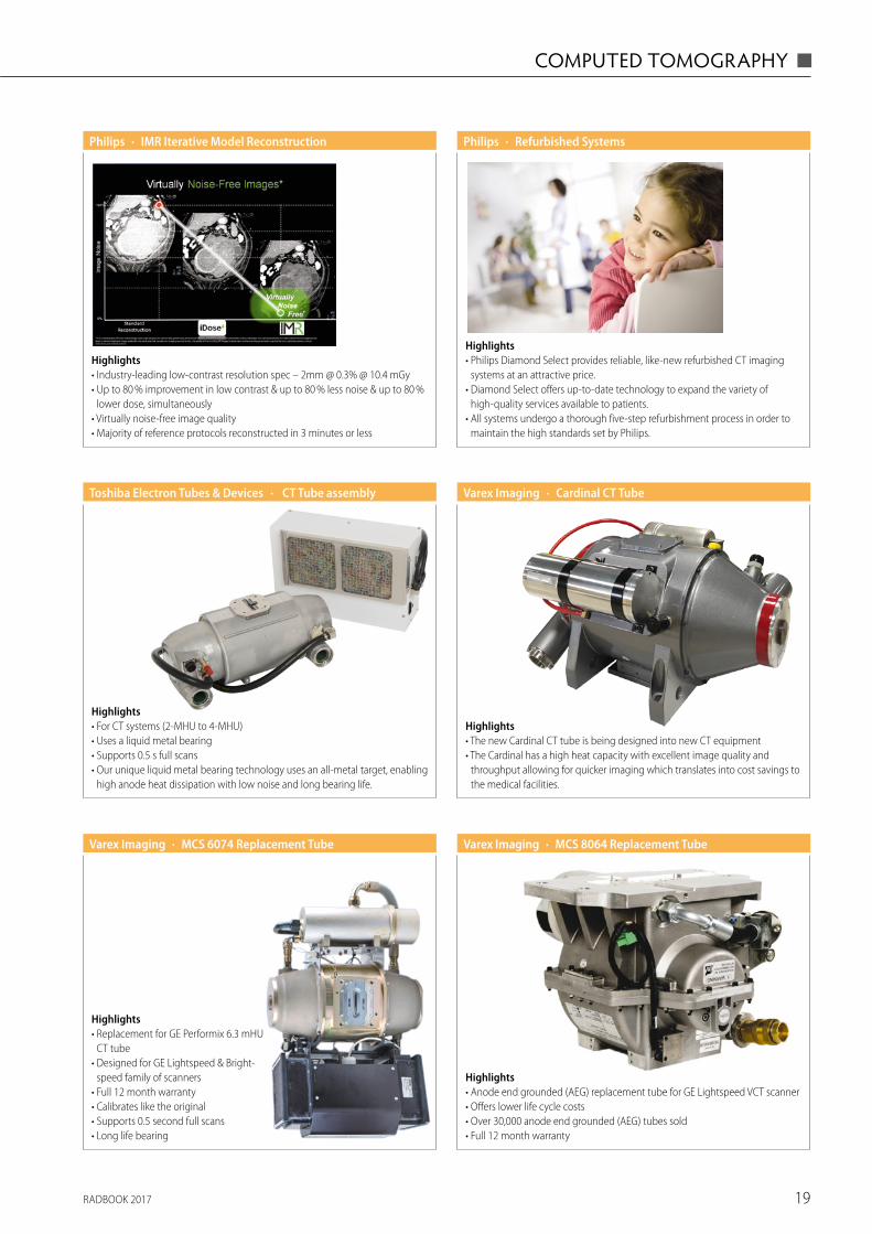
Highlights• Philips Diamond Select provides reliable, like-new refurbished CT imaging
systems at an attractive price.• Diamond Select offers up-to-date technology to expand the variety of
high-quality services available to patients.• All systems undergo a thorough five-step refurbishment process in order to
maintain the high standards set by Philips.
· Refurbished SystemsPhilips
Highlights• Industry-leading low-contrast resolution spec – 2mm @ 0.3% @ 10.4 mGy• Up to 80 % improvement in low contrast & up to 80 % less noise & up to 80 %
lower dose, simultaneously• Virtually noise-free image quality• Majority of reference protocols reconstructed in 3 minutes or less
· IMR Iterative Model ReconstructionPhilips
Highlights• For CT systems (2-MHU to 4-MHU)• Uses a liquid metal bearing• Supports 0.5 s full scans• Our unique liquid metal bearing technology uses an all-metal target, enabling
high anode heat dissipation with low noise and long bearing life.
· CT Tube assemblyToshiba Electron Tubes & Devices
Highlights• Anode end grounded (AEG) replacement tube for GE Lightspeed VCT scanner• Offers lower life cycle costs• Over 30,000 anode end grounded (AEG) tubes sold• Full 12 month warranty
· MCS 8064 Replacement TubeVarex Imaging
Highlights• The new Cardinal CT tube is being designed into new CT equipment • The Cardinal has a high heat capacity with excellent image quality and
throughput allowing for quicker imaging which translates into cost savings to the medical facilities.
· Cardinal CT TubeVarex Imaging
Highlights• Replacement for GE Performix 6.3 mHU
CT tube• Designed for GE Lightspeed & Bright-
speed family of scanners• Full 12 month warranty• Calibrates like the original• Supports 0.5 second full scans• Long life bearing
· MCS 6074 Replacement TubeVarex Imaging
RADBOOK 201720
◼ MAGNETIC RESONANCE IMAGING
Magnetic Resonance Imaging
7 Tesla
3 Tesla
1.5 Tesla
Open
MR-PET
MRT Coils
Accessories / Complementary Systems
RADBOOK 2017 21
MAGNETIC RESONANCE IMAGING ◼
7 TESLA
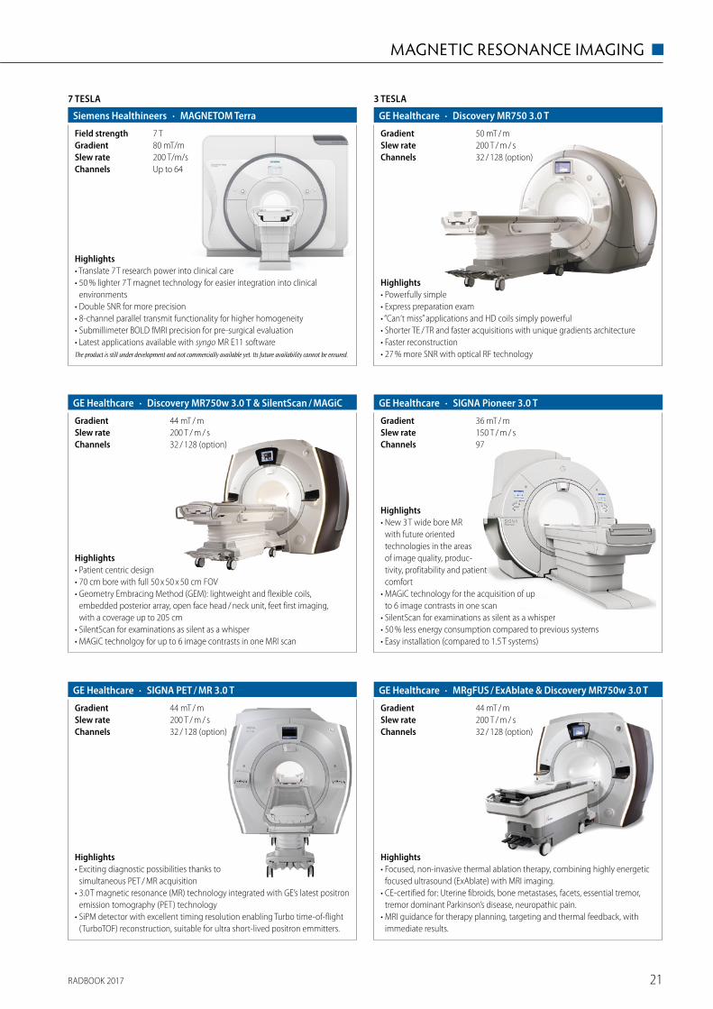
· MAGNETOM TerraSiemens Healthineers
Up to 64Channels200 T/m/sSlew rate80 mT/mGradient7 TField strength
Highlights• Translate 7 T research power into clinical care• 50 % lighter 7 T magnet technology for easier integration into clinical
environments• Double SNR for more precision• 8-channel parallel transmit functionality for higher homogeneity• Submillimeter BOLD fMRI precision for pre-surgical evaluation• Latest applications available with syngo MR E11 softwareThe product is still under development and not commercially available yet. Its future availability cannot be ensured.
Highlights• Exciting diagnostic possibilities thanks to
simultaneous PET / MR acquisition• 3.0 T magnetic resonance (MR) technology integrated with GE’s latest positron
emission tomography (PET) technology• SiPM detector with excellent timing resolution enabling Turbo time-of-flight
(TurboTOF) reconstruction, suitable for ultra short-lived positron emmitters.
· SIGNA PET / MR 3.0 TGE Healthcare
32 / 128 (option)Channels200 T / m / sSlew rate44 mT / mGradient
· SIGNA Pioneer 3.0 TGE Healthcare
97Channels150 T / m / sSlew rate36 mT / mGradient
Highlights• New 3 T wide bore MR
with future oriented technologies in the areas of image quality, produc-tivity, profitability and patient comfort
• MAGiC technology for the acquisition of up to 6 image contrasts in one scan
• SilentScan for examinations as silent as a whisper• 50 % less energy consumption compared to previous systems• Easy installation (compared to 1.5 T systems)
3 TESLA
· Discovery MR750 3.0 TGE Healthcare
32 / 128 (option)Channels200 T / m / sSlew rate50 mT / mGradient
Highlights• Powerfully simple• Express preparation exam• “Can‘t miss” applications and HD coils simply powerful• Shorter TE / TR and faster acquisitions with unique gradients architecture• Faster reconstruction• 27 % more SNR with optical RF technology
· Discovery MR750w 3.0 T & SilentScan / MAGiCGE Healthcare
32 / 128 (option)Channels200 T / m / sSlew rate44 mT / mGradient
Highlights• Patient centric design• 70 cm bore with full 50 x 50 x 50 cm FOV• Geometry Embracing Method (GEM): lightweight and flexible coils,
embedded posterior array, open face head / neck unit, feet first imaging, with a coverage up to 205 cm
• SilentScan for examinations as silent as a whisper• MAGiC technolgoy for up to 6 image contrasts in one MRI scan
· MRgFUS / ExAblate & Discovery MR750w 3.0 TGE Healthcare
32 / 128 (option)Channels200 T / m / sSlew rate44 mT / mGradient
Highlights• Focused, non-invasive thermal ablation therapy, combining highly energetic
focused ultrasound (ExAblate) with MRI imaging. • CE-certified for: Uterine fibroids, bone metastases, facets, essential tremor,
tremor dominant Parkinson’s disease, neuropathic pain. • MRI guidance for therapy planning, targeting and thermal feedback, with
immediate results.
RADBOOK 201722
◼ MAGNETIC RESONANCE IMAGING
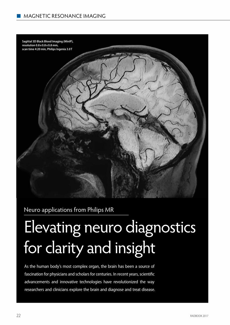
Neuro applications from Philips MR
Elevating neuro diagnostics for clarity and insight
However, despite the many developments, according to a 2016 TMTG study, 70 % of radiologists still consider neuro indications to be challenging, mostly due to a lack of imaging and visualization techniques. Philips believes that magnetic resonance imaging is in a unique position to address neurological disorders.
Against this background, Philips is this year introducing a set of advanced imaging and visualization strategies for neurological cases. The tools are designed to help clinicians answer complex indications and unlock new territories in neuro imaging.
Bringing complex structures to light
By visualizing intricate structures and helping clinicians track changes in the brain, MR can deliver vital insights into conditions such as brain tumors and vascular disease. Advanced visualization applications help physicians review complex, multi-dimensional data to make informed diagnosis and treatment decisions.
Philips is committed to pushing the boundaries and elevating neuro diagnostics with the aim of empowering healthcare providers to resolve neuro questions with more certainty. The suite of MR neuro tools intends to help clinicians explore new ground in advanced neurofunctional applications and deliver more de� nitive diagnoses1.
Rising patient numbers
Leveraging the Philips dStream digital platform, the enhanced port-folio of MR applications aims to touch the lives of a growing number of patients around the globe. Demographic changes such as aging populations in many parts of the world are driving an increase in neurological disease which creates new challenges in healthcare. Philips aims to extend the reach of MRI, by delivering advanced solu-tions that answer speci� c clinical and diagnostic questions.
1 De� nitive is de� ned as features that are expected to deliver alternative contrasts, functional or quantitative images.
2 Compared to Philips 2D double inversion methods with same brain coverage and scan time.3 Compared to Philips 3D T1w scan without MSDE pre-pulse.4 Compared to Philips 2D double inversion recovery methods with same full brain coverage.
As the human body’s most complex organ, the brain has been a source of
fascination for physicians and scholars for centuries. In recent years, scienti� c
advancements and innovative technologies have revo lutionized the way
researchers and clinicians explore the brain and diagnose and treat disease.
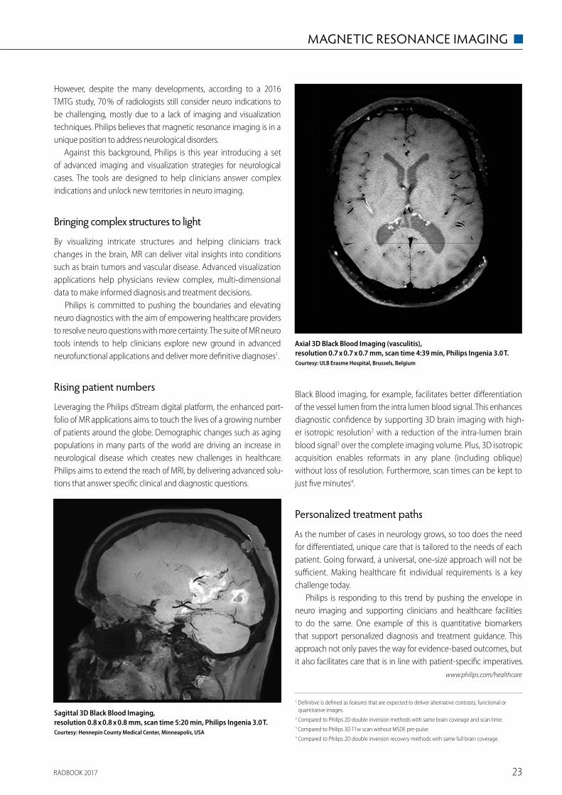
Black Blood imaging, for example, facilitates better di� eren tiation of the vessel lumen from the intra lumen blood signal. This enhances diagnostic con� dence by supporting 3D brain imaging with high-er isotropic resolution2 with a reduction of the intra-lumen brain blood signal3 over the complete imaging volume. Plus, 3D iso tropic acquisition enables reformats in any plane (including oblique) without loss of resolution. Furthermore, scan times can be kept to just � ve minutes4.
Personalized treatment paths
As the number of cases in neurology grows, so too does the need for di� erentiated, unique care that is tailored to the needs of each patient. Going forward, a universal, one-size approach will not be su� cient. Making healthcare � t individual requirements is a key challenge today.
Philips is responding to this trend by pushing the envelope in neuro imaging and supporting clinicians and healthcare facilities to do the same. One example of this is quantitative biomarkers that support personalized diagnosis and treatment guidance. This approach not only paves the way for evidence-based outcomes, but it also facilitates care that is in line with patient-speci� c imperatives.
www.philips.com/healthcare
Axial 3D Black Blood Imaging (vasculitis), resolution 0.7 x 0.7 x 0.7 mm, scan time 4:39 min, Philips Ingenia 3.0 T. Courtesy: ULB Erasme Hospital, Brussels, Belgium
Sagittal 3D Black Blood Imaging,resolution 0.8 x 0.8 x 0.8 mm, scan time 5:20 min, Philips Ingenia 3.0 T. Courtesy: Hennepin County Medical Center, Minneapolis, USA
Sagittal 3D Black Blood Imaging (MinIP), resolution 0.8 x 0.8 x 0.8 mm, scan time 4:20 min, Philips Ingenia 3.0 T
RADBOOK 2017 23
MAGNETIC RESONANCE IMAGING ◼
Neuro applications from Philips MR
Elevating neuro diagnostics for clarity and insight
However, despite the many developments, according to a 2016 TMTG study, 70 % of radiologists still consider neuro indications to be challenging, mostly due to a lack of imaging and visualization techniques. Philips believes that magnetic resonance imaging is in a unique position to address neurological disorders.
Against this background, Philips is this year introducing a set of advanced imaging and visualization strategies for neurological cases. The tools are designed to help clinicians answer complex indications and unlock new territories in neuro imaging.
Bringing complex structures to light
By visualizing intricate structures and helping clinicians track changes in the brain, MR can deliver vital insights into conditions such as brain tumors and vascular disease. Advanced visualization applications help physicians review complex, multi-dimensional data to make informed diagnosis and treatment decisions.
Philips is committed to pushing the boundaries and elevating neuro diagnostics with the aim of empowering healthcare providers to resolve neuro questions with more certainty. The suite of MR neuro tools intends to help clinicians explore new ground in advanced neurofunctional applications and deliver more de� nitive diagnoses1.
Rising patient numbers
Leveraging the Philips dStream digital platform, the enhanced port-folio of MR applications aims to touch the lives of a growing number of patients around the globe. Demographic changes such as aging populations in many parts of the world are driving an increase in neurological disease which creates new challenges in healthcare. Philips aims to extend the reach of MRI, by delivering advanced solu-tions that answer speci� c clinical and diagnostic questions.
1 De� nitive is de� ned as features that are expected to deliver alternative contrasts, functional or quantitative images.
2 Compared to Philips 2D double inversion methods with same brain coverage and scan time.3 Compared to Philips 3D T1w scan without MSDE pre-pulse.4 Compared to Philips 2D double inversion recovery methods with same full brain coverage.
As the human body’s most complex organ, the brain has been a source of
fascination for physicians and scholars for centuries. In recent years, scienti� c
advancements and innovative technologies have revo lutionized the way
researchers and clinicians explore the brain and diagnose and treat disease.
Black Blood imaging, for example, facilitates better di� eren tiation of the vessel lumen from the intra lumen blood signal. This enhances diagnostic con� dence by supporting 3D brain imaging with high-er isotropic resolution2 with a reduction of the intra-lumen brain blood signal3 over the complete imaging volume. Plus, 3D iso tropic acquisition enables reformats in any plane (including oblique) without loss of resolution. Furthermore, scan times can be kept to just � ve minutes4.
Personalized treatment paths
As the number of cases in neurology grows, so too does the need for di� erentiated, unique care that is tailored to the needs of each patient. Going forward, a universal, one-size approach will not be su� cient. Making healthcare � t individual requirements is a key challenge today.
Philips is responding to this trend by pushing the envelope in neuro imaging and supporting clinicians and healthcare facilities to do the same. One example of this is quantitative biomarkers that support personalized diagnosis and treatment guidance. This approach not only paves the way for evidence-based outcomes, but it also facilitates care that is in line with patient-speci� c imperatives.
www.philips.com/healthcare
Axial 3D Black Blood Imaging (vasculitis), resolution 0.7 x 0.7 x 0.7 mm, scan time 4:39 min, Philips Ingenia 3.0 T. Courtesy: ULB Erasme Hospital, Brussels, Belgium
Sagittal 3D Black Blood Imaging,resolution 0.8 x 0.8 x 0.8 mm, scan time 5:20 min, Philips Ingenia 3.0 T. Courtesy: Hennepin County Medical Center, Minneapolis, USA
Sagittal 3D Black Blood Imaging (MinIP), resolution 0.8 x 0.8 x 0.8 mm, scan time 4:20 min, Philips Ingenia 3.0 T
RADBOOK 201724
◼ MAGNETIC RESONANCE IMAGING
3 TESLA
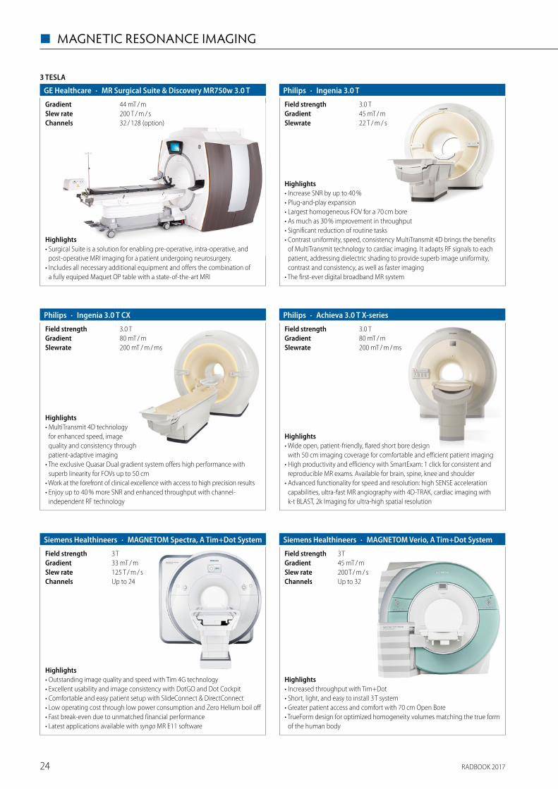
· MR Surgical Suite & Discovery MR750w 3.0 TGE Healthcare
32 / 128 (option)Channels200 T / m / sSlew rate44 mT / mGradient
Highlights• Surgical Suite is a solution for enabling pre-operative, intra-operative, and
post-operative MRI imaging for a patient undergoing neurosurgery.• Includes all necessary additional equipment and offers the combination of
a fully equiped Maquet OP table with a state-of-the-art MRI
· Ingenia 3.0 T CX Philips
200 mT / m / msSlewrate80 mT / mGradient3.0 TField strength
Highlights• MultiTransmit 4D technology
for enhanced speed, image quality and consistency through patient-adaptive imaging
• The exclusive Quasar Dual gradient system offers high performance with superb linearity for FOVs up to 50 cm
• Work at the forefront of clinical excellence with access to high precision results• Enjoy up to 40 % more SNR and enhanced throughput with channel-
independent RF technology
· Achieva 3.0 T X-seriesPhilips
200 mT / m / msSlewrate80 mT / mGradient3.0 TField strength
Highlights• Wide open, patient-friendly, flared short bore design
with 50 cm imaging coverage for comfortable and efficient patient imaging• High productivity and efficiency with SmartExam: 1 click for consistent and
reproducible MR exams. Available for brain, spine, knee and shoulder• Advanced functionality for speed and resolution: high SENSE acceleration
capabilities, ultra-fast MR angiography with 4D-TRAK, cardiac imaging with k-t BLAST, 2k Imaging for ultra-high spatial resolution
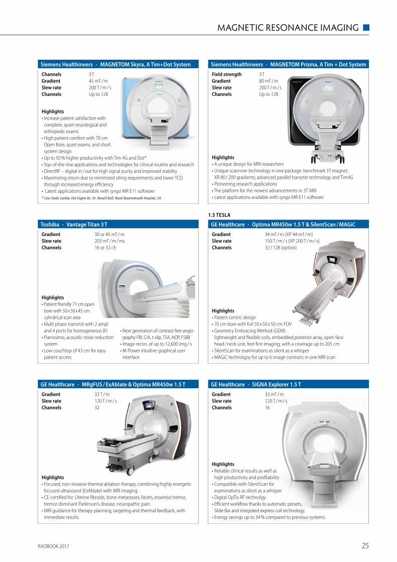
· Ingenia 3.0 TPhilips
22 T / m / sSlewrate45 mT / mGradient3.0 TField strength
Highlights• Increase SNR by up to 40 %• Plug-and-play expansion• Largest homogeneous FOV for a 70 cm bore• As much as 30 % improvement in throughput• Significant reduction of routine tasks• Contrast uniformity, speed, consistency MultiTransmit 4D brings the benefits
of MultiTransmit technology to cardiac imaging. It adapts RF signals to each patient, addressing dielectric shading to provide superb image uniformity, contrast and consistency, as well as faster imaging
• The first-ever digital broadband MR system
Highlights• Outstanding image quality and speed with Tim 4G technology • Excellent usability and image consistency with DotGO and Dot Cockpit• Comfortable and easy patient setup with SlideConnect & DirectConnect • Low operating cost through low power consumption and Zero Helium boil off • Fast break-even due to unmatched financial performance• Latest applications available with syngo MR E11 software
· MAGNETOM Spectra, A Tim+Dot SystemSiemens Healthineers
Up to 24Channels125 T / m / sSlew rate33 mT / mGradient3 TField strength
Highlights• Increased throughput with Tim+Dot• Short, light, and easy to install 3 T system• Greater patient access and comfort with 70 cm Open Bore• TrueForm design for optimized homogeneity volumes matching the true form
of the human body
· MAGNETOM Verio, A Tim+Dot SystemSiemens Healthineers
Up to 32Channels200 T / m / sSlew rate45 mT / mGradient3 TField strength
RADBOOK 2017 25
MAGNETIC RESONANCE IMAGING ◼
Highlights• A unique design for MRI researchers• Unique scannner technology in one package: benchmark 3T magnet;
XR 80 / 200 gradients; advanced parallel transmit technology and Tim4G• Pioneering research applications• The platform for the newest advancements in 3T MRI• Latest applications available with syngo MR E11 software
· MAGNETOM Prisma, A Tim + Dot SystemSiemens Healthineers
Up to 128Channels200 T / m / sSlew rate80 mT / mGradient3 TField strength
· MAGNETOM Skyra, A Tim+Dot SystemSiemens Healthineers
Up to 128Channels200 T / m / sSlew rate45 mT / mGradient3 TChannels
Highlights• Increase patient satisfaction with
complete, quiet neurological and orthopedic exams
• High patient comfort with 70 cm Open Bore, quiet exams, and short system design
• Up to 50 % higher productivity with Tim 4G and Dot*• Top-of-the-line applications and technologies for clinical routine and research• DirectRF – digital in / out for high signal purity and improved stability• Maximizing return due to minimized siting requirements and lower TCO
through increased energy efficiency • Latest applications available with syngo MR E11 software* Case Study Cardiac Dot Engine by: Dr. Russell Bull, Royal Bournemouth Hospital, UK
· Vantage Titan 3 TToshiba
16 or 32 chChannels203 mT / m / msSlew rate30 or 45 mT / mGradient
Highlights• Patient friendly 71 cm open
bore with 50 x 50 x 45 cm cylindrical scan area
• Multi phase transmit with 2 ampl and 4 ports for homogeneous B1
• Pianissimo, acoustic noise reduction system
• Low couchtop of 43 cm for easy patient access
• Next generation of contrast-free angio-graphy FBI, CIA, t-slip, TSA, HOP, FSBB
• Image recon. of up to 12,600 img / s• M-Power intuitive graphical user
interface
1.5 TESLA
· Optima MR450w 1.5 T & SilentScan / MAGiCGE Healthcare
32 / 128 (option)Channels150 T / m / s (XP 200 T / m / s)Slew rate34 mT / m (XP 44 mT / m)Gradient
Highlights• Patient centric design• 70 cm bore with full 50 x 50 x 50 cm FOV• Geometry Embracing Method (GEM):
lightweight and flexible coils, embedded posterior array, open face head / neck unit, feet first imaging, with a coverage up to 205 cm
• SilentScan for examinations as silent as a whisper• MAGiC technolgoy for up to 6 image contrasts in one MRI scan
· MRgFUS / ExAblate & Optima MR450w 1.5 TGE Healthcare
32Channels120 T / m / sSlew rate33 T / m Gradient
Highlights• Focused, non-invasive thermal ablation therapy, combining highly energetic
focused ultrasound (ExAblate) with MRI imaging. • CE-certified for: Uterine fibroids, bone metastases, facets, essential tremor,
tremor dominant Parkinson’s disease, neuropathic pain. • MRI guidance for therapy planning, targeting and thermal feedback, with
immediate results.
· SIGNA Explorer 1.5 TGE Healthcare
16Channels120 T / m / sSlew rate33 mT / mGradient
Highlights• Reliable clinical results as well as
high productivity and profitability • Compatible with SilentScan for
examinations as silent as a whisper• Digital OpTix RF-technolgy. • Efficient workflow thanks to automatic presets,
Slide Bar and integrated express coil technology.• Energy savings up to 34 % compared to previous systems.
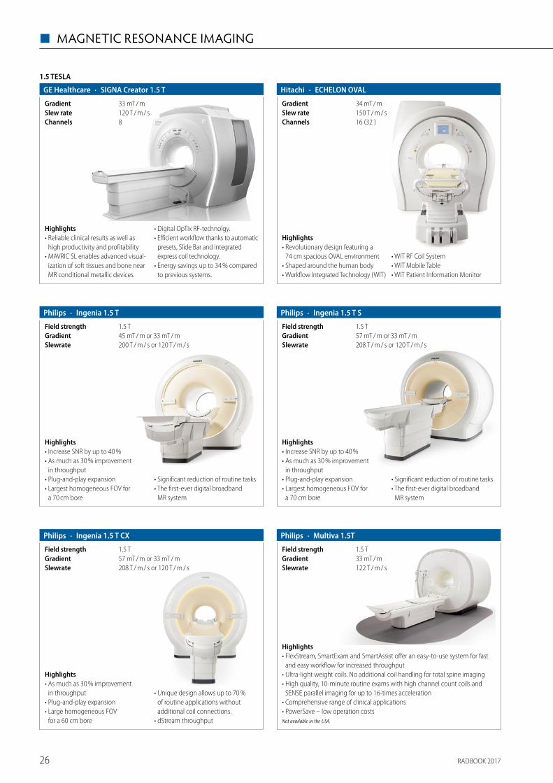
RADBOOK 201726
◼ MAGNETIC RESONANCE IMAGING
1.5 TESLA
· SIGNA Creator 1.5 TGE Healthcare
8Channels120 T / m / sSlew rate33 mT / mGradient
Highlights• Reliable clinical results as well as
high productivity and profitability• MAVRIC SL enables advanced visual-
ization of soft tissues and bone near MR conditional metallic devices.
• Digital OpTix RF-technolgy. • Efficient workflow thanks to automatic
presets, Slide Bar and integrated express coil technology.
• Energy savings up to 34 % compared to previous systems.
· ECHELON OVALHitachi
16 (32 )Channels150 T / m / sSlew rate34 mT / mGradient
Highlights• Revolutionary design featuring a
74 cm spacious OVAL environment• Shaped around the human body• Workflow Integrated Technology (WIT)
• WIT RF Coil System• WIT Mobile Table• WIT Patient Information Monitor
· Ingenia 1.5 TPhilips
200 T / m / s or 120 T / m / sSlewrate45 mT / m or 33 mT / mGradient1.5 TField strength
Highlights• Increase SNR by up to 40 %• As much as 30 % improvement
in throughput• Plug-and-play expansion• Largest homogeneous FOV for
a 70 cm bore
• Significant reduction of routine tasks• The first-ever digital broadband
MR system
· Ingenia 1.5 T SPhilips
208 T / m / s or 120 T / m / sSlewrate57 mT / m or 33 mT / mGradient1.5 TField strength
Highlights• Increase SNR by up to 40 % • As much as 30 % improvement
in throughput • Plug-and-play expansion • Largest homogeneous FOV for
a 70 cm bore
• Significant reduction of routine tasks • The first-ever digital broadband
MR system
· Ingenia 1.5 T CXPhilips
208 T / m / s or 120 T / m / sSlewrate57 mT / m or 33 mT / mGradient1.5 TField strength
Highlights• As much as 30 % improvement
in throughput • Plug-and-play expansion • Large homogeneous FOV
for a 60 cm bore
• Unique design allows up to 70 % of routine applications without additional coil connections.
• dStream throughput
· Multiva 1.5TPhilips
122 T / m / sSlewrate33 mT / mGradient1.5 TField strength
Highlights• FlexStream, SmartExam and SmartAssist offer an easy-to-use system for fast
and easy workflow for increased throughput• Ultra-light weight coils. No additional coil handling for total spine imaging• High quality, 10-minute routine exams with high channel count coils and
SENSE parallel imaging for up to 16-times acceleration• Comprehensive range of clinical applications• PowerSave – low operation costsNot available in the USA.
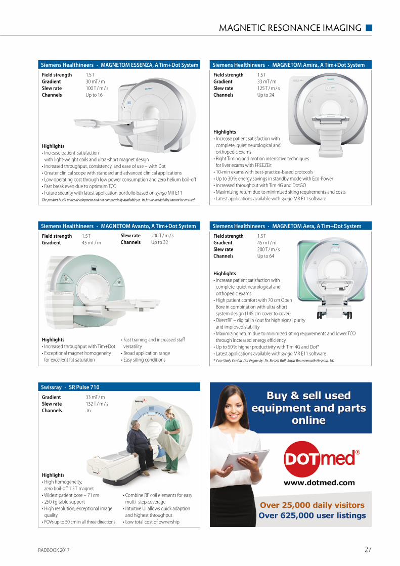
RADBOOK 2017 27
MAGNETIC RESONANCE IMAGING ◼
Highlights• Increase patient satisfaction with
complete, quiet neurological and ortho pedic exams
• High patient comfort with 70 cm Open Bore in combination with ultra-short system design (145 cm cover to cover)
• DirectRF – digital in / out for high signal purity and improved stability
• Maximizing return due to minimized siting requirements and lower TCO through increased energy efficiency
• Up to 50 % higher productivity with Tim 4G and Dot* • Latest applications available with syngo MR E11 software* Case Study Cardiac Dot Engine by: Dr. Russell Bull, Royal Bournemouth Hospital, UK
· MAGNETOM Aera, A Tim+Dot SystemSiemens Healthineers
Up to 64Channels200 T / m / sSlew rate45 mT / mGradient1.5 TField strength
Highlights• Increase patient satisfaction with
complete, quiet neurological and orthopedic exams
• Right Timing and motion insensitive techniques for liver exams with FREEZEit
• 10-min exams with best-practice-based protocols• Up to 30 % energy savings in standby mode with Eco-Power• Increased throughput with Tim 4G and DotGO• Maximizing return due to minimized siting requirements and costs• Latest applications available with syngo MR E11 software
· MAGNETOM Amira, A Tim+Dot SystemSiemens Healthineers
Up to 24Channels125 T / m / sSlew rate33 mT / mGradient1.5 TField strength
Highlights• Increased throughput with Tim+Dot• Exceptional magnet homogeneity
for excellent fat saturation
• Fast training and increased staff versatility
• Broad application range• Easy siting conditions
· MAGNETOM Avanto, A Tim+Dot SystemSiemens Healthineers
Up to 32Channels200 T / m / sSlew rate
45 mT / mGradient1.5 TField strength
Highlights• Increase patient-satisfaction
with light-weight coils and ultra-short magnet design• Increased throughput, consistency, and ease of use – with Dot• Greater clinical scope with standard and advanced clinical applications• Low operating cost through low power consumption and zero helium boil-off• Fast break even due to optimum TCO• Future security with latest application portfolio based on syngo MR E11The product is still under development and not commercially available yet. Its future availability cannot be ensured.
· MAGNETOM ESSENZA, A Tim+Dot SystemSiemens Healthineers
Up to 16Channels100 T / m / sSlew rate30 mT / mGradient1.5 TField strength
· SR Pulse 710Swissray
16Channels132 T / m / sSlew rate33 mT / mGradient
Highlights• High homogeneity,
zero boil-off 1.5 T magnet• Widest patient bore – 71 cm• 250 kg table support• High resolution, exceptional image
quality• FOVs up to 50 cm in all three directions
• Combine RF coil elements for easy multi- step coverage
• Intuitive UI allows quick adaption and highest throughput
• Low total cost of ownership
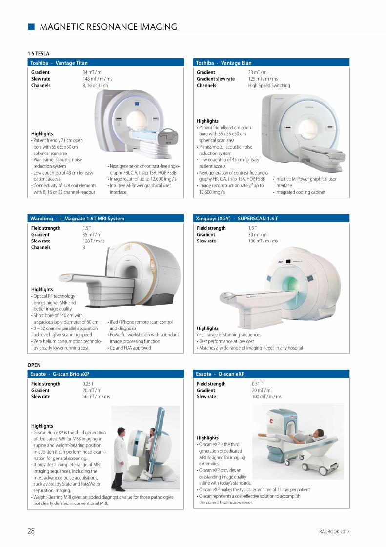
RADBOOK 201728
◼ MAGNETIC RESONANCE IMAGING
1.5 TESLA
· Vantage TitanToshiba
8, 16 or 32 chChannels148 mT / m / msSlew rate34 mT / mGradient
Highlights• Patient friendly 71 cm open
bore with 55 x 55 x 50 cm spherical scan area
• Pianissimo, acoustic noise reduction system
• Low couchtop of 43 cm for easy patient access
• Connectivity of 128 coil elements with 8, 16 or 32 channel-readout
• Next generation of contrast-free angio-graphy FBI, CIA, t-slip, TSA, HOP, FSBB
• Image recon of up to 12,600 img / s• Intuitive M-Power graphical user
interface
· Vantage ElanToshiba
High Speed SwitchingChannels125 mT / m / msGradient slew rate33 mT / mGradient
Highlights• Patient friendly 63 cm open
bore with 55 x 55 x 50 cm spherical scan area
• Pianissimo Σ , acoustic noise reduction system
• Low couchtop of 45 cm for easy patient access
• Next generation of contrast-free angio-graphy FBI, CIA, t-slip, TSA, HOP, FSBB
• Image reconstruction rate of up to 12,600 img / s
• Intuitive M-Power graphical user interface
• Integrated cooling cabinet
Highlights• Optical RF technology
brings higher SNR and better image quality
• Short bore of 140 cm with a spacious bore diameter of 60 cm
• 8 – 32 channel parallel acquisition achieve higher scanning speed
• Zero helium consumption technolo-gy greatly lower running cost
• iPad / iPhone remote scan control and diagnosis
• Powerful workstation with abundant image processing function
• CE and FDA approved
· i_Magnate 1.5T MRI SystemWandong
8Channels128 T / m / sSlew rate35 mT / mGradient1.5 TField strength
Highlights• Full range of stanning sequences• Best performance at low cost• Matches a wide range of imaging needs in any hospital
· SUPERSCAN 1.5 TXingaoyi (XGY)
100 mT / m / msSlew rate30 mT / mGradient1.5 TField strength
Highlights• G-scan Brio eXP is the third generation
of dedicated MRI for MSK imaging in supine and weight-bearing position. In addition it can perform head exami-nation for general screening.
• It provides a complete range of MRI imaging sequences, including the most advanced pulse acquisitions, such as Steady State and Fat&Water separation imaging.
• Weight-Bearing MRI gives an added diagnostic value for those pathologies not clearly defined in conventional MRI.
OPEN
· G-scan Brio eXPEsaote
56 mT / m / msSlew rate20 mT / mGradient0.25 TField strength
Highlights• O-scan eXP is the third
generation of dedicated MRI designed for imaging extremities.
• O-scan eXP provides an outstanding image quality in line with today’s standards.
• O-scan eXP makes the typical exam time of 15 min per patient. • O-scan represents a cost-effective solution to accomplish
the current healthcare’s needs.
· O-scan eXPEsaote
100 mT / m / msSlew rate20 mT / mGradient0.31 TField strength
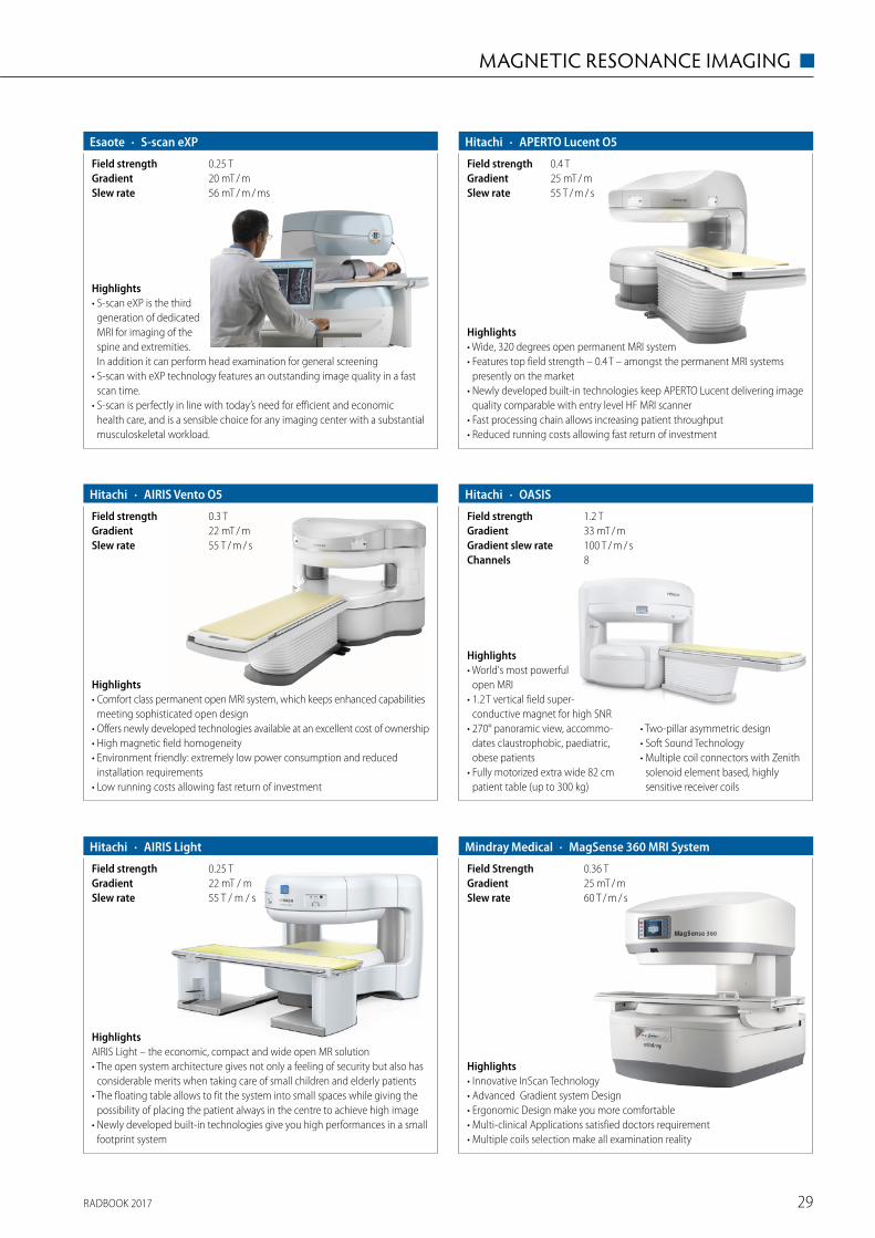
RADBOOK 2017 29
MAGNETIC RESONANCE IMAGING ◼
Highlights• S-scan eXP is the third
generation of dedicated MRI for imaging of the spine and extremities. In addition it can perform head examination for general screening
• S-scan with eXP technology features an outstanding image quality in a fast scan time.
• S-scan is perfectly in line with today’s need for efficient and economic health care, and is a sensible choice for any imaging center with a substantial musculoskeletal workload.
· S-scan eXPEsaote
56 mT / m / msSlew rate20 mT / mGradient0.25 T Field strength
· AIRIS Vento O5Hitachi
55 T / m / sSlew rate22 mT / mGradient0.3 TField strength
Highlights• Comfort class permanent open MRI system, which keeps enhanced capabilities
meeting sophisticated open design• Offers newly developed technologies available at an excellent cost of ownership• High magnetic field homogeneity• Environment friendly: extremely low power consumption and reduced
installation requirements• Low running costs allowing fast return of investment
· APERTO Lucent O5Hitachi
55 T / m / sSlew rate25 mT / mGradient0.4 TField strength
Highlights• Wide, 320 degrees open permanent MRI system• Features top field strength – 0.4 T – amongst the permanent MRI systems
presently on the market• Newly developed built-in technologies keep APERTO Lucent delivering image
quality comparable with entry level HF MRI scanner• Fast processing chain allows increasing patient throughput• Reduced running costs allowing fast return of investment
· AIRIS LightHitachi
55 T / m / sSlew rate22 mT / mGradient0.25 TField strength
HighlightsAIRIS Light – the economic, compact and wide open MR solution• The open system architecture gives not only a feeling of security but also has
considerable merits when taking care of small children and elderly patients• The floating table allows to fit the system into small spaces while giving the
possibility of placing the patient always in the centre to achieve high image• Newly developed built-in technologies give you high performances in a small
footprint system
· OASISHitachi
8Channels100 T / m / sGradient slew rate33 mT / mGradient1.2 TField strength
Highlights• World's most powerful
open MRI• 1.2 T vertical field super-
conductive magnet for high SNR• 270° panoramic view, accommo-
dates claustrophobic, paediatric, obese patients
• Fully motorized extra wide 82 cm patient table (up to 300 kg)
• Two-pillar asymmetric design• Soft Sound Technology• Multiple coil connectors with Zenith
solenoid element based, highly sensitive receiver coils
· MagSense 360 MRI SystemMindray Medical
60 T / m / sSlew rate25 mT / mGradient0.36 TField Strength
Highlights• Innovative InScan Technology• Advanced Gradient system Design• Ergonomic Design make you more comfortable• Multi-clinical Applications satisfied doctors requirement• Multiple coils selection make all examination reality
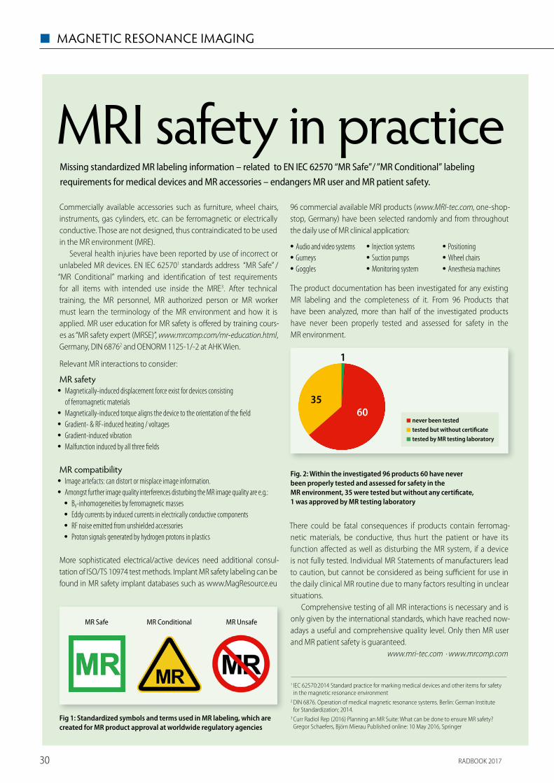
RADBOOK 201730
◼ MAGNETIC RESONANCE IMAGING
MRI safety in practiceCommercially available accessories such as furniture, wheel chairs, instruments, gas cylinders, etc. can be ferromagnetic or electrically conductive. Those are not designed, thus contraindicated to be used in the MR environment (MRE).
Several health injuries have been reported by use of incorrect or unlabeled MR devices. EN IEC 625701 standards address “MR Safe” / “MR Conditional” marking and identi� cation of test requirements for all items with intended use inside the MRE3. After technical training, the MR personnel, MR authorized person or MR worker must learn the terminology of the MR environment and how it is applied. MR user education for MR safety is o� ered by training cours-es as “MR safety expert (MRSE)”, www.mrcomp.com/mr-education.html, Germany, DIN 68762 and OENORM 1125-1/-2 at AHK Wien.
Relevant MR interactions to consider:
MR safety • Magnetically-induced displacement force exist for devices consisting
of ferromagnetic materials • Magnetically-induced torque aligns the device to the orientation of the � eld • Gradient- & RF-induced heating / voltages • Gradient-induced vibration • Malfunction induced by all three � elds
MR compatibility • Image artefacts: can distort or misplace image information. • Amongst further image quality interferences disturbing the MR image quality are e.g.:
• B0-inhomogeneities by ferromagnetic masses • Eddy currents by induced currents in electrically conductive components • RF noise emitted from unshielded accessories • Proton signals generated by hydrogen protons in plastics
More sophisticated electrical/active devices need additional consul-tation of ISO/TS 10974 test methods. Implant MR safety labeling can be found in MR safety implant databases such as www.MagResource.eu
Missing standardized MR labeling information – related to EN IEC 62570 “MR Safe” / ”MR Conditional” labeling requirements for medical devices and MR accessories – endangers MR user and MR patient safety.
96 commercial available MRI products (www.MRI-tec.com, one-shop-stop, Germany) have been selected randomly and from throughout the daily use of MR clinical application:
• Audio and video systems • Gurneys • Goggles
• Injection systems • Suction pumps • Monitoring system
• Positioning • Wheel chairs • Anesthesia machines
The product documentation has been investigated for any existing MR labeling and the completeness of it. From 96 Products that have been analyzed, more than half of the investigated products have never been properly tested and assessed for safety in the MR environment.
Fig. 2: Within the investigated 96 products 60 have never been properly tested and assessed for safety in the MR environment, 35 were tested but without any certi� cate, 1 was approved by MR testing laboratory
There could be fatal consequences if products contain ferromag-netic materials, be conductive, thus hurt the patient or have its function a� ected as well as disturbing the MR system, if a device is not fully tested. Individual MR Statements of manufacturers lead to caution, but cannot be considered as being su� cient for use in the daily clinical MR routine due to many factors resulting in unclear situations.
Comprehensive testing of all MR interactions is necessary and is only given by the international standards, which have reached now-adays a useful and comprehensive quality level. Only then MR user and MR patient safety is guaranteed.
www.mri-tec.com · www.mrcomp.com
Fig 1: Standardized symbols and terms used in MR labeling, which are created for MR product approval at worldwide regulatory agencies
1 IEC 62570:2014 Standard practice for marking medical devices and other items for safety in the magnetic resonance environment
2 DIN 6876. Operation of medical magnetic resonance systems. Berlin: German Institute for Standardization; 2014.
3 Curr Radiol Rep (2016) Planning an MR Suite: What can be done to ensure MR safety? Gregor Schaefers, Björn Mierau Published online: 10 May 2016, Springer
MR Safe MR Conditional MR Unsafe
◾ never been tested ◾ tested but without certi� cate◾ tested by MR testing laboratory
6035
1
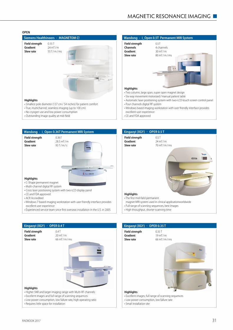
RADBOOK 2017 31
MAGNETIC RESONANCE IMAGING ◼
Highlights• Smallest pole diameter (137 cm / 54 inches) for patient comfort• True, multichannel, seamless imaging (up to 100 cm)• No cryogen use and low power consumption • Outstanding image quality at mid-field
OPEN
· MAGNETOM C!Siemens Healthineers
55 T / m / msSlew rate24 mT / mGradient0.35 TField strength
Highlights• Two column, large span, super open magnet design• Six-way movement motorized / manual patient table• Automatic laser positioning system with two-LCD touch screen control panel• Four channels digital RF system• Windows based imaging workstation with user friendly interface provides
excellent user experience• CE and FDA approved
· i_Open 0.5T Permanent MRI SystemWandong
80 mT / m / msSlew rate30 mT / mGradient4 channelsChannels0.5 TField strength
· i_Open 0.36T Permanent MRI SystemWandong
95 T / m / sSlew rate28.5 mT / mGradient0.36 TField strength
Highlights• C-Shape permanent magnet• Multi-channel digital RF system• Cross laser positioning system with two-LCD display panel• CE and FDA approved• ACR Accredited• Windows 7 based imaging workstation with user friendly interface provides
excellent user experience• Experienced service team since first overseas installation in the U.S. in 2005
Highlights• The first mid-field permanent
magnet MRI system used in clinical applicationworldwide• Full range of scanning sequences, best images• High throughput, shorter scanning time
· OPER 0.5 TXingaoyi (XGY)
70 mT / m / msSlew rate24 mT / mGradient0.5 TField strength
Highlights• Higher SNR and larger imaging range with Multi-RF channels• Excellent images and full range of scanning sequences• Low power consumption, low failure rate, high operating ratio• Requires little space for installation
· OPER 0.4 TXingaoyi (XGY)
66 mT / m / msSlew rate20 mT / mGradient0.4 TField strength
Highlights• Excellent images, full range of scanning sequences• Low power consumption, low failure rate• Small installation site
· OPER 0.35 TXingaoyi (XGY)
66 mT / m / msSlew rate19 mT / mGradient0.35 TField strength
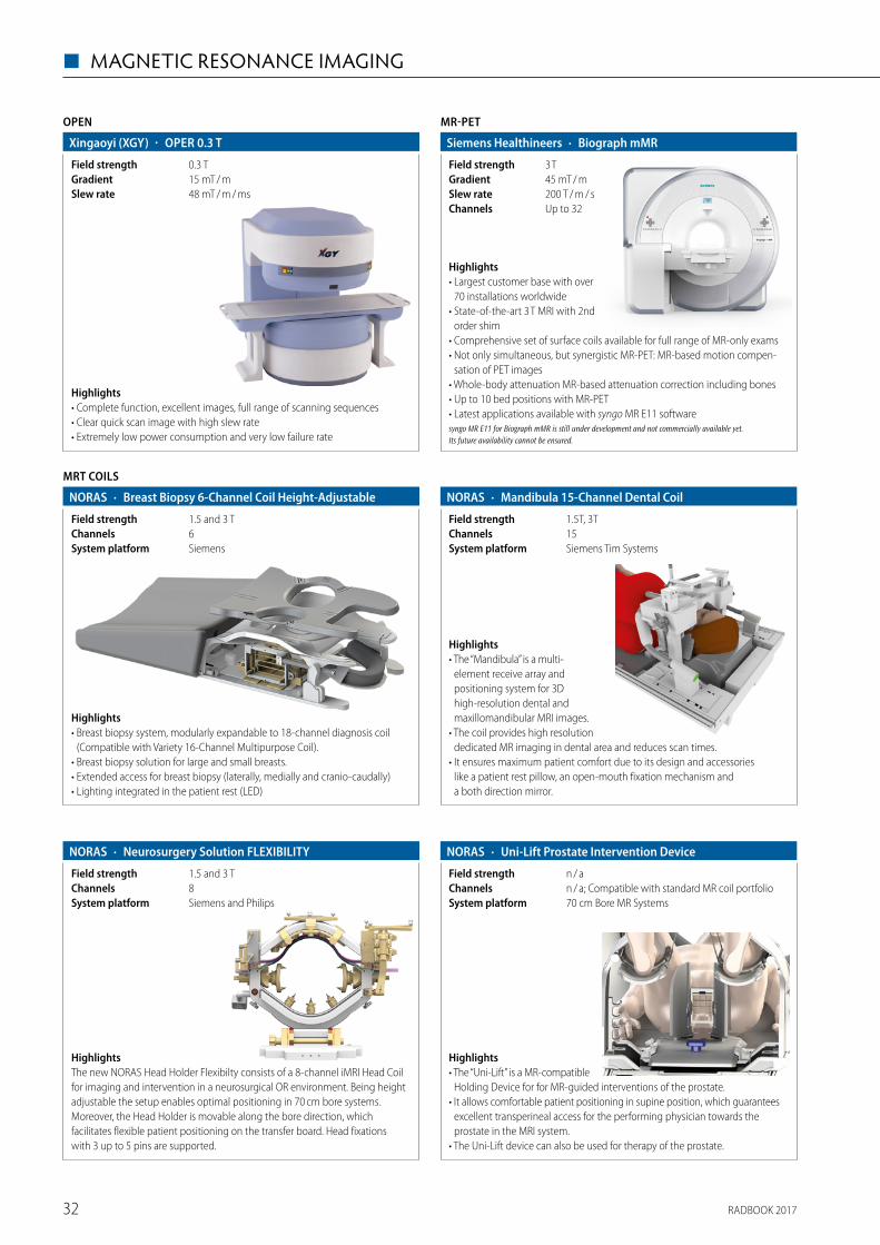
RADBOOK 201732
◼ MAGNETIC RESONANCE IMAGING
OPEN
Highlights• Complete function, excellent images, full range of scanning sequences• Clear quick scan image with high slew rate• Extremely low power consumption and very low failure rate
· OPER 0.3 TXingaoyi (XGY)
48 mT / m / msSlew rate15 mT / mGradient0.3 TField strength
MR-PET
· Biograph mMRSiemens Healthineers
200 T / m / sSlew rate45 mT / mGradient3 TField strength
Up to 32Channels
Highlights• Largest customer base with over
70 installations worldwide• State-of-the-art 3 T MRI with 2nd
order shim • Comprehensive set of surface coils available for full range of MR-only exams• Not only simultaneous, but synergistic MR-PET: MR-based motion compen-
sation of PET images• Whole-body attenuation MR-based attenuation correction including bones• Up to 10 bed positions with MR-PET• Latest applications available with syngo MR E11 softwaresyngo MR E11 for Biograph mMR is still under development and not commercially available yet. Its future availability cannot be ensured.
Highlights• Breast biopsy system, modularly expandable to 18-channel diagnosis coil
(Compatible with Variety 16-Channel Multipurpose Coil).• Breast biopsy solution for large and small breasts.• Extended access for breast biopsy (laterally, medially and cranio-caudally)• Lighting integrated in the patient rest (LED)
MRT COILS
· Breast Biopsy 6-Channel Coil Height-AdjustableNORAS
SiemensSystem platform6Channels1.5 and 3 TField strength
Highlights• The “Mandibula” is a multi-
element receive array and positioning system for 3D high-resolution dental and maxillomandibular MRI images.
• The coil provides high resolution dedicated MR imaging in dental area and reduces scan times.
• It ensures maximum patient comfort due to its design and accessories like a patient rest pillow, an open-mouth fixation mechanism and a both direction mirror.
· Mandibula 15-Channel Dental CoilNORAS
Siemens Tim SystemsSystem platform15Channels1.5T, 3TField strength
· Neurosurgery Solution FLEXIBILITYNORAS
Siemens and PhilipsSystem platform8Channels1.5 and 3 TField strength
HighlightsThe new NORAS Head Holder Flexibilty consists of a 8-channel iMRI Head Coil for imaging and intervention in a neurosurgical OR environment. Being height adjustable the setup enables optimal positioning in 70 cm bore systems. Moreover, the Head Holder is movable along the bore direction, which facilitates flexible patient positioning on the transfer board. Head fixations with 3 up to 5 pins are supported.
· Uni-Lift Prostate Intervention DeviceNORAS
70 cm Bore MR Systems System platformn / a; Compatible with standard MR coil portfolioChannelsn / aField strength
Highlights• The “Uni-Lift” is a MR-compa tible
Holding Device for for MR-guided interventions of the prostate.• It allows comfortable patient positioning in supine position, which guarantees
excellent transperineal access for the performing physician towards the prostate in the MRI system.
• The Uni-Lift device can also be used for therapy of the prostate.
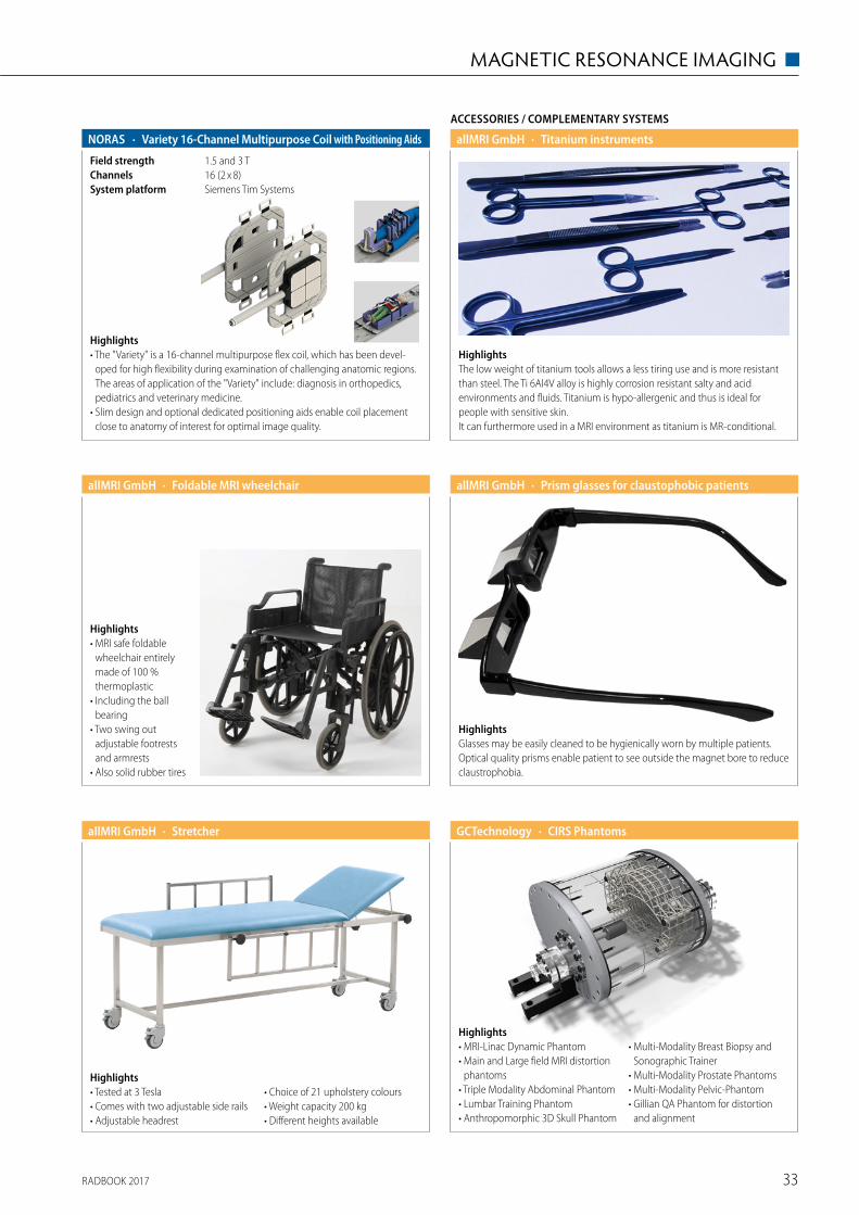
RADBOOK 2017 33
MAGNETIC RESONANCE IMAGING ◼
Highlights• The "Variety" is a 16-channel multipurpose flex coil, which has been devel-
oped for high flexibility during examination of challenging anatomic regions. The areas of application of the "Variety" include: diagnosis in orthopedics, pediatrics and veterinary medicine.
• Slim design and optional dedicated positioning aids enable coil placement close to anatomy of interest for optimal image quality.
· Variety 16-Channel Multipurpose Coil with Positioning AidsNORAS
Siemens Tim SystemsSystem platform16 (2 x 8)Channels1.5 and 3 TField strength
ACCESSORIES / COMPLEMENTARY SYSTEMS
HighlightsThe low weight of titanium tools allows a less tiring use and is more resistant than steel. The Ti 6Al4V alloy is highly corrosion resistant salty and acid environments and fluids. Titanium is hypo-allergenic and thus is ideal for people with sensitive skin. It can furthermore used in a MRI environment as titanium is MR-conditional.
· Titanium instrumentsallMRI GmbH
Highlights• MRI safe foldable
wheelchair entirely made of 100 % thermo plastic
• Including the ball bearing
• Two swing out adjustable footrests and armrests
• Also solid rubber tires
· Foldable MRI wheelchairallMRI GmbH
HighlightsGlasses may be easily cleaned to be hygienically worn by multiple patients. Optical quality prisms enable patient to see outside the magnet bore to reduce claustrophobia.
· Prism glasses for claustophobic patientsallMRI GmbH
Highlights• Tested at 3 Tesla• Comes with two adjustable side rails• Adjustable headrest
• Choice of 21 upholstery colours• Weight capacity 200 kg• Different heights available
· StretcherallMRI GmbH · CIRS PhantomsGCTechnology
Highlights• MRI-Linac Dynamic Phantom• Main and Large field MRI distortion
phantoms• Triple Modality Abdominal Phantom• Lumbar Training Phantom• Anthropomorphic 3D Skull Phantom
• Multi-Modality Breast Biopsy and Sonographic Trainer
• Multi-Modality Prostate Phantoms• Multi-Modality Pelvic-Phantom• Gillian QA Phantom for distortion
and alignment
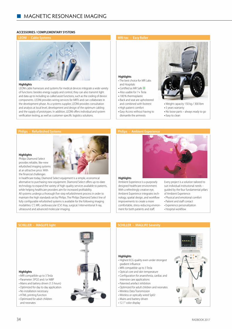
RADBOOK 201734
◼ MAGNETIC RESONANCE IMAGING
ACCESSORIES / COMPLEMENTARY SYSTEMS
· Cable SystemsLEONI
HighlightsLEONI cable harnesses and systems for medical devices integrate a wide variety of functions: besides energy supply and control, they can also transmit light and data up to including so-called assist functions, such as the cooling of device components. LEONI provides wiring services for MRTs and can collaborate in the development phase. As a systems supplier, LEONI provides consultation and analysis at local level, development and design of the optimum cabling and the supply of prototypes. In addition, LEONI offers individual and system verification testing, as well as customer-specific logistics solutions.
Highlights• The best choice for MR Labs
and Hospitals• Certified as MR Safe • Also usable for 7+ Tesla• 100 % thermoplastic• Back and seat are upholstered
and combined with footrest• High patient comfort• Easy Access without having to
dismantle the armrests
• Weight capacity 150 kg / 300 lbm• 5 years warranty• No loose parts – always ready to go• Easy to clean
· Easy RollerMRI-tec
HighlightsPhilips Diamond Select provides reliable, like-new refurbished imaging systems at an attractive price. With the financial challenges in healthcare today, Diamond Select equipment is a simple, economical alternative to purchasing new equipment. Diamond Select offers up-to-date technology to expand the variety of high-quality services available to patients, while helping healthcare providers aim for increased profitability. All systems undergo a thorough five-step refurbishment process in order to maintain the high standards set by Philips. The Philips Diamond Select line of fully configurable refurbished systems is available for the following imaging modalities: CT, MR, cardiovascular (CV) Xray, surgical / interventional X-ray, ultrasound and advanced molecular imaging.
· Refurbished SystemsPhilips
HighlightsAmbient Experience is a purposely designed healthcare environment. With a refreshingly creative eye, Ambient Experience integrates tech-nology, spatial design, and workflow improvements to create a more comfortable, stress-reducing environ-ment for both patients and staff.
Every project is a solution tailored to suit individual institutional needs – guided by the four fundamental pillars of Ambient Experience:• Physical and emotional comfort• Patient and staff contact• Experience personalization• Hospital workflow
· Ambient ExperiencePhilips
Highlights• MRI compatible up to 3 Tesla• Parameter: SPO2 and / or NIBP• Mains and battery driven (1.5 hours)• Optimized for day to day application• No installation necessary• HTML printing function• Optimized for adult children
and neonates
· MAGLIFE lightSCHILLER
Highlights• Highest ECG quality even under strongest
gradient influence• MRI compatible up to 3 Tesla• Optical core and skin temperature• Configuration for anaesthesia, cardiac and
intensive care applications• Patented artefact inhibition• Optimized for adult children and neonates• Wireless Data Transmission• Wireless or optically wired Sp02• Mains and battery driven• 12.1" color display
· MAGLIFE SerenitySCHILLER
RADBOOK 2017 35
INJECTORS ◼
Injectors
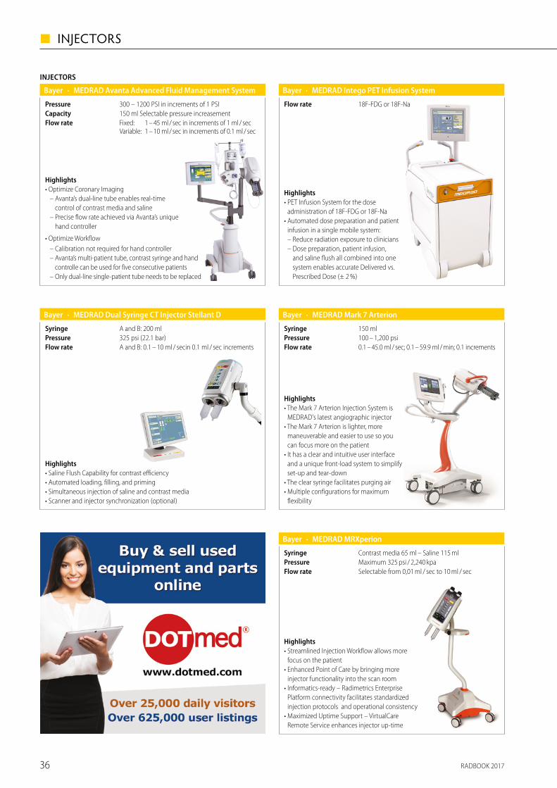
RADBOOK 201736
◼ INJECTORS
INJECTORS
· MEDRAD Avanta Advanced Fluid Management SystemBayer
Fixed: 1 – 45 ml / sec in increments of 1 ml / sec Variable: 1 – 10 ml / sec in increments of 0.1 ml / sec
Flow rate150 ml Selectable pressure increasementCapacity300 – 1200 PSI in increments of 1 PSIPressure
Highlights• Optimize Coronary Imaging
– Avanta’s dual-line tube enables real-time control of contrast media and saline
– Precise flow rate achieved via Avanta’s unique hand controller
• Optimize Workflow– Calibration not required for hand controller– Avanta’s multi-patient tube, contrast syringe and hand
controlle can be used for five consecutive patients– Only dual-line single-patient tube needs to be replaced
· MEDRAD Dual Syringe CT Injector Stellant DBayer
A and B: 0.1 – 10 ml / secin 0.1 ml / sec incrementsFlow rate325 psi (22.1 bar)PressureA and B: 200 mlSyringe
Highlights• Saline Flush Capability for contrast efficiency• Automated loading, filling, and priming• Simultaneous injection of saline and contrast media• Scanner and injector synchronization (optional)
· MEDRAD Intego PET Infusion SystemBayer
18F-FDG or 18F-NaFlow rate
Highlights• PET Infusion System for the dose
administration of 18F-FDG or 18F-Na • Automated dose preparation and patient
infusion in a single mobile system: – Reduce radiation exposure to clinicians – Dose preparation, patient infusion,
and saline flush all combined into one system enables accurate Delivered vs. Prescribed Dose (± 2 %)
· MEDRAD Mark 7 ArterionBayer
0.1 – 45.0 ml / sec; 0.1 – 59.9 ml / min; 0.1 incrementsFlow rate100 – 1,200 psiPressure150 mlSyringe
Highlights• The Mark 7 Arterion Injection System is
MEDRAD's latest angiographic injector• The Mark 7 Arterion is lighter, more
maneuverable and easier to use so you can focus more on the patient
• It has a clear and intuitive user interface and a unique front-load system to simplify set-up and tear-down
• The clear syringe facilitates purging air• Multiple configurations for maximum
flexibility
· MEDRAD MRXperionBayer
Selectable from 0,01 ml / sec to 10 ml / secFlow rateMaximum 325 psi / 2,240 kpaPressureContrast media 65 ml – Saline 115 mlSyringe
Highlights• Streamlined Injection Workflow allows more
focus on the patient• Enhanced Point of Care by bringing more
injector functionality into the scan room• Informatics-ready – Radimetrics Enterprise
Platform connectivity facilitates standardized injection protocols and operational consistency
• Maximized Uptime Support – VirtualCare Remote Service enhances injector up-time
RADBOOK 2017 37
INJECTORS ◼
Contrast medium injectors and consumablesfor CT, MRI and angiography
Hauptstr. 255 · 66128 SaarbrueckenFor more info: www.medtron.com
The specialist
for Stress-MRI!
Integrated infusion pump!
Wireless & 3-Tesla-capable!
Innovation – Made in Germany!
STRESScan be so relaxing.
Accutron® MR3!
Radbook_1-2_Seite_MR3.qxp_Anzeige 12.02.16 12:37 Seite 1
· MEDRAD Spectris Solaris EPBayer
Selectable from 0,01 ml / sec to 10 ml / sec
SyringeMaximum 325 psi / 2,240 kpaPressureContrast media 65 ml – Saline 115 ml
Flow rate
Highlights• Flexible power management with battery
operation or continuous battery charging through AC power connection
• Pressure Limit selection from one of six preset maximum pressure limits, and the ability to view pressure during injection on the control room display
• 3 T compatibility • Multiphase injection control with
6 user-programmable phases including PAUSE and HOLD
• Programmable KVO
Please visit us at
www.healthcare-in-europe.com
RADBOOK 2017
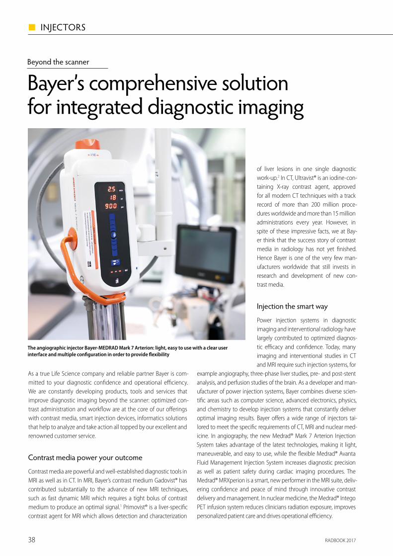
RADBOOK 201738
◼ INJECTORS
Beyond the scanner
Bayer’s comprehensive solution for integrated diagnostic imaging
As a true Life Science company and reliable partner Bayer is com-mitted to your diagnostic con� dence and operational e� ciency. We are constantly developing products, tools and services that improve diagnostic imaging beyond the scanner: optimized con-trast administration and work� ow are at the core of our o� erings with contrast media, smart injection devices, informatics solutions that help to analyze and take action all topped by our excellent and renowned customer service.
Contrast media power your outcome
Contrast media are powerful and well-established diagnostic tools in MRI as well as in CT. In MRI, Bayer’s contrast medium Gadovist® has contributed substantially to the advance of new MRI techniques, such as fast dynamic MRI which requires a tight bolus of contrast medium to produce an optimal signal.1 Primovist® is a liver-speci� c contrast agent for MRI which allows detection and characterization
Our CT injection systems (Medrad® Stellant, Medrad® Salient) are based on tried and tested technology with an excellent reliability track record. The highly � exible and easy to use injectors of the Medrad Stellant series meet the needs of even the most advanced CT imaging procedures and contribute to an improved work� ow. The Medrad Stellant injectors, togeth-er with the above mentioned MRXperion, are IT-ready and available with the Certegra® Work-station, intelligently connecting contrast media, injector and patient information with CT scan and other data from the RIS / HIS of your insti-tute for highly reproducible results.
� e power of connected radio logy: Informatics Solutions
No other specialty exceeds radiology in its need for managing big data. Proper integra-tion of advanced informatics systems into radiology department o� ers multiple bene� ts such as improved diagnostic accuracy and enhanced communication with patients, colleagues, and referring physicians, and at the same time avoids the data overload that already exists. One should keep in mind that to maximize the gain of the informatics solutions, their integration and implementation has to occur seamlessly. To this end Bayer has developed the web-based vendor neutral Radimetrics™ Enterprise Platform*.
Radimetrics is a multi-modality solution, spanning CT, NM, X-ray, MRI interventional and many more. It provides radiation dose, con-trast dose and scanner usage information all in one user experi-
References1 Scott LJ. Clin Drug Investig. 2013; 33(4):303-142 Zech CJ et al. Magn Reson Med Sci. 2007; 6(1):43–52
* Requires Medrad injection system and Certegra Workstation
ence. Through integration into the existing IT infrastructure and radiology equipment via the respective interfaces, Radimetrics intelligently connects contrast, injector and scan informa-tion into a seamlessly smart solution. Together with Radimetrics dataanalysis capabilities this helps institutions generate diagnostic quality images more safely, consistently and e� ciently, in line with radiation standards and regulations, and facilitates information exchange with other involved parties, such as healthcare systems or dose registries.
While Radimetrics collects and distributes information at the enterprise level, the Certegra products provide support at the point of care, allowing for greater control over the quality and consistency of patient care. Transcription errors are reduced and the work� ow is accelerated through the integrated data capture, tracking and distribution capabilities of Certegra. Key fea-tures of the Certegra products are personalized contrast dose management, user-friendly point of care data entry.
Peace of Mind with Bayer Equipment Service
The Bayer Equipment Service optimizes uptime, maximizes value and ensures safety, so you will always get the best out of your Bayer product. This customized service approach, which includes pre-dictive maintenance, calibrations and remote support, guarantees Bayer products keep performing at their peak e� ciency, giving customers the peace of mind to focus entirely on their patients.
Our goal at Bayer is integration within radiology
Bayer Radiology’s range of products covers everything beyond the scanner, from a large selection of contrast media to power injectors and from innovative informatics solutions to our excellent Equip-ment Service. Radiology gets smarter and so does our portfolio of all you need to improve your imaging acquisition work� ow. This integrated approach has resulted in one of the most comprehen-sive packages of radiology products available and guarantees that Bayer Radiology is more than just the sum of its parts. Combined modality scanners, computer-aided diagnosis, data manage-ment – there are many future challenges in the � eld of radiology. Bayer Radiology keeps track of current developments and provides customers with the products, services and the knowledge to master these challenges.
www.radiology.bayer.com
of liver lesions in one single diagnostic work-up.2 In CT, Ultravist® is an iodine-con-taining X-ray contrast agent, approved for all modern CT techniques with a track record of more than 200 million proce-dures worldwide and more than 15 million administrations every year. However, in spite of these impressive facts, we at Bay-er think that the success story of contrast media in radiology has not yet � nished. Hence Bayer is one of the very few man-ufacturers worldwide that still invests in research and development of new con-trast media.
Injection the smart way
Power injection systems in diagnostic imaging and interventional radiology have largely contributed to optimized diagnos-tic e� cacy and con� dence. Today, many imaging and interventional studies in CT and MRI require such injection systems, for
example angiography, three-phase liver studies, pre- and post-stent analysis, and perfusion studies of the brain. As a developer and man-ufacturer of power injection systems, Bayer combines diverse scien-ti� c areas such as computer science, advanced electronics, physics, and chemistry to develop injection systems that constantly deliver optimal imaging results. Bayer o� ers a wide range of injectors tai-lored to meet the speci� c requirements of CT, MRI and nuclear med-icine. In angiography, the new Medrad® Mark 7 Arterion Injection System takes advantage of the latest technologies, making it light, maneuverable, and easy to use, while the � exible Medrad® Avanta Fluid Management Injection System increases diagnostic precision as well as patient safety during cardiac imaging procedures. The Medrad® MRXperion is a smart, new performer in the MRI suite, deliv-ering con� dence and peace of mind through innovative contrast delivery and management. In nuclear medicine, the Medrad® Intego PET infusion system reduces clinicians radiation exposure, improves personalized patient care and drives operational e� ciency.
The new Bayer-MEDRAD MRXperion: thanks to its streamlined Injection Work� ow allows more focus on the patient. It is Informatics-ready in order to facilitate and optimize protocols
Radimetrics Enterprise Platform is a web-based system for monitoring and managing patient radiation exposure and contrast dose, it intelligently connects with radiology work� ow and the hospital IT infrastructure
The angiographic injector Bayer-MEDRAD Mark 7 Arterion: light, easy to use with a clear user interface and multiple con� guration in order to provide � exibility
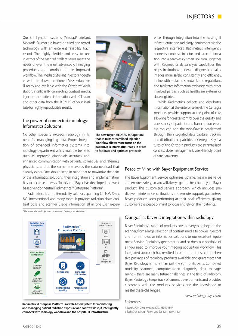
RADBOOK 2017 39
INJECTORS ◼
Beyond the scanner
Bayer’s comprehensive solution for integrated diagnostic imaging
As a true Life Science company and reliable partner Bayer is com-mitted to your diagnostic con� dence and operational e� ciency. We are constantly developing products, tools and services that improve diagnostic imaging beyond the scanner: optimized con-trast administration and work� ow are at the core of our o� erings with contrast media, smart injection devices, informatics solutions that help to analyze and take action all topped by our excellent and renowned customer service.
Contrast media power your outcome
Contrast media are powerful and well-established diagnostic tools in MRI as well as in CT. In MRI, Bayer’s contrast medium Gadovist® has contributed substantially to the advance of new MRI techniques, such as fast dynamic MRI which requires a tight bolus of contrast medium to produce an optimal signal.1 Primovist® is a liver-speci� c contrast agent for MRI which allows detection and characterization
Our CT injection systems (Medrad® Stellant, Medrad® Salient) are based on tried and tested technology with an excellent reliability track record. The highly � exible and easy to use injectors of the Medrad Stellant series meet the needs of even the most advanced CT imaging procedures and contribute to an improved work� ow. The Medrad Stellant injectors, togeth-er with the above mentioned MRXperion, are IT-ready and available with the Certegra® Work-station, intelligently connecting contrast media, injector and patient information with CT scan and other data from the RIS / HIS of your insti-tute for highly reproducible results.
� e power of connected radio logy: Informatics Solutions
No other specialty exceeds radiology in its need for managing big data. Proper integra-tion of advanced informatics systems into radiology department o� ers multiple bene� ts such as improved diagnostic accuracy and enhanced communication with patients, colleagues, and referring physicians, and at the same time avoids the data overload that already exists. One should keep in mind that to maximize the gain of the informatics solutions, their integration and implementation has to occur seamlessly. To this end Bayer has developed the web-based vendor neutral Radimetrics™ Enterprise Platform*.
Radimetrics is a multi-modality solution, spanning CT, NM, X-ray, MRI interventional and many more. It provides radiation dose, con-trast dose and scanner usage information all in one user experi-
References1 Scott LJ. Clin Drug Investig. 2013; 33(4):303-142 Zech CJ et al. Magn Reson Med Sci. 2007; 6(1):43–52
* Requires Medrad injection system and Certegra Workstation
ence. Through integration into the existing IT infrastructure and radiology equipment via the respective interfaces, Radimetrics intelligently connects contrast, injector and scan informa-tion into a seamlessly smart solution. Together with Radimetrics dataanalysis capabilities this helps institutions generate diagnostic quality images more safely, consistently and e� ciently, in line with radiation standards and regulations, and facilitates information exchange with other involved parties, such as healthcare systems or dose registries.
While Radimetrics collects and distributes information at the enterprise level, the Certegra products provide support at the point of care, allowing for greater control over the quality and consistency of patient care. Transcription errors are reduced and the work� ow is accelerated through the integrated data capture, tracking and distribution capabilities of Certegra. Key fea-tures of the Certegra products are personalized contrast dose management, user-friendly point of care data entry.
Peace of Mind with Bayer Equipment Service
The Bayer Equipment Service optimizes uptime, maximizes value and ensures safety, so you will always get the best out of your Bayer product. This customized service approach, which includes pre-dictive maintenance, calibrations and remote support, guarantees Bayer products keep performing at their peak e� ciency, giving customers the peace of mind to focus entirely on their patients.
Our goal at Bayer is integration within radiology
Bayer Radiology’s range of products covers everything beyond the scanner, from a large selection of contrast media to power injectors and from innovative informatics solutions to our excellent Equip-ment Service. Radiology gets smarter and so does our portfolio of all you need to improve your imaging acquisition work� ow. This integrated approach has resulted in one of the most comprehen-sive packages of radiology products available and guarantees that Bayer Radiology is more than just the sum of its parts. Combined modality scanners, computer-aided diagnosis, data manage-ment – there are many future challenges in the � eld of radiology. Bayer Radiology keeps track of current developments and provides customers with the products, services and the knowledge to master these challenges.
www.radiology.bayer.com
of liver lesions in one single diagnostic work-up.2 In CT, Ultravist® is an iodine-con-taining X-ray contrast agent, approved for all modern CT techniques with a track record of more than 200 million proce-dures worldwide and more than 15 million administrations every year. However, in spite of these impressive facts, we at Bay-er think that the success story of contrast media in radiology has not yet � nished. Hence Bayer is one of the very few man-ufacturers worldwide that still invests in research and development of new con-trast media.
Injection the smart way
Power injection systems in diagnostic imaging and interventional radiology have largely contributed to optimized diagnos-tic e� cacy and con� dence. Today, many imaging and interventional studies in CT and MRI require such injection systems, for
example angiography, three-phase liver studies, pre- and post-stent analysis, and perfusion studies of the brain. As a developer and man-ufacturer of power injection systems, Bayer combines diverse scien-ti� c areas such as computer science, advanced electronics, physics, and chemistry to develop injection systems that constantly deliver optimal imaging results. Bayer o� ers a wide range of injectors tai-lored to meet the speci� c requirements of CT, MRI and nuclear med-icine. In angiography, the new Medrad® Mark 7 Arterion Injection System takes advantage of the latest technologies, making it light, maneuverable, and easy to use, while the � exible Medrad® Avanta Fluid Management Injection System increases diagnostic precision as well as patient safety during cardiac imaging procedures. The Medrad® MRXperion is a smart, new performer in the MRI suite, deliv-ering con� dence and peace of mind through innovative contrast delivery and management. In nuclear medicine, the Medrad® Intego PET infusion system reduces clinicians radiation exposure, improves personalized patient care and drives operational e� ciency.
The new Bayer-MEDRAD MRXperion: thanks to its streamlined Injection Work� ow allows more focus on the patient. It is Informatics-ready in order to facilitate and optimize protocols
Radimetrics Enterprise Platform is a web-based system for monitoring and managing patient radiation exposure and contrast dose, it intelligently connects with radiology work� ow and the hospital IT infrastructure
The angiographic injector Bayer-MEDRAD Mark 7 Arterion: light, easy to use with a clear user interface and multiple con� guration in order to provide � exibility
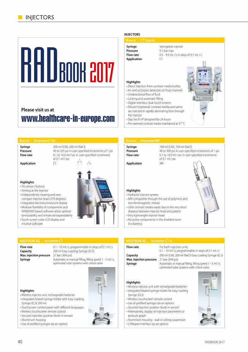
RADBOOK 201740
◼ INJECTORS
· Accutron CTMEDTRON AG
Automatic or manual filling, filling speed 1 – 5 ml / s, optimized tube systems with check valve
Syringe21 bar (304 psi)Max. injection pressure200 ml Easy Loading Syringe (ELS)Capacity0.1 – 10 ml / s, programmable in steps of 0.1 ml / sFlow rate
Highlights• Wireless injector unit, rechargeable batteries• Integrated heated syringe holder with Easy Loading
Syringe (ELS) 200 ml• Touchscreen control panel with different languages• Wireless touchscreen remote control • Secured injection position (built-in sensor) • Aluminium housing• Use of prefilled syringes (as an option)
· Accutron CT-DMEDTRON AG
Automatic or manual filling, filling speed 1 – 5 ml / s, optimized tube systems with check valve
Syringe21 bar (304 psi)Max. injection pressure200 ml (CM), 200 ml (NaCl) Easy Loading Syringe (ELS)Capacity
For both injection units: 0.1 – 10 ml / s, programmable in steps of 0.1 ml / s
Flow rate
Highlights• Wireless injector unit with rechargeable batteries• Integrated heated syringe holder for Easy Loading
Syringe (ELS)• Wireless touchscreen remote control• Use of prefilled syringes (as an option) • Secured injection position (built-in sensor)• Alternatively, display of injection parameters or
pressure graph• Aluminium housing – wall or ceiling suspension• CANopen Interface (as an option)
Highlights• Direct injection from contrast media bottles• Air and occlusion detection on fluid channels• Unidirectional flow of fluid• Locking and automatic filling• Digital interface, dual touch screens• DiluJect (optional): contrast media and saline
are injected in rapidly alternating flow through the injector
• Day Set III HP designed for 24 hours• Pre-warmed contrast media maintained at 37 ° C
INJECTORS
· CT ExprèsBracco
CTApplication0.5 – 9.9 mL / s in steps of 0.1 mL / sFlow rate9.1 bar maxPressureSyringeless injectorSyringe
Highlights• Tilt sensor / lockout • Arming at the injector • Independently rotating and very
compact injector head (270 degrees) • Integrated electroluminescent display • Modular flexibility of components and
WINDOWS based software allow optimal serviceability and enhanced expandability
• Touch-screen color LCD display and intuitive software
· EmpowerCTA+Bracco
CTApplication
0.1 to 10.0 ml / sec in user-specified increments of 0.1 ml / sec
Flow rate40 to 325 psi in user-specified increments of 1 psiPressure200 ml (CM), 200 ml (NaCl)Syringe
Highlights• Hydraulic injector system • MRI compatible through the use of polymers and
non-ferromagnetic metals • Little contrast media waste due to the very short
distance between injector head and patient • Very lightweight injector head • No active components in the shielded room
(no battery)
· EmpowerMRBracco
MRApplication
0.1 to 10.0 ml / sec in user-specified increments of 0.1 ml / sec
Flow rate40 to 300 psi in user-specified increments of 1 psiPressure100 ml (CM), 100 ml (NaCl)Syringe
Please visit us at
www.healthcare-in-europe.com
RADBOOK 2017
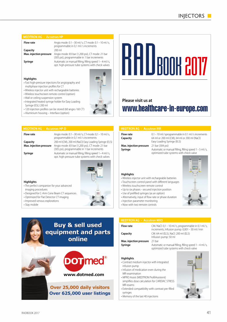
RADBOOK 2017 41
INJECTORS ◼
· Accutron HPMEDTRON AG
Automatic or manual filling, filling speed 1 – 4 ml / s, opt. high-pressure tube systems with check valves
Syringe
Angio mode: 83 bar (1,200 psi), CT mode: 21 bar (305 psi), programmable in 1 bar increments
Max. injection pressure200 mlCapacity
Angio mode: 0.1 – 30 ml / s, CT mode: 0.1 – 10 ml / s, programmable in 0,1 ml / s increments
Flow rate
Highlights• Fast high-pressure injections for angiography and
multiphase injection profiles for CT• Wireless injector unit with rechargeable batteries• Wireless touchscreen remote control (option)• Wall or ceiling suspension system• Integrated heated syringe holder for Easy Loading
Syringe (ESL) 200 ml• 120 injection profiles can be stored (60 angio / 60 CT)• Aluminium housing – Interface (option)
· Accutron HP-DMEDTRON AG
Automatic or manual filling, filling speed 1 – 4 ml / s, opt. high-pressure tube systems with check valves
Syringe
Angio mode: 83 bar (1,200 psi), CT mode: 21 bar (305 psi), programmable in 1 bar increments
Max. injection pressure200 ml (CM), 200 ml (NaCl) Easy Loading Syringe (ELS)Capacity
Angio mode: 0.1 – 30 ml / s, CT-mode: 0,1 – 10 ml / s, programmable in 0,1 ml / s increments
Flow rate
Highlights• The perfect companion for your advanced
imaging procedures• Designed for C-Arm Cone Beam CT sequences• Optimized for Flat Detector CT imaging• Improved venous explorations• Stay mobile
· Accutron MRMEDTRON AG
Automatic or manual filling, filling speed 1 – 5 ml / s, optimized tube systems with check valve
Syringe21 bar (304 psi)Max. injection pressure
64 ml or 200 ml (CM), 64 ml or 200 ml (NaCl) Easy Loading Syringe (ELS)
Capacity0.1 – 10 ml / sprogrammable in 0.1 ml / s incrementsFlow rate
Highlights• Wireless injector unit with rechargeable batteries• Touchscreen control panel with different languages• Wireless touchscreen remote control• Up to six phases – secured injection position • Use of prefilled syringes (as an option)• Alternatively, input of flow rate or phase duration• Injection parameter monitoring• Now with two remote controls
· Accutron MR3MEDTRON AG
CM: 64 ml (ELS), NaCl: 200 ml (ELS) Infusion pump: 50 ml
Capacity
CM / NaCl: 0,1 – 10 ml / s, programmable in 0,1 ml / s, increments, Infusion pump: 0,001 – 30 ml / min
Flow rate
Automatic or manual filling, filling speed 1 – 4 ml / s,optimized tube systems with check valve
Syringe21 barMax. injection pressure
Highlights• Contrast medium injector with integrated
infusion pump• Infusion of medication even during the
MR-examination• MPRO Assist (MEDTRON ProfilAssistent)
simplifies dose calculation for CARDIAC STRESS MR-exams
• Extended compatibility with contrast pre-filled syringes
• Memory of the last 40 injections
Please visit us at
www.healthcare-in-europe.com
RADBOOK 2017
RADBOOK 201742
◼ INTERVENTIONAL SYSTEMS
Interventional Systems
Hybrid-OPs
Bi-Plane
Single Plane
Surgical II-C-Arms
Surgical Flat Panel C-Arms
Accessories / Complementary Systems
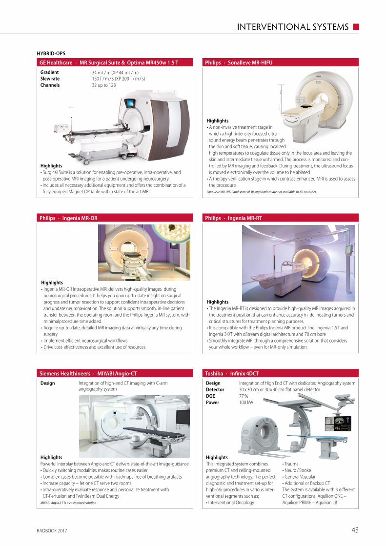
RADBOOK 2017 43
INTERVENTIONAL SYSTEMS ◼
HYBRID-OPS
· MR Surgical Suite & Optima MR450w 1.5 TGE Healthcare
32 up to 128Channels150 T / m / s (XP 200 T / m / s)Slew rate34 mT / m (XP 44 mT / m)Gradient
Highlights• Surgical Suite is a solution for enabling pre-operative, intra-operative, and
post-operative MRI imaging for a patient undergoing neurosurgery.• Includes all necessary additional equipment and offers the combination of a
fully equiped Maquet OP table with a state of the art MRI
· Sonalleve MR-HIFUPhilips
Highlights• A non-invasive treatment stage in
which a high-intensity focused ultra-sound energy beam penetrates through the skin and soft tissue, causing localized high temperatures to coagulate tissue only in the focus area and leaving the skin and intermediate tissue unharmed. The process is monitored and con-trolled by MR imaging and feedback. During treatment, the ultrasound focus is moved electronically over the volume to be ablated
• A therapy verifi cation stage in which contrast-enhanced MRI is used to assess the procedure
Sonalleve MR-HIFU and some of its applications are not available in all countries.
· Ingenia MR-ORPhilips
Highlights• Ingenia MR-OR intraoperative MRI delivers high-quality images during
neurosurgical procedures. It helps you gain up-to-date insight on surgical progress and tumor resection to support confident intraoperative decisions and update neuronavigation. The solution supports smooth, in-line patient transfer between the operating room and the Philips Ingenia MR system, with minimalprocedure time added.
• Acquire up-to-date, detailed MR imaging data at virtually any time during surgery
• Implement efficient neurosurgical workflows• Drive cost-effectiveness and excellent use of resources
· Ingenia MR-RTPhilips
Highlights• The Ingenia MR-RT is designed to provide high-quality MR images acquired in
the treatment position that can enhance accuracy in delineating tumors and critical structures for treatment planning purposes.
• It is compatible with the Philips Ingenia MR product line: Ingenia 1.5 T and Ingenia 3.0 T with dStream digital architecture and 70 cm bore
• Smoothly integrate MRI through a comprehensive solution that considers your whole workflow – even for MR-only simulation.
HighlightsPowerful Interplay between Angio and CT delivers state-of-the-art image-guidance• Quickly switching modalities makes routine cases easier• Complex cases become possible with roadmaps free of breathing artifacts • Increase capacity – let one CT serve two rooms • Intra-operatively evaluate response and personalize treatment with
CT-Perfusion and TwinBeam Dual EnergyMIYABI Angio-CT is a customized solution
· MIYABI Angio-CT Siemens Healthineers
Integration of high-end CT imaging with C-arm angiography system
Design
· Infinix 4DCTToshiba
Integration of High End CT with dedicated Angiography systemDesign
77 %DQE30 x 30 cm or 30 x 40 cm flat panel detectorDetector
100 kWPower
HighlightsThis integrated system combines premium CT and ceiling-mounted angiography technology. The perfect diagnostic and treatment set-up for high-risk procedures in various inter-ventional segments such as:• Interventional Oncology
• Trauma• Neuro / Stroke• General Vascular• Additional or Backup CTThe system is available with 3 different CT configurations: Aquilion ONE – Aquilion PRIME – Aquilion LB
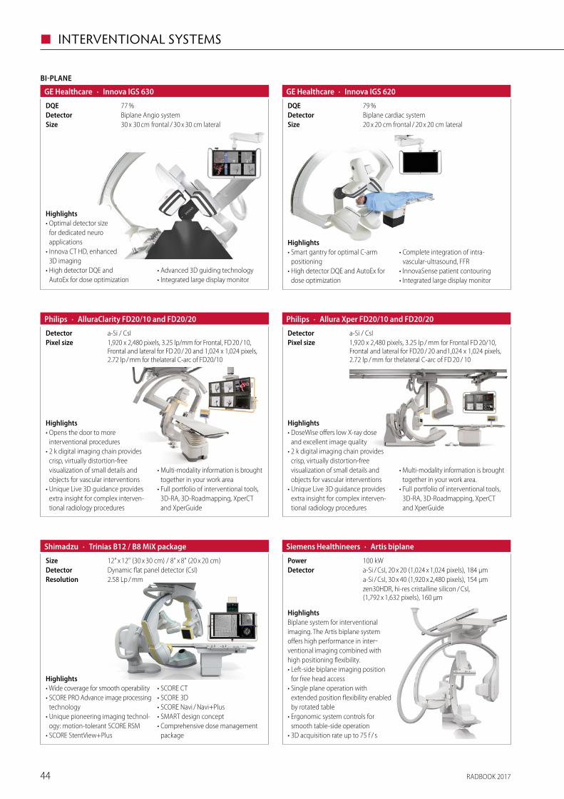
RADBOOK 201744
◼ INTERVENTIONAL SYSTEMS
79 %DQE
· Innova IGS 620GE Healthcare
20 x 20 cm frontal / 20 x 20 cm lateralSizeBiplane cardiac systemDetector
Highlights• Smart gantry for optimal C-arm
positioning• High detector DQE and AutoEx for
dose optimization
• Complete integration of intra- vascular-ultrasound, FFR
• InnovaSense patient contouring• Integrated large display monitor
77 %DQE
BI-PLANE
· Innova IGS 630GE Healthcare
30 x 30 cm frontal / 30 x 30 cm lateralSizeBiplane Angio systemDetector
Highlights• Optimal detector size
for dedicated neuro applications
• Innova CT HD, enhanced 3D imaging
• High detector DQE and AutoEx for dose optimization
• Advanced 3D guiding technology• Integrated large display monitor
· Allura Xper FD20/10 and FD20/20Philips
1,920 x 2,480 pixels, 3.25 lp / mm for Frontal FD 20/10, Frontal and lateral for FD20 / 20 and1,024 x 1,024 pixels, 2.72 lp / mm for thelateral C-arc of FD 20 / 10
Pixel sizea-Si / CsIDetector
Highlights• DoseWise offers low X-ray dose
and excellent image quality• 2 k digital imaging chain provides
crisp, virtually distortion-free visualization of small details and objects for vascular interventions
• Unique Live 3D guidance provides extra insight for complex interven-tional radiology procedures
• Multi-modality information is brought together in your work area.
• Full portfolio of interventional tools, 3D-RA, 3D-Roadmapping, XperCT and XperGuide
· AlluraClarity FD20/10 and FD20/20Philips
1,920 x 2,480 pixels, 3.25 lp/mm for Frontal, FD 20 / 10, Frontal and lateral for FD 20 / 20 and 1,024 x 1,024 pixels, 2.72 lp / mm for thelateral C-arc of FD20/10
Pixel sizea-Si / CsIDetector
Highlights• Opens the door to more
interventional procedures• 2 k digital imaging chain provides
crisp, virtually distortion-free visua lization of small details and objects for vascular interventions
• Unique Live 3D guidance provides extra insight for complex interven-tional radiology procedures
• Multi-modality information is brought together in your work area
• Full portfolio of interventional tools, 3D-RA, 3D-Roadmapping, XperCT and XperGuide
Highlights• Wide coverage for smooth operability• SCORE PRO Advance image processing
technology• Unique pioneering imaging technol-
ogy: motion-tolerant SCORE RSM• SCORE StentView+Plus
• SCORE CT• SCORE 3D• SCORE Navi / Navi+Plus• SMART design concept• Comprehensive dose management
package
· Trinias B12 / B8 MiX packageShimadzu
2.58 Lp / mmResolutionDynamic flat panel detector (CsI)Detector12" x 12" (30 x 30 cm) / 8" x 8" (20 x 20 cm)Size
· Artis biplaneSiemens Healthineers
zen30HDR, hi-res cristalline silicon / CsI, (1,792 x 1,632 pixels), 160 µm
a-Si / CsI, 30 x 40 (1,920 x 2,480 pixels), 154 µma-Si / CsI, 20 x 20 (1,024 x 1,024 pixels), 184 µmDetector100 kWPower
HighlightsBiplane system for interventional imaging. The Artis biplane system offers high performance in inter-ventional imaging combined with high positioning flexibility.• Left-side biplane imaging position
for free head access• Single plane operation with
extended position flexibility enabled by rotated table
• Ergonomic system controls for smooth table-side operation
• 3D acquisition rate up to 75 f / s
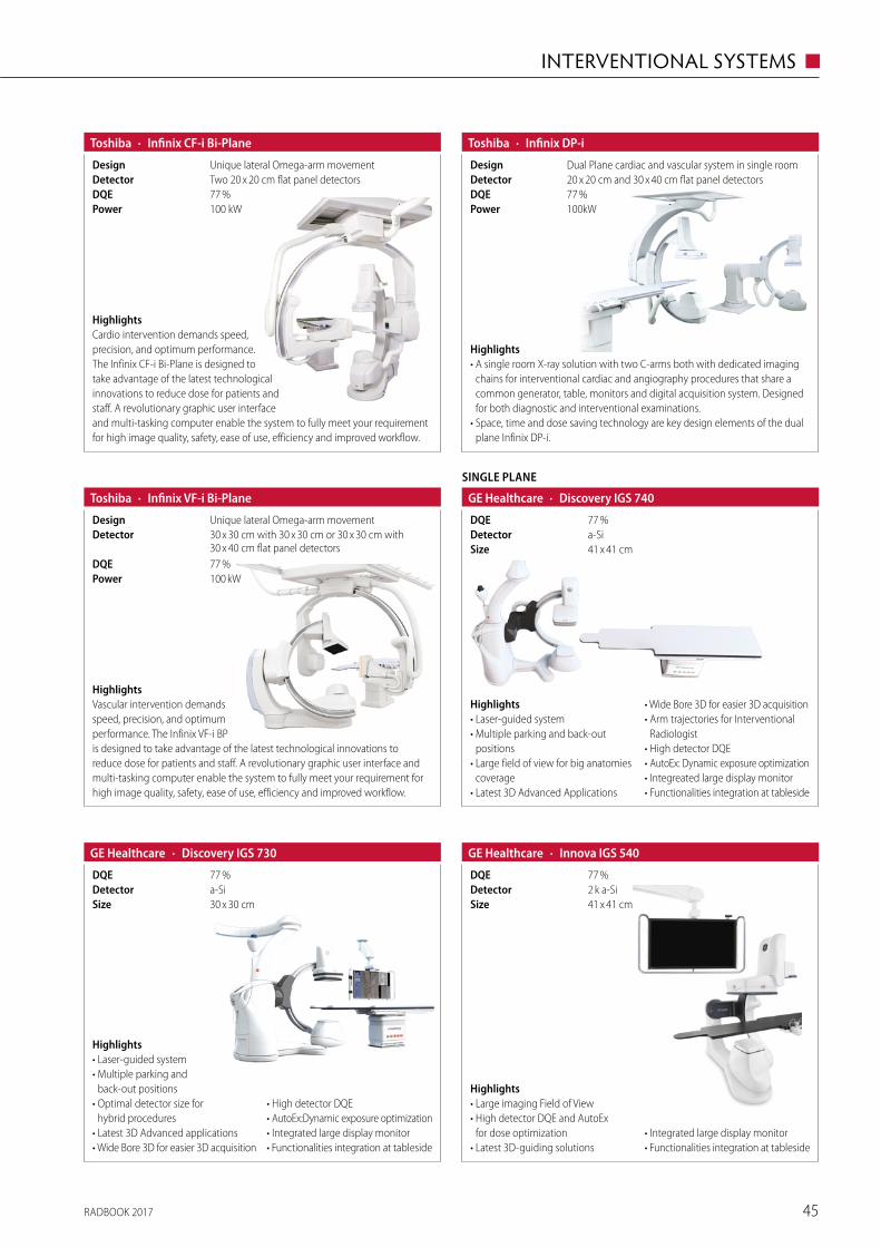
RADBOOK 2017 45
INTERVENTIONAL SYSTEMS ◼
· Infinix VF-i Bi-PlaneToshiba
77 %DQE
30 x 30 cm with 30 x 30 cm or 30 x 30 cm with 30 x 40 cm flat panel detectors
Detector
100 kWPower
Unique lateral Omega-arm movementDesign
HighlightsVascular intervention demands speed, precision, and optimum performance. The Infinix VF-i BP is designed to take advantage of the latest technological innovations to reduce dose for patients and staff. A revolutionary graphic user interface and multi-tasking computer enable the system to fully meet your requirement for high image quality, safety, ease of use, efficiency and improved workflow.
· Infinix CF-i Bi-PlaneToshiba
Two 20 x 20 cm flat panel detectorsDetector
100 kWPower77 %DQE
Unique lateral Omega-arm movementDesign
HighlightsCardio intervention demands speed, precision, and optimum performance. The Infinix CF-i Bi-Plane is designed to take advantage of the latest technological innovations to reduce dose for patients and staff. A revolutionary graphic user interface and multi-tasking computer enable the system to fully meet your requirement for high image quality, safety, ease of use, efficiency and improved workflow.
· Infinix DP-iToshiba
77 %DQE20 x 20 cm and 30 x 40 cm flat panel detectorsDetector
100kWPower
Dual Plane cardiac and vascular system in single room Design
Highlights• A single room X-ray solution with two C-arms both with dedicated imaging
chains for interventional cardiac and angiography procedures that share a common generator, table, monitors and digital acquisition system. Designed for both diagnostic and interventional examinations.
• Space, time and dose saving technology are key design elements of the dual plane Infinix DP-i.
SINGLE PLANE
· Discovery IGS 740GE Healthcare
41 x 41 cmSizea-SiDetector77 %DQE
Highlights• Laser-guided system• Multiple parking and back-out
positions• Large field of view for big anatomies
coverage• Latest 3D Advanced Applications
• Wide Bore 3D for easier 3D acquisition• Arm trajectories for Interventional
Radiologist• High detector DQE• AutoEx: Dynamic exposure optimization• Integreated large display monitor• Functionalities integration at tableside
77 %DQE
· Discovery IGS 730GE Healthcare
30 x 30 cmSizea-SiDetector
Highlights• Laser-guided system• Multiple parking and
back-out positions• Optimal detector size for
hybrid procedures• Latest 3D Advanced applications• Wide Bore 3D for easier 3D acquisition
• High detector DQE • AutoEx:Dynamic exposure optimization• Integrated large display monitor• Functionalities integration at tableside
2 k a-SiDetector
· Innova IGS 540GE Healthcare
41 x 41 cmSize
77 %DQE
Highlights• Large imaging Field of View• High detector DQE and AutoEx
for dose optimization• Latest 3D-guiding solutions
• Integrated large display monitor• Functionalities integration at tableside
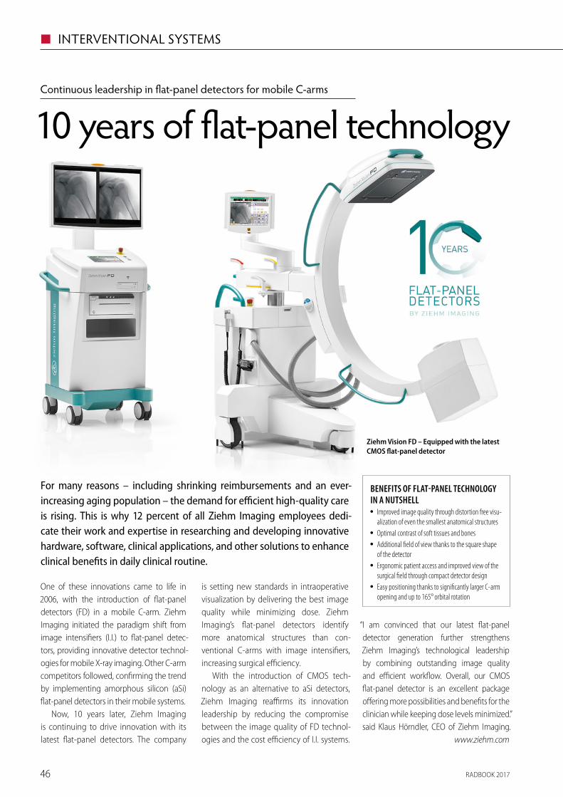
RADBOOK 201746
◼ INTERVENTIONAL SYSTEMS
One of these innovations came to life in 2006, with the introduction of flat-panel detectors (FD) in a mobile C-arm. Ziehm Imaging initiated the paradigm shift from image intensifiers (I.I.) to flat-panel detec-tors, providing innovative detector technol-ogies for mobile X-ray imaging. Other C-arm competitors followed, confirming the trend by implementing amorphous silicon (aSi) flat-panel detectors in their mobile systems.
Now, 10 years later, Ziehm Imaging is continuing to drive innovation with its latest flat-panel detectors. The company
Continuous leadership in flat-panel detectors for mobile C-arms
10 years of flat-panel technology
is setting new standards in intraoperative visualization by delivering the best image quality while minimizing dose. Ziehm Imaging’s flat-panel detectors identify more anatomical structures than con-ventional C-arms with image intensifiers, increasing surgical efficiency.
With the introduction of CMOS tech-nology as an alternative to aSi detectors, Ziehm Imaging reaffirms its innovation leadership by reducing the compromise between the image quality of FD technol-ogies and the cost efficiency of I.I. systems.
“I am convinced that our latest flat-panel detec tor generation further strengthens Ziehm Imaging’s technological leadership by combining outstanding image quality and efficient workflow. Overall, our CMOS flat-panel detector is an excellent package offering more possibilities and benefits for the clinician while keeping dose levels minimized.” said Klaus Hörndler, CEO of Ziehm Imaging.
www.ziehm.com
For many reasons – including shrinking reimbursements and an ever- increasing aging population – the demand for efficient high-quality care is rising. This is why 12 percent of all Ziehm Imaging employees dedi-cate their work and expertise in researching and developing innovative hardware, software, clinical applications, and other solutions to enhance clinical benefits in daily clinical routine.
BENEFITS OF FLAT-PANEL TECHNOLOGY IN A NUTSHELL
• Improved image quality through distortion free visu-alization of even the smallest anatomical structures
• Optimal contrast of soft tissues and bones • Additional field of view thanks to the square shape
of the detector • Ergonomic patient access and improved view of the
surgical field through compact detector design • Easy positioning thanks to significantly larger C-arm
opening and up to 165° orbital rotation
Ziehm Vision FD – Equipped with the latest CMOS flat-panel detector
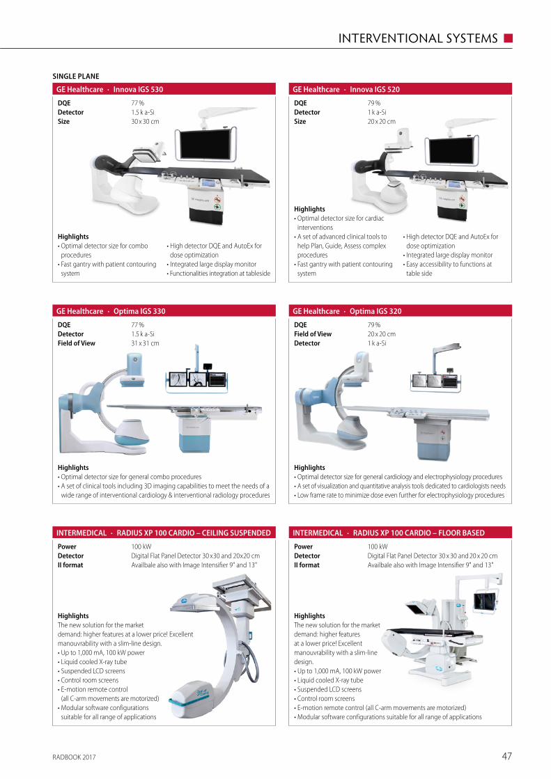
RADBOOK 2017 47
INTERVENTIONAL SYSTEMS ◼
1.5 k a-SiDetector
SINGLE PLANE
· Innova IGS 530GE Healthcare
30 x 30 cmSize
77 %DQE
Highlights• Optimal detector size for combo
procedures• Fast gantry with patient contouring
system
• High detector DQE and AutoEx for dose optimization
• Integrated large display monitor• Functionalities integration at tableside
1.5 k a-SiDetector
· Optima IGS 330GE Healthcare
31 x 31 cmField of View
77 %DQE
Highlights• Optimal detector size for general combo procedures• A set of clinical tools including 3D imaging capabilities to meet the needs of a
wide range of interventional cardiology & interventional radiology procedures
1 k a-SiDetector
· Innova IGS 520GE Healthcare
20 x 20 cmSize
79 %DQE
Highlights• Optimal detector size for cardiac
interventions• A set of advanced clinical tools to
help Plan, Guide, Assess complex procedures
• Fast gantry with patient contouring system
• High detector DQE and AutoEx for dose optimization
• Integrated large display monitor• Easy accessibility to functions at
table side
79 %DQE
· Optima IGS 320GE Healthcare
1 k a-SiDetector20 x 20 cmField of View
Highlights• Optimal detector size for general cardiology and electrophysiology procedures• A set of visualization and quantitative analysis tools dedicated to cardiologists needs• Low frame rate to minimize dose even further for electrophysiology procedures
· RADIUS XP 100 CARDIO – CEILING SUSPENDEDINTERMEDICAL
Availbale also with Image Intensifier 9" and 13"II formatDigital Flat Panel Detector 30 x 30 and 20 x 20 cmDetector100 kWPower
HighlightsThe new solution for the market demand: higher features at a lower price! Excellent manouvrability with a slim-line design.• Up to 1,000 mA, 100 kW power• Liquid cooled X-ray tube• Suspended LCD screens• Control room screens• E-motion remote control
(all C-arm movements are motorized)• Modular software configurations
suitable for all range of applications
· RADIUS XP 100 CARDIO – FLOOR BASEDINTERMEDICAL
Availbale also with Image Intensifier 9" and 13"II formatDigital Flat Panel Detector 30 x 30 and 20 x 20 cmDetector100 kWPower
HighlightsThe new solution for the market demand: higher features at a lower price! Excellent manouvrability with a slim-line design.• Up to 1,000 mA, 100 kW power• Liquid cooled X-ray tube• Suspended LCD screens• Control room screens• E-motion remote control (all C-arm movements are motorized)• Modular software configurations suitable for all range of applications
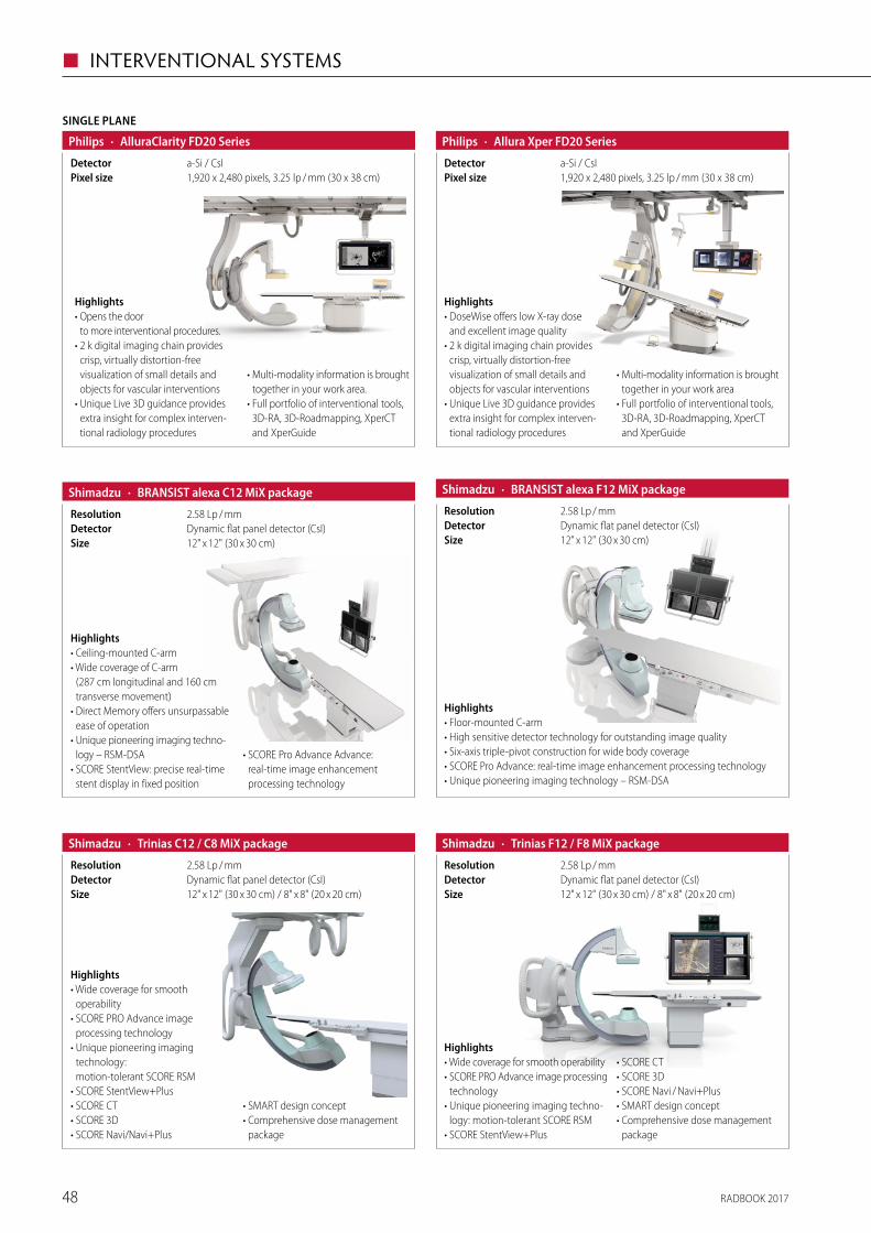
RADBOOK 201748
◼ INTERVENTIONAL SYSTEMS
· Allura Xper FD20 SeriesPhilips
1,920 x 2,480 pixels, 3.25 lp / mm (30 x 38 cm)Pixel sizea-Si / CsIDetector
Highlights• DoseWise offers low X-ray dose
and excellent image quality• 2 k digital imaging chain provides
crisp, virtually distortion-free visualization of small details and objects for vascular interventions
• Unique Live 3D guidance provides extra insight for complex interven-tional radiology procedures
• Multi-modality information is brought together in your work area
• Full portfolio of interventional tools, 3D-RA, 3D-Roadmapping, XperCT and XperGuide
SINGLE PLANE
· AlluraClarity FD20 SeriesPhilips
1,920 x 2,480 pixels, 3.25 lp / mm (30 x 38 cm)Pixel sizea-Si / CsIDetector
Highlights• Opens the door
to more interventional procedures.• 2 k digital imaging chain provides
crisp, virtually distortion-free visualization of small details and objects for vascular interventions
• Unique Live 3D guidance provides extra insight for complex interven-tional radiology procedures
• Multi-modality information is brought together in your work area.
• Full portfolio of interventional tools, 3D-RA, 3D-Roadmapping, XperCT and XperGuide
· BRANSIST alexa C12 MiX packageShimadzu
12" x 12" (30 x 30 cm)SizeDynamic flat panel detector (CsI)Detector2.58 Lp / mmResolution
Highlights• Ceiling-mounted C-arm• Wide coverage of C-arm
(287 cm longitudinal and 160 cm transverse movement)
• Direct Memory offers unsurpassable ease of operation
• Unique pioneering imaging techno-logy – RSM-DSA
• SCORE StentView: precise real-time stent display in fixed position
• SCORE Pro Advance Advance: real-time image enhancement processing technology
· BRANSIST alexa F12 MiX packageShimadzu
12" x 12" (30 x 30 cm)SizeDynamic flat panel detector (CsI)Detector2.58 Lp / mmResolution
Highlights• Floor-mounted C-arm• High sensitive detector technology for outstanding image quality• Six-axis triple-pivot construction for wide body coverage• SCORE Pro Advance: real-time image enhancement processing technology• Unique pioneering imaging technology – RSM-DSA
Highlights• Wide coverage for smooth
operability• SCORE PRO Advance image
processing technology• Unique pioneering imaging
technology: motion-tolerant SCORE RSM
• SCORE StentView+Plus• SCORE CT• SCORE 3D• SCORE Navi/Navi+Plus
• SMART design concept• Comprehensive dose management
package
· Trinias C12 / C8 MiX packageShimadzu
12" x 12" (30 x 30 cm) / 8" x 8" (20 x 20 cm)SizeDynamic flat panel detector (CsI)Detector2.58 Lp / mmResolution
· Trinias F12 / F8 MiX packageShimadzu
12" x 12" (30 x 30 cm) / 8" x 8" (20 x 20 cm)SizeDynamic flat panel detector (CsI)Detector2.58 Lp / mmResolution
Highlights• Wide coverage for smooth operability• SCORE PRO Advance image processing
technology• Unique pioneering imaging techno-
logy: motion-tolerant SCORE RSM• SCORE StentView+Plus
• SCORE CT• SCORE 3D• SCORE Navi / Navi+Plus• SMART design concept• Comprehensive dose management
package
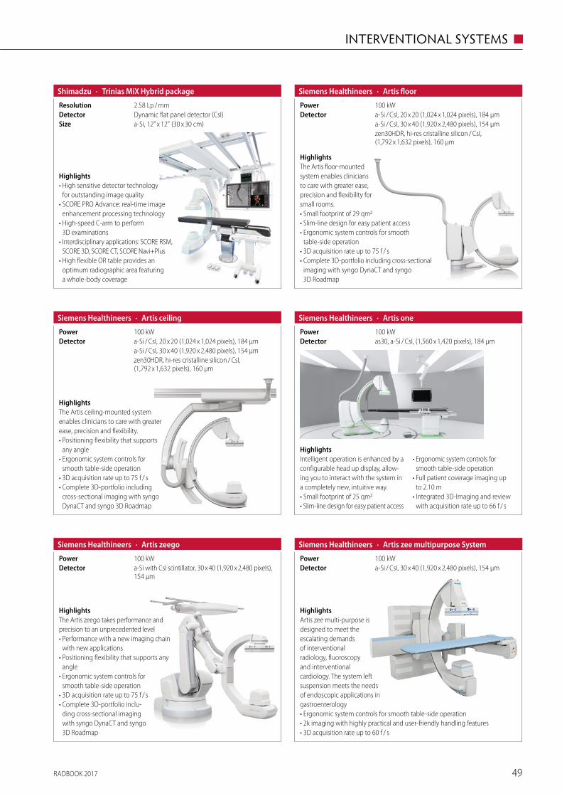
RADBOOK 2017 49
INTERVENTIONAL SYSTEMS ◼
· Trinias MiX Hybrid packageShimadzu
a-Si, 12" x 12" (30 x 30 cm)SizeDynamic flat panel detector (CsI)Detector2.58 Lp / mmResolution
Highlights• High sensitive detector technology
for outstanding image quality • SCORE PRO Advance: real-time image
enhancement processing technology• High-speed C-arm to perform
3D examinations • Interdisciplinary applications: SCORE RSM,
SCORE 3D, SCORE CT, SCORE Navi+Plus• High flexible OR table provides an
optimum radiographic area featuring a whole-body coverage
· Artis floorSiemens Healthineers
zen30HDR, hi-res cristalline silicon / CsI, (1,792 x 1,632 pixels), 160 µm
a-Si / CsI, 30 x 40 (1,920 x 2,480 pixels), 154 µma-Si / CsI, 20 x 20 (1,024 x 1,024 pixels), 184 µmDetector100 kWPower
HighlightsThe Artis floor-mounted system enables clinicians to care with greater ease, precision and flexibility for small rooms.• Small footprint of 29 qm2• Slim-line design for easy patient access• Ergonomic system controls for smooth
table-side operation• 3D acquisition rate up to 75 f / s• Complete 3D-portfolio including cross-sectional
imaging with syngo DynaCT and syngo 3D Roadmap
· Artis ceilingSiemens Healthineers
zen30HDR, hi-res cristalline silicon / CsI, (1,792 x 1,632 pixels), 160 µm
a-Si / CsI, 30 x 40 (1,920 x 2,480 pixels), 154 µma-Si / CsI, 20 x 20 (1,024 x 1,024 pixels), 184 µmDetector100 kWPower
HighlightsThe Artis ceiling-mounted system enables clinicians to care with greater ease, precision and flexibility.• Positioning flexibility that supports
any angle• Ergonomic system controls for
smooth table-side operation• 3D acquisition rate up to 75 f / s• Complete 3D-portfolio including
cross-sectional imaging with syngo DynaCT and syngo 3D Roadmap
HighlightsIntelligent operation is enhanced by a configurable head up display, allow-ing you to interact with the system in a completely new, intuitive way.• Small footprint of 25 qm2• Slim-line design for easy patient access
• Ergonomic system controls for smooth table-side operation
• Full patient coverage imaging up to 2.10 m
• Integrated 3D-Imaging and review with acquisition rate up to 66 f / s
· Artis oneSiemens Healthineers
as30, a-Si / CsI, (1,560 x 1,420 pixels), 184 µmDetector100 kWPower
· Artis zeegoSiemens Healthineers
a-Si with CsI scintillator, 30 x 40 (1,920 x 2,480 pixels), 154 µm
Detector100 kWPower
HighlightsThe Artis zeego takes performance and precision to an unprecedented level• Performance with a new imaging chain
with new applications• Positioning flexibility that supports any
angle• Ergonomic system controls for
smooth table-side operation• 3D acquisition rate up to 75 f / s• Complete 3D-portfolio inclu-
ding cross-sectional imaging with syngo DynaCT and syngo 3D Roadmap
· Artis zee multipurpose SystemSiemens Healthineers
a-Si / CsI, 30 x 40 (1,920 x 2,480 pixels), 154 µmDetector100 kWPower
HighlightsArtis zee multi-purpose is designed to meet the escalating demands of interventional radio logy, fluoroscopy and interventional cardiology. The system left suspension meets the needs of endoscopic applications in gastroenterology• Ergonomic system controls for smooth table-side operation• 2k imaging with highly practical and user-friendly handling features• 3D acquisition rate up to 60 f / s
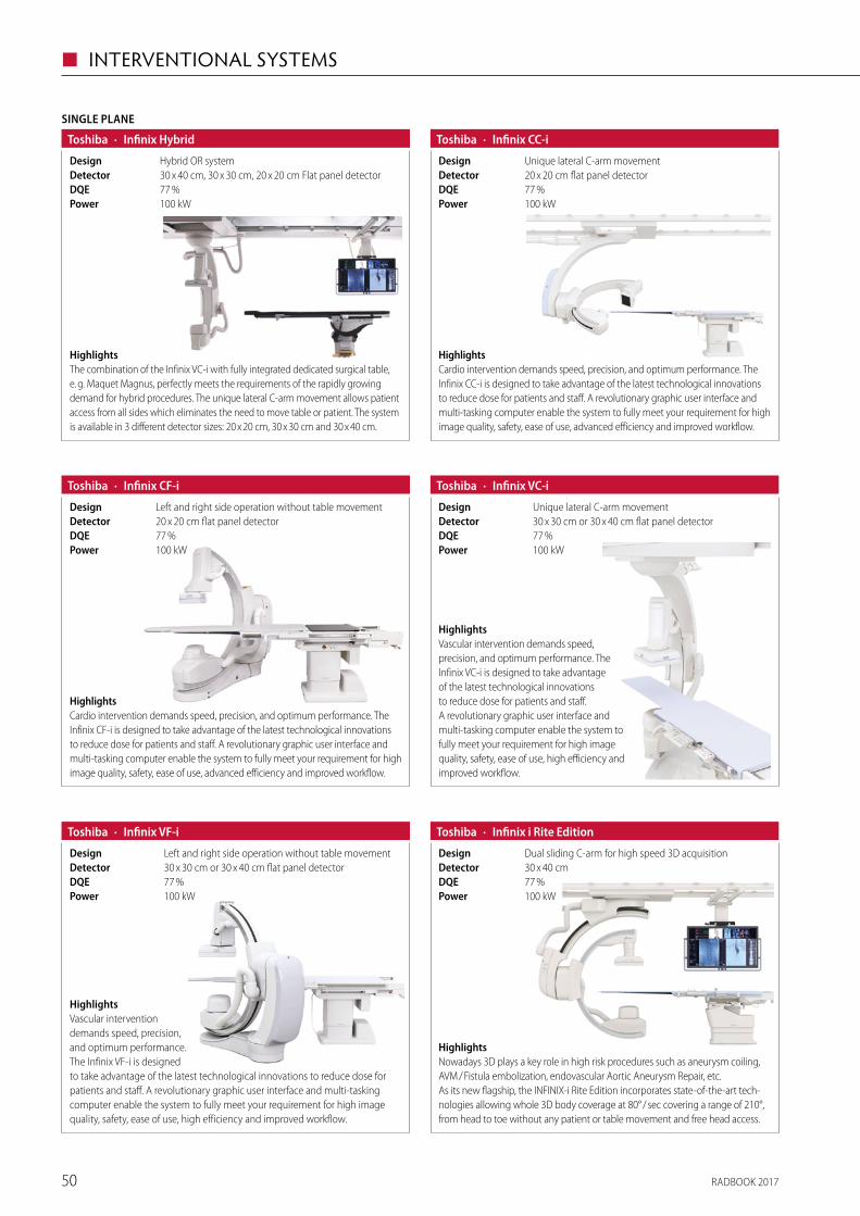
RADBOOK 201750
◼ INTERVENTIONAL SYSTEMS
· Infinix CC-iToshiba
77 %DQE20 x 20 cm flat panel detectorDetector
100 kWPower
Unique lateral C-arm movementDesign
HighlightsCardio intervention demands speed, precision, and optimum performance. The Infinix CC-i is designed to take advantage of the latest technological innovations to reduce dose for patients and staff. A revolutionary graphic user interface and multi-tasking computer enable the system to fully meet your requirement for high image quality, safety, ease of use, advanced efficiency and improved workflow.
· Infinix CF-iToshiba
77 %DQE20 x 20 cm flat panel detectorDetector
100 kWPower
Left and right side operation without table movementDesign
HighlightsCardio intervention demands speed, precision, and optimum performance. The Infinix CF-i is designed to take advantage of the latest technological innovations to reduce dose for patients and staff. A revolutionary graphic user interface and multi-tasking computer enable the system to fully meet your requirement for high image quality, safety, ease of use, advanced efficiency and improved workflow.
SINGLE PLANE
· Infinix HybridToshiba
100 kWPower
30 x 40 cm, 30 x 30 cm, 20 x 20 cm Flat panel detectorDetector77 %DQE
Hybrid OR systemDesign
HighlightsThe combination of the Infinix VC-i with fully integrated dedicated surgical table, e. g. Maquet Magnus, perfectly meets the requirements of the rapidly growing demand for hybrid procedures. The unique lateral C-arm movement allows patient access from all sides which eliminates the need to move table or patient. The system is available in 3 different detector sizes: 20 x 20 cm, 30 x 30 cm and 30 x 40 cm.
· Infinix VC-iToshiba
77 %DQE30 x 30 cm or 30 x 40 cm flat panel detectorDetector
100 kWPower
Unique lateral C-arm movementDesign
HighlightsVascular intervention demands speed, precision, and optimum performance. The Infinix VC-i is designed to take advantage of the latest technological innovations to reduce dose for patients and staff. A revolutionary graphic user interface and multi-tasking computer enable the system to fully meet your requirement for high image quality, safety, ease of use, high efficiency and improved workflow.
· Infinix i Rite EditionToshiba
30 x 40 cmDetector77 %DQE100 kWPower
Dual sliding C-arm for high speed 3D acquisitionDesign
HighlightsNowadays 3D plays a key role in high risk procedures such as aneurysm coiling, AVM / Fistula embolization, endovascular Aortic Aneurysm Repair, etc.As its new flagship, the INFINIX-i Rite Edition incorporates state-of-the-art tech-nologies allowing whole 3D body coverage at 80° / sec covering a range of 210°, from head to toe without any patient or table movement and free head access.
· Infinix VF-iToshiba
77 %DQE30 x 30 cm or 30 x 40 cm flat panel detectorDetector
100 kWPower
Left and right side operation without table movementDesign
HighlightsVascular intervention demands speed, precision, and optimum performance. The Infinix VF-i is designed to take advantage of the latest technological innovations to reduce dose for patients and staff. A revolutionary graphic user interface and multi-tasking computer enable the system to fully meet your requirement for high image quality, safety, ease of use, high efficiency and improved workflow.
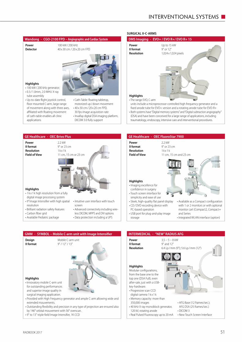
RADBOOK 2017 51
INTERVENTIONAL SYSTEMS ◼
Highlights• 100 kW / 200 kHz generator;• 0.3 / 1.0mm, 2.0 MHU X-ray
tube assembly • Up-to-date flight joystick control,
floor mounted C-arm, large range of movement along with three axes, affiliated with floating movement of cath-table enables all clinic applications
• Cath-Table: floating tabletop, motorized up / down movement
• 40 x 30 cm / 20 x 20 cm FPD, 30 fps image acquisition rate
• InvaRay digital DSA imaging platform, DICOM 3.0 fully support
· CGO-2100 FPD – Angiographic and Cardiac SystemWandong
40 x 30 cm / 20 x 20 cm FPDDetector100 kW / 200 kHzPower
· OEC Brivo PlusGE Healthcare
1 k x 1 kResolution9" or 23 cmII format
11 cm, 15 cm or 23 cmField of View
2.2 kWPower
Highlights• 1 k x 1 k high resolution from a fully
digital image processing system• 9” Image Intensifier with high spatial
resolution• Brilliant radiation safety features• Carbon fiber grid• Available Pediatric package
• Intuitive user interface with touch screen
• Advanced connectivity including wire-less DICOM, MPPS and DVI options
• Data protection including a UPS
· OEC FluoroStar 7900GE Healthcare
11 cm, 15 cm and 23 cmField of View1 k x 1 kResolution9" or 23 cmII format2.2 kWPower
Highlights• Imaging excellence for
confidence in surgery• Touch screen interface for
simplicity and ease of use• Sleek, high-quality flat panel display• CD / DVD recording device with
PC-based operation• USB port for plug-and-play image
storage
• Available as a Compact configuration with 1 or 2 monitors or with optional monitor cart (Compact2, Compact+ and Series
• Integrated WLAN interface (option)
Highlights• Innovatory mobile C-arm unit
for outstanding performances and superior image quality in surgical imaging application.
• Provided with High Frequency generator and ample C-arm allowing wide and extended mouvements.
• Outstanding flexibility and precision in any type of projection are ensured also by 146° orbital mouvement with 56° overscan.
• 9" to 13" triple field Image Intensifier, 1K CCD
· SYMBOL – Mobile C-arm unit with Image IntensifierGMM
9" / 12" / 13"II formatMobile C-arm unitDesign 3,5 – 5 – 8 kWPower
· “NEW” RADIUS AFGINTERMEDICAL
6.4 Lp / mm (9"); 5.6 Lp / mm (12")Resolution9" and 12"II format
HighlightsModular configurations, from the base one to the top one (DSA Full), even after-sale, just with a USB-key-hardware.• Progressive scan CCD
digital camera 1 k x 1 k• Memory capacity: more than
350,000 images• 40 kHz X-ray monoblock generator,
120 kV, rotating anode• Real Pulsed Fluoroscopy up to 20 mA
• AFG Base (12 frames/sec.); AFG DSA (25 frames/sec.)
• DICOM 3• New Touch Screen Interface
SURGICAL II-C-ARMS
· EVO+ / EVO R+/ EVO R+ 15DMS Imaging
1,024 x 1,024 pixelsResolution9" or 12"II formatUp to 15 kWPower
Highlights• The range EVO, C-arm
units include a microprocessor controlled high frequency generator and a fixed anode tube for EVO+ version and a rotating anode tube for EVO-R+
• Both systems have “Digital memory systems” and “Digital subtraction angiography” (DSA) and have been conceived for a large range of applications, including traumatology, endoscopy, intensive care and interventional procedures.
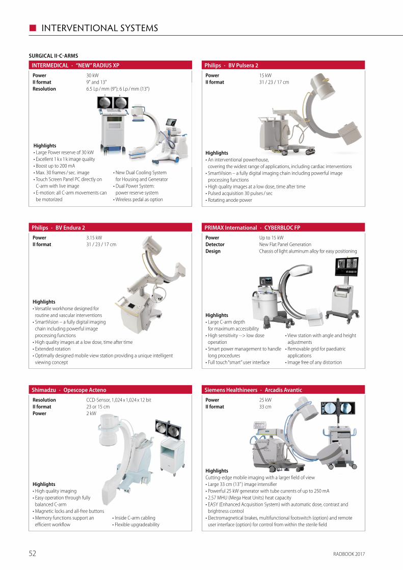
RADBOOK 201752
◼ INTERVENTIONAL SYSTEMS
· BV Endura 2Philips
31 / 23 / 17 cmII format3.15 kWPower
Highlights• Versatile workhorse designed for
routine and vascular interventions• SmartVision – a fully digital imaging
chain including powerful image processing functions
• High quality images at a low dose, time after time• Extended rotation• Optimally designed mobile view station providing a unique intelligent
viewing concept
· BV Pulsera 2Philips
31 / 23 / 17 cmII format15 kWPower
Highlights• An interventional powerhouse,
covering the widest range of applications, including cardiac interventions• SmartVision – a fully digital imaging chain including powerful image
processing functions• High quality images at a low dose, time after time• Pulsed acquisition 30 pulses / sec• Rotating anode power
· Opescope ActenoShimadzu
2 kWPower23 or 15 cmII formatCCD-Sensor, 1,024 x 1,024 x 12 bitResolution
Highlights• High quality imaging• Easy operation through fully
balanced C-arm• Magnetic locks and all-free buttons• Memory functions support an
efficient workflow• Inside C-arm cabling• Flexible upgradeability
HighlightsCutting-edge mobile imaging with a larger field of view• Large 33 cm (13” ) image intensifier • Powerful 25 kW generator with tube currents of up to 250 mA• 2.57 MHU (Mega Heat Units) heat capacity• EASY (Enhanced Acquisition System) with automatic dose, contrast and
brightness control• Electromagnetical brakes, multifunctional footswitch (option) and remote
user interface (option) for control from within the sterile field
· Arcadis AvanticSiemens Healthineers
33 cmII format25 kWPower
SURGICAL II-C-ARMS
· “NEW” RADIUS XPINTERMEDICAL
6.5 Lp / mm (9"); 6 Lp / mm (13")Resolution9" and 13"II format30 kWPower
Highlights• Large Power reserve of 30 kW• Excellent 1 k x 1 k image quality• Boost up to 200 mA• Max. 30 frames / sec. image• Touch Screen Panel PC directly on
C-arm with live image• E-motion: all C-arm movements can
be motorized
• New Dual Cooling System for Housing and Generator
• Dual Power System: power reserve system
• Wireless pedal as option
· CYBERBLOC FPPRIMAX International
Chassis of light aluminum alloy for easy positioningDesignNew Flat Panel GenerationDetectorUp to 15 kWPower
Highlights• Large C-arm depth
for maximum accessibility• High sensitivity --> low dose
operation• Smart power management to handle
long procedures• Full touch “smart” user interface
• View station with angle and height adjustments
• Removable grid for paediatric applications
• Image free of any distortion
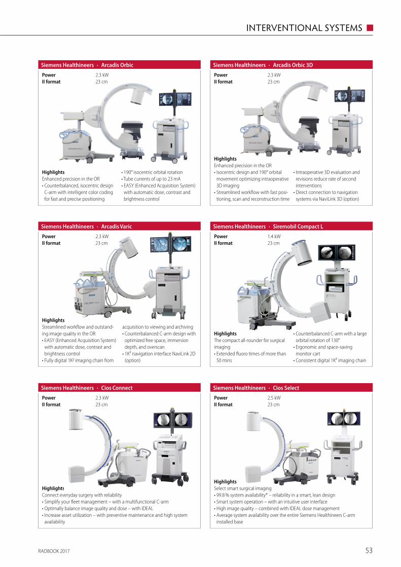
RADBOOK 2017 53
INTERVENTIONAL SYSTEMS ◼
· Arcadis OrbicSiemens Healthineers
23 cmII format2.3 kWPower
HighlightsEnhanced precision in the OR• Counterbalanced, isocentric design
C-arm with intelligent color coding for fast and precise positioning
• 190° isocentric orbital rotation• Tube currents of up to 23 mA• EASY (Enhanced Acquisition System)
with automatic dose, contrast and brightness control
· Arcadis Orbic 3DSiemens Healthineers
23 cmII format2.3 kWPower
HighlightsEnhanced precision in the OR• Isocentric design and 190° orbital
movement optimizing intraoperative 3D imaging
• Streamlined workflow with fast posi-tioning, scan and reconstruction time
• Intraoperative 3D evaluation and revisions reduce rate of second interventions
• Direct connection to navigation systems via NaviLink 3D (option)
HighlightsStreamlined workflow and outstand-ing image quality in the OR• EASY (Enhanced Acquisition System)
with automatic dose, contrast and brightness control
• Fully digital 1K2 imaging chain from
acquisition to viewing and archiving• Counterbalanced C-arm design with
optimized free space, immersion depth, and overscan
• 1K² navigation interface NaviLink 2D (option)
· Arcadis VaricSiemens Healthineers
23 cmII format2.3 kWPower
· Siremobil Compact LSiemens Healthineers
23 cmII format1.4 kWPower
HighlightsThe compact all-rounder for surgical imaging• Extended fluoro times of more than
50 mins
• Counterbalanced C-arm with a large orbital rotation of 130°
• Ergonomic and space-saving monitor cart
• Consistent digital 1K² imaging chain
HighlightsConnect everyday surgery with reliability• Simplify your fleet management – with a multifunctional C-arm• Optimally balance image quality and dose – with IDEAL• Increase asset utilization – with preventive maintenance and high system
availability
· Cios ConnectSiemens Healthineers
23 cmII format2.3 kWPower
· Cios SelectSiemens Healthineers
23 cmII format2.5 kWPower
HighlightsSelect smart surgical imaging• 99.8 % system availability* – reliability in a smart, lean design• Smart system operation – with an intuitive user interface• High image quality – combined with IDEAL dose management• Average system availability over the entire Siemens Healthineers C-arm
installed base
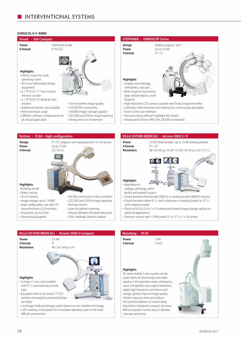
RADBOOK 201754
◼ INTERVENTIONAL SYSTEMS
· TCA6 – high configurationTechnix
23 / 32 cmII formatUp to 15 kWPower9" / 12" surgical C-arm equipped with 1k x 1k cameraDesign
Highlights• Rotating anode• Water cooling• 1k x 1k camera• Image storage: up to 110000• High configuration cart with 19"
monochromatic LCD monitors• Acquisition up to 25 fps• Anatomical programs
• DICOM connectivity (LAN or wireless)• CD / DVD and USB for image exporting• Remote control• Laser for patient centering• Virtual collimators (for dose reduction)• DSA, roadmap, stenosis analysis
· Arcovis 3000 S / RVILLA SISTEMI MEDICALI
48 / 56 / 64 Lp / cm (9" I. I.); 48 / 54 / 62 Lp / cm (12" I. I.)Resolution9" / 12"II format3.5 kW (fixed anode) / up to 15 kW (rotating anode)Power
Highlights• Application in
urology, cardiology, ortho-pedics and general surgery
• Choice between fixed anode (3000 S) or rotating anode (3000 R) versions• Choice between either 9" I. I. (with stationary or rotating anode) or 12" I. I.
(with rotating anode)• Choice of 0.5 x 0.5 k or 1 x 1 k camera and several image storage options to
satisfy all applications• Premium version with 15 kW power, 9" or 12" I.I., 1 x 1k camera
· Arcovis 3000 S CompactVILLA SISTEMI MEDICALI
48 / 56 / 64 Lp / cmResolution9"II format3.5 kWPower
Highlights• Compact C-arm unit available
with 9" I. I. and stationary anode tube
• Equipped with an on-board 17” LCD monitor, not requiring external displays on trolley
• Last Image Hold and storage system based on non-volatile technology• ± 60° rotating control panel for immediate operation even in the most
difficult environment
· XC30Wandong
9 inchII format5 kWPower
HighlightsXC series mobile C-arm system can be used mainly for fluoroscopy and radio-graphy in the operation room, emergency ward, orthopedics and surgical treatment. Apply high frequency conversion tech-nology, greatly improve image quality, shorten exposure time, and reduce the harmful radiation to human body. Ergonomics designed, compact structure, Microcomputer-control, easy to operate, maintain and move.
Highlights• Surgery, traumatology,
orthopedics, vascular . . .• Wide range of movements,
large orbital rotation, small footprint
• High resolution CCD camera coupled with Thales Image Intensifier• Collimator with motorised and rotating iris, continuously adjustable• Touch screen user interface• Post-processing software highlight tiny details• Advanced functions: APR, DSA, DICOM connectivity
· OMNISCOP SeriesSTEPHANIX
9" / 12"II formatUp to 15 kWPowerMobile surgical C-armDesign
SURGICAL II-C-ARMS
· 500 CompactSimad
9"1K CCDII format4 kW fixed anodePower
Highlights• Perfect choice for small
operating rooms• All in one lightweight design
equipment• n.1 TFT/LCD 17" Touch screen
monitor console• n.1 TFT/LCD 19" Medical color
monitor• Additional monitor cart available• Point and shoot usage• Different software configurations for
all clinical application
• 1kx1k excellent image quality• Full DICOM connectivity• 100.000 images storage capacity• CD / DVD and USB for image exporting• Strong return on investment.
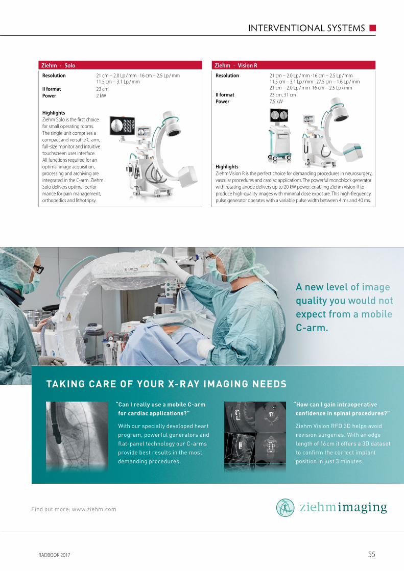
RADBOOK 2017 55
INTERVENTIONAL SYSTEMS ◼
Taking care of your X-r ay imaging neeDS
“can i really use a mobile c-arm
for cardiac applications?”
With our specially developed heart
program, powerful generators and
flat-panel technology our C-arms
provide best results in the most
demanding procedures.
“How can i gain intraoperative
confidence in spinal procedures?”
Ziehm Vision RFD 3D helps avoid
revision surgeries. With an edge
length of 16 cm it offers a 3D dataset
to confirm the correct implant
position in just 3 minutes.
A new level of image quality you would not expect from a mobile C-arm.
Find out more: www.ziehm.com
Zie-1541-Anzeige_European-Hospital_rz.indd 1 12.10.15 18:02
HighlightsZiehm Solo is the first choice for small operating rooms. The single unit comprises a compact and versatile C-arm, full-size monitor and intuitive touchscreen user interface. All functions required for an optimal image acquisition, processing and archiving are integrated in the C-arm. Ziehm Solo delivers optimal perfor-mance for pain management, orthopedics and lithotripsy.
· SoloZiehm
2 kWPower23 cmII format
21 cm – 2.0 Lp / mm · 16 cm – 2.5 Lp / mm 11.5 cm – 3.1 Lp / mm
Resolution
HighlightsZiehm Vision R is the perfect choice for demanding procedures in neurosurgery, vascular procedures and cardiac applications. The powerful monoblock generator with rotating anode delivers up to 20 kW power, enabling Ziehm Vision R to produce high-quality images with minimal dose exposure. This high-frequency pulse generator operates with a variable pulse width between 4 ms and 40 ms.
· Vision RZiehm
7.5 kWPower23 cm, 31 cmII format
21 cm – 2.0 Lp / mm · 16 cm – 2.5 Lp / mm 11.5 cm – 3.1 Lp / mm · 27.5 cm – 1.6 Lp / mm 21 cm – 2.0 Lp / mm · 16 cm – 2.5 Lp / mm
Resolution
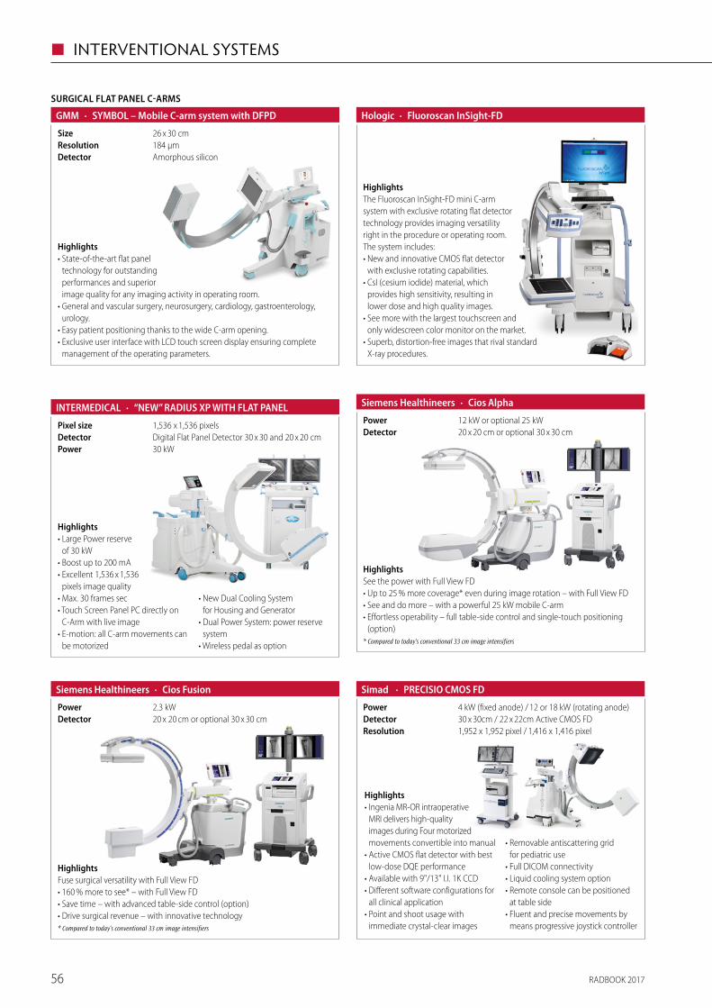
RADBOOK 201756
◼ INTERVENTIONAL SYSTEMS
· Cios AlphaSiemens Healthineers
20 x 20 cm or optional 30 x 30 cmDetector12 kW or optional 25 kWPower
HighlightsSee the power with Full View FD• Up to 25 % more coverage* even during image rotation – with Full View FD• See and do more – with a powerful 25 kW mobile C-arm• Effortless operability – full table-side control and single-touch positioning
(option)* Compared to today‘s conventional 33 cm image intensifiers
· Cios FusionSiemens Healthineers
20 x 20 cm or optional 30 x 30 cmDetector2.3 kWPower
HighlightsFuse surgical versatility with Full View FD• 160 % more to see* – with Full View FD• Save time – with advanced table-side control (option)• Drive surgical revenue – with innovative technology* Compared to today‘s conventional 33 cm image intensifiers
· PRECISIO CMOS FDSimad
1,952 x 1,952 pixel / 1,416 x 1,416 pixelResolution30 x 30cm / 22 x 22cm Active CMOS FDDetector4 kW (fixed anode) / 12 or 18 kW (rotating anode)Power
Highlights• Ingenia MR-OR intraoperative
MRI delivers high-quality images during Four motorized movements convertible into manual
• Active CMOS flat detector with best low-dose DQE performance
• Available with 9"/13" I.I. 1K CCD • Different software configurations for
all clinical application• Point and shoot usage with
immediate crystal-clear images
• Removable antiscattering grid for pediatric use
• Full DICOM connectivity• Liquid cooling system option• Remote console can be positioned
at table side• Fluent and precise movements by
means progressive joystick controller
· “NEW” RADIUS XP WITH FLAT PANELINTERMEDICAL
30 kWPowerDigital Flat Panel Detector 30 x 30 and 20 x 20 cmDetector1,536 x 1,536 pixelsPixel size
Highlights• Large Power reserve
of 30 kW• Boost up to 200 mA• Excellent 1,536 x 1,536
pixels image quality• Max. 30 frames sec• Touch Screen Panel PC directly on
C-Arm with live image• E-motion: all C-arm movements can
be motorized
• New Dual Cooling System for Housing and Generator
• Dual Power System: power reserve system
• Wireless pedal as option
· Fluoroscan InSight-FDHologic
HighlightsThe Fluoroscan InSight-FD mini C-arm system with exclusive rotating flat detector technology provides imaging versatility right in the procedure or operating room. The system includes:• New and innovative CMOS flat detector
with exclusive rotating capabilities.• CsI (cesium iodide) material, which
provides high sensitivity, resulting in lower dose and high quality images.
• See more with the largest touchscreen and only widescreen color monitor on the market.
• Superb, distortion-free images that rival standard X-ray procedures.
Highlights• State-of-the-art flat panel
technology for outstanding performances and superior image quality for any imaging activity in operating room.
• General and vascular surgery, neurosurgery, cardiology, gastroenterology, urology.
• Easy patient positioning thanks to the wide C-arm opening. • Exclusive user interface with LCD touch screen display ensuring complete
management of the operating parameters.
SURGICAL FLAT PANEL C-ARMS
· SYMBOL – Mobile C-arm system with DFPDGMM
Amorphous siliconDetector184 µmResolution26 x 30 cmSize
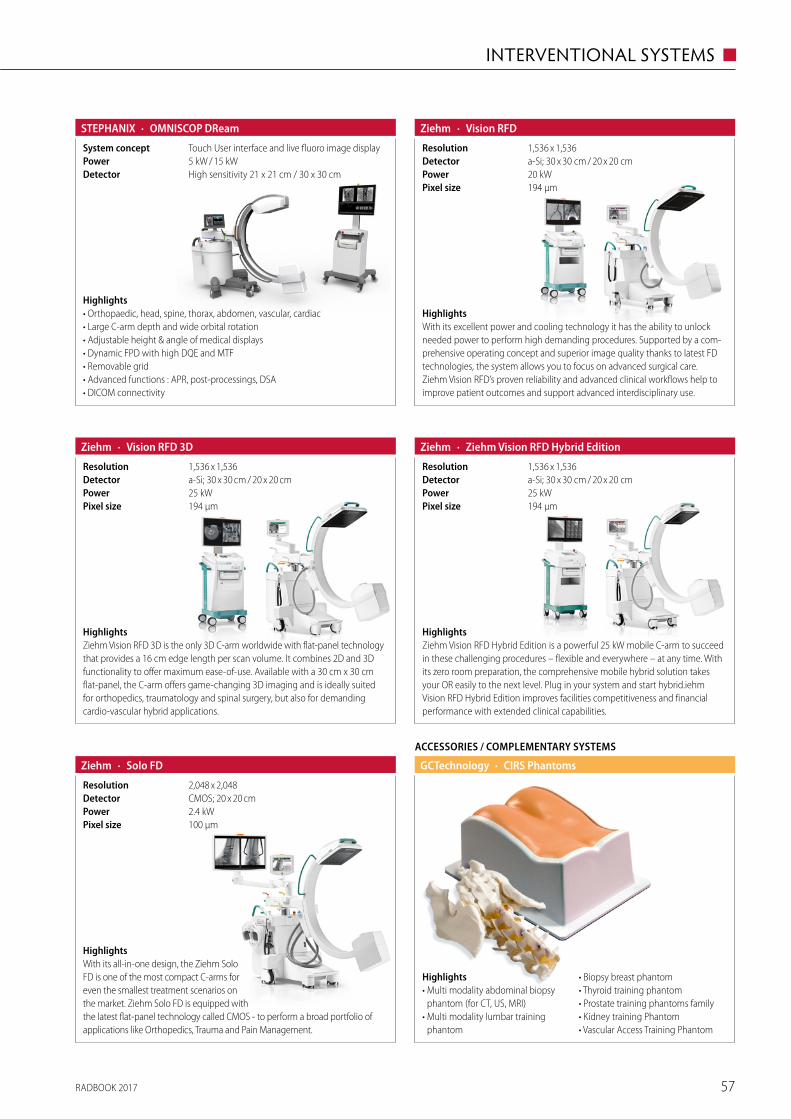
RADBOOK 2017 57
INTERVENTIONAL SYSTEMS ◼
Highlights• Orthopaedic, head, spine, thorax, abdomen, vascular, cardiac• Large C-arm depth and wide orbital rotation • Adjustable height & angle of medical displays• Dynamic FPD with high DQE and MTF• Removable grid• Advanced functions : APR, post-processings, DSA• DICOM connectivity
· OMNISCOP DReamSTEPHANIX
High sensitivity 21 x 21 cm / 30 x 30 cmDetector5 kW / 15 kWPowerTouch User interface and live fluoro image displaySystem concept
HighlightsZiehm Vision RFD 3D is the only 3D C-arm worldwide with flat-panel technology that provides a 16 cm edge length per scan volume. It combines 2D and 3D functionality to offer maximum ease-of-use. Available with a 30 cm x 30 cm flat-panel, the C-arm offers game-changing 3D imaging and is ideally suited for orthopedics, traumatology and spinal surgery, but also for demanding cardio-vascular hybrid applications.
· Vision RFD 3DZiehm
194 μmPixel size25 kWPowera-Si; 30 x 30 cm / 20 x 20 cmDetector1,536 x 1,536Resolution
HighlightsZiehm Vision RFD Hybrid Edition is a powerful 25 kW mobile C-arm to succeed in these challenging procedures – flexible and everywhere – at any time. With its zero room preparation, the comprehensive mobile hybrid solution takes your OR easily to the next level. Plug in your system and start hybrid.iehm Vision RFD Hybrid Edition improves facilities competitiveness and financial performance with extended clinical capabilities.
· Ziehm Vision RFD Hybrid EditionZiehm
194 μmPixel size25 kWPowera-Si; 30 x 30 cm / 20 x 20 cm Detector1,536 x 1,536Resolution
HighlightsWith its all-in-one design, the Ziehm Solo FD is one of the most compact C-arms for even the smallest treatment scenarios on the market. Ziehm Solo FD is equipped with the latest flat-panel technology called CMOS - to perform a broad portfolio of applications like Orthopedics, Trauma and Pain Management.
· Solo FDZiehm
100 μmPixel size2.4 kWPowerCMOS; 20 x 20 cmDetector2,048 x 2,048Resolution
ACCESSORIES / COMPLEMENTARY SYSTEMS
· CIRS PhantomsGCTechnology
Highlights• Multi modality abdominal biopsy
phantom (for CT, US, MRI)• Multi modality lumbar training
phantom
• Biopsy breast phantom• Thyroid training phantom• Prostate training phantoms family• Kidney training Phantom• Vascular Access Training Phantom
HighlightsWith its excellent power and cooling technology it has the ability to unlock needed power to perform high demanding procedures. Supported by a com-prehensive operating concept and superior image quality thanks to latest FD technologies, the system allows you to focus on advanced surgical care.Ziehm Vision RFD’s proven reliability and advanced clinical workflows help to improve patient outcomes and support advanced interdisciplinary use.
· Vision RFD
194 μmPixel size20 kWPowera-Si; 30 x 30 cm / 20 x 20 cmDetector1,536 x 1,536Resolution
Ziehm
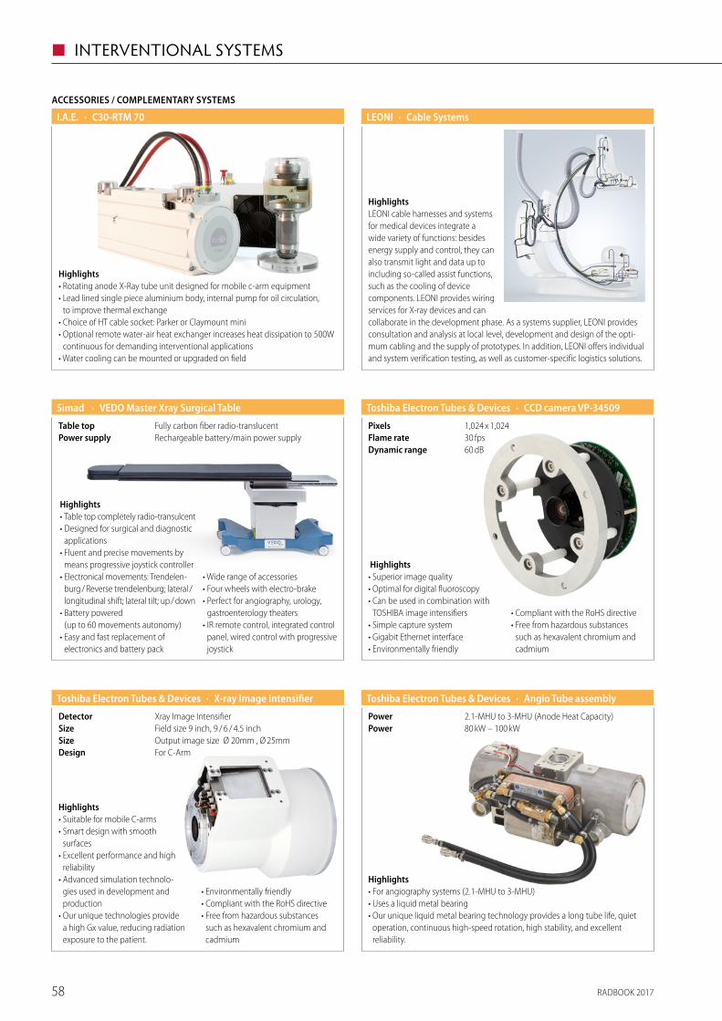
RADBOOK 201758
◼ INTERVENTIONAL SYSTEMS
· Cable SystemsLEONI
HighlightsLEONI cable harnesses and systems for medical devices integrate a wide variety of functions: besides energy supply and control, they can also transmit light and data up to including so-called assist functions, such as the cooling of device components. LEONI provides wiring services for X-ray devices and can collaborate in the development phase. As a systems supplier, LEONI provides consultation and analysis at local level, development and design of the opti-mum cabling and the supply of prototypes. In addition, LEONI offers individual and system verification testing, as well as customer-specific logistics solutions.
· VEDO Master Xray Surgical TableSimad
Rechargeable battery/main power supplyPower supplyFully carbon fiber radio-translucentTable top
Highlights• Table top completely radio-transulcent• Designed for surgical and diagnostic
applications• Fluent and precise movements by
means progressive joystick controller • Electronical movements: Trendelen-
burg / Reverse trendelenburg; lateral / longitudinal shift; lateral tilt; up / down
• Battery powered (up to 60 movements autonomy)
• Easy and fast replacement of electronics and battery pack
• Wide range of accessories• Four wheels with electro-brake• Perfect for angiography, urology,
gastroenterology theaters• IR remote control, integrated control
panel, wired control with progressive joystick
Highlights• Suitable for mobile C-arms• Smart design with smooth
surfaces• Excellent performance and high
reliability• Advanced simulation technolo-
gies used in development and production
• Our unique technologies provide a high Gx value, reducing radiation exposure to the patient.
• Environmentally friendly• Compliant with the RoHS directive• Free from hazardous substances
such as hexavalent chromium and cadmium
· X-ray Image IntensifierToshiba Electron Tubes & Devices
For C-ArmDesignOutput image size Ø 20mm , Ø 25mmSizeField size 9 inch, 9 / 6 / 4.5 inchSizeXray Image IntensifierDetector
Highlights• For angiography systems (2.1-MHU to 3-MHU)• Uses a liquid metal bearing• Our unique liquid metal bearing technology provides a long tube life, quiet
operation, continuous high-speed rotation, high stability, and excellent reliability.
· Angio Tube assemblyToshiba Electron Tubes & Devices
80 kW – 100 kWPower2.1-MHU to 3-MHU (Anode Heat Capacity)Power
· CCD camera VP-34509Toshiba Electron Tubes & Devices
60 dBDynamic range30 fpsFlame rate1,024 x 1,024Pixels
Highlights• Superior image quality• Optimal for digital fluoroscopy• Can be used in combination with
TOSHIBA image intensifiers• Simple capture system• Gigabit Ethernet interface• Environmentally friendly
• Compliant with the RoHS directive• Free from hazardous substances
such as hexavalent chromium and cadmium
ACCESSORIES / COMPLEMENTARY SYSTEMS
· C30-RTM 70I.A.E.
Highlights• Rotating anode X-Ray tube unit designed for mobile c-arm equipment• Lead lined single piece aluminium body, internal pump for oil circulation,
to improve thermal exchange• Choice of HT cable socket: Parker or Claymount mini• Optional remote water-air heat exchanger increases heat dissipation to 500W
continuous for demanding interventional applications• Water cooling can be mounted or upgraded on field
RADBOOK 2017 59
IT SYSTEMS ◼
IT Systems
RIS / PACS
Portal Solution
CAD
Mammo Workstation
Mobile RIS / PACS Viewer
Accessories / Complementary Systems
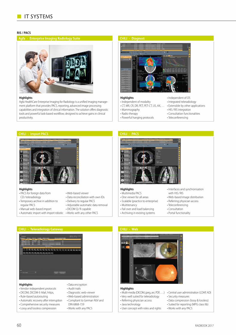
RADBOOK 201760
◼ IT SYSTEMS
Highlights• Multi-media (DICOM, jpeg, avi, PDF, …)• Very well suited for teleradiology• Referring physician access • Java technology• User concept with roles and rights
• Central user administration (LDAP, AD)• Security measures• Data compression (lossy & lossless)• Suited for reporting (MPG class IIb)• Works with any PACS
· WebCHILI
RIS / PACS
· Enterprise Imaging Radiology SuiteAgfa
HighlightsAgfa HealthCare Enterprise Imaging for Radiology is a unified imaging manage-ment platform that provides PACS, reporting, advanced image processing capabilities and integration of clinical information. The solution offers diagnostic tools and powerful task-based workflow, designed to achieve gains in clinical productivity.
Highlights• Independent of modality• CT, MR, CR, DR, PET, PET-CT, US, AX, …• Mammography• Radio therapy• Powerful hanging protocols
• Independent of OS• Integrated teleradiology• Extensible by other applications• HIS / RIS integration• Consultation functionalities• Teleconferencing
· DiagnostCHILI
Highlights• PACS for foreign data from
CD / tele radiology• Temporary archive in addition to
regular PACS• Manual web-based import • Automatic import with import robotic
• Web-based viewer• Data reconciliation with own IDs• Delivery to regular PACS• Adjustable automatic data removal• DICOM Q / R capable• Works with any other PACS
· Import PACSCHILI
Highlights• Multimedia PACS• One viewer for all areas• Scalable (practice to enterprise)• Multitenancy• Fail over and load balancing• Archiving in existing systems
• Interfaces and synchronisation with HIS / RIS
• Web-based image distribution• Referring physician access• Teleconferencing• Consultation• Portal functionality
· PACSCHILI
Highlights• Vendor-independent protocols• DICOM, DICOM-E-Mail, https, • Rule-based autorouting• Automatic recovery after interruption• Comprehensive security measures• Lossy and lossless compression
• Data encryption• Audit trails• Diagnostic web-viewer• Web-based administration• Compliant to German RöV and
DIN 6868-159• Works with any PACS
· Teleradiology GatewayCHILI
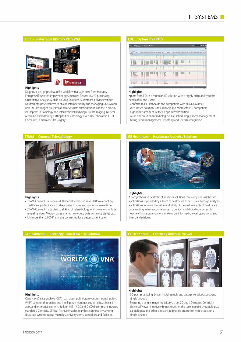
RADBOOK 2017 61
IT SYSTEMS ◼
HighlightsA comprehensive portfolio of analytics solutions that comprise insight-rich applications supported by a team of healthcare experts. Ready-to-go analytics applications increase the value and utility of the vast amounts of healthcare data residing in transactional systems, devices and digital equipment to help healthcare organizations make more informed clinical, operational and financial decisions.
· Healthcare Analytics SolutionsGE Healthcare
GE Healthcare
HighlightsCentricity Clinical Archive (CCA) is an open architecture vendor-neutral archive (VNA) solution that unifies and intelligently manages patient data, clinical im-ages and enterprise content. Built on IHE – XDS and DICOM-compliant industry standards, Centricity Clinical Archive enables seamless connectivity among disparate systems across multiple archive systems, specialties and facilities.
· Centricity Clinical Archive Solution
· Xplore RIS / PACSEDL
HighlightsXplore from EDL is a modular RIS solution with a highly adaptability to the needs of all end users:• Conform to IHE standards and compatible with all DICOM PACS• Web-based solution, Citrix XenApp and Microsoft RSD compatible• Ergonomic architecture for an optimized Workflow• All-in one solution for radiologic clinic: scheduling, patient management,
billing, stock management, reporting and speech recognition
Highlights• ETIAM-Connect is a secure Multispecialty Telemedicine Platform enabling
Healthcare professionals to share patient cases and diagnose in real time• ETIAM-Connect is adapted to all kind of teleradiology workflows and includes
several services: Medical cases sharing, Invoicing, Duty planning, Statistics…• Join more than 2,000 Physicians connected for a better patient care!
· Connect: TeleradiologyETIAM
HighlightsDiagnostic Imaging Software for workflow management, from Modality to Enterprise IT systems, implementing Structured Report, 3D/4D processing, Quantitative Analysis, Mobile & Cloud Solutions. Suitestensa provides Vendor Neutral Enterprise Archives to ensure interoperability and managing DICOM and non-DICOM images. Suitestensa achieves data administration and focus on clin-ical aspects in Radiology and Interventional Radiology, Breast Imaging, Nuclear Medicine, Radiotherapy, Orthopaedics, Cardiology (Cath-lab, Echocardio, EP, ECG, Check-ups), Cardiovascular Surgery.
· Suitestensa RIS CVIS PACS VNAEBIT
Highlights• 3D post-processing, breast imaging tools and enterprise-wide access on a
single desktop • Featuring a single image repository across 2D and 3D studies, Centricity
Universal Viewer intuitively brings together the tools needed by radiologists, cardiologists and other clinicians to provide enterprise-wide access on a single desktop
· Centricity Universal ViewerGE Healthcare
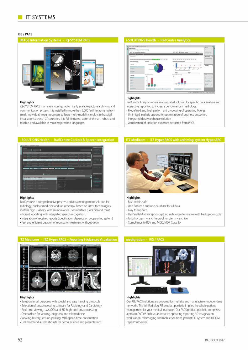
RADBOOK 201762
◼ IT SYSTEMS
Highlights• Solution for all purposes with special and easy hanging protocols• Selection of postprocessing software for Radiology and Cardiology• Real-time viewing. LVA, QCA and 3D-high-end-postprocessing• One surface for viewing, diagnosis and telemedicine• Viewing-history, session-parking, MRT-space-time-presentation• Unlimited and automatic lists for demo, science and presentations
· ITZ Hyper.PACS – Reporting & Advanced VisualizationITZ Medicom
HighlightsiQ-SYSTEM PACS is an easily configurable, highly scalable picture archiving and communication system. It is installed in more than 5,000 facilities ranging from small, individual, imaging centers to large multi-modality, multi-site hospital installations across 107 countries. It is full-featured, state-of-the-art, robust and reliable, and available in most major world languages.
RIS / PACS
· iQ-SYSTEM PACSIMAGE Information Systems
HighlightsRadCentre Analytics offers an integrated solution for specific data analysis and interactive reporting to increase performance in radiology.• Predefined and high performant processing of operating figures• Unlimited analysis options for optimisation of business outcomes• Integrated data warehouse solution• Visualization of radiation exposure extracted from PACS
· RadCentre Analyticsi-SOLUTIONS Health
HighlightsRadCentre is a comprehensive process and data management solution for radiology, nuclear medicine and radiotherapy. Based on latest technologies it offers high usability with an innovative user interface (Cockpit) and most efficient reporting with integrated speech recognition.• Integration of received reports (specification depends on cooperating system)• Fast and efficient creation of reports for treatment without delay
· RadCentre Cockpit & Speech Integrationi-SOLUTIONS Health
Highlights• Fast, stable, safe• One frontend and one database for all data• Easy to support• ITZ-Parallel-Archiving-Concept; no archiving of errors like with backup-principle• Fast shortterm – and fireproof longterm – archive• Compliance to RöV and MDD/MDR Class IIb
· ITZ Hyper.PACS with archiving-system Hyper.ARCITZ Medicom
HighlightsOur RIS / PACS solutions are designed for multisite and manufacturer-independent networks. The WinRadiolog RIS product portfolio implies the whole patient management for your medical institution. Our PACS product portfolio comprises a proven DICOM archive, an intuitive operating reporting 3D ImageVision workstation, teleimaging and mobile solutions, patient CD system and DICOM PaperPrint Server.
· RIS / PACSmedigration
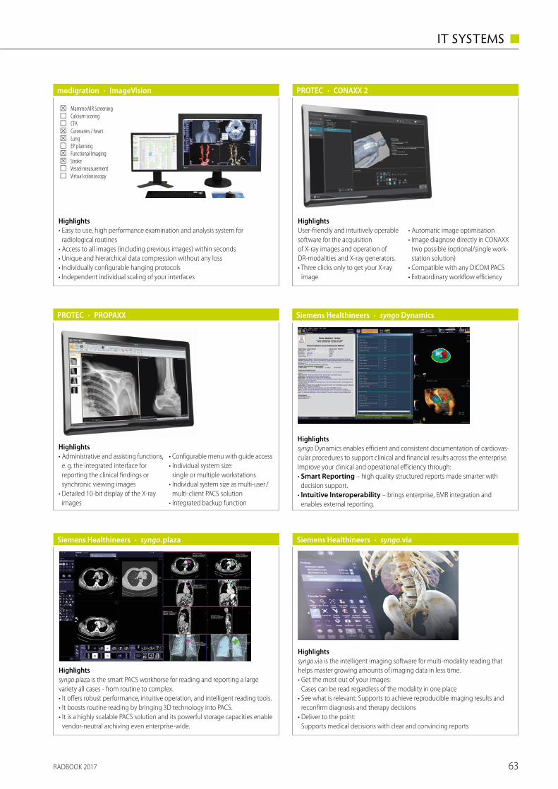
RADBOOK 2017 63
IT SYSTEMS ◼
Highlights• Administrative and assisting functions,
e. g. the integrated interface for reporting the clinical findings or synchronic viewing images
• Detailed 10-bit display of the X-ray images
• Configurable menu with guide access• Individual system size:
single or multiple workstations• Individual system size as multi-user /
multi-client PACS solution• Integrated backup function
· PROPAXXPROTEC
Highlightssyngo Dynamics enables efficient and consistent documentation of cardiovas-cular procedures to support clinical and financial results across the enterprise. Improve your clinical and operational efficiency through:• Smart Reporting – high quality structured reports made smarter with
decision support.• Intuitive Interoperability – brings enterprise, EMR integration and
enables external reporting.
· syngo DynamicsSiemens Healthineers
Highlightssyngo.plaza is the smart PACS workhorse for reading and reporting a large variety all cases - from routine to complex.• It offers robust performance, intuitive operation, and intelligent reading tools.• It boosts routine reading by bringing 3D technology into PACS.• It is a highly scalable PACS solution and its powerful storage capacities enable
vendor-neutral archiving even enterprise-wide.
· syngo.plazaSiemens Healthineers
Highlights• Easy to use, high performance examination and analysis system for
r adiological routines• Access to all images (including previous images) within seconds• Unique and hierarchical data compression without any loss• Individually configurable hanging protocols• Independent individual scaling of your interfaces
· ImageVisionmedigration
☒ Mammo MR Screening ☐ Calcium scoring ☐ CFA ☒ Curonaries / heart ☒ Lung ☐ EP planning ☒ Functional Imaging ☒ Stroke ☐ Vessel measurement ☐ Virtual colonoscopy
· CONAXX 2PROTEC
HighlightsUser-friendly and intuitively operable software for the acquisition of X-ray images and operation of DR-modalities and X-ray generators.• Three clicks only to get your X-ray
image
• Automatic image optimisation• Image diagnose directly in CONAXX
two possible (optional/single work-station solution)
• Compatible with any DICOM PACS• Extraordinary workflow efficiency
Siemens Healthineers
Highlightssyngo.via is the intelligent imaging software for multi‐modality reading that helps master growing amounts of imaging data in less time. • Get the most out of your images:
Cases can be read regardless of the modality in one place • See what is relevant: Supports to achieve reproducible imaging results and
reconfirm diagnosis and therapy decisions• Deliver to the point:
Supports medical decisions with clear and convincing reports
· syngo.via
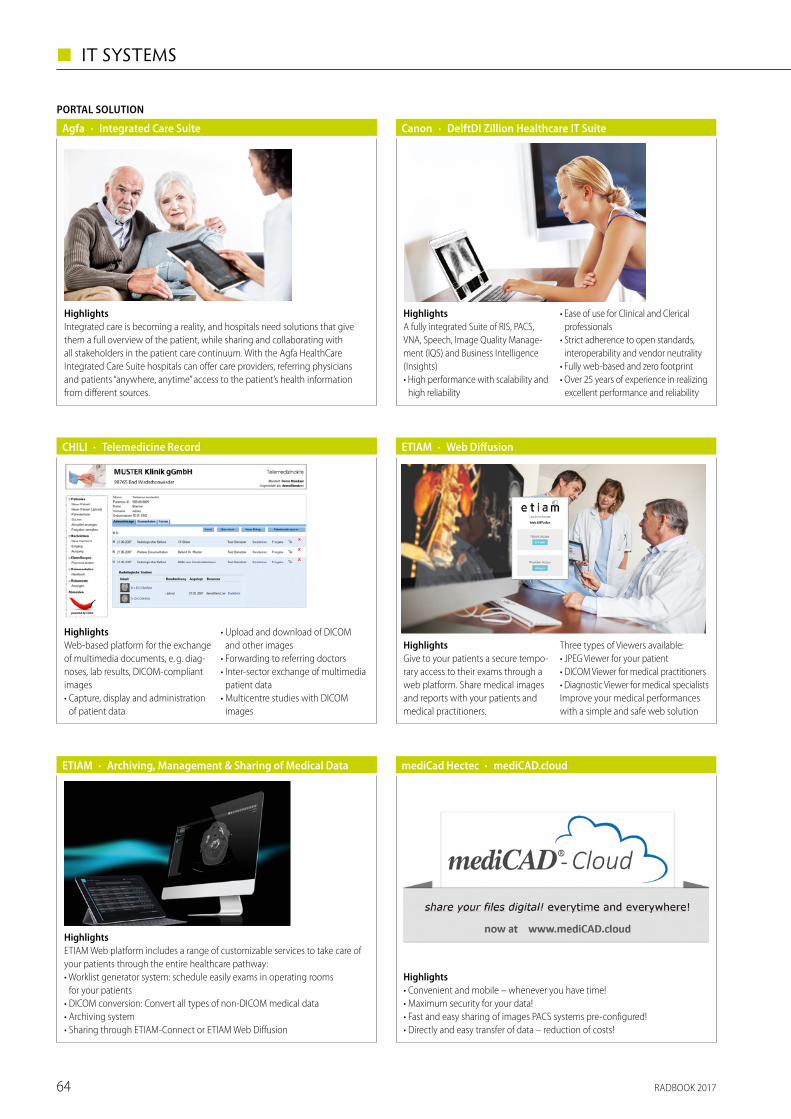
RADBOOK 201764
◼ IT SYSTEMS
Highlights• Convenient and mobile – whenever you have time!• Maximum security for your data!• Fast and easy sharing of images PACS systems pre-configured!• Directly and easy transfer of data – reduction of costs!
· mediCAD.cloudmediCad Hectec
Agfa
PORTAL SOLUTION
· Integrated Care Suite
HighlightsIntegrated care is becoming a reality, and hospitals need solutions that give them a full overview of the patient, while sharing and collaborating with all stakeholders in the patient care continuum. With the Agfa HealthCare Integrated Care Suite hospitals can offer care providers, referring physicians and patients “anywhere, anytime” access to the patient’s health information from different sources.
HighlightsA fully integrated Suite of RIS, PACS, VNA, Speech, Image Quality Manage-ment (IQS) and Business Intelligence (Insights) • High performance with scalability and
high reliability
• Ease of use for Clinical and Clerical professionals
• Strict adherence to open standards, interoperability and vendor neutrality
• Fully web-based and zero footprint • Over 25 years of experience in realizing
excellent performance and reliability
· DelftDI Zillion Healthcare IT SuiteCanon
HighlightsETIAM Web platform includes a range of customizable services to take care of your patients through the entire healthcare pathway:• Worklist generator system: schedule easily exams in operating rooms
for your patients• DICOM conversion: Convert all types of non-DICOM medical data• Archiving system• Sharing through ETIAM-Connect or ETIAM Web Diffusion
· Archiving, Management & Sharing of Medical DataETIAM
HighlightsGive to your patients a secure tempo-rary access to their exams through a web platform. Share medical images and reports with your patients and medical practitioners.
Three types of Viewers available:• JPEG Viewer for your patient• DICOM Viewer for medical practitioners• Diagnostic Viewer for medical specialistsImprove your medical performances with a simple and safe web solution
· Web DiffusionETIAM
HighlightsWeb-based platform for the exchange of multimedia documents, e. g. diag-noses, lab results, DICOM-compliant images• Capture, display and administration
of patient data
• Upload and download of DICOM and other images
• Forwarding to referring doctors • Inter-sector exchange of multimedia
patient data• Multicentre studies with DICOM
images
· Telemedicine RecordCHILI
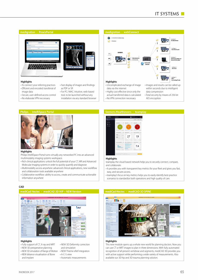
RADBOOK 2017 65
IT SYSTEMS ◼
Highlights• Fully support of CT, X-ray and MRT• NEW 3D preoperative planning• NEW 3D simulation of Range of Motion• NEW distance visualisation of Bone
and implant
• NEW 3D Deformity correction and simulation
• NEW Thieme eRef integration• A-C-S view• Automatic measurements
CAD
· mediCAD 3D HIP – NEW VersionmediCad Hectec
Highlights• To connect your referring practices• Efficient and encoded transferral of
image data• Secure, user-defined access control• No elaborate VPN neccessary
• Fast display of images and findings as PDF or SR
• For PC / MAC: Intuitive, web-based tool, to be launched without any installation via any standard browser
· PraxisPortalmedigration · webConnectmedigration
Highlights• Uncomplicated exchange of image
data via the internet• Highly cost effective since only the
actual transferred data is calculated• No VPN connection necessary
• Images and results can be called up within seconds due to intelligent data compression
• Total security by means of 256 bit AES encryption
· IntelliSpace PortalPhilips
HighlightsPhilips IntelliSpace Portal turns virtually any networked PC into an advanced multimodality imaging systems workspace. • Rich clinical applications: unlock the full potential of your CT, MR and Advanced
Molecular Imaging systems in order to quickly quantify and diagnose• Multimodality access anywhere: advanced clinical applications, new workflow
and collaboration tools available anywhere• Collaborative workflow: ability to access, create and communicate actionable
information anywhere
Highlightsteamplay the cloud-based network helps you to securely connect, compare, and collaborate.• It provides you with transparent key metrics for your fleet and gives you fast,
easy, and secures access.• teamplay’s focus on key metrics helps you to easily identify best-practice
scenarios to standardize both operations and high quality of care.
· teamplaySiemens Healthineers
HighlightsThis new module opens up a whole new world for planning doctors. Now you can use CT or MRT images to plan in three dimensions. With fully automated recognition of all present vertebrae and segments, mediCAD 3D provides you with active support while performing a wide variety of measurements. Also available our 3D hip and 3D trauma planning solution.
· mediCAD 3D SPINEmediCad Hectec
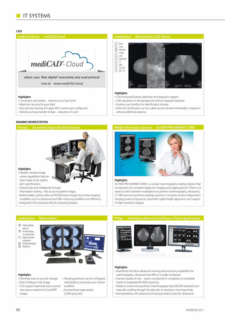
RADBOOK 201766
◼ IT SYSTEMS
· IntelliSpace Breast & IntelliSpace Clinical ApplicationsPhilips
Highlights• Seamlessly interfaces advanced viewing and processing capabilities for
mammography, ultrasound and MRI on a single workspace• Improve quality of care – report consistently in compliance to standards
thanks to integrated BI-RADS reporting• Ability to review tomosynthesis mammography data (DICOM standard) and
manually scrolling through the data sets or viewing in cine loop mode• Interoperability with advanced clinical quantitative tools for ultrasound
Highlights• Convenient and mobile – whenever you have time!• Maximum security for your data!• Fast and easy sharing of images PACS systems pre-configured!• Directly and easy transfer of data – reduction of costs!
CAD
· mediCAD.cloudmediCad Hectec
Highlights• CAD microcalcifications detection and diagnosis support• CAD calculation in the background without separate hardware• Intuitive user interface for identification training• Detected calcifications can be scaled up and viewed individually in sequence
without additional expense
· MammoView CAD-Optionmedigration
☐ Brain ☐ Lung ☒ Mammo ☐ Cardio ☐ Liver ☐ Abdomen ☐ CT ☐ MRI ☐ CR / DR ☐ PET / CT
MAMMO WORKSTATION
· SecurView Diagnostic WorkstationsHologic
Highlights• Flexible, intuitive image
review capabilities that are tailor made to the radiolo-gist’s specifications
• Interactively and intelligently through information-sharing – fast access to patient images
• Multimodality options allow all DICOM breast images from other imaging modalities such as ultrasound and MRI, improving workflow and efficiency
• Integrated CAD and breast density (Quantra) displays
HighlightsiQ-VIEW PRO MAMMO TOMO is a unique mammography reading solution that incorporates the complete diagnostic imaging and staging process. There is no need to switch between workstations to perform mammography, ultrasound, CT, MRI and tomosynthesis readings anymore. It includes vendor-independent hanging protocol sequences, automatic nipple height alignment, and support of high-resolution displays.
· iQ-VIEW PRO MAMMO TOMOIMAGE Information Systems
Highlights• Extremely easy to use and manage• Direct findings in the image• CAD support (optional) and a second
view area to examine US and MRT images
• Hanging protocols can be configured individually to automate your routine workflow
• Outstanding image quality (2,048 greyscale)
· MammoViewmedigration
☒ Default display protocol
☒ Hi-Res displays or mixed setups
☐ Digital dictation integration
☒ Dedicated keypad ☒ WebClient
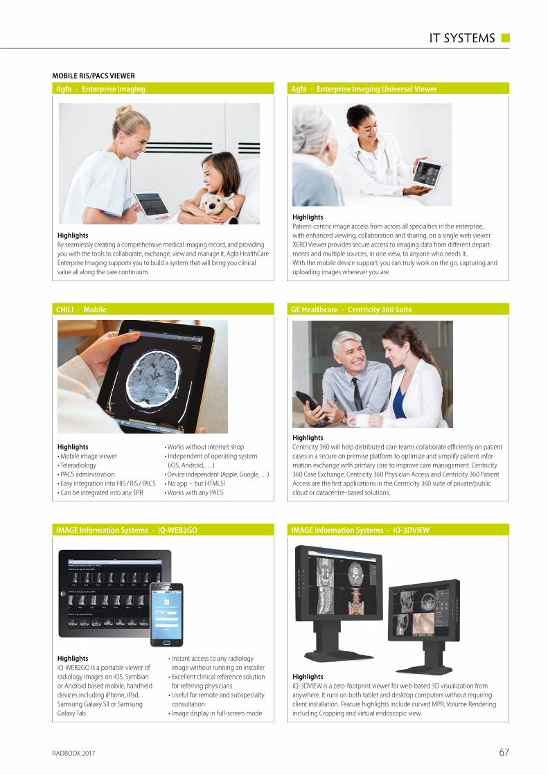
RADBOOK 2017 67
IT SYSTEMS ◼
· Enterprise Imaging Universal ViewerAgfa
HighlightsPatient-centric image access from across all specialties in the enterprise, with enhanced viewing, collaboration and sharing, on a single web viewer. XERO Viewer provides secure access to imaging data from different depart-ments and multiple sources, in one view, to anyone who needs it. With the mobile device support, you can truly work on the go, capturing and uploading images wherever you are.
Highlights• Mobile image viewer• Teleradiology• PACS administration• Easy integration into HIS / RIS / PACS• Can be integrated into any EPR
• Works without internet shop• Independent of operating system
(iOS, Android, …)• Device independent (Apple, Google, …)• No app – but HTML5!• Works with any PACS
· MobileCHILI
HighlightsCentricity 360 will help distributed care teams collaborate efficiently on patient cases in a secure on premise platform to optimize and simplify patient infor-mation exchange with primary care to improve care management. Centricity 360 Case Exchange, Centricity 360 Physician Access and Centricity 360 Patient Access are the first applications in the Centricity 360 suite of private/public cloud or datacentre-based solutions.
· Centricity 360 Suite GE Healthcare
HighlightsiQ-WEB2GO is a portable viewer of radiology images on iOS, Symbian or Android based mobile, handheld devices including iPhone, iPad, Samsung Galaxy SII or Samsung Galaxy Tab.
• Instant access to any radiology image without running an installer
• Excellent clinical reference solution for referring physicians
• Useful for remote and subspecialty consultation
• Image display in full-screen mode
· iQ-WEB2GOIMAGE Information Systems
HighlightsiQ-3DVIEW is a zero-footprint viewer for web-based 3D visualization from anywhere. It runs on both tablet and desktop computers without requiring client installation. Feature highlights include curved MPR, Volume Rendering including Cropping and virtual endoscopic view.
· iQ-3DVIEWIMAGE Information Systems
MOBILE RIS/PACS VIEWER
· Enterprise ImagingAgfa
HighlightsBy seamlessly creating a comprehensive medical imaging record, and providing you with the tools to collaborate, exchange, view and manage it, Agfa HealthCare Enterprise Imaging supports you to build a system that will bring you clinical value all along the care continuum.
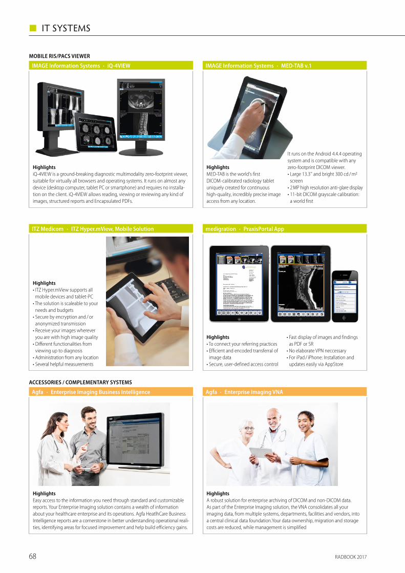
RADBOOK 201768
◼ IT SYSTEMS
HighlightsiQ-4VIEW is a ground-breaking diagnostic multimodality zero-footprint viewer, suitable for virtually all browsers and operating systems. It runs on almost any device (desktop computer, tablet PC or smartphone) and requires no installa-tion on the client. iQ-4VIEW allows reading, viewing or reviewing any kind of images, structured reports and Encapsulated PDFs.
MOBILE RIS/PACS VIEWER
· iQ-4VIEWIMAGE Information Systems
Highlights• ITZ Hyper.mView supports all
mobile devices and tablet-PC• The solution is scaleable to your
needs and budgets• Secure by encryption and / or
anonymized transmission• Receive your images wherever
you are with high image quality• Different functionalities from
viewing up to diagnosis• Administration from any location• Several helpful measurements
· ITZ Hyper.mView, Mobile SolutionITZ Medicom
Highlights• To connect your referring practices• Efficient and encoded transferral of
image data• Secure, user-defined access control
• Fast display of images and findings as PDF or SR
• No elaborate VPN neccessary• For iPad / iPhone: Installation and
updates easily via AppStore
· PraxisPortal Appmedigration
· Enterprise Imaging VNAAgfa
HighlightsA robust solution for enterprise archiving of DICOM and non-DICOM data. As part of the Enterprise Imaging solution, the VNA consolidates all your imaging data, from multiple systems, departments, facilities and vendors, into a central clinical data foundation.Your data ownership, migration and storage costs are reduced, while management is simplified
ACCESSORIES / COMPLEMENTARY SYSTEMS
· Enterprise Imaging Business IntelligenceAgfa
HighlightsEasy access to the information you need through standard and customizable reports. Your Enterprise Imaging solution contains a wealth of information about your healthcare enterprise and its operations. Agfa HeatlhCare Business Intelligence reports are a cornerstone in better understanding operational reali- ties, identifying areas for focused improvement and help build efficiency gains.
· MED-TAB v.1IMAGE Information Systems
HighlightsMED-TAB is the world's first DICOM-calibrated radiology tablet uniquely created for continuous high-quality, incredibly precise image access from any location.
It runs on the Android 4.4.4 operating system and is compatible with any zero-footprint DICOM viewer.• Large 13.3'' and bright 300 cd / m2
screen• 2 MP high resolution anti-glare display• 11-bit DICOM grayscale calibration:
a world first
RADBOOK 2017 69
IT SYSTEMS ◼
15992_bayer_ad_ccc_main_na_ESGAR2017_la05_tk.indd 1 06.12.16 17:29
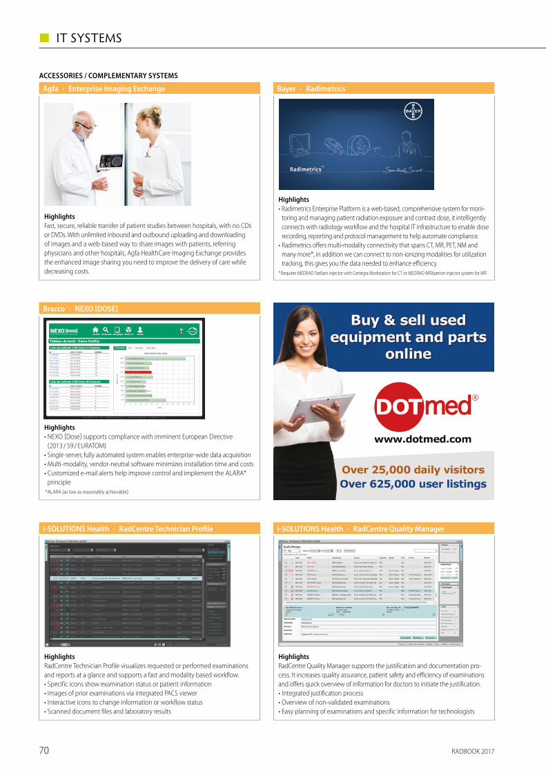
RADBOOK 201770
◼ IT SYSTEMS
ACCESSORIES / COMPLEMENTARY SYSTEMS
· Enterprise Imaging ExchangeAgfa
HighlightsFast, secure, reliable transfer of patient studies between hospitals, with no CDs or DVDs. With unlimited inbound and outbound uploading and downloading of images and a web-based way to share images with patients, referring physicians and other hospitals, Agfa HealthCare Imaging Exchange provides the enhanced image sharing you need to improve the delivery of care while decreasing costs.
· RadimetricsBayer
Highlights• Radimetrics Enterprise Platform is a web-based, comprehensive system for moni-
toring and managing patient radiation exposure and contrast dose, it intelligently connects with radiology workflow and the hospital IT infrastruc ture to enable dose recording, reporting and protocol management to help automate compliance.
• Radimetrics offers multi-modality connectivity that spans CT, MR, PET, NM and many more*, in addition we can connect to non-ionizing modalities for utilization tracking, this gives you the data needed to enhance efficiency.
* Requires MEDRAD Stellant injector with Certegra Workstation for CT or MEDRAD MRXperion injector system for MR
· NEXO [DOSE]Bracco
Highlights• NEXO [Dose] supports compliance with imminent European Directive
(2013 / 59 / EURATOM)• Single-server, fully automated system enables enterprise-wide data acquisition• Multi-modality, vendor-neutral software minimizes installation time and costs• Customized e-mail alerts help improve control and implement the ALARA*
principle *ALARA (as low as reasonably achievable)
HighlightsRadCentre Technician Profile visualizes requested or performed examinations and reports at a glance and supports a fast and modality based workflow.• Specific icons show examination status or patient information• Images of prior examinations via integrated PACS viewer• Interactive icons to change information or workflow status • Scanned document files and laboratory results
· RadCentre Technician Profilei-SOLUTIONS Health
HighlightsRadCentre Quality Manager supports the justification and documentation pro-cess. It increases quality assurance, patient safety and efficiency of examinations and offers quick overview of information for doctors to initiate the justification.• Integrated justification process• Overview of non-validated examinations• Easy planning of examinations and specific information for technologists
· RadCentre Quality Manageri-SOLUTIONS Health
RADBOOK 2017 71
MAMMOGRAPHY ◼
Mammography
Tomosynthesis
Digital Mammography
Film-Screen Mammography
Biopsy Tables
Radiowave-Imaging
Accessories / Complementary Systems

RADBOOK 201772
◼ MAMMOGRAPHY
TOMOSYNTHESIS
Highlights• New DBT system allows superior
clinical results with low dose• DBT scan angle of 30° with 11 exposures• “Step & Shoot” tube motion combined with 85 µm pixel size for the best
visualization of microcalcifications• Fast Iterative Reconstruction Software dedicated for DBT• Multifunctional system: DBT, SINTHETIC VIEW, FFDM, TOMO-Guided or Stereo
Biopsy with the patient in a PRONE or UPRIGHT, CEDM
· Giotto Class TOMOSYNTHESISIMS
8 kWPower
85 µm (without binning)Pixel sizea-Se 24 x 30 cmResolution
Highlights• The FLEXITABLE in combination with Giotto CLASS allows the operator to
perform Tomo-Guided or Stereo biopsy with the patient in prone position, operating with the same detector used in the DBT clinical investigation. It guarantees to proceed with the same visualization of lesions like in DBT.
• Prone position provides 360° access to the breast with lateral, cranial caudal and inclined approach
· Giotto Class FLEXITABLEIMS
Biopsy table for TOMO-Guided or Stereo BIOPSY with the patient in a PRONE position
Technology85 µm (without binning)Pixel sizeAmorphous Selenium latest generation, 24 x 30 cmDetector
Highlights• Fast and stream-lined workflow for high
patient troughput• Detector 24 x 31 cm with high quantum
efficiency (DQE)• High picture quality through automatic
optimization of all parameters = AOP• Patented Rh / Mo x-ray tube with matching
Rh / Mo filters• Option: Stereotaxy, Tomosynthesis SenoClaire,
SenoBright (CESM)
· Senographe CareGE Healthcare
100 μmPixel sizea-Silizium, 24 x 31 cmDetector22 – 35 kVkV Range
Highlights• Small, motorized gantry• Fits into small rooms, thank to small footprint• Easy to use interface• For screening and standard diagnostic
· Senographe CrystalGE Healthcare
70 μmPixel sizeCsI CMOS, 23 x 30 cmDetector22 – 35 kVkV Range
· Senographe Essential GE Healthcare
100 μmPixel sizea-Silizium, 24 x 31 cmDetector22 – 49 kVkV Range
Highlights• Optimized image quality and dose
efficiency• Detector 24 x 31 cm with high
quantum efficiency (DQE)• High picture quality through automatic
optimization of all parameters = AOP• Patented Rh / Mo x-ray tube with
matching Rh / Mo filters• Option: Stereotaxy, Tomosynthesis
SenoClaire, SenoBright (CESM)
Highlights• The Serenys DR Bym, with the
added advantage of an isocentric C-arm including stereotactic biopsy
• The isocentric C-arm can be fully motorized and permits all breast projections without moving the patient and without adjusting the height of the C-arm, making exams faster and more comfortable
• The device is also available in analogic version
DIGITAL MAMMOGRAPHY
· Serenys DR BymDMS Imaging
FPD 18 x 24 cm or 24 x 30 cmDetector85 µmPixel size20 – 35 kVkV Range
5 kWPower
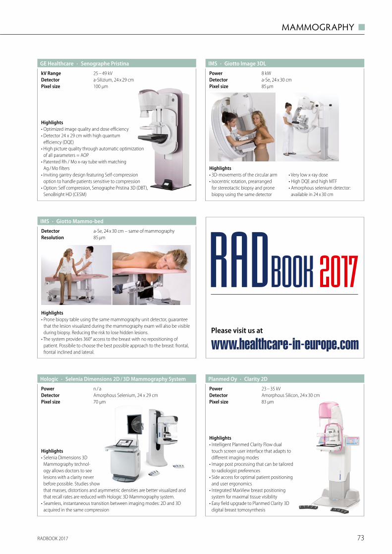
RADBOOK 2017 73
MAMMOGRAPHY ◼
Highlights• 3D-movements of the circular arm• Isocentric rotation, prearranged
for stereotactic biopsy and prone biopsy using the same detector
• Very low x-ray dose• High DQE and high MTF• Amorphous selenium detector:
available in 24 x 30 cm
· Giotto Image 3DLIMS
85 µmPixel sizea-Se, 24 x 30 cmDetector8 kWPower
Highlights• Prone biopsy table using the same mammography unit detector, guarantee
that the lesion visualized during the mammography exam will also be visible during biopsy. Reducing the risk to lose hidden lesions.
• The system provides 360° access to the breast with no repositioning of patient. Possibile to choose the best possible approach to the breast: frontal, frontal inclined and lateral.
· Giotto Mammo-bed
85 µmResolutiona-Se, 24 x 30 cm – same of mammographyDetector
IMS
Amorphous Selenium, 24 x 29 cmDetector
· Selenia Dimensions 2D / 3D Mammography SystemHologic
70 µmPixel size
n / aPower
Highlights• Selenia Dimensions 3D
Mammography technol-ogy allows doctors to see lesions with a clarity never before possible. Studies show that masses, distortions and asymmetric densities are better visualized and that recall rates are reduced with Hologic 3D Mammography system.
• Seamless, instantaneous transition between imaging modes: 2D and 3D acquired in the same compression
Highlights• Intelligent Planmed Clarity Flow dual
touch screen user interface that adapts to different imaging modes
• Image post processing that can be tailored to radiologist preferences
• Side access for optimal patient positioning and user ergonomics
• Integrated MaxView breast positioning system for maximal tissue visibility
• Easy field upgrade to Planmed Clarity 3D digital breast tomosynthesis
· Clarity 2DPlanmed Oy
83 µmPixel sizeAmorphous Silicon, 24 x 30 cmDetector23 – 35 kVPower
· Senographe PristinaGE Healthcare
100 μmPixel sizea-Silizium, 24 x 29 cmDetector25 – 49 kVkV Range
Highlights• Optimized image quality and dose efficiency• Detector 24 x 29 cm with high quantum
efficiency (DQE)• High picture quality through automatic optimization
of all parameters = AOP• Patented Rh / Mo x-ray tube with matching
Ag / Mo filters• Inviting gantry design featuring Self-compression
option to handle patients sensitive to compression • Option: Self compression, Senographe Pristina 3D (DBT),
SenoBright HD (CESM)
Please visit us at
www.healthcare-in-europe.com
RADBOOK 2017
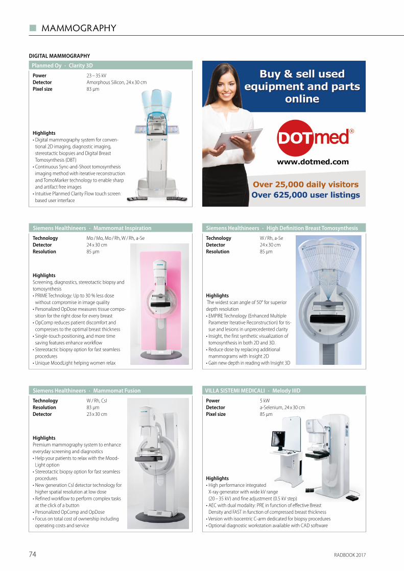
RADBOOK 201774
◼ MAMMOGRAPHY
Highlights• Digital mammography system for conven-
tional 2D imaging, diagnostic imaging, stereotactic biopsies and Digital Breast Tomosynthesis (DBT)
• Continuous Sync-and-Shoot tomosynthesis imaging method with iterative reconstruction and TomoMarker technology to enable sharp and artifact free images
• Intuitive Planmed Clarity Flow touch screen based user interface
DIGITAL MAMMOGRAPHY
· Clarity 3DPlanmed Oy
83 µmPixel sizeAmorphous Silicon, 24 x 30 cmDetector23 – 35 kVPower
HighlightsPremium mammography system to enhance everyday screening and diagnostics• Help your patients to relax with the Mood-
Light option • Stereotactic biopsy option for fast seamless
procedures• New generation Csl detector technology for
higher spatial resolution at low dose• Refined workflow to perform complex tasks
at the click of a button• Personalized OpComp and OpDose • Focus on total cost of ownership including
operating costs and service
· Mammomat Fusion Siemens Healthineers
23 x 30 cmDetector83 μmResolutionW / Rh, CsITechnology
Highlights The widest scan angle of 50° for superior depth resolution • EMPIRE Technology (Enhanced Multiple
Parameter Iterative Reconstruction) for tis-sue and lesions in unprecedented clarity
• Insight, the first synthetic visualization of tomosynthesis in both 2D and 3D.
• Reduce dose by replacing additional mammograms with Insight 2D
• Gain new depth in reading with Insight 3D
· High Definition Breast Tomosynthesis Siemens Healthineers
85 μmResolution24 x 30 cmDetectorW / Rh, a-SeTechnology
HighlightsScreening, diagnostics, stereotactic biopsy and tomosynthesis • PRIME Technology: Up to 30 % less dose
without compromise in image quality • Personalized OpDose measures tissue compo-
sition for the right dose for every breast• OpComp reduces patient discomfort and
compresses to the optimal breast thickness• Single-touch positioning, and more time
saving features enhance workflow• Stereotactic biopsy option for fast seamless
procedures• Unique MoodLight helping women relax
· Mammomat Inspiration Siemens Healthineers
85 μmResolution24 x 30 cmDetectorMo / Mo, Mo / Rh, W / Rh, a-SeTechnology
· Melody IIIDVILLA SISTEMI MEDICALI
85 μmPixel sizea-Selenium, 24 x 30 cmDetector5 kWPower
Highlights• High performance integrated
X-ray generator with wide kV range (20 – 35 kV) and fine adjustment (0.5 kV step)
• AEC with dual modality: PRE in function of effective Breast Density and FAST in function of compressed breast thickness
• Version with isocentric C-arm dedicated for biopsy procedures• Optional diagnostic workstation available with CAD software
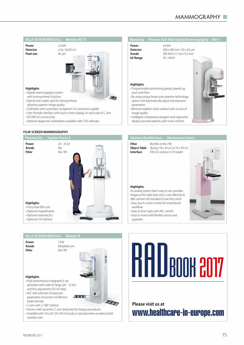
RADBOOK 2017 75
MAMMOGRAPHY ◼
Highlights• Entry level film unit • Optional magnification • Optional stereotactics • Optional CR interface
FILM-SCREEN MAMMOGRAPHY
· Sophie Classic SPlanmed Oy
Mo / RhFilterMoAnode20 – 35 kVPower
HighlightsAn analog system that is easy to use, provides images at the right dose and is cost-effective to offer women the standard of care they need• Easy touch screen control for streamlined
workflow• Easy to dose right with AEC control• Easy to invest with flexible service and
upgrades
· Mammomat Select Siemens Healthineers
Film ID camera or CR readerInterface(Bucky) 18 x 24 cm or 24 x 30 cmObject TableMo/Mo or Mo / RhFilter
Highlights• Programmable positioning greatly speeds up
your work flow• By using unique breast auto examine technology
system will automatically adjust the exposure parameters
• Minimal radiation dose realized with no loss of image quality
• Intelligent compression program and ergonomic designs provide patients with more comfort
4.8 kWPower
20~ 40 kVkV Range
300 x 240 mm / 85 x 85 μmDetector300 kHU 0.1 mm / 0.3 mmAnode
· Phoenix Full-field Digital Mammography – DM-1Wandong
· Melody IIIVILLA SISTEMI MEDICALI
Mo / RhFilterMolybdenumAnode5 kWPower
Highlights• High performance integrated X-ray
generator with wide kV range (20 – 35 kV) and fine adjustment (0.5 kV step)
• AEC with selection of exposure parameters in function of effective breast density
• C-arm with ± 180° rotation• Version with isocentric C-arm dedicated for biopsy procedures• Available with 18 x 24 / 24 x 30 cm bucky or special potter accepting both
cassette sizes
· Melody IIID TSVILLA SISTEMI MEDICALI
85 µmPixel sizea-Se, 24x30 cmDetector5.6 kWPower
Highlights• Digital mammography system
with tomosynthesis function• Special anti-scatter grid for tomosynthesis
allowing superior image quality• Collimator with automatic recognition of compressor paddle• User-friendly interface with touch screen display on each side of C-arm• DICOM 3.0 connectivity• Optional diagnostic workstation available with CAD software
Please visit us at
www.healthcare-in-europe.com
RADBOOK 2017
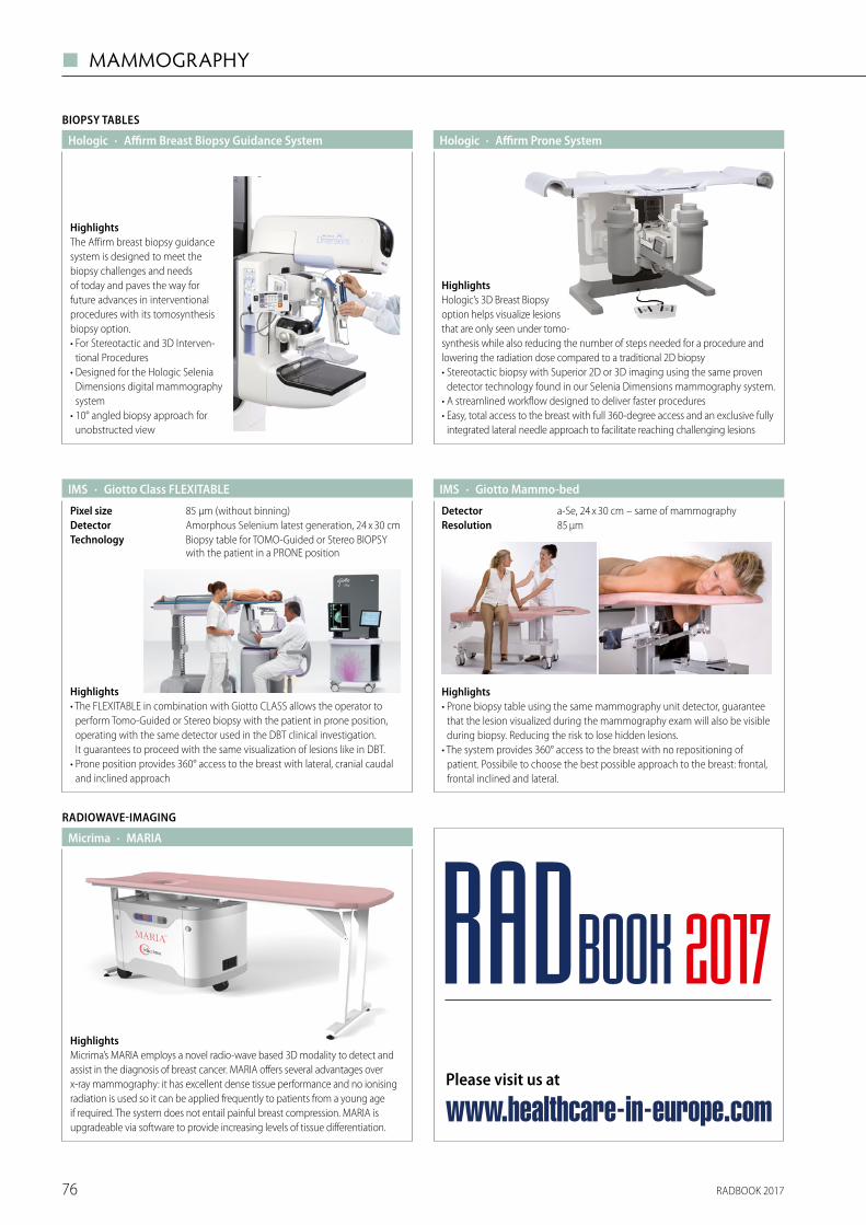
RADBOOK 201776
◼ MAMMOGRAPHY
Highlights• Prone biopsy table using the same mammography unit detector, guarantee
that the lesion visualized during the mammography exam will also be visible during biopsy. Reducing the risk to lose hidden lesions.
• The system provides 360° access to the breast with no repositioning of patient. Possibile to choose the best possible approach to the breast: frontal, frontal inclined and lateral.
· Giotto Mammo-bedIMS
85 µmResolutiona-Se, 24 x 30 cm – same of mammographyDetector
Highlights• The FLEXITABLE in combination with Giotto CLASS allows the operator to
perform Tomo-Guided or Stereo biopsy with the patient in prone position, operating with the same detector used in the DBT clinical investigation. It guarantees to proceed with the same visualization of lesions like in DBT.
• Prone position provides 360° access to the breast with lateral, cranial caudal and inclined approach
· Giotto Class FLEXITABLEIMS
Biopsy table for TOMO-Guided or Stereo BIOPSY with the patient in a PRONE position
TechnologyAmorphous Selenium latest generation, 24 x 30 cmDetector85 µm (without binning)Pixel size
BIOPSY TABLES
· Affirm Breast Biopsy Guidance SystemHologic
HighlightsThe Affirm breast biopsy guidancesystem is designed to meet thebiopsy challenges and needsof today and paves the way forfuture advances in interventionalprocedures with its tomosynthesisbiopsy option.• For Stereotactic and 3D Interven-
tional Procedures• Designed for the Hologic Selenia
Dimensions digital mammography system
• 10° angled biopsy approach for unobstructed view
· Affirm Prone SystemHologic
HighlightsHologic’s 3D Breast Biopsy option helps visualize lesions that are only seen under tomo-synthesis while also reducing the number of steps needed for a procedure and lowering the radiation dose compared to a traditional 2D biopsy• Stereotactic biopsy with Superior 2D or 3D imaging using the same proven
detector technology found in our Selenia Dimensions mammography system.• A streamlined workflow designed to deliver faster procedures• Easy, total access to the breast with full 360-degree access and an exclusive fully
integrated lateral needle approach to facilitate reaching challenging lesions
RADIOWAVE-IMAGING
· MARIAMicrima
HighlightsMicrima’s MARIA employs a novel radio-wave based 3D modality to detect and assist in the diagnosis of breast cancer. MARIA offers several advantages over x-ray mammography: it has excellent dense tissue performance and no ionising radiation is used so it can be applied frequently to patients from a young age if required. The system does not entail painful breast compression. MARIA is upgradeable via software to provide increasing levels of tissue differentiation.
Please visit us at
www.healthcare-in-europe.com
RADBOOK 2017

RADBOOK 2017 77
MAMMOGRAPHY ◼
Over the last 18 years, almost exclusively by word of mouth, DOTmed has become one of the busiest websites in healthcare. The services that DOTmed o� ers enables Buyers and Sellers of equipment and parts – as well as providers looking for service partners – to � nd exactly what they’re looking for.
Over 250,000 people around the world have registered on DOTmed and more than 22,000 people visit the website every day. Our news organization, DOTmed HealthCare Business News, has a
Six ways DOTmed protects buyers and sellers in 253 countries
1 DOTmed is free: Anybody can search the listings on DOTmed. Those listings feature equip-ment and parts either wanted or for sale. In addi-tion, visitors can browse service companies in our Services Directory. Our Users can communicate with each other at zero cost because DOTmed is driven by advertising. Therefore, throughout your visit, our site will display advertisements relevant to your speci� c interests.
2 Comprehensive: With over 450,000 active listings on most days, including 150,000 di� erent parts for sale, our 250,000 registered Users visit our site more than 22,000 times daily because they know – if it is available anywhere in the world – they will � nd it on DOTmed.
3 Easy to use: Whatever you are looking for, it is easy to � nd it on DOTmed because the site is so intuitive. There are several ways to search for things on whichever type of interface you prefer. We even o� er a free mobile app.
4 Users are protected: We review all regis-trations and postings every day, every four hours. There is someone on duty around the clock. More importantly, our Users police our site as well. We have a rating system and many of our Users apply for and receive DOTmed Certi� cation (more than 1,000 � rms). In our Honest / Dishonest Dealings Forum, our Users expose those � rms that are less than honorable and the worst of those � rms are Blacklisted from DOTmed. While you can never be 100 % sure about anything, there is no other web-
site that o� ers this level of protection for Buyers and Sellers of medical equipment and services.
5 Service forums: End Users ask thousands of service-related questions and the service com-munity on DOTmed always helps to answer them.
6 Industry news: DOTmed HealthCare Busi-ness News has nine journalists covering health-care around the world and report on a daily basis. Many of our Users visit dotmed.com/news every day to stay on top of the business dealings that matter most to health professionals. More than 50,000 people have signed up for our weekly news digest, which brings the most interesting and important headlines directly to their e-mail inbox at no charge.
WHY
monthly magazine with a circulation exceeding 30,000, and every week 50,000 people receive our weekly online news digest.
All of these features combined have resulted in the continued success of DOTmed over the last 10 years. Whether you are a Buyer, a Seller, in need of service or spare parts, or if you just want to stay informed about the latest healthcare news – DOTmed is a free service that you should take advantage of.
www.dotmed.com
© S
tuar
t Mile
s/Sh
utte
rsto
ck.co
m
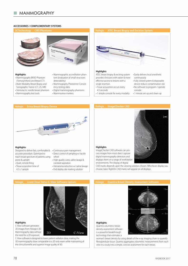
RADBOOK 201778
◼ MAMMOGRAPHY
Highlights• Mammography BR3D Phantom
(Tomosynthesis and Breast CT)• Multi-Modality Breast Biopsy and
Sonographic Trainer (CT, US, MR)• Stereotactic needle breast phantom • Mammography test tools
• Mammographic accreditation phan-tom (evaluation of small structures detectability)
• Mammography Phototimer Consist-ency testing slabs
• Digital mammography phantoms• Mammoview markers
ACCESSORIES / COMPLEMENTARY SYSTEMS
· CIRS PhantomsGCTechnology
· Lower Dose Tomo (C-View Software Option)Hologic
HighlightsC-View software generates2D images from Hologic’s 3DMammography data withoutthe need for a 2D exposure.C-View software is designed to lower patient radiation dose, making the3D mammography dose comparable to a 2D only exam while maintaining allthe clinical benefits and superior image quality of 3D.
· ImageChecker CADHologic
HighlightsImageChecker CAD software can pro-cess images from most direct capturedigital mammography detectors anddisplays them on a range of workstationenvironments. The display of digitalCAD marks depends upon the viewing solution chosen. Whichever display youchoose, basic RightOn CAD marks will appear on all displays.
· ATEC Breast Biopsy and Excision SystemHologic
HighlightsATEC breast biopsy & excising systemprovides clinicians with easier & moreeffective access to lesions with asingle insertion.• Tissue acquisition occurs every
4.5 seconds• 1 simple console for every modality
• Easily delivers local anesthetic continuously
• Fully closed system & disposable device reduce contamination risk
• No software to program / operate console
• 1 minute set-up and clean-up
· Eviva Breast Biopsy DeviceHologic
HighlightsDesigned to deliver fast, comfortable &accurate procedure. Optimized toreach broad spectrum of patients usingprone & upright• Quiet, remote firing• Tissue acquisition time of
4.5 s / sample
• Continuous pain management• Direct control of sampling w / tactile
wheel• High-quality cores, saline lavage &
constant aspiration• Hematoma reduction w / saline lavage• End deploy site marking solution
· Quantra Breast Density Assessment SoftwareHologic
HighlightsQuantra volumetric breastdensity assessment softwareis a powerful breakthroughtechnology that estimates awoman’s breast density by using details of the x-ray imaging chain to quantifyfibroglandular tissue. Quantra aggregates volumetric measurements from eachview in a study into a simple, concise assessment for each breast.
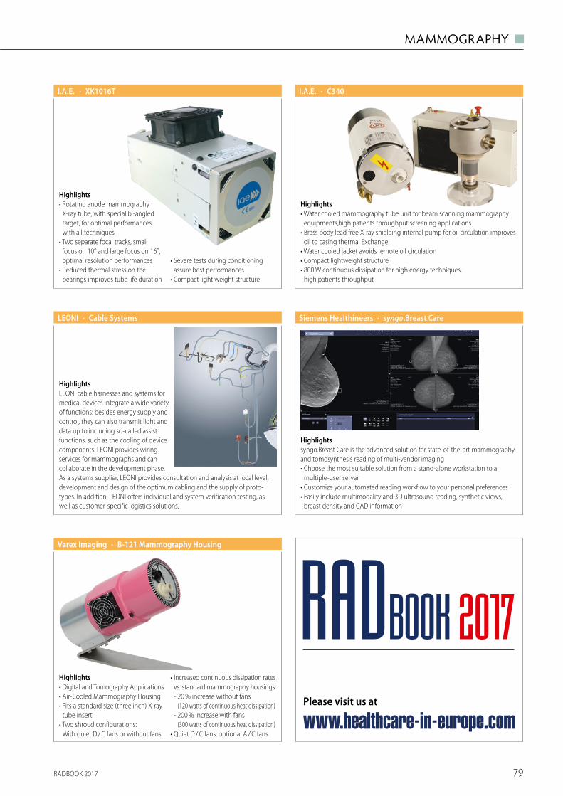
RADBOOK 2017 79
MAMMOGRAPHY ◼
Highlights• Rotating anode mammography
X-ray tube, with special bi-angled target, for optimal performances with all techniques
• Two separate focal tracks, small focus on 10° and large focus on 16°, optimal resolution performances
• Reduced thermal stress on the bearings improves tube life duration
• Severe tests during conditioning assure best performances
• Compact light weight structure
· XK1016TI.A.E. · C340I.A.E.
Highlights• Water cooled mammography tube unit for beam scanning mammography
equipments,high patients throughput screening applications• Brass body lead free X-ray shielding internal pump for oil circulation improves
oil to casing thermal Exchange• Water cooled jacket avoids remote oil circulation• Compact lightweight structure• 800 W continuous dissipation for high energy techniques,
high patients throughput
HighlightsLEONI cable harnesses and systems for medical devices integrate a wide variety of functions: besides energy supply and control, they can also transmit light and data up to including so-called assist functions, such as the cooling of device components. LEONI provides wiring services for mammographs and can collaborate in the development phase. As a systems supplier, LEONI provides consultation and analysis at local level, development and design of the optimum cabling and the supply of proto-types. In addition, LEONI offers individual and system verification testing, as well as customer-specific logistics solutions.
· Cable SystemsLEONI
Highlightssyngo.Breast Care is the advanced solution for state-of-the-art mammography and tomosynthesis reading of multi-vendor imaging• Choose the most suitable solution from a stand-alone workstation to a
multiple-user server• Customize your automated reading workflow to your personal preferences• Easily include multimodality and 3D ultrasound reading, synthetic views,
breast density and CAD information
· syngo.Breast CareSiemens Healthineers
· B-121 Mammography HousingVarex Imaging
Highlights• Digital and Tomography Applications• Air-Cooled Mammography Housing• Fits a standard size (three inch) X-ray
tube insert• Two shroud configurations:
With quiet D / C fans or without fans
• Increased continuous dissipation rates vs. standard mammography housings
- 20 % increase without fans (120 watts of continuous heat dissipation)
- 200 % increase with fans (300 watts of continuous heat dissipation)
• Quiet D / C fans; optional A / C fans
Please visit us at
www.healthcare-in-europe.com
RADBOOK 2017
RADBOOK 201780
◼ R/F FILM-SCREEN
R / F Film-Screen
Bucky
Fluoroscopy
Mobile X-Ray
Accessories / Complementary Systems
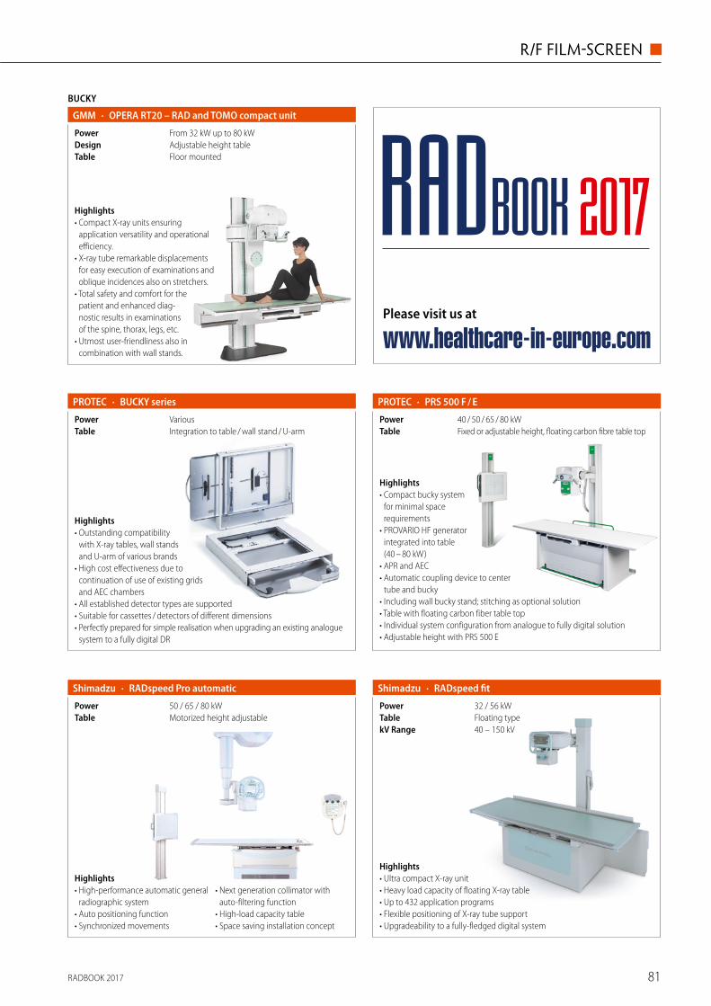
RADBOOK 2017 81
R/F FILM-SCREEN ◼
BUCKY
· OPERA RT20 – RAD and TOMO compact unitGMM
Floor mountedTableAdjustable height tableDesignFrom 32 kW up to 80 kWPower
Highlights• Compact X-ray units ensuring
application versatility and operational efficiency.
• X-ray tube remarkable displacements for easy execution of examinations and oblique incidences also on stretchers.
• Total safety and comfort for the patient and enhanced diag-nostic results in examinations of the spine, thorax, legs, etc.
• Utmost user-friendliness also in combination with wall stands.
· BUCKY series
Integration to table / wall stand / U-armTableVariousPower
Highlights• Outstanding compatibility
with X-ray tables, wall stands and U-arm of various brands
• High cost effectiveness due to continuation of use of existing grids and AEC chambers
• All established detector types are supported • Suitable for cassettes / detectors of different dimensions • Perfectly prepared for simple realisation when upgrading an existing analogue
system to a fully digital DR
PROTEC
Highlights• Compact bucky system
for minimal space requirements
• PROVARIO HF generator integrated into table (40 – 80 kW)
• APR and AEC • Automatic coupling device to center
tube and bucky • Including wall bucky stand; stitching as optional solution • Table with floating carbon fiber table top • Individual system configuration from analogue to fully digital solution• Adjustable height with PRS 500 E
· PRS 500 F / EPROTEC
Fixed or adjustable height, floating carbon fibre table topTable40 / 50 / 65 / 80 kWPower
· RADspeed fitShimadzu
40 – 150 kVkV RangeFloating typeTable32 / 56 kWPower
Highlights• Ultra compact X-ray unit• Heavy load capacity of floating X-ray table• Up to 432 application programs• Flexible positioning of X-ray tube support• Upgradeability to a fully-fledged digital system
· RADspeed Pro automaticShimadzu
Motorized height adjustableTable50 / 65 / 80 kWPower
Highlights• High-performance automatic general
radiographic system• Auto positioning function• Synchronized movements
• Next generation collimator with auto-filtering function
• High-load capacity table• Space saving installation concept
Please visit us at
www.healthcare-in-europe.com
RADBOOK 2017
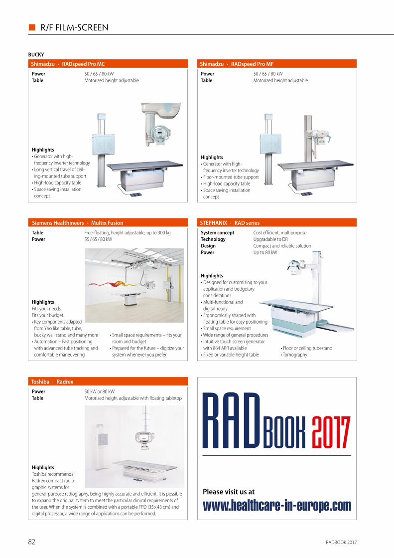
RADBOOK 201782
◼ R/F FILM-SCREEN
BUCKY
· RADspeed Pro MCShimadzu
Motorized height adjustableTable50 / 65 / 80 kWPower
Highlights• Generator with high-
frequency inverter technology• Long vertical travel of ceil-
ing-mounted tube support• High-load capacity table• Space saving installation
concept
· RADspeed Pro MFShimadzu
Motorized height adjustableTable50 / 65 / 80 kWPower
Highlights• Generator with high-
frequency inverter technology• Floor-mounted tube support• High-load capacity table• Space saving installation
concept
HighlightsFits your needs. Fits your budget. • Key components adapted
from Ysio like table, tube, bucky wall stand and many more
• Automation – Fast positioning with advanced tube tracking and comfortable maneuvering
• Small space requirements – fits your room and budget
• Prepared for the future – digitize your system whenever you prefer
· Multix Fusion Siemens Healthineers
55 / 65 / 80 kWPowerFree-floating, height adjustable, up to 300 kgTable
Highlights• Designed for customising to your
application and budgetary considerations
• Multi-functional and digital-ready
• Ergonomically shaped with floating table for easy positioning
• Small space requirement• Wide range of general procedures • Intuitive touch screen generator
with 864 APR available• Fixed or variable height table
• Floor or ceiling tubestand• Tomography
· RAD seriesSTEPHANIX
Up to 80 kWPowerCompact and reliable solutionDesignUpgradable to DRTechnologyCost efficient, multipurposeSystem concept
HighlightsToshiba recommends Radrex compact radio-graphic systems for general-purpose radiography, being highly accurate and efficient. It is possible to expand the original system to meet the particular clinical requirements of the user. When the system is combined with a portable FPD (35 x 43 cm) and digital processor, a wide range of applications can be performed.
· RadrexToshiba
Motorized height adjustable with floating tabletopTable50 kW or 80 kWPower
Please visit us at
www.healthcare-in-europe.com
RADBOOK 2017
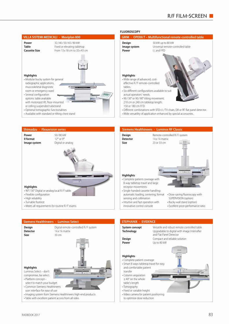
RADBOOK 2017 83
R/F FILM-SCREEN ◼
FLUOROSCOPY
· OPERA T – Multifunctional remote-controlled tableGMM
I.I. and FPDPowerUniversal remote-controlled tableImage system50 kW up to 80 kWDesign
Highlights• Wide range of advanced, cost-
effective R / F remote-controlled tables.
• Six different configurations available to suit actual operators' needs.
• 90 / 30° or 90 / 90° tilting movement; 210 cm or 240 cm tabletop length; 150 or 180 cm FFD.
• Different combinations with SFD-I.I. / TV chain, DR or RF flat panel detector.• Wide versatility of application enhanced by special accessories.
· Flexavision seriesShimadzu
Digital or analogImage system12" or 9"II format50 / 80 kWPower
Highlights• 90° / 30° Digital or analog local R / F table • Flexible configuration • High reliability • Turnable footrest• Meets all requirements for routine R / F exams
Highlights• Complete patient coverage with
8-way tabletop travel and large receptor movements
• Single-handed cassette handling: automatic loading, centering, format sensing and collimation
• Intuitive and fast operation with innovative control console
• Dose-saving fluoroscopy with SUPERVISION (option)
• Bucky wall stand (option) • Excellent price-performance ratio
· Luminos RF Classic Siemens Healthineers
23 or 33 cmSize1 k x 1 k matrixDetectorRemote-controlled R / F systemDesign
HighlightsLuminos Select – don’t compromise, be select.• Platform concept –
select to match your budget• Common Siemens Healthineers
user interface for ease of use• Imaging system from Siemens Healthineers high-end products• Table with excellent patient access from all sides
· Luminos SelectSiemens Healthineers
33 cmSize1 k x 1 k matrixDetectorDigital remote-controlled R / F systemDesign
Highlights• Complete patient coverage• Smart 8 ways tabletop travel for easy
and comfortable patient transfer
• Column angulation ± 40° on the whole table's length
• Tomography• Fixed or variable height• Video camera for patient positioning
to optimize dose reduction
· EVIDENCESTEPHANIX
Up to 80 kWPowerCompact and reliable solutionDesign
Upgradable to digital with image Intensifier and Flat Panel Detector
TechnologyVersatile and robust remote controlled tableSystem concept
· Moviplan 800VILLA SISTEMI MEDICALI
From 13 x 18 cm to 35 x 43 cmCassette SizeFixed or elevating tabletopTable32 / 40 / 50 / 65 / 80 kWPower
Highlights• Modular bucky system for general
radio graphic applications, muscoskeletal diagnostic room or emergency ward
• Several configuration options: table available with motorized lift, floor-mounted or ceiling suspended tubestand
• Optional tomographic functionalities• Available with standard or tilting chest stand
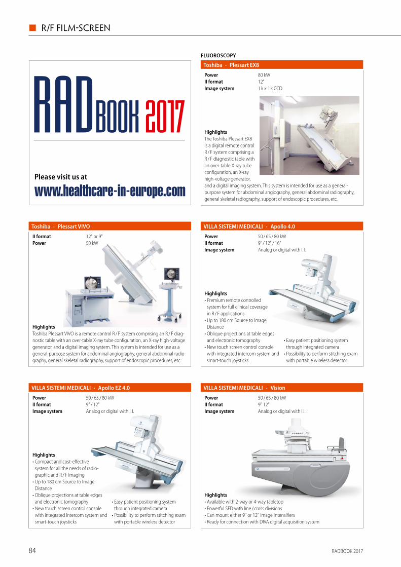
RADBOOK 201784
◼ R/F FILM-SCREEN
HighlightsThe Toshiba Plessart EX8 is a digital remote control R / F system comprising a R / F diagnostic table with an over-table X-ray tube configuration, an X-ray high-voltage generator, and a digital imaging system. This system is intended for use as a general- purpose system for abdominal angiography, general abdominal radiography, general skeletal radiography, support of endoscopic procedures, etc.
FLUOROSCOPY
· Plessart EX8Toshiba
1 k x 1 k CCDImage system12"II format80 kWPower
HighlightsToshiba Plessart VIVO is a remote control R / F system comprising an R / F diag-nostic table with an over-table X-ray tube configuration, an X-ray high-voltage generator, and a digital imaging system. This system is intended for use as a general-purpose system for abdominal angiography, general abdominal radio-graphy, general skeletal radiography, support of endoscopic procedures, etc.
· Plessart VIVOToshiba
50 kWPower12" or 9"II format
· Apollo 4.0VILLA SISTEMI MEDICALI
Analog or digital with I. I.Image system9" / 12" / 16"II format50 / 65 / 80 kWPower
Highlights• Premium remote controlled
system for full clinical coverage in R / F applications
• Up to 180 cm Source to Image Distance
• Oblique projections at table edges and electronic tomography
• New touch screen control console with integrated intercom system and smart-touch joysticks
• Easy patient positioning system through integrated camera
• Possibility to perform stitching exam with portable wireless detector
· Apollo EZ 4.0VILLA SISTEMI MEDICALI
Analog or digital with I. I.Image system9" / 12"II format50 / 65 / 80 kWPower
Highlights• Compact and cost-effective
s ystem for all the needs of radio-graphic and R / F imaging
• Up to 180 cm Source to Image Distance
• Oblique projections at table edges and electronic tomography
• New touch screen control console with integrated intercom system and smart-touch joysticks
• Easy patient positioning system through integrated camera
• Possibility to perform stitching exam with portable wireless detector
· VisionVILLA SISTEMI MEDICALI
Analog or digital with I.I.Image system9" 12"II format50 / 65 / 80 kWPower
Highlights• Available with 2-way or 4-way tabletop • Powerful SFD with line / cross divisions • Can mount either 9" or 12" Image Intensifiers • Ready for connection with DIVA digital acquisition system
Please visit us at
www.healthcare-in-europe.com
RADBOOK 2017
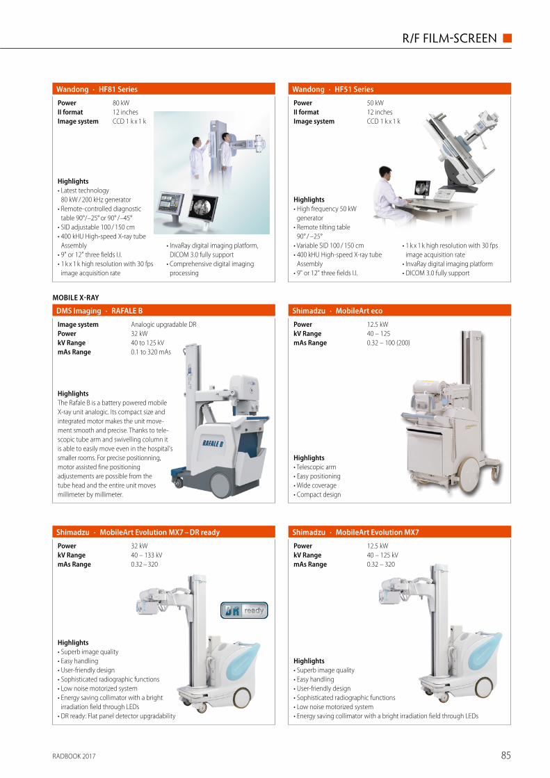
RADBOOK 2017 85
R/F FILM-SCREEN ◼
· HF51 SeriesWandong
CCD 1 k x 1 kImage system12 inchesII format50 kWPower
Highlights• High frequency 50 kW
generator• Remote tilting table
90° / –25°• Variable SID 100 / 150 cm• 400 kHU High-speed X-ray tube
Assembly• 9" or 12" three fields I.I.
• 1 k x 1 k high resolution with 30 fps image acquisition rate
• InvaRay digital imaging platform• DICOM 3.0 fully support
Highlights• Telescopic arm • Easy positioning • Wide coverage • Compact design
· MobileArt ecoShimadzu
0.32 – 100 (200)mAs Range40 – 125kV Range12.5 kWPower
· MobileArt Evolution MX7 – DR readyShimadzu
0.32 – 320 mAs Range40 – 133 kVkV Range32 kWPower
Highlights• Superb image quality• Easy handling• User-friendly design• Sophisticated radiographic functions• Low noise motorized system• Energy saving collimator with a bright
irradiation field through LEDs• DR ready: Flat panel detector upgradability
· MobileArt Evolution MX7Shimadzu
0.32 – 320mAs Range40 – 125 kVkV Range12.5 kWPower
Highlights• Superb image quality• Easy handling• User-friendly design• Sophisticated radiographic functions• Low noise motorized system• Energy saving collimator with a bright irradiation field through LEDs
· HF81 SeriesWandong
CCD 1 k x 1 kImage system12 inchesII format80 kWPower
Highlights• Latest technology
80 kW / 200 kHz generator• Remote-controlled diagnostic
table 90°/–25° or 90° /–45°• SID adjustable 100 / 150 cm• 400 kHU High-speed X-ray tube
Assembly• 9" or 12" three fields I.I.• 1 k x 1 k high resolution with 30 fps
image acquisition rate
• InvaRay digital imaging platform, DICOM 3.0 fully support
• Comprehensive digital imaging processing
MOBILE X-RAY
· RAFALE BDMS Imaging
0.1 to 320 mAsmAs Range40 to 125 kVkV Range32 kWPowerAnalogic upgradable DRImage system
HighlightsThe Rafale B is a battery powered mobile X-ray unit analogic. Its compact size and integrated motor makes the unit move-ment smooth and precise. Thanks to tele-scopic tube arm and swivelling column it is able to easily move even in the hospital's smaller rooms. For precise positionning, motor assisted fine positioning adjustements are possible from the tube head and the entire unit moves millimeter by millimeter.
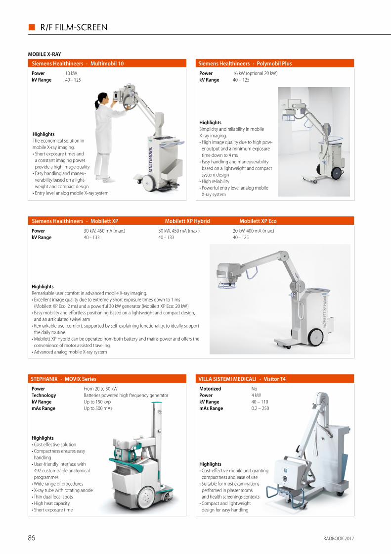
RADBOOK 201786
◼ R/F FILM-SCREEN
HighlightsSimplicity and reliability in mobile X-ray imaging. • High image quality due to high pow-
er output and a minimum exposure time down to 4 ms
• Easy handling and maneuverability based on a lightweight and compact system design
• High reliability• Powerful entry level analog mobile
X-ray system
· Polymobil PlusSiemens Healthineers
16 kW (optional 20 kW)Power40 – 125kV Range
MOBILE X-RAY
· Multimobil 10 Siemens Healthineers
40 – 125kV Range10 kWPower
HighlightsThe economical solution in mobile X-ray imaging. • Short exposure times and
a constant imaging power provide a high image quality
• Easy handling and maneu-verability based on a light-weight and compact design
• Entry level analog mobile X-ray system
HighlightsRemarkable user comfort in advanced mobile X-ray imaging. • Excellent image quality due to extremely short exposure times down to 1 ms
(Mobilett XP Eco: 2 ms) and a powerful 30 kW generator (Mobilett XP Eco: 20 kW)• Easy mobility and effortless positioning based on a lightweight and compact design,
and an articulated swivel arm • Remarkable user comfort, supported by self-explaining functionality, to ideally support
the daily routine • Mobilett XP Hybrid can be operated from both battery and mains power and offers the
convenience of motor assisted traveling • Advanced analog mobile X-ray system
Mobilett XP Mobilett XP Hybrid Mobilett XP Eco Siemens Healthineers ·
40 – 133 40 – 133 40 – 125kV Range30 kW, 450 mA (max.) 30 kW, 450 mA (max.) 20 kW, 400 mA (max.)Power
Highlights• Cost effective solution • Compactness ensures easy
handling• User-friendly interface with
492 customizable anatomical programmes
• Wide range of procedures• X-ray tube with rotating anode• Thin dual focal spots• High heat capacity• Short exposure time
· MOVIX Series STEPHANIX
Up to 500 mAsmAs RangeUp to 150 kVpkV RangeBatteries powered high frequency generatorTechnologyFrom 20 to 50 kWPower
· Visitor T4VILLA SISTEMI MEDICALI
0.2 – 250mAs Range40 – 110kV Range4 kWPowerNoMotorized
Highlights• Cost-effective mobile unit granting
compactness and ease of use• Suitable for most examinations
performed in plaster rooms and health screenings contexts
• Compact and lightweight design for easy handling
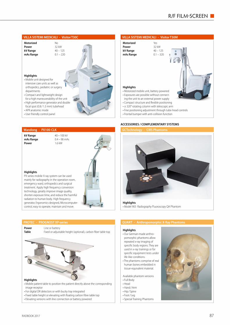
RADBOOK 2017 87
R/F FILM-SCREEN ◼
HighlightsPX series mobile X-ray system can be used mainly for radiography in the operation room, emergency ward, orthopedics and surgical treatment. Apply high frequency conversion technology, greatly improve image quality, shorten exposure time, and reduce the harmful radiation to human body. High frequency generator, Ergonomics designed, Microcomputer- control, easy to operate, maintain and move.
· PX100-CLKWandong
1.6 kWPower0.4 ~ 98 mAsmAs Range40 ~ 100 kVkV Range
Highlights• Model 903 Radiography Fluoroscopy QA Phantom
ACCESSORIES / COMPLEMENTARY SYSTEMS
· CIRS PhantomsGCTechnology
· PROGNOST XP-series
Fixed or adjustable height (optional), carbon fiber table topTableLine or batteryPower
Highlights• Mobile patient table to position the patient directly above the corresponding
image receptor • For digital DR detectors or with bucky tray integrated • Fixed table height or elevating with floating carbon fibre table top • Elevating versions with line connection or battery powered
PROTEC QUART
Highlights• Our German-made anthro-
pomorphic phantoms allow repeated x-ray imaging of specific body regions. They are used in x-ray trainings or for specific equipment tests under life-like conditions.
• The phantoms comprise of real human bones embedded in tissue-equivalent material.
Available phantom versions• Full Body• Head• Hand / Arm• Hip / Spine• Foot / Leg• Special Training Phantoms
· Anthropomorphic X-Ray Phantoms
· Visitor T30MVILLA SISTEMI MEDICALI
0.1 – 320mAs Range40 – 125kV Range32 kWPowerYesMotorized
Highlights• Motorized mobile unit, battery powered• Exposures are possible without connect-
ing the unit to an external power supply• Compact structure and flexible positioning• ± 320° rotating column with telescopic arm• Fine positioning adjustment through tube-head controls• Frontal bumper with anti-collision function
· Visitor T30CVILLA SISTEMI MEDICALI
0.1 – 220mAs Range40 – 125kV Range32 kWPowerNoMotorized
Highlights• Mobile unit designed for
intensive care units as well as orthopedics, pediatric or surgery departments
• Compact and lightweight design for a high maneuverability of the unit
• High performance generator and double focal spot (0.8 / 1.3 mm) tubehead
• APR anatomic mode • User friendly control panel
RADBOOK 201788
◼ R/F DIGITAL
MECALL
A Division of Philips Healthcare
R / F Digital
CR
DR
DR Retrofi t
Mobile DR
Flatpanel Fluoro
Accessories / Complementary Systems
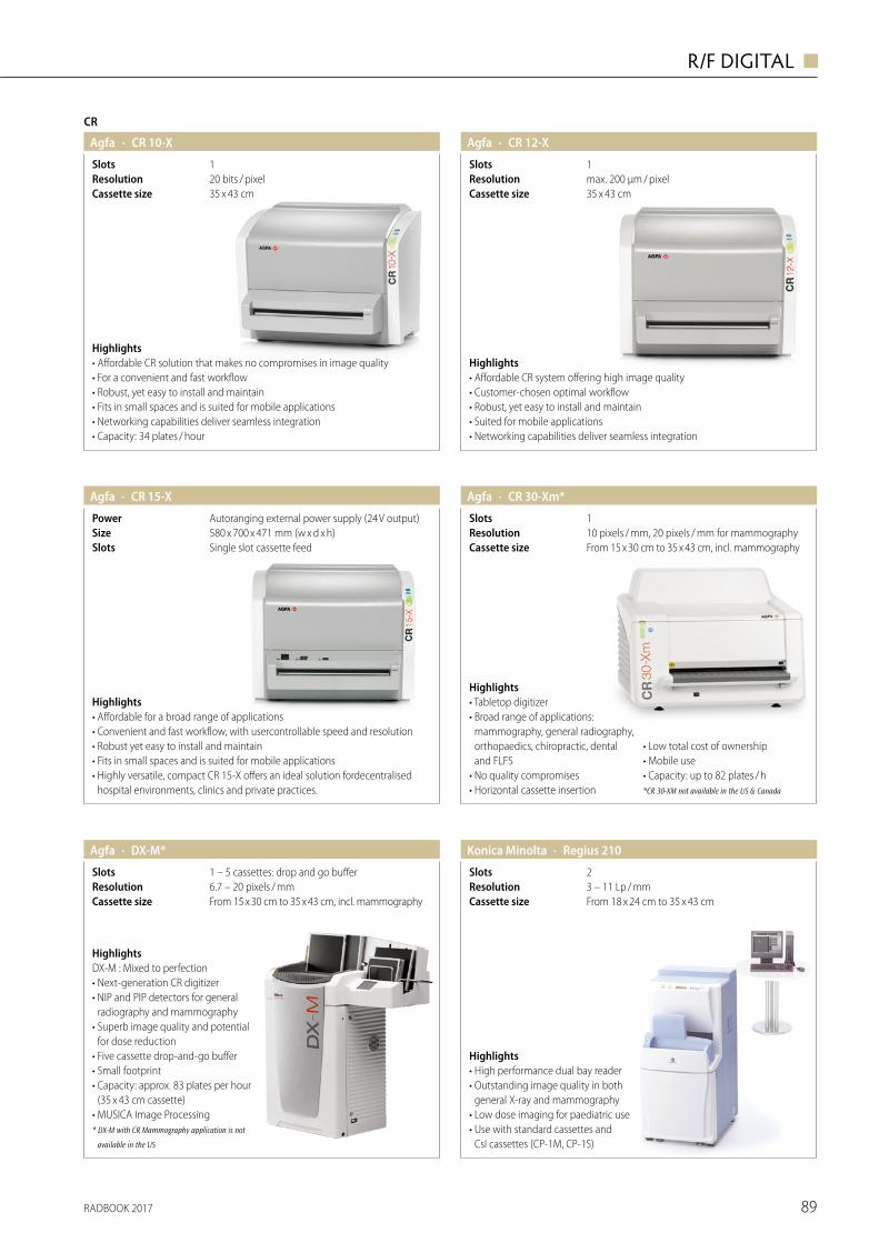
RADBOOK 2017 89
R/F DIGITAL ◼
CR
· CR 10-XAgfa
35 x 43 cmCassette size20 bits / pixelResolution1Slots
Highlights• Affordable CR solution that makes no compromises in image quality• For a convenient and fast workflow• Robust, yet easy to install and maintain• Fits in small spaces and is suited for mobile applications• Networking capabilities deliver seamless integration• Capacity: 34 plates / hour
· CR 15-XAgfa
Single slot cassette feedSlots580 x 700 x 471 mm (w x d x h)SizeAutoranging external power supply (24 V output)Power
Highlights• Affordable for a broad range of applications• Convenient and fast workflow, with usercontrollable speed and resolution• Robust yet easy to install and maintain• Fits in small spaces and is suited for mobile applications• Highly versatile, compact CR 15-X offers an ideal solution fordecentralised
hospital environments, clinics and private practices.
Agfa · CR 12-X
35 x 43 cmCassette sizemax. 200 μm / pixelResolution1Slots
Highlights• Affordable CR system offering high image quality• Customer-chosen optimal workflow• Robust, yet easy to install and maintain• Suited for mobile applications• Networking capabilities deliver seamless integration
Agfa · CR 30-Xm*
From 15 x 30 cm to 35 x 43 cm, incl. mammographyCassette size10 pixels / mm, 20 pixels / mm for mammographyResolution1Slots
Highlights• Tabletop digitizer• Broad range of applications:
mammography, general radiography, orthopaedics, chiropractic, dental and FLFS
• No quality compromises• Horizontal cassette insertion
• Low total cost of ownership• Mobile use• Capacity: up to 82 plates / h*CR 30-XM not available in the US & Canada
HighlightsDX-M : Mixed to perfection• Next-generation CR digitizer• NIP and PIP detectors for general
radiography and mammography• Superb image quality and potential
for dose reduction• Five cassette drop-and-go buffer• Small footprint• Capacity: approx. 83 plates per hour
(35 x 43 cm cassette)• MUSICA Image Processing* DX-M with CR Mammography application is not
available in the US
· DX-M*Agfa
From 15 x 30 cm to 35 x 43 cm, incl. mammographyCassette size6.7 – 20 pixels / mmResolution1 – 5 cassettes: drop and go bufferSlots
MECALL
A Division of Philips Healthcare
R / F Digital
CR
DR
DR Retrofi t
Mobile DR
Flatpanel Fluoro
Accessories / Complementary Systems
· Regius 210Konica Minolta
From 18 x 24 cm to 35 x 43 cmCassette size3 – 11 Lp / mmResolution2Slots
Highlights• High performance dual bay reader• Outstanding image quality in both
general X-ray and mammography• Low dose imaging for paediatric use• Use with standard cassettes and
CsI cassettes (CP-1M, CP-1S)
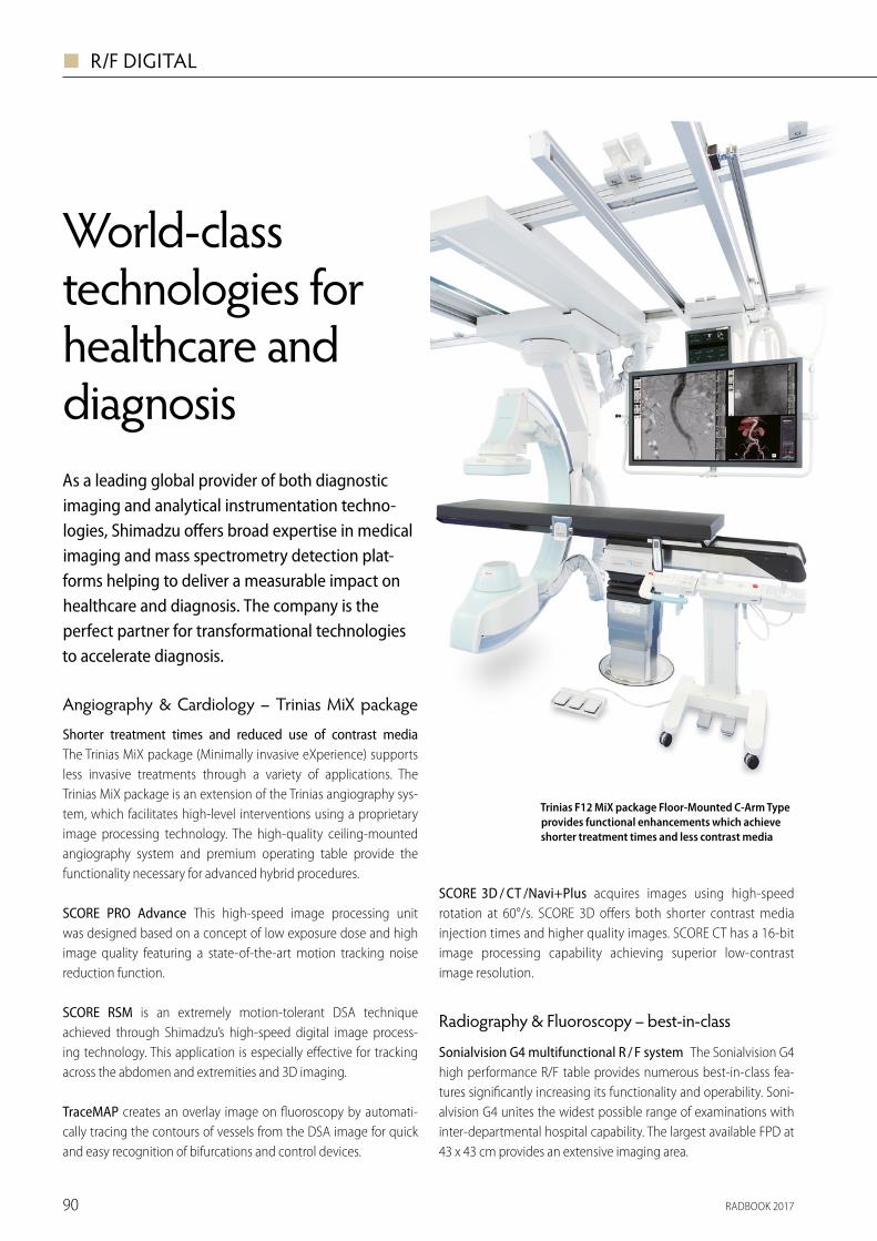
RADBOOK 201790
◼ R/F DIGITAL
SUREengine-Advance is a leading-edge digital image processing technology and ensures extremely clear fluoroscopy and radiography images.
SLOT Advance provides high accuracy images with long fields of view, such as for full spine or full leg images, taken with a minimal X-ray dose.
T-smart generates even clearer tomosyn-thesis images suppressing the artefacts around metal objects even further.
Auto-stitching radiography covers the entire lower extremities or entire spine and links the settings made on the X-ray tube with the Bucky table or Bucky stand with subsequent automatic image stitching.
Dedicated tomosynthesis workstation allows parallel processing of data with examinations increasing the throughput and reducing the stress on patients.
MobileDaRt Evolution MX7 mobile X-ray system
“Category Leader” in operator satisfac-tion survey The Shimadzu MobileDaRt Evolution digital mobile X-ray system has been favorably received thanks to its excellent maneuverability, reliability, per-formance and ease of positioning. In a
customer satisfaction sur-vey by KLAS Research, a U.S. based Research
Firm, this system has been named “Category Leader” in the field of digital mobile X-ray
systems, for three out of the last four years (with
the first award presented in 2011). This is truly an internationally top-rated product.
More convenient design better satis fies operator needs The MX7 series’ image display units feature a new, high-resolution 17-inch LCD monitor. Additionally, other new features improve the convenience of routine use such as storage space for smal ler items as well as grooves in the console top sidewalls, convenient for installing a cover over the FPD unit while keeping it stabilized. The MX7 series offers a wide range of FPD types to match individual clinical requirements.
Scatter Correction software generates a scatter model which is subsequently subtracted from the image. Whereas a grid physically reduc-es scatter and increases image
VISIT US AT ECR 2017 IN VIENNA, AUSTRIA · 2-5 MARCH EXPO X2, STAND 19Further information: Shimadzu Europawww.shimadzu-medical.eu
World-class technologies for healthcare and diagnosisAs a leading global provider of both diagnostic imaging and analytical instrumentation techno-logies, Shimadzu offers broad expertise in medical imaging and mass spectrometry detection plat-forms helping to deliver a measurable impact on healthcare and diagnosis. The company is the perfect partner for transformational technologies to accelerate diagnosis.
Angiography & Cardiology – Trinias MiX package
Shorter treatment times and reduced use of contrast mediaThe Trinias MiX package (Minimally invasive eXperience) supports less invasive treatments through a variety of applications. The Trinias MiX package is an extension of the Trinias angiography sys-tem, which facilitates high-level interventions using a proprietary image processing technology. The high-quality ceiling-mounted angiography system and premium operating table provide the functionality necessary for advanced hybrid procedures.
SCORE PRO Advance This high-speed image processing unit was designed based on a concept of low exposure dose and high image quality featuring a state-of-the-art motion tracking noise reduction function.
SCORE RSM is an extremely motion-tolerant DSA technique achieved through Shimadzu’s high-speed digital image process-ing technology. This application is especially effective for tracking across the abdomen and extremities and 3D imaging.
TraceMAP creates an overlay image on fluoroscopy by automati-cally tracing the contours of vessels from the DSA image for quick and easy recognition of bifurcations and control devices.
SCORE 3D / CT /Navi+Plus acquires images using high-speed rotation at 60°/s. SCORE 3D offers both shorter contrast media injection times and higher quality images. SCORE CT has a 16-bit image processing capability achieving superior low-contrast image resolution.
Radiography & Fluoroscopy – best-in-class
Sonialvision G4 multifunctional R / F system The Sonialvision G4 high performance R/F table provides numerous best-in-class fea-tures significantly increasing its functionality and operability. Soni-alvision G4 unites the widest possible range of examinations with inter-departmental hospital capability. The largest available FPD at 43 x 43 cm provides an extensive imaging area.
contrast, the software mimics this process virtually. The result is an image with reduced scatter and increased contrast.
Opescope Acteno surgical C-arm system
High operability and image quality Opescope Acteno combines high image quality with ease of use. The system enables free and easy positioning and optimal per-formance to meet the demands of operation and emergency rooms. The fully counter-bal-anced C-arm provides extra-light and extra quick C-arm movements and positioning.
RSM technology minimizes motion artifacts in DSA Adding the unique RSM filtering process to DSA images will elimi-nate the motion artifacts.
Touch Focus is the brand-new innovative technology for Opescope Acteno to opti-mize the image brightness focused to the ROI at real-time during fluoroscopy.
Trinias F12 MiX package Floor-Mounted C-Arm Type provides functional enhancements which achieve shorter treatment times and less contrast media
General Radiography – RADspeed Pro EDGERADspeed Pro EDGE combines the advan-tages of the well-known RADspeed Pro series in one instrument – for example the highly praised easy operability or the com-prehensive measures to reduce exposure levels. Numerous further functionalities can be added.
Tomosynthesis allows to easily obtain multiple digital cross-section images from a single linear tomography scan.
Dual-energy subtraction utilizes the dif-ference in X-ray absorption levels of bones and soft tissue to generate separate imag-es, which is useful for diagnoses in the chest area, such as lung cancer.
Sonialvision G4: Multifunctional R/F system
The new MX7 digital mobile X-ray systems combine clinical feedback with the most advanced techno-logies to meet the highest operator requirements
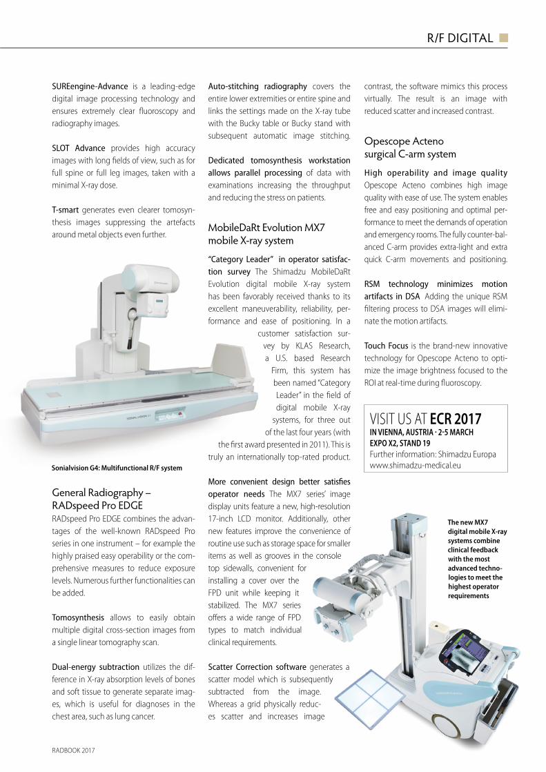
RADBOOK 2017 91
R/F DIGITAL ◼
SUREengine-Advance is a leading-edge digital image processing technology and ensures extremely clear fluoroscopy and radiography images.
SLOT Advance provides high accuracy images with long fields of view, such as for full spine or full leg images, taken with a minimal X-ray dose.
T-smart generates even clearer tomosyn-thesis images suppressing the artefacts around metal objects even further.
Auto-stitching radiography covers the entire lower extremities or entire spine and links the settings made on the X-ray tube with the Bucky table or Bucky stand with subsequent automatic image stitching.
Dedicated tomosynthesis workstation allows parallel processing of data with examinations increasing the throughput and reducing the stress on patients.
MobileDaRt Evolution MX7 mobile X-ray system
“Category Leader” in operator satisfac-tion survey The Shimadzu MobileDaRt Evolution digital mobile X-ray system has been favorably received thanks to its excellent maneuverability, reliability, per-formance and ease of positioning. In a
customer satisfaction sur-vey by KLAS Research, a U.S. based Research
Firm, this system has been named “Category Leader” in the field of digital mobile X-ray
systems, for three out of the last four years (with
the first award presented in 2011). This is truly an internationally top-rated product.
More convenient design better satis fies operator needs The MX7 series’ image display units feature a new, high-resolution 17-inch LCD monitor. Additionally, other new features improve the convenience of routine use such as storage space for smal ler items as well as grooves in the console top sidewalls, convenient for installing a cover over the FPD unit while keeping it stabilized. The MX7 series offers a wide range of FPD types to match individual clinical requirements.
Scatter Correction software generates a scatter model which is subsequently subtracted from the image. Whereas a grid physically reduc-es scatter and increases image
VISIT US AT ECR 2017 IN VIENNA, AUSTRIA · 2-5 MARCH EXPO X2, STAND 19Further information: Shimadzu Europawww.shimadzu-medical.eu
World-class technologies for healthcare and diagnosisAs a leading global provider of both diagnostic imaging and analytical instrumentation techno-logies, Shimadzu offers broad expertise in medical imaging and mass spectrometry detection plat-forms helping to deliver a measurable impact on healthcare and diagnosis. The company is the perfect partner for transformational technologies to accelerate diagnosis.
Angiography & Cardiology – Trinias MiX package
Shorter treatment times and reduced use of contrast mediaThe Trinias MiX package (Minimally invasive eXperience) supports less invasive treatments through a variety of applications. The Trinias MiX package is an extension of the Trinias angiography sys-tem, which facilitates high-level interventions using a proprietary image processing technology. The high-quality ceiling-mounted angiography system and premium operating table provide the functionality necessary for advanced hybrid procedures.
SCORE PRO Advance This high-speed image processing unit was designed based on a concept of low exposure dose and high image quality featuring a state-of-the-art motion tracking noise reduction function.
SCORE RSM is an extremely motion-tolerant DSA technique achieved through Shimadzu’s high-speed digital image process-ing technology. This application is especially effective for tracking across the abdomen and extremities and 3D imaging.
TraceMAP creates an overlay image on fluoroscopy by automati-cally tracing the contours of vessels from the DSA image for quick and easy recognition of bifurcations and control devices.
SCORE 3D / CT /Navi+Plus acquires images using high-speed rotation at 60°/s. SCORE 3D offers both shorter contrast media injection times and higher quality images. SCORE CT has a 16-bit image processing capability achieving superior low-contrast image resolution.
Radiography & Fluoroscopy – best-in-class
Sonialvision G4 multifunctional R / F system The Sonialvision G4 high performance R/F table provides numerous best-in-class fea-tures significantly increasing its functionality and operability. Soni-alvision G4 unites the widest possible range of examinations with inter-departmental hospital capability. The largest available FPD at 43 x 43 cm provides an extensive imaging area.
contrast, the software mimics this process virtually. The result is an image with reduced scatter and increased contrast.
Opescope Acteno surgical C-arm system
High operability and image quality Opescope Acteno combines high image quality with ease of use. The system enables free and easy positioning and optimal per-formance to meet the demands of operation and emergency rooms. The fully counter-bal-anced C-arm provides extra-light and extra quick C-arm movements and positioning.
RSM technology minimizes motion artifacts in DSA Adding the unique RSM filtering process to DSA images will elimi-nate the motion artifacts.
Touch Focus is the brand-new innovative technology for Opescope Acteno to opti-mize the image brightness focused to the ROI at real-time during fluoroscopy.
Trinias F12 MiX package Floor-Mounted C-Arm Type provides functional enhancements which achieve shorter treatment times and less contrast media
General Radiography – RADspeed Pro EDGERADspeed Pro EDGE combines the advan-tages of the well-known RADspeed Pro series in one instrument – for example the highly praised easy operability or the com-prehensive measures to reduce exposure levels. Numerous further functionalities can be added.
Tomosynthesis allows to easily obtain multiple digital cross-section images from a single linear tomography scan.
Dual-energy subtraction utilizes the dif-ference in X-ray absorption levels of bones and soft tissue to generate separate imag-es, which is useful for diagnoses in the chest area, such as lung cancer.
Sonialvision G4: Multifunctional R/F system
The new MX7 digital mobile X-ray systems combine clinical feedback with the most advanced techno-logies to meet the highest operator requirements
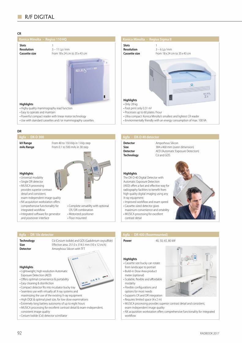
RADBOOK 201792
◼ R/F DIGITAL
CR
· Regius 110 HQKonica Minolta
From 18 x 24 cm to 35 x 43 cmCassette size3 – 11 Lp / mmResolution1Slots
Highlights• Highy quality mammography read function• Easy to operate and maintain• Powerful compact reader with linear motor technology• Use with standard cassettes and / or mammography cassettes
· Regius Sigma IIKonica Minolta
From 18 x 24 cm to 35 x 43 cmCassette size3 – 6 Lp / mmResolution1Slots
Highlights• Only 28 kg • Foot print only 0.31 m2 • Processes up to 60 plates / hour • Ultra compact: Konica Minolta's smallest and lightest CR reader • Environmentally friendly with an energy consumption of max. 100 VA
· DX-D 40 detectorAgfa
CsI and GOSTechnologyAED (Automatic Exposure Detection)Detector384 x 460 mm (outer dimension)SizeAmporhous SiliconDetector
HighlightsThe DX-D 40 Digital Detector with Automatic Exposure Detection (AED) offers a fast and effective way for radiography facilities to benefit from high quality digital imaging using any X-ray equipment: • Improved workflow and exam speed • Cassette-sized detector gives
maximum convenience and ortability • MUSICA processing for excellent
contrast detail
· DX-D 300Agfa
From 0.1 to 500 mAs in 38 stepmAs RangeFrom 40 to 150 kVp in 1 kVp stepkV Range
DR
Highlights• Universal modality• Single DR detector• MUSICA processing
provides superior contrast detail and consistent, exam-independent image quality
• NX acquisition workstation offers comprehensive functionality for integrated workflow
• Integrated software for generator and positioner interface
• Complete versatility with optional CR / DR combination
• Motorized positioner• Floor mounted
Highlights• Cassette size bucky can rotate
from landscape to portrait• Build-in Dose Area product
meter (optional)• Scalable, flexible and affor dable
modality• Flexible configurations and
options for most needs• Supports CR and DR integration• Requires limited space (4 x 2 m)• MUSICA processing provides superior contrast detail and consistent,
exam-independent image quality• NX acquisition workstation offers comprehensive functionality for integrated
workflow
· DR 400 (floormounted)
40, 50, 65, 80 kWPower
Agfa · DR 10s detectorAgfa
Amorphous Silicon with TFTDetectorEffective area: 251.0 x 314.5 mm (10 x 12 inch) SizeCsI (Cesium Iodide) and GOS (Gadolinium oxysulfide)Technology
Highlights• Lightweight, high resolution Automatic
Exposure Detection (AED) • Offers optimal convenience & portability• Easy cleaning & disinfection• Compact detector fits into incubator bucky tray• Seamless use with virtually all X-ray systems and
maximizing the use of the existing X-ray equipment• High DQE & optimal pixel size, for low dose examinations• Extremely long battery autonomy of up to eight hours• MUSICA processing for excellent contrast detail & exam-independent,
consistent image quality• Cesium Iodide (CsI) detector scintillator
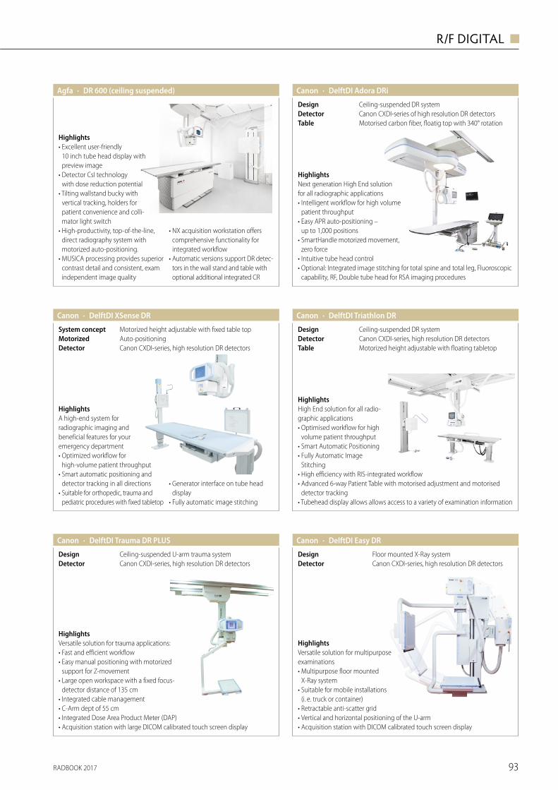
RADBOOK 2017 93
R/F DIGITAL ◼
· DR 600 (ceiling suspended)Agfa
Highlights• Excellent user-friendly
10 inch tube head display with preview image
• Detector CsI technology with dose reduction potential
• Tilting wallstand bucky with vertical tracking, holders for patient convenience and colli-mator light switch
• High-productivity, top-of-the-line, direct radiography system with motorized auto-positioning.
• MUSICA processing provides superior contrast detail and consistent, exam independent image quality
• NX acquisition workstation offers comprehensive functionality for integrated workflow
• Automatic versions support DR detec-tors in the wall stand and table with optional additional integrated CR
· DelftDI Adora DRiCanon
Motorised carbon fiber, floatig top with 340° rotationTableCanon CXDI-series of high resolution DR detectorsDetectorCeiling-suspended DR systemDesign
HighlightsNext generation High End solution for all radiographic applications• Intelligent workflow for high volume
patient throughput• Easy APR auto-positioning –
up to 1,000 positions• SmartHandle motorized movement,
zero force• Intuitive tube head control • Optional: Integrated image stitching for total spine and total leg, Fluoroscopic
capability, RF, Double tube head for RSA imaging procedures
· DelftDI XSense DRCanon
Canon CXDI-series, high resolution DR detectorsDetectorAuto-positioningMotorizedMotorized height adjustable with fixed table topSystem concept
HighlightsA high-end system for radio graphic imaging and beneficial features for your emergency department• Optimized workflow for
high-volume patient throughput• Smart automatic positioning and
detector tracking in all directions• Suitable for orthopedic, trauma and
pediatric procedures with fixed tabletop
• Generator interface on tube head display
• Fully automatic image stitching
· DelftDI Triathlon DR
Motorized height adjustable with floating tabletopTableCanon CXDI-series, high resolution DR detectorsDetectorCeiling-suspended DR systemDesign
HighlightsHigh End solution for all radio-graphic applications• Optimised workflow for high
volume patient throughput• Smart Automatic Positioning• Fully Automatic Image
Stitching• High efficiency with RIS-integrated workflow• Advanced 6-way Patient Table with motorised adjustment and motorised
detector tracking• Tubehead display allows allows access to a variety of examination information
Canon
· DelftDI Trauma DR PLUSCanon
Canon CXDI-series, high resolution DR detectorsDetectorCeiling-suspended U-arm trauma systemDesign
HighlightsVersatile solution for trauma applications:• Fast and efficient workflow• Easy manual positioning with motorized
support for Z-movement• Large open workspace with a fixed focus-
detector distance of 135 cm • Integrated cable management• C-Arm dept of 55 cm• Integrated Dose Area Product Meter (DAP)• Acquisition station with large DICOM calibrated touch screen display
· DelftDI Easy DRCanon
Floor mounted X-Ray systemDesignCanon CXDI-series, high resolution DR detectorsDetector
HighlightsVersatile solution for multi purpose examinations• Multipurpose floor mounted
X-Ray system• Suitable for mobile installations
(i. e. truck or container)• Retractable anti-scatter grid• Vertical and horizontal positioning of the U-arm• Acquisition station with DICOM calibrated touch screen display
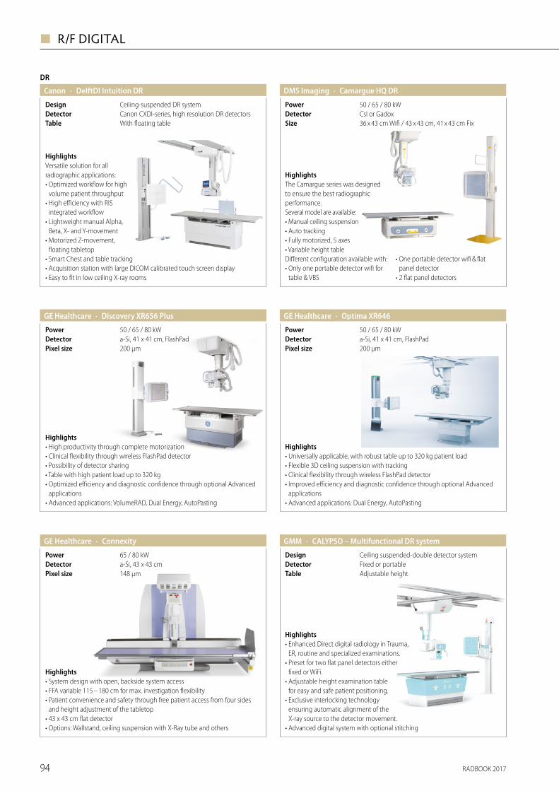
RADBOOK 201794
◼ R/F DIGITAL
DR
· DelftDI Intuition DRCanon
With floating tableTableCanon CXDI-series, high resolution DR detectorsDetectorCeiling-suspended DR systemDesign
HighlightsVersatile solution for all radiographic applications:• Optimized workflow for high
volume patient throughput• High efficiency with RIS
inte grated workflow• Lightweight manual Alpha,
Beta, X- and Y-movement• Motorized Z-movement,
floating tabletop• Smart Chest and table tracking• Acquisition station with large DICOM calibrated touch screen display• Easy to fit in low ceiling X-ray rooms
· Discovery XR656 PlusGE Healthcare
200 μmPixel sizea-Si, 41 x 41 cm, FlashPadDetector50 / 65 / 80 kWPower
Highlights• High productivity through complete motorization• Clinical flexibility through wireless FlashPad detector• Possibility of detector sharing• Table with high patient load up to 320 kg• Optimized efficiency and diagnostic confidence through optional Advanced
applications• Advanced applications: VolumeRAD, Dual Energy, AutoPasting
· Optima XR646GE Healthcare
200 μmPixel sizea-Si, 41 x 41 cm, FlashPadDetector50 / 65 / 80 kWPower
Highlights• Universally applicable, with robust table up to 320 kg patient load• Flexible 3D ceiling suspension with tracking• Clinical flexibility through wireless FlashPad detector• Improved efficiency and diagnostic confidence through optional Advanced
applications• Advanced applications: Dual Energy, AutoPasting
· ConnexityGE Healthcare
148 μmPixel sizea-Si, 43 x 43 cmDetector65 / 80 kWPower
Highlights• System design with open, backside system access• FFA variable 115 – 180 cm for max. investigation flexibility• Patient convenience and safety through free patient access from four sides
and height adjustment of the tabletop• 43 x 43 cm flat detector• Options: Wallstand, ceiling suspension with X-Ray tube and others
· CALYPSO – Multifunctional DR systemGMM
Adjustable heightTableFixed or portable DetectorCeiling suspended-double detector systemDesign
Highlights• Enhanced Direct digital radiology in Trauma,
ER, routine and specialized examinations.• Preset for two flat panel detectors either
fixed or WiFi.• Adjustable height examination table
for easy and safe patient positioning. • Exclusive interlocking technology
ensuring automatic alignment of the X-ray source to the detector movement.
• Advanced digital system with optional stitching
· Camargue HQ DRDMS Imaging
36 x 43 cm Wifi / 43 x 43 cm, 41 x 43 cm FixSizeCsI or GadoxDetector50 / 65 / 80 kWPower
HighlightsThe Camargue series was designed to ensure the best radiographic performance.Several model are available:• Manual ceiling suspension• Auto tracking• Fully motorized, 5 axes• Variable height tableDifferent configuration available with:• Only one portable detector wifi for
table & VBS
• One portable detector wifi & flat panel detector
• 2 flat panel detectors
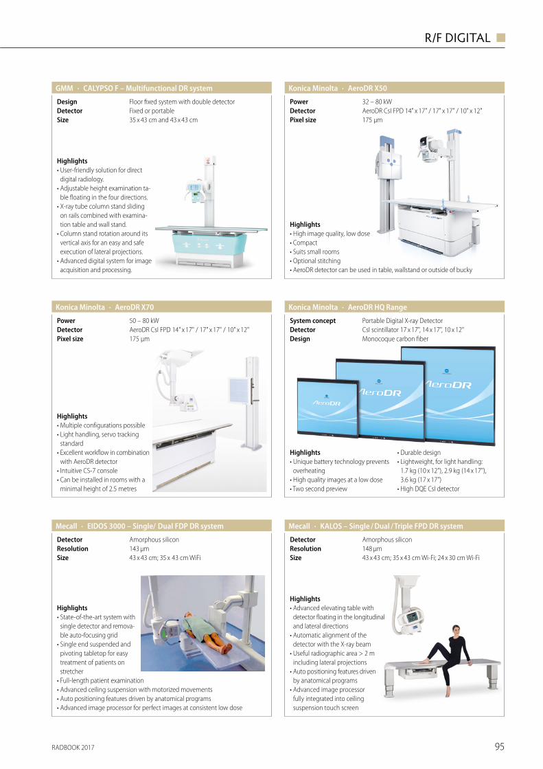
RADBOOK 2017 95
R/F DIGITAL ◼
Highlights• User-friendly solution for direct
digital radiology.• Adjustable height examination ta-
ble floating in the four directions.• X-ray tube column stand sliding
on rails combined with examina-tion table and wall stand.
• Column stand rotation around its vertical axis for an easy and safe execution of lateral projections.
• Advanced digital system for image acquisition and processing.
· CALYPSO F – Multifunctional DR systemGMM
35 x 43 cm and 43 x 43 cmSizeFixed or portableDetectorFloor fixed system with double detectorDesign
· AeroDR X50Konica Minolta
175 µmPixel sizeAeroDR CsI FPD 14" x 17" / 17" x 17" / 10" x 12"Detector32 – 80 kWPower
Highlights• High image quality, low dose• Compact • Suits small rooms• Optional stitching• AeroDR detector can be used in table, wallstand or outside of bucky
· AeroDR X70Konica Minolta
175 µmPixel sizeAeroDR CsI FPD 14" x 17" / 17" x 17" / 10" x 12"Detector50 – 80 kWPower
Highlights• Multiple configurations possible• Light handling, servo tracking
standard• Excellent workflow in combination
with AeroDR detector• Intuitive CS-7 console• Can be installed in rooms with a
minimal height of 2.5 metres
· AeroDR HQ RangeKonica Minolta
Monocoque carbon fiberDesignCsI scintillator 17 x 17", 14 x 17", 10 x 12"DetectorPortable Digital X-ray DetectorSystem concept
Highlights• Unique battery technology prevents
overheating• High quality images at a low dose• Two second preview
• Durable design• Lightweight, for light handling:
1.7 kg (10 x 12"), 2.9 kg (14 x 17"), 3.6 kg (17 x 17")
• High DQE CsI detector
Mecall
Highlights• State-of-the-art system with
single detector and remova-ble auto-focusing grid
• Single end suspended and pivoting tabletop for easy treatment of patients on stretcher
• Full-length patient examination• Advanced ceiling suspension with motorized movements• Auto positioning features driven by anatomical programs• Advanced image processor for perfect images at consistent low dose
· EIDOS 3000 – Single/ Dual FDP DR system
43 x 43 cm; 35 x 43 cm WiFiSize143 µmResolutionAmorphous silicon Detector
· KALOS – Single / Dual / Triple FPD DR systemMecall
43 x 43 cm; 35 x 43 cm Wi-Fi; 24 x 30 cm Wi-FiSize148 µmResolutionAmorphous silicon
Highlights• Advanced elevating table with
detector floating in the longitudinal and lateral directions
• Automatic alignment of the detector with the X-ray beam
• Useful radiographic area > 2 m including lateral projections
• Auto positioning features driven by anatomical programs
• Advanced image processor fully integrated into ceiling suspension touch screen
Detector
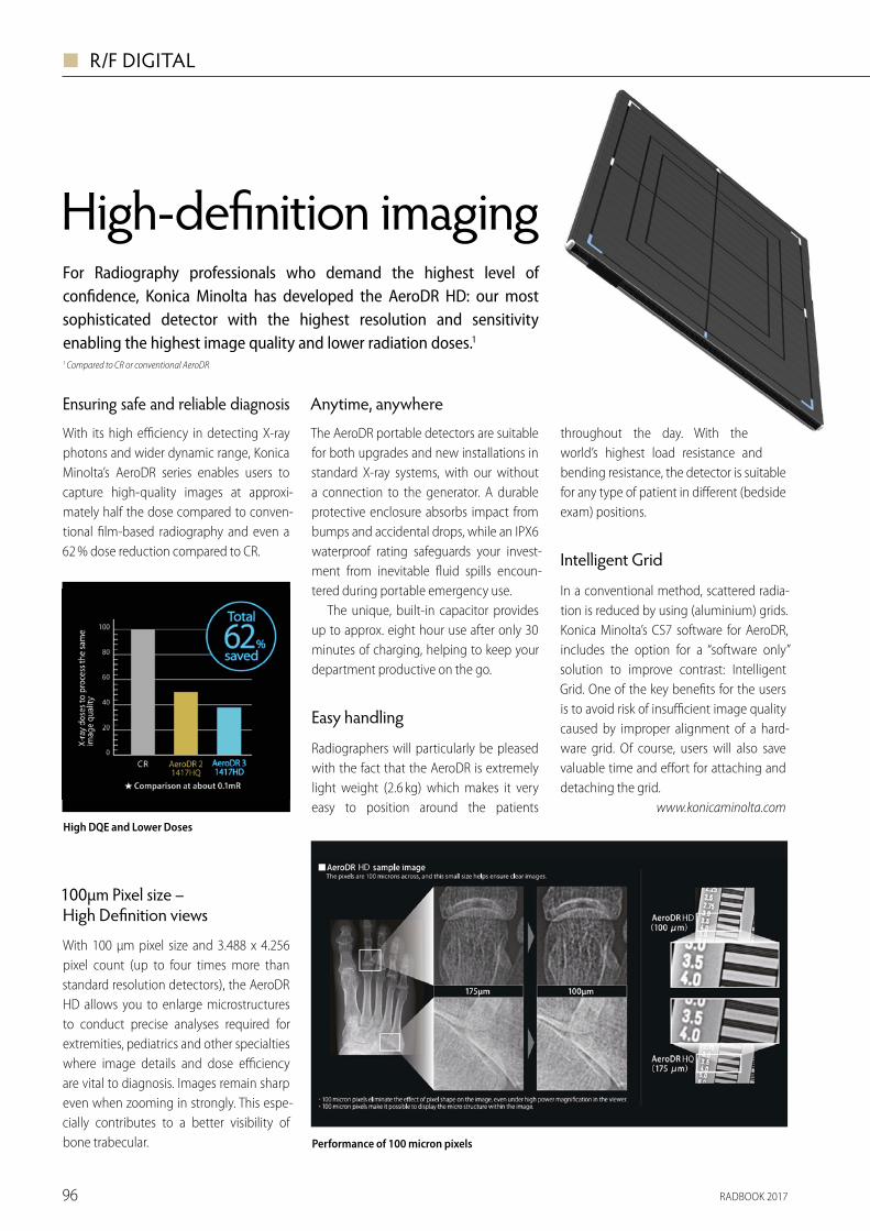
RADBOOK 201796
◼ R/F DIGITAL
Ensuring safe and reliable diagnosis
With its high e� ciency in detecting X-ray photons and wider dynamic range, Konica Minolta’s AeroDR series enables users to capture high-quality images at approxi-mately half the dose compared to conven-tional � lm-based radiography and even a 62 % dose reduction compared to CR.
High-de� nition imaging
Anytime, anywhere
The AeroDR portable detectors are suitable for both upgrades and new installations in standard X-ray systems, with our without a connection to the generator. A durable protective enclosure absorbs impact from bumps and accidental drops, while an IPX6 waterproof rating safeguards your invest-ment from inevitable � uid spills encoun-tered during portable emergency use.
The unique, built-in capacitor provides up to approx. eight hour use after only 30 minutes of charging, helping to keep your department productive on the go.
Easy handling
Radiographers will particularly be pleased with the fact that the AeroDR is extremely light weight (2.6 kg) which makes it very easy to position around the patients
throughout the day. With the world’s highest load resistance and bending resistance, the detector is suitable for any type of patient in di� erent (bedside exam) positions.
Intelligent Grid
In a conventional method, scattered radia-tion is reduced by using (aluminium) grids. Konica Minolta’s CS7 software for AeroDR, includes the option for a “software only” solution to improve contrast: Intelligent Grid. One of the key bene� ts for the users is to avoid risk of insu� cient image quality caused by improper alignment of a hard-ware grid. Of course, users will also save valuable time and e� ort for attaching and detaching the grid.
www.konicaminolta.com
For Radiography professionals who demand the highest level of con� dence, Konica Minolta has developed the AeroDR HD: our most sophisticated detector with the highest resolution and sensitivity enabling the highest image quality and lower radiation doses.11 Compared to CR or conventional AeroDR
100μm Pixel size – High De� nition views
With 100 μm pixel size and 3.488 x 4.256 pixel count (up to four times more than standard resolution detectors), the AeroDR HD allows you to enlarge microstructures to conduct precise analyses required for extremities, pediatrics and other specialties where image details and dose e� ciency are vital to diagnosis. Images remain sharp even when zooming in strongly. This espe-cially contributes to a better visibility of bone trabecular.
High DQE and Lower Doses
Performance of 100 micron pixels
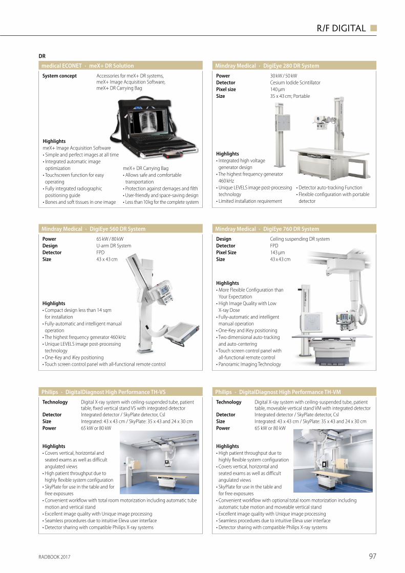
RADBOOK 2017 97
R/F DIGITAL ◼
DR
· meX+ DR Solutionmedical ECONET
Accessories for meX+ DR systems, meX+ Image Acquisition Software, meX+ DR Carrying Bag
System concept
HighlightsmeX+ Image Acquisition Software• Simple and perfect images at all time• Integrated automatic image
optimization• Touchscreen function for easy operating
• Fully integrated radiographic positioning guide
• Bones and soft tissues in one image
meX+ DR Carrying Bag• Allows safe and comfortable
transportation• Protection against demages and filth• User-friendly and space-saving design• Less than 10 kg for the complete system
· DigiEye 560 DR SystemMindray Medical
43 x 43 cmSizeFPDDetectorU-arm DR SystemDesign65 kW / 80 kWPower
Highlights• Compact design less than 14 sqm
for installation• Fully-automatic and intelligent manual
operation • The highest frequency generator 460 kHz• Unique LEVELS image post-processing
technology• One-Key and iKey positioning • Touch screen control panel with all-functional remote control
· DigiEye 280 DR SystemMindray Medical
30 kW / 50 kWPowerCesium Iodide ScintillatorDetector
35 x 43 cm; PortableSize140 μmPixel size
Highlights• Integrated high voltage
generator design • The highest frequency generator
460 kHz• Unique LEVELS image post-processing
technology• Limited installation requirement
• Detector auto-tracking Function • Flexible configuration with portable
detector
· DigiEye 760 DR SystemMindray Medical
43 x 43 cmSize
FPDDetectorCeiling suspending DR systemDesign
143 μmPixel Size
Highlights• More Flexible Configuration than
Your Expectation• High Image Quality with Low
X-ray Dose• Fully-automatic and intelligent
manual operation• One-Key and iKey positioning • Two dimensional auto-tracking
and auto-centering • Touch screen control panel with
all-functional remote control• Panoramic Imaging Technology
Highlights• Covers vertical, horizontal and
seated exams as well as difficult angulated views
• High patient throughput due to highly flexible system configuration
• SkyPlate for use in the table and for free exposures
• Convenient workflow with total room motorization including automatic tube motion and vertical stand
• Excellent image quality with Unique image processing• Seamless procedures due to intuitive Eleva user interface • Detector sharing with compatible Philips X-ray systems
· DigitalDiagnost High Performance TH-VSPhilips
Integrated detector / SkyPlate detector, CslIntegrated: 43 x 43 cm / SkyPlate: 35 x 43 and 24 x 30 cm
DetectorSize
65 kW or 80 kWPower
Digital X-ray system with ceiling-suspended tube, patient table, fixed vertical stand VS with integrated detector
Technology
· DigitalDiagnost High Performance TH-VMPhilips
Integrated detector / SkyPlate detector, CslIntegrated: 43 x 43 cm / SkyPlate: 35 x 43 and 24 x 30 cm
DetectorSize
65 kW or 80 kWPower
Digital X-ray system with ceiling-suspended tube, patient table, moveable vertical stand VM with integrated detector
Technology
Highlights• High patient throughput due to
highly flexible system configuration • Covers vertical, horizontal and
seated exams as well as difficult angulated views
• SkyPlate for use in the table and for free exposures
• Convenient workflow with optional total room motorization including automatic tube motion and moveable vertical stand
• Excellent image quality with Unique image processing• Seamless procedures due to intuitive Eleva user interface • Detector sharing with compatible Philips X-ray systems
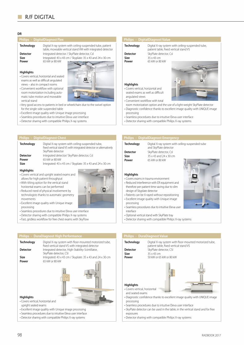
RADBOOK 201798
◼ R/F DIGITAL
DR
· DigitalDiagnost Flex
Integrated: 43 x 43 cm / Skyplate: 35 x 43 and 24 x 30 cmSize
Digital X-ray system with ceiling-suspended tube, patient table, moveable vertical stand VM with integrated detector
Technology
Highlights• Covers vertical, horizontal and seated
exams as well as difficult angulated views – also in compact rooms
• Convenient workflow with optional room motorization including auto-matic tube motion and moveable vertical stand
• Very good access to patients in bed or wheelchairs due to the swivel option for the single-side suspended table
• Excellent image quality with Unique image processing• Seamless procedures due to intuitive Eleva user interface • Detector sharing with compatible Philips X-ray systems
Philips
Integrated detector / SkyPlate detector, CslDetector
65 kW or 80 kWPower
Philips · DigitalDiagnost Value
35 x 43 cmSize
Digital X-ray system with ceiling-suspended tube, patient table, fixed vertical stand VS
Technology
Highlights• Covers vertical, horizontal and
seated exams as well as difficult angulated views
• Convenient workflow with total room motorization option and the use of a light-weight SkyPlate detector
• Diagnostic confidence thanks to excellent image quality with UNIQUE image processing
• Seamless procedures due to intuitive Eleva user interface • Detector sharing with compatible Philips X-ray systems
SkyPlate detector, CsIDetector
65 kW or 80 kWPower
· DigitalDiagnost ChestPhilips
Highlights• Covers vertical and upright seated exams and
allows for high patient throughput• With tilting option for the vertical stand
horizontal exams can be performed • Reduced need of physical involvement by
technologists thanks to automatic geometry movements
• Excellent image quality with Unique image processing
• Seamless procedures due to intuitive Eleva user interface • Detector sharing with compatible Philips X-ray systems• Fast, gridless workflow for free chest exams with SkyFlow
65 kW or 80 kWPowerIntegrated: 43 x 43 cm / Skyplate: 35 x 43 and 24 x 30 cmSize
Digital X-ray system with ceiling-suspended tube, fixed vertical stand VS with integrated detector or alternatively SkyPlate detector
Technology
Integrated detector/ SkyPlate detector, CslDetector
· DigitalDiagnost EmergencyPhilips
Highlights• Covers exams in trauma environment• Reduced interference with ER equipment and
therefore per-patient time saving due to slim design of Skyplate detector
• Patients can be X-rayed without repositioning• Excellent image quality with Unique image
processing• Seamless procedures due to intuitive Eleva user
interface • Optional vertical stand with SkyPlate tray • Detector sharing with compatible Philips X-ray systems
65 kW or 80 kWPower35 x 43 and 24 x 30 cmSize
Digital X-ray system with ceiling-suspended tube and SkyPlate detector
Technology
SkyPlate detector, CslDetector
· DuraDiagnost High PerformancePhilips
Integrated: 43 x 43 cm / Skyplate: 35 x 43 and 24 x 30 cmSize
Integrated detector, High-Stability-Scintillator,SkyPlate detector, CSI
Detector
Digital X-ray system with floor-mounted motorized tube, fixed vertical stand VS with integrated detector
Technology
Highlights• Covers vertical, horizontal and
upright seated exams• Excellent image quality with Unique image processing• Seamless procedures due to intuitive Eleva user interface• Detector sharing with compatible Philips X-ray systems
65 kW or 80 kWPower
Size
· DuraDiagnost ValuePhilips
Detector
Technology
Highlights• Covers vertical, horizontal
and seated exams • Diagnostic confidence thanks to excellent image quality with UNIQUE image
processing • Seamless procedures due to intuitive Eleva user interface • SkyPlate detector can be used in the table, in the vertical stand and for free
exposures• Detector sharing with compatible Philips X-ray systems
50 kW or 65 kW or 80 kWPower35 x 43 cmSkyPlate detector, CSI
Digital X-ray system with floor-mounted motorized tube, patient table, fixed vertical stand VS
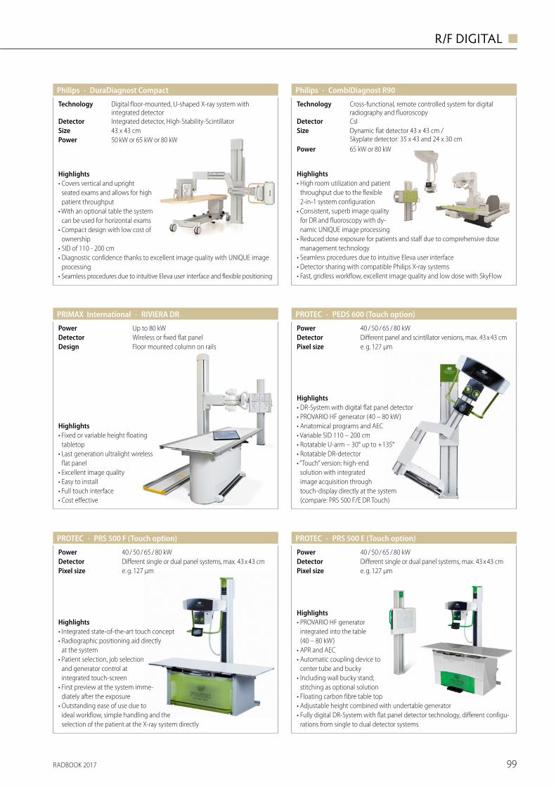
RADBOOK 2017 99
R/F DIGITAL ◼
· DuraDiagnost CompactPhilips
43 x 43 cmSizeIntegrated detector, High-Stability-ScintillatorDetector
Digital floor-mounted, U-shaped X-ray system with integrated detector
Technology
Highlights• Covers vertical and upright
seated exams and allows for high patient throughput
• With an optional table the system can be used for horizontal exams
• Compact design with low cost of ownership
• SID of 110 - 200 cm• Diagnostic confidence thanks to excellent image quality with UNIQUE image
processing • Seamless procedures due to intuitive Eleva user interface and flexible positioning
50 kW or 65 kW or 80 kWPower
· CombiDiagnost R90Philips
Highlights• High room utilization and patient
throughput due to the flexible 2-in-1 system configuration
• Consistent, superb image quality for DR and fluoroscopy with dy-namic UNIQUE image processing
• Reduced dose exposure for patients and staff due to comprehensive dose management technology
• Seamless procedures due to intuitive Eleva user interface • Detector sharing with compatible Philips X-ray systems• Fast, gridless workflow, excellent image quality and low dose with SkyFlow
Dynamic flat detector 43 x 43 cm / Skyplate detector: 35 x 43 and 24 x 30 cm
SizeCsIDetector
Cross-functional, remote controlled system for digital radiography and fluoroscopy
Technology
65 kW or 80 kWPower
· PEDS 600 (Touch option)PROTEC
e. g. 127 µmPixel sizeDifferent panel and scintillator versions, max. 43 x 43 cmDetector40 / 50 / 65 / 80 kWPower
Highlights• DR-System with digital flat panel detector• PROVARIO HF generator (40 – 80 kW)• Anatomical programs and AEC• Variable SID 110 – 200 cm• Rotatable U-arm – 30° up to +135°• Rotatable DR-detector• “Touch” version: high-end
solution with integrated image acquisition through touch- display directly at the system (compare: PRS 500 F/E DR Touch)
· PRS 500 F (Touch option)PROTEC
e. g. 127 µmPixel sizeDifferent single or dual panel systems, max. 43 x 43 cmDetector40 / 50 / 65 / 80 kWPower
Highlights• Integrated state-of-the-art touch concept• Radiographic positioning aid directly
at the system• Patient selection, job selection
and generator control at integrated touch-screen
• First preview at the system imme-diately after the exposure
• Outstanding ease of use due to ideal workflow, simple handling and the selection of the patient at the X-ray system directly
· RIVIERA DRPRIMAX International
Floor mounted column on railsDesignWireless or fixed flat panelDetectorUp to 80 kWPower
Highlights• Fixed or variable height floating
tabletop• Last generation ultralight wireless
flat panel• Excellent image quality• Easy to install• Full touch interface• Cost effective
· PRS 500 E (Touch option)PROTEC
e. g. 127 µmPixel sizeDifferent single or dual panel systems, max. 43 x 43 cmDetector40 / 50 / 65 / 80 kWPower
Highlights• PROVARIO HF generator
integrated into the table (40 – 80 kW)
• APR and AEC• Automatic coupling device to
center tube and bucky• Including wall bucky stand;
stitching as optional solution• Floating carbon fibre table top• Adjustable height combined with undertable generator• Fully digital DR-System with flat panel detector technology, different configu-
rations from single to dual detector systems
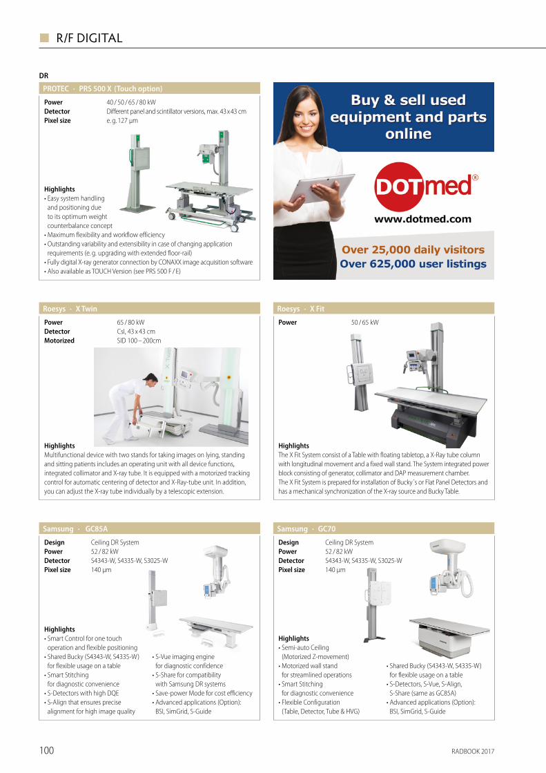
RADBOOK 2017100
◼ R/F DIGITAL
DR
· PRS 500 X (Touch option)PROTEC
e. g. 127 µmPixel sizeDifferent panel and scintillator versions, max. 43 x 43 cmDetector40 / 50 / 65 / 80 kWPower
Highlights• Easy system handling
and positioning due to its optimum weight counterbalance concept
• Maximum flexibility and workflow efficiency• Outstanding variability and extensibility in case of changing application
requirements (e. g. upgrading with extended floor-rail)• Fully digital X-ray generator connection by CONAXX image acquisition software• Also available as TOUCH Version (see PRS 500 F / E)
HighlightsThe X Fit System consist of a Table with floating tabletop, a X-Ray tube column with longitudinal movement and a fixed wall stand. The System integrated power block consisting of generator, collimator and DAP measurement chamber. The X Fit System is prepared for installation of Bucky´s or Flat Panel Detectors and has a mechanical synchronization of the X-ray source and Bucky Table.
· X FitRoesys
50 / 65 kWPower
HighlightsMultifunctional device with two stands for taking images on lying, standing and sitting patients includes an operating unit with all device functions, integrated collimator and X-ray tube. It is equipped with a motorized tracking control for automatic centering of detector and X-Ray-tube unit. In addition, you can adjust the X-ray tube individually by a telescopic extension.
· X Twin
SID 100 – 200cmMotorizedCsI, 43 x 43 cmDetector65 / 80 kWPower
Roesys
· GC85ASamsung
140 μmPixel size S4343-W, S4335-W, S3025-WDetector
Ceiling DR System52 / 82 kW
DesignPower
Highlights• Smart Control for one touch
operation and flexible positioning• Shared Bucky (S4343-W, S4335-W)
for flexible usage on a table• Smart Stitching
for diagnostic convenience• S-Detectors with high DQE• S-Align that ensures precise
alignment for high image quality
• S-Vue imaging engine for diagnostic confidence
• S-Share for compatibility with Samsung DR systems
• Save-power Mode for cost efficiency• Advanced applications (Option):
BSI, SimGrid, S-Guide
· GC70Samsung
140 μmPixel size S4343-W, S4335-W, S3025-WDetector
Ceiling DR System52 / 82 kW
DesignPower
Highlights• Semi-auto Ceiling
(Motorized Z-movement)• Motorized wall stand
for streamlined operations • Smart Stitching
for diagnostic convenience• Flexible Configuration
(Table, Detector, Tube & HVG)
• Shared Bucky (S4343-W, S4335-W) for flexible usage on a table
• S-Detectors, S-Vue, S-Align, S-Share (same as GC85A)
• Advanced applications (Option): BSI, SimGrid, S-Guide
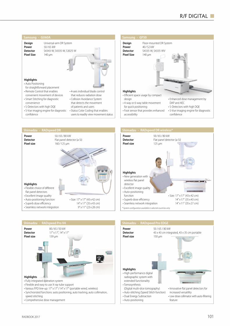
RADBOOK 2017 101
R/F DIGITAL ◼
50 / 65 kWPower
· GU60ASamsung
140 μmPixel SizeS4343-W, S4335-W, S3025-WDetector
Universal-arm DR System Design
Highlights• Auto Positioning
for straightforward placement• Remote Control that enables
convenient movement of devices• Smart Stitching for diagnostic
convenience• S-Detectors with high DQE• S-Vue imaging engine for diagnostic
confidence
• 4-axis individual blade control that reduces radiation dose
• Collision Avoidance System that detects the movement of patients and users
• Status Color Coding that enables users to readily view movement status
· GF50Samsung
140 μmPixel SizeS4335-W, S4335-WVDetector
Floor-mounted DR SystemDesign
Highlights• Efficient space usage by compact
design• 4-way or 6-way table movement
for quick positioning• Foot sensor that provides enhanced
accessibility
• Enhanced dose management by DAP and AEC
• S-Detectors with high DQE• S-Vue imaging engine for diagnostic
confidence
40 / 52 kWPower
Shimadzu · RADspeed DR wireless*
125 µmPixel sizeFlat panel detector (a-Si)Detector50 / 65 / 80 kWPower
Highlights• New generation with
wireless flat panel detector
• Excellent image quality • Auto-positioning
function • Superb dose efficiency • Seamless network integration
• Size: 17" x 17" (43 x 42 cm) 14" x 17" (35 x 43 cm) 14" x 11" (35 x 27 cm)
* System configuration available in selected countries only
· RADspeed DRShimadzu
160 / 125 µmPixel sizeFlat panel detector (a-Si)Detector50 / 65 / 80 kWPower
Highlights• Flexible choice of different
flat panel detectors• Excellent image quality• Auto-positioning function• Superb dose efficiency• Seamless network integration
• Size: 17" x 17" (43 x 42 cm) 14" x 17" (35 x 43 cm) 9" x 11" (23 x 28 cm)
· RADspeed Pro V4Shimadzu
139 µmPixel size17" x 17", 14" x 17"Detector80 / 65 / 50 kWPower
Highlights• Fully integrated operation system• Flexible and easy to use X-ray tube support• Various FPD line-up: 17" x 17" / 14" x 17" (portable wired, wireless)• Synchronized functions: auto positioning, auto tracking, auto collimation,
speed stitching• Comprehensive dose management
· RADspeed Pro EDGEShimadzu
50 / 65 / 80 kWPower
150 µmPixel size43 x 43 cm integrated, 43 x 35 cm portableDetector
Highlights• High-performance digital
radio graphic system with extended functionality
• Tomosynthesis (Digital multi-slice tomography)
• Auto-stitching (Speed Stitch function)• Dual Energy Subtraction• Auto-positioning
• Innovative flat panel detectors for increased versatility
• Low dose collimator with auto-filtering feature
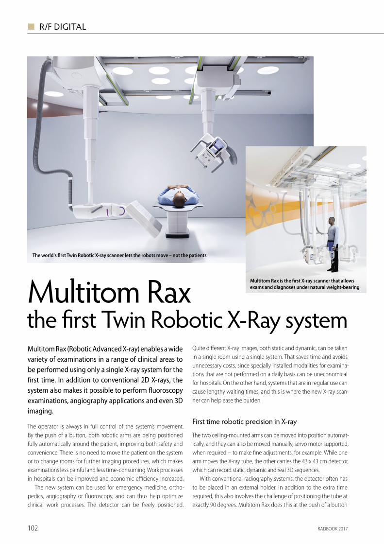
RADBOOK 2017102
◼ R/F DIGITAL
The operator is always in full control of the system’s movement. By the push of a button, both robotic arms are being positioned fully automatically around the patient, improving both safety and convenience. There is no need to move the patient on the system or to change rooms for further imaging procedures, which makes examinations less painful and less time-consuming. Work processes in hospitals can be improved and economic efficiency increased.
The new system can be used for emergency medicine, ortho-pedics, angiography or fluoroscopy, and can thus help optimize clinical work processes. The detector can be freely positioned.
Quite different X-ray images, both static and dynamic, can be taken in a single room using a single system. That saves time and avoids unnecessary costs, since specially installed modalities for examina-tions that are not performed on a daily basis can be uneconomical for hospitals. On the other hand, systems that are in regular use can cause lengthy waiting times, and this is where the new X-ray scan-ner can help ease the burden.
First time robotic precision in X-ray
The two ceiling-mounted arms can be moved into position automat-ically, and they can also be moved manually, servo motor supported, when required – to make fine adjustments, for example. While one arm moves the X-ray tube, the other carries the 43 x 43 cm detector, which can record static, dynamic and real 3D sequences.
With conventional radiography systems, the detector often has to be placed in an external holder. In addition to the extra time required, this also involves the challenge of positioning the tube at exactly 90 degrees. Multitom Rax does this at the push of a button
for free exams. This prevents any risk of having to repeat image processes because the tube was not precisely positioned. The system offers optionally also wireless, portable detectors in two different sizes that can be positioned directly between the wheelchair or mat-tress and the patient’s back. The automatic control of the robotic arms ensures that they will always take the shortest and safest route to reach the next programmed position. Pre-programmed safety zones and an auto-matic stop in response to contact also improve safety.
3D imaging offers diagnostic certainty
3D computed tomography (CT) images are often used in situations such as orthopedic examinations involv-ing the implantation of prosthetic joints. Multitom Rax makes it possible to take 3D images under the patient’s natural weight bearing condition. 3D images can be made of all areas of the body with the patient seated, lying down or standing. Images taken while the patient is standing are essential because for example knees, pelvis and spinal column appear differ-ently under the influence of the patient’s body weight compared to when the patient is lying down. As a result, 3D images acquired by Multitom Rax offer better diagnostic and planning certainty com-pared to those that do not reflect a natural weight bearing condi-tion. Conventional 2D X-rays, for example, do not always reveal fine hairline fractures in the bone. If a bone fracture is suspected, it has previously been necessary to take a 3D image using a CT system to be sure of the diagnosis. With Multitom Rax, however, a 3D image can be taken at the same system, and so the patient does not have to wait for a further appointment or to be transferred to the CT unit.
Easy access to the patient – in pediatrics in particular A free-standing patient table and fully mobile system elements with Multitom Rax provide a more comfortable examination atmosphere. The fact that the table can be adjusted to a very low 50 centimeter table height means that children can get onto it by themselves. It can also be positioned at the most convenient working height. The hospital staff thus has full access to the patient, with no need for the hospital staff to twist into an anatomically unnatural position. The result is an improvement in both safety for the patient and the examining physician, and in the level of con-venience, since it is the system that moves when needed, not the individuals. Additional devices and personnel are often essential for interventional procedures such as fluoroscopic needle locali-zation in particular. The open system design makes it possible to position the tube and detector most appropriately in the room. And the fact that both arms are ceiling-mounted means there is no floor-mounted equipment or cable ducts to get in the way.
Standardization – for future treatment trends, too
As a part of the Max system family from Siemens Healthineers, Multitom Rax stands out by providing the same image impression and thus making it easier to compare X-ray images. The controls and user interfaces on the Max systems are identical, which means the operators have no need to familiarize themselves over again with new equipment. The wireless detectors in the Max family can also be used equally with all the systems in the family, improving the level of flexibility.
www.siemens.com/mtr
Multitom Rax the first Twin Robotic X-Ray system
Multitom Rax is not commercially available in all countries. Due to regulatory rea-sons their future availability cannot be guaranteed. Further details are available from the local Siemens organizations.
Multitom Rax (Robotic Advanced X-ray) enables a wide variety of examinations in a range of clinical areas to be performed using only a single X-ray system for the first time. In addition to conventional 2D X-rays, the system also makes it possible to perform fluoroscopy examinations, angiography applications and even 3D imaging.
The world's first Twin Robotic X-ray scanner lets the robots move – not the patients
Multitom Rax is the first X-ray scanner that allows exams and diagnoses under natural weight-bearing
Always a perfect alignment of tube and detector even at free examinations
Multiple X-ray capabilities in one room: radiography, fluoroscopy, 3D images, and angiography
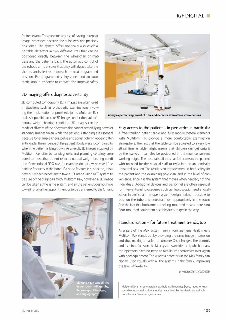
RADBOOK 2017 103
R/F DIGITAL ◼
The operator is always in full control of the system’s movement. By the push of a button, both robotic arms are being positioned fully automatically around the patient, improving both safety and convenience. There is no need to move the patient on the system or to change rooms for further imaging procedures, which makes examinations less painful and less time-consuming. Work processes in hospitals can be improved and economic efficiency increased.
The new system can be used for emergency medicine, ortho-pedics, angiography or fluoroscopy, and can thus help optimize clinical work processes. The detector can be freely positioned.
Quite different X-ray images, both static and dynamic, can be taken in a single room using a single system. That saves time and avoids unnecessary costs, since specially installed modalities for examina-tions that are not performed on a daily basis can be uneconomical for hospitals. On the other hand, systems that are in regular use can cause lengthy waiting times, and this is where the new X-ray scan-ner can help ease the burden.
First time robotic precision in X-ray
The two ceiling-mounted arms can be moved into position automat-ically, and they can also be moved manually, servo motor supported, when required – to make fine adjustments, for example. While one arm moves the X-ray tube, the other carries the 43 x 43 cm detector, which can record static, dynamic and real 3D sequences.
With conventional radiography systems, the detector often has to be placed in an external holder. In addition to the extra time required, this also involves the challenge of positioning the tube at exactly 90 degrees. Multitom Rax does this at the push of a button
for free exams. This prevents any risk of having to repeat image processes because the tube was not precisely positioned. The system offers optionally also wireless, portable detectors in two different sizes that can be positioned directly between the wheelchair or mat-tress and the patient’s back. The automatic control of the robotic arms ensures that they will always take the shortest and safest route to reach the next programmed position. Pre-programmed safety zones and an auto-matic stop in response to contact also improve safety.
3D imaging offers diagnostic certainty
3D computed tomography (CT) images are often used in situations such as orthopedic examinations involv-ing the implantation of prosthetic joints. Multitom Rax makes it possible to take 3D images under the patient’s natural weight bearing condition. 3D images can be made of all areas of the body with the patient seated, lying down or standing. Images taken while the patient is standing are essential because for example knees, pelvis and spinal column appear differ-ently under the influence of the patient’s body weight compared to when the patient is lying down. As a result, 3D images acquired by Multitom Rax offer better diagnostic and planning certainty com-pared to those that do not reflect a natural weight bearing condi-tion. Conventional 2D X-rays, for example, do not always reveal fine hairline fractures in the bone. If a bone fracture is suspected, it has previously been necessary to take a 3D image using a CT system to be sure of the diagnosis. With Multitom Rax, however, a 3D image can be taken at the same system, and so the patient does not have to wait for a further appointment or to be transferred to the CT unit.
Easy access to the patient – in pediatrics in particular A free-standing patient table and fully mobile system elements with Multitom Rax provide a more comfortable examination atmosphere. The fact that the table can be adjusted to a very low 50 centimeter table height means that children can get onto it by themselves. It can also be positioned at the most convenient working height. The hospital staff thus has full access to the patient, with no need for the hospital staff to twist into an anatomically unnatural position. The result is an improvement in both safety for the patient and the examining physician, and in the level of con-venience, since it is the system that moves when needed, not the individuals. Additional devices and personnel are often essential for interventional procedures such as fluoroscopic needle locali-zation in particular. The open system design makes it possible to position the tube and detector most appropriately in the room. And the fact that both arms are ceiling-mounted means there is no floor-mounted equipment or cable ducts to get in the way.
Standardization – for future treatment trends, too
As a part of the Max system family from Siemens Healthineers, Multitom Rax stands out by providing the same image impression and thus making it easier to compare X-ray images. The controls and user interfaces on the Max systems are identical, which means the operators have no need to familiarize themselves over again with new equipment. The wireless detectors in the Max family can also be used equally with all the systems in the family, improving the level of flexibility.
www.siemens.com/mtr
Multitom Rax the first Twin Robotic X-Ray system
Multitom Rax is not commercially available in all countries. Due to regulatory rea-sons their future availability cannot be guaranteed. Further details are available from the local Siemens organizations.
Multitom Rax (Robotic Advanced X-ray) enables a wide variety of examinations in a range of clinical areas to be performed using only a single X-ray system for the first time. In addition to conventional 2D X-rays, the system also makes it possible to perform fluoroscopy examinations, angiography applications and even 3D imaging.
The world's first Twin Robotic X-ray scanner lets the robots move – not the patients
Multitom Rax is the first X-ray scanner that allows exams and diagnoses under natural weight-bearing
Always a perfect alignment of tube and detector even at free examinations
Multiple X-ray capabilities in one room: radiography, fluoroscopy, 3D images, and angiography
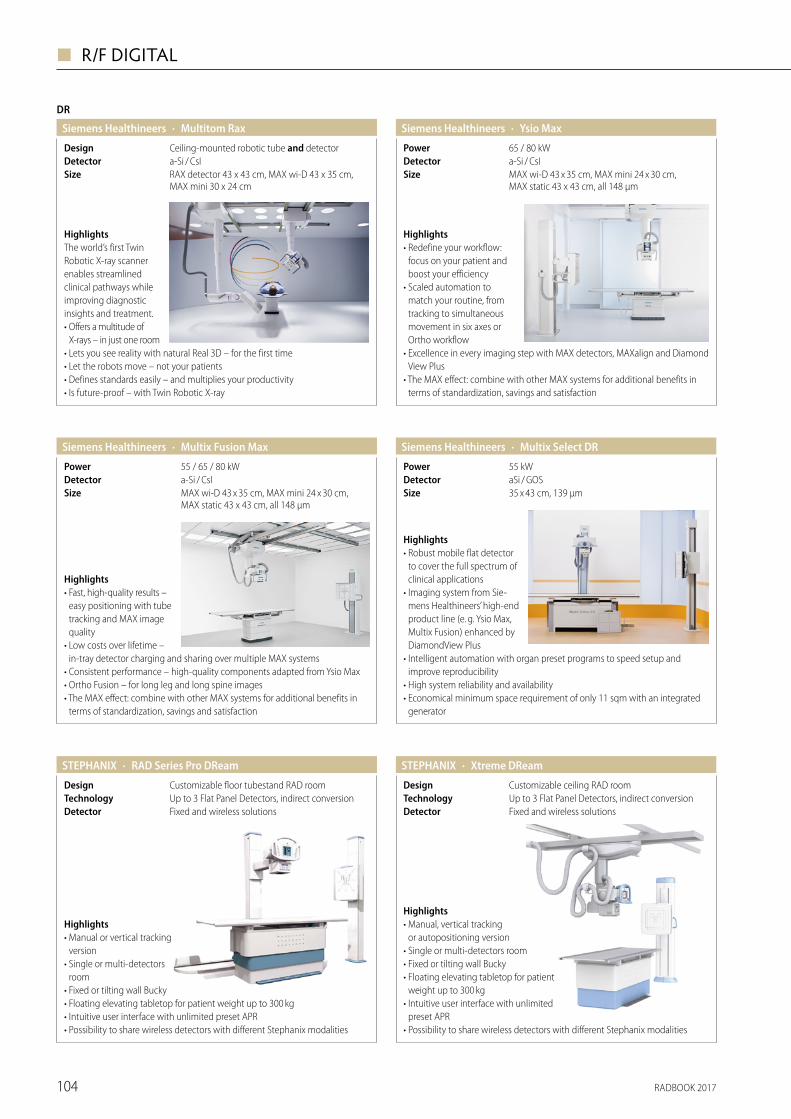
RADBOOK 2017104
◼ R/F DIGITAL
Highlights• Robust mobile flat detector
to cover the full spectrum of clinical applications
• Imaging system from Sie-mens Healthineers’ high-end product line (e. g. Ysio Max, Multix Fusion) enhanced by DiamondView Plus
• Intelligent automation with organ preset programs to speed setup and improve reproducibility
• High system reliability and availability• Economical minimum space requirement of only 11 sqm with an integrated
generator
· Multix Select DRSiemens Healthineers
35 x 43 cm, 139 μmSizeaSi / GOSDetector55 kWPower
Highlights• Fast, high-quality results –
easy positioning with tube tracking and MAX image quality
• Low costs over lifetime – in-tray detector charging and sharing over multiple MAX systems
• Consistent performance – high-quality components adapted from Ysio Max• Ortho Fusion – for long leg and long spine images• The MAX effect: combine with other MAX systems for additional benefits in
terms of standardization, savings and satisfaction
· Multix Fusion MaxSiemens Healthineers
a-Si / CsIDetectorMAX wi-D 43 x 35 cm, MAX mini 24 x 30 cm, MAX static 43 x 43 cm, all 148 µm
Size
55 / 65 / 80 kWPower
Highlights• Redefine your workflow:
focus on your patient and boost your efficiency
• Scaled automation to match your routine, from tracking to simultaneous movement in six axes or Ortho workflow
• Excellence in every imaging step with MAX detectors, MAXalign and Diamond View Plus
• The MAX effect: combine with other MAX systems for additional benefits in terms of standardization, savings and satisfaction
· Ysio MaxSiemens Healthineers
MAX wi-D 43 x 35 cm, MAX mini 24 x 30 cm, MAX static 43 x 43 cm, all 148 µm
Sizea-Si / CsIDetector65 / 80 kWPower
Detector
HighlightsThe world’s first Twin Robotic X-ray scanner enables streamlined clinical pathways while improving diagnostic insights and treatment.• Offers a multitude of
X-rays – in just one room• Lets you see reality with natural Real 3D – for the first time• Let the robots move – not your patients• Defines standards easily – and multiplies your productivity• Is future-proof – with Twin Robotic X-ray
DR
· Multitom RaxSiemens Healthineers
RAX detector 43 x 43 cm, MAX wi-D 43 x 35 cm, MAX mini 30 x 24 cm
Sizea-Si / CsICeiling-mounted robotic tube and detectorDesign
Highlights• Manual, vertical tracking
or auto positioning version• Single or multi-detectors room• Fixed or tilting wall Bucky• Floating elevating tabletop for patient
weight up to 300 kg• Intuitive user interface with unlimited
preset APR• Possibility to share wireless detectors with different Stephanix modalities
· Xtreme DReamSTEPHANIX
Fixed and wireless solutionsDetectorUp to 3 Flat Panel Detectors, indirect conversionTechnologyCustomizable ceiling RAD roomDesign
Highlights• Manual or vertical tracking
version• Single or multi-detectors
room• Fixed or tilting wall Bucky• Floating elevating tabletop for patient weight up to 300 kg• Intuitive user interface with unlimited preset APR• Possibility to share wireless detectors with different Stephanix modalities
· RAD Series Pro DReamSTEPHANIX
Fixed and wireless solutionsDetectorUp to 3 Flat Panel Detectors, indirect conversionTechnologyCustomizable floor tubestand RAD roomDesign
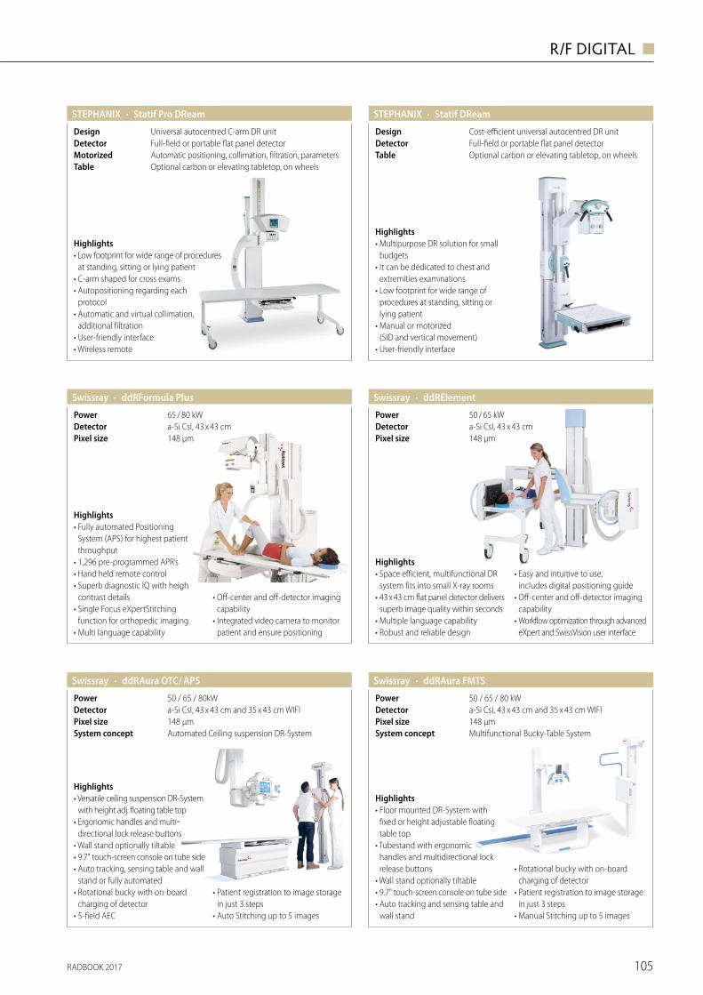
RADBOOK 2017 105
R/F DIGITAL ◼
Highlights• Low footprint for wide range of procedures
at standing, sitting or lying patient• C-arm shaped for cross exams• Autopositioning regarding each
protocol• Automatic and virtual collimation,
additional filtration• User-friendly interface• Wireless remote
· Statif Pro DReamSTEPHANIX
Optional carbon or elevating tabletop, on wheelsTableAutomatic positioning, collimation, filtration, parametersMotorizedFull-field or portable flat panel detectorDetectorUniversal autocentred C-arm DR unitDesign
Highlights• Multipurpose DR solution for small
budgets• It can be dedicated to chest and
extremities examinations• Low footprint for wide range of
procedures at standing, sitting or lying patient
• Manual or motorized (SID and vertical movement)
• User-friendly interface
· Statif DReamSTEPHANIX
Optional carbon or elevating tabletop, on wheelsTableFull-field or portable flat panel detectorDetectorCost-efficient universal autocentred DR unitDesign
· ddRElementSwissray
148 µmPixel sizea-Si CsI, 43 x 43 cmDetector50 / 65 kWPower
Highlights• Space efficient, multifunctional DR
system fits into small X-ray rooms• 43 x 43 cm flat panel detector delivers
superb image quality within seconds• Multiple language capability• Robust and reliable design
• Easy and intuitive to use, includes digital positioning guide
• Off-center and off-detector imaging capability
• Workflow optimization through advanced eXpert and SwissVision user interface
· ddRFormula PlusSwissray
148 µmPixel sizea-Si CsI, 43 x 43 cmDetector65 / 80 kWPower
Highlights• Fully automated Positioning
System (APS) for highest patient throughput
• 1,296 pre-programmed APR’s• Hand held remote control• Superb diagnostic IQ with heigh
contrast details• Single Focus eXpertStitching
function for orthopedic imaging• Multi language capability
• Off-center and off-detector imaging capability
• Integrated video camera to monitor patient and ensure positioning
· ddRAura OTC / APSSwissray
Automated Ceiling suspension DR-SystemSystem concept148 µmPixel sizea-Si CsI, 43 x 43 cm and 35 x 43 cm WIFIDetector50 / 65 / 80kWPower
Highlights• Versatile ceiling suspension DR-System
with height adj. floating table top• Ergonomic handles and multi-
directional lock release buttons• Wall stand optionally tiltable• 9.7" touch-screen console on tube side• Auto tracking, sensing table and wall
stand or fully automated• Rotational bucky with on-board
charging of detector• 5-field AEC
• Patient registration to image storage in just 3 steps
• Auto Stitching up to 5 images
148 µmPixel size
· ddRAura FMTSSwissray
Multifunctional Bucky-Table System System concept
50 / 65 / 80 kWPowera-Si CsI, 43 x 43 cm and 35 x 43 cm WIFIDetector
Highlights• Floor mounted DR-System with
fixed or height adjustable floating table top
• Tubestand with ergonomic handles and multidirectional lock release buttons
• Wall stand optionally tiltable• 9.7" touch-screen console on tube side• Auto tracking and sensing table and
wall stand
• Rotational bucky with on-board charging of detector
• Patient registration to image storage in just 3 steps
• Manual Stitching up to 5 images
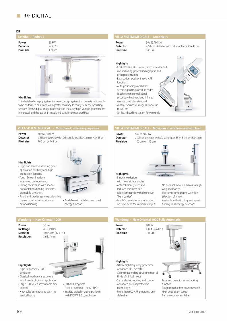
RADBOOK 2017106
◼ R/F DIGITAL
DR
· Radrex-iToshiba
139 µmPixel sizea-Si / CslDetector80 kWPower
HighlightsThis digital radiography system is a new-concept system that permits radiography to be performed easily and with greater accuracy. In this system, the operating sections for the digital image processor and the X-ray high-voltage generator are integrated, and the use of an integrated panel improves workflow.
· Moviplan iC with floor-mounted columnVILLA SISTEMI MEDICALI
100 μm or 143 μmPixel sizea-Silicon detector with CsI scintillator, 35 x 43 cm or 43 x 43 cmDetector50 / 65 / 80 kWPower
Highlights• Innovative design
with no unsightly cables• Anti-collision system and
reduced thickness rails• Table commands with distinctive
“light barrier”• Touch Screen interface integrated
on tube-head for immediate inputs
• No patient limitation thanks to high weight capacity
• Electronic tomography with free selection of angle
• Available with stitching, auto-posi-tioning, dual energy functions
· Armonicus VILLA SISTEMI MEDICALI
143 μmPixel sizea-Silicon detector with CsI scintillator, 43 x 43 cmDetector50 / 65 / 80 kWPower
Highlights• Cost-effective DR U-arm system for extended
use, including general radiographic and orthopedic studies
• Easy patient positioning via APR functions
• Auto-positioning capabilities according to RIS procedure codes
• Touch screen control panel, secondary keyboard and infrared remote control as standard
• Variable Source to Image Distance up to 180 cm
• On-board parking station for two grids
· Moviplan iC with ceiling suspensionVILLA SISTEMI MEDICALI
100 μm or 143 μmPixel sizea-Silicon detector with CsI scintillator, 35 x 43 cm or 43 x 43 cmDetector50 / 65 / 80 kWPower
Highlights• High-end solution allowing great
application flexibility and high production capacity
• Touch Screen interface integrated on tube-head
• Tiliting chest stand with special horizontal positioning for exams on mobile stretchers
• Rapid and precise system positioning thanks to full auto-tracking and autopositioning
• Available with stitching and dual energy functions
Highlights• 80 kW high frequency generator • Advanced FPD detector • Ceiling suspending structure meet all
kinds of clinical needs • 5 axis electric moving and control • Advanced patient protection
technology • More than 600 APR programs, user
definable
• Tube and detector auto-tracking function
• Programmable fast position switch • High acquisition speed• Remote control available
· New Oriental 1000 Fully AutomaticWandong
143 umPixel size43 x 43 cm FPDDetector80 kWPower
· New Oriental 1000 Wandong
3.6 lp / mmResolution43 x 43cm (17 x 17”)Detector40 ~ 150 kVkV Range50 kW Power
Highlights• High frequency 50 kW
generator• Classical mechanical structure
for all needs of clinical application• Large LCD touch screen table-side
control• X-ray tube auto tracking with the
vertical bucky
• 600 APR programs• Fixed or portable 17 x 17" FPD• InvaRay digital imaging platform
with DICOM 3.0 compliance
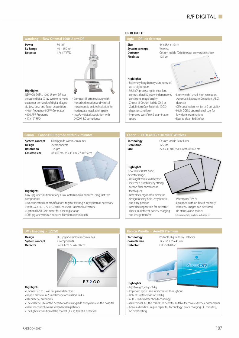
RADBOOK 2017 107
R/F DIGITAL ◼
HighlightsNEW ORIENTAL 1000 U-arm DR is a versatile digital X-ray system to meet customer demands of digital diagno-sis. Less dose and faster acquisition.• High frequency 50kW Generator• 600 APR Programs• 17 x 17" FPD
• Compact U-arm structure with motorized rotation and vertical movement is an ideal solution for inadequate installation space
• InvaRay digital acquisition with DICOM 3.0 compliance
· New Oriental 1000 U-arm DRWandong
17 x 17" FPDDetector40 – 150 kVkV Range50 KWPower
Agfa
DR RETROFIT
· DR 14s detector
125 µmPixel sizeCesium Iodide (CsI) detector conversion screenDetectorWirelessSystem concept46 x 38,4 x 1.5 cmSize
Highlights• Extremely long battery autonomy of
up to eight hours• MUSICA processing for excellent
contrast detail & exam-independent, consistent image quality
• Choice of Cesium Iodide (CsI) or Gadolinium Oxy-Sulphide (GOS) detector scintillator
• Improved workflow & examination speed
• Lightweight, small, high resolution Automatic Exposure Detection (AED) detector
• Offers optimal convenience & portability• High DQE & optimal pixel size, for
low dose examinations• Easy to clean & disinfect
· CXDI-410C/710C/810C WirelessCanon
27.4 x 35 cm, 35 x 43 cm, 43 x 42 cmSize125 µmResolutionCesium Iodide ScintillatorTechnology
HighlightsNew wireless flat panel detector range• Ultralight wireless detectors• Increased durability by strong
carbon fiber construction techniques
• New sleek ergonomic detector design for easy hold, easy handle and easy position
• New docking station for detector check-in, detector battery charging and image transfer
• Waterproof (IPX7)• Equipped with on-board memory
where 99 images can be stored (in stand-alone-mode)
Not commercially available in Europe yet
Canon · Canon DR-Upgrade-within-2-minutes
43 x 42 cm, 35 x 43 cm, 27.4 x 35 cmCassette size125 µmResolution2 componentsDesignDR Upgrade within 2 minutesSystem concept
HighlightsEasy upgrade solution for any X-ray system in two minutes using just two components• No connections or modifications to your existing X-ray system is necessary• With CXDI-401C / 701C / 801C Wireless Flat Panel Detectors• Optional USB DAP-meter for dose registration• DR Upgrade within 2 minutes. Freedom within reach
· AeroDR PremiumKonica Minolta
CsI scintillatorDetector
Portable Digital X-ray DetectorTechnology14 x 17" / 35 x 43 cmCassette size
Highlights• Lightweight, only 2.6 kg• Improved cycle time for increased throughput• Robust: surface load of 300 kg• AED – Hybrid detection technology• Waterproof IPX6, this makes the detector suitable for more extreme environments• Konica Minolta's unique capacitor technology: quick charging (30 minutes),
no overheating
· EZ2GODMS Imaging
36 x 43 cm or 24 x 30 cm Detector2 componentsSystem conceptDR upgrade mobile in 2 minutesDesign
Highlights• Connect up to 3 wifi flat panel detectors• Image preview in 2 s and image acquisition in 4 s• 8 h battery / autonomy• The cassette size of the detector allows upgrade everywhere in the hospital• Ideal for control exams for bedridden patients• The lightiest solution of the market (3.9 kg tablet & detector)
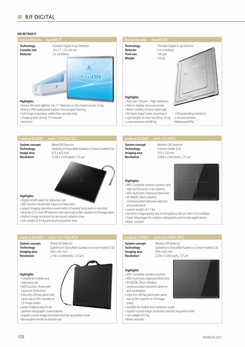
RADBOOK 2017108
◼ R/F DIGITAL
DR RETROFIT
· AeroDR 2SKonica Minolta
CsI scintillatorDetector14 x 17" / 35 x 43 cmCassette sizePortable Digital X-ray DetectorTechnology
Highlights• Konica Minolta’s lightest 14 x 17” detector on the market at just 2.5 kg• Robust, IPX6 waterproof, carbon monocoque housing • Full image acquisition within four seconds only• Charging time of only 13 minutes • AeroSync
· AeroDR HDKonica Minolta
2.6 kgWeight100 μmPixel sizeCsI scintillatorDetectorPortable Digital X-ray DetectorTechnology
Highlights• Pixel size: 100 μm – High Definition• Able to display micro structures• Better visibility of bone trabecular• No “pixel shape” when zooming in• Lightweight for easy handling: 2.6 kg• Load resistance of 400 kg
• 130 kg bending resistance • 2 second preview • Waterproof IPX6
· meX+1717SGC/SCCmedical ECONET
3,328 x 3,328 pixels, 127 μmResolution423 x 423 mmImage areaGadolinium Oxysulfide (Gadox) or Cesium-Iodide (Csl)TechnologyWired DR DetectorSystem concept
Highlights• Digital retrofit panel for stationary use• AED function (Automatic Exposure Detection)• Largest imaging size allows examination of several body parts in one shot• Ultra-flat (15.5 mm) DR detector with same size as film cassette or CR image plate• Highest image resolution by decreased radiation dose• Low weight of 4.4 kg and quick acquisition time
· meX+1012WCCmedical ECONET
2,080 x 2,560 pixels, 127 μmResolution259 x 320 mmImaging areaCesium-Iodide (Csl)TechnologyWireless DR DetectorSystem concept
Highlights• WIFI: Complete wireless solution with
high performance Li-ion battery• AED: Automatic Exposure Detection• AP MODE: Direct wireless
communication between detector and workstation
• Lowest weight of 1.7 kg• Excellent image quality due to Amorphous Silicon with Csl-Scintillator• Great Advantages for outdoor radiography and mobile applications• Water resistant
· meX+1417PGA/PCAmedical ECONET
2,756 x 3,268 pixels, 127 μmResolution358 x 423 mmImaging areaGadolinium Oxysulfide (Gadox) or Cesium-Iodide (Csl)TechnologyWired DR DetectorSystem concept
Highlights• Suitable for mobile and
stationary use• AED function (Automatic
Exposure Detection)• Ultra-thin DR flat panel with
same size as film cassette or CR image plates
• Large imaging area for all general radiographic examinations
• Superb-crystal image resolution and fast acquisition time• Removable handle for flexible use
· meX+1417WGC/WCCmedical ECONET
2,756 x 3,268 pixels, 127 μmResolution358 x 423 mmImaging areaGadolinium Oxysulfide (Gadox) or Cesium-Iodide (Csl)TechnologyWireless DR DetectorSystem concept
Highlights• WIFI: Complete wireless solution• AED: Automatic Exposure Detection• AP MODE: Direct Wireless
communication between detector and workstation
• Ultra-thin DR flat panel with same size as film cassette or CR image plates
• Suitable for mobile and stationary usage• Superb-crystal image resolution and fast acquisition time• Low weight of 3 kg• Water resistant
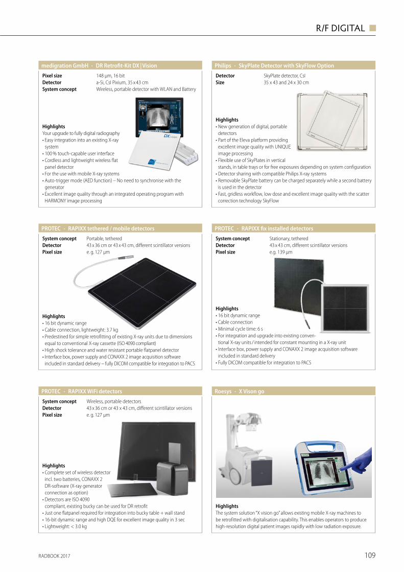
RADBOOK 2017 109
R/F DIGITAL ◼
· DR Retrofit-Kit DX | Visionmedigration GmbH
Wireless, portable detector with WLAN and BatterySystem concepta-Si, CsI Pixium, 35 x 43 cmDetector148 µm, 16 bitPixel size
HighlightsYour upgrade to fully digital radiography• Easy integration into an existing X-ray
system• 100 % touch-capable user interface• Cordless and lightweight wireless flat
panel detector• For the use with mobile X-ray systems• Auto-trigger mode (AED function) – No need to synchronise with the
generator• Excellent image quality through an integrated operating program with
HARMONY image processing
· RAPIXX tethered / mobile detectorsPROTEC
e. g. 127 µmPixel size43 x 36 cm or 43 x 43 cm, different scintillator versionsDetectorPortable, tetheredSystem concept
Highlights• 16 bit dynamic range• Cable connection, lightweight: 3.7 kg• Predestined for simple retrofitting of existing X-ray units due to dimensions
equal to conventional X-ray cassette (ISO 4090 compliant)• High shock tolerance and water resistant portable flatpanel detector• Interface box, power supply and CONAXX 2 image acquisition software
included in standard delivery – fully DICOM compatible for integration to PACS
· RAPIXX fix installed detectorsPROTEC
e.g. 139 µmPixel size43 x 43 cm, different scintillator versionsDetectorStationary, tetheredSystem concept
Highlights• 16 bit dynamic range• Cable connection• Minimal cycle time: 6 s• For integration and upgrade into existing conven-
tional X-ray units / intended for constant mounting in a X-ray unit• Interface box, power supply and CONAXX 2 image acquisition software
included in standard delivery• Fully DICOM compatible for integration to PACS
· SkyPlate Detector with SkyFlow OptionPhilips
35 x 43 and 24 x 30 cmSizeSkyPlate detector, CsIDetector
Highlights• New generation of digital, portable
detectors• Part of the Eleva platform providing
excellent image quality with UNIQUE image processing
• Flexible use of SkyPlates in vertical stands, in table trays or for free exposures depending on system configuration
• Detector sharing with compatible Philips X-ray systems• Removable SkyPlate battery can be charged separately while a second battery
is used in the detector• Fast, gridless workflow, low dose and excellent image quality with the scatter
correction technology SkyFlow
HighlightsThe system solution “X vision go” allows existing mobile X-ray machines to be retrofitted with digitalisation capability. This enables operators to produce high-resolution digital patient images rapidly with low radiation exposure.
· X Vison goRoesys · RAPIXX WiFi detectorsPROTEC
Wireless, portable detectorsSystem concept43 x 36 cm or 43 x 43 cm, different scintillator versionsDetector
Highlights• Complete set of wireless detector
incl. two batteries, CONAXX 2 DR-software (X-ray generator connection as option)
• Detectors are ISO 4090 compliant, existing bucky can be used for DR retrofit
• Just one flatpanel required for integration into bucky table + wall stand• 16-bit dynamic range and high DQE for excellent image quality in 3 sec• Lightweight: < 3.0 kg
e. g. 127 µmPixel size
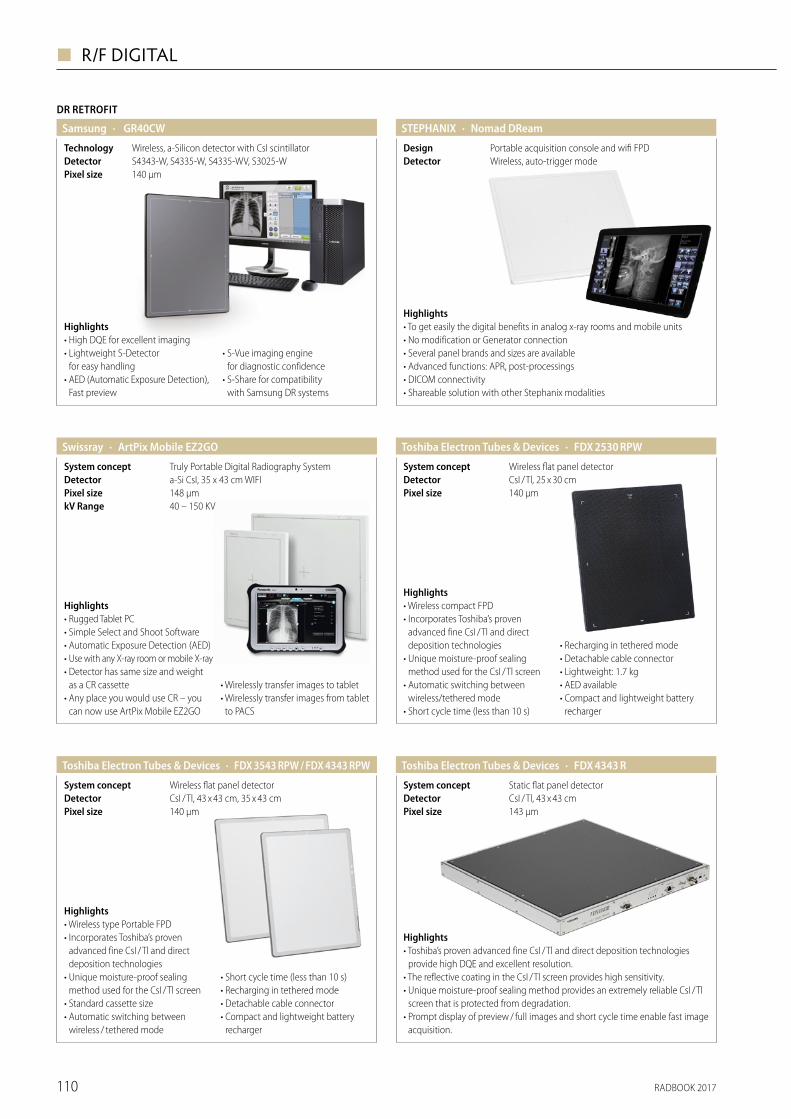
RADBOOK 2017110
◼ R/F DIGITAL
DR RETROFIT
· GR40CWSamsung
140 μmPixel size S4343-W, S4335-W, S4335-WV, S3025-WDetectorWireless, a-Silicon detector with CsI scintillatorTechnology
Highlights• High DQE for excellent imaging• Lightweight S-Detector
for easy handling• AED (Automatic Exposure Detection),
Fast preview
• S-Vue imaging engine for diagnostic confidence
• S-Share for compatibility with Samsung DR systems
Highlights• To get easily the digital benefits in analog x-ray rooms and mobile units• No modification or Generator connection• Several panel brands and sizes are available • Advanced functions: APR, post-processings• DICOM connectivity• Shareable solution with other Stephanix modalities
· Nomad DReamSTEPHANIX
Wireless, auto-trigger modeDetectorPortable acquisition console and wifi FPDDesign
· ArtPix Mobile EZ2GOSwissray
40 – 150 KVkV Range148 μmPixel sizea-Si CsI, 35 x 43 cm WIFIDetectorTruly Portable Digital Radiography SystemSystem concept
Highlights• Rugged Tablet PC• Simple Select and Shoot Software• Automatic Exposure Detection (AED)• Use with any X-ray room or mobile X-ray• Detector has same size and weight
as a CR cassette• Any place you would use CR – you
can now use ArtPix Mobile EZ2GO
• Wirelessly transfer images to tablet• Wirelessly transfer images from tablet
to PACS
Visit us at ECR, booth X5/2
AeroDR X30 is Konica Minolta’s mobile digital X-ray system, and it is unique in its own way. Why? The AeroDR detector can easily be stored and at the same time automatically charged in the bin, even during driving. The unique Lithium-Ion-Capacitor technology of the AeroDR detectors allows charging whenever and wherever without losing any perfomance capacity.Because of its retractable column, the AeroDR X30 is easy to manoeuvre. Furthermore, the system is very compact and has a smart, space saving design. Two detectors can be carried along in the bin. AeroDR X30 can be combined with Konica Minolta’s robust, carbon fiber Flat Panel Detectors available in various sizes: 10x12’’, 14x17’’ and 17x17’’.
Unique in its own way. AeroDR X30 from Konica Minolta.
KONICA MINOLTA MEDICAL & GRAPHIC IMAGING EUROPE B.V.www.konicaminolta.eu/healthcare | [email protected]
P O W E R F U L & P O R T A B L E
· FDX 2530 RPWToshiba Electron Tubes & Devices
140 μmPixel size
Wireless flat panel detectorSystem conceptCsI / Tl, 25 x 30 cmDetector
Highlights• Wireless compact FPD• Incorporates Toshiba’s proven
advanced fine CsI / Tl and direct deposition technologies
• Unique moisture-proof sealing method used for the CsI / Tl screen
• Automatic switching between wireless/tethered mode
• Short cycle time (less than 10 s)
• Recharging in tethered mode• Detachable cable connector• Lightweight: 1.7 kg• AED available• Compact and lightweight battery
recharger
· FDX 4343 RToshiba Electron Tubes & Devices
143 μmPixel sizeCsI / Tl, 43 x 43 cmDetectorStatic flat panel detectorSystem concept
Highlights• Toshiba’s proven advanced fine CsI / Tl and direct deposition technologies
provide high DQE and excellent resolution.• The reflective coating in the CsI / Tl screen provides high sensitivity.• Unique moisture-proof sealing method provides an extremely reliable CsI / Tl
screen that is protected from degradation.• Prompt display of preview / full images and short cycle time enable fast image
acquisition.
Highlights• Wireless type Portable FPD• Incorporates Toshiba’s proven
advanced fine CsI / Tl and direct deposition technologies
• Unique moisture-proof sealing method used for the CsI / Tl screen
• Standard cassette size• Automatic switching between
wireless / tethered mode
• Short cycle time (less than 10 s)• Recharging in tethered mode• Detachable cable connector• Compact and lightweight battery
recharger
· FDX 3543 RPW / FDX 4343 RPWToshiba Electron Tubes & Devices
140 μmPixel sizeCsI / Tl, 43 x 43 cm, 35 x 43 cmDetectorWireless flat panel detectorSystem concept
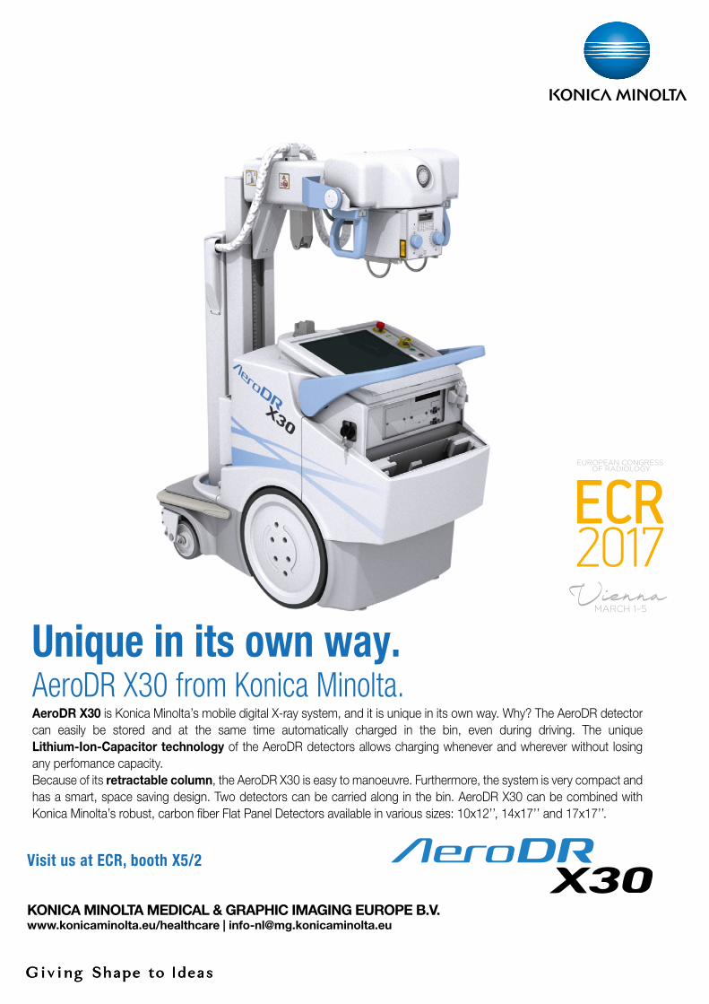
RADBOOK 2017 111
R/F DIGITAL ◼
Visit us at ECR, booth X5/2
AeroDR X30 is Konica Minolta’s mobile digital X-ray system, and it is unique in its own way. Why? The AeroDR detector can easily be stored and at the same time automatically charged in the bin, even during driving. The unique Lithium-Ion-Capacitor technology of the AeroDR detectors allows charging whenever and wherever without losing any perfomance capacity.Because of its retractable column, the AeroDR X30 is easy to manoeuvre. Furthermore, the system is very compact and has a smart, space saving design. Two detectors can be carried along in the bin. AeroDR X30 can be combined with Konica Minolta’s robust, carbon fiber Flat Panel Detectors available in various sizes: 10x12’’, 14x17’’ and 17x17’’.
Unique in its own way. AeroDR X30 from Konica Minolta.
KONICA MINOLTA MEDICAL & GRAPHIC IMAGING EUROPE B.V.www.konicaminolta.eu/healthcare | [email protected]
P O W E R F U L & P O R T A B L E
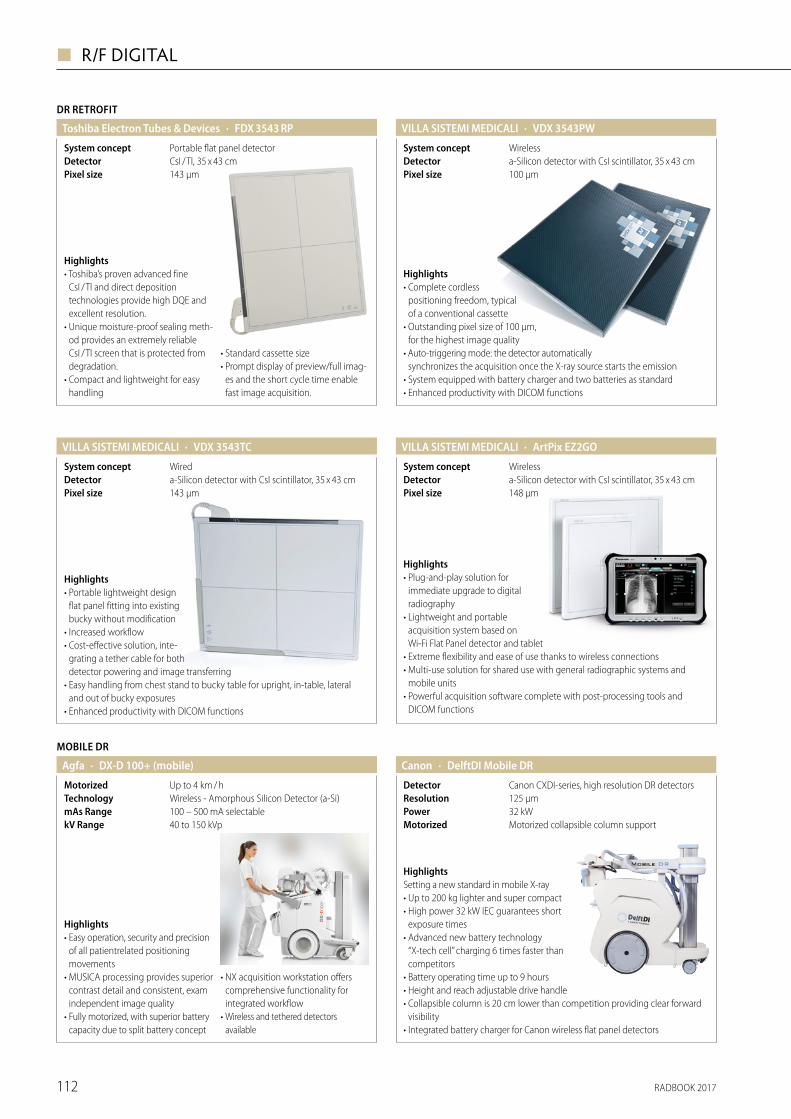
RADBOOK 2017112
◼ R/F DIGITAL
· ArtPix EZ2GOVILLA SISTEMI MEDICALI
148 μmPixel sizea-Silicon detector with CsI scintillator, 35 x 43 cmDetectorWirelessSystem concept
Highlights• Plug-and-play solution for
immediate upgrade to digital radiography
• Lightweight and portable acquisition system based on Wi-Fi Flat Panel detector and tablet
• Extreme flexibility and ease of use thanks to wireless connections• Multi-use solution for shared use with general radiographic systems and
mobile units• Powerful acquisition software complete with post-processing tools and
DICOM functions
MOBILE DR
· DX-D 100+ (mobile)Agfa
40 to 150 kVpkV Range100 – 500 mA selectablemAs RangeWireless - Amorphous Silicon Detector (a-Si)TechnologyUp to 4 km / hMotorized
Highlights• Easy operation, security and precision
of all patientrelated positioning movements
• MUSICA processing provides superior contrast detail and consistent, exam independent image quality
• Fully motorized, with superior battery capacity due to split battery concept
• NX acquisition workstation offers comprehensive functionality for integrated workflow
• Wireless and tethered detectors available
· DelftDI Mobile DRCanon
Motorized collapsible column support Motorized32 kWPower125 µmResolutionCanon CXDI-series, high resolution DR detectorsDetector
HighlightsSetting a new standard in mobile X-ray• Up to 200 kg lighter and super compact• High power 32 kW IEC guarantees short
exposure times• Advanced new battery technology
“X-tech cell” charging 6 times faster than competitors
• Battery operating time up to 9 hours• Height and reach adjustable drive handle• Collapsible column is 20 cm lower than competition providing clear forward
visibility• Integrated battery charger for Canon wireless flat panel detectors
· VDX 3543PWVILLA SISTEMI MEDICALI
100 μmPixel sizea-Silicon detector with CsI scintillator, 35 x 43 cmDetectorWirelessSystem concept
Highlights• Complete cordless
positioning freedom, typical of a conventional cassette
• Outstanding pixel size of 100 μm, for the highest image quality
• Auto-triggering mode: the detector automatically synchronizes the acquisition once the X-ray source starts the emission
• System equipped with battery charger and two batteries as standard• Enhanced productivity with DICOM functions
· VDX 3543TCVILLA SISTEMI MEDICALI
143 μmPixel sizea-Silicon detector with CsI scintillator, 35 x 43 cmDetectorWiredSystem concept
Highlights• Portable lightweight design
flat panel fitting into existing bucky without modification
• Increased workflow• Cost-effective solution, inte-
grating a tether cable for both detector powering and image transferring
• Easy handling from chest stand to bucky table for upright, in-table, lateral and out of bucky exposures
• Enhanced productivity with DICOM functions
DR RETROFIT
· FDX 3543 RPToshiba Electron Tubes & Devices
143 μmPixel sizeCsI / Tl, 35 x 43 cmDetectorPortable flat panel detectorSystem concept
Highlights• Toshiba’s proven advanced fine
CsI / Tl and direct deposition technologies provide high DQE and excellent resolution.
• Unique moisture-proof sealing meth-od provides an extremely reliable CsI / Tl screen that is protected from degradation.
• Compact and lightweight for easy handling
• Standard cassette size• Prompt display of preview/full imag-
es and the short cycle time enable fast image acquisition.
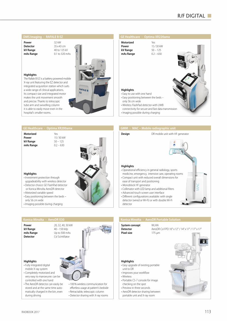
RADBOOK 2017 113
R/F DIGITAL ◼
· Optima XR200amxGE Healthcare
0.2 – 630mAs Range50 – 125kV Range15 / 30 kWPowerYesMotorized
Highlights• Investment protection through
upgradeability with wireless detector• Detector choice: GE FlashPad detector
or Konica Minolta AeroDR detector• Motorized variable speed• Easy positioning between the beds –
only 56 cm wide• Imaging possible during charging
· Optima XR220amxGE Healthcare
0.2 – 630mAs Range50 – 125kV Range15 / 30 kWPowerYesMotorized
Highlights• Easy to use with one hand• Easy positioning between the beds –
only 56 cm wide• Wireless FlashPad detector with UWB
connectivity for secure and fast data transmission• Imaging possible during charging
· MAC – Mobile radiographic unitGMM
DR mobile unit with HF generatorDesign
Highlights• Operational efficiency in general radiology, sports
medicine, emergency, intensive care, operating rooms • Compact unit with reduced overall dimensions for
ease of transport and positioning • Monoblock HF generator • Collimator with LED lamp and additional filters • Advanced touch screen user interface• Different configurations available: with single
detector (wired or Wi-Fi) or with double Wi-Fi detector
HighlightsThe Rafale B EZ is a battery powered mobile X-ray unit featuring the EZ detector and integrated acquisition station which suits a wide range of clinical applications. Its compact size and integrated motor makes the unit movement smooth and precise. Thanks to telescopic tube arm and swivelling column it is able to easily move even in the hospital's smaller rooms.
· RAFALE B EZDMS Imaging
0.1 to 320 mAsmAs Range40 to 125 kVkV Range35 x 43 cmDetector32 kWPower
Highlights• Easy upgrade of existing portable
unit to DR• Improves your workflow• Wireless• Portable CS-7 console for image
checking on the spot• Preview in three seconds• AeroDR detector sharing between
portable unit and X-ray room
· AeroDR Portable SolutionKonica Minolta
175 µmPixel sizeAeroDR CsI FPD 10" x 12" / 14" x 17" / 17" x 17"DetectorWLANSystem concept
· AeroDR X30Konica Minolta
CsI ScintillatorDetectorUp to 500 mAsmAs Range40 – 150 kVpkV Range20, 32, 40, 50 kWPower
Highlights• Fully integrated digital
mobile X-ray system• Completely motorized and
very easy to manoeuvre: can be controlled with one hand
• The AeroDR detector can easily be stored and at the same time auto-matically charged in the bin, even during driving
• 100 % wireless communication for effortless usage at patient's bedside
• Retractable, telescopic column • Detector sharing with X-ray rooms
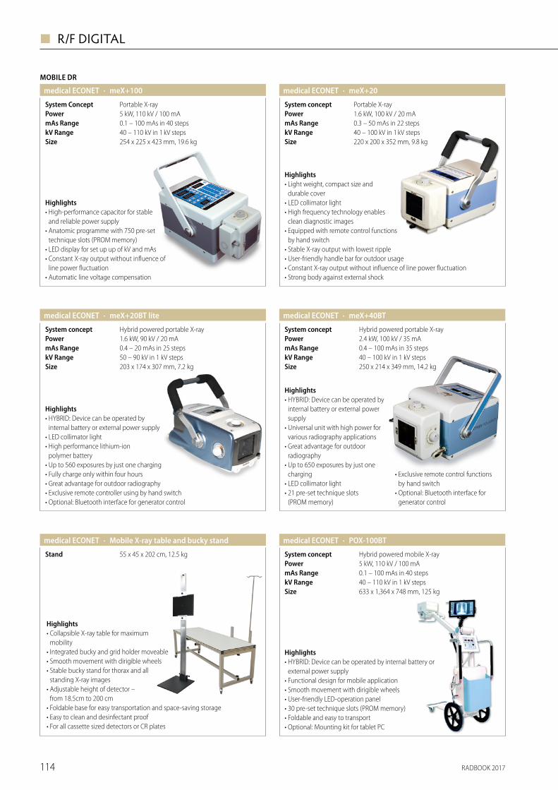
RADBOOK 2017114
◼ R/F DIGITAL
· meX+20medical ECONET
40 – 100 kV in 1 kV stepskV Range0.3 – 50 mAs in 22 stepsmAs Range1.6 kW, 100 kV / 20 mAPowerPortable X-raySystem concept
Highlights• Light weight, compact size and
durable cover• LED collimator light• High frequency technology enables
clean diagnostic images• Equipped with remote control functions
by hand switch• Stable X-ray output with lowest ripple• User-friendly handle bar for outdoor usage• Constant X-ray output without influence of line power fluctuation• Strong body against external shock
220 x 200 x 352 mm, 9.8 kgSize
· meX+20BT litemedical ECONET
50 – 90 kV in 1 kV stepskV Range0.4 – 20 mAs in 25 stepsmAs Range1.6 kW, 90 kV / 20 mAPowerHybrid powered portable X-raySystem concept
Highlights• HYBRID: Device can be operated by
internal battery or external power supply• LED collimator light• High performance lithium-ion
polymer battery• Up to 560 exposures by just one charging• Fully charge only within four hours• Great advantage for outdoor radiography• Exclusive remote controller using by hand switch• Optional: Bluetooth interface for generator control
203 x 174 x 307 mm, 7.2 kgSize
· meX+40BTmedical ECONET
40 – 100 kV in 1 kV steps250 x 214 x 349 mm, 14.2 kg
kV RangeSize
0.4 – 100 mAs in 35 stepsmAs Range2.4 kW, 100 kV / 35 mAPowerHybrid powered portable X-raySystem concept
Highlights• HYBRID: Device can be operated by
internal battery or external power supply
• Universal unit with high power for various radiography applications
• Great advantage for outdoor radiography
• Up to 650 exposures by just one charging
• LED collimator light• 21 pre-set technique slots
(PROM memory)
• Exclusive remote control functions by hand switch
• Optional: Bluetooth interface for generator control
· Mobile X-ray table and bucky standmedical ECONET
55 x 45 x 202 cm, 12.5 kgStand
Highlights• Collapsible X-ray table for maximum
mobility• Integrated bucky and grid holder moveable• Smooth movement with dirigible wheels• Stable bucky stand for thorax and all
standing X-ray images• Adjustable height of detector –
from 18.5cm to 200 cm• Foldable base for easy transportation and space-saving storage • Easy to clean and desinfectant proof• For all cassette sized detectors or CR plates
· POX-100BTmedical ECONET
40 – 110 kV in 1 kV stepskV Range0.1 – 100 mAs in 40 stepsmAs Range5 kW, 110 kV / 100 mAPowerHybrid powered mobile X-raySystem concept
Highlights• HYBRID: Device can be operated by internal battery or
external power supply• Functional design for mobile application• Smooth movement with dirigible wheels• User-friendly LED-operation panel• 30 pre-set technique slots (PROM memory)• Foldable and easy to transport• Optional: Mounting kit for tablet PC
633 x 1,364 x 748 mm, 125 kgSize
MOBILE DR
· meX+100medical ECONET
40 – 110 kV in 1 kV stepskV Range0.1 – 100 mAs in 40 stepsmAs Range5 kW, 110 kV / 100 mAPowerPortable X-raySystem Concept
Highlights• High-performance capacitor for stable
and reliable power supply• Anatomic programme with 750 pre-set
technique slots (PROM memory)• LED display for set up up of kV and mAs• Constant X-ray output without influence of
line power fluctuation• Automatic line voltage compensation
254 x 225 x 423 mm, 19.6 kgSize
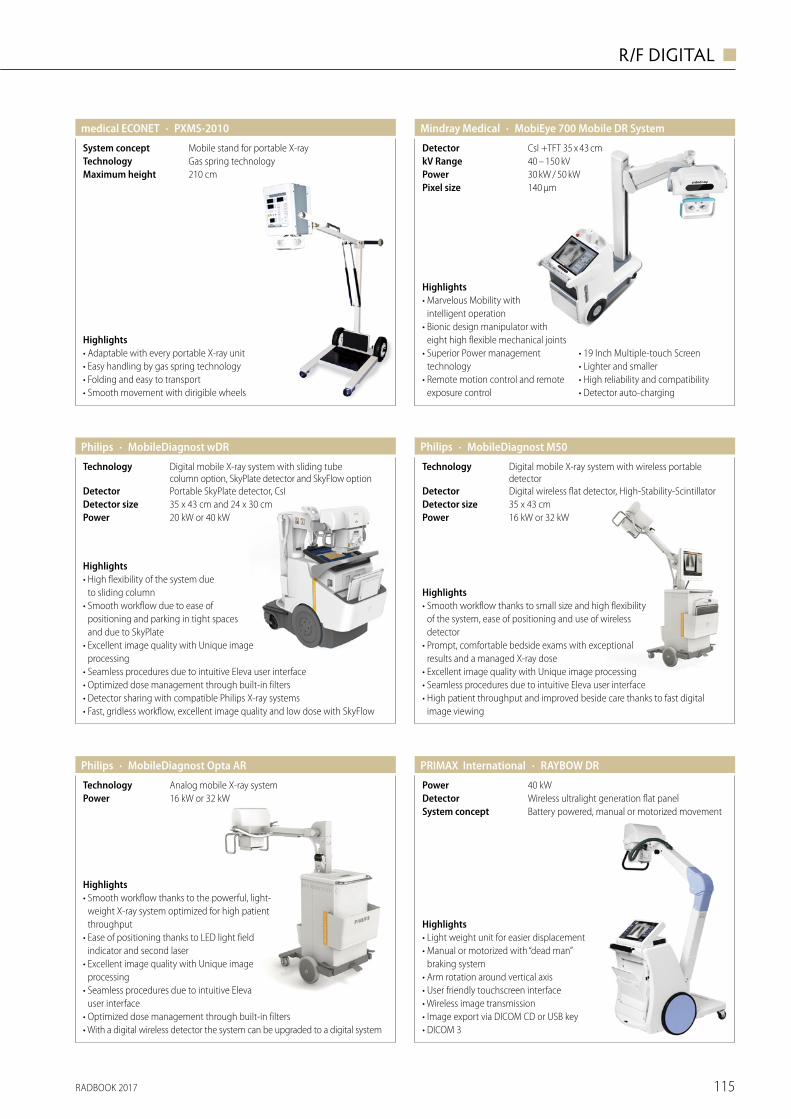
RADBOOK 2017 115
R/F DIGITAL ◼
· MobiEye 700 Mobile DR SystemMindray Medical
140 μmPixel size30 kW / 50 kWPower40 – 150 kVkV RangeCsI +TFT 35 x 43 cmDetector
Highlights• Marvelous Mobility with
intelligent operation• Bionic design manipulator with
eight high flexible mechanical joints • Superior Power management
technology• Remote motion control and remote
exposure control
• 19 Inch Multiple-touch Screen • Lighter and smaller• High reliability and compatibility • Detector auto-charging
· PXMS-2010medical ECONET
210 cmMaximum heightGas spring technologyTechnologyMobile stand for portable X-raySystem concept
Highlights• Adaptable with every portable X-ray unit• Easy handling by gas spring technology• Folding and easy to transport• Smooth movement with dirigible wheels
· MobileDiagnost wDR
35 x 43 cm and 24 x 30 cmPortable SkyPlate detector, CsI
20 kW or 40 kW
Digital mobile X-ray system with sliding tube column option, SkyPlate detector and SkyFlow option
Detector sizeDetector
Power
Technology
Highlights• High flexibility of the system due
to sliding column • Smooth workflow due to ease of
positioning and parking in tight spaces and due to SkyPlate
• Excellent image quality with Unique image processing
• Seamless procedures due to intuitive Eleva user interface• Optimized dose management through built-in filters• Detector sharing with compatible Philips X-ray systems• Fast, gridless workflow, excellent image quality and low dose with SkyFlow
Philips · MobileDiagnost M50
35 x 43 cmDigital wireless flat detector, High-Stability-Scintillator
16 kW or 32 kW
Digital mobile X-ray system with wireless portable detector
Detector sizeDetector
Power
Technology
Highlights• Smooth workflow thanks to small size and high flexibility
of the system, ease of positioning and use of wireless detector
• Prompt, comfortable bedside exams with exceptional results and a managed X-ray dose
• Excellent image quality with Unique image processing• Seamless procedures due to intuitive Eleva user interface• High patient throughput and improved beside care thanks to fast digital
image viewing
Philips
· MobileDiagnost Opta AR
16 kW or 32 kWAnalog mobile X-ray system
PowerTechnology
Highlights• Smooth workflow thanks to the powerful, light-
weight X‐ray system optimized for high patient throughput
• Ease of positioning thanks to LED light field indicator and second laser
• Excellent image quality with Unique image processing
• Seamless procedures due to intuitive Eleva user interface
• Optimized dose management through built-in filters• With a digital wireless detector the system can be upgraded to a digital system
Philips
Highlights• Light weight unit for easier displacement• Manual or motorized with “dead man”
braking system• Arm rotation around vertical axis• User friendly touchscreen interface• Wireless image transmission• Image export via DICOM CD or USB key• DICOM 3
· RAYBOW DRPRIMAX International
Battery powered, manual or motorized movementSystem conceptWireless ultralight generation flat panelDetector40 kWPower
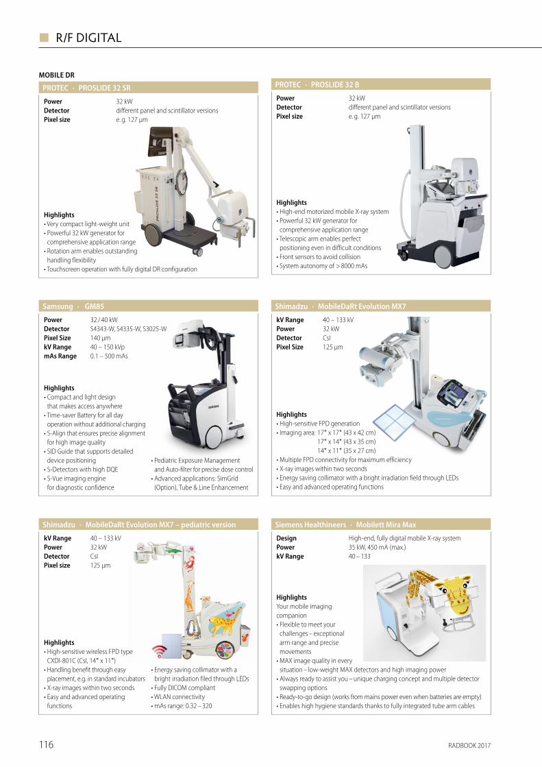
RADBOOK 2017116
◼ R/F DIGITAL
HighlightsYour mobile imaging companion• Flexible to meet your
challenges – exceptional arm range and precise movements
• MAX image quality in every situation – low-weight MAX detectors and high imaging power
• Always ready to assist you – unique charging concept and multiple detector swapping options
• Ready-to-go design (works from mains power even when batteries are empty) • Enables high hygiene standards thanks to fully integrated tube arm cables
· Mobilett Mira MaxSiemens Healthineers
40 – 133kV Range35 kW, 450 mA (max.)PowerHigh-end, fully digital mobile X-ray systemDesign
· MobileDaRt Evolution MX7Shimadzu
125 µmPixel SizeCsIDetector32 kWPower40 – 133 kVkV Range
Highlights• High-sensitive FPD generation• Imaging area: 17" x 17" (43 x 42 cm) 17" x 14" (43 x 35 cm) 14" x 11" (35 x 27 cm)• Multiple FPD connectivity for maximum efficiency• X-ray images within two seconds• Energy saving collimator with a bright irradiation field through LEDs• Easy and advanced operating functions
· MobileDaRt Evolution MX7 – pediatric versionShimadzu
Pixel sizeDetectorPowerkV Range
Highlights• High-sensitive wireless FPD type
CXDI-801C (CsI, 14" x 11")• Handling benefit through easy
placement, e.g. in standard incubators• X-ray images within two seconds• Easy and advanced operating
functions
• Energy saving collimator with a bright irradiation filed through LEDs
• Fully DICOM compliant• WLAN connectivity• mAs range: 0.32 – 320
125 µmCsI32 kW40 – 133 kV
· PROSLIDE 32 BPROTEC
e. g. 127 µmPixel size
32 kWPowerdifferent panel and scintillator versionsDetector
Highlights• High-end motorized mobile X-ray system• Powerful 32 kW generator for
comprehensive application range• Telescopic arm enables perfect
positioning even in difficult conditions• Front sensors to avoid collision• System autonomy of > 8000 mAs
· GM85Samsung
40 – 150 kVp0.1 – 500 mAs
kV RangemAs Range
140 μmPixel Size S4343-W, S4335-W, S3025-WDetector32 / 40 kWPower
Highlights• Compact and light design
that makes access anywhere• Time-saver Battery for all day
operation without additional charging• S-Align that ensures precise alignment
for high image quality• SID Guide that supports detailed
device positioning• S-Detectors with high DQE• S-Vue imaging engine
for diagnostic confidence
• Pediatric Exposure Management and Auto-filter for precise dose control
• Advanced applications: SimGrid (Option), Tube & Line Enhancement
MOBILE DR
· PROSLIDE 32 SRPROTEC
e. g. 127 µmPixel size
32 kWPowerdifferent panel and scintillator versionsDetector
Highlights• Very compact light-weight unit• Powerful 32 kW generator for
comprehensive application range• Rotation arm enables outstanding
handling flexibility• Touchscreen operation with fully digital DR configuration
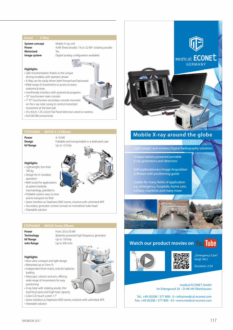
RADBOOK 2017 117
· X-WaySimad
Digital (analog configuration available)YesMotorized
Image system
4 kW (fixed anode) / 16 or 32 kW (rotating anode)PowerMobile X-ray unitSystem concept
Highlights• Safe movimentation thanks to the unique
driving modality with operator ahead• X-Way can be easily driven both forward and backward• Wide range of movements to access to every
anatomical areas• Userfriendly interface with anatomical programs• 10" touchscreen main console• 7" TFT touchscreen secondary console mounted
on the x-ray tube casing to control motorized movement at the bed side
• 35 x 43cm / 25 x 32cm Flat Panel detectors wired or wireless• Full DICOM connectivity
Highlights• Lightweight, less than
100 kg• Design for in /outdoor
operation• Well-suited for applications
at patient bedside, traumatology, paediatrics
• Foldable system easy to store and to transport on field
• Same interface as Stephanix RAD rooms, intuitive with unlimited APR• Secondary generator control console on monoblock tube head• Shareable solution
· MOVIX 4 / 8 DReamSTEPHANIX
Up to 125 kVpkV RangeFoldable and transportable in a dedicated caseDesign4 / 8 kWPower
Mobile X-ray around the globe
medical ECONET GmbHIm Erlengrund 20 • D-46149 Oberhausen
Tel.: +49 (0)208 / 377 890 - 0 • [email protected]: +49 (0)208 / 377 890 - 55 • www.medical-econet.com
„Emergency Care“ (engl. Ver.)
Duration: 2:50
Mobile X-ray around the globe
Light weight and wireless Digital Radiography solutions
Unique battery powered portableX-ray generators and detectors
Self explenationary Image AcquisitionSoftware with positioning guide
Usable for many fields of applicatione.g. emergency, hospitals, home care,military, maritime and many more
Highlights• New ultra-compact and light design• Motorized up to 5 km / h• Independent from mains, only for batteries
loading• Telescopic column and arm, offering
wide range of movements for easy positioning
• X-ray tube with rotating anode, thin dual focal spots and high heat capacity
• Color LCD touch screen 17"• Same interface as Stephanix RAD rooms, intuitive with unlimited APR• Shareable solution
· MOVIX Series DReamSTEPHANIX
Up to 500 mAsmAs RangeUp to 150 kVpkV RangeBatteries powered high frequency generatorTechnologyFrom 20 to 50 kWPower
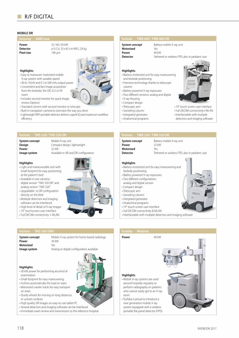
RADBOOK 2017118
◼ R/F DIGITAL
· MobirexToshiba
40 kWPower
Highlights• Mobile X-ray systems are used
around hospitals regularly to perform radiography on patients who cannot easily get to an X-ray room.
• Toshiba is proud to introduce a new generation mobile X-ray system equipped with a wireless portable flat panel detector (FPD).
· TMS 320 / TMS 320 DRTechnix
Available in AR and DR configurationImage system32 kWPowerCompact design, lightweightDesignMobile X-ray unitSystem concept
Highlights• Light and maneuverable unit with
small footprint for easy positioning at the patient's bed
• Available in two versions: digital version "TMS 320 DR" and analog version "TMS 320"
• Upgradable to DR configuration directly on the field
• Multiple detectors and imaging software can be interfaced
• High level of detail of X-ray images• 19" touchscreen user interface• Full DICOM connectivity + WLAN
· TMB 320 / TMB 320 DRTechnix
Tethered or wireless FPD, also in pediatric sizeDetectorYesMotorized32 kWPowerBattery mobile X-ray unitSystem concept
Highlights• Battery-motorized unit for easy maneuvering and
bedside positioning• Battery powered X-ray exposures• Two different configurations:
analog and digital version• Compact design• Telescopic arm• Swiveling column• Integrated generator• Anatomical programs• 19" touch screen user interface• Full DICOM connectivity & WLAN• Interfaceable with multiple detectors and imaging software
· TMS 300 DRHTechnix
Analog or digital configuration availableImage systemYesMotorized30 kWPowerMobile X-ray system for home-based radiologySystem concept
Highlights• 30 kW power for performing any kind of
examination• Small footprint for easy maneuvering• Inclines automatically the load on stairs• Motorized crawler tracks for easy transport
on stairs• Sturdy wheels for moving on long distances
or uneven surfaces• High quality DR images on easy-to-use tablet PC• Several detectors and imaging software can be interfaced• Immediate exam review and transmission to the reference hospital
· TMB 400 / TMB 400 DR
Tethered or wireless FPD, also in pediatric sizeDetector40 kWPowerYesMotorizedBattery mobile X-ray unitSystem concept
Highlights• Battery-motorized unit for easy maneuvering
and bedside positioning• Freeview technology thanks to telescopic
column• Battery powered X-ray exposures• Two different versions: analog and digital• X-ray Housing• Compact design• Telescopic arm• Swiveling column• Integrated generator• Anatomical programs
Technix
• 19" touch screen user interface• Full DICOM connectivity+WLAN• Interfaceable with multiple
detectors and imaging software
MOBILE DR
· ddRCruzeSwissray
Pixel sizeDetectorPower
Highlights• Easy to maneuver motorized mobile
X-ray system with variable speed• 40 to 150 kV and 0.1 to 500 mAs output power• Convenient and fast image acquisition
from the bedside, the OR, ICU or ER room
• Includes second monitor for quick image review (Option)
• Standard column with second monitor or telscopic• Built in navigation-camera to overview the way you drive• Lightweight WIFI portable detector delivers superb IQ and maximum workflow
efficiency
148 µma-Si CsI, 35 x 43 cm WiFi, 2.8 kg32 / 40 / 50 kW
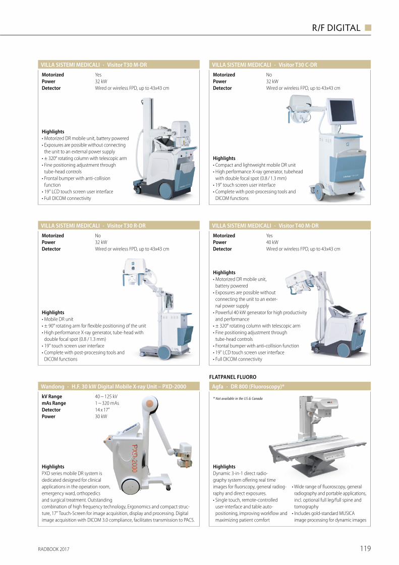
RADBOOK 2017 119
R/F DIGITAL ◼
· Visitor T30 C-DRVILLA SISTEMI MEDICALI
Wired or wireless FPD, up to 43x43 cmDetector32 kWPowerNoMotorized
Highlights• Compact and lightweight mobile DR unit• High performance X-ray generator, tubehead
with double focal spot (0.8 / 1.3 mm)• 19" touch screen user interface• Complete with post-processing tools and
DICOM functions
· Visitor T30 M-DR VILLA SISTEMI MEDICALI
Wired or wireless FPD, up to 43x43 cmDetector32 kWPowerYesMotorized
Highlights• Motorized DR mobile unit, battery powered• Exposures are possible without connecting
the unit to an external power supply• ± 320° rotating column with telescopic arm• Fine positioning adjustment through
tube-head controls• Frontal bumper with anti-collision
function• 19" LCD touch screen user interface • Full DICOM connectivity
· Visitor T30 R-DR VILLA SISTEMI MEDICALI
Wired or wireless FPD, up to 43x43 cmDetector32 kWPowerNoMotorized
Highlights• Mobile DR unit • ± 90° rotating arm for flexible positioning of the unit• High performance X-ray generator, tube-head with
double focal spot (0.8 / 1.3 mm) • 19" touch screen user interface • Complete with post-processing tools and
DICOM functions
· Visitor T40 M-DRVILLA SISTEMI MEDICALI
Wired or wireless FPD, up to 43x43 cmDetector40 kWPowerYesMotorized
Highlights• Motorized DR mobile unit,
battery powered• Exposures are possible without
connecting the unit to an exter-nal power supply
• Powerful 40 kW generator for high productivity and performance
• ± 320° rotating column with telescopic arm• Fine positioning adjustment through
tube-head controls• Frontal bumper with anti-collision function• 19" LCD touch screen user interface • Full DICOM connectivity
HighlightsPXD series mobile DR system is dedicated designed for clinical applications in the operation room, emergency ward, orthopedics and surgical treatment. Outstanding combination of high frequency technology, Ergonomics and compact struc-ture, 17" Touch-Screen for image acquisition, display and processing. Digital image acquisition with DICOM 3.0 compliance, facilitates transmission to PACS.
· H.F. 30 kW Digital Mobile X-ray Unit – PXD-2000
30 kWPower14 x 17"Detector1 ~ 320 mAsmAs Range40 ~ 125 kVkV Range
Wandong
FLATPANEL FLUORO
· DR 800 (Fluoroscopy)*Agfa
HighlightsDynamic 3-in-1 direct radio- graphy system offering real time images for fluorscopy, general radiog-raphy and direct exposures.• Single touch, remote-controlled
user-interface and table auto- positioning, improving workflow and maximizing patient comfort
• Wide range of fluoroscopy, general radiography and portable applications, incl. optional full leg/full spine and tomography
• Includes gold-standard MUSICA i mage processing for dynamic images
* Not available in the US & Canada
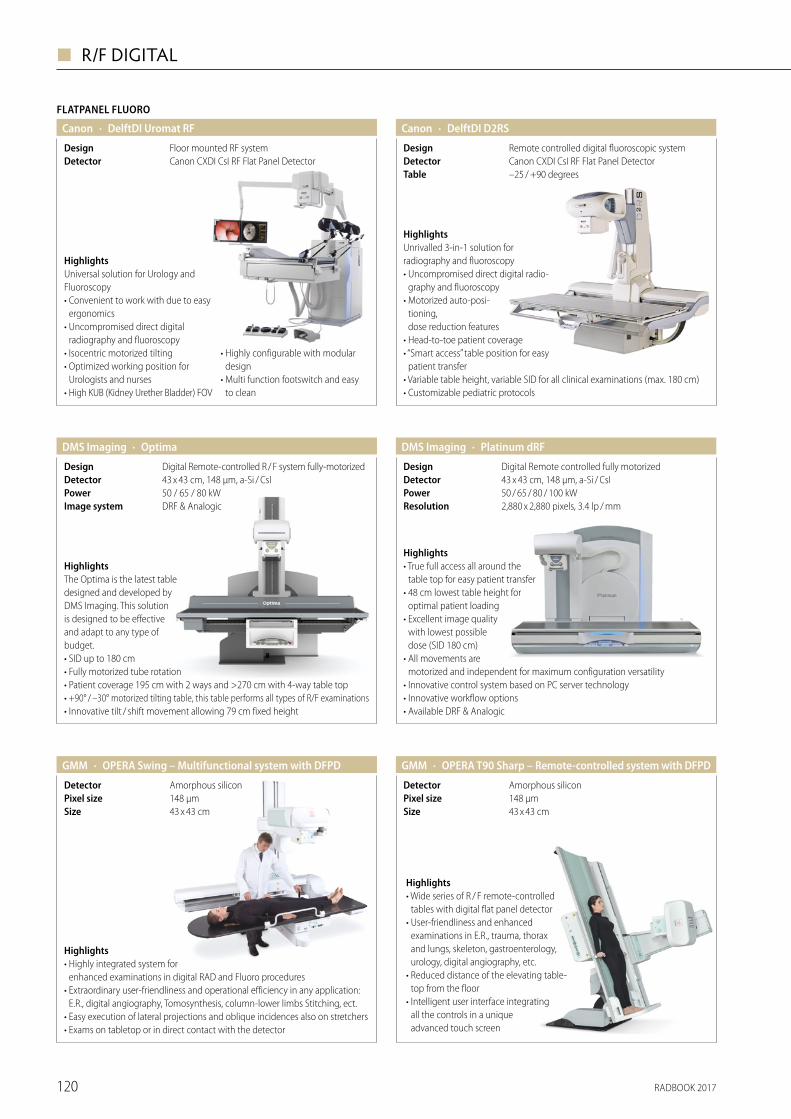
RADBOOK 2017120
◼ R/F DIGITAL
Canon
FLATPANEL FLUORO
· DelftDI Uromat RF
Canon CXDI CsI RF Flat Panel DetectorDetectorFloor mounted RF systemDesign
HighlightsUniversal solution for Urology and Fluoroscopy• Convenient to work with due to easy
ergonomics• Uncompromised direct digital
radiography and fluoroscopy• Isocentric motorized tilting• Optimized working position for
Urologists and nurses• High KUB (Kidney Urether Bladder) FOV
• Highly configurable with modular design
• Multi function footswitch and easy to clean
· DelftDI D2RSCanon
–25 / +90 degreesTableCanon CXDI CsI RF Flat Panel DetectorDetectorRemote controlled digital fluoroscopic systemDesign
HighlightsUnrivalled 3-in-1 solution for radio graphy and fluoroscopy• Uncompromised direct digital radio-
graphy and fluoroscopy• Motorized auto-posi-
tioning, dose reduction features
• Head-to-toe patient coverage• “Smart access” table position for easy
patient transfer• Variable table height, variable SID for all clinical examinations (max. 180 cm)• Customizable pediatric protocols
Highlights• Highly integrated system for
enhanced examinations in digital RAD and Fluoro procedures• Extraordinary user-friendliness and operational efficiency in any application:
E.R., digital angiography, Tomosynthesis, column-lower limbs Stitching, ect. • Easy execution of lateral projections and oblique incidences also on stretchers• Exams on tabletop or in direct contact with the detector
· OPERA Swing – Multifunctional system with DFPDGMM
43 x 43 cmSize148 µmPixel sizeAmorphous silicon Detector
· OPERA T90 Sharp – Remote-controlled system with DFPD
43 x 43 cmSize148 µmPixel sizeAmorphous siliconDetector
Highlights• Wide series of R / F remote- controlled
tables with digital flat panel detector• User-friendliness and enhanced
examinations in E.R., trauma, thorax and lungs, skeleton, gastroenterology, urology, digital angiography, etc.
• Reduced distance of the elevating table-top from the floor
• Intelligent user interface inte grating all the controls in a unique advanced touch screen
GMM
· OptimaDMS Imaging
43 x 43 cm, 148 µm, a-Si / CsIDetectorDigital Remote-controlled R / F system fully-motorizedDesign
50 / 65 / 80 kWPowerDRF & AnalogicImage system
HighlightsThe Optima is the latest table designed and developed by DMS Imaging. This solution is designed to be effective and adapt to any type of budget. • SID up to 180 cm• Fully motorized tube rotation• Patient coverage 195 cm with 2 ways and >270 cm with 4-way table top• +90° / –30° motorized tilting table, this table performs all types of R/F examinations• Innovative tilt / shift movement allowing 79 cm fixed height
· Platinum dRFDMS Imaging
43 x 43 cm, 148 µm, a-Si / CsIDetector50 / 65 / 80 / 100 kWPower
Digital Remote controlled fully motorizedDesign
2,880 x 2,880 pixels, 3.4 lp / mm Resolution
Highlights• True full access all around the
table top for easy patient transfer• 48 cm lowest table height for
optimal patient loading • Excellent image quality
with lowest possible dose (SID 180 cm)
• All movements are motorized and independent for maximum configuration versatility
• Innovative control system based on PC server technology• Innovative workflow options• Available DRF & Analogic
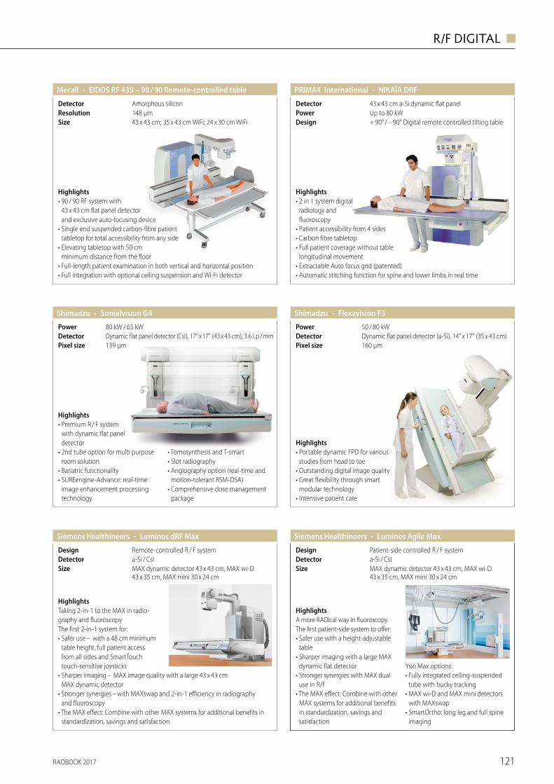
RADBOOK 2017 121
R/F DIGITAL ◼
· EIDOS RF 439 – 90 / 90 Remote-controlled table Mecall
43 x 43 cm; 35 x 43 cm WiFi; 24 x 30 cm WiFi Size148 µmResolutionAmorphous siliconDetector
Highlights• 90 / 90 RF system with
43 x 43 cm flat panel detector and exclusive auto-focusing device
• Single end suspended carbon-fibre patient tabletop for total accessibility from any side
• Elevating tabletop with 50 cm minimum distance from the floor
• Full-length patient examination in both vertical and horizontal position• Full integration with optional ceiling suspension and Wi-Fi detector
Highlights• Portable dynamic FPD for various
studies from head to toe• Outstanding digital image quality• Great flexibility through smart
modular technology• Intensive patient care
· Flexavision F3Shimadzu
160 µmPixel sizeDynamic flat panel detector (a-Si), 14" x 17" (35 x 43 cm)Detector50 / 80 kWPower
· Sonialvision G4Shimadzu
139 µmPixel sizeDynamic flat panel detector (CsI), 17" x 17" (43 x 43 cm), 3.6 Lp / mmDetector80 kW / 65 kWPower
Highlights• Premium R / F system
with dynamic flat panel detector
• 2nd tube option for multi purpose room solution
• Bariatric functionality• SUREengine-Advance: real-time
image enhancement processing technology
• Tomosynthesis and T-smart• Slot radiography• Angiography option (real-time and
motion-tolerant RSM-DSA)• Comprehensive dose management
package
HighlightsA more RADical way in fluoroscopy. The first patient-side system to offer:• Safer use with a height-adjus table
table• Sharper imaging with a large MAX
dynamic flat detector• Stronger synergies with MAX dual
use in R/F• The MAX effect: Combine with other
MAX systems for additional benefits in standardization, savings and satisfaction
Ysio Max options:• Fully integrated ceiling-suspended
tube with bucky tracking • MAX wi-D and MAX mini detectors
with MAXswap• SmartOrtho: long leg and full spine
imaging
· Luminos Agile MaxSiemens Healthineers
MAX dynamic detector 43 x 43 cm, MAX wi-D 43 x 35 cm, MAX mini 30 x 24 cm
Sizea-Si / CsIDetectorPatient-side controlled R / F systemDesign
HighlightsTaking 2-in-1 to the MAX in radio-graphy and fluoroscopyThe first 2-in-1 system for:• Safer use – with a 48 cm minimum
table height, full patient access from all sides and SmartTouch touch-sensitive joysticks
• Sharper imaging – MAX image quality with a large 43 x 43 cm MAX dynamic detector
• Stronger synergies – with MAXswap and 2-in-1 efficiency in radiography and fluoroscopy
• The MAX effect: Combine with other MAX systems for additional benefits in standardization, savings and satisfaction
· Luminos dRF MaxSiemens Healthineers
MAX dynamic detector 43 x 43 cm, MAX wi-D 43 x 35 cm, MAX mini 30 x 24 cm
Sizea-Si / CsIDetectorRemote-controlled R / F systemDesign
Highlights• 2 in 1 system digital
radiology and fluoroscopy
• Patient accessibility from 4 sides• Carbon fibre tabletop• Full patient coverage without table
longitudinal movement• Extractable Auto focus grid (patented)• Automatic stitching function for spine and lower limbs in real time
· NIKAÏA DRFPRIMAX International
+ 90° / – 90° Digital remote controlled tilting tableDesignUp to 80 kWPower43 x 43 cm a-Si dynamic flat panelDetector
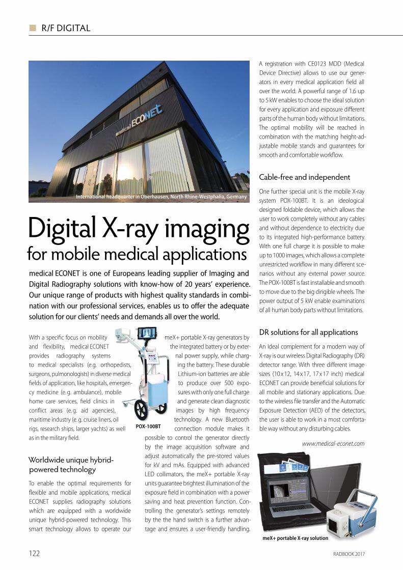
RADBOOK 2017122
◼ R/F DIGITAL
With a speci� c focus on mobility and � exibility, medical ECONET provides radiography systems to medical specialists (e.g. orthopedists, surgeons, pulmonologists) in diverse medical � elds of application, like hospitals, emergen-cy medicine (e. g. ambulance), mobile home care services, � eld clinics in con� ict areas (e. g. aid agencies), maritime industry (e. g. cruise liners, oil rigs, research ships, larger yachts) as well as in the military � eld.
Worldwide unique hybrid- powered technology
To enable the optimal requirements for � exible and mobile applications, medical ECONET supplies radiography solutions which are equipped with a worldwide unique hybrid-powered technology. This smart technology allows to operate our
Digital X-ray imaging for mobile medical applicationsmedical ECONET is one of Europeans leading supplier of Imaging and Digital Radiography solutions with know-how of 20 years’ experience.Our unique range of products with highest quality standards in combi-nation with our professional services, enables us to o� er the adequate solution for our clients’ needs and demands all over the world.
meX+ portable X-ray generators by the integrated battery or by exter-
nal power supply, while charg-ing the battery. These durable Lithium-ion batteries are able to produce over 500 expo-sures with only one full charge and generate clean diagnostic images by high frequency
technology. A new Bluetooth connection module makes it
possible to control the generator directly by the image acquisition software and adjust automatically the pre-stored values for kV and mAs. Equipped with advanced LED collimators, the meX+ portable X-ray units guarantee brightest illumination of the exposure � eld in combination with a power saving and heat prevention function. Con-trolling the generator’s settings remotely by the the hand switch is a further advan-tage and ensures a user-friendly handling.
A registration with CE0123 MDD (Medical Device Directive) allows to use our gener-ators in every medical application � eld all over the world. A powerful range of 1.6 up to 5 kW enables to choose the ideal solution for every application and exposure di� erent parts of the human body without limitations. The optimal mobility will be reached in combination with the matching height-ad-justable mobile stands and guarantees for smooth and comfortable work� ow.
Cable-free and independent
One further special unit is the mobile X-ray system POX-100BT. It is an ideological designed foldable device, which allows the user to work completely without any cables and without dependence to electricity due to its integrated high-performance battery. With one full charge it is possible to make up to 1000 images, which allows a complete unrestricted work� ow in many di� erent sce-narios without any external power source. The POX-100BT is fast installable and smooth to move due to the big dirigible wheels. The power output of 5 kW enable examinations of all human body parts without limitations.
DR solutions for all applications
An ideal complement for a modern way of X-ray is our wireless Digital Radiography (DR) detector range. With three di� erent image sizes (10 x 12, 14 x 17, 17 x 17 inch) medical ECONET can provide bene� cial solutions for all mobile and stationary applications. Due to the wireless � le transfer and the Automatic Exposure Detection (AED) of the detectors, the user is able to work in a most comforta-ble way without any disturbing cables.
www.medical-econet.com
International headquarter in Oberhausen, North Rhine-Westphalia, Germany
POX-100BT
meX+ portable X-ray solution
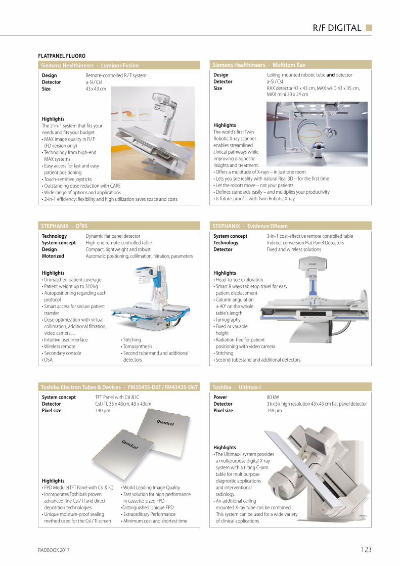
RADBOOK 2017 123
R/F DIGITAL ◼
Highlights• Unmatched patient coverage• Patient weight up to 310 kg• Autopositioning regarding each
protocol• Smart access for secure patient
transfer• Dose optimization with virtual
collimation, additional filtration, video camera . . .
• Intuitive user interface• Wireless remote• Secondary console• DSA
• Stitching • Tomosynthesis• Second tubestand and additional
detectors
· D²RSSTEPHANIX
Automatic positioning, collimation, filtration, parametersMotorizedCompact, lightweight and robustDesignHigh-end remote controlled tableSystem conceptDynamic flat panel detectorTechnology
Highlights• Head-to-toe exploration• Smart 8 ways tabletop travel for easy
patient displacement• Column angulation
± 40° on the whole table's length
• Tomography• Fixed or variable
height• Radiation-free for patient
positioning with video camera• Stitching • Second tubestand and additional detectors
· Evidence DReamSTEPHANIX
Fixed and wireless solutionsDetectorIndirect conversion Flat Panel DetectorsTechnology3-in-1 cost-effective remote controlled tableSystem concept
· Ultimax-iToshiba
148 µmPixel size3 k x 3 k high resolution 43 x 43 cm flat panel detectorDetector80 kWPower
Highlights• The Ultimax-i system provides
a multipurpose digital X-ray system with a tilting C-arm table for multipurpose diagnostic applications and interventional radiology.
• An additional ceiling mounted X-ray tube can be combined. This system can be used for a wide variety of clinical applications.
· FM3543S-D6T / FM4343S-D6TToshiba Electron Tubes & Devices
140 μmPixel sizeCsI / Tl, 35 x 43cm, 43 x 43cmDetectorTFT Panel with CsI & ICSystem concept
Highlights• FPD Module(TFT Panel with CsI & IC)• Incorporates Toshiba’s proven
advanced fine CsI / Tl and direct deposition technologies
• Unique moisture-proof sealing method used for the CsI / Tl screen
• World Leading Image Quality• Fast solution for high performance
in cassette-sized FPD•Distinguished Unique FPD• Extraordinary Performance• Minimum cost and shortest time
Siemens Healthineers
HighlightsThe world’s first Twin Robotic X-ray scanner enables streamlined clinical pathways while improving diagnostic insights and treatment.• Offers a multitude of X-rays – in just one room• Lets you see reality with natural Real 3D – for the first time• Let the robots move – not your patients• Defines standards easily – and multiplies your productivity• Is future-proof – with Twin Robotic X-ray
· Multitom Rax
RAX detector 43 x 43 cm, MAX wi-D 43 x 35 cm, MAX mini 30 x 24 cm
Sizea-Si / CsIDetectorCeiling-mounted robotic tube and detectorDesign
HighlightsThe 2-in-1 system that fits your needs and fits your budget• MAX image quality in R / F
(FD version only) • Technology from high-end
MAX systems• Easy access for fast and easy
patient positioning• Touch-sensitive joysticks • Outstanding dose reduction with CARE• Wide range of options and applications• 2-in-1 efficiency: flexibility and high utilization saves space and costs
FLATPANEL FLUORO
· Luminos FusionSiemens Healthineers
43 x 43 cmSizea-Si / CsIDetectorRemote-controlled R / F systemDesign
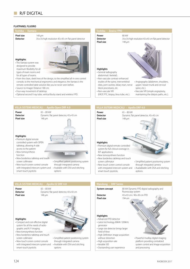
RADBOOK 2017124
◼ R/F DIGITAL
· Apollo DRF 4.0VILLA SISTEMI MEDICALI
148 μmPixel sizeDynamic flat panel detector, 43 x 43 cmDetector65 – 80 kWPower
Highlights• Premium digital remote controlled
system for full clinical coverage in R/F applications
• New tomosynthesis function • New borderless tabletop and touch
screen collimator• New touch screen control console
with integrated intercom system and smart-touch joysticks
• Simplified patient positioning system through integrated camera
• Availablable with DSA and stitching options
· Apollo EZ DRF 4.0VILLA SISTEMI MEDICALI
148 μmPixel sizeDynamic flat panel detector, 43 x 43 cmDetector65 – 80 kWPower
Highlights• Compact and cost-effective digital
system for all the needs of radio-graphic and R / F imaging
• New tomosynthesis function • New borderless tabletop and touch
screen collimator • New touch screen control console
with integrated intercom system and smart-touch joysticks
• Simplified patient positioning system through integrated camera
• Available with DSA and stitching options
· Apollo Open DRF 4.0VILLA SISTEMI MEDICALI
148 μmPixel sizeDynamic flat panel detector, 43 x 43 cmDetector65 – 80 kWPower
Highlights• Premium digital remote
controlled system with OPEN tabletop, allowing 4-side access to the patient
• New tomosynthesis function
• New borderless tabletop and touch screen collimator
• New touch screen control console with integrated intercom system and smart-touch joysticks
• Simplified patient positioning system through integrated camera
• Available with DSA and stitching options
Wandong · DRF Series
194 umPixel size43 x 43 cm / 40 x 30 cm FPDDetector
80 kW Dynamic FPD digital radiography and fluoroscopy system
System concept
Highlights• Advanced FPD detector • Latest technology 80kW / 200kHz
generator • Large size detector brings larger
Field of View• High Definition image acquisition
without distortion• High acquisition rate• Variable SID • Outstanding user experience
• Powerful InvaRay digital imaging platform providing centralized system control and image acquisition and processing
FLATPANEL FLUORO
· XantaraToshiba
3 k x 3 k high resolution 43 x 43 cm flat panel detectorDetector148 µmPixel size
Highlights• The Xantara system was
designed to provide maximum flexibility for all types of exam rooms and for all types of exams.
• From the clean, sleek lines of the design, to the simplified all-in-one control console, to the mechanical ergonomics and elegance, the Xantara is the remote controlled table solution like you’ve never seen before.
• Source-to-Imager Distance 180 cm. • Four-way movement of tabletop.• Optional second X-ray tube, vertical Bucky stand and wireless FPD.
· Zexira / FPDToshiba
148 µmPixel size3 k x 3 k high resolution 43 x 43 cm flat panel detectorDetector80 kWPower
Highlights• General radiography
abdominal / skeletal).• Non-vascular contrast-enhanced
studies of the spine, intervertebral disks, joint cavities, biliary tract, nerve block procedures, etc.
• Non-vascular IVR (ERCP, PTC, biopsy, ileus tube, etc.).
• Angiography (abdomen, shoulders, upper / slower trunk and cervical spine, etc.).
• Vascular IVR (simple angioplasty, maintaining the dialysis paths, etc.).
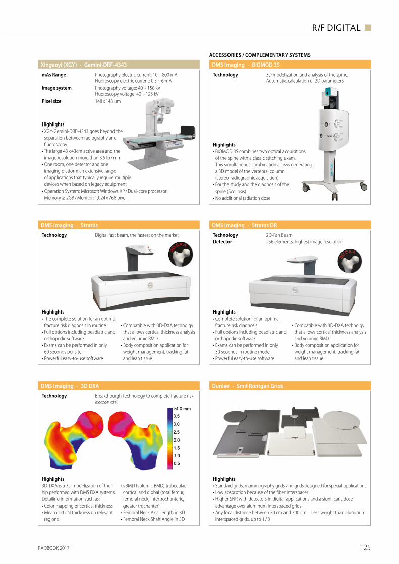
RADBOOK 2017 125
R/F DIGITAL ◼
Highlights• Standard grids, mammography grids and grids designed for special applications• Low absorption because of the fiber interspacer• Higher SNR with detectors in digital applications and a significant dose
advantage over aluminum interspaced grids• Any focal distance between 70 cm and 300 cm – Less weight than aluminum
interspaced grids, up to 1 / 3
· Smit Röntgen GridsDunlee
Highlights• XGY-Gemini-DRF-4343 goes beyond the
separation between radiography and fluoroscopy
• The large 43 x 43cm active area and the image resolution more than 3.5 lp / mm
• One room, one detector and one imaging platform an extensive range of applications that typically require multiple devices when based on legacy equipment
• Operation System: Microsoft Windows XP / Dual-core processor Memory ≥ 2GB / Monitor: 1,024 x 768 pixel
· Gemini-DRF-4343Xingaoyi (XGY)
148 x 148 μmPixel size
Photography voltage: 40 ~ 150 kV Fluoroscopy voltage: 40 ~ 125 kV
Image system
Photography electric current: 10 ~ 800 mA Fluoroscopy electric current: 0.5 ~ 6 mA
mAs Range
· StratosDMS Imaging
Digital fast beam, the fastest on the marketTechnology
Highlights• The complete solution for an optimal
fracture risk diagnosis in routine• Full options including peadiatric and
orthopedic software • Exams can be performed in only
60 seconds per site• Powerful easy-to-use software
• Compatible with 3D-DXA technolgy that allows cortical thickness analysis and volumic BMD
• Body composition application for weight management, tracking fat and lean tissue
· 3D DXADMS Imaging
Breakthourgh Technology to complete fracture risk assessment
Technology
Highlights3D-DXA is a 3D modelization of the hip performed with DMS DXA systems. Detailing information such as:• Color mapping of cortical thickness • Mean cortical thickness on relevant
regions
• vBMD (volumic BMD) trabecular, cortical and global (total femur, femoral neck, intertrochanteric, greater trochanter)
• Femoral Neck Axis Length in 3D• Femoral Neck Shaft Angle in 3D
· Stratos DRDMS Imaging
256 elements, highest image resolutionDetector2D-Fan BeamTechnology
Highlights• Complete solution for an optimal
fracture risk diagnosis• Full options including peadiatric and
orthopedic software • Exams can be performed in only
30 seconds in routine mode• Powerful easy-to-use software
• Compatible with 3D-DXA technolgy that allows cortical thickness analysis and volumic BMD
• Body composition application for weight management, tracking fat and lean tissue
3D modelization and analysis of the spine, Automatic calculation of 2D parameters
Technology
ACCESSORIES / COMPLEMENTARY SYSTEMS
· BIOMOD 3SDMS Imaging
Highlights• BIOMOD 3S combines two optical acquisitions
of the spine with a classic stitching exam. This simultaneous combination allows generating a 3D model of the vertebral column (stereo-radiographic acquisition)
• For the study and the diagnosis of the spine (Scoliosis)
• No additional radiation dose
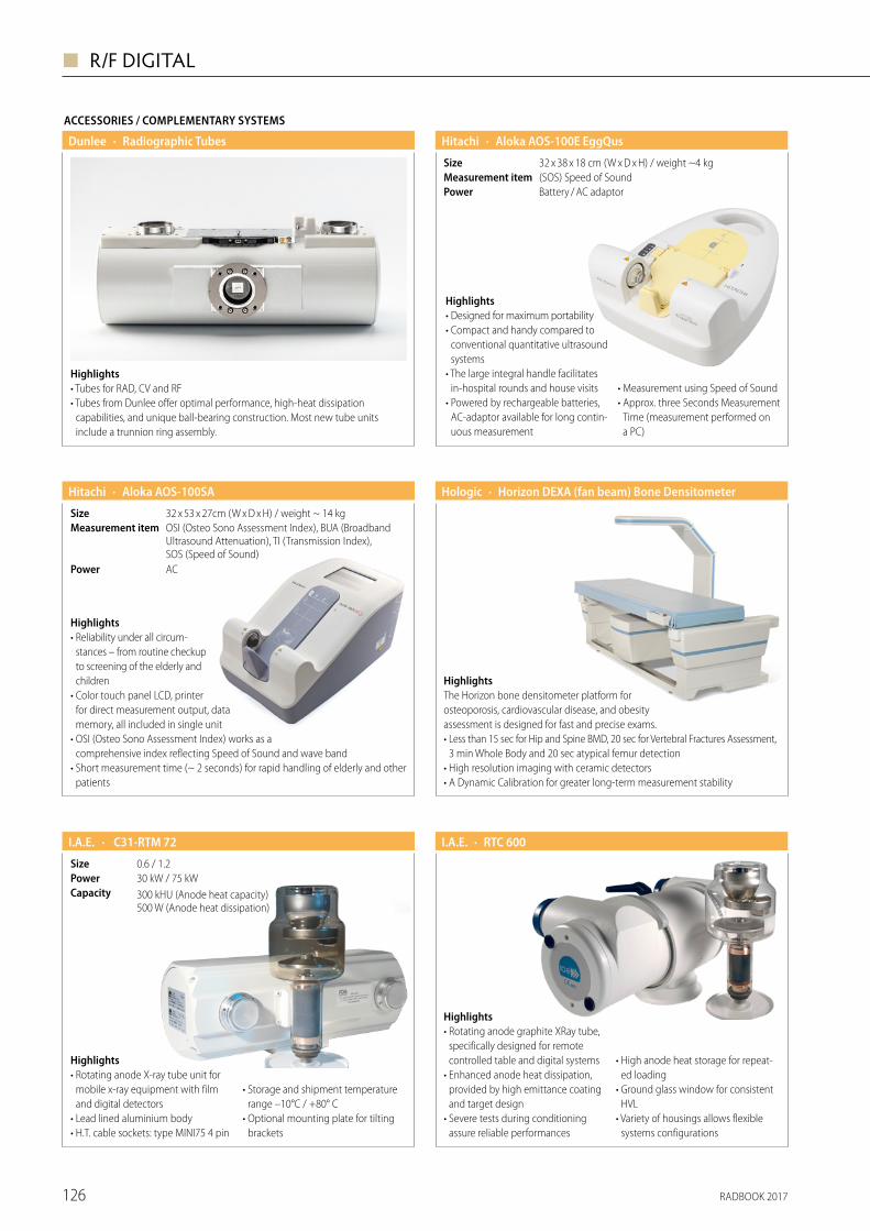
RADBOOK 2017126
◼ R/F DIGITAL
· Aloka AOS-100E EggQus Hitachi
Battery / AC adaptorPower(SOS) Speed of SoundMeasurement item32 x 38 x 18 cm (W x D x H) / weight ~4 kgSize
Highlights• Designed for maximum portability• Compact and handy compared to
conventional quantitative ultrasound systems
• The large integral handle facilitates in-hospital rounds and house visits
• Powered by rechargeable batteries, AC-adaptor available for long contin-uous measurement
• Measurement using Speed of Sound • Approx. three Seconds Measurement
Time (measurement performed on a PC)
· Aloka AOS-100SA Hitachi
ACPower
OSI (Osteo Sono Assessment Index), BUA (Broadband Ultrasound Attenuation), TI (Transmission Index), SOS (Speed of Sound)
Measurement item32 x 53 x 27cm (W x D x H) / weight ~ 14 kgSize
Highlights• Reliability under all circum-
stances – from routine checkup to screening of the elderly and children
• Color touch panel LCD, printer for direct measurement output, data memory, all included in single unit
• OSI (Osteo Sono Assessment Index) works as a comprehensive index reflecting Speed of Sound and wave band
• Short measurement time (~ 2 seconds) for rapid handling of elderly and other patients
· Horizon DEXA (fan beam) Bone Densitometer Hologic
HighlightsThe Horizon bone densitometer platform for osteoporosis, cardiovascular disease, and obesity assessment is designed for fast and precise exams.• Less than 15 sec for Hip and Spine BMD, 20 sec for Vertebral Fractures Assessment,
3 min Whole Body and 20 sec atypical femur detection• High resolution imaging with ceramic detectors• A Dynamic Calibration for greater long-term measurement stability
Highlights• Rotating anode X-ray tube unit for
mobile x-ray equipment with film and digital detectors
• Lead lined aluminium body• H.T. cable sockets: type MINI75 4 pin
• Storage and shipment temperature range –10°C / +80° C
• Optional mounting plate for tilting brackets
· C31-RTM 72I.A.E.
300 kHU (Anode heat capacity) 500 W (Anode heat dissipation)
Capacity30 kW / 75 kWPower0.6 / 1.2Size
· RTC 600I.A.E.
Highlights• Rotating anode graphite XRay tube,
specifically designed for remote controlled table and digital systems
• Enhanced anode heat dissipation, provided by high emittance coating and target design
• Severe tests during conditioning assure reliable performances
• High anode heat storage for repeat-ed loading
• Ground glass window for consistent HVL
• Variety of housings allows flexible systems configurations
ACCESSORIES / COMPLEMENTARY SYSTEMS
Highlights• Tubes for RAD, CV and RF• Tubes from Dunlee offer optimal performance, high-heat dissipation
capabilities, and unique ball-bearing construction. Most new tube units include a trunnion ring assembly.
· Radiographic TubesDunlee
RADBOOK 2017 127
R/F DIGITAL ◼
| visit us onfollow us onEuropean Hospital Verlags GmbH | Theodor-Althoff-Straße 45 | 45133 Essen
www.healthcare-in-europe.com
Trends & innovations in healthcare.
Subscribe to our Newsletter on healthcare-in-europe.com.
Onl ine Portal | Newspaper | Product Guides | Newsletter
phot
ogra
phy
by R
ober
t Hile
25 YEARSof Europeanhealthcare
communication:print & online. Subscribe to our Newsletter on healthcare-in-europe.com.
Staytunedon allchannels.
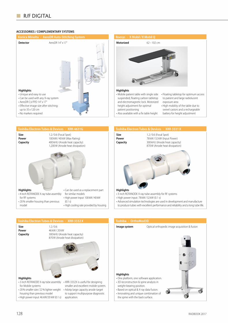
RADBOOK 2017128
◼ R/F DIGITAL
ACCESSORIES / COMPLEMENTARY SYSTEMS
· AeroDR Auto-Stitching SystemKonica Minolta
AeroDR 14" x 17"Detector
Highlights• Unique and easy to use• Can be used with any X-ray system• AeroDR CsI FPD 14" x 17"• Effective image size after stitching:
up to 35 x 120 cm• No markers required
Highlights• Mobile patient table with single side
suspended, floating carbon tabletop and electromagnetic lock. Motorized height adjustment for optimal patient positioning
• Also available with a fix table height
• Floating tabletop for optimum access to patient and large radiolucent exposure area
• High mobility of the table due to swivel castors and a rechargeable battery for height adjustment
· X Mobil / X Mobil QRoesys
62 – 102 cmMotorized
Highlights• 3 inch ROTANODE X-ray tube assembly for RF systems• High power input: 78 kW / 32 kW (0.1 s)• Advanced simulation technologies are used in development and manufacture
to produce tubes with excellent performance and reliability and a long tube life.
· XRR-3331 XToshiba Electron Tubes & Devices
300 kHU (Anode heat capacity)870 W (Anode heat dissipation)
Capacity78 kW / 32 kW (Input Power)Power1.2 / 0.6 (Focal Spot)Size
Highlights• 3 inch ROTANODE X-ray tube assembly
for Mobile systems• 20 % smaller size / 22 % lighter weight
housing than previous model• High power input: 46 kW/20 kW (0.1 s)
• XRR-3332X is useful for designing smaller and excellent mobile system.
• Adotp large capacity anode target to support multipurpose diagnostic application.
· XRR-3332 XToshiba Electron Tubes & Devices
300 kHU (Anode heat capacity)870 W (Anode heat dissipation)
Capacity46 kW / 20 kW Power1.2 / 0.6 Size
Highlights• 4 inch ROTANODE X-ray tube assembly
for RF systems• 20 % smaller housing than previous
model
• Can be used as a replacement part for similar models
• High power input: 100 kW / 40 kW (0.1 s)
• High cooling rate provided by housing
· XRR-4631GToshiba Electron Tubes & Devices
400 kHU (Anode heat capacity) 1,200 W (Anode heat dissipation)
Capacity100 kW / 40 kW (Max Rating)Power1.2 / 0.6 (Focal Spot)Size
· OrthoMod3DToshiba
Optical orthopedic image acquisition & fusionImage system
Highlights• One platform, one software application.• 3D reconstruction & spine analysis in
weight-bearing position.• Based on optical & X-ray data fusion.• Innovating and unique combination of
the spine with the back surface.
RADBOOK 2017 129
MOLECULAR IMAGING ◼
SPECT
SPECT-CT
PET-CT
PET-MR
Accessories / Complementary Systems
Molecular Imaging
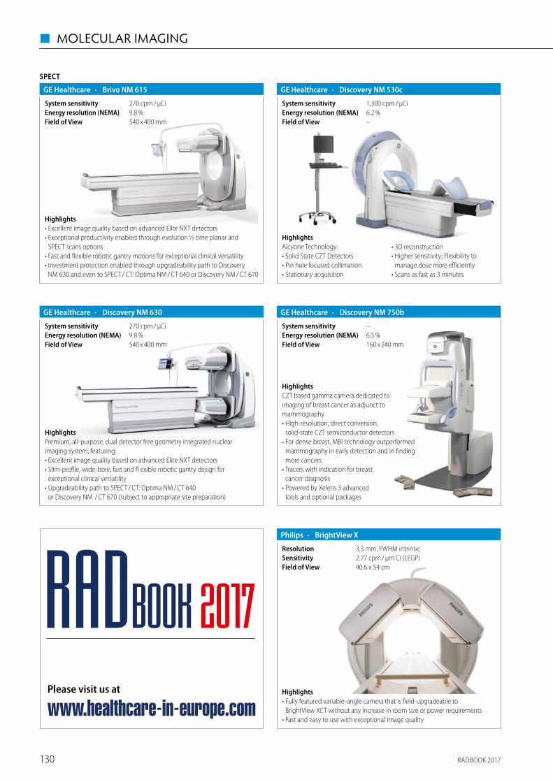
RADBOOK 2017130
◼ MOLECULAR IMAGING
· Discovery NM 630GE Healthcare
540 x 400 mmField of View9.8 %Energy resolution (NEMA)270 cpm / μCiSystem sensitivity
HighlightsPremium, all-purpose, dual detector free geometry integrated nuclear imaging system, featuring:• Excellent image quality based on advanced Elite NXT detectors• Slim-profile, wide-bore, fast and fl exible robotic gantry design for
exceptional clinical versatility• Upgradeability path to SPECT / CT: Optima NM / CT 640
or Discovery NM / CT 670 (subject to appropriate site preparation)
· Discovery NM 750bGE Healthcare
160 x 240 mmField of View6.5 %Energy resolution (NEMA)–System sensitivity
HighlightsCZT based gamma camera dedicated to imaging of breast cancer as adjunct to mammography• High-resolution, direct conversion,
solid-state CZT semiconductor detectors• For dense breast, MBI technology outperformed
mammography in early detection and in finding more cancers
• Tracers with indication for breast cancer diagnosis
• Powered by Xeleris 3 advanced tools and optional packages
SPECT
· Brivo NM 615GE Healthcare
540 x 400 mmField of View9.8 %Energy resolution (NEMA)270 cpm / μCiSystem sensitivity
Highlights• Excellent image quality based on advanced Elite NXT detectors• Exceptional productivity enabled through evolution ½ time planar and
SPECT scans options• Fast and flexible robotic gantry motions for exceptional clinical versatility• Investment protection enabled through upgradeability path to Discovery
NM 630 and even to SPECT / CT: Optima NM / CT 640 or Discovery NM / CT 670
· Discovery NM 530cGE Healthcare
–Field of View6.2 %Energy resolution (NEMA)1,300 cpm / μCiSystem sensitivity
HighlightsAlcyone Technology:• Solid State CZT Detectors• Pin hole focused collimation• Stationary acquisition
• 3D reconstruction• Higher sensitivity; Flexibility to
manage dose more efficiently• Scans as fast as 3 minutes
· BrightView XPhilips
40.6 x 54 cmField of View2.77 cpm / μm Ci (LEGP)Sensitivity3.3 mm, FWHM intrinsicResolution
Highlights• Fully featured variable-angle camera that is field-upgradeable to
BrightView XCT without any increase in room size or power requirements• Fast and easy to use with exceptional image quality
Please visit us at
www.healthcare-in-europe.com
RADBOOK 2017
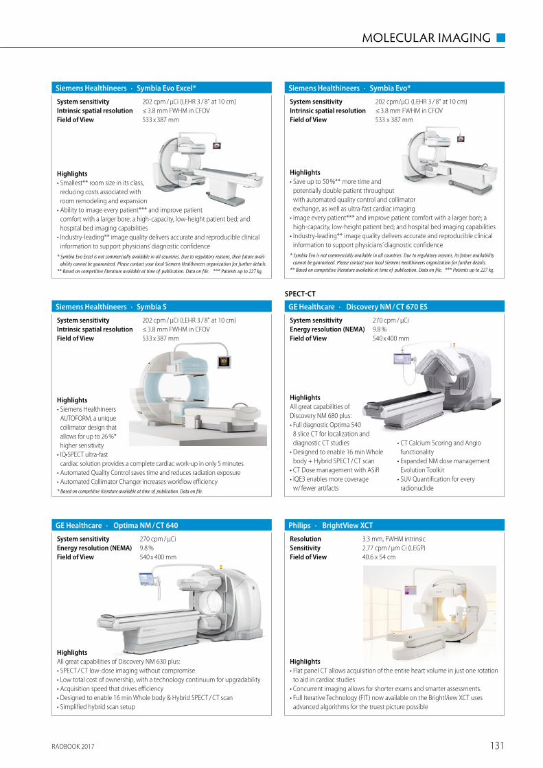
RADBOOK 2017 131
MOLECULAR IMAGING ◼
· BrightView XCTPhilips
40.6 x 54 cmField of View2.77 cpm / μm Ci (LEGP)Sensitivity3.3 mm, FWHM intrinsicResolution
Highlights• Flat panel CT allows acquisition of the entire heart volume in just one rotation
to aid in cardiac studies• Concurrent imaging allows for shorter exams and smarter assessments.• Full Iterative Technology (FIT) now available on the BrightView XCT uses
advanced algorithms for the truest picture possible
· Symbia Evo*Siemens Healthineers
533 x 387 mmField of View≤ 3.8 mm FWHM in CFOVIntrinsic spatial resolution202 cpm/μCi (LEHR 3 / 8" at 10 cm)System sensitivity
Highlights• Save up to 50 %** more time and
potentially double patient throughput with automated quality control and collimator exchange, as well as ultra-fast cardiac imaging
• Image every patient*** and improve patient comfort with a larger bore; a high-capacity, low-height patient bed; and hospital bed imaging capabilities
• Industry-leading** image quality delivers accurate and reproducible clinical information to support physicians’ diagnostic confidence
* Symbia Evo is not commercially available in all countries. Due to regulatory reasons, its future availability cannot be guaranteed. Please contact your local Siemens Healthineers organization for further details.
** Based on competitive literature available at time of publication. Data on file. *** Patients up to 227 kg.
· Symbia SSiemens Healthineers
533 x 387 mmField of View≤ 3.8 mm FWHM in CFOVIntrinsic spatial resolution202 cpm / μCi (LEHR 3 / 8" at 10 cm)System sensitivity
Highlights• Siemens Healthineers
AUTOFORM, a unique collimator design that allows for up to 26 %* higher sensitivity
• IQ•SPECT ultra-fast cardiac solution provides a complete cardiac work-up in only 5 minutes
• Automated Quality Control saves time and reduces radiation exposure• Automated Collimator Changer increases workflow efficiency* Based on competitive literature available at time of publication. Data on file.
· Symbia Evo Excel*Siemens Healthineers
533 x 387 mmField of View≤ 3.8 mm FWHM in CFOVIntrinsic spatial resolution202 cpm / μCi (LEHR 3 / 8" at 10 cm)System sensitivity
Highlights• Smallest** room size in its class,
reducing costs associated with room remodeling and expansion
• Ability to image every patient*** and improve patient comfort with a larger bore; a high-capacity, low-height patient bed; and hospital bed imaging capabilities
• Industry-leading** image quality delivers accurate and reproducible clinical information to support physicians’ diagnostic confidence
* Symbia Evo Excel is not commercially available in all countries. Due to regulatory reasons, their future avail-ability cannot be guaranteed. Please contact your local Siemens Healthineers organization for further details.
** Based on competitive literature available at time of publication. Data on file. *** Patients up to 227 kg.
SPECT-CT
· Discovery NM / CT 670 ESGE Healthcare
540 x 400 mmField of View9.8 %Energy resolution (NEMA)270 cpm / μCiSystem sensitivity
HighlightsAll great capabilities of Discovery NM 680 plus:• Full diagnostic Optima 540
8 slice CT for localization and diagnostic CT studies
• Designed to enable 16 min Whole body + Hybrid SPECT / CT scan
• CT Dose management with ASiR• IQE3 enables more coverage
w/ fewer artifacts
• CT Calcium Scoring and Angio functionality
• Expanded NM dose management Evolution Toolkit
• SUV Quantification for every radionuclide
· Optima NM / CT 640GE Healthcare
540 x 400 mmField of View9.8 %Energy resolution (NEMA)270 cpm / μCiSystem sensitivity
HighlightsAll great capabilities of Discovery NM 630 plus:• SPECT / CT low-dose imaging without compromise• Low total cost of ownership, with a technology continuum for upgradability• Acquisition speed that drives efficiency• Designed to enable 16 min Whole body & Hybrid SPECT / CT scan• Simplified hybrid scan setup
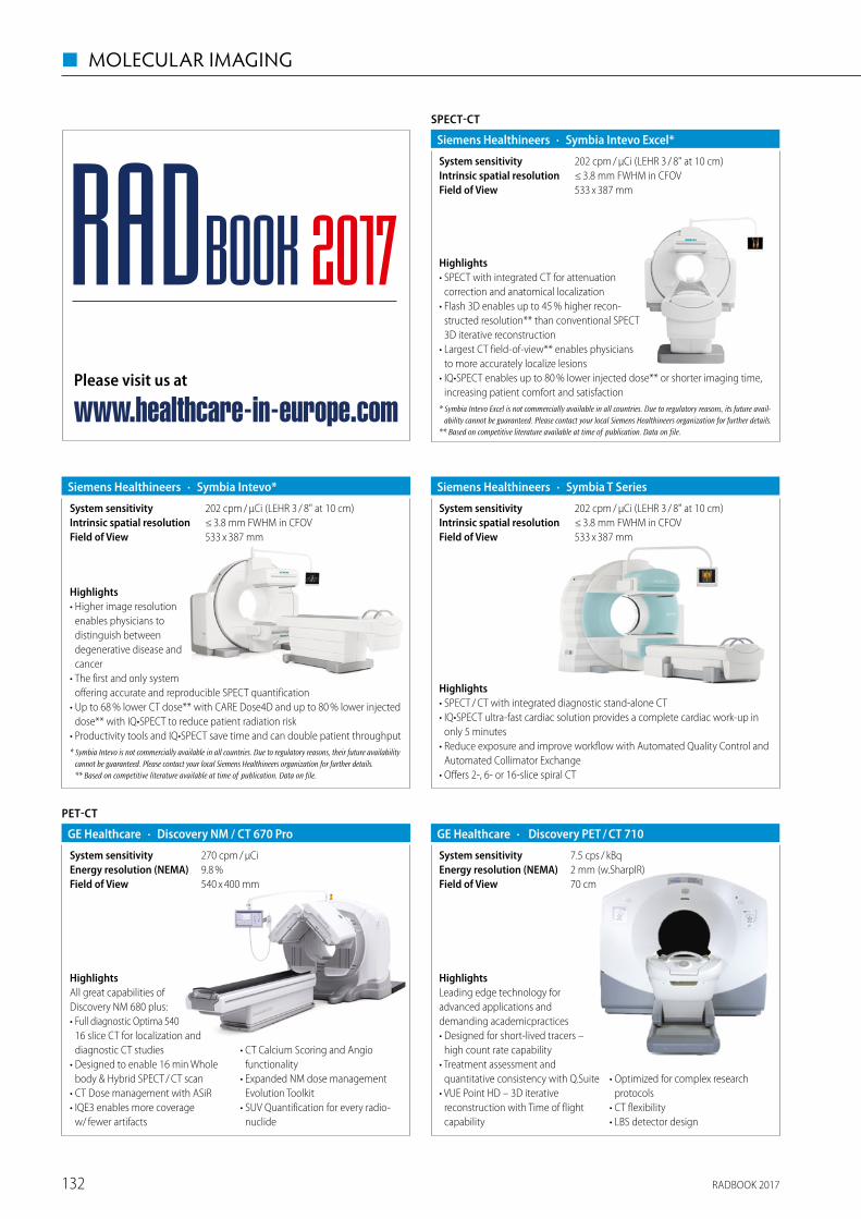
RADBOOK 2017132
◼ MOLECULAR IMAGING
· Symbia Intevo*Siemens Healthineers
533 x 387 mmField of View≤ 3.8 mm FWHM in CFOVIntrinsic spatial resolution202 cpm / μCi (LEHR 3 / 8" at 10 cm)System sensitivity
Highlights• Higher image resolution
enables physicians to distinguish between degenerative disease and cancer
• The first and only system offering accurate and reproducible SPECT quantification
• Up to 68 % lower CT dose** with CARE Dose4D and up to 80 % lower injected dose** with IQ•SPECT to reduce patient radiation risk
• Productivity tools and IQ•SPECT save time and can double patient throughput* Symbia Intevo is not commercially available in all countries. Due to regulatory reasons, their future availability
cannot be guaranteed. Please contact your local Siemens Healthineers organization for further details. ** Based on competitive literature available at time of publication. Data on file.
· Symbia T SeriesSiemens Healthineers
≤ 3.8 mm FWHM in CFOVIntrinsic spatial resolution533 x 387 mmField of View
202 cpm / μCi (LEHR 3 / 8" at 10 cm)System sensitivity
Highlights• SPECT / CT with integrated diagnostic stand-alone CT• IQ•SPECT ultra-fast cardiac solution provides a complete cardiac work-up in
only 5 minutes• Reduce exposure and improve workflow with Automated Quality Control and
Automated Collimator Exchange• Offers 2-, 6- or 16-slice spiral CT
SPECT-CT
· Symbia Intevo Excel*Siemens Healthineers
533 x 387 mmField of View≤ 3.8 mm FWHM in CFOVIntrinsic spatial resolution202 cpm / μCi (LEHR 3 / 8" at 10 cm)System sensitivity
Highlights• SPECT with integrated CT for attenuation
correction and anatomical localization• Flash 3D enables up to 45 % higher recon-
structed resolution** than conventional SPECT 3D iterative reconstruction
• Largest CT field-of-view** enables physicians to more accurately localize lesions
• IQ•SPECT enables up to 80 % lower injected dose** or shorter imaging time, increasing patient comfort and satisfaction
* Symbia Intevo Excel is not commercially available in all countries. Due to regulatory reasons, its future avail-ability cannot be guaranteed. Please contact your local Siemens Healthineers organization for further details.
** Based on competitive literature available at time of publication. Data on file.
PET-CT
· Discovery NM / CT 670 ProGE Healthcare
540 x 400 mmField of View9.8 %Energy resolution (NEMA)270 cpm / μCiSystem sensitivity
HighlightsAll great capabilities of Discovery NM 680 plus:• Full diagnostic Optima 540
16 slice CT for localization and diagnostic CT studies
• Designed to enable 16 min Whole body & Hybrid SPECT / CT scan
• CT Dose management with ASiR• IQE3 enables more coverage
w/ fewer artifacts
• CT Calcium Scoring and Angio functionality
• Expanded NM dose management Evolution Toolkit
• SUV Quantification for every radio-nuclide
· Discovery PET / CT 710GE Healthcare
70 cmField of View
7.5 cps / kBqSystem sensitivity2 mm (w.SharpIR)Energy resolution (NEMA)
HighlightsLeading edge technology for advanced applications and demanding academicpractices• Designed for short-lived tracers –
high count rate capability• Treatment assessment and
quantitative consistency with Q.Suite• VUE Point HD – 3D iterative
reconstruction with Time of flight capability
• Optimized for complex research protocols
• CT flexibility• LBS detector design
Please visit us at
www.healthcare-in-europe.com
RADBOOK 2017
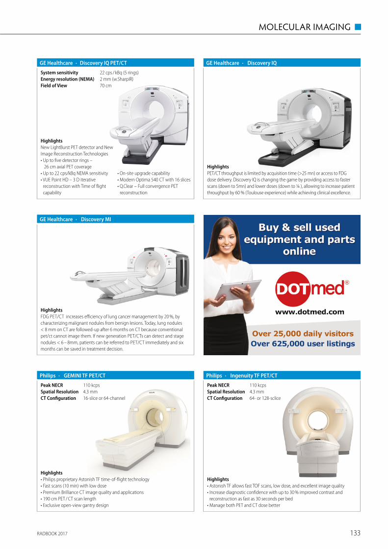
RADBOOK 2017 133
MOLECULAR IMAGING ◼
· Discovery IQGE Healthcare
HighlightsPET/CT throughput is limited by acquisition time (>25 mn) or access to FDG dose delivery. Discovery IQ is changing the game by providing access to faster scans (down to 5mn) and lower doses (down to ¼ ), allowing to increase patient throughput by 60 % (Toulouse experience) while achieving clinical excellence.
· Discovery MIGE Healthcare
HighlightsFDG PET/CT increases efficiency of lung cancer management by 20 %, by characterizing malignant nodules from benign lesions. Today, lung nodules < 8 mm on CT are followed-up after 6 months on CT because conventional pet/ct cannot image them. If new generation PET/CTs can detect and stage nodules < 6 – 8mm, patients can be referred to PET/CT immediately and six months can be saved in treatment decision.
· Discovery IQ PET/CTGE Healthcare
2 mm (w.SharpIR)Energy resolution (NEMA)22 cps / kBq (5 rings)System sensitivity
70 cmField of View
HighlightsNew LightBurst PET detector and New Image Reconstruction Technologies• Up to five detector rings –
26 cm axial PET coverage• Up to 22 cps/kBq NEMA sensitivity• VUE Point HD – 3 D iterative
reconstruction with Time of flight capability
• On-site upgrade capability• Modern Optima 540 CT with 16 slices• Q.Clear – Full convergence PET
reconstruction
· GEMINI TF PET/CTPhilips
16-slice or 64-channelCT Configuration4.3 mmSpatial Resolution110 kcpsPeak NECR
Highlights• Philips proprietary Astonish TF time-of-flight technology• Fast scans (10 min) with low dose• Premium Brilliance CT image quality and applications• 190 cm PET / CT scan length• Exclusive open-view gantry design
· Ingenuity TF PET/CTPhilips
64- or 128-scliceCT Configuration4.3 mmSpatial Resolution110 kcpsPeak NECR
Highlights• Astonish TF allows fast TOF scans, low dose, and excellent image quality• Increase diagnostic confidence with up to 30 % improved contrast and
reconstruction as fast as 30 seconds per bed• Manage both PET and CT dose better
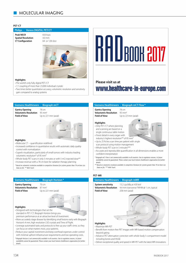
RADBOOK 2017134
◼ MOLECULAR IMAGING
PET-CT
· Vereos DIGITAL PET/CTPhilips
64- or 128-sliceCT Configuration4.0 mmSpatial Resolution650 kcpsPeak NECR
Highlights• The world’s only fully digital PET / CT• 1:1 coupling of more than 23,000 individual crystals• Two times better quantitative accuracy, volumetric resolution and sensitivity
gain compared to analog systems
78 cmGantry Opening
· Biograph mCTSiemens Healthineers
Up to 221 mm (axial)Field of View95 mm3Volumetric Resolution
Highlights• Molecular CT – quantification redefined• Increased confidence in quantitative results with automatic daily quality
control with normalization• Superb visualization, particularly of small tumors with industry-leading
volumetric resolution* of 95 mm³• Whole-body PET scans in only 5 minutes or with 5 mCi injected dose**• Increase revenue with a 78 cm bore for radiation therapy planning* Based on volumetric resolution available in competitive literature for systems greater than 70 cm bore size.
Data on file. ** With TrueV.
Highlights• Only PET / CT where planning
and scanning are based on a single continuous table motion
• Finest detail in every organ with industry’s highest resolution** of 95 mm³
• Up to 25 % less scan time per patient with single scan protocol using motion management
• Whole-body PET scan in 5 minutes***• Accurate and reproducible quantification in all dimensions enables a more
confident interpretation* Biograph mCT Flow is not commercially available in all countries. Due to regulatory reasons, its future
availability cannot be guaranteed. Please contact your local Siemens Healthineers organization for further details.
** Based on volumetric resolution available in competitive literature for systems greater than 70 cm bore size. Data on file. *** With TrueV.
78 cmGantry Opening
· Biograph mCT Flow *Siemens Healthineers
Up to 221mm (axial)Field of View95 mm3Volumetric Resolution
· Biograph Horizon *Siemens Healthineers
Up to 221 mm (axial)Field of View87 mm3Volumetric Resolution70 cmGantry Opening
Highlights• Designed with technologies that set the
standard in PET / CT, Biograph Horizon brings you premium performance at an attractive level of investment.
• More accurately stage disease by identifying small lesions early with Biograph Horizon’s 4 mm, high resolution LSO crystals and Time of Flight.
• Leverage automated tasks and protocols to free up your staff’s time, so they can focus on what matters most, your patients.
• Reduce your capital investment and keep overhead expenses under control with minimal upfront infrastructure requirements and low operating costs.
* Biograph Horizon is not commercially available in all countries. Due to regulatory reasons, its future availability cannot be guaranteed. Please contact your local Siemens Healthineers organization for further details.
PET-MR
· Biograph mMRSiemens Healthineers
258 mm (axial)Field of View4.4 mm transverse FWHM @ 1 cm, typicalVolumetric Resolution13.2 cps/kBq at 430 keVSystem sensitivity
Highlights• Maximize MR-PET• Benefit from motion-free PET images with MR-based motion compensation
beyond gating• Advance PET attenuation correction with whole-body 5-compartment model
including bones and HUGE• Deliver exceptional quality and speed in MR-PET with the latest MR innovations
Please visit us at
www.healthcare-in-europe.com
RADBOOK 2017
RADBOOK 2017 135
DISPLAYS / PRINTERS ◼
Displays / Printers
Displays – Mammo
Displays – Color
Displays - Grayscale
Displays – Clinical Review
Displays – Large Format
Printers
CD- / DVD-Robot
Accessories / Complementary Systems
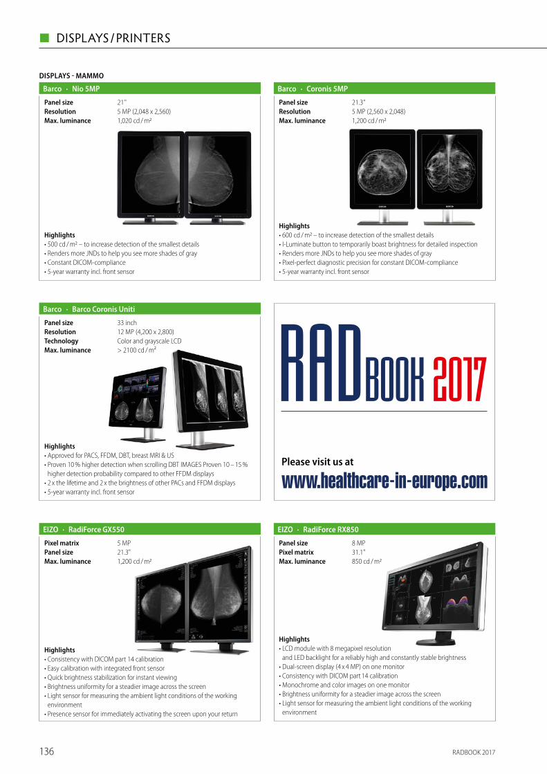
RADBOOK 2017136
◼ DISPLAYS / PRINTERS
DISPLAYS - MAMMO
· Nio 5MPBarco
1,020 cd / m2Max. luminance5 MP (2,048 x 2,560)Resolution21''Panel size
Highlights• 500 cd / m2 – to increase detection of the smallest details• Renders more JNDs to help you see more shades of gray• Constant DICOM-compliance• 5-year warranty incl. front sensor
· Coronis 5MPBarco
1,200 cd / m2Max. luminance5 MP (2,560 x 2,048)Resolution21.3"Panel size
Highlights• 600 cd / m2 – to increase detection of the smallest details• I-Luminate button to temporarily boast brightness for detailed inspection • Renders more JNDs to help you see more shades of gray• Pixel-perfect diagnostic precision for constant DICOM-compliance• 5-year warranty incl. front sensor
· Barco Coronis UnitiBarco
> 2100 cd / m²Max. luminanceColor and grayscale LCDTechnology12 MP (4,200 x 2,800)Resolution33 inchPanel size
Highlights• Approved for PACS, FFDM, DBT, breast MRI & US• Proven 10 % higher detection when scrolling DBT IMAGES Proven 10 – 15 %
higher detection probability compared to other FFDM displays• 2 x the lifetime and 2 x the brightness of other PACs and FFDM displays • 5-year warranty incl. front sensor
· RadiForce GX550EIZO
1,200 cd / m2Max. luminance21.3"Panel size5 MPPixel matrix
Highlights• Consistency with DICOM part 14 calibration • Easy calibration with integrated front sensor • Quick brightness stabilization for instant viewing • Brightness uniformity for a steadier image across the screen • Light sensor for measuring the ambient light conditions of the working
environment • Presence sensor for immediately activating the screen upon your return
· RadiForce RX850EIZO
850 cd / m2Max. luminance31.1"Pixel matrix8 MPPanel size
Highlights• LCD module with 8 megapixel resolution
and LED backlight for a reliably high and constantly stable brightness• Dual-screen display (4 x 4 MP) on one monitor• Consistency with DICOM part 14 calibration• Monochrome and color images on one monitor• Brightness uniformity for a steadier image across the screen• Light sensor for measuring the ambient light conditions of the working
environment
Please visit us at
www.healthcare-in-europe.com
RADBOOK 2017
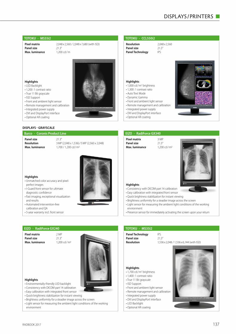
RADBOOK 2017 137
DISPLAYS / PRINTERS ◼
Highlights• LED Backlight• 1,200 : 1 contrast ratio• True 11 Bit grayscale• ISD Support• Front and ambient light sensor• Remote management and calibration• Integrated power supply• DVI and DisplayPort interface• Optional AR coating
· MS55i2TOTOKU
1,200 cd / mMax. luminance21.3"Panel size2,048 x 2,560 / 2,048 x 7,680 (with ISD)Pixel matrix
Highlights• 1,000 cd / m2 brightness• 1,300 : 1 contrast ratio• Auto Text Mode• Dynamic Gamma• Front and ambient light sensor• Remote management and calibration• Integrated power supply• DVI and DisplayPort interface• Optional AR coating
· CCL550i2TOTOKU
21.3"Panel size2,048 x 2,560Resolution
IPSPanel Technology
DISPLAYS - GRAYSCALE
· Coronis Product LineBarco
1,700 / 1,200 cd / m2Max. luminance3 MP (2,048 x 1,536) / 5 MP (2,560 x 2,048)Resolution21.3"Panel size
Highlights• Unmatched color accuracy and pixel-
perfect images• I-Guard front sensor for ultimate
diagnostic confidence• Fast imaging, exceptional visualization
and results• Automated intervention-free
calibration and QA• 5-year warranty incl. front sensor
· RadiForce GX340
1,200 cd / m2Max. luminance21.3"Panel size3 MPPixel matrix
Highlights• Consistency with DICOM part 14 calibration• Easy calibration with integrated front sensor• Quick brightness stabilization for instant viewing• Brightness uniformity for a steadier image across the screen• Light sensor for measuring the ambient light conditions of the working
environment• Presence sensor for immediately activating the screen upon your return
EIZO
· RadiForce GX240EIZO
1,200 cd / m2Max. luminance21.3"Panel size2 MPPixel matrix
Highlights• Environmentally-friendly LED backlight• Consistency with DICOM part 14 calibration• Easy calibration with integrated front sensor• Quick brightness stabilization for instant viewing• Brightness uniformity for a steadier image across the screen• Light sensor for measuring the ambient light conditions of the working
environment
Highlights• 1,700 cd / m2 brightness• 1,400 : 1 contrast ratio• True 11 Bit grayscale• ISD Support• Front and ambient light sensor• Remote management and calibration• Integrated power supply• DVI and DisplayPort interface• LED Backlight• Optional AR coating
· MS35i2TOTOKU
1,536 x 2,048 / 1,536 x 6,144 (with ISD)Resolution21.3"Panel sizeIPSPanel Technology
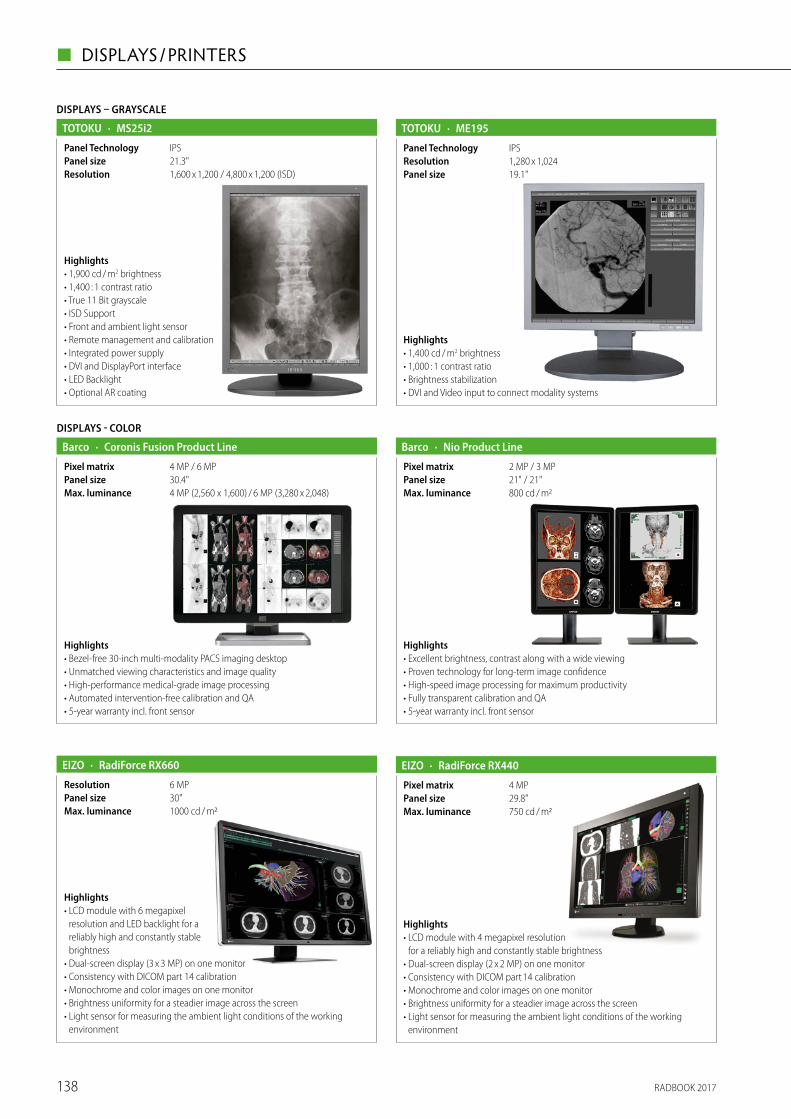
RADBOOK 2017138
◼ DISPLAYS / PRINTERS
DISPLAYS – GRAYSCALE
Highlights• 1,900 cd / m2 brightness• 1,400 : 1 contrast ratio• True 11 Bit grayscale• ISD Support• Front and ambient light sensor• Remote management and calibration• Integrated power supply• DVI and DisplayPort interface• LED Backlight• Optional AR coating
· MS25i2TOTOKU
1,600 x 1,200 / 4,800 x 1,200 (ISD)Resolution21.3"Panel sizeIPSPanel Technology
Highlights• 1,400 cd / m2 brightness• 1,000 : 1 contrast ratio• Brightness stabilization• DVI and Video input to connect modality systems
· ME195TOTOKU
19.1"Panel size1,280 x 1,024ResolutionIPSPanel Technology
DISPLAYS - COLOR
· Coronis Fusion Product LineBarco
4 MP (2,560 x 1,600) / 6 MP (3,280 x 2,048)Max. luminance30.4"Panel size4 MP / 6 MPPixel matrix
Highlights• Bezel-free 30-inch multi-modality PACS imaging desktop• Unmatched viewing characteristics and image quality • High-performance medical-grade image processing• Automated intervention-free calibration and QA• 5-year warranty incl. front sensor
· Nio Product LineBarco
800 cd / m2Max. luminance21" / 21"Panel size2 MP / 3 MPPixel matrix
Highlights• Excellent brightness, contrast along with a wide viewing• Proven technology for long-term image confidence• High-speed image processing for maximum productivity• Fully transparent calibration and QA• 5-year warranty incl. front sensor
Highlights• LCD module with 6 megapixel
resolution and LED backlight for a reliably high and constantly stable brightness
• Dual-screen display (3 x 3 MP) on one monitor• Consistency with DICOM part 14 calibration• Monochrome and color images on one monitor• Brightness uniformity for a steadier image across the screen• Light sensor for measuring the ambient light conditions of the working
environment
· RadiForce RX660
1000 cd / m2Max. luminance30"Panel size6 MPResolution
EIZO · RadiForce RX440EIZO
750 cd / m2Max. luminance29.8"Panel size4 MPPixel matrix
Highlights• LCD module with 4 megapixel resolution
for a reliably high and constantly stable brightness• Dual-screen display (2 x 2 MP) on one monitor• Consistency with DICOM part 14 calibration• Monochrome and color images on one monitor• Brightness uniformity for a steadier image across the screen• Light sensor for measuring the ambient light conditions of the working
environment
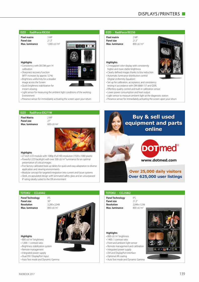
RADBOOK 2017 139
DISPLAYS / PRINTERS ◼
· RadiForce RX350
1,000 cd / m2Max. luminance21.3"Panel size3 MPPixel matrix
Highlights• Consistency with DICOM part 14
calibration• Sharpness recovery function
(MTF increases by approx. 52 %)• Brightness uniformity for a steadier
image across the Screen• Quick brightness stabilization for
instant viewing• Light sensor for measuring the ambient light conditions of the working
Environment• Presence sensor for immediately activating the screen upon your return
EIZO · RadiForce RX250EIZO
800 cd / m2Max. luminance21.3"Panel size2 MPPixel matrix
Highlights• 2 megapixel color display with consistently
higher and more stable brightness• Clearly defined images thanks to blur reduction• Automatic luminance distribution control
(Digital Uniformity Equalizer)• Set up for calibration, acceptance, and consistency
testing in accordance with DIN 6868-157 and QSRL• Effortless quality control and built-in calibration sensor• Lower power consumption and heat output• Light sensor to measure ambient light at the diagnostic station• Presence sensor for immediately activating the screen upon your return
· RadiForce EX271WEIZO
600 cd / m2Max. luminance27"Panel size2 MPPixel Matrix
Highlights• 27-inch LCD module with 1080p (Full HD) resolution (1920 x 1080 pixels)• Powerful LED backlight with over 500 cd / m² luminance for an optimal
presentation of critical images• Five factory calibrated look-up tables for quick and easy adaptation to diverse
application and viewing environments• Modular concept for targeted integration into current and future systems• Sleek, encapsulated design with laminated safety glass and an unsurpassed
IP rating ideally suited to the OR environment
Highlights• 800 cd / m2 brightness• 1400 : 1 contrast ratio• Front and ambient light sensor• Remote management and calibration• Integrated power supply• DVI and DisplayPort interface• Optional AR coating• Auto Text mode and Dynamic Gamma
· CCL358i2TOTOKU
IPSPanel Technology21.3"Panel size2,048 x 1,536Resolution800 cd / m2Max. luminance
Highlights• 800 cd / m2 brightness• 1,000 : 1 contrast ratio• Brightness stabilization system• Remote management• Integrated power supply• Dual DVI / DisplayPort Input• Auto Text mode and Dynamic Gamma
· CCL650i2TOTOKU
3,280 x 2,048Resolution800 cd / m2Max. luminance
IPSPanel Technology30“Panel size
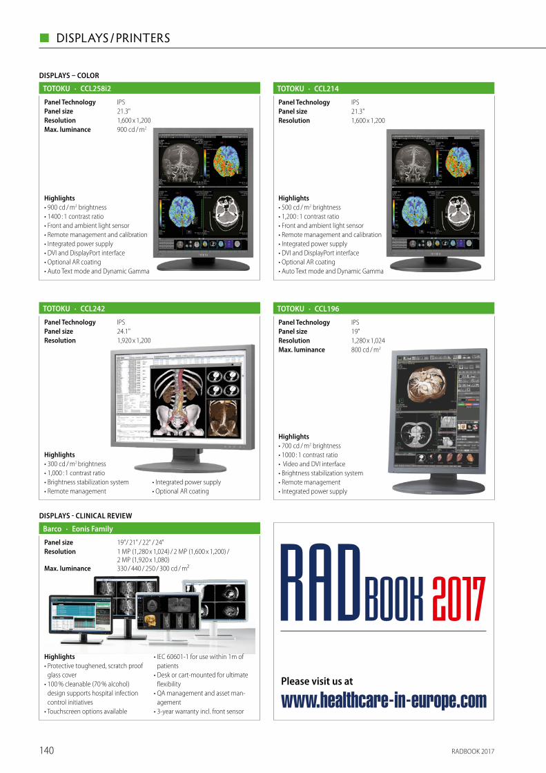
RADBOOK 2017140
◼ DISPLAYS / PRINTERS
IPSPanel Technology
DISPLAYS – COLOR
Highlights• 900 cd / m2 brightness• 1400 : 1 contrast ratio• Front and ambient light sensor• Remote management and calibration• Integrated power supply• DVI and DisplayPort interface• Optional AR coating• Auto Text mode and Dynamic Gamma
· CCL258i2TOTOKU
900 cd / m2Max. luminance
21.3"Panel size1,600 x 1,200Resolution
Highlights• 300 cd / m2 brightness• 1,000 : 1 contrast ratio• Brightness stabilization system• Remote management
• Integrated power supply• Optional AR coating
· CCL242TOTOKU
1,920 x 1,200Resolution24.1"Panel sizeIPSPanel Technology
Highlights• 500 cd / m2 brightness• 1,200 : 1 contrast ratio• Front and ambient light sensor• Remote management and calibration• Integrated power supply• DVI and DisplayPort interface• Optional AR coating• Auto Text mode and Dynamic Gamma
· CCL214TOTOKU
1,600 x 1,200Resolution21.3"Panel sizeIPSPanel Technology
Highlights• 700 cd / m2 brightness• 1000 : 1 contrast ratio• Video and DVI interface• Brightness stabilization system• Remote management• Integrated power supply
· CCL196TOTOKU
800 cd / m2Max. luminance1,280 x 1,024Resolution19"Panel sizeIPSPanel Technology
DISPLAYS - CLINICAL REVIEW
· Eonis FamilyBarco
330 / 440 / 250 / 300 cd / m²Max. luminance
1 MP (1,280 x 1,024) / 2 MP (1,600 x 1,200) / 2 MP (1,920 x 1,080)
Resolution19"/ 21" / 22" / 24"Panel size
Highlights• Protective toughened, scratch proof
glass cover• 100 % cleanable (70 % alcohol)
design supports hospital infection control initiatives
• Touchscreen options available
• IEC 60601-1 for use within 1m of patients
• Desk or cart-mounted for ultimate flexibility
• QA management and asset man-agement
• 3-year warranty incl. front sensor
Please visit us at
www.healthcare-in-europe.com
RADBOOK 2017

RADBOOK 2017 141
DISPLAYS / PRINTERS ◼
The World’s Leading Medical Equipment Marketplace
www.dotmed.com
Afghanistan 190, Albania 380, Algeria 313, American Samoa 39, Andorra 60, Angola 44, Anguilla 20, Antarctica 16, Antigua 4, Antigua and Barbuda 140, Argentina 1542, Armenia 202, Aruba 39, Australia 1685, Austria 332, Azerbaijan
133, Bahamas 72, Bahrain 95, Bangladesh 271, Barbados 32, Belarus 81, Belgium 442, Belize 58, Benin 55, Bermuda 34, Bhutan 24, Bolivia 460, Bonaire 2, Bosnia and Herzegovina 160, Botswana 46, Brazil 3068, British Indian Ocean Territory 16, Brunei Darussalam 37, Bulgaria 590, Burkina Faso 40, Burundi 18, Cambodia 112, Cameroon 200, Canada 4583, Canary Islands 2, Cape Verde 31, Cape Verde Islands 1, Caroline Islands 2, Côte D’Ivoire 1, Cayman
Islands 38, Central African Republic 495, Chad 17, Chile 708, China 4339, Christmas Island 10, Cocos(Keeling)Island 1, Colombia 1317, Comoros 18, Congo 7, Congo, Democratic Republic of the 65, Cook Islands 17, Costa Rica 234, Côte D’Ivoire 139, Croatia 191, Cuba 33, Curacao 4, Cyprus 365, Czech Republic 196, Denmark 252, Djibouti 26,
Dominica 22, Dominican Republic 502, Ecuador 620, Egypt 1487, El Salvador 247, Equatorial Guinea 28, Eritrea 17, Estonia 98, Ethiopia 132, Faeroe Islands 1, Falkland Islands 1, Faroe Islands 1, Fiji 34, Finland 124, France 1034,
French Guiana 18, French Polynesia 21, Gabon 23, Gambia 31, Georgia 478, Germany 1782, Ghana 365, Gibraltar 27, Greece 915, Greenland 23, Grenada 25, Guadeloupe 18, Guam 71, Guatemala 366, Guinea 14, Guinea-Bissau
943, Guyana 30, Haiti 121, Honduras 266, Hong Kong 383, Hungary 367, Iceland 49, India 7123, Indonesia 1096, Iran 1085, Iraq 545, Ireland 301, Isle of Man 1, Israel 753, Italy 1926, Jamaica 158, Japan 450, Jordan 356, Kazakhstan
123, Kenya 327, Kiribati 9, Kuwait 162, Kyrgyz Republic 70, Lao People’s Democratic Republic 30, Latvia 118, Lebanon 494, Lesotho 20, Liberia 19, Libyan Arab Jamahiriya 247, Liechtenstein 12, Lithuania 123, Luxembourg 38, Macao 28, Macedonia 218, Madagascar 62, Malawi 43, Malaysia 692, Maldives 33, Mali 35, Malta 56, Marshall Islands
23, Martinique 22, Mauritania 32, Mauritius 66, Mayotte 13, Mexico 5172, Micronesia 19, Moldova 126, Monaco 26, Mongolia 144, Montenegro 31, Montserrat 9, Morocco 426, Mozambique 43, Myanmar 65, Namibia 35, Nauru 15,
Nepal 127, Netherlands 699, Netherlands Antilles 34, Nevis 1, New Caledonia 18, New Zealand 271, Nicaragua 168, Niger 20, Nigeria 1369, Niue 12, Norfolk Island 21, Northern Mariana Islands 32, North Korea 15, Norway 129, Oman
88, Pakistan 3289, Palau 8, Palestinian Territory 8, Panama 224, Papua New Guinea 42, Paraguay 243, Peru 865, Philippines 1459, Pitcairn Island 1, Poland 1111, Portugal 461, Puerto Rico 740, Qatar 83, Reunion 16, Romania 955,
Russia 1304, Rwanda 40, Saint Barthelemy 2, Saint Helena 13, Saint Kitts 2, Saint Kitts and Nevis 20, Saint Lucia 46, Saint Maarten 4, Saint Pierre and Miquelon 11, Saint Vincent 1, Saint Vincent and the Grenadines 21, Samoa
13, San Marino 12, Sao Tome and Principe 2, Saudi Arabia 1025, Senegal 101, Serbia 348, Serbia and Montenegro 79, Seychelles 19, Sierra Leone 31, Singapore 392, Slovakia 115, Slovenia 115, Solomon Islands 13, Somalia 40, South Africa 684, South Korea 674, Spain 1056, Sri Lanka 160, Sudan 212, Suriname 50, Swaziland 11, Sweden
242, Switzerland 293, Syria 477, Tahiti 2, Taiwan 558, Tajikistan 57, Tanzania 159, Thailand 422, Timor-Leste 18, Togo 50, Tokelau 4, Tonga 13, Trinidad and Tobago 151, Tunisia 120, Turkey 1780, Turkmenistan 20, Turks and Caicos
Islands 22, Tuvalu 13, Uganda 234, Ukraine 895, United Arab Emirates 960, United Kingdom 3660, Uruguay 149, USA 130148, USVI 1, Uzbekistan 137, Vanuatu 17, Venezuela 1187, Vietnam 905, Virgin Islands (U.K.) 5, Virgin Islands (U.S.) 21, Wallis and Futuna Islands 11, Western Sahara 1, Western Samoa 2, Yemen 305, Yugoslavia 112, Zambia 80,
Zimbabwe 120.
So they can buy & sell medical equipment and parts, find a service partner, browse over 625,000 listings, and read
HealthCare Business News online. Register for free and join them.
Why are more than a quarter million health care professionals registered on DOTmed?
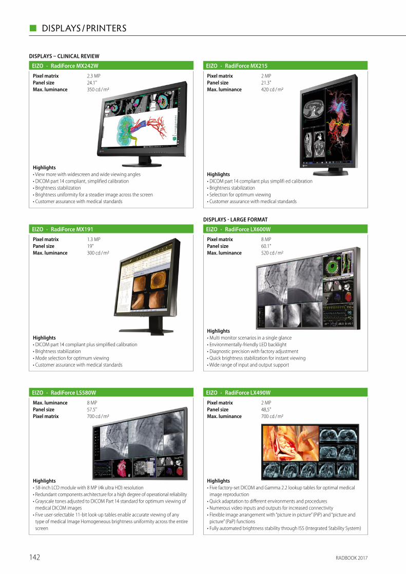
RADBOOK 2017142
◼ DISPLAYS / PRINTERS
DISPLAYS – CLINICAL REVIEW
· RadiForce MX242WEIZO
350 cd / m2Max. luminance24.1"Panel size2.3 MPPixel matrix
Highlights• View more with widescreen and wide viewing angles• DICOM part 14 compliant, simplified calibration• Brightness stabilization• Brightness uniformity for a steadier image across the screen• Customer assurance with medical standards
· RadiForce MX215EIZO
420 cd / m2Max. luminance21.3"Panel size2 MPPixel matrix
Highlights• DICOM part 14 compliant plus simplifi ed calibration• Brightness stabilization• Selection for optimum viewing• Customer assurance with medical standards
· RadiForce MX191EIZO
300 cd / m2Max. luminance19"Panel size1.3 MPPixel matrix
Highlights• DICOM part 14 compliant plus simplified calibration• Brightness stabilization• Mode selection for optimum viewing• Customer assurance with medical standards
· RadiForce LS580WEIZO
700 cd / m2Pixel matrix57.5"Panel size8 MPMax. luminance
Highlights• 58-inch LCD module with 8 MP (4k ultra HD) resolution • Redundant components architecture for a high degree of operational reliability• Grayscale tones adjusted to DICOM Part 14 standard for optimum viewing of
medical DICOM images• Five user-selectable 11-bit look-up tables enable accurate viewing of any
type of medical Image Homogeneous brightness uniformity across the entire screen
DISPLAYS - LARGE FORMAT
· RadiForce LX600WEIZO
520 cd / m2Max. luminance60.1"Panel size8 MPPixel matrix
Highlights• Multi monitor scenarios in a single glance• Environmentally-friendly LED backlight• Diagnostic precision with factory adjustment• Quick brightness stabilization for instant viewing• Wide range of input and output support
· RadiForce LX490WEIZO
700 cd / m2Max. luminance48,5"Panel size2 MPPixel matrix
Highlights• Five factory-set DICOM and Gamma 2.2 lookup tables for optimal medical
image reproduction• Quick adaptation to different environments and procedures• Numerous video inputs and outputs for increased connectivity• Flexible image arrangement with “picture in picture” (PiP) and “picture and
picture” (PaP) functions• Fully automated brightness stability through ISS (Integrated Stability System)
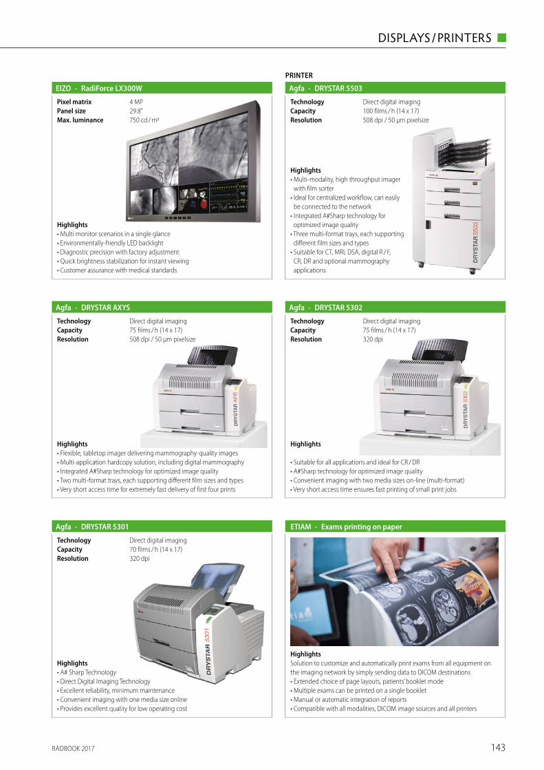
RADBOOK 2017 143
DISPLAYS / PRINTERS ◼
· RadiForce LX300W
750 cd / m2Max. luminance29.8"Panel size4 MPPixel matrix
Highlights• Multi monitor scenarios in a single glance• Environmentally-friendly LED backlight• Diagnostic precision with factory adjustment• Quick brightness stabilization for instant viewing• Customer assurance with medical standards
EIZO
· DRYSTAR 5301Agfa
320 dpiResolution70 films / h (14 x 17)CapacityDirect digital imagingTechnology
Highlights• A# Sharp Technology • Direct Digital Imaging Technology• Excellent reliability, minimum maintenance• Convenient imaging with one media size online• Provides excellent quality for low operating cost
· DRYSTAR 5302Agfa
320 dpiResolution75 films / h (14 x 17)CapacityDirect digital imagingTechnology
Highlights
• Suitable for all applications and ideal for CR / DR• A#Sharp technology for optimized image quality• Convenient imaging with two media sizes on-line (multi-format)• Very short access time ensures fast printing of small print jobs
PRINTER
· DRYSTAR 5503Agfa
508 dpi / 50 μm pixelsizeResolution100 films / h (14 x 17)CapacityDirect digital imagingTechnology
Highlights• Multi-modality, high throughput imager
with film sorter• Ideal for centralized workflow, can easily
be connected to the network• Integrated A#Sharp technology for
optimized image quality• Three multi-format trays, each supporting
different film sizes and types• Suitable for CT, MRI, DSA, digital R / F,
CR, DR and optional mammography applications
· DRYSTAR AXYSAgfa
508 dpi / 50 μm pixelsizeResolution75 films / h (14 x 17)CapacityDirect digital imagingTechnology
Highlights• Flexible, tabletop imager delivering mammography-quality images• Multi-application hardcopy solution, including digital mammography• Integrated A#Sharp technology for optimized image quality• Two multi-format trays, each supporting different film sizes and types• Very short access time for extremely fast delivery of first four prints
HighlightsSolution to customize and automatically print exams from all equipment on the imaging network by simply sending data to DICOM destinations• Extended choice of page layouts, patients’ booklet mode• Multiple exams can be printed on a single booklet• Manual or automatic integration of reports• Compatible with all modalities, DICOM image sources and all printers
· Exams printing on paperETIAM
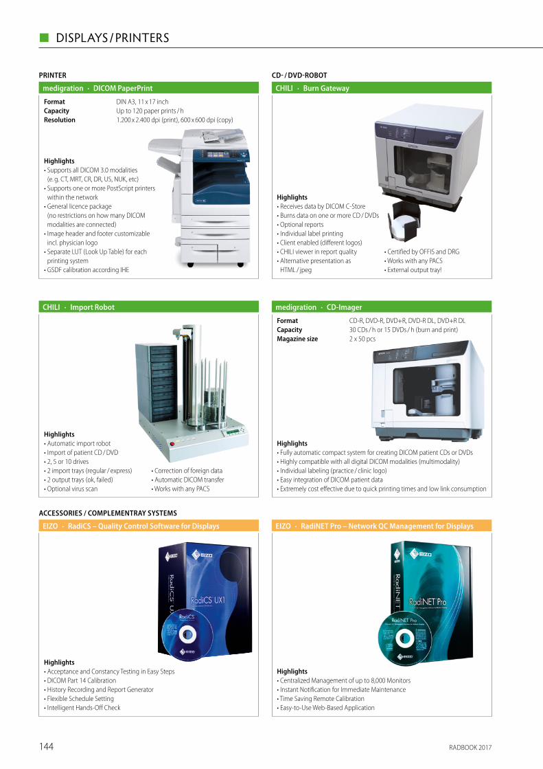
RADBOOK 2017144
◼ DISPLAYS / PRINTERS
CD- / DVD-ROBOT
· Burn GatewayCHILI
Highlights• Receives data by DICOM C-Store• Burns data on one or more CD / DVDs• Optional reports• Individual label printing• Client enabled (different logos)• CHILI viewer in report quality• Alternative presentation as
HTML / jpeg
• Certified by OFFIS and DRG• Works with any PACS• External output tray!
· Import RobotCHILI
Highlights• Automatic import robot• Import of patient CD / DVD• 2, 5 or 10 drives• 2 import trays (regular / express)• 2 output trays (ok, failed)• Optional virus scan
• Correction of foreign data• Automatic DICOM transfer• Works with any PACS
· RadiNET Pro – Network QC Management for DisplaysEIZO
Highlights• Centralized Management of up to 8,000 Monitors• Instant Notification for Immediate Maintenance• Time Saving Remote Calibration• Easy-to-Use Web-Based Application
ACCESSORIES / COMPLEMENTRAY SYSTEMS
· RadiCS – Quality Control Software for DisplaysEIZO
Highlights• Acceptance and Constancy Testing in Easy Steps• DICOM Part 14 Calibration• History Recording and Report Generator• Flexible Schedule Setting• Intelligent Hands-Off Check
Highlights• Fully automatic compact system for creating DICOM patient CDs or DVDs• Highly compatible with all digital DICOM modalities (multimodality)• Individual labeling (practice / clinic logo)• Easy integration of DICOM patient data• Extremely cost effective due to quick printing times and low link consumption
· CD-Imagermedigration
2 x 50 pcsMagazine size30 CDs / h or 15 DVDs / h (burn and print)CapacityCD-R, DVD-R, DVD+R, DVD-R DL, DVD+R DLFormat
PRINTER
· DICOM PaperPrintmedigration
1.200 x 2.400 dpi (print), 600 x 600 dpi (copy)ResolutionUp to 120 paper prints / hCapacityDIN A3, 11 x 17 inchFormat
Highlights• Supports all DICOM 3.0 modalities
(e. g. CT, MRT, CR, DR, US, NUK, etc)• Supports one or more PostScript printers
within the network• General licence package
(no restrictions on how many DICOM modalities are connected)
• Image header and footer customizable incl. physician logo
• Separate LUT (Look Up Table) for each printing system
• GSDF calibration according IHE
RADBOOK 2017 145
ULTRASOUND ◼
Ultrasound
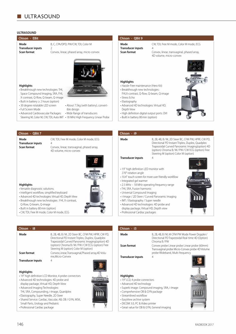
RADBOOK 2017146
◼ ULTRASOUND
· QBit 7Chison
Convex, linear, transvaginal, phased array, 4D volume, micro-convex
Scan format
CW, TDI, Free M mode, Color M mode, ECGMode4Transducer inputs
Highlights• Versatile diagnostic solutions.• Intelligent workflow, simplified keyboard• Advanced 4D technologies: Virtual HD, Depth View• Breakthrough new technologies : FHI, X-contrast,
Q-flow, Q-beam, Q-image• Built in battery 80 min (option)• CW, TDI, Free M mode, Color M mode, ECG
· EBitChison
Convex, linear, phased array, micro-convexScan format
B, C, CPA/DPD, PW/CW, TDI, Color M Mode2Transducer inputs
Highlights• Breakthrough new technologies: THI,
Space Compound Imaging, SRA, FHI, X-contrast, Q-flow, Q-beam, Q-image
• Built in battery ≥ 2 hours (option)• 30 degree rotatable LED screen• Full Screen Mode• Advanced Cardiovascular Packages:
Steering M, Color M, CW, TDI, Auto IMT
• About 7.5kg (with battery), convert-ible design
• Wide Range of transducers• 18 MHz High Frequency Linear Probe
ULTRASOUND
· QBit 9Chison
Convex, linear, transvaginal, phased array, 4D volume, micro-convex
Scan format
CW, TDI, Free M mode, Color M mode, ECGMode4Transducer inputs
Highlights• Hassle-Free maintenance (Hero Kit)• Breakthrough new technologies :
FHI,X-contrast, Q-flow, Q-beam, Q-image• Stress Echo• Elastography• Advanced 4D technologies: Virtual HD,
Depth View• High definition digital output ports: DVI• Built in battery 80 min (option)
• 19" high definition LED monitor with 270° rotation angle
• 10.4" touch screen for more user friendly workflow • Integrated gel warmer • 2.5 MHz – 18 MHz operating frequency range• THI, SRA, Fusion harmonic • Universal Compound Imaging • i-Image / 2D Steer / Curved Panoramic Imaging • IMT / Elastography / Super needle • Advanced 4D technologies: 4D probe and
display package, Virtual HD, Depth view • Professional Cardiac packages
· i9Chison
B, 2B, 4B, B / M, 2D Steer BC, CFM PW, HPRF, CW PD, Directional PD Instant Triplex, Duplex, Quadplex Trapezoidal Curved Panoramic Imaging(option) 4D (option) Chroma B / M / PW / CW ECG (option) Free Steering M (option) Color M (option)
Mode
4Transducer inputs
Highlights • 19" high definition LCD Monitor, 4 probe connectors • Advanced 4D technologies: 4D probe and
display package, Virtual HD, Depth View • Advanced Imaging Technologies:
THI, SRA, Compounding, i-Image, Quardplex • Elastography, Super Needle, 2D Steer • Shared Service: Cardiac, Vascular, AB, OB / GYN, MSK,
Small Parts, Urology and Pediatric • Professional Cardiac package
Mode B, 2B, 4B, B / M, 2D Steer BC, CFM PW, HPRF, CW PD, Directional PD Instant Triplex, Duplex, Quadplex Trapezoidal Curved Panoramic Imaging(option) 4D (option) Chroma B / M / PW / CW ECG (option) Free Steering M (option) Color M (option)Convex,Linear,Transvaginal,Phased array,4D Volu-me,Micro-Convex
Scan format
· i8Chison
4Transducer inputs
B, 2B, 4B, B / M, M CFM PW Mode Power Doppler / Directional PD Trapezoidal Real-time 4D (Option) Chroma B / PW
Mode
Highlights • 19” LCD, 4 probe connectors • Advanced 4D technology • Superb image: Compound imaging, SRA, i-Image • Comprehensive OB & GYN package • Streamlined workflow • EasyView archive system • DICOM 3.0, PC & Video printer • Great value for OB & GYN, General imaging
· i3Chison
4Transducer inputs
Convex probe Linear probe Linear probe (60mm) Transvaginal probe Micro-Convex probe 4D Volume probe Wideband, Multi-frequency
Scan format
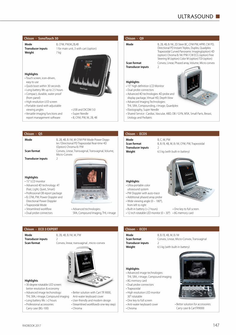
RADBOOK 2017 147
ULTRASOUND ◼
Mode B, 2B, 4B, B / M, M CFM PW Mode Power Dopp-ler / Directional PD Trapezoidal Real-time 4D (Option) Chroma B / PW
Highlights • 15" LCD monitor• Advanced 4D technology: 4T
(Fast, Light, Quiet, Smart)• Professional OB report package• B, CFM, PW, Power Doppler and
Directional Power Doppler• Trapezoidal Mode• Streamlined workflow• Dual probe connectors
• Advanced technologies: SRA, Compound Imaging, THI, i-Image
· Q5Chison
Transducer inputs
Scan format
2
Convex, Linear, Transvaginal, Transvaginal, Volume, Micro-Convex
6.5 kg (with built-in battery)Weight2Transducer inputsB, B / B, 4B, M, B / M, CFM, PW, TrapezoidalScan format
Highlights • Ultra-portable color
ultrasound system• PW Doppler with auto-trace• Additional phased array probe• Wide viewing angle (0 – 180°),
from left to right• Built-in battery (> 2 hours)• 12 inch rotatable LED monitor (0 – 30°)
• One key to full screen• 8G memory card
· ECO5Chison
B, C, M, PWMode
Highlights • Advanced image technologies:
THI, SRA, i-Image, Compound imaging• 8G memory card• Dual probe connectors• Trapezoidal• High resolution LED monitor
30° rotatable• One key to full screen• Anti-water keyboard cover• Chroma
· ECO1Chison
6.5 kg (with built-in battery)Weight2Transducer inputsConvex, Linear, Micro-Convex, TransvaginalScan formatB, B / B, 4B, M, B / MMode
• Better solution for accessories: Carry case & CartTR9000
· ECO 3 EXPERTChison
Convex, linear, transvaginal , micro-convexScan format
B, 2B, 4B, B / M, M, PWMode2Transducer inputs
Highlights• 30 degree rotatable LED screen:
better resolution & economy • Advanced image techonology:
THI, SRA, i-Image, Compound imaging• Long battery life: >2 hours• Professional accessories:
Carry case (BG-100)
• Better solution with Cart TR 9000, Anti-water keyboard cover
• User-friendly and modern design• Streamlined workflow(6-one-key step)• Chroma
B, CFM, PW,M,2B,4BMode
Highlights • Touch screen, icon-driven,
easy to use • Quick boot within 30 seconds • Long battery life up to 2.5 hours • Compact, durable, water proof
(from panel) • High resolution LED screen• Portable stand with adjustable
viewing angles• Versatile imaging functions and
report management software
• USB and DICOM 3.0 • Super Needle • B, CFM, PW, M, 2B, 4B
· SonoTouch 30Chison
7 kgWeight1 for main unit, 3 with cart (option)Transducer inputs
B, 2B, 4B, B / M, 2D Steer BC, CFM PW, HPRF, CW PD, Directional PD Instant Triplex, Duplex, Quadplex Trapezoidal Curved Panoramic Imaging(option) 4D (option) Chroma B / M / PW / CW ECG (option) Free Steering M (option) Color M (option) TDI (option)
Mode
Highlights • 15" high definition LCD Monitor• Dual probe connectors• Advanced 4D technologies: 4D probe and
display package, Virtual HD, Depth View• Advanced Imaging Technologies:
THI, SRA, Compounding, i-Image, Quardplex• Elastography, Super Needle• Shared Service : Cardiac, Vascular, ABD, OB / GYN, MSK, Small Parts, Breast,
Urology and Pediatric
· Q9Chison
2Transducer inputsConvex, Linear, Phased array, Volume, Micro convexScan format
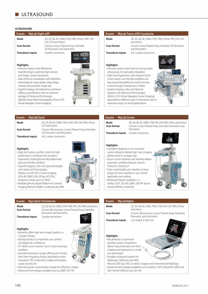
RADBOOK 2017148
◼ ULTRASOUND
· MyLab Twice eHD CrystaLineEsaote
Convex, Linear, Phased Array, Extended, 3D Panoramic and Volumetric
Scan format
4 & 1 probe connectorsTransducer inputs
2D, 3D, 4D, M, CMM, CFM, PWD, XFlow, PW, CW, CnTI and others
Mode
Highlights• Premium system with Point-of-Care portable
ultrasound unit optionally integrated• High level Ergonomics with intuitive Touch
Screen panel, user friendly workflow and App based MyLabRemote tool for remote control though Smartphone or Tablet
• Superb Imaging, Color and Spectral Doppler with Advanced Technologies (ElaXto, CnTI, Virtual Navigator Fusion Imaging) applicable to different types of transducer and to extensive range of clinical applications
· MyLabClassCEsaote
4 & 1 probe connectorsTransducer inputs
Convex, Microconvex, Linear, Phased Array, Extended, 3D Panoramic and Volumetric
Scan format
2D, 3D, 4D, M, CMM, TVM, CFM, PW, CW, PWD, XFlow CnTI and others
Mode
Highlights• High-end System, perfect choice for high
performance combined with excellent ergonomics (OptiLight and MyLabRemote) and user friendly workflow
• Superb Imaging, Color and Spectral Doppler with Advanced Technologies (ElaXto, Low MI CEUS, Fusion Imaging, 3D & 4D, QIMT, QAS, XFlow, HD CFM, Frequency range up to 22 MHz)
• Multidisciplinary Digital Platform for General Imaging, Women’s Health, Cardiovascular, MSK
· MyLab SevenEsaote
4 probe connectorsTransducer inputs
Convex, Linear, Phased Array, Extended, Panoramic and Volumetric
Scan format2D, 3D, 4D, M, CMM, CFM, PW, CW, PWD, XFlow and othersMode
Highlights• Confident diagnosis in an innovative
system design to deliver high-class imaging performance in compact size
• Touch-screen centered user interface allows automatic workflow features: eTouch, Protocols, SmarTouch
• Fully customizable user interface to have always the best workflow in any clinical application and setting
• Advanced features available, i. e.: ElaXto, CEUS, 3D / 4D, QIMT, QAS RF-based arterial stiffness, XStrain4D
ULTRASOUND
· MyLab Eight eXPEsaote
2D, 3D, 4D, M, CMM, CFM, PWD, XFlow, SWE, PW, CW, CnTI and others
Mode
Convex, Linear, Phased Array, Extended, 3D Panoramic and Volumetric
Scan format
4 probe connectorsTransducer inputs
Highlights• Premium system with MPowered
beam forming to optimize high-density and Single Crystal transducers
• State of the art visualization with WideView technology for crispy details, deep image contrast and extensive image size
• Superb Imaging, hemodynamics and tissue stiffness quantification with an extensive package of Advanced Tech nologie (QElaXto Shear Wave Elastography, XFlow, CnTI, Virtual Navigator Fusion Imaging)
· MyLabAlphaEsaote
2 on board, 4 with cartTransducer inputs
Convex, Microconvex, Linear, Phased Array, Extended, Panoramic and Volumetric
Scan format
2D, 3D, 4D, M, CMM, TVM, CFM, PW, CW, PWD, XFlow and others
Mode
Highlights• MyLabAlpha is a premium
portable system, designed to deliver top performance for both imaging and ergonomics in small size and weight
• Portable ultrasound system for Radiology, Cardiovascular, MSK, Rheuma, OB-Gyn, POC as well as Surgery and Interventional Radiology
• Advanced technologies available such as ElaXto, CEUS, XStrain4D, QIMT and QAS Arterial Stiffness tool, 3D / 4D
· MyLabSix ChristaLineEsaote
3 probe connectorsTransducer inputs
Convex, Microconvex, Linear, Phased Array, Extended, Panoramic and Volumetric
Scan format2D, 3D, 4D, M, CMM, CFM, TVM, PW, CW, PWD and othersMode
Highlights• MyLabSix offers high level Image Quality in a
Compact Design• eDesign product to maximize user comfort
and diagnostic confidence• 19” Wide screen monitor, Touch Screen and easy
workflow• Extended transducer range, offering also Hockey
Stick High Frequency, Biopsy dedicated convex transducer, TEE, Endocavity, Surgery and Laparo-scopic transducers
• Ultra-low power consumption: Esaote Eco Efficiency engine• Advanced technologies available such as, QIMT, 3D / 4D
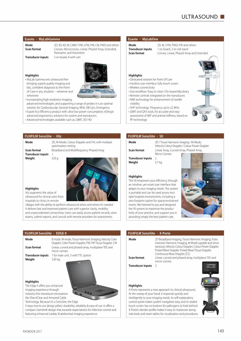
RADBOOK 2017 149
ULTRASOUND ◼
· MyLabGammaEsaote
2 on board, 4 with cartTransducer inputs
Convex, Microconvex, Linear, Phased Array, Extended, Panoramic and Volumetric
Scan format2D, 3D, 4D, M, CMM, TVM, CFM, PW, CW, PWD and othersMode
Highlights• MyLab Gamma sets ultrasound free
bringing superb quality imaging and fast, confident diagnosis to the Point-of-Care in any situation – wherever and whenever
• Incorporating high resolution imaging, advanced technologies, and supporting a range of probes it is an optimal solution for Cardiovascular, General Imaging, MSK, OB-Gyn, Emergency
• Esaote Eco Efficiency product with ultra-low power consumption; eDesign advanced ergonomics solutions for system and transducers.
• Advanced technologies available such as, QIMT, 3D / 4D
· MyLabOne
Convex, Linear, Phased Array and ExtendedScan format1 on board, 3 on roll standTransducer inputs2D, M, CFM, PWD, PW and othersMode
Highlights• Dedicated solution for Point Of Care • Intuitive user interface, fully touch screen • Wireless connectivity • Fast workflow / Easy to clean / On-board MyLibrary • Remote controls integrated on the transducers • NNE technology for enhancement of needle
visibility • XHF technology: Frequency up to 22 MHz • QIMT and QAS tools, for accurate and easy
assessment of IMT and arterial stiffness, based on RF technology
Esaote
· EDGE IIFUJIFILM SonoSite
1 for main unit, 3 with TTC optionTransducer inputs
Linear, curved and phased array, multiplane TEE and micro-convex
Scan format
B mode, M mode, Tissue Harmonic Imaging, Velocity Color Doppler, Color Power Doppler, PW, PW Tissue Doppler, CW
Mode
3.85 kgWeight
HighlightsThe Edge II offers you enhanced imaging experience through industry-first transducer innovations like DirectClear and Armored Cable Technology. Because it’s a SonoSite, the Edge II stays true to our design pillars: durability, reliability & ease of use. It offers a compact clamshell design that exceeds expectations for infection control and featuring enhanced cardiac & abdominal imaging experience.
· iVizFUJIFILM SonoSite
1Transducer inputs
2D, M-Mode, Colour Doppler and THI, with multiple optimisation setting
Mode
520 gWeight
Broadband and Multifrequency Phased ArrayScan format
HighlightsiViz augments the value of ultrasound for clinical users from hospitals to clinics in remote villages with the ability to perform ultrasound when and where it’s needed. It delivers fast and improved patient care with superior clarity, mobility, and unprecedented connectivity. Users can easily access patient records, store exams, submit reports, and consult with remote providers for assessments.
· SIIFUJIFILM SonoSite
2Transducer inputs
Linear Array, Curved Array, Phased Array,Micro-Convex
Scan format
2D / Tissue Harmonic Imaging / M-Mode, Velocity Colour Doppler / Colour Power Doppler
Mode
5,7 kgWeight
HighlightsThe SII empowers your efficiency through an intuitive, yet smart user interface that adapts to your imaging needs. The system is portable and can be used across mul-tiple hospital environments, including a zero footprint option for spaceconstrained rooms. We listened to you and designed the SII system to maximise the produc-tivity of your practice, and support you in providing simply the best patient care.
· X-PorteFUJIFILM SonoSite
3Transducer inputs
Linear, curved and phased array, multiplane TEE and micro-convex
Scan format
2D Broadband imaging, Tissue Harmonic Imaging, Pulse Inversion Harmonic Imaging, M Mode (update and simul-taneous), Velocity Colour Doppler, Colour Power Doppler, Pulsed Wave Doppler, Pulsed Wave Tissue Doppler, Continuous Wave Doppler, ECG
Mode
HighlightsX-Porte represents a new approach to clinical ultrasound. At the sweep of your hand, it responds quickly and intelligently to your imaging needs. Its self-explanatory control panel makes system navigation easy and its sealed touch screen has no buttons for pathogens to hide behind. X-Porte’s slender profile makes it easy to maneuver along-side beds and exam tables for visualization and procedures.
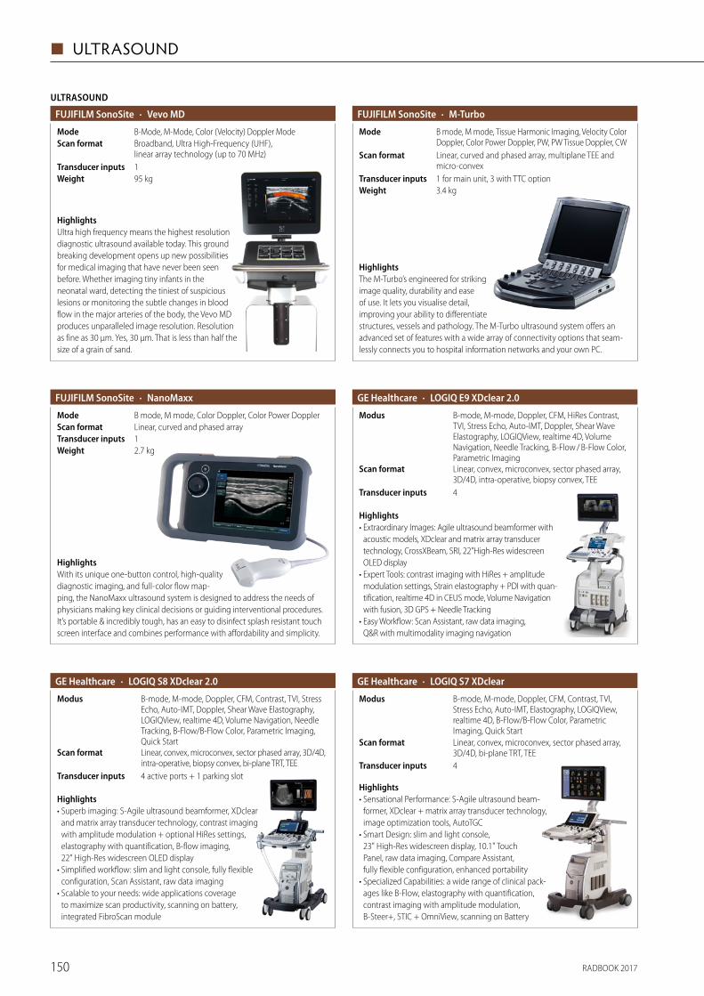
RADBOOK 2017150
◼ ULTRASOUND
95 kgWeight
ULTRASOUND
· Vevo MDFUJIFILM SonoSite
1Transducer inputs
Broadband, Ultra High-Frequency (UHF), linear array technology (up to 70 MHz)
Scan formatB-Mode, M-Mode, Color (Velocity) Doppler ModeMode
HighlightsUltra high frequency means the highest resolution diagnostic ultrasound available today. This ground breaking development opens up new possibilities for medical imaging that have never been seen before. Whether imaging tiny infants in the neonatal ward, detecting the tiniest of suspicious lesions or monitoring the subtle changes in blood flow in the major arteries of the body, the Vevo MD produces unparalleled image resolution. Resolution as fine as 30 μm. Yes, 30 μm. That is less than half the size of a grain of sand.
· M-TurboFUJIFILM SonoSite
1 for main unit, 3 with TTC optionTransducer inputs
Linear, curved and phased array, multiplane TEE and micro-convex
Scan format
B mode, M mode, Tissue Harmonic Imaging, Velocity Color Doppler, Color Power Doppler, PW, PW Tissue Doppler, CW
Mode
3.4 kgWeight
HighlightsThe M-Turbo’s engineered for striking image quality, durability and ease of use. It lets you visualise detail, improving your ability to differentiate structures, vessels and pathology. The M-Turbo ultrasound system offers an advanced set of features with a wide array of connectivity options that seam-lessly connects you to hospital information networks and your own PC.
2.7 kg
· NanoMaxxFUJIFILM SonoSite
1Transducer inputsLinear, curved and phased arrayScan formatB mode, M mode, Color Doppler, Color Power DopplerMode
Weight
HighlightsWith its unique one-button control, high-quality diagnostic imaging, and full-color flow map-ping, the NanoMaxx ultrasound system is designed to address the needs of physicians making key clinical decisions or guiding interventional procedures. It’s portable & incredibly tough, has an easy to disinfect splash resistant touch screen interface and combines performance with affordability and simplicity.
· LOGIQ E9 XDclear 2.0GE Healthcare
4Transducer inputs
Linear, convex, microconvex, sector phased array, 3D/4D, intra-operative, biopsy convex, TEE
Scan format
B-mode, M-mode, Doppler, CFM, HiRes Contrast, TVI, Stress Echo, Auto-IMT, Doppler, Shear Wave Elastography, LOGIQView, realtime 4D, Volume Navigation, Needle Tracking, B-Flow / B-Flow Color, Parametric Imaging
Modus
Highlights• Extraordinary Images: Agile ultrasound beamformer with
acoustic models, XDclear and matrix array transducer technology, CrossXBeam, SRI, 22"High-Res widescreen OLED display
• Expert Tools: contrast imaging with HiRes + amplitude modulation settings, Strain elastography + PDI with quan-tification, realtime 4D in CEUS mode, Volume Navigation with fusion, 3D GPS + Needle Tracking
• Easy Workflow: Scan Assistant, raw data imaging, Q&R with multimodality imaging navigation
· LOGIQ S8 XDclear 2.0GE Healthcare
4 active ports + 1 parking slotTransducer inputs
Linear, convex, microconvex, sector phased array, 3D/4D, intra-operative, biopsy convex, bi-plane TRT, TEE
Scan format
B-mode, M-mode, Doppler, CFM, Contrast, TVI, Stress Echo, Auto-IMT, Doppler, Shear Wave Elastography, LOGIQView, realtime 4D, Volume Navigation, Needle Tracking, B-Flow/B-Flow Color, Parametric Imaging, Quick Start
Modus
Highlights• Superb imaging: S-Agile ultrasound beamformer, XDclear
and matrix array transducer technology, contrast imaging with amplitude modulation + optional HiRes settings, elastography with quantification, B-flow imaging, 22" High-Res widescreen OLED display
• Simplified workflow: slim and light console, fully flexible configuration, Scan Assistant, raw data imaging
• Scalable to your needs: wide applications coverage to maximize scan productivity, scanning on battery, integrated FibroScan module
4Transducer inputs
· LOGIQ S7 XDclearGE Healthcare
Linear, convex, microconvex, sector phased array, 3D/4D, bi-plane TRT, TEE
Scan format
B-mode, M-mode, Doppler, CFM, Contrast, TVI, Stress Echo, Auto-IMT, Elastography, LOGIQView, realtime 4D, B-Flow/B-Flow Color, Parametric Imaging, Quick Start
Modus
Highlights• Sensational Performance: S-Agile ultrasound beam-
former, XDclear + matrix array transducer technology, image optimization tools, AutoTGC
• Smart Design: slim and light console, 23" High-Res widescreen display, 10.1" Touch Panel, raw data imaging, Compare Assistant, fully flexible confi guration, enhanced portability
• Specialized Capabilities: a wide range of clinical pack-ages like B-Flow, elastography with quantification, contrast imaging with amplitude modulation, B-Steer+, STIC + OmniView, scanning on Battery
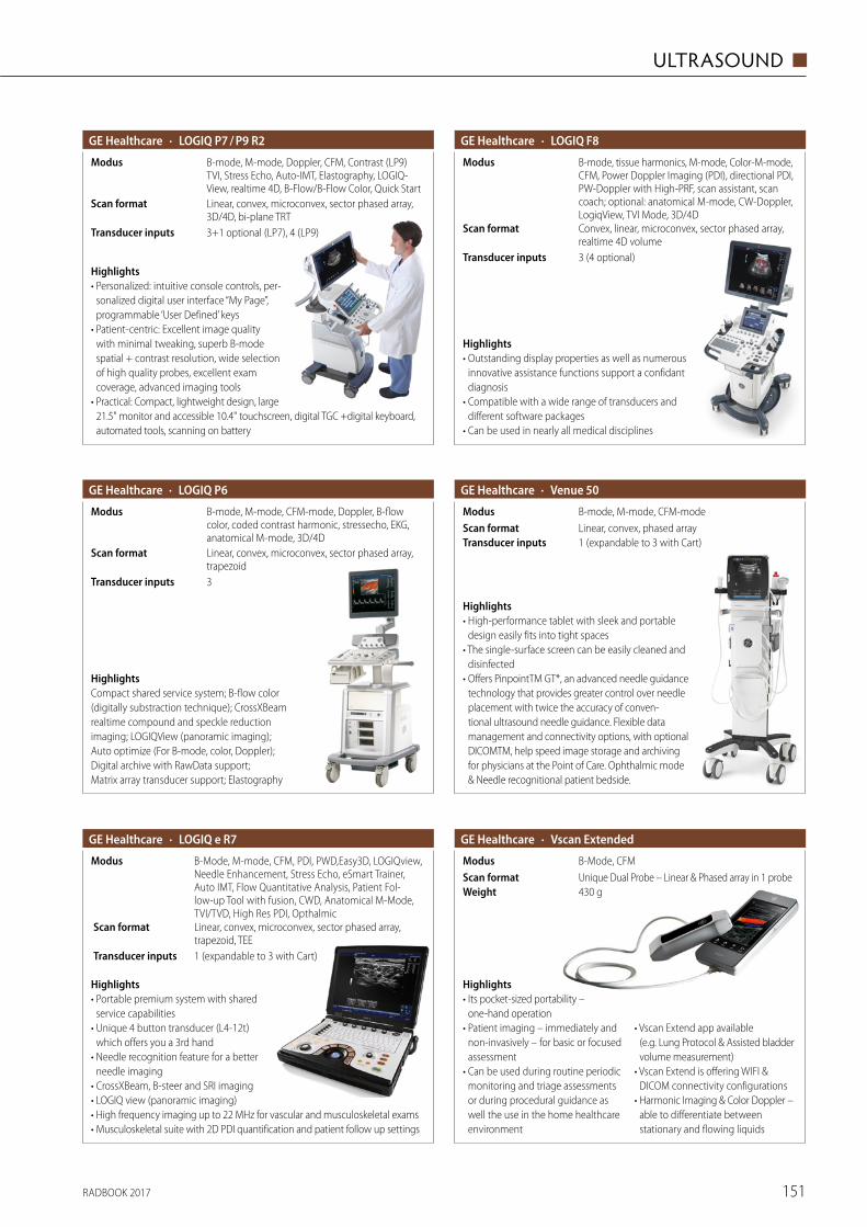
RADBOOK 2017 151
ULTRASOUND ◼
· LOGIQ P7 / P9 R2GE Healthcare
3+1 optional (LP7), 4 (LP9)Transducer inputs
Linear, convex, microconvex, sector phased array, 3D/4D, bi-plane TRT
Scan format
B-mode, M-mode, Doppler, CFM, Contrast (LP9) TVI, Stress Echo, Auto-IMT, Elastography, LOGIQ-View, realtime 4D, B-Flow/B-Flow Color, Quick Start
Modus
Highlights• Personalized: intuitive console controls, per-
sonalized digital user interface “My Page”, programmable ‘User Defined’ keys
• Patient-centric: Excellent image quality with minimal tweaking, superb B-mode spatial + contrast resolution, wide selection of high quality probes, excellent exam coverage, advanced imaging tools
• Practical: Compact, lightweight design, large 21.5" monitor and accessible 10.4" touchscreen, digital TGC +digital keyboard, automated tools, scanning on battery
· Venue 50GE Healthcare
1 (expandable to 3 with Cart)Transducer inputsLinear, convex, phased arrayScan formatB-mode, M-mode, CFM-modeModus
Highlights• High-performance tablet with sleek and portable
design easily fits into tight spaces• The single-surface screen can be easily cleaned and
disinfected• Offers PinpointTM GT*, an advanced needle guidance
technology that provides greater control over needle placement with twice the accuracy of conven-tional ultrasound needle guidance. Flexible data management and connectivity options, with optional DICOMTM, help speed image storage and archiving for physicians at the Point of Care. Ophthalmic mode & Needle recognitional patient bedside.
· LOGIQ F8GE Healthcare
3 (4 optional)Transducer inputs
Convex, linear, microconvex, sector phased array, realtime 4D volume
Scan format
B-mode, tissue harmonics, M-mode, Color-M-mode, CFM, Power Doppler Imaging (PDI), directional PDI, PW-Doppler with High-PRF, scan assistant, scan coach; optional: anatomical M-mode, CW-Doppler, LogiqView, TVI Mode, 3D/4D
Modus
Highlights• Outstanding display properties as well as numerous
innovative assistance functions support a confidant diagnosis
• Compatible with a wide range of transducers and different software packages
• Can be used in nearly all medical disciplines
· LOGIQ P6 GE Healthcare
3Transducer inputs
Linear, convex, microconvex, sector phased array, trapezoid
Scan format
B-mode, M-mode, CFM-mode, Doppler, B-flow color, coded contrast harmonic, stressecho, EKG, anatomical M-mode, 3D/4D
Modus
HighlightsCompact shared service system; B-flow color (digitally substraction technique); CrossXBeam realtime compound and speckle reduction imaging; LOGIQView (panoramic imaging); Auto optimize (For B-mode, color, Doppler); Digital archive with RawData support; Matrix array transducer support; Elastography
· LOGIQ e R7GE Healthcare
1 (expandable to 3 with Cart)Transducer inputs
Linear, convex, microconvex, sector phased array, trapezoid, TEE
Scan format
B-Mode, M-mode, CFM, PDI, PWD,Easy3D, LOGIQview, Needle Enhancement, Stress Echo, eSmart Trainer, Auto IMT, Flow Quantitative Analysis, Patient Fol-low-up Tool with fusion, CWD, Anatomical M-Mode, TVI/TVD, High Res PDI, Opthalmic
Modus
Highlights• Portable premium system with shared
service capabilities• Unique 4 button transducer (L4-12t)
which offers you a 3rd hand• Needle recognition feature for a better
needle imaging• CrossXBeam, B-steer and SRI imaging• LOGIQ view (panoramic imaging)• High frequency imaging up to 22 MHz for vascular and musculoskeletal exams• Musculoskeletal suite with 2D PDI quantification and patient follow up settings
· Vscan ExtendedGE Healthcare
430 gWeightUnique Dual Probe – Linear & Phased array in 1 probeScan formatB-Mode, CFMModus
Highlights• Its pocket-sized portability –
one-hand operation• Patient imaging – immediately and
non-invasively – for basic or focused assessment
• Can be used during routine periodic monitoring and triage assessments or during procedural guidance as well the use in the home healthcare environment
• Vscan Extend app available (e.g. Lung Protocol & Assisted bladder volume measurement)
• Vscan Extend is offering WIFI & DICOM connectivity configurations
• Harmonic Imaging & Color Doppler – able to differentiate between stationary and flowing liquids
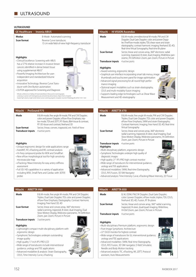
RADBOOK 2017152
◼ ULTRASOUND
ULTRASOUND
· Invenia ABUSGE Healthcare
Reverse Curve transducer, 15 cm wide field-of-view high-frequency transducer
Scan formatB-Mode Automated scanningModus
Highlights• Clinical Excellence: Screening with ABUS
has a 57 % relative increase in invasive breast cancers identified in dense breast tissue using supplemental ABUS¹
• Powerful Imaging Architecture for user- independent and standardized Volume acquisition
• Innovative Technology: Reverse Curve Trans-ducer with One Button automation
• CE/FDA approved for Screening and Diagnosis1 Wilczek, Leifland, et.al. Adding 3D Automated Breast Ultrasound to mammography
screening in women with heterogeneously and extremely dense breasts. Report from a hospital- based, high-volume, single-center breast cancer screening program. European Journal of Radiology 85 (2016) 1554–1563
Highlights• Award-winning, ergonomic design• Graphical user interface incorporating smart tab menus, image
thumbnails and touchscreen panel for image optimisation• Advanced signal processing for all-round high perfor-
mance imaging• Optional expert modalities such as strain elastography,
CEUS and multi-modality fusion imaging• Supports leading edge technologies such as Shear Wave
Measurement and 4D-elastography
· HI VISION AscendusHitachi
4 active portsTransducer inputs
Sector, linear and convex array, 360° electronic radial scanning, trapezoid, B-steer, dual imaging, WideView pan-oramic, HI-Definition Zoom, pan Zoom; Picture in Picture
Scan format
B & M-mode; omnidirectional M-mode; PW and CW Doppler; Dual Gate Doppler; color and power Dopp-ler; FineFlow-mode; triplex; TDI; shear wave and strain elastography; contrast harmonic imaging; freehand 3D; 4D; Real-time Virtual Sonography; Real-time Bi-plane
Mode
Highlights• Unique ergonomic design for wide applications range• AutoIMT, NT, eTracking and WI, contrast analysis• Hi-Freq compound probe for MSK and SmallPart• New eFlow morphological tool for high sensitivity
microvascular map• eTracking / Wave Intensity for easy artery stiffness
assessment• Full 3D / 4D capabilities in a variety of application
including MSK, Small Parts and Cardiac with 3DTEE probe
· ProSound F75Hitachi
4 active portsTransducer inputsSector, linear, convex, trapezoid, ext. Field of ViewScan format
B & M-mode; free angle M-mode; PW and CW Doppler; color and power Doppler; eFlow-Flow Emphasis; trip-lex-mode; TDI and 2DTT; RT-Elasto; BbH tissue & contrast; RT-3D-tissue and contrast; freehand 3D
Mode
Highlights• Multi-disciplinary platform, ergonomic design• Symphonic Technologies underpin high quality of
diagnostic images• High quality 21" IPS-PRO high contrast monitor• Wide range of transducers for interventional guidance,
urology and TEE applications• Advanced modalities: SWM, Real-time Elastography,
CEUS, RVS Fusion, 3D SIM Navigator• Advanced analysis: Time Intensity Curve, eTracking/Wave Intensity, 2D Tissue
· ARIETTA V70Hitachi
B & M-mode; free angle M-mode; PW and CW Doppler; Triplex; Dual Gate Doppler; TDI; color and power Doppler; eFlow-Flow Emphasis; SWM and strain Elastography; Contrast Harmonic Imaging; Free Hand 3D; 4D; Real-time Virtual Sonography
Mode
Sector, linear and convex array, 360° electronic radial scanning, trapezoid, B-steer, dual imaging, Dual Slow-Motion Display, Wideview panoramic, HI-Definition Zoom, pan Zoom; Picture in Picture
Scan format
4 active portsTransducer inputs
Highlights• Lightweight compact multi-disciplinary platform with
ergonomic design• Symphonic Technologies underpin outstanding
image quality• High quality 17 inch IPS-PRO LCD• Wide range of transducers include interventional
guidance, urology and TEE applications• Advanced modalities & analysis: Strain Elastography,
CEUS, Time Intensity Curve, eTracking
· ARIETTA V60Hitachi
Sector, linear and convex array, 360° electronic radial scanning, trapezoid, B-steer, dual imaging, Dual Slow-Motion Display, Wideview panoramic, HI-Definition Zoom, pan Zoom; Picture in Picture
Scan format
B & M-mode; free angle M-mode; PW and CW Doppler; Triplex; Dual Gate Doppler; TDI; color and power Doppler; eFlow-Flow Emphasis; Elastography; Contrast Harmonic Imaging; Free Hand 3D; 4D
Mode
3 active portsTransducer inputs
· ARIETTA 850Hitachi
Sector, linear and convex array, 360° radial scanning, trapezoid, B-steer, dual/quad imaging, WideView, HI-Def Zoom, pan Zoom; Picture in Picture
Scan format
B, M, ODM; PW,CW Doppler; Dual Gate Doppler; color / power Doppler; eFlow mode; triplex; TDI; CEUS; freehand 3D; 4D, Fusion, RT Bi-plane
Mode
4 active portsTransducer inputs
Highlights• Multi-disciplinary Premium platform, ergonomic design • Pure Image Symphonic Architecture • 22“ OLED monitor for highest contrast • Wide range of transducers for GI, interventional guidance,
urology and TEE applications • Advanced modalities: SWM, Real-time Elastography,
CEUS, RVS Fusion, 3D SIM navigator, E-field Simulator, Needle and Body Motion tracking
• Advanced analysis: TIC, eTracking, WI, 2DTT, Protocol assistant, Auto Measurements
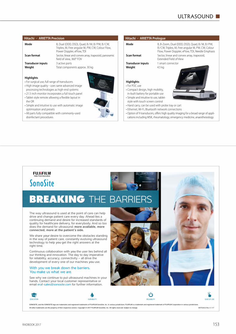
RADBOOK 2017 153
ULTRASOUND ◼
Highlights• For surgical use, full range of transducers• High image quality - uses same advanced image
processing technologies as high-end systems• 21.5 inch monitor incorporates a full touch panel• Tablet-style remote allowing a flexible layout in
the OR• Simple and intuitive to use with automatic image
optimisation and presets• All parts fully compatible with commonly-used
disinfectant procedures
· ARIETTA PrecisionHitachi
3 active portsTransducer inputs
Sector, linear and convex array, trapezoid, panoramic field of view, 360° FOV
Scan format
B, Dual (DDD, DSD), Quad, B / M, B / PW, B / CW, Triplex, M, Free angular M, PW, CW, Colour Flow, Power Doppler, eFlow, TDI
Mode
Total components approx. 30 kgWeight
Highlights• For POC use• Compact design, high mobility,
in-built battery for portable use• Simple and intuitive to use, tablet-
style with touch screen control• Hand carry, can be used with probe tray or cart• Ethernet, Wi-Fi, Bluetooth network connections• Option of 9 transducers, offers high quality imaging for a broad range of appli-
cations including MSK, rheumatology, emergency medicine, anaesthesiology
· ARIETTA PrologueHitachi
1 smart connectorTransducer inputs
Sector, linear and convex array, trapezoid, Extended Field of View
Scan format
B, B-Zoom, Dual (DDD, DSD), Quad, B / M, B / PW, B / CW, Triplex, M, Free angular M, PW, CW, Colour Flow, Power Doppler, eFlow, TDI, Needle Emphasis
Mode
4.5 kgWeight
SONOSITE, and the SONOSITE logo are trademarks and registered trademarks of FUJIFILM SonoSite, Inc. in various jurisdictions. FUJIFILM is a trademark and registered trademark of FUJIFILM Corporation in various jurisdictions.
All other trademarks are the property of their respective owners. Copyright © 2017 FUJIFILM SonoSite, Inc. All rights reserved. Subject to change. MKT02833 Rev A 1/17
The way ultrasound is used at the point of care can help drive and change patient care every day. Ahead lies a continuing demand and desire for increased standards of quality for healthcare delivery, for everybody. And so too does the demand for ultrasound: more available, more connected, more at the patient’s side.
We share your desire to overcome the obstacles standing in the way of patient care, constantly evolving ultrasound technology to help you get the right answers at the right time.
Continuous collaboration with you the user lies behind all our thinking and innovation. The day to day imperative for reliability, accuracy, connectivity – all drive the development of every one of our machines you use.
With you we break down the barriers. You make us what we are.See why we continue to put ultrasound machines in your hands. Contact your local customer representative or email [email protected] for further information.
BREAKING THE BARRIERS
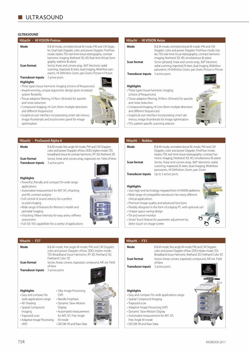
RADBOOK 2017154
◼ ULTRASOUND
Highlights• Three types tissue harmonic imaging (choice of frequencies)• Award-winning, unique ergonomic design gives increased
system flexibility• Tissue adaptive filtering, HI Rez+ (8 levels) for speckle
and noise reduction• Compound imaging, HI Com (from multiple directions
and different frequencies)• Graphical user interface incorporating smart tab menus,
image thumbnails and touchscreen panel for image optimisation
ULTRASOUND
· HI VISION PreirusHitachi
3 active portsTransducer inputs
Sector, linear and convex array, 360° electronic radial scanning, trapezoid, B-steer, dual imaging, WideView pan-oramic, HI-Definition Zoom, pan Zoom; Picture in Picture
Scan format
B & M-mode; omnidirectional M-mode; PW and CW Dopp-ler; Dual Gate Doppler; color and power Doppler; FineFlow mode; triplex; TDI; real-time tissue elastography; contrast harmonic imaging; freehand 3D; 4D; Real-time Virtual Sono-graphy; realtime Bi-plane
Mode
Highlights• Three types tissue harmonic imaging
(choice of frequencies)• Tissue adaptive filtering, HI Rez+ (8 levels) for speckle
and noise reduction• Compound imaging, HI Com (from multiple directions
and different frequencies)• Graphical user interface incorporating smart tab
menus, image thumbnails for image optimisation• PSS, patient specific scanning selector
· HI VISION AviusHitachi
3 active portsTransducer inputs
Sector (phased), linear and convex array, 360° electronic radial scanning, trapezoid, B-steer, dual imaging, WideView panoramic, HI-Definition Zoom, pan Zoom; Picture in Picture
Scan format
B & M-mode; omnidirectional M-mode; PW and CW Doppler; color and power Doppler; FineFlow-mode; trip-lex; TDI; real-time tissue elastography; contrast harmonic imaging; freehand 3D; 4D; simultaneous Bi-plane
Mode
Highlights• Powerful, friendly and compact for wide range
applications• Automated measurement for IMT, NT, eTracking
and WI, contrast analysis• Full control of sound velocity for a perfect
ocused imaging• Wide range of features for Women's Health and
perinatal imaging• eTracking / Wave Intensity for easy artery stiffness
assessment• Full 3D / 4D capabilities for a variety of applications
· ProSound Alpha 6Hitachi
3 active portsTransducer inputsSector, linear and convex array, trapezoid, ext. Field of ViewScan format
B & M-mode; free angle M-mode; PW and CW Doppler; color and power Doppler; eFlow; DDD; triplex-mode; TDI; broadband tissue & contrast harmonic; RT-3D; freehand 3D
Mode
Highlights• Uses high-end technology migrated from HI VISION platforms• Wide range of compatible transducers for many different
clinical applications• Premium image quality and advanced functions• Flexibly designed in the form of a laptop PC with optional cart• Unique space-saving design• Tilt and swivel monitor• Smart Touch feature for parameter adjustment by
direct touch on image screen
· NoblusHitachi
Sector, linear and convex array, 360° electronic radial scanning, trapezoid, B-steer, dual imaging, WideView panoramic, HI-Definition Zoom, pan Zoom
Scan format
Up to 3 active portsTransducer inputs
B & M-mode; omnidirectional M-mode; PW and CW Doppler; color and power Doppler; FineFlow mode; triplex; TDI; real-time tissue elastography; contrast har-monic imaging; Freehand 3D; 4D; simultaneous Bi-plane
Mode
Highlights• Easy and compact for wide applications range• Spatial Compound Imaging• Trapezoid scan• Adaptive Image Processing (AIP)• Dynamic Slow-Motion Display • Automated measurement for IMT, NT,
Free Angle M-mode• DICOM SR and Raw Data
· F31Hitachi
3 active portsTransducer inputs
B & M-mode; free angle M-mode; PW and CW Doppler; color and power Doppler; eFlow; DDD; triplex-mode; TDI; Broadband tissue Harmonic; freehand 3D, Freehand Color 3D
Mode
Sector, linear, convex, trapezoid, compound, AIP, ext. Field of View
Scan format
· F37Hitachi
3 active portsTransducer inputs
Sector, linear, convex, trapezoid, compound, AIP, ext. Field of View
Scan format
B & M-mode; free angle M-mode; PW and CW Doppler; color and power Doppler; eFlow; DDD; triplex-mode; TDI; Broadband tissue Harmonic; RT-3D; freehand 3D, Freehand Color 3D
Mode
Highlights• Easy and compact for
wide applications range• 4D Shading• Spatial Compound
Imaging• Trapezoid scan• Adaptive Image Processing
(AIP)
• Silky Image Processing (SIP)
• Needle Emphasis• Dynamic Slow-Motion
Display• Automated measurement
for IMT, NT, Free Angle M-mode
• DICOM SR and Raw Data
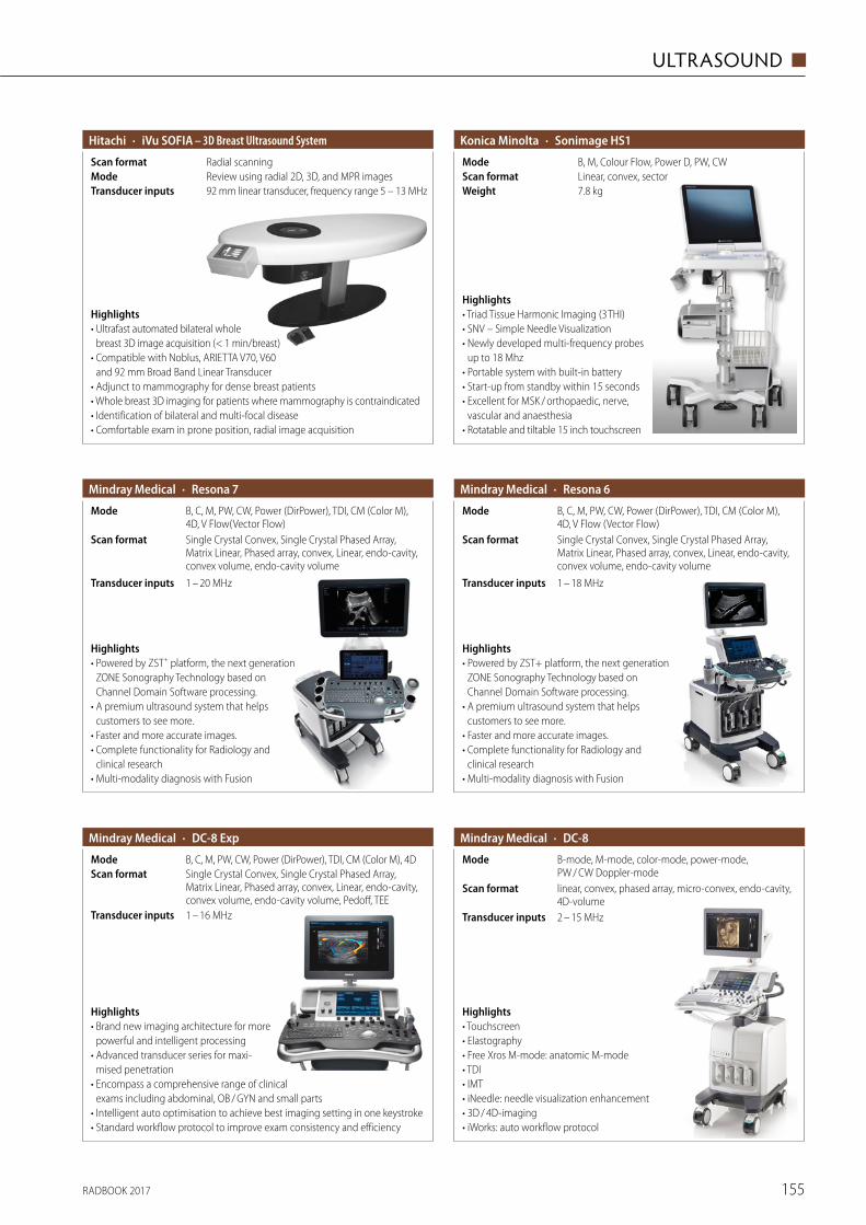
RADBOOK 2017 155
ULTRASOUND ◼
· iVu SOFIA – 3D Breast Ultrasound SystemHitachi
92 mm linear transducer, frequency range 5 – 13 MHzTransducer inputsReview using radial 2D, 3D, and MPR imagesModeRadial scanningScan format
Highlights• Ultrafast automated bilateral whole
breast 3D image acquisition (< 1 min/breast)• Compatible with Noblus, ARIETTA V70, V60
and 92 mm Broad Band Linear Transducer• Adjunct to mammography for dense breast patients• Whole breast 3D imaging for patients where mammography is contraindicated• Identification of bilateral and multi-focal disease• Comfortable exam in prone position, radial image acquisition
Highlights• Triad Tissue Harmonic Imaging (3 THI)• SNV – Simple Needle Visualization• Newly developed multi-frequency probes
up to 18 Mhz• Portable system with built-in battery• Start-up from standby within 15 seconds• Excellent for MSK / orthopaedic, nerve,
vascular and anaesthesia• Rotatable and tiltable 15 inch touchscreen
· Sonimage HS1Konica Minolta
7.8 kgWeightLinear, convex, sectorScan formatB, M, Colour Flow, Power D, PW, CWMode
· Resona 7Mindray Medical
Single Crystal Convex, Single Crystal Phased Array, Matrix Linear, Phased array, convex, Linear, endo-cavity, convex volume, endo-cavity volume
Scan format
B, C, M, PW, CW, Power (DirPower), TDI, CM (Color M), 4D, V Flow(Vector Flow)
Mode
1 – 20 MHzTransducer inputs
Highlights• Powered by ZST+ platform, the next generation
ZONE Sonography Technology based on Channel Domain Software processing.
• A premium ultrasound system that helps customers to see more.
• Faster and more accurate images.• Complete functionality for Radiology and
clinical research• Multi-modality diagnosis with Fusion
· Resona 6Mindray Medical
Single Crystal Convex, Single Crystal Phased Array, Matrix Linear, Phased array, convex, Linear, endo-cavity, convex volume, endo-cavity volume
Scan format
B, C, M, PW, CW, Power (DirPower), TDI, CM (Color M), 4D, V Flow (Vector Flow)
Mode
1 – 18 MHzTransducer inputs
Highlights• Powered by ZST+ platform, the next generation
ZONE Sonography Technology based on Channel Domain Software processing.
• A premium ultrasound system that helps customers to see more.
• Faster and more accurate images. • Complete functionality for Radiology and
clinical research • Multi-modality diagnosis with Fusion
· DC-8 ExpMindray Medical
1 – 16 MHzTransducer inputs
Single Crystal Convex, Single Crystal Phased Array, Matrix Linear, Phased array, convex, Linear, endo-cavity, convex volume, endo-cavity volume, Pedoff, TEE
Scan formatB, C, M, PW, CW, Power (DirPower), TDI, CM (Color M), 4DMode
Highlights• Brand new imaging architecture for more
powerful and intelligent processing • Advanced transducer series for maxi-
mised penetration • Encompass a comprehensive range of clinical
exams including abdominal, OB / GYN and small parts• Intelligent auto optimisation to achieve best imaging setting in one keystroke• Standard workflow protocol to improve exam consistency and efficiency
· DC-8Mindray Medical
linear, convex, phased array, micro-convex, endo-cavity, 4D-volume
Scan format
B-mode, M-mode, color-mode, power-mode, PW / CW Doppler-mode
Mode
2 – 15 MHzTransducer inputs
Highlights• Touchscreen• Elastography• Free Xros M-mode: anatomic M-mode• TDI• IMT• iNeedle: needle visualization enhancement• 3D / 4D-imaging• iWorks: auto workflow protocol
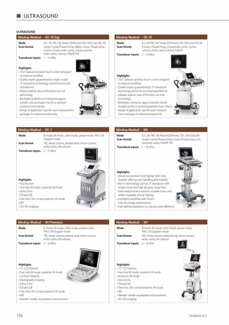
RADBOOK 2017156
◼ ULTRASOUND
· DC-70Mindray Medical
2 – 14 MHzTransducer inputs
Convex, Phased Array, Linear,endo-cavity, convex volume, endo-cavity volume, Pedoff
Scan formatB, C, M, PW, CW, Power (DirPower),TDI, CM (Color M), 4DMode
Highlights• 10.4" Gesture sensitive touch screen designed
to improve workflow• Quality exams guaranteed by 3T transducer
technology and Echo-enriched beamformer • Obatain realistic view of the fetus via iLive
technology• MedSight, interactive app to transfer clinical
images via iOS or android powered smart device• Range of application specific auto measure-
ment packages to improve productivity
ULTRASOUND
· DC-70 ExpMindray Medical
1 – 16 MHzTransducer inputs
Single Crystal Phased Array, Matrix Linear, Phased array, convex, Linear, endo-cavity, convex volume, endo-cavity volume, Pedoff, TEE
Scan formatB, C, M, PW, CW, Power (DirPower),TDI, CM (Color M), 4DMode
Highlights• 10.4" Gesture sensitive touch screen designed
to improve workflow • Quality exams guaranteed by single crystal
3T transducer technology and Echo-enriched beamformer
• Obtain realistic view of the fetus via iLive technology
• MedSight & MedTouch interactive app to transfer clinical images via iOS or android powered smart device
• Range of application specific auto measure ment packages to improve productivity
· DC-7Mindray Medical
TEE, linear, convex, phased array, micro-convex, endo-cavity, 4D-volume
Scan format
B-mode, M-mode, color-mode, power-mode, PW / CW Doppler-mode
Mode
2 – 15 MHzTransducer inputs
Highlights• Touchscreen• Free Xros M-mode: anatomic M-mode• Stress Echo• TDI and QA• Free Xros CM: curved anatomic M-mode• IMT• 3D / 4D-imaging
· M9Mindray Medical
1 – 16 MHzTransducer inputs
Single Crystal Phased Array, Linear, Phased array, con-vex,endo-cavity, Pedoff, TEE
Scan formatB, C, M, PW, CW, Power(DirPower), TDI, CM (Color M)Mode
Highlights• Advanced premium level laptop style color
Doppler offering easy handling and mobility• Rich in technology such as 3T transducer with
single crystal and high dynamic range flow• Ideal shared-service solution suitable to be used
within muptiple clinical settings• Intelligent workflow with iTouch
(one key image optimisation)• User-defined operation to improve work efficiency
· M7Mindray Medical
2 – 16 MHzTransducer inputs
TEE, linear, convex, phased array, micro-convex, endo-cavity, 4D-volume
Scan format
B-mode, M-mode, color-mode, power-mode, PW / CW Doppler-mode
Mode
Highlights• 15" LCD monitor• Free Xros M-mode: anatomic M-mode• Anatomic M-mode• Stress Echo• TDI and QA• Free Xros CM: curved anatomic M-mode• IMT• iNeedle: needle visualization enhancement• 3D / 4D-imaging
· M7 PremiumMindray Medical
2 – 16 MHzTransducer inputs
TEE, linear, convex, phased array, micro-convex, endo-cavity, 4D-volume
Scan format
B-mode, M-mode, color-mode, power-mode, PW / CW Doppler-mode
Mode
Highlights• 15" LCD monitor • Free Xros M-mode: anatomic M-mode • Contrast imaging• Elastography imaging• Stress Echo • TDI and QA • Free Xros CM: curved anatomic M-mode • IMT • iNeedle: needle visualization enhancement
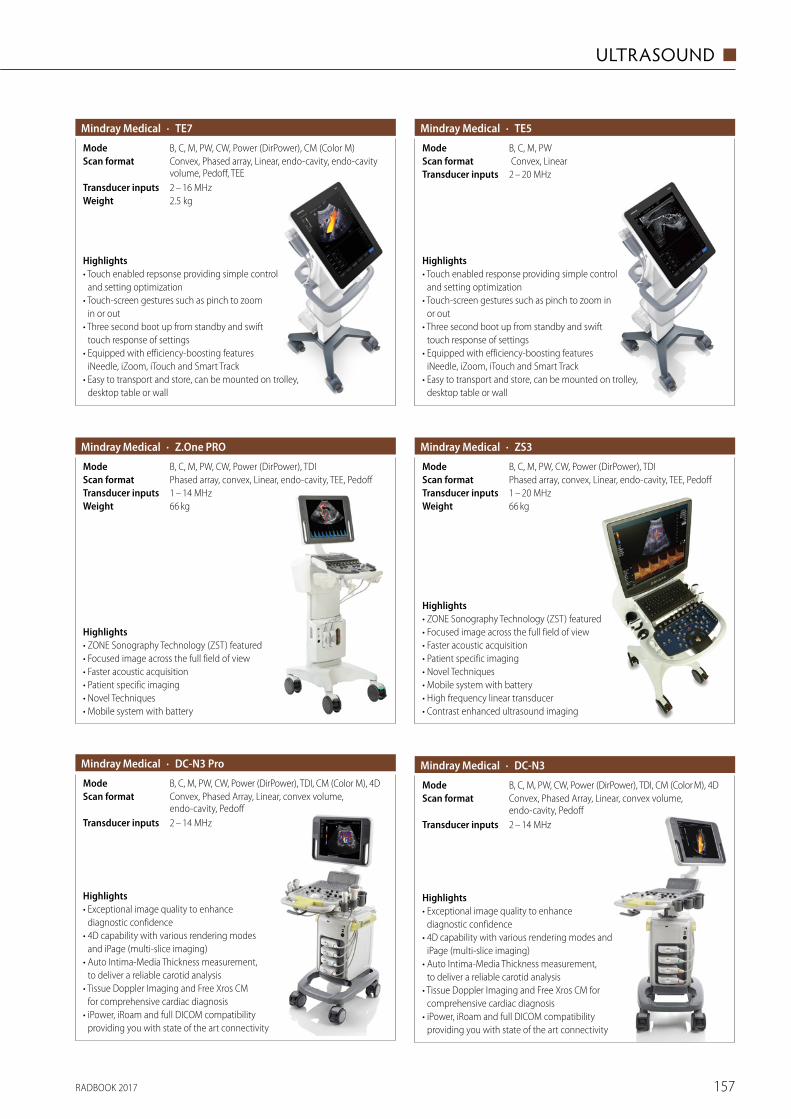
RADBOOK 2017 157
ULTRASOUND ◼
· TE7Mindray Medical
2.5 kgWeight2 – 16 MHzTransducer inputs
Convex, Phased array, Linear, endo-cavity, endo-cavity volume, Pedoff, TEE
Scan formatB, C, M, PW, CW, Power (DirPower), CM (Color M)Mode
Highlights• Touch enabled repsonse providing simple control
and setting optimization• Touch-screen gestures such as pinch to zoom
in or out• Three second boot up from standby and swift
touch response of settings• Equipped with efficiency-boosting features
iNeedle, iZoom, iTouch and Smart Track• Easy to transport and store, can be mounted on trolley,
desktop table or wall
· TE5Mindray Medical
2 – 20 MHzTransducer inputs Convex, LinearScan formatB, C, M, PWMode
Highlights• Touch enabled response providing simple control
and setting optimization • Touch-screen gestures such as pinch to zoom in
or out • Three second boot up from standby and swift
touch response of settings • Equipped with efficiency-boosting features
iNeedle, iZoom, iTouch and Smart Track • Easy to transport and store, can be mounted on trolley,
desktop table or wall
· Z.One PROMindray Medical
B, C, M, PW, CW, Power (DirPower), TDIModePhased array, convex, Linear, endo-cavity, TEE, PedoffScan format1 – 14 MHzTransducer inputs66 kgWeight
Highlights• ZONE Sonography Technology (ZST) featured• Focused image across the full field of view• Faster acoustic acquisition• Patient specific imaging• Novel Techniques• Mobile system with battery
· ZS3Mindray Medical
1 – 20 MHzTransducer inputsPhased array, convex, Linear, endo-cavity, TEE, PedoffScan formatB, C, M, PW, CW, Power (DirPower), TDIMode
66 kgWeight
Highlights• ZONE Sonography Technology (ZST) featured• Focused image across the full field of view• Faster acoustic acquisition• Patient specific imaging• Novel Techniques• Mobile system with battery• High frequency linear transducer• Contrast enhanced ultrasound imaging
· DC-N3 ProMindray Medical
2 – 14 MHzTransducer inputs
Convex, Phased Array, Linear, convex volume, endo-cavity, Pedoff
Scan formatB, C, M, PW, CW, Power (DirPower), TDI, CM (Color M), 4DMode
Highlights• Exceptional image quality to enhance
diagnostic confidence • 4D capability with various rendering modes
and iPage (multi-slice imaging) • Auto Intima-Media Thickness measurement,
to deliver a reliable carotid analysis • Tissue Doppler Imaging and Free Xros CM
for comprehensive cardiac diagnosis • iPower, iRoam and full DICOM compatibility
providing you with state of the art connectivity
· DC-N3Mindray Medical
2 – 14 MHzTransducer inputs
Convex, Phased Array, Linear, convex volume, endo-cavity, Pedoff
Scan formatB, C, M, PW, CW, Power (DirPower), TDI, CM (Color M), 4DMode
Highlights• Exceptional image quality to enhance
diagnostic confidence• 4D capability with various rendering modes and
iPage (multi-slice imaging)• Auto Intima-Media Thickness measurement,
to deliver a reliable carotid analysis• Tissue Doppler Imaging and Free Xros CM for
comprehensive cardiac diagnosis• iPower, iRoam and full DICOM compatibility
providing you with state of the art connectivity
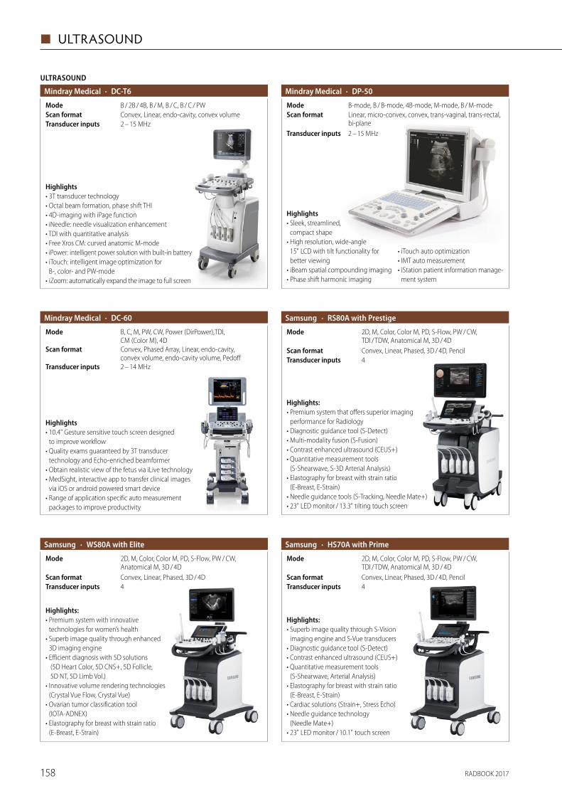
RADBOOK 2017158
◼ ULTRASOUND
ULTRASOUND
· DC-T6Mindray Medical
2 – 15 MHzTransducer inputsConvex, Linear, endo-cavity, convex volumeScan formatB / 2B / 4B, B / M, B / C, B / C / PWMode
Highlights• 3T transducer technology• Octal beam formation, phase shift THI• 4D-imaging with iPage function• iNeedle: needle visualization enhancement• TDI with quantitative analysis• Free Xros CM: curved anatomic M-mode• iPower: intelligent power solution with built-in battery• iTouch: intelligent image optimization for
B-, color- and PW-mode• iZoom: automatically expand the image to full screen
· DP-50Mindray Medical
2 – 15 MHzTransducer inputs
Linear, micro-convex, convex, trans-vaginal, trans-rectal, bi-plane
Scan formatB-mode, B / B-mode, 4B-mode, M-mode, B / M-modeMode
Highlights• Sleek, streamlined,
compact shape• High resolution, wide-angle
15" LCD with tilt functionality for better viewing
• iBeam spatial compounding imaging• Phase shift harmonic imaging
• iTouch auto optimization• IMT auto measurement• iStation patient information manage-
ment system
· DC-60Mindray Medical
2 – 14 MHzTransducer inputs
Convex, Phased Array, Linear, endo-cavity, convex volume, endo-cavity volume, Pedoff
Scan format
B, C, M, PW, CW, Power (DirPower),TDI, CM (Color M), 4D
Mode
Highlights• 10.4" Gesture sensitive touch screen designed
to improve workflow • Quality exams guaranteed by 3T transducer
technology and Echo-enriched beamformer • Obtain realistic view of the fetus via iLive technology • MedSight, interactive app to transfer clinical images
via iOS or android powered smart device • Range of application specific auto measure ment
packages to improve productivity
· RS80A with PrestigeSamsung
4Transducer inputsConvex, Linear, Phased, 3D / 4D, PencilScan format
2D, M, Color, Color M, PD, S-Flow, PW / CW, TDI / TDW, Anatomical M, 3D / 4D
Mode
Highlights:• Premium system that offers superior imaging
performance for Radiology• Diagnostic guidance tool (S-Detect)• Multi-modality fusion (S-Fusion)• Contrast enhanced ultrasound (CEUS+)• Quantitative measurement tools
(S-Shearwave, S-3D Arterial Analysis)• Elastography for breast with strain ratio
(E-Breast, E-Strain)• Needle guidance tools (S-Tracking, Needle Mate+)• 23" LED monitor / 13.3" tilting touch screen
· WS80A with EliteSamsung
4Transducer inputsConvex, Linear, Phased, 3D / 4DScan format
2D, M, Color, Color M, PD, S-Flow, PW / CW, Anatomical M, 3D / 4D
Mode
Highlights:• Premium system with innovative
technologies for women’s health• Superb image quality through enhanced
3D imaging engine• Efficient diagnosis with 5D solutions
(5D Heart Color, 5D CNS+, 5D Follicle, 5D NT, 5D Limb Vol.)
• Innovative volume rendering technologies (Crystal Vue Flow, Crystal Vue)
• Ovarian tumor classification tool (IOTA-ADNEX)
• Elastography for breast with strain ratio (E-Breast, E-Strain)
· HS70A with PrimeSamsung
4Transducer inputsConvex, Linear, Phased, 3D / 4D, PencilScan format
2D, M, Color, Color M, PD, S-Flow, PW / CW, TDI / TDW, Anatomical M, 3D / 4D
Mode
Highlights:• Superb image quality through S-Vision
imaging engine and S-Vue transducers • Diagnostic guidance tool (S-Detect) • Contrast enhanced ultrasound (CEUS+) • Quantitative measurement tools
(S-Shearwave, Arterial Analysis)• Elastography for breast with strain ratio
(E-Breast, E-Strain)• Cardiac solutions (Strain+, Stress Echo)• Needle guidance technology
(Needle Mate+) • 23" LED monitor / 10.1" touch screen
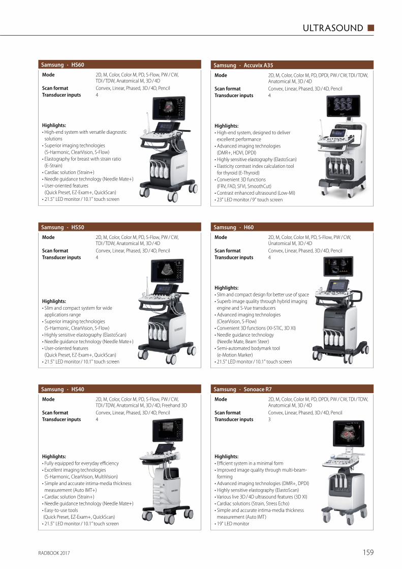
RADBOOK 2017 159
ULTRASOUND ◼
· HS60Samsung
4Transducer inputsConvex, Linear, Phased, 3D / 4D, PencilScan format
2D, M, Color, Color M, PD, S-Flow, PW / CW, TDI / TDW, Anatomical M, 3D / 4D
Mode
Highlights:• High-end system with versatile diagnostic
solutions• Superior imaging technologies
(S-Harmonic, ClearVision, S-Flow)• Elastography for breast with strain ratio
(E-Strain)• Cardiac solution (Strain+)• Needle guidance technology (Needle Mate+)• User-oriented features
(Quick Preset, EZ-Exam+, QuickScan)• 21.5" LED monitor / 10.1" touch screen
2D, M, Color, Color M, PD, DPDI, PW / CW, TDI / TDW, Anatomical M, 3D / 4D
Mode
· Accuvix A35Samsung
4Transducer inputsConvex, Linear, Phased, 3D / 4D, PencilScan format
Highlights:• High-end system, designed to deliver
excellent performance• Advanced imaging technologies
(DMR+, HDVI, DPDI)• Highly sensitive elastography (ElastoScan)• Elasticity contrast index calculation tool
for thyroid (E-Thyroid)• Convenient 3D functions
(FRV, FAD, SFVI, SmoothCut)• Contrast enhanced ultrasound (Low-MI)• 23" LED monitor / 9" touch screen
· H60Samsung
4Transducer inputsConvex, Linear, Phased, 3D / 4D, PencilScan format
2D, M, Color, Color M, PD, S-Flow, PW / CW, Únatomical M, 3D / 4D
Mode
Highlights:• Slim and compact design for better use of space• Superb image quality through hybrid imaging
engine and S-Vue transducers• Advanced imaging technologies
(ClearVision, S-Flow)• Convenient 3D functions (XI-STIC, 3D XI)• Needle guidance technology
(Needle Mate, Beam Steer)• Semi-automated bodymark tool
(e-Motion Marker)• 21.5" LED monitor / 10.1" touch screen
· HS50Samsung
4Transducer inputsConvex, Linear, Phased, 3D / 4D, PencilScan format
2D, M, Color, Color M, PD, S-Flow, PW / CW, TDI / TDW, Anatomical M, 3D / 4D
Mode
Highlights:• Slim and compact system for wide
applications range• Superior imaging technologies
(S-Harmonic, ClearVision, S-Flow)• Highly sensitive elastography (ElastoScan)• Needle guidance technology (Needle Mate+)• User-oriented features
(Quick Preset, EZ-Exam+, QuickScan)• 21.5" LED monitor / 10.1" touch screen
· HS40Samsung
4Transducer inputsConvex, Linear, Phased, 3D / 4D, PencilScan format
2D, M, Color, Color M, PD, S-Flow, PW / CW, TDI / TDW, Anatomical M, 3D / 4D, Freehand 3D
Mode
Highlights:• Fully equipped for everyday efficiency• Excellent imaging technologies
(S-Harmonic, ClearVision, MultiVision)• Simple and accurate intima-media thickness
measurement (Auto IMT+)• Cardiac solution (Strain+)• Needle guidance technology (Needle Mate+)• Easy-to-use tools (Quick Preset, EZ-Exam+, QuickScan)• 21.5" LED monitor / 10.1” touch screen
· Sonoace R7Samsung
3Transducer inputsConvex, Linear, Phased, 3D / 4D, PencilScan format
2D, M, Color, Color M, PD, DPDI, PW / CW, TDI / TDW, Anatomical M, 3D / 4D
Mode
Highlights:• Efficient system in a minimal form• Improved image quality through multi-beam-
forming• Advanced imaging technologies (DMR+, DPDI)• Highly sensitive elastography (ElastoScan)• Various live 3D / 4D ultrasound features (3D XI)• Cardiac solutions (Strain, Stress Echo)• Simple and accurate intima-media thickness
measurement (Auto IMT)• 19" LED monitor
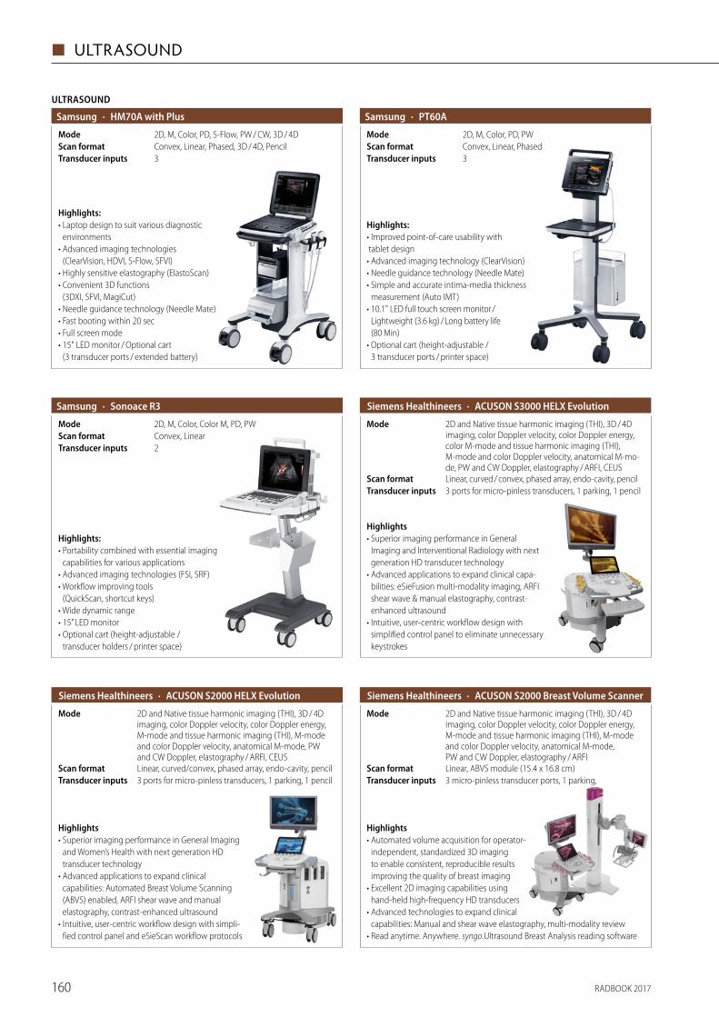
RADBOOK 2017160
◼ ULTRASOUND
ULTRASOUND
· HM70A with PlusSamsung
3Transducer inputsConvex, Linear, Phased, 3D / 4D, PencilScan format2D, M, Color, PD, S-Flow, PW / CW, 3D / 4DMode
Highlights:• Laptop design to suit various diagnostic
environments• Advanced imaging technologies
(ClearVision, HDVI, S-Flow, SFVI)• Highly sensitive elastography (ElastoScan)• Convenient 3D functions
(3DXI, SFVI, MagiCut)• Needle guidance technology (Needle Mate)• Fast booting within 20 sec• Full screen mode• 15" LED monitor / Optional cart
(3 transducer ports / extended battery)
· PT60ASamsung
3Transducer inputsConvex, Linear, PhasedScan format2D, M, Color, PD, PWMode
Highlights:• Improved point-of-care usability with tablet design• Advanced imaging technology (ClearVision)• Needle guidance technology (Needle Mate)• Simple and accurate intima-media thickness
measurement (Auto IMT)• 10.1" LED full touch screen monitor /
Lightweight (3.6 kg) / Long battery life (80 Min)
• Optional cart (height-adjustable / 3 transducer ports / printer space)
· Sonoace R3Samsung
2Transducer inputsConvex, LinearScan format2D, M, Color, Color M, PD, PWMode
Highlights:• Portability combined with essential imaging
capabilities for various applications• Advanced imaging technologies (FSI, SRF)• Workflow improving tools
(QuickScan, shortcut keys)• Wide dynamic range• 15” LED monitor• Optional cart (height-adjustable /
transducer holders / printer space)
· ACUSON S3000 HELX Evolution Siemens Healthineers
3 ports for micro-pinless transducers, 1 parking, 1 pencilTransducer inputsLinear, curved / convex, phased array, endo-cavity, pencilScan format
2D and Native tissue harmonic imaging (THI), 3D / 4D imaging, color Doppler velocity, color Doppler energy, color M-mode and tissue harmonic imaging (THI), M-mode and color Doppler velocity, anatomical M-mo-de, PW and CW Doppler, elastography / ARFI, CEUS
Mode
Highlights• Superior imaging performance in General
Imaging and Interventional Radiology with next generation HD transducer technology
• Advanced applications to expand clinical capa-bilities: eSieFusion multi-modality imaging, ARFI shear wave & manual elastography, contrast- enhanced ultrasound
• Intuitive, user-centric workflow design with simplified control panel to eliminate unnecessary keystrokes
· ACUSON S2000 HELX Evolution Siemens Healthineers
3 ports for micro-pinless transducers, 1 parking, 1 pencilTransducer inputsLinear, curved/convex, phased array, endo-cavity, pencilScan format
2D and Native tissue harmonic imaging (THI), 3D / 4D imaging, color Doppler velocity, color Doppler energy, M-mode and tissue harmonic imaging (THI), M-mode and color Doppler velocity, anatomical M-mode, PW and CW Doppler, elastography / ARFI, CEUS
Mode
Highlights• Superior imaging performance in General Imaging
and Women’s Health with next generation HD transducer technology
• Advanced applications to expand clinical capabilities: Automated Breast Volume Scanning (ABVS) enabled, ARFI shear wave and manual elastography, contrast-enhanced ultrasound
• Intuitive, user-centric workflow design with simpli-fied control panel and eSieScan workflow protocols
· ACUSON S2000 Breast Volume Scanner Siemens Healthineers
2D and Native tissue harmonic imaging (THI), 3D / 4D imaging, color Doppler velocity, color Doppler energy, M-mode and tissue harmonic imaging (THI), M-mode and color Doppler velocity, anatomical M-mode, PW and CW Doppler, elastography / ARFI
Mode
Linear, ABVS module (15.4 x 16.8 cm)Scan format3 micro-pinless transducer ports, 1 parking, Transducer inputs
Highlights• Automated volume acquisition for operator-
independent, standardized 3D imaging to enable consistent, reproducible results improving the quality of breast imaging
• Excellent 2D imaging capabilities using hand-held high-frequency HD transducers
• Advanced technologies to expand clinical capabilities: Manual and shear wave elastography, multi-modality review
• Read anytime. Anywhere. syngo.Ultrasound Breast Analysis reading software
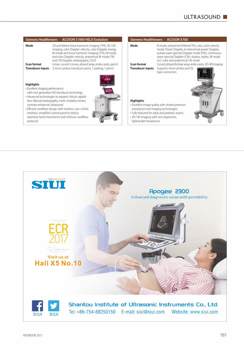
RADBOOK 2017 161
ULTRASOUND ◼
· ACUSON S1000 HELX Evolution Siemens Healthineers
3 micro-pinless transducer ports, 1 parking, 1 pencilTransducer inputsLinear, curved / convex, phased array, endo-cavity, pencilScan format
2D and Native tissue harmonic imaging (THI), 3D / 4D imaging, color Doppler velocity, color Doppler energy, M-mode and tissue harmonic imaging (THI), M-mode and color Doppler velocity, anatomical M-mode, PW and CW Doppler, elastography, CEUS
Mode
Highlights• Excellent imaging performance
with next generation HD transducer technology • Advanced technologies to expand clinical capabil-
ities: Manual elastography, multi-modality review, contrast-enhanced ultrasound
• Efficient workflow design with intuitive, user- centric interface, simplified control panel to reduce repetitive hand movements and eSieScan workflow protocols
· ACUSON X700 Siemens Healthineers
Supports micro-pinless and DL type connectors
Transducer inputsCurved, phased & linear array, endo-cavity, 3D / 4D imagingScan format
B-mode, phased and filtered THI, color, color velocity mode, Power Doppler, bi-directional power Doppler, pulsed wave spectral Doppler mode (PW), continuous wave spectral Doppler (CW), duplex, triplex, M-mode incl. color and anatomical l M-mode
Mode
Highlights• Excellent image quality with shared premium
transducers and imaging technologies• Fully-featured for adult and pediatric exams• 3D / 4D imaging with new ergonomic,
lightweight transducers
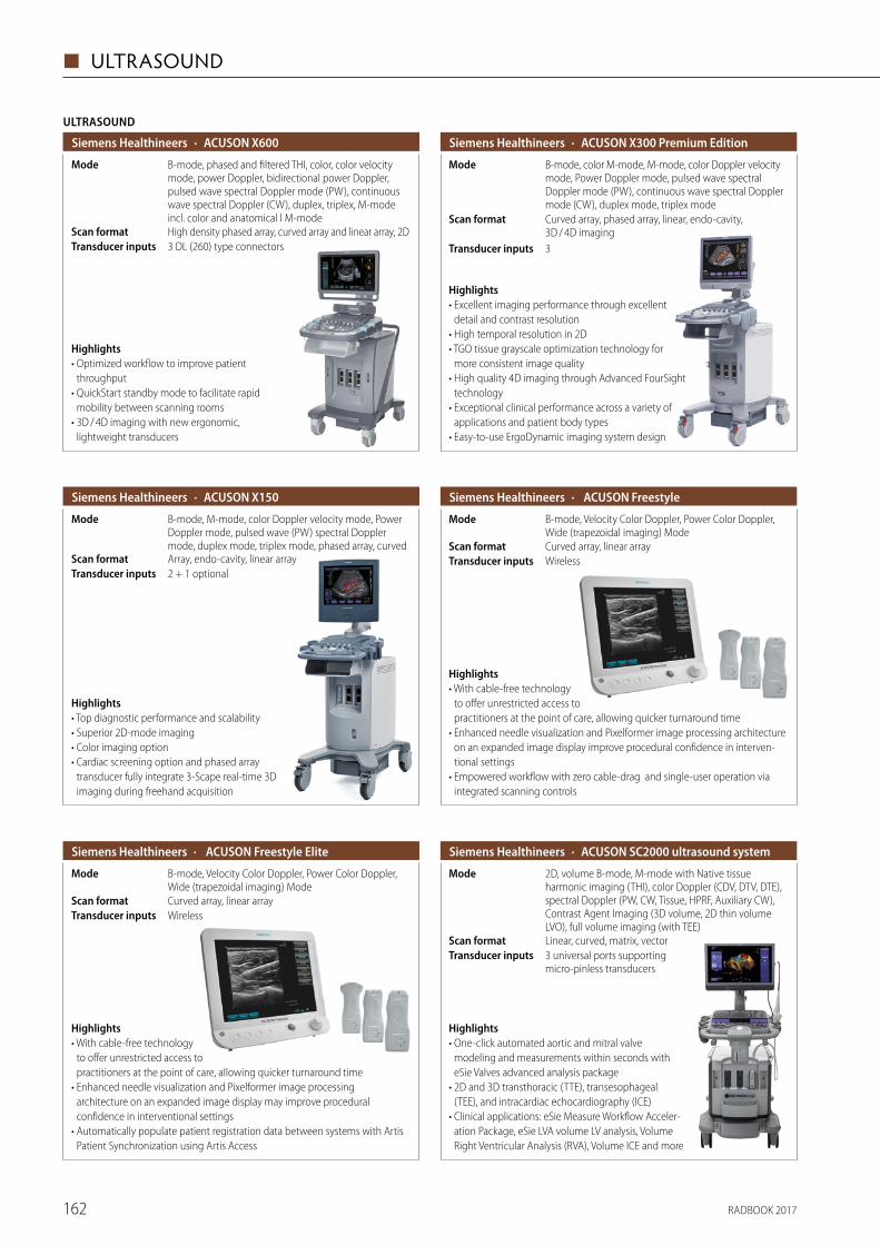
RADBOOK 2017162
◼ ULTRASOUND
ULTRASOUND
· ACUSON X600 Siemens Healthineers
3 DL (260) type connectorsTransducer inputsHigh density phased array, curved array and linear array, 2DScan format
B-mode, phased and filtered THI, color, color velocity mode, power Doppler, bidirectional power Doppler, pulsed wave spectral Doppler mode (PW), continuous wave spectral Doppler (CW), duplex, triplex, M-mode incl. color and anatomical l M-mode
Mode
Highlights• Optimized workflow to improve patient
throughput• QuickStart standby mode to facilitate rapid
mobility between scanning rooms• 3D / 4D imaging with new ergonomic,
lightweight transducers
· ACUSON X300 Premium Edition Siemens Healthineers
3Transducer inputs
Curved array, phased array, linear, endo-cavity, 3D / 4D imaging
Scan format
B-mode, color M-mode, M-mode, color Doppler velocity mode, Power Doppler mode, pulsed wave spectral Doppler mode (PW), continuous wave spectral Doppler mode (CW), duplex mode, triplex mode
Mode
Highlights• Excellent imaging performance through excellent
detail and contrast resolution• High temporal resolution in 2D• TGO tissue grayscale optimization technology for
more consistent image quality• High quality 4D imaging through Advanced FourSight
technology• Exceptional clinical performance across a variety of
applications and patient body types• Easy-to-use ErgoDynamic imaging system design
· ACUSON X150 Siemens Healthineers
2 + 1 optionalTransducer inputsArray, endo-cavity, linear arrayScan format
B-mode, M-mode, color Doppler velocity mode, Power Doppler mode, pulsed wave (PW) spectral Doppler mode, duplex mode, triplex mode, phased array, curved
Mode
Highlights• Top diagnostic performance and scalability• Superior 2D-mode imaging• Color imaging option• Cardiac screening option and phased array
transducer fully integrate 3-Scape real-time 3D imaging during freehand acquisition
· ACUSON Freestyle Siemens Healthineers
WirelessTransducer inputsCurved array, linear arrayScan format
B-mode, Velocity Color Doppler, Power Color Doppler, Wide (trapezoidal imaging) Mode
Mode
Highlights• With cable-free technology
to offer unrestricted access to practitioners at the point of care, allowing quicker turnaround time
• Enhanced needle visualization and Pixelformer image processing architecture on an expanded image display improve procedural confidence in interven-tional settings
• Empowered workflow with zero cable-drag and single-user operation via integrated scanning controls
· ACUSON SC2000 ultrasound system Siemens Healthineers
3 universal ports supporting micro-pinless transducers
Transducer inputsLinear, curved, matrix, vectorScan format
2D, volume B-mode, M-mode with Native tissue harmonic imaging (THI), color Doppler (CDV, DTV, DTE), spectral Doppler (PW, CW, Tissue, HPRF, Auxiliary CW), Contrast Agent Imaging (3D volume, 2D thin volume LVO), full volume imaging (with TEE)
Mode
Highlights• One-click automated aortic and mitral valve
modeling and measurements within seconds with eSie Valves advanced analysis package
• 2D and 3D transthoracic (TTE), transesophageal (TEE), and intracardiac echocardiography (ICE)
• Clinical applications: eSie Measure Workflow Acceler-ation Package, eSie LVA volume LV analysis, Volume Right Ventricular Analysis (RVA), Volume ICE and more
· ACUSON Freestyle Elite Siemens Healthineers
WirelessTransducer inputsCurved array, linear arrayScan format
B-mode, Velocity Color Doppler, Power Color Doppler, Wide (trapezoidal imaging) Mode
Mode
Highlights• With cable-free technology
to offer unrestricted access to practitioners at the point of care, allowing quicker turnaround time
• Enhanced needle visualization and Pixelformer image processing architecture on an expanded image display may improve procedural confidence in interventional settings
• Automatically populate patient registration data between systems with Artis Patient Synchronization using Artis Access
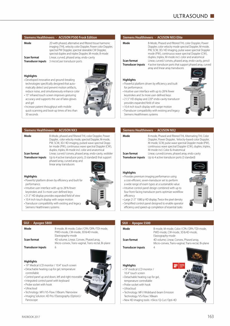
RADBOOK 2017 163
ULTRASOUND ◼
· ACUSON P500 Frosk Edition Siemens Healthineers
3 microCase transducer ports Transducer inputsLinear, curved, phased array, endo-cavityScan format
2D with phased, alternative and filtered tissue harmonic imaging (THI), velocity color Doppler, Power color Doppler, spectral PW Doppler, spectral steerable CW Doppler, spectral duplex and triplex Doppler, M-mode, B-mode
Mode
Highlights• Developed innovative and ground-breaking
technologies specifically designed that auto-matically detect and prevent motion artifacts, reduce noise, and simultaneously enhance color
• 15" infrared touch screen improves gesturing accuracy and supports the use of latex gloves and gel
• Increase patient throughput with mobile quick scanning and boot-up times of less than 30 seconds
· ACUSON NX3 Siemens Healthineers
Up to 4 active transducer ports, (3 standard) that support phased array, curved array and linear array transducers
Transducer inputsLinear, curved / convex, phased array, endo-cavity, wobblerScan format
B-Mode, phased and filtered THI, color Doppler, Power Doppler, color velocity mode, spectral Doppler, M-mode, PW, SCW, 3D / 4D imaging, pulsed wave spectral Dopp-ler mode (PW), continuous wave spectral Doppler (CW), duplex, triplex, M-mode incl. color and anatomical
Mode
Highlights• Powerful platform driven by efficiency and built for
performance. • Intuitive user interface with up to 28 % fewer
keystrokes and 3 x more user-defined keys • 21.5" HD display provides expanded field of view • 10.4 inch touch display with swipe motion• Transducer compatibility with existing and legacy
Siemens Healthineers systems
· ACUSON NX3 Elite Siemens Healthineers
4 active transducer ports that support phased array, curved array and linear array transducers
Transducer inputsLinear, curved / convex, phased array, endo-cavity, pencilScan format
B-Mode, Phased and filtered THI, color Doppler, Power Doppler, color velocity mode spectral Doppler, M-mode, PW, SCW, 3D / 4D imaging, pulse wave spectral Doppler mode (PW), continuous wave spectral Doppler (CW), duplex, triplex, M-mode incl. color and anatomical
Mode
Highlights• Powerful platform driven by efficiency and built
for performance. • Intuitive user interface with up to 28 % fewer
keystrokes and 3x more user-defined keys• 21.5" HD display and 220° endo-cavity transducer
provides expanded field of view • 10.4 inch touch display with swipe motion• Transducer compatibility with existing and legacy
Siemens Healthineers systems
· ACUSON NX2 Siemens Healthineers
Up to 4 active transducer ports (3 standard)Transducer inputsLinear, curved/convex, phased array, endo-cavityScan format
B-mode, Phased and filtered THI, Alternating THI, Color Doppler, Power Doppler, Velocity-based color Doppler, M-mode, SCW, pulse wave spectral Doppler mode (PW), continuous wave spectral Doppler (CW), duplex, triplex, M-mode incl. Color & Anatomical
Mode
Highlights• Provides premium imaging performance using
a cost-efficient, seven-transducer set to perform a wide range of exam types at a sustainable value
• Intuitive control panel design combined with up to four front-facing transducer ports optimize workflow efficiency
• Large 21.5" 1080 p HD display; Twice the pixel density• Simplified control panel designed to enable operator
efficiency and speed-up completion of essential tasks
· Apogee 5800SIUI
6Transducer inputs
4D volume, Linear, Convex, Phased array, Micro-convex, Trans-vaginal, Trans-rectal, Bi-plane
Scan format
B-mode, M-mode, Color / CPA / DPA / TDI-mode, PWD-mode, CW-mode, 3D & 4D mode, Elastography-mode
Mode
Highlights• 19" Medical LCD monitor / 10.4" touch screen• Detachable heating cup for gel, temperature
controllable• Control panel up and down, left and right moveable• Integrated control panel with keyboard• Probe socket with hook• Ultracloud • Technology: MFI / VS-Flow / XBeam / Nanoview• Imaging Solution: 4D Pro / Elastography (Option) /
Panoscope
· Apogee 5500
4Transducer inputs
4D volume, Linear, Convex, Phased array, Micro-convex, Trans-vaginal, Trans-rectal, Bi-plane
Scan format
B-mode, M-mode, Color / CPA / DPA / TDI-mode, PWD-mode, CW-mode, 3D & 4D-mode, Elastography-mode
Mode
Highlights• 19" medical LCD monitor /
10.4" touch screen• Detachable heating cup for gel,
temperature controllable• Probe socket with hook• Ultracloud • Technology: MFI / Wideband-beam Emission
Technology / VS-Flow / XBeam• New 4D imaging tools: nSlice / Q-Cut / Opti-4D
SIUI
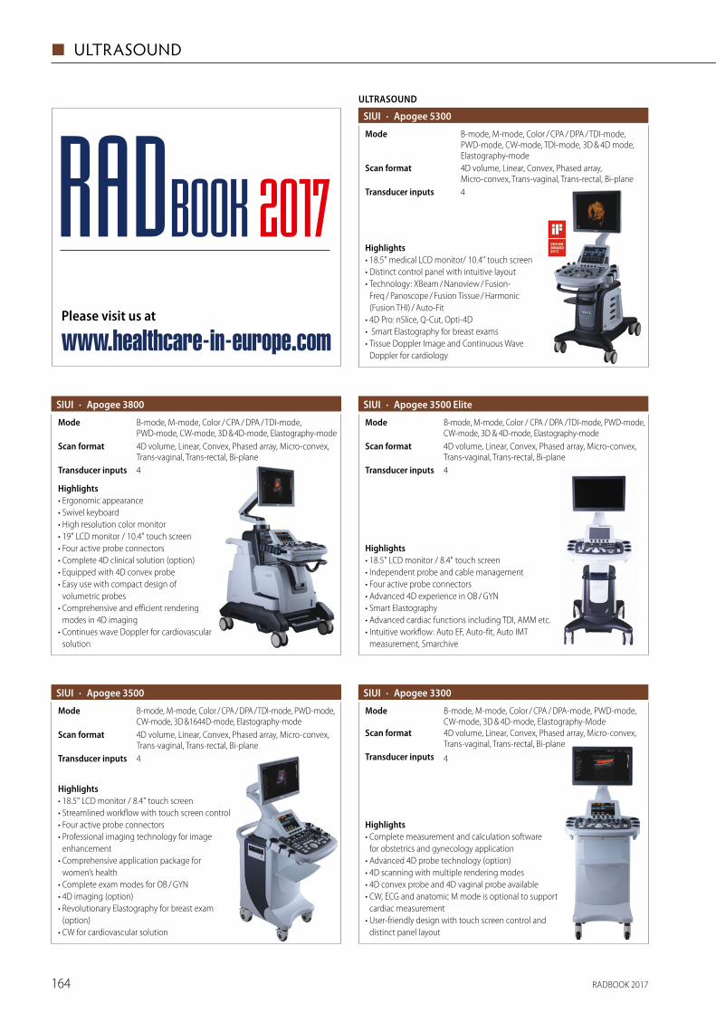
RADBOOK 2017164
◼ ULTRASOUND
ULTRASOUND
· Apogee 5300SIUI
4Transducer inputs
4D volume, Linear, Convex, Phased array, Micro-convex, Trans-vaginal, Trans-rectal, Bi-plane
Scan format
B-mode, M-mode, Color / CPA / DPA / TDI-mode, PWD-mode, CW-mode, TDI-mode, 3D & 4D mode, Elastography-mode
Mode
Highlights• 18.5" medical LCD monitor/ 10.4" touch screen• Distinct control panel with intuitive layout• Technology: XBeam / Nanoview / Fusion-
Freq / Panoscope / Fusion Tissue / Harmonic (Fusion THI) / Auto-Fit
• 4D Pro: nSlice, Q-Cut, Opti-4D • Smart Elastography for breast exams• Tissue Doppler Image and Continuous Wave
Doppler for cardiology
· Apogee 3800 SIUI
4Transducer inputs
4D volume, Linear, Convex, Phased array, Micro-convex, Trans-vaginal, Trans-rectal, Bi-plane
Scan format
B-mode, M-mode, Color / CPA / DPA / TDI-mode, PWD-mode, CW-mode, 3D & 4D-mode, Elastography-mode
Mode
Highlights• Ergonomic appearance• Swivel keyboard • High resolution color monitor• 19" LCD monitor / 10.4" touch screen• Four active probe connectors• Complete 4D clinical solution (option)• Equipped with 4D convex probe• Easy use with compact design of
volumetric probes • Comprehensive and efficient rendering
modes in 4D imaging• Continues wave Doppler for cardiovascular
solution
· Apogee 3500SIUI
4Transducer inputs
4D volume, Linear, Convex, Phased array, Micro-convex, Trans-vaginal, Trans-rectal, Bi-plane
Scan format
B-mode, M-mode, Color / CPA / DPA / TDI-mode, PWD-mode, CW-mode, 3D &1644D-mode, Elastography-mode
Mode
Highlights• 18.5" LCD monitor / 8.4" touch screen• Streamlined workflow with touch screen control• Four active probe connectors• Professional imaging technology for image
enhancement• Comprehensive application package for
women’s health• Complete exam modes for OB / GYN• 4D imaging (option)• Revolutionary Elastography for breast exam
(option)• CW for cardiovascular solution
· Apogee 3500 EliteSIUI
4Transducer inputs
4D volume, Linear, Convex, Phased array, Micro-convex, Trans-vaginal, Trans-rectal, Bi-plane
Scan format
B-mode, M-mode, Color / CPA / DPA /TDI-mode, PWD-mode, CW-mode, 3D & 4D-mode, Elastography-mode
Mode
Highlights• 18.5" LCD monitor / 8.4" touch screen• Independent probe and cable management• Four active probe connectors• Advanced 4D experience in OB / GYN• Smart Elastography• Advanced cardiac functions including TDI, AMM etc.• Intuitive workflow: Auto EF, Auto-fit, Auto IMT
measurement, Smarchive
· Apogee 3300
4Transducer inputs
4D volume, Linear, Convex, Phased array, Micro-convex, Trans-vaginal, Trans-rectal, Bi-plane
Scan format
B-mode, M-mode, Color / CPA / DPA-mode, PWD-mode, CW-mode, 3D & 4D-mode, Elastography-Mode
Mode
Highlights• Complete measurement and calculation software
for obstetrics and gynecology application• Advanced 4D probe technology (option)• 4D scanning with multiple rendering modes • 4D convex probe and 4D vaginal probe available• CW, ECG and anatomic M mode is optional to support
cardiac measurement• User-friendly design with touch screen control and
distinct panel layout
SIUI
Please visit us at
www.healthcare-in-europe.com
RADBOOK 2017
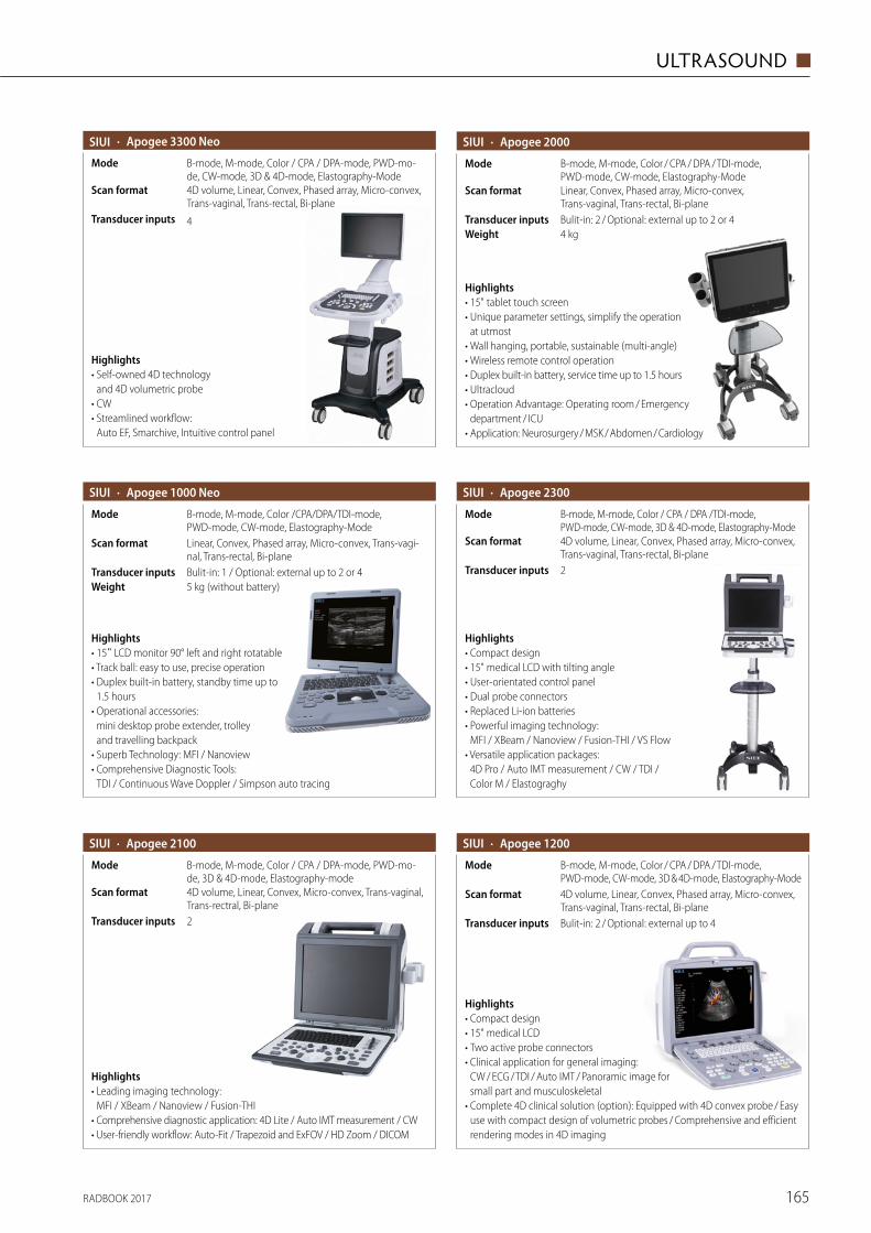
RADBOOK 2017 165
ULTRASOUND ◼
· Apogee 2000
4 kgWeightBulit-in: 2 / Optional: external up to 2 or 4Transducer inputs
Linear, Convex, Phased array, Micro-convex, Trans-vaginal, Trans-rectal, Bi-plane
Scan format
B-mode, M-mode, Color / CPA / DPA / TDI-mode, PWD-mode, CW-mode, Elastography-Mode
Mode
Highlights• 15" tablet touch screen• Unique parameter settings, simplify the operation
at utmost• Wall hanging, portable, sustainable (multi-angle)• Wireless remote control operation • Duplex built-in battery, service time up to 1.5 hours• Ultracloud • Operation Advantage: Operating room / Emergency
department / ICU• Application: Neurosurgery / MSK / Abdomen / Cardiology
· Apogee 2300SIUI
2Transducer inputs
4D volume, Linear, Convex, Phased array, Micro-convex, Trans-vaginal, Trans-rectal, Bi-plane
Scan format
B-mode, M-mode, Color / CPA / DPA /TDI-mode, PWD-mode, CW-mode, 3D & 4D-mode, Elastography-Mode
Mode
Highlights• Compact design• 15" medical LCD with tilting angle• User-orientated control panel• Dual probe connectors• Replaced Li-ion batteries• Powerful imaging technology:
MFI / XBeam / Nanoview / Fusion-THI / VS Flow• Versatile application packages:
4D Pro / Auto IMT measurement / CW / TDI / Color M / Elastograghy
· Apogee 2100SIUI
2Transducer inputs
4D volume, Linear, Convex, Micro-convex, Trans-vaginal, Trans-rectral, Bi-plane
Scan format
B-mode, M-mode, Color / CPA / DPA-mode, PWD-mo-de, 3D & 4D-mode, Elastography-mode
Mode
Highlights• Leading imaging technology:
MFI / XBeam / Nanoview / Fusion-THI• Comprehensive diagnostic application: 4D Lite / Auto IMT measurement / CW• User-friendly workflow: Auto-Fit / Trapezoid and ExFOV / HD Zoom / DICOM
· Apogee 1000 Neo
Bulit-in: 1 / Optional: external up to 2 or 4Transducer inputs
Linear, Convex, Phased array, Micro-convex, Trans-vagi-nal, Trans-rectal, Bi-plane
Scan format
B-mode, M-mode, Color /CPA/DPA/TDI-mode, PWD-mode, CW-mode, Elastography-Mode
Mode
5 kg (without battery)Weight
Highlights• 15" LCD monitor 90° left and right rotatable• Track ball: easy to use, precise operation• Duplex built-in battery, standby time up to
1.5 hours• Operational accessories:
mini desktop probe extender, trolley and travelling backpack
• Superb Technology: MFI / Nanoview• Comprehensive Diagnostic Tools:
TDI / Continuous Wave Doppler / Simpson auto tracing
SIUI · Apogee 3300 Neo
4Transducer inputs
4D volume, Linear, Convex, Phased array, Micro-convex, Trans-vaginal, Trans-rectal, Bi-plane
Scan format
B-mode, M-mode, Color / CPA / DPA-mode, PWD-mo-de, CW-mode, 3D & 4D-mode, Elastography-Mode
Mode
Highlights• Self-owned 4D technology
and 4D volumetric probe• CW• Streamlined workflow:
Auto EF, Smarchive, Intuitive control panel
SIUI
SIUI
· Apogee 1200SIUI
Bulit-in: 2 / Optional: external up to 4Transducer inputs
4D volume, Linear, Convex, Phased array, Micro-convex, Trans-vaginal, Trans-rectal, Bi-plane
Scan format
B-mode, M-mode, Color / CPA / DPA / TDI-mode, PWD-mode, CW-mode, 3D & 4D-mode, Elastography-Mode
Mode
Highlights• Compact design• 15" medical LCD• Two active probe connectors• Clinical application for general imaging:
CW / ECG / TDI / Auto IMT / Pano ramic image for small part and musculoskeletal
• Complete 4D clinical solution (option): Equipped with 4D convex probe / Easy use with compact design of volumetric probes / Comprehensive and efficient rendering modes in 4D imaging
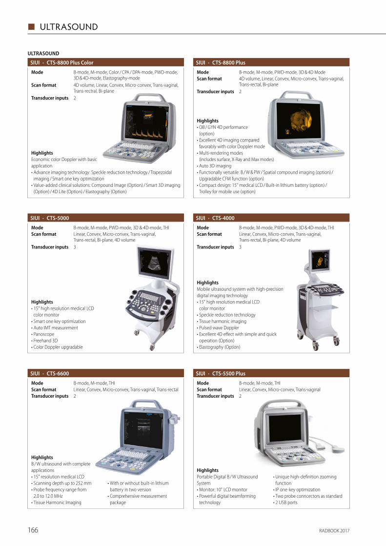
RADBOOK 2017166
◼ ULTRASOUND
· CTS-5000
B-mode, M-mode, PWD-mode, 3D & 4D-mode, THIModeLinear, Convex, Micro-convex, Trans-vaginal, Trans-rectal, Bi-plane, 4D volume
Scan format
3Transducer inputs
Highlights• 15" high resolution medical LCD
color monitor• Smart one key optimization• Auto IMT measurement• Panoscope• Freehand 3D• Color Doppler upgradable
· CTS-8800 Plus SIUI
B-mode, M-mode, PWD-mode, 3D & 4D ModeMode4D volume, Linear, Convex, Micro-convex, Trans-vaginal, Trans-rectal, Bi-plane
Scan format
2Transducer inputs
Highlights• OB / GYN 4D performance
(option)• Excellent 4D imaging compared
favorably with color Doppler mode• Multi-rendering modes
(includes surface, X-Ray and Max modes)• Auto 3D imaging• Functionally versatile: B / W & PW / Spatial compound imaging (option) /
Upgradable CFM function (option)• Compact design: 15" medical LCD / Built-in lithium battery (option) /
Trolley for mobile use (option)
· CTS-4000
3Transducer inputs
Linear, Convex, Micro-convex, Trans-vaginal, Trans-rectal, Bi-plane, 4D volume
Scan formatB-mode, M-mode, PWD-mode, 3D & 4D-mode, THIMode
HighlightsMobile ultrasound system with high- precision digital imaging technology• 15" high resolution medical LCD
color monitor• Speckle reduction technology • Tissue harmonic imaging • Pulsed wave Doppler• Excellent 4D effect with simple and quick
operation (Option)• Elastography (Option)
SIUI
· CTS-6600SIUI
B-mode, M-mode, THIModeLinear, Convex, Micro-convex, Trans-vaginal, Trans-rectalScan format2Transducer inputs
HighlightsB / W ultrasound with complete applications• 15" resolution medical LCD • Scanning depth up to 252 mm• Probe frequency range from
2.0 to 12.0 MHz• Tissue Harmonic Imaging
• With or without built-in lithium battery in two version
• Comprehensive measurement package
SIUI · CTS-5500 Plus
2Transducer inputsLinear, Convex, Micro-convex, Trans-vaginalScan formatB-mode, M-mode, THIMode
HighlightsPortable Digital B / W Ultrasound System• Monitor: 10" LCD monitor• Powerful digital beamforming
technology
• Unique high-definition zooming function
• IP one-key optimization• Two probe conncectors as standard• 2 USB ports
HighlightsEconomic color Doppler with basic application• Advance imaging technology: Speckle reduction technology / Trapezoidal
imaging / Smart one key optimization• Value-added clinical solutions: Compound Image (Option) / Smart 3D imaging
(Option) / 4D Lite (Option) / Elastography (Option)
ULTRASOUND
· CTS-8800 Plus Color
2Transducer inputs
4D volume, Linear, Convex, Micro-convex, Trans-vaginal, Trans-rectral, Bi-plane
Scan format
B-mode, M-mode, Color / CPA / DPA-mode, PWD-mode, 3D & 4D-mode, Elastography-mode
Mode
SIUI
SIUI
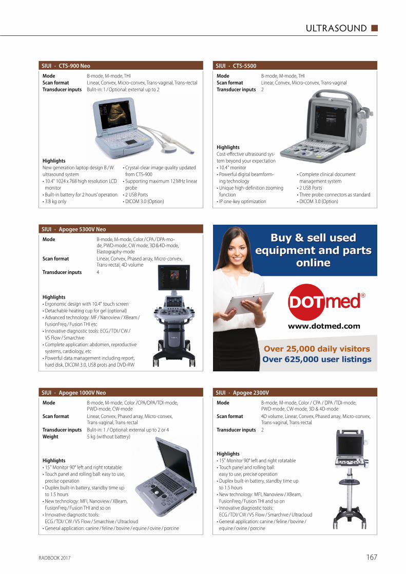
RADBOOK 2017 167
ULTRASOUND ◼
· CTS-900 NeoSIUI
Bulit-in: 1 / Optional: external up to 2 Transducer inputsLinear, Convex, Micro-convex, Trans-vaginal, Trans-rectalScan formatB-mode, M-mode, THIMode
HighlightsNew generation laptop design B / W ultrasound system• 10.4" 1024 x 768 high resolution LCD
monitor• Built-in battery for 2 hours’ operation• 3.8 kg only
• Crystal-clear image quality updated from CTS-900
• Supporting maximum 12 MHz linear probe
• 2 USB Ports• DICOM 3.0 (Option)
· CTS-5500SIUI
2Transducer inputsLinear, Convex, Micro-convex, Trans-vaginalScan formatB-mode, M-mode, THIMode
HighlightsCost-effective ultrasound sys-tem beyond your expectation• 10.4" monitor• Powerful digital beamform-
ing technology• Unique high-definition zooming
function• IP one-key optimization
• Complete clinical document management system
• 2 USB Ports• Three probe connectors as standard• DICOM 3.0 (Option)
· Apogee 5300V NeoSIUI
4Transducer inputs
Linear, Convex, Phased array, Micro-convex, T rans-rectal, 4D volume
Scan format
B-mode, M-mode, Color / CPA / DPA-mo-de, PWD-mode, CW mode, 3D & 4D-mode, Elastography-mode
Mode
Highlights• Ergonomic design with 10.4" touch screen• Detachable heating cup for gel (optional)• Advanced technology: MF / Nanoview / XBeam /
FusionFreq / Fusion THI etc• Innovative diagnostic tools: ECG / TDI / CW /
VS Flow / Smarchive• Complete application: abdomen, reproductive
systems, cardiology, etc• Powerful data management including report,
hard disk, DICOM 3.0, USB prots and DVD-RW
· Apogee 2300V
2Transducer inputs
4D volume, Linear, Convex, Phased array, Micro-convex, Trans-vaginal, Trans-rectal
Scan format
B-mode, M-mode, Color / CPA / DPA /TDI-mode, PWD-mode, CW-mode, 3D & 4D-mode
Mode
Highlights• 15" Monitor 90° left and right rotatable• Touch panel and rolling ball:
easy to use, precise operation• Duplex built-in battery, standby time up
to 1.5 hours• New technology: MFI, Nanoview / XBeam,
FusionFreq / Fusion THI and so on• Innovative diagnostic tools:
ECG / TDI/ CW / VS Flow / Smarchive / Ultracloud• General application: canine / feline / bovine /
equine / ovine / porcine
· Apogee 1000V Neo
Bulit-in: 1 / Optional: external up to 2 or 4Transducer inputs
Linear, Convex, Phased array, Micro-convex, Trans-vaginal, Trans-rectal
Scan format
B-mode, M-mode, Color /CPA/DPA/TDI-mode, PWD-mode, CW-mode
Mode
5 kg (without battery)Weight
Highlights• 15" Monitor 90° left and right rotatable• Touch panel and rolling ball: easy to use,
precise operation• Duplex built-in battery, standby time up
to 1.5 hours• New technology: MFI, Nanoview / XBeam,
FusionFreq / Fusion THI and so on• Innovative diagnostic tools:
ECG / TDI/ CW / VS Flow / Smarchive / Ultracloud• General application: canine / feline / bovine / equine / ovine / porcine
SIUI SIUI
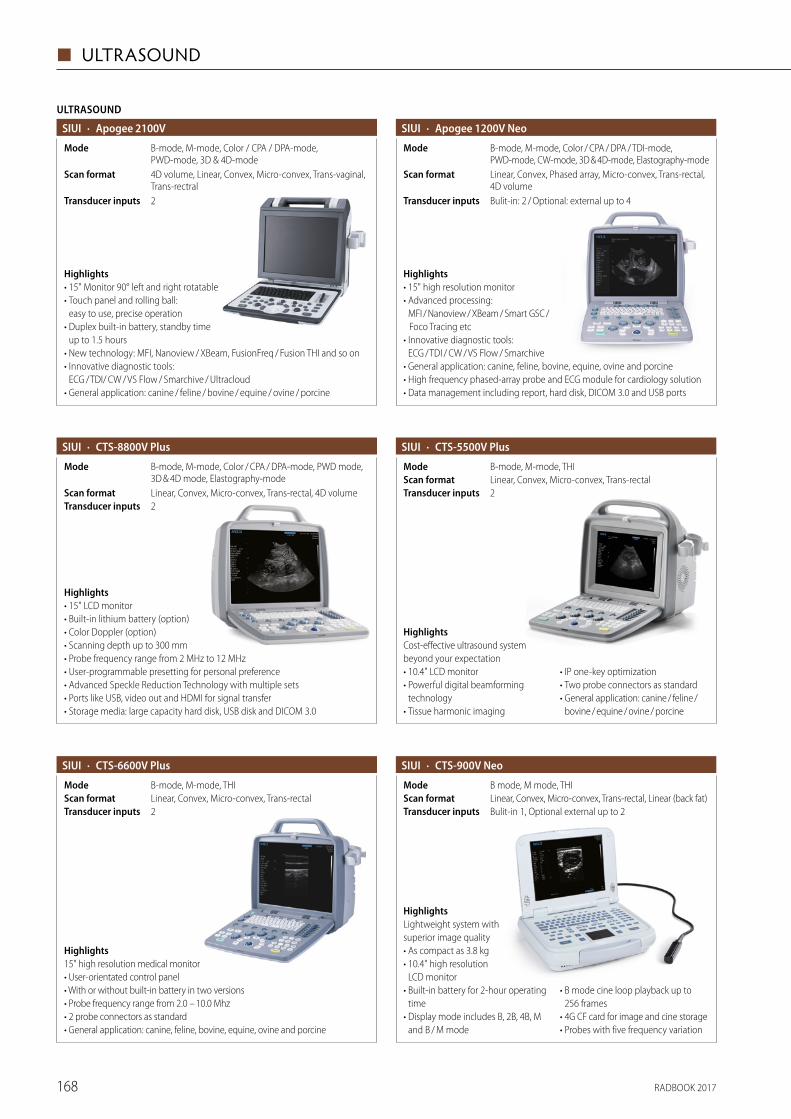
RADBOOK 2017168
◼ ULTRASOUND
· CTS-8800V Plus
2Transducer inputsLinear, Convex, Micro-convex, Trans-rectal, 4D volumeScan format
B-mode, M-mode, Color / CPA / DPA-mode, PWD mode, 3D & 4D mode, Elastography-mode
Mode
Highlights• 15" LCD monitor • Built-in lithium battery (option)• Color Doppler (option)• Scanning depth up to 300 mm• Probe frequency range from 2 MHz to 12 MHz• User-programmable presetting for personal preference• Advanced Speckle Reduction Technology with multiple sets• Ports like USB, video out and HDMI for signal transfer• Storage media: large capacity hard disk, USB disk and DICOM 3.0
SIUI · CTS-5500V PlusSIUI
2Transducer inputsLinear, Convex, Micro-convex, Trans-rectalScan formatB-mode, M-mode, THIMode
HighlightsCost-effective ultrasound system beyond your expectation• 10.4" LCD monitor• Powerful digital beamforming
technology• Tissue harmonic imaging
• IP one-key optimization• Two probe connectors as standard• General application: canine / feline /
bovine / equine / ovine / porcine
· CTS-6600V PlusSIUI
2Transducer inputsLinear, Convex, Micro-convex, Trans-rectalScan formatB-mode, M-mode, THIMode
Highlights15" high resolution medical monitor • User-orientated control panel• With or without built-in battery in two versions• Probe frequency range from 2.0 – 10.0 Mhz• 2 probe connectors as standard• General application: canine, feline, bovine, equine, ovine and porcine
· CTS-900V NeoSIUI
Bulit-in 1, Optional external up to 2 Transducer inputsLinear, Convex, Micro-convex, Trans-rectal, Linear (back fat)Scan formatB mode, M mode, THIMode
HighlightsLightweight system with superior image quality• As compact as 3.8 kg• 10.4" high resolution
LCD monitor• Built-in battery for 2-hour operating
time• Display mode includes B, 2B, 4B, M
and B / M mode
• B mode cine loop playback up to 256 frames
• 4G CF card for image and cine storage• Probes with five frequency variation
ULTRASOUND
· Apogee 2100V
2Transducer inputs
4D volume, Linear, Convex, Micro-convex, Trans-vaginal, Trans-rectral
Scan format
B-mode, M-mode, Color / CPA / DPA-mode, PWD-mode, 3D & 4D-mode
Mode
Highlights• 15" Monitor 90° left and right rotatable• Touch panel and rolling ball:
easy to use, precise operation• Duplex built-in battery, standby time
up to 1.5 hours• New technology: MFI, Nanoview / XBeam, FusionFreq / Fusion THI and so on• Innovative diagnostic tools:
ECG / TDI/ CW / VS Flow / Smarchive / Ultracloud• General application: canine / feline / bovine / equine / ovine / porcine
SIUI · Apogee 1200V NeoSIUI
Bulit-in: 2 / Optional: external up to 4Transducer inputs
Linear, Convex, Phased array, Micro-convex, Trans-rectal, 4D volume
Scan format
B-mode, M-mode, Color / CPA / DPA / TDI-mode, PWD-mode, CW-mode, 3D & 4D-mode, Elastography-mode
Mode
Highlights• 15" high resolution monitor • Advanced processing:
MFI / Nanoview / XBeam / Smart GSC / Foco Tracing etc
• Innovative diagnostic tools: ECG / TDI / CW / VS Flow / Smarchive
• General application: canine, feline, bovine, equine, ovine and porcine• High frequency phased-array probe and ECG module for cardiology solution• Data management including report, hard disk, DICOM 3.0 and USB ports
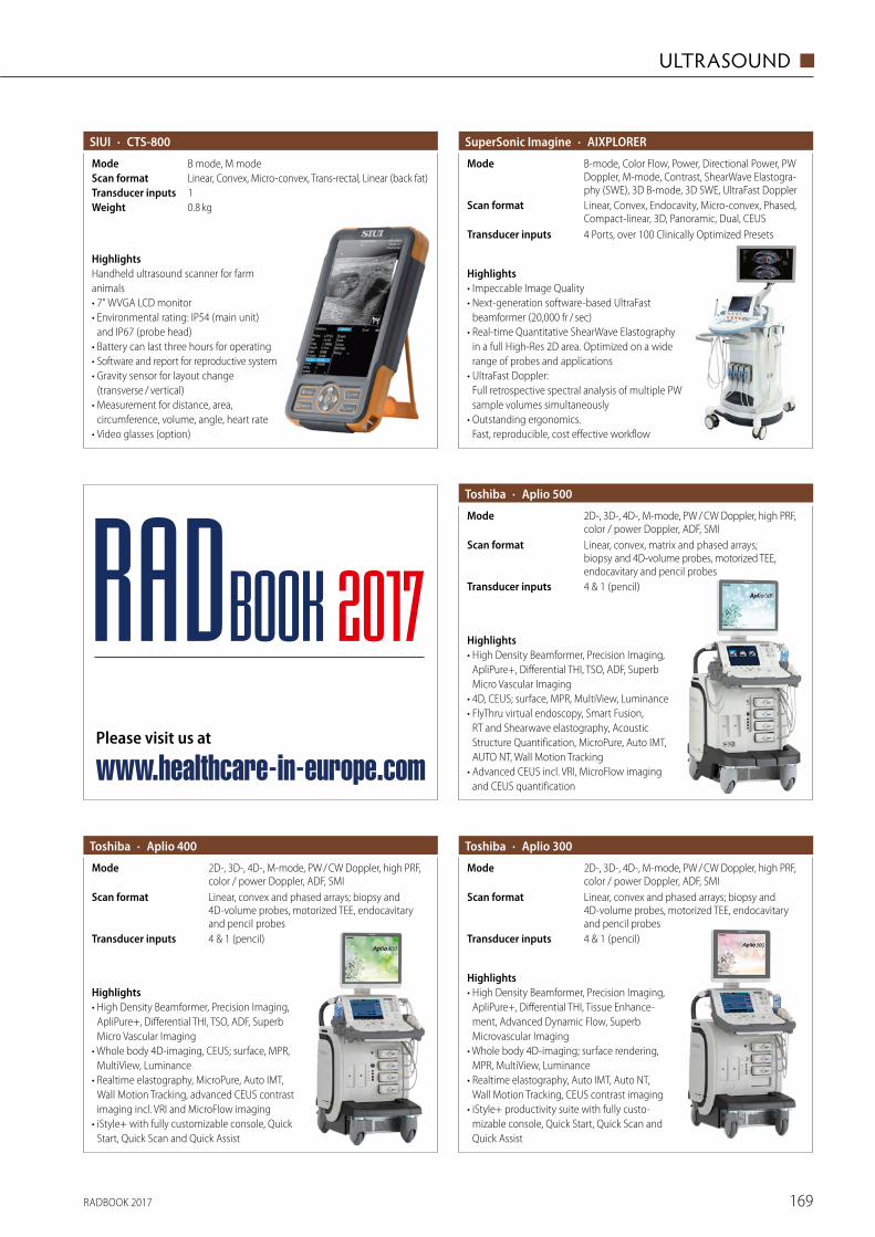
RADBOOK 2017 169
ULTRASOUND ◼
· CTS-800SIUI
1Transducer inputsLinear, Convex, Micro-convex, Trans-rectal, Linear (back fat)Scan formatB mode, M modeMode
0.8 kgWeight
HighlightsHandheld ultrasound scanner for farm animals• 7" WVGA LCD monitor• Environmental rating: IP54 (main unit)
and IP67 (probe head)• Battery can last three hours for operating• Software and report for reproductive system• Gravity sensor for layout change
(transverse / vertical)• Measurement for distance, area,
circumference, volume, angle, heart rate• Video glasses (option)
Highlights• Impeccable Image Quality• Next-generation software-based UltraFast
beamformer (20,000 fr / sec)• Real-time Quantitative ShearWave Elastography
in a full High-Res 2D area. Optimized on a wide range of probes and applications
• UltraFast Doppler: Full retrospective spectral analysis of multiple PW sample volumes simultaneously
• Outstanding ergonomics. Fast, reproducible, cost effective workflow
· AIXPLORERSuperSonic Imagine
4 Ports, over 100 Clinically Optimized PresetsTransducer inputs
Linear, Convex, Endocavity, Micro-convex, Phased, Compact-linear, 3D, Panoramic, Dual, CEUS
Scan format
B-mode, Color Flow, Power, Directional Power, PW Doppler, M-mode, Contrast, ShearWave Elastogra-phy (SWE), 3D B-mode, 3D SWE, UltraFast Doppler
Mode
· Aplio 400Toshiba
4 & 1 (pencil)Transducer inputs
Linear, convex and phased arrays; biopsy and 4D-volume probes, motorized TEE, endocavitary and pencil probes
Scan format
2D-, 3D-, 4D-, M-mode, PW / CW Doppler, high PRF, color / power Doppler, ADF, SMI
Mode
Highlights• High Density Beamformer, Precision Imaging,
ApliPure+, Differential THI, TSO, ADF, Superb Micro Vascular Imaging
• Whole body 4D-imaging, CEUS; surface, MPR, MultiView, Luminance
• Realtime elastography, MicroPure, Auto IMT, Wall Motion Tracking, advanced CEUS contrast imaging incl. VRI and MicroFlow imaging
• iStyle+ with fully customizable console, Quick Start, Quick Scan and Quick Assist
· Aplio 500Toshiba
4 & 1 (pencil)Transducer inputs
Linear, convex, matrix and phased arrays; biopsy and 4D-volume probes, motorized TEE, endocavitary and pencil probes
Scan format
2D-, 3D-, 4D-, M-mode, PW / CW Doppler, high PRF, color / power Doppler, ADF, SMI
Mode
Highlights• High Density Beamformer, Precision Imaging,
ApliPure+, Differential THI, TSO, ADF, Superb Micro Vascular Imaging
• 4D, CEUS; surface, MPR, MultiView, Luminance• FlyThru virtual endoscopy, Smart Fusion,
RT and Shearwave elastography, Acoustic Structure Quantification, MicroPure, Auto IMT, AUTO NT, Wall Motion Tracking
• Advanced CEUS incl. VRI, MicroFlow imaging and CEUS quantification
· Aplio 300Toshiba
4 & 1 (pencil)Transducer inputs
Linear, convex and phased arrays; biopsy and 4D-volume probes, motorized TEE, endocavitary and pencil probes
Scan format
2D-, 3D-, 4D-, M-mode, PW / CW Doppler, high PRF, color / power Doppler, ADF, SMI
Mode
Highlights• High Density Beamformer, Precision Imaging,
ApliPure+, Differential THI, Tissue Enhance-ment, Advanced Dynamic Flow, Superb Microvascular Imaging
• Whole body 4D-imaging; surface rendering, MPR, MultiView, Luminance
• Realtime elastography, Auto IMT, Auto NT, Wall Motion Tracking, CEUS contrast imaging
• iStyle+ productivity suite with fully custo-mizable console, Quick Start, Quick Scan and Quick Assist
Please visit us at
www.healthcare-in-europe.com
RADBOOK 2017
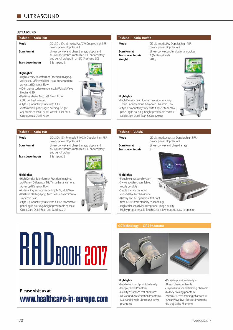
RADBOOK 2017170
◼ ULTRASOUND
Highlights• Fetal ultrasound phantom family• Doppler Flow Phantom• Quality assurance test phantoms • Ultrasound Accreditation Phantoms• Male and female ultrasound pelvic
phantoms
• Prostate phantom family – Breast phantom family
• Thyroid ultrasound training phantom• Kidney training phantom• Vascular access training phantom kit• Shear Wave Liver Fibrosis Phantoms• Elastography Phantoms
· CIRS PhantomsGCTechnology
· Xario 100Toshiba
3 & 1 (pencil)Transducer inputs
Linear, convex and phased arrays; biopsy and 4D-volume probes, motorized TEE, endocavitary and pencil probes
Scan format
2D-, 3D-, 4D-, M-mode, PW/CW Doppler, high PRF, color / power Doppler, ADF
Mode
Highlights• High Density Beamformer, Precision Imaging,
ApliPure+, Differential THI, Tissue Enhancement, Advanced Dynamic Flow
• 4D-imaging; surface rendering, MPR, MultiView,• Realtime elastography, Auto IMT, Panoramic View,
Trapezoid Scan• iStyle+ productivity suite with fully customizable
panel, agile housing, height presettable console, Quick Start, Quick Scan and Quick Assist
· Xario 100MXToshiba
70 kgWeight2 (3rd is optional)Transducer inputsLinear, convex, and endocavitary probesScan format
2D-, M-mode, PW Doppler, high PRF, color / power Doppler, ADF
Mode
Highlights• High Density Beamformer, Precision Imaging,
Tissue Enhancement, Advanced Dynamic Flow• iStyle+ productivity suite with fully customizable
panel, agile housing, height presettable console, Quick Start, Quick Scan & Quick Assist
· VIAMOToshiba
2Transducer inputsLinear, convex and phased arraysScan format
2D-, M-mode, spectral Doppler, high PRF, color / power Doppler, ADF
Mode
Highlights• Portable ultrasound system• Swivel touch screen, Tablet
mode possible• Single transducer input,
expandable to 2 transducers• Battery and AC operation, fast boot
time (< 10 s from standby to scanning)• High color sensitivity, exceptional image quality• Highly programmable Touch Screen, few buttons, easy to operate
Toshiba
ULTRASOUND
· Xario 200
3 & 1 (pencil)Transducer inputs
Linear, convex and phased arrays; biopsy and 4D-volume probes, motorized TEE, endocavitary and pencil probes, Smart 3D (Freehand 3D)
Scan format
2D-, 3D-, 4D-, M-mode, PW / CW Doppler, high PRF, color / power Doppler, ADF
Mode
Highlights• High Density Beamformer, Precision Imaging,
ApliPure+, Differential THI, Tissue Enhancement, Advanced Dynamic Flow
• 4D-imaging; surface rendering, MPR, MultiView, Freehand 3D
• Realtime elasto, Auto IMT, Stress Echo, CEUS contrast imaging
• iStyle+ productivity suite with fully custo mizable panel, agile housing, height adjustable console, panel swivel, Quick Start, Quick Scan & Quick Assist
Please visit us at
www.healthcare-in-europe.com
RADBOOK 2017
RADBOOK 2017 171
TESTING DEVICES ◼
Testing Devices
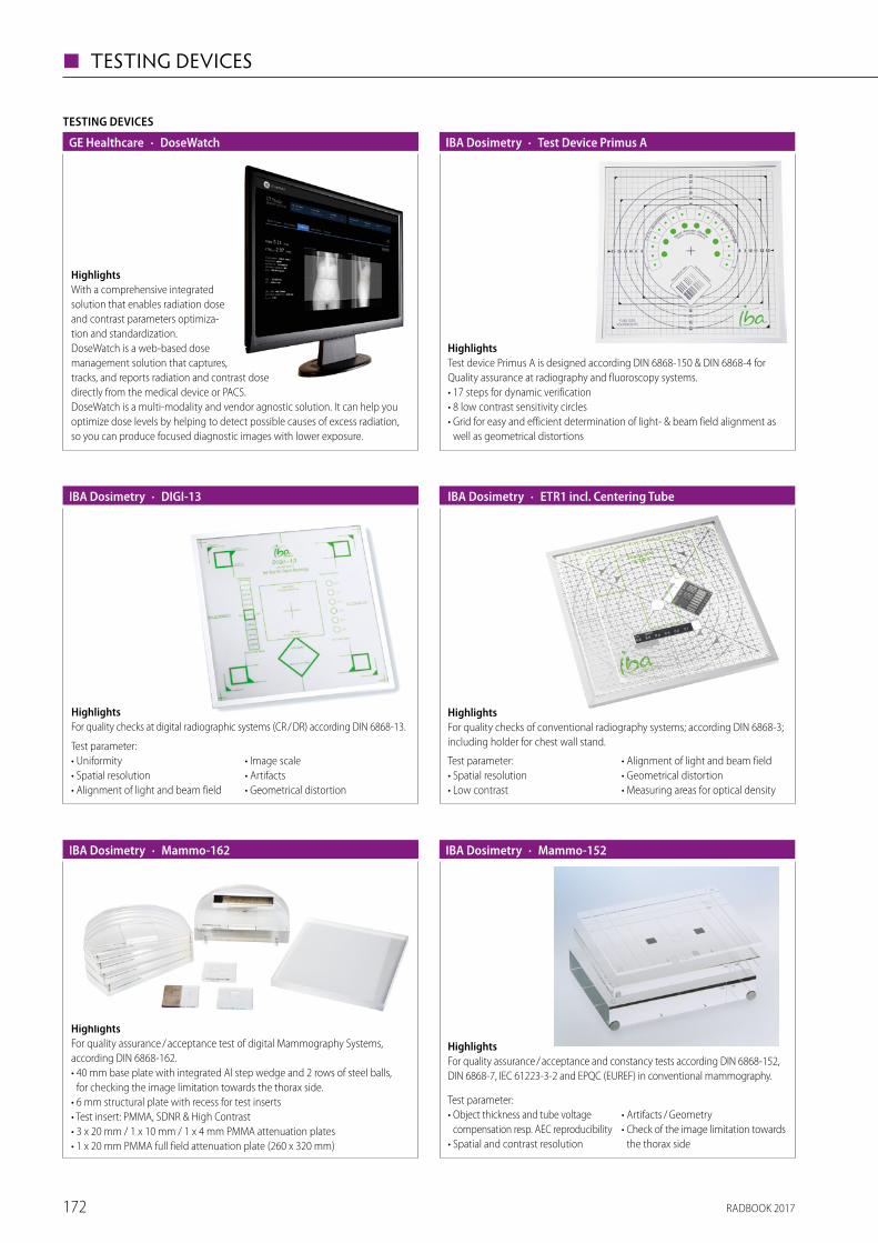
RADBOOK 2017172
◼ TESTING DEVICES
TESTING DEVICES
· DoseWatchGE Healthcare
HighlightsWith a comprehensive integrated solution that enables radiation dose and contrast parameters optimiza-tion and standardization. DoseWatch is a web-based dose management solution that captures, tracks, and reports radiation and contrast dose directly from the medical device or PACS. DoseWatch is a multi-modality and vendor agnostic solution. It can help you optimize dose levels by helping to detect possible causes of excess radiation, so you can produce focused diagnostic images with lower exposure.
HighlightsTest device Primus A is designed according DIN 6868-150 & DIN 6868-4 for Quality assurance at radiography and fluoroscopy systems.• 17 steps for dynamic verification• 8 low contrast sensitivity circles• Grid for easy and efficient determination of light- & beam field alignment as
well as geometrical distortions
· Test Device Primus AIBA Dosimetry
· DIGI-13IBA Dosimetry
Test parameter:• Uniformity• Spatial resolution• Alignment of light and beam field
• Image scale• Artifacts• Geometrical distortion
HighlightsFor quality checks at digital radiographic systems (CR / DR) according DIN 6868-13.
· ETR1 incl. Centering Tube
HighlightsFor quality checks of conventional radiography systems; according DIN 6868-3; including holder for chest wall stand.
Test parameter:• Spatial resolution• Low contrast
• Alignment of light and beam field• Geometrical distortion• Measuring areas for optical density
IBA Dosimetry
HighlightsFor quality assurance / acceptance test of digital Mammography Systems, according DIN 6868-162.• 40 mm base plate with integrated Al step wedge and 2 rows of steel balls,
for checking the image limitation towards the thorax side.• 6 mm structural plate with recess for test inserts• Test insert: PMMA, SDNR & High Contrast• 3 x 20 mm / 1 x 10 mm / 1 x 4 mm PMMA attenuation plates• 1 x 20 mm PMMA full field attenuation plate (260 x 320 mm)
· Mammo-162IBA Dosimetry
HighlightsFor quality assurance / acceptance and constancy tests according DIN 6868-152, DIN 6868-7, IEC 61223-3-2 and EPQC (EUREF) in conventional mammography.
Test parameter:• Object thickness and tube voltage
compensation resp. AEC reproducibility• Spatial and contrast resolution
• Artifacts / Geometry• Check of the image limitation towards
the thorax side
· Mammo-152IBA Dosimetry
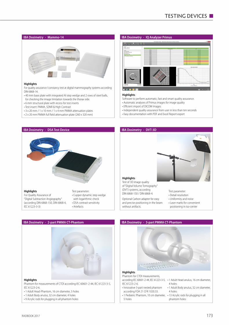
RADBOOK 2017 173
TESTING DEVICES ◼
HighlightsFor quality assurance / constancy test at digital mammography systems according DIN 6868-14.• 40 mm base plate with integrated Al step wedge and 2 rows of steel balls,
for checking the image limitation towards the thorax side.• 6 mm structural plate with recess for test inserts• Test insert: PMMA, SDNR & High Contrast• 3 x 20 mm / 1 x 10 mm / 1 x 4 mm PMMA attenuation plates• 2 x 20 mm PMMA full field attenuation plate (260 x 320 mm)
· Mammo-14IBA Dosimetry
HighlightsSoftware to perform automatic, fast and smart quality assurance.• Automatic analyses of Primus images for image quality• Efficient import of DICOM Images• Independent quality assurance from user in less than ten seconds• Easy documentation with PDF and Excel Report export
· IQ Analyzer PrimusIBA Dosimetry
· DSA Test DeviceIBA Dosimetry
HighlightsFor Quality Assurance of “Digital Subtraction Angiography” (according DIN 6868-150, DIN 6868-4, IEC 61223-3-3)
Test parameter:• Cupper dynamic step wedge
with logarithmic check• DSA contrast sensitivity• Artefacts
HighlightsTest of 3D image quality of “Digital Volume Tomography” (DVT) systems, according DIN 6868-150 / DIN 6868-4.
Optional Carbon adapter for easy and precise positioning in the beam without artifacts.
Test parameter:• Detail resolution• Uniformity and noise• Laser marks for convenient
positioning in iso-center
· DVT-3DIBA Dosimetry
HighlightsPhantom for measurements of CTDI according IEC 60601-2-44, IEC 61223-3-5, IEC 61223-2-6.• 1 Adult Head-Phantom, 16 cm diameter, 5 holes• 1 Adult Body anulus, 32 cm diameter, 4 holes• 9 Acrylic rods for plugging in all phantom holes
· 2-part PMMA CT-PhantomIBA Dosimetry
HighlightsPhantom for CTDI measurements, according IEC 60601-2-44, IEC 61223-3-5, IEC 61223-2-6.• Innovative 3-part nested phantom
according FDA 21 CFR 1020.33.• 1 Pediatric Phantom, 10 cm diameter,
5 holes
• 1 Adult Head anulus, 16 cm diameter, 4 holes
• 1 Adult Body anulus, 32 cm diameter, 4 holes
• 13 Acrylic rods for plugging in all phantom holes
· 3-part PMMA CT-PhantomIBA Dosimetry
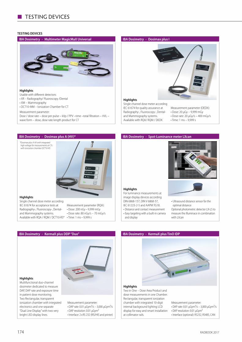
RADBOOK 2017174
◼ TESTING DEVICES
HighlightsUsable with different detectors: • XR – Radiography/ Fluoroscopy /Dental• XM – Mammography• DCT10-MM – Ionization Chamber for CT
Measurement parameter: Dose / dose rate – dose per pulse – kVp / PPV –time –total filtration – HVL – wave form – dose, dose rate length product for CT
TESTING DEVICES
· Multimeter MagicMaX UniversalIBA Dosimetry
HighlightsSingle channel dose meter according IEC 61674 for quality assurance at Radiography-, Fluoroscopy-, Dental- and Mammography systems.Available with RQA/ RQM / DEDX
Measurement parameter (DEDX):• Dose: 20 μGy – 9,999 mGy• Dose rate: 20 μGy/s – 400 mGy/s• Time: 1 ms – 9,999 s
· Dosimax plus IIBA Dosimetry
HighlightsSingle channel dose meter according IEC 61674 for acceptance tests at Radiography-, Fluoroscopy-, Dental- and Mammography systems.Available with RQA / RQM / DCT10-RS*
Measurement parameter (RQA):• Dose: 200 nGy – 9,999 mGy• Dose rate: 80 nGy/s – 70 mGy/s• Time: 1 ms – 9,999 s
· Dosimax plus A (HV)*IBA Dosimetry*Dosimax plus A HV with integrated high voltage for measurements at CTs with ionization chamber DCT10-RS
HighlightsFor luminance measurements at image display devices according DIN 6868-157, DIN V 6868-57, IEC 61223-2-5 and AAPM TG18.• Distance and contact measurement• Easy targeting with a built-in camera
and display
• Ultrasound distance sensor for the optimal distance
Optional photometric detector LX-LS to measure the Illuminace in combination with LXcan
· Spot-Luminance meter LXcanIBA Dosimetry
HighlightsMultifunctional duo-channel dosimeter dedicated to measure DAP, DAP rate and exposure time in patient dose monitoring.Two Rectangular, transparent ionization chamber with integrated electronics and one separate “Dual Line Display” with two very bright LED display lines.
Measurement parameter:• DAP rate: 0.01 µGym²/s – 3,000 µGym²/s• DAP resolution: 0.01 µGym²• Interface: 2 x RS 232 (RIS/HIS and printer)
· KermaX plus DDP “Duo”IBA Dosimetry
HighlightsTwo in One – Dose Area Product and dose measurements in one Chamber.Rectangular, transparent ionization chamber with integrated 10-digit internal background lighting LCD display for easy and smart installation at collimator rails.
Measurement parameter:• DAP rate: 0.01 µGym²/s – 3,000 µGym²/s• DAP resolution: 0.01 µGym²• Interface (optional): RS232, RS485, CAN
· KermaX plus TinO IDPIBA Dosimetry
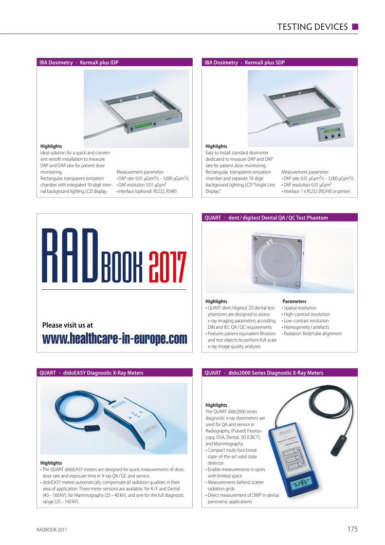
RADBOOK 2017 175
TESTING DEVICES ◼
HighlightsIdeal solution for a quick and conven-ient retrofit installation to measure DAP and DAP rate for patient dose monitoring. Rectangular, transparent ionization chamber with integrated 10-digit inter-nal background lighting LCD display.
Measurement parameter:• DAP rate: 0.01 µGym²/s – 3,000 µGym²/s• DAP resolution: 0.01 µGym²• Interface (optional): RS232, RS485
· KermaX plus IDPIBA Dosimetry
HighlightsEasy to install standard dosimeter dedicated to measure DAP and DAP rate for patient dose monitoring.Rectangular, transparent ionization chamber and separate 10-digit background lighting LCD “Single Line Display”.
Measurement parameter:• DAP rate: 0.01 µGym²/s – 3,000 µGym²/s• DAP resolution: 0.01 µGym²• Interface: 1 x RS232 (RIS/HIS or printer)
· KermaX plus SDPIBA Dosimetry
Highlights• QUART dent /digitest 2D dental test
phantoms are designed to assess x-ray imaging parameters according DIN and IEC QA / QC requirements.
• Features patient equivalent filtration and test objects to perform full-scale x-ray image quality analyses.
Parameters• Spatial resolution• High-contrast resolution• Low-contrast resolution• Homogeneity / artefacts• Radiation field/tube alignment
· dent / digitest Dental QA / QC Test PhantomQUART
Highlights• The QUART didoEASY meters are designed for quick measurements of dose,
dose rate and exposure time in X-ray QA / QC and service. • didoEASY meters automatically compensate all radiation qualities in their
area of application. Three meter versions are available: for R / F and Dental (40 – 160 kV), for Mammography (25 – 40 kV), and one for the full diagnostic range (25 – 160 kV).
· didoEASY Diagnostic X-Ray MetersQUART · dido2000 Series Diagnostic X-Ray MetersQUART
HighlightsThe QUART dido2000 series diagnostic x-ray dosemeters are used for QA and service in Radiography, (Pulsed) Fluoros-copy, DSA, Dental, 3D (CBCT), and Mammo graphy.• Compact multi-functional
state-of-the-art solid state detector
• Enable measurements in spots with limited space
• Measurements behind scatter radiation grids
• Direct measurement of DWP in dental panoramic applications
Please visit us at
www.healthcare-in-europe.com
RADBOOK 2017
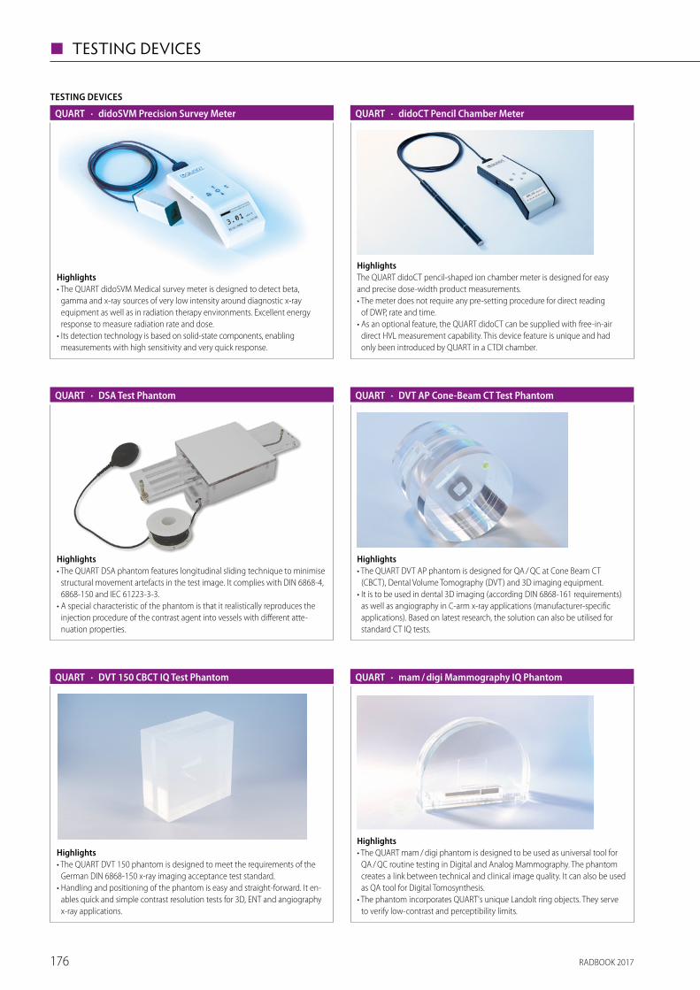
RADBOOK 2017176
◼ TESTING DEVICES
HighlightsThe QUART didoCT pencil-shaped ion chamber meter is designed for easy and precise dose-width product measurements.• The meter does not require any pre-setting procedure for direct reading
of DWP, rate and time.• As an optional feature, the QUART didoCT can be supplied with free-in-air
direct HVL measurement capability. This device feature is unique and had only been introduced by QUART in a CTDI chamber.
· didoCT Pencil Chamber MeterQUART
Highlights• The QUART DSA phantom features longitudinal sliding technique to minimise
structural movement artefacts in the test image. It complies with DIN 6868-4, 6868-150 and IEC 61223-3-3.
• A special characteristic of the phantom is that it realistically reproduces the injection procedure of the contrast agent into vessels with different atte-nuation properties.
· DSA Test PhantomQUART
Highlights• The QUART DVT 150 phantom is designed to meet the requirements of the
German DIN 6868-150 x-ray imaging acceptance test standard.• Handling and positioning of the phantom is easy and straight-forward. It en-
ables quick and simple contrast resolution tests for 3D, ENT and angiography x-ray applications.
· DVT 150 CBCT IQ Test PhantomQUART
Highlights• The QUART DVT AP phantom is designed for QA / QC at Cone Beam CT
(CBCT), Dental Volume Tomography (DVT) and 3D imaging equipment.• It is to be used in dental 3D imaging (according DIN 6868-161 requirements)
as well as angiography in C-arm x-ray applications (manufacturer-specific applications). Based on latest research, the solution can also be utilised for standard CT IQ tests.
· DVT AP Cone-Beam CT Test PhantomQUART
Highlights• The QUART mam / digi phantom is designed to be used as universal tool for
QA / QC routine testing in Digital and Analog Mammography. The phantom creates a link between technical and clinical image quality. It can also be used as QA tool for Digital Tomosynthesis.
• The phantom incorporates QUART's unique Landolt ring objects. They serve to verify low-contrast and perceptibility limits.
· mam / digi Mammography IQ PhantomQUART
TESTING DEVICES
· didoSVM Precision Survey MeterQUART
Highlights• The QUART didoSVM Medical survey meter is designed to detect beta,
gamma and x-ray sources of very low intensity around diagnostic x-ray equipment as well as in radiation therapy environments. Excellent energy response to measure radiation rate and dose.
• Its detection technology is based on solid-state components, enabling measure ments with high sensitivity and very quick response.
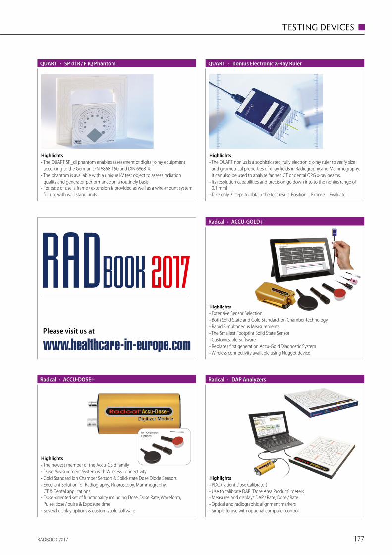
RADBOOK 2017 177
TESTING DEVICES ◼
Highlights• The QUART SP_dl phantom enables assessment of digital x-ray equipment
according to the German DIN 6868-150 and DIN 6868-4.• The phantom is available with a unique kV test object to assess radiation
quality and generator performance on a routinely basis.• For ease of use, a frame / extension is provided as well as a wire-mount system
for use with wall stand units.
· SP dl R / F IQ PhantomQUART
Highlights• The QUART nonius is a sophisticated, fully electronic x-ray ruler to verify size
and geometrical properties of x-ray fields in Radiography and Mammography. It can also be used to analyse fanned CT or dental OPG x-ray beams.
• Its resolution capabilities and precision go down into to the nonius range of 0.1 mm!
• Take only 3 steps to obtain the test result: Position – Expose – Evaluate.
· nonius Electronic X-Ray RulerQUART
· ACCU-GOLD+Radcal
Highlights• Extensive Sensor Selection• Both Solid State and Gold Standard Ion Chamber Technology• Rapid Simultaneous Measurements• The Smallest Footprint Solid State Sensor• Customizable Software• Replaces first generation Accu-Gold Diagnostic System• Wireless connectivity available using Nugget device
· ACCU-DOSE+Radcal
Highlights• The newest member of the Accu-Gold family• Dose Measurement System with Wireless connectivity• Gold Standard Ion Chamber Sensors & Solid-state Dose Diode Sensors• Excellent Solution for Radiography, Fluoroscopy, Mammography,
CT & Dental applications• Dose-oriented set of functionality including Dose, Dose Rate, Waveform,
Pulse, dose / pulse & Exposure time• Several display options & customizable software
· DAP AnalyzersRadcal
Highlights• PDC (Patient Dose Calibrator)• Use to calibrate DAP (Dose Area Product) meters• Measures and displays DAP / Rate, Dose / Rate• Optical and radiographic alignment markers• Simple to use with optional computer control
Please visit us at
www.healthcare-in-europe.com
RADBOOK 2017
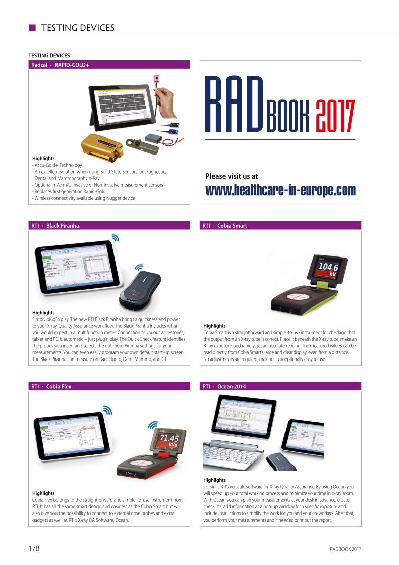
RADBOOK 2017178
◼ TESTING DEVICES
HighlightsSimply plug ’n’ play. The new RTI Black Piranha brings a quickness and power to your X-ray Quality Assurance work flow. The Black Piranha includes what you would expect in a multifunction meter. Connection to various accessories, tablet and PC is automatic – just plug ‘n’ play. The Quick Check feature identifies the probes you insert and selects the optimum Piranha settings for your measure ments. You can even easily program your own default start-up screen. The Black Piranha can measure on Rad, Fluoro, Dent, Mammo, and CT.
· Black PiranhaRTI
HighlightsCobia Smart is a straightforward and simple-to-use instrument for checking that the output from an X-ray tube is correct. Place it beneath the X-ray tube, make an X-ray exposure, and rapidly get an accurate reading. The measured values can be read directly from Cobia Smart’s large and clear display, even from a distance. No adjustments are required, making it exceptionally easy to use.
· Cobia SmartRTI
HighlightsCobia Flex belongs to the straightforward and simple-to-use instrument from RTI. It has all the same smart design and easiness as the Cobia Smart but will also give you the possibility to connect to external dose probes and extra gadgets as well as RTI’s X-ray QA Software, Ocean.
· Cobia FlexRTI
HighlightsOcean is RTI’s versatile software for X-ray Quality Assurance. By using Ocean you will speed up your total working process and minimize your time in X-ray room. With Ocean you can plan your measurements at your desk in advance, create checklists, add information as a pop-up window for a specific exposure and include instructions to simplify the work for you and your co-workers. After that, you perform your measurements and if needed print out the report.
· Ocean 2014RTI
TESTING DEVICES
· RAPID-GOLD+Radcal
Highlights• Accu-Gold+ Technology• An excellent solution when using Solid State Sensors for Diagnostic,
Dental and Mammography X-Ray• Optional mA / mAs invasive or Non-invasive measurement sensors• Replaces first generation Rapid-Gold• Wireless connectivity available using Nugget device
Please visit us at
www.healthcare-in-europe.com
RADBOOK 2017
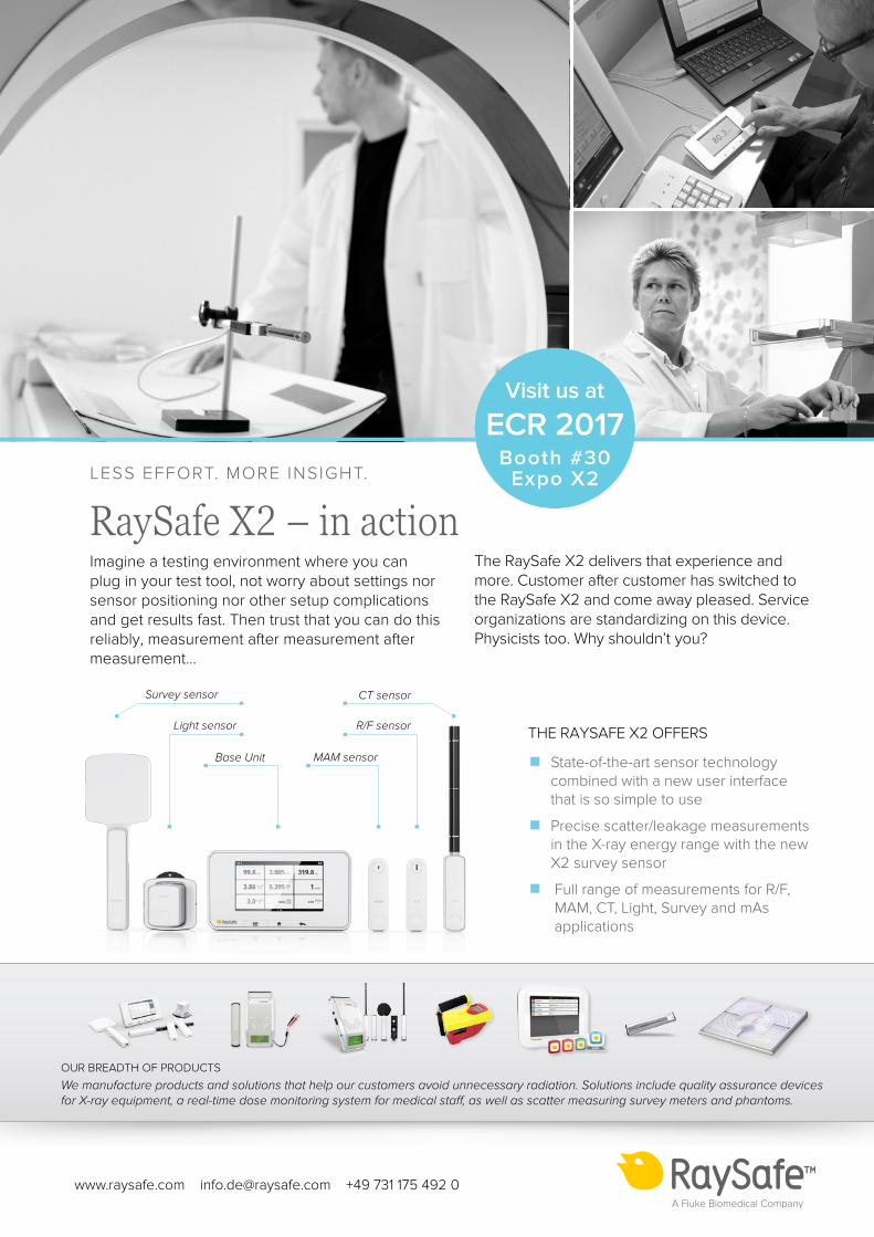
RADBOOK 2017 179
TESTING DEVICES ◼
MAM sensor
Light sensor
Survey sensor
Base Unit
R/F sensor
CT sensor
The RaySafe X2 delivers that experience and more. Customer after customer has switched to the RaySafe X2 and come away pleased. Service organizations are standardizing on this device. Physicists too. Why shouldn’t you?
THE RAYSAFE X2 OFFERS
▪ State-of-the-art sensor technologycombined with a new user interfacethat is so simple to use
▪ Precise scatter/leakage measurementsin the X-ray energy range with the newX2 survey sensor
▪ Full range of measurements for R/F,MAM, CT, Light, Survey and mAsapplications
www.raysafe.com [email protected] +49 731 175 492 0
OUR BREADTH OF PRODUCTS
We manufacture products and solutions that help our customers avoid unnecessary radiation. Solutions include quality assurance devices for X-ray equipment, a real-time dose monitoring system for medical staff, as well as scatter measuring survey meters and phantoms.
LESS EFFORT. MORE INSIGHT.
RaySafe X2 – in actionImagine a testing environment where you can plug in your test tool, not worry about settings nor sensor positioning nor other setup complications and get results fast. Then trust that you can do this reliably, measurement after measurement after measurement…
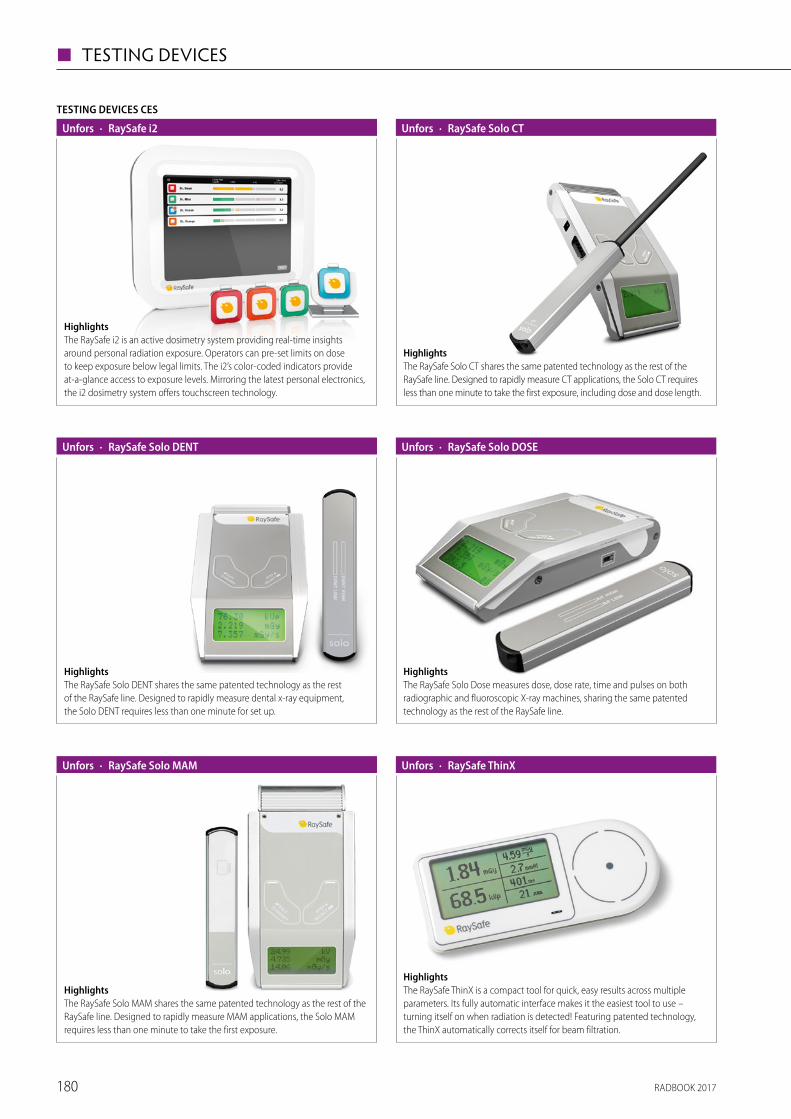
RADBOOK 2017180
◼ TESTING DEVICES
TESTING DEVICES CES
· RaySafe i2Unfors
HighlightsThe RaySafe i2 is an active dosimetry system providing real-time insights around personal radiation exposure. Operators can pre-set limits on dose to keep exposure below legal limits. The i2’s color-coded indicators provide at-a-glance access to exposure levels. Mirroring the latest personal electronics, the i2 dosimetry system offers touchscreen technology.
· RaySafe Solo CTUnfors
HighlightsThe RaySafe Solo CT shares the same patented technology as the rest of the RaySafe line. Designed to rapidly measure CT applications, the Solo CT requires less than one minute to take the first exposure, including dose and dose length.
· RaySafe Solo DOSEUnfors
HighlightsThe RaySafe Solo Dose measures dose, dose rate, time and pulses on both radiographic and fluoroscopic X-ray machines, sharing the same patented technology as the rest of the RaySafe line.
· RaySafe Solo DENTUnfors
HighlightsThe RaySafe Solo DENT shares the same patented technology as the rest of the RaySafe line. Designed to rapidly measure dental x-ray equipment, the Solo DENT requires less than one minute for set up.
· RaySafe Solo MAMUnfors
HighlightsThe RaySafe Solo MAM shares the same patented technology as the rest of the RaySafe line. Designed to rapidly measure MAM applications, the Solo MAM requires less than one minute to take the first exposure.
· RaySafe ThinX
HighlightsThe RaySafe ThinX is a compact tool for quick, easy results across multiple parameters. Its fully automatic interface makes it the easiest tool to use – turning itself on when radiation is detected! Featuring patented technology, the ThinX automatically corrects itself for beam filtration.
Unfors
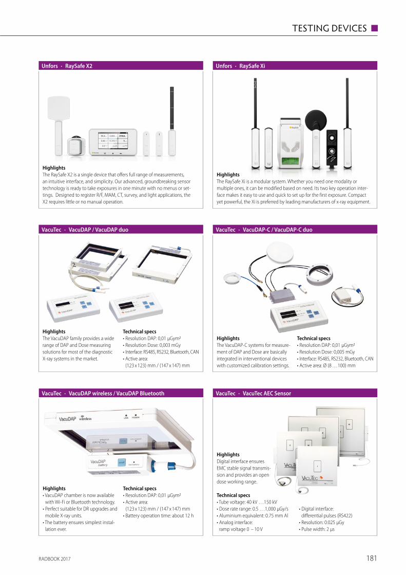
RADBOOK 2017 181
TESTING DEVICES ◼
· RaySafe X2Unfors
HighlightsThe RaySafe X2 is a single device that offers full range of measurements, an intuitive interface, and simplicity. Our advanced, groundbreaking sensor technology is ready to take exposures in one minute with no menus or set-tings. Designed to register R/F, MAM, CT, survey, and light applications, the X2 requires little or no manual operation.
· RaySafe XiUnfors
HighlightsThe RaySafe Xi is a modular system. Whether you need one modality or multiple ones, it can be modified based on need. Its two key operation inter-face makes it easy to use and quick to set up for the first exposure. Compact yet powerful, the Xi is preferred by leading manufacturers of x-ray equipment.
HighlightsThe VacuDAP family provides a wide range of DAP and Dose measuring solutions for most of the diagnostic X-ray systems in the market.
Technical specs• Resolution DAP: 0,01 µGym2• Resolution Dose: 0,003 mGy• Interface: RS485, RS232, Bluetooth, CAN• Active area:
(123 x 123) mm / (147 x 147) mm
· VacuDAP / VacuDAP duoVacuTec
Highlights• VacuDAP chamber is now available
with Wi-Fi or Bluetooth technology.• Perfect suitable for DR upgrades and
mobile X-ray units.• The battery ensures simplest instal-
lation ever.
Technical specs• Resolution DAP: 0,01 µGym2• Active area:
(123 x 123) mm / (147 x 147) mm • Battery operation time: about 12 h
· VacuDAP wireless / VacuDAP BluetoothVacuTec
HighlightsThe VacuDAP-C systems for measure-ment of DAP and Dose are basically integrated in interventional devices with customized calibration settings.
Technical specs• Resolution DAP: 0,01 µGym2• Resolution Dose: 0,005 mGy• Interface: RS485, RS232, Bluetooth, CAN• Active area: Ø (8 …100) mm
· VacuDAP-C / VacuDAP-C duoVacuTec
HighlightsDigital interface ensures EMC stable signal transmis-sion and provides an open dose working range.
Technical specs• Tube voltage: 40 kV …150 kV• Dose rate range: 0.5 …1,000 µGy/s• Aluminium equivalent: 0.75 mm Al• Analog interface:
ramp voltage 0 – 10 V
• Digital interface: differential pulses (RS422)
• Resolution: 0.025 µGy• Pulse width: 2 µs
· VacuTec AEC SensorVacuTec
RADBOOK 2017182
INDEX OF ADVERTISERS
69
141
151
5
111
117
37
15
7
179
back cover
front cover
back cover
161
55
page

RADBOOK 2017 183
COMPANIES & SUPPLIERS
| visit us onfollow us onEuropean Hospital Verlags GmbH | Theodor-Althoff-Straße 45 | 45133 Essen
www.healthcare-in-europe.com
Trends & innovations in healthcare.
25 YEARSof Europeanhealthcare
communication:print & online.
Online Portal | Newspaper | Product Guides | Newsletter
Staytunedon allchannels.
.artu
ndw
ork
desi
gnbü
ro
Hitachi Medical Systems Europe Holding AG · Sumpfstrasse 13 · CH-6300 Zug
www.hitachi-medical-systems.eu
Embracing lifethrough better medical
imaging solutions.
Better medical imaging solutions for the needs of our customers
We have been committed to delivering outstanding medical imaging solutions over decades. Original solutions,
that have grown out of the groups' expertise and technical know-how, providing innovations that meet equally
the Zeitgeist and the needs of our customers and their patients.
Shimadzu_RADbook_2013.qxd 0
7.02.2013 13:33 Uhr Seite
1
RADBOO
K 20
12 ·
The
Radi
olog
y Gu
ide
to T
echn
olog
y &
Info
rmat
ics
in E
urop
e
W IT W CTW MRI W InterventionalW MammoW R / FW NucW Displays / Printers
W UltrasoundW InjectorsW Testing Devices
The Radiology Guide to Technology & Informatics in Europe
T 19.-
2013
The world’s most advanced dynamic volume CT just got even better. The new Aquilion ONE ViSION Edition provides you and
your patients robust clinical solutions when you need them most. This innovative Dynamic Volume CT enables all patients a
successful examination, with the lowest possible radiation exposure and the highest quality diagnostic outcomes.
For more information please turn to page 12 or visit www.aquilionvision.com.
RADBOOK 2014The Radiology Guide to Technology and Informatics in Europe
T 19.-
.artu
ndw
ork
desi
gnbü
ro
Hitachi Medical Systems Europe Holding AG · Sumpfstrasse 13 · CH-6300 Zug
www.hitachi-medical-systems.eu
Embracing lifethrough better medical
imaging solutions.
Better medical imaging solutions for the needs of our customers
We have been committed to delivering outstanding medical imaging solutions over decades. Original solutions,
that have grown out of the groups' expertise and technical know-how, providing innovations that meet equally
the Zeitgeist and the needs of our customers and their patients.
Shimadzu_RADbook_2013.qxd 0
7.02.2013 13:33 Uhr Seite
1
RADBOO
K 20
12 ·
The
Radi
olog
y Gu
ide
to T
echn
olog
y &
Info
rmat
ics
in E
urop
e
W IT W CTW MRI W InterventionalW MammoW R / FW NucW Displays / Printers
W UltrasoundW InjectorsW Testing Devices
The Radiology Guide to Technology & Informatics in Europe
T 19.-
2013
The world’s most advanced dynamic volume CT just got even better. The new Aquilion ONE ViSION Edition provides you and
your patients robust clinical solutions when you need them most. This innovative Dynamic Volume CT enables all patients a
successful examination, with the lowest possible radiation exposure and the highest quality diagnostic outcomes.
For more information please turn to page 12 or visit www.aquilionvision.com.
ITCTMRIInterventionalMammoR/FNucDisplays/PrintersUltrasoundInjectorsTesting Devices
Villa Sistemi Medicali’s new general radiographic system, the Moviplan iC, has been conceived for every diagnostic need.
It is available in a wide range of configurations, from basic analog versions up to fully digital and automatized rooms.
T H E E U R O P E A N F O R U M F O R T H O S E I N T H E B U S I N E S S O F M A K I N G H E A L T H C A R E W O R K
CONTENTS
NEWS & MANAGEMENT 1-3
ONCOLOGY 4-5
RADIOLOGY 6-8
MEDICA PRODUCTS 9
INFECTION CONTROL 10
LABORATORY 11-16
www.european-hospital.com
V O L 2 3 I S S U E 6 / 1 4 • D E C E M B E R 2 0 1 4
Report: Michael Krassnitzer
By definition, an emerging infec-
tious disease is one that has newly
appeared in a population or has
been known for some time but is
rapidly increasing in incidence or
spreading to new geographic areas.
At the International Meeting on
Emerging Diseases and Surveillance
(IMED 2014), in Vienna, this year’s
focus was on one particular emerg-
ing infectious disease: Ebola.
According to the World Health
Organisation (WHO) in the recent
outbreak 5,400 people have died
of this dangerous disease and, as
of 20 November 2014, more than
15,000 cases were reported in eight
countries.
‘This most serious Ebola outbreak
ever was caused by social, geo-
graphic and political factors in the
regions affected,’ says Belgian physi-
cian Hilde De Clerck MD, who works
for the aid organisation Médecins
sans frontières in the crisis regions.
All severely affected countries are
poor, have underdeveloped health
systems, were involved in wars or
armed conflicts in the past few
years, and the people tend to mis-
trust government agencies and the
countries mistrust each other.
De Clerck: ‘The epidemic start-
ed off slowly and spread quick-
ly, affecting people from all walks
of life. Lack of awareness about
the disease, insufficient protection
measures and the high degree of
mobility of the people between
three of the countries concerned –
be it for business or family purposes
– contributed to Ebola being able
acute Ebola infection cannot travel.
During the severe acute respira-
tory syndrome (SARS) pandemic,
in 2002/2003, 45 million travellers
were screened at airports – with
only a single SARS case being iden-
tified.
It is much more important,
according to the experts, to use the
money for aid programmes right in
the affected regions. ‘It is crucial
to provide help where the epi-
demic broke out, that is in Western
Africa,’ underlines Jack Woodall
MA PhD, an epidemiologist from
Brazil and vice-editor of ProMED-
mail, the web-based Programme
for Monitoring Emerging Diseases
of the International Society for
Infectious Diseases (ISID). He is
confident: ‘We can stop the disease
from spreading if we manage to
break the chain of infection.’
to spread.’ There is, however, good
news: Oyewale Tomori DVM, PhD,
President of the Nigerian Academy
of Science, describes how Ebola
was contained in his country. On
20 July 2014 a person from Liberia
with acute Ebola symptoms arrived
at Lagos International Airport. The
preliminary diagnosis – Ebola – was
confirmed by a private hospital. This
index case had had contact with 72
people at the airport and in the hos-
pital, who potentially were exposed
to the virus.
The Ministry of Health and the
Nigeria Centre for Disease Control
(NCDC) declared an Ebola emer-
gency. On 23 July the Ministry of
Health, the regional government
of Lagos and international partners
set up an Ebola crisis intervention
centre.
The index case died on 25 July 20.
Subsequent Ebola infections were
reported in Nigeria. All of them
could be traced back to the index
case. Close to 900 identified con-
tact persons were monitored; eight
patients died. On 20 October 2014,
WHO officially declared Nigeria
Ebola-free.
‘It is to a large extent due to
the quickly established crisis inter-
vention centre that we were able
to successfully fight the disease,’
said Tomori. ‘We not only suf-
fer from a real epidemic in West
Africa, we are also suffering from
an epidemic of fear that’s spread-
ing all over the globe,’ said Dr
Pamela Rendi-Wagner, Director of
the Department of Public Health
Services and Medical Affairs at the
Austrian Federal Ministry of Health
and adds, ‘Despite concerted efforts
by all public health authorities it is
difficult to counter the rising public
panic’. In Austria it was reported
that several health professionals quit
their jobs because they feared hav-
ing to care for Ebola patients. ‘It
is our main task to listen to these
fear-driven concerns and to inform
and communicate widely, openly,
early and in a transparent fashion
in order to create trust,’ the health
official emphasised.
Experts agree on one issue: they
consider screening of in-coming
travellers at international airports
for Ebola to be useless, inter alia
because usually people with an
Ebola: Reports of
panic among medicsExperts confirm the disease can be contained by stopping the chain of infection
Médecins Sans Frontières medics: a vital aid organisation in crisis zones
So
urc
e: M
SF;
Julien R
ey
RADIOLOGY 6-8
• Precision radiotherapy
with 4-D imaging
• MorphMatch technology
for 3-T scans
LABORATORY 11-16
• Breakthrough in hepatitis
C research
• Stain-free 3-D digital pathology
• Recycling blood lost during surgery
■ R
IS
■ P
ACS
■ W
orks
tatio
ns
■ C
T
■ M
RI
■ I
njec
tors
■ I
nter
vent
iona
l
■ I
T Sy
stem
s
■ M
amm
ogra
phy
■ R
/ F F
ilm-S
cree
n
■ R
/ F D
igita
l
■ M
olec
ular
Imag
ing
■ D
ispla
ys
■ P
rinte
rs
■ U
ltras
ound
■ T
estin
g D
evic
es
Agfa HealthCareSeptestraat 272640 Mortsel, Belgiumtel +32 3 444 94 [email protected]
3 3 4
189 190
6064676870
899293
107112qq9
143
allMRI GmbHSüdstr. 2374226 Nordheim, Germanytel +49 71 33 2 37 02 [email protected]
33
Barco N.V.President Kennedypark 358500 Kortrijk, Belgiumtel +32 56 23 32 [email protected]
136137138140
Bayer Medical Care B.V.Avenue Céramique 276221 KV Maastricht, The Netherlandstel +31 43 358 56 01www.radiology.bayer.com
3637
Bracco Injeneering S.A.Avenue de Sévelin 461004 Lausanne, Switzerlandtel +41 21 621 74 [email protected]
40
Canon Europa N.V.Bovenkerkerweg 591185 XB Amstelveen, The Netherlandstel +31 20 545 8 [email protected]/medical
3 34
189190 64
9394
107112120
Cefla s.c. Via Selice Provinciale 23A 40026, Imola (BO), Italy tel +39 045 820 27 27 [email protected] www.newtom.it
17
CHILI GmbH Friedrich-Ebert-Str. 2 69221 Dossenheim / Heidelberg, Germany tel +49 62 21 180 79 [email protected]
34
189190
606467
144
CHISON Medical Imaging Co., Ltd.No. 9 Xin Hui Huan Road, New District,Wuxi, Jiang Su Province, China 214028tel +086 510 85 31 05 93 [email protected]
146147
DMS Imaging393 rue Charles Lindbergh34130 Mauguio, Francetel +33 4 67 50 49 00www.dms.com
51 72 85
94107113120125
Dunlee Medical ComponentsEuropean Customer Service CenterVeenpluis 65684 PC Best, The Netherlandstel +31 40 276 25 [email protected]
A Division of Philips Healthcare
18 125126
EBIT S.r.l. – Esaote Group Via Siffredi 58 16153 Genoa, Italy tel +39 010 65 [email protected] www.esaote.com/healthcare-it
3 34
189190 61
RADBOOK 2017184
COMPANIES & SUPPLIERS
■ R
IS
■ P
ACS
■ W
orks
tatio
ns
■ C
T
■ M
RI
■ I
njec
tors
■ I
nter
vent
iona
l
■ I
T Sy
stem
s
■ M
amm
ogra
phy
■ R
/ F F
ilm-S
cree
n
■ R
/ F D
igita
l
■ M
olec
ular
Imag
ing
■ D
ispla
ys
■ P
rinte
rs
■ U
ltras
ound
■ T
estin
g D
evic
es
EDL SAS1031, chemin de la Seyne à Bastian83500 La Seyne-sur-mer, Francetel +33 4 94 10 99 [email protected]
3 34 61
EIZO Europe GmbHHelmut-Grashoff-Str. 1841179 Moenchengladbach, Germanytel +49 21 61 82 [email protected]
136137138139142143144
Esaote S.p.A.Via Siffredi 5816153 Genoa, Italytel +39 010 65 [email protected]
2829
148149
ETIAMLa Palmeraie, ZA de la Hallerais11 rue du Bois de Soeuvres35770 Vern-sur-Seiche, Francetel : +33 2 99 14 33 [email protected]
3 3 4
189 190
6164 143
FujiFilm SonoSite BV EUHQJoop Geesinkweg 1401114 AB Amsterdam, The Netherlandstel +31 20 462 [email protected]/uk
149150
GCTechnology GmbHFreidling 1284172 Buch am Erlbach, Germanytel +49 87 06 94 15 [email protected]
18 33 57 78 87 170
GE Healthcare 283 Rue de la Minière78533 Buc Cedex, Francetel +33 130 70 40 [email protected]
3 34
189190
911131416
212425
4344454751
6167 72 94
113
130131132133
150151152
172
GENERAL MEDICAL MERATE S.p.A.Via Partigiani, 2524068 Seriate (BG), Italytel +39 035 45 25 [email protected]
5156
8183
9495
113120
Giotto / IMS Internazionale Medico SciSagittario, 540037 Sasso Marconi (BO), Italytel +39 051 84 68 [email protected]
727376
Hitachi Medical Systems Europe (Holding) AG Sumpfstrasse 136300 Zug, Switzerlandtel +41 41 748 63 33welcome@hitachi-medical-systems.comwww.hitachi-medical-systems.com
91214
2629 126
152153154155
Hologic Europe N.V. Da Vincilaan 5, Building Caprese 1925 Zaventem, Belgiumtel +32 2 711 46 [email protected]
56 66737678
126
I.A.E. S.P.A.Via Fabio Filzi, 53 20032 Cormano (MI), Italytel +39 02 66 30 32 [email protected]
18 58 79 126
RADBOOK 2017 185
COMPANIES & SUPPLIERS
■ R
IS
■ P
ACS
■ W
orks
tatio
ns
■ C
T
■ M
RI
■ I
njec
tors
■ I
nter
vent
iona
l
■ I
T Sy
stem
s
■ M
amm
ogra
phy
■ R
/ F F
ilm-S
cree
n
■ R
/ F D
igita
l
■ M
olec
ular
Imag
ing
■ D
ispla
ys
■ P
rinte
rs
■ U
ltras
ound
■ T
estin
g D
evic
es
IBA Dosimetry GmbHBahnhofstr. 590592 Schwarzenbruck, Germanytel +49 91 28 [email protected]
172173174175
IMAGE Information Systems Europe GmbHLange Str. 1618055 Rostock, Germanytel +49 381 496 58 20 [email protected]
3 34
189190
62666768
IMS Internazionale Medico SciSagittario, 540037 Sasso Marconi (BO), Italytel +39 051 84 68 [email protected]
727376
INTERMEDICAL SRLE. Fermi, 2624050 Grassobbio (BG), Italytel +39 035 659 48 [email protected]
47515256
i-SOLUTIONS Health GmbHAm Exerzierplatz 1468167 Mannheim, Germanytel +49 621 39 [email protected]
3 34
6270
ITZ Medicom GmbH & Co. KGSiemensring 44 a47877 Willich, Germanytel +49 21 54 49 79 [email protected]
3 34
189190
6268
KONICA MINOLTA Medical & Graphic Imaging Europe B.V.Hoogoorddreef 91101 BA Amsterdam, The Netherlandstel +31 20 658 41 [email protected]/healthcare
34
189190
899295
107108113128
155
LEONI Special Cables GmbHBusiness Unit HealthcareEschstraße 126169 Friesoythe, Germanytel +49 4491 [email protected]
18 34 58 79
MECALL S.R.L.Via Negrelli, 5520851 Lissone (MB), Italytel +39 039 24 31 [email protected]
MECALL
95121
mediCAD Hectec GmbHOpalstr. 5484032 Altdorf, Germany tel +49 871 33 02 [email protected] www.mediCAD.eu
189190
646566
Medical ECONET GmbHIm Erlengrund 2046149 Oberhausen, Germanytel +49 208 37 78 [email protected]
97108114115
medigration GmbH Am Anger 291052 Erlangen, Germanytel +49 91 31 690 87-48 [email protected]
3 34
189190
6263656668
109 144
RADBOOK 2017186
COMPANIES & SUPPLIERS
■ R
IS
■ P
ACS
■ W
orks
tatio
ns
■ C
T
■ M
RI
■ I
njec
tors
■ I
nter
vent
iona
l
■ I
T Sy
stem
s
■ M
amm
ogra
phy
■ R
/ F F
ilm-S
cree
n
■ R
/ F D
igita
l
■ M
olec
ular
Imag
ing
■ D
ispla
ys
■ P
rinte
rs
■ U
ltras
ound
■ T
estin
g D
evic
es
MEDTRON AGHauptstr. 25566128 Saarbruecken, Germanytel +49 681 970 [email protected]
4041
Micrima Ltd.One Glass Wharf, Temple QuayBristol, UK. BS2 0ELtel +44 1172 51 00 [email protected] www.micrima.com
76
SHENZHEN MINDRAY BIO-MEDICAL ELECTRONICS CO., LTD.Mindray Building, Keji 12th Road SouthNanshan, Shenzhen 518057, Chinatel +86 755 81 88 89 98 [email protected]
29 97115
155156157158
MRI-tecDistributor for MRI Tools, Accessories & EquipmentBuschgrundstr. 2345894 Gelsenkirchen, Germanytel +49 209 60 48 93 [email protected]
34
NORAS MRI products GmbHLeibnizstr. 497204 Hoechberg, Germanytel +49 931 29 92 [email protected]
3233
Philips HealthcareP. O. Box 10.0005680 DA Best, The Netherlandstel +31 40 278 56 [email protected]/healthcare
189190
1012141619
242634
43444852
6566 73
979899
109115
130131133134
Planmed OySorvaajankatu 700880 Helsinki, Finlandtel +358 20 77 95 [email protected]
17 7374
PRIMAX International"Le Minotaure"30 – 34 Avenue Henri Matisse06200 Nice, Francetel +33 492 29 23 [email protected]
52
99115121
PROTEC GmbH & Co. KGIn den Dorfwiesen 1471720 Oberstenfeld, Germanytel +49 70 62 925 [email protected]
4 189190 63 81
87
99100109116
QUART GmbHKirchenweg 785604 Zorneding, Germanytel +49 81 06 24 91 [email protected]
87175176177
Radcal Corporation426 West Duarte RoadMonrovia, CA 91016, USAtel +1 626 357 [email protected]
177178
Roesys GmbHDr.-Max-Ilgner-Str. 232339 Espelkamp, Germanytel +49 57 72 915 55 [email protected]
100109128
RADBOOK 2017 187
COMPANIES & SUPPLIERS
■ R
IS
■ P
ACS
■ W
orks
tatio
ns
■ C
T
■ M
RI
■ I
njec
tors
■ I
nter
vent
iona
l
■ I
T Sy
stem
s
■ M
amm
ogra
phy
■ R
/ F F
ilm-S
cree
n
■ R
/ F D
igita
l
■ M
olec
ular
Imag
ing
■ D
ispla
ys
■ P
rinte
rs
■ U
ltras
ound
■ T
estin
g D
evic
es
RTI ElectronicsFloejelbergsgatan 8C43137 Moelndal, Swedentel +46 31 746 36 [email protected]
178
SAMSUNG MEDISON CO., LTD.42, Teheran-ro 108-gil, Gangnam-gu, Seoul, Koreatel +82 2 21 94 14 [email protected]
100101110116
158159160
SCHILLER AGAltgasse 68, P. O. Box 10 526341 Baar, Switzerlandtel +41 41 766 42 [email protected]
34
Shimadzu Europa GmbHMedical Systems DivisionAlbert-Hahn-Str. 6 – 1047269 Duisburg, Germanytel +49 203 76 [email protected]
484952
81828385
101116121
Siemens AG, Healthcare SectorHenkestr. 12791052 Erlangen, Germanytel +49 91 31 [email protected]/healthcare
34
189190
9101112131516
212425273132
434449525356
6365
747579
828386
104116
121123
131132134
160161162163
SIMAD s.r.l.Via Zallone, 2540066 Pieve di Cento - Bologna, Italytel +39 051 686 08 [email protected]
545658
117
Shantou Institute of Ultrasonic Instr. Co., Ltd.#77, Jinsha Road515041 Shantou, Chinatel +86 754 88 25 01 [email protected]
163164165166167168169
STEPHANIX10, Rue Jean Moulin42150 La Ricamarie, Francetel +33 4 77 47 81 [email protected]
5457
828386
104105110117123
SuperSonic ImagineLes Jardins de la Duranne, Bât E & F510, Rue René Descartes13857 Aix-en-Provence, Francetel +33 442 99 24 [email protected]
169
Swissray Medical AGTurbistr. 25 – 276280 Hochdorf, Switzerlandtel +41 41 914 12 [email protected]
105110118
Technix S.p.A.Via Fermi 45 24050 Grassobbio (BG), Italytel +39 035 384 66 [email protected]
54 118
Toshiba Medical Systems EuropeZilverstraat 12718 RP Zoetermeer, The Netherlandstel +31 79 368 92 [email protected]
111317
2528
434550
8284
106118123128
169170
RADBOOK 2017188
COMPANIES & SUPPLIERS
■RI
S
■PA
CS
■W
orks
tatio
ns
■CT
■M
RI
■In
ject
ors
■In
terv
entio
nal
■IT
Sys
tem
s
■M
amm
ogra
phy
■R
/ F F
ilm-S
cree
n
■R
/ F D
igita
l
■M
olec
ular
Imag
ing
■D
ispla
ys
■Pr
inte
rs
■Ul
tras
ound
■Te
stin
g D
evic
es
Toshiba Electronics Europe GmbHHansaallee 18140549 Duesseldorf, Germanytel +49 211 52 [email protected]
19 58
110112118123124128
TOTOKU Europe GmbHJakob-Krebs-Str. 12447877 Willich, Germanytel +49 21 56 49 68 [email protected]
137138139140
Unfors RaySafe ABUggledalsvaegen 2942740 Billdal, Swedentel +46 31 719 97 [email protected]
180181
VacuTec Meßtechnik GmbHDornbluethstr. 1401277 Dresden, Germanytel +49 351 317 [email protected]
181
Varex Imaging CorporationKarl-Arnold-Straße 1247877 Willich, Germany+49 21 54 92 49 [email protected]
19 79
VILLA SISTEMI MEDICALI s.p.a.Via delle Azalee, 320090 Buccinasco (MI), Italytel +39 02 48 85 [email protected]
17 54 7475
83848687
106112119124
China Resources Wandong Medical Equipment Co., Ltd.Bld.3, No. 9 Jiuxianqiaodong Road Chaoyang District100015 Beijing, Chinatel +86 10 845 757 [email protected]
2831
515455
75 8587
106107119124
Ningbo Xingaoyi Medical Instruments Co. Ltd(XGY Medical)777 West Tanjialing Rd.315400 Yuyao, Chinatel +86 574 627 308 [email protected]
283132
125
Ziehm Imaging GmbHDonaustr. 3190451 Nuremberg, Germanytel +49 911 21 [email protected]
5557
RADBOOK 2017 189
COMPANIES & SUPPLIERS

RADBOOK 2017190
NOTES
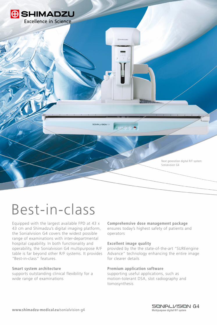
RAD
BOO
K 20
17· T
he G
uide
to Im
agin
g Te
chno
logy
and
Info
rmat
ics i
n Eu
rope
RADBOOK 2017
IT-SOLUTIONS IT-SOLUTIONS
RADBOOK 2017€ 19.-
• IT •CT •MRI • Interventional •Mammo •R/F •Nuc •Displays/Printers•Ultrasound • Injectors • Testing Devices
The Guide to Imaging Technology and Informatics in Europe
More precise diagnostics, low dose and accustomed workflow: Based on dual layer detector technologythe IQon Spectral CT from Philips opens up new dimensions in CT imaging.
Visit us at ECR 2017EXPO X5 Booth #12
Visit us at ECR 2017EXPO X5 Booth #12
In today’s healthcare environment,excellent patient care and outstandingproductivity often compromise each other.Multitom Rax1) puts an end to “either/or”.Robotic Advanced X-ray – RAX – supportsyou in streamlining clinical pathwayswhile improving diagnostic insights and treatments.
How? With one scanner that offers amultitude of X-rays – in just one roomand at an expert level. With movementsthat create natural Real 3D2) images –even under weight-bearing conditions.
With an open design that reduces patient transfers, pain, and anxiety while offeringmore space to perform interventions.With twin robots to simplify, standardize,and speed up operations. All with the goalof accelerating productivity and allowinghumans to do what they do best – carefor patients.
Prepare yourself for the future with theworld’s first Twin Robotic X-ray scanner –achieving a new level of excellencewith regard to diagnostic insights andtreatments.
www.siemens.com/multitom-rax
Multitom Rax: Join the newmovement with robotic X-ray
Powered by
RAXtechnology
RAX – Robotic Advanced X-ray –opens up exciting new movementsfor enhanced patient care and out-standing productivity.
1) Multitom Rax is not commercially available in all countries. Due to regulatory reasons its future availability cannot be guaranteed.Please contact your local Siemens organization for further details. 2) Option, only in combination with additional workstation syngo X workplace.
A91X
P-94
57-A
1-76
00
Best-in-classEquipped with the largest available FPD at 43 x43 cm and Shimadzu’s digital imaging platform,the Sonialvision G4 covers the widest possiblerange of examinations with inter-departmentalhospital capability. In both functionality andoperability, the Sonialvision G4 multipurpose R/Ftable is far beyond other R/F systems. It provides“Best-in-class” features.
Smart system architecturesupports outstanding clinical flexibility for a wide range of examinations
Comprehensive dose management packageensures today’s highest safety of patients andoperators
Excellent image qualityprovided by the the state-of-the-art “SUREengineAdvance“ technology enhancing the entire imagefor clearer details
Premium application softwaresupporting useful applications, such as motion-tolerant DSA, slot radiography andtomosynthesis
www.shimadzu-medical.eu/sonialvision-g4
Next generation digital R/F system:Sonialvision G4
Best-in-classEquipped with the largest available FPD at 43 x43 cm and Shimadzu’s digital imaging platform,the Sonialvision G4 covers the widest possiblerange of examinations with inter-departmentalhospital capability. In both functionality andoperability, the Sonialvision G4 multipurpose R/Ftable is far beyond other R/F systems. It provides“Best-in-class” features.
Smart system architecturesupports outstanding clinical flexibility for a wide range of examinations
Comprehensive dose management packageensures today’s highest safety of patients andoperators
Excellent image qualityprovided by the the state-of-the-art “SUREengineAdvance“ technology enhancing the entire imagefor clearer details
Premium application softwaresupporting useful applications, such as motion-tolerant DSA, slot radiography andtomosynthesis
www.shimadzu-medical.eu/sonialvision-g4
Next generation digital R/F system:Sonialvision G4
RAD
BOO
K 20
17· T
he G
uide
to Im
agin
g Te
chno
logy
and
Info
rmat
ics i
n Eu
rope
RADBOOK 2017
IT-SOLUTIONS IT-SOLUTIONS
RADBOOK 2017€ 19.-
• IT •CT •MRI • Interventional •Mammo •R/F •Nuc •Displays/Printers•Ultrasound • Injectors • Testing Devices
The Guide to Imaging Technology and Informatics in Europe
More precise diagnostics, low dose and accustomed workflow: Based on dual layer detector technologythe IQon Spectral CT from Philips opens up new dimensions in CT imaging.
Visit us at ECR 2017EXPO X5 Booth #12
Visit us at ECR 2017EXPO X5 Booth #12
In today’s healthcare environment,excellent patient care and outstandingproductivity often compromise each other.Multitom Rax1) puts an end to “either/or”.Robotic Advanced X-ray – RAX – supportsyou in streamlining clinical pathwayswhile improving diagnostic insights and treatments.
How? With one scanner that offers amultitude of X-rays – in just one roomand at an expert level. With movementsthat create natural Real 3D2) images –even under weight-bearing conditions.
With an open design that reduces patient transfers, pain, and anxiety while offeringmore space to perform interventions.With twin robots to simplify, standardize,and speed up operations. All with the goalof accelerating productivity and allowinghumans to do what they do best – carefor patients.
Prepare yourself for the future with theworld’s first Twin Robotic X-ray scanner –achieving a new level of excellencewith regard to diagnostic insights andtreatments.
www.siemens.com/multitom-rax
Multitom Rax: Join the newmovement with robotic X-ray
Powered by
RAXtechnology
RAX – Robotic Advanced X-ray –opens up exciting new movementsfor enhanced patient care and out-standing productivity.
1) Multitom Rax is not commercially available in all countries. Due to regulatory reasons its future availability cannot be guaranteed.Please contact your local Siemens organization for further details. 2) Option, only in combination with additional workstation syngo X workplace.
A91X
P-94
57-A
1-76
00
Best-in-classEquipped with the largest available FPD at 43 x43 cm and Shimadzu’s digital imaging platform,the Sonialvision G4 covers the widest possiblerange of examinations with inter-departmentalhospital capability. In both functionality andoperability, the Sonialvision G4 multipurpose R/Ftable is far beyond other R/F systems. It provides“Best-in-class” features.
Smart system architecturesupports outstanding clinical flexibility for a wide range of examinations
Comprehensive dose management packageensures today’s highest safety of patients andoperators
Excellent image qualityprovided by the the state-of-the-art “SUREengineAdvance“ technology enhancing the entire imagefor clearer details
Premium application softwaresupporting useful applications, such as motion-tolerant DSA, slot radiography andtomosynthesis
www.shimadzu-medical.eu/sonialvision-g4
Next generation digital R/F system:Sonialvision G4
Best-in-classEquipped with the largest available FPD at 43 x43 cm and Shimadzu’s digital imaging platform,the Sonialvision G4 covers the widest possiblerange of examinations with inter-departmentalhospital capability. In both functionality andoperability, the Sonialvision G4 multipurpose R/Ftable is far beyond other R/F systems. It provides“Best-in-class” features.
Smart system architecturesupports outstanding clinical flexibility for a wide range of examinations
Comprehensive dose management packageensures today’s highest safety of patients andoperators
Excellent image qualityprovided by the the state-of-the-art “SUREengineAdvance“ technology enhancing the entire imagefor clearer details
Premium application softwaresupporting useful applications, such as motion-tolerant DSA, slot radiography andtomosynthesis
www.shimadzu-medical.eu/sonialvision-g4
Next generation digital R/F system: Sonialvision G4
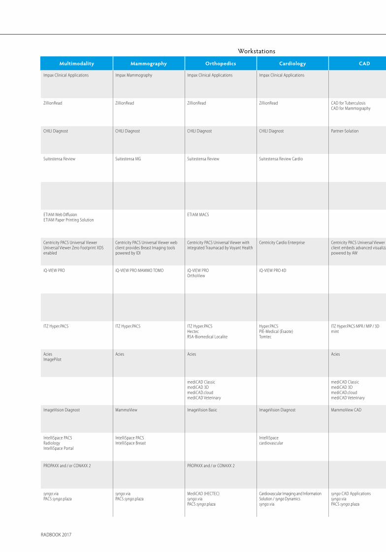
Management Archiving Image Distribution Workstations
RIS Small Business PACS Enterprise PACS Cardiology PACS Long Term Multimedia Inhouse Teleradiology Portal Solution Cloud Computing Application Multimodality Mammography Orthopedics Cardiology CAD Advanced Visualization
Orbis RIS Impax Impax Impax for Cardiology ICISVNA
ICISHYDMedia
ICISEnterprise Imaging SuiteXero Viewer
ICISEnterprise Imaging SuiteXero Viewer
ICIS ICIS Impax Clinical Applications Impax Mammography Impax Clinical Applications Impax Clinical Applications Impax Clinical Applications Agfa HealthCareSeptestraat 27 · 2640 Mortsel, Belgiumtel +32 3 444 94 [email protected] · www.agfa.com
ZillionRIS ZillionArchivewith ZillionRead
ZillionArchivewith ZillionRead
ZillionArchivewith ZillionRead
ZillionVNAwith ZillionRead
ZillionVNAwith ZillionRead
ZillionArchivewith ZillionRead
ZillionArchivewith ZillionRIS and ZillionRead
ZillionPortal ZillionArchivewith ZillionRead
ZillionRead ZillionRead ZillionRead ZillionRead CAD for TuberculosisCAD for Mammography
ZillionRead DelftDI, a Canon companyWiltonstraat 41, 3905 KW Veenendaal, The Netherlandstel +31 318 583 [email protected] · www.delftdi.com
CHILI Modality PACS CHILI PACS CHILI PACS CHILI PACS CHILI PACS CHILI/Web CHILI/Web CHILI/Telemedicine Record OmniPACS CHILI Diagnost CHILI Diagnost CHILI Diagnost CHILI Diagnost Partner-Solution Partner-Solution CHILI GmbHFriedrich-Ebert-Str. 2 · 69221 Dossenheim/Heidelberg, Germanytel +49 6221 1 80 79 [email protected] · www.chili-radiology.com
Suitestensa RIS Suitestensa PACS Suitestensa PACS Suitestensa CVIS Suitestensa Archive Suitestensa Suitestensa WebSuitestensa Mobile
Suitestensa WebSuitestensa Mobile
Suitestensa Web Suitestensa WebSuitestensa Mobile
Suitestensa Review Suitestensa MG Suitestensa Review Suitestensa Review Cardio Suitestensa 3DSuitestensa Vascular
EBIT S.r.l. – Esaote GroupVia Siffredi 58 · 16153 Genoa, Italytel +39 010 65 [email protected] · www.esaote.com/healthcare-it
Xplore WebXplore AnalyticsXplore Nuclear medicine
Xplore PACS Solution Xplore PACS Solution Xplore PACS Solution Xplore PACS Solution Xplore Web Xplore Web Xplore Web Xplore Web EDL SAS1031, chemin de la Seyne à Bastian, 83500 La Seyne-sur-mer, Francetel +33 4 94 10 99 [email protected] · www.xplore.eu
ETIAM MACS ETIAM MACS ETIAM MACS ETIAM MACSETIAM Paper Printing Solution
ETIAM Connect ETIAM WebETIAM Connect
ETIAM WebETIAM Connect
ETIAM Web DiffusionETIAM Paper Printing Solution
ETIAM MACS ETIAM MACS ETIAMZA de la Hallerais, 11 rue du Bois de Soeuvres, 35770 Vern-sur-Seiche, Francetel : +33 2 99 14 33 [email protected] · www.etiam.com
Centricity RISiwith eRadCockpit
Centricity PACSwith Universal Viewer
Centricity PACSwith Universal Viewer
Centricity PACSwith Universal Viewer
Centricity PACSwith Universal Viewer
Centricity Clinical Archive(VNA L1-L4, XDS Repository)
Centricity PACSwith Universal ViewerZero Footprint
Centricity PACSwith Universal ViewerZero Footprint,Centricity 360
Centricity RISwith eRadCockpit,Centricity 360
Centricity 360 Centricity PACS Universal ViewerUniversal Viewer Zero Footprint XDS enabled
Centricity PACS Universal Viewer web client provides Breast Imaging tools powered by IDI
Centricity PACS Universal Viewer with integrated Traumacad by Voyant Health
Centricity Cardio Enterprise Centricity PACS Universal Viewer webclient embeds advanced visualizationpowered by AW
Centricity PACS Universal Viewer webclient embeds advanced visualizationpowered by AW
GE HealthcareLerchenbergstr. 15 · 89160 Dornstadt, Germanytel +49 7348 [email protected] · www.gehealthcare.com
iQ-RIS MED-TAB iQ-SYSTEM PACS MED-TAB MED-TAB MED-TAB MED-TAB iQ-VIEW PRO iQ-VIEW PRO MAMMO TOMO iQ-VIEW PRO OrthoView
iQ-VIEW PRO 4D iQ-VIEW PRO 4D IMAGE Information Systems Europe GmbHLange Str. 16 · 18055 Rostock, Germanytel +49 381 496 58 [email protected] · www.image-systems.biz
RadCentreRadCentre Analytics
RadCentre Multi-PACSIntegration
RadCentre Multi-PACSIntegration
RadCentre Archiving Solution RadCentre Archiving Solution Health Relations RC Health Relations RC Health Relations RC RadCentre as a Service i-SOLUTIONS Health GmbHAm Exerzierplatz 14 · 68167 Mannheim, Germanytel +49 621 39 [email protected] · www.i-solutions.de
ITZ Hyper.RIS ITZ Hyper.ePACS ITZ Hyper.PACS ITZ Hyper.PACS ITZ Hyper.ARC ITZ Hyper.PACSITZ Hyper.WEB
ITZ Hyper.PACSITZ Hyper.WEBITZ Hyper.mView
ITZ Hyper.TELEMEDITZ Hyper.COMDicom2Mail-ModuleITZ Hyper.mView
ITZ Hyper.WEBITZ Hyper.TELEMEDITZ Hyper.COM / ITZ Hyper.UPITZ Hyper.mView
ITZ Hyper.PACS Telearchive ITZHyper.WEB CloudITZ Hyper.ARC CloudITZ DicomCloud.de
ITZ Hyper.PACS ITZ Hyper.PACS ITZ Hyper.PACSHectecRSA-Biomedical Localite
Hyper.PACSPIE-Medical (Esaote)Tomtec
ITZ Hyper.PACS MPR / MIP / 3Dmint
ITZ Hyper.PACSmintTerareconMedian
ITZ Medicom GmbH & Co. KGSiemensring 44 a · 47877 Willich, Germanytel +49 2154 [email protected] · www.itz-medi.com
AciesImagePilot
Acies AciesImagePilot
AciesImagePilot
AciesImagePilot
AciesImagePilot
AciesImagePilot
Acies Acies Acies Acies Konica Minolta Medical & Graphic Imaging Europe B.V.Hoogoorddreef 9 · 1101 BA Amsterdam, The Netherlandstel +31 20 658 41 [email protected] · www.konicaminolta.eu/healthcare
mediCAD.cloud mediCAD ClassicmediCAD 3DmediCAD.cloudmediCAD Veterinary
mediCAD ClassicmediCAD 3DmediCAD.cloudmediCAD Veterinary
mediCAD Hectec GmbHOpalstr. 54 · 84032 Altdorf, Germanytel +49 871 33 02 [email protected] · www.mediCAD.eu
WinRadiolog RIS ImageBroker XS ImageBroker ImageBroker ImageBroker ImageBroker ImageWeb webConnect PraxisPortal PraxisPortal App ImageVision Diagnost MammoView ImageVision Basic ImageVision Diagnost MammoView CAD ImageVision Diagnost medigration GmbHAm Anger 2 · 91052 Erlangen, Germanytel +49 91 31 690 [email protected] · www.medigration.de
IntelliSpace PACS IntelliSpace PACS IntelliSpacecardiovascularXcelera
IntelliSpace PACS IntelliSpace PACS IntelliSpace PACSIntelliSpace PACS AnywhereEnterprise
IntelliSpace PACSRadiology
IntelliSpace PACS IntelliSpace PACSRadiologyIntelliSpace Portal
IntelliSpace PACSIntelliSpace Breast
IntelliSpacecardiovascular
IntelliSpace Portal Philips HealthcareP.O. Box 10.000 · 5680 DA Best, The Netherlandstel +31 40 278 56 [email protected] · www.philips.com/healthcare
CONAXX 2 and PROPAXX PROPAXX and / or CONAXX 2 PROPAXX and / or CONAXX 2 PROPAXX and / or CONAXX 2 PROTEC GmbH & Co. KGIn den Dorfwiesen 14 · 71720 Oberstenfeld, Germanytel +49 7062 [email protected] · www.protec-med.com
PACS syngo.plaza PACS syngo.plazaVNA syngo.shareeHealth
Cardiovascular Imaging andInformation Solution /syngo Dynamics
PACS syngo.plazaVNA syngo.shareeHealth
PACS syngo.plazaVNA syngo.shareeHealth
PACS syngo.plazaVNA syngo.shareeHealth
PACS syngo.plazaVNA syngo.shareeHealth
PACS syngo.plaza teamplayPACS syngo.plazaVNA syngo.shareeHealth
syngo.viaPACS syngo.plaza
syngo.via PACS syngo.plaza
MediCAD (HECTEC) syngo.viaPACS syngo.plaza
Cardiovascular Imaging and Information Solution / syngo Dynamics syngo.via
syngo CAD Applicationssyngo.viaPACS syngo.plaza
syngo.via Siemens Healthineers HeadquartersSiemens Healthcare GmbHHenkestr. 127 · 91052 Erlangen, Germanytel +49 9131 84-0www.siemens.com/healthineers
IT-SOLUTIONS IT-SOLUTIONS
RADBOOK 2017 RADBOOK 2017
Management Archiving Image Distribution Workstations
RIS Small Business PACS Enterprise PACS Cardiology PACS Long Term Multimedia Inhouse Teleradiology Portal Solution Cloud Computing Application Multimodality Mammography Orthopedics Cardiology CAD Advanced Visualization
Orbis RIS Impax Impax Impax for Cardiology ICISVNA
ICISHYDMedia
ICISEnterprise Imaging SuiteXero Viewer
ICISEnterprise Imaging SuiteXero Viewer
ICIS ICIS Impax Clinical Applications Impax Mammography Impax Clinical Applications Impax Clinical Applications Impax Clinical Applications Agfa HealthCareSeptestraat 27 · 2640 Mortsel, Belgiumtel +32 3 444 94 [email protected] · www.agfa.com
ZillionRIS ZillionArchivewith ZillionRead
ZillionArchivewith ZillionRead
ZillionArchivewith ZillionRead
ZillionVNAwith ZillionRead
ZillionVNAwith ZillionRead
ZillionArchivewith ZillionRead
ZillionArchivewith ZillionRIS and ZillionRead
ZillionPortal ZillionArchivewith ZillionRead
ZillionRead ZillionRead ZillionRead ZillionRead CAD for TuberculosisCAD for Mammography
ZillionRead DelftDI, a Canon companyWiltonstraat 41, 3905 KW Veenendaal, The Netherlandstel +31 318 583 [email protected] · www.delftdi.com
CHILI Modality PACS CHILI PACS CHILI PACS CHILI PACS CHILI PACS CHILI/Web CHILI/Web CHILI/Telemedicine Record OmniPACS CHILI Diagnost CHILI Diagnost CHILI Diagnost CHILI Diagnost Partner-Solution Partner-Solution CHILI GmbH Friedrich-Ebert-Str. 2 · 69221 Dossenheim/Heidelberg, Germany tel +49 6221 1 80 79 10 [email protected] · www.chili-radiology.com
Suitestensa RIS Suitestensa PACS Suitestensa PACS Suitestensa CVIS Suitestensa Archive Suitestensa Suitestensa WebSuitestensa Mobile
Suitestensa WebSuitestensa Mobile
Suitestensa Web Suitestensa WebSuitestensa Mobile
Suitestensa Review Suitestensa MG Suitestensa Review Suitestensa Review Cardio Suitestensa 3D Suitestensa Vascular
EBIT S.r.l. – Esaote Group Via Siffredi 58 · 16153 Genoa, Italy tel +39 010 65 47-464 [email protected] · www.esaote.com/healthcare-it
Xplore WebXplore AnalyticsXplore Nuclear medicine
Xplore PACS Solution Xplore PACS Solution Xplore PACS Solution Xplore PACS Solution Xplore Web Xplore Web Xplore Web Xplore Web EDL SAS1031, chemin de la Seyne à Bastian, 83500 La Seyne-sur-mer, Francetel +33 4 94 10 99 [email protected] · www.xplore.eu
ETIAM MACS ETIAM MACS ETIAM MACS ETIAM MACSETIAM Paper Printing Solution
ETIAM Connect ETIAM WebETIAM Connect
ETIAM WebETIAM Connect
ETIAM WebETIAM Paper Printing Solution
ETIAM MACS ETIAM MACS ETIAMZA de la Hallerais, 11 rue du Bois de Soeuvres, 35770 Vern-sur-Seiche, Francetel : +33 2 99 14 33 [email protected] · www.etiam.com
Centricity RISiwith eRadCockpit
Centricity PACSwith Universal Viewer
Centricity PACSwith Universal Viewer
Centricity PACSwith Universal Viewer
Centricity PACSwith Universal Viewer
Centricity Clinical Archive(VNA L1-L4, XDS Repository)
Centricity PACSwith Universal ViewerZero Footprint
Centricity PACSwith Universal ViewerZero Footprint,Centricity 360
Centricity RISwith eRadCockpit,Centricity 360
Centricity 360 Centricity PACS Universal ViewerUniversal Viewer Zero Footprint XDSenabled
Centricity PACS Universal Viewer webclient provides Breast Imaging toolspowered by IDI
Centricity PACS Universal Viewer withintegrated Traumacad by Voyant Health
Centricity Cardio Enterprise Centricity PACS Universal Viewer web client embeds advanced visualization powered by AW
Centricity PACS Universal Viewer web client embeds advanced visualization powered by AW
GE HealthcareLerchenbergstr. 15 · 89160 Dornstadt, Germanytel +49 7348 [email protected] · www.gehealthcare.com
iQ-RIS MED-TAB iQ-SYSTEM PACS MED-TAB MED-TAB MED-TAB MED-TAB iQ-VIEW PRO iQ-VIEW PRO MAMMO TOMO iQ-VIEW PROOrthoView
iQ-VIEW PRO 4D iQ-VIEW PRO 4D IMAGE Information Systems Europe GmbHLange Str. 16 · 18055 Rostock, Germanytel +49 381 496 58 [email protected] · www.image-systems.biz
RadCentreRadCentre Analytics
RadCentre Multi-PACSIntegration
RadCentre Multi-PACSIntegration
RadCentre Archiving Solution RadCentre Archiving Solution Health Relations RC Health Relations RC Health Relations RC RadCentre as a Service i-SOLUTIONS Health GmbHAm Exerzierplatz 14 · 68167 Mannheim, Germanytel +49 621 39 [email protected] · www.i-solutions.de
ITZ Hyper.RIS ITZ Hyper.ePACS ITZ Hyper.PACS ITZ Hyper.PACS ITZ Hyper.ARC ITZ Hyper.PACSITZ Hyper.WEB
ITZ Hyper.PACSITZ Hyper.WEBITZ Hyper.mView
ITZ Hyper.TELEMEDITZ Hyper.COMDicom2Mail-ModuleITZ Hyper.mView
ITZ Hyper.WEBITZ Hyper.TELEMEDITZ Hyper.COM / ITZ Hyper.UPITZ Hyper.mView
ITZ Hyper.PACS Telearchive ITZHyper.WEB CloudITZ Hyper.ARC CloudITZ DicomCloud.de
ITZ Hyper.PACS ITZ Hyper.PACS ITZ Hyper.PACSHectecRSA-Biomedical Localite
Hyper.PACSPIE-Medical (Esaote)Tomtec
ITZ Hyper.PACS MPR / MIP / 3Dmint
ITZ Hyper.PACSmintTerareconMedian
ITZ Medicom GmbH & Co. KGSiemensring 44 a · 47877 Willich, Germanytel +49 2154 [email protected] · www.itz-medi.com
AciesImagePilot
Acies AciesImagePilot
AciesImagePilot
AciesImagePilot
AciesImagePilot
AciesImagePilot
Acies Acies Acies Acies Konica Minolta Medical & Graphic Imaging Europe B.V.Hoogoorddreef 9 · 1101 BA Amsterdam, The Netherlandstel +31 20 658 41 [email protected] · www.konicaminolta.eu/healthcare
mediCAD.cloud mediCAD ClassicmediCAD 3DmediCAD.cloudmediCAD Veterinary
mediCAD ClassicmediCAD 3DmediCAD.cloudmediCAD Veterinary
mediCAD Hectec GmbHOpalstr. 54 · 84032 Altdorf, Germanytel +49 871 33 02 [email protected] · www.mediCAD.eu
WinRadiolog RIS ImageBroker XS ImageBroker ImageBroker ImageBroker ImageBroker ImageWeb webConnect PraxisPortal PraxisPortal App ImageVision Diagnost MammoView ImageVision Basic ImageVision Diagnost MammoView CAD ImageVision Diagnost medigration GmbH Am Anger 2 · 91052 Erlangen, Germanytel +49 91 31 690 87-48 [email protected] · www.medigration.de
IntelliSpace PACS IntelliSpace PACS IntelliSpacecardiovascularXcelera
IntelliSpace PACS IntelliSpace PACS IntelliSpace PACSIntelliSpace PACS AnywhereEnterprise
IntelliSpace PACSRadiology
IntelliSpace PACS IntelliSpace PACSRadiologyIntelliSpace Portal
IntelliSpace PACSIntelliSpace Breast
IntelliSpacecardiovascular
IntelliSpace Portal Philips HealthcareP.O. Box 10.000 · 5680 DA Best, The Netherlandstel +31 40 278 56 [email protected] · www.philips.com/healthcare
CONAXX 2 and PROPAXX PROPAXX and / or CONAXX 2 PROPAXX and / or CONAXX 2 PROPAXX and / or CONAXX 2 PROTEC GmbH & Co. KGIn den Dorfwiesen 14 · 71720 Oberstenfeld, Germanytel +49 7062 [email protected] · www.protec-med.com
PACS syngo.plaza PACS syngo.plazaVNA syngo.shareeHealth
Cardiovascular Imaging andInformation Solution /syngo Dynamics
PACS syngo.plazaVNA syngo.shareeHealth
PACS syngo.plazaVNA syngo.shareeHealth
PACS syngo.plazaVNA syngo.shareeHealth
PACS syngo.plazaVNA syngo.shareeHealth
PACS syngo.plaza teamplayPACS syngo.plazaVNA syngo.shareeHealth
syngo.viaPACS syngo.plaza
syngo.viaPACS syngo.plaza
MediCAD (HECTEC)syngo.viaPACS syngo.plaza
Cardiovascular Imaging and Information Solution / syngo Dynamics syngo.via
syngo CAD Applicationssyngo.viaPACS syngo.plaza
syngo.via Siemens Healthineers HeadquartersSiemens Healthcare GmbHHenkestr. 127 · 91052 Erlangen, Germanytel +49 9131 84-0www.siemens.com/healthineers
IT-SOLUTIONS IT-SOLUTIONS
RADBOOK 2017 RADBOOK 2017
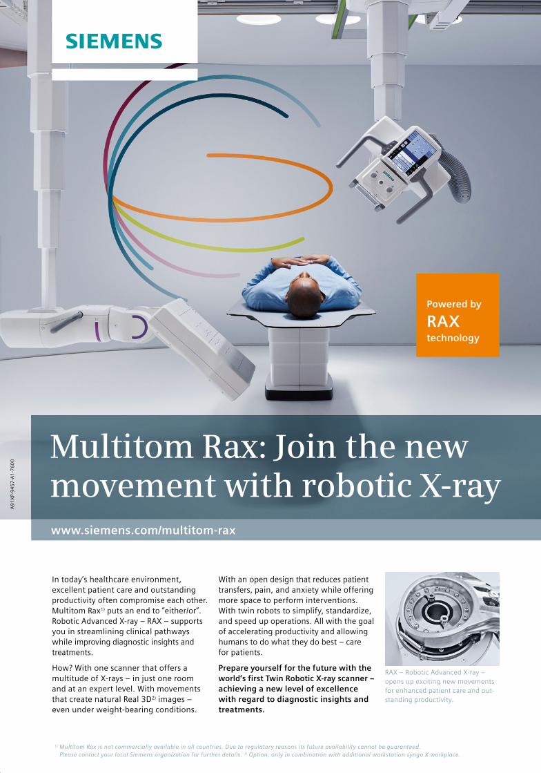
RAD
BOO
K 20
17· T
he G
uide
to Im
agin
g Te
chno
logy
and
Info
rmat
ics i
n Eu
rope
RADBOOK 2017
IT-SOLUTIONS IT-SOLUTIONS
RADBOOK 2017€ 19.-
• IT •CT •MRI • Interventional •Mammo •R/F •Nuc •Displays/Printers•Ultrasound • Injectors • Testing Devices
The Guide to Imaging Technology and Informatics in Europe
More precise diagnostics, low dose and accustomed workflow: Based on dual layer detector technologythe IQon Spectral CT from Philips opens up new dimensions in CT imaging.
Visit us at ECR 2017EXPO X5 Booth #12
Visit us at ECR 2017EXPO X5 Booth #12
In today’s healthcare environment, excellent patient care and outstanding productivity often compromise each other. Multitom Rax1) puts an end to “either/or”.Robotic Advanced X-ray – RAX – supports you in streamlining clinical pathways while improving diagnostic insights and treatments.
How? With one scanner that offers a multitude of X-rays – in just one room and at an expert level. With movements that create natural Real 3D2) images – even under weight-bearing conditions.
With an open design that reduces patient transfers, pain, and anxiety while offering more space to perform interventions. With twin robots to simplify, standardize, and speed up operations. All with the goal of accelerating productivity and allowing humans to do what they do best – care for patients.
Prepare yourself for the future with the world’s first Twin Robotic X-ray scanner – achieving a new level of excellence with regard to diagnostic insights and treatments.
www.siemens.com/multitom-rax
Multitom Rax: Join the new movement with robotic X-ray
Powered by
RAX technology
RAX – Robotic Advanced X-ray – opens up exciting new movements for enhanced patient care and out-standing productivity.
1) Multitom Rax is not commercially available in all countries. Due to regulatory reasons its future availability cannot be guaranteed. Please contact your local Siemens organization for further details. 2) Option, only in combination with additional workstation syngo X workplace.
A91X
P-94
57-A
1-76
00
Best-in-classEquipped with the largest available FPD at 43 x43 cm and Shimadzu’s digital imaging platform,the Sonialvision G4 covers the widest possiblerange of examinations with inter-departmentalhospital capability. In both functionality andoperability, the Sonialvision G4 multipurpose R/Ftable is far beyond other R/F systems. It provides“Best-in-class” features.
Smart system architecturesupports outstanding clinical flexibility for a wide range of examinations
Comprehensive dose management packageensures today’s highest safety of patients andoperators
Excellent image qualityprovided by the the state-of-the-art “SUREengineAdvance“ technology enhancing the entire imagefor clearer details
Premium application softwaresupporting useful applications, such as motion-tolerant DSA, slot radiography andtomosynthesis
www.shimadzu-medical.eu/sonialvision-g4
Next generation digital R/F system:Sonialvision G4
Best-in-classEquipped with the largest available FPD at 43 x43 cm and Shimadzu’s digital imaging platform,the Sonialvision G4 covers the widest possiblerange of examinations with inter-departmentalhospital capability. In both functionality andoperability, the Sonialvision G4 multipurpose R/Ftable is far beyond other R/F systems. It provides“Best-in-class” features.
Smart system architecturesupports outstanding clinical flexibility for a wide range of examinations
Comprehensive dose management packageensures today’s highest safety of patients andoperators
Excellent image qualityprovided by the the state-of-the-art “SUREengineAdvance“ technology enhancing the entire imagefor clearer details
Premium application softwaresupporting useful applications, such as motion-tolerant DSA, slot radiography andtomosynthesis
www.shimadzu-medical.eu/sonialvision-g4
Next generation digital R/F system:Sonialvision G4
Related Documents