1. w Volume 13, Number 1 January 2022 ISSN: 2081-9390 p. 1-119 DOI: 10.7241/ourd Issue online since Monday January 03 2022 Dermatology Online www.odermatol.cowm Our Issue 1.2022 - A multi-center, cross-sectional study on the prevalence of facial dermatoses induced by mask use in the general pu- blic during the COVID-19 pandemic - Recurrent herpes zoster with IgD deposits, multinucleated keratinocytes and overexpression of galectin and glypican 3 in a patient with SARS- -COVID-19 infection - Zoster infection after vaccination with the AstraZeneca COVID-19 vaccine: A case report - Urticarial eruption in COVID- -19-positive children: A report of two cases

Welcome message from author
This document is posted to help you gain knowledge. Please leave a comment to let me know what you think about it! Share it to your friends and learn new things together.
Transcript
1. w
Volume 13, Number 1 January 2022 ISSN: 2081-9390
p. 1-119 DOI: 10.7241/ourd
Issue online since Monday January 03 2022
Dermatology Online www.odermatol.cowm
Our
Issue 1.2022
- A multi-center, cross-sectional study on the prevalence of facial dermatoses induced by mask use in the general pu-
blic during the COVID-19 pandemic
- Recurrent herpes zoster with IgD deposits, multinucleated keratinocytes and overexpression of galectin and
glypican 3 in a patient with SARS--COVID-19 infection
- Zoster infection after vaccination
with the AstraZeneca COVID-19 vaccine: A case report
- Urticarial eruption in COVID--19-positive children: A report of two cases
Editorial Pages
Quarterly published since 01/06/2010 yearsOur Dermatol Online
www.odermatol.com
Editor in Chief: Publisher:Piotr Brzeziński, MD Ph.D Our Dermatology Online
Address: Address:ul. Braille’a 50B, 76200 Słupsk, Poland ul. Braille’a 50B, 76200 Słupsk, Polandtel. 48 692121516, fax. 48 598151829 tel. 48 692121516, fax. 48 598151829e-mail: [email protected] e-mail: [email protected]
Associate Editor:Ass. Prof. Vikash Paudel, MBBS, MD (Nepal)
Indexed in:Universal Impact Factor for year 2012 is = 0.7319
system of opinion of scientific periodicals INDEX COPERNICUS (8,69)(Academic Search) EBSCO
(Academic Search Premier) EBSCOMNiSW (kbn)-Ministerstwo Nauki i Szkolnictwa Wyższego (7.00)
DOAJ (Directory of Open Acces Journals)Geneva Foundation for Medical Education and Research (GFMER), Google Scholar, Open J-Gate, NewJour,
International Committee of Medical Journal Editors (ICMJE), Genamics JournalSeek, Hinari,Bielefeld Academic Search Engine (BASE), WorldCat, e -journal, WorldWideScience.org, National Science Library,
LibSearch, Sciencegate, Virtual Science Library (VSL), Wanfang Data, COnnecting REpositories (CORE),CAB Abstracts, Global Health, Journal Indexed in Directory of Research Journals Indexing,OAIster: The Open Access Initiative, OAJSE - Open Access Journals Search Engine, Scirus
Previous website: issue 1.2010 www.ndermatol.like.pl since issue 2.2010 to issue 3.2011 www.odermatol.like.pl since issue 4.2011 www.odermatol.comPrevious shortcut: since issue 1.2010 to issue 3.2011 N Dermatol Online since issue 4.2011 Our Dermatol Online
Open access journal:This is an open access journal which means that all content is freely available without charge to the user or his/her institution. Users are allowed to read, download, copy, distribute, print, search, or link to the fullor texts of the articles in this journal without asking prior permission from the publisher or the author.Our Dermatology Online is a international journal that publishes original contributions in the field of dermatology, including papers on biochemistry, morphology and immunology of the skin.The journal is among the few not related to dermatological associations or belonging to respective societies which guarantees complete independence. Offers a platform for review articles in areas of interest for dermatologists.OurDermatologyOnline offers article in English as well as in other languages. This is in accordance with the BOAI definition of open access.
e-ISSN: 2081-9390DOI: 10.7241/ourd
Editorial Board
Abdel-Naser, Mohamed Badawy, Prof. (Egypt)Abdul-Lateef Mousa Haider, MD (Iraq)Al Aboud Khalid, MD (Saudi Arabia)Al-Kamel Mohamed A., MD (Yemen)Al-Mashaleh Manal Sulaiman, MD (Jordan)Abreu-Velez Ana Maria, Prof. (USA)Abreu Hilda, MD (Urugway)Adaskevich Uladzimir, Prof. (Belarus)Afifi Mustafa, MD (United Arab Emirates)Aghaei Shahin, Ass. Prof. (Iran)Akpaka Patrick Eberechi, Prof. (Trinidad and Tobago)Akyshbayeva Kulbarshin, Prof. (Kazakhstan)Amichai Boaz, MD (Israel)Arakelyan Hayk S. Prof. (Armenia)Arenas Roberto, Prof. (Mexico)Arif Tasleem, MD (India)Asuquo Maurice Efana, Prof. (Nigeria)Auto James, Ass. Prof. (Solomon Islands)Fatou Barro-Traoré, Prof. (Burkina Faso)Christian Muteba Baseke, MD (Democratic Republic of the Congo)Beigi Pooya Khan Mohammad, Prof. (Canada)Bharti Rakesh, MD (India)Bonifaz Alexandro, Prof. (Mexico)Borowska Katarzyna, Ass. Prof. (Poland)Borruto Franco, Prof. (Monaco)Bouadjar Bakar, Prof. (Algeria)Bukhari Iqbal A., Prof. (Saudi Arabia)Cabo Horacio, Prof. (Argentina)Chamcheu Jean Christopher, Ph.D (USA)Chang Patricia, MD Ph.D (Guatemala)Chihanga Simon, MD (Botswana)Choon Siew Eng, MD (Malaysia)Chuh An Tung Antonio, Prof. (Hong Kong)Crump Vincent, MD (New Zealand)Daboul Mohamed Wael, MD (Syria)Daisley Hubert, Prof. (Trinidad and Tobago)Darlenski Razvigor, MD Ph.D (Bulgaria)Diouf Assane, Ass. Prof. (Senegal)Dobrev Hristo, Prof. (Bulgaria)Doganay Mehmet, Prof. (Turkey)Dong Huiting, Prof. (China)Dori Geme Urge, PhD (Ethiopia)Draganita Ana Maria, MD PhD (Romania)Drljević Irdina, MD, Ph.D. Ass. Prof. (Bosnia and Herzegovina)Dubakienė Rūta, Prof. (Lithuania)Edwards Carl, Ass. Prof. (USA)Elhassan Elizabeth, MD (Senegal)Farkas Arpad, MD PhD (Hungary)Fernandez-Flores Angel, MD Ph.D (Spain)Fortuna Giulio, Ass. Prof. (USA)
Gołąb Elżbieta, Prof. (Poland)Gómez Cuevas Alina, Prof. MD (Nicaragua)Grattan Clive (United Kingdom)Grivcheva-Panovska Vesna, Prof. (Macedonia)Guzmán Antonio, MD (Paraguay)Hashimoto Takashi, Prof. (Japan)Hassan Iffat, Prof. (India)Hegyi Vladimir, Prof. (Slovakia)Hidalgo-Matlock Benjamin, MD (Costa Rica)Hysi Katerina, MD (Albania)Janjua Shahbaz, MD (Pakistan)Jeseňák Miloš, Ass. Prof. (Slovakia)Jeewon Rajesh, Ph.D. (Mauritius)Jordán Rodriguez Ramiro, Prof. (Bolivia)Julian Rolando, Prof. (El Salvador)Kaszuba Andrzej, Prof. (Poland)Kaštelan Marija, Prof. (Croatia)Katsambas Andreas, Prof. (Greece)
Khawaja Shakeel Ahmed, PhD (Eritrea)Kibbi Abdul-Ghani, Prof. (Lebanon) Kossi Metowogo, Ph.D (Togo)Kuiate Jules-Roger, Prof. (Cameroon)Lan Cheng-Che E., Ass. Prof. (Taiwan) Lopez-Granja Jorge, MD (Belize) Lotti Torello, Prof. (Italy)Mahassadi Alassan Kouamé, Ass. Prof. (Côte d’Ivoire’)Mahdi Juma Husain Ali, MD (Bahrain)Maibach Howard I., Prof (USA)Maio Paula, MD (Portugal) Mekokishvili Lali, Prof. (Georgia) Mikkelsen Carsten Sauer, MD (Denmark) Mourad Mokni, Prof. (Tunisia)Mota Luiz Alberto Alves, Prof. (Brazil) Mrisho Fatma, MD (Tanzania) Muvunyi Claude Mambo, MD (Rwanda) Ndugwa Christopher, Prof. (Uganda) Nedelciuc Boris, Ass. Prof. (Moldova) Nhlengethwa Winnie, Prof. (Swaziland) Nigam Pramod Kumar, Prof. (India) Nikolic Milos, Prof. (Serbia)Nowicki Roman, Prof. (Poland)Nwabudike Lawrence Chukwudi, MD Ph.D (Romania)Odeh Samuel, Prof. (Gabon)Olszański Romuald, Prof. (Poland)Oranje Arnold, Prof. (Netherlands) Parajuli Sudip, MD (Nepal) Parvin Rukhsana, MD (Bangladesh)du Plessis Jeanetta, Prof. (South Africa) Puri Neerja, MD (India)
Kazlouskaya Viktoryia, Ass. Prof. (USA)

Editorial Board
Qurashi Mohd, MD (Sudan)Riedl Elisabeth, Ass. Prof. (Austria)Ríos Yuil José Manuel, Prof. (Panama) Ranotsi Amelia, PhD (Lesotho) Rubio-Texeira Marta Ph.D. (Belgium) Rusnak Martin, Prof. (Slovakia) Sayad Ibrahim, Prof. (Kuwait) Sharquie Khalifa E., Prof. (Iraq) Shawa Mary, MD (Malawi)Shkilna Mariia, MD Ph.D (Ukraine)Sinclair Rodney Daniel, Prof. (Australia) Singh Harjeet, MD (Qatar)Slavic Vjerolsva, MD PhD (Montenegro)Srinivasan Sundaramoorthy, Prof. (India)Sumathipala Gayan Saranga, MD (Sri Lanka)
Tapia Felix J., Ass. Prof. (Venezuela)Tatu Alin, MD (Romania)Teixeira Roni Leonardo, MD (Brazil) Tincopa-Wong Oscar Wilfredo, MD (Peru) Tresh Amani, MD (Libya)Tylewska-Wierzbanowska Stanisława, Prof. (Poland)Uraga Pazmiño Enrique, MD (Ecuador)Usha Rani Anaparthy, Prof. (India) Valdebran Manuel, MD (Dominican Republic) Vok Marko, MD (Slovenia)Win Oo Soe, MD (Myanmar)Wollina Uwe, Prof. (Germany) Wortsman Ximena, Ass. Prof. (Chile) Yamamoto Toshiyuki, Prof. (Japan) Yuil de Ríos Emma, MD (Panama) Zabielski Stanisław, Prof. (Poland) Zawar Vijay, Prof (India)
Pusahai-Riman Paula, BSc, MS (Papua New Guinea)
© Our Dermatol Online 1.2022 i
Contents
ORIGINAL ARTICLES
A multi-center, cross-sectional study on the prevalence of facial dermatoses induced by mask use in the general public during the COVID-19 pandemic ................................................................................... 1Tanreet Kaur, Simplepreet Kaur
Effi cacy and safety of dupilumab in adult moderate-to-severe atopic dermatitis: An update narrative literature review ...................................................................................................................................... 6Magdalini Kreouzi, Nikolaos Theodorakis, Ekatherine Prokopiou, Elena Thomaidou
A clinical and epidemiological study of non-venereal genital dermatoses: A cross-sectional, hospital-based study from Nepal ......................................................................................................................... 16Vikash Paudel, Deepa Chudal, Upama Paudel, Dwarika Prasad Shrestha
Task shifting in dermatology: Are nurses prepared and willing? ........................................................................... 22Kavita Kavita, Hitaishi Mehta, Sandhya Ghai, Aarti Garg, Tarun Narang
BRIEF REPORTS
Pulse dye laser therapy and superfi cial cryotherapy as a novel combination treatment for hypertrophic scars and keloids ............................................................................................................................. 28Iqbal A. Bukhari
Eff ects of plaster therapy on thigh fat .................................................................................................................. 32Sara Gonçalves
Chronic dermatophytosis: A clinical, epidemiological, mycological study ............................................................ 36Abdullah Mancy
CASE REPORTS
Recurrent herpes zoster with IgD deposits, multinucleated keratinocytes and overexpression of galectin and glypican 3 in a patient with SARS-COVID-19 infection ...................................... 41Ana Maria Abreu Velez, Amanda Bortle Thomason, Billie L. Jackson, Michael S. Howard
Zoster infection after vaccination with the AstraZeneca COVID-19 vaccine: A case report ................................. 45Laurent Dupoirieux
Urticarial eruption in COVID-19-positive children: A report of two cases .......................................................... 47Shikhar Ganjoo, Resham Vasani, Tulika Gupta
Warty cutaneous tuberculosis of the nose: A rare localization .............................................................................. 50Moussa Doulla, Laouali Salissou, Nina Korsaga/Some, Maimouna M Ouedraogo, Larabou A, Mahamadou ZH, Soumana A, Pascal Niamba, Adama Traoré
Lupus vulgaris mimicking cutaneous leishmaniasis: A case report ........................................................................ 53Nouf Faihan Bin Rubaian, Haya Fahad Alzamami, Gadah Abdulatif Alhosawi, Leena Abdulrahman Almuhaish
SAPHO syndrome associated with a digestive disorder in a ten-year-old girl: Diagnostic diffi culties ............................................................................................................................................................ 57Bérénice Dégboé, Gloria Nouhoumon, Christabelle Nguessie, Fabrice Akpadjan, Nadège Agbéssi, Christiane Koudoukpo, Zavier Zomalheto, Jean Sèhonou, Hugues Adégbidi, Félix Atadokpèdé
Treatment with intralesional methotrexate injection in a patient with nail psoriasis ............................................. 62Yesim Akpinar Kara
© Our Dermatol Online 1.2022 ii
Contents
Acute localized exanthematous pustulosis: A novel side eff ect of piroxicam .......................................................... 65Soukaina Maghfour, Monia Youssef, Rim Hadhri, Ines Lahouel, Yosra Soua, Mouna Korbi, Hichem Belhadjali, Jameleddine Zili
Extragenital lichen sclerosus of the breast and silicone breast implants ................................................................. 67Alberto Goldman, Uwe Wollina
Lichen aureus induced by an insect bite............................................................................................................... 70Emad Bahashwan
Metastatic basal cell carcinoma: A report of two cases and a review of the literature ............................................. 73Claire Quigley, Siona Ni Raghallaigh
A new case of scalp angiosarcoma revealed by eyelid edema ................................................................................. 77Maha Mouradi, Fatima Zahra Elfatoiki, Fouzia Hali, Farida Mernissi, Sara Moukhlis, Soumiya Chiheb
Dermoscopy of pilomatricoma: A case report with a review of the literature ........................................................ 82Radia Chakiri, Youssef Bouhajeb
Isolated pilomatricoma of the arm: A case and a review of the literature .............................................................. 86Sara Bouabdella, Afaf Khouna, Siham Dikhaye, Nada Zizi
Superfi cial epidermolytic ichthyosis: A rare disorder with the unusual absence of blistering ................................. 89Ashwani Rana, Prajul Mehta
REVIEW ARTICLE
Progress of diff erent treatment modalities to limit the use of antibiotics in the treatment of acne ........................ 92Kiran Sanjel, Xue Mei Zhang
CLINICAL IMAGES
Ecthyma gangrenosum in a patient with febrile pancytopenia ............................................................................. 98Samia Mrabat, Hanane Baybay, Ryme Dassouly, Zakia Douhi, Sara Elloudi, Fatima Zahra Mernissi
Giant squamous cell carcinoma of the scalp ......................................................................................................... 99Soumaya Hamich, Fatima Zahra El Gaitibi, Kaoutar Znati, Meriem Meziane, Nadia Ismaili, Laila Benzekri, Karima Senouci
LETTER TO THE EDITORS
Comorbidities of alopecia areata in infancy and childhood. A small descriptive study in a tertiary hospital in Greece.................................................................................................................................. 101Eleni Klimi
Uncommon sublingual ulceration in an infant .................................................................................................. 103Abdelhakim Oukerroum, Fatima Zahra Elfatoiki, Fouzia Hali, Faical Slimani, Soumiya Chiheb
Solitary skin-colored nodule on a child’s face ..................................................................................................... 105Mariem Tabka, Refka Frioui, Taghrid Tlili, Nedia Fetoui, Amina Ounallah, Colandane Belajouza, Mohamed Denguezli
A relapse of pemphigus vulgaris in pemphigus herpetiformis or a phenotypic “switch” ...................................... 107Siham Belmourida, Meriame Meziane, Nadia Ismaili, Laila Benzekri, Badreddine Hassam, Karima Senouci
© Our Dermatol Online 1.2022 iii
Contents
Penile porokeratosis mimicking annular lichen planus ....................................................................................... 109Ngo Binh Trinh, Giang Huong Tran, Hoang Trung Hieu
Folliculotropic mycosis fungoides treated with topical corticosteroids: A case report and a review of its trichoscopic features....................................................................................................................... 111Linda Manaa, Yosra Soua, Ines Lahouel, Marwa Thabouti, Laila Njim, Monia Youssef, Hichem Belahdjali, Jameleddine Zili
Sézary syndrome preceded by mycosis fungoides and complicated by tumor lysis syndrome and macrophage activation syndrome ................................................................................................................ 114Asmae Abdelmouttalib, Fatima Zahra Elghtaibi, Sanae Sialiti, Karima Senouci
Pityriasis lichenoid-like mycosis fungoides ......................................................................................................... 116Fatima Azzahra El Gaitibi, Sara Oulad Ali, Jihane Belcadi, Kaoutar Znati, Mariame Meziane, Laila Benzekri, Nadia Ismaili, Karima Senouci
CD8+ mycosis fungoides: A wolf in sheep’s clothing? ........................................................................................ 118Asmae Abdelmouttalib, Sanae Sialiti, Soumaya Hamich, Kawtar Znati, Mariame Meziane, Nadia Ismaili, Leila Benzekri, Karima Senouci
Our Dermatology Online
© Our Dermatol Online 1.2022 1
How to cite this article: Kaur T, Kaur S. A multi.center, cross.sectional study on the prevalence of facial dermatoses induced by mask use in the general public during the COVID-19 pandemic. Our Dermatol Online. 2022;13(1):1-5.
Submission: 11.09.2021; Acceptance: 21.11.2021DOI: 10.7241/ourd.20221.1
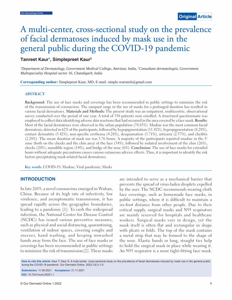
A multi-center, cross-sectional study on the prevalence A multi-center, cross-sectional study on the prevalence of facial dermatoses induced by mask use in the of facial dermatoses induced by mask use in the general public during the COVID-19 pandemicgeneral public during the COVID-19 pandemicTanreet Kaur1, Simplepreet Kaur2
1Department of Dermatology, Government Medical College, Amritsar, India, 2Consultant dermatologist, Government Multispeciality Hospital sector 16, Chandigarh, India
Corresponding author: Simplepreet Kaur, MD, E-mail: [email protected]
INTRODUCTION
In late 2019, a novel coronavirus emerged in Wuhan, China. Because of its high rate of infectivity, low virulence, and asymptomatic transmission, it has spread rapidly across the geographic boundaries, leading to a pandemic [1]. To curb the widespread infection, the National Center for Disease Control (NCDC) has issued various preventive measures, such as physical and social distancing, quarantining, ventilation of indoor spaces, covering coughs and sneezes, hand washing, and keeping unwashed hands away from the face. The use of face masks or coverings has been recommended in public settings to minimize the risk of transmission [2]. These masks
are intended to serve as a mechanical barrier that prevents the spread of virus-laden droplets expelled by the user. The NCDC recommends wearing cloth face coverings, such as homemade face masks, in public settings, where it is difficult to maintain a six-foot distance from other people. Due to their critical supply, surgical masks and N95 respirators are mainly reserved for hospitals and healthcare workers. Surgical masks vary in design, yet the mask itself is often flat and rectangular in shape with pleats or folds. The top of the mask contains a metal strip that may be formed to the shape of the nose. Elastic bands or long, straight ties help to hold the surgical mask in place while wearing it. An N95 respirator is a more tight-fitting face mask.
ABSTRACT
Background: The use of face masks and coverings has been recommended in public settings to minimize the risk of the transmission of coronavirus. The rampant surge in the use of masks for a prolonged duration has resulted in various facial dermatoses. Materials and Methods: The present study was an outpatient, multicentric, observational survey conducted over the period of one year. A total of 350 patients were enrolled. A structured questionnaire was employed to collect data identifying adverse skin reactions that had occurred in the area covered by a face mask. Results: Most of the facial dermatoses were observed in the urban population (78.85%). Maskne was the most common facial dermatosis, detected in 62% of the participants, followed by hypopigmentation (11.42%), hyperpigmentation (8.28%), contact dermatitis (5.42%), non-specific erythema (4.28%), desquamation (3.71%), urticaria (2.57%), and cheilitis (2.28%). The mean duration of mask use was 5.76 hours. A majority of the participants reported maskne in the U zone (both on the cheeks and the chin area) of the face (34%), followed by isolated involvement of the chin (26%), cheeks (20%), mandible region (14%), and bridge of the nose (6%). Conclusion: The use of face masks for extended hours without adequate precautions causes various cutaneous adverse effects. Thus, it is important to identify the risk factors precipitating mask-related facial dermatoses.
Key words: COVID-19; Maskne; Viral pandemic; Masks
Original Article
www.odermatol.com
© Our Dermatol Online 1.2022 2
In addition to splashes, sprays, and large droplets, a respirator may also filter out 95% of minute particles such as viruses and bacteria [3]. However, wearing a mask for a prolonged amount of time causes a physiological and psychological burden to the host. Various adverse effects such as headache, maculopapular rash, mask-induced acne (maskne), contact dermatitis, and impaired cognition have been reported in the literature. As we remain amid the pandemic and more waves are predicted to take place in the future, the recognition and management of mask-induced facial dermatoses is imperative for enduring prolonged mask use. Hence, the present study was conducted with the objective to study facial dermatoses induced by mask use in the general public and to provide recommendations for the prevention and treatment of mask-induced facial dermatoses.
MATERIALS AND METHODS
The present study was an outpatient, multicentric, observational survey conducted over the period of one year. A total of 350 patients participated in the study. Patients with a history of facial dermatoses, such as acne, rosacea, or seborrhea, prior to mask use were excluded from the study. Informed consent was obtained from all participants.
A structured questionnaire was employed to collect data identifying adverse skin reactions that had occurred in the area covered by a face mask. The demographic background information included in the questionnaire were age, sex, occupation, Fitzpatrick skin type. The details regarding the possible risk factors predisposing to adverse reactions in the skin covered by a face mask, included types of face masks, the average duration of wearing a face mask in a day, cleaning methods after face mask use, details regarding the use of cosmetic products on the skin underneath the mask, and were addressed in a structured questionnaire.
We employed descriptive statistics to calculate the frequencies and percentages of categorical variables, and means (M) ± standard deviations (SD) for normally distributed continuous variables. Statistical analysis was performed with commercial software (SPSS, version 22.0). To determine the association of maskne with the use of cosmetic products, an odds ratio was calculated, in which the enrolled patients without maskne served as the controls.
RESULTS
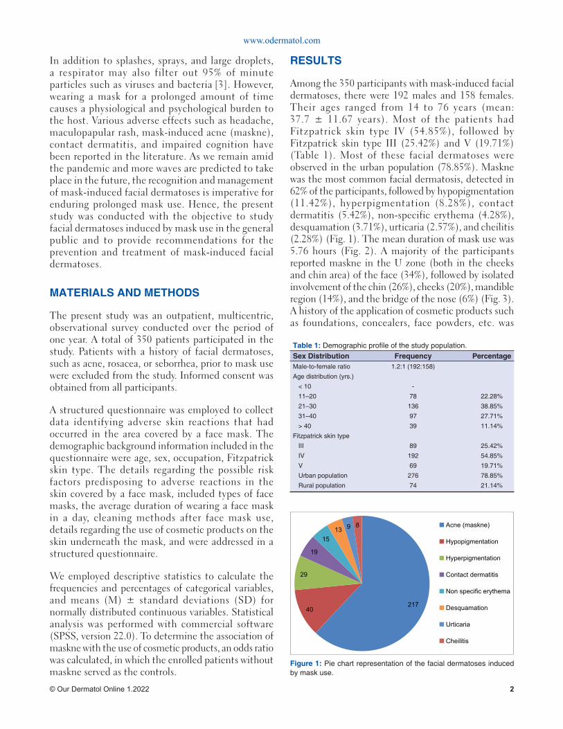
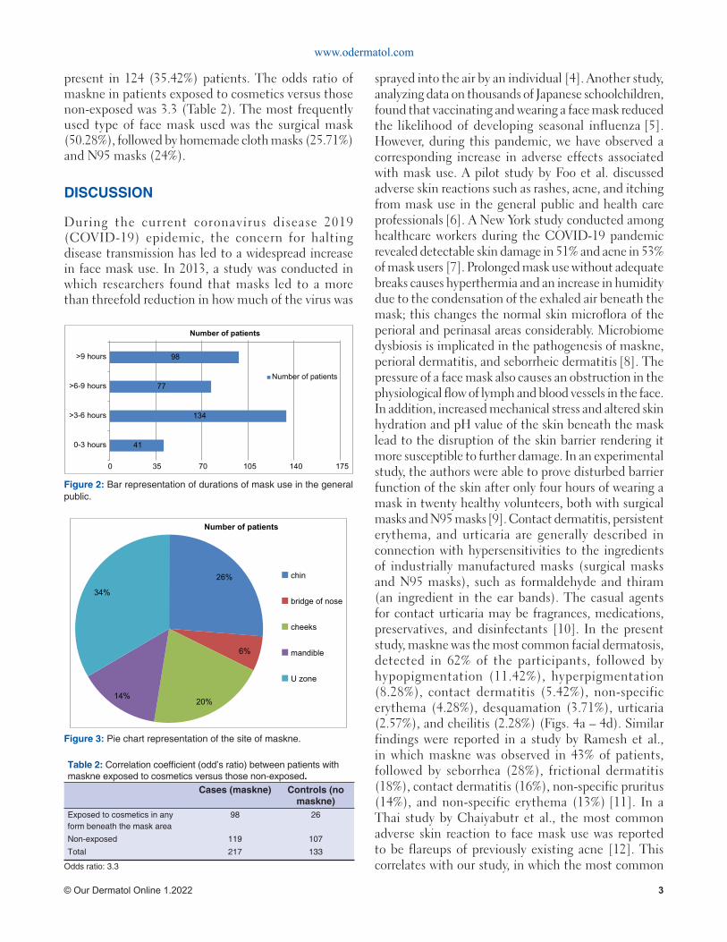
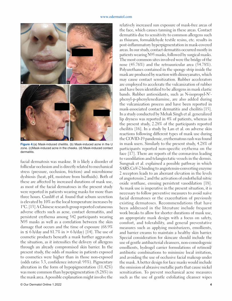
Among the 350 participants with mask-induced facial dermatoses, there were 192 males and 158 females. Their ages ranged from 14 to 76 years (mean: 37.7 ± 11.67 years). Most of the patients had Fitzpatrick skin type IV (54.85%), followed by Fitzpatrick skin type III (25.42%) and V (19.71%) (Table 1). Most of these facial dermatoses were observed in the urban population (78.85%). Maskne was the most common facial dermatosis, detected in 62% of the participants, followed by hypopigmentation (11.42%), hyperpigmentation (8.28%), contact dermatitis (5.42%), non-specific erythema (4.28%), desquamation (3.71%), urticaria (2.57%), and cheilitis (2.28%) (Fig. 1). The mean duration of mask use was 5.76 hours (Fig. 2). A majority of the participants reported maskne in the U zone (both in the cheeks and chin area) of the face (34%), followed by isolated involvement of the chin (26%), cheeks (20%), mandible region (14%), and the bridge of the nose (6%) (Fig. 3). A history of the application of cosmetic products such as foundations, concealers, face powders, etc. was
Table 1: Demographic profi le of the study population.Sex Distribution Frequency Percentage Male-to-female ratio 1.2:1 (192:158)
Age distribution (yrs.)
< 10 -
11–20 78 22.28%
21–30 136 38.85%
31–40 97 27.71%
> 40 39 11.14%
Fitzpatrick skin type
III 89 25.42%
IV 192 54.85%
V 69 19.71%
Urban population 276 78.85%
Rural population 74 21.14%
21740
29
19
1513 9 8 Acne (maskne)
Hypopigmentation
Hyperpigmentation
Contact dermatitis
Non specific erythema
Desquamation
Urticaria
Cheilitis
Figure 1: Pie chart representation of the facial dermatoses induced by mask use.
www.odermatol.com
© Our Dermatol Online 1.2022 3
present in 124 (35.42%) patients. The odds ratio of maskne in patients exposed to cosmetics versus those non-exposed was 3.3 (Table 2). The most frequently used type of face mask used was the surgical mask (50.28%), followed by homemade cloth masks (25.71%) and N95 masks (24%).
DISCUSSION
During the current coronavirus disease 2019 (COVID-19) epidemic, the concern for halting disease transmission has led to a widespread increase in face mask use. In 2013, a study was conducted in which researchers found that masks led to a more than threefold reduction in how much of the virus was
sprayed into the air by an individual [4]. Another study, analyzing data on thousands of Japanese schoolchildren, found that vaccinating and wearing a face mask reduced the likelihood of developing seasonal influenza [5]. However, during this pandemic, we have observed a corresponding increase in adverse effects associated with mask use. A pilot study by Foo et al. discussed adverse skin reactions such as rashes, acne, and itching from mask use in the general public and health care professionals [6]. A New York study conducted among healthcare workers during the COVID-19 pandemic revealed detectable skin damage in 51% and acne in 53% of mask users [7]. Prolonged mask use without adequate breaks causes hyperthermia and an increase in humidity due to the condensation of the exhaled air beneath the mask; this changes the normal skin microflora of the perioral and perinasal areas considerably. Microbiome dysbiosis is implicated in the pathogenesis of maskne, perioral dermatitis, and seborrheic dermatitis [8]. The pressure of a face mask also causes an obstruction in the physiological flow of lymph and blood vessels in the face. In addition, increased mechanical stress and altered skin hydration and pH value of the skin beneath the mask lead to the disruption of the skin barrier rendering it more susceptible to further damage. In an experimental study, the authors were able to prove disturbed barrier function of the skin after only four hours of wearing a mask in twenty healthy volunteers, both with surgical masks and N95 masks [9]. Contact dermatitis, persistent erythema, and urticaria are generally described in connection with hypersensitivities to the ingredients of industrially manufactured masks (surgical masks and N95 masks), such as formaldehyde and thiram (an ingredient in the ear bands). The casual agents for contact urticaria may be fragrances, medications, preservatives, and disinfectants [10]. In the present study, maskne was the most common facial dermatosis, detected in 62% of the participants, followed by hypopigmentation (11.42%), hyperpigmentation (8.28%), contact dermatitis (5.42%), non-specific erythema (4.28%), desquamation (3.71%), urticaria (2.57%), and cheilitis (2.28%) (Figs. 4a – 4d). Similar findings were reported in a study by Ramesh et al., in which maskne was observed in 43% of patients, followed by seborrhea (28%), frictional dermatitis (18%), contact dermatitis (16%), non-specific pruritus (14%), and non-specific erythema (13%) [11]. In a Thai study by Chaiyabutr et al., the most common adverse skin reaction to face mask use was reported to be flareups of previously existing acne [12]. This correlates with our study, in which the most common
Table 2: Correlation coeffi cient (odd’s ratio) between patients with maskne exposed to cosmetics versus those non-exposed.
Cases (maskne) Controls (no maskne)
Exposed to cosmetics in any form beneath the mask area
98 26
Non-exposed 119 107
Total 217 133
Odds ratio: 3.3
98
77
134
41
0 35 70 105 140 175
>9 hours
>6-9 hours
>3-6 hours
0-3 hours
Number of patients
Number of patients
Figure 2: Bar representation of durations of mask use in the general public.
26%
6%
20%14%
34%
Number of patients
chin
bridge of nose
cheeks
mandible
U zone
Figure 3: Pie chart representation of the site of maskne.
www.odermatol.com
© Our Dermatol Online 1.2022 4
facial dermatosis was maskne. It is likely a disorder of follicular occlusion and is directly related to mechanical stress (pressure, occlusion, friction) and microbiome dysbiosis (heat, pH, moisture from biofluids). Both of these are affected by increased durations of mask use, as most of the facial dermatoses in the present study were reported in patients wearing masks for more than three hours. Cunliff et al. found that sebum secretion is elevated by 10% as the local temperature increases by 1°C. [13] A Chinese research group reported cutaneous adverse effects such as acne, contact dermatitis, and persistent erythema among 542 participants wearing N95 masks as well as a correlation between the skin damage that occurs and the time of exposure (68.9% in ≤ 6 h/day and 81.7% in > 6 h/day) [14]. The use of cosmetic products beneath a mask further aggravates the situation, as it intensifies the delivery of allergens through an already compromised skin barrier. In the present study, the odds of maskne in patients exposed to cosmetics were higher than in those non-exposed (odds ratio: 3.3, confidence interval: 95%). Pigmentary alteration in the form of hypopigmentation (11.42%) was more common than hyperpigmentation (8.28%) in the mask area. A possible explanation might involve the
relatively increased sun exposure of mask-free areas of the face, which causes tanning in these areas. Contact dermatitis due to sensitivity to common allergens such as thiuram, formaldehyde textile resins, etc. results in post-inflammatory hyperpigmentation in mask-covered areas. In our study, contact dermatitis occurred mostly in patients wearing N95 masks, followed by surgical masks. The most common sites involved were the bridge of the nose (45.76%) and the retroauricular area (34.78%). Polyurethanes contained in the sponge strip inside the mask are produced by reaction with diisocyanates, which may cause contact sensitization. Rubber accelerators are employed to accelerate the vulcanization of rubber and have been identified to be allergens in mask elastic bands. Rubber antioxidants, such as N-isopropyl-N’-phenyl-p-phenylenediamine, are also added during the vulcanization process and have been reported in mask-associated contact dermatitis and cheilitis [15].
In a study conducted by Mehak Singh et al. generalized lip dryness was reported in 4% of patients, whereas in the present study, 2.28% of the participants reported cheilitis [16]. In a study by Lan et al. on adverse skin reactions following different types of mask use during the COVID-19 pandemic, erythematous rash was found in mask users. Similarly to the present study, 4.28% of participants reported non-specific erythema on the face [17]. There are reports of the coronavirus leading to vasodilation and telangiectatic vessels in the dermis. Sungnak et al. explained a possible pathway in which SARS.CoV-2 binding to angiotensin-converting enzyme 2 receptors leads to an aberrant elevation in the levels of angiotensin 2 and the activation of endothelial nitric oxide synthase, ensuing persistent vasodilation [18]. As mask use is imperative in the present situation, it is necessary to follow preventive measures to avoid naïve facial dermatoses or the exacerbation of previously existing dermatoses. Recommendations that have been addressed in the literature include frequent work breaks to allow for shorter durations of mask use, an appropriate mask design with a focus on safety, comfort, and tolerability, and general preventative measures such as applying moisturizers, emollients, and barrier creams to maintain a healthy skin barrier. Special consideration for skincare should include the use of gentle antibacterial cleansers, non-comedogenic emollients, hydrogel carrier formulations of retinoid/antibiotic combinations to minimize local irritation, and avoiding the use of occlusive facial makeup under the mask. A better design for face masks would include the omission of abrasive metallic parts that cause nickel sensitization. To prevent mechanical acne measures such as the use of gentle exfoliating cleanser wipes
Figure 4:(a) Mask-induced cheilitis. (b) Mask-induced acne in the U zone. (c)Mask-induced acne in the cheeks. (d) Mask-induced contact dermatitis.
dc
ba
www.odermatol.com
© Our Dermatol Online 1.2022 5
throughout one’s shift, using an ear saver or a headband with buttons to allow ear straps to rest on these instead of behind the ears, and the use of Tegaderm on the bridge of the nose to decrease mechanical stress should be employed.
CONCLUSION
Prolonged mask use for extended hours without adequate precautions causes bacterial optimization under the moist and warm environment beneath the mask, leading to various cutaneous adverse effects. As the third wave of COVID-19 is expected, it is imperative to identify solutions to manage these adverse effects. Frequent breaks, improved hydration, an appropriate skincare regimen, and potentially newly designed comfortable masks are recommendations for the future management of adverse effects related to prolonged mask use.
Statement of Human and Animal Rights
All the procedures followed were in accordance with the ethical standards of the responsible committee on human experimentation (institutional and national) and with the 2008 revision of the Declaration of Helsinki of 1975.
Statement of Informed Consent
Informed consent for participation in this study was obtained from all patients.
REFERENCES
1. Sohrabi C, Alsafi Z, O’Neill N, Wo Khan M, Kerwan A, Al-Jabir A, et al. World Health Organization declares global emergency: A review of the 2019 novel coronavirus (COVID-19). Int J Surg Lond Engl. 2020;76:71-6.
2. Teo WL. Diagnostic and management considerations for “maskne” in the era of COVID-19. J Am Acad Dermatol. 2021;84:520-1.
3. Lan J, Song Z, Miao X. Skin damage among healthcare workers managing coronavirus disease (2019). J Am Acad Dermatol. 2020;82:1215-6.
4. Giacalone S, Minuti A, Spigariolo CB, Passoni E, Nazzaro G. Facial dermatoses in the general population due to wearing of personal protective masks during the COVID-19 pandemic: First
observations after lockdown. Clin Exp Dermatol. 2021;46:368-9.5. Wang M-W, Zhou M-Y, Ji G-H, L Ye, Y-R Cheng, Z-H Feng, et al.
Mask crisis during the COVID-19 outbreak. Eur Rev Med Pharmacol Sci. 2020;24:3397-9.
6. Foo CCI, Goon ATJ, Leow Y-H, Goh C-L. Adverse skin reactions to personal protective equipment against severe acute respiratory syndrome: A descriptive study in Singapore. Contact Dermatitis. 2006;55:291-4.
7. Rosner E. Adverse effects of prolonged mask use among healthcare professionals during COVID-19. J Infect Dis Epidemiol. 2000;6:130.
8. Teo WL. The “maskne” microbiome: Pathophysiology and therapeutics. Int J Dermatol. 2021;60:799-809.
9. Tang KM, Chau KH, Kan CW, Fan J. Assessing the accumulated stickiness magnitude from fabric-skin friction: Effect of wetness level of various fabrics. R Soc Open Sci. 2018;5:180860.
10. Singh M, Pawar M, Bothra A. Personal protective equipment induced facial dermatoses in healthcare workers managing Coronavirus disease 2019. J Eur Acad Dermatol Venereol. 2020;34:e378-80.
11. Ramesh A, Thamizhinian K. Clinic epidemiological study of mask induced facial dermatoses due to increased mask usage in general public during COVID-19 pandemic. Int J Res Dermatol. 2021;7:232-8.
12. Chaiyabutr C, Sukakul T, Pruksaeakanan C, Boonchai JTW. Adverse skin reactions following different types of mask usage during the COVID-19 pandemic. J Eur Acad Dermatol Venereol. 2006;55:291-4.
13. Cunliffe WJ, Burton JL, Shuster S. The effect of local temperature variations on the sebum excretion rate. Br J Dermatol. 1970;83:650-4.
14. Choi SY, Hong JY, Kim HJ, Lee GY, Hyun CS, Jung HJ, et al. Mask- induced dermatoses during COVID-19 pandemic: A questionnaire.based study in 12 hospitals of Korea. Clin Exp Dermatol. 2021;3:10.
15. Yu J, Chen JK, Mowad CM, Reeder M, Hylwa S, Chisolm S, et al. Occupational dermatitis to facial personal protective equipment in health care workers: A systematic review. J Am Acad Dermatol. 2021;84:486-94.
16. Singh M, Bothra A, Pawar M, Maheswari A, Tiwari A, Adhicari P. Prevalence of cheilitis in health care workers treating patients with COVID-19. J Am Acad Dermatol. 2020;83:e373-4.
17. Lan J, Song Z, Miao X, Li H, Li Y, Dong L, et al. Skin damage among health care workers managing coronavirus disease (2019). J Am Acad Dermatol. 2020;82:1215-16.
18. Sungnak W, Huang N, Bécavin C, Berg M, Queen R, Litvinukova M, et al. SARS-CoV-2 entry factors are highly expressed in nasal epithelial cells together with innate immune genes. Nat Med. 2020;26:681-7.
Copyright by Tanreet Kaur, et al. This is an open.access article distributed under the terms of the Creative Commons Attribution License, which permits unrestricted use, distribution, and reproduction in any medium, provided the original author and source are credited.Source of Support: Nil, Confl ict of Interest: None.
Our Dermatology Online
© Our Dermatol Online 1.2022 6
How to cite this article: Kreouzi M, Theodorakis N, Prokopiou E, Thomaidou E. Effi cacy and safety of dupilumab in adult moderate-to-severe atopic dermatitis: An update narrative literature review. Our Dermatol Online. 2022;13(1):6-15.Submission: 30.08.2021; Acceptance: 19.11.2021DOI: 10.7241/ourd.20221.2
Effi cacy and safety of dupilumab in adult Effi cacy and safety of dupilumab in adult moderate-to-severe atopic dermatitis: An update moderate-to-severe atopic dermatitis: An update narrative literature reviewnarrative literature reviewMagdalini Kreouzi1, Nikolaos Theodorakis2, Ekatherine Prokopiou1, Elena Thomaidou1
1University of Nicosia Medical School, 21 Ilia Papakyriakou, 2414 Engomi, P.O. Box 24005, CY-1700, Nicosia, Cyprus, 2School of Medicine, National and Kapodistrian University of Athens, 75 Mikras Asias, 11527 Athens, Greece
Corresponding author: Elena Thomaidou, MD, E-mail: [email protected]
ABSTRACT
Background: Adult atopic dermatitis (AD) is defined as a continuum of childhood AD or the development of the disease in adulthood, accounting for 7.7–59.7% of adult AD cases varying in severity and manifestations. The symptomatology of moderate-to-severe adult AD may significantly impact the overall health and quality of life of the patient. The “classic” topical treatments used in mild-to-moderate cases, such as emollients and topical corticosteroids, are usually not adequate to control the symptoms of most of the patients with moderate-to-severe disease. For many years these patients were managed with systemic corticosteroids and immunomodulators, leading to substantial side effects with questionable efficacy. The introduction of dupilumab, the first biologic agent approved by the Food and Drug Administration for use in adult moderate-to-severe AD, has commenced a new era in the management of AD. This narrative literature review addresses the question of how patients with moderate-to-severe AD may achieve a recession or improvement in the overall progression of the disease with the use of dupilumab in both an efficient and safe way.Material and Methods: A search in the PubMed, Embase, and Cochrane databases was conducted using the following combination of MeSH terms: “dupilumab” AND “atopic” (“dermatitis” OR “eczema”). The searches were limited to RCTs written in the English language published before January 25, 2021. The literature used included phase II and III RCTs examining the efficacy and/or safety of dupilumab compared to placebo or other treatments in adults with moderate-to-severe AD. Moderate-to-severe AD was defined by an IGA score of 3 (moderate) or 4 (severe) and EASI 16 or higher at screening and baseline. Additionally, we searched the website clinicaltrials.gov for any unpublished or ongoing RCTs. The search was done independently by two authors in all databases and followed by the exclusion of duplicates. Results: Upon reviewing all randomized controlled trials, dupilumab was found to be an effective and safe option for managing adult moderate-to-severe AD with long-term therapeutic effects. Conclusion: The best results for maintaining long-term disease recession were achieved with the combination of dupilumab and topical corticosteroids.
Keywords: Atopic dermatitis; Biologics; Dupilumab; Efficacy; Safety
INTRODUCTION
Atopic dermatitis (AD) or atopic eczema is a highly prevalent chronic inflammatory skin disorder affecting all ages [1]. In recent years, AD prevalence has increased among several ethnic groups; the highest prevalence of AD between the ages of 13–14 was found in Bolivia and Brazil, with a rate of 21.1%, while other highly prevalent countries include Africa, Oceania, and Northern
Europe [2]. The prevalence of the disease in childhood is up to 20% and up to 10% in adults, affecting 14–24% of the general population [3]. Childhood AD may progress into adult AD in about 10–30% of patients [4]. Adult AD is defined as either a continuum of childhood-onset AD or the development of the disease in adulthood; the latter is called adult-onset AD, accounting for 7.7–59.7% of adult AD cases [5].
Original Article
www.odermatol.com
© Our Dermatol Online 1.2022 7
For the scope of this narrative literature review (NLR), adults will be outlined exclusively.
The term atopy is defined as a predisposition to immunoglobin E (IgE) release after exposure to specific antigens or allergens [6]. AD is often associated with other atopic diseases, such as allergic rhinitis and rhino-conjunctivitis, allergic bronchial asthma, and food allergy, which may be present in the past medical or family history of the patient, a phenomenon known as atopic march [7]. Despite its name, the pathophysiology of the disease is not a typical type 1 hypersensitivity reaction; it includes complex mechanisms. The pathogenesis includes two basic components: a compromised keratin barrier and immune-mediated inflammation driven mainly by a T-helper 2 (Th2) response [8,9]. The former may be a result of various mechanisms (e.g., filaggrin mutations) and leads to epidermal dehydration and increased penetration of various antigens, including microorganisms and allergens [9]. The latter results in increased production of various cytokines, including interleukins IL-4, IL-5, and IL-13. IL-5 induces the activation of eosinophils, which plays a role in the inflammation seen in AD. IL-4 and IL-13 bind to IL-4α, producing various effects, including class switching and the production of IgE by the B-lymphocytes, the differentiation of CD4+ T-lymphocytes into Th2 cells, epidermal dysfunction, itch, and predisposition to skin infections [8]. Dupilumab, the biologic to be outlined in this review, targets the receptor IL-4Rα, therefore blocking the IL-4 and IL-13 signaling pathways. In recent years, various other immunological mechanisms, such as Th1, Th17, and Th22 responses, have been implicated in the pathogenesis of AD [8].
AD is characterized by a pruritic cutaneous rash with specific patterns of involvement: facial, neck, and extensor surfaces, sparing areas such as the groin and axillary region [10]. The diagnosis of AD is mainly clinical, based on specific criteria, including the characteristics of the rash and the past and family history of atopic diseases. Some of these criteria are by Hanifin and Rajka (1980) and the UK Working Party (1994) [11,12]. A skin biopsy may be used only to exclude other conditions, because the histological findings are not pathognomonic for AD, while skin prick testing and allergen-specific IgE testing have been included in the Millennium Criteria (1998) [13]. The severity of AD may be measured with various tools, including the Eczema Area and Severity Index (EASI), Investigation Global Assessment (IGA), Percent of Body Surface Area (BSA), Pruritus Numerical Rating Scale (NRS), Scoring Atopic
Dermatitis (SCORAD), Physician Global Assessment (PGA), Atopic Dermatitis Severity Index (ADSI), Global Individual Signs Score (GISS), Six-Area, Six-Sign Atopic Dermatitis (SASSAD), Patient-Oriented Eczema Measure (POEM), Hospital Anxiety Depression Scale (HADS), and Dermatology Life Quality Index (DLQI). These tools help the physician to classify the disease severity according to its symptomatology, to determine the extent of skin involvement, and to gain a better understanding of how the disease affects the quality of life of the individual [14].
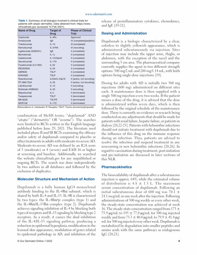
The treatment of mild-to-moderate AD is based on the avoidance of specific irritants and allergens and the use of topical emollients, topical corticosteroids (TCS), or topical calcineurin inhibitors. In patients with moderate-to-severe AD, which accounts for approx. 20% of patients with AD, control of the disease is usually inadequate with the above-mentioned treatments and, as a result, phototherapy or systemic medications (e.g., corticosteroids, calcineurin inhibitors, methotrexate, mycophenolate mofetil) are employed [15]. There are numerous adverse effects and problems arising from these treatments, such as increased susceptibility to infection, bone marrow suppression, nephrotoxicity, and hepatotoxicity, hence there has been a need for the development of safe and effective alternatives, which mainly include biological agents and Janus kinase (JAK) inhibitors [16]. Table 1 summarizes all biological agents targeting various aspects of the pathogenesis of AD, which had been on trial until June 29, 2021. In general, most of these RCTs are small phase II trials without published results yet; however, tralokinumab (anti-IL-13) has successfully completed a phase III trial, showing promising results in both efficacy and safety in adult moderate-to-severe AD. Furthermore, there are currently two large phase III studies (RCT and open-label) assessing the efficacy and safety of nemolizumab (anti-IL-31R) in adult moderate-to-severe AD [15,17]. Dupilumab has been proven to be a safe and efficacious therapeutic agent of adult moderate-to-severe AD and was granted approval by the United States Food and Drug Administration (FDA) in 2017 for this indication [18]. This NLR will extensively outline the efficacy and safety of dupilumab from the data obtained from randomized controlled trials (RCTs) with a brief reference to its pharmacological characteristics.
MATERIALS AND METHODS
A search of the PubMed, Embase, and Cochrane databases was conducted using the following
www.odermatol.com
© Our Dermatol Online 1.2022 8
combination of MeSH terms: “dupilumab” AND “atopic” (“dermatitis” OR “eczema”). The searches were limited to RCTs written in the English language published before June 29, 2021. The literature used included phase II and III RCTs examining the efficacy and/or safety of dupilumab compared to placebo or other treatments in adults with moderate-to-severe AD. Moderate-to-severe AD was defined by an IGA score of 3 (moderate) or 4 (severe) and EASI 16 or higher at screening and baseline. Additionally, we searched the website clinicaltrials.gov for any unpublished or ongoing RCTs. The search was done independently by two authors in all databases and followed by the exclusion of duplicates.
Molecular Structure and Mechanism of Action
Dupilumab is a fully human IgG4 monoclonal antibody binding to the IL-4Rα subunit, which is shared by both IL-4 and IL-13. IL-4R is characterized by two types: the IL-4Rα/γc complex (type 1) and the IL-4Rα/IL-13Rα complex (type 2). Dupilumab achieves signaling inhibition of IL-4 by blocking both types of receptors and IL-13 signaling by blocking type 2 receptors. As a result, it causes the dual inhibition of the IL-4/IL-13 signaling pathway, producing a reduction in epidermal hyperplasia, modification in the lesional skin appearance, modulation of genes related to epidermal pathology in AD, and inhibition of the
release of proinflammatory cytokines, chemokines, and IgE [19-21].
Dosing and Administration
Dupilumab is a biologic characterized by a clear, colorless to slightly yellowish appearance, which is administered subcutaneously via injection. Sites of injection may include the upper arms, thighs, or abdomen, with the exception of the navel and the surrounding 5 cm area. The pharmaceutical company currently supplies the agent in two different strength options: 300 mg/2 mL and 200 mg/1.14 mL, with both options being single-dose injections [19].
Dosing for adults with AD is initially two 300 mg injections (600 mg) administered on different sites each. A maintenance dose is then supplied with a single 300 mg injection every two weeks. If the patient misses a dose of the drug, it is advised that the dose is administered within seven days, which is then followed by the original schedule of the maintenance dose. There is currently no evidence or research being conducted on any adjustments that should be made for patients with renal failure, hepatic failure, or patients in dialysis [20,22-25]. Patients with helminthic infections should not initiate treatment with dupilumab due to the influence of this drug on the immune response during an infection. Thus, patients are advised to resolve the infection and suspend treatment in any reoccurring or new helminthic infections [20,26]. In regard to vaccination during treatment, post-initiation and pre-initiation are discussed in later sections of this NLR.
Pharmacokinetics
The bioavailability of dupilumab after a subcutaneous injection is approx. 64%, while the estimated volume of distribution is 4.8 ± 1.3 L. The maximum serum concentration of dupilumab. Following an initial subcutaneous dose of 600 mg was 70.1 ± 24.1 mcg/mL in one week after the injection. Following administrations of 300 mg weekly or every other week, the steady-state concentration was achieved at week 16. The steady-state concentration ranged from 173 ± 75.9 μg/mL to 193 ± 77.0 μg/mL for 300 mg injected weekly and from 73.3 ± 40.0 μg/mL to 79.9 ± 41.4 μg/mL for 300 mg injected every other week. Dupilumab is metabolized by degradation into smaller peptides and amino acids with the same pathways as endogenous IgG [20,21].
Table 1: Summary of all biologics involved in clinical trials for patients with atopic dermatitis. Data obtained from: https://www.clinicaltrials.gov accessed 10 Feb 2021).Name of Drug Target of
DrugPhase of Clinical Trial
Dupilumab IL-4Rα Approved
Omalizumab IgE IV (completed/pediatric)
Tralokinumab IL-13 III (completed)
Nemolizumab IL-31RA III (recruiting)
Ligelizumab (QGE031) IgE II (completed)
Bermekimab IL-1α II (completed)
Ustekinumab IL-12/23p40 II (completed)
Secukinumab IL-17A II (completed)
Fezakinumab (ILV-094) IL-22 II (completed)
REGN3500 IL-33 II (completed)
GBR830 TSLP II (completed)
KHK4083 TSLP II (completed)
Risankizumab IL23A/IL-23p19 II (active, not recruiting)
PF‐06817024 IL-33 II (active, not recruiting)
Lebrikizumab IL-13 II (recruiting)
Etokimab (ANB020) IL-33 II (recruiting)
Mepolizumab IL-5 II (terminated)
Tezepelumab TSLP II (terminated)
MK-8226 TSLPR II (terminated)
MOR106 IL-17C II (terminated)
Abbreviations IL: Interleukin; R: Receptor; TSLP: Thymic stromal lymphopoietin
www.odermatol.com
© Our Dermatol Online 1.2022 9
Pharmacodynamics
Early results showed that there is a significant increase in the serum concentrations of IL-4 and IL-13 following dupilumab administration, indicating IL-4Rα blockade [20]. There has also been a recently published study assessing the pharmacodynamics of dupilumab showing statistically significant decreases in total serum IgE, serum thymus, and activation-regulated chemokine (TARC) [21]. TARC is a correlation factor of disease activity as well as the blood eosinophil count.
In two studies, both IgE and TARC serum levels were measured in patients receiving dupilumab therapy with varying results between groups and doses [27,28]. IgE concentrations were found to decline significantly following dupilumab administration, which was observed with increasing dosage. However, a study showed that a dose of 75 mg or 150 mg had no significant effects on the IgE serum decline [27]. In the above studies, the measurement of TARC was also conducted and was found to markedly decrease after dupilumab administration correlated with decreased disease activity compared to placebo. The highest decrease in serum TARC was observed with the use of 300 mg of dupilumab at day eight of treatment, although doses 75–600 mg were all associated with a dose-dependent decline in serum TARC when compared with the placebo [27,28].
Drug Interactions
The concomitant use of dupilumab with live-attenuated vaccines could potentially lead to disseminated infection and thus the administration of live-attenuated vaccines should strictly excide twelve weeks prior to the first administration of dupilumab [19,20]. However, further clinical trials are needed in order to assure this possible interaction between live vaccine use and dupilumab. A 32-week study was conducted to assess the immunization response to non-live vaccines in adults with moderate-to-severe AD treated with dupilumab. The study measured the percentage of participants with a positive response (more than fourfold) to the tetanus toxoid (Tdap) and meningococcal polysaccharide vaccine. Results were highly promising, with the Tdap at 83.3% (compared to the placebo at 83.7%) and for the meningococcal at 86.3% (compared to the placebo at 83.7%) [29].
Furthermore, dupilumab could theoretically alter the formation of cytochrome P450 (CYP) enzymes. Therefore,
patients receiving drugs that are CYP substrates, especially those with a narrow therapeutic index or severe side effects, should be monitored for their efficacy (e.g., prothrombin time for warfarin) and/or plasma levels (e.g., cyclosporine) [20]. However, a clinical trial (NCT02647086) conducted to assess drug-to-drug interactions between dupilumab and drugs metabolized by specific CYP enzymes demonstrated that the pharmacokinetics of oral midazolam (CYP3A), omeprazole (CYP2C19), warfarin (CYP2C9), caffeine (CYP1A2), and metoprolol (CYP2D6) were unaffected by dupilumab. Thus, the study concluded that there were no clinically significant and/or relevant effects on the pharmacokinetics of CYP substrate, provided that dupilumab clinically benefited the patients [30].
RESULTS
Effi cacy of Dupilumab
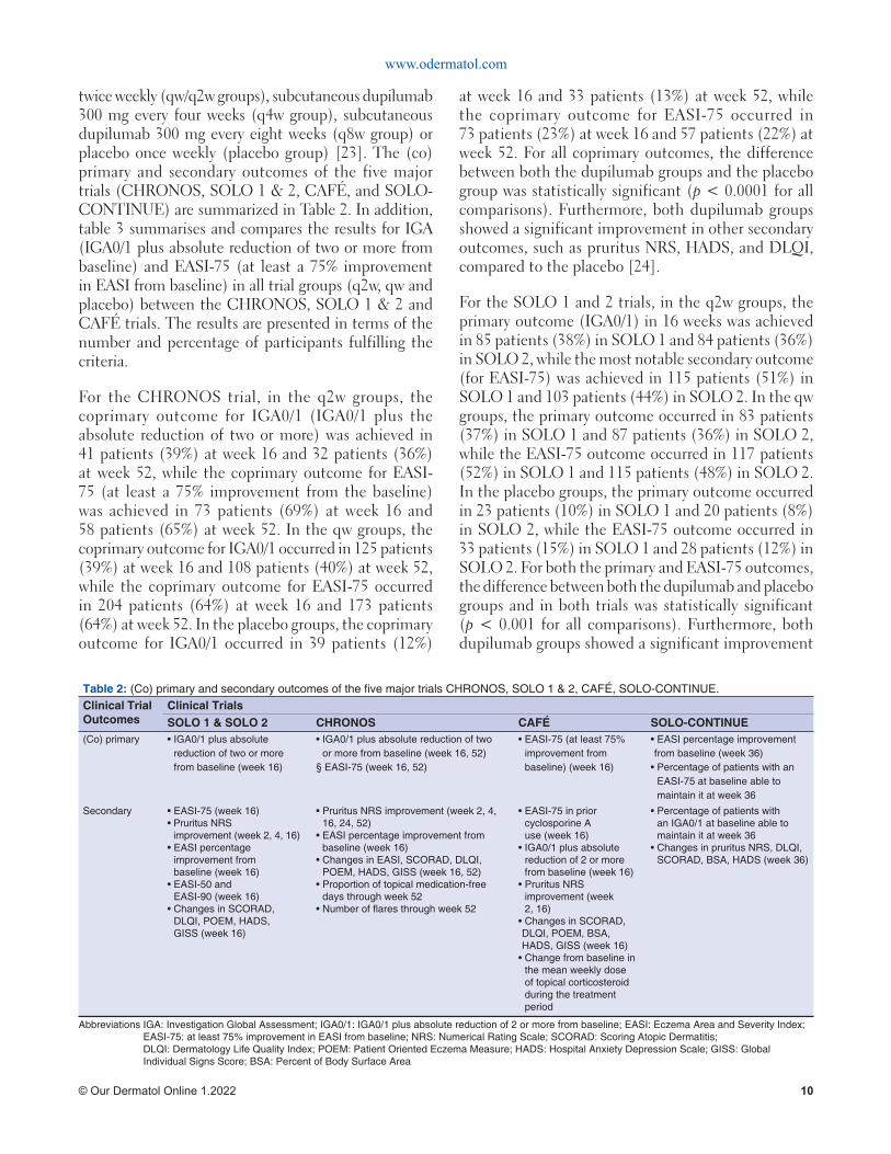
The most valuable RCTs evaluating the efficacy of dupilumab were: LIBERTY AD CHRONOS (NCT02260986) (n = 740), LIBERTY AD CAFÉ (NCT02755649) (n = 325), LIBERTY AD SOLO 1 (NCT02277743) (n = 671), LIBERTY AD SOLO 2 (NCT02277769) (n = 708), and LIBERTY AD SOLO-CONTINUE (NCT02395133) (n = 422). All five trials were randomized, double-blinded, placebo-controlled, parallel-group, phase III clinical trials, with SOLO 1 and 2 being replicate trials. The CHRONOS and SOLO 1 and 2 trials assessed the efficacy of dupilumab when compared with the placebo in 52 and 16 weeks, respectively [22,24]. The CAFÉ trial assessed the efficacy of dupilumab when compared with the placebo in 16 weeks in patients who had never received cyclosporin A (CsA) or patients for whom CsA treatment failed [25]. Patients in the CHRONOS, CAFÉ, and SOLO 1 and 2 trials were randomized into three groups: subcutaneous dupilumab 300 mg once weekly (qw group), subcutaneous dupilumab 300 mg every two weeks (q2w group), or placebo once weekly [22-25]. In the CHRONOS and CAFÉ trials, patients from all groups received concomitant TCS (or topical calcineurin inhibitors), compared to the SOLO 1 and 2 trials, which used dupilumab as a monotherapy [22,24,25]. The SOLO-CONTINUE trial assessed the ability of different dupilumab dose regimens to maintain the treatment response achieved in the SOLO 1 and 2 trials compared to the placebo in a time span of 36 weeks. Patients in the SOLO-CONTINUE trial were randomized into four groups: subcutaneous dupilumab 300 mg once/
www.odermatol.com
© Our Dermatol Online 1.2022 10
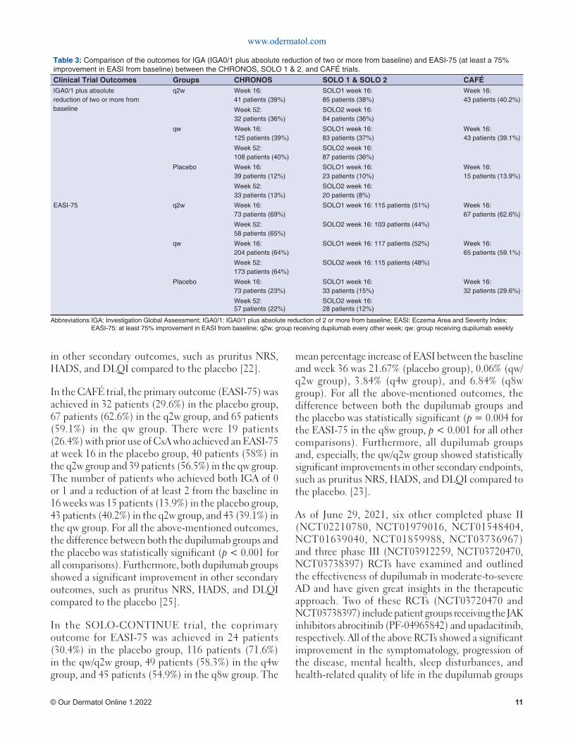
twice weekly (qw/q2w groups), subcutaneous dupilumab 300 mg every four weeks (q4w group), subcutaneous dupilumab 300 mg every eight weeks (q8w group) or placebo once weekly (placebo group) [23]. The (co)primary and secondary outcomes of the five major trials (CHRONOS, SOLO 1 & 2, CAFÉ, and SOLO-CONTINUE) are summarized in Table 2. In addition, table 3 summarises and compares the results for IGA (IGA0/1 plus absolute reduction of two or more from baseline) and EASI-75 (at least a 75% improvement in EASI from baseline) in all trial groups (q2w, qw and placebo) between the CHRONOS, SOLO 1 & 2 and CAFÉ trials. The results are presented in terms of the number and percentage of participants fulfilling the criteria.
For the CHRONOS trial, in the q2w groups, the coprimary outcome for IGA0/1 (IGA0/1 plus the absolute reduction of two or more) was achieved in 41 patients (39%) at week 16 and 32 patients (36%) at week 52, while the coprimary outcome for EASI-75 (at least a 75% improvement from the baseline) was achieved in 73 patients (69%) at week 16 and 58 patients (65%) at week 52. In the qw groups, the coprimary outcome for IGA0/1 occurred in 125 patients (39%) at week 16 and 108 patients (40%) at week 52, while the coprimary outcome for EASI-75 occurred in 204 patients (64%) at week 16 and 173 patients (64%) at week 52. In the placebo groups, the coprimary outcome for IGA0/1 occurred in 39 patients (12%)
at week 16 and 33 patients (13%) at week 52, while the coprimary outcome for EASI-75 occurred in 73 patients (23%) at week 16 and 57 patients (22%) at week 52. For all coprimary outcomes, the difference between both the dupilumab groups and the placebo group was statistically significant (p < 0.0001 for all comparisons). Furthermore, both dupilumab groups showed a significant improvement in other secondary outcomes, such as pruritus NRS, HADS, and DLQI, compared to the placebo [24].
For the SOLO 1 and 2 trials, in the q2w groups, the primary outcome (IGA0/1) in 16 weeks was achieved in 85 patients (38%) in SOLO 1 and 84 patients (36%) in SOLO 2, while the most notable secondary outcome (for EASI-75) was achieved in 115 patients (51%) in SOLO 1 and 103 patients (44%) in SOLO 2. In the qw groups, the primary outcome occurred in 83 patients (37%) in SOLO 1 and 87 patients (36%) in SOLO 2, while the EASI-75 outcome occurred in 117 patients (52%) in SOLO 1 and 115 patients (48%) in SOLO 2. In the placebo groups, the primary outcome occurred in 23 patients (10%) in SOLO 1 and 20 patients (8%) in SOLO 2, while the EASI-75 outcome occurred in 33 patients (15%) in SOLO 1 and 28 patients (12%) in SOLO 2. For both the primary and EASI-75 outcomes, the difference between both the dupilumab and placebo groups and in both trials was statistically significant (p < 0.001 for all comparisons). Furthermore, both dupilumab groups showed a significant improvement
Table 2: (Co) primary and secondary outcomes of the fi ve major trials CHRONOS, SOLO 1 & 2, CAFÉ, SOLO-CONTINUE.Clinical Trial Outcomes
Clinical Trials SOLO 1 & SOLO 2 CHRONOS CAFÉ SOLO-CONTINUE
(Co) primary • IGA0/1 plus absolute reduction of two or more from baseline (week 16)
• IGA0/1 plus absolute reduction of two or more from baseline (week 16, 52)
§ EASI-75 (week 16, 52)
• EASI-75 (at least 75% improvement from baseline) (week 16)
• EASI percentage improvement from baseline (week 36)
• Percentage of patients with an EASI-75 at baseline able to maintain it at week 36
Secondary • EASI-75 (week 16)• Pruritus NRS
improvement (week 2, 4, 16)• EASI percentage
improvement from baseline (week 16)
• EASI-50 and EASI-90 (week 16)
• Changes in SCORAD, DLQI, POEM, HADS, GISS (week 16)
• Pruritus NRS improvement (week 2, 4, 16, 24, 52)
• EASI percentage improvement from baseline (week 16)
• Changes in EASI, SCORAD, DLQI, POEM, HADS, GISS (week 16, 52)
• Proportion of topical medication-free days through week 52
• Number of fl ares through week 52
• EASI-75 in prior cyclosporine A use (week 16)
• IGA0/1 plus absolute reduction of 2 or more from baseline (week 16)
• Pruritus NRS improvement (week 2, 16)
• Changes in SCORAD, DLQI, POEM, BSA, HADS, GISS (week 16)
• Change from baseline in the mean weekly dose of topical corticosteroid during the treatment period
• Percentage of patients with an IGA0/1 at baseline able to maintain it at week 36
• Changes in pruritus NRS, DLQI, SCORAD, BSA, HADS (week 36)
Abbreviations IGA: Investigation Global Assessment; IGA0/1: IGA0/1 plus absolute reduction of 2 or more from baseline; EASI: Eczema Area and Severity Index; EASI-75: at least 75% improvement in EASI from baseline; NRS: Numerical Rating Scale; SCORAD: Scoring Atopic Dermatitis; DLQI: Dermatology Life Quality Index; POEM: Patient Oriented Eczema Measure; HADS: Hospital Anxiety Depression Scale; GISS: Global Individual Signs Score; BSA: Percent of Body Surface Area
www.odermatol.com
© Our Dermatol Online 1.2022 11
in other secondary outcomes, such as pruritus NRS, HADS, and DLQI compared to the placebo [22].
In the CAFÉ trial, the primary outcome (EASI-75) was achieved in 32 patients (29.6%) in the placebo group, 67 patients (62.6%) in the q2w group, and 65 patients (59.1%) in the qw group. There were 19 patients (26.4%) with prior use of CsA who achieved an EASI-75 at week 16 in the placebo group, 40 patients (58%) in the q2w group and 39 patients (56.5%) in the qw group. The number of patients who achieved both IGA of 0 or 1 and a reduction of at least 2 from the baseline in 16 weeks was 15 patients (13.9%) in the placebo group, 43 patients (40.2%) in the q2w group, and 43 (39.1%) in the qw group. For all the above-mentioned outcomes, the difference between both the dupilumab groups and the placebo was statistically significant (p < 0.001 for all comparisons). Furthermore, both dupilumab groups showed a significant improvement in other secondary outcomes, such as pruritus NRS, HADS, and DLQI compared to the placebo [25].
In the SOLO-CONTINUE trial, the coprimary outcome for EASI-75 was achieved in 24 patients (30.4%) in the placebo group, 116 patients (71.6%) in the qw/q2w group, 49 patients (58.3%) in the q4w group, and 45 patients (54.9%) in the q8w group. The
mean percentage increase of EASI between the baseline and week 36 was 21.67% (placebo group), 0.06% (qw/q2w group), 3.84% (q4w group), and 6.84% (q8w group). For all the above-mentioned outcomes, the difference between both the dupilumab groups and the placebo was statistically significant (p = 0.004 for the EASI-75 in the q8w group, p < 0.001 for all other comparisons). Furthermore, all dupilumab groups and, especially, the qw/q2w group showed statistically significant improvements in other secondary endpoints, such as pruritus NRS, HADS, and DLQI compared to the placebo. [23].
As of June 29, 2021, six other completed phase II (NCT02210780, NCT01979016, NCT01548404, NCT01639040, NCT01859988, NCT03736967) and three phase III (NCT03912259, NCT03720470, NCT03738397) RCTs have examined and outlined the effectiveness of dupilumab in moderate-to-severe AD and have given great insights in the therapeutic approach. Two of these RCTs (NCT03720470 and NCT03738397) include patient groups receiving the JAK inhibitors abrocitinib (PF-04965842) and upadacitinib, respectively. All of the above RCTs showed a significant improvement in the symptomatology, progression of the disease, mental health, sleep disturbances, and health-related quality of life in the dupilumab groups
Table 3: Comparison of the outcomes for IGA (IGA0/1 plus absolute reduction of two or more from baseline) and EASI-75 (at least a 75% improvement in EASI from baseline) between the CHRONOS, SOLO 1 & 2, and CAFÉ trials.Clinical Trial Outcomes Groups CHRONOS SOLO 1 & SOLO 2 CAFÉ IGA0/1 plus absolute reduction of two or more from baseline
q2w Week 16:41 patients (39%)
SOLO1 week 16:85 patients (38%)
Week 16:43 patients (40.2%)
Week 52:32 patients (36%)
SOLO2 week 16:84 patients (36%)
qw Week 16:125 patients (39%)
SOLO1 week 16:83 patients (37%)
Week 16:43 patients (39.1%)
Week 52:108 patients (40%)
SOLO2 week 16:87 patients (36%)
Placebo Week 16:39 patients (12%)
SOLO1 week 16:23 patients (10%)
Week 16:15 patients (13.9%)
Week 52:33 patients (13%)
SOLO2 week 16:20 patients (8%)
EASI-75 q2w Week 16:73 patients (69%)
SOLO1 week 16: 115 patients (51%) Week 16:67 patients (62.6%)
Week 52:58 patients (65%)
SOLO2 week 16: 103 patients (44%)
qw Week 16:204 patients (64%)
SOLO1 week 16: 117 patients (52%) Week 16:65 patients (59.1%)
Week 52:173 patients (64%)
SOLO2 week 16: 115 patients (48%)
Placebo Week 16:73 patients (23%)
SOLO1 week 16:33 patients (15%)
Week 16:32 patients (29.6%)
Week 52:57 patients (22%)
SOLO2 week 16:28 patients (12%)
Abbreviations IGA: Investigation Global Assessment; IGA0/1: IGA0/1 plus absolute reduction of 2 or more from baseline; EASI: Eczema Area and Severity Index; EASI-75: at least 75% improvement in EASI from baseline; q2w: group receiving dupilumab every other week; qw: group receiving dupilumab weekly
www.odermatol.com
© Our Dermatol Online 1.2022 12
over the placebo, which were measured with several scoring systems [29,31-37].
Currently, there are three ongoing phase III RCTs (NCT04678882, NCT04417894, NCT04345367) assessing the safety and/or efficacy of dupilumab. The NCT04345367 trial aims to compare the effectiveness and safety of the JAK inhibitor abrocitinib over dupilumab [38-40].
Long-term efficacy has been established with the LIBERTY AD OLE, a phase 3, multi-center, open-label extension study with 2733 participants, who received dupilumab 300 mg weekly for 148 weeks. The major outcomes in regards to efficacy at week 148 were favorable, with a mean EASI of 1.4 (-95.4% from the baseline) and a weekly pruritus NRS of 2.2 (-65.4% from the baseline) [41].
Safety of Dupilumab
In the outline of the safety of dupilumab, the most valuable RCTs were the CHRONOS, CAFÉ, SOLO 1, SOLO 2, and SOLO-CONTINUE trials. For these trials, there were four deaths documented; their causes were unrelated to the use of the therapeutic agent. In addition, withdrawal of the participants from the trial due to adverse effects were among all groups; more common in the placebo group, accounting for 1–8%, in comparison to the dupilumab groups q2w accounting for 1–2% and qw 1–3%. Furthermore, the most common of the side effects present and documented throughout all trials was AD exacerbation, which highly impacted the placebo group (14.8-48.8%) and the minority of cases in groups q2w (7.5-32.1%) and qw (8.2-34.5%) [22-25].
A relatively common non-infectious side effect was injection site reactions with a high prevalence among the dupilumab groups, with q2w accounting for 0.9–15%, qw 3.6–19%, and, for the placebo, 0–8%. Another prevalent non-infectious side effect was headaches, which was slightly more prevalent among the dupilumab groups, in comparison to the placebo. Nasopharyngitis and upper respiratory tract infections were also observed, with their prevalence being balanced throughout the three groups. Non-herpetic skin infections were documented with a higher prevalence in the placebo group, in comparison to the q2w and qw dupilumab groups, among which herpetic infections were slightly more prevalent [22-25].
Conjunctivitis with an unspecified cause and allergic conjunctivitis were documented with a higher prevalence in the dupilumab groups (15–20%), in comparison to the placebo (up to 8%). Bacterial and viral conjunctivitis, on the other hand, were generally of a low prevalence between all groups, but the few cases documented were present in the dupilumab groups [22-25].
As far as long-term safety is concerned, there is data available from two open-label studies. The LIBERTY AD OLE study showed a favorable safety profile in a 148-week period, similarly to the safety outcomes of the RCTs, supporting the long-term safety of this biologic [41]. Similar safety outcomes in a 76-week period are being shown by a large, ongoing, multi-center, open-label study evaluating the long-term safety of dupilumab [42].
There is no available data for the effects of dupilumab use during pregnancy. Human IgG is known to cross the placental barrier, yet the effect of dupilumab on the human fetus remains unknown. Animal studies on the administration of homologous anti-IL-4Rα during pregnancy showed no evidence of fetal toxicity or teratogenesis [20]. Currently, there are two ongoing observational studies assessing the effects of dupilumab on pregnancy: one prospective cohort (NCT04173442) and one retrospective cohort (NCT03936335) [43,44]. As with pregnancy, the effects of dupilumab in the newborn during lactation are unknown. Human IgG is present in the milk and the risks to the newborn should be weighted with the benefits to the mother [20].
Overall, the side effects of dupilumab throughout the trials were minor, with some exceptions, and could easily be managed by the participants. Thus, we conclude that the safety profile of the drug is supportive with relatively few side effects and, rarely, cases of severe manifestations [22-25,29,31-37].
Immunogenicity of Dupilumab in RCTs
In the era of biological therapies, immunogenicity is of high importance. Immunogenicity is defined as a humoral or cell-mediated response induced by the introduction of a foreign substance and, in the case of dupilumab, a monoclonal antibody. In the case of biological therapies, the unwanted effects of immunogenicity include an immune response against the antigen leading to the production of anti-drug-antibodies (ADAs), inactivating the therapeutic
www.odermatol.com
© Our Dermatol Online 1.2022 13
effects of the treatment [43]. Data provided by the FDA showed that approx. 7% of patients receiving dupilumab (300 mg) for AD develop ADAs after 16 weeks, with 30% of those patients presenting with neutralizing ADAs [20].
The SOLO-CONTINUE trial determined that ADAs occurred in the placebo group at 11%, in the dupilumab group every eight weeks up to 6%, in the dupilumab group every four weeks at 4.3%, in the dupilumab group every two weeks at 1.2%, and the highest prevalence among all was in the dupilumab weekly group [23]. Furthermore, in the CHRONOS trial, there were 7% of patients developing ADAs, among them 2% having a persistent antibody response and 14% had neutralizing antibodies [24].
DISCUSSION
For the past several years, the management of moderate-to-severe AD was restricted to options such as phototherapy, systemic corticosteroids, or systemic immunomodulators [15]. Phototherapy has been proven inconvenient for a large number of patients, as well as to have adverse effects due to UV radiation, such as non-melanoma skin cancer in the long term, limiting its use [46]. Furthermore, patients using systemic corticosteroids and immunomodulators may present with severe and long-term adverse effects, may have a poor clinical response, become refractory, or require large maintenance doses of these systemic medications in order to maintain recession of the disease [1,15,16].
The development of novel alternatives may be the last resort for many individuals who are unresponsive; thus, biological treatments have proven to be of great importance. One of the first biologic treatments, which is proven to help in AD, is dupilumab, and several clinical trials have been conducted to assess its efficacy and safety in the treatment of adult moderate-to-severe AD. The severity of AD is one of the key criteria for selecting the type of treatment [22-25]. Furthermore, the SCORAD index is a good estimator of AD severity, with scores of < 25 being classified as mild, 25–50 as moderate, and > 50 as severe [47].
Dupilumab was the first FDA-approved biologic for the treatment of adult moderate-to-severe AD, providing solutions to the previously mentioned problems. RCTs show that dupilumab is both a safe and effective option. Concerning the efficacy of the drug, RCT results show
substantial improvements in objective signs (e.g., the extent of the disease), subjective signs (e.g., pruritus), mental health (i.e., anxiety or depression), and overall quality of life with minor side effects, compared to a placebo [22-25,29,31-37]. The SOLO 1 and 2 trials revealed that monotherapy with dupilumab could provide adequate clinical responses in sixteen weeks with an encouraging safety profile [22]. The SOLO-CONTINUE trial determined that the patients who achieved a positive response in SOLO 1 and 2 should continue receiving dupilumab every week or every other week in order to maintain this response. Dose regimens every four or eight weeks resulted in decreased efficacy, no change in the safety profile, and increased ADA formation [23]. The CAFÉ and CHRONOS trials concluded that the combination of dupilumab and TCS for sixteen weeks is superior to a placebo with TCS in regard to efficacy with minimal side effects [24,25].
The CHRONOS trial assessed safety and efficacy for a total of 52 weeks and the results showed that both dupilumab groups had similar percentages in efficacy outcomes in 52 weeks compared to 16 weeks without more adverse effects [24]. Furthermore, the higher percentages of patients fulfilling the primary outcomes in CAFÉ and CHRONOS compared to SOLO 1 and 2 could be an indicator that topical corticosteroid treatment should be continued long-term in patients receiving dupilumab, as it increases the chances of a positive response [22,24,25].
In all RCTs, the most common side effects linked to dupilumab were mild, including injection-site reactions and headaches. Furthermore, dupilumab administration was not correlated with an increased susceptibility to infections compared to the placebo, which had a higher prevalence of skin infections [22-25,29,31-37]. Conjunctivitis was a relatively common adverse effect with an unknown pathogenesis linked to the administration of dupilumab in patients with AD rather than other diseases, such as asthma, chronic rhinosinusitis, and nasal polyposis [24].
A limitation of the above studies was the absence of statistical comparison between the qw and q2w dupilumab groups, yet clinical data demonstrates that both regimens are safe and effective for treating adult moderate-to-severe AD. However, in the CHRONOS trial, the variability of the primary outcomes was more prevalent in the q2w groups over time. Although limitations may have been present in all trials, the safety profile outline was consistent among all of them, showing
www.odermatol.com
© Our Dermatol Online 1.2022 14
no increased risk of infections (serious or opportunistic), both systemic and skin-related [22-25]. Long-term safety and efficacy beyond the 52-week period in the CHRONOS trial were established by the LIBERTY AD OLE open-label study, which showed a favorable safety profile and sustained efficacy in a 148-week period [41]. A second large open-label study showed promising results in regards to safety in a 76-week period [42]. Furthermore, there are two main cohort studies that are currently evaluating the safety of dupilumab during pregnancy without any results published yet [43,44].
Dupilumab administered together with TCS drastically decreased the use of rescue treatments (e.g., systemic corticosteroids), as established by the CHRONOS trial; however, there have been no current studies comparing dupilumab with systemic corticosteroids or immunomodulators [24]. The above is doubtless a gap in evidence, which could be of great importance in both establishing a stronger safety profile regarding dupilumab and minimizing the use of older systemic medications. Finally, a recent study comparing abrocitinib and dupilumab was published, outlining the superiority of the former in decreasing pruritus, which may become an alternative treatment to adult moderate-to-severe AD as well [36].
Currently, there are no other available biological agents for the management of adult moderate-to-severe AD apart from dupilumab. In addition to the promising therapeutic outcomes observed with dupilumab, encouraging clinical results were also demonstrated with the use of some topical (tofacitinib, ruxolitinib, delgocitinib) or oral (abrocitinib, baricitinib, upadacitinib, delgocitinib) JAK inhibitors and the anti-IL-13 biologic tralokinumab [48,49]. Various phase III trials on the JAK inhibitors, as well as a phase III trial on tralokinumab, outlined that these agents are superior to the placebo for various primary (e.g., EASI-75, IGA0/1) and secondary outcomes (e.g., SCORAD, pruritus NRS, DLQI) [48,49].
In conclusion, the administration of dupilumab, the only FDA-approved biologic for AD, is both an effective and safe therapeutic choice for the treatment of adult moderate-to-severe AD, with an even greater impact on disease recession when used concomitantly with TCS.
Statement of Human and Animal Rights
All the procedures followed were in accordance with the ethical standards of the responsible committee on human experimentation (institutional and national) and with the 2008 revision of the Declaration of Helsinki of 1975.
Statement of Informed Consent
Informed consent for participation in this study was obtained from all patients.
REFERENCES
1. Wollenberg A, Barbarot S, Bieber T, Christen-Zaech S, Deleuran M, Fink-Wagner A, et al. Consensus-based European guidelines for treatment of atopic eczema (atopic dermatitis) in adults and children: Part I. J Eur Acad Dermatol Venereol. 2018;32:657-82.
2. Odhiambo JA, Williams HC, Clayton TO, Robertson CF, Asher MI. Global variations in prevalence of eczema symptoms in children from ISAAC Phase Three. J Allergy Clin Immunol. 2009;124:1251-1258.e23.
3. Perkin MR, Strachan DP, Williams HC, Kennedy CTC, Golding J. Natural history of atopic dermatitis and its relationship to serum total immunoglobulin E in a population-based birth cohort study. Pediatr Allergy Immunol. 2004;15:221-29.
4. Ellis CN, Mancini AJ, Paller AS, Simpson EL, Eichenfi eld LF. Understanding and managing atopic dermatitis in adult patients. Semin Cutan Med Surg. 2012;31:S18-S22.
5. Vakharia PP, Silverberg JI. New and emerging therapies for paediatric atopic dermatitis. Lancet Child Adolesc Health. 2019;3:343-53.
6. Bellanti JA, Settipane RA. The atopic disorders and atopy. “strange diseases” now better defi ned! Allergy Asthma Proc. 2017;38:241-2.
7. Davidson WF, Leung DYM, Beck LA, Berin CM, Boguniewicz M, Busse WW, et al. Report from the National Institute of Allergy and Infectious Diseases workshop on “Atopic dermatitis and the atopic march: Mechanisms and interventions.” J Allergy Clin Immunol. 2019;143:894-913.
8. Guttman-Yassky E, Waldman A, Ahluwalia J, Ong PY, Eichenfi eld L. Atopic dermatitis: Pathogenesis. Semin Cutan Med Surg. 2017;36:100-3.
9. Kim J, Kim BE, Leung DYM. Pathophysiology of atopic dermatitis: Clinical implications. Allergy Asthma Proc. 2019;40:84-92.
10. Eichenfi eld LF, Tom WL, Chamlin SL, Feldman SR, Hanifi n JM, Simpson EL, et al. Guidelines of care for the management of atopic dermatitis. J Am Acad Dermatol. 2014;70:338-51.
11. M. Hanifi n, Rajka G. Diagnostic features of atopic dermatitis. Acta Dermato Venereolologica. 1980;60:44-7.
12. Williams JA, Paddock SW, Vorwerk K, Carroll SB. Organization of wing formation and induction of a wing-patterning gene at the dorsal/ventral compartment boundary. Nature. 1994;368:299-305.
13. Bos JD, Leent EJM, Smitt JHS. The millennium criteria for the diagnosis of atopic dermatitis. Exp Dermatol. 1998;7:132-8.
14. Gooderham MJ, Bissonnette R, Grewal P, Lansang P, Papp KA, Hong C. Approach to the assessment and management of adult patients with atopic dermatitis: A consensus document. Section II: Tools for assessing the severity of atopic dermatitis. J Cutan Med Surg. 2018;22(1_suppl):10S16S.
15. Renert-Yuval Y, Guttman-Yassky E. New treatments for atopic dermatitis targeting beyond IL-4/IL-13 cytokines. Ann Allergy Asthma Immunol. 2019;124:28-35.
16. Wollenberg A, Barbarot S, Bieber T, Christen-Zaech S, Deleuran M, Fink-Wagner A,et al. Consensus-based European guidelines for treatment of atopic eczema (atopic dermatitis) in adults and children: Part II. J Eur Acad Dermatol Venerol. 2018;32:850-78.
17. Wu J, Guttman-Yassky E. Effi cacy of biologics in atopic dermatitis. Expert Opin Biol Ther. 2020;20:525-38.
18. Moyle M, Cevikbas F, Harden JL, Guttman‐Yassky E. Understanding the immune landscape in atopic dermatitis: The era of biologics and emerging therapeutic approaches. Exp Dermatol. 2019;28:756-68.
19. DUPIXENT® (dupilumab) HCP Website. Updated November, 2020. Accessed April 20, 2021. www.dupixenthcp.com.
www.odermatol.com
© Our Dermatol Online 1.2022 15
20. FDA. HIGHLIGHTS OF PRESCRIBING INFORMATION. Updated March 2017. Accessed April 23, 2021. https://www.accessdata.fda.gov/drugsatfda_docs/label/2017/761055lbl.pdf.
21. Li Z, Radin A, Li M, Hamilton JD, Kajiwara M, Davis JD, et al. Pharmacokinetics, pharmacodynamics, safety, and tolerability of dupilumab in healthy adult subjects. Clin Pharmacol Drug Dev. 2020;9:742-55.
22. Simpson EL, Bieber T, Guttman-Yassky E, Beck LA, Blauvelt A, Cork MJ, et al. Two phase 3 trials of dupilumab versus placebo in atopic dermatitis. N Engl J Med. 2016;375:2335-48.
23. Worm M, Simpson EL, Thaçi D, Bissonnette R, Lacour JP, Beissert S, et al. Efficacy and safety of multiple dupilumab dose regimens after initial successful treatment in patients with atopic dermatitis: A randomized clinical trial. JAMA Dermatol. 2020;156:131-43.
24. Blauvelt A, de Bruin-Weller M, Gooderham M, Cather JC, Weisman J, Pariser D, et al. Long-term management of moderate-to-severe atopic dermatitis with dupilumab and concomitant topical corticosteroids (LIBERTY AD CHRONOS): A 1-year, randomised, double-blinded, placebo-controlled, phase 3 trial. Lancet. 2017;389:2287-303.
25. de Bruin-Weller M, Thaçi D, Smith CH, Reich K, Cork MJ, Radin A, et al. Dupilumab with concomitant topical corticosteroid treatment in adults with atopic dermatitis with an inadequate response or intolerance to ciclosporin A or when this treatment is medically inadvisable: A placebo-controlled, randomized phase III clinical trial (LIBERTY AD CAFÉ). Br J Dermatol. 2018;178:1083-101.
26. Tan LD, Schaeffer B, Alismail A. Parasitic (helminthic) infection while on asthma biologic treatment: Not everything is what it seems. J Asthma Allergy. 2019;12:415-20.
27. ClinicalTrials.gov. Safety, Tolerability and Pharmacokinetics of SAR231893 (REGN668) in Healthy Japanese Adult Male Subjects. Updated December 6, 2013. Accessed May 10, 2021. https://clinicaltrials.gov/ct2/show/NCT01537653.
28. ClinicalTrials.gov. Ascending Dose Study of the Safety and Tolerability of REGN668(SAR231893) in Normal Healthy Volunteers. Updared June 17, 2013. Accessed May 10, 2021. https://clinicaltrials.gov/ct2/show/NCT01015027.
29. Blauvelt A, Simpson EL, Tyring SK, Purcell LA, Shumel B, Petro CD, et al. Dupilumab does not affect correlates of vaccine-induced immunity: A randomized, placebo-controlled trial in adults with moderate-to-severe atopic dermatitis. J Am Acad Dermatol. 2019;80:158-167.e1.
30. Davis JD, Bansal A, Hassman D, Akinlade B, Li M, Li Z, et al. Evaluation of potential disease-mediated drug-drug interaction in patients with moderate-to-severe atopic dermatitis receiving dupilumab. Clin Pharmacol Ther. 2018;104:1146-54.
31. Guttman-Yassky E, Bissonnette R, Ungar B, Suárez-Fariñas M, Ardeleanu M, et al. Dupilumab progressively improves systemic and cutaneous abnormalities in patients with atopic dermatitis. J Allergy Clin Immunol. 2019;143:155-72.
32. Beck LA, Thaçi D, Hamilton JD, Graham NM, Bieber T, Rocklin R, et al. Dupilumab treatment in adults with moderate-to-severe atopic dermatitis. N Engl J Med. 2014;371:130-9.
33. Thaçi D, Simpson EL, Beck LA, Bieber T, Blauvelt A, Papp K, et al. Effi cacy and safety of dupilumab in adults with moderate-to-severe atopic dermatitis inadequately controlled by topical treatments: A randomised, placebo-controlled, dose-ranging phase 2b trial. Lancet. 2016;387:40-52.
34. ClinicalTrials.gov. Evaluation of Dupilumab in Chinese Adult Patients With Moderate to Severe Atopic Dermatitis. Updated April 5, 2021. Accessed May 15, 2021. https://clinicaltrials.gov/ct2/show/NCT03912259.
35. ClinicalTrials.gov. Effi cacy and Safety of REGN3500 Monotherapy and Combination of REGN3500 Plus Dupilumab in Adult Patients With Moderate-to-Severe Atopic Dermatitis. Updated March 22,
2021. Accessed May 17, 2021. https://clinicaltrials.gov/ct2/show/NCT03736967.
36. Bieber T, Simpson EL, Silverberg JI, Thaçi D, Paul C, Pink AE, et al. Abrocitinib versus placebo or dupilumab for atopic dermatitis. N Eng J Med. 2021;384:1101-12.
37. ClinicalTrials.gov. A Phase 3b Multicenter, Randomized, Double-Blind, Double-Dummy, Active Controlled Study Comparing the Safety and Effi cacy of Upadacitinib to Dupilumab in Adult Subjects With Moderate to Severe Atopic Dermatitis. Updated Janyary 13, 2021. Accessed May 18, 2021. https://clinicaltrials.gov/ct2/show/NCT03738397.
38. ClinicalTrials.gov. Dupilumab in Japanese Patients With Atopic Dermatitis. Updated January 22, 2021. Accessed May 18, 2021. https://clinicaltrials.gov/ct2/show/NCT04678882.
39. ClinicalTrials.gov. A Study to Evaluate the Effi cacy and Safety of Dupilumab in Adult and Adolescent Patients With Moderate-to-Severe Atopic Hand and Foot Dermatitis (Liberty-AD-HAFT). Updated May 20, 2021. Accessed May 24, 2021. https://clinicaltrials.gov/ct2/show/NCT04417894.
40. ClinicalTrials.gov. A PHASE 3B RANDOMIZED, DOUBLE-BLIND, DOUBLE-DUMMY, ACTIVE CONTROLLED MULTI-CENTER STUDY ASSESSING THE EFFICACY AND SAFETY OF ABROCITINIB COMPARED WITH DUPILUMAB IN ADULT PARTICIPANTS ON BACKGROUND TOPICAL THERAPY WITH MODERATE TO SEVERE ATOPIC DERMATITIS. Updated March 24, 2021. Accessed May 26, 2021. https://clinicaltrials.gov/ct2/show/NCT04345367.
41. Beck LA, Thaçi D, Deleuran M, Blauvelt A, Bissonnette R, de Bruin-Weller M, et al. Dupilumab provides favorable safety and sustained effi cacy for up to 3 years in an open-label study of adults with moderate-to-severe atopic dermatitis. Am J Clin Dermatol. 2020;21:567-77.
42. Deleuran M, Thaçi D, Beck LA, de Bruin-Weller M, Blauvelt A, Forman S, et al. Dupilumab shows long-term safety and effi cacy in patients with moderate to severe atopic dermatitis enrolled in a phase 3 open-label extension study. J Am Acad Dermatol. 2020;82:377-88.
43. ClinicalTrials.gov. Post-authorization Safety Study in North America to Monitor Pregnancy and Infant Outcomes Following Administration of Dupilumab During Planned or Unexpected Pregnancy. Updated June 2, 2020. Accessed May 29, 2021. https://clinicaltrials.gov/ct2/show/NCT04173442.
44. ClinicalTrials.gov. Dupilumab and Pregnancy Outcomes: A Retrospective Cohort Study Using Administrative Healthcare Databases (Dupi PODS). Updated October 29, 2020. Accessed May 29, 2021. https://clinicaltrials.gov/ct2/show/NCT03936335.
45. Groot ASD, Scott DW. Immunogenicity of protein therapeutics. Trends Immunol. 2007;28:482-90.
46. Rodenbeck DL, Silverberg JI, Silverberg NB. Phototherapy for atopic dermatitis. Clin Dermatol. 2016;34:607-13.
47. Oranje AP. Practical issues on interpretation of scoring atopic dermatitis: SCORAD Index, objective SCORAD, patient-oriented SCORAD and Three-Item Severity score. Curr Probl Dermatol. 2011;41:149-55.
48. Cartron AM, Nguyen TH, Roh YS, Kwatra MM, Kwatra SG. JAK inhibitors for atopic dermatitis: A promising treatment modality. Clin Exp Dermatol. 2021;46:820-4.
49. Tubau C, Puig L. IL-13 antagonists in the treatment of atopic dermatitis. Immunotherapy. 2021;13:327-44.
Copyright by Magdalini Kreouzi, et al. This is an open-access article distributed under the terms of the Creative Commons Attribution License, which permits unrestricted use, distribution, and reproduction in any medium, provided the original author and source are credited.Source of Support: Nil, Confl ict of Interest: None declared.
© Our Dermatol Online 1.2022 16
Our Dermatology Online
How to cite this article: Paudel V, Chudal D, Paudel U, Shrestha DP. A clinical and epidemiological study of non-venereal genital dermatoses: A cross-sectional, hospital-based study from Nepal. Our Dermatol Online. 2022;13(1):16-21.
Submission: 20.08.2021; Acceptance: 28.11.2021DOI: 10.7241/ourd.20221.3
A clinical and epidemiological study of non-venereal A clinical and epidemiological study of non-venereal genital dermatoses: A cross-sectional, hospital-based genital dermatoses: A cross-sectional, hospital-based study from Nepalstudy from NepalVikash Paudel1,2, Deepa Chudal3, Upama Paudel2, Dwarika Prasad Shrestha2
1 Department of Dermatology, National Medical College, Birgunj, Parsa, Nepal 2 Department of Dermatology and Venereology, Institute of Medicine, Maharajgunj Medical Campus, Kathmandu, Nepal 3 Deputy Superintendent of Police, Nepal Police Hospital, Kathmandu, Nepal
Corresponding author: Vikash Paudel, MD, E-mail: [email protected]
INTRODUCTION
Genital dermatoses are less common dermatoses, yet bear significant importance in personal well-being. Non-venereal genital dermatoses are conditions that are not transmitted sexually and without the role of venereal agents [1]. As the skin homeostasis around the ano-genitalia is related to reproduction, excretion, and digestion, its dermatosis might be related to skin pathophysiology and sexual, urinary, or digestive dysfunction [2].
Genital dermatoses pose serious diagnostic and therapeutic challenges due to privacy, hesitant checkups, embarrassment, and the inability of necessary investigations [3]. These dermatoses may also lead to mental distress with the feeling of guilt and, if not treated properly in time, may lead to complications as well [1,4]. A number of patients with genital dermatoses may visit gynecological, urological, surgical, or other super-specialties in which exposure to dermatological disorders is minimal [5,6]. Very few studies from Nepal have investigated the overall
ABSTRACT
Background: Non-venereal genital dermatoses are the conditions of the genitalia that are not transmitted sexually. They may be confused with venereal diseases and be responsible for concerns among patients as well as diagnostic dilemmas for physicians. This study was conducted to determine the prevalence and describe the patterns of non-venereal genital conditions. Methods: This was a hospital-based, cross-sectional, prospective study conducted in a tertiary center in Kathmandu, Nepal, over a period of one year. Non-probability purposive sampling was employed to select the samples. Two hundred patients were enrolled in the study. Ethical approval was taken prior to the study. Detailed history taking along with a complete cutaneous examination were conducted for all patients and recorded in a preformed proforma.Results: Among 21366 patients, two hundred patients had non-venereal genital dermatoses. The prevalence of non-venereal dermatoses was 0.93 %. The mean age of the patient was 29.5 ± 15 years, ranging from 2 months to 81 years. The male-to-female ratio was 2.7:1. Itching was the most common presentation (46%). Fifty-four different types of non-venereal diseases were encountered and classified into inflammatory lesions (n = 84; 42%), infections and infestations (n = 43; 21.5%), normal variants and benign abnormalities (n = 41; 20.5%), and miscellaneous (n = 21; 10.5%). The most common were, among inflammatory dermatoses, drug reactions (11.5%) and eczema (6.5%) and, among infections and infestations, scabies (9.5%) and fungal infections (7.5%). Conclusion: Non-venereal genital dermatoses are important yet less common dermatological conditions. A number of patients have misconceptions about them as venereal. A comprehensive study of non-venereal dermatological genital conditions is required for careful management to minimize morbidity.
Key words: dermatoses; genital dermatosis; non-venereal
Original Article
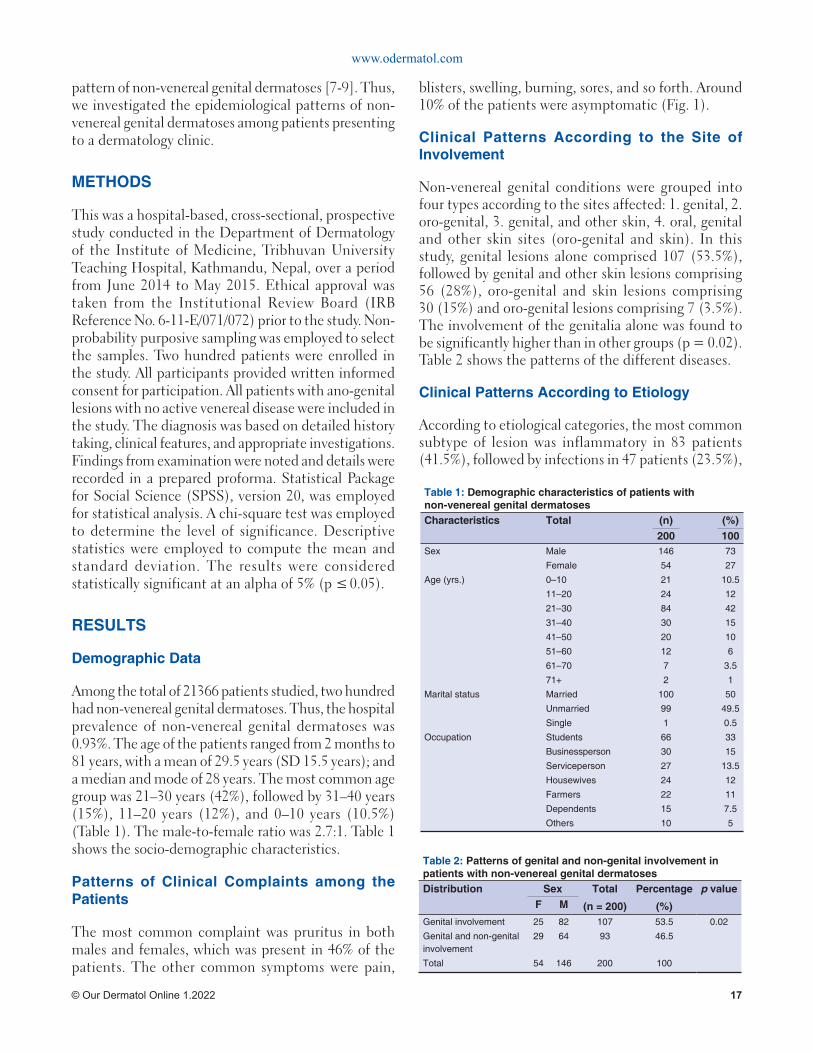
www.odermatol.com
© Our Dermatol Online 1.2022 17
pattern of non-venereal genital dermatoses [7-9]. Thus, we investigated the epidemiological patterns of non-venereal genital dermatoses among patients presenting to a dermatology clinic.
METHODS
This was a hospital-based, cross-sectional, prospective study conducted in the Department of Dermatology of the Institute of Medicine, Tribhuvan University Teaching Hospital, Kathmandu, Nepal, over a period from June 2014 to May 2015. Ethical approval was taken from the Institutional Review Board (IRB Reference No. 6-11-E/071/072) prior to the study. Non-probability purposive sampling was employed to select the samples. Two hundred patients were enrolled in the study. All participants provided written informed consent for participation. All patients with ano-genital lesions with no active venereal disease were included in the study. The diagnosis was based on detailed history taking, clinical features, and appropriate investigations. Findings from examination were noted and details were recorded in a prepared proforma. Statistical Package for Social Science (SPSS), version 20, was employed for statistical analysis. A chi-square test was employed to determine the level of significance. Descriptive statistics were employed to compute the mean and standard deviation. The results were considered statistically significant at an alpha of 5% (p ≤ 0.05).
RESULTS
Demographic Data
Among the total of 21366 patients studied, two hundred had non-venereal genital dermatoses. Thus, the hospital prevalence of non-venereal genital dermatoses was 0.93%. The age of the patients ranged from 2 months to 81 years, with a mean of 29.5 years (SD 15.5 years); and a median and mode of 28 years. The most common age group was 21–30 years (42%), followed by 31–40 years (15%), 11–20 years (12%), and 0–10 years (10.5%) (Table 1). The male-to-female ratio was 2.7:1. Table 1 shows the socio-demographic characteristics.
Patterns of Clinical Complaints among the Patients
The most common complaint was pruritus in both males and females, which was present in 46% of the patients. The other common symptoms were pain,
blisters, swelling, burning, sores, and so forth. Around 10% of the patients were asymptomatic (Fig. 1).
Clinical Patterns According to the Site of Involvement
Non-venereal genital conditions were grouped into four types according to the sites affected: 1. genital, 2. oro-genital, 3. genital, and other skin, 4. oral, genital and other skin sites (oro-genital and skin). In this study, genital lesions alone comprised 107 (53.5%), followed by genital and other skin lesions comprising 56 (28%), oro-genital and skin lesions comprising 30 (15%) and oro-genital lesions comprising 7 (3.5%). The involvement of the genitalia alone was found to be significantly higher than in other groups (p = 0.02). Table 2 shows the patterns of the different diseases.
Clinical Patterns According to Etiology
According to etiological categories, the most common subtype of lesion was inflammatory in 83 patients (41.5%), followed by infections in 47 patients (23.5%),
Table 1: Demographic characteristics of patients with non-venereal genital dermatosesCharacteristics Total (n) (%)
200 100Sex Male 146 73
Female 54 27
Age (yrs.) 0–10 21 10.5
11–20 24 12
21–30 84 42
31–40 30 15
41–50 20 10
51–60 12 6
61–70 7 3.5
71+ 2 1
Marital status Married 100 50
Unmarried 99 49.5
Single 1 0.5
Occupation Students 66 33
Businessperson 30 15
Serviceperson 27 13.5
Housewives 24 12
Farmers 22 11
Dependents 15 7.5
Others 10 5
Table 2: Patterns of genital and non-genital involvement in patients with non-venereal genital dermatosesDistribution Sex Total
(n = 200)
Percentage
(%)
p valueF M
Genital involvement 25 82 107 53.5 0.02
Genital and non-genital involvement
29 64 93 46.5
Total 54 146 200 100
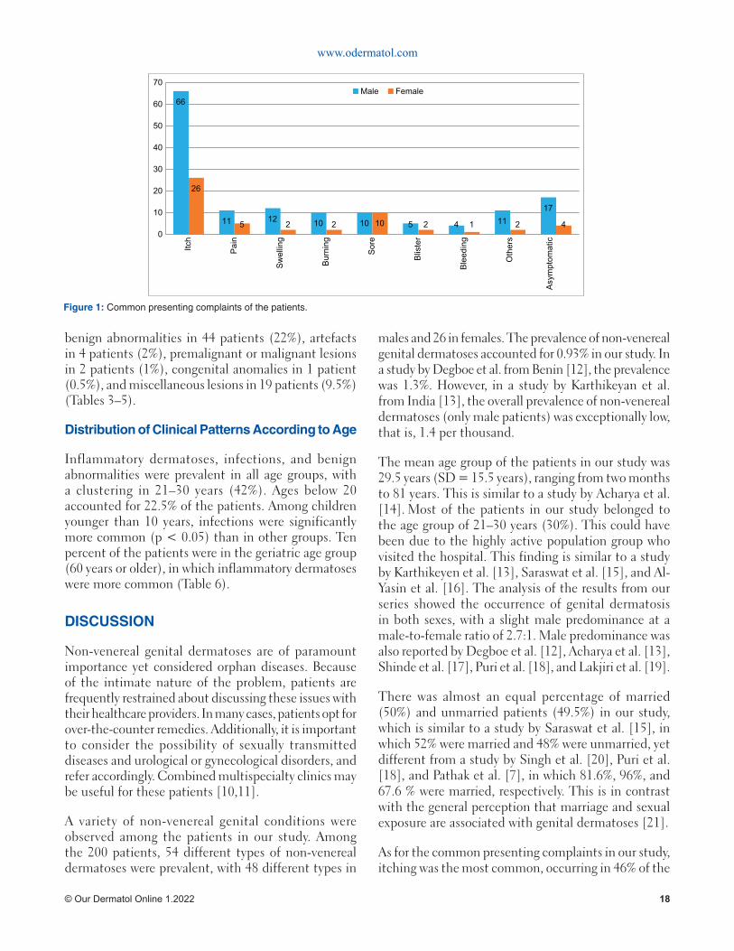
www.odermatol.com
© Our Dermatol Online 1.2022 18
benign abnormalities in 44 patients (22%), artefacts in 4 patients (2%), premalignant or malignant lesions in 2 patients (1%), congenital anomalies in 1 patient (0.5%), and miscellaneous lesions in 19 patients (9.5%) (Tables 3–5).
Distribution of Clinical Patterns According to Age
Inflammatory dermatoses, infections, and benign abnormalities were prevalent in all age groups, with a clustering in 21–30 years (42%). Ages below 20 accounted for 22.5% of the patients. Among children younger than 10 years, infections were significantly more common (p < 0.05) than in other groups. Ten percent of the patients were in the geriatric age group (60 years or older), in which inflammatory dermatoses were more common (Table 6).
DISCUSSION
Non-venereal genital dermatoses are of paramount importance yet considered orphan diseases. Because of the intimate nature of the problem, patients are frequently restrained about discussing these issues with their healthcare providers. In many cases, patients opt for over-the-counter remedies. Additionally, it is important to consider the possibility of sexually transmitted diseases and urological or gynecological disorders, and refer accordingly. Combined multispecialty clinics may be useful for these patients [10,11].
A variety of non-venereal genital conditions were observed among the patients in our study. Among the 200 patients, 54 different types of non-venereal dermatoses were prevalent, with 48 different types in
males and 26 in females. The prevalence of non-venereal genital dermatoses accounted for 0.93% in our study. In a study by Degboe et al. from Benin [12], the prevalence was 1.3%. However, in a study by Karthikeyan et al. from India [13], the overall prevalence of non-venereal dermatoses (only male patients) was exceptionally low, that is, 1.4 per thousand.
The mean age group of the patients in our study was 29.5 years (SD = 15.5 years), ranging from two months to 81 years. This is similar to a study by Acharya et al. [14]. Most of the patients in our study belonged to the age group of 21–30 years (30%). This could have been due to the highly active population group who visited the hospital. This finding is similar to a study by Karthikeyen et al. [13], Saraswat et al. [15], and Al-Yasin et al. [16]. The analysis of the results from our series showed the occurrence of genital dermatosis in both sexes, with a slight male predominance at a male-to-female ratio of 2.7:1. Male predominance was also reported by Degboe et al. [12], Acharya et al. [13], Shinde et al. [17], Puri et al. [18], and Lakjiri et al. [19].
There was almost an equal percentage of married (50%) and unmarried patients (49.5%) in our study, which is similar to a study by Saraswat et al. [15], in which 52% were married and 48% were unmarried, yet different from a study by Singh et al. [20], Puri et al. [18], and Pathak et al. [7], in which 81.6%, 96%, and 67.6 % were married, respectively. This is in contrast with the general perception that marriage and sexual exposure are associated with genital dermatoses [21].
As for the common presenting complaints in our study, itching was the most common, occurring in 46% of the
66
11 12 10 10 5 4 1117
26
5 2 2 10 2 1 2 40
10
20
30
40
50
60
70
Itch
Pain
Swelling
Burning
Sore
Blister
Bleeding
Others
Asymptomatic
Male Female
Figure 1: Common presenting complaints of the patients.
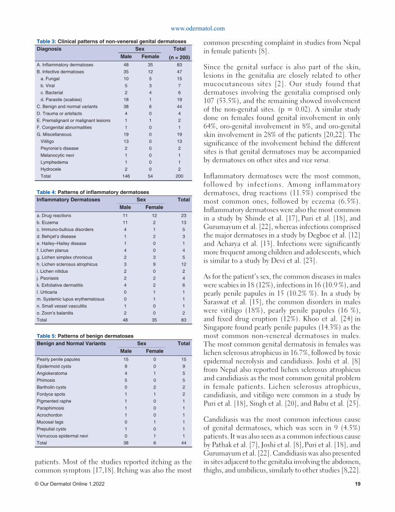
www.odermatol.com
© Our Dermatol Online 1.2022 19
common presenting complaint in studies from Nepal in female patients [8].
Since the genital surface is also part of the skin, lesions in the genitalia are closely related to other mucocutaneous sites [2]. Our study found that dermatoses involving the genitalia comprised only 107 (53.5%), and the remaining showed involvement of the non-genital sites. (p = 0.02). A similar study done on females found genital involvement in only 64%, oro-genital involvement in 8%, and oro-genital skin involvement in 28% of the patients [20,22]. The significance of the involvement behind the different sites is that genital dermatoses may be accompanied by dermatoses on other sites and vice versa.
Inflammatory dermatoses were the most common, followed by infections. Among inflammatory dermatoses, drug reactions (11.5%) comprised the most common ones, followed by eczema (6.5%). Inflammatory dermatoses were also the most common in a study by Shinde et al. [17], Puri et al. [18], and Gurumayum et al. [22], whereas infections comprised the major dermatoses in a study by Degboe et al. [12] and Acharya et al. [13]. Infections were significantly more frequent among children and adolescents, which is similar to a study by Devi et al. [23].
As for the patient’s sex, the common diseases in males were scabies in 18 (12%), infections in 16 (10.9 %), and pearly penile papules in 15 (10.2% %). In a study by Saraswat et al. [15], the common disorders in males were vitiligo (18%), pearly penile papules (16 %), and fixed drug eruption (12%). Khoo et al. [24] in Singapore found pearly penile papules (14.3%) as the most common non-venereal dermatoses in males. The most common genital dermatosis in females was lichen sclerosus atrophicus in 16.7%, followed by toxic epidermal necrolysis and candidiasis. Joshi et al. [8] from Nepal also reported lichen sclerosus atrophicus and candidiasis as the most common genital problem in female patients. Lichen sclerosus atrophicus, candidiasis, and vitiligo were common in a study by Puri et al. [18], Singh et al. [20], and Babu et al. [25].
Candidiasis was the most common infectious cause of genital dermatoses, which was seen in 9 (4.5%) patients. It was also seen as a common infectious cause by Pathak et al. [7], Joshi et al. [8], Puri et al. [18], and Gurumayum et al. [22]. Candidiasis was also presented in sites adjacent to the genitalia involving the abdomen, thighs, and umbilicus, similarly to other studies [8,22].
Table 4: Patterns of infl ammatory dermatosesInfl ammatory Dermatoses Sex Total
Male Female
a. Drug reactions 11 12 23
b. Eczema 11 2 13
c. Immuno-bullous disorders 4 1 5
d. Behçet’s disease 1 2 3
e. Hailey–Hailey disease 1 0 1
f. Lichen planus 4 0 4
g. Lichen simplex chronicus 2 3 5
h. Lichen sclerosus atrophicus 3 9 12
i. Lichen nitidus 2 0 2
j. Psoriasis 2 2 4
k. Exfoliative dermatitis 4 2 6
l. Urticaria 0 1 1
m. Systemic lupus erythematosus 0 1 1
n. Small vessel vasculitis 1 0 1
o. Zoon’s balanitis 2 0 2
Total 48 35 83
Table 3: Clinical patterns of non-venereal genital dermatosesDiagnosis Sex Total
(n = 200)Male Female
A. Infl ammatory dermatoses 48 35 83
B. Infective dermatoses 35 12 47
a. Fungal 10 5 15
b. Viral 5 3 7
c. Bacterial 2 4 6
d. Parasite (scabies) 18 1 19
C. Benign and normal variants 38 6 44
D. Trauma or artefacts 4 0 4
E. Premalignant or malignant lesions 1 1 2
F. Congenital abnormalities 1 0 1
G. Miscellaneous 19 0 19
Vitiligo 13 0 13
Peyronie’s disease 2 0 2
Melanocytic nevi 1 0 1
Lymphedema 1 0 1
Hydrocele 2 0 2
Total 146 54 200
Table 5: Patterns of benign dermatosesBenign and Normal Variants Sex Total
Male Female
Pearly penile papules 15 0 15
Epidermoid cysts 9 0 9
Angiokeratoma 4 1 5
Phimosis 5 0 5
Bartholin cysts 0 2 2
Fordyce spots 1 1 2
Pigmented raphe 1 0 1
Paraphimosis 1 0 1
Acrochordon 1 0 1
Mucosal tags 0 1 1
Preputial cysts 1 0 1
Verrucous epidermal nevi 0 1 1
Total 38 6 44
patients. Most of the studies reported itching as the common symptom [17,18]. Itching was also the most
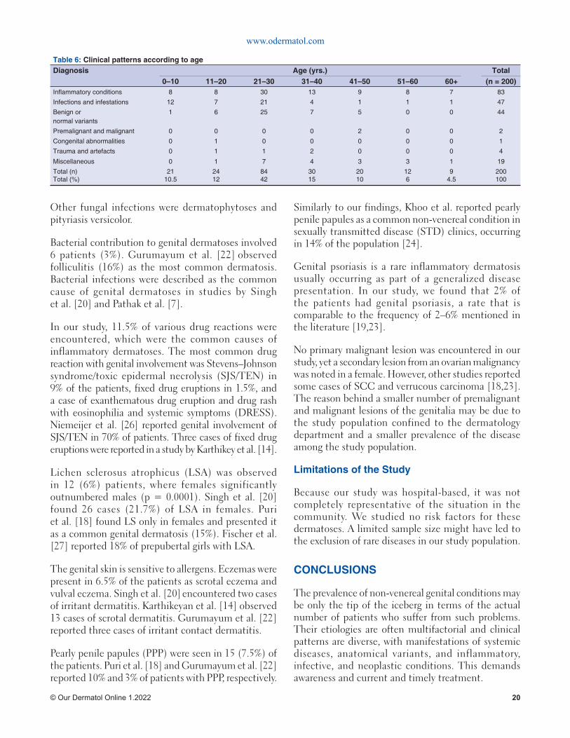
www.odermatol.com
© Our Dermatol Online 1.2022 20
Other fungal infections were dermatophytoses and pityriasis versicolor.
Bacterial contribution to genital dermatoses involved 6 patients (3%). Gurumayum et al. [22] observed folliculitis (16%) as the most common dermatosis. Bacterial infections were described as the common cause of genital dermatoses in studies by Singh et al. [20] and Pathak et al. [7].
In our study, 11.5% of various drug reactions were encountered, which were the common causes of inflammatory dermatoses. The most common drug reaction with genital involvement was Stevens–Johnson syndrome/toxic epidermal necrolysis (SJS/TEN) in 9% of the patients, fixed drug eruptions in 1.5%, and a case of exanthematous drug eruption and drug rash with eosinophilia and systemic symptoms (DRESS). Niemeijer et al. [26] reported genital involvement of SJS/TEN in 70% of patients. Three cases of fixed drug eruptions were reported in a study by Karthikey et al. [14].
Lichen sclerosus atrophicus (LSA) was observed in 12 (6%) patients, where females significantly outnumbered males (p = 0.0001). Singh et al. [20] found 26 cases (21.7%) of LSA in females. Puri et al. [18] found LS only in females and presented it as a common genital dermatosis (15%). Fischer et al. [27] reported 18% of prepubertal girls with LSA.
The genital skin is sensitive to allergens. Eczemas were present in 6.5% of the patients as scrotal eczema and vulval eczema. Singh et al. [20] encountered two cases of irritant dermatitis. Karthikeyan et al. [14] observed 13 cases of scrotal dermatitis. Gurumayum et al. [22] reported three cases of irritant contact dermatitis.
Pearly penile papules (PPP) were seen in 15 (7.5%) of the patients. Puri et al. [18] and Gurumayum et al. [22] reported 10% and 3% of patients with PPP, respectively.
Similarly to our findings, Khoo et al. reported pearly penile papules as a common non-venereal condition in sexually transmitted disease (STD) clinics, occurring in 14% of the population [24].
Genital psoriasis is a rare inflammatory dermatosis usually occurring as part of a generalized disease presentation. In our study, we found that 2% of the patients had genital psoriasis, a rate that is comparable to the frequency of 2–6% mentioned in the literature [19,23].
No primary malignant lesion was encountered in our study, yet a secondary lesion from an ovarian malignancy was noted in a female. However, other studies reported some cases of SCC and verrucous carcinoma [18,23]. The reason behind a smaller number of premalignant and malignant lesions of the genitalia may be due to the study population confined to the dermatology department and a smaller prevalence of the disease among the study population.
Limitations of the Study
Because our study was hospital-based, it was not completely representative of the situation in the community. We studied no risk factors for these dermatoses. A limited sample size might have led to the exclusion of rare diseases in our study population.
CONCLUSIONS
The prevalence of non-venereal genital conditions may be only the tip of the iceberg in terms of the actual number of patients who suffer from such problems. Their etiologies are often multifactorial and clinical patterns are diverse, with manifestations of systemic diseases, anatomical variants, and inflammatory, infective, and neoplastic conditions. This demands awareness and current and timely treatment.
Table 6: Clinical patterns according to ageDiagnosis Age (yrs.) Total
0–10 11–20 21–30 31–40 41–50 51–60 60+ (n = 200)Infl ammatory conditions 8 8 30 13 9 8 7 83
Infections and infestations 12 7 21 4 1 1 1 47
Benign ornormal variants
1 6 25 7 5 0 0 44
Premalignant and malignant 0 0 0 0 2 0 0 2
Congenital abnormalities 0 1 0 0 0 0 0 1
Trauma and artefacts 0 1 1 2 0 0 0 4
Miscellaneous 0 1 7 4 3 3 1 19
Total (n)Total (%)
2110.5
2412
8442
3015
2010
126
94.5
200100
www.odermatol.com
© Our Dermatol Online 1.2022 21
ACKNOWLEDGMENTS
The authors would like to thank all participants enrolled in their study and all faculty, resident, and staff members of the Department of Dermatology and Venereology, Institute of Medicine, for their cooperation in conducting the study.
Statement of Human and Animal Rights
All the procedures followed were in accordance with the ethical standards of the responsible committee on human experimentation (institutional and national) and with the 2008 revision of the Declaration of Helsinki of 1975.
Statement of Informed Consent
Informed consent for participation in this study was obtained from all patients.
REFERENCES
1. Khaitan BK. Non-venereal diseases of genitalia. In: Sharma VK, editor. Sexually Transmitted Diseases and AIDS. 1st edn. New Delhi: Viva books Pvt Ltd; 2003. pp. 413-421.
2. Griffi ths C, Barker J, Bleiker T, Chalmers D, Bunker CB, Porter W. The genital, perianal and umbilical regions. In: Griffi ths C, Barker J, Bleiker T, Chalmers D, eds. Rook’s Textbook of Dermatology.9th ed. Wiley-Blackwell, 2016.
3. Conforti C, Giuffrida R, Di Meo N, Longone M, Vichi S, Colli C, et al. Benign dermatoses of the male genital areas: A review of the literature. Dermatol Ther. 2020;33:e13355.
4. Sadownik LA, Koert E, Maher C, Smith KB. A qualitative exploration of women’s experiences of living with chronic vulvar dermatoses. J Sex Med. 2020;17:1740-50.
5. Ryan C, Sadlier M, De Vol E, Patel M, Lloyd AA, Day A, et al. Genital psoriasis is associated with signifi cant impairment in quality of life and sexual functioning. J Am Acad Dermatol. 2015;72:978-83.
6. Gabrielson AT, Le TV, Fontenot C, Usta M, Hellstrom WJG. Male genital dermatology: A primer for the sexual medicine physician. Sex Med Rev. 2019;7:71-83.
7. Pathak D, Agrawal S, Dhali TK. Prevalences of and risk factors for vulvar diseases in Nepal: A hospital-based study. Int J Dermatol. 2011;50:161-7.
8. Joshi S, Shrestha S, Joshi A. Clinico-epidemiological profi le of women with non-venereal vulval diseases: A hospital-based observational study. NJDVL. 2019;17:32-8.
9. Gyawalee M, Pokhrel D. Pattern of sexually transmitted infections and sexual behavior in patients with genital symptoms. NJDVL. 2016;12:20-7.
10. Ascott A, Chinthapalli S, Gibbon K. Unifying clinical care between specialties: A model for genital disease. J R Soc Med.
2017;110:177-82.11. Cheung ST, Gach JE, Lewis FM. A retrospective study of the
referral patterns to a vulval clinic: Highlighting educational needs in this subspeciality. J Obstet Gynaecol. 2006;26:435-7.
12. Degboe B, Atadokpede F, Adegbidi H, Saka B, Akpadjan F, d’Almeida C, et al. [Epidemiological and clinical characteristics of anogenital dermatoses in the department of dermatology-venereology in Cotonou, Benin]. Med Sante Trop. 2014;24:416-9.
13. Acharya KM, Ranpara H, Sakhia JJ, Kaur C. A study of 200 cases of genital lesions of non-venereal origin. Indian J Dermatol Venereol Leprol. 1998;64:68-70.
14. Karthikeyan K, Jaisankar TJ, Thappa DM. Non-venereal dermatoses in male genital region-prevalence and patterns in a referral centre in South India. Indian J Dermatol. 2001;46:18-22.
15. Saraswat PK, Garg A, Mishra D, Garg S. A study of pattern of nonvenereal genital dermatoses of male attending skin OPD at a tertiary care center. Indian J Sex Transm Dis AIDS. 2014;35:129-34.
16. Al-Yasin ZT. The frequency of symptomatic vulval disease: A gynecological’s perspective. MJBU. 2009;27:104-7.
17. Shinde G, Popere S. A clinical study of non-venereal genital dermatoses of adult in a tertiary care center. Int J Biomed Adv Res. 2017;8:168-73.
18. Puri N, Puri A. A study on non-venereal genital dermatoses in North India. Our Dermatol Online. 2013;4:304-7.
19. Lakjiri S, Meziane M, Elloudi S, Sy O, Nejjari C, Mernissi FZ. [Genital dermatoses: Epidemiological and clinical profi le]. Pan Afr Med J. 2014;18:240.
20. Singh N, Thappa DM, Jaisankar TJ, Habeebullah S. Pattern of non-venereal dermatoses of female external genitalia in South India. Dermatol Online J. 2008;14:1.
21. Vinay N, Ranugha PSS, Betkerur JB, Shastry V, Ashwini PK. Non-venereal genital dermatoses and their impact on quality of life: A cross-sectional study. Indian J Dermatol Venereol Leprol. 2021:1-6.
22. Gurumayum M, Shivakumar V, Okade R. Non-venereal female genital dermatoses: A clinical study. JMSCR. 2014;2:2864-73.
23. Devi VN, Balachandrudu P. Prevalence and patterns of genital dermatoses in children. Int J Sci Res. 2015;4:1833-9.
24. Khoo LS, Cheong WK. Common genital dermatoses in male patients attending a public sexually transmitted disease clinic in Singapore. Ann Acad Med Singapore. 1995;24:505-9.
25. Babu AR, Shivakumar V, Prasad AM. A clinical study of non-venereal genital dermatoses in women in a rural setup. Int J Med Public Health. 2020;10:29-33.
26. Niemeijer IC, van Praag MC, van Gemund N. Relevance and consequences of erythema multiforme, Stevens-Johnson syndrome and toxic epidermal necrolysis in gynecology. Arch Gynecol Obstet. 2009;280:851-4.
27. Fischer GO. Vulval disease in pre-pubertal girls. Australas J Dermatol. 2001;42:225-36.
Vikash Paudel, et al. This is an open-access article distributed under the terms of the Creative Commons Attribution License, which permits unrestricted use, distribution, and reproduction in any medium, provided the original author and source are credited.Source of Support: Nil, Confl ict of Interest: None declared.
Our Dermatology Online
© Our Dermatol Online 1.2022 22
Task shifting in dermatology: Are nurses prepared and Task shifting in dermatology: Are nurses prepared and willing?willing?Kavita Kavita1, Hitaishi Mehta2, Sandhya Ghai1, Aarti Garg1, Tarun Narang2
1National Institute of Nursing Education, Postgraduate Institute of Medical Education and Research, Chandigarh, India, 2Department of Dermatology, Venereology, and Leprology, Postgraduate Institute of Medical Education and Research, Chandigarh, India
Corresponding author: Tarun Narang, MD, MNAMS. E-mail: [email protected]
INTRODUCTION
Skin diseases constitute a significant burden worldwide. As per the 2017 study on the global burden of disease (GBD), these contributed 1.76% of the total global burden of disease measured in DALYs (disability-adjusted life years) [1]. Skin diseases are among the top ten causes of non-fatal disease burdens. According to 2017 GBD data, the years lived with disability (YLDs) from skin diseases worldwide were 41.6 million, which is more than with cardiovascular diseases (35.6 million) [2,3]. Skin diseases are among the top ten causes of non-fatal disease burdens in India, and the burden due to skin diseases has increased from 4.07 million in 1990 to 6.26 million in 2017 [4].
The high need for dermatological care in India poses a significant challenge to the healthcare delivery system, which already has a shortage of dermatologists. The population ratio of dermatologists is more skewed in low- and middle-income countries than in high-income countries. There are 3.2 dermatologists per 100,000 individuals in many states of the U.S. [5], compared to less than one dermatologist per 100,000 individuals in India [6]. Task shifting may be one of the solutions to increase access to dermatological care. Task shifting means transferring clinical tasks from physicians to trained non-physician health workers. Although task shifting is being done to a limited extent in dermatology, its need and effectiveness are evident from the literature [7-10].
ABSTRACT
Background: The high burden of skin diseases and the shortage of dermatologists are significant challenges in providing care to millions of people with skin diseases. Task shifting to nurses is a viable option for the delivery of dermatologic care in resource-poor settings. Satisfactory knowledge and a positive attitude are crucial for nurses to undertake the task of managing common skin diseases. This study aimed to investigate the knowledge of registered nurses and their attitude toward common skin conditions. Methods: In this descriptive, cross-sectional study, a total of 187 nurses were included from a nurses training institute by total enumeration sampling. A knowledge questionnaire and five-point Likert type-attitude scale were developed, validated, and employed to collect data. Written informed consent was obtained from the participants after approval from the institute ethics committee. Results: The mean of the knowledge scores were 10.7 ± 2.2. Nearly two thirds (62%) of the subjects demonstrated a low level of knowledge, while the remaining 38% showed a moderate level. There was no participant in the high-knowledge category. A majority of the nurses demonstrated a favorable attitude toward learning and undertaking the task of managing common skin conditions. Conclusion: We observed a low level of knowledge on diagnosing and managing common skin conditions. We recommend incorporating the relevant concepts of common skin conditions in the nursing curriculum with an emphasis on continuing education.
Keywords: Nurses; Common skin conditions; Knowledge; Attitude; Dermatology training; Task shifting
How to cite this article: Kavita, Mehta H, Ghai S, Garg A, Narang T. Task shifting in dermatology: Are nurses prepared and willing?. Our Dermatol Online. 2022;13(1):22-27.Submission: 22.06.2021; Acceptance: 30.11.2021DOI: 10.7241/ourd.20221.4
Original Article
www.odermatol.com
© Our Dermatol Online 1.2022 23
Task shifting may involve various categories of non-physician health workers, but nurses are the ideal choice. In the developed world, nurses work successfully as dermatological nurses in both independent and dependent roles [11,12]. India’s national leprosy elimination program is an excellent example of task shifting [13]. Mid-level health care providers (MLHP) under the Ayushman Bharat scheme are also nurses who are being trained to work independently in health and wellness centers [14].
However, nurses’ educational level, competence, and willingness are crucial prerequisites for successful task shifting. To the best of our knowledge, there have been no studies assessing nurses from India for their knowledge and attitude regarding skin conditions. For this reason, the present study was undertaken to evaluate the knowledge and attitude of registered nurses regarding the diagnosis and management of common skin diseases. The findings of this study will help to make recommendations for effective task shifting and curriculum changes.
METHODOLOGY
A descriptive, cross-sectional design was adopted for this study. Nurses were recruited from a nurses’ training institute in Northern India. All registered nurses (n = 187) pursuing higher education (B.Sc. nursing (post-basic)/M.Sc. nursing) were recruited in the study.
Study Instruments
A knowledge questionnaire and a five-point Likert scale were employed to assess the nurses’ knowledge about and attitudes to common skin conditions. Tools were developed by reviewing the literature and consulting experts in the field of dermatology and nursing. The validation of the tools was performed by experts in the field of dermatology and nursing. Their suggestions were incorporated in the final version of the tool. The knowledge questionnaire consisted of two sections. Section I included the socio-demographic and professional profiles of the participants. Section II consisted of multiple-choice questions related to the diagnosis and management of common skin diseases. The total score ranged from 0 to 20. A higher score represented a higher level of knowledge. Knowledge scores were graded into three levels. Scores between 16 and 20 (> 80%) were
classified as a high level of knowledge, while scores between 10 and 15 (79–50%) and <10 (<50%) were graded as a moderate and low level of knowledge, respectively.
A five-point Likert scale (strongly disagree, disagree, neutral, agree, strongly agree) was constructed to assess the attitude of nurses regarding the diagnosis and management of common skin disorders. There were a total of twelve statements with scores ranging from 12 to 60. A higher score indicated a more favorable attitude. A score above the median was considered a positive attitude and below the median a negative attitude.
Data entry and analysis were performed using the software SPSS, version 20. Descriptive statistics were employed to analyze the data, and the results were presented as frequencies, percentages, means, and standard deviations. The Chi-square test was employed for the analysis of categorical variables. The relationship between knowledge and attitude was established by bivariate correlational analysis.
RESULTS
A total of 187 nurses completed the questionnaire and attitude scale. The mean age of the participants was 25.6 ± 4.89 years, and the majority (84.5%) were females. As per the professional qualification, 40 (21.4%) participants had done B.Sc. nursing, and the rest had completed their diplomas in general nursing and midwifery. Regarding work experience, 84 (44.9%) participants had no work experience, and 19 (10.2%) had an experience of more than ten years (Table 1). Only three participants (1.6%) had attended training related to the prevention and management of skin conditions.
Respondents’ Knowledge on Common Skin Diseases
Most of the participants (n = 142; 75.9%) knew that leprosy is a communicable disease. However, only 60 (32.1%) were aware that leprosy classification is based on the number of skin lesions. Nearly one fourth of the participants (28.3%) knew about the cardinal features for the diagnosis of leprosy. Table 2 summarizes details regarding knowledge on the etiology of common skin disorders.
A per the questions about the basic understanding of dermatology, 88 (47.1%) and 128 (68.4%) of the
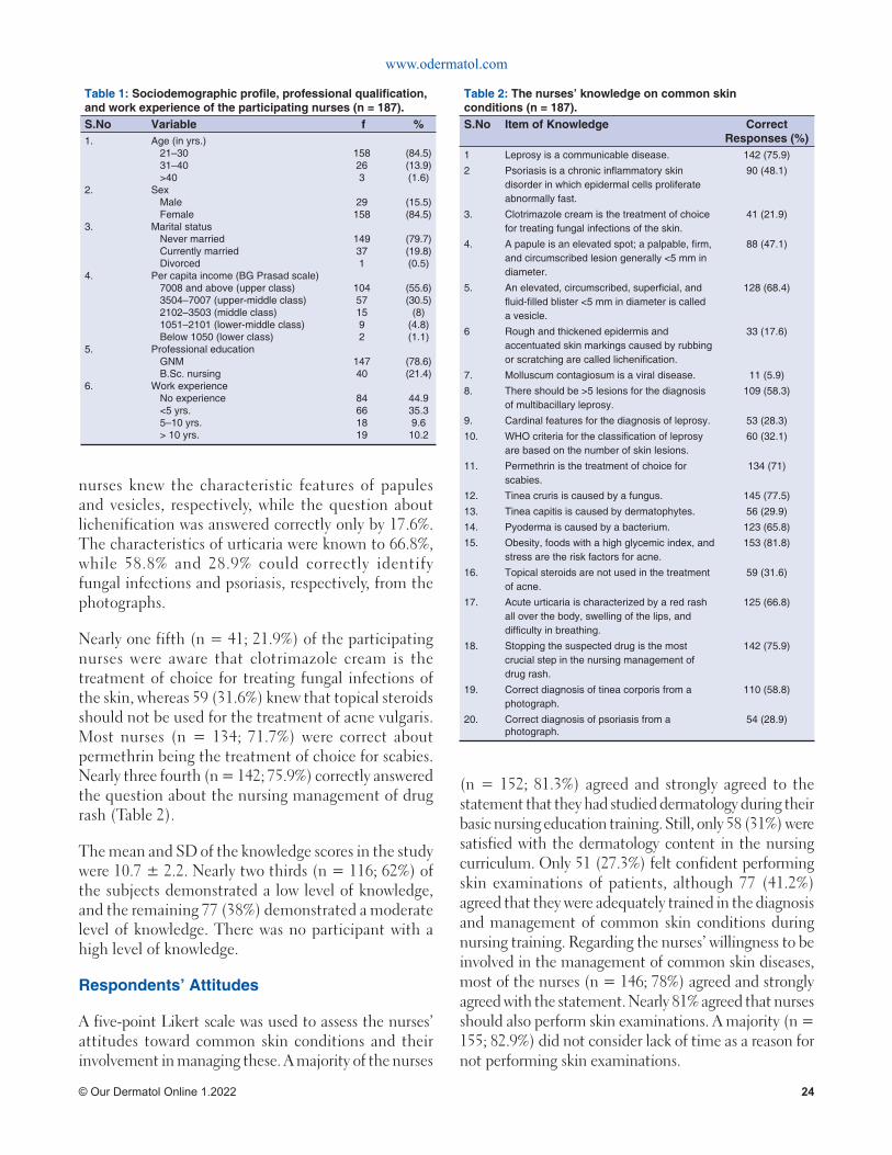
www.odermatol.com
© Our Dermatol Online 1.2022 24
Table 1: Sociodemographic profi le, professional qualifi cation, and work experience of the participating nurses (n = 187).S.No Variable f %1.
2.
3.
4.
5.
6.
Age (in yrs.)21–3031–40>40
SexMaleFemale
Marital statusNever marriedCurrently marriedDivorced
Per capita income (BG Prasad scale)7008 and above (upper class)3504–7007 (upper-middle class)2102–3503 (middle class)1051–2101 (lower-middle class)Below 1050 (lower class)
Professional educationGNMB.Sc. nursing
Work experienceNo experience<5 yrs.5–10 yrs.> 10 yrs.
158263
29158
149371
104571592
14740
84661819
(84.5)(13.9)(1.6)
(15.5)(84.5)
(79.7)(19.8)(0.5)
(55.6)(30.5)
(8)(4.8)(1.1)
(78.6)(21.4)
44.935.39.6
10.2
Table 2: The nurses’ knowledge on common skin conditions (n = 187).S.No Item of Knowledge Correct
Responses (%)1 Leprosy is a communicable disease. 142 (75.9)
2 Psoriasis is a chronic infl ammatory skin disorder in which epidermal cells proliferate abnormally fast.
90 (48.1)
3. Clotrimazole cream is the treatment of choice for treating fungal infections of the skin.
41 (21.9)
4. A papule is an elevated spot; a palpable, fi rm, and circumscribed lesion generally <5 mm in diameter.
88 (47.1)
5. An elevated, circumscribed, superfi cial, and fl uid-fi lled blister <5 mm in diameter is called a vesicle.
128 (68.4)
6 Rough and thickened epidermis and accentuated skin markings caused by rubbing or scratching are called lichenifi cation.
33 (17.6)
7. Molluscum contagiosum is a viral disease. 11 (5.9)
8. There should be >5 lesions for the diagnosis of multibacillary leprosy.
109 (58.3)
9. Cardinal features for the diagnosis of leprosy. 53 (28.3)
10. WHO criteria for the classifi cation of leprosy are based on the number of skin lesions.
60 (32.1)
11. Permethrin is the treatment of choice for scabies.
134 (71)
12. Tinea cruris is caused by a fungus. 145 (77.5)
13. Tinea capitis is caused by dermatophytes. 56 (29.9)
14. Pyoderma is caused by a bacterium. 123 (65.8)
15. Obesity, foods with a high glycemic index, and stress are the risk factors for acne.
153 (81.8)
16. Topical steroids are not used in the treatment of acne.
59 (31.6)
17. Acute urticaria is characterized by a red rash all over the body, swelling of the lips, and diffi culty in breathing.
125 (66.8)
18. Stopping the suspected drug is the most crucial step in the nursing management of drug rash.
142 (75.9)
19. Correct diagnosis of tinea corporis from a photograph.
110 (58.8)
20. Correct diagnosis of psoriasis from a photograph.
54 (28.9)
nurses knew the characteristic features of papules and vesicles, respectively, while the question about lichenification was answered correctly only by 17.6%. The characteristics of urticaria were known to 66.8%, while 58.8% and 28.9% could correctly identify fungal infections and psoriasis, respectively, from the photographs.
Nearly one fifth (n = 41; 21.9%) of the participating nurses were aware that clotrimazole cream is the treatment of choice for treating fungal infections of the skin, whereas 59 (31.6%) knew that topical steroids should not be used for the treatment of acne vulgaris. Most nurses (n = 134; 71.7%) were correct about permethrin being the treatment of choice for scabies. Nearly three fourth (n = 142; 75.9%) correctly answered the question about the nursing management of drug rash (Table 2).
The mean and SD of the knowledge scores in the study were 10.7 ± 2.2. Nearly two thirds (n = 116; 62%) of the subjects demonstrated a low level of knowledge, and the remaining 77 (38%) demonstrated a moderate level of knowledge. There was no participant with a high level of knowledge.
Respondents’ Attitudes
A five-point Likert scale was used to assess the nurses’ attitudes toward common skin conditions and their involvement in managing these. A majority of the nurses
(n = 152; 81.3%) agreed and strongly agreed to the statement that they had studied dermatology during their basic nursing education training. Still, only 58 (31%) were satisfied with the dermatology content in the nursing curriculum. Only 51 (27.3%) felt confident performing skin examinations of patients, although 77 (41.2%) agreed that they were adequately trained in the diagnosis and management of common skin conditions during nursing training. Regarding the nurses’ willingness to be involved in the management of common skin diseases, most of the nurses (n = 146; 78%) agreed and strongly agreed with the statement. Nearly 81% agreed that nurses should also perform skin examinations. A majority (n = 155; 82.9%) did not consider lack of time as a reason for not performing skin examinations.
www.odermatol.com
© Our Dermatol Online 1.2022 25
Nearly half of the nurses (n = 93; 49.7%) disagreed and strongly disagreed that most skin diseases are communicable. Half (50.3%) felt confident in taking care of patients with any skin condition, whereas 65.8% disagreed that they were afraid of taking care of patients with skin disorders as they are contagious. Only 51 (27.3%) agreed that they were confident in diagnosing common skin conditions. A majority of the subjects (85.6%) agreed and strongly agreed to the question about their willingness to learn more about dermatology and to attend continuing education courses and lectures for the diagnosis and management of common skin conditions.
The median attitude score of the participants was 43 (IQR: 40–46). Most of the nurses (76.2%) scored above the median, demonstrating their positive attitude and willingness to undertake the task.
The association of knowledge and attitude scores with selected variables was assessed with a chi-square test. The results showed no significant association of knowledge scores with age, sex, professional qualification, and years of experience. However, there was a positive association between knowledge scores and attitude.
DISCUSSION
Skin diseases are associated with significant morbidity and the psychosocial and emotional issues in individuals suffering from skin conditions are comparable to that of arthritis, back pain, diabetes mellitus, epilepsy, cancer, and even asthma [15,16]. Skin diseases are highly common in rural and urban areas, yet there is a shortage of well-trained dermatologists who may address the needs of these problems [17]. Most dermatologists work in urban areas, whereas almost 70% of India’s population lives in rural areas [18]. Hence, task shifting is the need of the hour for addressing this supply and demand imbalance in skin disorders, and nurses are the ideal choice as they are one of the vital health care providers in any health setup [11]. Adequate knowledge and a favorable attitude are prerequisites for effective and successful task shifting in any specialty. The current study was conducted in a nurses’ training institute in Northern India. It was aimed to generate useful outputs that may support future actions to improve the knowledge and attitude of nurses involved in dermatological care. Although there exist numerous skin disorders, we selected the
most common skin conditions prevalent in India to assess the participants’ knowledge [2,19].
A study by Kouotou et al. assessed the knowledge, attitudes, and practices of medical personnel regarding atopic dermatitis. Twenty-two percent of the participants were nurses, and 45% of them showed a moderate level of knowledge, with none in the good level of knowledge category [20]. Although we attempted to study knowledge on dermatology and not a specific disease, our study’s findings agree with the findings of the above research, as most of our study participants were also in the low to moderate knowledge category and none were in the good knowledge category.
Dermatological conditions are considered difficult to diagnose and manage, even by primary care physicians and family physicians. In a cross-sectional survey on the primary care physician’s ability to recognize common dermatoses, it was observed that the mean score on a photograph quiz was 4.1/10, and 70.5% of the participants rated their ability to diagnose and manage skin disorders as average, on a five-point Likert scale. The authors believed that primary care physicians had poor knowledge of skin disorders, and there is a need for more training in the diagnosis and management of common dermatological conditions [21]. Federman et al. also concluded that family care physicians cannot diagnose and manage dermatological conditions [22]. Similar findings were revealed in other studies [23,24]. Another study, involving 400 health workers from Mali, revealed inadequate knowledge on skin conditions. Knowledge of health workers on the typical cases of pyoderma, scabies, tinea capitis, and hypochromic patches was assessed by showing pictures on a PowerPoint presentation [25]. The authors reported that 19% of the subjects showed correct knowledge on the treatment of scabies. We observed that most of the nurses (71.7%) knew that permethrin is the treatment of choice for scabies, and 58.8% could correctly diagnose fungal infections. However, in a study from Mali, only 6% of health workers could diagnose mycosis. The difference might have been due to various factors, such as different methodologies and data collection instruments and the basic qualification and knowledge of the healthcare workers.
Our study demonstrated a positive attitude and willingness of the nurses to learn and be involved in the management of skin conditions (76% of the participants). Similar results were reported in a study from Sub-Saharan
www.odermatol.com
© Our Dermatol Online 1.2022 26
Africa on task shifting for the diagnosis of Kaposi’s sarcoma. In this study, physicians, clinical officers, nurses, and technicians were trained by a dermatologist in doing skin punch biopsy. Although initially targeted at physicians, the proportion of skin biopsies done by nurses (62%) were more as compared to physicians (15%), clinical officers (12%), and technicians (11%), which suggests nurses’ willingness to undertake this task [26].
Although most of the participants in our study agreed to have studied dermatology in their curriculum, only 27.3% said that they felt confident in performing skin evaluations, and 69% of the study subjects believed that the dermatology content of their curricula was not enough and should be enhanced with the addition of practical training. Earlier studies showed that training healthcare workers and physicians in the care of skin diseases may be the key to improving knowledge and patient care. A study from Mali showed a marked improvement in the management of skin diseases in primary health care after a single day of training of the healthcare workers [24,25].
The strength of the current study is that it is probably the first study assessing nurses’ knowledge and attitudes regarding common skin conditions in India. However, the study also had certain limitations as it was conducted in a single nursing training institute. Therefore, the nurses’ knowledge and attitudes may not provide a true picture for all nurses in India. A small sample size also limited the generalizability of the study results. Further studies are needed for a more detailed insight into the assessment of the knowledge and attitudes of nursing students and practicing nurses regarding skin conditions and their management, which will help to formulate future educational content in nursing studies.
CONCLUSION
Nurses demonstrated a low level of knowledge on the diagnosis and management of common skin conditions, but a majority showed a positive attitude and willingness to learn and care for patients with skin diseases. It is recommended that relevant concepts related to skin conditions should be integrated into nursing curricula, and there should be more emphasis on continuing education.
Statement of Human and Animal Rights
All the procedures followed were in accordance with the ethical standards of the responsible committee on human experimentation
(institutional and national) and with the 2008 revision of the Declaration of Helsinki of 1975.
Statement of Informed Consent
Informed consent for participation in this study was obtained from all participants.
REFERENCES
1. GBD 2017 DALYs and HALE Collaborators. Global, regional, and national disability-adjusted life years (DALYs) for 359 diseases and injuries and healthy life expectancy (HALE) for 195 countries and territories, 1990–2017: A systematic analysis for the Global Burden of Disease Study 2017. Lancet. 2018;392:1859-922.
2. GBD 2017 Disease and Injury Incidence and Prevalence Collaborators. Global, regional, and national incidence, prevalence, and years lived with disability for 354 diseases and injuries for 195 countries and territories, 1990–2017: A systematic analysis for the Global Burden of Disease Study 2017. Lancet. 2018;392:1789-858.
3. Global burden of disease study 2017. GBD Compare. Available at https://vizhub.healthdata.org/gbd-compare/ [Accessed on 22.02.2020]
4. Global burden of disease study 2017. GBD India Compare. Available at https://vizhub.healthdata.org/gbd-compare/india [Accessed on 22.02.2020]
5. Resneck J Jr, Kimball AB. The dermatology workforce shortage. J Am Acad Dermatol. 2004;50:50-4.
6. IADVL news. 2020;16:9. https://www.iadvl.org/newsletter.php (last accessed on 06 June 2020)
7. Hay R, Estrada R, Grossmann H. Managing skin disease in resource-poor environments: The role of community-oriented training and control programs. Int J Dermatol. 2011;50:558-63.
8. Andrews RM, Kearns T, Connors C, Parker C, Carville K, Currie BJ, et al. A regional initiative to reduce skin infections amongst aboriginal children living in remote communities of the Northern Territory, Australia. PLoS Negl Trop Dis. 2009;3:e554.
9. Brown DN, Langan SM, Freeman EE. Task shifting in dermatology:a call to action. JAMA Dermatol. 2017;153:1179-80.
10. Chang AY, Kiprono SK, Maurer TA. Providing dermatological care in resource-limited settings: Barriers and potential solutions. Br J Dermatol. 2017;177:247-8.
11. Kavita, Narang T, Dogra S. Task shifting in dermatology: Nurses’ role. Indian J Dermatol Venereol Leprol. 2021;87:323-5.
12. Lawton S. The specialist dermatology nurse: Providing expert care to patients. Br J Nurs. 2020;29:136-8.
13. Shukla LK, Patel RN, Patel SV, Baxi RK. Evaluation of the effect of block level awareness campaign on performance indicators of national leprosyelimination program in Vadodara district, Gujarat, India. Indian J Dermatol Venereol Leprol. 2015;81:257-62.
14. Moola S, Bhaumik S, Nambiar D. Mid-level health providers (MLHPs) for primary healthcare. New Delhi: The George Institute for Global Health, India. 2019.
15. World Health Organization: Epidemiology and Management of Common Skin Diseasesin children in Developing Countries Geneva, World Health Organization. 2005. (https://www.who.int/maternal_child_adolescent/documents/fch_cah_05_12/en/)
16. Dalgard FJ, Gieler U, Tomas-Aragones L, Lien L, Poot F, Jemec GBE, et al. The psychological burden of skin diseases: A cross-sectional multicenter study among dermatological out-patients in 13 European countries. J Invest Dermatol. 2015;135:984-91.
17. Seth D, Cheldize K, Brown D, Freeman EF. Global burden of skin disease: Inequalities and innovations. Curr Dermatol Rep. 2017;6:204-10.
18. The World Bank. Rural population (% of total population) - India.
www.odermatol.com
© Our Dermatol Online 1.2022 27
Available from:https://data.worldbank.org/indicator/SP.RUR.TOTL.ZS?locations=IN (last accessed on 28-07-21).
19. Rao PN, Suneetha S. Current situation of leprosy in india and its future implications. Indian Dermatol Online J. 2018;9:83-9.
20. Kouotou EA, Nansseu JR, Ngangue Engome AD, Tatah SA, Zoung-Kanyi Bissek AC. Knowledge, attitudes and practices of the medical personnel regarding atopic dermatitis in Yaoundé, Cameroon. BMC Dermatol. 2017;17:1.
21. Thakkar SH, Chavda PD, Mehta KG. Do primary care physicians require training in core clinical dermatology? A cross-sectional needs assessment study from Western India. Indian J Dermatol Venereol Leprol. 2019;85:380-7.
22. Federman DG, Kirsner RS. The abilities of primary care physicians in dermatology: Implications for quality of care. Am J Manag Care. 1997;3:1487-92.
23. Nawaz S, Tapley A, Davey AR, van Driel ML, Fielding A, Holliday EG, et al. Management of a chronic skin disease in primary care: An analysis of early-career general practitioners’ consultations involving psoriasis. Dermatol Pract Concept. 2021;11:e2021055.
24. Mohan GC, Molina GE, Stavert R. Store and forward teledermatology improves dermatology knowledge among referring primary care providers: A survey-based cohort study. J Am Acad Dermatol. 2018;79:960-1.
25. Mahe A, Faye O, N’Diaye HT, Konaré HD, Coulibaly I, Kéita S, et al. Integration of basic dermatological care into primary health care services in Mali. Bull World Health Organ. 2005;83:935-41.
26. Laker-Oketta M, Wenger M, Semeere A, Castelnuovo B, Lukande R, Asirwa FC, et al. Task shifting and skin punch for the histologic diagnosis of Kaposi’s sarcoma in sub-Saharan Africa: A public health solution to a public health problem. Oncology. 2015;89:60-5.
Copyright by Kavita Kavita, et al. This is an open-access article distributed under the terms of the Creative Commons Attribution License, which permits unrestricted use, distribution, and reproduction in any medium, provided the original author and source are credited.Source of Support: Nil. Confl ict of Interest: None declared.
© Our Dermatol Online 1.2022 28
Our Dermatology Online
How to cite this article: Bukhari IA. Pulse dye laser therapy and superfi cial cryotherapy as a novel combination treatment for hypertrophic scars and keloids. Our Dermatol Online. 2022;13(1):28-31.Submission: 24.08.2021; Acceptance: 09.11.2021DOI: 10.7241/ourd.20221.5
Pulse dye laser therapy and superfi cial cryotherapy as Pulse dye laser therapy and superfi cial cryotherapy as a novel combination treatment for hypertrophic scars a novel combination treatment for hypertrophic scars and keloidsand keloidsIqbal A. Bukhari1,2
1Consultant Dermatologist, Dermatology Department, Dr. Sulaiman AlHabib Hospital, Alkhobar, Kingdom of Saudi Arabia. 2Professor, Dermatology Department, College of Medicine, Imam Abdulrahman Bin Faisal University (IAU), Dammam, Kingdom of Saudi Arabia
Corresponding author: Prof. Iqbal A. Bukhari, MD, E-mail: [email protected], [email protected]
INTRODUCTION
Hypertrophic scars are benign fibrotic skin lesions caused by defects in regulating fibrous tissue cellularity during wound-healing limited to the area of trauma. While keloids are overgrowths of fibrous tissue outside the original boundaries of trauma and occur secondary to defective wound healing, they may occur spontaneously without previous trauma. Both are abnormal responses to trauma followed by excessive wound tension, inducing fibroblast proliferation and overproduction of dense collagen and
glycosaminoglycans [1]. The exact pathogenesis of both conditions has not yet been elucidated. Hypertrophic scars and keloids often have functional, aesthetic, and psychosocial impacts on patients, as highlighted by quality-of-life studies. Numerous variables affect the severity of scarring, including the size and depth of the wound, blood supply to the area, the thickness and color of the skin, and the direction of the scar [2].
Numerous treatment options are available with limited efficacy, including surgical excision, intralesional or topical corticosteroids, other intralesional therapies —
Brief Report
ABSTRACT
Background: Hypertrophic scars are benign and fibrotic skin lesions caused by defects in the regulation of cellularity during the wound-healingprocess, in which there is higher collagen production and less degradation. Genetic predisposing factors and different skin injuries may play a role in developing these types of lesions. On the other hand, keloids are overgrowths of fibrous tissue outside the original boundaries of trauma, yet these may also occur spontaneously. There are numerous treatment options for both conditions, including silicone gel sheeting, pressure therapy, intralesional triamcinolone acetonide, radiation, laser therapy, cryosurgery, interferon, 5-fluorouracil, and surgical excision as well as a multitude of extracts and topical agents. Objective: The objective was to evaluate the effectiveness of pulse dye laser (PDL) therapy and superficial cryotherapy as a combination treatment for hypertrophic scars and keloids. Method: Four Arabic female patients were seen at the outpatient clinic of the Department of Dermatology at the King Fahd Hospital of the University in Khobar, Saudi Arabia. The patients had keloids and hypertrophic scars. Treatment with cryotherapy every week for three weeks followed by one session of pulsed dye laser was administered rotationally for three to six months until the lesions displayed remarkable physical improvement or complete resolution. Results: All patients experienced significant improvement, showing a reduction in the size, erythema, pliability, and pruritus. None of the hypertrophic scars or keloids deteriorated during the one year of treatment. No complications were noted during the treatment period. Conclusion: Sequential PDL therapy combined with superficial cryotherapy may be an option for treating hypertrophic scars and keloids.
Keywords: Hypertrophic scars; Keloids; Pulse dye laser; Cryotherapy
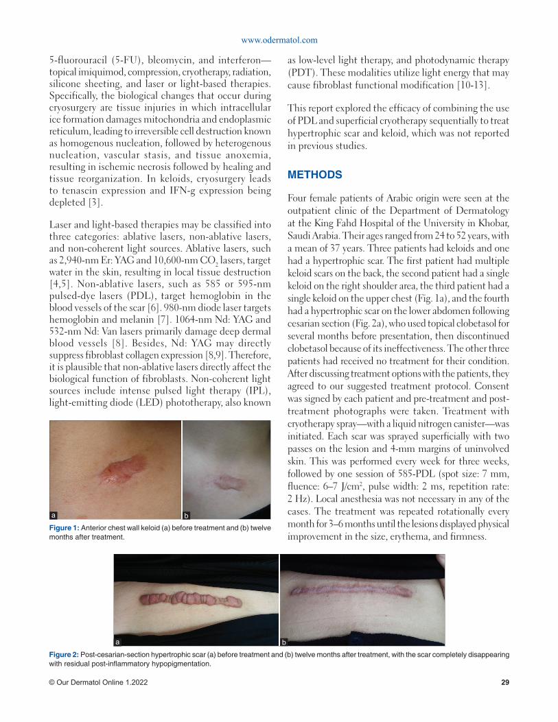
Figure 1: Anterior chest wall keloid (a) before treatment and (b) twelve months after treatment.
Figure 2: Post-cesarian-section hypertrophic scar (a) before treatment and (b) twelve months after treatment, with the scar completely disappearing with residual post-infl ammatory hypopigmentation.
ba
www.odermatol.com
© Our Dermatol Online 1.2022 29
5-fluorouracil (5-FU), bleomycin, and interferon—topical imiquimod, compression, cryotherapy, radiation, silicone sheeting, and laser or light-based therapies. Specifically, the biological changes that occur during cryosurgery are tissue injuries in which intracellular ice formation damages mitochondria and endoplasmic reticulum, leading to irreversible cell destruction known as homogenous nucleation, followed by heterogenous nucleation, vascular stasis, and tissue anoxemia, resulting in ischemic necrosis followed by healing and tissue reorganization. In keloids, cryosurgery leads to tenascin expression and IFN-g expression being depleted [3].
Laser and light-based therapies may be classified into three categories: ablative lasers, non-ablative lasers, and non-coherent light sources. Ablative lasers, such as 2,940-nm Er: YAG and 10,600-nm CO2 lasers, target water in the skin, resulting in local tissue destruction [4,5]. Non-ablative lasers, such as 585 or 595-nm pulsed-dye lasers (PDL), target hemoglobin in the blood vessels of the scar [6]. 980-nm diode laser targets hemoglobin and melanin [7]. 1064-nm Nd: YAG and 532-nm Nd: Van lasers primarily damage deep dermal blood vessels [8]. Besides, Nd: YAG may directly suppress fibroblast collagen expression [8,9]. Therefore, it is plausible that non-ablative lasers directly affect the biological function of fibroblasts. Non-coherent light sources include intense pulsed light therapy (IPL), light-emitting diode (LED) phototherapy, also known
as low-level light therapy, and photodynamic therapy (PDT). These modalities utilize light energy that may cause fibroblast functional modification [10-13].
This report explored the efficacy of combining the use of PDL and superficial cryotherapy sequentially to treat hypertrophic scar and keloid, which was not reported in previous studies.
METHODS
Four female patients of Arabic origin were seen at the outpatient clinic of the Department of Dermatology at the King Fahd Hospital of the University in Khobar, Saudi Arabia. Their ages ranged from 24 to 52 years, with a mean of 37 years. Three patients had keloids and one had a hypertrophic scar. The first patient had multiple keloid scars on the back, the second patient had a single keloid on the right shoulder area, the third patient had a single keloid on the upper chest (Fig. 1a), and the fourth had a hypertrophic scar on the lower abdomen following cesarian section (Fig. 2a), who used topical clobetasol for several months before presentation, then discontinued clobetasol because of its ineffectiveness. The other three patients had received no treatment for their condition. After discussing treatment options with the patients, they agreed to our suggested treatment protocol. Consent was signed by each patient and pre-treatment and post-treatment photographs were taken. Treatment with cryotherapy spray—with a liquid nitrogen canister—was initiated. Each scar was sprayed superficially with two passes on the lesion and 4-mm margins of uninvolved skin. This was performed every week for three weeks, followed by one session of 585-PDL (spot size: 7 mm, fluence: 6–7 J/cm2, pulse width: 2 ms, repetition rate: 2 Hz). Local anesthesia was not necessary in any of the cases. The treatment was repeated rotationally every month for 3–6 months until the lesions displayed physical improvement in the size, erythema, and firmness.
ba
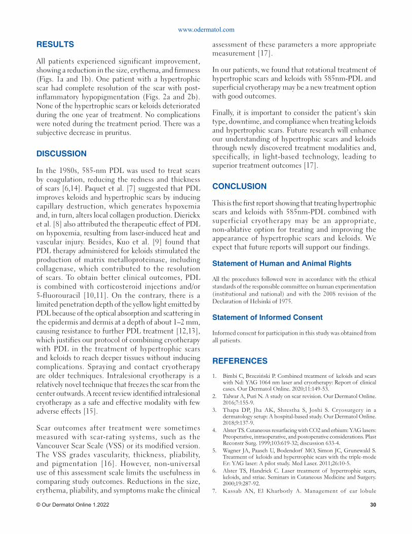
www.odermatol.com
© Our Dermatol Online 1.2022 30
RESULTS
All patients experienced significant improvement, showing a reduction in the size, erythema, and firmness (Figs. 1a and 1b). One patient with a hypertrophic scar had complete resolution of the scar with post-inflammatory hypopigmentation (Figs. 2a and 2b). None of the hypertrophic scars or keloids deteriorated during the one year of treatment. No complications were noted during the treatment period. There was a subjective decrease in pruritus.
DISCUSSION
In the 1980s, 585-nm PDL was used to treat scars by coagulation, reducing the redness and thickness of scars [6,14]. Paquet et al. [7] suggested that PDL improves keloids and hypertrophic scars by inducing capillary destruction, which generates hypoxemia and, in turn, alters local collagen production. Dierickx et al. [8] also attributed the therapeutic effect of PDL on hypoxemia, resulting from laser-induced heat and vascular injury. Besides, Kuo et al. [9] found that PDL therapy administered for keloids stimulated the production of matrix metalloproteinase, including collagenase, which contributed to the resolution of scars. To obtain better clinical outcomes, PDL is combined with corticosteroid injections and/or 5-fluorouracil [10,11]. On the contrary, there is a limited penetration depth of the yellow light emitted by PDL because of the optical absorption and scattering in the epidermis and dermis at a depth of about 1–2 mm, causing resistance to further PDL treatment [12,13], which justifies our protocol of combining cryotherapy with PDL in the treatment of hypertrophic scars and keloids to reach deeper tissues without inducing complications. Spraying and contact cryotherapy are older techniques. Intralesional cryotherapy is a relatively novel technique that freezes the scar from the center outwards. A recent review identified intralesional cryotherapy as a safe and effective modality with few adverse effects [15].
Scar outcomes after treatment were sometimes measured with scar-rating systems, such as the Vancouver Scar Scale (VSS) or its modified version. The VSS grades vascularity, thickness, pliability, and pigmentation [16]. However, non-universal use of this assessment scale limits the usefulness in comparing study outcomes. Reductions in the size, erythema, pliability, and symptoms make the clinical
assessment of these parameters a more appropriate measurement [17].
In our patients, we found that rotational treatment of hypertrophic scars and keloids with 585nm-PDL and superficial cryotherapy may be a new treatment option with good outcomes.
Finally, it is important to consider the patient’s skin type, downtime, and compliance when treating keloids and hypertrophic scars. Future research will enhance our understanding of hypertrophic scars and keloids through newly discovered treatment modalities and, specifically, in light-based technology, leading to superior treatment outcomes [17].
CONCLUSION
This is the first report showing that treating hypertrophic scars and keloids with 585nm-PDL combined with superficial cryotherapy may be an appropriate, non-ablative option for treating and improving the appearance of hypertrophic scars and keloids. We expect that future reports will support our findings.
Statement of Human and Animal Rights
All the procedures followed were in accordance with the ethical standards of the responsible committee on human experimentation (institutional and national) and with the 2008 revision of the Declaration of Helsinki of 1975.
Statement of Informed Consent
Informed consent for participation in this study was obtained from all patients.
REFERENCES
1. Bimbi C, Brzeziński P. Combined treatment of keloids and scars with Nd: YAG 1064 nm laser and cryotherapy: Report of clinical cases. Our Dermatol Online. 2020;11:149-53.
2. Talwar A, Puri N. A study on scar revision. Our Dermatol Online. 2016;7:155-9.
3. Thapa DP, Jha AK, Shrestha S, Joshi S. Cryosurgery in a dermatology setup: A hospital-based study. Our Dermatol Online. 2018;9:137-9.
4. Alster TS. Cutaneous resurfacing with CO2 and erbium: YAG lasers: Preoperative, intraoperative, and postoperative considerations. Plast Reconstr Surg. 1999;103:619-32; discussion 633-4.
5. Wagner JA, Paasch U, Bodendorf MO, Simon JC, Grunewald S. Treatment of keloids and hypertrophic scars with the triple-mode Er: YAG laser: A pilot study. Med Laser. 2011;26:10-5.
6. Alster TS, Handrick C. Laser treatment of hypertrophic scars, keloids, and striae. Seminars in Cutaneous Medicine and Surgery. 2000;19:287-92.
7. Kassab AN, El Kharbotly A. Management of ear lobule
www.odermatol.com
© Our Dermatol Online 1.2022 31
keloids using 980-nm diode laser. Eur Arch Otorhinolaryngol. 2012;269:419-23.
8. Philipp CM, Scharschmidt D, Berlien HP. Laser treatment of scars and keloids: How we do it. Medical Laser Application. 2008;23:79-86.
9. Abergel RP, Meeker CA, Lam TS, Dwyer RM, Lesavoy MA, Uitto J. Control of connective tissue metabolism by lasers: Recent developments and future prospects. J Am Acad Dermatol. 1984;11:1142-50.
10. Nie Z. Is photodynamic therapy a solution for keloid? Giornale Italiano di Dermatologia e Venereologia. 2011;146:463-72.
11. Kontoes PP, Marayiannis KV, Vlachos SP. The use of intense pulsed light in the treatment of scars. Eur J Plast Surg. 2003;25:374-7.
12. Huang YY, Sharma SK, Carroll J, Hamblin MR. Biphasic dose response in low level light therapy - an update. Dose Response. 2011;9:602-18.
13. Liu A, Moy RL, Ross EV, Hamzavi I, Ozog DM. Pulsed dye laser and pulsed dye laser-mediated photodynamic therapy in the treatment of dermatologic disorders. Dermatol Surg. 2012;38:351-66.
Copyright by Iqbal A. Bukhari. This is an open-access article distributed under the terms of the Creative Commons Attribution License, which permits unrestricted use, distribution, and reproduction in any medium, provided the original author and source are credited.Source of Support: Nil, Confl ict of Interest: None declared.
14. Garden JM, Tan OT, Kerschmann R, Boll J, Furumoto H, Anderson RR, et al. Effect of dye laser pulse duration on selective cutaneous vascular injury. J Invest Dermatol. 1986;87:653-7.
15. O’Boyle CP, Shayan-Arani H, Hamada MW. Intralesional cryotherapy for hypertrophic scars and keloids: A review. Scars Burn Heal. 2017;17:3:2059513117702162.
16. Fearmonti R, Bond J, Erdmann D, Levinson H. A review of scar scales and scar measuring devices. Eplasty. 2010;10:e43.
17. Mamalis AD, Lev-Tov H, Nguyen DH, Jagdeo JR. Laser and light-based treatment of Keloids--A review. J Eur Acad Dermatol Venereol. 2014;28:689-99.
Our Dermatology Online
© Our Dermatol Online 1.2022 32
Eff ects of plaster therapy on thigh fatEff ects of plaster therapy on thigh fatSara Gonçalves
Clínica Áurea – Aesthetic Biomedicine Clinic; Rua de Três Lagares, bloco 1, loja G, 5000-577 Vila Real, Portugal
Corresponding author: Sara Gonçalves, MSc, E-mail: [email protected]
INTRODUCTION
Thigh fat is associated with high cardiometabolic risks, attenuate risk for dyslipidemia, and glucose intolerance [1-3].
Aerobic exercise has been linked to fat metabolization due to the increase of free fatty acid oxidation and the preservation of muscle glycogen [4]. Plaster therapy, a beauty treatment that allows the quick elimination or reduction of cellulite, flaccid skin, and localized fat, eliminating liquid from the body, producing improvements that are not only aesthetic but also health-wise [1], may be used to maximize fat loss in the thigh area and complement exercise.
In practice, the procedure begins with the preparation of the skin. During this process, the area is cleansed and exfoliated to better absorb the ingredients. Lemon essential oil has a diuretic effect and is used in obese patients at a dilution of 3% [5,6].
During the treatment, the plaster absorbs heat released by the body, which increases blood circulation and facilitates the penetration of the active ingredients into the skin [7,8]. Green clay is used to complement the treatment since it contains minerals such as iron and magnesium, facilitating lipolysis [9]. Iron increases the rate of lipolysis in adipocytes [10].
The aim of this study was to analyze the effect of plaster therapy in combination with aerobic exercise on thigh fat.
MATERIALS AND METHODS
Sample
The controlled trial sample was composed of six female volunteers selected through a questionnaire and divided randomly into treatment (TG; n = 3) and control (CG; n = 3) groups. Volunteers were selected
ABSTRACT
Background: Thigh fat is associated with high cardiometabolic risks, attenuate risk for dyslipidemia, and glucose intolerance. Aerobic exercise has been linked to fat metabolization due to the increase of free fatty acid oxidation and the preservation of muscle glycogen. Plaster therapy, a beauty treatment that allows the quick elimination or reduction of cellulite, flaccid skin, and localized fat, eliminating liquid from the body, producing improvements that are not only aesthetic but also health-wise, may be used to maximize fat loss in the thigh area and complement exercise. The aim of this study was to analyze the effect of plaster therapy in combination with aerobic exercise on thigh fat. Methods: Six female volunteers were randomly divided into an intervention group (TG; n = 3) performing an aerobic exercise with plaster therapy and a control group (CG; n = 3) performing only aerobic exercise. Subcutaneous fat was estimated by the analysis of skinfolds and thigh perimeters. Results: The treatment group demonstrated a significant decrease (p ≤ 0.05) in subcutaneous fat at the left and right perimeters and thigh skinfold measurements at the end of the 10-session protocol. Conclusion: Comparing skinfold measurements, both groups revealed a statistically significant decrease in the perimeter of both left and right thighs. Furthermore, skinfold measurements showed, for the treatment groups, significant statistical results in the thigh area. Exercise is indeed one of the most critical components for the metabolism of fat. Plaster therapy in combination with aerobic exercise seems to be effective for thigh fat reduction.
Key words: Clay; Physical Exercise; Plaster Therapy; Thighs
How to cite this article: Gonçalves S. Eff ects of plaster therapy on thigh fat. Our Dermatol Online. 2022;13(1):32-35.Submission: 08.07.2021; Acceptance: 16.10.2021DOI: 10.7241/ourd.20221.6
Brief Report
www.odermatol.com
© Our Dermatol Online 1.2022 33
with a body mass index (BMI) falling between 18.5 and 29.9, corresponding to the normal range and pre-obese [11]. Excluded from the sample were those who practiced regular physical activity, had a disease or risk factor that might have influenced lipid metabolism, had one or more contraindications to the treatment, or regularly smoked or consumed alcohol.
Instruments
A nonstretchable measuring tape (170750 lot, Comed, France) was employed to measure height and perimeters. Bioelectrical impedance Tanita UM-076 was employed to register weight. Skinfolds were determined with Digital Skinfold Analyzer (170125 lot, Comed, France). An automatic arm sphygmomanometer (BM26, Beurer Medical, Germany) was used to measure heart rate at rest.
A preparation of sweet almond oil (TulsiCosmetics Professional, Portugal) and lemon essential oil (Citrus Limon, Plena Natura, Portugal) was used for dynamic massage. Plaster therapy was prepared with the following components: magnesium sulfate (19998901 lot, Labchem, Portugal), distilled water, a plaster bandage (MedicalExpress, Portugal), and green clay (712644 lot, Cattier Paris, France). Aerobic exercise was performed on an exercise bike (EB 120 DOMYOS).
Procedures
Two sessions were performed per week with a duration of five weeks. Assessments were done before (M0) and after (M1) each of the ten sessions. Height and weight were measured. The perimeter was measured for the thighs (the volunteers were asked to stand up, and the measurement was performed just below the gluteal fold) [12]. Skinfolds were measured at the triceps, suprailia, and thigh areas [12,13].
The percentage of body fat was estimated with skinfold measures according to the following formula: z
body density = 1.0994921 − (0.0009929 × (X1)) + (0.0000023 × (X1)) × 2 − (0.0001392 × age)
body fat (%) = 495 ÷ body density − 450
where X1 is the sum of triceps, suprailia, and thigh skinfolds in millimeters [12].
The TG intervention protocol began with a five-minute dynamic massage on each thigh to promote blood
circulation with 20 ml of sweet almond oil and 12 drops of lemon essential oil. Then, a solution of 50 g of green clay combined with 25 g of magnesium sulfate in 30 ml of distilled water was applied on the thighs to improve clay element mobility. The plaster bandage was impregnated with 10 g of magnesium sulfate in 0.5 L of water and then applied. Finally, a plastic film was applied around the plaster bandage to keep it moist and retain body temperature.
While using the plaster bandage, participants performed thirty minutes of moderate-intensity aerobic exercise on an exercise bike, monitored with Polar heart monitors on the bike and a Borg scale. The exercise was performed with Karvonen’s formula, at 50% of the heart rate reserve (HR):
HRreserve = (220 − age) − HRresting
HRduring training = HRresting + (0.50 × HRreserve)
The CG only performed aerobic exercise following the same criteria as the TG. A physical therapist performed all these procedures from the clinic.
Statistical Analysis
Statistical analysis was done with IBM SPSS Statistics (version 26, USA), with a significance level of 5% (p ≤ 0.05). Given the small sample size, non-parametric tests were applied. All severe outliers were excluded from the sample (n = 1).
A new variable was calculated using differences between the final and initial values in each group. A Mann–Whitney test was applied to compare the groups, and a Wilcoxon test allowed comparison between the initial and final measures in each group.
Ethics Statement
All volunteers signed informed consent according to the Declaration of Helsinki.
RESULTS
The sample (n = 6) was characterized as shown in Table 1, and no significant differences were found between groups, making them comparable. A significant decrease was found in thigh skinfold measurement (Table 2).
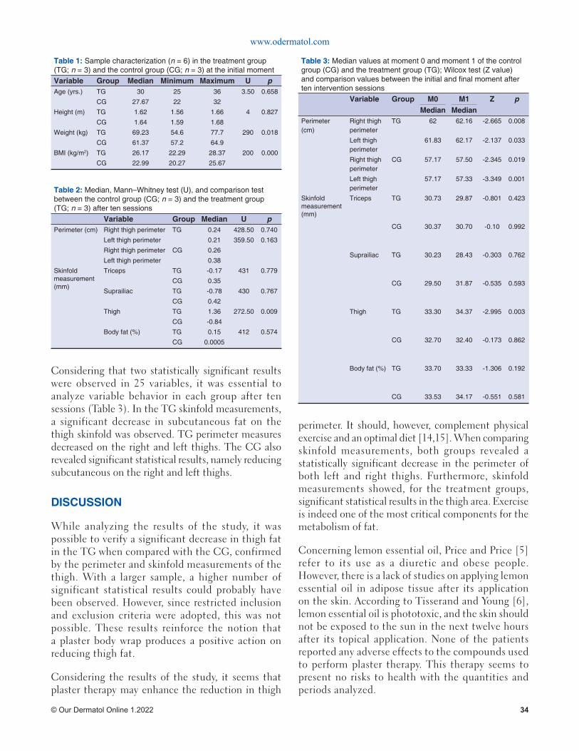
www.odermatol.com
© Our Dermatol Online 1.2022 34
Considering that two statistically significant results were observed in 25 variables, it was essential to analyze variable behavior in each group after ten sessions (Table 3). In the TG skinfold measurements, a significant decrease in subcutaneous fat on the thigh skinfold was observed. TG perimeter measures decreased on the right and left thighs. The CG also revealed significant statistical results, namely reducing subcutaneous on the right and left thighs.
DISCUSSION
While analyzing the results of the study, it was possible to verify a significant decrease in thigh fat in the TG when compared with the CG, confirmed by the perimeter and skinfold measurements of the thigh. With a larger sample, a higher number of significant statistical results could probably have been observed. However, since restricted inclusion and exclusion criteria were adopted, this was not possible. These results reinforce the notion that a plaster body wrap produces a positive action on reducing thigh fat.
Considering the results of the study, it seems that plaster therapy may enhance the reduction in thigh
perimeter. It should, however, complement physical exercise and an optimal diet [14,15]. When comparing skinfold measurements, both groups revealed a statistically significant decrease in the perimeter of both left and right thighs. Furthermore, skinfold measurements showed, for the treatment groups, significant statistical results in the thigh area. Exercise is indeed one of the most critical components for the metabolism of fat.
Concerning lemon essential oil, Price and Price [5] refer to its use as a diuretic and obese people. However, there is a lack of studies on applying lemon essential oil in adipose tissue after its application on the skin. According to Tisserand and Young [6], lemon essential oil is phototoxic, and the skin should not be exposed to the sun in the next twelve hours after its topical application. None of the patients reported any adverse effects to the compounds used to perform plaster therapy. This therapy seems to present no risks to health with the quantities and periods analyzed.
Table 1: Sample characterization (n = 6) in the treatment group (TG; n = 3) and the control group (CG; n = 3) at the initial momentVariable Group Median Minimum Maximum U pAge (yrs.) TG 30 25 36 3.50 0.658
CG 27.67 22 32
Height (m) TG 1.62 1.56 1.66 4 0.827
CG 1.64 1.59 1.68
Weight (kg) TG 69.23 54.6 77.7 290 0.018
CG 61.37 57.2 64.9
BMI (kg/m2) TG 26.17 22.29 28.37 200 0.000
CG 22.99 20.27 25.67
Table 2: Median, Mann–Whitney test (U), and comparison test between the control group (CG; n = 3) and the treatment group (TG; n = 3) after ten sessions Variable Group Median U pPerimeter (cm) Right thigh perimeter TG 0.24 428.50 0.740
Left thigh perimeter 0.21 359.50 0.163
Right thigh perimeter CG 0.26
Left thigh perimeter 0.38
Skinfold measurement (mm)
Triceps TG -0.17 431 0.779
CG 0.35
Suprailiac TG -0.78 430 0.767
CG 0.42
Thigh TG 1.36 272.50 0.009
CG -0.84
Body fat (%) TG 0.15 412 0.574
CG 0.0005
Table 3: Median values at moment 0 and moment 1 of the control group (CG) and the treatment group (TG); Wilcox test (Z value) and comparison values between the initial and fi nal moment after ten intervention sessions Variable Group M0 M1 Z p
Median MedianPerimeter (cm)
Right thigh perimeter
TG 62 62.16 -2.665 0.008
Left thigh perimeter
61.83 62.17 -2.137 0.033
Right thigh perimeter
CG 57.17 57.50 -2.345 0.019
Left thigh perimeter
57.17 57.33 -3.349 0.001
Skinfold measurement (mm)
Triceps TG 30.73 29.87 -0.801 0.423
CG 30.37 30.70 -0.10 0.992
Suprailiac TG 30.23 28.43 -0.303 0.762
CG 29.50 31.87 -0.535 0.593
Thigh TG 33.30 34.37 -2.995 0.003
CG 32.70 32.40 -0.173 0.862
Body fat (%) TG 33.70 33.33 -1.306 0.192
CG 33.53 34.17 -0.551 0.581
www.odermatol.com
© Our Dermatol Online 1.2022 35
CONCLUSION
While analyzing the results of the study, it was possible to verify a significant decrease in thigh fat in the TG when compared with the CG, confirmed by the perimeter and skinfold measurements of the thigh. With a larger sample, a higher number of significant statistical results could probably have been observed. However, since restricted inclusion and exclusion criteria were adopted, this was not possible. These results reinforce the notion that a plaster body wrap produces a positive action on reducing thigh fat. Plaster therapy may function as an adjunct to physical exercise in reducing thigh fat. Therefore, it is essential to highlight the results of this study so that aesthetic biomedicine professionals and physical therapists consider this novel tool for lipolysis enhancement [14].
It is also essential to perform a correct assessment of all measures, which should always be performed by the same professional to avoid minor mistakes and miscalculations [16].
Statement of Human and Animal Rights
All the procedures followed were in accordance with the ethical standards of the responsible committee on human experimentation (institutional and national) and with the 2008 revision of the Declaration of Helsinki of 1975.
Statement of Informed Consent
Informed consent for participation in this study was obtained from all patients.
REFERENCES
1. Eastwood SV, Tillin T, Wright A, Mayet J, Godsland I, Forouhi NG, et al. Thigh fat and muscle each contribute to excess cardiometabolic
risk in South Asians, independent of visceral adipose tissue. Obesity (Silver Spring). 2014;22:2071-9.
2. Ebbert J, Jensen M. Fat depots, Free fatty acids, and dyslipidemia. Nutrients. 2013;5:498-508.
3. Baldisserotto M, Damiani D, Cominato L, Franco R, Lazaretti A, Camargo P, et al. Subcutaneous fat: A better marker than visceral fat for insulin resistance in obese adolescents. E-SPEN J. 2013;8:e251-5.
4. Knuiman P, Hopman MTE, Mensink M. Glycogen availability and skeletal muscle adaptations with endurance and resistance exercise. Nutr Metab. 2015;12:59.
5. Zielinski E. The healing Power of Essential Oils. First. New York: Harmony Books; 2020.
6. Tisserand R, Young R. Essential Oil Safety - A Guide for Health Care Professionals. Second. Elsevier; 2014.
7. Seeley R, VanPutte C, Regan J, Russo A. Seeley’s Anatomy & Physiology. 10th ed. United States of America: McGraw Hill; 2014.
8. Zsikó S, Csányi E, Kovács A, Budai-Szűcs M, Gácsi A, Berkó A. Methods to evaluate skin penetration in vitro. Sci Pharm. 2019;87:19.
9. Buriti BMAB, Buriti JS, Silva IA, Cartaxo JM, Neves GA, Ferreira HC. Characterization of clays from the State of Paraíba, Brazil for aesthetic and medicinal use. Cerâmica. 2019;65:78–84.
10. Santos Moreira J, Melo ASCP de, Noites A, Couto MF, Melo CA de, Adubeiro NCF de A. Plaster body wrap: effects on abdominal fat. Integr Med Res. 2013;2:151-6.
11. Nuttall FQ. Body Mass Index: Obesity, BMI, and Health. Nutr Today. 2015;50:117-28.
12. Mussoi TD. Avaliação Nutricional na Prática Clínica: da gestação ao envelhecimento. 1. ed. Rio de Janeiro: Guanabara Koogan; 2014.
13. Norton KI. Standards for Anthropometry Assessment. In: Norton K, Eston R, editors. Kinanthropometry Exerc. Physiol. 4th ed., Fourth Edition. | New York: Routledge, 2018. | Roger G. Eston Is the Principal Editor of the Third Edition Published 2009. | “First Edition Published by Routledge 2001”—T.p. Verso.: Routledge; 2018, p. 68-137.
14. Gonçalves S. Elaboration and subsequent application of a protocol for measure reduction in a patient with Hashimoto thyroiditis. Our Dermatol Online. 2020;11:243-6.
15. Gonçalves S. Sculpt plaster therapy for abdominal circumference reduction. Our Dermatol Online. 2021;12:79-81.
16. Gonçalves S. A Consulta de Biomedicina Estética. vol. 1. 1st ed. Portugal: escrytos; 2021.
Copyright by Sara Gonçalves. This is an open access article distributed under the terms of the Creative Commons Attribution License, which permits unrestricted use, distribution, and reproduction in any medium, provided the original author and source are credited.Source of Support: Nil, Confl ict of Interest: None declared.
© Our Dermatol Online 1.2022 36
Our Dermatology Online
How to cite this article: Mancy A. Chronic dermatophytosis: A clinical, epidemiological, mycological study. Our Dermatol Online. 2022;13(1):36-40.Submission: 30.07.2021; Acceptance: 26.11.2021DOI: 10.7241/ourd.20221.7
Chronic dermatophytosis: A clinical, epidemiological, Chronic dermatophytosis: A clinical, epidemiological, mycological studymycological studyAbdullah Mancy
Al-Ramadi Teaching Hospital, Al-Anbar Health Directorate, Ministry of Health/Iraq
Corresponding author: Abdullah Mancy, MD, E-mail: [email protected]
INTRODUCTION
Fungal infections attacking humans are classified into three forms: superficial, subcutaneous, and systemic [1]. Dermatophytes are one of the fungi that digest the keratin in the skin, hair, and nails [2]. They are classified into three genera: Trichophyton, Microsporum, and Epidermophyton; and into anthropophilic, geophilic, and zoophilic, according to the mode of transmission [3]. They may affect any site on the body and are clinically known according to that site [4]. In the last years, several terms have been used to describe persistent fungal infections, such as chronic, recurrent, and recalcitrant dermatophytosis [5-7]. In this study, chronicity will be applied to an infection lasting more than six months, with the presence or absence of recurrence despite treatment. Multiple factors in the environment and host and the infecting agents affect the chronicity of an infection. Environmental factors such as weather changes and hot and humid climates are
a favorable environment for fungal growth [5,7]. Host factors involve frequent use of antifungal and steroid combinations, noncompliance of the patient with antifungal drugs, obesity, and sedentary lifestyle [7]. Frequent migration of people enhances the spread of the infection with a special strain such as Trichophyton mentagrophyte [8]. A defective immunological status, poverty, and overcrowding are mostly associated with chronic infection [5,7,9]. Infecting agents involve the emergence of novel antifungal, drug-resistant strains of dermatophytes [10,6]. Biofilm produced by some species of dermatophytes enhances virulence and hence chronicity [11]. The immune response is ameliorated to escape the detection by the host immune system [12].
MATERIALS AND METHODS
This study was conducted in the Dermatology and Venereology Department of Al-Ramadi Teaching Hospital and College of Science, Al-Anbar University.
ABSTRACT
Background: Dermatophytes are a common cause of superficial fungal infection of the skin. The emergence of epidemic-like attacks of those chronic and recurring represents a public health problem. Materials and Methods: Two hundred patients with suspected fungal infection of the skin attending the Dermatology and Venereology Department of Al-Ramadi Teaching Hospital were examined. Fifty-nine patients with chronic dermatophytosis were selected for the study and fifty of those were subjected for culture. History taking and a physical examination were conducted for all patients. A wet mount of 10% potassium hydroxide and culture on Sabouraud dextrose agar was done for selected cases. Results: Among 59 patients with chronic infections, the main age group affected was 29 years old, with a nearly equal sex ratio. The mean duration of the illness was 1.2 years. Tinea corporis was the mos t common type. The Trichophyton genera were the most common (65%), and Trichophyton mentagrophyte was the most common species isolated (46%). Conclusion: Multiple factors have been associated with the appearance of epidemic-like attacks of chronic dermatophyte infections in Iraq in the last several years. Herein, we would like to shed light on these factors and the pathogens responsible.
Keywords: Ringworms; Dermatophytes; Chronic Infections; Potent steroids
Brief Report
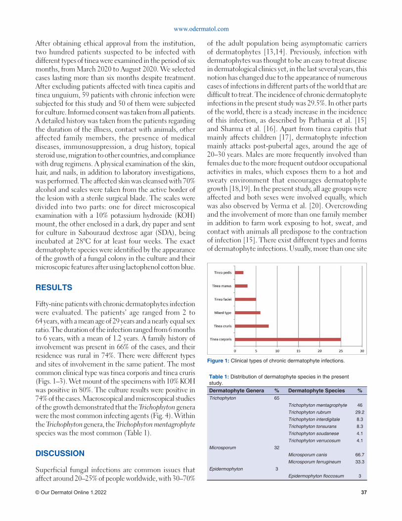
Figure 1: Clinical types of chronic dermatophyte infections.
www.odermatol.com
© Our Dermatol Online 1.2022 37
After obtaining ethical approval from the institution, two hundred patients suspected to be infected with different types of tinea were examined in the period of six months, from March 2020 to August 2020. We selected cases lasting more than six months despite treatment. After excluding patients affected with tinea capitis and tinea unguium, 59 patients with chronic infection were subjected for this study and 50 of them were subjected for culture. Informed consent was taken from all patients. A detailed history was taken from the patients regarding the duration of the illness, contact with animals, other affected family members, the presence of medical diseases, immunosuppression, a drug history, topical steroid use, migration to other countries, and compliance with drug regimens. A physical examination of the skin, hair, and nails, in addition to laboratory investigations, was performed. The affected skin was cleansed with 70% alcohol and scales were taken from the active border of the lesion with a sterile surgical blade. The scales were divided into two parts: one for direct microscopical examination with a 10% potassium hydroxide (KOH) mount, the other enclosed in a dark, dry paper and sent for culture in Sabouraud dextrose agar (SDA), being incubated at 28°C for at least four weeks. The exact dermatophyte species were identified by the appearance of the growth of a fungal colony in the culture and their microscopic features after using lactophenol cotton blue.
RESULTS
Fifty-nine patients with chronic dermatophytes infection were evaluated. The patients’ age ranged from 2 to 64 years, with a mean age of 29 years and a nearly equal sex ratio. The duration of the infection ranged from 6 months to 6 years, with a mean of 1.2 years. A family history of involvement was present in 66% of the cases, and their residence was rural in 74%. There were different types and sites of involvement in the same patient. The most common clinical type was tinea corporis and tinea cruris (Figs. 1–3). Wet mount of the specimens with 10% KOH was positive in 80%. The culture results were positive in 74% of the cases. Macroscopical and microscopical studies of the growth demonstrated that the Trichophyton genera were the most common infecting agents (Fig. 4). Within the Trichophyton genera, the Trichophyton mentagrophyte species was the most common (Table 1).
DISCUSSION
Superficial fungal infections are common issues that affect around 20–25% of people worldwide, with 30–70%
of the adult population being asymptomatic carriers of dermatophytes [13,14]. Previously, infection with dermatophytes was thought to be an easy to treat disease in dermatological clinics yet, in the last several years, this notion has changed due to the appearance of numerous cases of infections in different parts of the world that are difficult to treat. The incidence of chronic dermatophyte infections in the present study was 29.5%. In other parts of the world, there is a steady increase in the incidence of this infection, as described by Pathania et al. [15] and Sharma et al. [16]. Apart from tinea capitis that mainly affects children [17], dermatophyte infection mainly attacks post-pubertal ages, around the age of 20–30 years. Males are more frequently involved than females due to the more frequent outdoor occupational activities in males, which exposes them to a hot and sweaty environment that encourages dermatophyte growth [18,19]. In the present study, all age groups were affected and both sexes were involved equally, which was also observed by Verma et al. [20]. Overcrowding and the involvement of more than one family member in addition to farm work exposing to hot, sweat, and contact with animals all predispose to the contraction of infection [15]. There exist different types and forms of dermatophyte infections. Usually, more than one site
Table 1: Distribution of dermatophyte species in the present study.Dermatophyte Genera % Dermatophyte Species %Trichophyton 65
Trichophyton mentagrophyte 46
Trichophyton rubrum 29.2
Trichophyton interdigitale 8.3
Trichophyton tonsurans 8.3
Trichophyton soudanese 4.1
Trichophyton verrucosum 4.1
Microsporum 32
Microsporum canis 66.7
Microsporum ferrugineum 33.3
Epidermophyton 3
Epidermophyton fl occosum 3
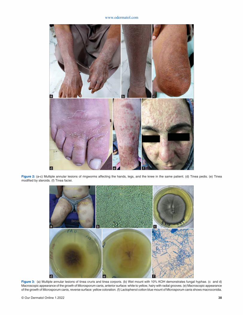
www.odermatol.com
© Our Dermatol Online 1.2022 38
Figure 2: (a-c) Multiple annular lesions of ringworms affecting the hands, legs, and the knee in the same patient. (d) Tinea pedis. (e) Tinea modifi ed by steroids. (f) Tinea faciei.
d e
cba
f
Figure 3: (a) Multiple annular lesions of tinea cruris and tinea corporis. (b) Wet mount with 10% KOH demonstrates fungal hyphae. (c and d) Macroscopic appearance of the growth of Microsporum canis, anterior surface: white to yellow, hairy with radial grooves. (e) Macroscopic appearance of the growth of Microsporum canis, reverse surface: yellow coloration. (f) Lactophenol cotton blue mount of Microsporum canis shows macroconidia.
d e
cba
f
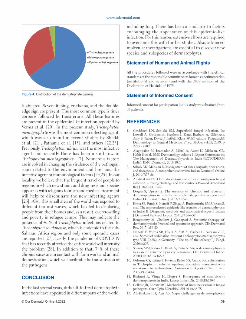
Trichophyton genera
Microsporum genera
Epidermophyton genera
Figure 4: Distribution of the dermatophyte genera.
www.odermatol.com
© Our Dermatol Online 1.2022 39
is affected. Severe itching, erythema, and the double-edge sign are present. The most common type is tinea corporis followed by tinea cruris. All these features are present in the epidemic-like infection reported by Verma et al. [20]. In the present study, Trichophyton mentagrophyte was the most common infecting agent, which was also found in recent studies by Sheikh et al. [21], Pathania et al. [15], and others [22,23]. Previously, Trichophyton rubrum was the most infective agent, but recently there has been a shift toward Trichophyton mentagrophyte [17]. Numerous factors are involved in changing the virulence of the pathogen, some related to the environment and host and the infective agent or immunological factors [24,25]. In our locality, we believe that the frequent travel of people to regions in which new strains and drug-resistant species appear as with religious tourism and medical treatment will help to disseminate the new infecting agents [26]. Also, this small area of the world was exposed to different terrorist waves, which has led to displacing people from their homes and, as a result, overcrowding and poverty in refugee camps. This may indicate the presence of 4.1% of Trichophyton infections related to Trichophyton soudanense, which is endemic to the sub-Saharan Africa region and only some sporadic cases are reported [27]. Lastly, the pandemic of COVID-19 that has recently affected the entire world will intensify the problem [28]. In addition to that, 74% of these chronic cases are in contact with farm work and animal domestication, which will facilitate the transmission of the pathogens.
CONCLUSION
In the last several years, difficult-to-treat dermatophyte infections have appeared in different parts of the world,
including Iraq. There has been a similarity to factors encouraging the appearance of this epidemic-like infection. For this reason, extensive efforts are required to overcome this with further studies. Also, advanced molecular investigations are essential to discover new species and subspecies of dermatophytes.
Statement of Human and Animal Rights
All the procedures followed were in accordance with the ethical standards of the responsible committee on human experimentation (institutional and national) and with the 2008 revision of the Declaration of Helsinki of 1975.
Statement of Informed Consent
Informed consent for participation in this study was obtained from all patients.
REFERENCES
1. Craddock LN, Schieke SM. Superficial fungal infection. In: Lowell A. Goldsmith, Stephen I. Katz, Barbara A. Gilchrest, Amy S. Paller, David J. Leffell, Klaus Wolff, editors. Fitzpatrick’s Dermatology in General Medicine. 8th ed. McGraw-Hill; 2019. p 2925 - 2989.
2. Ajagopalan M, Inamadar A, Mittal A, Autar K, Miskeen, CR, Kabir S, et al. BMC Dermatology volume 1 Expert Consensus on The Management of Dermatophytosis in India (ECTODERM India). BMC Dermatol, 2018;18:6.
3. Sahoo AK, Mahajan R. Management of tinea corporis, tinea cruris, and tinea pedis: A comprehensive review. Indian Dermatol Online J. 2016;7:77-86.
4. AL-Khikani FH. Dermatophytosis a worldwide contiguous fungal infection: Growing challenge and few solutions. Biomed Biotechnol Res J. 2020;4:117-22.
5. Dogra S, Uprety S. The menace of chronic and recurrent dermatophytosis in India: Is the problem deeper than we perceive? Indian Dermatol Online J. 2016;7:73-6.
6. Erma SB, Panda S, Nenoff P, Singal A, Rudramurthy SM, Uhrlass S, et al. The unprecedented epidemic-like scenario of dermatophytosis in India: II. Diagnostic methods and taxonomical aspects. Indian J Dermatol Venereol Leprol. 2021;87:326-32.
7. Rengasamy M, Chellam J, Ganapati S. Systemic therapy of dermatophytosis: Practical and systematic approach. Clin Dermatol Rev. 2017;1:19-23.
8. Nenoff P, Verma SB, Ebert A, Süß A, Fischer E, Auerswald E, et al. Spread of terbinafi ne-resistant Trichophyton mentagrophytes type VIII (India) in Germany: “The tip of the iceberg?” J Fungi. 2020;6:207.
9. Shenoy MM, Kilaru G, Rasak A, Pinto A. Atypical dermatophytosis in a case of systemic lupus erythematosus. Our Dermatol Online. 2020;11:e165.1-e165.3
10. Osborne CS, Leitner I, Favre B, Ryder NS. Amino acid substitution in Trichophyton rubrum squalene epoxidase associated with resistance to terbinafine. Antimicrob Agents Chemother. 2005;49:2840-4.
11. Bishnoi A, Vinay K, Dogra S. Emergence of recalcitrant dermatophytosis in India. Lancet Infect Dis. 2018;18:250-1.
12. Collette JR, Lorenz MC. Mechanisms of immune evasion in fungal pathogens. Curr Opin Microbiol. 2011;14:668-75.
13. AL-Khikani FH, Ayit AS. Major challenges in dermatophytosis
www.odermatol.com
© Our Dermatol Online 1.2022 40
treatment: Current options and future visions. Egypt J Dermatol Venerol. 2021;41:1-9.
14. Monise FP, Abreu MH, Cantelli BAM, Segura GG, Nishimura FG, Bitencourt TA, et al. Epidemiology and diagnostic perspectives of dermatophytoses. J Fungi. 2020;6:310.
15. Pathania S, Rudramurthy SM, Narang T, Saikia UN, Dogra S. A prospective study of the epidemiological and clinical patterns of recurrent dermatophytosis at a tertiary care hospital in India. Indian J Dermatol Venereol Leprol. 2018;84:678-84.
16. Sharma R, Adhikari L, Sharma RL. Recurrent dermatophytosis: A rising problem in Sikkim, a Himalayan state of India. Indian J Pathol Microbiol. 2017;60:541-5.
17. James WD, Berger TG, Elastone DM. Diseases resulting from fungi and yeasts In: Andrews Diseases of the Skin: Clinical Dermatology.13th ed. Philadelphia. WB Saunders Company, 2020;291-324.
18. Bhatia VK, Sharma PC. Epidemiological studies on dermatophytosis in human patients in Himachal Pradesh, India. Springerplus. 2014;3:134.
19. Singh SK, Subba N, Tilak R. Effi cacy of terbinafi ne and itraconazole in different doses and in combination in the treatment of tinea infection: A randomized controlled parallel group open labeled trial with clinic-mycological correlation. Indian J Dermatol. 2020;65:284-9.
20. Verma SB, Panda S, Nenoff P, Singal A, Rudramurthy SM, Uhrlass S, et al. The unprecedented epidemic-like scenario of dermatophytosis in India: I. Epidemiology, risk factors and clinical features. Indian J Dermatol Venereol Leprol. 2021;87:154-75.
21. Sheikh JS, Aaqib AS, Ilham I, Faizan Y, Iffat HS. Dermatophytosis: An epidemiological and clinical comparative study in a tertiary care centre. Int J Contemp Med. 2020;7:21-7.
22. Hanumanthappa H, Sarojini K, Shilpashree P, Muddapur SB. Clinicomycological study of 150 cases of dermatophytosis
in a tertiary care hospital in South India. Indian J Dermatol. 2012;57:322-3.
23. De Freitas RS, Neves PS, Charbel CE, Criado PR, Nunes RS, Santos-Filho AM, et al. Investigation of superfi cial mycosis in cutaneous allergy patients using topical or systemic corticosteroids. Int J Dermatol. 2017;56:e194-8.
24. Nenoff P, Verma SB, Vasani R, Burmester A, Hipler UC, Wittig F, et al. The current Indian epidemic of superfi cial dermatophytosis due to Trichophyton mentagrophytes: A molecular study. Mycoses. 2019;62:336-56.
25. Shenoy MM, Jayaraman J. Epidemic of diffi cult-to-treat tinea in India: Current scenario, culpritscurbing strategies. Arch Med Health Sci. 2019;7:112-7.
26. Taghipour S, Shamsizadeh F, Pchelin IM, Rezaei-Matehhkolaei A, Mahmoudabadi AZ, Valadan R, et al. Emergence of terbinafi ne resistant Trichophyton mentagrophytes in Iran, harboring mutations in the squalene epoxidase (SQLE) gene. Infect Drug Resist. 2020:13:845-50.
27. Sahai S, Mishra D, Tripathi P. Rare isolation of Trichophyton soudanense from three cases of superfi cial mycoses in Lucknow, India. Indian J Pathol Microbiol. 2010;53:896-7.
28. Yaneva M, Demerdjieva Z, Darlenski R, Tsankov N. COVID-19 and skin: Analysis of the available data. Our Dermatol Online. 2020;11(Supp. 2):6-9.
Copyright by Abdullah Mancy. This is an open-access article distributed under the terms of the Creative Commons Attribution License, which permits unrestricted use, distribution, and reproduction in any medium, provided the original author and source are credited.Source of Support: Nil, Confl ict of Interest: None declared.
© Our Dermatol Online 1.2022 41
Our Dermatology Online
How to cite this article: Abreu Velez AM, Thomason AB, Jackson BL, Howard MS. Recurrent herpes zoster with IgD deposits, multinucleated keratinocytes and overexpression of galectin and glypican 3 in a patient with SARS-COVID-19 infection. Our Dermatol Online. 2022;13(1):41-44.Submission: 22.09.2021; Acceptance: 11.11.2021DOI: 10.7241/ourd.20221.8
Recurrent herpes zoster with IgD deposits, Recurrent herpes zoster with IgD deposits, multinucleated keratinocytes and overexpression multinucleated keratinocytes and overexpression of galectin and glypican 3 in a patient with of galectin and glypican 3 in a patient with SARS-COVID-19 infectionSARS-COVID-19 infectionAna Maria Abreu Velez1, Amanda Bortle Thomason2, Billie L. Jackson3, Michael S. Howard1
1Georgia Dermatopathology Associates, Atlanta, Georgia, USA, 2D.O. Student, Philadelphia College of Osteopathic Medicine/Georgia Campus, Suwanee, Georgia, USA, 3Billie L. Jackson, M.D., Dermatologist, Macon, Georgia, USA.
Corresponding author: Amanda Bortle Thomason, D.O, E-mail: [email protected]
INTRODUCTION
In December 2019, a severe acute respiratory syndrome coronavirus 2 (SARS-CoV-2) presented clinically in Wuhan, China. It was first reported from a group of patients who suddenly developed severe pneumonia [1]. The SARS-CoV-2 virus is part of the coronavirus family, and this newly discovered strain is a RNA, single stranded, encapsulated virus [2]. The associated human disease, COVID-19, has spread globally and is responsible for an ongoing pandemic that has claimed thousands of lives [1]. Skin related conditions due to COVID 19 have been reported; the most common presentations seem to be acral lesions resembling pseudo-chilblains (perniosis like)
in 40.4% of cases, occurring in young adults. Other manifestations include erythematous, maculopapular rashes affecting approximately 21.3% of adult patients, vesicular “rashes” affecting about 13.0% of middle-aged adults, and urticarial rashes in 10.9% of adults. Vascular rashes reminiscent of livedo or purpura were uncommon (4% of cases), mostly seen in elderly patients. In minors, the most common dermatologic manifestation was erythema multiforme-like eruptions in about 4% of the cases [3].
CASE REPORT
A 73-year-old Caucasian female presented to her primary care physician for recurrent, vesicular rashes.
ABSTRACT
The novel coronavirus disease (COVID-19) that currently plagues the world and caused by SARS-CoV-2, has spread internationally since late 2019. The dermatologic manifestations of this virus are currently being identified. We describe a 73-year-old Caucasian female who presented to many physicians for recurrent Herpes zoster episodes that persisted, despite treatment with multiple antiviral medications. The patient was diagnosed with COVID-19 before an onset of vesicular pustular lesions. The clinical diagnoses were recurrent herpes zoster and recurrent varicella. A skin biopsy was obtained and stained with hematoxylin and eosin to confirm a diagnosis. Immunohistochemical stains for Ki-67, Phospho-Histone H3, galectin 3, glypican and IgD were positive in multinucleated cells of the skin, where the viral lesions were detected. Recidivated herpes zoster and varicella are currently being clinically associated with COVID- 19; the abnormal immune response in patients with COVID-19 may be due to the overexpression of molecules that facilitate the outbreak of these viruses.
Keywords: Herpes zoster; Varicella; COVID 19; IgD; Galectin and glypican 3
Case Report
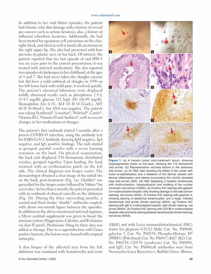
www.odermatol.com
© Our Dermatol Online 1.2022 42
In addition to her viral blister episodes, the patient had chronic solar skin damage with a history of several pre-cancers such as actinic keratosis; also, a history of inflamed seborrheic keratoses. Additionally, she had been treated for squamous cell carcinoma on the chin, right cheek, and chest as well as basal cell carcinoma on the right upper lip. She also had presented with four previous dysplastic nevi on her back. Of interest, the patient reported that her last episode of oral HSV-1 was six years prior to the current presentation; it was treated with antiviral medications. She also reported two episodes of chickenpox in her childhood, at the ages of 4 and 7. She had never taken the shingles vaccine but did have a mild outbreak of shingles in 1990 on her left lower back with mild pain; it resolved quickly. The patient’s chemical laboratory tests displayed mildly abnormal results such as phosphorus 2.4 L (3-4.5 mg/dl), glucose 112 high (H) (66-99 mg/dl), Hemoglobin A1c 6.1%, ALT 63 H (0-32iu/L), AST 60 H (0-40iu/L); her ANA was negative. The patient was taking Synthroid®, Losartan®, Welchol®, Cartia®, Vitamin B12, Vitamin D and Ambien®, with no recent changes in her medications or dosages.
The patient’s first outbreak started 3 months after a proven COVID-19 infection, using the antibody test for SARS-CoV-2-Antibody showing IgM negative, IgA negative, and IgG positive findings. The rash started as grouped, painful vesicles with a severe burning sensation on the back. On physical examination, the back rash displayed T10 dermatome distributed vesicles, grouped together. Upon healing, the back retained with an erythematous patch on the left side. The clinical diagnosis was herpes zoster. The dermatologist obtained a clear image of the initial site on the back, post-treatment (Fig. 1a). Gladdin® was prescribed for the herpes zoster followed by Valtrex® for seven days. In less than a month, the patient presented with an outbreak of clinical varicella on the abdomen (Fig. 1b). During the three succeeding months, a second and third similar “double” outbreaks erupted, with about one-month lapses between the episodes. In addition to the above-mentioned antiviral regimen, a Myers cocktail supplement was given to boost the immune system (Magnesium, Calcium, and B vitamins via slow IV push). Prednisone 10mg for three days was added as therapy. Due to a superinfection with Gram positive bacteria, the lesions were cleaned with a topical antiseptic.
A skin biopsy of the affected area from the left abdomen was examined with hematoxylin and eosin
(H&E) and with Leica immunohistochemical (IHC) stains for glypican-3(1G12 Mab) Cat. No. PA0800; galectin 3, Cat. No. PA0238; Phospho-Histone H3 (PHH3) (Polyclonal) Cat. No. PA0817; Ki67 (K2) Cat. No. PA0230; CD138 (syndecan) Cat. No. PA0088, and IgD, Cat. No. PA006(all antibodies were from Novocastra-Leica Biosystem’s, Buffalo Grove, Illinois,
Figure 1: (a) A herpes zoster post-treatment lesion, showing hyperpigmented marks on the back, following the T10 dermatome (red arrow). (b) Representative varicella lesions in the abdomen (red arrow). (c) An H&E stain showing the blister in the center with some re-epithelization and a dilatation of the dermal vessels with dermal infl ammation and edema surrounding the central ulcerated area (red arrow) (40X). (d) H&E displaying a herpetic keratinocyte with multinucleation, marginalization and molding of the nuclear chromatin (red arrow) (1000X). (e) Positive IHC staining with galectin 3 in multinucleated herpetic cells showing damage to the cells (brown staining; red arrow) (400X). (f) Positive IHC staining with glypican 3, showing staining of epidermal keratinocytes, and a large, infected keratinocyte (red arrow) (brown staining) (600X). (g) Positive IHC staining with IgD in multinucleated herpetic cells (brown staining; red arrow) (600X). (h) Positive IHC staining with CD138 in multinucleated herpetic cells and some staining between keratinocytes (brown staining; red arrow) (600X).
d
h
c
g
b
f
a
e
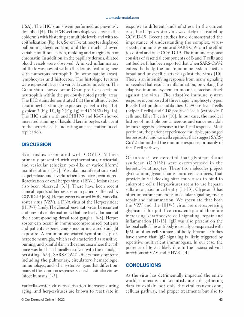
www.odermatol.com
© Our Dermatol Online 1.2022 43
USA). The IHC stains were performed as previously described [4]. The H&E sections displayed areas in the epidermis with blistering at multiple levels and with re-epithelization (Fig. 1c). Several keratinocytes displayed ballooning degeneration, and their nuclei showed variable multinucleation, molding and margination of chromatin. In addition, in the papillary dermis, dilated blood vessels were observed. A mixed inflammatory infiltrate was present within the dermis, featuring areas with numerous neutrophils (in some patchy areas), lymphocytes and histocytes. The histologic features were representative of a varicella zoster infection. The Gram stain showed some Gram-positive cocci and neutrophils within the previously noted patchy areas. The IHC stains demonstrated that the multinucleated keratinocytes strongly expressed galectin (Fig. 1e), glypican 3 (Fig. 1f), IgD (Fig. 1g) and CD138 (Fig. 1h). The IHC stains with and PHHP-3 and Ki-67 showed increased staining of basaloid keratinocytes subjacent to the herpetic cells, indicating an acceleration in cell replication.
DISCUSSION
Skin rashes associated with COVID-19 have primarily presented with erythematous, urticarial, and vesicular (chicken pox-like or varicelliform) manifestations [3-5]. Vascular manifestations such as petechiae and livedo reticularis have been noted. Reactivation of oral herpes virus (HSV-1) lesions have also been observed [3,5]. There have been recent clinical reports of herpes zoster in patients affected by COVID-19 [6-8]. Herpes zoster is caused by the varicella‐zoster virus (VZV), a DNA virus of the Herpesviridae (HHV-3) family. The clinical presentation can be recurrent and presents in dermatomes that are likely dormant at their corresponding dorsal root ganglia [6-8]. Herpes zoster can occur in immunocompromised patients and patients experiencing stress or increased sunlight exposure. A common associated symptom is post-herpetic neuralgia, which is characterized as sensitive, burning, and painful skin in the same area where the rash once was but has clinically resolved with the neuralgia persisting [6-9]. SARS-CoV-2 affects many systems including the pulmonary, circulatory, hematologic, immunologic, and other systems/organs that differ from many of the common responses seen when similar viruses infect humans [1-3].
Varicella-zoster virus re-activation increases during aging, and herpesviruses are known to reactivate in
response to different kinds of stress. In the current case, the herpes zoster virus was likely reactivated by COVID-19. Recent studies have demonstrated the importance of understanding the complex, virus-specific immune response of SARS-CoV-2 in the effort to control and treat COVID-19. The immune response consists of essential components of B and T cells and antibodies. It has been reported that when SARS-CoV-2 enters the body, the innate immune system elicits a broad and unspecific attack against the virus [10]. There is an intensifying response from many signaling molecules that result in inflammation, provoking the adaptive immune system to mount a precise attack against the virus. The adaptive immune system response is composed of three major lymphocyte types: B cells that produce antibodies, CD4 positive T cells (helper T cells) and CD8 positive T cells (cytotoxic T cells and killer T cells) [10]. In our case, the medical history of multiple pre-cancerous and cancerous skin lesions suggests a decrease in the T cell response. Most pertinent, the patient experienced multiple, prolonged herpes zoster and varicella episodes that suggest SARS-CoV-2 diminished the immune response, primarily of the T cell pathway.
Of interest, we detected that glypican 3 and syndecan (CD138) were overexpressed in the herpetic keratinocytes. These two molecules project glycosaminoglycan chains onto cell surfaces, that provide initial docking sites for viruses to bind to eukaryotic cells . Herpesviruses seem to use heparan sulfate to assist in cell entry [11-13]. Glypican 3 has other important functions in cellular signaling, tissue repair and inflammation. We speculate that both the VZV and the HHV-3 virus are overexpressing glypican 3 for putative virus entry, and therefore increasing keratinocyte cell signaling, repair and inflammation [11-13]. IgD was also present on the lesional cells. This antibody is usually co-expressed with IgM, another cell surface antibody. Previous studies have shown that IgD signaling is likely triggered by repetitive multivalent immunogens. In our case, the presence of IgD is likely due to the associated viral infections of VZV and HHV-3 [14].
CONCLUSIONS
As the virus has detrimentally impacted the entire world, clinicians and scientists are still gathering data to explain not only the viral transmission, cellular pathway, and proper treatments but also to
www.odermatol.com
© Our Dermatol Online 1.2022 44
detect early symptoms of the disease and all effected organ systems. While this patient is considered high risk for reactivation of herpes zoster due to her age alone, we cannot rule out that her recent exposure to COVID-19 contributed to the sixty-plus-day duration of zoster rash. We conclude that repetitive outbreaks of herpes zoster and varicella, in a patient with previous COVID-19 exposure, are part of the dermatological manifestation of this disease. This disseminated herpes reactivation illustrates the unpredictable presentation of the virus, with the back lesion’s characteristic of classic shingles and abdominal lesions simulating chickenpox, with a non-dermatomal distribution pattern. The glypican 3 and CD138 biomarkers found in the biopsy sample support the concept that not only is the host immune system altered by the herpes infections, but also the possibility that their overexpression is due to the prior Coronavirus exposure. As more clinical findings are being reported before, during, and after COVID-19 exposure it is critical to consider these manifestations to fully understand this disease process.
Consent
The examination of the patient was conducted according to the principles of the Declaration of Helsinki. The authors certify that they have obtained all appropriate patient consent forms, in which the patients gave their consent for images. Our Dermatol Online 3.2021 269 and other clinical information to be included in the journal. The patients understand that their names and initials will not be published, and due effort will be made to conceal their identity, but that anonymity cannot be guaranteed.
REFERENCES
1. Zhu N, Zhang D, Wang W, Li X, Yang B, Son J, et al. A novel coronavirus from patients with pneumonia in China. 2019. N Engl J Med. 2020;382:727–33.
2. Lu R, Zhao X, Li J, Niu P, Yang B, Wu H, et al. Genomic characterization, and epidemiology of 2019 novel coronavirus:
implications for virus origins and receptor binding. Lancet, 2020;395:565–74.
3. Daneshgaran G, Dubin DP, Gould DJ. Cutaneous manifestations of COVID-19: An Evidence-based review. Am J Clin Dermatol, 2020;21:627-39.
4. Abreu-Velez AM, Upegui-Zapata YA, Valencia-Yepes CA, Upegui-Quiceno E, Mesa-Herrera NR, Jiménez-Echavarria AM, et al. Membrane attack complex (C5b-9 complex or Mac), is strongly present in lesional skin from patients with endemic pemphigus foliaceus in El Bagre, Colombia. J Cutan Pathol. 2019;46:925-9.
5. Tang K, Wang Y, Zhang H, Zheng Q, Fang R, Sun Q. Cutaneous manifestations of the Coronavirus Disease 2019 (COVID-19): A brief review. Dermatol Ther. 2020:e13528.
6. Xu R, Zhou Y, Cai L, Wang L, Han J, Yang X, et al. Co-reactivation of the human herpesvirus alpha subfamily (herpes simplex virus-1 and varicella zoster virus) in a critically ill patient with COVID-19. Br J Dermatol. 2020;183:1145-7.
7. Wang B, Guo S, Yao Y, Li Y, Zhang G. Dermatologists may need to pay more attention to herpes zoster during the pandemic of COVID-19. Infect Dis (Lond). 2020;52:917-18.
8. Saati A, Al-Husayni F, Malibari AA, Bogari AA, Maher Alharbi M. Herpes zoster co-infection in an immunocompetent patient with COVID-19. Cureus, 2020;12:e8998.
9. Català A, Galván Casas, Carretero Hernadez G, Garcia-Doval I. Vesicular eruption in COVID-19 - to exclude varicella: reply from the authors. Br J Dermatol, 2020;183:791.
10. Moderbacher CR, Ramirez SI, Dan JM, Grifoni A, Hastie KM, Daniela Weiskopf D, et al. Antigen-specifi c adaptive immunity to SARS-CoV-2 in acute COVID-19 and associations with age and disease severity. Cell. 2020;183:996-1012.e19.
11. Shukla D, Spear PG. Herpesviruses and heparan sulfate: an intimate relationship in aid of viral entry. J Clin Invest. 2001;108:503–10.
12. Jacquet A, Haumont M, Chellun D, Massaer M, Tufaro F, A Bollen A, et al. The varicella zoster virus glycoprotein B (gB) plays a role in virus binding to cell surface heparan sulfate proteoglycans. Virus Res. 1998;53:197-7.
13. Bacsa S, Karasneh G, Dosa S, Liu J, Valyi-Nagy T, Shukla D. Syndecan-1 and syndecan-2 play key roles in herpes simplex virus type-1 infection. J Gen Virol. 2011;92:733–43.
14. Übelhart R, Hug E, Bach MP, Thomas Wossning T, Dühren-von Minden M, Anselm H C Horn AHC et al. Responsiveness of B cells is regulated by the hinge region of IgD. Nat Immunol. 2015;16:534–43.
Copyright by Ana Maria Abreu Velez, et al. This is an open-access article distributed under the terms of the Creative Commons Attribution License, which permits unrestricted use, distribution, and reproduction in any medium, provided the original author and source are credited.Source of Support: Georgia Dermatopathology Associates, Atlanta, GA. Confl ict of Interest: None declared.
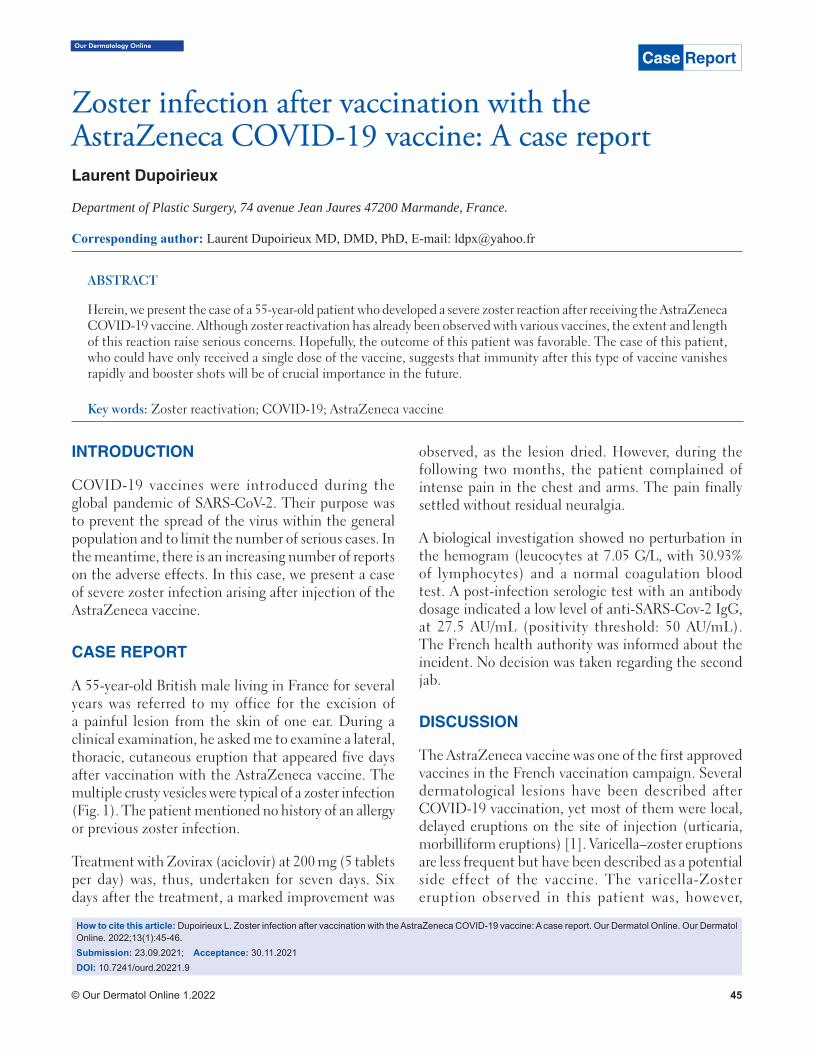
© Our Dermatol Online 1.2022 45
Our Dermatology Online
Zoster infection after vaccination with the Zoster infection after vaccination with the AstraZeneca COVID-19 vaccine: A case reportAstraZeneca COVID-19 vaccine: A case reportLaurent Dupoirieux
Department of Plastic Surgery, 74 avenue Jean Jaures 47200 Marmande, France.
Corresponding author: Laurent Dupoirieux MD, DMD, PhD, E-mail: [email protected]
INTRODUCTION
COVID-19 vaccines were introduced during the global pandemic of SARS-CoV-2. Their purpose was to prevent the spread of the virus within the general population and to limit the number of serious cases. In the meantime, there is an increasing number of reports on the adverse effects. In this case, we present a case of severe zoster infection arising after injection of the AstraZeneca vaccine.
CASE REPORT
A 55-year-old British male living in France for several years was referred to my office for the excision of a painful lesion from the skin of one ear. During a clinical examination, he asked me to examine a lateral, thoracic, cutaneous eruption that appeared five days after vaccination with the AstraZeneca vaccine. The multiple crusty vesicles were typical of a zoster infection (Fig. 1). The patient mentioned no history of an allergy or previous zoster infection.
Treatment with Zovirax (aciclovir) at 200 mg (5 tablets per day) was, thus, undertaken for seven days. Six days after the treatment, a marked improvement was
observed, as the lesion dried. However, during the following two months, the patient complained of intense pain in the chest and arms. The pain finally settled without residual neuralgia.
A biological investigation showed no perturbation in the hemogram (leucocytes at 7.05 G/L, with 30.93% of lymphocytes) and a normal coagulation blood test. A post-infection serologic test with an antibody dosage indicated a low level of anti-SARS-Cov-2 IgG, at 27.5 AU/mL (positivity threshold: 50 AU/mL). The French health authority was informed about the incident. No decision was taken regarding the second jab.
DISCUSSION
The AstraZeneca vaccine was one of the first approved vaccines in the French vaccination campaign. Several dermatological lesions have been described after COVID-19 vaccination, yet most of them were local, delayed eruptions on the site of injection (urticaria, morbilliform eruptions) [1]. Varicella–zoster eruptions are less frequent but have been described as a potential side effect of the vaccine. The varicella-Zoster eruption observed in this patient was, however,
ABSTRACT
Herein, we present the case of a 55-year-old patient who developed a severe zoster reaction after receiving the AstraZeneca COVID-19 vaccine. Although zoster reactivation has already been observed with various vaccines, the extent and length of this reaction raise serious concerns. Hopefully, the outcome of this patient was favorable. The case of this patient, who could have only received a single dose of the vaccine, suggests that immunity after this type of vaccine vanishes rapidly and booster shots will be of crucial importance in the future.
Key words: Zoster reactivation; COVID-19; AstraZeneca vaccine
How to cite this article: Dupoirieux L. Zoster infection after vaccination with the AstraZeneca COVID-19 vaccine: A case report. Our Dermatol Online. Our Dermatol Online. 2022;13(1):45-46.Submission: 23.09.2021; Acceptance: 30.11.2021DOI: 10.7241/ourd.20221.9
Case Report
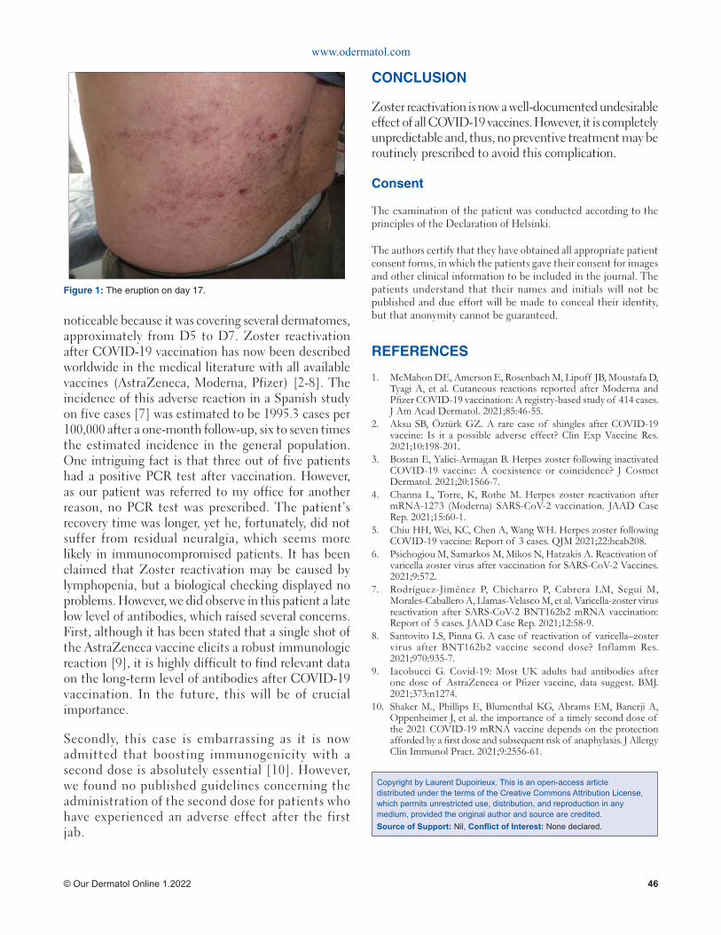
www.odermatol.com
© Our Dermatol Online 1.2022 46
noticeable because it was covering several dermatomes, approximately from D5 to D7. Zoster reactivation after COVID-19 vaccination has now been described worldwide in the medical literature with all available vaccines (AstraZeneca, Moderna, Pfizer) [2-8]. The incidence of this adverse reaction in a Spanish study on five cases [7] was estimated to be 1995.3 cases per 100,000 after a one-month follow-up, six to seven times the estimated incidence in the general population. One intriguing fact is that three out of five patients had a positive PCR test after vaccination. However, as our patient was referred to my office for another reason, no PCR test was prescribed. The patient’s recovery time was longer, yet he, fortunately, did not suffer from residual neuralgia, which seems more likely in immunocompromised patients. It has been claimed that Zoster reactivation may be caused by lymphopenia, but a biological checking displayed no problems. However, we did observe in this patient a late low level of antibodies, which raised several concerns. First, although it has been stated that a single shot of the AstraZeneca vaccine elicits a robust immunologic reaction [9], it is highly difficult to find relevant data on the long-term level of antibodies after COVID-19 vaccination. In the future, this will be of crucial importance.
Secondly, this case is embarrassing as it is now admitted that boosting immunogenicity with a second dose is absolutely essential [10]. However, we found no published guidelines concerning the administration of the second dose for patients who have experienced an adverse effect after the first jab.
CONCLUSION
Zoster reactivation is now a well-documented undesirable effect of all COVID-19 vaccines. However, it is completely unpredictable and, thus, no preventive treatment may be routinely prescribed to avoid this complication.
Consent
The examination of the patient was conducted according to the principles of the Declaration of Helsinki.
The authors certify that they have obtained all appropriate patient consent forms, in which the patients gave their consent for images and other clinical information to be included in the journal. The patients understand that their names and initials will not be published and due effort will be made to conceal their identity, but that anonymity cannot be guaranteed.
REFERENCES
1. McMahon DE, Amerson E, Rosenbach M, Lipoff JB, Moustafa D, Tyagi A, et al. Cutaneous reactions reported after Moderna and Pfi zer COVID-19 vaccination: A registry-based study of 414 cases. J Am Acad Dermatol. 2021;85:46-55.
2. Aksu SB, Öztürk GZ. A rare case of shingles after COVID-19 vaccine: Is it a possible adverse effect? Clin Exp Vaccine Res. 2021;10:198-201.
3. Bostan E, Yalici-Armagan B. Herpes zoster following inactivated COVID-19 vaccine: A coexistence or coincidence? J Cosmet Dermatol. 2021;20:1566-7.
4. Channa L, Torre, K, Rothe M. Herpes zoster reactivation after mRNA-1273 (Moderna) SARS-CoV-2 vaccination. JAAD Case Rep. 2021;15:60-1.
5. Chiu HH, Wei, KC, Chen A, Wang WH. Herpes zoster following COVID-19 vaccine: Report of 3 cases. QJM 2021;22:hcab208.
6. Psichogiou M, Samarkos M, Mikos N, Hatzakis A. Reactivation of varicella zoster virus after vaccination for SARS-CoV-2 Vaccines. 2021;9:572.
7. Rodríguez-Jiménez P, Chicharro P, Cabrera LM, Seguí M, Morales-Caballero A, Llamas-Velasco M, et al. Varicella-zoster virus reactivation after SARS-CoV-2 BNT162b2 mRNA vaccination: Report of 5 cases. JAAD Case Rep. 2021;12:58-9.
8. Santovito LS, Pinna G. A case of reactivation of varicella–zoster virus after BNT162b2 vaccine second dose? Inflamm Res. 2021;970:935-7.
9. Iacobucci G. Covid-19: Most UK adults had antibodies after one dose of AstraZeneca or Pfi zer vaccine, data suggest. BMJ. 2021;373:n1274.
10. Shaker M., Phillips E, Blumenthal KG, Abrams EM, Banerji A, Oppenheimer J, et al. the importance of a timely second dose of the 2021 COVID-19 mRNA vaccine depends on the protection afforded by a fi rst dose and subsequent risk of anaphylaxis. J Allergy Clin Immunol Pract. 2021;9:2556-61.
Copyright by Laurent Dupoirieux. This is an open-access article distributed under the terms of the Creative Commons Attribution License, which permits unrestricted use, distribution, and reproduction in any medium, provided the original author and source are credited.Source of Support: Nil, Confl ict of Interest: None declared.
Figure 1: The eruption on day 17.
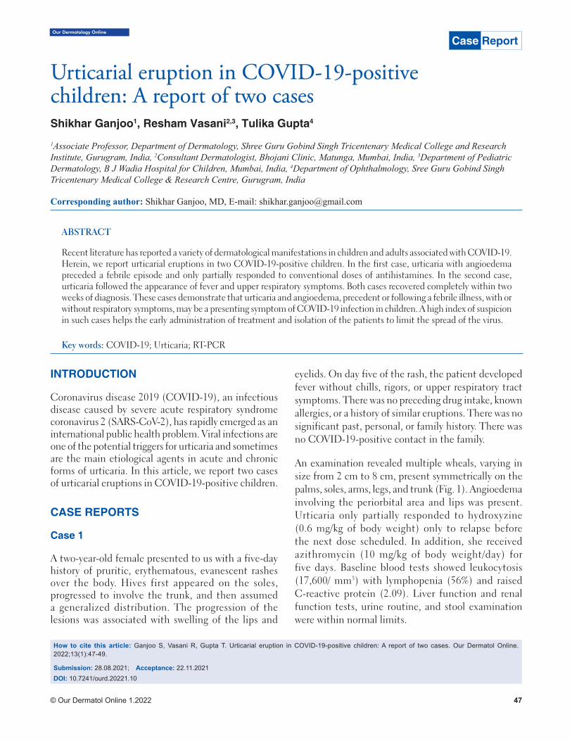
© Our Dermatol Online 1.2022 47
Our Dermatology Online
How to cite this article: Ganjoo S, Vasani R, Gupta T. Urticarial eruption in COVID-19-positive children: A report of two cases. Our Dermatol Online. 2022;13(1):47-49.
Submission: 28.08.2021; Acceptance: 22.11.2021DOI: 10.7241/ourd.20221.10
Urticarial eruption in COVID-19-positive Urticarial eruption in COVID-19-positive children: A report of two caseschildren: A report of two casesShikhar Ganjoo1, Resham Vasani2,3, Tulika Gupta4
1Associate Professor, Department of Dermatology, Shree Guru Gobind Singh Tricentenary Medical College and Research Institute, Gurugram, India, 2Consultant Dermatologist, Bhojani Clinic, Matunga, Mumbai, India, 3Department of Pediatric Dermatology, B J Wadia Hospital for Children, Mumbai, India, 4Department of Ophthalmology, Sree Guru Gobind Singh Tricentenary Medical College & Research Centre, Gurugram, India
Corresponding author: Shikhar Ganjoo, MD, E-mail: [email protected]
INTRODUCTION
Coronavirus disease 2019 (COVID-19), an infectious disease caused by severe acute respiratory syndrome coronavirus 2 (SARS-CoV-2), has rapidly emerged as an international public health problem. Viral infections are one of the potential triggers for urticaria and sometimes are the main etiological agents in acute and chronic forms of urticaria. In this article, we report two cases of urticarial eruptions in COVID-19-positive children.
CASE REPORTS
Case 1
A two-year-old female presented to us with a five-day history of pruritic, erythematous, evanescent rashes over the body. Hives first appeared on the soles, progressed to involve the trunk, and then assumed a generalized distribution. The progression of the lesions was associated with swelling of the lips and
eyelids. On day five of the rash, the patient developed fever without chills, rigors, or upper respiratory tract symptoms. There was no preceding drug intake, known allergies, or a history of similar eruptions. There was no significant past, personal, or family history. There was no COVID-19-positive contact in the family.
An examination revealed multiple wheals, varying in size from 2 cm to 8 cm, present symmetrically on the palms, soles, arms, legs, and trunk (Fig. 1). Angioedema involving the periorbital area and lips was present. Urticaria only partially responded to hydroxyzine (0.6 mg/kg of body weight) only to relapse before the next dose scheduled. In addition, she received azithromycin (10 mg/kg of body weight/day) for five days. Baseline blood tests showed leukocytosis (17,600/ mm3) with lymphopenia (56%) and raised C-reactive protein (2.09). Liver function and renal function tests, urine routine, and stool examination were within normal limits.
ABSTRACT
Recent literature has reported a variety of dermatological manifestations in children and adults associated with COVID-19. Herein, we report urticarial eruptions in two COVID-19-positive children. In the first case, urticaria with angioedema preceded a febrile episode and only partially responded to conventional doses of antihistamines. In the second case, urticaria followed the appearance of fever and upper respiratory symptoms. Both cases recovered completely within two weeks of diagnosis. These cases demonstrate that urticaria and angioedema, precedent or following a febrile illness, with or without respiratory symptoms, may be a presenting symptom of COVID-19 infection in children. A high index of suspicion in such cases helps the early administration of treatment and isolation of the patients to limit the spread of the virus.
Key words: COVID-19; Urticaria; RT-PCR
Case Report
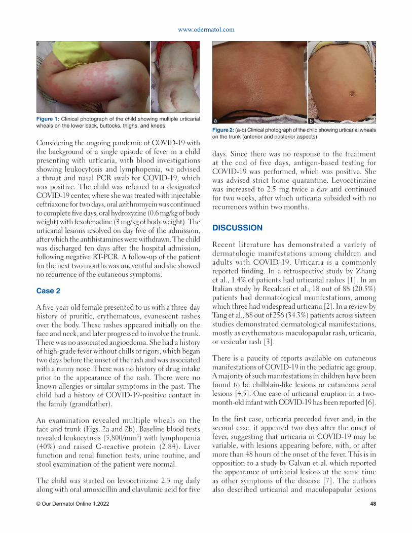
Figure 1: Clinical photograph of the child showing multiple urticarial wheals on the lower back, buttocks, thighs, and knees.
www.odermatol.com
© Our Dermatol Online 1.2022 48
Considering the ongoing pandemic of COVID-19 with the background of a single episode of fever in a child presenting with urticaria, with blood investigations showing leukocytosis and lymphopenia, we advised a throat and nasal PCR swab for COVID-19, which was positive. The child was referred to a designated COVID-19 center, where she was treated with injectable ceftriaxone for two days, oral azithromycin was continued to complete five days, oral hydroxyzine (0.6 mg/kg of body weight) with fexofenadine (3 mg/kg of body weight). The urticarial lesions resolved on day five of the admission, after which the antihistamines were withdrawn. The child was discharged ten days after the hospital admission, following negative RT-PCR. A follow-up of the patient for the next two months was uneventful and she showed no recurrence of the cutaneous symptoms.
Case 2
A five-year-old female presented to us with a three-day history of pruritic, erythematous, evanescent rashes over the body. These rashes appeared initially on the face and neck, and later progressed to involve the trunk. There was no associated angioedema. She had a history of high-grade fever without chills or rigors, which began two days before the onset of the rash and was associated with a runny nose. There was no history of drug intake prior to the appearance of the rash. There were no known allergies or similar symptoms in the past. The child had a history of COVID-19-positive contact in the family (grandfather).
An examination revealed multiple wheals on the face and trunk (Figs. 2a and 2b). Baseline blood tests revealed leukocytosis (5,800/mm3) with lymphopenia (40%) and raised C-reactive protein (2.84). Liver function and renal function tests, urine routine, and stool examination of the patient were normal.
The child was started on levocetirizine 2.5 mg daily along with oral amoxicillin and clavulanic acid for five
days. Since there was no response to the treatment at the end of five days, antigen-based testing for COVID-19 was performed, which was positive. She was advised strict home quarantine. Levocetirizine was increased to 2.5 mg twice a day and continued for two weeks, after which urticaria subsided with no recurrences within two months.
DISCUSSION
Recent literature has demonstrated a variety of dermatologic manifestations among children and adults with COVID-19. Urticaria is a commonly reported finding. In a retrospective study by Zhang et al., 1.4% of patients had urticarial rashes [1]. In an Italian study by Recalcati et al., 18 out of 88 (20.5%) patients had dermatological manifestations, among which three had widespread urticaria [2]. In a review by Tang et al., 88 out of 256 (34.3%) patients across sixteen studies demonstrated dermatological manifestations, mostly as erythematous maculopapular rash, urticaria, or vesicular rash [3].
There is a paucity of reports available on cutaneous manifestations of COVID-19 in the pediatric age group. A majority of such manifestations in children have been found to be chilblain-like lesions or cutaneous acral lesions [4,5]. One case of urticarial eruption in a two-month-old infant with COVID-19 has been reported [6].
In the first case, urticaria preceded fever and, in the second case, it appeared two days after the onset of fever, suggesting that urticaria in COVID-19 may be variable, with lesions appearing before, with, or after more than 48 hours of the onset of the fever. This is in opposition to a study by Galvan et al. which reported the appearance of urticarial lesions at the same time as other symptoms of the disease [7]. The authors also described urticarial and maculopapular lesions
Figure 2: (a-b) Clinical photograph of the child showing urticarial wheals on the trunk (anterior and posterior aspects).
ba
www.odermatol.com
© Our Dermatol Online 1.2022 49
as associated with a more severe COVID-19 disease. However, no such association was found in either of our cases.
The appearance of urticarial lesions in the early phase of the disease suggests the direct role of SARS-CoV-2, entering the vascular tissue with angiotensin converting enzyme 2 protein inside the cells. The deposition of Ag–Ab complexes on the site leads to complement activation and subsequent mast cell degranulation, leading to the onset of urticaria. Increased IL-6 levels in COVID-19 are also implicated in the pathogenesis of urticaria [8].
None of our two patients gave a history of drug intake prior to the urticarial episode, excluding a drug-induced etiology.
Our cases demonstrate that urticaria with pyrexia in a child may be the first manifestation of COVID-19 infection even with no respiratory symptoms. Given the current pandemic circumstances, a clinician should consider COVID-19 as a possible cause of urticaria, with or without angioedema, especially if the disease is unresponsive to the conventional doses of antihistamines. These patients may unknowingly infect others and contribute to the spread of the infection.
CONSENT
The examination of the patient was conducted according to the principles of the Declaration of Helsinki.
The authors certify that they have obtained all appropriate patient consent forms, in which the patients gave their consent for images
and other clinical information to be included in the journal. The patients understand that their names and initials will not be published and due effort will be made to conceal their identity, but that anonymity cannot be guaranteed.
REFERENCES
1. Zhang JJ, Dong X, Cao YY, Yuan YD, Yang YB, Yan YQ, et al. Clinical characteristics of 140 patients infected with SARS-CoV-2 in Wuhan, China. Allergy. 2020;75:1730-41.
2. Recalcati S. Cutaneous manifestations in COVID-19: A first perspective. J Eur Acad Dermatol Venereol. 2020;34:e212-3.
3. Tang K, Wang Y, Zhang H, Zheng Q, Fang R, Sun Q. Cutaneous manifestations of the Coronavirus Disease 2019 (COVID-19): A brief review. Dermatol Ther. 2020;33:e13528.
4. Andina D, Noguera-Morel L, Bascuas-Arribas M, Gaitero-Tristán J, Alonso-Cadenas JA, Escalada-Pellitero S, et al. Chilblains in children in the setting of COVID-19 pandemic. Pediatr Dermatol. 2020;37:406-11.
5. Piccolo V, Neri I, Filippesch C. Chilblain-like lesions during COVID-19 epidemic: A preliminary study on 63 patients. J Eur Acad Dermatol Venereol. 2020:34;e291-3.
6. Morey-Olivé M, Espiau M, Mercadal-Hally M, Lera-Carballo E, García-Patos V. Cutaneous manifestations in the current pandemic of coronavirus infection disease (COVID 2019). An Pediatr (Engl Ed). 2020:92;374-5.
7. Galván Casas C, Català A, Carretero Hernández G, Rodríguez-Jiménez P, Fernández-Nieto D, Rodríguez-Villa Lario A, et al. Classifi cation of the cutaneous manifestations of COVID-19: A rapid prospective nationwide consensus study in Spain with 375 cases. Br J Dermatol. 2020;183:71-7.
8. Kaushik A, Parsad D, Kumaran M. Urticaria in the times of COVID-19. Dermatol Ther. 2020;33:e13817.
Copyright by Shikhar Ganjoo, et al. This is an open-access article distributed under the terms of the Creative Commons Attribution License, which permits unrestricted use, distribution, and reproduction in any medium, provided the original author and source are credited.Source of Support: Nil, Confl ict of Interest: None declared.
© Our Dermatol Online 1.2022 50
Our Dermatology Online
Warty cutaneous tuberculosis of the nose: A rare Warty cutaneous tuberculosis of the nose: A rare localizationlocalizationMoussa Doulla1, Laouali Salissou1, Nina Korsaga/Some2, Maimouna Mamadou Ouedraogo1, Larabou Aminou3, Mahamadou Zaki Harouna4, Soumana Alphazazi5, Pascal Niamba6, Adama Traoré6
1Department of Dermatology, National Hospital Niamey, Niamey, Niger, 2Department of Dermatology-Venereology of the CMA of Pissy Ouagadougou, Burkina Faso, 3Department of Dermatology-Venereology of the Reference Hospital of Maradi, Niger. 4Amirou Boubacar Diallo National Hospital of Niamey, Niger, 5National Center for the fi ght against Tuberculosis and Lung Diseases Niamey, Niger, 6Department of Dermatology-Venereology of CHU-YO, Ouagadougou, Burkina Faso
Corresponding author: Laouali Salissou, MD, E-mail: [email protected]
INTRODUCTION
Cutaneous tuberculosis includes all cutaneous manifestations due to mycobacteria of the tuberculosis complex: Mycobacterium tuberculosis, Mycobacterium bovis, and Mycobacterium africanum [1]. It is rare, representing 2% of cases of extrapulmonary tuberculosis [2-4]. Its clinical forms are highly numerous and much more poorly understood than in the past [5,6]. Diagnosis is often difficult due to the polymorphism of both clinical and histological pictures [3,4,6,7]. Herein, we report a case of cutaneous tuberculosis in its warty form located on the nose.
CASE REPORT
This was a 57-year-old housewife who consulted on March 10, 2018, for a keratotic lesion located on
the nose and evolving for three months. A notion of contagion with a case of pulmonary tuberculosis was found during interrogation as well as a notion of evening fever and weight loss. A treatment combining metronidazole at a dose of 2 g/day per os for one month and traditional herbal medicine by local application had been instituted without success. On physical examination, the patient was in good general condition. A dermatological examination revealed a blackish, keratotic patch with a crusty, melliceric surface covering the entire nasal mass with an overflow in the nasolabial folds (Fig. 1). This lesion was neither itchy nor painful. In front of this case, we evoked cutaneous tuberculosis, leishmaniasis, basal cell carcinoma, and Kaposi’s disease.
A skin biopsy with a pathological examination showed an inflammatory tuberculoid granuloma
ABSTRACT
Cutaneous tuberculosis is a rare, extra-pulmonary form of tuberculosis caused by mycobacteria of the tuberculosis complex. It is characterized by clinical polymorphism often posing a difficult diagnostic challenge. Herein, we report a case of cutaneous tuberculosis in its warty form located on the nose. This was a 57-year-old patient who was infected in the classroom three months previously while taking lessons from a woman with pulmonary tuberculosis. A facial examination revealed a blackish, papillomatous patch invading almost the entire nose, with a keratotic surface spreading over the wings of the nose. The diagnosis of verrucous tuberculosis was reached on the basis of epidemiological, clinical, and paraclinical arguments. Under anti-tuberculosis treatment for six months, the lesion had healed without sequelae. The diagnosis of verrucous cutaneous tuberculosis must be established in the presence of any chronic and crusty lesion. The management responds to the treatment protocol for all forms of tuberculosis.
Key words: Cutaneous tuberculosis; Warty; nose; Niamey; Niger
How to cite this article: Doulla M, Salissou L, Korsaga/Somé N, Ouédraogo MM, Aminou L,Zaki Harouna M, Alphazazi S, Niamba P, Traoré A. Warty cutaneous tuberculosis of the nose: A rare localization. Our Dermatol Online. 2022;13(1):50-52.Submission: 21.06.2021; Acceptance: 07.12.2021DOI: 10.7241/ourd.20221.11
Case Report
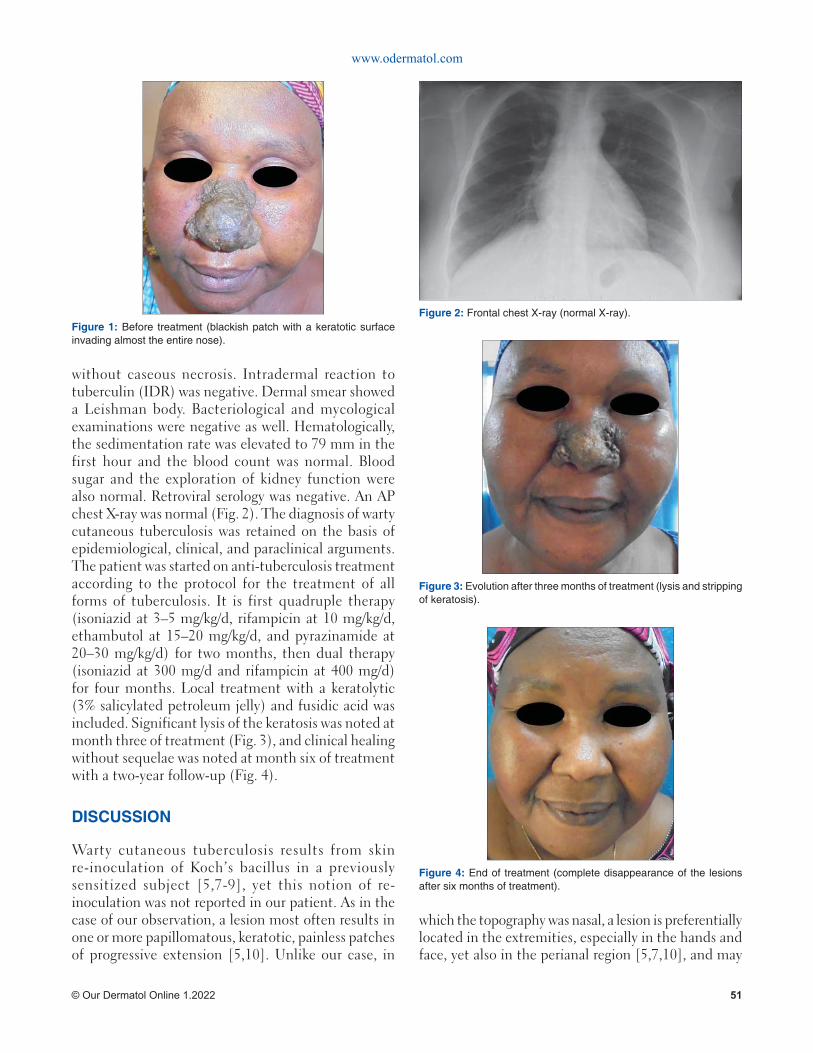
www.odermatol.com
© Our Dermatol Online 1.2022 51
without caseous necrosis. Intradermal reaction to tuberculin (IDR) was negative. Dermal smear showed a Leishman body. Bacteriological and mycological examinations were negative as well. Hematologically, the sedimentation rate was elevated to 79 mm in the first hour and the blood count was normal. Blood sugar and the exploration of kidney function were also normal. Retroviral serology was negative. An AP chest X-ray was normal (Fig. 2). The diagnosis of warty cutaneous tuberculosis was retained on the basis of epidemiological, clinical, and paraclinical arguments. The patient was started on anti-tuberculosis treatment according to the protocol for the treatment of all forms of tuberculosis. It is first quadruple therapy (isoniazid at 3–5 mg/kg/d, rifampicin at 10 mg/kg/d, ethambutol at 15–20 mg/kg/d, and pyrazinamide at 20–30 mg/kg/d) for two months, then dual therapy (isoniazid at 300 mg/d and rifampicin at 400 mg/d) for four months. Local treatment with a keratolytic (3% salicylated petroleum jelly) and fusidic acid was included. Significant lysis of the keratosis was noted at month three of treatment (Fig. 3), and clinical healing without sequelae was noted at month six of treatment with a two-year follow-up (Fig. 4).
DISCUSSION
Warty cutaneous tuberculosis results from skin re-inoculation of Koch’s bacillus in a previously sensitized subject [5,7-9], yet this notion of re-inoculation was not reported in our patient. As in the case of our observation, a lesion most often results in one or more papillomatous, keratotic, painless patches of progressive extension [5,10]. Unlike our case, in
which the topography was nasal, a lesion is preferentially located in the extremities, especially in the hands and face, yet also in the perianal region [5,7,10], and may
Figure 4: End of treatment (complete disappearance of the lesions after six months of treatment).
Figure 3: Evolution after three months of treatment (lysis and stripping of keratosis).
Figure 1: Before treatment (blackish patch with a keratotic surface invading almost the entire nose).
Figure 2: Frontal chest X-ray (normal X-ray).
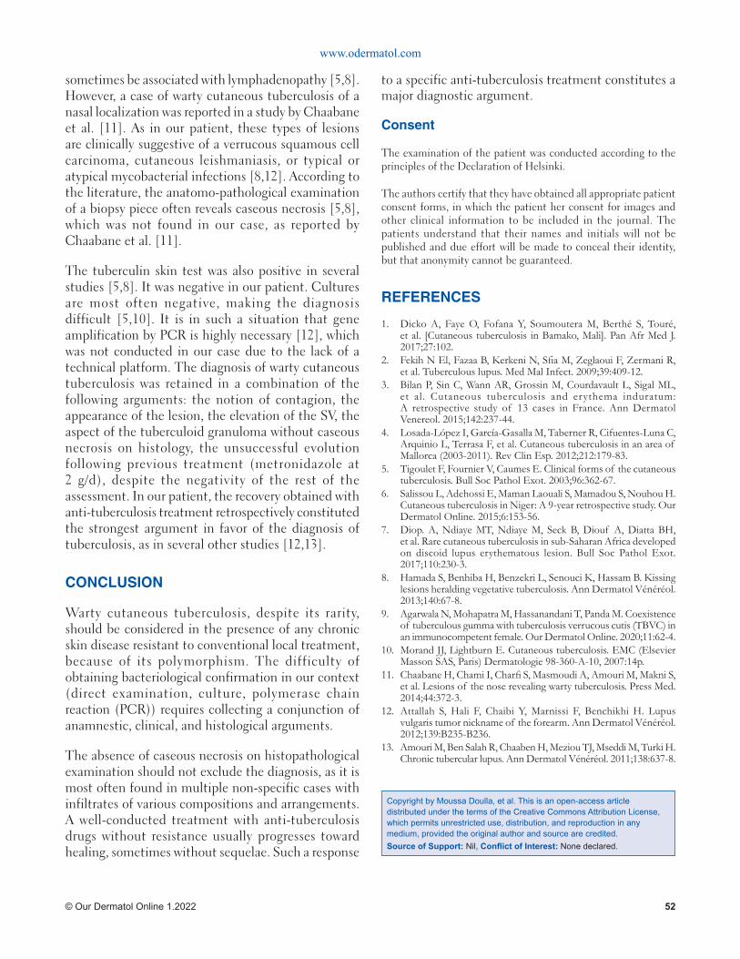
www.odermatol.com
© Our Dermatol Online 1.2022 52
sometimes be associated with lymphadenopathy [5,8]. However, a case of warty cutaneous tuberculosis of a nasal localization was reported in a study by Chaabane et al. [11]. As in our patient, these types of lesions are clinically suggestive of a verrucous squamous cell carcinoma, cutaneous leishmaniasis, or typical or atypical mycobacterial infections [8,12]. According to the literature, the anatomo-pathological examination of a biopsy piece often reveals caseous necrosis [5,8], which was not found in our case, as reported by Chaabane et al. [11].
The tuberculin skin test was also positive in several studies [5,8]. It was negative in our patient. Cultures are most often negative, making the diagnosis difficult [5,10]. It is in such a situation that gene amplification by PCR is highly necessary [12], which was not conducted in our case due to the lack of a technical platform. The diagnosis of warty cutaneous tuberculosis was retained in a combination of the following arguments: the notion of contagion, the appearance of the lesion, the elevation of the SV, the aspect of the tuberculoid granuloma without caseous necrosis on histology, the unsuccessful evolution following previous treatment (metronidazole at 2 g/d), despite the negativity of the rest of the assessment. In our patient, the recovery obtained with anti-tuberculosis treatment retrospectively constituted the strongest argument in favor of the diagnosis of tuberculosis, as in several other studies [12,13].
CONCLUSION
Warty cutaneous tuberculosis, despite its rarity, should be considered in the presence of any chronic skin disease resistant to conventional local treatment, because of its polymorphism. The difficulty of obtaining bacteriological confirmation in our context (direct examination, culture, polymerase chain reaction (PCR)) requires collecting a conjunction of anamnestic, clinical, and histological arguments.
The absence of caseous necrosis on histopathological examination should not exclude the diagnosis, as it is most often found in multiple non-specific cases with infiltrates of various compositions and arrangements. A well-conducted treatment with anti-tuberculosis drugs without resistance usually progresses toward healing, sometimes without sequelae. Such a response
to a specific anti-tuberculosis treatment constitutes a major diagnostic argument.
Consent
The examination of the patient was conducted according to the principles of the Declaration of Helsinki.
The authors certify that they have obtained all appropriate patient consent forms, in which the patient her consent for images and other clinical information to be included in the journal. The patients understand that their names and initials will not be published and due effort will be made to conceal their identity, but that anonymity cannot be guaranteed.
REFERENCES
1. Dicko A, Faye O, Fofana Y, Soumoutera M, Berthé S, Touré, et al. [Cutaneous tuberculosis in Bamako, Mali]. Pan Afr Med J. 2017;27:102.
2. Fekih N El, Fazaa B, Kerkeni N, Sfi a M, Zeglaoui F, Zermani R, et al. Tuberculous lupus. Med Mal Infect. 2009;39:409-12.
3. Bilan P, Sin C, Wann AR, Grossin M, Courdavault L, Sigal ML, et al. Cutaneous tuberculosis and erythema induratum: A retrospective study of 13 cases in France. Ann Dermatol Venereol. 2015;142:237-44.
4. Losada-López I, García-Gasalla M, Taberner R, Cifuentes-Luna C, Arquinio L, Terrasa F, et al. Cutaneous tuberculosis in an area of Mallorca (2003-2011). Rev Clin Esp. 2012;212:179-83.
5. Tigoulet F, Fournier V, Caumes E. Clinical forms of the cutaneous tuberculosis. Bull Soc Pathol Exot. 2003;96:362-67.
6. Salissou L, Adehossi E, Maman Laouali S, Mamadou S, Nouhou H. Cutaneous tuberculosis in Niger: A 9-year retrospective study. Our Dermatol Online. 2015;6:153-56.
7. Diop. A, Ndiaye MT, Ndiaye M, Seck B, Diouf A, Diatta BH, et al. Rare cutaneous tuberculosis in sub-Saharan Africa developed on discoid lupus erythematous lesion. Bull Soc Pathol Exot. 2017;110:230-3.
8. Hamada S, Benhiba H, Benzekri L, Senouci K, Hassam B. Kissing lesions heralding vegetative tuberculosis. Ann Dermatol Vénéréol. 2013;140:67-8.
9. Agarwala N, Mohapatra M, Hassanandani T, Panda M. Coexistence of tuberculous gumma with tuberculosis verrucous cutis (TBVC) in an immunocompetent female. Our Dermatol Online. 2020;11:62-4.
10. Morand JJ, Lightburn E. Cutaneous tuberculosis. EMC (Elsevier Masson SAS, Paris) Dermatologie 98-360-A-10, 2007:14p.
11. Chaabane H, Chami I, Charfi S, Masmoudi A, Amouri M, Makni S, et al. Lesions of the nose revealing warty tuberculosis. Press Med. 2014;44:372-3.
12. Attallah S, Hali F, Chaibi Y, Marnissi F, Benchikhi H. Lupus vulgaris tumor nickname of the forearm. Ann Dermatol Vénéréol. 2012;139:B235-B236.
13. Amouri M, Ben Salah R, Chaaben H, Meziou TJ, Mseddi M, Turki H. Chronic tubercular lupus. Ann Dermatol Vénéréol. 2011;138:637-8.
Copyright by Moussa Doulla, et al. This is an open-access article distributed under the terms of the Creative Commons Attribution License, which permits unrestricted use, distribution, and reproduction in any medium, provided the original author and source are credited.Source of Support: Nil, Confl ict of Interest: None declared.
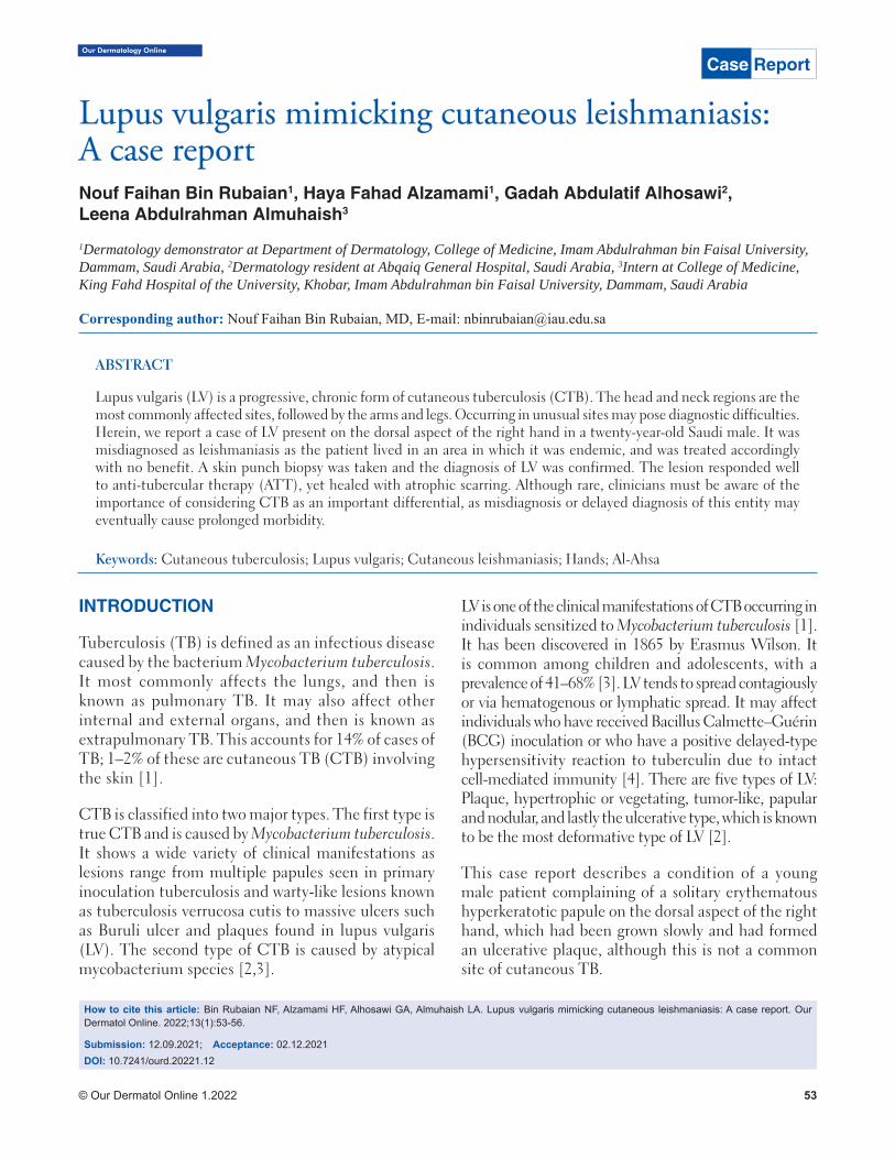
© Our Dermatol Online 1.2022 53
Our Dermatology Online
How to cite this article: Bin Rubaian NF, Alzamami HF, Alhosawi GA, Almuhaish LA. Lupus vulgaris mimicking cutaneous leishmaniasis: A case report. Our Dermatol Online. 2022;13(1):53-56.
Submission: 12.09.2021; Acceptance: 02.12.2021DOI: 10.7241/ourd.20221.12
Lupus vulgaris mimicking cutaneous leishmaniasis: Lupus vulgaris mimicking cutaneous leishmaniasis: A case reportA case reportNouf Faihan Bin Rubaian1, Haya Fahad Alzamami1, Gadah Abdulatif Alhosawi2, Leena Abdulrahman Almuhaish3
1Dermatology demonstrator at Department of Dermatology, College of Medicine, Imam Abdulrahman bin Faisal University, Dammam, Saudi Arabia, 2Dermatology resident at Abqaiq General Hospital, Saudi Arabia, 3Intern at College of Medicine, King Fahd Hospital of the University, Khobar, Imam Abdulrahman bin Faisal University, Dammam, Saudi Arabia
Corresponding author: Nouf Faihan Bin Rubaian, MD, E-mail: [email protected]
INTRODUCTION
Tuberculosis (TB) is defined as an infectious disease caused by the bacterium Mycobacterium tuberculosis. It most commonly affects the lungs, and then is known as pulmonary TB. It may also affect other internal and external organs, and then is known as extrapulmonary TB. This accounts for 14% of cases of TB; 1–2% of these are cutaneous TB (CTB) involving the skin [1].
CTB is classified into two major types. The first type is true CTB and is caused by Mycobacterium tuberculosis. It shows a wide variety of clinical manifestations as lesions range from multiple papules seen in primary inoculation tuberculosis and warty-like lesions known as tuberculosis verrucosa cutis to massive ulcers such as Buruli ulcer and plaques found in lupus vulgaris (LV). The second type of CTB is caused by atypical mycobacterium species [2,3].
LV is one of the clinical manifestations of CTB occurring in individuals sensitized to Mycobacterium tuberculosis [1]. It has been discovered in 1865 by Erasmus Wilson. It is common among children and adolescents, with a prevalence of 41–68% [3]. LV tends to spread contagiously or via hematogenous or lymphatic spread. It may affect individuals who have received Bacillus Calmette–Gué rin (BCG) inoculation or who have a positive delayed-type hypersensitivity reaction to tuberculin due to intact cell-mediated immunity [4]. There are five types of LV: Plaque, hypertrophic or vegetating, tumor-like, papular and nodular, and lastly the ulcerative type, which is known to be the most deformative type of LV [2].
This case report describes a condition of a young male patient complaining of a solitary erythematous hyperkeratotic papule on the dorsal aspect of the right hand, which had been grown slowly and had formed an ulcerative plaque, although this is not a common site of cutaneous TB.
ABSTRACT
Lupus vulgaris (LV) is a progressive, chronic form of cutaneous tuberculosis (CTB). The head and neck regions are the most commonly affected sites, followed by the arms and legs. Occurring in unusual sites may pose diagnostic difficulties. Herein, we report a case of LV present on the dorsal aspect of the right hand in a twenty-year-old Saudi male. It was misdiagnosed as leishmaniasis as the patient lived in an area in which it was endemic, and was treated accordingly with no benefit. A skin punch biopsy was taken and the diagnosis of LV was confirmed. The lesion responded well to anti-tubercular therapy (ATT), yet healed with atrophic scarring. Although rare, clinicians must be aware of the importance of considering CTB as an important differential, as misdiagnosis or delayed diagnosis of this entity may eventually cause prolonged morbidity.
Keywords: Cutaneous tuberculosis; Lupus vulgaris; Cutaneous leishmaniasis; Hands; Al-Ahsa
Case Report
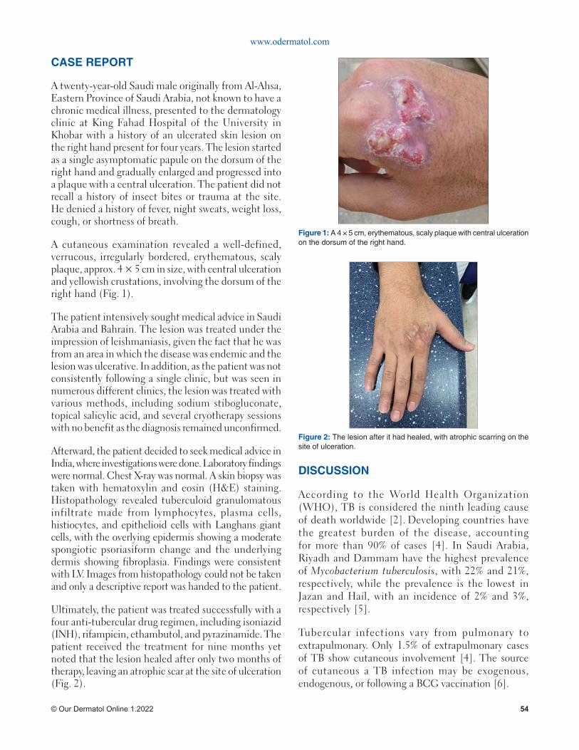
www.odermatol.com
© Our Dermatol Online 1.2022 54
CASE REPORT
A twenty-year-old Saudi male originally from Al-Ahsa, Eastern Province of Saudi Arabia, not known to have a chronic medical illness, presented to the dermatology clinic at King Fahad Hospital of the University in Khobar with a history of an ulcerated skin lesion on the right hand present for four years. The lesion started as a single asymptomatic papule on the dorsum of the right hand and gradually enlarged and progressed into a plaque with a central ulceration. The patient did not recall a history of insect bites or trauma at the site. He denied a history of fever, night sweats, weight loss, cough, or shortness of breath.
A cutaneous examination revealed a well-defined, verrucous, irregularly bordered, erythematous, scaly plaque, approx. 4 × 5 cm in size, with central ulceration and yellowish crustations, involving the dorsum of the right hand (Fig. 1).
The patient intensively sought medical advice in Saudi Arabia and Bahrain. The lesion was treated under the impression of leishmaniasis, given the fact that he was from an area in which the disease was endemic and the lesion was ulcerative. In addition, as the patient was not consistently following a single clinic, but was seen in numerous different clinics, the lesion was treated with various methods, including sodium stibogluconate, topical salicylic acid, and several cryotherapy sessions with no benefit as the diagnosis remained unconfirmed.
Afterward, the patient decided to seek medical advice in India, where investigations were done. Laboratory findings were normal. Chest X-ray was normal. A skin biopsy was taken with hematoxylin and eosin (H&E) staining. Histopathology revealed tuberculoid granulomatous infiltrate made from lymphocytes, plasma cells, histiocytes, and epithelioid cells with Langhans giant cells, with the overlying epidermis showing a moderate spongiotic psoriasiform change and the underlying dermis showing fibroplasia. Findings were consistent with LV. Images from histopathology could not be taken and only a descriptive report was handed to the patient.
Ultimately, the patient was treated successfully with a four anti-tubercular drug regimen, including isoniazid (INH), rifampicin, ethambutol, and pyrazinamide. The patient received the treatment for nine months yet noted that the lesion healed after only two months of therapy, leaving an atrophic scar at the site of ulceration (Fig. 2).
DISCUSSION
According to the World Health Organization (WHO), TB is considered the ninth leading cause of death worldwide [2]. Developing countries have the greatest burden of the disease, accounting for more than 90% of cases [4]. In Saudi Arabia, Riyadh and Dammam have the highest prevalence of Mycobacterium tuberculosis, with 22% and 21%, respectively, while the prevalence is the lowest in Jazan and Hail, with an incidence of 2% and 3%, respectively [5].
Tubercular infections vary from pulmonary to extrapulmonary. Only 1.5% of extrapulmonary cases of TB show cutaneous involvement [4]. The source of cutaneous a TB infection may be exogenous, endogenous, or following a BCG vaccination [6].
Figure 1: A 4 × 5 cm, erythematous, scaly plaque with central ulceration on the dorsum of the right hand.
Figure 2: The lesion after it had healed, with atrophic scarring on the site of ulceration.
www.odermatol.com
© Our Dermatol Online 1.2022 55
LV is a chronic, progressive disease, occurring in patients sensitized to Mycobacterium tuberculosis with moderate to high immunity [6]. The infection occurs mainly through direct extension from the underlying affected tissue or by hematogenous or lymphatics spread. Infection may also occur due to the reactivation of a latent cutaneous focus secondary to previous silent bacteremia in patients in whom the underlying focus is not clear [6]. LV commonly involves the head and neck areas, followed by the arms and legs [7]. In Europe, 80% of cases involve the head and neck, in particular, the nose and cheeks. On the other hand, in India, the buttocks, thighs, and legs are commonly involved.
Usually, A lesion of LV presents itself as a solitary, reddish-brown papule or nodule, soft in consistency, gradually enlarging into a plaque with a tendency to ulcerate. The plaque often shows an apple-jelly color on diascopy [4]. Clinically, LV shows five major clinical patterns depending on the local tissue response to the infection, including plaque, hypertrophic or vegetation, tumor-like, papular or nodular, and the ulcerative type [6]. Plaque-type LV is considered a common form, while based on a study done in India, the ulcerative type was the least common clinical variant, accounting for 14.2% of cases [8]. Long-standing ulcerative LV may eventually lead to scarring and deformity in addition to the risk of squamous cell carcinoma [9]. Fortunately, our patient had no destruction or deformity in the underlying tissue. However, the lesion healed leaving an atrophic scar.
In our case, the site of involvement considered an exposed area was rarely to be affected. In addition, the lesion greatly mimicked chronic cutaneous leishmaniasis due to the presence of an ulcer, especially since it was endemic in the region from which the patient came. Therefore, it is crucial for clinicians, especially in endemic areas, to keep in mind other differential diagnoses even if the lesions are present in unusual sites. In this case, other than leishmaniasis, CTD, lupus erythematosus, sarcoidosis, and granuloma annulare should definitely be considered [10].
To diagnose LV, there are different modalities that have to be considered. Firstly, a diascopy examination in the clinic will show apple-jelly nodules in the plaques of LV. Secondly, a tuberculin test and interferon-gamma (IFN-γ) release assays (QuantiFERON TB Gold, T-SPOT.TB) may confirm the diagnosis of LV through the presence of IFN-γ from T cells in the patient’s blood, mediated by Mycobacterium tuberculosis peptides [11]. Thirdly, a skin biopsy will show the histopathological features of LV,
including a thin, atrophied epidermis or acanthosis with hyperkeratosis and pseudoepitheliomatous hyperplasia, as well as scant caseation of tubercles and invisible bacilli [4]. In comparison, the histological features of leishmaniasis are similar to some extent to LV, except for necrosis, atrophy, and hyperplasia of the epidermis. Also, there is a multitude of multinucleated giant cells with slight parasites present in lesions of leishmaniasis. Additionally, amastigote organisms may be visible with H&E or Giemsa stain. Fourthly, a polymerase chain reaction (PCR) test may also be helpful in diagnosing LV, which will detect positive Mycobacterium tuberculosis organisms [1,11].
Our patient was diagnosed with LV through a skin punch biopsy. The histopathology report revealed patchy, nodular, tuberculoid, granulomatous infiltrate made from lymphocytes, plasma cells, histiocytes, and epithelioid cells with occasional Langhans giant cells. The overlying epidermis showed moderate acanthotic and spongiotic psoriasiform changes. The underlying dermis showed fibroplasia. Chest X-ray, complete blood count, and chemistry were normal.
An appropriate treatment may assist in decreasing the transmission of Mycobacterium tuberculosis and prevent bacterial resistance to antibiotics [3]. Various drugs were highlighted by the WHO: INH, rifampicin, ethambutol, pyrazinamide, and streptomycin [3].
In CTB, the management plan consists of two periods. During the first eight weeks, intense therapy is employed, consisting of INH, rifampicin, ethambutol, and pyrazinamide. Then, therapy is resumed as a maintenance for sixteen weeks with INH and rifampicin. Ethambutol may be used as an alternative to INH in the second phase of the treatment if the patient shows resistance [3].
Our patient was mistreated as a case of leishmaniasis in different dermatology clinics, based on history taking and clinical examination. Sodium stibogluconate injection, topical salicylic acid, and cryotherapy were administered yet the results were unsatisfactory. In the end, the patient underwent anti-tubercular therapy (ATT) and four drugs were prescribed. A remarkable improvement was observed after two months of therapy. The treatment successfully ended after nine months.
CONCLUSION
An awareness of various clinical manifestations of CTB especially in uncommon sites, as in the
www.odermatol.com
© Our Dermatol Online 1.2022 56
presented case, is crucial to the early diagnosis and treatment, thus reducing morbidity. In our case, the lesion was misdiagnosed based on clinical findings as leishmaniasis. After a skin biopsy was taken, the diagnosis of LV was confirmed and ATT was initiated. Afterward, the lesion subsided leaving an atrophic scar in the site of ulceration, which could have been avoided by an early diagnosis.
Consent
The examination of the patient was conducted according to the principles of the Declaration of Helsinki. The authors certify that they have obtained all appropriate patient consent forms, in which the patients gave their consent for images and other clinical information to be included in the journal. The patients understand that their names and initials will not be published and due effort will be made to conceal their identity, but that anonymity cannot be guaranteed.
REFERENCES
1. Gunawan H, Achdiat PA, Hindritiani R, Essary ED, Ningtias LD, Siregar EP, et al. Various cutaneous tuberculosis with rare clinical manifestations: A case series. Int J Mycobacteriol. 2018;7:288-91.
2. Chen Q, Chen W, Hao F. Cutaneous tuberculosis: A great imitator. Clin Dermatol. 2019;37:192-9.
3. van Zyl L, du Plessis J, Viljoen J. Cutaneous tuberculosis
overview and current treatment regimens. Tuberculosis (Edinb). 2015;95:629-38.
4. Nair PA, Mehta MJ, Patel BB. Ulcerative lupus vulgaris over nose, leading to cosmetic deformity. Indian J Dermatol. 2015;60:104.
5. Al Watban AZ, Al Salamah AA, El Faki MG. Prevalence of suspected tuberculosis in the Kingdom of Saudi Arabia according to conventional and molecular methods. J Family Community Med. 2014;21:182-5.
6. Pai VV, Naveen KN, Athanikar SB, Dinesh US, Divyashree A, Gupta G. A clinico-histopathological study of lupus vulgaris: A 3-year experience at a tertiary care centre. Indian Dermatol Online J. 2014;5:461-5.
7. Hassan I, Ahmad M, Masood Q. Lupus vulgaris: An atypical presentation. Indian J Dermatol Venereol Leprol. 2010;76:180-1.
8. Chhangte MZ, Thakur BK, Verma S, Dey B. Ulcerative lupus vulgaris primarily involving the ear lobule. Indian Dermatol Online J. 2019;10:729-31.
9. De D, Khadwal A, Singhi P, Dogra S, Radotra BD, Kanwar AJ. Chronic nonhealing ulcer on the hand. Clin Exp Dermatol. 2009;34:647-8.
10. Douba MD, Abbas O, Wali A, Nassany J, Aouf A, Tibbi MS, et al. Chronic cutaneous leishmaniasis, a great mimicker with various clinical presentations: 12 years experience from Aleppo. J Eur Acad Dermatol Venereol. 2012;26:1224-9.
11. Bolognia J, Schaffer J, Cerroni L: Dermatology. 4th ed. Elsevier; 2018.
Copyright by Nouf Faihan Bin Rubaian, et al. This is an open-access article distributed under the terms of the Creative Commons Attribution License, which permits unrestricted use, distribution, and reproduction in any medium, provided the original author and source are credited.Source of Support: Nil, Confl ict of Interest: None declared.
© Our Dermatol Online 1.2022 57
Our Dermatology Online
How to cite this article: Dégboé B, Nouhoumon G, Nguessie C, Akpadjan F, Agbéssi N, Koudoukpo C, Zomalheto Z, Sèhonou J, Adégbidi H, Atadokpèdé F. SAPHO syndrome associated with a digestive disorder in a ten-year-old girl: Diagnostic diffi culties. Our Dermatol Online. 2022;13(1):57-61.
Submission: 21.09.2021; Acceptance: 23.11.2021DOI: 10.7241/ourd.20221.13
SAPHO syndrome associated with a digestive disorder SAPHO syndrome associated with a digestive disorder in a ten-year-old girl: Diagnostic diffi cultiesin a ten-year-old girl: Diagnostic diffi cultiesBérénice Dégboé1, Gloria Nouhoumon1, Christabelle Nguessie1, Fabrice Akpadjan1, Nadège Agbéssi2, Christiane Koudoukpo2, Zavier Zomalheto3, Jean Sèhonou4, Hugues Adégbidi1, Félix Atadokpèdé1
1Department of Dermatology Venereology, National Teaching Hospital Center Hubert Koutoukou Maga of Cotonou, Faculty of Health Sciences - University of Abomey-Calavi, Benin, 2Department of Dermatology Venereology, Departmental Teaching Hospital Center Borgou-Alibori, Faculty of Medicine-University of Parakou, Benin, 3Department of Rheumatology, National Teaching Hospital Center Hubert Koutoukou Maga of Cotonou, Faculty of Health Sciences - University of Abomey-Calavi, Benin, 4Department of Hepato-Gastroenterology, National Teaching Hospital Center Hubert Koutoukou Maga of Cotonou, Faculty of Health Sciences - University of Abomey-Calavi, Benin
Corresponding author: Bérénice Dégboé, MD, E-mail: [email protected]
INTRODUCTION
SAPHO syndrome (acronym for synovitis, acne, pustulosis, hyperostosis, osteitis) is an auto-inflammatory disease characterized by neutrophilic damage, which may affect the skin, bones, and joints [1,2]. Having a low incidence (1–4/10.000 individuals), it is usually observed in young adults. Rheumatologic involvement is most often axial and the most frequently reported dermatological manifestations are palmoplantar pustulosis and severe acne [1-4].
These very diverse and nonspecific clinical manifestations often lead to diagnostic delays and sometimes expose patients to numerous
invasive explorations and inappropriate and harmful therapies [1,2,5].
SAPHO syndrome may be associated with other inflammatory diseases, including cryptogenetic inflammatory bowel disease (IBD) [5-7]. Herein, we report a case of SAPHO syndrome associated with digestive involvement, possibly Crohn’s disease, in a ten-year-old girl after almost three years of diagnostic wandering.
CASE REPORT
A ten-year-old girl was referred to us for the management of pustular, painful, and itchy skin lesions that had been
ABSTRACT
SAPHO syndrome (acronym for synovitis, acne, pustulosis, hyperostosis, osteitis) is a rare dermato-rheumatic entity usually observed in young adults. The clinical manifestations are proteinaceous and without specificity at the origin of inflammatory diseases of the intestine. Our clinical case is that of a ten-year-old girl who presented with chronic and recurrent osteomyelitis of the pelvic limbs on a febrile background, followed by persistent and recurrent pustular lesions. During the same period, because of an acute abdominal pain syndrome accompanied by fever, a biological inflammatory syndrome, and predominantly neutrophilic hyperleukocytosis, laparotomy was performed and no lesions were found. She subsequently presented with intermittent and recurrent spasmodic abdominal pain. In view of these various symptoms, a multidisciplinary consultation concluded that the patient had SAPHO syndrome associated with a digestive disorder, possibly Crohn’s disease. Our clinical case illustrates the diagnostic difficulties of SAPHO syndrome.
Keywords: SAPHO syndrome; Chronic recurrent osteomyelitis in children, Inflammatory bowel disease, Pustulosis, Benin
Case Report
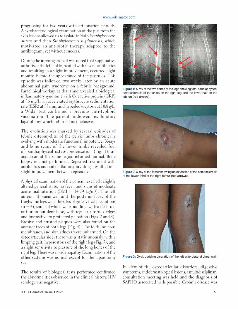
www.odermatol.com
© Our Dermatol Online 1.2022 58
progressing for two years with attenuation periods. A cytobacteriological examination of the pus from the skin lesions allowed us to isolate initially Staphylococcus aureus and then Staphylococcus lugdunensis, which motivated an antibiotic therapy adapted to the antibiogram, yet without success.
During the interrogation, it was noted that suppurative arthritis of the left ankle, treated with several antibiotics and resulting in a slight improvement, occurred eight months before the appearance of the pustules. This episode was followed two weeks later by an acute abdominal pain syndrome on a febrile background. Paraclinical workup at that time revealed a biological inflammatory syndrome with C-reactive protein (CRP) at 56 mg/L, an accelerated erythrocyte sedimentation rate (ESR) at 53 mm, and hyperleukocytosis at 10.8 g/L; a Widal test confirmed a previous anti-typhoid vaccination. The patient underwent exploratory laparotomy, which returned inconclusive.
The evolution was marked by several episodes of febrile osteomyelitis of the pelvic limbs chronically evolving with moderate functional impotence. X-rays and bone scans of the lower limbs revealed foci of pandiaphyseal osteo-condensation (Fig. 1); an angioscan of the same region returned normal. Bone biopsy was not performed. Repeated treatment with antibiotics and anti-inflammatory drugs resulted in a slight improvement between episodes.
A physical examination of the patient revealed a slightly altered general state, no fever, and signs of moderate acute malnutrition (BMI = 14.79 kg/m²). The left anterior thoracic wall and the posterior faces of the thighs and legs were the sites of grossly oval ulcerations (n = 4), some of which were budding, with a flesh-red or fibrino-purulent base, with regular, unstuck edges and insensitive to protected palpation (Figs. 2 and 3). Erosive and crusted plaques were also found on the anterior faces of both legs (Fig. 4). The folds, mucous membranes, and skin adnexa were unharmed. On the osteoarticular side, there was a static anomaly with a limping gait, hyperostosis of the right leg (Fig. 5), and a slight sensitivity to pressure of the long bones of the right leg. There was no adenopathy. Examination of the other systems was normal except for the laparotomy scar.
The results of biological tests performed confirmed the abnormalities observed in the clinical history. HIV serology was negative.
In view of the osteoarticular disorders, digestive symptoms, and dermatological lesions, a multidisciplinary consultation meeting was held and the diagnosis of SAPHO associated with possible Crohn’s disease was
Figure 3: Oval, budding ulceration of the left anterolateral chest wall.
Figure 1: X-ray of the two bones of the legs showing total pandiaphyseal osteosclerosis of the shins on the right leg and the lower half on the left leg (red arrows).
Figure 2: X-ray of the femur showing an extension of the osteosclerosis to the lower third of the right femur (red arrows).
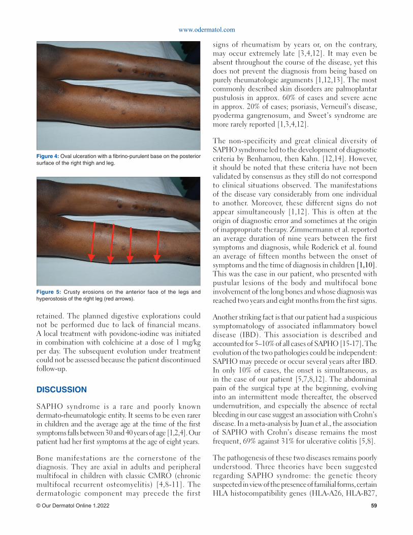
www.odermatol.com
© Our Dermatol Online 1.2022 59
retained. The planned digestive explorations could not be performed due to lack of financial means. A local treatment with povidone-iodine was initiated in combination with colchicine at a dose of 1 mg/kg per day. The subsequent evolution under treatment could not be assessed because the patient discontinued follow-up.
DISCUSSION
SAPHO syndrome is a rare and poorly known dermato-rheumatologic entity. It seems to be even rarer in children and the average age at the time of the first symptoms falls between 30 and 40 years of age [1,2,4]. Our patient had her first symptoms at the age of eight years.
Bone manifestations are the cornerstone of the diagnosis. They are axial in adults and peripheral multifocal in children with classic CMRO (chronic multifocal recurrent osteomyelitis) [4,8-11]. The dermatologic component may precede the first
signs of rheumatism by years or, on the contrary, may occur extremely late [3,4,12]. It may even be absent throughout the course of the disease, yet this does not prevent the diagnosis from being based on purely rheumatologic arguments [1,12,13]. The most commonly described skin disorders are palmoplantar pustulosis in approx. 60% of cases and severe acne in approx. 20% of cases; psoriasis, Verneuil’s disease, pyoderma gangrenosum, and Sweet’s syndrome are more rarely reported [1,3,4,12].
The non-specificity and great clinical diversity of SAPHO syndrome led to the development of diagnostic criteria by Benhamou, then Kahn. [12,14]. However, it should be noted that these criteria have not been validated by consensus as they still do not correspond to clinical situations observed. The manifestations of the disease vary considerably from one individual to another. Moreover, these different signs do not appear simultaneously [1,12]. This is often at the origin of diagnostic error and sometimes at the origin of inappropriate therapy. Zimmermann et al. reported an average duration of nine years between the first symptoms and diagnosis, while Roderick et al. found an average of fifteen months between the onset of symptoms and the time of diagnosis in children [1,10]. This was the case in our patient, who presented with pustular lesions of the body and multifocal bone involvement of the long bones and whose diagnosis was reached two years and eight months from the first signs.
Another striking fact is that our patient had a suspicious symptomatology of associated inflammatory bowel disease (IBD). This association is described and accounted for 5–10% of all cases of SAPHO [15-17]. The evolution of the two pathologies could be independent: SAPHO may precede or occur several years after IBD. In only 10% of cases, the onset is simultaneous, as in the case of our patient [5,7,8,12]. The abdominal pain of the surgical type at the beginning, evolving into an intermittent mode thereafter, the observed undernutrition, and especially the absence of rectal bleeding in our case suggest an association with Crohn’s disease. In a meta-analysis by Juan et al., the association of SAPHO with Crohn’s disease remains the most frequent, 69% against 31% for ulcerative colitis [5,8].
The pathogenesis of these two diseases remains poorly understood. Three theories have been suggested regarding SAPHO syndrome: the genetic theory suspected in view of the presence of familial forms, certain HLA histocompatibility genes (HLA-A26, HLA-B27,
Figure 5: Crusty erosions on the anterior face of the legs and hyperostosis of the right leg (red arrows).
Figure 4: Oval ulceration with a fi brino-purulent base on the posterior surface of the right thigh and leg.
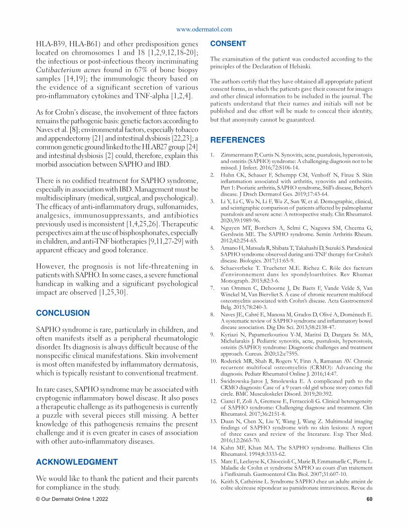
www.odermatol.com
© Our Dermatol Online 1.2022 60
HLA-B39, HLA-B61) and other predisposition genes located on chromosomes 1 and 18 [1,2,9,12,18-20]; the infectious or post-infectious theory incriminating Cutibacterium acnes found in 67% of bone biopsy samples [14,19]; the immunologic theory based on the evidence of a significant secretion of various pro-inflammatory cytokines and TNF-alpha [1,2,4].
As for Crohn’s disease, the involvement of three factors remains the pathogenic basis: genetic factors according to Naves et al. [8]; environmental factors, especially tobacco and appendectomy [21] and intestinal dysbiosis [22,23]; a common genetic ground linked to the HLAB27 group [24] and intestinal dysbiosis [2] could, therefore, explain this morbid association between SAPHO and IBD.
There is no codified treatment for SAPHO syndrome, especially in association with IBD. Management must be multidisciplinary (medical, surgical, and psychological). The efficacy of anti-inflammatory drugs, sulfonamides, analgesics, immunosuppressants, and antibiotics previously used is inconsistent [1,4,25,26]. Therapeutic perspectives aim at the use of bisphosphonates, especially in children, and anti-TNF biotherapies [9,11,27-29] with apparent efficacy and good tolerance.
However, the prognosis is not life-threatening in patients with SAPHO. In some cases, a severe functional handicap in walking and a significant psychological impact are observed [1,25,30].
CONCLUSION
SAPHO syndrome is rare, particularly in children, and often manifests itself as a peripheral rheumatologic disorder. Its diagnosis is always difficult because of the nonspecific clinical manifestations. Skin involvement is most often manifested by inflammatory dermatosis, which is typically resistant to conventional treatment.
In rare cases, SAPHO syndrome may be associated with cryptogenic inflammatory bowel disease. It also poses a therapeutic challenge as its pathogenesis is currently a puzzle with several pieces still missing. A better knowledge of this pathogenesis remains the present challenge and it is even greater in cases of association with other auto-inflammatory diseases.
ACKNOWLEDGMENT
We would like to thank the patient and their parents for compliance in the study.
CONSENT
The examination of the patient was conducted according to the principles of the Declaration of Helsinki.
The authors certify that they have obtained all appropriate patient consent forms, in which the patients gave their consent for images and other clinical information to be included in the journal. The patients understand that their names and initials will not be published and due effort will be made to conceal their identity, but that anonymity cannot be guaranteed.
REFERENCES
1. Zimmermann P, Curtis N. Synovitis, acne, pustulosis, hyperostosis, and osteitis (SAPHO) syndrome: A challenging diagnosis not to be missed. J Infect. 2016;72:S106-14.
2. Huhn CK, Schauer F, Schempp CM, Venhoff N, Finze S. Skin infl ammation associated with arthritis, synovitis and enthesitis. Part 1: Psoriatic arthritis, SAPHO syndrome, Still’s disease, Behç et’s disease. J Dtsch Dermatol Ges. 2019;17:43-64.
3. Li Y, Li C, Wu N, Li F, Wu Z, Sun W, et al. Demographic, clinical, and scintigraphic comparison of patients affected by palmoplantar pustulosis and severe acne: A retrospective study. Clin Rheumatol. 2020;39:1989-96.
4. Nguyen MT, Borchers A, Selmi C, Naguwa SM, Cheema G, Gershwin ME. The SAPHO syndrome. Semin Arthritis Rheum. 2012;42:254-65.
5. Amano H, Matsuda R, Shibata T, Takahashi D, Suzuki S. Paradoxical SAPHO syndrome observed during anti-TNF therapy for Crohn’s disease. Biologics. 2017;11:65-9.
6. Schaeverbeke T. Truchetet M.E. Richez C. Rôle des facteurs d’environnement dans les spondyloarthrites. Rev Rhumat Monograph. 2015;82:3-6.
7. van Ommen C, Dehoorne J, De Baets F, Vande Velde S, Van Winckel M, Van Biervliet S. A case of chronic recurrent multifocal osteomyelitis associated with Crohn’s disease. Acta Gastroenterol Belg. 2015;78:240-3.
8. Naves JE, Cabré E, Manosa M, Grados D, Olivé A, Domènech E. A systematic review of SAPHO syndrome and infl ammatory bowel disease association. Dig Dis Sci. 2013;58:2138-47.
9. Kyriazi N, Papamerkouriou Y-M, Maritsi D, Dargara Sr. MA, Michelarakis J. Pediatric synovitis, acne, pustulosis, hyperostosis, osteitis (SAPHO) syndrome: Diagnostic challenges and treatment approach. Cureus. 2020;12:e7595.
10. Roderick MR, Shah R, Rogers V, Finn A, Ramanan AV. Chronic recurrent multifocal osteomyelitis (CRMO): Advancing the diagnosis. Pediatr Rheumatol Online J. 2016;14:47.
11. Ś widrowska-Jaros J, Smolewska E. A complicated path to the CRMO diagnosis: Case of a 9 years old girl whose story comes full circle. BMC Musculoskelet Disord. 2019;20:392.
12. Cianci F, Zoli A, Gremese E, Ferraccioli G. Clinical heterogeneity of SAPHO syndrome: Challenging diagnose and treatment. Clin Rheumatol. 2017;36:2151-8.
13. Duan N, Chen X, Liu Y, Wang J, Wang Z. Multimodal imaging fi ndings of SAPHO syndrome with no skin lesions: A report of three cases and review of the literature. Exp Ther Med. 2016;12:2665-70.
14. Kahn MF, Khan MA. The SAPHO syndrome. Baillieres Clin Rheumatol. 1994;8:3333-62.
15. Marc E, Lecluyse K, Chioccioli C, Marie B, Emmanuelle C, Pierre L. Maladie de Crohn et syndrome SAPHO au cours d’un traitement à l’infl iximab. Gastroenterol Clin Biol. 2007;31:607-10.
16. Keith S, Cathérine L. Syndrome SAPHO chez un adulte atteint de colite ulcéreuse répondeur au pamidronate intraveineux. Revue du
www.odermatol.com
© Our Dermatol Online 1.2022 61
rhumatisme. 2010;77:206-7.17. Morbach H, Dick A, Beck C, Stenzel M, Muller HK, Raab P.
Association of chronic non-bacterial osteomyelitis with Crohn’s disease but not with CARD15 gene variants. Rheumatol Int. 2010;30:617-21.
18. Yeon HB, Lindor NM, Seidman JG, Seidman CE. Pyogenic arthritis, pyoderma gangrenosum, and acne syndrome maps to chromosome 15q. Am J Hum Genet. 2000;66:1443-8.
19. Zimmermann P, Curtis N. The role of Cutibacterium acnes in auto-infl ammatory bone disorders. Eur J Pediatr. 2019;178:89-95.
20. Assmann G, Kueck O, Kirchhoff T. Efficacy of antibiotic therapy for SAPHO syndrome is lost after its discontinuation: Interventional study. Arthritis Res Ther. 2009;11:R140.
21. Tunay K, Franck H, Hasan M, Laurent PB. Physiopathologie des maladies infl ammatoires chroniques de l’intestin. HEGEL. 2016;2:119-29.
22. Manichanh C, Rigottier L, Bonnaud E, et al. Reduced diversity of faecal microbiota in Crohn’s disease revelated by a metagenomic approach. Gut. 2006;55:205-11.
23. Baugart M, Dogan D, Rishniw M, Weitzman G, Bosworth B, Yantiss R, et al. Culture independent analysis of ileal mucosa reveals a selective increase in invasive Escherichia coli of novel phylogeny relative to depletion of Clostridiales in Crohn’s disease involving the ileum. ISME J. 2007;1:403-18.
24. Kanh MF, Bouchon JP, Chamot AM, Palazzo E. Syndrome SAPHO, une maladie extra-digestive rare de la maladie de Crohn. Gastroenterol Clin Biol. 1998;22:p240.
25. Liu S, Tang M, Cao Y, Li C. Synovitis, acne, pustulosis, hyperostosis, and osteitis syndrome: review and update. Ther Adv Musculoskelet Dis. 2020;12:1-14.
26. Chamot AM, Kahn MF. SAPHO syndrome. Rheumatol. 1994;53:234-42.
27. Kerrison C, Davidson JE, Cleary AG, Beresford MW. Pamidronate in the treatment of childhood SAPHO syndrome. Rheumatology. 2004;43:1246-51.
28. Wagner AD, Andresen J, Jendro MC. Sustained response to tumor necrosis factor alpha-blocking agents in two patients with SAPHO syndrome. Arthritis Rheumat. 2002;46:1965-68.
29. Hofmann SR, Kapplusch F, Girschick HJ, Morbach H, Pablik J, Ferguson PJ, et al. Chronic recurrent multifocal osteomyelitis (CRMO): Presentation, pathogenesis, and treatment. Curr Osteoporos Rep. 2017;15:542-54.
30. Schilling F, Coerdt W, Eckardt A, Full H, Hospach T, Kessler S et al. Pelvic Type of Chronic Recurrent Multifocal Osteomyelitis (CRMO) in children and adolescents: Clinical aspects, radiological and pathological fi ndings in 11 cases. Klin Pädiatr. 20 01;213:277-84.
Copyright by Bérénice Dégboé, et al. This is an open-access article distributed under the terms of the Creative Commons Attribution License, which permits unrestricted use, distribution, and reproduction in any medium, provided the original author and source are credited.Source of Support: Nil, Confl ict of Interest: None declared.
Our Dermatology Online
© Our Dermatol Online 1.2022 62
Treatment with intralesional methotrexate injection in Treatment with intralesional methotrexate injection in a patient with nail psoriasisa patient with nail psoriasisYesim Akpinar Kara
Department of Dermatology, Liv Hospital, Ankara, Turkey
Corresponding author: Yesim Akpinar Kara, MD, E-mail: [email protected]
INTRODUCTION
Psoriasis is the most common skin disease affecting the nails. Nail changes may be associated with skin lesions while isolated nail psoriasis (1–5%) without skin involvement may also appear [1]. Nail involvement in psoriasis is more common in the fingernails. Pitting, leukonychia, erythema of the lunula, which are among the nail findings of psoriasis, progress depending on the nail plate thickening and the matrix crumbling; salmon patches, splinter hemorrhage, subungual hyperkeratosis, and onycholysis are seen in the involvement of the nail bed [2].
Topical corticosteroids, intralesional injections of steroids or methotrexate, systemic and biological agents, and laser therapy are among the available treatments for nail psoriasis. Intralesional injections constitute another form of local treatment. Methotrexate (MTX), a folic acid analog, blocks the synthesis of deoxyribonucleic acid by binding to dihydrofolate reductase enzyme and produces an anti-proliferative and anti-inflammatory effect. It is known to be
effective for psoriasis and other several skin diseases due to this effect [3].
Several studies on intralesional MTX injection in nail involvement in psoriasis have been reported [4-6]. During the treatment, maximum efficacy is obtained with a minimum dose of the drug, which is applied into the lesion. It might be considered that painful intralesional injection has restricted its use in the treatment of nail psoriasis, but it has been reported that the outcome of the treatment has been successful.
Herein, we present a patient with subungual hyperkeratosis, nail crumbling, and pitting on the nail, who was treated with intralesional methotrexate injection.
CASE REPORT
A 43-year-old female patient was admitted to the outpatient clinic complaining of squamous plaques on the scalp and behind both ears present for the previous five years as well as thickening and color changes in the fingernails for the previous eight months. The patient
ABSTRACT
Psoriasis vulgaris is an inflammatory skin disease involving the skin, nails, and joints. While nail involvement is observed in 70–80% of patients with psoriasis, the rate of patients with isolated nail involvement is 5–10%. Dystrophies arising in the nails in psoriasis affect the patient’s quality of life, and local and systemic therapies may be used as treatment. Intralesional methotrexate or corticosteroid injection might be an option in the treatment of patients with the involvement of one nail or some nails or without the involvement of the skin and joints, due to the side effects of systemic and biological agents. Herein, we report a female patient with nail psoriasis resistant to a previously applied topical treatment, the efficacy of intralesional methotrexate without the use of a systemic antipsoriatic agent, and no progression of side effects.
Keywords: Psoriatic nails; Intralesional injection; Methotrexate
How to cite this article: Kara YA. Treatment with intralesional methotrexate injection in a patient with nail psoriasis. Our Dermatol Online. 2022;13(1):62-64.Submission: 08.06.2021; Acceptance: 18.10.2021DOI: 10.7241/ourd.20221.14
Case Report
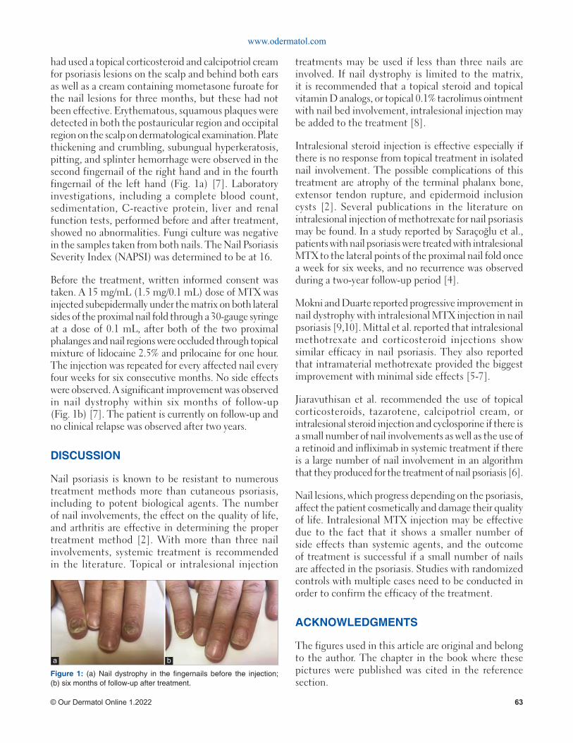
www.odermatol.com
© Our Dermatol Online 1.2022 63
had used a topical corticosteroid and calcipotriol cream for psoriasis lesions on the scalp and behind both ears as well as a cream containing mometasone furoate for the nail lesions for three months, but these had not been effective. Erythematous, squamous plaques were detected in both the postauricular region and occipital region on the scalp on dermatological examination. Plate thickening and crumbling, subungual hyperkeratosis, pitting, and splinter hemorrhage were observed in the second fingernail of the right hand and in the fourth fingernail of the left hand (Fig. 1a) [7]. Laboratory investigations, including a complete blood count, sedimentation, C-reactive protein, liver and renal function tests, performed before and after treatment, showed no abnormalities. Fungi culture was negative in the samples taken from both nails. The Nail Psoriasis Severity Index (NAPSI) was determined to be at 16.
Before the treatment, written informed consent was taken. A 15 mg/mL (1.5 mg/0.1 mL) dose of MTX was injected subepidermally under the matrix on both lateral sides of the proximal nail fold through a 30-gauge syringe at a dose of 0.1 mL, after both of the two proximal phalanges and nail regions were occluded through topical mixture of lidocaine 2.5% and prilocaine for one hour. The injection was repeated for every affected nail every four weeks for six consecutive months. No side effects were observed. A significant improvement was observed in nail dystrophy within six months of follow-up (Fig. 1b) [7]. The patient is currently on follow-up and no clinical relapse was observed after two years.
DISCUSSION
Nail psoriasis is known to be resistant to numerous treatment methods more than cutaneous psoriasis, including to potent biological agents. The number of nail involvements, the effect on the quality of life, and arthritis are effective in determining the proper treatment method [2]. With more than three nail involvements, systemic treatment is recommended in the literature. Topical or intralesional injection
treatments may be used if less than three nails are involved. If nail dystrophy is limited to the matrix, it is recommended that a topical steroid and topical vitamin D analogs, or topical 0.1% tacrolimus ointment with nail bed involvement, intralesional injection may be added to the treatment [8].
Intralesional steroid injection is effective especially if there is no response from topical treatment in isolated nail involvement. The possible complications of this treatment are atrophy of the terminal phalanx bone, extensor tendon rupture, and epidermoid inclusion cysts [2]. Several publications in the literature on intralesional injection of methotrexate for nail psoriasis may be found. In a study reported by Saraçoğlu et al., patients with nail psoriasis were treated with intralesional MTX to the lateral points of the proximal nail fold once a week for six weeks, and no recurrence was observed during a two-year follow-up period [4].
Mokni and Duarte reported progressive improvement in nail dystrophy with intralesional MTX injection in nail psoriasis [9,10]. Mittal et al. reported that intralesional methotrexate and corticosteroid injections show similar efficacy in nail psoriasis. They also reported that intramaterial methotrexate provided the biggest improvement with minimal side effects [5-7].
Jiaravuthisan et al. recommended the use of topical corticosteroids, tazarotene, calcipotriol cream, or intralesional steroid injection and cyclosporine if there is a small number of nail involvements as well as the use of a retinoid and infliximab in systemic treatment if there is a large number of nail involvement in an algorithm that they produced for the treatment of nail psoriasis [6].
Nail lesions, which progress depending on the psoriasis, affect the patient cosmetically and damage their quality of life. Intralesional MTX injection may be effective due to the fact that it shows a smaller number of side effects than systemic agents, and the outcome of treatment is successful if a small number of nails are affected in the psoriasis. Studies with randomized controls with multiple cases need to be conducted in order to confirm the efficacy of the treatment.
ACKNOWLEDGMENTS
The figures used in this article are original and belong to the author. The chapter in the book where these pictures were published was cited in the reference section.
Figure 1: (a) Nail dystrophy in the fi ngernails before the injection; (b) six months of follow-up after treatment.
a b
www.odermatol.com
© Our Dermatol Online 1.2022 64
and cyclosporine. Indian J Dermatol Venereol Leprol. 2018;84:419-23.
6. Jiaravuthisan MM, Sasseville D, Vender RB, Murphy F, Muhn CY. Psoriasis of the nail: Anatomy, pathology, clinical presentation, and a review of the literature on therapy. J Am Acad Dermatol. 2007;57:1-27.
7. Kara YA. Chapter the etiology, pathophysiology, differential diagnosis, clinical fi ndings and treatment of nail psoriais.ın: shahin aghaei, tailored treatments in psoriatic patients.1th ed.London, PA: Intechopen; 2019.
8. Rigopoulos D, Baran R, Chiheb S, Daniel CR, Di Chiacchio N, Gregoriou S, et al. Recommendations for the defi nition, evaluation, and treatment of nail psoriasis in adult patients with no or mild skin psoriasis: A dermatologist and nail expert group consensus. J Am Acad Dermatol. 2019;81:228-40.
9. Mokni S, Ameur K, Ghariani N, Sriha B, Belajouza C, Denguezli M, et al. A case of nail psoriasis successfully treated with intralesional methotrexate. Dermatol Ther (Heidelb). 2018;8:647-51.
10. Duarte AA, Carneiro GP, Murari CM, Jesus LCB. Nail psoriasis treated with intralesional methotrexate infi ltration. An Bras Dermatol. 2019;17:94:491-2.
Consent
The examination of the patient was conducted according to the principles of the Declaration of Helsinki.
The authors certify that they have obtained all appropriate patient consent forms, in which the patients gave their consent for images and other clinical information to be included in the journal. The patients understand that their names and initials will not be published and due effort will be made to conceal their identity, but that anonymity cannot be guaranteed.
REFERENCES
1. Salomon J, Szepietowski JC, Proniewicz A. Psoriatic nails: A prospective clinical study. J Cutan Med Surg. 2003;7:317-21.
2. Haneke E. Nail psoriasis: Clinical features, pathogenesis, differential diagnoses, and management. Psoriasis (Auckl). 2017;16:51-63.
3. Chan ES, Cronstein BN. Methotrexate – How does it really work? Nat Rev Rheumatol. 2010;6:175-8.
4. Sarıcaoglu H, Oz A, Turan H. Nail psoriasis successfully treated with intralesional methotrexate: Case Rep Dermatol. 2011;222:5-7.
5. Mittal J, Mahajan BB. Intramatricial injections for nail psoriasis: An open-label comparative study of triamcinolone, methotrexate,
Copyright by Yesim Akpinar Kara. This is an open access article distributed under the terms of the Creative Commons Attribution License, which permits unrestricted use, distribution, and reproduction in any medium, provided the original author and source are credited.Source of Support: Nil, Confl ict of Interest: None declared.
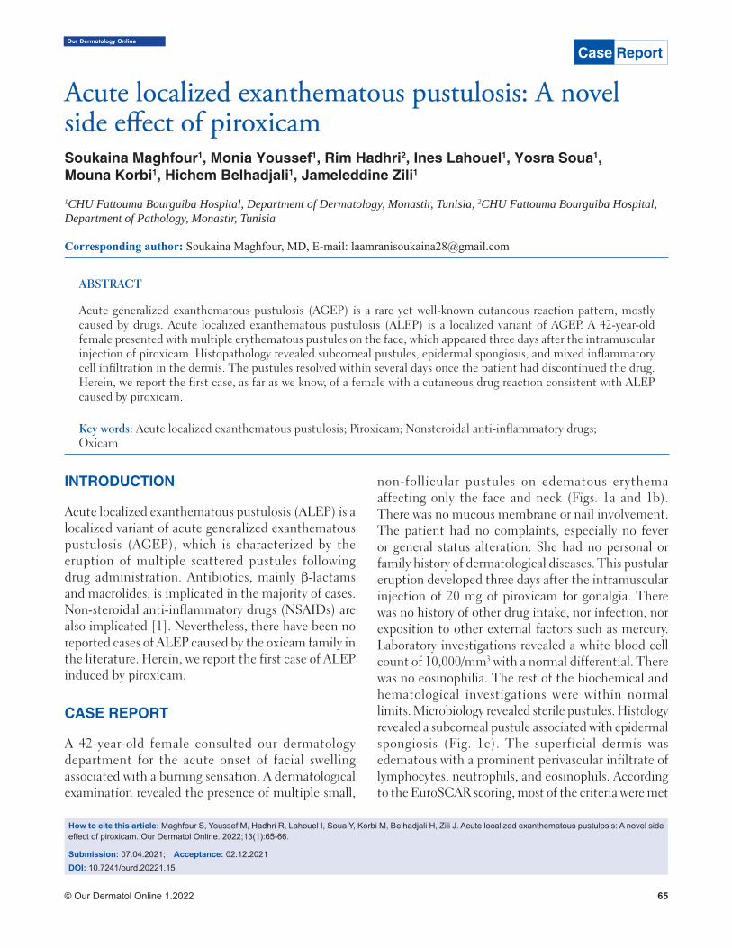
© Our Dermatol Online 1.2022 65
Our Dermatology Online
How to cite this article: Maghfour S, Youssef M, Hadhri R, Lahouel I, Soua Y, Korbi M, Belhadjali H, Zili J. Acute localized exanthematous pustulosis: A novel side eff ect of piroxicam. Our Dermatol Online. 2022;13(1):65-66.
Submission: 07.04.2021; Acceptance: 02.12.2021DOI: 10.7241/ourd.20221.15
Acute localized exanthematous pustulosis: A novel Acute localized exanthematous pustulosis: A novel side eff ect of piroxicamside eff ect of piroxicamSoukaina Maghfour1, Monia Youssef1, Rim Hadhri2, Ines Lahouel1, Yosra Soua1,Mouna Korbi1, Hichem Belhadjali1, Jameleddine Zili1
1CHU Fattouma Bourguiba Hospital, Department of Dermatology, Monastir, Tunisia, 2CHU Fattouma Bourguiba Hospital, Department of Pathology, Monastir, Tunisia
Corresponding author: Soukaina Maghfour, MD, E-mail: [email protected]
INTRODUCTION
Acute localized exanthematous pustulosis (ALEP) is a localized variant of acute generalized exanthematous pustulosis (AGEP), which is characterized by the eruption of multiple scattered pustules following drug administration. Antibiotics, mainly β-lactams and macrolides, is implicated in the majority of cases. Non-steroidal anti-inflammatory drugs (NSAIDs) are also implicated [1]. Nevertheless, there have been no reported cases of ALEP caused by the oxicam family in the literature. Herein, we report the first case of ALEP induced by piroxicam.
CASE REPORT
A 42-year-old female consulted our dermatology department for the acute onset of facial swelling associated with a burning sensation. A dermatological examination revealed the presence of multiple small,
non-follicular pustules on edematous erythema affecting only the face and neck (Figs. 1a and 1b). There was no mucous membrane or nail involvement. The patient had no complaints, especially no fever or general status alteration. She had no personal or family history of dermatological diseases. This pustular eruption developed three days after the intramuscular injection of 20 mg of piroxicam for gonalgia. There was no history of other drug intake, nor infection, nor exposition to other external factors such as mercury. Laboratory investigations revealed a white blood cell count of 10,000/mm3 with a normal differential. There was no eosinophilia. The rest of the biochemical and hematological investigations were within normal limits. Microbiology revealed sterile pustules. Histology revealed a subcorneal pustule associated with epidermal spongiosis (Fig. 1c). The superficial dermis was edematous with a prominent perivascular infiltrate of lymphocytes, neutrophils, and eosinophils. According to the EuroSCAR scoring, most of the criteria were met
ABSTRACT
Acute generalized exanthematous pustulosis (AGEP) is a rare yet well-known cutaneous reaction pattern, mostly caused by drugs. Acute localized exanthematous pustulosis (ALEP) is a localized variant of AGEP. A 42-year-old female presented with multiple erythematous pustules on the face, which appeared three days after the intramuscular injection of piroxicam. Histopathology revealed subcorneal pustules, epidermal spongiosis, and mixed inflammatory cell infiltration in the dermis. The pustules resolved within several days once the patient had discontinued the drug. Herein, we report the first case, as far as we know, of a female with a cutaneous drug reaction consistent with ALEP caused by piroxicam.
Key words: Acute localized exanthematous pustulosis; Piroxicam; Nonsteroidal anti-inflammatory drugs; Oxicam
Case Report
www.odermatol.com
© Our Dermatol Online 1.2022 66
by our patient, who had a score of nine, and therefore the case was classified as definite ALEP. Piroxicam was ceased with a resolution of the pustules within several days after topical corticosteroid treatment with desonide cream, followed by characteristic post pustular pinpoint desquamation. The patient refused a follow-up cutaneous patch test.
DISCUSSION
ALEP is a rare variant of AGEP characterized by the acute onset of multiple, 1–2 mm, non-follicular sterile pustules arising on edematous erythema, localized typically in the face, neck, or chest [2]. Skin symptoms may be associated with fever and leukocytosis. Mucous membranes are rarely affected, commonly mild and limited to one site, mostly the oral lips [1]. Lesions usually resolve rapidly, within several days, after the withdrawal of the causative agent. At this point, a characteristic collaret-shaped post-pustular desquamation may still be a clue to the diagnosis.
ALEP is an uncommon cutaneous drug reaction caused mainly by antibiotics [1]. However, other triggering factors such as bacterial, viral, or parasitic infections, herbal medications, PUVA, mercury, lacquer, venoms, foods, and xenobiotics have also been reported to be involved [3]. ALEP caused by non-steroidal anti-inflammatory drugs (NSAIDs) has been reported only three times in the literature, in three patients, two female and one male, aged 64, 40, and 29, respectively. These cases were induced by ibuprofen, flurbiprofen, and diclofenac, each and respectively [1]. The oxicam family of NSAIDs has not been reported to induce ALEP. The only three
compounds of this family that have been implicated in acute exanthematous pustulosis in its generalized form are meloxicam, lornoxicam, and piroxicam [4]. Herein, we report the first case of ALEP induced by piroxicam. Our patient displayed the classic features of ALEP with a characteristic morphology, histology, and course. Although patch testing was not done in our patient, the causative effect of piroxicam was certain due to the course of the affection (the acute onset and the improvement after the withdrawal of the drug). A patch test is an alternative proving the role of a suspected drug and is particularly useful if there may be several causative drugs. Although its sensitivity is around 50% and negative tests do not allow a final conclusion, positive results are of great value [5].
CONCLUSION
Piroxicam is a widely prescribed molecule. We report our case to highlight a novel side effect of this drug, which is ALEP, and to increase awareness of this side effect among clinicians.
Consent
The examination of the patient was conducted according to the principles of the Declaration of Helsinki.
The authors certify that they have obtained all appropriate patient consent forms, in which the patients gave their consent for images and other clinical information to be included in the journal. The patients understand that their names and initials will not be published and due effort will be made to conceal their identity, but that anonymity cannot be guaranteed.
REFERENCES
1. Villani A, Baldo A, De Fata Salvatores G, Desiato V, Ayala F, Donadio C. Acute localized exanthematous pustulosis (ALEP): Review of literature with report of case caused by amoxicillin-clavulanic acid. Dermatol Ther (Heidelb). 2017;7:563-70.
2. Mohamed M, Soua Y, Njim L, Hammemi S, Youssef M, Akkari H, et al. [Acute localized exanthematous pustulosis on the face: 6 cases in Tunisia]. Ann Dermatol Venereol. 2014;141:756-64.
3. Szatkowski J, Schwartz RA. Acute generalized exanthematous pustulosis (AGEP): A review and update. J Am Acad Dermatol. 2015;73:843-8.
4. Larif S, Slim R, Fathallah N, Ghariani N, Hmouda H, Ben Salem C. Lornoxicam-induced acute generalized exanthematous pustulosis. Int J Dermatol. 2016;55:e458-60.
5. Altaykan A, Boztepe G, Erkin G, Özkaya Ö, Özden E. Acute generalized exanthematous pustulosis induced by bleomycin and confi rmed by patch testing. J Dermatol Treat. 2004;15:231-4.
Copyright by Soukaina Maghfour, et al. This is an open access article distributed under the terms of the Creative Commons Attribution License, which permits unrestricted use, distribution, and reproduction in any medium, provided the original author and source are credited.Source of Support: Nil, Confl ict of Interest: None declared.
Figure 1: (a-b) Multiple small, non-follicular pustules on edematous erythema affecting the face. (c) Subcorneal pustular with epidermal spongiosis (H&E, 100×).
c
b
a
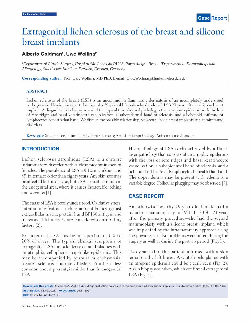
© Our Dermatol Online 1.2022 67
Extragenital lichen sclerosus of the breast and silicone Extragenital lichen sclerosus of the breast and silicone breast implantsbreast implantsAlberto Goldman1, Uwe Wollina2
1Department of Plastic Surgery, Hospital São Lucas da PUCS, Porto Alegre, Brazil, 2Department of Dermatology and Allergology, Städtisches Klinikum Dresden, Dresden, Germany
Corresponding author: Prof. Uwe Wollina, MD PhD, E-mail: [email protected]
INTRODUCTION
Lichen sclerosus atrophicus (LSA) is a chronic inflammatory disorder with a clear predominance of females. The prevalence of LSA is 0.1% in children and 3% in females older than eighty years. Any skin site may be affected by the disease, but LSA is most common in the anogenital area, where it causes intractable itching and soreness [1].
The cause of LSA is poorly understood. Oxidative stress, autoimmune features such as autoantibodies against extracellular matrix protein 1 and BP180 antigen, and increased Th1 activity are considered contributing factors [2].
Extragenital LSA has been reported in 6% to 20% of cases. The typical clinical symptoms of extragenital LSA are pale, ivory-colored plaques with an atrophic, cellophane, paper-like epidermis. This may be accompanied by purpura or ecchymosis, fissures, sclerosis, and rarely blisters. Pruritus is less common and, if present, is milder than in anogenital LSA.
Histopathology of LSA is characterized by a three-layer pathology that consists of an atrophic epidermis with the loss of rete ridges and basal keratinocyte vacuolization, a subepidermal band of sclerosis, and a lichenoid infiltrate of lymphocytes beneath that band. The upper dermis may be present with edema to a variable degree. Follicular plugging may be observed [3].
CASE REPORT
An otherwise healthy 29-year-old female had a reduction mammoplasty in 1991. In 2014—23 years after the primary procedure—she had the second mammoplasty with a silicone breast implant, which was implanted by the inframammary approach using the previous scar. No problems were noted during the surgery as well as during the post-op period (Fig. 1).
Two years later, the patient returned with a skin lesion on the left breast. A whitish pale plaque with an atrophic epidermis could be clearly seen (Fig. 2). A skin biopsy was taken, which confirmed extragenital LSA (Fig. 3).
ABSTRACT
Lichen sclerosus of the breast (LSB) is an uncommon inflammatory dermatosis of an incompletely understood pathogenesis. Herein, we report the case of a 29-year-old female who developed LSB 23 years after a silicone breast implant. A diagnostic skin biopsy revealed the typical three-layered pathology of an atrophic epidermis with the loss of rete ridges and basal keratinocyte vacuolization, a subepidermal band of sclerosis, and a lichenoid infiltrate of lymphocytes beneath that band. We discuss the possible relationship between silicone breast implants and autoimmune disorders.
Keywords: Silicone breast implant; Lichen sclerosus; Breast; Histopathology; Autoimmune disorders
How to cite this article: Goldman A, Wollina U. Extragenital lichen sclerosus of the breast and silicone breast implants. Our Dermatol Online. 2022;13(1):67-69.Submission: 02.06.2021; Acceptance: 08.11.2021DOI: 10.7241/ourd.20221.16
Case ReportOur Dermatology Online
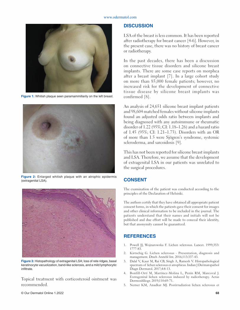
www.odermatol.com
© Our Dermatol Online 1.2022 68
DISCUSSION
LSA of the breast is less common. It has been reported after radiotherapy for breast cancer [4-6]. However, in the present case, there was no history of breast cancer or radiotherapy.
In the past decades, there has been a discussion on connective tissue disorders and silicone breast implants. There are some case reports on morphea after a breast implant [7]. In a large cohort study on more than 85,000 female patients; however, no increased risk for the development of connective tissue disease by silicone breast implants was confirmed [8].
An analysis of 24,651 silicone breast implant patients and 98,604 matched females without silicone implants found an adjusted odds ratio between implants and being diagnosed with any autoimmune or rheumatic disorder of 1.22 (95%; CI: 1.18–1.26) and a hazard ratio of 1.45 (95%; CI: 1.21–1.73). Disorders with an OR of more than 1.5 were Sjögren’s syndrome, systemic scleroderma, and sarcoidosis [9].
This has not been reported for silicone breast implants and LSA. Therefore, we assume that the development of extragenital LSA in our patients was unrelated to the surgical procedures.
CONSENT
The examination of the patient was conducted according to the principles of the Declaration of Helsinki.
The authors certify that they have obtained all appropriate patient consent forms, in which the patients gave their consent for images and other clinical information to be included in the journal. The patients understand that their names and initials will not be published and due effort will be made to conceal their identity, but that anonymity cannot be guaranteed.
REFERENCES
1. Powell JJ, Wojnarowska F. Lichen sclerosus. Lancet. 1999;353:1777-83.
2. Kirtschig G. Lichen sclerosus - Presentation, diagnosis and management. Dtsch Arztebl Int. 2016;113:337-43.
3. Dalal V, Kaur M, Rai CB, Singh A, Ramesh V. Histopathological spectrum of lichen sclerosus et atrophicus. Indian J Dermatopathol Diagn Dermatol. 2017;4:8-13.
4. Bonfill-Ortí M, Martínez-Molina L, Penín RM, Marcoval J. Extragenital lichen sclerosus induced by radiotherapy. Actas Dermosifi liogr. 2019;110:69-71.
5. Nemer KM, Anadkat MJ. Postirradiation lichen sclerosus et
Topical treatment with corticosteroid ointment was recommended.
Figure 2: Enlarged whitish plaque with an atrophic epidermis (extragenital LSA).
Figure 1: Whitish plaque seen paramammillarily on the left breast.
Figure 3: Histopathology of extragenital LSA; loss of rete ridges, basal keratinocyte vacuolization, band-like sclerosis, and a mild lymphocytic infi ltrate.

www.odermatol.com
© Our Dermatol Online 1.2022 69
atrophicus. JAMA Dermatol. 2017;153:1067-9.6. Moore KA, Potter JE. Lichen sclerosus in a breast cancer survivor.
J Gen Intern Med. 2013;28:345.7. Moretti A, Bianchi F, Abbate IV, Gherardi G, Bonavita M,
Passoni E, et al. Localized morphea after breast implant for breast cancer: A case report. Tumori. 2018;104:NP25-8.
8. Sánchez-Guerrero J, Colditz GA, Karlson EW, Hunter DJ, Speizer FE, Liang MH. Silicone breast implants and the risk of connective-tissue diseases and symptoms. N Engl J Med. 1995;332:1666-70.
9. Watad A, Rosenberg V, Tiosano S, Cohen Tervaert JW, Yavne Y, Shoenfeld Y, et al. Silicone breast implants and the risk of autoimmune/rheumatic disorders: A real-world analysis. Int J Epidemiol. 2018;47:1846-54.
Copyright by Alberto Goldman, et al. This is an open access article distributed under the terms of the Creative Commons Attribution License, which permits unrestricted use, distribution, and reproduction in any medium, provided the original author and source are credited.Source of Support: Nil, Confl ict of Interest: None declared.
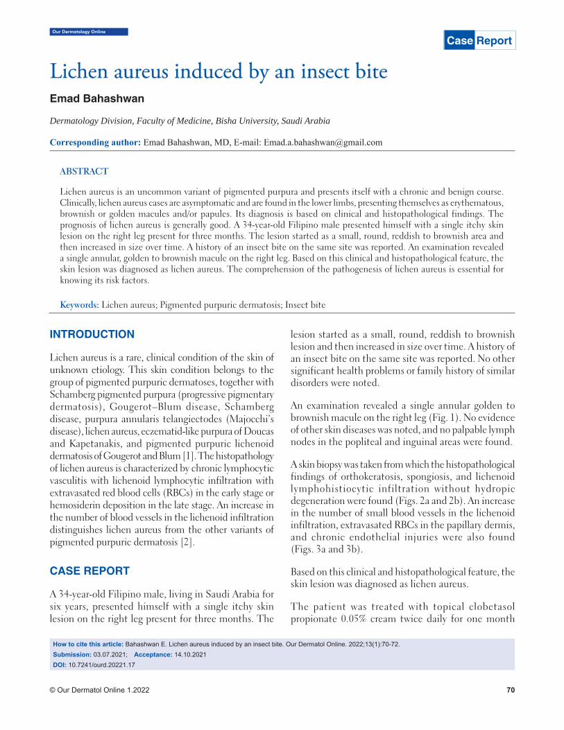
Our Dermatology Online
© Our Dermatol Online 1.2022 70
Lichen aureus induced by an insect biteLichen aureus induced by an insect biteEmad Bahashwan
Dermatology Division, Faculty of Medicine, Bisha University, Saudi Arabia
Corresponding author: Emad Bahashwan, MD, E-mail: [email protected]
INTRODUCTION
Lichen aureus is a rare, clinical condition of the skin of unknown etiology. This skin condition belongs to the group of pigmented purpuric dermatoses, together with Schamberg pigmented purpura (progressive pigmentary dermatosis), Gougerot–Blum disease, Schamberg disease, purpura annularis telangiectodes (Majocchi’s disease), lichen aureus, eczematid-like purpura of Doucas and Kapetanakis, and pigmented purpuric lichenoid dermatosis of Gougerot and Blum [1]. The histopathology of lichen aureus is characterized by chronic lymphocytic vasculitis with lichenoid lymphocytic infiltration with extravasated red blood cells (RBCs) in the early stage or hemosiderin deposition in the late stage. An increase in the number of blood vessels in the lichenoid infiltration distinguishes lichen aureus from the other variants of pigmented purpuric dermatosis [2].
CASE REPORT
A 34-year-old Filipino male, living in Saudi Arabia for six years, presented himself with a single itchy skin lesion on the right leg present for three months. The
lesion started as a small, round, reddish to brownish lesion and then increased in size over time. A history of an insect bite on the same site was reported. No other significant health problems or family history of similar disorders were noted.
An examination revealed a single annular golden to brownish macule on the right leg (Fig. 1). No evidence of other skin diseases was noted, and no palpable lymph nodes in the popliteal and inguinal areas were found.
A skin biopsy was taken from which the histopathological findings of orthokeratosis, spongiosis, and lichenoid lymphohistiocytic infiltration without hydropic degeneration were found (Figs. 2a and 2b). An increase in the number of small blood vessels in the lichenoid infiltration, extravasated RBCs in the papillary dermis, and chronic endothelial injuries were also found (Figs. 3a and 3b).
Based on this clinical and histopathological feature, the skin lesion was diagnosed as lichen aureus.
The patient was treated with topical clobetasol propionate 0.05% cream twice daily for one month
ABSTRACT
Lichen aureus is an uncommon variant of pigmented purpura and presents itself with a chronic and benign course. Clinically, lichen aureus cases are asymptomatic and are found in the lower limbs, presenting themselves as erythematous, brownish or golden macules and/or papules. Its diagnosis is based on clinical and histopathological findings. The prognosis of lichen aureus is generally good. A 34-year-old Filipino male presented himself with a single itchy skin lesion on the right leg present for three months. The lesion started as a small, round, reddish to brownish area and then increased in size over time. A history of an insect bite on the same site was reported. An examination revealed a single annular, golden to brownish macule on the right leg. Based on this clinical and histopathological feature, the skin lesion was diagnosed as lichen aureus. The comprehension of the pathogenesis of lichen aureus is essential for knowing its risk factors.
Keywords: Lichen aureus; Pigmented purpuric dermatosis; Insect bite
How to cite this article: Bahashwan E. Lichen aureus induced by an insect bite. Our Dermatol Online. 2022;13(1):70-72.Submission: 03.07.2021; Acceptance: 14.10.2021DOI: 10.7241/ourd.20221.17
Case Report
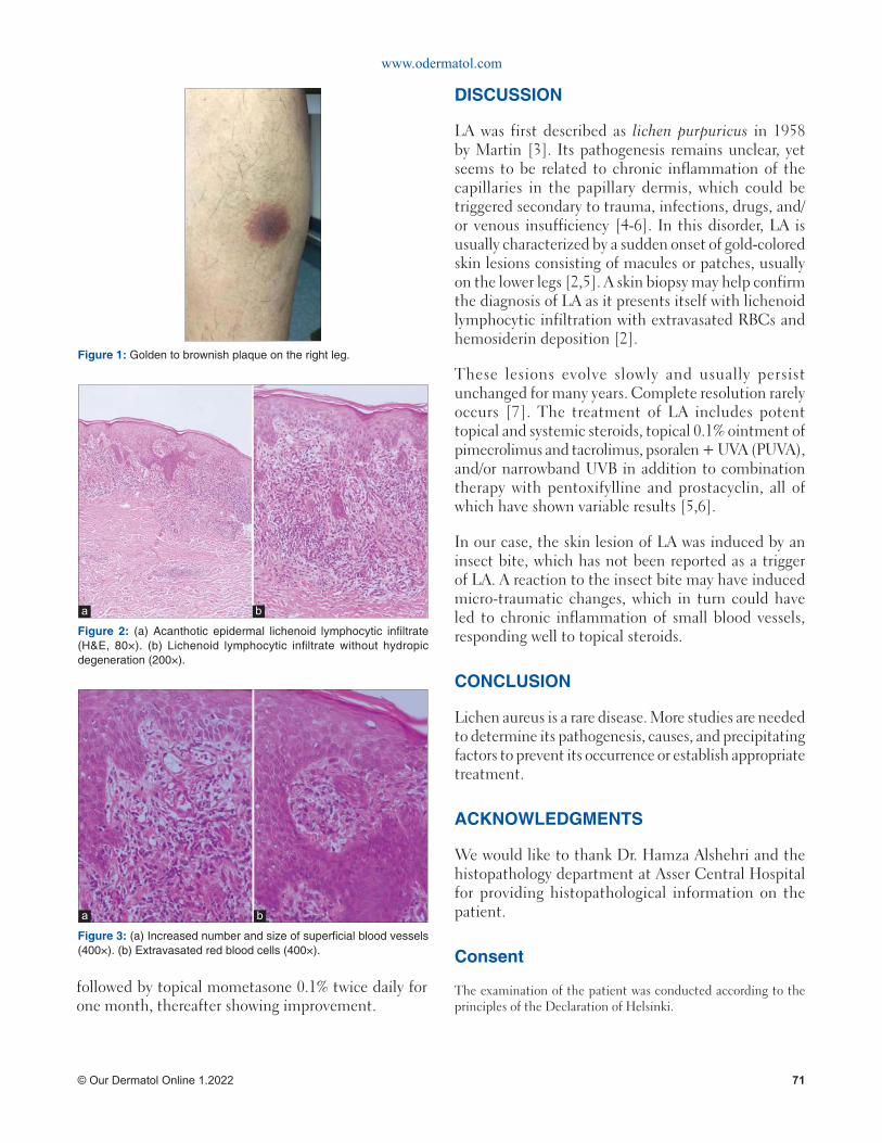
www.odermatol.com
© Our Dermatol Online 1.2022 71
DISCUSSION
LA was first described as lichen purpuricus in 1958 by Martin [3]. Its pathogenesis remains unclear, yet seems to be related to chronic inflammation of the capillaries in the papillary dermis, which could be triggered secondary to trauma, infections, drugs, and/or venous insufficiency [4-6]. In this disorder, LA is usually characterized by a sudden onset of gold-colored skin lesions consisting of macules or patches, usually on the lower legs [2,5]. A skin biopsy may help confirm the diagnosis of LA as it presents itself with lichenoid lymphocytic infiltration with extravasated RBCs and hemosiderin deposition [2].
These lesions evolve slowly and usually persist unchanged for many years. Complete resolution rarely occurs [7]. The treatment of LA includes potent topical and systemic steroids, topical 0.1% ointment of pimecrolimus and tacrolimus, psoralen + UVA (PUVA), and/or narrowband UVB in addition to combination therapy with pentoxifylline and prostacyclin, all of which have shown variable results [5,6].
In our case, the skin lesion of LA was induced by an insect bite, which has not been reported as a trigger of LA. A reaction to the insect bite may have induced micro-traumatic changes, which in turn could have led to chronic inflammation of small blood vessels, responding well to topical steroids.
CONCLUSION
Lichen aureus is a rare disease. More studies are needed to determine its pathogenesis, causes, and precipitating factors to prevent its occurrence or establish appropriate treatment.
ACKNOWLEDGMENTS
We would like to thank Dr. Hamza Alshehri and the histopathology department at Asser Central Hospital for providing histopathological information on the patient.
Consent
The examination of the patient was conducted according to the principles of the Declaration of Helsinki.
followed by topical mometasone 0.1% twice daily for one month, thereafter showing improvement.
Figure 1: Golden to brownish plaque on the right leg.
Figure 2: (a) Acanthotic epidermal lichenoid lymphocytic infi ltrate (H&E, 80×). (b) Lichenoid lymphocytic infi ltrate without hydropic degeneration (200×).
a b
Figure 3: (a) Increased number and size of superfi cial blood vessels (400×). (b) Extravasated red blood cells (400×).
a b
www.odermatol.com
© Our Dermatol Online 1.2022 72
The authors certify that they have obtained all appropriate patient consent forms, in which the patients gave their consent for images and other clinical information to be included in the journal. The patients understand that their names and initials will not be published and due effort will be made to conceal their identity, but that anonymity cannot be guaranteed.
REFERENCES
1. Filho CRR, Schwartz J, Zanol J. [“Algesiogenic” lichen aureus] An Bras Dermatol. 2006;81:163-5.
2. Aung PP, Burns SJ, Bhawan J. Lichen aureus: An unusual histopathological presentation: A case report and a review of literature. Am J Dermatopathol. 2014;36:e1-4.
3. Martin RH. Case for diagnosis. Trans Rep St Johns Hosp Dermatol
Soc Lond. 1958;40:98.4. Kanitakis C, Tsoïtis G. [Lichen purpuricus]. Ann Dermatol
Venereol. 1982;109:445-52.5. Murota H, Katayama I. Lichen aureus responding to topical
tacrolimus treatment. J Dermatol. 2011;38:823-5.6. Kim MJ, Kim BY, Park KC, Youn SW. A case of childhood lichen
aureus. Ann Dermatol. 2009;21:393-5.7. Price ML, Jones EW, Calnan CD, MacDonald DM. Lichen aureus:
A localized persistent form of pigmented purpuric dermatitis. Br J Dermatol. 1985;112:307-14.
Copyright by Emad Bahashwan. This is an open access article distributed under the terms of the Creative Commons Attribution License, which permits unrestricted use, distribution, and reproduction in any medium, provided the original author and source are credited.Source of Support: Nil, Confl ict of Interest: None declared.
© Our Dermatol Online 1.2022 73
Our Dermatology Online
How to cite this article: Quigley C, Raghallaigh SN. Metastatic basal cell carcinoma: A report of two cases and a review of the literature. Our Dermatol Online. 2022;13(1):73-76.Submission: 30.08.2021; Acceptance: 18.11.2021DOI: 10.7241/ourd.20221.18
Metastatic basal cell carcinoma: A report of two cases Metastatic basal cell carcinoma: A report of two cases and a review of the literatureand a review of the literatureClaire Quigley, Siona Ni Raghallaigh
Departments of Dermatology, Beaumont Hospital, Dublin, Ireland
Corresponding author: Claire Quigley, MD, E-mail: [email protected]
INTRODUCTION
It is estimated that approx. 50% of referrals to the dermatologist are for skin cancer [1]. Basal cell carcinoma (BCC) is the most common cancer worldwide and accounts for 80% of non-melanoma skin cancers. An increasing age and prolonged sun exposure are significant risk factors for the development of BCC.
Herein, we present two cases of metastatic BCC, a rare progression of a common presentation. We also review the literature to identify what features could be used to stratify patients into high-risk and low-risk according to the progression of metastatic BCC and what treatment options are available to those with this diagnosis today.
CASE REPORTS
Case 1
The first is a case of a 57-year-old male who presented with a 2 × 2 cm lesion on the right cheek clinically consistent with BCC. He had a palpable lymph node in his right cervical chain and had a history of atrial fibrillation,
hypertension, coronary artery disease, insulin-dependent diabetes mellitus, and diabetic nephropathy, as well as a previous basal cell carcinoma removal from the left nasal ala one year before presentation. The patient was a heavy smoker with a 30-pack-year history.
CT imaging of the neck revealed a 2.6 cm exophytic soft tissue lesion on the right cheek, which was in keeping with the known primary BCC, as well as a 3 cm mass in the right neck, concerning local invasion. A brain, abdomen, and pelvis CT scan showed no evidence of distal metastases.
Under the care of a plastic surgeon, the patient underwent excision of the BCC from the right cheek (Fig. 1) and FNA of LN followed by a neck dissection of levels I, II, and III with superficial parotidectomy. Histology (Fig. 2) showed mixed nodular and infiltrative subtypes of BCC. Three of the twenty-five lymph nodes were positive for metastatic carcinoma and one node was positive at level II at the tail of the parotid gland.
Although no systemic therapy was recommended, the patient completed a course of RT with 60GY over thirty fractions.
ABSTRACT
Basal cell carcinomas (BCCs) are among the most common non-melanoma skin cancers in the world. However, given their slowly progressive nature, metastatic BCCs are a relatively uncommon entity. Below, we discuss two separate cases of metastatic BCC that we encountered in our clinical practice. The first is the case of a 57-year-old male with a right cheek BCC and bilateral pulmonary metastases. The second is the case of a 71-year-old male who also presented with a right BCC and pulmonary metastases. We discuss their altered clinical courses. We also conducted a review of the literature focusing on the use of the relatively novel hedgehog inhibitors as a treatment option for individuals diagnosed with metastatic BCC.
Key words: BCC; metastatic; hedgehog inhibitors
Case Report
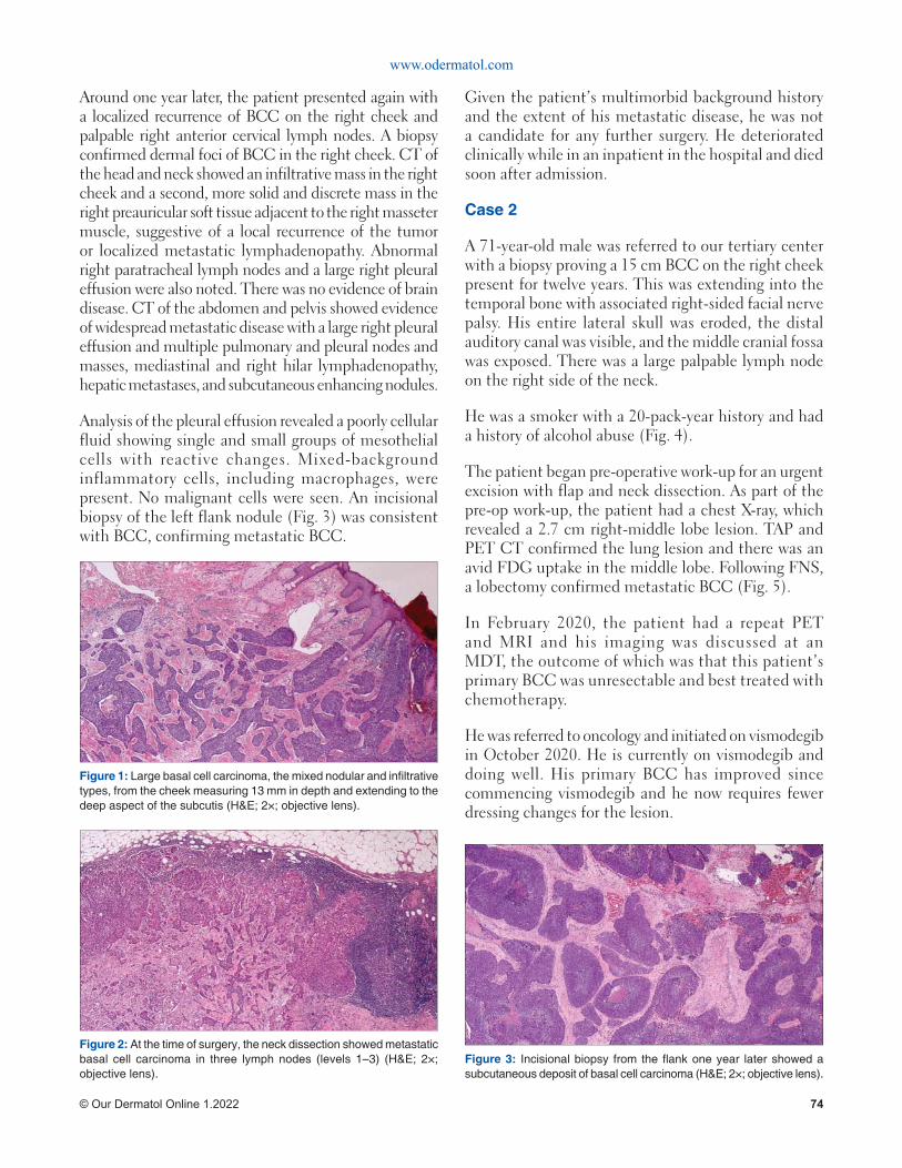
www.odermatol.com
© Our Dermatol Online 1.2022 74
Around one year later, the patient presented again with a localized recurrence of BCC on the right cheek and palpable right anterior cervical lymph nodes. A biopsy confirmed dermal foci of BCC in the right cheek. CT of the head and neck showed an infiltrative mass in the right cheek and a second, more solid and discrete mass in the right preauricular soft tissue adjacent to the right masseter muscle, suggestive of a local recurrence of the tumor or localized metastatic lymphadenopathy. Abnormal right paratracheal lymph nodes and a large right pleural effusion were also noted. There was no evidence of brain disease. CT of the abdomen and pelvis showed evidence of widespread metastatic disease with a large right pleural effusion and multiple pulmonary and pleural nodes and masses, mediastinal and right hilar lymphadenopathy, hepatic metastases, and subcutaneous enhancing nodules.
Analysis of the pleural effusion revealed a poorly cellular fluid showing single and small groups of mesothelial cells with reactive changes. Mixed-background inflammatory cells, including macrophages, were present. No malignant cells were seen. An incisional biopsy of the left flank nodule (Fig. 3) was consistent with BCC, confirming metastatic BCC.
Given the patient’s multimorbid background history and the extent of his metastatic disease, he was not a candidate for any further surgery. He deteriorated clinically while in an inpatient in the hospital and died soon after admission.
Case 2
A 71-year-old male was referred to our tertiary center with a biopsy proving a 15 cm BCC on the right cheek present for twelve years. This was extending into the temporal bone with associated right-sided facial nerve palsy. His entire lateral skull was eroded, the distal auditory canal was visible, and the middle cranial fossa was exposed. There was a large palpable lymph node on the right side of the neck.
He was a smoker with a 20-pack-year history and had a history of alcohol abuse (Fig. 4).
The patient began pre-operative work-up for an urgent excision with flap and neck dissection. As part of the pre-op work-up, the patient had a chest X-ray, which revealed a 2.7 cm right-middle lobe lesion. TAP and PET CT confirmed the lung lesion and there was an avid FDG uptake in the middle lobe. Following FNS, a lobectomy confirmed metastatic BCC (Fig. 5).
In February 2020, the patient had a repeat PET and MRI and his imaging was discussed at an MDT, the outcome of which was that this patient’s primary BCC was unresectable and best treated with chemotherapy.
He was referred to oncology and initiated on vismodegib in October 2020. He is currently on vismodegib and doing well. His primary BCC has improved since commencing vismodegib and he now requires fewer dressing changes for the lesion.
Figure 1: Large basal cell carcinoma, the mixed nodular and infi ltrative types, from the cheek measuring 13 mm in depth and extending to the deep aspect of the subcutis (H&E; 2×; objective lens).
Figure 2: At the time of surgery, the neck dissection showed metastatic basal cell carcinoma in three lymph nodes (levels 1–3) (H&E; 2×; objective lens).
Figure 3: Incisional biopsy from the fl ank one year later showed a subcutaneous deposit of basal cell carcinoma (H&E; 2×; objective lens).
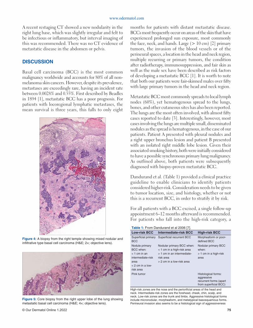
www.odermatol.com
© Our Dermatol Online 1.2022 75
A recent restaging CT showed a new nodularity in the right lung base, which was slightly irregular and felt to be infectious or inflammatory, but interval imaging of this was recommended. There was no CT evidence of metastatic disease in the abdomen or pelvis.
DISCUSSION
Basal cell carcinoma (BCC) is the most common malignancy worldwide and accounts for 80% of all non-melanoma skin cancers. However, despite its prevalence, metastases are exceedingly rare, having an incident rate between 0.0028% and 0.55%. First described by Beadles in 1894 [1], metastatic BCC has a poor prognosis. For patients with locoregional lymphatic metastases, the mean survival is three years, this falls to only eight
months for patients with distant metastatic disease. BCCs most frequently occur on areas of the skin that have experienced prolonged sun exposure, most commonly the face, neck, and hands. Large (> 10 cm) [2] primary tumors, the invasion of the blood vessels or of the perineural spaces, a location in the head and neck region, multiple recurring or primary tumors, the condition after radiotherapy, immunosuppression, and fair skin as well as the male sex have been described as risk factors of developing a metastatic BCC [1]. It is worth to note that both our patients were fair-skinned males over fifty with large primary tumors in the head and neck region.
Metastatic BCC most commonly spreads to local lymph nodes (60%), yet hematogenous spread to the lungs, bones, and other cutaneous sites has also been reported. The lungs are the most often involved, with almost fifty cases reported to date [3]. Interestingly, however, most cases involving the lungs are multiple small, disseminated nodules as the spread is hematogenous, in the case of our patients. Patient A presented with pleural nodules and a right upper bronchus lesion and patient B presented with an isolated right middle lobe lesion. Given their associated smoking history, both were initially considered to have a possible synchronous primary lung malignancy. As outlined above, both patients were subsequently diagnosed with biopsy-proven metastatic BCC.
Dandurand et al. (Table 1) provided a clinical practice guideline to enable clinicians to identify patients considered higher-risk. Consideration needs to be given to tumor location, size, and histology, whether or not this is a recurrent BCC, in order to stratify it by risk.
For all patients with a BCC excised, a single follow-up appointment 6–12 months afterward is recommended. For patients who fall into the high-risk category, a
Table 1: From Dandurand et al 2006 [7].Low-risk BCC Intermediate-risk BCC High-risk BCCSuperfi cial primary BCC
Superfi cial recurrent BCC Morpheaform or poor-defi ned BCC
Nodule primary BCC when: < 1 cm in an intermediate-risk area < 2 cm in a low-risk area
Nodular primary BCC when: < 1 cm in a high-risk area > 1 cm in an intermediate-risk area > 2 cm in a low-risk area
Nodular primary BCC when: > 1 cm in a high-risk area
Pink tumor Histological forms: aggressive recurrent forms (apart from superfi cial BCC)
High-risk zones are the nose and the periorifi cial areas of the head and neck. Intermediate-risk zones are the forehead, cheek, chin, scalp, and neck. Low-risk zones are the trunk and limbs. Aggressive histological forms include micronodular, morpheaform, and metatypical basosquamous forms. Perineural invasion also seems to be a histological sign of aggressiveness
Figure 4: A biopsy from the right temple showing mixed nodular and infi ltrative type basal cell carcinoma (H&E; 2×; objective lens).
Figure 5: Core biopsy from the right upper lobe of the lung showing metastatic basal cell carcinoma (H&E; 4×; objective lens).
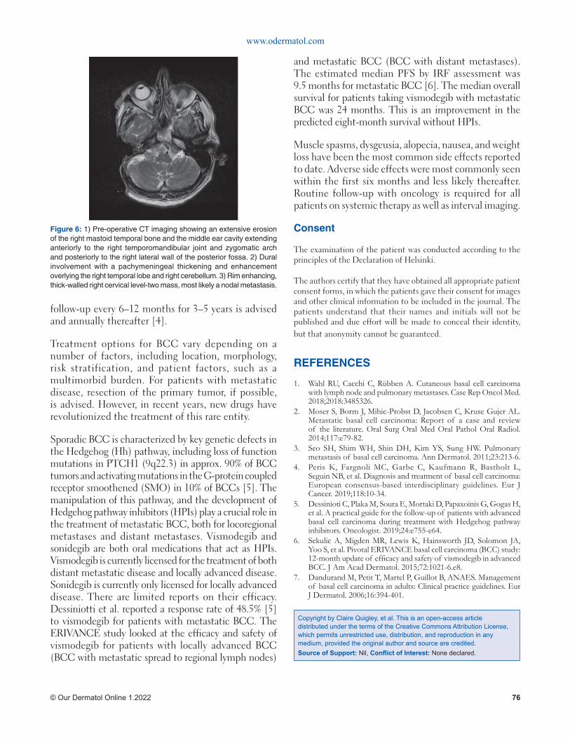
www.odermatol.com
© Our Dermatol Online 1.2022 76
follow-up every 6–12 months for 3–5 years is advised and annually thereafter [4].
Treatment options for BCC vary depending on a number of factors, including location, morphology, risk stratification, and patient factors, such as a multimorbid burden. For patients with metastatic disease, resection of the primary tumor, if possible, is advised. However, in recent years, new drugs have revolutionized the treatment of this rare entity.
Sporadic BCC is characterized by key genetic defects in the Hedgehog (Hh) pathway, including loss of function mutations in PTCH1 (9q22.3) in approx. 90% of BCC tumors and activating mutations in the G-protein coupled receptor smoothened (SMO) in 10% of BCCs [5]. The manipulation of this pathway, and the development of Hedgehog pathway inhibitors (HPIs) play a crucial role in the treatment of metastatic BCC, both for locoregional metastases and distant metastases. Vismodegib and sonidegib are both oral medications that act as HPIs. Vismodegib is currently licensed for the treatment of both distant metastatic disease and locally advanced disease. Sonidegib is currently only licensed for locally advanced disease. There are limited reports on their efficacy. Dessiniotti et al. reported a response rate of 48.5% [5] to vismodegib for patients with metastatic BCC. The ERIVANCE study looked at the efficacy and safety of vismodegib for patients with locally advanced BCC (BCC with metastatic spread to regional lymph nodes)
and metastatic BCC (BCC with distant metastases). The estimated median PFS by IRF assessment was 9.5 months for metastatic BCC [6]. The median overall survival for patients taking vismodegib with metastatic BCC was 24 months. This is an improvement in the predicted eight-month survival without HPIs.
Muscle spasms, dysgeusia, alopecia, nausea, and weight loss have been the most common side effects reported to date. Adverse side effects were most commonly seen within the first six months and less likely thereafter. Routine follow-up with oncology is required for all patients on systemic therapy as well as interval imaging.
Consent
The examination of the patient was conducted according to the principles of the Declaration of Helsinki.
The authors certify that they have obtained all appropriate patient consent forms, in which the patients gave their consent for images and other clinical information to be included in the journal. The patients understand that their names and initials will not be published and due effort will be made to conceal their identity, but that anonymity cannot be guaranteed.
REFERENCES
1. Wahl RU, Cacchi C, Rübben A. Cutaneous basal cell carcinoma with lymph node and pulmonary metastases. Case Rep Oncol Med. 2018;2018:3485326.
2. Moser S, Borm J, Mihic-Probst D, Jacobsen C, Kruse Gujer AL. Metastatic basal cell carcinoma: Report of a case and review of the literature. Oral Surg Oral Med Oral Pathol Oral Radiol. 2014;117:e79-82.
3. Seo SH, Shim WH, Shin DH, Kim YS, Sung HW. Pulmonary metastasis of basal cell carcinoma. Ann Dermatol. 2011;23:213-6.
4. Peris K, Fargnoli MC, Garbe C, Kaufmann R, Bastholt L, Seguin NB, et al. Diagnosis and treatment of basal cell carcinoma: European consensus-based interdisciplinary guidelines. Eur J Cancer. 2019;118:10-34.
5. Dessinioti C, Plaka M, Soura E, Mortaki D, Papaxoinis G, Gogas H, et al. A practical guide for the follow-up of patients with advanced basal cell carcinoma during treatment with Hedgehog pathway inhibitors. Oncologist. 2019;24:e755-e64.
6. Sekulic A, Migden MR, Lewis K, Hainsworth JD, Solomon JA, Yoo S, et al. Pivotal ERIVANCE basal cell carcinoma (BCC) study: 12-month update of effi cacy and safety of vismodegib in advanced BCC. J Am Acad Dermatol. 2015;72:1021-6.e8.
7. Dandurand M, Petit T, Martel P, Guillot B, ANAES. Management of basal cell carcinoma in adults: Clinical practice guidelines. Eur J Dermatol. 2006;16:394-401.
Copyright by Claire Quigley, et al. This is an open-access article distributed under the terms of the Creative Commons Attribution License, which permits unrestricted use, distribution, and reproduction in any medium, provided the original author and source are credited.Source of Support: Nil, Confl ict of Interest: None declared.
Figure 6: 1) Pre-operative CT imaging showing an extensive erosion of the right mastoid temporal bone and the middle ear cavity extending anteriorly to the right temporomandibular joint and zygomatic arch and posteriorly to the right lateral wall of the posterior fossa. 2) Dural involvement with a pachymeningeal thickening and enhancement overlying the right temporal lobe and right cerebellum. 3) Rim enhancing, thick-walled right cervical level-two mass, most likely a nodal metastasis.
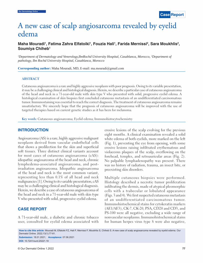
© Our Dermatol Online 1.2022 77
Our Dermatology Online
How to cite this article: Mouradi M, Elfatoiki FZ, Hali F, Mernissi F, Moukhlis S, Chiheb S. A new case of scalp angiosarcoma revealed by eyelid edema. Our Dermatol Online. 2022;13(1):77-81.Submission: 18.01.2021; Acceptance: 07.06.2021DOI: 10.7241/ourd.20221.19
A new case of scalp angiosarcoma revealed by eyelid A new case of scalp angiosarcoma revealed by eyelid edemaedemaMaha Mouradi1, Fatima Zahra Elfatoiki1, Fouzia Hali1, Farida Mernissi2, Sara Moukhlis2, Soumiya Chiheb1
1Department of Dermatology and Venerology,IbnRochd University Hospital, Casablanca, Morocco, 2Department of pathology, Ibn Rochd University Hospital, Casablanca, Morocco
Corresponding author: Maha Mouradi, MD, E-mail: [email protected]
INTRODUCTION
Angiosarcoma (AS) is a rare, highly aggressive malignant neoplasm derived from vascular endothelial cells that shows a predilection for the skin and superficial soft tissues. Three distinct clinical variants account for most cases of cutaneous angiosarcoma (cAS): idiopathic angiosarcoma of the head and neck, chronic lymphedema-associated angiosarcoma, and post-irradiation angiosarcoma. Idiopathic angiosarcoma of the head and neck is the most common variant, representing less than 0.1% of all head and neck malignancies [1]. Owing to its variable presentation, cAS may be a challenging clinical and histological diagnosis. Herein, we describe a case of cutaneous angiosarcoma of the head and neck in a 71-year-old male with skin type V who presented with solid, progressive eyelid edema.
CASE REPORT
A 71-year-old male, a diabetic and chronic tobacco user, consulted for eyelid edema associated with
erosive lesions of the scalp evolving for the previous eight months. A clinical examination revealed a solid white edema of both eyelids, more marked on the left (Fig. 1), preventing the eye from opening, with some erosive lesions raising infiltrated erythematous and violaceous plaques of the scalp, overflowing on the forehead, temples, and retroauricular areas (Fig. 2). No palpable lymphadenopathy was present. There was no history of radiation, trauma, an insect bite, or preexisting skin disorders.
Multiple cutaneous biopsies were performed. Histology described a necrotic tumor proliferation infiltrating the dermis, made of atypical pleomorphic cells with a trabecular or lobulated appearance (Figs. 3 and 4). We first suspected cutaneous metastasis of an undifferentiated carcinomatous tumor. Immunohistochemical stains for cytokeratin markers (AE1/AE3), CK-7, CK-20, PSA, CD20 and CD3, and PS-100 were all negative, excluding a wide range of nonvascular neoplasms. Immunohistochemical stains for human herpes virus type 8 were also negative,
ABSTRACT
Cutaneous angiosarcoma is a rare and highly aggressive neoplasm with poor prognosis. Owing to its variable presentation, it may be a challenging clinical and histological diagnosis. Herein, we describe a particular case of cutaneous angiosarcoma of the head and neck in a 71-year-old male with skin type V who presented with solid, progressive eyelid edema. A histological examination of skin biopsies first concluded cutaneous metastasis of an undifferentiated carcinomatous tumor. Immunostaining was essential to reach the correct diagnosis. The treatment of cutaneous angiosarcoma remains unsatisfactory. We sincerely hope that the prognosis of cutaneous angiosarcoma will be improved with the use of targeted therapies based on current genetic studies as it has been for melanoma.
Key words: Cutaneous angiosarcoma; Eyelid edema; Immunohistocytochemistry
Case Report
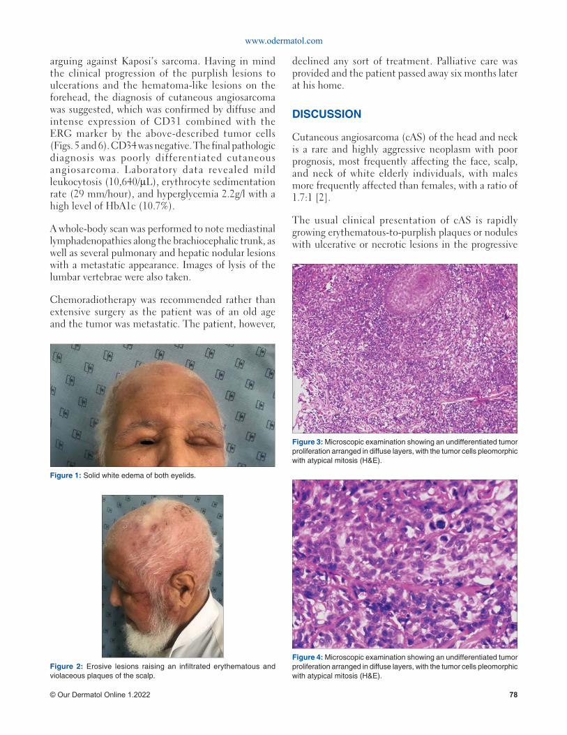
www.odermatol.com
© Our Dermatol Online 1.2022 78
arguing against Kaposi’s sarcoma. Having in mind the clinical progression of the purplish lesions to ulcerations and the hematoma-like lesions on the forehead, the diagnosis of cutaneous angiosarcoma was suggested, which was confirmed by diffuse and intense expression of CD31 combined with the ERG marker by the above-described tumor cells (Figs. 5 and 6). CD34 was negative. The final pathologic diagnosis was poorly differentiated cutaneous angiosarcoma. Laboratory data revealed mild leukocytosis (10,640/μL), erythrocyte sedimentation rate (29 mm/hour), and hyperglycemia 2.2g/l with a high level of HbA1c (10.7%).
A whole-body scan was performed to note mediastinal lymphadenopathies along the brachiocephalic trunk, as well as several pulmonary and hepatic nodular lesions with a metastatic appearance. Images of lysis of the lumbar vertebrae were also taken.
Chemoradiotherapy was recommended rather than extensive surgery as the patient was of an old age and the tumor was metastatic. The patient, however,
declined any sort of treatment. Palliative care was provided and the patient passed away six months later at his home.
DISCUSSION
Cutaneous angiosarcoma (cAS) of the head and neck is a rare and highly aggressive neoplasm with poor prognosis, most frequently affecting the face, scalp, and neck of white elderly individuals, with males more frequently affected than females, with a ratio of 1.7:1 [2].
The usual clinical presentation of cAS is rapidly growing erythematous-to-purplish plaques or nodules with ulcerative or necrotic lesions in the progressive
Figure 2: Erosive lesions raising an infi ltrated erythematous and violaceous plaques of the scalp.
Figure 1: Solid white edema of both eyelids.
Figure 3: Microscopic examination showing an undifferentiated tumor proliferation arranged in diffuse layers, with the tumor cells pleomorphic with atypical mitosis (H&E).
Figure 4: Microscopic examination showing an undifferentiated tumor proliferation arranged in diffuse layers, with the tumor cells pleomorphic with atypical mitosis (H&E).
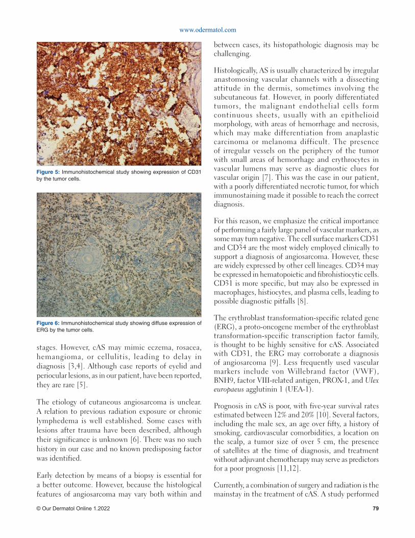
www.odermatol.com
© Our Dermatol Online 1.2022 79
stages. However, cAS may mimic eczema, rosacea, hemangioma, or cellulitis, leading to delay in diagnosis [3,4]. Although case reports of eyelid and periocular lesions, as in our patient, have been reported, they are rare [5].
The etiology of cutaneous angiosarcoma is unclear. A relation to previous radiation exposure or chronic lymphedema is well established. Some cases with lesions after trauma have been described, although their significance is unknown [6]. There was no such history in our case and no known predisposing factor was identified.
Early detection by means of a biopsy is essential for a better outcome. However, because the histological features of angiosarcoma may vary both within and
between cases, its histopathologic diagnosis may be challenging.
Histologically, AS is usually characterized by irregular anastomosing vascular channels with a dissecting attitude in the dermis, sometimes involving the subcutaneous fat. However, in poorly differentiated tumors, the malignant endothelial cells form continuous sheets, usually with an epithelioid morphology, with areas of hemorrhage and necrosis, which may make differentiation from anaplastic carcinoma or melanoma difficult. The presence of irregular vessels on the periphery of the tumor with small areas of hemorrhage and erythrocytes in vascular lumens may serve as diagnostic clues for vascular origin [7]. This was the case in our patient, with a poorly differentiated necrotic tumor, for which immunostaining made it possible to reach the correct diagnosis.
For this reason, we emphasize the critical importance of performing a fairly large panel of vascular markers, as some may turn negative. The cell surface markers CD31 and CD34 are the most widely employed clinically to support a diagnosis of angiosarcoma. However, these are widely expressed by other cell lineages. CD34 may be expressed in hematopoietic and fibrohistiocytic cells. CD31 is more specific, but may also be expressed in macrophages, histiocytes, and plasma cells, leading to possible diagnostic pitfalls [8].
The erythroblast transformation-specific related gene (ERG), a proto-oncogene member of the erythroblast transformation-specific transcription factor family, is thought to be highly sensitive for cAS. Associated with CD31, the ERG may corroborate a diagnosis of angiosarcoma [9]. Less frequently used vascular markers include von Willebrand factor (VWF), BNH9, factor VIII-related antigen, PROX-1, and Ulex europaeus agglutinin 1 (UEA-1).
Prognosis in cAS is poor, with five-year survival rates estimated between 12% and 20% [10]. Several factors, including the male sex, an age over fifty, a history of smoking, cardiovascular comorbidities, a location on the scalp, a tumor size of over 5 cm, the presence of satellites at the time of diagnosis, and treatment without adjuvant chemotherapy may serve as predictors for a poor prognosis [11,12].
Currently, a combination of surgery and radiation is the mainstay in the treatment of cAS. A study performed
Figure 6: Immunohistochemical study showing diffuse expression of ERG by the tumor cells.
Figure 5: Immunohistochemical study showing expression of CD31 by the tumor cells.
www.odermatol.com
© Our Dermatol Online 1.2022 80
at the University of Texas MD Anderson Cancer Center demonstrated that patients who undergo combination therapy have a statistically greater overall survival rate when compared with those who undergo radiation or surgery alone [11]. However, because of delay in diagnosis, a third of the patients present with a metastatic or diffuse non-resectable tumor, as the case of our patient [13]. These patients require systemic treatments. Doxorubicin-based regimens were considered the gold standard of the treatment of soft tissue sarcomas.
Therapy with taxanes (paclitaxel) has shown better efficacy than doxorubicin with, a median progression-free survival rate of 4–5 months [14].
Successful management of infantile hemangiomas with oral and topical beta blockers led to considering propranolol for AS. Adding propranolol to a chemotherapy regimen has shown a promising response in several case reports [15,16].
The use of chemotherapy may be limited due to an advanced age or associated comorbidities. Targeted therapies, such as tyrosine kinase inhibitors (pazopanib, sorafenib, axitinib) are interesting alternatives in such cases. Bevacizumab, a VEGFR inhibitor, was reported to be an effective treatment option for AS [17]. Combinations of bevacizumab and paclitaxel are under investigation.
To date, there is no immunotherapy approved for cAS. One case report described an interesting response to anti-PD-1 treatment in a patient with angiosarcoma, although the patient experienced drug-induced hepatitis, which necessitated systemic corticosteroid treatment [18]. We sincerely hope that the treatment of cAS will be improved in the future, as it has been for melanoma.
CONCLUSION
This is a rare example of a new and challenging diagnosis of cutaneous angiosarcoma presenting as a solid, progressive eyelid edema. The case highlights the diversity of clinical manifestations associated with this type of neoplasm and the value of immunostaining in confirming a diagnosis. However, the prognosis in our case was dismal and a diagnosis at an earlier stage may have possibly given us more options for management and probably a better outcome in the end.
Consent
The examination of the patient was conducted according to the principles of the Declaration of Helsinki.
The authors certify that they have obtained all appropriate patient consent forms, in which the patients gave their consent for images and other clinical information to be included in the journal. The patients understand that their names and initials will not be published and due effort will be made to conceal their identity, but that anonymity cannot be guaranteed.
REFERENCES
1. Maddox JC, Evans HL. Angiosarcoma of skin and soft tissue: A study of forty-four cases. Cancer. 1981;48:1907-21.
2. Dettenborn T, Wermker K, Schulze HJ, Klein M, Schwipper V, Hallermann C. Prognostic features in angiosarcoma of the head and neck: A retrospective monocenter study. J Craniomaxillofac Surg. 2014;42:1623-8.
3. Ambujam S, Audhya M, Reddy A, Roy S. Cutaneous angiosarcoma of the head, neck, and face of the elderly in type 5 skin. J Cutan Aesthet Surg. 2013;6:45-7.
4. Trinh NQ, Rashed I, Hutchens KA, Go A, Melian E, Tung R. Unusual clinical presentation of cutaneous angiosarcoma masquerading as eczema: A case report and review of the literature. Case Rep Dermatol Med. 2013;2013:906426.
5. Cabral ANF, Rocha RH, Amaral ACV, Medeiros KB, Nogueira PSE, Diniz LM. Cutaneous angiosarcoma: Report of three different and typical cases admitted in a unique dermatology clinic. An Bras Dermatol. 2017;92:235-8.
6. Mushtaq S, Sakral A, Dogra D, Bhardwaj S, Bhat A. Primary angiosarcoma of chest wall inside out: A rare presentation. Our Dermatol Online. 2017;8:306-10.
7. Paolino G, Lora V, Cota C, Panetta C, Muscardin LM, Donati P. Early angiosarcoma of the scalp: A clinicopathological pitfall. Am J Dermatopathol. 2016;38:690-4.
8. Aouali S, Alouani I, Ragragui H, Zizi N, Dikhaye S. A case of epithelioid angiosarcoma in a young man with chronic lymphedema. Our Dermatol Online. 2020;11:165-7.
9. Sullivan HC, Edgar MA, Cohen C, Kovach CK, HooKim K, Reid MD. The utility of ERG, CD31 and CD34 in the cytological diagnosis of angiosarcoma: An analysis of 25 cases. J Clin Pathol. 2015;68:44-50.
10. Donghi D, Kerl K, Dummer R, Schoenewolf N, Cozzio A. Cutaneous angiosarcoma: Own experience over 13 years. Clinical features, disease course and immunohistochemical profi le. J Eur Acad Dermatol Venereol. 2010;24:1230-4.
11. Guadagnolo BA, Zagars GK, Araujo D, Ravi V, Shellenberger TD, Sturgis EM. Outcomes after defi nitive treatment for cutaneous angiosarcoma of the face and scalp. Head Neck. 2011;33:661-7.
12. Lee BL, Chen CF, Chen PC, Lee HC, Liao WC, Perng CK, et al. Investigation of prognostic features in primary cutaneous and soft tissue angiosarcoma after surgical resection: A retrospective study. Ann Plast Surg. 2017;78(3 Suppl 2):S41-6.
13. Becquart O, Girard C, Lesage C, Guillot B. [Angiosarcoma of the head and neck treated with bevacizumab and paclitaxel]. Ann Dermatol Venereol. 2018;145:451-3.
14. Penel N, Italiano A, Ray-Coquard I, Chaigneau L, Delcambre C, Robin YM, et al. Metastatic angiosarcomas: Doxorubicin-based regimens, weekly paclitaxel and metastasectomy significantly improve the outcome. Ann Oncol. 2012;23:517-23.
15. Agulnik M, Yarber JL, Okuno SH, von Mehren M, Jovanovic BD, Brockstein BE, et al. An open-label, multicenter, phase II study of bevacizumab for the treatment of angiosarcoma and epithelioid

www.odermatol.com
© Our Dermatol Online 1.2022 81
hemangioendotheliomas. Ann Oncol. 2013;24:257-63.16. Banavali S, Pasquier E, Andre N. Targeted therapy with
propranolol and metronomic chemotherapy combination: Sustained complete response of a relapsing metastatic angiosarcoma. Ecancermedicalscience. 2015;9:499.
17. Pasquier E, André N, Street J, Chougule A, Rekhi B, Ghosh J, et al. Effective management of advanced angiosarcoma by the synergistic combination of propranolol and vinblastine-based metronomic chemotherapy: A bench to bedside study. EBioMedicine. 2016;6:87-95.
18. Sindhu S, Gimber LH, Cranmer L, McBride A, Kraft AS. Angiosarcoma treated successfully with anti-PD-1 therapy - A case report. J Immunother Cancer. 2017;5:58.
Copyright by Maha Mouradi, et al. This is an open access article distributed under the terms of the Creative Commons Attribution License, which permits unrestricted use, distribution, and reproduction in any medium, provided the original author and source are credited.Source of Support: Nil, Confl ict of Interest: None declared.
© Our Dermatol Online 1.2022 82
Our Dermatology Online
How to cite this article: Chakiri R, Bouhajeb Y. Dermoscopy of pilomatricoma: A case report with a review of the literature. Our Dermatol Online. 2022;13(1):82-85.
Submission: 08.04.2021; Acceptance: 05.08.2021DOI: 10.7241/ourd.20221.20
Dermoscopy of pilomatricoma: A case report with a Dermoscopy of pilomatricoma: A case report with a review of the literaturereview of the literatureRadia Chakiri1,2, Youssef Bouhajeb3
1Department of Dermatology, University Hospital Agadir, Morocco, 2Faculty of Medicine and Pharmacy Agadir, University Ibn Zohr, Agadir, Morocco, 3Center of Pathology Ibn Rochd, Agadir, Morocco
Corresponding author: Radia Chakiri, MD, E-mail: [email protected]
INTRODUCTION
Pilomatrixoma is a benign adnexal, dermal, or subcutaneous tumor [1].
The tumor was first described by Malherbe and Chenantais in 1880, who hypothesized that the lesion originated from a sebaceous gland and, therefore, they named the tumor calcifying epithelioma of sebaceous glands [2,3].
The lesion usually occurs on the face, neck, and upper limbs, while the trunk and the lower limbs are less often affected [4].
Typically, pilomatricoma is described as a firm, painless, well-defined solitary nodule, which may have a bluish-red coloration. Its size normally ranges from 0.5 to 4.5 cm in diameter and the highest incidence is found in children and females [5].
Dermoscopy is a non-invasive technique that has greatly improved the diagnostic accuracy of melanocytic and non-melanocytic skin tumors.
We present the dermoscopic features of pilomatricoma through our case report with a literature review.
CASE REPORT
An eleven-year-old female was admitted to our department for a rapidly-growing, painless tumor of the back which had been evolving for four months. There was no notion of prior trauma nor a tendency for spontaneous regression.
A clinical examination revealed an erythematous-purplish tumor 3 cm in diameter with a smooth and telangiectatic surface. The tumor was roughly rounded, adhered to the superficial planes, and painless on palpation (Fig. 1).
Dermatoscopic examination of the lesion showed structures invisible to the naked eye. There were multiple irregular, yellowish-white structures, white streaks, yellowish lobules, blueish-gray areas, and linear, irregular vessels (Figs. 2 and 3).
ABSTRACT
Pilomatricoma is a benign tumor originating from hair follicle matrix cells and characterized by the presence of cutaneous and subcutaneous nodules up to 3.0 cm in diameter, usually on the head, neck, and upper extremities, rarely on the trunk and lower extremities. An eleven-year-old female with a painless, erythematous-purplish tumor of the back. A dermoscopic examination revealed irregular linear vessels, white structures, and structureless grayish-blue areas. Histological examination after excision confirmed the diagnosis of pilomatricoma. Dermoscopy may be a useful tool for improving the clinical recognition of pilomatricoma.
Key words: Dermoscopy; Pilomatricomas; Adnexal tumor
Case Report
www.odermatol.com
© Our Dermatol Online 1.2022 83
Histological examination of the excised specimen revealed a dermal tumor made of several rounded
masses, the largest having a cystic appearance. In the most superficial zone, there were numerous ghost cell ranges. There were also intensely dark, matrix-like basophilic cell areas with characteristic maturation toward eosinophilic transitional cells and then nucleus-free ghost cells. Mummified cells were sometimes calcified and surrounded by zones of foreign body granuloma.
Thus, the diagnosis of pilomatricoma clinically and dermoscopically suspected was confirmed by histology.
DISCUSSION
Pilomatricoma is a common tumor derived from hair matrix cells; it is most often diagnosed in young children yet may also affect adults [6].
It clinically presents itself as a firm, single, stony-hard, slow-growing subcutaneous or intradermal nodule, asymptomatic and adherent to the skin. Lesions are usually skin-colored, although reddish-blue lesions have also been observed [7].
Dermoscopy is a non-invasive procedure focusing on analyzing epidermal and dermal structures, although it is widely used to facilitate the diagnosis of melanocytic and non-melanocytic skin lesions [1].
Since 2008 till now, dermoscopic features of fifteen cases of pilomatricomas have been reported in published English-language works, including our case (Table 1).
The patients, nine females and six males, were aged from 4 to 75 years old (mean = 37.06). Nine of the lesions (60%) were located on the face, four (26,66%) on the upper extremities (arm), and the two (13,3%) remaining were located one (6,67%) on the back and the other on the neck (6,67%) [1-2].
A dermoscopic examination of the lesions allowed the observation of the following features: white streaks and irregular yellow-whitish structures were present in nine pilomatricomas (60%); vascular structures were identified in thirteen cases (86.66%); reddish homogeneous areas, linear-irregular vessels, and hairpin-like vessels were observed in nine cases (60%); dotted vessels in four cases (26,7%), and crown-like vessels and comma-like vessels in one case (6.7%); ulceration was observed in eight pilomatricomas (53.3%); structureless grayish-blue areas were found in
Figure 1: Erythematous-violaceus tumor in the back of the eleven-year-old female.
Figure 2: Dermoscopy of pilomatricoma (red circle: white streaks; black arrow: linear-irregular vessels; green triangles: structureless yellow-whitish areas).
Figure 3: Dermoscopy of pilomatricoma (chunks: structureless bluish-gray areas).
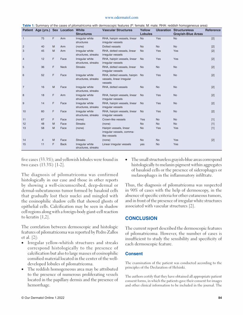
www.odermatol.com
© Our Dermatol Online 1.2022 84
five cases (33.3%); and yellowish lobules were found in two cases (13.3%) [1-2].
The diagnosis of pilomatricoma was confirmed histologically in our case and those in other reports by showing a well-circumscribed, deep-dermal or dermal-subcutaneous tumor formed by basaloid cells that gradually lost their nuclei and mingled with the eosinophilic shadow cells that showed ghosts of epithelial cells. Calcification may be seen in shadow cell regions along with a foreign-body giant-cell reaction to keratin [1,2].
The correlation between dermoscopic and histologic features of pilomatricoma was reported by Pedro Zallos et al. [2]:• Irregular yellow-whitish structures and streaks
correspond histologically to the presence of calcification but also to large masses of eosinophilic cornified material located in the center of the well-developed lobules of pilomatricoma.
• The reddish homogeneous area may be attributed to the presence of numerous proliferating vessels located in the papillary dermis and the presence of hemorrhage.
• The small structureless grayish-blue areas correspond histologically to melanin pigment within aggregates of basaloid cells or the presence of siderophages or melanophages in the inflammatory infiltrate.
Thus, the diagnosis of pilomatricoma was suspected in 90% of cases with the help of dermoscopy, in the absence of specific criteria for other cutaneous tumors, and in front of the presence of irregular white structures associated with vascular st ructures [2].
CONCLUSION
The current report described the dermoscopic features of pilomatricoma. However, the number of cases is insufficient to study the sensibility and specificity of each dermoscopic feature.
Consent
The examination of the patient was conducted according to the principles of the Declaration of Helsinki.
The authors certify that they have obtained all appropriate patient consent forms, in which the patients gave their consent for images and other clinical information to be included in the journal. The
Table 1: Summary of the cases of pilomatricoma with dermoscopic features (F: female; M: male; RHA: reddish homogeneous area)Patient Age (yrs.) Sex Location White
StructuresVascular Structures Yellow
LobulesUlceration Structureless
Grayish-Blue AreasReference
1 75 F Arm Irregular white structures
RHA, hairpin vessels, linear irregular vessels
No Yes No [2]
2 40 M Arm (none) Dotted vessels No No No [2]
3 45 M Arm Irregular white structures, streaks
RHA, dotted vessels, linear irregular vessels
No Yes Yes [2]
4 12 F Face Irregular white structures, streaks
RHA, hairpin vessels, linear irregular vessels
No Yes Yes [2]
5 36 F Neck Streaks RHA, dotted vessels, linear irregular vessels
No No No [2]
6 52 F Face Irregular white structures, streaks
RHA, dotted vessels, hairpin vessels, linear irregular vessels
No Yes No [2]
7 16 M Face Irregular whitestructures, streaks
RHA, dotted vessels No No No [2]
8 18 F Arm Irregular white structures
RHA, hairpin vessels, linear irregular vessels
No Yes No [2]
9 14 F Face Irregular white structures, streaks
RHA, hairpin vessels, linear irregular vessels
No Yes No [2]
10 60 F Face Irregular white structures, streaks
RHA, hairpin vessels, linear irregular vessels
No Yes No [2]
11 67 F Face (none) Crown-like vessels Yes No No [1]
12 48 M Face Streaks (none) No No No [1]
13 58 M Face (none) Hairpin vessels, linear irregular vessels, comma-like vessels
No Yes Yes [1]
14 4 M Face Streaks (none) No No Yes [2]
15 11 F Back Irregular white structures, streaks
Linear irregular vessels yes No Yes
www.odermatol.com
© Our Dermatol Online 1.2022 85
patients understand that their names and initials will not be published and due effort will be made to conceal their identity, but that anonymity cannot be guaranteed.
REFERENCES
1. Ayhan E, Ertugay O, Gundogdu R. Three different dermoscopic view of three new cases with pilomatrixoma. Int J Trichology. 2014;6:21-2.
2. Wolff CR, Brehmer F, Lockmann A, Hofmann L, Brauns B, Schön MP, et al. Rapidly growing blue-red nodule on the cheek of a 4-year-old boy. J Dtsch Dermatol Ges. 2014;12:432-4.
3. Taoufi KN, El Ouatassi N, Bamine H, El Alami El Amine NM, El Kadiri S, Baybay H, et al. Proliferating pilomatrixoma mimicking squamous cell carcinoma. Our Dermatol Online. 2021;12(e):e4.
4. Han G, Kim AR, Song HJ, Oh CH, Jeon J. Updated view on
Copyright by Radia Chakiri, et al. This is an open access article distributed under the terms of the Creative Commons Attribution License, which permits unrestricted use, distribution, and reproduction in any medium, provided the original author and source are credited.Source of Support: Nil, Confl ict of Interest: None declared.
epidemiology and clinical aspects of pilomatricoma in adults. Int J Dermatol. 2017;56:1032-6.
5. Pinheiro TN, Fayad FT, Arantes P, Benetti F, Guimarães G, Cintra LTA. A new case of the pilomatrixoma rare in the pre-auricular region and review of series of cases. Oral Maxillofac Surg. 2018;22:483-8.
6. Corredor-Osorio R, Suarez-Tata M, Orellana ME. Pilomatricoma of the orbit. Our Dermatol Online. 2016;7:188-90.
7. Inamura Y, Hata H, Imafuku K, Kitamura S, Shimizu H. Would you consider pilomatricoma as a differential diagnosis? Our Dermatol Online. 2016;7:117-8.
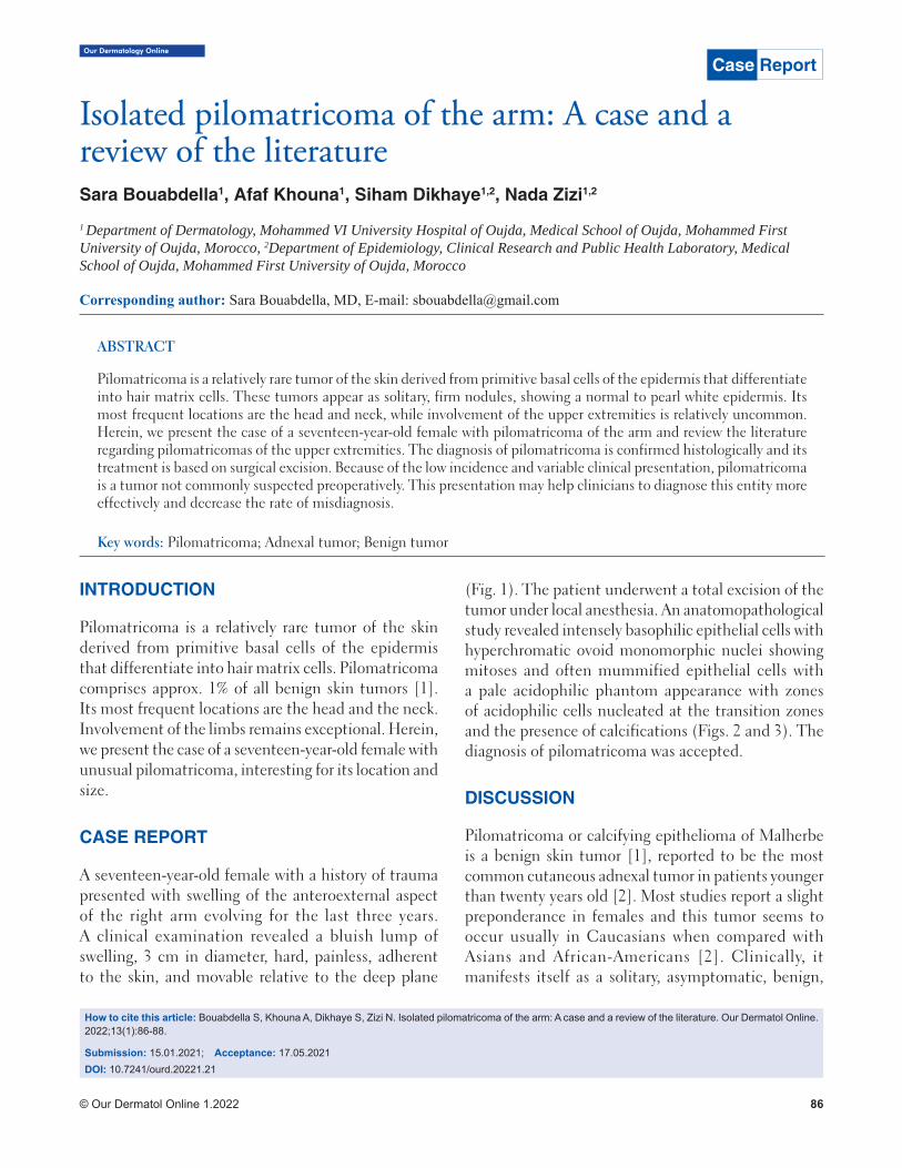
Our Dermatology Online
© Our Dermatol Online 1.2022 86
Isolated pilomatricoma of the arm: A case and a Isolated pilomatricoma of the arm: A case and a review of the literaturereview of the literatureSara Bouabdella1, Afaf Khouna1, Siham Dikhaye1,2, Nada Zizi1,2
1 Department of Dermatology, Mohammed VI University Hospital of Oujda, Medical School of Oujda, Mohammed First University of Oujda, Morocco, 2Department of Epidemiology, Clinical Research and Public Health Laboratory, Medical School of Oujda, Mohammed First University of Oujda, Morocco
Corresponding author: Sara Bouabdella, MD, E-mail: [email protected]
INTRODUCTION
Pilomatricoma is a relatively rare tumor of the skin derived from primitive basal cells of the epidermis that differentiate into hair matrix cells. Pilomatricoma comprises approx. 1% of all benign skin tumors [1]. Its most frequent locations are the head and the neck. Involvement of the limbs remains exceptional. Herein, we present the case of a seventeen-year-old female with unusual pilomatricoma, interesting for its location and size.
CASE REPORT
A seventeen-year-old female with a history of trauma presented with swelling of the anteroexternal aspect of the right arm evolving for the last three years. A clinical examination revealed a bluish lump of swelling, 3 cm in diameter, hard, painless, adherent to the skin, and movable relative to the deep plane
(Fig. 1). The patient underwent a total excision of the tumor under local anesthesia. An anatomopathological study revealed intensely basophilic epithelial cells with hyperchromatic ovoid monomorphic nuclei showing mitoses and often mummified epithelial cells with a pale acidophilic phantom appearance with zones of acidophilic cells nucleated at the transition zones and the presence of calcifications (Figs. 2 and 3). The diagnosis of pilomatricoma was accepted.
DISCUSSION
Pilomatricoma or calcifying epithelioma of Malherbe is a benign skin tumor [1], reported to be the most common cutaneous adnexal tumor in patients younger than twenty years old [2]. Most studies report a slight preponderance in females and this tumor seems to occur usually in Caucasians when compared with Asians and African-Americans [2]. Clinically, it manifests itself as a solitary, asymptomatic, benign,
ABSTRACT
Pilomatricoma is a relatively rare tumor of the skin derived from primitive basal cells of the epidermis that differentiate into hair matrix cells. These tumors appear as solitary, firm nodules, showing a normal to pearl white epidermis. Its most frequent locations are the head and neck, while involvement of the upper extremities is relatively uncommon. Herein, we present the case of a seventeen-year-old female with pilomatricoma of the arm and review the literature regarding pilomatricomas of the upper extremities. The diagnosis of pilomatricoma is confirmed histologically and its treatment is based on surgical excision. Because of the low incidence and variable clinical presentation, pilomatricoma is a tumor not commonly suspected preoperatively. This presentation may help clinicians to diagnose this entity more effectively and decrease the rate of misdiagnosis.
Key words: Pilomatricoma; Adnexal tumor; Benign tumor
Case Report
How to cite this article: Bouabdella S, Khouna A, Dikhaye S, Zizi N. Isolated pilomatricoma of the arm: A case and a review of the literature. Our Dermatol Online. 2022;13(1):86-88.
Submission: 15.01.2021; Acceptance: 17.05.2021DOI: 10.7241/ourd.20221.21
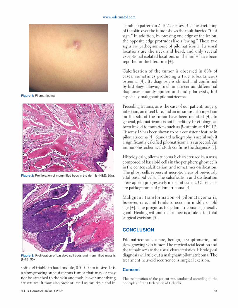
www.odermatol.com
© Our Dermatol Online 1.2022 87
a nodular pattern in 2–10% of cases [3]. The stretching of the skin over the tumor shows the multifaceted “tent sign.” In addition, by pressing one edge of the lesion, the opposite edge protrudes like a “swing.” These two signs are pathognomonic of pilomatricoma. Its usual locations are the neck and head, and only several exceptional isolated locations on the limbs have been reported in the literature [4].
Calcification of the tumor is observed in 80% of cases, sometimes producing a true subcutaneous osteoma [4]. Its diagnosis is clinical and confirmed by histology, allowing to eliminate certain differential diagnoses, mainly epidermoid and pilar cysts, but especially malignant pilomatricoma.
Preceding trauma, as is the case of our patient, surgery, infection, an insect bite, and an intramuscular injection on the site of the tumor have been reported [4]. In general, pilomatricoma is not hereditary. Its etiology has been linked to mutations such as β-catenin and BCL2. Trisomy 18 has been shown to be a consistent feature in pilomatricoma [4]. Standard radiography is useful only if a significantly calcified pilomatricoma is suspected. An immunohistochemical study confirms the diagnosis [5].
Histologically, pilomatricoma is characterized by a mass composed of basaloid cells in the periphery, ghost cells in the center, calcification, and sometimes ossification. The ghost cells represent necrotic areas of previously vital basaloid cells. The calcification and ossification areas appear progressively in necrotic areas. Ghost cells are pathognomic of pilomatricoma [3].
Malignant transformation of pilomatricoma is, however, rare, and tends to occur in middle or old age [4]. The prognosis for pilomatricoma is generally good. Healing without recurrence is a rule after total surgical excision [5].
CONCLUSION
Pilomatricoma is a rare, benign, asymptomatic, and slow-growing skin tumor. The cervicofacial location and the female sex are the usual characteristics. Histological diagnosis will rule out a malignant pilomatricoma. The treatment to avoid recurrence is surgical excision.
Consent
The examination of the patient was conducted according to the principles of the Declaration of Helsinki.
soft and friable to hard nodule, 0.5–5.0 cm in size. It is a slow-growing subcutaneous tumor that may or may not be attached to the skin and mobile over underlying structures. It may also present itself as multiple and in
Figure 2: Proliferation of mummifi ed beds in the dermis (H&E; 50×).
Figure 1: Pilomatricoma.
Figure 3: Proliferation of basaloid cell beds and mummifi ed massifs (H&E; 50×).
www.odermatol.com
© Our Dermatol Online 1.2022 88
The authors certify that they have obtained all appropriate patient consent forms, in which the patients gave their consent for images and other clinical information to be included in the journal. The patients understand that their names and initials will not be published and due effort will be made to conceal their identity, but that anonymity cannot be guaranteed.
REFERENCES
1. El Jouari O, Gallouj S, Douhi Z, Benkirane S, Baybay H, Mernissi FZ. A pregnancy pilomatricoma: An uncommon dermatologic benign neoplasm. Our Dermatol Online. 2018;9:425-7.
2. Harbaoui S, Youssef S, Ben Lagha I, Daadaa N, Jaber K,
Dhaoui MR, et al. Multiple and giant perforating pilomatrixoma: A case report. Our Dermatol Online. 2018;9:299-301.
3. Arif T, Amin SS, Raj D. Pilomatricoma of the eyelid. Our Dermatol Online. 2017;9:73-4.
4. Corredor-Osorio R, Suarez-Tata M, Orellana ME. Pilomatricoma of the orbit. Our Dermatol Online. 2016;7:188-90.
5. Chan JJ, Tey HL. Multiple pilomatricomas: Case presentation and review of the literature. Dermatol Online J. 2010;16:2.
Copyright by Sara Bouabdella, et al. This is an open access article distributed under the terms of the Creative Commons Attribution License, which permits unrestricted use, distribution, and reproduction in any medium, provided the original author and source are credited.Source of Support: Nil, Confl ict of Interest: None declared.
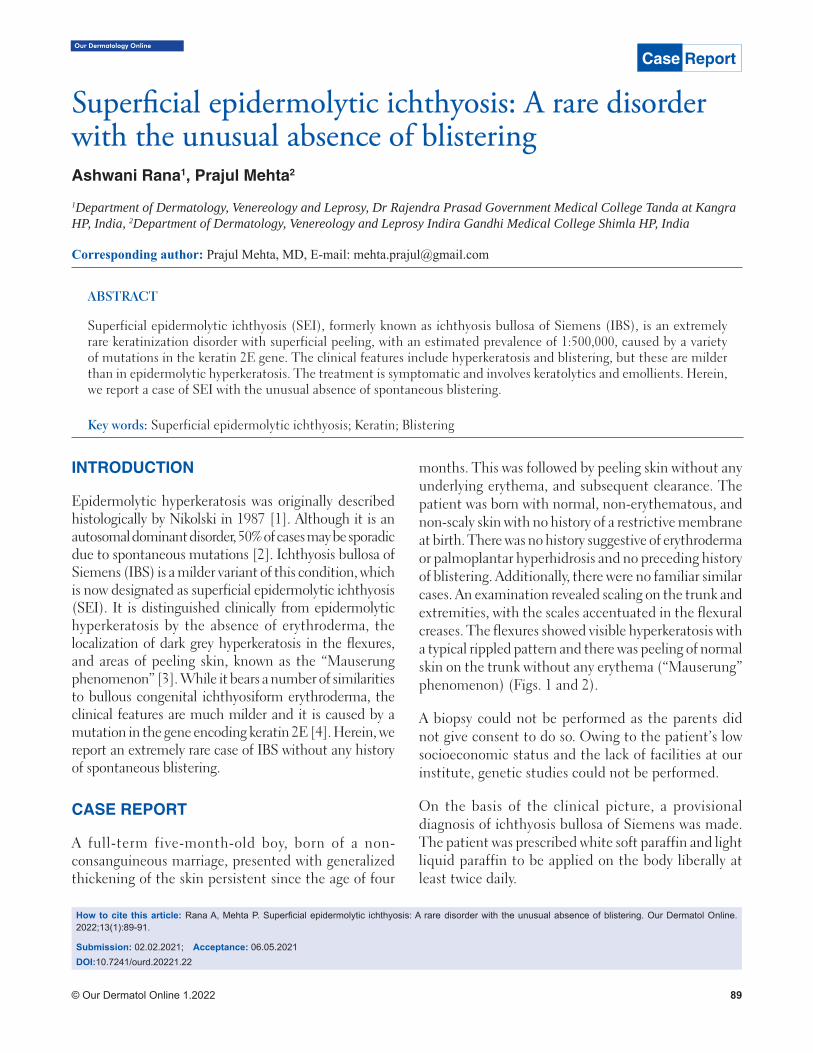
Our Dermatology Online
© Our Dermatol Online 1.2022 89
How to cite this article: Rana A, Mehta P. Superfi cial epidermolytic ichthyosis: A rare disorder with the unusual absence of blistering. Our Dermatol Online. 2022;13(1):89-91.
Submission: 02.02.2021; Acceptance: 06.05.2021DOI:10.7241/ourd.20221.22
Superfi cial epidermolytic ichthyosis: A rare disorder Superfi cial epidermolytic ichthyosis: A rare disorder with the unusual absence of blisteringwith the unusual absence of blisteringAshwani Rana1, Prajul Mehta2
1Department of Dermatology, Venereology and Leprosy, Dr Rajendra Prasad Government Medical College Tanda at Kangra HP, India, 2Department of Dermatology, Venereology and Leprosy Indira Gandhi Medical College Shimla HP, India
Corresponding author: Prajul Mehta, MD, E-mail: [email protected]
INTRODUCTION
Epidermolytic hyperkeratosis was originally described histologically by Nikolski in 1987 [1]. Although it is an autosomal dominant disorder, 50% of cases may be sporadic due to spontaneous mutations [2]. Ichthyosis bullosa of Siemens (IBS) is a milder variant of this condition, which is now designated as superficial epidermolytic ichthyosis (SEI). It is distinguished clinically from epidermolytic hyperkeratosis by the absence of erythroderma, the localization of dark grey hyperkeratosis in the flexures, and areas of peeling skin, known as the “Mauserung phenomenon” [3]. While it bears a number of similarities to bullous congenital ichthyosiform erythroderma, the clinical features are much milder and it is caused by a mutation in the gene encoding keratin 2E [4]. Herein, we report an extremely rare case of IBS without any history of spontaneous blistering.
CASE REPORT
A full-term five-month-old boy, born of a non-consanguineous marriage, presented with generalized thickening of the skin persistent since the age of four
months. This was followed by peeling skin without any underlying erythema, and subsequent clearance. The patient was born with normal, non-erythematous, and non-scaly skin with no history of a restrictive membrane at birth. There was no history suggestive of erythroderma or palmoplantar hyperhidrosis and no preceding history of blistering. Additionally, there were no familiar similar cases. An examination revealed scaling on the trunk and extremities, with the scales accentuated in the flexural creases. The flexures showed visible hyperkeratosis with a typical rippled pattern and there was peeling of normal skin on the trunk without any erythema (“Mauserung” phenomenon) (Figs. 1 and 2).
A biopsy could not be performed as the parents did not give consent to do so. Owing to the patient’s low socioeconomic status and the lack of facilities at our institute, genetic studies could not be performed.
On the basis of the clinical picture, a provisional diagnosis of ichthyosis bullosa of Siemens was made. The patient was prescribed white soft paraffin and light liquid paraffin to be applied on the body liberally at least twice daily.
ABSTRACT
Superficial epidermolytic ichthyosis (SEI), formerly known as ichthyosis bullosa of Siemens (IBS), is an extremely rare keratinization disorder with superficial peeling, with an estimated prevalence of 1:500,000, caused by a variety of mutations in the keratin 2E gene. The clinical features include hyperkeratosis and blistering, but these are milder than in epidermolytic hyperkeratosis. The treatment is symptomatic and involves keratolytics and emollients. Herein, we report a case of SEI with the unusual absence of spontaneous blistering.
Key words: Superficial epidermolytic ichthyosis; Keratin; Blistering
Case Report
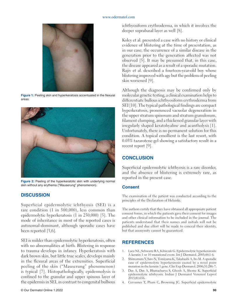
www.odermatol.com
© Our Dermatol Online 1.2022 90
DISCUSSION
Superficial epidermolytic ichthyosis (SEI) is a rare condition (1 in 500,000), less common than epidermolytic hyperkeratosis (1 in 250,000) [5]. The mode of inheritance in most of the reported cases is autosomal-dominant, although sporadic cases have been reported [3,6].
SEI is milder than epidermolytic hyperkeratosis, often with no abnormalities at birth. Blistering in response to trauma develops in infancy. Hyperkeratosis with dark brown skin, but little true scales, develops mainly in the flexural areas of the extremities. Superficial peeling of the skin (“Mauserung” phenomenon) is typical [7]. Histopathologically, epidermolysis is confined to the granular and upper spinous layer of the epidermis in SEI, in contrast to congenital bulbous
ichthyosiform erythroderma, in which it involves the deeper suprabasal layer as well [8].
Koley et al. presented a case with no history or clinical evidence of blistering at the time of presentation, as in our case; the occurrence of a similar disease in the generation prior to the generation affected was not observed [5]. It may be presumed that, in this case, the disease appeared as a result of a sporadic mutation. Rajiv et al. described a fourteen-year-old boy whose blistering improved with age but the problem of peeling skin worsened [9].
Although the diagnosis may be confirmed only by molecular genetic testing, a clinical examination helps to differentiate bullous ichthyosiform erythroderma from SEI [10]. The typical pathological findings are compact hyperkeratosis, pronounced vacuolar degeneration in the upper stratum spinosum and stratum granulosum, filament clumping, and a thickened granular layer with irregularly shaped keratohyaline and acantholysis [1]. Unfortunately, there is no permanent solution for this condition. A topical emollient is the last resort, with 0.05% tazarotene gel showing a satisfactory result in a recent report [9].
CONCLUSION
Superficial epidermolytic ichthyosis is a rare disorder, and the absence of blistering is extremely rare, as reported in the present case.
Consent
The examination of the patient was conducted according to the principles of the Declaration of Helsinki.
The authors certify that they have obtained all appropriate patient consent forms, in which the patients gave their consent for images and other clinical information to be included in the journal. The patients understand that their names and initials will not be published and due effort will be made to conceal their identity, but that anonymity cannot be guaranteed.
REFERENCES
1. Lacz NL, Schwartz RA, Kihiczak G. Epidermolytic hyperkeratosis: A keratin 1 or 10 mutational event. Int J Dermatol. 2005;44:1-6.
2. Shimomura Y, Sato N, Tomiyama K, Takahashi A, Ito M. A sporadic case of epidermolytic hyperkeratosis caused by a novel point mutation in the keratin 1 gene. Clin Exp Dermatol. 2006;31:286-7.
3. Das A, Das A, Bhattacharya S, Ghosh A, Shome K. Superfi cial epidermolytic ichthyosis. Indian J Dermatol Venereol Leprol 2015;81:656.
4. Cervantes T, Pham C, Browning JC. Superficial epidermolytic
Figure 2: Peeling of the hyperkeratotic skin with underlying normal skin without any erythema (“Mauserung” phenomenon).
Figure 1: Peeling skin and hyperkeratosis accentuated in the fl exural areas.

www.odermatol.com
© Our Dermatol Online 1.2022 91
ichthyosis: A report of two families. Pediatr Dermatol. 2013;30:469-72.5. Koley S, Salodkar A, Gupta S, Bhanke A, Ujawane A, Shazia B.
Ichthyosis bullosa of Siemens sans history of blistering: An interesting case report. J Pakistan Assoc Dermatol 2009;19:171-4.
6. Akiyama M, Tsuji-Abe Y, Yanagihara M, Nakajima K, Kodama H, Yaosaka M, et al. Ichthyosis bullosa of Siemens: its correct diagnosis facilitated by molecular genetic testing. Br J Dermatol. 2005;152:1353-6.
7. Whittock NV, Ashton GH, Griffi ths WA, Eady RA, McGrath JA. New mutations in keratin 1 that cause bullous congenital ichthyosiform erythroderma and keratin 2e that cause ichthyosis bullosa of Siemens. Br J Dermatol. 2001;145:330-5.
8. Sarkar P, Saha A, Malakar S. Ichthyosis bullosa of Siemens sans
blistering with extracutaneous features: A subtype or association? Indian J Paediatr Dermatol. 2015;16:188-90.
9. Rajiv S, Rakhesh SV. Ichthyosis bullosa of Siemens: Response to topical tazarotene. Indian J Dermatol Venereol Leprol. 2006;72:43-6.
10. Ang-Tiu CU, Nicolas ME. Ichthyosis bullosa of Siemens. J Dermatol Case Rep. 2012;6:78-81.
Copyright by Ashwani Rana, et al. This is an open access article distributed under the terms of the Creative Commons Attribution License, which permits unrestricted use, distribution, and reproduction in any medium, provided the original author and source are credited.Source of Support: Nil, Confl ict of Interest: None declared.
Our Dermatology Online
© Our Dermatol Online 1.2022 92
Progress of diff erent treatment modalities to limit the Progress of diff erent treatment modalities to limit the use of antibiotics in the treatment of acneuse of antibiotics in the treatment of acneKiran Sanjel1,2, Xue Mei Zhang1,2
1Clinical Medical School of Inner Mongolia University for the Nationalities, Tongliao, Inner Mongolia 028000, China, 2Department of Dermatology and Venerology, the Affi liated Hospital of Inner Mongolia University for the Nationalities, Tongliao, Inner Mongolia 028000, China
Corresponding author: Kiran Sanjel, MD, E-mail: [email protected]
INTRODUCTION
Acne vulgaris is a common chronic, multifaceted inflammatory disease of the pilosebaceous unit. Although acne may be self-limiting, the sequelae may be lifelong. Early diagnosis and treatment of acne is the modality to decrease acne sequelae. Oral antibiotics are a common choice as a drug for acne. Although different guidelines have been implicated, oral antibiotics are prescribed haphazardly for a very long time in acne vulgaris. Antibiotics resistance has been an alarming public health concern for the last three decades, including in dermatology. Resistance to topical erythromycin, clindamycin, and systemic tetracycline-class antibiotics are increased in Cutibacterium acne (formerly Propionibacterium acne) [1]. Excessive oral use of antibiotics leads to the disruption of indigenous gut flora [2]. Besides that, upper respiratory tract infection and an increase in pharyngitis are also involved [3]. Longer antibiotic use might be associated with inflammatory bowel disease [4]. At the same time, there is an association between the use of oral
tetracycline-class antibiotics and the risk of breast and colon cancer [5,6]. This article mainly emphasizes the different alternative treatment modalities for decreasing oral antibiotic use in acne vulgaris.
Topical Drugs
Benzoyl Peroxide (BPO)BPO is one of the over-the-counter medications for acne vulgaris, with formulation concentrations of 2.5%, 5%, and 10%. It is a powerful antimicrobial agent, inhibiting bacterial protein and nucleotide synthesis, metabolic pathways, and mitochondrial activity. It also acts as a mild sebostatic and keratolytic agent [7]. This mechanism allows benzoyl peroxide to be employed as a long-term therapy for acne. It is useful either as a monotherapy or in combination with topical retinoids or antibiotics, without the risk of developing bacterial resistance. It is a therapy for mild acne or as an adjunctive therapy for moderate to severe acne. It is available in lotions, creams, gels, foams, solutions, cleansing bars, masks, and shaving creams. A recent
ABSTRACT
Acne vulgaris is one of the most common skin diseases, affecting mainly teenagers. Its treatment procedure is complex, with a long duration of medication. Antibiotics are the most preferably prescribed drugs for the treatment of acne. The long-term use of antibiotics leads to various adverse effects such as the disruption of indigenous flora and resistance. Therefore, numerous therapeutic protocols such as antimicrobial stewardship have been proposed to limit the haphazard use of oral antibiotics. This review emphasizes different topical drugs, systemic alternative drugs, laser, and light therapy, as effective therapies for acne. This review also briefly reflects the efficacy of fire needle therapy—a traditional Chinese therapy—for acne.
Key words: acne vulgaris; antibiotic resistance; fire needle therapy; isotretinoin; Clascoterone
How to cite this article: Sanjel K, Zhang XM. Progress of diff erent treatment modalities to limit the use of antibiotics in the treatment of acne. Our Dermatol Online. 2022;13(1):92-97.Submission: 24.07.2021; Acceptance: 31.10.2021DOI: 10.7241/ourd.20221.23
Review Article
www.odermatol.com
© Our Dermatol Online 1.2022 93
2020 Cochrane review on benzoyl peroxide assessed that BPO is more effective than placebo and is as effective as topical retinoids and topical antibiotics [8].
Safety Level
Bleaching of hair and color fabrics are the most common side effects, together with dryness, erythema, and scaling in higher concentrations. Some portion of the literature reports cases of contact dermatitis. It is better to begin with a low dose and concentration and gradually increase as the skin develops tolerance. FDA classifies benzoyl peroxide as pregnancy risk category C. It is considered safe for breastfeeding until and unless benzoyl peroxide comes directly in contact with the area with which the infant could have direct contact during breastfeeding [9].
Topical Retinoids
First-line therapy for mild to moderate acne is topical retinoids. Retinoids activate the retinoic acid receptors, which affect the expression of genes involved in cell proliferation, differentiation, and inflammation. Thus, retinoids have both comedolytic and anti-inflammatory actions devoid of bacterial resistance. Tretinoin and tazarotene also suppress toll-like receptor expression, causing inflammatory cytokines and nitric oxide levels to decrease. Among topical retinoids, the most tolerated is 0.3% adapalene gel as it is photostable is the only topical retinoid that may be combined with 2.5% benzoyl peroxide without degradation; these are the most effective in moderate to severe acne [10]. A recent post hoc analysis showed that tretinoin 0.05% lotion was more effective in reducing non-inflammatory acne with no treatment-related adverse effects [11]. Recently, a fourth-generation topical retinoid—trifarotene—has been shown to have a selective retinoic acid receptor gamma agonist action. A three-phase randomized evaluation of 50 micrograms of trifarotene for facial and truncal acne for twelve weeks showed a reduction in the number of inflammatory lesions compared to the vehicle [12]. It has a better safety profile in comparison to other retinoids.
Safety Level
Topical retinoids can irritate more sensitive skin. Excessive use may result in redness, peeling, and blistering in the used area. Peeling of the stratum corneum may increase the chances of sunburn, so sunblock measures are needed. Adapalene and tretinoin
are pregnancy category C, whereas tazarotene is a category X drug [13]. Since retinoids are absorbed poorly after topical use, these are considered low-risk to nursing infants [13], ensuring that the skin is not in contact with the treated area.
Topical Anti-Androgen
Clascoterone is a new topical anti-androgen medication approved by the FDA in 2020 for the topical treatment of acne [14], which mainly targets testosterone and dihydrotestosterone [15]. It is available as 1% cream. It binds to the androgen receptor on the site of topical application. It is metabolized to an inactive form quickly, which limits its systemic absorption [15]. One of its advantages is that its onset of action is rapid, resulting in an improvement within two weeks. Both females and males older than twelve years are allowed to use it [16,17]. Two large phase three randomized controlled trials evaluated the effectiveness of clascoterone administered for twelve weeks, reporting a decrease in acne lesions and its symptoms by 8–18% more than placebo [18].
Safety Level
To date, the literature has reported no contraindications of this drug. The side effects include itching, burning, peeling, and dryness of the skin. Suppression of the hypothalamic-pituitary-adrenal axis is its severe side effect due to its cortexolone metabolites [13]. Hyperkalemia occurs in 5% of clascoterone-treated individuals [15]. Ophthalmic, oral, or vaginal routes are not prescribed for use. No data regarding pregnancy and nursing women is available.
Systemic Drugs
Oral SpironolactoneSpironolactone—a synthetic 17-lactone steroid—is an aldosterone antagonist. It functions as an androgen receptor blocker and inhibitor of 5α-reductase in the context of acne treatment. As it decreases sebum production, it is used for acne for more than thirty years [19]. In recent years, the use of spironolactone has increased considerably, but the use of oral antibiotics has not declined among females with acne [20]. Thereby, increased use of spironolactone might be an opportunity to improve antimicrobial stewardship. It is effective for acne in females of all ages. Spironolactone combined with a topical retinoid seems to provide a superior response to retinoid monotherapy
www.odermatol.com
© Our Dermatol Online 1.2022 94
in adult female acne. A recent retrospective review performed on eighty adolescent females showed an 80% improvement in acne lesions with few side effects [21]. A retrospective study done in 2017 stated that women treated with spironolactone at 100 mg/day showed an improvement in 80%, with only 4% experiencing any side effects [22]. FASCE and SAFA, two randomized trials, are being conducted to compare the efficacy of spironolactone over oral antibiotics. The results of these trials anticipated making the drug FDA approved [23]. Dosing is 25–200 mg/day, starting from 100 mg/day.
Safety Level
The side effects include diuresis, potential hyperkalemia, irregular menstrual bleeding, breast tenderness, headache, and fatigue. Combining spironolactone with oral contraceptives may decrease the problem of irregular menstrual bleeding. In healthy young females without cardiovascular disease, hypertension, or renal disease and not taking any interacting medicine, there is no evidence of increased potassium levels [24]. The contraindications include renal impairment, hyperkalemia, taking medication that increases serum potassium, and Addison’s disease. Spironolactone is pregnancy category C. Due to the evidence of teratogenicity in animal studies with high doses, it received a black box warning from the FDA [25]. Regarding breastfeeding, the risk to the infant is minimal.
Oral Contraceptives
Combined oral contraceptives focus on four mechanisms of the hormonal pathogenesis of acne:1. They decrease the amount of gonadal androgen
production;2. They decrease 40–50% of free testosterone [26];3. Estrogen reduces the conversion of testosterone
to dihydrotestosterone in the pilosebaceous unit, decreasing sebum;
4. Progestins with an anti-androgenic effect block the androgen receptors on keratinocytes and sebocytes.
It is effective in the treatment of both non-inflammatory and inflammatory acne. Ethinylestradiol + cyproterone acetate shows efficacy comparable to minocycline and superior efficacy compared to tetracycline [27]. A Cochrane review emphasized the effectiveness of all oral contraceptives for the treatment of acne in females. In trials with drospirenone-containing combined oral contraceptives compared with other oral contraceptives,
the former have been generally favored [28]. A course of 3–6 months of therapy is required. The three oral contraceptives approved by the FDA for the treatment of acne are norgestimate (180 mg, 215 mg, 250 mg) + ethinylestradiol (35 mcg), ethinylestradiol (20–35 mcg) + norethindrone acetate (1 mg), and ethinylestradiol (20 mcg) + drospirenone (3 mg).
Safety Level
The common side effects include breakthrough bleeding, nausea, breast tenderness, and increased risk of thromboembolic events (attributable risk). While counseling, it is emphasized that the risk of venous thromboembolism is higher in pregnancy than in combined oral contraceptive use [29]. It is pregnancy category X and is regarded safe for use for lactating mothers.
Isotretinoin
Isotretinoin is a retinoic acid derivative mostly used in the treatment of nodulocystic acne. It acts on all four etiopathogeneses of acne vulgaris and is capable of prolonged remission and the curing of up to 80% of patients. It is an FDA-approved treatment for severe recalcitrant acne, which is also useful in moderately treatment-resistant acne, and relapses quickly after the discontinuation of oral antibiotic therapy. Some of the patients using isotretinoin may aggravate their acne after three to six weeks, which may be due to sebaceous gland apoptosis; thus, a low dose of 0.2 mg/kg/day may cancel these effects. Low dose isotretinoin (0.2–0.4 mg/kg/day) have similar effectiveness and fewer side effects [30]. It is generally initiated at 0.5 mg/kg/day and titrated to 1 mg/kg/day as tolerated [31]. A study stated that, as long as treatment with isotretinoin was continued for more than two months after the acne had completely resolved, a cumulative dose or daily dose does not influence acne relapse [32].
Safety Level
Isotretinoin is the most effective treatment available for acne, but given the ubiquitous distribution of retinoic acid receptors, it almost always causes side effects resembling hypervitaminosis A syndrome. A systematic review shows that its side effects are mostly dermatological, such as skin and mucosal dryness, cheilitis, and peeling of the skin [33]. These are dose-dependent and are also signs of effective drug absorption. Oral isotretinoin may cause spontaneous
www.odermatol.com
© Our Dermatol Online 1.2022 95
abortion and life-threatening congenital malformations. To reduce teratogenicity, counseling, informed consent, a strict negative pregnancy test, and follow-up should be performed. Regarding mental health, a study published in 1983 asserted a relation between isotretinoin and depressive symptoms. Afterward, numerous articles have triggered controversy around this issue. Most studies have found no association between oral isotretinoin and depression. However, a 2017 meta-analysis revealed that oral isotretinoin produced a significant decrease in depression scales [34]. Oral isotretinoin may cause liver function disarrangements such as increased triglycerides, increased liver transaminases, increased low-density lipoproteins, and decreased high-density lipoproteins, but these side effects are usually short-lived and reversible without withdrawing the medication. Oral isotretinoin is the pregnancy X category and is not determined yet for nursing.
Emerging Therapies
Stearoyl coenzyme A desaturase 1 (SCD1) is an enzyme mainly responsible for the synthesis of monounsaturated fatty acids in the sebaceous glands of the skin. The topical application of SCD1 inhibitors has shown the potential to reduce the synthesis of monounsaturated fatty acids and the number of sebaceous glands in mouse skin. Several clinical trials of topical formulations of SCD1 inhibitors and their potential topical and systemic side effects are ongoing [35].
Nitric oxide-releasing nanoparticles have shown a promising role in preventing C. acnes-induced inflammation. Different investigations are ongoing due to their potential to suppress the release of multiple cytokines from human monocytes and keratinocytes and to prevent C. acnes-induced inflammation [36]. A recent open-label pilot study investigating novel nitric oxide-producing gels showed a decrease in comedones and papules by 50% in acne lesions [37].
Immunity induction therapy antibodies against the Christie–Atkins–Munch–Peterson (CAMP) factor show cytotoxicity against C. acnes and inhibit C. acnes growth and the production of murine MIP-2 [38]. Investigations of vaccines produced by Staphylococcus capitis E12 to prevent C. acnes overgrowth and destroy overgrown C. acnes are ongoing [39]. Different monoclonal antibodies blocking cytokines to suppress inflammation are needed as killed C. acnes induces inflammatory cytokines [40].
Light and Laser Therapy
Light and laser therapies have had their application broadened in the treatment of active acne. Laser therapy is advantageous as it is an in-office treatment and provides no systemic side effects. Red light, blue light, or combined red-blue light leads to the photo-excitation of endogenous bacterial porphyrins, singlet oxygen production, and subsequent C. acnes destruction [41].
Photodynamic therapy has shown the most consistent improvement in acne. A randomized control trial reported 5-aminolevulinic acid-PDT (ALA-PDT) followed by adapalene or oral doxycycline showed a greater reduction of inflammation and the total lesion count in the PDT group at twelve weeks [42]. Intense pulsed light (IPL) is able to destroy C. acnes and lead to the thermolysis of vessels supplying the sebaceous glands. As a result, it decreases sebum production. A study done on ten patients with mild to moderate acne with IPL using a dual-band “notch” acne filter showed that overall lesion clearance was substantial after four weeks of follow-up [43]. However, in recent research comparing Nd: YAG and IPL, a greater reduction of acne lesions, especially of non-inflammatory lesions, was seen with Nd: YAG laser [44]. Pulsed dye laser (585–595 nm) uses an organic dye as the laser solution and targets oxyhemoglobin, which causes the heating and photothermolysis of vessels supplying inflammatory acne lesions. A comparison of 585 pulsed dye lasers alone or in combination with Nd: YAG laser showed that inflammatory acne lesions were significantly reduced (82.5–83.5%) [45].
Fire Needle Therapy
Fire needle therapy is a form of acupuncture, which exhibits both conventional acupuncture and moxibustion. It is used in traditional Chinese medicine for numerous skin diseases, mainly for the clinical treatment of moderate to severe acne, especially in cystic cases. Fire needle therapy is included in the Chinese Acne Treatment Guidelines [46]. After routine disinfection, an acupuncture needle of 0.15 mm is heated with an alcohol lamp and pricked in the center of the pustule or cyst. The second or third acupuncture needle is heated until red hot and quickly and accurately pricked into the lesion. A recent study has shown that it is able to reduce the release of inflammatory factors [47], promote circulation, promote metabolism, and restore damaged tissue,
www.odermatol.com
© Our Dermatol Online 1.2022 96
leading to less scarring [48]. A systematic review and meta-analysis done in 2019 indicated fire needle as a monotherapy or combined with different Chinese and Western medicines, which is equally effective as other conventional modalities of acne treatment [49]. There are no recorded side effects mentioned in the review. Given increasing antibiotic resistance and different undesirable side effects, the currently recommended treatment for acne needs to be upgraded, and alternative modalities should be considered.
CONCLUSION
Although multiple guidelines recommend limiting the use and decreasing antibiotic resistance, antibiotics are frequently used agents for moderate to severe acne. Topical alternatives, spironolactone, isotretinoin, combined oral contraceptives, and fire needles are all effective therapeutic alternatives. Consideration should be given to these options to improve antibiotic stewardship and proper treatment outcomes for the patient.
Ethics Statement
The medical ethics committee of the Affiliated Hospital of Inner Mongolia University for the Nationalities approved this study. No potentially identifiable images or data are included in this article.
Author Contributions
Kiran Sanjel and Xue Mei Zhang contributed to the conception of the study, design and data acquisition, and interpretation, and drafted the article. All authors critically revised and approved the final version.
Statement of Human and Animal Rights
All the procedures followed were in accordance with the ethical standards of the responsible committee on human experimentation (institutional and national) and with the 2008 revision of the Declaration of Helsinki of 1975.
Statement of Informed Consent
Informed consent for participation in this study was obtained from all patients.
REFERENCES
1. Walsh TR, Efthimiou J, Dreno B. Systematic review of antibiotic resistance in acne: An increasing topical and oral threat. Lancet
Infect Dis. 2016;16:e23-e33.2. Levy RM, Huang EY, Roling D, Leyden JJ, Margolis DJ. Effect of
antibiotics on the oropharyngeal fl ora in patients with acne. Arch Dermatol. 2003;139:467-71.
3. Margolis DJ, Fanelli M, Kupperman E, Papadopoulos M, Metlay JP, Xie SX, et al. Association of pharyngitis with oral antibiotic use for the treatment of acne: A cross-sectional and prospective cohort study. Arch Dermatol. 2012;148:326-32.
4. Margolis DJ, Fanelli M, Hoffstad O, Lewis JD. Potential association between the oral tetracycline class of antimicrobials used to treat acne and infl ammatory bowel disease. Am J Gastroenterol. 2010;105:2610-16.
5. Cao Y, Wu K, Mehta R, Drew DA, Song M, Lochhead P, et al. Long-term use of antibiotics and risk of colorectal adenoma. Gut. 2018;67:672-8.
6. Velicer CM, Heckbert SR, Lampe JW, Potter JD, Robertson CA, Taplin SH. Antibiotic use concerning the risk of breast cancer. JAMA. 2004;291:827-35.
7. Kircik LH. The role of benzoyl peroxide in the new treatment paradigm for acne. J Drugs Dermatol. 2013;12:s73-6.
8. Yang Z, Zhang Y, Lazic Mosler E, Hu J, Li H, Zhang Y, et al. Topical benzoyl peroxide for acne. Cochrane Database Syst Rev. 2020;3:CD011154.
9. Kong YL, Tey HL. Treatment of acne vulgaris during pregnancy and lactation. Drugs. 2013;73:779-87.
10. Leyden J, Stein-Gold L, Weiss J. Why topical retinoids are mainstay of therapy for acne. Dermatol Ther (Heidelb). 2017;7:293-304.
11. Han G, Armstrong AW, Desai SR, Guenin E. Novel tretinoin 0.05% lotion for the once-daily treatment of moderate-to-severe acne vulgaris in an asian population. J Drugs Dermatol. 2019;18:910-6.
12. Tan J, Thiboutot D, Popp G, Gooderham M, Lynde C, Del Rosso J, et al. Randomized phase 3 evaluation of trifarotene 50 μg/g cream treatment of moderate facial and truncal acne. J Am Acad Dermatol. 2019;80:1691-9.
13. Butler DC, Heller MM, Murase JE. Safety of dermatologic medications in pregnancy and lactation: Part II Lactation. J Am Acad Dermatol. 2014;70(3):417.e1-427
14. Cassiopea Receives FDA Approval for Winlevi (clascoterone cream 1%), fi rst in class in Topical acne treatment targeting Androgen Receptor”. Cassiopea (press release). 2020.
15. Kircik LH. What’s new in the management of acne vulgaris. Cutis. 2019;104:48-52.
16. Barbieri JS. A new class of topical acne treatment addressing the hormonal pathogenesis of acne. JAMA Dermatol. 2020;156:619-20.
17. Kalabalik-Hoganson J, Frey KM, Ozdener-Poyraz AE, Slugocki M. Clascoterone: A novel topical androgen receptor inhibitor for the treatment of acne. Ann Pharmacother. 2021;3:1060028021992053.
18. Hebert A, Thiboutot D, Stein Gold L, Cartwright M, Gerloni M, Fragasso E, Mazzetti A. Effi cacy and safety of topical clascoterone cream, 1%, for treatment in patients with facial acne: Two phase 3 randomized clinical trials. JAMA Dermatol. 2020;156:621-30.
19. Layton AM, Eady EA, Whitehouse H, Del Rosso JQ, Fedorowicz Z, van Zuuren EJ. Oral spironolactone for acne vulgaris in adult females: A hybrid systematic review. Am J Clin Dermatol. 2017;18:169-91.
20. Barbieri JS, James WD, Margolis DJ. Trends in prescribing behavior of systemic agents used in the treatment of acne among dermatologists and nondermatologists: A retrospective analysis, 2004-2013. J Am Acad Dermatol. 2017;77:456463.e4.
21. Roberts EE, Nowsheen S, Davis DMR, Hand JL, Tollefson MM, Wetter DA. Use of spironolactone to treat acne in adolescent females. Pediatr Dermatol. 2021;38:72-6.
22. Park JH, Bienenfeld A, Orlow SJ, Nagler AR. The use of hormonal antiandrogen therapy in female patients with acne: A 10-year retrospective study. Am J Clin Dermatol. 2018;19:449-55
23. Poinas A, Lemoigne M, Le Naour S, Nguyen JM, Schirr-Bonnans S, Riche VP, et al. FASCE, the benefi t of spironolactone for treating
www.odermatol.com
© Our Dermatol Online 1.2022 97
acne in women: study protocol for a randomized double-blind trial. Trials. 2020;21:571.
24. Plovanich M, Weng QY, Mostaghimi A. Low usefulness of potassium monitoring among healthy young women taking spironolactone for acne. JAMA Dermatol. 2015;151:941-4.
25. Food and Drug Administration. Aldactone (spironolactone) tablets. Physician Labeling. Accessed November 9, 2017.
26. Thorneycroft IH, Stanczyk FZ, Bradshaw KD, Ballagh SA, Nichols M, Weber ME. Effect of low-dose oral contraceptives on androgenic markers and acne. Contraception. 1999;60:255-62.
27. Nast A, Dréno B, Bettoli V, Degitz K, Erdmann R, Finlay AY, et al. European evidence-based (S3) guidelines for the treatment of acne. J Eur Acad Dermatol Venereol. 2012;1:1-29.
28. Lortscher D, Admani S, Satur N, Eichenfield LF. Hormonal contraceptives and acne: a retrospective analysis of 2147 patients. J Drugs Dermatol. 2016;15:670-4.
29. Reid RL. Oral contraceptives and venous thromboembolism: Pill scares and public health. J Obstet Gynaecol Can JOGC. 2011;33:1150-5.
30. Faghihi G, Mokhtari F, Fard NM, Motamedi N, Hosseini SM. comparing the effi cacy of low dose and conventional dose of oral isotretinoin in treatment of moderate and severe acne vulgaris. J Res Pharm Pract. 2017;6:233-8.
31. Zaenglein AL, Pathy AL, Schlosser BJ, Alikhan A, Baldwin HE, Berson DS, et al. Guidelines of care for the management of acne vulgaris. J Am Acad Dermatol. 2016;74:945-73.e33.
32. Rademaker M. Making sense of the effects of the cumulative dose of isotretinoin in acne vulgaris. Int J Dermatol. 2016;55:518-23.
33. Vallerand IA, Lewinson RT, Farris MS, Sibley CD, Ramien ML, Bulloch AGM, Patten SB. Effi cacy and adverse events of oral isotretinoin for acne: A systematic review. Br J Dermatol. 2018;178:76-85.
34. Huang Y-C, Cheng Y-C. Isotretinoin treatment for acne and risk of depression: A systematic review and meta-analysis. J Am Acad Dermatol. 2017;76:1068-76.e9.
35. Zouboulis CC, Dessinioti C, Tsatsou F, Gollnick HPM. Antiacne drugs in phase 1 and 2 clinical trials. Expert Opin Investig Drugs. 2017;26:813-23.
36. Qin M, Landriscina A, Rosen JM, Wei G, Kao S, Olcott W, et al. nitric oxide-releasing nanoparticles prevent propionibacterium acnes-induced infl ammation by both clearing the organism and inhibiting microbial stimulation of the innate immune response. J Invest Dermatol. 2015;135:2723-31.
37. Settelmeier S, Rassaf T, Hendgen-Cotta UB, Stoffels I. nitric oxide generating formulation as an innovative approach to topical skin care: An open-label pilot study. Cosmetics. 2021;8:16.
38. Wang Y, Hata TR, Tong YL, Kao MS, Zouboulis CC, Gallo RL, et al. The anti-infl ammatory activities of propionibacterium acnes camp factor-targeted acne vaccines. J Invest Dermatol. 2018;138:2355-64.
39. O’Neill AM, Nakatsuji T, Hayachi A, Williams MR, Mills RH, Gonzalez DJ, et al. Identifi cation of a human skin commensal bacterium that selectively kills cutibacterium acnes. J Invest Dermatol. 2020;140:1619-28.
40. Lyte P, Sur R, Nigam A, Southall MD. Heat-killed Propionibacterium acnes is capable of inducing infl ammatory responses in skin. Exp Dermatol. 2009;18:1070-2.
41. Nestor MS, Swenson N, Macri A. Physical modalities (devices) in the management of acne. Dermatol Clin. 2016;34:215223.
42. Nicklas C, Rubio R, Cárdenas C, Hasson A. Comparison of effi cacy of aminolaevulinic acid photodynamic therapy vs. adapalene gel plus oral doxycycline for treatment of moderate acne vulgaris-A simple, blind, randomized, and controlled trial. Photodermatol Photoimmunol Photomed. 2019;35:3-10.
43. Knight JM. Combined 400-600nm and 800-1200nm intense pulsed phototherapy of facial acne vulgaris. J Drugs Dermatol. 2019;18:1116-22.
44. Monib KME, Hussein MS. Nd: YAG laser vs IPL in infl ammatory and noninfl ammatory acne lesion treatment. J Cosmet Dermatol. 2020;19:2325-32.
45. Salah El Din MM, Samy NA, Salem AE. Comparison of pulsed dye laser versus combined pulsed dye laser and Nd: YAG laser in the treatment of infl ammatory acne vulgaris. J Cosmet Laser Ther. 2017;19:149-59.
46. Ju Qiang. Chinese acne treatment guidelines(2019 Revision). J Clin Dermatol. 2019;49:584.
47. Zhou Z, Li Y. Effect of fi re on expression of IL-1beta and caspase -3 protein in rat model of spinal cord injury. Shanghai J Acupunct Moxibust. 2010;29:318-21.
48. Rui Z. Clinical observation of isotretinoin combined with fi re needle therapy in treatment of grade IV acne. J Pract Chin Med. 2016,32:278.
49. Xing M, Yan X, Sun X, Wang S, Zhou M, Zhu B, et al. Fire needle therapy for moderate-severe acne: A PRISMA systematic review and meta-analysis of randomized controlled trials. Complement Ther Med. 2019;44:253-60.
Copyright by Kiran Sanjel, et al. This is an open access article distributed under the terms of the Creative Commons Attribution License, which permits unrestricted use, distribution, and reproduction in any medium, provided the original author and source are credited.Source of Support: Nil, Confl ict of Interest: None declared.
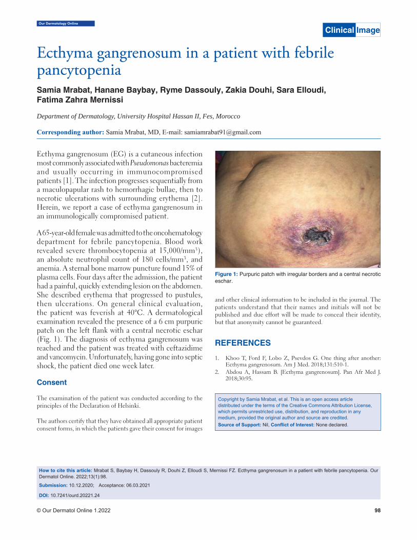
Our Dermatology Online
© Our Dermatol Online 1.2022 98
How to cite this article: Mrabat S, Baybay H, Dassouly R, Douhi Z, Elloudi S, Mernissi FZ. Ecthyma gangrenosum in a patient with febrile pancytopenia. Our Dermatol Online. 2022;13(1):98.
Submission: 10.12.2020; Acceptance: 06.03.2021
DOI: 10.7241/ourd.20221.24
Ecthyma gangrenosum in a patient with febrile Ecthyma gangrenosum in a patient with febrile pancytopeniapancytopeniaSamia Mrabat, Hanane Baybay, Ryme Dassouly, Zakia Douhi, Sara Elloudi,Fatima Zahra Mernissi
Department of Dermatology, University Hospital Hassan II, Fes, Morocco
Corresponding author: Samia Mrabat, MD, E-mail: [email protected]
Ecthyma gangrenosum (EG) is a cutaneous infection most commonly associated with Pseudomonas bacteremia and usually occurring in immunocompromised patients [1]. The infection progresses sequentially from a maculopapular rash to hemorrhagic bullae, then to necrotic ulcerations with surrounding erythema [2]. Herein, we report a case of ecthyma gangrenosum in an immunologically compromised patient.
A 65-year-old female was admitted to the oncohematology department for febrile pancytopenia. Blood work revealed severe thrombocytopenia at 15,000 /mm³), an absolute neutrophil count of 180 cells/mm³, and anemia. A sternal bone marrow puncture found 15% of plasma cells. Four days after the admission, the patient had a painful, quickly extending lesion on the abdomen. She described erythema that progressed to pustules, then ulcerations. On general clinical evaluation, the patient was feverish at 40°C. A dermatological examination revealed the presence of a 6 cm purpuric patch on the left flank with a central necrotic eschar (Fig. 1). The diagnosis of ecthyma gangrenosum was reached and the patient was treated with ceftazidime and vancomycin. Unfortunately, having gone into septic shock, the patient died one week later.
Consent
The examination of the patient was conducted according to the principles of the Declaration of Helsinki.
The authors certify that they have obtained all appropriate patient consent forms, in which the patients gave their consent for images
and other clinical information to be included in the journal. The patients understand that their names and initials will not be published and due effort will be made to conceal their identity, but that anonymity cannot be guaranteed.
REFERENCES
1. Khoo T, Ford F, Lobo Z, Psevdos G. One thing after another: Ecthyma gangrenosum. Am J Med. 2018;131:510-1.
2. Abdou A, Hassam B. [Ecthyma gangrenosum]. Pan Afr Med J. 2018;30:95.
Copyright by Samia Mrabat, et al. This is an open access article distributed under the terms of the Creative Commons Attribution License, which permits unrestricted use, distribution, and reproduction in any medium, provided the original author and source are credited.Source of Support: Nil, Confl ict of Interest: None declared.
Clinical Image
Figure 1: Purpuric patch with irregular borders and a central necrotic eschar.
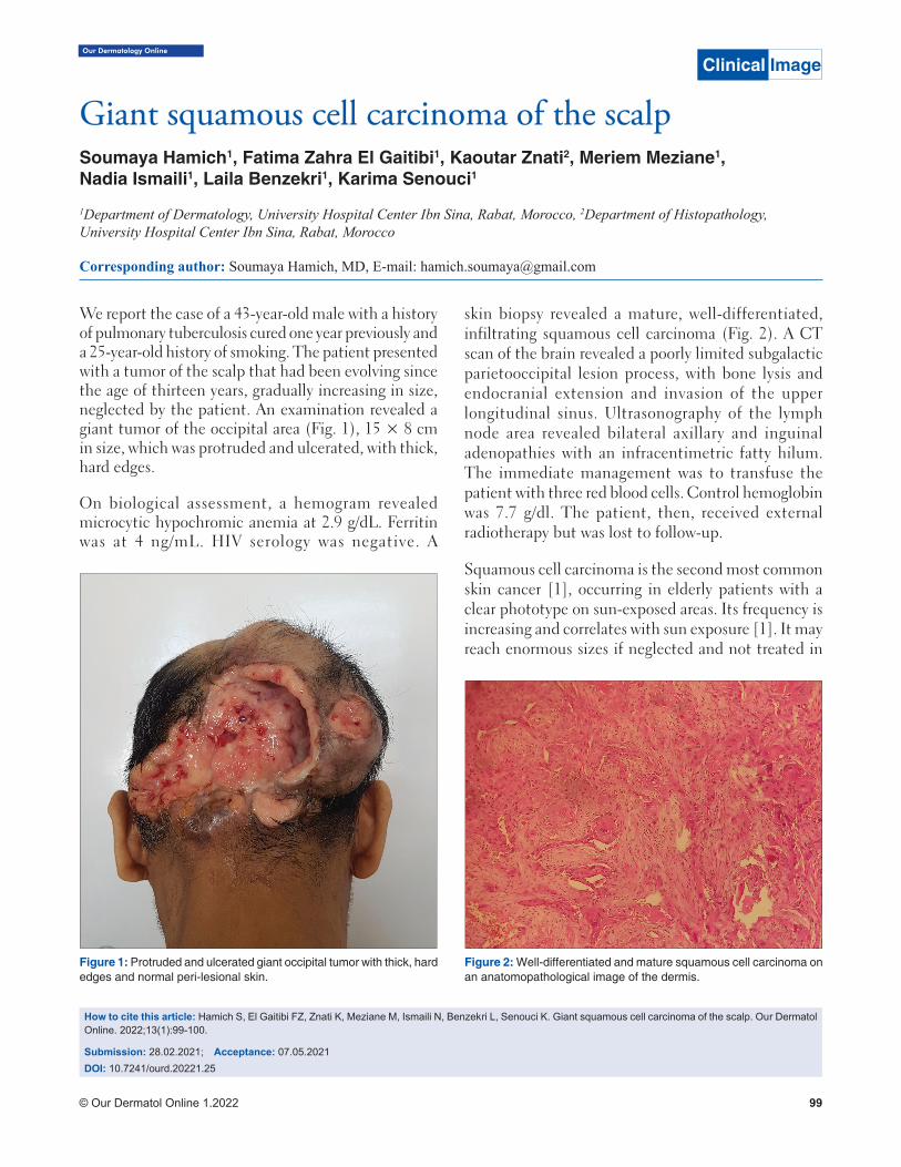
© Our Dermatol Online 1.2022 99
Our Dermatology Online
How to cite this article: Hamich S, El Gaitibi FZ, Znati K, Meziane M, Ismaili N, Benzekri L, Senouci K. Giant squamous cell carcinoma of the scalp. Our Dermatol Online. 2022;13(1):99-100.
Submission: 28.02.2021; Acceptance: 07.05.2021DOI: 10.7241/ourd.20221.25
Giant squamous cell carcinoma of the scalpGiant squamous cell carcinoma of the scalpSoumaya Hamich1, Fatima Zahra El Gaitibi1, Kaoutar Znati2, Meriem Meziane1,Nadia Ismaili1, Laila Benzekri1, Karima Senouci1
1Department of Dermatology, University Hospital Center Ibn Sina, Rabat, Morocco, 2Department of Histopathology, University Hospital Center Ibn Sina, Rabat, Morocco
Corresponding author: Soumaya Hamich, MD, E-mail: [email protected]
We report the case of a 43-year-old male with a history of pulmonary tuberculosis cured one year previously and a 25-year-old history of smoking. The patient presented with a tumor of the scalp that had been evolving since the age of thirteen years, gradually increasing in size, neglected by the patient. An examination revealed a giant tumor of the occipital area (Fig. 1), 15 × 8 cm in size, which was protruded and ulcerated, with thick, hard edges.
On biological assessment, a hemogram revealed microcytic hypochromic anemia at 2.9 g/dL. Ferritin was at 4 ng/mL. HIV serology was negative. A
skin biopsy revealed a mature, well-differentiated, infiltrating squamous cell carcinoma (Fig. 2). A CT scan of the brain revealed a poorly limited subgalactic parietooccipital lesion process, with bone lysis and endocranial extension and invasion of the upper longitudinal sinus. Ultrasonography of the lymph node area revealed bilateral axillary and inguinal adenopathies with an infracentimetric fatty hilum. The immediate management was to transfuse the patient with three red blood cells. Control hemoglobin was 7.7 g/dl. The patient, then, received external radiotherapy but was lost to follow-up.
Squamous cell carcinoma is the second most common skin cancer [1], occurring in elderly patients with a clear phototype on sun-exposed areas. Its frequency is increasing and correlates with sun exposure [1]. It may reach enormous sizes if neglected and not treated in
Clinical Image
Figure 2: Well-differentiated and mature squamous cell carcinoma on an anatomopathological image of the dermis.
Figure 1: Protruded and ulcerated giant occipital tumor with thick, hard edges and normal peri-lesional skin.
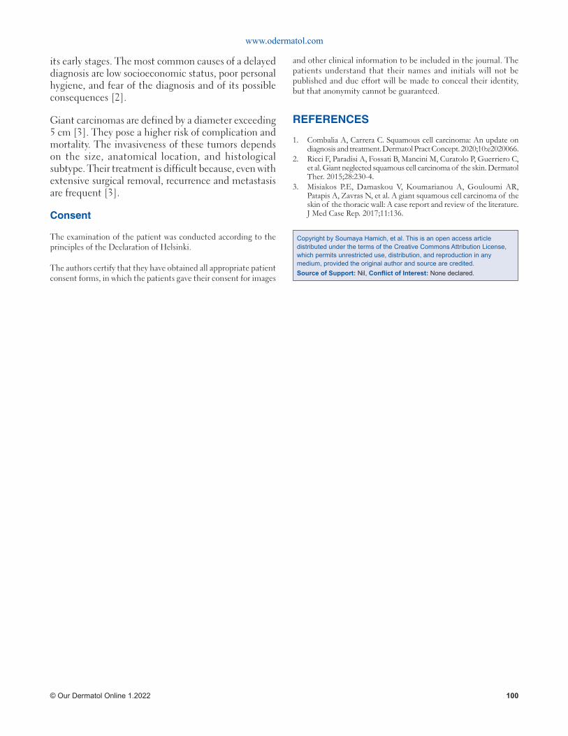
www.odermatol.com
© Our Dermatol Online 1.2022 100
its early stages. The most common causes of a delayed diagnosis are low socioeconomic status, poor personal hygiene, and fear of the diagnosis and of its possible consequences [2].
Giant carcinomas are defined by a diameter exceeding 5 cm [3]. They pose a higher risk of complication and mortality. The invasiveness of these tumors depends on the size, anatomical location, and histological subtype. Their treatment is difficult because, even with extensive surgical removal, recurrence and metastasis are frequent [3].
Consent
The examination of the patient was conducted according to the principles of the Declaration of Helsinki.
The authors certify that they have obtained all appropriate patient consent forms, in which the patients gave their consent for images
and other clinical information to be included in the journal. The patients understand that their names and initials will not be published and due effort will be made to conceal their identity, but that anonymity cannot be guaranteed.
REFERENCES
1. Combalia A, Carrera C. Squamous cell carcinoma: An update on diagnosis and treatment. Dermatol Pract Concept. 2020;10:e2020066.
2. Ricci F, Paradisi A, Fossati B, Mancini M, Curatolo P, Guerriero C, et al. Giant neglected squamous cell carcinoma of the skin. Dermatol Ther. 2015;28:230-4.
3. Misiakos P.E, Damaskou V, Koumarianou A, Gouloumi AR, Patapis A, Zavras N, et al. A giant squamous cell carcinoma of the skin of the thoracic wall: A case report and review of the literature. J Med Case Rep. 2017;11:136.
Copyright by Soumaya Hamich, et al. This is an open access article distributed under the terms of the Creative Commons Attribution License, which permits unrestricted use, distribution, and reproduction in any medium, provided the original author and source are credited.Source of Support: Nil, Confl ict of Interest: None declared.
Our Dermatology Online
© Our Dermatol Online 1.2022 101
Comorbidities of alopecia areata in infancy and Comorbidities of alopecia areata in infancy and childhood. A small descriptive study in a tertiary childhood. A small descriptive study in a tertiary hospital in Greecehospital in GreeceEleni Klimi
Department of Dermatology, Triasssio General Hospital Avenue Gennimata 19200 Magoula, Greece
Corresponding author: Eleni Klimi, MD, E-mail: [email protected]
Sir,A small descriptive study in a tertiary hospital in Greece was conducted on the comorbidities of alopecia areata in infancy and childhood.
Alopecia areata is a non-scarring alopecia of autoimmune origin linked also to genetic and environmental factors [1], affecting 2% of the general population and is considered a disease of young adults. Attempts have been made to detect the comorbidities in infants and children suffering from alopecia areata. Sorell Jennifer et al. established a strong association of alopecia areata with atopy, psoriasis thyroid disease, and juvenile idiopathic arthritis [2]. More recently, Comiz et al. [3] added anemia, obesity, vitamin D deficiency, hypothyroidism, vitiligo, psoriasis, hyperlipidemia, and depression to the list of the comorbidities detected in the pediatric population with alopecia areata. The purpose of the study was to detect the comorbidities in infants and children with alopecia areata in an outpatient dermatology clinic during a period of six years from 2013 through 2019. All those examined as outpatients and those hospitalized for several reasons in the pediatric ward who were diagnosed with alopecia areata were included in the study. Laboratory tests, a full blood count, and vitamin D, IgE, and thyroid tests were performed in the laboratories of our hospital. During these seven years, 71 patients were diagnosed with alopecia areata and 7 (approx. 10%) were children. Four (57.1%) were males, and the rest three were females. The males were aged 23 months, and 6-, 7-, and 11-years. The females were 2-, 7-, and 11-year-old.
Clinical atopy confirmed by high levels of IgE in the serum was detected in two males and in all three females. Thyroid dysfunction, hypothyroidism, was only detected in one infant associated with atopy; this was in a 23-month-old who at the time of the diagnosis of alopecia areata was hospitalized with severe asthma. Vitamin D deficiency was found in one male patient. A family history of alopecia areata was found in only one male patient. A family history of atopy was reported in only one boy, aged 7 years. A family history of thyroid dysfunction was detected in two males 28%: The 23-month-old infant whose father suffered from hyperthyroidism and the 12-year-old male whose both parents suffered from hypothyroidism. A family history of rheumatoid arthritis was found in one female patient. All patients presented with a mild disease limited to the scalp at the time of diagnosis. No nail pitting was observed, and neither clinical signs of psoriasis, nor of vitiligo. Folliculitis of the scalp preceded the onset of alopecia areata in one of the females (Table 1). Although males comprised 57.1% of our cases, most studies have found a preponderance of females in the pediatric population with AA. Atopy was the most frequent comorbidity (5/7; 70%) and was more frequent in females; all three girls were atopic. The second most frequently found comorbidity was thyroid dysfunction, hypothyroidism., detected in one patient (14%). Vitamin D deficiency was noted in one (14%) patient. A family history of AA was found in one patient as well as a family history of atopy. A family history of thyroid dysfunction was found in two patients (28%). The precipitating factor in our case was staphylococcal infection of the scalp.
How to cite this article: Klimi E. Comorbidities of alopecia areata in infancy and childhood. A small descriptive study in a tertiary hospital in Greece. Our Dermatol Online. 2022;13(1):101-102.Submission: 16.06.2021; Acceptance: 14.10.2021DOI: 10.7241/ourd.20221.26
Letter to the Editor
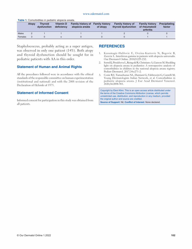
www.odermatol.com
© Our Dermatol Online 1.2022 102
Staphylococcus, probably acting as a super antigen, was observed in only one patient (14%). Both atopy and thyroid dysfunction should be sought for in pediatric patients with AA in this order.
Statement of Human and Animal Rights
All the procedures followed were in accordance with the ethical standards of the responsible committee on human experimentation (institutional and national) and with the 2008 revision of the Declaration of Helsinki of 1975.
Statement of Informed Consent
Informed consent for participation in this study was obtained from all patients.
REFERENCES
1. Kasumagic-Halilovic E, Ovcina-Kurtovic N, Begovic B, Zecevic L. Interferon-gamma in patients with alopecia universalis. Our Dermatol Online. 2018;9:229-232.
2. Sorrell J, Petukhova L, Reingold R, Christiano A, Garzon M. Shedding light on alopecia areata in pediatrics: A retrospective analysis of comorbidities in children in the national alopecia areata registry. Pediatr Dermatol. 2017;34:e271-2.
3. Conic RZ, Tamashunas NL, Damiani G, Fabbrocini G, Cantelli M; Young Dermatologists Italian Network, et al. Comorbidities in pediatric alopecia areata. J Eur Acad Dermatol Venereol. 2020;34:2898-901.
Copyright by Eleni Klimi. This is an open access article distributed under the terms of the Creative Commons Attribution License, which permits unrestricted use, distribution, and reproduction in any medium, provided the original author and source are credited.Source of Support: Nil, Confl ict of Interest: None declared.
Table 1: Comorbidities in pediatric alopecia areata.Atopy Thyroid
dysfunctionVitamin D defi ciency
Family history of alopecia areata
Family history of atopy
Family history of thyroid dysfunction
Family history of rheumatoid
arthritis
Precipitating factor
Males 2 1 1 1 1 2 0 0
Females 3 0 o 0 0 0 1 1
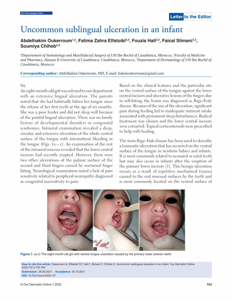
© Our Dermatol Online 1.2022 103
Our Dermatology Online
How to cite this article: Oukerroum A, Elfatoiki FZ, Hali F, Slimani F, Chiheb S. Uncommon sublingual ulceration in an infant. Our Dermatol Online. 2022;13(1):103-104.Submission: 28.05.2021; Acceptance: 20.10.2021DOI: 10.7241/ourd.20221.27
Uncommon sublingual ulceration in an infantUncommon sublingual ulceration in an infantAbdelhakim Oukerroum1,2, Fatima Zahra Elfatoiki2,3, Fouzia Hali2,3, Faical Slimani1,2, Soumiya Chiheb2,3
1Departement of Stomatology and Maxillofacial Surgery of UH Ibn Rochd of Casablanca, Morocco, 2Faculty of Medicine and Pharmacy, Hassan II University of Casablanca, Casablanca, Morocco, 3Department of Dermatology of UH Ibn Rochd of Casablanca, Morocco
Corresponding author: Abdelhakim Oukerroum, MD, E-mail: [email protected]
Sir,An eight-month-old girl was referred to our department with an extensive lingual ulceration. The parents noted that she had habitually bitten her tongue since the release of her first teeth at the age of six months. She was a poor feeder and did not sleep well because of the painful lingual ulceration. There was no family history of developmental disorders or congenital syndromes. Intraoral examination revealed a deep, circular, and extensive ulceration of the whole ventral surface of the tongue with intermittent bleeding in the tongue (Figs. 1a – c). An examination of the rest of the intraoral mucosa revealed that the lower central incisors had recently erupted. However, there were two other ulcerations of the palmar surface of the second and third fingers caused by nocturnal finger biting. Neurological examination noted a lack of pain sensitivity related to peripheral neuropathy diagnosed as congenital insensitivity to pain.
Based on the clinical features and the particular site on the ventral surface of the tongue against the lower central incisors and ulcerative lesions of the fingers due to self-biting, the lesion was diagnosed as Riga–Fede disease. Because of the size of the ulceration, significant pain during feeding led to inadequate nutrient intake associated with permanent sleep disturbances. Radical treatment was chosen and the lower central incisors were extracted. Topical corticosteroids were prescribed to help with healing.
The term Riga–Fede disease has been used to describe a traumatic ulceration that has occurred on the ventral surface of the tongue in newborn babies and infants. It is most commonly related to neonatal or natal teeth but may also occur in infants after the eruption of the primary lower incisors [1]. This benign ulceration occurs as a result of repetitive mechanical trauma caused to the oral mucosal surfaces by the teeth and is most commonly located on the ventral surface of
Letter to the Editor
Figure 1: (a-c) The eight-month-old girl with ventral tongue ulceration caused by the primary lower anterior teeth.
cba
www.odermatol.com
© Our Dermatol Online 1.2022 104
the tongue against the teeth [1,2]. Riga–Fede disease may reveal an underlying developmental or neurologic disorder, including congenital insensitivities to pain [3]. The case of our patient was associated with congenital insensitivity to pain.
Failure to diagnose may lead to dehydration and inadequate nutrient intake in the infant because of the significant pain during feeding. No biopsy is needed. The diagnosis of Riga–Fede disease is based on clinical characteristics [1,2].
Treatment should focus on eliminating the source of trauma. Conservative treatment is attempted at first by grinding the sharp edges of the teeth and placing composite resin in a dome shape or by placing a protective ring. If conservative treatment fails to heal the wounds, radical treatment may be necessary, such as extraction of the teeth [2,3].
We believe that Riga–Fede disease must be recognized by clinicians to avoid misdiagnosis and delayed treatment.
Consent
The examination of the patient was conducted according to the principles of the Declaration of Helsinki.
The authors certify that they have obtained all appropriate patient consent forms, in which the patients gave their consent for images and other clinical information to be included in the journal. The patients understand that their names and initials will not be published and due effort will be made to conceal their identity, but that anonymity cannot be guaranteed.
REFERENCES
1. Lahrichi A, El Fatoiki FZ, Hali F, Chiheb S. [Riga-Fede disease in an infant]. Ann Dermatol Venereol. 2019;146:319-320.
2. Choi SC, Park JH, Choi YC, Kim GT. Sublingual traumatic ulceration (a Riga-Fede disease): Report of two cases. Dent Traumatol. 2009;25:e48-50.
3. Yürekli A, Dinçer D. Successfully treated Riga-Fede disease. Dermatol Pract Concept. 2019;9:218-9.
Copyright by Abdelhakim Oukerroum, et al. This is an open access article distributed under the terms of the Creative Commons Attribution License, which permits unrestricted use, distribution, and reproduction in any medium, provided the original author and source are credited.Source of Support: Nil, Confl ict of Interest: None declared.
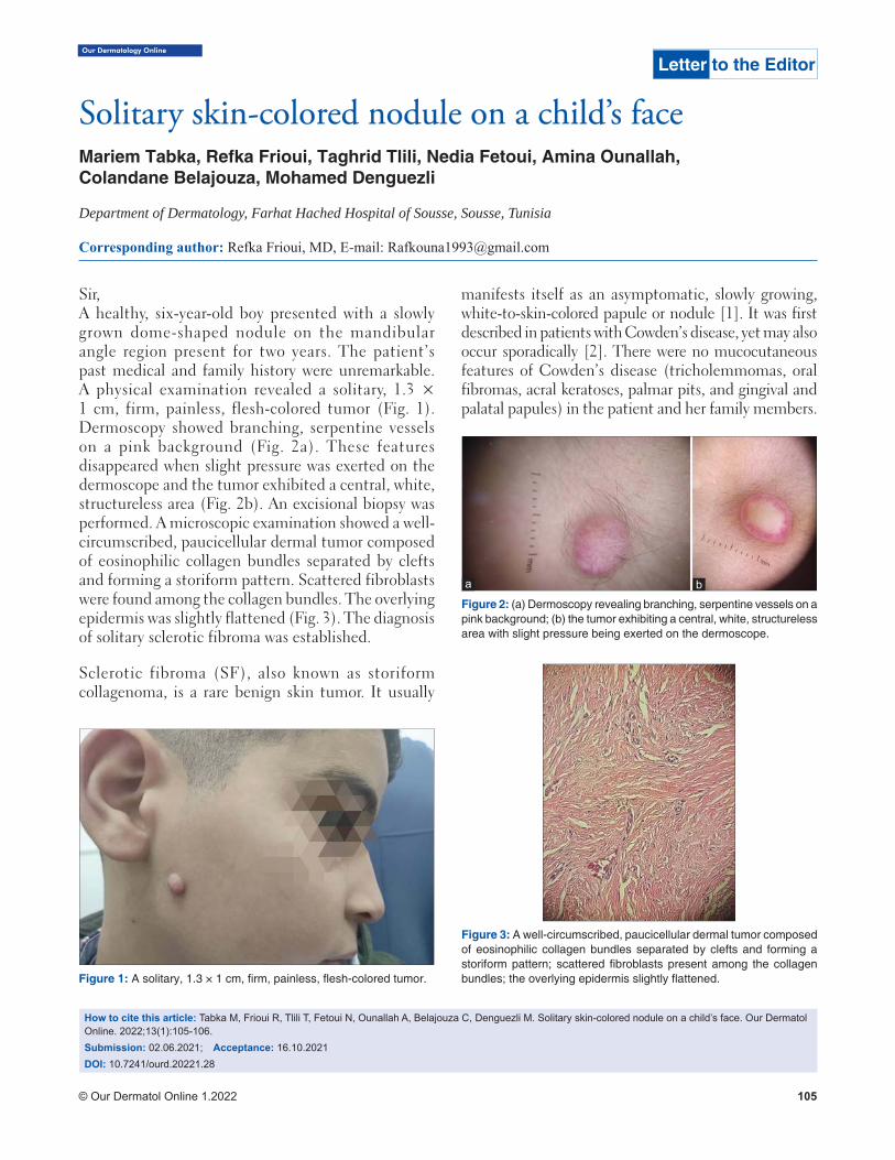
© Our Dermatol Online 1.2022 105
Our Dermatology Online
How to cite this article: Tabka M, Frioui R, Tlili T, Fetoui N, Ounallah A, Belajouza C, Denguezli M. Solitary skin-colored nodule on a child’s face. Our Dermatol Online. 2022;13(1):105-106.Submission: 02.06.2021; Acceptance: 16.10.2021DOI: 10.7241/ourd.20221.28
Solitary skin-colored nodule on a child’s faceSolitary skin-colored nodule on a child’s faceMariem Tabka, Refka Frioui, Taghrid Tlili, Nedia Fetoui, Amina Ounallah,Colandane Belajouza, Mohamed Denguezli
Department of Dermatology, Farhat Hached Hospital of Sousse, Sousse, Tunisia
Corresponding author: Refka Frioui, MD, E-mail: [email protected]
Sir,A healthy, six-year-old boy presented with a slowly grown dome-shaped nodule on the mandibular angle region present for two years. The patient’s past medical and family history were unremarkable. A physical examination revealed a solitary, 1.3 × 1 cm, firm, painless, flesh-colored tumor (Fig. 1). Dermoscopy showed branching, serpentine vessels on a pink background (Fig. 2a). These features disappeared when slight pressure was exerted on the dermoscope and the tumor exhibited a central, white, structureless area (Fig. 2b). An excisional biopsy was performed. A microscopic examination showed a well-circumscribed, paucicellular dermal tumor composed of eosinophilic collagen bundles separated by clefts and forming a storiform pattern. Scattered fibroblasts were found among the collagen bundles. The overlying epidermis was slightly flattened (Fig. 3). The diagnosis of solitary sclerotic fibroma was established.
Sclerotic fibroma (SF), also known as storiform collagenoma, is a rare benign skin tumor. It usually
manifests itself as an asymptomatic, slowly growing, white-to-skin-colored papule or nodule [1]. It was first described in patients with Cowden’s disease, yet may also occur sporadically [2]. There were no mucocutaneous features of Cowden’s disease (tricholemmomas, oral fibromas, acral keratoses, palmar pits, and gingival and palatal papules) in the patient and her family members.
Letter to the Editor
Figure 1: A solitary, 1.3 × 1 cm, fi rm, painless, fl esh-colored tumor.
Figure 3: A well-circumscribed, paucicellular dermal tumor composed of eosinophilic collagen bundles separated by clefts and forming a storiform pattern; scattered fi broblasts present among the collagen bundles; the overlying epidermis slightly fl attened.
Figure 2: (a) Dermoscopy revealing branching, serpentine vessels on a pink background; (b) the tumor exhibiting a central, white, structureless area with slight pressure being exerted on the dermoscope.
ba
www.odermatol.com
© Our Dermatol Online 1.2022 106
Dermatofibroma, the main differential diagnosis of SF, usually exhibits hyperplastic changes of the epidermis instead of atrophy, and the boundaries of the lesion are unclear [2]. Only two papers have been published describing the dermoscopic findings of SF, consisting of a white background with peripheral arborizing vessels [3]. A white background may be related to an increased dermal collagen density. It is also described in dermatofibroma, typically with a peripheral pigmentation network. Although dermoscopy may improve the clinical diagnosis of SF, histopathological analysis is required.
Consent
The examination of the patient was conducted according to the principles of the Declaration of Helsinki.
The authors certify that they have obtained all appropriate patient consent forms, in which the patients gave their consent for images
and other clinical information to be included in the journal. The patients understand that their names and initials will not be published and due effort will be made to conceal their identity, but that anonymity cannot be guaranteed.
REFERENCES
1. Kutzner H, Lazar AJ, Patel RM, Wang WL. Fibromas. WHO Classifi cation of Skin Tumours. 4th ed. Lyon: IARC; 2018:314-7.
2. Tosa M, Ansai SI, Kuwahara H, Akaishi S, Ogawa R. Two cases of sclerotic fi broma of the skin that mimicked keloids clinically. J Nippon Med Sch. 2018;85:283-6.
3. Ebadian M, Citarella L, Collins D, Diaz-Cano S, Pozo-Garcia L. Dermoscopy of a solitary storiform collagenoma. Dermatol Pract Concept. 2018;8:120-2.
Copyright by Mariem Tabka, et al. This is an open access article distributed under the terms of the Creative Commons Attribution License, which permits unrestricted use, distribution, and reproduction in any medium, provided the original author and source are credited.Source of Support: Nil, Confl ict of Interest: None declared.
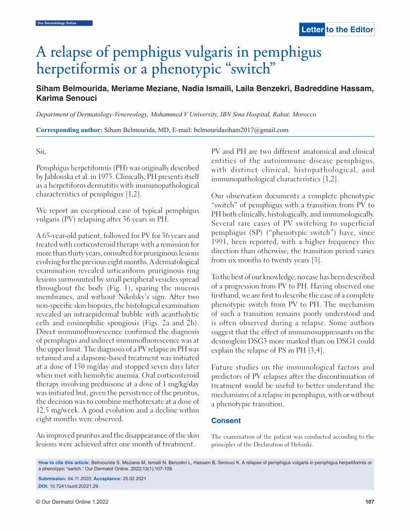
© Our Dermatol Online 1.2022 107
Our Dermatology Online
How to cite this article: Belmourida S, Meziane M, Ismaili N, Benzekri L, Hassam B, Senouci K. A relapse of pemphigus vulgaris in pemphigus herpetiformis or a phenotypic “switch.” Our Dermatol Online. 2022;13(1):107-108.
Submission: 04.11.2020; Acceptance: 25.02.2021DOI: 10.7241/ourd.20221.29
A relapse of pemphigus vulgaris in pemphigus A relapse of pemphigus vulgaris in pemphigus herpetiformis or a phenotypic “switch”herpetiformis or a phenotypic “switch”Siham Belmourida, Meriame Meziane, Nadia Ismaili, Laila Benzekri, Badreddine Hassam, Karima Senouci
Department of Dermatology-Venereology, Mohammed V University, IBN Sina Hospital, Rabat, Morocco
Corresponding author: Siham Belmourida, MD, E-mail: [email protected]
Sir,
Pemphigus herpetiformis (PH) was originally described by Jablonska et al. in 1975. Clinically, PH presents itself as a herpetiform dermatitis with immunopathological characteristics of pemphigus [1,2].
We report an exceptional case of typical pemphigus vulgaris (PV) relapsing after 36 years in PH.
A 65-year-old patient, followed for PV for 36 years and treated with corticosteroid therapy with a remission for more than thirty years, consulted for pruriginous lesions evolving for the previous eight months. A dermatological examination revealed urticariform pruriginous ring lesions surmounted by small peripheral vesicles spread throughout the body (Fig. 1), sparing the mucous membranes, and without Nikolsky’s sign. After two non-specific skin biopsies, the histological examination revealed an intraepidermal bubble with acantholytic cells and eosinophilic spongiosis (Figs. 2a and 2b). Direct immunofluorescence confirmed the diagnosis of pemphigus and indirect immunofluorescence was at the upper limit. The diagnosis of a PV relapse in PH was retained and a dapsone-based treatment was initiated at a dose of 150 mg/day and stopped seven days later when met with hemolytic anemia. Oral corticosteroid therapy involving prednisone at a dose of 1 mg/kg/day was initiated but, given the persistence of the pruritus, the decision was to combine methotrexate at a dose of 12.5 mg/week. A good evolution and a decline within eight months were observed.
An improved pruritus and the disappearance of the skin lesions were achieved after one month of treatment.
PV and PH are two different anatomical and clinical entities of the autoimmune disease pemphigus, with distinct clinical, histopathological, and immunopathological characteristics [1,2].
Our observation documents a complete phenotypic “switch” of pemphigus with a transition from PV to PH both clinically, histologically, and immunologically. Several rare cases of PV switching to superficial pemphigus (SP) (“phenotypic switch”) have, since 1991, been reported, with a higher frequency this direction than otherwise; the transition period varies from six months to twenty years [3].
To the best of our knowledge, no case has been described of a progression from PV to PH. Having observed one firsthand, we are first to describe the case of a complete phenotypic switch from PV to PH. The mechanism of such a transition remains poorly understood and is often observed during a relapse. Some authors suggest that the effect of immunosuppressants on the desmoglein DSG3 more marked than on DSG1 could explain the relapse of PS in PH [3,4].
Future studies on the immunological factors and predictors of PV relapses after the discontinuation of treatment would be useful to better understand the mechanisms of a relapse in pemphigus, with or without a phenotypic transition.
Consent
The examination of the patient was conducted according to the principles of the Declaration of Helsinki.
Letter to the Editor
www.odermatol.com
© Our Dermatol Online 1.2022 108
The authors certify that they have obtained all appropriate patient consent forms, in which the patients gave their consent for images and other clinical information to be included in the journal. The patients understand that their names and initials will not be published and due effort will be made to conceal their identity, but that anonymity cannot be guaranteed.
REFERENCES
1. Jablonska S, Chorzelski T, Beutner E, Chorzelska J. Herpetiform pemphigus, a variable pattern of pemphigus. Int J Dermatol. 1975;14:353-9.
2. Micali G, Musumeci ML, Nasca MR. Epidemiologic analysis and clinical course of 84 consecutive cases of pemphigus in eastern Sicily. Int J Dermatol. 1998;37:197-200.
3. Lévy-Sitbon C, Reguiaï Z, Durlach A, Goeldel AL, Grange. F, Bernard P. Transition from pemphigus vulgaris to pemphigus foliaceus: A case report. Ann Dermatol Venereol. 2013;140:788-92.
4. Abreu Velez AM, Upegui-Zapata YA, Valencia-Yepes CA, Upegui-Quiceño E, Yi H, Vargas Florez A, et al. A new variant of endemic pemphigus foliaceus in Colombia South America. Our Dermatol Online. 2020;11:284-99.
Copyright by Siham Belmourida, et al. This is an open access article distributed under the terms of the Creative Commons Attribution License, which permits unrestricted use, distribution, and reproduction in any medium, provided the original author and source are credited.Source of Support: Nil, Confl ict of Interest: None declared.
Figure 1: Urticariform pruriginous ring lesions surmounted by small peripheral vesicles.
Figure 2: (a-b) Histological image showing an intraepidermal bubble with acantholytic cells and eosinophilic spongiosis.
ba
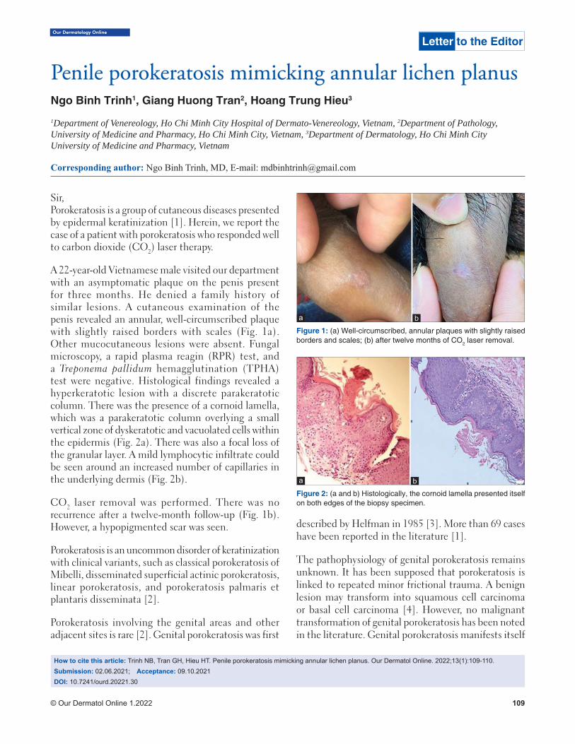
© Our Dermatol Online 1.2022 109
Our Dermatology Online
How to cite this article: Trinh NB, Tran GH, Hieu HT. Penile porokeratosis mimicking annular lichen planus. Our Dermatol Online. 2022;13(1):109-110.Submission: 02.06.2021; Acceptance: 09.10.2021DOI: 10.7241/ourd.20221.30
Penile porokeratosis mimicking annular lichen planusPenile porokeratosis mimicking annular lichen planusNgo Binh Trinh1, Giang Huong Tran2, Hoang Trung Hieu3
1Department of Venereology, Ho Chi Minh City Hospital of Dermato-Venereology, Vietnam, 2Department of Pathology, University of Medicine and Pharmacy, Ho Chi Minh City, Vietnam, 3Department of Dermatology, Ho Chi Minh City University of Medicine and Pharmacy, Vietnam
Corresponding author: Ngo Binh Trinh, MD, E-mail: [email protected]
Sir,Porokeratosis is a group of cutaneous diseases presented by epidermal keratinization [1]. Herein, we report the case of a patient with porokeratosis who responded well to carbon dioxide (CO2) laser therapy.
A 22-year-old Vietnamese male visited our department with an asymptomatic plaque on the penis present for three months. He denied a family history of similar lesions. A cutaneous examination of the penis revealed an annular, well-circumscribed plaque with slightly raised borders with scales (Fig. 1a). Other mucocutaneous lesions were absent. Fungal microscopy, a rapid plasma reagin (RPR) test, and a Treponema pallidum hemagglutination (TPHA) test were negative. Histological findings revealed a hyperkeratotic lesion with a discrete parakeratotic column. There was the presence of a cornoid lamella, which was a parakeratotic column overlying a small vertical zone of dyskeratotic and vacuolated cells within the epidermis (Fig. 2a). There was also a focal loss of the granular layer. A mild lymphocytic infiltrate could be seen around an increased number of capillaries in the underlying dermis (Fig. 2b).
CO2 laser removal was performed. There was no recurrence after a twelve-month follow-up (Fig. 1b). However, a hypopigmented scar was seen.
Porokeratosis is an uncommon disorder of keratinization with clinical variants, such as classical porokeratosis of Mibelli, disseminated superficial actinic porokeratosis, linear porokeratosis, and porokeratosis palmaris et plantaris disseminata [2].
Porokeratosis involving the genital areas and other adjacent sites is rare [2]. Genital porokeratosis was first
described by Helfman in 1985 [3]. More than 69 cases have been reported in the literature [1].
The pathophysiology of genital porokeratosis remains unknown. It has been supposed that porokeratosis is linked to repeated minor frictional trauma. A benign lesion may transform into squamous cell carcinoma or basal cell carcinoma [4]. However, no malignant transformation of genital porokeratosis has been noted in the literature. Genital porokeratosis manifests itself
Letter to the Editor
Figure 2: (a and b) Histologically, the cornoid lamella presented itself on both edges of the biopsy specimen.
Figure 1: (a) Well-circumscribed, annular plaques with slightly raised borders and scales; (b) after twelve months of CO2 laser removal.
ba
ba
www.odermatol.com
© Our Dermatol Online 1.2022 110
clinically as classic or plaque-type porokeratosis of Mibelli [2].
Histological findings revealed a cornoid lamella with the absence of a granular layer and dyskeratotic cells in the upper spinous zone [2]. Our case may mimic some annular lesions, such as secondary syphilis, fungal infection, and annular lichen planus. Because a fungal examination and syphilis serology were negative, we could exclude fungal infection and annular secondary syphilis. The distinctive histology of porokeratosis such as a cornoid lamella with a decreased granular layer may help to differentiate between porokeratosis and annular lichen planus [4].
Numerous therapeutic methods of treatment exist, including surgical excision, CO2 laser, cryotherapy, topical retinoids, 5% 5-fluorouracil, vitamin D3 analogs, imiquimod cream, and 3% diclofenac gel [2,5].
Consent
The examination of the patient was conducted according to the principles of the Declaration of Helsinki.
The authors certify that they have obtained all appropriate patient consent forms, in which the patients gave their consent for images and other clinical information to be included in the journal. The patients understand that their names and initials will not be published and due effort will be made to conceal their identity, but that anonymity cannot be guaranteed.
REFERENCES
1. Bari O, Calame A, Marietti-Shepherd S, Barrio VR. Pediatric penile porokeratosis: A case report. Pediatr Dermatol. 2018;35:e103-e4.
2. Dongre A, Adhe V, Sanghavi S. Genital porokeratosis: a rare entity. Indian J Dermatol. 2013;58:81.
3. Helfman RJ, Poulos EG. Reticulated porokeratosis: A unique variant of porokeratosis. Arch Dermatol. 1985;121:1542-3.
4. Ahmed A, Hivnor C. A case of genital porokeratosis and review of literature. Indian J Dermatol. 2015;60:217.
5. Roge RP, Hajare SA, Mukhi JI, Singh RS. Genital porokeratosis: A report of two cases and review of literature. Indian J Dermatopathol Diagn Dermatol. 2020;7:19.
Copyright by Ngo Binh Trinh, et al. This is an open access article distributed under the terms of the Creative Commons Attribution License, which permits unrestricted use, distribution, and reproduction in any medium, provided the original author and source are credited.Source of Support: Nil, Confl ict of Interest: None declared.
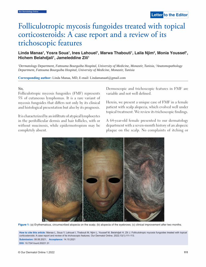
Our Dermatology Online
© Our Dermatol Online 1.2022 111
Folliculotropic mycosis fungoides treated with topical Folliculotropic mycosis fungoides treated with topical corticosteroids: A case report and a review of its corticosteroids: A case report and a review of its trichoscopic featurestrichoscopic featuresLinda Manaa1, Yosra Soua1, Ines Lahouel1, Marwa Thabouti1, Laila Njim2, Monia Youssef1, Hichem Belahdjali1, Jameleddine Zili1
1Dermatology Department, Fattouma Bourguiba Hospital, University of Medicine, Monastir, Tunisia, 2Anatomopathology Department, Fattouma Bourguiba Hospital, University of Medicine, Monastir, Tunisia
Corresponding author: Linda Manaa, MD, E-mail: [email protected]
Sir,Folliculotropic mycosis fungoides (FMF) represents 5% of cutaneous lymphomas. It is a rare variant of mycosis fungoides that differs not only by its clinical and histological presentation but also by its prognosis.
It is characterized by an infiltrate of atypical lymphocytes in the perifollicular dermis and hair follicles, with or without mucinosis, while epidermotropism may be completely absent.
Dermoscopic and trichoscopic features in FMF are variable and not well defined.
Herein, we present a unique case of FMF in a female patient with scalp alopecia, which evolved well under topical treatment. We review its trichoscopic findings.
A 64-year-old female presented to our dermatology department with a seven-month history of an alopecic plaque on the scalp. No complaints of itching or
How to cite this article: Manaa L, Soua Y, Lahouel I, Thabouti M, Njim L, Youssef M, Belahdjali H, Zili J. Folliculotropic mycosis fungoides treated with topical corticosteroids: A case report and review of its trichoscopic features. Our Dermatol Online. 2022;13(1):111-113.Submission: 08.06.2021; Acceptance: 14.10.2021DOI: 10.7241/ourd.20221.31
Figure 1: (a) Erythematous, circumscribed alopecia on the scalp; (b) alopecia of the eyebrows; (c) clinical improvement after two months.
a b c
Letter to the Editor
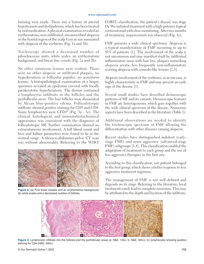
www.odermatol.com
© Our Dermatol Online 1.2022 112
burning were made. There was a history of arterial hypertension and dyslipidemia, which has been treated by oral medication. A physical examination revealed an erythematous, non-infiltrated, circumscribed alopecia in the frontal region of the scalp 8 cm in size associated with alopecia of the eyebrows (Fig. 1a and 1b).
Trichoscopy showed a decreased number of pilosebaceous units, white scales, an erythematous background, and linear fine vessels (Fig. 2a and 2b).
No other cutaneous lesions were evident. There were no other alopecic or infiltrated plaques, no hyperkeratosis or follicular papules, no acneiform lesions. A histopathological examination of a biopsy specimen revealed an epidermis covered with focally parakeratotic hyperkeratosis. The dermis contained a lymphocytic infiltrate in the follicles and the perifollicular areas. The hair follicles were dissociated by Alcian blue-positive edema. Folliculotropic infiltrate showed positive staining for CD3 and CD4. Some lymphocytes were CD20⁺ (Fig. 3a - 3c). The clinical, histological, and immunohistochemical appearance was consistent with the diagnosis of folliculotropic MF. Further examination showed no extracutaneous involvement. A full blood count and liver and kidney parameters were found to be in the normal range. A thoraco-abdomino-pelvic CT scan was without abnormality. Referring to the WHO/
EORTC classification, the patient’s disease was stage IA. We initiated treatment with a high-potency topical corticosteroid with close monitoring. After two months of treatment, improvement was observed (Fig. 1c).
FMF presents a wide clinical spectrum. Alopecia is a typical manifestation of FMF occurring in up to 81% of patients [1]. The involvement of the scalp is not uncommon and may manifest itself by infiltrated inflammatory areas with hair loss, plaques mimicking alopecia areata, less frequently non-inflammatory scarring alopecia with comedo-like lesions [2,3].
Alopecic involvement of the eyebrows, as in our case, is highly characteristic in FMF and may present an early sign of the disease [1].
Several small studies have described dermoscopic patterns of MF and its variants. Dermoscopic features in FMF are heterogeneous, which goes together with the wide clinical spectrum of the disease. Numerous aspects have been described in the literature (Table 1).
Additional observations are needed to identify the trichoscopic spectrum of FMF allowing the differentiation with other diseases causing alopecia.
Recent studies have distinguished indolent (early-stage FMF) and more aggressive (advanced-stage FMF) subgroups [1,4]. This classification enabled the adaptation of treatment to each group and the use of less aggressive therapies in the first one.
According to this classification, our patient belonged to the first group, which shows a better response to less aggressive treatment regimens.
The management of FMF is not well defined and depends on its stage. Referring to the literature, local treatments rarely lead to complete remission. This may be attributed to the depth and location of the infiltrate.
Figure 2: (a) Fine linear vessels and an erythematous background; (b) white scales and a decreased number of follicles.
a b
Figure 3: Lymphocytic infi ltrate into the follicles and the perifollicular areas (a: H&E, 100×; b: H&E, 400×); (c) lymphocytes showing positive staining for CD4 (H&E, 400×).
a b c
www.odermatol.com
© Our Dermatol Online 1.2022 113
In our patient, given the early stage of the disease, treatment with topical corticosteroids was sufficient to control the lesions.
Consent
The examination of the patient was conducted according to the principles of the Declaration of Helsinki.
The authors certify that they have obtained all appropriate patient consent forms, in which the patients gave their consent for images and other clinical information to be included in the journal. The patients understand that their names and initials will not be published and due effort will be made to conceal their identity, but that anonymity cannot be guaranteed.
REFERENCES
1. Van Santen S, Roach REJ, van Doorn R, Horváth B, Bruijn MS, Sanders CJG, et al. Clinical Staging and Prognostic Factors in Folliculotropic Mycosis Fungoides. JAMA Dermatol. 2016;152:992-1000.
2. Sławińska M, Sobjanek M, Olszewska B, Nowicki R,
Sokołowska-Wojdyło M. Trichoscopic spectrum of folliculotropic mycosis fungoides. J Eur Acad Dermatol Venereol. 2018;32:e107-8.
3. Geller S, Rishpon A, Myskowski PL. Dermoscopy in folliculotropic mycosis fungoides—A possible mimicker of follicle-based infl ammatory and infectious disorders. J Am Acad Dermatol. 2019;81:e75-6.
4. Caccavale S, Vitiello P, Franco R, Panarese I, Ronchi A, Sica A, et al. Dermoscopic characterization of folliculotropic mycosis fungoides selectively localized on trunk and limbs. Int J Dermatol. 2019;58:e187-9.
5. Ghahramani GK, Goetz KE, Liu V. Dermoscopic characterization of cutaneous lymphomas: A pilot survey. Int J Dermatol. 2018;57:339-43.
6. Jurakic Toncic R, Ledic Drvar D, Bradamante M, Rados J, Jerkovic-Gulin S, Caccavale S, et al. Early dermoscopic sign of folliculotropism in patients with mycosis fungoides. Dermatol Pract Concept. 2018;8:328-9.
7. Jurakić Tončić R, Radoš J, Ćurković D, Ilić I, Caccavale S, Bradamante M. Dermoscopy of syringotropic and folliculotropic mycosis fungoides. Dermatol Pract Concept. 2020;10:e2020069.
Copyright by Linda Manaa, et al. This is an open access article distributed under the terms of the Creative Commons Attribution License, which permits unrestricted use, distribution, and reproduction in any medium, provided the original author and source are credited.Source of Support: Nil, Confl ict of Interest: None declared.
Table 1: Literature review of the dermoscopic features of FMF.Author (Year of publication)
Clinical Presentation Dermoscopic Findings
Ghahramani et al. (2018) [5] 5 patients with FMF.Lesions on multiple sites (trunk, extremities, buttocks).
- Perifollicular accentuation (100%)- Comedo-like openings (60%)- White structureless areas (60%)- Fine short linear vessels (40%)- Dotted vessels (40%)- White scale (80%)
Toncic et al. (2018) [6] Erythematous patch on the right side of a forehead. - Perifollicular accentuation seen as a white halo around the follicles
Slawinska et al. (2018) [2] Extensive patchy alopecia of the scalp (erythematous areas of hair loss with scaling).
- A decreased number of pilosebaceous units mostly single hair- Milky-white globules- Yellow dots with or without centrally located black dots/broken hairs- White dots and lines - White and yellow scale - Short hair with split-ends- Short hair with triangular ends- Short, broken hair- Pigtail-appearance hair - Short hairs broken at the same or different level.
Caccavale et al. (2019) [4] Comedo-like lesions, follicular papules, pustules located on the trunk, and an alopecic patch on the left forearm.
- Comedo-like lesions surrounded by perifollicular erythema - White or pinkish structureless areas replacing lost hair follicles- Fine short linear, glomerular and dotted vessels
Geller et al. (2019) [3] Patient 1: follicular papules with alopecia on the thighPatient 2: erythematous plaque with alopecia on the scalpPatient 3: erythematous follicular papules with a central
scale (keratosis pilaris-like) on the buttocks.Patient 4: facial erythematous follicular papules
- Patient 1: orange-pink perifollicular clods with peripheral scale and central broken hairs
- Patient 2: perifollicular halos and broken hairs- Patient 3: short fi ne vessels and perifollicular scale overlying a
yellowish background, surrounding central keratotic plugs- Patient 4: a white and hyperpigmented halo around the follicles
and perifollicular scale and white clods
Toncic et al. (2020) [7] Patient 1: erythematous patches and plaques on the neck, face, arms, and back.
Patient 2: erythematous papules, plaques, comedones, and milia localized on the neck and chest.
- Obliteration of the follicles, follicular accentuation, and follicular plugging
- Loss of terminal follicles- Comedo-like openings- Interconnected regular-appearing structureless patches- Bluish structures (when eccrine glands are affected)
Our case Infl ammatory alopecic plaque on the scalp. - A decreased number of pilosebaceous units, white scales, an erythematous background, and linear fi ne vessels
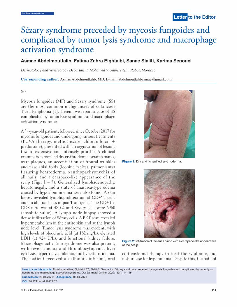
Our Dermatology Online
© Our Dermatol Online 1.2022 114
Sézary syndrome preceded by mycosis fungoides and Sézary syndrome preceded by mycosis fungoides and complicated by tumor lysis syndrome and macrophage complicated by tumor lysis syndrome and macrophage activation syndromeactivation syndromeAsmae Abdelmouttalib, Fatima Zahra Elghtaibi, Sanae Sialiti, Karima Senouci
Dermatology and Venereology Department, Mohamed V University in Rabat, Morocco
Corresponding author: Asmae Abdelmouttalib, MD, E-mail: [email protected]
Sir,
Mycosis fungoides (MF) and Sézary syndrome (SS) are the most common malignancies of cutaneous T-cell lymphoma [1]. Herein, we report a case of SS complicated by tumor lysis syndrome and macrophage activation syndrome.
A 54-year-old patient, followed since October 2017 for mycosis fungoides and undergoing various treatments (PUVA therapy, methotrexate, chlorambucil + prednisone), presented with an aggravation of lesions toward extensive and intensely pruritic. A clinical examination revealed dry erythroderma, scratch marks, wart plaques, an accentuation of frontal wrinkles and nasolabial folds (leonine facies), palmoplantar fissuring keratoderma, xanthopachyonychia of all nails, and a carapace-like appearance of the scalp (Figs. 1 – 3). Generalized lymphadenopathy, hepatomegaly, and a state of anasarca-type edema caused by hypoalbuminemia were also found. A skin biopsy revealed lymphoproliferation of CD4⁺ T-cells and an aberrant loss of pan-T antigens. The CD4-to-CD8 ratio was at 48.5% and Sézary cells were 6960 (absolute value). A lymph node biopsy showed a dense infiltration of Sézary cells. A PET scan revealed hypermetabolism in the entire skin and at the lymph node level. Tumor lysis syndrome was evident, with high levels of blood uric acid (at 182 mg/L), elevated LDH (at 924 U/L), and functional kidney failure. Macrophage activation syndrome was also present, with fever, anemia and thrombocytopenia, liver cytolysis, hypertriglyceridemia, and hyperferritinemia. The patient received an albumin infusion, oral
corticosteroid therapy to treat the syndrome, and rasburicase for hyperuremia. Despite this, the patient
Letter to the Editor
How to cite this article: Abdelmouttalib A, Elghtaibi FZ, Sialiti S, Senouci K. Sézary syndrome preceded by mycosis fungoides and complicated by tumor lysis syndrome and macrophage activation syndrome. Our Dermatol Online. 2022;13(1):114-115.Submission: 20.01.2021; Acceptance: 05.04.2021DOI: 10.7241/ourd.20221.32
Figure 2: Infi ltration of the ear’s pinna with a carapace-like appearance of the scalp.
Figure 1: Dry and lichenifi ed erythroderma.
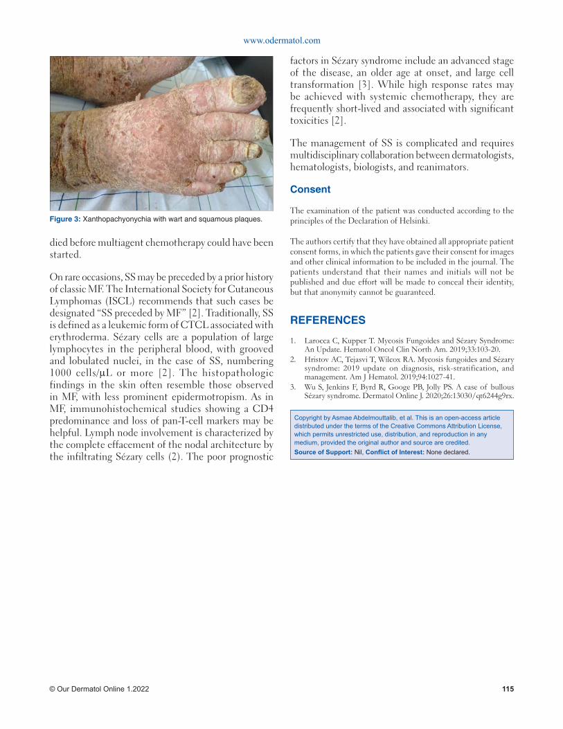
www.odermatol.com
© Our Dermatol Online 1.2022 115
died before multiagent chemotherapy could have been started.
On rare occasions, SS may be preceded by a prior history of classic MF. The International Society for Cutaneous Lymphomas (ISCL) recommends that such cases be designated “SS preceded by MF” [2]. Traditionally, SS is defined as a leukemic form of CTCL associated with erythroderma. Sézary cells are a population of large lymphocytes in the peripheral blood, with grooved and lobulated nuclei, in the case of SS, numbering 1000 cells/μL or more [2]. The histopathologic findings in the skin often resemble those observed in MF, with less prominent epidermotropism. As in MF, immunohistochemical studies showing a CD4 predominance and loss of pan-T-cell markers may be helpful. Lymph node involvement is characterized by the complete effacement of the nodal architecture by the infiltrating Sézary cells (2). The poor prognostic
factors in Sézary syndrome include an advanced stage of the disease, an older age at onset, and large cell transformation [3]. While high response rates may be achieved with systemic chemotherapy, they are frequently short-lived and associated with significant toxicities [2].
The management of SS is complicated and requires multidisciplinary collaboration between dermatologists, hematologists, biologists, and reanimators.
Consent
The examination of the patient was conducted according to the principles of the Declaration of Helsinki.
The authors certify that they have obtained all appropriate patient consent forms, in which the patients gave their consent for images and other clinical information to be included in the journal. The patients understand that their names and initials will not be published and due effort will be made to conceal their identity, but that anonymity cannot be guaranteed.
REFERENCES
1. Larocca C, Kupper T. Mycosis Fungoides and Sézary Syndrome: An Update. Hematol Oncol Clin North Am. 2019;33:103-20.
2. Hristov AC, Tejasvi T, Wilcox RA. Mycosis fungoides and Sézary syndrome: 2019 update on diagnosis, risk-stratification, and management. Am J Hematol. 2019;94:1027-41.
3. Wu S, Jenkins F, Byrd R, Googe PB, Jolly PS. A case of bullous Sézary syndrome. Dermatol Online J. 2020;26:13030/qt6244g9rx.
Figure 3: Xanthopachyonychia with wart and squamous plaques.
Copyright by Asmae Abdelmouttalib, et al. This is an open-access article distributed under the terms of the Creative Commons Attribution License, which permits unrestricted use, distribution, and reproduction in any medium, provided the original author and source are credited.Source of Support: Nil, Confl ict of Interest: None declared.
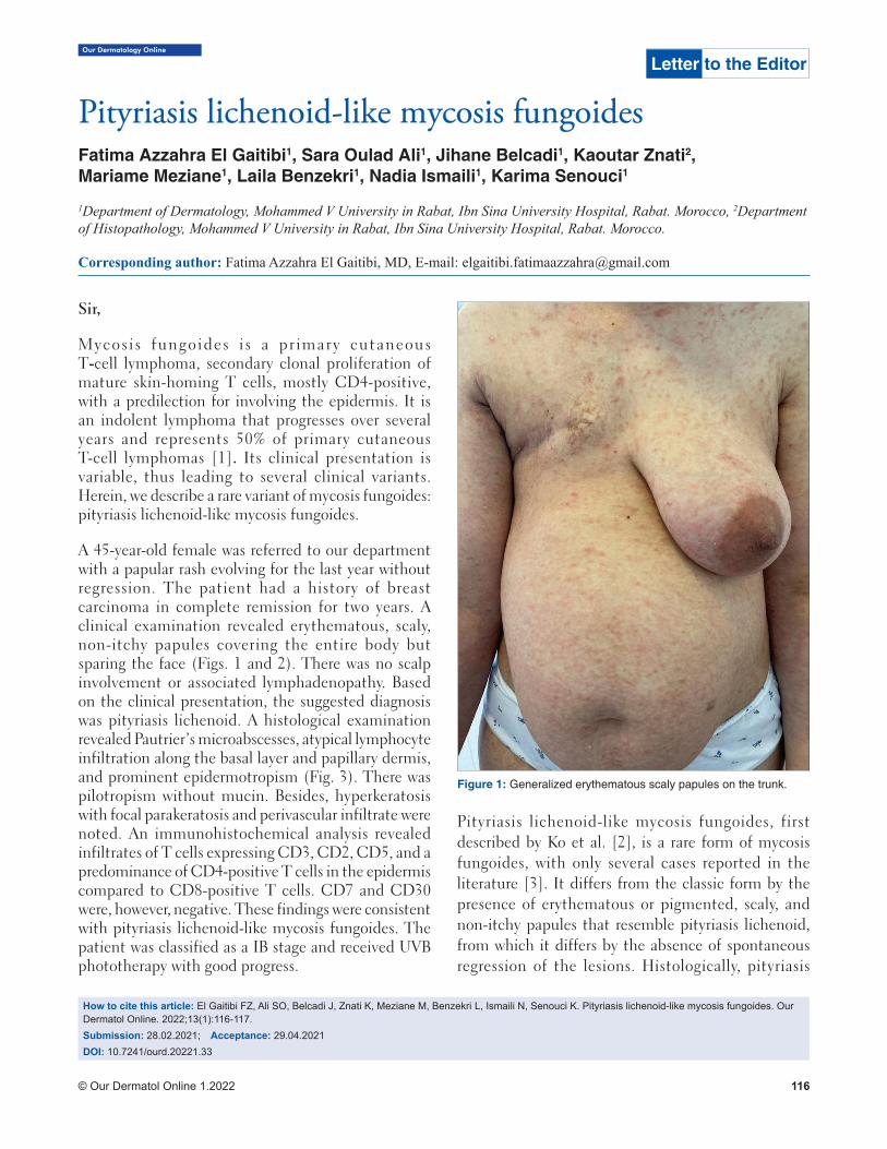
Our Dermatology Online
© Our Dermatol Online 1.2022 116
Pityriasis lichenoid-like mycosis fungoidesPityriasis lichenoid-like mycosis fungoidesFatima Azzahra El Gaitibi1, Sara Oulad Ali1, Jihane Belcadi1, Kaoutar Znati2, Mariame Meziane1, Laila Benzekri1, Nadia Ismaili1, Karima Senouci1
1Department of Dermatology, Mohammed V University in Rabat, Ibn Sina University Hospital, Rabat. Morocco, 2Department of Histopathology, Mohammed V University in Rabat, Ibn Sina University Hospital, Rabat. Morocco.
Corresponding author: Fatima Azzahra El Gaitibi, MD, E-mail: [email protected]
Sir,
Mycosis fungoides i s a pr imary cutaneous T-cell lymphoma, secondary clonal proliferation of mature skin-homing T cells, mostly CD4-positive, with a predilection for involving the epidermis. It is an indolent lymphoma that progresses over several years and represents 50% of primary cutaneous T-cell lymphomas [1]. Its clinical presentation is variable, thus leading to several clinical variants. Herein, we describe a rare variant of mycosis fungoides: pityriasis lichenoid-like mycosis fungoides.
A 45-year-old female was referred to our department with a papular rash evolving for the last year without regression. The patient had a history of breast carcinoma in complete remission for two years. A clinical examination revealed erythematous, scaly, non-itchy papules covering the entire body but sparing the face (Figs. 1 and 2). There was no scalp involvement or associated lymphadenopathy. Based on the clinical presentation, the suggested diagnosis was pityriasis lichenoid. A histological examination revealed Pautrier’s microabscesses, atypical lymphocyte infiltration along the basal layer and papillary dermis, and prominent epidermotropism (Fig. 3). There was pilotropism without mucin. Besides, hyperkeratosis with focal parakeratosis and perivascular infiltrate were noted. An immunohistochemical analysis revealed infiltrates of T cells expressing CD3, CD2, CD5, and a predominance of CD4-positive T cells in the epidermis compared to CD8-positive T cells. CD7 and CD30 were, however, negative. These findings were consistent with pityriasis lichenoid-like mycosis fungoides. The patient was classified as a IB stage and received UVB phototherapy with good progress.
Pityriasis lichenoid-like mycosis fungoides, first described by Ko et al. [2], is a rare form of mycosis fungoides, with only several cases reported in the literature [3]. It differs from the classic form by the presence of erythematous or pigmented, scaly, and non-itchy papules that resemble pityriasis lichenoid, from which it differs by the absence of spontaneous regression of the lesions. Histologically, pityriasis
Letter to the Editor
How to cite this article: El Gaitibi FZ, Ali SO, Belcadi J, Znati K, Meziane M, Benzekri L, Ismaili N, Senouci K. Pityriasis lichenoid-like mycosis fungoides. Our Dermatol Online. 2022;13(1):116-117.Submission: 28.02.2021; Acceptance: 29.04.2021DOI: 10.7241/ourd.20221.33
Figure 1: Generalized erythematous scaly papules on the trunk.
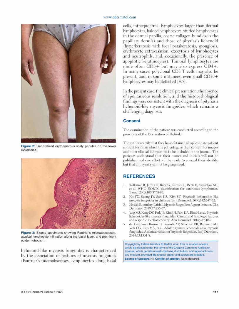
www.odermatol.com
© Our Dermatol Online 1.2022 117
lichenoid-like mycosis fungoides is characterized by the association of features of mycosis fungoides (Pautrier’s microabscesses, lymphocytes along basal
cells, intraepidermal lymphocytes larger than dermal lymphocytes, haloed lymphocytes, stuffed lymphocytes in the dermal papilla, coarse collagen bundles in the papillary dermis) and those of pityriasis lichenoid (hyperkeratosis with focal parakeratosis, spongiosis, erythrocyte extravasation, exocytosis of lymphocytes and neutrophils, and, occasionally, the presence of apoptotic keratinocytes). Tumoral lymphocytes are more often CD8+ but may also express CD4+. In many cases, polyclonal CD3 T cells may also be present, and, in some instances, even small CD30+ lymphocytes may be detected [4,5].
In the present case, the clinical presentation, the absence of spontaneous resolution, and the histopathological findings were consistent with the diagnosis of pityriasis lichenoid-like mycosis fungoides, which remains a challenging diagnosis.
Consent
The examination of the patient was conducted according to the principles of the Declaration of Helsinki.
The authors certify that they have obtained all appropriate patient consent forms, in which the patients gave their consent for images and other clinical information to be included in the journal. The patients understand that their names and initials will not be published and due effort will be made to conceal their identity, but that anonymity cannot be guaranteed.
REFERENCES
1. Willemze R, Jaffe ES, Burg G, Cerroni L, Berti E, Swerdlow SH, et al. WHO-EORTC classifi cation for cutaneous lymphomas. Blood. 2005;105:3768-85.
2. Ko JW, Seong JY, Suh KS, Kim ST. Pityriasis lichenoides-like mycosis fungoides in children. Br J Dermatol. 2000;142:347-52.
3. Hodak E, Amitay-Laish I. Mycosis fungoides: A great imitator. Clin Dermatol. 2019;37:255-67.
4. Jang MS, Kang DY, Park JB, Kim JH, Park KA, Rim H, et al. Pityriasis lichenoides-like mycosis fungoides: Clinical and histologic features and response to phototherapy. Ann Dermatol. 2016;28:540-7.
5. de Unamuno Bustos B, Ferriols AP, Sánchez RB, Rabasco AG, Vela CG, Piris MA, et al. Adult pityriasis lichenoides-like mycosis fungoides: A clinical variant of mycosis fungoides. Int J Dermatol. 2014;53:1331-8.
Copyright by Fatima Azzahra El Gaitibi, et al. This is an open access article distributed under the terms of the Creative Commons Attribution License, which permits unrestricted use, distribution, and reproduction in any medium, provided the original author and source are credited.Source of Support: Nil, Confl ict of Interest: None declared.
Figure 2: Generalized erythematous scaly papules on the lower extremities.
Figure 3: Biopsy specimens showing Pautrier’s microabscesses, atypical lymphocyte infi ltration along the basal layer, and prominent epidermotropism.
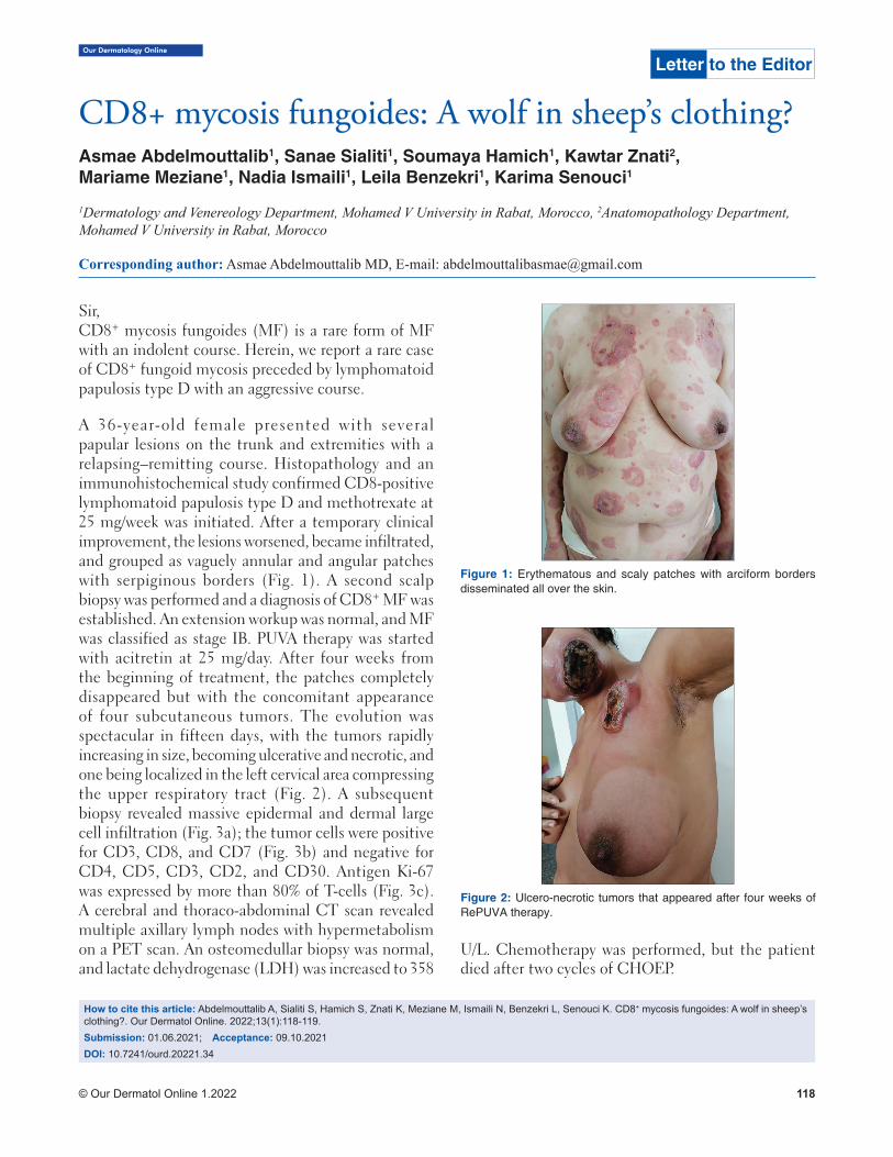
© Our Dermatol Online 1.2022 118
Our Dermatology Online
How to cite this article: Abdelmouttalib A, Sialiti S, Hamich S, Znati K, Meziane M, Ismaili N, Benzekri L, Senouci K. CD8+ mycosis fungoides: A wolf in sheep’s clothing?. Our Dermatol Online. 2022;13(1):118-119.Submission: 01.06.2021; Acceptance: 09.10.2021DOI: 10.7241/ourd.20221.34
CD8+ mycosis fungoides: A wolf in sheep’s clothing?CD8+ mycosis fungoides: A wolf in sheep’s clothing?Asmae Abdelmouttalib1, Sanae Sialiti1, Soumaya Hamich1, Kawtar Znati2,Mariame Meziane1, Nadia Ismaili1, Leila Benzekri1, Karima Senouci1
1Dermatology and Venereology Department, Mohamed V University in Rabat, Morocco, 2Anatomopathology Department, Mohamed V University in Rabat, Morocco
Corresponding author: Asmae Abdelmouttalib MD, E-mail: [email protected]
Sir,CD8+ mycosis fungoides (MF) is a rare form of MF with an indolent course. Herein, we report a rare case of CD8+ fungoid mycosis preceded by lymphomatoid papulosis type D with an aggressive course.
A 36-year-old female presented with several papular lesions on the trunk and extremities with a relapsing–remitting course. Histopathology and an immunohistochemical study confirmed CD8-positive lymphomatoid papulosis type D and methotrexate at 25 mg/week was initiated. After a temporary clinical improvement, the lesions worsened, became infiltrated, and grouped as vaguely annular and angular patches with serpiginous borders (Fig. 1). A second scalp biopsy was performed and a diagnosis of CD8+ MF was established. An extension workup was normal, and MF was classified as stage IB. PUVA therapy was started with acitretin at 25 mg/day. After four weeks from the beginning of treatment, the patches completely disappeared but with the concomitant appearance of four subcutaneous tumors. The evolution was spectacular in fifteen days, with the tumors rapidly increasing in size, becoming ulcerative and necrotic, and one being localized in the left cervical area compressing the upper respiratory tract (Fig. 2). A subsequent biopsy revealed massive epidermal and dermal large cell infiltration (Fig. 3a); the tumor cells were positive for CD3, CD8, and CD7 (Fig. 3b) and negative for CD4, CD5, CD3, CD2, and CD30. Antigen Ki-67 was expressed by more than 80% of T-cells (Fig. 3c). A cerebral and thoraco-abdominal CT scan revealed multiple axillary lymph nodes with hypermetabolism on a PET scan. An osteomedullar biopsy was normal, and lactate dehydrogenase (LDH) was increased to 358
U/L. Chemotherapy was performed, but the patient died after two cycles of CHOEP.
Letter to the Editor
Figure 2: Ulcero-necrotic tumors that appeared after four weeks of RePUVA therapy.
Figure 1: Erythematous and scaly patches with arciform borders disseminated all over the skin.
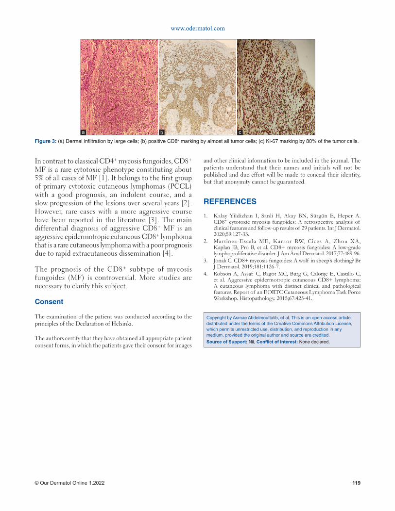
www.odermatol.com
© Our Dermatol Online 1.2022 119
In contrast to classical CD4+ mycosis fungoides, CD8+ MF is a rare cytotoxic phenotype constituting about 5% of all cases of MF [1]. It belongs to the first group of primary cytotoxic cutaneous lymphomas (PCCL) with a good prognosis, an indolent course, and a slow progression of the lesions over several years [2]. However, rare cases with a more aggressive course have been reported in the literature [3]. The main differential diagnosis of aggressive CD8+ MF is an aggressive epidermotropic cutaneous CD8+ lymphoma that is a rare cutaneous lymphoma with a poor prognosis due to rapid extracutaneous dissemination [4].
The prognosis of the CD8+ subtype of mycosis fungoides (MF) is controversial. More studies are necessary to clarify this subject.
Consent
The examination of the patient was conducted according to the principles of the Declaration of Helsinki.
The authors certify that they have obtained all appropriate patient consent forms, in which the patients gave their consent for images
and other clinical information to be included in the journal. The patients understand that their names and initials will not be published and due effort will be made to conceal their identity, but that anonymity cannot be guaranteed.
REFERENCES
1. Kalay Yildizhan I, Sanli H, Akay BN, Sürgün E, Heper A. CD8+ cytotoxic mycosis fungoides: A retrospective analysis of clinical features and follow-up results of 29 patients. Int J Dermatol. 2020;59:127-33.
2. Martinez-Escala ME, Kantor RW, Cices A, Zhou XA, Kaplan JB, Pro B, et al. CD8+ mycosis fungoides: A low-grade lymphoproliferative disorder. J Am Acad Dermatol. 2017;77:489-96.
3. Jonak C. CD8+ mycosis fungoides: A wolf in sheep’s clothing? Br J Dermatol. 2019;181:1126-7.
4. Robson A, Assaf C, Bagot MC, Burg G, Calonje E, Castillo C, et al. Aggressive epidermotropic cutaneous CD8+ lymphoma: A cutaneous lymphoma with distinct clinical and pathological features. Report of an EORTC Cutaneous Lymphoma Task Force Workshop. Histopathology. 2015;67:425-41.
Copyright by Asmae Abdelmouttalib, et al. This is an open access article distributed under the terms of the Creative Commons Attribution License, which permits unrestricted use, distribution, and reproduction in any medium, provided the original author and source are credited.Source of Support: Nil, Confl ict of Interest: None declared.
Figure 3: (a) Dermal infi ltration by large cells; (b) positive CD8+ marking by almost all tumor cells; (c) Ki-67 marking by 80% of the tumor cells.
cba
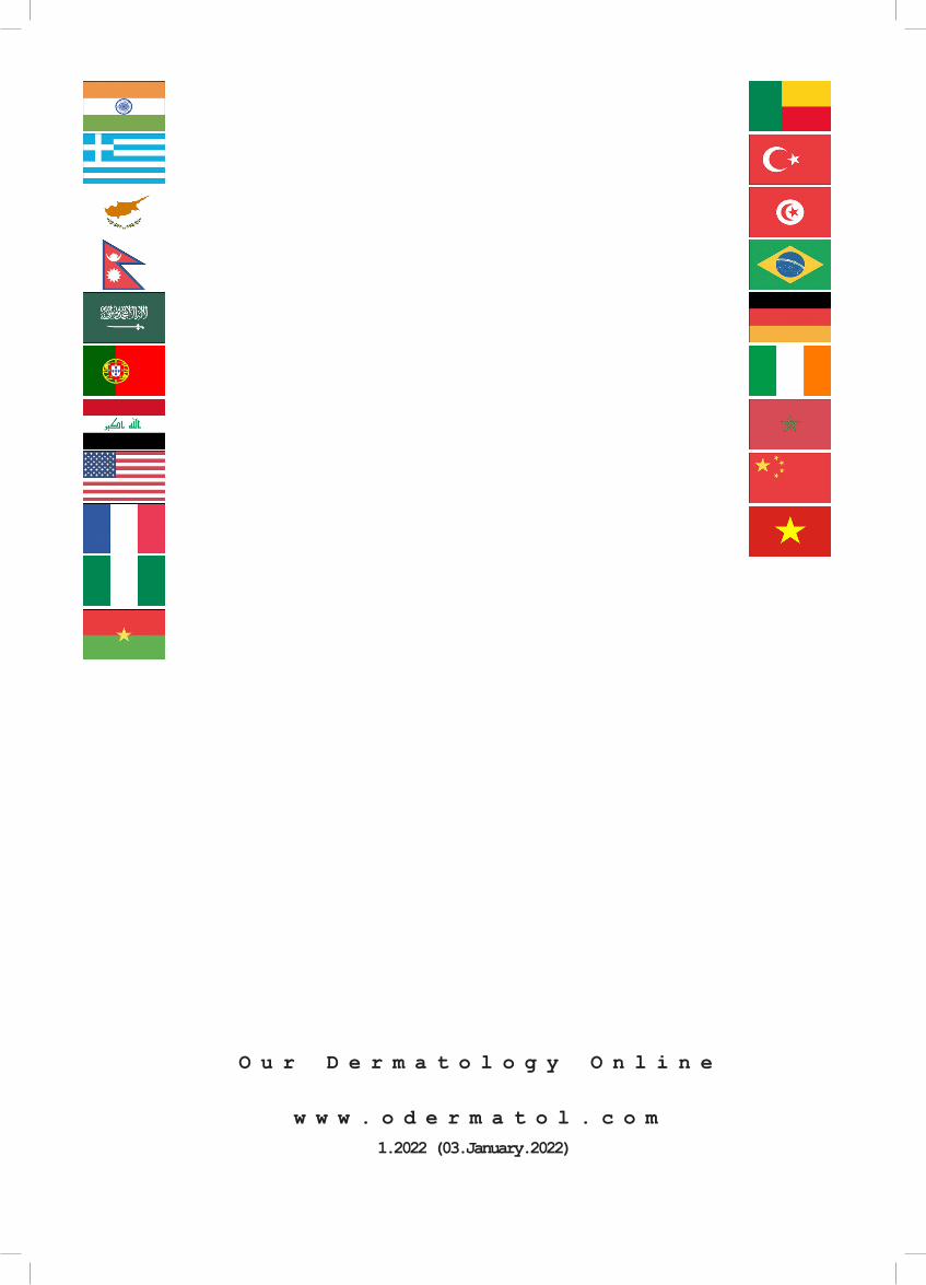
O u r D e r m a t o l o g y O n l i n e
w w w . o d e r m a t o l . c o m1.2022 (03.January.2022)
Related Documents