ISSN 1324-5627 Australasian Musculoskeletal Medicine Vol. 13 No.2 Nov 2008 • Risk factors for overuse tendinopathy • Personal injury claims • Discography • Pain conundrums • Treating inferior heel pain with vitamin D3 dermal cream • An alernative sacroiliac joint injection technique • Meralgia paresthetica • Complementary pain management • Paraspinal nerve injection

Welcome message from author
This document is posted to help you gain knowledge. Please leave a comment to let me know what you think about it! Share it to your friends and learn new things together.
Transcript
ISSN 1324-5627
AustralasianMusculoskeletal
Medicine
Vol. 13 No.2 Nov 2008
• Risk factors for overuse tendinopathy
• Personal injury claims
• Discography
• Pain conundrums
• Treating inferior heel pain with vitamin D3 dermal cream
• An alernative sacroiliac joint injection technique
• Meralgia paresthetica
• Complementary pain management
• Paraspinal nerve injection
54 Australasian Musculoskeletal Medicine
ContentsEditorial ......................................... 55
From the AAMM President ... ....... 58
From the NZAMM President .. ...... 59
Risk factors for overusetendinopathy ................................. 60
Personal injury claims .................. 66
Discography .................................. 69
Pain conundrums ......................... 72
Treating inferior heel pain withvitamin D3 dermal cream ............. 75
An alternative sacroiliac jointinjection technique ........................ 78
Meralgia paresthetica ................... 82
Complementary painmanagement ................................. 87
Paraspinal nerve injection ............ 95
Journal abstracts ........................ 103
IASP world congress .................. 115
Educational activities .................. 117
Australasian Musculoskeletal Medicine ispublished by the Australian Association ofMusculoskeletal Medicine for medical prac-titioners interested in the etiology andmanagement of musculoskeletal disorders.Opinions expressed are those of the au-thors and not necessarily those of the editoror the Association. Editorial comment mayreflect the opinions of the editor alone.
Contributions on any relevant topic arewelcome for submission to the editor, DrDavid Roselt26 Crofton St, Bundaberg 4670Phone +61 7 4152 2888Fax +61 7 4153 3245Email [email protected]
AAMM website: www.musmed.com
NZAMSM website: www.musculoskeletal.co.nz
AFMM website: www.afmm.com.au
FIMM website: www.fimm-online.org
Australian Association ofMusculoskeletal Medicine
Office Bearers
New Zealand Association ofMusculoskeletal Medicine
Office BearersPresidentDr Michael Oei MBBS, Dip Phys Med, MMed Phys Med, Cert Man Med, FACPMMM & Golf Injury Clinic, 37a Wolseley Rd,Mosman, NSW 2088Ph: +61 2 8302 1180, +61 2 9969 2198Fax: +61 2 8302 1195
Vice-PresidentDr Geoffrey Harding MBBS, Dip Musc Med,FAFMM1st Flr, 67 Brighton Rd, Sandgate, Qld 4017Ph: +61 7 32695522Fax: +61 7 3269 6407
Honorary SecretaryDr David Roselt MBBS, Dip Musc Med,FRACGP, FAFMM26 Crofton St, Bundaberg 4670Phone +61 7 4152 2888Fax +61 7 4153 3245
Honorary TreasurerDr Margaret Taylor BSc, MBBS, FACNEM,FIBCTPO Box 570, Fullarton SA 5063Ph/fax: +61 8 8338 3778
Committee MembersDr Quet-Fui Ho MBBS, Grad Dip Mus MedPO Box 436, Melville Plaza, Melville WA6153Ph: 08 9353 2140Fax: 08 9353 2141
Dr Chris Homan MBBS, FRACGP,FACRRM, DRANZCOG, Grad Dip Mus Med3/400 Gregory Terrace, Spring Hill Qld 4000Ph: 07 3839 7600Fax: 07 3236 5228
Dr Roland Loeve MBBS, FRACGP,FAFMM, Grad Cert Pain MedicinePO Box 554, Tamworth NSW 2340Ph: 02 6766 7047Fax: 02 6761 3400
Dr Jennie Wright BMBS, FRACGP, GradDip Mus Med, Grad Cert Med AcupunctureBayside Family Medical & MusculoskeletalBrighton Rd, Glenelg SA 5045Ph: 08 8295 1890Fax: 08 8295 6808
Immediate Past PresidentDr Michael Yelland MBBS, Dip Musc Med,PhD, FRACGP, FAFMMSchool of Medicine, Logan Campus,Griffith University, Meadowbrook, Qld 4131Ph +61-7-3382 1358Fax +61-7-3382 1338
WebmasterDr Victor Wilk MBBS, M Med Pain Med, DipMusc Med, FAFMM441 Bay St, Brighton, Vic 3186Ph: +61 3 9596 7211Fax: +61 3 9596 7871
PresidentDr Gary Collinson MBChB, Dip Musc Med,FAFMM, Cert Spinal Inj4 Kinross St, Blockhouse Bay, AucklandPh: +64 9 6271024, Fax: +64 9 6271181
Honorary SecretaryDr Charlie Ng MBChB, Dip Musc Med, DipSports Med, FAFMM, Cert Spinal Inj24 Green Lane East, Remuera, AucklandPh: +64 9 523 4681, Fax: +64 9 523 4682
Honorary TreasurerDr Clemens Franzmayr Specialist for Con-servative Orthopoedics 197515 Frank St, ChristchurchPh: +64 3 352 7761, Fax: +64 3 352 7762
Past President (ex officio)Dr Peter McKenzie BSc, MBChB, Dip Obs,Dip Musc Med, FRNZCGP, FAFMM217 Bridge St, NelsonPh: +64 3 548 3455, Fax: +64 3 546 8962
Diploma Convenor (co-opted)Dr Jim Borowczyk BSc, MBChB, Dip MuscMed, MRCP, FAFMM256 Papanui Rd, Merivale, ChristchurchPh: + 64 3 355 0342, Fax: +64 3 355 7071
Censor in Chief (co-opted)Dr Mark Johnston MBChB, M Med PainMed, Dip Mus Med, FAFMM, Cert Spinal Inj394 Hibiscus Coast Highway, OrewaPh: +64 9 426 1260, Fax: +64 9 426 1136
Committee MembersMike Cleary MBChB, Dip Anaes, Dip Obs,Dip Musc Med, Dip Occ Med, FRNZGP,FAFMMBox 3010 Onerahi, WhangereiPh: +64 2 132 3336, Fax: +64 9 459 4455
Lucy Holtzhausen MBChB, Dip MSMAuckland Family Medical Centre94 Remuera Rd, AucklandPh: +64 9 524 6249, Fax: +64 9 524 5230
John MacVicar6 Bryndwr Road, ChristchurchPh: +64 3 366 8436, Fax: +64 3 366 8436
November 2008 55
Editorial
Professor Nik Bogduk is retiring from clinical pract-ice in Newcastle and also I believe from theMasters in Pain Medicine at the University of
Newcastle. He has advanced the understanding of painand its assessment and management like no other per-son, and has been truly a giant in our own time. He haswritten a vast number of original papers and been on theeditorial board of anything worth reading. He has certainlyadvanced the cause of evidence-based medicine world-wide and has led the good fight in Australia. We all owe hima tremendous debt for his wonderful leadership and hisenormous energy and productivity. He will be very sorelymissed. We hope that he will still join us when he can, andthat we will see him in the very near future.
The Australian Association of Musculoskeletal Medi-cine (AAMM) had its 38th annual scientific
meeting with another combined conference with theNew Zealand Association of Musculoskeletal Medicine(NZAMSM), the Australasian Faculty of MusculoskeletalMedicine (AFMM) and the Australian College of PhysicalMedicine (ACPM) 17-19 October 2008, coinciding withthe Melbourne Spring Racing Carnival and the CaulfieldCup.
Melbourne was beautiful as always with a short walk toFitzroy St, St Kilda for restaurants, Chapel St, SouthYarra, for restaurants, cafes, and shopping, or a quicktram ride on St Kilda Rd into the city centre to satisfy anyneed. I bought my wife some more peppermint tea at T2in the city to stay in the good books.
The weather was so good I thought I had stayed inQueensland. Beautiful one day, perfect the next, a bigsurprise really. Melbourne was full of that southernmulticultural charm we have come to know and love, withbarely an underbelly to be seen.
Back pain and sciatica – New paradigms in manage-ment was a huge success all round.
Assessment, conservative evidence-based manage-ment, interventional procedures and surgery were all thego and were examined in detail.
The lack of efficacy of public pain clinics was high-lighted, with some recent audit results presented from theNewcastle experience. There was massive discrepancyin published results regarding the efficacy of functionalrestoration touted in studies and systematic reviews.1, 2
The purported success rate of functional restoration interms of return to work is 80%, with 95% confidenceintervals of 66% to 94%.
In the Newcastle public pain clinic audit, with datacollected by a research nurse not involved in the patient’scare, there was assessment before and then immediatelyafter treatment, and at three months and at six monthsfollow up. This involved using a visual analogue scale forpain, the SF36 for function, a patient-specified functionaloutcome assessment based on four activities of dailyliving (ADLs) impaired by pain that the patients mostdearly wanted restored, a treatment helpfulness question-
naire, and the need for any other care was recorded.After treatment, median pain scores did NOT improve.
Physical functioning, social functioning, and vitality didNOT improve. One patient out of 30 restored their desiredADLs. The majority of patients (25/30) restored NO activ-ity. These outcomes did not improve at the three- or six-month reviews. Four patients previously unemployed re-turned to work, but six patients previously employedactually ceased work. This was a net gain of unemployedpatients, totally at odds with the results reported in theliterature. All patients required some form of continuingcare from their general practitioner. Anyone working at thecoalface would not be at all surprised by these results.
Importantly the sample size in this latest audit wassimilar to that used in the original studies that promotedfunctional restoration as the Holy Grail for return to workin patients with chronic low back pain.
Statistically and clinically the outcomes in the audit arecompletely dissonant with the published claims of 80%success rate for functional restoration programs. The 95%confidence intervals of a success rate of zero are 0-11%,which fails to reach the lower limit 95% confidence intervalof 80%, which is 66%. The results are completely incom-patible with the literature supporting functional restora-tion, which is still recommended by various practice guide-lines as the preferred, indeed the only, treatment en-dorsed for low back pain in many centers.
These results warn that what is achieved in the realworld of conventional practice using functional restorationmay not even approach the purported outcomes estab-lished in the literature as the benchmarks. Evidence fromresearch is certainly not translating into standards ofpractice, and many of us have been very unimpressed bysimilar observations with our own patients attending theseprograms. Obviously, citing the evidence is no substitutefor auditing the outcomes of individual programs in differ-ent public pain clinic settings or private programs used totreat workers’ compensation patients. Musculoskeletalpain medicine practitioners and general practitioners atthe coalface should audit their own local public pain clinicservices. Professor Bogduk suggested at the conferencethat it might be hypocritical to follow such guidelines whendoing so condemns patients to failure, despite complyingwith these widely accepted clinical practice guidelines.
The Sebel Hotel, Albert Park, Queens Road, Melbourne,formerly the Carlton Crest, the scene of the historic 1984AAMM meeting, is well located, and handy to the localofferings.
An AFMM meeting and winery lunch and afternoon onTuesday 14 October 2008 kicked off at 9.30 am. The busup to the bush picked up several stragglers on the way andafter a fruitful informal meeting and visiting several wineriesto taste the fruit of the vine, we had a wonderful lunch atYeringberg Station Winery and restaurant, one of theoldest wineries in Australia. We went to the very scenicand impressive TarraWarra Museum of Art in Yarra Glen
56 Australasian Musculoskeletal Medicine
Editorial
that consists of three galleries – two to display the muse-um’s extensive collection and one to host temporaryexhibitions. We stopped for a palate cleanser and platteron the way home at Domaine Chandon. It’s fair to say thata great time was had by all.
An AFMM workshop on the Wednesday featured inter-ventional techniques at Metro Spinal Clinic. This wasfollowed by drinks and sushi very kindly provided at theVivian residence for those willing and able to attend. Thegrand piano got a serious workout, with John Malloy andDavid Vivian providing some unexpected entertainmentand brought the dark horses out of hiding. Thanks verymuch, David Vivian, for all your trouble for this wonderfulday.
A working morning for Faculty with a meeting to discussamalgamation was held at Mirka at Tolarno on FitzroyStreet on the Thursday morning where we were made tofeel very welcome. Lunch was enjoyed very much bythose not golfing in the afternoon. Mirka was described inThe Age as a professionally run restaurant with a sense offolly and a place in history.
The pre-conference Thursday afternoon featured golf atRoyal Melbourne Golf Course for the lucky few braveenough to accept the challenge. Lunch prior to hit-off wasincluded in the Vivian golfing package and was a highlightfor the tried and true and they were truly tested. It was agreat success.
A group of 20 or so of us descended on Mirka again thatnight after sampling Fitzroy St and its many delights. Theevening menu and the international wine list were irresist-ible, as it turned out. We knew it was a great place afterlunch and they were able to accommodate us at very shortnotice and again did not disappoint, with a great nightenjoyed by those imbibing.
The main combined conference started on Friday, andran until lunch time Sunday. The welcome reception wasa blast and a few of us went out to dine and enjoy some livemusic in the vibrant Melbourne cultural scene. A fewdiehards but mostly accompanying partners attended theCaulfield Cup. Others descended on the National Galleryof Victoria for some art and colour. Most registrants weretransfixed by the concurrent workshops at the conferenceon the Saturday afternoon. There was a nice roll out ofphysios and other health professionals and interestedparties who served to swell the numbers and make themeeting successful from a social and financial perspec-tive.
The conference dinner was held on the Saturday nightat the hotel and was a mixed bag before the power wentout. But as usual a great time was had by all, withentertainment by Doctors Harding, Nevin, Vivian, Malloy,and Keightley providing much entertainment before wewere consigned to emergency lighting by the storm. It wasat times amusing and at others stimulating, with dinersjoining in for some of the singing as appropriate. Thecompany and comradery was a highlight of the evening as
usual. Thanks to all involved.The AFMM held a retreat in conjunction with the NZ Pain
Society Meeting 12-15 March 2008 in Auckland, withinvolvement of AFMM fellows in presentations, and was agreat success. Other meetings were held in Christchurchwith a third planned for November 2008 in Auckland.
There are ongoing plans to again apply to the AMC forspecialist recognition under the Rudd government whichhas now been up and running for some 12 months.
The 2020 submission was made prior to the summit butno reply has been received to date.
There is still interest in forming an alliance of the fourorganizations to enhance organization and to give secre-tarial support, at least. This is progressing steadily. Jointeducational meetings are planned in the future.
Please read Michael Oei’s AAMM President’s Report forthe full details of developments. Michael worked tirelesslywith Victor Wilk, David Vivian, Steve Jensen, NZAMSMPresident Peter McKenzie, and the rest of the executiveon the Melbourne program, along with Kate Ryall andDianna Crebbin from DC Conferences. Thanks to all for ajob very well done.
NZAMSM President Peter McKenzie provides an up-date on the latest developments on the NZ scene acrossthe Tasman. Please read his informative report in thisedition of the journal.
The Australian Pain Society (APS) 29th Annual Scien-tific Meeting will be celebrating the 30th Anniversary of theAPS at the Sydney Convention and Exhibition CentreSunday 5 - Wednesday 8 April 2009. The theme will beThe Pain Continuum: Making Pain History. Three ex-cellent keynote speakers have accepted an invitation tospeak – Rolf Detlef-Treede from Germany, Patrick Mantyhfrom the USA, and PP Chen from Hong Kong. It should bewell worth supporting, so please mark your diaries.3
The AAMM Annual Scientific Meeting is planned for July2009 on the Gold Coast. A local Queensland subcommit-tee of co-opted insiders is working with your executive andrepresentatives of all four organizations to plan anothermemorable and informative combined conference.
James Gaida and Jill Cook from Deacon Universityhave provided a great overview of risk factors fortendinopathy, highlighting that there are intrinsic or pre-disposing factors and extrinsic mostly environmental fac-tors, usually involving repetitive loading. These combineto make an athlete susceptible to injury, and all it takes isan inciting event, a certain manoeuvre, a collision, or asession of hill running, for presentation with clinicaltendinopathy.
Jay Govind, Senior Staff Specialist, Occupational andPain Medicine, Canberra Hospital, examines issues withpersonal injury claims and their management that aredisturbing, to say the least. The existence of chronic axialpain is denied with the publication of the InternationalAssociation for the Study of Pain’s (IASP) monograph onBack Pain in the Workplace. This monograph has defined
November 2008 57
chronic low back pain not as a “medical problem,” but asa problem of “activity intolerance”. Paradoxically, thesame monograph advocated that the “medical manage-ment” should not be “pain contingent” but rather “timecontingent”. The taskforce recommended that those whofail to achieve restoration of function and return to workwere to be reclassified as “unemployed.” It seems theIASP Task Force is promoting itself as “a self-appointedsurrogate gatekeeper to a non-medical system principallyensconced in claims management, cost containment, andcost reduction.”
Victor Wilk expands further with his critique of the valueof discography in diagnosing discogenic pain after thegreat debate in Melbourne with Nik Bogduk. Local tender-ness on physical examination can mislead the unwary.Victor still believes internal disc disruption is a real entityand MRI can certainly be of value in the assessmentprocess. Discography, however, has a high false positiverate and the risk of significant morbidity at least. Pleaseread carefully this interesting and insightful review of theevidence, with some practical case study material in-cluded.
John Lyftogt has provided a provocative and thought-provoking article about the relative merits of central nerv-ous system sensitization versus peripheral nervous sys-tem autonomy in terms of explaining chronic or persistentpain. He argues against the reigning paradigm of “painmanagement” for people with chronic neuropathic painwhen there are well-documented alternatives availablethat may offer a cure or at least address the pain scoresand attempt to reduce them.
In a subsequent paper, John Lyftogt has explored theuse of vitamin D3 dermal cream to treat inferior heel pain,also known as plantar fasciitis or even policeman’s foot insome parts, with impressive results. John explains thatthis may be viewed as an example of a peripheral neuro-pathic pain syndrome due to persistent neuropathic in-flammation of the medial calcaneal branches of the tibialnerve. Vitamin D3 may be viewed as a neurosteroid withneuroprotective properties. John highlights the lack ofefficacy and potential morbidity such as plantar fasciarupture associated with the use of corticosteroid injectionin this setting. It is a condition resistant to most interven-tions and this is the first report of two patients respondingfavorably to twice daily applications of vitamin D3transdermal cream for this difficult-to-treat condition. Besure to read this important paper examining the role ofvitamin D deficiency in contributing to, and offering ameans of treating, some forms of neuropathic pain.
Paul Quin, current president of the Australasian Facultyof Musculoskeletal Medicine (AFMM), has produced awonderfully clear account of his method of sacroiliac jointinjection that should improve the reproducibility of thetechnique while minimising patient discomfort and expo-sure time under fluoroscopy.
Mark Bailey has produced a very nice review of meralgia
paresthetica as part of his diploma studies in muscu-loskeletal medicine, and it is presented in this edition of thejournal.
Breck McKay has written a very interesting piece oncomplementary pain management, emphasizing the im-portance of taking an exhaustive history and conducting athorough physical examination, and treating the wholepatient.
Breck, Scott Masters, and I have written a paper lookingat musculoskeletal pain and the role of paraspinal nerveinjection. We have found these injections to be very usefulas an office procedure since Stefan Blomberg visited ourMelbourne annual scientific meeting in 2002 as keynotespeaker. We first heard about using local anesthetic andsteroid injections to the parasacrococcygeal paraspinalnerve regions from Stefan’s paper, which was publishedin Spine in 1994.4 Paraspinal nerve injections are of valuefor acute spinal pain, and for chronic spinal pain that maybe associated with radiculopathy or radicular pain. Some-times there are elements of somatic pain and radicularpain and/or features of radiculopathy in a patient’s painpresentation. There are limitations in sensitivity andspecificity with history, but it is by far the best guide.Physical examination, and imaging, which does not showpain per se, do not fully overcome diagnostic uncertainty,which remains an issue. There can certainly be mixedpresentations. Relieving somatic pain components canprovide pain relief and relieve patient suffering and helpclarify the diagnosis. Any significant radicular symptomsor signs should be assessed and treated in their own right.There can be somatic components with radicular symp-toms and signs as the nerve root sleeves are innervatedby the sinuvertebral nerve, a recurrent branch of theventral ramus, as are the intervertebral discs, and thesecan be a source of somatic pain.5
There are some abstracts from the recent literature andcomment which I hope readers will find stimulating andinformative. Any feedback on these abstracts and com-ments is welcome. Please address any correspondenceto the editor.
Geoff Harding continues as vice-president, master ofceremonies, and entertainer extraordinaire. Thanks verymuch Geoffrey. He is still the onsite co-ordinator in Aus-tralia for the Otago Diploma of Musculoskeletal Medicine.
Margaret Taylor, our treasurer, has again coordinatededucational activities. Thanks for all your wonderful ef-forts, Margi, ably assisted by your assistant Martin.
David Vivian continues as co-editor of the journal andplayed a very important role in organizing the Melbourneconference, as did Victor Wilk, Steve Jensen, MichaelOei, and the executive and committee. Thanks David,Steve, and Michael, and all who were involved.
Victor Wilk continues his invaluable role as web master,committee member, and master debater. Thanks verymuch, Victor, for all of your efforts.
I look forward very much to seeing readers in July 2009
Editorial
58 Australasian Musculoskeletal Medicine
From the AAMM President
I hope everyone attending the combined annualscientific conference of musculoskeletal medicine atAlbert Park, Melbourne, on 16-19 October 2008
found it as stimulating and interesting as I did. Unfortunately,I was too unwell to appreciate fully both the conferenceand the social functions.
From all the feedback, the conference program wasvery well received by all delegates. Most of the delegateswere very impressed by the calibre of the speakers andrated them very highly. The conference was a success inuniting all the musculoskeletal organizations together,with the aim of working more closely as a group. Apartfrom the educational success, it also achieved a financialprofit, which is a great bonus. This is why it’s so importantthat we should continue to have a joint conference withAFMM, ACPM, and NZAMSM. A successful conferencelike this will not happen without the hard work of theorganizing committee, who have spent countless hours inpreparing for it. Once again I would like to thank VictorWilk, Steven Jensen, David Vivian, Geoff Harding, David
at the Gold Coast. The committee, with other Queenslandmembers co-opted, will endeavour to put together anotherhighly memorable event academically, pragmatically, func-tionally, and socially as always.
Please remember to spread the word to all of yourmedical, nursing, and allied health colleagues and weshould have another successful and highly enjoyableevent to look forward to and attend.
1.Mayer TG, Gatchel RJ, Kishino N et al. Objective assessmentof spine function following industrial injury. A prospective studywith comparison group and one-year follow-up. Spine 1985; 10:482-93.
2. Guzman J, Esmail R, Karjalainen K et al. Multidisciplinary
Editorial
rehabilitation for chronic back pain: systematic review. Br Med J2001; 322: 1511-16.
3. APS Newsletter, June 2008. http://www.apsoc.org.au/owner/files/4t0r8y.pdf .
4. Blomberg S, Hallin G, Grann K et al. Manual therapy withsteroid injections – a new approach to treatment of low back pain:a controlled multicentre trial with an evaluation by orthopedicsurgeons. Spine 1994; 19: 569-77.
5. Bogduk N. Clinical Anatomy of the Lumbar Spine and Sacrum.3rd ed. Edinburgh; Churchill Livingstone, 1997.
David Roselt
Roselt, Margaret Taylor, Peter McKenzie, Jennie Wright,and Rod Ayscough for their contributions. Last but notleast, I would like to congratulate Kate Ryall from DCConferences for doing such a wonderful job and makingthis conference a success.
At the AGM held on 17 October 2008, it was proposedthat we form an alliance between the musculoskeletalorganizations rather than an amalgamation. This will haveto be worked out by the respective presidents.
The next combined conference is likely to be held at theGold Coast in July 2009, to allow enough time before theSpine in Action conference in Auckland, New Zealand on26-30 March 2010.
As the year 2008 is drawing to a close, I wish you all avery peacef,ul Christmas holiday and a very fruitful andhappy New Year 2009. Finally I look forward to meeting allold and new AAMM members at the next conference at theGold Coast.
Michael Oei
November 2008 59
From the NZAMSM President
This year has seen the continuing development ofmusculoskeletal medicine. The role of theNZAMSM continues to evolve and change.
The possibility of becoming part of a larger Australasianmusculoskeletal organization is being considered, withdiscussions between the four organizations underway,that is, NZAMSM, AAMM, AFMM, and ACPM.
What is being considered is the formation of an umbrellaorganization – possibly called the Australasian College ofMusculoskeletal Medicine – to which the above fourorganizations will be affiliated. With time a merger mayoccur.
The aim is to find commonality and to speak with onevoice on all maters pertaining to musculoskeletal medi-cine.
The role needs to be clearly defined but could include:1) Unification of the present separate bodies.2) Research3) Organizational and secretarial4) Registrar training5) Accreditation and re-accreditation of Fellows6) Political voice7) More conjoint conferences.8) Education, including GPs and colleagues.
Hopefully, the formation of such a body will help achieveAustralian specialist recognition.
It could be argued that loss of autonomy may occur. NZalready has specialist status so why bother. However,musculoskeletal medicine in NZ is struggling. Member-ship of the NZAMSM is dropping, mainly through retire-ment. The vast majority of active members are Fellowsand the distinction between NZAMSM business and AFMMbusiness is increasingly blurred. The paramount concernof both the NZ and Australian Medical Councils is the smallnumbers. A larger unified body would go some waytowards addressing these concerns
Holding conjoint conferences continues to be very suc-cessful. This started in Palmerston North last year andcontinued in Melbourne this year.
Plans for a flagship Spine in Action conference to beheld in Auckland in March 2010 are well advanced, withfour keynote European speakers confirmed.
Two AFMM retreats have been held this year, the first inassociation with the NZ Pain Society (colleagues wereasked to contribute) and the second in Christchurch. Athird is planned for Auckland in November.
Planning for further practical sessions is under activeconsideration and members will be notified accordingly.
The NZAMSM remains a CME provider for the RNZCGPand various educational activities have been organized bymembers in their own regions, the largest being theAuckland Roadshow. Musculoskeletal teaching sessionshave also been incorporated into the GP registrar trainingprogram.
Jim Borowczyk continues as convener of the OtagoMusculoskeletal Diploma. He has developed a new paperMSMX 711 Pain Assessment which is now available. Jimis also looking into the possibility of offering a Masters inPain Medicine through Otago. This would replace New-castle’s program as Nik Bogduk is looking towards retire-ment.
Being involved in and offering academic studies of thehighest order helps give musculoskeletal medicine statusand credibility. It also helps build bridges with othermedical colleagues.
Ian Holding has the position of GP liaison at the Depart-ment of Orthopaedics and Musculoskeletal Medicine.
Nationalistic differences continue to beset FIMM, withits continuing existence in doubt. NZAMSM is continuingdialogue but we did not send a delegate to their annualmeeting this year as we felt the cost could not be justifiedwith the present uncertainties. A written statement hasbeen sent representing our views, that is, the continuationof a global musculoskeletal organization would be worth-while but the direction it takes would need to have continu-ing relevance to us.
Peter McKenzie
60 Australasian Musculoskeletal Medicine
Introduction
Tendinopathies limit physical activity, are oftenlong-standing, and are difficult to treat. A thor-ough understanding of the risk factors for
tendinopathy is important for several reasons. First, itmakes screening for these factors possible among high-risk populations (that is, athletes). Second, prophylactictreatment can be devised, evaluated and applied amongthe high-risk individuals. Third, knowing which factorsincrease the risk of tendinopathy gives insight into theunderlying mechanisms. And finally, treatment can bedirected toward modifying the factors that have precipi-tated the injury. The underlying assumption is that ad-dressing these underlying factors will improve treatmentoutcomes and prevent recurrences.
It is only possible to apply the label “risk factor” ifmeasurement takes place prior to injury. Much of ourcurrent knowledge about tendinopathy is derived fromcross-sectional and case-control studies. As such, weknow little about risk factors for tendinopathy – our knowl-edge is of factors associated with tendinopathy. Theexception is situations where a strong case can be madethat the variable of interest cannot change as a result ofthe injury (that is, genetics).
Bahr modelBahr and Holm have presented a thorough and compre-
hensive model of musculoskeletal injury risk factors.1
While the basics of this model are outlined here, interestedreaders are directed to the original article. Briefly, factorsthat increase the likelihood of injury are risk factors for thatinjury. These factors may either occur within the individual(intrinsic) or they may come from the external environment(extrinsic). In the Bahr model, intrinsic factors make anathlete predisposed to injury. In their paper the authorsstress that being predisposed to injury is rarely, in itself,enough to lead to an injury. The next step toward injuryinvolves the extrinsic factors – these are laid on top of theintrinsic factors and create an athlete who is now suscep-tible to injury. The final piece of the puzzle, once the athletehas been “prepared” for injury by being both predisposedand susceptible, is the inciting event. The inciting eventmay be, for example, a particular manoeuvre, a collisionwith another athlete, or a session of hill running. Theinciting event encompasses not only the injury mecha-
nism but also the environment – firmness of the surface,interaction with other players, and also factors such as thestage of the game at which the injury occurs (that is, 89thminute of a soccer match).
Predisposing factors(intrinsic risk factors)Age
Generally the incidence of tendinopathy increases withage.2,3 While young athletes often develop tendinopathyas a consequence of overload, with increasing age theloading threshold required to incite a tendon injury de-creases. This association is more evident for some ten-dons than others. For example, in the general communityrotator cuff problems are relatively rare below the age of50 but become common from that age onward.4 Similarly,Achilles tendinopathy incidence peaks during middle age,particularly among men.5 It is well known that the mechani-cal properties of many tissues decline from middle ageonward. The increased incidence of tendinopathies maybe related to altered tissue properties or to cumulativeloading history, which increases with each passing year.
GenderGender biases are evident for some tendinopathies,
while others are equally represented. In males there aremuch higher incidences of Achilles,6 patellar and (hip)adductor tendinopathy.7 Conversely, gluteus mediustendinopathy presents almost exclusively in women.8
Upper limb tendinopathies such as medial and lateralelbow and rotator cuff are approximately equally repre-sented.3 These findings may relate to biomechanicaldifferences between men and women – for example,pelvis shape and gluteus medius tendinopathy. Morelikely, however, is that for the other tendinopathies, it is thehormonal and metabolic differences that influence ten-dons and affect the incidence of injury.9
GenesAchilles tendinopathy
Knowledge of genetic factors associated with Achillestendinopathy is much more advanced than for any other
Risk Factors for Overuse Tendinopathy
James E Gaida, Physiotherapist, School of Exercise and Nutrition Sciences, Deakin University([email protected])
Jill L Cook, Associate Professor, Centre for Physical Activity and Nutrition Research, DeakinUniversity

November 2008 61
tendon. Seminal work by Jozsa and co-workers demon-strated associations between tendon injury and ABOblood grouping.10 It is now thought that the associationwith ABO grouping is due to genetic linkage between theABO gene [9q34] and other closely associated genes.The South African group have shown that the gene for thealpha chain of collagen type V (COL5A1, [9q34]) and thegene for tenascin-C (TNC, [9q32-q34]) are linked to Achil-les tendon injuries.11-14
Rotator cuff tendinopathyIn comparison, only one study has investigated genetic
influences for rotator cuff tendinopathy. A 2004 papershowed a 4.65 relative risk (95% CI 2.42 to 8.63) ofsymptomatic full-thickness rotator cuff tears in full first-degree siblings of probands compared with spousal con-trols.15 While this paper did not investigate tendinopathyspecifically, it could be argued that painful rotator cufftears are an endpoint of rotator cuff tendinopathy. Furtherresearch is required into the influence that genetics playsin rotator cuff tendinopathy, with COL5A1 and TNC beinglikely starting points.
RaceA recent study covering 6.8 million person-years of
military service and 4,451 tendon ruptures showed theeffect of race on these injuries.6 The increased risk ofquadriceps tendon rupture among blacks was 2.89 (95%CI 2.42 to 3.44), for the patellar tendon 4.52 (95% CI 3.94to 5.19) and for the Achilles tendon 3.58 (95% CI 3.31 to3.88). The authors hypothesized that these differencesmay be attributable to racial differences in tendon me-chanical properties, ABO blood grouping (higher preva-lence of type O in blacks) or body weight.
EstrogenFollowing early work2 which showed a spike in the
incidence of Achilles tendon problems in women aged 50and over (that is, menopause), Cook and co-workersinvestigated the influence of estrogen on tendons.16 Theycompared the tendons of post-menopausal women takinghormone replacement (HRT) with those not taking HRT,who were either active (golfers) or non-active. Activewomen had more tendon abnormalities than did non-active women, but active women on HRT had less tendonabnormality (p=0.056) and thinner tendons (p<0.05) thandid active women not on HRT. Using these data, theauthors argued that estrogen offers protection againsttendon injuries and the spike in tendon injuries with meno-pause is due to the sudden loss of circulating estrogen.
In contrast, Danish researchers argue that estrogen isharmful to tendons. They note that men’s but not women’spatellar tendons respond to prolonged running by increas-ing in cross-sectional area. Additionally, they note thatpost-menopausal women have larger Achilles tendoncross-sectional area than do young females. Their expla-
nation for this apparent contradiction is that once theinhibiting effect of estrogen is removed after menopause,tendons have a greater capacity to adapt to loads.17 Thus,although it is acknowledged that estrogen affects ten-dons, disagreement exists as to whether the effect ispositive or negative.
BiomechanicsAchilles tendinopathy
A host of case-control studies have identifiedbiomechanical factors related to Achilles tendinopathy.These include excessive hindfoot movement,18-21 which isthought to cause a whipping mechanism in the Achillestendon. Arch height22,23 as well as increased forefootvarus,20 knee range of motion24 and electromyographicpatterns of the muscles controlling the foot and ankle19,25
also differ between individuals with tendinopathy andcontrols.
Interestingly, the only two longitudinal studies investi-gating biomechanical risk factors for Achilles tendinopathyhave produced conflicting results. Kaufman studied 334males enrolled in Navy SEAL training and found thatreduced dorsiflexion range was the only factor predictingthe 30 cases of tendinopathy.26 In contrast Mahieu fol-lowed 69 military recruits during six weeks of basic trainingand found that increased dorsiflexion range in conjunctionwith decreased plantarflexion strength was a predictor forthe 10 cases of tendinopathy.27 A key difference betweenthese cohorts is that the Navy SEAL trainees were ath-letes who increased their training, while Mahieu’s cohortwas unaccustomed to physical training. From a clinicalstandpoint, the first group is probably most informative.Thus, in trained athletes it is important to address limiteddorsiflexion while poor control of dorsiflexion may be afactor when untrained individuals suddenly start runninglong distances. Again, scientific evidence supporting theefficacy of these interventions is lacking, although clini-cally good results are seen.28
Patellar tendinopathyA host of small case-control studies have investigated
differences in lower-limb biomechanics in relation to patel-lar tendinopathy. They have shown alterations in run-ning,29 jumping, and landing mechanics both at the knee30,31
and the ankle.30,32 At times the findings have been contra-dictory and currently have limited clinical utility.
A longitudinal study of 138 physical education studentsprovides us with some useful insights.33 Over two years,19 students developed patellar tendinopathy. Lookingback over the data the investigators found that those whowent on to develop tendinopathy had lower hamstring andquadriceps flexibility at the beginning of the study. Thus,a good starting point when treating patellar tendinopathyis to examine and address any muscle inflexibility. Weawait scientific evidence to support the efficacy of thisintervention.
Risk Factors for Overuse Tendinopathy
62 Australasian Musculoskeletal Medicine
Another factor linked to patellar tendinopathy is a de-creased range of ankle dorsiflexion.34 As with muscleinflexibility of the thigh, limited dorsiflexion may be im-proved through intervention and training under physi-otherapy guidance.
Lateral epicondyle tendinopathyWhen performing the backhand stroke in tennis, novice
players strike the ball with a greater degree of wrist flexioncompared with expert players. Computer models haveshown that this strategy involves substantial eccentriccontraction of extensor carpi muscles. It is hypothesizedthat the repeated eccentric contractions overload theextensor origin.35
DiabetesAchilles tendinopathy
Diabetes is consistently associated with tendinopathythroughout the body. In the Achilles tendon, those withdiabetes have thicker tendons36 and more frequently havedisorganized collagen on ultrasound.37 A recent caseseries showed that diabetes was more common thanexpected in young males with painful Achillestendinopathy.38
KneeAt the knee, asymptomatic MRI changes affecting the
quadriceps tendon are much more common in diabetes.39
The clinical significance of these changes was not dis-cussed, and is difficult to determine.
HandTendinopathies affecting the hand and wrist are associ-
ated with either poor glucose control or overt diabetes.40,41
It has been suggested that a tendon disorder of the handshould prompt the physician to examine glucose toler-ance.41 Thus, tendon problems in the hand may be the firstsign of incipient diabetes.
ElbowAt the elbow, a study with more than 10,000 participants
found that diabetes was associated with both medial(golfer’s elbow) and lateral epicondyle pain (tennis el-bow).3
ShoulderIn a study investigating factors associated with rotator
cuff tendinopathy among more than 4,000 participants,insulin treatment for diabetes was associated with arelative risk of 12.8 (95% CI 2.6 to 62.7) for tendinopathyamong men.4
TreatmentFinally, a diagnosis of diabetes is a strong predictor of
poor outcome following steroid injection for trigger fin-ger,42 while in diabetes with trigger finger the best predictor
of a poor outcome following the same treatment is a highHbA1c (poorly controlled glucose).43 The lower rates ofsuccess for these interventions in diabetes should beconsidered when recommending treatments to patients.
MechanismsDiabetes
The underlying mechanisms that increase the risk oftendinopathy in diabetes are not well understood. Mostlikely, elevated glucose levels are responsible throughtheir ability to cause non-enzymatic cross-linking of colla-gen. This non-enzymatic glycosylation increases the stiff-ness of tendon and other connective tissue, potentiallyexplaining the increased injury risk.44 Another possibility isthat the metabolic environment associated with diabetes(increased proinflammatory cytokine expression) altersthe protein expression of the cells (tenocytes) that buildand maintain the tendon extracellular matrix.
AdiposityAdiposity is a factor that has only recently come to
attention as a potential risk factor for tendinopathies.Recent work by both our group based in Melbourne andothers internationally highlights the importance of storedfat in relation to tendinopathy.45 For example, among elitemale volleyball players waist circumference (a proxy forabdominal adipose tissue) was the only factor able todiscriminate those with patellar tendon abnormalities fromthose with normal tendons.46 This factor was superior tobody weight, training schedule, and a host of other factorsconsidered. Similarly, high BMI is a very strong risk factorfor pathology affecting the rotator cuff tendons47,48 and themedial epicondylar attachment of elbow tendons.3 Wehave recently outlined the reasons a mechanical hypoth-esis is insufficient in explaining these findings.45 Briefly, asonly the lower limb tendons are weight bearing, theassociation between adiposity and tendinopathy in boththe upper and lower limbs supports, an alternative, sys-temic hypothesis. Interested readers are directed to arecent paper discussing this matter.45
Susceptible factors (extrinsicrisk factors)Load
The most widely recognized factor that makes individu-als susceptible to tendinopathy is repetitive loading. Thisis why high rates of patellar tendinopathy are seen in
Risk Factors for Overuse Tendinopathy
November 2008 63
volleyball players,49 and Achilles tendinopathy in middledistance runners.50 It also appears as if training surfacesare important, with much higher rates of patellartendinopathy among volleyball players who train on as-phalt compared with sprung wooden floors.51 Similarly,runners who perform running sessions in sand, or fre-quently use hill running for training report higher rates ofAchilles tendinopathy.52 By the same token, tennis elbowis common in tennis; golfer’s elbow common in golfers;adductor tendinopathy common in AFL players; rotatorcuff tendinopathy common in pitchers53 and swimmers.54
Also, workers who are exposed to heavy manual la-bour,55 repetitive movements,56-58 particularly above thelevel of the shoulder57,59 are at high risk of upper limbtendinopathies. Similarly, awkward shoulder postures48
and vibration60 appear to be detrimental.
TobaccoTobacco exposure both as cigarettes and snuff/snus
seems to play an important role in increasing the risk oftendinopathies. This has been reported for the shoulder,60
lower limb injuries,61 wrist and hand symptoms,62 and forboth medial and lateral epicondylar pain.3
MedicationExposure to certain medications also appears to in-
crease the risk of tendinopathies and associated condi-tions. In some circumstances it may be appropriate toswitch to an alternative medication, while in other casesthe medication must be continued and the tendon problemmanaged as far as possible.63 For example, iffluoroquinolone antibiotics induce Achilles tendinopathy adifferent class of medication may provide similar antibioticcover while avoiding further exacerbation of the tendonproblem.64 Similarly, if tendinopathy is associated withinitiation or change in statin medication, avoiding a rapidincrease in dosage or changing the class of statin may beappropriate.65
Change is inappropriate if patients are being treatedwith a metalloproteinase inhibitor (such as Marimastat) forinoperable cancers. In these cases the development ofmusculoskeletal side effects such as frozen shoulder andDupuytren’s contracture are associated with improvedsurvival.63
ConclusionIt is clear from the evidence presented that our knowl-
edge of true risk factors for overuse tendinopathy islimited. In many cases, we cannot be certain whether thefactors cross-sectionally associated with tendinopathywere present prior to injury or whether they developed afterthe injury. Carefully controlled longitudinal studies are des-
perately required to address this knowledge shortfall.A number of the listed factors are non-modifiable (that is,
age, gender); however, knowledge of these factors willallow the clinician to identify cases that don’t fit the usualpresentation. These atypical cases may prompt the searchfor underlying diseases such as seronegative arthropa-thies or other rheumatologic disorders.66,67
Conversely, knowledge of modifiable factors allows theclinician to educate their patients and to encourage themto engage in risk factor reduction. This knowledge alsodirects the clinician when interviewing the patient with theview to understanding the factors that have led to theinjury. And finally, when modifiable risk factors are identi-fied during the examination, these factors can be ad-dressed either by the treating doctor or by appropriatereferral.
References1. Bahr R, Holme I. Risk factors for sports injuries – a methodo-logical approach. Br J Sports Med 2003; 37(5): 384-92.
2. Maffulli N, Waterston SW, Squair J et al. Changing incidenceof Achilles tendon rupture in Scotland: a 15-year study. Clin JSport Med 1999; 9(3): 157-60.
3. Shiri R, Viikari-Juntura E, Varonen H, Heliovaara M. Preva-lence and determinants of lateral and medial epicondylitis: apopulation study. Am J Epidemiol 2006; 164(11): 1065-74.
4. Miranda H, Viikari-Juntura E, Heistaro S et al. A populationstudy on differences in the determinants of a specific shoulderdisorder versus nonspecific shoulder pain without clinical find-ings. Am J Epidemiol 2005; 161(9):847-55.
5. Alfredson H, Lorentzon R. Chronic Achilles tendinosis: recom-mendations for treatment and prevention. Sports Med 2000;29(2):135-46.
6. Owens B, Mountcastle S, White D. Racial differences intendon rupture incidence. Int J Sports Med 2007; 28(7): 617-20.
7. Holmich P. Long-standing groin pain in sportspeople falls intothree primary patterns, a “clinical entity” approach: a prospectivestudy of 207 patients. Br J Sports Med 2007; 41(4): 247-52;discussion 52.
8. Connell DA, Bass C, Sykes CA et al. Sonographic evaluationof gluteus medius and minimus tendinopathy. Eur Radiol 2003;13(6):1339-47.
9. Miller BF, Hansen M, Olesen JL et al. Tendon collagensynthesis at rest and after exercise in women. J Appl Physiol2007; 102(2): 541-46.
10. Jozsa L, Balint JB, Kannus P et al. Distribution of bloodgroups in patients with tendon rupture. An analysis of 832 cases.J Bone Joint Surg Br 1989; 71(2): 272-74.
Risk Factors for Overuse Tendinopathy

64 Australasian Musculoskeletal Medicine
11. Mokone GG, Gajjar M, September AV et al. The guanine-thymine dinucleotide repeat polymorphism within the tenascin-Cgene is associated with achilles tendon injuries. Am J Sports Med2005; 33(7): 1016-21.
12. Mokone GG, Schwellnus MP, Noakes TD, Collins M. TheCOL5A1 gene and Achilles tendon pathology. Scand J Med SciSports 2006; 16(1):19-26.
13. September AV, Cook J, Handley CJ et al. Variants within theCOL5A1 gene are associated with achilles tendinopathy in twopopulations. Br J Sports Med 2008:doi:10.1136/bjsm.2008.048793.
14. September AV, Schwellnus MP, Collins M. Tendon andligament injuries: the genetic component. Br J Sports Med 2007;41(4): 241-46.
15. Harvie P, Ostlere SJ, Teh J et al. Genetic influences in theaetiology of tears of the rotator cuff. Sibling risk of a full-thicknesstear. J Bone Joint Surg Br 2004; 86(5): 696-700.
16. Cook J, Bass SL, Black JE. Hormone therapy is associatedwith smaller Achilles tendon diameter in active post-menopausalwomen. Scand J Med Sci Sports 2007; 17(2): 128-32.
17. Magnusson SP, Hansen M, Langberg H et al. The adaptabil-ity of tendon to loading differs in men and women. Int J Exp Pathol2007; 88(4): 237-40.
18. Clement DB, Taunton JE, Smart GW. Achilles tendinitis andperitendinitis: etiology and treatment. Am J Sports Med 1984;12(3): 179-84.
19. Donoghue O, Harrison A, Coffey N, Hayes K. Functional dataanalysis of running kinematics in chronic Achilles tendon injury.Med Sci Sports Exerc 2008; 40(7):1323-35.
20. Kvist M. Achilles tendon injuries in athletes. Ann Chir Gynaecol1991; 80(2):188-201.
21. Kvist M. Achilles tendon injuries in athletes. Sports Med1994; 18(3): 173-201.
22. Haglund-Åkerlind Y, Eriksson E. Range of motion, muscletorque and training habits in runners with and without Achillestendon problems. Knee Surg Sports Traumatol Arthrosc 1993;1(3-4): 195-99.
23. McCrory JL, Martin DF, Lowery RB et al. Etiologic factorsassociated with Achilles tendinitis in runners. Med Sci SportsExerc 1999; 31(10): 1374-81.
24. Azevedo LB, Lambert MI, Vaughan CL et al. Biomechanicalvariables associated with achilles tendinopathy in runners. Br JSports Med 2008:doi:10.1136/bjsm.2008.053421.
25. Baur H, Divert C, Hirschmuller A et al. Analysis of gaitdifferences in healthy runners and runners with chronic Achillestendon complaints. Isokinet Exerc Sci 2004;12(2): 111-16.
26. Kaufman KR, Brodine SK, Shaffer RA et al. The effect of footstructure and range of motion on musculoskeletal overuse
injuries. Am J Sports Med 1999;27(5): 585-93.
27. Mahieu NN, Witvrouw E, Stevens V et al. Intrinsic risk factorsfor the development of achilles tendon overuse injury: a prospec-tive study. Am J Sports Med 2006;34(2): 226-35.
28. Kountouris A, Cook JL. Rehabilitation of Achilles and patellartendinopathies. Best Pract Res Clin Rheumatol 2007;21(2): 295-316.
29. Grau S, Maiwald C, Krauss I et al. What are causes andtreatment strategies for patellar-tendinopathy in female run-ners? J Biomech 2008;41(9):2042-46.
30. Bisseling RW, Hof AL, Bredeweg SW et al. Are the takeoffand landing phase dynamics of the volleyball spike jump relatedto patellar tendinopathy? Br J Sports Med 2008:doi:10.1136/bjsm.2007.044057.
31. Richards DP, Ajemian SV, Wiley JP, Zernicke RF. Knee jointdynamics predict patellar tendinitis in elite volleyball players. AmJ Sports Med 1996;24(5): 676-83.
32. Richards DP, Ajemian SV, Wiley JP et al. Relation betweenankle joint dynamics and patellar tendinopathy in elite volleyballplayers. Clin J Sport Med 2002;12(5): 266-72.
33. Witvrouw E, Bellemans J, Lysens RJ et al. Intrinsic riskfactors for the development of patellar tendinitis in an athleticpopulation. A two-year prospective study. Am J Sports Med2001;29(2): 190-95.
34. Malliaras P, Cook J, Kent P. Reduced ankle dorsiflexionrange may increase the risk of patellar tendon injury amongvolleyball players. J Sci Med Sport 2006;9(4): 304-9.
35. Eygendaal D, Rahussen FT, Diercks RL. Biomechanics ofthe elbow joint in tennis players and relation to pathology. Br JSports Med 2007;41(11): 820-23.
36. Akturk M, Ozdemir A, Maral I et al. Evaluation of Achillestendon thickening in type 2 diabetes mellitus. Exp Clin EndocrinolDiabetes 2007;115(2): 92-96.
37. Batista F, Nery C, Pinzur M et al. Achilles tendinopathy indiabetes mellitus. American Orthopaedic Foot and Ankle Society[and] Swiss Foot and Ankle Society. Foot Ankle Int 2008;29(5):498-501.
38. Holmes GB, Lin J. Etiologic factors associated with sympto-matic achilles tendinopathy. Foot Ankle Int 2006;27(11): 952-59.
39. Altinel L, Kose KC, Degirmenci B et al. The midterm effectsof diabetes mellitus on quadriceps and patellar tendons inpatients with knee arthrosis: a comparative radiological study. JDiabetes Complicat 2007;21(6): 392-96.
40. Gamstedt A, Holmglad J, Ohlson CG, Sundstrom M. Handabnormalities are strongly associated with the duration of diabe-tes mellitus. J Intern Med 1993: 189-93.
41. Leden I, Jonsson G, Larsen S et al. Flexor tenosynovitis(FTS): a risk indicator of abnormal glucose tolerance. Scand J
Risk Factors for Overuse Tendinopathy
November 2008 65
Rheumatol 1985;14(3): 293-97.
42. Rozental TD, Zurakowski D, Blazar PE. Trigger finger:prognostic indicators of recurrence following corticosteroid injec-tion. J Bone Joint Surg Am 2008;90(8): 1665-72.
43. Kameyama M, Funae O, Meguro S, Atsumi Y. HbA1c valuesdetermine the outcome of intrasheath injection of triamcinolonefor diabetic flexor tenosynovitis. Diabetes Care 2006;29(11):2512-14.
44. Avery NC, Bailey AJ. Enzymic and non-enzymic cross-linking mechanisms in relation to turnover of collagen: relevanceto aging and exercise. Scand J Med Sci Sports 2005;15(4): 231-40.
45. Gaida JE, Cook JL, Bass SL. Adiposity and tendinopathy.Disabil Rehabil 2008: doi:10.1080/09638280701786864.
46. Malliaras P, Cook JL, Kent PM. Anthropometric risk factorsfor patellar tendon injury among volleyball players. Br J SportsMed 2007;41(4): 259-63.
47. Wendelboe AM, Hegmann KT, Gren LH et al. Associationsbetween body-mass index and surgery for rotator cuff tendinitis.J Bone Joint Surg Am 2004;86-A(4): 743-47.
48. Werner RA, Franzblau A, Gell N et al. A longitudinal study ofindustrial and clerical workers: predictors of upper extremitytendonitis. J Occup Rehabil 2005;15(1): 37-46.
49. Lian O, Engebretsen L, Bahr R. Prevalence of jumper’s kneeamong elite athletes from different sports: a cross-sectionalstudy. Am J Sports Med 2005;33(4): 561-67.
50. Kujala UM, Sarna S, Kaprio J. Cumulative incidence ofachilles tendon rupture and tendinopathy in male former eliteathletes. Clin J Sport Med 2005;15(3): 133-35.
51. Ferretti A, Puddu G, Mariani PP, Neri M. Jumper’s knee: anepidemiological study of volleyball players. Physician andSportsmed 1984;12(10): 97-106.
52. Knobloch K, Yoon U, Vogt PM. Acute and overuse injuriescorrelated to hours of training in master running athletes. FootAnkle Int 2008;29(7): 671-76.
53. Wang H, Lin J, Pan SL, Wang TG. Sonographic evaluationsin elite college baseball athletes. Scand J Med Sci Sports2005;15(1): 29-35.
54. Sein ML, Walton J, Linklater J, Appleyard R, Kirkbride B,Kuah D, et al. Shoulder Pain in Elite Swimmers: Primarily Due toSwim-volume-induced Supraspinatus Tendinopathy. Br J SportsMed 2008:doi:10.1136/bjsm.2008.047282.
55. Melchior M, Roquelaure Y, Evanoff B, Chastang JF, Ha C,Imbernon E, et al. Why are manual workers at high risk of upperlimb disorders? The role of physical work factors in a random
sample of workers in France (the Pays de la Loire study). OccupEnviron Med 2006;63(11):1754-61.
56. Frost P, Bonde JP, Mikkelsen S, Andersen JH, Fallentin N,Kaergaard A, et al. Risk of shoulder tendinitis in relation toshoulder loads in monotonous repetitive work. Am J Ind Med2002;41(1): 11-18.
57. Miranda H, Viikari-Juntura E, Martikainen R, Takala EP,Riihimaki H. A prospective study of work related factors andphysical exercise as predictors of shoulder pain. Occup EnvironMed 2001;58(8): 528-34.
58. Tanaka S, Petersen M, Cameron L. Prevalence and riskfactors of tendinitis and related disorders of the distal upperextremity among U.S. workers: comparison to carpal tunnelsyndrome. Am J Ind Med 2001;39(3): 328-35.
59. Svendsen SW, Gelineck J, Mathiassen SE, Bonde JP, FrichLH, Stengaard-Pedersen K, et al. Work above shoulder level anddegenerative alterations of the rotator cuff tendons: a magneticresonance imaging study. Arthritis Rheum 2004;50(10): 3314-22.
60. Stenlund B, Goldie I, Hagberg M, Hogstedt C. Shouldertendinitis and its relation to heavy manual work and exposure tovibration. Scand J Work, Envir and Health 1993;19(1): 43-49.
61. Heir T, Eide G. Injury proneness in infantry conscriptsundergoing a physical training programme: smokeless tobaccouse, higher age, and low levels of physical fitness are risk factors.Scand J Med Sci Sports 1997;7(5): 304-11.
62. Zetterberg C, Ofverholm T. Carpal tunnel syndrome andother wrist/hand symptoms and signs in male and female carassembly workers. Int J Ind Ergon 1999;23(3): 193-204.
63. King J, Zhao J, Clingan P, Morris D. Randomised doubleblind placebo control study of adjuvant treatment with themetalloproteinase inhibitor, Marimastat in patients with inoper-able colorectal hepatic metastases: significant survival advan-tage in patients with musculoskeletal side-effects. AnticancerRes 2003;23(1B): 639-45.
64. Khaliq Y, Zhanel GG. Fluoroquinolone-associatedtendinopathy: a critical review of the literature. Clin Infect Dis2003;36(11): 1404-10.
65. Marie I, Delafenetre H, Massy N et al. Tendinous disordersattributed to statins: A study on ninety-six spontaneous reportsin the period 1990-2005 and review of the literature. ArthritisRheum 2008;59(3): 367-72.
66. Ames PR, Longo UG, Denaro V, Maffulli N. Achilles tendonproblems: Not just an orthopaedic issue. TIDS 2008: 1-5.
67. Benjamin M, McGonagle D. The anatomical basis for diseaselocalisation in seronegative spondyloarthropathy at enthesesand related sites. J Anat 2001;199(Pt 5): 503-26.
Risk Factors for Overuse Tendinopathy
66 Australasian Musculoskeletal Medicine
Stakeholders who have a vested interest in themanagement of personal injury claims and moreso in the management of chronic axial pain
coincidentally demonstrated an acute attitudinal changewith the publication of the International Association for theStudy of Pain’s (IASP) monograph on Back Pain in theWorkplace. By denying the reality of chronic axial pain,this monograph defined chronic low back pain not as a“medical problem,” but as a problem of “activity intoler-ance”.1 Yet paradoxically, the same monograph advo-cated that the “medical management” should not be “paincontingent” but rather “time contingent.” The rationale forthis was not articulated, and evidentiary basic science – asone would expect in cardiorespiratory and other medicaldisorders – was conspicuously absent. The taskforcefurther recommended that those who fail to achieve res-toration of function and return to work were to be reclas-sified as “unemployed.” Despite its irony, it is a sadcommentary for the premier scientific body in pain medi-cine to deny the existence of chronic axial pain, to encour-age unemployment and its psychosocial upheavals andfor this Task Force to promote itself as a self-appointedsurrogate gatekeeper to a non-medical system principallyensconced in claims management, cost containment, andcost reduction.
Yet the medical profession cannot totally abrogate theneed for cost containment. In 1992 the total cost ofoccupational injuries in California was at least A$20 bil-lion2 and by 2000 Washington State outlaid $472.4 millionfor medical care only.3 Commensurate with the rapidexpansion of Health Maintenance Organizations (HMOs),physicians so affiliated had a greater tendency to classifyclaims as compensable under workers’ compensationthan did other physicians.4 Levied too, is the indictment ofself-referral by physicians outside the bounds of “medicalnecessity”.5 Self-referrals were associated with costly andexcessive administration of inappropriate multimodaltreatments, generating positive revenue enhancementbut negative medical outcomes – causing Congress topass legislation prohibiting the referral of Medicaid andMedicare patients for any of the 11 designated healthservices – from clinical labs to prosthetic supplies – withwhich the referring physician had a financial relationship.5
In most Western societies, changes in Workers’ Com-pensation legislation induced an inflexible system of casemanagement. Defined as a set of “logical steps and aprocess of interaction within a service network whichassures that a client receives needed services in a sup-portive effective efficient and cost-effective manner”6 case-management is primarily pre-occupied with cost contain-
ment, including medical care and returning the injuredworker to work – be it pre-injury, alternative or evennotional work that would expedite case-closure.7 Casemanagers have multiple roles. Within the ambit of a singleclaim, they serve multiple stakeholders simultaneously. Intheir “administrative” role, case managers process claims,pay wages and bills. As “watchdogs” they monitor healthcare services and the “medical necessity” thereof. In a“supportive” role they liaise and co-ordinate the passageof the claim with the legal fraternity, health care andrehabilitation providers, the employer and the worker.Throughout, case managers are accountable either to theinsurer or to government instrumentalities that administerthe relevant Act.
Nomogenic disorder is a newer kind of impairment anddisability created by such a rigid and inflexible system.8
Analogous to an iatrogenic disorder, nomogenic disorderdescribes those psychopathologic disorders in which thelaw and its application play an etiologic role.8 This is furtherexacerbated by unique pressure placed on health careproviders, such that their traditional role as a healer hasbeen transformed into that of a “medical police”. Theprocess also undermines the quality of health care per-missible, devalues the (treating) doctor-patient relation-ship and hinders access to unbiased clinical assess-ment.9 Many claimants say they have experienced a lossof esteem, self-worth and dignity: a traumatic separationfrom the workplace and an exposure to an overwhelmingrange of health care professionals. Inappropriate andineffectual treatment is said to prolong absence fromwork, causing financial loss, anger and stress anxiety,whilst adversarial medical consultations could lead todisenfranchisement.10
With its newly found epiphany, some stakeholders in-cluding insurers, government instrumentalities andanointed members of the health care profession dictatethe management of work related injuries according to“evidence based guidelines”, but fail (or refuse) to appre-ciate that inherent within such mantra is:· a failure to define the level or hierarchy of evidence, or
that,· evidence can be conjured to suit self-interests;· an absence of validation of most published guidelines,
and,· problems associated with insurance-funded research.11
Published guidelines rely heavily on randomized controltrials for their conclusions, irrespective of quality andvalidity, and what is not universally known is that policymakers legitimized randomized controlled trials (RCTs)so that the medical profession could be regulated.12 Au-
Personal Injury Claims: Quo Vadis?
Jayantilal Govind MBChB, M Med, FAFOEM, Director and Senior Staff Specialist, Occupa-tional and Pain Medicine, ACT Health at Canberra Hospital; Australian National University,Canberra ([email protected])
November 2008 67
thors of putative “evidence based guidelines” have care-fully omitted “integrating individual clinical expertise ...and ... compassionate use of individual patient’s predica-ments, rights and preferences”13 in making decisionsabout patient care. By a process of selective conceptual-ization, third party funders and legislators have hijackedthe principles of evidence based medicine, principally andsolely to contain costs, as aptly exemplified in a recentpublication by the Bone and Joint Decade Task Force onNeck Pain.14
Insurers do this by refusing to reimburse the costs oftreatment that are not “medically necessary” and by refus-ing to pay more for the treatment of a particular problemthan the predetermined average cost of treating thatproblem within a particular patient population.15 Ratherthan judiciously incorporating the current best evidence,pre-eminent in the postulations of guideline authors is therationing – and not rationalizing16,17 of health care18,19 To betrustworthy and accepting, authors of guidelines mustdemonstrate the legitimacy of their process through clini-cal governance.20
No system of injury compensation can function withoutthe expert evidence of physicians, irrespective of qualifi-cations.21 Under most workers’ compensation systems, acaveat to reimbursement is that treatment must be appro-priate, reasonable and medically necessary. The conceptof “medical necessity” actually functions as a principle ofallocation and gate-keeping22 – because insurance com-panies fear that funds will be siphoned into a bottomlesspit. Thus, pre-emptively, insurers and third party payersrely on “utilization management/review” boards to reducethe consumption of “unnecessary and inappropriate” healthcare services.23 Traditionally, insurers’ decisions are basedon the idea that without the service harm will come to thepatient and with that service potentially beneficial out-come will result. Setting such threshold is illusionarybecause patients and providers, insurers and courts havedifferent values and objectives.24 Whilst clinical facts maydetermine “medical necessity”, ethically and morally, phy-sicians so engaged must demonstrate transparency aboutthe grounds for decisions and procedures for revising thedecisions in light of challenges to assure “accountabilityfor reasonableness”.25
In many Australian jurisdictions, such decision-makingprocess is delegated to “approved medical specialists”and in at least one Australian state it appears that medicalexpertise has been conferred by Parliamentary decree inpreference to a university degree. In this way the “hired-gun”26 is legitimized. However, a trend to hold expertwitnesses liable for their professional errors is gainingmomentum.27
ConclusionIn the USA, approximately 48 million patients suffer from
chronic pain and they suffer needlessly, because al-though the technology is available, 90% cannot accessit.10 What is at stake here is the erosion of the real standardof care.28 Recognizing a constitutional right to adequatepain relief has the potential to remedy any inequalities.29
In a throw-away society, when bad evidence happens togood treatments30 the “injured worker suffers the samefate as the plastic cup”.31
References1. Fordyce WE (ed). Back Pain in the Workplace. Task Force onPain in the Workplace. International Association for the Study ofPain. Seattle; IASP Press, 1995, p.xiii.
2. Leigh JP, Cone JE, Harrison R. Cost of occupational injuriesand illness in California. Preventive Med 2001; 32: 393-406.
3. Wickizer TM, Franklin G, Gluck JV, Fulton-Keoe D. Improvingquality through identifying inappropriate care: the use of guide-line based utilization review protocols in the Washington StateWorkers’ Compensation System. J Occup Environ Med 2004;46: 198-204.
4. Butler RJ, Hartwig RP, Gardner H. HMOs, moral hazard andcost shifting in workers compensation. J Health Econ 1997; 6:191-206.
5. Baer N. Fraud worries insurance companies but shouldconcern physicians too, industry says. Can Med Assoc J 1997;156: 251-56.
6. Kenny DT. Case management in occupational rehabilitation:would the real case manager please stand up? Aust J RehabilCounselling 1995; 1: 104-17.
7. Franche RL, Baril R, Shaw W et al. Workplace-based return-to-work interventions: optimizing the role of stakeholders inimplementation ad research. J Occup Rehabil 2005; 15: 525-42.
8. Hayes B, Solyom CA, Wing PC, Berkowitz J. Use of psycho-metric measures and nonorganic signs testing in detectingnomogenic disorders in low back pain patients. Spine 1993; 18:1254-59.
9. Niemeyer LO. Social labelling, stereotyping and observer biasin workers compensation: the impact of provider patient interac-tion on outcome. J Occup Rehabil 1991; 1: 251-69.
10. Roberts-Yates C. The concerns and issues of injured work-ers in relation to claims/injury management and rehabilitation:the need for new operational frameworks. Disability and Rehab2003; 25: 898-907.
11. Merskey H, Teasell RW. Problems with Insurance-Basedresearch on chronic pain. Med Clin Nth Am 2007; 91: 31-43.
Personal Injury Claims: Quo Vadis?
68 Australasian Musculoskeletal Medicine
12. Oakley A. Experimentation and social interventions: a forgot-ten but important history. Br Med J 1998;317: 1239-42.
13. Sackett DL, Rosenberg WMC, Gray JAM et al. Evidencebased medicine: what it is and what it isn’t: it’s about integratingindividual clinical expertise and the best external evidence. BrMed J 1996; 312: 71-72.
14. Carragee EJ, Hurwitz EL, Carroll LJ et al. Treatment of Neckof Pain. Spine 2008; 33: S153-S169.
15. Weinman BP. Freedom from Pain. Establishing a constitu-tional right to pain relief. J Legal Med 2003; 24: 495-539.
16. Ratmell JP. Rational use of new modalities for the treatmentof back pain. www.com/asa2005/rcl-_SOURCE/131_Ratmell.pdf.Accessed 31.07.2007.
17. Bogduk N. Evidence-informed management of chronic lowback pain with facet injections and radiofrequency neurotomy.Spine J 2008;8: 56-64.
18. Gibson JNA, Waddell G. Surgery for degenerative lumbarspondylosis: updated Cochrane Review. Spine 2005; 30: 2312-20.
19. Gibson JA, Waddell G. [Letter in Response]. Spine 2006; 31:1402-140.
20. Samanta A, Samanta J. Evidence-based medicine. A clinicalgovernance tool for rationalising or rationing health care? ClinGovernance 2005; 10: 308-13.
21. Duncan, G. Workers’ Compensation and the Governance ofPain. Economy and Society 2003, 32: 449-77.
22. Sabin JE, Daniels N. Determining “medical necessity” inmental health practice. The Hastings Centre Report 1994; 24:file://H:\CM5.htm. Accessed 08.02.08.
23. Wickizer TM, Lesler D. Utilization management: issues,effects and future prospects. Ann Rev Pub Health 2002; 23: 233-54.
24. Glassman PA, Model KE, Kahan JP et al. The role of medicalnecessity and cost effectiveness in making medical decisions.Ann Intern Med 1997; 126: 152-56.
25. Daniels N. Accountability for reasonableness. Br Med J2000; 321: 1300-1301.
26. Meister M, Masella R. Have gun will travel: the role of theexpert witness before and during litigation. Seminars Orthodon-tics 2000; 8: 234-37.
27. Hansen M. Experts are liable, too. Am Bar Association J2000; 86: 17-18.
28. Fisher P. Defending the real standard of care. Fam PractManag. 2008; 15(2).www.medscape.com/view article/570383.
Accessed 17.04.08.
29. Benson SW. The pain of irresponsible pain management.Oncology Issues 2003 (July/August): 21.
30. Carr DB. When bad evidence happens to good treatments.Reg Anesth Pain Med 2008; 33: 229-40.
31. Kryspin J, Phillips H. Personal injury compensation: the curethat fails. Humane Medicine Health Care. www.humanehealthcare.com/Article.asp?art_id=343. Accessed 20.01.07.
Suggested readings1. Tucker KL. Provider accountability for inadequate pain man-agement. In Weiner RS (Ed). Pain Management. A practicalguide for clinicians.6th ed. American Academy of Pain Manage-ment, 2002, pp.935-38.
2. Hogerzeil HV, Samson M, Casanovas JV, Rahmani-Ocora L.Is access to essential medicines as part of the fulfilment of theright to health enforceable through the courts? Lancet2006;368:305-11.
3. Mendelson G, Mendelson D. Medicolegal aspects of painmanagement. Pain Reviews 1997; 4: 244-74.
4. Hyman CS. Pain management and disciplinary action: howmedical boards can remove barriers to effective treatment. J LawMed Ethics 1996; 24: 338-43.
5. Furrow R. Pain management and provider liability: no moreexcuses. J Law Med Ethics 2001; 29: 289-51.
6. Shapiro RS. Health care provider’s liability for inappropriatepain management. J Law Med Ethics 1996;24: 360-64.
7. Weinman BP. Freedom from pain: establishing a constitu-tional right to pain relief. J Legal Med 2003; 24: 495-539.
8. Shapiro RS. Liability issues in the management of pain. J PainSymptom Management 1994; 9:146-52.
9. Abraham I, Killachey-Jones B. Lack of evidence-based re-search for idiopathic low back pain. The importance of a specificdiagnosis. Arch Intern Med 2002;162: 1442-48.
10. Havighurst CC, Hutt PB, McNeil BJ, Miller W. Evidence: itsmeaning in health care and in law. J Health Politics, Policy andLaw 2001; 26: 195-215.
11. Langworthy J, Clow W, Breen. Low back pain: barriers toeffective clinical governance. Clin Governance 2005; 10: 281-90.
12. Daniels N. Justice, Health and Healthcare. Am J Bioethics2001;1: 2-16.
Personal Injury Claims: Quo Vadis?
November 2008 69
Introduction
In the November 2007 issue of Australasian Musc-uloskeletal Medicine we looked at the correlationbetween lumbar disc morphological changes and
pain in the general population. We know that there arecertain changes in the disc seen on MRI that do correlatewith pain, but are nonetheless present in significant pro-portions of the normal population. We also examinedsome of the key evidence questioning the validity ofprovocative discography as the gold standard in diagnos-ing discogenic pain.
Since then we have had the “great debate” on discogra-phy (between Professor Nikolai Bogduk and me) at theannual scientific meeting of Australasian MusculoskeletalMedicine in Melbourne in October 2008. The debatecontinues and the jury is still out as to whether there is aclinical role for discography. In this article I will examine theprinciple of provoking pain to make a specific diagnosis.
Finding the nociceptorThere are a few aspects of musculoskeletal medicine
practice that differentiate it from mainstream medicine.The taking of a detailed pain history including the assess-ment of biomechanical factors leading to pain and assess-ing the quality of the pain are important. We also put a lotof emphasis on trying to find the source of pain byperforming a more careful detailed examination lookingfor abnormal mobility and tenderness. Over the years,these tender spots near the spine have been describedvariously as paravertebral muscle spasm, facet joint ten-derness, zones of irritation, tendonitis and enthesopathies,myofascial trigger points, interspinous ligament pain,iliiolumbar ligament strain and, more recently, nerve en-trapment (greater occipital nerves in the upper neck,cluneal nerves over the iliac crests).
The diagnosis in each of these examples relies onfinding the tender spot and reproducing the patient’s pain.The site and quality of the pain and the texture of theunderlying tissues lead to the hypothesis of a likely noci-ceptor. The reproduction of the exact same pain is said tobe proof of the diagnosis. However, what I have discov-ered over the years (and argued recently) is that provokingpain alone is not specific enough to make a diagnosis. Letme explain by way of example in a real patient I have seenrecently – Mr GD, aged 48. Here are my actual notes of
consultations and comments in italics below:
01/10/2008 (Wednesday) 9:16 am with Dr Victor Wilk(VW)Medical initial consultation
HISTORY: WC injury 7/7/08 official date, but gradualonset of low back and left leg pains at work. No specifictrauma though, and no time off work. Some tingling wasfelt in the left foot, dull ache throughout whole left leg. Noreal shooting/electric shock pains in the left leg. Pains badat night – can’t lie on his back at night. Some back pain inaddition to leg pains, but leg pain is worse. Physiotherapywas not making any difference. Pain was there most oftime – worse bending forwards, unable to lift anythingheavy.
WORK: His job is systems operator – administration jobmanaging all computers in the store and checking pricing.Sitting on and off.
PAST HISTORY: WC injury 12 years ago at Woolworthswhen doing lots of heavy lifting – order writer at the time –on the day of injury he was squatting down doing ordersand waddling along the isles when he stood up and twistedand felt sharp pains across the back worse on the rightside, but with no leg pains. He was off work for one month,recovering with lots of physio over 6 months. Occasionalpains since.
FAMILY HISTORY: Nil relevant
GENERAL HEALTH: Weight ~ 100 kg – the same over thelast 8 years
HOBBIES: Likes bush walking – but he can’t get far, walksthe dogs, 3 sons – 14, 15, and 17
TREATMENTS: Traction, interferential (IF), massage,home exercises
MEDICATION: Nil
INVESTIGATION: CT scan lumbar 29/8/08 – moderateleft postero-lateral disc prolapse L5/S1
EXAM: Physical: Height: 178 cm; Weight: 110 kg; BMI:34.72; Abdominal obesity. Walks with slight limp, F=60,E=10, SB = 15 bilaterally, HIPS ok, SLR supine left 40*,
Discography - Making the Diagnosis byProvocative Means. Part 2.
Dr Victor Wilk MBBS, Grad Dip Mus Med, Masters Pain Med, Medical Director BrightonSpinal Group, Melbourne ([email protected])
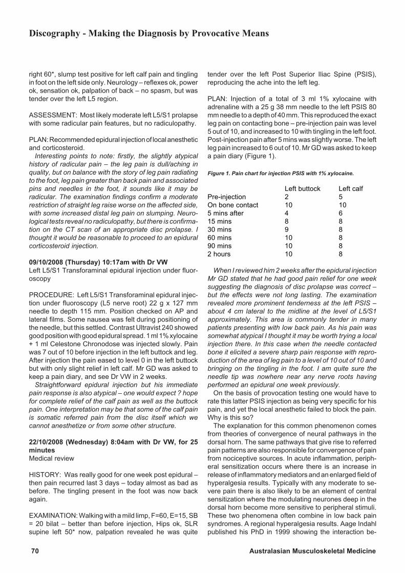
70 Australasian Musculoskeletal Medicine
right 60*, slump test positive for left calf pain and tinglingin foot on the left side only. Neurology – reflexes ok, powerok, sensation ok, palpation of back – no spasm, but wastender over the left L5 region.
ASSESSMENT: Most likely moderate left L5/S1 prolapsewith some radicular pain features, but no radiculopathy.
PLAN: Recommended epidural injection of local anestheticand corticosteroid.
Interesting points to note: firstly, the slightly atypicalhistory of radicular pain – the leg pain is dull/aching inquality, but on balance with the story of leg pain radiatingto the foot, leg pain greater than back pain and associatedpins and needles in the foot, it sounds like it may beradicular. The examination findings confirm a moderaterestriction of straight leg raise worse on the affected side,with some increased distal leg pain on slumping. Neuro-logical tests reveal no radiculopathy, but there is confirma-tion on the CT scan of an appropriate disc prolapse. Ithought it would be reasonable to proceed to an epiduralcorticosteroid injection.
09/10/2008 (Thursday) 10:17am with Dr VWLeft L5/S1 Transforaminal epidural injection under fluor-oscopy
PROCEDURE: Left L5/S1 Transforaminal epidural injec-tion under fluoroscopy (L5 nerve root) 22 g x 127 mmneedle to depth 115 mm. Position checked on AP andlateral films. Some nausea was felt during positioning ofthe needle, but this settled. Contrast Ultravist 240 showedgood position with good epidural spread. 1 ml 1% xylocaine+ 1 ml Celestone Chronodose was injected slowly. Painwas 7 out of 10 before injection in the left buttock and leg.After injection the pain eased to level 0 in the left buttockbut with only slight relief in left calf. Mr GD was asked tokeep a pain diary, and see Dr VW in 2 weeks.
Straightforward epidural injection but his immediatepain response is also atypical – one would expect ? hopefor complete relief of the calf pain as well as the buttockpain. One interpretation may be that some of the calf painis somatic referred pain from the disc itself which wecannot anesthetize or from some other structure.
22/10/2008 (Wednesday) 8:04am with Dr VW, for 25minutesMedical review
HISTORY: Was really good for one week post epidural –then pain recurred last 3 days – today almost as bad asbefore. The tingling present in the foot was now backagain.
EXAMINATION: Walking with a mild limp, F=60, E=15, SB= 20 bilat – better than before injection, Hips ok, SLRsupine left 50* now, palpation revealed he was quite
tender over the left Post Superior Iliac Spine (PSIS),reproducing the ache into the left leg.
PLAN: Injection of a total of 3 ml 1% xylocaine withadrenaline with a 25 g 38 mm needle to the left PSIS 80mm needle to a depth of 40 mm. This reproduced the exactleg pain on contacting bone – pre-injection pain was level5 out of 10, and increased to 10 with tingling in the left foot.Post-injection pain after 5 mins was slightly worse. The leftleg pain increased to 6 out of 10. Mr GD was asked to keepa pain diary (Figure 1).
Figure 1. Pain chart for injection PSIS with 1% xylocaine.
Left buttock Left calfPre-injection 2 5On bone contact 10 105 mins after 4 615 mins 8 830 mins 9 860 mins 10 890 mins 10 82 hours 10 8
When I reviewed him 2 weeks after the epidural injectionMr GD stated that he had good pain relief for one weeksuggesting the diagnosis of disc prolapse was correct –but the effects were not long lasting. The examinationrevealed more prominent tenderness at the left PSIS –about 4 cm lateral to the midline at the level of L5/S1approximately. This area is commonly tender in manypatients presenting with low back pain. As his pain wassomewhat atypical I thought it may be worth trying a localinjection there. In this case when the needle contactedbone it elicited a severe sharp pain response with repro-duction of the area of leg pain to a level of 10 out of 10 andbringing on the tingling in the foot. I am quite sure theneedle tip was nowhere near any nerve roots havingperformed an epidural one week previously.
On the basis of provocation testing one would have torate this latter PSIS injection as being very specific for hispain, and yet the local anesthetic failed to block the pain.Why is this so?
The explanation for this common phenomenon comesfrom theories of convergence of neural pathways in thedorsal horn. The same pathways that give rise to referredpain patterns are also responsible for convergence of painfrom nociceptive sources. In acute inflammation, periph-eral sensitization occurs where there is an increase inrelease of inflammatory mediators and an enlarged field ofhyperalgesia results. Typically with any moderate to se-vere pain there is also likely to be an element of centralsensitization where the modulating neurones deep in thedorsal horn become more sensitive to peripheral stimuli.These two phenomena often combine in low back painsyndromes. A regional hyperalgesia results. Aage Indahlpublished his PhD in 1999 showing the interaction be-
Discography - Making the Diagnosis by Provocative Means
November 2008 71
tween the disc, zygapophysial joints and the paraspinalmuscles and how they interplay to cause regional func-tional disturbance. It is still very relevant and worth aread.1,2
In some patients, pain can be very localized to onesegment and there may be very localized superficialtenderness, some reactive protective muscle tightnessand a possible deeper joint/disc problem. In other casespain may be more widespread. Anesthetizing the pain isthe best way to confirm a diagnosis – it is the only true goldstandard. However, even this gold standard has to takeinto account the placebo response.
Further researchThis lack of specificity of discography has led one
researcher, Dr Richard Derby in the USA, to investigatewhat proportion of provocative positive discs may beresponsive to local anesthetic injected into the disc.3 Of 39subjects with positive discograms, only 51% gained mod-est relief with injection of 4% lidocaine into the nucleuspulposis. Criteria for relief included a reduction of pain > 2on a 0 to 10 pain scale on sitting. However none of thesubjects achieved improvement in other functional scores.Some would argue that they didn’t gain pain relief becausethe anulus may have been largely intact and the localagent couldn’t reach the outer anular nociceptive nerves.This may well be the case – we just don’t know. However,most of these discs had significant grade 3 or aboveanular fissures and one would expect some anestheticinfiltration into the outer anular tears where the pain isthought to be generated.
I believe this study adds weight to the evidence of a largefalse positive response to discography. If we use the strictcriteria of 90-100% relief of pain used to diagnosezygapophysial joint pain, false positives in the study aboveapproach 100%.
ConclusionAs I have stated previously, I am a believer in the
concept of internal disc disruption and primary discogenicpain. Some cases may not yet have developed MRIchanges – these cannot be diagnosed at present. How-ever, in the majority of cases, discogenic pain will show
some morphological changes on MRI scans. There maybe some emerging therapies that will help these condi-tions. I would advocate that any new therapies be trialledon patients with a specific condition diagnosable on MRIscan. In the research setting all patients with the conditionwould receive the active or control therapy and all couldalso receive discography. It is only in this way that we candiscover whether discography has any extra predictivevalue over and above MRI. At the present time all we knowis that if we see something abnormal in the disc on MRI,then that disc is more like to be painful when provoked witha needle.
We must continue to keep an open mind in relation to theinterpretation of findings on physical examination. I havehad the odd great success in alleviating pain of severalyears’ duration by the simple injection of local anestheticinto a tight band of tender muscle. However, I hesitate tomake the diagnosis of myofascial trigger points in everypatient I see with local muscle tenderness and spasm. Ibelieve it is important to carefully examine for alterationsin local tissue texture and to try to reproduce the patient’spain by palpation or movement. We just need to keep inmind the effects of regional hyperalgesia and the possibledifferential diagnoses.
In my opinion the only case that can be made fordiscography in clinical practice is where the surgeon hasmade the decision to operate irrespective of the discogra-phy findings and that they are testing adjacent levels tohelp plan the extent of surgery. The argument for usingdiscography to avoid surgery does not hold in the realworld. A significant number of patients suffer increasedpain for up to one year post discography. I have had twopatients aggravated by the procedure to the degree thatthey ended up having fusion operations they didn’t needand they continue to suffer with long-term disability.
References1. Indahl A. Low back pain – a functional disturbance. Universityof Oslo, Norway 1999.
2. Indahl A, Kaigle AM, Reikeras O, Holm SH. Interactionbetween the porcine lumbar intervertebral disc, zygapophysialjoints and paraspinal muscles. Spine 1997; 22 (24):2834-40.
3. Derby R. Discogenic pain - evolving standards of practice.Presentation, ISIS Annual Scientific Meeting, Las Vegas, 2008.
Discography - Making the Diagnosis by Provocative Means
72 Australasian Musculoskeletal Medicine
The two hypotheses, central nervous system (CNS)sensitization and peripheral nervous system(PNS) autonomy are at first glance irreconcilable.
CNS sensitization is the reigning paradigm in main-stream pain medicine. It is shared by most members of themedical scientific community.
PNS autonomy is not recognized despite convincingscientific and clinical evidence. It has yet to establish aparadigm status and would require a scientific revolutionor paradigm shift for it to gain acceptance, as predicted byThomas Kuhn.1
However, a growing body of scientific evidence, old andnew, is addressing anomalies in the CNS sensitizationconcepts that have been ignored or dismissed.
Historically, ideas culminating in the CNS sensitizationtheory started with the Melzack-Wall article in 19652 on theGate Control Theory, which “emphasized the mecha-nisms of the CNS controlling the perception of a noxiousstimulus and thus integrated afferent, upstream proc-esses with downstream modulation from the brain”.3 Thecurrent definition of pain endorsed by the InternationalAssociation for the Study of Pain (IASP) is a logicaloutcome of this theory: “Pain is an unpleasant sensoryand emotional experience associated with actual or po-tential tissue damage, or described in terms of suchdamage”.4
According to Howard L Fields,5 “The pain pathway weare all familiar with normally begins with tissue damage,inflammation, and traumatic injury. It starts with impulsesout in the periphery. These are propagated to the spinalcord, cross in the contralateral spinothalamic tract, deliv-ered to the thalamus and then widely distributed to thecortex. Injury to a peripheral nerve somehow causes anincrease of activity in this pathway somewhere along thispathway.” He goes on to say that this increase of activitycould come from either greater activity in the nociceptorsin the periphery or by removal of some sort of inhibition inthe CNS releasing the transmission neurons from inhibi-tion. Further, he argues that these neurons are nowspontaneously active and produce a pain signal. Animalstudies have shown that these nerves themselves be-come pain generators. It is also known that damage toselectively large myelinated fibers will cause central fibersto fire more to any peripheral stimulus. There is alsoevidence that damage to peripheral nerves results inspontaneous activity in second-order neurons. Injury toperipheral nerves can also possibly cause “rewiring” in thespinal cord where fibers from the periphery normallyresponding to light touch producing the sensation of
vibration or tickle now connect to a different second-ordercell in the spinal cord. Their activity now may produce aburning pain instead of a tickle. In an article in theLancet, Loeser and Melzack3 conclude that “The braincontains widely distributed neural networks that create animage of self through genetic programmes and memoriesof past experience. Afferent inputs act on this neuromatrixand produce output patterns that lead to the report of pain.Stress can change the interactions between theneuromatrix and peripheral stimuli, as can learned expe-riences and expectation.”
As a consequence of the above concepts, the memoryof the injury leading to chronic pain and CNS sensitizationare now generally considered useful in the “management”of chronic pain by increasingly complex multidisciplinaryteams.
One anomaly in the CNS sensitization theory is itsimpotence in curing chronic pain, despite more than 40years of millions of dollars of research unraveling themysteries of the CNS. Loeser and Melzack3 admit to thiswhen they conclude their article in 1999 by saying, “In bothclinical and basic research, we are rapidly gaining usefulinformation that will lead to more effective care for thosewho suffer pain”. In the introduction to the third edition ofThe Textbook of Pain, Wall and Melzack6 express thehope that in their next edition they will be able to announceto the world a cure for chronic pain. This inability to curechronic pain has led mainstream medicine to build up animmutable conviction that any health professional claim-ing the opposite is deluding himself or herself and his orher patients. Popularized books on the management ofchronic pain by reputable and leading specialists thuswarn patients seeking a cure for their chronic pain to bewary because offering a cure is akin to deception.7 Afurther anomaly is the belief there is no demonstratedpathology in the PNS satisfactorily explaining chronicpain. This view is detailed in the introduction to Wall andMelzack’s Textbook of Pain6 in 1997.
The last 40 years of basic research into the PNS hasshown the opposite. Since Jancso et al. published theirDirect Evidence for Neurogenic Inflammation and its Pre-vention by Denervation and by Pretreatment with Capsai-cin in 19678 a growing number of scientists have system-atically unraveled the PNS response to injury. Bennett in19999 summarized this: “Painful peripheral neuropathiesbegin with nerve injury caused by disease or trauma. Thisinjury will result in an inflammatory reaction, a neuritis thatwill mobilize the immune system.” Subsequent changesmay result in more slowly developing mechanisms of
Pain Conundrums: Which Hypothesis?Central Nervous System Sensitization versus PeripheralNervous System Autonomy
Dr John Lyftogt, Christchurch, New Zealand
November 2008 73
abnormal pain that underlie the chronic phase of painfulneuropathy.
Douglas Zochodne from the Neuroscience ResearchGroup in Calgary10 examined the role of the microenviron-ment and microcirculation of the injured and regeneratingperipheral nerve trunk and concluded in his seminal paperon peripheral nerve response to injury: “Better under-standing of these and other events in injured nerve trunksis needed to help solve the two cardinal problems ofperipheral nerve injuries:1) functional disability from impaired regeneration, and 2) the development of disabling neuropathic pain.”
Peripheral nerves respond to injury in a unique way.Instead of ischemia, peripheral nerves develop increasedendoneurial blood flow. Trauma-induced ischemia in allother tissues may lead to cell death and release ofarachidonic acid, stimulating COX I, COX II, and 5-LOXpathways upregulating prostaglandin production and tis-sue inflammation. Peripheral nerve injury leads to “dump-ing” of calcitonin gene related peptide (CGRP), substanceP (SP) and nitric oxide (NO) from nervi nervorum, theextensive innervations of the connective tissue of periph-eral nerve trunks, the epi-perineurium. CGRP, SP, andNO are vasodilators with CGRP and SP also potentupregulators of vascular permeability of the vasa nervorumand neighboring blood vessels. The result is a rapidincrease in the endoneurial blood flow and neurogenicinflammation of the nerve trunk itself and the surroundingtissues. This forms the basis of the Triple Responsedescribed by Lewis in 192711 with the well-known “line,wheal and flare”. Lewis also identified the Axon Reflex,showing axonal impulses travelling in an orthodromic (toCNS) and antidromic direction. In 1901 Bayliss12 foundthat stimulation of the dorsal root ganglion (DRG) resultedin peripheral vasodilatation. He postulated afferent andefferent conduction. Some 20 neuropeptides andneurotransmitters are known to be involved in neurogenicinflammation.13 Most of these have been cloned, includingtheir antagonists, their receptors, and receptor antago-nists. Evidence of pivotal roles for specific neuropeptidesis lacking, hence no drug treatment has yet been devel-oped against neurogenic inflammation and the concept ofneuromodulation is rationalizing the impasse. It has alsobeen found that some neuropeptides (CGRP, peptide YY- PYY - and neuropeptide Y - NPY) can be released fromnon-neuronal cells and also in a paracrine fashion fromneurons. Some anti-inflammatory neuropeptides such asmelanocyte-stimulating hormone (MSH), vasoactive in-testinal peptide (VIP) and pituitary adenylate cyclase-activating polypeptide (PACAP) may be released to termi-nate inflammation under physiological conditions. Thusneuromediators are not all pro-inflammatory but regulateall phases of the inflammatory response.
The complexity of responses of the peripheral nervoussystem was also highlighted by Burnstock in 1976.14 Heintroduced the concept of co-transmission in the auto-nomic nervous system with neuropeptides also contained
in cholinergic and adrenergic nerves. The intimate con-nection between sensory and autonomic nerves is par-ticularly poignant in children born with congenital insensi-tivity to pain in hereditary sensory and autonomic neu-ropathy (HSAN type I-IV). The absent or abnormal periph-eral nerves are the reason for severe disfiguring sequelaeof trauma, inability to repair tissue or adequately mobilizethe immune system, often leading to death from infectionduring childhood. The recent finding of opioid receptors onperipheral sensory axons15 led to some speculation thatthese µ opioid receptors (MORs) may have an anti-nociceptive action. This motivated Zochodne et al,16 toexamine the function and expression of local MORsassociated with the chronic constriction injury (CCI)17
model of sciatic neuropathic pain in rats. Low dose mor-phine was injected percutaneously near the nerve. Theyconcluded that their positive findings “may provide atherapeutic direction for the treatment of certain focalneuropathic lesions in humans”.
Successful treatments of painful peripheral neuropa-thies or chronic recalcitrant pain have been described inthe literature. Whether one goes along with the rationalefor these treatments is less relevant. After all, clinicianswould consider it unethical to cease treatment with lithiumcarbonate or chlorpromazine simply because the mode ofaction is unknown. By 1965 George Hackett18 had pub-lished the results of prolotherapy treatment of 1800 casesof chronic low back pain, with an 82% success rate and a12-year follow up. He published 16 articles and one bookon his treatment. Also in 1965 Melzack and Wall publishedtheir Gate Control Theory of Pain. Their Textbook of Pain,published in 1997, does not reference prolotherapy. Nei-ther does it reference neural therapy. This treatment wasdeveloped in Germany in the 1940s by Drs Ferdinand andWalter Huneke. Lidocaine is used to treat chronic pain bytargeting postulated “interference fields” that cause “blocks”in the autonomic nervous system leading to chronic pain.This treatment is highly successful and practised widely inGermany and Spain by more than 5000 medical practi-tioners. Most of the literature is in German and this may bethe reason there is little knowledge of this treatment in theEnglish-speaking world.
The growing scientific evidence supporting the view thatneuropathic pain syndromes are caused by unremittingperipheral neurogenic inflammation involving the auto-nomic and sensory nerves may lead to renewed interestin prolotherapy and neural therapy as these treatmentsare effective and seem to target the PNS. The author hasnow treated more than a 1000 patients with subcutaneousprolotherapy targeting neurogenic inflammation of periph-eral nerve trunks in much the same way as Zochodne’spercutaneous near nerve injections with low-dose opioids.Published results19-21 are promising for recalcitrant lum-bago, shoulder, knee, elbow pain, and achillodynia.Patients with chronic neuropathic pain will continue tosuffer needlessly if physicians remain fixed on the reigningparadigm that can suggest only “pain management” when
Pain Conundrums: Which Hypothesis?
74 Australasian Musculoskeletal Medicine
there are well-documented alternatives available that mayoffer a cure.
References1. Kuhn T. The Structure of Scientific Revolutions. 1962.
2. Melzack R, Wall PD. Pain Mechanisms: A New Theory.Science 1965; 150: 971-79.
3. Loeser J D. Melzack R. Pain: an overview. Lancet 1999; 353:1607-09.
4. Merskey H, Bogduk N. Classification of Chronic Pain. Seattle:International Association for the Study of Pain Press, 1994,p.210.
5. Fields H L. Power Point presentation Medscape FamilyMedicine 2007.
6. Wall PD, Melzack R, eds Textbook of Pain. 3rd ed. Edinburgh;Churchill Livingstone, 2004.
7. Nicholas M, Molloy A, Tonkin L, Beeston L. Manage your pain:practical and positive ways of adapting to chronic pain. Sydney;Australian Broadcasting Corporation Books, 2001.
8. Jancso N, Jancso-Gabor A, Szolcsanyi J. Direct Evidence forNeurogenic Inflammation and its Prevention by Denervation andby Pretreatment with Capsaicin. Br J Pharmac Chemother 1967;31: 138-51.
9. Bennett GJ. Does a neuroimmune interaction contribute to thegenesis of painful peripheral neuropathies? Proc Natl Acad USA1999; 96(14): 7737-38.
10. Zochodne D. Local Events within the Injured and Regener-ating Peripheral Nerve Trunk: The Role of the Microenvironment
and Microcirculation. Biomed Rev 1997; 8: 37-54.
11. Lewis T. Released substances the cause of the triple re-sponse. In: The blood vessels of the heart and their responses.London; Shaw and Son.
12. Bayliss WM. On the origin from the spinal cord of the vaso-dilator fibers of the hind-limb, and on the nature of these fibers.J Physiol 1901; 26: 173-209.
13. Brain SD, Cox HM. Neuropeptides and their receptors:Innovative science providing novel therapeutic targets. Br JPharmacol 2006; 147: S202-S211.
14. Burnstock G. Do some nerve cells release more than onetransmitter? J Neurosci 1976; 1: 239-48 co-transmission.
15. Coggeshall RE, Zhou S, Carlton SM. Opioid receptors onperipheral sensory axons. Brain Res 1997;764: 126-32.
16. Truong W, Cheng C, Xu Q et al. µ Opioid Receptors andAnalgesia at the Site of a Peripheral Nerve Injury. Ann Neurol2003; 53: 366-75.
17. Bennett GJ, Xie YK. A peripheral mononeuropathy in rat thatproduces disorders of pain sensation like those seen in man.Pain 1988;33: 87-107.
18. Hackett G. Ligament and Tendon Relaxation Treated byProlotherapy. 3rd ed. Springfield, IL; Charles Thomas, 1958.
19. Lyftogt JA. Prolotherapy for recalcitrant lumbago. Aust Mus-culoskeletal Med 2008; 13(1): 18-20.
20. Lyftogt J. Subcutaneous prolotherapy treatment of refractoryknee, shoulder and lateral elbow pain. Aust MusculoskeletalMed 2007; 12(2): 110-12.
21. Lyftogt J. Subcutaneous Prolotherapy for Achillestendinopathy: The best solution? Aust Musculoskeletal Med2007; 12(2): 107-109.
Pain Conundrums: Which Hypothesis?
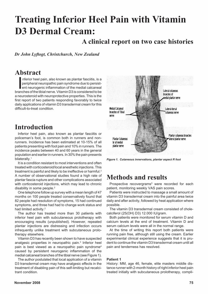
November 2008 75
Abstract
Inferior heel pain, also known as plantar fasciitis, is aperipheral neuropathic pain syndrome due to persist-ent neurogenic inflammation of the medial calcaneal
branches of the tibial nerve. Vitamin D3 is considered to bea neurosteroid with neuroprotective properties. This is thefirst report of two patients responding favorably to twicedaily applications of vitamin D3 transdermal cream for thisdifficult-to-treat condition.
IntroductionInferior heel pain, also known as plantar fasciitis or
policeman’s foot, is common both in runners and non-runners. Incidence has been estimated at 10-15% of allpatients presenting with foot pain and 10% in runners. Theincidence peaks between 40 and 60 years in the generalpopulation and earlier in runners. In 30% the pain presentsbilaterally.1
It is a condition resistant to most interventions and oftentreated with corticosteroid/local anesthetic injections. Thistreatment is painful and likely to be ineffective or harmful.2
A number of observational studies found a high rate ofplantar fascia rupture and other complications associatedwith corticosteroid injections, which may lead to chronicdisability in some people.2
One telephone follow up survey with a mean length of 47months on 100 people treated conservatively found that82 people had resolution of symptoms, 15 had continuedsymptoms, and three had had to change work status andhad limited activity.2
The author has treated more than 30 patients withinferior heel pain with subcutaneous prolotherapy withencouraging results (unpublished). However, repeatedplantar injections are distressing and infection occursinfrequently unlike treatment with subcutaneous prolo-therapy elsewhere.
Vitamin D3 has recently been shown to have suspectedanalgesic properties in neuropathic pain.3 Inferior heelpain is best viewed as a neuropathic pain syndrome4
caused by persistent neurogenic inflammation of themedial calcaneal branches of the tibial nerve (see Figure 1).
The author postulated that local application of a vitaminD3 transdermal cream may have analgesic effects in thetreatment of disabling pain of this self-limiting but recalci-trant condition.
Treating Inferior Heel Pain with VitaminD3 Dermal Cream:
A clinical report on two case histories
Dr John Lyftogt, Christchurch, New Zealand
Methods and resultsProspective recovergrams5 were recorded for each
patient, monitoring weekly VAS pain scores.Patients were instructed to massage a small amount of
vitamin D3 transdermal cream into the painful area twicedaily and after activity, followed by heat application wherepossible.
The vitamin D3 transdermal cream consisted of chole-calciferol (25(OH) D3) 12.000 IU/gram.
Both patients were monitored for serum vitamin D andcalcium levels at the end of treatment. Vitamin D andserum calcium levels were all in the normal range.
At the time of writing this report both patients wererunning pain free, although still using the cream. Earlierexperimental clinical experience suggests that it is pru-dent to continue the vitamin D3 transdermal cream until allpain and tenderness has resolved.
Patient 1History: MM, age 46, female, elite masters middle dis-tance runner with 2-month history of right inferior heel paintreated initially with subcutaneous prolotherapy, compli-
Figure 1. Cutaneous innervations, plantar aspect R foot
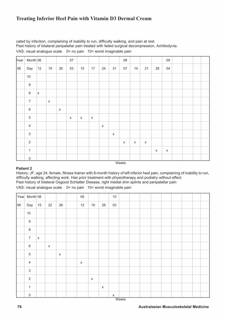
76 Australasian Musculoskeletal Medicine
cated by infection, complaining of inability to run, difficulty walking, and pain at rest.Past history of bilateral peripatellar pain treated with failed surgical decompression, Achillodynia.VAS: visual analogue scale 0= no pain 10= worst imaginable pain
Year Month 06 07 08 09
08 Day 12 19 26 03 10 17 24 31 07 14 21 28 04
10
9
8 x
7 x
6 x
5 x x x
4 x
3 x
2 x x x
1 x x
0 Weeks
Patient 2History: JF, age 24, female, fitness trainer with 8-month history of left inferior heel pain, complaining of inability to run,difficulty walking, affecting work. Has prior treatment with physiotherapy and podiatry without effect.Past history of bilateral Osgood Schlatter Disease, right medial shin splints and peripatellar painVAS: visual analogue scale 0= no pain 10= worst imaginable pain
Year Month 08 09 10
08 Day 15 22 28 12 19 26 03
10
9
8
7 x
6 x
5 x
4 x
3
2 x
1 x
0 x Weeks
Treating Inferior Heel Pain with Vitamin D3 Dermal Cream
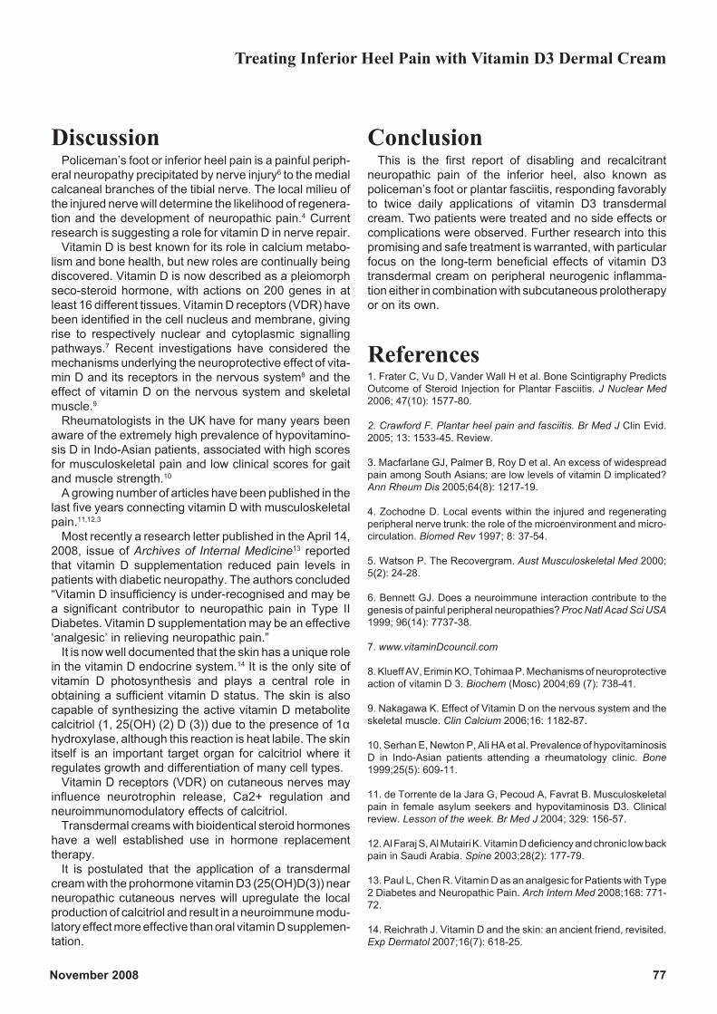
November 2008 77
DiscussionPoliceman’s foot or inferior heel pain is a painful periph-
eral neuropathy precipitated by nerve injury6 to the medialcalcaneal branches of the tibial nerve. The local milieu ofthe injured nerve will determine the likelihood of regenera-tion and the development of neuropathic pain.4 Currentresearch is suggesting a role for vitamin D in nerve repair.
Vitamin D is best known for its role in calcium metabo-lism and bone health, but new roles are continually beingdiscovered. Vitamin D is now described as a pleiomorphseco-steroid hormone, with actions on 200 genes in atleast 16 different tissues. Vitamin D receptors (VDR) havebeen identified in the cell nucleus and membrane, givingrise to respectively nuclear and cytoplasmic signallingpathways.7 Recent investigations have considered themechanisms underlying the neuroprotective effect of vita-min D and its receptors in the nervous system8 and theeffect of vitamin D on the nervous system and skeletalmuscle.9
Rheumatologists in the UK have for many years beenaware of the extremely high prevalence of hypovitamino-sis D in Indo-Asian patients, associated with high scoresfor musculoskeletal pain and low clinical scores for gaitand muscle strength.10
A growing number of articles have been published in thelast five years connecting vitamin D with musculoskeletalpain.11,12,3
Most recently a research letter published in the April 14,2008, issue of Archives of Internal Medicine13 reportedthat vitamin D supplementation reduced pain levels inpatients with diabetic neuropathy. The authors concluded“Vitamin D insufficiency is under-recognised and may bea significant contributor to neuropathic pain in Type IIDiabetes. Vitamin D supplementation may be an effective‘analgesic’ in relieving neuropathic pain.”
It is now well documented that the skin has a unique rolein the vitamin D endocrine system.14 It is the only site ofvitamin D photosynthesis and plays a central role inobtaining a sufficient vitamin D status. The skin is alsocapable of synthesizing the active vitamin D metabolitecalcitriol (1, 25(OH) (2) D (3)) due to the presence of 1αhydroxylase, although this reaction is heat labile. The skinitself is an important target organ for calcitriol where itregulates growth and differentiation of many cell types.
Vitamin D receptors (VDR) on cutaneous nerves mayinfluence neurotrophin release, Ca2+ regulation andneuroimmunomodulatory effects of calcitriol.
Transdermal creams with bioidentical steroid hormoneshave a well established use in hormone replacementtherapy.
It is postulated that the application of a transdermalcream with the prohormone vitamin D3 (25(OH)D(3)) nearneuropathic cutaneous nerves will upregulate the localproduction of calcitriol and result in a neuroimmune modu-latory effect more effective than oral vitamin D supplemen-tation.
ConclusionThis is the first report of disabling and recalcitrant
neuropathic pain of the inferior heel, also known aspoliceman’s foot or plantar fasciitis, responding favorablyto twice daily applications of vitamin D3 transdermalcream. Two patients were treated and no side effects orcomplications were observed. Further research into thispromising and safe treatment is warranted, with particularfocus on the long-term beneficial effects of vitamin D3transdermal cream on peripheral neurogenic inflamma-tion either in combination with subcutaneous prolotherapyor on its own.
References1. Frater C, Vu D, Vander Wall H et al. Bone Scintigraphy PredictsOutcome of Steroid Injection for Plantar Fasciitis. J Nuclear Med2006; 47(10): 1577-80.
2. Crawford F. Plantar heel pain and fasciitis. Br Med J Clin Evid.2005; 13: 1533-45. Review.
3. Macfarlane GJ, Palmer B, Roy D et al. An excess of widespreadpain among South Asians; are low levels of vitamin D implicated?Ann Rheum Dis 2005;64(8): 1217-19.
4. Zochodne D. Local events within the injured and regeneratingperipheral nerve trunk: the role of the microenvironment and micro-circulation. Biomed Rev 1997; 8: 37-54.
5. Watson P. The Recovergram. Aust Musculoskeletal Med 2000;5(2): 24-28.
6. Bennett GJ. Does a neuroimmune interaction contribute to thegenesis of painful peripheral neuropathies? Proc Natl Acad Sci USA1999; 96(14): 7737-38.
7. www.vitaminDcouncil.com
8. Klueff AV, Erimin KO, Tohimaa P. Mechanisms of neuroprotectiveaction of vitamin D 3. Biochem (Mosc) 2004;69 (7): 738-41.
9. Nakagawa K. Effect of Vitamin D on the nervous system and theskeletal muscle. Clin Calcium 2006;16: 1182-87.
10. Serhan E, Newton P, Ali HA et al. Prevalence of hypovitaminosisD in Indo-Asian patients attending a rheumatology clinic. Bone1999;25(5): 609-11.
11. de Torrente de la Jara G, Pecoud A, Favrat B. Musculoskeletalpain in female asylum seekers and hypovitaminosis D3. Clinicalreview. Lesson of the week. Br Med J 2004; 329: 156-57.
12. Al Faraj S, Al Mutairi K. Vitamin D deficiency and chronic low backpain in Saudi Arabia. Spine 2003;28(2): 177-79.
13. Paul L, Chen R. Vitamin D as an analgesic for Patients with Type2 Diabetes and Neuropathic Pain. Arch Intern Med 2008;168: 771-72.
14. Reichrath J. Vitamin D and the skin: an ancient friend, revisited.Exp Dermatol 2007;16(7): 618-25.
Treating Inferior Heel Pain with Vitamin D3 Dermal Cream
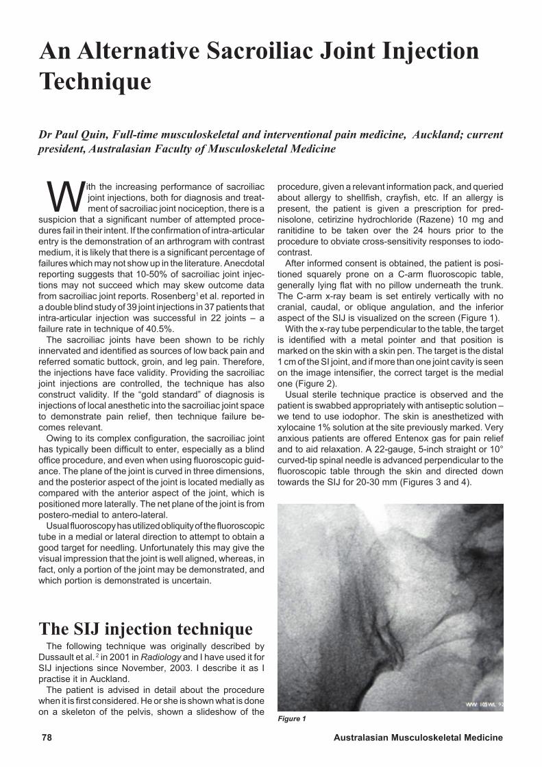
78 Australasian Musculoskeletal Medicine
With the increasing performance of sacroiliacjoint injections, both for diagnosis and treat-ment of sacroiliac joint nociception, there is a
suspicion that a significant number of attempted proce-dures fail in their intent. If the confirmation of intra-articularentry is the demonstration of an arthrogram with contrastmedium, it is likely that there is a significant percentage offailures which may not show up in the literature. Anecdotalreporting suggests that 10-50% of sacroiliac joint injec-tions may not succeed which may skew outcome datafrom sacroiliac joint reports. Rosenberg1 et al. reported ina double blind study of 39 joint injections in 37 patients thatintra-articular injection was successful in 22 joints – afailure rate in technique of 40.5%.
The sacroiliac joints have been shown to be richlyinnervated and identified as sources of low back pain andreferred somatic buttock, groin, and leg pain. Therefore,the injections have face validity. Providing the sacroiliacjoint injections are controlled, the technique has alsoconstruct validity. If the “gold standard” of diagnosis isinjections of local anesthetic into the sacroiliac joint spaceto demonstrate pain relief, then technique failure be-comes relevant.
Owing to its complex configuration, the sacroiliac jointhas typically been difficult to enter, especially as a blindoffice procedure, and even when using fluoroscopic guid-ance. The plane of the joint is curved in three dimensions,and the posterior aspect of the joint is located medially ascompared with the anterior aspect of the joint, which ispositioned more laterally. The net plane of the joint is frompostero-medial to antero-lateral.
Usual fluoroscopy has utilized obliquity of the fluoroscopictube in a medial or lateral direction to attempt to obtain agood target for needling. Unfortunately this may give thevisual impression that the joint is well aligned, whereas, infact, only a portion of the joint may be demonstrated, andwhich portion is demonstrated is uncertain.
The SIJ injection techniqueThe following technique was originally described by
Dussault et al. 2 in 2001 in Radiology and I have used it forSIJ injections since November, 2003. I describe it as Ipractise it in Auckland.
The patient is advised in detail about the procedurewhen it is first considered. He or she is shown what is doneon a skeleton of the pelvis, shown a slideshow of the
procedure, given a relevant information pack, and queriedabout allergy to shellfish, crayfish, etc. If an allergy ispresent, the patient is given a prescription for pred-nisolone, cetirizine hydrochloride (Razene) 10 mg andranitidine to be taken over the 24 hours prior to theprocedure to obviate cross-sensitivity responses to iodo-contrast.
After informed consent is obtained, the patient is posi-tioned squarely prone on a C-arm fluoroscopic table,generally lying flat with no pillow underneath the trunk.The C-arm x-ray beam is set entirely vertically with nocranial, caudal, or oblique angulation, and the inferioraspect of the SIJ is visualized on the screen (Figure 1).
With the x-ray tube perpendicular to the table, the targetis identified with a metal pointer and that position ismarked on the skin with a skin pen. The target is the distal1 cm of the SI joint, and if more than one joint cavity is seenon the image intensifier, the correct target is the medialone (Figure 2).
Usual sterile technique practice is observed and thepatient is swabbed appropriately with antiseptic solution –we tend to use iodophor. The skin is anesthetized withxylocaine 1% solution at the site previously marked. Veryanxious patients are offered Entenox gas for pain reliefand to aid relaxation. A 22-gauge, 5-inch straight or 10°curved-tip spinal needle is advanced perpendicular to thefluoroscopic table through the skin and directed downtowards the SIJ for 20-30 mm (Figures 3 and 4).
An Alternative Sacroiliac Joint InjectionTechnique
Dr Paul Quin, Full-time musculoskeletal and interventional pain medicine, Auckland; currentpresident, Australasian Faculty of Musculoskeletal Medicine
Figure 1
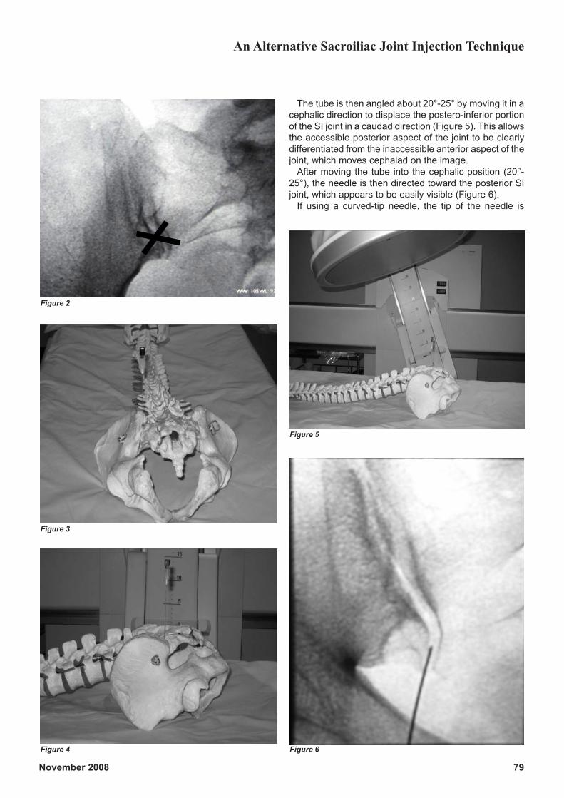
November 2008 79
The tube is then angled about 20°-25° by moving it in acephalic direction to displace the postero-inferior portionof the SI joint in a caudad direction (Figure 5). This allowsthe accessible posterior aspect of the joint to be clearlydifferentiated from the inaccessible anterior aspect of thejoint, which moves cephalad on the image.
After moving the tube into the cephalic position (20°-25°), the needle is then directed toward the posterior SIjoint, which appears to be easily visible (Figure 6).
If using a curved-tip needle, the tip of the needle is
Figure 2
Figure 3
Figure 4
Figure 5
Figure 6
An Alternative Sacroiliac Joint Injection Technique
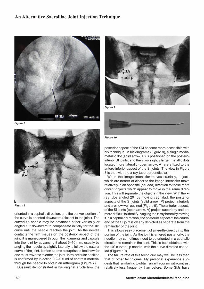
80 Australasian Musculoskeletal Medicine
oriented in a cephalic direction, and the convex portion ofthe curve is oriented downward (closest to the joint). Thecurved-tip needle may be advanced either vertically orangled 10° downward to compensate initially for the 10°curve until the needle reaches the joint. As the needlecontacts the firm tissues on the posterior aspect of thejoint, it is maneuvered through the ligaments and capsuleinto the joint by advancing it about 5–10 mm, usually byangling the needle tip slightly laterally to follow the naturalcurve of the joint. It often seems a surprise to feel how farone must traverse to enter the joint. Intra-articular positionis confirmed by injecting 0.2–0.5 ml of contrast materialthrough the needle to obtain an arthrogram (Figure 7).
Dussault demonstrated in his original article how the
posterior aspect of the SIJ became more accessible withhis technique. In his diagrams (Figure 8), a single medialmetallic dot (solid arrow, P) is positioned on the postero-inferior SI joints, and then two slightly larger metallic dotslocated more laterally (open arrow, A) are affixed to theantero-inferior aspect of the SI joints. The view in Figure8 is that with the x-ray tube perpendicular.
When the image intensifier moves cranially, objectswhich are nearer or closer to the image intensifier moverelatively in an opposite (caudad) direction to those moredistant objects which appear to move in the same direc-tion. This will separate the objects in the view. With the x-ray tube angled 20° by moving cephalad, the posterioraspects of the SI joints (solid arrow, P) project inferiorlyand are now well outlined (Figure 9). The anterior aspectsof the SI joints (open arrow, A) project superiorly and aremore difficult to identify. Angling the x-ray beam by movingit in a cephalic direction, the posterior aspect of the caudalend of the SI joint is clearly depicted as separate from theremainder of the joint.
This allows easy placement of a needle directly into thisportion of the joint. As the joint is entered posteriorly, theneedle may sometimes need to be oriented in a cephalicdirection to remain in the joint. This is best obtained withthe 10° curved-tip needle, with the curve directed cepha-lad (Figure 10).
The failure rate of this technique may well be less thanthat of other techniques. My personal experience sug-gests that I am failing to obtain an arthrogram with contrastrelatively less frequently than before. Some SIJs have
Figure 7
Figure 8
Figure 9
Figure 10
An Alternative Sacroiliac Joint Injection Technique
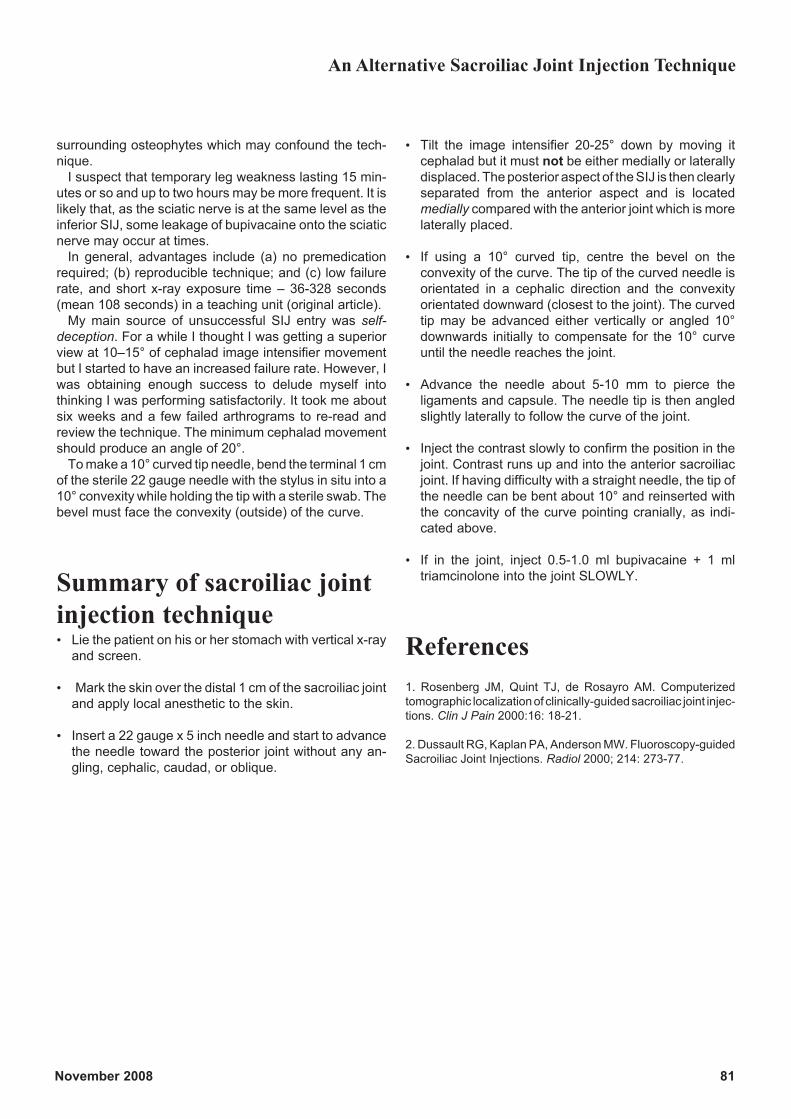
November 2008 81
surrounding osteophytes which may confound the tech-nique.
I suspect that temporary leg weakness lasting 15 min-utes or so and up to two hours may be more frequent. It islikely that, as the sciatic nerve is at the same level as theinferior SIJ, some leakage of bupivacaine onto the sciaticnerve may occur at times.
In general, advantages include (a) no premedicationrequired; (b) reproducible technique; and (c) low failurerate, and short x-ray exposure time – 36-328 seconds(mean 108 seconds) in a teaching unit (original article).
My main source of unsuccessful SIJ entry was self-deception. For a while I thought I was getting a superiorview at 10–15° of cephalad image intensifier movementbut I started to have an increased failure rate. However, Iwas obtaining enough success to delude myself intothinking I was performing satisfactorily. It took me aboutsix weeks and a few failed arthrograms to re-read andreview the technique. The minimum cephalad movementshould produce an angle of 20°.
To make a 10° curved tip needle, bend the terminal 1 cmof the sterile 22 gauge needle with the stylus in situ into a10° convexity while holding the tip with a sterile swab. Thebevel must face the convexity (outside) of the curve.
Summary of sacroiliac jointinjection technique• Lie the patient on his or her stomach with vertical x-ray
and screen.
• Mark the skin over the distal 1 cm of the sacroiliac jointand apply local anesthetic to the skin.
• Insert a 22 gauge x 5 inch needle and start to advancethe needle toward the posterior joint without any an-gling, cephalic, caudad, or oblique.
• Tilt the image intensifier 20-25° down by moving itcephalad but it must not be either medially or laterallydisplaced. The posterior aspect of the SIJ is then clearlyseparated from the anterior aspect and is locatedmedially compared with the anterior joint which is morelaterally placed.
• If using a 10° curved tip, centre the bevel on theconvexity of the curve. The tip of the curved needle isorientated in a cephalic direction and the convexityorientated downward (closest to the joint). The curvedtip may be advanced either vertically or angled 10°downwards initially to compensate for the 10° curveuntil the needle reaches the joint.
• Advance the needle about 5-10 mm to pierce theligaments and capsule. The needle tip is then angledslightly laterally to follow the curve of the joint.
• Inject the contrast slowly to confirm the position in thejoint. Contrast runs up and into the anterior sacroiliacjoint. If having difficulty with a straight needle, the tip ofthe needle can be bent about 10° and reinserted withthe concavity of the curve pointing cranially, as indi-cated above.
• If in the joint, inject 0.5-1.0 ml bupivacaine + 1 mltriamcinolone into the joint SLOWLY.
References1. Rosenberg JM, Quint TJ, de Rosayro AM. Computerizedtomographic localization of clinically-guided sacroiliac joint injec-tions. Clin J Pain 2000:16: 18-21.
2. Dussault RG, Kaplan PA, Anderson MW. Fluoroscopy-guidedSacroiliac Joint Injections. Radiol 2000; 214: 273-77.
An Alternative Sacroiliac Joint Injection Technique
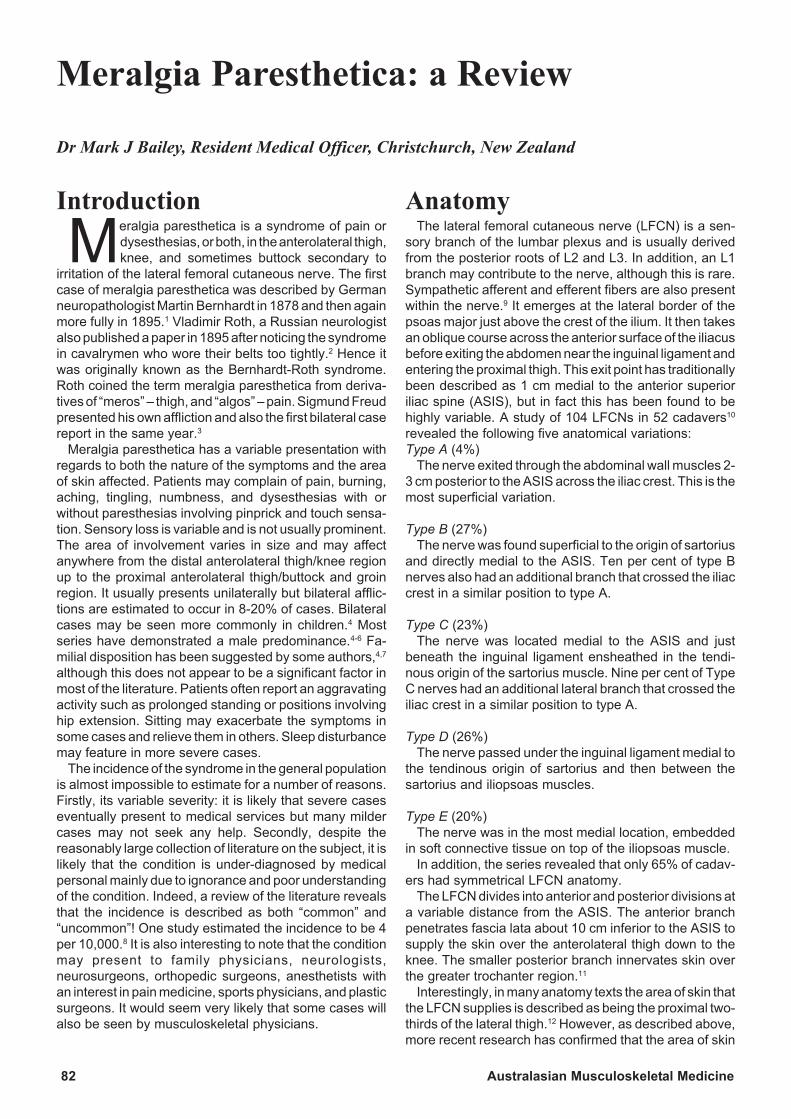
82 Australasian Musculoskeletal Medicine
Introduction
Meralgia paresthetica is a syndrome of pain ordysesthesias, or both, in the anterolateral thigh,knee, and sometimes buttock secondary to
irritation of the lateral femoral cutaneous nerve. The firstcase of meralgia paresthetica was described by Germanneuropathologist Martin Bernhardt in 1878 and then againmore fully in 1895.1 Vladimir Roth, a Russian neurologistalso published a paper in 1895 after noticing the syndromein cavalrymen who wore their belts too tightly.2 Hence itwas originally known as the Bernhardt-Roth syndrome.Roth coined the term meralgia paresthetica from deriva-tives of “meros” – thigh, and “algos” – pain. Sigmund Freudpresented his own affliction and also the first bilateral casereport in the same year.3
Meralgia paresthetica has a variable presentation withregards to both the nature of the symptoms and the areaof skin affected. Patients may complain of pain, burning,aching, tingling, numbness, and dysesthesias with orwithout paresthesias involving pinprick and touch sensa-tion. Sensory loss is variable and is not usually prominent.The area of involvement varies in size and may affectanywhere from the distal anterolateral thigh/knee regionup to the proximal anterolateral thigh/buttock and groinregion. It usually presents unilaterally but bilateral afflic-tions are estimated to occur in 8-20% of cases. Bilateralcases may be seen more commonly in children.4 Mostseries have demonstrated a male predominance.4-6 Fa-milial disposition has been suggested by some authors,4,7
although this does not appear to be a significant factor inmost of the literature. Patients often report an aggravatingactivity such as prolonged standing or positions involvinghip extension. Sitting may exacerbate the symptoms insome cases and relieve them in others. Sleep disturbancemay feature in more severe cases.
The incidence of the syndrome in the general populationis almost impossible to estimate for a number of reasons.Firstly, its variable severity: it is likely that severe caseseventually present to medical services but many mildercases may not seek any help. Secondly, despite thereasonably large collection of literature on the subject, it islikely that the condition is under-diagnosed by medicalpersonal mainly due to ignorance and poor understandingof the condition. Indeed, a review of the literature revealsthat the incidence is described as both “common” and“uncommon”! One study estimated the incidence to be 4per 10,000.8 It is also interesting to note that the conditionmay present to family physicians, neurologists,neurosurgeons, orthopedic surgeons, anesthetists withan interest in pain medicine, sports physicians, and plasticsurgeons. It would seem very likely that some cases willalso be seen by musculoskeletal physicians.
AnatomyThe lateral femoral cutaneous nerve (LFCN) is a sen-
sory branch of the lumbar plexus and is usually derivedfrom the posterior roots of L2 and L3. In addition, an L1branch may contribute to the nerve, although this is rare.Sympathetic afferent and efferent fibers are also presentwithin the nerve.9 It emerges at the lateral border of thepsoas major just above the crest of the ilium. It then takesan oblique course across the anterior surface of the iliacusbefore exiting the abdomen near the inguinal ligament andentering the proximal thigh. This exit point has traditionallybeen described as 1 cm medial to the anterior superioriliac spine (ASIS), but in fact this has been found to behighly variable. A study of 104 LFCNs in 52 cadavers10
revealed the following five anatomical variations:Type A (4%)
The nerve exited through the abdominal wall muscles 2-3 cm posterior to the ASIS across the iliac crest. This is themost superficial variation.
Type B (27%)The nerve was found superficial to the origin of sartorius
and directly medial to the ASIS. Ten per cent of type Bnerves also had an additional branch that crossed the iliaccrest in a similar position to type A.
Type C (23%)The nerve was located medial to the ASIS and just
beneath the inguinal ligament ensheathed in the tendi-nous origin of the sartorius muscle. Nine per cent of TypeC nerves had an additional lateral branch that crossed theiliac crest in a similar position to type A.
Type D (26%)The nerve passed under the inguinal ligament medial to
the tendinous origin of sartorius and then between thesartorius and iliopsoas muscles.
Type E (20%)The nerve was in the most medial location, embedded
in soft connective tissue on top of the iliopsoas muscle.In addition, the series revealed that only 65% of cadav-
ers had symmetrical LFCN anatomy.The LFCN divides into anterior and posterior divisions at
a variable distance from the ASIS. The anterior branchpenetrates fascia lata about 10 cm inferior to the ASIS tosupply the skin over the anterolateral thigh down to theknee. The smaller posterior branch innervates skin overthe greater trochanter region.11
Interestingly, in many anatomy texts the area of skin thatthe LFCN supplies is described as being the proximal two-thirds of the lateral thigh.12 However, as described above,more recent research has confirmed that the area of skin
Meralgia Paresthetica: a Review
Dr Mark J Bailey, Resident Medical Officer, Christchurch, New Zealand
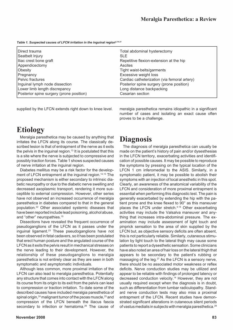
November 2008 83
supplied by the LFCN extends right down to knee level.
EtiologyMeralgia paresthetica may be caused by anything that
irritates the LFCN along its course. The classically de-scribed lesion is that of entrapment of the nerve as it exitsthe pelvis in the inguinal region.13 It is postulated that thisis a site where the nerve is subjected to compressive andpossibly traction forces. Table 1 shows suspected causesof nerve irritation at the inguinal region.
Diabetes mellitus may be a risk factor for the develop-ment of LFCN entrapment at the inguinal region.14,15 Theproposed mechanism is either secondary to intrinsic dia-betic neuropathy or due to the diabetic nerve swelling anddecreased axoplasmic transport, rendering it more sus-ceptible to external compression. However, other serieshave not observed an increased occurrence of meralgiaparesthetica in diabetes compared to that in the generalpopulation.22 Other associated systemic diseases thathave been reported include lead poisoning, alcohol abuse,and “other” neuropathies.15
Dissections have revealed the frequent occurrence ofpseudoganglions of the LFCN as it passes under theinguinal ligament.23 These pseudoganglions have notbeen observed in fetal cadavers, so it has been postulatedthat erect human posture and the angulated course of theLFCN as it exits the pelvis result in mechanical stresses onthe nerve leading to their development. However, therelationship of these pseudoganglions to meralgiaparesthetica is not entirely clear as they are seen in bothsymptomatic and asymptomatic individuals.
Although less common, more proximal irritation of theLFCN can also lead to meralgia paresthetica. Potentiallyany structure that comes into contact with the LFCN alongits course from its origin to its exit from the pelvis can leadto compression or traction irritation. To date some of thedescribed causes have included meralgia paresthetica ofspinal origin,24 malignant tumor of the psoas muscle,25 andcompression of the LFCN beneath the iliacus fasciasecondary to infection or hematoma.26 The cause of
Table 1. Suspected causes of LFCN irritation in the inguinal region4,14-21
Direct trauma Total abdominal hysterectomy Seatbelt Injury SLE Iliac crest bone graft Repetitive flexion-extension at the hip Appendicectomy Ascites Obesity Tight waist-belts/garments Pregnancy Excessive weight loss Pelvic fractures Cardiac catheterization (via femoral artery) Inguinal lymph node dissection Posterior spine surgery (prone position) Lower limb length discrepancy Long distance backpacking Posterior spine surgery (prone position) Cesarian section
meralgia paresthetica remains idiopathic in a significantnumber of cases and isolating an exact cause oftenproves to be a challenge.
DiagnosisThe diagnosis of meralgia paresthetica can usually be
made on the patient’s history of pain and/or dysesthesiasin the LFCN territory, exacerbating activities and identifi-cation of possible causes. It may be possible to reproducethe symptoms by pressing on the typical location of theLFCN 1 cm inferomedial to the ASIS. Similarly, in asymptomatic patient, it may be possible to abolish theirsymptoms with an injection of local anesthetic in this spot.Clearly, an awareness of the anatomical variability of theLFCN and consideration of more proximal entrapment isessential when performing this diagnostic test. The pain isgenerally exacerbated by extending the hip with the pa-tient prone and the knee flexed to 90° as this maneuverplaces the LFCN under stretch.4,15 Other exacerbatingactivities may include the Valsalva maneuver and any-thing that increases intra-abdominal pressure. The ex-amination may include assessment of light touch andpinprick sensation to the area of skin supplied by theLFCN but, as objective sensory deficits are often absent,this is not particularly reliable. Similarly, cutaneous stimu-lation by light touch to the lateral thigh may cause somepatients to report a dysesthetic sensation. Some clinicianshave also noted an area of hair loss on the lateral thigh thatappears to be secondary to the patient’s rubbing ormassaging of the leg.27 As the LFCN is a sensory nerve,there should be no associated motor weakness or reflexdeficits. Nerve conduction studies may be utilized andappear to be reliable with findings of prolonged latency ordecreased conduction velocity.14 However, they are notusually required except when the diagnosis is in doubt,such as differentiation from lumbar radiculopathy. Stand-ard nerve conduction tests may also miss a proximalentrapment of the LFCN. Recent studies have demon-strated significant alterations in cutaneous silent periodsof vastus medialis in subjects with meralgia paresthetica.28
Meralgia Paresthetica: a Review
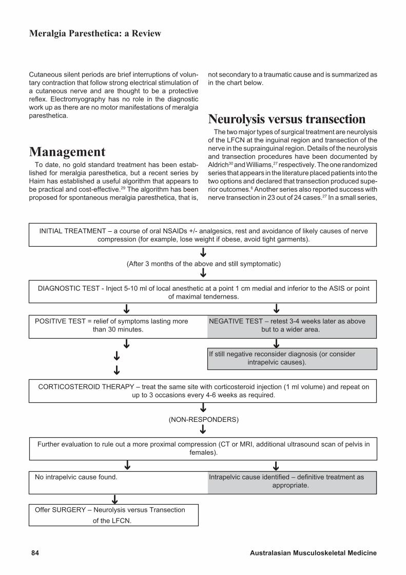
84 Australasian Musculoskeletal Medicine
Cutaneous silent periods are brief interruptions of volun-tary contraction that follow strong electrical stimulation ofa cutaneous nerve and are thought to be a protectivereflex. Electromyography has no role in the diagnosticwork up as there are no motor manifestations of meralgiaparesthetica.
ManagementTo date, no gold standard treatment has been estab-
lished for meralgia paresthetica, but a recent series byHaim has established a useful algorithm that appears tobe practical and cost-effective.29 The algorithm has beenproposed for spontaneous meralgia paresthetica, that is,
INITIAL TREATMENT – a course of oral NSAIDs +/- analgesics, rest and avoidance of likely causes of nervecompression (for example, lose weight if obese, avoid tight garments).
(After 3 months of the above and still symptomatic)
DIAGNOSTIC TEST - Inject 5-10 ml of local anesthetic at a point 1 cm medial and inferior to the ASIS or pointof maximal tenderness.
POSITIVE TEST = relief of symptoms lasting more NEGATIVE TEST – retest 3-4 weeks later as abovethan 30 minutes. but to a wider area.
If still negative reconsider diagnosis (or considerintrapelvic causes).
CORTICOSTEROID THERAPY – treat the same site with corticosteroid injection (1 ml volume) and repeat onup to 3 occasions every 4-6 weeks as required.
(NON-RESPONDERS)
Further evaluation to rule out a more proximal compression (CT or MRI, additional ultrasound scan of pelvis infemales).
No intrapelvic cause found. Intrapelvic cause identified – definitive treatment asappropriate.
Offer SURGERY – Neurolysis versus Transectionof the LFCN.
not secondary to a traumatic cause and is summarized asin the chart below.
Neurolysis versus transectionThe two major types of surgical treatment are neurolysis
of the LFCN at the inguinal region and transection of thenerve in the suprainguinal region. Details of the neurolysisand transection procedures have been documented byAldrich30 and Williams,27 respectively. The one randomizedseries that appears in the literature placed patients into thetwo options and declared that transection produced supe-rior outcomes.6 Another series also reported success withnerve transection in 23 out of 24 cases.27 In a small series,
Meralgia Paresthetica: a Review
November 2008 85
Ivins reported a high failure rate with neurolysis and alsofavored transaction.31 However, other operators havefound that neurolysis has a high rate of success,4,14 andarguments against transection include the potential de-velopment of painful neuromas32 and the resulting sen-sory loss, although it is a relatively unimportant territory. Ithas been suggested that some of the anatomical varia-tions of the LFCN render neurolysis as technically difficultand less likely to succeed.27 In addition, they felt that thepresence of a neurinoma results in a higher failure ratewith neurolysis and suggested this was an analogouslesion to Morton’s neurinoma. In the presence of either ofthese findings, transection may be the procedure of choice.Ivins recommended that neurolysis should be offered toall pediatric patients and patients with less than one yearof symptoms.31 He advised that transection should beperformed after failed neurolysis and as the procedure offirst choice in patients who have been symptomatic formore than one year. One consensus is that alleviation ofsymptoms from a local anesthetic block in the inguinalregion is essential before proceeding to surgical treat-ment. The choice between the two options seems to bedependent largely on the operator’s preference and famili-arity of a particular procedure as well as the patient’swishes with regards to preservation of sensory function. Inthe future more successful outcomes from neurolysis mayalso be facilitated with the assistance of intraoperativesomatosensory evoked potentials. A recent case demon-strated that during a LFCN release an intraoperativeincrease in amplitude of somatosensory evoked potentialsin the nerve correlated with a favorable clinical outcome.33
Further investigation in this area would be useful.
Other treatmentsOther treatments that have been described in the litera-
ture but their efficacy and role in management is yet to beestablished include anticonvulsant and nerve-stabilizingdrugs such as carbamazepine, gabapentin, and diphenyl-hydantoin.5
Abdominal toning exercises could theoretically reducepressure over the nerve at the inguinal region, but againthis is of unproven value.
One case report in the literature reported the successfultreatment of meralgia paresthetica with topical capsaicin0.025% cream applied to the area of skin supplied by theLFCN.34 The proposed mechanism is desensitization ofC-polymodal nociceptors and depletion of substance P inthe nerve. There was no mention of an endpoint in thistreatment, but it may be a useful non-invasive adjunctivetherapy. Again, further investigation in this area would beuseful.
Alcohol rubs, diathermy, and ultrasound have also ap-peared as therapies in the older literature, but their effec-tiveness was not described.15
With regards to meralgia paresthetica of spinal origin,high rates of a success were obtained with 1-4 epiduralinjections of dexamethasone.24
The author developed bilateral meralgia parestheticawhile training as an endurance athlete and after threeyears of symptoms had a failed LFCN neurolysis surgery.The symptoms were eventually brought under control witha yoga-based myofascial stretching program involving thelower back, pelvic, and thigh musculature. To my knowl-edge this type of therapy has not been described previ-ously in the literature.
References1. Bernhardt M. Uber isolirt im Gebiete des Nervus cutaneousfemoris externus vorkommende Parasthesien. NeurologischesCentralblatt 1895;14: 242-44.
2. Roth VK. Meralgia Paraesthetica. Meditisinskoye ObozrainieMoskova 1895; 42: 678.
3. Freud S. Ueber die Bernhardt’sche Sensibilitätsstörung amOberschenkel. Neurologisches Centralblatt 1895;14: 491-92.
4. Edelson R, Stevens P. Meralgia Paresthetica in Children. JBone and Joint Surg 1994;76A(7): 993-99.
5. Dureja GP et al. Management of meralgia paresthetica: amultimodality regimen. Anesthes and Analges 1995;80(5): 1060-61.
6. van Eeten PV, Polder TW, Broere CAJ. Operative treatmentof meralgia paresthetica: transection versus neurolysis.Neurosurg 1995; 37(5): 63-65.
7. Goldstein H. Meralgia paresthetica (Roth’s or Bernhardt’sdisease): with the report of five cases: three cases occurring inthe same family. Am J Med Sci 1921;162(5): 720-35.
8. van Slobbe AM et al. Incidence rates and determinants inmeralgia paresthetica in general practice. J Neurol 2004;251(3):294-97.
9. Reichert FL. Meralgia paresthetica: a form of causalgiarelieved by interruption of the sympathetic fibers. Surg Clinics ofNth Am 1933;13: 1443.
10. Aszmann OC, Dellon ES, Dellon AL. Anatomical Course ofthe Lateral Femoral Cutaneous Nerve and Its Susceptibility toCompression and Injury. Plastic and Reconstructive Surg 1997;100(3): 600-604.
11. Grossman MG. Meralgia paresthetica: diagnosis and treat-ment. J Am Academy of Orthop Surg 2001;9(5): 336-44.
12. Moore KL. Clinically Oriented Anatomy. 3rd ed. Williams andWilkins, 1992.
13. Wilkins RH, Rengachary SS, eds. Neurosurgery. Vol 2.
Meralgia Paresthetica: a Review
86 Australasian Musculoskeletal Medicine
McGraw-Hill, 1985.
14. Nahabedian MY, Dellow A L. Meralgia Paresthetica: etiology,diagnosis, and outcome of surgical decompression. Annals ofPlastic Surg 1995;35(6): 590-94.
15. Kitchen C, Simpson J. Meralgia paresthetica: a review of 67patients. Acta Neurologica Scandinavica 1972;48(5): 547-55.
16. Baldini M, Raimondi PL, Princi L. Meralgia parestheticafollowing weight loss. Case report. Neurosurg Rev 1982;5(2):45-47.
17. Butler R, Webster MI. Meralgia paresthetica: an unusualcomplication of cardiac catheterisation via the femoral artery.Catheterisation and Cardiovascular Interventions 2002;56(1):69-71.
18. Goel A. Meralgia paresthetica secondary to limb lengthdiscrepancy: case report. Archives of Phys Med and Rehab1999;80(3): 348-49.
19. Yang SH, Wu CC, Chen PQ. Postoperative meralgiaparesthetica after posterior spinal surgery: incidence, risk fac-tors, and clinical outcomes. Spine 2005;30(18): E47-50.
20. Paul F, Zipp F. Bilateral meralgia paresthetica after cesariansection with epidural analgesia. J Peripheral Nervous System2006;11(1): 98-99.
21. Boulware DR. Backpacking-induced paresthesias. Wilder-ness and Environmental Med 2003; 14(3): 161-66.
22. Jones RK. Meralgia paresthetica as a cause of leg discom-fort. Can Med Assoc J 1974; 111(6): 541-42.
23. Edelson JG, Nathan H. Meralgia paresthetica: and anatomi-cal interpretation. Clin Orthopaed and Related Research1977;122: 255-62.
24. Jiang G-X, Xu W-D. Meralgia paresthetica of spinal origin:brief report. J Bone and Joint Surg 1998;70-B(5): 843-44.
25. Amoridis G et al. Malignant tumour of the psoas: anothercause of meralgia paresthetica. Electromyog and ClinNeurophysiol 1993;33(2): 109-12.
26. Traycoff RB. Pseudotrochanteric bursitis: the differentialdiagnosis of lateral hip pain. J Rheumatol 1991;18(12): 1810-12.
27. Williams HW, Trzil KP. Management of meralgia paresthetica.J Neurosurg 1991;74(1): 76-80.
28. Tataroglu C et al. Cutaneous silent periods of the vastusmedialis evoked by the stimulation of lateral femoral cutaneousnerve. Clin Neurophysiol 2005;116(6): 1335-41.
29. Haim A et al. Meralgia paresthetica: a retrospective analysisof 79 patients evaluated and treated according to a standardalgorithm. Acta Orthopaedica 2006;77(3): 482-86.
30. Aldrich EF, van den Heever CM. Suprainguinal ligamentapproach for surgical treatment of meralgia paresthetica: techni-cal note. J Neurosurg 1989;70(3): 492-94.
31. Ivins GK. Meralgia paresthetica, the elusive diagnosis:clinical experience with 14 adult patients. Annals of Surg2000;232(2): 281-86.
32. Stevens H. Meralgia paresthetica. Archives of Neurol andPsych 1957;77(6): 557-74.
33. Salengros JC et al. Intraoperative somatosensory evokedpotentials to facilitate peripheral nerve release. Can J Anaesthes2006;53(1): 40-45.
34. Puig L, Alegre M, de Moragas JM. Treatment of meralgiaparesthetica with topical capsaicin. Dermatol 1995;191(1): 73-74.
Meralgia Paresthetica: a Review
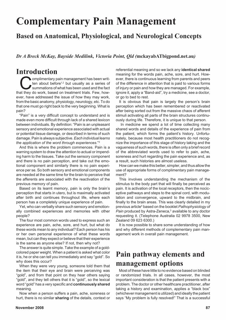
November 2008 87
Introduction
Complimentary pain management has been writ-ten about before1,2 but usually as a series ofsummations of what has been used and the fact
that they do work, based on treatment trials. Few, how-ever, have addressed the issue of how they may work,from the basic anatomy, physiology, neurology, etc. To dothat one must go right back to the very beginning. What ispain?
“Pain” is a very difficult concept to understand and ismade even more difficult through lack of a shared lexiconbetween individuals. By definition: “Pain is an unpleasantsensory and emotional experience associated with actualor potential tissue damage, or described in terms of suchdamage. Pain is always subjective. Each individual learnsthe application of the word through experiences.”3
And this is where the problem commences. Pain is awarning system to draw the attention to actual or impend-ing harm to the tissues. Take out the sensory componentand there is no pain perception, and take out the emo-tional component and similarly there is no pain experi-ence per se. So both sensory and emotional componentsare needed at the same time for the brain to perceive thatthe afferents are associated with the reactivation of theprevious memory of pain.
Based on its learnt memory, pain is only the brain’sperception that starts in utero, but is maximally activatedafter birth and continues throughout life, where eachperson has a completely unique experience of pain.
Yet, who can verbally share such sensory and emotion-ally combined experiences and memories with otherpeople?
The four most common words used to express such anexperience are pain, ache, sore, and hurt, but what dothese words mean to any individual? Each person has hisor her own personal experience of what these wordsmean, but can they expect or believe that their experienceis the same as anyone else? If not, then why not?
The answer is quite simple. Take the example of a gold-colored paper weight. When a patient is asked what colorit is, he or she can tell you immediately and say “gold”. Sowhy does this occur?
When they were very young, someone told them thatthe item that their eye and brain were perceiving was“gold”, and from that point on they hear others saying“gold”, and they tell others that it is “gold”, so the lexicalword “gold” has a very specific and continuously sharedmeaning.
Now when a person suffers a pain, ache, soreness orhurt, there is no similar sharing of the details, context or
referential meaning and so we lack any identical sharedmeaning for the words pain, ache, sore, and hurt. How-ever, there is continuous learning from parents and peersof the difference in attention that is paid to various formsof injury or pain and how they are managed. For example,ignore it, apply a “Band-aid”, try a medicine, see a doctor,or go to bed to rest.
It is obvious that pain is largely the person’s brainperception which has been remembered or reactivatedafter being sorted out from the massive chaos of afferentstimuli activating all parts of the brain structures continu-ously during life. Therefore, it is unique to that person.
In medicine we spend a lot of time collecting manyshared words and details of the experience of pain fromthe patient, which forms the patient’s history. Unfortu-nately, because most health practitioners do not recog-nize the importance of this stage of history taking and thevagueness of such words, there is often only a brief recordof the abbreviated words used to refer to pain, ache,soreness and hurt regarding the pain experience and, asa result, such histories are almost useless.
How can we make this better, simpler, and thus allow theuse of appropriate forms of complimentary pain manage-ment?
This involves understanding the mechanism of thestimulus to the body part that will finally be perceived aspain. It is activation of the local receptors, then the nocic-eptive pathways and steps to the spinal cord, with modu-lation and convergence, upward to the midbrain, andfinally to the brain areas. This was clearly detailed in myprevious article4 based on the booklet Pathophysiology ofPain produced by Astra-Zeneca,3 available to any doctorrequesting it. (Telephone Australia 02 9978 3500, NewZealand 09 623 6300.)
It is now possible to share some understanding of howand why different methods of complementary pain man-agement work in overall pain management.
Pain pathway elements andmanagement options
Most of these have little to no evidence based on blindedor randomized trials. In all cases, however, the mostimportant consideration is that the patient presents with aproblem. The doctor or other healthcare practitioner, aftertaking a history and examination, applies a “black box”(whichever management is utilized) and ideally the patientsays “My problem is fully resolved!” That is a successful
Complementary Pain ManagementBased on Anatomical, Physiological, and Neurological Concepts
Dr A Breck McKay, Bayside Medilink, Victoria Point, Qld (mckayabATbigpond.net.au)
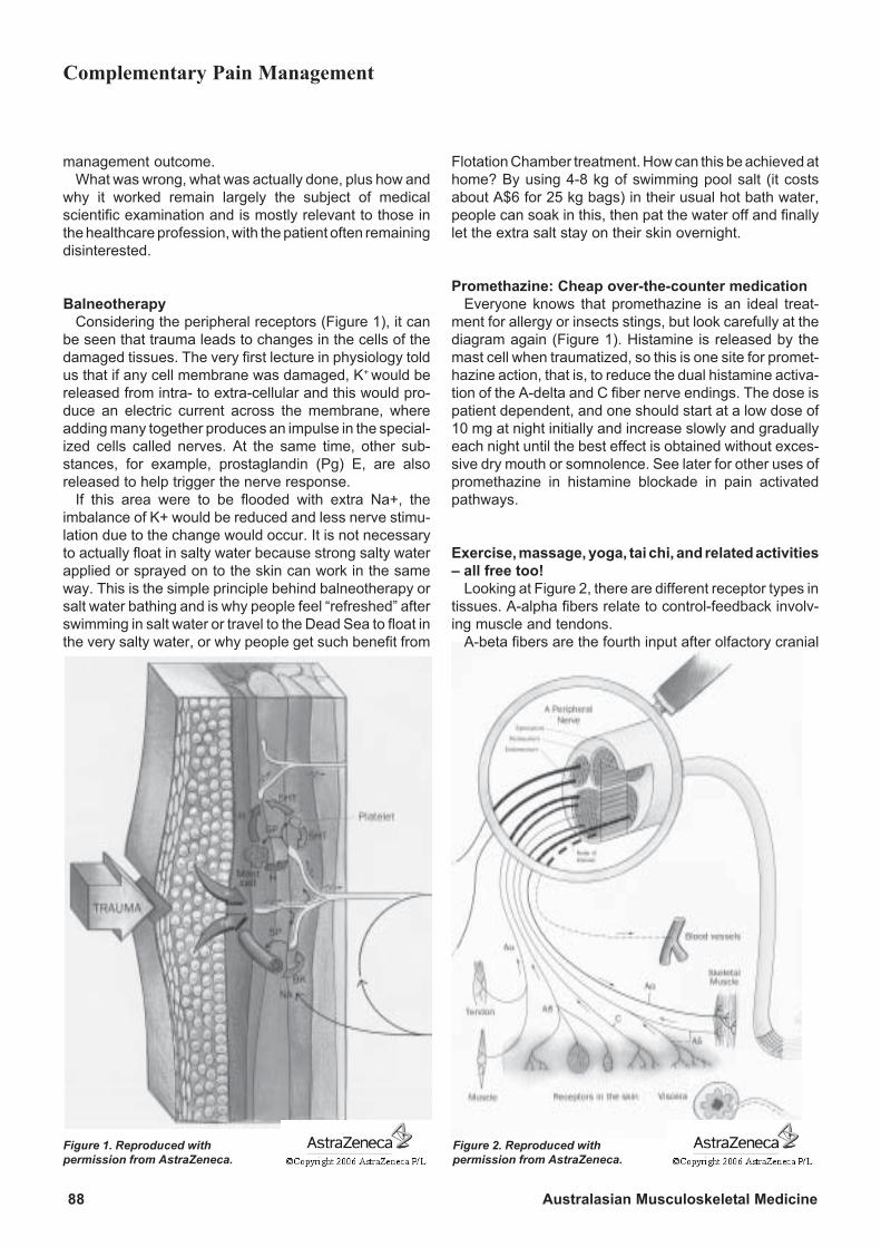
88 Australasian Musculoskeletal Medicine
management outcome.What was wrong, what was actually done, plus how and
why it worked remain largely the subject of medicalscientific examination and is mostly relevant to those inthe healthcare profession, with the patient often remainingdisinterested.
BalneotherapyConsidering the peripheral receptors (Figure 1), it can
be seen that trauma leads to changes in the cells of thedamaged tissues. The very first lecture in physiology toldus that if any cell membrane was damaged, K+ would bereleased from intra- to extra-cellular and this would pro-duce an electric current across the membrane, whereadding many together produces an impulse in the special-ized cells called nerves. At the same time, other sub-stances, for example, prostaglandin (Pg) E, are alsoreleased to help trigger the nerve response.
If this area were to be flooded with extra Na+, theimbalance of K+ would be reduced and less nerve stimu-lation due to the change would occur. It is not necessaryto actually float in salty water because strong salty waterapplied or sprayed on to the skin can work in the sameway. This is the simple principle behind balneotherapy orsalt water bathing and is why people feel “refreshed” afterswimming in salt water or travel to the Dead Sea to float inthe very salty water, or why people get such benefit from
Flotation Chamber treatment. How can this be achieved athome? By using 4-8 kg of swimming pool salt (it costsabout A$6 for 25 kg bags) in their usual hot bath water,people can soak in this, then pat the water off and finallylet the extra salt stay on their skin overnight.
Promethazine: Cheap over-the-counter medicationEveryone knows that promethazine is an ideal treat-
ment for allergy or insects stings, but look carefully at thediagram again (Figure 1). Histamine is released by themast cell when traumatized, so this is one site for promet-hazine action, that is, to reduce the dual histamine activa-tion of the A-delta and C fiber nerve endings. The dose ispatient dependent, and one should start at a low dose of10 mg at night initially and increase slowly and graduallyeach night until the best effect is obtained without exces-sive dry mouth or somnolence. See later for other uses ofpromethazine in histamine blockade in pain activatedpathways.
Exercise, massage, yoga, tai chi, and related activities– all free too!
Looking at Figure 2, there are different receptor types intissues. A-alpha fibers relate to control-feedback involv-ing muscle and tendons.
A-beta fibers are the fourth input after olfactory cranial
Complementary Pain Management
Figure 1. Reproduced withpermission from AstraZeneca.
Figure 2. Reproduced withpermission from AstraZeneca.
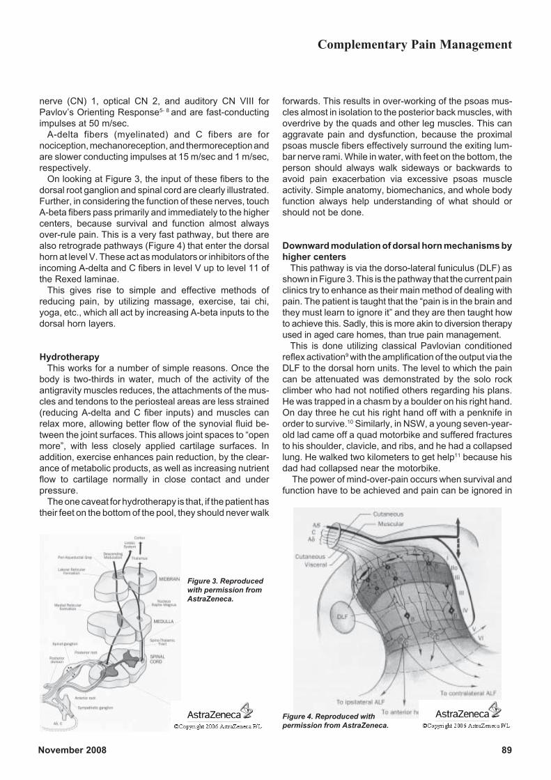
November 2008 89
nerve (CN) 1, optical CN 2, and auditory CN VIII forPavlov’s Orienting Response5- 8 and are fast-conductingimpulses at 50 m/sec.
A-delta fibers (myelinated) and C fibers are fornociception, mechanoreception, and thermoreception andare slower conducting impulses at 15 m/sec and 1 m/sec,respectively.
On looking at Figure 3, the input of these fibers to thedorsal root ganglion and spinal cord are clearly illustrated.Further, in considering the function of these nerves, touchA-beta fibers pass primarily and immediately to the highercenters, because survival and function almost alwaysover-rule pain. This is a very fast pathway, but there arealso retrograde pathways (Figure 4) that enter the dorsalhorn at level V. These act as modulators or inhibitors of theincoming A-delta and C fibers in level V up to level 11 ofthe Rexed laminae.
This gives rise to simple and effective methods ofreducing pain, by utilizing massage, exercise, tai chi,yoga, etc., which all act by increasing A-beta inputs to thedorsal horn layers.
HydrotherapyThis works for a number of simple reasons. Once the
body is two-thirds in water, much of the activity of theantigravity muscles reduces, the attachments of the mus-cles and tendons to the periosteal areas are less strained(reducing A-delta and C fiber inputs) and muscles canrelax more, allowing better flow of the synovial fluid be-tween the joint surfaces. This allows joint spaces to “openmore”, with less closely applied cartilage surfaces. Inaddition, exercise enhances pain reduction, by the clear-ance of metabolic products, as well as increasing nutrientflow to cartilage normally in close contact and underpressure.
The one caveat for hydrotherapy is that, if the patient hastheir feet on the bottom of the pool, they should never walk
forwards. This results in over-working of the psoas mus-cles almost in isolation to the posterior back muscles, withoverdrive by the quads and other leg muscles. This canaggravate pain and dysfunction, because the proximalpsoas muscle fibers effectively surround the exiting lum-bar nerve rami. While in water, with feet on the bottom, theperson should always walk sideways or backwards toavoid pain exacerbation via excessive psoas muscleactivity. Simple anatomy, biomechanics, and whole bodyfunction always help understanding of what should orshould not be done.
Downward modulation of dorsal horn mechanisms byhigher centers
This pathway is via the dorso-lateral funiculus (DLF) asshown in Figure 3. This is the pathway that the current painclinics try to enhance as their main method of dealing withpain. The patient is taught that the “pain is in the brain andthey must learn to ignore it” and they are then taught howto achieve this. Sadly, this is more akin to diversion therapyused in aged care homes, than true pain management.
This is done utilizing classical Pavlovian conditionedreflex activation9 with the amplification of the output via theDLF to the dorsal horn units. The level to which the paincan be attenuated was demonstrated by the solo rockclimber who had not notified others regarding his plans.He was trapped in a chasm by a boulder on his right hand.On day three he cut his right hand off with a penknife inorder to survive.10 Similarly, in NSW, a young seven-year-old lad came off a quad motorbike and suffered fracturesto his shoulder, clavicle, and ribs, and he had a collapsedlung. He walked two kilometers to get help11 because hisdad had collapsed near the motorbike.
The power of mind-over-pain occurs when survival andfunction have to be achieved and pain can be ignored in
Complementary Pain Management
Figure 4. Reproduced withpermission from AstraZeneca.
Figure 3. Reproducedwith permission fromAstraZeneca.
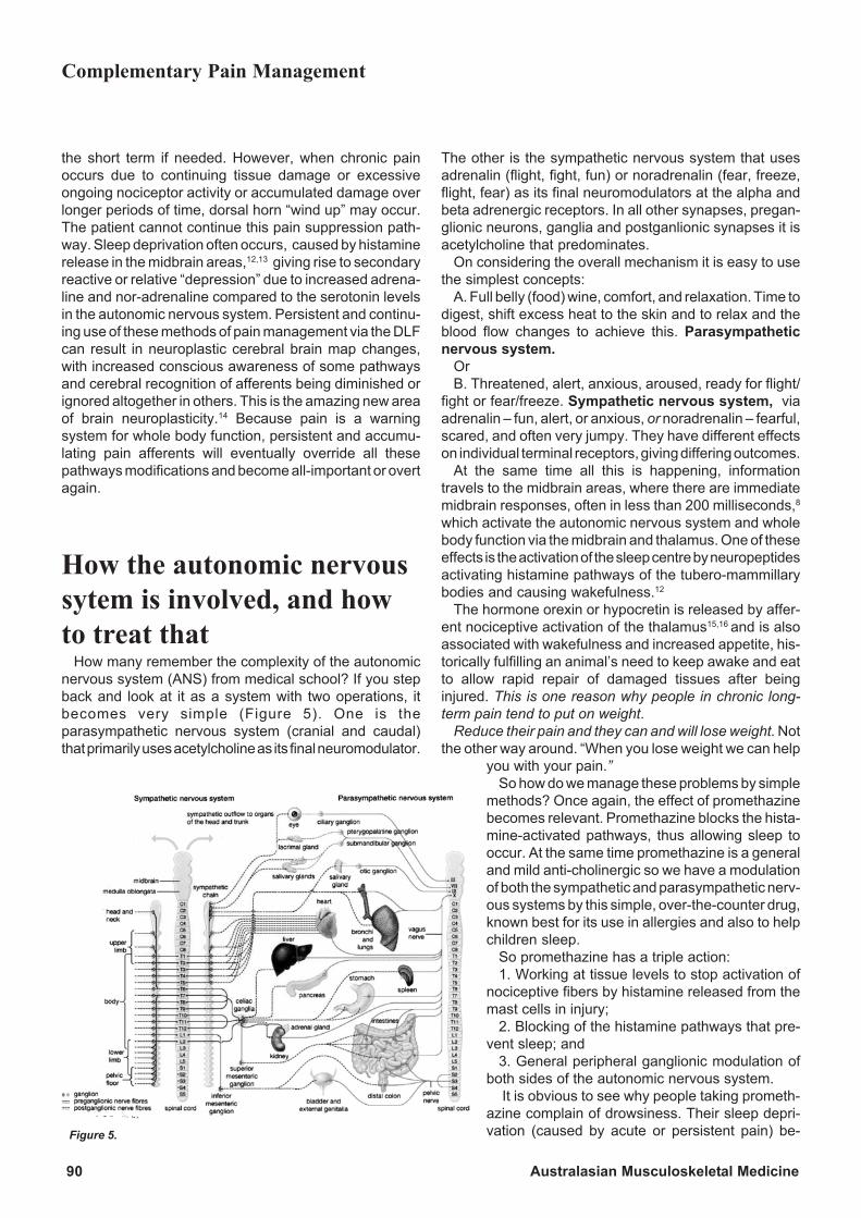
90 Australasian Musculoskeletal Medicine
the short term if needed. However, when chronic painoccurs due to continuing tissue damage or excessiveongoing nociceptor activity or accumulated damage overlonger periods of time, dorsal horn “wind up” may occur.The patient cannot continue this pain suppression path-way. Sleep deprivation often occurs, caused by histaminerelease in the midbrain areas,12,13 giving rise to secondaryreactive or relative “depression” due to increased adrena-line and nor-adrenaline compared to the serotonin levelsin the autonomic nervous system. Persistent and continu-ing use of these methods of pain management via the DLFcan result in neuroplastic cerebral brain map changes,with increased conscious awareness of some pathwaysand cerebral recognition of afferents being diminished orignored altogether in others. This is the amazing new areaof brain neuroplasticity.14 Because pain is a warningsystem for whole body function, persistent and accumu-lating pain afferents will eventually override all thesepathways modifications and become all-important or overtagain.
How the autonomic nervoussytem is involved, and howto treat that
How many remember the complexity of the autonomicnervous system (ANS) from medical school? If you stepback and look at it as a system with two operations, itbecomes very simple (Figure 5). One is theparasympathetic nervous system (cranial and caudal)that primarily uses acetylcholine as its final neuromodulator.
The other is the sympathetic nervous system that usesadrenalin (flight, fight, fun) or noradrenalin (fear, freeze,flight, fear) as its final neuromodulators at the alpha andbeta adrenergic receptors. In all other synapses, pregan-glionic neurons, ganglia and postganlionic synapses it isacetylcholine that predominates.
On considering the overall mechanism it is easy to usethe simplest concepts:
A. Full belly (food) wine, comfort, and relaxation. Time todigest, shift excess heat to the skin and to relax and theblood flow changes to achieve this. Parasympatheticnervous system.
OrB. Threatened, alert, anxious, aroused, ready for flight/
fight or fear/freeze. Sympathetic nervous system, viaadrenalin – fun, alert, or anxious, or noradrenalin – fearful,scared, and often very jumpy. They have different effectson individual terminal receptors, giving differing outcomes.
At the same time all this is happening, informationtravels to the midbrain areas, where there are immediatemidbrain responses, often in less than 200 milliseconds,8
which activate the autonomic nervous system and wholebody function via the midbrain and thalamus. One of theseeffects is the activation of the sleep centre by neuropeptidesactivating histamine pathways of the tubero-mammillarybodies and causing wakefulness.12
The hormone orexin or hypocretin is released by affer-ent nociceptive activation of the thalamus15,16 and is alsoassociated with wakefulness and increased appetite, his-torically fulfilling an animal’s need to keep awake and eatto allow rapid repair of damaged tissues after beinginjured. This is one reason why people in chronic long-term pain tend to put on weight.
Reduce their pain and they can and will lose weight. Notthe other way around. “When you lose weight we can help
you with your pain.”So how do we manage these problems by simple
methods? Once again, the effect of promethazinebecomes relevant. Promethazine blocks the hista-mine-activated pathways, thus allowing sleep tooccur. At the same time promethazine is a generaland mild anti-cholinergic so we have a modulationof both the sympathetic and parasympathetic nerv-ous systems by this simple, over-the-counter drug,known best for its use in allergies and also to helpchildren sleep.
So promethazine has a triple action:1. Working at tissue levels to stop activation of
nociceptive fibers by histamine released from themast cells in injury;
2. Blocking of the histamine pathways that pre-vent sleep; and
3. General peripheral ganglionic modulation ofboth sides of the autonomic nervous system.
It is obvious to see why people taking prometh-azine complain of drowsiness. Their sleep depri-vation (caused by acute or persistent pain) be-
Complementary Pain Management
Figure 5.
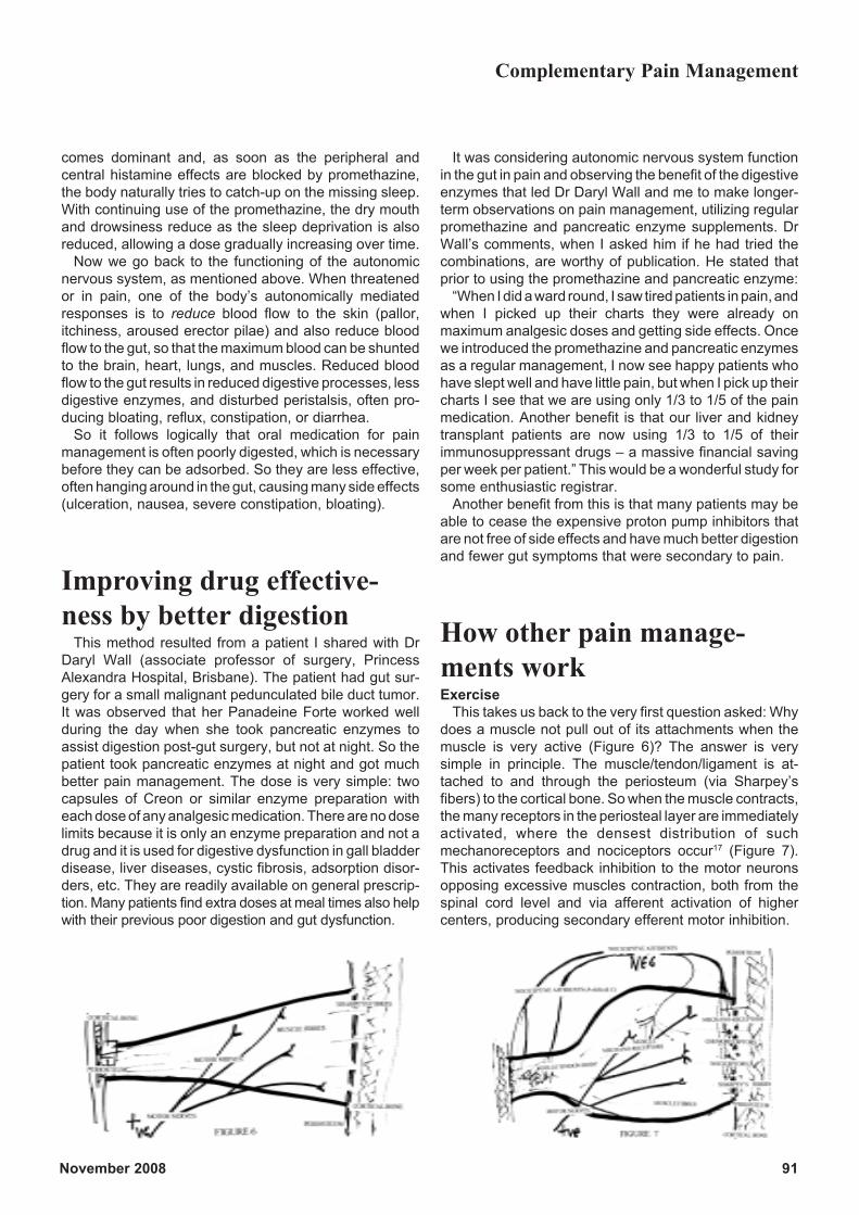
November 2008 91
comes dominant and, as soon as the peripheral andcentral histamine effects are blocked by promethazine,the body naturally tries to catch-up on the missing sleep.With continuing use of the promethazine, the dry mouthand drowsiness reduce as the sleep deprivation is alsoreduced, allowing a dose gradually increasing over time.
Now we go back to the functioning of the autonomicnervous system, as mentioned above. When threatenedor in pain, one of the body’s autonomically mediatedresponses is to reduce blood flow to the skin (pallor,itchiness, aroused erector pilae) and also reduce bloodflow to the gut, so that the maximum blood can be shuntedto the brain, heart, lungs, and muscles. Reduced bloodflow to the gut results in reduced digestive processes, lessdigestive enzymes, and disturbed peristalsis, often pro-ducing bloating, reflux, constipation, or diarrhea.
So it follows logically that oral medication for painmanagement is often poorly digested, which is necessarybefore they can be adsorbed. So they are less effective,often hanging around in the gut, causing many side effects(ulceration, nausea, severe constipation, bloating).
Improving drug effective-ness by better digestion
This method resulted from a patient I shared with DrDaryl Wall (associate professor of surgery, PrincessAlexandra Hospital, Brisbane). The patient had gut sur-gery for a small malignant pedunculated bile duct tumor.It was observed that her Panadeine Forte worked wellduring the day when she took pancreatic enzymes toassist digestion post-gut surgery, but not at night. So thepatient took pancreatic enzymes at night and got muchbetter pain management. The dose is very simple: twocapsules of Creon or similar enzyme preparation witheach dose of any analgesic medication. There are no doselimits because it is only an enzyme preparation and not adrug and it is used for digestive dysfunction in gall bladderdisease, liver diseases, cystic fibrosis, adsorption disor-ders, etc. They are readily available on general prescrip-tion. Many patients find extra doses at meal times also helpwith their previous poor digestion and gut dysfunction.
It was considering autonomic nervous system functionin the gut in pain and observing the benefit of the digestiveenzymes that led Dr Daryl Wall and me to make longer-term observations on pain management, utilizing regularpromethazine and pancreatic enzyme supplements. DrWall’s comments, when I asked him if he had tried thecombinations, are worthy of publication. He stated thatprior to using the promethazine and pancreatic enzyme:
“When I did a ward round, I saw tired patients in pain, andwhen I picked up their charts they were already onmaximum analgesic doses and getting side effects. Oncewe introduced the promethazine and pancreatic enzymesas a regular management, I now see happy patients whohave slept well and have little pain, but when I pick up theircharts I see that we are using only 1/3 to 1/5 of the painmedication. Another benefit is that our liver and kidneytransplant patients are now using 1/3 to 1/5 of theirimmunosuppressant drugs – a massive financial savingper week per patient.” This would be a wonderful study forsome enthusiastic registrar.
Another benefit from this is that many patients may beable to cease the expensive proton pump inhibitors thatare not free of side effects and have much better digestionand fewer gut symptoms that were secondary to pain.
How other pain manage-ments workExercise
This takes us back to the very first question asked: Whydoes a muscle not pull out of its attachments when themuscle is very active (Figure 6)? The answer is verysimple in principle. The muscle/tendon/ligament is at-tached to and through the periosteum (via Sharpey’sfibers) to the cortical bone. So when the muscle contracts,the many receptors in the periosteal layer are immediatelyactivated, where the densest distribution of suchmechanoreceptors and nociceptors occur17 (Figure 7).This activates feedback inhibition to the motor neuronsopposing excessive muscles contraction, both from thespinal cord level and via afferent activation of highercenters, producing secondary efferent motor inhibition.
Complementary Pain Management
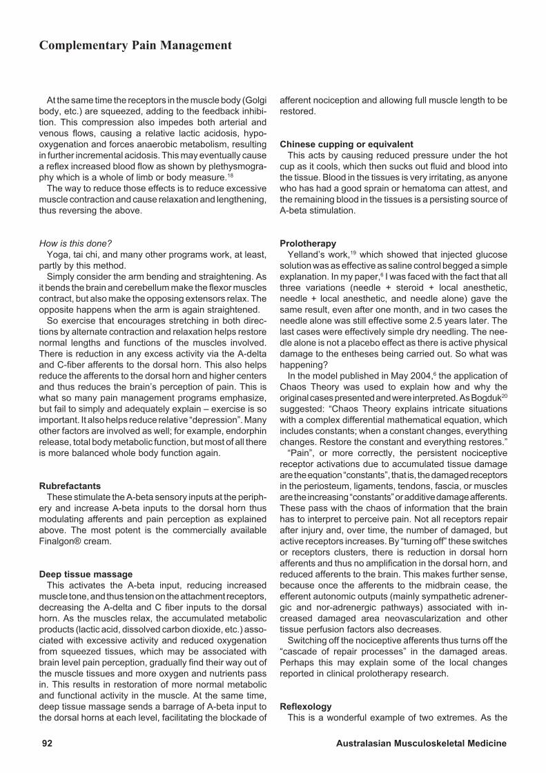
92 Australasian Musculoskeletal Medicine
At the same time the receptors in the muscle body (Golgibody, etc.) are squeezed, adding to the feedback inhibi-tion. This compression also impedes both arterial andvenous flows, causing a relative lactic acidosis, hypo-oxygenation and forces anaerobic metabolism, resultingin further incremental acidosis. This may eventually causea reflex increased blood flow as shown by plethysmogra-phy which is a whole of limb or body measure.18
The way to reduce those effects is to reduce excessivemuscle contraction and cause relaxation and lengthening,thus reversing the above.
How is this done?Yoga, tai chi, and many other programs work, at least,
partly by this method.Simply consider the arm bending and straightening. As
it bends the brain and cerebellum make the flexor musclescontract, but also make the opposing extensors relax. Theopposite happens when the arm is again straightened.
So exercise that encourages stretching in both direc-tions by alternate contraction and relaxation helps restorenormal lengths and functions of the muscles involved.There is reduction in any excess activity via the A-deltaand C-fiber afferents to the dorsal horn. This also helpsreduce the afferents to the dorsal horn and higher centersand thus reduces the brain’s perception of pain. This iswhat so many pain management programs emphasize,but fail to simply and adequately explain – exercise is soimportant. It also helps reduce relative “depression”. Manyother factors are involved as well; for example, endorphinrelease, total body metabolic function, but most of all thereis more balanced whole body function again.
RubrefactantsThese stimulate the A-beta sensory inputs at the periph-
ery and increase A-beta inputs to the dorsal horn thusmodulating afferents and pain perception as explainedabove. The most potent is the commercially availableFinalgon® cream.
Deep tissue massageThis activates the A-beta input, reducing increased
muscle tone, and thus tension on the attachment receptors,decreasing the A-delta and C fiber inputs to the dorsalhorn. As the muscles relax, the accumulated metabolicproducts (lactic acid, dissolved carbon dioxide, etc.) asso-ciated with excessive activity and reduced oxygenationfrom squeezed tissues, which may be associated withbrain level pain perception, gradually find their way out ofthe muscle tissues and more oxygen and nutrients passin. This results in restoration of more normal metabolicand functional activity in the muscle. At the same time,deep tissue massage sends a barrage of A-beta input tothe dorsal horns at each level, facilitating the blockade of
afferent nociception and allowing full muscle length to berestored.
Chinese cupping or equivalentThis acts by causing reduced pressure under the hot
cup as it cools, which then sucks out fluid and blood intothe tissue. Blood in the tissues is very irritating, as anyonewho has had a good sprain or hematoma can attest, andthe remaining blood in the tissues is a persisting source ofA-beta stimulation.
ProlotherapyYelland’s work,19 which showed that injected glucose
solution was as effective as saline control begged a simpleexplanation. In my paper,6 I was faced with the fact that allthree variations (needle + steroid + local anesthetic,needle + local anesthetic, and needle alone) gave thesame result, even after one month, and in two cases theneedle alone was still effective some 2.5 years later. Thelast cases were effectively simple dry needling. The nee-dle alone is not a placebo effect as there is active physicaldamage to the entheses being carried out. So what washappening?
In the model published in May 2004,6 the application ofChaos Theory was used to explain how and why theoriginal cases presented and were interpreted. As Bogduk20
suggested: “Chaos Theory explains intricate situationswith a complex differential mathematical equation, whichincludes constants; when a constant changes, everythingchanges. Restore the constant and everything restores.”
“Pain”, or more correctly, the persistent nociceptivereceptor activations due to accumulated tissue damageare the equation “constants”, that is, the damaged receptorsin the periosteum, ligaments, tendons, fascia, or musclesare the increasing “constants” or additive damage afferents.These pass with the chaos of information that the brainhas to interpret to perceive pain. Not all receptors repairafter injury and, over time, the number of damaged, butactive receptors increases. By “turning off” these switchesor receptors clusters, there is reduction in dorsal hornafferents and thus no amplification in the dorsal horn, andreduced afferents to the brain. This makes further sense,because once the afferents to the midbrain cease, theefferent autonomic outputs (mainly sympathetic adrener-gic and nor-adrenergic pathways) associated with in-creased damaged area neovascularization and othertissue perfusion factors also decreases.
Switching off the nociceptive afferents thus turns off the“cascade of repair processes” in the damaged areas.Perhaps this may explain some of the local changesreported in clinical prolotherapy research.
ReflexologyThis is a wonderful example of two extremes. As the
Complementary Pain Management
November 2008 93
Chinese found many thousands of years ago, if youmassage the feet very gently, you increase relaxation.How? Simply by increasing the A-beta inputs to the lowerdorsal horns, thus modulating nociception all the way upthe spinal levels up to the midbrain. After a while every-thing settles and is calm.
The opposite was also used by the Chinese. Caning ofthe feet slowly and continuously was used to torture andkill people. How did this work? By increasing the A-deltaand C fiber inputs from receptors (massive nociceptiveinputs), the opposite was achieved; death from extremeoverload to the autonomic nervous system at midbrainlevels. This is one of the natural killers for prey animals inthe wild, with pressure on the throat area resulting inautonomic overload after the chase and capture, anddeath follows much more quickly than by simple strangu-lation. This rapid death occurs in captured birds, even ifquickly released after capture by a predator.
TENS machinesThese work by fatiguing the sensory and nociceptive
nerves by excessive stimulation at the receptor and pri-mary afferent level. Once a receptor has been forced tofire many times, it runs out of neurotransmitters and thenerve no longer feeds to the dorsal horn. This lasts onlyuntil the TENS is turned off and the receptors and nerveshave time to metabolically recover their previous func-tions. The dorsal horn also changes its activity, as de-scribed by Melzack and Wall.21
The Likon machineThis is a very interesting machine that came from China
and was initially related to acupuncture methods. It hasmultiple settings that, when managed correctly, actuallymodulates via low wavelength electrotherapy to the auto-nomic nervous system in two different ways, modulatingthe sympathetic or parasympathetic systems. The actualsettings required, and how to use the machine in themanagement of muscle problems and specifically in com-plex regional pain syndromes, were deduced by simpletrial and error over some two years by Adelaide physi-otherapist Aileen Jefferis.
My wife and I have extensively used this machine withgreat benefit since 1990 and its use has allowed ourdevelopment of the whole body functional models fromclinical observations.
It cannot be used without careful training by those whohave experience in correct Likon use, as it is very easy toincorrectly modulate the wrong part of the autonomicnervous system, resulting in rapid exacerbation of symp-toms and signs.
Cold and heatConsidering the physiology, these are easy to under-
stand. Heat increases the blood flow, thus delivering moresubstrate and removing metabolic products as well asassisting biochemical reactions. Increased blood flowmay also cause more swelling when inflammation ispresent, which may be counterproductive. The nerves canactually work more efficiently and thus, at times, can makethe pain appear worse, due to improved nerve conductionrates and increased afferents to the dorsal horn.
Cold, on the other hand, reduces the blood flow and alsodecreases the function, physiology, and biochemistry ofthe muscles and nerves and reduces secondary swellingin the area, as well as decreasing afferent nociceptiveactivity to the dorsal horn, thus reducing pain perception.
HypnosisThis involves the ability of the brain to be placed in a
sleep-like dissociative state where the pain afferents canbe “ignored” both by increasing efferent DLF activity aswell as cerebral activity to override the areas associatedwith pain perception.22
MeditationThis works like hypnosis, but in this case the brain is
taught to ignore the nociceptive afferents by activatingother more dominant pathways. fMRI studies have shownthat many areas of the brain are involved in such activityand brain neuro-plasticity is the key to long term benefit ofsuch meditation.14,22
AromatherapyThis is a most fascinating area of pain management that
is only just starting to be understood. Olfactory inputs passdirectly to the midbrain and form an important part ofPavlov’s orienting response, as the result is frequentlysub-200 milliseconds in onset.8 From birth onwards, vari-ous aromas are related to different environments and arelearnt as an association memory.
Later, the presence of that same smell can help recreatethe memory and may be calming, alerting, or even repul-sive, but the response depends on the original environ-mental association. This is an underlying feature of Pav-lov’s original conditioned reflex development and theability to moderate this by environmental changes waswell demonstrated in Stanley Kubrick’s film “A ClockworkOrange”.
Stationary magnetsDespite all the claims for these, there are no blinded,
randomized controlled trials that show any positive ben-efit, other than the extra warmth and support from themagnet carriers.
Complementary Pain Management
94 Australasian Musculoskeletal Medicine
The power of mind over matterAs indicated above, there are many ways in which the
brain and cerebral interpretation can be fooled into ignor-ing or downplaying the importance of the afferent nocicep-tive information. However, if the incoming pain informationcontinues to increase or accumulate, it will eventuallyoverride and redirect the brain and body’s attention to thecause as a method of protection from actual or possibleinjury.
How powerful is this process? Think of the boxer gettinghis face and nose pummelled, or the rugby player beingheavily tackled, or the person in a fight to the death withanything. It is then that survival and function will overrulepain.5 However, eventually, pain perception will dominatebecause the afferents from the dorsal horns will causeoverload in the midbrain autonomic control area. Eitheraction is taken to address the pain and reduce it, or thewhole body function will result in loss of consciousness.5,7
The whole body response is summarized as MOMM.Monitor (M) the environment;Orient (O) to the changes;Memory (M) check for past experience of the same; andManage (M) by:a) Freeze/Fear initially while the situation is considered.
Remember it is the excessive Freeze/Fear responsethat may result in death via autonomic overload.
b) Flight/Fightc) Return to normal.
These are the steps that Pavlov first identified in 1904 to1917,9 and now that we are starting to understand brainneuroplasticity better,14 newer and better pain manage-ment programs may be developed.
As I have tried to show here and in previous articles, inmusculoskeletal medicine we do need to go back to thebasic principles of anatomical and physiological normalityto understand what is going on. We must ensure that notonly do we make the right diagnosis, but that our manage-ments are also aimed at restoration of normal, rather thanmerely treating symptoms and signs after “labeling” of theproblem instead of true diagnosis.23
References1. Ernst E. Complimentary therapies for pain management.Elsevier Mosby, 2007.
2. Menefee LA, Monti DA. Nonpharmacological and complimen-tary approaches to cancer pain management. JAOA 2005; 105:15-20.
3. The Pathophysiology of Pain. Astra-Zeneca PharmaceuticalEducation Booklet. J Pain and Symptom Management 2000;19: 3.
4. McKay AB. Chronic low back pain and pain: simple models toexplain pain to patients. Australas Musculosketal Med 2006; 11:92-96.
5. McKay AB, Wall D. The orienting response and the functional
whole human body. Australas Musculoskeletal Med 2003;8:86-99.
6. McKay AB. Pain and chronic low back pain: a new model?Parts 1and 2. Australas Musculoskeletal Med 2004; 9:14-25.
7. McKay AB. Tennis Elbow Everywhere. Australas Muscu-loskeletal Med 2005; 10: 127-30.
8. McKay AB. Watch the Ball- but why? Australas Musculoskel-etal Med 2008; 12: 25-27.
9. Pavlov I. Lectures on conditioned reflexes. Vol 1. New YorkInternational Publishers, 1928.
10. Ralston A. I cut off my right hand. www.msnbc.msn.com/id/5956900/.
11. Master Tyler Stephen Moon: Bravery Award, 2004.www.governorgeneral.gov.au/res/File/PDFs/honours/br65/Media%20Notes%20-%20Commendat ion%20for%20Brave%20Conduct.pdf.
12. Hou RH, Langley RW, Szabadi E et al. Comparison ofdiphenhydramine and modafinil on arousal and autonomic func-tions in healthy volunteers. J Psychopharmacol 2007; 21: 567-78.
13. Parmenter R, Anaclet C, Guhennec C et al. The brain H3-receptor as a novel therapeutic target for vigilance and sleep-wake disorders. Biochem Pharmacol 2007; 73: 1157-71.
14. Doidge N. The brain that changes itself. Penguin, 2007.
15. Lin JS, Dauvilliers Y, Arnulf I et al. An inverse agonist of thehistamine H(3) receptor improves wakefulness in narcolepsy:studies in orexin-l-mice and patients. Neurobiol Dis 2008: 30: 74-83.
16. Cortess S, Konofal E, Lecendreux M. Alertness and feedingbehaviours in ADHA: Does hypocretin/orexin system play a role?Med Hypothese 2008.
17. Bogduk N. Mechanisms of musculoskeletal pain. AustralasMusculoskeletal Med 2006; 11: 6-18.
18. Yang D. Reproducibility of air plethysmographic measure-ments for the evaluation of blood flow in the lower extremity.Australian Conference of Science and Medicine in Sport 2001; ASports medicine Odyssey – Challenges Controversies and Change.
19. Yelland MJ, Glasziou PP, Bogduk N et al. Prolotherapyinjections, saline injections and exercises for chronic low backpain: a randomized trial. Spine 2004; 29: 9-16.
20. Bogduk N, McKay AB. Email personal communication.
21. Melzack R, Wall PD. Pain mechanisms; a new theory.Science 1965; 150: 971-79.
22. Grendahl JR, Rosvold, EI. Hypnosis as a treatment of chronicwidespread pain in general practice: a randomized controlledpilot study. 2008. www.medscape.com/viewarticle/581316 .
23. McKay AB. Musculoskeletal Pot Pourri. Australas Muscu-loskeletal Med 2006; 10: 108-110.
Complementary Pain Management
November 2008 95
History
Paraspinal nerve injection is an economical andminimally invasive procedure that can be used totreat acute spinal pain, or chronic spinal pain that
may be associated with radiculopathy. Radiculopathy canbe defined in the broadest sense as pain and dysfunctionmediated by the radicles or nerve roots, the final commonpathway for nociception into the central nervous system(CNS).
The original work using paraspinal nerve injections wasa randomized controlled trial (RCT) by Dr Stefan Blombergas part of his PhD thesis in the early 1990s. Stefan runs apain clinic in Stockholm, Sweden.1-5
Stefan Blomberg came to Melbourne in October 2002as keynote speaker for the annual scientific meeting of theAustralian Association of Musculoskeletal Medicine(AAMM). His pragmatic approach is systematic, using hisown algorithm.
Because of limited sensitivity and specificity of history,examination, and imaging for diagnosing and managingmusculoskeletal pain problems, Stefan believes it is hu-mane and cost-effective to offer patients a pragmatic trialof treatment when there is a chance of “cure” or significantreduction in their pain and associated symptoms.
Musculoskeletal pain is a concept, not a well-definedentity.6 It is the brain’s unique interpretation, based on pastlearning, of chaotic afferent input to the CNS.
The history is the best guide to diagnosing musculoskel-etal pain problems.7
It is vital to take a thorough history. The aim is to triagethe patient with respect to red flag conditions (such asfracture, tumours benign or malignant, infection, or inflam-mation such as ankylosing spondylitis and other seron-egative spondylarthropathies), radicular pain andradiculopathy, and somatic pain which may be associatedwith referred pain through convergence.7
The red flag checklist has been validated for acute andchronic lumbar spinal pain, and is applicable to otherareas of the body.7
Physical examination can support historical impres-sions and help engage the patient but lacks diagnosticprecision without proven reliability and validity.7 It canreveal tender entheses which may be associated withchronic spinal pain.
Imaging does not show pain per se, and in the absenceof red flag indicators usually shows non-diagnostic age-related changes that are no more common in people with
pain than those without pain.7- 9
This evidence can free the practitioner from the need toimage patients repeatedly with XRs, CTs, or MRIs when-ever they have an exacerbation of their musculoskeletalpain. In the absence of red flag indicators, the yield ofrelevant information is very low and should not changemanagement. This can save patients needless exposureto ionizing radiation in the case of XR and CT, andexpense for the patient, the insurer, and the tax payer,depending on funding arrangements.
History is the best guide to diagnosis but is not com-pletely reliable. Somatic referred pain and radicular painwith or without radiculopathy can co-exist.10
Neurogenic pain is pain evoked by the stimulation ofperipheral axons or their cell bodies rather than peripheralnerve endings as occurs with somatic pain.7
Radicular pain is a subset of neurogenic pain, in whichpain is evoked by stimulation of the nerve roots, dorsal rootganglion (DRG), or spinal nerve itself.11, 12
In neurogenic pain the pain is perceived as arising in theperipheral area supplied by the affected nerve. As the painis perceived in a region distal to the actual pain stimulus,neurogenic pain and radicular pain are, by definition, aform of referred pain.7
Lumbar radicular pain (LRP) could be referred to anarea innervated by either the dorsal or the ventral rami, butpain in the distribution of the dorsal rami has not beenproperly defined to date. LRP can also result from irritationof the ventral root or the spinal nerve itself.11-14
LRP differs, however, from somatic and visceral re-ferred pain because it does not involve the stimulation ofnerve endings, and convergence onto the same secondorder neuron at the same segmental level. It is perceivedas arising distally because of ectopic activation of thenerves supplying that region by irritation of the nerveroots, DRG or spinal nerve proximally. This manifests asreferred radicular pain, that is, shooting, stabbing, lanci-nating, electric shock like pain, with or without dysesthesiato that region.7 This is in contrast to somatic referred pain,which is constant in location but poorly localized anddiffuse, and aching in quality.11, 12
Burning pain is often a feature of neurogenic pain. Deepburning pain without other features, distribution, or quali-ties is not necessarily neurogenic in origin. Burning sen-sations in the skin strongly imply a neurogenic processthat may be radicular, or some other neurogenic process.7
The sensory dorsal nerve roots receive fibers from boththe ventral and the dorsal rami that combine briefly to form
Paraspinal Nerve Injection:Its use as a presurgical diagnostic disambiguation tool and for thetemporary and permanent relief of musculoskeletal pain
Dr David Roselt, Aberdovy Clinic, Bundaberg; Dr A Breck McKay, Bayside Medilink; Dr ScottMasters, Caloundra Spinal and Sports Medicine Centre
96 Australasian Musculoskeletal Medicine
the spinal nerve just distal to the intervertebral foramen.The sinuvertebral nerves (SVNs) are recurrent branchesof the ventral rami that re-enter the intervertebral foraminato be distributed within the vertebral canal, formed by asomatic root from a ventral ramus and an autonomic rootfrom a grey ramus communicans. In the intervertebralforamina, the lumbar SVNs run across the back of thevertebral body just below the upper pedicle supplying theperiosteum and also the outer third to half of the interver-tebral disc, and the posterior longitudinal ligament (PLL) atthat level and also the level above. The SVNs also supplythe blood vessels of the vertebral canal, the ventral aspectof the dura mater, the dural sleeves enclosing the nerveroots, and the spinal nerves as far as the intervertebralforamina, where the dura merges with and becomes theepineurium of the spinal nerve. Ascending and descend-ing meningeal branches travel one segment rostrally andtwo segments caudally respectively. All the nerve rootsand the roots of the cauda equina are covered with theirown sleeve of pia mater and bathed in cerebrospinal fluid(CSF) which percolates through the subarachnoid spaceof the dural sac. The pia mater of each nerve root extendsas far as the spinal nerve, as does an extension of thesubarachnoid space.12
Therefore, somatic pain could be expected as part ofany nerve root irritation or compression syndrome viaeffects on the dural sleeve enclosing the nerve root. Thisproduces nociception from stimulation of nerve endings inthe dural sleeve supplied by the SVNs, which is relayed viathe ventral rami to the spinal nerve and then the dorsal rootbefore entering the dorsal horn of the spinal cord.12
It is conceivable that radicular pain from ectopic stimu-lation of the dorsal root or DRG could be referred to theregion supplied by the dorsal ramus. This could produceradicular pain in the distribution of the dorsal ramus whichsupplies the zygapophysial joints, the paraspinal mus-cles, and the skin in the paraspinal areas.12 Similarly. itcould refer to the regions supplied by the sinuvertebralnerves, that is, the vertebral body, intervertebral disc, PLL,and meningeal blood vessels.12
Radiculopathy and radicular pain involving referral toareas supplied by fibers from the dorsal ramus could besubtle in terms of clinical presentation and is possible froma basic science perspective. It could conceivably berelated to a chemical radiculitis due to the release ofdemyelinating enzymes from an anular fissure with orwithout an obvious prolapsed intervertebral disc,15- 21 butalso from foraminal stenosis, or other less common causesaffecting the ventral ramus.
Perineural fibrosis is another possible cause of radicularpain that could be present in the absence of gross mor-phology on imaging.22, 23 It most commonly occurs afterdisc herniation or after disc surgery, and seems to re-spond better to local anesthetic and steroid injections iftried before any surgery is attempted.23
In patients presenting with features of somatic spinalpain and LRP together on history, it is important that each
pain be defined and delineated where possible, as theyarise from different anatomical structures and are causedby different pathological mechanisms.10
Radiculopathy may be more rigorously defined as ob-jective loss of sensory and/or motor function as a result ofconduction block.11 It might include numbness, motorloss, wasting, weakness, and loss of reflexes. Paresthesiaand or numbness can occur with nerve root compressionor nerve root inflammation and signify conduction block.12
Ill-defined paresthesia can also occur as a manifestationof somatic referred but raises the possibility of associatedradiculopathy.
Any lesion that affects the integrity of the lumbosacralnerve root can cause LRP, radiculopathy, or both.10
Lumbar radicular pain is caused by more than a masseffect. CT and MRI studies have shown that patientswhose symptoms of “sciatica” (radicular pain with orwithout radiculopathy) have resolved often still show thesame mass effects on follow-up imaging.24, 25
Disc herniations or protrusions evident on CT or MRImay not even be associated with low back pain or LRP,and may be totally asymptomatic.8, 9
Ectopic impulses, and hence perception of pain, may begenerated as a result of:• mechanical deformation of the DRG• mechanical stimulation of previously damaged nerve
roots• inflammation of the roots or DRG, related to chemical
radiculitis and/or• possible ischemic damage to the DRG.10, 15, 16, 17, 20, 21
Compression of nerve roots evokes a brief discharge atthe time of application but then the root becomes silent.DRG compression can produce sustained activity in noci-ceptive axons and interestingly also Aβ fibers.12
Clinical experiments have shown compressing normalnerve roots with urinary catheters evokes paresthesia andnumbness but not pain.12
Prolapsed nucleus pulposis produces chemical radicu-litis via phospholipase A2, producing hyperemia, increasedpermeability, and edema of the nerve roots, which causesconduction block.16-18 The hyperemia produces venousengorgement, producing DRG ischemia,19 and thrombo-sis of radicular veins may occur.16
Persistence of the inflammation produces fibrosis of thenerve roots and the perineural tissue.22 This explainsconduction block in affected nerve roots, but the mecha-nism of radicular pain is obscure. Ectopic discharges fromDRG ischemia is the most likely cause.19 This definesradicular pain as neurogenic. 12
However, the pain of radiculitis may also involve thedural sleeve of the affected nerve roots. This is innervatedby the SVN; pain may arise from irritation of nerve endingsin the dura, which produces somatic pain. Pain fromradiculitis may have been misinterpreted, and may not beintrinsically radicular, but may in fact be referred somaticpain from the inflamed dura of the nerve root sleeve.12
Paraspinal Nerve Injection
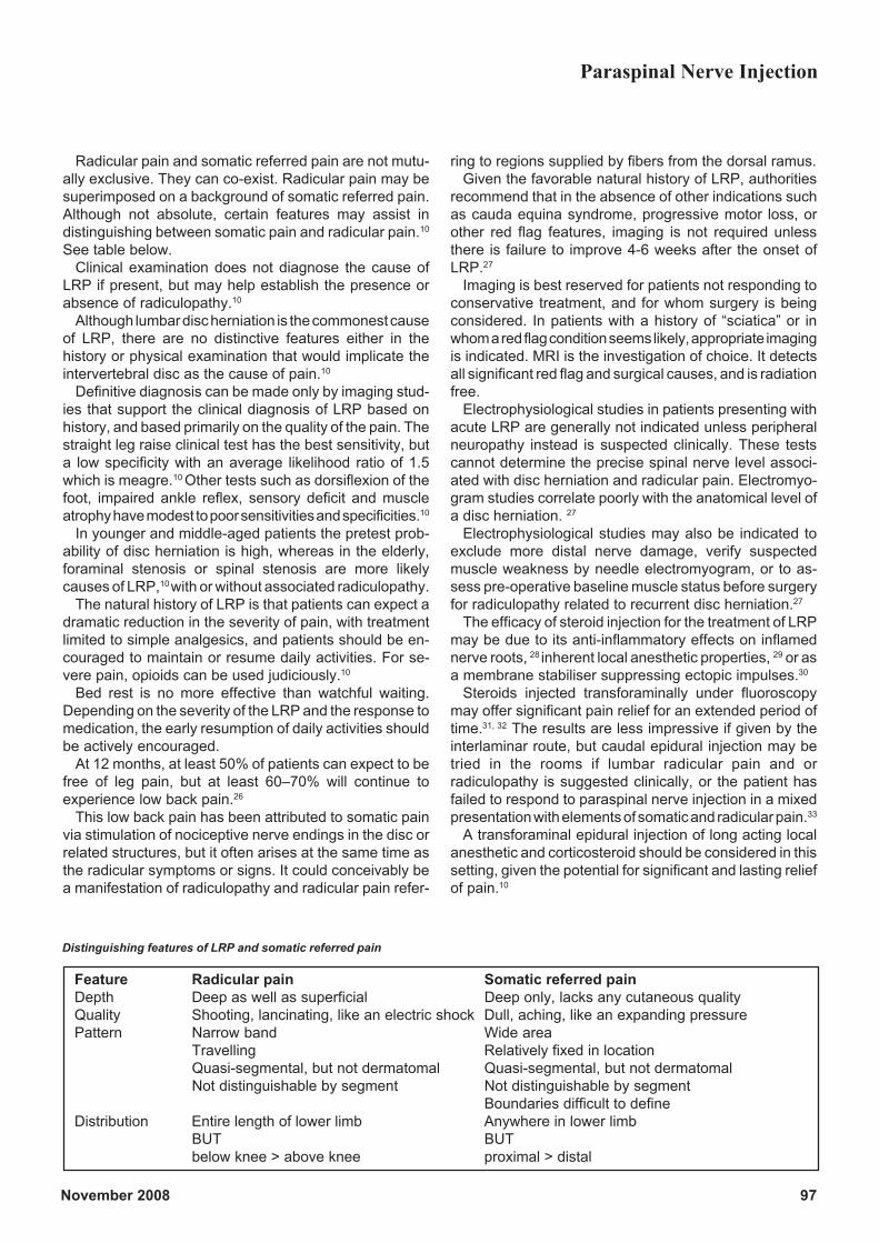
November 2008 97
Radicular pain and somatic referred pain are not mutu-ally exclusive. They can co-exist. Radicular pain may besuperimposed on a background of somatic referred pain.Although not absolute, certain features may assist indistinguishing between somatic pain and radicular pain.10
See table below.Clinical examination does not diagnose the cause of
LRP if present, but may help establish the presence orabsence of radiculopathy.10
Although lumbar disc herniation is the commonest causeof LRP, there are no distinctive features either in thehistory or physical examination that would implicate theintervertebral disc as the cause of pain.10
Definitive diagnosis can be made only by imaging stud-ies that support the clinical diagnosis of LRP based onhistory, and based primarily on the quality of the pain. Thestraight leg raise clinical test has the best sensitivity, buta low specificity with an average likelihood ratio of 1.5which is meagre.10 Other tests such as dorsiflexion of thefoot, impaired ankle reflex, sensory deficit and muscleatrophy have modest to poor sensitivities and specificities.10
In younger and middle-aged patients the pretest prob-ability of disc herniation is high, whereas in the elderly,foraminal stenosis or spinal stenosis are more likelycauses of LRP,10 with or without associated radiculopathy.
The natural history of LRP is that patients can expect adramatic reduction in the severity of pain, with treatmentlimited to simple analgesics, and patients should be en-couraged to maintain or resume daily activities. For se-vere pain, opioids can be used judiciously.10
Bed rest is no more effective than watchful waiting.Depending on the severity of the LRP and the response tomedication, the early resumption of daily activities shouldbe actively encouraged.
At 12 months, at least 50% of patients can expect to befree of leg pain, but at least 60–70% will continue toexperience low back pain.26
This low back pain has been attributed to somatic painvia stimulation of nociceptive nerve endings in the disc orrelated structures, but it often arises at the same time asthe radicular symptoms or signs. It could conceivably bea manifestation of radiculopathy and radicular pain refer-
Distinguishing features of LRP and somatic referred pain
Feature Radicular pain Somatic referred painDepth Deep as well as superficial Deep only, lacks any cutaneous qualityQuality Shooting, lancinating, like an electric shock Dull, aching, like an expanding pressurePattern Narrow band Wide area
Travelling Relatively fixed in locationQuasi-segmental, but not dermatomal Quasi-segmental, but not dermatomalNot distinguishable by segment Not distinguishable by segment
Boundaries difficult to defineDistribution Entire length of lower limb Anywhere in lower limb
BUT BUTbelow knee > above knee proximal > distal
ring to regions supplied by fibers from the dorsal ramus.Given the favorable natural history of LRP, authorities
recommend that in the absence of other indications suchas cauda equina syndrome, progressive motor loss, orother red flag features, imaging is not required unlessthere is failure to improve 4-6 weeks after the onset ofLRP.27
Imaging is best reserved for patients not responding toconservative treatment, and for whom surgery is beingconsidered. In patients with a history of “sciatica” or inwhom a red flag condition seems likely, appropriate imagingis indicated. MRI is the investigation of choice. It detectsall significant red flag and surgical causes, and is radiationfree.
Electrophysiological studies in patients presenting withacute LRP are generally not indicated unless peripheralneuropathy instead is suspected clinically. These testscannot determine the precise spinal nerve level associ-ated with disc herniation and radicular pain. Electromyo-gram studies correlate poorly with the anatomical level ofa disc herniation. 27
Electrophysiological studies may also be indicated toexclude more distal nerve damage, verify suspectedmuscle weakness by needle electromyogram, or to as-sess pre-operative baseline muscle status before surgeryfor radiculopathy related to recurrent disc herniation.27
The efficacy of steroid injection for the treatment of LRPmay be due to its anti-inflammatory effects on inflamednerve roots, 28 inherent local anesthetic properties, 29 or asa membrane stabiliser suppressing ectopic impulses.30
Steroids injected transforaminally under fluoroscopymay offer significant pain relief for an extended period oftime.31, 32 The results are less impressive if given by theinterlaminar route, but caudal epidural injection may betried in the rooms if lumbar radicular pain and orradiculopathy is suggested clinically, or the patient hasfailed to respond to paraspinal nerve injection in a mixedpresentation with elements of somatic and radicular pain.33
A transforaminal epidural injection of long acting localanesthetic and corticosteroid should be considered in thissetting, given the potential for significant and lasting reliefof pain.10
Paraspinal Nerve Injection
98 Australasian Musculoskeletal Medicine
A recent systematic review concluded that there ismoderate evidence for interlaminar epidurals in the cervi-cal spine and limited evidence in the lumbar spine for long-term relief of radicular pain.
The evidence for cervical and lumbar transforaminalepidural steroid injections is moderate for long-term im-provement in managing radicular pain. The evidence forcaudal epidural steroid injections is moderate for long-term relief in managing radicular pain and chronic lowback pain.33
For somatic spinal pain, paraspinal nerve injection shouldbe considered. It blocks nociception into the dorsal horn ofthe spinal cord via the dorsal nerve root, and can relievesomatic pain while the patient is on the table, allowing aresumption of usual activities of daily living with stretchingand walking that can stimulate Aβ fibers which detectgross spinal movements. This input rises to the midbrain,and can help to block incoming nociception by increasingdescending pain control mechanisms.
Components of radicular pain and or radiculopathy maybe also present but clinical examination lacks sensitivityand specificity.10
Imaging with MRI does not always resolve the dilemmaas there is an increasing prevalence of false positive MRIfindings with age.8, 9
A trial of treatment may relieve distressing symptoms,and avoid expensive and unnecessary imaging. Red flagfeatures are uncommon, accounting for less than 1% ofcauses of low back pain in a primary care setting.34, 35
In the original trials by Blomberg on subacute low backpain (pain for 6-12 weeks), including patients with radicularleg pain, the average number of parasacrococcygeal para-spinal nerve injections needed for lasting pain relief was 1-2.
In Australia, musculoskeletal pain medicine doctorsusing this approach have found that people with chronicspinal pain of many years’ duration can benefit from onaverage 1-4 injections given at weekly or greater intervals,with a cumulative effect that seems not diminished bygreater intervals between injections.
The importance of diagnosis, education, and assurancecannot be underestimated, with recommendations forlight activity, stretching,36 manual therapy if applicable anda graded home exercise program utilizing walking of greatbenefit in maintaining improvement.
Level of evidenceThere is Level II evidence from Stefan Blomberg’s RCT
in the early 1990s, using parasacrococcygeal paraspinalinjection.1-5 One of the papers was published in Spine in1994.5
In Australia, there is level IV evidence from some caseseries.37-41 A pilot study in Queensland has also beenconducted. Data collection has been completed, withresults currently being analysed.
IndicationsPrimary indications: somatic with or without referred
spinal pain, acute, or chronic, possibly associated withradiculopathy. This may be diagnosed prospectively orretrospectively when revealed after overlying somaticpain that has been clouding the clinical picture has re-solved with paraspinal nerve injection.
Secondary indications: after failed epidural injection,when undiagnosed or coexistent somatic spinal with orwithout referred pain is diagnosed or suspected.
ContraindicationsAbsolute contraindications: red flag conditions; un-
corrected or suspected coagulopathy; known allergy tolocal anesthetic/corticosteroid, for example, lignocaine,Celestone Chronodose, etc.; local infection.
Relative contraindications: anticoagulation therapy -injection is still possible, with careful technique and fine25G needles if the INR is within recommended referenceranges; frail elderly patients; those prone to vasovagalepisodes. Patients with previous spinal surgery and metalinternal fixation can be injected, carefully avoiding infiltra-tion close to the metal using a no-touch or sterile tech-nique, aiming to bathe the dorsal ramus and or otherparaspinal nerves with local anesthetic with or withoutcorticosteroid.
EquipmentParaspinal nerve injections can be done with minimal
equipment after marking relevant land marks using a no-touch technique in the rooms. Equipment for treatingallergic reactions and vasovagal reactions should bereadily to hand.
ProcedureThe parasacrococcygeal paraspinal nerve injections for
lumbar spinal pain are usually done with the patient lyingprone or in the lateral position if this is problematic, and theinjections are given paraspinally to block the nervesentering the neuraxis adjacent to the edges of the sacrumand coccyx bilaterally.
The injection site can be at C1-2 (coccygeal) as inBlomberg’s original work and involves infiltration up anddown beside the sacrum and coccyx, usually bilaterally toblock multiple nerve roots and spinal nerves. Entry site ofinjection can be at other sites parasacrally, such as at S4or S2, and infiltration performed up and down the
Paraspinal Nerve Injection
November 2008 99
parasacrococcygeal region with 15 ml of 0.5% plain ligno-caine with Celestone Chronodose or comparable steroidsolution typically using 2 ampoules, that is, 2 ml in andaround the dorsal nerve rami to block nociception enteringthe dorsal roots and hence dorsal horn segmentally.
A 22 or 23 G 50-60 mm needle or longer spinal needleis used using a no-touch technique. This has the effect ofreversing the wind-up and central sensitization that is theusual cause for persistent spinal pain, acutely, or chroni-cally when it can be associated with radiculopathy.
If patients are needle phobic or in significant pain ormarkedly tender, consider infiltrating first with 5 ml oflignocaine 0.5% either side at the entry site and infiltratingup and down in the usual directions using a 38 mm 25 Gneedle. This can be done routinely or in selected patients.
There may be variable lumbosacral tenderness, anddiagnosis is best made on the basis of history and or paindiagram.
There may be tenderness paraspinally on digital rectalexamination and Stefan Blomberg teaches the injectionwith a per rectal (PR) guiding digit in the anus to assess fortenderness, to guide the depth of injection, and to stretchthe parasacrococcygeal ligaments after paraspinal nerveinjection has blocked the nociception and relieved thepain. This was used in the published trials so is thepreferred method, though omitting this aspect of theinjection may still produce an effective outcome if patientsare uncomfortable with the idea after informing them of thetrial details and evidence. The use of the PR guide couldbe reserved as an option for later inclusion or considera-tion if there is failure to respond to an unguided injectionwithout the associated stretching.
Needling of any tender entheses after blocking thedorsal rami may have additional reflex effects to reset thedorsal horn and reverse the wind-up and centralsensitization and persistent pain.
Additional paraspinal nerves may be blocked from L1-5if needed with lignocaine 0.5% using 1.5-2 ml with orwithout corticosteroid each side for each involved seg-ment, aiming for the inferior aspect of the attachment ofthe transverse process to the vertebral arch in order toblock the dorsal rami. The history, pain diagram, andspinal tenderness and persistence of pain are the bestguide to choosing additional spinal nerves to block. It isimportant to update and review the anatomy regularly.42, 43
Similarly, thoracic spinal pain may be treated by inject-ing the dorsal rami at the inferior aspect of the attachmentof the transverse process to the vertebral arch with ligno-caine 0.5% using 1.5-2 ml with or without corticosteroideach side at each involved segmental level. The history,pain diagram, and spinal tenderness and persistence ofpain are the best guide to choosing additional spinalnerves to block.
Thoracic radicular pain due to disc prolapse is lesscommon than in the lumbar and cervical regions but othermechanisms could be involved. Chronic thoracic spinalpain is more commonly of zygapophysial joint origin, but
thoracic radicular pain still occurs, and can be subtle in itsclinical presentation.44, 45
The cervical transverse process tips can be infiltratedusing lignocaine 0.5% with or without dexamethasone, anon-particulate corticosteroid, using 0.4 ml for each spinalnerve from C3-7 especially if tender unilaterally or bilater-ally .
Non-particulate steroids are safer in case of intravascu-lar injection.46
A study published in Spine in 2007 on cervicaltransforaminal epidurals steroid injections (TFEIs) lookedat 30 cases of brain or spinal cord infarction (16 brain, 12cervical spinal cord, and 2 combined brain/spinal cordinfarcts), over three times greater than the sum of allpublished infarcts previously (n = 8 published case re-ports).
Of all the cases reported, four involved only corticoster-oid with no local anesthetic. All four cases involved meth-ylprednisolone and resulted in brain infarction, three ofwhich were fatal. This is the strongest association to datebetween particulate corticosteroids and brain or spinalcord infarctions.
The authors point out that although co-occurrence ofalternative mechanisms of injury (for example, vertebralartery dissection or needle-induced vasospasm) is possi-ble, it supports an embolic mechanism of action.
This was the first study to propose a “top of the basilar”artery mechanism, whereby the steroid embolus travels tothe confluence of the distal basilar artery, its thalamo-perforate branches, the superior cerebellar artery, and theposterior cerebral artery. Occlusion of these arteries givesrise to midbrain, pons, cerebellum, thalamus, and ortemporal and occipital lobe infarctions.
In order to minimize the risk of complications, the au-thors suggest : 1) using real-time fluoroscopy with non-ionic contrast and digital subtraction to maximize detec-tion of vascular uptake during cervical TFEIs; 2) using atest dose of local anesthetic prior to injecting corticoster-oid to prevent irreversible neurologic sequelae; 3) usingmicrobore extension tubing to minimize needle move-ment while changing syringes; 4) using minimal if anysedation to allow for clinical neurologic monitoring; 5)using a shorter-acting local anesthetic such as lignocaine,preferably at the lowest possible concentration and dose,to minimize high spinal anesthesia occurrence or severity;6) using blunt needles; 7) screening for arterial dissectionrisk factors; and 8) using a non-particulate corticosteroidsuch as dexamethasone.46
A recent experimental animal study using a rat modelsuggested that corticosteroids may add no extra efficacyto local anesthetic injection in treating radiculopathy,suggesting that corticosteroid may be unnecessary fornerve root infiltration (NRI).47
The paraspinal injection is meant to be extradural butthese points are certainly worth bearing in mind. Cervicalinjections should be attempted only by experienced doc-tors with a sound knowledge of the anatomy and the
Paraspinal Nerve Injection
100 Australasian Musculoskeletal Medicine
techniques involved.Cervical paraspinal nerve injection may succeed in
blocking nociception arriving in both the dorsal rami sup-plying the posterior elements of the spine, and ventral ramiwhich transmit nociception from the intervertebral discsand dural sleeves and dura to the dorsal root via the spinalnerve. It may, therefore, help with discogenic pain.
The patient is placed in the lateral position and the tipsof the cervical lateral masses or transverse processes arecarefully marked, and then a no-touch technique is used.It is important to be aware of the course of the vertebralartery which is exposed between lateral masses. It isprudent to be very gentle and withdraw if the tip is notcontacted at the appropriate depth or if there is uncertaintyabout needle position.
Utilizing a low dose of local anesthetic such as 0.4 ml of0.5% lignocaine alone with or without non-particulatesteroid such as dexamethasone enhances the safety ofthe procedure.
Alternatively, a posterior approach can be used to blockthe dorsal rami or at least the medial branches frombehind. The injection is approximately on the same levelas the spinous process, the articular pillar, and the tips ofthe cervical lateral masses or transverse processes. Theneedle can be inserted slightly inferiorly and 2.5 cm lateralto the spinous process and aimed at the posterior aspectof the articular pillar to be blocked. After bony contact ismade, the depth of contact is noted and the needlerepositioned aiming towards the most lateral aspect of thearticular pillar. The 25 G or larger needle is advanced, but,if it walks off laterally, is withdrawn and redirected slightlymedially and advanced to the depth of the previous bonycontact.
After the needle is felt to be the optimum position, thehub is observed for blood or cerebrospinal fluid. If neitheris evident, gentle aspiration can be attempted. If theaspiration test is negative, 1.5 ml of solution of localanesthetic such as lignocaine 0.5% with or without dex-amethasone can be injected to block the medial branch ofthe dorsal ramus at that level.
The patient is usually shown some gentle stretchingexercises to use as part of a home-based rehabilitationprogram 2-3 times daily and also if there is a flare of pain.It is useful to utilize post-isometric relaxation with deepbreathing in accordance with Sherrington’s second law toachieve a painless stretch.48, 49
ComplicationsThese include bleeding, infection, pain, scarring, inad-
vertent epidural or subarachnoid injection producing ahigh spinal anesthetic if sufficient volume and concentra-tion of local anesthetic is injected too close to the neuraxis.A subdural injection can give similar results to a subarach-noid injection but with a prolonged onset time. However,
a small dose of dilute 0.5% lignocaine with or withoutdexamethasone reduces any substantial risk.
Paralysis is possible with injections in the cervical re-gion, into a vertebral or smaller vessel usually withparticulate corticosteroid solution via steroid embolusproducing thrombosis, cord or brain stem infarction and ordeath. Injection directly into the spinal cord itself canproduce similar, possibly more localized effects.
Alternatives to paraspinal injection include other con-servative management; epidural injection may be consid-ered if there is a poor response to paraspinal nerveinjection, as radicular pain or even radiculopathy involvingfibers related to the dorsal ramus may be present. How-ever, cervical transforaminal epidural steroid injection perse remains out of favour in view of safety concerns.Referral for formal cervical or lumbar medial branch blocks,with or without radiofrequency neurotomy if positive withcontrolled blocks, is supported strongly by the evidencebase and is the ideal and only proven treatment for Z jointpain but unfortunately is not always available or done toISIS standards.
ResultsThe results have been very good empirically and when
followed up in clinical practice, but more formal study isstrongly indicated, with case series the best way to pro-ceed in private musculoskeletal or general medical prac-tice.
ConclusionsParaspinal nerve injection offers a valuable way of
managing acute pain or chronic pain that may be associ-ated with radiculopathy. This may be fully revealed onlyafter a trial of treatment. It seems to work best for somaticwith or without referred spinal pain but this can coexist withor be associated with radicular pain and/or radiculopathy.50
The paraspinal nerve injections can also clarify a mixedsomatic and radicular presentation and relieve somaticpain which may mask associated radicular pain and/orradiculopathy. This may allow a more precise diagnosis ofspecific level nerve root impingement syndromes, whichmay then be more amenable to surgery withmicrodiscectomy, etc.
Persistent musculoskeletal pain is likely to have a largeneuropathic component. Some 20 years ago ProfessorBogduk delivered a lecture at an AAMM or APS meetingin which he threatened a nightmare. The proposition wasthat there was no such entity as musculoskeletal pain: thatit was all neuropathic.
Even osteoarthritis is neuropathic (at the micro level).Radiofrequency neurotomy may work not because it
Paraspinal Nerve Injection
November 2008 101
denervates a painful joint but because the articular nervesare neuropathic, in that some of the afferents are injured.The pain arises not from the joint but because of afferentimbalance. Denaturing the nerves resets the balance.Could the result of the Bone and Joint Decade be: it’s allneural? All treatments work by resetting the dorsal horn?
This is certainly not an argument for more surgery, etc.,as surgery is effective only for intractable radicular pain orfor other rare red flag conditions such as cauda equinasyndrome, unstable fracture, tumour, or infection.
Paraspinal nerve injections may reduce the volumeof back surgery and increase the precision of thatwhich is done once pain treatable by injection isremoved and the masking is uncovered.
Public pain clinics in Australia are difficult to access, withextremely long waiting lists for persistent non-malignantpain. They usually have a multi-disciplinary approachwhich may assist function in the short-term but the Austral-ian experience reveals very small and temporary effects,as revealed at the recent AAMM annual scientific meetingin Melbourne. A recent audit in the Newcastle pain clinicshowed no change in median pain, no increase in returnto work. Similar results from Stephen J Gibson with theVictorian experience with CBT showed that one-third ofchronic pain patients ended up worse. These patientscommonly represent to primary care doctors for furtherassistance.
We believe that a trial of paraspinal nerve injections isdefinitely indicated for acute pain not responding to oralanalgesia and simple physical measures, for example,stretching, or chronic spinal pain that is commonly asso-ciated with radiculopathy, before referral for a surgicalopinion. Surgery is not indicated in the absence of red flagconditions as outlined above. Surgery may still have a rolefor intractable radicular pain after a trial of epidural injec-tion of local anesthetic with or without steroid, eithertransforaminally or via the caudal route. Paraspinal nerveinjection can help clarify the diagnosis and can offer veryeffective treatment for somatic spinal pain components. Itmay also work like a selective nerve block if well placed,avoiding the dangers of a transforaminal epidural injectionif the injection is lateral to the intervertebral foramina andepidural space. Surgery is indicated for cauda equinasyndrome or progressive motor impairment related toradiculopathy.
References1. Blomberg S, Svardsudd K, Mildenberger F. A controlled,multicentre trial of manual therapy in low back pain; initial status,sick-leave and pain score during follow-up. Scand J Prim HealthCare 1992; 10: 170-78.
2. Blomberg S, Svardudd K, Tiblin G. Manual therapy with steroidinjections in low-back pain: improvement of quality of life in acontrolled trial with four months’ follow-up. Scand J Prim Health
Care 1993; 11: 83-90.
3. Blomberg S, Tibblin G. A controlled, multicentre trial of manualtherapy with steroid injections in low-back pain: functional vari-ables, side-effects and complications during four months follow-up. Clinical Rehabil 1993; 7: 49-62.
4. Blomberg S, Svardudd K, Tibblin G. A randomized study ofmanual therapy with steroid injections in low-back pain: tel-ephone interview follow-up of pain, disability, recovery and drugconsumption. Eur Spine J 1994; 3:246-254.
5. Blomberg S, Hallin G, Grann K, et al. Manual therapy withsteroid injections – a new approach to treatment of low back pain:a controlled multicentre trial with an evaluation by orthopedicsurgeons. Spine 1994; 19: 569-77.
6. Bogduk N. Mechanisms of musculoskeletal pain. AustralasMusculoskeletal Med 2006;1: 7-19.
7. Bogduk N, McGuirk B. Pain Research and Clinical Manage-ment, Vol 13. Medical Management of Acute and Chronic LowBack Pain. An Evidence-Based Approach. Elsevier, 2002.
8. Boden SD, Davis DO, Dina TS et al. Abnormal magnetic-resonance scans of the lumbar spine in asymptomatic subjects.A prospective investigation. J Bone Joint Surg Am 1990;72(3):403-8.
9. Jensen MC, Brant-Zawadzki MN, Obuchowski N et al. Mag-netic resonance imaging of the lumbar spine in people withoutback pain. N Engl J Med 1994; 331(2): 69-73.
10. Govind J. Lumbar radicular pain. Aust Family Physician2004; 33(6): 409-41.
11. Merskey H, Bogduk N (eds.) Classification of chronic pain.Descriptions of chronic pain syndromes and definitions of painterms. 2nd ed. Seattle; IASP Press, 1994.
12. Bogduk N. Clinical Anatomy of the Lumbar Spine andSacrum. 3rd ed. Edinburgh; Churchill Livingstone, 1997.
13. Howe JF. A neurophysiological basis for the radicular pain ofnerve root compression. In Bonica JJ, Liebeskind JC, Albe-Fessard DG, eds. Advances in pain research and therapy. Vol 3.New York; Raven Press, 1979, pp. 647-57.
14. Howe JF, Loeser JD, Calvin WH. Mechanosensitivity ofdorsal root ganglion and chronically injured axons: a physiologi-cal basis for the radicular pain of nerve root compression. Pain1977; 3: 25-41.
15. McCarron RF, Wimpee MW, Hudkins PG, Laros GS. Theinflammatory effect of nucleus pulposus. A possible element inthe pathogenesis of low-back pain. Spine 1987; 12(8): 760-64.
16. Olmarker K, Blomquist J, Strömberg J et al. Inflammatogenicproperties of nucleus pulposus. Spine 1995 Mar 15; 20(6): 665-69.
17. Saal JS, Franson RC, Dobrow R et al. High levels ofinflammatory phospholipase A2 activity in lumbar disc herniations.
Paraspinal Nerve Injection
102 Australasian Musculoskeletal Medicine
Spine 1990; 15: 674-78.
18. Franson RC, Saal JS, Saal JA. Human disc phospholipaseA2 is inflammatory. Spine 1992; 17: S129-S132.
19. Rydevik BL, Myers R, Powell HC. Pressure increase in thedorsal root ganglion following mechanical compression. Spine1989; 14: 574-76.
20. Ozaktay AC, Kallakuri S, Cavanaugh JM. Phospholipase A2sensitivity of the dorsal root and dorsal root ganglion. Spine1998; 23(12): 1297-306.
21. Chen C, Cavanaugh JM, Ozaktay AC et al. Effects ofphospholipase A2 on lumbar nerve root structure and function.Spine 1997; 22(10): 1057-64.
22.. Cooper RG, Freemont AJ, Hoyland JA et al. Herniatedintervertebral disc-associated periradicular fibrosis and vascularabnormalities occur without inflammatory cell infiltration. Spine1995; 20(5): 591-98.
23. Lutze M, Stendel R, Vesper J, Brock M. Periradicular therapyin lumbar radicular syndromes: methodology and results. ActaNeurochir (Wien) 1997; 139(8): 719-24.
24. Maigne JY, Rime B, Delinge B. Computed tomographicfollow up study of forty-eight cases of nonoperatively treatedlumbar intervertebral disc herniation. Spine 1992; 17: 1071-74.
25. Delauce-Cavallier MC, Budet C, Lardeo JD et al. Lumbar discherniation: computed tomography scan changes after conserva-tive treatment of nerve root compression. Spine 1992; 17: 927-33.
26. Weber H, Holme I, Amlie E. The natural course of acutesciatica with nerve root symptoms in a double blind placebocontrolled trial evaluating the effect of piroxicam. Spine 1993; 18:1433-38.
27. Anderson GBJ, Brown MD, Dvorak J et al. Consensussummary on the diagnosis and treatment of lumbar disc hernia-tion. Spine 1996; 21(Suppl 24S): 75S-78S.
28. Olmarker K, Byrod G, Cornefjord M et al. Effects ofmethyprednisolone on nucleus pulposis-induced nerve root in-jury. Spine 1994; 19: 1803-08.
29. Johanson A, Hao J, Sjolund B. Local corticosteroid applica-tion blocks transmission in normal nociceptive fibres. ActaAnaesthesiol Scand 1990; 34: 335-38.
30. Devor M, Govrin-Lippmann R, Raper P. Corticosteroidssuppress ectopic neural discharge originating in experimentalneuromas. Pain 1985; 22: 127-37.
31. Thelander U, Fagerlund M, Friberg S, Larsson S. Straight legraising test versus radiological size, shape and position oflumbar disc hernias. Spine 1992; 17: 395-99.
32. Lutz E, Vad VB, Wisneski RJ. Fluoroscopic transforaminallumbar epidural steroids: an outcome study. Arch Phys Rehabil1998; 79:1362-66.
33. Abdi S, Datta S, Trescot AM et al. Epidural steroids in themanagement of chronic spinal pain: a systematic review. PainPhysician 2007;10(1): 185-212.
34. Wilk V. Acute low back pain - assessment and management.Aust Family Physician 2004; 33(6): 385-480.
35. Deyo RA, Rainville J, Kent DL. What can the history andphysical examination tell us about low back pain? JAMA 1992;26(6): 760-65.
36. Bogduk N, McGuirk B. The “Indahl” Exercises for Low BackPain. Australas Musculoskeletal Med 2008; (1): 8-14.
37. McKay AB, Wall D. The Orienting Response and the Func-tional Whole Human Body. Australas Musculoskeletal Med 2003;8(2): 86-99.
38. McKay AB. Pain and Chronic Low Back Pain: A New Model?Part 1.The Hypothesis and Model. Australas MusculoskeletalMed 2004; 9(1): 14-19.
39. McKay AB. Pain and Chronic Low Back Pain: A New Model?Part 2. Observations and clinical material. Australas Muscu-loskeletal Med 2004 9(1): 20-25.
40. McKay AB. Tennis Elbow Everywhere. Australas Muscu-loskeletal Med 2005; 10(2): 127-30.
41. McKay AB. Chronic Low Back Pain and Pain: Simple Modelsto Explain Pain to Patients. Australas Musculoskeletal Med2006: (2) 92-96.
42. Agur AM, Dalley AF. Grant’s atlas of anatomy. 12th ed.Baltimore; Lippincott, Williams & Wilkins, 2008.
43. Waldman SD. Atlas of Interventional Pain Management. 2nd
ed. Philadelphia; Saunders, 2004.
44. Malanga GA, McLean JP. Thoracic Discogenic Pain Syn-drome. http://emedicine.medscape.com/article/96284-overview
45. Manchikanti L, Boswell MV, Singh V et al. Prevalence of facetjoint pain in chronic spinal pain of cervical, thoracic, and lumbarregions. BMC Musculoskelet Disord 2004; 5: 15.
46. Scanlon GC, Moeller-Bertram T, Romanowsky SM, WallaceMS. Cervical Transforaminal Epidural Steroid Injections: MoreDangerous Than We Think? Spine 2007; 32(11): 1249-56.
47. Tachihara H, Sekiguchi M, Kikuchi S, Konno S. Docorticosteroids produce additional benefit in nerve root infiltra-tion for lumbar disc herniation? Spine 2008; 33(7): 743-47.
48. http://nobelprize.org/nobel_prizes/medicine/laureates/1932/sherrington-bio.html
49. http://www.worldscibooks.com/medsci/etextbook/p210/p210_chap1.pdf
50. Can pain be more or less neuropathic? Editorial. Pain 2004;110: 510-11.
Paraspinal Nerve Injection
November 2008 103
Journal Abstracts
This section aims to update the reader with some of the more significant musculoskeletalresearch published in the last year which is listed on the Medline and CINAHL databases.
BACK PAIN
Little P, Lewith G, Webley F et al. Randomised control-led trial of Alexander technique lessons, exercise,and massage (ATEAM) for chronic and recurrent backpain. Br Med J 2008; 337: a884. doi:10.1136/bmj.a884.
Objective. To determine the effectiveness of lessons inthe Alexander technique, massage therapy, and advicefrom a doctor to take exercise (exercise prescription)along with nurse-delivered behavioural counselling forpatients with chronic or recurrent back pain.
Design. Factorial randomised trial.Setting. 64 general practices in England.Participants. 579 patients with chronic or recurrent low
back pain; 144 were randomised to normal care, 147 tomassage, 144 to six Alexander technique lessons, and144 to 24 Alexander technique lessons; half of each ofthese groups were randomised to exercise prescription.
Interventions. Normal care (control), six sessions ofmassage, six or 24 lessons on the Alexander technique,and prescription for exercise from a doctor with nursedelivered behavioural counselling.
Main outcome measures. Roland Morris disability score(number of activities impaired by pain) and number ofdays in pain.
Results. Exercise and lessons in the Alexander tech-nique, but not massage, remained effective at one year(compared with control Roland disability score 8.1: mas-sage -0.58, 95% confidence interval -1.94 to 0.77, sixlessons -1.40, -2.77 to -0.03, 24 lessons -3.4, -4.76 to -2.03, and exercise -1.29, -2.25 to -0.34). Exercise after sixlessons achieved 72% of the effect of 24 lessons alone(Roland disability score -2.98 and -4.14, respectively).Number of days with back pain in the past four weeks waslower after lessons (compared with control median 21days: 24 lessons -18, six lessons -10, massage -7) andquality of life improved significantly. No significant harmswere reported.
Conclusions. One to one lessons in the Alexandertechnique from registered teachers have long-term ben-efits for patients with chronic back pain. Six lessonsfollowed by exercise prescription were nearly as effectiveas 24 lessons. Trial registration: National Research Reg-ister N0028108728.
Comment. The study was a large, well-conductedrandomized controlled trial comparing the short-term andlong-term effects of the Alexander technique with mas-sage and normal care and also the effects of an unsuper-vised home-based aerobic exercise program (prescribedby a general practitioner with follow-up behavioural coun-selling from a nurse). The Alexander technique was supe-
rior to normal care at three and 12 months and to massageat 12 months. It resulted in moderate improvements indisability and pain-free days. Twenty-four lessons wereonly marginally better than six lessons combined with theexercise program. Interestingly the exercise programprovided modest but useful benefits from a relatively briefGP/nurse intervention.
The results suggest that the Alexander technique is areasonable choice for the treatment of chronic low backpain. It stands alongside other self-management optionswith some evidence for effectiveness such as supervisedtailored exercise programs. When recommending self-management options, patient preferences and expecta-tions should be considered.
The results also offer a weak endorsement for home-based exercise prescribed by a GP with follow-up by anurse. Massage therapy may offer short term benefits. –Dr Michael Yelland
Friedman BW, Esses D, Solorzano C et al. A randomizedplacebo-controlled trial of single-dose im corticoster-oid for radicular low back pain. Spine 2008;33(18):E624-29. Department of Emergency Medicine, AlbertEinstein College of Medicine, Bronx, NY 10467, [email protected].
Study Design. A randomized, double-blind, placebo-controlled trial of patients with radicular low back pain whopresent to an emergency department (ED) within 1 weekof pain onset.
Objective. We hypothesized that a single intramuscular160 mg dose of methylprednisolone acetate would im-prove pain and functional outcomes 1 month after EDdischarge if the corticosteroid were administered early indisease symptomatology.
Summary of background data. Parenteral corticosteroidsare not recommended for acute, radicular low back pain,though their role in this disease process is ill-defined. Todate, this medication class has only been studied in ahighly selected group of patients requiring hospitalization.
Methods. Adults between the ages of 21 and 50 whopresented to an ED with low back pain and a positivestraight leg raise test were enrolled. The primary outcomewas change in pain intensity on an 11 point numericalrating scale 1 month after ED visit. Secondary outcomes1 month after ED discharge included analgesic use,functional disability, and adverse medication effects.
Results. Six hundred thirty-seven patients were ap-proached for participation, 133 were eligible, and 82 wererandomized. Baseline characteristics were comparablebetween the groups. The primary outcome, a comparisonof the mean improvement in pain intensity, favored meth-
104 Australasian Musculoskeletal Medicine
Journal Abstracts
ylprednisolone by 1.3 (P = 0.10). Some secondary out-comes favored methylprednisolone, such as use of anal-gesic medication within the previous 24 hours (22% vs.43%, 95% CI for difference of 20%: 0%-40%) and func-tional disability (19% vs. 49%, 95% CI for difference of29%: 9%-49%). Adverse medication effects 1 week afterED discharge were reported by 32% of methylprednisoloneand 24% of placebo patients (95% CI for difference of 9%:-12% to 30%).
Conclusion. This study was a negative study, thoughthere was a suggestion of benefit of methylprednisoloneacetate in a population of young adults with acute radicu-lar low back pain. Further work with a larger sample ofpatients is needed.
Comment. This study, though negative, exhibited atrend towards the positive or analgesic effects, and wassignificant for medication use and functional disability. Itwas probably underpowered. It is not clear whether thesepatients had true radicular pain or somatic referred pain ora combination in individual cases. They were assumed tohave an acute disc prolapse based on having low backpain and a positive straight leg raise test, without it beingclear whether this produced true radicular leg pain asshould be the case, or just aggravated the low back pain.
Methylprednisolone (Depo-Medrol) 160 mg was used,that is, 40 mg x 4 amps – IM, without any problems, andthe authors said that they would use this dose in furthertrials.
It could be considered for additional pain relief if oralanalgesia is not enough in this somewhat ill-definedsetting, especially if there is no access to a caudal ortransforaminal epidural injection. Alternatively, one couldtry a paraspinal nerve injection if there is a possiblesomatic component with or without radicular/neuropathiccomponent to the pain presentation. – Dr David Roselt
Roelofs PD, Deyo RA, Koes BW at al. Nonsteroidalanti-inflammatory drugs for low back pain: an up-dated Cochrane review. Spine 2008;33(16): 1766-74.Department of General Practice, Erasmus MC, UniversityMedical Center, Rotterdam, the [email protected].
Study Design. A systematic review of randomized con-trolled trials.
Objectives. To assess the effects of nonsteroidal anti-inflammatory drugs (NSAIDs) and COX-2 inhibitors in thetreatment of nonspecific low back pain and to assesswhich type of NSAID is most effective.
Summary of Background Data. NSAIDs are the mostfrequently prescribed medications worldwide and arewidely used for patients with low back pain. SelectiveCOX-2 inhibitors are currently available and used forpatients with low back pain.
Methods. We searched the MEDLINE and EMBASEdatabases and the Cochrane Central Register of Control-
led Trials up to and including June 2007 if reported inEnglish, Dutch, or German. We also screened referencesgiven in relevant reviews and identified trials. Randomizedtrials and double-blind controlled trials of NSAIDs in non-specific low back pain with or without sciatica were in-cluded.
Results. In total, 65 trials (total number of patients =11,237) were included in this review. Twenty-eight trials(42%) were considered high quality. Statistically signifi-cant effects were found in favor of NSAIDs compared withplacebo, but at the cost of statistically significant more sideeffects. There is moderate evidence that NSAIDs are notmore effective than paracetamol for acute low back pain,but paracetamol had fewer side effects. There is moder-ate evidence that NSAIDs are not more effective thanother drugs for acute low back pain. There is strongevidence that various types of NSAIDs, including COX-2NSAIDs, are equally effective for acute low back pain.COX-2 NSAIDs had statistically significantly fewer sideeffects than traditional NSAIDs.
Conclusion. The evidence from the 65 trials included inthis review suggests that NSAIDs are effective for short-term symptomatic relief in patients with acute and chroniclow back pain without sciatica. However, effect sizes aresmall. Furthermore, there does not seem to be a specifictype of NSAID, which is clearly more effective than others.The selective COX-2 inhibitors showed fewer side effectscompared with traditional NSAIDs in the randomizedcontrolled trials included in this review. However, recentstudies have shown that COX-2 inhibitors are associatedwith increased cardiovascular risks in specific patientpopulations.
Comment. Here is the latest review on NSAIDs for lowback pain for the Cochrane Collaboration http://www.cochrane.org/.
It is consistent with previous reviews, showing short-term symptomatic relief in patients with acute and chroniclow back pain without sciatica. Effect sizes are small, andthere is no long-term effect on the natural history of chronicspinal pain. Side-effects continue to be an issue andthese agents must be used cautiously if at all. Analgesicsare often preferable and safer from both GI and cardiovas-cular perspectives. – Dr David Roselt
Nath S, Nath CA, Pettersson K. Percutaneous lumbarzygapophysial (Facet) joint neurotomy usingradiofrequency current, in the management of chroniclow back pain: a randomized double-blind trial. Spine2008;33(12): 1291-97; discussion 1298. Smärtkliniken(The Pain Clinic), Umeå, Sweden. [email protected].
Study Design. A randomized controlled study of percu-taneous radiofrequency neurotomy was conducted in 40patients with chronic low back pain (20 active and 20controls).
Objective. The aim of the study was to evaluate the
November 2008 105
Journal Abstracts
possible beneficial effect of percutaneous radiofrequencyzygapophysial joint neurotomy in reducing pain and physi-cal impairment in patients with pain from the lumbarzygapophysial joints, selected after repeated diagnosticblocks.
Summary of Background Data. Facet or zygapophysialjoint pain may be one of the causes of chronic low backpain and may be treated by a percutaneous radiofrequencydenervation. Patients may possibly be identified by apositive diagnostic block. These blocks need to be re-peated as false positive responses to single blocks occur.In all previous studies patients treated with radiofrequencydenervation have been selected after single diagnosticblocks resulting in a varying degree of relief.
Methods. All patients were examined by an orthopedicsurgeon before and 6 months after the treatment (sham oractive). Inclusion criteria were 3 separate positive facetblocks. Denervation was achieved by multiple lesions ateach level in an effort to provide effective denervation.
Results. The active treatment group showed statisticallysignificant improvement not only in back and leg pain butalso back and hip movement as well as the sacro-iliac jointtest. Pre operative sensory deficit and weak or absentankle reflex normalized (P < 0.01) and (P < 0.05), respec-tively. There was significant improvement in quality of lifevariables, global perception of improvement, and gener-alized pain. The improvement seen in the active groupwas significantly greater than that seen in the placebogroup with regard to all the above-mentioned variables.None of our patients had any complication other thantransient postoperative pain that was easily managed.
Conclusion. Our study indicates that radiofrequencyfacet denervation is not a placebo and could be used in thetreatment of carefully selected patients with chronic lowback pain.
Comment. In a point of view, Professor Nik Bogduk inthe same copy of Spine points out that a previous outcomestudy by Dreyfuss et al.1 showed excellent results couldbe achieved with lumbar medial branch neurotomy. How-ever, that study was criticized for having no controls, forbeing highly selective in its recruitment criteria, for enroll-ing only a small proportion of potential patients, and forbeing too small a study. Later systematic reviews oflumbar medial branch neurotomy demanded randomizedcontrolled trials, but none of the controlled trials thatfollowed used correct surgical technique for this proce-dure, and none selected patients on the basis of controlleddiagnostic blocks.2,3
This is first study since Dreyfuss et al. to use controlleddiagnostic blocks to select patients,4 and the first to useapproved ISIS techniques.5
Professor Nik Bogduk points out also that Dr Nath didnot select ideal patients, free of comorbidity, with goodfunction, and no depression. His patients were enrolledfrom a pain clinic population, and they had other co-morbidities. Nevertheless, they were able to identify a
component of their pain that was completely relieved bycontrolled medial branch blocks. It was this pain that wastreated in a placebo-controlled trial of lumbar medialbranch neurotomy.
The results clearly show that the effects of lumbarmedial branch neurotomy cannot be attributed to placeboeffects. The effects are real.
This treatment did not as expected relieve every painthe patients had, that were not the target of the interven-tion. The index pain was relieved as postulated, andcorroborated by improvements in function.
Lumbar medial branch neurotomy as a monotherapy isnot indicated for all patients with back pain. However, thestudy showed that medial branch neurotomy could be acomplementary therapy in patients typical of a pain clinicpopulation. It also dispelled accusations that lumbar me-dial branch neurotomy is a placebo.
Professor Bogduk wonders whether critics will nextcomplain that Dr Nath should have recruited highly se-lected, ideal patients, with pure zygapophysial joint pain,with no comorbidity, and who would be atypical of patientsseen in conventional practice.1.Dreyfuss P, Halbrook B, Pauza K et al. Efficacy and validity ofradiofrequency neurotomy for chronic lumbar zygapophysialjoint pain. Spine 2000;25: 1270-77.
2.Hooten WM, Martin DP, Huntoon MA. Radiofrequency neu-rotomy for low back pain: evidence-based procedural guidelines.Pain Med 2005;6:129-38.
3.Bogduk N. Evidence-informed management of chronic backpain with facet injections and radiofrequency neurotomy. SpineJ 2008;8: 56-64.
4.International Spine Intervention Society. Lumbar medial branchblocks. In: Bogduk N, ed. Practice Guidelines for Spinal Diagnos-tic and Treatment Procedures. San Francisco; InternationalSpinal Intervention Society, 2004, pp. 47-65.
5.International Spine Intervention Society. Lumbar medial neu-rotomy. In: Bogduk N, ed. Practice Guidelines for Spinal Diag-nostic and Treatment Procedures. San Francisco; InternationalSpinal Intervention Society, 2004, pp. 188-218.
– Dr David Roselt
Bogduk N. Evidence-informed management of chroniclow back pain with facet injections and radiofrequencyneurotomy. Spine J 2008;8(1): 56-64. Pain MedicineDepartment, University of Newcastle, Newcastle, Aus-tralia. [email protected].
The management of chronic low back pain (CLBP) hasproven to be very challenging in North America, as evi-denced by its mounting socioeconomic burden. Choosingamongst available nonsurgical therapies can be over-whelming for many stakeholders, including patients, healthproviders, policy makers, and third-party payers. Althoughall parties share a common goal and wish to use limitedhealthcare resources to support interventions most likely

106 Australasian Musculoskeletal Medicine
Journal Abstracts
to result in clinically meaningful improvements, there isoften uncertainty about the most appropriate interventionfor a particular patient. To help understand and evaluatethe various commonly used nonsurgical approaches toCLBP, the North American Spine Society has sponsoredthis special focus issue of Spine Journal, titled “Evidence-Informed Management of Chronic Low Back Pain WithoutSurgery”. Articles in this special focus issue were contrib-uted by leading spine practitioners and researchers, whowere invited to summarize the best available evidence fora particular intervention and encouraged to make thisinformation accessible to nonexperts. Each of the articlescontains five sections (description, theory, evidence ofefficacy, harms, and summary) with common subhead-ings to facilitate comparison across the 24 different inter-ventions profiled in this special focus issue, blendingnarrative and systematic review methodology as deemedappropriate by the authors. It is hoped that articles in thisspecial focus issue will be informative and aid in decisionmaking for the many stakeholders evaluating nonsurgicalinterventions for CLBP.
Comment. This common abstract applies to this and aseries of reviews of these topics in Spine Journal in thefirst edition this year. This edition looks at evidence-informed management of chronic low back pain with 24interventions including epidural steroid injections, mas-sage, trigger point injections, functional restoration, andmany others. – Dr David Roselt
Hansen HC, McKenzie-Brown AM, Cohen SP,Swicegood JR, Colson JD, Manchikanti L. Sacroiliacjoint interventions: a systematic review. Pain Physi-cian 2007;10(1): 165-84. The Pain Relief Center, Conover,NC 28613, USA. hans.hippocrates.org.
Background. The sacroiliac joint is a diarthrodial syno-vial joint with abundant innervation and capability of beinga source of low back pain and referred pain in the lowerextremity. There are no definite historical, physical, orradiological features to provide definite diagnosis of sac-roiliac joint pain, although many authors have advocatedprovocational maneuvers to suggest sacroiliac joint as apain generator. An accurate diagnosis is made by control-led sacroiliac joint diagnostic blocks. The sacroiliac jointhas been shown to be a source of pain in 10% to 27% ofsuspected cases with chronic low back pain utilizingcontrolled comparative local anesthetic blocks.Intraarticular injections, and radiofrequency neurotomyhave been described as therapeutic measures. This sys-tematic review was performed to assess diagnostic test-ing (non-invasive versus interventional diagnostic tech-niques) and to evaluate the clinical usefulness of interven-tional techniques in the management of chronic sacroiliacjoint pain.
Objective. To evaluate and update the available evi-dence regarding diagnostic and therapeutic sacroiliac
joint interventions in the management of sacroiliac jointpain.
Study Design. A systematic review using the criteria asoutlined by the Agency for Healthcare Research andQuality (AHRQ), Cochrane Review Group Criteria fortherapeutic interventions and AHRQ, and Quality Assess-ment for Diagnostic Accuracy Studies (QUADAS) fordiagnostic studies.
Methods. The databases of EMBASE and MEDLINE(1966 to December 2006), and Cochrane Reviews weresearched. The searches included systematic reviews,narrative reviews, prospective and retrospective studies,and cross-references from articles reviewed. The searchstrategy included sacroiliac joint pain and dysfunction,sacroiliac joint injections, interventions, andradiofrequency.
Results. The results of this systematic evaluation re-vealed that for diagnostic purposes, there is moderateevidence showing the accuracy of comparative, control-led local anesthetic blocks. Prevalence of sacroiliac jointpain is estimated to range between 10% and 27% using adouble block paradigm. The false-positive rate of single,uncontrolled, sacroiliac joint injections is around 20%. Theevidence for provocative testing to diagnose sacroiliacjoint pain is limited. For therapeutic purposes, intraarticularsacroiliac joint injections with steroid and radiofrequencyneurotomy were evaluated. Based on this review, there islimited evidence for short-term and long-term relief withintraarticular sacroiliac joint injections and radiofrequencythermoneurolysis.
Conclusions. The evidence for the specificity and valid-ity of diagnostic sacroiliac joint injections is moderate. Theevidence for accuracy of provocative maneuvers in diag-nosis of sacroiliac joint pain is limited. The evidence fortherapeutic intraarticular sacroiliac joint injections is lim-ited. The evidence for radiofrequency neurotomy in man-aging chronic sacroiliac joint pain is limited.
Comment. There is no doubt that sacroiliac joints areinnervated and are capable of producing low back andreferred pain in the lower extremity.1 The authors point outthat the literature on diagnostic sacroiliac joint injectionsand non-invasive diagnostic techniques is superior to theliterature on therapeutic interventions. Due to the lack ofsignificant literature, the level of evidence was low fortherapeutic interventions. It is important that previousstudies are replicated and high quality evidence pro-duced. The full text article is available free atwww.painphysicianjournal.com.1. Bogduk N. The sacroiliac joint. Clinical Anatomy of LumbarSpine and Sacrum. 4th ed. New York; Churchill Livingstone,2005, pp. 173-81.
NECK PAIN
Buitenhuis J, de Jong PJ, Jaspers JP, Groothoff JW.
November 2008 107
Journal Abstracts
Catastrophizing and causal beliefs in whiplash. Spine2008;33(22): 2427-33; discussion 2434. Medical De-partment, Univé Insurance and Department of SocialMedicine, University Medical Center Groningen, Univer-sity of Groningen, the Netherlands. [email protected].
Study Design. Prospective cohort study.Objective. This study investigates the role of pain
catastrophizing and causal beliefs with regard to severityand persistence of neck complaints after motor vehicleaccidents.
Summary of Background Data. In previous research onlow back pain, somatoform disorders and chronic fatiguesyndrome, pain catastrophizing and causal beliefs werefound to be related to perceived disability and prognosis.Furthermore, it has been argued with respect to whiplashthat culturally dependent symptom expectations are re-sponsible for a chronic course.
Methods. Individuals involved in traffic accidents whoinitiated compensation claim procedures with a Dutchinsurance company were sent questionnaires (Q1) con-taining the Neck Disability Index, the Pain CatastrophizingScale, and the Causal Beliefs Questionnaire-Whiplash.Of 1252 questionnaires dispatched, 747 (59.7%) werereturned. Only car occupants with neck complaints wereincluded in this study (n = 140). Complaints were moni-tored using additional questionnaires administered 6 (Q2)and 12 months (Q3) after the accident. RESULTS: Paincatastrophizing and causal beliefs were related to theseverity of concurrent whiplash disability. The severity ofinitial complaints was related to the severity and persist-ence of whiplash complaints. Attributing initial neck com-plaints to whiplash was found to predict the persistence ofdisability at 6 and 12 months follow-up, over and above theseverity of the initial complaints.
Conclusion. The results suggest that causal beliefs mayplay a major role in the perceived disability and course ofneck complaints after motor vehicle accidents, whereaspain catastrophizing is predominantly related to concur-rent disability. The current findings are consistent with theview that an early conviction that neck complaints arecaused by the medico-cultural entity whiplash has adetrimental effect on the course of symptoms.
Comment: This study confirms what musculoskeletalpain medicine practitioners have suspected about theimportance of patients’ beliefs and catastrophizing indetermining outcomes. The importance of early interven-tion in assessment, with history and examination, educa-tion, assurance, maintenance of usual activities of dailyliving (ADLs), and light activity as much as possible, isapparent. The use of local heat to relax the muscles, andgentle range of movement exercises, using deep breath-ing in the form of post-isometric relaxation home exer-cises, along with simple analgesics and/or opioids brieflyif necessary, seem to go a long way to manage pain andprevent disability before the lawyers become involved ornon-evidence-based input occurs. – Dr David Roselt
Atluri S, Datta S, Falco FJ, Lee M. Systematic review ofdiagnostic utility and therapeutic effectiveness ofthoracic facet joint interventions. Pain Physician2008;11(5): 611-29. Tri-State Spine Care Institute,Cincinnati, OH 45230, USA. [email protected].
Background. Chronic mid back and upper back paincaused by thoracic facet joints has been reported in 34%to 48% of the patients based on the responses to control-led diagnostic blocks. Systematic reviews have estab-lished moderate evidence for controlled comparative localanesthetic blocks of thoracic facet joints in the diagnosisof mid back and upper back pain, moderate evidence fortherapeutic thoracic medial branch blocks, and limitedevidence for radiofrequency neurotomy of therapeuticfacet joint nerves.
Objectives. To determine the clinical utility of diagnosticand therapeutic thoracic facet joint interventions in diag-nosing and managing chronic upper back and mid backpain.
Study Design. Systematic review of diagnostic andtherapeutic thoracic facet joint interventions.
Methods. Review of the literature for utility of facet jointinterventions in diagnosing and managing facet joint painwas performed according to the Agency for HealthcareResearch and Quality (AHRQ) criteria for diagnostic stud-ies and observational studies and the Cochrane Muscu-loskeletal Review Group criteria as utilized for interven-tional techniques for randomized trials. The level of evi-dence was classified as Level I, II, or III based on thequality of evidence developed by United States Preven-tive Services Task Force (USPSTF) for therapeutic inter-ventions. Recommendations were based on the criteriadeveloped by Guyatt et al. Data sources included relevantliterature of the English language identified throughsearches of Medline and EMBASE from 1966 to July 2008and manual searches of bibliographies of known primaryand review articles. Results of the analysis were per-formed for diagnostic and therapeutic interventions sepa-rately.
Outcome Measures. For diagnostic interventions, stud-ies must have been performed utilizing controlled localanesthetic blocks. For therapeutic interventions, the pri-mary outcome measure was pain relief (short-term relief= up to 6 months and long-term relief > 6 months) withsecondary outcome measures of improvement in func-tional status, psychological status, return to work, andreduction in opioid intake.
Results. Based on the controlled comparative localanesthetic blocks, the evidence for the diagnosis of tho-racic facet joint pain is Level I or II-1. The evidence fortherapeutic thoracic medial branch blocks is Level I or II-1. The recommendation is IA or 1B/strong for diagnosticand therapeutic medial branch blocks.
Conclusion. The evidence for the diagnosis of thoracicfacet joint pain with controlled comparative local anestheticblocks is Level I or II-1. The evidence for therapeutic facetjoint interventions is Level I or II-1 for medial branch
108 Australasian Musculoskeletal Medicine
Journal Abstracts
blocks. Recommendation is 1A or 1B/strong for diagnosticand therapeutic medial branch blocks.
Comment. This is good evidence for this commoncondition and supports the use of medial branch blocks(MBBs) for diagnosis and treatment of thoraciczygapophysial joint pain.
No evidence is available for thoracic intraarticular injec-tions and radiofrequency neurotomy. Intraarticular blockswith local anesthetic with or without steroid are not sup-ported by the same level of evidence as MBBs, andcertainly seem to give disappointing results in clinicalpractice, and appear to be of very limited value.
The full text article is available free at www.painphysicianjournal.com or more simply via the top righthand corner of the Pubmed abstract available at http://www.ncbi.nlm.nih.gov/pubmed/. – Dr David Roselt
Manchikanti L, Singh V, Falco FJ, Cash KM, Fellows B.Cervical medial branch blocks for chronic cervicalfacet joint pain: a randomized, double-blind, control-led trial with one-year follow-up. Spine 2008;33(17):1813-20. Pain Management Center of Paducah, Paducah,KY, USA. [email protected].
Study Design. A double-blind, randomized, controlledtrial.
Objective. To determine the clinical effectiveness oftherapeutic local anesthetic cervical medial branch blockswith or without steroid in managing chronic neck pain offacet joint origin.
Summary of Background Data. The prevalence of per-sistent neck pain, secondary to involvement of cervicalfacet or zygapophysial joints, has been described incontrolled studies as varying from 39% to 67%. Intra-articular injections, medial branch nerve blocks, and neu-rolysis of medial branch nerves have been described inmanaging chronic neck pain of facet joint origin.
Methods. A total of 120 patients were included, with 60patients in each of the local anesthetic and steroid groups.All the patients met the diagnostic criteria of cervical facetjoint pain by means of comparative, controlled diagnosticblocks, and the inclusion criteria. Group I consisted ofmedial branch blocks with bupivacaine. Group II con-sisted of cervical medial branch blocks with bupivacaineand steroid. Numerical pain scores, Neck Disability Index,opioid intake, and work status were evaluated at baseline,3 months, 6 months, and 12 months.
Results. Significant pain relief (>or=50%) and functionalstatus improvement was observed at 3 months, 6 months,and 12 months in over 83% of patients. The averagenumber of treatments for 1 year was 3.5 +/- 1.0 in thenonsteroid group and 3.4 +/- 0.9 in the steroid group.Duration of average pain relief with each procedure was 14+/- 6.9 weeks in the nonsteroid group, and it was 16 +/- 7.9weeks in the steroid group. Significant relief and functionalimprovement was reported for 46 to 48 weeks in a year.
Conclusion. Therapeutic cervical medial branch nerveblocks, with or without steroids, may provide effectivemanagement for chronic neck pain of facet joint origin.
Comment. This study shows therapeutic efficacy fromrepeated cervical medial branch blocks that could repre-sent another option for treating chronic somatic cervicalspinal pain. Paraspinal cervical nerve injections performedin the rooms that seem to have a very beneficial clinicaleffect may be working along similar lines in a less targetednon-specific fashion. Both treatments may be worthy offurther study. – Dr David Roselt
SPINE
Byun WM, Ahn SH, Ahn MW.Significance of perianularenhancement associated with anular tears on mag-netic resonance imagings in diagnosis ofradiculopathy. Spine 2008;33(22): 2440-43. Depart-ment of Diagnostic Radiology, College of Medicine,Yeungnam University, Daemyungdong, Namku, Daegu,Korea. [email protected].
Study Design. Retrospective analysis of magnetic reso-nance imaging (MRI) and clinical findings about chemicalradiculitis-associated anular tear in patients withradiculopathy.
Objective. To investigate MRI findings of the chemicalradiculitis caused by anular tears and to determine whetherchemical radiculitis detected by MRI is the cause ofradiculopathy.
Summary of Background Data. Many studies documentthat irritation of adjacent nerve roots by a chemical media-tor of inflammation from the nucleus pulposus may resultin radiculopathy. Computed tomography (CT) discogra-phy may be the best examination for diagnosing discogenicchemical radiculitis but is too invasive. A reliable imagingmethod for replacing invasive provocative CT discogra-phy and diagnosing chemical radiculitis is required.
Methods. The study population consisted of 12 patientswith pain referred to leg(s) with or without low back painwho underwent lumbar spine MRI. All cases of our studydemonstrated perianular enhancement caused by chemi-cal radiculitis associated with anular tears. Patterns andlocations of perianular enhancement adjacent to anulartears on MRI were assessed. MRI findings were com-pared with clinical symptoms and/or provocativetransforaminal epidural injection (n = 6). For documenta-tion of the relationship between perianular enhancementand radiculopathy, provocative CT discography was per-formed in 2 cases.
Results. Perianular enhancement associated with anulartears revealed thick linear patterns (2.5-7 mm thickness)along margins of anular tears on contrast enhanced axialT1-weighted images with fat suppression. Locations ofperianular enhancement adjacent to anular tears were at
November 2008 109
Journal Abstracts
foraminal (n = 6) and extraforaminal portions (n = 6). CTdiscography showed a leak of contrast from anular tear tothe perianular regions. Pain reproduction at contrast leaklevel during discography showed concordant pain. Therewas an apparent correlation between perianular enhance-ment on MRI and clinical symptoms or provocative epi-dural nerve root injection in all cases.
Conclusion. The perianular enhancement adjacent toanular tears on MRI may be relevant in the diagnosis ofsymptomatic chemical radiculitis.
Comment. Many studies document that irritation ofadjacent nerve roots by a chemical mediator of inflamma-tion from the nucleus pulposus may result in radiculopathy.
Common recognized causes of sciatica include discherniation and spinal stenosis, central or foraminal, butsome patients presenting with radiculopathy can show noevidence of nerve root compression on magnetic reso-nance imaging (MRI).
Patients with anular tears may experience low back painand radiation into the lower limb. The posterior aspects ofthe disc and posterior longitudinal ligament are innervatedby the sinuvertebral nerves. The posterolateral aspects ofthe discs receive branches from adjacent ventral primaryrami and from rami communicantes near their junctionwith ventral primary rami. The lateral aspects of the discsreceive other branches from the rami communicantes.
Marshall et al1 proposed the concept of chemical radicu-litis because of the rupture of the anulus fibrosus anddissemination of disc fluid along nerve roots.
If anular tears occur, these nerve endings may beirritated by the acid metabolites, that is, phospholipase(PL) A22 and prostaglandin (PG) E23 contained in thematerial from the herniating disc.
The pain associated with anular tears has been termeddiscogenic, and it has been shown to radiate to the legs inthe absence of nerve compression.4
Kayama et al. suggest, based on data from dog models,that leakage of nucleus pulposus material from anulartears, with injury to adjacent nerve roots, might be onepathophysiologic mechanism in patients with low backpain and sciatica without radiologic evidence of discherniation.5
Peng et al. reported that there was a significant positivecorrelation between the site of anular tear and the side ofradiating pain.6
Anular tears manifest on MRI as a high-intensity zone(HIZ). The presence of HIZs within the posterior anulusseen on T2-weighted MRI has aroused great interest andeven controversy regarding whether the HIZs was closelyassociated with a concordant pain response on awakediscography.7, 8
However, MRI studies about chemical radiculitis asso-ciated with anular tears are rare.
Peng et al. investigated the histologic features of 19specimens of lumbar intervertebral discs from 17 patientswith discogenic low back pain during posterior lumbar
interbody fusion. They suggested that the zone of granula-tion tissue with extensive innervations along the tears in theposterior part of the painful disc may be responsible forcausing the pain of discography and of discogenic lowback pain. Also, they described the adjacent tissuessurrounding the anular tears replaced by disorganizedand vascularized granulation and scar tissue.8
Crock in 1986 raised the concept of internal disc disrup-tion, suggesting that trauma to the intervertebral discresulted in the production of inflammatory substanceswithin the nucleus pulposus that could have localautoimmune effects causing back pain, and chemicaleffects on the adjacent nerve roots result in leg pain, buttypically no neurologic deficit.9
There were several studies for chemical radiculitis, butnoninvasive diagnostic imaging studies are rare. In thecurrent study, contrast-enhanced axial T1-weighted im-ages showed enhancement at regions of all perianularinflammations and anular tears. The authors suggestenhancement at perianular regions and anular tears iscaused by break-down of the normal vessel wall barrier tothe diffusion of gadolinium out of the vessels aroundvascularized granulation.
Detection of perianular inflammation on noncontrast T1-and T2-weighted images is difficult because there issimilar signal intensity at the site of inflammation com-pared to adjacent normal structures, such as nerve rootsand epidural fat. This is why contrast enhanced axial T1-weighted images with fat suppression are important fordetection and diagnosis of perianular inflammation.
Nonspecific HIZs on T2-weighted images seems to bea reliable marker of painful anular disruption. The authorssuggest that perianular enhancement associated withanular tears on MRI could be considered as the reliablenew marker of symptomatic anular tears in diagnosis ofradiculopathy. Limitations of the study included the smallnumber of patients. Also provocative CT discography wasnot performed in all cases. It is possible that no perianularenhancement on MRI can be detected in some cases withchemical radiculitis. Further study may be required toassess the sensitivity of this test. The use of gadoliniumadds extra cost and additional time due to extra se-quences so gadolinium-enhanced MRI generally is not aroutine examination of patients with radiculopathy. How-ever, the authors suggest gadolinium-enhanced studywith fat suppression is helpful for detecting perianularenhancement by the inflammatory cytokines if patientspresenting with radiculopathy reveal no evidence of nerveroot compression or spinal stenosis on noncontrast MRI.
Provocative CT discography is the best examination fordiagnosing discogenic chemical radiculitis but is too inva-sive. Contrast MRI is an excellent modality for replacinginvasive provocative CT discography and diagnosingchemical radiculitis. The authors suggest that perianularenhancement adjacent to anular tears on MRI may berelevant in the diagnosis of symptomatic chemical radicu-litis.
110 Australasian Musculoskeletal Medicine
1. Marshall LL, Trethewie ER, Curtain CC. Chemical radiculitis.A clinical, physiological and immunological study. Clin OrthopRelat Res 1977;61-67.
2. Ozaktay AC, Kallakuri S, Cavanaugh JM. Phospholipase A2sensitivity of the dorsal root and dorsal root ganglion. Spine1998;23(12): 1297-306.
3. Muramoto T, Atsuta Y, Iwahara T et al. The action of prostag-landin E2 and triamcinolone acetonide on the firing activity oflumbar nerve roots. Int Orthop 1997;21: 172-75.
4. Moneta GB, Videman T, Kaivanto K et al. Reported pain duringlumbar discography as a function of anular ruptures and discdegeneration. A re-analysis of 833 discograms. Spine 1994;19:1968-74.
5. Kayama S, Konno S, Olmarker K et al. Incision of the anulusfibrosus induces nerve root morphologic, vascular, and func-tional changes. An experimental study. Spine 1996;21: 2539-43.
6. Peng B, Wu W, Li Z et al. Chemical radiculitis. Pain 2007;127:11-16.
7. Lim CH, Jee WH, Son BC et al. Discogenic lumbar pain:association with MR imaging and CT discography. Eur J Radiol2005;54: 431-37.
8. Peng B, Hou S, Wu W et al. The pathogenesis and clinicalsignificance of a high-intensity zone (HIZ) of lumbar interverte-bral disc on MR imaging in the patient with discogenic low backpain. Eur Spine J 2006;15: 583-87.
9. Crock HV. Internal disc disruption. A challenge to disc pro-lapse fifty years on. Spine 1986;11: 650-53.
– Dr David Roselt
Paus AC, Steen H, Røislien J et al. High mortality ratein rheumatoid arthritis with subluxation of the cervi-cal spine: a cohort study of operated and nonoperatedpatients. Spine 2008;33(21): 2278-83. Orthopaedic De-partment, Rikshospitalet University Hospital, Oslo, Nor-way. [email protected].
Study Design. In a prospective cohort study 532 pa-tients with rheumatoid arthritis (RA) and subluxations ofthe cervical spine were consecutively collected during1974-1999. OBJECTIVE: The aims of the study were toassess important factors affecting the mortality rate andthe timing of surgical intervention.
Summary of Background Data. The average follow-uptime from the first visit to death or to the end of the studywas 8.5 (SD, 5.7) years. Of the 217 operated patients 144(66%) died, and of the 315 nonoperated patients 137(43%) died.
Methods. Patients were selected for operative interven-tion based on anterior, vertical and subaxial subluxations,pain, and/or cervical neurology. Survival analyses wereused for comparisons between patients with RA and thenormal population, and between the operated and those
treated conservatively.Results. The survival rate for all RA patients was signifi-
cantly reduced when compared with average survival inNorway (P < 0.001). The operated group had a signifi-cantly lower survival rate than the nonoperated group. Inpatients with severe instability of the cervical spine, thedefined selection criteria for surgical intervention werespecific. By comparison of calculated propensity scores,the operated and nonoperated groups were too differentto be directly comparable. After surgery only 11 patients(5%) experienced residual pain in the neck or neurologicsymptoms. None of these patients were alive at the end ofthe study, signifying that residual pain or neurologic symp-toms are poor prognostic signs (P = 0.015). In the oper-ated group, anterior subluxation and vertical settling greaterthan the lower indication limits did not have a significantinfluence on the survival rate, but there was a reducedsurvival for patients with subaxial subluxations. A clearassociation was found between increased vertical settlingand sudden death.
Conclusion. RA with neck involvement is a progressiveand serious condition with reduced lifetime expectancy.Hence, our interpretation is that operative interventionimproves local symptoms and most likely changes thecondition from worse to better by increasing lifetime ex-pectancy in high risk patients. Since the per- and postop-erative complications are few, a changed attitude towardmore liberal indications for earlier surgery may reduce thesymptoms and the mortality rate even more.
Comment. It is important to be mindful of possiblecervical spine involvement in rheumatoid arthrtitis (RA)and to consider early referral for surgical assessment orreview in these patients if symptomatic or if clinicallysignificant disease is detected incidentally, such as duringpre-operative assessment for other surgery. – Dr DavidRoselt
Kim HJ, Lee HM, Kim HS et al. Life expectancy afterlumbar spine surgery: one- to eleven-year follow-upof 1015 patients. Spine 2008;33(19): 2116-21; discus-sion 2122-23. Department of Orthopaedic Surgery, YonseiUniversity College of Medicine, Seoul, Korea.
Study Design. Retrospective study.Objectives. To investigate the 10-year survival of a large
number of elderly patients who underwent spine surgeryfor lumbar spinal stenosis, and to identify significant riskfactors and compare them with age- and gender-matchedcontrols from the general population.
Summary of Background Data. There have been manystudies on treatment options and surgical outcomes forlumbar spinal stenosis. However, survival outcomes afterlumbar spinal stenosis surgery have not previously beenstudied. Because these operations are usually performedfor elderly patients, we consider patient survival or lifeexpectancy to be a significant outcome measure.
Journal Abstracts

November 2008 111
Methods. Between January 1997 and June 2006, patientsunderwent spine surgery for lumbar spinal stenosis. Thedate of death was verified using records from the NationalHealth Insurance Corporation. Cumulative 10-year survivalwas calculated using the Kaplan-Meier method, and thesurvival of patients who had undergone spine surgery wascompared to that of age- and sex-matched members of thegeneral population. A Cox multivariate regression analysiswas used in order to compare the survival rates for differentcovariates.
Results. Using Kaplan-Meier curves, the overall 10-year survival was 87.8% in patients 60 to 70 years old atsurgery, and 83.8% in patients 70 to 85 years old atsurgery. The 10-year survival rate of female patients andpatients who underwent fusion surgery were higher thanthose of male patients and patients with nonfusion sur-gery. Compared to the adjusted corresponding portion ingeneral population, the standardized mortality ratios were0.21, 0.53, and 0.45 in patients aged 50 to 59, 60 to 69, and70 to 85, respectively.
Conclusion. Elderly patients who underwent spine sur-gery for spinal stenosis had reduced mortality comparedto the corresponding portion of the general population.Therefore, surgery for spinal stenosis is a justifiable pro-cedure even in elderly patients.
Comment. This study looks at the 10-year survival of1015 elderly patients who underwent spine surgery forlumbar spinal stenosis and shows pretty favorable out-comes ,with substantially reduced mortality compared tothat of the general population. It seems quite reasonableto consider referring for a surgical opinion even elderlypatients with significantly limiting spinal claudication. – DrDavid Roselt
Abdi S, Datta S, Trescot AM, Schultz DM, Adlaka R,Atluri SL, Smith HS, Manchikanti L. Epidural steroidsin the management of chronic spinal pain: a system-atic review. Pain Physician 2007 Jan;10(1): 185-212.University of Miami, Miller School of Medicine, Miami, FL33136, USA. [email protected].
Background. Epidural injection of corticosteroids is oneof the most commonly used interventions in managingchronic spinal pain. However, there has been a lack ofwell-designed randomized, controlled studies to deter-mine the effectiveness of epidural injections. Conse-quently, debate continues as to the value of epiduralsteroid injections in managing spinal pain.
Objective. To evaluate the effect of various types ofepidural steroid injections (interlaminar, transforaminal,and caudal), in managing various types of chronic spinalpain (axial and radicular) in the neck and low back regions.
Study design. A systematic review utilizing the criteriaestablished by the Agency for Healthcare Research andQuality (AHRQ) for evaluation of randomized and non-randomized trials, and criteria of Cochrane Musculoskel-
Journal Abstracts
etal Review Group for randomized trials were used.Methods. Data sources included relevant English litera-
ture performed by a librarian experienced in EvidenceBased Medicine (EBM), as well as manual searches ofbibliographies of known primary and review articles andabstracts from scientific meetings within the last 2 years.Three reviewers independently assessed the trials for thequality of their methods. Subgroup analyses were per-formed among trials with different control groups, withdifferent techniques of epidural injections (interlaminar,transforaminal, and caudal), with different injection sites(cervical/thoracic, lumbar/sacral), and with timing of out-come measurement (short- and long-term).
Outcome Measures. The primary outcome measure ispain relief. Other outcome measures were functionalimprovement, improvement of psychological status, andreturn to work. Short-term improvement is defined as 6weeks or less, and long-term relief is defined as 6 weeksor longer.
Results. In managing lumbar radicular pain withinterlaminar lumbar epidural steroid injections, the evi-dence is strong for short-term relief and limited for long-term relief. In managing cervical radiculopathy with cervi-cal interlaminar epidural steroid injections, the evidence ismoderate. The evidence for lumbar transforaminal epi-dural steroid injections in managing lumbar radicular painis strong for short-term and moderate for long-term relief.The evidence for cervical transforaminal epidural steroidinjections in managing cervical nerve root pain is moder-ate. The evidence is moderate in managing lumbar radicularpain in post lumbar laminectomy syndrome. The evidencefor caudal epidural steroid injections is strong for short-term relief and moderate for long-term relief, in managingchronic pain of lumbar radiculopathy and postlumbarlaminectomy syndrome.
Conclusion. There is moderate evidence for interlaminarepidurals in the cervical spine and limited evidence in thelumbar spine for long-term relief. The evidence for cervicaland lumbar transforaminal epidural steroid injections ismoderate for long-term improvement in managing nerveroot pain. The evidence for caudal epidural steroid injec-tions is moderate for long-term relief in managing nerveroot pain and chronic low back pain.
Comment. This paper from 2007 provides supportiveevidence for the use of epidural injection of corticosteroidsin managing chronic spinal pain. The authors concludethat there is moderate evidence for lumbar transforaminalepidural steroid injections for long-term improvement inmanaging nerve root pain. The evidence for caudal epi-dural steroid injections is moderate for long-term relief inmanaging nerve root pain and chronic low back pain.
The full text article is available free at www.painphysicianjournal.com or more simply via the top righthand corner of the Pubmed abstract available at http://www.ncbi.nlm.nih.gov/pubmed/. – Dr David Roselt
112 Australasian Musculoskeletal Medicine
Tachihara H, Sekiguchi M, Kikuchi S, Konno S. Docorticosteroids produce additional benefit in nerveroot infiltration for lumbar disc herniation? Spine2008; 33(7): 743-47. Department of Orthopaedic Sur-gery, Fukushima Medical University School of Medicine,Fukushima, Japan. [email protected].
Study Design. Experimental animal study.Objective. To determine whether corticosteroids pro-
duce additional benefit to nerve root infiltration (NRI) forexperimental lumbar disc herniation.
Summary of Background Data. NRI is used for nonsur-gical treatment of radicular symptoms caused by lumbardisc herniation or lumbar spinal canal stenosis. Variousstudies have shown that NRI using local anesthetic orcombinations of local anesthetic and corticosteroid canprovide both short- and long-term pain relief. However,whether corticosteroids produce additional benefit to NRIremains controversial.
Methods. A total of 174 adult female Sprague-Dawleyrats were used in this study. The left L5 nerve root anddorsal root ganglion (DRG) were exposed. For thenontreatment group, autologous nucleus pulposus washarvested from the tail and applied to the DRG. Fortreatment groups, 1% lidocaine (Lido group), 0.4% dex-amethasone (Dexa group), 1% lidocaine + 0.4% dexam-ethasone (Lido + Dexa group), or saline (Saline group)was injected into the underlayer of epineurium just distalto the nucleus pulposus. At 2, 7, 14, and 21 days aftersurgery, withdrawal threshold was determined using thevon Frey test for mechanical allodynia. Expression oftumor necrosis factor (TNF)-alpha in the DRG was exam-ined by immunohistochemical analyses andimmunoblotting.
Results. Withdrawal threshold decreased in thenontreatment group from day 2 to day 14. Conversely,Lido, Dexa, and Lido + Dexa groups showed no de-creases in withdrawal thresholds, and no significant differ-ences were observed among these 3 groups. Immunohis-tochemical analyses showed that TNF-alpha was local-ized in DRG neurons in all groups. Immunoblotting showedthat expression of TNF-alpha in the DRG was lower inLido, Dexa, and Lido + Dexa groups than in thenontreatment group. No significant differences were ob-served among these 3 groups.
Conclusion. NRI prevented mechanical allodynia. How-ever, no additional benefit from using corticosteroid wasidentified, suggesting that corticosteroid may be unneces-sary for NRI.
Comment. This rat model suggests that corticosteroidsmay add no extra efficacy to local anesthetic injection intreating radiculopathy.
Nerve root infiltration (NRI), also known as transforaminalepidural steroid injection (TFESI), is used for nonsurgicaltreatment of radicular symptoms caused by lumbar discherniation (LDH) or lumbar spinal canal stenosis.
Paraspinal nerve injections may also have a beneficial
effect at this level on somatic components of radiculopathy,and represent in part a non-target-specific ESI that ispossible in the rooms.
Various studies have shown that NRI using localanesthetic or a combination of local anesthetic and corti-costeroid can provide both short- and long-term painrelief.1–6
Recent studies have reported that, not only mechanicalcompression due to intervertebral disc protrusion, but alsonociceptive and inflammatory mediators originating fromthe nucleus pulposus (NP) play important roles in theonset of sciatic pain in LDH.7–13
Local injection of corticosteroid to provide anti-inflam-matory effects to the affected nerve root appears to be apromising approach. Interestingly, whether corticosteroidsproduce additional benefit in NRI is still controversial.
A randomized, double-blinded, controlled trial describedthe effectiveness of corticosteroids, with 67% of patientsin the group receiving both local anesthetic and steroidavoiding the need for operative intervention, comparedwith 28% in the group receiving local anesthetic alone.5
However, another randomized, double-blinded, control-led trial found that corticosteroids produced no additionalbenefit to NRI using local anesthetic agents for the treat-ment of chronic radicular pain.4
Another randomized, double-blinded trial showed thatthe combination of corticosteroids and bupivacaine hadshort-term effects, but the steroid group experienced a“rebound” phenomenon at three and six months.2
The present results indicate that NRI prevented me-chanical allodynia and decreased expression of TNF-alpha in the DRG for experimental LDH. However, noadditional benefit from using corticosteroid in NRI wasdemonstrated.
The therapeutic mechanisms of NRI using localanesthetic have been investigated. Application of NP tothe nerve root induces an increase in endoneurial fluidpressure (EFP) and a decrease of blood flow in the DRG.14
Increased pressure is caused by interference with cap-illary flow and intraneurial edema, followed by breakdownof the myelin sheath and other cytoplasmic components ofSchwann cells and the axon.15,16 These changes arethought to represent an important pathogenic mechanismassociated with sciatica caused by disc herniation.
Lidocaine reportedly reduces the increase in EFP andpathophysiological changes in the DRG induced by NP.17
Increased intraradicular blood flow has also been ob-served in compressed spinal nerve roots after NRI withlidocaine.18
NRI with lidocaine may thus exert therapeutic effects byimproving EFP and blood flow in the DRG.
Furthermore, a relationship between lidocaine and acid-sensing ion channel 3 (ASIC3) has recently been re-ported.19
ASIC3 is a sodium channel associated with acidosis andincreased inflammatory pain. ASIC3 is up-regulated inDRG neurons following the disc herniation model. Lido-
Journal Abstracts
November 2008 113
caine decreases ASIC3 expression in DRG neurons and thepain associated with the disc herniation model. Thesefindings suggest the possibility that lidocaine decreasesacidosis by increasing blood flow.
In addition, Hasue suggested that lidocaine breaks upthe vicious circle of pain, desensitizing the central andperipheral nervous systems by blocking abnormal im-pulses from and to the involved nerve root and DRG.20
Corticosteroids have shown anti-inflammatory proper-ties related to inhibition of prostaglandin synthesis anddecreases in regional levels of inflammatory mediatorssuch as interleukin-1, TNF and phospholipase A2.21-23
In patients with LDH, elevated levels of phospholipaseA2, prostaglandin E2 production, and inflammatorycytokines may directly or indirectly stimulate the nerveroot, and inflammatory reactions may play an importantpathogenic role in causing sciatica after disc herniation.24, 25
Recent studies have shown the pro-inflammatorycytokine TNF-alpha in NP plays a vital role in the develop-ment of NP-induced inflammatory changes in the nerveroot.9, 26
Corticosteroids thus have therapeutic effects on radicu-lar symptoms caused by LDH due to their anti-inflamma-tory function.
Corticosteroids also reduce increases in early vascularpermeability in spinal nerve roots and inhibit reductions innerve conduction velocity induced by epidural applicationof NP.27
Corticosteroids exert anesthetic-like actions on nocic-eptive C-fiber conduction independent of their anti-inflam-matory properties.28
Conversely corticosteroids have been shown to pos-sess direct neurotoxic effects on peripheral nerve tissue.29
Dexamethasone causes reduced blood flow in normalnerves and DRG.30
Corticosteroids have some detrimental effects on thefunction of macrophages, which are thought to play a rolein the resorption of herniated intervertebral discs.31
Methylprednisolone contains 40% polyethylene glycolas a buffer, while hydrocortisone contains benzyl alcohol,both buffering agents known to be neurotoxic.32, 33
Corticosteroids may thus have both beneficial and harm-ful effects on nerve tissue.
The present study focused on TNF-alpha, which playsa very important role in the development of NP-inducedinflammatory changes in the nerve root. NRI decreasedexpression of TNF-alpha in the DRG for experimentalLDH. But other factors may contribute to sciatica causedby disc herniation.
The authors feel that further studies focussing on otherfactors may reveal different findings and provide furtherinformation on the pathomechanisms of NRI.
Nonsurgical treatments for radicular symptoms using NRIin clinical situations seem likely to continue to develop.
In this study, combinations of local anesthetic andcorticosteroid did not result in multiplicative or synergisticactions in this study, suggesting corticosteroid may be
unnecessary for NRI.
1. Abdi S, Datta S, Lucas LF. Role of epidural steroids in themanagement of chronic spinal pain: a systemic review of effec-tiveness and complications. Pain Physician 2005;8: 127-43.
2. Karppinen J, Malmivaara A, Kurunlahti M et al. Peri-radicularinfiltration for sciatica: a randomized controlled trial. Spine2001;13: 1056-67.
3. Lutz GE, Vad VB, Wisneski RJ. Fluoroscopic transforaminallumbar epidural steroids: an outcome study. Arch Phys MedRehabil 1998;79:1362-66.
4. Ng L, Chaudhary N, Sell P. The efficacy of corticosteroids inperiradicular infiltration for chronic radicular pain: a randomized,double-blind, controlled trial. Spine 2005;30:857-62.
5. Riew KD, Yin Y, Gilula L et al. The effect of nerve root injectionson the need for operative treatment of lumbar radicular pain. JBone Joint Surg Am 2000;82A: 1589-93.
6. Vad VB, Bhat AL, Luts GE et al. Transforaminal epiduralsteroid injections in lumbarsacral radiculopathy: a prospectiverandomized study. Spine 2002;27: 11-16.
7. Aoki Y, Rydevik B, Kikuchi S. Local application of disc-relatedcytokines on spinal nerve roots. Spine 2002;27: 1614-17.
8. Hou SX, Tang JG, Chen HS et al. Chronic inflammation andcompression of the dorsal root contribute to sciatica induced bythe intervertebral disc herniation in rats. Pain 2003;105: 255-64.
9. Igarashi T, Kikuchi S, Myers RR. Exogenous tumor necrosisfactor-alpha mimics nucleus pulposus-induced neuropathology.Molecular, histologic, and behavioral comparisons in rats. Spine2000;25: 2975-80.
10. McCarron RF, Wimpee MW, Hudkins PG. The inflammatoryeffect of nucleus pulposus: a possible element in the pathogenesisof low-back pain. Spine 1987;12: 760-64.
11. Olmarker K, Rydevik B, Nordberg C. Autologous nucleuspulposus induces neurophysiologic and histologic changes inporcine cauda equina nerve roots. Spine 1993;18: 1425-32.
12. Olmarker K, Blomquist J, Strömberg J. Inflammatogenicproperties of nucleus pulposus. Spine 1995;20: 665-69.
13. Olmarker K, Myers RR. Pathogenesis of sciatic pain: role ofherniated nucleus pulposus and deformation of spinal nerve rootand dorsal ganglion. Pain 1998;78: 99-105.
14. Yabuki S, Kikuchi S, Olmarker K et al. Acute effects ofnucleus pulposus on blood flow and endoneurial fluid pressurein rat dorsal root ganglia. Spine 1998;23: 2517-23.
15. Myers RR, Mizisin AP, Powell HC et al. Reduced nerve bloodflow in hexachlorophen neuropathy. J Neuropath Exp Neurol1982;41: 391-99.
16. Myers RR, Powell HC, Costello ML et al. Endoneurial fluidpressure: Direct measurement with micropipettes. Brain Res
Journal Abstracts
114 Australasian Musculoskeletal Medicine
1978;148: 510-15.
17. Onda A, Yabuki S, Kikuchi S et al. Effects of lidocaine onblood flow and endoneurial fluid pressure in a rat model ofherniated nucleus pulposus. Spine 2001;26: 2186-91.
18. Yabuki S, Kikuchi S. Nerve root infiltration and sympatheticblock: an experimental study of intraradicular blood flow. Spine1995;20: 901-6.
19. Ohtori S, Inoue G, Koshi T et al. Up-regulation of acid-sensing ion channel 3 in dorsal root ganglion neurons followingapplication of nucleus pulposus on nerve root in rats. Spine2006;31: 2048-52.
20. Hasue M. Pain and the nerve root. Spine 1993;18: 2053-58.
21. Bendrups A, Hilton A, Meagear A et al. Reduction of tumornecrosis factor alpha and interleukin-1 beta levels in humansynovial tissue by interleukin-4 and glucocorticoid. RheumatolInt 1993;12: 217-20.
22. Kantrowitz F, Robinson DR, McGuire MDB. Corticosteroidsinhibit prostaglandin production by rheumatoid synovia. Nature1975;258: 737.
23. Lee HM, Weinstein JN, Meller ST et al. The role of steroidsand their effects on phospholipase A2. An animal model ofradiculopathy. Spine 1998;23: 1191-96.
24. Saal JS, Franson RC, Dobrow R et al. High levels ofinflammatory phospholipase A2 activity in lumbar disc herniations.Spine 1990;15: 674-78.
25. Takahashi H, Suguro T, Okazima Y et al. Inflammatorycytokines in the herniated disc of the lumbar spine. Spine
Journal Abstracts
1996;21: 218-24.
26.. Olmarker K, Larsson K. Tumor necrosis factor [alpha] andnucleus pulposus-induced nerve root injury. Spine 1998;23:2538-44.
27. Byrod G, Otani K, Brisby H et al. Methylprednisolone reducesthe early vascular permeability increases in spinal nerve rootsinduced by epidural nucleus application. J Orthop Res 2000;18:983-87.
28. Johansson A, Hao J, Sjolund B. Local corticosteroid applica-tion blocks transmission in normal nociceptor C-fibres. ActaAnaesthesiol Scand 1990;34: 335-38.
29. Mackinnon SE, Hudson MB, Gentile F et al. Peripheral nerveinjection injury with steroid agents. Plast Reconstr Surg 1982;69:482-89.
30. Shishido H, Kikuchi S, Heckman H et al. Dexamethasonedecreases blood flow in normal nerves and dorsal root ganglia.Spine 2002;27: 581-86.
31. Minamide A, Tamaki T, Hashizume H et al. Effects of steroidand lipopolysaccharide on spontaneous resorption of herniatedintervertebral discs: an experimental study in the rabbit. Spine1998;23: 870-6.
32. Chino N, Awad EA, Kottke FJ. Pathology of propylene glycoladministered by perineural and intramuscular injection in rats.Arch Phys Med Rehabil 1974;55: 33-38.
33. Sun CN, White JH. Destruction and remyelinization ofperipheral nerve after alcohol injury. Exp Pathol 1974;9: 169-74.– Dr David Roselt
November 2008 115
And so with the farm sold, cashed up on 30 June2008, we went to Scotland and the 12th Intern-ational Association for the Study of Pain (IASP)
World Congress on Pain in Glasgow.Yes, we made a few mistakes, like arriving in the United
Kingdom during the coldest and wettest summer for 60years. A coupe of days in London to burn off the jet-lag,then to Aberdeen for the pre-conference meeting onPrimary Care Management of Back Pain, which did noteven get under way (not enough registrants), but next daythe visit to Royal Lochnagar Whisky Distillery at Crathienear Balmoral made up in part, especially the bottles of 50-year-old Special Reserve Whisky. (Enough was senthome to last a few years or until the next visit!)
A drive around Scotland to all the scenic bits, such asPitlochry, Inverness, down past Loch Ness to Glencoe(very eerie), to Loch Lomond and the Robbie Burnsmemorials, to Thornhill, and back to Glasgow was wellworth it. Getting in to the Quality Hotel in Glasgow was anexercise in itself. We drove around in the rain, four times(a long way when you add all the one-way street diver-sions) and found only one door and no parking anywhere,because it is beside and, in fact, part of the main railwaystation! We gave up, took the car to the hire place and saidfill it up … no petrol stations anywhere near Glasgow (onlyon the M8 or main roads outside the city) and asked themto take us to the hotel.
Amazingly, as we drove past the side of the railwaystation the driver suddenly turned sharp right, dived intoan obscured street and up into the station to park whereyou would least expect … beside the railway platform! Themain entrance to the hotel is inside the Central Station.Into our Quality Hotel which had the forlorn appearance ofGrizabella the Glamour Cat from “Cats”… seen muchbetter days but now very old, tattered, and torn.
And so to the conference.Held in the Glasgow Conference and Convention Cen-
tre (looking like an armadillo perched outside a bloody bigsquare barn) but with a holding capacity way above the6,500 delegates. So they shut off half of each of the fivebiggest barn areas and had the presentations in the fronthalf of each as well as the many lesser rooms in thecomplex. The armadillo held the main auditorium with amaze of steps and passages to get into it and you oftenended up nowhere near where you wanted to sit. Oh well,it was only for the plenary sessions.
The facilities were good as long as you liked the coffeeand junk food styles available and the nearest otheroutlets were in Glasgow a six-minute walk and 10-minutetube train ride to central station. It was not at all near thestandard of the Sydney Convention Centre where the 11th
IASP World Congress on Pain was held in 2005. Bigger
but ….Sunday had seen the usual rash of pre-conference
refresher courses, all summarized in the available bookpublished by IASP. Monday had the obligatory officialopening and then it got underway. The plenaries wereexcellent and covered many divergent topics from sodiumchannels and pain, joint pain neuronal mechanisms, stressand chronic pain and fibromyalgia, clinical pain in geneticdisorders, neurochemistry of pain, pain and sufferingfollowing torture (it missed the torture of patients bydoctors not actually listening and jumping to the wrongconclusions without any examination), pain in the devel-oping world (India), understanding pain mechanisms,external validity of randomized trials and to whom theyapply (this confirmed that randomized trials apply only tothose conforming to the trial patient types … that is, toalmost no one else), the neurobiology of itch and pain,inhibition in pain amplification and generation, cytokinesin pain, pediatric pain and palliative management quality,pain memory extinction, neuronal spinal cord pain process-ing, the ubiquitous catastrophizing, neuropathic pain atthe spinal level, and traumatic nerve injury.
Now if you were not mind boggled by all those, thenthere were 90 workshops held between 10.00-11.30 and15.30 to 17.00 each day, with the special interest groupsmeeting on the Wednesday afternoon …. or you could jointhe various tours. Only tragedy was that the organizershad not managed to get it sorted out and, unlike Sydneywhere every workshop and plenary was recorded andavailable on CD, only the plenaries were video recorded.What a stuff up! Miss a workshop or 2-5 that you wantedto go to, and there is no way of finding out what wasdiscussed until the abstracts are published in 2009. Surelythey should have improved the recording output after thehigh Sydney standard in 2005. Perhaps 2010 in MontrealCanada might be better.
Of the workshops I attended, the one on fibromyalgia,trigger points, and myofascial pain rehashed all that hasgone before and not one new item came up. “The manag-ing low back pain: where do we go from here?” was doneby three epidemiologists who waffled on about the statis-tics and added not one useful or relevant new option. Thiswas a total waste of time compared to our work in Australiaand they did not even consider anything other thanrandomized controlled trials, and they really are a non-entity when one realizes that pain is a unique personalexperience and successful management is achieved onlyin each patient, thus only N=1 trials can give you compa-rable and useful results. Sorry, the statistical boys andgirls presenting could not cope with that idea. They like bignumbers per trial.
Functional pain syndrome was going somewhere. I
The 12th IASP World Congress on PainGlasgow, Scotland, 18-22 August 2008
Dr A Breck Mackay
116 Australasian Musculoskeletal Medicine
never found out just where, and CRPS: nerve, immuneand endothelial cell interactions was fascinating and mindboggling, with a new laboratory model in the rat by DrTerrence Coderre from Canada. (I am biased because heagreed our Australian whole body function models weregood and need to be followed up.) Pain in older personswas old material and missed the effects of accumulatedenthesis damage as one gets older. Preventing chronicpain in primary care was not very useful at all. Andperformance of the performance measures was analysisof the analysis. I lost their plot. Longitudinal perspective onmusculoskeletal pain in the population was very drawn outand then I was done!
Overall? Scotland is very lovely, but I would havepreferred to see it without the rain. Aberdeen, Edinburgh,and Glasgow confirmed in my mind why so many Scottishpersons emigrate. If that is what it is like in summer, whoin their right mind would stick around for very short daysand long cold dark nights for the rest of the year?
The whole Pain Congress reminded me that pain is likea new long sausage that has not been twisted into bundlesof three. Then it is twisted up and each “expert in painresearch and management” is busily beavering away ontheir own sausage, oblivious to those on either side or
anywhere else in the string … and missing the whole painconcept of one unique experience to each patient, involv-ing sensory and emotional fear of, or actual damage.
And yes, we did go to the Edinburgh Military Tattoo. Besensible, stop at home, be comfortable and warm, have anice drink and eats beside you, hear it all clearly. Do notget soaked by the very heavy mists (I would hate to seetheir rain). Do not feel that sardines are very looselypacked in comparison and actually hear the music andperformance, for sure as hell we could not as we gotuncomfortably jammed into our seats, knees in the personin front, knees behind in our back, looking from left andright like on the side of a king-sized tennis court, elbowshooked each side and the rain saturating everythingbelow the waist. What a waste of money!
Will I go in 2010 to the 13th IASP World Congress on Painin Montreal Canada from August 29 – September 2, 2010?
Yes, but only because I can add it to a Greek Islandcruise and conference, a visit to Bulgaria (to a familyphysician who is doing what we do and getting the sameresults), a London visit, time in Montreal, and a visit tofriends in Ottawa as well. Must make a wholesome trip outof it to enjoy it fully!
The 12th IASP World Congress on Pain
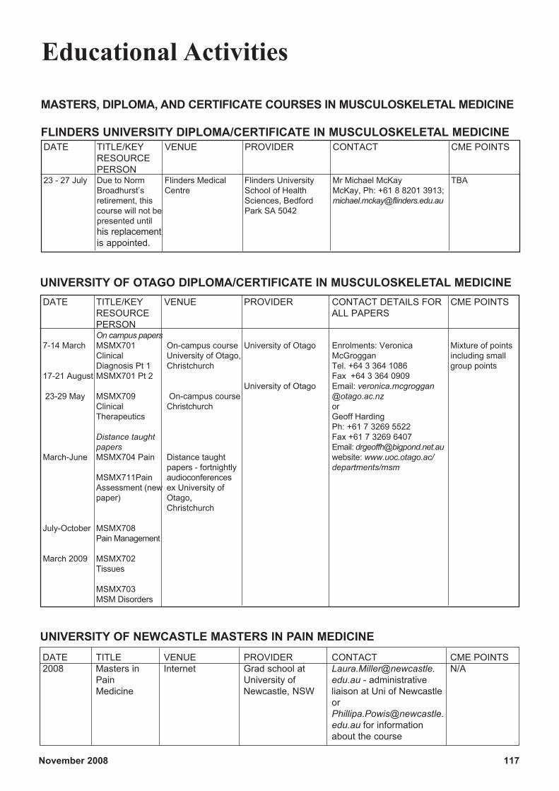
November 2008 117
Educational Activities
MASTERS, DIPLOMA, AND CERTIFICATE COURSES IN MUSCULOSKELETAL MEDICINE
FLINDERS UNIVERSITY DIPLOMA/CERTIFICATE IN MUSCULOSKELETAL MEDICINEDATE TITLE/KEY VENUE PROVIDER CONTACT CME POINTS
RESOURCEPERSON
23 - 27 July Due to Norm Flinders Medical Flinders University Mr Michael McKay TBABroadhurst’s Centre School of Health McKay, Ph: +61 8 8201 3913;retirement, this Sciences, Bedford [email protected] will not be Park SA 5042presented untilhis replacementis appointed.
DATE TITLE/KEY VENUE PROVIDER CONTACT DETAILS FOR CME POINTSRESOURCE ALL PAPERSPERSONOn campus papers
7-14 March MSMX701 On-campus course University of Otago Enrolments: Veronica Mixture of pointsClinical University of Otago, McGroggan including smallDiagnosis Pt 1 Christchurch Tel. +64 3 364 1086 group points
17-21 August MSMX701 Pt 2 Fax +64 3 364 0909University of Otago Email: veronica.mcgroggan
23-29 May MSMX709 On-campus course @otago.ac.nzClinical Christchurch orTherapeutics Geoff Harding
Ph: +61 7 3269 5522Distance taught Fax +61 7 3269 6407papers Email: [email protected]
March-June MSMX704 Pain Distance taught website: www.uoc.otago.ac/papers - fortnightly departments/msm
MSMX711Pain audioconferencesAssessment (new ex University ofpaper) Otago,
Christchurch
July-October MSMX708Pain Management
March 2009 MSMX702Tissues
MSMX703MSM Disorders
UNIVERSITY OF OTAGO DIPLOMA/CERTIFICATE IN MUSCULOSKELETAL MEDICINE
DATE TITLE VENUE PROVIDER CONTACT CME POINTS2008 Masters in Internet Grad school at Laura.Miller@newcastle. N/A
Pain University of edu.au - administrativeMedicine Newcastle, NSW liaison at Uni of Newcastle
[email protected] for informationabout the course
UNIVERSITY OF NEWCASTLE MASTERS IN PAIN MEDICINE
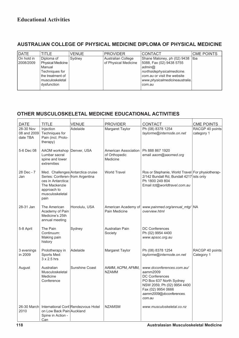
118 Australasian Musculoskeletal Medicine
OTHER MUSCULOSKELETAL MEDICINE EDUCATIONAL ACTIVITIES
DATE TITLE VENUE PROVIDER CONTACT CME POINTS28-30 Nov Injection Adelaide Margaret Taylor Ph (08) 8378 1254 RACGP 40 points08 and 2009 Techniques for [email protected] category 1date TBA Pain (incl. Prolo-
therapy)
5-6 Dec 08 AAOM workshop Denver, USA American Association Ph 888 867 1920Lumbar sacral of Orthopedic email [email protected] and lower Medicineextremities
28 Dec - 7 Med. Challenges Antarctica cruise World Travel Ros or Stephanie, World Travel For physiotherap-Jan Series: Conferen- from Argentina 2/142 Bundall Rd, Bundall 4217 ists only
ces in Antarctica: Ph 1800 249 804The Mackenzie Email [email protected] tomusculoskeletalpain
28-31 Jan The American Honolulu, USA American Academy of www.painmed.org/annual_mtg/ NAAcademy of Pain Pain Medicine overview.htmlMedicine’s 25thannual meeting
5-8 April The Pain Sydney Australian Pain DC ConferencesContinuum: Society Ph (02) 9954 4400Making pain www.apsoc.org.auhistory
3 evenings Prolotherapy in Adelaide Margaret Taylor Ph (08) 8378 1254 RACGP 40 pointsin 2009 Sports Med: [email protected] Category 1
3 x 2.5 hrs
August Australian Sunshine Coast AAMM, ACPM, AFMM, www.dcconferences.com.au/Musculoskeletal NZAMM aamm2009Medicine DC ConferencesConference PO Box 637 North Sydney
NSW 2059, Ph (02) 9954 4400Fax (02) 9954 [email protected]
26-30 March International Conf.Rendezvous Hotel NZAMSM www.musculoskeletal.co.nz2010 on Low Back Pain Auckland
Spine in Action -Can
Educational Activities
AUSTRALIAN COLLEGE OF PHYSICAL MEDICINE DIPLOMA OF PHYSICAL MEDICINE
DATE TITLE VENUE PROVIDER CONTACT CME POINTSOn hold in Diploma of Sydney Australian College Shane Maloney, ph (02) 9438 tba2008/2009 Physical Medicine of Physical Medicine 5088, Fax (02) 9438 5755
Manual admin@Techniques for northsidephysicalmedicine.the treatment of com.au or visit the websitemusculoskeletal www.physicalmedicineaustralia.dysfunction com.au
Related Documents


