Isolation, Structure Elucidation and Biological Investigation of Active Compounds in Cordia americana and Brugmansia suaveolens Dissertation der Mathematisch-Naturwissenschaftlichen Fakult¨ at der Eberhard Karls Universit¨ at T ¨ ubingen zur Erlangung des Grades eines Doktors der Naturwissenschaften (Dr. rer. nat.) vorgelegt von Fabiana Cristina Geller aus Santa Cruz do Sul - Brasilien T¨ ubingen 2010

Welcome message from author
This document is posted to help you gain knowledge. Please leave a comment to let me know what you think about it! Share it to your friends and learn new things together.
Transcript
Isolation, Structure Elucidation andBiological Investigation of
Active Compounds inCordia americana
andBrugmansia suaveolens
Dissertation
der Mathematisch-Naturwissenschaftlichen Fakultatder Eberhard Karls Universitat Tubingen
zur Erlangung des Grades einesDoktors der Naturwissenschaften
(Dr. rer. nat.)
vorgelegt vonFabiana Cristina Geller
aus Santa Cruz do Sul - Brasilien
Tubingen2010
The research work described herein, was conducted under the supervision of Prof. Dr. Stefan Lauferin the Department of Pharmaceutical and Medicinal Chemistry, Institute of Pharmacy, Universityof Tubingen from 01.01.07 to 31.08.10.
Tag der mundlichen Qualifikation: 10. November 2010
Dekan: Prof. Dr. Wolfgang Rosenstiel
1. Berichterstatter: Prof. Dr. Stefan Laufer
2. Berichterstatter: Prof. Dr. Irmgard Merfort
(Albert-Ludwigs-Universitat Freiburg)
“Jesus said to them, I am the bread of life; whoever comes to me shall not hunger, and whoeverbelieves in me shall never thirst”. John 6:35
“O segredo nao e correr atras das borboletas, mas sim, cultivar o seu jardim para que elas venhamate voce.” (Mario Quintana)


Mama, Papa (in memoriam) and Djones for your love, patience and support atall times.

AcknowledgmentsThe following is my appreciation to those people that in the past and present gave me the spirit
and encouragement to start, conduct and complete this thesis, as well to those people who mademe feel at home in Germany. My thanks go to my family, colleagues, cooperation partners andfriends who accompanied me during this work, in particular ...
• I am very grateful to my supervisor, Prof. Dr. Stefan Laufer for his comprehensive supportin all phases of this work and for the excellent opportunity of being a PhD. student in hisdepartment during my doctoral studies, in the last three years. For his insights and effortsto construct this bridge between South Brazil, Freiburg and Tubingen. Also for the financialsupport allowing me the participation in conferences and academic activities in Europe andin Brazil. His attention and motivation contributed to my personal and professional improve-ment. “Prof. Laufer, vielen herzlichen Dank!”.
• I am specially thankful to Prof. Dr. Irmgard Merfort, Freiburg, and her group. Thanksfor your dedication concerning the cooperation project Brazil-Germany and for the generoussupport allowing the execution of phytochemical and biological analysis in your department.Also for your valuable advices and improvements concerning my work and for teaching mea lot of things about Pharmacognosy.
• the members of my defense committee Prof. Dr. Rolf Daniels and Prof. Dr. Peter Ruth,for their time to go through my dissertation and taking part of my final exam.
• “muchas gracias tambien al profesor del Costa Rica”, Prof. Dr. Renato Murillo, for in-troducing me the complicated NMR topic in a very patient and uncomplicated form. Yoursuggestions and discussion regarding the elucidation of the flavonol glycosides were indis-pensable.
• “um grande muito obrigado” to Prof. Dr. Berta Heinzmann, from Santa Maria, Brazil.Thank you for introducing me to the plant world and for the profitable afternoons during thecollection of plants. I will always remember the nice time with you and your group.
• Prof. Dr. Erico Flores, who always supported our cooperation project, specially during theextraction of the plant material at the Department of Chemistry, at the Federal University ofSanta Maria, Brazil.
• Prof. Dr. Oliver Werz and his group, for the good living, the gatherings and the use ofhis laboratories and equipment. Specially, I would like to thank Bianca Jazzar and DanielaMuller for providing me technical assistance in the 5-lipoxygenase assays.

• “ein grosses Dankeschon” to Prof. Dr. Wolf Engels (Brasilien-Zentrum), who along withProf. Dr. Stefan Laufer made efforts to acquire financial support from the Ministry ofScience, Research and the Arts of Baden-Wurttemberg for the project involving Brazil andGermany.
• “ein grosses Dankeschon” to Dr. Rainer Radtke for the great time that we spent together inTubingen. Thanks for reading my dissertation and for your suggestions and improvements.
• the botanists Dr. Solon Longhi and Dr. Gilberto Zanetti for the collection of the plantsCordia americana and Brugmansia suaveolens.
• Marcio Fronza and Cleber Schmidt from the Department of Pharmaceutical Biology andBiotechnology, University of Freiburg, for carrying out the scratch and NF-κB assays. Ca-tiguria, many thanks for the nice chats from time to time about research and also otherthings. Thanks for reading my dissertation and for your suggestions. I am sure that webecame good friends. I appreciated that I had the chance to meet you here in Germany!
• “ein super Dankeschon fur” Stef�, for your very sweet Swabian sentence “Fabi, es wirdscho”. Stef�, many thanks for reading my dissertation, for your suggestions involving NMRspectra, as well as for the chocolates and “Gummibarchen” time! I hope that you will cometo visit me in Brazil.
• “ein grosses Dankeschon” to Lisa Steinhauser for supporting me with the NMR spectra andalso for organizing the NMR measurements.
• Sabine for enjoying with me the rare sunshine time during the breaks at the university. Joeand Mohamed thanks for the patience during the first steps with the flash chromatography.Maissa thanks for your big smile. Thanks also for the nice time in the lab and also for thefriendship.
• Claudi and Frank for their time to perform the LC-MS measurements and for the nice chatduring the “Mittagspause” in the “Mensa”.
• Verena Schattel for conducting the molecular modeling studies and for providing the dock-ing pictures.
• Marcia Goettert and Katharina Bauer for carrying out the biological assays on p38α,JNK3 and TNFα.
• our secretary Karin Ward for all solutions concerning the bureaucratic problems.

• all my colleagues and the employees from our department who contributed to the realizationof my dissertation.
• “meu amor e meu alemao preferido”' Djones; how can I thank you? You are the best thing,the best person that Tubingen brought me! Thanks for your love, patience, encouragementand support.
• my lovely, wonderful and big Geller family, specially meine Mutti, for her love, for theunconditionally support, even many times feeling the distance ... a thousand thanks foreverything!
• my parents-in-law for holding me up in many moments.
• my family and friends in Germany: Walter for the very nice time in Munchen and inthe“Bayerische Wald”, Pedro, Sandra and Jorge, Birkner's thanks for the nice celebra-tions together. Also for the forever Brazilian friends that Tubingen brought me: Melissa,Lissi, Ana Carolina, Karina, for sure we will see us in Brazil and will miss the nice timein Tubingen.
• my friends in Brazil, Julie, Ana Paula e Andressa. The friendship that keeps us together isone of the greatest thing that ever happened to me.
• the Eberhard Karls University of Tubingen and the Pharmacy Institute in Tubingen forsupporting the necessary conditions for the development of this research work and also forthe opportunity to attend German courses in order to improve my language skills.
• the Goverment of Baden-Wurttemberg (Zukunftsoffensive IV “Innovation und Exzel-lenz”, Forderung von internationalen Kooperationen zwischen den Hochschulen) for thefinancial support that was indispensable for the development of this work.
Fabiana Cristina Geller

AbstractIn Brazil, medicinal plants have been widely used for the treatment of diseases in folk medicine.
However, the effective compounds responsible for the biological effects are often unknown. Ex-tracts prepared from traditional medicinal plants from South Brazil were screened for their anti-inflammatory and wound healing activities. The Boraginaceae Cordia americana, locally knownas “Guajuvira”, and the Solanaceae Brugmansia suaveolens, generically recognized as “Trom-beteira”, presented interesting activity in the biological screening. Thus, the objective of this dis-sertation was the investigation of the ethanolic extracts prepared from the leaves of both plants andthe characterization of potential effective compounds, focusing on: firstly, the isolation of the plantconstituents using chromatographic methods; secondly, structural elucidation by means of spec-troscopy experiments; and finally, biological investigation of the plant extracts and their respectivecompounds targeting different aspects of inflammation and wound healing processes.
From the ethanolic extract of Cordia americana, flavonols (rutin and quercitrin), phenolic com-pounds (rosmarinic acid, rosmarinic acid ethyl ester and 3-(3,4-dihydroxyphenyl)-2-hydroxypropa-noic acid), phytosterols (campesterol and β-sistosterol) and triterpenoids (α- and β-amyrin) werecharacterized. Quantification analysis of the plant extract showed rosmarinic acid as the majorconstituent with an amount of 8.44%. The ethanolic extract exhibited higher inhibition (i.e., pro-inflammatory mediators p38α and JNK3, TNFα and 5-LO as well as on scratch assay) in compar-ison with the predominant and other isolated compounds, however, evidences were provided for acrucial role of rosmarinic acid as the major key player.
Regarding the ethanolic extract of Brugmansia suaveolens, four new flavonol glycosides kaemp-ferol 3-O-β-D-glucopyranosyl-(1′′′→2′′)-O-α-L-arabinopyranoside-7-O-β-D-glucopyranoside, ka-empferol 3-O-β-D-[6′′′-O-(3,4-dihydroxy-cinnamoyl)]-glucopyranosyl-(1′′′→2′′)-O-α-L-arabino-pyranoside-7-O-β-D-glucopyranoside, kaempferol 3-O-β-D-[2′′′-O-(3,4-dihydroxy-cinnamoyl)]-glucopyranosyl-(1′′′→2′′)-O-α-L-arabinopyranoside-7-O-β-D-glucopyranoside, and kaempferol 3-O-β-D-glucopyranosyl-(1′′′→ 2′′)-O-α-L-arabinopyranoside were isolated. Concerning the bio-logical effects of the ethanolic extract, the kaempferol aglycone as well as further non-isolatedsecondary metabolites might contribute to the plant activity.
In summary, this dissertation increases the phytochemical and pharmacological knowledge aboutCordia americana and Brugmansia suaveolens, which support their use in traditional medicine.
ZusammenfassungIn Brasilien werden in der Volksmedizin Heilpflanzen haufig fur die Behandlung von Krankheiten
verwendet. Die wirksamen Verbindungen, verantwortlich fur die biologischen Wirkungen, sindaber in der Regel unbekannt. Extrakte aus traditionellen Heilpflanzen aus Sud-Brasilien wurdenauf ihre entzundungshemmenden und wundheilenden Eigenschaften untersucht. Die BoraginaceaeCordia americana, lokal bekannt als “Guajuvira”, und die Solanaceae Brugmansia suaveolens, all-gemein bekannt als “Trombeteira”, prasentierten interessante biologische Aktivitaten in den erstenScreening-Versuchen. So war das Ziel dieser Dissertation die Untersuchung der ethanolischen Ex-trakte aus den Blattern der beiden Pflanzen und die Charakterisierung von potentiell wirksamenVerbindungen. Hierbei erfolgte die Isolierung der pflanzlichen Inhaltstoffe mit chromatographis-chen Methoden, die Strukturaufklarung mittels NMR- und MS-Spektroskopie, und die biologischeUntersuchung der Pflanzenextrakte und ihrer jeweiligen Inhaltstoffe in Testsystemen, die die Un-tersuchung verschiedener Aspekte der Entzundung und Wundheilung moglich machen.
Von dem ethanolischen Extrakt von Cordia americana wurden die Flavonoide (Rutin und Querci-trin), Phenolische Verbindungen (Rosmarinsaure, Rosmarinsaure Ethylester und 3-(3,4 dihydroxy-phenyl)-2-Hydroxypropansaure), Phytosterine (Campesterin und β-Sitosterol) und Triterpenoide(α-und β-Amyrin) charakterisiert. Die Quantifizierung des pflanzlichen Extrakts zeigte Rosmarin-saure als Hauptbestandteil mit einer Konzentration von 8,44%. Der ethanolische Extrakt zeigteeine nennenswerte Hemmung von proinflammatorischen Mediatoren wie p38α, JNK3, TNFα und5-LO sowie im Scratch assay (als Modelle fur Wundheilung), im Vergleich zu den Hauptbe-standteilen und anderen isolierten Verbindungen. Rosmarinsaure kommt eine Schlusselrolle furdiese Wirkung zu.
Hinsichtlich des ethanolischen Extrakts von Brugmansia suaveolens, konnten vier neue Flavonol-glykoside isolierter werden: Kaempferol 3-O-β-D-glucopyranosyl-(1′′′→2′′)-O-α-L-arabinopyra-noside-7-O-β-D-glucopyranoside, Kaempferol 3-O-β-D-[6′′′-O-(3,4-dihydroxy-cinnamoyl)]-glu-copyranosyl-(1′′′→2′′)-O-α-L-arabinopyranoside-7-O-β-D-glucopyranoside, Kaempferol 3-O-β-D-[2′′′-O-(3,4-dihydroxy-cinnamoyl)]-glucopyranosyl-(1′′′→2′′)-O-α-L-arabinopyranoside-7-O-β-D-glucopyranoside, and Kaempferol 3-O-β-D-glucopyranosyl-(1′′′→2′′)-O-α-L-arabinopyranosi-de. Bezuglich der biologischen Effekte des ethanolischen Extrakts konnten das Kaempferol Aglykonsowie weitere nicht isolierte Sekundarmetaboliten zur Aktivitat des Extrakts beitragen.
Damit tragt dieser Dissertation zur Ausweitung der phytochemischen und pharmakologischenKenntnisse uber Cordia americana und Brugmansia suaveolens.
List of Publications and PresentationsFull Papers
• Geller F., Schmidt C., Goettert M., Fronza M., Schattel V., Heinzmann B., Werz O., FloresE.M.M., Merfort I., Laufer S. Identification of rosmarinic acid as the major active constituentin Cordia americana. Journal of Ethnopharmacology, 128, 561-566, 2010.
• Geller F., Murillo R., Steinhauser L., Heinzmann B., Flores E., Albert K., Merfort I., LauferS. Flavonol glycosides from the leaves of Brugmansia suaveolens. In preparation.
• Schmidt C., Fronza M., Goettert M., Geller F., Luik S., Flores E.M.M., Bittencourt C.F.,Zanetti G.D., Heinzmann B.M., Laufer S., Merfort I. Biological studies on Brazilian plantsused in wound healing. Journal of Ethnopharmacology, 122, 523-532, 2009.
Oral Presentations
• Geller, F., Schmidt, C., Goettert, M., Fronza, M., Heinzmann, B., Werz, O., Merfort, I.,Laufer, S. Rosmarinic acid as the effective compound in Cordia americana. Deutsch-Brasi-lianisches Jahr 2010/11, Drugs from Natural Sources: The Potential of Brazilian Plants usedin Traditional Medicine, Sao Paulo, Brazil, 22.09.2010.
• Geller F., Heinzmann B., Goettert M., Werz O., Merfort I., Laufer S. Isolation and identi-fication of natural compounds with anti-inflammatory activity from Cordia americana. IVSimposio Brasil Alemanha: Desenvolvimento Sustentavel, Curitiba, Brazil, 05-07.10.2009.
Presentations
• Geller F., Schmidt C., Goettert M., Fronza M., Heinzmann B., Werz O., Merfort I., LauferS. Rosmarinic acid as the effective compound in Cordia americana. 58th InternationalCongress and Annual Meeting of the Society for Medicinal Plant and Natural Product Re-search, Berlin, 29.08-02.09.2010.
• Geller F., Goettert M., Fronza M., Schmidt C., Schattel V., Heinzmann B., Flores E., MerfortI., Laufer S. Phytochemical and biological investigation on the ethanolic extract of Cordia
americana. 6th Status Seminar Chemical Biology, Frankfurt, 30.11-1.12.2009.
• Geller F., Heinzmann B., Schattel V., Goettert M., Werz O., Merfort I., Laufer S.. Identifi-cation of the main effective compound in the ethanolic extract from Cordia americana. IVDeutsch-Brasilianisches Symposium, Curitiba - Parana, Brasilien, 05-10.10.2009
• Geller F., Heinzmann B., Goettert M., Schattel V., Werz O., E. Flores, Merfort I., Laufer S.Phytochemical and anti-inflammatory investigation on the ethanolic extract of Cordia amer-
icana. Jahrestagung der Deutschen Pharmazeutischen Gesellschaft, Jena, 28.09-1.10.2009.
• Fronza M., Heinzmann B., Geller F., Laufer S., Merfort I. An improved scratch assay forstudying the wound healing effects of medicinal plants. IV Simposio Brasil Alemanha:Desenvolvimento Sustentavel, Curitiba, Brazil, 05-07.10.2009.
• Goettert M., Luik S., Fronza M., Schmdit C., Geller F., Heinzmann B., Merfort I., LauferS. Structural features and biological evaluation of flavonoids as p38α MAPK inhibitors. IVSimposio Brasil Alemanha: Desenvolvimento Sustentavel, Curitiba, Brazil, 05-07.10.2009.
• Goettert M., Luik S., Fronza M., Schmidt C., Geller F., Heinzmann B., Merfort I., Laufer S.Effect of natural phenolic compounds on p38α MAPK activity IV Deutsch-BrasilianischesSymposium, Curitiba - Parana, Brasilien, 05-10.10.2009.
• Goettert M., Luik S., Fronza M., Schmidt C., Geller F., Merfort I., Laufer S. Natural phenoliccompounds as inhibitors of p38α MAPK. Drug Discovery and Delivery Membrane Proteinsand Natural Product Research, Freiburg, 16-17.04.2009.
• Goettert M., Luik S., Fronza M., Geller F., Schmidt C., Merfort I. , Laufer S. Biological test-ing of bioactive compounds that inhibit p38α MAPK. 5th Status Seminar Chemical Biology,ChemBioNnet, Frankfurt, 08.12.2008.
• Fronza M., Geller F., Bittencourt C., Flores E., Heinzmann B., Laufer S., Merfort I. Thescratch assay: A suitable in vitro tool for studying wound healing effects. 7th Joint Meetingof AFERP, ASP, GA, PSE, SIF, Athens, Greece, August 2008.
• Geller F., Goettert M., Heinzmann B., Laufer S. Identification, structural elucidation andbiological testing of active principles of Brazilian medicinal plants. Naturraume Brasiliens:Im Spannungsfeld zwischen biologischer Vielfalt und industrieller Entwicklung. AustellungUniversitatsbibliothek Tubingen, 05.6.2008.
• Merfort I., Heinzmann B., Flores E., Bittencourt C., Schmidt C., Geller F., Goettert M.,Laufer S. Biological active compounds from Brazilian traditional medicinal plants. IIIDeutsch-Brasilianisches Symposium, Freiburg, 23-27.07.2007.
Contents
1 Introduction 11.1 The Importance of Medicinal Plants in Drug Discovery . . . . . . . . . . . . . . . 11.2 Project Overview . . . . . . . . . . . . . . . . . . . . . . . . . . . . . . . . . . . 3
1.2.1 Screening . . . . . . . . . . . . . . . . . . . . . . . . . . . . . . . . . . . 31.2.2 Cordia americana . . . . . . . . . . . . . . . . . . . . . . . . . . . . . . 5
1.2.2.1 Localization . . . . . . . . . . . . . . . . . . . . . . . . . . . . 61.2.2.2 Botany . . . . . . . . . . . . . . . . . . . . . . . . . . . . . . . 61.2.2.3 Economical Importance and Traditional Medicine . . . . . . . . 91.2.2.4 Chemical Constituents . . . . . . . . . . . . . . . . . . . . . . . 9
1.2.3 Brugmansia suaveolens . . . . . . . . . . . . . . . . . . . . . . . . . . . 111.2.3.1 Localization . . . . . . . . . . . . . . . . . . . . . . . . . . . . 121.2.3.2 Botany . . . . . . . . . . . . . . . . . . . . . . . . . . . . . . . 131.2.3.3 Economical Importance and Traditional Medicine . . . . . . . . 141.2.3.4 Chemical Constituents . . . . . . . . . . . . . . . . . . . . . . . 15
1.3 Objectives of this Dissertation . . . . . . . . . . . . . . . . . . . . . . . . . . . . 17
2 In�ammatory and Wound Healing Processes 192.1 Inflammatory and Wound Healing Processes . . . . . . . . . . . . . . . . . . . . . 192.2 Mitogen-Activated Protein Kinases (MAPKs) . . . . . . . . . . . . . . . . . . . . 20
2.2.1 The ERK Signaling Pathway . . . . . . . . . . . . . . . . . . . . . . . . . 222.2.2 The JNK Signaling Pathway . . . . . . . . . . . . . . . . . . . . . . . . . 232.2.3 The p38 MAPK . . . . . . . . . . . . . . . . . . . . . . . . . . . . . . . . 242.2.4 Structure of Protein Kinase . . . . . . . . . . . . . . . . . . . . . . . . . . 262.2.5 Diseases Associated with MAPKs . . . . . . . . . . . . . . . . . . . . . . 30
2.3 Cytokines . . . . . . . . . . . . . . . . . . . . . . . . . . . . . . . . . . . . . . . 322.3.1 Tumor Necrosis Factor α (TNFα) . . . . . . . . . . . . . . . . . . . . . . 33
2.4 Nuclear Factor-κB (NF-κB) . . . . . . . . . . . . . . . . . . . . . . . . . . . . . 352.5 Arachidonic Acid Cascade . . . . . . . . . . . . . . . . . . . . . . . . . . . . . . 37
2.5.1 5-Lipoxygenase . . . . . . . . . . . . . . . . . . . . . . . . . . . . . . . . 382.5.2 Structure and Regulation of 5-LO . . . . . . . . . . . . . . . . . . . . . . 402.5.3 5-LO Inhibitors . . . . . . . . . . . . . . . . . . . . . . . . . . . . . . . . 40
2.6 Wound Healing Process: Scratch and Elastase . . . . . . . . . . . . . . . . . . . . 41
i
Contents
3 Results and Discussion 433.1 Phytochemical Investigation . . . . . . . . . . . . . . . . . . . . . . . . . . . . . 43
3.1.1 Cordia americana . . . . . . . . . . . . . . . . . . . . . . . . . . . . . . 433.1.1.1 Bioguided Fractionation based on p38α MAPK Assay . . . . . . 433.1.1.2 Identification and Structural Elucidation . . . . . . . . . . . . . 44
3.1.1.2.1 CA3: 3-(3,4-dihydroxyphenyl)-2-hydroxypropanoic acid 443.1.1.2.2 CA1: Rosmarinic Acid . . . . . . . . . . . . . . . . . 513.1.1.2.3 CA2: Rosmarinic Acid Ethyl Ester . . . . . . . . . . . 593.1.1.2.4 CA4: Rutin . . . . . . . . . . . . . . . . . . . . . . . 663.1.1.2.5 CA5: Quercitrin . . . . . . . . . . . . . . . . . . . . . 763.1.1.2.6 CA6: β-Sitosterol . . . . . . . . . . . . . . . . . . . . 783.1.1.2.7 CA7: Campesterol . . . . . . . . . . . . . . . . . . . 793.1.1.2.8 CA8: α-Amyrin . . . . . . . . . . . . . . . . . . . . . 813.1.1.2.9 CA9: β-Amyrin . . . . . . . . . . . . . . . . . . . . . 82
3.1.1.3 Discussion . . . . . . . . . . . . . . . . . . . . . . . . . . . . . 843.1.2 Brugmansia suaveolens . . . . . . . . . . . . . . . . . . . . . . . . . . . 88
3.1.2.1 Bioguided Fractionation based on p38α MAPK Assay . . . . . . 883.1.2.2 Structural Elucidation . . . . . . . . . . . . . . . . . . . . . . . 88
3.1.2.2.1 BS4: Kaempferol 3-O-β-D-glucopyranosyl-(1′′′→2′′)-O-α-L-arabinopyranoside . . . . . . . . . . . . . . . 89
3.1.2.2.2 BS1: Kaempferol 3-O-β-D-glucopyranosyl-(1′′′→2′′)-O-α-L-arabinopyranoside-7-O-β-D-glucopyranoside . 101
3.1.2.2.3 BS2: Kaempferol 3-O-β-D-[6′′′-O-(3,4-dihydroxy-cinnamoyl)]-glucopyranosyl-(1′′′→2′′)-O-α-L-arabinopyranoside-7-O-β-D-glucopyranoside . . . . . . . . . . . . . . . . . 113
3.1.2.2.4 BS3: Kaempferol 3-O-β-D-[2′′′-O-(3,4-dihydroxy-cinnamoyl)]-glucopyranosyl-(1′′′→2′′)-O-α-L-arabinopyranoside-7-O-β-D-glucopyranoside . . . . . 124
3.1.2.3 Discussion . . . . . . . . . . . . . . . . . . . . . . . . . . . . . 1353.2 Biological Investigation and Discussion . . . . . . . . . . . . . . . . . . . . . . . 139
3.2.1 p38α MAPK . . . . . . . . . . . . . . . . . . . . . . . . . . . . . . . . . 1393.2.1.1 Cordia americana . . . . . . . . . . . . . . . . . . . . . . . . . 1403.2.1.2 Brugmansia suaveolens . . . . . . . . . . . . . . . . . . . . . . 144
3.2.2 TNFα . . . . . . . . . . . . . . . . . . . . . . . . . . . . . . . . . . . . . 1463.2.3 JNK3 MAPK . . . . . . . . . . . . . . . . . . . . . . . . . . . . . . . . . 148
3.2.3.1 Cordia americana . . . . . . . . . . . . . . . . . . . . . . . . . 1483.2.3.2 Brugmansia suaveolens . . . . . . . . . . . . . . . . . . . . . . 152
3.2.4 5-Lipoxygenase . . . . . . . . . . . . . . . . . . . . . . . . . . . . . . . . 1533.2.4.1 Inhibition of 5-LO Activity in a Cell-free Assay . . . . . . . . . 153
3.2.4.1.1 Cordia americana . . . . . . . . . . . . . . . . . . . . 1543.2.4.1.2 Brugmansia suaveolens . . . . . . . . . . . . . . . . . 155
3.2.4.2 Interference of 5-LO Activity in Cell-based Assay Using PMNL 1563.2.5 Supplementary Assays for Cordia americana . . . . . . . . . . . . . . . . 157
3.2.5.1 NF-κB Assay . . . . . . . . . . . . . . . . . . . . . . . . . . . 157
ii
Contents
3.2.5.2 Scratch Assay . . . . . . . . . . . . . . . . . . . . . . . . . . . 1593.2.6 Summary of the Biological Activity . . . . . . . . . . . . . . . . . . . . . 161
3.2.6.1 Rosmarinic Acid, Rosmarinic Acid Ethyl Ester and3-(3,4-dihydroxyphenyl)-2-hydroxypropanoic acid . . . . . . . . 161
3.2.6.2 β-Sitosterol and Campesterol . . . . . . . . . . . . . . . . . . . 1623.2.6.3 α- and β-Amyrin . . . . . . . . . . . . . . . . . . . . . . . . . 1633.2.6.4 Flavonol Glycosides . . . . . . . . . . . . . . . . . . . . . . . . 163
4 Summary 165
5 Experimental Part 1695.1 Plant Material . . . . . . . . . . . . . . . . . . . . . . . . . . . . . . . . . . . . . 1695.2 Chemicals, Reagents and Materials . . . . . . . . . . . . . . . . . . . . . . . . . . 1705.3 Instruments . . . . . . . . . . . . . . . . . . . . . . . . . . . . . . . . . . . . . . 1705.4 Chromatographic and Spectroscopic Methods . . . . . . . . . . . . . . . . . . . . 171
5.4.1 Thin Layer Chromatography (TLC) . . . . . . . . . . . . . . . . . . . . . 1715.4.1.1 TLC Method for Cordia americana . . . . . . . . . . . . . . . . 1715.4.1.2 TLC Methods for Brugmansia suaveolens . . . . . . . . . . . . 171
5.4.2 Column Chromatography . . . . . . . . . . . . . . . . . . . . . . . . . . . 1735.4.2.1 Sephadex®LH-20 . . . . . . . . . . . . . . . . . . . . . . . . . 1735.4.2.2 Open Column Chromatography (OC) . . . . . . . . . . . . . . . 173
5.4.3 Flash Chromatography (FC) . . . . . . . . . . . . . . . . . . . . . . . . . 1735.4.4 High Pressure Liquid Chromatography (HPLC) . . . . . . . . . . . . . . . 1745.4.5 UV-Visible Spectroscopy . . . . . . . . . . . . . . . . . . . . . . . . . . . 1765.4.6 Fourier Transform-Infrared Spectroscopy (FT-IR) . . . . . . . . . . . . . . 1765.4.7 Mass Spectroscopy . . . . . . . . . . . . . . . . . . . . . . . . . . . . . . 176
5.4.7.1 Gas Chromatography-Mass Spectrometry (GC-MS) . . . . . . . 1765.4.7.2 Electron Ionization Mass Spectrometry (EI-MS) . . . . . . . . . 1775.4.7.3 Electrospray Ionisation-Mass Spectrometry (ESI-MS) . . . . . . 1775.4.7.4 Fourier-Transform-Ion Cyclotron Resonance Mass-Spectrometry
(FT-ICR-MS) . . . . . . . . . . . . . . . . . . . . . . . . . . . 1785.4.8 Nuclear Magnetic Resonance Spectroscopy (NMR) . . . . . . . . . . . . . 179
5.5 Plant Extraction Methods for the Biological Screening Phase . . . . . . . . . . . . 1795.6 Extraction and Isolation Methods . . . . . . . . . . . . . . . . . . . . . . . . . . . 181
5.6.1 Cordia americana . . . . . . . . . . . . . . . . . . . . . . . . . . . . . . 1815.6.1.1 Isolation of Compounds . . . . . . . . . . . . . . . . . . . . . . 1855.6.1.2 Characterization of the Compounds . . . . . . . . . . . . . . . . 1855.6.1.3 Quantification Method . . . . . . . . . . . . . . . . . . . . . . . 189
5.6.2 Brugmansia suaveolens . . . . . . . . . . . . . . . . . . . . . . . . . . . 1905.6.2.1 Qualitative Analysis for Alkaloids . . . . . . . . . . . . . . . . 1945.6.2.2 Isolation of Compounds . . . . . . . . . . . . . . . . . . . . . . 1945.6.2.3 Characterization of the Compounds . . . . . . . . . . . . . . . . 195
5.7 Biological Assays . . . . . . . . . . . . . . . . . . . . . . . . . . . . . . . . . . . 1985.7.1 p38α MAPK Assay . . . . . . . . . . . . . . . . . . . . . . . . . . . . . . 198
iii

Contents
5.7.2 JNK3 MAPK Assay . . . . . . . . . . . . . . . . . . . . . . . . . . . . . 2015.7.3 TNFα Release Assay . . . . . . . . . . . . . . . . . . . . . . . . . . . . . 2025.7.4 5-Lipoxygenase Assay . . . . . . . . . . . . . . . . . . . . . . . . . . . . 205
5.7.4.1 Determination of 5-LO Product Formation in Cell-free Assays . 2055.7.4.2 Isolation of Human PMNL from Venous Blood . . . . . . . . . . 2065.7.4.3 Determination of 5-LO Product Formation in Cell-based Assays
Using Isolated Human PMNL . . . . . . . . . . . . . . . . . . . 2065.7.5 NF-κB Electrophoretic Mobility Shift Assay (EMSA) . . . . . . . . . . . 2075.7.6 Fibroblast Scratch Assay . . . . . . . . . . . . . . . . . . . . . . . . . . . 2075.7.7 MTT Assay . . . . . . . . . . . . . . . . . . . . . . . . . . . . . . . . . . 208
5.8 Computer Program . . . . . . . . . . . . . . . . . . . . . . . . . . . . . . . . . . 2085.9 Statistical Analysis . . . . . . . . . . . . . . . . . . . . . . . . . . . . . . . . . . 2095.10 Docking . . . . . . . . . . . . . . . . . . . . . . . . . . . . . . . . . . . . . . . . 209
iv
List of Figures
1.1 Distribution of Cordia americana [241] . . . . . . . . . . . . . . . . . . . . . . . 61.2 Tree of Cordia americana . . . . . . . . . . . . . . . . . . . . . . . . . . . . . . 71.3 Leaf of Cordia americana . . . . . . . . . . . . . . . . . . . . . . . . . . . . . . 71.4 Flower of Cordia americana . . . . . . . . . . . . . . . . . . . . . . . . . . . . . 81.5 Fruit of Cordia americana [117] . . . . . . . . . . . . . . . . . . . . . . . . . . . 81.6 Distribution of Brugmansia suaveolens [241] . . . . . . . . . . . . . . . . . . . . 121.7 Shrub of Brugmansia suaveolens . . . . . . . . . . . . . . . . . . . . . . . . . . . 131.8 Leaf of Brugmansia suaveolens . . . . . . . . . . . . . . . . . . . . . . . . . . . . 131.9 Flower form . . . . . . . . . . . . . . . . . . . . . . . . . . . . . . . . . . . . . . 141.10 Flower length . . . . . . . . . . . . . . . . . . . . . . . . . . . . . . . . . . . . . 14
2.1 Illustration of the general MAPK signaling cascades [44] . . . . . . . . . . . . . . 222.2 p38 MAPK signaling pathway [44] . . . . . . . . . . . . . . . . . . . . . . . . . . 262.3 Representation of the structure of p38 MAPK (PDB ID: 1A9U) [137, 127]) . . . . 272.4 Representation of the ATP-binding site of protein kinases bound to the ATP cofator
[314] . . . . . . . . . . . . . . . . . . . . . . . . . . . . . . . . . . . . . . . . . . 292.5 Activation pathways of the transcription factor NF-κB [279] . . . . . . . . . . . . 362.6 Arachidonic acid cascade . . . . . . . . . . . . . . . . . . . . . . . . . . . . . . . 382.7 Conversion of arachidonic acid in leukotrienes by 5-Lipoxygenase [330] . . . . . . 39
3.1 Chemical structure of CA3 . . . . . . . . . . . . . . . . . . . . . . . . . . . . . . 443.2 EI-MS of CA3 . . . . . . . . . . . . . . . . . . . . . . . . . . . . . . . . . . . . . 453.3 1H-NMR of CA3 (400 MHz, MeOH-d4) . . . . . . . . . . . . . . . . . . . . . . . 473.4 13C-NMR of CA3 (100 MHz, MeOH-d4) . . . . . . . . . . . . . . . . . . . . . . 483.5 DEPT-135 of CA3 (100 MHz, MeOH-d4) . . . . . . . . . . . . . . . . . . . . . . 493.6 H-H-COSY of CA3 (400 MHz, MeOH-d4) . . . . . . . . . . . . . . . . . . . . . 503.7 Chemical structure of CA1 . . . . . . . . . . . . . . . . . . . . . . . . . . . . . . 513.8 UV spectrum of CA1 . . . . . . . . . . . . . . . . . . . . . . . . . . . . . . . . . 513.9 IR spectrum of CA1 . . . . . . . . . . . . . . . . . . . . . . . . . . . . . . . . . . 523.10 ESI-MS (negative mode) of CA1 . . . . . . . . . . . . . . . . . . . . . . . . . . . 533.11 1H-NMR of CA1 (400 MHz, MeOH-d4) . . . . . . . . . . . . . . . . . . . . . . . 553.12 13C-NMR of CA1 (100 MHz, MeOH-d4) . . . . . . . . . . . . . . . . . . . . . . 563.13 DEPT-135 of CA1 (100 MHz, MeOH-d4) . . . . . . . . . . . . . . . . . . . . . . 573.14 H-H-COSY of CA1 (400 MHz, MeOH-d4) . . . . . . . . . . . . . . . . . . . . . 583.15 Chemical structure of CA2 . . . . . . . . . . . . . . . . . . . . . . . . . . . . . . 593.16 ESI-MS (negative mode) of CA2 . . . . . . . . . . . . . . . . . . . . . . . . . . . 603.17 1H-NMR of CA2 (400 MHz, MeOH-d4) . . . . . . . . . . . . . . . . . . . . . . . 62
v
List of Figures
3.18 13C-NMR of CA2 (100 MHz, MeOH-d4) . . . . . . . . . . . . . . . . . . . . . . 633.19 DEPT-135 of CA2 (100 MHz, MeOH-d4) . . . . . . . . . . . . . . . . . . . . . . 643.20 H-H-COSY of CA2 (400 MHz, MeOH-d4) . . . . . . . . . . . . . . . . . . . . . 653.21 Chemical structure of CA4 . . . . . . . . . . . . . . . . . . . . . . . . . . . . . . 663.22 UV of CA4 . . . . . . . . . . . . . . . . . . . . . . . . . . . . . . . . . . . . . . 663.23 IR spectrum of CA4 . . . . . . . . . . . . . . . . . . . . . . . . . . . . . . . . . . 673.24 ESI-MS (positive mode) of CA4 . . . . . . . . . . . . . . . . . . . . . . . . . . . 683.25 1H-NMR of CA4 (400 MHz, DMSO-d6) . . . . . . . . . . . . . . . . . . . . . . . 713.26 13C-NMR of CA4 (100 MHz, DMSO-d6) . . . . . . . . . . . . . . . . . . . . . . 723.27 DEPT-135 of CA4 (100 MHz, DMSO-d6) . . . . . . . . . . . . . . . . . . . . . . 733.28 H-H-COSY of CA4 (400 MHz, DMSO-d6) . . . . . . . . . . . . . . . . . . . . . 743.29 HSQC of CA4 (400 MHz, DMSO-d6) . . . . . . . . . . . . . . . . . . . . . . . . 753.30 Chemical structure of CA5 . . . . . . . . . . . . . . . . . . . . . . . . . . . . . . 763.31 Comparison between the chromatogram of the fraction I and quercitrin standard
(Method LC-DAD) . . . . . . . . . . . . . . . . . . . . . . . . . . . . . . . . . . 773.32 Comparison of the MS data of quercetrin from fraction I and the standard . . . . . 773.33 Chemical structure of CA6 . . . . . . . . . . . . . . . . . . . . . . . . . . . . . . 783.34 Comparison of the MS data between peak GC-CA6 (A) and respective standard (B) 793.35 Chemical structure of CA7 . . . . . . . . . . . . . . . . . . . . . . . . . . . . . . 793.36 Comparison of the MS data between peak GC-CA7 (A) and respective standard (B) 803.37 Chemical structure of CA8 . . . . . . . . . . . . . . . . . . . . . . . . . . . . . . 813.38 Comparison of the MS fragmentation between peak GC-CA8 (A) and data from
the natural compound library (B) . . . . . . . . . . . . . . . . . . . . . . . . . . . 823.39 Chemical structure of CA9 . . . . . . . . . . . . . . . . . . . . . . . . . . . . . . 823.40 Comparison of the MS fragmentation between peak GC-CA9 (A) and data from
the natural compound library (B) . . . . . . . . . . . . . . . . . . . . . . . . . . . 833.41 Representative HPLC chromatogram of the ethanolic extract of Cordia americana
and its characterized compounds. Rosmarinic acid (CA1), rosmarinic acid ethyl es-ter (CA2), 3-(3,4-dihydroxyphenyl)-2-hydroxypropanoic acid (CA3), rutin (CA4),and quercitrin (CA5) (Method LC-DAD, with wavelength λ = 254 nm). . . . . . . 85
3.42 Chemical structure of BS4 . . . . . . . . . . . . . . . . . . . . . . . . . . . . . . 893.43 UV of the compound BS4 . . . . . . . . . . . . . . . . . . . . . . . . . . . . . . . 903.44 IR of the compound BS4 . . . . . . . . . . . . . . . . . . . . . . . . . . . . . . . 903.45 ESI-MS (positive mode) of the compound BS4 . . . . . . . . . . . . . . . . . . . 913.46 1H-NMR of BS4 (250 MHz, MeOH-d4) . . . . . . . . . . . . . . . . . . . . . . . 953.47 13C-NMR of BS4 (100 MHz, MeOH-d4) . . . . . . . . . . . . . . . . . . . . . . . 963.48 DEPT-135 of BS4 (100 MHz, MeOH-d4) . . . . . . . . . . . . . . . . . . . . . . 973.49 H-H-COSY of BS4 (600 MHz, MeOH-d4) . . . . . . . . . . . . . . . . . . . . . . 983.50 HSQC of BS4 (600 MHz, MeOH-d4) . . . . . . . . . . . . . . . . . . . . . . . . . 993.51 HMBC of BS4 (600 MHz, MeOH-d4) . . . . . . . . . . . . . . . . . . . . . . . . 1003.52 Chemical structure of the compound BS1 . . . . . . . . . . . . . . . . . . . . . . 1013.53 UV of the compound BS1 . . . . . . . . . . . . . . . . . . . . . . . . . . . . . . . 1023.54 IR of the compound BS1 . . . . . . . . . . . . . . . . . . . . . . . . . . . . . . . 1023.55 ESI-MS (positive mode) of the compound BS1 . . . . . . . . . . . . . . . . . . . 103
vi
List of Figures
3.56 1H-NMR of BS1 (600 MHz, DMSO-d6) . . . . . . . . . . . . . . . . . . . . . . . 1063.57 13C-NMR of BS1 (100 MHz, DMSO-d6) . . . . . . . . . . . . . . . . . . . . . . . 1073.58 DEPT-135 of BS1 (100 MHz, DMSO-d6) . . . . . . . . . . . . . . . . . . . . . . 1083.59 H-H-COSY of BS1 (600 MHz, DMSO-d6) . . . . . . . . . . . . . . . . . . . . . . 1093.60 HSQC of BS1 (600 MHz, DMSO-d6) . . . . . . . . . . . . . . . . . . . . . . . . 1103.61 HSQC of BS1 sugar region (600 MHz, DMSO-d6) . . . . . . . . . . . . . . . . . 1113.62 HMBC of BS1 (600 MHz, DMSO-d6) . . . . . . . . . . . . . . . . . . . . . . . . 1123.63 Chemical structure of BS2 . . . . . . . . . . . . . . . . . . . . . . . . . . . . . . 1133.64 UV of BS2 . . . . . . . . . . . . . . . . . . . . . . . . . . . . . . . . . . . . . . 1143.65 IR of the compound BS2 . . . . . . . . . . . . . . . . . . . . . . . . . . . . . . . 1143.66 ESI-MS (positiv mode) of the compound BS2 . . . . . . . . . . . . . . . . . . . . 1153.67 1H-NMR of BS2 (600 MHz, Pyridine-d5) . . . . . . . . . . . . . . . . . . . . . . 1193.68 13C-NMR of BS2 (100 MHz, Pyridine-d5) . . . . . . . . . . . . . . . . . . . . . . 1203.69 H-H-COSY of BS2 (600 MHz, Pyridine-d5) . . . . . . . . . . . . . . . . . . . . . 1213.70 HSQC of BS2 (600 MHz, Pyridine-d5) . . . . . . . . . . . . . . . . . . . . . . . . 1223.71 HMBC of BS2 (600 MHz, Pyridine-d5) . . . . . . . . . . . . . . . . . . . . . . . 1233.72 Chemical structure of the compound BS3 . . . . . . . . . . . . . . . . . . . . . . 1243.73 UV spectrum of BS3 . . . . . . . . . . . . . . . . . . . . . . . . . . . . . . . . . 1253.74 IR of the compound BS3 . . . . . . . . . . . . . . . . . . . . . . . . . . . . . . . 1253.75 ESI-MS (positiv mode) of the compound BS3 . . . . . . . . . . . . . . . . . . . . 1263.76 1H-NMR of BS3 (600 MHz, DMSO-d6) . . . . . . . . . . . . . . . . . . . . . . . 1293.77 13C-NMR of BS3 (100 MHz, DMSO-d6) . . . . . . . . . . . . . . . . . . . . . . . 1303.78 DEPT-135 of BS3 (100 MHz, DMSO-d6) . . . . . . . . . . . . . . . . . . . . . . 1313.79 H-H-COSY of BS3 (600 MHz, DMSO-d6) . . . . . . . . . . . . . . . . . . . . . . 1323.80 HSQC of BS3 (600 MHz, DMSO-d6) . . . . . . . . . . . . . . . . . . . . . . . . 1333.81 HMBC of BS3 (600 MHz, DMSO-d6) . . . . . . . . . . . . . . . . . . . . . . . . 1343.82 Representative HPLC chromatogram of the ethanolic extract of Brugmansia suave-
olens and its isolated compounds (BS1), (BS2), (BS3), and (BS4) (Method HPLC-B with wavelength λ = 254 nm) . . . . . . . . . . . . . . . . . . . . . . . . . . . 137
3.83 Proposed biosynthesis pathway of the isolated compounds BS1, BS2, BS3 andBS4 from Brugmansia suaveolens . . . . . . . . . . . . . . . . . . . . . . . . . . 138
3.84 Inhibitory activity of the ethanolic extract of Cordia americana and rosmarinicacid on p38α . . . . . . . . . . . . . . . . . . . . . . . . . . . . . . . . . . . . . 140
3.85 Possible binding modes for rosmarinic acid to the different X-ray structures ofp38α: (A) PDB 2QD9 and (B) PDB 2ZAZ . . . . . . . . . . . . . . . . . . . . . . 141
3.86 Inhibitory activity of the ethanolic extract of C. americana, rosmarinic acid ethylester and rosmarinic acid on p38α . . . . . . . . . . . . . . . . . . . . . . . . . . 142
3.87 Possible binding modes for rosmarinic acid ethyl ester to the different X-ray struc-tures of p38α: (A) PDB 2QD9 and (B) PDB 2ZAZ . . . . . . . . . . . . . . . . . 143
3.88 Kaempferol . . . . . . . . . . . . . . . . . . . . . . . . . . . . . . . . . . . . . . 1443.89 Caffeic acid . . . . . . . . . . . . . . . . . . . . . . . . . . . . . . . . . . . . . . 1443.90 Inhibitory activity of the ethanolic extract of Brugmansia suaveolens and the iso-
lated flavonol glycosides on p38α . . . . . . . . . . . . . . . . . . . . . . . . . . 146
vii
List of Figures
3.91 Inhibitory activity of the ethanolic extract of Cordia americana and rosmarinicacid on JNK3 . . . . . . . . . . . . . . . . . . . . . . . . . . . . . . . . . . . . . 148
3.92 Possible binding modes for rosmarinic acid to the different X-ray structures ofJNK3: (A) PDB 3G9L and (B) PDB 3FI3 . . . . . . . . . . . . . . . . . . . . . . 149
3.93 Inhibitory activity of the ethanolic extract of Cordia americana, rosmarinic acidethyl ester and rosmarinic acid on JNK3 . . . . . . . . . . . . . . . . . . . . . . . 150
3.94 Possible binding mode for rosmarinic acid ethyl ester to the X-ray structure PDB3G9L on JNK3 . . . . . . . . . . . . . . . . . . . . . . . . . . . . . . . . . . . . 151
3.95 Inhibitory activity of the ethanolic extract of Cordia americana, rosmarinic acid,rosmarinic acid ethyl ester and quercitrin on 5-LO . . . . . . . . . . . . . . . . . . 154
3.96 Inhibitory activity of the ethanolic extract of Cordia americana and rosmarinicacid ethyl ester on 5-LO (PMNL) . . . . . . . . . . . . . . . . . . . . . . . . . . . 157
3.97 Inhibitory activity of the ethanolic extract of Cordia americana and rosmarinicacid on NF-κB . . . . . . . . . . . . . . . . . . . . . . . . . . . . . . . . . . . . 158
3.98 Effect of the ethanolic extract from Cordia americana and rosmarinic acid on themigration and proliferation of fibroblasts . . . . . . . . . . . . . . . . . . . . . . . 159
4.1 Isolated flavonol glycosides from the ethanolic extract of Brugmansia suaveolens . 167
5.1 GC-MS of fraction E from Cordia americana (Method GC-MS) . . . . . . . . . . 1775.2 Plant extraction flow . . . . . . . . . . . . . . . . . . . . . . . . . . . . . . . . . 1805.3 Extraction and isolation of compounds from the ethanolic extract of the leaves of
Cordia americana. Cursive letters: compounds identified from the fractions; Boldletters: isolated compounds . . . . . . . . . . . . . . . . . . . . . . . . . . . . . . 182
5.4 TLC of Cordia americana fractions (A-P) (Method TLC-A, see Section 5.4.1.1) . . 1835.5 Representative analytical HPLC of the ethanolic extract of Cordia americana in
different wave lengths (Method HPLC-A, see Section 5.4.4) . . . . . . . . . . . . 1845.6 Calibration curve of rosmarinic acid . . . . . . . . . . . . . . . . . . . . . . . . . 1905.7 Extraction and isolation of compounds from the ethanolic extract of the leaves of
Brugmansia suaveolens . . . . . . . . . . . . . . . . . . . . . . . . . . . . . . . . 1915.8 TLC of Brugmansia suaveolens fraction (A-K) (Method TLC-B, see Section 5.4.1.2)1925.9 TLC of Brugmansia suaveolens fraction (G-I) (Method TLC-C, see Section 5.4.1.2) 1925.10 Representative HPLC chromatogram of the ethanolic extract of Brugmansia suave-
olens in different wave lengths (Method LC-DAD) . . . . . . . . . . . . . . . . . 1935.11 TLC analysis for alkaloids in the ethanolic extract of Brugmansia suaveolens (Method
TLC-D) . . . . . . . . . . . . . . . . . . . . . . . . . . . . . . . . . . . . . . . . 1945.12 Scheme of the p38α assay [161] . . . . . . . . . . . . . . . . . . . . . . . . . . . 1995.13 p38α reference compound SB203580 . . . . . . . . . . . . . . . . . . . . . . . . 2015.14 JNK3 reference compound SP600125 . . . . . . . . . . . . . . . . . . . . . . . . 2015.15 Stimulation of cytokine release by human whole blood diluted 1:2 in LPS [188] . . 2025.16 Scheme of the Cytokine-ELISA assay for the determination of TNFα release [161] 2035.17 5-LO reference compound BWA4C . . . . . . . . . . . . . . . . . . . . . . . . . 206
viii

List of Tables
1.1 Plants selected for the biological screening phase . . . . . . . . . . . . . . . . . . 41.2 Chemical constituents and biological investigations of the genus Cordia . . . . . . 101.3 Chemical constituents and biological activity of the genus Brugmansia without B.
suaveolens . . . . . . . . . . . . . . . . . . . . . . . . . . . . . . . . . . . . . . . 151.4 Chemical constituents and biological investigations of Brugmansia suaveolens . . . 16
2.1 Sequence alignment of the ATP binding pocket region of some MAPK isoformswith the amino acid X highlighted in the Thr-Xxx-Tyr phosphorylation motif [1] . 21
2.2 p38 isoforms expression in tissues and cells of the immune system and endothe-lium [122, 275, 30] . . . . . . . . . . . . . . . . . . . . . . . . . . . . . . . . . . 24
3.1 Chemical shifts of CA3 and literature . . . . . . . . . . . . . . . . . . . . . . . . 463.2 Chemical shifts of CA1 and literature . . . . . . . . . . . . . . . . . . . . . . . . 543.3 Chemical shifts of CA2 and literature . . . . . . . . . . . . . . . . . . . . . . . . 613.4 Chemical shifts of CA4 and literature . . . . . . . . . . . . . . . . . . . . . . . . 703.5 Chemical shifts of BS4 and literature . . . . . . . . . . . . . . . . . . . . . . . . . 943.6 Chemical shifts of BS1 . . . . . . . . . . . . . . . . . . . . . . . . . . . . . . . . 1053.7 Chemical shifts of BS2 . . . . . . . . . . . . . . . . . . . . . . . . . . . . . . . . 1183.8 Chemical shifts of BS3 . . . . . . . . . . . . . . . . . . . . . . . . . . . . . . . . 1283.9 Biological effects of the ethanolic extract of Cordia americana and rosmarinic acid
on p38α . . . . . . . . . . . . . . . . . . . . . . . . . . . . . . . . . . . . . . . . 1423.10 Inhibition of the ethanolic extract of Cordia americana and characterized com-
pounds on p38α . . . . . . . . . . . . . . . . . . . . . . . . . . . . . . . . . . . . 1443.11 Inhibition of the ethanolic extract and isolated flavonol glycosides from B. suave-
olens on p38α . . . . . . . . . . . . . . . . . . . . . . . . . . . . . . . . . . . . . 1453.12 Inhibition of ethanolic extract of Cordia americana and the characterized com-
pounds on TNFα release . . . . . . . . . . . . . . . . . . . . . . . . . . . . . . . 1473.13 Biological effects of the ethanolic extract of Cordia americana and rosmarinic acid
on JNK3 . . . . . . . . . . . . . . . . . . . . . . . . . . . . . . . . . . . . . . . . 1503.14 Inhibition of the of the ethanolic extract of Cordia americana and characterized
compounds on JNK3 . . . . . . . . . . . . . . . . . . . . . . . . . . . . . . . . . 1523.15 Inhibition of ethanolic extract of Brugmansia suaveolens and the isolated flavonol
glycosides on JNK3 . . . . . . . . . . . . . . . . . . . . . . . . . . . . . . . . . . 1533.16 Biological effects of the ethanolic extract of Cordia americana and rosmarinic acid
on 5-LO . . . . . . . . . . . . . . . . . . . . . . . . . . . . . . . . . . . . . . . . 1553.17 Inhibition of the isolated compounds from Cordia americana on 5-LO . . . . . . . 155
ix
List of Tables
3.18 Inhibition of the ethanolic extract of Brugmansia suaveolens and the isolated flavonolglycosides on 5-LO . . . . . . . . . . . . . . . . . . . . . . . . . . . . . . . . . . 156
3.19 Biological effect of the ethanolic extract of Cordia americana and rosmarinic acidon scratch assay . . . . . . . . . . . . . . . . . . . . . . . . . . . . . . . . . . . . 160
5.1 Chemicals, reagents and materials . . . . . . . . . . . . . . . . . . . . . . . . . . 1705.2 Instruments . . . . . . . . . . . . . . . . . . . . . . . . . . . . . . . . . . . . . . 1705.3 Method FLASH-A . . . . . . . . . . . . . . . . . . . . . . . . . . . . . . . . . . 1745.4 Method FLASH-B . . . . . . . . . . . . . . . . . . . . . . . . . . . . . . . . . . 1745.5 Method FLASH-C . . . . . . . . . . . . . . . . . . . . . . . . . . . . . . . . . . 1745.6 Method FLASH-D . . . . . . . . . . . . . . . . . . . . . . . . . . . . . . . . . . 1745.7 Method HPLC-A . . . . . . . . . . . . . . . . . . . . . . . . . . . . . . . . . . . 1755.8 Method HPLC-B . . . . . . . . . . . . . . . . . . . . . . . . . . . . . . . . . . . 1755.9 Method HPLC-C . . . . . . . . . . . . . . . . . . . . . . . . . . . . . . . . . . . 1755.10 Method HPLC-D . . . . . . . . . . . . . . . . . . . . . . . . . . . . . . . . . . . 1755.11 Method LC-DAD . . . . . . . . . . . . . . . . . . . . . . . . . . . . . . . . . . . 1785.12 p38α inhibition and yield of the fraction sets of Cordia americana . . . . . . . . . 1835.13 p38α inhibition and yield of the fraction sets of Brugmansia suaveolens . . . . . . 191
x
List of Abbreviations
µ micro
4-NPP 4-nitrophenylphosphate
5-LO 5-Lipoxygenase
E. coli Escherichia coli
AA Arachidonic Acid
ACN Acetonitrile
ADAM A Disintegrin and Metalloprotease
Asp Asparagine
ATF-2 Activation Transcription Factor-2
ATP Adenosine-5'-triphosphate
AU Adenosine/Uridine
B.C. Before Christ
BAFF B-cell Activating Factor
br broad
BSA Bovine Serum Albumin
CC Column Chromatography
CDK Cyclin Dependent Kinase
cm centimeter
COSY Correlation Spectroscopy
COX Cyclooxygenase
COX-2 Cyclooxygenase-2
d doublet
xi
List of Tables
Da Dalton
DAD Diode Array Detector
DAPI 4',6-diamino-2-phenylindole
DEPT Distortionless Enhancement by Polarization Transfer
DMEM Dulbecco's modified Eagle's medium
DMSO Dimethylsulfoxide
DMSO-d6 Deuterated Dimethylsulfoxide
DNA Deoxyribonucleic Acid
ECM Extracellular Matrix
EET Epoxyeicosatrienoic
EGF Epidermal Growth Factor
EI-MS Electron Ionization Mass Spectrometry
ELISA Enzyme-Linked Immunosorbent Assay
EMSA Electrophoretic Mobility Shift Assay
ERK Extracellular Signal Regulated Protein Kinase
ESI-MS Electrospray Ionisation Mass Spectrometry
EtOH Ethanol
FA Formic Acid
FBS Fetal Bovine Serum
FGF Fibroblast Growth Factor
FT-ICR-MS Fourier-Transform-Ion Cyclotron Resonance-Mass Spectrometry
FT-IR Fourier Transform-Infrared Spectroscopy
g gram
GC-MS Gas Chromatography Mass Spectrometry
Gln Glutamine
Glu Glutamate
xii
List of Tables
Glu Glutamic Acid
Gly Glycine
h Hour
H2O Water
HETE Hydroxy-Eicosatetraenoic Acid
His Histidine
HIV Human Immunodeficiency Virus
HMBC Heteronuclear Multiple Bond Coherence
HPETE Hydroperoxyeicosatetraenoic Acid
HPLC High Pressure Liquid Chromatography
HSQC Heteronuclear Single Quantum Coherence
Hz Hertz
I/R Ischemia/Reperfusion
IC50 Half Maximal Inhibitory Concentration
IFN Interferon
IKK IκB Kinase
IL Interleukin
iNOS Inducible Nitric Oxide Synthase
IUPAC International Union of Pure and Applied Chemistry
J J-coupling
JNK c-Jun-N-terminal Protein Kinase
K Kilo
KB Kinase Buffer
L Liter
LC Liquid Chromatography
Leu Leucine
xiii
List of Tables
LO Lipoxygenase
LPS Lipopolysaccharide
LT Leukotriene
Lys Lysine
M Mega
m meter, mili or multiplet
m/z mass-to-charge ratio
MAPK Mitogen Activated Protein Kinase
MAPKAPK2 MAP Kinase Activated Protein Kinase 2
MAPKK MAP2K, MEK, MKK, MAP Kinase Kinase
MAPKKK MAP3K, MEKK, MKKK, MAP Kinase Kinase Kinase
MAPKKKK MAP4K, MKKKK, MAPKKK Kinase
MEF 2C Myocyte Enhancer Factor 2C
MEK Message Encryption Key or MAP/ERK Kinase
MeOH Methanol
MeOH-d4 Deuterated Methanol
Met Methionine
min minutes
mm millimeter
mRNA Messenger RNA
MS Mass Spectrometry
MSK Mitogen- and Stress-activated Protein Kinase
MTT 3-(4,5-Dimethylthiazol-2-yl)-2,5-diphenyltetrazolium bromide
Mult. Multiplet
NEMO NF-κB-Essential Modulator
NF-κB Nuclear Factor-κB
xiv

List of Tables
NIK NF-κB Inducing Kinase
NLS Nuclear Localization Sequence
nm nanometer
NMR Nuclear Magnetic Resonance
NSB Non Specific Binding
OC Open Column Chromatography
PDB Protein Data Bank
PDGF Platelet-Derived Growth Factor
PG Protaglandin
Phe Phenylalanine
PKC Protein Kinase C
PMNL Polymorphonuclear Leukocytes
ppm parts per million
Pro Proline
Pyridine-d5 Deuterated Pyridine
q quartet
Rf Retention Factor
RA Rheumatoid Arthritis
RHD Rel-Homology Domain
RNA Ribonucleic Acid
RP Reverse Phase
RT Room Temperature
s singlet
SAPK Stress-Activated Protein Kinase
SAR Structure Activity Relationship
SEM Standard Error of the Mean
xv

List of Tables
Ser Serine
T Transmittance
t triplet
tR Retention time
TACE TNFα Converting Enzyme
TBS Tris Buffered Saline
TGY Thr-Gly-Tyr
Thr Threonine
TLC Thin Layer Chromatography
TMB 3,3',5,5'-tetramethylbenzidine
TNF Tumor Necrosis Factor
TNFα Tumor Necrosis Factor α
TX Tromboxano
Tyr Threonine
UV Ultraviolet
UV/VIS Ultraviolet-Visible Spectrophotometry
v:v volume to volume
WHO World Health Organization
xvi
1 Introduction
This chapter outlines, firstly, the importance of the ethnopharmacological research. Secondly,
it briefly introduces the Brazil-Germany cooperation project and the selected plants that were in-
vestigated, namely, Cordia americana and Brugmansia suaveolens. Finally, the objectives of this
study and the scientific contributions are presented.
1.1 The Importance of Medicinal Plants in Drug
Discovery
Medicinal herbs were used to treat wounds and inflammations during the history of many civi-
lizations. In Egypt (1,500 years B.C.), the papyrus “Ebers” related 800 remedies based on 150
plants. In India (600 years B.C.), the text “Susruta-samhita” described 700 medicinal plants.
Dioscorides in Greece (1st Century) wrote the “Materia Medica”, which is considered as a pre-
cursor to all modern pharmacopeias and it gave the knowledge about herbs and remedies used by
the Greeks, Romans, and other cultures in the antiquity [198]. Between 18th and 20th centuries, the
formation of the modern pharmaceutical industry was stimulated by essential natural drugs, such
as digoxin from Digitalis purpurea (1785), morphine from Papaver somniferum (1806), aspirin
from salicylic acid in Salix species (1897) and penicillin from Penicillium chrysogenum (1928)
[260].
Nowadays, the herbal medicines are still widely used in conventional as well as alternative med-
ical practices in developed and developing countries as a complementary medicine [37]. However,
1

1 Introduction
the irrational use of therapies, such as inaccurate dosage, lack of proof of safety and efficacy, and
interaction risk with other drugs, may lead to health hazards [166]. Additionally, the search for new
or alternative agents is an important factor to replace drugs with side effects [208], for example,
such as pancreatitis and peptic ulcer due to high-dose or prolonged Glucocorticoide therapy [257].
Therefore, the systematic investigation of medicinal plants plays a key role in the understanding of
its active principles and mode of action.
Still today, natural products including those from plants play an important role in the therapy
of diseases. “A study of the 25 best-selling pharmaceutical drugs in 1997 found that 11 of them
(42%) were biologicals, natural products or entities derived from natural products, with a total
value of US$ 17.5 billion” [232]. So far, about 25% of all drugs prescribed worldwide originate
from plants. Moreover, from 252 drugs considered as basic and essential by the World Health
Organization (WHO), 11% are exclusively from plants and there is a significant number of drugs
that were obtained by molecular modification of natural products [256].
Brazil is considered to belong to the leading country in biodiversity, with 15 to 20% of the total
number of species on the planet. The country has the most diverse flora in the world, resulting
in more than 55 thousand described species [307]. Due to this large species diversity, there is
a higher chance to identify new substances with pharmacological potentials and to discover new
biological targets. The “Farmacopia Brasileira” [14] contains 42 medicinal plants which have been
extensively described, and since 2005, it is recognized by the European Union [13].
Since the ancient civilizations of Brazil, medicinal plants have been used in folk medicine,
however, the compounds responsible for the biological effect are often unknown. For a safe use, it
is necessary to increase the knowledge on their effects and side effects by intensive phytochemical
and pharmacological studies [177, 209]. Therefore, a cooperation project between Brazil-Germany
was undertaken in order to investigate medicinal plants that have been used in South Brazil as
traditional medicine. The objective of this project and the investigated plants are presented in the
next section.
2
1.2 Project Overview
1.2 Project Overview
A cooperation network between the institutes Federal University of Santa Maria in South Brazil,
Albert-Ludwigs University of Freiburg as well as Eberhard-Karls University of Tubingen was
undertaken in order to increase the knowledge on Brazilian medicinal plants. The project has
started in January 2007 and was financially supported by the government of Baden-Wurttemberg
[177, 209].
The Brazilian plants studied in this project focused on their anti-inflammatory, antitumoral, an-
timicrobial and wound healing effects.
1.2.1 Screening
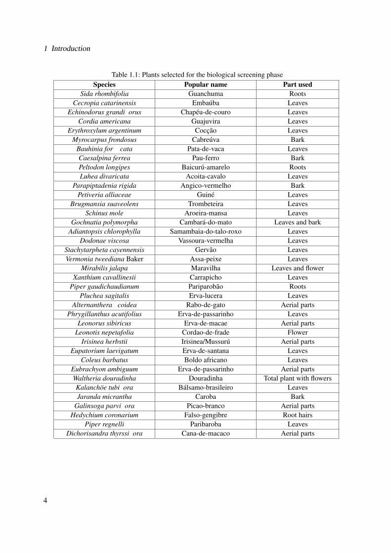
The plants used in the screening phase1 (see Table 1.1) were collected in autumn-winter season
(between March and July) in the region of Santa Maria, South Brazil. Both hexanic and ethanolic
extracts were prepared by means of soxhlet and ultrasonic extraction resulting in four different
extracts for each plant (see Section 5.5, Experimental Part).
As aforementioned, the screening of the plant extracts were based on bioassays targeting anti-
inflammatory, cytotoxic, antimicrobial and wound healing activity in order to identify the most
interesting extracts. Ethanolic extracts from Cordia americana and Brugmansia suaveolens were
selected for further investigation in the Eberhard-Karls University of Tubingen, since both hy-
drophilic extracts exhibited significantly inhibition effects on p38α MAPK (Mitogen-activated
Protein Kinase), TNFα release (Tumor Necrosis Factor α) and NF-κB assays (Nuclear Factor-
κB), and on fibroblast scratch assay [277, 113]. The selected plants are introduced in the following
sections.
1Leaves, aerial parts and flowers from the plants were collected and extracted by the doctoral candidate FabianaGeller with support of Dr. Klaus Gasser and Cleber Schmidt under coordination of Prof. Dr. Berta Heinzmann.The plants were authenticated by the botanist Dr. Gilberto Zanetti.
3
1 Introduction
Table 1.1: Plants selected for the biological screening phaseSpecies Popular name Part used
Sida rhombifolia Guanchuma RootsCecropia catarinensis Embauba Leaves
Echinodorus grandi�orus Chapeu-de-couro LeavesCordia americana Guajuvira Leaves
Erythroxylum argentinum Coccao LeavesMyrocarpus frondosus Cabreuva Bark
Bauhinia for��cata Pata-de-vaca LeavesCaesalpina ferrea Pau-ferro BarkPeltodon longipes Baicuru-amarelo RootsLuhea divaricata Acoita-cavalo Leaves
Parapiptadenia rigida Angico-vermelho BarkPetiveria alliaceae Guine Leaves
Brugmansia suaveolens Trombeteira LeavesSchinus mole Aroeira-mansa Leaves
Gochnatia polymorpha Cambara-do-mato Leaves and barkAdiantopsis chlorophylla Samambaia-do-talo-roxo Leaves
Dodonae viscosa Vassoura-vermelha LeavesStachytarpheta cayennensis Gervao LeavesVermonia tweediana Baker Assa-peixe Leaves
Mirabilis jalapa Maravilha Leaves and flowerXanthium cavallinesii Carrapicho Leaves
Piper gaudichaudianum Pariparobao RootsPluchea sagitalis Erva-lucera Leaves
Alternanthera �coidea Rabo-de-gato Aerial partsPhrygillanthus acutifolius Erva-de-passarinho Leaves
Leonorus sibiricus Erva-de-macae Aerial partsLeonotis nepetafolia Cordao-de-frade Flower
Irisinea herbstii Irisinea/Mussuru Aerial partsEupatorium laevigatum Erva-de-santana Leaves
Coleus barbatus Boldo africano LeavesEubrachyon ambiguum Erva-de-passarinho Aerial partsWaltheria douradinha Douradinha Total plant with flowersKalanchoe tubi�ora Balsamo-brasileiro LeavesJaranda micrantha Caroba Bark
Galinsoga parvi�ora Picao-branco Aerial partsHedychium coronarium Falso-gengibre Root hairs
Piper regnelli Paribaroba LeavesDichorisandra thyrssi�ora Cana-de-macaco Aerial parts
4
1.2 Project Overview
1.2.2 Cordia americana
Cordia americana (Linaeus) Gottschling & J.S.Mill. (syn. Patagonula americana) belongs to
the Boraginaceae family, subfamily Cordioideae.
The Boraginaceae family consists of about 2,700 species which are distributed in tropical, sub-
tropical and warmer regions around the world [117]. It is composed of about 130 genera and six
subfamilies: Boraginoideae, Cordioideae, Ehretioideae, Heliotropioideae, Hydrophylloideae, and
Lennooideae. Some well-known species that can be found in the Boraginaceae family and used
as medicinal plants are: Symphytum of�cinale (Comfrey), Borago of�cinalis (Borage) and Echium
amoenum (Echium).
The subfamily Cordioideae contains the genus Cordia, which is comprised of evergreen trees
and shrubs [308]. About 300 species of Cordia have been identified worldwide. In Brazil, the
genus Cordia is represented by approximately 65 species [306]. In this genus, some well-known
species are: Cordia dichotona, Cordia myxa, Cordia obliqua, Cordia verbenacea, Cordia martini-
censis, Cordia salicifolia, Cordia spinescens, Cordia latifolia and Cordia ulmifolia, which have
been used as cicatrizant, astringent, anti-inflammatory, antihelmintic, antimalarial remedy, and in
the treatment of urinary infections and lung diseases [308]. For example, studies with Cordia ver-
benacea revealed that α-humulen was the main compound responsible for the anti-inflammatory
properties of this plant [36]. Thus, the product Ache�an, manufactured by Brazilian Ache Labora-
tories, was developed based on the extract of Cordia verbenacea and it is used in the treatment of
chronic tendinitis and muscle pains.
Since 2003, Cordia americana, which was previously classified as Patagonula americana, was
included in the Cordioideae subfamily due to its molecular and morphology characteristics [117].
5

1 Introduction
1.2.2.1 Localization
The subfamily Cordioideae is distributed worldwide mainly in warmer regions. The majority of
the species grow in the American continent (i.e., more than 250 species) and the remaining species
are distributed in Africa, Asian and Oceania continents (i.e., more than 50 species) [117].
Cordia americana is commonly located in South Brazil, but can be found also in Argentina,
Uruguay, Paraguay and Bolivia (see Figure 1.1). In Brazil, usually it is located in regions with 20
up to 900 m of altitude. In Bolivia, it can be found up to 1,200 m of altitude [74]. Concerning
its etymology, Cordia americana (i.e., Patagonula americana) comes originally from “Patago-
nia”, Southern and semi-arid regions of Argentina [74]. This tree has different local names like
“guajuvira” in South Brazil, “guajayvi” in Paraguay, “guayaibi” in Argentina, and “guayubira” in
Uruguay.
Figure 1.1: Distribution of Cordia americana [241]
1.2.2.2 Botany
Cordia americana is described by the following botanical features [194, 74, 117]:
• Regarding its morphologic characteristics, Cordia americana is a semicaducifolia2 tree,
with 10 to 15 m height and with 20 to 40 cm diameter at breast height3 (see Figure 1.2). In
adulthood, it can reach up to 30 m height and 100 cm diameter at breast height.
2Semicaducifolia means that part of the tree leaves falls in winter.3Diameter at breast height is a standard method of expressing the diameter of a tree trunk.
6
1.2 Project Overview
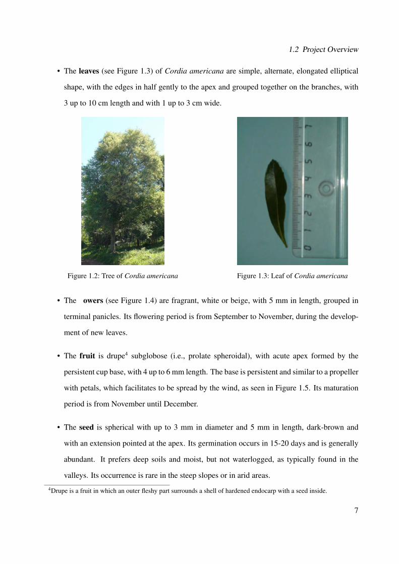
• The leaves (see Figure 1.3) of Cordia americana are simple, alternate, elongated elliptical
shape, with the edges in half gently to the apex and grouped together on the branches, with
3 up to 10 cm length and with 1 up to 3 cm wide.
Figure 1.2: Tree of Cordia americana Figure 1.3: Leaf of Cordia americana
• The �owers (see Figure 1.4) are fragrant, white or beige, with 5 mm in length, grouped in
terminal panicles. Its flowering period is from September to November, during the develop-
ment of new leaves.
• The fruit is drupe4 subglobose (i.e., prolate spheroidal), with acute apex formed by the
persistent cup base, with 4 up to 6 mm length. The base is persistent and similar to a propeller
with petals, which facilitates to be spread by the wind, as seen in Figure 1.5. Its maturation
period is from November until December.
• The seed is spherical with up to 3 mm in diameter and 5 mm in length, dark-brown and
with an extension pointed at the apex. Its germination occurs in 15-20 days and is generally
abundant. It prefers deep soils and moist, but not waterlogged, as typically found in the
valleys. Its occurrence is rare in the steep slopes or in arid areas.
4Drupe is a fruit in which an outer fleshy part surrounds a shell of hardened endocarp with a seed inside.
7

1 Introduction
Figure 1.4: Flower of Cordia americana Figure 1.5: Fruit of Cordia americana [117]
• The trunk is rarely cylindrical, often tortuous and irregular. Its bole is usually short and ir-
regular when the species grows alone, but in the forest, it reaches up to 10 m length. Usually,
it presents branches sprouting from the trunk.
• The shell has a thickness of up to 8 mm. The outer shell is generally grizzly, rarely dark,
slightly cracks in the longitudinal direction, forming rectangular plaques. The inner bark is
white to yellowish and with fibrous striations.
• The branch is typically raceme (i.e., unbranched and indeterminated). Its top is crown
narrow, elongated, ascending and densely branched.
8
1.2 Project Overview
1.2.2.3 Economical Importance and Traditional Medicine
The wood of Cordia americana has economical value due to its elasticity, flexibility and dura-
bility. Because of its flexible heartwood, it is widely applied to handwork, as for example by the
Caingangue Indians in the manufacture of bows for hunting. The heartwood has normally a dark
color. For this reason, the name given by German immigrants in South Brazil was “schwarz-herz”
(i.e., black heartwood) [164]. Nowadays, the wood is still utilized in building construction, manu-
facture of doors, windows, and luxe furniture [74]. Furthermore, this tree is applied in landscaping
and it is appropriated for heterogeneous reforestation of degraded areas.
In folks medicine, a decoction prepared from its leaves is used in order to wash wounds and to
treat inflammatory diseases [297, 164]. The cataplasm from the leaves is also externally applied
on wounds [294, 59, 164]. Additionally, this plant is known for the treatment of ulcers, because of
its suggested astringent and mucilaginous properties [207].
1.2.2.4 Chemical Constituents
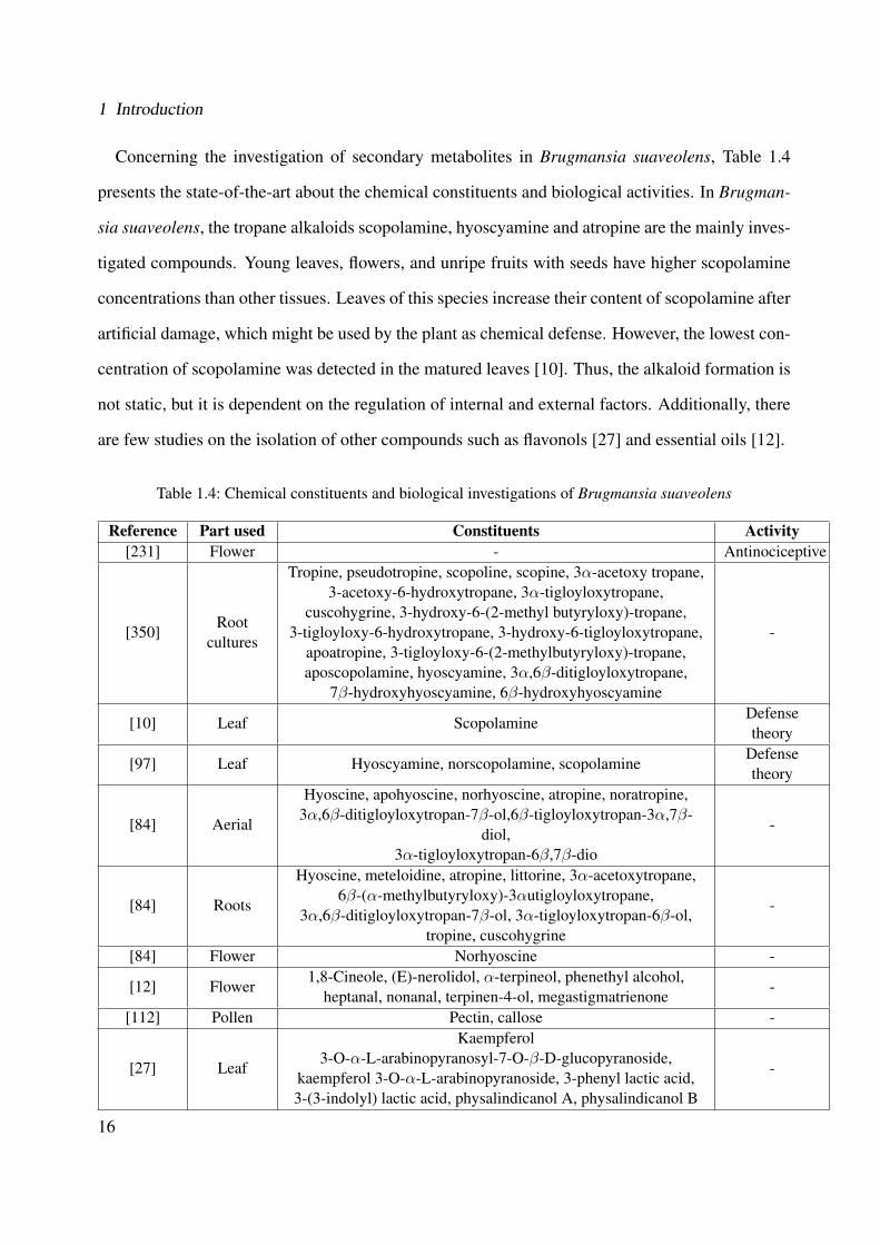
The genus Cordia has been demonstrated to be a potential producer of diverse secondary metabo-
lites including flavonoids, phenolic acids, triterpenes, sesquiterpenes, saponins, hydroquinones,
chromenes, terpenoid naphthoquinones and benzoquinones. Table 1.2 presents the state-of-the-art
concerning the studied secondary metabolites of the genus Cordia and its biological activities.
Regarding the investigation of secondary metabolites in Cordia americana, so far only few
phytochemical investigations have been done. Two quinones (cordiachrome G and leucocor-
diachrome H) and one phenolic aldehyde known as patagonaldehyde were isolated from its heart-
wood [213, 214]. From the bark coumarin [266] and tannins [131] have been reported. From its
leaves, only tannins have been identified [294, 131] and no pyrrolizidine alkaloids were identified
in Cordia americana [251]. None of the previous studies considered the biological investigation,
therefore, this plant has not been extensively investigated.
9

1 Introduction
Table 1.2: Chemical constituents and biological investigations of the genus Cordia
Species Part used Constituents Activity ReferenceCordia
cylindrostachyaRoem. & Schult.
- α-pinene, amphene, tricyleneAntibacterial,
anti-inflammatory[101]
Cordia dichotoma G.Forst
Fruits Flavonoids Wound healing [168]
Cordia francisci Ten. Leaves -Analgesic,
anti-inflammatory[253]
Cordia martinicensis(Jacq.) Roem. &
Schult.Leaves -
Analgesic,anti-inflammatory
[253]
Cordia myxa L.Leaves and
fruits
Robinin, rutin, datiscoside, hesperidin,dihydrorobinetin, chlorogenic, caffeic
acid, quercitrin, carotenoids, oleicacid, β-sitosterol
Anti-inflammatory,anti-arthritic
[253, 7, 93,106, 4,212]
Cordia obliquaWilld.
Seeds
α-amyrin, betulin, octacosanol,lupeol-3-rhamnoside, β-sitosterol,
β-sitosterol-3-glucoside,hentricontanol, hentricontane,taxifolin-3, 5-dirhamnoside,
hesperetin-7-rhamnoside
Anti-inflammatory
[5]
Cordia serratifoliaKunth.
Leaves -Analgesic,
anti-inflammatory[253]
Cordia ulmifoliaJuss.
Leaves Pyrrolizidine alkaloidsHepatotoxic,
anti-inflammatory[254]
Cordia curassavica(Jacq.) Roem. &
Schult. (syn. Cordiaverbenacea D.C.)
Leaves,areal parts
α-pinene, α-humulene,trans-caryophyllene,
aloaromadendrene, cordialin A,cordialin B, rosmarinic acid,
flavonols-artemetin
Anti-edematogenic,analgesic, anti-inflammatory,anti-rheumatic
[204, 26,290, 73,
320, 310]
Cordia dentata Poir. FlowersRosmarinic acid,
quercetin-3-o-rutinoside- [90]
Cordia dichotomaForst.
Leaves Quercetin, quercitrin - [324]
Cordia globosa Jacq. Roots Meroterpenoid benzoquinone Anti-cancer [76]Cordia linnaei
Stearn.Roots
Meroterpenoid naphthoquinones,naphthoxirene
Antifungal,larvicidal
[143]
Cordia latifoliaRoxb.
Fruits -Anti-ulcer,
anti-histaminic[6]
Cordia spinescens L. Leaves Triterpenes Anti-viral [221, 201]Cordia americana Leaves Tannins - [294, 131]
Heartwood Quinones, phenolic aldehyde - [213, 214]
10
1.2 Project Overview
1.2.3 Brugmansia suaveolens
Brugmansia suaveolens (Humb. & Bonpl. ex Willd.) Bercht. & C. Presl (syn. Datura suave-
olens Humb. & Bonpl. ex Willd.) belongs to the Solanaceae family.
The family Solanaceae consists of about 2,700 species and of about 98 genera [226] and con-
tains flowering plants which have a large number of important agricultural as well as toxic species.
They are extensively used by humans as an important source of food, spice and medicine. How-
ever, some Solanaceae species are often rich in alkaloids, whose toxicity ranges from mildly
irritating to fatal for humans as well as for animals. Some well-known species in this family
include: Datura stramonium (Jimson weed), Solanum tuberosum (Potatoes), Solanum lycoper-
sicum (Tomato), Nicotiana tabacum (Tobacco) and the genus Capsicum (Chili pepper). The great-
est diversity of species can be found in South and in Central America. The origin of the name
“Solanaceae” might come from the Latin “Solanum” meaning the “nightshade” plant, or it might
be originated from the Latin verb “solari” meaning “to soothe”, because of its soothing pharmaco-
logical properties of some psychoactive species in this family.
Brugmansia is a genus of the flowering species in the family Solanaceae. It is known as “angel's
trumpets”, sharing this name with the genus Datura, which is closely related. Brugmansia is peren-
nial and woody [246]. Brugmansia species consist of large shrubs and small trees reaching heights
of 3 up to 11 m. The name “angel's trumpets” refers to the large pendulous flowers that may be 14-
50 cm long and 35 cm wide. This flower might have white, yellow, pink, orange or red colours. In
this genus, some of well-known species include: Brugmansia arborea, Brugmansia aurea, Brug-
mansia sanguinea, Brugmansia suaveolens, and Brugmansia versicolor, which have been used to
treat rheumatic and arthritic pains, swelling, scalds, inflammations, skin rashes, hemorrhoids and
wounds. Their extracts exhibit spasmolytic, antiasthmatic, anticholinergic, narcotic and anesthetic
properties [350]. The “Brugmansia” name is honored to Sebald J. Brugmans (1763-1819), a Dutch
botanist, physician and professor of natural sciences. The “suaveolens” name means “fragrant”,
which is a characteristic of this plant due to its intense smell in the evening period [246].
11
1 Introduction
Brugmansia suaveolens was firstly described by Willdenow in 1809 as Datura suaveolens and
discovered by Humboldt and Bonpland on their expeditions in North America. Since 1823, it
was reclassified into the genus Brugmansia [246]. In Brazil, this species is locally known as
“trombeteira” (i.e., trumpeter) and it can be found in various regions of the country. Due this plant
is popular as a drug (i.e., hallucinogenic tea from the flowers) its commercialization is controlled
by the Ministry of Health in Brazil [43].
1.2.3.1 Localization
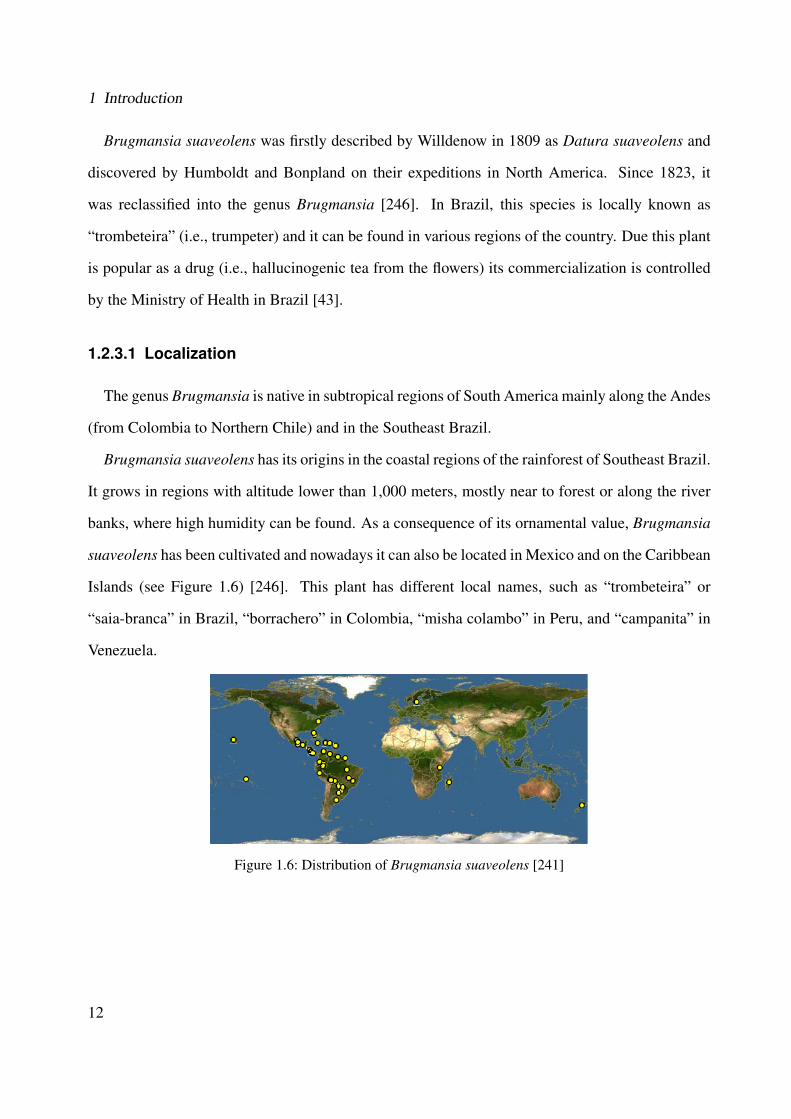
The genus Brugmansia is native in subtropical regions of South America mainly along the Andes
(from Colombia to Northern Chile) and in the Southeast Brazil.
Brugmansia suaveolens has its origins in the coastal regions of the rainforest of Southeast Brazil.
It grows in regions with altitude lower than 1,000 meters, mostly near to forest or along the river
banks, where high humidity can be found. As a consequence of its ornamental value, Brugmansia
suaveolens has been cultivated and nowadays it can also be located in Mexico and on the Caribbean
Islands (see Figure 1.6) [246]. This plant has different local names, such as “trombeteira” or
“saia-branca” in Brazil, “borrachero” in Colombia, “misha colambo” in Peru, and “campanita” in
Venezuela.
Figure 1.6: Distribution of Brugmansia suaveolens [241]
12
1.2 Project Overview
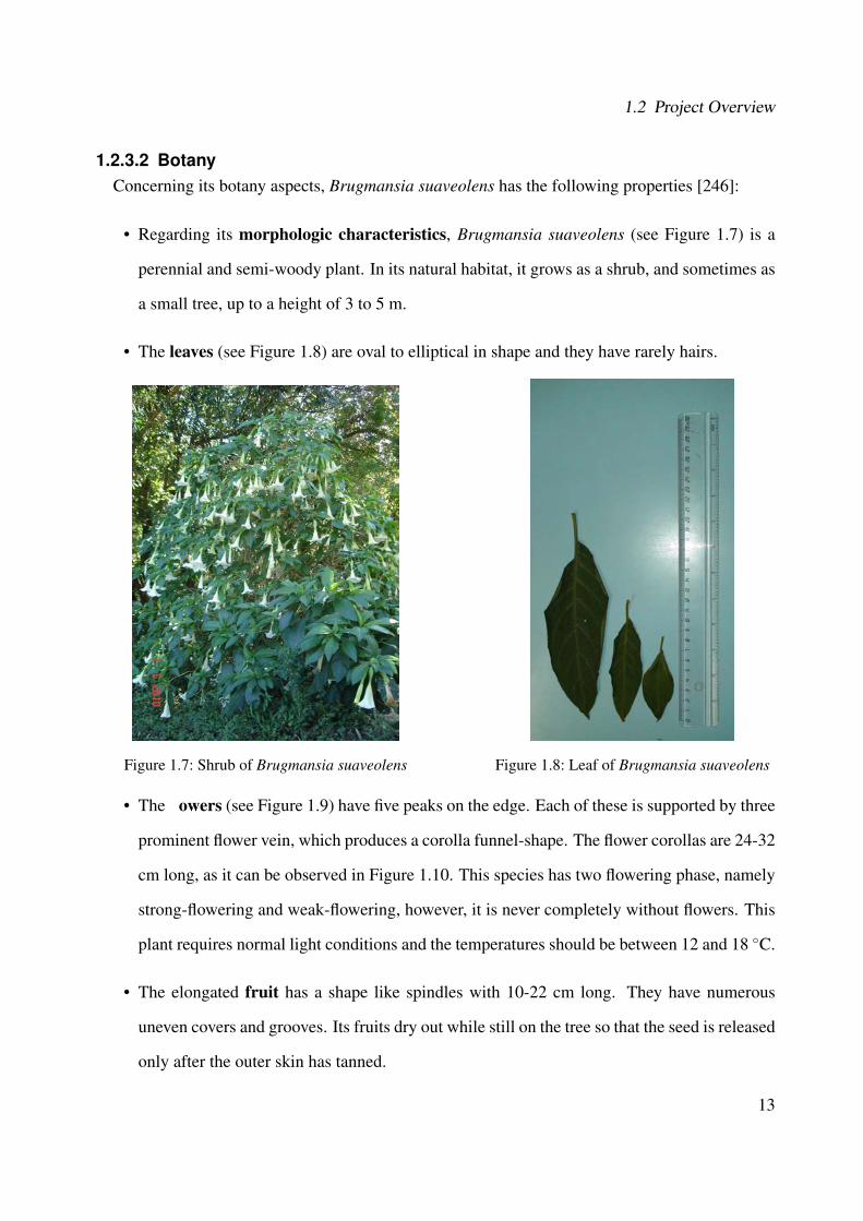
1.2.3.2 BotanyConcerning its botany aspects, Brugmansia suaveolens has the following properties [246]:
• Regarding its morphologic characteristics, Brugmansia suaveolens (see Figure 1.7) is a
perennial and semi-woody plant. In its natural habitat, it grows as a shrub, and sometimes as
a small tree, up to a height of 3 to 5 m.
• The leaves (see Figure 1.8) are oval to elliptical in shape and they have rarely hairs.
Figure 1.7: Shrub of Brugmansia suaveolens Figure 1.8: Leaf of Brugmansia suaveolens
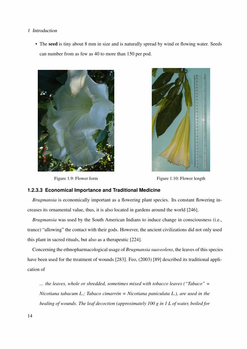
• The �owers (see Figure 1.9) have five peaks on the edge. Each of these is supported by three
prominent flower vein, which produces a corolla funnel-shape. The flower corollas are 24-32
cm long, as it can be observed in Figure 1.10. This species has two flowering phase, namely
strong-flowering and weak-flowering, however, it is never completely without flowers. This
plant requires normal light conditions and the temperatures should be between 12 and 18 ◦C.
• The elongated fruit has a shape like spindles with 10-22 cm long. They have numerous
uneven covers and grooves. Its fruits dry out while still on the tree so that the seed is released
only after the outer skin has tanned.
13
1 Introduction
• The seed is tiny about 8 mm in size and is naturally spread by wind or flowing water. Seeds
can number from as few as 40 to more than 150 per pod.
Figure 1.9: Flower form Figure 1.10: Flower length
1.2.3.3 Economical Importance and Traditional Medicine
Brugmansia is economically important as a flowering plant species. Its constant flowering in-
creases its ornamental value, thus, it is also located in gardens around the world [246].
Brugmansia was used by the South American Indians to induce change in consciousness (i.e.,
trance) “allowing” the contact with their gods. However, the ancient civilizations did not only used
this plant in sacred rituals, but also as a therapeutic [224].
Concerning the ethnopharmacological usage of Brugmansia suaveolens, the leaves of this species
have been used for the treatment of wounds [283]. Feo, (2003) [89] described its traditional appli-
cation of
... the leaves, whole or shredded, sometimes mixed with tobacco leaves (“Tabaco” =
Nicotiana tabacum L.; Tabaco cimarron = Nicotiana paniculata L.), are used in the
healing of wounds. The leaf decoction (approximately 100 g in 1 L of water, boiled for
14
1.2 Project Overview
30 min. until the preparation becomes green) is used externally in cataplasms as an
anti-in�ammatory on traumatized body parts. The vapors of this decoction are used
as a vaginal cleanser (antiseptic) in cases of dysmenhorrea and white secretions. The
plant is claimed to be toxic if ingested.
Havelius and Asman, (2002) [129] and Oliveira et al., (2003) [225] described intoxication oc-
currences due to the ingestion of leaves, flowers and/or fruit by children. The contact of the sap
with the eyes caused mydriasis. Moreover, a study is presented based on patients with anticholin-
ergic poisoning by Brugmansia suaveolens between July 1990 and June 2000 in Australia. The
main clinical effects were mydriasis, dried mouth, delirium, flushed skin, aggressiveness, visual
hallucinations, tachycardia, urinary retention and fever [144]. In more severe cases, the patients
may have neurological, cardiovascular and respiratory disorders, leading to death [225].
1.2.3.4 Chemical Constituents
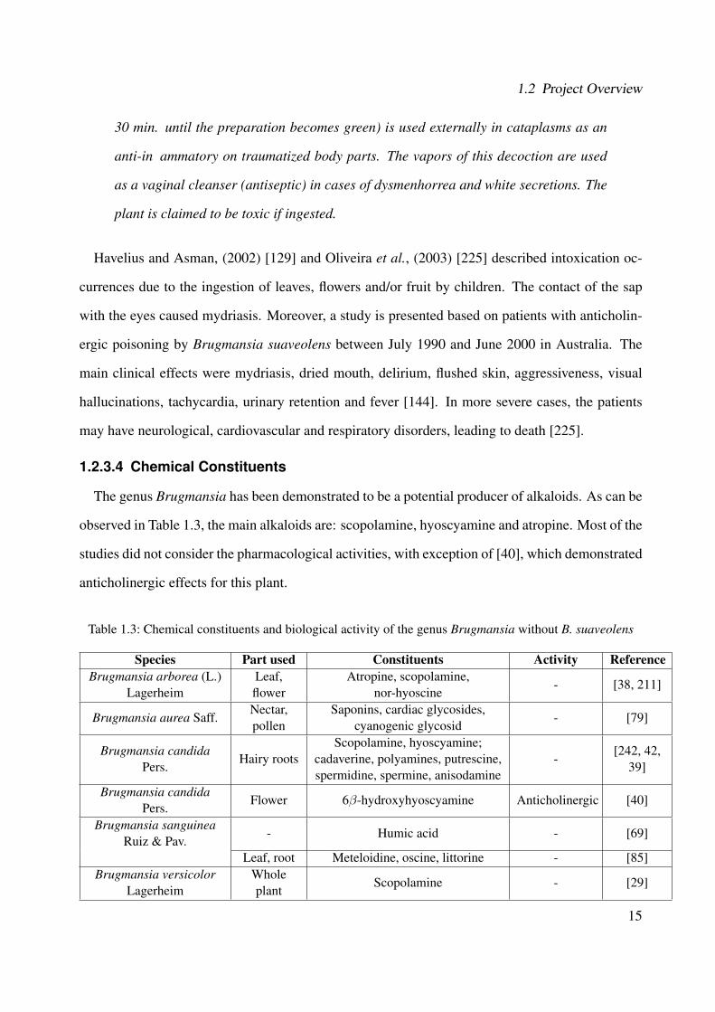
The genus Brugmansia has been demonstrated to be a potential producer of alkaloids. As can be
observed in Table 1.3, the main alkaloids are: scopolamine, hyoscyamine and atropine. Most of the
studies did not consider the pharmacological activities, with exception of [40], which demonstrated
anticholinergic effects for this plant.
Table 1.3: Chemical constituents and biological activity of the genus Brugmansia without B. suaveolens
Species Part used Constituents Activity ReferenceBrugmansia arborea (L.)
LagerheimLeaf,flower
Atropine, scopolamine,nor-hyoscine
- [38, 211]
Brugmansia aurea Saff.Nectar,pollen
Saponins, cardiac glycosides,cyanogenic glycosid
- [79]
Brugmansia candidaPers.
Hairy rootsScopolamine, hyoscyamine;
cadaverine, polyamines, putrescine,spermidine, spermine, anisodamine
-[242, 42,
39]
Brugmansia candidaPers.
Flower 6β-hydroxyhyoscyamine Anticholinergic [40]
Brugmansia sanguineaRuiz & Pav.
- Humic acid - [69]
Leaf, root Meteloidine, oscine, littorine - [85]Brugmansia versicolor
LagerheimWholeplant
Scopolamine - [29]
15
1 Introduction
Concerning the investigation of secondary metabolites in Brugmansia suaveolens, Table 1.4
presents the state-of-the-art about the chemical constituents and biological activities. In Brugman-
sia suaveolens, the tropane alkaloids scopolamine, hyoscyamine and atropine are the mainly inves-
tigated compounds. Young leaves, flowers, and unripe fruits with seeds have higher scopolamine
concentrations than other tissues. Leaves of this species increase their content of scopolamine after
artificial damage, which might be used by the plant as chemical defense. However, the lowest con-
centration of scopolamine was detected in the matured leaves [10]. Thus, the alkaloid formation is
not static, but it is dependent on the regulation of internal and external factors. Additionally, there
are few studies on the isolation of other compounds such as flavonols [27] and essential oils [12].
Table 1.4: Chemical constituents and biological investigations of Brugmansia suaveolens
Reference Part used Constituents Activity[231] Flower - Antinociceptive
[350]Root
cultures
Tropine, pseudotropine, scopoline, scopine, 3α-acetoxy tropane,3-acetoxy-6-hydroxytropane, 3α-tigloyloxytropane,
cuscohygrine, 3-hydroxy-6-(2-methyl butyryloxy)-tropane,3-tigloyloxy-6-hydroxytropane, 3-hydroxy-6-tigloyloxytropane,
apoatropine, 3-tigloyloxy-6-(2-methylbutyryloxy)-tropane,aposcopolamine, hyoscyamine, 3α,6β-ditigloyloxytropane,
7β-hydroxyhyoscyamine, 6β-hydroxyhyoscyamine
-
[10] Leaf ScopolamineDefensetheory
[97] Leaf Hyoscyamine, norscopolamine, scopolamineDefensetheory
[84] Aerial
Hyoscine, apohyoscine, norhyoscine, atropine, noratropine,3α,6β-ditigloyloxytropan-7β-ol,6β-tigloyloxytropan-3α,7β-
diol,3α-tigloyloxytropan-6β,7β-dio
-
[84] Roots
Hyoscine, meteloidine, atropine, littorine, 3α-acetoxytropane,6β-(α-methylbutyryloxy)-3αutigloyloxytropane,
3α,6β-ditigloyloxytropan-7β-ol, 3α-tigloyloxytropan-6β-ol,tropine, cuscohygrine
-
[84] Flower Norhyoscine -
[12] Flower1,8-Cineole, (E)-nerolidol, α-terpineol, phenethyl alcohol,
heptanal, nonanal, terpinen-4-ol, megastigmatrienone-
[112] Pollen Pectin, callose -
[27] Leaf
Kaempferol3-O-α-L-arabinopyranosyl-7-O-β-D-glucopyranoside,
kaempferol 3-O-α-L-arabinopyranoside, 3-phenyl lactic acid,3-(3-indolyl) lactic acid, physalindicanol A, physalindicanol B
-
16
1.3 Objectives of this Dissertation
1.3 Objectives of this Dissertation
In Brazil, Cordia americana and Brugmansia suaveolens have been used for the treatment of
anti-inflammatory diseases in the folk medicine. However, the effective compounds responsible
for the biological effects are widely unknown. Thus, the general objective of this dissertation was
the investigation of the anti-inflammatory and wound healing properties of the ethanolic extracts
from the leaves of both medicinal plants.
More specifically, this dissertation focused on:
• Bioguide fractionation of the plant extracts based on p38α.
• Isolation of the plants constituents using chromatographic methods.
• Structural elucidation by means of spectroscopic methods such as UV/VIS, mass spectrom-
etry and nuclear magnetic resonance spectroscopy.
• Biological investigation of the ethanolic extracts and their isolated compounds in the p38α,
JNK3, TNFα release, 5-lipoxygenase, NF-κB activation, and fibroblast scratch assay.
17

2 In�ammatory and Wound Healing
Processes
This chapter presents an overview about inflammatory and wound healing processes. More
specifically, it describes in details the biological targets p38α, JNK3, TNFα, 5-lipoxygenase, NF-
κB and fibroblasts scratch assay.
2.1 In�ammatory and Wound Healing Processes
Inflammation is a biological response of the immune system against challenges originating from
the surrounding environment. Challenge of host tissues due to traumatic, infectious or toxic injury
or lesions lead to a complex series of vascular and cellular events carried out by the organism to re-
move the injury and to initiate the healing process, resulting in the release of different biochemical
mediators. These events1 causes redness, heat, swelling, pain and loss of function [289]. Va-
sodilatation, increased blood flow, enhanced permeability of blood vessels and peripheral nervous
tissue stimulation are further events. Depending on the extent of insult, prolonged inflammation
can lead to a chronic condition and eventually to loss of function [293].
The inflammation comprises of a large and complex regulated number of biochemical events
including cellular, molecular and physiological changes in response to the stimuli. It involves1Based on visual observation, the ancients characterized inflammation by five cardinal signs, namely redness (rubor),
swelling (tumour), heat (calor, only applicable to the body extremities), pain (dolor) and loss of function (functiolaesa). The first four of these signs were named by Celsus in ancient Rome (30-38 B.C.) and the last by Galen(A.D. 130-200) [289].
19

2 In�ammatory and Wound Healing Processes
the immune system, the local vascular system and cells resident within the injured tissue. These
cells produce multiple inflammatory mediators like cytokines (e.g., interleukin 1 and TNF (Tumor
Necrosis Factor)), plasma proteins (thrombin), histamine and bioactive lipids. These events en-
able the successive recruitment of neutrophils, monocytes/macrophages and lymphocytes from the
blood, which in turn release further pro-inflammatory mediators [223, 293].
Wounds are physical injuries that result in an opening or break of the skin. Healing is a complex
and intricate process, initiated by a response to an injury, that restores the function and integrity
of damaged tissues [277]. Wound healing involves inflammation as well as the formation and
remodeling of new tissue [100].
Thus, more targets are necessary to study how the plant extracts and isolated compounds can
modulate or inhibit inflammatory responses, and increase or accelerate the wound healing process
[277]. Among several mediators, which are responsible to induce or maintain the inflammation,
this dissertation focuses on the following biological targets: p38α and JNK3 (c-Jun N-terminal
Protein Kinase 3) MAPK, TNFα, 5-lipoxygenase, NF-κB and fibroblasts scratch assay. These
biological targets are explained in more details in the following sections.
2.2 Mitogen-Activated Protein Kinases (MAPKs)
Mitogen-activated protein kinase (MAPK) pathways regulate diverse processes ranging from
proliferation and differentiation to apoptosis. Activated by an enormous array of stimuli, they
phosphorylate numerous proteins, including transcription factors, cytoskeletal proteins and other
enzymes. MAPKs have greatly influence on gene expression, metabolism, cell division, cell mor-
phology and cell survival [248, 48].
Each MAPK pathway contains a three-tiered kinase cascade comprising a MAP kinase kinase
kinase (MAPKKK, MAP3K, MEKK or MKKK), a MAP kinase kinase (MAPKK, MAP2K, MEK
or MKK) and a MAP kinase [91, 248]. Normally, a MAPKKK kinase (MAPKKKK, MAP4K or
MKKKK) activates the MAPKKK. The MAPKKKK or MAPKKK can be linked to the plasma
membrane, for example, through association with a small GTPase or lipid (i.e., MAPKKKKs and
20
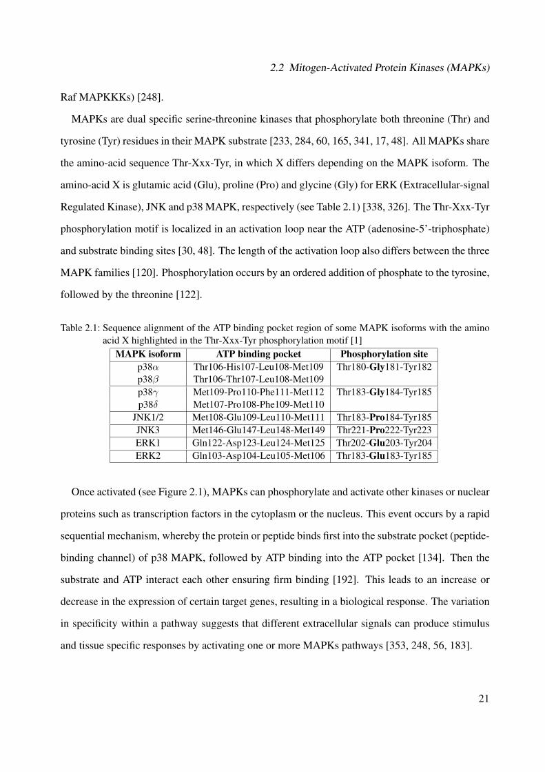
2.2 Mitogen-Activated Protein Kinases (MAPKs)
Raf MAPKKKs) [248].
MAPKs are dual specific serine-threonine kinases that phosphorylate both threonine (Thr) and
tyrosine (Tyr) residues in their MAPK substrate [233, 284, 60, 165, 341, 17, 48]. All MAPKs share
the amino-acid sequence Thr-Xxx-Tyr, in which X differs depending on the MAPK isoform. The
amino-acid X is glutamic acid (Glu), proline (Pro) and glycine (Gly) for ERK (Extracellular-signal
Regulated Kinase), JNK and p38 MAPK, respectively (see Table 2.1) [338, 326]. The Thr-Xxx-Tyr
phosphorylation motif is localized in an activation loop near the ATP (adenosine-5'-triphosphate)
and substrate binding sites [30, 48]. The length of the activation loop also differs between the three
MAPK families [120]. Phosphorylation occurs by an ordered addition of phosphate to the tyrosine,
followed by the threonine [122].
Table 2.1: Sequence alignment of the ATP binding pocket region of some MAPK isoforms with the aminoacid X highlighted in the Thr-Xxx-Tyr phosphorylation motif [1]
MAPK isoform ATP binding pocket Phosphorylation sitep38α Thr106-His107-Leu108-Met109 Thr180-Gly181-Tyr182p38β Thr106-Thr107-Leu108-Met109p38γ Met109-Pro110-Phe111-Met112 Thr183-Gly184-Tyr185p38δ Met107-Pro108-Phe109-Met110
JNK1/2 Met108-Glu109-Leu110-Met111 Thr183-Pro184-Tyr185JNK3 Met146-Glu147-Leu148-Met149 Thr221-Pro222-Tyr223ERK1 Gln122-Asp123-Leu124-Met125 Thr202-Glu203-Tyr204ERK2 Gln103-Asp104-Leu105-Met106 Thr183-Glu183-Tyr185
Once activated (see Figure 2.1), MAPKs can phosphorylate and activate other kinases or nuclear
proteins such as transcription factors in the cytoplasm or the nucleus. This event occurs by a rapid
sequential mechanism, whereby the protein or peptide binds first into the substrate pocket (peptide-
binding channel) of p38 MAPK, followed by ATP binding into the ATP pocket [134]. Then the
substrate and ATP interact each other ensuring firm binding [192]. This leads to an increase or
decrease in the expression of certain target genes, resulting in a biological response. The variation
in specificity within a pathway suggests that different extracellular signals can produce stimulus
and tissue specific responses by activating one or more MAPKs pathways [353, 248, 56, 183].
21
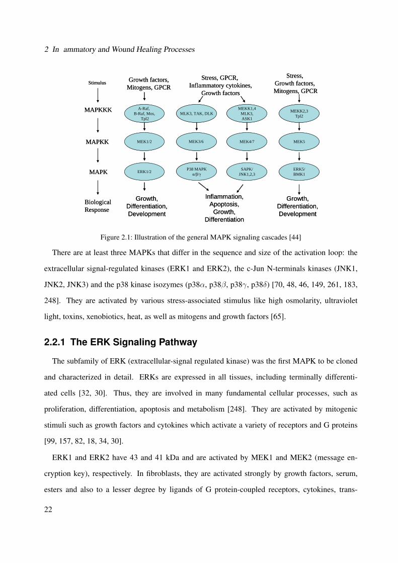
2 In�ammatory and Wound Healing Processes
Stimulus
MAPKKK
MAPKK
MAPK
BiologicalResponse
Growth factors,Mitogens, GPCR
Stress, GPCR, Inflammatory cytokines,
Growth factors
Stress, Growth factors, Mitogens, GPCR
A-Raf, B-Raf, Mos,
Tpl2
MEK1/2
ERK1/2
MLK3, TAK, DLK
MEK3/6
P38 MAPKα/β/γ
MEKK1,4MLK3,ASK1
MEK4/7
SAPK/JNK1,2,3
MEKK2,3Tpl2
MEK5
ERK5/BMK1
Growth,Differentiation,Development
Inflammation,Apoptosis,
Growth,Differentiation
Growth,Differentiation,Development
Figure 2.1: Illustration of the general MAPK signaling cascades [44]
There are at least three MAPKs that differ in the sequence and size of the activation loop: the
extracellular signal-regulated kinases (ERK1 and ERK2), the c-Jun N-terminals kinases (JNK1,
JNK2, JNK3) and the p38 kinase isozymes (p38α, p38β, p38γ, p38δ) [70, 48, 46, 149, 261, 183,
248]. They are activated by various stress-associated stimulus like high osmolarity, ultraviolet
light, toxins, xenobiotics, heat, as well as mitogens and growth factors [65].
2.2.1 The ERK Signaling Pathway
The subfamily of ERK (extracellular-signal regulated kinase) was the first MAPK to be cloned
and characterized in detail. ERKs are expressed in all tissues, including terminally differenti-
ated cells [32, 30]. Thus, they are involved in many fundamental cellular processes, such as
proliferation, differentiation, apoptosis and metabolism [248]. They are activated by mitogenic
stimuli such as growth factors and cytokines which activate a variety of receptors and G proteins
[99, 157, 82, 18, 34, 30].
ERK1 and ERK2 have 43 and 41 kDa and are activated by MEK1 and MEK2 (message en-
cryption key), respectively. In fibroblasts, they are activated strongly by growth factors, serum,
esters and also to a lesser degree by ligands of G protein-coupled receptors, cytokines, trans-
22
2.2 Mitogen-Activated Protein Kinases (MAPKs)
forming growth factors and osmotic stress. In differentiated cells, they are often activated by the
primary stimuli that regulate tissue specific functions, like glucose in islets or transmitters in brain
[48, 99, 157, 18]. ERK1 is important for T cell responses, whereas ERK2 plays a role in mesoderm
differentiation and placenta formation [248].
Blocking of ERK activity could be a great benefit for the treatment of metastatic cancer, because
they are involved in the control of proliferation, differentiation and apoptosis. Therefore, the ERK
pathway is also explored for the therapy of viral diseases including HIV [215], influenza [243] as
well as neurodegenerative syndromes such as Alzheimer [255] or cardiovascular diseases [55].
2.2.2 The JNK Signaling Pathway
The c-Jun N-terminal protein kinases (JNK) consist of at least ten protein isoforms that are
generated through alternative splicing of three closely related genes, such as JNK1, JNK2 and
JNK3. The JNKs along p38 are also called as stress-activated protein kinases (SAPK). JNK1 and
JNK2 are expressed ubiquitously in all tissues. In contrast, the JNK3 has more limited pattern
of expression and is largely restricted to the nervous system, but also detected in heart and testis
[227, 30].
JNKs are involved in a wide range of cell signaling, including cell death apoptosis [78, 172, 123,
83, 344, 195, 295] and neurodegenerative diseases [316, 33, 111, 343, 319], brain, heart [325, 130]
and renal ischemia [108], epilepsy and inflammatory disorders (multiple sclerosis, rheumatoid
arthritis (RA), asthma, inflammatory bowel diseases and psoriasis) [126, 262, 58, 259, 258, 15,
222].
Along with p38 MAPK, JNK pathways are triggered by a variety of cellular stresses, inflamma-
tory cytokines, UV light and peroxides [24, 248]. The major JNK activators are MKK4 and MKK7
[335]. Both protein kinases can activate JNK by dual phosphorylation of the motif Thr-Pro-Tyr,
located in the activation loop [78]. While MKK4 phosphorylates preferentially JNK on tyrosine,
MKK7 phosphorylates JNK on threonine [181, 313, 322, 35].
23
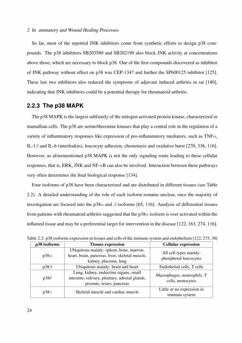
2 In�ammatory and Wound Healing Processes
So far, most of the reported JNK inhibitors come from synthetic efforts to design p38 com-
pounds. The p38 inhibitors SB203580 and SB202190 also block JNK activity at concentrations
above those, which are necessary to block p38. One of the first compounds discovered as inhibitor
of JNK pathway without effect on p38 was CEP-1347 and further the SP600125 inhibitor [125].
These last two inhibitors also reduced the symptoms of adjuvant induced arthritis in rat [180],
indicating that JNK inhibitors could be a potential therapy for rheumatoid arthritis.
2.2.3 The p38 MAPK
The p38 MAPK is the largest subfamily of the mitogen activated protein kinase, characterized in
mamallian cells. The p38 are serine/threonine kinases that play a central role in the regulation of a
variety of inflammatory responses like expression of pro-inflammatory mediators, such as TNFα,
IL-1β and IL-6 (interleukin), leucocyte adhesion, chemotaxis and oxidative burst [270, 336, 116].
However, as aforementioned p38 MAPK is not the only signaling route leading to these cellular
responses, that is, ERK, JNK and NF-κB can also be involved. Interaction between these pathways
very often determines the final biological response [134].
Four isoforms of p38 have been characterized and are distributed in different tissues (see Table
2.2). A detailed understanding of the role of each isoform remains unclear, once the majority of
investigation are focused into the p38α and β isoforms [65, 116]. Analysis of differential tissues
from patients with rheumatoid arthritis suggested that the p38α isoform is over activated within the
inflamed tissue and may be a preferential target for intervention in the disease [122, 163, 274, 116].
Table 2.2: p38 isoforms expression in tissues and cells of the immune system and endothelium [122, 275, 30]p38 isoforms Tissues expression Cellular expression
p38αUbiquitous mainly: spleen, bone, marrow,
heart, brain, pancreas, liver, skeletal muscle,kidney, placenta, lung
All cell types mainly:pheripheral leucocytes
p38β Ubiquitous mainly: brain and heart Endothelial cells, T cells
p38δLung, kidney, endocrine organs, small
intestine, salivary, pituitary, adrenal glands,prostate, testes, pancreas
Macrophages, neutrophils, Tcells, monocytes
p38γ Skeletal muscle and cardiac muscleLittle or no expression in
immune system
24
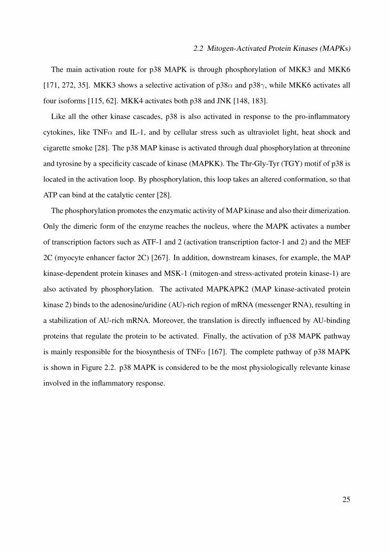
2.2 Mitogen-Activated Protein Kinases (MAPKs)
The main activation route for p38 MAPK is through phosphorylation of MKK3 and MKK6
[171, 272, 35]. MKK3 shows a selective activation of p38α and p38γ, while MKK6 activates all
four isoforms [115, 62]. MKK4 activates both p38 and JNK [148, 183].
Like all the other kinase cascades, p38 is also activated in response to the pro-inflammatory
cytokines, like TNFα and IL-1, and by cellular stress such as ultraviolet light, heat shock and
cigarette smoke [28]. The p38 MAP kinase is activated through dual phosphorylation at threonine
and tyrosine by a specificity cascade of kinase (MAPKK). The Thr-Gly-Tyr (TGY) motif of p38 is
located in the activation loop. By phosphorylation, this loop takes an altered conformation, so that
ATP can bind at the catalytic center [28].
The phosphorylation promotes the enzymatic activity of MAP kinase and also their dimerization.
Only the dimeric form of the enzyme reaches the nucleus, where the MAPK activates a number
of transcription factors such as ATF-1 and 2 (activation transcription factor-1 and 2) and the MEF
2C (myocyte enhancer factor 2C) [267]. In addition, downstream kinases, for example, the MAP
kinase-dependent protein kinases and MSK-1 (mitogen-and stress-activated protein kinase-1) are
also activated by phosphorylation. The activated MAPKAPK2 (MAP kinase-activated protein
kinase 2) binds to the adenosine/uridine (AU)-rich region of mRNA (messenger RNA), resulting in
a stabilization of AU-rich mRNA. Moreover, the translation is directly influenced by AU-binding
proteins that regulate the protein to be activated. Finally, the activation of p38 MAPK pathway
is mainly responsible for the biosynthesis of TNFα [167]. The complete pathway of p38 MAPK
is shown in Figure 2.2. p38 MAPK is considered to be the most physiologically relevante kinase
involved in the inflammatory response.
25
2 In�ammatory and Wound Healing Processes
Stat1p53 Max Myc Elk-1Histone H3
CREBMSK1/2
p38 MAPK
p38 MAPKHSP27
MKK4
MLK2/3 ASK1/2 TAK1
MKK3/6
cPLA 2
MNK1/2
eEF2K
TGF-
G o
GPCR
TRA
DD
RIP
TRA
F2
Dax
x
Pax6 CHOP ETS1HMGN1
MK2
MK2Tau
PRAK
MEKK1-4TAO1/2/3
MK3
MK3
PRAK
TAB1
DLK
In�ammatoryCytokines,
FasL
SB203580
DNA Damage, Oxidative Stress, UV
Apoptosis
Cytokine-inducedmRNA stability
TNF- biosynthesis
Cytoplasm
Nucleus
TranscriptionCytokine Production,
Apoptosis, etc.
Translation
MEF2 ATF-2
Figure 2.2: p38 MAPK signaling pathway [44]
2.2.4 Structure of Protein Kinase
The genome is composed of 518 genes that encode for protein kinases with similar tertiary
structure and ATP binding sites. Protein kinases catalyze the same chemical reaction, namely the
phosphorylation of other proteins. Each kinase presents its own structural and dynamic character-
istics. MAPKs share between 50% and 80% sequence identity [268]. Most protein kinases have a
common fold of two domains, the N-terminal lobe consisting of five antiparallel β-strands and one
α-helix, and the C-terminal lobe, which is composed predominantly of α-helix. The two structural
subunits are linked together via a hinge region that allows the rotation of the two lobes [114].
26
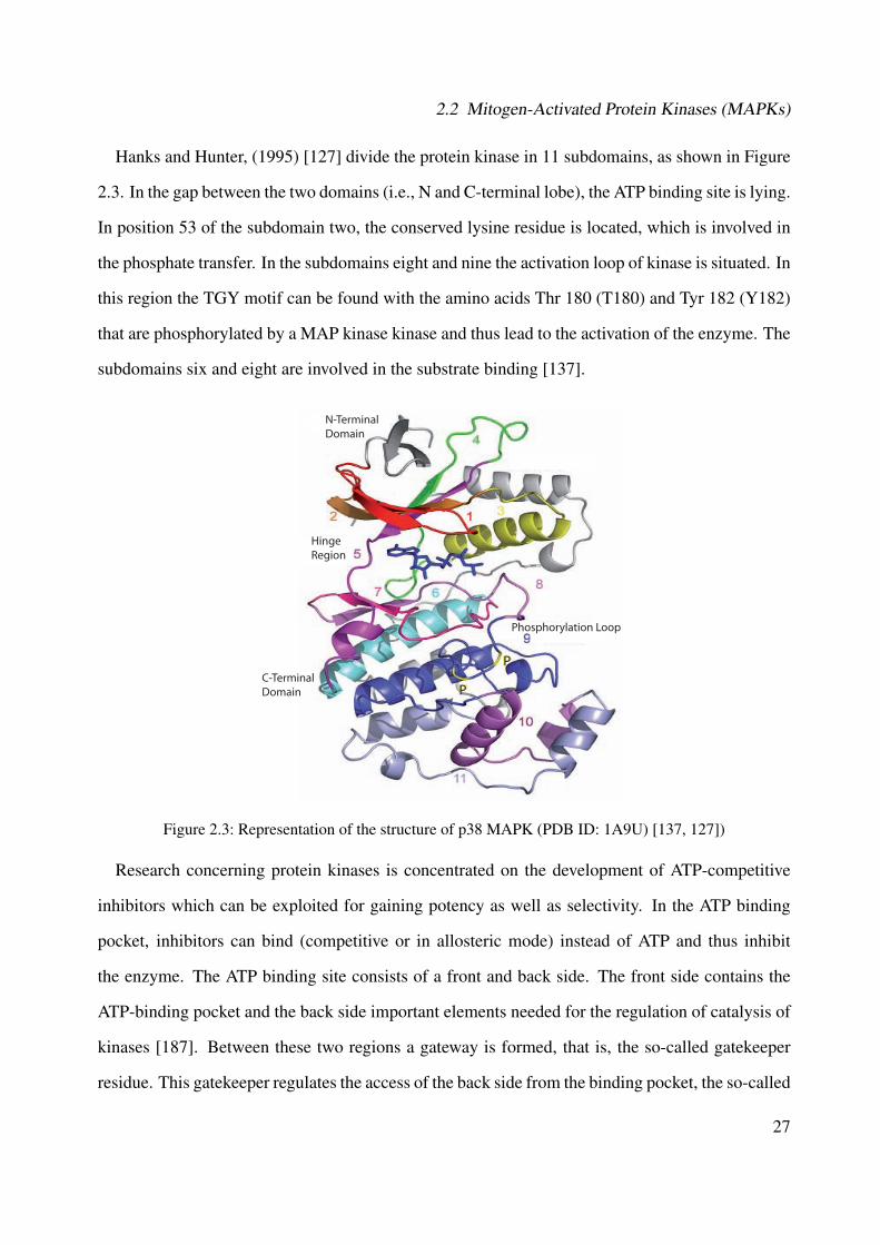
2.2 Mitogen-Activated Protein Kinases (MAPKs)
Hanks and Hunter, (1995) [127] divide the protein kinase in 11 subdomains, as shown in Figure
2.3. In the gap between the two domains (i.e., N and C-terminal lobe), the ATP binding site is lying.
In position 53 of the subdomain two, the conserved lysine residue is located, which is involved in
the phosphate transfer. In the subdomains eight and nine the activation loop of kinase is situated. In
this region the TGY motif can be found with the amino acids Thr 180 (T180) and Tyr 182 (Y182)
that are phosphorylated by a MAP kinase kinase and thus lead to the activation of the enzyme. The
subdomains six and eight are involved in the substrate binding [137].
N-Terminal Domain
Phosphorylation Loop
Hinge Region
P
PC-Terminal Domain
Figure 2.3: Representation of the structure of p38 MAPK (PDB ID: 1A9U) [137, 127])
Research concerning protein kinases is concentrated on the development of ATP-competitive
inhibitors which can be exploited for gaining potency as well as selectivity. In the ATP binding
pocket, inhibitors can bind (competitive or in allosteric mode) instead of ATP and thus inhibit
the enzyme. The ATP binding site consists of a front and back side. The front side contains the
ATP-binding pocket and the back side important elements needed for the regulation of catalysis of
kinases [187]. Between these two regions a gateway is formed, that is, the so-called gatekeeper
residue. This gatekeeper regulates the access of the back side from the binding pocket, the so-called
27

2 In�ammatory and Wound Healing Processes
hydrophobic pocket I. If the gatekeeper is a small amino acid such as threonine or alanine, than the
access to hydrophobic region I is granted. A large amino acid at this position (e.g., phenylalanine)
leucine or methionine prevented however, the interaction between the inhibitor and this region
[187]. The gatekeeper residues for p38α and JNK3 consist Thr106 and Met146, respectively.
Based on cristallographic structural data, Manning et al., (2002) [199, 314] proposed to divide
the ATP-binding site of kinases into five sub regions (see Figure 2.4), as following:
• Adenine-binding region (Purin-binding region): The predominantly hydrophobic character
of this region permits that ATP as well as the inhibitors interact through van der Walls forces
with this pocket. This observation is confirmed by the linear correlation between completed
or occupied surface and binding affinity of ATP-competitive ligands. The position of H-bond
donors and acceptors in this area allows important interactions with the hinge region.
• Hydrophobic backpocket (Hydrophobic pocket I, selectivity pocket): The hydrophobic re-
gion I is similar to a cavity (gap), whose dimension is determined through the gatekeeper
residue Thr 106 in case of p38. This region is located orthogonal behind the adenin in the
ATP binding region.
• Hydrophobic region II (Hydrophobic pocket II): This region is a kind of groove located
in front of the ATP binding site. This area has predominantly a hydrophobic character in
contact to the solvent. This region as well as the hydrophobic pocket I are not occupied by
ATP and they can be used for the development of selective inhibitors.
• Phosphate-binding region (Glycin-rich loop): The phosphate binding region is the roof of
the ATP-binding pocket and consists of a glycine rich sequence that is mainly exposed to the
solvent. The glycine residues make the loop flexible and thus allows the opening and closing
of the ATP binding site during catalysis. It is a highly conserved region, since amino acid
residues play an important role in this region in the catalytic process and in the binding of
28
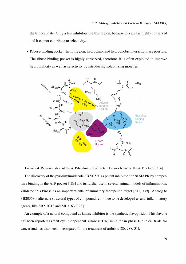
2.2 Mitogen-Activated Protein Kinases (MAPKs)
the triphosphate. Only a few inhibitors use this region, because this area is highly conserved
and it cannot contribute to selectivity.
• Ribose-binding pocket: In this region, hydrophilic and hydrophobic interactions are possible.
The ribose-binding pocket is highly conserved, therefore, it is often exploited to improve
hydrophilicity as well as selectivity by introducing solubilizing moieties.
Adenin-bindingRegion
Ribose Pocket
ungstasche
Phosphat-binding Region
Gatekeeper
Hinge Region
Hydrophobic
Region II
Hydrophobic Backpocket I
Selectivity Pocket
-
Lys 53
NHO
NH 3
+
Glu 71NH O
OO
Val 105
HN
O
O
NHO
NHO
NH
His 107
Leu 108
Met109
Thr 106
OLeu 104
NH
O
ON
N N
N
NHH
OHOH
OP O
O
OH
P O
O
OH
P OH
O
OH
Mg2+
Mg2+
ONH 2
N
O
O
NH H
Figure 2.4: Representation of the ATP-binding site of protein kinases bound to the ATP cofator [314]
The discovery of the pyridinylimidazole SB203580 as potent inhibitor of p38 MAPK by compet-
itive binding in the ATP pocket [183] and its further use in several animal models of inflammation,
validated this kinase as an important anti-inflammatory therapeutic target [311, 339]. Analog to
SB203580, alternate structural types of compounds continue to be developed as anti-inflammatory
agents, like SB210313 and ML3163 [178].
An example of a natural compound as kinase inhibitor is the synthetic flavopiridol. This flavone
has been reported as first cyclin-dependent kinase (CDK) inhibitor in phase II clinical trials for
cancer and has also been investigated for the treatment of arthritis [86, 288, 31].
29
2 In�ammatory and Wound Healing Processes
2.2.5 Diseases Associated with MAPKs
A signal transduction, which is initiated by receptor activation, is a complex set of cascading
networks involving significant crosstalk, intracellular trafficking, scaffold modules, and feedback
loops. The net cellular response is dependent on a large number of parameters like signal strength,
amplification, and duration, protein expression levels, and the numbers and types of concurrent
extracellular signals [276]. There are many studies in stroke suggesting the involvement of the three
families of MAPKs (ERK, JNKs, and p38 MAPKs) in inflammations. They are attractive targets
for new therapies and development of more selective inhibitors against inflammatory diseases.
The most studies concerning p38 MAPK are focused on their function in inflammatory pro-
cesses. Several groups have reported that specific and selective p38α/β MAPK inhibitors block the
production of IL-1, TNF and IL-6 in vitro and in vivo. In addition, the p38 MAPK pathway is in-
volved in the induction of several other inflammatory molecules, such as COX-2 (Cyclooxygenase-
2) and inducible nitric oxide synthase (iNOS). Moreover, p38α-dependent histone H3 phosphory-
lation has been shown to mark and recruit NF-κB to other promoters resulting in increased expres-
sion of several inflammatory cytokines and chemokines [263].
Concerning the diseases associated with MAPKs, rheumatoid arthritis is a chronic autoimmune
inflammatory disease which affects about 1% of the adult population worldwide [275, 276] and
is characterized by inflammation of the synovial joints and production of pro-inflammatory me-
diators by immune cells that infiltrate in the synovium. This provokes proliferation of synovial
fibroblasts, further release of inflammatory molecules and formation of pannus tissue that eventu-
ally degrades cartilage and subchondral bone, leading to joint destruction, pain and loss of physical
function. Arthritis results from dysregulation of pro-inflammatory cytokines (e.g., IL-1 and TNFα)
and pro-inflammatory enzymes that mediate the production of prostaglandins (e.g., COX-2) and
leukotrienes (e.g., lipoxygenase) together with the expression of adhesion molecules and matrix
metalloproteinases, and hyperproliferation of synovial fibroblasts. All of these factors are regulated
by the activation of NF-κB [167, 156]. Anti-cytokine biotherapeutic approaches, such as etanercept
30
2.2 Mitogen-Activated Protein Kinases (MAPKs)
(tumor necrosis factor receptor-p-75 Fc fusion protein), infliximab (chimeric anti-human TNFα
monoclonal antibody) and adalimumab (recombinant human anti-human TNFα monoclonal anti-
body) bind to TNF-α and prevent the binding to cell-surface receptors [167, 276, 275]. Agents that
suppress the expression of TNFα, IL-1β, COX-2, lipoxygenase, matrix metalloproteinases or adhe-
sion molecules, or suppress the activation of NF-κB, have all potential for the treatment of arthritis.
Compounds derived from plants like curcumin (from tumeric), resveratrol (red grapes, cranberries
and peanuts), tea polyphenols, genistein (soy), quercetin (onions), silymarin (artichoke), boswellic
acid and anolides can also suppress these cell signaling intermediates [156].
Increasing tissue levels of inflammatory cytokines (i.e., IL- 1, IL-6, TNFα) have also been ob-
served in patients suffering from inflammatory Crohns diseases which is characterized by a chronic
inflammation in the gastrointestinal tract. Therapy with anti-TNF-α agents have been showing
clinically efficacious [276, 66].
MAPKs and NF-κB activation have been also identified in the pathogenesis of chronic inflam-
matory bowel diseases. Immunohistochemical analysis of inflamed mucosal biopsies revealed
that expression of p38α was abundant in activated macrophages and neutrophils infiltrating bowel
mucosa. The treatment with SB203580 improves the clinical score, ameliorates the histological
alterations, and reduces mRNA levels of pro-inflammatory cytokines [151].
JNKs and p38 MAPKs are also activated by ischemia/reperfusion (I/R) [171, 245]. Evidences
from studies conducted on mice have suggested that JNKs might be a potential therapeutic target
for obesity and type 2-diabetes [135, 68].
The MAPK pathway is also investigated in connection with cancer. The pathway is activated by
mitogens that promote mitosis, however, unregulated activation of this pathway has been linked to
be a cause of cancer [184, 167].
31
2 In�ammatory and Wound Healing Processes
2.3 Cytokines
Cytokines are glycoproteins produced by different cell types that bind to specific high-affinity
receptors. They consist of 100-200 amino acids and have molecular weights around 10-25 kDa
and are high active in a concentration range of pg to ng [54]. Cytokines regulate intercellular
communication and are directly implicated in many immune processes [16, 203]. They act only in
short distances (except TNFα) differently from hormones [23].
There are two different classes of cytokines (inflammatory /pro-inflammatory and anti-inflamma-
tory cytokines). The pro-inflammatory cytokines (TNFα, IFNg, IL-1, IL-2, IL-6, IL-12) ensure
that in case of penetration of one pathogen, for example, the immune cells are attracted to the site
of infection and activated. However, anti-inflammatory cytokines (IL-4, IL-10, IL-13) should be
a successful fight against the infectious agent, so that the resulting inflammation may be counter-
acted quickly. Because of their biological function, cytokines can be classified in interferon (IFN),
interleukins (IL-1 to IL-23), tumor necrosis factor (TNF), growth factors (EGF, FGF, PDGF) and
chemokines [193].
IL-1 is a 17 kDa protein that is mostly produced by monocytes and macrophages but also by
endothelial cells, B cells, and activated T cells. IL-1 includes two different cytokine agonists,
termed as IL-1α and IL-1β. Both IL-1 forms differ very slightly in their physiological and patho-
physiological function [175]. IL-1β is a crucial mediator of the inflammatory response that plays
an important role in the development of chronic inflammation, especially joint damage causing
arthritis [54].
IL-4 is produced by CD4 type-2 helper T cells and participates in the differentiation and growth
of B cells [145]. In vitro, IL-4 inhibits the activation of type-1 helper T cells, and this, in turn,
decreases the production of IL-1 and TNFα and inhibits cartilage damage. In rheumatoid arthritis,
this anti-inflammatory cytokine inhibits the production of IL-1 and increases the expression of IL-1
receptor antagonist, and both actions should decrease inflammation [52].
IL-6 is an inflammatory cytokine produced by T cells, monocytes, macrophages and synovial
32
2.3 Cytokines
fibroblasts. This interleukin is involved in diverse biological processes, such as activation of T
cells, the induction of acute-phase response, the stimulation of the growth and differentiation of
hematopoietic precursor cells and proliferation of synovial fibroblasts [54].
IL-8 is produced by monocytes, T lymphocytes, neutrophils, fibroblasts, epithelial, endothelial
and tumor cells. It is hardly found in healthy tissues, but its production is increased from five to
one hundred times after stimulation by cytokines such as TNFα and IL-1, LPS (lipopolysaccha-
ride), viral products, and cellular stress. The uncontrolled production of IL-8 is related to diseases
such as rheumatoid arthritis, lung disease, skin, viral infections, tumor growth, sclerosis and arte-
riosclerosis [189]. The expression of IL-8 may be regulated by treatment with immunosuppressive
agents, but polyphenols isolated from green tea and genistein from soy also inhibit the production
of this cytokine [315] and many other compounds.
IL-10 belongs to the anti-inflammatory cytokine group and is produced by monocytes, macrophages,
B-cells and T-cells. It inhibits the production of several cytokines, including IL-1 and TNFα and
the proliferation of T-cells in vitro. The IL-10 is also found in synovial fluid of patients with
rheumatoid arthritis, but the amount is insufficient to suppress inflammation [54].
2.3.1 Tumor Necrosis Factor α (TNFα)
Tumor necrosis factor (TNF) is a non glycosylated polypeptide that belongs to the group of
multifunctional pro-inflammatory cytotoxins. It exists in an α and β-form, which are only 30%
homologus [175]. Normal quantities of circulating TNFα are between 10 and 80 pg/mL [190].
TNFα plays an important role in chronic inflammation, cell proliferation, differentiation and apop-
tosis [229, 25]. It is predominantly detected during the early stages of diseases and its dysregu-
lation of expression and/or signaling is involved in many pathologies, including Crohns diseases,
rheumatoid arthritis and neurophatologies such as stroke, multiple sclerosis and Alzheimer's dis-
ease [72, 21].
TNFα is produced in macrophages, monocytes, T-cells and NK cells but also in endothelial cells
33

2 In�ammatory and Wound Healing Processes
and fibroblasts by stimulation of lipopolysaccharide (LPS), for example [291, 184]. Initially, TNF
is produced as a membrane-associated precursor protein with a molecular weight of 26 kDa and
is accumulated in the intracellular space. By means of TACE (TNFα converting enzyme), pro-
TNFα is transformed in the active 17 kDa form and released from the cells. TACE is a membrane-
bound metalloproteinase, which belongs to the group of ADAM (a disintegrin and metalloprotease)
protein family [218].
The biological responses to TNFα are mediated through two structurally distinct receptors: type
1 (TNFR1) and type 2 (TNFR2). Both receptors are transmembrane-glycoproteins with multiple
cysteine-rich regions in the extracellular N-terminal domains. Although their extracellular domains
share structural and functional homology, their intracellular domains are distinct and transduce
their signals through both overlapping and different pathways. The primary characteristic property
that distinguishes the intracellular domains of TNFR1 and TNFR2, is the presence of a death do-
main in TNFR1, which is not present in TNFR2. The death domain is a sequence of approximately
70 amino acids and is pivotal to the ability of TNFα to trigger cellular apoptosis [229].
Under physiological conditions, signaling through TNFR1 seems to be primarily responsible
for the pro-inflammatory and shock producing properties of TNFα. It means that the biological
responses to TNFα seem to be dependent on signaling through both receptors. Both TNFα recep-
tors can be cleaved from the cell surface by members of the matrix metalloproteinase family in
response to inflammatory signals, such as TNFα receptor binding. The extracellular domains of
the receptors retain their ability to bind TNFα. Therefore, these domains have either endogenous
inhibitors or facilitators of the biological activity of TNFα [351], which are dependent on their
concentrations and ligands [229].
Exposure of cells to TNFα can result in an activation of a caspase cascade leading to apoptosis
[45]. However, more commonly, the binding of TNFα to its receptors causes activation of two
major transcription factors, AP-1 and NF-κB, that in turn induce genes involved in chronic and
acute inflammatory responses. Furthermore, some of these genes act to suppress TNFα induced
apoptosis, thereby explaining why the apoptotic response to TNFα is usually dependent on inhi-
34

2.4 Nuclear Factor-κB (NF-κB)
bition of RNA or protein synthesis. The suppression of apoptosis is mostly dependent on NF-κB,
which increases the inflammatory response to TNFα [25].
2.4 Nuclear Factor-κB (NF-κB)
The nuclear factor (NF)-κB plays a crucial role in the regulation of numerous genes involved in
diverse cellular processes like cell growth, apoptosis, differentiation, inflammation by regulating
the transcription of genes encoding for cytokines, COX-2, nitric oxide synthase, immunoreceptor
molecules of adhesion and hematopoietic growth factors [110, 197]. Dysregulation of NF-κB
pathway is associated with a variety of human diseases, like atherosclerosis, asthma, rheumatoid
arthritis, cancer, inflammatory bowel disease, type 1 diabetes mellitus, and psoriasis [20, 170].
NF-κB is a dimeric protein, which can be differently composed. Five NF-κB members have
been found in mammal cells: NF-κB1 (p105/50), NF-κB2 (p100/52), RelA (p65), RelB and RelC.
All NF-κB proteins share the RHD (Rel-homology domain) in their N-terminal region, which is
required for dimerization, DNA binding, interaction with the inhibitory protein IκB, as well as the
nuclear localization sequence (NLS) [152, 153, 352].
In inactive cells, NF-κB resides in the cytoplasm bound to the inhibitory IκB subunit. The IκB
family of proteins include IκB-α, IκB-β, IκB-γ, IκB-ε, Bcl-3 and the precursor proteins p105
and p100. Activation of p105 and p100 give the subunits p52 and p50, respectively. IκB proteins
interact with the RHD of NF-κB, thereby inhibiting its transport to the nucleus and binding of
NF-κB to the DNA [109].
The major activation route of NF-κB (canonical pathway) occurs through the activation of the
IκB kinase complex (IKK). The substances that induce the activation of the IκB-kinase complex
are LPS, cytokines, viruses, physical and physiological stress such as UV and gamma radiation,
and several chemical agents. The activation of IKK leads to phosphorylation of serine in IκB-α.
Phosphorylated IκB undergoes ubiquitination and subsequent proteosomal degradation [152, 352].
The degradation of IκB allows the transport of NF-κB into the nucleus and its DNA binding [109,
352].
35
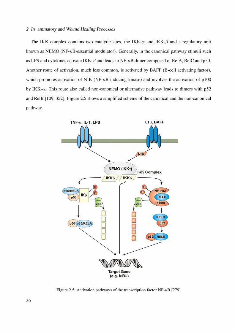
2 In�ammatory and Wound Healing Processes
The IKK complex contains two catalytic sites, the IKK-α and IKK-β and a regulatory unit
known as NEMO (NF-κB-essential modulator). Generally, in the canonical pathway stimuli such
as LPS and cytokines activate IKK-β and leads to NF-κB dimer composed of RelA, RelC and p50.
Another route of activation, much less common, is activated by BAFF (B-cell activating factor),
which promotes activation of NIK (NF-κB inducing kinase) and involves the activation of p100
by IKK-α. This route also called non-canonical or alternative pathway leads to dimers with p52
and RelB [109, 352]. Figure 2.5 shows a simplified scheme of the canonical and the non-canonical
pathway.
Figure 2.5: Activation pathways of the transcription factor NF-κB [279]
36
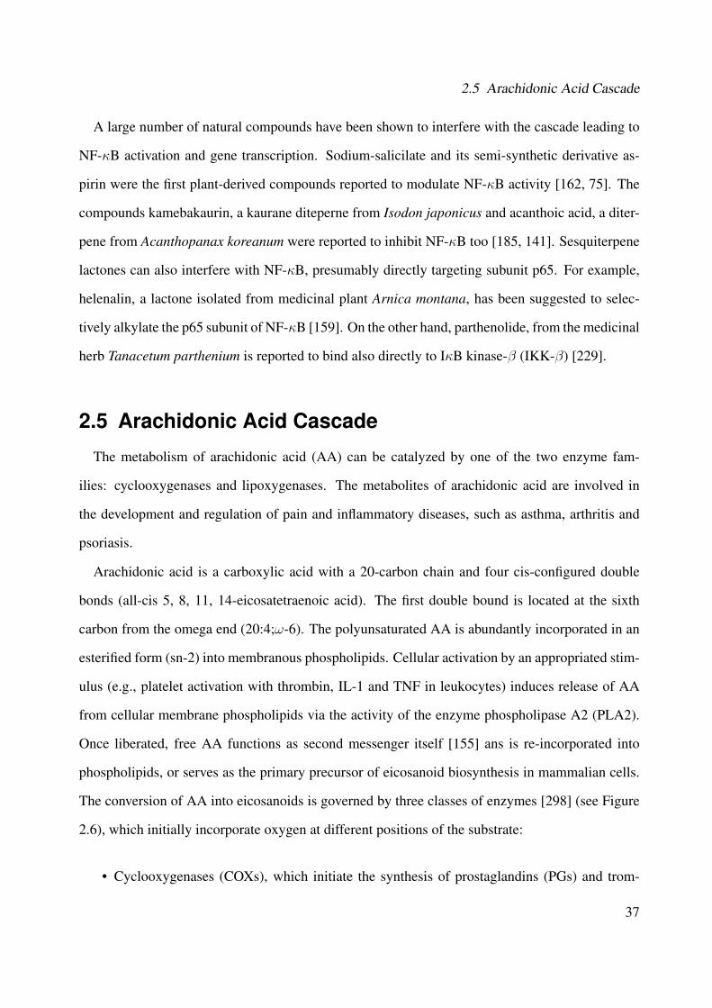
2.5 Arachidonic Acid Cascade
A large number of natural compounds have been shown to interfere with the cascade leading to
NF-κB activation and gene transcription. Sodium-salicilate and its semi-synthetic derivative as-
pirin were the first plant-derived compounds reported to modulate NF-κB activity [162, 75]. The
compounds kamebakaurin, a kaurane diteperne from Isodon japonicus and acanthoic acid, a diter-
pene from Acanthopanax koreanum were reported to inhibit NF-κB too [185, 141]. Sesquiterpene
lactones can also interfere with NF-κB, presumably directly targeting subunit p65. For example,
helenalin, a lactone isolated from medicinal plant Arnica montana, has been suggested to selec-
tively alkylate the p65 subunit of NF-κB [159]. On the other hand, parthenolide, from the medicinal
herb Tanacetum parthenium is reported to bind also directly to IκB kinase-β (IKK-β) [229].
2.5 Arachidonic Acid Cascade
The metabolism of arachidonic acid (AA) can be catalyzed by one of the two enzyme fam-
ilies: cyclooxygenases and lipoxygenases. The metabolites of arachidonic acid are involved in
the development and regulation of pain and inflammatory diseases, such as asthma, arthritis and
psoriasis.
Arachidonic acid is a carboxylic acid with a 20-carbon chain and four cis-configured double
bonds (all-cis 5, 8, 11, 14-eicosatetraenoic acid). The first double bound is located at the sixth
carbon from the omega end (20:4;ω-6). The polyunsaturated AA is abundantly incorporated in an
esterified form (sn-2) into membranous phospholipids. Cellular activation by an appropriated stim-
ulus (e.g., platelet activation with thrombin, IL-1 and TNF in leukocytes) induces release of AA
from cellular membrane phospholipids via the activity of the enzyme phospholipase A2 (PLA2).
Once liberated, free AA functions as second messenger itself [155] ans is re-incorporated into
phospholipids, or serves as the primary precursor of eicosanoid biosynthesis in mammalian cells.
The conversion of AA into eicosanoids is governed by three classes of enzymes [298] (see Figure
2.6), which initially incorporate oxygen at different positions of the substrate:
• Cyclooxygenases (COXs), which initiate the synthesis of prostaglandins (PGs) and trom-
37

2 In�ammatory and Wound Healing Processes
boxanos (TXs), altogether termed as prostanoids.
• Lipoxygenases (LOs), such as 5-LO, which catalyzes the formation of leukotrienes (LT) as
well as 12- and 15-LOs yielding hydroxy-eicosatetraenoic acids (HETEs).
• A class of CYP 450 enzymes which form epoxyeicosatrienoic acids (EETs).
Membranous phospholipidsp p p
PLA2
Arachidonic acid
COXs LOs CYP450COXs LOs CYP450
Prostanoids LT & HETEs EETs
Figure 2.6: Arachidonic acid cascade
2.5.1 5-Lipoxygenase
Lipoxygenases are a family of structurally related non-heme iron-containing enzymes that insert
molecular oxygen into polyunsaturated fatty acids with cis, cis-1,4-pentadiene. Depending on the
position of oxygen insertion into arachidonic acid, mammalian lipoxygensases are classified as 5-,
12- and 15-hydroperoxyeicosatetraenoic acids (HPETEs), which are reduced to the corresponding
hydroxyeicosatetraenoic acids (HETEs) or converted into various other types of eicosanoids such
as leukotrienes [278, 299].
Leukotrienes (LTs) are bioactive mediators mainly produced and released from activated leuko-
cytes, but also in granulocytes, monocytes/macrophages, mast cells, dendritic cells and B lympho-
38

2.5 Arachidonic Acid Cascade
cytes. Platelets, endothelial cells, T-cells and erythrocytes do not express them [300, 102]. Ele-
vated levels of lipoxygenase metabolites, particularly 5-LO, have been also found in lung, prostate,
breast, colon and skin cancer cells, as well as in cells from patients with acute leukemias [299].
The antagonists of leukotrienes pathway have been used in the treatment of bronchial asthma,
arteriosclerosis, cardiovascular diseases and cancer [299, 250, 330, 236].
The conversion of arachidonic acid in leukotrienes by 5-lipoxygenase is shown in Figure 2.7.
The initial step in LT biosynthesis is the dioxygenation of free AA by 5-LO yielding 5(S)-hydro-
peroxyeicosatetraenoic acid (5-HPETE) which is further metabolized by 5-LO to the instable epox-
ide LTA4. In neutrophils and monocytes, LTA4 can be converted to LTB4 by LTA4 hydrolase,
whereas in mast cells and eosinophils, LTC4 synthase or membrane-associated proteins can con-
jugate LTA4 with glutathione, yielding the cysteinyl-LT which can be cleaved in the extracellular
environment yielding LTD4 and then LTE4 [330, 299].
COO-
OOHHCOOH
OHHCOOH
COOH
OH
OH
COOHOH
R
COOH
O
COOH
O
LTC4
LTD4
LTE4
Cys-LTs
AA
5-HPETE
5-HETE
LTA4
LTB4
5-oxo-ETE
5-LO (oxygenase + O2)
5-LO (LTA4 synthase)
LTA4 hydrolase LTC4 synthase
R= Cys Gly
Glu
R= Cys Gly
R= Cys
COO-
OOHHCOOH
OHHCOOH
COOH
OH
OH
COOHOH
R
COOH
O
COOH
O
LTC4
LTD4
LTE4
Cys-LTs
AA
5-HPETE
5-HETE
LTA4
LTB4
5-oxo-ETE
5-LO (oxygenase + O2)
5-LO (LTA4 synthase)
LTA4 hydrolase LTC4 synthase
R= Cys Gly
Glu
R= Cys Gly
R= Cys
Figure 2.7: Conversion of arachidonic acid in leukotrienes by 5-Lipoxygenase [330]
39
2 In�ammatory and Wound Healing Processes
2.5.2 Structure and Regulation of 5-LO
So far, the 3D structure of 5-LO has not been resolved. However, practicable computational
models of 5-LO based on the structure of 15-LO from rabbit have been used to explain the interac-
tions between compounds and the enzyme [334, 124]. The structure of 5-LO is composed of two
domains: C-terminal and N-terminal domain. The catalytic C-terminal domain is mainly helical
and contains iron. The smaller N-terminal domain is a C2-like β sandwich with typical ligand-
binding loops [124, 133]. The C-terminal domain contains a non-heme iron in the active site,
essential for their catalytic activity. This iron acts as an electron acceptor or donor during catal-
yses. In the inactive form, the iron is presented in the ferrous state (Fe2+), whereas the catalytic
active 5-LO requires conversion to the ferric (Fe3+) iron [9].
5-LO is phosphorylated by MAP kinase kinase and traffics through the nuclear pore, possibly
in association with NF-κB [298, 332]. During cell activation, for example, by the intracellular
increase of Ca2+, both cytosolic and nuclear soluble 5-LO can translocate to the nuclear envelope,
leading to colocalization with cytosolic phospholipase A2 and lipoxygenase activating protein.
This migration of 5-LO ist probably important for its activity and regulation [250].
2.5.3 5-LO Inhibitors
The most inhibitors, synthetic and as well as from natural sources (e.g., polyphenols, coumarins
and quinones) act at the catalytic domain by reducing or chelating the active-site iron or sim-
ply by scavenging electrons participating in the redox cycle of iron [329, 333]. Until now, no
pharmacological data demonstrate inhibition of 5-LO interfering with the C-2-like domain [330].
Compounds that interfere with cellular 5-LO traffic will cause a suppression of LT formation.
For example, natural compounds that interfere with Ca2+ prevent the activation of 5-LO without
inhibiting the enzyme directly [330].
Redox-active 5-LO inhibitors comprise lipophilic reducing agents, and among those, there are
many plant derived classes like flavonoids, coumarins, quinones, lignans and other polyphenols.
40

2.6 Wound Healing Process: Scratch and Elastase
These drugs act by keeping the active site iron in the ferrous state, thereby uncoupling the catalytic
cycle of the enzyme. They are highly efficient inhibitors of 5-LO product formation in vitro and
partially also in vivo. Iron ligand inhibitors chelate the active site iron via a hydroxamic acid or
an N-hydroxyurea moiety and also exert weak reducing properties. BWA4C, a hydroxamic acid
belongs to this class of potent orally-active 5-LO inhibitor [330].
Examples of natural compounds that inhibit the 5-LO pathway are the constituents of Boswellia
serrata [264, 265], hyperforin, the main ingredient of the extracts of Hypericum perforatum [8]
and myrtucommulon, a acylphloroglycinol from the leaves of Myrtus communis [88]. Licofelone
is also an example of an anti-inflammatory natural drug that inhibits 5-LO and COX pathway and
is currently undergoing in the phase III trials for osteoarthritis [160].
2.6 Wound Healing Process: Scratch and Elastase
A complex series of cellular and molecular events including inflammation, cellular proliferation
and migration, angiogenesis and matrix remodeling are involved in wound healing. This pro-
cess requires the coordinate involvement of different cell types, such as keratinocytes, fibroblasts,
endothelial and inflammatory cells. These cells proliferate into the wound area, synthesize new
extracellular matrix (ECM), as well as express thick actin bundles as myofibroblasts [252].
Wound healing involves continuous cell-cell and cell-matrix interactions that allow the process
to occur in three overlapping phases: inflammation (0-3 days), cellular proliferation (3-12 days)
and remodeling (3-6 months) [277]. These processes are mediated by molecular signals, involv-
ing cytokines and growth factors, which stimulate and modulate the main cellular activities that
underscore the healing process [328].
Extracellular and cytosolic concentration of calcium plays a central role in the remodeling of
tissue during wound healing, particularly in epidermal cell migration [174, 49]. Moreover, other
components of cell signaling are known to be involved in wound healing such as MAPK [186, 57].
The scratch assay has been used to obtain first insights how plant preparations and their isolated
41
2 In�ammatory and Wound Healing Processes
compounds can positively influence the formation of new tissues and repair them. Through this
assay, it is possible to evaluate the proliferation and migration of fibroblast to the wounded area
[100]. One example of plant formulation that stimulates the wound reepithelialization is eupolin
ointment, prepared from the leaves of Chromolaena odorata [239]. Fronza et al., (2009) [100] also
showed potential wound healing effects for Calendula of�cinalis.
Another important target involved in wound healing is the serinprotease elastase, which is
mainly secreted by neutrophils, and also to a lesser extent by keratinocytes and fibroblasts. Se-
creted elastase can degrade local extracellular matrix proteins, modulate the function of other in-
flammatory cells, such as lymphocyte activation, as well as the influx of neutrophils into the site
of inflammation. Studies have being shown that uncontrolled elastase activity may be implicated
in delayed wound healing and in a decrease of skin elasticity [292].
42
3 Results and Discussion
In this chapter, the phytochemical results and discussion from the bioguided fractionation, isola-
tion and structural elucidation of the compounds from Cordia americana and Brugmansia suave-
olens are presented. In the second part, the results and discussion about the biological investiga-
tions of the plant extracts and their corresponding characterized compounds are reported.
3.1 Phytochemical Investigation
3.1.1 Cordia americana
3.1.1.1 Bioguided Fractionation based on p38α MAPK Assay
The ethanolic extract from Cordia americana was fractionated by Sephadex®LH-20 open col-
umn chromatography and methanol as mobile phase (see Section 5.6.1, Experimental Part). The
fraction sets were analysed by using the p38α assay (see Table 5.12, Experimental Part) and the
active fractions with higher output yields (see criteria in Section 5.6.1, Experimental Part), such
as fractions E, F, G, H, I and K were further investigated in order to characterize the compounds
which may be responsible for the biological activity. In fraction E, the compounds CA6, CA7 as
well as CA8 and CA9 were identified using GC-MS by comparison of the experimental data with
the data from a natural compound in the library (see Section 5.4.7.1, Experimental Part). CA5 was
identified from fraction I by LC-ESI-MS. Parts of the fractions F, G, H and K were subfractionated
by flash chromatography over a RP-18 column and a mixture of methanol and water as eluent (fur-
ther details see Section 5.6.1, Experimental Part). The following compounds were isolated from
43

3 Results and Discussion
the fractions: CA3 from fraction F; CA4 from fraction G; CA1 from fraction K; and CA2 from
fraction H.
The ethanolic extract from the leaves of Cordia americana as well as the isolated compounds
were evaluated for their inhibition acitivity for p38α, JNK3, TNFα release, 5-lipoxygenase, NF-κB
activation and in the fibroblast scratch assay.
3.1.1.2 Identi�cation and Structural Elucidation
Phytochemical studies (i.e., MS and NMR analysis, see Section 5.4, Experimental Part) resulted
in the identification of compounds in Cordia americana, which are described in the following sec-
tions. The absolute configuration of all characterized compounds were not studied. All chemical
structures are in accordance with their literature.
3.1.1.2.1 CA3: 3-(3,4-dihydroxyphenyl)-2-hydroxypropanoic acid
The subfractionation of fraction F was performed by flash chromatography over a RP-18 column
using methanol and water as mobile phase (see Section 5.6.1.1, Experimental Part) and resulted
in 5 mg of 3-(3,4-dihydroxyphenyl)-2-hydroxypropanoic acid (syn., danshensu). This compound
was isolated as a light gray powder and its chemical structure is shown in Figure 3.1. This structure
was confirmed by MS and NMR spectroscopic data.
3’
4’5’
6’
1’2’
23
OH1OH
OH
O
OH
Figure 3.1: Chemical structure of CA3
Useful structural information was supplied by mass spectrometry. The EI mass spectrometry
(see Figure 3.2) showed a molecular ion peak1 at m/z 198.1 [M] (11), and further peaks at m/z:
123.1 [C7H7O2] (100) (i.e., base peak); 77.1 [C6H5] (11). Based on the molecular mass at m/z =
1In brackets, the relative intensity in % of the ion peaks is shown.
44
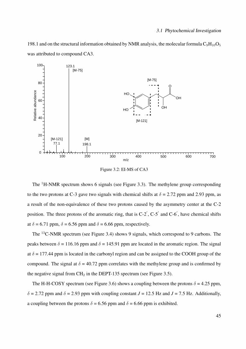
3.1 Phytochemical Investigation
198.1 and on the structural information obtained by NMR analysis, the molecular formula C9H10O5
was attributed to compound CA3.
123.1
77.1
100
80
60
40
20
0100 200 300 400 500 600 700
m/z
Rel
ativ
e ab
unda
nce
198.1[M][M-121]
[M-75]
OHHO
HO
O
OH
[M-75]
[M-121]
Figure 3.2: EI-MS of CA3
The 1H-NMR spectrum shows 6 signals (see Figure 3.3). The methylene group corresponding
to the two protons at C-3 gave two signals with chemical shifts at δ = 2.72 ppm and 2.93 ppm, as
a result of the non-equivalence of these two protons caused by the asymmetry center at the C-2
position. The three protons of the aromatic ring, that is C-2′ , C-5′ and C-6′ , have chemical shifts
at δ = 6.71 ppm, δ = 6.56 ppm and δ = 6.66 ppm, respectively.
The 13C-NMR spectrum (see Figure 3.4) shows 9 signals, which correspond to 9 carbons. The
peaks between δ = 116.16 ppm and δ = 145.91 ppm are located in the aromatic region. The signal
at δ = 177.44 ppm is located in the carbonyl region and can be assigned to the COOH group of the
compound. The signal at δ = 40.72 ppm correlates with the methylene group and is confirmed by
the negative signal from CH2 in the DEPT-135 spectrum (see Figure 3.5).
The H-H-COSY spectrum (see Figure 3.6) shows a coupling between the protons δ = 4.25 ppm,
δ = 2.72 ppm and δ = 2.93 ppm with coupling constant J = 12.5 Hz and J = 7.5 Hz. Additionally,
a coupling between the protons δ = 6.56 ppm and δ = 6.66 ppm is exhibited.
45

3 Results and Discussion
Based on the MS, 1D- and 2D-NMR analysis, 3-(3,4-dihydroxyphenyl)-2-hydroxypropanoic
acid (syn. danshensu) was identified as the compound CA3 and compared to the literature [136],
as shown in Table 3.1.
Table 3.1: Chemical shifts of CA3 and literatureAtom 13C* 13C** 1H* 1H** 1H - 1H*
numbers δ/ppm δ/ppm δ/ppm (Mult., J(Hz), H) δ/ppm (Mult., J(Hz), H) COSY1 177.4 177.15 - - -2 73.1 72.15 4.25 (s, 1H) 4.32 (s, 1H) 33 41.0 39.95 2.72 (dd, 12.52, 7.5 Hz, 1H); 2.73 (dd, 1H); 2.98 (d, 1H) 2, 3
2.93 (d, 12.9 Hz,1H)
1′ 130.3 129.75 - - -
2′ 116.2 116.70 6.71 (brs, 1H) 6.79 (s, 1H) -
3′ 144.9 144.30 - - -
4′ 145.9 144.35 - - -
5′ 117.2 116.95 6.56 (d, 7.8 Hz, 1H) 6.72 (d, 1H) 6
′
6′ 121.9 121.85 6.66 (m, 1H) 6.60 (dd, 1H) 5
′
* In MeOH-d4. ** Data from the literature [136].
46
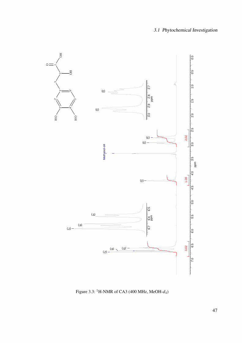
3.1 Phytochemical Investigation
7.0
6.5
6.0
5.5
5.0
4.5
4.0
3.5
3.0
2.5
2.0
1.5
1.0
0.5
0.0
ppm
3.02
2.02
1.00
Met
hano
l-d4
(3)
(2)
(6') .(5') (2')
3.0
2.9
2.8
2.7
ppm
(3)
(3)
6.7
6.6
6.5
(6')
(5')
(2')
ppm
3' 4'
5'
6'1'
2'
2
3
OH
1O
H
OH
O
OH
(3)
Figure 3.3: 1H-NMR of CA3 (400 MHz, MeOH-d4)
47

3 Results and Discussion
3' 4'
5'
6'1'
2'
2
3
OH
1O
H
OH
O
OH
180
170
160
150
140
130
120
110
100
9080
7060
5040
3020
100
ppm
Met
hano
l-d4
(3)
(2)
(2') (5')
(6')
(1')
(3') (4')
(1)
Figure 3.4: 13C-NMR of CA3 (100 MHz, MeOH-d4)
48

3.1 Phytochemical Investigation
180
170
160
150
140
130
120
110
100
9080
7060
5040
3020
100
ppm
Met
hano
l-d4 (3)
(2)
(2') (5')
(6')
3' 4'
5'
6'1'
2'
2
3
OH
1O
H
OH
O
OH
Figure 3.5: DEPT-135 of CA3 (100 MHz, MeOH-d4)
49
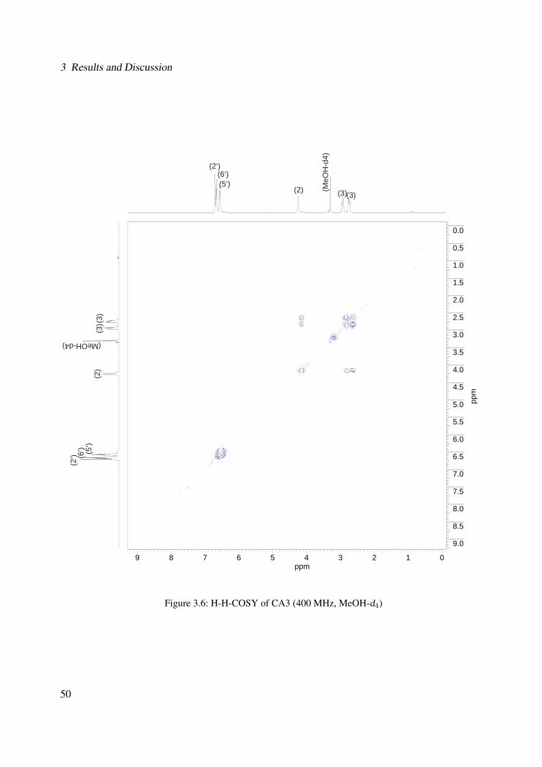
3 Results and Discussion
9 8 7 6 5 4 3 2 1 0ppm
0.0
0.5
1.0
1.5
2.0
2.5
3.0
3.5
4.0
4.5
5.0
5.5
6.0
6.5
7.0
7.5
8.0
8.5
9.0
ppm
(3)(3)(2) (MeO
H-d
4)
(6‘)(5‘)
(2‘)
(3)
(3)
(MeOH-d4)
(2)
(6‘) (5‘)
(2‘)
Figure 3.6: H-H-COSY of CA3 (400 MHz, MeOH-d4)
50

3.1 Phytochemical Investigation
3.1.1.2.2 CA1: Rosmarinic Acid
The subfractionation of fraction K was separated by flash chromatography over a RP-18 column
using methanol and water as mobile phase (see Section 5.6.1.1, Experimental Part) and resulted
in 12 mg of rosmarinic acid2. This compound was isolated as a light gray colored powder and its
chemical structure can be observed in Figure 3.7. This structure was established on the basis of
UV, IR, MS and NMR spectroscopic data.
3''
4''5''
6''
1''2''
2'3'
O1'
21
O
O
31'''
6'''
OH2'''
3'''
4'''5''' OH
OH OH
OH
Figure 3.7: Chemical structure of CA1
The UV spectrum of CA1 (see Figure 3.8) exhibited two absorption maxima (in MeOH) at λ =
290 and 330 nm. The FTIR spectrum (see Figure 3.9) showed distinguishable absorption bands at:
3165.4, 1707.2, 1617.4, 1515.6, 1348.7, 1285.1, 1260.4, 1231.5, 1200.5, 1154.0, 1113.4, 1075.9,
972.3, 851.7, 818.8, 781.4 cm−1.
Figure 3.8: UV spectrum of CA1
2IUPAC name: (2R)-3-(3,4-dihydroxyphenyl)-2-[(E)-3-(3,4-dihydroxyphenyl)prop-2-enoyl]oxypropanoic acid
51

3 Results and Discussion
4000,0 3600 3200 2800 2400 2000 1800 1600 1400 1200 1000 800 650,078,0
79
80
81
82
83
84
85
86
87
88
89
90
91
92
93
94
95
9696,6
cm-1
%T
3165,4
1707,2
1617,4
1515,6
1464,6
1348,7
1285,1
1260,4
1231,51200,5
1154,0
1113,4
1075,9972,3
851,7
818,8
781,4
Figure 3.9: IR spectrum of CA1
The ESI-MS (negative mode) (see Figure 3.10) revealed the a quasimolecular peak3 [M - H]− at
m/z = 359.0 (100) (i.e., base peak). Further ions at m/z = 197.0 (10) and m/z = 161.1 (36) resulted
from the loss of caffeic acid (163) and of 3-(3,4-dihydroxyphenyl)-2-hydroxypropanoic acid (197).
On the basis of the molecular mass at m/z = 360.1 and the structural information obtained by NMR
analysis, the molecular formula C18H16O8 was attributed to compound CA1. This molecular mass
was confirmed by the high resolution FT-ICR-MS for [M - H]− at m/z = 359.076450 (calculated
mass for C18H16O8 was 359.07614).
3In brackets, the relative intensity in % of the ion peaks is shown.
52
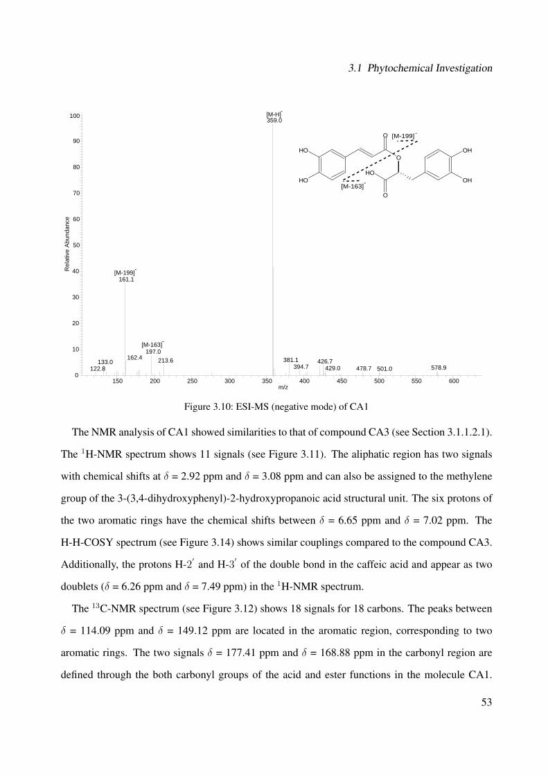
3.1 Phytochemical Investigation
150 200 250 300 350 400 450 500 550 600m/z
0
10
20
30
40
50
60
70
80
90
100
Rel
ativ
e A
bund
ance
359.0
161.1
197.0162.4 381.1213.6 426.7133.0
394.7 578.9429.0 478.7122.8 501.0
O
O
O
OH
OH
OH OH
OH
[M-H]-
[M-163]-
[M-199]-
[M-163]-
[M-199]-
Figure 3.10: ESI-MS (negative mode) of CA1
The NMR analysis of CA1 showed similarities to that of compound CA3 (see Section 3.1.1.2.1).
The 1H-NMR spectrum shows 11 signals (see Figure 3.11). The aliphatic region has two signals
with chemical shifts at δ = 2.92 ppm and δ = 3.08 ppm and can also be assigned to the methylene
group of the 3-(3,4-dihydroxyphenyl)-2-hydroxypropanoic acid structural unit. The six protons of
the two aromatic rings have the chemical shifts between δ = 6.65 ppm and δ = 7.02 ppm. The
H-H-COSY spectrum (see Figure 3.14) shows similar couplings compared to the compound CA3.
Additionally, the protons H-2′ and H-3′ of the double bond in the caffeic acid and appear as two
doublets (δ = 6.26 ppm and δ = 7.49 ppm) in the 1H-NMR spectrum.
The 13C-NMR spectrum (see Figure 3.12) shows 18 signals for 18 carbons. The peaks between
δ = 114.09 ppm and δ = 149.12 ppm are located in the aromatic region, corresponding to two
aromatic rings. The two signals δ = 177.41 ppm and δ = 168.88 ppm in the carbonyl region are
defined through the both carbonyl groups of the acid and ester functions in the molecule CA1.
53
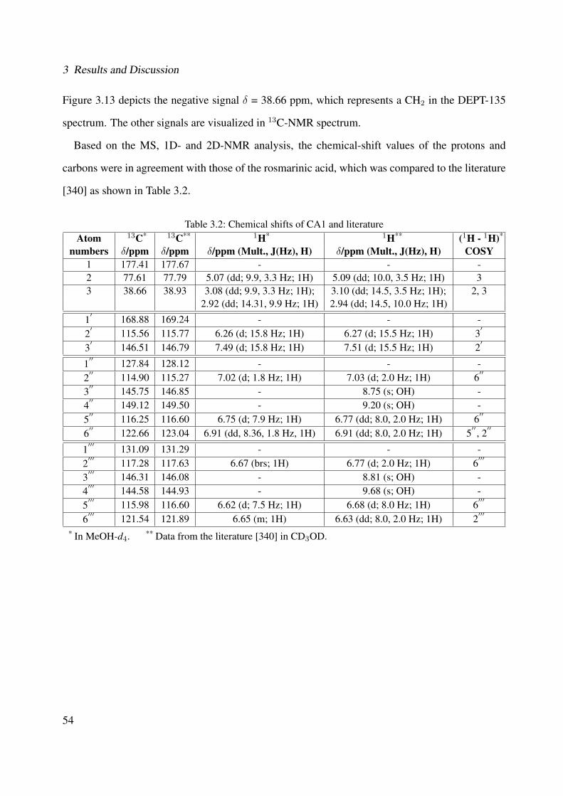
3 Results and Discussion
Figure 3.13 depicts the negative signal δ = 38.66 ppm, which represents a CH2 in the DEPT-135
spectrum. The other signals are visualized in 13C-NMR spectrum.
Based on the MS, 1D- and 2D-NMR analysis, the chemical-shift values of the protons and
carbons were in agreement with those of the rosmarinic acid, which was compared to the literature
[340] as shown in Table 3.2.
Table 3.2: Chemical shifts of CA1 and literatureAtom 13C* 13C** 1H* 1H** (1H - 1H)*
numbers δ/ppm δ/ppm δ/ppm (Mult., J(Hz), H) δ/ppm (Mult., J(Hz), H) COSY1 177.41 177.67 - - -2 77.61 77.79 5.07 (dd; 9.9, 3.3 Hz; 1H) 5.09 (dd; 10.0, 3.5 Hz; 1H) 33 38.66 38.93 3.08 (dd; 9.9, 3.3 Hz; 1H); 3.10 (dd; 14.5, 3.5 Hz; 1H); 2, 3
2.92 (dd; 14.31, 9.9 Hz; 1H) 2.94 (dd; 14.5, 10.0 Hz; 1H)
1′ 168.88 169.24 - - -
2′ 115.56 115.77 6.26 (d; 15.8 Hz; 1H) 6.27 (d; 15.5 Hz; 1H) 3
′
3′ 146.51 146.79 7.49 (d; 15.8 Hz; 1H) 7.51 (d; 15.5 Hz; 1H) 2
′
1′′ 127.84 128.12 - - -
2′′ 114.90 115.27 7.02 (d; 1.8 Hz; 1H) 7.03 (d; 2.0 Hz; 1H) 6
′′
3′′ 145.75 146.85 - 8.75 (s; OH) -
4′′ 149.12 149.50 - 9.20 (s; OH) -
5′′ 116.25 116.60 6.75 (d; 7.9 Hz; 1H) 6.77 (dd; 8.0, 2.0 Hz; 1H) 6
′′
6′′ 122.66 123.04 6.91 (dd, 8.36, 1.8 Hz, 1H) 6.91 (dd; 8.0, 2.0 Hz; 1H) 5
′′, 2
′′
1′′′ 131.09 131.29 - - -
2′′′ 117.28 117.63 6.67 (brs; 1H) 6.77 (d; 2.0 Hz; 1H) 6
′′′
3′′′ 146.31 146.08 - 8.81 (s; OH) -
4′′′ 144.58 144.93 - 9.68 (s; OH) -
5′′′ 115.98 116.60 6.62 (d; 7.5 Hz; 1H) 6.68 (d; 8.0 Hz; 1H) 6
′′′
6′′′ 121.54 121.89 6.65 (m; 1H) 6.63 (dd; 8.0, 2.0 Hz; 1H) 2
′′′
* In MeOH-d4. ** Data from the literature [340] in CD3OD.
54
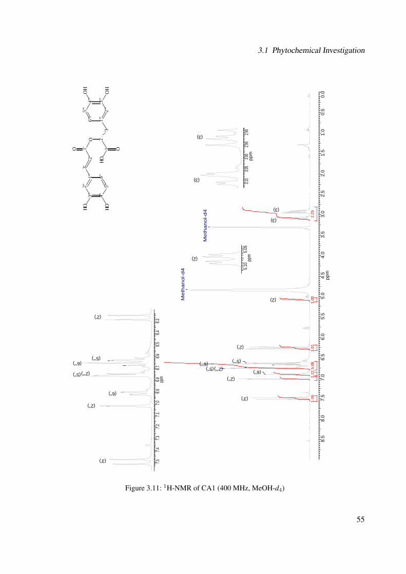
3.1 Phytochemical Investigation
8.5
8.0
7.5
7.0
6.5
6.0
5.5
5.0
4.5
4.0
3.5
3.0
2.5
2.0
1.5
1.0
0.5
0.0
ppm
5.05
2.25
1.22
00.110.1
60.1
Me
tha
no
l-d
4
(3)
(3)
(2)
(2')
(6''') (5''') (5'')
(6'') (2'')
(3')
7.57.4
7.37.2
7.17.0
6.96.8
6.76.6
6.56.4
6.3pp
m
5.10
5.05
ppm
3.10
3.05
3.00
2.95
2.90
ppm
3''
4''
5''
6''
1''
2''
2'3'
O1'
21 OO
31'
''
6'''
OH2'
''3'
''
4'''
5'''
OH
OHO
H
OH
Me
tha
no
l-d
4
(3)
(3)
(2)
(2')
(3')
(2'')
(6'')
(5'')
(6''') (5''')
(2''')(2''')
Figure 3.11: 1H-NMR of CA1 (400 MHz, MeOH-d4)
55

3 Results and Discussion
3''
4''
5''
6''
1''
2''
2'3'
O1'
21 OO
31'
''
6'''
OH2'
''3'
''
4'''
5'''
OH
OHO
H
OH
180
170
160
150
140
130
120
110
100
9080
7060
5040
3020
100
ppm
Met
hano
l-d4
(3)
(2)
(2'') (5''') (2''')
(6''') (6'')
(1'') (1''')
(4'')
(3'') (3''')
(3')
(4''')
(1')
(1)
130
125
120
115
ppm
(2'') (2')
(5''') (5'') (2''')
(6''')
(6'')
(1'')
(1''')
149
148
147
146
145
ppm
(4'')
(3'')
(3''') (3')
(4''')
Figure 3.12: 13C-NMR of CA1 (100 MHz, MeOH-d4)
56

3.1 Phytochemical Investigation
180
170
160
150
140
130
120
110
100
9080
7060
5040
3020
100
ppm
Met
hano
l-d4
(3)
(2)
(2'') (5''') (2''')
(6''') (6'')
(3''')
3''
4''
5''
6''
1''
2''
2'3'
O1'
21 OO
31'
''
6'''
OH2'
''3'
''
4'''
5'''
OH
OHO
H
OH
Figure 3.13: DEPT-135 of CA1 (100 MHz, MeOH-d4)
57
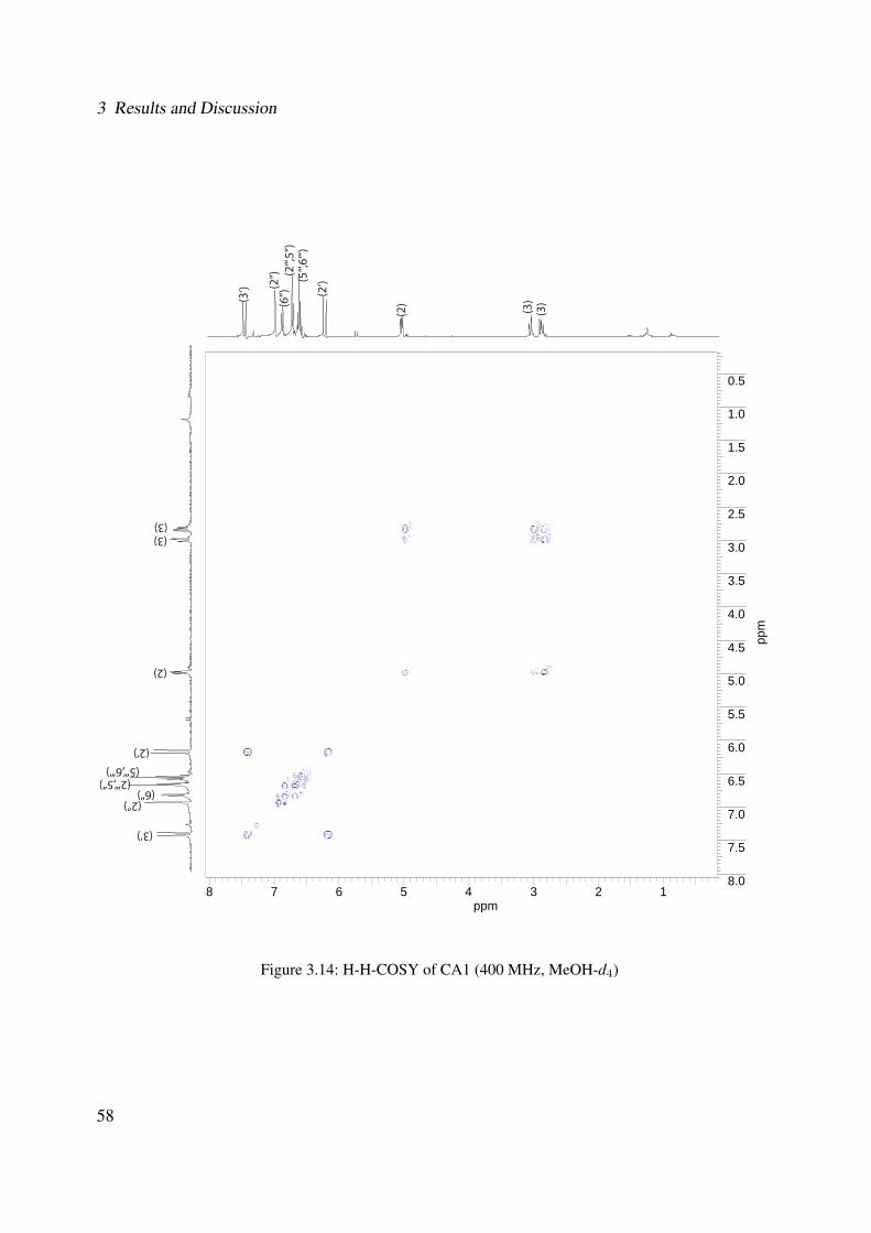
3 Results and Discussion
8 7 6 5 4 3 2 1ppm
0.5
1.0
1.5
2.0
2.5
3.0
3.5
4.0
4.5
5.0
5.5
6.0
6.5
7.0
7.5
8.0
ppm
(3)
(3)
(2)(3
‘) (2‘)
(6‘‘)
(2‘‘‘,5
‘‘)(2
‘‘) (5‘‘‘,6
‘‘‘)
(3)(3)
(2)
(3‘)
(2‘)
(2‘‘)(6‘‘)
(2‘‘‘,5‘‘)(5‘‘‘,6‘‘‘)
Figure 3.14: H-H-COSY of CA1 (400 MHz, MeOH-d4)
58

3.1 Phytochemical Investigation
3.1.1.2.3 CA2: Rosmarinic Acid Ethyl Ester
The subfractionation of fraction H was separated by flash chromatography over a RP-18 column
using methanol and water as mobile phase (see Section 5.6.1.1, Experimental Part) followed by
a further purification in an analytical HPLC system resulting in 3.3 mg of rosmarinic acid ethyl
ester4. This compound was isolated as a light gray amorphous powder and its chemical struc-
ture is represented in Figure 3.15. This structure was established on the basis of MS and NMR
spectroscopic data.
3'''
4'''5'''
6'''
1'''2'''
2''3''
O1''
21
O
O
31""
6''''
O2''''
3''''
4''''5'''' OH
OH OH
OH
1'
2'
Figure 3.15: Chemical structure of CA2
The ESI mass spectrometry (negative mode) (see Figure 3.16) showed a quasimolecular ion
peak5 at m/z = 388.7 [M]− (24) and further peaks at m/z: 387.5 [M - H]− (100) (i.e., base peak);
207.3 [M - 179]− (2); 179.3 [M - 209]− (19); 161.2 [M - 225]− (4); 135.3 [M - 253]− (17).
Based on the molecular mass at m/z = 388.7 and the structural information obtained by NMR
analysis, a molecular formula C20H20O8 was assigned to compound CA2. This molecular mass
was confirmed by the high resolution FT-ICR-MS for [M + Na]+ at m/z = 411.105329 (calculated
mass for C20H20O8Na was 411.10504).
4IUPAC name: (1R)-1-(3,4-dihydroxybenzyl)-2-ethoxy-2-oxoethyl (2E)-3-(3,4-dihydroxyphenyl)acrylate5In brackets, the relative intensity in % of the ion peaks is shown.
59
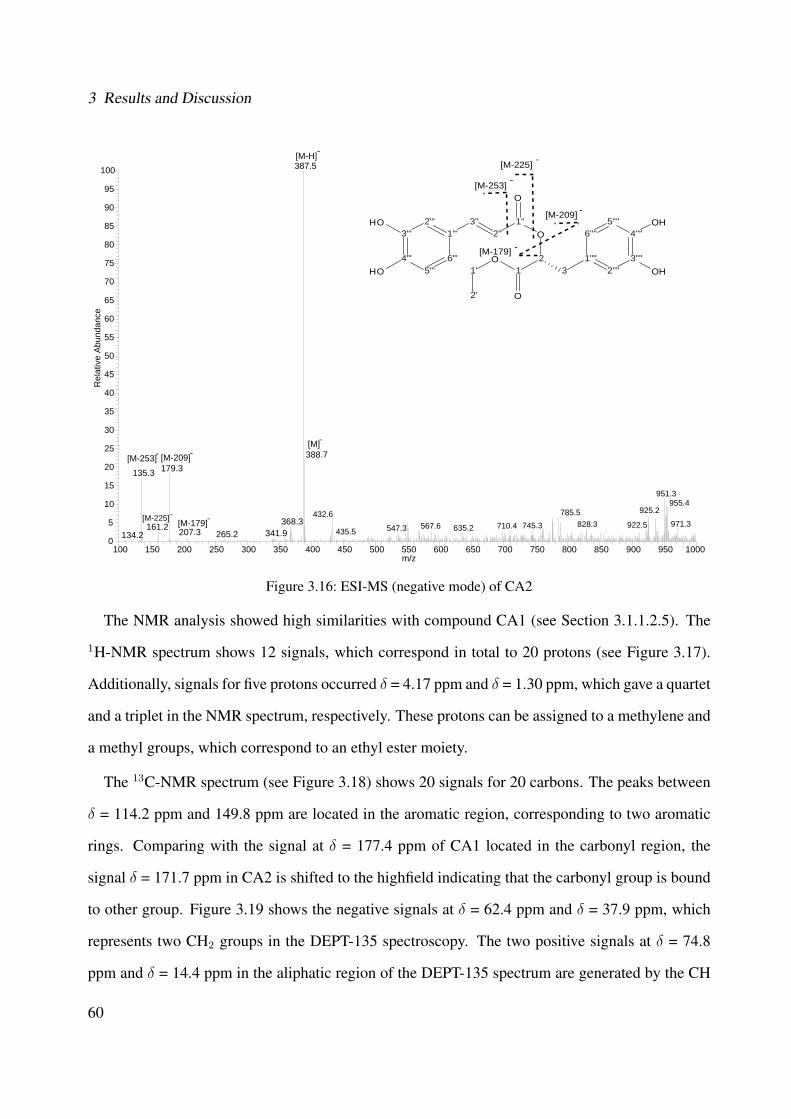
3 Results and Discussion
100 150 200 250 300 350 400 450 500 550 600 650 700 750 800 850 900 950 1000m/z
0
5
10
15
20
25
30
35
40
45
50
55
60
65
70
75
80
85
90
95
100
Rel
ativ
e A
bund
ance
387.5
388.7179.3135.3
951.3955.4
368.3925.2785.5432.6
971.3828.3 922.5745.3710.4567.6161.2 635.2547.3435.5207.3 341.9265.2134.2
3'''
4'''5'''
6'''
1'''2'''
2''3''
O1''
21
O
O
31""
6''''
O2''''
3''''
4''''5'''' OH
OH OH
OH
1'
2'
[M-H]-
[M-209]-[M]-
[M-253]-
[M-209]
[M-253] -
-
[M-179]-[M-225]-
[M-179] -
[M-225] -
Figure 3.16: ESI-MS (negative mode) of CA2
The NMR analysis showed high similarities with compound CA1 (see Section 3.1.1.2.5). The
1H-NMR spectrum shows 12 signals, which correspond in total to 20 protons (see Figure 3.17).
Additionally, signals for five protons occurred δ = 4.17 ppm and δ = 1.30 ppm, which gave a quartet
and a triplet in the NMR spectrum, respectively. These protons can be assigned to a methylene and
a methyl groups, which correspond to an ethyl ester moiety.
The 13C-NMR spectrum (see Figure 3.18) shows 20 signals for 20 carbons. The peaks between
δ = 114.2 ppm and 149.8 ppm are located in the aromatic region, corresponding to two aromatic
rings. Comparing with the signal at δ = 177.4 ppm of CA1 located in the carbonyl region, the
signal δ = 171.7 ppm in CA2 is shifted to the highfield indicating that the carbonyl group is bound
to other group. Figure 3.19 shows the negative signals at δ = 62.4 ppm and δ = 37.9 ppm, which
represents two CH2 groups in the DEPT-135 spectroscopy. The two positive signals at δ = 74.8
ppm and δ = 14.4 ppm in the aliphatic region of the DEPT-135 spectrum are generated by the CH
60
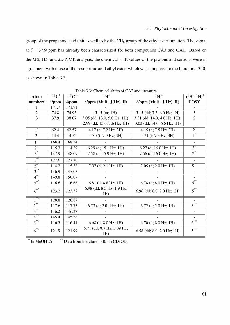
3.1 Phytochemical Investigation
group of the propanoic acid unit as well as by the CH3 group of the ethyl ester function. The signal
at δ = 37.9 ppm has already been characterized for both compounds CA3 and CA1. Based on
the MS, 1D- and 2D-NMR analysis, the chemical-shift values of the protons and carbons were in
agreement with those of the rosmarinic acid ethyl ester, which was compared to the literature [340]
as shown in Table 3.3.
Table 3.3: Chemical shifts of CA2 and literatureAtom 13C* 13C** 1H* 1H** (1H - 1H)*
numbers δ/ppm δ/ppm δ/ppm (Mult., J(Hz), H) δ/ppm (Mult., J(Hz), H) COSY1 171.7 171.91 - - -2 74.8 74.95 5.15 (m; 1H) 5.15 (dd; 7.5, 6.0 Hz; 1H) 33 37.9 38.07 3.05 (dd; 13.0, 5.0 Hz; 1H); 3.31 (dd; 14.0, 4.8 Hz; 1H); 2
2.99 (dd; 13.0, 7.6 Hz; 1H) 3.03 (dd; 14.0, 6.6 Hz; 1H)
1′ 62.4 62.57 4.17 (q; 7.2 Hz: 2H) 4.15 (q; 7.5 Hz; 2H) 2
′
2′ 14.4 14.52 1.30 (t; 7.9 Hz; 3H) 1.21 (t; 7.5 Hz; 3H) 1
′
1′′ 168.4 168.54 - - -
2′′ 115.3 114.29 6.29 (d; 15.1 Hz; 1H) 6.27 (d; 16.0 Hz; 1H) 3
′′
3′′ 147.9 148.09 7.58 (d; 15.9 Hz; 1H) 7.56 (d; 16.0 Hz; 1H) 2
′′
1′′′ 127.6 127.70 - - -
2′′′ 114.2 115.36 7.07 (d; 2.1 Hz; 1H) 7.05 (d; 2.0 Hz; 1H) 5
′′′
3′′′ 146.9 147.03 - - -
4′′′ 149.8 150.07 - - -
5′′′ 116.6 116.66 6.81 (d; 8.8 Hz; 1H) 6.78 (d; 8.0 Hz; 1H) 6
′′′
6′′′ 123.2 123.37
6.98 (dd; 8.3 Hz, 1.9 Hz;1H)
6.96 (dd; 8.0, 2.0 Hz; 1H) 5′′′
1′′′′ 128.8 128.87 - - -
2′′′′ 117.6 117.75 6.73 (d; 2.01 Hz; 1H) 6.72 (d; 2.0 Hz; 1H) 6
′′′′
3′′′′ 146.2 146.37 - - -
4′′′′ 145.4 145.56 - - -
5′′′′ 116.3 116.44 6.68 (d; 8.0 Hz; 1H) 6.70 (d; 8.0 Hz; 1H) 6
′′′′
6′′′′ 121.9 121.99
6.71 (dd; 8.7 Hz, 3.09 Hz;1H)
6.58 (dd; 8.0, 2.0 Hz; 1H) 5′′′′
* In MeOH-d4. ** Data from literature [340] in CD3OD.
61

3 Results and Discussion
8.5
8.0
7.5
7.0
6.5
6.0
5.5
5.0
4.5
4.0
3.5
3.0
2.5
2.0
1.5
1.0
0.5
0.0
ppm
3.53
9.38
3.11
1.31
1.00
(2')
(3) (3)
(1')
(2)
(2'')
(6'''') (2'''')
(5'''')
(6''') (2''')
(3'')
7.6
7.5
7.4
7.3
7.2
7.1
7.0
6.9
6.8
6.7
6.6
6.5
6.4
6.3
ppm
9.38
1.31
1.00 (2'')
(6'''')
(2'''')
(5'''')
(5''')
(6''')
(2''')
(3'')
3'''
4'''
5'''
6'''
1'''
2'''
2''
3''
O1'
'
21 OO
31"
"
6''''
O2'
'''3'
'''
4''''
5''''
OH
OHO
H
OH
1' 2'
(5''')
1.00
1.00
4.2
4.1
(1')
1.20(2')
Figure 3.17: 1H-NMR of CA2 (400 MHz, MeOH-d4)
62
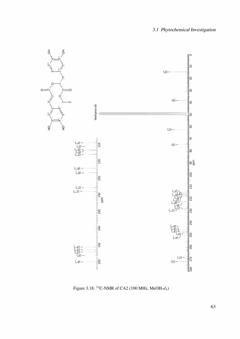
3.1 Phytochemical Investigation
180
170
160
150
140
130
120
110
100
9080
7060
5040
3020
100
ppm
Met
hano
l-d4
(2')
(3)
(1')
(2)
(2''') (5'''')
(5''') (2'''')
(6'''') (6''') (1''')
(1'''')
(4'''') (3'''') (3''')
(3'') (4''')
(1'') (1)
150
145
140
135
130
125
120
115
ppm
(2''') (2'') (5'''') (5''') (2'''')
(6'''') (6''')
(1''') (1'''')
(4'''') (3'''') (3''')
(3'')
(4''')
3'''
4'''
5'''
6'''
1'''
2'''
2''
3''
O1'
'
21 OO
31"
"
6''''
O2'
'''3'
'''
4''''
5''''
OH
OHO
H
OH
1' 2'
(2'')
Figure 3.18: 13C-NMR of CA2 (100 MHz, MeOH-d4)
63

3 Results and Discussion
180
170
160
150
140
130
120
110
100
9080
7060
5040
3020
100
ppm
Met
hano
l-d4
(2')
(3)
(1')
(2)
(2''')
(5'''') (2'''') (6'''')
(6''')
(3'')
3'''
4'''
5'''
6'''
1'''
2'''
2''
3''
O1'
'
21 OO
31"
"
6''''
O2'
'''3'
'''
4''''
5''''
OH
OHO
H
OH
1' 2'
Figure 3.19: DEPT-135 of CA2 (100 MHz, MeOH-d4)
64
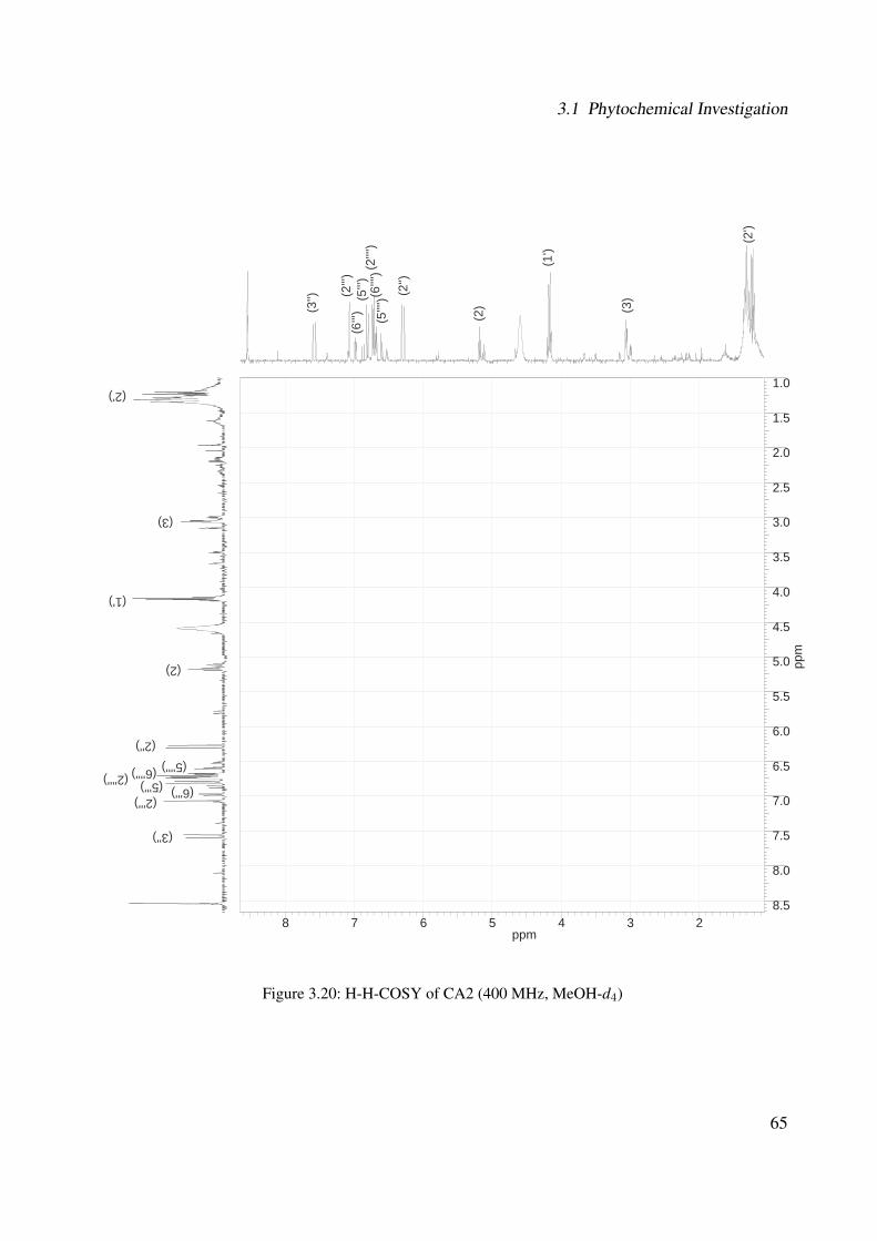
3.1 Phytochemical Investigation
8 7 6 5 4 3 2ppm
1.0
1.5
2.0
2.5
3.0
3.5
4.0
4.5
5.0
5.5
6.0
6.5
7.0
7.5
8.0
8.5
ppm
(3)
(1')
(2)
(2'')
(6''''
) (2'
''') (5
'''')
(6''')
(2''')
(3'')
(3)
(1')
(2)
(2'')
(6'''') (2'''') (5'''')
(6''') (2''')
(3'')
(2')
(2')
(5''')
(5''')
Figure 3.20: H-H-COSY of CA2 (400 MHz, MeOH-d4)
65

3 Results and Discussion
3.1.1.2.4 CA4: Rutin
The subfractionation of fraction G was performed by flash chromatography over a RP-18 column
using methanol and water as eluent (see Section 5.6.1.1, Experimental Part) and resulted in 15 mg
of rutin6. This compound is a yellow powder and its chemical structure can be observed in Figure
3.21. This structure was also established based on UV, IR, MS and NMR spectroscopic data.
9
104
3
2O8
7
65
O
2'1'
OH O
3'
4'5'
6'
OH
OH
2''
1''O
5''
4''3''
5'''
4'''3'''
2'''
1'''O
OHOH
OH6''
O
6'''OH
OHOH
OH
Figure 3.21: Chemical structure of CA4
The UV spectrum of CA4 (see Figure 3.22) exhibited two absorption maxima (in MeOH) at λ
= 257 and 354 nm, which indicated the presence of a flavonol structure [196]. The FTIR spectrum
(see Figure 3.23) showed the absorption bands at: 3329.6, 1653.9, 1596.1, 1501.0, 1455.7, 1358.9,
1296.4, 1203.3, 1172.2, 1062.9, 1041.9, 1014.5, 1001.2, 967.7, 944.5, 808.0, 707.2, 686.6 cm−1.
Figure 3.22: UV of CA4
6IUPAC name: 2-(3,4-dihydroxyphenyl)-5,7-dihydroxy-3-[(2S,3R,4S,5S,6R) -3,4,5-trihydroxy-6-[[(2R,3R,4R,5R,6S)-3,4,5-trihydroxy-6-methyloxan-2-yl]oxymethyl]oxan-2-yl]oxychromen-4-one
66
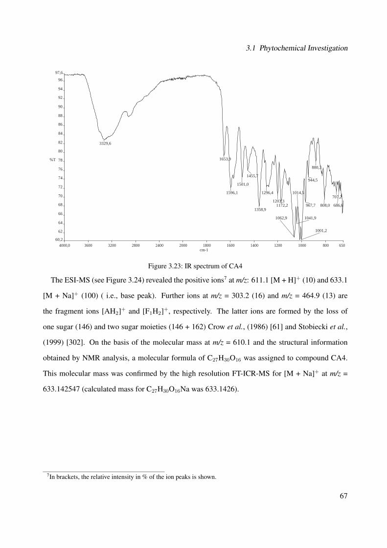
3.1 Phytochemical Investigation
4000,0 3600 3200 2800 2400 2000 1800 1600 1400 1200 1000 800 65060,2
62
64
66
68
70
72
74
76
78
80
82
84
86
88
90
92
94
96
97,6
cm-1
%T
3329,6
1653,9
1596,1
1501,0
1455,7
1358,9
1296,4
1203,31172,2
1062,9 1041,9
1014,5
1001,2
967,7
944,5
880,3
808,0
707,2
686,6
Figure 3.23: IR spectrum of CA4
The ESI-MS (see Figure 3.24) revealed the positive ions7 at m/z: 611.1 [M + H]+ (10) and 633.1
[M + Na]+ (100) ( i.e., base peak). Further ions at m/z = 303.2 (16) and m/z = 464.9 (13) are
the fragment ions [AH2]+ and [F1H2]+, respectively. The latter ions are formed by the loss of
one sugar (146) and two sugar moieties (146 + 162) Crow et al., (1986) [61] and Stobiecki et al.,
(1999) [302]. On the basis of the molecular mass at m/z = 610.1 and the structural information
obtained by NMR analysis, a molecular formula of C27H30O16 was assigned to compound CA4.
This molecular mass was confirmed by the high resolution FT-ICR-MS for [M + Na]+ at m/z =
633.142547 (calculated mass for C27H30O16Na was 633.1426).
7In brackets, the relative intensity in % of the ion peaks is shown.
67
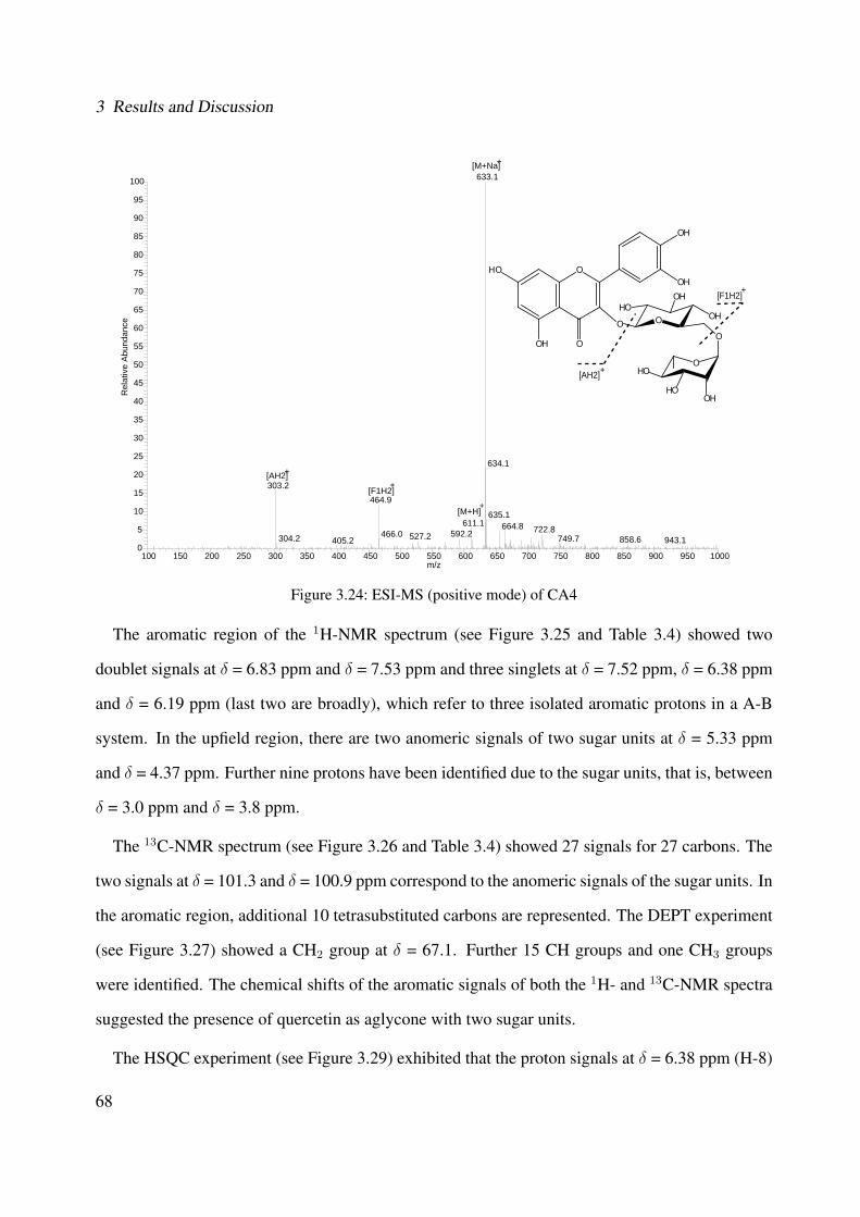
3 Results and Discussion
100 150 200 250 300 350 400 450 500 550 600 650 700 750 800 850 900 950 1000m/z
0
5
10
15
20
25
30
35
40
45
50
55
60
65
70
75
80
85
90
95
100
Rel
ativ
e A
bund
ance
633.1
634.1
303.2464.9
635.1611.1 664.8 722.8592.2466.0 527.2304.2 749.7 858.6 943.1405.2
O
O
OH O
HO
OH
O
O
HOOH
OH
O
HO
HOOH
OH
[M+H]+
[M+Na]+
[F1H2]+
[F1H2]+
[AH2]+
[AH2]+
Figure 3.24: ESI-MS (positive mode) of CA4
The aromatic region of the 1H-NMR spectrum (see Figure 3.25 and Table 3.4) showed two
doublet signals at δ = 6.83 ppm and δ = 7.53 ppm and three singlets at δ = 7.52 ppm, δ = 6.38 ppm
and δ = 6.19 ppm (last two are broadly), which refer to three isolated aromatic protons in a A-B
system. In the upfield region, there are two anomeric signals of two sugar units at δ = 5.33 ppm
and δ = 4.37 ppm. Further nine protons have been identified due to the sugar units, that is, between
δ = 3.0 ppm and δ = 3.8 ppm.
The 13C-NMR spectrum (see Figure 3.26 and Table 3.4) showed 27 signals for 27 carbons. The
two signals at δ = 101.3 and δ = 100.9 ppm correspond to the anomeric signals of the sugar units. In
the aromatic region, additional 10 tetrasubstituted carbons are represented. The DEPT experiment
(see Figure 3.27) showed a CH2 group at δ = 67.1. Further 15 CH groups and one CH3 groups
were identified. The chemical shifts of the aromatic signals of both the 1H- and 13C-NMR spectra
suggested the presence of quercetin as aglycone with two sugar units.
The HSQC experiment (see Figure 3.29) exhibited that the proton signals at δ = 6.38 ppm (H-8)
68
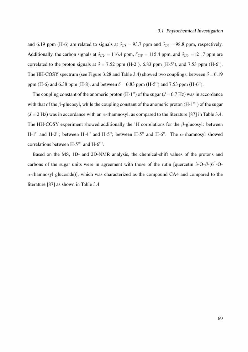
3.1 Phytochemical Investigation
and 6.19 ppm (H-6) are related to signals at δC8 = 93.7 ppm and δC6 = 98.8 ppm, respectively.
Additionally, the carbon signals at δC2′ = 116.4 ppm, δC5′ = 115.4 ppm, and δC6′ =121.7 ppm are
correlated to the proton signals at δ = 7.52 ppm (H-2'), 6.83 ppm (H-5'), and 7.53 ppm (H-6').
The HH-COSY spectrum (see Figure 3.28 and Table 3.4) showed two couplings, between δ = 6.19
ppm (H-6) and 6.38 ppm (H-8), and between δ = 6.83 ppm (H-5”) and 7.53 ppm (H-6”).
The coupling constant of the anomeric proton (H-1”) of the sugar (J = 6.7 Hz) was in accordance
with that of the β-glucosyl, while the coupling constant of the anomeric proton (H-1”') of the sugar
(J = 2 Hz) was in accordance with an α-rhamnosyl, as compared to the literature [87] in Table 3.4.
The HH-COSY experiment showed additionally the 1H correlations for the β-glucosyl: between
H-1” and H-2”; between H-4” and H-5”; between H-5” and H-6”. The α-rhamnosyl showed
correlations between H-5”' and H-6”'.
Based on the MS, 1D- and 2D-NMR analysis, the chemical-shift values of the protons and
carbons of the sugar units were in agreement with those of the rutin [quercetin 3-O-β-(6′′-O-
α-rhamnosyl glucoside)], which was characterized as the compound CA4 and compared to the
literature [87] as shown in Table 3.4.
69
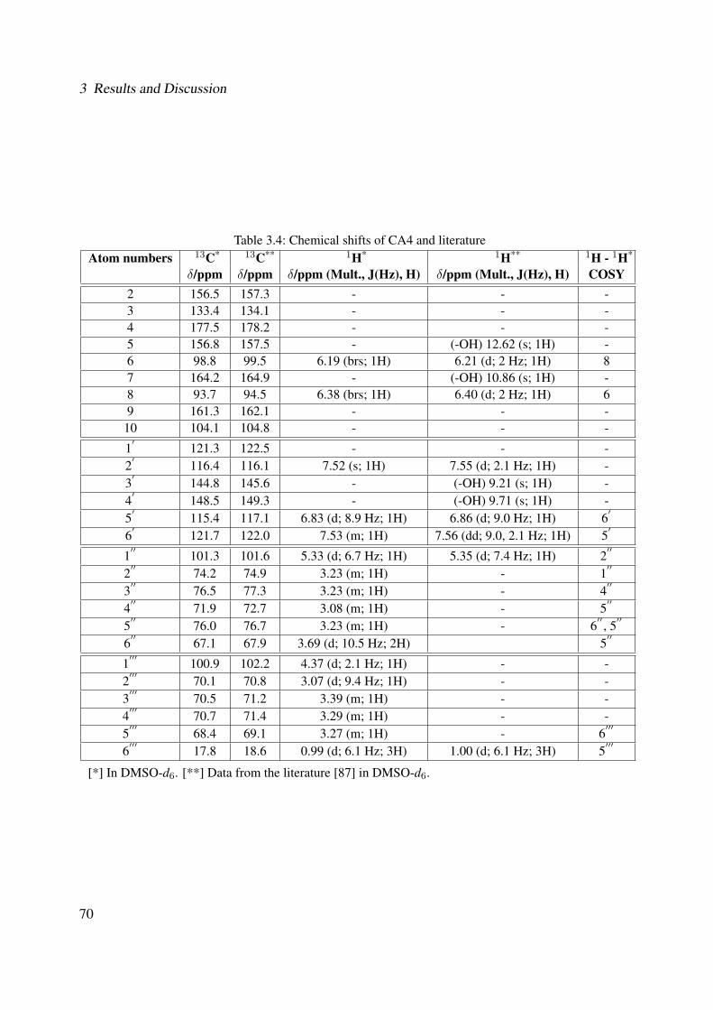
3 Results and Discussion
Table 3.4: Chemical shifts of CA4 and literatureAtom numbers 13C* 13C** 1H* 1H** 1H - 1H*
δ/ppm δ/ppm δ/ppm (Mult., J(Hz), H) δ/ppm (Mult., J(Hz), H) COSY2 156.5 157.3 - - -3 133.4 134.1 - - -4 177.5 178.2 - - -5 156.8 157.5 - (-OH) 12.62 (s; 1H) -6 98.8 99.5 6.19 (brs; 1H) 6.21 (d; 2 Hz; 1H) 87 164.2 164.9 - (-OH) 10.86 (s; 1H) -8 93.7 94.5 6.38 (brs; 1H) 6.40 (d; 2 Hz; 1H) 69 161.3 162.1 - - -10 104.1 104.8 - - -
1′
121.3 122.5 - - -2
′116.4 116.1 7.52 (s; 1H) 7.55 (d; 2.1 Hz; 1H) -
3′
144.8 145.6 - (-OH) 9.21 (s; 1H) -4
′148.5 149.3 - (-OH) 9.71 (s; 1H) -
5′
115.4 117.1 6.83 (d; 8.9 Hz; 1H) 6.86 (d; 9.0 Hz; 1H) 6′
6′
121.7 122.0 7.53 (m; 1H) 7.56 (dd; 9.0, 2.1 Hz; 1H) 5′
1′′
101.3 101.6 5.33 (d; 6.7 Hz; 1H) 5.35 (d; 7.4 Hz; 1H) 2′′
2′′
74.2 74.9 3.23 (m; 1H) - 1′′
3′′
76.5 77.3 3.23 (m; 1H) - 4′′
4′′
71.9 72.7 3.08 (m; 1H) - 5′′
5′′
76.0 76.7 3.23 (m; 1H) - 6′′, 5
′′
6′′
67.1 67.9 3.69 (d; 10.5 Hz; 2H) 5′′
1′′′
100.9 102.2 4.37 (d; 2.1 Hz; 1H) - -2
′′′70.1 70.8 3.07 (d; 9.4 Hz; 1H) - -
3′′′
70.5 71.2 3.39 (m; 1H) - -4
′′′70.7 71.4 3.29 (m; 1H) - -
5′′′
68.4 69.1 3.27 (m; 1H) - 6′′′
6′′′
17.8 18.6 0.99 (d; 6.1 Hz; 3H) 1.00 (d; 6.1 Hz; 3H) 5′′′
[*] In DMSO-d6. [**] Data from the literature [87] in DMSO-d6.
70
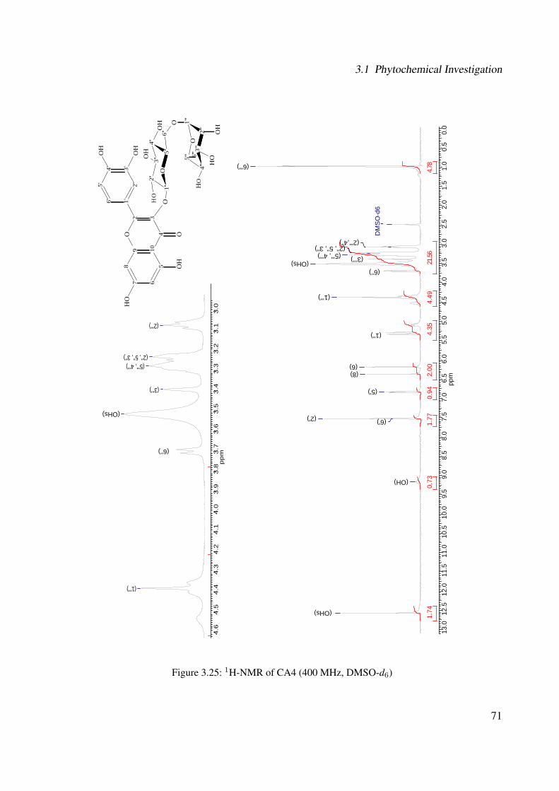
3.1 Phytochemical Investigation
13.0
12.5
12.0
11.5
11.0
10.5
10.0
9.5
9.0
8.5
8.0
7.5
7.0
6.5
6.0
5.5
5.0
4.5
4.0
3.5
3.0
2.5
2.0
1.5
1.0
0.5
0.0
ppm
87.465.12
4.49
4.35
2.00
1.77
1.74
0.94
0.73
DM
SO
-d6
(6''')
(2''',4”) (2'', 5'', 3'')
(5''', 4''') (OHs)
(1''')
(1'')
(6) (8)
(5')
(2') (6')
(OH)
4.6
4.5
4.4
4.3
4.2
4.1
4.0
3.9
3.8
3.7
3.6
3.5
3.4
3.3
3.2
3.1
3.0
pp
m
(2''')
(2'', 5'', 3'') (5''', 4''')
(3''')
(1''')
9 104
32O
87 6
5O
2'1'
OH
O
3'4'5'
6'
OH
OH
2''
1''
O5'
'
4''
3'' 5'''
4'''
3'''
2'''
1'''
O
OH
OH
OH
6''
O
6'''
OH
OH
OH
OH
(OHs)
(OHs)
(3’’’)
(6'’)
(6'’)
Figure 3.25: 1H-NMR of CA4 (400 MHz, DMSO-d6)
71
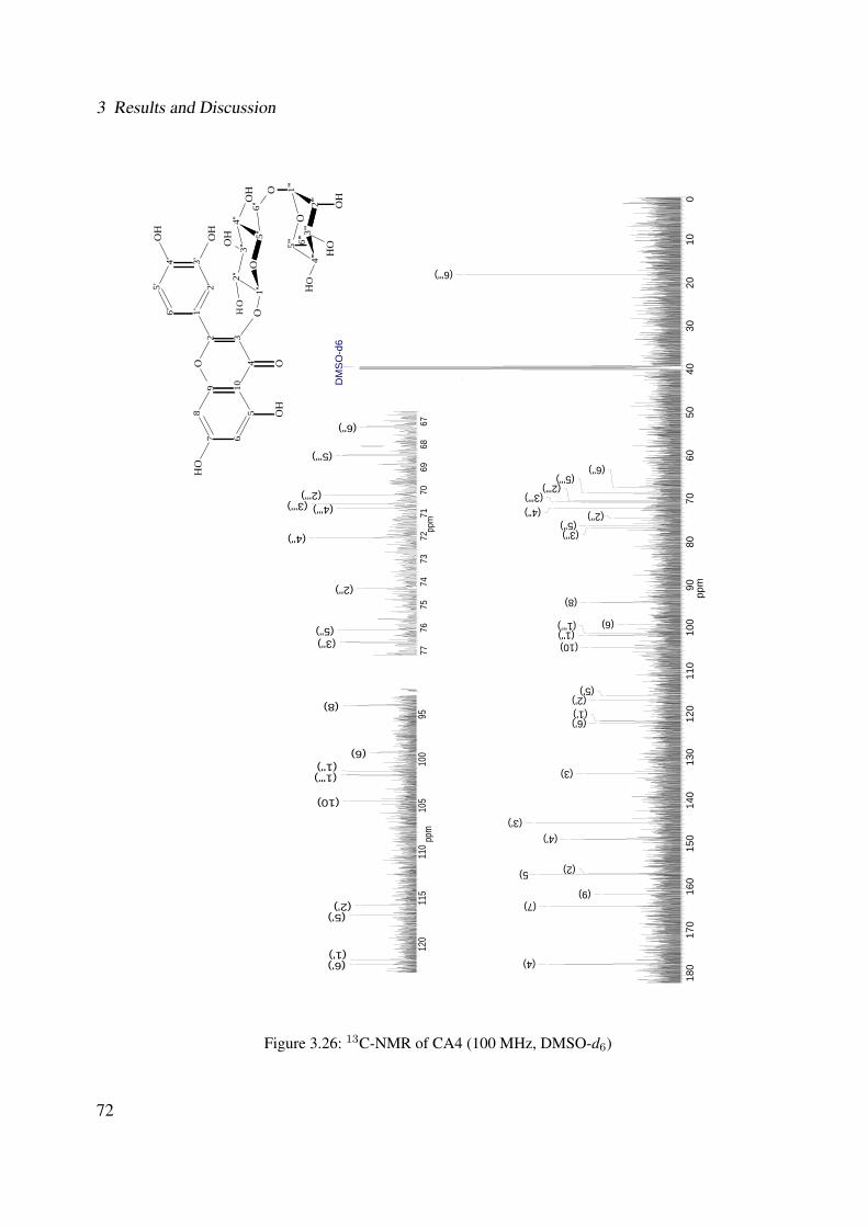
3 Results and Discussion
180
170
160
150
140
130
120
110
100
9080
7060
5040
3020
100
ppm
DM
SO
-d6
(6''')
(6'') (5''')
(2''') (3''') (4'') (2'')
(5'') (3'')
(8)
(6) (1''’) (1'')
(10)
(5') (2') (1') (6')
(3)
(3') (4')
(2)5)
(9) (7)
(4)
7776
7574
7372
7170
6968
67pp
m
(6'')
(5''')
(2''') (3''') (4''')
(4'')
(2'')
(5'') (3'')
120
115
110
105
100
95pp
m
(8)
(6) (1'') (1''')
(10)
(2') (5')
(1') (6')
9 104
32O
87 6
5O
2'1'
OH
O
3'4'5'
6'
OH
OH
2''
1''
O5'
'
4''
3'' 5'''
4'''
3'''
2'''
1'''
O
OH
OH
OH
6''
O
6'''
OH
OH
OH
OH
Figure 3.26: 13C-NMR of CA4 (100 MHz, DMSO-d6)
72
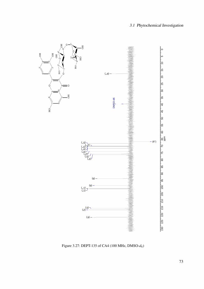
3.1 Phytochemical Investigation
130
125
120
115
110
105
100
9590
8580
7570
6560
5550
4540
3530
2520
1510
50
ppm
DM
SO
-d6
(6''')
(6'') (5''') (2''')
(3''') (4''') (4'')
(2'') (5'')
(3'')
(8)
(6) (1’'') (1'')
(5') (2')
(6')
9 104
32O
87 6
5O
2'1'
OH
O
3'4'5'
6'
OH
OH
2''
1''
O5'
'
4''
3'' 5'''
4'''
3'''
2'''
1'''
O
OH
OH
OH
6''
O
6'''
OH
OH
OH
OH
Figure 3.27: DEPT-135 of CA4 (100 MHz, DMSO-d6)
73
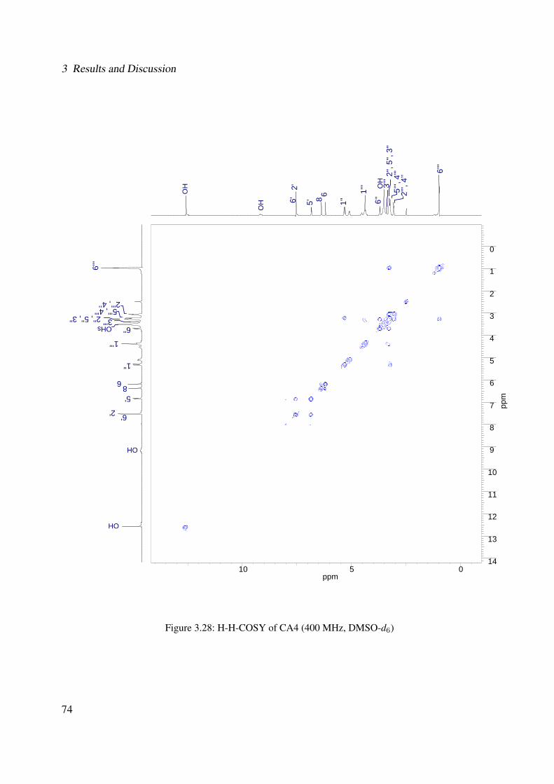
3 Results and Discussion
10 5 0ppm
0
1
2
3
4
5
6
7
8
9
10
11
12
13
14
ppm
OH
OH 6'
2'
5' 8 6
1''
1'''
6'' 5'
'', 4
'''2'',
5'',
3''
2''',
4’’ 6'
''
3'''
6'2'
5'86
1''
1'''
6''
6'''
OH
OH
OHs
5''', 4'''2'', 5'', 3''
2''', 4’’
3'''
OH
Figure 3.28: H-H-COSY of CA4 (400 MHz, DMSO-d6)
74

3.1 Phytochemical Investigation
10 5 0ppm
0
10
20
30
40
50
60
70
80
90
100
110
120
130
140
150
160
170
180
190
ppm
6'''
6''5'
''2'
''
3'''
4'''
4''
2''
5''
3''
86
1''1'
''2'
5'6'
OH
OH 6'
2'
5' 8 6
1''
OH
s
1'''
6'' 5'
'', 4
'''2'',
5'',
3''
2''',
4’’ 6'
''
3'''
Figure 3.29: HSQC of CA4 (400 MHz, DMSO-d6)
75
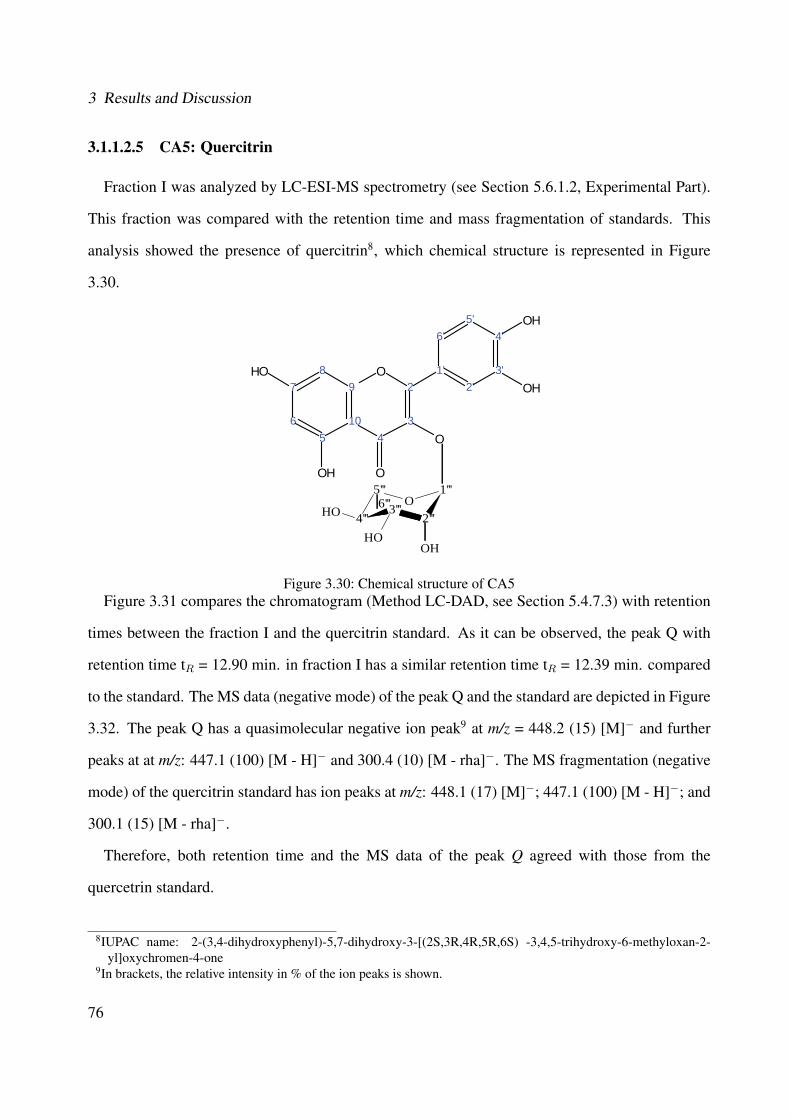
3 Results and Discussion
3.1.1.2.5 CA5: Quercitrin
Fraction I was analyzed by LC-ESI-MS spectrometry (see Section 5.6.1.2, Experimental Part).
This fraction was compared with the retention time and mass fragmentation of standards. This
analysis showed the presence of quercitrin8, which chemical structure is represented in Figure
3.30.
O
7
65
10
98
5'6'
1'2'
3'
4'
O2
43
OH
OH
OH
OH
O5'''
4'''3'''
2'''
1'''O6'''
OH
OHOH
Figure 3.30: Chemical structure of CA5Figure 3.31 compares the chromatogram (Method LC-DAD, see Section 5.4.7.3) with retention
times between the fraction I and the quercitrin standard. As it can be observed, the peak Q with
retention time tR = 12.90 min. in fraction I has a similar retention time tR = 12.39 min. compared
to the standard. The MS data (negative mode) of the peak Q and the standard are depicted in Figure
3.32. The peak Q has a quasimolecular negative ion peak9 at m/z = 448.2 (15) [M]− and further
peaks at at m/z: 447.1 (100) [M - H]− and 300.4 (10) [M - rha]−. The MS fragmentation (negative
mode) of the quercitrin standard has ion peaks at m/z: 448.1 (17) [M]−; 447.1 (100) [M - H]−; and
300.1 (15) [M - rha]−.
Therefore, both retention time and the MS data of the peak Q agreed with those from the
quercetrin standard.
8IUPAC name: 2-(3,4-dihydroxyphenyl)-5,7-dihydroxy-3-[(2S,3R,4R,5R,6S) -3,4,5-trihydroxy-6-methyloxan-2-yl]oxychromen-4-one
9In brackets, the relative intensity in % of the ion peaks is shown.
76
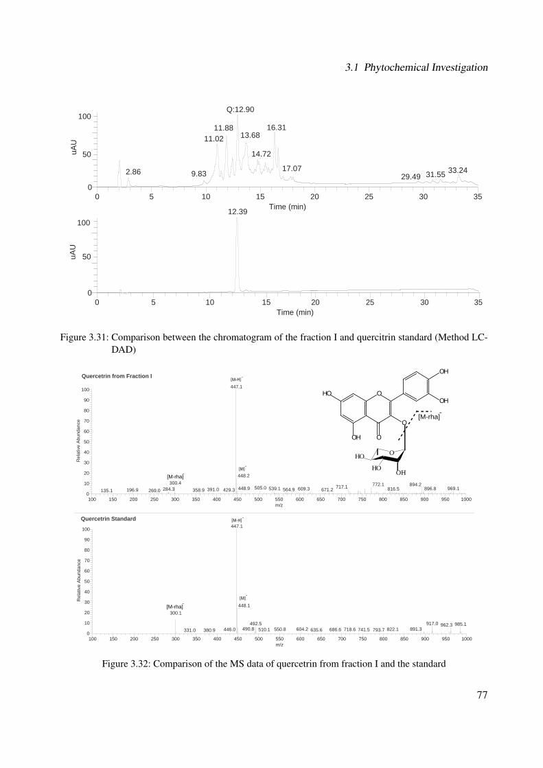
3.1 Phytochemical Investigation
0 5 10 15 20 25 30 35Time (min)
0
50
100
uAU
11.88
Q:12.90
11.0216.31
14.72
33.2417.072.86 31.559.83 29.49
0 5 10 15 20 25 30 35Time (min)
0
50
100
uAU
12.39
13.68
Figure 3.31: Comparison between the chromatogram of the fraction I and quercitrin standard (Method LC-DAD)
100 150 200 250 300 350 400 450 500 550 600 650 700 750 800 850 900 950 1000m/z
0
10
20
30
40
50
60
70
80
90
100
Rel
ativ
e A
bund
ance
447.1
448.1
917.0492.5 985.1962.3
300.1
490.8 891.3604.2550.8 822.1718.6446.0 510.1 686.6 741.5635.6 793.7380.9331.0
100 150 200 250 300 350 400 450 500 550 600 650 700 750 800 850 900 950 1000m/z
0
10
20
30
40
50
60
70
80
90
100
Rel
ativ
e A
bund
ance
447.1
448.2
772.1 894.2717.1505.0448.9 969.1539.1 896.8609.3 816.5284.3 391.0 564.9 671.2429.3196.9 358.9300.4
269.0135.1
Quercetrin Standard
Quercetrin from Fraction I
O
OH
OH
OH
HO
O
O
[M]-
[M]-
[M-H]-
[M-H]-
[M-rha]-
[M-rha]-
[M-rha]-
OOH
OHOH
Figure 3.32: Comparison of the MS data of quercetrin from fraction I and the standard
77
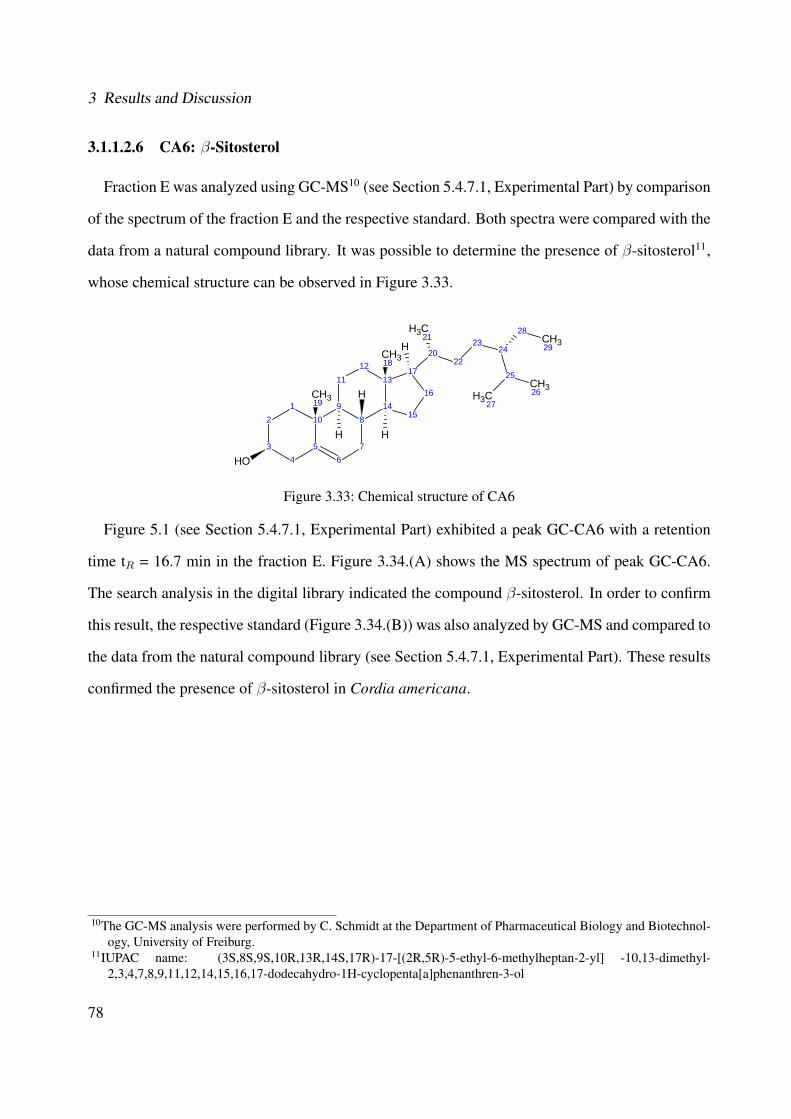
3 Results and Discussion
3.1.1.2.6 CA6: β-Sitosterol
Fraction E was analyzed using GC-MS10 (see Section 5.4.7.1, Experimental Part) by comparison
of the spectrum of the fraction E and the respective standard. Both spectra were compared with the
data from a natural compound library. It was possible to determine the presence of β-sitosterol11,
whose chemical structure can be observed in Figure 3.33.
2
34
5
101
67
89 14
1312
11
15
16
17
2022
OH
CH319H
CH318
H H
H
CH3 2128
2423 CH329
CH326
25
CH3 27
Figure 3.33: Chemical structure of CA6
Figure 5.1 (see Section 5.4.7.1, Experimental Part) exhibited a peak GC-CA6 with a retention
time tR = 16.7 min in the fraction E. Figure 3.34.(A) shows the MS spectrum of peak GC-CA6.
The search analysis in the digital library indicated the compound β-sitosterol. In order to confirm
this result, the respective standard (Figure 3.34.(B)) was also analyzed by GC-MS and compared to
the data from the natural compound library (see Section 5.4.7.1, Experimental Part). These results
confirmed the presence of β-sitosterol in Cordia americana.
10The GC-MS analysis were performed by C. Schmidt at the Department of Pharmaceutical Biology and Biotechnol-ogy, University of Freiburg.
11IUPAC name: (3S,8S,9S,10R,13R,14S,17R)-17-[(2R,5R)-5-ethyl-6-methylheptan-2-yl] -10,13-dimethyl-2,3,4,7,8,9,11,12,14,15,16,17-dodecahydro-1H-cyclopenta[a]phenanthren-3-ol
78

3.1 Phytochemical Investigation
Figure 3.34: Comparison of the MS data between peak GC-CA6 (A) and respective standard (B)
3.1.1.2.7 CA7: Campesterol
Fraction E was analyzed by GC mass spectrometry (see Section 5.4.7.1, Experimental Part) by
comparison of the spectrum of the fraction E with the respective standard. Both spectra were
compared to the data from a natural compound library. It was possible to determine the presence
of campesterol12, whose chemical structure can be observed in Figure 3.35.
2
34
5
101
67
89 14
1312
11
15
16
17
2022
OH
CH319H
CH318
H H
CH3 21
CH32824
23
CH3 26
25CH327
H
Figure 3.35: Chemical structure of CA712IUPAC name: (3S,8S,9S,10R,13R,14S,17R)-17-[(2R,5R)-5,6-dimethylheptan-2-yl] -10,13-dimethyl-
2,3,4,7,8,9,11,12,14,15,16,17-dodecahydro-1H-cyclopenta[a]phenanthren-3-ol
79
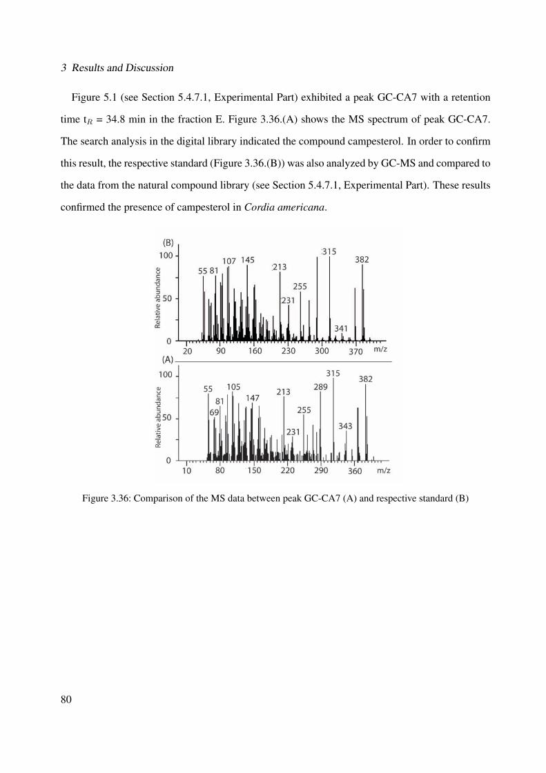
3 Results and Discussion
Figure 5.1 (see Section 5.4.7.1, Experimental Part) exhibited a peak GC-CA7 with a retention
time tR = 34.8 min in the fraction E. Figure 3.36.(A) shows the MS spectrum of peak GC-CA7.
The search analysis in the digital library indicated the compound campesterol. In order to confirm
this result, the respective standard (Figure 3.36.(B)) was also analyzed by GC-MS and compared to
the data from the natural compound library (see Section 5.4.7.1, Experimental Part). These results
confirmed the presence of campesterol in Cordia americana.
Figure 3.36: Comparison of the MS data between peak GC-CA7 (A) and respective standard (B)
80

3.1 Phytochemical Investigation
3.1.1.2.8 CA8: α-Amyrin
The mass spectrum of fraction E was analyzed by means of GC-MS (see Section 5.4.7.1, Experi-
mental Part) using computer searches in the natural compound library. It was possible to determine
the presence of α-amyrin13, whose chemical structure can be observed in Figure 3.37.
2
34
5
101
67
89 14
1312
11
OH
CH325CH326
H CH327
1516
1718 22
2120
19
CH3 23CH324
CH328H
H
CH330
CH3 29
Figure 3.37: Chemical structure of CA8
Figure 5.1 (see Section 5.4.7.1, Experimental Part) exhibited a peak GC-CA8 with a reten-
tion time of 22.2 min in the fraction E. Figure 3.38.(A) shows the comparison between the MS
fragmentation of peak GC-CA8 and data from a natural compound library (see Section 5.4.7.1,
Experimental Part) (Figure 3.38.(B)). The search analysis in the digital library indicated that the
pentacyclic triterpene α-amyrin is present in the fraction E from Cordia americana.
13IUPAC name: (3S,4aR,6aR,6bS,8aR,11R,12S,12aR,14aR,14bR)-4,4,6a,6b,8a,11,12,14b-octamethyl-2,3,4a,5,6,7,8,9,10,11,12,12a,14,14a-tetradecahydro-1H-picen-3-ol
81
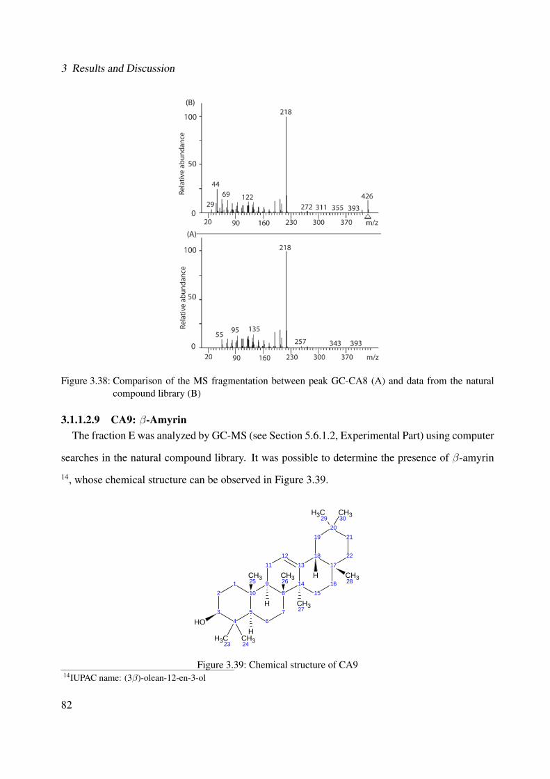
3 Results and Discussion
Figure 3.38: Comparison of the MS fragmentation between peak GC-CA8 (A) and data from the naturalcompound library (B)
3.1.1.2.9 CA9: β-AmyrinThe fraction E was analyzed by GC-MS (see Section 5.6.1.2, Experimental Part) using computer
searches in the natural compound library. It was possible to determine the presence of β-amyrin
14, whose chemical structure can be observed in Figure 3.39.
2
34
5
101
67
89 14
1312
11
OH
CH325CH326
H CH327
1516
1718 22
2120
19
CH330CH3 29
CH3 23CH324
CH328H
H
Figure 3.39: Chemical structure of CA914IUPAC name: (3β)-olean-12-en-3-ol
82

3.1 Phytochemical Investigation
Figure 5.1 (see Section 5.4.7.1, Experimental Part) exhibited a peak GC-CA9 with a reten-
tion time of 18.7 min in the fraction E. Figure 3.39.(A) shows the comparison between the MS
fragmentation of peak GC-CA9 and data from the natural compound library (see Section 5.4.7.1,
Experimental Part) (Figure 3.39.(B)). The search analysis in the digital library indicated that the
pentacyclic triterpene β-amyrin is present in the fraction E from Cordia americana.
Figure 3.40: Comparison of the MS fragmentation between peak GC-CA9 (A) and data from the naturalcompound library (B)
83
3 Results and Discussion
3.1.1.3 Discussion
As presented in the previous sections, the phytochemical studies (i.e., MS, 1D and 2D NMR)
revealed the presence of
• flavonols: rutin and quercitrin;
• phytosterols: campesterol and β-sitosterol;
• triterpenoids: α- and β-amyrin;
• phenolic acids: 3-(3,4-dihydroxyphenyl)-2-hydroxypropanoic acid, rosmarinic acid and ros-
marinic acid ethyl ester in the ethanolic extract from the leaves of Cordia americana.
Figure 3.41 illustrate the DAD-HPLC chromatogram of the ethanolic extract of Cordia amer-
icana and its characterized compounds. As it can be observed, the ethanolic extract contains
rosmarinic acid (CA1) as the major compound. Additionally, further HPLC analysis (Method
LC-DAD, see Section 5.4.7.3, Experimental Part) (see Figure 5.5, Experimental Part) revealed
compound CA1 as the major compound in the ethanolic extract of Cordia americana at different
wavelengths (220, 250, 280, 330, 350 nm).
Rosmarinic acid, an ester of caffeic acid with 3,4-dihydroxyphenylpropionic acid, is a charac-
teristic constituent in members of the Lamiaceae and the Boraginaceae where it occurs in higher
amounts [318]. CA1 was also found in the leaves of Lemon balm (Melissa of�cinalis, Lamiaceae)
in a concentration of 3.91% [312]. In Rosmarinus of�cinalis (Lamiaceae), CA1 can be detected in
leaves, flowers, stems and roots, but the highest amount of 2.5% was found during the first stages
of leaf growth [22]. In the crude extract of Borago of�cinalis (Boraginaceae), 2.5% of CA1 was
quantified [337]. However, quantification analysis (see Section 5.6.1.3, Experimental Part) in the
ethanolic extract from the leaves of Cordia americana showed the concentration of 8.44% of CA1,
which is so far the highest concentration found in a species of the Boraginaceae family.
84
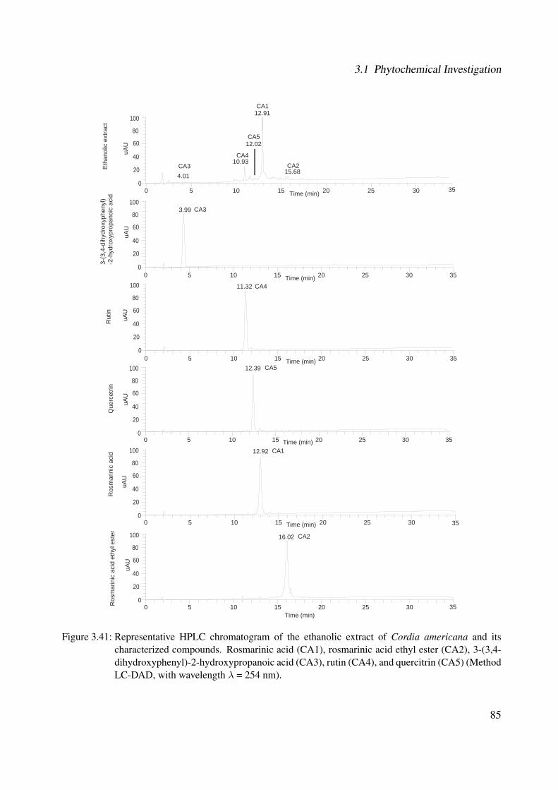
3.1 Phytochemical Investigation
0 5 10 15 20 25 30Time (min)
12.91
12.02
10.9315.68
4.01
0 5 10 15 20 25 30 35Time (min)
0
20
40
60
80
100
uAU
11.32
0 5 10 15 20 25 30 35Time (min)
12.39
0 5 10 15 20 25 30Time (min)
12.92
0 5 10 15 20 25 30Time (min)
16.02
35
35
CA3
CA1
CA2CA4
CA5
CA4
CA5
CA1
CA2
0
20
40
60
80
100uA
U
0
20
40
60
80
100
uAU
0
20
40
60
80
100
uAU
0
20
40
60
80
100
uAU
35
Eth
anol
ic e
xtra
ct
0 5 10 15 20 25 30 35Time (min)
0
20
40
60
80
100
uAU
CA33.99
3-(3
,4-d
ihyd
roxy
phen
yl)
-2-h
ydro
xypr
opan
oic
acid
Rut
inQ
uerc
etrin
Ros
mar
inic
aci
dR
osm
arin
ic a
cid
ethy
l est
er
Figure 3.41: Representative HPLC chromatogram of the ethanolic extract of Cordia americana and itscharacterized compounds. Rosmarinic acid (CA1), rosmarinic acid ethyl ester (CA2), 3-(3,4-dihydroxyphenyl)-2-hydroxypropanoic acid (CA3), rutin (CA4), and quercitrin (CA5) (MethodLC-DAD, with wavelength λ = 254 nm).
85
3 Results and Discussion
Research concerning the identification of rosmarinic acid started around 1950, when it was iso-
lated from Rosmarinus of�cinalis (Lamiaceae) by Scarpati and Oriente, (1958) [269]. Up to now,
CA1 has already been isolated in some species of the genus Cordia, for example from flowers
of Cordia dentata Poir [90] and from the leaves of Cordia verbenacea [310]. Nevertheless, this
was the first report about the presence of CA1 in Cordia americana. CA1 is widely found in
the plant kingdom and presumably accumulated as a defense compound [237]. In order to iden-
tify and quantify CA1, there are several analytical methods described in the literature concerning
Lamiaceae species, including UV-VIS spectrophotometry, HPLC and GC [318, 312].
So far, the compound rosmarinic acid ethyl ester (CA2) has not been isolated for the genus
Cordia. However in the Boraginaceae family, it has been isolated from Lindelo�a sylosa [53].
Additionally, CA2 has previously been identified in Lycopus lucidus (Lamiaceae) [340, 217], in
Prunella vulgaris L. (Labiatae) [327], and in Nepeta prattii (Lamiaceae). However, this compound
might be also an artifact that was originated during the extraction process.
3-(3,4-dihydroxyphenyl)-2-hydroxypropanoic acid (CA3) has been isolated from the water ex-
tract of Chinese herb Salvia miltiorrhiza Bunge [249] from the Lamiaceae family. However, no
reports have been made for the genus Cordia as well as for the Boraginaceae family. Therefore, it
can be assumed that this was the first time to describe the isolation of CA3 in the genus Cordia as
well as in the Boraginaceae family.
Rutin (CA4), a quercetin-3-rutinoside, has been previous identified in the leaves of Cordia myxa
L. [106] and in the flowers of Cordia dentata Poir [90]. Additionally, CA4 has been isolated
from Fagopyrum sculentum (Polygonaceae), Sophora japonica (Fabaceae), and Ruta graveolens
(Rutaceae) [87] and many other plant species. Some methods have been described for the deter-
mination of rutin in different plants extracts, these include HPLC, capillary electrophoresis and
spectrophotometry [3]. CA4 has also been used as a coloring agent, food additive in various food
preparations and drinks, and for various purposes in cosmetics [87].
Concerning the phytochemical studies of quercitrin (CA5), it has been isolated from the leaves
of Cordia dichotoma Forst. [324], from leaves and fruits of Cordia myxa L. [324] and from Cordia
86
3.1 Phytochemical Investigation
globosa [64]. Additionally, the plants Bauhinia microstachya (Leguminosae)[103], Kalanchoe
pinnata (Crassulaceae) and Polygonum hydropiper L. (Polygonaceae) are reported to contain CA5
[219] and in many other plant species. It belongs together with rutin to the ubiquitous flavonol
glycosides.
β-sitosterol (CA6) has been isolated in the genus Cordia from heart wood of Cordia trichotoma
[206] and from the seeds of Cordia obliqua [5].
Campesterol (CA7) is one of the most common plant sterols in nature along β-sitosterol and stig-
masterol [142]. This compound is abundant in seeds, nuts, cereals, beans, legumes and vegetable
oils [240]. CA7 for example, is one of the most common sterols in Chrysanthemum coronarium
(Asteraceae) [51], in Euphorbia pulcherimma (Euphorbiaceae) [285] and in tomato shoots [347].
α-amyrin (CA8) and β-amyrin (CA9) are pentacyclic triterpenes found in various plants. α-
amyrin has been isolated from the seeds of Cordia obliqua [5]. Both of these compounds were
isolated from Brazilian red propolis [317] and from Protium kleinii (Burseraceae), both medicinal
plants used in Brazil [228].
As presented in Section 1.2.2.4, only a few compounds such as two quinones, one phenolic
aldehyde, one coumarin and tannins has been studied for Cordia americana. Thus, all the afore-
mentioned compounds were isolated and identified for the first time in Cordia americana.
87
3 Results and Discussion
3.1.2 Brugmansia suaveolens
3.1.2.1 Bioguided Fractionation based on p38α MAPK Assay
The ethanolic extract from Brugmansia suaveolens was fractionated by means of Sephadex®LH-
20 open column chromatography using methanol as mobile phase (see Section 5.6.2, Experimental
Part). The obtained fraction sets were submitted to a bioguided study in the p38α assay (see
Table 5.13, Experimental Part) and the most active fractions with higher output yields, such as
fractions G, H and I were further investigated. The selected fractions were subfractionated by flash
chromatography, open column chromatography and analytical HPLC using methanol/acetonitrile
and water as mobile phase (further details see Section 5.6.2, Experimental Part). The following
compounds were isolated from the fractions: BS1 was isolated from fraction G; BS2, BS3 and
BS4 were isolated from fraction H; and BS2 was isolated from fraction I.
Brugmansia suaveolens has been studied due to the presence of the alkaloids, as already men-
tioned in Section 1.2.3.4. The qualitative analysis of these alkaloids was performed by TLC (see
Section 5.4.1, Experimental Part) by comparison of the ethanolic extract of Brugmansia suave-
olens (BS) and the fraction sets (A-K) after Sephadex®LH-20 with the standards hyoscyamine and
scopolamine. Figure 5.11 (Experimental Part) shows by means of this qualitative test that it was
not possible to detect alkaloids.
3.1.2.2 Structural Elucidation
The chemical structures of the following compounds in Brugmansia suaveolens were elucidated
by MS and NMR analysis. The absolute configuration of all characterized compounds was not
studied. The known chemical structure was in accordance with the respectively literature.
88
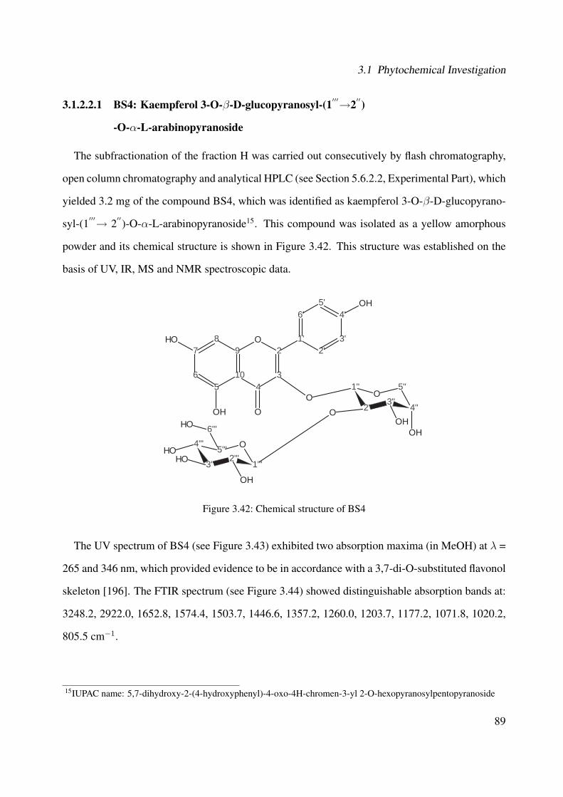
3.1 Phytochemical Investigation
3.1.2.2.1 BS4: Kaempferol 3-O-β-D-glucopyranosyl-(1′′′→2′′)
-O-α-L-arabinopyranoside
The subfractionation of the fraction H was carried out consecutively by flash chromatography,
open column chromatography and analytical HPLC (see Section 5.6.2.2, Experimental Part), which
yielded 3.2 mg of the compound BS4, which was identified as kaempferol 3-O-β-D-glucopyrano-
syl-(1′′′→ 2′′)-O-α-L-arabinopyranoside15. This compound was isolated as a yellow amorphous
powder and its chemical structure is shown in Figure 3.42. This structure was established on the
basis of UV, IR, MS and NMR spectroscopic data.
7
65
10
98
43
2O 1'
6'
2'3'
4'5'
OH
OOH
OH
4'''
3'''2'''
1'''
O5'''
1''
2''3''
4''
5''O
OOH
OH
O
6'''OH
OH
OHOH
Figure 3.42: Chemical structure of BS4
The UV spectrum of BS4 (see Figure 3.43) exhibited two absorption maxima (in MeOH) at λ =
265 and 346 nm, which provided evidence to be in accordance with a 3,7-di-O-substituted flavonol
skeleton [196]. The FTIR spectrum (see Figure 3.44) showed distinguishable absorption bands at:
3248.2, 2922.0, 1652.8, 1574.4, 1503.7, 1446.6, 1357.2, 1260.0, 1203.7, 1177.2, 1071.8, 1020.2,
805.5 cm−1.
15IUPAC name: 5,7-dihydroxy-2-(4-hydroxyphenyl)-4-oxo-4H-chromen-3-yl 2-O-hexopyranosylpentopyranoside
89

3 Results and Discussion
Figure 3.43: UV of the compound BS4
4000,0 3600 3200 2800 2400 2000 1800 1600 1400 1200 1000 800 650,055,8
58
6062
6466
68707274767880828486
8890
9294
9697,6
cm-1
%T
3248,2
2922,0
1652,8
1574,4
1503,71446,6
1357,2
1260,0
1203,7
1177,2
1071,8
1020,2
805,5
Figure 3.44: IR of the compound BS4
90
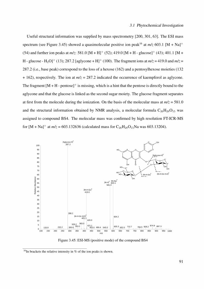
3.1 Phytochemical Investigation
Useful structural information was supplied by mass spectrometry [200, 301, 63]. The ESI mass
spectrum (see Figure 3.45) showed a quasimolecular positive ion peak16 at m/z 603.1 [M + Na]+
(54) and further ion peaks at m/z: 581.0 [M + H]+ (52); 419.0 [M + H - glucose]+ (43); 401.1 [M +
H - glucose - H2O]+ (13); 287.2 [aglycone + H]+ (100). The fragment ions at m/z = 419.0 and m/z =
287.2 (i.e., base peak) correspond to the loss of a hexose (162) and a pentosylhexose moieties (132
+ 162), respectively. The ion at m/z = 287.2 indicated the occurrence of kaempferol as aglycone.
The fragment [M + H - pentose]+ is missing, which is a hint that the pentose is directly bound to the
aglycone and that the glucose is linked as the second sugar moiety. The glucose fragment separates
at first from the molecule during the ionization. On the basis of the molecular mass at m/z = 581.0
and the structural information obtained by NMR analysis, a molecular formula C26H28O15 was
assigned to compound BS4. The molecular mass was confirmed by high resolution FT-ICR-MS
for [M + Na]+ at m/z = 603.132636 (calculated mass for C26H28O15Na was 603.13204).
100 150 200 250 300 350 400 450 500 550 600 650 700 750 800 850 900 950 1000m/z
0
5
10
15
20
25
30
35
40
45
50
55
60
65
70
75
80
85
90
95
100
Rel
ativ
e A
bund
ance
287.2
603.1581.0
419.0
288.2
604.2401.1
420.0
383.0329.2 421.1 874.9353.2 897.3605.3 712.7 828.2784.5232.2 490.4300.0 652.0545.0453.0133.9
[M+H]+ [M+Na]
+
[M+H-Glc]+
[Aglycone+H]+
[M+H-Glc-H2O]+
[Aglycone+H]
[M+H-Glc] [M+H-Glc-H2O]+ +
+7
65
10
98
43
2O 1'
6'
2'3'
4'5'
OH
OOH
OH
4'''
3'''2'''
1'''
O5'''
1''
2''3''
4''
5''O
OOH
OH
O
6'''OH
OH
OHOH
Figure 3.45: ESI-MS (positive mode) of the compound BS4
16In brackets the relative intensity in % of the ion peaks is shown.
91

3 Results and Discussion
The aromatic region of the 1H-NMR (see Figure 3.46 and Table 3.5) showed two doublets signals
at δ = 6.40 ppm and δ = 6.21 ppm and further two doublets at δ = 8.02 ppm and δ = 6.92 ppm (both
d, J = 6 Hz), which indicate compound BS4 as a 5,7-dihydroxyflavonnol with a 1,4-disubtituted.
In the upfield region, there are two anomeric signals due to the sugar units at 5.48 ppm and 4.55
ppm. Further nine protons have been identified in the upfield region due to the sugar units between
δ = 3.2 ppm and δ = 4.4 ppm.
The 13C-NMR spectrum (see Figure 3.47 and Table 3.5) showed 24 signals for 26 carbons.
Between δ = 180 ppm and δ = 106 ppm, there are 13 signals for the 15 carbons of the kaempferol
aglycone. Two of them (i.e., δ = 132.4 ppm and δ = 116.5 ppm) are represented by the same carbon
signal through the symmetrical structure. The two signals at δ = 105.4 ppm and δ = 101.3 ppm
represent the carbons of the sugar residues with the anomeric protons. The nine further signals
are caused by the two sugar moieties between δ = 62.7 ppm and δ = 80.1 ppm. The DEPT-135
experiment (see Figure 3.48) determined two CH2 groups at δ = 63.4 ppm and δ = 62.7 ppm and
further 14 CH groups.
The H-H-COSY (see Figure 3.49 and Table 3.5) showed two meta-coupled doublets at δ = 6.40
ppm and 6.21 ppm. Additionally, an AA'BB' spin system (i.e., C-2' and C-5', C-3' and C-6') was
evident as two doublets at δ = 8.06 ppm and δ = 6.91 ppm. The values of the aromatic signals of
both the 1H- and 13C-NMR spectra suggested the presence of kaempferol as the aglycone with two
sugar units.
The coupling constant of the anomeric proton of the glucose (J = 6 Hz) was in accordance with
a β-glycosidic linkage (i.e., β-D-glucopyranose), while the coupling constant of the anomeric
proton of the pentose (J = 3 Hz) was in accordance with an α-glycosidic linkage (i.e., α-L-
arabinopyranose). The H-H-COSY allowed to show the coupling between each protons of the
sugar moieties as demonstrated in Figure 3.49 and in Table 3.5. The α-L-arabinopyranose exhib-
ited the proton correlations: between H-1” (δ = 5.48 ppm) and H-2” (δ = 4.21 ppm); between
H-2” and H-3” (δ = 3.96 ppm); and between H-4” (δ = 3.86 ppm) and H-5” (δ = 3.69 ppm). The
β-D-glucopyranose presented the proton correlations: between H-1”' (δ = 4.55 ppm) and H-2”' (δ
92
3.1 Phytochemical Investigation
= 3.25 ppm); between H-2”' and H-3”' (δ = 3.38 ppm); between H-4”' (δ = 3.34 ppm) and H-5”'
(δ = 3.23 ppm δ = 3.73 ppm).
The HSQC experiment (see Figure 3.50) showed that the two signals at δ = 99.9 ppm and δ =
94.7 ppm were correlated to the proton signals at δ = 6.40 ppm (H-8) and δ = 6.21 ppm (H-6). The
two carbon signals at δ = 132.4 ppm and δ = 116.5 ppm were also coupled with the two doublets
of the 1,4-disubstituted aromatic moiety (B ring) at δ = 8.02 ppm and δ = 6.92 ppm, respectively.
In the HMBC experiment (see Figure 3.51 and Table 3.5), C-7 appeared at δ = 166.0 ppm
and is coupled with H-8 (δ = 6.40 ppm). The signals C-5, C-9 and C-10 are also identified by
HMBC long-range coupling. C-9 and C-10 (δC9 = 159.0 ppm and δC10 = 105.8 ppm) are coupled
with H-8. C-10 and C-5 (δC10 = 105.8 ppm and δC5 = 161.6 ppm) show a correlation with H-6
(δ = 6.21 ppm). The carbons in the C-4 and C-2 position provide signals at δC4 = 179.7 ppm
and δC2 = 158.5 ppm. C-2 is correlated with δ = 8.02 ppm (H-2', H-6') and δ = 6.92 ppm (H-
3', H-5'). Furthermore, the anomeric proton H-1” (δ = 5.48 ppm) is correlated with C-3 (δC3 =
135.7 ppm) showing the coupling between the α-L-arabinopyranose and the aglycone kaempferol.
Additionally, the coupling between the sugar units is shown by the correlation of the H-1”'(δ =
4.55 ppm) of the β-D-glucopyranose with C-2”(δC2′′ = 80.1 ppm) of the α-L-arabinopyranose.
The HMBC experiment represented the following long-range correlations between 1H and 13C in
α-L-arabinopyranose: between H-2” (δ = 4.21 ppm) and C-1”' (δC1′′′ = 105.4 ppm); between H-
4” (δ = 3.86 ppm) and C-5” (δC5′′ = 63.4 ppm); between H-5” (δ = 3.69 ppm) and C-1” (δC1′′ =
101.3 ppm) and C-3” (δC3′′ = 71.3 ppm). In β-D-glucopyranose, the HMBC experiment provided
following long-range correlations between 1H and 13C: between H-2”' (δ = 3.25 ppm) and C-1”'
(δC1′′′ = 105.4 ppm) and C-3”' (δC3′′′ = 78.1 ppm) ; between both H-3”' (δ = 3.38 ppm) and H-4”'
(δ = 3.34 ppm) with C-2”' (δC2′′′ = 75.2 ppm); between H-5”' (δ = 3.36 ppm) and C-4”'(δC4′′′ =
71.4 ppm).
The chemical-shift values of the carbons of the sugar units were in agreement with those of a
glucopyranose (i.e., β-D-glucopyranose) and an arabinopyranose (i.e., α-L-arabinopyranose) from
the literature [169] (see Table 3.5). The assignment of the sugars to D- or L-series are based on the
93

3 Results and Discussion
literature [27]. Based on the MS, 1D- and 2D-NMR analysis, a kaempferol 3-O-β-D-glucopyra-
nosyl-(1′′′→ 2′′)-O-α-L-arabinopyranoside was identified as the compound BS4.
Table 3.5: Chemical shifts of BS4 and literatureAtom 13C* 13C** 1H* 1H - 1H 1H - 13C
numbers δ/ppm δ/ppm δ/ppm (Mult., J(Hz), H) COSY* HMBC*
2 158.5 - - - -3 135.7 - - - -4 179.7 - - - -5 161.6 - - - -6 99.9 - 6.21 (brs; 1H) 8 5, 8, 107 166.0 - - - -8 94.7 - 6.40 (brs; 1H) 6 6, 7, 9, 109 159.0 - - - -10 105.8 - - - -
1′ 122.6 - - - -
2′ 132.4 - 8.02 (d; 6 Hz; 1H) 3
′, 5
′2, 4
′, 6
′
3′ 116.5 - 6.92 (d; 6 Hz; 1H) 2
′, 6
′2, 1
′, 5
′
4′ 163.1 - - - -
5′ 116.5 - 6.92 (d; 6 Hz; 1H) 2
′, 6
′2, 1
′, 3
′
6′ 132.4 - 8.02 (d; 6 Hz; 1H) 3
′, 5
′2, 2
′, 4
′
1′′ 101.3 100.9 5.48 (d; 3 Hz; 1H) 2
′′ 32
′′ 80.1 78.3 4.21 (dd; 6 Hz; 4 Hz; 1H) 1′′, 3
′′1
′′
3′′ 71.3 71.4 3.96 (dd; 6 Hz; 4 Hz; 1H) 2
′′ -4
′′ 66.6 66.3 3.86 (m; 1H) 5′′
5′′
5′′ 63.4 62.1 3.23 (m; 1H); 3.73 (m; 1H) 4
′′1
′′, 3
′′
1′′′ 105.4 103.1 4.55 (d; 6 Hz; 1H) 2
′′′2
′′
2′′′ 75.2 77.6 3.25 (d; 6 Hz; 1H) 1
′′′, 3
′′′1
′′′, 3
′′′
3′′′ 78.1 79.4 3.38 (brs; 1H) 2
′′′2
′′′
4′′′ 71.4 72.7 3.34 (brs; 1H) 5
′′′2
′′′
5′′′ 78.0 78.2 3.36 (brs; 1H) 4
′′′4
′′′
6′′′ 62.7 62.1 3.78-3.82 (m; 2H) - -
* In MeOH-d4. ** Data from literature [169] in Pyridine-d5.
94
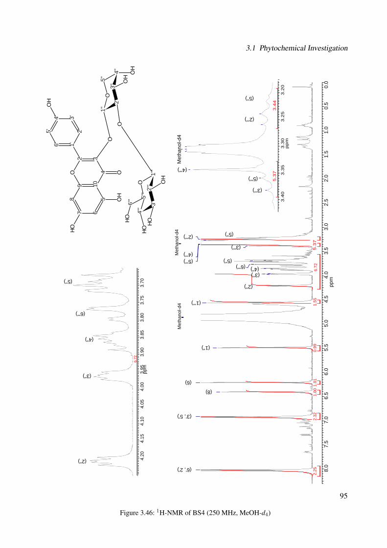
3.1 Phytochemical Investigation
8.0
7.5
7.0
6.5
6.0
5.5
5.0
4.5
4.0
3.5
3.0
2.5
2.0
1.5
1.0
0.5
0.0
ppm
2.32
2.25
1.55
1.01
1.00
0.99
Met
hano
l-d4
Met
hano
l-d4
(2''')
(4''') (5''')
(3''')
(5'') (6''') (4'')
(3'')
(2'')
(1''')
(1'')
(6)
(8)
(3', 5')
(6', 2')
4.20
4.15
4.10
4.05
4.00
3.95
3.90
3.85
3.80
3.75
3.70
ppm
6.72
3.4
03
.35
3.3
03
.25
3.2
0p
pm
5.3
73
.44
(2''')
(4''')
(5''')
(3''')
(5'')
(6''')
(4'')
(3'')
(2'')
Met
hano
l-d4
5.3
76.
72
7 65
1098
432
O1'6'
2'3'4'
5'
OH
OO
H
OH
4''' 3'
''2'
''1'
''
O5'
''
1'' 2'
'3'
'4'
'
5''
O
OO
HO
H
O
6'''
OH
OH
OHOH (5'')
(5'')
Figure 3.46: 1H-NMR of BS4 (250 MHz, MeOH-d4)
95

3 Results and Discussion
180
170
160
150
140
130
120
110
100
9080
7060
5040
3020
100
ppm
Met
hano
l-d4
(6''') (5'')
(4'')
(3'') (4''') (2''')
(5''') (3''') (2'')
(8)
(6) (1'')
(1''') (10)
(3', 5')
(1')
(6', 2') (3)
(4') (9) (5)
(2)
(7)
(4)
7 65
1098
432
O1'6'
2'3'4'
5'
OH
OO
H
OH
4''' 3'
''2'
''1'
''
O5'
''
1'' 2'
'3'
'4'
'
5''
O
OO
HO
H
O
6'''
OH
OH
OHOH
Figure 3.47: 13C-NMR of BS4 (100 MHz, MeOH-d4)
96
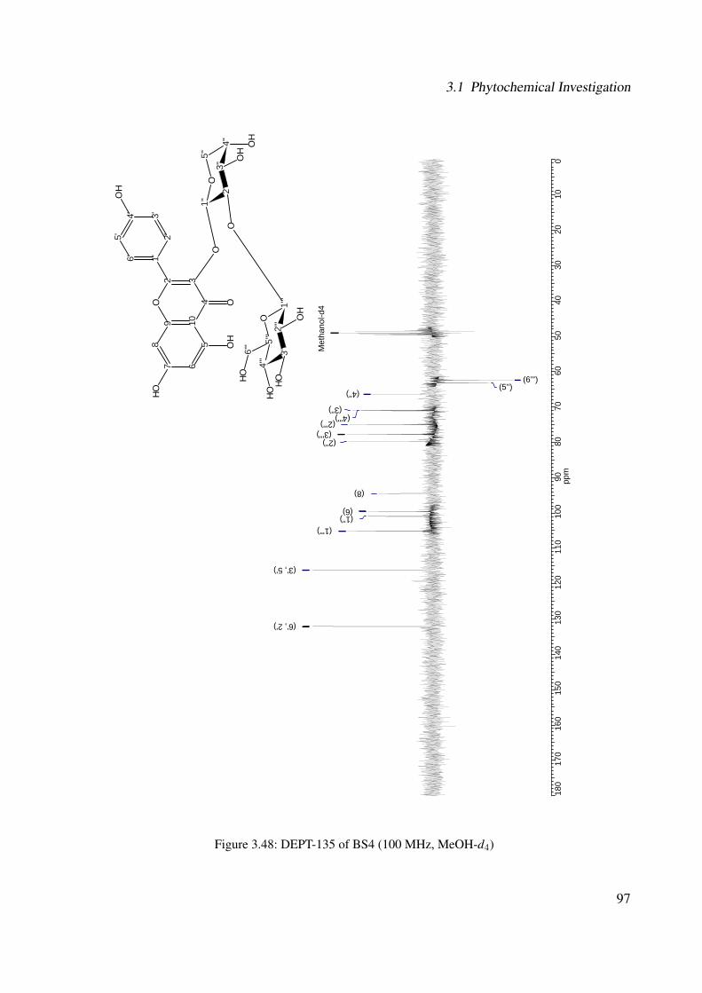
3.1 Phytochemical Investigation
180
170
160
150
140
130
120
110
100
9080
7060
5040
3020
100
ppm
Met
hano
l-d4
(6''') (5'') (4'')
(3'') (4''')
(2''') (3''') (2'')
(8)
(6) (1'')
(1''')
(3', 5')
(6', 2')
7 65
1098
432
O1'6'
2'3'4'
5'
OH
OO
H
OH
4''' 3'
''2'
''1'
''
O5'
''
1'' 2'
'3'
'4'
'
5''
O
OO
HO
H
O
6'''
OH
OH
OHOH
Figure 3.48: DEPT-135 of BS4 (100 MHz, MeOH-d4)
97
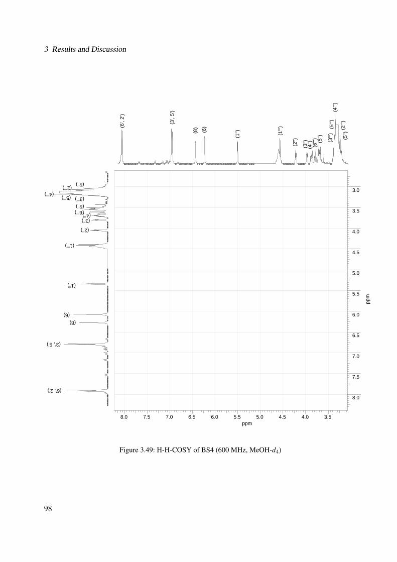
3 Results and Discussion
8.0 7.5 7.0 6.5 6.0 5.5 5.0 4.5 4.0 3.5ppm
3.0
3.5
4.0
4.5
5.0
5.5
6.0
6.5
7.0
7.5
8.0
ppm
(2'
'') (
4''')
(5'
'') (
3''')
(5'
') (6
''')
(4'')
(3
'')
(2'
')
(1''')
(1'
')
(6
)
(8
) (3
', 5'
)
(6
', 2'
)
(2''') (4''')
(5''') (3''') (5'') (6''') (4'')
(3'')
(2'')
(1''')
(1'')
(6) (8)
(3', 5')
(6', 2')
(5'
')
(5'')
Figure 3.49: H-H-COSY of BS4 (600 MHz, MeOH-d4)
98
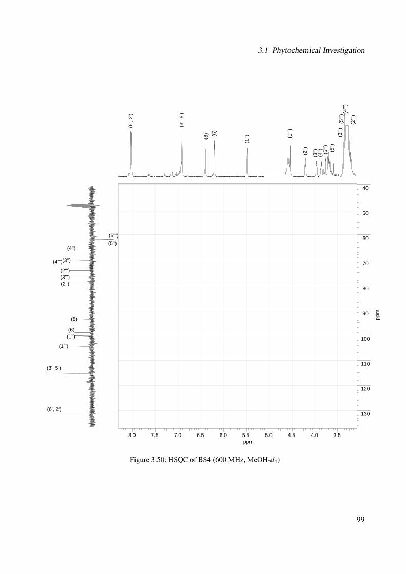
3.1 Phytochemical Investigation
8.0 7.5 7.0 6.5 6.0 5.5 5.0 4.5 4.0 3.5ppm
40
50
60
70
80
90
100
110
120
130
ppm
(2'
'') (4'
'') (
5''')
(3'
'') (
5'')
(6''')
(4
'')
(3'')
(2'
')
(1''')
(1'
')
(6
)
(8)
(3',
5')
(6
', 2'
)
(6''') (5'')
(4'')
(3'') (4''')
(2''') (3''') (2'')
(8)
(6) (1'')
(1''')
(3', 5')
(6', 2')
Figure 3.50: HSQC of BS4 (600 MHz, MeOH-d4)
99

3 Results and Discussion
8.0 7.5 7.0 6.5 6.0 5.5 5.0 4.5 4.0 3.5 3.0ppm
40
50
60
70
80
90
100
110
120
130
140
150
160
170
180
ppm
(2'
'') (
4''')
(5'
'') (
3''')
(5'
') (6
''')
(4'')
(3
'') (
2'')
(1''')
(1'
')
(6
)
(8)
(3',
5')
(6
', 2'
)
(6''') (5'')
(4'') (3'') (4''') (2''')
(5''') (3''') (2'')
(8) (6)
(1'') (1''') (10)
(3', 5') (1')
(6', 2') (3)
(4') (9)
(5) (2)
(7)
(4)
(5'
')
Figure 3.51: HMBC of BS4 (600 MHz, MeOH-d4)
100
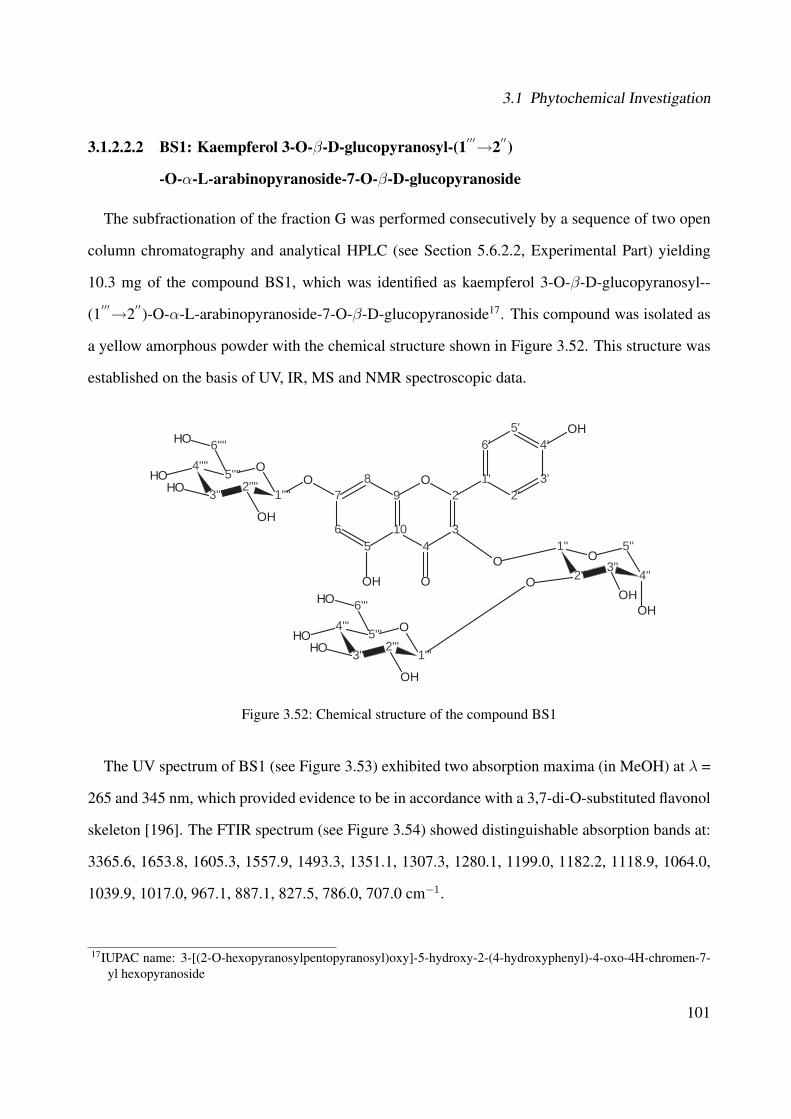
3.1 Phytochemical Investigation
3.1.2.2.2 BS1: Kaempferol 3-O-β-D-glucopyranosyl-(1′′′→2′′)
-O-α-L-arabinopyranoside-7-O-β-D-glucopyranoside
The subfractionation of the fraction G was performed consecutively by a sequence of two open
column chromatography and analytical HPLC (see Section 5.6.2.2, Experimental Part) yielding
10.3 mg of the compound BS1, which was identified as kaempferol 3-O-β-D-glucopyranosyl--
(1′′′→2′′)-O-α-L-arabinopyranoside-7-O-β-D-glucopyranoside17. This compound was isolated as
a yellow amorphous powder with the chemical structure shown in Figure 3.52. This structure was
established on the basis of UV, IR, MS and NMR spectroscopic data.
7
65
10
98
43
2O 1'
6'
2'3'
4'5'
O
OOH
OH
4'''
3'''2'''
1'''
O5'''
1''
2''3''
4''
5''O
OOH
OH
O
6'''OH
OH
OHOH
4''''
3'''' 2''''1''''
O5''''
6''''OH
OH
OHOH
Figure 3.52: Chemical structure of the compound BS1
The UV spectrum of BS1 (see Figure 3.53) exhibited two absorption maxima (in MeOH) at λ =
265 and 345 nm, which provided evidence to be in accordance with a 3,7-di-O-substituted flavonol
skeleton [196]. The FTIR spectrum (see Figure 3.54) showed distinguishable absorption bands at:
3365.6, 1653.8, 1605.3, 1557.9, 1493.3, 1351.1, 1307.3, 1280.1, 1199.0, 1182.2, 1118.9, 1064.0,
1039.9, 1017.0, 967.1, 887.1, 827.5, 786.0, 707.0 cm−1.
17IUPAC name: 3-[(2-O-hexopyranosylpentopyranosyl)oxy]-5-hydroxy-2-(4-hydroxyphenyl)-4-oxo-4H-chromen-7-yl hexopyranoside
101
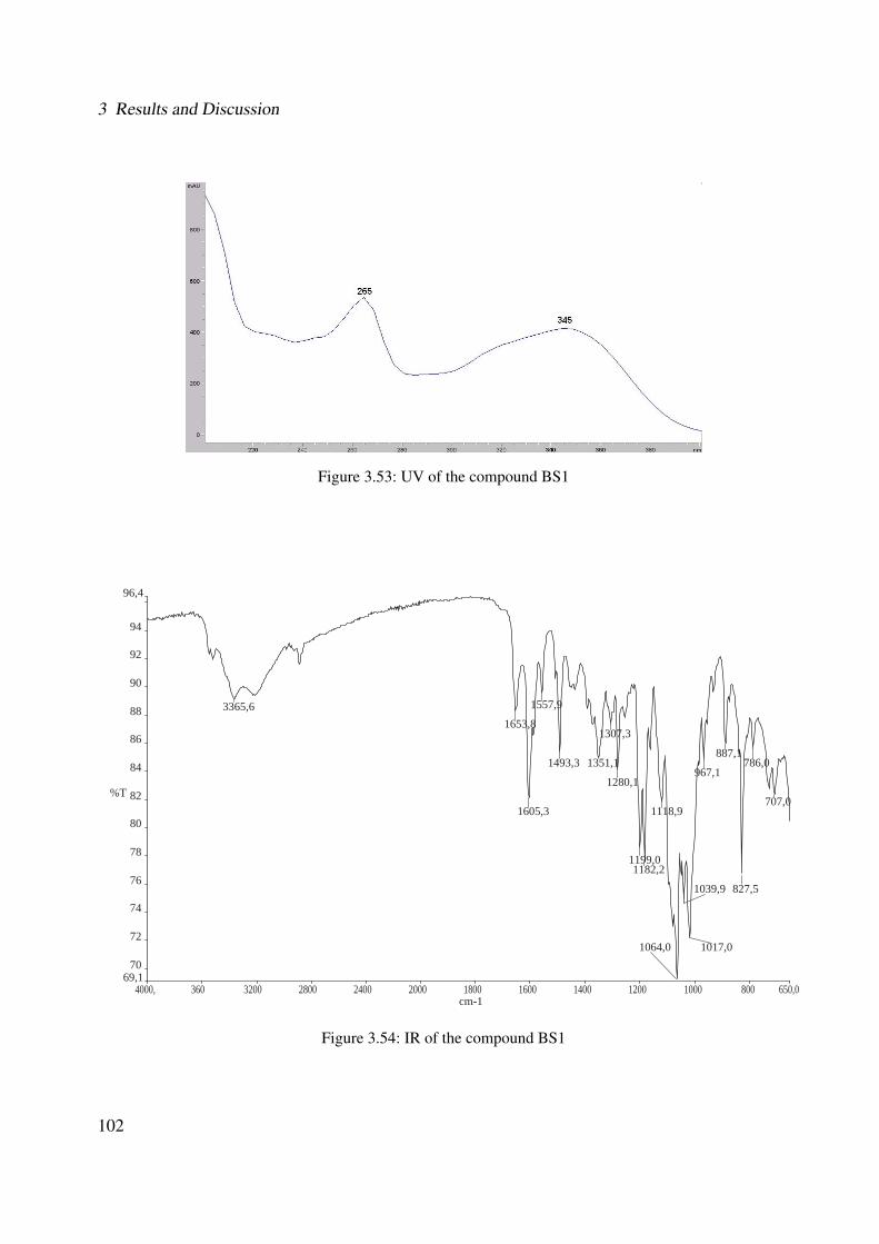
3 Results and Discussion
Figure 3.53: UV of the compound BS1
4000, 360 3200 2800 2400 2000 1800 1600 1400 1200 1000 800 650,069,1
70
72
74
76
78
80
82
84
86
88
90
92
94
96,4
cm-1
%T
3365,61653,8
1605,3
1557,9
1493,3 1351,1
1307,3
1280,1
1199,01182,2
1118,9
1064,0
1039,9
1017,0
967,1
887,1
827,5
786,0
707,0
Figure 3.54: IR of the compound BS1
102

3.1 Phytochemical Investigation
The ESI mass spectrum (see Figure 3.55) showed a quasimolecular positive ion peak18 at m/z =
765.0 [M + Na]+ (10), and further peaks at m/z: 742.8 [M + H]+ (22); 580.9 [M + H - glucose]+
(29); 448.8 [M + H - arabinose - glucose]+ (100); 418.9 [M + H - glucose - glucose]+ (15); 400.9
[M + H - glucose - H2O]+ (7); 287.1 [aglycone + H]+ (82). The fragment ions at m/z = 580.9, 448.8
(i.e., base peak) and 418.9 correspond to the loss of a hexose (162), a pentosylhexose residue (132
+ 162), and of two hexoses (162 + 162), respectively. On the basis of the molecular mass at m/z =
742.8 and the structural information obtained by NMR analysis, a molecular formula C32H38O20
was assigned to compound BS1. The molecular mass was confirmed by high resolution FT-ICR
mass spectrometry for [M + Na]+ at m/z = 765.184176 (calculated mass for C32H38O20Na was
765.18486).
100 150 200 250 300 350 400 450 500 550 600 650 700 750 800 850 900 950 1000m/z
0
5
10
15
20
25
30
35
40
45
50
55
60
65
70
75
80
85
90
95
100
Rel
ativ
e A
bund
ance
448.8
287.1
580.9
742.8
418.9 581.9288.2580.3 765.0
400.9382.9 582.9451.0 766.1610.6329.2 796.9490.8 562.7261.1 874.8712.6192.9 931.1134.9
[Aglycone+H]
[M+H-Glc]
[M+H-Glc]
[M+H-Glc]
[M+H]
[M+Na]
+
+
+
+
+
+
[M+H-Glc-Ara]+
[M+H-Glc-Glc]+
[Aglycone+H]+
7
65
10
98
43
2O 1'
6'
2'3'
4'5'
O
OOH
OH
4'''
3'''2'''
1'''
O5'''
1''
2''3''
4''
5''O
OOH
OH
O
6'''OH
OH
OHOH
4''''
3'''' 2''''1''''
O5''''
6''''OH
OH
OHOH
Figure 3.55: ESI-MS (positive mode) of the compound BS1
18In brackets, the relative intensity in % of the ion peaks is shown.
103

3 Results and Discussion
The NMR-spectra of BS1 are quite similar to those of BS4 (see Section 3.1.2.2.1). Additionally,
in the middle region of the 1H-NMR spectrum, one further anomeric signal due to sugar unit at δ
= 5.07 ppm was identified.
The 13C-NMR spectrum (see Figure 3.57 and Table 3.6) showed 30 signals for 32 carbons. The
three signals at δ = 98.9 ppm, δ = 99.8 ppm and δ = 103.8 ppm can be assigned to anomeric carbons
of the sugar units. The DEPT-135 experiment (see Figure 3.58) demonstrated three CH2 groups at
δ = 61.2 ppm, δ = 60.9 ppm and δ = 60.6 ppm and further 23 CH groups were identified.
The HSQC (see Figure 3.60) and the H-H-COSY experiments (see Figure 3.59 and Table 3.6)
showed a similar spectra as that from BS4. The main difference is that BS1 has a possible third
sugar unit. The values of the aromatic signals of both 1H- and 13C-NMR spectra suggested again
the presence of a kaempferol as aglycone, however, with three sugar units (see Figure 3.61).
The coupling constant of the anomeric protons H-1”' and H-1”” (both J = 6 Hz) of the glu-
coses were in accordance with a β-glycosidic linkage (i.e., β-D-glucopyranose), whereas the
anomeric proton of the pentose (i.e., brs) was in accordance with an α-glycosidic linkage (i.e., α-L-
arabinopyranose). The H-H-COSY showed the 1H correlations for the second β-D-glucopyranose:
between H-1”” (δ = 5.07 ppm) and H-2”” (δ = 3.25 ppm); between H-2”” and H-3”” (δ = 3.30
ppm); between H-4”” (δ = 3.17 ppm) and H-5”” (δ = 3.12 ppm); between H-5”” and H-6”” (δ =
3.70 ppm and δ = 3.43 ppm).
In the HMBC spectrum (see Figure 3.62 and Table 3.6), the anomeric proton H-1”” (δ = 5.07
ppm) is correlated with C-7 (δC7 = 162.9 ppm) showing the coupling between the β-D-glucopyranose
with the aglycone kaempferol at C-7. Additionally, in this second β-D-glucopyranose, the follow-
ing long-range correlations between 1H and 13C were observed in the HMBC experiment: between
H-2”” (δ = 3.25 ppm) and C-1”” (δC1′′′′ = 99.8 ppm); between H-3”” (δ = 3.30 ppm) and C-2””
(δC2′′′ = 73.6 ppm) and C-5”” (δC5′′′ = 76.8 ppm).
The chemical-shift values of the carbons of the sugar units were in agreement with those of a
glucopyranose (i.e., β-D-glucopyranose) and an arabinopyranose (i.e., α-L-arabinopyranose) from
the literature [169] (see Table 3.6). The assignment of the sugars to D- or L-series is based on
104
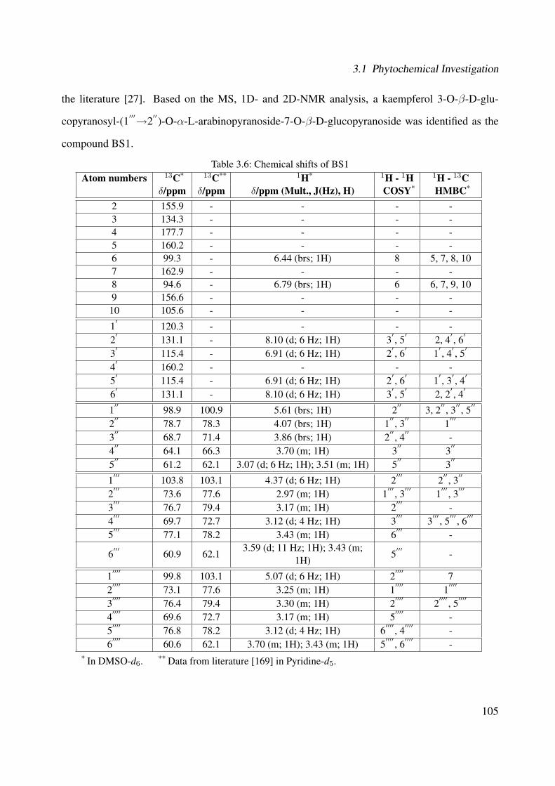
3.1 Phytochemical Investigation
the literature [27]. Based on the MS, 1D- and 2D-NMR analysis, a kaempferol 3-O-β-D-glu-
copyranosyl-(1′′′→2′′)-O-α-L-arabinopyranoside-7-O-β-D-glucopyranoside was identified as the
compound BS1.
Table 3.6: Chemical shifts of BS1Atom numbers 13C* 13C** 1H* 1H - 1H 1H - 13C
δ/ppm δ/ppm δ/ppm (Mult., J(Hz), H) COSY* HMBC*
2 155.9 - - - -3 134.3 - - - -4 177.7 - - - -5 160.2 - - - -6 99.3 - 6.44 (brs; 1H) 8 5, 7, 8, 107 162.9 - - - -8 94.6 - 6.79 (brs; 1H) 6 6, 7, 9, 109 156.6 - - - -10 105.6 - - - -
1′
120.3 - - - -2
′131.1 - 8.10 (d; 6 Hz; 1H) 3
′, 5
′2, 4
′, 6
′
3′
115.4 - 6.91 (d; 6 Hz; 1H) 2′, 6
′1
′, 4
′, 5
′
4′
160.2 - - - -5
′115.4 - 6.91 (d; 6 Hz; 1H) 2
′, 6
′1
′, 3
′, 4
′
6′
131.1 - 8.10 (d; 6 Hz; 1H) 3′, 5
′2, 2
′, 4
′
1′′
98.9 100.9 5.61 (brs; 1H) 2′′
3, 2′′, 3
′′, 5
′′
2′′
78.7 78.3 4.07 (brs; 1H) 1′′, 3
′′1
′′′
3′′
68.7 71.4 3.86 (brs; 1H) 2′′, 4
′′-
4′′
64.1 66.3 3.70 (m; 1H) 3′′
3′′
5′′
61.2 62.1 3.07 (d; 6 Hz; 1H); 3.51 (m; 1H) 5′′
3′′
1′′′
103.8 103.1 4.37 (d; 6 Hz; 1H) 2′′′
2′′, 3
′′
2′′′
73.6 77.6 2.97 (m; 1H) 1′′′
, 3′′′
1′′′
, 3′′′
3′′′
76.7 79.4 3.17 (m; 1H) 2′′′
-4
′′′69.7 72.7 3.12 (d; 4 Hz; 1H) 3
′′′3
′′′, 5
′′′, 6
′′′
5′′′
77.1 78.2 3.43 (m; 1H) 6′′′
-
6′′′
60.9 62.13.59 (d; 11 Hz; 1H); 3.43 (m;
1H)5
′′′-
1′′′′
99.8 103.1 5.07 (d; 6 Hz; 1H) 2′′′′
72
′′′′73.1 77.6 3.25 (m; 1H) 1
′′′′1
′′′′
3′′′′
76.4 79.4 3.30 (m; 1H) 2′′′′
2′′′′
, 5′′′′
4′′′′
69.6 72.7 3.17 (m; 1H) 5′′′′
-5
′′′′76.8 78.2 3.12 (d; 4 Hz; 1H) 6
′′′′, 4
′′′′-
6′′′′
60.6 62.1 3.70 (m; 1H); 3.43 (m; 1H) 5′′′′
, 6′′′′
-* In DMSO-d6. ** Data from literature [169] in Pyridine-d5.
105

3 Results and Discussion
8.0
7.5
7.0
6.5
6.0
5.5
5.0
4.5
4.0
3.5
3.0
2.5
2.0
1.5
1.0
0.5
0.0
ppm
19.9
46.
1705.2
66.242.2
62.21.
2600.1
50.180.1
DM
SO
-d6
(2''') (5'') (5'''', 4''')
(4'''', 3''') (2'''') (5''', 6'''', 6''')
(5'') (4'', 6'''')
(3'')
(2'')
(1''')
(1'''')
(1'')
(6)
(8) (3', 5')
(6', 2')
3.95
3.90
3.85
3.80
3.75
3.70
3.65
3.60
3.55
3.50
3.45
3.40
3.35
3.30
3.25
3.20
3.15
3.10
3.05
3.00
2.95
2.90
2.85
2.80
ppm
19.9
46.
174.
142.
501.
711.
171.
02
(2''')
(5'')
(5'''', 4''')
(4'''', 3''')
(2'''')
(3'''')
(5''', 6'''', 6''')
(5'')
(6''')
(4'', 6'''')
(3'')
7 65
1098
432
O1'6'
2'3'4'
5'
O
OO
H
OH
4''' 3'
''2'
''1'
''
O5'
''
1'' 2'
'3'
'4'
'
5''
O
OO
H OH
O
6'''
OH
OH
OHOH
4'''' 3'
'''2'
'''1'
'''
O5'
'''
6''''
OH
OH
OHH
Figure 3.56: 1H-NMR of BS1 (600 MHz, DMSO-d6)
106

3.1 Phytochemical Investigation
180
170
160
150
140
130
120
110
100
9080
7060
5040
3020
100
ppm
DM
SO
-d6
(6'''') (6''') (5'')
(3'') (4'''', 4''') (2'''') (2''')
(3''') (5'''')
(2'')
(8) (1'') (1'''')
(1''') (10)
(3', 5')
(1')
(6', 2') (3)
(2) (9)
(5, 4') (7)
(4)
79
78
77
76
75
74
73
72
71
70
69
68
67
66
65
64
63
62
61
60
pp
m
(6'''') (6''') (5'')
(4'')
(3'')
( 4''')
(2'''') (2''')
(3'''') (3''') (5'''')
(5''')
(2'')
(4'''')
7 65
1098
432
O1'6'
2'3'4'
5'
O
OO
H
OH
4''' 3'
''2'
''1'
''
O5'
''
1'' 2'
'3'
'4'
'
5''
O
OO
H OH
O
6'''
OH
OH
OHOH
4'''' 3'
'''2'
'''1'
'''
O5'
'''
6''''
OH
OH
OHOH
Figure 3.57: 13C-NMR of BS1 (100 MHz, DMSO-d6)
107

3 Results and Discussion
140
135
130
125
120
115
110
105
100
9590
8580
7570
6560
5550
4540
3530
2520
1510
50
ppm
DM
SO
-d6
(6'''') (6''') (5'')
(4'') (3'')
(4'''') (4''')
(2'''') (2''')
(3''') (5'''')
(5''') (2'')
(8)
(1'') (1'''')
(1''')
(3', 5')
(6', 2')
10
09
59
08
58
07
57
06
56
0p
pm
(6'''') (6''') (5'')
(4'')
(3'') (4'''') (4''')
(2'''') (2''')
(3'''') (3''') (5'''') (5''')
(2'')
(8)
(1'') (6) (1'''')
7 65
1098
432
O1'6'
2'3'4'
5'
O
OO
H
OH
4''' 3'
''2'
''1'
''
O5'
''
1'' 2'
'3'
'4'
'
5''
O
OO
H OH
O
6'''
OH
OH
OHOH
4'''' 3'
'''2'
'''1'
'''
O5'
'''
6''''
OH
OH
OHH
Figure 3.58: DEPT-135 of BS1 (100 MHz, DMSO-d6)
108
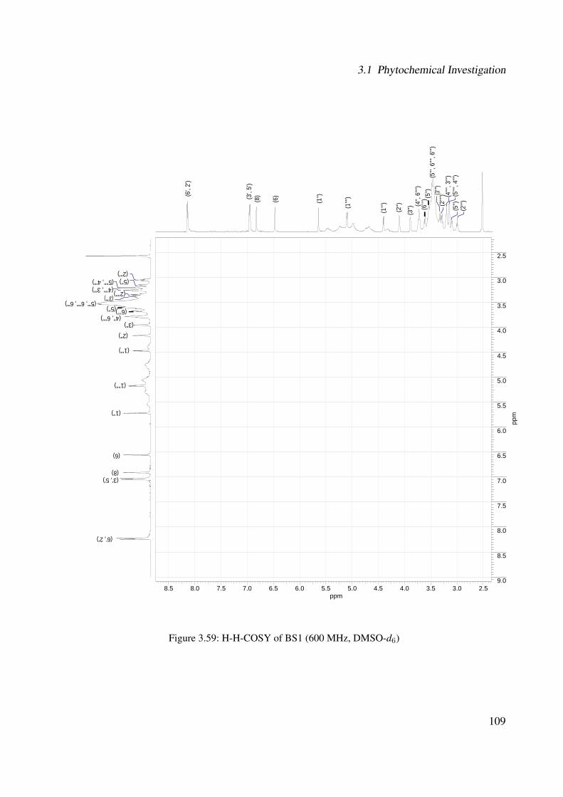
3.1 Phytochemical Investigation
8.5 8.0 7.5 7.0 6.5 6.0 5.5 5.0 4.5 4.0 3.5 3.0 2.5ppm
2.5
3.0
3.5
4.0
4.5
5.0
5.5
6.0
6.5
7.0
7.5
8.0
8.5
9.0
ppm
(2''')
(5''''
, 4''')
(4''''
, 3''')
(2''''
) (5
''', 6
'''', 6
''') (5
'') (4
'', 6'
''') (3
'') (2'')
(1''') (1
'''')
(1'')
(6)
(8) (3',
5')
(6',
2')
(3'''')
(2''') (5'''', 4''') (4'''', 3''') (2'''')
(5''', 6'''', 6''') (5'')
(4'', 6'''') (3'')
(2'')
(1''')
(1'''')
(1'')
(6)
(8) (3', 5')
(6', 2')
(3'''')
(5'')
(6’’’)
(6’’’)
(5'')
Figure 3.59: H-H-COSY of BS1 (600 MHz, DMSO-d6)
109

3 Results and Discussion
8.0 7.5 7.0 6.5 6.0 5.5 5.0 4.5 4.0 3.5 3.0 2.5 2.0ppm
35
40
45
50
55
60
65
70
75
80
85
90
95
100
105
110
115
120
125
130
135
ppm
(2''')
(5''''
, 4''')
(4''''
, 3''')
(2''''
)
(5''',
6''''
, 6''')
(5'')
(4'',
6'''')
(3'') (2
'')
(1''') (1
'''')
(1'')
(6)
(8) (3
', 5')
(6', 2
')
(3'''')
(6', 2')
(3', 5')
(1'') (1'''')
(1''')
(8)
(6)
(2’’) (5’’’)
(5’’’’) (3’’’) (2’’’)
(2’’’’) (4’’’) (4’’’’)
(4’’)
(3’’)
(6’’’’)(5’’)
(6’’’)
(5'')
(6’’’)
Figure 3.60: HSQC of BS1 (600 MHz, DMSO-d6)
110

3.1 Phytochemical Investigation
4.0 3.5 3.0 2.5ppm
55
60
65
70
75
80
85
ppm
(6'''') (6''')
(5'')
(4'')
(3'') (4'''')
(4''')
(2'''') (2''')
(3'''') (3''') (5'''')
(5''')
(2'')
(2'') (3'') (5'')
(5'')
(6''')
(6''')
(6'''')
(6'''') (4'''') (4''')
(2'''') (2''')
(5''')
(3'''')
(3''') (5'''')
Figure 3.61: HSQC of BS1 sugar region (600 MHz, DMSO-d6)
111
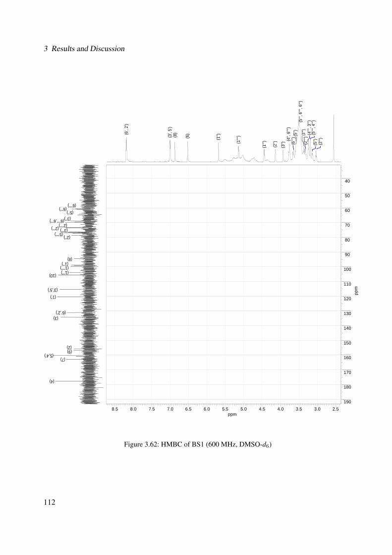
3 Results and Discussion
8.5 8.0 7.5 7.0 6.5 6.0 5.5 5.0 4.5 4.0 3.5 3.0 2.5ppm
40
50
60
70
80
90
100
110
120
130
140
150
160
170
180
190
ppm
(2''')
(5''''
, 4''')
(4''''
, 3''')
(2''''
) (5
''', 6
'''', 6
''') (5
'') (4
'', 6'
''') (3
'')
(2'')
(1''') (1
'''')
(1'')
(6)
(8)
(3', 5
')
(6', 2
')
(3'''')
(6’’’’) (6’’’)
(5’’) (3’’) (6’’’’,6’’’)
(2’’’) (2’’’’) (3’’’)
(5’’’’) (2’’)
(8) (1’’) (1’’’’) (1’’’) (10)
(3’,5’) (1’)
(6’,2’) (3)
(2) (9)
(5,4’) (7)
(4)
(5'')
(6’’’)
Figure 3.62: HMBC of BS1 (600 MHz, DMSO-d6)
112
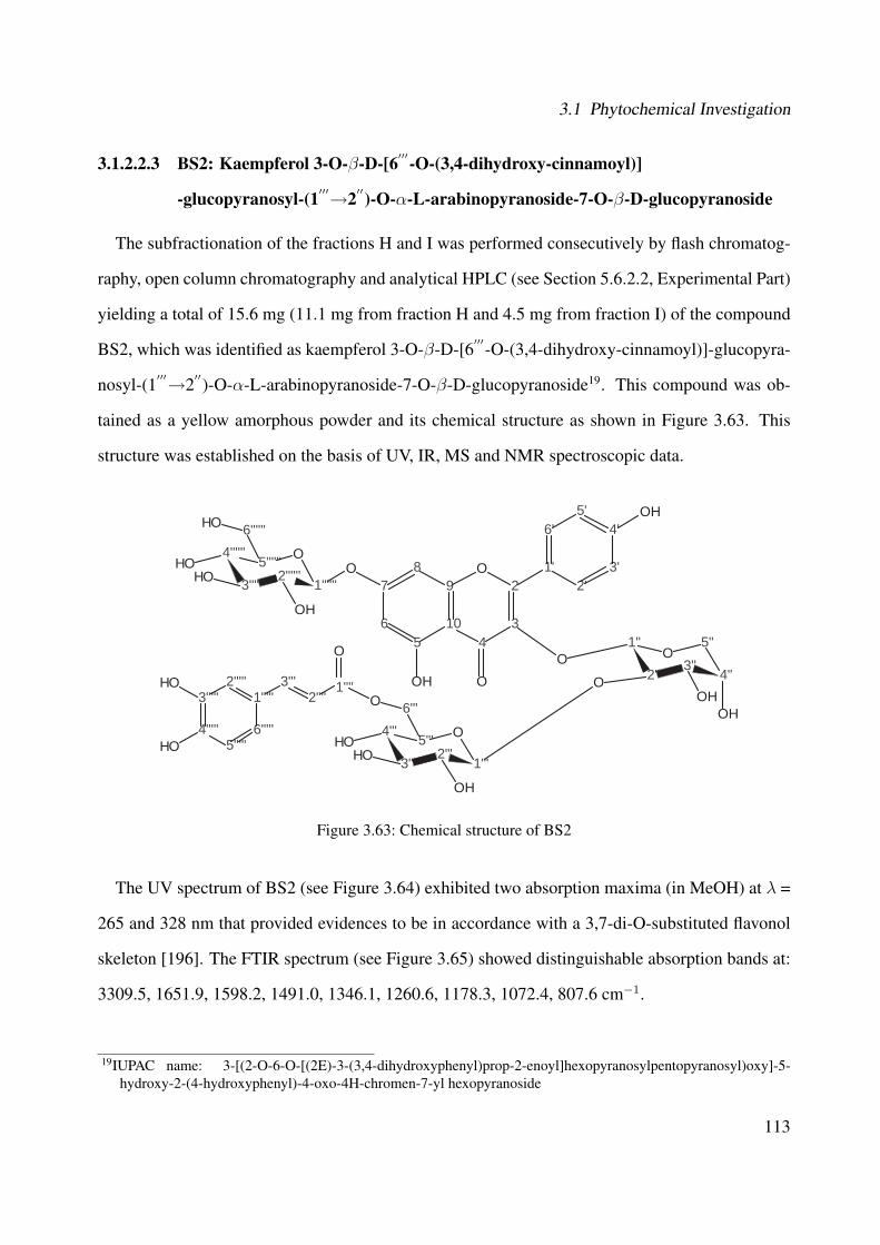
3.1 Phytochemical Investigation
3.1.2.2.3 BS2: Kaempferol 3-O-β-D-[6′′′-O-(3,4-dihydroxy-cinnamoyl)]
-glucopyranosyl-(1′′′→2′′)-O-α-L-arabinopyranoside-7-O-β-D-glucopyranoside
The subfractionation of the fractions H and I was performed consecutively by flash chromatog-
raphy, open column chromatography and analytical HPLC (see Section 5.6.2.2, Experimental Part)
yielding a total of 15.6 mg (11.1 mg from fraction H and 4.5 mg from fraction I) of the compound
BS2, which was identified as kaempferol 3-O-β-D-[6′′′-O-(3,4-dihydroxy-cinnamoyl)]-glucopyra-
nosyl-(1′′′→2′′)-O-α-L-arabinopyranoside-7-O-β-D-glucopyranoside19. This compound was ob-
tained as a yellow amorphous powder and its chemical structure as shown in Figure 3.63. This
structure was established on the basis of UV, IR, MS and NMR spectroscopic data.
7
65
10
98
43
2O 1'
6'
2'3'
4'5'
O
OOH
OH
4'''
3'''2'''
1'''
O5'''
1''
2''3''
4''
5''O
OOH
OH
O
6'''O
OH
OHOH
4''''''
3''''''2''''''
1''''''
O5''''''
6''''''OH
OH
OHOH
3'''''
4'''''5'''''
6'''''
1'''''2'''''
2''''3'''
O
OH
OH 1''''
Figure 3.63: Chemical structure of BS2
The UV spectrum of BS2 (see Figure 3.64) exhibited two absorption maxima (in MeOH) at λ =
265 and 328 nm that provided evidences to be in accordance with a 3,7-di-O-substituted flavonol
skeleton [196]. The FTIR spectrum (see Figure 3.65) showed distinguishable absorption bands at:
3309.5, 1651.9, 1598.2, 1491.0, 1346.1, 1260.6, 1178.3, 1072.4, 807.6 cm−1.
19IUPAC name: 3-[(2-O-6-O-[(2E)-3-(3,4-dihydroxyphenyl)prop-2-enoyl]hexopyranosylpentopyranosyl)oxy]-5-hydroxy-2-(4-hydroxyphenyl)-4-oxo-4H-chromen-7-yl hexopyranoside
113
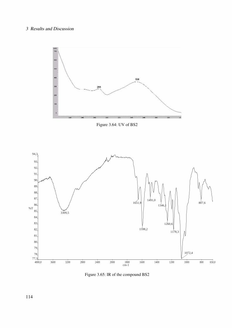
3 Results and Discussion
Figure 3.64: UV of BS2
4000,0 3600 3200 2800 2400 2000 1800 1600 1400 1200 1000 800 650,077,3
78
79
80
81
82
83
84
85
86
87
88
89
90
91
92
93
94,3
cm-1
%T 3309,5
1651,9
1598,2
1491,0
1346,1
1260,6
1178,3
1072,4
807,6
Figure 3.65: IR of the compound BS2
114
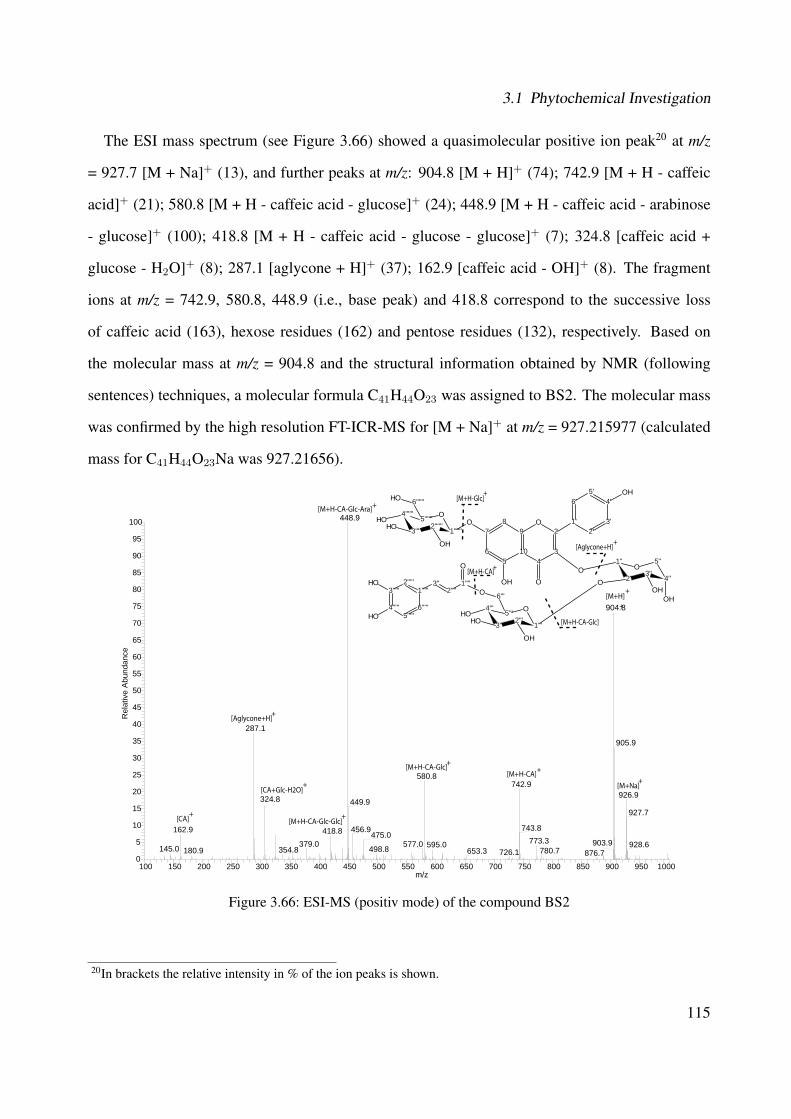
3.1 Phytochemical Investigation
The ESI mass spectrum (see Figure 3.66) showed a quasimolecular positive ion peak20 at m/z
= 927.7 [M + Na]+ (13), and further peaks at m/z: 904.8 [M + H]+ (74); 742.9 [M + H - caffeic
acid]+ (21); 580.8 [M + H - caffeic acid - glucose]+ (24); 448.9 [M + H - caffeic acid - arabinose
- glucose]+ (100); 418.8 [M + H - caffeic acid - glucose - glucose]+ (7); 324.8 [caffeic acid +
glucose - H2O]+ (8); 287.1 [aglycone + H]+ (37); 162.9 [caffeic acid - OH]+ (8). The fragment
ions at m/z = 742.9, 580.8, 448.9 (i.e., base peak) and 418.8 correspond to the successive loss
of caffeic acid (163), hexose residues (162) and pentose residues (132), respectively. Based on
the molecular mass at m/z = 904.8 and the structural information obtained by NMR (following
sentences) techniques, a molecular formula C41H44O23 was assigned to BS2. The molecular mass
was confirmed by the high resolution FT-ICR-MS for [M + Na]+ at m/z = 927.215977 (calculated
mass for C41H44O23Na was 927.21656).
100 150 200 250 300 350 400 450 500 550 600 650 700 750 800 850 900 950 1000m/z
0
5
10
15
20
25
30
35
40
45
50
55
60
65
70
75
80
85
90
95
100
Rel
ativ
e A
bund
ance
448.9
904.8
287.1
905.9
580.8742.9
926.9449.9
927.7
743.8456.9162.9
324.8
418.8 475.0773.3379.0 577.0 928.6595.0 903.9
145.0 498.8354.8180.9 780.7653.3 726.1 876.7
[M+H-Glc]+
[Aglycone+H]+
[M+H-CA-Glc]
+
[M+H-CA]+
[M+H-CA]+
[M+H]
[M+Na]+
+
[M+H-CA-Glc]+
[M+H-CA-Glc-Ara]+
[M+H-CA-Glc-Glc]+
[Aglycone+H]+
[CA]+
[CA+Glc-H2O]+
7
65
10
98
43
2O 1'
6'
2'3'
4'5'
O
OOH
OH
4'''
3'''2'''
1'''
O5'''
1''
2''3''
4''
5''O
OOH
OH
O
6'''O
OH
OHOH
4''''''
3''''''2''''''
1''''''
O5''''''
6''''''OH
OH
OHOH
3'''''
4'''''5'''''
6'''''
1'''''2'''''
2''''3'' 1''''
O
OH
OH
Figure 3.66: ESI-MS (positiv mode) of the compound BS2
20In brackets the relative intensity in % of the ion peaks is shown.
115
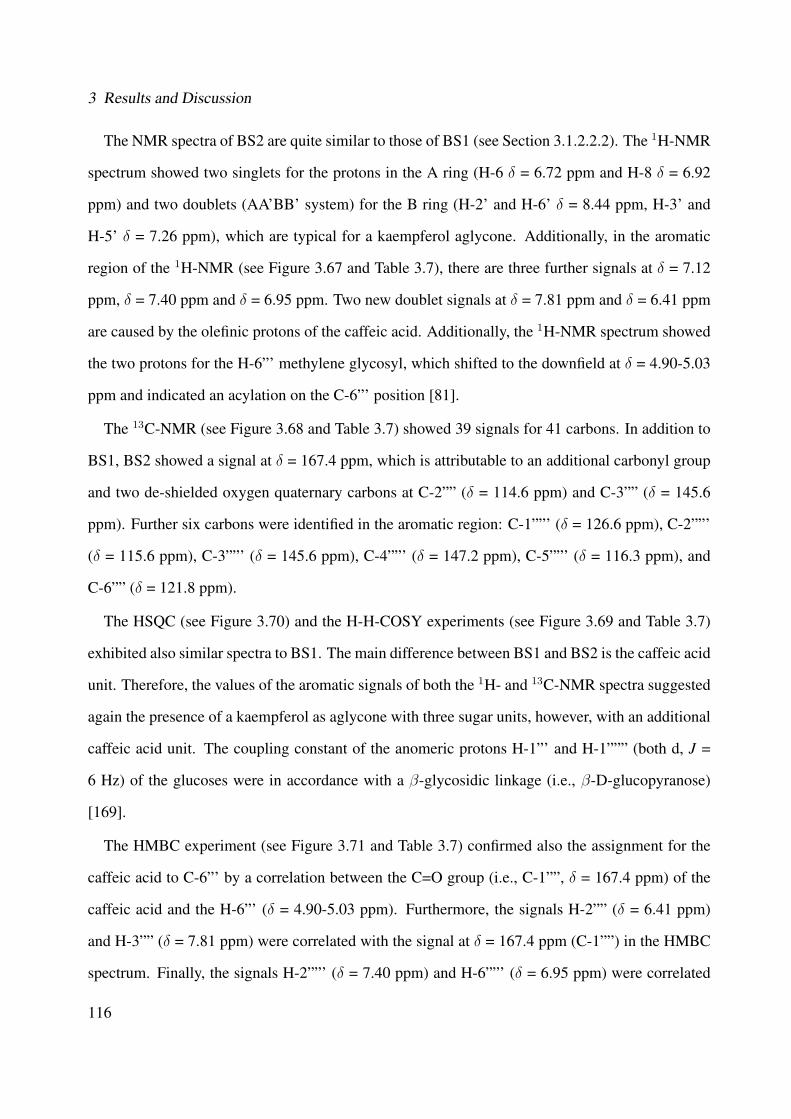
3 Results and Discussion
The NMR spectra of BS2 are quite similar to those of BS1 (see Section 3.1.2.2.2). The 1H-NMR
spectrum showed two singlets for the protons in the A ring (H-6 δ = 6.72 ppm and H-8 δ = 6.92
ppm) and two doublets (AA'BB' system) for the B ring (H-2' and H-6' δ = 8.44 ppm, H-3' and
H-5' δ = 7.26 ppm), which are typical for a kaempferol aglycone. Additionally, in the aromatic
region of the 1H-NMR (see Figure 3.67 and Table 3.7), there are three further signals at δ = 7.12
ppm, δ = 7.40 ppm and δ = 6.95 ppm. Two new doublet signals at δ = 7.81 ppm and δ = 6.41 ppm
are caused by the olefinic protons of the caffeic acid. Additionally, the 1H-NMR spectrum showed
the two protons for the H-6”' methylene glycosyl, which shifted to the downfield at δ = 4.90-5.03
ppm and indicated an acylation on the C-6”' position [81].
The 13C-NMR (see Figure 3.68 and Table 3.7) showed 39 signals for 41 carbons. In addition to
BS1, BS2 showed a signal at δ = 167.4 ppm, which is attributable to an additional carbonyl group
and two de-shielded oxygen quaternary carbons at C-2”” (δ = 114.6 ppm) and C-3”” (δ = 145.6
ppm). Further six carbons were identified in the aromatic region: C-1””' (δ = 126.6 ppm), C-2””'
(δ = 115.6 ppm), C-3””' (δ = 145.6 ppm), C-4””' (δ = 147.2 ppm), C-5””' (δ = 116.3 ppm), and
C-6”” (δ = 121.8 ppm).
The HSQC (see Figure 3.70) and the H-H-COSY experiments (see Figure 3.69 and Table 3.7)
exhibited also similar spectra to BS1. The main difference between BS1 and BS2 is the caffeic acid
unit. Therefore, the values of the aromatic signals of both the 1H- and 13C-NMR spectra suggested
again the presence of a kaempferol as aglycone with three sugar units, however, with an additional
caffeic acid unit. The coupling constant of the anomeric protons H-1”' and H-1””” (both d, J =
6 Hz) of the glucoses were in accordance with a β-glycosidic linkage (i.e., β-D-glucopyranose)
[169].
The HMBC experiment (see Figure 3.71 and Table 3.7) confirmed also the assignment for the
caffeic acid to C-6”' by a correlation between the C=O group (i.e., C-1””, δ = 167.4 ppm) of the
caffeic acid and the H-6”' (δ = 4.90-5.03 ppm). Furthermore, the signals H-2”” (δ = 6.41 ppm)
and H-3”” (δ = 7.81 ppm) were correlated with the signal at δ = 167.4 ppm (C-1””) in the HMBC
spectrum. Finally, the signals H-2””' (δ = 7.40 ppm) and H-6””' (δ = 6.95 ppm) were correlated
116
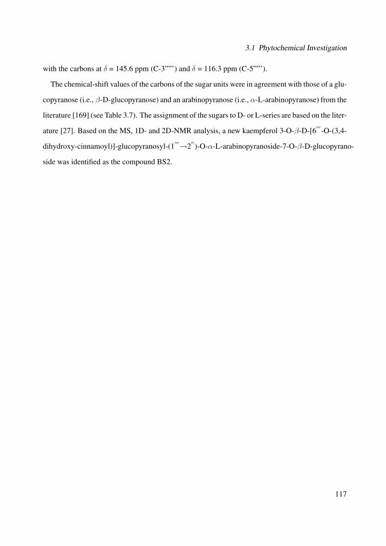
3.1 Phytochemical Investigation
with the carbons at δ = 145.6 ppm (C-3””') and δ = 116.3 ppm (C-5””').
The chemical-shift values of the carbons of the sugar units were in agreement with those of a glu-
copyranose (i.e., β-D-glucopyranose) and an arabinopyranose (i.e., α-L-arabinopyranose) from the
literature [169] (see Table 3.7). The assignment of the sugars to D- or L-series are based on the liter-
ature [27]. Based on the MS, 1D- and 2D-NMR analysis, a new kaempferol 3-O-β-D-[6′′′-O-(3,4-
dihydroxy-cinnamoyl)]-glucopyranosyl-(1′′′→2′′)-O-α-L-arabinopyranoside-7-O-β-D-glucopyrano-
side was identified as the compound BS2.
117

3 Results and Discussion
Table 3.7: Chemical shifts of BS2Atom numbers 13C* 13C** 1H* 1H - 1H 1H - 13C
δ/ppm δ/ppm δ/ppm (Mult., J(Hz), H) COSY* HMBC*
2 157.2 - - - -3 137.0 - - - -4 178.8 - - - -5 161.7 - - - -6 100.0 - 6.72 (brs; 1H) 8 7, 8, 9, 107 163.6 - - - -8 94.6 - 6.92 (brs; 1H) 6 4, 6, 9, 109 156.6 - - - -
10 106.7 - - - -
1′
121.8 - - - -2
′131.8 - 8.44 (d; 6 Hz; 1H) 3
′, 5
′2, 7, 3
′, 5
′, 6
′
3′
116.2 - 7.26 (d; 6 Hz; 1H) 2′, 6
′2, 7, 2
′, 5
′, 6
′
4′
162.0 - - - -5
′116.2 - 7.26 (d; 6 Hz; 1H) 2
′, 6
′2, 7, 2
′, 3
′, 6
′
6′
131.8 - 8.44 (d; 6 Hz; 1H) 3′, 5
′2, 7, 2
′, 3
′, 5
′
1′′
100.4 100.9 6.41 (brs; 1H) 2′′
2′′, 3, 3
′′, 5
′′
2′′
80.7 78.3 5.07 (brs; 1H) 1′′, 3
′′1
′′, 4
′′, 1
′′′
3′′
70.9 71.4 4.67 (brs; 1H) 2′′, 4
′′-
4′′
66.0 66.3 4.47 (m; 1H) 5′′, 3
′′-
5′′
62.1 62.1 4.38 (m; 1H); 4.56 (m; 1H) 4′′, 5
′′1
′′
1′′′
106.7 103.1 5.28 (d; 6 Hz; 1H) 2′′′
-2
′′′75.1 77.6 4.12 (m; 1H) 1
′′′,3
′′′1
′′′, 3
′′′
3′′′
78.1 79.4 4.05 (m; 1H) 4′′′
2′′′
, 4′′′
, 5′′′
4′′′
70.9 72.7 4.15 (m; 1H) 3′′′
, 5′′′
-5
′′′75.4 78.2 4.05 (m; 1H) 4
′′′2
′′′, 3
′′′, 4
′′′
6′′′
63.9 62.1 4.90-5.03 (m; 2H) 6′′′
1′′′′
1′′′′
167.4 - - - -2
′′′′114.6 - 6.41 (d; 12 Hz; 1H) 3
′′′′1
′′′′, 1
′′′′′
3′′′′
145.6 - 7.81 (d; 12 Hz; 1H) 2′′′′
1′′′′
, 2′′′′
, 6′′′′′
1′′′′′
126.6 - - - 72
′′′′′115.6 - 7.40 (brs; 1H) - 1
′′′′′, 3
′′′′′, 5
′′′′′
3′′′′′
145.6 - - - 5′′′′′
4′′′′′
147.2 - - - -5
′′′′′116.3 - 7.12 (d; 6 Hz; 1H) 6
′′′′′-
6′′′′′
121.8 - 6.95 (brd; 6 Hz; 1H) 5′′′′′
3′′′′′
, 5′′′′′
1′′′′′′
101.3 103.1 5.78 (d; 6 Hz; 1H) 2′′′′′′
-2
′′′′′′74.6 77.6 4.30 (m; 1H) 1
′′′′′′1
′′′′′′, 3
′′′′′′
3′′′′′′
78.9 79.4 4.40 (m; 1H) 3′′′′′′
2′′′′′′
, 4′′′′′′
4′′′′′′
71.0 72.7 4.30 (m; 1H) 2′′′′′′
-5
′′′′′′78.1 78.2 4.30 (m; 1H) - -
6′′′′′′
63.4 62.1 3.71 (m; 1H); 4.50 (m; 1H) 2′′′′′′
, 3′′′′′′
-* In Pyridine-d5. ** Data from literature [169] in Pyridine-d5.118

3.1 Phytochemical Investigation
8.5
8.0
7.5
7.0
6.5
6.0
5.5
5.0
4.5
4.0
3.5
3.0
ppm
3.05
2.59
2.31
2.09
1.71
1.44
1.38
1.28
1.17
1.16
1.00
0.90
0.78
0.71
0.62
Pyridine-d5
(5"',3"')
(2""",5""",4""") ( 6""",3""") (5")
(4")
(6""")
(3")
(6"')
(2")
(1"')
(1""")
(1") (2"")
(6)
(8) (6""')
(2""')
(3', 5')
(5""')
(3"")
(6', 2')
Pyridine-d5
Pyridine-d5
5.4
5.3
5.2
5.1
5.0
4.9
4.8
4.7
4.6
4.5
4.4
4.3
4.2
4.1
4.0
3.9
3.8
3.7
3.6
ppm
95.250.3
2.31
2.10
1.28
0.98
0.93
0.90
0.90
0.73
0.65
0.62
(5")
(5’", 3"')
(2"', 4"')
(3""")
(3""", 6""") (5") (4")
(6""")
(3")
(6"')
(2")
(1"')
(5")
(2""",5""",4""")
(2"', 4"')
7 65
1098
432
O1'6'
2'3'4'
5'
O
OO
H
OH
4''' 3'
''2'
''1'
''
O5'
''
1'' 2'
'3'
'4'
'
5''
O
OO
H OH
O
6'''
O
OH
OHOH
4'''''
' 3'''''
'2'
'''''
1'''''
'
O5'
'''''
6'''''
'OH
OH
OHOH
3'''''
4'''''
5'''''
6'''''
1'''''
2'''''
2''''
3'''
O
OHOH1'
'''
Figure 3.67: 1H-NMR of BS2 (600 MHz, Pyridine-d5)
119

3 Results and Discussion
180
175
170
165
160
155
150
145
140
135
130
125
120
115
110
105
100
9590
8580
7570
6560
ppm
(6""") (5")
(6"') (4")
(3")
(2""") (2"')
(3"') (3""")
(5""") (2")
(8)
(6)(1")
(1""")
(1"') (10)
(2"")
(5""') (3', 5') (2""')
(1') (6""')
(1""')
(6', 2')
(3)
(3"") (4""') (3""')
(9) (2)
(4') (5)
(7)
(1"")
(4)
Pyridine-d5
Pyridine-d5
Pyridine-d5
(5"')
(4""") (4’")
7 65
1098
432
O1'6'
2'3'4'
5'
O
OO
H
OH
4''' 3'
''2'
''1'
''
O5'
''
1'' 2'
'3'
'4'
'
5''
O
OO
H OH
O
6'''
O
OH
OHOH
4'''''
' 3'''''
'2'
'''''
1'''''
'
O5'
'''''
6'''''
'OH
OH
OHOH
3'''''
4'''''
5'''''
6'''''
1'''''
2'''''
2''''
3'''
O
OHOH1'
'''
Figure 3.68: 13C-NMR of BS2 (100 MHz, Pyridine-d5)
120
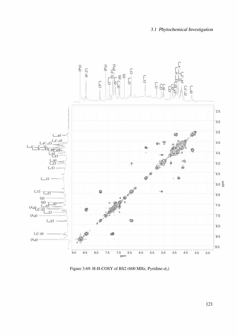
3.1 Phytochemical Investigation
3.59.0 8.5 8.0 7.5 7.0 6.5 6.0 5.5 5.0 4.5 4.0ppm
2.5
3.0
3.5
4.0
4.5
5.0
5.5
6.0
6.5
7.0
7.5
8.0
8.5
9.0
ppm
(Py)
(Py)
(Py)
(6"')
(1")
(2’')(1"')
(1””
”)(6’,2
’)
(5’,3
’)
(3””
)
(5”’
”)
(2”’
”)
(6”’
”)
(6)
(8)
(2””
)
(3’')
(6””
”)
(4’') (
5’')
(5’')
(3""
",6
""")
( 5""
",2""
", 4"
"")
(4"',
2"')
(5"',
3"')
(Py)
(Py)
(Py)
(6"')
(1")
(2’')(1"')
(1”””)
(6’,2’)
(5’,3’)
(3””)
(5”’”)
(2”’”)
(6”’”)
(6)(8)
(2””)
(3’')
(6”””)
(4’')(5’')(5’')
(3""",6""")(5""",2""", 4""")
(2"',4"')(5"',3"')
3.0
Figure 3.69: H-H-COSY of BS2 (600 MHz, Pyridine-d5)
121
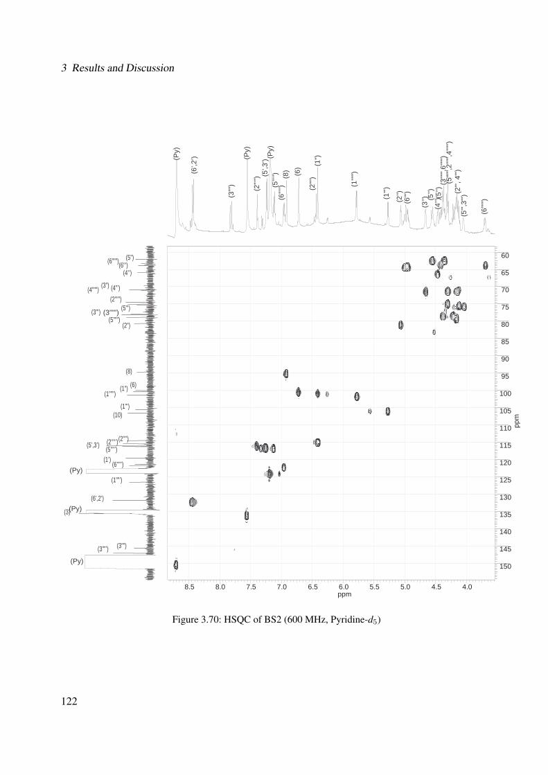
3 Results and Discussion
8.5 8.0 7.5 7.0 6.5 6.0 5.5 5.0 4.5 4.0ppm
60
65
70
75
80
85
90
95
100
105
110
115
120
125
130
135
140
145
150
ppm
(Py)
(Py)
(Py)
(Py)
(Py)
(Py)
(6"')
(1")
(2’')(1"')
(1”””
)(6’,2
’)
(5’,3
’)
(3””
) (5”’”
)
(2”’”
)
(6”’”
)
(6)
(8)
(2””
)
(3’')
(6”””
)
(4’') (
5’')
(3""
",6""
")(5
""",2
""",4
""")
(2"',
4"')
(5"',
3"')
(3)
(1”’)
(3””’) (3””)
(6’,2’)
(1””’)
(6’”’’)(1’)
(2’”’’)(5’”’’)(5’,3’)
(2’”’)
(10)
(1”””)(1”) (6)
(8)
(2”)(5”””)
(3”’)
(4”””)(2”””)(4”’)(3”)
(6”””)(4’’)
(6”’)(5”)
(3”””) (5”’)
(5’')
Figure 3.70: HSQC of BS2 (600 MHz, Pyridine-d5)
122
3.1 Phytochemical Investigation
9.0 8.5 8.0 7.5 7.0 6.5 6.0 5.5 5.0 4.5 4.0ppm
60
70
80
90
100
110
120
130
140
150
160
170
180
ppm
(6"')
(1"")
(3)
(1")
(7)
(2’')(1
"')(1”””
)
(1”’)
(Py)
(Py)
(Py)
(Py)
(Py)
(Py)
(4)
(5)(4’)(2)(9)
(3””’)(3””)
(6’,2’)
(1””’)(6’”’’)(1’)
(2’”’’)(5’”’’)(5’,3’)
(2’”’)
(10)
(1”””)(1”)(6)(8)
(2”)(5”””)(3”””)(3”’)
(4”””)(2”””)
(2”’)
(4”’)(3”)
(6”””)(4’’)
(6”’)(5”)
(6’,2
’)
(5’,3
’)
(3””
) (5”’”
)
(2”’”
)
(6”’”
)
(6)
(8)
(2””
)
(3’')
(6”””
)
(4’') (
5’')
(3""
",6""
")(5
""",2
""",
4"""
)(2
"',4"
')(5
"',3"
')
(5”’)
(4””’)
(5’')
Figure 3.71: HMBC of BS2 (600 MHz, Pyridine-d5)
123
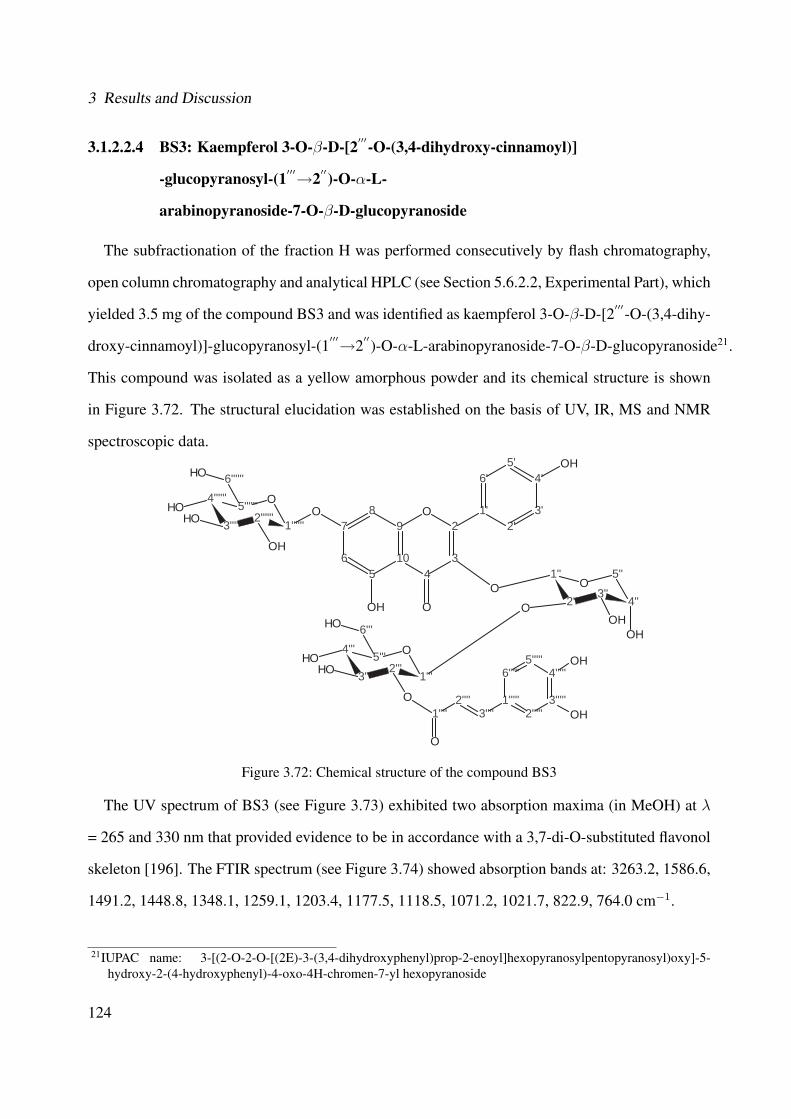
3 Results and Discussion
3.1.2.2.4 BS3: Kaempferol 3-O-β-D-[2′′′-O-(3,4-dihydroxy-cinnamoyl)]
-glucopyranosyl-(1′′′→2′′)-O-α-L-
arabinopyranoside-7-O-β-D-glucopyranoside
The subfractionation of the fraction H was performed consecutively by flash chromatography,
open column chromatography and analytical HPLC (see Section 5.6.2.2, Experimental Part), which
yielded 3.5 mg of the compound BS3 and was identified as kaempferol 3-O-β-D-[2′′′-O-(3,4-dihy-
droxy-cinnamoyl)]-glucopyranosyl-(1′′′→2′′)-O-α-L-arabinopyranoside-7-O-β-D-glucopyranoside21.
This compound was isolated as a yellow amorphous powder and its chemical structure is shown
in Figure 3.72. The structural elucidation was established on the basis of UV, IR, MS and NMR
spectroscopic data.
7
65
10
98
43
2O 1'
6'
2'3'
4'5'
O
OOH
OH
4'''
3'''2'''
1'''
O5'''
1''
2''3''
4''
5''O
OOH
OH
O
6'''OH
O
OHOH
4''''''
3''''''2''''''
1''''''
O5''''''
6''''''OH
OH
OHOH
3'''''
4'''''5'''''
6'''''
1'''''2'''''
2''''3''''1''''
O
OH
OH
Figure 3.72: Chemical structure of the compound BS3
The UV spectrum of BS3 (see Figure 3.73) exhibited two absorption maxima (in MeOH) at λ
= 265 and 330 nm that provided evidence to be in accordance with a 3,7-di-O-substituted flavonol
skeleton [196]. The FTIR spectrum (see Figure 3.74) showed absorption bands at: 3263.2, 1586.6,
1491.2, 1448.8, 1348.1, 1259.1, 1203.4, 1177.5, 1118.5, 1071.2, 1021.7, 822.9, 764.0 cm−1.
21IUPAC name: 3-[(2-O-2-O-[(2E)-3-(3,4-dihydroxyphenyl)prop-2-enoyl]hexopyranosylpentopyranosyl)oxy]-5-hydroxy-2-(4-hydroxyphenyl)-4-oxo-4H-chromen-7-yl hexopyranoside
124
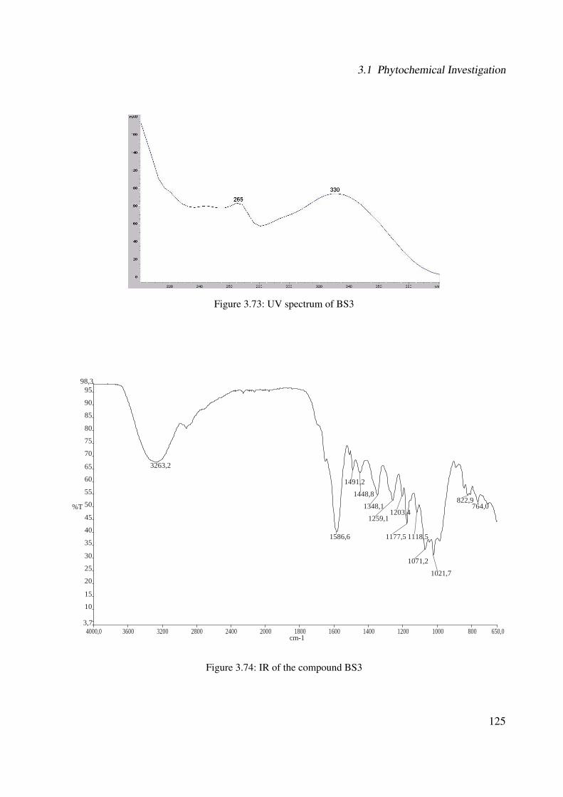
3.1 Phytochemical Investigation
Figure 3.73: UV spectrum of BS3
4000,0 3600 3200 2800 2400 2000 1800 1600 1400 1200 1000 800 650,03,7
10
15
20
25
30
35
40
45
50
55
60
65
70
75
80
85
90
9598,3
cm-1
%T
3263,2
1586,6
1491,2
1448,8
1348,1
1259,11203,4
1177,5 1118,5
1071,2
1021,7
822,9764,0
Figure 3.74: IR of the compound BS3
125
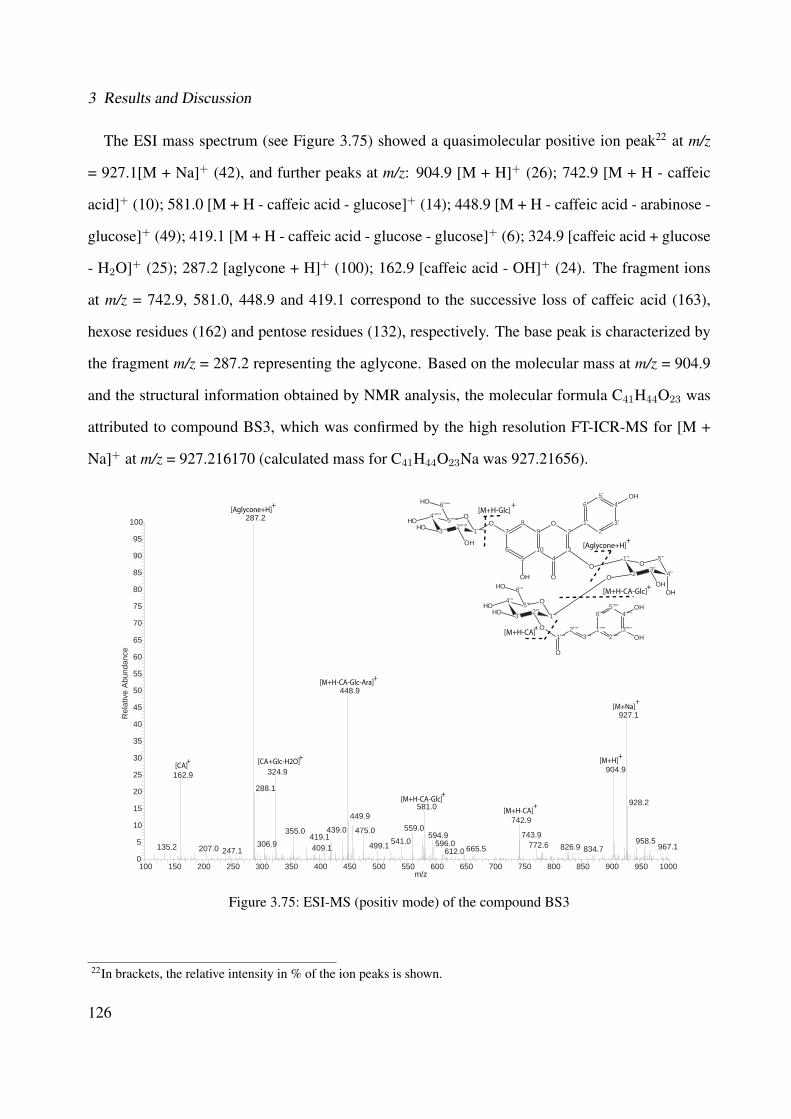
3 Results and Discussion
The ESI mass spectrum (see Figure 3.75) showed a quasimolecular positive ion peak22 at m/z
= 927.1[M + Na]+ (42), and further peaks at m/z: 904.9 [M + H]+ (26); 742.9 [M + H - caffeic
acid]+ (10); 581.0 [M + H - caffeic acid - glucose]+ (14); 448.9 [M + H - caffeic acid - arabinose -
glucose]+ (49); 419.1 [M + H - caffeic acid - glucose - glucose]+ (6); 324.9 [caffeic acid + glucose
- H2O]+ (25); 287.2 [aglycone + H]+ (100); 162.9 [caffeic acid - OH]+ (24). The fragment ions
at m/z = 742.9, 581.0, 448.9 and 419.1 correspond to the successive loss of caffeic acid (163),
hexose residues (162) and pentose residues (132), respectively. The base peak is characterized by
the fragment m/z = 287.2 representing the aglycone. Based on the molecular mass at m/z = 904.9
and the structural information obtained by NMR analysis, the molecular formula C41H44O23 was
attributed to compound BS3, which was confirmed by the high resolution FT-ICR-MS for [M +
Na]+ at m/z = 927.216170 (calculated mass for C41H44O23Na was 927.21656).
100 150 200 250 300 350 400 450 500 550 600 650 700 750 800 850 900 950 1000m/z
0
5
10
15
20
25
30
35
40
45
50
55
60
65
70
75
80
85
90
95
100
Rel
ativ
e A
bund
ance
287.2
448.9
927.1
904.9324.9162.9288.1
928.2581.0449.9 742.9
559.0475.0355.0 743.9439.0594.9419.1 541.0 958.5596.0306.9 772.6499.1 967.1826.9135.2 409.1207.0 665.5 834.7247.1 612.0
[M+H-CA]+
[M+H]
[M+Na]+
+
[M+H-CA-Glc]+
[M+H-CA-Glc-Ara]+
[Aglycone+H]+
[CA]+
[M+H-Glc]+
[Aglycone+H]+
[M+H-CA-Glc]+
[M+H-CA]+
[CA+Glc-H2O]+
7
65
10
98
43
2O 1'
6'
2'3'
4'5'
O
OOH
OH
4'''
3'''2'''
1'''
O5'''
1''
2''3''
4''
5''O
OOH
OH
O
6'''OH
O
OHOH
4''''''
3''''''2''''''
1''''''
O5''''''
6''''''OH
OH
OHOH
3'''''
4'''''5'''''
6'''''
1'''''2'''''
2''''3''''1''''
O
OH
OH
Figure 3.75: ESI-MS (positiv mode) of the compound BS3
22In brackets, the relative intensity in % of the ion peaks is shown.
126
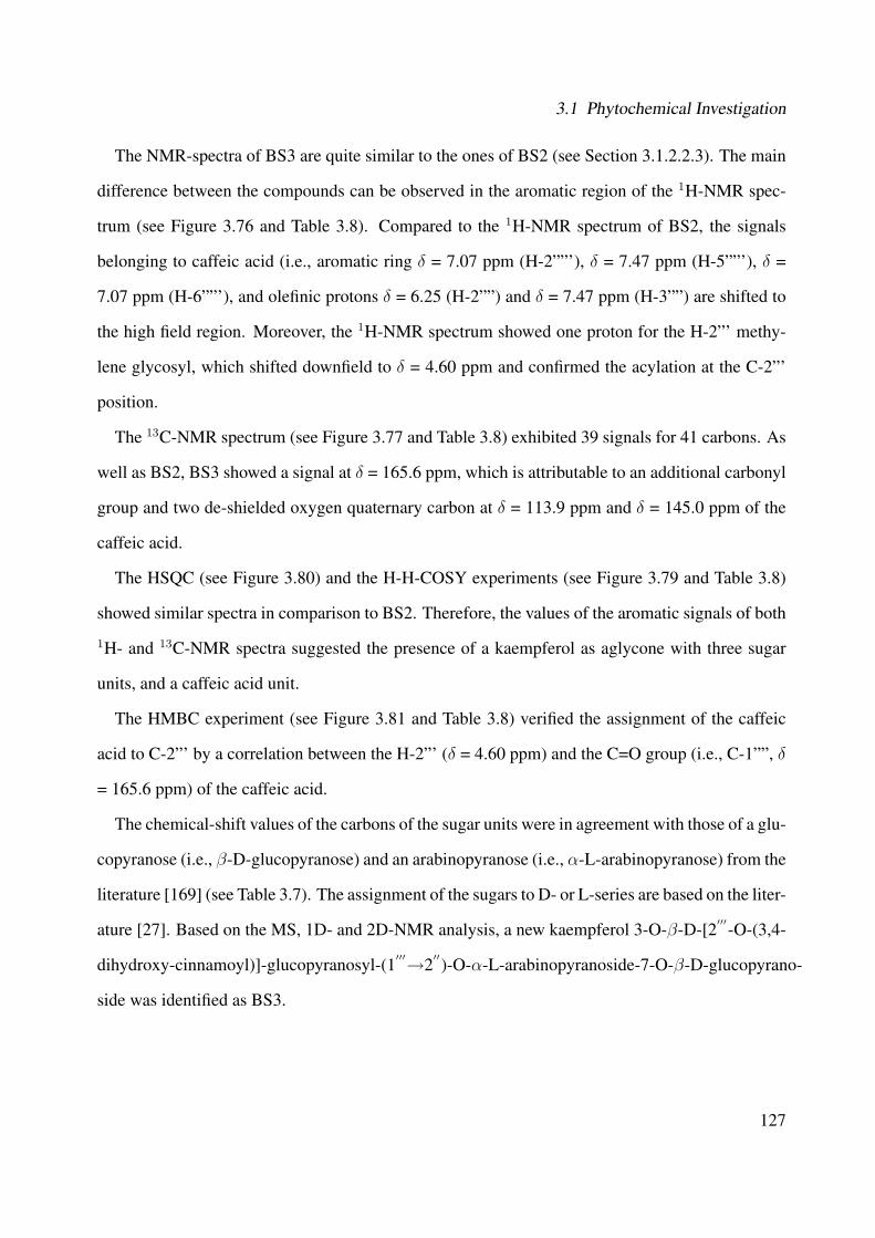
3.1 Phytochemical Investigation
The NMR-spectra of BS3 are quite similar to the ones of BS2 (see Section 3.1.2.2.3). The main
difference between the compounds can be observed in the aromatic region of the 1H-NMR spec-
trum (see Figure 3.76 and Table 3.8). Compared to the 1H-NMR spectrum of BS2, the signals
belonging to caffeic acid (i.e., aromatic ring δ = 7.07 ppm (H-2””'), δ = 7.47 ppm (H-5””'), δ =
7.07 ppm (H-6””'), and olefinic protons δ = 6.25 (H-2””) and δ = 7.47 ppm (H-3””) are shifted to
the high field region. Moreover, the 1H-NMR spectrum showed one proton for the H-2”' methy-
lene glycosyl, which shifted downfield to δ = 4.60 ppm and confirmed the acylation at the C-2”'
position.
The 13C-NMR spectrum (see Figure 3.77 and Table 3.8) exhibited 39 signals for 41 carbons. As
well as BS2, BS3 showed a signal at δ = 165.6 ppm, which is attributable to an additional carbonyl
group and two de-shielded oxygen quaternary carbon at δ = 113.9 ppm and δ = 145.0 ppm of the
caffeic acid.
The HSQC (see Figure 3.80) and the H-H-COSY experiments (see Figure 3.79 and Table 3.8)
showed similar spectra in comparison to BS2. Therefore, the values of the aromatic signals of both
1H- and 13C-NMR spectra suggested the presence of a kaempferol as aglycone with three sugar
units, and a caffeic acid unit.
The HMBC experiment (see Figure 3.81 and Table 3.8) verified the assignment of the caffeic
acid to C-2”' by a correlation between the H-2”' (δ = 4.60 ppm) and the C=O group (i.e., C-1””, δ
= 165.6 ppm) of the caffeic acid.
The chemical-shift values of the carbons of the sugar units were in agreement with those of a glu-
copyranose (i.e., β-D-glucopyranose) and an arabinopyranose (i.e., α-L-arabinopyranose) from the
literature [169] (see Table 3.7). The assignment of the sugars to D- or L-series are based on the liter-
ature [27]. Based on the MS, 1D- and 2D-NMR analysis, a new kaempferol 3-O-β-D-[2′′′-O-(3,4-
dihydroxy-cinnamoyl)]-glucopyranosyl-(1′′′→2′′)-O-α-L-arabinopyranoside-7-O-β-D-glucopyrano-
side was identified as BS3.
127

3 Results and Discussion
Table 3.8: Chemical shifts of BS3Atom numbers 13C* 13C** 1H* 1H - 1H 1H - 13C
δ/ppm δ/ppm δ/ppm (Mult., J(Hz), H) COSY* HMBC*
2 154.4 - - - -3 134.5 - - - -4 177.6 - - - -5 160.5 - - - -6 98.7 - 6.43 (brs; 1H) - -7 162.9 - - - -8 94.0 - 6.76 (brs; 1H) - -9 155.9 - - - -10 106.0 - - - -
1′
121.2 - - - -2
′131.0 - 8.06 (d; 6 Hz; 1H) 3
′, 5
′6
′
3′
115.4 - 6.81 (d; 6 Hz; 1H) 2′, 6
′5
′
4′
160.5 - - - -5
′115.4 - 6.81 (d; 6 Hz; 1H) 2
′, 6
′3
′
6′
131.0 - 8.06 (d; 6 Hz; 1H) 3′, 5
′2
′
1′′
98.7 100.9 5.58 (brs; 1H) - 2′′, 3, 3
′′, 2
′′′
2′′
78.7 78.3 4.09 (brs; 1H) 3′′
1′′, 4
′′, 3
′′
3′′
68.6 71.4 3.85 (brs; 1H) 2′′, 4
′′-
4′′
63.7 66.3 3.50 (m; 1H) - -5
′′60.6 62.1 3.00 (m; 1H); 3.50 (m; 1H) 5
′′1
′′, 3
′′, 4
′′
1′′′
101.5 103.1 4.68 (d; 6 Hz; 1H) 2′′′
2′′′
2′′′
73.1 77.6 4.60 (d; 6 Hz; 1H) 1′′′
, 3′′′
1′′′
, 5′′′
3′′′
73.6 79.4 3.48 (m; 1H) - 5′′′
4′′′
69.5 72.7 3.25 (m; 1H) - -5
′′′76.4 78.2 3.30 (m; 1H) - -
6′′′
60.3 62.1 3.50-3.70 (m; 2H) 6′′′
4′′′
, 5′′′
1′′′′
165.6 - - - -2
′′′′113.9 - 6.25 (d; 12 Hz; 1H) - 1
′′′′, 1
′′′′′
3′′′′
145.0 - 7.47 (d; 12 Hz; 1H) - 1′′′′
, 2′′′′
, 6′′′′′
1′′′′′
125.4 - - - 72
′′′′′114.9 - 7.07 (brs; 1H) - 4
′′′′′, 3
′′′′′, 5
′′′′′
3′′′′′
145.7 - - - -4
′′′′′148.7 - - - -
5′′′′′
115.8 - 6.90 (d; 6 Hz; 1H) - -6
′′′′′121.2 - 6.96 (brd; 6 Hz; 1H) - 3
′′′′′, 5
′′′′′
1′′′′′′
99.8 103.1 5.06 (d; 6 Hz; 1H) 2′′′′′′
72
′′′′′′72.5 77.6 3.20 (m; 1H) - -
3′′′′′′
76.8 79.4 3.28 (m; 1H) - -4
′′′′′′69.8 72.7 3.25 (m; 1H) - -
5′′′′′′
77.1 78.2 3.40 (m; 1H) - 1′′′′′′
6′′′′′′
60.3 62.1 3.50-3.70 (m; 2H) 6′′′′′′
-* In DMSO-d6. ** Data from literature [169] in Pyridine-d5.128
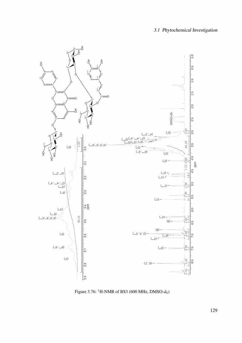
3.1 Phytochemical Investigation
8.0
7.5
7.0
6.5
6.0
5.5
5.0
4.5
4.0
3.5
3.0
2.5
2.0
1.5
1.0
0.5
0.0
ppm
66.1
83.
682.
762.
321.
601.
391.
121.
051.
000.
900.
88 (6""", 6''')
(2")
(2''') (1''')
(1""")
(1")
(2"") (6)
(8) (3', 5', 5""') (6""')
(2""')
(3"")
(6', 2')
DM
SO
-d6
3.9
3.8
3.7
3.6
3.5
3.4
3.3
3.2
3.1
3.0
ppm
66.1
81.
60
(6""", 6''')
(5")
(4",5'',6''',6""")(5""")
(4'''’,2""")
(5''')
(3""") (2"", 4""", 4''')
(5'')
(3’")
(5'') (4'''’,2""") (2"", 4""", 4''') (3""")
(5''') (3’")(5""") (4",5'',6''',6""")
(5")
(3")
(3")
7 65
1098
432
O1'6'
2'3'4'
5'
O
OO
H
OH
4''' 3'
''2'
''1'
''
O5'
''
1'' 2'
'3'
'4'
'
5''
O
OO
HO
H
O
6'''
OH
O
OHOH
4'''''
' 3'''''
'2'
'''''
1'''''
'
O5'
'''''
6'''''
'OH
OH
OHOH
3'''''
4'''''
5'''''
6'''''
1'''''
2'''''
2''''
3''''
1''''
O
OH
OH
Figure 3.76: 1H-NMR of BS3 (600 MHz, DMSO-d6)
129
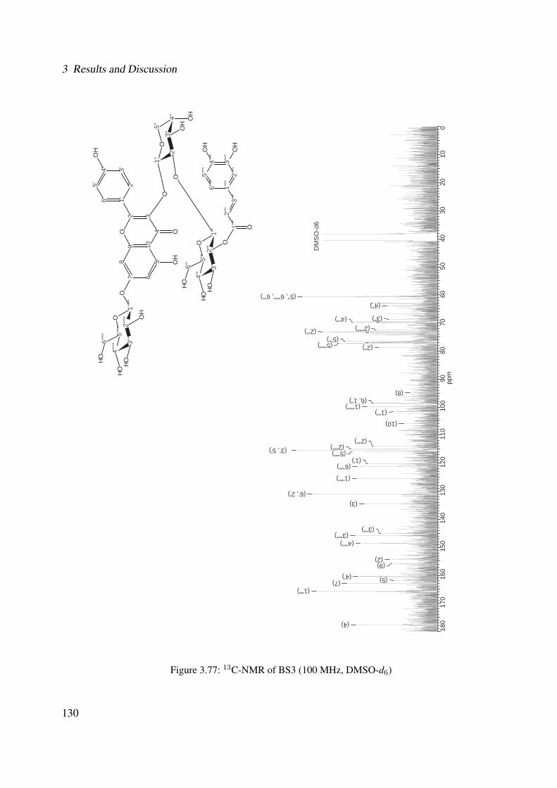
3 Results and Discussion
180
170
160
150
140
130
120
110
100
9080
7060
5040
3020
100
ppm
DM
SO
-d6
(5", 6""", 6''') (4")
(3") (4''') (2""") (2''')
(5''') (5""") (2")
(8) (6, 1")
(1""") (1''')
(10)
(2"") (2""') (3', 5') (5""')
(1') (6""')
(1""')
(6', 2') (3)
(3"") (3""')
(4""')
(2) (9)
(4') (5) (7) (1"")
(4)
7 65
1098
432
O1'6'
2'3'4'
5'
O
OO
H
OH
4''' 3'
''2'
''1'
''
O5'
''
1'' 2'
'3'
'4'
'
5''
O
OO
HO
H
O
6'''
OH
O
OHOH
4'''''
' 3'''''
'2'
'''''
1'''''
'
O5'
'''''
6'''''
'OH
OH
OHOH
3'''''
4'''''
5'''''
6'''''
1'''''
2'''''
2''''
3''''
1''''
O
OH
OH
Figure 3.77: 13C-NMR of BS3 (100 MHz, DMSO-d6)
130

3.1 Phytochemical Investigation
160
150
140
130
120
110
100
9080
7060
5040
3020
100
ppm
DM
SO
-d6
(5", 6""", 6''')
(4") (4""") (2""")
(3''') (5''') (5""")
(2")
(6, 1") (1""")
(1''')
(2"") (2""')
(3', 5') (5""')
(6""')
(6', 2')
(3"")
7 65
1098
432
O1'6'
2'3'4'
5'
O
OO
H
OH
4''' 3'
''2'
''1'
''
O5'
''
1'' 2'
'3'
'4'
'
5''
O
OO
HO
H
O
6'''
OH
O
OHOH
4'''''
' 3'''''
'2'
'''''
1'''''
'
O5'
'''''
6'''''
'OH
OH
OHOH
3'''''
4'''''
5'''''
6'''''
1'''''
2'''''
2''''
3''''
1''''
O
OH
OH
Figure 3.78: DEPT-135 of BS3 (100 MHz, DMSO-d6)
131
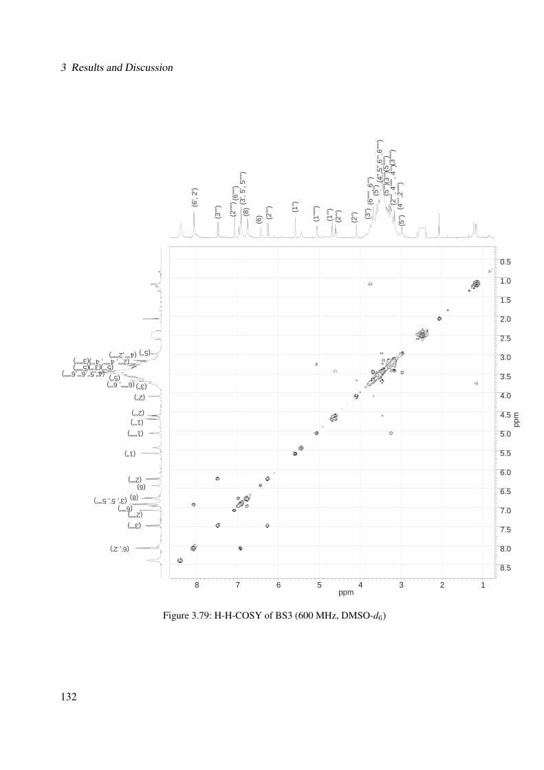
3 Results and Discussion
8 7 6 5 4 3 2 1ppm
0.5
1.0
1.5
2.0
2.5
3.0
3.5
4.0
4.5
5.0
5.5
6.0
6.5
7.0
7.5
8.0
8.5
ppm
(2")
(2''') (1''') (1""")
(1")
(2"") (6)
(8) (3', 5', 5""') (6""')
(2""') (3"")
(6', 2')
(6""", 6''')
(5'') (4'''’,2""") (2"", 4""", 4''') (3""")
(5''') (3’")(5""") (4",5'',6''',6""") (5")
(2")
(2''')
(1''')
(1""
") (1")
(2""
) (6
) (8) (3
', 5'
, 5""
') (6
""')
(2""
')
(3""
) (6',
2')
(6""
", 6'
'')
(5'')
(4'''’
,2""
") (2
"", 4
""",
4''') (
3"""
) (5
''') (3
’")(5
""")
(4",5
'',6'
'',6"
"")
(5")
(3")
(3")
Figure 3.79: H-H-COSY of BS3 (600 MHz, DMSO-d6)
132
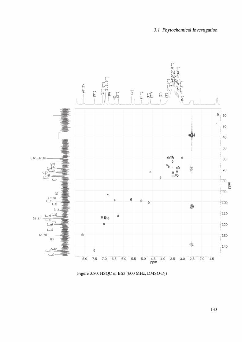
3.1 Phytochemical Investigation
8.0 7.5 7.0 6.5 6.0 5.5 5.0 4.5 4.0 3.5 3.0 2.5 2.0 1.5ppm
20
30
40
50
60
70
80
90
100
110
120
130
140
ppm
(2")
(2''')
(1''')
(1""
") (1")
(2""
) (6
) (8) (3
', 5'
, 5""
') (6
""')
(2""
')
(3""
) (6',
2')
(6""
", 6'
'')
(5'')
(4'''’
,2""
") (2
"", 4
""",
4''') (
3"""
) (5
''') (3
’")(5
""")
(4",5
'',6'
'',6"
"")
(5")
(5", 6""", 6''')
(4") (3")
(4''') (2""") (2''')
(5''') (5""")
(2")
(8) (6, 1")
(1""") (1''')
(10)
(2"") (2""') (3', 5') (5""')
(1') (6""')
(1""')
(6', 2') (3)
(3"") (3""')
(4""')
(3")
Figure 3.80: HSQC of BS3 (600 MHz, DMSO-d6)
133

3 Results and Discussion
1.0
1.5
2.0
2.5
3.0
3.5
4.0
4.5
5.0
5.5
6.0
6.5
7.0
7.5
8.0
8.5
ppm40 60 80
100
120
140
160
180
200
(2")
(2''') (1''')
(1""")
(1")
(2"") (6)
(8) (3', 5', 5""')
(6""') (2""')
(3"")
(6', 2')
(6""", 6''')
(5'') (4'''’,2""") (2"", 4""", 4''') (3""")
(5''') (3’")(5""") (4",5'',6''',6""") (5")
(5",
6"""
, 6''')
(4") (3
") (4
''') (2
""")
(2''')
(5''')
(5""
") (2
")
(8)
(6, 1
") (1
""")
(1''')
(10)
(2""
) (2
""')
(3',
5')
(5""
') (1
') (6
""')
(1""
') (6
', 2'
) (3
)
(3""
) (3
""')
(4""
') (2
) (9
) (4
') (5
) (7)
(1""
)
(4)
(3")
Figure 3.81: HMBC of BS3 (600 MHz, DMSO-d6)
134

3.1 Phytochemical Investigation
3.1.2.3 Discussion
In the previous sections, the phytochemical studies (i.e., MS and 1D and 2D NMR) revealed
the presence of four flavonol glycosides in the ethanolic extract from the leaves of Brugman-
sia suaveolens. BS1 was assigned as kaempferol 3-O-β-D-glucopyranosyl-(1′′′→2′′)-O-α-L-ara-
binopyranoside-7-O-β-D-glucopyranoside, BS2 as kaempferol 3-O-β-D-[6′′′-O-(3,4-dihydroxy-
cinnamoyl)]-glucopyranosyl-(1′′′→2′′)-O-α-L-arabinopyranoside-7-O-β-D-glucopyranoside, BS3
as kaempferol 3-O-β-D-[2′′′-O-(3,4-dihydroxy-cinnamoyl)]-glucopyranosyl-(1′′′→2′′)-O-α-L-ara-
binopyranoside-7-O-β-D-glucopyranoside and BS4 as kaempferol 3-O-β-D-glucopyranosyl-(1′′′→
2′′)-O-α-L-arabinopyranoside. The compounds BS1, BS2, BS3 and BS4 were reported for the first
time in nature. Figure 3.82 presents the HPLC chromatogram (Method HPLC-B, see Section
5.4.4, Experimental Part) of the ethanolic extract of Brugmansia suaveolens and its character-
ized compounds. Additionally, flavonol glycosides (i.e., kaempferol 3-O-α-L-arabinopyranoside
and kaempferol 3-O-α-L-arabinopyranoside-7-O-β-D-glucopyranoside) have also been reported
by Begum et al., (2006) [27] in B. suaveolens.
Brugmansia suaveolens has mainly been studied due to the presence of alkaloids [84, 97, 10,
350]. However, the qualitative determination of alkaloids, which was carried out with the ethanolic
extract, showed negative results. A reason that could explain this result might be the low concen-
tration of alkaloids in the leaves of this plant. Alves et al., (2007) [10] reported the occurrence of
lower concentrations of alkaloids in the leaves and the highest concentrations were identified in the
roots and in the flowers of Brugmansia suaveolens.
Concerning the biosynthesis of the aforementioned isolated compounds from Brugmansia suave-
olens, a hypothetical pathway was proposed, as shown in Figure 3.83. As a first step, the kaempferol
(Figure 3.83.A) is derived from a 4-hydroxycinnamoyl-CoA [80]. In the second step, the α-L-
arabinopyranose group might be added to O-3 of kaempferol (Figure 3.83.B), which was already
isolated from this plant by Begum et al., (2006) [27]. Third step, β-D-glucopyranoses might be
added to the O-7 of kaempferol (Figure 3.83.C was also already isolated from this plant by Be-
135

3 Results and Discussion
gum et al., (2006) [27]) or to the O-2 of arabinose (Figure 3.83.D was isolated in this work).
Fourth step, β-D-glucopyranoses might be added to the O-7 of kaempferol (Figure 3.83.D) or to
the O-2 of arabinose (Figure 3.83.C) to produce the compounds BS1 (Figure 3.83.E). Fifth step,
after the biosynthesis of the caffeic acid [237], the acylation might occur on the terminal glu-
cosyl unit of the kaempferol 3,7-O-triglucoside to produce the compound BS2 (Figure 3.83.F).
This acylation is very common at position C-6 [150] and has been described for some examples
[105, 118, 140, 138, 50, 2]. However, the acylation at the position C-2 of the kaempferol 3,7-
O-triglucoside producing the compound BS3 (Figure 3.83.G) is not a common transfer and has
only been reported by Kellam et al., (1993) [154] and by Tian et al., (2007) [309]. Considering
that the acylation at position C-6 occurs frequently in the nature compared to the acylation at the
position C-2, one may speculate that the biosynthesis of compound BS2 might be produced be-
fore the biosynthesis of compound BS3. Based on this hypothesis, the possible biosynthesis of the
compounds from Brugmansia suaveolens might follow: BS4→ BS1→ BS2→ BS3.
136
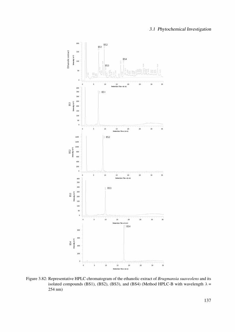
3.1 Phytochemical Investigation
2,66
4,66
5,19
5,83
6,33
7,21
7,73
8,29
8,95
9,61
10,2
110
,76 11
,75
12,1
812
,78
13,7
314
,48
15,9
116
,70
17,2
817
,91
18,7
219
,59
20,1
720
,72
21,6
621
,96
22,6
523
,36
24,0
724
,51
25,0
425
,53
26,4
827
,14
27,5
127
,76
28,6
5
29,8
330
,45
31,2
031
,75
32,6
0
33,7
5
0 5 10 15 20 25 30 35Retention Time (min)
0
50
100
150
200
Inte
nsity
(mV)
7,04
0 5 10 15 20 25 30 35Retention Time (min)
0
50
100
150
200
250
300
350
400
Inte
nsity
(mV)
9,84
0 5 10 15 20 25 30 35
Retention Time (min)
0
50
100
150
200
250
300
350
400
Inte
nsity
(mV)
9,02
0 5 10 15 20 25 30 35
Retention Time (min)
0
200
400
600
800
1000
1200
1400
Inte
nsity
(mV)
17,6
6
0 5 10 15 20 25 30 35
Retention Time (min)
0
200
400
600
800
Inte
nsity
(mV)
BS4
BS1BS2
BS3
BS1
BS2
BS3
BS4
Etha
nolic
ext
ract
BS1
BS2
BS3
BS4
Figure 3.82: Representative HPLC chromatogram of the ethanolic extract of Brugmansia suaveolens and itsisolated compounds (BS1), (BS2), (BS3), and (BS4) (Method HPLC-B with wavelength λ =254 nm)
137
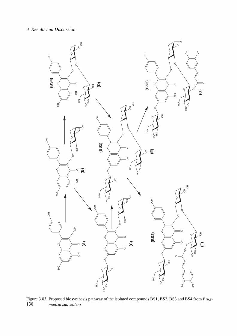
3 Results and Discussion
(BS2
)(B
S3)
(BS1
)
(BS4
)
(A)
(B)
(D)
(E)
(F)
(G)
(C)
OH
O
OO
H
OH
O
O
OO
H OH
O
HO
OH
HO
HO
OH
O
OO
H
OH
OH
OH
O
OO
H
OH
O
HO
OH
OH
O
OO
OO
H
OH
O
HO
OH
OH
O
O
HO
OH
HO
HO
OO
OO
H
OH
O
O
OO
HO
H
O
HO
OH
HO
HO
O
HO
OH
HO
HO
OO
OO
H
OH
O
O
OO
H OH
O
HO
O
HO
HO
O
HO
OH
HO
HO
O
OH
OH
OO
OO
H
OH
O
O
OO
HO
H
O
O
OH
HO
HO
O
HO
HO
O
HO
OH
HO
HO
Figure 3.83: Proposed biosynthesis pathway of the isolated compounds BS1, BS2, BS3 and BS4 from Brug-mansia suaveolens138
3.2 Biological Investigation and Discussion
3.2 Biological Investigation and Discussion
The ethanolic extracts from the leaves of Cordia americana and Brugmansia suaveolens as well
as their isolated compounds were evaluated using in vitro test systems, such as enzyme-linked
immunosorbent assay (ELISA), which determines the inhibition of p38α and JNK3 in isolated
enzyme assays. Moreover, docking studies were also performed in order to explain the possible
binding modes of the most active isolated compounds at the ATP binding site of both enzymes.
The activity of the plant extract and isolated compounds of Cordia americana were also studied
for TNFα release in human whole blood assay. These assays23 as well as the docking studies24
were carried out in the Department of Pharmaceutical and Medicinal Chemistry at the University
of Tubingen.
The 5-lipoxygenase assays were performed in cell free and in cell-based assays using isolated
human PMNL. These assays25 were carried out in the Department of Pharmaceutical Analytics at
the University of Tubingen.
In cooperation with the Department of Pharmaceutical Biology and Biotechnology at the Uni-
versity of Freiburg, the NF-κB26 activation was studied by means of the electrophoretic mobility
shift assay (EMSA). The wound healing effects27 were studied using the fibroblast scratch assay
and finally cytotoxic effects of the plant extract were studied by the MTT (3-(4,5-dimethylthiazol-
2-yl)-2,5-diphenyltetrazolium bromide) assay.
3.2.1 p38α MAPK
This section presents the results of the inhibition on p38α (see Section 5.7.1, Experimental
Part) with regard to the ethanolic extracts of Cordia americana, Brugmansia suaveolens and their23The MAPK (i.e., p38α and JNK3) and also the TNFα assays were carried out by Marcia Goettert and Katharina
Bauer (by Prof. Dr. Laufer).24The molecular modeling studies were carried out by Verena Schattel (by Prof. Dr. Laufer).255-LO assays in cell free and isolated PMNL were carried out by Bianca Jazzar and Daniela Mueller (by Prof. Dr.
Werz).26The NF-κB assay was carried out by Cleber Schmidt (by Prof. Dr. Merfort).27The MTT and fibroblast scratch assays were carried out by Marcio Fronza (by Prof. Dr. Merfort).
139
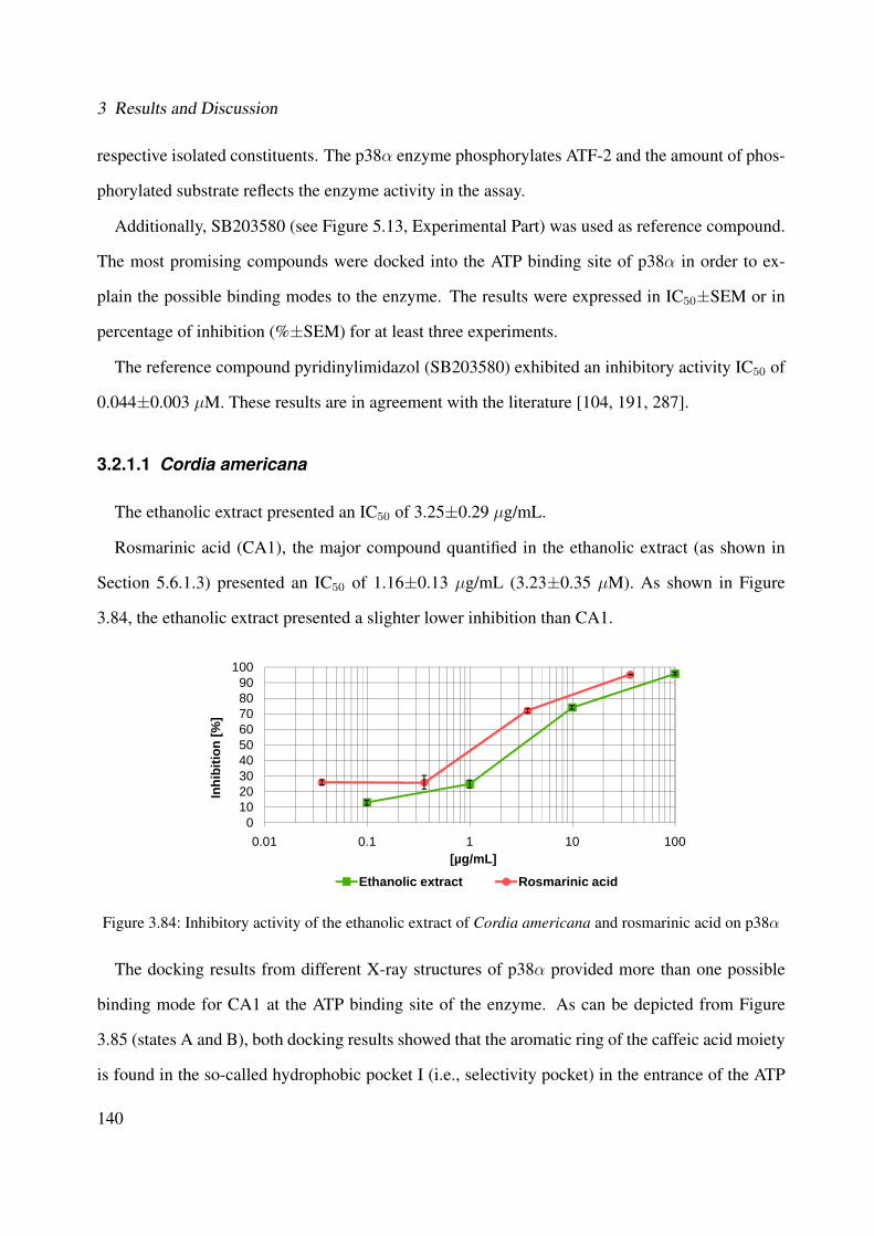
3 Results and Discussion
respective isolated constituents. The p38α enzyme phosphorylates ATF-2 and the amount of phos-
phorylated substrate reflects the enzyme activity in the assay.
Additionally, SB203580 (see Figure 5.13, Experimental Part) was used as reference compound.
The most promising compounds were docked into the ATP binding site of p38α in order to ex-
plain the possible binding modes to the enzyme. The results were expressed in IC50±SEM or in
percentage of inhibition (%±SEM) for at least three experiments.
The reference compound pyridinylimidazol (SB203580) exhibited an inhibitory activity IC50 of
0.044±0.003 µM. These results are in agreement with the literature [104, 191, 287].
3.2.1.1 Cordia americana
The ethanolic extract presented an IC50 of 3.25±0.29 µg/mL.
Rosmarinic acid (CA1), the major compound quantified in the ethanolic extract (as shown in
Section 5.6.1.3) presented an IC50 of 1.16±0.13 µg/mL (3.23±0.35 µM). As shown in Figure
3.84, the ethanolic extract presented a slighter lower inhibition than CA1.
0102030405060708090
100
0.01 0.1 1 10 100
Inhi
bitio
n [%
]
[µg/mL]
Ethanolic extract Rosmarinic acid
Figure 3.84: Inhibitory activity of the ethanolic extract of Cordia americana and rosmarinic acid on p38α
The docking results from different X-ray structures of p38α provided more than one possible
binding mode for CA1 at the ATP binding site of the enzyme. As can be depicted from Figure
3.85 (states A and B), both docking results showed that the aromatic ring of the caffeic acid moiety
is found in the so-called hydrophobic pocket I (i.e., selectivity pocket) in the entrance of the ATP
140
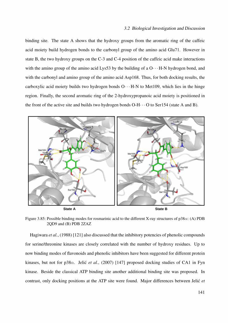
3.2 Biological Investigation and Discussion
binding site. The state A shows that the hydroxy groups from the aromatic ring of the caffeic
acid moiety build hydrogen bonds to the carbonyl group of the amino acid Glu71. However in
state B, the two hydroxy groups on the C-3 and C-4 position of the caffeic acid make interactions
with the amino group of the amino acid Lys53 by the building of a O· · ·H-N hydrogen bond, and
with the carbonyl and amino group of the amino acid Asp168. Thus, for both docking results, the
carboxylic acid moiety builds two hydrogen bonds O· · ·H-N to Met109, which lies in the hinge
region. Finally, the second aromatic ring of the 2-hydroxypropanoic acid moiety is positioned in
the front of the active site and builds two hydrogen bonds O-H· · ·O to Ser154 (state A and B).
State A State B
Figure 3.85: Possible binding modes for rosmarinic acid to the different X-ray structures of p38α: (A) PDB2QD9 and (B) PDB 2ZAZ
Hagiwara et al., (1988) [121] also discussed that the inhibitory potencies of phenolic compounds
for serine/threonine kinases are closely correlated with the number of hydroxy residues. Up to
now binding modes of flavonoids and phenolic inhibitors have been suggested for different protein
kinases, but not for p38α. Jelic et al., (2007) [147] proposed docking studies of CA1 in Fyn
kinase. Beside the classical ATP binding site another additional binding site was proposed. In
contrast, only docking positions at the ATP site were found. Major differences between Jelic et
141

3 Results and Discussion
al., (2007) [147] and this approach are: Fyn kinase is a non-receptor tyrosine kinase from the
Src kinase family, whereas p38α is a serine/threonine kinase from the MAPK family. In addition,
a homology model of the enzyme and docking was performed with FlexX and Gold software
[147]. The current approach is based on X-ray structures of the p38α and the induced fit tool from
Schrodinger software package [281] was used for docking.
CA1 was identified as the major compound with an amount of 8.44% in the ethanolic extract of
the leaves of Cordia americana. However, the ethanolic extract from Cordia americana exhibited
higher inhibition in comparison to the predominant constituent, as can be observed in Table 3.9.
Thus, further compounds may contribute to the described biological effects.
Table 3.9: Biological effects of the ethanolic extract of Cordia americana and rosmarinic acid on p38α
IC50 of theethanolic extract
(µg/mL)
Content of CA1 (8.44%) in thisamount of ethanolic extract (µg/mL)
IC50 of CA1(µg/mL)
p38α 3.25 0.27 1.16
CA1 is also the major constituent of lemon balm (Melissa of�cinalis), a plant that has shown
promising signs of therapeutic activity in patients with Alzheimer's diseases [146] and it is also
used as a cough remedy [128, 318].
The rosmarinic acid ethyl ester (CA2) was studied and showed an IC50 of 5.10±0.43 µg/mL
(13.13±1.1 µM). As observed in Figure 3.86, CA2 had a slight lower inhibition than the ethanolic
extract and CA1.
0102030405060708090
100
0.01 0.1 1 10 100
Inhi
bitio
n [%
]
[µg/mL]Ethanolic extract Rosmarinic acid ethyl ester Rosmarinic acid
Figure 3.86: Inhibitory activity of the ethanolic extract of C. americana, rosmarinic acid ethyl ester androsmarinic acid on p38α
142

3.2 Biological Investigation and Discussion
Concerning the docking studies of CA2 at the ATP binding site of the kinase, it is possible to
observe that the binding modes are similar as in CA1. As represented in Figure 3.87, the state A
shows that both aromatic rings of CA2 bind and make interactions in the same position as the state
A of CA1 (see Figure 3.85 (state A)). On one hand, in state B (see Figure 3.87), one hydroxy group
of the aromatic ring of the 2-hydroxypropanoic acid moiety makes interactions with Asp168, and
on the other hand, two hydroxy groups of the second aromatic ring of the caffeic acid interact with
the carbonyl groups of both amino acids Asp112 and Ser154. Regarding the interactions in the
hinge region, it is important to point out that the ester group might make weak or no interactions
(state B) with Met109, which is probably reflected in a lower inhibition of the isolated CA2.
State A State B
Figure 3.87: Possible binding modes for rosmarinic acid ethyl ester to the different X-ray structures of p38α:(A) PDB 2QD9 and (B) PDB 2ZAZ
The compounds 3-(3,4-dihydroxyphenyl)-2-hydroxypropanoic acid (CA3), rutin (CA4), quercitrin
(CA5), and α-amyrin (CA8) were also evaluated on the p38α assay. As observed in Table 3.10,
CA3 exhibited an IC50 of 4.28±1.97 µg/mL (21.64±8.9 µM). The flavonol glycosides CA5 and
CA4 gave an IC50 of 12.59±0.52 µg/mL (28.08±1.17 µM) and 40.22±5.44 µg/mL (65.88±8.88
µM), respectively. Finally, the compound CA8 showed an IC50 of 15.25±1.36 µg/mL (35.75±3.18
143
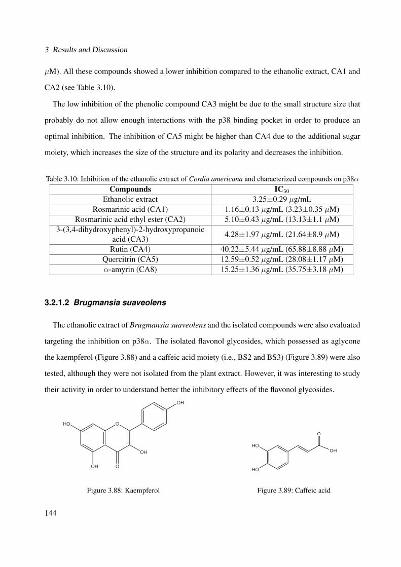
3 Results and Discussion
µM). All these compounds showed a lower inhibition compared to the ethanolic extract, CA1 and
CA2 (see Table 3.10).
The low inhibition of the phenolic compound CA3 might be due to the small structure size that
probably do not allow enough interactions with the p38 binding pocket in order to produce an
optimal inhibition. The inhibition of CA5 might be higher than CA4 due to the additional sugar
moiety, which increases the size of the structure and its polarity and decreases the inhibition.
Table 3.10: Inhibition of the ethanolic extract of Cordia americana and characterized compounds on p38α
Compounds IC50
Ethanolic extract 3.25±0.29 µg/mLRosmarinic acid (CA1) 1.16±0.13 µg/mL (3.23±0.35 µM)
Rosmarinic acid ethyl ester (CA2) 5.10±0.43 µg/mL (13.13±1.1 µM)3-(3,4-dihydroxyphenyl)-2-hydroxypropanoic
acid (CA3)4.28±1.97 µg/mL (21.64±8.9 µM)
Rutin (CA4) 40.22±5.44 µg/mL (65.88±8.88 µM)Quercitrin (CA5) 12.59±0.52 µg/mL (28.08±1.17 µM)α-amyrin (CA8) 15.25±1.36 µg/mL (35.75±3.18 µM)
3.2.1.2 Brugmansia suaveolens
The ethanolic extract of Brugmansia suaveolens and the isolated compounds were also evaluated
targeting the inhibition on p38α. The isolated flavonol glycosides, which possessed as aglycone
the kaempferol (Figure 3.88) and a caffeic acid moiety (i.e., BS2 and BS3) (Figure 3.89) were also
tested, although they were not isolated from the plant extract. However, it was interesting to study
their activity in order to understand better the inhibitory effects of the flavonol glycosides.
OHO
O
OH
OH
OH
Figure 3.88: Kaempferol
OH
O
HO
HO
Figure 3.89: Caffeic acid
144
3.2 Biological Investigation and Discussion
The ethanolic extract showed an IC50 of 1.21±0.02 µg/mL.
As observed in Table 3.11, the kaempferol 3-O-β-glucopyranosyl-(1→2)-O-α-L-arabinopyranoside
(BS4) exhibited an IC50 of 26.80±1.78 µM (15.55±1.03 µg/mL) on p38α. The new compound
kaempferol 3-O-β-glucopyranosyl-(1→2)-O-α-L-arabinopyranoside-7-O-β-glucopyranoside (BS1)
showed an IC50 of 34.35±2.09 µM (25.51±1.55 µg/mL). The kaempferol 3-O-β-[6”'-O-(3,4-
dihydroxy-cinnamoyl)]-glucopyranosyl-(1→2)-O-α-L-arabinopyranoside-7-O-β-gluco- pyranoside
(BS2) exhibited an IC50 of 25.73±3.63 µM (23.28±3.28 µg/mL) on p38α, as shown in Figure
3.90. On the other hand, no IC50-value was obtained for the new kaempferol 3-O-β-[2”'-O-(3,4-
dihydroxy-cinnamoyl)]-glucopyranosyl-(1→2)-O-α-L-arabinopyranoside-7-O-β-glucopyranoside
(BS3), whereas the highest inhibition of 41.39±3.75% was found at 100 µM (90.48 µg/mL). As
can be observed, the acylation of the caffeic acid moiety at C-2”' (i.e., BS3) might provoke a lower
inhibition compared to the acylation of the caffeic acid moiety at C-6”' (i.e., BS2).
In order to investigate, wether the aglycone kaempferol and the caffeic acid moiety contributed to
the activity of the isolated flavonol glycosides, the corresponding reference compounds were also
tested. Kaempferol inhibited p38αwith an IC50 of 14.51±0.01 µM (4.15±0.01 µg/mL) and caffeic
acid showed an IC50 of 50.40±8.29 µM (9.08±1.49 µg/ml), as shown in Table 3.11. Thus, it can
be assumed that the kaempferol aglycone might contribute more than the caffeic acid moiety to the
inhibition of the flavonol glycosides as well as to the ethanolic extract of Brugmansia suaveolens
in the p38α. However, further non-characterized constituents might play a role in the effects of the
plant extract.
Table 3.11: Inhibition of the ethanolic extract and isolated flavonol glycosides from B. suaveolens on p38α
Compounds IC50 / percentage of inhibition (%)Ethanolic extract 1.21±0.02 µg/mL
BS1 34.35±2.09 µM (25.51±1.55 µg/mL)BS2 25.73±3.63 µM (23.28±3.28 µg/mL)BS3 41.39±3.75% @ 100 µM (90.48 ±g/mL)BS4 26.80±1.78 µM (15.55±1.03 µg/mL)
Kaempferol 14.51±0.01 µM (4.15±0.01 µg/mL)Caffeic acid 50.40±8.29 µM (9.08±1.49 µg/ml)
145

3 Results and Discussion
Following these results, BS1-4 do not substantially contribute to the the activity of the extract, as
can be observed in Figure 3.90. The flavonol glycosides from Brugmansia suaveolens (i.e., BS1,
BS2, BS3 and BS4) might present low significantly activity due to the glycosylation.
0102030405060708090
100
0.01 0.1 1 10 100
Inhi
bitio
n [%
]
[µg/mL]Ethanolic extract BS1 BS2 BS3 BS4
Figure 3.90: Inhibitory activity of the ethanolic extract of Brugmansia suaveolens and the isolated flavonolglycosides on p38α
Ferriola et al., (1989) [92] have already suggested that the inhibitory potency on Protein Kinase
C (PKC) by flavonols were reduced by glycosylation. This feature could be also observed with
kaempferol, which has a higher inhibition on p38α than the isolated flavonol glycosides.
3.2.2 TNFα
p38α is also involved in the release of TNFα [282, 179, 176]. Further studies were carried out in
order to evaluate the effects on release of TNFα with the ethanolic extract and the corresponding
isolated compounds from Cordia americana using human whole blood by ELISA (see Section
5.7.3, Experimental Part). The isolated compounds from Brugmansia suaveolens were not tested
in TNFα, because of their large molecular size and their polarity, which might hinder the diffusion
across the cell membrane. All the values are expressed in IC50±SEM or in percentage of inhibition
at the highest tested concentration (%±SEM) from at least two experiments.
The pyridinylimidazole SB203580 was used as reference compound and exhibited an IC50 of
1.97±0.57 µM.
146
3.2 Biological Investigation and Discussion
Table 3.12 shows the results of the ethanolic extract of Cordia americana and their respec-
tive compounds on TNFα release. The ethanolic extract moderately suppressed the release of
TNFα, where the highest inhibition effect of 49.71±15.87% was achieved at 100 µg/mL. The
major compound CA1 shows an inhibition of 36.75±1.54% tested in a concentration of 100 µM
(36.03 µg/mL). Compared to CA1, the ethanolic extract presented a slightly lower activity. On the
other hand, CA2 showed the highest inhibitory effect with an IC50 of 47.84±4.87 µM (18.58±1.89
µg/mL).
Table 3.12: Inhibition of ethanolic extract of Cordia americana and the characterized compounds on TNFαrelease
Compounds IC50 / percentage of inhibition (%)Ethanolic extract 49.71±15.87% @ 100 µg/mL
Rosmarinic acid (CA1) 36.75±1.54% @ 100 µM (36.03 µg/mL)Rosmarinic acid ethyl ester (CA2) 47.84±4.87 µM (18.58±1.89 µg/mL)
It is important to point out that the inhibition effects of the compounds might be highly de-
pendent of the donors of human blood. CA1 and CA2 have lower activity in the TNFα assay in
comparison to the p38α on cell free assay. This might be probably explained due to the plasma
protein binding, so that only a small amount of the inhibitor is absorbed by the cell.
As well as for the kinases assays (Section 3.2.1.1), the ethanolic extract exhibited a higher effect
compared to CA1, since the ethanolic extract at 100 µg/mL, which contains 8.44% of CA1 (i.e.,
8.44 µg/mL), resulted in 49.71% of inhibition. Therefore, one might suspect that the inhibitory
properties of Cordia americana might be dependent mostly on CA1, but also on CA2.
147
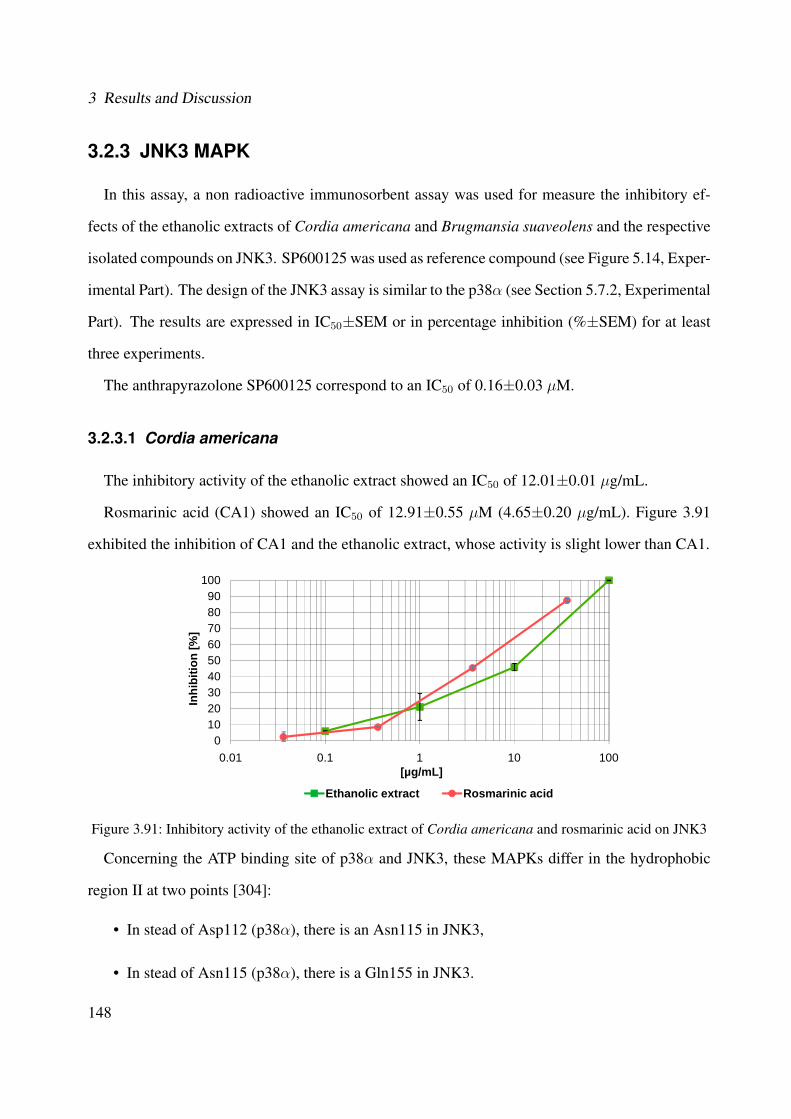
3 Results and Discussion
3.2.3 JNK3 MAPK
In this assay, a non radioactive immunosorbent assay was used for measure the inhibitory ef-
fects of the ethanolic extracts of Cordia americana and Brugmansia suaveolens and the respective
isolated compounds on JNK3. SP600125 was used as reference compound (see Figure 5.14, Exper-
imental Part). The design of the JNK3 assay is similar to the p38α (see Section 5.7.2, Experimental
Part). The results are expressed in IC50±SEM or in percentage inhibition (%±SEM) for at least
three experiments.
The anthrapyrazolone SP600125 correspond to an IC50 of 0.16±0.03 µM.
3.2.3.1 Cordia americana
The inhibitory activity of the ethanolic extract showed an IC50 of 12.01±0.01 µg/mL.
Rosmarinic acid (CA1) showed an IC50 of 12.91±0.55 µM (4.65±0.20 µg/mL). Figure 3.91
exhibited the inhibition of CA1 and the ethanolic extract, whose activity is slight lower than CA1.
0102030405060708090
100
0.01 0.1 1 10 100
Inhi
bitio
n [%
]
[µg/mL]
Ethanolic extract Rosmarinic acid
Figure 3.91: Inhibitory activity of the ethanolic extract of Cordia americana and rosmarinic acid on JNK3
Concerning the ATP binding site of p38α and JNK3, these MAPKs differ in the hydrophobic
region II at two points [304]:
• In stead of Asp112 (p38α), there is an Asn115 in JNK3,
• In stead of Asn115 (p38α), there is a Gln155 in JNK3.
148

3.2 Biological Investigation and Discussion
The docking results from different X-ray structures, referenced as PDB 3G9L (State A) and PDB
3FI3 (State B) provide also more than one possible binding mode for CA1 at the ATP binding site
of JNK3. As shown in Figure 3.92 (PDB 3G9L and PDB 3FI3), both docking results demonstrate
that the aromatic ring of the caffeic acid moiety is found in the so-called hydrophobic pocket I
(i.e., selectivity pocket) in the entrance of the ATP binding site. The state A demonstrates that
the hydroxy groups at the C-3 and C-4 position of the aromatic ring build hydrogen bonds to the
carboxyl-group of the amino acid Glu111. However, in state B, the hydroxy group at the C-3
position makes interactions with the amino group of Lys93 (O· · ·H-N hydrogen bond) and with
the carbonyl group of the amino acid Leu206 (O-H· · ·O hydrogen bond). The aromatic ring of the
2-hydroxypropanoic acid moiety is located in the hinge region in state A, whereas the two hydroxy
groups build two hydrogen bonds to the carbonyl and amino group of Met149. In the state B, one
hydroxy group makes interactions with Asp150 by the building of a O-H· · ·O hydrogen bond and
with Gln155 by the building of a O· · ·H-N hydrogen bond. Finally, the carboxylic acid moiety
builds in state A a hydrogen bond to the carbonyl-group of Asn152 (O-H· · ·O hydrogen bond),
and in state B, it makes interactions with Met149 by the building of two hydrogen bonds to the
carbonyl and amino group of Met149.
State A
State B
Figure 3.92: Possible binding modes for rosmarinic acid to the different X-ray structures of JNK3: (A) PDB3G9L and (B) PDB 3FI3
149
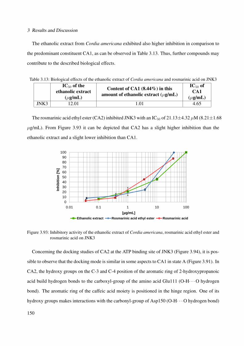
3 Results and Discussion
The ethanolic extract from Cordia americana exhibited also higher inhibition in comparison to
the predominant constituent CA1, as can be observed in Table 3.13. Thus, further compounds may
contribute to the described biological effects.
Table 3.13: Biological effects of the ethanolic extract of Cordia americana and rosmarinic acid on JNK3IC50 of the
ethanolic extract(µg/mL)
Content of CA1 (8.44%) in thisamount of ethanolic extract (µg/mL)
IC50 ofCA1
(µg/mL)JNK3 12.01 1.01 4.65
The rosmarinic acid ethyl ester (CA2) inhibited JNK3 with an IC50 of 21.13±4.32 µM (8.21±1.68
µg/mL). From Figure 3.93 it can be depicted that CA2 has a slight higher inhibition than the
ethanolic extract and a slight lower inhibition than CA1.
0102030405060708090
100
0.01 0.1 1 10 100
Inhi
bitio
n [%
]
[µg/mL]Ethanolic extract Rosmarinic acid ethyl ester Rosmarinic acid
Figure 3.93: Inhibitory activity of the ethanolic extract of Cordia americana, rosmarinic acid ethyl ester androsmarinic acid on JNK3
Concerning the docking studies of CA2 at the ATP binding site of JNK3 (Figure 3.94), it is pos-
sible to observe that the docking mode is similar in some aspects to CA1 in state A (Figure 3.91). In
CA2, the hydroxy groups on the C-3 and C-4 position of the aromatic ring of 2-hydroxypropanoic
acid build hydrogen bonds to the carboxyl-group of the amino acid Glu111 (O-H· · ·O hydrogen
bond). The aromatic ring of the caffeic acid moiety is positioned in the hinge region. One of its
hydroxy groups makes interactions with the carbonyl-group of Asp150 (O-H· · ·O hydrogen bond)
150
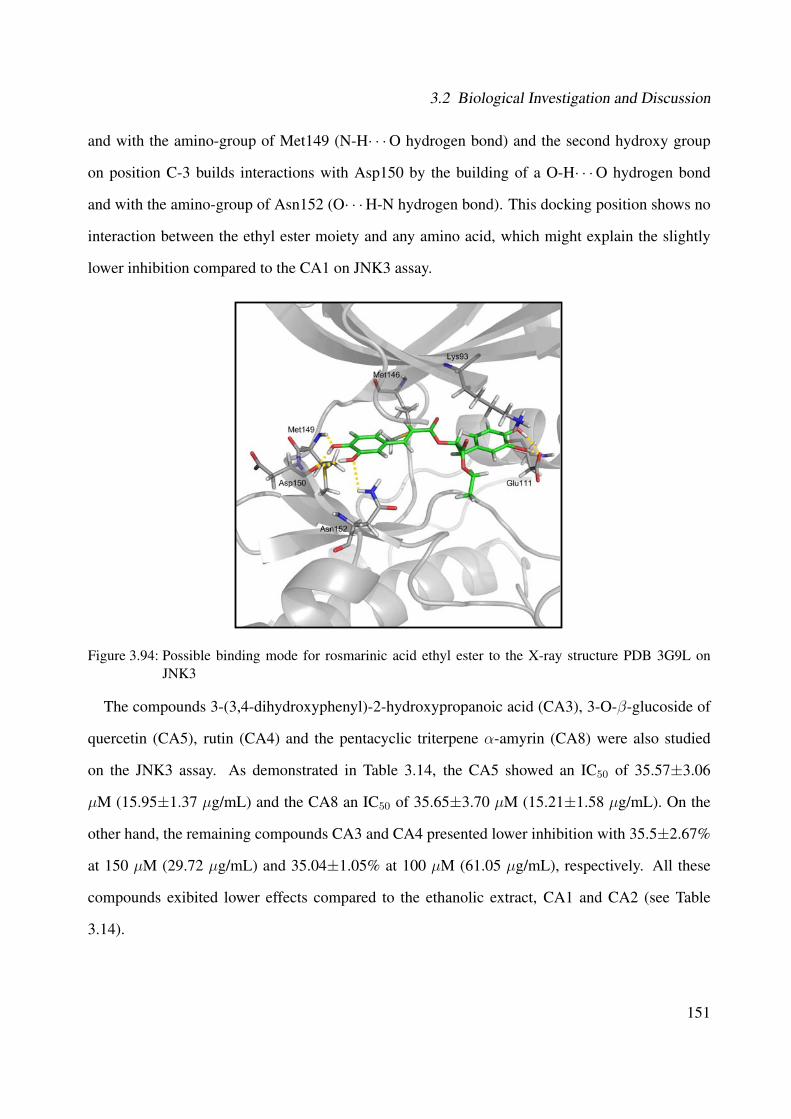
3.2 Biological Investigation and Discussion
and with the amino-group of Met149 (N-H· · ·O hydrogen bond) and the second hydroxy group
on position C-3 builds interactions with Asp150 by the building of a O-H· · ·O hydrogen bond
and with the amino-group of Asn152 (O· · ·H-N hydrogen bond). This docking position shows no
interaction between the ethyl ester moiety and any amino acid, which might explain the slightly
lower inhibition compared to the CA1 on JNK3 assay.
Figure 3.94: Possible binding mode for rosmarinic acid ethyl ester to the X-ray structure PDB 3G9L onJNK3
The compounds 3-(3,4-dihydroxyphenyl)-2-hydroxypropanoic acid (CA3), 3-O-β-glucoside of
quercetin (CA5), rutin (CA4) and the pentacyclic triterpene α-amyrin (CA8) were also studied
on the JNK3 assay. As demonstrated in Table 3.14, the CA5 showed an IC50 of 35.57±3.06
µM (15.95±1.37 µg/mL) and the CA8 an IC50 of 35.65±3.70 µM (15.21±1.58 µg/mL). On the
other hand, the remaining compounds CA3 and CA4 presented lower inhibition with 35.5±2.67%
at 150 µM (29.72 µg/mL) and 35.04±1.05% at 100 µM (61.05 µg/mL), respectively. All these
compounds exibited lower effects compared to the ethanolic extract, CA1 and CA2 (see Table
3.14).
151

3 Results and Discussion
Table 3.14: Inhibition of the of the ethanolic extract of Cordia americana and characterized compounds onJNK3
Compounds IC50 / percentage inhibition (%)Ethanolic extract 12.01±0.01 µg/mL
Rosmarinic acid (CA1) 12.91±0.55 µM (4.65±0.20 µg/mL)Rosmarinic acid ethyl ester (CA2) 21.13±4.32 µM (8.21±1.68 µg/mL)
3-(3,4-dihydroxyphenyl)-2-hydroxypropanoicacid (CA3)
35.5±2.67% @ 150 µM (29.72 µg/mL)
Rutin (CA4) 35.04±1.05% @ 100 µM (61.05 µg/mL)Quercitrin (CA5) 35.57±3.06 µM (15.95±1.37 µg/mL)α-amyrin (CA8) 35.65±3.70 µM (15.21±1.58 µg/mL)
The low inhibition of CA4 and CA5 might be explained due to the glycosylation which increases
the size and polarity of the structure that might be not adequate to the small ATP binding pocket of
JNK3.
3.2.3.2 Brugmansia suaveolens
The ethanolic extract of Brugmansia suaveolens and its respective isolated flavonol glycosides
were also studied targeting the inhibition on JNK3. Furthermore, kaempferol that corresponds to
the aglycone of the isolated compounds and the caffeic acid, which were not detected in the plant
extract, were tested.
The ethanolic extract of Brugmansia suaveolens exhibited an IC50 of 20.76±0.18 µg/mL.
As can be observed in Table 3.15, none of the isolated flavonol glycosides showed relevant
inhibition on JNK3 assay. This can mainly be explained due to the size of the compounds, since
the ATP binding site of JNK3 is flat and small, which cannot accommodate larger inhibitors as the
isolated ones.
Kaempferol inhibited the JNK3 with an IC50 of 17.77±0.38 µM (5.08±0.11 µg/mL) and caffeic
acid exhibited lower inhibition of 18.80±1.49% at 100 µM (18.02 µg/mL), as shown in Table 3.15.
Thus, it can be assumed that the kaempferol aglycone might contribute more than the caffeic acid
to the inhibition of the ethanolic extract of Brugmansia suaveolens on JNK3. Moreover, further
non-characterized compounds might explain the effects of the ethanolic extract of Brugmansia
suaveolens on JNK3.
152
3.2 Biological Investigation and Discussion
Table 3.15: Inhibition of ethanolic extract of Brugmansia suaveolens and the isolated flavonol glycosides onJNK3
Compounds IC50 / percentage inhibition (%)Ethanolic extract 20.76±0.18 µg/mL
BS1 13.5±1.62% @ 100 µM (74.26 µg/mL)BS2 24.03±3.49% @ 100 µM (90.48 µg/mL)BS3 33.4±0.80% @ 100 µM (90.48 µg/mL)BS4 19.1±2.29% @100 µM (58.05 µg/mL)
Kaempferol 17.77±0.38 µM (5.08±0.11 µg/mL)Caffeic acid 18.80±1.49% @ 100 µM (18.02 µg/mL)
Scapin et al., (2003) [268] suggests also that small, flat and more hydrophobic inhibitors proba-
bly bind better to the JNK3 ATP binding site than to the more solvent exposed p38 cavity. Probably
due to this feature, the isolated flavonol glycosides exhibited no considerable inhibitory effects on
JNK3.
3.2.4 5-Lipoxygenase
The present section describes the results on the inhibition of 5-LO (see Section 5.7.4, Experi-
mental Part), concerning the ethanolic extracts of Cordia americana and Brugmansia suaveolens,
and their isolated compounds. The investigation on 5-LO inhibition was done in a cell-free as-
say using partially purified 5-LO. Moreover, the most active compounds were further tested in
cell-based assay using human PMNL (polymorphonuclear leukocytes). BWA4C (see Figure 5.17,
Experimental Part) was used as reference compound. All the values are expressed in IC50±SEM
or in percentage of inhibition (%±SEM) from at least two experiments.
3.2.4.1 Inhibition of 5-LO Activity in a Cell-free Assay
The reference compound BWA4C exhibited an IC50 of 0.3±0.01 µM.
153
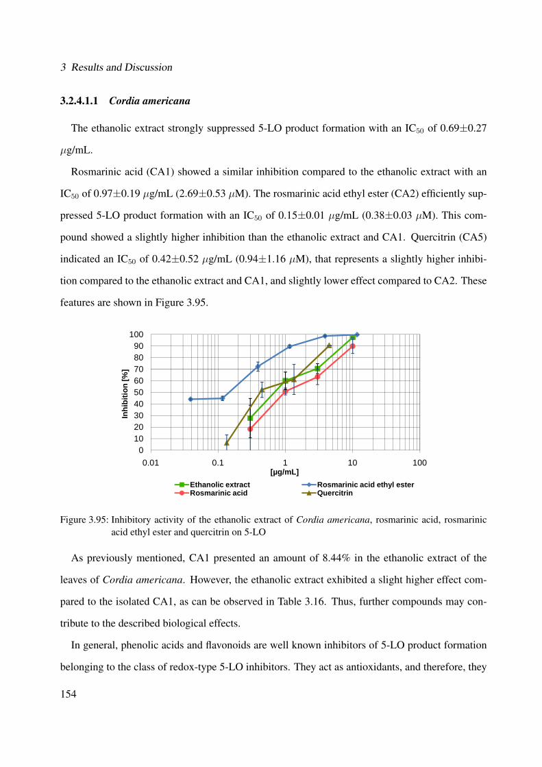
3 Results and Discussion
3.2.4.1.1 Cordia americana
The ethanolic extract strongly suppressed 5-LO product formation with an IC50 of 0.69±0.27
µg/mL.
Rosmarinic acid (CA1) showed a similar inhibition compared to the ethanolic extract with an
IC50 of 0.97±0.19 µg/mL (2.69±0.53 µM). The rosmarinic acid ethyl ester (CA2) efficiently sup-
pressed 5-LO product formation with an IC50 of 0.15±0.01 µg/mL (0.38±0.03 µM). This com-
pound showed a slightly higher inhibition than the ethanolic extract and CA1. Quercitrin (CA5)
indicated an IC50 of 0.42±0.52 µg/mL (0.94±1.16 µM), that represents a slightly higher inhibi-
tion compared to the ethanolic extract and CA1, and slightly lower effect compared to CA2. These
features are shown in Figure 3.95.
0102030405060708090
100
0.01 0.1 1 10 100
Inhi
bitio
n [%
]
[µg/mL]Ethanolic extract Rosmarinic acid ethyl ester Rosmarinic acid Quercitrin
Figure 3.95: Inhibitory activity of the ethanolic extract of Cordia americana, rosmarinic acid, rosmarinicacid ethyl ester and quercitrin on 5-LO
As previously mentioned, CA1 presented an amount of 8.44% in the ethanolic extract of the
leaves of Cordia americana. However, the ethanolic extract exhibited a slight higher effect com-
pared to the isolated CA1, as can be observed in Table 3.16. Thus, further compounds may con-
tribute to the described biological effects.
In general, phenolic acids and flavonoids are well known inhibitors of 5-LO product formation
belonging to the class of redox-type 5-LO inhibitors. They act as antioxidants, and therefore, they
154

3.2 Biological Investigation and Discussion
Table 3.16: Biological effects of the ethanolic extract of Cordia americana and rosmarinic acid on 5-LOIC50 of the ethanolic
extract (µg/mL)Content of CA1 (8.44%) in this
amount of ethanolic extract (µg/mL)IC50 of CA1
(µg/mL)5-LO 0.69 0.0582 0.97
keep the active site-iron of 5-LO in the inactive ferrous state and uncouple the catalytic redox cycle
of the enzyme. In case of CA1 and CA2, the presence of phenolic hydroxy groups might govern
the potency on 5-LO inhibition. In particular, polyphenols active on 5-LO typically resemble fatty
acid-like structures, with a carboxylic acid moiety or an acidic phenol core [330].
The compounds 3-(3,4-dihydroxyphenyl)-2- hydroxypropanoic acid (CA3) and rutin (CA4)
were also investigated for the inhibition on the 5-LO product formation, as illustrated in Table
3.17. These compounds showed no notably activity.
Table 3.17: Inhibition of the isolated compounds from Cordia americana on 5-LOCompounds Percentage of inhibition (%)
3-(3,4-dihydroxyphenyl)-2-hydroxypropanoic acid (CA3) 14.96% @ 10 µM (1.98 µg/mL)Rutin (CA4) 20.55% @ 10 µM (6.10 µg/mL)
3.2.4.1.2 Brugmansia suaveolens
The ethanolic extract of Brugmansia suaveolens and the respective isolated compounds were
also evaluated targeting the inhibition on 5-LO product formation on cell-free assay. Furthermore,
kaempferol, which corresponds to the aglycone of the flavonol glycosides and caffeic acid moiety
(i.e., BS2 and BS3) were also studied.
The effect of the ethanolic extract resulted in a suppression of the 5-LO product formation with
an IC50 of 5.42±5.16 µg/mL.
Table 3.18 exhibited the inhibitory effects of the isolated flavonol glycosides from Brugmansia
suaveolens. Most of the new isolated compounds presented no significant activity on 5-LO prod-
uct formation, with exception of BS3. The flavonol glycoside BS3 (kaempferol 3-O-β-[2”'-O-(3,4-
dihydroxy-cinnamoyl)]-glucopyranosyl-(1→2)-O-α-L-arabinopyranoside-7-O-β-glucopyranoside)
moderately inhibited the 5-LO product formation with an IC50 of 28.38±15.90 µM (25.68±14.38
155
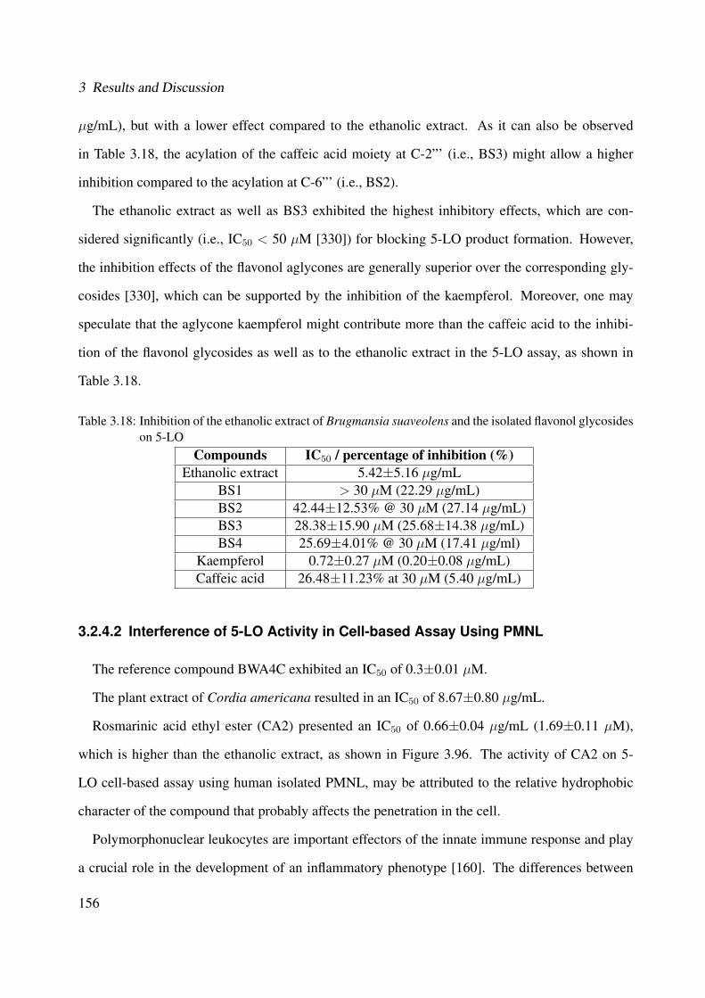
3 Results and Discussion
µg/mL), but with a lower effect compared to the ethanolic extract. As it can also be observed
in Table 3.18, the acylation of the caffeic acid moiety at C-2”' (i.e., BS3) might allow a higher
inhibition compared to the acylation at C-6”' (i.e., BS2).
The ethanolic extract as well as BS3 exhibited the highest inhibitory effects, which are con-
sidered significantly (i.e., IC50 < 50 µM [330]) for blocking 5-LO product formation. However,
the inhibition effects of the flavonol aglycones are generally superior over the corresponding gly-
cosides [330], which can be supported by the inhibition of the kaempferol. Moreover, one may
speculate that the aglycone kaempferol might contribute more than the caffeic acid to the inhibi-
tion of the flavonol glycosides as well as to the ethanolic extract in the 5-LO assay, as shown in
Table 3.18.
Table 3.18: Inhibition of the ethanolic extract of Brugmansia suaveolens and the isolated flavonol glycosideson 5-LO
Compounds IC50 / percentage of inhibition (%)Ethanolic extract 5.42±5.16 µg/mL
BS1 > 30 µM (22.29 µg/mL)BS2 42.44±12.53% @ 30 µM (27.14 µg/mL)BS3 28.38±15.90 µM (25.68±14.38 µg/mL)BS4 25.69±4.01% @ 30 µM (17.41 µg/ml)
Kaempferol 0.72±0.27 µM (0.20±0.08 µg/mL)Caffeic acid 26.48±11.23% at 30 µM (5.40 µg/mL)
3.2.4.2 Interference of 5-LO Activity in Cell-based Assay Using PMNL
The reference compound BWA4C exhibited an IC50 of 0.3±0.01 µM.
The plant extract of Cordia americana resulted in an IC50 of 8.67±0.80 µg/mL.
Rosmarinic acid ethyl ester (CA2) presented an IC50 of 0.66±0.04 µg/mL (1.69±0.11 µM),
which is higher than the ethanolic extract, as shown in Figure 3.96. The activity of CA2 on 5-
LO cell-based assay using human isolated PMNL, may be attributed to the relative hydrophobic
character of the compound that probably affects the penetration in the cell.
Polymorphonuclear leukocytes are important effectors of the innate immune response and play
a crucial role in the development of an inflammatory phenotype [160]. The differences between
156
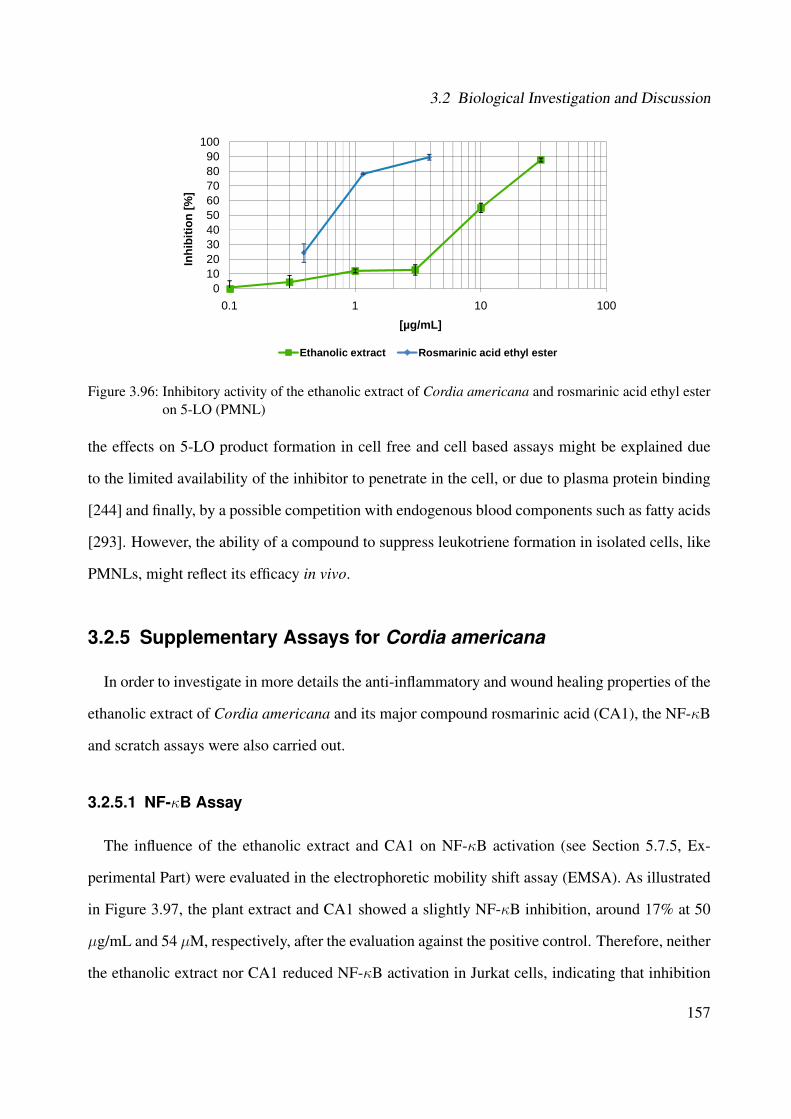
3.2 Biological Investigation and Discussion
0102030405060708090
100
0.1 1 10 100
Inhi
bitio
n [%
]
[µg/mL]
Ethanolic extract Rosmarinic acid ethyl ester
Figure 3.96: Inhibitory activity of the ethanolic extract of Cordia americana and rosmarinic acid ethyl esteron 5-LO (PMNL)
the effects on 5-LO product formation in cell free and cell based assays might be explained due
to the limited availability of the inhibitor to penetrate in the cell, or due to plasma protein binding
[244] and finally, by a possible competition with endogenous blood components such as fatty acids
[293]. However, the ability of a compound to suppress leukotriene formation in isolated cells, like
PMNLs, might reflect its efficacy in vivo.
3.2.5 Supplementary Assays for Cordia americana
In order to investigate in more details the anti-inflammatory and wound healing properties of the
ethanolic extract of Cordia americana and its major compound rosmarinic acid (CA1), the NF-κB
and scratch assays were also carried out.
3.2.5.1 NF-κB Assay
The influence of the ethanolic extract and CA1 on NF-κB activation (see Section 5.7.5, Ex-
perimental Part) were evaluated in the electrophoretic mobility shift assay (EMSA). As illustrated
in Figure 3.97, the plant extract and CA1 showed a slightly NF-κB inhibition, around 17% at 50
µg/mL and 54 µM, respectively, after the evaluation against the positive control. Therefore, neither
the ethanolic extract nor CA1 reduced NF-κB activation in Jurkat cells, indicating that inhibition
157
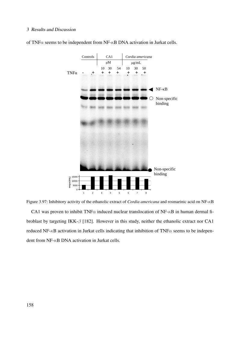
3 Results and Discussion
of TNFα seems to be independent from NF-κB DNA activation in Jurkat cells.
Controls CA1 Cordia americanaμM μg/mL
- + + + + + + + 10 30 54 10 30 50
TNFα
NF-κB
Non-specific binding
Non-specific binding
0
5000
10000
15000
1 2 3 4 5 6 7 8
Inte
gral
[PSL
]
Figure 3.97: Inhibitory activity of the ethanolic extract of Cordia americana and rosmarinic acid on NF-κB
CA1 was proven to inhibit TNFα induced nuclear translocation of NF-κB in human dermal fi-
broblast by targeting IKK-β [182]. However in this study, neither the ethanolic extract nor CA1
reduced NF-κB activation in Jurkat cells indicating that inhibition of TNFα seems to be indepen-
dent from NF-κB DNA activation in Jurkat cells.
158
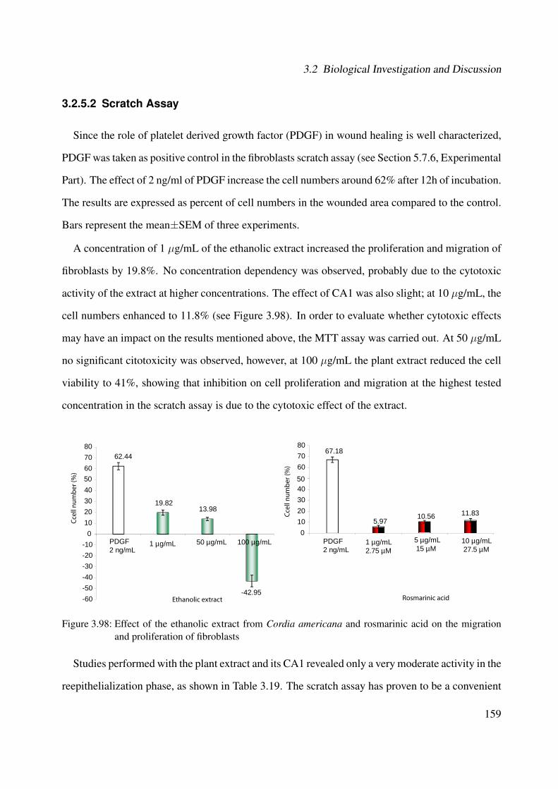
3.2 Biological Investigation and Discussion
3.2.5.2 Scratch Assay
Since the role of platelet derived growth factor (PDGF) in wound healing is well characterized,
PDGF was taken as positive control in the fibroblasts scratch assay (see Section 5.7.6, Experimental
Part). The effect of 2 ng/ml of PDGF increase the cell numbers around 62% after 12h of incubation.
The results are expressed as percent of cell numbers in the wounded area compared to the control.
Bars represent the mean±SEM of three experiments.
A concentration of 1 µg/mL of the ethanolic extract increased the proliferation and migration of
fibroblasts by 19.8%. No concentration dependency was observed, probably due to the cytotoxic
activity of the extract at higher concentrations. The effect of CA1 was also slight; at 10 µg/mL, the
cell numbers enhanced to 11.8% (see Figure 3.98). In order to evaluate whether cytotoxic effects
may have an impact on the results mentioned above, the MTT assay was carried out. At 50 µg/mL
no significant citotoxicity was observed, however, at 100 µg/mL the plant extract reduced the cell
viability to 41%, showing that inhibition on cell proliferation and migration at the highest tested
concentration in the scratch assay is due to the cytotoxic effect of the extract.
67.18
607080
62.44
50607080
5.9710.56 11.83
010203040
19.8213.98
-42.95-60-50-40-30-20-10
010203040
Rosmarinic acidEthanolic extract
1 µg/mLPDGF2 ng/mL
50 µg/mL PDGF2 ng/mL
Ccel
l num
ber (
%)
Ccel
l num
ber (
%)
1 µg/mL2.75 µM
5 µg/mL 15 µM
10 µg/mL 27.5 µM
100 µg/mL
5050
Figure 3.98: Effect of the ethanolic extract from Cordia americana and rosmarinic acid on the migrationand proliferation of fibroblasts
Studies performed with the plant extract and its CA1 revealed only a very moderate activity in the
reepithelialization phase, as shown in Table 3.19. The scratch assay has proven to be a convenient
159
3 Results and Discussion
and inexpensive method to give first insights on the proliferation and migration of fibroblasts into
the damaged area, as demonstrated for the ethanolic extract of Calendula of�cinalis (1 µg/mL),
where an increasing effect of 60% was observed [100].
Table 3.19: Biological effect of the ethanolic extract of Cordia americana and rosmarinic acid on scratchassay
Concentrationof the plant
extract(µg/mL)
Content of CA1(8.44%) in this
amount ofethanolic
extract (µg/mL)
Cell numbercompared tocontrol (%)
Concentrationof CA1µg/mL)
Cell numbercompared tocontrol (%)
Scratchassay
1 0.084 19.8 1 6.0
50 4.22 13.9 5 10.6100 8.44 -42.0 10 11.8
160
3.2 Biological Investigation and Discussion
3.2.6 Summary of the Biological Activity
This section summarizes the biological activity of the characterized compounds of the investi-
gated plants Cordia americana and Brugmansia suaveolens.
3.2.6.1 Rosmarinic Acid, Rosmarinic Acid Ethyl Ester and
3-(3,4-dihydroxyphenyl)-2-hydroxypropanoic acid
Rosmarinic acid (CA1) was isolated from many species of plants of the families Lamiaceae and
Boraginaceae and was identified as one of the active components of several medicinal plants (e.g.,
Salvia of�cinallis, Mentha piperitam, Thymus vulgaris, Melissa of�cinalis, Symphytum of�cinale)
[237]. It has been shown to possess anti-inflammatory, antioxidative, antiviral as well as antibac-
terial activity in various in vitro assays [205, 238, 346, 234] and in vivo studies [348, 305]. Studies
demonstrated inhibitory effects on 5 and 12-lipoxygenase and gene expression of cycloxygenase-2
[346, 247, 273]. Besides the antioxidative properties, CA1 acts as a potent antiviral agent in vivo
against Japanese encephalitis virus by reducing the viral replication and secondary inflammation
resulting from microglial activation [305]. Gao et al., (2004) [107] and Hur et al., (2004) [139]
also showed neuroprotective effects for CA1 by inducing apoptosis.
Rosmarinic acid ethyl ester (CA2) showed hypotensive, antibacterial, anti-viral, anti-inflammatory,
anti-tumor and hypoglycemic activities [327]. Choudhary et al., (2005) [53] reported that CA2,
isolated from Lindelo�a stylosa (Boraginaceae), possesses antioxidant activity.
CA1 and CA2 have effects on the p56lck SH2 domain (src homology-2 domain). The SH2 do-
main is a highly conserved non-catalytic module, consisting of 100 amino acids residues, and is
found in many intracellular signal-transduction proteins. This domain recognizes phosphotyro-
sine, containing proteins with high affinity. Specific antagonist of the p56lck SH2 domains can be
developed as novel therapeutic agents to treat a broad range of human diseases such as cancer, au-
toimmune diseases, osteoporosis and chronic inflammatory disease [230]. CA1 exhibited a binding
to the p56lck SH2 domain with an IC50 of 24 µM, which was measured by an ELISA competitive
assay. On the other hand, the CA2 showed a less potent binding affinity with an IC50 of 91±3 µM
161

3 Results and Discussion
in comparison with CA1, which can be explained by the presence of an ester group. Considering
the structure-activity relationship (SAR) studies, all four hydroxyl groups from both compounds
are essential for the interaction with p56lck SH2 domain [230].
3-(3,4-dihydroxyphenyl)-2-hydroxypropanoic acid (CA3) (i.e.,danshenshu) has relaxing effects
on 5-HT-precontracted coronary artery rings [349, 173] and in ischemic myocardial injury, which
was proven in rats [342].
Psotova et al., (2003) [247] studied the antioxidant activity of Prunella vulgaris and reported
that the scavenging activity might be attributed due to the major isolated compounds CA1 and CA3.
Bano et al., (2003) [22] and Dapkevivius et al., (2002) [67] attributed the antioxidant activity of
CA1 due to the presence of two catechol structures, conjugated with a carboxylic acid group. In
case of CA3, Chen and Ho (1997) [47] related the antioxidative activity to the hydroxyl groups.
3.2.6.2 β-Sitosterol and Campesterol
A variety of pharmacological properties are attributed to β-sitosterol (CA6), like antioxidant,
anti-inflammatory, anti-carcinogenic and anti-atherogenic [303]. More specifically, CA6 blocks
cholesterol absorption, resulting in lower serum cholesterol levels, and also prevents the oxida-
tion of LDL cholesterol, whereby the risk of atherosclerosis is reduced. It has been used to treat
prostate problems such as benign prostatic hypertrophy. Thus, it may reduce the growth of the
prostate gland, as well as inhibiting colon cancer cells and altering membrane lipids [303]. CA6
had been reported to exhibit both antifungical and antibacterial activities against Fusarium ssp.
and Salmonella typhii, respectively [158, 220].
There is an accumulating evidence that campesterol (CA7) exhibits chemoprotective effects
against many cancers, including prostate [202], lung [271] and breast [19]. CA7 can inhibit
endothelial cell proliferation and differentiation as well as neovascularization with no toxicity,
suggesting that it could be an antiangiogenic candidate for the prevention and treatment of angio-
genesis diseases [51]. However, finally further in vivo studies have to be carried out to confirm all
these in vitro studies.
162
3.2 Biological Investigation and Discussion
3.2.6.3 α- and β-Amyrin
The pentacyclic triterpene α-amyrin (CA8) and β-amyrin (CA9) were used to alleviate inflam-
matory symptoms [321] and is also reported to possess a wide range of activity against gram-
positive and gram-negative bacteria. Vitor et al., (2009) [321] showed that the anti-inflammatory
effects of both compounds seem to be related to the local suppression of inflammatory cytokines
and COX-2 levels, possibly via inhibition of NF-κB pathway, which were examinated on an ex-
perimental model of colitis in mice. Considering its biological effects, the mixture of compounds
α- and β-amyrin presented antinociceptive properties [228].
3.2.6.4 Flavonol Glycosides
Flavonol glycosides play a special role in the protection of plants from ultraviolet damage [210]
and in the excitation and coloring of plant fluorescence [296]. Plants usually glycosylate its sec-
ondary metabolites in order to enhance their solubility and improve sequestration into specific
cellular compartments [132].
In nature, flavonoids exist almost as β-glycosides, although the 7 and 4' positions may also be
glycosylated in some plants [96, 345]. Other classes of flavonoids are found mainly glycosylated
in the position 7 [71]. The different structures of flavonoids significantly affect the absorption,
metabolism, bioactivities, and the binding process with plasma proteins [323].
With respect to the structural differences of glycosides and aglycones, Amakura et al., (2003)
[11] reported that their differences in the activity may be described to the increasing molecular
size and polarity and due to the transfer to the non-planar structure produced by the addition of
sugars. Acylation and glycosylation of flavonoids also increase stability of the compound. On the
other hand, flavonoid glycosides are generally hydrophilic and thus cannot be transported across
membranes by passive diffusion. In case of hydrolysis by bacterial enzymes in the lower part
of the intestine, the sugar moiety of flavonoid glycosides is cleaved, resulting in more lipophilic
aglycones. These become permeable through the cell wall [345].
163

3 Results and Discussion
Rutin (CA4) has anti-inflammatory [286] and anti-tumour [41] effects and showed also activity
against hemoglobin oxidation [119]. Moreover, rutin can reduce capillary fragility, swelling and
has been used in the treatment of venous insufficiency (varicose veins, haemorrhoids, diabetic
vascular disease, and diabetic retinopathy), and for improving micro-vascular blood flow (pain,
tired legs, night cramps, and restless legs) [3]. CA4 also demonstrated antioxidant effects on
malonaldehyde formation from ethyl arachidonate.
Quercitrin (CA5), the 3-O-β-glucoside of quercetin, is a flavonol glycoside, which shows an-
tileishmanial (in vitro) [219] and antioxidant activities [235].
164

4 Summary
In Brazil, the medicinal plants Cordia americana and Brugmansia suaveolens have been used
to treat inflammations and wounds in folk medicine. However, the effective compounds respon-
sible for the biological effects of these plants are widely unknown. Therefore, both plants were
investigated in this dissertation and the conclusions and scientific contributions are summarized:
• Bioguided fractionation, based on p38α MAPK assay, was carried out with the fraction sets
of the ethanolic extract of Cordia americana and revealed five groups that were submitted
to successive subfractionation. The phytochemical studies (i.e., MS, 1D and 2D NMR)
allowed the identification of flavonols (rutin and quercitrin), phytosterols (campesterol and
β-sitosterol), triterpenoids (α- and β-amyrin) and phenolic acids (3-(3,4-dihydroxyphenyl)-
2-hydroxypropanoic acid, rosmarinic acid and rosmarinic acid ethyl ester).
• All the aforementioned compounds were identified for the first time in Cordia americana,
and 3-(3,4-dihydroxyphenyl)-2-hydroxypropanoic acid and rosmarinic acid ethyl ester were
described for the first time for the genus Cordia.
• HPLC analysis revealed rosmarinic acid as the major compound in the ethanolic extract
of Cordia americana. Therefore, a quantification method was developed and a content of
8.44% of rosmarinic acid in the dried leaves of Cordia americana was detected, which is so
far the highest concentration found in Boraginaceaes species.
• Concerning the biological effects, the ethanolic extract of Cordia americana as well as ros-
marinic acid and rosmarinic acid ethyl ester exhibited the highest inhibitory effects on the
165
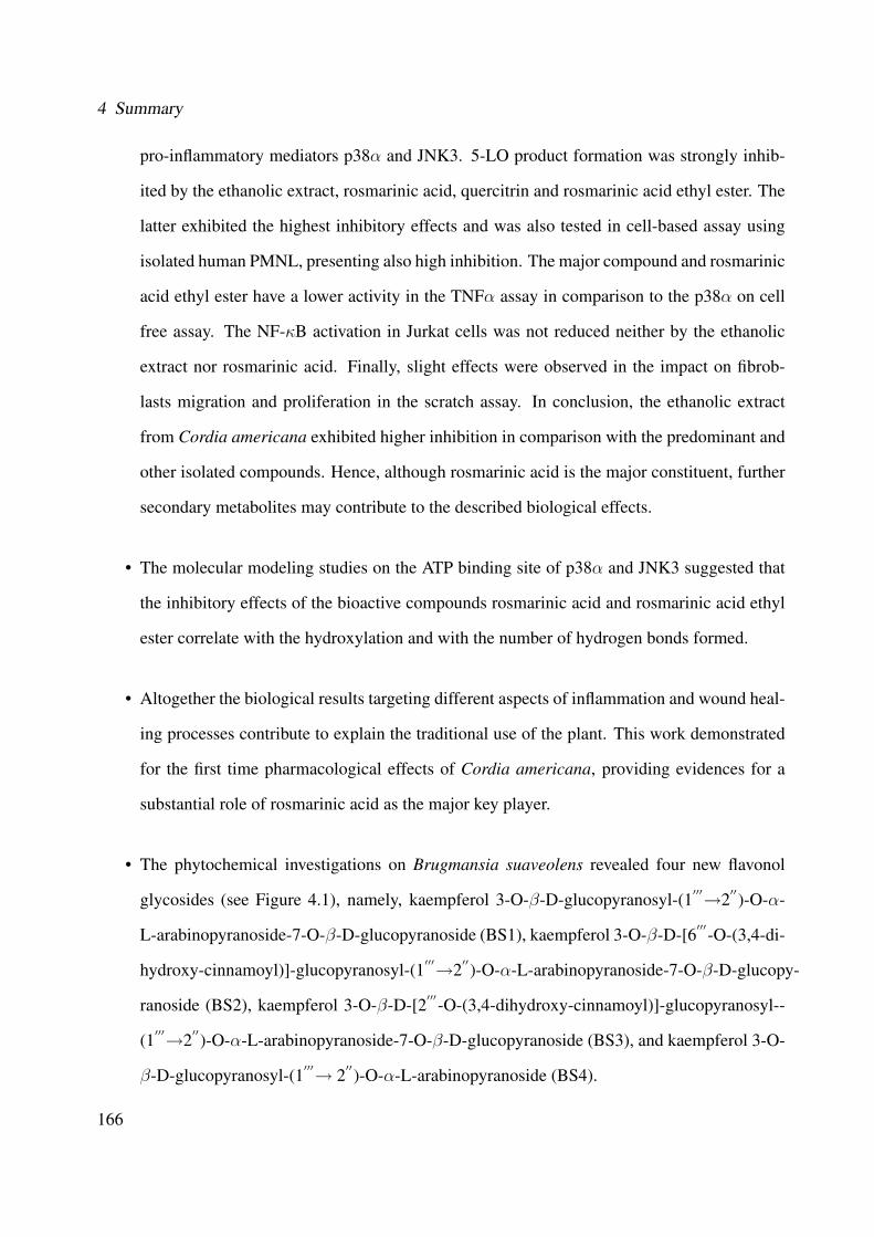
4 Summary
pro-inflammatory mediators p38α and JNK3. 5-LO product formation was strongly inhib-
ited by the ethanolic extract, rosmarinic acid, quercitrin and rosmarinic acid ethyl ester. The
latter exhibited the highest inhibitory effects and was also tested in cell-based assay using
isolated human PMNL, presenting also high inhibition. The major compound and rosmarinic
acid ethyl ester have a lower activity in the TNFα assay in comparison to the p38α on cell
free assay. The NF-κB activation in Jurkat cells was not reduced neither by the ethanolic
extract nor rosmarinic acid. Finally, slight effects were observed in the impact on fibrob-
lasts migration and proliferation in the scratch assay. In conclusion, the ethanolic extract
from Cordia americana exhibited higher inhibition in comparison with the predominant and
other isolated compounds. Hence, although rosmarinic acid is the major constituent, further
secondary metabolites may contribute to the described biological effects.
• The molecular modeling studies on the ATP binding site of p38α and JNK3 suggested that
the inhibitory effects of the bioactive compounds rosmarinic acid and rosmarinic acid ethyl
ester correlate with the hydroxylation and with the number of hydrogen bonds formed.
• Altogether the biological results targeting different aspects of inflammation and wound heal-
ing processes contribute to explain the traditional use of the plant. This work demonstrated
for the first time pharmacological effects of Cordia americana, providing evidences for a
substantial role of rosmarinic acid as the major key player.
• The phytochemical investigations on Brugmansia suaveolens revealed four new flavonol
glycosides (see Figure 4.1), namely, kaempferol 3-O-β-D-glucopyranosyl-(1′′′→2′′)-O-α-
L-arabinopyranoside-7-O-β-D-glucopyranoside (BS1), kaempferol 3-O-β-D-[6′′′-O-(3,4-di-
hydroxy-cinnamoyl)]-glucopyranosyl-(1′′′→2′′)-O-α-L-arabinopyranoside-7-O-β-D-glucopy-
ranoside (BS2), kaempferol 3-O-β-D-[2′′′-O-(3,4-dihydroxy-cinnamoyl)]-glucopyranosyl--
(1′′′→2′′)-O-α-L-arabinopyranoside-7-O-β-D-glucopyranoside (BS3), and kaempferol 3-O-
β-D-glucopyranosyl-(1′′′→ 2′′)-O-α-L-arabinopyranoside (BS4).
166
R1 R2 R3
(BS1) O-Glc OH (BS2) O-Glc O-Caffeoyl OH (BS3) O-Glc OH O-Caffeoyl (BS4) OH OH
R1
R3
R2
OH
OH
7
65
10
98
43
2O 1'
6'
2'3'
4'5'
OOH
OH
4'''
3'''2'''
1'''
O5'''
1''
2''3''
4''
5''O
OOH
OH
O
6'''
OHOH
Figure 4.1: Isolated flavonol glycosides from the ethanolic extract of Brugmansia suaveolens
• The biological studies on p38α, JNK3 as well as 5-LO with the ethanolic extract of Brug-
mansia suaveolens and the isolated flavonol glycosides exhibited moderate inhibitory effects.
The inhibition of the flavonol glycosides (i.e., BS1, BS2, BS3 and BS4) are probably due
to the kaempferol aglycone that presented also a moderate inhibition in the assays. Thus,
the difference in the activity might be influenced by the glycosylation, which increases the
molecular size and the polarity of the compounds. Therefore, it can be assumed that the
plant extract contains other secondary metabolites that were not identified, but they might
also contribute to the overall biological activity of the ethanolic extract from Brugmansia
suaveolens.
• A biosynthesis pathway was hypothesized for the isolated flavonol glycosides from Brug-
mansia suaveolens, considering that the acylation at position C-6′′′ (BS2) occurs frequently
and is widely known compared to the acylation at the position C-2′′′ (BS3). One may spec-
ulate that the biosynthesis of compound BS2 might be produced before BS3. Based on this
hypothesis, the possible biosynthesis pathway might follow: BS4→ BS1→ BS2→ BS3.
167


5 Experimental Part
This chapter presents the materials and methods applied in this work. More specifically, it de-
scribes in details the plant material and the methods for: plant extraction, isolation, quantification,
chromatography, spectroscopy and biological assays.
5.1 Plant Material
The leaves from Cordia americana and from Brugmansia suaveolens were collected in the re-
gion of Santa Maria, Rio Grande do Sul, Brazil in October 2007 and January 2008, respectively.
Cordia americana was authenticated by the botanist Solon J. Longhi and Brugmansia suaveolens
by Gilberto Zanetti. Voucher specimens of both plants are deposited in the herbarium of the Depart-
ment of Biology at the Santa Maria University, Brazil, under the reference number SMDB12308
(Cordia americana) and SMDB12520 (Brugmansia suaveolens).
169

5 Experimental Part
5.2 Chemicals, Reagents and MaterialsTable 5.1: Chemicals, reagents and materials
Material ManufacturerAcetone p.a Sigma-Aldrich, Germany
Acetonitrile HPLC grade Merck, GermanyDiethylamine p.a Sigma-Aldrich, Germany
Dimethylsulfoxide-d6 Euriso-Top GermanyEthylacetate p.a Sigma-Aldrich, GermanyFormic Acid p.a Sigma-Aldrich, Germany
Methanol-d4 Euriso-Top, GermanyMethanol HPLC grade Merck, Germany
Methanol p.a Brenntag Chemiepartner, Germanyp-Anisaldehyd 97% Acros Organics, Belgien
Pyridine-d5 Sigma-Aldrich, GermanySephadex®LH-20 (Bead size 25-100 µ) Sigma-Aldrich, Germany
Sulfuric acid (95-97%) p.a Sigma-Aldrich, GermanyToluen p.a VWR International, Germanyα-amyrin Extrasynth�ese, France
Rosmarinic acid Synthesized at University of Tubingen, GermanyQuercitrin Carl Roth, Germany
Kaempferol Extrasynth�ese, FranceCaffeic acid Carl Roth, GermanyHiosciamin Carl Roth, GermanyScopolamin Carl Roth, Germany
RP-18 LiChroprep RP-18 (25-40 µm) Merck, GermanyFluorescent tagged SiO2 60 F254 Merck, GermanyFluorescent tagged RP-18 F254 Merck, Germany
5.3 InstrumentsTable 5.2: Instruments
Instruments ManufacturerHairdryer Siemens, Germany
Fraction collector FRS Mini Manuel, GermanyMilli-Q Water Millipore Purification System, USA
Liofilizator Finn-Aqua Lyovac GT2, GermanyMillipore-Water System (MilliQ Plus) Billerica, MA, USA
Rotavapor Buchi, GermanyIce machine Wessamat Flake Line, Germany
Cabinet dryer WTB Binder , GermanyPhoto Camera Desaga UV/VIS Sarsted-Gruppe, Germany
Balance Kern Sohn, Germany
170
5.4 Chromatographic and Spectroscopic Methods
5.4 Chromatographic and Spectroscopic Methods
5.4.1 Thin Layer Chromatography (TLC)
Thin layer chromatography was used to control each fraction after separation procedures.
An amount of 5 µL up to 30 µL was manually applied for TLC plates in a band-shaped of 1
cm. The TLC plates were dried with a hair-dryer and run 8 cm using one of the methods (TLC-A),
(TLC-B) or (TLC-C). After that, plates were dried again and analyzed under white light, short-
wave (λ = 254 nm) and long-wave (λ = 366 nm). Additionally, plates were derivatized using
the anisaldehyde-sulfuric reagent. Finally, plates were photographed for documentation using the
Photo Camera Desaga UV/VIS system.
5.4.1.1 TLC Method for Cordia americana
• Method TLC-A:
– Mobile phase: ethyl acetate:methanol:water (77:15:8)
– Plate: Silica gel 60 F254
– Anisaldehyde-sulfuric reagent: 10 mL of sulfuric acid was carefully added to an ice-
cooled mixture of 170 mL methanol and 20 mL of acetic acid. To this solution, 1 mL
anisaldehyde was added. The plate was immersed in the reagent for 1 sec then heated
at 100 ◦C for 2-5 minutes.
– Examination: white light, UV λ = 366 nm.
5.4.1.2 TLC Methods for Brugmansia suaveolens
• Method TLC-B:
– Mobile phase: toluene:methanol:diethylamine (8:1:1)
– Plate: Silica gel 60 F254
171

5 Experimental Part
– Anisaldehyde-sulfuric reagent: see Section 5.4.1.1
– Examination: white light
• Method TLC-C
– Mobile phase: water:methanol(1:1)
– Plate: reverse phase RP-18 F254
– Anisaldehyde-sulfuric reagent: see Section 5.4.1.1
– Examination: white light
• Method TLC-D:
– Mobile phase: toluene:methanol:diethylamine (8:1:1)
– Plate: Silica gel 60 F254
– Dragendorff's reagent: solution A: 0.85 g of basic bismuth nitrate was dissolved in 10
mL acetic acid and 40 mL water under heating; solution B: 8 g potassium iodide was
dissolved in 30 mL water. Just before spraying, 1 mL of each solution was mixed with
4 mL of acetic acid and 20 mL of water.
– Examination: white light
172

5.4 Chromatographic and Spectroscopic Methods
5.4.2 Column Chromatography
5.4.2.1 Sephadex®LH-20
A total amount of 300 g of Sephadex®LH-20 was dissolved with 1,250 mL of methanol split
in three Erlenmeyer flasks. The solution was reposed for 12 h in order to expand. After that,
the solution was applied in the open column (i.e., 80 cm long and 6 cm diameter) avoiding the
formation of bubbles. The open column with Sephadex®LH-20 was reposed for 12 h in order to
form a homogeneous and tight packing.
Before separation of the plant extract, the solvent was moved, keeping 1 cm of methanol over
the top of the stationary phase. Finally, the plant extract was applied into the column and fractions
were collected with a manual fraction collector.
5.4.2.2 Open Column Chromatography (OC)
A column with 25 cm long and 1 cm diameter was used and at each reaction tube 2 mL was
collected. The following methods were used:
• Method OC-A: water:methanol (1:1)
• Method OC-B: water:methanol (2:1)
5.4.3 Flash Chromatography (FC)
• LaFlash System, FC 204 Fraction Collector (VWR International GmbH)
• UV-Filterphotometer with 200, 220, 254 and 280 nm (Labomatic Instruments AG)
• Pre-column with 10 cm length and 2 cm diameter
• Column with 20 cm length and 3 cm diameter
173
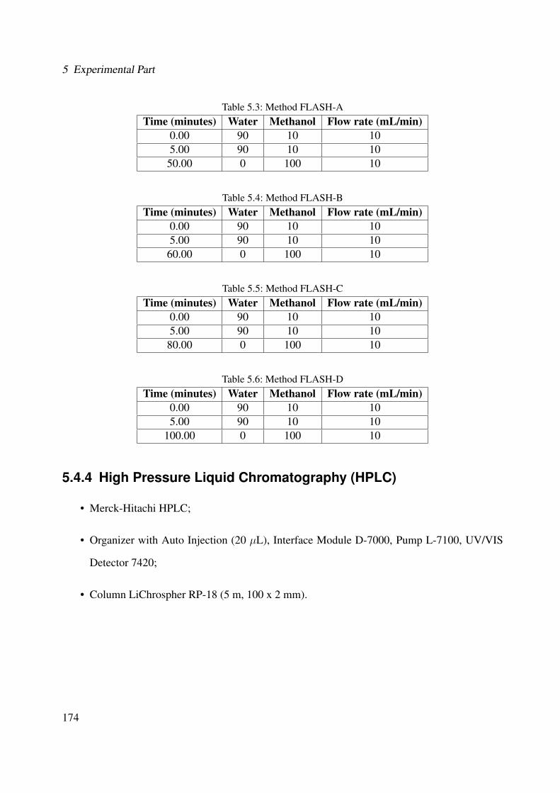
5 Experimental Part
Table 5.3: Method FLASH-ATime (minutes) Water Methanol Flow rate (mL/min)
0.00 90 10 105.00 90 10 10
50.00 0 100 10
Table 5.4: Method FLASH-BTime (minutes) Water Methanol Flow rate (mL/min)
0.00 90 10 105.00 90 10 10
60.00 0 100 10
Table 5.5: Method FLASH-CTime (minutes) Water Methanol Flow rate (mL/min)
0.00 90 10 105.00 90 10 10
80.00 0 100 10
Table 5.6: Method FLASH-DTime (minutes) Water Methanol Flow rate (mL/min)
0.00 90 10 105.00 90 10 10
100.00 0 100 10
5.4.4 High Pressure Liquid Chromatography (HPLC)
• Merck-Hitachi HPLC;
• Organizer with Auto Injection (20 µL), Interface Module D-7000, Pump L-7100, UV/VIS
Detector 7420;
• Column LiChrospher RP-18 (5 m, 100 x 2 mm).
174
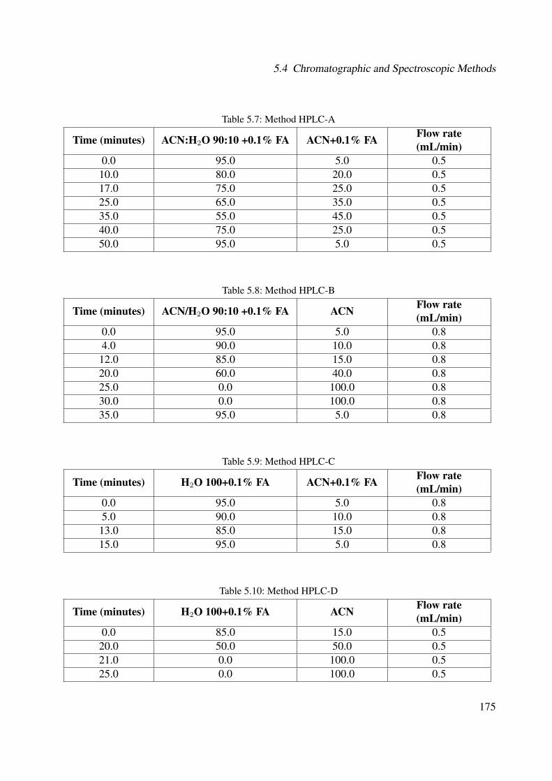
5.4 Chromatographic and Spectroscopic Methods
Table 5.7: Method HPLC-A
Time (minutes) ACN:H2O 90:10 +0.1% FA ACN+0.1% FA Flow rate(mL/min)
0.0 95.0 5.0 0.510.0 80.0 20.0 0.517.0 75.0 25.0 0.525.0 65.0 35.0 0.535.0 55.0 45.0 0.540.0 75.0 25.0 0.550.0 95.0 5.0 0.5
Table 5.8: Method HPLC-B
Time (minutes) ACN/H2O 90:10 +0.1% FA ACN Flow rate(mL/min)
0.0 95.0 5.0 0.84.0 90.0 10.0 0.812.0 85.0 15.0 0.820.0 60.0 40.0 0.825.0 0.0 100.0 0.830.0 0.0 100.0 0.835.0 95.0 5.0 0.8
Table 5.9: Method HPLC-C
Time (minutes) H2O 100+0.1% FA ACN+0.1% FA Flow rate(mL/min)
0.0 95.0 5.0 0.85.0 90.0 10.0 0.813.0 85.0 15.0 0.815.0 95.0 5.0 0.8
Table 5.10: Method HPLC-D
Time (minutes) H2O 100+0.1% FA ACN Flow rate(mL/min)
0.0 85.0 15.0 0.520.0 50.0 50.0 0.521.0 0.0 100.0 0.525.0 0.0 100.0 0.5
175

5 Experimental Part
5.4.5 UV-Visible Spectroscopy
• HPLC-DAD Hewlett Packard HP 1090
• Column Specification: ZORBAX Eclipse XDB-C8 (4.6 x 150 mm, 5 µm)
• Pump: Merck Hitachi (Darmstadt)
• Software: Shimazdu Client Server 7.2.1 SPI
5.4.6 Fourier Transform-Infrared Spectroscopy (FT-IR)
• Perkin Elmer Spectrum One (ATR Technology)
• Software: Graph Server v 1.60
5.4.7 Mass Spectroscopy
5.4.7.1 Gas Chromatography-Mass Spectrometry (GC-MS)
• Method GC-MS1:
• GC-MS System (Agilent 6890 series ) with natural compound library (NIST MS Search
Program version 1.7A);
• Capillar-column Rtx-1 MS (Fa. Restek; 25 m; 0.25 mm diameter; 0.25 µm Film Thickness-
Dimethylsiloxane; Carrier Gas: Helium);
• Agilent 5973 Network Mass Selective Detector;
• Injector 7683 Series;
1GC-MS was performed by C. Schmidt at the Department of Pharmaceutical Biology and Biotechnology, Universityof Freiburg.
176
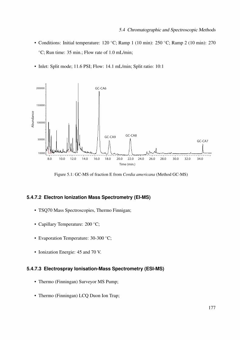
5.4 Chromatographic and Spectroscopic Methods
• Conditions: Initial temperature: 120 ◦C; Ramp 1 (10 min): 250 ◦C; Ramp 2 (10 min): 270
◦C; Run time: 35 min.; Flow rate of 1.0 mL/min;
• Inlet: Split mode; 11.6 PSI; Flow: 14.1 mL/min; Split ratio: 10:1
8.0 10.0 12.0 14.0 16.0 18.0 20.0 22.0 24.0 26.0 28.0 30.0 32.0 34.0
GC-CA6
GC-CA7GC-CA9 GC-CA8
Abu
ndan
ce
Time (min.)
10000
50000
100000
150000
200000
Figure 5.1: GC-MS of fraction E from Cordia americana (Method GC-MS)
5.4.7.2 Electron Ionization Mass Spectrometry (EI-MS)
• TSQ70 Mass Spectroscopies, Thermo Finnigan;
• Capillary Temperature: 200 ◦C;
• Evaporation Temperature: 30-300 ◦C;
• Ionization Energie: 45 and 70 V.
5.4.7.3 Electrospray Ionisation-Mass Spectrometry (ESI-MS)
• Thermo (Finningan) Surveyor MS Pump;
• Thermo (Finningan) LCQ Duon Ion Trap;
177
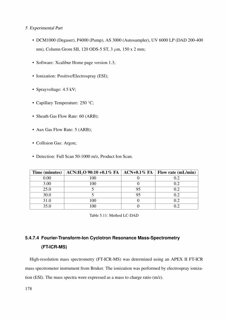
5 Experimental Part
• DCM1000 (Degaser), P4000 (Pump), AS 3000 (Autosampler), UV 6000 LP (DAD 200-400
nm), Column Grom SIL 120 ODS-5 ST, 3 µm, 150 x 2 mm;
• Software: Xcalibur Home page version 1.3;
• Ionization: Positive/Electrospray (ESI);
• Sprayvoltage: 4.5 kV;
• Capillary Temperature: 250 ◦C;
• Sheath Gas Flow Rate: 60 (ARB);
• Aux Gas Flow Rate: 5 (ARB);
• Collision Gas: Argon;
• Detection: Full Scan 50-1000 m/z, Product Ion Scan.
Time (minutes) ACN:H2O 90:10 +0.1% FA ACN+0.1% FA Flow rate (mL/min)0.00 100 0 0.23.00 100 0 0.225.0 5 95 0.230.0 5 95 0.231.0 100 0 0.235.0 100 0 0.2
Table 5.11: Method LC-DAD
5.4.7.4 Fourier-Transform-Ion Cyclotron Resonance Mass-Spectrometry
(FT-ICR-MS)
High-resolution mass spectrometry (FT-ICR-MS) was determined using an APEX II FT-ICR
mass spectrometer instrument from Bruker. The ionization was performed by electrospray ioniza-
tion (ESI). The mass spectra were expressed as a mass to charge ratio (m/z).
178
5.5 Plant Extraction Methods for the Biological Screening Phase
5.4.8 Nuclear Magnetic Resonance Spectroscopy (NMR)
Cordia americana
For the structural elucidation of the isolated compounds of this plant, the following NMR
1D (1H, 13C and DEPT-135) and 2D (H-H-COSY) were carried out using the following instru-
ments: Bruker Avance ARX-250; (Bruker S.A., Wissembourg, France); Bruker Avance DMX-400;
(Bruker S.A., Wissembourg, France).
Brugmansia suaveolens
In order to elucidate the isolated compounds of this plant, the 1D (1H, 13C and DEPT-135) and
2D NMR (H-H-COSY, HSQC, HMBC) were carried out with: Bruker AMX 600.13 MHz Spec-
trometer; Magnetic field strength of 14.1 Tesla; Micro-probe was an inverse 1H/13C micro volume
flow probe with 1.5 µL active detection configuration in solenoids (Protasis Corp., Marlboro, MA,
USA); Bruker Avance ARX-250; (Bruker S.A., Wissembourg, France).
5.5 Plant Extraction Methods for the Biological
Screening PhaseFor the biological screening phase, the selected parts of the plants were dried, grounded and
extracted using soxhlet, ultrasound or maceration. The soxhlet extraction was performed with 25 g
of plant material, using at first n-hexane (250 mL), and after drying, ethanol (250 mL). 10 g were
taken for the ultrasound extraction using n-hexane (100 mL), followed by ethanol (100 mL). The
maceration process was carried out with 438.5 g of Sedum dendroideum and 329.5 g of Kalanchoe
tubi�ora. Each solvent was applied twice directly to the grounded plant material during 16 days
changing the solvent each 8 days. Firstly, hexane was used for 16 days, followed by ethanol with
the same material for another 16 days. Extraction was exhaustively carried out in each case. The
solvents were removed under vacuum at 40 ◦C. Finally, extracts were lyophilised. Figure 5.2
depicts the extraction procedures.
179

5 Experimental Part
(A) Collection of Plants
(B) Preparetion of the Plant Material
(C) Soxhlet Extraction (D) Ultrasonic Extraction
(E) Hexanic and Ethanolic Extracts
Figure 5.2: Plant extraction flow
180
5.6 Extraction and Isolation Methods
5.6 Extraction and Isolation Methods
5.6.1 Cordia americana
The air-dried and powdered leaves (1140.94 g) of Cordia americana were exhaustively extracted
with ethanol in a soxhlet apparatus. The resulting ethanolic extract was concentrated under vac-
uum at 40 ◦C and finally lyophilisated to yield 227.7 g of extract, that is, 19.90% of the original
powdered leaves. The ethanolic extract was defatted resulting in 219.6 g.
The defatting process was carried out by dissolving the ethanolic extract of Cordia americana
in methanol. This solution was left for 48 h in the refrigerator at -20 ◦C. After that, it was filtered,
evaporated and lyophilisated.
In the next step, as shown in Figure 5.3, an amount of 6.0 g of the defatted ethanolic extract
was diluted in 20 mL of methanol. This solution was subjected to column chromatography using
Sephadex®LH-20 (see Section 5.4.2.1) and 100% methanol as mobile phase, with a flow rate of
1.0 mL/min. The fractions were collected in reaction tubes with 10 mL resulting in a total of 282
tubes. After TLC control with Method TLC-A (see Section 5.4.1.1) for detection, the tubes with a
similar composition were combined and 16 fractions (A-P) were obtained, as shown in Figure 5.4.
The yield of each fraction is shown in Table 5.12.
The fraction sets were investigated for the inhibition on p38α assay (see Section 5.7.1) in a
concentration of 30 µg/mL. Additionally, HPLC analysis of the ethanolic extract in different wave
lengths (see Figure 5.5) revealed the presence of a major peak and a few secondary peaks. Thus,
the criteria to choose the fractions for further subfractionation was based on:
• inhibitory activity considering the results on p38α assay (bioguided investigation);
• major and secondary HPLC peaks;
• yields of fraction sets.
Therefore, the fractions E, F, G, H, I and K were further studied.
181

5 Experimental Part
Flas
h C
C, R
P-18
, MeO
H/H
2O
grad
ient
1140
.94
g of
Cor
dia
amer
ican
a le
aves
sito
ster
ol,
cam
pest
erol
α,
β-a
myr
ine
quer
citri
n
rosm
arin
ic
acid
eth
yl e
ster
ru
tin
3-(3
,4-
dihy
drox
y-ph
enyl
)-2-
hydr
oxy-
prop
anoi
c ac
id
Flas
h C
C, R
P-
18,M
eOH
/H2O
gr
adie
nt
Flas
h C
C, R
P-18
,MeO
H/H
2O
grad
ient
227.
7g
of
etha
nolic
ext
ract
219.
6g
of
defa
tted
extr
act
16 fr
act.
(A-P
)of
282
tube
s
Soxh
let e
xtra
ctio
n(E
tOH
) Fa
t rem
ovin
g (M
eOH
-20º
C)
Frac
tiona
tion
of 6
g(S
epha
dex
LH-2
0;
MeO
H)
Frac
tion
I87
-94
(5.1
mg)
Frac
tion
F55
-67
(502
.5 m
g)
Frac
tion
E45
-54
(614
mg)
Frac
tion
K11
0-13
5 (4
06.1
mg)
Frac
tion
G68
-79
(258
mg)
Flas
h C
C, R
P-18
,MeO
H/H
2O
grad
ient
,an
alyt
ical
HP
LC
Frac
tion
H80
-86
(81
mg)
rosm
arin
ic
acid
Figure 5.3: Extraction and isolation of compounds from the ethanolic extract of the leaves of Cordia ameri-cana. Cursive letters: compounds identified from the fractions; Bold letters: isolated compounds
182
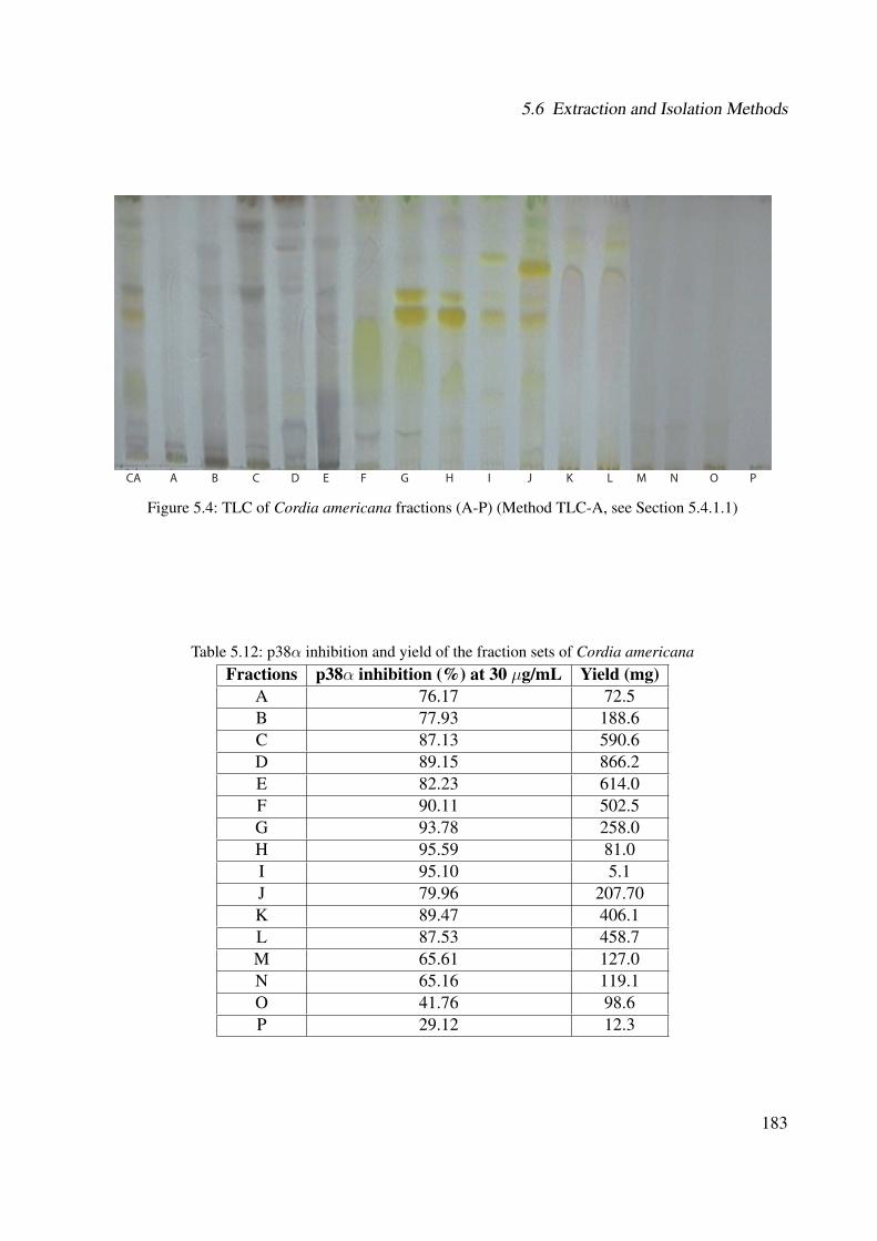
5.6 Extraction and Isolation Methods
CA A B C D E F G H I J K L M N O P
Figure 5.4: TLC of Cordia americana fractions (A-P) (Method TLC-A, see Section 5.4.1.1)
Table 5.12: p38α inhibition and yield of the fraction sets of Cordia americanaFractions p38α inhibition (%) at 30 µg/mL Yield (mg)
A 76.17 72.5B 77.93 188.6C 87.13 590.6D 89.15 866.2E 82.23 614.0F 90.11 502.5G 93.78 258.0H 95.59 81.0I 95.10 5.1J 79.96 207.70K 89.47 406.1L 87.53 458.7M 65.61 127.0N 65.16 119.1O 41.76 98.6P 29.12 12.3
183

5 Experimental Part
3,10
3,75 7,5
9
9,28
11,50
13,97
16,52 17,
56
19,41
20,58
22,11
23,95
25,34
27,55
28,41
30,21
31,16
31,97
33,56
35,97
37,48
39,21
2,48
3,45 4,73 7,62
9,28
14,11 16,42
18,46
19,50 20,32
22,39
23,58 24,
9326,
1127,
4528,
40 29,35
31,47
32,36
34,49
3,23
4,69 8,4
4
10,72 12,11 14,
64 17,59
18,55 19,35
22,14
24,07
25,64
26,62
27,43 28,83
29,77
31,89
33,76
34,97
36,65 37,28
38,68
3,22
11,83 14,
35 17,75
18,84 19,89
22,70
24,53
25,91
26,80
28,11 28,92
32,08
37,01
38,25
3,21 14,
38 17,94
18,85 19,71
20,89
22,45
24,32
25,75
26,70
27,98 28,80
31,95
36,68
220 nm
250 nm
280 nm
330 nm
350 nm
Retention Time (min)
Retention Time (min)
Retention Time (min)
Retention Time (min)
Retention Time (min)
Inte
nsity
(mV)
Inte
nsity
(mV)
Inte
nsity
(mV)
Inte
nsity
(mV)
Inte
nsity
(mV)
500
400
300200
100 0
-100
-200
0 5 10 15 20 25 30 35 40 45 50
0 5 10 15 20 25 30 35 40 45 50
0 5 10 15 20 25 30 35 40 45 50
0 5 10 15 20 25 30 35 40 45 50
0 5 10 15 20 25 30 35 40 45 50
250
200
150
100
50
0
250
200
150
100
50
0
250
200
150
100
50 0
300
250
200
150
100
50
0
Figure 5.5: Representative analytical HPLC of the ethanolic extract of Cordia americana in different wavelengths (Method HPLC-A, see Section 5.4.4)
184

5.6 Extraction and Isolation Methods
5.6.1.1 Isolation of Compounds
The following procedures were carried out in order to isolate the compounds from the plant
extract:
• Rosmarinic acid (CA1): Parts of the active fraction K (100 mg) were subfractionated by
flash chromatography (Method FLASH-A, Section 5.4.3) over a RP-18 (25-40 µm) column
using methanol-water as mobile phase with a linear gradient starting at 10% methanol to
100% within 50 min and a flow rate of 10 mL/min. This process provided 81 fractions,
whereas 5 of them yielded the isolation of rosmarinic acid (12 mg) with a purity of 98.81%.
• Rosmarinic acid ethyl ester (CA2): Separation of fraction H (81 mg) with methanol-water
as eluate with a linear gradient starting at 10% methanol to 100% within 50 min (Method
FLASH-A, Section 5.4.3) followed by a further purification in the analytical HPLC (Method
HPLC-D, Section 5.4.4), which yielded the isolation of rosmarinic acid ethyl ester (3.3 mg)
with a purity of 95.70%.
• 3-(3,4-dihydroxyphenyl)-2-hydroxypropanoic acid (CA3): Parts of fraction F (101 mg)
were subfractionated by methanol-water as mobile phase with a linear gradient of 10%
methanol to 100% within 80 min (Method FLASH-B, Section 5.4.3) and 5 mg of 3-(3,4-
dihydroxyphenyl)-2-hydroxypropanoic acid (syn. danshensu) were obtained with a purity of
93.26%.
• Rutin (CA4): Fraction G (120.04 mg) were subfractionated by methanol-water with a linear
gradient from 10% methanol to 100% over 100 min (Method FLASH-C, Section 5.4.3) to
provide rutin (15 mg) with a purity of 94.72%.
5.6.1.2 Characterization of the Compounds
The isolated as well as the identified compounds showed the following characteristic features:
185
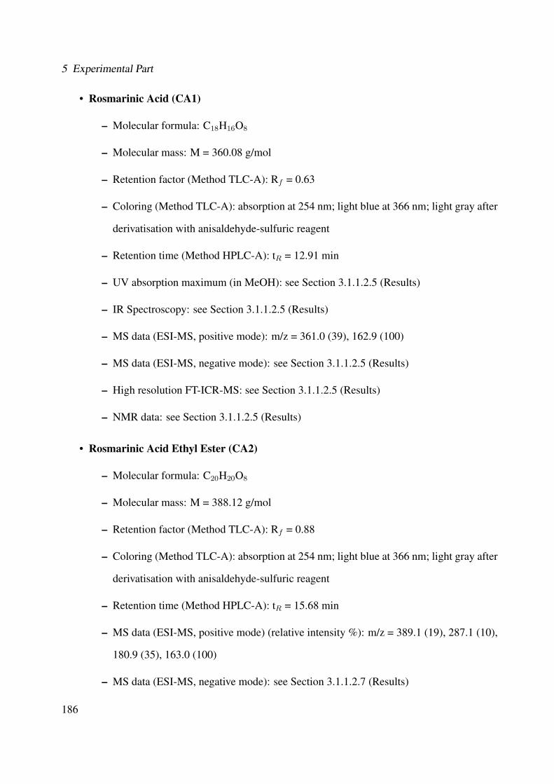
5 Experimental Part
• Rosmarinic Acid (CA1)
– Molecular formula: C18H16O8
– Molecular mass: M = 360.08 g/mol
– Retention factor (Method TLC-A): Rf = 0.63
– Coloring (Method TLC-A): absorption at 254 nm; light blue at 366 nm; light gray after
derivatisation with anisaldehyde-sulfuric reagent
– Retention time (Method HPLC-A): tR = 12.91 min
– UV absorption maximum (in MeOH): see Section 3.1.1.2.5 (Results)
– IR Spectroscopy: see Section 3.1.1.2.5 (Results)
– MS data (ESI-MS, positive mode): m/z = 361.0 (39), 162.9 (100)
– MS data (ESI-MS, negative mode): see Section 3.1.1.2.5 (Results)
– High resolution FT-ICR-MS: see Section 3.1.1.2.5 (Results)
– NMR data: see Section 3.1.1.2.5 (Results)
• Rosmarinic Acid Ethyl Ester (CA2)
– Molecular formula: C20H20O8
– Molecular mass: M = 388.12 g/mol
– Retention factor (Method TLC-A): Rf = 0.88
– Coloring (Method TLC-A): absorption at 254 nm; light blue at 366 nm; light gray after
derivatisation with anisaldehyde-sulfuric reagent
– Retention time (Method HPLC-A): tR = 15.68 min
– MS data (ESI-MS, positive mode) (relative intensity %): m/z = 389.1 (19), 287.1 (10),
180.9 (35), 163.0 (100)
– MS data (ESI-MS, negative mode): see Section 3.1.1.2.7 (Results)
186

5.6 Extraction and Isolation Methods
– High resolution FT-ICR-MS: see Section 3.1.1.2.7 (Results)
– NMR data: see Section 3.1.1.2.7 (Results)
• 3-(3,4-dihydroxyphenyl)-2-hydroxypropanoic acid (CA3)
– Molecular formula: C9H10O5
– Molecular mass: M = 198.1 g/mol
– Retention factor (Method TLC-A): Rf = 0.44
– Coloring (Method TLC-A): no absorption at 254 nm; blue at 366 nm; beige after
derivatisation with anisaldehyde-sulfuric reagent
– Retention time (Method HPLC-A): tR = 4.01 min
– MS data (EI-MS): see Section 3.1.1.2.1 (Results)
– NMR data: see Section 3.1.1.2.1 (Results)
• Rutin (CA4)
– Molecular formula: C27H30O16
– Molecular mass: M = 610.15 g/mol
– Retention factor (Method TLC-A): Rf = 0.54
– Coloring (Method TLC-A): absorption at 254 nm; dark blue at 366 nm; yellow after
derivatisation with anisaldehyde-sulfuric reagent
– Retention time (Method HPLC-A): tR = 10.93 min
– UV absorption maximum (in MeOH): see Section 3.1.1.2.4 (Results)
– IR Spectroscopy: see Section 3.1.1.2.4 (Results)
– MS data (ESI-MS, positive mode): see Section 3.1.1.2.4 (Results)
– MS data (ESI-MS, negative mode): m/z = 610.2 (41), 609.1 (100), 301.1 (10), 147.2
(5)
187
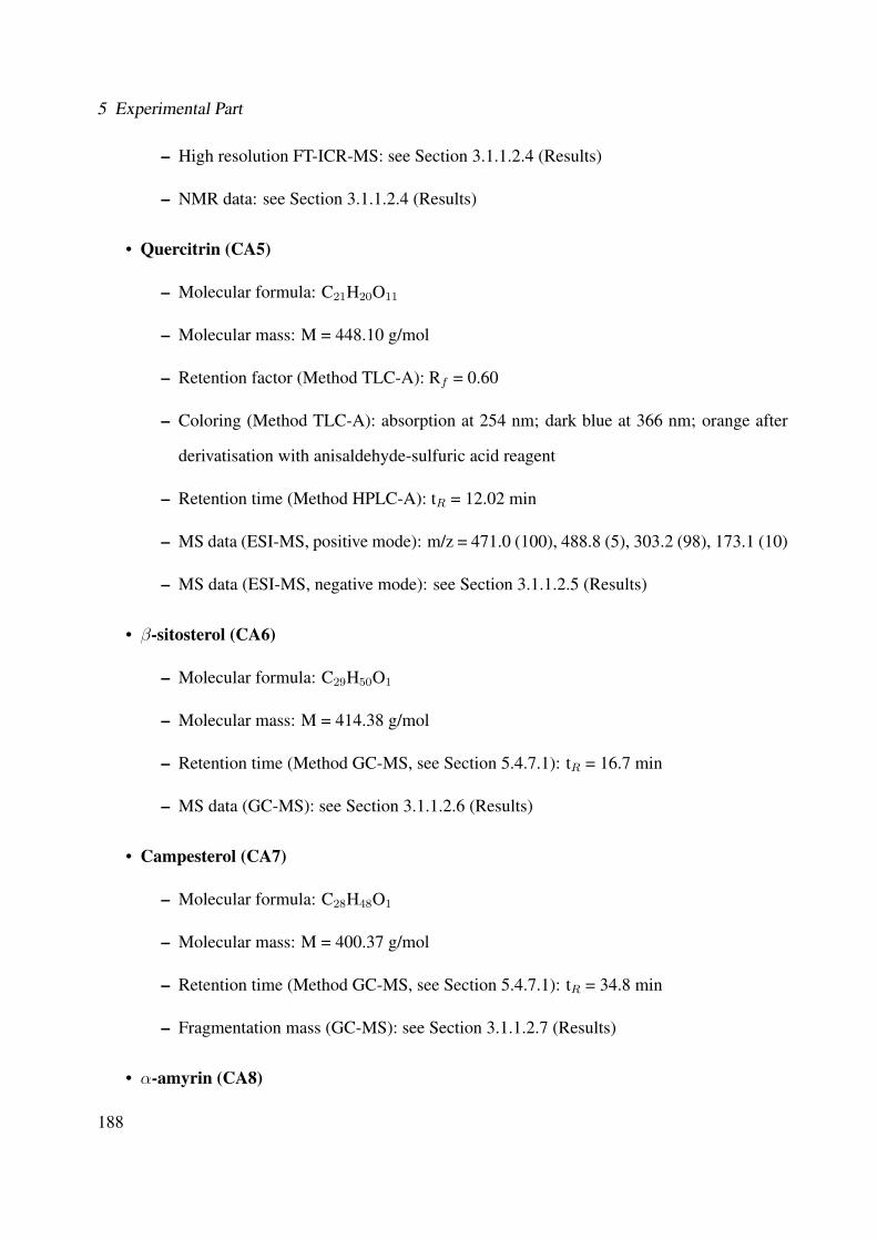
5 Experimental Part
– High resolution FT-ICR-MS: see Section 3.1.1.2.4 (Results)
– NMR data: see Section 3.1.1.2.4 (Results)
• Quercitrin (CA5)
– Molecular formula: C21H20O11
– Molecular mass: M = 448.10 g/mol
– Retention factor (Method TLC-A): Rf = 0.60
– Coloring (Method TLC-A): absorption at 254 nm; dark blue at 366 nm; orange after
derivatisation with anisaldehyde-sulfuric acid reagent
– Retention time (Method HPLC-A): tR = 12.02 min
– MS data (ESI-MS, positive mode): m/z = 471.0 (100), 488.8 (5), 303.2 (98), 173.1 (10)
– MS data (ESI-MS, negative mode): see Section 3.1.1.2.5 (Results)
• β-sitosterol (CA6)
– Molecular formula: C29H50O1
– Molecular mass: M = 414.38 g/mol
– Retention time (Method GC-MS, see Section 5.4.7.1): tR = 16.7 min
– MS data (GC-MS): see Section 3.1.1.2.6 (Results)
• Campesterol (CA7)
– Molecular formula: C28H48O1
– Molecular mass: M = 400.37 g/mol
– Retention time (Method GC-MS, see Section 5.4.7.1): tR = 34.8 min
– Fragmentation mass (GC-MS): see Section 3.1.1.2.7 (Results)
• α-amyrin (CA8)
188
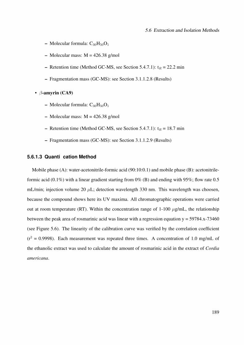
5.6 Extraction and Isolation Methods
– Molecular formula: C30H50O1
– Molecular mass: M = 426.38 g/mol
– Retention time (Method GC-MS, see Section 5.4.7.1): tR = 22.2 min
– Fragmentation mass (GC-MS): see Section 3.1.1.2.8 (Results)
• β-amyrin (CA9)
– Molecular formula: C30H50O1
– Molecular mass: M = 426.38 g/mol
– Retention time (Method GC-MS, see Section 5.4.7.1): tR = 18.7 min
– Fragmentation mass (GC-MS): see Section 3.1.1.2.9 (Results)
5.6.1.3 Quanti�cation Method
Mobile phase (A): water-acetonitrile-formic acid (90:10:0.1) and mobile phase (B): acetonitrile-
formic acid (0.1%) with a linear gradient starting from 0% (B) and ending with 95%; flow rate 0.5
mL/min; injection volume 20 µL; detection wavelength 330 nm. This wavelength was choosen,
because the compound shows here its UV maxima. All chromatographic operations were carried
out at room temperature (RT). Within the concentration range of 1-100 µg/mL, the relationship
between the peak area of rosmarinic acid was linear with a regression equation y = 59784.x-73460
(see Figure 5.6). The linearity of the calibration curve was verified by the correlation coefficient
(r2 = 0.9998). Each measurement was repeated three times. A concentration of 1.0 mg/mL of
the ethanolic extract was used to calculate the amount of rosmarinic acid in the extract of Cordia
americana.
189
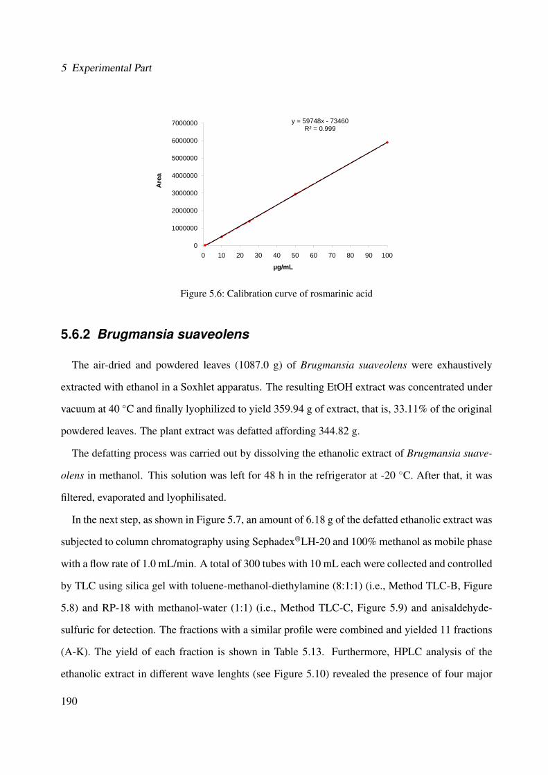
5 Experimental Part
y = 59748x - 73460R² = 0.999
6000000
7000000
4000000
5000000a
3000000
4000000
Are
a
1000000
2000000
00 10 20 30 40 50 60 70 80 90 100
µg/mL
Figure 5.6: Calibration curve of rosmarinic acid
5.6.2 Brugmansia suaveolens
The air-dried and powdered leaves (1087.0 g) of Brugmansia suaveolens were exhaustively
extracted with ethanol in a Soxhlet apparatus. The resulting EtOH extract was concentrated under
vacuum at 40 ◦C and finally lyophilized to yield 359.94 g of extract, that is, 33.11% of the original
powdered leaves. The plant extract was defatted affording 344.82 g.
The defatting process was carried out by dissolving the ethanolic extract of Brugmansia suave-
olens in methanol. This solution was left for 48 h in the refrigerator at -20 ◦C. After that, it was
filtered, evaporated and lyophilisated.
In the next step, as shown in Figure 5.7, an amount of 6.18 g of the defatted ethanolic extract was
subjected to column chromatography using Sephadex®LH-20 and 100% methanol as mobile phase
with a flow rate of 1.0 mL/min. A total of 300 tubes with 10 mL each were collected and controlled
by TLC using silica gel with toluene-methanol-diethylamine (8:1:1) (i.e., Method TLC-B, Figure
5.8) and RP-18 with methanol-water (1:1) (i.e., Method TLC-C, Figure 5.9) and anisaldehyde-
sulfuric for detection. The fractions with a similar profile were combined and yielded 11 fractions
(A-K). The yield of each fraction is shown in Table 5.13. Furthermore, HPLC analysis of the
ethanolic extract in different wave lenghts (see Figure 5.10) revealed the presence of four major
190
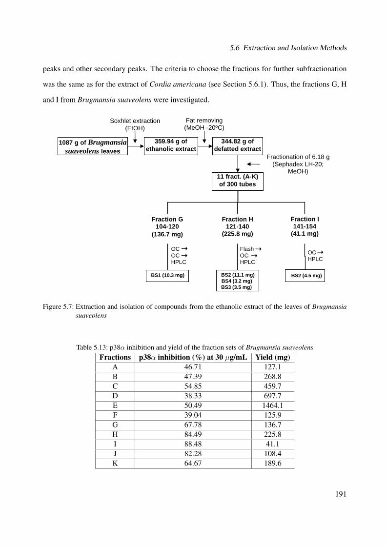
5.6 Extraction and Isolation Methods
peaks and other secondary peaks. The criteria to choose the fractions for further subfractionation
was the same as for the extract of Cordia americana (see Section 5.6.1). Thus, the fractions G, H
and I from Brugmansia suaveolens were investigated.
1087 g of Brugmansia suaveolens leaves
BS1 (10.3 mg)
359.94 g of ethanolic extract
344.82 g of defatted extract
11 fract. (A-K)of 300 tubes
Soxhlet extraction(EtOH)
Fat removing (MeOH -20ºC)
Fractionation of 6.18 g(Sephadex LH-20;
MeOH)
Fraction H121-140
(225.8 mg)
Fraction G104-120
(136.7 mg)
Fraction I141-154
(41.1 mg)
OCHPLC
FlashOCHPLC
OC OCHPLC
BS2 (11.1 mg) BS4 (3.2 mg) BS3 (3.5 mg)
BS2 (4.5 mg)
Figure 5.7: Extraction and isolation of compounds from the ethanolic extract of the leaves of Brugmansiasuaveolens
Table 5.13: p38α inhibition and yield of the fraction sets of Brugmansia suaveolensFractions p38α inhibition (%) at 30 µg/mL Yield (mg)
A 46.71 127.1B 47.39 268.8C 54.85 459.7D 38.33 697.7E 50.49 1464.1F 39.04 125.9G 67.78 136.7H 84.49 225.8I 88.48 41.1J 82.28 108.4K 64.67 189.6
191
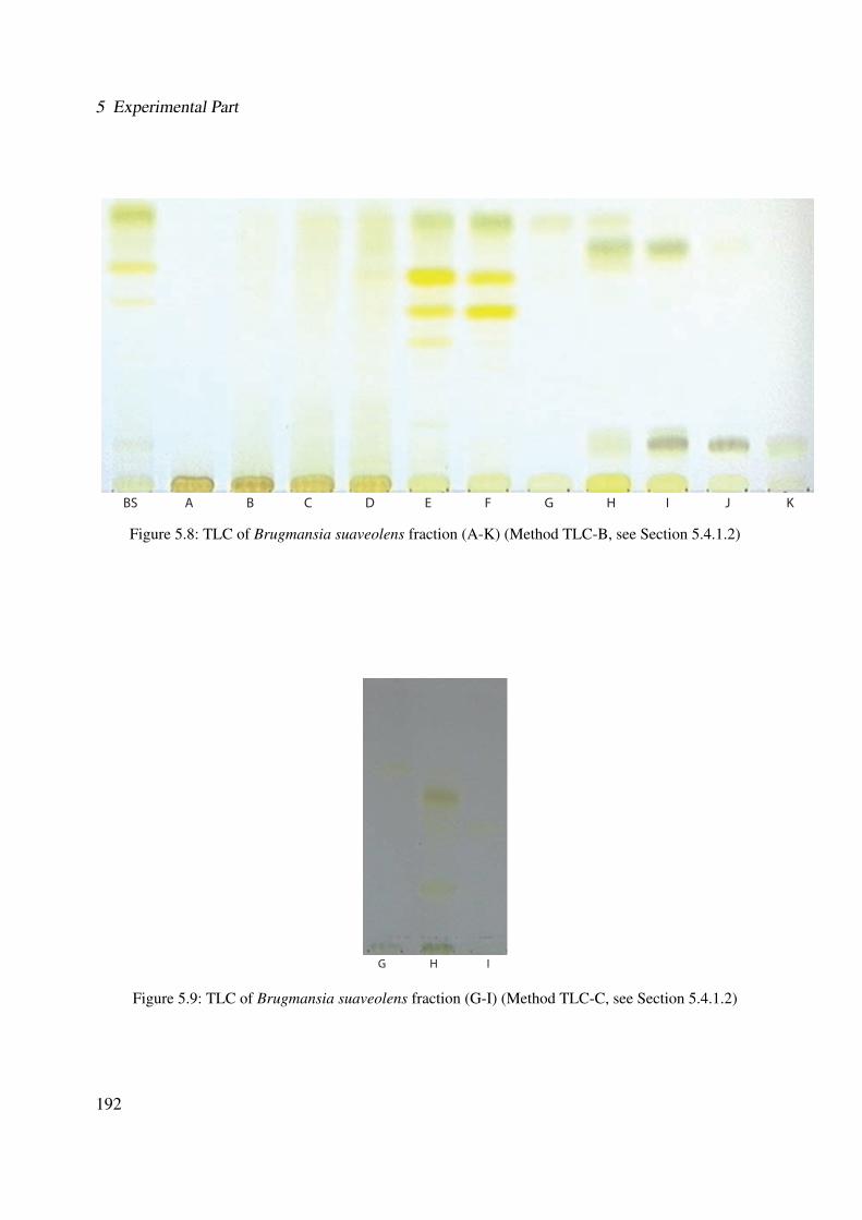
5 Experimental Part
BS A B C D E F G H I J K
Figure 5.8: TLC of Brugmansia suaveolens fraction (A-K) (Method TLC-B, see Section 5.4.1.2)
A B C D E F G H I J K
Figure 5.9: TLC of Brugmansia suaveolens fraction (G-I) (Method TLC-C, see Section 5.4.1.2)
192

5.6 Extraction and Isolation Methods
0 5 10 15 20 25 30 35Time (min)
0
50000
100000
150000
200000
250000
uAU
220 nm9.49
8.672.5211.48
11.87 32.2722.71
31.5529.1324.2913.0222.1414.30
8.465.34
0 5 10 15 20 25 30 35Time (min)
0
500000
1000000
1500000
uAU
254 nm9.50
2.528.67
9.66 11.49
12.44
32.2722.71 27.6025.7517.558.36 19.26
0 5 10 15 20 25 30 35Time (min)
0
500000
1000000
1500000
uAU
280 nm9.50
2.528.67 9.66
11.48
13.37 32.2714.71 18.11 25.758.44 27.6119.12
0 5 10 15 20 25 30 35Time (min)
0
500000
1000000
uAU
9.51330 nm
8.679.66
11.49
32.2812.46
18.112.53 14.00 31.5519.788.38 21.64 27.15
0 5 10 15 20 25 30 35Time (min)
0
500000
1000000
uAU
9.50350 nm
8.679.66
11.49
32.2712.3131.552.53 19.798.08 22.17
Figure 5.10: Representative HPLC chromatogram of the ethanolic extract of Brugmansia suaveolens in dif-ferent wave lengths (Method LC-DAD)
193
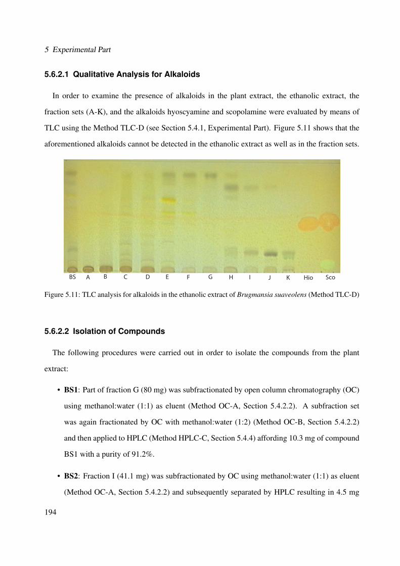
5 Experimental Part
5.6.2.1 Qualitative Analysis for Alkaloids
In order to examine the presence of alkaloids in the plant extract, the ethanolic extract, the
fraction sets (A-K), and the alkaloids hyoscyamine and scopolamine were evaluated by means of
TLC using the Method TLC-D (see Section 5.4.1, Experimental Part). Figure 5.11 shows that the
aforementioned alkaloids cannot be detected in the ethanolic extract as well as in the fraction sets.
BS A B C D E F G H I J K Hio Sco
Figure 5.11: TLC analysis for alkaloids in the ethanolic extract of Brugmansia suaveolens (Method TLC-D)
5.6.2.2 Isolation of Compounds
The following procedures were carried out in order to isolate the compounds from the plant
extract:
• BS1: Part of fraction G (80 mg) was subfractionated by open column chromatography (OC)
using methanol:water (1:1) as eluent (Method OC-A, Section 5.4.2.2). A subfraction set
was again fractionated by OC with methanol:water (1:2) (Method OC-B, Section 5.4.2.2)
and then applied to HPLC (Method HPLC-C, Section 5.4.4) affording 10.3 mg of compound
BS1 with a purity of 91.2%.
• BS2: Fraction I (41.1 mg) was subfractionated by OC using methanol:water (1:1) as eluent
(Method OC-A, Section 5.4.2.2) and subsequently separated by HPLC resulting in 4.5 mg
194
5.6 Extraction and Isolation Methods
of compound BS2 (Method HPLC-C, Section 5.4.4). Parts of fraction H (100 mg) were
subfractionated by flash chromatography with methanol:water as mobile phase with a linear
gradient starting from 10% methanol to 100% within 60 minutes (Method FLASH-D, Sec-
tion 5.4.3). A subfraction set (25.2 mg) was applied to HPLC (Method HPLC-C, Section
5.4.4) affording 11.1 mg of compound BS2 with a purity of 93.2%.
• BS3: From the aforementioned fraction H, the subfraction (35.2 mg) was applied to HPLC
affording 3.5 mg (Method HPLC-C, Section 5.4.4) of compound BS3 with a purity of 91.0%.
• BS4: Additionally from the fraction H (100 mg) one subfraction (47.8 mg) was further sep-
arated by OC using methanol:water (1:1) as mobile phase (Method OC-A, Section 5.4.2.2)
yielding 3.2 mg of compound BS4 with a purity of 90.1%.
5.6.2.3 Characterization of the Compounds
The elucidated compounds from Brugmansia suaveolens had the following characteristic fea-
tures:
• BS1
– Molecular formula: C26H28O15
– Molecular mass: M = 742.2 g/mol
– Retention factor (Method-TLC-C): Rf = 0.65
– Coloring (Method-TLC-C): absorption at 254 nm; dark blue at 366 nm; yellow after
derivatisation with anisaldehyde-sulfuric reagent
– Retention time (Method-HPLC-B): tR = 7.21 min
– UV absorption maximum (in MeOH): see Section 3.1.2.2.2 (Results)
– IR Spectroscopy: see Section 3.1.2.2.2 (Results)
– MS data (ESI-MS, positive mode): see Section 3.1.2.2.2 (Results)
195

5 Experimental Part
– MS data (ESI-MS, negative mode): m/z = 742.2 (51), 741.0 (100), 579.1 (19), 446.1
(8), 283.4 (5)
– High resolution FT-ICR-MS: see Section 3.1.2.2.2 (Results)
– NMR data: see Section 3.1.2.2.2 (Results)
• BS2
– Molecular formula: C41H44O23
– Molecular mass: M = 904.2 g/mol
– Retention factor (Method-TLC-C): Rf = 0.61
– Coloring (Method-TLC-C): absorption at 254 nm; dark blue at 366 nm; yellow after
derivatisation with anisaldehyde-sulfuric reagent
– Retention time (Method-HPLC-B): tR = 9.61 min
– UV absorption maximum (in MeOH): see Section 3.1.2.2.3 (Results)
– IR Spectroscopy: see Section 3.1.2.2.3 (Results)
– MS data (ESI-MS, positive mode): see Section 3.1.2.2.3 (Results)
– MS data (ESI-MS, negative mode): m/z = 904.1 (40), 903.1 (100), 741.1 (31), 579.1
(3), 447.1 (2), 284.0 (2)
– High resolution FT-ICR-MS: see Section 3.1.2.2.3 (Results)
– NMR data: see Section 3.1.2.2.3 (Results)
• BS3
– Molecular formula: C41H44O23
– Molecular mass: M = 904.2 g/mol
– Retention factor (Method-TLC-C): Rf = 0.51
196
5.6 Extraction and Isolation Methods
– Coloring (Method-TLC-C): absorption at 254 nm; dark blue at 366 nm; yellow after
derivatisation with anisaldehyde-sulfuric reagent
– Retention time (Method-HPLC-B): tR = 10.21 min
– UV absorption maximum (in MeOH): see Section 3.1.2.2.4 (Results)
– IR Spectroscopy: see Section 3.1.2.2.4 (Results)
– MS data (ESI-MS, positive mode): see Section 3.1.2.2.4 (Results)
– MS data (ESI-MS, negative mode): m/z = 904.2 (41), 903.1 (100), 741.1 (32), 579.0
(3), 287.1 (2)
– High resolution FT-ICR-MS: see Section 3.1.2.2.4 (Results)
– NMR data: see Section 3.1.2.2.4 (Results)
• BS4
– Molecular formula: C26H28O15
– Molecular mass: M = 580.14 g/mol
– Retention factor (Method-TLC-C): Rf = 0.40
– Coloring (Method-TLC-C): absorption at 254 nm; dark blue at 366 nm; orange after
derivatisation with anisaldehyde-sulfuric reagent
– Retention time (Method-HPLC-B): tR = 17.91 min
– UV absorption maximum (in MeOH): see Section 3.1.2.2.1 (Results)
– IR Spectroscopy: see Section 3.1.2.2.1 (Results)
– MS data (ESI-MS, positive mode): see Section 3.1.2.2.1 (Results)
– MS data (ESI-MS, negative mode): m/z = 580.3 (25), 579.2 (100), 285.1 (3)
– High resolution FT-ICR-MS: see Section 3.1.2.2.1 (Results)
– NMR data: see Section 3.1.2.2.1 (Results)
197
5 Experimental Part
5.7 Biological Assays
In order to characterize the anti-inflammatory activity and the wound healing properties of the
ethanolic extracts of Cordia americana and Brugmansia suaveolens and their isolated compounds,
the following bioassays were carried out.
5.7.1 p38α MAPK Assay
This assay evaluates the inhibitory effect of a potential p38α inhibitor using the phosphorylation
of the kinase substrate ATF-2. Its amount reflects the enzyme activity. The assay was developed
by Forrer et al., (1998) [95] and further optimized in the department by Greim and Thuma [179].
The ethanolic extracts as well as the isolated compounds were tested according to the in vitro
enzyme-linked immunossorbent assay, described in Laufer et al., (2005) [179]. The concentration,
used in the test were 100, 10, 1 and 0.1 µM for the natural compounds and 100, 10, 1 and 0.1 µg/mL
for the plant extracts. The ethanolic extracts as well as the isolated compounds were tested three
times. The ATP concentration in this assay was 100 µM. There are two antibodies in this assay: the
primary antibody (phospho-ATF-2(Thr69/71)-antibody) that detects dual phosphorylated ATF-2 at
(Thr69/71) and acts as antigen for the second AP-conjugated secondary antibody (anti-rabbit IgG-
AP-Antibody) that dephosphorylates 4-nitrophenylphosphate in order to allow the 4-nitrophenyl
to be detected photometrically at a wavelength of 405 nm. This ELISA-based p38α assay consists
of the following steps (see Figure 5.12):
1. Coating the wells of the microtiter plates with the kinase substrate ATF-2 and than incubation
for 90 min in 37 ◦C;
2. Blocking of the free binding sites with blocking buffer which includes TBS (Tris Buffered
Saline), BSA, sodium azide and tween 20;
3. Addition of kinase reaction mixture which contains ATP as co-substrate, different phos-
phatase inhibitors, p38α and the test compounds. In the incubation period (1 h), ATP and
198
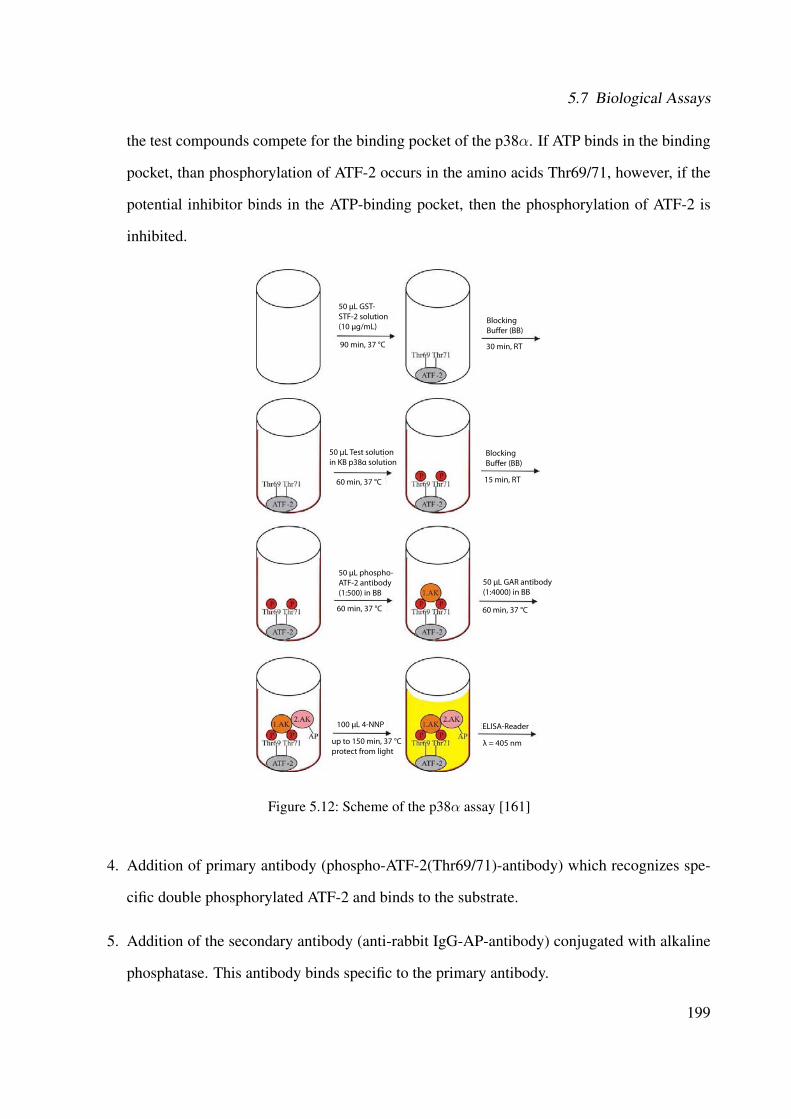
5.7 Biological Assays
the test compounds compete for the binding pocket of the p38α. If ATP binds in the binding
pocket, than phosphorylation of ATF-2 occurs in the amino acids Thr69/71, however, if the
potential inhibitor binds in the ATP-binding pocket, then the phosphorylation of ATF-2 is
inhibited.
50 μL GST-STF-2 solution(10 μg/mL)
90 min, 37 °C 30 min, RT
BlockingBu�er (BB)
50 μL Test solutionin KB p38α solution
60 min, 37 °C
BlockingBu�er (BB)
15 min, RT
50 μL phospho-ATF-2 antibody(1:500) in BB
60 min, 37 °C 60 min, 37 °C
50 μL GAR antibody(1:4000) in BB
up to 150 min, 37 °Cprotect from light
100 μL 4-NNP ELISA-Reader
λ = 405 nm
Figure 5.12: Scheme of the p38α assay [161]
4. Addition of primary antibody (phospho-ATF-2(Thr69/71)-antibody) which recognizes spe-
cific double phosphorylated ATF-2 and binds to the substrate.
5. Addition of the secondary antibody (anti-rabbit IgG-AP-antibody) conjugated with alkaline
phosphatase. This antibody binds specific to the primary antibody.
199

5 Experimental Part
6. Finally, 4-nitrophenylphosphate (4-NPP) is added, which is dephosphorylated by the second
antibody bound to the alkaline phosphatase. The chromophore 4-nitrophenyl can be detected
and quantified by a microplate reader at a wavelength of 405 nm.
In addition, stimulation controls and non-specific binding (NSB) were also tested in the polysty-
rene plate. The stimulation control contained only ATP and activated p38α without inhibitor. After
the addition of the antibodies and the 4-NPP, the value of the maximum of phosphorylation can be
determined. In order to obtain the value of NSB, the kinase buffer without p38α and inhibitor was
used. If there was no kinase in these wells, it means that no phosphorylation of ATF-2 has occurred
and, therefore, no color can be observed. For the evaluation of the inhibition rate, the NSB value
is subtracted from all samples and also from the stimulation control.
The relative inhibition is calculated by the following equation:
RelativeInhibition[%] = 100− ODComp
ODStim
∗ 100 (5.1)
ODComp: Mean of the optical density in 3 wells of the corresponding compounds.
ODStim: Mean of the optical density in the non-inhibited stimulation controls.
Subsequently, the IC50 values were determined. The IC50 value is defined as the inhibitory
concentration by which 50% of the enzyme activity is inhibited. It can be graphically constructed
by interpolation of the semi-logarithmic plot of the inhibition [%] on the inhibitor concentration
[log c]. The straight line joining points intersect the 50% inhibition in the IC50 value corresponding
to the concentration.
On each plate, the reference compound SB203580 (see Figure 5.13) was also tested in the con-
centration of 10, 1, 0.1 and 0.01 µM.
200
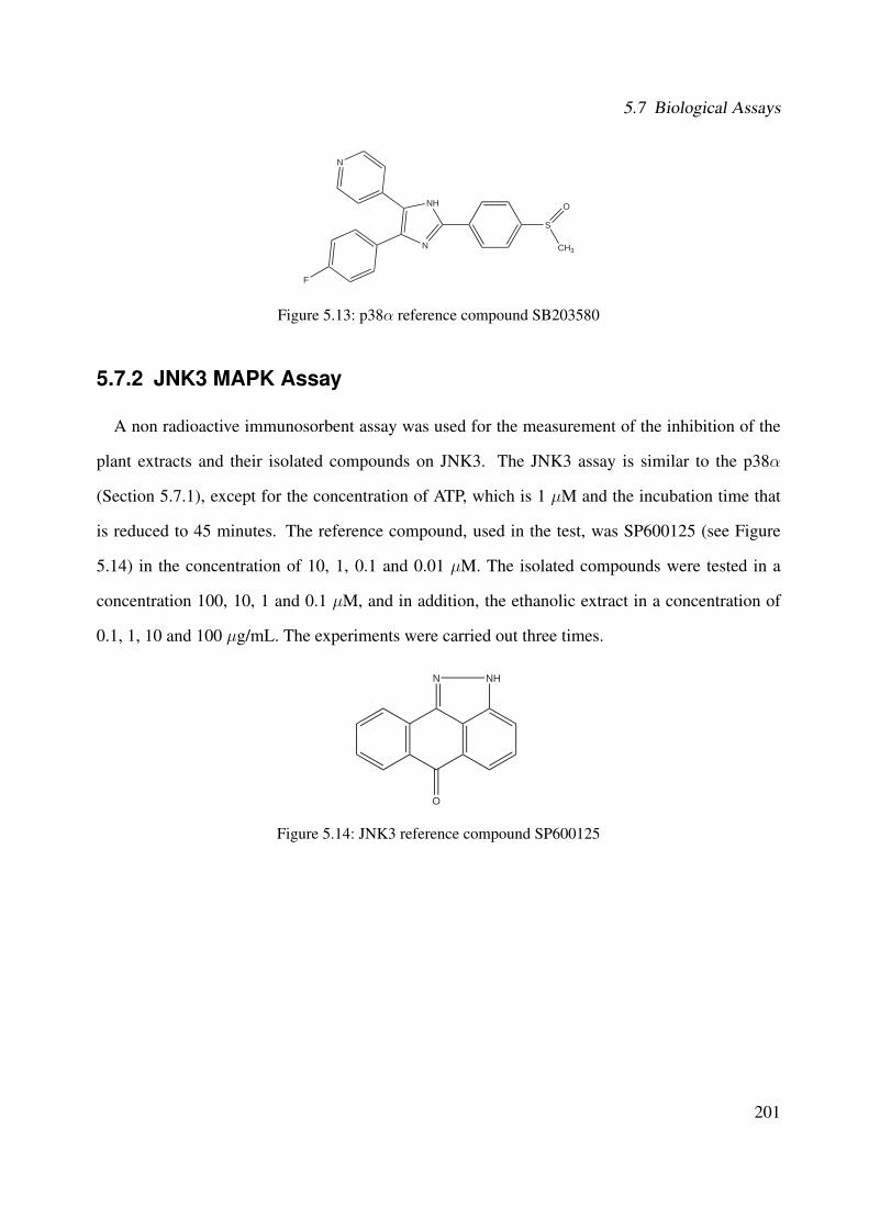
5.7 Biological Assays
S
NH
N
N
O
F
CH3
Figure 5.13: p38α reference compound SB203580
5.7.2 JNK3 MAPK Assay
A non radioactive immunosorbent assay was used for the measurement of the inhibition of the
plant extracts and their isolated compounds on JNK3. The JNK3 assay is similar to the p38α
(Section 5.7.1), except for the concentration of ATP, which is 1 µM and the incubation time that
is reduced to 45 minutes. The reference compound, used in the test, was SP600125 (see Figure
5.14) in the concentration of 10, 1, 0.1 and 0.01 µM. The isolated compounds were tested in a
concentration 100, 10, 1 and 0.1 µM, and in addition, the ethanolic extract in a concentration of
0.1, 1, 10 and 100 µg/mL. The experiments were carried out three times.
O
N NH
Figure 5.14: JNK3 reference compound SP600125
201
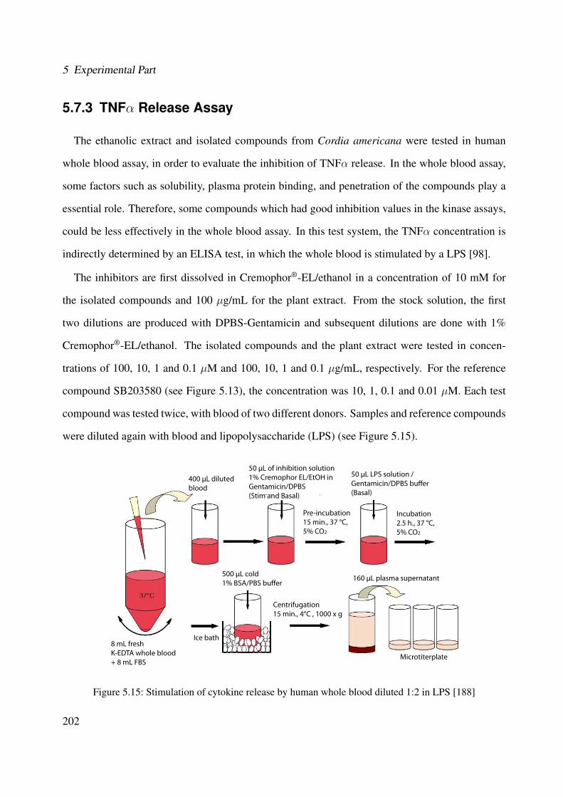
5 Experimental Part
5.7.3 TNFα Release Assay
The ethanolic extract and isolated compounds from Cordia americana were tested in human
whole blood assay, in order to evaluate the inhibition of TNFα release. In the whole blood assay,
some factors such as solubility, plasma protein binding, and penetration of the compounds play a
essential role. Therefore, some compounds which had good inhibition values in the kinase assays,
could be less effectively in the whole blood assay. In this test system, the TNFα concentration is
indirectly determined by an ELISA test, in which the whole blood is stimulated by a LPS [98].
The inhibitors are first dissolved in Cremophor®-EL/ethanol in a concentration of 10 mM for
the isolated compounds and 100 µg/mL for the plant extract. From the stock solution, the first
two dilutions are produced with DPBS-Gentamicin and subsequent dilutions are done with 1%
Cremophor®-EL/ethanol. The isolated compounds and the plant extract were tested in concen-
trations of 100, 10, 1 and 0.1 µM and 100, 10, 1 and 0.1 µg/mL, respectively. For the reference
compound SB203580 (see Figure 5.13), the concentration was 10, 1, 0.1 and 0.01 µM. Each test
compound was tested twice, with blood of two different donors. Samples and reference compounds
were diluted again with blood and lipopolysaccharide (LPS) (see Figure 5.15).
400 μL dilutedblood
50 μL of inhibition solution1% Cremophor EL/EtOH in Gentamicin/DPBS (Stim and Basal)
50 μL LPS solution /Gentamicin/DPBS bu�er(Basal)
Pre-incubation15 min., 37 °C,5% CO2
Incubation2.5 h., 37 °C,5% CO2
8 mL freshK-EDTA whole blood+ 8 mL FBS
Ice bath
Centrifugation15 min., 4°C , 1000 x g
Microtiterplate
500 μL cold1% BSA/PBS bu�er
160 μL plasma supernatant
Figure 5.15: Stimulation of cytokine release by human whole blood diluted 1:2 in LPS [188]
202

5.7 Biological Assays
The blood is initially 1:1 diluted with fetal bovine serum (FBS) followed by the incubation
with the test compounds for 15 min in the CO2 cabinet (37 ◦C, 5% CO2 saturation, 100% of
humidity). Then, by adding LPS, the cytokine release is stimulated and the solution is incubated
again (2.5 hours, 5% of CO2 saturation, 100% of humidity). After incubation, the reaction is
stopped by adding a 1% ice-cold BSA (bovine serum albumin) buffer. The cellular components are
centrifugated. The concentration of the proinflammatory TNFα is determined from the supernatant
(plasma) by ELISA (Figure 5.16).
Capture antibody
BSA to block the wells
TNFα
Detection antibody
Streptavidin / Avidin-peroxidase conjugate
Subtrate (reacts with peroxidase)
Colour change after addition of acid solution
Peroxidase
TMB(3, 3’, 5, 5’-Tetramethylbenzidine)
VIS measurement at λ = 450 nm
Figure 5.16: Scheme of the Cytokine-ELISA assay for the determination of TNFα release [161]
For the ELISA, the supernatant is diluted with a special diluent (TNFα Diluent, Beckman Coul-
ter). The plate is first coated with the primary antibody (capture antibody; TNFα: murine-human
203
5 Experimental Part
antibody) and the free binding sites are blocked with BSA. 100 µL of plasma are added to a stan-
dard series of TNFα and incubated for 2 hours at RT. During this time, the cytokines bind to the
primary antibody. Subsequently, the addition of the second antibody occurs (detection antibody;
biotinylated anti-human TNFα antibody) and is incubated for two hours again. An enzym-reagent
is added consisting of streptavidin (TNFα-Merrettich-Peroxidase-Conjugated). Streptavidin binds
to the biotin rest of the second antibody. After addition of the substrate solution of 3,3',5,5'-
tetramethylbenzidine (TMB) and hydrogen peroxide, a blue color is formed by the oxidation of
one of the two amino groups of TMB. After 30 minutes, the enzyme reaction is stopped with 1 M
sulfuric acid. This leads to a protonation of the remaining amino group and to a bathochromic shift
indicating by a yellow color. The detection is carried out by an ELISA reader at 450 nm.
The inhibition rate of the cytokine release is calculated using the following equation:
RelativeInhibition[%] = 100− CComp − CBasal
CStim − CBasal
∗ 100 (5.2)
CComp: concentration of cytokines in wells with test compound.
CBasal: concentration of cytokines in wells with test compound and without LPS.
CStim: mean of the cytokine concentration in the stimulation control.
204
5.7 Biological Assays
5.7.4 5-Lipoxygenase Assay
5.7.4.1 Determination of 5-LO Product Formation in Cell-free Assays
Escherichia coli (E.coli) MV1190 was transformed with pT3-5-LO plasmid and recombinant
5-LO protein was expressed as described in [94]. In brief, E.coli was harvested and lysed by
incubation in 50 mM triethanolamine/HCl, pH = 8.0, 5 mM EDTA, soybean trypsin inhibitor
(60 µg/mL), 1 mM phenylmethylsulphonyl fluoride and lysozyme (500 µg/mL), homogenized
by sonication (3 x 15 sec) and centrifuged at 19,000 x g for 15 min. Proteins including 5-LO
were precipitated with 50% saturated ammonium sulfate during stirring on ice for 60 min. The
precipitate was collected by centrifugation at 16,000 x g for 25 min and the pellet was resuspended
in 20 mL PBS containing 1 mM EDTA and 1 mM PMSF. After centrifugation at 100,000 x g for 70
min at 4 ◦C, the 100,000 x g supernatant was applied to an ATP-agarose column (Sigma A2767),
and the column was eluted as described previously [94].
For activity assays, partially purified 5-LO was resuspended in 1 mL PBS, pH = 7.4 containing
1 mM EDTA, and 1 mM ATP was added. Samples were preincubated with the test compounds
for 10 min at 4 ◦C, prewarmed for 30 s at 37 ◦C, and then 2 mM CaCl2 and 20 µM arachidonic
acid were added to start 5-LO product formation. The reaction was stopped after 10 min at 37
◦C by addition of 1 mL ice cold methanol and the formed metabolites were analyzed by HPLC as
described [331].
5-LO products include LTB4 isomers and 5(S)-hydro(pero)xy-6-trans-8,11,14-cis-eicosatetraenoic
acid (5-H(p)ETE). The ethanolic extract of Cordia americana was tested at concentration of 10, 3,
1 and 0.1 µg/mL and its compounds at 10, 3, 1 and 0.1 µM. The ethanolic extract of Brugmansia
suaveolens was determined at concentrations of 3 and 30 µg/mL and its compounds at 3 and 30
µM for at least two experiments. Finally, the reference compound BWA4C (see Figure 5.17) was
also tested at a concentration of 3 µM.
205
5 Experimental Part
N
OH
CH3
O
O
Figure 5.17: 5-LO reference compound BWA4C
5.7.4.2 Isolation of Human PMNL from Venous Blood
Human PMNL (polymorphonuclear leukocytes) were freshly isolated from buffy coats obtained
from the Blood Center of the University Hospital Tubingen (Germany). In brief, venous blood from
healthy donors was taken and leukocyte concentrates were prepared by centrifugation at 4,000 g
for 20 min at RT. Buffy coats were diluted 1:1 (V:V) with phosphate buffered saline pH = 7.4 (PBS)
and then with ice-cold 5% dextran (w/v in PBS) in a ratio of 4:5 (V:V), for 45 min. After dextran
sedimentation, neutrophils were immediately isolated by centrifugation at 1,000 g, 10 min, RT
(Heraeus sepatech, Varifuge 3.0, Hanau , Germany) on Nycoprep cushions, and hypotonic lysis of
erythrocytes as described [331]. PMNL (106 cells/mL; purity> 96-97%) were finally resuspended
in PBS plus 1 mg/mL glucose (PG buffer) or in PG buffer plus 1 mM CaCl2 (PGC buffer) as
indicated.
5.7.4.3 Determination of 5-LO Product Formation in Cell-based Assays Using
Isolated Human PMNL
For determination of cellular 5-LO product formation, 5 x 10⁶ freshly isolated PMNL in 1 mL
PGC buffer with or without bovine serum albumin (BSA) was pre-incubated with test compounds
or with vehicle (DMSO) for 10 min at 37 ◦C, as indicated. 5-LO product formation was started
by addition of A23187 (2.5 µM) with or without 20 µM AA. The reaction was stopped after 10
min with 1 mL of methanol and then 30 µL of 1 N HCl, 200 ng PGB1 and 500 µL of PBS were
added. Formed 5-lipoxygenase metabolites were extracted and analyzed by HPLC as described
[329]. 5-Lipoxygenase product formation includes leukotriene B4 and its all-trans isomers and
206
5.7 Biological Assays
5(S)- H(P)ETE. Cysteinyl leukotrienes C4, D4 and E4 were not detected, and oxidation products
of leukotriene B4 were not determined. The ethanolic extract of Cordia americana was tested
based at the concentrations of 0.1, 0.3, 1, 3, 10 and 30 µg/mL and CA2 at 1, 3 an 10 µM for at
least two experiments.
5.7.5 NF-κB Electrophoretic Mobility Shift Assay (EMSA)
Jurkat T cells were maintained in RPMI 1640 medium, supplemented with 10% fetal calf serum,
100 IU/mL penicillin and 100 g/mL streptomycin (all Gibco-BRL, Groningen, Netherlands).
Total cell extracts from Jurkat T cells were prepared as previously described [159]. In contrast
to the previous study, NF-κB oligonucleotide (Promega) was labeled using [γ−33P] dATP (3000
Ci/mmol; Amersham) and a T4 nucleotide kinase (New England Biolabs). The ethanolic extract
was tested at the concentrations of 10, 30 and 50 µg/mL and rosmarinic acid of 10, 30, 54 µM, for
at least two experiments.
5.7.6 Fibroblast Scratch Assay
Scratch assay was performed, as previously described [100]. In detail: Swiss 3T3 albino mouse
fibroblasts were cultured in Dulbecco's modified Eagle's medium (DMEM), supplemented with
10% fetal calf serum, 100 IU/mL penicillin and 100 g/mL streptomycin and maintained at 37 ◦C
in a humidified, 5% CO2 environment (all Gibco-BRL, Groningen, Netherlands).
Swiss 3T3 albino mouse fibroblasts were grown in a confluent cell monolayer on coverslips
into 24-well plates. The coverslips were precoated with collagen type I (40 µg/mL) for 2 h at
37 ◦C, before seeding the cells. Then the cells were cultured to nearly confluent monolayers and
thereafter a linear wound was generated in the monolayer with a sterile 100 µL plastic pipette
tip. The medium was changed in order to remove scraped cells. DMEM medium with dimethyl
sulfoxid (0.25%), platelet derived growth factor (2 ng/mL), ethanolic extract (1, 50 and 100 µg/mL)
and rosmarinic acid (1, 5, 10 µg/mL) were added to a set of 3 coverslips per dose and incubated for
207

5 Experimental Part
12 h at 37 ◦C with 5% CO2. The cells were fixed with 4% paraformaldehyde for 15 minutes and
stained with 4',6-diamino-2-phenylindole (DAPI) overnight. Three representative images from
each coverslip of the scratched areas under each condition were photographed to estimate the
relative migration and proliferation of the cells. The effect of 1, 50 and 100 µg/mL of ethanolic
extract of Cordia americana, and 1, 5 and 10 µg/mL of CA1, were assayed on fibroblast scratched
monolayers. The experiments were carried out in triplicate.
5.7.7 MTT Assay
The cytotoxic activity was studied using the MTT colorimetric assay as previously described by
[216]. For all samples and controls, 4 mL of a suspension of Jurkat cells (2.5 x 10⁵ cells/mL) were
used. The ethanolic extract (10 mg/mL DMSO) were tested in independent assays at concentrations
of 50 and 100 µg/mL. Parthenolide (100 µM) was used as positive control and DMSO 1% (V:V) as
negative control. All plates were incubated at 37 ◦C, 5% CO2, during 24 hours. After incubation,
1.5 mL of MTT (0.5% in sterile PBS) was added to each plate, followed by additional 2 hours
incubation in 5% CO2 at 37 ◦C. The volume of each plate was transferred to tubes and centrifugated
at 5,000 rpm for 10 min at 4 ◦C. The supernatant was discarded and the cells were resuspended
with 1 mL of extraction solution buffer (20% SDS, 50% DMF). After overnight incubation (5%
CO2 at 37 ◦C), the absorbance of each sample and controls was measured at λ = 595 nm and the
percentual of inhibition was calculated. Jurkat cells were treated for 24 h at a concentration of 50
and 100 µg/mL of the ethanolic extract of Cordia americana.
5.8 Computer Program
The following programs and databases have been used during the execution of the present work:
ChemDraw Ultra 8, Scifinder Scholar 2007, ACDLabs 5.07, LaFlash System 1.1.
208
5.9 Statistical Analysis
5.9 Statistical Analysis
Statistical evaluation was carried out with Origin Scientific Graphing and Analysis Software,
and Microsoft Office Excel 2007.
5.10 Docking
The molecular modeling studies, that is, the visualization and building of the 3D-structures of
the ligands were done with Maestro (version 8.5) from Schrodinger [280]. Docking studies were
performed with Induced Fit docking protocol from Schrodinger [281]. The figures which showed
the different docking positions to the ATP binding site were prepared with PyMol [77].
209

Bibliography
[1] Abadleh, M. M. M., 2009. Diarylisoxazoles as lead for the design and synthesis of proteinkinase inhibitors. Ph.D. thesis, University of Tubingen.
[2] Abdallah, O. M., Kamel, M. S., Mohamed, M. H., 1994. Phenylpropanoid glycosides forPrunus ssiori. Phytochemistry 37, 1689–1692.
[3] Abou-Donia, A. H., Toaima, S. M., Hammoda, H. M., Shawky, E., 2006. Determina-tion of rutin in Amaryllis belladonna L. flowers by HPTLC and spectrophotometry. Chro-matographia 64, 109–112.
[4] Afzal, M., Obuekwe, C., Shuaib, N., Barakat, H., 2004. Photosynthetic pigment profile ofCordia myxa L. and its potential in folklore medicinal application. Food, Agriculture &Environment 2, 114–120.
[5] Agnihotri, V. K., Srivastava, S. D., Srivastava, S. K., Pitre, S., Rusia, K., 1987. Constiuentsof Cordia obliqua as potential anti-inflammatory agents. Indian Journal of PharmaceuticalSciencies 49, 66–69.
[6] Akhtar, A. H., Ahmad, K. U., 1995. Anti-ulcerogenic evaluation of the methanolic ex-tracts of some indigenous medicinal plants of Pakistan in aspirin-ulcerated rats. Journalof Ethnopharmacology 46, 1–6.
[7] Al-Awadi, F. M., Srikumar, T. S., Anim, J. T., Khan, I., 2001. Antiinflammatory effects ofCordia myxa fruit on experimentally induced colitis in rats. Nutrition 17, 391–396.
[8] Albert, D., Zndorf, I., Dingermann, T., Mller, W. E., Steinhilber, D., Werz, O., 2002. Hyper-forin is a dual inhibitor of cyclooxygenase-1 and 5-lipoxygenase. Biochemical Pharmacol-ogy 64, 1767–1775.
[9] Allard, J. B., Brock, T. G., 2005. Structural organization of the regulatory domain of human5-lipoxygenase. Current Protein and Peptide Science 6, 125–131.
211
Bibliography
[10] Alves, M. N., Sartoratto, A., Trigo, J. R., 2007. Scopolamine in Brugmansia suaveolens
(Solanaceae): Defense, allocation, costs, and induced response. Journal of Chemical Ecol-ogy 33, 297–309.
[11] Amakura, Y., Tsutsumi, T., Nakamura, M., Kitagawa, H., Fujino, J., Sasaki, K., Toyoda,M., Yoshida, T., Maitani, T., 2003. Activation of the aryl hydrocarbon receptor by somevegetable constituents determined using in vitro reporter gene assay. Biological & Pharma-ceutical Bulletin 26, 532–539.
[12] Anthony, S. J., Zuchowski, W., Setzer, W. N., 2009. Composition of the floral essential oilof Brugmansia suaveolens. Records of Natural Products 3, 76–81.
[13] Anvisa, 2005. Farmacopeia Brasileira ganha reconhecimento da Europa.URL http://www.anvisa.gov.br/
[14] ANVISA, 2009. Farmacopeia Brasileira.URL http://www.anvisa.gov.br/hotsite/farmacopeia/index.htm
[15] Arbour, N., Naniche, D., Homann, D., Davis, R. J., Flavell, R. A., Oldstone, M. B., 2002.C-jun NH2-terminal kinase (JNK)1 and JNK2 signalling pathways have divergent roles inCD8+ T cell- mediated antiviral immunity. Journal of Experimental Medicine 195, 801–810.
[16] Arend, W. P., 2001. The innate immune system in rheumatoid arthritis. Arthritis andRheumatism 44, 2224–2234.
[17] Ashworth, A., Nakielny, S., Cohen, P., Marshall, C., 1996. The amino acid sequence of amammalian MAP kinase kinase. Journal of Biological Chemistry 271, 27696–27700.
[18] Atkins, C. M., Selcher, J. C., Petraitis, J. J., Trzaskos, J. M., Sweatt, J. D., 1998. The MAPKcascade is required for mammalian asscociative learning. Nature Neuroscience 1, 602– 609.
[19] Awad, A. B., Fink, C. S., 2000. Phytosterols as anticancer dietary components: evidenceand mechanism of action. Journal of Nutrition 130, 2127–2130.
[20] Baldwin, A. S., 2001. The transcription factor NF-κB and human disease. Journal of ClinicalInvestigation 107, 36.
[21] Balkwill, F., 2000. TNF is here to stay! Immunology Today 21, 470–471.
212
Bibliography
[22] Bano, J. M., Lorente, J., Castillo, J., Garca-Benavente, O., Rio, J. A., Ortuno, A., Quirin,K. W., Gerard, D., 2003. Phenolic diterpenes, flavones, and rosmarinic acid distributionduring the development of leaves, flowers, stems, and roots of Rosmarinus of�cinalis. An-tioxidant activity. Agricultural and Food Chemistry 51, 4247–4253.
[23] Baron, D., 1995. Cytokine: Ihre Biologie und klinische Anwendung. PharmazeutischeZeitung 25, 9–20.
[24] Barr, R. K., Bogoyevitch, M. A., 2001. The c-jun N-terminal protein kinase family ofmitogen-activated protein kinases (JNK MAPK). International Journal of Biochemistry andCell Biology 33, 1047–1063.
[25] Baud, V., Karin, M., 2001. Signal transduction by tumor necrosis factor and its relatives.Trends in Cell Biology 11, 372–377.
[26] Bayeux, M., Fernandes, A., Foglio, M., Carvalho, J., 2002. Evaluation of the antiedemato-genic activity of artemetin isolated from Cordia curassavica. Brazilian Journal of Medicaland Biological Research 35, 1229–1232.
[27] Begum, S., Sahai, M., Fujimoto, Y., Asai, K., Schneider, K., Nicholson, G., Suessmuth, R.,2006. A new kaempferol diglycoside from Datura suaveolens Humb. & Bonpl. ex. Willd.Natural Product Research 20, 1231–1236.
[28] Bellon, S., Fitzgibbon, M. J., Fox, T., Hsiao, H. M., Wilson, K. P., 1999. The structure ofphosphorylated p38γ is monomeric and reveals a conserved activation-loop conformation.Structure 9, 1057–1065.
[29] Bhatt, D., Chang, J.-I., Hiraokan, N., 2004. In vitro propagation and storage of Brugmansia
versicolor lagerheim. Plant Biotechnology 21, 237–241.
[30] Boldt, S., Kolch, W., 2004. Targeting MAPK signalling: Prometheus �re or Pandora's Box?Current Pharmaceutical Design 10, 1885–1905.
[31] Bonvini, P., Zorzi, E., Mussolin, L., Monaco, G., Pigazzi, M., Basso, G., Rosolen, A., 2009.The effect of the cyclin-dependent kinase inhibitor flavopiridol on anaplastic large cell lym-phoma cells and relationship with NPM-ALK kinase expression and activity. Haematologica94, 944–955.
[32] Boulton, T. G., Yancopoulos, G. D., Gregory, J. S., Slaughter, C., Moomaw, C., Hsu, J.,Cobb, M. H., 1990. An insulin-stimulated protein kinase similar to yeast kinases involvedin cell cycle control. Science 249, 64–67.
213
Bibliography
[33] Bozyczko-Coyne, D., OKane, T. M., Wu, Z. L., Dobrzanski, P., Murthy, S., Vaught, J. L.,Scott, R. W., 2001. CEP-1347/KT-7515, an inhibitor of SAPK/JNK pathway activation,promotes survival and blocks multiple events associated with AB-induced cortical neuronapoptosis. Journal of Neurochemistry 77, 849–863.
[34] Brambilla, R., Gnesutta, N., Minichiello, L., White, G., Roylance, A. J., Herron, C. E.,Ramsey, M., Wolfer, D. P., Cestari, V., Rossi-Arnaud, C., Grant, S. G., Chapman, P. F.,Lipp, H. P., Sturani, E., Klein, R., 1997. A role for the Ras signalling pathway in synaptictransmission and long- term memory. Nature 390, 281–286.
[35] Brancho, D., Tanaka, N., Jaeschke, A., Ventura, J.-J., Kelkar, N., Tanaka, Y., Kyuuma, M.,Takeshita, T., Flavell, R. A., Davis, R. J., 2003. Mechanism of p38 MAP kinase activationin vivo. Genes and Development 17, 1969–1978.
[36] Calixto, J. B., 2005. Twenty-five years of research on medicinal plants in Latin America: Apersonal view. Journal of Ethnopharmacology 100, 131–134.
[37] Calixto, J. B., Otuki, M. F., Santos, A. R., 2003. Anti-inflammatory compounds of plantorigin. Part I. Action on arachidonic acid pathway, nitric oxide and nuclear factor kappa B(NF-κB). Planta Medica 69, 973–983, pMID: 14735432.
[38] Capasso, A., Feo, V. D., Simone, F. D., Sorrentino, L., 1997. Activity-directed isolation ofspasmolytic (anti-cholinergic) alkaloids from Brugmansia arborea (L.) Lagerheim. Phar-maceutical Biology 35, 43–48.
[39] Cardillo, A. B., Alvarez, A. M. O., Lopez, A. C., Lozano, M. E. V., Talou, J. R., Giulietti,A. M., 2009. Anisodamine production from natural sources: Seedlings and hairy root cul-tures of Argentinean and Colombian Brugmansia candida plants. Planta Med 76, 402–405.
[40] Cardillo, A. B., Talou, J. R., Giulietti, A. M., 2008. Expression of Brugmansia candida
hyoscyamine 6 beta-hydroxylase gene in Saccharomyces cerevisiae and its potential use asbiocatalyst. Microbial Cell Factories 7, 7–17.
[41] Carlo, G. D., Mascolo, N., Izzo, A. A., Capasso, F., 1999. Flavonoids: Old and new aspectsof a class of natural therapeutic drugs. Life Sciences 65, 337–353.
[42] Carrizo, C. N., Pitta-Alvareza, S. I., Koganb, M. J., Giulietti, A. M., Tomaro, M. L., 2001.Occurrence of cadaverine in hairy roots of Brugmansia candida. Phytochemistry 57, 759–763.
214

Bibliography
[43] CEBRID, 2006. Anti-colinergicos. CENTRO BRASILEIRO DE INFORMACOES SOBREDROGAS PSICOTROPICAS.URL http://bvsms.saude.gov.br/bvs/folder/
[44] CellSignal, 2008. MAPK signaling cascades.URL http://www.cellsignal.com/
[45] Chang, H. Y., Yang, X., 2000. Proteases for cell suicide: Function and regulation of cas-pases. Microbiology and Molecular Biology Reviews 64, 821–846.
[46] Chang, L., Karin, M., 2001. Mammalian MAP kinase signalling cascades. Nature 410, 37–40.
[47] Chen, J. H., Ho, C.-T., 1997. Antioxidant activities of caffeic acid and its related hydrox-ycinnamic acid compounds. Journal of Agricultural and Food Chemistry 45, 2374–2378.
[48] Chen, Z., Gibson, T. B., Robinson, F., Silvestro, L., Pearson, G., e Xu, B., Wright, A.,Vanderbilt, C., Cobb, M. H., 2001. MAP Kinases. Chemical Reviews 101, 2449–2476.
[49] Cheng, H. P., Wei, S., Wei, L. P., Verkhratsky, A., 2006. Calcium signaling in physiologyand pathophysiology. Acta Pharmacologica Sinica 27, 767–772.
[50] Chiang, H.-C., Lo, Y. J., Lu, F.-J., 1994. Xanthine oxidase inhibitors from the leaves ofAlsophila spinulosa. Journal of Enzyme Inhibition and Medicinal Chemistry 8, 61–71.
[51] Choi, J. M., Lee, E. O., Lee, H. J., Kim, K. H., Ahn, K. S., Shim, B. S., Kim, N. I., Song,M. C., Baek, N. I., Kim, S. H., 2007. Identification of campesterol from Chrysanthemum
coronarium L. and its antiangiogenic activities. Phytotherapy Research 10, 954–959.
[52] Chomarat, P., Vannier, E., Dechanet, J., Rissoan, M. C., Banchereau, J., Dinarello, C. A.,Miossec, P., 1995. Balance of IL-1 receptor antagonist/IL-1b in rheumatoid synovium andits regulation by IL-4 and IL-10. Journal of Immunology 154, 1432–1439.
[53] Choudhary, M. I., Begum, A., Abbaskhan, A., Ajaz, A., ur Rehman, S., ur Rahman, A.,2005. Phenyl polypropanoids from Lindelo�a stylosa. Chemical & Pharmaceutical Bulletin53, 1469–1471.
[54] Choy, E. H., Panayi, G. S., 2001. Cytokine pathways and joint inflammation in rheumatoidarthritis. New England Journal of Medicine 344, 907–916.
[55] Clerk, A., H.Sugden, P., 1999. Activation of protein kinase cascades in the heart by hyper-trophic G protein-coupled receptor agonists. American Journal of Cardiology 83, 64H–69H.
215

Bibliography
[56] Cobb, M. H., Goldsmith, E. J., 1995. How MAP kinases are regulated. Journal of BiologicalChemistry 25, 14843–14846.
[57] Cole, J., Tsou, R., Wallace, K., Gibran, N., Isik, F., 2001. Early gene expression profile ofhuman skin to injury using high-density cDNA microarrays. Wound Repair and Regenera-tion 9, 360–370.
[58] Conze, D., Krahl, T., Kennedy, N., Weiss, L., Lumsden, J., Hess, P., Flavel, R. A., Le, G. G.,Davis, R. J., Rincon, M., 2002. C-jun NH(2)-terminal kinase (JNK)1 and JNK2 have distinctroles in CD8 (+) T cell activation. Journal of Experimental Medicine 195, 811–823.
[59] Correa, M. P., 1952. Dicionario das Plantas Uteis do Brasil e das Exoticas Cultivadas. Im-prensa Nacional.
[60] Crews, C. M., Alessandrini, A., Erikson, R. L., 1992. The primary structure of MEK, aprotein kinase that phosphorylates the ERK gene product. Science 258, 478–480.
[61] Crow, F. W., Tomer, K. B., Looker, J. H., Gross, M. L., 1986. Fast atom bombardment andtandem mass spectrometry for structure determination of steroid and flavonoid glycosides.Analytical Biochemistry 155, 286–307.
[62] Cuenda, A., Cohen, P., Buee-Scherrer, V., Goeder, M., 1997. Activation of stress-activatedprotein kinase-3 (SAPK3) by cytokines and cellular stresses is mediated via SAPKK3(MKK6); comparison of the specificities of SAPK3 and SAPK2 (RK/p38). The EMBOJournal 16, 295–305.
[63] Cuyckens, F., Claeys, M., 2004. Mass spectrometry in the structural analysis of flavonoids.Journal of Mass Spectrometry 39, 1–15.
[64] da Silva, S. A. S., Rodrigues, M. S. L., de Ftima Agra, M., da Cunhaa, E. V. L., Barbosa-Filhoa, J. M., da Silva, M. S., 2004. Flavonoids from Cordia globosa. Biochemical System-atics and Ecology 32, 359–361.
[65] Dambach, D. M., 2005. Potential adverse effects associated with inhibition of p38α/β MAPkinases. Current Topics in Medicinal Chemistry 5, 929–939.
[66] Danese, S., Semeraro, S., Armuzzi, A., Papa, A., Gasbarrini, A., 2006. Biological thera-pies for inflammatory bowel disease: Research drives clinics. Mini-Reviews in MedicinalChemistry 6, 771–784.
216
Bibliography
[67] Dapkevicius, A., van Beek, T. A., Lelyveld, G. P., van Veldhuizen, A., de Groot, A., Linssen,J. P. H., Venskutonis, R., 2002. Isolation and structure elucidation of radical scavengers fromThymus vulgaris leaves. Journal of Natural Products 65, 892–896.
[68] Davidson, A., Diamond, B., 2001. Autoimune diseases. New England journal of Medicine345, 340–350.
[69] Davies, G., Fataftah, A., Radwan, A., Raffauf, R. F., Ghabbour, E. A., Jansen, S. A., 1997.Isolation of humic acid from the terrestrial plant Brugmansia sanguinea. Science of TheTotal Environment 201, 79–87.
[70] Davis, R. J., 2000. Signal transduction by the JNK group of MAP kinases. Cell 103, 239–252.
[71] Day, A. J., DuPont, M. S., Ridley, S., Rhodes, M., Rhodes, M. J., Morgan, M. R.,Williamson, G., 1998. Deglycosylation of flavonoid and isoflavonoid glycosides by humansmall intestine and liver beta-glucosidase activity. FEBS Letters 436, 71–75.
[72] Dayer, J.-M., Bresnihan, B., 2002. Targeting interleukin-1 in the treatment of rheumatoidarthritis. Arthritis and Rheumatism 46, 574–578.
[73] de Carvalho, P., Rodriguesa, R., Sawayaa, A., Marquesb, M., Shimizu, M., 2004. Chemi-cal composition and antimicrobial activity of the essential oil of Cordia verbenacea D.C.Journal of Ethnopharmacology 95, 297–301.
[74] de Carvalho, P. E. R., 2004. Guajuvira - Patagonula americana. Tech. rep., Embrapa.
[75] de las Heras, B., Hortelano, S., 2009. Molecular basis of the anti-inflammatory effects ofterpenoids. Inflammation & Allergy - Drug Targets 8, 28–39.
[76] de Menezes, J. A., Lemos, T., Pessoa, O., Braz-Filho, R., Montenegro, R., Wilke, D., Costa-Lotufo, L., Pessoa, C., de Moraes, M., Silveira, E., 2005. A cytotoxic meroterpenoid ben-zoquinone from roots of Cordia globosa. Planta Medica 71, 54–8.
[77] DeLano, W. L., 01 2002. The PyMol Molecular Graphics System. DeLano Scientific, sanCarlos, CA, USA.URL http://www.pymol.org
[78] Derijard, B., Hibi, M., Wu, I. H., Barret, T., Su, B., Deng, T., Karin, M., Davis, R. J., 1994.JNK1: A protein kinase stimulated by UV light and Ha-Ras that binds and phosphorylatesthe c-Jun activation domain. Cell 76, 1025–1037.
217
Bibliography
[79] Detzel, A., Wink, M., 1993. Attraction, deterrence or intoxication of bees (Apis mellifera)by plant allelochemicals. Chemoecology 4, 8–18.
[80] Dewick, P., 2001. Medicinal Natural Products: A Biosynthetic Approach. Wiley & Sons,West Sussex, England.
[81] El-Sayed, N., Omara, N., Yousef, A., Farag, A., Mabry, T., 2001. Kaempferol triosides fromReseda muricata. Phytochemistry 57, 575–578.
[82] Englisch, J. D., Sweatt, J. D., 1996. Activation of p42 mitogen-activated protein kinase inhippocampal lon term potentiation. Journal of Biological Chemistry 271, 24329–24332.
[83] Estus, S., Zaks, W. J., Freemann, R. S., Gruda, M., Bravo, R., Johnson, E. M., 1994. Al-tered gene expression in neurons during programmed cell death: Identification of c-jun asnecessary for neuronal apoptosis. Journal of Cell Biology 127, 1717–1727.
[84] Evans, W., Lampard, J., 1972. Alkaloids of Datura suaveolens. Phytochemistry 11, 3293–3298.
[85] Evans, W. C., Major, V. A., 1968. The alkaloids of the genus Datura, section Brugmansia.Part IV. New alkaloids of Datura sanguinea R. and P. Chemical Society C: Organic articles,2775–2778.
[86] Fabian, M. A., Biggs, W. H., Treiber, D. K., Atteridge, C. E., Azimioara, M. D., Benedetti,M. G., Carter, T. A., Ciceri, P., Edeen, P. T., Floyd, M., Ford, J. M., Galvin, M., Gerlach,J. L., Grotzfeld, R. M., Herrgard, S., Insko, D. E., Insko, M. A., Lai, A. G., Lelias, J.-M.,Mehta, S. A., Milanov, Z. V., Velasco, A. M., Wodicka, L. M., Patel, H. K., Zarrinkar,P. P., Lockhart, D. J., 2005. A small molecule-kinase interaction map for clinical kinaseinhibitors. Nature Biotechnology 23, 329–336.
[87] Fathiazada, F., Delazara, A., Amiria, R., Sarkerb, S. D., 2006. Extraction of flavonoidsand quantification of rutin from waste Tobacco leaves. Iranian Journal of PharmaceuticalResearch 3, 222–227.
[88] Feisst, C., Franke, L., Appendino, G., Werz, O., 2005. Identification of molecular targets ofthe oligomeric nonprenylated acylphloroglucinols from Myrtus communis and their impli-cation as anti-inflammatory compounds. Journal of Pharmacology and Experimental Ther-apeutics 315, 389–396.
[89] Feo, V. D., 2003. Ethnomedical field study in northern Peruvian Andes with particular ref-erence to divination practices. Journal of Ethnopharmacology 85, 243–256.
218
Bibliography
[90] Ferrari, F., Monache, F. D., Compagnone, R., Oliveri, M. C., 1997. Chemical constituentsof Cordia dentata flowers. Fitoterapia 68, 88.
[91] Ferrel, J. E., 1996. Tripping the switch fantastic: How a protein kinase cascade can convertgraded inputs into switch-like outputs. Trends in Biochemistry Science 21, 460–466.
[92] Ferriola, C. P., Cody, V., Middleton, J. E., 1989. Protein kinase C inhibition by plantflavonoids. Biochemical Pharmacology 38, 1617–1624.
[93] Ficarra, R., Ficarra, P., Tommasini, S., Calabro, M. L., Ragusa, S., Barbera, R., Rapisarda,A., 1995. Leaf extracts of some Cordia species: Analgesic and anti-inflammatory activitiesas well as their chromatographic analysis. Pharmacology 50, 245–256.
[94] Fischer, L., Szellas, D., Radmark, O., Steinhilber, D., Werz, O., 2003. Phosphorylationand stimulus-dependent inhibition of cellular 5-lipoxygenase activity by non-redox-type in-hibitors. FASEB Journal 17, 949–951.
[95] Forrer, P., Tamaskovic, R., Jaussi, R., 1998. Enzyme-linked immunosorbent assay for mea-surement of JNK, ERK, and p38 kinase activities. Journal of Biological Chemistry 379,1101–1111.
[96] Fossen, T., Pedersen, A. T., Andersen, O. M., 1998. Flavonoids from red onion (Allium
cepa). Phytochemistry 47, 281–285.
[97] Freitas, A. V. L., Trigo, J. R., Junior, K. S. B., Witte, L., Hartmann, T., Barata, L. E. S., 1996.Tropane and pyrrolizidine alkaloids in the ithomiines Placidula euryanassa and Miraleria
cymothoe (Lepidoptera: Nymphalidae). Chemoecology 7, 61–67.
[98] Friedrichs, A., 2005. Optimierung eines Vollblut-Testsystems zur Evaluirung von Hemm-stoffen der Zytokin-Freisetzung. Master's thesis, Universitat Tubingen.
[99] Frodin, M., Sekine, N., Roche, E., Filloux, C., Prentki, M., Wollheim, C. B., Obberghen,E. J. V., 1995. Glucose, other secretagogues, and nerve growth factor stimulate mitogen-activated protein kinase in the insulin-secreting beta-cell line, INS-1. Journal of BiologicalChemistry 270, 7882–7889.
[100] Fronza, M., Heinzmann, B., Hamburger, M., Laufer, S., Merfort, I., 2009. Determination ofthe wound healing effect of Calendula extracts using the scratch assay with 3T3 �broblasts.Journal of Ethnopharmacology 126, 463–467.
219
Bibliography
[101] Fun, C., Svendsen, A. B., 1990. The essential oil of Cordia cylindrostachya Roem. & Schult.grown on Aruba. Journal of Essential Oil Research 2, 209–210.
[102] Funk, C. D., 2001. Prostaglandins and leukotrienes: Advances in eicosanoid biology. Sci-ence 294, 1871–1875.
[103] Gadotti, V. M., Schmeling, L. O., Machado, C., Liz, F. H., Filho, V. C., Meyre-Silva, C.,Santos, A. R., 2005. Antinociceptive action of the extract and the flavonoid quercitrin iso-lated from Bauhinia microstachya leaves. Journal of Pharmacy and Pharmacology 57, 1345–1351.
[104] Gallagher, T. F., Seibel, G. L., Kassis, S., Laydon, J. T., Blumenthal, M. J., Lee, J. C., Lee,D., Boehm, J. C., Fier-Thompson, S. M., Abt, J. W., Soreson, M. E., Smietana, J. M., Hall,R. F., Garigipati, R. S., Bender, P. E., Erhard, K. F., Krog, A. J., Hofmann, G. A., Sheldrake,P. L., McDonnell, P. C., Kumar, S., Young, P. R., Adams, J. L., 1997. Regulation of stress-induced cytokine production by pyridinylimidazoles inhibition of CSBP kinase. Bioorganic& Medicinal Chemistry 5, 49–64.
[105] Gallo, M. B. C., Rocha, W. C., da Cunha, U. S., Diogo, F., da Silva, F. C., Vieira, P. C.,Vendramim, J. D., Fernandes, J. B., da Silva, M. F., Batista-Pereira, L. G., 2006. Bioactivityof extracts and isolated compounds from Vitex polygama (Verbenaceae) and Siphoneugena
densi�ora (Myrtaceae) against Spodoptera frugiperda (Lepidoptera: Noctuidae). Pest Man-agement Science 62, 1072–1081.
[106] Galvez, J., 1996. Application of natural products in experimental models of intestinal in-flammation in rats. Methods & Findings in Experimental & Clinical Pharmacology 18, 7–10.
[107] Gao, L. P., Wei, H. L., Zhao, H. S., Xiao, S. Y., Zheng, R. L., 60. Antiapoptotic and antiox-idant effects of rosmarinic acid in astrocytes. Pharmazie 2005, 62–65.
[108] Garay, M., Gaarde, W., Monia, B. P., Nero, P., Cioffi, C. L., 2000. Inhibiton ofhipoxia/reoxygenation-induced apoptosis by an antisense oligonucleotide targeted to JNK1in human kidney cells. Biochemical Pharmacology 59, 1033–1043.
[109] Garcia-Pilneres, A. J., 2003. Contribution to the elucidation of the anti-inflammatory activityof sesquiterpene lactones. Ph.D. thesis, Albert-Ludwigs-Universitt Freiburg.
220
Bibliography
[110] Garc�a-Pilneres, A. J., Castro, V., Mora, G., Schmidt, T. J., Strunck, E., Pahl, H. L., Mer-fort, I., 2001. Cysteine 38 in p65/NF-κB plays a crucial role in DNA binding inhibition bysesquiterpene lactones. Journal of Biologic Chemistry 276, 39713–39720.
[111] Gearan, T., Castilho, O. A., Schwarzchild, M. A., 2001. The parkinsonian neurotoxin, MPP+induces phosphorylated c-Jun in dopaminergic neurons of mesencephalic cultures. Parkin-sonism & Related Disorders 8, 19–22.
[112] Geitmann, A., Hudak, J., Vennigerholz, F., Walles, B., 1995. Immunogold localization ofpectin and callose in pollen grains and pollen tubes of Brugmansia suaveolens - Implicationsfor the self-incompatibility reaction. Journal of Plant Physiology 147, 225–235.
[113] Geller, F., Schmidt, C., Gottert, M., Fronza, M., Schattel, V., Heinzmann, B., Werz, O.,Flores, E., Merfort, I., Laufer, S., 2010. Identification of rosmarinic acid as the major activeconstituent in Cordia americana. Journal of Ethnopharmacology 128, 561–566.
[114] Ghose, A. K., Herbertz, T., Pippin, D. A., Salvino, J. M., Mallamo, J. P., 2008. Knowl-edge based prediction of ligand binding modes and rational inhibitor design for kinase drugdiscovery. Journal of Medicinal Chemistry 51, 5149–5171.
[115] Goedert, M., Cuenda, A., Craxton, M., Jakes, R., Cohen, P., 1997. Activation of the novelstress-activated protein kinase SAPK4 by cytokines and cellular stresses is mediated bySKK3 (MKK6); comparison of its substrate specificity with that of other SAP kinases. TheEMBO Journal 16, 3563 – 3571.
[116] Goldstein, D. M., Kuglstatter, A., Lou, Y., Soth., M. J., 2009. Selective p38α inhibitors clin-ically evaluated for the treatment of chronic inflammatory disorders. Journal of MedicinalChemistry 53, 2345–2353.
[117] Gottschling, M., 2003. Phylogenetic analysis of selected Boraginales. Ph.D. thesis, FreienUniversitat Berlin.
[118] Gouda, Y. G., Abdel-Baky, A. M., Mohamed, K. M., Darwish, F. M., R. Kasai, K. Y., 2006.Phenylpropanoid and phenylethanoid derivatives from Kigelia pinnata dc. fruits. Journal ofNatural Products 20, 935–939.
[119] Grinberg, L. N., Rachmilewitz, E. A., Newmark, H., 1994. Protective effects of rutin againsthemoglobin oxidation. Biochemical Pharmacology 48, 643–649.
221
Bibliography
[120] Gum, R. J., Mclaughlin, M. M., Kumar, S., Wang, Z., Bower, M. J., Lee, J. C., Adams, J. L.,Livi, G. P., Goldsmith, E. J., Young, P. R., 1998. Acquisition of sensitivity of stress-activatedprotein kinases to the inhibitor, SB 203580, by alteration of one or more amino acids withinthe ATP binding pocket. Journal of Biological Chemistry 273, 15605– 15610.
[121] Hagiwara, M., Inoue, S., Tanaka, T., Nunoki, K., Ito, M., Hidaka, H., 1998. Differentialeffects of flavonoids as inhibitors of tyrosine protein kinases and serine/threonine proteinkinases. Biochemical Pharmacology 37, 2987–2992.
[122] Hale, K. K., Trollinger, D., Rihanek, M., Manthey, C. L., 1999. Differential expressionand activation of p38 mitogen-activated protein kinase α, β, γ and δ in inflammatory celllineages. Journal of Immunology 162, 4246– 4252.
[123] Ham, J., Babij, C., Whitfield, J., Pfarr, C. M., Lallemand, D., Yaniv, M., Rubin, L. L., 1995.A c-jun dominant negative mutant protects sympathetic neurons against programmed celldeath. Neuron 14, 927– 939.
[124] Hammarberg, T., Provost, P., Presson, B., Radmark, O., 2000. The N-terminal domain of5-lipoxygenase binds calcium and mediates calcium stimulation of enzyme activity. Journalof Biological Chemistry 275, 38787–38793.
[125] Han, Z., Boyle, D. L., Chang, L., Bennett, B., Karin, M., Yang, L., Manning, A. M.,Firestein, G. S., 2001. c-Jun N-terminal kinase is required for metalloproteinase expres-sion and joint destruction in inflammatory arthritis. Journal of Clinical Investigation 108,73–81.
[126] Han, Z., Chang, L., Yamanishi, Y., Karin, M., Firestein, G. S., 2002. Joint damage andinflammation in c-jun N-terminal kinase 2 knockout mice with passive murine collagen-induced arthritis. Arthritis & Rheumatism 46, 818– 823.
[127] Hanks, S. K., Hunter, T., 1995. Protein kinases 6. The eukaryotic protein kinase superfamily:kinase (catalytic) domain structure and classification. Journal of the Federation of AmericanSocieties for Experimental Biology 8, 576–596.
[128] Hansel, R., Keller, K., Rimpler., H., Schneider, G., 1993. Hagers Handbuch der Phar-mazeutischen Praxis: Drogen E-O. Springer Verlag.
[129] Havelius, U., Asman, P., 2002. Accidental mydriasis from exposure to Angel's trumpet(Datura suaveolens). Acta Ophthalmologica Scandinavica 80, 332–335.
222
Bibliography
[130] He, H., Li, H. L., Lin, A., Gottlieb, R. A., 1999. Activation of JNK pathway is importantfor cardiomyocyte death in response to stimulated ischemia. Cell Death & Differentiation 6,987– 991.
[131] Hegnauer, R., Hegnauer, M., 2001. Chemotaxonomie der Pflanzen: Band 3: Dicotyle-doneae. Birkhauser Basel.
[132] Heldt, H. W., Piechulla, B., 1999. Pflanzen-biochemie. Spektrum Akademischer Verlag.
[133] Hemak, J., Gale, D., Brock, T. G., 2002. Structural characterization of the catalytic domainof the human 5-lipoxygenase enzyme. Journal of Molecular Modeling 8, 102–112.
[134] Herlaar, E., Brown, Z., 1999. p38 MAPK signalling cascades in inflammatory disease.Molecular Medicine Today 5, 439– 447.
[135] Hirosumi, J. G., Chang, L., Goerguen, C. Z., Uysal, K. T., Maeda, K., Karin, M., Hotamis-ligil, G. S., 2002. A central role of JNK in obesity and insulin resistance. Nature 420, 333–336.
[136] HMDB, 2006. 3-(3,4-Dihydroxyphenyl)lactic acid (HMDB03503).URL http://www.hmdb.ca/metabolites/HMDB03503
[137] Holm, M., Lehmann, F., Laufer, S., 2008. Medicinal chemistry and molecular inhibitormechanism of tyrosine kinase inhibitors. Pharmazie in unserer Zeit 37, 382–392.
[138] Hosny, M., 1998. Secoiridoid glucosides from Fraxinus oxycarba. Phytochemistry 47,1569–1576.
[139] Hur, Y.-G., Yun, Y., Won, J., 2004. Rosmarinic acid induces p56lck-dependent apoptosis inJukart and pheripheral T cells via mitochondrial pathway independent from Fas/Fas ligandinteraction. Journal of Immunology 172, 79–87.
[140] Hussein, S. A. M., Ayoub, N. A., Nawwar, M. A. M., 2003. Caffeoyl sugar esters and anellagitannin from Rubus sanctus. Phytochemistry 63, 905–911.
[141] Hwang, B. Y., Lee, J. H., Koo, T. H., Kim, H. S., Hong, Y. S., Ro, J. S., Lee, K. S., Lee, J. J.,2001. Kaurane diterpenes from Isodon japonicus inhibit nitric oxide and prostaglandin E2production and NF-κB activation in LPS-stimulated macrophage RAW264.7 cells. PlantaMed 67, 406–410.
223
Bibliography
[142] Ikeda, I., Konno, R., Shimizu, T., Ide, T., Takahashi, N., Kawada, T., Nagao, K., Inoue,N., Yanagita, T., Hamada, T., Morinaga, Y., Tomoyori, H., Imaizumi, K., Suzuki, K., 2006.Campest-5-en-3-one, an oxidized derivative of campesterol, activates PPAR, promotes en-ergy consumption and reduces visceral fat deposition in rats. Biochimica et Biophysica Acta1760, 800–807.
[143] Ioset, J.-R., Marstona, A., Guptab, M. P., Hostettmanna, K., 1998. Antifungal and larvici-dal meroterpenoid naphthoquinones and a naphthoxirene from the roots of Cordia linnael.Phytochemistry 47, 729–734.
[144] Isbister, G. K., Oakley, P., Dawson, A. H., Whyte, I. M., 2003. Presumed Angels trumpet(Brugmansia) poisoning: Clinical effects and epidemiology. Emergency Medicine 15, 376–382.
[145] Isomaki, P., Punnonen, J., 1997. Pro and anti-inflammatory cytokines in rheumatoid arthritis.Annals of Medicine 29, 499–507.
[146] Iuvone, T., Filippis, D. D., Esposito, G., D'Amico, A., Izzo, A. A., 2006. The spice sage andits active ingredient rosmarinic acid protect PC12 cells from amyloid-beta peptide-inducedneurotoxicity. Journal of Pharmacology and Experimental Therapeutics 317, 1143–1149.
[147] Jelic, D., Mildner, B., Kostrun, S., Nujic, K., Verbanac, D., Cyulic, O., R. Antolovic Brandt,W., 2007. Homology modeling of human Fyn kinase structure: discovery of rosmarinic acidas a new Fyn Kinase inhibitor and in silico study of its possible binding modes. Journal ofMedicinal Chemistry 50, 1090–1100.
[148] Jiang, Y., Gra, H., Zhao, M., New, L. G., Gu, J., Feng, L. L., Dipadova, F., Ulevitch, R. J.,Han, J. H., 1997. Characterization of the structure and function of the fourth member of p38group of mitogen-activated protein kinases. Journal of Biological Chemistry 272, 30122–30128.
[149] Johnson, G. L., Lapadat, R., 2002. Mitogen-activated protein kinase pathways mediated byERK, JNK, and p38 protein kinases. Science 298, 1911–1912.
[150] Jourdan, P. S., Mansell, R. L., 1982. Isolation and partial characterization of three glucosyltransferases involved in the biosynthesis of flavonol triglucosides in Pisum sativum L. Arch.Biochemical and Biophysical Research Communications 213, 434–443.
224
Bibliography
[151] Kaminska, B., 2005. MAPK signalling pathway as molecular targets for anti-inflammatorytherapy from molecular mechanisms to therapeutic benefits. Biochimica et Biophysica Acta1754, 253– 262.
[152] Karin, M., Ben-Neriah, Y., 2000. Phosphorylation meets ubiquitination: The control of NF-κB activity. Annual Review of Immunology 18, 621–663.
[153] Karin, M., Lin, A., 2002. NF-κB at the crossroads of life and death. Nature Immunology 3,221–227.
[154] Kellam, S. J., Mitchell, K. A., Blunt, J. W., Clark, B. M., Munro, M. H. G., Walker,J. R. L., 1993. Phenylpropanoid glycoside esters: leucine aminopeptidase inhibitors fromHebe stricta var. atkinsonii. Natural Product Letters 2, 87–94.
[155] Khan, W. A., Blobe, G. C., Hannum, Y. A., 1995. Arachidonic acid and free fatty acids as asecond messengers and the role of protein kinase C. Cell Signal 7, 171–184.
[156] Khanna, D., Sethi, G., Ahn, K. S., Pandey, M. K., Kunnumakkara, A. B., Sung, B., Ag-garwal, A., Aggarwal, B. B., 2007. Natural products as a gold mine for arthritis treatment.Current Opinion in Pharmacology 7, 344–351.
[157] Khoo, S., Cobb, M. H., 1997. Activation of mitogen-activating protein kinase by glucose isnot required for insulin secretion. National Academy of Sciencies 94, 5599– 5604.
[158] Kiprono, P. C., Kaberia, F., Keriko, J. M., Karanja, J. N., 2000. The in vitro anti-fungaland anti-bacterial activities of beta-sitosterol from Senecio lyratus (Asteraceae). Zeitschriftfr Naturforschung C 55, 485–488.
[159] Klaas, C. A., Wagner, G., Laufer, S., Sosa, S., Della, L. R., Bomme, U., Pahl, H. L., Merfort,I., 2002. Studies on the anti-inflammatory activity of phytopharmaceuticals prepared fromArnica flowers. Planta Medica 68, 385–391.
[160] Koberle, A., 2009. Identification and characterization of microsomal prostaglandin E2synthase-1 inhibitors. Ph.D. thesis, University of Tubingen.
[161] Koch, P. R. G., 2009. Design, synthese und biologische testung von 2-alkylsulfanyl-5-pyridinylimidazolen, pyridinylimidazol-2-onen, pyridinylchinoxalinen und pyridinylpyri-dopyrazinen als neuartige hemmstoffe der p38α map kinase. Ph.D. thesis, University ofTubingen.
225
Bibliography
[162] Kopp, E., Ghosh, S., 1994. Inhibition of NF-kappa B by sodium salicylate and aspirin.Science 265, 956–959.
[163] Korb, A., Tohidast-Akrad, M., Cetin, E., Axmann, R., Smolen, J., Schett, G., 2006. Dif-ferential tissue expression and activation of p38 MAPK alpha, beta, gamma and delta inrheumatoid arthritis. Arthritis & Rheumatism 54, 2745– 2756.
[164] Korbes, C. V., 1995. Manual de Plantas Medicinais. ASSESOAR.
[165] Kosako, H., Gotoh, Y., Matsuda, S., Ishikawa, M., Nishida, E., 1992. Xenopus MAP Kinaseactivator is serine/threonine/tyrosine kinase activated by threonine phosphorylation. EMBOJournal 11, 2903– 2908.
[166] Kroll, D. J., 2001. Concerns and needs for research in herbal supplement pharmacotherapyand safety. Journal of Herbal Pharmacotherapy 1, 3–23.
[167] Kumar, S., Boehm, J., Lee, J. C., 2003. p38 MAP kinases: key signalling molecules astherapeutic targets for inflammatory diseases. Nature Reviews Drug Discovery 9, 717–726.
[168] Kuppast, I. J., Nayak, P. V., 2006. Wound healing activity of Cordia dichotoma Forst. f.fruits. Natural Product Radiance 5, 103–107.
[169] Kuroda, M., Mimaki, Y., Ori, K., Sakagami, H., Sashida, Y., 2004. 27-Norlanostane glyco-sides from the bulbs of Muscari paradoxum. Journal of Natural Products 67, 2099–2103.
[170] Kurylowicz, A., Nauman, J., 2008. The role of nuclear factor-κB in the development ofautoimmune diseases: a link between genes and environment. Acta Biochimica Polonica55, 629–647.
[171] Kyriakis, J. M., Avruch, J., 2001. Mammalian mitogen-activated protein kinase signal trans-duction pathways activated by stress and inflammation. Physiological Reviews 81, 807–869.
[172] Kyriakis, J. M., Banerjee, P., Nikolakaki, E., Dai, T., Rubie, E. A., andJ. Avruch, M. A.,Woodgett, J. R., 1994. The stress-activated protein kinase subfamily of c-Jun kinases. Nature369, 156–160.
[173] Lam, F. F. Y., Yeung, J. H. K., Chan, K. M., Or, P. M. Y., 2007. Relaxant effects of dan-shen aqueous extract and its constituent danshensu on rat coronary artery are mediated byinhibition of calcium channels. Vascular Pharmacology 46, 271–277.
226

Bibliography
[174] Lansdown, A. B., 2002. Calcium: A potential central regulator in wound healing in the skin.Wound Repair Regen 10, 271–285.
[175] Laufer, S., Gay, S., Brune, K., 2007. Rheumatische Erkrankungen und Entzndung. GeorgThieme Verlag Stuttgart.
[176] Laufer, S., Greim, C., Bertsche, T., 2002. A in vitro screening assay for the detection ofinhibitors of proinflammatory cytokine synthesis: a useful tool for the development of newantiarthritic and disease modifying drugs. Osteoarthritis and Cartilage 10, 961–967.
[177] Laufer, S., Merfort, I., Heinzmann, B., Bittencourt, C. F., 2005. Identifizierung und Charak-terisierung aktiver Inhaltstoffe brasilianischer Arzneipflanzen. Projektantrag.
[178] Laufer, S., Striegel, H., Neher, K., Patent EP6945[2000017192], 1999. Preparation of 2-aralkylthioimidazoles and related compounds as antiinflammatories.
[179] Laufer, S., Thuma, S., Peifer, C., Greim, C., Herweh, Y., Albrecht, A., Dehner, F., 2005. Animmunosorbent, nonradioactive p38 MAP Kinase assay comparable to standard radioactiveliquid-phase assays. Analytical Biochemistry 344, 135–137.
[180] Laufer, S. A., Zimmermann, W., Ruff, K. J., 2004. Tetrasubstituted imidazole inhibitors ofcytokine release: Probing substituents in the N-1 position. Journal of Medicinal Chemistry47, 6311–6325.
[181] Lawler, S., Fleming, Y., Goedert, M., Cohen, P., 1998. Synergistic activation ofSAPK1/JNK1 by two kinases in vitro. Current Biology 8, 1387–1390.
[182] Lee, J., Jung, E., Kim, Y., Lee, J., Park, J., Hong, S., Hyun, C.-G., Park, D., Kim, Y. S.,2006. Rosmarinic acid as a downstream inhibitor of IKKβ in TNF-α-induced upregulationof CCL11 and CCR3. British Journal of Pharmacology 148, 366–375.
[183] Lee, J. C., Kassis, S., Kumar, S., Badger, A., Adams, J. L., 1999. p38 mitogen-activated pro-tein kinase inhibitors-mechanisms and therapeutic potentials. Pharmacology & Therapeutics82, 389– 397.
[184] Lee, J. C., Laydon, J. T., McDonnell, P. C., Gallagher, T. F., Kumar, S., Green, D., McNulty,D., Blumenthal, M. J., Heys, J. R., Landvatter, S. W., 1994. A protein kinase involved in theregulation of inflammatory cytokine biosynthesis. Nature 372, 739– 746.
227
Bibliography
[185] Lee, J. H., Koo, T. H., Hwang, B. Y., Lee, J. J., 2002. Kaurane diterpene, kamebakaurin,inhibits NF-κB by directly targeting the DNA-binding activity of p50 and blocks the ex-pression of antiapoptotic NF-κB target genes. Journal of Biological Chemistry 277, 18411–18420.
[186] Leiper, L. J., Walczysko, P., Kucerova, R., Ou, J., Shanley, L. J., Lawson, D., Forrester,J. V., McCaig, C. D., Zhao, M., Collinson, J. M., 2006. The roles of calcium signalingand ERK1 2 phosphorylation in a Pax6+− mouse model of epithelial wound-healing delay.BMC Biology 16, 4–27.
[187] Liao, J. J. L., 2007. Molecular recognition of protein kinase binding pockets for design ofpotent and selective kinase inhibitors. Journal of Medical Chemistry 50, 409–424.
[188] Liedtke, A. J., 2008. Synthese, Analytik und biologische Testung von tri- und tetrasubsti-tuierten Imidazolen als ATP-kompetitive Hemmstoffe der p38 MAP Kinase: Optimierungvon Wechselwirkungen mit der Hydrophoben Enzymregion II. Ph.D. thesis, University ofTubingen.
[189] Lindenmeyer, M. T., 2004. Untersuchungen zum molekularen Wirkmechanismus derantiinflamatorischen Aktivitat von Sesquiterpenlactonen. Ph.D. thesis, Albert-Ludwigs-Universitat Freiburg.
[190] Linsenmaier, S., 2006. Entwicklung und Optimierung von in vitro Testverfahren zurEvaluierung von Hemmstoffen der p38α MAP Kinase und JNK3. Ph.D. thesis, Universityof Tubingen.
[191] Liverton, N. J., Butcher, J. W., Claiborne, C. F., Claremon, D. A., Libby, B. E., Nguyen,K. T., Pitzenberger, S. M., Selnick, H. G., Smith, G. R., Tebben, A., Vacca, J. P., Varga,S. L., Agarwal, L., Dancheck, K., Forsyth, A. J., Fletcher, D. S., Frantz, B., Hanlon, W. A.,Harper, C. F., Hofsess, S. J., Kostura, M., Lin, J., Luell, S., O'Neill, E. A., Orevillo, C. J.,Pang, M., Parsons, J., Rolando, A., Sahly, Y., Visco, D. M., O'Keefe, S. J., 1999. Design andsynthesis of potent, selective, and orally bioavailable tetrasubstituted imidazole inhibitors ofp38 mitogen-activated protein kinase. Journal of Medicinal Chemistry 42, 2180–2190.
[192] LoGrasso, P. V., Frantz, B., Rolando, A. M., OKeefe, S. J., Hermes, J. D., ONeill, E. A.,1997. Kinetic mechanism for p38 MAP kinase. Biochemistry 36, 10422– 10427.
[193] Loppnow, H., 2001. Cytokines: Classification, receptors, mechanisms of action. InternistenBerlin 42, 13–27.
228
Bibliography
[194] Lorenzi, H., 1998. Arvores Brasileiras, Manual de Identificacao e Cultivo de PlantasArboreas Nativas do Brasil. Vol. 1. Plantarum.
[195] Luo, Y., Umegaki, H., Wang, X., Abe, R., Roth, G. S., 1998. Dopamine induces apoptosisthrough on oxidation-involved SAPK/JNK activation pathway. Journal of Biological Chem-istry 273, 3756– 3764.
[196] Mabry, T. J., Markham, K. R., Thomass, M. B., 1970. The systematic identification offlavonoids. Springer, Heidelberg.
[197] Makarov, S. S., 2000. NF-kappaB as a therapeutic target in chronic inflammation: recentadvances. Molecular Medicine Today 6, 441–448.
[198] Mann, J., 1994. Murder, Magic, and Medicine. Oxford University Press.
[199] Manning, G., Whyte, D. B., Martinez, R., Hunter, T., Sudarsanam, S., 2002. The proteinkinase complement of the human genome. Science 298, 1912– 1934.
[200] March, R. E., Miao, X.-S., Metcalfe, C. D., 2004. A fragmentation study of a flavone trigly-coside, kaempferol-3-O-robinoside-7-O-rhamnoside. Rapid Communications in Mass Spec-trometry 18, 931–934.
[201] Matsusea, I. T., Limb, Y. A., Hattorib, M., Correac, M., Gupta, M. P., 1998. A search foranti-viral properties in Panamanian medicinal plants: The effects on HIV and its essentialenzymes. Journal of Ethnopharmacology 64, 15–22.
[202] McCann, S. E., Ambrosone, C. B., Moysich, K. B., Brasure, J., Marshall, J. R., Freuden-heim, J. L., Wilkinson, G. S., Graham, S., 2005. Intakes of selected nutrients, foods, andphytochemicals and pro-state cancer risk in western New York. Nutrition and Cancer 53,33–41.
[203] McInnes, I. B., Schett, G., 2007. Cytokines in the pathogenesis of rheumatoid arthritis.Nature Reviews 7, 429–442.
[204] Medeiros, R., Passos, G., Vitor, C., Koepp, J., Mazzuco, T., Pianowski, L., Campos, M.,Calixto, J., 2007. Effect of two active compounds obtained from the essential oil of Cordia
verbenacea on the acute inflammatory responses elicited by LPS in the rat paw. BritishJournal of Pharmacology 151, 618–627.
229
Bibliography
[205] Mehrabani, M., Ghassemi, N., Sajjadi, E., Ghannadi, A. R., Shams-Ardakani, M. R., 2005.Main phenolic compound of petals of Echium amoenum Fish. and C.A. Mey, a famousmedicinal plant of Iran. Daru (Journal of Faculty of Pharmacy) 13, 65–69.
[206] Menezes, J. E. S. A., Lemos, T. L. G., Silveira, E. R., Braz-Filho, R., Braz, O. D. L. P. J.,2001. Trichotomol, a new cadinenediol from Cordia trichotoma. Journal of the BrazilianChemical Society 12, 787–790.
[207] Mentz, L. A., Lutzemberger, L. C., Schenkel, E. P., 1997. Da flora medicinal do Rio Grandedo Sul: Notas sobre a Obra de D'Avila (1910). Caderno de Farmacia 13, 25–48.
[208] Merfort, I., 2003. Arnika: Neue Erkenntnisse zum Wirkungsmechanismus einer tradi-tionellen Heilpflanze. Forschende Komplementrmedizin und klassische Naturheilkunde 10(Suppl. 1), 45–48.
[209] Merfort, I., Heinzmann, B., Flores, E., Bittencourt, C., Schmidt, C., Geller, F., Goettert, M.,Laufer, S., 2007. Biological active compounds from Brazilian traditional medicinal plants.Poster at 3. Deutsch-Brasilianisches Symposium.
[210] Middleton, E. M., Teramura, A. H., 1993. The role of flavonol glycosides and carotenoidsin protecting soybean from ultraviolet-b damage. Plant Physiology 103, 741–752.
[211] Miklos, E. J., Botz, L., Horvath, G., Farkas, A., Dezso, G., Szabo, L. G., 2001. Atropine andscopolamine in leaf and flower of Datura arborea L. International Journal of HorticulturalScience Hungary 72, 61–64.
[212] Miralles, J., Noba, K., Bassene, E., 1989. Chimiotaxonomie des Borraginaceae: Composi-tion en acides gras et sterols des feuilles de quelques especes appartenant aux genres Cordia
et Heliotropium. Herba hungarica 2, 7–12.
[213] Moir, M., Thomson, R., 1973. A new cinnamaldehyde from Patagonula americana. Phyto-chemistry 12, 2501– 2503.
[214] Moir, M., Thomson, R. H., 1973. Naturally occurring quinones. Part XXIII. Cordiachromesfrom Patagonula americana L. Journal of the Chemical Society Perkin Transactions 1, 1556– 1561.
[215] Montes, M., Tagieva, N. E., Heveker, N., Nahmias, C., Baleux, F., Trautmann, A., 2000.SDF-1-induced activation of ERK enhances HIV-1 expreession. European Cytokine Net-work 11, 470–477.
230
Bibliography
[216] Mosmann, T., 1983. Rapid colorimetric assay for cellular growth and survival: applicationto proliferation and cytotoxicity assays. Journal of Immunological Methods 65, 55–63.
[217] Murata, T., Watahiki, M., Tanaka, Y., Miyase, T., Yoshizaki, F., 2010. Hyaluronidase in-hibitors from Takuran, Lycopus lucidus. Chemical & Pharmaceutical Bulletin 58, 394–397.
[218] Muthusamy, V., Piva, T., 2010. The UV response of the skin: a review of the MAPK, NF-κBand TNF-α signal transduction pathways. Archives of Dermatological Research 302, 5– 17.
[219] Muzitano, M. F., Tinoco, L. W., Guette, C., Kaiser, C. R., Rossi-Bergmann, B., Costa, S. S.,2006. The antileishmanial activity assessment of unsual flavonoids from Kalanchoe pinnata.Phytochemistry 67, 2071–2077.
[220] Mwangi, E. S. K., Keriko, J. M., Machocho, A. K., Wanyonyi, A. W., H. M. Malebo, S.C. C., Tarus, P. K., 2010. Antiprotozoal activity and cytotoxicity of metabolites from leavesof Teclea trichocarpa. Journal of Medicinal Plants Research 4, 726–731.
[221] Nakamura, N., Kojima, S., Lim, Y. A., Meselhy, M. R., Hattori, M., Gupta, M. P., Correa,M., 1997. Dammarane-type triterpenes from Cordia spinescens. Phytochemistry 46, 1139–1141.
[222] Nath, P., Eynott, P., Leung, S., Adcock, I. M., Bennett, B. L., Chung, K. F., 2005. Potentialrole of c- jun NH2- terminal kinase in allergic airway inflammation and remodelling: Effectsof SP600125. European Journal of Pharmacology 506, 273– 283.
[223] Nathan, C., 2002. Points of control in inflammation. Nature 420, 846–852.
[224] Nencini, C., Cavallo, F., Bruni, G., Capasso, A., Feo, V. D., Martinob, L. D., Giorgia, G.,Micheli, L., 2006. Affinity of Iresine herbstii and Brugmansia arborea extracts on differentcerebral receptors. Journal of Ethnopharmacology 105, 352–357.
[225] Oliveira, R. B., Godoy, S. A. P., Costa, F. B., 2003. Plantas Toxicas - Conhecimento ePrevencao de Acidentes. Holos.
[226] Olmstead, R., Bohs, L., 2007. A summary of molecular systematic research in Solanaceae:1982-2006. In: ISHS Acta Horticulturae. No. 745. pp. 255–268.
[227] Otto, I. M., Raabe, T., Rennefahrt, U. E., Bork, P., Rapp, U. R., Kerkhoff, E., 2000. Thep150-Spir protein provides a link between c-Jun N-terminal kinase function and actin reor-ganization. Current Biology 10, 345–348.
231
Bibliography
[228] Otuki, M. F., Ferreira, J., Lima, F. V., Meyre-Silva, C., Malheiros, A., Muller, L. A., Cani,G. S., Santos, A. R. S., Yunes, R. A., Calixto, J. B., 2005. Antinociceptive properties ofmixture of alpha-amyrin and beta-amyrin triterpenes: Evidence for participation of proteinkinase C and protein kinase a pathways. Journal of Pharmacology and Experimental Thera-peutics 313, 310–318.
[229] Palladino, M., Bahjat, F. R., Theodorakis, E. A., Moldawer, L. L., 2003. Anti-TNF-α thera-pies: The next generation. Nature Reviews/ Drug Discovery 2, 736–746.
[230] Park, S.-H., Oh, H.-S., Kang, M.-A., Cho, H., Prasad, J. B., Won, J., Leeb, K.-H., 2007. Thestructure-activity relationship of the series of non-peptide small antagonists for p56lck SH2domain. Bioorganic & Medicinal Chemistry 15, 3938–3950.
[231] Parker, A. G., Peraza, G. G., Sena, J., Silva, E. S., Soares, M. C., Furlong, M. R. V. E. B.,Muccillo-Baisch, A. L., 2007. Antinociceptive effects of the aqueous extract of Brugmansia
suaveolens flowers in mice. Biological Research For Nursing 8, 234–239.
[232] Patwardhan, B., January 2009. Drug discovery and development: Traditional medicine andethnopharmacology perspectives. [Online available] http://www.scitopics.com/.
[233] Payne, M., Rossomando, A. J., Martino, P., Erickson, A. K., Her, J.-H., Shabanowitz, J.,Donald F. Hunt, M. J. W., Sturgill, T. W., 1991. Identification of the regulatory phosphory-lation sites in pp42/mitogen-activated protein kinase (MAP kinase). The EMBO Journal 10,885–892.
[234] Peake, P. W., Pussell, B. A., Martyn, P., Timmermans, V., Charlesworth, J. A., 1991. Theinhibitory effect of rosmarinic acid on complement involves the C5 convertase. InternationalJournal of Immunopharmacology 13, 853–857.
[235] Peng, L. Z. F., Strack, D., Baumert, A., Subramaniam, R., Goh, N. K., Chia, T. F., Tan,S. N., Chia, L. S., 2003. Antioxidant flavonoids from leaves of Polygonum hydropiper.Phytochemistry 62, 219–228.
[236] Peters-Golden, M., Henderson, W. R., 2007. Leukotrienes. New England Journal ofMedicine 357, 1841–1854.
[237] Petersen, M., Abdullah, Y., Benner, J., Eberle, D., Gehlen, K., Hucherig, S., Janiak, V., Kim,K. H., Sander, M., Weitzel, C., Wolters, S., 2009. Evolution of rosmarinic acid biosynthesis.Phytochemistry 70, 1663–1679.
232

Bibliography
[238] Petersen, M., Simmonds, M., 2003. Rosmarinic acid. Phytochemistry 62, 121–125.
[239] Phan, T. T., Hughes, M. A., Cherry, G. W., 2001. Effects of an aqueous extract from theleaves of Chromolaena odorata (Eupolin) on the proliferation of human keratinocytes andon their migration in an in vitro model of reepithelialization. Wound Repais and Regenera-tion 9, 305–313.
[240] Phillips, K. M., Ruggio, D. M., Ashraf-Khorassani, M., 2005. Phytosterol composition ofnuts and seeds commonly consumed in the United States. Journal of Agricultural and FoodChemistry 53, 9436–9445.
[241] Pickering, J., October 2010. Discover Live.URL http://www.discoverlife.org
[242] PittaAlvarez, S. I., Spollansky, T. C., Giulietti, A. M., 2000. The influence of different bioticand abiotic elicitors on the production and profile of tropane alkaloids in hairy root culturesof Brugmansia candida. Enzyme and Microbial Technology 26, 252–258.
[243] Pleschka, S., Wolff, T., Ehrhardt, C., Hobom, G., Planz, O., Rapp, U., 2001. Influenza viruspropagation is impaired by inhibition of the Raf/MEK/ERK signalling cascade. Nature CellBiology 3, 301–305.
[244] Poeckel, D., Greiner, C., Verhoff, M., Rau, O., Tausch, L., Hrnig, C., Steinhilber, D.,Schubert-Zsilavecz, M., Werz, O., 2008. Carnosic acid and carnosol potently inhibit hu-man 5-lipoxygenase and suppress pro-inflammatory responses of stimulated human poly-morphonuclear leukocytes. Biochemical Pharmacology 76, 91–97.
[245] Pombo, C. M., Bonventre, J. V., Avruch, J., R.Woodgett, J., Kyriakis, J. M., Force, T., 1994.The stress-activated protein kinases are major c-jun amino-terminal kinases activated byischemia and reperfusion. Journal of Biological Chemistry 269, 26546– 26551.
[246] Preissel, U., Preissel, H.-G., 2002. Brugmansia and Datura: Angel's Trumpets and ThornApples. Firefly Books.
[247] Psotova, J., Kolar, M., Sousek, J., Svagera, Z., Vicar, J., Ulrichova, J., 2003. Biologicalactivities of Prunella vulgaris extract. Phytotherapy Research 9, 1082–1087.
[248] Qi, M., Elion, E., 2005. MAP Kinase pathways. Journal of Cell Science 118, 3569–3572.
233
Bibliography
[249] Qiao, C., Zhao, L., Jiang, S., Song, P., 2007. Separation and determination of water solubleactive components in Salvia miltiorrhiza Bunge and its pharmaceutical preparations by cap-illary zone electrophoresis with Diode Array Detection. Liquid Chromatography & RelatedTechnologies 30, 2819 – 2833.
[250] Radmark, O., Werz, O., Steinhilber, D., Samuelsson, B., 2007. 5-lipoxygenase: Regulationof expression and enzyme activity. Trends in Biochemical Sciences 32, 332–341.
[251] Raffauf, R. F., 1996. Plant alkaloids: A guide to their discovery and distribution. CRC.
[252] Ranzato, E., Patrone, M., Mazzucco, L., Burlando., B., 2008. Platelet lysate stimulateswound repair of HaCaT keratinocytes. Britsh Journal of Dermatology 159, 537–545.
[253] Rapisarda, A., Ficarra, R., Tommasin, S., Caldbro, M. L., Hungsa, S., 1992. Cordia fran-
cisci, Cordia martinicensis, Cordia myxa, Cordia serratifolia and Culmfolia leaves as newsource of routin: analgesic and anti-inflammatory activity. Plant Medica 42, 643.
[254] Rapisarda, A., Ragusa, S., de Pasquale, A., 1993. Hepatotoxic effect of the leaves of someCordia species. In: ISHS Acta Horticulturae.
[255] Rapoport, M., Ferreira, A., 2000. PD98059 prevents neurite degeneration induced by fibril-lar beta-amyloid in mature hippocampal neurons. Journal of Neurochemistry 74, 125–133.
[256] Rates, S., 2000. Plants as source of drugs. Toxicon 39, 603–613.
[257] Rhen, T., Cidlowski, J. A., 2005. Antiinflammatory action of glucocorticoids - New mecha-nisms for old drugs. The New England Journal of Medicine 353, 1711–1723.
[258] Rincon, M., Flavell, R. A., Davis, R. J., 2001. Signal transduction by MAP kinases in Tlymphocytes. Oncogene 20, 2490–2497.
[259] Rincon, M., Whitmarsh, A., Yang, D. D., Weiss, L., Derijard, B., Jayaraj, P., David, R. J.,Flavel, R. A., 1998. The JNK pathway regulates the in vivo deletion of immature CD4+CD8+ thymocytes. Journal of Experimental Medicice 188, 1817– 1830.
[260] Rishton, G. M., 2008. Natural products as a robust source of new drugs and drug leads: Pastsuccesses and present day issues. The American Journal of Cardiology 101, S43 – S49.URL http://www.sciencedirect.com/science/article/
B6T10-4SGDVDK-C/2/077a46fa7444569517a59f99d8c4209e
234
Bibliography
[261] Roux, P. P., Blenis, J., 2004. ERK and p38 MAPK-activated protein kinases: a family ofprotein kinases with diverse biological functions. Microbiology and Molecular Biology Re-views 68, 320–344.
[262] Sabapathy, K., Hu, Y., Kallunki, T., Schreiber, M., David, J. P., Jochum, W., Wagner, E. F.,karin, M., 1999. JNK2 is required for efficient T-cell activation and apoptosis but not fornormal lymphocyte development. Current Biology 9, 116–125.
[263] Saccani, S., Pantano, S., Natoli, G., 2002. p38 dependent marking of inflammatory genesfor increased NF-kappaB recruitment. Nature Immunology 3, 69–75.
[264] Safayhi, H., Mack, T., Sabieraj, J., Anazodo, M. I., Subramanian, L. R., Ammon, H. T.,1992. Boswellic acids: Novel, specific, nonredox inhibitors of 5-lipoxygenase. Journal ofPharmacology and Experimental Therapeutics 261, 1143–1146.
[265] Sailer, E. R., Schweizer, S., Boden, S. E., Ammon, H. P. T., H.Safayhi, 1998. Charac-terization of acetyl-11-keto-b-boswellic acid and arachidonate-binding regulatory site of 5-lipoxygenase using photoaffinity labeling. European Journal of Biochemistry 256, 364–368.
[266] Sakita, M., Vallilo, M., 1990. Estudos fitoqu�micos preliminares em especies florestais doParque Estadual do Morro do Diabo. Revista do Instituto Florestal 2, 215–226.
[267] Saklatvala, J., 2004. The p38 MAP kinase pathway as a therapeutic target in inflammatorydisease. Current Opinion in Pharmacology 4, 372–377.
[268] Scapin, G., Patel, S. B., Lisnock, J., Becker, J. W., LoGrasso, P. V., 2003. The structure ofJNK3 in complex with small molecule inhibitors: Structural basis for potency and selectiv-ity. Chemistry & Biology 10, 705–712.
[269] Scarpati, M. L., Oriente, G., 1958. Isolamento e costituzione dell'acido rosmarinico (dalRosmarinus off.). Rice Science 28, 2329–2333.
[270] Scett, G., Zwerina, J., Firestein, G., 2008. The p38 mitogen-activated protein kinase(MAPK) pathway in reumathoid arthritis. Annals of the Rheumatic Diseases 67, 909– 916.
[271] Schabath, M. B., Hernandez, L. M., Wu, X., Pillow, P. C., Spitz, M. R., 2005. Dietaryphytoestrogens and lung cancer risk. Journal of the American Medical Association 294,1493–1504.
[272] Schaeffer, H. J., Weber, M. J., 1999. Mitogen-activated protein kinases: Specific messagesfrom ubiquitous messengers. Molecular and Cellular Biology 19, 2435– 2444.
235
Bibliography
[273] Scheckel, K. A., Degner, S. C., Romagnolo, D. F., 2008. Rosmarinic acid antagonizes acti-vator protein-1-dependent activation of cyclooxygenase-2 expression in human cancer andnonmalignant cell lines. Journal of Nutrition 138, 2098–2105.
[274] Schett, G., Tohidast-Akrad, M., Smolen, J. S., Schimid, B. J., Steiner, C. W., Bitzan, P.,Zenz, P., Redlich, K., Xu, Q., Steiner, G., 2000. Activation, differentiall localization andregulation of the stress-activated protein kinases, extracellular signal regulated kinase, c-JunN-terminal kinase, and p38 mitogen-activated protein kinase, in synovial tissue and cells inrheumatoid arthritis. Arthritis & Rheumatism 43, 2501– 2512.
[275] Schieven, G. L., 2005. The biology of p38 kinase: A central role in inflammation. CurrentTopics in Medicinal Chemistry 5, 921– 928.
[276] Schindler, J. F., Monahan, J. B., Smith, W. G., 2007. p38 pathway kinases as anti-inflammatoy drug targets. Journal of Dental Research 86, 800– 811.
[277] Schmidt, C., Fronza, M., Goettert, M., Geller, F., Luik, S., Flores, E. M. M., Zanetti, C.B. G. D., Heinzmann, B. M., Laufer, S., Merfort, I., 2009. Biological studies on Brazilianplants used in wound healing. Journal of Ethnopharmacology 122, 523–532.
[278] Schneider, I., Bucar, F., 2005. Lipoxygenase inhibitors from natural plant sources. Part 2:Medicinal plants with inhibitory activity on arachidonate 12-lipoxygenase, 15-lipoxygenaseand leukotriene receptor antagonists. Phytotherapy research 19, 263–272.
[279] Schorr, K., 2005. Smallanthus sonchifolius (Asteraceae): estudo fitoqu�mico, controle dequalidade e ensaios biologicos. Ph.D. thesis, Universidade de Sao Paulo.
[280] Schrodinger, 2008. Maestro, version 8.5. LLC, New York, NY.
[281] Schrodinger, 2008. Induced Fit Docking Protocol; Glide version 5.0. Prime version 1.7.LLC, New York, NY.
[282] Schwenger, P., Alpert, D., Skolnik, E., Vilcek, J., 1998. Activation of p38 mitogen activatedprotein kinase by sodium salicylate leads to inhibition of tumor necrosis factor-inducedIkappaB alpha phosphorylation and degradation. Molecular and Cellular Biology 18, 7884.
[283] Sebold, D. F., 2003. Levantamento etnobotanico de plantas de uso medicinal no munic�piode Campo Bom, Rio Grande do Sul, Brasil. Master's thesis, Universidade Federal do RioGrande do Sul, 107 p.
236
Bibliography
[284] Seger, R., Seger, D., Lozeman, F. J., Ahn, N. G., Graves, L. M., Campbell, J. S., Ericsson, L.,Harrylock, M., Jensen, A. M., Krebs, E. G., 1992. Human T-cell mitogen-activated proteinkinase kinases are related to yeast signal transduction kinases. The Journal of BiologicalChemistry 267, 25628– 25631.
[285] Sekula, B. C., Nes, W. R., 1980. The identification of cholesterol and other steroids inEuphorbia pulcherimma. Phytochemistry 19, 1509–1512.
[286] Selloum, L., Bouriche, H., Tigrine, C., Boudoukh, C., 2003. Anti-inflammatory effect ofrutin on rat paw oedema, and on neutrophils chemotaxis and degranulation. Experimentaland Toxicologic Pathology 54, 313–318.
[287] Semones, M., Feng, Y., Johnson, N., Adams, J. L., Winkler, J., Hansbury, M., 2007.Pyridinylimidazole inhibitors of Tie2 kinase. Bioorganic & Medicinal Chemistry Letters17, 4756–4760.
[288] Senderowicz, A. M., 1999. Flavopiridol: The first cyclin-dependent kinase inhibitor in hu-man clinical trials. Investigational New Drugs 17, 313–320.
[289] Serhan, C. N., Chiang, N., Dyke, T. E. V., 2008. Resolving inflammation: Dual anti-inflammatory and pro-resolution lipid mediators. Nature Reviews Immunology 8, 349–361.
[290] Sertie, J., Woisky, R., Wiezel, G., Rodrigues, M., 2005. Pharmacological assay of Cordia
verbenacea V: Oral and topical anti-inflammatory activity, analgesic effect and fetus toxicityof a crude leaf extract. Phytomedicine 12, 338–344.
[291] Shakhov, A. N., Collart, M. A., Vassalli, P., Nedospasov, A., Jongeneel, C. V., 1990. kB-Type enhancers are involved in lipopolysaccharide-mediated transcriptional activation of thetumor necrosis factor a gene in primary macrophages. Journal of Experimental Medicine171, 35–47.
[292] Siedle, B., Hrenn, A., Merfort, I., 2007. Natural compounds as inhibitors of human neu-trophil elastase. Planta Medica 73, 401–420.
[293] Siemoneit, U., 2009. Anti-inflammatory actions of boswellic acids: Identification and crit-ical evaluation of molecular targets and signaling pathways. Ph.D. thesis, University ofTubingen.
[294] Simoes, C. M. O., Mentz, L. A., Schenkel, E. P., Irgang, B. E., Stehmann, J. R., 1986.Plantas da medicina popular no Rio Grande do Sul/Medicinal plants in Rio Grande do Sul.Universidade Federal do Rio Grande do Sul.
237
Bibliography
[295] Sluss, H. K., Barret, T., Derijard, B., Davis, R. J., 1994. Signal transduction by tumor necro-sis factor mediated by JNK protein kinases. Molecular and Cellular Biology 14, 8376– 8384.
[296] Smith, G. J., Markham, K. R., 1998. Tautomerism of flavonol glucosides: relevance to plantUV protection and flower colour. Journal of Photochemistry and Photobiology A: Chemistry118, 99–105.
[297] Smith, L., 1970. Boraginaceas. Tech. rep., Itaja: Herbrio Barbosa Rodrigues.
[298] Soberman, R. J., Christmas, P., 2003. The organization and consequences of eicosanoidsignaling. Journal of Clinical Investigation 111, 1107–1113.
[299] Steele, V. E., Holmes, C. A., Hawk, E. T., Kopelovich, L., Lubet, R. A., Crowell, J. A.,Sigman, C. C., Kelloff, G. J., 1999. Lipoxygenease inhibitors as potential cancer chemopre-ventives. Cancer Epidemiology, Biomarkers & Prevention 8, 467–483.
[300] Steinhilber, D., 1999. 5-lipoxygenase: a target for antiinflammatory drugs revisited. CurrentMedicinal Chemistry 6, 69–83.
[301] Stobiecki, M., 2000. Application of mass spectrometry for identification and structural stud-ies of favonoid glycosides. Phytochemistry 54, 237–256.
[302] Stobiecki, M., Malosse, C., Kerhoas, L., Wojlaszek, P., Einhorn, J., 1999. Detection ofisoflavonoids and their glycosides by liquid chromatography/electrospray ionization massspectrometry in root extracts of lupin (Lupinus albus). Phytochemical Analysis 10, 198 –207.
[303] Sudhamalla, B., Gokara, M., Ahalawat, N., Amooru, D. G., Subramanyam, R., 2010. Molec-ular dynamic/simulation and binding studies of β-sitosterol with human serum albumin andits biological relevance. Journal of Physical Chemistry 114, 9054–9062.
[304] Swahn, B. M., Huerta, F., Kallin, E., Malmstroem, J., Weigelt, T., Viklund, J., Womack,P., Xue, Y., Oehberg, L., 2005. Design and synthesis of 6-anilinoindazoles as selective in-hibitors of c-Jun N-terminal kinase-3. Bioorganic & Medicinal Chemistry Letters 15, 5095–5099.
[305] Swarup, V., Ghosh, J., Ghosh, S., Saxena, A., Basu, A., 2007. Anti-viral and anti-inflammatory effects of rosmarinic acid in an experimental murine model of Japanese en-
cephalitis. Antimicrobial Agents and Chemotherapy 51, 3367–3370.
238
Bibliography
[306] Taroda, N., Gibbs, P., 1987. Studies on the genus Cordia L. Boraginaceae in Brazil. Anoutline taxonomic revision of subgenus Myxa Taroda. Hoehnea 14, 31–52.
[307] Terrazul, 2006. O Brasil e a Convencao sobre Diversidade Biologica.URL http://www.terrazul.m2014.net/spip.php?article277
[308] Thirupathi, K., Kumar, S., Raju, V., Ravikumar, B., Krishna, D., G.K.Mohan, 2008. Areview of medicinal plants of the genus Cordia: Their chemistry and pharmacological uses.Journal of Natural Remedies 8, 1–10.
[309] Tian, X.-Y., Wang, Y.-H., Liu, H.-Y., Yu, S.-S., Fang, W.-S., 2007. On the chemical con-stituents of Dipsacus asper. Chemical & Pharmaceutical Bulletin 12, 1677–1681.
[310] Ticlia, F. K., Hagea, L. I., Cambraia, R. S., Pereira, P. S., Magro, A. J., Fontes, M. R.,Stabelid, R. G., Giglioe, J. R., Franca, S. C., Soares, A. M., 2005. Rosmarinic acid, a newsnake venom phospholipase A2 inhibitor from Cordia verbenacea (Boraginaceae): Anti-serum action potentiation and molecular interaction. Toxicon 46, 318–327.
[311] Tong, L., Pav, S., White, D. M., Rogers, S., Crane, K. M., Cywin, C. L., Brown, M. L.,Pargellis, C. A., 1997. A highly specific inhibitor of human p38 MAP kinase binds in theATP pocket. Nature Structural Biology 4, 311– 316.
[312] Toth, J., Mrlianova, M., Tekelova, D., Korenova, M., 2003. Rosmarinic acid: an importantphenolic active compound of Lemon Balm (Melissa of�cinalis L.). Acta Facultatis Pharma-ceuticae Universitas Comenianae 3, 139–145.
[313] Tournier, C., Dong, C., Turner, T. K., Jones, S. N., Flavell, R. A., Davis, R. J., 2001. MKK7is an essential component of the JNK signal transduction pathway activated by proinflam-matory cytokines. Genes & Development 15, 1419–1426.
[314] Traxler, P., Furet, P., 1999. Strategies toward the design of novel and selective proteinstyrosine kinase inhibitors. Pharmacology & Therapeutics 82, 195– 206.
[315] Trompezinski, S., Denis, A., Schmitt, D., Viac, J., 2003. Comparative effects of polyphenolsfrom green tea (EGCG) and soybean (genistein) on VEGF and IL-8 release from normalhuman keratinocytes stimulated with the proinflammatory cytokine TNF alpha. Archives ofDermatological Research 295, 112–116.
[316] Troy, C. M., Rabacchi, S. A., Xu, Z., Maroney, A. C., Connors, T. J., Shelanski, M. L.,Greene, L. A., 2001. Beta-amyloid-induced neuronal apoptosis requires c-Jun N-terminalkinase activation. Journal of Neurochemistry 77, 157– 164.
239

Bibliography
[317] Trusheva, B., Popova, M., Bankova, V., Simova, S., Marcucci, M. C., Miorin, P. L.,da Rocha Pasin, F., Tsvetkova, I., 2006. Bioactive constituents of Brazilian red propolis.Evidence-based on Complementary and Alternative Medicine 3, 249–254.
[318] Trute, A., Nahrstedt, A., 1996. Separation of rosmarinic acid enantiomers by three differentchromatographic methods (HPLC, CE, GC) and the determination of rosmarinic acid inHedera helix L. Phytochemical Analysis 7, 204 –208.
[319] Vaudano, E., Rosenblad, C., Bjorklund, A., 2001. Injury induced c-jun expression and phos-phorylation in the dopaminergicnigral neurons of the rat: correlation with neuronal deathand modulation by glial-cell-line-derived neurotrophic factor. European Journal of Neuro-science 13, 1–14.
[320] Velde, V. V., Lavie, D., Zelnik, R., Matida, A. K., Panizza, S., 1982. Cordialin A and B,two new triterpenes from Cordia verbenacea DC. Journal of the Chemical Society PerkinTransactions 1, 2697 – 2700.
[321] Vitor, C. E., Figueiredo, C. P., Hara, D. B., Bento, A. F., Mazzuco, T. L., Calixto, J. B., 2009.Therapeutic action and underlying mechanisms of a combination of two pentacyclic triter-penes, alfa and beta-amyrin, in a mouse model of colitis. British Journal of Pharmacology157, 1034–1044.
[322] Wada, T., Nakagawa, K., Watanabe, T., Nishitai, G., Seo, J., Kishimoto, H., Kitagava, D.,Sasaki, T., Penninger, J. M., Nishina, H., 2001. Impaired synergistic activation of stress-activated protein kinase SAPK/JNK in mouse embryonic stem cells lacking SEK1/MKK4:different contribution of SEK2/MKK7 isoforms to the synergic activation. Journal of Bio-logical Chemistry 276, 30892–30897.
[323] Walle, T., 2004. Absorption and metabolism of flavonoids. Free Radical Biology &Medicine 36, 829–837.
[324] Wang, Y., Ohtani, K., Kasai, R., Yamasaki, K., 1996. Flavonol glycosides and phenolicsfrom leaves of Cordia dichotoma. Natural Medicines 50, 367.
[325] Wang, Y., Su, B., Sah, V. P., Brown, J. H., Han, J., Chien, K. R., 1999. Cardiac hipertrophyinduced by mitogen-activated protein kinase kinase 7, a specific activator for c-jun NH2-terminal kinase in ventricular muscle cells. Journal of Biological Chemistry 6, 987– 991.
240
Bibliography
[326] Wang, Z., Canagarajah, B. J., Boehm, J. C., Kassis, S., Cobb, M. H., Young, P. R., Abdel-Meguid, S., Adams, J. L., Goldsmith, E. J., 1998. Structural basis of inhibitor selectivity inMAP kinases. Structure 6, 1117–1128.
[327] Wang, Z. J., Zhao, Y. Y., Wang, B., AI, T. M., Chen, Y. Y., 2000. Depsides from Prunella
vulgaris. Chinese Chemical Letters 11, 997–1000.
[328] Werner, S., Grose, R., 2003. Regulation of wound healing by growth factors and cytokines.Physiological Reviews 83, 835–870.
[329] Werz, O., 2002. 5-lipoxygenase: Cellular biology and molecular pharmacology. CurrentDrugs Targets-Inflammation & Allergy 1, 23–44.
[330] Werz, O., 2007. Inhibition of 5-lipoxygenase product synthesis by natural compounds ofplant origin. Planta Medica 73, 1331–1357.
[331] Werz, O., Burkert, E., Samuelsson, B., Radmark, O., Steinhilber, D., 2002. Activation of 5-lipoxygenase by cell stress is calcium independent in human polymorphonuclear leukocytes.Blood 99, 1044–1052.
[332] Werz, O., Klemm, J., Samuelsson, B., Radmark, O., 2000. 5-lipoxygenase is phosphory-lated by p38 kinase-dependent MAPKAP kinases. Proceedings of the National Academy ofSciences 97, 5261–5266.
[333] Werz, O., Steinhilber, D., 2005. Development of 5-lipoxygenase inhibitors-lessons fromcellular enzyme regulation. Biochemical Pharmacology 70, 327–333.
[334] Werz, O., Tretiakova, I., Michel, A., Ulke-Lemee, A., Horning, M., Franke, L., 2005.Caspase-mediated degradation of human 5-lipoxygenase in β lymphocytic cells. Proceed-ings of the National Academy of Sciences 102, 13164–13169.
[335] Weston, C. R., Davis, R. J., 2002. The JNK signal transduction pathway. Current Opinionin Genetics & Development 12, 14–21.
[336] Westra, J., Limburg, P. C., 2006. p38 mitogen-activated protein kinase (MAPK) in rheuma-toid artthritis. Mini-Reviews in Medicinal Chemistry 6, 867– 874.
[337] Wettasinghe, M., Shahidi, F., Amarowicz, R., Abou-Zaid, M. M., 2001. Phenolic acids indefatted seeds of borage (Borago of�cinalis L.). Food Chemistry 75, 49–56.
241

Bibliography
[338] Wilson, K. P., Fitzgibbon, M. J., Caron, P. R., Griffith, J. P., Chen, W., McCaffrey, P. G.,Chambers, S. P., Su, M. S. S., 1996. Crystal structure of p38 mitogen-activated proteinkinase. Journal of Biological Chemistry 271, 27696– 27700.
[339] Wilson, K. P., McCaffrey, P. G., Hsiao, K., Pazhinisamy, S., Galullo, V., Bemis, G. W.,Fitzgibbon, M. J., Caron, P. R., Murcko, M. A., Su, M. S. S., 1997. The structural basisfor the specificity of pyridinylimidazole inhibitors of p38 MAPK. Chemistry & Biology 4,423– 431.
[340] Woo, E.-R., Piao, M. S., 2004. Antioxidative constituents from Lycopus lucidus. Archivesof Pharmacal Research 27, 173–176.
[341] Wu, J., Harrison, J. K., Vincent, L. A., Haystead, C., Haystead, T. A., Michel, H., Hunt,D. F., Lynch, K. R., Sturgill, T. W., 1993. Molecular structure of a protein-tyrosine/threoninekinase activating p42 mitogen-activated protein (MAP) kinase: MAP kinase kinase. Pro-ceedings of the National Academy of Sciences 90, 173–177.
[342] Wu, L., Qiao, H., Lib, Y., Li, L., 2007. Protective roles of puerarin and Danshensu on acuteischemic myocardial injury in rats. Phytomedicine 14, 652–658.
[343] Xia, X. G., Harding, T., Weller, M., Bieneman, A., Uney, J. B., Schulz, J. B., 2001. Genetransfer of the JNK interacting protein-1 protects dopaminergic neurons in the MPTP modelof Parkinsons disease. Proceedings of the National Academy of Sciences of the UnitedStates of America 98, 10433– 10438.
[344] Xia, Z., Dickens, M., Raingeaud, J., Davis, R. J., Grenberg, M. E., 1995. Opposing effectsof ERK and JNK-p38 MAP kinases on apoptosis. Science 270, 1326– 1331.
[345] Xiao, J., Cao, H., Wang, Y., Zhao, J., Wei, X., 2009. Glycosylation of dietary flavonoidsdecreases the affinities for plasma protein. Journal of Agricultural and Food Chemistry 57,6642–6648.
[346] Yamamoto, H., Sakakibar, J., Nagatsu, A., Sekiya, K., 1998. Inhibitors of arachidonatelipoxygenase from defatted Perilla seed. Journal of Agricultural and Food Chemistry 46,862–865.
[347] Yokot, T., Nomur2, T., Nakayama, M., 1997. Identification of brassinosteroids that appearto be derived from campesterol and cholesterol in tomato shoots. Plant and Cell Physiology38, 1291–1294.
242
Bibliography
[348] Youn, J., Lee, K.-H., Won, J., Huh, S.-J., Yun, H.-S., Cho, W.-G., Paik, D.-J., 2003. Ben-eficial effects of rosmarinic acid on suppression of collagen induced arthritis. Journal ofRheumatology 6, 1203–1207.
[349] Yuan, J.-C., Han, G.-Z., Yao, J.-H., Li, L., Yuan, J., Su, C.-Y., 2006. Simultaneous determi-nation of danshensu, protocatechuic acid, protocatechuic aldehyde and salvinolic acid B inplasma of Salvia miltiorrhiza injection. Asian Journal of Pharmacodynamics and Pharma-cokinetics 6, 231–234.
[350] Zayed, R., Wink, M., 2004. Induction of tropane alkaloid formation in transformed rootcultures of Brugmansia suaveolens (Solanaceae). Zeitschrift fr Naturforschung 59, 863–867.
[351] Zee, K. J. V., Kohno, T., Fischer, E., Rock, C. S., Moldawer, L. L., Lowry, S. F., 1992. Tumornecrosis factor soluble receptors circulate during experimental and clinical inflammation andcan protect against excessive tumor necrosis factor α in vitro and in vivo. Proceedings of theNational Academy of Sciences 89, 4845– 4849.
[352] Zhu, M., Fu, Y., 2010. The complicated role of NF-κB in T-cell selection. Cellular andMolecular Immunology 7, 89–93.
[353] Zu, Y., Qi, J., Gilchrist, A., Fernandez, G. A., Vazquez-Abad, D., Kreutzer, D. L., Huang, C.-K., Shaafi, R. I., 1998. p38 mitogen-activated protein kinase activation is required for humanneutrophil function triggered by TNF-α or FMLP stimulation. Journal of Immunology 160,1982–1989.
243
Related Documents