Isolation of tumorigenic circulating melanoma cells Jie Ma a,b , Jennifer Y. Lin b , Allireza Alloo a,b , Brian J. Wilson a,b , Tobias Schatton a,b , Qian Zhan c , George F. Murphy c , Ana-Maria Waaga-Gasser d , Martin Gasser d , F. Stephen Hodi e , Natasha Y. Frank f,g , Markus H. Frank a,b,⇑ a Transplantation Research Center, Children’s Hospital Boston, Harvard Medical School, Boston, MA, USA b Department of Dermatology, Brigham & Women’s Hospital, Boston, MA, USA c Department of Pathology, Brigham & Women’s Hospital, Boston, MA, USA d Department of Surgery, University of Würzburg Medical School, Würzburg, Germany e Department of Medical Oncology, Dana-Farber Cancer Institute, Boston, MA, USA f Department of Medicine, VA Boston Healthcare System, Boston, MA, USA g Division of Genetics, Brigham & Women’s Hospital, Boston, MA, USA article info Article history: Received 13 October 2010 Available online 25 October 2010 Keywords: Tumor xenotransplantation Immunocompromised mice Interleukin-2 receptor gamma Circulating tumor cells Metastasis Melanoma abstract Circulating tumor cells (CTC) have been identified in several human malignancies, including malignant melanoma. However, whether melanoma CTC are tumorigenic and cause metastatic progression is cur- rently unknown. Here, we isolate for the first time viable tumorigenic melanoma CTC and demonstrate that this cell population is capable of metastasis formation in human-to-mouse xenotransplantation experiments. The presence of CTC among peripheral blood mononuclear cells (PBMC) of murine recipi- ents of subcutaneous (s.c.) human melanoma xenografts could be detected based on mRNA expression for human GAPDH and/or ATP-binding cassette subfamily B member 5 (ABCB5), a marker of malignant melanoma-initiating cells previously shown to be associated with metastatic disease progression in human patients. ABCB5 expression could also be detected in PBMC preparations from human stage IV melanoma patients but not healthy controls. The detection of melanoma CTC in human-to-mouse s.c. tumor xenotransplantation models correlated significantly with pulmonary metastasis formation. More- over, prospectively isolated CTC from murine recipients of s.c. melanoma xenografts were capable of pri- mary tumor initiation and caused metastasis formation upon xenotransplantation to secondary murine NOD-scid IL2Rc null recipients. Our results provide initial evidence that melanoma CTC are tumorigenic and demonstrate that CTC are capable of causing metastatic tumor progression. These findings suggest a need for CTC eradication to inhibit metastatic progression and provide a rationale for assessment of therapeutic responses of this tumorigenic cell population to promising emerging melanoma treatment modalities. Ó 2010 Elsevier Inc. All rights reserved. 1. Introduction Human melanoma is the most malignant skin cancer, with increasing incident rates worldwide. Although melanoma can be cured by surgical resection before the cancer has spread, meta- static melanoma is one of the most aggressive and drug-resistant cancers with very poor overall survival [1–3]. A more detailed understanding of the cellular mechanisms that drive metastatic melanoma progression would improve the development and assessment of more effective new or emerging treatment modali- ties that target disseminated disease. In metastatic dissemination, primary tumor cells invade base- ment membranes, intravasate into blood or lymphatic vessels, reach secondary sites through the circulation, and extravasate and colonize again [4]. In recent years, circulating tumor cells (CTC) have been actively investigated as potential prognostic and therapeutic biomarkers [5]. In melanoma patients, CTC were first detected based on expression of tyrosinase RNA in the peripheral circulation [6]. Melanoma CTC have been found to correlate signif- icantly with tumor stage and patient survival [7], and can serve as a biomarker for predicting anti-tumor responses in patients under- going systemic therapies [8]. Different strategies have been employed to detect and charac- terize melanoma CTC, and several genes have been investigated as melanoma CTC biomarkers, including genes involved in melanin 0006-291X/$ - see front matter Ó 2010 Elsevier Inc. All rights reserved. doi:10.1016/j.bbrc.2010.10.091 Abbreviations: (CTC), circulating tumor cells; (MMIC), malignant melanoma- initiating cells; (PBMC), peripheral blood mononuclear cells; (ABCB5), ATP-binding cassette subfamily B member 5; (s.c.), subcutaneous. ⇑ Corresponding author. Address: Children’s Hospital Boston, 300 Longwood Avenue, Boston, MA 02115, USA. Fax: +1 617 730 0129. E-mail address: [email protected] (M.H. Frank). Biochemical and Biophysical Research Communications 402 (2010) 711–717 Contents lists available at ScienceDirect Biochemical and Biophysical Research Communications journal homepage: www.elsevier.com/locate/ybbrc

Welcome message from author
This document is posted to help you gain knowledge. Please leave a comment to let me know what you think about it! Share it to your friends and learn new things together.
Transcript
Biochemical and Biophysical Research Communications 402 (2010) 711–717
Contents lists available at ScienceDirect
Biochemical and Biophysical Research Communications
journal homepage: www.elsevier .com/locate /ybbrc
Isolation of tumorigenic circulating melanoma cells
Jie Ma a,b, Jennifer Y. Lin b, Allireza Alloo a,b, Brian J. Wilson a,b, Tobias Schatton a,b,Qian Zhan c, George F. Murphy c, Ana-Maria Waaga-Gasser d, Martin Gasser d, F. Stephen Hodi e,Natasha Y. Frank f,g, Markus H. Frank a,b,⇑a Transplantation Research Center, Children’s Hospital Boston, Harvard Medical School, Boston, MA, USAb Department of Dermatology, Brigham & Women’s Hospital, Boston, MA, USAc Department of Pathology, Brigham & Women’s Hospital, Boston, MA, USAd Department of Surgery, University of Würzburg Medical School, Würzburg, Germanye Department of Medical Oncology, Dana-Farber Cancer Institute, Boston, MA, USAf Department of Medicine, VA Boston Healthcare System, Boston, MA, USAg Division of Genetics, Brigham & Women’s Hospital, Boston, MA, USA
a r t i c l e i n f o
Article history:Received 13 October 2010Available online 25 October 2010
Keywords:Tumor xenotransplantationImmunocompromised miceInterleukin-2 receptor gammaCirculating tumor cellsMetastasisMelanoma
0006-291X/$ - see front matter � 2010 Elsevier Inc. Adoi:10.1016/j.bbrc.2010.10.091
Abbreviations: (CTC), circulating tumor cells; (Minitiating cells; (PBMC), peripheral blood mononucleacassette subfamily B member 5; (s.c.), subcutaneous.⇑ Corresponding author. Address: Children’s Hosp
Avenue, Boston, MA 02115, USA. Fax: +1 617 730 012E-mail address: [email protected]
a b s t r a c t
Circulating tumor cells (CTC) have been identified in several human malignancies, including malignantmelanoma. However, whether melanoma CTC are tumorigenic and cause metastatic progression is cur-rently unknown. Here, we isolate for the first time viable tumorigenic melanoma CTC and demonstratethat this cell population is capable of metastasis formation in human-to-mouse xenotransplantationexperiments. The presence of CTC among peripheral blood mononuclear cells (PBMC) of murine recipi-ents of subcutaneous (s.c.) human melanoma xenografts could be detected based on mRNA expressionfor human GAPDH and/or ATP-binding cassette subfamily B member 5 (ABCB5), a marker of malignantmelanoma-initiating cells previously shown to be associated with metastatic disease progression inhuman patients. ABCB5 expression could also be detected in PBMC preparations from human stage IVmelanoma patients but not healthy controls. The detection of melanoma CTC in human-to-mouse s.c.tumor xenotransplantation models correlated significantly with pulmonary metastasis formation. More-over, prospectively isolated CTC from murine recipients of s.c. melanoma xenografts were capable of pri-mary tumor initiation and caused metastasis formation upon xenotransplantation to secondary murineNOD-scid IL2Rcnull recipients. Our results provide initial evidence that melanoma CTC are tumorigenicand demonstrate that CTC are capable of causing metastatic tumor progression. These findings suggesta need for CTC eradication to inhibit metastatic progression and provide a rationale for assessment oftherapeutic responses of this tumorigenic cell population to promising emerging melanoma treatmentmodalities.
� 2010 Elsevier Inc. All rights reserved.
1. Introduction assessment of more effective new or emerging treatment modali-
Human melanoma is the most malignant skin cancer, withincreasing incident rates worldwide. Although melanoma can becured by surgical resection before the cancer has spread, meta-static melanoma is one of the most aggressive and drug-resistantcancers with very poor overall survival [1–3]. A more detailedunderstanding of the cellular mechanisms that drive metastaticmelanoma progression would improve the development and
ll rights reserved.
MIC), malignant melanoma-r cells; (ABCB5), ATP-binding
ital Boston, 300 Longwood9.
du (M.H. Frank).
ties that target disseminated disease.In metastatic dissemination, primary tumor cells invade base-
ment membranes, intravasate into blood or lymphatic vessels,reach secondary sites through the circulation, and extravasateand colonize again [4]. In recent years, circulating tumor cells(CTC) have been actively investigated as potential prognostic andtherapeutic biomarkers [5]. In melanoma patients, CTC were firstdetected based on expression of tyrosinase RNA in the peripheralcirculation [6]. Melanoma CTC have been found to correlate signif-icantly with tumor stage and patient survival [7], and can serve asa biomarker for predicting anti-tumor responses in patients under-going systemic therapies [8].
Different strategies have been employed to detect and charac-terize melanoma CTC, and several genes have been investigatedas melanoma CTC biomarkers, including genes involved in melanin

712 J. Ma et al. / Biochemical and Biophysical Research Communications 402 (2010) 711–717
biosynthesis or genes encoding melanoma-associated antigens [9].However, isolation of viable CTC, a prerequisite for functional stud-ies in tumor initiation and metastasis of this cell population, hasnot been achieved to date. While observations of prognostic rele-vance of melanoma CTC have suggested that this malignant sub-population might indeed be tumorigenic and responsible for thedevelopment of distant metastases, this hypothesis has not beenexperimentally proven to date.
Melanoma-initiating cells [10,11] are minority subpopulationsin which clinical virulence resides as a consequence of unlimitedself-renewal capacity, resulting in inexorable tumor progression[1,10,11]. Our laboratory recently showed human malignant mela-noma-initiating cells (MMIC) to express the targetable biomarker[10,12–14] and multidrug resistance mediator [14,15], ATP-bind-ing cassette subfamily B member 5 (ABCB5) [10]. In this study,proof of principle of immune-mediated MMIC destruction and con-sequent inhibition of tumor growth was demonstrated [10]. Morerecently, we have shown that MMIC employ mechanisms to thwartendogenous anti-tumor immunity [16], which could account forthe observed relative enrichment of MMIC in patient lymph node(LN) metastases compared to visceral metastases [10]. WhetherABCB5 might also be expressed by melanoma CTC hypothesizedto exert important roles in metastatic progression, has not beenevaluated to date.
Here, we provide initial evidence for the existence of circulatingmelanoma cells that are capable of causing tumor initiation andmetastatic disease progression, considerably strengthening therationale for ongoing clinical evaluations of melanoma CTC as abiomarker for disease progression, prognosis and outcome. Fur-thermore, we find that CTC can be comprised of heterogeneouscancer populations, including ABCB5-positive subsets previouslyassociated with clinical metastatic melanoma progression. Thisadditional result provides a rationale to examine in future studieswhether specific CTC subsets might contribute differentially to tu-mor initiation and metastatic disease progression, of relevance tothe selection of appropriate CTC markers for diagnostic, prognosticor therapeutic purposes.
2. Materials and methods
2.1. Melanoma specimens
Clinical melanoma cells were derived from surgical specimensaccording to an Institutional Review Board-approved research pro-tocol (University of Würzburg, Germany). Metastatic human C8161melanoma cells [17] were provided by Dr. Mary Hendrix(Children’s Memorial Research Center, Chicago, IL) and metastatichuman LOX and FEMX1 melanoma cells [18] were provided byDr. Udo Schumacher (University Hospital Hamburg-Eppendorf,Hamburg, Germany). Melanoma cell lines were cultured asdescribed [15]. Fluorescent protein-expressing cells were generatedby stable transfection of C8161, LOX, or clinical melanoma cells withexpression vectors encoding red fluorescent protein (RFP) or greenfluorescent protein (GFP) genes (BD Bioscience, Franklin Lakes, NJ)as described [10]. Individual clones were generated for RFP-labeledLOX or clinical melanoma cells. Fluorescence-transfected C8161cells were purified for high RFP or GFP expression using a Cytoma-tion MoFlo sorter (Dako, Carpinteria, CA).
2.2. Animals
Athymic nude mice were purchased from Harlan Laboratories(Indianapolis, IN). Nonobese diabetic-severe combined immunedeficiency (NOD-scid) mice and NOD-scid IL2Rcnull mice were pur-chased from The Jackson Laboratory (Bar Harbor, ME). Mice were
maintained under defined conditions in accordance with the insti-tutional guidelines of Children’s Hospital Boston and Harvard Med-ical School. Experiments were performed according toexperimental protocols approved by Children’s Hospital Boston.
2.3. Human-to-mouse xenotransplantation studies
Human melanoma cells (C8161, LOX, FEMX1 or clinical speci-men-derived cells) were injected subcutaneously (s.c.) into theright flank of recipient NOD-scid or nude mice (1 � 106 in 100 lLof PBS per inoculum). Tumor formation was assayed weekly upto 11 weeks and the development of pulmonary metastases wasdetermined by examining hematoxylin–eosin (H&E)-stained paraf-fin-embedded tissue sections prepared by serial sectioning of theentire lungs of all primary tumor-bearing mice.
2.4. Antibodies
The murine IgG1j anti-ABCB5 monoclonal antibody (mAb)3C2-1D12 [10,15,16,19] was used in the herein reported studies.Isotype control mAb and allophycocyanin (APC)-conjugated mur-ine anti-human HLA–ABC IgG1 Ab were purchased from BD Biosci-ence. APC-conjugated isotype murine IgG1 mAb was purchasedfrom Miltenyi Biotec (Auburn, CA). APC-conjugated donkey anti-mouse IgG was purchased from eBioscience (San Diego, CA).
2.5. Isolation of circulating melanoma cells
NOD-scid IL2Rcnull mice were inoculated s.c. with RFP-transfec-ted human melanoma cells (2 � 103–2 � 104 cells/inoculum). Micewere euthanized 5–7 weeks upon xenotransplantation and wholeblood specimens were collected through heart acupuncture. PBMCwere isolated as described [20] and stained with APC-conjugatedanti-human HLA–ABC Ab or APC-conjugated isotype control Ab.To isolate CTC, RFP-positive and/or human HLA–ABC antigen-posi-tive cells were purified from cell suspensions using a MoFlo high-speed sorter and collected in growth factor-reduced matrigel (BDBiosciences). Cultured RFP-transfected human melanoma cells orPBMC from tumor-free NOD-scid IL2Rcnull mice were used as po-sitive or negative controls, respectively. Matrigel preparationscontaining sorted melanoma CTC were injected s.c. into the rightflank of secondary NOD-scid IL2Rcnull mice (first-generation CTCxenotransplantation). Tumor formation was monitored weeklyfor 7 weeks and subsequently, second-generation CTC were iso-lated as above and xenografted s.c. to tertiary NOD-scid IL2Rcnull
mice. Primary tumors and lungs from xenografted mice were col-lected and the expression of RFP was determined following count-erstaining with DAPI by fluorescence microscopy as described[10].
2.6. Flow cyotometry
ABCB5 protein expression in human melanoma cultures, pri-mary tumors, distant metastases and CTC was determined by flowcytometry as described [10,15,16,19]. First, NOD-scid IL2Rcnull
mice were inoculated s.c. with C8161/RFP or C8161/GFP cells(2 � 104 cells/inoculum). Subsequently, primary tumors, lungs,and enlarged axillary lymph nodes were harvested and dissociatedas described [10]. Cell suspensions were incubated with anti-ABCB5 mAb or isotype control mAb followed by counterstainingwith APC-conjugated secondary antibody, and dual- or triple-colorflow cytometry was subsequently performed at the FL1 (GFP) orFL2 (RFP) spectra, and the FL4 (APC) spectrum as described [10].Single-cell suspensions from tumor-free mice were used as nega-tive controls to exclude autofluorescent cells. Melanoma cells wereidentified as cells positive for RFP or GFP. Statistical differences in
J. Ma et al. / Biochemical and Biophysical Research Communications 402 (2010) 711–717 713
the percentages of ABCB5-positive melanoma cells were deter-mined using ANOVA and Bonferroni’s Multiple Comparison Test,with P < 0.05 considered statistically significant.
2.7. RNA extraction, reverse transcription, and real-time quantitativePCR
Murine blood specimens were collected by cardiac punctureand erythrocytes were lysed using ACK lysis buffer (Invitrogen).Human blood specimens were obtained from metastatic mela-noma patients or healthy controls in compliance with institutionalIRB regulations and PBMC were isolated as described [21]. TotalRNA was extracted and transcribed into complementary DNA(cDNA), and real-time quantitative PCR was performed as de-scribed [10]. Human GAPDH expression in PBMC samples of mur-ine melanoma xenograft recipients was determined using aTaqMan assay (Hs99999905_ml, Applied Biosystems) and humanABCB5 expression was assayed using SYBR Green chemistry, withthe use of 50-ATGATGTGACTGATGAAGAGATGGAGAGA-30 and 50-CTCTGTTTCTGCCCTCCACTCATTTGAGC-30 as forward and reverseprimers, respectively. A Ct value of 33.5 was set as the thresholdfor ABCB5 positivity, as determined by using PBMC from tumor-free NOD-scid mice as negative controls. For ABCB5 detection inhuman metastatic melanoma patient blood specimens, a TaqManassay (Hs00698751_m1, Applied Biosystems) was performed, withuse of a DCt value <15 (compared to b-actin expression) as athreshold for significant ABCB5 mRNA detection, as determinedby using PBMC from healthy donors, previously found negativefor ABCB5 expression [19], as negative controls. Statistically signif-icant associations between GAPDH and/or ABCB5 detection and thepresence of melanoma metastases were determined using theFisher’s Exact Test and/or Relative Risk analyses, with P < 0.05considered significant.
3. Results
3.1. Isolation of tumorigenic circulating melanoma cells
In order to demonstrate tumorigenic potential of human mela-noma CTC, we developed a novel metastatic melanoma xenotrans-plantation model whereby fluorescent transgene-expressingmetastatic human melanoma cells are xenografted s.c. to immuno-deficient mice and, upon primary tumor formation and systemicspreading, human melanoma cells are isolated from the murinecirculation for use in further tumorigenicity experiments(Fig. 1A). In order to maximize tumor take resulting from xeno-transplantation of relatively low CTC numbers in this model, wegrafted human melanoma CTC isolated from murine PBMC into se-verely immune-compromised NOD-scid IL2Rcnull mice, usingmatrigel [11,22]. To maximize the efficiency of melanoma CTCdetection for flow cytometric cell sorting, melanoma cells weretransfected with the fluorescent transgene RFP, and CTC that ex-pressed the RFP marker, or the human MHC Class-I antigen HLA–ABC, or both markers, were flow cytometrically sorted from themurine circulation (Fig. 1B). These detection criteria were positedto maximize human CTC detection, because of the possibility ofgradual loss of RFP transgene expression during prolongedin vivo tumorigenic growth, and because of the possibility that hu-man melanoma cells might not invariably express human MHCClass-I antigens [16]. Subcutaneous xenotransplantation of humanmelanoma cells (2 � 103–2 � 104 cells/inoculum) resulted in con-sistent primary tumor formation in NOD-scid IL2Rcnull primary re-cipient mice (n = 18/18; Table 1). CTC isolated from the circulationof n = 11 primary tumor-bearing mice (first-generation CTC) re-sulted in tumor formation in 7 of 11 secondary recipients upon
s.c xenotransplantation of purified CTC ranging in numbers from81 to 4.2 � 103, including 4 of 4 murine recipients of clinical spec-imen-derived melanoma CTC (Fig. 1C, Table 1). Further re-isolatedCTC from secondary xenograft recipient (second-generation CTC)performed for C8161 melanomas were likewise capable of tumorinitiation in 2 of 2 tertiary xenograft recipients (Fig. 1C, Table 1).Purified CTC were not only capable of primary tumor initiation(Fig. 1C and D), but also of metastasis formation upon s.c. xeno-transplantation (Fig. 1D). These results demonstrate a capacity ofcirculating human melanoma cells for in vivo initiation of tumori-genic growth and metastatic progression.
3.2. Melanoma CTC contain ABCB5-positive subpopulations associatedwith metastatic disease progression in human patients
We and others have demonstrated that the chemoresistancemediator ABCB5 [14,15] marks immunoevasive [16] subpopula-tions of MMIC that correlate with metastatic malignant melanomaprogression in human patients [10,12,13] and experimental xeno-transplantation models [22]. We therefore examined whetherABCB5 might also be expressed by melanoma CTC found capableof tumor initiation and metastasis formation in this study. Similarto results of previous expression analyses in cultured melanomacells and patient-derived primary tumor and metastatic speci-mens, ABCB5 was expressed on melanoma subpopulations rangingfrom 0.9% to 1.7% in clinical specimen-derived melanoma cells andestablished C8161, FEMX1 and LOX melanoma cultures (Fig. 2A).Further investigation of ABCB5-positive MMIC frequencies at eachstep of tumorigenesis and metastatic progression revealed simi-larly low percentages of ABCB5-positive cells among melanomacells derived from primary tumors and axillary lymph node (LN)metastases in the C8161 xenotransplantation model. Interestingly,ABCB5-positive melanoma cell frequency was significantly in-creased among melanoma CTC (33.4 ± 7.4%) compared to the fre-quencies detected among melanoma cells in xenograft inocula,resultant primary tumors, LN metastases or pulmonary metastases(P < 0.001, P < 0.001, P < 0.05 and P < 0.001, respectively) (Fig. 2B).Thus, circulating melanoma cells represent heterogeneous cellpopulations that include ABCB5-positive subsets previously associ-ated with clinical metastatic progression [10].
3.3. Melanoma CTC detection correlates with metastatic progression
Based on our demonstration that melanoma CTC can cause tu-mor formation and metastatic progression in human-to-mousexenotransplantation experiments, we examined whether detectionof melanoma CTC might correlate significantly with metastatic tu-mor progression. Human melanoma cells (C8161, LOX, FEMX1 orclinical specimen-derived cells) were injected s.c. into the rightflank of recipient NOD-scid or nude mice (1 � 106 in 100 lL ofPBS per inoculum), and the presence or absence of CTC and pul-monary metastases was assayed in n = 32 xenograft recipients withestablished primary tumors. The presence of CTC among PBMC ofmurine recipients of s.c. human melanoma xenografts was estab-lished based on mRNA detection for human GAPDH and/or humanABCB5, a marker of drug resistant and immunoevasive MMIC[1,2,10,15,16]. Absence of CTC among PBMC preparations was de-fined as dual negativity for human GAPDH and ABCB5 expression.The presence or absence of pulmonary macro- or micrometastaseswas determined by macroscopic tissue examination and micro-scopic examination of serial tissue sections of the entire lungs ofall primary tumor-bearing mice.
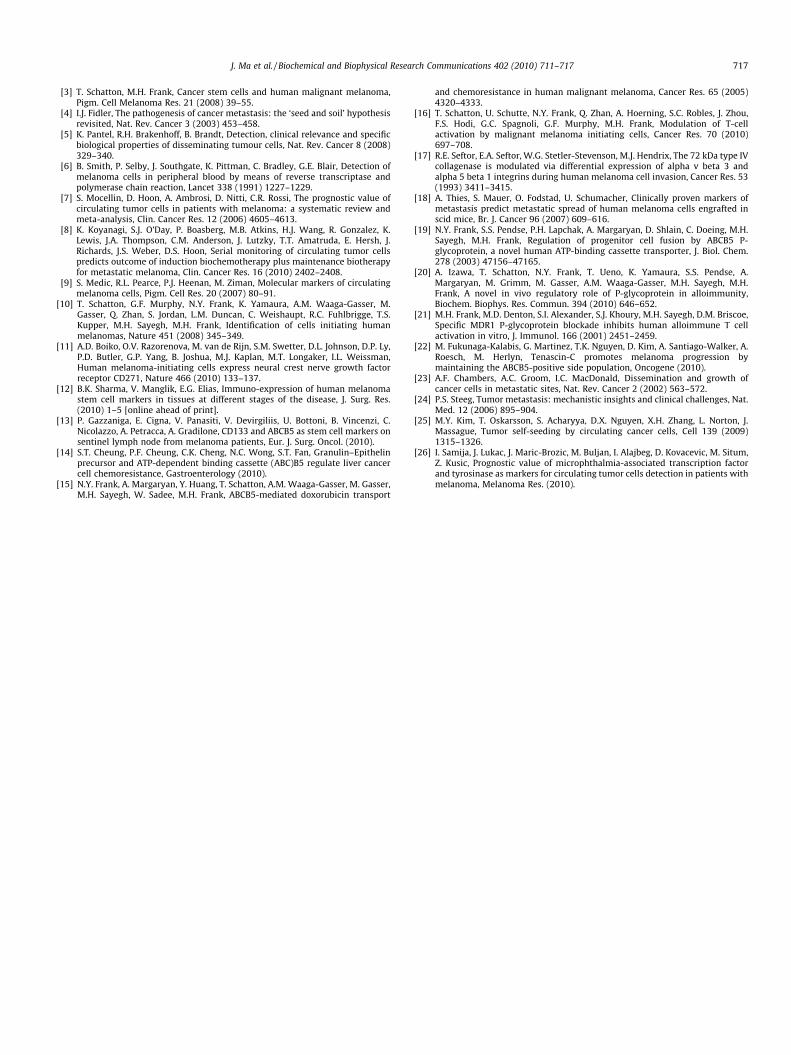
Among 27 CTC-positive primary tumor-bearing mice, 25(92.6%) had pulmonary metastases, whereas only one of five(20%) of CTC-negative primary tumor-bearing mice showed evi-dence for pulmonary metastases (Fig. 3A and B), demonstrating a
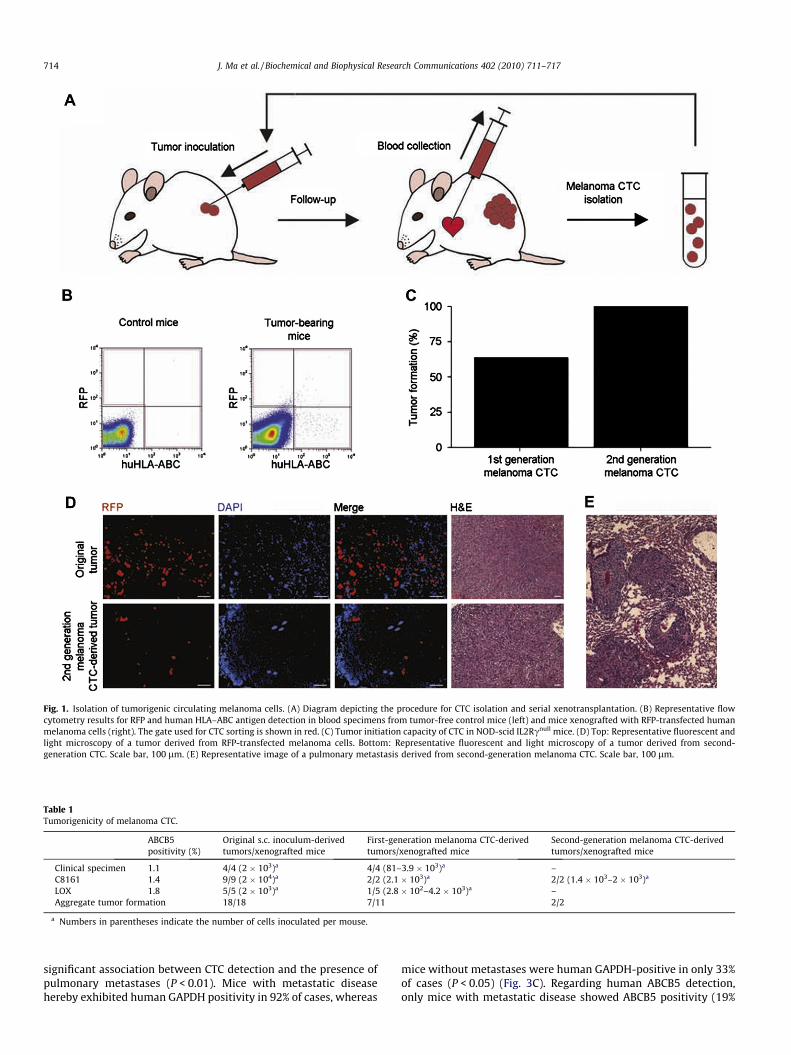
Fig. 1. Isolation of tumorigenic circulating melanoma cells. (A) Diagram depicting the procedure for CTC isolation and serial xenotransplantation. (B) Representative flowcytometry results for RFP and human HLA–ABC antigen detection in blood specimens from tumor-free control mice (left) and mice xenografted with RFP-transfected humanmelanoma cells (right). The gate used for CTC sorting is shown in red. (C) Tumor initiation capacity of CTC in NOD-scid IL2Rcnull mice. (D) Top: Representative fluorescent andlight microscopy of a tumor derived from RFP-transfected melanoma cells. Bottom: Representative fluorescent and light microscopy of a tumor derived from second-generation CTC. Scale bar, 100 lm. (E) Representative image of a pulmonary metastasis derived from second-generation melanoma CTC. Scale bar, 100 lm.
Table 1Tumorigenicity of melanoma CTC.
ABCB5positivity (%)
Original s.c. inoculum-derivedtumors/xenografted mice
First-generation melanoma CTC-derivedtumors/xenografted mice
Second-generation melanoma CTC-derivedtumors/xenografted mice
Clinical specimen 1.1 4/4 (2 � 103)a 4/4 (81–3.9 � 103)a –C8161 1.4 9/9 (2 � 104)a 2/2 (2.1 � 103)a 2/2 (1.4 � 103–2 � 103)a
LOX 1.8 5/5 (2 � 103)a 1/5 (2.8 � 102–4.2 � 103)a –Aggregate tumor formation 18/18 7/11 2/2
a Numbers in parentheses indicate the number of cells inoculated per mouse.
714 J. Ma et al. / Biochemical and Biophysical Research Communications 402 (2010) 711–717
significant association between CTC detection and the presence ofpulmonary metastases (P < 0.01). Mice with metastatic diseasehereby exhibited human GAPDH positivity in 92% of cases, whereas
mice without metastases were human GAPDH-positive in only 33%of cases (P < 0.05) (Fig. 3C). Regarding human ABCB5 detection,only mice with metastatic disease showed ABCB5 positivity (19%

Fig. 2. Circulating melanoma cells contain ABCB5-positive tumor subpopulations. (A) Flow cytometry plots depicting ABCB5 expression in melanoma cell inocula for a clinicalspecimen and C8161, FEMX1 and LOX melanoma cells. The lower panels show isotype control-staining. (B) Flow cytometrically-determined percentages of ABCB5-positivemelanoma cells in cell inocula and resultant primary tumors, lymph node metastases, melanoma CTC, and pulmonary metastases. Means ± SE of n = 3–11 samples/group areillustrated.
J. Ma et al. / Biochemical and Biophysical Research Communications 402 (2010) 711–717 715
of metastatic cases), whereas none of the mice without metastasesexhibited ABCB5 positivity (P < 0.05) (Fig. 3D), indicating thatABCB5 might represent a novel relatively specific, albeit relativelyinsensitive molecular marker for the detection of CTC that corre-late with metastasis formation. In support of this possibility, onlyPBMC specimens derived from n = 9 stage IV metastatic melanomapatients showed ABCB5 positivity in 3 of 9 cases, whereas none ofthe PBMC specimens derived from n = 5 healthy human controlswere found to express ABCB5 (P < 0.05), consistent with previouslydetermined ABCB5 mRNA negativity of human PBMC [19].
4. Discussion
CTC are thought to be important mediators of tumor dissemina-tion and metastatic disease progression in human patients. Theyrepresent unique malignant subpopulations that emigrate fromthe cellular microenvironment and extracellular matrix supportof the primary tumor site, survive in a fluid dynamic environmentin the context of immunocompetent PBMC, and possess the capac-ity to eventually home to and establish tumorigenic growth in
secondary sites of metastasis. Not all CTC might necessarily bealike; some might readily adapt to a new microenvironment andreinitiate cellular proliferation causing metastasis, while othersmight remain dormant for prolonged time periods or ultimatelydie [23,24]. Moreover, while some CTC might drive metastatic dis-semination of the primary tumor, others have been shown to con-tribute to primary tumor growth through self-seeding [25].
CTC have previously been detected and phenotypically charac-terized in human malignant melanoma, using melanoma-specificor melanoma-associated molecular markers [9], and CTC detectionhas been shown to correlate with clinical tumor progression [26].However, the existence of tumorigenic melanoma CTC capable ofdriving metastatic progression, a concept that underlies the pos-ited prognostic value of CTC detection in this malignancy, hasnot been directly demonstrated prior to this study. Therefore, ourfinding that melanoma CTC are capable of tumor initiation andmetastatic progression significantly strengthens the rationale forfurther evaluating CTC as a biomarker for melanoma progression,prognosis and outcome, and as potential therapeutic targets inthis malignancy. Additionally, the prospective isolation of viable
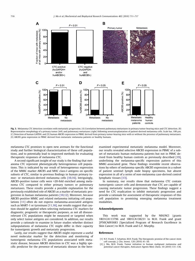
Fig. 3. Melanoma CTC detection correlates with metastatic progression. (A) Correlation between pulmonary metastases in primary tumor-bearing mice and CTC detection. (B)Representative morphology of a primary tumor (left) and pulmonary metastases (right) following xenotransplantation of patient-derived melanoma cells. Scale bar, 100 lm.(C) Detection of human GAPDH, and (D) human ABCB5 expression in PBMC derived from primary tumor-bearing mice with or without the presence of pulmonary metastases.(E) ABCB5 gene expression in PBMC derived from metastatic melanoma patients or healthy humans.
716 J. Ma et al. / Biochemical and Biophysical Research Communications 402 (2010) 711–717
melanoma CTC promises to open new avenues for the functionalstudy and further biological characterization of these cell popula-tions, and to potentially lead to improved methods for evaluatingtherapeutic responses of melanoma CTC.
A second significant insight of our study is the finding that mel-anoma CTC represent phenotypically heterogeneous cell popula-tions. This is indicated by our result of heterogeneous expressionof the MMIC marker ABCB5 and MHC class-I antigens on specificsubsets of CTC, similar to previous findings in human primary tu-mor- or metastasis-derived melanoma cells [10,16]. Intriguingly,ABCB5-positive tumor cells were >20-fold enriched among mela-noma CTC compared to either primary tumors or pulmonarymetastases. These results provide a possible explanation for thepreviously established role of ABCB5 as a marker of metastatic pro-gression in human melanoma patients [10,12]. Moreover, becauseABCB5-positive MMIC and related melanoma initiating cell popu-lations [11] often do not express melanoma-associated antigenssuch as MART-1 or tyrosinase [11,16], our results suggest that cau-tion should be applied when selecting melanoma CTC markers fordiagnostic, prognostic or therapeutic purposes, as not all disease-relevant CTC populations might be measured or targeted whenonly select tumor antigens are considered. In addition, our resultsprovide a rationale to examine in future studies whether distinctsubpopulations of melanoma CTC might differ in their capacityfor tumorigenic growth and metastatic progression.
Lastly, our results suggest that ABCB5 might represent a usefulnovel molecular marker for the detection and monitoring oftumorigenic melanoma CTC populations associated with meta-static disease, because ABCB5 detection in CTC was a highly spe-cific predictor for the presence of metastatic disease in the here-
examined experimental metastatic melanoma model. Moreover,our results revealed selective ABCB5 expression in PBMC of a sub-set of metastatic human melanoma patients but not in PBMC de-rived from healthy human controls as previously described [19],underlining the melanoma-specific expression pattern of thisMMIC-associated gene. These findings resemble recent observa-tions by others of melanoma-specific ABCB5 expression in a subsetof patient sentinel lymph node biopsy specimens, but absentexpression in all of a series of non-melanoma case-derived controllymphatic tissues [13].
In summary, our results show that melanoma CTC containtumorigenic cancer cells and demonstrate that CTC are capable ofcausing metastatic tumor progression. These findings suggest aneed for CTC eradication to inhibit metastatic progression andprovide a rationale for assessment of therapeutic responses of thiscell population to promising emerging melanoma treatmentmodalities.
Acknowledgments
This work was supported by the NIH/NCI (grants1RO1CA113796 and 1R01CA138231 to M.H. Frank and grant2P50CA093683 (Specialized Program of Research Excellence inSkin Cancer) to M.H. Frank and G.F. Murphy.
References
[1] N.Y. Frank, T. Schatton, M.H. Frank, The therapeutic promise of the cancer stemcell concept, J. Clin. Invest. 120 (2010) 41–50.
[2] J. Ma, M.H. Frank, Tumor initiation in human malignant melanoma andpotential cancer therapies, Anticancer Agents Med. Chem. 10 (2010) 131–136.
J. Ma et al. / Biochemical and Biophysical Research Communications 402 (2010) 711–717 717
[3] T. Schatton, M.H. Frank, Cancer stem cells and human malignant melanoma,Pigm. Cell Melanoma Res. 21 (2008) 39–55.
[4] I.J. Fidler, The pathogenesis of cancer metastasis: the ‘seed and soil’ hypothesisrevisited, Nat. Rev. Cancer 3 (2003) 453–458.
[5] K. Pantel, R.H. Brakenhoff, B. Brandt, Detection, clinical relevance and specificbiological properties of disseminating tumour cells, Nat. Rev. Cancer 8 (2008)329–340.
[6] B. Smith, P. Selby, J. Southgate, K. Pittman, C. Bradley, G.E. Blair, Detection ofmelanoma cells in peripheral blood by means of reverse transcriptase andpolymerase chain reaction, Lancet 338 (1991) 1227–1229.
[7] S. Mocellin, D. Hoon, A. Ambrosi, D. Nitti, C.R. Rossi, The prognostic value ofcirculating tumor cells in patients with melanoma: a systematic review andmeta-analysis, Clin. Cancer Res. 12 (2006) 4605–4613.
[8] K. Koyanagi, S.J. O’Day, P. Boasberg, M.B. Atkins, H.J. Wang, R. Gonzalez, K.Lewis, J.A. Thompson, C.M. Anderson, J. Lutzky, T.T. Amatruda, E. Hersh, J.Richards, J.S. Weber, D.S. Hoon, Serial monitoring of circulating tumor cellspredicts outcome of induction biochemotherapy plus maintenance biotherapyfor metastatic melanoma, Clin. Cancer Res. 16 (2010) 2402–2408.
[9] S. Medic, R.L. Pearce, P.J. Heenan, M. Ziman, Molecular markers of circulatingmelanoma cells, Pigm. Cell Res. 20 (2007) 80–91.
[10] T. Schatton, G.F. Murphy, N.Y. Frank, K. Yamaura, A.M. Waaga-Gasser, M.Gasser, Q. Zhan, S. Jordan, L.M. Duncan, C. Weishaupt, R.C. Fuhlbrigge, T.S.Kupper, M.H. Sayegh, M.H. Frank, Identification of cells initiating humanmelanomas, Nature 451 (2008) 345–349.
[11] A.D. Boiko, O.V. Razorenova, M. van de Rijn, S.M. Swetter, D.L. Johnson, D.P. Ly,P.D. Butler, G.P. Yang, B. Joshua, M.J. Kaplan, M.T. Longaker, I.L. Weissman,Human melanoma-initiating cells express neural crest nerve growth factorreceptor CD271, Nature 466 (2010) 133–137.
[12] B.K. Sharma, V. Manglik, E.G. Elias, Immuno-expression of human melanomastem cell markers in tissues at different stages of the disease, J. Surg. Res.(2010) 1–5 [online ahead of print].
[13] P. Gazzaniga, E. Cigna, V. Panasiti, V. Devirgiliis, U. Bottoni, B. Vincenzi, C.Nicolazzo, A. Petracca, A. Gradilone, CD133 and ABCB5 as stem cell markers onsentinel lymph node from melanoma patients, Eur. J. Surg. Oncol. (2010).
[14] S.T. Cheung, P.F. Cheung, C.K. Cheng, N.C. Wong, S.T. Fan, Granulin–Epithelinprecursor and ATP-dependent binding cassette (ABC)B5 regulate liver cancercell chemoresistance, Gastroenterology (2010).
[15] N.Y. Frank, A. Margaryan, Y. Huang, T. Schatton, A.M. Waaga-Gasser, M. Gasser,M.H. Sayegh, W. Sadee, M.H. Frank, ABCB5-mediated doxorubicin transport
and chemoresistance in human malignant melanoma, Cancer Res. 65 (2005)4320–4333.
[16] T. Schatton, U. Schutte, N.Y. Frank, Q. Zhan, A. Hoerning, S.C. Robles, J. Zhou,F.S. Hodi, G.C. Spagnoli, G.F. Murphy, M.H. Frank, Modulation of T-cellactivation by malignant melanoma initiating cells, Cancer Res. 70 (2010)697–708.
[17] R.E. Seftor, E.A. Seftor, W.G. Stetler-Stevenson, M.J. Hendrix, The 72 kDa type IVcollagenase is modulated via differential expression of alpha v beta 3 andalpha 5 beta 1 integrins during human melanoma cell invasion, Cancer Res. 53(1993) 3411–3415.
[18] A. Thies, S. Mauer, O. Fodstad, U. Schumacher, Clinically proven markers ofmetastasis predict metastatic spread of human melanoma cells engrafted inscid mice, Br. J. Cancer 96 (2007) 609–616.
[19] N.Y. Frank, S.S. Pendse, P.H. Lapchak, A. Margaryan, D. Shlain, C. Doeing, M.H.Sayegh, M.H. Frank, Regulation of progenitor cell fusion by ABCB5 P-glycoprotein, a novel human ATP-binding cassette transporter, J. Biol. Chem.278 (2003) 47156–47165.
[20] A. Izawa, T. Schatton, N.Y. Frank, T. Ueno, K. Yamaura, S.S. Pendse, A.Margaryan, M. Grimm, M. Gasser, A.M. Waaga-Gasser, M.H. Sayegh, M.H.Frank, A novel in vivo regulatory role of P-glycoprotein in alloimmunity,Biochem. Biophys. Res. Commun. 394 (2010) 646–652.
[21] M.H. Frank, M.D. Denton, S.I. Alexander, S.J. Khoury, M.H. Sayegh, D.M. Briscoe,Specific MDR1 P-glycoprotein blockade inhibits human alloimmune T cellactivation in vitro, J. Immunol. 166 (2001) 2451–2459.
[22] M. Fukunaga-Kalabis, G. Martinez, T.K. Nguyen, D. Kim, A. Santiago-Walker, A.Roesch, M. Herlyn, Tenascin-C promotes melanoma progression bymaintaining the ABCB5-positive side population, Oncogene (2010).
[23] A.F. Chambers, A.C. Groom, I.C. MacDonald, Dissemination and growth ofcancer cells in metastatic sites, Nat. Rev. Cancer 2 (2002) 563–572.
[24] P.S. Steeg, Tumor metastasis: mechanistic insights and clinical challenges, Nat.Med. 12 (2006) 895–904.
[25] M.Y. Kim, T. Oskarsson, S. Acharyya, D.X. Nguyen, X.H. Zhang, L. Norton, J.Massague, Tumor self-seeding by circulating cancer cells, Cell 139 (2009)1315–1326.
[26] I. Samija, J. Lukac, J. Maric-Brozic, M. Buljan, I. Alajbeg, D. Kovacevic, M. Situm,Z. Kusic, Prognostic value of microphthalmia-associated transcription factorand tyrosinase as markers for circulating tumor cells detection in patients withmelanoma, Melanoma Res. (2010).
Related Documents