Accepted Manuscript Title: Isolation of Spirochetes of Genus Treponema from Pigs with Ear Necrosis Authors: M¨ arit Pringle, Annette Backhans, Faruk Otman, Marie Sj ¨ olund, Claes Fellstr ¨ om PII: S0378-1135(09)00270-3 DOI: doi:10.1016/j.vetmic.2009.05.018 Reference: VETMIC 4454 To appear in: VETMIC Received date: 24-3-2009 Revised date: 6-5-2009 Accepted date: 28-5-2009 Please cite this article as: Pringle, M., Backhans, A., Otman, F., Sj¨ olund, M., Fellstr ¨ om, C., Isolation of Spirochetes of Genus Treponema from Pigs with Ear Necrosis, Veterinary Microbiology (2008), doi:10.1016/j.vetmic.2009.05.018 This is a PDF file of an unedited manuscript that has been accepted for publication. As a service to our customers we are providing this early version of the manuscript. The manuscript will undergo copyediting, typesetting, and review of the resulting proof before it is published in its final form. Please note that during the production process errors may be discovered which could affect the content, and all legal disclaimers that apply to the journal pertain.

Welcome message from author
This document is posted to help you gain knowledge. Please leave a comment to let me know what you think about it! Share it to your friends and learn new things together.
Transcript

Accepted Manuscript
Title: Isolation of Spirochetes of Genus Treponema from Pigswith Ear Necrosis
Authors: Marit Pringle, Annette Backhans, Faruk Otman,Marie Sjolund, Claes Fellstrom
PII: S0378-1135(09)00270-3DOI: doi:10.1016/j.vetmic.2009.05.018Reference: VETMIC 4454
To appear in: VETMIC
Received date: 24-3-2009Revised date: 6-5-2009Accepted date: 28-5-2009
Please cite this article as: Pringle, M., Backhans, A., Otman, F., Sjolund, M., Fellstrom,C., Isolation of Spirochetes of Genus Treponema from Pigs with Ear Necrosis,Veterinary Microbiology (2008), doi:10.1016/j.vetmic.2009.05.018
This is a PDF file of an unedited manuscript that has been accepted for publication.As a service to our customers we are providing this early version of the manuscript.The manuscript will undergo copyediting, typesetting, and review of the resulting proofbefore it is published in its final form. Please note that during the production processerrors may be discovered which could affect the content, and all legal disclaimers thatapply to the journal pertain.
Page 1 of 22
Accep
ted
Man
uscr
ipt
1
Revised version
Isolation of Spirochetes of Genus Treponema from Pigs with Ear
Necrosis
Märit Pringle a*, Annette Backhans b, Faruk Otman c, Marie Sjölund d, Claes Fellström b
a Dept of Biomedical Sciences and Veterinary Public Health, b Dept of Clinical Sciences,Swedish University of Agricultural Sciences, SE-75007 Uppsala, Sweden
c Dept of Pathology and Wildlife Diseases, d Dept of Animal Health and Antimicrobial Strategies, National Veterinary Institute, SE-75189 Uppsala, Sweden
___________________________
* Corresponding author. Tel.: +46 18 67 23 86; fax: +46 18 67 33 34. E-mail address: [email protected] (M. Pringle).
Märit Pringle, DVM, PhDDept of Biomedical Sciences and Veterinary Public HealthDiv of Bacteriology and Food Safety Swedish University of Agricultural Sciences (SLU)Box 7009SE-750 07 UppsalaSWEDEN
Manuscript
Page 2 of 22
Accep
ted
Man
uscr
ipt
“2002” has been added to Stamm et al.
The recently published reference has been updated to:
Stamm, L.V., Walker, R.L., Read, D.H., 2008. Genetic diversity of bovine ulcerative
mammary dermatitis-associated Treponema. Vet. Microbiol. 136, 192-196.
It is not clear to us were the “Walker et al. 1995” reference should be included in the text. We have not used the same method for culturing and all DD DNA sequences mentioned in the manuscript are from later publications.
Page 3 of 22
Accep
ted
Man
uscr
ipt
2
Abstract 1
Various ear lesions, often caused by ear biting, are common in pigs. Some herds 2
have a high frequency of ear necrosis, a syndrome characterized by necrotic 3
lesions along the rim of the pinna, often bilateral and sometimes resulting in loss 4
of the entire ear. In samples from such lesions spirochetes have been observed 5
microscopically but never isolated or identified. In this study two herds with6
periodic outbreaks of ear necrosis among weaners were investigated. Samples 7
were collected from ear lesions and from the gingiva of the pigs. Spirochetes were 8
observed in silver stained histological sections and by phase contrast microscope 9
in scrapings from the necrotic lesions. From an ear lesion a pure spirochete isolate 10
was obtained and identified as a yet unnamed species of genus Treponema, 11
closely related to spirochetes found in digital dermatitis in cattle. From the oral 12
samples two pure isolates were obtained. One of these isolates was identified as 13
the same species as in the ear lesion and one as Treponema socranskii. Species 14
identification was based on 16S rRNA gene sequences.15
16
Keywords: Ear necrosis; Ear biting; Treponema; Spirochete; Pig17
18
1. Introduction19
20
Ear necrosis in pigs is a scantily studied malady. Other names used are necrotic 21
ear syndrome and ulcerative spirochetosis of the ear (Cameron, 2006). In a case 22
study (Harcourt, 1973) the lesions were reported to appear at around 8-9 weeks of 23

Page 4 of 22
Accep
ted
Man
uscr
ipt
3
age with the presence of bilateral small reddened areas close to the junction of the 24
lower margin of the ear with the neck. The lesions then progressed to necrosis and 25
in severe cases involved the entire length of the margin of the pinna. On 26
histopathological examination numerous spirochetes were found together with 27
coccoid bacteria. In several other skin disorders of pigs such as facial necrosis in 28
suckling pigs, wound infections and foot-rot spirochetes have been observed since 29
the early 1900´s (Cleland, 1908; Dodd, 1906; Gilruth, 1910; Hindmarsh, 1937; 30
Neitz and Canham, 1930; Osborne and Ensor, 1955). Coccoid bacteria found in 31
ear necrosis lesions in pigs have been identified as Staphylococcus hyicus and 32
beta-hemolytic streptococci (Cameron, 2006). In a recent large-scale study of the 33
prevalence of clinical signs in Danish finisher pigs, ear necrosis was the most 34
common finding. About 30% of the recorded clinical signs were ear necrosis, 35
defined as: open wound(s) or crust(s) at the tip or base of one or both ears 36
(Petersen et al., 2008). Ear lesions defined as ear biting were also a common 37
clinical observation (28.3%) in a study from the Netherlands (Blocks et al., 1994).38
39
To the best of our knowledge the spirochetes observed in smears and histological 40
sections from various skin diseases in pigs have never been isolated. Species 41
names as Borrelia suilla and Borrelia suis have been used, however without 42
references to an original source (Cameron, 2006; Penny et al., 1971).43
44
In this study, two pig herds with periodic outbreaks of ear necrosis among 45
weaners were investigated. The aims were to isolate and identify spirochetes and 46

Page 5 of 22
Accep
ted
Man
uscr
ipt
4
spirochetal DNA from the necrotic ear lesions, and because ear biting has been 47
discussed as a cause of ear necrosis, from the gingiva of the pigs.48
49
2. Materials and methods50
51
2.1. Pig herds investigated and clinical signs52
53
Ear ulcers were observed at several occasions during two years in two organic pig 54
herds, A and B, in Sweden. Herd A was a piglet producing herd with 180 sows 55
employing batch production (~24 sows/batch) with all in-all out procedures 56
practiced in all units except for the dry sow unit. The farrowing unit had 57
individual pens with partially slatted floors. When the piglets were two weeks old, 58
sows and piglets were moved to a barn with four group pens (12 sows + 59
piglets/pen) with deep straw bedding and access to a small outdoor surface. Pigs 60
did not have direct contact to herd mates in adjacent pens but the airspace was 61
shared for two pens. Pigs were weaned at 6 weeks of age, fed with dry feed with 62
constant access ad libitum, and kept in the group pens until sale. Batches of pigs 63
were sold at approximately 30 kilos of weight to the finishing herd B. The first 64
signs of ear ulcers were noted right before or after weaning, although bites or 65
scratches on the pigs’ ears were minimal. Ulcers were bilateral and situated close 66
to the junction of the lower margin of the ear. Over the course of several weeks, 67
ulcers increased in size and spread along the rim of the ear and became necrotic. 68
In the most advanced cases sloughing of the necrotic tissue occurred, resulting in 69

Page 6 of 22
Accep
ted
Man
uscr
ipt
5
a total loss of the ear. During the two year period the number of pigs with ear 70
ulcers in each group varied, with the highest prevalence observed during the 71
winter period, when 50-70% affected pigs were commonly noted. Pigs were not 72
medically treated due to ear necrosis. General health and growth rate were 73
seemingly unaffected. At 60-80 kilos of weight the lesions had healed, leaving 74
affected ears with a normal appearance or a portion of the ear missing. 75
76
2.2. Histology77
78
In three pigs from herd A sent for necropsy, bilateral lesions at the ear base and 79
the ear pinna were observed. Samples for histology were taken from the margin of 80
the lesions. After fixation and embedding in paraffin by standard methods 5-7 m 81
sections were cut for hematoxylin and eosin (HE) and Warthin-Starry silver 82
staining. 83
84
2.3. Sampling for bacteriological cultures and culture conditions85
86
Samples were collected from live pigs after cleaning the ear lesions with water of 87
drinking quality. Scalpel blade scrapings were taken from the margins of the 88
lesions. The scrapings were transported in tubes with FAB (fastidious anaerobe 89
broth, LAB 71, LabM, Lancashire, UK). From the gingiva, cotton swab samples 90
were taken and transported in Amies medium with charcoal (COPAN, Brescia, 91
Italy). The samples were transported directly to the lab for culturing. Four ear 92
Page 7 of 22
Accep
ted
Man
uscr
ipt
6
lesion samples were taken from both herds and four gingival samples from herd 93
A. Flasks with FABSA (FAB with 25% fetal calf serum, S 0115, Biochrom AG, 94
Germany; 10% rabbit serum, R4505 Sigma, Sigma-Aldrich Sweden AB; 10 µg/ml 95
rifampicin Sigma, Sigma-Aldrich Sweden AB, and 10 µg/ml enrofloxacin Fluka, 96
Sigma-Aldrich Sweden AB,) and OTEBSA (oral treponeme enrichment broth 97
(OTEB), Anaerobe Systems, Morgan Hill, CA, USA 25% fetal calf serum, S 98
0115, Biochrom AG, Germany; 10% rabbit serum, R4505 Sigma, Sigma-Aldrich 99
Sweden AB; 10 µg/ml rifampicin Sigma, Sigma-Aldrich Sweden AB, and 10 100
µg/ml enrofloxacin Fluka, Sigma-Aldrich Sweden AB) were inoculated with 101
scrapings and swabs and incubated in 37 °C, in anaerobic jars on a shaker (90 102
rpm). The samples from herd A were inoculated only in FABSA and the samples 103
from herd B in both FABSA and OTEBSA. The heavily contaminated gingival 104
cultures were purified through a membrane filter with pore size 0,22 m. The 105
filter was placed on FAA plates (fastidious anaerobe agar with 10% horse blood, 106
National Veterinary Institute, Uppsala, Sweden) and inoculated with a drop of 107
broth culture. The filter was removed after 2-3 days and the growth under the 108
filter was checked through phase contrast microscopy.109
110
2.4. Amplification and sequencing of 16S rDNA and ISR2 111
112
DNA was prepared from the broth cultures and from three ear lesion scrapings 113
from herd B. The material was washed twice in PBS (phosphate buffered saline, 114
pH 7.3), boiled for 10 min in sterile water and cell debris removed by 115
Page 8 of 22
Accep
ted
Man
uscr
ipt
7
centrifugation. Primers trepF 5’-GTTTGATCCTGGCTCAGAACG-3’ and trepR 116
5’-CGGTACGGCTACCTTGTTACG-3’ were designed for amplification and 117
DNA sequencing of the 16S rRNA gene. Internal primers used for sequencing 118
have been described previously (Johansson et al., 2004). For amplification of the 119
16S-tRNAIle region of the intergenic spacer region 2 (ISR2) a protocol developed 120
by Stamm et al. was followed (Stamm et al., 2002). The sequence from the ISR2 121
PCR was determined using the PCR primers as sequencing primers. Amplified 122
fragments were purified (illustra GFX PCR DNA and Gel Band Purification Kit, 123
GE Healthcare) and sequenced on an ABI PRISM 3100 Genetic Analyzer 124
(Applied Biosystems). Sequences were edited using ContigExpress, Vector NTI 125
Advance 10 (Invitrogen).126
127
3. Results128
129
3.1. Gross pathology130
131
In all three pigs from herd A sent for necropsy, bilateral chronic active necrotizing 132
dermatitis situated close to the junction of the lower margin of the ear pinna and 133
ear bases was observed. The lesions consisted of irregular bleeding margins at the 134
surface, a layer of necrosis and underlying this, irregular zones showing different 135
degrees of blackish grey and red discoloration. The crusts were flat to thick, 136
irregular, and slightly raised above the epidermis. Both ears were thickened and 137
the lesions were frequently associated with mild edema and erythema. 138
Page 9 of 22
Accep
ted
Man
uscr
ipt
8
139
3.2. Histology140
141
The main histological features were of chronic ulcerative and superficial pustular 142
dermatitis with severe acanthosis and hyperkeratosis (Fig. 1). Adjacent to the 143
ulcers the surviving epidermis was hyperplastic with numerous rete ridges 144
extending deep into the dermis. The epidermis was covered by a thick layer of 145
crust consisting of necrotic epidermal cells, degenerated inflammatory cells, and 146
proteinaceous exudates. Inflammatory changes were a prominent part of the 147
overall reaction and neutrophils formed a main proportion of the cells involved. 148
There was vasculitis in the arterioles and venules characterized by hyaline 149
degeneration, medial hyperplasia and thrombosis.150
151
Silver stained sections from all three pigs showed numerous spirochetes, often 152
aligned in bundles within bands of connective tissue at the junction of the necrotic 153
and granulation tissues (Fig. 2). Coccoid or cocco-bacillary bacteria were 154
frequently seen both within the thick layer of exudates on the surface of early 155
lesions and deep within the tissues of the more severely ulcerated lesions. 156
157
3.3. Isolation of spirochetes158
159
From herd B spirochetes were observed through phase contrast microscopy in two 160
of four scrapings and one of these samples resulted in a pure isolate of spirochetes 161
Page 10 of 22
Accep
ted
Man
uscr
ipt
9
designated as T A4. In one of four scrapings from herd A, a low number of 162
spirochetes were observed through phase contrast microscopy. It was not possible 163
to obtain a spirochete culture from this sample. Isolation of T A4 was made in 164
OTEBSA but growth of the pure isolate was supported equally well by FAB with 165
fetal calf serum and after subculture a fully grown culture was obtained in 4-5 166
days. From the four gingival samples obtained from herd A two spirochete 167
isolates, designated T M1 and T M4, were purified through filter on agar and 168
colony material was transferred as agar cylinders, with a pasteur pipette, to new 169
broth cultures. On FAA plates all three isolates formed pinpoint colonies. Isolates 170
T M1 and T A4 had distinct hemolysis zones but no hemolysis was observed for T 171
M4.172
173
3.4. 16S rRNA gene sequences from cultures174
175
A 16S rRNA gene fragment of 1466 nucleotides was obtained for T A4 and T M1 176
and of 1455 nucleotides for T M4 (deposited in GenBank with accession numbers 177
FJ805835-FJ805837). The sequences for T A4 and T M1 were identical except for 178
two positions (one in each isolate) that had polymorphic nucleotide positions, 179
134R in T A4 and 199Y in T M1 (Escherichia coli numbering). These two 180
positions were polymorphic with two nucleotides in 50% representation and were 181
present in sequences from both strands. Except for the polymorphism the 16S 182
rRNA gene sequences for T A4 and T M1 were also identical to a deposited 183
sequence for a yet unnamed Treponema sp. (GenBank accession number 184

Page 11 of 22
Accep
ted
Man
uscr
ipt
10
EF061268) isolated from digital dermatitis in cattle in the UK (Evans et al., 2008). 185
The T M4 isolate was most closely related to Treponema socranskii subsp. 186
paredis (GenBank accession number AY369254) with 1451 identical nucleotides 187
out of 1455 (Correia et al., 2003).188
189
3.5. ISR2 sequence from tissue scraping190
191
For two of the three DNA preparations from the lesion scrapings from herd B the 192
ISR2 PCR resulted in a single band at the size of approximately 300 bp (A3 and 193
A4). The third sample was negative. The amount of product was very low in 194
sample A3 but the product from sample A4, which was the sample that also 195
yielded a spirochete culture, was purified and sequenced. The chromatogram 196
showed a clearly dominating sequence but with traces of contaminating 197
nucleotides. ISR fragments from Treponema from digital dermatitis, isolate 9-198
3143 (AF179259), and ulcerative mammary dermatitis, clone C1BT2-3199
(AY342044), in cattle in USA were the closest related deposited sequences in 200
GenBank (Stamm et al., 2002; Stamm et al., 2008). When the sequence from the 201
present study was compared to the AF179259 sequence 313 out of 317 202
nucleotides were identical and for AY342044, which is shorter, 205 out of 209 203
were identical. Both comparisons covered the highly variable region between 16S 204
rRNA and tRNA-Ile.205
206
4. Discussion207

Page 12 of 22
Accep
ted
Man
uscr
ipt
11
208
While production losses associated with ear necrosis have not been documented 209
the condition should be considered an animal welfare problem. In this study 210
samples from necrotic ear lesions were investigated regarding the presence of 211
spirochetes. Spirochetes were observed in ear lesion samples from both of the 212
studied herds. One Treponema sp. isolate was obtained from herd B and silver 213
impregnated histological sections from herd A showed spirochetes in the 214
epidermis. Besides that Treponema spp. are fastidious and difficult to culture in 215
competition with fast growing bacteria in the samples, other reasons for only 216
obtaining one isolate could be that scrapings may be too superficial or that the 217
spirochetes were unevenly distributed in the lesions. Biopsy samples from the 218
margin of the lesions would probably increase the chance of isolating spirochetes.219
The ISR2 sequence directly from a tissue sample from a pig in herd B was 220
obtained with primers developed to be universal for the genus Treponema (Stamm 221
et al., 2002). ISR2 amplicons from digital dermatitis samples from cattle claws 222
usually contain fragments from several species of Treponema and need to be 223
cloned before sequencing. However from the sample from herd B it was possible 224
to obtain a readable DNA sequence without cloning, indicating that a single 225
species was dominating. Only a limited number of ISR2 sequences from 226
Treponema spp. are deposited in GenBank. However two of these, from skin 227
diseases in cattle, shared a high similarity to the sequence from the ear lesion, 228
especially considering that intergenic spacer region between 16S rRNA and 229
tRNA-Ile is highly variable. There are two deposited 16S rRNA sequences from 230

Page 13 of 22
Accep
ted
Man
uscr
ipt
12
the digital dermatitis isolate from USA, 9-3143, clone 5 and clone 7 (AF109070-231
71) and these belong to the same 16S rRNA phylotype as the digital dermatitis 232
isolate from the UK (EF061268) and T A4. This indicates that the dominating 233
phylotype in the ear lesion of one pig in this study also is the isolated.234
235
In addition to the spirochetes, numerous cocci could be seen both through direct 236
microscopy of the samples and in the histological sections. Staphylococcus hyicus237
and beta-hemolytic streptococci were identified in the samples through 238
conventional methods (data not shown).239
240
Ear biting is commonly suggested as the cause of ear necrosis in pigs. In the 241
investigated herds sometimes as much as 70% of the pigs were affected, and as in 242
the herd described by Harcourt, the lesions were always bilateral and pigs of a 243
similar age involved (Harcourt, 1973). The anatomically consistent localization 244
and the epidemic spread in the herd followed by healing are in our view difficult 245
to explain solely by opportunistic bacterial infection. Further the ad libitum246
feeding regime practiced in herd A should have minimized the risk for ear biting.247
248
Several species of genus Treponema have been isolated from the mouth of 249
humans, cats and dogs and are associated with periodontal disease (Holt and 250
Ebersole, 2006; Nordhoff et al., 2008; Riviere et al., 1996; Valdez et al., 2000). 251
The spirochetal flora in the mouth of the pig has not been investigated before. In 252
this study the 16S rRNA gene sequences from two gingival isolates showed that 253

Page 14 of 22
Accep
ted
Man
uscr
ipt
13
the pig oral flora can include T. socranskii and the Treponema sp. found in the ear 254
lesion. As for the oral flora of dogs and humans it is likely that analyzing many 255
oral samples from pigs would reveal several other Treponema spp. Isolates T M1 256
and T A4 are obviously not the same clone since both have a polymorphism in the 257
16S rRNA gene but at different positions. Despite this, the findings of the same 258
phylotype at both sampling sites indicate a possible relationship between ear 259
necrosis and oral spirochetes in pigs. Additionally, the 16S rRNA gene sequences 260
of T A4 and T M1 were identical (except for the polymorphisms) to the sequence 261
deposited for an isolate from digital dermatitis in cattle (Evans et al., 2008) and 262
moreover, only differed five nucleotides from an isolate from ovine foot rot 263
(GenBank accession number AF363634) (Demirkan et al., 2001). This yet 264
unnamed phylotype of Treponema occurs in different skin disorders in three 265
different animal species and in the oral flora of the pig, suggesting both a 266
pathogenic potential and a broad host range.267
268
Spirochetes have been observed in other different skin lesions in pigs for example 269
in foot-rot, castration wounds and ulcers in the mouth (Blandford et al., 1972; 270
Gilruth, 1910; Osborne and Ensor, 1955). Isolation and identification of such 271
spirochetes is of interest both for comparison between the different conditions in 272
pigs and for comparison with skin diseases caused by Treponema spp. in other 273
animals including humans. 274
275
5. Conclusion 276
Page 15 of 22
Accep
ted
Man
uscr
ipt
14
277
Spirochetes of genus Treponema can be isolated from ear necrosis lesions and 278
from the gingiva in pigs. The Treponema phylotype isolated from the ear lesion 279
was closely related, but not identical, to one of the oral isolates. Increased 280
understanding of the role of the spirochetes in porcine skin disorders and the route 281
of infection would be helpful both for choice of treatment of acute lesions and to 282
be able to recommend preventive measures.283
284
Acknowledgements285
286
We would like to thank the group of veterinary students who helped with 287
sampling and the investigation of the two herds: Karolina Dahlqvist, Birgitta 288
Gralén, Karin Jonasson, Elisabeth Lindahl, Karin Lindroth, Maja Nilsson, Maria 289
Pylkkänen, Nathalie Sjögren and Helena Törnelius.290
291
References292
293
Blandford, T.B., Bygrave, A.C., Harding, J.D., Little, T.W., 1972. Suspected 294
porcine ulcerative spirochaetosis in England. Vet. Rec. 90, 15.295
296
Blocks, G.H., Vernooy, J.C., Verheijden, J.H., 1994. Integrated quality control 297
project: relationships between pathological findings detected at the 298
Page 16 of 22
Accep
ted
Man
uscr
ipt
15
slaughterhouse and information gathered in a veterinary health scheme at 299
pig farms. Vet. Q. 16, 123-127.300
301
Cameron, R.D.A., 2006. Diseases of the skin, In: Straw, B.E., Zimmerman, J., 302
D´Allaire, S.,Taylor, D.J. (Eds.), Diseases of Swine, 9th Edition, Blackwell 303
Publishing, Ames, IA, pp. 179-198.304
305
Cleland, J.B., 1908. Note on spirochaetes in castration tumours of pigs, 306
Parasitology, Suppl. Journal of Hygiene. 1, 218-219.307
308
Correia, F.F., Plummer, A.R., Ellen, R.P., Wyss, C., Boches, S.K., Galvin, J.L., 309
Paster, B.J., Dewhirst, F.E., 2003. Two paralogous families of a two-gene 310
subtilisin operon are widely distributed in oral treponemes. J. Bacteriol. 311
185, 6860-6869.312
313
Demirkan, I., Carter, S.D., Winstanley, C., Bruce, K.D., McNair, N.M., 314
Woodside, M., Hart, C.A., 2001. Isolation and characterisation of a novel 315
spirochaete from severe virulent ovine foot rot. J. Med. Microbiol. 50, 316
1061-1068.317
318
Dodd, S., 1906. A disease of the pig due to a spirochaeta. J. Comp. Pathol. 319
Terapeut. 19, 216-222.320
321
Page 17 of 22
Accep
ted
Man
uscr
ipt
16
Evans, N.J., Brown, J.M., Demirkan, I., Murray, R.D., Vink, W.D., Blowey, 322
R.W., Hart, C.A., Carter, S.D., 2008. Three unique groups of spirochetes 323
isolated from digital dermatitis lesions in UK cattle. Vet. Microbiol. 130, 324
141-150.325
326
Gilruth, J.A., 1910. Spirochaetae in lesions affecting the pig. Proc. Royal Soc. of 327
Victoria 23, 105-109.328
329
Harcourt, R.A., 1973. Porcine ulcerative spirochaetosis. Vet. Rec. 92, 647-648.330
331
Hindmarsh, W.L., 1937. Ulcerative granuloma of pigs (spirochaetal tumours of 332
pigs). New South Wales Department of Agriculture, Veterinary Research 333
Report. 64-70.334
335
Holt, S.C., Ebersole, J.L., 2006. The oral spirochetes: their ecology and role in the 336
pathogenesis of periodontal disease, In: Radolf, J.D., Lukehart, S.A. 337
(Eds.), Pathogenic Treponema molecular and cellular biology, Caister 338
Academic Press, Wymondham, Norfolk, UK, pp. 323-356.339
340
Johansson, K.-E., Duhamel, G.E., Bergsjø, B., Engvall, E.O., Persson, M., 341
Pettersson, B., Fellström, C., 2004. Identification of three clusters of 342
canine intestinal spirochaetes by biochemical and 16S rDNA sequence 343
analysis. J. Med. Microbiol. 53, 345-350.344
Page 18 of 22
Accep
ted
Man
uscr
ipt
17
345
Neitz, W.O., Canham, A.S., 1930. A short note on the spirochaetal wound 346
infections of pigs. Report of the director of veterinary services and animal 347
industry. 69-79.348
349
Nordhoff, M., Rühe, B., Kellermeier, C., Moter, A., Schmitz, R., Brunnberg, L., 350
Wieler, L.H., 2008. Association of Treponema spp. with canine 351
periodontitis. Vet. Microbiol. 127, 334-342.352
353
Osborne, H.G., Ensor, C.R., 1955. Some aspects of the pathology, aetiology, and 354
therapeutics of foot-rot in pigs. N. Z. Vet. J. 3, 91-99.355
356
Penny, R.H., Edwards, M.J., Mulley, R., 1971. Clinical observations of necrosis 357
of the skin of suckling piglets. Aust. Vet. J. 47, 529-537.358
359
Petersen, H.H., Nielsen, E.O., Hassing, A.G., Ersboll, A.K., Nielsen, J.P., 2008. 360
Prevalence of clinical signs of disease in Danish finisher pigs. Vet. Rec. 361
162, 377-382.362
363
Riviere, G.R., Thompson, A.J., Brannan, R.D., McCoy, D.E., Simonson, L.G., 364
1996. Detection of pathogen-related oral spirochetes, Treponema 365
denticola, and Treponema socranskii in dental plaque from dogs. J. Vet. 366
Dent. 13, 135-138.367
Page 19 of 22
Accep
ted
Man
uscr
ipt
18
368
Stamm, L.V., Bergen, H.L., Walker, R.L., 2002. Molecular typing of 369
papillomatous digital dermatitis-associated Treponema isolates based on 370
analysis of 16S-23S ribosomal DNA intergenic spacer regions. J. Clin. 371
Microbiol. 40, 3463-3469.372
373
Stamm, L.V., Walker, R.L., Read, D.H., 2008. Genetic diversity of bovine 374
ulcerative mammary dermatitis-associated Treponema. Vet. Microbiol. 375
136, 192-196. 376
377
Valdez, M., Haines, R., Riviere, K.H., Riviere, G.R., Thomas, D.D., 2000. 378
Isolation of oral spirochetes from dogs and cats and provisional 379
identification using polymerase chain reaction (PCR) analysis specific for 380
human plaque Treponema spp. J. Vet. Dent. 17, 23-26.381
382
Figure legends383
384
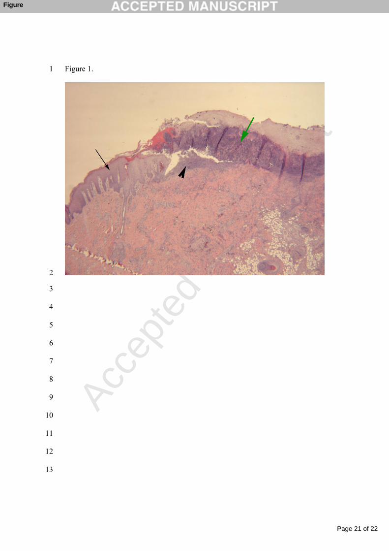
Fig. 1. Histological section of an ear necrosis lesion from herd A. The epidermis 385
is covered by thick crusts (green arrow). Below the crusts there is an ulcer with 386
abundant degenerated inflammatory cells (arrow head). Epidermis shows severe 387
thickening (acanthosis) with rete ridges (black arrow). Hematoxylin and eosin; 388
(original magnification 10 X 0.63 X 25).389
390

Page 20 of 22
Accep
ted
Man
uscr
ipt
19
Fig. 2. Silver impregnated sections of an ear necrosis lesion from herd A.391
Numerous spirochetes are visualized at the junction of the necrotic and 392
granulation tissues. The arrows show bundles of spirochetes aligned with 393
connective tissue. Warthin-Starry; (original magnification 10 X 100).394
395
Page 21 of 22
Accep
ted
Man
uscr
ipt
Figure 1. 1
2
3
4
5
6
7
8
9
10
11
12
13
Figure

Page 22 of 22
Accep
ted
Man
uscr
ipt
Figure 2. 14
15
16
Related Documents