ELSEVIER Journal of Chromatography B, 680 (1996) 201-212 JOURNAL OF CHROMATOGRAPHY B: BIOMEDICAL APPLICATIONS Isolation by preparative free-flow electrophoresis and aqueous two- phase partition from rat adipocytes of an insulin-responsive small vesicle fraction with glucose transport activity • a ~ • a,c a,c Dorothy M. Morre ' , David W. Sammons b, Jonathan Ylm , Maria Bruno , Timothy Snyder a, Timothy Reust a, Lidia Maianu d, W. Timothy Garvey J, D. James Morr6 c aDepartment of Foods and Nutrition, Purdue University, West Lafayette, IN 47907, USA bDepartment of Veterinary Science, University of Arizona, Tucson, AZ 85721, USA ~Department of Medicinal Chemistry and Pharmacognosy, Purdue University, West Lafayette, IN 47907, USA aDepartment of Medicine, Medical University of South Carolina and the Charleston Veterans Affairs Medical Center, Charleston, SC 29425, USA Abstract Preparative free-flow electrophoresis and aqueous two-phase polymer partition were used to obtain a plasma membrane-enriched fraction of adipocytes isolated from epididymal fat pads of the rat together with a fraction enriched in small vesicles with plasma membrane characteristics (thick membranes, clear dark-light-dark pattern). The electrophoretic mobility of the small vesicles was much less than that of the plasma membrane consistent with an inside-out orientation whereby charged molecules normally directed to the cell surface were on the inside. When plasma membranes and the small vesicle fraction were isolated from fat cells treated or not treated with 100/zU/ml insulin and the resident proteins of the two fractions analyzed by SDS-PAGE, the two fractions exhibited characteristic responses involving specific protein bands. Insulin treatment for 2 min resulted in the loss of a 90 kDa band from the plasma membrane. At the same time, a ca. 55-kDa peptide band that was enhanced in the plasma membrane was lost from the small vesicle fraction. The latter corresponded on Western blots to the GLUT-4 glucose transporter. Thus, we suggest that the small vesicle fraction with characteristics of inside-out plasma membrane vesicles may represent the internal vesicular pool of plasma membrane subject to modulation by treatment of adipocytes with insulin. Keywords: Free-flow electrophoresis; Partitioning; Vesicles, insulin-responsive; Glucose transport I. Introduction Our laboratory has used preparative free-flow electrophoresis [ 1,2] and aqueous two-phase polymer partition [3,4] to obtain a plasma membrane-enriched fraction from adipocytes of epididymal fat pads of Correspondmg author. the rat together with a fraction enriched in small vesicles with plasma membrane characteristics (thick membranes, clear dark-light-dark pattern) [4,5]. The electrophoretic mobility of the small vesicles was much less than that of the plasma membrane con- sistent with an inside-out orientation whereby charged molecules normally directed to the cell surface were on the inside. These fractions have been 0378-4347/96/$15.00 © 1996Elsevier Science B.V. All rights reserved SSDI 0378-4347(95)00391-6

Welcome message from author
This document is posted to help you gain knowledge. Please leave a comment to let me know what you think about it! Share it to your friends and learn new things together.
Transcript
ELSEVIER Journal of Chromatography B, 680 (1996) 201-212
JOURNAL OF CHROMATOGRAPHY B: BIOMEDICAL APPLICATIONS
Isolation by preparative free-flow electrophoresis and aqueous two- phase partition from rat adipocytes of an insulin-responsive small
vesicle fraction with glucose transport activity • a ~ • a , c a , c Dorothy M . M o r r e ' , D a v i d W . S a m m o n s b, Jonathan Ylm , Maria Bruno ,
Timothy Snyder a, Timothy Reust a, Lidia Maianu d, W. Timothy Garvey J, D. James Morr6 c
aDepartment of Foods and Nutrition, Purdue University, West Lafayette, IN 47907, USA bDepartment of Veterinary Science, University of Arizona, Tucson, AZ 85721, USA
~Department of Medicinal Chemistry and Pharmacognosy, Purdue University, West Lafayette, IN 47907, USA aDepartment of Medicine, Medical University of South Carolina and the Charleston Veterans Affairs Medical Center, Charleston,
SC 29425, USA
Abstract
Preparative free-flow electrophoresis and aqueous two-phase polymer partition were used to obtain a plasma membrane-enriched fraction of adipocytes isolated from epididymal fat pads of the rat together with a fraction enriched in small vesicles with plasma membrane characteristics (thick membranes, clear dark-light-dark pattern). The electrophoretic mobility of the small vesicles was much less than that of the plasma membrane consistent with an inside-out orientation whereby charged molecules normally directed to the cell surface were on the inside. When plasma membranes and the small vesicle fraction were isolated from fat cells treated or not treated with 100/zU/ml insulin and the resident proteins of the two fractions analyzed by SDS-PAGE, the two fractions exhibited characteristic responses involving specific protein bands. Insulin treatment for 2 min resulted in the loss of a 90 kDa band from the plasma membrane. At the same time, a ca. 55-kDa peptide band that was enhanced in the plasma membrane was lost from the small vesicle fraction. The latter corresponded on Western blots to the GLUT-4 glucose transporter. Thus, we suggest that the small vesicle fraction with characteristics of inside-out plasma membrane vesicles may represent the internal vesicular pool of plasma membrane subject to modulation by treatment of adipocytes with insulin.
Keywords: Free-flow electrophoresis; Partitioning; Vesicles, insulin-responsive; Glucose transport
I. Introduct ion
Our laboratory has used preparative free-flow electrophoresis [ 1,2] and aqueous two-phase polymer partition [3,4] to obtain a plasma membrane-enriched fraction from adipocytes of epididymal fat pads of
Correspondmg author.
the rat together with a fraction enriched in small vesicles with plasma membrane characteristics (thick membranes, clear dark-light-dark pattern) [4,5]. The electrophoretic mobility of the small vesicles was much less than that of the plasma membrane con- sistent with an inside-out orientation whereby charged molecules normally directed to the cell surface were on the inside. These fractions have been
0378-4347/96/$15.00 © 1996 Elsevier Science B.V. All rights reserved SSDI 0378-4347(95)00391-6
202 D.M. Morrd et aL / ,L Chromatogr. B 6~¢0 (1096) 201 212
utilized in the present report to investigale the cascade of membrane-associated events that occur in response to insulin treatment of isolated adipocytes. These include modulation of the 90-kDa peptide reported previously by Schoenle et al. [61 to be lost from the plasma membrane of adipocytes upon insulin treatment and a redistribution of the GLUT-4 glucose transporter consistent with translocation from a small vesicle fraction to the plasma mem- brane.
The insulin action sequence begins with ligand binding to cell-surface receptors which are hetero- dimers comprised of 135 kDa alpha subunits and 95 kDa beta subunits. Binding of insulin to alpha subunits initiates autophosphorylation of tyrosine residues in the beta subunits which in turn augments receptor tyrosine phosphorylase activity for other intracellular substrates [7,8]. Using isolated rat adipocytes, we found increased numbers of cell surface invaginations and loss of vesicular profiles associated with the plasma membrane during the first 3 min of insulin administration and return to initial values by 5 min [9]. The findings are expected to have significance for the elucidation of early events responsible for the down regulation of insulin re- ceptors [10-12] at the cell surface.
Propagation of signal transduction by ligand-acti- vated insulin receptors results in stimulation of glucose transport into adipose and muscle cells. The increase in transport activity is due to translocation of an intracellular pool of facilitative glucose trans- porter proteins to the cell surface [13,141. This process involves exocytosis of transporter-containing vesicles, together with other insulin-responsive vesi- cles containing alternative receptors [15], which fuse with plasma membrane allowing an exofacial orientation. The predominant insulin-responsive glu- cose transporter isoform has been designated GLUT 4. While the involvement of GLUT 4 vesicles has been established [16,17], the actual location of the intracellular pool and organelle source of translocat- ing GLUT 4 has not been identified with certainty. This is because conventional methods for membrane subfractionation have not yielded a pure intracellular fraction containing GLUT 4, and morphological localization using immunocytochemistry has not been definitive. The current study utilizes new approaches, namely free-flow electrophoresis and
aqueous two-phase polymer partitioning, to study insulin's effects on vesicular traffic in isolated adipocytes.
2. Materials and methods
Z 1. Preparation of adipocytes
Intact insulin-sensitive adipocytes were isolated from rat epididymal fat pads according to the basic procedure of Rodbell [18]. The preparations of collagenase and albumin used for digestion and incubation of fat cells were carefully monitored. Albumin was trypsinized to remove insulin. Male Wistar rats (fed standard laboratory chow), weighing approximately 200 g, were killed by decapitation and the epididymal fat pads removed. Fat cells were isolated by collagenase digestion [18] in Krebs- Ringer bicarbonate buffer (pH 7.4) containing 0.6% calcium, 3% bovine serum albumin, 0.2% glucose and 7.0 mg collagenase for 30 min at 37°C. The isolated cells were washed in 10 volumes of homog- enization medium containing I0 mM Tris-HC1 (pH 7.4 at 4°C), 0.25 M sucrose and 0.1 mM phenyl- methyl-sulfonyl fluoride (PMSF) and centrifuged at 400 g for 1 rain. Cell intactness was verified by light microscopic examinations. Insulin from porcine was a gift of Eli Lilly and Company, and was added to the cells and membranes at a near optimum final concentration of 100 p,U/mi.
2.2. Electron microscopy
Reproducible fixations of high quality have been difficult to achieve with adipocytes and special fixation protocols have appeared [19]. Our protocol is based on addition of a small quantity of concen- trated glutaraldehyde at ambient temperature directly to the culture medium followed by rapid mixing to a final concentration of 0.2%. After an additional 5 rain, the culture medium containing fixative was removed and replaced by 2% glutaraldehyde pre- pared in 0.1 M phosphate buffer, pH 7.2, beginning at ambient temperature and then continuing at 4°C for 2 to 8 h. Postfixation was in 1% osmium tetroxide with dehydration using an acetone series and embedment in Epon. The sections were post-
D.M. Morrd et al. / J. Chromatogr. B 680 (1996) 201-212 203
stained with alkaline lead citrate to enhance mem- brane contrast and examined and photographed using a Philips EM200 electron microscope.
2.3. Morphometry
Well-fixed cells with high membrane contrast were photographed and the micrographs printed at a final magnification of about 35 000. All vesicular profiles were analyzed as to number, diameter, morphologi- cal characteristics and distance from the plasma membrane as a function of time after insulin addi- tion. Surface evaginations were analyzed in propor- tion to total membrane surface, as were locations of subsurface vesicles. The findings were analyzed for statistical significance and to ensure that sample sizes were sufficient to provide reliability.
2.4. Isolation o f plasma membranes
Isolated fat cells were incubated in buffer in the presence or absence of insulin, resuspended in 3.5 volumes of the homogenization medium, transferred to a glass homogenizer fitted with a Teflon pestle and homogenized with ten up-and-down strokes by means of a variable-speed motor driven homogenizer at 2600 rpm. The homogenates were centrifuged at 35 000 g for 15 min to obtain a 'heavy' microsomal pellet. The pellet was resuspended in 0.5 ml phos- phate buffer, pH 7.4, and used for two-phase sepa- ration of plasma membranes. The 4-g two-phase system contained equal portions (6.6% w/w) of dextran T500 (Pharmacia, Uppsala, Sweden) and poly(ethylene glycol) 3350 (Fisher, Chicago, IL, USA) plus 0.1 M sucrose, 5 mM potassium phos- phate, pH 7.2, and 0.5 g of resuspended microsomes (0.5 mg protein). The contents of the two-phase systems were mixed thoroughly by 30 to 40 inver- sions of the tubes at 4°C and centrifuged at 750 g for 5 min to separate the phases. The upper phase was collected, diluted three- to five-fold with 1 mM sodium bicarbonate and collected by centrifugation at 20 000 g as the source of plasma membranes.
Both upper and lower phases were analyzed for the plasma membrane marker, K+-stimulated p-ni- trophenylphosphatase (EC 3.1.3.1), the mitochon- drial marker succinate-INT reductase (EC 2.3.99.1) and protein (with BSA as standard) [3]. As the
polymer concentration of the two phases was in- creased, mitochondria and mitochondrial enzyme activities were partitioned to the lower phase. K +- stimulated p-nitrophenylphosphatase specific activity increased to an optimum at about 6.6% of each polymer. The recovery of plasma membrane in the upper phase obtained by aqueous two-phase partition was about 2% of the total protein.
2.5. Loss o f 90-kDa protein
Two-dimensional gel electrophoresis used the ISO-DALT system (Ref. [6] and references cited therein). The proteins on the polyacrylamide gels were stained with the silver-based color development system using Gelcode (Upjohn, Kalamazoo, MI, USA). Molecular mass was determined by applying molecular mass standards (10-200 kDa) designed specifically for use with the color silver stains (Health Products) to each of the second dimension gels.
2.6. Free-flow electrophoresis
For isolation of the small vesicle fraction, free- flow electrophoresis was used. In this technique, the mixture of components to be separated was intro- duced as a fine jet into a separation buffer moving perpendicular to the field lines of an electric field. Membranes bearing different electrical charge den- sities migrated different distances across the sepa- ration chamber and thus were resolved. The electro- phoresis medium (chamber buffer) contained 10 mM triethanolamine, 10 mM acetic acid, 5 mM glucose, 0.25 M sucrose and 0.5 mM MgCI2, final pH 6.5 (1 M NaOH). The electrode buffer contained 100 mM triethanolamine and 100 mM acetic acid, pH 6.5. Electrophoresis conditions were 167 mA (constant current), 131_+10% V/cm, buffer flow 2.75 ml per fraction per h, sample injection 3.5 ml/h, tempera- ture 6°C. The electrophoresis equipment was a VAP- 21 continuous free-flow electrophoresis unit (Bender and Hobein, Munich, Germany).
2. 7. In vitro glucose uptake
For the in vitro glucose uptake experiments, purified plasma membrane vesicles, purified by
204 D.M. Morr~ et al. / J. Chromatogr. B 680 (1996) 201-212
aqueous two-phase partition, or the free-flow electro- phoretic fractions were absorbed onto 0 .5x0 .5 cm nitrocellulose strips. The fractions were resuspended at a final concentration of 1 to 2 mg of protein per mill i l i ter of saline and incubated 6 s tr ips/300 to 500 /zl for 30 min, with continuous shaking. The strips, each loaded with about 30 /zg of plasma membrane protein, were rinsed through four changes of 4- (2-hydroxyethyl)- 1-piperazineethanesulphonic acid (Hepes ) -Mg(OAc)2-KC1, and edge blotted to re- move excess solution. The strips were added to Eppendorf cuPs containing 0.1 /zCi [methyl-laC]-3- O-methylglucose ( 3 0 - 6 0 mCi /mmol ) (Sigma, St. Louis, MO, USA), 15 /xM ATP plus an ATP regenerating system [20] with or without 1 mM glucose and in the presence or absence of 0.2 /zM cytochalasin B in a final volume of 150 /zl. At appropriate times of incubation, strips were removed, washed through four changes of H e p e s - M g ( O A c ) 2 - KC1, 0.5 s per change, and edge blotted. Radioactivi- ty was determined by scintillation methods.
3. Results
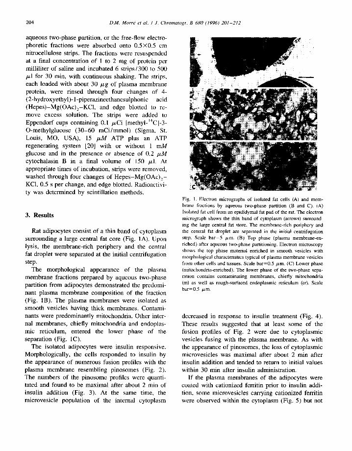
Rat adipocytes consist of a thin band of cytoplasm surrounding a large central fat core (Fig. 1A). Upon lysis, the membrane-rich periphery and the central fat droplet were separated at the initial centrifugation step.
The morphological appearance of the plasma membrane fractions prepared by aqueous two-phase partition from adipocytes demonstrated the predomi- nant plasma membrane composit ion of the fraction (Fig. 1B). The plasma membranes were isolated as smooth vesicles having thick membranes. Contami- nants were predominantly mitochondria. Other inter- nal membranes, chiefly mitochondria and endoplas- mic reticulum, entered the lower phase of the separation (Fig. IC).
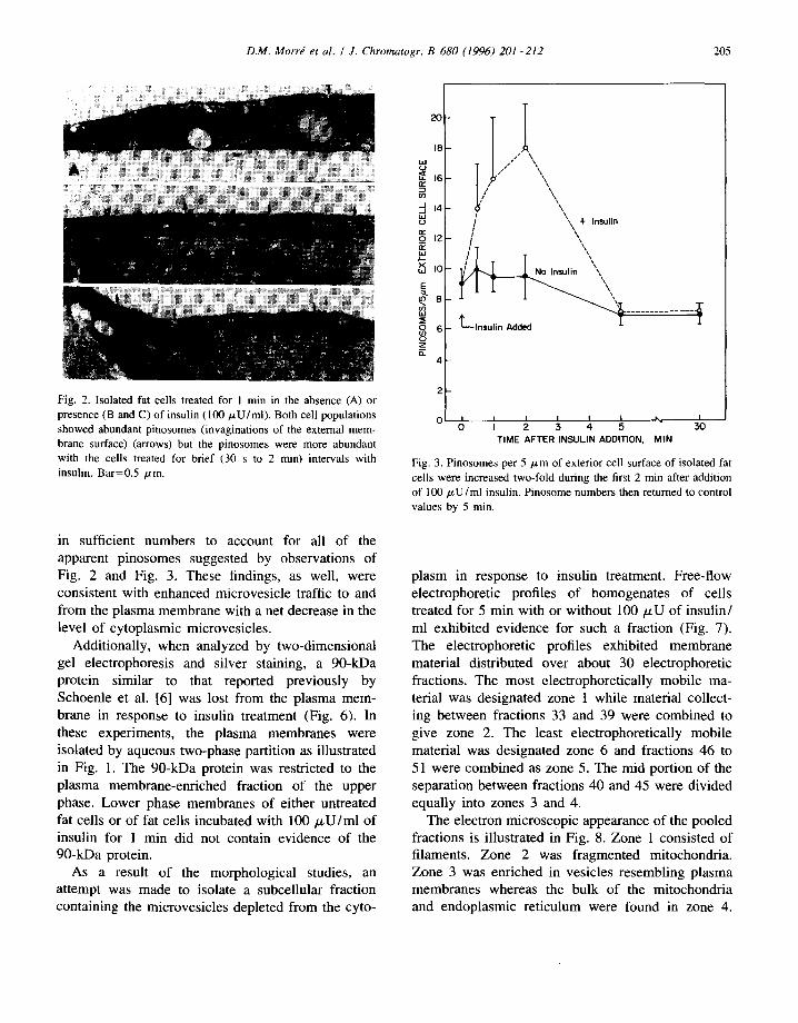
The isolated adipocytes were insulin responsive. Morphological ly, the cells responded to insulin by the appearance of numerous fusion profiles with the plasma membrane resembling pinosomes (Fig. 2). The numbers of the pinosome profiles were quanti- tated and found to be maximal after about 2 min of insulin addition (Fig. 3). At the same time, the microvesicle population of the internal cytoplasm
Fig. 1. Electron micrographs of isolated fat cells (A) and mem- brane fractions by aqueous two-phase partition (B and C). (A) Isolated fat cell from an epididymal fat pad of the rat. The electron micrograph shows the thin band of cytoplasm (arrows) surround- ing the large central fat store. The membrane-rich periphery and the central fat droplet are separated in the initial centrifugation step. Scale bar=5 btm. (B) Top phase (plasma membrane-en- riched) after aqueous two-phase partitioning. Electron microscopy shows the top phase material enriched in smooth vesicles with morphological characteristics typical of plasma membrane vesicles from other cells and tissues. Scale bar-0.5 /xm. (C) Lower phase (mitochondria-enriched). The lower phase of the two-phase sepa- ration contains contaminating membranes, chiefly mitochondria (m) as well as rough-surfaced endoplasmic reticulum (er). Scale bar=0.5 /~m.
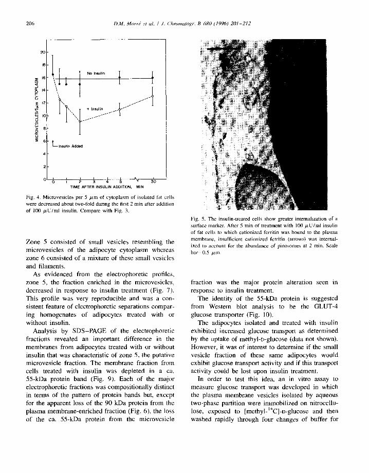
decreased in response to insulin treatment (Fig. 4). These results suggested that at least some of the fusion profiles of Fig. 2 were due to cytoplasmic vesicles fusing wi th the plasma membrane. As with the appearance of pinosomes, the loss of cytoplasmic microvesicles was maximal after about 2 min after insulin addition and tended to return to initial values within 30 min after insulin administration.
If the plasma membranes of the adipocytes were coated with cationized ferritin prior to insulin addi- tion, some microvesicles carrying cationized ferritin were observed within the cytoplasm (Fig. 5) but not
D.M. Morrd et al. / J. Chromatogr. B 680 (1996) 201-212 205
Fig. 2. Isolated fat cells treated for 1 min in the absence (A) or presence (B and C) of insulin (100/zU/ml). Both cell populations showed abundant pinosomes (invaginations of the external mem- brane surface) (arrows) but the pinosomes were more abundant with the cells treated for brief (30 s to 2 min) intervals with insulin. Bar=0.5 #m.
in sufficient numbers to account for all of the apparent pinosomes suggested by observations of Fig. 2 and Fig. 3. These findings, as well, were consistent with enhanced microvesicle traffic to and from the plasma membrane with a net decrease in the level of cytoplasmic microvesicles.
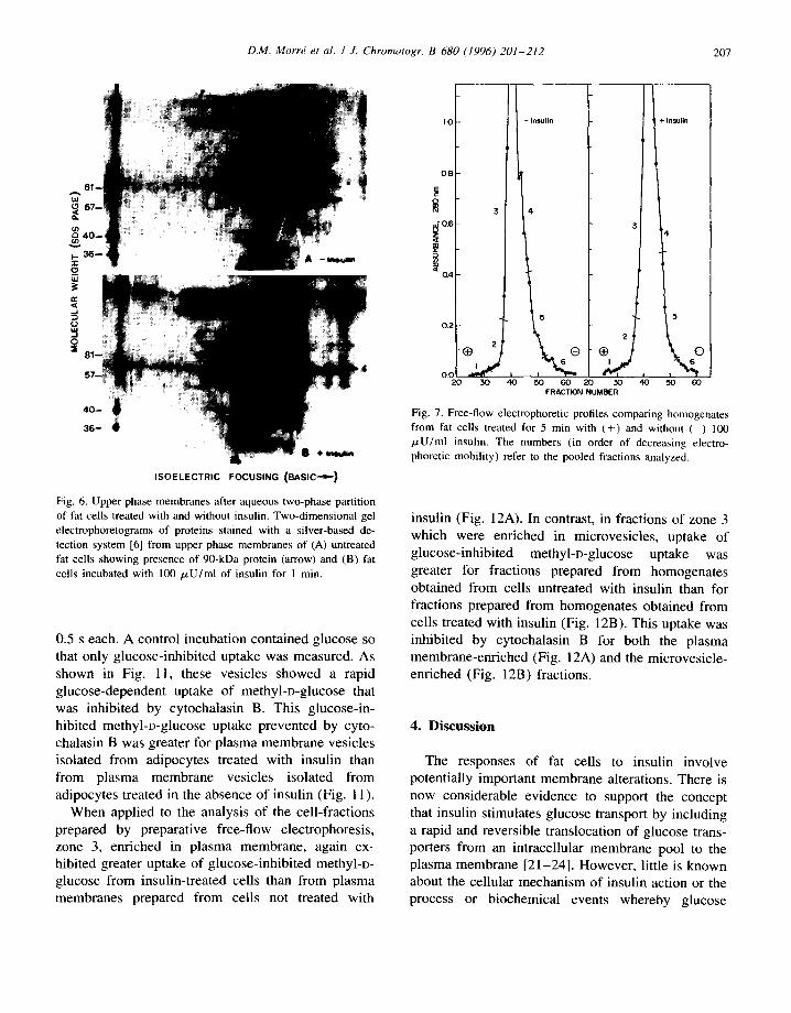
Additionally, when analyzed by two-dimensional gel electrophoresis and silver staining, a 90-kDa protein similar to that reported previously by Schoenle et al. [6] was lost from the plasma mem- brane in response to insulin treatment (Fig. 6). In these experiments, the plasma membranes were isolated by aqueous two-phase partition as illustrated in Fig. 1. The 90-kDa protein was restricted to the plasma membrane-enriched fraction of the upper phase. Lower phase membranes of either untreated fat cells or of fat cells incubated with 100 /xU/ml of insulin for 1 min did not contain evidence of the 90-kDa protein.
As a result of the morphological studies, an attempt was made to isolate a subcellular fraction containing the microvesicles depleted from the cyto-
18 LtJ 0
,.x ~o E ~ 8
~ 6 o z ft.
4
/ " \ \ ,,- , \ \
# s \ \ ,
\ \ + Insulin
T '\\
LInsu l in Added I I
I I I I%, I r 3 4 3o TIME AFTER INSULIN ADDITION, MIN
Fig. 3. Pinosomes per 5 /zm of exterior cell surface of isolated fat cells were increased two-fold during the first 2 min after addition of 100/xU/ml insulin. Pinosome numbers then returned to control values by 5 min.
plasm in response to insulin treatment. Free-flow electrophoretic profiles of homogenates of cells treated for 5 min with or without 100/.tU of insulin/ ml exhibited evidence for such a fraction (Fig. 7). The electrophoretic profiles exhibited membrane material distributed over about 30 electrophoretic fractions. The most electrophoretically mobile ma- terial was designated zone 1 while material collect- ing between fractions 33 and 39 were combined to give zone 2. The least electrophoretically mobile material was designated zone 6 and fractions 46 to 51 were combined as zone 5. The mid portion of the separation between fractions 40 and 45 were divided equally into zones 3 and 4.
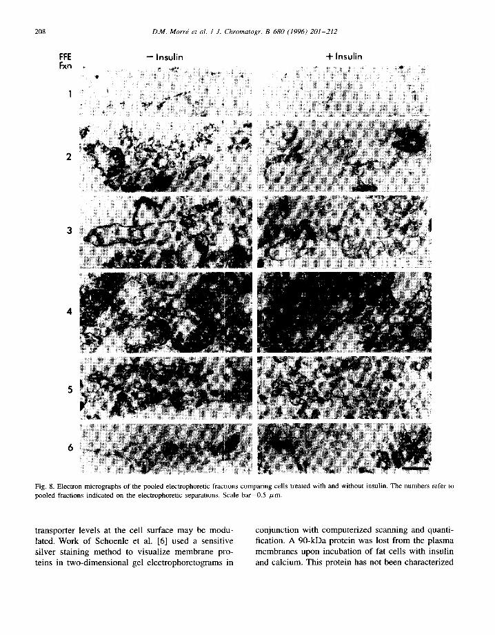
The electron microscopic appearance of the pooled fractions is illustrated in Fig. 8. Zone 1 consisted of filaments. Zone 2 was fragmented mitochondria. Zone 3 was enriched in vesicles resembling plasma membranes whereas the bulk of the mitochondria and endoplasmic reticulum were found in zone 4.
206 D.M. Morrd et al. / J. Chromatogr. B 680 (1996) 201-212
20
m4 ~o
E 12 :¢
8
_u :E 6
L Insulin Added
o ~ I ~ ~ ~ ~ ~ 3~ TIME AFTER INSULIN ADDITION, MIN
Fig. 4. Microvesicles per 5 #m of cytoplasm of isolated fat cells were decreased about two-fold during the first 2 min after addition of 100 /zU/ml insulin. Compare with Fig. 3.
Zone 5 consisted of small vesicles resembling the microvesicles of the adipocyte cytoplasm whereas zone 6 consisted of a mixture of these small vesicles and filaments.
As evidenced from the electrophoretic profiles, zone 5, the fraction enriched in the microvesicles, decreased in response to insulin treatment (Fig. 7). This profile was very reproducible and was a con- sistent feature of electrophoretic separations compar- ing homogenates of adipocytes treated with or without insulin.
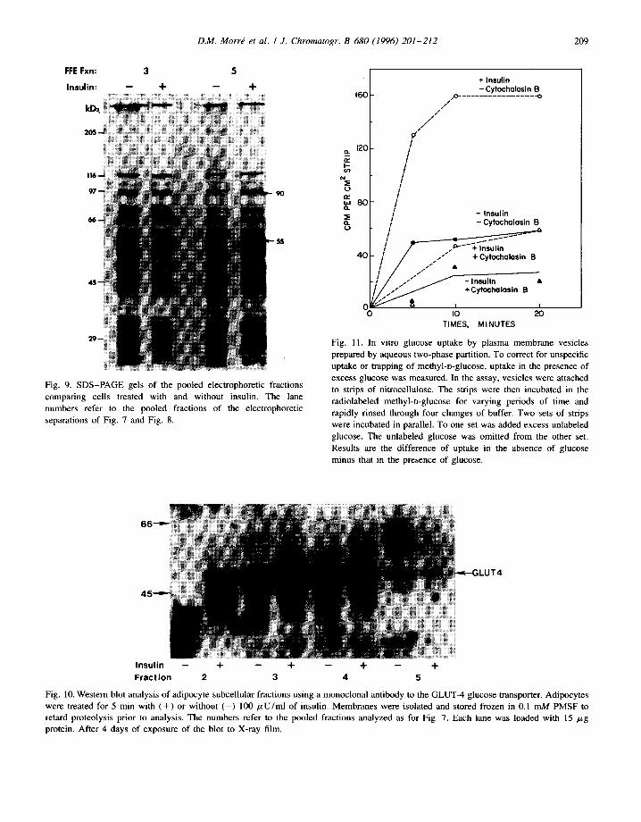
Analysis by SDS-PAGE of the electrophoretic fractions revealed an important difference in the membranes from adipocytes treated with or without insulin that was characteristic of zone 5, the putative microvesicle fraction. The membrane fraction from cells treated with insulin was depleted in a ca. 55-kDa protein band (Fig. 9). Each of the major electrophoretic fractions was compositionally distinct in terms of the pattern of protein bands but, except for the apparent loss of the 90 kDa protein from the plasma membrane-enriched fraction (Fig. 6), the loss of the ca. 55-kDa protein from the microvesicle
Fig. 5. The insulin-treated cells show greater internalization of a surface marker. After 5 rain of treatment with 100 #U/ml insulin of fat cells to which cationized ferritin was bound to the plasma membrane, insufficient cationized ferritin (arrows) was internal- ized to account for the abundance of pinosomes at 2 min. Scale bar=0.5 #m.
fraction was the major protein alteration seen in response to insulin treatment.
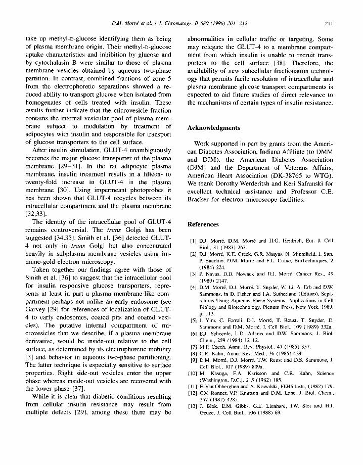
The identity of the 55-kDa protein is suggested from Western blot analysis to be the GLUT-4 glucose transporter (Fig. 10).
The adipocytes isolated and treated with insulin exhibited increased glucose transport as determined by the uptake of methyl-D-glucose (data not shown). However, it was of interest to determine if the small vesicle fraction of these same adipocytes would exhibit glucose transport activity and if this transport activity could be lost upon insulin treatment.
In order to test this idea, an in vitro assay to measure glucose transport was developed in which the plasma membrane vesicles isolated by aqueous two-phase partition were immobilized on nitrocellu- lose, exposed to [methyl-=4C]-D-glucose and then washed rapidly through four changes of buffer for
D.M. Morr~ et al. / J. Chromatogr. B 680 (1996) 201--212 207
,r.
_(.2 w
a r
O
ISOELECTRIC FOCUSING (BASICs)
Fig. 6. Upper phase membranes after aqueous two-phase partition of fat cells treated with and without insulin. Two-dimensional gel electrophoretograms of proteins stained with a silver-based de- tection system [6] from upper phase membranes of (A) untreated fat cells showing presence of 90-kDa protein (arrow) and (B) fat cells incubated with 100 #U/ml of insulin for 1 min.
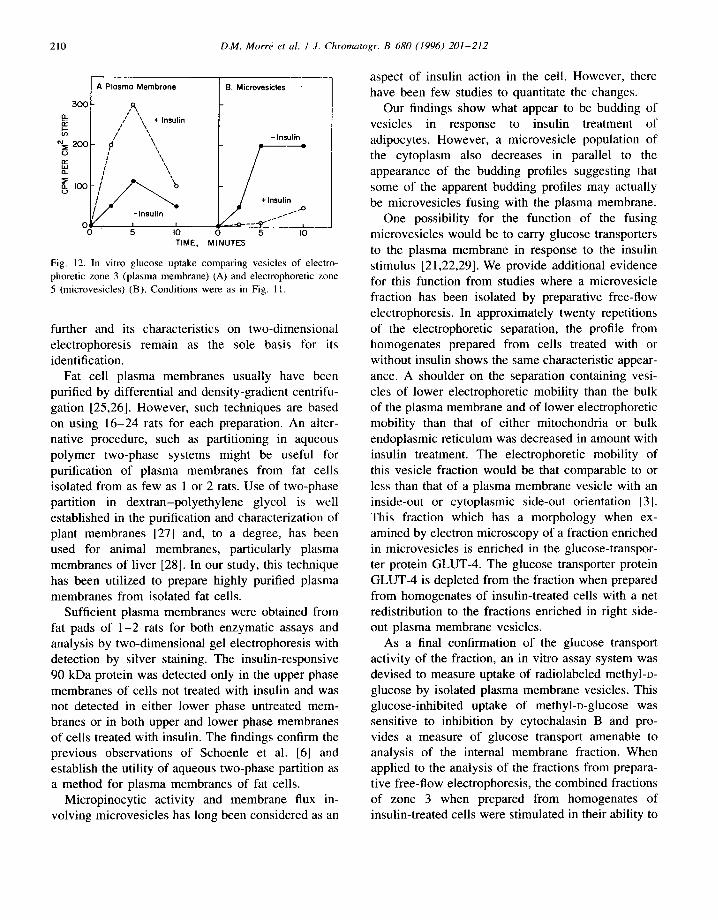
0.5 s each. A control incubation contained glucose so that only glucose-inhibited uptake was measured• As shown in Fig. 1 i, these vesicles showed a rapid glucose-dependent uptake of methyl-o-glucose that was inhibited by cytochalasin B. This glucose-in- hibited methyl-D-glucose uptake prevented by cyto- chalasin B was greater for p lasma membrane vesicles isolated from adipocytes treated with insulin than from plasma membrane vesicles isolated from adipocytes treated in the absence of insulin (Fig. 11).
When applied to the analysis of the cell-fractions prepared by preparative free-flow electrophoresis, zone 3, enriched in plasma membrane, again ex- hibited greater uptake of glucose-inhibited methyl-D- glucose from insulin-treated cells than from plasma membranes prepared from cells not treated with
i
1,0
0.8
u 3 0 . 6
0 . 4
0 . 2
2
O.C I 20 50 40
- I n s u l i n
4
Insulin i
I i i
' I . I
'
15 . - ,5
z~
50 60 20 30 40 50 60 FRACTION NUMBER
Fig. 7. Free-flow electrophoretic profiles comparing homogenates from fat cells treated for 5 min with (+) and without ( ) 100 /zU/ml insulin. The numbers (in order of decreasing electro- phoretic mobility) refer to the pooled fractions analyzed.
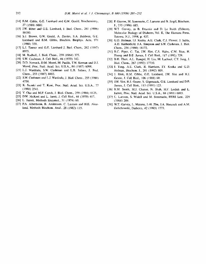
insulin (Fig. 12A). In contrast, in fractions of zone 3 which were enriched in microvesicles, uptake of glucose-inhibited methyl-o-glucose uptake was greater for fractions prepared from homogenates obtained from cells untreated with insulin than for fractions prepared from homogenates obtained from cells treated with insulin (Fig. 12B). This uptake was inhibited by cytochalasin B for both the plasma membrane-enriched (Fig. 12A) and the microvesicle- enriched (Fig. 12B) fractions.
4 . D i s c u s s i o n
The responses of fat cells to insulin involve potentially important membrane alterations• There is now considerable evidence to support the concept that insulin stimulates glucose transport by including a rapid and reversible translocation of glucose trans- porters from an intracellular membrane pool to the plasma membrane [21-24]. However, little is known about the cellular mechanism of insulin action or the process or biochemical events whereby glucose
1
F F E
Fxn e
D.M. Morr~ et al. / J. Chromatogr. B 680 (1996) 201-212
- - Insulin + Insulin
2
208
4
% ::
;4
Fig. 8. Electron micrographs of the pooled electrophoretic fractions comparing cells treated with and without insulin. The numbers refer to pooled fractions indicated on the electrophoretic separations. Scale bar=0.5 txm.
transporter levels at the cell surface may be modu- lated. Work of Schoenle et al. [6] used a sensitive silver staining method to visualize membrane pro- teins in two-dimensional gel electrophoretograms in
conjunction with computerized scanning and quanti- fication. A 90-kDa protein was lost from the plasma membranes upon incubation of fat cells with insulin and calcium. This protein has not been characterized
D.M. MorrE et al. / J. Chromatogr. B 680 (1996) 201-212 209
FFE Fxn: 3 5
Insu l in : - - + - - +
Fig. 9. SDS-PAGE gels of the pooled electrophoretic fractions comparing cells treated with and without insulin. The lane numbers refer to the pooled fractions of the electrophoretic separations of Fig. 7 and Fig. 8.
a .
~c
( J
160
120
n~ w 8 0 O. ~E n 0
40
0 0
+ Insulin - Cytocholosin B
jo- . . . . . . . . . . . . . . . . -o / 1
i S / /
/ / /
¢ /
I I /
I / I
I I
/ - Insulin / - Cytocholasin B
t / .P ' - +insulin / / / , . / -I- Cytocholasin B / / / s & I s* S
/.,.~ytocholosln a
IO 20 TIMES, MINUTES
Fig. 11. In vitro glucose uptake by plasma membrane vesicles prepared by aqueous two-phase partition. To correct for unspecific uptake or trapping of methyl-D-glucose, uptake in the presence of excess glucose was measured. In the assay, vesicles were attached to strips of nitrocellulose. The strips were then incubated in the radiolabeled methyl-D-glucose for varying periods of time and rapidly rinsed through four changes of buffer. Two sets of strips were incubated in parallel. To one set was added excess unlabeled glucose. The unlabeled glucose was omitted from the other set. Results are the difference of uptake in the absence of glucose minus that in the presence of glucose.
ILUT4
Insulin -- + -- + -- + -- + Fraction 2 3 4 5
Fig. 10. Western blot analysis of adipocyte subcellular fractions using a monoclonal antibody to the GLUT-4 glucose transporter. Adipocytes were treated for 5 min with ( + ) or without ( - ) 100 p,U/ml of insulin. Membranes were isolated and stored frozen in 0.1 mM PMSF to retard proteolysis prior to analysis. The numbers refer to the pooled fractions analyzed as for Fig. 7. Each lane was loaded with 15 p,g protein. After 4 days of exposure of the blot to X-ray film.
210 D.M. Morrd et al. I J. Chromatogr. B 680 (1996) 201-212
APIosmo Membrone B. Microvesicles
500 R
o_ / ~ ÷ Insulin
E ¢/' ', -I.sulin ~zvo \,, / + = ~ / \, I1_ [ ~\
~oo- I } " - . . 'u
-Ins .x~
0 5 I0 0 5 I0 TIME, MINUTES
Fig. 12. In vitro glucose uptake comparing vesicles of electro- phoretic zone 3 (plasma membrane) (A) and electrophoretic zone 5 (microvesicles) (B). Conditions were as in Fig. 11.
further and its characteristics on two-dimensional electrophoresis remain as the sole basis for its identification.
Fat cell plasma membranes usually have been purified by differential and density-gradient centrifu- gation [25,26]. However, such techniques are based on using 16-24 rats for each preparation. An alter- native procedure, such as partitioning in aqueous polymer two-phase systems might be useful for purification of plasma membranes from fat cells isolated from as few as 1 or 2 rats. Use of two-phase partition in dextran-polyethylene glycol is well established in the purification and characterization of plant membranes [27] and, to a degree, has been used for animal membranes, particularly plasma membranes of liver [28]. In our study, this technique has been utilized to prepare highly purified plasma membranes from isolated fat cells.
Sufficient plasma membranes were obtained from fat pads of 1-2 rats for both enzymatic assays and analysis by two-dimensional gel electrophoresis with detection by silver staining. The insulin-responsive 90 kDa protein was detected only in the upper phase membranes of cells not treated with insulin and was not detected in either lower phase untreated mem- branes or in both upper and lower phase membranes of cells treated with insulin. The findings confirm the previous observations of Schoenle et al. [6] and establish the utility of aqueous two-phase partition as a method for plasma membranes of fat cells.
Micropinocytic activity and membrane flux in- volving microvesicles has long been considered as an
aspect of insulin action in the cell. However, there have been few studies to quantitate the changes.
Our findings show what appear to be budding of vesicles in response to insulin treatment of adipocytes. However, a microvesicle population of the cytoplasm also decreases in parallel to the appearance of the budding profiles suggesting that some of the apparent budding profiles may actually be microvesicles fusing with the plasma membrane.
One possibility for the function of the fusing microvesicles would be to carry glucose transporters to the plasma membrane in response to the insulin stimulus [21,22,29]. We provide additional evidence for this function from studies where a microvesicle fraction has been isolated by preparative free-flow electrophoresis. In approximately twenty repetitions of the electrophoretic separation, the profile from homogenates prepared from cells treated with or without insulin shows the same characteristic appear- ance. A shoulder on the separation containing vesi- cles of lower electrophoretic mobility than the bulk of the plasma membrane and of lower electrophoretic mobility than that of either mitochondria or bulk endoplasmic reticulum was decreased in amount with insulin treatment. The electrophoretic mobility of this vesicle fraction would be that comparable to or less than that of a plasma membrane vesicle with an inside-out or cytoplasmic side-out orientation [3]. This fraction which has a morphology when ex- amined by electron microscopy of a fraction enriched in microvesicles is enriched in the glucose-transpor- ter protein GLUT-4. The glucose transporter protein GLUT-4 is depleted from the fraction when prepared from homogenates of insulin-treated cells with a net redistribution to the fractions enriched in right side- out plasma membrane vesicles.
As a final confirmation of the glucose transport activity of the fraction, an in vitro assay system was devised to measure uptake of radiolabeled methyl-o- glucose by isolated plasma membrane vesicles. This glucose-inhibited uptake of methyl-o-glucose was sensitive to inhibition by cytochalasin B and pro- vides a measure of glucose transport amenable to analysis of the internal membrane fraction. When applied to the analysis of the fractions from prepara- tive free-flow electrophoresis, the combined fractions of zone 3 when prepared from homogenates of insulin-treated cells were stimulated in their ability to
D.M. Morrd et al. / J. Chromatogr. B 680 (1996) 201-212 211
take up methyl-o-glucose identifying them as being of plasma membrane origin. Their methyl-o-glucose uptake characteristics and inhibition by glucose and by cytochalasin B were similar to those of plasma membrane vesicles obtained by aqueous two-phase partition. In contrast, combined fractions of zone 5 from the electrophoretic separations showed a re- duced ability to transport glucose when isolated from homogenates of cells treated with insulin. These results further indicate that the microvesicle fraction contains the internal vesicular pool of plasma mem- brane subject to modulation by treatment of adipocytes with insulin and responsible for transport of glucose transporters to the cell surface.
After insulin stimulation, GLUT-4 unambiguously becomes the major glucose transporter of the plasma membrane [29-31]. In the rat adipocyte plasma membrane, insulin treatment results in a fifteen- to twenty-fold increase in GLUT-4 in the plasma membrane [30]. Using impermeant photoprobes it has been shown that GLUT-4 recycles between its intracellular compartment and the plasma membrane [32,331.
The identity of the intracellular pool of GLUT-4 remains controversial. The t r a n s Golgi has been suggested [34,35]. Smith et al. [36] detected GLUT- 4 not only in t r a n s Golgi but also concentrated heavily in subplasma membrane vesicles using im- muno-gold electron microscopy.
Taken together our findings agree with those of Smith et al. [36] to suggest that the intracellular pool for insulin responsive glucose transporters, repre- sents at least in part a plasma membrane-like com- partment perhaps not unlike an early endosome (see Garvey [29] for references of localization of GLUT- 4 to early endosomes, coated pits and coated vesi- cles). The putative internal compartment of mi- crovesicles that we describe, if a plasma membrane derivative, would be inside-out relative to the cell surface, as determined by its electrophoretic mobility [3] and behavior in aqueous two-phase partitioning. The latter technique is especially sensitive to surface properties. Right side-out vesicles enter the upper phase whereas inside-out vesicles are recovered with the lower phase [37].
While it is clear that diabetic conditions resulting from cellular insulin resistance may result from multiple defects [29], among these there may be
abnormalities in cellular traffic or targeting. Some may relegate the GLUT-4 to a membrane compart- ment from which insulin is unable to recruit trans- porters to the cell surface [38]. Therefore, the availability of new subcellular fractionation technol- ogy that permits facile resolution of intracellular and plasma membrane glucose transport compartments is expected to aid future studies of direct relevance to the mechanisms of certain types of insulin resistance.
Acknowledgments
Work supported in part by grants from the Ameri- can Diabetes Association, Indiana Affiliate (to DMM and DJM), the American Diabetes Association (DJM) and the Department of Veterans Affairs, American Heart Association (DK-38765 to WTG). We thank Dorothy Werderitsh and Keri Safranski for excellent technical assistance and Professor C.E. Bracker for electron microscope facilities.
References
[1] D.J. Morrd, D.M. Morr6 and H.G. Heidrich, Eur. J. Cell Biol., 31 (1983) 263.
[2] D.J. Morrd, K.E. Creek, G.R. Matyas, N. Minnifield, I. Sun, E Baudoin, D.M. Morr6 and F.L. Crane, BioTechniques, 2 (1984) 224.
[3] E Navas, D.D. Nowack and D.J. Morrd, Cancer Res., 49 (1989) 2147.
[4] D.M. Morr6, D.J. Morr6, T. Snyder, W. Li, A. Erb and D.W. Sammons, in D. Fisher and I.A. Sutherland (Editors), Sepa- rations Using Aqueous Phase Systems. Applications in Cell Biology and Biotechnology, Plenum Press, New York, 1989, p. 113.
[5] J. Yim, C. Ferroli, D.J. Morrd, T. Reust, T. Snyder, D. Sammons and D.M. Mort6, J. Cell Biol., 109 (1989) 332a.
[6] E.J. Schoenle, L.D. Adams and D.W. Sammons, J. Biol. Chem., 259 (1984) 12112.
[7] M.E Czech, Annu. Rev. Physiol., 47 (1985) 357. [8] C.R. Kahn, Annu. Rev. Med., 36 (1985) 429. [9] D.M. Morr6, D.J. Mort6, T.W. Reust and D.S. Sammons, J.
Cell Biol., 107 (1989) 809a. [10] M. Kasuga, F.A. Km'lsson and C.R. Kahn, Science
(Washington, D.C.), 215 (1982) 185. [11] E. Van Obberghen and A. Kowalski, FEBS Lett., (1982) 179. [12] G.V. Ronnet, V.P. Knutson and D.M. Lane, J. Biol. Chem.,
257 (1982) 4285. [13] J. Blok, E.M. Gibbs, G.E. Lienhard, J.W. Slot and H.J.
Geuze, J. Cell Biol., 106 (1988) 69.
212 D.M. Morr~ et al. / J. Chromatogr. B 680 (1996) 201-212
[14] E.M. Gibbs, G.E. Lienhard and G.W. Gould, Biochemistry, 27 (1988) 6681.
[15] J.W. Biber and G.E. Lienhard, J. Biol. Chem., 261 (19861 16180.
[16] S.J. Brown, G.W. Gould, A. Davies, S.A. Baldwin, G.E. Lienhard and E.M. Gibbs, Biochim. Biophys. Acta, 971 (1988) 339.
[17] L.I. Tanner and G.E. Lienhard J. Biol. Chem., 262 (1987) 8975.
[18] M. Rodbell, J. Biol. Chem., 239 (19641 375. [19] S.W. Cushman, J. Cell Biol., 46 (1970) 342. [20] D.D. Nowack, D.M. Morr6, M. Paulik, T.W. Keenan and D.J.
Morr6, Proc. Natl. Acad. Sci. U.S.A., 84 (1987) 6098. [21] L.J. Wardzala, S.W. Cushman and L.B. Salans, J. Biol.
Chem., 253 (1987) 8002. [22] S.W. Cushman and L.J. Wardzala, J. Biol. Chem., 255 (198(I)
4758. [23] K. Suzuki and T. Koni, Proc. Natl. Acad. Sci. U.S.A., 77
(1980) 2541. [24] Y. Oka and M.P. Czech, J. Biol. Chem., 259 (19841 8125. [25] D.W. McKeel and L. Jarett, J. Cell Biol., 44 (1970) 417. [26] L. Jarrett, Methods Enzymol., 31 (1974) 60. [27] EA. Albertsson, B. Andersson, C. Larsson and H.E. Aker-
lund, Methods Biochem. Anal., 28 (19821 115.
[28] E Gierow, M. Sommarin, C. Larsson and B. Jergil, Biochem. J., 235 (1986) 685.
[29] W.T. Garvey, in B. Draznin and D. Le Roith (Editors), Molecular Biology of Diabetes, Vol. II, The Humana Press, Totowa, N.J., 1994, p. 437.
[30] G.D. Holman, I.J. Kozka, A.E. Clark, C.J. Flower, J. Saltis, A.D. Habberfield, J.A. Simpson and S.W. Cushman, J. Biol. Chem., 256 (1990) 18172.
[31] R.C. Piper, C. Tai, J.W. Slot, C.S. Hahn, C.M. Rice, H. Huang and D.E. James, J. Cell Biol., 117 (1992) 729.
[32] B.H. Thun, A.L. Rampal, H. Liu, M. Lachool, C.Y. Jung, J. Biol. Chem., 267 (1992) 17710.
[33] J. Yang, A.E. Clark, R. Harrison, I.Y. Kozka and G.D. Holman, Biochem. J., 281 (1992) 809.
[34] J. Blok, E.M. Gibbs, G.E. Lienhard, J.W. Slot and H.J. Geuze, J. Cell Biol., 106 (1988) 69.
[35] J.W. Slot, H.J. Geuze, S. Gigenzack, G.E. Lienhard and D.E. James, J. Cell Biol., 113 (1991) 123.
[36] R.M. Smith, M.J. Charon, N. Shah, H.F. Lodish and L. Jarrett, Proc. Natl. Acad. Sci. U.S.A., 88 (1991) 6893.
[37] C. Larsson, S. Widell and M. Sommarin, FEBS Lett., 229 (1988) 289.
[38] W.T. Garvey, L. Maianu, J.-H. Zhu, J.A. Hancock and A.M. Golichowski, Diabetes, 42 (1993) 1773.
Related Documents