Eur. J. Biochem. 182, 77-84 (1989) 0 FEBS 1989 Isolation and structure of two novel 6-kDa dimeric peptides from the corpora cardiaca of the insect Locusta migratoria Molecular mass determination by mass spectrometry Helene HIETTER I, Bang LUU ', Francine GOLTZENE', Daniel ZACHARY ', Jules HOFFMANN2 and Alain VAN DORSSELAER' Laboratoire de chimie organique des substances naturelles, associe au Centre national de la recherche scientifique, Strasbourg Laboratoire de biologie genkrale, associi. au Centre national de la recherche scientifique, Strasbourg (Received October 26, 1988) - EJB 881269 We have isolated two major 6-kDa peptides from extracts of corpora cardiaca of adult females of Locusta migratoria. These peptides have been characterized by peptide sequencing and liquid secondary-ion mass spec- trometry. They are structurally related dimers, one (6278.5 Da) being a homodimer (A-A chains), the other (6280.5 Da) being a heterodimer (A-B chains). A 60% similarity exists between the A and B chains. Both peptides have been chemically synthesized and the synthetic compounds appeared to be identical to the native ones. Polyclonal antibodies raised against each of these peptides demonstrated that they were contained within the secretory granules of the intrinsic cells of the glandular lobes of the corpora cardiaca. The physiological significance of these two peptides is unknown but, using the synthetic peptides, we are currently probing their biological role. The corpora cardiaca of insects are retrocerebral neuro- haemal organs of dual anatomical composition. They contain most of the axon endings from the neurosecretory cells of the brain, serving as storage and release sites for the brain neurohormones, and they have intrinsic secretory cells which synthesize and release peptide hormones. This anatomical and functional organization has attracted considerable interest among endocrinologists, namely because of the obvious evol- utionary convergence with the vertebrate hypothalamohypo- physeal system [I]. The isolation of the hormones secreted by the corpora cardiuca has been a major goal of several groups over the last two decades. Our information nevertheless still remains fragmentary and only a few molecules have been fully characterized so far. Most of these are N-terminal blocked peptides of 8 - 10 amino acid residues, with adipokinetic and/or carbohydrate-mobilizing activity; some also show myotropic activity [2 - 61. Peptides with diuretic or antidi- uretic activities are also contained within the corpora cardiaca of insects [7-91 but to date no full structure has been published. We have undertaken an investigation aiming at the characterization of the major peptides synthesized or stored in the corpora cardiuca of Locusta migratoria. This biological system is attractive because of the clear-cut anatomical subdiv- ision of the corpora cardiaca into the storage lobe (axon ter- minals of the neurosecretory cells of the brain) and the glandu- lar lobe (intrinsic cells). We report in the present paper the isolation and complete chemical characterization of two 6- kDa dimeric peptides contained within the secretory granules of the glandular lobes. Correspondence to A. Van Dorsselaer, Laboratoire de chimie organique des substances naturelles, associe au Centre national de la recherche scientifique, 5 rue Blaise Pascal, F-67084 Strasbourg, France Ahhreviutions. [MH]+, protonated molecular ion; [MH2I2+, doubly protonated molecular ion; RP-HPLC, reversed-phase high- performance liquid chromatography; LSIMS, liquid secondary-ion mass spectrometry; CF3C02H, trifluoroacetic acid. MATERIALS AND METHODS Chemicals Trifluoroacetic acid (CF3C02H), sequencer grade, and 6 M HC1 were obtained from Pierce. Iodoacetic acid and amide and dithiothreitol were from Sigma. Phenylmethylsul- phony1 fluoride and aprotinin were used as protease inhibitors and were obtained from Sigma. All organic solvents were HPLC grade and were purchased from Merck. Deionized water was obtained from a tandem Milli-RO and Milli-Q system (Millipore Inc, USA). Insects Locusta migratoria migratorioides were reared in gregari- ous phase at a day temperature of 28 - 30°C, falling to 25 "C at night. Electric bulbs inside the cages allowed temperature gradients up to 38°C. Relative humidity was around 70%. Day conditions lasted from 11 .OO to 23.00. Peptide extraction Corpora cardiaca (endocrine and/or neurohaemal storage lobes) and pars intercerebralis were excised from 30-day-old adult females (1000) and stored in liquid N2. Homogenization was typically performed in a glass/glass Potter in ice-cold deionized water containing 400 pM phenylmethylsulphonyl fluoride and 1 pM aprotinin (pH = 5.5); in other experiments the extraction was performed identically but at pH = 2.1 (0.1 % CF3C02H).The homogenate was treated according to one of two procedures: (a) 5-min heat-treatment at IOO'C, followed by a 2-min sonication and a I-h centrifugation at I00000 x g, after which the supernatant was freeze-dried; (b) a 15-min centrifugation at 15000 x g followed by application of the supernatant on a CI8 Sep pak cartridge (Waters). The elution was performed by increasing concentrations of acetonitrile (IS%, 36%, 60%) in water/O.l% CF3C02H.The

Welcome message from author
This document is posted to help you gain knowledge. Please leave a comment to let me know what you think about it! Share it to your friends and learn new things together.
Transcript
Eur. J. Biochem. 182, 77-84 (1989) 0 FEBS 1989
Isolation and structure of two novel 6-kDa dimeric peptides from the corpora cardiaca of the insect Locusta migratoria Molecular mass determination by mass spectrometry
Helene HIETTER I , Bang LUU ', Francine GOLTZENE', Daniel ZACHARY ', Jules HOFFMANN2 and Alain VAN DORSSELAER'
Laboratoire de chimie organique des substances naturelles, associe au Centre national de la recherche scientifique, Strasbourg Laboratoire de biologie genkrale, associi. au Centre national de la recherche scientifique, Strasbourg
(Received October 26, 1988) - EJB 881269
We have isolated two major 6-kDa peptides from extracts of corpora cardiaca of adult females of Locusta migratoria. These peptides have been characterized by peptide sequencing and liquid secondary-ion mass spec- trometry. They are structurally related dimers, one (6278.5 Da) being a homodimer (A-A chains), the other (6280.5 Da) being a heterodimer (A-B chains). A 60% similarity exists between the A and B chains. Both peptides have been chemically synthesized and the synthetic compounds appeared to be identical to the native ones. Polyclonal antibodies raised against each of these peptides demonstrated that they were contained within the secretory granules of the intrinsic cells of the glandular lobes of the corpora cardiaca. The physiological significance of these two peptides is unknown but, using the synthetic peptides, we are currently probing their biological role.
The corpora cardiaca of insects are retrocerebral neuro- haemal organs of dual anatomical composition. They contain most of the axon endings from the neurosecretory cells of the brain, serving as storage and release sites for the brain neurohormones, and they have intrinsic secretory cells which synthesize and release peptide hormones. This anatomical and functional organization has attracted considerable interest among endocrinologists, namely because of the obvious evol- utionary convergence with the vertebrate hypothalamohypo- physeal system [I]. The isolation of the hormones secreted by the corpora cardiuca has been a major goal of several groups over the last two decades. Our information nevertheless still remains fragmentary and only a few molecules have been fully characterized so far. Most of these are N-terminal blocked peptides of 8 - 10 amino acid residues, with adipokinetic and/or carbohydrate-mobilizing activity; some also show myotropic activity [2 - 61. Peptides with diuretic or antidi- uretic activities are also contained within the corpora cardiaca of insects [7-91 but to date no full structure has been published. We have undertaken an investigation aiming at the characterization of the major peptides synthesized or stored in the corpora cardiuca of Locusta migratoria. This biological system is attractive because of the clear-cut anatomical subdiv- ision of the corpora cardiaca into the storage lobe (axon ter- minals of the neurosecretory cells of the brain) and the glandu- lar lobe (intrinsic cells). We report in the present paper the isolation and complete chemical characterization of two 6- kDa dimeric peptides contained within the secretory granules of the glandular lobes.
Correspondence to A. Van Dorsselaer, Laboratoire de chimie organique des substances naturelles, associe au Centre national de la recherche scientifique, 5 rue Blaise Pascal, F-67084 Strasbourg, France
Ahhreviutions. [MH]+, protonated molecular ion; [MH2I2+, doubly protonated molecular ion; RP-HPLC, reversed-phase high- performance liquid chromatography; LSIMS, liquid secondary-ion mass spectrometry; CF3C02H, trifluoroacetic acid.
MATERIALS AND METHODS
Chemicals
Trifluoroacetic acid (CF3C02H), sequencer grade, and 6 M HC1 were obtained from Pierce. Iodoacetic acid and amide and dithiothreitol were from Sigma. Phenylmethylsul- phony1 fluoride and aprotinin were used as protease inhibitors and were obtained from Sigma. All organic solvents were HPLC grade and were purchased from Merck. Deionized water was obtained from a tandem Milli-RO and Milli-Q system (Millipore Inc, USA).
Insects
Locusta migratoria migratorioides were reared in gregari- ous phase at a day temperature of 28 - 30°C, falling to 25 "C at night. Electric bulbs inside the cages allowed temperature gradients up to 38°C. Relative humidity was around 70%. Day conditions lasted from 11 .OO to 23.00.
Peptide extraction
Corpora cardiaca (endocrine and/or neurohaemal storage lobes) and pars intercerebralis were excised from 30-day-old adult females (1000) and stored in liquid N2. Homogenization was typically performed in a glass/glass Potter in ice-cold deionized water containing 400 pM phenylmethylsulphonyl fluoride and 1 pM aprotinin (pH = 5.5); in other experiments the extraction was performed identically but at pH = 2.1 (0.1 % CF3C02H). The homogenate was treated according to one of two procedures: (a) 5-min heat-treatment at IOO'C, followed by a 2-min sonication and a I-h centrifugation at I00000 x g, after which the supernatant was freeze-dried; (b) a 15-min centrifugation at 15000 x g followed by application of the supernatant on a CI8 Sep pak cartridge (Waters). The elution was performed by increasing concentrations of acetonitrile (IS%, 36%, 60%) in water/O.l% CF3C02H. The
78
eluted fractions were combined and dried under reduced pres- sure and heating in a Speed Vac concentrator.
Purification of peptides by high-performance liquid chromatography
Peptide mixtures from various tissues were fractionated by CI8 reversed-phase HPLC on a Waters liquid chromatograph. Two pumps (model 510) were monitored by a Waters auto- mated gradient controller (model 680). Absorbance was monitored at 225 nm using a Waters spectrophotometer (model 481). A column (0.46 x 25 cm) packed with 5-pm-di- ameter wide-pore (30 nm) CIS Vydac particles was used. The samples were solubilized in deionized water/O.l YO CF3C02H and applied on the column which had been equilibrated in solvent A (15% CH3CN, 0.1% CF3C02H). Elution was performed at a flow rate of 1 ml/min using a stepwise gra- dient over 75 min of 15-6OY0 acetonitrile in water/O.l% CF3C02H. The different peaks were collected and then, for further purification, submitted to a second chromatography (isocratic conditions, percentage of acetonitrile depending on the compound). All peaks were collected manually, freeze- dried or dried using a Speed Vac concentrator and stored at -18'C.
Amino acid sequence anal-ysis
Automated Edman degradation of peptides was perform- ed using an Applied Biosystems sequencer (model 470A) equipped with an on-line phenylthiohydantoin analyzer. The repetitive yield was always between 93 -96%.
Amino acid analysis
Peptides (1 nmol) were hydrolyzed in constant-boiling 6 M HCl at 116'C for 24 h. After hydrolysis, amino acids were converted to their phenylthiocarbamoyl derivatives using phenylisothiocyanate as described by the Waters Picotag manual. These derivatives were then identified by reversed- phase HPLC on a Picotag column (0.39 x 15 cm) at 38"C, using a gradient of 6- 60% acetonitrile.
Reduction and alkylation
These procedures were carried out according to Nagasawa et al. [lo]. Peptides (2 nmol) in 60 pI 0.5 M Tris/HCl pH = 8.5 containing 6 M urea were mixed with 50 p10.02 M dithio- threitol in the same buffer and allowed to react under an argon atmosphcre at 45°C for 1 h. Alkylation was achieved by the addition of 50 p1 0.04 M iodoacetic acid or amide in deionized water to the mixture and incubation was continued for 20 min at 45'C in the dark. The peptides were recovered from the mixture by RP-HPLC.
Liquid secondury-ion muss spectrometry ( L S I M S )
Positive-ion mass spectra were obtained using a VG Ana- lytical ZAB-2SE double-focussing instrument (mass range 15 kDa at 8 keV ion energy) and recorded on a VG 11-250 data system (VG Analytical Ltd, England). Ionization of the sample was performed with approximately 2 FA of 30-keV- energy cesium ions using the cesium ion gun. Some measure- ments were also performed with a ZAB-HF (VG Analytical) fitted with a xenon atom gun operated at 10 keV and the spectra werc also processed on a VG 11-250 data system.
The underivatized peptides and the alkylated peptides were dissolved in deionized water containing 5% acetic acid at an, average concentration of 2 pg/pl. The matrix was l-thio- glycerol containing 1 % CF3C02H; 1 pl was deposited on the target and 0.5 pl of the peptide solution was added.
Wide scans were performed at a resolution of 1000 by magnetic scanning over the mass range 12- 1.5 kDa in 20 s. Narrow scans were performed at low (1000) or high resolution (4000); in both cases a narrow zone of about 350 Da was voltage-scanned to include two successive cesium iodide clus- ters which served for calibration. The acquisition data system was always used in the MCA (multi-channel acquisition) mode. Data were processed according to described procedures [Ill.
Peptide synthesis
The synthesis was done at Neosystem (Strasbourg, France) on a semi-automatic multi-channel solid-phase peptide synthesizer (model NPS 4000). The purification was per- formed using the same HPLC conditions described for the natural peptides.
Oxidation of' single chains to dimer
The monomers were oxidized by air, after dissolution in 0.1 M Tris/HCl pH = 8.5. This reaction was performed at room temperature, for 5 h, at a concentration of 1 pg/pl. The peptides were then isolated by RP-HPLC.
Immunization
Each of peptides 5 and 6 (16 pg) was purified by RP- HPLC, lyophilized, and taken up in 100 p1 0.2 M KA1(S04)2 . 12 H 2 0 solution, pH = 3, to which 50 pl of a 1 M solution of NaHC03, pH = 8, was added. After 15 min, the mixture was centrifuged and the pellet was taken up in phosphate- buffered saline (NaCl/Pi) to a final volume of 600 pl. Three injections (intraperitoneal and subcutaneous) were performed at 15-day intervals and blood was withdrawn 8 days after the last injection. The serum was kept in NaCI/Pi (1 : 1, by vol.) at - 20"C.
Imnzunocytocherviistry
For light microscope immunohistology the conventional horse-radish peroxidase/dianiinobenzidine technique, as de- scribed in detail elsewhere [12], was used. Electron microscope immunocytochemistry was performcd with the immunogold method [12].
RESULTS
Isolation, characterization and structure of two peptides in extructs of'the corpora cardiaca of adult females of Locusta migratoria
Isolution and structural data
In a pilot experiment, corpora cardiacn (each containing a glandular lobe and a neurohaemal lobe) were excised from 100 30-day-old adult females of Locustu and heat-treated. After centrifugation, the supernatant was subjected to re- versed-phase HPLC (Fig. 1). A relatively large number of distinct absorption peaks were observed in this chro-
79
50-
5 ABSORBANCE -
1571.9
5 ABSORBANCE -
min
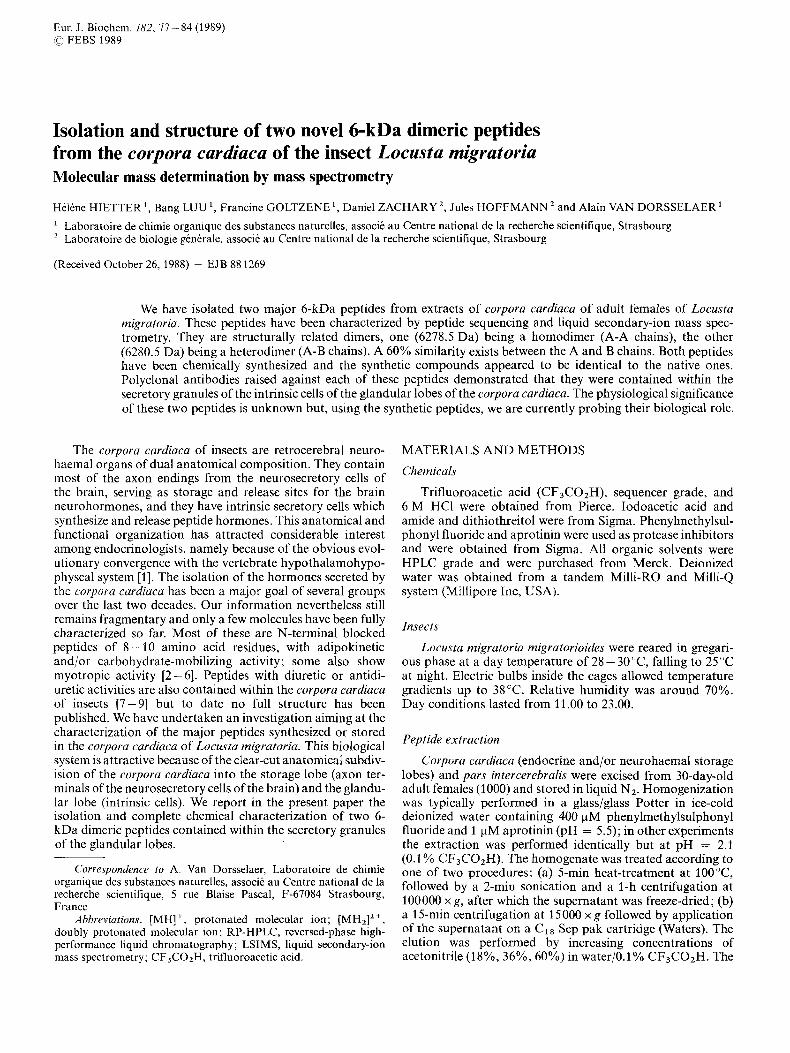
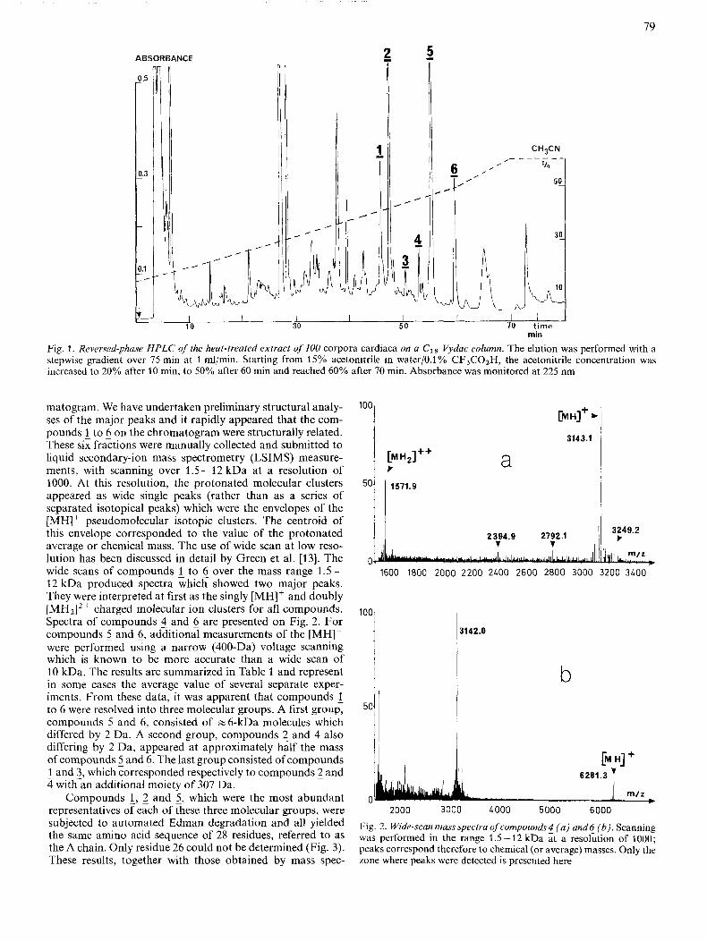
Fig. 1 . Reversed-phase HPLC of the heat-treaied extract of100 corpora cardiaca on a c18 Vydac column. The elution was performed with a stepwise gradient over 75 min at 1 ml/min. Starting from 15% acetonitrile in water/O.l Yo CF3COZH, the acetonitrile concentration was increased to 20% after 10 min, to 50% after 60 min and reached 60% after 70 min. Absorbance was monitored at 225 nm
,
I I 1 I I I I 50 70 time 10 30
min
Fig. 1 . Reversed-phase HPLC of the heat-treaied extract of100 corpora cardiaca on a c18 Vydac column. The elution was performed with a stepwise gradient over 75 min at 1 ml/min. Starting from 15% acetonitrile in water/O.l Yo CF3COZH, the acetonitrile concentration was increased to 20% after 10 min, to 50% after 60 min and reached 60% after 70 min. Absorbance was monitored at 225 nm
m / r
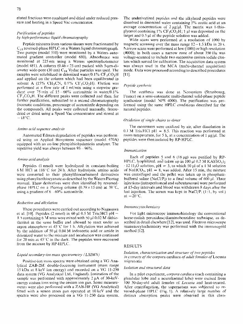
matogram. We have undertaken preliminary structural analy- ses of the major peaks and it rapidly appeared that the com- pounds 1 to 6 on the chromatogram were structurally related. These six fractions were manually collected and submitted to liquid secondary-ion mass spectrometry (LSIMS) measure- ments, with scanning over 1.5-12 kDa at a resolution of 1000. At this resolution, the protonated molecular clusters appeared as wide single peaks (rather than as a series of separated isotopical peaks) which were the envelopes of the [MH] + pseudomolecular isotopic clusters. The centroid of this envelope corresponded to the value of the protonated average or chemical mass. The use of wide scan at low reso- lution has been discussed in detail by Green et al. [13]. The wide scans of compounds 1 to 6 over the mass range 1.5 - 12 kDa produced spectra which showed two major peaks. They were interpreted at first as the singly [MH]' and doubly [MH2]'+ charged molecular ion clusters for all compounds. Spectra of compounds 5 and 6 are presented on Fig. 2. For compounds 5 and 5, additional measurements of the [MH]' were performed using a narrow (400-Da) voltage scanning which is known to be more accurate than a wide scan of 10 kDa. The results are summarized in Table 1 and represent in some cases the average value of several separate exper- iments. From these data, it was apparent that compounds 1 to 6 were resolved into three molecular groups. A first group, compounds 5 and 5, consisted of ~ 6 - k D a molecules which differed by 2 Da. A second group, compounds 2 and 4 also differing by 2 Da, appeared at approximately half the mass of compounds 5 and 6. The last group consisted of compounds 1 and 3, which corresponded respectively to compounds 2 and 4 with an additional moiety of 307 Da.
Compounds 1, 2 and 5 , which were the most abundant representatives of each of these three molecular groups, were subjected to automated Edman degradation and all yielded the same amino acid sequence of 28 residues, referred to as the A chain. Only residue 26 could not be determined (Fig. 3). These results, together with those obtained by mass spec-
'OOi
[MH21++ t a
3143.1
/ / 3249.2 t 2394.9 2792.1
V I ,
0 1600 1800 2000 2200 2LOO 2600 2800 3000 3200 3LOO
1142.0
b
- - 2000 3000 LOO0 5000 6000
Fig. 2. Wide-scan massspectra of compoundsf ( a ) andc (b). Scanning was performed in the range 1.5-12 kDa at a resolution of 1000; peaks correspond therefore to chemical (or average) masses. Only the zone where peaks were detected is presented here
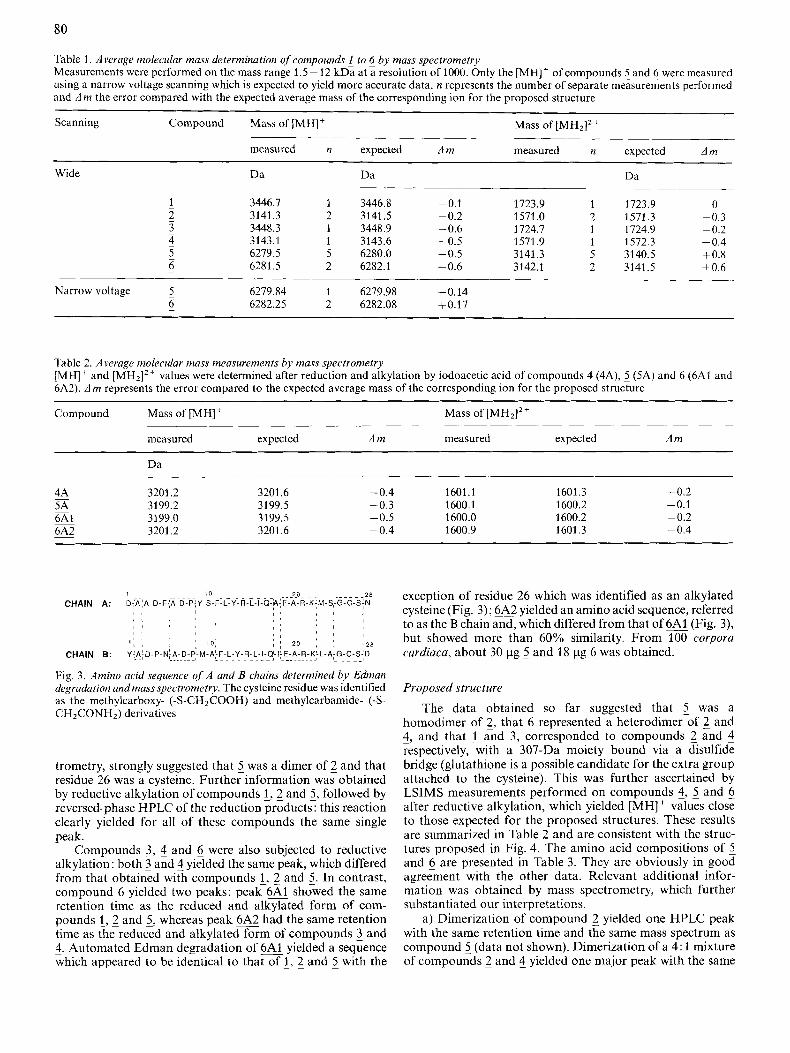
Table 1. Average molecular mass determination of compounds 1 to 6 by mass spectrometry Measurements were performed on the mass range 1.5 - 12 kDa at a resolution of 1000. Only the [MH]+ of compounds and 6 were measured using a narrow voltage scanning which is expected to yield more accurate data. n represents the number of separate measurements performed and Am the error compared with the expected average mass of the corresponding ion for the proposed structure
Scanning Compound Mass of [MH]+ Mass of [MH2I2+
measured n expected Am measured n expected Am
Wide Dd Da Da
- 1 3446.7 1 3446.8 -0.1 1723.9 1 1723.9 0
~ 3 3448.3 1 3448.9 -0.6 1724.7 1 1724.9 -0.2 - 2 3141.3 2 3141.5 - 0.2 1571 .O 2 1571.3 -0.3
4 3143.1 1 3143.6 -0.5 1571.9 1 1572.3 -0.4 ~ 5 6279.5 5 6280.0 -0.5 3141.3 5 3140.5 +0.8 ~ 6 6281.5 2 6282.1 -0.6 3142.1 2 3141.5 +0.6
Narrow voltage ~ 5 6279.84 1 6279.98 -0.14 - 6 6282.25 2 6282.08 +0.17
Table 2. Average molecular mass measurements by mass spectrometry [MH]' and [MH,]'+ values were determined after reduction and alkylation by iodoacetic acid of compounds 4 (4A), 5 (5A) and 6 (6A1 and 6A2). Am represents the error compared to the expected average mass of the corresponding ion for the proposed structure
Compound Mass of [MH]' Mass of [MH2I2+
measured expected Am measured expected Am
Da
4A 3201.2 3201.6 -0.4 1601.1 1601.3 - 0.2 5A 3199.2 3199.5 -0.3 1600.1 1600.2 -0.1 6AI 3199.0 3199.5 -0.5 1600.0 1600.2 -0.2 6A2 3201.2 3201.6 -0.4 1600.9 1601.3 - 0.4
-
-
~
~
trometry, strongly suggested that 5 was a dimer of 2 and that residue 26 was a cysteine. Further information was obtained by reductive alkylation of compounds !,2 and 5, followed by reversed-phase HPLC of the reduction products : this reaction clearly yielded for all of these compounds the same single peak.
Compounds 3, 5 and 6 were also subjected to reductive alkylation: both 3 and 5 yielded the same peak, which differed from that obtained with compounds i, 2 and 2. In contrast, compound 6 yielded two peaks: peak 6A1 showed the same retention time as the reduced and alkylated form of com- pounds 1,2 and 5, whereas peak 6A2 had the same retention time as the reduced and alkylated form of compounds 3 and 4. Automated Edman degradation of 6A1 yielded a sequence which appeared to be identical to that of 1, 2 and 5 with the
exception of residue 26 which was identified as an alkylated cysteine (Fig. 3); 6A2 yielded an amino acid sequence, referred to as the B chain and, which differed from that of (Fig. 3), but showed more than 60% similarity. From 100 corpora cardiaca, about 30 pg 5 and 18 pg 6 was obtained.
Proposed structure The data obtained so far suggested that 5 was a
homodimer of 2, that 6 represented a heterodimer of 2 and ~ 4, and that 1 and 3 , corresponded to compounds 2 and 4 respectively, with a 307-Da moiety bound via a disulfide bridge (glutathione is a possible candidate for the extra group attached to the cysteine). This was further ascertained by LSIMS measurements performed on compounds 4, 5 and 6 after reductive alkylation, which yielded [MH] + values close to those expected for the proposed structures. These results are summarized in Table 2 and are consistent with the struc- tures proposed in Fig. 4. The amino acid compositions of and 6 are presented in Table 3. They are obviously in good agreement with the other data. Relevant additional infor- mation was obtained by mass spectrometry, which further substantiated our interpretations.
a) Dimerization of compound 2 yielded one HPLC peak with the same retention time and the same mass spectrum as compound 5 (data not shown). Dimerization of a 4: 1 mixture of compounds 2 and 4 yielded one major peak with the same
81
3141.54 k
P
3139.45, C
I I mf I
A A
f 1 1
m / z P
B B
A 0 A A
5 - 6 -
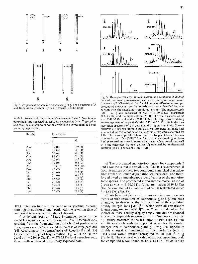
Fig. 4. Proposed structure.yjor compound.y L to c. The structures of A and B chains are given in Fig. 3. G represents glutathione
Tdbk 3. Amino acid composition of compounds S and 6. Numbers in parentheses are expected values from sequencing data. Tryptophan and cysteine contents were not determined (no tryptophan had been found by sequencing)
Residue Residues in
5 6 ~ -
Asx 8.2 (8) 1.9 (8) Glx 3.9 (4) 4.1 (4) Ser 6.0 (6) 4.1 (4) GlY 1.8 (2) 2.1 (2) Arg 4.2 (4) 3.7 (4)
Ala 9.9 (10) 9.7 (10)
TYr 4.1 (4) 3.7 (4) Val 0 (0) 0.1 (0) Met 1.9 (2) 1.9 (2)
Thr 0.2 (0) 0.3 (0)
Pro 2.1 (2) 2.8 (3)
Ile 2.1 (2) 2.8 (3) Leu 4.2 (4) 4.8 (5) Phe 4.2 (4) 3.0 (3) LYS 2.2 (2) 2.1 (2)
HPLC retention time and the same mass spectrum as com- pound 5 ; an additional small peak with the retention time of compound 5 was detected (data not shown).
b) Wide-scan spectra of 2 and 4 contained peaks (in the 2 - 3-kDa region) which corresponded to the C-terminal ions resulting from the fragmentation at the level of proline resi- dues, a process already observed in the case of large peptides [14]. According to the nomenclature of Roepstorff et al. [15] to describe this type of fragmentation, Y,, = 2433.5 Da for - 2 and Y Z l = 2394.9 Da, Y Z 5 = 2792.1 Da for 4 were observed; these results reinforced the primary sequencedata.
3141.31A'
Fig. 5. Muss spectrometry; isotopic puttern at u resolution uf 4000 of the molecular ions qf compounds 2 ( a ) , 4 (b) , and o j the major source jrugments of's ( d ) and6 ( e ) . For 2 and 4 the peaks (P) ofmonoisotopic protonated molecular ions (darkened) were easily identified by com- parison with the calculated isotopic pattern (c). The monoisotopic [MH]' of 2 was measured at m/z = 3139.39 Da (calculated: 3139.45 Da) and the monoisotopic [MH]' of 4 was measured at m/ z = 3141.52 Da (calculated: 3141.54 Da). The large ions exhibiting an average mass of respectively 3141.3 Da and 3142.1 Da in the low- resolution spectrum of 5 (Table 1) and 6 (Table 1 and Fig. 2) were observed at 4000 resolution (d and e). It was apparent that these ions were not doubly charged since the isotopic peaks were separated by 1 Da. The isotopic profile obtained for this fragment from 5 (d) was close to the one of the [MH]+ from 2 (a). The corresponding ion from 6 (e) presented an isotopic pattern and mass values correlating well with the calculated isotopic pattern (0 obtained by mathematical addition (in a 1 : 1 ratio) of 2 and 4 [MH]'
c) The protonated monoisotopic mass for compounds 2 and 4 was measured at a resolution of 4000. The experimental isotopic pattern of these two compounds, matched that calcu- lated from our Edman degradation sequence data, and there- fore allowed an unambiguous identification of the monoiso- topic species. The protonated monoisotopic molecular ion of - 2 was at m/z = 3139.39 Da (calculated value: 3139.45 Da) (Fig. 5a) and that of 4 at m/z = 3141.52 Da (calculated value: 3141.54 Da) (Fig. 5b).
d) We have not performed monoisotopic mass measure- ments at unit resolution of compounds 5 and 6, but have attempted to determine the isotopic mass of their putative doubly charged ions [MH2I2+, which were all remarkably intense compared to the [MH]' ions. Other peptides of similar molecular mass usually display singly and doubly charged ions with comparable intensities [13, 141. We noticed that the m / z values measured at the resolution of 1000 (Table 1) did not fit accurately with the expected values for the doubly charged ions of compounds 2 and 6. For 5, the supposedly doubly charged ion measured at low resolution (m/z = 3141.3 Da) would rather correspond to an [MH]' of 2 (Table 1). The chemical m/z value of the corresponding peak for compound 6 was found to be 3142.1 Da, which is very
82
ABSORBANCE
rnin
ABSORBANCE
miri
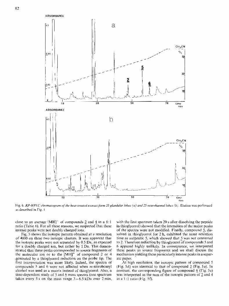
Fig. 6 . RP-HPLC chromatogram of the heat-treated extract form 25 glandular lobes ( a ) and 25 neurohaemal lobes ( b ) Elution as described in Fig. 1
was performed
close to an average [MH]' of compounds 2 and 4 in a 1 : 1 ratio (Table 1). For all these reasons, we suspected that these intense peaks were not doubly charged ions.
Fig. 5 shows the isotopic pattern obtained at a resolution of 4000 on these two isotopic clusters. It was apparent that the isotopic peaks were not separated by 0.5 Da, as expected for a doubly charged ion, but rather by 1 Da. This demon- strated that these peaks corresponded to source fragments of the molecular ion or to the [MH]' of compound 2 or 4 generated by a thioglycerol reduction on the probe tip. The first interpretation was more likely. Indeed, the spectra of compounds 5 and 6 were not affected when m-nitrobenzyl alcohol was used as a matrix instead of thioglycerol. Also, a time-dependent study of 5 and 6 mass spectra (one spectrum taken every 5 s on the mass range 3 -6.5 kDa over 2 min,
with the first spectrum taken 20 s after dissolving the peptide in thioglycerol) showed that the intensities of the major peaks of the spectra were not modified. Finally, compound 5, dis- solved in thioglycerol for 2 h, exhibited the same retention time as authentic 5 , which showed that 5 was not converted to 2. Therefore reduction by thioglycerol of compounds 5 and 6 appared highly unlikely. In consequence, we interpreted these peaks as source fragments and we shall discuss the mechanism yielding these particularly intense peaks in a separ- ate paper.
At high resolution, the isotopic pattern of compound 5 (Fig. 5.d) was identical to that of compound 2 (Fig. 5a). In contrast, the corresponding figure of compound 6 (Fig. 5e) was interpreted as the sum of the isotopic pattern of 2 and 4 in a 1 : 1 ratio (Fig. 5f).
83
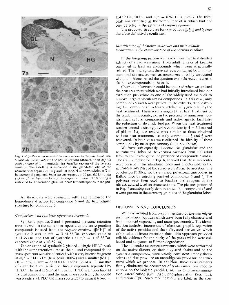
Fig. I. Distribution q f materid immunoreactive to the polyclonul unti- 6 untibody jseriiin diluted I : 2000) in corpora cardiaca of 30-day-old udult fi.ma1e.r of L. migratoria. (a) Paraffin section of the corpora rnrdicicci. The labelling is restricted to thc glandular lobe of the neurohaemal organ. (GL = glandular lobe, N = nervous lobe, HG = hypocerebral ganglion). Scale bar corresponds to 50 km. (b) Ultrathin section of the glandular lobe of the corporci cuudicrcu. The labclling is rcstricted to the secretion granules. Scale bar corresponds to 0.5 pm
All these data were consistent with, and reinforced the homodimer structure for compound 5 and the heterodimer structure for compound 6.
Coinparison with .s-ynthetic rgference compounds
Synthetic peptides 2 and 4 presented the same retention times as well as the same mass spectra as the corresponding compounds isolated from the corpora cardiaca. ([MH] + of synthetic 2 was at m/z = 3141.53 Da, expected value at 3141.49 Da, and that of synthetic 4 ~ at mjz = 3143.58 Da, expected value at 3143.59 Da).
Dimerization of synthetic 2 yielded a single HPLC peak with the same retention time as the natural compound 5; the niass spectrum was also identical, with it very intense fragment at nz/z = 3141.3 Da (base peak: 100%) and a smaller [MH]' (10- 15%) at m/z = 6279.8 Da. Oxidation of a 1 : 1 mixture of synthetic 2 and 4 yielded three compounds separated by HPLC. The first presented the same HPLC retention time as natural compound 5 and the same mass spectrum; the second was identical (HPLC and mass spectrum) to natural ~ 6 ( m / z =
3142.2 Da, 100%, and m/z = 6282.1 Da, 12%). The third peak was identified as the homodimer of 4, which had not been detected in the extracts of corpora cardiaca.
The proposed structures for compounds 2,4,5 and 6 were therefore definitively confirmed.
Identification of the native molecules and their cellular localization in the glandular lobe qf the corpora cardiaca
In the foregoing section we have shown that heat-treated extracts of corpora cardiaca from adult females of Locusta contained at least six compounds which were structurally related. The finding that these extracts contained both mono- mers and dimers, as well as monomers possibly associated with glutathione, raised the question as to the exact nature of the native compounds in the cells.
Clear-cut information could be obtained when we omitted the heat treatment which we had initially introduced into our extraction procedure as one of the widely used methods to remove large-molecular-mass compounds. In this case, only compounds 5 and 6 were present in the extracts, demonstrat- ing that compounds 1 to 4 were artefactually generated by the heat treatment. The& results suggest that heat treatment of the crude homogenate, i.e. in the presence of numerous non- identified cellular components and redox agents, facilitates thc reduction of disulfide bridges. When the heat treatment was performed in strongly acidic conditions (pH = 2.1 instead of pH = 5.51, the results were similar to those obtained without heat treatment, i.e. only compounds 5 and 5 were recovered. In both cases we confirmed the identity of these compounds by mass spectrometry (data not shown).
We have subsequently dissected the glandular and the neurohaemal lobes of the corpora cardiaca from 100 adult females and investigated the presence of compounds 5 and 6. The results, presented in Fig. 6, showed that these molecules were present in the glandular lobes and undetectable in the neurosecretory part of the corpora cardiaca. To ascertain this conclusion further, we have raised polyclonal antibodies in Balb/c mice by injecting purified compounds 5 and 6. The polysera were then used to localize the antigens at the ultrastructural level on tissue sections. The pictures presented in Fig. 7 unambiguously demonstrated that compounds 5 and 6 were present in the secretory granules of the glandular lobes.
DISCUSSION A N D CONCLUSION
We have isolated from corpora cardiaca of Locusta migra- toriu two major peptides which have been fully characterized by amino acid sequencing and mass spectrometry. The identi- fication included intense use of chromatographic separation of the native peptides and their alkylated derivatives which exhibited a different retention time. This approach provided reliable evidence for the purity of the peaks which were col- lected and subjected to Edman degradation.
The molecular mass measurements, which were performed on the native dimers, on their alkylated chains and on the synthetic compounds, were strictly consistent among them- selves and thus provided an unambiguous proof for the struc- tures which we propose. In addition, these measurements firmly eliminated the occurrence of post-translational modifi- cations on the isolated peptides, such as C-terminal amida- tion, esterification (Glu, Asp), phosphorylation (Ser, Thr), sulfatation (Tyr). Such modifications are labile in the con-
84
ditions of Edman degradation or during amino acid analysis and would not have been detected.
Compounds 1 to 4 isolated in this study were undoubtedly artefactual by-products of the extraction. Their presence has proven advantageous for the reinforcement of the structural data of the two native compounds 5 and 6.
Interestingly, the two peptides 5 aad 6 are a homodimer and a heterodimer, respectively. As-presented in Fig. 4, they share one common monomer, which we refer to as A; peptide 5 appears as a homodimeric association A-A, whereas peptide
~ 6 contains an A chain plus a structurally related (60% simi- larity) B chain (A-B association). From our synthetic mono- mers, we could generate a B-B dimer which we have characterized in HPLC: such a dimer was undetectable in the extracts of corpora cardiaca.
A computer search of protein sequence data banks has not revealed any substantial similarity between the two dimers and other established sequences. We can therefore consider these two molecules as novel peptides for which we propose the names of 6K-I (homodimer) and 6K-I1 (heterodimer) peptides, pending the determination of their physiological roles.
Our immunocytochemical data, taken in conjunction with the HPLC profiles from separate extractions of the storage and the glandular lobes, clearly showed that the 6-kDa peptides were predominantly present in the glandular lobes of the corpora cardiacu. We do not firmly exclude, however, that the storage lobes contain some material at significantly lower concentrations. The presence of the 6-kDa peptides could be traced by immunocytochemistry to the secretory granules within the glandular lobe cells. These granules have been reported to contain bioactive peptides, namely AKH-I and AKH-I1[16]. It will be challenging to investigate whether our 6-kDa peptides and the adipokinetic hormones are de- rived from a common precursor or if they are biogenetically unrelated. The 6-kDa peptides do not contain the AKH se- quence, but could possibly be processed by proteolytic cleavage to smaller bioactive molecules.
As already indicated, both 6-kDa peptides (plus a series of selected fragments) have been chemically synthesized and we are now investigating the functional significance of these peptides in L. migraboria.
The skilful technical assistance of Mrs Martine Schneider, Annie Meunier, Clotilde Heyer and Odile Sorokine is gratefully acknowl-
edged. We wish to express our gratitude to Dr Marie Meister for the preparation of polyclonal antibodies. H. H. expresses her appreciation to Prof. Suzuki and Dr Nagasawa for introducing her to the chemistry ofpeptide hormones. We are indebted to B. Green from VG Analytical (Manchester, UK) and to D. Fraisse from Service central d’analyse- Centre national de la recherche scientifique (Solaize, France) for some mass spectrometry facilities. The expertise of L. Denoroy from Service central dhna1.yse-Centre nutional de la recherche scientifique for auto- mated sequencing of peptides is gratefully acknowledged.
REFERENCES 1. Scharrer, B. (1952) Biol. Bull. Woods Hole 102, 261 -272. 2. Scarborough, R. M., Jamieson, G. C., Kalish, F., Kramer, S. J.,
McEnroe, G. A., Miller, C. A. & Schooley, D. A. (1984) Proc. Natl Acad. Sci. USA 81, 5515 - 5579.
3. Witten, J. L., Schaffer, M. H., O’Shea, M., Cook, J. C., Hemling, M. I. & Rinehart, K. L. (1984)Biochem. Biuphys. Res. Commun.
4. Ziegler, R., Eckart, K., Schwarz, H. & Keller, R. (1985) Biochem.
5. Hayes, T. K., Keeley, L. L., Knight, D. W. (1986) Biuchem.
6. Gade, G. & Rinehart, K. L. (1987) Bid. Chem. Hoppe-Seyler 368,
7. Herault, J. P., Girardie, J. & Proux J. (1985) Int. J . Invert. Reprod.
8. Fournier, B., Herault, J. P. & Proux, J. (1987) Gen. Comp.
9. Morgan, P. J., Siegert, K. J. & Mordue, W. (1987) Insect Biochem.
10. Nagasawa, H., Kataoka, H., Isogai, A,, Tamura, S., Suzuki, A,, Mizoguchi, A., Fujiwara, Y., Suzuki, A., Takahashi, S. & Ishizaki, H. (1986) Proc. Natl Acad. Sci. USA 83, 5840-5843.
11. Barber, M. & Green, B. (1987) Rapid Commun. Mass Spectrom.
12. Zachary, D., Goltzene, F., Holder, F. C., Berchtold, J. P., Nagasawa, H., Susuki, A., Mizoguchi, H., Ishizaki, H. & Hoffmann, J . A. (1988) Znt. J . Invert. Reprod. Devl. 14, 1-10,
13. Green, B. & Bordoli, R. S. (1986) in Mass spectrometry in biomedi- cal research (Gaskell, S. J., ed.) pp. 235-250, John Wiley & Sons, New York.
14. Boyot, P., Trifilieff, E., Van Dorsselaer, A. & Luu, B. (2988) Anal. Biochem. I73,15 - 85.
15. Roepstroff, P. & Fohlmann, J. (1984) Biomed. Mass Spectrom 11, 601.
16. Diederen, J. H. B., Maas, H. A., Pel, H. J., Schooneveld, H., Jansen, W. F. & Vullings, H. G. B. (1987) Cell. Tissue Res. 249, 379-389.
124,350-358.
Biophys. Res. Commun. 133, 331 - 342.
Biuphys. Res. Cornmun. 140, 674-678.
67 - 75.
Dev. 8, 325 - 335.
Endoocrinol. 68, 49 - 56.
17,383-388.
I , 80-83.
Note added in proof’: The 6-kDa dimeric peptides described in this paper are similar to the dimeric peptides associated with adipokinetic hormones I and I1 in their corresponding precursor molecules [O’Shea (1 989) personal communication, in the press].
Related Documents