Gen. Physiol. Biophys. (2003), 22, 329—340 329 Isolation and Morphology of Single Purkinje Cells from the Porcine Heart T. Stankovičová 1,3 , V. Bito 1 , F. Heinzel 1,4 , K. Mubagwa 2 and K. R. Sipido 1 1 Laboratory of Experimental Cardiology, Catholic University of Leuven, Belgium 2 Center of Experimental Surgery, Catholic University of Leuven, Belgium 3 Department of Pharmacology and Toxicology, Faculty of Pharmacy, Comenius University, Bratislava, Slovakia 4 Institute of Pathophysiology, University of Essen, Germany Abstract. Purkinje cells were isolated from both ventricles of young adult domes- tic pigs and examined by transmitted light or laser scanning confocal microscopy. Purkinje cells in free running Purkinje fibres were organised in multicellular strands where individual cells were tightly connected end-to-end and closely side-to-side. After isolation, single cells gradually lost the elongated appearance and became more rounded, but the cell membrane remained smooth and undamaged. The contractile material was not very dense and was seen most clearly in the sub- membraneous area. Staining of the cell membrane with the lipophilic fluorescent dye di-8-ANNEPS, and visualization with confocal microscopy, confirmed that the cell surface membrane was smooth without blebs. This staining also showed that Purkinje cells had no transversal tubules. We reconstructed the three-dimensional geometry of the Purkinje cells and determined the cell size. The average values were 62 ± 9 μm for length, 32 ± 3 μm for width, and 41 ± 4 μm for depth (n = 7). Calculated cross-section area and volume were 1047 ± 167 μm 2 and 47 ± 14 pl. Compared to ventricular cells, the morphology of the Purkinje cells reflects their specific role in impulse conduction. Key words: Purkinje cell — Pig — Morphology — Confocal microscopy Introduction The cardiac conduction system coordinates and synchronizes cardiac contraction (Sommer and Johnson 1979). The impulses pass from the sino-atrial node across the atria and atrio-ventricular node to Purkinje fibres which complete the activation Correspondence to: Dr. Tatiana Stankovičová, Department of Pharmacology and Toxicology, Faculty of Pharmacy, Comenius University,Kalinčiakova 8, 832 32 Bratislava 3, Slovakia. E-mail: [email protected]

Welcome message from author
This document is posted to help you gain knowledge. Please leave a comment to let me know what you think about it! Share it to your friends and learn new things together.
Transcript

Gen. Physiol. Biophys. (2003), 22, 329—340 329
Isolation and Morphology of Single Purkinje Cellsfrom the Porcine Heart
T. Stankovičová1,3
, V. Bito1, F. Heinzel
1,4, K. Mubagwa
2
and K. R. Sipido1
1 Laboratory of Experimental Cardiology,Catholic University of Leuven, Belgium
2 Center of Experimental Surgery, Catholic University of Leuven, Belgium3 Department of Pharmacology and Toxicology, Faculty of Pharmacy,
Comenius University, Bratislava, Slovakia4 Institute of Pathophysiology, University of Essen, Germany
Abstract. Purkinje cells were isolated from both ventricles of young adult domes-tic pigs and examined by transmitted light or laser scanning confocal microscopy.Purkinje cells in free running Purkinje fibres were organised in multicellular strandswhere individual cells were tightly connected end-to-end and closely side-to-side.After isolation, single cells gradually lost the elongated appearance and becamemore rounded, but the cell membrane remained smooth and undamaged. Thecontractile material was not very dense and was seen most clearly in the sub-membraneous area. Staining of the cell membrane with the lipophilic fluorescentdye di-8-ANNEPS, and visualization with confocal microscopy, confirmed that thecell surface membrane was smooth without blebs. This staining also showed thatPurkinje cells had no transversal tubules. We reconstructed the three-dimensionalgeometry of the Purkinje cells and determined the cell size. The average valueswere 62± 9 µm for length, 32± 3 µm for width, and 41± 4 µm for depth (n = 7).Calculated cross-section area and volume were 1047 ± 167 µm2 and 47 ± 14 pl.Compared to ventricular cells, the morphology of the Purkinje cells reflects theirspecific role in impulse conduction.
Key words: Purkinje cell — Pig — Morphology — Confocal microscopy
Introduction
The cardiac conduction system coordinates and synchronizes cardiac contraction(Sommer and Johnson 1979). The impulses pass from the sino-atrial node across theatria and atrio-ventricular node to Purkinje fibres which complete the activation
Correspondence to: Dr. Tatiana Stankovičová, Department of Pharmacology andToxicology, Faculty of Pharmacy, Comenius University, Kalinčiakova 8, 832 32 Bratislava3, Slovakia. E-mail: [email protected]

330 Stankovičová et al.
pathway through the interventricular septum, penetrate into the heart apex, andturn superiorly into the ventricular walls. They supply the papillary muscles beforesupplying the lateral walls of the ventricles. The ventricular depolarisation dependscritically on the presence of these large, barrel-shaped fibres, first described by JanEvangelista Purkinje in 1845. Because the mass of the left ventricle is much largerthan that of the right ventricle, the Purkinje network is more elaborate in that sideof the heart.
The primary function of cardiac Purkinje cells is a fast conduction of theimpulse, rather than production of contractile force. The cellular architecture isknown to differ from that of working myocytes (reviewed by Sommer and Johnson1979), e.g., by the less dense network of contractile filaments. This feature is verypronounced in cells from larger mammals (sheep, cattle, dog) where the contractilematerial is restricted to a small rim under the sarcolemma, in contrast to smallermammals (rabbit, rat, guinea pig) that can have abundant myofilaments in theirPurkinje fibres. In the present report we have examined the morphology of singlePurkinje cells isolated from pig hearts.
Materials and Methods
Isolation of Purkinje cells
The experimental procedures were approved by the Ethical Committee of theCatholic University of Leuven and conform to the principles outlined in the Decla-ration of Helsinki. Healthy domestic pigs (30–35 kg, sex indeterminate) were used.Pigs were fully anaesthetised with pentobarbital (20 mg/kg i.v.) after premedica-tion with azaperone (4 mg/kg i.m.) and atropine (0.35 mg/kg i.v.). Under artificialventilation, the chest was opened, the heart was rapidly removed, and collectedinto a beaker of cold Tyrode solution to be rinsed of blood.
To dissociate Purkinje cells from both ventricles two approaches were used.From the left ventricle we dissected free-running Purkinje fibres and subjected theseto the enzymatic isolation procedure described by Glitsch et al. (1989), combinedwith the procedure for tissue chunk isolation described by Curtet et al. (2000), andmodified for our experimental conditions. Purkinje fibres were placed into 10 mlbeakers with Ca2+-free Tyrode solution at 37◦C and agitated with a stream ofoxygen. After 35–40 min Purkinje fibres were transferred into the freshly preparedenzymatic solution: 1.0–1.5 mg/ml collagenase A (Roche Diagnostics, Brussel, Bel-gium) and 0.1 mg/ml protease XIV (Sigma-Aldrich, Bornem, Belgium), dissolvedin Ca2+-free Tyrode solution. After 20–40 min the fibrous tissue sheath was largelydigested, revealing bundles of Purkinje cells. The digestion medium with Purkinjefibres was gently triturated and regularly checked on an inverted microscope atlow magnification (at first at 20 min intervals, more frequently as digestion pro-gressed) for dissociation of single cells. When these were found to be abundant, theundigested strands were removed from the Purkinje-cells enriched suspension andtransferred to another beaker with a fresh enzymatic solution. To stop digestion,

Purkinje Cell Morphology 331
low-Ca2+ (0.18 mmol/l) Tyrode solution was added to the cell suspension. Aftercells had settled on the bottom of the beaker, 60–70% of the solution was exchangedwith a fresh batch of the low calcium solution. These steps were repeated severaltimes to remove the enzymes. At the end, normal Tyrode solution was added andcells were stored at the room temperature. Morphology was measured within 90minutes after isolation.
To isolate Purkinje fibres from the right heart side, we first perfused the rightventricle through its supplying coronary artery, as a variant of the isolation tech-nique described for rabbit Purkinje fibres (Sipido et al. 1993) and for left ventricularmyocytes (VMs) (Stankovicova et al. 2000). After perfusing with 37◦C oxygenatedTyrode solution for 3–5 min to clear all blood from coronary vasculature, theperfusate was changed to a nominally Ca2+-free Tyrode solution for 30 min, afterwhich the perfusate was exchanged with Ca2+-free solution containing the enzymes(collagenase A, 1.0–1.5 mg/ml, and protease XIV, 0.1 mg/ml, dissolved in Ca2+-free Tyrode solution). After 30–40 min, the tissue became swollen and opalescent,indicating digestion. At this time, free running Purkinje fibres were dissected andreturned for further enzymatic digestion into a beaker positioned in a double-walledPetri dish, warmed to 37◦C and filled with fresh enzyme solution. All the followingsteps were the same as described above for Purkinje fibres from left ventricle.
Confocal microscopy and staining with di-8-ANNEPS
The cells were studied in a glass-bottomed perfused chamber placed on the stageof a Zeiss Axiovert 100M (Carl Zeiss Microscopy, Jena, Germany) inverted micro-scope with a 40× oil-immersion objective Plan-Neofluar (NA = 1.3). The cells wererecognised and selected for further study by transmitted light imaging.
Purkinje cells were stained with the membrane-selective fluorescent dye di-8-ANNEPS, a voltage-sensitive dye, which is highly lipophilic and avidly stains thecell membrane. Cells were exposed to 20 µmol/l dye for 1–3 min directly in exper-imental bath (1 ml volume, 1/1000 mol/l dilution of a 20 mmol/l stock solutionin DMSO with 20% w.v. Pluronic). Confocal images were acquired and processedwith a Zeiss LSM 510 confocal laser point scanning system (Carl Zeiss Microscopy).After washout with a normal Tyrode solution, stained cells were excited at a wave-length of 488 nm with a 25 mW Argon laser (output power 50%, intensity attenu-ation to 3–10%). Fluorescence emission was detected via a 505–530 nm band-passfilter.
In order to obtain a two-dimensional fluorescent confocal image of a selectedcell, successive line scans were performed in the XY plane. For three-dimensionalinformation of the cell, a series of XY-images along the Z axis was collected. Thethickness of the optical slice on the Z axis was 1 µm. The scanning speed variedbetween 1.5 and 3.8 ms/line. Scanning speed, excitation and amplification settingswere kept constant during each experiment.
Electrophysiology data were recorded and analysed with pClamp (Ver. 8.2,Axon Instruments, USA).

332 Stankovičová et al.
Solutions and Drugs
The Tyrode solution used for perfusion contained (in mmol/l): NaCl 137; KCl 5.4;MgCl2 0.5; CaCl2 1.8; HEPES 11.8; glucose 10; adjusted with NaOH to pH 7.2.For cell isolation, the Ca2+-free Tyrode solution was used, which contained (inmmol/l): NaCl 130; KCl 5.4; KH2PO4 1.2; MgSO4 1.2; HEPES 6; NaOH till pH7.2. This solution could be supplemented with enzymes as described above. Thelow-Ca2+ Tyrode solution was made from the Ca2+-free Tyrode solution by adding0.18 mmol/l CaCl2. Di-8-ANNEPS was purchased from Molecular Probes (EuropeBV, Leiden, Netherlands). All other chemicals were from Sigma (Bornem, Belgium)or Merck (Darmstadt, Germany) and were of analytical grade.
Results
Morphology
We first examined the cell morphology in partially digested Purkinje fibre bundles(after 40–50 min of digestion). At this time, the collagenous sheath lost its density,became more transparent and strings of elongated cells were visible inside the fibres
Figure 1. A. Transmitted light image of a partially digested large free-running Purkinjefibre dissected from porcine left ventricle. B. Detail from the area indicated in panel Ashowing Purkinje cells connected end-to-end and side-to-side. C. Same preparation afterlabelling of the surface membrane with the fluorescent dye di-8-ANEPPS, identifyingindividual cells organised in strings; the arrow shows a clear end-to-end connection.
Purkinje Cell Morphology 333
(Fig. 1A,B). After an additional 30–60 min digestion Purkinje cell aggregated orindividual single cells were found in the bath and these were then further examined.
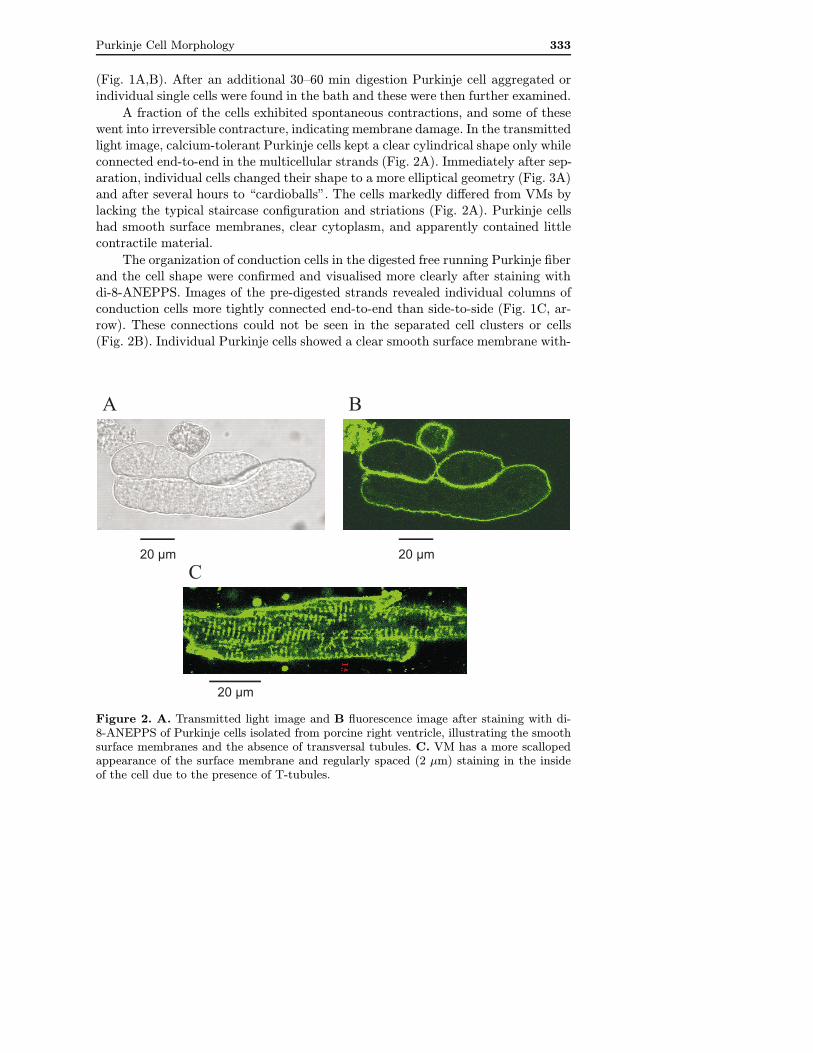
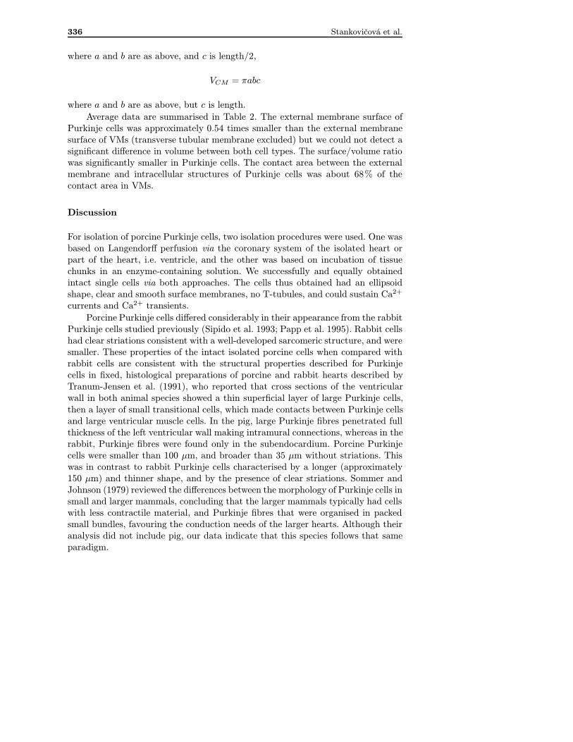
A fraction of the cells exhibited spontaneous contractions, and some of thesewent into irreversible contracture, indicating membrane damage. In the transmittedlight image, calcium-tolerant Purkinje cells kept a clear cylindrical shape only whileconnected end-to-end in the multicellular strands (Fig. 2A). Immediately after sep-aration, individual cells changed their shape to a more elliptical geometry (Fig. 3A)and after several hours to “cardioballs”. The cells markedly differed from VMs bylacking the typical staircase configuration and striations (Fig. 2A). Purkinje cellshad smooth surface membranes, clear cytoplasm, and apparently contained littlecontractile material.
The organization of conduction cells in the digested free running Purkinje fiberand the cell shape were confirmed and visualised more clearly after staining withdi-8-ANEPPS. Images of the pre-digested strands revealed individual columns ofconduction cells more tightly connected end-to-end than side-to-side (Fig. 1C, ar-row). These connections could not be seen in the separated cell clusters or cells(Fig. 2B). Individual Purkinje cells showed a clear smooth surface membrane with-
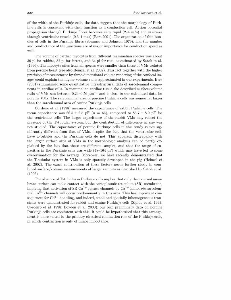
Figure 2. A. Transmitted light image and B fluorescence image after staining with di-8-ANEPPS of Purkinje cells isolated from porcine right ventricle, illustrating the smoothsurface membranes and the absence of transversal tubules. C. VM has a more scallopedappearance of the surface membrane and regularly spaced (2 µm) staining in the insideof the cell due to the presence of T-tubules.

334 Stankovičová et al.
A
B20 µm
20 µm
Figure 3. A. Transmitted lightimage, illustrating the sparse my-ofilaments below the surface mem-brane in the cytoplasm of Purkinjecells. B. Typical rod-shaped VMwith well organised and clear crossstriations.
out blebs or membrane disruptions. In contrast to VMs, Purkinje cells had notransversal tubules, which should be seen in the centre of the cells (Fig. 2B vs. C).Cells that had been identified as dead cells in the transmitted light image, basedon the presence of rough, irregular membranes with blebs, and of coarse cytoplas-mic granulations, did not exclude the dye and had irregular cytosolic staining,consistent with severe membrane damage (data not shown).
In the transmitted light image, striations inside the cytoplasm, indicative ofsarcomeric organization of myofilaments, were only faintly identifiable, predomi-nantly at the periphery in the subsarcolemmal area (Fig. 3A), in sharp contrast tothe well-defined and recognizable sarcomeric pattern in VMs (Fig. 3B).
Whole cell patch clamp on both isolated Purkinje cells (Fig 3A) and VMs wereperformed (Fig 3B). The membrane capacitance was 109±17 pF for Purkinje cells(n = 10) and 102 ± 6 pF for VMs (n = 41). Preliminary data indicate that thesingle Purkinje cells had Ca2+ currents, which could evoke Ca2+ transients (datanot shown).
Cell dimensions
Purkinje cell suspensions included some single ventricular cells as well, and wecompared cell morphology of the two cell types. Single Purkinje cells varied con-

Purkinje Cell Morphology 335
Table 1. Cell dimensions
length width depth(µm) (µm) (µm)
Purkinje cell (n = 7) 62± 9* 32± 3* 41± 4*
VM (n = 26) 156± 6 26± 1 17± 1
Purkinje cell vs. VM; * p ≤ 0.05.
siderably in length (35–99 µm) but their other two dimensions were less variable.Purkinje cell width varied between 22 to 45 µm and Purkinje cell depth variedbetween 32 to 59 µm. There were significant differences between the average di-mensions of Purkinje cells and those of VMs (Table 1). Purkinje cells were shorterbut somewhat wider compared to VMs, and their thickness was also larger.
We assumed that the Purkinje cell had the shape of an elliptical spheroid, andthe VM was an elliptical cylinder, and calculated the cross-sectional area accordingto the formula
P = πab
where a is width/2; b is depth/2.Data in Table 2 show that the cross-section of a Purkinje cell was approxi-
mately 3 times larger than the cross-section of a VM.Based on the measured dimensions and on the cellular shape, an estimate of
surface area and volume could be obtained. If we did not consider tubular mem-branes, the external cellular surface was calculated as
SPC = 4π((abc)2)1/3
for Purkinje cells, where a and b are as above, and c is length/2, and for VM as
SCM = 2πab+ c[π(1.5(a+ b))− (ab)1/2]
where a and b are as above, but c is length.For calculation of volume, the following formulas were used:
VPC = 4/3πabc
Table 2. Derived surface and volume dimensions
cross-sectional area surface area volume surface/volume(µm2) (µm2) (µm3) (µm2/µm3)
Purkinje cell 1 048± 167* 5 999± 1 135* 46 968± 13 997 0.1467 ± 0.0116*(n = 7)
VM 345± 18 11 091± 719 52 722± 4 237 0.2164 ± 0.0038(n = 26)
Purkinje cell vs. VM; * p ≤ 0.05.
336 Stankovičová et al.
where a and b are as above, and c is length/2,
VCM = πabc
where a and b are as above, but c is length.Average data are summarised in Table 2. The external membrane surface of
Purkinje cells was approximately 0.54 times smaller than the external membranesurface of VMs (transverse tubular membrane excluded) but we could not detect asignificant difference in volume between both cell types. The surface/volume ratiowas significantly smaller in Purkinje cells. The contact area between the externalmembrane and intracellular structures of Purkinje cells was about 68% of thecontact area in VMs.
Discussion
For isolation of porcine Purkinje cells, two isolation procedures were used. One wasbased on Langendorff perfusion via the coronary system of the isolated heart orpart of the heart, i.e. ventricle, and the other was based on incubation of tissuechunks in an enzyme-containing solution. We successfully and equally obtainedintact single cells via both approaches. The cells thus obtained had an ellipsoidshape, clear and smooth surface membranes, no T-tubules, and could sustain Ca2+
currents and Ca2+ transients.Porcine Purkinje cells differed considerably in their appearance from the rabbit
Purkinje cells studied previously (Sipido et al. 1993; Papp et al. 1995). Rabbit cellshad clear striations consistent with a well-developed sarcomeric structure, and weresmaller. These properties of the intact isolated porcine cells when compared withrabbit cells are consistent with the structural properties described for Purkinjecells in fixed, histological preparations of porcine and rabbit hearts described byTranum-Jensen et al. (1991), who reported that cross sections of the ventricularwall in both animal species showed a thin superficial layer of large Purkinje cells,then a layer of small transitional cells, which made contacts between Purkinje cellsand large ventricular muscle cells. In the pig, large Purkinje fibres penetrated fullthickness of the left ventricular wall making intramural connections, whereas in therabbit, Purkinje fibres were found only in the subendocardium. Porcine Purkinjecells were smaller than 100 µm, and broader than 35 µm without striations. Thiswas in contrast to rabbit Purkinje cells characterised by a longer (approximately150 µm) and thinner shape, and by the presence of clear striations. Sommer andJohnson (1979) reviewed the differences between the morphology of Purkinje cells insmall and larger mammals, concluding that the larger mammals typically had cellswith less contractile material, and Purkinje fibres that were organised in packedsmall bundles, favouring the conduction needs of the larger hearts. Although theiranalysis did not include pig, our data indicate that this species follows that sameparadigm.

Purkinje Cell Morphology 337
Generally, Purkinje fibres in our study had a thick coat of fibrous tissue, inwhich the individual conduction cells were packed and organised into thin bundles.The composition of this fibrous tissue has been described during histological stud-ies (Ohayon and Chadwick 1990). It has plentiful fibroblasts and matrix in whichcollagen, collagen struts (the isotropic components of the system), some elasticfibres and microfibrils surround conduction cells collected into bundles. The pres-ence of such a thick fibrous sheath is a major difficulty for the enzymatic isolationprocedure, as it increases the time needed to extract the Purkinje cells. Sheets etal. (1983) needed 4 hours for sheath digestion and revealing the columns of ca-nine Purkinje cells. In the case of the porcine heart, 20 min in enzymatic solutionallowed to confirm the typical organization of the individual cells in a chain-likearrangement in the partially digested Purkinje fibres, using transmitted light andconfocal microscopy. However, much more time, up to 2 hours was needed to obtainsingle cells in solution.
Sommer and Johnson (1968) established that conduction cells have no transver-sal tubules. This has been shown to be true across all animal species (reviewed inSommer and Johnson 1979). The absence of a T-tubular system may be one ofthe reasons why the more elongated shape, present in the multicellular prepara-tion, cannot be maintained after loosening cell-to-cell contacts and constraint bythe fibrous tissue. This transformation of Purkinje cells to elliptical spheroids or“cardiospheroids” and after several hours to “cardioballs” was also described forsheep Purkinje cells (Glitsch et al. 1989), and is reminescent of the atrial cardioballs(Bechem et al. 1983), observed in atrial cells that also lack the T-tubular system(Huser et al. 1996).
Morphology of isolated cells described so far varies considerably between ani-mal species. Glitsch et al. (1989) described the freshly isolated sheep Purkinje cellsas brick-shaped, with some cross striations, transforming later on to more spherical“cardioballs” with a diameter of 40–70 µm without deep invaginations of the cellmembrane. Single canine cardiac Purkinje cells were described to have well-definedstriations, cell borders (Sheets et al. 1983), and fine finger-like processes at eitherend (Boyden et al. 1989) with averaged dimensions 164 ± 41 µm by 35 ± 4 µm(at 22◦C, Sheets et al. 1983), 125 ± 26 µm by 32 ± 11 µm (at 37◦C, Boyden etal. 1989), or 157–314 µm by 35–36 µm (Boyden et al. 2000). The canine Purkinjecells possessed sparse myofilaments, and a more spherical shape compared to rabbitPurkinje cells. The rabbit Purkinje cells are 130–140 µm long and 10–15 µm wide(Cordeiro et al. 1998), with clear cross-striations. Our data are most reminiscentof the data on sheep and dog Purkinje cells.
We performed morphological measurements that served as a basis to estimatethe cross-sectional area, surface and volume of isolated cells. The changes in shapeafter isolation of Purkinje cells creates difficulties in extrapolating the measuredcell length and width to the dimensions of length and width in the intact fibre.However, the derived values of volume and surface area are not invalid, as there isno indication that there is a loss of membrane or cell contents, which would altersurface area or volume. Despite this limitation, and allowing for an overestimation
338 Stankovičová et al.
of the width of the Purkinje cells, the data suggest that the morphology of Purk-inje cells is consistent with their function as a conduction cell. Action potentialpropagation through Purkinje fibres becomes very rapid (2–4 m/s) and is slowerthrough ventricular muscle (0.3–1 m/s) (Bers 2001). The organization of thin bun-dles of cells in the Purkinje fibres (Sommer and Johnson 1979), and the numberand conductance of the junctions are of major importance for conduction speed aswell.
The volume of cardiac myocytes from different mammalian species was about30 pl for rabbits, 32 pl for ferrets, and 34 pl for rats, as estimated by Satoh et al.(1996). The myocyte sizes from all species were smaller than those of VMs isolatedfrom porcine heart (see also Heinzel et al. 2002). This fact together with the higherprecision of measurement by three-dimensional volume rendering of the confocal im-ages could explain the higher volume value approximated in our experiments. Bers(2001) summarised some quantitative ultrastructural data of sarcolemmal compo-nents in cardiac cells. In mammalian cardiac tissue the described surface/volumeratio of VMs was between 0.24–0.56 µm−1 and is close to our calculated data forporcine VMs. The sarcolemmal area of porcine Purkinje cells was somewhat largerthan the sarcolemmal area of canine Purkinje cells.
Cordeiro et al. (1998) measured the capacitance of rabbit Purkinje cells. Themean capacitance was 66.5 ± 2.5 pF (n = 65), compared to 86.7 ± 8.9 pF forthe ventricular cells. The larger capacitance of the rabbit VMs may reflect thepresence of the T-tubular system, but the contribution of differences in size wasnot studied. The capacitance of porcine Purkinje cells in this study is not sig-nificantly different from that of VMs, despite the fact that the ventricular cellshave T-tubules and the Purkinje cells do not. This apparent discrepancy withthe larger surface area of VMs in the morphologic analysis can be partly ex-plained by the fact that these are different samples, and that the range of ca-pacities in the Purkinje cells was wide (48–164 pF) which may have led to someoverestimation for the average. Moreover, we have recently demonstrated thatthe T-tubular system in VMs is only sparsely developed in the pig (Heinzel etal. 2002). The exact contribution of these factors needs further study in com-bined surface/volume measurements of larger samples as described by Satoh et al.(1996).
The absence of T-tubules in Purkinje cells implies that only the external mem-brane surface can make contact with the sarcoplasmic reticulum (SR) membrane,implying that activation of SR Ca2+ release channels by Ca2+ influx via sarcolem-mal Ca2+ channels will occur predominantly in this area. This has important con-sequences for Ca2+ handling, and indeed, small and spatially inhomogeneous tran-sients were demonstrated for rabbit and canine Purkinje cells (Sipido et al. 1993;Cordeiro et al. 1998; Boyden et al. 2000); our own preliminary data on porcinePurkinje cells are consistent with this. It could be hypothesised that this arrange-ment is more suited to the primary electrical conduction role of the Purkinje cells,in which contraction is only of minor importance.

Purkinje Cell Morphology 339
Acknowledgements. We thank Fons Verdonck for useful comments and suggestionsduring the experimental work, Patricia Holemans for technical assistance, and AlexandraZahradnikova for help during data analysis. This work was supported by grants of theFWO, the Flanders Fund for Scientific Research (KRS, KM), and by grants No. 1/8220/01and No. 1/0522/03 of the VEGA SR (TS).
References
Bechem M., Pott L., Rennebaum H. (1983): Atrial cells from hearts of adult guinea-pigsin culture: a new preparation for cardiac cellular electrophysiology. J. Membr. Biol.97, 179—191
Bers D. M. (2001): Excitation-contraction coupling and cardiac contractile force. (2nd ed.)Kluwer Academic Publishers, Dordrecht
Boyden P. A., Albala A., Dresdner K. P. Jr. (1989): Electrophysiology and ultrastructureof canine subendocardial Purkinje cells isolated from control and 24-hour infarctedhearts. Circ. Res. 65, 955—970
Boyden P. A., Pu J., Pinto J., ter Keurs H. E. D. (2000): Ca2+ transients and Ca2+ wavesin Purkinje cells: role in action potential initiation. Circ. Res. 86, 448—455
Cordeiro J. M., Spitzer K. W., Giles W. R. (1998): Repolarizing K+ currents in rabbitheart Purkinje cells. J. Physiol. (London) 508, 811—823
Curtet S., Soulier J. L., Zahradnik I., Giner M., Berque-Bestel I., Mialet J., Lezoualc’h F.,Donzeau-Gouge P., Sicsic S., Fischmeister R., Langlois M. (2000): New arylpiper-azine derivatives as antagonists of the human cloned 5-HT(4) receptor isoforms. J.Med. Chem. 43, 3761—3769
Glitsch H. G., Krahn T., Verdonck F. (1989): The dependence of sodium pump currenton internal Na concentration and membrane potential in cardioballs from sheepPurkinje fibers. Pfluegers Arch. 414, 52—58
Heinzel F., Bito V., Volders P. G., Antoons G., Mubagwa K., Sipido K. R. (2002): Spatialand temporal inhomogeneities during Ca2+ release from the sarcoplasmic reticulumin pig ventricular myocytes. Circ. Res. 91, 1023—1030
Huser J., Lipsius S. L., Blatter L. A. (1996): Calcium gradients during excitation-contrac-tion coupling in cat atrial myocytes. J. Physiol. (London) 494, 641—651
Mendez C., Mueller W. J., Merideth J., Moe G. K. (1969): Interaction of transmembranepotentials in canine Purkinje fibers and at Purkinje fiber-muscle junctions. Circ.Res. 24, 361—372
Ohayon J., Chadwick R. S. (1990): Effects of collagen microstructure on the mechanicsof the left ventricle. Biophys. J. 54, 1077—1088
Papp Z., Sipido K. R., Callewaert G., Carmeliet E. (1995): Two components of [Ca2+]i-activated Cl− current during large [Ca2+]i transients in single rabbit heart Purkinjecells. J. Physiol. (London) 483, 319—330
Satoh H., Delbridge L. M. D., Blatter L. A., Bers D. M. (1996): Surface: volume rela-tionship in cardiac myocytes studied with confocal microscopy and membrane ca-pacitance measurements: species-dependence and developmental effects. Biophys.J. 70, 1494—1504
Sheets M. F., January C. T., Fozzard H. A. (1983): Isolation and characterization of singlecanine cardiac Purkinje cells. Circ. Res. 53, 544—548
Sipido K. R., Callewaert G., Carmeliet E. (1993): [Ca2+]i transients and [Ca2+]i-depen-dent chloride current in single Purkinje cells from rabbit heart. J. Physiol. (London)468, 641—667

340 Stankovičová et al.
Sommer J. R., Johnson E. A. (1968): Cardiac muscle. A comparative study of Purkinjefibers and ventricular fibers. J. Cell Biol. 36, 497—526
Sommer J. R., Johnson E. A. (1979): Ultrastructure of cardiac muscle. In: Handbookof Physiology. Section 2: The Cardiovascular System (Eds. Berne, Sperelakis andGeiger), pp. 61—100, Bethesda MD, U.S.A.
Stankovicova T., Szilard M., De Scheerder I., Sipido K. R. (2000): M cells and transmuralheterogeneity of action potential configuration in myocytes from the left ventricularwall of the pig heart. Cardiovasc. Res. 45, 952—960
Tranum-Jensen J., Wilde A. A. M., Vermeulen Jessica T., Janse M. J. (1991): Morphologyof electrophysiologically identified junctions between Purkinje fibres and ventricu-lar muscle in rabbit and pig hearts. Circ. Res. 69, 429—437
Final version accepted: August 15, 2003
Related Documents