Citation: Wang, Y.; Yu, H.; He, X.; Lu, T.; Huang, X.; Luo, C. Isolation and Functional Characterization of a LEAFY Gene in Mango (Mangifera indica L.). Int. J. Mol. Sci. 2022, 23, 3974. https://doi.org/10.3390/ijms 23073974 Academic Editor: Pedro Martínez-Gómez Received: 26 February 2022 Accepted: 31 March 2022 Published: 2 April 2022 Publisher’s Note: MDPI stays neutral with regard to jurisdictional claims in published maps and institutional affil- iations. Copyright: © 2022 by the authors. Licensee MDPI, Basel, Switzerland. This article is an open access article distributed under the terms and conditions of the Creative Commons Attribution (CC BY) license (https:// creativecommons.org/licenses/by/ 4.0/). International Journal of Molecular Sciences Article Isolation and Functional Characterization of a LEAFY Gene in Mango (Mangifera indica L.) Yihan Wang † , Haixia Yu † , Xinhua He * , Tingting Lu, Xing Huang and Cong Luo * State Key Laboratory for the Conservation and Utilization of Subtropical Agro-Bioresources, College of Agriculture, Guangxi University, 100 East Daxue Road, Nanning 530004, China; [email protected] (Y.W.); [email protected] (H.Y.); [email protected] (T.L.); [email protected] (X.H.) * Correspondence: [email protected] (X.H.); [email protected] (C.L.) † These authors contributed equally to this work. Abstract: LEAFY (LFY) plays an important role in the flowering process of plants, controlling flowering time and mediating floral meristem differentiation. Owing to its considerable importance, the mango LFY gene (MiLFY; GenBank accession no. HQ585988) was isolated, and its expression pattern and function were characterized in the present study. The cDNA sequence of MiLFY was 1152 bp, and it encoded a 383 amino acid protein. MiLFY was expressed in all tested tissues and was highly expressed in flowers and buds. Temporal expression analysis showed that MiLFY expression was correlated with floral development stage, and two relative expression peaks were detected in the early stages of floral transition and floral organ differentiation. Moreover, 35S::GFP-MiLFY fusion protein was shown to be localized to the nucleus of cells. Overexpression of MiLFY in Arabidopsis promoted early flowering and the conversion of lateral meristems into terminal flowers. In addition, transgenic plants exhibited obvious morphological changes, such as differences in cauline leaf shape, and the number of lateral branches. When driven by the MiLFY promoter, GFP was highly expressed in leaves, floral organs, stems, and roots, during the flowering period. Exogenous gibberellin (GA 3 ) treatment downregulated MiLFY promoter expression, but paclobutrazol (PPP 333 ) upregulated it. Bimolecular fluorescence complementation (BiFC) assays showed that the MiLFY protein can interact with zinc-finger protein 4 (ZFP4) and SUPPRESSOR OF OVEREXPRESSION OF CONSTANS 1 (MiSOC1D). Taken together, these results indicate that MiLFY plays a pivotal role in controlling mango flowering, and that it is regulated by gibberellin and paclobutrazol. Keywords: mango (Mangifera indica L.); LEAFY; expression analysis; functional identification; promoter analysis 1. Introduction Flowering is an important process in the plant life cycle, and the time of flowering (early or late) essentially determines the time of harvest. In recent decades, much progress has been made in understanding the physiological and molecular mechanisms underlying flowering time in plants. It is clear that plant flowering is mainly affected by various environmental and endogenous signals, such as daylength, temperature, drought, plant age, endogenous phytohormones, and exogenous plant growth regulators (PGRs) [1,2]. A complex gene regulatory network has been revealed in Arabidopsis. At least six flowering pathways and several genes, such as FLOWERING LOCUS T (FT), SUPPRESSOR OF OVEREXPRESSION OF CONSTANS 1 (SOC1), and LEAFY (LFY), which act as floral pathway integrators to activate downstream floral meristem identity genes, such as LFY and APETALA1 (AP1), cooperate to promote flowering [3]. LFY is a plant-specific transcription factor that is a master regulator of flower initiation and, as such, it determines floral fate in Arabidopsis [4]. Many plant genomes contain Int. J. Mol. Sci. 2022, 23, 3974. https://doi.org/10.3390/ijms23073974 https://www.mdpi.com/journal/ijms

Welcome message from author
This document is posted to help you gain knowledge. Please leave a comment to let me know what you think about it! Share it to your friends and learn new things together.
Transcript
�����������������
Citation: Wang, Y.; Yu, H.; He, X.; Lu,
T.; Huang, X.; Luo, C. Isolation and
Functional Characterization of a
LEAFY Gene in Mango (Mangifera
indica L.). Int. J. Mol. Sci. 2022, 23,
3974. https://doi.org/10.3390/ijms
23073974
Academic Editor: Pedro
Martínez-Gómez
Received: 26 February 2022
Accepted: 31 March 2022
Published: 2 April 2022
Publisher’s Note: MDPI stays neutral
with regard to jurisdictional claims in
published maps and institutional affil-
iations.
Copyright: © 2022 by the authors.
Licensee MDPI, Basel, Switzerland.
This article is an open access article
distributed under the terms and
conditions of the Creative Commons
Attribution (CC BY) license (https://
creativecommons.org/licenses/by/
4.0/).
International Journal of
Molecular Sciences
Article
Isolation and Functional Characterization of a LEAFY Gene inMango (Mangifera indica L.)Yihan Wang †, Haixia Yu †, Xinhua He * , Tingting Lu, Xing Huang and Cong Luo *
State Key Laboratory for the Conservation and Utilization of Subtropical Agro-Bioresources,College of Agriculture, Guangxi University, 100 East Daxue Road, Nanning 530004, China;[email protected] (Y.W.); [email protected] (H.Y.); [email protected] (T.L.);[email protected] (X.H.)* Correspondence: [email protected] (X.H.); [email protected] (C.L.)† These authors contributed equally to this work.
Abstract: LEAFY (LFY) plays an important role in the flowering process of plants, controllingflowering time and mediating floral meristem differentiation. Owing to its considerable importance,the mango LFY gene (MiLFY; GenBank accession no. HQ585988) was isolated, and its expressionpattern and function were characterized in the present study. The cDNA sequence of MiLFY was1152 bp, and it encoded a 383 amino acid protein. MiLFY was expressed in all tested tissues and washighly expressed in flowers and buds. Temporal expression analysis showed that MiLFY expressionwas correlated with floral development stage, and two relative expression peaks were detected in theearly stages of floral transition and floral organ differentiation. Moreover, 35S::GFP-MiLFY fusionprotein was shown to be localized to the nucleus of cells. Overexpression of MiLFY in Arabidopsispromoted early flowering and the conversion of lateral meristems into terminal flowers. In addition,transgenic plants exhibited obvious morphological changes, such as differences in cauline leaf shape,and the number of lateral branches. When driven by the MiLFY promoter, GFP was highly expressedin leaves, floral organs, stems, and roots, during the flowering period. Exogenous gibberellin (GA3)treatment downregulated MiLFY promoter expression, but paclobutrazol (PPP333) upregulated it.Bimolecular fluorescence complementation (BiFC) assays showed that the MiLFY protein can interactwith zinc-finger protein 4 (ZFP4) and SUPPRESSOR OF OVEREXPRESSION OF CONSTANS 1(MiSOC1D). Taken together, these results indicate that MiLFY plays a pivotal role in controllingmango flowering, and that it is regulated by gibberellin and paclobutrazol.
Keywords: mango (Mangifera indica L.); LEAFY; expression analysis; functional identification;promoter analysis
1. Introduction
Flowering is an important process in the plant life cycle, and the time of flowering(early or late) essentially determines the time of harvest. In recent decades, much progresshas been made in understanding the physiological and molecular mechanisms underlyingflowering time in plants. It is clear that plant flowering is mainly affected by variousenvironmental and endogenous signals, such as daylength, temperature, drought, plantage, endogenous phytohormones, and exogenous plant growth regulators (PGRs) [1,2]. Acomplex gene regulatory network has been revealed in Arabidopsis. At least six floweringpathways and several genes, such as FLOWERING LOCUS T (FT), SUPPRESSOR OFOVEREXPRESSION OF CONSTANS 1 (SOC1), and LEAFY (LFY), which act as floralpathway integrators to activate downstream floral meristem identity genes, such as LFYand APETALA1 (AP1), cooperate to promote flowering [3].
LFY is a plant-specific transcription factor that is a master regulator of flower initiationand, as such, it determines floral fate in Arabidopsis [4]. Many plant genomes contain
Int. J. Mol. Sci. 2022, 23, 3974. https://doi.org/10.3390/ijms23073974 https://www.mdpi.com/journal/ijms
Int. J. Mol. Sci. 2022, 23, 3974 2 of 15
single copies or low copy numbers of LFY homologs, and these genes contain three exonsand two introns at conserved positions [5]. The LFY gene is highly conserved acrossplant species. LFY proteins contain conserved C-terminal and N-terminal regions, whichfunction in the regulation of transcriptional activity and have a conserved DNA-bindingdomain [6,7]. Overexpression of LFY in transgenic Arabidopsis and tobacco can induceearly flowering [8,9]. Plants with a mutant LFY gene develop leaves and associated shootsinstead of flowers [4]. Overexpression of AtLFY in citrus trees resulted in precociousflowering phenotypes, with flowers developing during the juvenile period [10]. However,overexpression of the tobacco LFY homolog NFL1 in Arabidopsis did not severely affectflowering [11]. Therefore, further study of LFY homologs in different species is needed toimprove our understanding of the LFY gene function in flowering regulation.
Mango (Mangifera indica L.) is one of the most important woody tree species, and it iswidely distributed in tropical and subtropical regions. Mango has a long juvenile period,and its flowering is triggered by cold temperatures and rain. Identifying approaches forregulating the time of flowering to avoid adverse weather impacts is one of the most impor-tant research topics for mango production [1]. Understanding the molecular mechanismsunderlying flowering regulation in mango can provide a theoretical basis for the regulationof flowering. To date, several flowering-related genes have been isolated and functionallycharacterized in mango [12–15]. However, functional information on the mango LFY gene(MiLFY) is not available. In the present study, we report an LFY gene from mango, andevaluate its expression in different tissues and at different times via quantitative real-timePCR (qRT–PCR). We explored the function of MiLFY by studying its heterologous expres-sion in Arabidopsis. In addition, we developed a construct in which the expression of theGUS gene was driven by the MiLFY promoter to detect transcriptional activity at differentdevelopmental stages in transgenic Arabidopsis, and we analyzed the effects of gibberellin(GA3) and paclobutrazol (PPP333) treatment on its activity.
2. Results2.1. Sequence and Phylogenetic Analysis of LFY
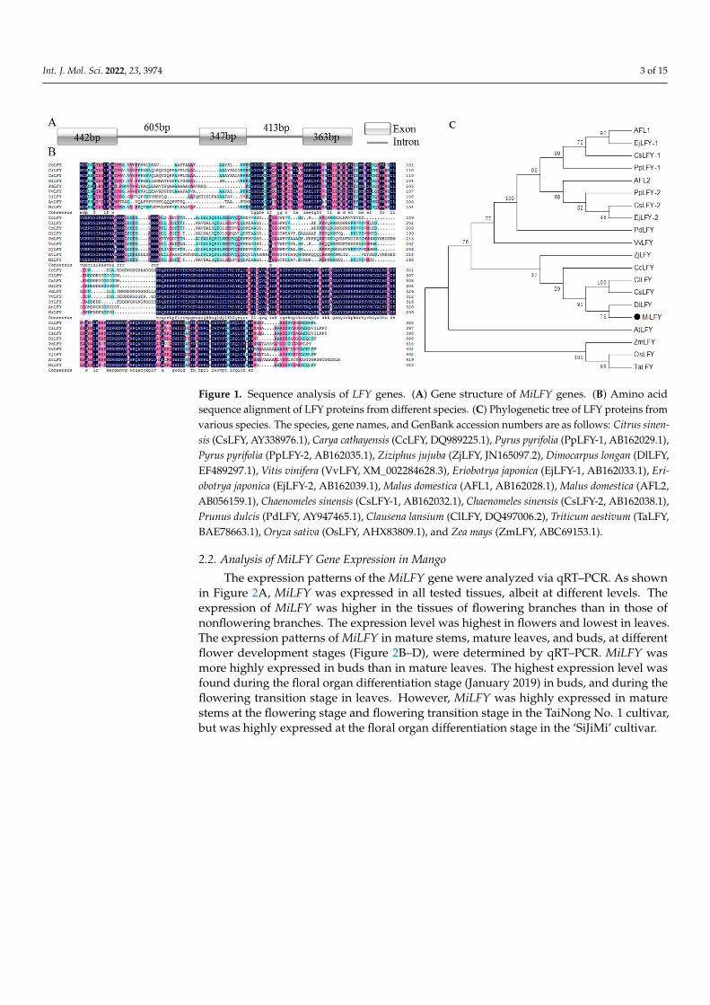
The sequence of a MiLFY homolog in mango was obtained. The cDNA length was1152 bp, encoding a 383 amino acid protein (GenBank accession no. HQ585988). Thefull-length DNA sequence of MiLFY was 2170 bp, comprising three exons and two introns(Figure 1A). The predicted protein molecular weight was 43.31 kDa, and the isoelectricpoint was 6.37. The N- and C-terminal regions of the MiLFY proteins were highly conservedacross species (Figure 1B). The C- and N-terminal regions contained a DNA-binding domainand a sterile alpha motif (SAM) domain. The MiLFY protein had 61.79% sequence homologywith the Arabidopsis thaliana ortholog. Phylogenetic tree analysis showed that MiLFY wasclosely related to DlLFY (Dimocarpus longan), CsLFY (Citrus sinensis), and ClLFY (Clausenalansium) (Figure 1C).
Int. J. Mol. Sci. 2022, 23, 3974 3 of 15Int. J. Mol. Sci. 2022, 23, x FOR PEER REVIEW 3 of 15
Figure 1. Sequence analysis of LFY genes. (A) Gene structure of MiLFY genes. (B) Amino acid se-quence alignment of LFY proteins from different species. (C) Phylogenetic tree of LFY proteins from various species. The species, gene names, and GenBank accession numbers are as follows: Citrus sinensis (CsLFY, AY338976.1), Carya cathayensis (CcLFY, DQ989225.1), Pyrus pyrifolia (PpLFY-1, AB162029.1), Pyrus pyrifolia (PpLFY-2, AB162035.1), Ziziphus jujuba (ZjLFY, JN165097.2), Dimocarpus longan (DlLFY, EF489297.1), Vitis vinifera (VvLFY, XM_002284628.3), Eriobotrya japonica (EjLFY-1, AB162033.1), Eriobotrya japonica (EjLFY-2, AB162039.1), Malus domestica (AFL1, AB162028.1), Malus domestica (AFL2, AB056159.1), Chaenomeles sinensis (CsLFY-1, AB162032.1), Chaenomeles sinensis (CsLFY-2, AB162038.1), Prunus dulcis (PdLFY, AY947465.1), Clausena lansium (ClLFY, DQ497006.2), Triticum aestivum (TaLFY, BAE78663.1), Oryza sativa (OsLFY, AHX83809.1), and Zea mays (ZmLFY,ABC69153.1).
2.2. Analysis of MiLFY Gene Expression in Mango The expression patterns of the MiLFY gene were analyzed via qRT–PCR. As shown
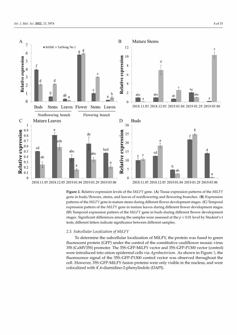
in Figure 2A, MiLFY was expressed in all tested tissues, albeit at different levels. The ex-pression of MiLFY was higher in the tissues of flowering branches than in those of non-flowering branches. The expression level was highest in flowers and lowest in leaves. The expression patterns of MiLFY in mature stems, mature leaves, and buds, at different flower development stages (Figure 2B–D), were determined by qRT–PCR. MiLFY was more highly expressed in buds than in mature leaves. The highest expression level was found during the floral organ differentiation stage (January 2019) in buds, and during the flowering transition stage in leaves. However, MiLFY was highly expressed in mature stems at the flowering stage and flowering transition stage in the TaiNong No. 1 cultivar,but was highly expressed at the floral organ differentiation stage in the ‘SiJiMi’ cultivar.
C
Figure 1. Sequence analysis of LFY genes. (A) Gene structure of MiLFY genes. (B) Amino acidsequence alignment of LFY proteins from different species. (C) Phylogenetic tree of LFY proteins fromvarious species. The species, gene names, and GenBank accession numbers are as follows: Citrus sinen-sis (CsLFY, AY338976.1), Carya cathayensis (CcLFY, DQ989225.1), Pyrus pyrifolia (PpLFY-1, AB162029.1),Pyrus pyrifolia (PpLFY-2, AB162035.1), Ziziphus jujuba (ZjLFY, JN165097.2), Dimocarpus longan (DlLFY,EF489297.1), Vitis vinifera (VvLFY, XM_002284628.3), Eriobotrya japonica (EjLFY-1, AB162033.1), Eri-obotrya japonica (EjLFY-2, AB162039.1), Malus domestica (AFL1, AB162028.1), Malus domestica (AFL2,AB056159.1), Chaenomeles sinensis (CsLFY-1, AB162032.1), Chaenomeles sinensis (CsLFY-2, AB162038.1),Prunus dulcis (PdLFY, AY947465.1), Clausena lansium (ClLFY, DQ497006.2), Triticum aestivum (TaLFY,BAE78663.1), Oryza sativa (OsLFY, AHX83809.1), and Zea mays (ZmLFY, ABC69153.1).
2.2. Analysis of MiLFY Gene Expression in Mango
The expression patterns of the MiLFY gene were analyzed via qRT–PCR. As shownin Figure 2A, MiLFY was expressed in all tested tissues, albeit at different levels. Theexpression of MiLFY was higher in the tissues of flowering branches than in those ofnonflowering branches. The expression level was highest in flowers and lowest in leaves.The expression patterns of MiLFY in mature stems, mature leaves, and buds, at differentflower development stages (Figure 2B–D), were determined by qRT–PCR. MiLFY wasmore highly expressed in buds than in mature leaves. The highest expression level wasfound during the floral organ differentiation stage (January 2019) in buds, and during theflowering transition stage in leaves. However, MiLFY was highly expressed in maturestems at the flowering stage and flowering transition stage in the TaiNong No. 1 cultivar,but was highly expressed at the floral organ differentiation stage in the ‘SiJiMi’ cultivar.
Int. J. Mol. Sci. 2022, 23, 3974 4 of 15Int. J. Mol. Sci. 2022, 23, x FOR PEER REVIEW 4 of 15
Figure 2. Relative expression levels of the MiLFY gene. (A) Tissue expression patterns of the MiLFY gene in buds/flowers, stems, and leaves of nonflowering and flowering branches. (B) Expression patterns of the MiLFY gene in mature stems during different flower development stages. (C) Tem-poral expression pattern of the MiLFY gene in mature leaves during different flower development stages. (D) Temporal expression pattern of the MiLFY gene in buds during different flower devel-opment stages. Significant differences among the samples were assessed at the p < 0.01 level by Student’s t tests; different letters indicate significance between different samples.
2.3. Subcellular Localization of MiLFY To determine the subcellular localization of MiLFY, the protein was fused to green
fluorescent protein (GFP) under the control of the constitutive cauliflower mosaic virus 35S (CaMV35S) promoter. The 35S::GFP-MiLFY vector and 35S::GFP-P1300 vector (con-trol) were introduced into onion epidermal cells via Agrobacterium. As shown in Figure 3, the fluorescence signal of the 35S::GFP-P1300 control vector was observed throughout the cell. However, 35S::GFP-MiLFY fusion proteins were only visible in the nucleus, and were colocalized with 4′,6-diamidino-2-phenylindole (DAPI).
Figure 2. Relative expression levels of the MiLFY gene. (A) Tissue expression patterns of the MiLFYgene in buds/flowers, stems, and leaves of nonflowering and flowering branches. (B) Expressionpatterns of the MiLFY gene in mature stems during different flower development stages. (C) Temporalexpression pattern of the MiLFY gene in mature leaves during different flower development stages.(D) Temporal expression pattern of the MiLFY gene in buds during different flower developmentstages. Significant differences among the samples were assessed at the p < 0.01 level by Student’s ttests; different letters indicate significance between different samples.
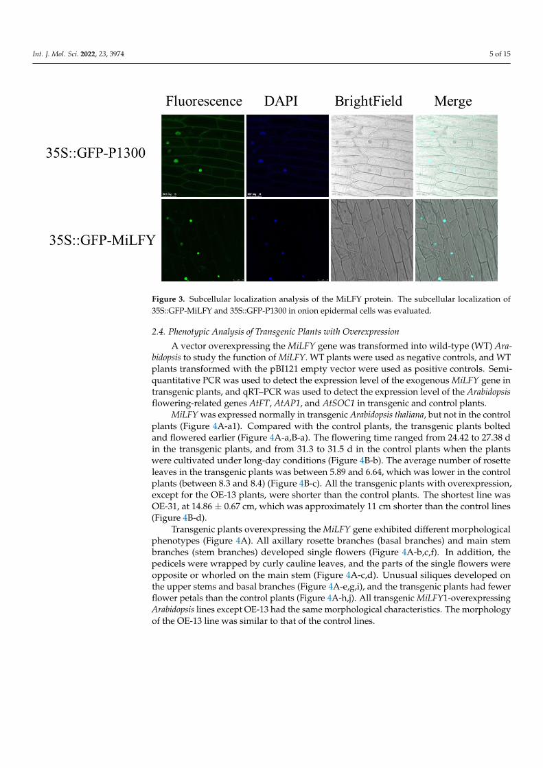
2.3. Subcellular Localization of MiLFY
To determine the subcellular localization of MiLFY, the protein was fused to greenfluorescent protein (GFP) under the control of the constitutive cauliflower mosaic virus35S (CaMV35S) promoter. The 35S::GFP-MiLFY vector and 35S::GFP-P1300 vector (control)were introduced into onion epidermal cells via Agrobacterium. As shown in Figure 3, thefluorescence signal of the 35S::GFP-P1300 control vector was observed throughout thecell. However, 35S::GFP-MiLFY fusion proteins were only visible in the nucleus, and werecolocalized with 4′,6-diamidino-2-phenylindole (DAPI).
Int. J. Mol. Sci. 2022, 23, 3974 5 of 15Int. J. Mol. Sci. 2022, 23, x FOR PEER REVIEW 5 of 15
Figure 3. Subcellular localization analysis of the MiLFY protein. The subcellular localization of 35S::GFP-MiLFY and 35S::GFP-P1300 in onion epidermal cells was evaluated.
2.4. Phenotypic Analysis of Transgenic Plants with Overexpression A vector overexpressing the MiLFY gene was transformed into wild-type (WT) Ara-
bidopsis to study the function of MiLFY. WT plants were used as negative controls, and WT plants transformed with the pBI121 empty vector were used as positive controls. Sem-iquantitative PCR was used to detect the expression level of the exogenous MiLFY gene in transgenic plants, and qRT–PCR was used to detect the expression level of the Arabidop-sis flowering-related genes AtFT, AtAP1, and AtSOC1 in transgenic and control plants.
MiLFY was expressed normally in transgenic Arabidopsis thaliana, but not in the con-trol plants (Figure 4A-a1). Compared with the control plants, the transgenic plants bolted and flowered earlier (Figure 4A-a,B-a). The flowering time ranged from 24.42 to 27.38 d in the transgenic plants, and from 31.3 to 31.5 d in the control plants when the plants were cultivated under long-day conditions (Figure 4B-b). The average number of rosette leaves in the transgenic plants was between 5.89 and 6.64, which was lower in the control plants (between 8.3 and 8.4) (Figure 4B-c). All the transgenic plants with overexpression, except for the OE-13 plants, were shorter than the control plants. The shortest line was OE-31, at 14.86 ± 0.67 cm, which was approximately 11 cm shorter than the control lines (Figure 4B-d).
Transgenic plants overexpressing the MiLFY gene exhibited different morphological phenotypes (Figure 4A). All axillary rosette branches (basal branches) and main stem branches (stem branches) developed single flowers (Figure 4A-b,c,f). In addition, the ped-icels were wrapped by curly cauline leaves, and the parts of the single flowers were op-posite or whorled on the main stem (Figure 4A-c,d). Unusual siliques developed on the upper stems and basal branches (Figure 4A-e,g,i), and the transgenic plants had fewer flower petals than the control plants (Figure 4A-h,j). All transgenic MiLFY1-overexpress-ing Arabidopsis lines except OE-13 had the same morphological characteristics. The mor-phology of the OE-13 line was similar to that of the control lines.
The experimental materials were sampled when the transgenic plants were flowering and the control plants were not. RNA was extracted from the aerial portions of the plants 27 d after germination, and this RNA was subsequently reverse-transcribed into cDNA. qRT–PCR was used to determine whether the expression of endogenous genes in trans-genic Arabidopsis was affected by MiLFY overexpression (Figure 5). Compared with the WT Arabidopsis plants, and the plants transformed with the empty vector, the transgenic
Figure 3. Subcellular localization analysis of the MiLFY protein. The subcellular localization of35S::GFP-MiLFY and 35S::GFP-P1300 in onion epidermal cells was evaluated.
2.4. Phenotypic Analysis of Transgenic Plants with Overexpression
A vector overexpressing the MiLFY gene was transformed into wild-type (WT) Ara-bidopsis to study the function of MiLFY. WT plants were used as negative controls, and WTplants transformed with the pBI121 empty vector were used as positive controls. Semi-quantitative PCR was used to detect the expression level of the exogenous MiLFY gene intransgenic plants, and qRT–PCR was used to detect the expression level of the Arabidopsisflowering-related genes AtFT, AtAP1, and AtSOC1 in transgenic and control plants.
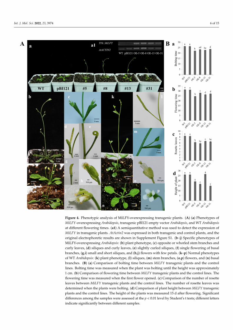
MiLFY was expressed normally in transgenic Arabidopsis thaliana, but not in the controlplants (Figure 4A-a1). Compared with the control plants, the transgenic plants boltedand flowered earlier (Figure 4A-a,B-a). The flowering time ranged from 24.42 to 27.38 din the transgenic plants, and from 31.3 to 31.5 d in the control plants when the plantswere cultivated under long-day conditions (Figure 4B-b). The average number of rosetteleaves in the transgenic plants was between 5.89 and 6.64, which was lower in the controlplants (between 8.3 and 8.4) (Figure 4B-c). All the transgenic plants with overexpression,except for the OE-13 plants, were shorter than the control plants. The shortest line wasOE-31, at 14.86 ± 0.67 cm, which was approximately 11 cm shorter than the control lines(Figure 4B-d).
Transgenic plants overexpressing the MiLFY gene exhibited different morphologicalphenotypes (Figure 4A). All axillary rosette branches (basal branches) and main stembranches (stem branches) developed single flowers (Figure 4A-b,c,f). In addition, thepedicels were wrapped by curly cauline leaves, and the parts of the single flowers wereopposite or whorled on the main stem (Figure 4A-c,d). Unusual siliques developed onthe upper stems and basal branches (Figure 4A-e,g,i), and the transgenic plants had fewerflower petals than the control plants (Figure 4A-h,j). All transgenic MiLFY1-overexpressingArabidopsis lines except OE-13 had the same morphological characteristics. The morphologyof the OE-13 line was similar to that of the control lines.
Int. J. Mol. Sci. 2022, 23, 3974 6 of 15
Int. J. Mol. Sci. 2022, 23, x FOR PEER REVIEW 6 of 15
MiLFY1-overexpressing Arabidopsis plants exhibited obviously increased expression of AtAP1, AtFT, and AtSOC1.
Figure 4. Phenotypic analysis of MiLFY-overexpressing transgenic plants. (A) (a) Phenotypes of MiLFY-overexpressing Arabidopsis, transgenic pBI121 empty vector Arabidopsis, and WT Arabidopsis at different flowering times. (a1) A semiquantitative method was used to detect the expression of MiLFY in transgenic plants. AtActin2 was expressed in both transgenic and control plants, and the original electrophoretic results are shown in Supplement Figure S1. (b–j) Specific phenotypes of MiLFY-overexpressing Arabidopsis: (b) plant phenotype, (c) opposite or whorled stem branches and curly leaves, (d) siliques and curly leaves, (e) slightly curled siliques, (f) single flowering of basal branches, (g,i) small and short siliques, and (h,j) flowers with few petals. (k–p) Normal phenotypes of WT Arabidopsis: (k) plant phenotype, (l) siliques, (m) stem branches, (o,p) flowers, and (n) basal branches. (B) (a) Comparison of bolting time between MiLFY transgenic plants and the control lines. Bolting time was measured when the plant was bolting until the height was approximately 1 cm. (b) Comparison of flowering time between MiLFY transgenic plants and the control lines. The flow-ering time was measured when the first flower opened. (c) Comparison of the number of rosette leaves between MiLFY transgenic plants and the control lines. The number of rosette leaves was determined when the plants were bolting. (d) Comparison of plant height between MiLFY trans-
Figure 4. Phenotypic analysis of MiLFY-overexpressing transgenic plants. (A) (a) Phenotypes ofMiLFY-overexpressing Arabidopsis, transgenic pBI121 empty vector Arabidopsis, and WT Arabidopsisat different flowering times. (a1) A semiquantitative method was used to detect the expression ofMiLFY in transgenic plants. AtActin2 was expressed in both transgenic and control plants, and theoriginal electrophoretic results are shown in Supplement Figure S1. (b–j) Specific phenotypes ofMiLFY-overexpressing Arabidopsis: (b) plant phenotype, (c) opposite or whorled stem branches andcurly leaves, (d) siliques and curly leaves, (e) slightly curled siliques, (f) single flowering of basalbranches, (g,i) small and short siliques, and (h,j) flowers with few petals. (k–p) Normal phenotypesof WT Arabidopsis: (k) plant phenotype, (l) siliques, (m) stem branches, (o,p) flowers, and (n) basalbranches. (B) (a) Comparison of bolting time between MiLFY transgenic plants and the controllines. Bolting time was measured when the plant was bolting until the height was approximately1 cm. (b) Comparison of flowering time between MiLFY transgenic plants and the control lines. Theflowering time was measured when the first flower opened. (c) Comparison of the number of rosetteleaves between MiLFY transgenic plants and the control lines. The number of rosette leaves wasdetermined when the plants were bolting. (d) Comparison of plant height between MiLFY transgenicplants and the control lines. The height of the plants was measured 15 d after flowering. Significantdifferences among the samples were assessed at the p < 0.01 level by Student’s t tests; different lettersindicate significantly between different samples.
Int. J. Mol. Sci. 2022, 23, 3974 7 of 15
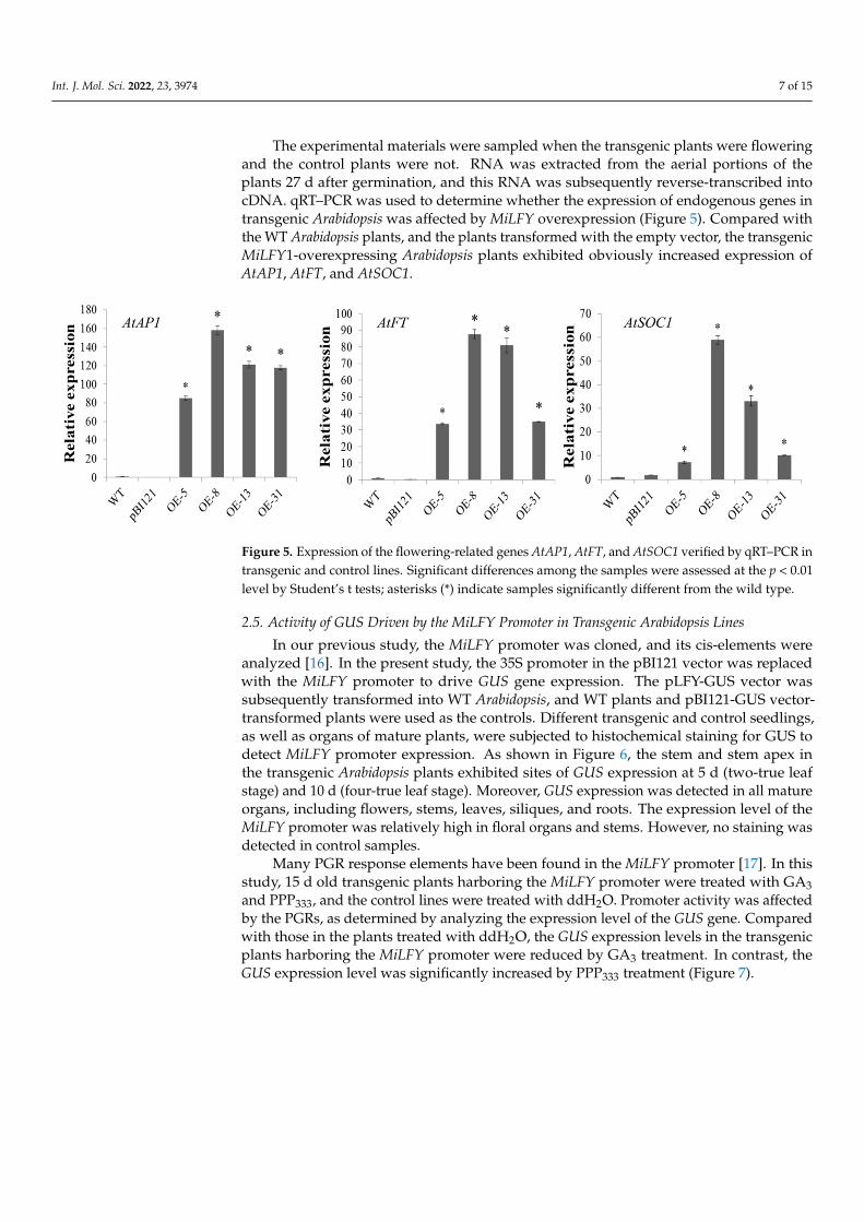
The experimental materials were sampled when the transgenic plants were floweringand the control plants were not. RNA was extracted from the aerial portions of theplants 27 d after germination, and this RNA was subsequently reverse-transcribed intocDNA. qRT–PCR was used to determine whether the expression of endogenous genes intransgenic Arabidopsis was affected by MiLFY overexpression (Figure 5). Compared withthe WT Arabidopsis plants, and the plants transformed with the empty vector, the transgenicMiLFY1-overexpressing Arabidopsis plants exhibited obviously increased expression ofAtAP1, AtFT, and AtSOC1.
Int. J. Mol. Sci. 2022, 23, x FOR PEER REVIEW 7 of 15
genic plants and the control lines. The height of the plants was measured 15 d after flowering. Sig-nificant differences among the samples were assessed at the p < 0.01 level by Student’s t tests; dif-ferent letters indicate significantly between different samples.
Figure 5. Expression of the flowering-related genes AtAP1, AtFT, and AtSOC1 verified by qRT–PCR in transgenic and control lines. Significant differences among the samples were assessed at the p < 0.01 level by Student’s t tests; asterisks (*) indicate samples significantly different from the wild type.
2.5. Activity of GUS Driven by the MiLFY Promoter in Transgenic Arabidopsis Lines In our previous study, the MiLFY promoter was cloned, and its cis-elements were
analyzed [16]. In the present study, the 35S promoter in the pBI121 vector was replaced with the MiLFY promoter to drive GUS gene expression. The pLFY-GUS vector was sub-sequently transformed into WT Arabidopsis, and WT plants and pBI121-GUS vector-trans-formed plants were used as the controls. Different transgenic and control seedlings, as well as organs of mature plants, were subjected to histochemical staining for GUS to detect MiLFY promoter expression. As shown in Figure 6, the stem and stem apex in the trans-genic Arabidopsis plants exhibited sites of GUS expression at 5 d (two-true leaf stage) and 10 d (four-true leaf stage). Moreover, GUS expression was detected in all mature organs, including flowers, stems, leaves, siliques, and roots. The expression level of the MiLFY promoter was relatively high in floral organs and stems. However, no staining was de-tected in control samples.
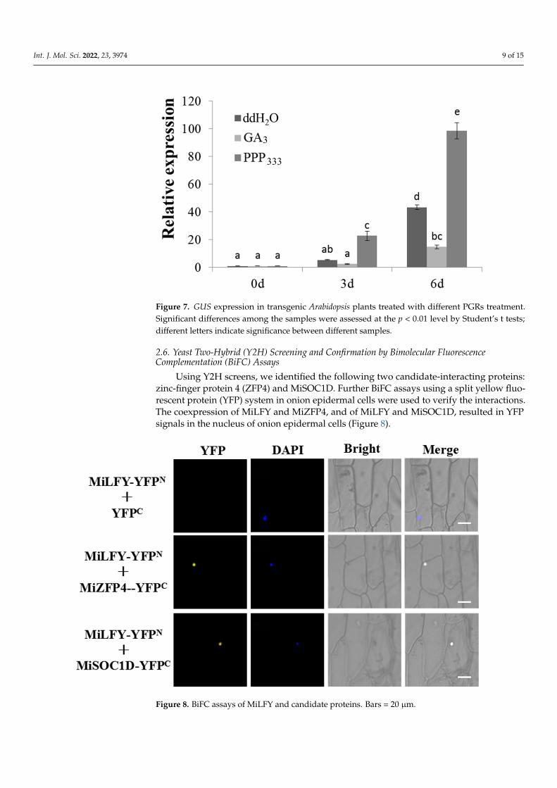
Many PGR response elements have been found in the MiLFY promoter [17]. In this study, 15 d old transgenic plants harboring the MiLFY promoter were treated with GA3 and PPP333, and the control lines were treated with ddH2O. Promoter activity was affected by the PGRs, as determined by analyzing the expression level of the GUS gene. Compared with those in the plants treated with ddH2O, the GUS expression levels in the transgenic plants harboring the MiLFY promoter were reduced by GA3 treatment. In contrast, the GUS expression level was significantly increased by PPP333 treatment (Figure 7).
Figure 5. Expression of the flowering-related genes AtAP1, AtFT, and AtSOC1 verified by qRT–PCR intransgenic and control lines. Significant differences among the samples were assessed at the p < 0.01level by Student’s t tests; asterisks (*) indicate samples significantly different from the wild type.
2.5. Activity of GUS Driven by the MiLFY Promoter in Transgenic Arabidopsis Lines
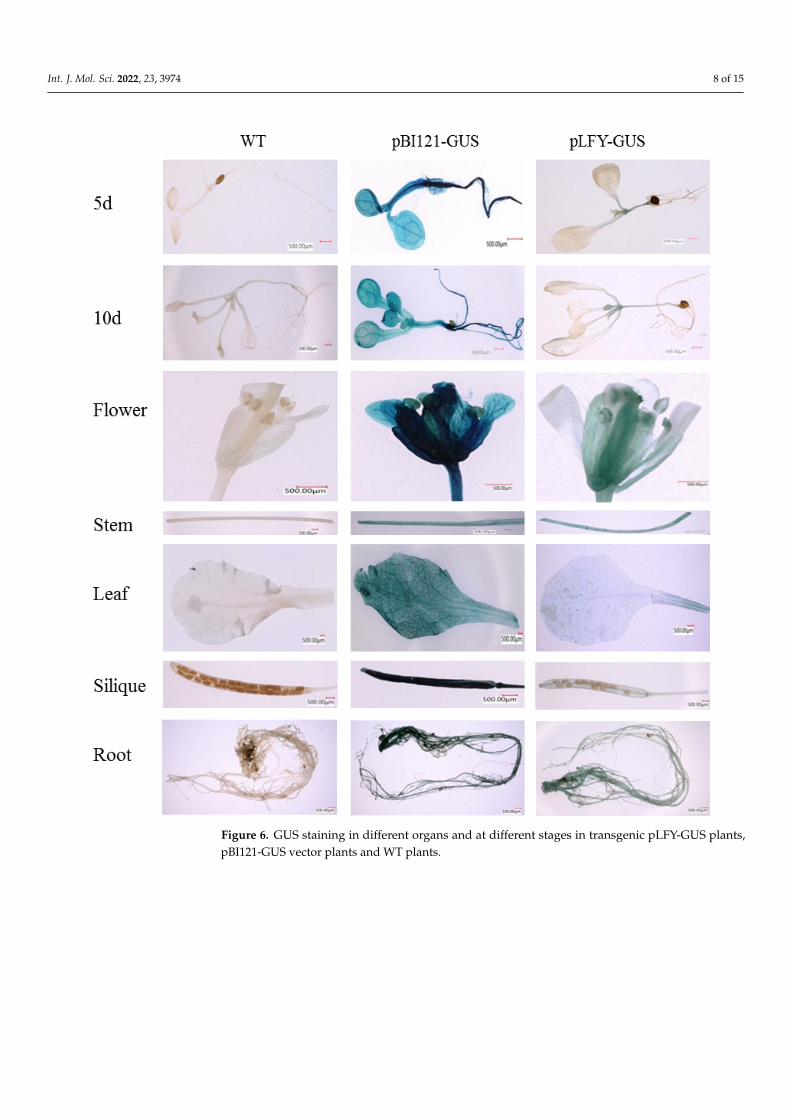
In our previous study, the MiLFY promoter was cloned, and its cis-elements wereanalyzed [16]. In the present study, the 35S promoter in the pBI121 vector was replacedwith the MiLFY promoter to drive GUS gene expression. The pLFY-GUS vector wassubsequently transformed into WT Arabidopsis, and WT plants and pBI121-GUS vector-transformed plants were used as the controls. Different transgenic and control seedlings,as well as organs of mature plants, were subjected to histochemical staining for GUS todetect MiLFY promoter expression. As shown in Figure 6, the stem and stem apex inthe transgenic Arabidopsis plants exhibited sites of GUS expression at 5 d (two-true leafstage) and 10 d (four-true leaf stage). Moreover, GUS expression was detected in all matureorgans, including flowers, stems, leaves, siliques, and roots. The expression level of theMiLFY promoter was relatively high in floral organs and stems. However, no staining wasdetected in control samples.
Many PGR response elements have been found in the MiLFY promoter [17]. In thisstudy, 15 d old transgenic plants harboring the MiLFY promoter were treated with GA3and PPP333, and the control lines were treated with ddH2O. Promoter activity was affectedby the PGRs, as determined by analyzing the expression level of the GUS gene. Comparedwith those in the plants treated with ddH2O, the GUS expression levels in the transgenicplants harboring the MiLFY promoter were reduced by GA3 treatment. In contrast, theGUS expression level was significantly increased by PPP333 treatment (Figure 7).
Int. J. Mol. Sci. 2022, 23, 3974 8 of 15
Figure 6. GUS staining in different organs and at different stages in transgenic pLFY-GUS plants,pBI121-GUS vector plants and WT plants.
Int. J. Mol. Sci. 2022, 23, 3974 9 of 15Int. J. Mol. Sci. 2022, 23, x FOR PEER REVIEW 9 of 15
Figure 7. GUS expression in transgenic Arabidopsis plants treated with different PGRs treatment. Significant differences among the samples were assessed at the p < 0.01 level by Student’s t tests; different letters indicate significance between different samples.
2.6. Yeast Two-Hybrid (Y2H) Screening and Confirmation by Bimolecular Fluorescence Comple-mentation (BiFC) Assays
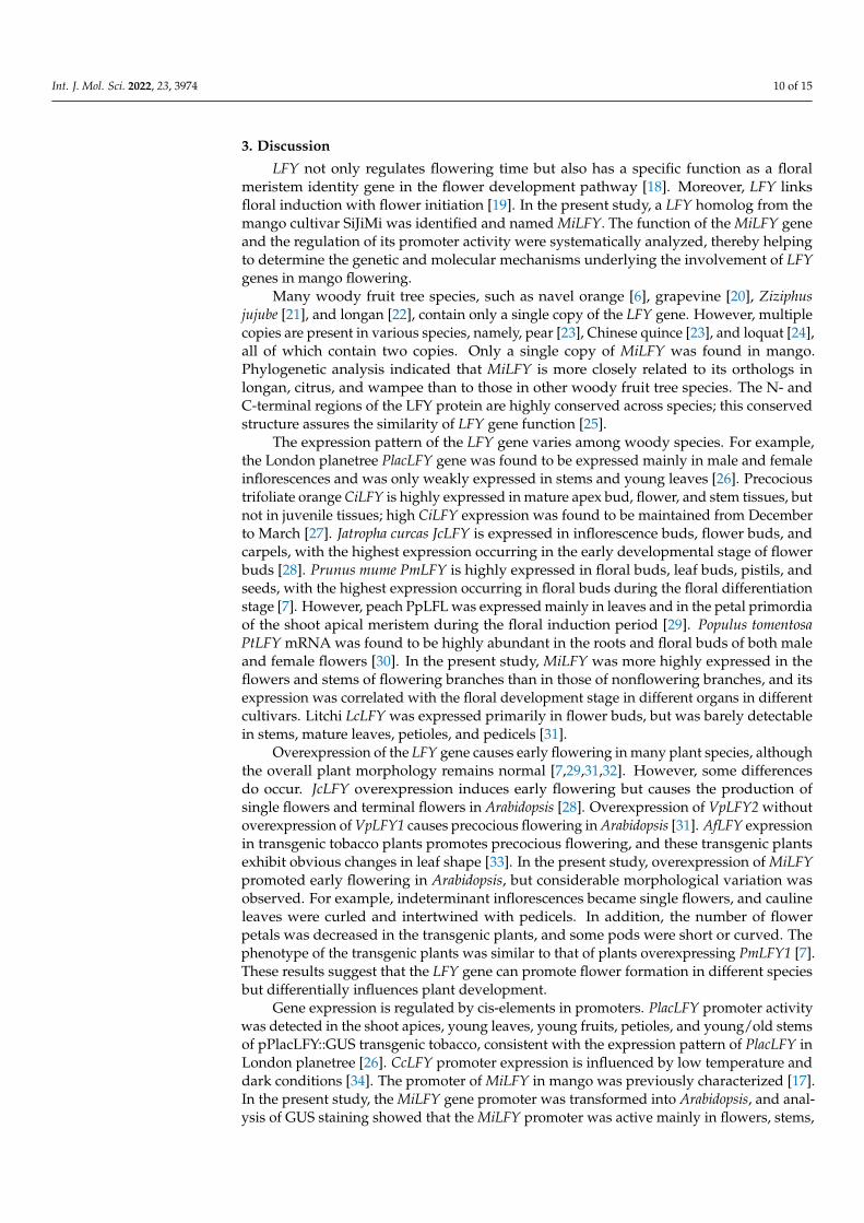
Using Y2H screens, we identified the following two candidate-interacting proteins: zinc-finger protein 4 (ZFP4) and MiSOC1D. Further BiFC assays using a split yellow flu-orescent protein (YFP) system in onion epidermal cells were used to verify the interac-tions. The coexpression of MiLFY and MiZFP4, and of MiLFY and MiSOC1D, resulted in YFP signals in the nucleus of onion epidermal cells (Figure 8).
Figure 8. BiFC assays of MiLFY and candidate proteins. Bars=20 μm.
3. Discussion LFY not only regulates flowering time but also has a specific function as a floral me-
ristem identity gene in the flower development pathway [18]. Moreover, LFY links floral
Figure 7. GUS expression in transgenic Arabidopsis plants treated with different PGRs treatment.Significant differences among the samples were assessed at the p < 0.01 level by Student’s t tests;different letters indicate significance between different samples.
2.6. Yeast Two-Hybrid (Y2H) Screening and Confirmation by Bimolecular FluorescenceComplementation (BiFC) Assays
Using Y2H screens, we identified the following two candidate-interacting proteins:zinc-finger protein 4 (ZFP4) and MiSOC1D. Further BiFC assays using a split yellow fluo-rescent protein (YFP) system in onion epidermal cells were used to verify the interactions.The coexpression of MiLFY and MiZFP4, and of MiLFY and MiSOC1D, resulted in YFPsignals in the nucleus of onion epidermal cells (Figure 8).
Int. J. Mol. Sci. 2022, 23, x FOR PEER REVIEW 9 of 15
Figure 7. GUS expression in transgenic Arabidopsis plants treated with different PGRs treatment. Significant differences among the samples were assessed at the p < 0.01 level by Student’s t tests; different letters indicate significance between different samples.
2.6. Yeast Two-Hybrid (Y2H) Screening and Confirmation by Bimolecular Fluorescence Comple-mentation (BiFC) Assays
Using Y2H screens, we identified the following two candidate-interacting proteins: zinc-finger protein 4 (ZFP4) and MiSOC1D. Further BiFC assays using a split yellow flu-orescent protein (YFP) system in onion epidermal cells were used to verify the interac-tions. The coexpression of MiLFY and MiZFP4, and of MiLFY and MiSOC1D, resulted in YFP signals in the nucleus of onion epidermal cells (Figure 8).
Figure 8. BiFC assays of MiLFY and candidate proteins. Bars=20 μm.
3. Discussion LFY not only regulates flowering time but also has a specific function as a floral me-
ristem identity gene in the flower development pathway [18]. Moreover, LFY links floral
Figure 8. BiFC assays of MiLFY and candidate proteins. Bars = 20 µm.
Int. J. Mol. Sci. 2022, 23, 3974 10 of 15
3. Discussion
LFY not only regulates flowering time but also has a specific function as a floralmeristem identity gene in the flower development pathway [18]. Moreover, LFY linksfloral induction with flower initiation [19]. In the present study, a LFY homolog from themango cultivar SiJiMi was identified and named MiLFY. The function of the MiLFY geneand the regulation of its promoter activity were systematically analyzed, thereby helpingto determine the genetic and molecular mechanisms underlying the involvement of LFYgenes in mango flowering.
Many woody fruit tree species, such as navel orange [6], grapevine [20], Ziziphusjujube [21], and longan [22], contain only a single copy of the LFY gene. However, multiplecopies are present in various species, namely, pear [23], Chinese quince [23], and loquat [24],all of which contain two copies. Only a single copy of MiLFY was found in mango.Phylogenetic analysis indicated that MiLFY is more closely related to its orthologs inlongan, citrus, and wampee than to those in other woody fruit tree species. The N- andC-terminal regions of the LFY protein are highly conserved across species; this conservedstructure assures the similarity of LFY gene function [25].
The expression pattern of the LFY gene varies among woody species. For example,the London planetree PlacLFY gene was found to be expressed mainly in male and femaleinflorescences and was only weakly expressed in stems and young leaves [26]. Precocioustrifoliate orange CiLFY is highly expressed in mature apex bud, flower, and stem tissues, butnot in juvenile tissues; high CiLFY expression was found to be maintained from Decemberto March [27]. Jatropha curcas JcLFY is expressed in inflorescence buds, flower buds, andcarpels, with the highest expression occurring in the early developmental stage of flowerbuds [28]. Prunus mume PmLFY is highly expressed in floral buds, leaf buds, pistils, andseeds, with the highest expression occurring in floral buds during the floral differentiationstage [7]. However, peach PpLFL was expressed mainly in leaves and in the petal primordiaof the shoot apical meristem during the floral induction period [29]. Populus tomentosaPtLFY mRNA was found to be highly abundant in the roots and floral buds of both maleand female flowers [30]. In the present study, MiLFY was more highly expressed in theflowers and stems of flowering branches than in those of nonflowering branches, and itsexpression was correlated with the floral development stage in different organs in differentcultivars. Litchi LcLFY was expressed primarily in flower buds, but was barely detectablein stems, mature leaves, petioles, and pedicels [31].
Overexpression of the LFY gene causes early flowering in many plant species, althoughthe overall plant morphology remains normal [7,29,31,32]. However, some differencesdo occur. JcLFY overexpression induces early flowering but causes the production ofsingle flowers and terminal flowers in Arabidopsis [28]. Overexpression of VpLFY2 withoutoverexpression of VpLFY1 causes precocious flowering in Arabidopsis [31]. AfLFY expressionin transgenic tobacco plants promotes precocious flowering, and these transgenic plantsexhibit obvious changes in leaf shape [33]. In the present study, overexpression of MiLFYpromoted early flowering in Arabidopsis, but considerable morphological variation wasobserved. For example, indeterminant inflorescences became single flowers, and caulineleaves were curled and intertwined with pedicels. In addition, the number of flowerpetals was decreased in the transgenic plants, and some pods were short or curved. Thephenotype of the transgenic plants was similar to that of plants overexpressing PmLFY1 [7].These results suggest that the LFY gene can promote flower formation in different speciesbut differentially influences plant development.
Gene expression is regulated by cis-elements in promoters. PlacLFY promoter activitywas detected in the shoot apices, young leaves, young fruits, petioles, and young/old stemsof pPlacLFY::GUS transgenic tobacco, consistent with the expression pattern of PlacLFY inLondon planetree [26]. CcLFY promoter expression is influenced by low temperature anddark conditions [34]. The promoter of MiLFY in mango was previously characterized [17].In the present study, the MiLFY gene promoter was transformed into Arabidopsis, and anal-ysis of GUS staining showed that the MiLFY promoter was active mainly in flowers, stems,
Int. J. Mol. Sci. 2022, 23, 3974 11 of 15
leaves, and roots during the flowering period, consistent with the expression patterns ofMiLFY in mango (Figure 2). Exogenous GA3 promotes Arabidopsis flowering but inhibitsmango flowering, and PPP333, a synthetic inhibitor of GA3, promotes mango flower forma-tion [1]. In the present study, we treated MiLFY promoter-containing transgenic seedlingswith exogenous GA3 and PPP333 and found that the expression of the MiLFY promoterwas significantly inhibited by GA3 but enhanced by PPP333. These results suggest that theMiLFY gene is involved in phytohormone-mediated regulation of mango flowering.
As a floral integration factor, the LFY gene plays an important regulatory role in thefloral network [35]. Winter (2011) identified direct LFY target genes throughout the genome.These target genes were found to be involved in flowering time, floral organ development,phytohormone responses, and biotic stimulus responses. In the present study, we foundthat two proteins, MiZFP4 and MiSOC1D, can directly interact with LFY. ZFPs constituteone of the largest transcription factor families, whose members are highly involved intranscriptional regulation of flowering induction, floral organ morphogenesis, and stressresponses [36]. SOC1 is a floral integration factor, and overexpression of MiSOC1 promotesearly flowering in A. thaliana [37].
In conclusion, we functionally characterized the MiLFY gene in mango and found thatoverexpression of MiLFY significantly promoted flowering in transgenic plants. Moreover,transgene expression led to significant morphological variation, including changes in floralorgans and leaf morphology, in the transgenic plants. For the first time, we found thatGA3 treatment inhibits, but PPP333 promotes, MiLFY promoter activity. This pattern isconsistent with the finding that GA3 inhibits mango flower development while PPP333promotes mango flowering. Protein interaction analysis showed that by directly interactingwith MiZFP4 and MiSOC1D, the MiLFY protein regulates mango flowering.
4. Materials and Methods4.1. Plant Materials
The mango cultivars SiJiMi and TaiNong No. 1 were grown in an orchard of the Collegeof Agriculture, Guangxi University, Nanning, Guangxi, China. For tissue expressionanalysis, leaves, stems, and buds or flower tissues were collected from the flowering andnonflowering branches of the same plants on 4 January 2019. For seasonal expressionanalysis, leaves, stems, and buds or flowers were collected each month from 5 November2018 to 6 March 2019. All samples were used for experiments before storage at −80 ◦C. TheArabidopsis ecotype Columbia (Col-0) plants used for transformation were maintained inour laboratory.
4.2. Isolation of the MiLFY Gene
Total RNA was extracted from mango leaves by using an RNAprep pure kit (DP441)(Tiangen, Beijing, China). First-strand cDNA was synthesized with M-MLV reverse tran-scriptase (TaKaRa, Dalian, China) using 1 µg of RNA according to the manufacturer’sinstructions. Genomic DNA was isolated using the cetyl-trimethylammonium bromide(CTAB) method with minor modifications. In our previous study, we obtained the sequenceof a LFY gene from transcriptome data from the mango cultivar SiJiMi (unpublished data)and, in this study, we further verified the correctness of its sequence by RT–PCR with theprimers LFY-F and LFY-R (Table S1). PCR amplification was performed as described in aprevious study [2]. The primers LFY-F and LFY-R were also used to amplify genomic DNA.
4.3. Sequence Alignment and Bioinformatic Analysis
Sequence analysis and amino acid predictions were performed using BioXM 2.6software (http://cbi.njau.edu.cn/BioXM/; 21 May 2021). The exon–intron structure wasgenerated with IBS 1.0. The conserved protein domain was predicted with the NCBIConserved Domain Database online tool (http://www.ncbi.nlm.nih.gov/Structure/cdd/cdd.shtml/; 11 May 2021). Multiple sequence alignment of the LFY protein was performedwith DNAMAN software. A phylogenetic tree of LFY was constructed using the neighbor-
Int. J. Mol. Sci. 2022, 23, 3974 12 of 15
joining method with MEGA 6.0, with 1000 bootstrap replicates (https://megasoftware.net/;20 March 2022).
4.4. Analysis of MiLFY Gene Expression
The expression pattern of MiLFY was analyzed with an ABI 7500 Real-Time PCRSystem (Applied Biosystems, Foster City, CA, USA) and SYBR Premix Ex Taq II (Takara,Dalian, China) according to the manufacturers’ instructions. The reaction mixture containedthe following components: reaction solution 10 µL, cDNA 2 µL (50 ng/µL), up- anddownstream primers 0.5 µL (10 µM) each, ROX reference dye II 0.8 µL, and sterile waterto 20 µL. PCR amplification was performed with the following thermal cycling program:95 ◦C for 30 s; 40 cycles at 95 ◦C for 5 s, 60 ◦C for 34 s, and 95 ◦C for 15 s; melting curveanalysis was performed at 95 ◦C for 15 s and 60 ◦C for 1 min. The gene-specific primersused are listed in Table S1. MiActin1 was used as the internal reference gene in mango [16].The delta-delta Ct method was used to calculate the relative gene expression [38]. The dataare presented as the average of at least three technical replicates.
4.5. Subcellular Localization Analysis
To analyze the subcellular localization of MiLFY, the full coding sequence (CDS) ofMiLFY without the termination codon was inserted into the Xba I and BamH I sites inthe P1300 vector to express the 35S::GFP-MiLFY fusion construct under the control of theCaMV35S promoter [39]. The 35S::GFP-MiLFY vector was transformed into Agrobacteriumtumefaciens EHA105a, and the transformants were used to infect onion (Allium cepa) epider-mal cells. The onion epidermal cells were then observed at a wavelength of 488 nm with aconfocal laser-scanning microscope (TCS-SP8MP, Leica, Germany). Nuclei were confirmedby DAPI staining.
4.6. Vector Construction and Transformation of Arabidopsis
The full CDS of the MiLFY gene was inserted into the XbaI and XmaI sites in pBI121under the control of the CaMV35S promoter to construct the overexpression vector. ThepBI121-MiLFY and pBI121 vectors were separately transformed into WT Arabidopsis viaA. tumefaciens EHA105 using the floral dip method [40]. The transgenic plants were firstgrown on half-strength Murashige and Skoog (MS) medium supplemented with kanamycin(100 mg/L). The positive transgenic plants were further confirmed via PCR-based amplifi-cation of DNA. Homozygous T3 transgenic plants were used for subsequent experiments.
The bolting time, flowering time, number of rosette leaves, and plant height of the WTArabidopsis and empty vector-transformed Arabidopsis plants (which were used as controls)were recorded or measured under long-day (LD) conditions. For semiquantitative PCR andqRT–PCR analysis, 24-d-old transgenic and WT Arabidopsis seedling leaves were collectedfor total RNA extraction. Total RNA was extracted, and first-strand cDNA was synthesizedas described above. Semi-quantification was mainly used to detect whether MiLFY genewas normally expressed in transgenic plants. qRT–PCR was used to measure the expressionlevels of the endogenous flowering-related genes in Arabidopsis. The Arabidopsis Actin2gene was used as the internal control. The semiquantitative PCR and qRT–PCR methodswere described in a previous study [15].
4.7. MiLFY Promoter Activity Assay
In our previous study, a 1314-bp fragment of the MiLFY promoter was amplifiedthrough thermal asymmetric interlaced PCR (TAIL-PCR) [17]. This fragment was insertedinto the pBI121 vector in place of the 35S promoter to activate GUS, and plants containingthe 35S promoter vector and WT plants were used as the controls. pMiLFY-pBI121 trans-genic plants were obtained via the above approach. Homozygous T3 transgenic plants wereused for subsequent experiments. Growing plants at different stages were harvested andimmersed in GUS staining buffer (Real-Time, Beijing, China) at the same time. The plantmaterial was incubated in a constant-temperature incubator at 37 ◦C for 24 h in the dark
Int. J. Mol. Sci. 2022, 23, 3974 13 of 15
and was then decolorized with 75% ethanol until the control samples became white. Imagesshowing sites of GUS staining were acquired using an ultra-depth-of-field 3D microscopysystem (VHX-6000, Leica, Germany).
To determine the effects of plant growth regulators (PGRs) on MiLFY promoter activity,20-d-old Arabidopsis plants were sprayed with 150 mL of ddH2O, 10 µM GA3, or 10 µMPPP333. Materials were collected at 0 d, 3 d, and 6 d after treatment. Total RNA wasextracted from these samples using an RNAprep pure kit (DP441) (Tiangen, Beijing, China),and the GUS gene expression level was measured via qRT–PCR. The gene-specific primersused are listed in Table S1. The method was the same as that described above.
4.8. Y2H Screening and Confirmation by BiFC Assay
The full-length CDS of MiLFY was amplified and inserted into the pGBKT7 vector.A cDNA library comprising bud, leaf, and flower samples, obtained from SiJiMi mangotrees during flower development, was constructed and stored in our laboratory. cDNAlibrary screening was performed according to the Matchmaker Gold Yeast Two-HybridSystem User Manual (Clontech). All positive clones were sequenced and functionallyannotated using the BLAST online search engine tool. To further verify protein–proteininteractions, the full-length CDSs of the candidate-interacting proteins were inserted intothe pSPYCE vector, and the full-length CDS of MiLFY was inserted into the pSPYNE vector.All fusion constructs were subsequently transformed into A. tumefaciens (strain GV3101).Subsequently, the different fusion vectors were transformed into onion epidermal cells.Fluorescence signals were observed 48 h after infiltration using laser-scanning confocalmicroscopy (TCS SP8, Leica, Germany)
4.9. Statistical Analysis
SPSS 19.0 statistical software (SPSS, Inc., Chicago, IL, USA) was used for statisti-cal analysis.
Supplementary Materials: The following are available online at https://www.mdpi.com/article/10.3390/ijms23073974/s1.
Author Contributions: C.L. and X.H. (Xinhua He) conceived, designed and financially supportedthe experiments. Y.W., H.Y. and T.L. conducted the experiments. Y.W., H.Y. and X.H. (Xing Huang)analyzed the data. Y.W. and H.Y. wrote the manuscript. C.L. and X.H. (Xinhua He) revised themanuscript. All authors have read and agreed to the published version of the manuscript.
Funding: This research was supported by the National Natural Science Foundation of China(31860541), The ‘six one’ special action of “strengthening agriculture and enriching people” byscience and technology Vanguard (Guangxi Agricultural Science and technology League 202204),Innovation Team of Guangxi Mango Industry Project (nycytxgxcxtd-2021-06-02), and State Key Labo-ratory for the Conservation and Utilization of Subtropical Agro-Bioresources (SKLCUSA-a201906and SKLCUSA-c201901).
Institutional Review Board Statement: Not applicable.
Informed Consent Statement: Not applicable.
Data Availability Statement: Not applicable.
Conflicts of Interest: The authors declare that they have no conflict of interest.
References1. Luo, C.; Yu, H.X.; Fan, Y.; Zhang, X.J.; He, X.H. Research advance on the flowering mechanism of mango. Acta Hortic. 2019, 1,
17–22. [CrossRef]2. Wang, S.L.; An, H.R.; Tong, C.G.; Jang, S. Flowering and fowering genes: From model plants to orchids. Hortic. Environ. Biotechnol.
2021, 62, 135–148. [CrossRef]3. Moon, J.; Lee, H.; Kim, M.; Lee, I. Analysis of flowering pathway integrators in Arabidopsis. Plant Cell Physiol. 2005, 47, 292–299.
[CrossRef] [PubMed]
Int. J. Mol. Sci. 2022, 23, 3974 14 of 15
4. Weigel, D.; Alvarez, J.P.; Smyth, D.R.; Yanofsky, M.F.; Meyerowitz, E.M. LEAFY controls floral meristem identity in Arabidopsis.Cell 1992, 79, 843–859. [CrossRef]
5. Dornelas, M.C.; Rodriguez, A.P. A Floricaula/Leafy gene homolog is preferentially expressed in developing female cones of thetropical pine Pinus caribaea var. caribaea. Genet. Mol. Biol. 2005, 28, 299–307. [CrossRef]
6. Pillitteri, L.; Lovatt, C.; Walling, L. Isolation and characterization of LEAFY and apetala1 homologues from citrus sinensis l.osbeck ‘washington’. J. Am. Soc. Hortic. Sci. 2004, 129, 847–857. [CrossRef]
7. Ahmad, S.; Li, Y.; Yang, Y.; Zhou, Y.; Zhao, K.; Zhang, Q. Isolation, functional characterization and evolutionary study of LFY1gene in Prunus mume. Plant Cell Tissue Organ Cult. 2019, 136, 523–537. [CrossRef]
8. Weigel, D.; Nilsson, O. A developmental switch sufficient for flower initiation in diverse plants. Nature 1995, 377, 495–500.[CrossRef] [PubMed]
9. Wang, Z.J.; Huang, J.Q.; Huang, Y.J.; Chen, F.; Zheng, B.S. Cloning and characterization of a homologue of the FLORI-CAULA/LEAFY gene in Hickory (Carya cathayensis Sarg). Plant Mol. Biol. Rep. 2012, 30, 794–805. [CrossRef]
10. Peña, L.; Martín-Trillo, M.; Juárez, J.; Pina, J.A.; Navarro, L.; Martínez-Zapater, J.M. Constitutive expression of Arabidopsis LEAFYor APETALA1 genes in citrus reduces their generation time. Nat. Biotechnol. 2001, 19, 263–267. [CrossRef]
11. Ahearn, K.P.; Johnson, H.A.; Weigel, D.; Wagner, D.R. NFL1, a Nicotiana tabacum LEAFY-like gene, controls meristem initiation andfloral structure. Plant Cell Physiol. 2001, 42, 1130–1139. [CrossRef] [PubMed]
12. Yu, H.X.; Luo, C.; Fan, Y.; Zhang, X.J.; Huang, F.; Li, M.; He, X.H. Isolation and characterization of two APETALA1-Like genesfrom mango (Mangifera indica L.). Sci. Hortic. Amst. Neth. 2020, 259, 08814. [CrossRef]
13. Fan, Z.Y.; He, X.H.; Fan, Y.; Yu, H.X.; Wang, Y.H.; Xie, X.J.; Liu, Y.; Mo, X.; Wang, J.Y.; Luo, C. Isolation and functionalcharacterization of three MiFTs genes from mango. Plant Physiol. Bioch. 2020, 155, 169–176. [CrossRef] [PubMed]
14. Liu, Y.; Luo, C.; Zhang, X.J.; Lu, X.X.; Yu, H.X.; Xie, X.J.; Fan, Z.Y.; Mo, X.; He, X.H. Overexpression of the mango MiCO genedelayed fowering time in transgenic Arabidopsis. Plant Cell Tissue Organ Cult. 2020, 143, 219–228. [CrossRef]
15. Wang, Y.H.; He, X.H.; Yu, H.X.; Mo, X.; Fan, Y.; Fan, Z.Y.; Xie, X.J.; Liu, Y.; Luo, C. Overexpression of four MiTFL1 genes frommango delays the flowering time in transgenic Arabidopsis. BMC Plant Biol. 2021, 7, 407.
16. Luo, C.; He, X.H.; Chen, H.; Hu, Y.; Ou, S.J. Molecular cloning and expression analysis of four actin genes (MiACT) from mango.Biol. Plantarum. 2013, 57, 238–244. [CrossRef]
17. Luo, C.; He, X.H.; Tan, C.; Hu, Y. Cloning and sequence analysis of LEAFY promoter from Mango. Acta Hortic. 2013, 992, 309–317.[CrossRef]
18. Guo, L.; Yu, Y.; Xia, X.; Yin, W. Identification and functional characterisation of the promoter of the calcium sensor gene CBL1from the xerophyte Ammopiptanthus mongolicus. BMC Plant Biol. 2010, 10, 18. [CrossRef] [PubMed]
19. Blázquez, M.A.; Soowal, L.N.; Lee, I.; Weigel, D. LEAFY expression and flower initiation in Arabidopsis. Development 1997, 124,3835–3844. [CrossRef]
20. Shangguan, L.; Wang, C.; Kayesh, E.; Zhang, Y.P.; Korir, N.K.; Han, J.; Fang, J. Review and structural analysis of the evolution ofgrapevine (Vitis vinifera L.) genes involved in flower and fruit development. J. Hortic. Sci. Biotechnol. 2012, 87, 243–249. [CrossRef]
21. Meir, M.; Ransbotyn, V.; Raveh, E.; Barak, S.; Telzur, N.; Zaccai, M. Dormancy release and flowering time in Ziziphus jujuba Mill., a“direct flowering” fruit tree, has a facultative requirement for chilling. J. Plant Physiol. 2017, 192, 118–127. [CrossRef] [PubMed]
22. Jue, D.W.; Sang, X.L.; Liu, L.Q.; Shu, B.; Wang, Y.C.; Liu, C.M.; Wang, Y.; Xie, J.H.; Shi, S.Y. Comprehensive analysis of the longantranscriptome reveals distinct regulatory programs during the floral transition. BMC Genom. 2019, 20, 127. [CrossRef]
23. Esumi, T.; Tao, R.; Yonemori, K. Isolation of LEAFY and TERMINAL FLOWER 1 homologues from six fruit tree species in thesubfamily Maloideae of the Rosaceae. Sex. Plant Reprod. 2005, 17, 277–287. [CrossRef]
24. Jiang, Y.; Zhu, Y.; Zhang, L.; Su, W.; Peng, J.; Yang, X.; Song, H.; Gao, Y.; Lin, S. EjTFL1 genes promote growth but inhibit flowerbud differentiation in loquat. Front. Plant Sci. 2020, 11, 576. [CrossRef]
25. Yeoh, S.H.; Satake, A.; Numata, S.; Ichie, T.; Lee, S.L.; Basherudin, N.; Muhammad, N.; Kondo, T.; Otani, T.; Hashim, M.; et al.Unravelling proximate cues of mass flowering in the tropical forests of South-East Asia from gene expression analyses. Mol. Ecol.2017, 27, 5074–5085. [CrossRef] [PubMed]
26. Lu, S.; Li, Z.; Zhang, J.; Yi, S.; Liu, L.; Bao, M.; Liu, G. Isolation and expression analysis of a LEAFY/FLORICAULA homolog andits promoter from London plane (Platanus acerifolia Willd.). Plant Cell Rep. 2012, 31, 1851–1865. [CrossRef] [PubMed]
27. Zhang, J.Z.; Li, M.; Liu, R.; Khan, M.R.; Hu, C.G. Possible involvement of locus-specific methylation on expression regulation ofLEAFY homologous gene (CiLFY) during precocious trifoliate orange phase change process. PLoS ONE 2014, 9, e88558. [CrossRef][PubMed]
28. Tang, M.; Tao, Y.B.; Fu, Q.; Song, Y.; Niu, L.; Xu, Z.F. An ortholog of LEAFY in Jatropha curcas regulates flowering time and floralorgan development. Sci. Rep. 2016, 6, 37306. [CrossRef] [PubMed]
29. An, L.; Lei, H.; Shen, X.; Li, T. Identification and characterization of PpLFL, a homolog of FLORICAULA/LEAFY in Peach (Prunuspersica). Plant Mol. Biol. Rep. 2012, 30, 1488–1495. [CrossRef]
30. An, X.M.; Wang, D.M.; Wang, Z.L.; Li, B.; Bo, W.H.; Cao, G.L.; Zhang, Z.Y. Isolation of a LEAFY homolog from Populus tomentosa:Expression of PtLFY in P. tomentosa floral buds and PtLFY-IR-mediated gene silencing in tobacco (Nicotiana tabacum). Plant CellRep. 2011, 30, 89–100. [CrossRef] [PubMed]
Int. J. Mol. Sci. 2022, 23, 3974 15 of 15
31. Ding, F.; Zhang, S.W.; Chen, H.B.; Peng, H.X.; Lu, J.; He, X.H.; Pan, J.C. Functional analysis of a homologue of the FLORI-CAULA/LEAFY gene in litchi (Litchi chinensis Sonn.) revealing its significance in early flowering process. Genes Genom. 2018, 40,1259–1277. [CrossRef] [PubMed]
32. Wang, Y.; Ballard, H.E.; Sternberger, A.L.; Wyatt, S.E.; Stockinger, E.J.; Nadella, V. The potential role of two LEAFY orthologs inthe chasmogamous cleistogamous mixed breeding system of Viola pubescens (Violaceae). J. Torrey Bot. Soc. 2017, 144, 206–217.[CrossRef]
33. Hu, J.; Jin, Q.; Ma, Y. AfLFY, a LEAFY homolog in Argyranthemum frutescens, controls flowering time and leaf development. Sci.Rep. 2020, 10, 1616. [CrossRef] [PubMed]
34. Sun, Z.; Li, Z.; Huang, J.; Zheng, B.; Zhang, L.; Wang, Z. Genome-wide comparative analysis of LEAFY promoter sequence inangiosperms. Physiol. Mol. Biol. Plants 2017, 23, 23–33. [CrossRef]
35. Jin, R.; Klasfeld, S.; Zhu, Y.; Garcia, M.F.; Xiao, J.; Han, S.K.; Konkol, A.; Wagner, D. LEAFY is a pioneer transcription factor andlicenses cell reprogramming to floral fate. Nat. Commun. 2021, 12, 626. [CrossRef]
36. Lyu, T.; Cao, J. Cys2/His2 Zinc-Finger Proteins in transcriptional regulation of flower development. Int. J. Mol. Sci. 2018, 19, 2589.[CrossRef]
37. Wei, J.; Liu, D.; Liu, G.; Tang, J.; Chen, Y. Molecular cloning, characterization, and expression of MiSOC1: A homolog of theflowering gene SUPPRESSOR OF OVEREXPRESSION OF CONSTANS1 from mango (Mangifera indica L). Front. Plant Sci. 2016, 7,1758. [CrossRef]
38. Livak, K.J.; Schmittgen, T.D. Analysis of relative gene expression data using real-time quantitative PCR, and the 2−∆∆CT method.Methods 2002, 25, 402–408. [CrossRef]
39. Mo, X.; Luo, C.; Yu, H.; Chen, J.; Liu, Y.; Xie, X.; Fan, Z.; He, X. Isolation and Functional Characterization of Two SHORTVEGETATIVE PHASE Homologous Genes from Mango. Int. J. Mol. Sci. 2021, 10, 9802. [CrossRef]
40. Clough, S.J.; Bent, A.F. Floral dip: A simplifed method for Agrobacterium—Mediated transformation of Arabidopsis thaliana. PlantJ. 1998, 16, 735–743. [CrossRef]
Related Documents
![Isolation of a Regulatory Gene of Anthocyanin …...Isolation of a Regulatory Gene of Anthocyanin Biosynthesis in Tuberous Roots of Purple-Fleshed Sweet Potato[OA] Hironori Mano*,](https://static.cupdf.com/doc/110x72/5e60c2e42cf627071c337bef/isolation-of-a-regulatory-gene-of-anthocyanin-isolation-of-a-regulatory-gene.jpg)


