ISOLATION AND CHARACTERIZATION OF A NATIVE ISOLATE OF LEUCONOSTOC FOR FUNCTIONAL ATTRIBUTES A Thesis Submitted to University of Mysore, Mysore for the Degree of DOCTOR OF PHILOSOPHY IN MICROBIOLOGY By SHOBHA RANI. P Under the guidance of Dr. RENU AGRAWAL DEPARTMENT OF FOOD MICROBIOLOGY CENTRAL FOOD TECHNOLOGICAL RESEARCH INSTITUTE, MYSORE-20 2008

Welcome message from author
This document is posted to help you gain knowledge. Please leave a comment to let me know what you think about it! Share it to your friends and learn new things together.
Transcript
ISOLATION AND CHARACTERIZATION OF A NATIVE ISOLATE OF LEUCONOSTOC
FOR FUNCTIONAL ATTRIBUTES
A Thesis Submitted to University of Mysore, Mysore for the Degree of
DOCTOR OF PHILOSOPHY IN
MICROBIOLOGY
By
SHOBHA RANI. P
Under the guidance of
Dr. RENU AGRAWAL
DEPARTMENT OF FOOD MICROBIOLOGY CENTRAL FOOD TECHNOLOGICAL RESEARCH INSTITUTE,
MYSORE-20
2008
ACKNOWLEDGEMENT
With great reverence, I extent my deep sense of gratitude to my respected guide
and teacher Dr. Renu Agrawal for her advice and able guidance, constant
supervision, co-operation, inspiration, constructive criticism and novel suggestions
throughout the investigation without whose initiative and enthusiasm this study would not be completed. I remain indebted to her for being there for me as a personnel supporter and career builder.
My thanks are due to Dr. Prakash,V., Director, CFTRI for providing the
necessary facilities to carryout research work in the institute.
I take this opportunity to thank Dr. Umesh Kumar S, Head, Food
Microbiology Department, CFTRI for his support and encouragement during the course of the study.
I sincerely thank Dr. Baskaran, V., Department of Biochemistry and
Nutrition, Dr. Manjunath, M.N., Food Safety and Analytical Quality Control
Laboratory and Dr. Ramesh, B.S., TTBD Department for their suggestions and
advice.
I am also thankful to staff of animal house for their assistance and special
thanks to Dr. Saibaba, P., for his help and cooperation during the work.
I will always cherish my memorable time that I have spent in the lab. I wish to
express my sincere thanks to all my Scientists and fellow colleagues in the department both past and present for cooperation and ambient atmosphere all through my research
period. Prathibha. DV deserves special thanks for having been there all the time as a
great source of moral support.
I had an opportunity to interact with many research fellows and Scientists from
various department and they always assisted me every time. I thank all of them.
It is my sincere duty to acknowledge the help rendered by the staff of Central
Instrumentation Facilities, computer center, library and administration.
University of Mysore is greatly acknowledged for the financial support in
the form of fellowship.
Finally I am deeply indebted to my parents and brothers who were a constant
source of support and encouragement in this endeavor.
Date:
Place: Shobha Rani. P
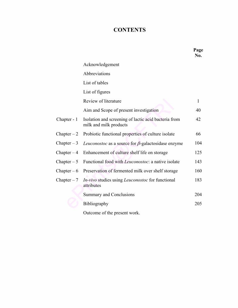
CONTENTS
Page No.
Acknowledgement
Abbreviations
List of tables
List of figures
Review of literature 1
Aim and Scope of present investigation 40
Chapter - 1 Isolation and screening of lactic acid bacteria from milk and milk products
42
Chapter – 2 Probiotic functional properties of culture isolate 66
Chapter – 3 Leuconostoc as a source for β-galactosidase enzyme 104
Chapter – 4 Enhancement of culture shelf life on storage 125
Chapter – 5 Functional food with Leuconostoc: a native isolate 143
Chapter – 6 Preservation of fermented milk over shelf storage 160
Chapter – 7 In-vivo studies using Leuconostoc for functional attributes
183
Summary and Conclusions 204
Bibliography 205
Outcome of the present work.
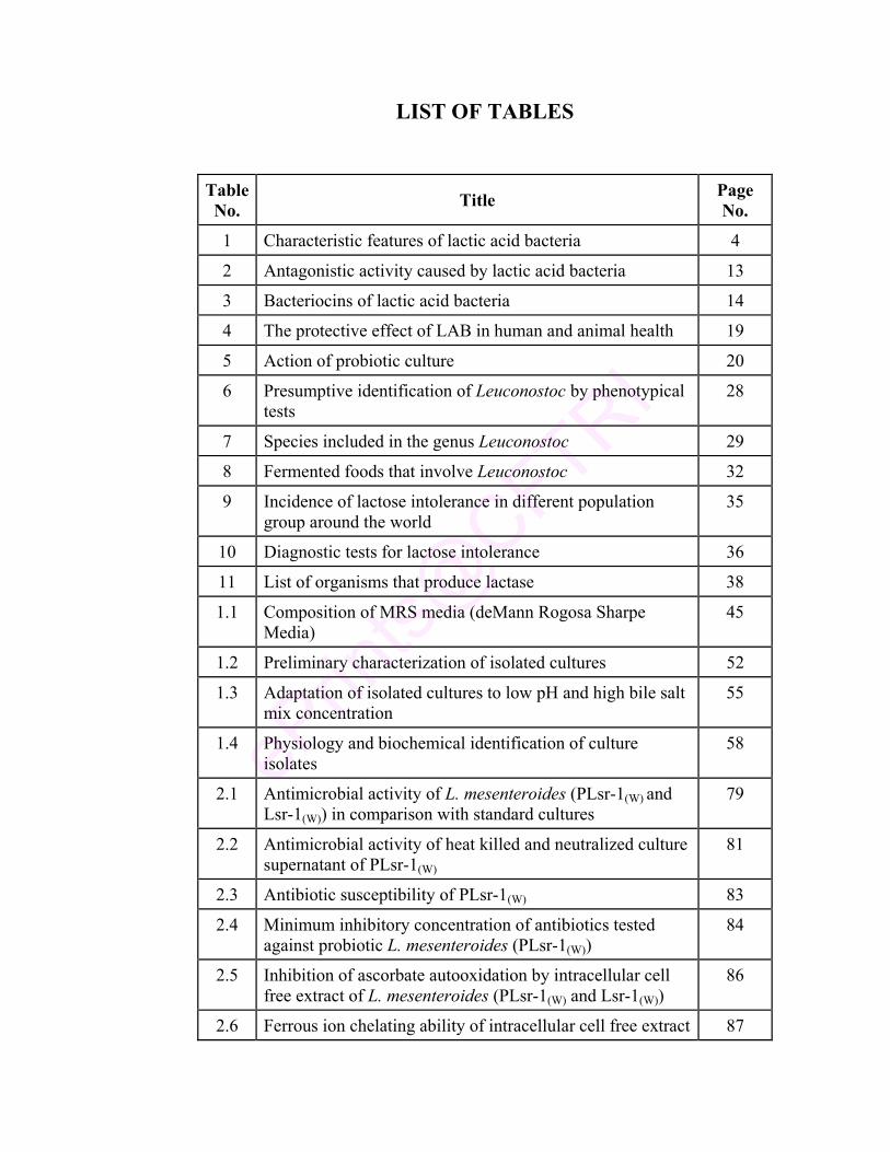
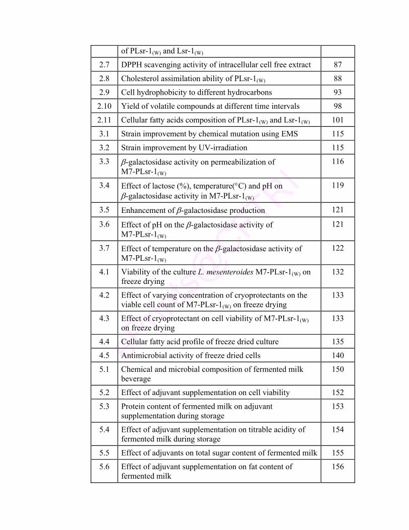
LIST OF TABLES
Table No. Title Page
No.
1 Characteristic features of lactic acid bacteria 4
2 Antagonistic activity caused by lactic acid bacteria 13
3 Bacteriocins of lactic acid bacteria 14
4 The protective effect of LAB in human and animal health 19
5 Action of probiotic culture 20
6 Presumptive identification of Leuconostoc by phenotypical tests
28
7 Species included in the genus Leuconostoc 29
8 Fermented foods that involve Leuconostoc 32
9 Incidence of lactose intolerance in different population group around the world
35
10 Diagnostic tests for lactose intolerance 36
11 List of organisms that produce lactase 38
1.1 Composition of MRS media (deMann Rogosa Sharpe Media)
45
1.2 Preliminary characterization of isolated cultures 52
1.3 Adaptation of isolated cultures to low pH and high bile salt mix concentration
55
1.4 Physiology and biochemical identification of culture isolates
58
2.1 Antimicrobial activity of L. mesenteroides (PLsr-1(W) and Lsr-1(W)) in comparison with standard cultures
79
2.2 Antimicrobial activity of heat killed and neutralized culture supernatant of PLsr-1(W)
81
2.3 Antibiotic susceptibility of PLsr-1(W) 83
2.4 Minimum inhibitory concentration of antibiotics tested against probiotic L. mesenteroides (PLsr-1(W))
84
2.5 Inhibition of ascorbate autooxidation by intracellular cell free extract of L. mesenteroides (PLsr-1(W) and Lsr-1(W))
86
2.6 Ferrous ion chelating ability of intracellular cell free extract 87
of PLsr-1(W) and Lsr-1(W)
2.7 DPPH scavenging activity of intracellular cell free extract 87
2.8 Cholesterol assimilation ability of PLsr-1(W) 88
2.9 Cell hydrophobicity to different hydrocarbons 93
2.10 Yield of volatile compounds at different time intervals 98
2.11 Cellular fatty acids composition of PLsr-1(W) and Lsr-1(W) 101
3.1 Strain improvement by chemical mutation using EMS 115
3.2 Strain improvement by UV-irradiation 115
3.3 β-galactosidase activity on permeabilization of M7-PLsr-1(W)
116
3.4 Effect of lactose (%), temperature(°C) and pH on β-galactosidase activity in M7-PLsr-1(W)
119
3.5 Enhancement of β-galactosidase production 121
3.6 Effect of pH on the β-galactosidase activity of M7-PLsr-1(W)
121
3.7 Effect of temperature on the β-galactosidase activity of M7-PLsr-1(W)
122
4.1 Viability of the culture L. mesenteroides M7-PLsr-1(W) on freeze drying
132
4.2 Effect of varying concentration of cryoprotectants on the viable cell count of M7-PLsr-1(W) on freeze drying
133
4.3 Effect of cryoprotectant on cell viability of M7-PLsr-1(W) on freeze drying
133
4.4 Cellular fatty acid profile of freeze dried culture 135
4.5 Antimicrobial activity of freeze dried cells 140
5.1 Chemical and microbial composition of fermented milk beverage
150
5.2 Effect of adjuvant supplementation on cell viability 152
5.3 Protein content of fermented milk on adjuvant supplementation during storage
153
5.4 Effect of adjuvant supplementation on titrable acidity of fermented milk during storage
154
5.5 Effect of adjuvants on total sugar content of fermented milk 155
5.6 Effect of adjuvant supplementation on fat content of fermented milk
156
5.7 Effect of adjuvant supplementation of soluble mineral
content of fermented milk 157
5.8 Effect of fatty acid composition of fermented milk on adjuvant supplementation
157
6.1 Characteristics of spoilage bacterial culture 168
6.2 Effect of 2(5H)-furanone on Pseudomonas sp growth in fermented milk
180
6.3 Preservation of fermented milk with 2(5H)-furanone for longer shelf life
181
7.1 Toxicity of M7-PLsr-1(W) in experimental rats 190
7.2 Glucose and Urea in serum of experimental rats fed with M7-PLsr-1(W)
191
7.3 Caecum analysis of experimental rats 193
7.4 β-galactosidase activity in caecum on feeding L. mesenteroides (M7-PLsr-1(W))
194
7.5 Weight range of experimental rats on probiotic feeding 196
7.6 pH change during probiotic feeding for 90 days 197
7.7 Glucose and urea concentration in serum and urine of M7-PLsr-1(W) fed rats
200
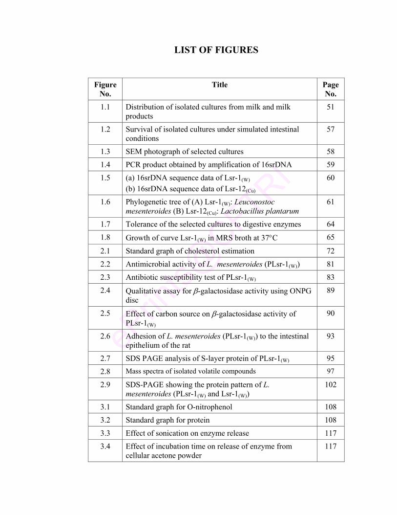
LIST OF FIGURES
Figure No.
Title Page No.
1.1 Distribution of isolated cultures from milk and milk products
51
1.2 Survival of isolated cultures under simulated intestinal conditions
57
1.3 SEM photograph of selected cultures 58
1.4 PCR product obtained by amplification of 16srDNA 59
1.5 (a) 16srDNA sequence data of Lsr-1(W)
(b) 16srDNA sequence data of Lsr-12(Cu) 60
1.6 Phylogenetic tree of (A) Lsr-1(W): Leuconostoc mesenteroides (B) Lsr-12(Cu): Lactobacillus plantarum
61
1.7 Tolerance of the selected cultures to digestive enzymes 64
1.8 Growth of curve Lsr-1(W) in MRS broth at 37°C 65
2.1 Standard graph of cholesterol estimation 72
2.2 Antimicrobial activity of L. mesenteroides (PLsr-1(W)) 81
2.3 Antibiotic susceptibility test of PLsr-1(W) 83
2.4 Qualitative assay for β-galactosidase activity using ONPG disc
89
2.5 Effect of carbon source on β-galactosidase activity of PLsr-1(W)
90
2.6 Adhesion of L. mesenteroides (PLsr-1(W)) to the intestinal epithelium of the rat
93
2.7 SDS PAGE analysis of S-layer protein of PLsr-1(W) 95
2.8 Mass spectra of isolated volatile compounds 97
2.9 SDS-PAGE showing the protein pattern of L. mesenteroides (PLsr-1(W) and Lsr-1(W))
102
3.1 Standard graph for O-nitrophenol 108
3.2 Standard graph for protein 108
3.3 Effect of sonication on enzyme release 117
3.4 Effect of incubation time on release of enzyme from cellular acetone powder
117
3.5 Effect of solvents on enzyme release 117
3.6 RSM study to optimize the condition for maximum β-galactosidase activity in M7-PLsr-1(W)
120
3.7 SDS PAGE analysis of β-galactosidase enzyme in M7-PLsr-1(W)
121
3.8 Effect of substrate concentration 122
3.9 Effect of enzyme concentration 122
3.10 Effect of enzyme inhibitors/ modulators on enzyme activity
123
3.11 Effect of metal ions on enzyme activity 123
4.1 Comparison of preservative methods for stability of M7-PLsr-1(W)
131
4.2 GC profile of cellular fatty acid 136
4.3 Cellular protein profile of M7-PLsr-1(W) 137
4.4 Storage stability of L. mesenteroides M7-PLsr-1(W) after freeze drying. (a) Viability at 30°C (b) Viability at 4°C (c) Viability at -20°C
138
4.5 Tolerance of M7-PLsr-1(W) (A) to acidic pH 2.0 (B) to high bile salt concentration (4%)
139
4.6 (a) SEM of L. mesenteroides M7-PLsr-1(W) before freeze drying in skim milk (b) SEM of L. mesenteroides M7-PLsr-1(W) after freeze drying in skim milk
141
5.1 Standard graph for the estimation of total sugars 147
6.1 Standard graph for rhamnose estimation 165
6.2 SEM of Pseudomonas sp 168
6.3 TLC Overlay assay for identification of signal molecule 170
6.4 GC chromatograph of (A) standard HHSL (B) standard BHSL and (C) Pseudomonas sp extract (D) Pseudomonas sp extract spiked with standard HHSL
171
6.5 GCMS fragmentation pattern of AHL produced by Pseudomonas sp
173
6.6 Effect of furanone on Pseudomonas sp growth in culture media
176
6.7 Effect of furanone on rhamnolipid content of Pseudomonas sp
177
6.8 Inhibition of Pseudomonas sp motility by 2(5H)-furanone 178
6.9 Effect of furanone on exoprotease activity of Pseudomonas sp
179
7.1 Serum glucose concentration and β-galactosidase activity in caecum of M7-PLsr-1(W) fed rats
192
7.2 Effect of L. mesenteroides (M7-PLsr-1(W)) on serum cholesterol of experimental rats
193
7.3 pH change and LAB count in caecum on feeding L. mesenteroides (M7-PLsr-1(W))
194
7.4 SEM photograph of probiotic fed rat intestine showing adhesion of probiotic culture
194
7.5 Probiotic influence on E. coli in (A) Caecum, (B) Feces, (C) Large intestine
198
7.6 SEM photograph showing adhesion of probiotic culture to large intestine (LI) and caecum of experimental rats
199
7.7 Serum glucose concentration and the enzyme activity in the caecum of experimental rats
201
7.8 β-galactosidase activity in caecum and small intestine of experimental rats
202
SHOBHA RANI. P Senior Research Fellow Food Microbiology Department Central Food Technological Research Institute Mysore
DECLARATION
I hereby declare that the thesis entitled “Isolation and
characterization of a native isolate of Leuconostoc for functional
attributes” submitted to the University of Mysore, Mysore, for the award of
Degree of Doctor of Philosophy in the faculty of Microbiology is the result
of work carried out by me under the guidance of Dr. Renu Agrawal,
Scientist, Department of Food Microbiology, Central Food Technological
Research Institute, Mysore.
I further declare that the results of this thesis have not been submitted
by me for award of any other degree/diploma to this or any other Universities.
SHOBHA RANI. P (Candidate) Date: Place: Mysore
CERTIFICATE
This is to certify that the thesis entitled “Isolation and
characterization of a native isolate of Leuconostoc for functional
attributes” submitted to the University of Mysore, Mysore for the award of
Degree of Doctor of Philosophy in the faculty of Microbiology by
Shobha Rani. P is the result of work carried out by her in the Department of
Food Microbiology, Central Food Technological Research Institute, Mysore.
RENU AGRAWAL (Guide) Date: Place: Mysore
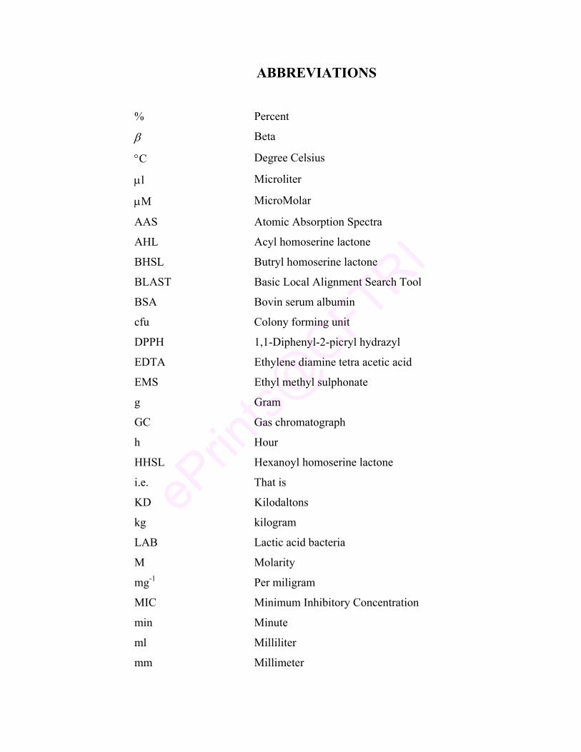
ABBREVIATIONS
% Percent
β Beta
°C Degree Celsius
µl Microliter
µM MicroMolar
AAS Atomic Absorption Spectra
AHL Acyl homoserine lactone
BHSL Butryl homoserine lactone
BLAST Basic Local Alignment Search Tool
BSA Bovin serum albumin
cfu Colony forming unit
DPPH 1,1-Diphenyl-2-picryl hydrazyl
EDTA Ethylene diamine tetra acetic acid
EMS Ethyl methyl sulphonate
g Gram
GC Gas chromatograph
h Hour
HHSL Hexanoyl homoserine lactone
i.e. That is
KD Kilodaltons
kg kilogram
LAB Lactic acid bacteria
M Molarity
mg-1 Per miligram
MIC Minimum Inhibitory Concentration
min Minute
ml Milliliter
mm Millimeter
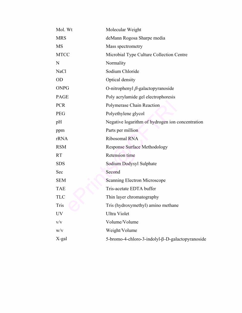
Mol. Wt Molecular Weight
MRS deMann Rogosa Sharpe media
MS Mass spectrometry
MTCC Microbial Type Culture Collection Centre
N Normality
NaCl Sodium Chloride
OD Optical density
ONPG O-nitrophenyl β-galactopyranoside
PAGE Poly acrylamide gel electrophoresis
PCR Polymerase Chain Reaction
PEG Polyethylene glycol
pH Negative logarithm of hydrogen ion concentration
ppm Parts per million
rRNA Ribosomal RNA
RSM Response Surface Methodology
RT Retension time
SDS Sodium Dodysyl Sulphate
Sec Second
SEM Scanning Electron Microscope
TAE Tris-acetate EDTA buffer
TLC Thin layer chromatography
Tris Tris (hydroxymethyl) amino methane
UV Ultra Violet
v/v Volume/Volume
w/v Weight/Volume
X-gal 5-bromo-4-chloro-3-indolyl-β-D-galactopyranoside
ISOLATION AND CHARACTERIZATION OF A NATIVE ISOLATE OF LEUCONOSTOC
FOR FUNCTIONAL ATTRIBUTES
A Synopsis Submitted to University of Mysore, Mysore for the Degree of
DOCTOR OF PHILOSOPHY IN
MICROBIOLOGY
By
SHOBHA RANI. P
Under the guidance of
Dr. RENU AGRAWAL
DEPARTMENT OF FOOD MICROBIOLOGY CENTRAL FOOD TECHNOLOGICAL RESEARCH INSTITUTE,
MYSORE-20
2008
1
SYNOPSIS
ISOLATION AND CHARACTERIZATION OF A NATIVE ISOLATE OF LEUCONOSTOC FOR
FUNCTIONAL ATTRIBUTES
INTRODUCTION
During the past several years, the focus of nutritional sciences has shifted
from deficiency disease prevention to optimizing health and prevention of
chronic diseases. Accordingly the research has encompassed the health effects of
bioactive food components. In this regard, probiotic therapy is being used
increasingly in humans and veterinary medicine due to their apparent high index
of safety and public perception about natural or alternative therapies (Gherty
1995; Sander 1998; FAO Report 2002).
Probiotics are a category of ‘nutraceuticals’ i.e., viable cultures added to
food with the intension of maintaining or improving the nutritional health of
consumers. These bacteria favorably alter the intestinal microflora, inhibit
growth of harmful bacteria, promote good digestion, boost immune function and
increase resistance to infection (Malin et al 1996; Haudault et al 1997).
Lactic acid bacteria (LAB) are in the focus of extensive research because
of their probiotic nature. One important way in which they affect health of the
host is by providing enzymatic activities that improve the utilization of nutrients
within the intestine.
Today, probiotic market promises the disease prevention and better health
for all as a natural alternative therapy. One of the important challenges to the
present day probiotic industry is the eradication of lactose intolerance problem
which is quite common all over the world. It is estimated that 70-90% of adults
are lactose intolerant (Swegerty 2002), suffering from intestinal discomfort with
the symptoms like nausea, cramps and gas (Stiles and Holzapfel 1997). In the
present scenario, with increase desire of consumer for natural food products as
source of providing nutrition and other desirable benefits, research work towards
2
the selection of strain with functional properties has become very important
especially for treating lactose intolerance.
Considering all these aspects, in the present work a strain of LAB was
isolated from milk and milk products, characterized and studied for its probiotic
functional attributes to be used in food formulation.
Objectives
1) Isolation and characterization of Leuconostoc from milk products
2) Properties of the isolated bacterium in relation to functional significance
3) Colonization of LAB in relation to homoserine lactone
Chapter 1
Isolation and screening of lactic acid bacteria from milk and milk product
The problem for choosing a culture to be used in health promoting
probiotic ingredients in food and pharmaceutical preparations was apparent even
in the original work of Metchnikoff (1906). Reid (1999) and Sobel (1999)
specify certain properties like ability to adhere intestinal cell wall, exclude/
reduce pathogenic microorganisms, produce antimicrobial compound, resist
microbicides, non-carcinogenic and non-pathogenic character to be present in
the selected strain for their use as probiotic culture.
Now a days, a consensus is emerging for selection criteria of LAB to
achieve positive probiotic effect (Collins et al 1998). Therefore, in the present
work a screening procedure is performed to select a potential probiotic culture
with an ultimate aim of using the culture in functional food for beneficial effect.
In the present work, 45 isolates were screened for basic LAB
characteristics from milk and milk products. As the most critical characteristics
of probiotic strain are tolerance gastrointestinal condition (Ammor and Mayo
2007), the isolated strains were initially screened for their resistance in this harsh
environment and further adapted to grow at low pH and high bile salt
concentration. The strains were further analyzed for their survival under
simulated gastrointestinal condition. The strains that were able to survive under
3
such environment were characterized and identified through biochemical assays
and molecular techniques. The identified cultures Leuconostoc mesenteroides
(Lsr-1(W)) and Lactobacillus plantarum (Lsr-12(Cu)) were further studied for their
resistance to digestive enzymes pepsin and trypsin. Leuconostoc mesenteroides
(Lsr-1(W)) that was better resistant to these enzymes was selected for further
studies and was coded as PLsr-12(Cu).
Chapter 2
Probiotic functional properties of culture isolate
Probiotics have health promoting effects including inhibition of
pathogens, antimutagenic, anticarcinogenic activity, prevention of diarrhea,
stimulation of immune response and ability to reduce serum cholesterol levels
(Tannock 1999). The development of new applications such as life vaccines and
probiotic foods reinforces the need for these characteristics.
In this regard, the selected culture of Leuconostoc mesenteroides
(PLsr-1(W)) was evaluated for its functional properties. According to the data
obtained, the culture PLsr-1(W) shows antimicrobial activity against 7 toxic food
pathogens such as E. coli, S. typhi, S. dysenteriae, P. aeroginosa, V. cholerae,
Y. enterocolitica and S. aureus. The isolate was found resistant to 3 antibiotics
tested. The inhibitory activity of intracellular cell free extract of culture to
ascorbate autooxidation, ferrous ion chelating ability and scavenging ability of
oxygen radical represent the antioxidative property of culture isolate. The culture
was also able to assimilate 28 µg/ml of cholesterol from media which shows the
anticholesterol activity of the strain. Analysis of β-galactosidase in culture
indicate the ability of the culture to hydrolyze lactose into simple sugar for easy
absorption. From the results of cell surface hydrophobicity and intestinal
adherence test, the adhesion ability of the culture was confirmed. Analysis of
S-layer adhesion protein by SDS-PAGE showed a prominent protein band of
60 KD. Volatile compounds analyzed by GC and GCMS of culture extract
confirmed the presence of therapeutically important compounds. Stress response
of PLsr-1(W) to low pH and high bile salt indicate the increased content of stress
4
proteins and membrane fatty acids (saturated and unsaturated). All these
properties make the present isolate a potent probiotic and will stand out as a
natural cure to many diseases.
Chapter 3
Leuconostoc as a source for β-galactosidase enzyme
Lactose is a non-reducing disaccharide which provides almost half the
total energy required by infants. In presence of lactase/ β-galactosidase, lactose
gets hydrolyzed into galactose and glucose for easy absorption. But in persons
deficient in lactase show symptoms of intestinal discomfort known as lactose
intolerance (Sieber et al 1997). Hence studies were carried out to enhance the
enzyme activity in the present culture isolate.
Leuconostoc mesenteroides PLsr-1(W) that had the ability to survive the
harsh conditions of GIT was studied for its ability to increase lactose tolerance.
Primarily strain was improved by UV irradiation and chemical mutagenesis for
enhanced enzyme activity. UV mutant strain showed 2 folds higher activity than
the parent strain and hence was selected for all further studies (mutant strain
was coded as M7-PLsr-1(W)). Cell permeabilization method was optimized for
maximum release of enzyme. Response surface methodology (RSM) studies
were undertaken for optimization of chemical and physical parameter. The
enzyme produced under optimum condition of pH 7.5 with 1.25 % lactose was
partially purified and studied for kinetics. The results indicate a 25 fold increase
in the activity of partially purified enzyme as compared to the crude extract.
Chapter 4
Enhancement of culture shelf life on storage
LAB in food biotechnology or for any probiotic formulations are strictly
concerned for the preservation techniques employed to ensure stable culture in
terms of viability and bacterial metabolism. It is very important to use suitable
technology or process to enhance and maintain the viability of the culture on
storage for a beneficial effect.
5
As the present isolate M7-PLsr-1(W) is very important culture due to its
characteristic multifunctional probiotic properties, it becomes necessary to keep
the culture viable for a longer time. Hence the aim of the present work was to
preserve the culture for longer shelf life. In this regard, the culture was subjected
to different preservation techniques i.e., oven, spray, vacuum and freeze drying.
The viability and resistance of culture to these methods was tested and it was
found that the culture showed maximum survival to freeze drying condition. The
viability under freeze drying condition was further enhanced with
supplementation of different cryoprotective agents (PEG, lactose and sucrose).
Enhanced membrane fatty acid composition and cellular protein confirmed the
adoptive nature of the culture to freeze drying.
In the present work, we also report the viability of the freeze dried culture
for 6 months. It showed resistance to low pH (2.0) and high bile salt (4%). Even
after 6 months of freeze drying, culture showed antimicrobial activity against 6
toxic food pathogens. Data determines that the culture retains the β-galactosidase
enzyme even after 6 months of storage.
The above study determines the capability of culture to remain viable
even after 6 months of storage. It also shows the importance of cryoprotectants
in enhancing the viability and beneficial attributes of culture during storage. This
determines the capability of the culture to be utilized in the form of capsules or
any functional food.
Chapter 5
Functional food with Leuconostoc: a native isolate
The demand for probiotic foods is increasing all over the world reflecting
the heightened awareness among the public relationship between diet and health.
Fermented dairy products are the most widely used food vehicles for these
probiotic bacteria because of their healthy image. During the past twenty years
there has been a tremendous increase in the world wide sales of cultured
products containing probiotic bacteria because of their health effects (Ostlie
2005; Maltila-Sandholm 1999).
6
In this regard, the culture M7-PLsr-1(W) that has the potent probiotic
properties was used as starter culture in the preparation of fermented milk
beverage. The product was analyzed to be rich in protein, fat, sugar and minerals
like iron, zinc and magnesium. Hence the viability of culture in the product was
further enhanced with supplementation of different adjuvants (tryptone, casein
hydrolysate, cysteine hydrochloride and ascorbic acid). The results conclude that
the culture maintained maximum viability on supplementation of tryptone
(100 mg/L) after 5 days of storage at 4°C. Fatty acid composition of the product
also confirmed the nutritional property of the product.
Chapter 6
Preservation of fermented milk over shelf storage
Spoilage causes a significant loss to dairy industries and also disease
outbreaks. In this concern, research work was carried out to investigate and
preserve fermented milk beverage prepared with M7-PLsr-1(W).
In this regard, the functional fermented milk beverage prepared by
M7-PLsr-1(W) was studied for the predominant bacterial cultures responsible for
spoilage of the product. Pseudomonas sp was identified as dominant spoilage
bacteria and through TLC, GC and GCMS, the signal molecule for spoilage was
investigated as hexanoyl homoserine lactone (HHSL).
This culture releases HHSL and forms a quorum when high cell density is
reached. Inhibition of bacterial growth in this biofilm by using biocides,
antibiotic and bacteriophages has many obstacles such as cell permeability,
specificity and efficacy in mode of delivery. Hence the natural furanones which
are non-toxic are used in present study because of its small size and ease of
delivery. Because of its structural similarity they specifically interfere with
signal molecule without any adverse effect on the beneficial bacterial consortia.
Results show that 2(5H)-furanone tested was having better inhibitory activity
against Pseudomonas than bromofuranone. This was also in concurrence with
reduction in rhamnolipid content, reduced motility and exoprotease enzyme
activity. Using 2(5H)-furanone about 5-6 log of Pseudomonas culture was
reduced in the fermented beverage.
7
Chapter 7
In-vivo studies using Leuconostoc for functional attributes
The health benefits described for probiotic lactic acid bacteria make them
a good agent for preparation of functional food and hence a number of bacterial
strains are being identified and incorporation into these foods. As more probiotic
organisms are discovered, it is important to carefully document the efficacy of
the strain for its potential application and safety.
Looking into the probiotic characteristic of the present isolate
M7-PLsr-1(W) of being resistant to GIT condition, adherence ability,
antimicrobial, β-galactosidase activity and anticholesterol activity through
in-vitro assays the present study was aimed to assess its safety and functions in
in-vivo model using albino rats.
According to the result there was no deleterious effect on probiotic
feeding for 3 months. No bacterial translocation was observed and hence it is
likely to be safe for human consumption. Increase in the general body weight
and serum urea concentration provides a potential proof for the health promoting
effect of the culture. High LAB count in feces and ceacum shows the ability of
the culture to resist the GIT condition and adhere to exert beneficial effect.
Results of cholesterol assay confirmed the anticholesterol effect of the culture.
The ability of the isolate to protect against pathogens was determined by
decrease in E. coli count.
Lactose intolerance, a clinical problem associated with unpleasant
abdominal discomfort is due to undigested lactose. According to a survey, about
70-80% of the world population are lactose intolerant (Swagerty 2002).
Innovative approaches have been tried as alternative to antibiotics in treating
lactose intolerance because of the growing antibiotic resistance problem.
Alternative methods such as exogenous β-galactosidase administration in
functional foods or in pharmaceutical preparation is advised but the draw back is
the inactivation of the enzyme in gastrointestinal transit. The other approach is
gene therapy or exclusion of milk and dairy products with lactose from diet
which may cause nutritional disadvantages.
8
Considering this, the present culture M7-PLsr-1(W) was tested for its
ability to reduce lactose intolerance problem by in-vivo experiments. Single dose
study and long term (3 months) experiments were carried out with albino Wister
rats. From the study, the effective dose of culture was determined to be
108 cfu/ml. Disappearance of diarrhea in lactose intolerant induced rats after
culture feeding confirmed the positive impact of culture in treating lactose
intolerance.
From the report presented here, the culture M7-PLsr-1(W) was found to
have probiotic characters in terms of resistance to GIT, antimicrobial activity,
anticholesterol activity and adherence ability. The culture was also able to
reduce the symptoms of lactose intolerance and hence can be used as an
alternative source to treat the problem.
Achievement of the work
The culture is native, isolated in laboratory and shows potential probiotic
characteristics along with high β-galactosidase activity. The in-vivo experiments
conducted with albino Wister rats conclude the potential probiotic functional
properties and its importance in reducing lactose intolerance problem. The
culture preserved by freeze drying shows viability even after 6 months of
storage. The fermented milk beverage prepared with the present isolate is having
a high nutritive value so that it can be used by all ages for its beneficial effects.
This fermented milk beverage was preserved over a storage time by interrupting
the signal molecules produced by of spoilage bacteria using 2(5H)-furanone
which is a natural compound produced by an algae and known to be safe.
Social and scientific relevance
With the rise in the consumer’s awareness of individual health, nutrition
and well being, the interest and demand for value added foods and beverages has
expanded. Although some companies have marketed the probiotic products, they
are either expensive or are specified for one specific cure. Most of these products
9
are not indigenous in our country and the viable count of the culture is
questionable or not to the mark as labeled.
Today WHO and Indian five year plans both have a common priority to
replace antibiotics with natural means of cure. In this regard, the present isolate
having probiotic functional properties promises to be as a source of natural
alternative cure. The fermented beverage prepared by Leuconostoc
mesenteroides (M7-PLsr-1(W)) has many functional attributes.
References
1. Ammor MS, Mayo B (2007). Slection criteria for LAB to be used as
functional starter cultures in dry sausage production: an update. Meat Sci 76,
138-146.
2. Collins JK, Thornton G, Sullivan GO (1998). Selection of probiotic strains
for human applications. Int Dairy J 8, 487-490.
3. FAO Expert Report 2002. evaluation of health and nutritional properties of
probiotics in food including powder milk with live lactic acid bacteria.
American Cordoba Park Hotel, Cordeba. Argentina.
4. Gherty J (1995). American dairy science association foundation, lecture.
J Dairy Sci 78(7): 1401
5. Haudault S, Lieven V, Bernet-Camard MF, Servin AL (1997). Antagonistic
activity exerted in vitro and in vivo by L. casei (strain GG) against
S. typhimurium C5 infection. Appl Environ Microbiol 63, 513-518.
6. Malin M, Verronen P, Mykkanen H, Salminene S, Isolauri E (1996).
Increased bacterial urease activity in feces in juvenile chronic arthritis
evidence of altered intestinal microflora? Br J Rheumatol 35, 689-694.
7. Mattila-Sandholm T, Matto J, Saarela M (1999) Lactic acid bacteria with
health claims – interactions and interference with gastrointestinal flora.
Int Dairy J 9, 25–35.
8. Metchnikoff E (1906). The prolongation of life. G.P. Putnam’s Sons, New
York.
10
9. Ostlie HM, Treimo J, Narvhus JA (2005). Effect of temperature on growth
and metabolism of probiotic bacteria in milk. 15(10), 989-997.
10. Reid, G., 1999. The scientific basis for probiotic strains of Lactobacillus.
Appl Environ Microbiol 65, 3763–3766.
11. Sanders ME (1998). Over view of functional foods: emphasis on probiotic
bacteria. Int Dairy J 8, 341-347
12. Sieber R, Stransky M, de Vrese M (1997). Lactose intolerance and
consumption of milk and milk products. Z Emahrungswiss 36(4), 375-393
13. Stiles ME, Holzapfel WH (1997). Lactic acid bacteria of foods and other
their current taxonomy. Int J Food Microbiol 36, 1-29.
14. Swagerty, DL, Walling AD, Klein RM (2002). Lactose intolerance.
American Fan Physician 65(9), 1845-1850
15. Tannock, GW (1999). Probiotic: A critical review, Horizon Scientific Press,
Norfolk England.
Signature of the candidate Date:
Signature of the guide Date:
Review of Literature
Review of Literature
1
REVIEW OF LITERATURE
INTRODUCTION
1. Lactic acid bacteria: characterization, classification and importance
Lactic acid bacteria (LAB) are nutritionally fastidious microorganisms
that have been used to ferment or culture foods for atleast 4000 years. All
over the world they are used particularly in the preparation of fermented milk
products including yogurt, cheese, butter, buttermilk and kefir. They refer to a
large group of beneficial bacteria that produce lactic acid as their major
metabolic end product. They produce characteristic flavor and aroma
compounds like acetaldehyde and diacetyl.
1.1. Classification of lactic acid bacteria
Lactic acid bacteria belong to the phylum Firmicutes, which share the
property of being gram positive (Fooks et al., 1999) that ferment
carbohydrates into energy and lactic acid (Jay, 2000). Depending on the
metabolic pathway they are classified into homofermentative and
heterofermentative cultures (Caplice and Fitzgerald, 1999; Kuipers et al.,
2000).
Schleifer et al. (1991) have classified lactic acid bacteria into four
genera based on sugar fermentation and growth at specific temperatures. This
includes Lactobacillus (rod shaped), Streptococcus (homofermentative),
facultative anaerobic cocci, betacoccus and tetracoccus. Later, Carr et al.
(2002) and Frank et al. (2002) grouped them into four important genera
namely Streptococcus, Pediococcus, Leuconostoc and Lactobacillus.
At present Carnobacterium, Enterococcus, Lactobacillus, Lactococcus,
Lactosphaera, Leuconostoc, Melissococcus, Oenococcus, Pediococcus,
Streptococcus, Tetragenococcus, Vagococcus and Weissella are the
recognized genera of lactic acid bacteria (Stiles and Holzapfel, 1997; Ercolini
et al., 2001; Holzapfel et al., 2001).
Review of Literature
2
Streptococcus, Pediococcus and Lactobacilli are homofermentative
species that produces 2 moles of lactic acid for each mole of glucose
consumed as their metabolic product. They possess enzyme aldolase and
hexose isomerase but lack phosphoketolase, so it uses the Embden-
Meyernhoff (EM) glycolytic pathway for converting glucose to fructose-1,6
diphosphate. In heterofermentative species, the key enzyme fructose-1,6
diphosphate aldolase is absent and so they possess an alternative glycolytic
pathway, where in the glucose is converted to 6-phosphogluconate that gets
decarboxylated into pentose resulting in lactic acid and ethanol and/or acetate
(Blackwood and Blakley, 1960; Holzapfel and Wood, 1998). Leuconostoc and
Weissella belong to this group (Jay, 1992).
The taxonomy of LAB is based on comparative 16srRNA sequence
analysis. Molecular techniques, especially polymerase chain reaction (PCR)
based methods, such as rep-PCR fingerprinting and restriction fragment
length polymorphism (RFLP) as well as pulse field gel electrophoresis
(PFGE) are regarded as important techniques for specific characterization and
detection of LAB strains (Gevers et al., 2001). Denaturing gradient gel
electrophoresis (DGGE) and temperature gradient gel electrophoresis (TGGE)
of 16srRNA gene have shown to be powerful approaches in determining and
monitoring bacterial community (Cocconcelli et al., 1997; Zoetendal et al.,
1998). Methods such as DNA-DNA hybridization (Yaeshima et al., 1996),
genus specific and species-specific probes (Hensiek et al., 1992; Timisjarvi
and Alatossava, 1997), 16s and 23s intergeneric spacer region sequencing
(Bourget et al., 1996) and ribotyping (Ning et al., 1997) have also been used
for identification and characterization of LAB strain.
1.2. Habitat and characterization of lactic acid bacteria
LAB are typically fastidious and require a variety of amino acids,
Vitamins, purine and pyrimidine bases for their growth (Calderon et al 2001).
Although they are mesophilic, some can grow below 5°C and others at
temperature as high as 45°C. Usually most of the LAB cultures grow at pH
Review of Literature
3
6.0-6.5, but some can also grow in acidic pH (3.2) and others in alkaline pH
(9.6) (Jay, 2000). Some of the characteristic features of lactic acid bacteria are
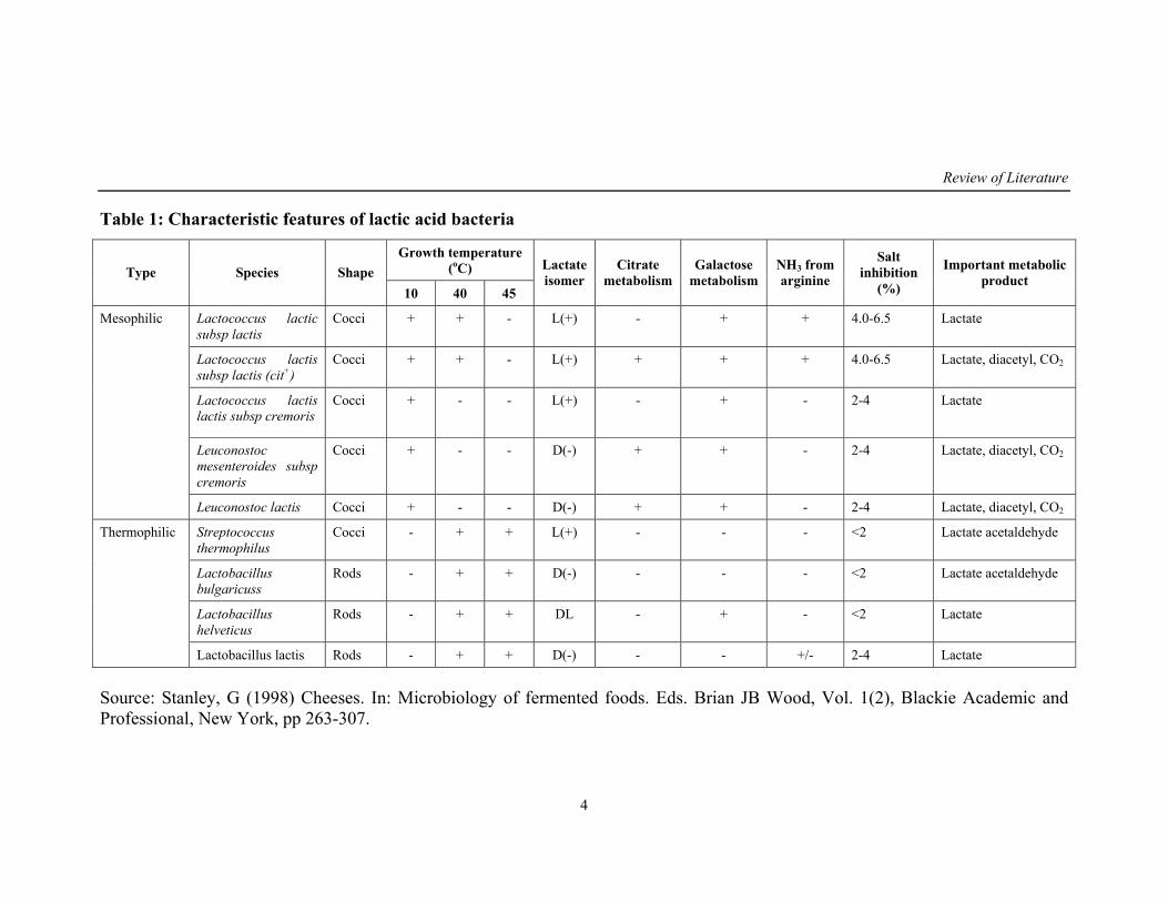
presented in the table 1.
Heterofermentative bacteria especially Leuconostoc spp appear to be
common in plant materials like vegetables and roots. In refrigerated products
generally Streptobacteria predominates whereas thermobacteria are
commonly found in products of higher temperature (Kitchell and Shaw, 1995;
Franz and Holy, 1996; Samelis et al., 2000; Holm et al., 2004). LAB occur
naturally in fermented food and have been detected in soil, water, manure and
sewage (Holzapfel et al., 2001). They are the normal intestinal microflora in
human (Boris et al., 1998; Reid, 2001; Schrezenmeir and deVrese, 2001) and
in animals (Fujisawa and Mitsuoka, 1996; Klijn et al., 1995).
1.3. Importance of lactic acid bacteria
Without understanding the scientific basis people are using lactic acid
bacteria to produce fermented foods with characteristic flavors and texture.
These LAB also help our normal gut bacteria to function more efficiently.
The world wide market for these products continues to increase in response to
the demands of an increasingly health conscious public. Lactic acid bacteria
are therefore excellent ambassadors for an often maligned microbial world.
With growing interest in self-care linked between diet and health, the market
of food that promotes health beyond basic nutrition has become stronger and
is flourishing all over the world.
Review of Literature
4
Table 1: Characteristic features of lactic acid bacteria
Growth temperature (oC) Type Species Shape
10 40 45
Lactate isomer
Citrate metabolism
Galactose metabolism
NH3 from arginine
Salt inhibition
(%)
Important metabolic product
Lactococcus lactic subsp lactis
Cocci + + - L(+) - + + 4.0-6.5 Lactate
Lactococcus lactis subsp lactis (cit+)
Cocci + + - L(+) + + + 4.0-6.5 Lactate, diacetyl, CO2
Lactococcus lactis lactis subsp cremoris
Cocci + - - L(+) - + - 2-4 Lactate
Leuconostoc mesenteroides subsp cremoris
Cocci + - - D(-) + + - 2-4 Lactate, diacetyl, CO2
Mesophilic
Leuconostoc lactis Cocci + - - D(-) + + - 2-4 Lactate, diacetyl, CO2
Streptococcus thermophilus
Cocci - + + L(+) - - - <2 Lactate acetaldehyde
Lactobacillus bulgaricuss
Rods - + + D(-) - - - <2 Lactate acetaldehyde
Lactobacillus helveticus
Rods - + + DL - + - <2 Lactate
Thermophilic
Lactobacillus lactis Rods - + + D(-) - - +/- 2-4 Lactate
Source: Stanley, G (1998) Cheeses. In: Microbiology of fermented foods. Eds. Brian JB Wood, Vol. 1(2), Blackie Academic and Professional, New York, pp 263-307.
Review of Literature
5
2. Lactic acid bacteria as probiotics
2.1. Probiotics: Definitions and History
LAB are regarded as a major group of probiotic bacteria (Collins et al.,
1998; Tannock, 1998; Schrezenmeir and deVrese, 2001). There is a long
history of health claims concerning LAB in food. In a Persian version of the
Old Testament (Genesis 18:8) it states that “Abraham owed his longevity to
the consumption of sour milk”. In 76 BC, the Roman historian Plinius
recommended the administration of fermented milk products for treating
gastroenteritis (Bottazzi, 1983).
The term ‘probiotic’, meaning “for life”, is derived from the Greek
language. As early as 1906, Tissier noted that significant stool colonization
with Bifidobacteria sp was protective against diarrhea in children.
Metchnikoff (1908) suggested that long, healthy life of Bulgarian peasants
results from the consumption of fermented milk products. It was Ferdinand
Vergin (Vergin, 1954) a German scientist who introduced the term ‘probiotic’
in an article entitled “Anti-Und Probiotika”, wherein he compared the harmful
effects of antibiotics and the beneficial (“Probiotic”) effects of the lactic acid
bacteria. Later in 1965, Lilley and Stillwell described it as “substances
secreted by one microorganism which stimulates the growth of another”.
In 1971, Sperti applied the term probiotic as “tissue extracts that
stimulate microbial growth”. Parker (1974) defined it as “organisms and
substances which contribute to intestinal microbial balance”. In 1992, Fuller
redefined it as “a live microbial feed supplement which beneficially affects
the host animals by improving its intestinal microbial balance”. Further,
Havenaar and Huis in’t Veld et al. (1992) broadened the definition as “a
viable mono or mixed culture of microorganisms which are given to animals
or human for its beneficial effect in improving the properties of indigenous
microflora”.
Salminen (1996) defined it as “a live microbial culture or cultured
dairy product which beneficially influences the health and nutrition of the
host”. Further Schaafsma (1996) described it as “oral probiotics are
Review of Literature
6
microorganisms which upon ingestion in certain numbers exert health effects
beyond inherent basic nutrition”. Ouwehand et al. (2002) have defined
probiotics as “non-pathogenic microorganisms that when ingested in certain
number exerts a positive influence on the host physiology and health beyond
inherent general nutrition”
The joint FAO/WHO (2002) have proposed a general definition that
probiotics are “live microorganisms which when administered in adequate
amounts confer a health benefit on the host”.
2.2. Probiotics: Essential characteristic requirements
Many scientific publications and review articles has listed a series of
essential requirements in the screening of microorganisms for the probiotic
value. The list of essential requirements based on theoretical consideration
included the following (Guarner, 2005; Rashid et al., 2007; Maurad and
Merien, 2008).
1) Human origin (as a token of safety for human use)
2) Resistance to gastric acidity and bile toxicity (for good survival during
gastrointestinal transit)
3) Adhesion to gut epithelial cells (for successful colonization)
4) Production of antimicrobial substances or bacteriocins (for pathogenic
antagonism)
5) Ability to modulate immune responses.
2.3. Probiotic: Mechanism of action
Stress in modern day life has disrupted the homeostasis in the gut
through change in dietary pattern and eating habits. Another contributory
factor includes the consumption of pharmaceutical compounds, in particular
antibiotics that destroy bacteria creating an imbalance in the gut microflora.
This leads to a number of diseases. Hence there is an increasing demand for
food products that support health beyond providing basic nutrition.
Review of Literature
7
Consumption of probiotics is known to help in balancing the normal
intestinal microflora. It is also understood that each probiotic strain is
independent of the genera and species and is unique in their properties. The
exact manner in which probiotics affect is uncertain. However some
mechanisms have been speculated (Hatcher and Lambrecht, 1993; Ouwehand,
1998; Jacobsen et al., 1999; Boirivant and Strober, 2007; Allan, 2008)
1) Biochemical effect
Through production of bacteriocins
Short chain fatty acids with antagonistic effect
Creation of unfavorable environment for pathogens by reduction
of pH
Production of antimicrobial compounds including organic acids,
hydrogen peroxide and diacetyl that inhibit growth of spoilage
organisms
2) Competition for nutrients
3) Immune clearance
By surface Ig A attachment to mucosal membrane, adherence of
enteropathogens has been limited
Stimulation of cell mediated response by increasing macrophage
phagocytic activity.
4) Attachment
Blockage of adherence of enteropathogens by occupying the
niche of the intestinal mucosa.
2.4. Probiotic: Criteria for selection
The selection of lactic acid bacterial culture to be used as a probiotic
source depends on the host specificity and colonization ability. It should be
generally regarded as safe (GRAS status) with antimicrobial activity and other
desirable metabolic activities for beneficial effect (Canducci et al., 2000;
Sanders, 2003; Reid et al., 2003). Criteria should also include sensory
characteristics and technological suitability for general acceptance
Review of Literature
8
(O’Sullivan et al., 2002; Charteris et al., 1998). The market of a probiotic
strain requires a well designed, double blind, placebo controlled host specific
studies with animal and humans model. Culture also should resist to
technological processes, with respect to viability and activity throughout
processing (Dunne et al., 2001). Each potential probiotic strain must be
documented independently, without extrapolating any data from closely
related strains and employing only well defined strains in the trials.
Safety assessment of the strain is a very important criteria (Saarela et
al., 2002). Intrinsic properties of a strain, its interactions in-vivo with the host
and its pharmacokinetics should also be accounted for commercial approval
of the strain (Marteau et al., 1993; Pelletier et al., 1996; Saxelin, 1996).
2.4.1. Survival within the gastrointestinal tract
To survive passage through the gastrointestinal condition, probiotic
strains must tolerate the acidic and protease rich environment of stomach and
also bile rich environment of the intestine (Tuomola et al., 2001). It is
observed that there is an intraspecies variation in the mechanism to tolerate
low pH and high bile concentration between the potential probiotic strains
(Lee and Salminen, 1995; Lee and Wong, 1998). Lorca and Font de Valdez
(2001) have described that acid tolerance in Lactobacillus acidophilus is
mediated by membrane ATPases. Bile resistance appears to be mediated by
bile salt hydrolase activity (De Boever et al., 2000) or by deconjugation of
bile salts (Ahn et al., 2003; Ashar and Prajapathi, 1998). Duc et al. (2004)
have reported a probiotic Bacillus sp that can survive in the gastrointestinal
transit because they are in the form of spores. Report says that the culture in
the presence of milk or other food products show significantly higher
resistance to GIT conditions (Saxelin et al., 1999). For example
Saccharomyces boulardii has shown better survival in presence of dietary
fibers (Elmer et al., 1999).
Review of Literature
9
2.4.2. Adhesion/ cell surface properties
The ability to adhere to the intestinal mucosa is one of the important
criteria for the culture to be used as probiotic because adhesion is considered
a prerequisite for colonization and also important for stimulation of immune
system (Alander et al., 1997; Tuomola et al., 2001). Mechanisms of adherence
involves receptor specific binding and hydrophobic interaction which can be
measured by salt aggregation test (SAT), contact angle and adhesion to
hydrocarbons (Strus et al., 2001; Wojnicz and Jankowski, 2007). LAB also
expresses binding to extracellular matrix like collagen, fibronectin and
vitronectin (Aleljung et al., 1994, Howard et al., 2000; Lorca et al., 2002).
Cell surface proteins have also been shown in various LAB to mediate
adhesion to mucus (Kirjavainen et al., 1998; Roos and Jonsson, 2002).
Probiotic culture needs to adhere to the mucosa atleast temporarily, colonize
the ileum where they are thought to exert their beneficial effects (Goldin and
Gorbach, 1992; Ouwehand et al., 2002; Ouwehand and Salminen, 2003).
Therefore probiotic starter strains should be screened for adherence and
persistence in the human GIT conditions.
2.4.3. Tolerance to digestive enzymes
Probiotics after getting entry through oesophagus and stomach have to
pass through duodenum where it is constantly washed with bile acids along
with a number of digestive enzymes like trypsin, pepsin, rennin and lipases
(Olejnik et al., 2005). These enzymes may adversely affect microbial
physiology and metabolism. Though not much work has been done in this
regard, the detrimental effects of the enzymes are some important aspects to
be concerned (Zhou et al., 2007).
2.4.4. Resistance towards intestinal pathogens
The concept of microbial antagonism is very well known and refers to
the inhibition of other microorganisms by competition for nutrients or
production of microbial metabolites (Hugas, 1998; Makras and deVyust,
Review of Literature
10
2006). Probiotic cultures that are claimed for the beneficial effect have to
resist and also competitively exclude the pathogenic microflora of the
intestine. Lactobacillus sp are known to inhibit enteropathogenic E. coli
adherence in-vitro by inducing intestinal mucin gene expression (Mark et al.,
1999).
2.5. Probiotic functional characteristics
2.5.1. Antibiotic resistance
Regular use of antibiotics causes depletion of vitamins resulting in gut
microbial imbalance. This causes excessive loss of calcium, magnesium and
potassium ions from the body, thus creating an environment which is more
susceptible for proliferation of opportunistic microbes causing abnormal
health to the host. Thus it is an essential characteristic of probiotic culture to
be resistant to common antibiotics so that it can proliferate in the gut and
maintain microbial balance thereby reducing opportunistic microbes.
Antibiotic resistance has been reported in a large number of lactic acid
bacterial strains (Kozlova et al., 1992; Teuber et al., 1999; Strompfova et al.,
2004; Mathur and Singh, 2005; Herreros et al., 2005; D’Aimmo et al., 2007).
2.5.2. Immunomodulatory activity
Nutritional interventions that can enhance immunity have been
highlighted for the potential to offer benefits to human health (Scrimshaw and
San Giovanni, 1997). In this regard probiotics are playing a very important
role. Administration of probiotic strains causes a range of specific and non-
specific host immune responses (Schiffrin et al., 1997; Gill et al., 2001; Gill
and Rutherfurd, 2001). These include enhancement of phagocytic activity of
peripheral blood leukocytes and natural killer cell activity (Fernandes and
Shahani, 1990; Solis Pereyra and Lemonnier, 1993; Schiffrin et al., 1997).
Some of the bacterial moieties with immunomodulatory activities are
peptidoglycans, lipoteichoic acid and endotoxic lipopolysaccharide
(Standiford et al., 1994). Lactobacillus rhamnosus HN001 and
Review of Literature
11
Bifidobacterium sp are shown to enhance immune function on the
development and progression of autoimmune thyroiditis and are safe in hosts
with immume dysfunction (Gill et al., 2000). Earlier reports have shown that
a large number of LAB are potent modulators of immune function in children,
adults and elderly (Kaila et al., 1992; Schiffrin et al., 1995; Kishi et al., 1996;
Donnet Hughes et al., 1999; Arunachalam et al., 2000; Esther et al., 2007).
2.5.3. Antigenotoxicity/ Antitumerogenic activity/ Anticarcinogenic
activity
A large number of reports support for the beneficial effect of probiotics
against tumor/ malignance. Administration of Lactobacillus sp and
Lactococcus sp have shown to suppress bacterial enzyme activity such as β-
glucoronidase, urease, fecal glycocholate, nitroreductase and azoreductase
(Lidbeck et al., 1991; Aso et al., 1995; Spanhaak et al., 1998; Gorbach, 2000).
Abdelali et al. (1995) have suggested that antigenotoxic property of lactic acid
bacteria ingested with foods could be promising in preventing the effect of
food related mutagens and can potentially reduce the risk of cancer.
Fermented milk product consumption reduces the diet-associated risk of
carcinogenesis either by reduction of carcinogen itself or by reducing the
enzymes that causes the conversion of pre-carcinogens to carcinogens (Challa
et al., 1997; Reddy, 1998; Femia et al., 2002).
The probable mechanisms suggested for the action of LAB are
summarized below (O’Sullivan, 1992).
1) Suppress growth of intestinal microflora incriminated in producing
putative carcinogen.
2) Produce antitumerogenic/ antimutagenic compounds.
3) Alter the physiological condition (such as pH).
4) Affect the metabolic activity of intestinal flora.
5) Reduce the levels of fecal bacterial enzymes such as β-glucoronidase,
nitroreductase and azoreductase which are considered to be factors in
the carcinogenic process.
Review of Literature
12
6) Metabolically convert, degrade and absorb the carcinogenic compound.
7) Stimulate host immune system.
8) Assimilate and detoxify the dietary, endogenous and toxic compound
generated by intestinal flora.
9) Deconjugate bile acids, which are considered to have a role in the
carcinogenic process.
An antitumor effect has been reported by oral intake of LAB
(Hirayama and Rafter, 2000) wherein it has been suggested that the effect
may be mediated by production of antimutagens. Some studies have shown
that LAB can bind to mutagens and detoxify, thus reducing the risk of
mutagenesis (Lankaputra and Shah, 1998; Knasmuller et al., 2001; Vorobjeva
and Abilev, 2002). Probiotic mechanism involved in prevention of cancer
may be through binding of carcinogens, stimulation of protective enzymes,
increasing immune response and production of metabolite that affect bacterial
enzymes (Burns and Rowland, 2000).
A large number of reports support for the beneficial effect of probiotics
against malignance (Marteau et al., 1990; Penner et al., 2005). They reduce
the activity of nitroreductase and increase β-glucosidase enzyme, which
release flavonoides that have antimutagenic, antioxidative and immune
stimulatory effects (Stoner and Mukhtar, 1995). Piotrowska and Zakowska
(2005) have shown the potential effect of LAB in reduction of carcinogenic/
genotoxic effect on consumption of food contaminated by ochratoxin A.
Aberrant crypts of putative pre-neoplastic lesions are known to reduce
by probiotic feeding (Marotta et al., 2003). Pool-Zobel et al. (1993) have
demonstrated the antimutagenic activity of Lactobacillus casei and
Lactococcus lactis. Using the single cell microgel electrophoresis they have
investigated the ability of these LAB cultures to inhibit DNA damage caused
by treatment of carcinogens like N-nitroso compounds, N-methyl-N-nitro-N-
nitroguanide and N-methyl nitroso urea heterocylic amines and aflatoxin B1.
Review of Literature
13
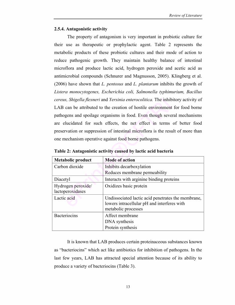
2.5.4. Antagonistic activity
The property of antagonism is very important in probiotic culture for
their use as therapeutic or prophylactic agent. Table 2 represents the
metabolic products of these probiotic cultures and their mode of action to
reduce pathogenic growth. They maintain healthy balance of intestinal
microflora and produce lactic acid, hydrogen peroxide and acetic acid as
antimicrobial compounds (Schnurer and Magnusson, 2005). Klingberg et al.
(2006) have shown that L. pentosus and L. plantarum inhibits the growth of
Listera monocytogenes, Escherichia coli, Salmonella typhimurium, Bacillus
cereus, Shigella flexneri and Yersinia enterocolitica. The inhibitory activity of
LAB can be attributed to the creation of hostile environment for food borne
pathogens and spoilage organisms in food. Even though several mechanisms
are elucidated for such effects, the net effect in terms of better food
preservation or suppression of intestinal microflora is the result of more than
one mechanism operative against food borne pathogens.
Table 2: Antagonistic activity caused by lactic acid bacteria
Metabolic product Mode of action Carbon dioxide Inhibits decarboxylation
Reduces membrane permeability Diacetyl Interacts with arginine binding proteins Hydrogen peroxide/ lactoperoxidases
Oxidizes basic protein
Lactic acid Undissociated lactic acid penetrates the membrane, lowers intracellular pH and interferes with metabolic processes
Bacteriocins Affect membrane DNA synthesis Protein synthesis
It is known that LAB produces certain proteinaceous substances known
as “bacteriocins” which act like antibiotics for inhibition of pathogens. In the
last few years, LAB has attracted special attention because of its ability to
produce a variety of bacteriocins (Table 3).
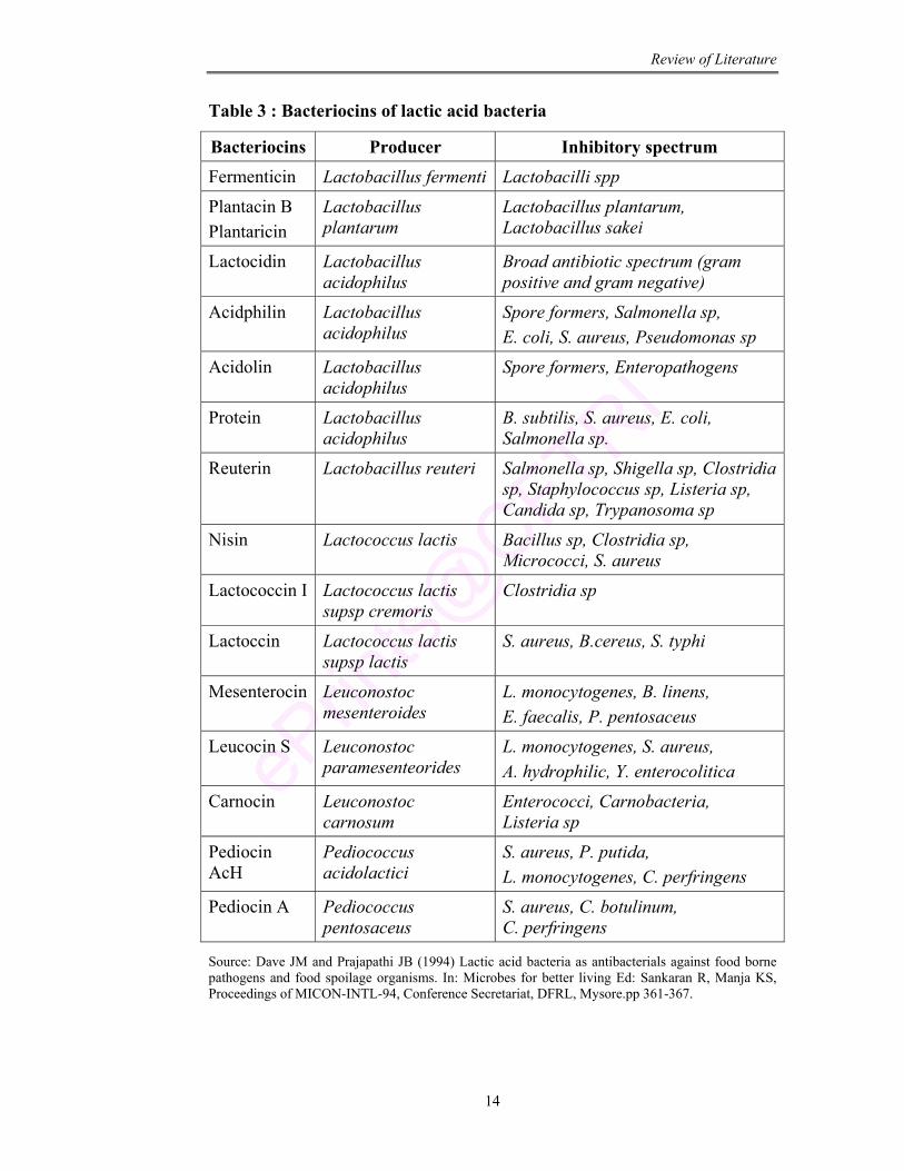
Review of Literature
14
Table 3 : Bacteriocins of lactic acid bacteria
Bacteriocins Producer Inhibitory spectrum Fermenticin Lactobacillus fermenti Lactobacilli spp
Plantacin B Plantaricin
Lactobacillus plantarum
Lactobacillus plantarum, Lactobacillus sakei
Lactocidin Lactobacillus acidophilus
Broad antibiotic spectrum (gram positive and gram negative)
Acidphilin Lactobacillus acidophilus
Spore formers, Salmonella sp, E. coli, S. aureus, Pseudomonas sp
Acidolin Lactobacillus acidophilus
Spore formers, Enteropathogens
Protein Lactobacillus acidophilus
B. subtilis, S. aureus, E. coli, Salmonella sp.
Reuterin Lactobacillus reuteri Salmonella sp, Shigella sp, Clostridia sp, Staphylococcus sp, Listeria sp, Candida sp, Trypanosoma sp
Nisin Lactococcus lactis Bacillus sp, Clostridia sp, Micrococci, S. aureus
Lactococcin I Lactococcus lactis supsp cremoris
Clostridia sp
Lactoccin Lactococcus lactis supsp lactis
S. aureus, B.cereus, S. typhi
Mesenterocin Leuconostoc mesenteroides
L. monocytogenes, B. linens, E. faecalis, P. pentosaceus
Leucocin S Leuconostoc paramesenteorides
L. monocytogenes, S. aureus, A. hydrophilic, Y. enterocolitica
Carnocin Leuconostoc carnosum
Enterococci, Carnobacteria, Listeria sp
Pediocin AcH
Pediococcus acidolactici
S. aureus, P. putida, L. monocytogenes, C. perfringens
Pediocin A Pediococcus pentosaceus
S. aureus, C. botulinum, C. perfringens
Source: Dave JM and Prajapathi JB (1994) Lactic acid bacteria as antibacterials against food borne pathogens and food spoilage organisms. In: Microbes for better living Ed: Sankaran R, Manja KS, Proceedings of MICON-INTL-94, Conference Secretariat, DFRL, Mysore.pp 361-367.
Review of Literature
15
2.5.6. Cholesterol lowering capacity
Hypercholesterol has been linked with increased risk for coronary heart
diseases. The use of probiotics to reduce this risk seems very attractive,
especially if consumed as a part of a normal daily diet. Several human studies
have suggested that dairy fermented products with certain strains of probiotic
bacteria are able to lower the cholesterol level (Larsen et al., 2000; Sindhu
and Khetarpaul, 2003; Parvez et al., 2006). De Smet et al. (1998) conducted
an experiment in hypercholesterolemic pigs and showed a significant
reduction of serum cholesterol level after administration of Lactobacillus
reuteri. A number of mechanisms have been proposed for this action of
probiotic bacteria. These include physiological action of short chain fatty acid
fermentation, bile acid deconjugation and cholesterol assimilation by bacteria
(Klaver and Vander Meer, 1993; Mercenier et al., 2002). Probiotics are
known to ferment carbohydrates and produce short chain fatty acids in the
intestine which inhibits cholesterol synthesis in liver and redistributes
cholesterol from plasma to the liver. Individual strains can deconjugate bile
salts and hamper absorption of cholesterol from the gut (Gilliland et al., 1985;
De Boever et al., 2000; Doncheva et al., 2002; Ahn et al., 2003). Pereira and
Gibson (2002) have proved that probiotics along with prebiotics have a
potential to decrease serum lipid levels.
2.5.7. Antioxidative activity
The importance of reactive oxygen species in biology and medicine is
evident because of their strong relationship with phenomenon such as aging
and disease process (Cao et al., 1995). It is well known that free radicals and
reactive oxygen (ROS) are continuously been produced in living organisms.
As a result, defense mechanisms have evolved to deactivate these free radicals
and repair the damage caused by their reactivity. Free radical scavenging
properties of starter cultures are very useful in many food manufacturing
industries. The probiotic cultures provide beneficial effect to the consumer by
releasing antioxidants during the growth in the intestinal tract (Virtanen et al.,
2007).
Review of Literature
16
2.5.8. Enzymatic activity
LAB cultures are known to produce large number of enzymes for their
survival and to impart beneficial effect. For rapid microbial growth and as
precursors for aroma development in baked foods they are equipped with
proteolytic system. This system is composed of cell envelop associated
proteinase, peptidase transferase and intracellular peptidase for hydrolysis of
protein and amino acids (Di Cagno et al., 2004). Aminopeptidase in
particular, could reduce the amount of proline rich gliadin peptides in baked
foods, which are known to elicit immune response in celiac disease (Gallo
et al., 2005).
β-glucosidases, a major group of glycosyl hydrolase enzymes catalyze
the selective cleavage of β-1,4-glycosidic linkages that play an important role
in several biological pathways (Yan et al., 1998). Phytase, a phytate
degrading enzyme is present in LAB that catalyzes the stepwise hydrolysis of
phytic acid to myo-inositol via penta to mono phosphates (Lopez et al., 2000;
Reale et al., 2004). Phytic acids are regarded as antinutritional compound
since it chelates proteins, aminoacids and divalent cations such as Ca2+, Fe2+,
Mg2+ and Zn2+ preventing their absorption by the intestinal mucosa
(De Angelis et al., 2003). So phytase is very important enzyme in LAB when
used in any functional food prepared with cereal grains. Urease, another
important enzyme is known to protect microorganisms from harmful effect of
acidic condition by increasing the environmental pH through conversion of
urea into ammonia and CO2 (Vande Guchte et al., 2002).
2.5.9. Nutraceutical property
LAB are the ideal factories for the production of nutraceutical
compounds (Hugenholtz and Smid, 2002). Fermentation of food with LAB
has been shown to increase niacin, riboflavin and folic acid content of yogurt,
bifidus milk and kefir. Vitamin B12 and Vitamin B6 have found to increase in
Review of Literature
17
cottage cheese and cheddar cheese (Shahani and Chandan, 1979; Alm, 1982;
Sauer et al., 1998). In addition to nutrient synthesis LAB improves
digestibility of some available food supplements such as protein and fat
(Friend and Shahani, 1984). Short chain fatty acids such as lactic, propionic
and butyric acid produced by these cultures help in maintaining an
appropriate pH and protect against pathological changes in the colonic
mucosa. Burgess et al. (2004) have overexpressed a metabolic gene in
Lactobacillus lactis for production of vitamins. Lactobacillus plantarum has
been designed for excess production of folate that is known to maintain
normal plasma homocysteine level in cognitive function and to provide
protection against cancer (Sybesma et al., 2003; Jagerstad et al., 2004).
2.5.10. Probiotic as a source of fats and Fatty acids
Role of fats and fatty acids are well-known in human nutrition. Fats
contained in foodstuffs provide substantial amount of energy for humans.
Fatty acids are basic building blocks of the lipids in dairy products. In their
free form they make significant contributions to the flavor of fermented foods.
They also act as precursors for the formation of other aroma components,
such as esters, aldehydes, alcohols, and ketones (Kinsella and Hwang, 1976;
Scott, 1981). Analysis of the fatty acid profile which gives dairy products
their particular organoleptic properties can be regarded as an index that can be
very helpful in characterizing the functional properties of the foods. These
fatty acids are also known to be therapeutically very important. Studies with
animal models have demonstrated that conjugated linolenic acid consumption
inhibits the initiation of carcinogenesis and tumorigenesis (Ip et al., 1991;
Devery et al., 2001; Pariza et al., 2001) reduces body fat content and increases
muscle mass (Chin et al., 1992; Akahoshi et al., 2002), decreases
atherosclerosis (Lee et al., 1994; Nicolosi et al., 1997), improves
hyperinsulinemia (Houseknecht et al., 1998), enhances the immune system
Review of Literature
18
(Cook et al., 1993; Miller et al., 1994) and alters the low-density
lipoprotein/high-density lipoprotein cholesterol ratio (Lee et al., 1994).
Inoculation of L. acidophilus into skim milk medium was effective in
promoting conjugated linolenic acid (CLA) formation (Lin, 2000). Dahi, an
Indian equivalent of yoghurt is found to contain more CLA than the raw
material (Aneja and Murthi, 1990).
Recent studies have shown that components of fermented dairy foods
such as LAB cultures, dairy proteins, and dairy fats (including sphingomyelin,
ether lipids, fatty acids such as oleic acid, palmitic acid, palmitoleic and
conjugated linoleic acids) have antimutagenic and anticarcinogenic properties
(Lidbeck et al., 1992; McIntosh et al., 1995).
2.5.11. Probiotic: Medical importance
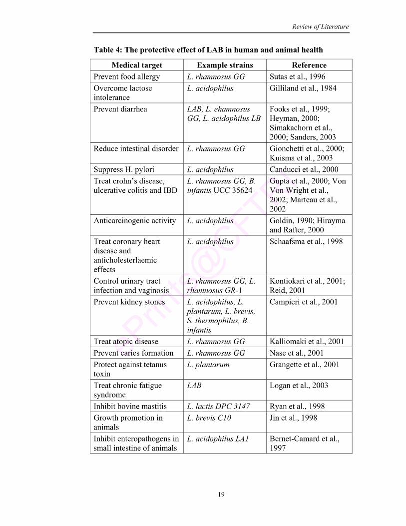
Table 4 represents the LAB used in treatment of large number of
diseases. Fuller (1999) has reviewed the uses of these products in animal
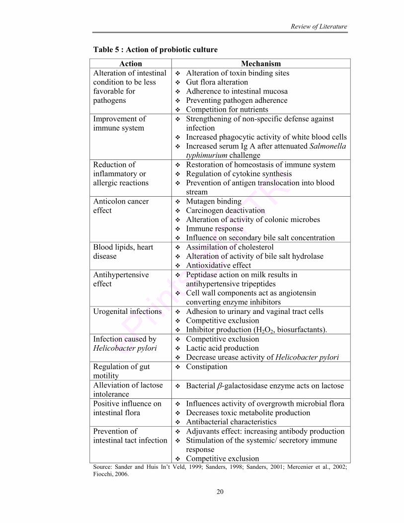
husbandry. Mechanism of action of these LAB are summarized in Table 5.
These LAB cultures constitute an integral part of healthy intestinal flora and
are involved in host metabolism (Denev, 2006). LAB along with other gut
microbiota ferments various substrates like lactose, biogenic amines and
allergic compounds into short chain fatty acids and other organic acids and
gases (Gibson and Fuller, 2000; Jay, 2000). LAB are known to synthesize
enzymes, vitamins, antioxidants and bacteriocins (Knorr, 1998). All these
properties contribute for the detoxification of foreign substances entering the
body (Salminen, 1990).
Review of Literature
19
Table 4: The protective effect of LAB in human and animal health
Medical target Example strains Reference Prevent food allergy L. rhamnosus GG Sutas et al., 1996 Overcome lactose intolerance
L. acidophilus Gilliland et al., 1984
Prevent diarrhea LAB, L. ehamnosus GG, L. acidophilus LB
Fooks et al., 1999; Heyman, 2000; Simakachorn et al., 2000; Sanders, 2003
Reduce intestinal disorder L. rhamnosus GG Gionchetti et al., 2000; Kuisma et al., 2003
Suppress H. pylori L. acidophilus Canducci et al., 2000 Treat crohn’s disease, ulcerative colitis and IBD
L. rhamnosus GG, B. infantis UCC 35624
Gupta et al., 2000; Von Von Wright et al., 2002; Marteau et al., 2002
Anticarcinogenic activity L. acidophilus Goldin, 1990; Hirayma and Rafter, 2000
Treat coronary heart disease and anticholesterlaemic effects
L. acidophilus Schaafsma et al., 1998
Control urinary tract infection and vaginosis
L. rhamnosus GG, L. rhamnosus GR-1
Kontiokari et al., 2001; Reid, 2001
Prevent kidney stones L. acidophilus, L. plantarum, L. brevis, S. thermophilus, B. infantis
Campieri et al., 2001
Treat atopic disease L. rhamnosus GG Kalliomaki et al., 2001 Prevent caries formation L. rhamnosus GG Nase et al., 2001 Protect against tetanus toxin
L. plantarum Grangette et al., 2001
Treat chronic fatigue syndrome
LAB Logan et al., 2003
Inhibit bovine mastitis L. lactis DPC 3147 Ryan et al., 1998 Growth promotion in animals
L. brevis C10 Jin et al., 1998
Inhibit enteropathogens in small intestine of animals
L. acidophilus LA1 Bernet-Camard et al., 1997
Review of Literature
20
Table 5 : Action of probiotic culture
Action Mechanism Alteration of intestinal condition to be less favorable for pathogens
Alteration of toxin binding sites Gut flora alteration Adherence to intestinal mucosa Preventing pathogen adherence Competition for nutrients
Improvement of immune system
Strengthening of non-specific defense against infection
Increased phagocytic activity of white blood cells Increased serum Ig A after attenuated Salmonella
typhimurium challenge Reduction of inflammatory or allergic reactions
Restoration of homeostasis of immune system Regulation of cytokine synthesis Prevention of antigen translocation into blood
stream Anticolon cancer effect
Mutagen binding Carcinogen deactivation Alteration of activity of colonic microbes Immune response Influence on secondary bile salt concentration
Blood lipids, heart disease
Assimilation of cholesterol Alteration of activity of bile salt hydrolase Antioxidative effect
Antihypertensive effect
Peptidase action on milk results in antihypertensive tripeptides
Cell wall components act as angiotensin converting enzyme inhibitors
Urogenital infections Adhesion to urinary and vaginal tract cells Competitive exclusion Inhibitor production (H2O2, biosurfactants).
Infection caused by Helicobacter pylori
Competitive exclusion Lactic acid production Decrease urease activity of Helicobacter pylori
Regulation of gut motility
Constipation
Alleviation of lactose intolerance
Bacterial β-galactosidase enzyme acts on lactose
Positive influence on intestinal flora
Influences activity of overgrowth microbial flora Decreases toxic metabolite production Antibacterial characteristics
Prevention of intestinal tact infection
Adjuvants effect: increasing antibody production Stimulation of the systemic/ secretory immune
response Competitive exclusion
Source: Sander and Huis In’t Veld, 1999; Sanders, 1998; Sanders, 2001; Mercenier et al., 2002; Fiocchi, 2006.
Review of Literature
21
2.5.11.1. Protection against gastrointestinal tract problem
Gastroenteritis is caused by viral, bacterial or parasitic infections. They
also cause acute diarrhea, Clostridium difficili infections, ulcerative colitis,
crohn’s disease, traveller’s diarrhea, Helicobacter infection, pouchitis and
other inflammatory bowel diseases. A large number of reviews (Saavedra,
1995; Elmer, 1996; Michetti et al., 1999; Felley et al., 2001) focus on their
preventive action by probiotic LAB. A large number of reports also
determined that probiotic therapy shortened the length of acute diarrheal
illness (Pedone et al., 1999; Pedone et al., 2000; Huang et al., 2002; Weizman
et al., 2005). LAB have been found to control intestinal disorders partially due
to antibodies like Ig G, IgA and IgM for enhancing immune responses
(Kimura et al., 1997; Grangette et al., 2001). Some strains of LAB can
translocate across the intestinal mucosa influencing systemic immune system
(Cross, 2002). Characteristic adherence of the culture to mucosal surface
contributes to their efficacy of being probiotic, since adherent strains confer
competitive advantage that is important for the maintenance of balanced
gastrointestinal microflora. Antibiotic associated colitis is known to be
reduced by administration of the probiotic Lactobacillus GG (Vanderhoof et
al., 1999; D’Souza et al., 2002).
Inflammatory bowel disease (IBD): Inflammatory bowel disease (IBD)
mainly like Crohn’s disease and Ulcerative colitis are chronic inflammation of
terminal ileum. It is caused due to three pathogenic factors i.e., genetic
susceptibility, immune dysregulation and environmental triggering events
(Shanahan, 2004). A few experimental studies support probiotic LAB for the
treatment of IBD. Administration of probiotic culture is known to restore
epithelial barrier function with reduction of TNF α and INF-X secretion
(Madsen et al 2001), inhibition of proteosome and activation of NF-KB
(Petrof et al., 2004). Probiotic DNA is known to induce anti-inflammatory
effect by signaling via TLR 9 (Rachmilewitz et al., 2004). They also activate
anti-apoptotic A Kt/protein kinase B and inhibit activation of pro-apoptotic
Review of Literature
22
P38/ mitogen activation protein kinase (Yan and Polk, 2002) which resulted
in inhibition of cytokine induced apoptosis.
Irritable bowel syndrome (IBS): Irritable bowel syndrome (IBS) is a common
functional disorder of lower intestine affecting both adults and children. It is
associated with abdominal distension, diarrhea, constipation, bloating and
urgency to defecate. Earlier reports have shown that this syndrome can be
reduced with administration of probiotic culture (Brigidi et al., 2001; Sen et
al., 2002; Kim et al., 2003).
Traveller’s diarrhea: A number of specific strains, including Lactobacillus
GG, Lactobacillus reuteri, Saccharomyces boulardii, Bifidobacteria sp and
others have been shown to have significant benefit for diarrhea. In a study of
820 travellers, the diarrhea rate was 40% for control compared with 24% in
Lactobacillus GG treated ones (Oksanen et al., 1990). In another study in a
hospital based travel and immunization centre, an overall incidence averaged
7.4% for placebo and 3.9% for probiotic (Lactobacillus GG) group (Hilton,
1997).
2.5.11.2. Management of atopic disease and allergy
Atopic disease such as atopic eczema, allergic rhinitis and bronchial
asthma are on the increase in all industrialized countries because of integrity
of defense systems or disruption of immune response. Number of studies
shows a significant reduction in respiratory infection of children and
management of atopic disease by the administration of probiotic cultures
(Hatakk et al., 2001; Murch, 2001; Kirjavainen et al., 2003). The protective
action of probiotics to allergic inflammation and food allergies may be due to
enhancement of gut specific IgA response, restoring normal intestinal
permeability, improvement of intestinal immunological barrier function,
alleviation of inflammatory cytokines characteristic of local and systemic
allergic inflammation (Isolauri et al., 1992; Majamma and Isolauri, 1997;
Isolauri et al., 2000; Isolauri, 2004).
Review of Literature
23
2.6. Probiotics in functional foods
Functional foods are foods that claim to promote human health. The
term functional food was first proposed in Japan two decades ago and legally
approved them in terms of foods for specified health use (FOSHU). A large
number of scientific evidences indicate that ingestion of LAB cultures exert
health benefits not only in the gastrointestinal tract but also in the respiratory
and urogenital tracts (Cadieux et al., 2008; Reid, 2008). Lactic acid bacteria
(LAB) occupies a central role in fermentation processes and has long and safe
history of application in the production of fermented foods and beverages
(Caplice and Fitzgerald, 1999). They cause rapid acidification of the raw
material through the production of organic acids, mainly lactic acid. LAB are
able to produce antimicrobial substances, sugar polymers, sweeteners,
aromatic compounds, useful enzymes and nutraceuticals which represent the
health promoting properties of LAB. These properties help in replacing
chemical additives by natural compounds and at the same time providing the
consumer with new, attractive food products with health benefits. Further they
also enhance shelf life, improve texture and contribute to the pleasant sensory
profile of the end product.
2.7. Probiotic: Dosage and Administration
As there are a variety of probiotic organisms with various delivery
systems, there is no uniform dose recommendation. Studies to date have used
oral doses of 1 to 10 billion colony forming units (cfu) per dose, with
administration frequency ranging from twice daily to intermittent weekly
schedules. Dose may also be dependent on the purpose of use that is treatment
versus prophylaxis. With respect to site of action and strain used, it is
suggested that each species function only at a particular site. Because of this
industries are facing difficulties in establishing good measures of probiotic
efficacy (Rolfe, 2000). Studies on lactose intolerance, diarrhea and colon
cancer show that a daily dose of LAB is needed for any measurable effect
(Rembacken et al., 1999).
Review of Literature
24
The food and pharmaceutical industries have to be careful in assessing
the efficacy of new species/ strain before incorporating them into the product.
Many probiotic formulations are administered orally, either in capsule or
powder form. These preparations result in a variable viability and are
inadequate for therapeutic benefits. So the efficacy of the product is very
essential and the optimal dose of probiotic for beneficial effect is to be
considered before using them.
2.8. Probiotic: Market potential
Marketing functional foods was initiated in Japan in late 1980s. This
concept is becoming increasingly popular with consumers heightened
awareness of health linked with nutrition and diet. Today the food
manufacturers are more enthusiastic in developing products with added
ingredients to increase health. In this regard probiotic products are gaining
popularity and acceptance throughout the world. These products are common
in Japan and Europe (Lee et al., 1999; Sanders and Huis In’t Veld, 1999;
Stanton et al., 2001). The passage of Dietary supplement Health and
Education Act (1994) has increased the scale of these products as dietary
supplement. In 1997, these products accounted for 65% of European
functional food market valued for US $ 889 million (Hilliam, 1998). Probiotic
yogurt market in UK, France, Germany, Spain, Belgium, Netherlands,
Denmark, Finland and Sweden is more than 250 million Kg (Hilliam, 1998).
“Bikkle” a probiotic drink with Bifidobacteria sp was launched in 1993
(Osaka, Japan) which achieved a sale of 11 billion Yen in its first year
(Young, 1996).
Interest in acceptance of functional foods is gaining importance in
areas of developing new food processing, retailing and distribution
technologies. Benefits of these functional foods are opening new
opportunities to value added products for increasing profit. If a product can
offer any measurable benefit to consumers in long term, there is a greater
chance of re-buying the product (Childs, 1997). Hence, it is necessary that the
Review of Literature
25
product should be of high quality that fits to the consumer’s life style. As a
reference, yogurt industry reported a 10.5% increase in sales from 1999-2000
and increase of 7.9% in 2001.
Probiotics products are becoming popular because of the desire by
consumers to use natural methods for health maintenance rather than long
term chemotherapeutic agents. The present challenge for nutrition and health
professionals would be to guide consumers for their specific needs. They also
should be aware of individual taste and food preference or nutrients of the
consumers (Hoolihan, 2003).
2.9. Probiotic safety
The use of LAB in food products has a long safety record. The safety
of probiotic bacteria has been reviewed by several authors (Mogensen et al.,
2002; Saarela et al., 2002; Hammerman et al., 2006). The absence of
pathogenicity and infectivity is the prerequisite of probiotic safety. There are
a few published case reports of rare infections involving LAB, like liver
abscess (Rautio et al., 1999), endocarditis (Husni et al., 1997), dental caries
(Mackay et al., 1998), induction of inflammatory cytokines (Okitsu-Negishi et
al., 1996) and septicemia (Barros et al., 2001). Therefore, safety assessment is
an essential phase in the use of any culture.
Safety and efficacy assessment requires proper identification of
probiotic culture to avoid any inclusion of pathogenic microorganism in the
products. Pathogenicity, infectivity and virulence factors comprising of
toxicity, metabolic activity and intrinsic properties of the microbes are the
major factors that have to be addressed for the evaluation of safety of
probiotic microorganisms (Salminen and Marteau, 1997; Ishibashi and
Yamazaki, 2001). The culture should not bear any plasmid coding for
antibiotic resistance (Franz et al., 2003; Cocconcelli et al., 2003).
Review of Literature
26
2.10. Preservation of probiotic culture
Preservation of microorganism by desiccation has been the preferred
method for long term storage. There are extensive culture collections that
depend on these drying methods to preserve a huge diversity of cells. There
are many applications in industries like food and pharmaceutical industries
which utilize preserved aliquots of microorganism. These strains are subjected
to different drying techniques to make cultures stable and transportable at
ambient temperature. The different techniques employed are as follows:
Freeze drying: It is one of such technique used to preserve organisms for
decades and is the preferred method for culture collections all over the world
including American Type Culture Collection (ATCC) and National Collection
of Type Culture (NCTC). Freeze dried materials allows easy and inexpensive
shipping and handling. It is generally recommended to freeze dry
concentrated cultures of >107 cells to ensure that there are sufficient cells
remaining after the freeze drying process.
Foam formation: It is another technique which uses protective sugar matrics
to transform biological suspensions into mechanically stable dry foams. The
foam is then subjected to further drying at elevated temperatures to increase
their stability (Bronshtein, 2004).
Spray drying: It is a drying technique that produces granulated powders from
a slurry solution by atomizing the wet product at high velocity within the
chamber (Corcoran et al., 2004).
Fluidized bed drying method: It is a method which uses an upward flow of
heated air and mechanical shaking to create a fluidized effect in a solid
product (Larena et al., 2003).
The process of dehydration can be detrimental to the viability of
bacterial cell (Uzunova-Doneva and Donev, 2000). Hence protective agents
are added prior to freeze drying to enhance the cell stability. Sugars like
trehalose and sucrose have been reported to enhance the tolerance of
numerous microorganisms by stabilizing membrane and proteins (Crowe et
al., 1998; Gomez-Zavaglia et al., 2003; Streeter, 2003).
Review of Literature
27
3. Leuconostoc: Characteristic and functional prospects
Leuconostoc spp are gram positive, catalase negative LAB with G+C
DNA content less than 50%. They are involved in fermentation of food stuffs,
production of gas in cheese, production of flavor compounds in dairy products
and formation of dextran.
Leuconostoc spp are present in many natural ecological niches like
green vegetation and roots from where they can easily propagate into various
niches such as vegetables, silage (Ennahar et al., 2003) and fermented food
products. They have been isolated from feces, vaginal samples and breast
milk (Auge et al., 1987; Dal Bello et al., 2003). They have also been isolated
from microflora of cattle (Brashears et al., 2003), fish (Ringo and Gatisoupe,
1998), insects (Ohkuma and Kudo, 1998; Reeson et al., 2003) and other
environments such as treated bauxite residues (Hamdy and Williams, 2001). It
is also one of the microbial components of kefir grain contributing to the
production of ethanol and acetate (Robinson et al., 2002). They are commonly
found in sugar processing liquors and fermented foods. They are desirable in
foods because of their involvement in flavor development and preservation.
3.1. Leuconostoc: Taxonomy
Leuconostoc spp are non-motile, non-spore forming almost spherical
cells, sometimes rather lenticular that resemble very short bacilli with
rounded ends. Their size is approximately 0.5-0.7 × 0.7-1.2 µm. The cells are
arranged in pairs or chains. They are facultative anaerobic bacteria with
optimum growth at 20-30°C, but some species even grow at 10°C. The initial
pH of the growth medium drops from 6.5 to 4.5 ± 0.1 during growth due to
acid production. According to Bergey’s manual one of the distinctive traits of
Leuconostoc spp is their inability to hydrolyze Arginine. They are
heterofermentative cultures producing CO2 from glucose metabolism beside
D-lactate and ethanol/ acetate (Table 6).
Nucleic acid hybridization, polymerase chain reaction, amplification
and nucleic acid sequencing may be applied for Leuconostoc sp identification.
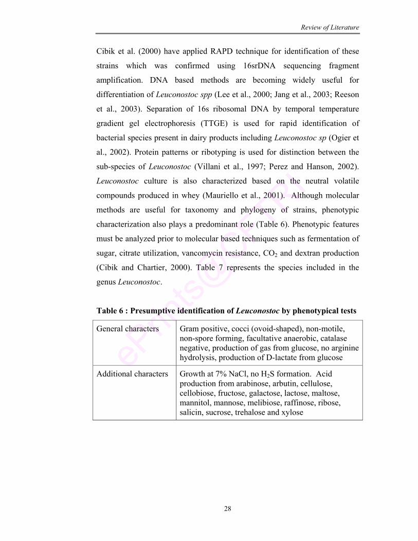
Review of Literature
28
Cibik et al. (2000) have applied RAPD technique for identification of these
strains which was confirmed using 16srDNA sequencing fragment
amplification. DNA based methods are becoming widely useful for
differentiation of Leuconostoc spp (Lee et al., 2000; Jang et al., 2003; Reeson
et al., 2003). Separation of 16s ribosomal DNA by temporal temperature
gradient gel electrophoresis (TTGE) is used for rapid identification of
bacterial species present in dairy products including Leuconostoc sp (Ogier et
al., 2002). Protein patterns or ribotyping is used for distinction between the
sub-species of Leuconostoc (Villani et al., 1997; Perez and Hanson, 2002).
Leuconostoc culture is also characterized based on the neutral volatile
compounds produced in whey (Mauriello et al., 2001). Although molecular
methods are useful for taxonomy and phylogeny of strains, phenotypic
characterization also plays a predominant role (Table 6). Phenotypic features
must be analyzed prior to molecular based techniques such as fermentation of
sugar, citrate utilization, vancomycin resistance, CO2 and dextran production
(Cibik and Chartier, 2000). Table 7 represents the species included in the
genus Leuconostoc.
Table 6 : Presumptive identification of Leuconostoc by phenotypical tests
General characters Gram positive, cocci (ovoid-shaped), non-motile, non-spore forming, facultative anaerobic, catalase negative, production of gas from glucose, no arginine hydrolysis, production of D-lactate from glucose
Additional characters Growth at 7% NaCl, no H2S formation. Acid production from arabinose, arbutin, cellulose, cellobiose, fructose, galactose, lactose, maltose, mannitol, mannose, melibiose, raffinose, ribose, salicin, sucrose, trehalose and xylose
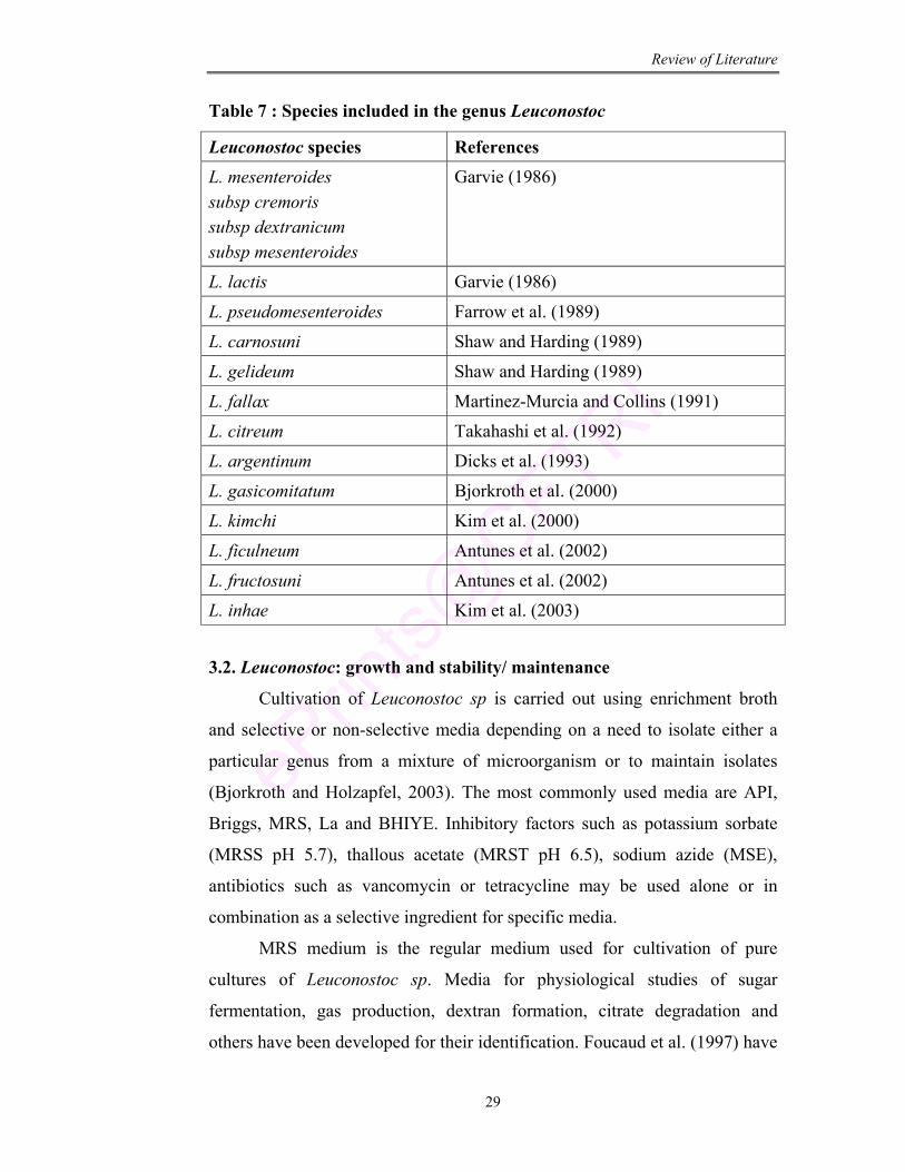
Review of Literature
29
Table 7 : Species included in the genus Leuconostoc
Leuconostoc species References
L. mesenteroides subsp cremoris subsp dextranicum subsp mesenteroides
Garvie (1986)
L. lactis Garvie (1986)
L. pseudomesenteroides Farrow et al. (1989)
L. carnosuni Shaw and Harding (1989)
L. gelideum Shaw and Harding (1989)
L. fallax Martinez-Murcia and Collins (1991)
L. citreum Takahashi et al. (1992)
L. argentinum Dicks et al. (1993)
L. gasicomitatum Bjorkroth et al. (2000)
L. kimchi Kim et al. (2000)
L. ficulneum Antunes et al. (2002)
L. fructosuni Antunes et al. (2002)
L. inhae Kim et al. (2003)
3.2. Leuconostoc: growth and stability/ maintenance
Cultivation of Leuconostoc sp is carried out using enrichment broth
and selective or non-selective media depending on a need to isolate either a
particular genus from a mixture of microorganism or to maintain isolates
(Bjorkroth and Holzapfel, 2003). The most commonly used media are API,
Briggs, MRS, La and BHIYE. Inhibitory factors such as potassium sorbate
(MRSS pH 5.7), thallous acetate (MRST pH 6.5), sodium azide (MSE),
antibiotics such as vancomycin or tetracycline may be used alone or in
combination as a selective ingredient for specific media.
MRS medium is the regular medium used for cultivation of pure
cultures of Leuconostoc sp. Media for physiological studies of sugar
fermentation, gas production, dextran formation, citrate degradation and
others have been developed for their identification. Foucaud et al. (1997) have
Review of Literature
30
developed a chemically defined medium that fulfills the nutritional
requirement for rapid growth.
Leuconostoc sp grow at 30°C like other mesophilic Lactococcus
starters but are favored by low temperature. Most strains grow well at 10°C
(Hamasaki et al., 2003). Some are also thermoduric as they resist
pasteurization (Martley and Crow, 1993). They are able to survive for a long
time in unfavorable environment like surface of wooden gerl, glazed
sandstone, iron or plastic moulds used in traditional cheese making (Devoyod
and Poullain, 1988). They form slime or glycocalix in presence of saccharose
and trace minerals resulting in a biofilm that protects cells from detrimental
effect (Kim et al., 2000). Resistance to heat in L. mesenteroides is due to over
expression of stress proteins that are homologous to chaperone proteins
(Salotra et al., 1995; Derre et al., 1999).
Leuconostoc spp may be maintained for short duration in MRS stabs at
4°C (1-2 weeks). Cultures grown in litmus milk (supplemented with 5% yeast
extract and 5% glucose) or in MRS broth (containing 1% lactose and 10%
glycerol) can be stored for 6 months at -20°C. Lyophilization or freeze drying,
the culture shows viability even on long storage for several months
(Bellengier et al., 1997; Bjorkroth and Holzapfel, 2003).
3.3. Leuconostoc: Metabolism
Leuconostoc spp like other LAB do not contain a tricarboxylic acid
cycle or a cytochrome system and so cannot derive energy from oxidative
phosphorylation. Instead they obtain energy through substrate level
phosphorylation during fermentation of sugars to lactic acid, CO2 and
ethanol/acetate (Cogan and Jordon, 1991). They take up carbohydrates by
permeases enzymes which enter into phosphoketolase pathway leading to CO2
and pyruvate. Pyruvate further gets converted into D-lactate in presence of D-
lactate dehydrogenase enzyme. In presence of acetaldehyde in the medium,
acetate kinase directs the flux for the formation of acetate and ATP.
Additionally phosphotransacetylase produces acetyl CoA that is used for
Review of Literature
31
biosynthesis of NADH with production of ethanol by alcohol dehydrogenase
(Bourel et al., 2001).
Leuconostoc spp ferment fructose that enters pentose-phosphate
pathway to be converted into mannitol which causes oxidation of NADH to
NAD+ in presence of mannitol dehydrogenase enzyme. Pentoses are
converted to xylulose-5-phosphate that are further catabolized into
glyceradehyde-3-phosphate and acetyl-phosphate (Cogan and Jordan, 1994).
Galactose and mannose are used via the Leloir pathway. Lactose is cleaved to
galactose and glucose by a β-galactosidase enzyme, which are further used
via Leloir and pentose-phosphate pathway.
Organic acids like citrate and malate are metabolized by Leuconostoc
spp that is important in aroma formation and gas production in fermented
dairy products (McSweeney and Sausa, 2000; Konings, 2002). The ability of
Leuconostoc spp to utilize amino acids independently or as mixture varied
with strains and species and it shows the potential of the culture for flavor
generation (Crowe et al., 1998; Tavaria et al., 2002; Liu et al., 2003).
3.4. Leuconostoc: Application
Leuconostoc spp plays a very important role in the technology of dairy
products, in particular through production of gas and aroma compounds
(Table 8). It is also applied for inhibition of undesirable bacteria in functional
foods (Vedamuthu, 1994). They are involved in early and late blowing of
some cheeses (Devoyod and Poullain., 1988; Narvhus et al., 1992).
Leuconostoc spp utilizes diacetyl, acetate and ethanol contributing to aroma
formation. They are also able to reduce acetaldehyde content under
refrigerated condition thus avoiding ‘green’ flavor formation in butter and
fermented milk. Some strains have shown to inhibit off-flavor production in
cheddar cheese (Martley and Crow., 1993). They are used in many functional
foods as probiotic culture with an intension of contributing health benefit to
the consumer. Their presence in dahi is known to reduce diarrhea in children
(Agarwal and Bhasin, 2002).
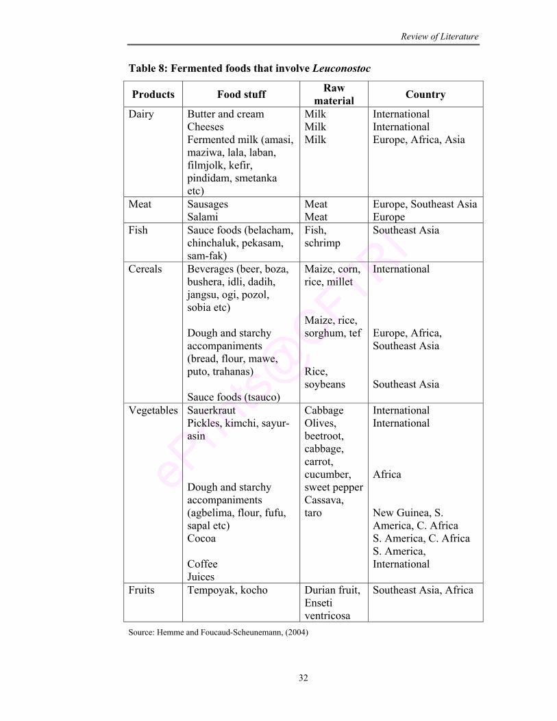
Review of Literature
32
Table 8: Fermented foods that involve Leuconostoc
Products Food stuff Raw material Country
Dairy Butter and cream Cheeses Fermented milk (amasi, maziwa, lala, laban, filmjolk, kefir, pindidam, smetanka etc)
Milk Milk Milk
International International Europe, Africa, Asia
Meat Sausages Salami
Meat Meat
Europe, Southeast Asia Europe
Fish Sauce foods (belacham, chinchaluk, pekasam, sam-fak)
Fish, schrimp
Southeast Asia
Cereals Beverages (beer, boza, bushera, idli, dadih, jangsu, ogi, pozol, sobia etc) Dough and starchy accompaniments (bread, flour, mawe, puto, trahanas) Sauce foods (tsauco)
Maize, corn, rice, millet Maize, rice, sorghum, tef Rice, soybeans
International Europe, Africa, Southeast Asia Southeast Asia
Vegetables Sauerkraut Pickles, kimchi, sayur-asin Dough and starchy accompaniments (agbelima, flour, fufu, sapal etc) Cocoa Coffee Juices
Cabbage Olives, beetroot, cabbage, carrot, cucumber, sweet pepperCassava, taro
International International Africa New Guinea, S. America, C. Africa S. America, C. Africa S. America, International
Fruits Tempoyak, kocho Durian fruit, Enseti ventricosa
Southeast Asia, Africa
Source: Hemme and Foucaud-Scheunemann, (2004)
Review of Literature
33
4. Lactose intolerance
Inability of human beings to digest lactose is referred to as “Lactose
intolerance” and has been attributed to insufficient amounts of lactase in the
small intestine to hydrolyze lactose consumed in the diet (Littman and
Hammond, 1965; Bayless and Rosenweig, 1966; Sarkar, 2006). Lactase
deficient persons show symptoms like nausea, cramps, gas and diarrhea
(Sieber et al., 1997). Temporary lactase deficiencies may also result from the
damage of the intestinal lining. The disease is alarming and is of great
concern to the society.
4.1. Significance of β-galactosidase/ lactase enzyme
Lactose, a disaccharide composed of glucose and galactose is the major
solid component of milk. It is hydrolyzed into its components under the
influence of enzyme lactase/β-galactosidase, a membrane bound enzyme
present in the brush border of the small intestinal epithelial cell prior to its
absorption by humans (Miller and Brand, 1980; Hourigan, 1984). In case the
lactose ingested exceeds the hydrolytic capacity of the available intestinal
lactase, the undigested portion of lactose is transported to the large intestine,
where it increases the osmolarity of the intestinal fluids. Undigested lactose
undergoes bacterial fermentation in the colon resulting in organic acid, CO2
and H2, which causes exclusion of large amount of water into intestine. This is
primarily responsible for various symptoms such as bloating, flatulence,
abdominal cramps, diarrhoea and loss of appetite (Hourigan, 1984; Hofi,
1990).
4.2. Lactose intolerance, maldigestion/ Malabsorption
Lactose maldigestion occurs due to either gastrointestinal disease or
physiological decline in the intestinal lactase activity. Semenza and Auricchio
(1995) registered reduction in lactase activity due to the digestion of lactase-
Review of Literature
34
phlonizin hydrolase molecule by pancreatic proteases at the brush border
membrane. Swaminathan (1998) has described three main reasons for
deficiency of lactase enzyme.
1) Congenital lactase efficiency: caused due to the absence of lactase
enzyme in intestinal mucosa.
2) Lactase deficiency in premature infants: occurs in premature infants
due to decrease in lactase activity. Initially, infants cannot utilize
lactose efficiently, however they are able to tolerate and digest milk
after one month due to increase in lactase activity.
3) Acquired lactase deficiency: adults and older children cannot tolerate
large amounts of milk due to their non-habitual consumption of milk
resulting in low lactase in the intestinal mucosa.
4.3. Occurrence of lactose intolerance
According to Swagerty et al. (2002) about 70-90% of adults in most
parts of the world are known to be lactose intolerant (Table 9). The
prevalence of lactose maldigestion is 15-20% in Austria, 55% in Balkans, 70-
90% in Africa, 80% in Central Asia, 90-100% in Eastern Asia, 30% in
Northern India, 70% in Southern India and 65-75% in South America (de
Vrese et al., 2001).
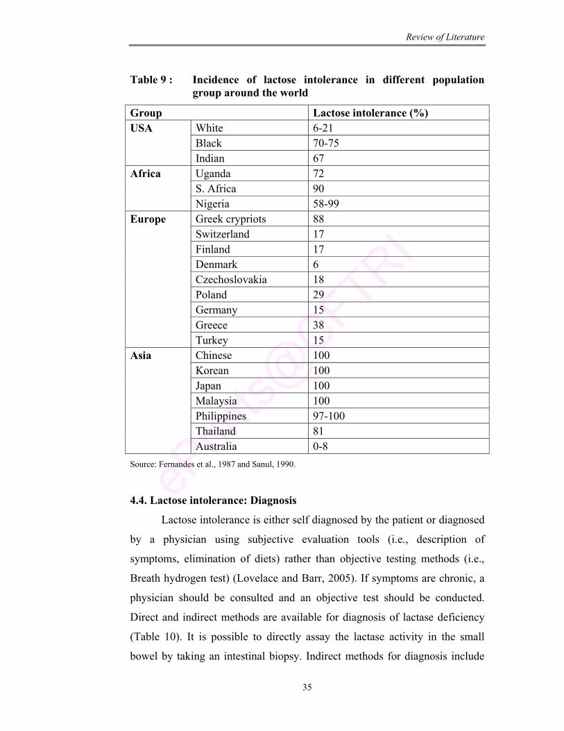
Review of Literature
35
Table 9 : Incidence of lactose intolerance in different population group around the world
Group Lactose intolerance (%) White 6-21 Black 70-75
USA
Indian 67 Uganda 72 S. Africa 90
Africa
Nigeria 58-99 Greek crypriots 88 Switzerland 17 Finland 17 Denmark 6 Czechoslovakia 18 Poland 29 Germany 15 Greece 38
Europe
Turkey 15 Chinese 100 Korean 100 Japan 100 Malaysia 100 Philippines 97-100 Thailand 81
Asia
Australia 0-8 Source: Fernandes et al., 1987 and Sanul, 1990.
4.4. Lactose intolerance: Diagnosis
Lactose intolerance is either self diagnosed by the patient or diagnosed
by a physician using subjective evaluation tools (i.e., description of
symptoms, elimination of diets) rather than objective testing methods (i.e.,
Breath hydrogen test) (Lovelace and Barr, 2005). If symptoms are chronic, a
physician should be consulted and an objective test should be conducted.
Direct and indirect methods are available for diagnosis of lactase deficiency
(Table 10). It is possible to directly assay the lactase activity in the small
bowel by taking an intestinal biopsy. Indirect methods for diagnosis include
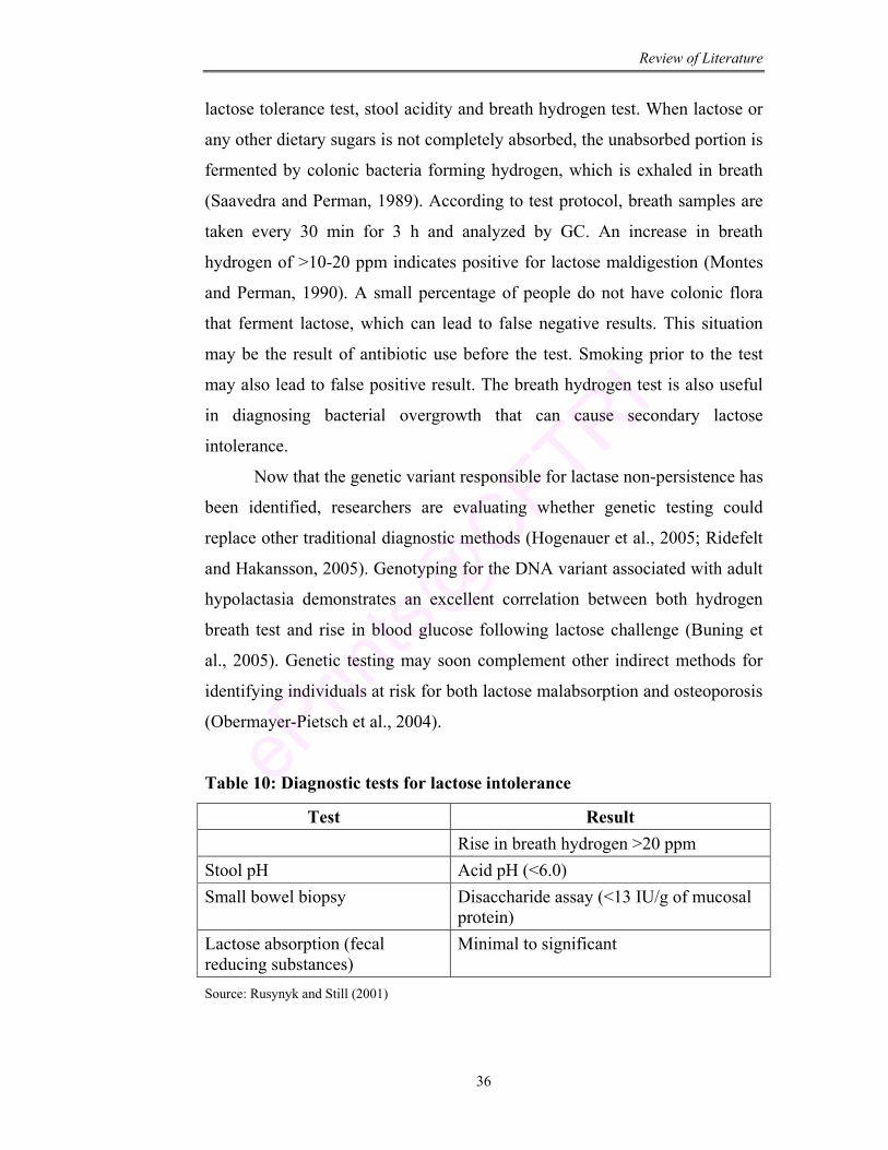
Review of Literature
36
lactose tolerance test, stool acidity and breath hydrogen test. When lactose or
any other dietary sugars is not completely absorbed, the unabsorbed portion is
fermented by colonic bacteria forming hydrogen, which is exhaled in breath
(Saavedra and Perman, 1989). According to test protocol, breath samples are
taken every 30 min for 3 h and analyzed by GC. An increase in breath
hydrogen of >10-20 ppm indicates positive for lactose maldigestion (Montes
and Perman, 1990). A small percentage of people do not have colonic flora
that ferment lactose, which can lead to false negative results. This situation
may be the result of antibiotic use before the test. Smoking prior to the test
may also lead to false positive result. The breath hydrogen test is also useful
in diagnosing bacterial overgrowth that can cause secondary lactose
intolerance.
Now that the genetic variant responsible for lactase non-persistence has
been identified, researchers are evaluating whether genetic testing could
replace other traditional diagnostic methods (Hogenauer et al., 2005; Ridefelt
and Hakansson, 2005). Genotyping for the DNA variant associated with adult
hypolactasia demonstrates an excellent correlation between both hydrogen
breath test and rise in blood glucose following lactose challenge (Buning et
al., 2005). Genetic testing may soon complement other indirect methods for
identifying individuals at risk for both lactose malabsorption and osteoporosis
(Obermayer-Pietsch et al., 2004).
Table 10: Diagnostic tests for lactose intolerance
Test Result Rise in breath hydrogen >20 ppm Stool pH Acid pH (<6.0) Small bowel biopsy Disaccharide assay (<13 IU/g of mucosal
protein) Lactose absorption (fecal reducing substances)
Minimal to significant
Source: Rusynyk and Still (2001)
Review of Literature
37
4.5. Approach towards treating lactose intolerance
The common therapeutic approach would be to exclude milk and dairy
products with lactose from the diet. However this strategy may have
nutritional disadvantages like reduced intake of calcium, phosphorus and
vitamins (Di Stefano et al., 2002). Solid lactase preparation in the form of
capsules and tablets are commercially available as an alternative enzyme
replacement therapy (Di palma et al., 1989). However comparative studies
have shown that these preparations are more expensive and significantly less
effective probably due to enzyme gastric inactivation (Suarez et al., 1995). So
the use of culture, which is resistant to gastrointestinal conditions, is the best
method for lactose hydrolysis.
LAB contains β-galactosidase as an intracellular enzyme which is
protected during passage through the harsh environment of stomach and is
able to reach the small intestine. Shah and Lankaputhra (1997) have shown
that rupturing of cell membrane of starter cultures of yogurt reduced viable
counts but increased the release of intracellular β-galactosidase.
Microorganisms residing in the large intestine were tolerant to lactose through
modifications of their metabolic activity (Hertzler and Savaiano, 1996). In
general, improved digestion results from lactase activity of bacteria or
stimulation of host’s mucosal lactase activity (Kolars et al., 1984; Gibson and
Fuller, 2000).
4.6. β-galactosidase enzyme/ Lactase
Lactase is a trival name of the enzyme β-D-galactosidase (Gekas and
Lopez-Leiva, 1985). β-galactosidase was among the first hydrolase enzyme to
be discovered. The molecule breaks down the sugar lactose which is
composed of two rings bound together by Oxygen Bridge. This bridge is
broken when enzyme β-galactosidase binds to lactose and water molecule
reacts with oxygen atom in the bridge (Hung and Lee, 2002). β-galactosidase
not only catalyses the hydrolysis of β-galactosidic linkage, but also catalyses
transglycosylation reactions. It is speculated that β-galactosidase is a spurious
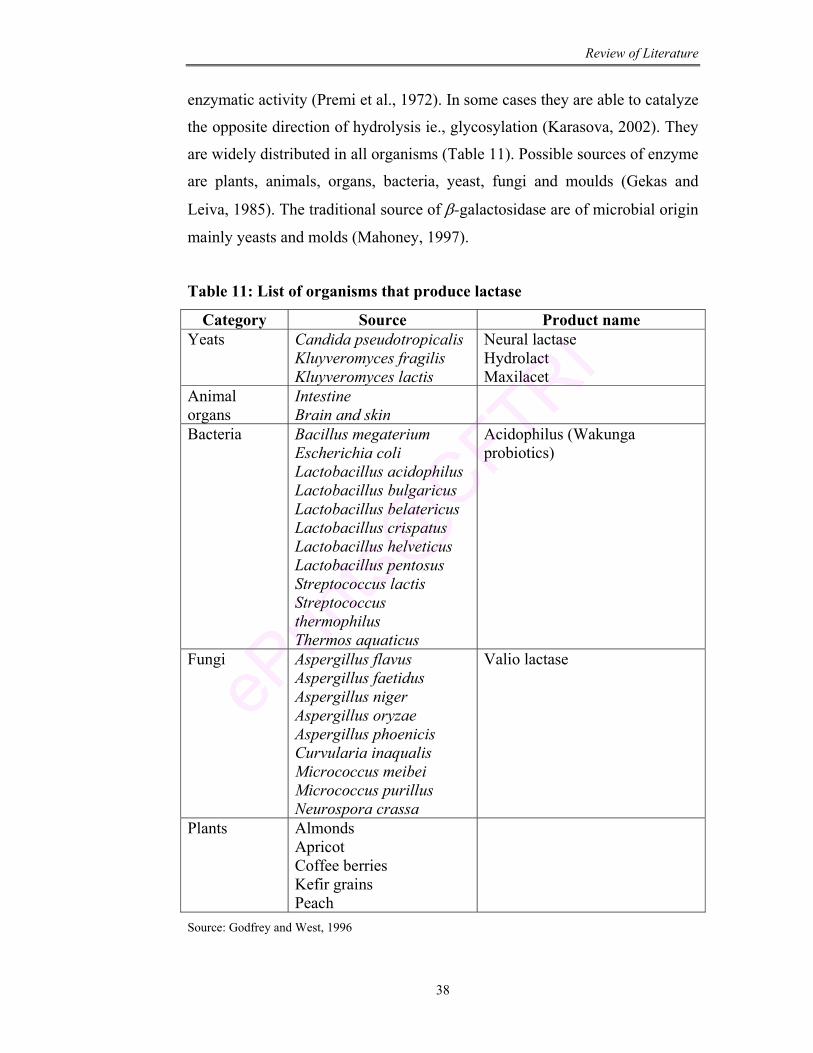
Review of Literature
38
enzymatic activity (Premi et al., 1972). In some cases they are able to catalyze
the opposite direction of hydrolysis ie., glycosylation (Karasova, 2002). They
are widely distributed in all organisms (Table 11). Possible sources of enzyme
are plants, animals, organs, bacteria, yeast, fungi and moulds (Gekas and
Leiva, 1985). The traditional source of β-galactosidase are of microbial origin
mainly yeasts and molds (Mahoney, 1997).
Table 11: List of organisms that produce lactase
Category Source Product name Yeats Candida pseudotropicalis
Kluyveromyces fragilis Kluyveromyces lactis
Neural lactase Hydrolact Maxilacet
Animal organs
Intestine Brain and skin
Bacteria Bacillus megaterium Escherichia coli Lactobacillus acidophilus Lactobacillus bulgaricus Lactobacillus belatericus Lactobacillus crispatus Lactobacillus helveticus Lactobacillus pentosus Streptococcus lactis Streptococcus thermophilus Thermos aquaticus
Acidophilus (Wakunga probiotics)
Fungi Aspergillus flavus Aspergillus faetidus Aspergillus niger Aspergillus oryzae Aspergillus phoenicis Curvularia inaqualis Micrococcus meibei Micrococcus purillus Neurospora crassa
Valio lactase
Plants Almonds Apricot Coffee berries Kefir grains Peach
Source: Godfrey and West, 1996
Review of Literature
39
In the present work Leuconostoc mesenteroides, a lactic acid bacterial
strain has been isolated, characterized and adapted for its probiotic functional
properties to be used in food formulation.
Objectives
1) Isolation and characterization of Leuconostoc from milk products
2) Properties of the isolated bacterium in relation to functional
significance
3) Colonization of LAB in relation to homoserine lactone

Aim & Scope of Present investigation
Aim and Scope
40
AIM OF THE PRESENT INVESTIGATION
During the past several years, the focus of nutritional sciences has
shifted from deficiency disease prevention to optimizing health and
prevention of chronic diseases. Accordingly, the research has encompassed
the health effects of bioactive food components. In this regard, probiotic
therapy is being used increasingly in humans and veterinary medicine due to
their apparent high index of safety and public perception about natural or
alternative therapies (Gherty 1995; Sander 1998; FAO Report 2002).
Probiotics are a category of ‘nutraceuticals’ i.e., viable cultures added
to food with the intention of maintaining or improving the nutritional health
of consumers. Lactic acid bacteria (LAB) are in the focus of extensive
research because of their probiotic nature.
Today, probiotic market promises the disease prevention and better
health for all as a natural alternative therapy. In the present scenario, with
increase desire of consumer for natural food products as source of providing
nutrition and other desirable benefits, research work towards the selection of
strain with functional properties has become very important.
Considering all these aspects, the aim of the present work is to isolate
an indigenous food grade lactic acid bacterial culture from different milk and
milk products to be used as a potential probiotic culture. The selected strains
will be adapted to survive under gastrointestinal conditions. The culture will
be analyzed for its potential probiotic functional properties like antimicrobial,
antibiotic resistance, antioxidative activity, anticholesterolemic activity and
adherence ability. Volatile compounds of therapeutic importance produced
during growth will be studied.
The study will be focused on the ability of the culture to hydrolyze the
non-reducing disaccharide lactose into simple sugar for easy absorption.
Strain will be improved for enhanced β-galactosidase activity which is
responsible for lactose hydrolysis. Work will be carried out to preserve the
culture on shelf storage by different drying techniques.
Aim and Scope
41
The potent isolated culture will be used as a starter culture for the
preparation of probiotic fermented milk beverage. Different adjuvants will be
supplemented to enhance the nutritional properties of the fermented milk
beverage. Further the aim will be to preserve the fermented milk beverage
from spoilage bacterial flora by interfering with the quorum sensing signal
molecules using furanones. Safety and beneficial effect of the culture will be
analyzed in-vivo using albino rats. Ability of the culture to hydrolyze lactose
and reduce lactose intolerance problem will be analyzed in lactose intolerance
induced rats.
SCOPE OF THE PRESENT INVESTIGATION
With the rise in the consumer’s awareness of individual health,
nutrition and well being, the interest and demand for value added foods and
beverages has expanded. In this regard, the present isolate having potential
probiotic functional properties promises to be a source of natural alternative
cure. The β-galactosidase activity of the culture will show its positive impact
in reducing lactose intolerance problem which is prevalent all over the world.
The fermented beverage prepared by the probiotic Leuconostoc mesenteroides
(M7-PLsr-1(W)) will serve as a nutritive source for all ages.
Chapter 1 Isolation & Screening of
Lactic Acid Bacteria from Milk & Milk Products
Isolation and Characterization
42
CHAPTER - 1
ISOLATION AND SCREENING OF LACTIC ACID BACTERIA FROM MILK AND MILK PRODUCTS
ABSTRACT
In the present work bacterial cultures were isolated from milk and milk
products and screened for lactic acid bacteria with characteristics of being
tolerant to gastrointestinal conditions. Totally forty five bacterial strains were
isolated and screened for gram positive, catalase negative, non-motile, non-
spore forming and vancomycin sensitive strains. The selected strains were
adapted to grow at pH 2.0 and 4.0% bile salt mix concentration. The three
adapted strains were subjected to simulated gastrointestinal condition. The
strains that survived under such environment were identified through
biochemical and molecular methods. The identified culture Leuconostoc
mesenteroides (Lsr-1(W)) and Lactobacillus plantarum (Lsr-12(cu)) were then
screened for resistance to digestive enzymes pepsin and trypsin. Leuconostoc
mesenteroides (Lsr-1(W)) that confirmed resistance to these enzymes was
coded as PLsr-1(W) and used for further studies.
Isolation and Characterization
43
1.1. Introduction
Interest in microorganisms as a component of biological diversity has
been renewed in recent years. Probiotics include lactic acid bacteria (LAB).
Mankind has exploited these bacteria for many years for the production of
fermented foods because of their ability to produce desirable changes in taste,
flavor and texture. The demand for the probiotic foods is increasing all over
the world reflecting the awareness among the public between diet and health.
Numerous scientific papers and review articles have been published on health
benefits of these fermented products (Ostlie, 2005; Hughes and Hoover,
1995).
Probiotics have health promoting effects including inhibition of
pathogens, antimutagenic, anticarcinogenic activity, prevention of diarrhea,
stimulation of immune response and reduction of serum cholesterol (Tannock,
1999). The development of new applications such as live vaccines and
probiotic foods reinforces the need for these characteristics.
Considering these, study was carried out to isolate a potential LAB
culture from milk and milk products with the ultimate aim of using the culture
in food formulations. In this study, isolated cultures were adapted for
gastrointestinal condition of low pH and high bile salt mix concentration. The
cultures that could survive such stress conditions were characterized and
identified through bio-chemical assays and molecular techniques. These
cultures were studied for the tolerance to digestive enzymes. The best culture
showing all these properties was selected for further studies.
1.2. Materials
Samples: Milk, curd, butter, cheddar cheese, buttermilk and whey.
Chemicals: All chemicals used in this study were of analytical reagent grade
purchased from HiMedia Pvt Ltd., India unless otherwise mentioned.
Isolation and Characterization
44
1) De Mann Rogosa Sharpe media: Table (1.1) represents the media
composition. All the ingredients are accurately weighed and
dissolved in double distilled water. Sterilization was carried out at
1210C at 15 lb pressure for 20 min (For preparation of solid media
bacteriological agar was added (2%) before sterilization).
2) Saline: 0.8% NaCl.
3) Hydrogen peroxide (30%).
4) Vancomycin antibiotic (30 µg).
5) Buffers: Glycine-HCl buffer (0.2 M; pH 2.0-3.0), Citrate buffer (0.1
M; pH 4.0-5.0), Sodium-phosphate buffer (0.2 M; pH 6.0-8.0) and
Glycine-NaOH buffer (0.2 M; pH 9.0).
6) Bile salt mix: Bile salt mix contains 0.3% each of sodium salts of
glycocholic acid, glycodeoxycholic acid, taurocholic acid and
taurodeoxycholic acid.
7) Digestive enzyme: Pepsin and trypsin (Sigma-Aldrich Company).
8) Chemicals for DNA isolation: TAE buffer (40 mM Tris acetate, 1
mM EDTA; pH 8.0), PBS buffer (137 mM NaCl, 2.7 mM KCl, 4.3
mM Na2HPO4, 1.4 M KH2PO4), proteinase K, SDS, agarose,
ethidium bromide and Sodium acetate (Sigma-Aldrich Company,
India).
9) PCR chemicals: Primer, Taq polymerase, Taq polymerase buffer,
MgCl2 and dNTPs (Sigma-Aldrich Company, India).
10) Lysis buffer (100 mM EDTA, 34 mM SDS)
Equipment : Microscope (Olympus BX 40, Japan), pH meter (Genei, Control
Dynamics, India), Centrifuge (Labline, India), DNA isolation unit (Genei,
Bangalore), PCR machine (Primers Company, MWG Biotech, Germany), Gel
documention (Bio-Rad Laboratories, USA).
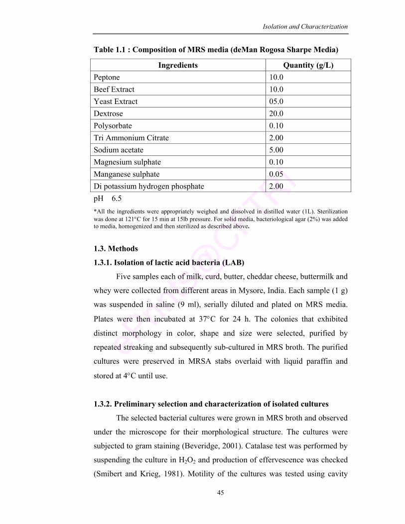
Isolation and Characterization
45
Table 1.1 : Composition of MRS media (deMan Rogosa Sharpe Media)
Ingredients Quantity (g/L) Peptone 10.0 Beef Extract 10.0 Yeast Extract 05.0 Dextrose 20.0 Polysorbate 0.10 Tri Ammonium Citrate 2.00 Sodium acetate 5.00 Magnesium sulphate 0.10 Manganese sulphate 0.05 Di potassium hydrogen phosphate 2.00 pH 6.5 *All the ingredients were appropriately weighed and dissolved in distilled water (1L). Sterilization was done at 121°C for 15 min at 15lb pressure. For solid media, bacteriological agar (2%) was added to media, homogenized and then sterilized as described above.
1.3. Methods
1.3.1. Isolation of lactic acid bacteria (LAB)
Five samples each of milk, curd, butter, cheddar cheese, buttermilk and
whey were collected from different areas in Mysore, India. Each sample (1 g)
was suspended in saline (9 ml), serially diluted and plated on MRS media.
Plates were then incubated at 37°C for 24 h. The colonies that exhibited
distinct morphology in color, shape and size were selected, purified by
repeated streaking and subsequently sub-cultured in MRS broth. The purified
cultures were preserved in MRSA stabs overlaid with liquid paraffin and
stored at 4°C until use.
1.3.2. Preliminary selection and characterization of isolated cultures
The selected bacterial cultures were grown in MRS broth and observed
under the microscope for their morphological structure. The cultures were
subjected to gram staining (Beveridge, 2001). Catalase test was performed by
suspending the culture in H2O2 and production of effervescence was checked
(Smibert and Krieg, 1981). Motility of the cultures was tested using cavity
Isolation and Characterization
46
slide by hanging drop method under the microscope (Priest et al., 1988).
Vancomycin susceptibility was tested by BSAC standardized disc sensitivity
testing method using 30µg vancomycin disc. Sensitivity or resistance was
determined by the growth of the culture around the antibiotic disc.
1.3.3. Screening the isolated cultures for acid and alkaline tolerance
The gastric pH varies from pH 2.0-3.5 and intestinal pH from 7.5-9.0.
Therefore to check the growth of LAB under these conditions the selected
bacterial strains were inoculated (1% v/v) into MRS broth at different pH
(2.0, 3.0, 4.0, 7.5 and 9.0) and incubated at 370C for 24 h. Initial and final cell
count of treated cultures were determined by plating on MRSA media.
Bacterial cultures that exhibited survival under such conditions were selected
for further screening for bile resistance.
1.3.4. Screening the cultures for bile salt tolerance
Bacteria that survived the acidic condition of stomach have to face
further challenge of bile that is released into upper small intestine after
ingestion of fatty meals (Hong et al., 2005). As the bile salt concentration
varies in different regions of human intestine from 1.5 to 4.0% (Chou and
Weimer, 1999; Berrada et al., 1991) the selected cultures were further
screened for their growth under the conditions. Experimentally, MRS broth
was prepared by supplementing different concentrations of filter sterilized
bile salt mix (0.5 to 4.0% w/v) separately in different conical flasks. The
selected cultures were inoculated (1% v/v) to these flasks and incubated at
37°C for 24 h. Cultures that grew in the presence of bile salt mix were
selected for further studies.
1.3.5. Adaptation of culture to pH 2.0 and 4% bile salt concentration
The selected cultures were adapted for acidic pH by repeated sub-
culturing for 3-4 generations in MRS media prepared in a descending gradient
from pH 4.3 to pH 2.0 using 1N HCl. Similarly, for adaptation to bile salts,
Isolation and Characterization
47
the cultures were grown in MRS broth supplemented with bile salt mix in
ascending succession gradually from 0.5 to 4.0% and sub-culturing 3-4 times
in the medium at each concentration.
1.3.6. Survival under simulated intestinal tolerance
An in-vitro method was designed that resembled the intestinal
conditions of human to check the tolerance of the culture that were adapted to
grow at low pH (2.0) and high bile salt mix (4%). MRS broth was adjusted to
that of jejunum condition (pH 7.5 and 4% bile salt) and inoculated with the
selected cultures (one culture/flask) at a concentration of 2 × 10⁸ cfu/ml. After
an incubation period of 4 h at 37°C, cells were collected by centrifugation and
then transferred to another flask with MRS broth at pH 8.0 and 2% bile salt
mix (condition of small intestine) and incubated for 12 h at 37°C. Again after
the incubation period the cell pellet was collected and transferred to another
flask of MRS broth at pH 9.0 and 1.5% bile salt mix (condition as of large
intestine) and incubated for 24 h at 37°C. At each simulated condition culture
viability was determined by plating the appropriate dilution on MRSA media.
After an incubation period of 24 h, colonies grown were counted and
expressed as colony forming units per ml (cfu/ml).
1.3.7. Identification and characterization of selected cultures
The adapted cultures that were able to survive under the simulated
intestinal conditions were identified by bio-chemical assays and molecular
techniques.
1.3.7.1. Phenotypic characterization of selected culture
Physiological and biochemical characterization of the selected cultures
were tested according to Bergey’s manual of Systematic Bacteriology (Kreig,
1984). Growth at different temperatures (10°, 37° and 45°C) and pH (4.2, 8.5
and 9.6) were evaluated by incubating the selected cultures at the respective
temperature and pH. Similarly salt tolerance was determined by growing the
Isolation and Characterization
48
culture in MRS broth supplemented with different concentration of NaCl (2.5,
6.5 and 18.0%).
The carbohydrate fermentation ability of the selected strains was tested
in basal media (MRS broth devoid of beef extract and dextrose) with
bromocresol purple as an indicator. The filter sterilized sugar was added to
the basal media at a final concentration of 2g/100 ml. The selected strains
were inoculated (50 µl) into the basal medium and incubated at 37°C for 24 h.
Growth and color change in the medium were observed after 24 h of
incubation (Sneath et al., 1986; Rashid et al., 2007). Deamination of arginine
was tested in Thornley’s semi solid arginine medium (Terzaghi and Sandine,
1975).
1.3.7.2. Molecular characterization of selected cultures
1.3.7.2.1. Extraction of genomic DNA
Bacterial genomic DNA was extracted according to the method of
Perez et al (2002). The exponentially grown culture (1.4 ml) was centrifuged
to collect the cell pellet. Cell pellet was washed twice with PBS buffer and
suspended in 700 µl of lysis buffer followed by addition of proteinase K (35
µl; 20mg/ml). The mixture was then incubated at 50°C for 80 min. The
aqueous phase was then extracted with an equal volume of phenol followed
by phenol: chloroform: iso-amyl alcohol (25:24:1) and finally with
chloroform: iso-amyl alcohol (24:1). The aqueous supernatant was combined
with 1/10 volume of 3 M sodium acetate (pH 5.2) and 2 volumes of 100%
ethanol. The pellet was washed with 70% ethanol and dissolved in 50 µl 1X
TAE buffer.
1.3.7.2.2. Amplification of 16srRNA by polymerase chain reaction and
identification by sequence analysis
PCR based typing method was used for identification of the strain
using the universal primer M13 (5’ CCC AGT CAC GAC GTT 3’). PCR was
performed in a total volume of 30µl containing 1 ng of template DNA, 0.8
µM of primer, 0.5 U Taq polymerase, 1X Taq polymerase buffer, 2 mM
MgCl2 and 0.2 mM of dNTPs. Amplification reaction was performed with the
Isolation and Characterization
49
following temperature profile: 10 min at 94°C, 25 cycles of 1 min at 94°C, 2
min at 61°C and 5 min at 72°C. The amplified product (15 µl) was loaded
into agarose gel (1.5%) in TAE buffer and the bands were observed after
staining with ethidium bromide. Gels were then photographed under gel
documentation.
For identification of Leuconostoc culture, the specific primer was
designed with a sequence of F 5’ ATT GGG ACT GAG ACA CGG 3’ and R
5’ TGA TGA CCT GAC GTC GTC C 3’. Amplification of the 16srRNA
sequence was performed with the following thermal cycle: initial denaturation
at 94°C for 2 min, followed by 40 cycles of 1 min denaturation at 94°C,
annealing at 32°C for 2 min and extension for 7 min at 72°C and a final
extension for 7 min at 72°C. PCR product was then sequenced and through
BLAST search of multiple sequence alignment programme, the cultures were
identified and a phylogram was drawn with the nearest cultures.
1.3.8. Determination of tolerance to digestive enzymes
1.3.8.1. Preparation of simulated gastric and small intestinal juices
Simulated gastric and small intestinal juice was prepared fresh as per
the procedure of Huang and Adams (2004). Simulated gastric juice was
prepared by suspending pepsin (3 g/L) in sterile saline (0.5% w/v) which was
previously adjusted to pH 2.0 using buffer. Simulated small intestinal juice
was prepared by suspending trypsin (1 g/L) in sterile saline (0.5% w/v) with
supplementation of 4% bile salt mix. The pH was adjusted to 7.5 with sterile
0.1 M NaOH.
1.3.8.2. Determination of GI transit tolerance
The cell biomass of the selected cultures was collected by
centrifugation at 8000 rpm for 10 min at 4°C. The cell biomass collected was
washed thrice in phosphate buffer (pH 7.0) and suspended in saline (1 ml).
An aliquot (0.1 ml) of this suspension was mixed with simulated gastric (10
ml) and intestinal juice (10 ml) separately and incubated at 37°C for 3 h.
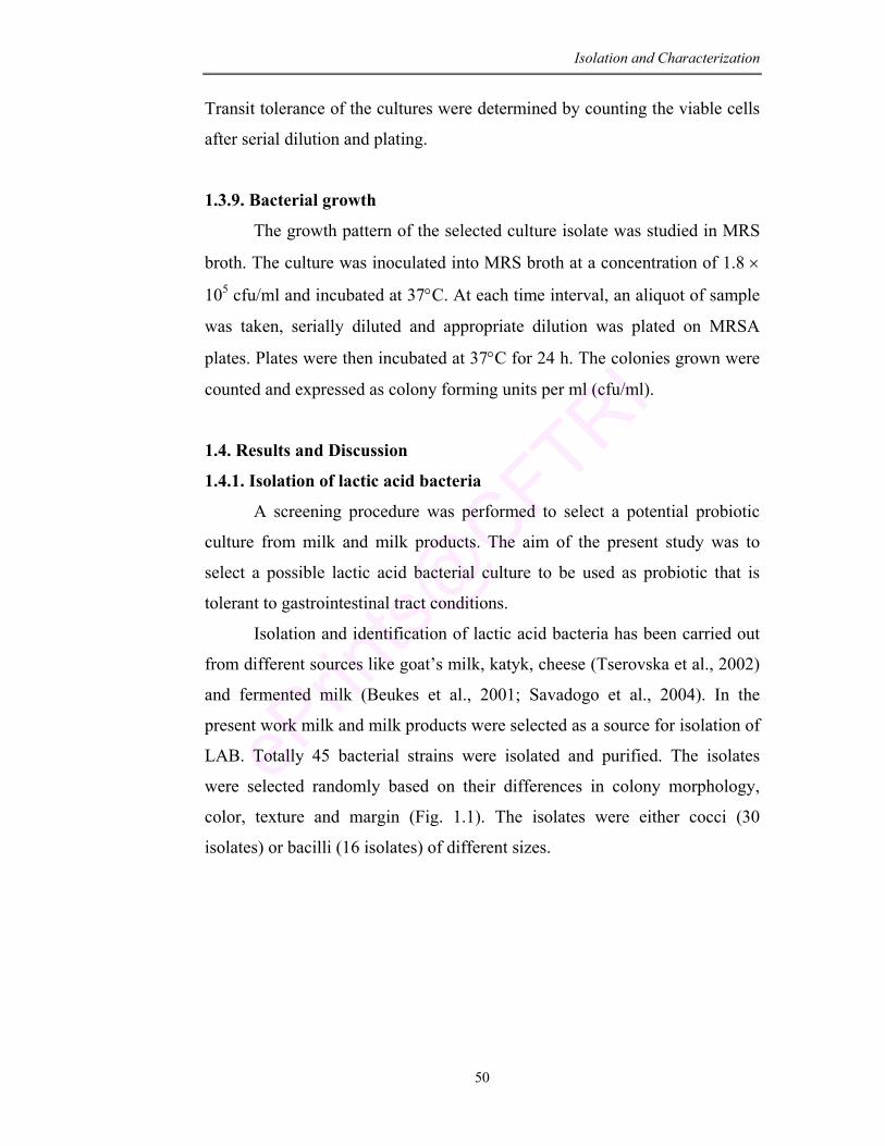
Isolation and Characterization
50
Transit tolerance of the cultures were determined by counting the viable cells
after serial dilution and plating.
1.3.9. Bacterial growth
The growth pattern of the selected culture isolate was studied in MRS
broth. The culture was inoculated into MRS broth at a concentration of 1.8 ×
105 cfu/ml and incubated at 37°C. At each time interval, an aliquot of sample
was taken, serially diluted and appropriate dilution was plated on MRSA
plates. Plates were then incubated at 37°C for 24 h. The colonies grown were
counted and expressed as colony forming units per ml (cfu/ml).
1.4. Results and Discussion
1.4.1. Isolation of lactic acid bacteria
A screening procedure was performed to select a potential probiotic
culture from milk and milk products. The aim of the present study was to
select a possible lactic acid bacterial culture to be used as probiotic that is
tolerant to gastrointestinal tract conditions.
Isolation and identification of lactic acid bacteria has been carried out
from different sources like goat’s milk, katyk, cheese (Tserovska et al., 2002)
and fermented milk (Beukes et al., 2001; Savadogo et al., 2004). In the
present work milk and milk products were selected as a source for isolation of
LAB. Totally 45 bacterial strains were isolated and purified. The isolates
were selected randomly based on their differences in colony morphology,
color, texture and margin (Fig. 1.1). The isolates were either cocci (30
isolates) or bacilli (16 isolates) of different sizes.
Isolation and Characterization
51
Fig 1.1: Distribution of isolated cultures from milk and milk products.
Values are the number of isolated cultures.
1.4.2. Preliminary selection and characterization of isolated cultures
Out of 45 strains isolated two strains did not grow on further
propagation. From the remaining 43 strains, 10 were gram negative and 9
were catalase positive and were not considered to be presumptive LAB.
Motility test confirmed that 5 isolates were motile and other 4 strains showed
sporulation. The remaining 15 cultures were tested for Vancomycin
sensitivity. It was observed that 3 isolates were Vancomycin resistant. All
these were discarded.
Out of 12 selected strains, 3 strains were rod shaped. They did not
produce gas on glucose fermentation and were not able to ferment pentose
sugars (arabinose, ribose and xylose). Therefore, they were characterized as
obligate homofermentative bacilli. One isolate with rod-shaped morphology
did not produce gas from glucose fermentation but could ferment pentose
sugars and was characterized as facultative heterofermentative rod/ bacilli.
Four strains which were cocci produced gas from glucose and L-lactate
indicating that they belong to heterofermentative cocci. The other 4 strains
with cocci morphology did not produce gas from glucose and L-lactate.
Therefore they were characterized as homofermentative cocci (Table 1.2).
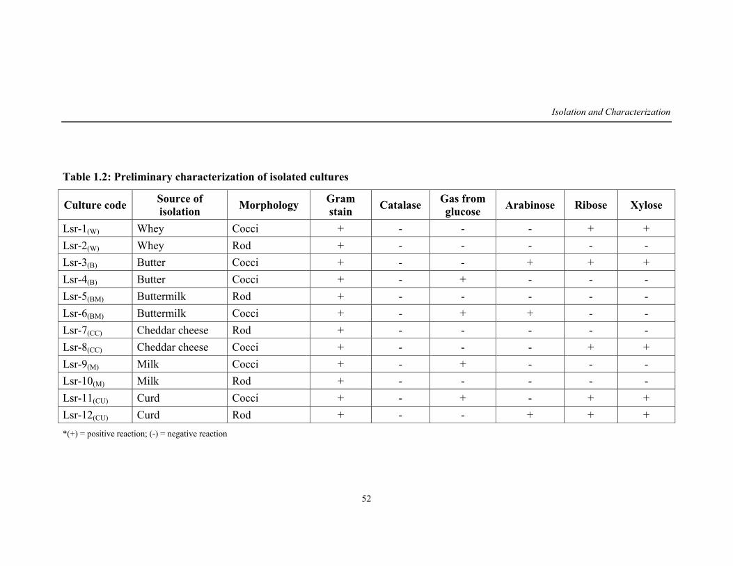
Isolation and Characterization
52
Table 1.2: Preliminary characterization of isolated cultures
Culture code Source of isolation Morphology Gram
stain Catalase Gas from glucose Arabinose Ribose Xylose
Lsr-1(W) Whey Cocci + - - - + + Lsr-2(W) Whey Rod + - - - - - Lsr-3(B) Butter Cocci + - - + + + Lsr-4(B) Butter Cocci + - + - - - Lsr-5(BM) Buttermilk Rod + - - - - - Lsr-6(BM) Buttermilk Cocci + - + + - - Lsr-7(CC) Cheddar cheese Rod + - - - - - Lsr-8(CC) Cheddar cheese Cocci + - - - + + Lsr-9(M) Milk Cocci + - + - - - Lsr-10(M) Milk Rod + - - - - - Lsr-11(CU) Curd Cocci + - + - + + Lsr-12(CU) Curd Rod + - - + + + *(+) = positive reaction; (-) = negative reaction
Isolation and Characterization
53
1.4.3. Screening for gastrointestinal tolerance
Survival under gastrointestinal environmental condition is one of the
most important characteristic features for a culture to be used as a probiotic.
As the pH is acidic in stomach and alkaline in the intestinal region (Pennacchi
et al., 2004; Klingberg et al., 2006), the selected cultures were screened for
their ability to grow at both acidic and alkaline pH.
pH of secreted HCl in stomach is 0.9, but presence of food raises the
pH value to 2.0-3.0 (Erkkila and Petaja, 2000). Hence in the present study the
isolated cultures were screened for their ability to grow at varying pH from
2.0 to 9.0 by inoculating the cultures at a rate of 5 × 10⁵ cfu/ml. The cultures
show variable degree of tolerance to different pH. Zarate et al. (2000) have
reported four dairy Propionibacterium to survive at pH 4.0 but did not grow
substantially at pH 2.0. Similarly in the present work, at pH 4.0 all the tested
bacterial strains survived but at pH 3.0, four strains lost the viability.
Earlier reports have shown large number of LAB like L. sakei,
L. plantarum, L. pentosus, P. acidilactici and P. pentosaceus to be tolerant to
acidic condition (Pennacchia et al., 2004; Klingberg et al., 2006). Prasad et al.
(1998) during screening a large number of LAB (200 isolates), selected four
resistant strains which were able to survive at pH 3.0. In the present study 6
cultures were selected that were able to survive at pH 3.0. The culture Lsr-
1(W) was able to survive at pH 3.0 with maximum survival rate (20%). The
other cultures Lsr-3(B), Lsr-8(Cc), Lsr-10(M), Lsr-11(Cu) and Lsr-12(Cu) showed a
survival rate of 10, 1, 4, 3 and 8% respectively. These six cultures which were
able to grow at acidic pH 3.0 were selected for bile salt tolerance.
Bile plays a fundamental role in specific (Marteau et al., 1997) and
non-specific (Kalambaheti et al., 1994) defense mechanism of the gut and the
magnitude of its inhibitory effect is determined primarily by the bile salt
concentration (Charteris et al., 2000). Since the cell membrane of
microorganism is composed of lipids and fatty acids the bile salts are critical
for their survival.
Isolation and Characterization
54
With this in mind, the present work was carried out to screen the
isolated cultures for bile salt tolerance. The selected cultures that were able to
tolerate pH 3.0 were grown in MRS broth supplemented with varying
concentration of bile salt mix (0.5–4.0%). It was observed that there was a
delay in the growth on supplementation of bile. Chateau et al. (1994) have
also reported an extreme variability of resistance to bile salts in Lactobacillus
sp and all the tested strains showed a delayed growth as compared to a
reference culture without bile salts. Similar results have been noticed by
Gilliland et al. (1984), Gupta et al. (1996) and Mustapha et al. (1997).
Gomes-Zavaglia et al. (1998) have shown that 10 Bifidobacterium
strains out of 41 tested were resistant to 0.5% bile. In the present work all the
six cultures selected were able to grow at 0.5% bile salt mix. The cell viability
was found to decrease with increase in bile salt mix concentration. The
culture Lsr-1(W) that was isolated from whey exhibited maximum survival
(12%) followed by Lsr-10(M), Lsr-12(Cu) and Lsr-3(B). Further these four
cultures were adapted to tolerate the gastrointestinal condition of pH 2.0 and
4% bile salt concentration.
1.4.4. Adaptation of the culture to GIT condition
Adaptation to low pH and high bile is a valuable tool for increasing
bacterial survival in the harsh conditions of GIT. Earlier reports have
suggested that inhibition of microbial growth due to stress condition can be
overcome by progressive adaptation to increasing concentration of these
compounds (Chung et al., 1999; Margolles et al., 2003). In the present work,
the selected culture isolates were grown in MRS broth through sequential
sub-culturing by increasing the concentration of bile salt and decreasing the
pH (Table 1.3). The results determined that the cultures Lsr-1(W), Lsr-10(Cu)
and Lsr-12(Cu) were better (P<0.05) adapted to such stress conditions.
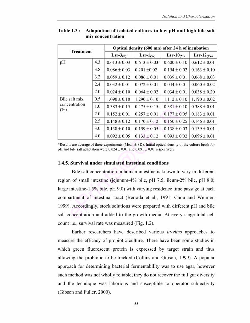
Isolation and Characterization
55
Table 1.3 : Adaptation of isolated cultures to low pH and high bile salt mix concentration
Optical density (600 mn) after 24 h of incubation
Treatment Lsr-3(B) Lsr-1(W) Lsr-10(M) Lsr-12(Cu)
4.3 0.613 ± 0.03 0.613 ± 0.03 0.600 ± 0.10 0.612 ± 0.013.8 0.086 ± 0.03 0.201 ±0.02 0.194 ± 0.02 0.163 ± 0.103.2 0.059 ± 0.12 0.086 ± 0.01 0.039 ± 0.01 0.068 ± 0.032.4 0.032 ± 0.01 0.072 ± 0.01 0.044 ± 0.01 0.060 ± 0.02
pH
2.0 0.024 ± 0.10 0.064 ± 0.02 0.034 ± 0.01 0.038 ± 0.200.5 1.090 ± 0.10 1.290 ± 0.10 1.112 ± 0.10 1.190 ± 0.021.0 0.383 ± 0.15 0.475 ± 0.15 0.381 ± 0.10 0.388 ± 0.012.0 0.152 ± 0.01 0.257 ± 0.01 0.177 ± 0.05 0.183 ± 0.012.5 0.148 ± 0.12 0.170 ± 0.12 0.150 ± 0.25 0.146 ± 0.013.0 0.138 ± 0.10 0.159 ± 0.05 0.138 ± 0.03 0.139 ± 0.01
Bile salt mix concentration (%)
4.0 0.092 ± 0.05 0.133 ± 0.12 0.093 ± 0.02 0.096 ± 0.01
*Results are average of three experiments (Mean ± SD). Initial optical density of the culture broth for pH and bile salt adaptation were 0.024 ± 0.01 and 0.091 ± 0.01 respectively.
1.4.5. Survival under simulated intestinal conditions
Bile salt concentration in human intestine is known to vary in different
region of small intestine (jejunum-4% bile, pH 7.5; ileum-2% bile, pH 8.0;
large intestine-1.5% bile, pH 9.0) with varying residence time passage at each
compartment of intestinal tract (Berrada et al., 1991; Chou and Weimer,
1999). Accordingly, stock solutions were prepared with different pH and bile
salt concentration and added to the growth media. At every stage total cell
count i.e., survival rate was measured (Fig. 1.2).
Earlier researchers have described various in-vitro approaches to
measure the efficacy of probiotic culture. There have been some studies in
which green fluorescent protein is expressed by target strain and thus
allowing the probiotic to be tracked (Collins and Gibson, 1999). A popular
approach for determining bacterial fermentability was to use agar, however
such method was not wholly reliable, they do not recover the full gut diversity
and the technique was laborious and susceptible to operator subjectivity
(Gibson and Fuller, 2000).
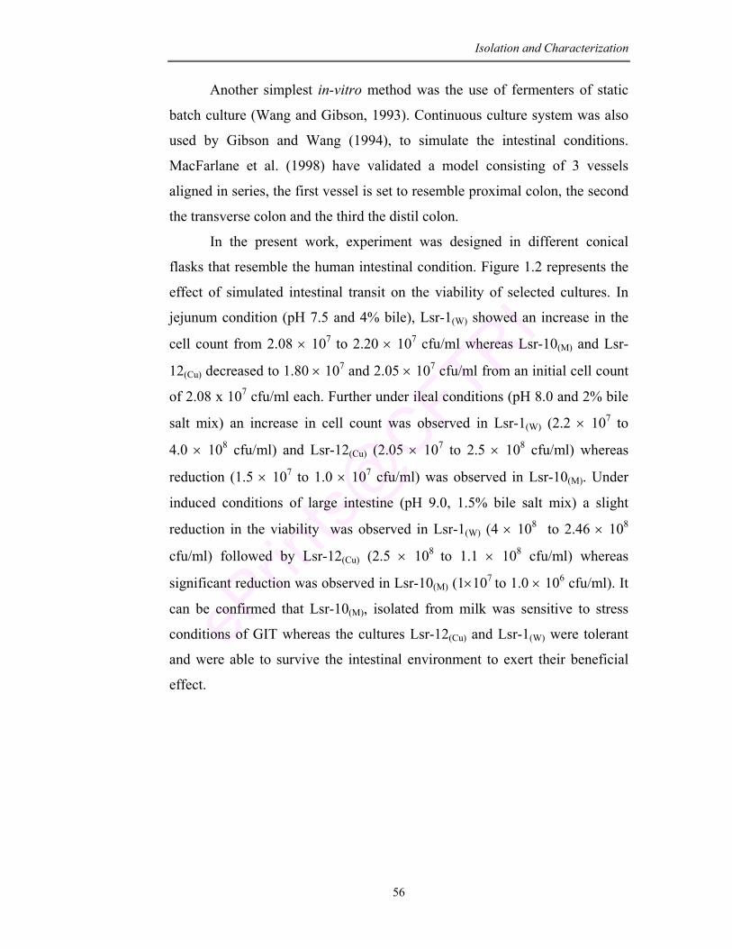
Isolation and Characterization
56
Another simplest in-vitro method was the use of fermenters of static
batch culture (Wang and Gibson, 1993). Continuous culture system was also
used by Gibson and Wang (1994), to simulate the intestinal conditions.
MacFarlane et al. (1998) have validated a model consisting of 3 vessels
aligned in series, the first vessel is set to resemble proximal colon, the second
the transverse colon and the third the distil colon.
In the present work, experiment was designed in different conical
flasks that resemble the human intestinal condition. Figure 1.2 represents the
effect of simulated intestinal transit on the viability of selected cultures. In
jejunum condition (pH 7.5 and 4% bile), Lsr-1(W) showed an increase in the
cell count from 2.08 × 10⁷ to 2.20 × 10⁷ cfu/ml whereas Lsr-10(M) and Lsr-
12(Cu) decreased to 1.80 × 10⁷ and 2.05 × 10⁷ cfu/ml from an initial cell count
of 2.08 x 10⁷ cfu/ml each. Further under ileal conditions (pH 8.0 and 2% bile
salt mix) an increase in cell count was observed in Lsr-1(W) (2.2 × 10⁷ to
4.0 × 10⁸ cfu/ml) and Lsr-12(Cu) (2.05 × 10⁷ to 2.5 × 10⁸ cfu/ml) whereas
reduction (1.5 × 10⁷ to 1.0 × 10⁷ cfu/ml) was observed in Lsr-10(M). Under
induced conditions of large intestine (pH 9.0, 1.5% bile salt mix) a slight
reduction in the viability was observed in Lsr-1(W) (4 × 10⁸ to 2.46 × 10⁸
cfu/ml) followed by Lsr-12(Cu) (2.5 × 10⁸ to 1.1 × 10⁸ cfu/ml) whereas
significant reduction was observed in Lsr-10(M) (1×10⁷ to 1.0 × 10⁶ cfu/ml). It
can be confirmed that Lsr-10(M), isolated from milk was sensitive to stress
conditions of GIT whereas the cultures Lsr-12(Cu) and Lsr-1(W) were tolerant
and were able to survive the intestinal environment to exert their beneficial
effect.
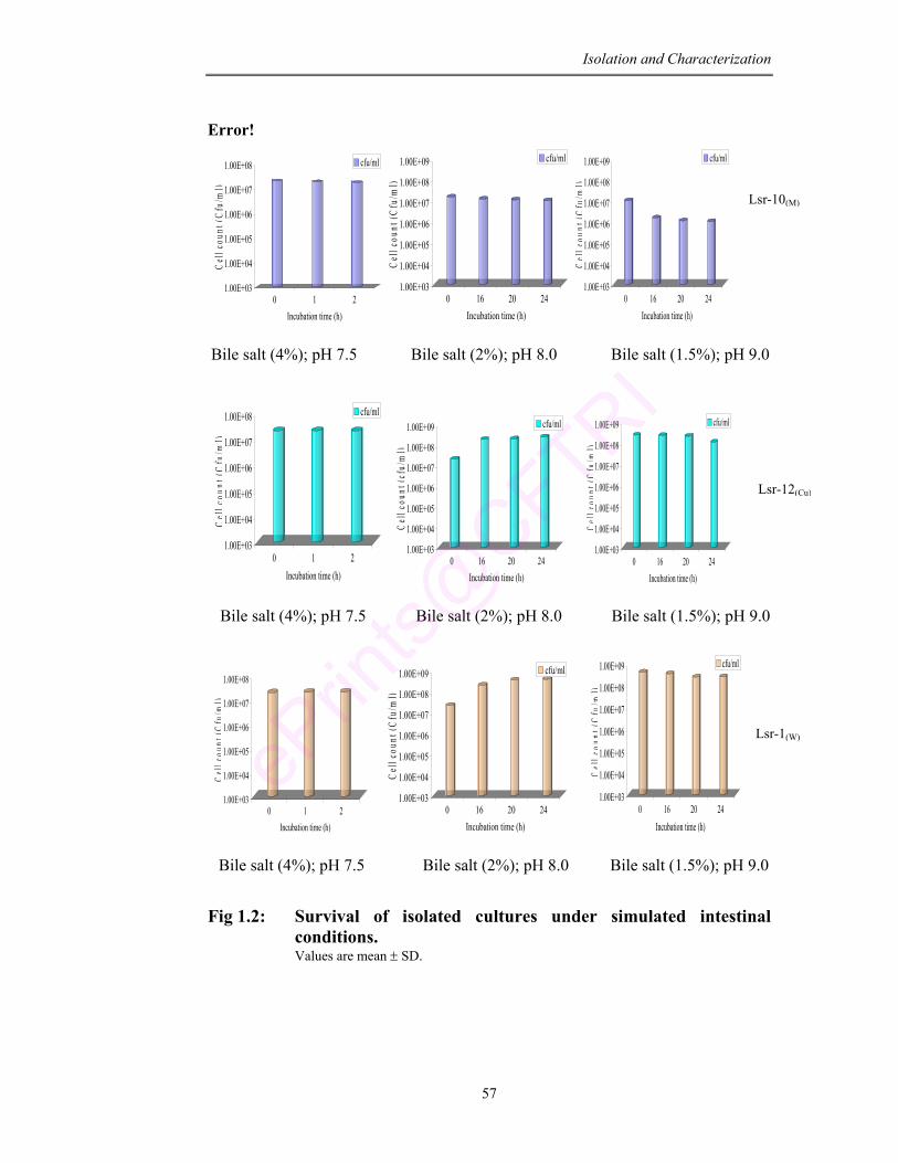
Isolation and Characterization
57
Error!
1.00E+03
1.00E+04
1.00E+05
1.00E+06
1.00E+07
1.00E+08
Cel
l cou
nt (C
fu/m
l)
0 1 2Incubation time (h)
cfu/ml
1.00E+03
1.00E+04
1.00E+05
1.00E+06
1.00E+07
1.00E+08
1.00E+09
Cel
l cou
nt (C
fu/m
l)
0 16 20 24Incubation time (h)
cfu/ml
1.00E+03
1.00E+04
1.00E+05
1.00E+06
1.00E+07
1.00E+08
1.00E+09
Cel
lcou
nt(C
fu/m
l)
0 16 20 24Incubation time (h)
cfu/ml
Bile salt (4%); pH 7.5 Bile salt (2%); pH 8.0 Bile salt (1.5%); pH 9.0
1.00E+03
1.00E+04
1.00E+05
1.00E+06
1.00E+07
1.00E+08
Cel
lcou
nt(C
fu/m
l)
0 1 2Incubation time (h)
cfu/ml
1.00E+03
1.00E+04
1.00E+05
1.00E+06
1.00E+07
1.00E+08
1.00E+09
Cel
l cou
nt (c
fu/m
l)
0 16 20 24Incubation time (h)
cfu/ml
1.00E+03
1.00E+04
1.00E+05
1.00E+06
1.00E+07
1.00E+08
1.00E+09
Cel
lcou
nt(C
fu/m
l)
0 16 20 24Incubation time (h)
cfu/ml
Bile salt (4%); pH 7.5 Bile salt (2%); pH 8.0 Bile salt (1.5%); pH 9.0
1.00E+03
1.00E+04
1.00E+05
1.00E+06
1.00E+07
1.00E+08
Cel
lcou
nt(C
fu/m
l)
0 1 2Incubation time (h)
1.00E+03
1.00E+04
1.00E+05
1.00E+06
1.00E+07
1.00E+08
1.00E+09
Cel
l cou
nt (C
fu/m
l)
0 16 20 24Incubation time (h)
cfu/ml
1.00E+03
1.00E+04
1.00E+05
1.00E+06
1.00E+07
1.00E+08
1.00E+09
Cel
lcou
nt(C
fu/m
l)
0 16 20 24Incubation time (h)
cfu/ml
Bile salt (4%); pH 7.5 Bile salt (2%); pH 8.0 Bile salt (1.5%); pH 9.0
Fig 1.2: Survival of isolated cultures under simulated intestinal conditions.
Values are mean ± SD.
Lsr-10(M)
Lsr-12(Cu)
Lsr-1(W)
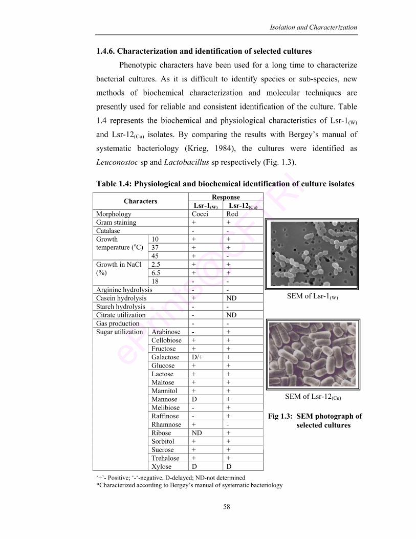
Isolation and Characterization
58
1.4.6. Characterization and identification of selected cultures
Phenotypic characters have been used for a long time to characterize
bacterial cultures. As it is difficult to identify species or sub-species, new
methods of biochemical characterization and molecular techniques are
presently used for reliable and consistent identification of the culture. Table
1.4 represents the biochemical and physiological characteristics of Lsr-1(W)
and Lsr-12(Cu) isolates. By comparing the results with Bergey’s manual of
systematic bacteriology (Krieg, 1984), the cultures were identified as
Leuconostoc sp and Lactobacillus sp respectively (Fig. 1.3).
Table 1.4: Physiological and biochemical identification of culture isolates Response Characters Lsr-1(W) Lsr-12(Cu)
Morphology Cocci Rod Gram staining + + Catalase - -
10 + + 37 + +
Growth temperature (oC)
45 + - 2.5 + + 6.5 + +
Growth in NaCl (%)
18 - - Arginine hydrolysis - - Casein hydrolysis + ND Starch hydrolysis - - Citrate utilization - ND Gas production - -
Arabinose - + Cellobiose + + Fructose + + Galactose D/+ + Glucose + + Lactose + + Maltose + + Mannitol + + Mannose D + Melibiose - + Raffinose - + Rhamnose + - Ribose ND + Sorbitol + + Sucrose + + Trehalose + +
Sugar utilization
Xylose D D ‘+’- Positive; ‘-‘-negative, D-delayed; ND-not determined *Characterized according to Bergey’s manual of systematic bacteriology
SEM of Lsr-1(W)
SEM of Lsr-12(Cu)
Fig 1.3: SEM photograph of selected cultures
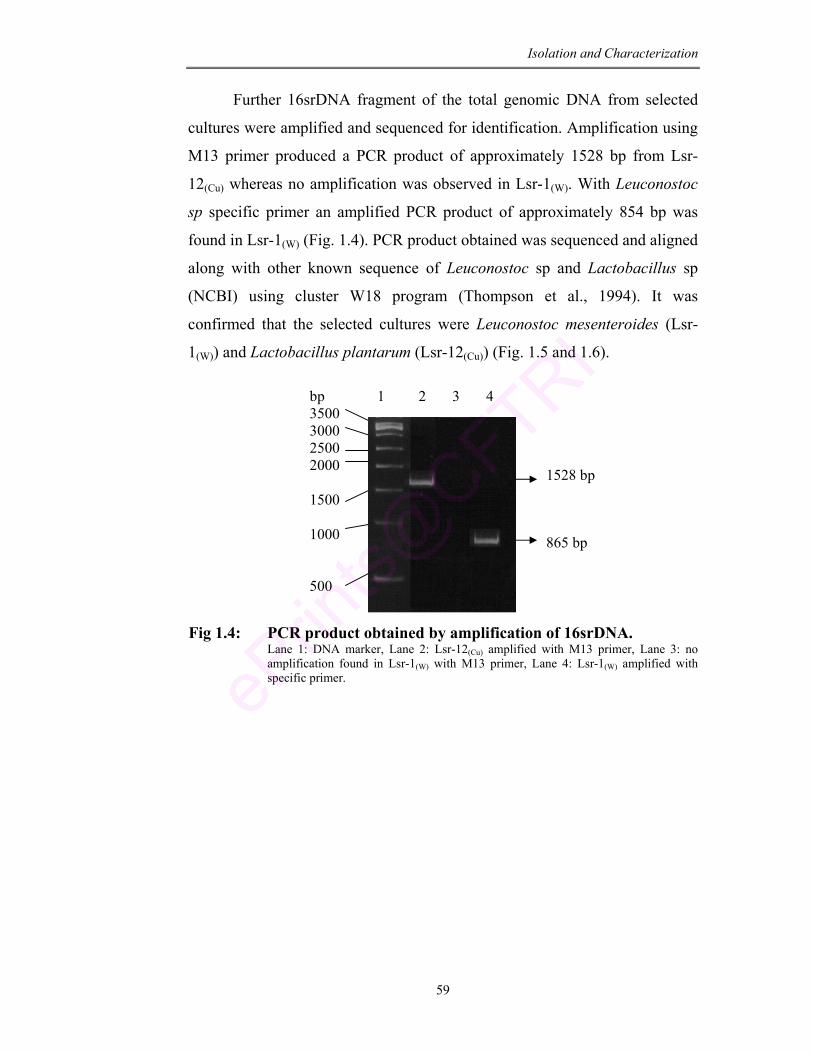
Isolation and Characterization
59
Further 16srDNA fragment of the total genomic DNA from selected
cultures were amplified and sequenced for identification. Amplification using
M13 primer produced a PCR product of approximately 1528 bp from Lsr-
12(Cu) whereas no amplification was observed in Lsr-1(W). With Leuconostoc
sp specific primer an amplified PCR product of approximately 854 bp was
found in Lsr-1(W) (Fig. 1.4). PCR product obtained was sequenced and aligned
along with other known sequence of Leuconostoc sp and Lactobacillus sp
(NCBI) using cluster W18 program (Thompson et al., 1994). It was
confirmed that the selected cultures were Leuconostoc mesenteroides (Lsr-
1(W)) and Lactobacillus plantarum (Lsr-12(Cu)) (Fig. 1.5 and 1.6).
Fig 1.4: PCR product obtained by amplification of 16srDNA. Lane 1: DNA marker, Lane 2: Lsr-12(Cu) amplified with M13 primer, Lane 3: no amplification found in Lsr-1(W) with M13 primer, Lane 4: Lsr-1(W) amplified with specific primer.
1 2 3 4 bp 3500 3000 2500 2000 1500 1000 500
1528 bp
865 bp
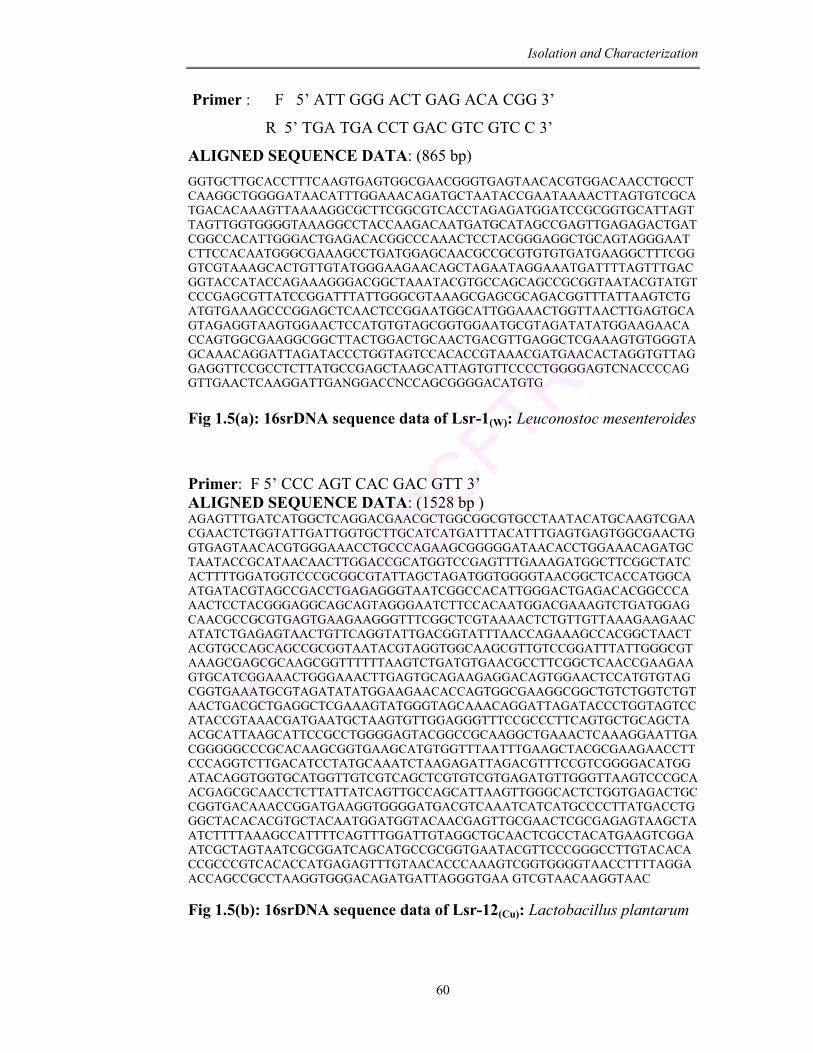
Isolation and Characterization
60
Primer : F 5’ ATT GGG ACT GAG ACA CGG 3’
R 5’ TGA TGA CCT GAC GTC GTC C 3’
ALIGNED SEQUENCE DATA: (865 bp) GGTGCTTGCACCTTTCAAGTGAGTGGCGAACGGGTGAGTAACACGTGGACAACCTGCCTCAAGGCTGGGGATAACATTTGGAAACAGATGCTAATACCGAATAAAACTTAGTGTCGCATGACACAAAGTTAAAAGGCGCTTCGGCGTCACCTAGAGATGGATCCGCGGTGCATTAGTTAGTTGGTGGGGTAAAGGCCTACCAAGACAATGATGCATAGCCGAGTTGAGAGACTGATCGGCCACATTGGGACTGAGACACGGCCCAAACTCCTACGGGAGGCTGCAGTAGGGAATCTTCCACAATGGGCGAAAGCCTGATGGAGCAACGCCGCGTGTGTGATGAAGGCTTTCGGGTCGTAAAGCACTGTTGTATGGGAAGAACAGCTAGAATAGGAAATGATTTTAGTTTGACGGTACCATACCAGAAAGGGACGGCTAAATACGTGCCAGCAGCCGCGGTAATACGTATGTCCCGAGCGTTATCCGGATTTATTGGGCGTAAAGCGAGCGCAGACGGTTTATTAAGTCTGATGTGAAAGCCCGGAGCTCAACTCCGGAATGGCATTGGAAACTGGTTAACTTGAGTGCAGTAGAGGTAAGTGGAACTCCATGTGTAGCGGTGGAATGCGTAGATATATGGAAGAACACCAGTGGCGAAGGCGGCTTACTGGACTGCAACTGACGTTGAGGCTCGAAAGTGTGGGTAGCAAACAGGATTAGATACCCTGGTAGTCCACACCGTAAACGATGAACACTAGGTGTTAGGAGGTTCCGCCTCTTATGCCGAGCTAAGCATTAGTGTTCCCCTGGGGAGTCNACCCCAGGTTGAACTCAAGGATTGANGGACCNCCAGCGGGGACATGTG Fig 1.5(a): 16srDNA sequence data of Lsr-1(W): Leuconostoc mesenteroides
Primer: F 5’ CCC AGT CAC GAC GTT 3’ ALIGNED SEQUENCE DATA: (1528 bp ) AGAGTTTGATCATGGCTCAGGACGAACGCTGGCGGCGTGCCTAATACATGCAAGTCGAACGAACTCTGGTATTGATTGGTGCTTGCATCATGATTTACATTTGAGTGAGTGGCGAACTGGTGAGTAACACGTGGGAAACCTGCCCAGAAGCGGGGGATAACACCTGGAAACAGATGCTAATACCGCATAACAACTTGGACCGCATGGTCCGAGTTTGAAAGATGGCTTCGGCTATCACTTTTGGATGGTCCCGCGGCGTATTAGCTAGATGGTGGGGTAACGGCTCACCATGGCAATGATACGTAGCCGACCTGAGAGGGTAATCGGCCACATTGGGACTGAGACACGGCCCAAACTCCTACGGGAGGCAGCAGTAGGGAATCTTCCACAATGGACGAAAGTCTGATGGAGCAACGCCGCGTGAGTGAAGAAGGGTTTCGGCTCGTAAAACTCTGTTGTTAAAGAAGAACATATCTGAGAGTAACTGTTCAGGTATTGACGGTATTTAACCAGAAAGCCACGGCTAACTACGTGCCAGCAGCCGCGGTAATACGTAGGTGGCAAGCGTTGTCCGGATTTATTGGGCGTAAAGCGAGCGCAAGCGGTTTTTTAAGTCTGATGTGAACGCCTTCGGCTCAACCGAAGAAGTGCATCGGAAACTGGGAAACTTGAGTGCAGAAGAGGACAGTGGAACTCCATGTGTAGCGGTGAAATGCGTAGATATATGGAAGAACACCAGTGGCGAAGGCGGCTGTCTGGTCTGTAACTGACGCTGAGGCTCGAAAGTATGGGTAGCAAACAGGATTAGATACCCTGGTAGTCCATACCGTAAACGATGAATGCTAAGTGTTGGAGGGTTTCCGCCCTTCAGTGCTGCAGCTAACGCATTAAGCATTCCGCCTGGGGAGTACGGCCGCAAGGCTGAAACTCAAAGGAATTGACGGGGGCCCGCACAAGCGGTGAAGCATGTGGTTTAATTTGAAGCTACGCGAAGAACCTTCCCAGGTCTTGACATCCTATGCAAATCTAAGAGATTAGACGTTTCCGTCGGGGACATGGATACAGGTGGTGCATGGTTGTCGTCAGCTCGTGTCGTGAGATGTTGGGTTAAGTCCCGCAACGAGCGCAACCTCTTATTATCAGTTGCCAGCATTAAGTTGGGCACTCTGGTGAGACTGCCGGTGACAAACCGGATGAAGGTGGGGATGACGTCAAATCATCATGCCCCTTATGACCTGGGCTACACACGTGCTACAATGGATGGTACAACGAGTTGCGAACTCGCGAGAGTAAGCTAATCTTTTAAAGCCATTTTCAGTTTGGATTGTAGGCTGCAACTCGCCTACATGAAGTCGGAATCGCTAGTAATCGCGGATCAGCATGCCGCGGTGAATACGTTCCCGGGCCTTGTACACACCGCCCGTCACACCATGAGAGTTTGTAACACCCAAAGTCGGTGGGGTAACCTTTTAGGAACCAGCCGCCTAAGGTGGGACAGATGATTAGGGTGAA GTCGTAACAAGGTAAC Fig 1.5(b): 16srDNA sequence data of Lsr-12(Cu): Lactobacillus plantarum
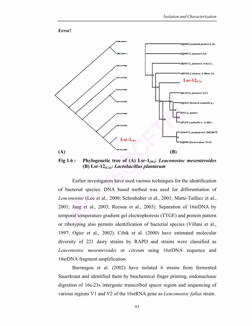
Isolation and Characterization
61
Lsr-1(W)
Lsr-12(Cu)
Error!
(A) (B)
Fig 1.6 : Phylogenetic tree of (A) Lsr-1(W): Leuconostoc mesenteroides (B) Lsr-12(Cu): Lactobacillus plantarum
Earlier investigators have used various techniques for the identification
of bacterial species. DNA based method was used for differentiation of
Leuconostoc (Lee et al., 2000; Schonhuber et al., 2001; Matte-Tailliez et al.,
2001; Jang et al., 2003; Reeson et al., 2003). Separation of 16srDNA by
temporal temperature gradient gel electrophoresis (TTGE) and protein pattern
or ribotyping also permits identification of bacterial species (Villani et al.,
1997; Ogier et al., 2002). Cibik et al. (2000) have estimated molecular
diversity of 221 dairy strains by RAPD and strains were classified as
Leuconostoc mesenteroides or citreum using 16srDNA sequence and
16srDNA fragment amplification.
Barrangou et al. (2002) have isolated 6 strains from fermented
Sauerkraut and identified them by biochemical finger printing, endonuclease
digestion of 16s-23s intergenic transcribed spacer region and sequencing of
various regions V1 and V2 of the 16srRNA gene as Leuconostoc fallax strain.
Lsr-1(W)
Lsr-12(Cu)
Isolation and Characterization
62
Other methods have also been proposed like characterization through
neutral compounds produced in whey (Mauriello et al., 2001) and cellular
fatty acid profile (Rementzis and Samelis, 1996). Some discrepancy is seen in
each of these results and hence molecular method using 16srRNA analysis is
followed for the identification of isolated cultures. In the present study
primarily presumptive identification was done by biochemical assay and
sugar fermentation pattern according to Bergey’s manual of systematic
bacteriology. Further the cultures were identified by 16sRNA sequencing as
Leuconostoc mesenteroides and Lactobacillus plantarum.
Benkerroun et al. (2003) have shown only 1% of Leuconostoc sp to be
present out of the total isolates from camel’s milk. Gomes-Zavaglia et al.
(1998) have isolated 25 Bifidobacterium strains from infant feces and
identified them by sugar fermentation pattern and whole cell protein analysis.
Probiotic properties were checked in the isolated strain and were found that
not all desirable characteristics were present in a single strain.
Leuconostoc mesenteroides has been considered to be a predominant
species during first week of fermentation (Fleming et al., 1995). Several
reports confirm the presence of Leuconostoc mesenteroides and Lactobacillus
plantarum as dominant microflora of fermented product (Beukes et al., 2001;
Gadaga et al., 2001). Obodai and Dodd (2006) have characterized LAB from
Nyarmie, traditional Ghanaian fermented milk and have found Leuconostoc
mesenteroides, Streptococcus thermophilus, Lactobacillus delbruekii,
Lactobacillus helveticus and Lactococcus lactis to be the dominant
microflora. Similarly out of 45 LAB isolates from Kenyan traditional
fermented camel milk 24% were found to be Leuconostoc mesenteroides and
16% Lactobacillus plantarum (Lore et al., 2005).
In the present study, the selected cultures Lsr-1(W) and Lsr-12(Cu) were
identified as Leuconostoc mesenteroides and Lactobacillus plantarum
respectively.
Isolation and Characterization
63
1.4.7. Determination of tolerance to digestive enzymes
The probiotic microorganisms must have the ability to survive and
persist in the GIT. The low pH of the stomach and the antimicrobial action of
pepsin are known to provide an effective barrier against the entry of bacteria
into the intestine (Holzapfel et al., 1998). Another barrier to probiotic bacteria
is the adverse condition of intestine that includes bile salts and pancreatin (Le
Vay, 1988). With this in mind, the present culture isolates were tested for
their tolerance to digestive enzymes (pepsin and trypsin) under simulated
gastrointestinal conditions.
The effect of digestive enzymes on the viability of selected LAB
cultures ie., L. mesenteroides and L. plantarum is presented in Fig. 1.7.
According to the data obtained the isolated culture Lsr-1(W) was found to be
more tolerant to digestive enzymes than Lsr-12(Cu) and can be considered as
intrinsically tolerant. After 3 h of incubation there was a reduction in cell
count of Lsr-1(W) by 3 logs (3.49 X 10¹⁰ to 3.59 X 10⁷ cfu/ml) whereas
Lsr-12(Cu) exhibited reduction by 6 logs in presence of pepsin (3.49 X 10¹⁰ to
8.19 X 10⁴ cfu/ml). With trypsin Lsr-1(W) exhibited 8.22 × 10⁷ cfu/ml of
viable cells after 3 h of incubation whereas Lsr-12(Cu) showed only
5.20 × 10⁵ cfu/ml under the same condition.
Charteris et al. (1998) have studied the transit tolerance of potential
probiotic Lactobacillus sp and Bifidobacterium sp by exposing them to
simulated gastric juice (pH 2.0; 0.3% pepsin) and intestinal juice
(pH 8.0; 1g/L pancreatin). They found that only Lactobacillus fermentum was
intrinsically resistant with 30% survival. In the present study the culture
L. mesenteroides-Lsr-1(W) showed higher capacity to tolerate the
gastrointestinal conditions.
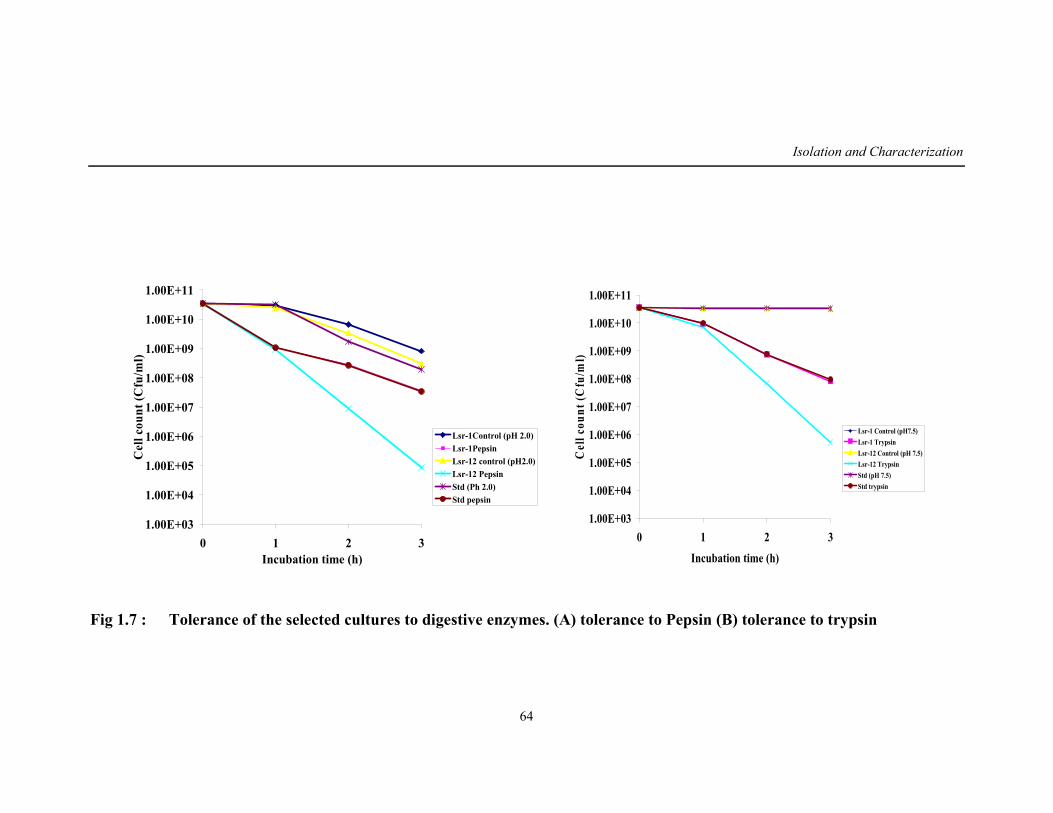
Isolation and Characterization
64
1.00E+03
1.00E+04
1.00E+05
1.00E+06
1.00E+07
1.00E+08
1.00E+09
1.00E+10
1.00E+11
0 1 2 3Incubation time (h)
Cel
l cou
nt (C
fu/m
l)
Lsr-1Control (pH 2.0)Lsr-1PepsinLsr-12 control (pH2.0)Lsr-12 PepsinStd (Ph 2.0)Std pepsin
1.00E+03
1.00E+04
1.00E+05
1.00E+06
1.00E+07
1.00E+08
1.00E+09
1.00E+10
1.00E+11
0 1 2 3
Incubation time (h)
Cel
l cou
nt (C
fu/m
l)
Lsr-1 Control (pH7.5)Lsr-1 TrypsinLsr-12 Control (pH 7.5)Lsr-12 TrypsinStd (pH 7.5)Std trypsin
Fig 1.7 : Tolerance of the selected cultures to digestive enzymes. (A) tolerance to Pepsin (B) tolerance to trypsin
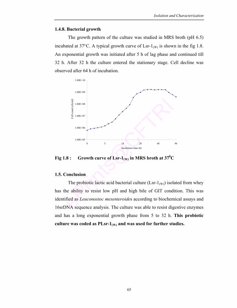
Isolation and Characterization
65
1.4.8. Bacterial growth
The growth pattern of the culture was studied in MRS broth (pH 6.5)
incubated at 37°C. A typical growth curve of Lsr-1(W) is shown in the fig 1.8.
An exponential growth was initiated after 5 h of lag phase and continued till
32 h. After 32 h the culture entered the stationary stage. Cell decline was
observed after 64 h of incubation.
1.00E+05
1.00E+06
1.00E+07
1.00E+08
1.00E+09
1.00E+10
0 5 14 28 48 96
Incubation time (h)
Cel
l cou
nt (c
fu/m
l)
Fig 1.8 : Growth curve of Lsr-1(W) in MRS broth at 370C
1.5. Conclusion
The probiotic lactic acid bacterial culture (Lsr-1(W)) isolated from whey
has the ability to resist low pH and high bile of GIT condition. This was
identified as Leuconostoc mesenteroides according to biochemical assays and
16srDNA sequence analysis. The culture was able to resist digestive enzymes
and has a long exponential growth phase from 5 to 32 h. This probiotic
culture was coded as PLsr-1(W) and was used for further studies.
Chapter 2 Probiotic Functional
Properties of Culture Isolate
Probiotic Functional Properties
66
CHAPTER – 2
PROBIOTIC FUNCTIONAL PROPERTIES OF CULTURE ISOLATE
ABSTRACT
The probiotic culture Leuconostoc mesenteroides (PLsr-1(W)) was
evaluated for its potential functional properties. The culture exerts
antimicrobial activity against seven food borne pathogens and is resistant to
three common antibiotics tested. The inhibitory activity of intracellular cell
free extract of culture to ascorbate autooxidation, ferrous ion chelating ability
and scavenging activity of oxygen radical represents the antioxidative
property of the culture isolate. Cell hydrophobicity with different
hydrocarbon confirms the adhesion ability of the culture. The SDS-PAGE
analysis of the surface protein showed a prominent protein band at 42 KD
which is the S-layer protein responsible for the adhesion of the culture. The
culture has cholesterol lowering ability, β-galactosidase activity and produces
volatile compounds of therapeutic value which shows the importance of
probiotic culture (PLsr-1(W)) as compared to native isolate Lsr-1(W).
Probiotic Functional Properties
67
2.1. Introduction In the quest of discovering how food can enhance health or prevent
chronic diseases, researchers have stumbled on to another range of components in foods beside nutrition. The idea of being healthy by consuming bacteria with beneficial gastrointestinal effects was promoted by the notable immunologist Metchnikoff around a century ago. These bacteria are characterized as “probiotics” that favorably maintain or improve the intestinal microflora, inhibit growth of harmful bacteria, promote good digestion, boost immune function and increase resistance to infection (Haudault et al., 1997; Tannock, 1999).
Industrialization of food bio-transformation increased the economical importance of lactic acid bacteria as they play a crucial role in the development of organoleptic and hygienic quality of fermented products. Therefore the reliability of starter strains in terms of their quality, functional properties and growth performance has become essential (van de Guchte et al., 2002). With the growing threat of food borne pathogens, discovery of new food processes and the consumers demand for natural preventive measures has made these microorganisms as potential biopreservatives for foods. Substantial research on these microorganisms has been focused on such application in treatment of diseases (Hoover, 1993; Ennahar et al., 1999).
A number of requirements have been identified for strains to be effective probiotic microorganism with functional and technological aspects. The required characteristics include stress adaptation towards gastric acid, physiological concentration of bile and adherence to intestinal epithelial cells (Dunne et al., 2001; Schillinger et al., 2005; Khalil et al., 2007). These characters are frequently suggested for the evaluation of the probiotic potential of bacterial strain.
In this regard the probiotic culture L. mesenteroides (PLsr-1(W)) (Chapter 1) was studied for the changes in cellular fatty acids and protein profile when exposed to these stress conditions. The culture PLsr-1(W) was also studied for antimicrobial activity, susceptibility to common antibiotics,
antioxidant activity, cholesterol lowering ability, β-galactosidase activity, adherence ability and the release of volatile compounds.
Probiotic Functional Properties
68
2.2 Materials
• Standard culture Leuconostoc mesenteroides (B640) and dairy starter
culture Streptococcus thermophilus were procured from MTCC culture
collection centre, Chandigard, Hariyana.
• Bacterial strains and media: The probiotic strain (PLsr-1(W)) of
Leuconostoc mesenteroides preserved in MRSA-stabs was propagated
twice in MRS broth at 37°C before use. Native culture isolate (Lsr-
1(W)), Leuconostoc mesenteroides (B640) and Streptococcus
thermophilus were used for comparison.
Chemicals:
1) Brain heart infusion agar (BHI; HiMedia Pvt Ltd, India).
2) Antibiotic disc (Octadisc, HiMedia Pvt Ltd, Mumbai, India).
3) Phosphate-Urea-Magnesium sulphate buffer (pH 7.1; 16.9g K2HPO4,
7.3g KH2PO4, 1.8g Urea, 0.2g MgSO4.7H2O, 1000 ml Distilled water).
4) Lysis buffer (0.06 M Tris HCl, pH 6.8, 10% glycerol, 2 % SDS, 5%
β-mercaptoethanol), sample buffer (0.12 M Tris HCl, 0.1% SDS, 5%
β-mercaptoethanol, 25% glycerol and 0.01% bromophenol blue).
5) Chemicals: NaOH, HCl, H2SO4, ascorbic acid, FeSO4, trichloroacetic
acid, O-phenanthroline, 0-phthalaldehyde, ONPG, KOH and
hexadecane were of analytical grade purchased from HiMedia Pvt Ltd,
India. Protein marker was purchased from Genei, Bangalore, India.
Cholesterol, acrylamide, bisacrylamide and DPPH (1,1-Diphenyl-2-
picryl hydrazyl) were procured from Sigma-Aldrich company, India.
Hexane, methanol, chloroform, dichlormethane, octane, xylene,
toluene and ethanol were of HPLC grade (SRL Company, India).
Equipments: Spectrophotometer (UV-1601 A, Shimadzu Corporation, Japan),
pH meter (Genei, Bangalore, India), Vortex, Centrifuge (Labline Company,
India), Microscope (Olumpus, Japan), Electrophoretic unit (Genei,
Bangalore), Gel documentation system (Bio-Rad Laboratories, USA), Gas
Chromatograph and Mass Spectrometer (Shimadzu Corporation, Japan) with
SE-30 and ELITE-1 column.
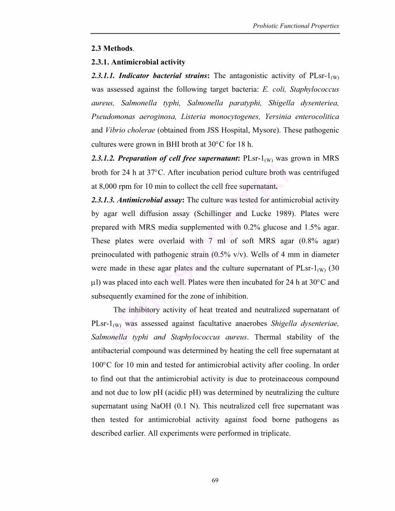
Probiotic Functional Properties
69
2.3 Methods.
2.3.1. Antimicrobial activity
2.3.1.1. Indicator bacterial strains: The antagonistic activity of PLsr-1(W)
was assessed against the following target bacteria: E. coli, Staphylococcus
aureus, Salmonella typhi, Salmonella paratyphi, Shigella dysenteriea,
Pseudomonas aeroginosa, Listeria monocytogenes, Yersinia enterocolitica
and Vibrio cholerae (obtained from JSS Hospital, Mysore). These pathogenic
cultures were grown in BHI broth at 30°C for 18 h.
2.3.1.2. Preparation of cell free supernatant: PLsr-1(W) was grown in MRS
broth for 24 h at 37°C. After incubation period culture broth was centrifuged
at 8,000 rpm for 10 min to collect the cell free supernatant.
2.3.1.3. Antimicrobial assay: The culture was tested for antimicrobial activity
by agar well diffusion assay (Schillinger and Lucke 1989). Plates were
prepared with MRS media supplemented with 0.2% glucose and 1.5% agar.
These plates were overlaid with 7 ml of soft MRS agar (0.8% agar)
preinoculated with pathogenic strain (0.5% v/v). Wells of 4 mm in diameter
were made in these agar plates and the culture supernatant of PLsr-1(W) (30
µl) was placed into each well. Plates were then incubated for 24 h at 30°C and
subsequently examined for the zone of inhibition.
The inhibitory activity of heat treated and neutralized supernatant of
PLsr-1(W) was assessed against facultative anaerobes Shigella dysenteriae,
Salmonella typhi and Staphylococcus aureus. Thermal stability of the
antibacterial compound was determined by heating the cell free supernatant at
100°C for 10 min and tested for antimicrobial activity after cooling. In order
to find out that the antimicrobial activity is due to proteinaceous compound
and not due to low pH (acidic pH) was determined by neutralizing the culture
supernatant using NaOH (0.1 N). This neutralized cell free supernatant was
then tested for antimicrobial activity against food borne pathogens as
described earlier. All experiments were performed in triplicate.
Probiotic Functional Properties
70
2.3.2. Antibiotic susceptibility test
The susceptibility to antibiotics which are commonly used by animals
and humans (Food and Drug administration, 2001) was tested by disc
diffusion method (Brashears and Durre, 1999). The culture PLsr-1(W) (1%) in
MRS soft agar (0.8% agar) was overlaid on MRS agar plate. An antibiotic
disc (Octadisc) was placed on it to allow the diffusion of antibiotics into the
medium and then incubated at 37°C for 24 h. The inhibition zone around each
antibiotic was measured to check the susceptibility of the culture. The
minimal inhibitory concentration (MIC, expressed in µg/ml) was determined
by using antibiotics at variable concentrations. The MIC was defined as the
smallest amount of antibiotic needed to inhibit the growth of bacteria after
incubation for 24 h.
2.3.3. Antioxidative activity
2.3.3.1. Preparation of intracellular cell free extract: The exponentially (16
h) grown culture (PLsr-1(W)) was harvested by centrifugation at 8000 rpm for
15 min. Cell biomass obtained was resuspended in deionized water after
washing. The cell suspension was then homogenized for 5 min at 4°C. Cell
debris was removed by centrifugation at 10,000 rpm for 10 min and the
resulting supernatant was used as the intracellular cell free extract.
2.3.3.2. Ascorbate autooxidation assay: The antioxidant activity of the
culture was determined by ascorbate autooxidation assay (Mishra and
Korachich, 1984). Intracellular cell free extract (0.1 ml) was added to the
mixture of ascorbate stock solution (0.1 ml; 5 mM) and 9.8 ml of phosphate
buffer (0.2 M; pH 7.0). The mixture was then quickly measured for
absorbance at 265 nm using spectrophotometer.
Percentage of inhibition of ascorbate autooxidation was calculated by using
the following formula:
A265 (Sample) % Inhibition = 1- × 100 A265 (blank)
Probiotic Functional Properties
71
2.3.3.3. Metal ion chelating assay: The chelating ability of intracellular cell
free extract of the culture for ferrous ion was determined by ion chelating
assay. The method developed by Yamauchi et al. (1984) and modified by
Lin and Yen (1999) was used for assaying. The intracellular cell free extract
(0.5 ml) was mixed with 0.1 ml of ascorbate (1g/dl), followed by 0.1 ml of
FeSO4 (0.4 g/dl) and 1 ml of NaOH (0.2 M). The mixture was incubated at
37°C in a water bath for 20 min and then 0.2 ml of trichloroacetic acid (10%)
was added. Mixture was then centrifuged at 5000 rpm for 10 min to obtain the
supernatant. O-phenanthroline (0.5 ml; 1 g/L) was added to the supernatant
and incubated for 10 min. Absorbance of the mixture was then measured at
510 nm.
2.3.3.4. DPPH assay (1,1-Diphenyl-2-picryl hydrazyl): The scavenging of
DPPH by L.mesenteroides (PLsr-1(W)) was analyzed by the method of Brand
Williams et al. (1995) which was modified by Pyo et al. (2005). Intracellular
cell free extract (0.02 ml) and 1.0 ml of freshly prepared DPPH solution
(0.1 mM in methanol) were mixed and the absorbance was measured
spectrophotometrically at 517 nm after 30 min of incubation. The scavenging
activity was calculated by the following equation. Blank sample contained
deionized water.
(Ab–Ac) Scavenging activity (%) = Aa – × 100 Aa
Where, Aa – absorbance of DPPH solution without sample
Ab – absorbance of mixture containing sample and DPPH
Ac – absorbance of blank solution without DPPH
2.3.4. Anticholesterolemic activity
An exponentially (16 h) grown culture L. mesenteroides (PLsr-1(W))
was inoculated (1%) into MRS broth (10 ml) supplemented with water
soluble cholesterol (100 mg/L) and incubated anaerobically for 24 h at 37°C.
After the incubation period, the cell biomass was collected by centrifugation
Probiotic Functional Properties
72
at 8,000 rpm for 15 min at 4°C and suspended in distilled water (10 ml). The
cholesterol content was then measured by O-phthalaldehyde method as
described by Rudel and Morris (1973) and modified by Gilliland et al. (1985).
Sample extract (0.5ml) was placed in a clean test tube and mixed with
3 ml of ethanol (95%). This was followed by the addition of 2 ml of
potassium hydroxide (50%). The mixture was thoroughly vortexed and heated
on a water bath for 10 min at 60°C. After cooling, hexane (5 ml) was added
into the test tube and mixed thoroughly. This was allowed to stand at room
temperature for 10 min and then 3 ml of distilled water was added and
vortexed. The hexane (2.5 ml) layer was carefully transferred into a clean test
tube and evaporated at 60°C. To this dry tube, 4 ml of O-phthalaldehyde
reagent (0.5 mg per ml of glacial acetic acid) was added and allowed to stand
at room temperature for 10 min followed by slow addition of concentrated
H2SO4 (2 ml). The contents of the tube were mixed thoroughly. After 10 min
of incubation the absorbance was read at 550 nm against a reagent blank. The
absorbance was compared with the standard curve (Fig. 2.1) to determine the
concentration of the cholesterol. Results are expressed as µg of cholesterol
per ml.
0
0.05
0.1
0.15
0.2
0.25
0.3
0.35
0.4
0.45
0 20 40 60 80 100 120 140 160 180
Fig 2.1 : Standard graph of cholesterol estimation Note: Standard curve was prepared by following the same procedure but in place of sample extract different concentrations of cholesterol (20-140 µg) were used. The absorbance values were plotted against µg of cholesterol. Values are average of three experiments (n=3).
Concentration of cholesterol (µg)
Opt
ical
den
sity
(550
nm
)
Probiotic Functional Properties
73
2.3.5. β-galactosidase activity
The culture PLsr-1(W) was tested for β-galactosidase activity by
qualitative assay using ONPG discs. The culture grown in MRS broth was
harvested by centrifugation at 5,000 rpm for 15 min. Cell biomass was
washed twice in phosphate buffer (pH 7.0) and resuspended in the same
buffer. The cell suspension (1 ml) was permeabilized with 50 µl of
toluene:acetone (1:9) mixture. ONPG disc was immersed in this suspension
and incubated for 30 min at 37°C. After the incubation time, the development
of yellow color was recorded as a positive reaction for β-galactosidase
activity. Quantitatively the enzyme activity was estimated by using ONPG as
substrate according to the method of Bhowmik and Marth (1989). Specific
activity (µM mg-1) was then expressed as the amount of O-nitrophenol (ONP)
released per mg of protein.
2.3.5.1. Influence of carbon source on enzymatic activity: PLsr-1(W) was
inoculated at a rate of 1% (v/v) in MRS basal broth supplemented with
glucose, galactose, lactose, sucrose and fructose at a concentration of
2% (w/v) in different test tubes and incubated at 37°C for 24 h. After the
incubation period, cell biomass was collected by centrifugation and assayed
for enzyme activity.
2.3.6. Adherence ability
2.3.6.1. Cell hydrophobicity assay (Microbial adhesion to hydrocarbons) The in-vitro method by Rosenberg et al. (1980) was used to assess the
bacterial adhesion to hydrocarbons (toluene, xylene, octane and hexadecane).
The culture L. mesenteroides (PLsr-1(W)) was grown at 37°C for 16 h in MRS
broth containing cystine hydrochloride. Cell biomass was collected by
centrifugation at 5000 rpm for 10 min, washed twice with phosphate-urea-
magnesium sulphate buffer (pH 7.1) and resuspended in the same. The optical
density of the suspension was adjusted to 0.8-0.9 at 610 nm using a
spectrophotometer. To this bacterial suspension (2.4 ml) hydrocarbons were
added (0.4 ml) separately and mixed well for 2 min followed by incubation at
Probiotic Functional Properties
74
37°C for 1 h. The aqueous phase was carefully removed and the absorbance
was measured at 610 nm.
The fraction of adherent cells was calculated as percent decrease in
absorbance of aqueous phase as compared to that of original cell suspension.
Cell surface hydrophobicity or the percent adhesion was calculated by the
following formula
Initial OD – Final OD % adhesion = × 100 Initial OD
2.3.6.2. Adhesion of PLsr-1(W) to rat intestinal epithelium The in-vitro adhesion to intestinal epithelium layer was performed by
the method of Mayra-Makinen et al. (1983) with modifications. Ileal sample
was collected from male Albino Wistar rats. The tissue was held in PBS at
4°C for 30 min to loosen surface mucus, and then washed three times with
buffer (pH 7.0). The adhesion test was performed by incubating tissue sample
(1 cm2) in bacterial suspension (108 cfu/ml in buffer) at 37°C for 30 min.
Treated tissue sample was fixed in 10% formalin, dehydrated by increasing
concentrations of ethanol, and embedded in paraffin. Serial sections (5 µm)
were cut, mounted on standard microscope slides and stained for
identification of Gram-positive and Gram-negative bacteria. Slides were
examined and photographed using a light microscope.
2.3.6.3. Analysis of cell surface protein of PLsr-1(W) (a) Extraction of S-layer protein
The exponentially grown (16 h) culture PLsr-1(W) was centrifuged at
8,000 rpm for 10 min at 4°C. The cell biomass was washed twice in ice-cold
water and resuspended in 10 ml of Lithium chloride (5 M). This was
incubated at 37°C for 1 h. The supernatant was then collected by
centrifugation (10,000 rpm for 15 min) at 4°C and dialyzed against water at
4°C. The dialyzed sample was freeze dried and resuspended in 1 ml of Tris
buffer (pH 7.5).
Probiotic Functional Properties
75
Note: Dialyzing bag preparation: Cutoff the dialyzing tube to required length
and boil it in water for 10 min containing Na2CO3 (2%) and 1mM EDTA.
After cooling, wash with distilled water and again boil for 10 min in distilled
water. On cooling rinse with distilled water thrice and store in water at 4°C
until use.
(b) SDS-PAGE analysis of S-layer protein
Electrophoresis was performed as described by Laemmli (1970). The
polyacrylamide gel was composed of a 4% stacking gel and a 12.5%
separating gel.
The S-layer protein was diluted in an equal volume of the tracking
buffer (0.12 M Tris HCl; pH 6.8 and 0.01% bromophenol blue) and then
applied to the gel. Electrophoresis was done at 50 mA through the stacking
gel and 70 mA through separating gel until the tracking dye migrates to the
bottom of the gel. Further the gel was stained in a mixture of 1% (wt/v)
coomassie blue, 40% (v/v) methanol and 20% (v/v) acetic acid. After
overnight staining it was decolorized under continuous shaking in a 25% (v/v)
methanol solution containing 10% (v/v) acetic acid. The stained protein
patterns were then scanned with a gel documentation system. Molecular
masses of the protein extracts were estimated using the linear relationship
between the marker and the protein band.
2.3.7. Analysis of volatile compounds
2.3.7.1. Extraction of volatile compounds
PLsr-1(W) was grown in MRS broth at 37°C. Formation of volatile
compounds were analyzed at different growth stages. An aliquot (5 ml) of
culture broth was drawn at regular interval of time and the cell biomass was
collected by centrifugation (5,000 rpm for 20 min). The cell biomass was
suspended in dichloromethane (10 ml) and mechanically homogenized.
Extraction procedure was repeated thrice and the solvent layer was collected,
dried over anhydrous sodium sulphate and concentrated to 0.5 ml under a
stream of nitrogen.
Probiotic Functional Properties
76
2.3.7.2. Gas chromatographic analysis and GCMS conditions
GC analysis was carried out with a Packed series 15A gas
chromatograph, equipped with a flame ionization detector. A (3%) SE-30
column (3m X 3mm id) with mesh size 80/100 was used. O2 flow of 300
ml/min and H2 flow of 30 ml/min was employed. The operating conditions
were as follows: the temperature was programmed from 100°C to 250°C
(100°C with 6 min hold, 100-150°C; 4°C/min, 150-220°C; 8°C/min). The
injector and detector temperature were set at 250°C. The nitrogen carrier
velocity was 30 ml/min.
Mass spectra were obtained with Turbomass gold mass spectrometer
(Perkin Elmer International, Switzerland) coupled with gas chromatograph
equipped with turbomass version-4 software. Sample injection was done in
the split mode (40:1) in an ELITE-1 column, 30 m X 0.25 mm id and 0.25 µm
film thickness coated with 100% poly dimethoxy siloxane. Pure Helium was
used as the GC carrier gas at a flow rate of 1 ml/min. The GC injector
temperature was set at 250°C, oven temperature at 100°C. The detector was
adjusted to 250°C. The mass spectra were determined at 70eV with emission
current 100 µA and ion source was held at 150°C. Acquisition and processing
of mass spectra were carried out by means of a computer and the compounds
were identified with the aid of mass spectral data bases.
2.3.8. Changes in cellular fatty acids and proteins of PLsr-1(W)
2.3.8.1. Extraction of cellular membrane fatty acids
Cell biomass of the culture PLsr-1(W) and native isolate Lsr1(W)
collected separately by centrifugation at 8000 rpm for 10 min. The cellular
fatty acids were then extracted by the method of Bligh and Dyer (1959). The
cell suspension (1 ml) in distilled water was mixed with methanol:chloroform
(2:1) mixture (3.7 ml), vortexed thoroughly and incubated for 2 h at 28°C.
The mixture was then centrifuged at 5000 rpm for 15 min at 4°C to collect the
supernatant. Extraction was repeated with 4.75 ml of methanol:chloroform:water
Probiotic Functional Properties
77
mixture (2:1:0.8) and the supernatant extract was collected by centrifugation.
To the pooled extract water (2.5 ml) and chloroform (2.5 ml) were added and
vortexed thoroughly. This was kept at room temperature for phase separation.
The chloroform layer was quantitatively taken into a clean dry test tube and
evaporated under nitrogen flow. The dried tubes were stored at -20°C.
Analysis of fatty acids by GC and GCMS method
The dried tubes containing the fatty acids were dissolved in hexane
(2 ml). To this methanolic KOH (2 N; 0.1 ml) was added and mixed
thoroughly. The upper hexane layer was taken in a clean test tube and
evaporated to dryness under N2 flow. The dried residues were then dissolved
in 0.1 ml of hexane and analyzed by GC with the following operating
conditions: injection temperature 220°C and detector temperature 250°C. The
temperature was programmed from 100°C to 220°C (100°C held for 5 min,
100-220°C; 10°C/min, at 220°C held for 10 min). Carrier gas N2 was used at
a flow rate of 1.5 ml per min. Injection volume was 1µl. The identification of
peaks was based on the comparison of fragmentation pattern of GCMS
chromatogram with GCMS library data base and then GCMS volumes by
Noever et al. (1998).
2.3.8.2. Extraction of cellular proteins
Cell biomass of the culture PLsr-1(W) and Lsr-1(W) was collected
separately by centrifugation at 8000 rpm for 10 min, washed in Tris HCl
buffer (pH 7.0) and resuspended in the same. Cells were then solubilized by
boiling for 5 min in disruption buffer and analyzed on SDS PAGE as
described in section 2.3.6.3.
2.4. Results and Discussion
2.4.1. Antimicrobial activity
One of the most frequent health claims for probiotics is the reduction
and prevention of infectious diseases in the gastrointestinal tract (GIT).
Enteric pathogens infect the host in different atmospheric conditions of GIT
causing diarrhoel diseases. Many scientists all over the world have reported
Probiotic Functional Properties
78
the occurrence of food borne pathogens contaminating processed foods and
vegetables thus causing health hazards (Vescovo et al., 1996; Kannappan and
Manja, 2004; Wilderdyke et al., 2004). Innovative approaches have been tried
as an alternative to antibiotics in treating these diseases which include usage
of live biotherapeutic agents such as bacterial isolates (Daly and Davis, 1998;
Soomro et al., 2002; Oyetayo et al., 2003). The antimicrobial effect of LAB
has been used to extend the shelf life of many foods and in treating food
borne diseases (Savadogo et al., 2004).
The effect of probiotic strains depends on their ability to persist and
compete with pathogens in GIT. Antimicrobial activities of LAB have been
widely investigated in the past few years (Daeschel, 1989; Piard and
Desmazeaud, 1991; Hechard et al., 1992). The inhibitory activity of LAB
against spoilage bacteria and food borne pathogens is mainly based on acid
production, competition for nutrients and space, formation of hydrogen
peroxide, carbon dioxide and other products of catabolism and bacteriocins
(Lucke and Earnshaw, 1990). Broad-spectrum inhibition is generally
attributed to organic acid production and / or to H2O2. Narrow inhibitory
spectrum includes synthesis of bacteriocins (Tagg et al., 1976; Klaenhammer,
1988).
In the present experiment, agar well diffusion assay was used to
determine the antimicrobial activity of probiotic L. mesenteroides (PLsr-1(W))
against nine food borne pathogens. The antimicrobial activity of PLsr-1(W)
was found to be better than the native isolate Lsr-1(W) (Table 2.1). The culture
PLsr-1(W) inhibited the growth of seven food borne pathogens with the
inhibition zone of 11-16 mm whereas Lsr-1(W) inhibited with the inhibition
zone of 8-12 mm. The culture PLsr-1(W) exhibited maximum inhibition to
V. cholerae (16 mm) as compared to Lsr-1(W) (12 mm), dairy starter culture
S. thermophilus (10 mm) and Standard L. mesenteroides (B640) (11 mm).
Other pathogens like S. typhi, P. aeroginosa and S. aureus (Kannappan and
Manja, 2004; Vescovo et al., 1996; Wilderdyke et al., 2004) were inhibited
by PLsr-1(W) with an inhibition zone of 13, 14 and 11 mm diameter
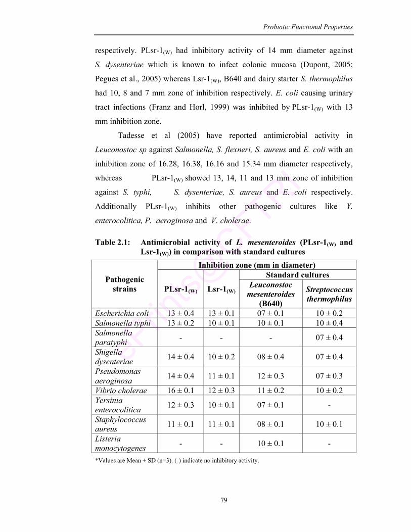
Probiotic Functional Properties
79
respectively. PLsr-1(W) had inhibitory activity of 14 mm diameter against
S. dysenteriae which is known to infect colonic mucosa (Dupont, 2005;
Pegues et al., 2005) whereas Lsr-1(W), B640 and dairy starter S. thermophilus
had 10, 8 and 7 mm zone of inhibition respectively. E. coli causing urinary
tract infections (Franz and Horl, 1999) was inhibited by PLsr-1(W) with 13
mm inhibition zone.
Tadesse et al (2005) have reported antimicrobial activity in
Leuconostoc sp against Salmonella, S. flexneri, S. aureus and E. coli with an
inhibition zone of 16.28, 16.38, 16.16 and 15.34 mm diameter respectively,
whereas PLsr-1(W) showed 13, 14, 11 and 13 mm zone of inhibition
against S. typhi, S. dysenteriae, S. aureus and E. coli respectively.
Additionally PLsr-1(W) inhibits other pathogenic cultures like Y.
enterocolitica, P. aeroginosa and V. cholerae.
Table 2.1: Antimicrobial activity of L. mesenteroides (PLsr-1(W) and Lsr-1(W)) in comparison with standard cultures
Inhibition zone (mm in diameter) Standard cultures Pathogenic
strains PLsr-1(W) Lsr-1(W)Leuconostoc
mesenteroides (B640)
Streptococcus thermophilus
Escherichia coli 13 ± 0.4 13 ± 0.1 07 ± 0.1 10 ± 0.2 Salmonella typhi 13 ± 0.2 10 ± 0.1 10 ± 0.1 10 ± 0.4 Salmonella paratyphi - - - 07 ± 0.4
Shigella dysenteriae 14 ± 0.4 10 ± 0.2 08 ± 0.4 07 ± 0.4
Pseudomonas aeroginosa 14 ± 0.4 11 ± 0.1 12 ± 0.3 07 ± 0.3
Vibrio cholerae 16 ± 0.1 12 ± 0.3 11 ± 0.2 10 ± 0.2 Yersinia enterocolitica 12 ± 0.3 10 ± 0.1 07 ± 0.1 -
Staphylococcus aureus 11 ± 0.1 11 ± 0.1 08 ± 0.1 10 ± 0.1
Listeria monocytogenes - - 10 ± 0.1 -
*Values are Mean ± SD (n=3). (-) indicate no inhibitory activity.
Probiotic Functional Properties
80
To determine the effect of heat/ higher temperature, cell free culture
supernatant was treated at 100°C for 10 min and then tested for antimicrobial
activity against S. dysenteriae, S. typhi and S. aureus. These are important
food borne pathogens which are known to cause disease outbreaks all over the
world. From the result it was observed that the heat treated cell free
supernatant shows activity less (≤ 9 mm) as compared to non-thermal treated
sample (≥ 11 mm) (Table 2.2; Fig. 2.2). This shows that the antimicrobial or
bacteriocin like compound present in the cell free supernatant is heat
sensitive.
To determine that acidity is not the only criteria responsible for
antimicrobial activity of PLsr-1(W), the cell free supernatant was adjusted to
pH 6.0 using 0.1 N NaOH and assayed for antimicrobial activity. The results
indicate that the culture supernatant adjusted to pH 6.0 showed very slight
reduction (P>0.01) in the activity. Inhibitory activity against S. dysenteriae,
S. typhi and S. aureus was reduced by 14.29, 15.39 and 9.09% respectively
(Fig. 2.2).
Draksler et al. (2004) have shown Lactobacillus DDL48 having
antimicrobial activity against Salmonella typhi with an inhibition zone of
8–10 mm diameter. The effect was observed with a non-adjusted pH
supernatant. However, the supernatant adjusted to pH 6.0 was unable to
inhibit the growth of the pathogen. In the present study, PLsr-1(W) inhibits
S. typhi with an inhibition zone of 13 mm diameter. The culture supernatant
when adjusted to pH 6.0 had inhibitory activity against S. typhi with an
inhibition zone of 11 mm diameter.
The results indicate that PLsr-1(W) can be used as a starter culture for
traditional fermented foods to improve its safety. It could be helpful in
treatment of diseases like typhoid, shigellosis, lung infection, cholera,
enterocolitis and food poisoning.
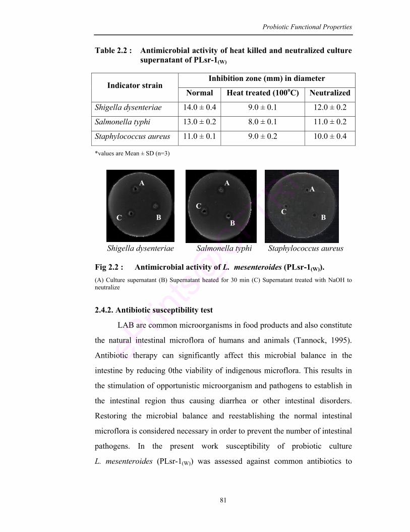
Probiotic Functional Properties
81
Table 2.2 : Antimicrobial activity of heat killed and neutralized culture supernatant of PLsr-1(W)
Inhibition zone (mm) in diameter
Indicator strain Normal Heat treated (100oC) Neutralized
Shigella dysenteriae 14.0 ± 0.4 9.0 ± 0.1 12.0 ± 0.2
Salmonella typhi 13.0 ± 0.2 8.0 ± 0.1 11.0 ± 0.2
Staphylococcus aureus 11.0 ± 0.1 9.0 ± 0.2 10.0 ± 0.4
*values are Mean ± SD (n=3)
Shigella dysenteriae Salmonella typhi Staphylococcus aureus Fig 2.2 : Antimicrobial activity of L. mesenteroides (PLsr-1(W)). (A) Culture supernatant (B) Supernatant heated for 30 min (C) Supernatant treated with NaOH to neutralize
2.4.2. Antibiotic susceptibility test
LAB are common microorganisms in food products and also constitute
the natural intestinal microflora of humans and animals (Tannock, 1995).
Antibiotic therapy can significantly affect this microbial balance in the
intestine by reducing 0the viability of indigenous microflora. This results in
the stimulation of opportunistic microorganism and pathogens to establish in
the intestinal region thus causing diarrhea or other intestinal disorders.
Restoring the microbial balance and reestablishing the normal intestinal
microflora is considered necessary in order to prevent the number of intestinal
pathogens. In the present work susceptibility of probiotic culture
L. mesenteroides (PLsr-1(W)) was assessed against common antibiotics to
A A
A
B B B
C C
C
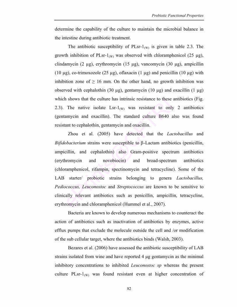
Probiotic Functional Properties
82
determine the capability of the culture to maintain the microbial balance in
the intestine during antibiotic treatment.
The antibiotic susceptibility of PLsr-1(W) is given in table 2.3. The
growth inhibition of PLsr-1(W) was observed with chloramphenicol (25 µg),
clindamycin (2 µg), erythromycin (15 µg), vancomycin (30 µg), ampicillin
(10 µg), co-trimoxozole (25 µg), oflaxacin (1 µg) and penicillin (10 µg) with
inhibition zone of ≥ 16 mm. On the other hand, no growth inhibition was
observed with cephalothin (30 µg), gentamycin (10 µg) and oxacillin (1 µg)
which shows that the culture has intrinsic resistance to these antibiotics (Fig.
2.3). The native isolate Lsr-1(W) was resistant to only 2 antibiotics
(gentamycin and oxacillin). The standard culture B640 also was found
resistant to cephalothin, gentamycin and oxacillin.
Zhou et al. (2005) have detected that the Lactobacillus and
Bifidobacterium strains were susceptible to β-Lactam antibiotics (penicillin,
ampicillin, and cephalothin) also Gram-positive spectrum antibiotics
(erythromycin and novobiocin) and broad-spectrum antibiotics
(chloramphenicol, rifampin, spectinomycin and tetracycline). Some of the
LAB starter/ probiotic strains belonging to genera Lactobacillus,
Pediococcus, Leuconostoc and Streptococcus are known to be sensitive to
clinically relevant antibiotics such as penicillin, ampicillin, tetracycline,
erythromycin and chloramphenicol (Hummel et al., 2007).
Bacteria are known to develop numerous mechanisms to counteract the
action of antibiotics such as inactivation of antibiotics by enzymes, active
efflux pumps that exclude the molecule outside the cell and /or modification
of the sub cellular target, where the antibiotics binds (Walsh, 2003).
Bezares et al. (2006) have assessed the antibiotic susceptibility of LAB
strains isolated from wine and have reported 4 µg gentamycin as the minimal
inhibitory concentrations to inhibited Leuconostoc sp whereas the present
culture PLsr-1(W) was found resistant even at higher concentration of
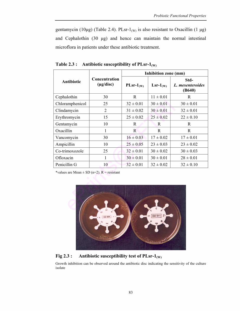
Probiotic Functional Properties
83
gentamycin (10µg) (Table 2.4). PLsr-1(W) is also resistant to Oxacillin (1 µg)
and Cephalothin (30 µg) and hence can maintain the normal intestinal
microflora in patients under these antibiotic treatment.
Table 2.3 : Antibiotic susceptibility of PLsr-1(W)
Inhibition zone (mm)
Antibiotic Concentration (µg/disc) PLsr-1(W) Lsr-1(W)
Std- L. mesenteroides
(B640) Cephalothin 30 R 11 ± 0.01 R Chloramphenicol 25 32 ± 0.01 30 ± 0.01 30 ± 0.01 Clindamycin 2 31 ± 0.02 30 ± 0.01 32 ± 0.01 Erythromycin 15 25 ± 0.02 25 ± 0.02 22 ± 0.10 Gentamycin 10 R R R Oxacillin 1 R R R Vancomycin 30 16 ± 0.03 17 ± 0.02 17 ± 0.01 Ampicillin 10 25 ± 0.05 23 ± 0.03 23 ± 0.02 Co-trimoxozole 25 32 ± 0.01 30 ± 0.02 30 ± 0.03 Ofloxacin 1 30 ± 0.01 30 ± 0.01 28 ± 0.01 Penicillin G 10 32 ± 0.01 32 ± 0.02 32 ± 0.10
*values are Mean ± SD (n=2). R = resistant
Fig 2.3 : Antibiotic susceptibility test of PLsr-1(W)
Growth inhibition can be observed around the antibiotic disc indicating the sensitivity of the culture isolate
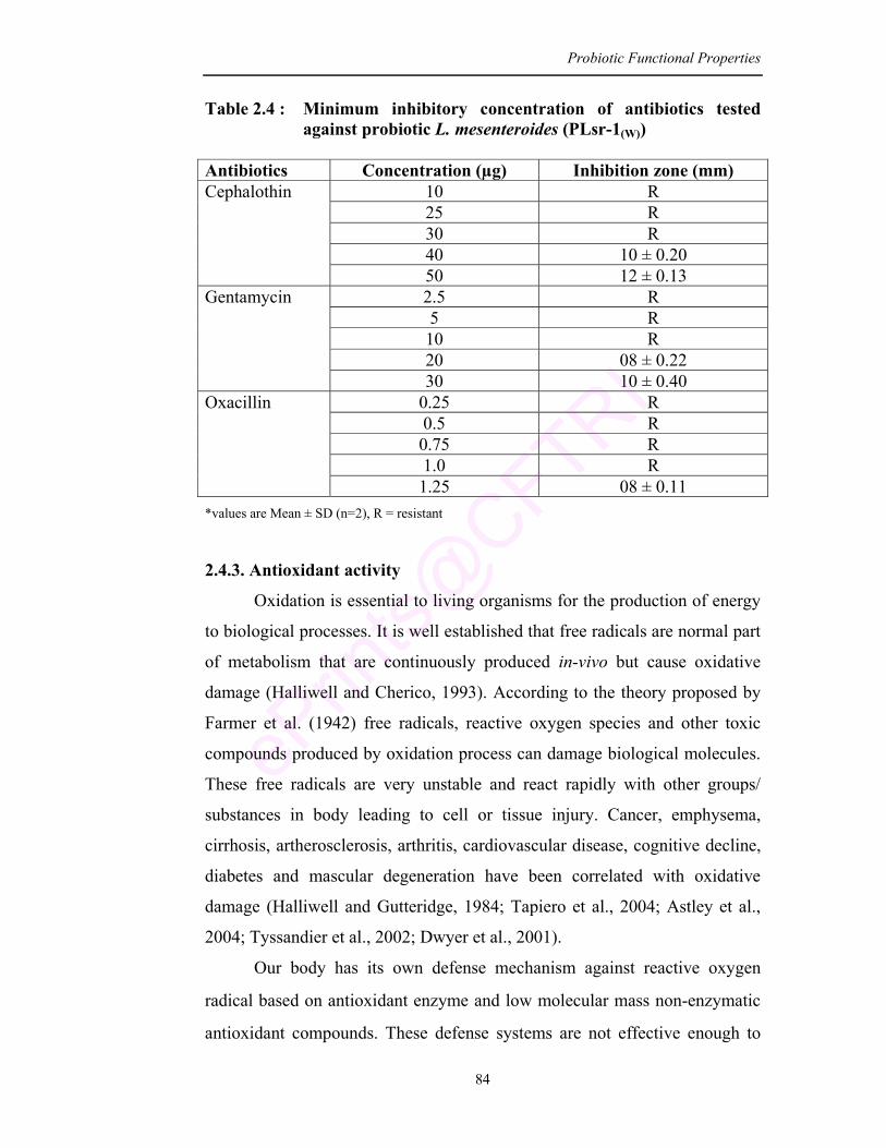
Probiotic Functional Properties
84
Table 2.4 : Minimum inhibitory concentration of antibiotics tested against probiotic L. mesenteroides (PLsr-1(W))
Antibiotics Concentration (µg) Inhibition zone (mm)
10 R 25 R 30 R 40 10 ± 0.20
Cephalothin
50 12 ± 0.13 2.5 R 5 R 10 R 20 08 ± 0.22
Gentamycin
30 10 ± 0.40 0.25 R 0.5 R 0.75 R 1.0 R
Oxacillin
1.25 08 ± 0.11 *values are Mean ± SD (n=2), R = resistant
2.4.3. Antioxidant activity
Oxidation is essential to living organisms for the production of energy
to biological processes. It is well established that free radicals are normal part
of metabolism that are continuously produced in-vivo but cause oxidative
damage (Halliwell and Cherico, 1993). According to the theory proposed by
Farmer et al. (1942) free radicals, reactive oxygen species and other toxic
compounds produced by oxidation process can damage biological molecules.
These free radicals are very unstable and react rapidly with other groups/
substances in body leading to cell or tissue injury. Cancer, emphysema,
cirrhosis, artherosclerosis, arthritis, cardiovascular disease, cognitive decline,
diabetes and mascular degeneration have been correlated with oxidative
damage (Halliwell and Gutteridge, 1984; Tapiero et al., 2004; Astley et al.,
2004; Tyssandier et al., 2002; Dwyer et al., 2001).
Our body has its own defense mechanism against reactive oxygen
radical based on antioxidant enzyme and low molecular mass non-enzymatic
antioxidant compounds. These defense systems are not effective enough to
Probiotic Functional Properties
85
totally prevent the damage. Therefore, food supplements containing
antioxidants may be useful to human body in reducing oxidative damage
(Zommara et al., 1996; Oxman et al., 2000; Terahara et al., 2001; Kullisaar
et al., 2003).
Various synthetic and natural antioxidants have been reported.
However Lin and Yen (1999) have predicted doubts about the safety and long
term effects on health of synthetic antioxidants, hence antioxidants from
natural sources are likely to be desirable. In this regard, antioxidative property
of LAB has been exploited to be used as a natural source of antioxidants.
There are various reports on LAB that are able to degrade the superoxide
anion and hydrogen peroxide (Alander et al., 1997; Miller and Britigan, 1997;
Kullisaar et al., 2002). Considering this, the present isolate L. mesenteroides
(PLsr-1(W)) was studied for its antioxidative property.
Earlier authors have described various methods to measure the
antioxidative property and the results are expressed in a variety of ways.
Inhibition of lipid peroxidation (Wanasundrara et al., 1994; Bertelsen et al.,
1995), ascorbate autooxidation (Rashid et al., 1993) and norepinephrine
oxidation (Mishra and Korachich, 1984) are used. In the present study,
antioxidative activity was measured by method of ascorbate autooxidation,
metal chelating and DPPH scavenging activity.
2.4.3.1. Inhibition of ascorbate autooxidation
The inhibition of ascorbate autooxidation by intracellular cell free
supernatant of PLsr-1(W) increases with incubation time and reaches
maximum (5.7%) after 6 min, whereas the native isolate Lsr-1(W) had 4.9%
inhibition after 6 min (Table 2.5). Some of the earlier investigators have
found the ability of LAB cultures to inhibit ascorbate autooxidation (Esaki
et al., 1994; Berghofer et al., 1998; Sheih et al., 2000; Chung et al., 2002).
Wang et al. (2006) have shown that fermentation of soymilk with lactic acid
bacteria and Bifidobacteria significantly increases the inhibition rate of
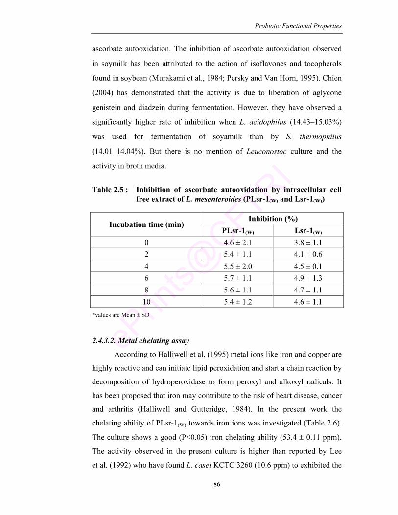
Probiotic Functional Properties
86
ascorbate autooxidation. The inhibition of ascorbate autooxidation observed
in soymilk has been attributed to the action of isoflavones and tocopherols
found in soybean (Murakami et al., 1984; Persky and Van Horn, 1995). Chien
(2004) has demonstrated that the activity is due to liberation of aglycone
genistein and diadzein during fermentation. However, they have observed a
significantly higher rate of inhibition when L. acidophilus (14.43–15.03%)
was used for fermentation of soyamilk than by S. thermophilus
(14.01–14.04%). But there is no mention of Leuconostoc culture and the
activity in broth media.
Table 2.5 : Inhibition of ascorbate autooxidation by intracellular cell
free extract of L. mesenteroides (PLsr-1(W) and Lsr-1(W))
*values are Mean ± SD
2.4.3.2. Metal chelating assay
According to Halliwell et al. (1995) metal ions like iron and copper are
highly reactive and can initiate lipid peroxidation and start a chain reaction by
decomposition of hydroperoxidase to form peroxyl and alkoxyl radicals. It
has been proposed that iron may contribute to the risk of heart disease, cancer
and arthritis (Halliwell and Gutteridge, 1984). In the present work the
chelating ability of PLsr-1(W) towards iron ions was investigated (Table 2.6).
The culture shows a good (P<0.05) iron chelating ability (53.4 ± 0.11 ppm).
The activity observed in the present culture is higher than reported by Lee
et al. (1992) who have found L. casei KCTC 3260 (10.6 ppm) to exhibited the
Inhibition (%) Incubation time (min)
PLsr-1(W) Lsr-1(W) 0 4.6 ± 2.1 3.8 ± 1.1 2 5.4 ± 1.1 4.1 ± 0.6 4 5.5 ± 2.0 4.5 ± 0.1 6 5.7 ± 1.1 4.9 ± 1.3 8 5.6 ± 1.1 4.7 ± 1.1 10 5.4 ± 1.2 4.6 ± 1.1
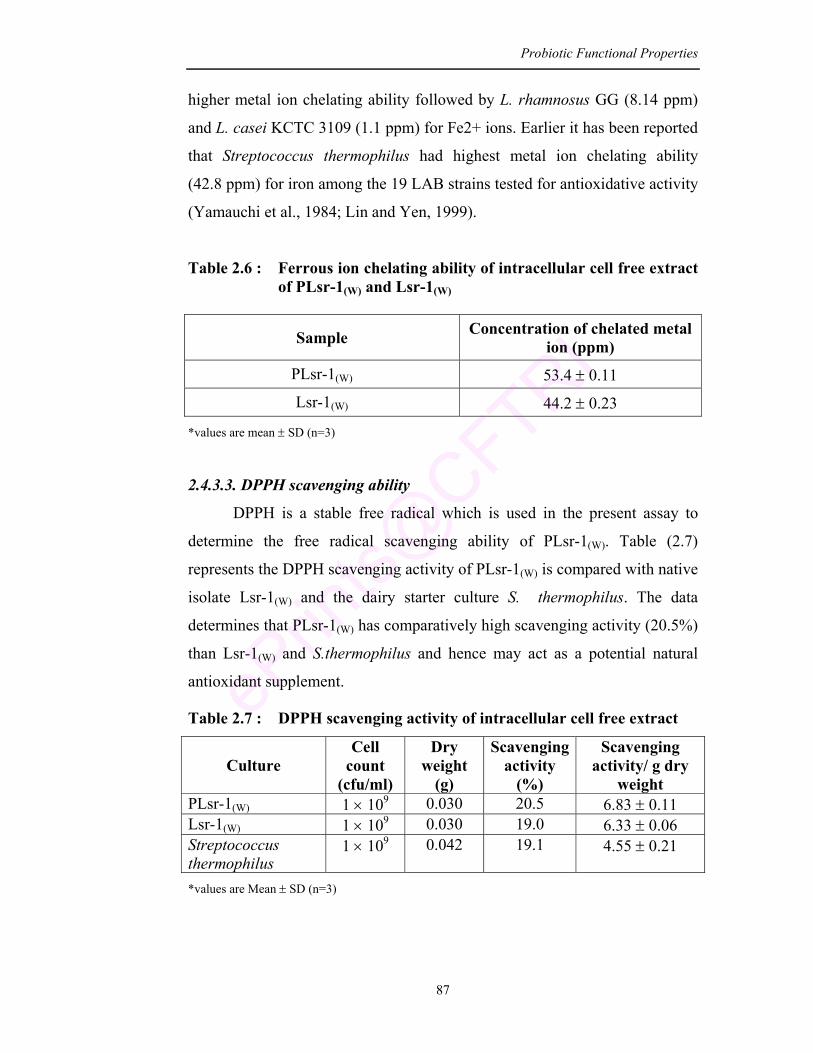
Probiotic Functional Properties
87
higher metal ion chelating ability followed by L. rhamnosus GG (8.14 ppm)
and L. casei KCTC 3109 (1.1 ppm) for Fe2+ ions. Earlier it has been reported
that Streptococcus thermophilus had highest metal ion chelating ability
(42.8 ppm) for iron among the 19 LAB strains tested for antioxidative activity
(Yamauchi et al., 1984; Lin and Yen, 1999).
Table 2.6 : Ferrous ion chelating ability of intracellular cell free extract of PLsr-1(W) and Lsr-1(W)
Sample Concentration of chelated metal ion (ppm)
PLsr-1(W) 53.4 ± 0.11
Lsr-1(W) 44.2 ± 0.23
*values are mean ± SD (n=3)
2.4.3.3. DPPH scavenging ability
DPPH is a stable free radical which is used in the present assay to
determine the free radical scavenging ability of PLsr-1(W). Table (2.7)
represents the DPPH scavenging activity of PLsr-1(W) is compared with native
isolate Lsr-1(W) and the dairy starter culture S. thermophilus. The data
determines that PLsr-1(W) has comparatively high scavenging activity (20.5%)
than Lsr-1(W) and S.thermophilus and hence may act as a potential natural
antioxidant supplement.
Table 2.7 : DPPH scavenging activity of intracellular cell free extract
Culture Cell
count (cfu/ml)
Dry weight
(g)
Scavenging activity
(%)
Scavenging activity/ g dry
weight PLsr-1(W) 1 × 10⁹ 0.030 20.5 6.83 ± 0.11 Lsr-1(W) 1 × 10⁹ 0.030 19.0 6.33 ± 0.06 Streptococcus thermophilus
1 × 10⁹ 0.042 19.1 4.55 ± 0.21
*values are Mean ± SD (n=3)
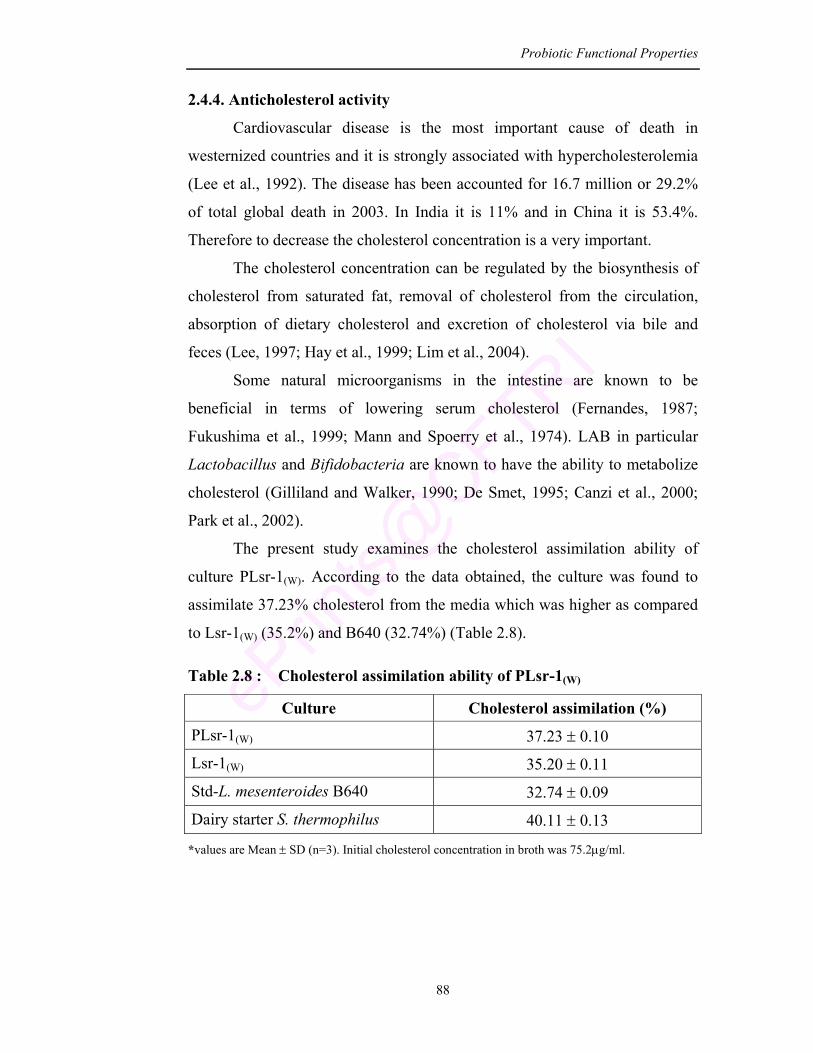
Probiotic Functional Properties
88
2.4.4. Anticholesterol activity
Cardiovascular disease is the most important cause of death in
westernized countries and it is strongly associated with hypercholesterolemia
(Lee et al., 1992). The disease has been accounted for 16.7 million or 29.2%
of total global death in 2003. In India it is 11% and in China it is 53.4%.
Therefore to decrease the cholesterol concentration is a very important.
The cholesterol concentration can be regulated by the biosynthesis of
cholesterol from saturated fat, removal of cholesterol from the circulation,
absorption of dietary cholesterol and excretion of cholesterol via bile and
feces (Lee, 1997; Hay et al., 1999; Lim et al., 2004).
Some natural microorganisms in the intestine are known to be
beneficial in terms of lowering serum cholesterol (Fernandes, 1987;
Fukushima et al., 1999; Mann and Spoerry et al., 1974). LAB in particular
Lactobacillus and Bifidobacteria are known to have the ability to metabolize
cholesterol (Gilliland and Walker, 1990; De Smet, 1995; Canzi et al., 2000;
Park et al., 2002).
The present study examines the cholesterol assimilation ability of
culture PLsr-1(W). According to the data obtained, the culture was found to
assimilate 37.23% cholesterol from the media which was higher as compared
to Lsr-1(W) (35.2%) and B640 (32.74%) (Table 2.8).
Table 2.8 : Cholesterol assimilation ability of PLsr-1(W)
Culture Cholesterol assimilation (%)
PLsr-1(W) 37.23 ± 0.10
Lsr-1(W) 35.20 ± 0.11
Std-L. mesenteroides B640 32.74 ± 0.09
Dairy starter S. thermophilus 40.11 ± 0.13
*values are Mean ± SD (n=3). Initial cholesterol concentration in broth was 75.2µg/ml.
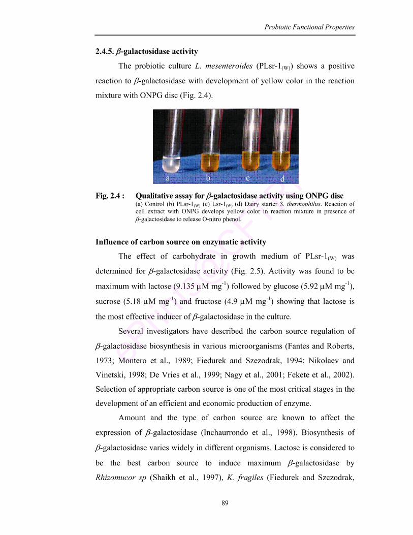
Probiotic Functional Properties
89
2.4.5. β-galactosidase activity
The probiotic culture L. mesenteroides (PLsr-1(W)) shows a positive
reaction to β-galactosidase with development of yellow color in the reaction
mixture with ONPG disc (Fig. 2.4).
Fig. 2.4 : Qualitative assay for β-galactosidase activity using ONPG disc (a) Control (b) PLsr-1(W) (c) Lsr-1(W) (d) Dairy starter S. thermophilus. Reaction of
cell extract with ONPG develops yellow color in reaction mixture in presence of β-galactosidase to release O-nitro phenol.
Influence of carbon source on enzymatic activity
The effect of carbohydrate in growth medium of PLsr-1(W) was
determined for β-galactosidase activity (Fig. 2.5). Activity was found to be
maximum with lactose (9.135 µM mg-1) followed by glucose (5.92 µM mg-1),
sucrose (5.18 µM mg-1) and fructose (4.9 µM mg-1) showing that lactose is
the most effective inducer of β-galactosidase in the culture.
Several investigators have described the carbon source regulation of
β-galactosidase biosynthesis in various microorganisms (Fantes and Roberts,
1973; Montero et al., 1989; Fiedurek and Szezodrak, 1994; Nikolaev and
Vinetski, 1998; De Vries et al., 1999; Nagy et al., 2001; Fekete et al., 2002).
Selection of appropriate carbon source is one of the most critical stages in the
development of an efficient and economic production of enzyme.
Amount and the type of carbon source are known to affect the
expression of β-galactosidase (Inchaurrondo et al., 1998). Biosynthesis of
β-galactosidase varies widely in different organisms. Lactose is considered to
be the best carbon source to induce maximum β-galactosidase by
Rhizomucor sp (Shaikh et al., 1997), K. fragiles (Fiedurek and Szczodrak,
a b c d
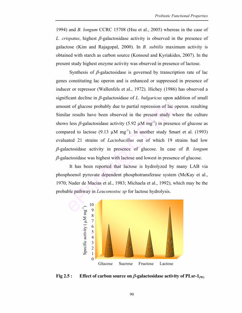
Probiotic Functional Properties
90
1994) and B. longum CCRC 15708 (Hsu et al., 2005) whereas in the case of
L. crispatus, highest β-galactosidase activity is observed in the presence of
galactose (Kim and Rajagopal, 2000). In B. subtilis maximum activity is
obtained with starch as carbon source (Konsoul and Kyriakides, 2007). In the
present study highest enzyme activity was observed in presence of lactose.
Synthesis of β-galactosidase is governed by transcription rate of lac
genes constituting lac operon and is enhanced or suppressed in presence of
inducer or repressor (Wallenfels et al., 1972). Hichey (1986) has observed a
significant decline in β-galactosidase of L. bulgaricus upon addition of small
amount of glucose probably due to partial repression of lac operon. resulting
Similar results have been observed in the present study where the culture
shows less β-galactosidase activity (5.92 µM mg-1) in presence of glucose as
compared to lactose (9.13 µM mg-1). In another study Smart et al. (1993)
evaluated 21 strains of Lactobacillus out of which 19 strains had low
β-galactosidase activity in presence of glucose. In case of B. longum
β-galactosidase was highest with lactose and lowest in presence of glucose.
It has been reported that lactose is hydrolyzed by many LAB via
phosphoenol pyruvate dependent phosphotransferase system (McKay et al.,
1970; Nader de Macias et al., 1983; Michaela et al., 1992), which may be the
probable pathway in Leuconostoc sp for lactose hydrolysis.
0123456789
10
Spec
ific
activ
ity ( µM
mg-1
)
Glucose Sucrose Fructose Lactose
Fig 2.5 : Effect of carbon source on β-galactosidase activity of PLsr-1(W)
Probiotic Functional Properties
91
2.4.6. Adherence ability
Adherence is one of the most important selection criteria for probiotic
bacteria (Shah, 2001). Adhesion of bacteria to gastrointestinal epithelium is
considered to be a prerequisite for exclusion of enteropathogenic bacteria
(Bernet et al., 1993; Mack et al., 1999) or immunomodulation of the host
(Perez et al., 1998; Isolauri et al., 1999; Blum et al., 2002).
Adherence of bacterial cells is related to cell surface characteristics
(Bibiloni et al., 2001; Canzi et al., 2005). Therefore the identification and
characterization of bacterial cell wall properties is important to understand
their role in adhesion to hydrocarbons, autoaggregation and relation to
co-aggregation mechanism and also the relevance to future probiotic food
development from natural strains (van-Loosdrecht et al., 1987; Zammaretti
and Ubbink, 2003). Adhesion to the colon surface occurs through the
association of bacteria with secreted mucus gel or by adherence to underlying
epithelium (Fontaine et al., 1994). High cell surface hydrophobicity favors the
colonization of mucosal surfaces and plays a role in adhesion of bacteria to
epithelial cells and extracellular membrane proteins (Zareba et al., 1997).
As bacterial adhesion to epithelial cells is been considered as one of
the selection criteria for probiotic strains, the present probiotic
L. mesenteroides (PLsr-1(W)) was studied for its adherence activity.
Hydrophobic interactions with different solvents are evaluated in-vitro and
the S-layer responsible for adhesion is extracted with LiCl and analyzed by
SDS-PAGE.
2.4.6.1. Cell surface hydrophobicity For probiotics to exert maximum effect on the host, it should have the
ability to adhere and colonize the intestine apart from being resistant to GI
condition (Clark et al., 1993). In the present study PLsr-1(W) was studied for
its adhesion ability using different hydrocarbons which is an important
parameter for bacterial adhesion and colonization in the GI tract (Rosenberg
et al., 1980). Bacterial adhesion to hydrocarbons (BATH test) has been
extensively used for measuring cell surface hydrophobicity in LAB (Kos
et al., 2003; Vinderola et al., 2004; Canzi et al., 2005).
Probiotic Functional Properties
92
Earlier reports have correlated the adhesion ability and hydrophobicity
as a measure of microbial adhesion (Wadstrom et al., 1987; Del Re et al.,
2000). Hydrophobicity is one of the physico-chemical properties that
facilitate the first contact between the microorganisms and the host cells. This
non-specific initial interaction is weak and reversible and precedes the
subsequent adhesion process mediated by more specific mechanisms
involving cell surface proteins and lipoteichoic acid (Granato et al., 1999;
Rojas et al., 2002; Roos and Jonsson., 2002). A number of reports have
described the composition, structure and forces of interaction related to
bacterial adhesion (Pelletier et al., 1997; Tuomola et al., 2000; Collado et al.,
2005).
The present study was carried out to determine adhesion ability of the
culture by using toluene, xylene, octane and hexadecane (Table 2.9). The
culture exhibited maximum (p<0.05) adhesion when grown in MRS medium
as compared to Rogosa media. Similar results were found by Ram and
Chander (2003) who speculated that the MRS media which contain yeast
extract synthesize certain enzymes. These enzymes are responsible for the
selective intake of nutrients and synthesize cell-surface components that play
a vital role in hydrophobic interaction. Rogosa media contains FeSO4 that
might probably inhibit hydrophobic components on cell surface. With
different hydrocarbons, maximum adhesion of PLsr-1(W) was with toluene
(50.81%) which was higher than Lsr-1(W) (46.22%), B640 (47.11%) and dairy
starter culture S. thermophilus (49.13%). Ram and Chander (2003) have
studied the influence of different growth media and observed that maximum
percent (80-89%) adhesion of B. bifidum was with MILS media followed by
MRS-lactose broth (68-79%). Draksler et al. (2004) have demonstrated the
adhesion ability of Lactobacillus strains to three solvents namely hexadecane,
xylene and toluene. The highest hydrophobic percentages was found in
Lactobacilli strains DDL19 (60, 57 and 63%) and DDL48 (47, 68 and 69%).
In the present study highest hydrophobic adhesion is found with toluene
(50.81%) followed by xylene (41.69%), octane (34.78%) and hexadecane
(24.47%).
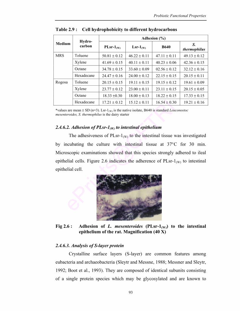
Probiotic Functional Properties
93
Table 2.9 : Cell hydrophobicity to different hydrocarbons
Adhesion (%) Medium Hydro-
carbon PLsr-1(W) Lsr-1(W) B640 S. thermophilus
Toluene 50.81 ± 0.12 46.22 ± 0.11 47.11 ± 0.11 49.13 ± 0.12 Xylene 41.69 ± 0.15 40.11 ± 0.11 40.23 ± 0.06 42.36 ± 0.15 Octane 34.78 ± 0.15 33.60 ± 0.09 02.56 ± 0.12 32.12 ± 0.16
MRS
Hexadecane 24.47 ± 0.16 24.00 ± 0.12 22.15 ± 0.15 20.15 ± 0.11 Toluene 20.15 ± 0.15 19.11 ± 0.15 19.15 ± 0.12 19.61 ± 0.09 Xylene 23.77 ± 0.12 23.00 ± 0.11 23.11 ± 0.15 20.15 ± 0.05 Octane 18.33 ±0.30 18.00 ± 0.13 18.22 ± 0.15 17.33 ± 0.15
Rogosa
Hexadecane 17.21 ± 0.12 15.12 ± 0.11 16.54 ± 0.30 19.21 ± 0.16
*values are mean ± SD (n=3). Lsr-1(W) is the native isolate, B640 is standard Leuconostoc mesenteroides, S. thermophilus is the dairy starter 2.4.6.2. Adhesion of PLsr-1(W) to intestinal epithelium
The adhesiveness of PLsr-1(W) to the intestinal tissue was investigated
by incubating the culture with intestinal tissue at 37°C for 30 min.
Microscopic examinations showed that this species strongly adhered to ileal
epithelial cells. Figure 2.6 indicates the adherence of PLsr-1(W) to intestinal
epithelial cell.
Fig 2.6 : Adhesion of L. mesenteroides (PLsr-1(W)) to the intestinal
epithelium of the rat. Magnification (40 X)
2.4.6.3. Analysis of S-layer protein
Crystalline surface layers (S-layer) are common features among
eubacteria and archaeobacteria (Sleytr and Messne, 1988; Messner and Sleytr,
1992; Boot et al., 1993). They are composed of identical subunits consisting
of a single protein species which may be glycosylated and are known to
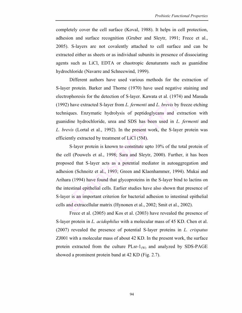
Probiotic Functional Properties
94
completely cover the cell surface (Koval, 1988). It helps in cell protection,
adhesion and surface recognition (Gruber and Sleytr, 1991; Frece et al.,
2005). S-layers are not covalently attached to cell surface and can be
extracted either as sheets or as individual subunits in presence of dissociating
agents such as LiCl, EDTA or chaotropic denaturants such as guanidine
hydrochloride (Navarre and Schneewind, 1999).
Different authors have used various methods for the extraction of
S-layer protein. Barker and Thorne (1970) have used negative staining and
electrophoresis for the detection of S-layer. Kawata et al. (1974) and Masuda
(1992) have extracted S-layer from L. fermenti and L. brevis by freeze etching
techniques. Enzymatic hydrolysis of peptidoglycans and extraction with
guanidine hydrochloride, urea and SDS has been used in L. fermenti and
L. brevis (Lortal et al., 1992). In the present work, the S-layer protein was
efficiently extracted by treatment of LiCl (5M).
S-layer protein is known to constitute upto 10% of the total protein of
the cell (Pouwels et al., 1998; Sara and Sleytr, 2000). Further, it has been
proposed that S-layer acts as a potential mediator in autoaggregation and
adhesion (Schneitz et al., 1993; Green and Klaenhammer, 1994). Mukai and
Arihara (1994) have found that glycoproteins in the S-layer bind to lactins on
the intestinal epithelial cells. Earlier studies have also shown that presence of
S-layer is an important criterion for bacterial adhesion to intestinal epithelial
cells and extracellular matrix (Hynonen et al., 2002; Smit et al., 2002).
Frece et al. (2005) and Kos et al. (2003) have revealed the presence of
S-layer protein in L. acidophilus with a molecular mass of 45 KD. Chen et al.
(2007) revealed the presence of potential S-layer proteins in L. crispatus
ZJ001 with a molecular mass of about 42 KD. In the present work, the surface
protein extracted from the culture PLsr-1(W) and analyzed by SDS-PAGE
showed a prominent protein band at 42 KD (Fig. 2.7).
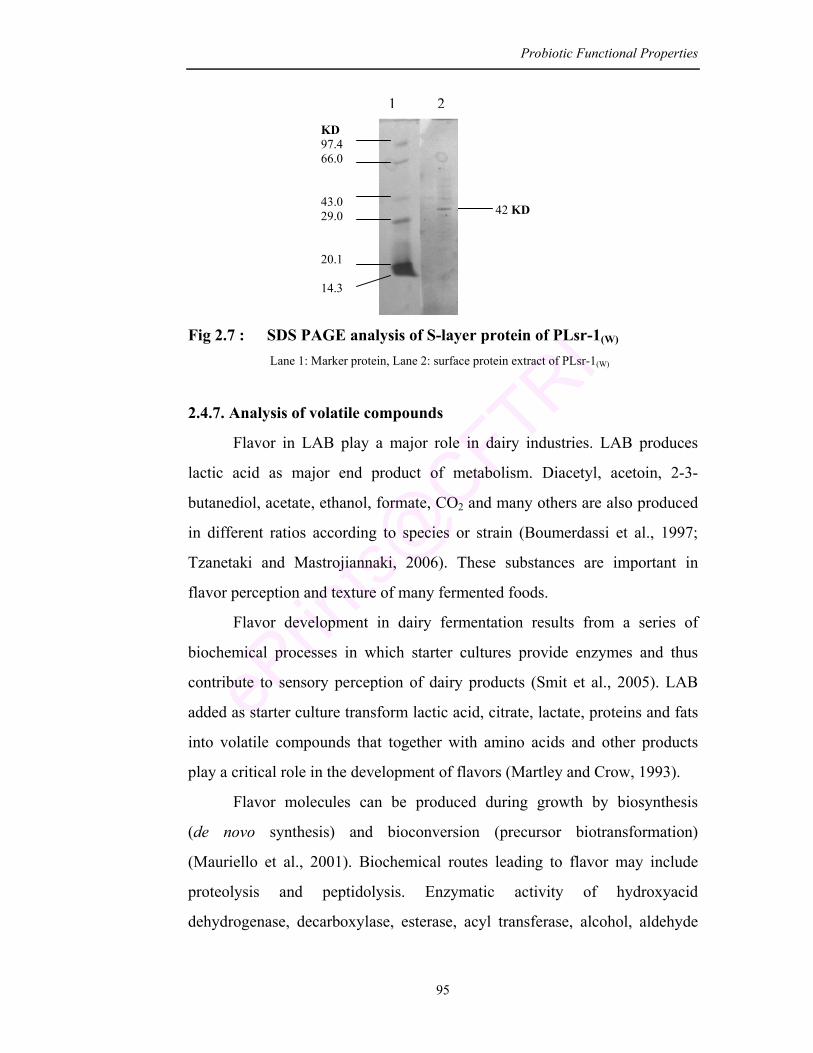
Probiotic Functional Properties
95
Fig 2.7 : SDS PAGE analysis of S-layer protein of PLsr-1(W)
Lane 1: Marker protein, Lane 2: surface protein extract of PLsr-1(W)
2.4.7. Analysis of volatile compounds
Flavor in LAB play a major role in dairy industries. LAB produces
lactic acid as major end product of metabolism. Diacetyl, acetoin, 2-3-
butanediol, acetate, ethanol, formate, CO2 and many others are also produced
in different ratios according to species or strain (Boumerdassi et al., 1997;
Tzanetaki and Mastrojiannaki, 2006). These substances are important in
flavor perception and texture of many fermented foods.
Flavor development in dairy fermentation results from a series of
biochemical processes in which starter cultures provide enzymes and thus
contribute to sensory perception of dairy products (Smit et al., 2005). LAB
added as starter culture transform lactic acid, citrate, lactate, proteins and fats
into volatile compounds that together with amino acids and other products
play a critical role in the development of flavors (Martley and Crow, 1993).
Flavor molecules can be produced during growth by biosynthesis
(de novo synthesis) and bioconversion (precursor biotransformation)
(Mauriello et al., 2001). Biochemical routes leading to flavor may include
proteolysis and peptidolysis. Enzymatic activity of hydroxyacid
dehydrogenase, decarboxylase, esterase, acyl transferase, alcohol, aldehyde
KD 97.4 66.0 43.0 29.0 20.1 14.3
1 2
42 KD
Probiotic Functional Properties
96
and keto acid dehydrogenases also contribute to flavor development (Smit
et al., 2005).
Earlier Alonso and Fraga (2001) have analyzed the volatile flavor
compounds in yogurt by headspace gas chromatography-mass spectrometry
(GCMS), wherein important compounds identified were acetone and acetic
acid. Fermented products formed from starter mixture of Lactococcus lactis
subsp lactis and Lactococcus lactis subsp cremoris are known to produce
volatile compounds like acetaldehyde, acetone, 2-butanone, diacetyl and
ethanol (Richelieu et al., 1997). Agrawal et al. (2000) have studied the flavor
profile of idli batter prepared from Pediococcus pentosaceus and Candida
versitalis as starter cultures, wherein diols and acids were found upto 8 days
of storage.
According to the literature volatile compounds produced from LAB are
found to be therapeutically important (Murti et al., 1993; Gallardo-Escamilla
et al., 2005). In the present work the volatile compounds of therapeutic
importance produced by the PLsr-1(W) are identified at different stages of its
growth.
Table 2.10 represents the volatile compounds extracted at different
time intervals during growth. The compounds were identified by comparing
the fragmentation pattern with the mass spectra of respective standards
(Fig. 2.8).
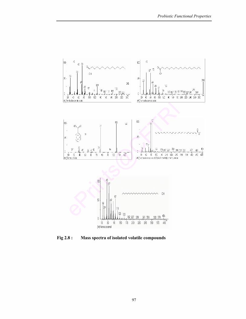
Probiotic Functional Properties
97
Fig 2.8 : Mass spectra of isolated volatile compounds
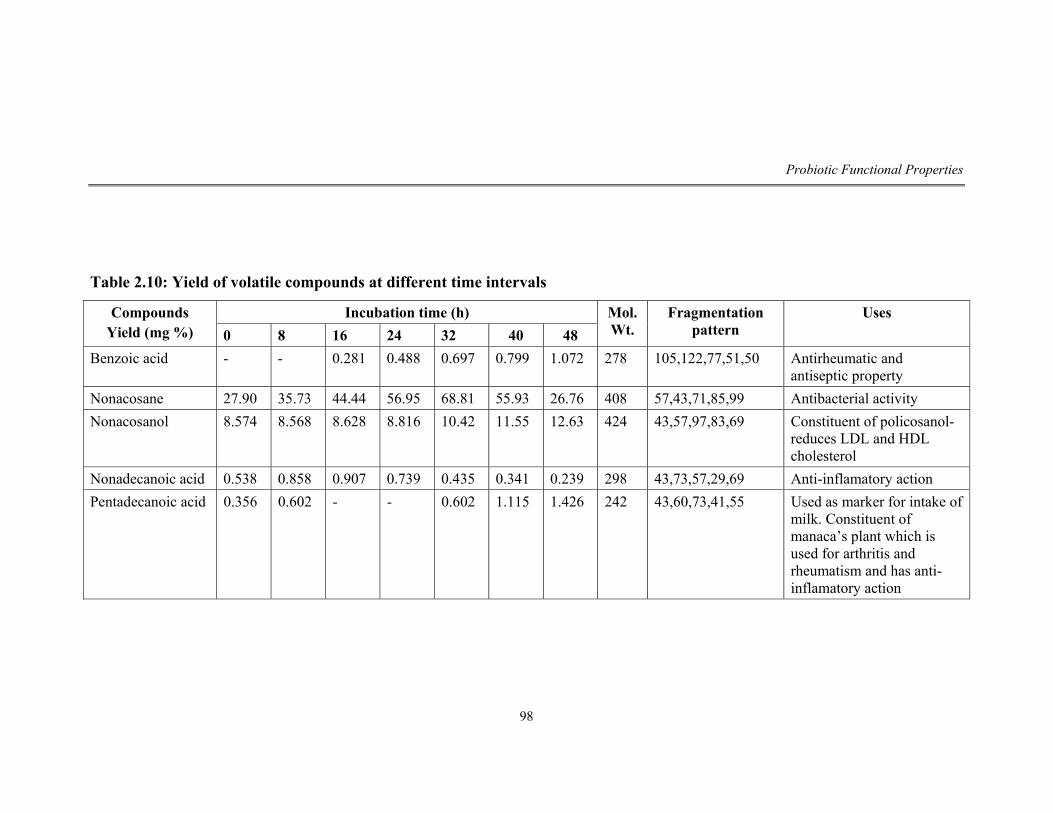
Probiotic Functional Properties
98
Table 2.10: Yield of volatile compounds at different time intervals
Incubation time (h) Compounds Yield (mg %) 0 8 16 24 32 40 48
Mol. Wt.
Fragmentation pattern
Uses
Benzoic acid - - 0.281 0.488 0.697 0.799 1.072 278 105,122,77,51,50 Antirheumatic and antiseptic property
Nonacosane 27.90 35.73 44.44 56.95 68.81 55.93 26.76 408 57,43,71,85,99 Antibacterial activity Nonacosanol 8.574 8.568 8.628 8.816 10.42 11.55 12.63 424 43,57,97,83,69 Constituent of policosanol-
reduces LDL and HDL cholesterol
Nonadecanoic acid 0.538 0.858 0.907 0.739 0.435 0.341 0.239 298 43,73,57,29,69 Anti-inflamatory action Pentadecanoic acid 0.356 0.602 - - 0.602 1.115 1.426 242 43,60,73,41,55 Used as marker for intake of
milk. Constituent of manaca’s plant which is used for arthritis and rheumatism and has anti-inflamatory action
Probiotic Functional Properties
99
The major compounds produced were identified as pentadecanoic acid,
nonadecanoic acid, benzoic acid, nonacosane and nonacosanol. Pentadecanoic
acid, which is used to cure arthritis and also as an anti inflammatory agent
was initially 0.356% which increased to 1.426% after 48h of incubation.
Nonadecanoic acid which is used for anti-inflammation was produced
initially at a concentration of 0.538% and reached maximum at 8 h of
growing phase (0.858%) then gradually declined to 0.239% after 48 h.
Benzoic acid which is used as antirheumatic and antiseptic agent was
produced 0.281% at 16 h of growth which increased upto 1.072% after 48 h.
Nonacosane which has antibacterial property reached a maximum 68.81%
after 32 h and declined on further incubation.
In the present work the pentadecanoic acid which was increasing even
after 48 h of incubation was also reported by Hannes et al. (2000) where it is
formed at the end of alcoholic fermentation by Saccharomyces bayanus and
Saccharomyces cerevisiae. Hirschler et al. (1998) has shown the formation of
pentadecanoic acid by carboxylation at C3 of ketone and the removal of two
terminal carbon atoms at C1 and C2 position of hexadecane. Kato et al. (2001)
have also shown the accumulation of pentadecanoate in Bacillus
thermoleovorans.
Nonacosanol which is a constituent of policosanol was increasing even
after 48 h of bacterial growth. Policosanol is known to be effective in
lowering LDL and cholesterol (Pons et al., 1994; Ortensi et al., 1997;
Fernandez et al., 2001).
Benzoic acid which is a natural antibiotic is known to be produced by
Lactobacillus bulgaricus and Lactobacillus acidophilus (Shahani et al.,
1976). In the present work PLsr-1(W) produces benzoic acid after 16 h of
incubation. The culture shows the importance for its use in food preservation.
According to Masja et al. (1998) benzoic acid is formed by aminotransferase
reaction of keto acids. Thierry and Maillard (2002) has shown that when
amino acids are used as a substrate it is catabolised by several reactions like
deamination, decarboxylation, oxidative deamination and transamination
leading to keto acids that enters the pathway of fatty acid synthesis.
Probiotic Functional Properties
100
All these volatile compounds produced by PLsr-1(W) in the present
study are shown to have therapeutic value for various ailments like anti-
inflammation, antirheumatic, antiseptic, antibacterial and antitumor activity.
Today, flavor industry has come a long way in terms of making
nutraceutical products more acceptable to the consumers, wherein this potent
probiotic culture may play an important role. Food formulations with addition
of such culture or byproduct or metabolite of the culture may have an
immense health effect on humans and can prove to be a good functional food.
2.4.8. Changes in the cellular fatty acids and proteins in probiotic
PLsr-1(W)
Bacteria are known to develop a set of mechanism leading to their
protection against GIT conditions. A combination of constitutive and
inducible methods like removal of protons (H+), alkalization of external
environment, changes in the composition of cell envelope, production of
general shock proteins, expression of transcriptional regulators and responses
to changes in cell density all can contribute to their survival (Christensen et
al., 1999; Nomura et al., 1999; Sanders et al., 1999; Cotter and Hill, 2003).
LAB have also evolved specific mechanism to resist the toxic effect of
bile thus enabling them to live in intestinal environment (Gunn, 2000). The
mechanism may be the release of bile salt hydrolases (BSH) that deconjugate
bile acid or by hydrolyzing amino acid glycine or taurine from steroid core
thus altering the properties of bile (Adamowicz et al., 1991; De Smet et al.,
1995).
Cellular membrane fatty acids and cellular proteins are the primary
target of any stress. It is established that organisms regulate membrane
composition in response to environmental condition to maintain optical
membrane fluidity for normal cellular function (Swan and Watson, 1997). In
the present study cellular fatty acid and proteins of probiotic PLsr-1(W) is
compared with native isolate Lsr-1(W) to determine the adaptive nature of
PLsr-1(W).
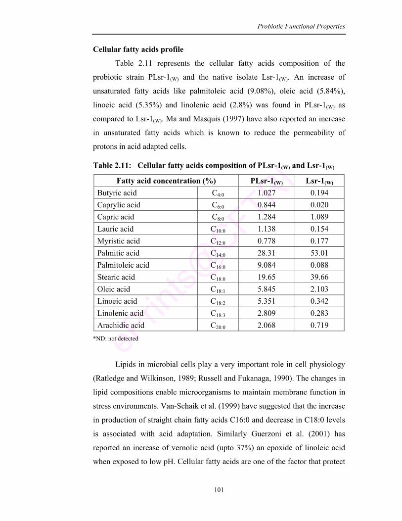
Probiotic Functional Properties
101
Cellular fatty acids profile
Table 2.11 represents the cellular fatty acids composition of the
probiotic strain PLsr-1(W) and the native isolate Lsr-1(W). An increase of
unsaturated fatty acids like palmitoleic acid (9.08%), oleic acid (5.84%),
linoeic acid (5.35%) and linolenic acid (2.8%) was found in PLsr-1(W) as
compared to Lsr-1(W). Ma and Masquis (1997) have also reported an increase
in unsaturated fatty acids which is known to reduce the permeability of
protons in acid adapted cells.
Table 2.11: Cellular fatty acids composition of PLsr-1(W) and Lsr-1(W)
Fatty acid concentration (%) PLsr-1(W) Lsr-1(W) Butyric acid C4:0 1.027 0.194 Caprylic acid C6:0 0.844 0.020 Capric acid C8:0 1.284 1.089 Lauric acid C10:0 1.138 0.154 Myristic acid C12:0 0.778 0.177 Palmitic acid C14:0 28.31 53.01 Palmitoleic acid C16:0 9.084 0.088 Stearic acid C18:0 19.65 39.66 Oleic acid C18:1 5.845 2.103 Linoeic acid C18:2 5.351 0.342 Linolenic acid C18:3 2.809 0.283 Arachidic acid C20:0 2.068 0.719
*ND: not detected
Lipids in microbial cells play a very important role in cell physiology
(Ratledge and Wilkinson, 1989; Russell and Fukanaga, 1990). The changes in
lipid compositions enable microorganisms to maintain membrane function in
stress environments. Van-Schaik et al. (1999) have suggested that the increase
in production of straight chain fatty acids C16:0 and decrease in C18:0 levels
is associated with acid adaptation. Similarly Guerzoni et al. (2001) has
reported an increase of vernolic acid (upto 37%) an epoxide of linoleic acid
when exposed to low pH. Cellular fatty acids are one of the factor that protect
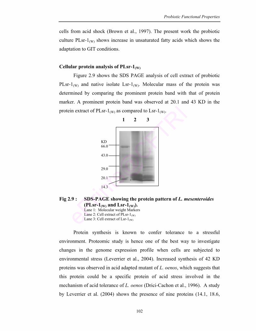
Probiotic Functional Properties
102
1 2 3
KD 66.0 43.0 29.0 20.1 14.3
cells from acid shock (Brown et al., 1997). The present work the probiotic
culture PLsr-1(W) shows increase in unsaturated fatty acids which shows the
adaptation to GIT conditions.
Cellular protein analysis of PLsr-1(W)
Figure 2.9 shows the SDS PAGE analysis of cell extract of probiotic
PLsr-1(W) and native isolate Lsr-1(W). Molecular mass of the protein was
determined by comparing the prominent protein band with that of protein
marker. A prominent protein band was observed at 20.1 and 43 KD in the
protein extract of PLsr-1(W) as compared to Lsr-1(W).
Fig 2.9 : SDS-PAGE showing the protein pattern of L. mesenteroides (PLsr-1(W) and Lsr-1(W)).
Lane 1: Molecular weight Markers Lane 2: Cell extract of PLsr-1(W) Lane 3: Cell extract of Lsr-1(W)
Protein synthesis is known to confer tolerance to a stressful
environment. Proteomic study is hence one of the best way to investigate
changes in the genome expression profile when cells are subjected to
environmental stress (Leverrier et al., 2004). Increased synthesis of 42 KD
proteins was observed in acid adapted mutant of L. oenos, which suggests that
this protein could be a specific protein of acid stress involved in the
mechanism of acid tolerance of L. oenos (Drici-Cachon et al., 1996). A study
by Leverrier et al. (2004) shows the presence of nine proteins (14.1, 18.6,
Probiotic Functional Properties
103
21.5, 26.9, 41.9, 49.6 and 56.2 KD) as a result of pH drop in the media.
Savijoki et al. (2005) have observed an upregulated induction of proteins like
HtrA (67 KD), Dna K (66 KD) and GroEL (57 KD) in B. longum on bile salt
treatment. In E. coli (aEPEC) bile treatment induced LdaG (25 KD) protein
(Torres et al., 2007). These results strongly suggest the involvement of these
proteins in cell adaptation to environmental changes.
In the present culture increase in 20.1 and 43 KD protein has been
observed PLsr-1(W) as compared to Lsr-1(W) which suggests the culture PLsr-
1(W) adaptation to stress conditions GIT.
Conclusion
This study focused on the functional properties of Leuconostoc
mesenteroides PLsr-1(W). The culture has antimicrobial activity against seven
food borne pathogens, which determines the capability of the culture to be
used as a preservative or in pharmacological industries against large number
of diseases. The antibiotic susceptibility test confirmed the culture to be
resistant to 3 antibiotics which enables the culture to maintain the balanced
intestinal microflora under antibiotic therapy. The antioxidative activity of the
culture demonstrates the scavenging activity against oxygen free radicals.
Therefore, it can be used as an antioxidant source in food and pharmaceutical
preparation. Further the adhesion assays indicate the ability of the culture to
adhere to the intestinal epithelial cells which imparts beneficial health effect
to the consumer. The culture has cholesterol lowering ability, β-galactosidase
activity and produces volatile compounds of therapeutic value.
Chapter 3 Leuconostoc as a Source for β-Galactosidase Enzyme
Leuconostoc as a source for β-galactosidase enzyme
104
CHAPTER – 3
LEUCONOSTOC AS A SOURCE FOR
β-GALACTOSIDASE ENZYME
ABSTRACT
The focus of the present chapter was to evaluate the ability of the
culture (PLsr-1(W)) to hydrolyze lactose. β-galactosidase responsible for the
breakdown of non-reducing disaccharide lactose was studied in the culture
isolate. The strain was improved by UV irradiation and chemical mutagenesis
for enhanced enzyme activity. The UV-mutant (coded as M7-PLsr-1(W))
which had 2 folds higher activity than parent strain (PLsr-1(W)) was subjected
to different permeabilization methods to optimize maximum release of the
enzyme. Further RSM studies were undertaken for optimization of chemical
and physical parameters. The enzyme produced under optimum conditions of
pH 7.5 with 1.25% lactose was further precipitated by ammonium sulphate.
The results indicate a 25 fold increase in the enzyme activity after ammonium
sulphate precipitation as compared to the crude extract.
Leuconostoc as a source for β-galactosidase enzyme
105
3.1. Introduction
Probiotics beneficially affect the health of the host by providing
enzymatic activities that improve the utilization of nutrients within the
intestine (Rowland, 1992). In this sense, these probiotic cultures are included
in dairy products to improve lactose absorption (Montes et al., 1995; Jiang
et al., 1996; Mustapha et al., 1997).
Lactose is a non-reducing disaccharide, hydrolyzed into glucose and
galactose in presence of lactase/ β-galactosidase enzyme. During infancy, all
humans and mammals possess high levels of enzyme lactase in their small
intestine which enables digestion of lactose. After weaning stage, a large part
(≈ 75%) of world population undergoes a genetically determined decline in
lactase activity which can lead to maldigestion of lactose causing abdominal
discomfort (Sahi, 2001).
The development of lactose hydrolyzed products with β-galactosidase
is one of the possible approaches to diminish the lactose maldigestion
problem. By hydrolyzing lactose with β-galactosidase, the problem associated
with whey disposal, lactose crystallization in frozen concentrated deserts and
lactose intolerance problems can be alleviated (Mahoney, 1998; Kim and
Rajagopal, 2000; Hsu et al., 2007). Although pharmaceutical preparation of
β-galactosidase have been developed for treating lactose intolerance
(Moskovitz et al., 1987; Sanders et al., 1992) they are found to be less
effective (Solomons et al., 1985; Onwulata et al., 1989). They are either
expensive, not available or are in insufficient quantity for industrial
application (Albayrak and Yang, 2002). Therefore selection of
microorganisms which is capable of producing high level of β-galactosidase
is very important.
β-galactosidase has been characterized in yogurt cultures
L. delbrueckii and S. thermophilus (Itoh et al., 1980; Greenberg and
Mahoney, 1982). Gilliland and Kim (1984) suggested that microbial
β-galactosidase which survives gastric digestion improves lactose digestion.
Leuconostoc as a source for β-galactosidase enzyme
106
Jiang et al. (1996) have shown that ingestion of milk containing B. longum at
a dose of 5 × 10⁸ cfu/ml improved lactose digestion and caused a moderate
reduction in total excretion of breath hydrogen. To alleviate lactose
intolerance it is important to have a potent probiotic culture capable of
producing large amounts of β-galactosidase enzyme.
For this approach, the probiotic culture L. mesenteroides (PLsr-1(W))
was improved and optimized for maximum β-galactosidase production.
The study was undertaken as
1) Strain improvement using UV-radiation and chemical mutagenesis for
enhanced β-galactosidase activity.
2) Optimization of permeabilization technique for maximum release of
β-galactosidase.
3) Optimization of conditions for β-galactosidase using response surface
methodology
4) Ammnium sulphate precipitation of β-galactosidase.
3.2. Materials
Chemicals and reagents
All chemicals used in this study were of analytical reagent grade.
O-nitrophenyl β-galactopyranoside (0.012 M), glucose, galactose, lactose,
sucrose, fructose, sodium carbonate (2 ml; 0.6 M), Sodium thiosulphate,
Bovine Serum Albumin (BSA), O-nitrophenol purchased from HiMedia Pvt
Ltd, Mumbai, India..
Ethyl methyl sulphonate was purchased from SRL Chemicals, India.
Solvents: toluene, acetone, chloroform were used of HPLC grade purchased
from Qualigens company, India.
Equipments:
1) Homogenizer, deep freezer, sonicator, lyophilizer, UV-lamp
2) Spectrophotometer, Centrifuge, Vortex ,Incubator (37, 40 and 60°C),
GC and GCMS (as described in earlier chapters)
Leuconostoc as a source for β-galactosidase enzyme
107
3.3. Methods
3.3.1. Bacterial growth and media
PLsr-1(W) was grown in MRS broth (1 L) supplemented with lactose
(1.5%) as carbon source. Cells were harvested by centrifugation at 8000 rpm
for 15 min at 4°C, washed twice with phosphate buffer (0.02 M; pH 7.0) and
resuspended in the same.
3.3.2. Estimation of β-galactosidase activity
Extraction of cell free enzyme extract: β-galactosidase activity was
determined according to the method of Bhowmik and Marth (1989). One ml
of bacterial suspension in buffer was permeabilized with 50 µl of toluene:
acetone mixture (1: 9 v/v), vortexed thoroughly and incubated at 37°C for 10
min. Suspension was then centrifuged at 10,000 rpm for 10 min at 4°C to
collect the cell free enzyme extract. This enzyme extract was then assayed for
β-galactosidase activity.
Estimation of enzyme activity: Cell free enzyme extract (1 ml) was taken in a
clean test tube and treated with O-nitrophenyl β-galactopyranoside (1 ml;
0.012 M) at 37°C for 30 min. After the incubation period ice cold sodium
carbonate (2 ml; 0.6 M) was added to stop the reaction and absorbance was
recorded on a spectrophotometer at 420 nm. Specific activity (µM mg-1) was
expressed as the amount of ONP released per mg of protein (Fig. 3.1 and 3.2).
Protein was estimated by Lowry’s method (Lowry et al., 1951).
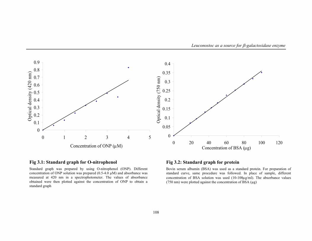
Leuconostoc as a source for β-galactosidase enzyme
108
00.10.20.30.40.50.60.70.80.9
0 1 2 3 4 5
Concentration of ONP (µM)
Opt
ical
den
sity
(420
nm
)
0
0.05
0.1
0.15
0.2
0.25
0.3
0.35
0.4
0 20 40 60 80 100 120Concentration of BSA (µg)
Opt
ical
den
sity
(750
nm
)Fig 3.1: Standard graph for O-nitrophenol Standard graph was prepared by using O-nitrophenol (ONP). Different concentration of ONP solution was prepared (0.5-4.0 µM) and absorbance was measured at 420 nm in a spectrophotometer. The values of absorbance obtained were then plotted against the concentration of ONP to obtain a standard graph
Fig 3.2: Standard graph for protein Bovin serum albumin (BSA) was used as a standard protein. For preparation of standard curve, same procedure was followed. In place of sample, different concentration of BSA solution was used (10-100µg/ml). The absorbance values (750 nm) were plotted against the concentration of BSA (µg)
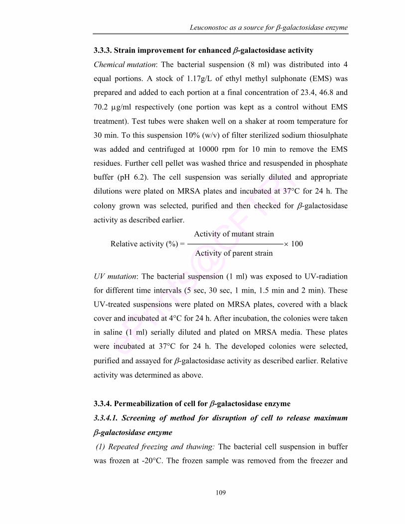
Leuconostoc as a source for β-galactosidase enzyme
109
3.3.3. Strain improvement for enhanced β-galactosidase activity
Chemical mutation: The bacterial suspension (8 ml) was distributed into 4
equal portions. A stock of 1.17g/L of ethyl methyl sulphonate (EMS) was
prepared and added to each portion at a final concentration of 23.4, 46.8 and
70.2 µg/ml respectively (one portion was kept as a control without EMS
treatment). Test tubes were shaken well on a shaker at room temperature for
30 min. To this suspension 10% (w/v) of filter sterilized sodium thiosulphate
was added and centrifuged at 10000 rpm for 10 min to remove the EMS
residues. Further cell pellet was washed thrice and resuspended in phosphate
buffer (pH 6.2). The cell suspension was serially diluted and appropriate
dilutions were plated on MRSA plates and incubated at 37°C for 24 h. The
colony grown was selected, purified and then checked for β-galactosidase
activity as described earlier.
Activity of mutant strain Relative activity (%) = × 100 Activity of parent strain
UV mutation: The bacterial suspension (1 ml) was exposed to UV-radiation
for different time intervals (5 sec, 30 sec, 1 min, 1.5 min and 2 min). These
UV-treated suspensions were plated on MRSA plates, covered with a black
cover and incubated at 4°C for 24 h. After incubation, the colonies were taken
in saline (1 ml) serially diluted and plated on MRSA media. These plates
were incubated at 37°C for 24 h. The developed colonies were selected,
purified and assayed for β-galactosidase activity as described earlier. Relative
activity was determined as above.
3.3.4. Permeabilization of cell for β-galactosidase enzyme
3.3.4.1. Screening of method for disruption of cell to release maximum
β-galactosidase enzyme
(1) Repeated freezing and thawing: The bacterial cell suspension in buffer
was frozen at -20°C. The frozen sample was removed from the freezer and
Leuconostoc as a source for β-galactosidase enzyme
110
thawed with warm water. This process was repeated three times and then
supernatant was assayed for enzyme activity.
(2) Lyophilization: Cell biomass was freeze dried in a lyophilizer
programmed to operate for 10 min initial freezing later pressure reduces to 10-
1 Torr for a period of 24 h. The lyophilized powder was then suspended in Z-
buffer and assayed for enzyme activity.
(3) Sonication: Cell biomass suspended in buffer was sonicated under ice and
assayed for enzyme.
(4) Homogenization: Cell suspension was subjected to homogenization to
disrupt the cells and the enzyme released was estimated in the supernatant.
(5) Acetone powder preparation: Cell biomass collected after centrifugation
was washed twice with phosphate buffer and pressed in between whatman No
1 filter paper to remove excess water. The biomass was taken and treated with
HPLC grade chilled acetone. Immediately it was filtered and then pellet was
dried until acetone is completely evaporated. This acetone powder was then
suspended in buffer to estimate the enzyme activity.
3.3.4.2. Optimization of method for disruption to release maximum
β-galactosidase enzyme
Sonication: Cell biomass suspended in buffer was sonicated for different time
intervals (0.5-5 min) and assayed for enzyme activity.
Homogenization: Cell biomass suspended in buffer was homogenized for
1-2 min and assayed for enzyme activity.
Solvent permeabilization: Cell biomass suspended in buffer was treated with
toluene:acetone and toluene: chloroform at varying ratio (1:3 to 1:9) and then
assayed for enzyme activity.
3.3.5. Application of the response surface methodology for optimizing the
culture condition for maximum β-galactosidase synthesis
Experimental design and analysis: Response surface methodology was used
to estimate the effect of temperature, pH and lactose concentration during the
growth of the culture for β-galactosidase activity. Central composite design,
Leuconostoc as a source for β-galactosidase enzyme
111
face centered with three factors, two responses and five center points were
used to evaluate the experimental data (Liong and Shah, 2005). Quadratic
model used to describe the response variable was as follows:
Y = b0 + b1X1 + b2X12 + b3X2 + b3X2
2 + b4X3 + b5X32 + b6X1X2 + b7X1X3 +
b8X2X3
Where Y = response (dependent variable)
X1 = lactose levels
X2 = temperature conditions
X3 = pH values with b0, b1,……..b8 as the regression co-efficients.
The above model was used to optimize the values of independent
parameters for the response. The CCRD of three independent variables with
five levels of each was chosen to design matrix.
3.3.6. Ammonium sulphate precipitation of β-galactosidase
After optimization of physical and chemical parameter for maximum
enzyme release the enzyme extract in supernatant was collected after
centrifugation at 10,000 rpm for 15 min at 4°C (All the steps were carried out
at 4°C). This cell free enzyme extract was precipitated by ammonium
sulphate at 30-60% (w/v) saturation according to the method of Bhowmik and
Marth (1989). The enzyme precipitate was collected after centrifugation and
was dissolved in Z-buffer. This was then dialyzed against Z- buffer for 24 h
with several changes of liquid to remove the sulphate salts.
Analysis on SDS PAGE: Electrophoresis was carried out according to the
method of Laemmli (1970) using 12% running gel and 4% stacking gel.
Protein was stained with coomassie blue. On destaining the protein bands
were observed and photograph using gel documentation.
3.3.7. Stability of the enzyme
(a) Effect of pH and temperature: Three buffer systems, citrate buffer (0.1 M;
pH 4.0–6.0), sodium phosphate buffer (0.2 M; pH 7.0-8.0), and glycine-
NaOH buffer (0.2 M; pH 9.0–10.0) were used to study the effect of pH on
Leuconostoc as a source for β-galactosidase enzyme
112
enzyme activity. The enzyme was preincubated in different buffers at room
temperature for 1 h and then checked for the activity.
Thermal stability was estimated by incubating the enzyme at different
temperature (20-50°C). The residual activity was then measured as described
earlier under standard assay conditions.
(b) Effect of substrate and enzyme concentration: Enzyme was treated with
different concentration of ONPG (2-50 µM) and then assayed for the activity.
To determine the effect of enzyme concentration, varying concentration of
enzyme (25-500 mg protein) was incubated with ONPG (0.012 M) and
assayed for enzyme activity.
(c) Effects of inhibitors and activators: Enzyme samples were incubated with
different modulators like EDTA, HgCl2, 1,10-phenothroline and ascorbic acid
at varying concentrations (0.1-10.0 mM). Enzyme activity without
modulators was used as control. The effect of metal ions was determined by
incubating the enzyme with CaCl2, MgSO4, MnSO4 and ZnSO4 at a
concentration of 0.1-10.0 mM each. The treated sample were then incubated
with ONPG (0.012 M) and then assayed for enzyme activity as described
earlier. Enzyme activity measured without added cations was used as control.
3.4. Results and discussion
3.4.1. Estimation of β-galactosidase activity
β-galactosidase enzyme has been found in numerous biological
systems. Among these special attention has been paid to LAB because of
GRAS status (Stiles and Holzapfel, 1997). In the present study, the
production of β-galactosidase by a probiotic lactic acid bacteria
L. mesenteroides (PLsr-1(W)) was studied.
3.4.2. Strain improvement for enhanced β-galactosidase activity
Deficiency of lactase enzyme causes lactose intolerance problem,
which is of great concern. Today, consumers are aware of many side effects
caused by medicines and are looking forward for natural means to cure
Leuconostoc as a source for β-galactosidase enzyme
113
diseases. Preparation of lactose treated dairy foods and improvement in
enzyme production and hydrolysis reaction can lead to improved lactose
digestion.
To alleviate the symptoms of lactose intolerance, it is important to
select cultures capable of producing large amounts of β-galactosidase.
Although gene over expression technique is established it is not presently
applicable to most probiotic cultures because of the lack of knowledge and
tools for genetic manipulation (Ibrahim and O’Sullivan, 2000). Method of
recombinant DNA technology by mutagenesis offers powerful approach for
manipulating strains for optimal β-galactosidase production and it is a good
approach to obtain overproducing mutants. In the present work, the classical
mutagenesis is used for increasing β-galactosidase production to improve its
potential in treating lactose intolerance.
Chemical mutation
Chemical mutation of culture (PLSR-1(W)) using ethyl methane
sulfonate (EMS) shows a reduction in cell count with increase in the mutagen
concentration. There was a decrease in 4 logs compared to control. A total of
30 mutants were isolated and purified on MRSA plate by repeated streaking.
These were then analyzed for β-galactosidase activity by qualitative method
using ONPG disc. The mutants with high β-galactosidase were tested
quantitatively using ONPG as substrate by standard procedure as described
earlier. The mutant strains showed β-galactosidase activities ranging from
6.5-13.1µM mg-1. Five mutants showed high inducible level of
β-galactosidase (>10 µM mg-1). The best mutant strain coded as
M3-PLsr-1(W) exhibited 1.75 folds higher level of β-galactosidase than the
control (PLsr-1(W)) (Table 3.1).
Different chemical mutagens are used for different purposes.
N-methyl-N’-nitro-N-nitrosoguanidine (MNNG) and EMS was used as
chemical mutagen by Ibrahim and O’Sullivan (2000) to enhance
Leuconostoc as a source for β-galactosidase enzyme
114
β-galactosidase synthesis in Bifidobacterium sp, L. delbruckii and
S. thermophilus. Chemical mutagenesis with hydroxylamine or
methoxylamine was performed on E. coli expression vector gene for
enhanced production of β-galactosidase. Rasouli and Kulkarni (1994)
subjected Aspergillus niger to mutagenesis with MNNG to scale up the
production of β-galactosidase and found 28% increased enzyme activity. In
the present study the β-galactosidase activity of EMS mutant strain
(M3- PLsr-1(W)) was increased by 74.31% activity which was higher than
reported by Yu et al. (1986) wherein an increase of 40.9% β-galactosidase
activity in L. sporogenes was found compared to parent strain with MNNG
chemical.
UV-mutation
UV-irradiation of the culture shows a decrease in the survival rate as
the duration of exposure increases (Table 3.2). There was a reduction in
viable cells (1-3 logs). A maximum survival was observed when the cells
were exposed for 5 sec (10 × 108). On 2 min exposure there was a significant
reduction in the cell count of the culture (53 × 105). A total of 50 mutants
were isolated and tested for β-galactosidase production using ONPG disc.
The positive strains were then checked for enhanced β-galactosidase
production using ONPG as a substrate. The β-galactosidase activity in mutant
strains ranged from 7-15 µM mg-1. Maximum enzyme activity
(15.18 µM mg-1) was observed when the culture was exposed to 1 min. Hence
this strain that was coded as M7-Plsr-1(W) was taken for all further studies.
The mutant strain was checked every 15 days for its stability and
β-galactosidase activity. According to the results enzyme activity did not vary
even after 15 months of storage and was stable.
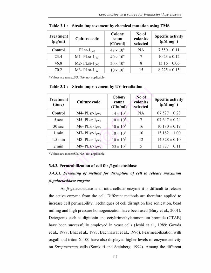
Leuconostoc as a source for β-galactosidase enzyme
115
Table 3.1 : Strain improvement by chemical mutation using EMS
Treatment (µg/ml) Culture code
Colony count
(Cfu/ml)
No of colonies selected
Specific activity (µM mg-1)
Control PLsr-1(W) 48 × 108 NA 7.550 ± 0.11 23.4 M1- PLsr-1(W) 40 × 104 7 10.23 ± 0.12 46.8 M2- PLsr-1(W) 20 × 104 8 13.16 ± 0.06 70.2 M3- PLsr-1(W) 10 × 104 15 8.225 ± 0.15
*Values are mean±SD. NA- not applicable Table 3.2 : Strain improvement by UV-irradiation
Treatment (time) Culture code
Colony count
(Cfu/ml)
No of colonies selected
Specific activity (µM mg-1)
Control M4- PLsr-1(W) 14 × 108 NA 07.527 ± 0.23 5 sec M5- PLsr-1(W) 10 × 108 7 07.647 ± 0.24 30 sec M6- PLsr-1(W) 10 × 107 16 10.180 ± 0.19 1 min M7- PLsr-1(W) 10 × 107 10 15.182 ± 1.00
1.5 min M8- PLsr-1(W) 10 × 106 12 14.328 ± 0.10 2 min M9- PLsr-1(W) 53 × 105 5 13.877 ± 0.11
*Values are mean±SD. NA- not applicable
3.4.3. Permeabilization of cell for β-galactosidase
3.4.3.1. Screening of method for disruption of cell to release maximum
β-galactosidase enzyme
As β-galactosidase is an intra cellular enzyme it is difficult to release
the active enzyme from the cell. Different methods are therefore applied to
increase cell permeability. Techniques of cell disruption like sonication, bead
milling and high pressure homogenization have been used (Bury et al., 2001).
Detergents such as digitonin and cetyltrimethylammonium bromide (CTAB)
have been successfully employed in yeast cells (Joshi et al., 1989; Gowda
et al., 1988; Bhat et al., 1993; Bachhawat et al., 1996). Pearmeabilization with
oxgall and triton X-100 have also displayed higher levels of enzyme activity
on Streptococcus cells (Somkuti and Steinberg, 1994). Among the different
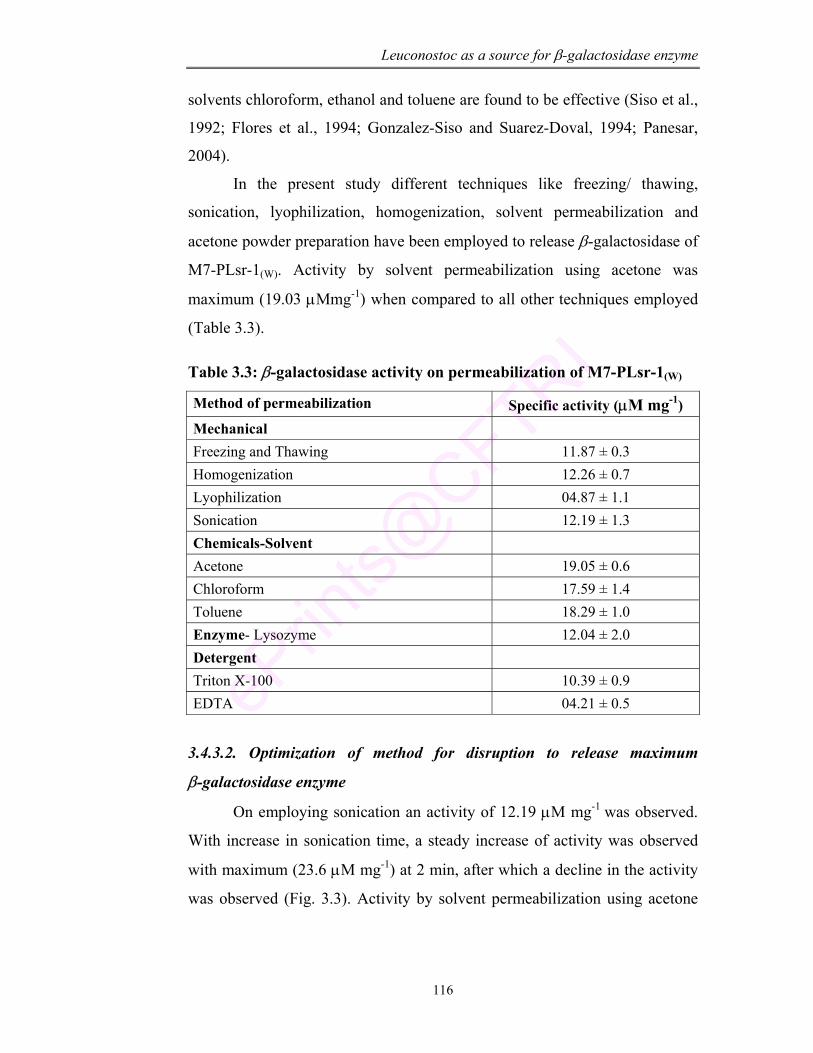
Leuconostoc as a source for β-galactosidase enzyme
116
solvents chloroform, ethanol and toluene are found to be effective (Siso et al.,
1992; Flores et al., 1994; Gonzalez-Siso and Suarez-Doval, 1994; Panesar,
2004).
In the present study different techniques like freezing/ thawing,
sonication, lyophilization, homogenization, solvent permeabilization and
acetone powder preparation have been employed to release β-galactosidase of
M7-PLsr-1(W). Activity by solvent permeabilization using acetone was
maximum (19.03 µMmg-1) when compared to all other techniques employed
(Table 3.3).
Table 3.3: β-galactosidase activity on permeabilization of M7-PLsr-1(W)
Method of permeabilization Specific activity (µM mg-1) Mechanical Freezing and Thawing 11.87 ± 0.3 Homogenization 12.26 ± 0.7 Lyophilization 04.87 ± 1.1 Sonication 12.19 ± 1.3 Chemicals-Solvent Acetone 19.05 ± 0.6 Chloroform 17.59 ± 1.4 Toluene 18.29 ± 1.0 Enzyme- Lysozyme 12.04 ± 2.0 Detergent Triton X-100 10.39 ± 0.9 EDTA 04.21 ± 0.5
3.4.3.2. Optimization of method for disruption to release maximum
β-galactosidase enzyme
On employing sonication an activity of 12.19 µM mg-1 was observed.
With increase in sonication time, a steady increase of activity was observed
with maximum (23.6 µM mg-1) at 2 min, after which a decline in the activity
was observed (Fig. 3.3). Activity by solvent permeabilization using acetone
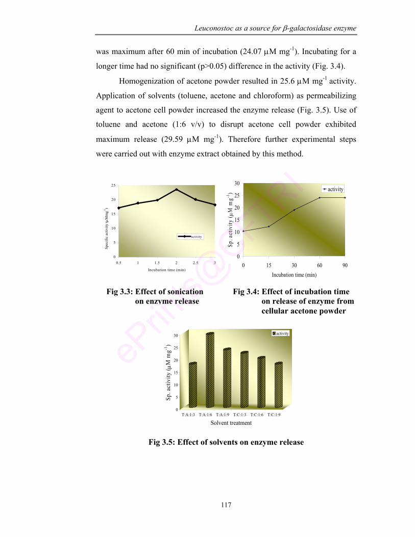
Leuconostoc as a source for β-galactosidase enzyme
117
was maximum after 60 min of incubation (24.07 µM mg-1). Incubating for a
longer time had no significant (p>0.05) difference in the activity (Fig. 3.4).
Homogenization of acetone powder resulted in 25.6 µM mg-1 activity.
Application of solvents (toluene, acetone and chloroform) as permeabilizing
agent to acetone cell powder increased the enzyme release (Fig. 3.5). Use of
toluene and acetone (1:6 v/v) to disrupt acetone cell powder exhibited
maximum release (29.59 µM mg-1). Therefore further experimental steps
were carried out with enzyme extract obtained by this method.
0
5
10
15
20
25
0.5 1 1.5 2 2.5 3
Incubation time (min)
Spec
ific
activ
ity ( µΜ
mg-1
)
activity
0
5
10
15
20
25
30
0 15 30 60 90Incubation time (min)
Sp. a
ctiv
ity ( µ
M m
g-1) activity
0
5
10
15
20
25
30
Sp. a
ctiv
ity ( µ
M m
g-1)
T:A:1:3 T:A:1:6 T:A:1:9 T:C:1:3 T:C:1:6 T:C:1:9
Solvent treatment
activity
Fig 3.5: Effect of solvents on enzyme release
Fig 3.3: Effect of sonication on enzyme release
Fig 3.4: Effect of incubation time on release of enzyme from cellular acetone powder
Leuconostoc as a source for β-galactosidase enzyme
118
3.4.4. Application of the response surface methodology for optimizing the
culture condition for maximum β-galactosidase activity The aim of the present study was to optimize the fermentation conditions of mutant strain of L. mesenteroides (M7-PLsr-1(W)). This statistically designed, multifactorial experiment offers an additional method
for potential enhancement of β-galactosidase activity. The response surface methodology (RSM) which includes factorial designs and regression analysis is known to effectively deal with technological optimization studies (Logothetis and Wynn, 1989). Microorganisms differ in their optimum condition for enzyme production. So it is necessary to find methods that are less expensive with higher productivity of the enzyme. Stability of enzyme is an important aspect in biotechnological process that could help in optimization of economic profitability of enzymatic production. The activity of enzyme is influenced by diverse environmental factors like pH, temperature and substrate concentration that strongly affect the spatial confirmation of the protein (Sadana and Henley, 1986; Juradol et al., 2004).
RSM is a collection of statistical techniques for designing experiments, building models, evaluating the effective factors and most importantly, searching for the optimum conditions of factors for desirable response (Montgomery, 1991). In this approach, the physical and chemical parameters (temperature, pH and lactose concentration) are varied in the growth condition of culture isolate to determine the optimum parameter for maximum
β-galactosidase activity. The response quadratic model was obtained as follows with R2 as 91.4% Y = 184.58-21.01X1 + 6.25X1
2 + 2.94X2 + 0.01X22 – 60.38X3 + 4.65X3
2 +4.65X3
2 – 0.57X1X2 + 3.64X1X3 – 0.38X2X3
Where Y = Specific activity (µM/ 100 mg protein) X1 = Lactose concentration (%)
X2 = Temperature (°C) X3 = pH The analysis of variance was calculated to check how well the model represented the data and it was found that F value for lactose, temperature and
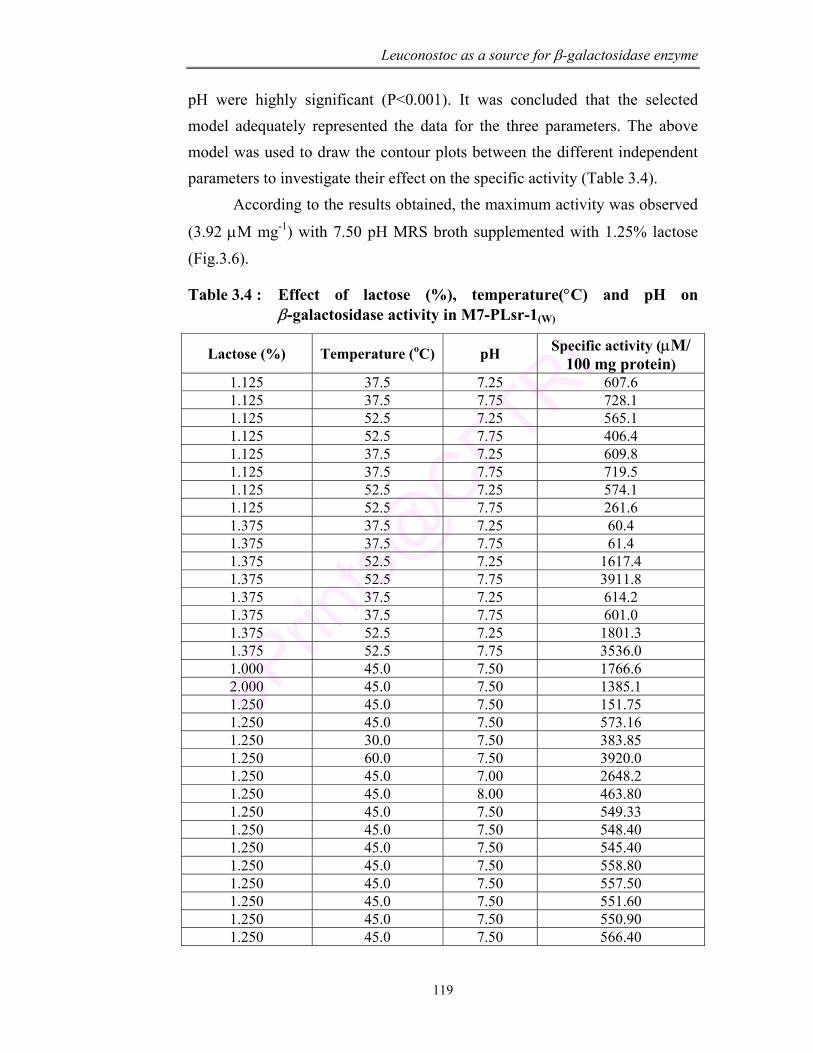
Leuconostoc as a source for β-galactosidase enzyme
119
pH were highly significant (P<0.001). It was concluded that the selected model adequately represented the data for the three parameters. The above model was used to draw the contour plots between the different independent parameters to investigate their effect on the specific activity (Table 3.4). According to the results obtained, the maximum activity was observed
(3.92 µM mg-1) with 7.50 pH MRS broth supplemented with 1.25% lactose (Fig.3.6).
Table 3.4 : Effect of lactose (%), temperature(°C) and pH on β-galactosidase activity in M7-PLsr-1(W)
Lactose (%) Temperature (oC) pH Specific activity (µM/ 100 mg protein)
1.125 37.5 7.25 607.6 1.125 37.5 7.75 728.1 1.125 52.5 7.25 565.1 1.125 52.5 7.75 406.4 1.125 37.5 7.25 609.8 1.125 37.5 7.75 719.5 1.125 52.5 7.25 574.1 1.125 52.5 7.75 261.6 1.375 37.5 7.25 60.4 1.375 37.5 7.75 61.4 1.375 52.5 7.25 1617.4 1.375 52.5 7.75 3911.8 1.375 37.5 7.25 614.2 1.375 37.5 7.75 601.0 1.375 52.5 7.25 1801.3 1.375 52.5 7.75 3536.0 1.000 45.0 7.50 1766.6 2.000 45.0 7.50 1385.1 1.250 45.0 7.50 151.75 1.250 45.0 7.50 573.16 1.250 30.0 7.50 383.85 1.250 60.0 7.50 3920.0 1.250 45.0 7.00 2648.2 1.250 45.0 8.00 463.80 1.250 45.0 7.50 549.33 1.250 45.0 7.50 548.40 1.250 45.0 7.50 545.40 1.250 45.0 7.50 558.80 1.250 45.0 7.50 557.50 1.250 45.0 7.50 551.60 1.250 45.0 7.50 550.90 1.250 45.0 7.50 566.40
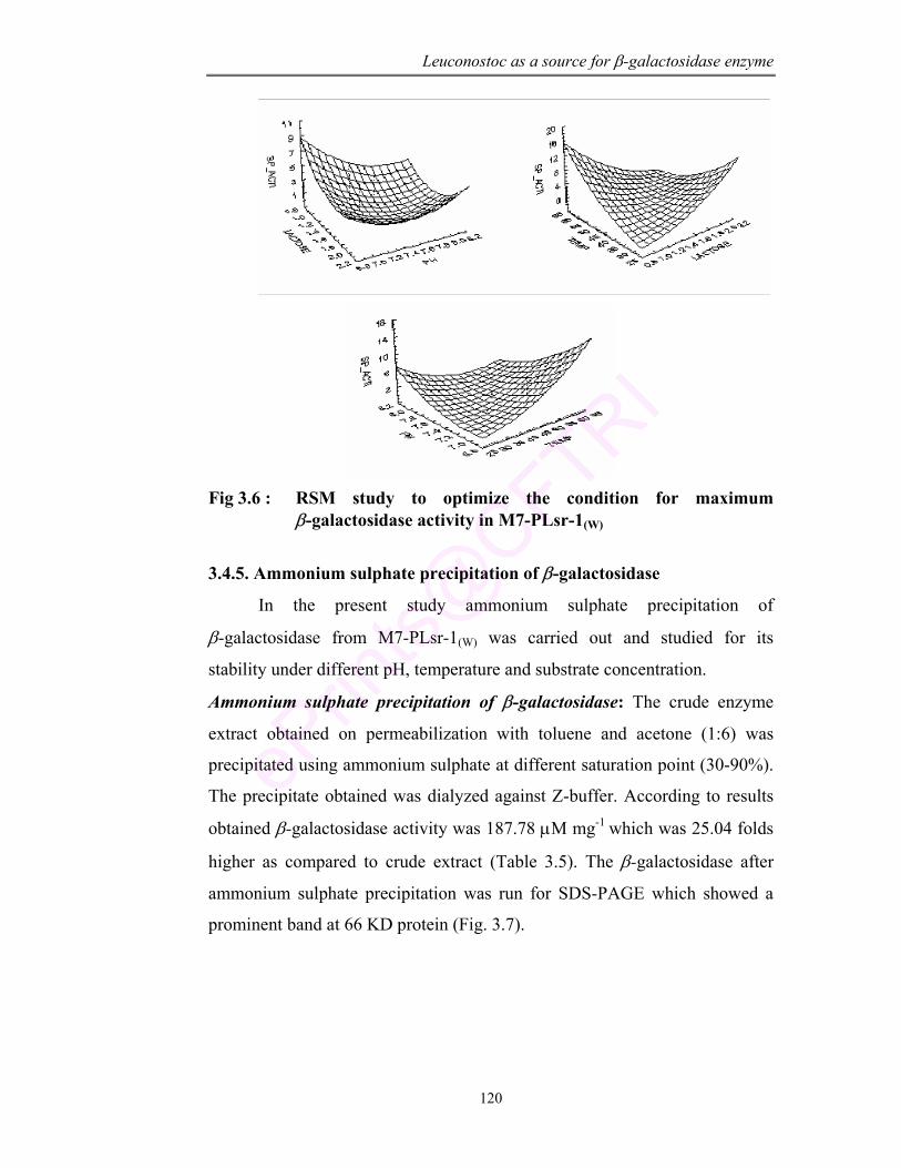
Leuconostoc as a source for β-galactosidase enzyme
120
Fig 3.6 : RSM study to optimize the condition for maximum
β-galactosidase activity in M7-PLsr-1(W)
3.4.5. Ammonium sulphate precipitation of β-galactosidase
In the present study ammonium sulphate precipitation of
β-galactosidase from M7-PLsr-1(W) was carried out and studied for its
stability under different pH, temperature and substrate concentration.
Ammonium sulphate precipitation of β-galactosidase: The crude enzyme
extract obtained on permeabilization with toluene and acetone (1:6) was
precipitated using ammonium sulphate at different saturation point (30-90%).
The precipitate obtained was dialyzed against Z-buffer. According to results
obtained β-galactosidase activity was 187.78 µM mg-1 which was 25.04 folds
higher as compared to crude extract (Table 3.5). The β-galactosidase after
ammonium sulphate precipitation was run for SDS-PAGE which showed a
prominent band at 66 KD protein (Fig. 3.7).
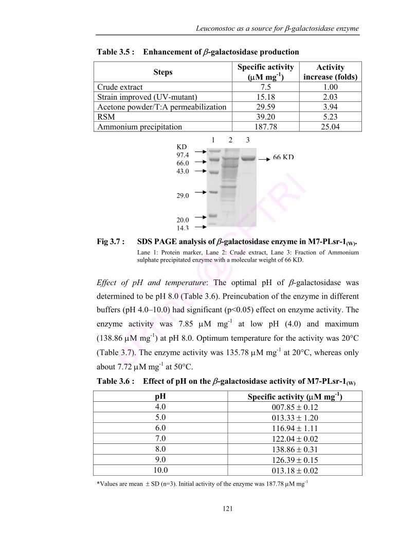
Leuconostoc as a source for β-galactosidase enzyme
121
Table 3.5 : Enhancement of β-galactosidase production
Steps Specific activity (µM mg-1)
Activity increase (folds)
Crude extract 7.5 1.00 Strain improved (UV-mutant) 15.18 2.03 Acetone powder/T:A permeabilization 29.59 3.94 RSM 39.20 5.23 Ammonium precipitation 187.78 25.04
Fig 3.7 : SDS PAGE analysis of β-galactosidase enzyme in M7-PLsr-1(W). Lane 1: Protein marker, Lane 2: Crude extract, Lane 3: Fraction of Ammonium sulphate precipitated enzyme with a molecular weight of 66 KD.
Effect of pH and temperature: The optimal pH of β-galactosidase was determined to be pH 8.0 (Table 3.6). Preincubation of the enzyme in different buffers (pH 4.0–10.0) had significant (p<0.05) effect on enzyme activity. The
enzyme activity was 7.85 µM mg-1 at low pH (4.0) and maximum
(138.86 µM mg-1) at pH 8.0. Optimum temperature for the activity was 20°C
(Table 3.7). The enzyme activity was 135.78 µM mg-1 at 20°C, whereas only
about 7.72 µM mg-1 at 50°C.
Table 3.6 : Effect of pH on the β-galactosidase activity of M7-PLsr-1(W)
pH Specific activity (µM mg-1) 4.0 007.85 ± 0.12 5.0 013.33 ± 1.20 6.0 116.94 ± 1.11 7.0 122.04 ± 0.02 8.0 138.86 ± 0.31 9.0 126.39 ± 0.15 10.0 013.18 ± 0.02
*Values are mean ± SD (n=3). Initial activity of the enzyme was 187.78 µM mg-1
KD 97.4 66.0 43.0 29.0 20.0 14.3
1 2 3
66 KD
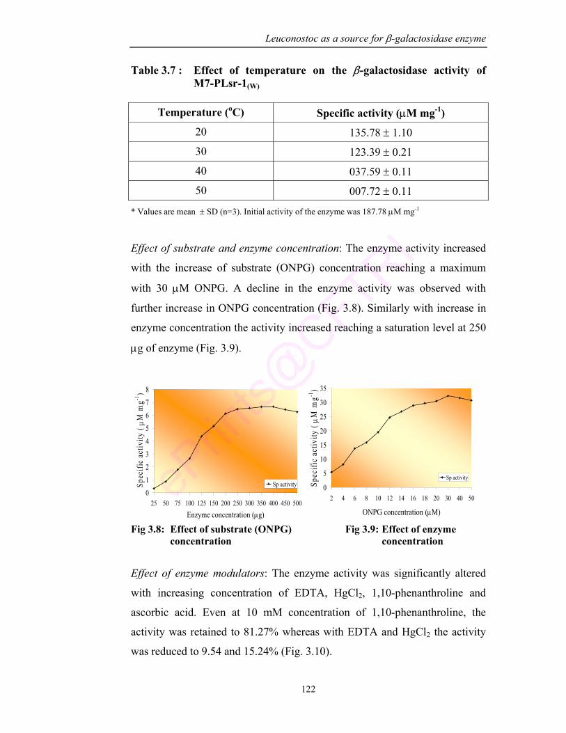
Leuconostoc as a source for β-galactosidase enzyme
122
Table 3.7 : Effect of temperature on the β-galactosidase activity of M7-PLsr-1(W)
Temperature (oC) Specific activity (µM mg-1)
20 135.78 ± 1.10
30 123.39 ± 0.21
40 037.59 ± 0.11
50 007.72 ± 0.11
* Values are mean ± SD (n=3). Initial activity of the enzyme was 187.78 µM mg-1
Effect of substrate and enzyme concentration: The enzyme activity increased
with the increase of substrate (ONPG) concentration reaching a maximum
with 30 µM ONPG. A decline in the enzyme activity was observed with
further increase in ONPG concentration (Fig. 3.8). Similarly with increase in
enzyme concentration the activity increased reaching a saturation level at 250
µg of enzyme (Fig. 3.9).
012
3
4
56
7
8
25 50 75 100 125 150 200 250 300 350 400 450 500
Enzyme concentration (µg)
Spe
cific
act
ivity
( µM
mg-1
)
Sp activity 0
5
10
15
20
25
30
35
2 4 6 8 10 12 14 16 18 20 30 40 50
ONPG concentration (µM)
Spec
ific
activ
ity ( µM
mg-1
)
Sp activity
Fig 3.8: Effect of substrate (ONPG) Fig 3.9: Effect of enzyme concentration concentration
Effect of enzyme modulators: The enzyme activity was significantly altered
with increasing concentration of EDTA, HgCl2, 1,10-phenanthroline and
ascorbic acid. Even at 10 mM concentration of 1,10-phenanthroline, the
activity was retained to 81.27% whereas with EDTA and HgCl2 the activity
was reduced to 9.54 and 15.24% (Fig. 3.10).
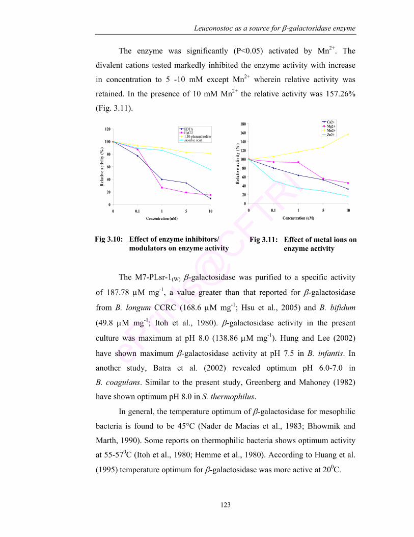
Leuconostoc as a source for β-galactosidase enzyme
123
The enzyme was significantly (P<0.05) activated by Mn2+. The
divalent cations tested markedly inhibited the enzyme activity with increase
in concentration to 5 -10 mM except Mn2+ wherein relative activity was
retained. In the presence of 10 mM Mn2+ the relative activity was 157.26%
(Fig. 3.11).
0
20
40
60
80
100
120
0 0.1 1 5 10
Concentration (uM)
Rel
ativ
e ac
tivity
(%)
EDTAHgCl21,10-phenanthrolineascorbic acid
0
20
40
60
80
100
120
140
160
180
0 0.1 1 5 10
Concnetration (uM)
Rel
ativ
e ac
tivity
(%)
Ca2+Mg2+Mn2+Zn2+
The M7-PLsr-1(W) β-galactosidase was purified to a specific activity
of 187.78 µM mg-1, a value greater than that reported for β-galactosidase
from B. longum CCRC (168.6 µM mg-1; Hsu et al., 2005) and B. bifidum
(49.8 µM mg-1; Itoh et al., 1980). β-galactosidase activity in the present
culture was maximum at pH 8.0 (138.86 µM mg-1). Hung and Lee (2002)
have shown maximum β-galactosidase activity at pH 7.5 in B. infantis. In
another study, Batra et al. (2002) revealed optimum pH 6.0-7.0 in
B. coagulans. Similar to the present study, Greenberg and Mahoney (1982)
have shown optimum pH 8.0 in S. thermophilus.
In general, the temperature optimum of β-galactosidase for mesophilic
bacteria is found to be 45°C (Nader de Macias et al., 1983; Bhowmik and
Marth, 1990). Some reports on thermophilic bacteria shows optimum activity
at 55-570C (Itoh et al., 1980; Hemme et al., 1980). According to Huang et al.
(1995) temperature optimum for β-galactosidase was more active at 200C.
Fig 3.10: Effect of enzyme inhibitors/ modulators on enzyme activity
Fig 3.11: Effect of metal ions on enzyme activity
Leuconostoc as a source for β-galactosidase enzyme
124
Results obtained in the present study demonstrated that after
ammonium sulpahte precipitation the specific activity of β-galactosidase
increased from 39.2 to 187.78 µM mg-1.
3.5. Conclusion
The probiotic L. mesenteroides PLsr-1(W) was improved for
β-galactosidase by chemical mutagen and UV radiation. The result indicates
an increase in enzyme activity in the UV-mutant (M7-PLsr-1) and was taken
for all further studies. Conditions for maximum release of enzyme were
optimized by different permeabilizing techniques. RSM study revealed the
optimum condition for physical and chemical parameter for maximum
activity. According to the study maximum activity (39.2 µM mg-1) was
observed with the culture grown at 7.50 pH in MRS broth supplemented with
1.25% lactose. Further on ammonium sulphate precipitation the activity
increased to 187.78 µM mg-1 which was 25 folds higher as compared to the
crude extract.

Chapter 4 Enhancement of Culture Shelf Life on Storage
Enhancement of culture shelf life on storage
125
CHAPTER – 4
ENHANCEMENT OF
CULTURE SHELF LIFE ON STORAGE
ABSTRACT
Aim of the present work was to preserve the selected culture
(M7-PLsr-1(W)) for longer shelf life. In this regard, the culture was subjected
to different drying methods like spray, vacuum, oven and freeze drying for
maximal survival. The results show maximum stability to freeze drying. The
content of saturated and unsaturated membrane fatty acid was increased on
freeze drying. SDS PAGE analysis of cellular protein showed a prominent
protein band at 22.9 and 20.1 KD after freeze drying. Further viability and
resistance of the culture to freeze drying was enhanced with supplementation
of different adjuvants like polyethylene glycol, lactose and sucrose. Sucrose
supplementation enhanced the survival rate of the culture to 72.26% in
comparison to control (52.55%). Viability of the freeze dried culture during
storage was studied at 30, 4 and -20°C for a period of 6 months. Data
determines that the storage temperature of -20°C was optimum for
maintaining maximum viability. Even after six months of storage the culture
retained its probiotic properties like antimicrobial activity, resistance to low
pH and high bile salt concentration. The study shows the importance of
cryoprotectants in enhancing the viability and beneficial attributes of culture
during storage.
Enhancement of culture shelf life on storage
126
4.1. Introduction
LAB are commonly used in the production of cheese, yogurt, dry
sausages, wine, bread and sauerkraut. They contribute to the formation of
organoleptic and rheological characteristics of these products and inhibit the
growth of undesirable bacteria (Coppola et al., 1998; Caplice and Fitzgerald,
1999). These cultures are added directly to the food matrix either in frozen or
dried form. In addition many commercially dried culture products are
available in the form of tablets and pharmaceutical preparations to treat large
number of diseases and to maintain normal intestinal flora (Shah, 2000).
Interest in ready to use cultures for direct inoculation has placed
greater emphasis on starter cultures and preservation methods that promote
high cell viability and activity (Broadbent and Lin, 1999). LAB may be
preserved and distributed in liquid, spray dried, frozen or lyophilized forms.
Spray drying produces dry granulated powder by atomizing the wet product at
high velocity within a chamber (Desmond et al., 2002; Corcoran et al., 2004).
Freeze drying or lyophilization is a process in which solution of a substance is
frozen and then the quantity of water is reduced by sublimation. Further it is
subjected to desorption to a value where it will no longer support biological
activity or chemical reaction (Jennings, 1990). Microbial survival during
these preservation processes depend on many factors like growth conditions
(Palmfeldt and Hahn-Hagerdal, 2000), protective medium (Hubalek, 1996;
Linder et al., 1997; Fernandez-Murga et al., 1998; Abadias et al., 2001),
initial cell concentration (Bozoglu et al., 1987; Costa et al., 2000), freezing
temperature (Sanders et al., 1999) and rehydration conditions (Theunissen
et al., 1993). Loss in the cell viability can be related to the destruction of cell
components including cell membrane, cell wall and DNA (Teixeira et al.,
1995; Ray et al., 1971). During the above drying methods the bacterial
cultures are also subjected to adverse conditions, such as water crystallization
and low temperature, producing a degree of protein denaturation and bacterial
membrane injury which consequently decreases the culture viability (Visick
and Clark, 1995).
Enhancement of culture shelf life on storage
127
To prevent or reduce all these adverse effects many substances are used as cryoprotectants (Carcoba and Rodriguez, 2000). A good protectant is the one that can be easily dried and gives a good matrix for culture stability (Zhao and Zhang, 2005). These cryoprotectants may be either simple or complex chemical compounds like quickly penetrating compounds (alcohols, amides etc), slowly penetrating compounds (glycerols, triols etc) or non penetrating compounds (mono, di, oligosaccharides, polyalcohols, sugar, proteins etc (Hubalck, 2003). Many compounds have been shown to increase the survival of LAB during freeze drying and the beneficial effect is related to the proteins and membranes of microorganisms (DeValdez et al., 1983; Wiemken, 1990; Leslie et al., 1995).
In the present work freeze drying, spray drying, vacuum drying and oven drying methods were used for the preservation of culture. Stability and viability of the culture was further enhanced on supplementation of different cryoprotectants. The viability of culture during storage at 3 different
temperatures (30°, 4° and -20°C) was studied for 6 months after freeze drying. Antimicrobial activity, tolerance to low pH and high bile salt concentration was checked during storage. 4.2. Materials Chemicals:
1) MRS broth, Bile salt mix, pH 2.0 buffer (as described in chapter 1). 2) Solvents: methanol, chloroform, hexane (HPLC grade: SLR Company,
India). 3) Filter sterilized cryoprotectants: polyethylene glycol (PEG), lactose
and sucrose (HiMedia Pvt. Ltd, Mumbai, India). Equipments:
1) Spray drier (Bowen Engineering, Inc, Somerville, New Jersey, USA), Vacuum drier (FJ Stoker Machine Company, Philadelphia, USA), hot air oven (Labline, India) and lyophilizer (Edwards, Freeze drier).
2) Scanning electron microscope (Leo-435 VP, Leo electron microscope, Zeiss ltd, Cambridge, UK).
3) Centrifuge, GC and GCMS (as described in chapter 2).
Enhancement of culture shelf life on storage
128
4.3. Methods
4.3.1. Bacterial culture and growth
M7-PLsr-1(W) was grown in MRS broth (1 L) at 37°C for 16 h. Cell
biomass was harvested by centrifugation (10,000 rpm for 15 min) and washed
twice in saline (0.8% NaCl). The cell biomass was then suspended in 10 ml of
sterile skim milk medium (10% skim milk powder, 0.5% yeast extract and
0.5% glucose).
4.3.2. Different drying methods for maximal survivalibility of the culture
4.3.2.1. Freeze drying of culture
For freeze drying, the culture in skim milk (1g/10 ml) was initially
frozen at -50°C in a freezer for 24 h and then subjected to freeze drying using
a lyophilizer. The freeze dryer was programmed to operate for initial freezing
(10 min) and after the internal pressure is reduced to 10-1 Torr, freeze drying
was carried out for 48 h. The temperature was maintained at -60°C. After
freeze drying samples were stored in a sterile dry glass container properly
stoppered until use. Experiments were carried out in triplicate. Stability of the
culture was evaluated by determining the bacterial count before and after
freeze drying.
4.3.2.2. Spray drying
Sample of culture in skim milk was spray-dried by using a Spray
Dryer, the process parameters being as follows: inlet temperature 170°C;
outlet temperature 80–85°C; product input flow 500 ml/h. Experiments were
carried out in triplicate. Stability of the culture was evaluated by determining
the bacterial count before and after spray drying.
4.3.2.3 Vacuum drying
Sample of culture in skim milk was vacuum dried using vacuum drier.
Experiments were carried out in triplicate. Stability of the culture was
evaluated by determining the bacterial count before and after vacuum drying.
Enhancement of culture shelf life on storage
129
4.3.2.4. Oven drying
Sample of culture in skim milk was oven dried by using a hot air oven
at 800C until complete evaporation of moisture. Experiments were carried out
in triplicate. Stability of the culture was evaluated by determining the
bacterial count before and after oven drying.
4.3.3. Enhancement of culture stability during freeze drying using
cryoprotectants
Cell biomass of M7-PLsr-1(W) (4 g) was suspended in skim milk
(40 ml) and was distributed into four equal parts. One was kept as control
(without cryoprotectant) and others were supplemented with filter sterilized
sucrose, lactose and polyethylene glycol separately into each fraction. The
cell suspension was then aseptically poured into large petriplate, sealed with
aluminium foil and then subjected for freeze drying (as described in section
4.3.2.1).
4.3.4. Cellular fatty acid analysis of freeze dried culture
Cellular fatty acids of M7-PLsr-1(W) before and after freeze drying
(100 mg each) were extracted according to the method of Bligh and Dyer
(1959) and analyzed on GC and GCMS (as described in chapter 2).
4.3.5. Cellular protein analysis by polyacrylamide gel electrophoresis
(SDS-PAGE) of freeze dried culture
Cellular protein of fresh cells (cells harvested prior to freeze drying)
and freeze dried cells were analyzed on SDS-PAGE by the method of
Laemmli (1970) as described in chapter 2.
4.3.6. Culture viability on shelf storage
Viability of culture during storage was evaluated by determining the
viable cell count of freeze dried culture incubated at 3 different temperatures
(30, 4 and -20°C). Freeze dried sample (100 mg) was rehydrated (1:1) with
Enhancement of culture shelf life on storage
130
sterile saline (0.8% NaCl), serially diluted and plated on MRSA plates. The
plates were incubated at 37°C for 24 h. The colonies grown were counted and
expressed as cfu/ml. Survival was determined as the viable cell counts
obtained during storage in comparison with the viable counts obtained
immediately after freeze drying.
4.3.7. Probiotic properties of freeze dried culture M7-PLsr-1(W)
4.3.7.1. Tolerance to low pH and high bile salt
Cell viability at pH 2.0 and high bile (4%) was checked every month
by suspending (100 mg) freeze dried sample in 1 ml of pH 2.0 buffer (0.2 M
HCl-glycine buffer) and 4% bile salt mix separately. The suspension was
serially diluted, plated on MRSA and incubated at 37°C. The colonies grown
were counted and expressed as cfu/ml.
4.3.7.2. Antimicrobial activity of freeze dried culture
Freeze dried culture (100 mg) stored at -200C was rehydrated at
regular intervals and analyzed for antimicrobial activity against 6 food borne
pathogens E. coli, S. typhi, S. dysenteriae, P. aeroginosa, V. cholerae and
S. aureus by the method of agar well diffusion assay (Perez et al., 1990).
4.3.8. Morphological study by scanning electron microscopy
The fresh cells and freeze dried samples were washed twice with
phosphate buffer (pH 6.5) and immersed in glutaraldehyde (1%) for 24 h at
40C. After fixing the cells with glutaraldehyde, the fixative was removed by
washing thrice with phosphate buffer (pH 6.5). The cells were subjected to
drying in increasing concentration of ethanol (0, 50, 70, 80, 90, 95 and
100%). After complete evaporation of ethanol, cells were mounted on carbon
stumps and coated with gold I sputterin device and examined under a
scanning electron microscope.
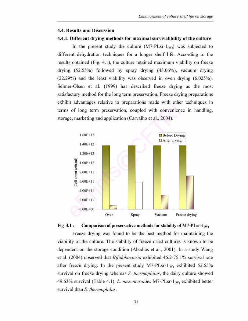
Enhancement of culture shelf life on storage
131
4.4. Results and Discussion 4.4.1. Different drying methods for maximal survivalibility of the culture In the present study the culture (M7-PLsr-1(W)) was subjected to different dehydration techniques for a longer shelf life. According to the results obtained (Fig. 4.1), the culture retained maximum viability on freeze drying (52.55%) followed by spray drying (43.06%), vacuum drying (22.29%) and the least viability was observed in oven drying (6.025%). Selmer-Olsen et al. (1999) has described freeze drying as the most satisfactory method for the long term preservation. Freeze drying preparations exhibit advantages relative to preparations made with other techniques in terms of long term preservation, coupled with convenience in handling, storage, marketing and application (Carvalho et al., 2004).
0.00E+00
2.00E+11
4.00E+11
6.00E+11
8.00E+11
1.00E+12
1.20E+12
1.40E+12
1.60E+12
Oven Spray Vaccum Freeze drying
Cel
l cou
nt (c
fu/m
l)
Before DryingAfter drying
Fig 4.1 : Comparison of preservative methods for stability of M7-PLsr-1(W) Freeze drying was found to be the best method for maintaining the viability of the culture. The stability of freeze dried cultures is known to be dependent on the storage condition (Abadias et al., 2001). In a study Wang et al. (2004) observed that Bifidobacteria exhibited 46.2-75.1% survival rate after freeze drying. In the present study M7-PLsr-1(W) exhibited 52.55% survival on freeze drying whereas S. thermophilus, the dairy culture showed 49.63% survival (Table 4.1). L. mesenteroides M7-PLsr-1(W) exhibited better survival than S. thermophilus.
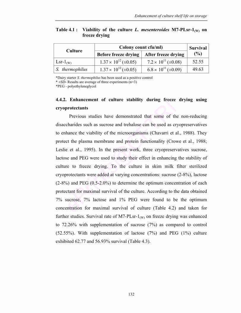
Enhancement of culture shelf life on storage
132
Table 4.1 : Viability of the culture L. mesenteroides M7-PLsr-1(W) on freeze drying
Colony count cfu/ml)
Culture Before freeze drying After freeze drying
Survival (%)
Lsr-1(W) 1.37 × 10¹² (±0.05) 7.2 × 10¹¹ (±0.08) 52.55 S. thermophilus 1.37 × 10¹² (±0.05) 6.8 × 10¹¹ (±0.09) 49.63
*Dairy starter S. thermophilus has been used as a positive control * ±SD- Results are average of three experiments (n=3) *PEG - polyethyleneglycol
4.4.2. Enhancement of culture stability during freeze drying using
cryoprotectants
Previous studies have demonstrated that some of the non-reducing
disaccharides such as sucrose and trehalose can be used as cryopreservatives
to enhance the viability of the microorganisms (Chavarri et al., 1988). They
protect the plasma membrane and protein functionality (Crowe et al., 1988;
Leslie et al., 1995). In the present work, three cryopreservatives sucrose,
lactose and PEG were used to study their effect in enhancing the stability of
culture to freeze drying. To the culture in skim milk filter sterilized
cryoprotectants were added at varying concentrations: sucrose (2-8%), lactose
(2-8%) and PEG (0.5-2.0%) to determine the optimum concentration of each
protectant for maximal survival of the culture. According to the data obtained
7% sucrose, 7% lactose and 1% PEG were found to be the optimum
concentration for maximal survival of culture (Table 4.2) and taken for
further studies. Survival rate of M7-PLsr-1(W) on freeze drying was enhanced
to 72.26% with supplementation of sucrose (7%) as compared to control
(52.55%). With supplementation of lactose (7%) and PEG (1%) culture
exhibited 62.77 and 56.93% survival (Table 4.3).
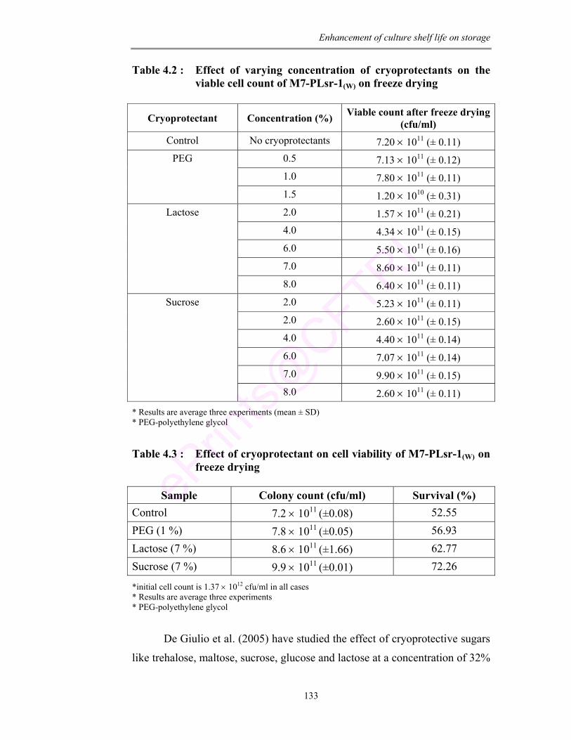
Enhancement of culture shelf life on storage
133
Table 4.2 : Effect of varying concentration of cryoprotectants on the viable cell count of M7-PLsr-1(W) on freeze drying
Cryoprotectant Concentration (%) Viable count after freeze drying (cfu/ml)
Control No cryoprotectants 7.20 × 10¹¹ (± 0.11) 0.5 7.13 × 10¹¹ (± 0.12) 1.0 7.80 × 10¹¹ (± 0.11)
PEG
1.5 1.20 × 10¹⁰ (± 0.31) 2.0 1.57 × 10¹¹ (± 0.21) 4.0 4.34 × 10¹¹ (± 0.15) 6.0 5.50 × 10¹¹ (± 0.16) 7.0 8.60 × 10¹¹ (± 0.11)
Lactose
8.0 6.40 × 10¹¹ (± 0.11) 2.0 5.23 × 10¹¹ (± 0.11) 2.0 2.60 × 10¹¹ (± 0.15) 4.0 4.40 × 10¹¹ (± 0.14) 6.0 7.07 × 10¹¹ (± 0.14) 7.0 9.90 × 10¹¹ (± 0.15)
Sucrose
8.0 2.60 × 10¹¹ (± 0.11)
* Results are average three experiments (mean ± SD) * PEG-polyethylene glycol
Table 4.3 : Effect of cryoprotectant on cell viability of M7-PLsr-1(W) on freeze drying
Sample Colony count (cfu/ml) Survival (%)
Control 7.2 × 10¹¹ (±0.08) 52.55 PEG (1 %) 7.8 × 10¹¹ (±0.05) 56.93 Lactose (7 %) 8.6 × 10¹¹ (±1.66) 62.77 Sucrose (7 %) 9.9 × 10¹¹ (±0.01) 72.26
*initial cell count is 1.37 × 10¹² cfu/ml in all cases * Results are average three experiments * PEG-polyethylene glycol
De Giulio et al. (2005) have studied the effect of cryoprotective sugars
like trehalose, maltose, sucrose, glucose and lactose at a concentration of 32%
Enhancement of culture shelf life on storage
134
on the survival rate of L. acidophilus, L. delbruekii and S. salivarus. The
study confirms the protective effect of all sugars on cell viability whereas in
the present study sucrose at a low concentration of 7% was found to be the
best cryoprotectant. The observed survival (72.26%) is much higher than in
B. longum (40%) as reported by Champagne et al. (1996).
These sugars used as cryoprotectants are known to replace structural
water in membrane after dehydration (Clegg, 1986; Crowe and Crow, 1986)
and prevent unfolding and aggregation of proteins by hydrogen bonding with
polar groups of proteins (Hanafusa, 1985; Carpenter et a., 1990) thus
protecting the cells under stressful conditions.
Zhao and Zhang (2005) have shown L. brevis and O. oeni to exhibit
45.1 and 38.2% survival in 10% sucrose as protective agent respectively and
47.8 and 2.4% survival when supplemented with 10% lactose. Whereas in the
present study the culture exhibits 72.26% and 62.77% survival with only 7%
sucrose and 7%lactose respectively.
According to Hubalek (2003) protective activity of sugars might be
due to the ability of sugars to prevent injurious freezing of cell fluids by
trapping salts in a highly viscous like phase. Leslie et al. (1995) postulated
that protective effect of sugar may be due to their ability to lower membrane
phase transition temperature and protection of protein structure in dry state.
Water molecules seem to be trapped between sugar molecules and
crystallization of ice is inhibited (Nicolajsen and Hvidt 1994; Chen et al.,
2000).
4.4.3. Cellular fatty acid analysis of freeze dried culture
Lonvaud-Funnel and Desens (1990) have observed an increase in
cellular membrane unsaturated fatty acid under low temperature. In the
present work we observed an increase in both saturated (C16:0 and C18:0)
and unsaturated fatty acids (C16:1, C18:2 and C18:3) as compared to
respective controls (before freeze drying) (Table 4.4; Fig. 4.2). Gilliland and
Speck (1974) have observed a decrease in C18:1 concentration in lactic
Streptococci in response to freezing and in L. acidophilus on spray drying.
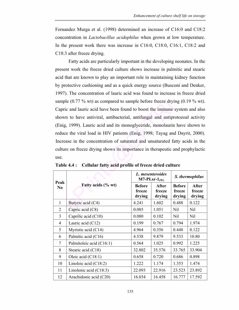
Enhancement of culture shelf life on storage
135
Fernandez Murga et al. (1998) determined an increase of C16:0 and C18:2
concentration in Lactobacillus acidophilus when grown at low temperature.
In the present work there was increase in C16:0, C18:0, C16:1, C18:2 and
C18:3 after freeze drying.
Fatty acids are particularly important in the developing neonates. In the
present work the freeze dried culture shows increase in palmitic and stearic
acid that are known to play an important role in maintaining kidney function
by protective cushioning and as a quick energy source (Busconi and Denker,
1997). The concentration of lauric acid was found to increase in freeze dried
sample (0.77 % wt) as compared to sample before freeze drying (0.19 % wt).
Capric and lauric acid have been found to boost the immune system and also
shown to have antiviral, antibacterial, antifungal and antiprotozoal activity
(Enig, 1999). Lauric acid and its monoglyceride, monolaurin have shown to
reduce the viral load in HIV patients (Enig, 1998; Tayag and Dayrit, 2000).
Increase in the concentration of saturated and unsaturated fatty acids in the
culture on freeze drying shows its importance in therapeutic and prophylactic
use.
Table 4.4 : Cellular fatty acid profile of freeze dried culture
L. mesenteroides M7-PLsr-1(W)
S. thermophilus Peak No Fatty acids (% wt) Before
freeze drying
After freeze drying
Before freeze drying
After freeze drying
1 Butyric acid (C4) 4.241 1.602 0.488 0.122 2 Capric acid (C8) 0.085 1.051 Nil Nil 3 Caprilic acid (C10) 0.080 0.102 Nil Nil 4 Lauric acid (C12) 0.199 0.767 0.794 1.974 5 Myristic acid (C14) 4.964 0.356 0.448 0.122 6 Palmitic acid (C16) 4.538 9.879 9.533 10.80 7 Palmitoleic acid (C16:1) 0.564 1.025 0.992 1.225 8 Stearic acid (C18) 32.802 35.576 33.765 33.904 9 Oleic acid (C18:1) 0.658 0.720 0.686 0.898 10 Linoleic acid (C18:2) 1.222 1.174 1.353 1.474 11 Linolenic acid (C18:3) 22.093 22.916 23.523 23.892 12 Arachidonic acid (C20) 16.034 16.458 16.777 17.592
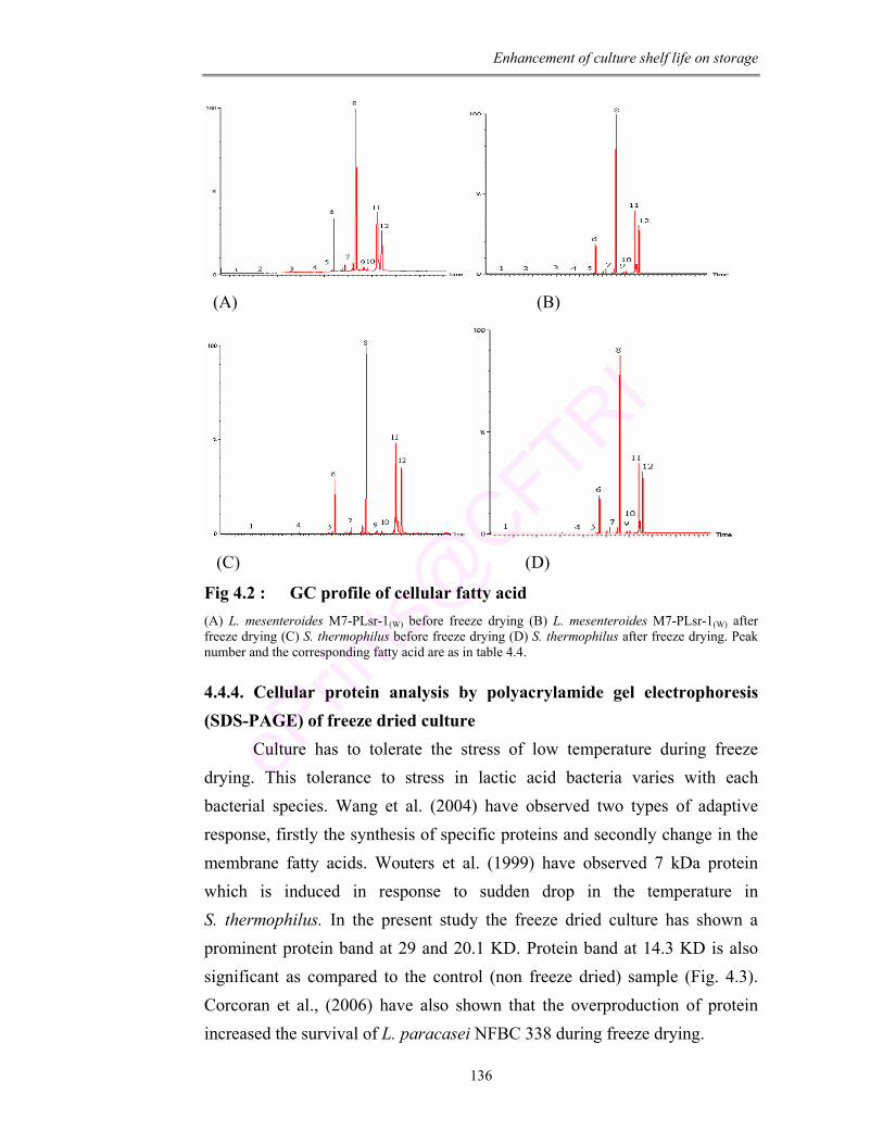
Enhancement of culture shelf life on storage
136
(A) (B)
(C) (D)
Fig 4.2 : GC profile of cellular fatty acid (A) L. mesenteroides M7-PLsr-1(W) before freeze drying (B) L. mesenteroides M7-PLsr-1(W) after freeze drying (C) S. thermophilus before freeze drying (D) S. thermophilus after freeze drying. Peak number and the corresponding fatty acid are as in table 4.4.
4.4.4. Cellular protein analysis by polyacrylamide gel electrophoresis (SDS-PAGE) of freeze dried culture Culture has to tolerate the stress of low temperature during freeze drying. This tolerance to stress in lactic acid bacteria varies with each bacterial species. Wang et al. (2004) have observed two types of adaptive response, firstly the synthesis of specific proteins and secondly change in the membrane fatty acids. Wouters et al. (1999) have observed 7 kDa protein which is induced in response to sudden drop in the temperature in S. thermophilus. In the present study the freeze dried culture has shown a prominent protein band at 29 and 20.1 KD. Protein band at 14.3 KD is also significant as compared to the control (non freeze dried) sample (Fig. 4.3). Corcoran et al., (2006) have also shown that the overproduction of protein increased the survival of L. paracasei NFBC 338 during freeze drying.
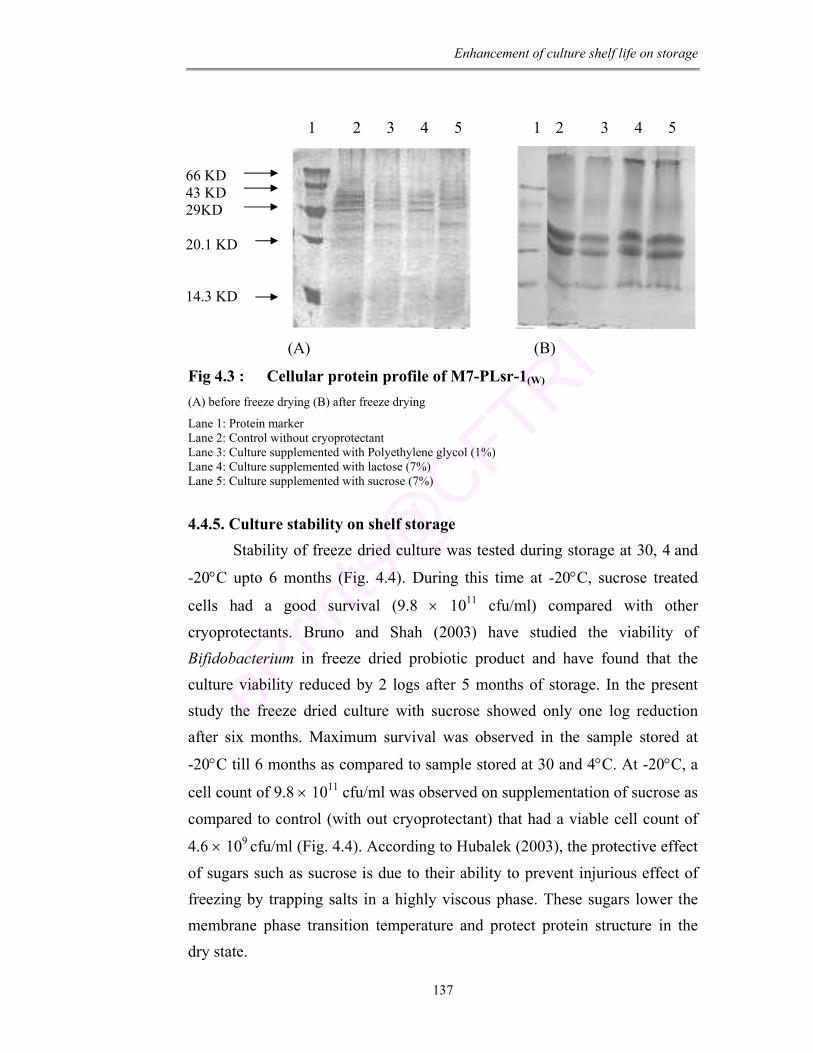
Enhancement of culture shelf life on storage
137
1 2 3 4 5 1 2 3 4 5
(A) (B)
Fig 4.3 : Cellular protein profile of M7-PLsr-1(W) (A) before freeze drying (B) after freeze drying
Lane 1: Protein marker Lane 2: Control without cryoprotectant Lane 3: Culture supplemented with Polyethylene glycol (1%) Lane 4: Culture supplemented with lactose (7%) Lane 5: Culture supplemented with sucrose (7%) 4.4.5. Culture stability on shelf storage Stability of freeze dried culture was tested during storage at 30, 4 and
-20°C upto 6 months (Fig. 4.4). During this time at -20°C, sucrose treated
cells had a good survival (9.8 × 10¹¹ cfu/ml) compared with other cryoprotectants. Bruno and Shah (2003) have studied the viability of Bifidobacterium in freeze dried probiotic product and have found that the culture viability reduced by 2 logs after 5 months of storage. In the present study the freeze dried culture with sucrose showed only one log reduction after six months. Maximum survival was observed in the sample stored at
-20°C till 6 months as compared to sample stored at 30 and 4°C. At -20°C, a
cell count of 9.8 × 10¹¹ cfu/ml was observed on supplementation of sucrose as compared to control (with out cryoprotectant) that had a viable cell count of
4.6 × 10⁹ cfu/ml (Fig. 4.4). According to Hubalek (2003), the protective effect of sugars such as sucrose is due to their ability to prevent injurious effect of freezing by trapping salts in a highly viscous phase. These sugars lower the membrane phase transition temperature and protect protein structure in the dry state.
66 KD 43 KD 29KD 20.1 KD 14.3 KD
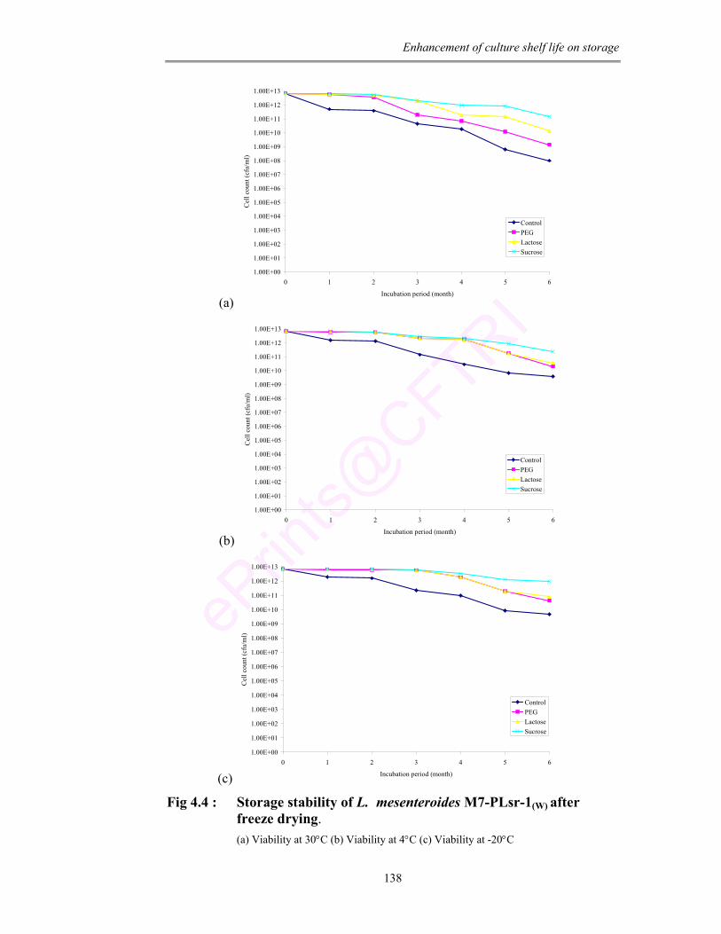
Enhancement of culture shelf life on storage
138
(a)
1.00E+00
1.00E+01
1.00E+02
1.00E+03
1.00E+04
1.00E+05
1.00E+06
1.00E+07
1.00E+08
1.00E+09
1.00E+10
1.00E+11
1.00E+12
1.00E+13
0 1 2 3 4 5 6
Incubation period (month)
Cel
l cou
nt (c
fu/m
l)
ControlPEGLactoseSucrose
(b)
1.00E+00
1.00E+01
1.00E+02
1.00E+03
1.00E+04
1.00E+05
1.00E+06
1.00E+07
1.00E+08
1.00E+09
1.00E+10
1.00E+11
1.00E+12
1.00E+13
0 1 2 3 4 5 6
Incubation period (month)
Cel
l cou
nt (c
fu/m
l)
ControlPEGLactoseSucrose
(c)
1.00E+00
1.00E+01
1.00E+02
1.00E+03
1.00E+04
1.00E+05
1.00E+06
1.00E+07
1.00E+08
1.00E+09
1.00E+10
1.00E+11
1.00E+12
1.00E+13
0 1 2 3 4 5 6
Incubation period (month)
Cel
l cou
nt (c
fu/m
l)
ControlPEGLactoseSucrose
Fig 4.4 : Storage stability of L. mesenteroides M7-PLsr-1(W) after freeze drying.
(a) Viability at 30°C (b) Viability at 4°C (c) Viability at -20°C
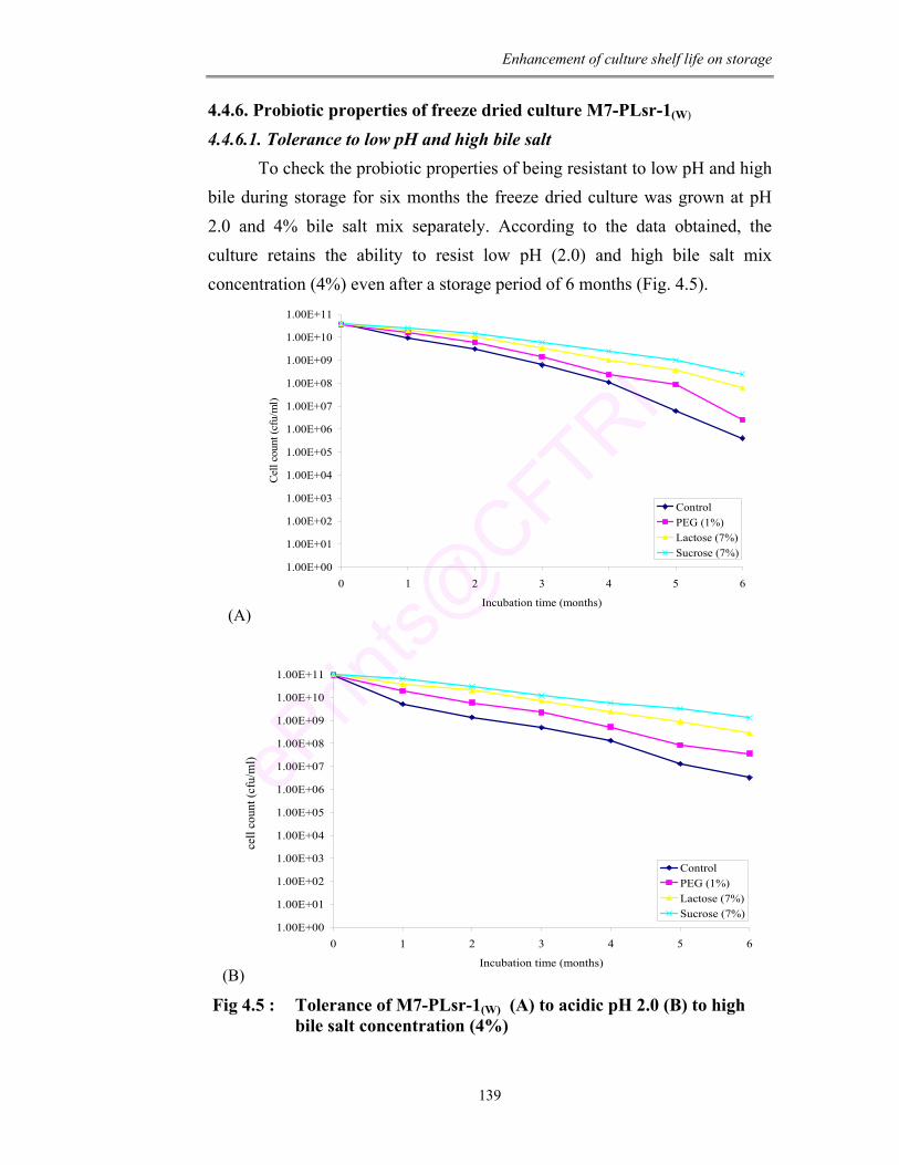
Enhancement of culture shelf life on storage
139
4.4.6. Probiotic properties of freeze dried culture M7-PLsr-1(W) 4.4.6.1. Tolerance to low pH and high bile salt
To check the probiotic properties of being resistant to low pH and high bile during storage for six months the freeze dried culture was grown at pH 2.0 and 4% bile salt mix separately. According to the data obtained, the culture retains the ability to resist low pH (2.0) and high bile salt mix concentration (4%) even after a storage period of 6 months (Fig. 4.5).
(A)
1.00E+00
1.00E+01
1.00E+02
1.00E+03
1.00E+04
1.00E+05
1.00E+06
1.00E+07
1.00E+08
1.00E+09
1.00E+10
1.00E+11
0 1 2 3 4 5 6
Incubation time (months)
Cel
l cou
nt (c
fu/m
l)
ControlPEG (1%)Lactose (7%)Sucrose (7%)
(B)
1.00E+00
1.00E+01
1.00E+02
1.00E+03
1.00E+04
1.00E+05
1.00E+06
1.00E+07
1.00E+08
1.00E+09
1.00E+10
1.00E+11
0 1 2 3 4 5 6
Incubation time (months)
Cel
cou
nt (c
fu/m
l)
ControlPEG (1%)Lactose (7%)Sucrose (7%)
Fig 4.5 : Tolerance of M7-PLsr-1(W) (A) to acidic pH 2.0 (B) to high bile salt concentration (4%)
cell
coun
t (cf
u/m
l)

Enhancement of culture shelf life on storage
140
4.4.6.2. Antimicrobial activity of freeze dried culture
The culture had antimicrobial activity against 6 food borne pathogens
namely E. coli, S. typhi, S. dysenteriae, P. aeroginosa, V. cholera and S.
aureus (Table 4.5). On storage of 6 months the activity was maximum with
sucrose as cryoprotectant against E. coli (16 mm).
Table 4.5 : Antimicrobial activity of freeze dried cells Inhibition zone (mm) Incubation
period (months)
Sample E. coli S. typhi S.
dysenteriae P.
aeroginosa V. cholerae S. aureus
Control 20 ± 0.02 23 ± 0.02 23 ± 0.11 23 ± 0.03 10 ± 0.02 18 ± 0.12
PEG 22 ± 0.10 23 ± 0.03 23 ± 0.02 23 ± 0.03 12 ± 0.02 24 ±0.11
Lactose 23 ± 0.30 23 ± 0.01 23 ± 0.03 23 ± 0.03 14 ± 0.03 20 ±0.11
0
Sucrose 24 ± 0.11 23 ± 0.11 23 ± 0.12 23 ± 0.11 10 ± 0.02 23 ±0.03
Control 19 ± 0.22 20 ± 0.21 22 ± 0.12 22 ± 0.22 10 ± 0.11 16 ±0.30
PEG 22 ± 0.15 22 ± 0.31 22 ± 0.13 22 ± 0.21 12 ± 0.11 20 ±0.03
Lactose 20 ± 0.16 22 ± 0.44 22 ± 0.15 22 ± 0.15 12 ± 0.05 18 ±0.05
1
Sucrose 22 ± 0.17 22 ± 0.33 22 ± 0.16 22 ± 0.17 16 ± 0.15 20 ±0.15
Control 18 ± 0.22 20 ± 0.18 20 ± 0.05 20 ± 0.19 10 ± 0.16 16 ±0.15
PEG 20 ± 0.13 20 ± 0.19 20 ± 0.05 20 ± 0.18 10 ± 0.19 18 ±0.16
Lactose 18 ± 0.11 20 ± 0.17 20 ± 0.06 20 ± 0.16 12 ± 0.11 16 ±0.17
2
Sucrose 20 ± 0.11 20 ± 0.15 20 ± 0.05 20 ± 0.04 14 ± 0.11 20 ±0.01
Control 18 ± 0.22 18 ± 0.11 16 ± 0.12 18 ± 0.05 08 ± 0.05 14 ±0.11
PEG 18 ± 0.11 18 ± 0.11 18 ± 0.11 18 ± 0.05 10 ± 0.05 16 ±0.12
Lactose 18 ± 0.46 18 ± 0.11 16 ± 0.14 18 ± 0.06 10 ± 0.05 15 ±0.15
3
Sucrose 20 ± 0.12 18 ± 0.02 18 ± 0.13 20 ± 0.15 12 ± 0.02 18 ±0.05
Control 16 ± 0.11 15 ± 0.02 15 ± 0.01 16 ± 0.16 08 ± 0.12 12 ±0.06
PEG 18 ± 0.33 16 ± 0.02 15 ± 0.19 18 ± 0.02 08 ± 0.03 16 ±0.06
Lactose 18 ± 0.18 16 ± 0.13 16 ± 0.12 16 ± 0.02 08 ± 0.13 14 ±0.06
4
Sucrose 20 ± 0.19 18 ± 0.30 16 ± 0.11 18 ± 0.02 10 ± 0.13 16 ±0.11
Control 16 ± 0.11 14 ± 0.12 12 ± 0.26 14 ± 0.11 06 ± 0.01 12 ±0.12
PEG 16 ± 0.11 15 ± 0.11 14 ± 0.19 16 ± 0.01 07 ± 0.11 12 ±0.03
Lactose 18 ± 0.22 16 ± 0.03 12 ± 0.11 14 ± 0.11 08 ± 0.12 10 ±0.09
5
Sucrose 18 ± 0.12 16 ± 0.03 15 ± 0.12 16 ± 0.11 08 ± 0.02 14 ±0.01
Control 14 ± 0.13 14 ± 0.06 10 ± 0.13 10 ± 0.02 - 08 ±0.12
PEG 14 ± 0.15 14 ± 0.04 14 ± 0.11 14 ± 0.03 06 ± 0.16 08 ±0.11
Lactose 15 ± 0.17 14 ± 0.15 10 ± 0.02 12 ± 0.02 07 ± 0.12 -
6
Sucrose 16 ± 0.18 14 ± 0.11 10 ± 0.03 12 ± 0.02 07 ± 0.11 10 ±0.01
* Results are average three experiments * FD = Freeze drying; PEG = polyethylene glycol (1%); Lactose (7%); Sucrose (7%)
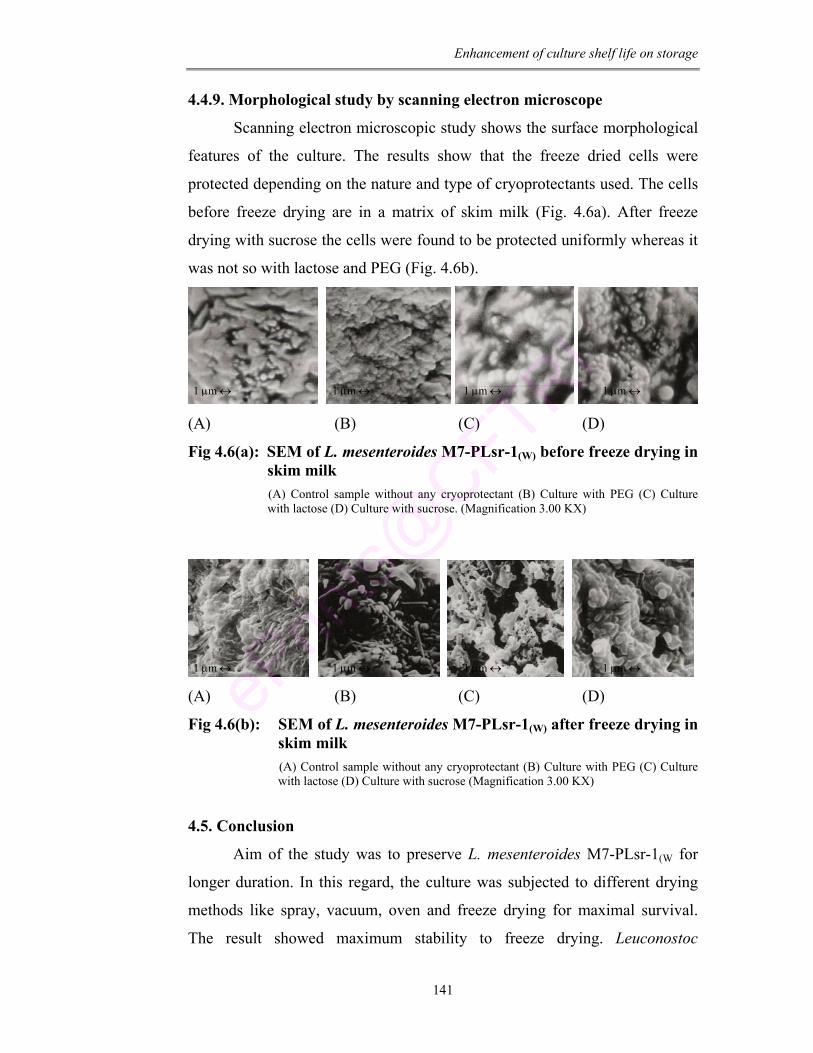
Enhancement of culture shelf life on storage
141
4.4.9. Morphological study by scanning electron microscope
Scanning electron microscopic study shows the surface morphological
features of the culture. The results show that the freeze dried cells were
protected depending on the nature and type of cryoprotectants used. The cells
before freeze drying are in a matrix of skim milk (Fig. 4.6a). After freeze
drying with sucrose the cells were found to be protected uniformly whereas it
was not so with lactose and PEG (Fig. 4.6b).
(A) (B) (C) (D)
Fig 4.6(a): SEM of L. mesenteroides M7-PLsr-1(W) before freeze drying in skim milk (A) Control sample without any cryoprotectant (B) Culture with PEG (C) Culture with lactose (D) Culture with sucrose. (Magnification 3.00 KX)
(A) (B) (C) (D)
Fig 4.6(b): SEM of L. mesenteroides M7-PLsr-1(W) after freeze drying in skim milk (A) Control sample without any cryoprotectant (B) Culture with PEG (C) Culture with lactose (D) Culture with sucrose (Magnification 3.00 KX)
4.5. Conclusion
Aim of the study was to preserve L. mesenteroides M7-PLsr-1(W for
longer duration. In this regard, the culture was subjected to different drying
methods like spray, vacuum, oven and freeze drying for maximal survival.
The result showed maximum stability to freeze drying. Leuconostoc
1 µm ↔ 1 µm ↔ 1 µm ↔ 1 µm ↔
1 µm ↔ 1 µm ↔ 1 µm ↔ 1 µm ↔
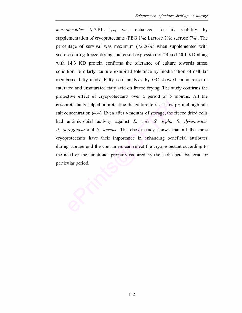
Enhancement of culture shelf life on storage
142
mesenteroides M7-PLsr-1(W) was enhanced for its viability by
supplementation of cryoprotectants (PEG 1%; Lactose 7%; sucrose 7%). The
percentage of survival was maximum (72.26%) when supplemented with
sucrose during freeze drying. Increased expression of 29 and 20.1 KD along
with 14.3 KD protein confirms the tolerance of culture towards stress
condition. Similarly, culture exhibited tolerance by modification of cellular
membrane fatty acids. Fatty acid analysis by GC showed an increase in
saturated and unsaturated fatty acid on freeze drying. The study confirms the
protective effect of cryoprotectants over a period of 6 months. All the
cryoprotectants helped in protecting the culture to resist low pH and high bile
salt concentration (4%). Even after 6 months of storage, the freeze dried cells
had antimicrobial activity against E. coli, S. typhi, S. dysenteriae,
P. aeroginosa and S. aureus. The above study shows that all the three
cryoprotectants have their importance in enhancing beneficial attributes
during storage and the consumers can select the cryoprotectant according to
the need or the functional property required by the lactic acid bacteria for
particular period.

Chapter 5 Functional Food with
Leuconostoc: A Native Isolate
Functional food with Leuconostoc
143
CHAPTER – 5
FUNCTIONAL FOOD WITH LEUCONOSTOC :
A NATIVE ISOLATE
ABSTRACT
In the present work the culture M7-PLsr-1(W) that has the potent
probiotic functional properties was used as starter culture in the preparation of
fermented milk beverage. The prepared product was found to be rich in
proteins, fat, total sugars, fatty acids and minerals like iron, zinc and
magnesium. The viability of culture and the nutritional properties of the
product were enhanced with supplementation of different adjuvants like
tryptone, casein hydrolysate, cysteine hydrochloride and ascorbic acid. After
5 days, maximum viability was observed on supplementation of tryptone
(100 mg/L).
Functional food with Leuconostoc
144
5.1. Introduction
Probiotic containing products are gaining popularity and acceptance
because of their high therapeutic and health benefits. Fermented dairy
products have been regarded as an ideal vector for the delivery of these
probiotic bacteria to consumers (Lourens-Hattingh and Viljoen, 2001).
Fermented dairy foods have long been considered safe and nutritious.
The health benefits elicited by LAB were the primary reason for Metchnikoff
to associate the consumption of yogurt with longevity of Bulgarian peasants.
Numerous scientific papers and review articles have been published on the
health benefits of fermented dairy products (Kurman et al., 1992; Hughes and
Hoover, 1995; Sanders, 1999; Cotter, 2007). Consumption of fermented dairy
products is known to have potential in aiding lactose digestion (Shermark
et al., 1995; Vesa et al., 1996), preventing traveller’s diarrhea (Oksanen et al.,
1990), reducing duration of rotavirus diarrhea (Guarino et al., 1998), exert
antitumor activity (Kato et al., 1994; Bakalinsky et al., 1996), enhance
activity of immune system (Meydani and Ha, 2000) and aid in controlling
serum cholesterol (Gilliland et al., 1985; Eichholzer and Stahelin, 1993).
The incorporation of probiotic bacteria in food products has led to the
creation of a new and rapidly increasing multi-billion dollar market especially
in Europe, Japan and Australia (ADC, 1998; Sanders, 1998; Stanton et al.,
2001). There is a tremendous increase in the world sale of cultured products
containing probiotic bacteria in the form of yogurt, fermented milk,
ice-creams and pharmaceutical products because of their health effects
(Mattila-Sandholm, 1999; Ostlie, 2005). Products containing Bifidobacteria
are also produced in Canada, Italy, Poland, Czechoslovaskia, US and Brazil.
Foods containing these bacteria are often referred to as functional
foods because of its potential benefits beyond being as a source of nutrient
(Gilliland et al., 2002). Fermented milk is a product obtained by controlled
fermentation to produce desirable acidity and flavor (Thapa, 2000). Owing to
the nutritional and therapeutic significance, an increasing trend towards
consumption of yogurt is observed world wide (Sarkar, 2006). The
Functional food with Leuconostoc
145
consumption has been considerably increased in UK (Kowalska et al., 2000)
and in USA. In 1995 sales figure was $ 516 million US dollars (Sivak, 2000).
Probiotic culture cannot exert beneficial effect unless their population
reaches a certain minimum level. For instance there is no general agreement
on minimum concentration of probiotic to achieve therapeutic benefits. While
some researchers suggest a concentration level above 106 cfu/ml (Kurmann
and Rasic, 1991; Samona and Robinson, 1991; Vinderola, et al., 2000) others
stipulate >107 and 108 cfu/ml as satisfactory levels (Davis et al., 1971;
Kailasapathy and Rybka, 1997). Oksanen et al. (1990) have shown that 109
cfu/ml of L. bulgaricus GG is required to reduce the occurrence of traveller’s
diarrhea. In Japan, fermented milk and lactic acid beverage association has
specified that there has to be atleast 107 cfu/ml of viable cells in fermented
drinks (Lourens-Hattingh and Viljoen 2001). The international standard of
federation / International Dairy federation (FIL/IDF) suggests 107 cfu/g of
L. acidophilus and 106 cfu/g of Bifidobacteria in fermented milk (IDF, 1992).
The number of live cells as well the organoleptic qualities play a major role in
the acceptability of any functional fermented foods.
The present culture L. mesenteroides (M7-PLsr-1(W)), a strain that was
found to have potential probiotic functional characters was used as a starter
culture for the preparation of fermented milk beverage. Viability of culture in
the fermented milk was enhanced with supplementation of different adjuvants
and studied for the nutritional aspect like protein, fat, sugars, minerals,
titrable acidity and fatty acid profile.
5.2. Materials:
Pasteurized cow milk (KMF, Nandini Mysore, India).
Chemicals: All the chemicals purchased were of analytical grade.
1) MRSA media.
2) Potassium oxalate, potassium hydroxide, sodium hydroxide, ammonia,
tryptone, casein hydrolysate, cysteine hydrochloride and ascorbic acid
(HiMedia Pvt Ltd, Mumbai, India).
Functional food with Leuconostoc
146
3) Solvents: Formalin, peroxide free diethyl ether, petroleum ether,
methanol, chloroform, and hexane (SRL Company, India).
4) Phenolphthalein indicator: Phenolphthalein (1 g) is mixed in ethyl
alcohol (110 ml). Further 80 ml of water is added and mixed with 0.1
M NaOH solution until one drop gives a faint pink coloration. Make
up to 200 ml with distilled water.
Equipments: Centrifuge, Spectrophotometer, Incubator (37°C), Muffle
furnace (as described in chapter 1 and 2).
5.3. Methods
5.3.1. Preparation and quality analysis of fermented milk beverage
A commercial homogenized and pasteurized milk (1 L) was sterilized
at 121°C for 5 min and cooled to room temperature. It was inoculated with
the starter culture L. mesenteroides (M7-PLsr-1(W)) at a concentration of
1 × 10⁵ cfu/ml and incubated at 37°C for 24 h. After the fermentation period,
the product was stored at 4°C for 5 days.
5.3.1.1. Viability of bacterial culture
Bacterial growth was estimated using colony plate count method
(Martinez-Villaluenza et al., 2005). An aliquot of sample was taken on 1, 3
and 5th day of storage, serially diluted and the appropriate dilution was plated
on MRSA plates. Plates were incubated at 37°C for 24 h. Colonies grown
were counted and expressed as cfu/ml.
5.3.1.2. Protein estimation
Total protein content was determined by formal titration method using
phenolphthalein as an indicator (Bennenberg et al., 1949; Saha et al., 2003).
Sample (10 ml) was mixed with phenolphthalein (0.5 ml; 0.5 % w/v) and
neutral saturated potassium oxalate (0.4 ml). The mixture was then
neutralized with sodium hydroxide (0.1 M) till it turns pink. Further formalin
(2 ml) was added and mixed well. The mixture was then allowed to stand for
few minutes to form clear colorless solution and then titrated against 0.1M
NaOH until pink. Protein content was then calculated by following formula
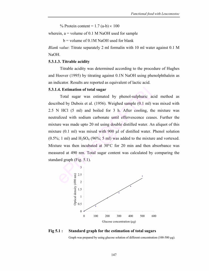
Functional food with Leuconostoc
147
% Protein content = 1.7 (a-b) × 100
wherein, a = volume of 0.1 M NaOH used for sample
b = volume of 0.1M NaOH used for blank
Blank value: Titrate separately 2 ml formalin with 10 ml water against 0.1 M
NaOH.
5.3.1.3. Titrable acidity
Titrable acidity was determined according to the procedure of Hughes
and Hoover (1995) by titrating against 0.1N NaOH using phenolphthalein as
an indicator. Results are reported as equivalent of lactic acid.
5.3.1.4. Estimation of total sugar
Total sugar was estimated by phenol-sulphuric acid method as
described by Dubois et al. (1956). Weighed sample (0.1 ml) was mixed with
2.5 N HCl (5 ml) and boiled for 3 h. After cooling, the mixture was
neutralized with sodium carbonate until effervescence ceases. Further the
mixture was made upto 20 ml using double distilled water. An aliquot of this
mixture (0.1 ml) was mixed with 900 µl of distilled water. Phenol solution
(0.5%; 1 ml) and H2SO4 (96%; 5 ml) was added to the mixture and vortexed.
Mixture was then incubated at 30°C for 20 min and then absorbance was
measured at 490 nm. Total sugar content was calculated by comparing the
standard graph (Fig. 5.1).
0
0.5
1
1.5
2
2.5
3
0 100 200 300 400 500 600
Glucose concentration (µg)
Opt
ical
den
sity
(490
nm
)
Fig 5.1 : Standard graph for the estimation of total sugars
Graph was prepared by using glucose solution of different concentration (100-500 µg).
Functional food with Leuconostoc
148
5.3.1.5. Fat estimation Fat content was determined by Rose-Gottlieb method (IDF 1996). The
sample (10 g) was mixed with ammonia (1 ml) and 95% alcohol (10 ml). Peroxide free diethyl ether (25 ml) was added to this mixture and vigorously shaken for 1 min. Further, petroleum ether (25 ml) was added and shaking was continued for 30 sec. Solvent layer was then carefully transferred to previously weighed flask. After extracting twice the solvent layer was
collected in the weighed flask. The flask was then dried for 1 h at 100°C, cooled and weighed again. Difference in the weight of the flask was calculated and expressed as g/100 ml or percentage. 5.3.1.6. Mineral estimation
Total and soluble mineral content of the fermented milk was analyzed by using atomic absorption spectra according to the method described by Jacob (1958) and modified by Miller-Ihli (1996). Sample (50 ml) was taken out on 1, 3 and 5th day and the soluble fraction was separated by high speed ultra centrifugation at 100,000 g. The supernatant fluid was carefully removed
and filtered through whatman-40 paper. Samples were dried at 100°C in a crucible for 6 h. After charring, samples were incinerated in a muffle furnace
at 460°C for 24 h. The ash obtained was dissolved in concentrated H2SO4
(2 ml) and warmed for 5 min at 40°C in a water bath. The mixture was then madeup to 30 ml with double distilled water and analyzed by atomic absorption spectral studies. 5.3.1.7. Fatty acid analysis
Fatty acid analysis was carried out by the method of Bligh and Dyer (1959). (operation condition as described in chapter 2). 5.3.2. Enhancement of nutritional property by adjuvant supplementation
The sterilized milk (4.5 L) was inoculated with M7-PLsr-1(W) at a
concentration of 1 × 10⁵ cfu/ml and the mix was divided into 18 equal portions (250 ml/flask). Filter sterilized adjuvants (tryptone, casein hydrolysate, cysteine hydrochloride and ascorbic acid) were added to achieve a final concentration of 50, 100, 250 and 500 mg/L each in a different conical flask (one portion was kept as control without adjuvant). Incubation was
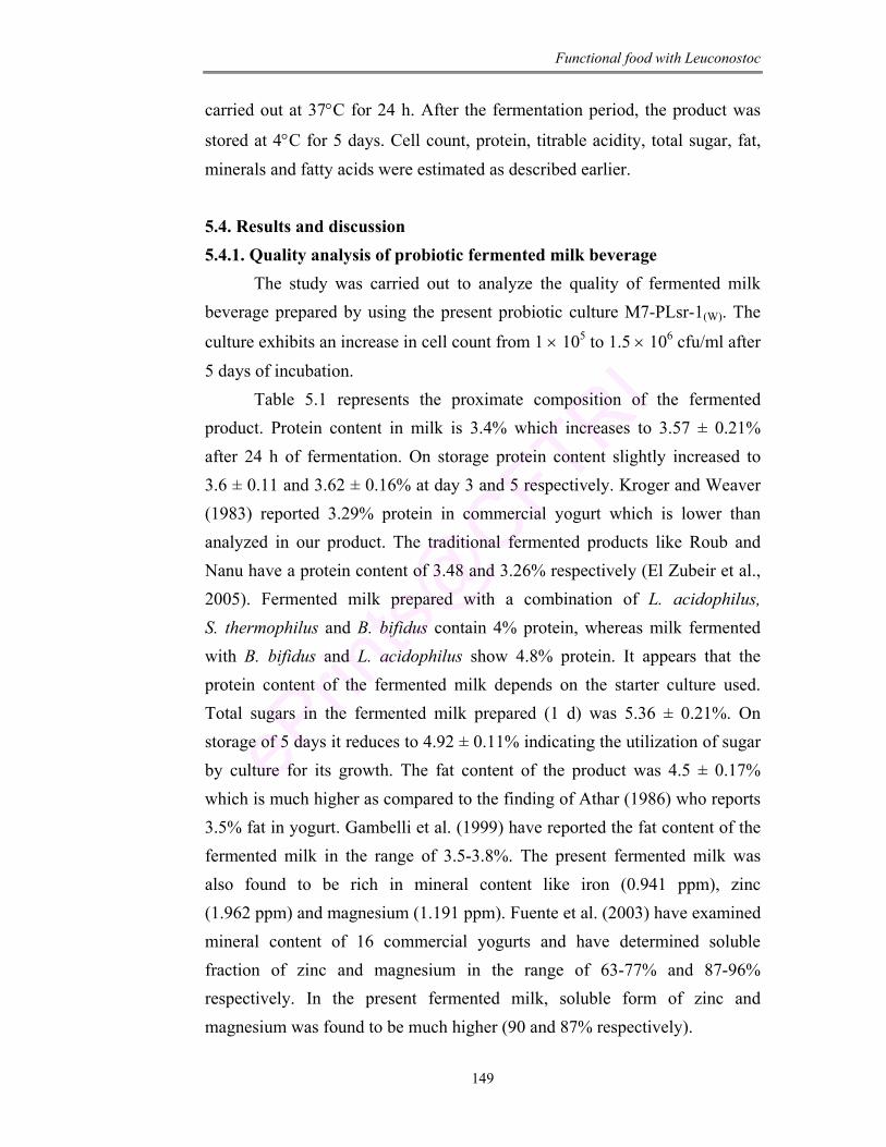
Functional food with Leuconostoc
149
carried out at 37°C for 24 h. After the fermentation period, the product was
stored at 4°C for 5 days. Cell count, protein, titrable acidity, total sugar, fat, minerals and fatty acids were estimated as described earlier. 5.4. Results and discussion 5.4.1. Quality analysis of probiotic fermented milk beverage
The study was carried out to analyze the quality of fermented milk beverage prepared by using the present probiotic culture M7-PLsr-1(W). The
culture exhibits an increase in cell count from 1 × 10⁵ to 1.5 × 10⁶ cfu/ml after 5 days of incubation.
Table 5.1 represents the proximate composition of the fermented product. Protein content in milk is 3.4% which increases to 3.57 ± 0.21% after 24 h of fermentation. On storage protein content slightly increased to 3.6 ± 0.11 and 3.62 ± 0.16% at day 3 and 5 respectively. Kroger and Weaver (1983) reported 3.29% protein in commercial yogurt which is lower than analyzed in our product. The traditional fermented products like Roub and Nanu have a protein content of 3.48 and 3.26% respectively (El Zubeir et al., 2005). Fermented milk prepared with a combination of L. acidophilus, S. thermophilus and B. bifidus contain 4% protein, whereas milk fermented with B. bifidus and L. acidophilus show 4.8% protein. It appears that the protein content of the fermented milk depends on the starter culture used. Total sugars in the fermented milk prepared (1 d) was 5.36 ± 0.21%. On storage of 5 days it reduces to 4.92 ± 0.11% indicating the utilization of sugar by culture for its growth. The fat content of the product was 4.5 ± 0.17% which is much higher as compared to the finding of Athar (1986) who reports 3.5% fat in yogurt. Gambelli et al. (1999) have reported the fat content of the fermented milk in the range of 3.5-3.8%. The present fermented milk was also found to be rich in mineral content like iron (0.941 ppm), zinc (1.962 ppm) and magnesium (1.191 ppm). Fuente et al. (2003) have examined mineral content of 16 commercial yogurts and have determined soluble fraction of zinc and magnesium in the range of 63-77% and 87-96% respectively. In the present fermented milk, soluble form of zinc and magnesium was found to be much higher (90 and 87% respectively).
Functional food with Leuconostoc
150
Table 5.1 : Chemical and microbial composition of fermented milk beverage
Fermented milk Parameter Milk
0 d 1 d 3 d 5 d Cell count (cfu/ml) Nil 1 × 10⁵ (± 0.02) 1.2 × 10⁶ (± 0.11) 1.3 × 10⁶ (± 0.15) 1.5 × 10⁶ (± 0.15) Protein (%) 3.40 ± 0.21 3.40 ± 0.21 3.57 ± 0.21 3.60 ± 0.11 3.62 ± 0.16 Total sugar (%) 5.40 ± 0.12 5.40 ± 0.12 5.36 ± 0.21 5.21 ± 1.00 4.92 ± 0.11 Fat (%) 4.70 ± 0.13 4.70 ± 0.13 4.50 ± 0.17 4.30 ± 0.15 4.00 ± 0.15
Mineral content of fermented milk (ppm)
Incubation time (d) 0 1 5 Mineral
(ppm) Total Soluble Bioavailable
(%) Total Soluble Bioavailable (%) Total Soluble Bioavailable
(%) Iron 0.769 ± 0.11 0.523 ± 0.21 68.01 0.941 ± 0.15 0.828 ± 0.63 87.99 0.941 ± 0.21 0.812 ± 0.11 86.29 Zinc 1.724 ± 0.15 1.496 ± 0.19 86.77 1.962 ± 0.15 1.766 ± 0.10 90.01 1.993 ± 0.19 1.812 ± 0.11 90.92 Magnesium 1.038 ± 0.16 0.912 ± 0.11 87.86 1.191 ± 0.11 1.036 ± 0.11 86.98 1.211 ± 1.21 1.121 ± 0.23 92.57
*Values are mean ± SD (n=3)
Functional food with Leuconostoc
151
5.4.2. Enhancement of nutritional property by adjuvant supplementation
In any functional fermented food it is important that the cell number of
live probiotic microorganism should be high till the end of the shelf life of the
product. For this, many workers have reported addition of adjuvants into the
product to increase the viability of the culture. Dave and Shah (1998) have
shown the improvement of probiotic Bifidobacterium viability in yogurt with
supplementation of cysteine, whey protein concentrate, acid casein
hydrolysate and tryptone wherein improvement in 3 logs was observed as
compared to control (without cryoprotectant). In the present study also an
increase in cell count was observed on adjuvant supplementation (Table 5.2).
After 24 h of fermentation, maximum viability was found with
supplementation of tryptone (100 mg/L). On shelf storage of 5 days it
increased to 9 × 10⁷ cfu/ml. Increase in the cell viability was also observed
with supplementation of other adjuvants like casein hydrolysate, cysteine
hydrochloride and ascorbic acid as compared to control.
5.4.3. Effect of adjuvants on protein content of fermented milk
Protein content of fermented milk prepared was 3.57%. On
incorporation of adjuvants higher protein content was recorded. Fermented
milk supplemented with tryptone (100 mg/L) exhibited highest protein
content (3.91%) after 24 h of fermentation (Table 5.3). On storage for 5 days,
the protein content gradually increased to 4.0%.
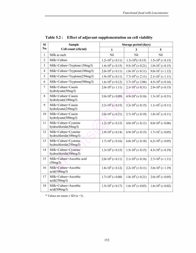
Functional food with Leuconostoc
152
Table 5.2 : Effect of adjuvant supplementation on cell viability
Storage period (days) Sl No
Sample Cell count (cfu/ml) 1 3 5
1 Milk as such Nil Nil Nil 2 Milk+Culture 1.2×10⁶ (± 0.11) 1.3×10⁶(± 0.15) 1.5×10⁶ (± 0.15) 3 Milk+Culture+Tryptone (50mg/l) 1.4×10⁶ (± 0.15) 9.5×10⁶ (± 0.21) 1.0×10⁷ (± 0.15) 4 Milk+Culture+Tryptone(100mg/l) 2.0×10⁶ (± 0.11) 1.0×10⁷ (± 0.11) 9.0×10⁷ (± 1.12) 5 Milk+Culture+Tryptone(250mg/l) 1.9×10⁶ (± 0.11) 7.7×10⁶ (± 2.11) 2.1×10⁷ (± 1.11) 6 Milk+Culture+Tryptone(500mg/l) 1.6×10⁶ (± 0.12) 3.7×10⁶ (± 2.06) 4.5×10⁶ (± 0.16) 7 Milk+Culture+Casein
hydrolysate(50mg/l) 2.0×10⁶ (± 1.11) 2.1×10⁶ (± 0.31) 2.9×10⁶ (± 0.15)
8 Milk+Culture+Casein hydrolysate(100mg/l)
3.0×10⁶ (± 0.09) 4.9×10⁶ (± 0.16) 1.3×10⁷ (± 0.31)
9 Milk+Culture+Casein hydrolysate(250mg/l)
2.2×10⁶ (± 0.15) 3.2×10⁶ (± 0.15) 1.1×10⁷ (± 0.11)
10 Milk+Culture+Casein hydrolysate(500mg/l)
2.0×10⁶ (± 0.21) 2.7×10⁶ (± 0.19) 1.0×10⁷ (± 0.11)
11 Milk+Culture+Cysteine hydrochloride(50mg/l)
1.2×10⁶ (± 0.15) 4.0×10⁶ (± 0.11) 8.0×10⁶ (± 0.06)
12 Milk+Culture+Cysteine hydrochloride(100mg/l)
1.9×10⁶ (± 0.14) 6.9×10⁶ (± 0.15) 1.7×10⁷ (± 0.05)
13 Milk+Culture+Cysteine hydrochloride(250mg/l)
1.7×10⁶ (± 0.16) 6.0×10⁶ (± 0.18) 6.2×10⁶ (± 0.05)
14 Milk+Culture+Cysteine hydrochloride(500mg/l)
1.3×10⁶ (± 0.15) 1.5×10⁶ (± 0.15) 4.3×10⁶ (± 0.19)
15 Milk+Culture+Ascorbic acid (50mg/l)
2.0×10⁶ (± 0.11) 2.1×10⁶ (± 0.16) 2.7×10⁶ (± 1.11)
16 Milk+Culture+Ascorbic acid(100mg/l)
1.8×10⁶ (± 0.12) 2.2×10⁶ (± 0.11) 3.0×10⁶ (± 1.19)
17 Milk+Culture+Ascorbic acid(250mg/l)
1.7×10⁶ (± 0.08) 1.8×10⁶ (± 0.21) 2.0×10⁶ (± 0.05)
18 Milk+Culture+Ascorbic acid(500mg/l)
1.5×10⁶ (± 0.17) 1.6×10⁶ (± 0.03) 1.6×10⁶ (± 0.02)
* Values are mean ± SD (n =3).
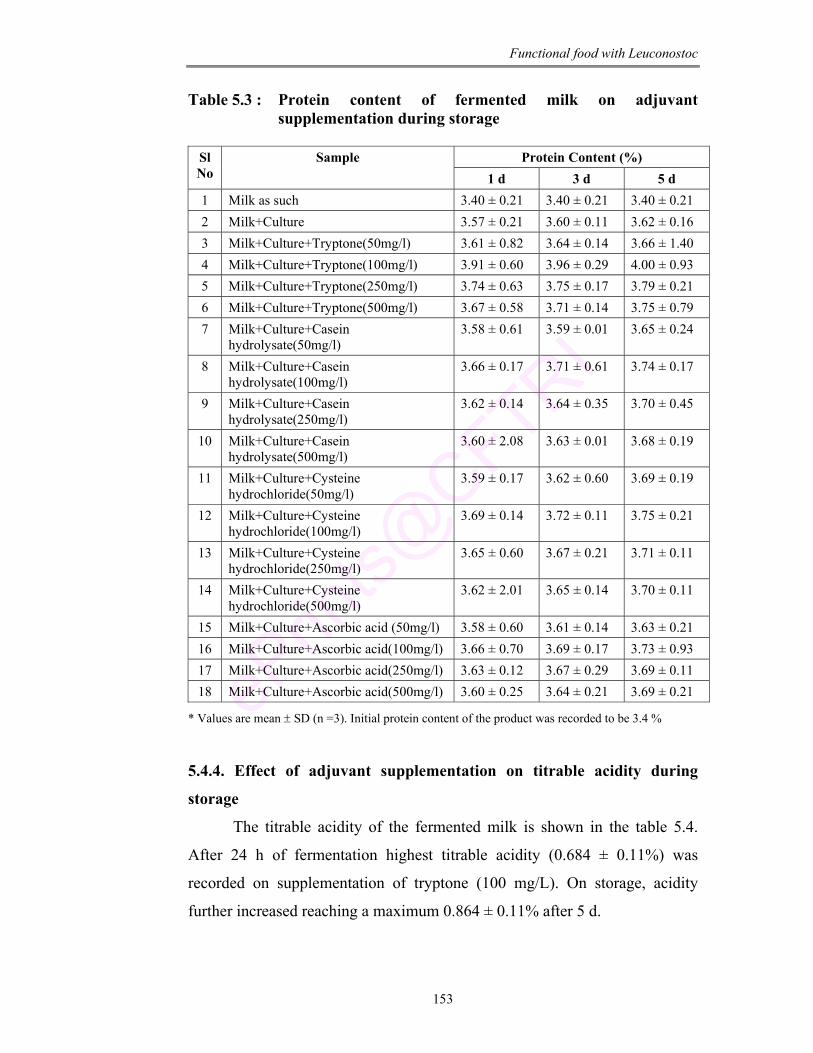
Functional food with Leuconostoc
153
Table 5.3 : Protein content of fermented milk on adjuvant supplementation during storage
Protein Content (%) Sl
No Sample
1 d 3 d 5 d 1 Milk as such 3.40 ± 0.21 3.40 ± 0.21 3.40 ± 0.21 2 Milk+Culture 3.57 ± 0.21 3.60 ± 0.11 3.62 ± 0.16 3 Milk+Culture+Tryptone(50mg/l) 3.61 ± 0.82 3.64 ± 0.14 3.66 ± 1.40 4 Milk+Culture+Tryptone(100mg/l) 3.91 ± 0.60 3.96 ± 0.29 4.00 ± 0.93 5 Milk+Culture+Tryptone(250mg/l) 3.74 ± 0.63 3.75 ± 0.17 3.79 ± 0.21 6 Milk+Culture+Tryptone(500mg/l) 3.67 ± 0.58 3.71 ± 0.14 3.75 ± 0.79 7 Milk+Culture+Casein
hydrolysate(50mg/l) 3.58 ± 0.61 3.59 ± 0.01 3.65 ± 0.24
8 Milk+Culture+Casein hydrolysate(100mg/l)
3.66 ± 0.17 3.71 ± 0.61 3.74 ± 0.17
9 Milk+Culture+Casein hydrolysate(250mg/l)
3.62 ± 0.14 3.64 ± 0.35 3.70 ± 0.45
10 Milk+Culture+Casein hydrolysate(500mg/l)
3.60 ± 2.08 3.63 ± 0.01 3.68 ± 0.19
11 Milk+Culture+Cysteine hydrochloride(50mg/l)
3.59 ± 0.17 3.62 ± 0.60 3.69 ± 0.19
12 Milk+Culture+Cysteine hydrochloride(100mg/l)
3.69 ± 0.14 3.72 ± 0.11 3.75 ± 0.21
13 Milk+Culture+Cysteine hydrochloride(250mg/l)
3.65 ± 0.60 3.67 ± 0.21 3.71 ± 0.11
14 Milk+Culture+Cysteine hydrochloride(500mg/l)
3.62 ± 2.01 3.65 ± 0.14 3.70 ± 0.11
15 Milk+Culture+Ascorbic acid (50mg/l) 3.58 ± 0.60 3.61 ± 0.14 3.63 ± 0.21 16 Milk+Culture+Ascorbic acid(100mg/l) 3.66 ± 0.70 3.69 ± 0.17 3.73 ± 0.93 17 Milk+Culture+Ascorbic acid(250mg/l) 3.63 ± 0.12 3.67 ± 0.29 3.69 ± 0.11 18 Milk+Culture+Ascorbic acid(500mg/l) 3.60 ± 0.25 3.64 ± 0.21 3.69 ± 0.21
* Values are mean ± SD (n =3). Initial protein content of the product was recorded to be 3.4 %
5.4.4. Effect of adjuvant supplementation on titrable acidity during
storage
The titrable acidity of the fermented milk is shown in the table 5.4.
After 24 h of fermentation highest titrable acidity (0.684 ± 0.11%) was
recorded on supplementation of tryptone (100 mg/L). On storage, acidity
further increased reaching a maximum 0.864 ± 0.11% after 5 d.
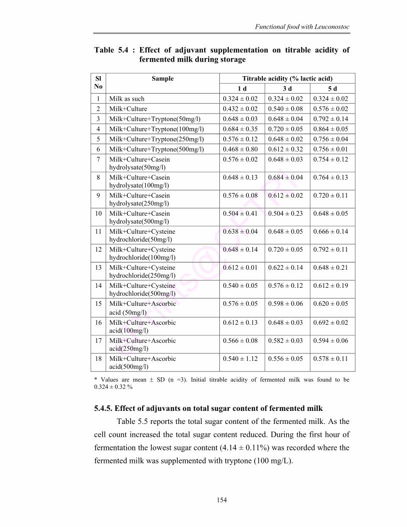
Functional food with Leuconostoc
154
Table 5.4 : Effect of adjuvant supplementation on titrable acidity of fermented milk during storage
Titrable acidity (% lactic acid) Sl
No Sample
1 d 3 d 5 d 1 Milk as such 0.324 ± 0.02 0.324 ± 0.02 0.324 ± 0.02 2 Milk+Culture 0.432 ± 0.02 0.540 ± 0.08 0.576 ± 0.02 3 Milk+Culture+Tryptone(50mg/l) 0.648 ± 0.03 0.648 ± 0.04 0.792 ± 0.14 4 Milk+Culture+Tryptone(100mg/l) 0.684 ± 0.35 0.720 ± 0.05 0.864 ± 0.05 5 Milk+Culture+Tryptone(250mg/l) 0.576 ± 0.12 0.648 ± 0.02 0.756 ± 0.04 6 Milk+Culture+Tryptone(500mg/l) 0.468 ± 0.80 0.612 ± 0.32 0.756 ± 0.01 7 Milk+Culture+Casein
hydrolysate(50mg/l) 0.576 ± 0.02 0.648 ± 0.03 0.754 ± 0.12
8 Milk+Culture+Casein hydrolysate(100mg/l)
0.648 ± 0.13 0.684 ± 0.04 0.764 ± 0.13
9 Milk+Culture+Casein hydrolysate(250mg/l)
0.576 ± 0.08 0.612 ± 0.02 0.720 ± 0.11
10 Milk+Culture+Casein hydrolysate(500mg/l)
0.504 ± 0.41 0.504 ± 0.23 0.648 ± 0.05
11 Milk+Culture+Cysteine hydrochloride(50mg/l)
0.638 ± 0.04 0.648 ± 0.05 0.666 ± 0.14
12 Milk+Culture+Cysteine hydrochloride(100mg/l)
0.648 ± 0.14 0.720 ± 0.05 0.792 ± 0.11
13 Milk+Culture+Cysteine hydrochloride(250mg/l)
0.612 ± 0.01 0.622 ± 0.14 0.648 ± 0.21
14 Milk+Culture+Cysteine hydrochloride(500mg/l)
0.540 ± 0.05 0.576 ± 0.12 0.612 ± 0.19
15 Milk+Culture+Ascorbic acid (50mg/l)
0.576 ± 0.05 0.598 ± 0.06 0.620 ± 0.05
16 Milk+Culture+Ascorbic acid(100mg/l)
0.612 ± 0.13 0.648 ± 0.03 0.692 ± 0.02
17 Milk+Culture+Ascorbic acid(250mg/l)
0.566 ± 0.08 0.582 ± 0.03 0.594 ± 0.06
18 Milk+Culture+Ascorbic acid(500mg/l)
0.540 ± 1.12 0.556 ± 0.05 0.578 ± 0.11
* Values are mean ± SD (n =3). Initial titrable acidity of fermented milk was found to be 0.324 ± 0.32 % 5.4.5. Effect of adjuvants on total sugar content of fermented milk Table 5.5 reports the total sugar content of the fermented milk. As the cell count increased the total sugar content reduced. During the first hour of fermentation the lowest sugar content (4.14 ± 0.11%) was recorded where the fermented milk was supplemented with tryptone (100 mg/L).
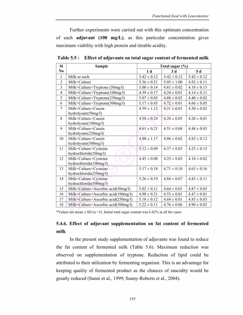
Functional food with Leuconostoc
155
Further experiments were carried out with this optimum concentration of each adjuvant (100 mg/L), as this particular concentration gives maximum viability with high protein and titrable acidity.
Table 5.5 : Effect of adjuvants on total sugar content of fermented milk
Total sugar (%) Sl No
Sample 1 d 3 d 5 d
1 Milk as such 5.42 ± 0.12 5.42 ± 0.12 5.42 ± 0.12 2 Milk+Culture 5.36 ± 0.21 5.05 ± 1.00 4.92 ± 0.11 3 Milk+Culture+Tryptone (50mg/l) 5.00 ± 0.14 4.81 ± 0.02 4.18 ± 0.15 4 Milk+Culture+Tryptone(100mg/l) 4.39 ± 0.17 4.24 ± 0.01 4.14 ± 0.11 5 Milk+Culture+Tryptone(250mg/l) 5.07 ± 0.05 4.88 ± 0.02 4.40 ± 0.02 6 Milk+Culture+Tryptone(500mg/l) 5.17 ± 0.45 4.72 ± 0.01 4.66 ± 0.05 7 Milk+Culture+Casein
hydrolysate(50mg/l) 4.59 ± 1.12 4.31 ± 0.01 4.30 ± 0.02
8 Milk+Culture+Casein hydrolysate(100mg/l)
4.50 ± 0.24 4.28 ± 0.03 4.20 ± 0.01
9 Milk+Culture+Casein hydrolysate(250mg/l)
4.61 ± 0.21 4.51 ± 0.04 4.48 ± 0.03
10 Milk+Culture+Casein hydrolysate(500mg/l)
4.88 ± 1.17 4.86 ± 0.02 4.82 ± 0.12
11 Milk+Culture+Cysteine hydrochloride(50mg/l)
5.12 ± 0.09 4.37 ± 0.03 4.25 ± 0.15
12 Milk+Culture+Cysteine hydrochloride(100mg/l)
4.45 ± 0.08 4.25 ± 0.03 4.18 ± 0.02
13 Milk+Culture+Cysteine hydrochloride(250mg/l)
5.17 ± 0.18 4.71 ± 0.10 4.63 ± 0.16
14 Milk+Culture+Cysteine hydrochloride(500mg/l)
5.26 ± 0.19 4.84 ± 0.07 4.83 ± 0.11
15 Milk+Culture+Ascorbic acid(50mg/l) 5.02 ± 0.11 4.64 ± 0.01 4.87 ± 0.03 16 Milk+Culture+Ascorbic acid(100mg/l) 4.90 ± 0.21 4.53 ± 0.01 4.47 ± 0.01 17 Milk+Culture+Ascorbic acid(250mg/l) 5.18 ± 0.12 4.69 ± 0.01 4.85 ± 0.03 18 Milk+Culture+Ascorbic acid(500mg/l) 5.22 ± 0.11 4.76 ± 0.06 4.90 ± 0.02
*Values are mean ± SD (n =3). Initial total sugar content was 6.42% in all the cases
5.4.6. Effect of adjuvant supplementation on fat content of fermented milk In the present study supplementation of adjuvants was found to reduce the fat content of fermented milk (Table 5.6). Maximum reduction was observed on supplementation of tryptone. Reduction of lipid could be attributed to their utilization by fermenting organism. This is an advantage for keeping quality of fermented product as the chances of rancidity would be greatly reduced (Sanni et al., 1999; Sunny-Roberts et al., 2004).
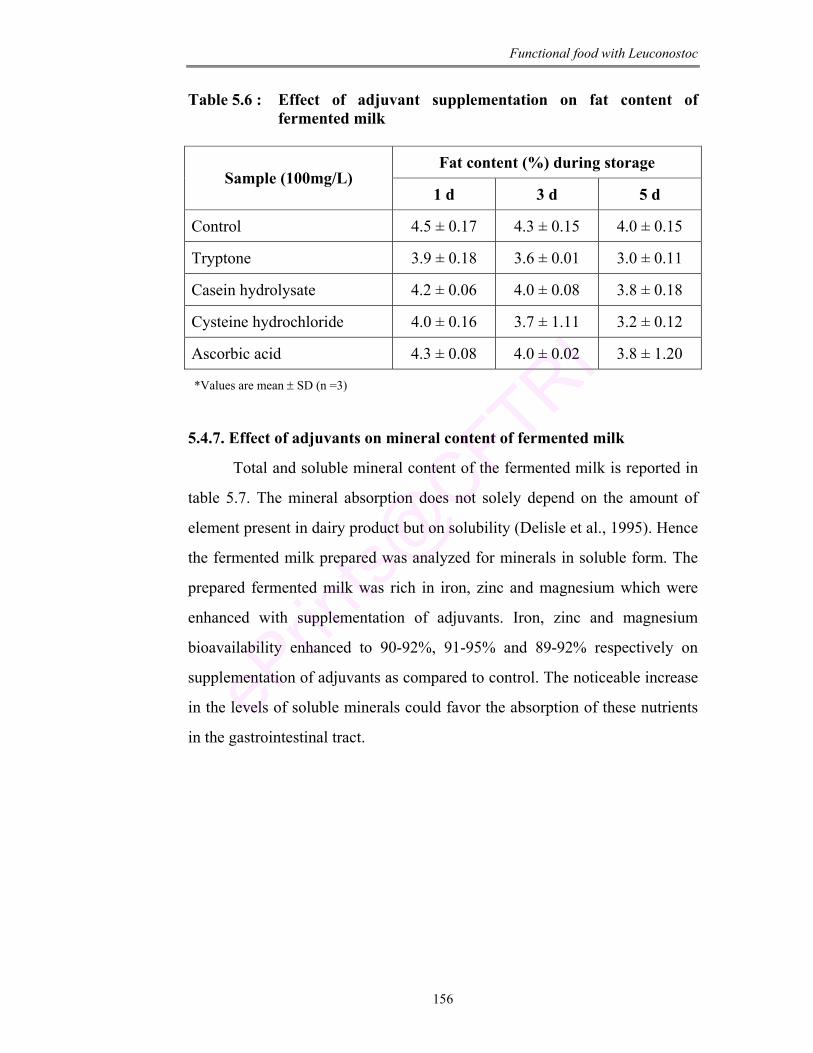
Functional food with Leuconostoc
156
Table 5.6 : Effect of adjuvant supplementation on fat content of fermented milk
Fat content (%) during storage
Sample (100mg/L) 1 d 3 d 5 d
Control 4.5 ± 0.17 4.3 ± 0.15 4.0 ± 0.15
Tryptone 3.9 ± 0.18 3.6 ± 0.01 3.0 ± 0.11
Casein hydrolysate 4.2 ± 0.06 4.0 ± 0.08 3.8 ± 0.18
Cysteine hydrochloride 4.0 ± 0.16 3.7 ± 1.11 3.2 ± 0.12
Ascorbic acid 4.3 ± 0.08 4.0 ± 0.02 3.8 ± 1.20
*Values are mean ± SD (n =3)
5.4.7. Effect of adjuvants on mineral content of fermented milk
Total and soluble mineral content of the fermented milk is reported in
table 5.7. The mineral absorption does not solely depend on the amount of
element present in dairy product but on solubility (Delisle et al., 1995). Hence
the fermented milk prepared was analyzed for minerals in soluble form. The
prepared fermented milk was rich in iron, zinc and magnesium which were
enhanced with supplementation of adjuvants. Iron, zinc and magnesium
bioavailability enhanced to 90-92%, 91-95% and 89-92% respectively on
supplementation of adjuvants as compared to control. The noticeable increase
in the levels of soluble minerals could favor the absorption of these nutrients
in the gastrointestinal tract.
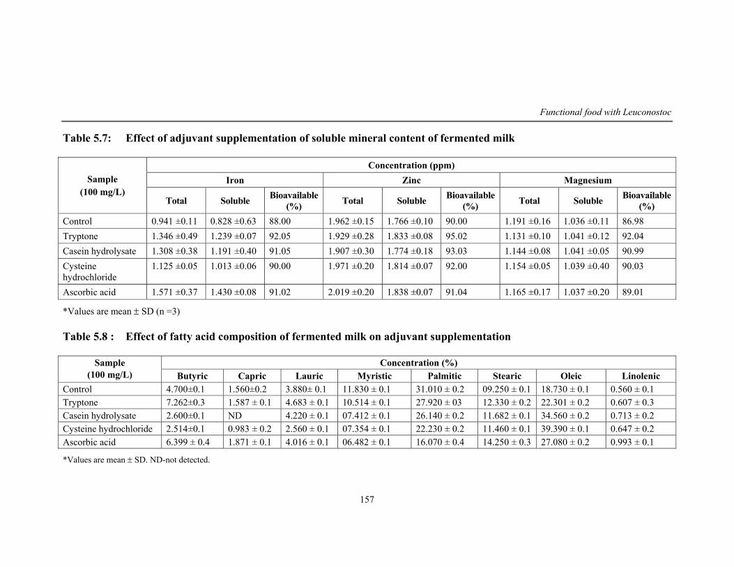
Functional food with Leuconostoc
157
Table 5.7: Effect of adjuvant supplementation of soluble mineral content of fermented milk
Concentration (ppm) Iron Zinc Magnesium Sample
(100 mg/L) Total Soluble Bioavailable
(%) Total Soluble Bioavailable (%) Total Soluble Bioavailable
(%) Control 0.941 ±0.11 0.828 ±0.63 88.00 1.962 ±0.15 1.766 ±0.10 90.00 1.191 ±0.16 1.036 ±0.11 86.98 Tryptone 1.346 ±0.49 1.239 ±0.07 92.05 1.929 ±0.28 1.833 ±0.08 95.02 1.131 ±0.10 1.041 ±0.12 92.04 Casein hydrolysate 1.308 ±0.38 1.191 ±0.40 91.05 1.907 ±0.30 1.774 ±0.18 93.03 1.144 ±0.08 1.041 ±0.05 90.99 Cysteine hydrochloride
1.125 ±0.05 1.013 ±0.06 90.00 1.971 ±0.20 1.814 ±0.07 92.00 1.154 ±0.05 1.039 ±0.40 90.03
Ascorbic acid 1.571 ±0.37 1.430 ±0.08 91.02 2.019 ±0.20 1.838 ±0.07 91.04 1.165 ±0.17 1.037 ±0.20 89.01
*Values are mean ± SD (n =3) Table 5.8 : Effect of fatty acid composition of fermented milk on adjuvant supplementation
Concentration (%) Sample (100 mg/L) Butyric Capric Lauric Myristic Palmitic Stearic Oleic Linolenic
Control 4.700±0.1 1.560±0.2 3.880± 0.1 11.830 ± 0.1 31.010 ± 0.2 09.250 ± 0.1 18.730 ± 0.1 0.560 ± 0.1 Tryptone 7.262±0.3 1.587 ± 0.1 4.683 ± 0.1 10.514 ± 0.1 27.920 ± 03 12.330 ± 0.2 22.301 ± 0.2 0.607 ± 0.3 Casein hydrolysate 2.600±0.1 ND 4.220 ± 0.1 07.412 ± 0.1 26.140 ± 0.2 11.682 ± 0.1 34.560 ± 0.2 0.713 ± 0.2 Cysteine hydrochloride 2.514±0.1 0.983 ± 0.2 2.560 ± 0.1 07.354 ± 0.1 22.230 ± 0.2 11.460 ± 0.1 39.390 ± 0.1 0.647 ± 0.2 Ascorbic acid 6.399 ± 0.4 1.871 ± 0.1 4.016 ± 0.1 06.482 ± 0.1 16.070 ± 0.4 14.250 ± 0.3 27.080 ± 0.2 0.993 ± 0.1
*Values are mean ± SD. ND-not detected.
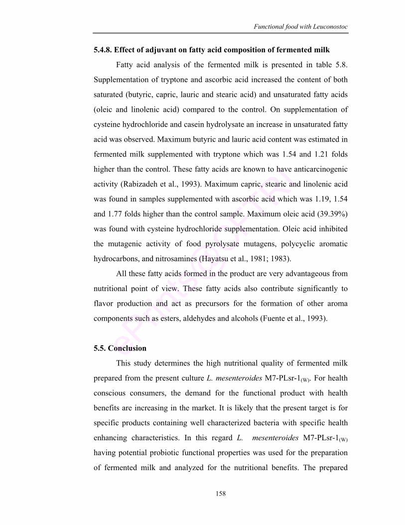
Functional food with Leuconostoc
158
5.4.8. Effect of adjuvant on fatty acid composition of fermented milk
Fatty acid analysis of the fermented milk is presented in table 5.8.
Supplementation of tryptone and ascorbic acid increased the content of both
saturated (butyric, capric, lauric and stearic acid) and unsaturated fatty acids
(oleic and linolenic acid) compared to the control. On supplementation of
cysteine hydrochloride and casein hydrolysate an increase in unsaturated fatty
acid was observed. Maximum butyric and lauric acid content was estimated in
fermented milk supplemented with tryptone which was 1.54 and 1.21 folds
higher than the control. These fatty acids are known to have anticarcinogenic
activity (Rabizadeh et al., 1993). Maximum capric, stearic and linolenic acid
was found in samples supplemented with ascorbic acid which was 1.19, 1.54
and 1.77 folds higher than the control sample. Maximum oleic acid (39.39%)
was found with cysteine hydrochloride supplementation. Oleic acid inhibited
the mutagenic activity of food pyrolysate mutagens, polycyclic aromatic
hydrocarbons, and nitrosamines (Hayatsu et al., 1981; 1983).
All these fatty acids formed in the product are very advantageous from
nutritional point of view. These fatty acids also contribute significantly to
flavor production and act as precursors for the formation of other aroma
components such as esters, aldehydes and alcohols (Fuente et al., 1993).
5.5. Conclusion
This study determines the high nutritional quality of fermented milk
prepared from the present culture L. mesenteroides M7-PLsr-1(W). For health
conscious consumers, the demand for the functional product with health
benefits are increasing in the market. It is likely that the present target is for
specific products containing well characterized bacteria with specific health
enhancing characteristics. In this regard L. mesenteroides M7-PLsr-1(W)
having potential probiotic functional properties was used for the preparation
of fermented milk and analyzed for the nutritional benefits. The prepared
Functional food with Leuconostoc
159
fermented milk beverage was found to be rich in proteins, fats, total sugars,
minerals and fatty acids content providing nutritional advantages to the
consumers. Further the nutritional properties and the viability of the culture in
the product were enhanced with supplementation of different adjuvants.
Maximum viability was observed on supplementation of tryptone (100 mg/L).
The noticeable increase in the minerals confirms the role of the fermented
milk as a source of essential nutrients.

Chapter 6 Preservation of Fermented Milk over Shelf Storage
Preservation of fermented milk
160
CHAPTER - 6
PRESERVATION OF FERMENTED MILK OVER
SHELF STORAGE
ABSTRACT
This chapter addresses the preservation of fermented milk beverage
during shelf storage. The functional fermented milk beverage prepared with
M7-PLsr-1(W) was studied for the spoilage bacterial cultures wherein
Pseudomonas sp was identified as dominant spoilage bacteria. The signal
molecule for spoilage was identified as hexanoyl homoserine lactone (HHSL)
and butryl homoserine lactone (BHSL) through TLC, GC and GCMS.
Initially inhibition of Pseudomonas sp was standardized by using furanones
(bromofuranone and 2(5H)-furanone) in LB broth and further taken up for
preservation of fermented milk for longer shelf life. With addition of
2(5H)-furanones the shelf life was increased upto 9 days by preventing the
pathogenic load. Because of its structural similarity furanones specifically
interfere with signal molecule of spoilage bacterial culture without any
adverse effect on the beneficial bacteria.
Preservation of fermented milk
161
6.1. Introduction A nutritive fermented milk prepared from L. mesenteroides
(M7-PLsr-1(W)) (as described in chapter 5) is a good source of protein (3.57%) and also iron and zinc (0.941 and 1.962 ppm). A fat content of 4.5%, the fatty acid composition and acceptable titrable acidity makes the product a delicious and refreshing beverage. On longer shelf storage the product was found to be spoilt with whey separation. This may cause significant loss and provoke the opportunistic microorganisms to flourish and spread diseases. Nutrients and the storage environment of the product create a selective condition for the growth of these spoilage bacteria. The traditional preservative methods by physical and chemical means are known to be less effective (Nebedum and Obiakor, 2007) as these spoilage organisms exhibit high intrinsic resistance to variety of stress conditions (Beales, 2004). These bacterial cultures are known to release some signal molecules in a density dependent manner for synchronizing expression of particular sets of genes and coordinating cellular activities (Dong et al., 2002). This phenomenon is described as quorum sensing (Fuqua et al., 1994).
The concept of quorum sensing has encouraged us to engage in the development of a novel non-antibiotic, anti-bacterial therapy using quorum sensing inhibitor compound. Traditional treatment of using antibiotics to kill or inhibit the growth of bacteria has created a major global concern for antibiotic resistant strains (Geddes, 2000; Hentzer et al., 2003). Hence the method of regulating bacterial virulence by interfering with quorum sensing system has afforded a novel opportunity to control infectious diseases and will not create a selection pressure for development of resistant strains.
Keeping this in mind, the predominant bacterial culture responsible for spoilage of fermented milk on storage was isolated and characterized. The signal molecule produced by the culture was identified by TLC and GC and GCMS. Further we attempted to attenuate bacterial growth and virulence by interfering with quorum sensing system. Our approach was based on using furanones that acts as a competitive inhibitor are natural and approved by FDA to be used in foods. The ultimate aim of the present work was to preserve the fermented milk for longer shelf life.
Preservation of fermented milk
162
6.2. Materials
Fermented milk using M7-PLsr-1(W) as starter culture.
Bacterial culture and growth: Agrobacterium tumefaciens (kindly provided
by Dr. Stephen K. Farrand, , Department of Microbiology, University of
Illinois at Urbana-Champaign Illinois 61801 USA).
Chemicals: All the chemicals purchased were of analytical grade.
a) De Mann Rogosa Sharpe Agar (MRS), Luria-Bertani media (LB),
Braid Parker media (BP), Salmonella-Shigella agar (SS), Listeria
Oxford Media (LM) and Pseudomonas agar (PA) were purchased from
HiMedia Pvt Ltd, Mumbai, India.
b) Skimmed milk media: Skimmed milk (10 g) was suspended in 100 ml
of distilled water and homogenized. It was supplemented with 1.5% of
agar and sterilized at 121°C for 15 min at 15 lb pressure.
c) Bromofuranone (BF) and 2(5H)-furanone [2(5H)-F] (Fluka, Buchs,
Switzerland).
d) Standards of hexanoyl homoserine lactone and butryl homoserine
lactone: N-acyl homoserine lactone (AHL) standards were purchased
from Fluka (Buchs, Switzerland). The standards were dissolved in
methanol at a concentration of 1 mg/ml and stored at -20°C. For GC
and GCMS analysis the stock solutions were mixed and diluted with
methanol at 100 µg/ml.
e) Rhamnose, X-gal, orcinol, HCl and H2SO4 (HiMedia Pvt Ltd, Mumbai,
India).
Solvents: Dichloromethane, diethyl ether, methanol (HPLC grade, SRL
Laboratories, India).
Equipments: Gas chromatograph, Gas chromatograph and Mass spectrometer,
Centrifuge, Spectrophotometer (as described in chapter 2 and 3).
Preservation of fermented milk
163
6.3. Methods
6.3.1. Isolation and characterization of bacterial cultures from fermented
milk
Bacterial cultures were isolated from spoilt fermented milk stored at 4
and 30°C by serial dilution and plating method. The colonies grown were
purified by repeated streaking and stored at 4°C under paraffin until use. The
predominant culture was further characterized by biochemical assays
according to Bergey’s manual of systematic bacteriology (Krieg 1984).
6.3.2. Identification of signal molecules
6.3.2.1. Thin layer chromatography (TLC)
The predominant spoilage culture (Pseudomonas sp) was grown in LB
media (100 ml) for 20 h at 28°C. Cell free supernatant was collected by
centrifugation at 8000 rpm for 15 min. The supernatant was then extracted
twice with equal volume of ethyl acetate and the extract was filtered and
dried.
Synthetic AHL standards (1 mg/ml) and the ethyl acetate culture
extract were taken in HPLC grade ethyl acetate and spotted onto TLC plate
(20 × 20 cm). The chromatogram was developed using a solvent system of
methanol:water (60:40 v/v) as described by Shaw et al. (1997). After
development the solvent was evaporated and the dried plate was overlaid with
monitor strain (A. tumefaciens). A 10 ml overnight culture of A. tumefaciens
was inoculated into 150 ml of LB medium containing X-gal and the culture
was spread over the surface of the developed TLC plate. The plate was
incubated overnight at 30°C in a sterilized closed plastic container. AHL were
visualized as blue spots.
6.3.2.2. Gas chromatographic technique
Extraction: A simple method was standardized by using GC and GCMS for
the identification and quantification of AHL. Cell free supernatant (5 ml) was
extracted twice with an equal volume of dichloromethane (DCM), dried over
anhydrous sodium sulphate and filtered. The solvent layer was carefully
Preservation of fermented milk
164
separated and dried. The dry residue was redissolved in 1 ml of DCM for GC
and GCMS analysis.
GC conditions: The column used was OV-17 with an oven temperature
programming from 700C to 2500C (70-1500C; 60C/min, 150-2500C; 100C/min,
2500C with 10 min holding time).
GCMS conditions: Analysis were performed using a model Turbomass Gold
(Perkin Elmer International, Switzerland) interfaced to a single quadrapole
mass selective detector, both of which were controlled by a computer
equipped with turbomass version 4 software. Sample was injected in the split
mode (40:1) in an ELITE-1 column, 30 m × 0.25 mm id and 0.25 µm film
thickness coated with 100% poly dimethoxy siloxane. Pure Helium was used
as the GC carrier gas at a flow rate of 1 ml/min. The GC injector temperature
was set at 100°C and oven temperature at 70°C. The detector was adjusted to
250°C. Mass spectrometry conditions were as follows: electron ionization
source set to 70 eV, emission current 100 µA, MS Quad 150°C, MS source
150°C. The mass spectrometer was run in full scan mode.
6.3.3. Optimization and effect the of furanone to inhibit spoilage
organism in LB culture medium
To study which of the furanone was effective against spoilage bacterial
culture (Pseudomonas sp) two commercially available furanones,
Bromofuranone and 2(5H)-furanone were tested for their efficacy in reducing
the growth of spoilage bacterial culture and its virulence expression.
6.3.3.1. Effect of furanones on bacterial growth: Selected bacterial strain
(Pseudomonas sp) was cultured in 100 ml LB broth supplemented with
furanones (Bromofuranone and 2(5H)-Furanone) at different concentrations
(100, 200 and 300 µM) separately and incubated at 4 and 30°C for 5 days.
Cultured broth without furanone was used as control. At regular interval of
time (1st, 3rd and 5th day) an aliquot (1 ml) of sample was drawn, serially
diluted and plated on selective media (Pseudomonas agar) to enumerate the
number of surviving bacterial cells.
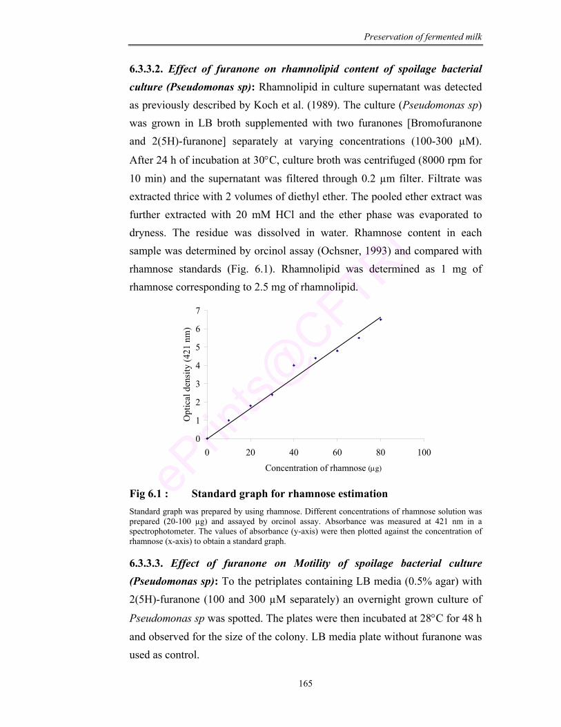
Preservation of fermented milk
165
6.3.3.2. Effect of furanone on rhamnolipid content of spoilage bacterial culture (Pseudomonas sp): Rhamnolipid in culture supernatant was detected as previously described by Koch et al. (1989). The culture (Pseudomonas sp) was grown in LB broth supplemented with two furanones [Bromofuranone and 2(5H)-furanone] separately at varying concentrations (100-300 µM).
After 24 h of incubation at 30°C, culture broth was centrifuged (8000 rpm for 10 min) and the supernatant was filtered through 0.2 µm filter. Filtrate was extracted thrice with 2 volumes of diethyl ether. The pooled ether extract was further extracted with 20 mM HCl and the ether phase was evaporated to dryness. The residue was dissolved in water. Rhamnose content in each sample was determined by orcinol assay (Ochsner, 1993) and compared with rhamnose standards (Fig. 6.1). Rhamnolipid was determined as 1 mg of rhamnose corresponding to 2.5 mg of rhamnolipid.
0
1
2
3
4
5
6
7
0 20 40 60 80 100
Concentration of rhamnose
Opt
ical
den
sity
(421
nm
)
Fig 6.1 : Standard graph for rhamnose estimation Standard graph was prepared by using rhamnose. Different concentrations of rhamnose solution was prepared (20-100 µg) and assayed by orcinol assay. Absorbance was measured at 421 nm in a spectrophotometer. The values of absorbance (y-axis) were then plotted against the concentration of rhamnose (x-axis) to obtain a standard graph.
6.3.3.3. Effect of furanone on Motility of spoilage bacterial culture (Pseudomonas sp): To the petriplates containing LB media (0.5% agar) with 2(5H)-furanone (100 and 300 µM separately) an overnight grown culture of
Pseudomonas sp was spotted. The plates were then incubated at 28°C for 48 h and observed for the size of the colony. LB media plate without furanone was used as control.
(µg)
Preservation of fermented milk
166
6.3.3.4. Effect of furanone on Exoprotease activity/casein hydrolysis of
spoilage bacterial culture (Pseudomonas sp): Bacterial culture grown in the
presence of different concentrations of 2(5H)-furanone (100, 300 and 500
µM) were centrifuged separately and the cell free supernatant was analyzed
for the exoprotease activity or the casein hydrolysis using skimmed milk
media (Chancey et al., 1999). Wells of 4 mm diameter were made in the
media plates and inoculated with cell free supernatant. After the incubation
period for 24 h at 28°C, plates were observed for the clear zone around the
wells.
6.3.4. Growth inhibition of spoilage bacterial culture (Pseudomonas sp) in
fermented milk by using furanone
Fermented milk was prepared by inoculating M7-PLsr-1(W) at a
concentration of 1 × 10⁵ cfu/ml. After 24 h of fermentation, Pseudomonas sp
as spoilage bacterial culture was added (1% v/v) into the fermented milk and
was divided into 6 equal portions. Two fractions were supplemented with
500 µM of 2(5H)-furanone and other two with 1000 µM of 2(5H)-furanone.
Two were kept as control. One set (each of control and fraction supplemented
with 500 µM and 1000 µM) was incubated at refrigerated condition (4°C) and
other at room temperature (30°C). At regular intervals (0-3 days) an aliquot of
sample (1 ml) was drawn, serially diluted and plated. The colonies grown
were enumerated and expressed as cfu/ml.
6.3.5. Preservation of fermented milk over shelf life by supplementation
of furanone
Fermented milk was prepared by adding M7-PLsr-1(W) at a
concentration of 1 × 10⁵ cfu/ml along with supplementation of
2(5H)-furanone (1000 µM). The fermented milk was incubated at 30 and 4°C
to study the stability of the product. At regular intervals (0-10 days) an aliquot
of sample (1 ml) was drawn, serially diluted and plated on MRSA and other
selective media. The colonies grown were enumerated and expressed as
cfu/ml.
Preservation of fermented milk
167
6.4. Results and Discussion
6.4.1. Isolation and characterization of bacterial cultures from spoilt
fermented milk
The nutritive probiotic fermented milk beverage prepared using the
potent probiotic isolate M7-PLsr-1(W) was studied for its shelf life. Although
the product was good upto 5 days of storage, spoilage was observed after the
6th day of storage with whey separation. The bacterial cultures responsible for
the spoilage were isolated by plating an aliquot of spoilt fermented milk on
different selective media (Braid Parker media, Salmonella-Shigella agar,
MacConkey agar, Pseudomonas agar and deMann Rogosa Sharpe agar).
Totally 22 bacterial cultures were isolated from spoiled fermented
milk. About 50% of the isolates were gram positive, catalase negative,
non-spore forming and non-haemolytic. Of the other 11 bacterial strains
9 isolates were identified as Pseudomonas sp according to Bergey’s manual
(Table 6.1; Fig. 6.2).
Huis In’t Veld (1996) has also reported P. fluorescens as the specific
spoilage organism in refrigerated milk. They impart off-flavor in milk due to
extracellular proteinases and lipases (Sorhaug and Stepaniak, 1997; Dogan
and Boor, 2003). Jaspe et al. (1995) have isolated a number of
Pseudomonas spp (80 strains) from raw milk. Pseudomonas spp are known to
be predominant spoilage flora in proteinaceous raw foods stored under
aerobic refrigerated condition (Gennari and Dragotto, 1992; Ternstrom et al.,
1993) particularly in beef, fish, chicken, raw milk and fermented milk (Kraft,
1992; Labadie, 1999; Tryfinopoulou et al., 2002; Jay et al., 2003). They have
significant spoilage potential by virtue of their ability to elaborate heat stable
enzymes (proteases and lipases) which can survive pasteurization and even
UHT heat treatment.
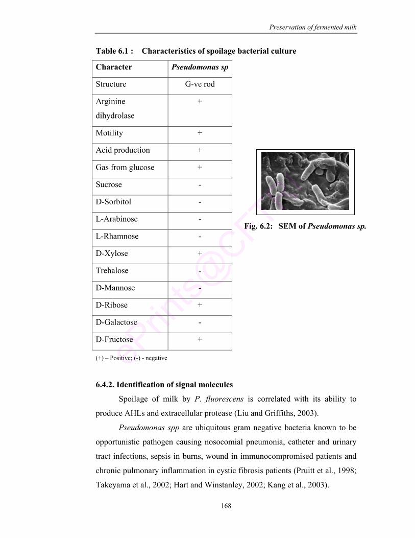
Preservation of fermented milk
168
Table 6.1 : Characteristics of spoilage bacterial culture
Character Pseudomonas sp
Structure G-ve rod
Arginine
dihydrolase
+
Motility +
Acid production +
Gas from glucose +
Sucrose -
D-Sorbitol -
L-Arabinose -
L-Rhamnose -
D-Xylose +
Trehalose -
D-Mannose -
D-Ribose +
D-Galactose -
D-Fructose +
(+) – Positive; (-) - negative
6.4.2. Identification of signal molecules
Spoilage of milk by P. fluorescens is correlated with its ability to
produce AHLs and extracellular protease (Liu and Griffiths, 2003).
Pseudomonas spp are ubiquitous gram negative bacteria known to be
opportunistic pathogen causing nosocomial pneumonia, catheter and urinary
tract infections, sepsis in burns, wound in immunocompromised patients and
chronic pulmonary inflammation in cystic fibrosis patients (Pruitt et al., 1998;
Takeyama et al., 2002; Hart and Winstanley, 2002; Kang et al., 2003).
Fig. 6.2: SEM of Pseudomonas sp.
Preservation of fermented milk
169
Pseudomonas spp regulate virulence gene expression in a cell density
dependent manner. After reaching a threshold concentration, autoinducer (AI)
activates a lux R-type transcriptional activators to induce specific genes
(Fuqua et al., 1996) for the release of N-acyl homoserine lactones as signal
molecules. These quorum sensing signal molecules regulate the synthesis of
elastase, alkaline protease, exoenzymes, haemolysin, lectins, pyocyanin,
rhamnolipids, HCN or oxidative stress responsive enzymes, catalase and
superoxide dismutase (Pearson et al., 1997; Hasset et al., 1999; Pessi and
Haas, 2000; Winzer et al., 2000). Quorum sensing also regulates competence
development, sporulation and virulence factor induction along with other
physiological events in pathogenic bacterial infections (Cvitkovitch et al.,
2003; Greenberg, 2003; Yarwood and Schlievert, 2003). Hence it is very
essential to identify the signal molecule produced by these bacterial cultures
to prevent its growth and virulence.
A large number of researchers have applied different methods for
identifying these signal molecules like green fluorescent protein technology
(Hentzer et al., 2002), semi-preparative HPLC, coupled with MS and NMR
spectroscopy (Eberl et al., 1996), colourimetric method (Hendricks et al.,
2004) and PCR (Nakayama et al., 2003) which are time and resource
consuming techniques.
Simple and convincing method for separation and tentative
identification of AHL molecules in extract of whole cell cultures has been
developed by Shaw et al. (1997). It consists of TLC followed by detection of
AHL molecule by means of agar overlay with sensor bacteria
(A. tumefaciens). Another method based on conversion of AHL molecule to
pentafluorobenzyloxime derivatives and identification on GCMS was
developed by Charlton et al. (2000) but it is a resource consuming method and
the derivatization may be hazardous to health. Capillary separation with mass
spectrometric detection was followed by Frommberger et al. (2004). As AHL
are actually cyclic esters, analysis of this compound is possible by GCMS
without derivatization (Cataldi et al., 2004).
Preservation of fermented milk
170
In the present work initially TLC was performed with AHL extract
from representative strain (Pseudomonas sp) followed by agar overlay with
A. tumefaciens (Fig. 6.3). TLC results indicate the presence of two kinds of
AHL in the extract of Pseudomonas sp which was tentatively identified as
butryl homoserine lactone (BHSL) and hexanoyl homoserine lactone (HHSL)
by comparing with AHL standards.
1 2 3
Fig 6.3 : TLC Overlay assay for identification of signal molecule
In the present study a simple method for the identification of these
signal molecule was optimized using GC and GCMS. As these N-AHL
molecules result from enzymatic condensation of HSL with 3–hydroxy,
3–oxo or an un substituted fatty acid (Schaefer et al., 1996) mass spectrum
can be used as a quantitative tool for identification of AHL molecules (Cataldi
et al., 2004; Gould et al., 2006). Thus, under the optimized experimental
conditions a series of sample extract from supernatant of bacterial cultures
were investigated. Pseudomonas sp when assayed showed the presence of
HHSL and BHSL molecule which were confirmed by comparing the retention
time of standard AHL and spiking with the standards (Fig. 6.4). The best
results in terms of selectivity and analysis time was obtained using a
temperature programme of 700C to 2500C (70-1500C; 60C/min, 150-2500C;
100C/min, 2500C with 10 min holding time).
Lane 1. BHL
Lane 2.HHSL
Lane 3.Pseudomonas sp extract
Preservation of fermented milk
171
Fig 6.4 : GC chromatograph (A) standard HHSL (B) standard BHSL and (C) Pseudomonas sp extract (D) Pseudomonas sp extract spiked with standard HHSL
(A) (B)
(C) (D)
Vol
tage
(mV
)
Time
Vol
tage
(mV
)
Time
Vol
tage
(mV
)
Time
Vol
tage
(mV
)
Time
Preservation of fermented milk
172
Further GCMS analysis gave a direct and effective indication of signal
molecule present in the culture supernatant extract. The mass spectra of
HHSL and BHSL standard exhibited a characteristic molecular ion [M]+ that
was identified as m/z 143 and m/z 171 respectively. A similar mass spectrum
was also reported by Pearson et al. (1995) and Cataldi et al. (2004) for
N-butryl HSL (molecular weight; 171) and hexanoyl homoserine lactone
(molecular weight; 199).
In the present study, the MS fragmentation pattern maximum peaks
were found at 143, 57, 43, 71, 99 for HHSL and 57, 71, 143, 85, 101 for
BHSL (Fig. 6.5). A comparison with the chromatogram recorded for sample
with that of a standard allowed for the identification of signal molecule.
Accordingly, HHSL and BHSL were identified in the extract of
Pseudomonas sp found to be associated with fermented milk spoilage.
Similarly Winson et al. (1995) have also identified these signal molecules in
the spent culture supernatant of P. aeroginosa by HPLC and EI-MS and have
detected VsmI gene responsible for their production.
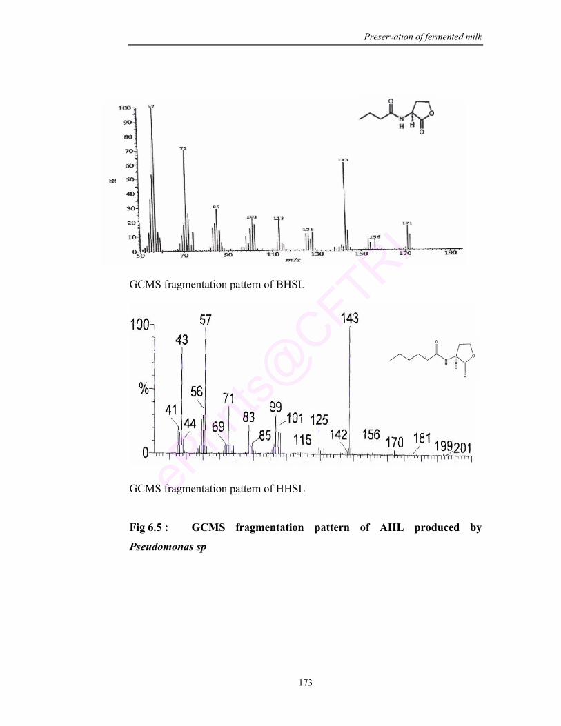
Preservation of fermented milk
173
GCMS fragmentation pattern of BHSL
GCMS fragmentation pattern of HHSL
Fig 6.5 : GCMS fragmentation pattern of AHL produced by
Pseudomonas sp
Preservation of fermented milk
174
6.4.3. Optimization and the effect of furanone to inhibit spoilage
organism in LB culture medium
Inhibition of bacterial growth in general is followed by using biocides,
antibiotics and bacteriophages (Costerton et al., 1999). But these methods
have failed due to non-penetration of biofilm. Cells in a biofilm are in starved
state and hence are not susceptible to these antimicrobial agents (Mah and
O’Toole, 2001; Stewart and Costerton, 2001; Gilbert et al., 2002). Hence, it is
necessary to apply alternative methods for disruption of this biofilm for
bacterial inhibition. In this regard signaling pathway has become an important
target for inhibition of bacteria. Disruption of quorum sensing may be
accomplished by blocking the AHL synthesis, AHL signal molecule
degradation and inhibition of AHL receptor activation (Juhas et al., 2005).
Hence application of drug that inhibit/ even prevent biofilm formation seems
to be a promising approach.
Use of lactonase enzyme (Dong et al., 2002; Reimmann et al., 2002;
Park et al., 2003) that cleaves the lactone ring is one of the possible approach
for abolishing their activity. But one of the draw back of this approach is the
difficulty in delivering active enzyme to the site of infection. In case of las
R/las I and rhlR/rhlI expression, the deletion of quorum sensing gene virtually
diminishes the expression of las R and reduces the production of virulence
factor (Chugani et al., 2001). Another alternative approach is the use of
antisense oligonucleotides that specifically binds to las R/las I or rhl R/rhl I
transcripts (Kurreck, 2003). Due to time consumtion and other obstacles such
as cell wall permeability, specificity and efficacy of mode of delivery these
methods are not popular.
Out of all these methods, use of furanone compounds is most
significant because of its small size and ease of delivery. Furanones that are
produced by D. pulchra, a marine alga functions as competitive inhibitors
(Givskov et al., 1996; Manefield et al., 2000). They bind to lux R in a mode
similar to binding of signal molecule thus inhibiting the transcription of
virulence genes (Zhang et al., 2002; Vannini et al., 2002; Koch et al., 2005).
Preservation of fermented milk
175
In the present experiment two commercially available furanones
Bromofuranone and 2(5H)-furanone were tested to study their efficacy in
reducing the growth of spoilage bacterial culture and its virulence expression
in LB culture medium.
6.4.3.1. Effect of furanones on bacterial growth: The Pseudomonas sp
grown in LB broth supplemented with furanones showed reduction in cell
count as compared to control grown in absence of furanone (Fig. 6.6). After
5 days of incubation at 4°C, culture supplemented with 2(5H)-furanone
(300 µM) reduced from 2.1 × 10¹⁵ to 1.3 × 10⁸ cfu/ml as compared to
2.1 × 10¹⁵ to 1.56 × 10⁸ cfu/ml in case of bromofuranone supplementation
(300 µM). At room temperature cell decline was observed from 7.03 × 10¹⁴ to
9.8 × 10⁷ cfu/ml with 2(5H)-furanone and from 7.03 × 10¹⁴ to 1.0 × 10⁸ cfu/ml
with bromofuranone. In the present work 2(5H)-furanone is found more
effective than bromofuranone in inhibiting Pseudomonas sp growth.
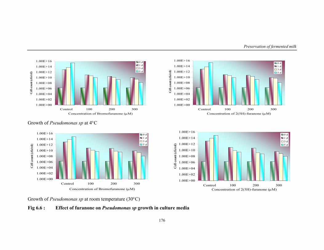
Preservation of fermented milk
176
1.00E+00
1.00E+02
1.00E+04
1.00E+06
1.00E+08
1.00E+10
1.00E+12
1.00E+14
1.00E+16
Control 100 200 300Concentration of Bromofuranone (µM)
Cel
l cou
nt (c
fu/m
l)
0 d1 d3 d5 d
1.00E+00
1.00E+02
1.00E+04
1.00E+06
1.00E+08
1.00E+10
1.00E+12
1.00E+14
1.00E+16
Control 100 200 300Concentration of 2(5H)-furanone (µM)
Cel
l cou
nt (c
fu/m
l)
0 d1 d3 d5 d
Growth of Pseudomonas sp at 4°C
1.00E+00
1.00E+02
1.00E+04
1.00E+06
1.00E+08
1.00E+10
1.00E+12
1.00E+14
1.00E+16
Control 100 200 300
Concentration of Bromofuranone (µM)
Cel
l cou
nt (c
fu/m
l)
0 d1 d3 d5 d
1.00E+00
1.00E+02
1.00E+04
1.00E+06
1.00E+08
1.00E+10
1.00E+12
1.00E+14
1.00E+16
Control 100 200 300Concentration of 2(5H)-furanone (µM)
Cel
l cou
nt (c
fu/m
l)
0 d1 d3 d5 d
Growth of Pseudomonas sp at room temperature (30°C)
Fig 6.6 : Effect of furanone on Pseudomonas sp growth in culture media
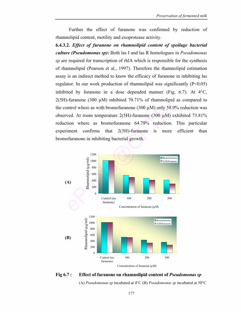
Preservation of fermented milk
177
Further the effect of furanone was confirmed by reduction of rhamnolipid content, motility and exoprotease activity. 6.4.3.2. Effect of furanone on rhamnolipid content of spoilage bacterial culture (Pseudomonas sp): Both las I and las R homologues in Pseudomonas sp are required for transcription of rhlA which is responsible for the synthesis of rhamnolipid (Pearson et al., 1997). Therefore the rhamnolipid estimation assay is an indirect method to know the efficacy of furanone in inhibiting las regulator. In our work production of rhamnolipid was significantly (P<0.05)
inhibited by furanone in a dose depended manner (Fig. 6.7). At 4°C, 2(5H)-furanone (300 µM) inhibited 70.71% of rhamnolipid as compared to the control where as with bromofuranone (300 µM) only 58.9% reduction was observed. At room temperature 2(5H)-furanone (300 µM) exhibited 73.81% reduction where as bromofuranone 64.79% reduction. This particular experiment confirms that 2(5H)-furanone is more efficient than bromofuranone in inhibiting bacterial growth.
0
200
400
600
800
1000
1200
Control (nofuranone)
100 200 300
Concentration of furanone (µM)
Rha
mno
lipid
( µg
/ml)
Bromofuranone5(2H)Furanone
0
200
400
600
800
1000
1200
Control (nofuranone)
100 200 300
Concentration of furanone (µM)
Rha
mno
lipid
( µg/
ml)
Bromofuranone5(2H)Furanone
Fig 6.7 : Effect of furanone on rhamnolipid content of Pseudomonas sp
(A) Pseudomonas sp incubated at 4°C (B) Pseudomonas sp incubated at 30°C
(A)
(B)
Rha
mno
lipid
(µg/
ml)
Rha
mno
lipid
(µg/
ml)
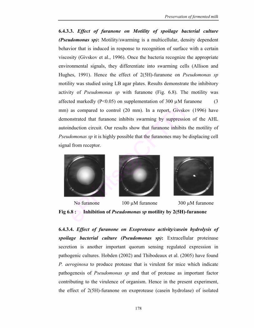
Preservation of fermented milk
178
6.4.3.3. Effect of furanone on Motility of spoilage bacterial culture
(Pseudomonas sp): Motility/swarming is a multicellular, density dependent
behavior that is induced in response to recognition of surface with a certain
viscosity (Givskov et al., 1996). Once the bacteria recognize the appropriate
environmental signals, they differentiate into swarming cells (Allison and
Hughes, 1991). Hence the effect of 2(5H)-furanone on Pseudomonas sp
motility was studied using LB agar plates. Results demonstrate the inhibitory
activity of Pseudomonas sp with furanone (Fig. 6.8). The motility was
affected markedly (P<0.05) on supplementation of 300 µM furanone (3
mm) as compared to control (20 mm). In a report, Givskov (1996) have
demonstrated that furanone inhibits swarming by suppression of the AHL
autoinduction circuit. Our results show that furanone inhibits the motility of
Pseudomonas sp it is highly possible that the furanones may be displacing cell
signal from receptor.
No furanone 100 µM furanone 300 µM furanone
Fig 6.8 : Inhibition of Pseudomonas sp motility by 2(5H)-furanone
6.4.3.4. Effect of furanone on Exoprotease activity/casein hydrolysis of
spoilage bacterial culture (Pseudomonas sp): Extracellular proteinase
secretion is another important quorum sensing regulated expression in
pathogenic cultures. Hobden (2002) and Thibodeaux et al. (2005) have found
P. aeroginosa to produce protease that is virulent for mice which indicate
pathogenesis of Pseudomonas sp and that of protease as important factor
contributing to the virulence of organism. Hence in the present experiment,
the effect of 2(5H)-furanone on exoprotease (casein hydrolase) of isolated
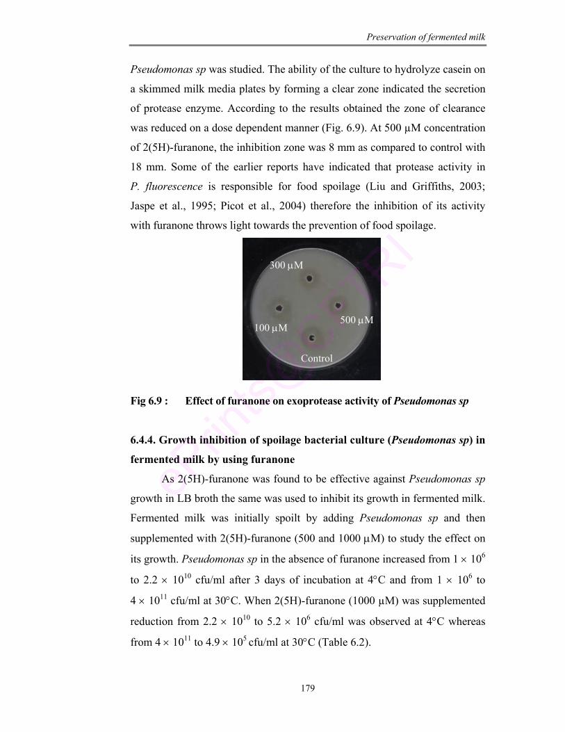
Preservation of fermented milk
179
Pseudomonas sp was studied. The ability of the culture to hydrolyze casein on
a skimmed milk media plates by forming a clear zone indicated the secretion
of protease enzyme. According to the results obtained the zone of clearance
was reduced on a dose dependent manner (Fig. 6.9). At 500 µM concentration
of 2(5H)-furanone, the inhibition zone was 8 mm as compared to control with
18 mm. Some of the earlier reports have indicated that protease activity in
P. fluorescence is responsible for food spoilage (Liu and Griffiths, 2003;
Jaspe et al., 1995; Picot et al., 2004) therefore the inhibition of its activity
with furanone throws light towards the prevention of food spoilage.
Fig 6.9 : Effect of furanone on exoprotease activity of Pseudomonas sp
6.4.4. Growth inhibition of spoilage bacterial culture (Pseudomonas sp) in
fermented milk by using furanone
As 2(5H)-furanone was found to be effective against Pseudomonas sp
growth in LB broth the same was used to inhibit its growth in fermented milk.
Fermented milk was initially spoilt by adding Pseudomonas sp and then
supplemented with 2(5H)-furanone (500 and 1000 µM) to study the effect on
its growth. Pseudomonas sp in the absence of furanone increased from 1 × 10⁶
to 2.2 × 10¹⁰ cfu/ml after 3 days of incubation at 4°C and from 1 × 10⁶ to
4 × 10¹¹ cfu/ml at 30°C. When 2(5H)-furanone (1000 µM) was supplemented
reduction from 2.2 × 10¹⁰ to 5.2 × 10⁶ cfu/ml was observed at 4°C whereas
from 4 × 10¹¹ to 4.9 × 10⁵ cfu/ml at 30°C (Table 6.2).
Control
100 µM
300 µM
500 µM
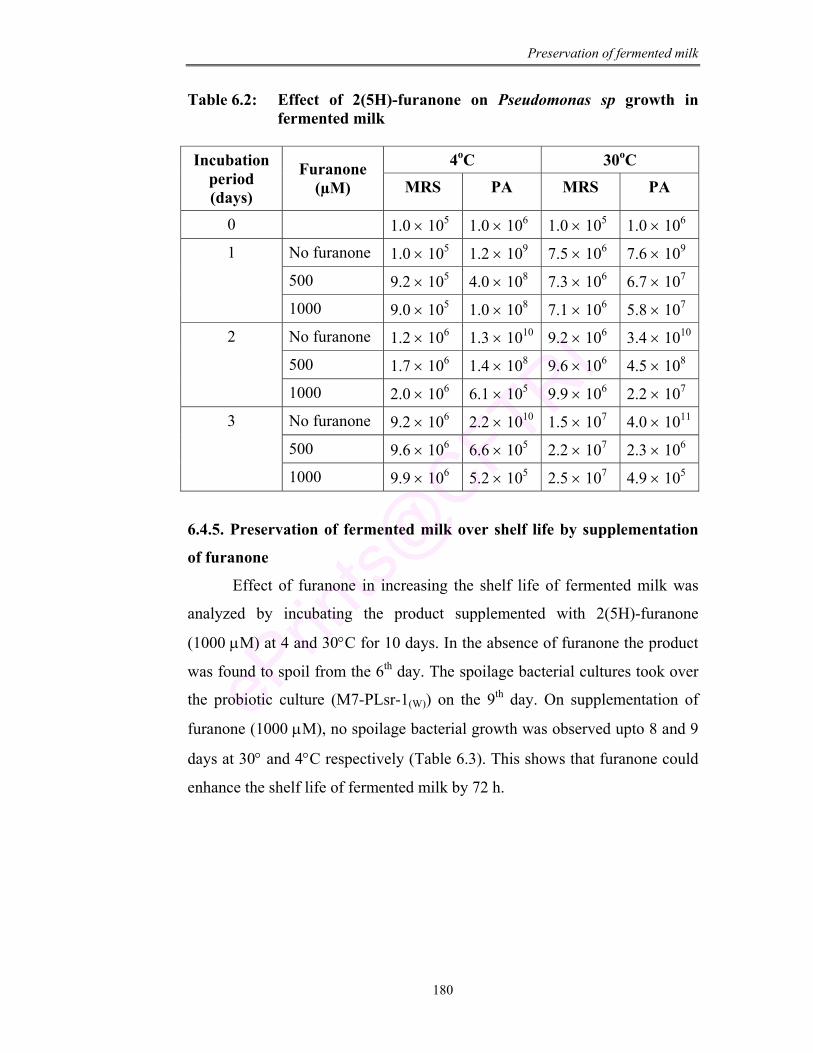
Preservation of fermented milk
180
Table 6.2: Effect of 2(5H)-furanone on Pseudomonas sp growth in fermented milk
4oC 30oC Incubation
period (days)
Furanone (µM) MRS PA MRS PA
0 1.0 × 10⁵ 1.0 × 10⁶ 1.0 × 10⁵ 1.0 × 10⁶
No furanone 1.0 × 10⁵ 1.2 × 10⁹ 7.5 × 10⁶ 7.6 × 10⁹
500 9.2 × 10⁵ 4.0 × 10⁸ 7.3 × 10⁶ 6.7 × 10⁷
1
1000 9.0 × 10⁵ 1.0 × 10⁸ 7.1 × 10⁶ 5.8 × 10⁷
No furanone 1.2 × 10⁶ 1.3 × 10¹⁰ 9.2 × 10⁶ 3.4 × 10¹⁰
500 1.7 × 10⁶ 1.4 × 10⁸ 9.6 × 10⁶ 4.5 × 10⁸
2
1000 2.0 × 10⁶ 6.1 × 10⁵ 9.9 × 10⁶ 2.2 × 10⁷
No furanone 9.2 × 10⁶ 2.2 × 10¹⁰ 1.5 × 10⁷ 4.0 × 10¹¹
500 9.6 × 10⁶ 6.6 × 10⁵ 2.2 × 10⁷ 2.3 × 10⁶
3
1000 9.9 × 10⁶ 5.2 × 10⁵ 2.5 × 10⁷ 4.9 × 10⁵
6.4.5. Preservation of fermented milk over shelf life by supplementation
of furanone
Effect of furanone in increasing the shelf life of fermented milk was
analyzed by incubating the product supplemented with 2(5H)-furanone
(1000 µM) at 4 and 30°C for 10 days. In the absence of furanone the product
was found to spoil from the 6th day. The spoilage bacterial cultures took over
the probiotic culture (M7-PLsr-1(W)) on the 9th day. On supplementation of
furanone (1000 µM), no spoilage bacterial growth was observed upto 8 and 9
days at 30° and 4°C respectively (Table 6.3). This shows that furanone could
enhance the shelf life of fermented milk by 72 h.
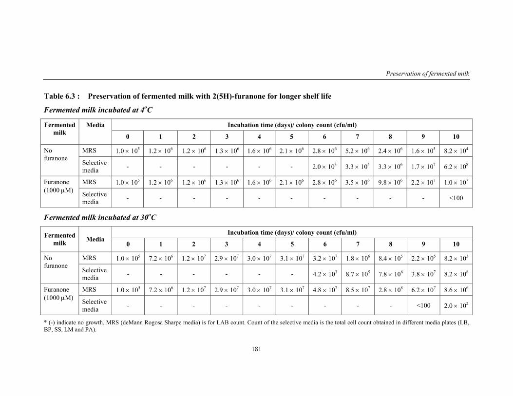
Preservation of fermented milk
181
Table 6.3 : Preservation of fermented milk with 2(5H)-furanone for longer shelf life
Fermented milk incubated at 4oC
Incubation time (days)/ colony count (cfu/ml) Fermented milk
Media
0 1 2 3 4 5 6 7 8 9 10
MRS 1.0 × 10⁵ 1.2 × 10⁶ 1.2 × 10⁶ 1.3 × 10⁶ 1.6 × 10⁶ 2.1 × 10⁶ 2.8 × 10⁶ 5.2 × 10⁶ 2.4 × 10⁶ 1.6 × 10⁵ 8.2 × 10⁴ No furanone
Selective media - - - - - - 2.0 × 10³ 3.3 × 10⁵ 3.3 × 10⁶ 1.7 × 10⁷ 6.2 × 10⁸
MRS 1.0 × 10⁵ 1.2 × 10⁶ 1.2 × 10⁶ 1.3 × 10⁶ 1.6 × 10⁶ 2.1 × 10⁶ 2.8 × 10⁶ 3.5 × 10⁶ 9.8 × 10⁶ 2.2 × 10⁷ 1.0 × 10⁷ Furanone (1000 µM) Selective
media - - - - - - - - - - <100
Fermented milk incubated at 30oC
Incubation time (days)/ colony count (cfu/ml) Fermented milk Media
0 1 2 3 4 5 6 7 8 9 10
MRS 1.0 × 10⁵ 7.2 × 10⁶ 1.2 × 10⁷ 2.9 × 10⁷ 3.0 × 10⁷ 3.1 × 10⁷ 3.2 × 10⁷ 1.8 × 10⁶ 8.4 × 10⁵ 2.2 × 10⁵ 8.2 × 10³ No furanone
Selective media - - - - - - 4.2 × 10³ 8.7 × 10⁵ 7.8 × 10⁶ 3.8 × 10⁷ 8.2 × 10⁸
MRS 1.0 × 10⁵ 7.2 × 10⁶ 1.2 × 10⁷ 2.9 × 10⁷ 3.0 × 10⁷ 3.1 × 10⁷ 4.8 × 10⁷ 8.5 × 10⁷ 2.8 × 10⁸ 6.2 × 10⁷ 8.6 × 10⁶ Furanone (1000 µM) Selective
media - - - - - - - - - <100 2.0 × 10²
* (-) indicate no growth. MRS (deMann Rogosa Sharpe media) is for LAB count. Count of the selective media is the total cell count obtained in different media plates (LB, BP, SS, LM and PA).
Preservation of fermented milk
182
6.5. Conclusion
The present chapter demonstrates a method for the preservation of
fermented milk on shelf storage. Pseudomonas sp associated with spoilage of
fermented milk was characterized and the signal molecules produced were
identified as hexanoyl and butryl homoserine lactone by GC and GCMS
method. Further furanones that are having structural similarity to that of AHL
was used to inhibit the Pseudomonas sp growth. 2(5H)-furanone was found
effective in inhibiting the growth and virulence factor expression of
Pseudomonas sp (rhamnolipid, exoprotease and motility). With addition of
2(5H)-furanone (1000 µM) the shelf life of fermented milk was increased
upto 9 days.
Use of such method of inhibiting growth of pathogenic/ spoilage
bacteria by using furanone may lead to the development of novel and non-
antibiotic method which aims in inhibiting bacterial virulence.

Chapter 7 In-vivo Studies using
Leuconostoc for Functional Attributes
In-vivo studies using Leuconostoc
183
CHAPTER - 7
IN-VIVO STUDIES USING LEUCONOSTOC FOR
FUNCTIONAL ATTRIBUTES
ABSTRACT
Lactose intolerance, a clinical problem associated with unpleasant
abdominal discomfort. Novel approaches have been tried as alternative to
antibiotics in treating this problem. In the present work Leuconostoc
mesenteroides M7-PLsr-1(W) has been studied in growing rats for its safety
and as a source to treat lactose intolerance. M7-PLsr-1(W) was administered
to a group of male rats at a dose of 106-1016 cfu/ml in the single dose study
and the effective dose was found to be 108 cfu/ml. In long term study for 90
days, initially the rats were fed with lactose rich diet to induce lactose
intolerance. Further induced rats were fed with diet supplemented with
effective dose of M7-PLsr-1(W) and four days of probiotic feeding was found
good with disappearance of diarrhea. No change in morphology, behavior
and treatment related toxicity/ bacterial translocation was observed on
probiotic feeding. Transit tolerance and adherence ability of M7-PLsr-1(W)
was checked by counting the cell number in caecum and large intestine.
Reduction of E. coli counts in caecum shows the antimicrobial activity of
the culture.
In-vivo studies using Leuconostoc
184
7.1. Introduction
The potential of probiotic lactic acid bacteria has been given a novel
approach to functional foods and pharmaceuticals (Kuipers et al., 2000;
Renault, 2002). It is important to carefully document the efficacy of the
strain for its potential application and safety. Several aspects (functional and
technological) are to be considered in the selection of probiotic strains for
food application. The functional aspects include survival and adhesion to
intestinal epithelium, antimicrobial activity and influence on metabolic
activities (Sanders and Huis in’t Veld, 1999).
In our laboratory, a lactic acid bacterium L. mesenteroides
M7-PLsr-1(W) has been selected with high β-galactosidase activity, an
enzyme which is responsible for the break down of non-reducing
disaccharide lactose into simple sugars for easy absorption (chapter 3).
Lactase/β-galactosidase is normally present in human intestinal
epithelial cells and thereby contributes to the digestion of lactose. In persons
who are deficient in lactase suffer from unpleasant abdominal pain (Sieber
et al., 1997). Temporary lactase deficiency may result from damage of the
intestinal lining or alterations in genetic expression of lactase phlorizin
hydrolase and is of great concern. Symptoms of lactose intolerance/
maldigestion include loose stools, abdominal bloating and pain, flatulence,
nausea, gas production and cramps (Hammer et al., 1996; Sieber et al.,
1997). The osmotic pressure of the lactose causes secretion of fluid and
electrolyte into intestinal lumen causing diarrhea (Launiala, 1968;
Christopher and Bayless, 1971). According to Swagerty et al. (2002) about
70-90% of adults are known to be lactose intolerant.
Approaches have been tried as an alternative to antibiotics in treating
lactose intolerance because of the growing antibiotic resistance and side
effects caused due to regular consumption of antibiotics (Fuller, 1992). The
common therapeutic approach would be to exclude milk and dairy products
with lactose from the diet but this will have nutritional disadvantage as it
reduces intake of calcium, phosphorus and vitamins (Di Stefano et al.,
In-vivo studies using Leuconostoc
185
2002). Use of lactase enzyme may be less effective probably due to enzyme
gastric inactivation (Suarez et al., 1995). Hence the use of probiotic culture
may be the alternative for treating lactose intolerance.
In the present work efficacy of M7-PLsr-1(W), its survival in
gastrointestinal transit, adherence to the mucosa of gut and as a source for
treating lactose intolerance were studied. It is generally accepted that rat is a
good animal model for studying interactions between the gut microbes and
the host. Although there are some anatomical differences in the
gastrointestinal tracts of rat and men, the fecal bacterial populations of the
major groups of bacteria is found to be similar (Tannock, 1999) and hence in
the present study albino Wister rats were selected as in-vivo model. Aims of
the present study are to evaluate the safety of culture and to determine
digestion of lactose in the intestine for clarifying possible involvement of the
culture in reducing lactose intolerance.
7.2. Materials
1) Animals and diet: Male albino rats [OUTB-Wister, IND-cft (2c)]
weighing 40 ± 2g, were obtained from animal house facility of
Central Food Technological Research Institute, Mysore. They were
used for experimenting safety and functionalities of M7-PLsr-1(W).
They were fed on basal pellet diet (Amrut diet, Sangli, India) and had
free access to tap water, ad libitum. Institutional animal ethical
committee approved the experimental protocol.
2) Chemicals: Glucose estimation kit (ERBA diagnostic, Germany),
cholesterol estimation kit (Agappe diagnostic, Ernakulam, Kerala),
urea estimation kit (Agappe diagnostics, Ernakulam, Kerala), ONPG
(HiMedia Pvt Ltd, Mumbai, India), MRS media (as described in
chapter 1)
3) Equipments: pH meter, Balance, Centrifuge, Spectrophotometer (as
described in chapter 2 and 3).
In-vivo studies using Leuconostoc
186
7.3. Methods
7.3.1. Single dose study
The objective of study was to evaluate the safety aspects of culture
and to study its functional attributes. Further, study was carried out to
determine the dosage required for maximum production of β-galactosidase.
Rats (n=6/group) were statistically grouped by randomized design and
assigned to various groups. They were housed individually in stainless steel
cages. A dose of probiotic culture (M7-PLsr-1(W)) suspended in skim milk
(2 ml) was administered intragastrically at a concentration of 106, 108, 1010,
1012, 1014 and 1016 cfu/ml. Rats (n=6) receiving only skim milk without
culture was considered as control group. Animals were observed for
morphological, behavioral changes if any and body weight was recorded.
The maximum tolerated dose and LD50 values were calculated according to
the method of Litchfield and Wilcox (1949).
7.3.2. Long term studies
The objective of the study was to evaluate the efficiency of the
culture in reducing lactose intolerance. Rats (n=36) were housed
individually in stainless steel cages in a room maintained at 25 ± 2°C, a
relative humidity of 45-70% and under 12:12 h light and dark cycle. They
were fed with 40% lactose with basal diet for 7 days to induce lactose
intolerance. Appearance of diarrhea was monitored during lactose induction
period. Occurrence of 100% diarrhea was considered as lactose intolerant
rats. A separate group (n=6) was killed at 0 day for baseline data and
considered as control. Lactose intolerance induce rats were statistically
divided into 5 groups (n=6/group). Group one was killed at 0 day after
induction of lactose intolerance. The other groups were fed with fresh basal
diets supplemented with effective dose (high β-galactosidase activity) of
culture isolate (108 cfu/ml). During the experimental run rats were monitored
for the disappearance of diarrhea and weekly gain in body weight was
recorded. During the course of the experiment all the animals were carefully
monitored for any signs of change in morphology and behavior. At the end
of each experiment (15, 30, 60 and 90 days) animals were fasted overnight
In-vivo studies using Leuconostoc
187
and humanly sacrificed under ether anesthesia. Blood was drawn by cardiac
puncture and serum was separated by centrifugation at 2,500 rpm for 10
min. Tissues (liver, kidney, spleen, heart, ceacum, small and large intestine)
were collected, rinsed with ice-cold saline, weighed, blotted and stored at
-200C until analyzed.
(1) Clinical chemistry
(a) Estimation of serum glucose Serum glucose was estimated by using commercially available
glucose estimation kit. The principle behind the reaction is the action of
glucose oxidase and peroxidase enzyme which gives an intensive pink color
to reaction mixture which is proportional to glucose concentration and can
be measured spectrophotometrically at 510 nm.
The reaction is as follows
Glucose + O2 + H2O Gluconic acid + H2O
H2O2 + 4HBA + 4AAP Quinoneimine dye + 2H2O
4HBH: 4-hydroxy benzoic acid
4AAP: 4-Amino antipyrine
Calculation
(mg/dL) standard ofion concentratstandard) of e(absorbancsample)ofe(absorbanc(mg/dL) Glucose ×=
(b) Estimation of urea Serum urea was determined by using commercially available kit.
Enzymatic determination of urea is according to the following reaction
Urea + H2O 2NH3 + CO2
NH3 + Salicylate 2,2-dicarboxyindophenol
The dye 2,2-dicarboxylindophenol gives a light green color product
which can be measured spectrophotometrically at 600 nm.
Calculation
04standard) of e(absorbancsample) of e(absorbanc(mg/dL) Urea ×=
Glucose oxidase
Peroxidase
Urease
Nitro prusside hypochlorite
In-vivo studies using Leuconostoc
188
(C) Estimation of cholesterol: Blood samples were collected from rats by
arterial puncture and the serum was separated by centrifugation. Serum
cholesterol was estimated by the modified method of Roeschlau’s method
using commercially available cholesterol estimation kit. The reaction is as
follows-
Cholesterol ester Cholesterol + fatty acid
Cholesterol + O2 Cholest-4-en-3-one + H2O2
2H2O2 + 4AAP + phenol 4H2O + quenoneimine where, CE: Cholesterol esterase CHOD: Cholesterol oxidase 4AAP: 4-Aminoantipyrine
Calculation
(mg/dL) standard ofion Concentratstandard) of e(absorbancsample) of e(absorbanc(mg/dL) lCholestero ×=
(2) Measurement of pH and cell count
Transit tolerance of the culture through gastrointestinal tract
condition was determined by the cell count in caecum, feces and large
intestine samples. Approximately, one gram each sample was suspended
separately in saline (0.85% NaCl), serially diluted and plated for LAB on
MRS agar and for E. coli on MacConkey’s agar media. After 24 h of
incubation period at 37°C, colonies grown were counted and expressed as
colony forming unit/ g of sample. pH of urine, caecum and feces were
measured using pH meter.
(3) Urine analysis
In the last week of experiment urine samples were collected over a
period of 24 h from each rat and examined for pH, glucose and urea using
standard procedure as described earlier.
CE
CHOD
POD
In-vivo studies using Leuconostoc
189
(4) β-galactosidase activity
Small and large intestines were brought to room temperature and
were cut to required size (60 and 5 cm respectively), washed thoroughly by
flushing saline into the intestinal lumen using a syringe. Carefully intestine
was cut open and scrapped for the intestinal content and suspended in 5 ml
saline. The suspension was homogenized and centrifuged (8000 rpm for
15 min) to collect a clear supernatant. An aliquot (100 µl) of this supernatant
was used for the estimation of β-galactosidase enzyme using ONPG as
substrate (Bhowmik and Marth 1989) and expressed as specific activity
(µM/mg protein). Enzyme activity in caecum was estimated by suspending
caecum (1 g) in 5 ml saline and determining specific activity as described
elsewhere. Protein was estimated by Lowry’s method (Lowry et al., 1951).
(5) Bacterial translocation
Bacterial translocation was analyzed in blood, liver and spleen. Blood
(100 µl) was cultured on MRS agar and incubated at 37°C to observe for the
bacterial growth. Liver and spleen (1 g each) were homogenized in saline
(1 ml) and 100 µl of the resulting homogenate was plated on MRS media as
mentioned above. After 24 h of incubation at 37°C, colonies grown were
counted and the results were expressed as incidence of translocation i.e.,
number of rats where translocation was detected/ total number of rats.
7.4. Results and Discussion
Probiotic are viable microbes which beneficially influence the health
of the host (Schrezenmeir and de Vrese, 2001). Several beneficial effects of
these organism in gut has been reported which include growth promotion,
protection from pathogens, alleviation of lactose intolerance, relief of
constipation, anticholesterolaemic effect and immunostimulation (Casas and
Dobrogosz, 2000; Bertazzoni et al., 2001; Aattouri et al., 2002). To exert all
these functional properties, the probiotic culture should be able to survive in
GIT, produce antimicrobial compounds and must adhere to the intestinal
cells of the host. In-vivo studies are a prerequisite to confirm the safety and
health benefits of M7-PLsr-1(W).
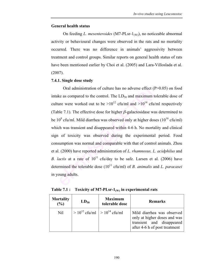
In-vivo studies using Leuconostoc
190
General health status
On feeding L. mesenteroides (M7-PLsr-1(W)), no noticeable abnormal
activity or behavioural changes were observed in the rats and no mortality
occurred. There was no difference in animals’ aggressivity between
treatment and control groups. Similar reports on general health status of rats
have been mentioned earlier by Choi et al. (2005) and Lara-Villoslada et al.
(2007).
7.4.1. Single dose study
Oral administration of culture has no adverse effect (P>0.05) on food
intake as compared to the control. The LD50 and maximum tolerable dose of
culture were worked out to be >1012 cfu/ml and >1014 cfu/ml respectively
(Table 7.1). The effective dose for higher β-galactosidase was determined to
be 108 cfu/ml. Mild diarrhea was observed only at higher doses (1016 cfu/ml)
which was transient and disappeared within 4-6 h. No mortality and clinical
sign of toxicity was observed during the experimental period. Food
consumption was normal and comparable with that of control animals. Zhou
et al. (2000) have reported administration of L. rhamnosus, L. acidphilus and
B. lactis at a rate of 1011 cfu/day to be safe. Larsen et al. (2006) have
determined the tolerable dose (1011 cfu/ml) of B. animalis and L. paracasei
in young adults.
Table 7.1 : Toxicity of M7-PLsr-1(W) in experimental rats
Mortality (%) LD50
Maximum tolerable dose Remarks
Nil > 1012 cfu/ml > 1014 cfu/ml Mild diarrhea was observed only at higher doses and was transient and disappeared after 4-6 h of post treatment
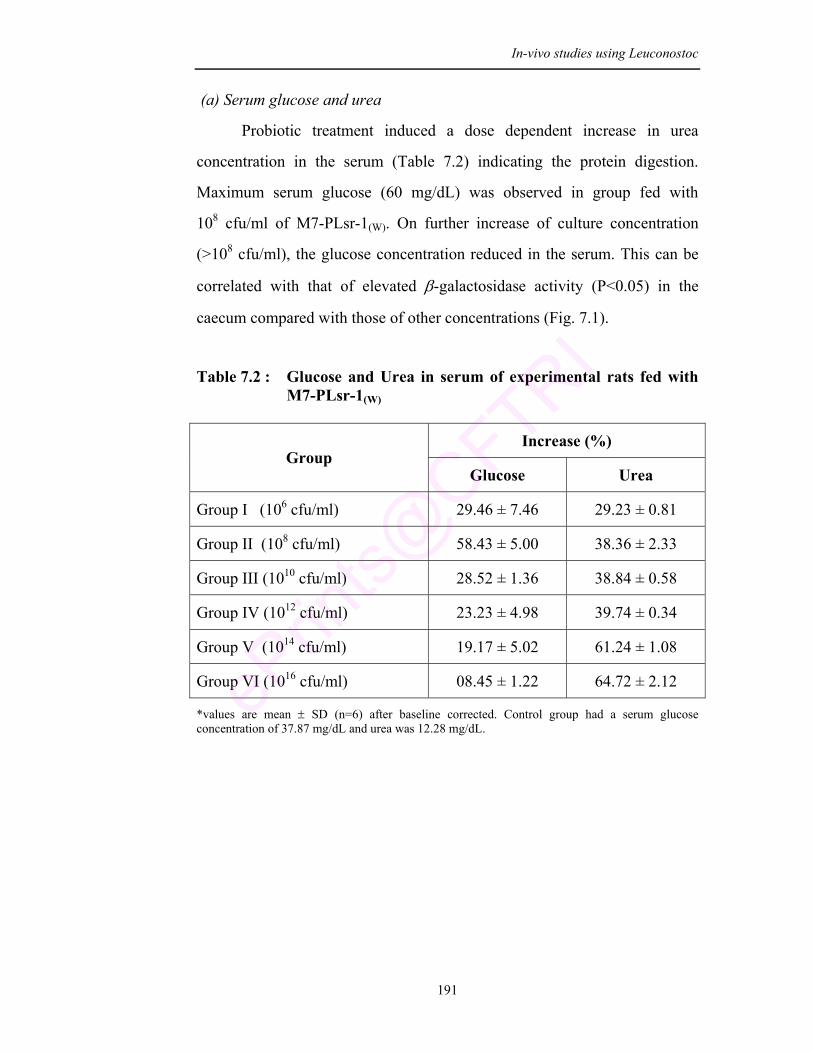
In-vivo studies using Leuconostoc
191
(a) Serum glucose and urea
Probiotic treatment induced a dose dependent increase in urea
concentration in the serum (Table 7.2) indicating the protein digestion.
Maximum serum glucose (60 mg/dL) was observed in group fed with
108 cfu/ml of M7-PLsr-1(W). On further increase of culture concentration
(>108 cfu/ml), the glucose concentration reduced in the serum. This can be
correlated with that of elevated β-galactosidase activity (P<0.05) in the
caecum compared with those of other concentrations (Fig. 7.1).
Table 7.2 : Glucose and Urea in serum of experimental rats fed with M7-PLsr-1(W)
Increase (%) Group
Glucose Urea
Group I (106 cfu/ml) 29.46 ± 7.46 29.23 ± 0.81
Group II (108 cfu/ml) 58.43 ± 5.00 38.36 ± 2.33
Group III (1010 cfu/ml) 28.52 ± 1.36 38.84 ± 0.58
Group IV (1012 cfu/ml) 23.23 ± 4.98 39.74 ± 0.34
Group V (1014 cfu/ml) 19.17 ± 5.02 61.24 ± 1.08
Group VI (1016 cfu/ml) 08.45 ± 1.22 64.72 ± 2.12
*values are mean ± SD (n=6) after baseline corrected. Control group had a serum glucose concentration of 37.87 mg/dL and urea was 12.28 mg/dL.
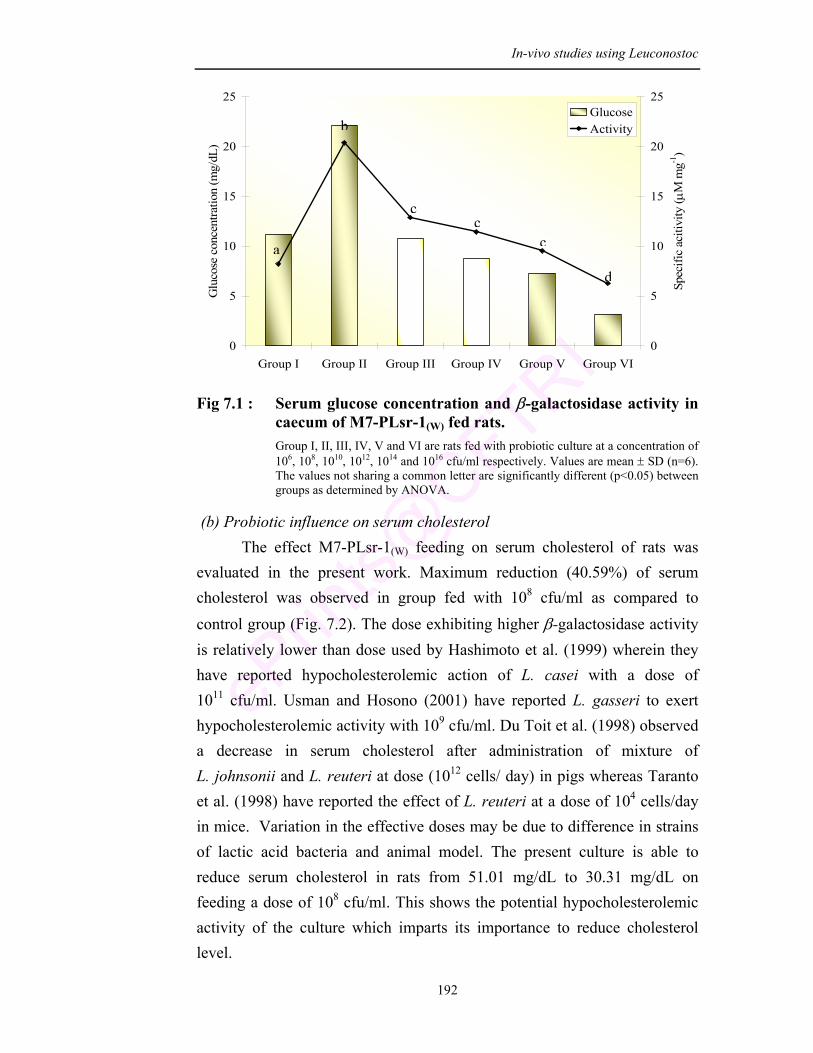
In-vivo studies using Leuconostoc
192
0
5
10
15
20
25
Group I Group II Group III Group IV Group V Group VI
Glu
cose
con
cent
ratio
n (m
g/dL
)
0
5
10
15
20
25
Spec
ific
aciti
vity
( µM
mg-1
)
GlucoseActivity
Fig 7.1 : Serum glucose concentration and β-galactosidase activity in
caecum of M7-PLsr-1(W) fed rats. Group I, II, III, IV, V and VI are rats fed with probiotic culture at a concentration of 106, 108, 1010, 1012, 1014 and 1016 cfu/ml respectively. Values are mean ± SD (n=6). The values not sharing a common letter are significantly different (p<0.05) between groups as determined by ANOVA.
(b) Probiotic influence on serum cholesterol The effect M7-PLsr-1(W) feeding on serum cholesterol of rats was
evaluated in the present work. Maximum reduction (40.59%) of serum cholesterol was observed in group fed with 108 cfu/ml as compared to control group (Fig. 7.2). The dose exhibiting higher β-galactosidase activity is relatively lower than dose used by Hashimoto et al. (1999) wherein they have reported hypocholesterolemic action of L. casei with a dose of 1011 cfu/ml. Usman and Hosono (2001) have reported L. gasseri to exert hypocholesterolemic activity with 109 cfu/ml. Du Toit et al. (1998) observed a decrease in serum cholesterol after administration of mixture of L. johnsonii and L. reuteri at dose (1012 cells/ day) in pigs whereas Taranto et al. (1998) have reported the effect of L. reuteri at a dose of 104 cells/day in mice. Variation in the effective doses may be due to difference in strains of lactic acid bacteria and animal model. The present culture is able to reduce serum cholesterol in rats from 51.01 mg/dL to 30.31 mg/dL on feeding a dose of 108 cfu/ml. This shows the potential hypocholesterolemic activity of the culture which imparts its importance to reduce cholesterol level.
a
b
cc
c
d
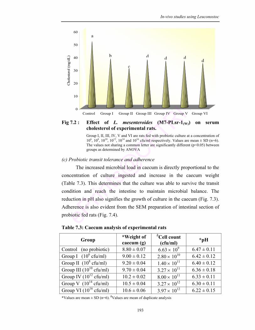
In-vivo studies using Leuconostoc
193
0
10
20
30
40
50
60
Cho
lest
erol
(mg/
dL)
Control Group I Group II Group III Group IV Group V Group VI Fig 7.2 : Effect of L. mesenteroides (M7-PLsr-1(W)) on serum
cholesterol of experimental rats. Group I, II, III, IV, V and VI are rats fed with probiotic culture at a concentration of 106, 108, 1010, 1012, 1014 and 1016 cfu/ml respectively. Values are mean ± SD (n=6). The values not sharing a common letter are significantly different (p<0.05) between groups as determined by ANOVA
(c) Probiotic transit tolerance and adherence The increased microbial load in caecum is directly proportional to the
concentration of culture ingested and increase in the caecum weight (Table 7.3). This determines that the culture was able to survive the transit condition and reach the intestine to maintain microbial balance. The reduction in pH also signifies the growth of culture in the caecum (Fig. 7.3). Adherence is also evident from the SEM preparation of intestinal section of probiotic fed rats (Fig. 7.4).
Table 7.3: Caecum analysis of experimental rats
Group *Weight of caecum (g)
δCell count (cfu/ml) *pH
Control (no probiotic) 8.80 ± 0.07 6.63 × 10⁹ 6.47 ± 0.11 Group I (106 cfu/ml) 9.00 ± 0.12 2.80 × 10¹⁰ 6.42 ± 0.12 Group II (108 cfu/ml) 9.20 ± 0.04 1.40 × 10¹¹ 6.40 ± 0.12 Group III (1010 cfu/ml) 9.70 ± 0.04 3.27 × 10¹¹ 6.36 ± 0.18 Group IV (1012 cfu/ml) 10.2 ± 0.02 8.00 × 10¹¹ 6.33 ± 0.11 Group V (1014 cfu/ml) 10.5 ± 0.04 3.27 × 10¹² 6.30 ± 0.11 Group VI (1016 cfu/ml) 10.6 ± 0.06 3.97 × 10¹² 6.22 ± 0.15 *Values are mean ± SD (n=6). δValues are mean of duplicate analysis
a
b
c
d d dd
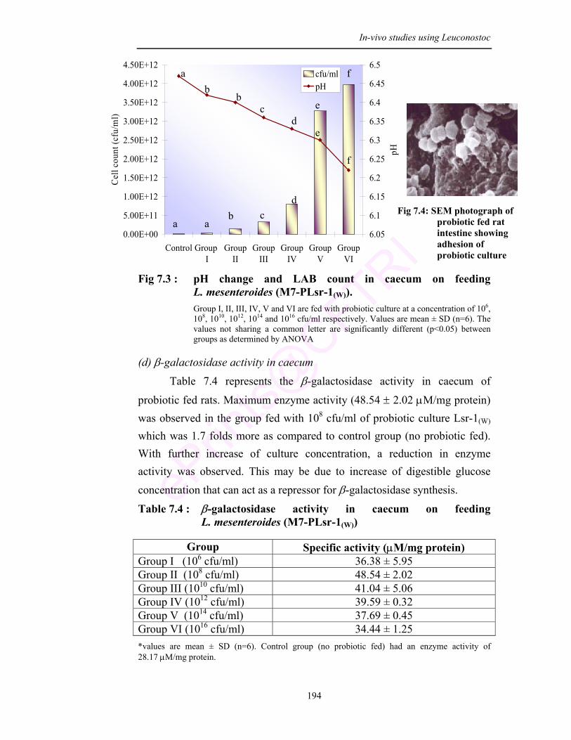
In-vivo studies using Leuconostoc
194
0.00E+00
5.00E+11
1.00E+12
1.50E+12
2.00E+12
2.50E+12
3.00E+12
3.50E+12
4.00E+12
4.50E+12
Control Group I
GroupII
GroupIII
GroupIV
GroupV
GroupVI
Cel
l cou
nt (c
fu/m
l)
6.05
6.1
6.15
6.2
6.25
6.3
6.35
6.4
6.45
6.5
pH
cfu/mlpH
Fig 7.3 : pH change and LAB count in caecum on feeding L. mesenteroides (M7-PLsr-1(W)). Group I, II, III, IV, V and VI are fed with probiotic culture at a concentration of 106, 108, 1010, 1012, 1014 and 1016 cfu/ml respectively. Values are mean ± SD (n=6). The values not sharing a common letter are significantly different (p<0.05) between groups as determined by ANOVA
(d) β-galactosidase activity in caecum Table 7.4 represents the β-galactosidase activity in caecum of
probiotic fed rats. Maximum enzyme activity (48.54 ± 2.02 µM/mg protein) was observed in the group fed with 108 cfu/ml of probiotic culture Lsr-1(W) which was 1.7 folds more as compared to control group (no probiotic fed). With further increase of culture concentration, a reduction in enzyme activity was observed. This may be due to increase of digestible glucose concentration that can act as a repressor for β-galactosidase synthesis.
Table 7.4 : β-galactosidase activity in caecum on feeding L. mesenteroides (M7-PLsr-1(W))
Group Specific activity (µM/mg protein)
Group I (106 cfu/ml) 36.38 ± 5.95 Group II (108 cfu/ml) 48.54 ± 2.02 Group III (1010 cfu/ml) 41.04 ± 5.06 Group IV (1012 cfu/ml) 39.59 ± 0.32 Group V (1014 cfu/ml) 37.69 ± 0.45 Group VI (1016 cfu/ml) 34.44 ± 1.25 *values are mean ± SD (n=6). Control group (no probiotic fed) had an enzyme activity of 28.17 µM/mg protein.
Fig 7.4: SEM photograph of probiotic fed rat intestine showing adhesion of probiotic culture
a b
b c
d e
f
a a b c
d
e
f
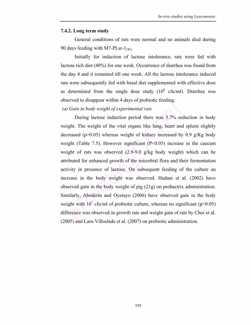
In-vivo studies using Leuconostoc
195
7.4.2. Long term study
General conditions of rats were normal and no animals died during
90 days feeding with M7-PLsr-1(W).
Initially for induction of lactose intolerance, rats were fed with
lactose rich diet (40%) for one week. Occurrence of diarrhea was found from
the day 4 and it remained till one week. All the lactose intolerance induced
rats were subsequently fed with basal diet supplemented with effective dose
as determined from the single dose study (108 cfu/ml). Diarrhea was
observed to disappear within 4 days of probiotic feeding.
(a) Gain in body weight of experimental rats
During lactose induction period there was 5.7% reduction in body
weight. The weight of the vital organs like lung, heart and spleen slightly
decreased (p>0.05) whereas weight of kidney increased by 0.9 g/Kg body
weight (Table 7.5). However significant (P<0.05) increase in the caecum
weight of rats was observed (2.9-9.0 g/kg body weight) which can be
attributed for enhanced growth of the microbial flora and their fermentation
activity in presence of lactose. On subsequent feeding of the culture an
increase in the body weight was observed. Hadani et al. (2002) have
observed gain in the body weight of pig (21g) on probactrix administration.
Similarly, Aboderin and Oyetayo (2006) have observed gain in the body
weight with 107 cfu/ml of probiotic culture, whereas no significant (p>0.05)
difference was observed in growth rate and weight gain of rats by Choi et al.
(2005) and Lara-Villoslade et al. (2007) on probiotic administration.
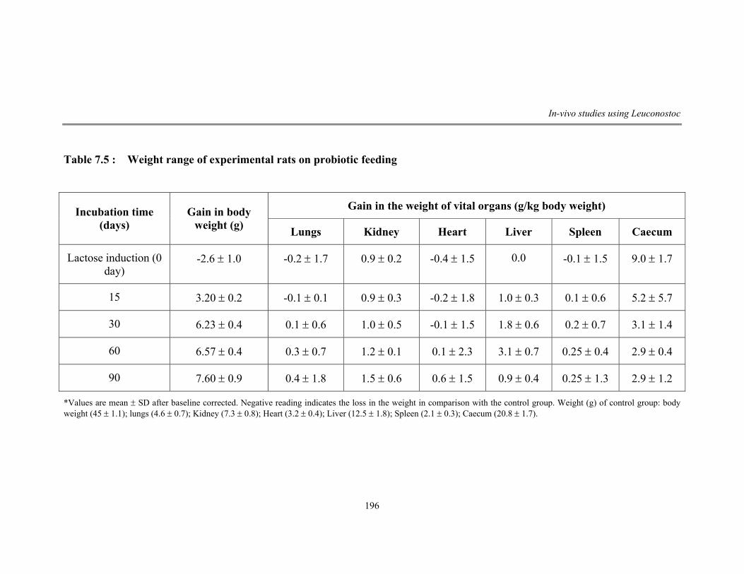
In-vivo studies using Leuconostoc
196
Table 7.5 : Weight range of experimental rats on probiotic feeding
Gain in the weight of vital organs (g/kg body weight) Incubation time (days)
Gain in body weight (g) Lungs Kidney Heart Liver Spleen Caecum
Lactose induction (0 day)
-2.6 ± 1.0 -0.2 ± 1.7 0.9 ± 0.2 -0.4 ± 1.5 0.0 -0.1 ± 1.5 9.0 ± 1.7
15 3.20 ± 0.2 -0.1 ± 0.1 0.9 ± 0.3 -0.2 ± 1.8 1.0 ± 0.3 0.1 ± 0.6 5.2 ± 5.7
30 6.23 ± 0.4 0.1 ± 0.6 1.0 ± 0.5 -0.1 ± 1.5 1.8 ± 0.6 0.2 ± 0.7 3.1 ± 1.4
60 6.57 ± 0.4 0.3 ± 0.7 1.2 ± 0.1 0.1 ± 2.3 3.1 ± 0.7 0.25 ± 0.4 2.9 ± 0.4
90 7.60 ± 0.9 0.4 ± 1.8 1.5 ± 0.6 0.6 ± 1.5 0.9 ± 0.4 0.25 ± 1.3 2.9 ± 1.2
*Values are mean ± SD after baseline corrected. Negative reading indicates the loss in the weight in comparison with the control group. Weight (g) of control group: body weight (45 ± 1.1); lungs (4.6 ± 0.7); Kidney (7.3 ± 0.8); Heart (3.2 ± 0.4); Liver (12.5 ± 1.8); Spleen (2.1 ± 0.3); Caecum (20.8 ± 1.7).
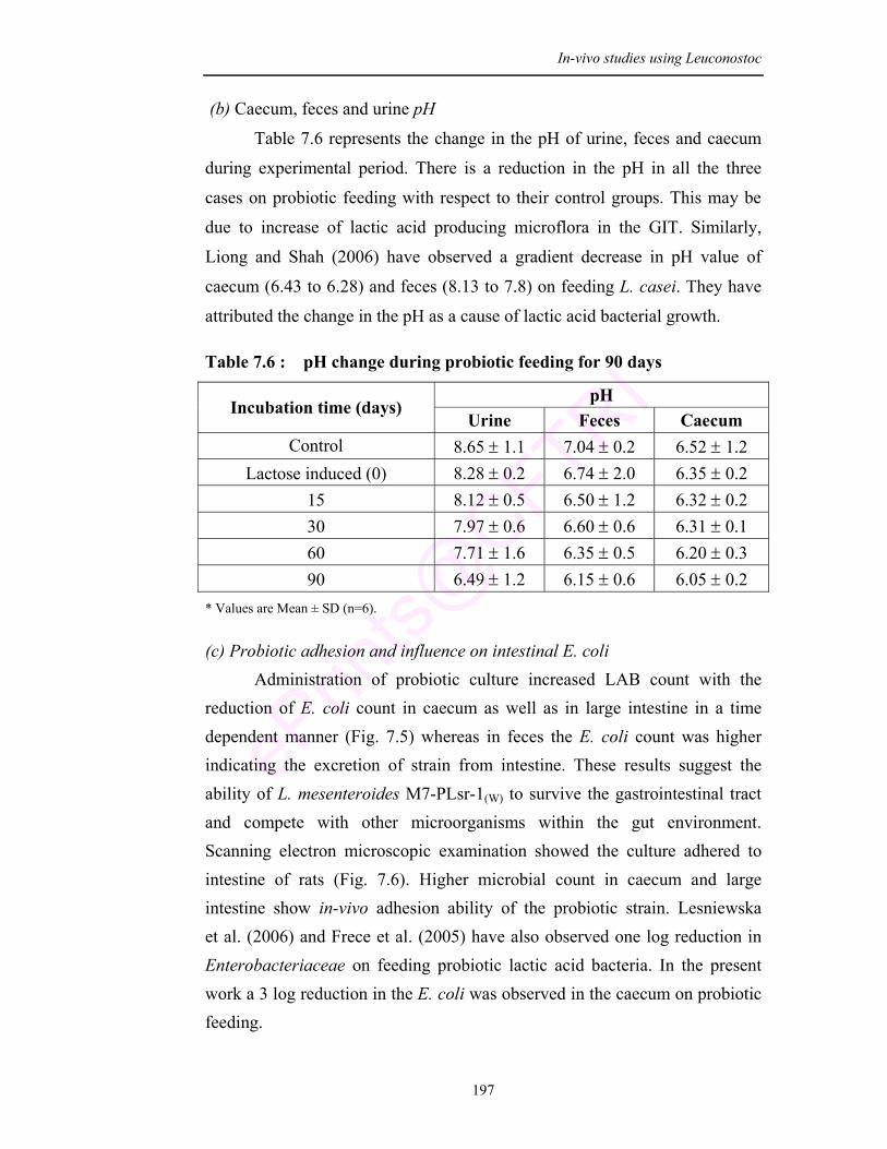
In-vivo studies using Leuconostoc
197
(b) Caecum, feces and urine pH
Table 7.6 represents the change in the pH of urine, feces and caecum
during experimental period. There is a reduction in the pH in all the three
cases on probiotic feeding with respect to their control groups. This may be
due to increase of lactic acid producing microflora in the GIT. Similarly,
Liong and Shah (2006) have observed a gradient decrease in pH value of
caecum (6.43 to 6.28) and feces (8.13 to 7.8) on feeding L. casei. They have
attributed the change in the pH as a cause of lactic acid bacterial growth.
Table 7.6 : pH change during probiotic feeding for 90 days
pH Incubation time (days)
Urine Feces Caecum Control 8.65 ± 1.1 7.04 ± 0.2 6.52 ± 1.2
Lactose induced (0) 8.28 ± 0.2 6.74 ± 2.0 6.35 ± 0.2 15 8.12 ± 0.5 6.50 ± 1.2 6.32 ± 0.2 30 7.97 ± 0.6 6.60 ± 0.6 6.31 ± 0.1 60 7.71 ± 1.6 6.35 ± 0.5 6.20 ± 0.3 90 6.49 ± 1.2 6.15 ± 0.6 6.05 ± 0.2
* Values are Mean ± SD (n=6).
(c) Probiotic adhesion and influence on intestinal E. coli Administration of probiotic culture increased LAB count with the
reduction of E. coli count in caecum as well as in large intestine in a time dependent manner (Fig. 7.5) whereas in feces the E. coli count was higher indicating the excretion of strain from intestine. These results suggest the ability of L. mesenteroides M7-PLsr-1(W) to survive the gastrointestinal tract and compete with other microorganisms within the gut environment. Scanning electron microscopic examination showed the culture adhered to intestine of rats (Fig. 7.6). Higher microbial count in caecum and large intestine show in-vivo adhesion ability of the probiotic strain. Lesniewska et al. (2006) and Frece et al. (2005) have also observed one log reduction in Enterobacteriaceae on feeding probiotic lactic acid bacteria. In the present work a 3 log reduction in the E. coli was observed in the caecum on probiotic feeding.
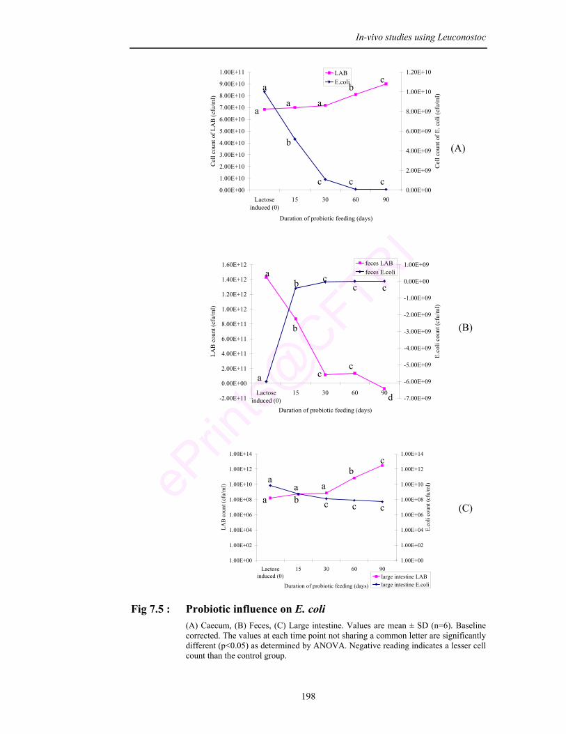
In-vivo studies using Leuconostoc
198
0.00E+00
1.00E+10
2.00E+10
3.00E+10
4.00E+10
5.00E+10
6.00E+10
7.00E+10
8.00E+10
9.00E+10
1.00E+11
Lactoseinduced (0)
15 30 60 90
Duration of probiotic feeding (days)C
ell c
ount
of L
AB
(cfu
/ml)
0.00E+00
2.00E+09
4.00E+09
6.00E+09
8.00E+09
1.00E+10
1.20E+10
Cel
l cou
nt o
f E. c
oli (
cfu/
ml)
LABE.coli
-2.00E+11
0.00E+00
2.00E+11
4.00E+11
6.00E+11
8.00E+11
1.00E+12
1.20E+12
1.40E+12
1.60E+12
Lactoseinduced (0)
15 30 60 90
Duration of probiotic feeding (days)
LAB
cou
nt (c
fu/m
l)
-7.00E+09
-6.00E+09
-5.00E+09
-4.00E+09
-3.00E+09
-2.00E+09
-1.00E+09
0.00E+00
1.00E+09
E.co
li co
unt (
cfu/
ml)
feces LABfeces E.coli
1.00E+00
1.00E+02
1.00E+04
1.00E+06
1.00E+08
1.00E+10
1.00E+12
1.00E+14
Lactoseinduced (0)
15 30 60 90
Duration of probiotic feeding (days)
LAB
cou
nt (c
fu/m
l)
1.00E+00
1.00E+02
1.00E+04
1.00E+06
1.00E+08
1.00E+10
1.00E+12
1.00E+14
E.co
li co
unt (
cfu/
ml)
large intestine LABlarge intestine E.coli
Fig 7.5 : Probiotic influence on E. coli (A) Caecum, (B) Feces, (C) Large intestine. Values are mean ± SD (n=6). Baseline
corrected. The values at each time point not sharing a common letter are significantly different (p<0.05) as determined by ANOVA. Negative reading indicates a lesser cell count than the control group.
(A)
(B)
(C)
aa a
bc
a
b
c c c
a
b
cc
d
a
b cc c
a
b c c ca
a ab
c
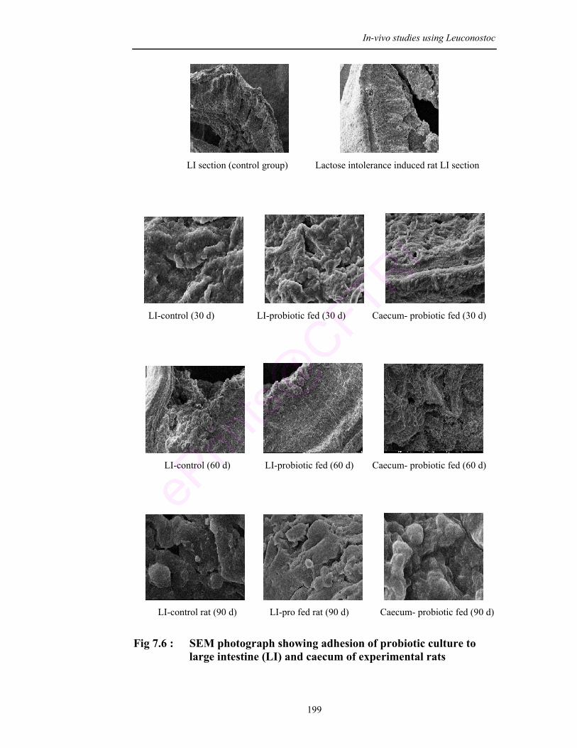
In-vivo studies using Leuconostoc
199
LI section (control group) Lactose intolerance induced rat LI section
LI-control (30 d) LI-probiotic fed (30 d) Caecum- probiotic fed (30 d)
LI-control (60 d) LI-probiotic fed (60 d) Caecum- probiotic fed (60 d)
LI-control rat (90 d) LI-pro fed rat (90 d) Caecum- probiotic fed (90 d)
Fig 7.6 : SEM photograph showing adhesion of probiotic culture to
large intestine (LI) and caecum of experimental rats
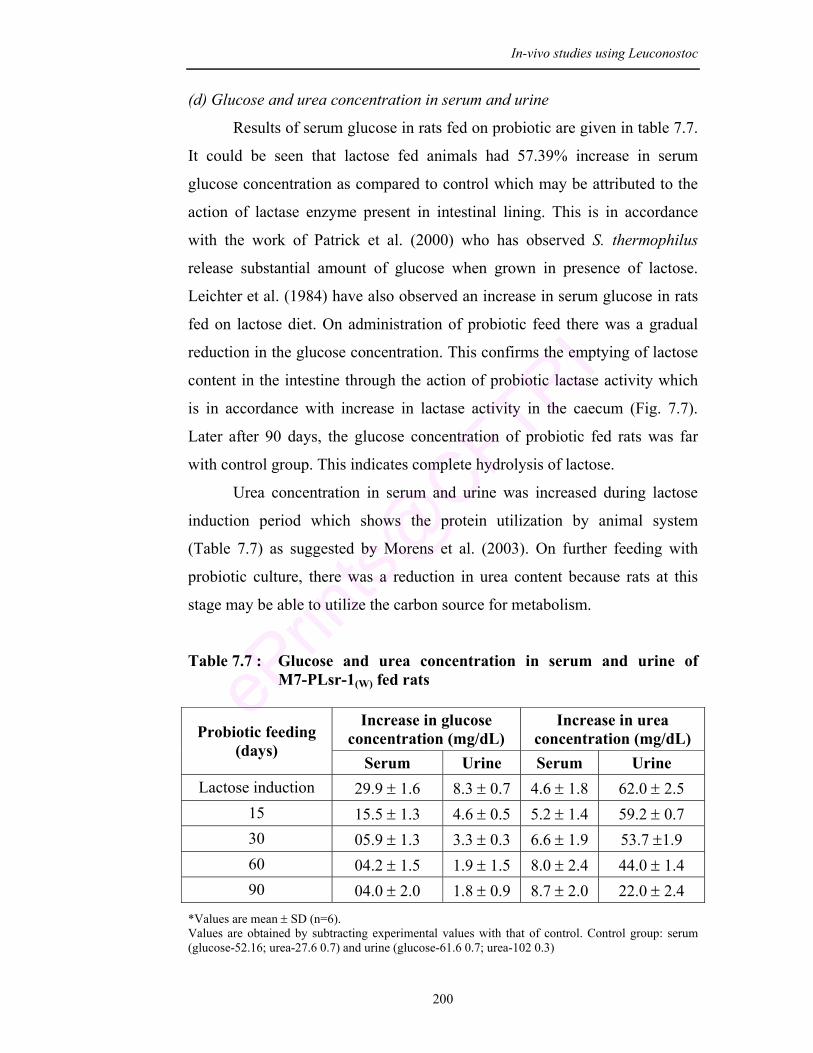
In-vivo studies using Leuconostoc
200
(d) Glucose and urea concentration in serum and urine
Results of serum glucose in rats fed on probiotic are given in table 7.7.
It could be seen that lactose fed animals had 57.39% increase in serum
glucose concentration as compared to control which may be attributed to the
action of lactase enzyme present in intestinal lining. This is in accordance
with the work of Patrick et al. (2000) who has observed S. thermophilus
release substantial amount of glucose when grown in presence of lactose.
Leichter et al. (1984) have also observed an increase in serum glucose in rats
fed on lactose diet. On administration of probiotic feed there was a gradual
reduction in the glucose concentration. This confirms the emptying of lactose
content in the intestine through the action of probiotic lactase activity which
is in accordance with increase in lactase activity in the caecum (Fig. 7.7).
Later after 90 days, the glucose concentration of probiotic fed rats was far
with control group. This indicates complete hydrolysis of lactose.
Urea concentration in serum and urine was increased during lactose
induction period which shows the protein utilization by animal system
(Table 7.7) as suggested by Morens et al. (2003). On further feeding with
probiotic culture, there was a reduction in urea content because rats at this
stage may be able to utilize the carbon source for metabolism.
Table 7.7 : Glucose and urea concentration in serum and urine of M7-PLsr-1(W) fed rats
Increase in glucose
concentration (mg/dL) Increase in urea
concentration (mg/dL) Probiotic feeding (days)
Serum Urine Serum Urine Lactose induction 29.9 ± 1.6 8.3 ± 0.7 4.6 ± 1.8 62.0 ± 2.5
15 15.5 ± 1.3 4.6 ± 0.5 5.2 ± 1.4 59.2 ± 0.7 30 05.9 ± 1.3 3.3 ± 0.3 6.6 ± 1.9 53.7 ±1.9 60 04.2 ± 1.5 1.9 ± 1.5 8.0 ± 2.4 44.0 ± 1.4 90 04.0 ± 2.0 1.8 ± 0.9 8.7 ± 2.0 22.0 ± 2.4
*Values are mean ± SD (n=6). Values are obtained by subtracting experimental values with that of control. Control group: serum (glucose-52.16; urea-27.6 0.7) and urine (glucose-61.6 0.7; urea-102 0.3)
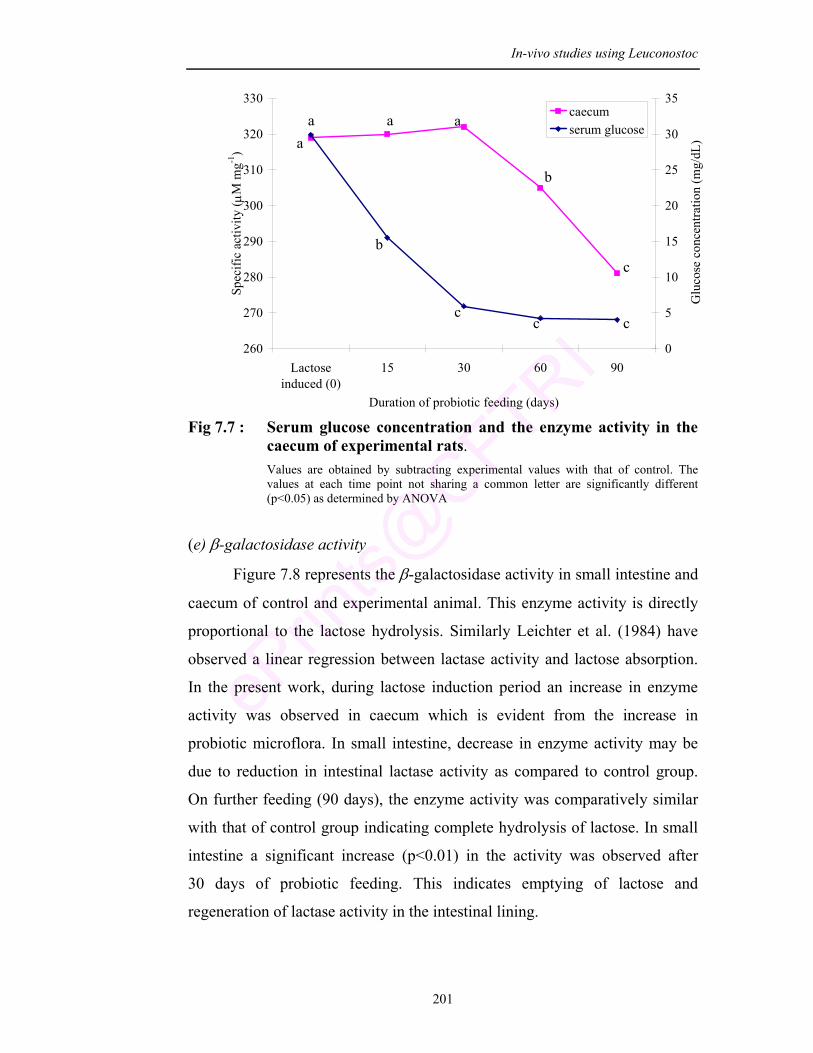
In-vivo studies using Leuconostoc
201
Fig 7.7 : Serum glucose concentration and the enzyme activity in the
caecum of experimental rats. Values are obtained by subtracting experimental values with that of control. The values at each time point not sharing a common letter are significantly different (p<0.05) as determined by ANOVA
(e) β-galactosidase activity
Figure 7.8 represents the β-galactosidase activity in small intestine and
caecum of control and experimental animal. This enzyme activity is directly
proportional to the lactose hydrolysis. Similarly Leichter et al. (1984) have
observed a linear regression between lactase activity and lactose absorption.
In the present work, during lactose induction period an increase in enzyme
activity was observed in caecum which is evident from the increase in
probiotic microflora. In small intestine, decrease in enzyme activity may be
due to reduction in intestinal lactase activity as compared to control group.
On further feeding (90 days), the enzyme activity was comparatively similar
with that of control group indicating complete hydrolysis of lactose. In small
intestine a significant increase (p<0.01) in the activity was observed after
30 days of probiotic feeding. This indicates emptying of lactose and
regeneration of lactase activity in the intestinal lining.
a a a
b
c
a
b
c c c
260
270
280
290
300
310
320
330
Lactoseinduced (0)
15 30 60 90
Duration of probiotic feeding (days)
Spec
ific
activ
ity ( µ
M m
g-1)
0
5
10
15
20
25
30
35
Glu
cose
con
cent
ratio
n (m
g/dL
)
caecumserum glucose
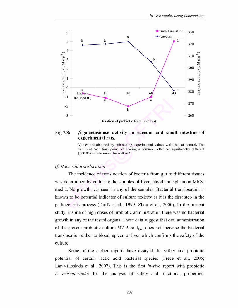
In-vivo studies using Leuconostoc
202
Fig 7.8: β-galactosidase activity in caecum and small intestine of experimental rats. Values are obtained by subtracting experimental values with that of control. The values at each time point not sharing a common letter are significantly different (p<0.05) as determined by ANOVA.
(f) Bacterial translocation The incidence of translocation of bacteria from gut to different tissues was determined by culturing the samples of liver, blood and spleen on MRS-media. No growth was seen in any of the samples. Bacterial translocation is known to be potential indicator of culture toxicity as it is the first step in the pathogenesis process (Duffy et al., 1999; Zhou et al., 2000). In the present study, inspite of high doses of probiotic administration there was no bacterial growth in any of the tested organs. These data suggest that oral administration of the present probiotic culture M7-PLsr-1(W) does not increase the bacterial translocation either to blood, spleen or liver which confirms the safety of the culture. Some of the earlier reports have assayed the safety and probiotic potential of certain lactic acid bacterial species (Frece et al., 2005; Lar-Villoslada et al., 2007). This is the first in-vivo report with probiotic L. mesenteroides for the analysis of safety and functional properties.
a a a
b
c a
a
b
c
d
-3
-2
-1
0
1
2
3
4
5
6
Lactoseinduced (0)
15 30 60 90
Duration of probiotic feeding (days)
Enzy
me
activ
ity ( µM
mg-1
)
260
270
280
290
300
310
320
330
Enzy
me
activ
ity ( µM
mg-1
)
small inrestinecaecum
In-vivo studies using Leuconostoc
203
Additionally the culture has been evaluated for its ability to reduce lactose intolerance problem. Earlier experiments in animals have indicated that kefir cultures play important role in improving lactose digestion (de Vrese et al., 1992). Goodenough and Kleyn (1976) have studied the influence of viable yogurt culture on digestion of lactose by the rats. Rats were fed on yogurt supplemented with lactose and sucrose for 7 days. Assay showed that lactase activity was more in animals fed with yogurt culture. Studies in lactase deficient humans confirm that yogurt can reduce lactose intolerance (Savaiano and Levitt, 1987). Shah and Jelen (1991) studied the lactose absorption by post weaning rats and found that lactose digestion from yogurt and guarg was facilitated
and the assay confirmed the presence of viable cultures and β-galactosidase activity after feeding. A study was designed to determine the effect of oral feeding of L. acidophilus on lactose tolerance by Saltzman et al. (1999). The culture was fed twice a day upto 7 days to 42 patients with lactose intolerance but they found that there was no significant change in breath hydrogen excretion. In the present study there was reduction in diarrhea on probiotic feeding. After 4 days of probiotic feeding to lactose intolerant induced rats there was disappearance of diarrhea. This shows that the present probiotic isolate Lsr-1(W) has potential in treating lactose intolerance.
7.5. Conclusion From the study it can be concluded that oral feeding of probiotic
culture exerts beneficial effect on experimental rats. Feeding probiotic culture L. mesenteroides (M7-PLSr-1(W)) has no harmful effect on the animals either morphologically or behaviorally. Hence, the culture is safe and is able to resist the gastrointestinal conditions. The culture shows transit tolerance in gastrointestinal tract. The culture is able to adhere and has antimicrobial activity against the intestinal E. coli. Reduction in the number of E. coli counts in caecum and large intestine supports that the culture maintains healthy intestinal microflora. High β-galactosidase activity of the culture reduces the accumulation of lactose as observed from the experimental results.
Summary & Conclusions
Summary and Conclusion
204
SUMMARY AND CONCLUSION
Bacterial cultures were isolated from milk and milk products and screened for lactic acid bacteria and were adapted to tolerate gastrointestinal conditions. The strain that was able to survive under such environment was characterized and identified through biochemical assays and molecular methods as Leuconostoc mesenteroides (PLsr-1(W)).
The selected culture (PLsr-1(W)) exerts antimicrobial activity against seven toxic food pathogens and was resistant to three common antibiotics. The antioxidative activity, serum cholesterol reducing ability, adherence ability, production of therapeutically important volatile compound and β-galactosidase activity shows potential functional characteristics of the probiotic culture isolate.
The culture strain was improved by using UV irradiation where the β-galactosidase activity increased by 2.03 folds. This was coded as M7-PLsr-1(W) and all further studies were conduced on this improved strain. On ammonium sulphate precipitation M7-PLsr-1(W) exhibited 25 folds higher activity as compared to crude extract of PLsr-1(W).
The culture M7-PLsr-1(W) was preserved by freeze drying and its viability was further enhanced with supplementation of cryoprotectants during 6 months shelf storage. During the storage period the culture retained its antimicrobial activity and resistance to low pH and high bile salt concentration.
The fermented milk beverage prepared with the M7-PLsr-1(W) was found to be rich in protein, total sugar, fatty acids and minerals like iron, zinc and magnesium. The viability of the culture and nutritional properties of the product was further enhanced with supplementation of adjuvants so that it can be used by all ages for its beneficial effects.
This fermented milk beverage was preserved over a storage time by interrupting the signal molecules produced by of spoilage bacteria using 2(5H)-furanone.
The in-vivo experiments conducted with albino Wister rats gave a clear evidence for the potential probiotic functional properties and its importance in reducing lactose intolerance problem.

Bibliography
205
Aattouri, N., Bouras, M., Tome, D., Marcos, A. and Lemonnier, D. (2002). Oral ingestion of lactic acid bacteria by rats increases lymphocytic proliferation and γ interferon production. Br J Nutr 87: 367–373.
Abadias, M., Teixido, N., Usall, J., Benabarre, A. and Vinas, I. (2001). Viability, efficacy, and storage stability of freeze-dried biocontrol agent Candida sake using different protective and rehydration media. J Food Prot 64 (6): 856–861.
Abdelali, H., Cassand, P., Soussotte, V., Daubeze, M., Bouley, C. and Narbonne, J.F. (1995). Effect of dairy products on initiation of precursor lesions of colon cancer in rats. Nutr Cancer 24: 121-132.
Aboderin, F.I. and Oyetayo, V.O. (2006). Haematological studies of rats fed different doses of probiotic, Lactobacillus plantarum, isolated from fermenting corn slurry. Pak J Nutr 5(2): 102-105.
Adamowicz, M., Kelley, P.M. and Nickerson, K.W. (1991). Detergent (sodium dodecyl sulfate) shock proteins in Escherichia coli. J Bacteriol 173: 229–233.
ADC (Australian Dairy Corporation). (1998). Dairy compendium. Melbourne: Australian Dairy Corporation, pp. 11-13.
Agarwal, K.N. and Bhasin, S.K. (2002). Feasibility studies to control acute diarrhoea in children by feeding fermented milk preparations Actimel and Dahi. Eur J Clin Nutr 56: S56–S59.
Agrawal, R., Rati, E.R., Vijayendra, S.V.N., Varadaraj, M.C., Prasad, M.S. and Krishnanand. (2000). Flavor profile of idli batter prepared from defined microbial starter cultures. World J Microbiol Biotechnol 16: 687-690.
Ahn, Y.T., Kim, G.B., Lim, K.S., Baek, Y.J. and Kim, H.U. (2003). Deconjugation of bile salts by Lactobacillus acidophilus isolates. Int Dairy J 13: 303–311.
Akahoshi, A., Goto, Y., Murao, K., Miyazaki, T., Yamasaki, M., Nonaka, M., Yamada, K. and Sugano, M. (2002). Conjugated linoleic acid reduces body fats and cytokine levels of mice. Biosci Biotechnol Biochem 66(4): 916-920.
Alander, M., Korpela, R., Saxelin, M., Vilpponen-Salmela, T., Matilla-Sandholm, T. and Wright, A. (1997). Recovery of Lactobacillus rhamnosus GG from human colonic biopsies. Lett Appl Microbiol 24: 361–364.
Albayrak, N.A. and Yang, S.T. (2002). Production of galacto-oligosaccharides from lactose by Aspergillus oryzae β-galactosidase immobilized on cotton cloth. Biotechnol Bioeng 77: 8 – 19.
Aleljung, P., Shen, W., Rozalska, B., Hellman, U., Ljungh, A. and Wadstrom, T. (1994). Purification of collagen binding proteins of Lactobacillus reuteri NCIB 11951. Curr Microbiol 28: 231–236.
Allan, W.W. (2008). Mechanisms of action of probiotics. Clin Infectious Diseases 46: 101-111.
Allison, C. and Hughes, C. (1991). Bacterial swarming: an example of prokaryotic differentiation and multicellular behaviour. Sci Prog 75(298 Pt 3-4): 403–422.
Alm, L. (1982). Effect of fermentation on milk fat of Swedish fermented milk products. J Dairy Sci 65: 521–532.
206
Alonso, L. and Fraga, M.J. (2001). Simple and rapid analysis for quantitation of most important volatile flavor compounds in yogurt by headspace GC-MS. J Chromatographic Sci 39(7): 297-300.
Aneja, R.P. and Murthy, T.N. (1990). Conjugated linoleic acid contents of Indian curds and ghee. Indian J Dairy Sci 43: 231–238.
Antunes, A., Rainey, F.A., Nobre, M.F., Schumann, P., Ferreira, A.M., Ramos, A., Santos, H. and de Costa, M.S. (2002). Leuconostoc ficulneum sp. nov., a novel lactic acid bacterium isolated from a ripe fig, and reclassification of Lactobacillus fructosus as Leuconostoc fructosum comb. nov. Int J Sys Evolutionary Microbiol 52: 647–655.
Arunachalam, K., Gill, H.S. and Chandra, R.K. (2000). Enhancement of natural immune function by dietary consumption of Bifidobacterium lactis (HN019). Eur J Clin Nutr 54: 1-5.
Ashar, M.A. and Prajapathi, J.B. (1998). Bile tolerance, bile deconjugation and cholesterol reducing properties of dietary Lactobacilli. Ind J Microbiol 38: 145–148.
Aso, Y., Akaza, H., Kotake, T., Tsukamoto, T., Imai, K. and Naito, S. (1995). Preventive effect of a Lactobacillus casei preparation on the recurrence of superficial bladder cancer in a double blind trial. The BLP Study Group. Euro Urol 27: 104–109.
Astley, S.B., Elliott, R.M., Archer, D.B. and Southon, S. (2004). Evidence that dietary supplementation with carotenoids and carotenoid rich foods modulates the DNA damage: repair balance in human lymphocytes. Brit J Nutr 91: 63-72.
Auge, B., Donnio, P.Y., Le Dleaut, P. and Avril, J. L. (1987). Influence de la vancomycine intraveineuse sur lesores alerobies salivaires et flecales. Pathologie Biologie 35: 673– 675.
Bachhawat, N., Gowda, L.R. and Bhat, S.G. (1996). Single step method of preparation of detergent permeabilized Kluyveromyces fragilis for lactose hydrolysis. Process Biochem 31: 21–25.
Bakalinsky, A.T., Nadathur, S.R., Carney, J.R. and Gould, S.J. (1996). Antimutogenicity of yoghurt. Mut Res 350(1):199–200.
Barker, D.C. and Thorne, K.J. (1970). Spheroplasts of Lactobacillus casei and the cellular distribution of bactoprenol. J Cell Sci 7: 755-785.
Barrangou, R., Sung-Sik, Y., Breidt Jr., F., Fleming, H. P. and Klaenhammer, T. R. (2002). Identification and characterization of Leuconostoc fallax strains isolated from an industrial Sauerkraut fermentation. Appl Environ Microbiol 68: 2877– 2884.
Barros, R.R, Carvalho, M.D.G.S., Peralta, J.M., Facklam, R.R. and Teixeira, L.M. (2001). Phenotypic and genotypic characterization of Pediococcus strains isolated from human sources. J Clin Microbiol 39–40: 1241–1246.
Batra, N., Singh, J., Banerjee, U.C., Patnaik, P.R. and Sobti, R.C. (2002). Production and characterization of a thermostable β-galactosidase from Bacillus coagulans RCS3. Biotechnol Appl Biochem 36: 1–6.
207
Bayless, T.M. and Rosensweig, N.S. (1966). A racial difference in incidence of lactase deficiency. A survey of milk intolerance and lactase deficiency in healthy adult males. JAMA. 197: 968-972.
Beales, N. (2004). Adaptation of microorganisms to cold temperatures, weak acid preservatives, low ph, and osmotic stress: a review. Comprehensive Rev Food Sci Food Safety 3(1): 1-20.
Bellengier, P., Richard, J. and Foucaud, C. (1997). Nutritional requirements of Leuconostoc mesenteroides subsp. Mesenteroides and subsp. dextranicum for growth in milk. J Dairy Res 64: 95–103.
Benkerroum, N., Boughdadi, A., Bennani, N. and Hidane, K. (2003). Microbiological quality assessment of Moroccan camel’s milk and identification of predominating lactic acid bacteria. World J Microbiol Biotechnol 19: 645-648.
Bennenberg, H.J., Hock, C. and Van den, W. (1949). Estimation of milk protein by formal titration. Dairy Sci Abstr 45: 103.
Berghofer, E., Grzeskowiak, B., Mundigler, N., Sentall, W.B. and Walcak, J.J. (1998). Antioxidative properties of faba bean, soybean and oat tempeh. Int J Food Sci Nutr 49: 45–54.
Bernet, M.F., Brassart, D., Neeser, J. R. and Servin. A. L. (1993). Adhesion of human bifidobacterial strains to cultured human intestinal epithelial cells and inhibition of enteropathogen-cell interactions. Appl Environ Microbiol 59: 4121– 4135.
Bernet-Camard, M.F., Lievin, V., Brassart, D., Neeser, J.R., Servin, A.L. and Hudault, S. (1997). The human Lactobacillus acidophilus strain LA1 secretes a nonbacteriocin antibacterial substance(s) active in vitro and in vivo. Appl Environ Microbiol 63: 2747– 2753.
Berrada, N., Lemeland, J.F., Laroch, G., Thouvenot, P. and Piaia, M. (1991). Bifidobacterium from fermented milks: survival during gastric transit. J Dairy Sci 74: 409–413.
Bertazzoni, M.E., Benini, A., Marzotto, M., Hendriks, H., Sbarbati, A. and Dellaglio, F. (2001). Preliminary screening of health-promoting properties of new Lactobacillus strain: in vitro and in vivo. HEALFO abstracts, Italy.
Bertelsen, G., Christophersen, C., Nielsen, P.H., Madsen, H.L., Stadel, P. (1995). Chromatographic isolation of antioxidants guided by a methyl linoleate assay. J Agric Food Chem 43: 1272-1275.
Beukes, E.M., Bester, B.H. and Mostert, J.F. (2001). The microbiology of South African traditional fermented milks. Int J Food Microbiol 63(3): 189–197.
Beveridge, T.J. (2001). Use of the gram stain in microbiology. Biotech Histochem 76(3): 111–118.
Bezares, B.R., Saenz, Y., Poeta, P., Zarazaga, M., Ruiz-Larrea, F. and Torres, C. (2006). Assessment of antibiotic susceptibility within lactic acid bacteria strains isolated from wine. Int J Food Microbiol 111(3): 234-240.
Bhat, N., Naina, N.S., Gowda, L.K. and Bhat, S.G. (1993). Detergent permeabilized yeast cells as the source of intracellular enzymes for estimation of biomolecules. Enzyme Microb Technol 15: 796–800.
208
Bhowmik, T. and Marth, E.H. (1989). Simple method to detect β-galactosidase. Appl Environ Microbiol 55: 3240-3242.
Bibiloni, R., Zavaglia, A.G. and Antoni, G.d. (2001). Enzyme based most probable number method for the enumeration of Bifidobacterium in dairy products. J Food Prot 64(12): 2001–2006.
Bjorkroth and Holzapfel. (2003). Genera Leuconostoc, Oenococcus and Weissella. In: M. Dworkin, et al. (Eds.), The prokaryotes: An evolving electronic resource for the microbiological community (3rd ed), release 3.12, March 28, 2003. Berlin: Springer, NY http : //link.springer-ng.com/link/service/books/10125/.
Bjorkroth, K.J., Geisen, R., Schillinger, U., Weiss, N., de Vos, P., Holzapfel, W.H., Korkeala, H.J. and Vandamme, P. (2000). Characterization of Leuconostoc gasicomitatum sp. nov., associated with spoiled raw tomato marinated broiler meat strips packaged under modified atmosphere conditions. Appl Environ Microbiol 66: 3764-3772.
Blackwood, A.C. and Blakley. E.R. (1960). Fermentation of glucose-c14 and xylose-c14 by various strains of Leuconostoc mesenteroides. J Bacteriol 79: 411-416.
Bligh, E.G. and Dyer, W.J. (1959). A rapid method of total lipids extraction and purification. Can J Biochem Physiol 37: 911–917.
Blum, S., Haller, D., Pfeifer, A. and Schiffrin. E. J. (2002). Probiotics and immune response. Clin Rev Allergy Immunol 22: 287–309.
Boirivant, M. and Strober, W. (2007). The mechanism of action of probiotics. Curr Opin Gastroenterol 23(6): 679-692.
Boot, H.J., Kolen, C.P.A.M., Van Noort, J.M. and Pouwels, P.H. (1993). S-layer protein of Lactobacillus acidophilus ATCC 4356: Purification, expression in Escherichia coli, and nucleotide sequence of the corresponding gene. J Bacteriol 175(19): 6089-6096.
Boris, S., Suarez, J.E., Vazquez, F. and Barbes. (1998). Adherence of human vaginal Lactobacilli to vaginal epithelial cells and interaction with uropathogens. Infect Immun 66: 1985-1989.
Bottazzi, V. (1983). Other fermented dairy products. In Biotechnology, Vol. 5, Food and Feed Production with Microorganisms ed. Reed, G. Weinheim: Verlag Chemie, pp. 315-363.
Boumerdassi, H., Monnet, C., Desmazeaud, M. and Corrieu, G. (1997) Effect of citrate on production of diacetyl and acetoin by Lactococcus Lactis ssp. Lactis CNRZ 483 cultivated in the presence of oxygen. J Dairy Sci 80: 634–639.
Bourel, G., Henini, S., Krantar, K., Oraby, M., Divies, C. and Garmyn, D. (2001). Metabolisme sucrecitrate chez Leuconostoc mesenteroides. Le Lait 81: 75–82.
Bourget, N.L., Philippe, H., Mangin, I. and Decaris, B. (1996). 16SrRNA and 16 to 23 internal transcribed spacer sequence analyses reveal inter and intraspecific Bifidobacterium phylogeny. Int J Sys Bacteriol 46: 102–111.
Bozoglu, T.F., Ozilgen, M. and Bakir, U. (1987). Survival kinetics of lactic acid starter cultures during and after freeze drying. Enzyme Microbiol Technol 9: 531–537.
209
Brand-Williams, W., Cuvelier, M.E. and Berset, C. (1995). Use of a free radical method to evaluate antioxidant activity. Lebensm Wiss Technol 26: 25–30.
Brashears, M.M. and Durre, W.A. (1999). Antagonistic action of Lactobacillus lactis toward Salmonella spp and Escherichia coli 0151:H7 during growth and refrigerated storage. J Fd Prot 62: 1336-1340.
Brashears, M.M., Jaroni, D. and Trimble, J. (2003). Isolation, selection, and characterization of lactic acid bacteria for a competitive exclusion product to reduce sheddingof E. coli O157:H7 in cattle. J Food Prot 66: 355–363.
Brigidi, P., Vitali, B., Swennen, E., Bazzocchi, G. and Matteuzzi, D. (2001). Effects of probiotic administration upon the composition and enzymatic activity of human fecal microbiota in patients with irritable bowel syndrome or functional diarrhea. Res Microbiol 152: 735-741.
Broadbent, J.R. and Lin, C. (1999). Effect of heat shock or cold shock treatment on the resistance of Lactococcus lactis to freezing and lyophilization. Cryobiol 39: 88–102.
Bronshtein,V. (2004). Preservation by foam formation. Pharmaceutical Technol 28(4): 86–92.
Brown, J.L., Ross, T., McMeekin, T.A. and Nichols, P.D. (1997). Acid habituation of Escherichia coli and the potential role of cyclopropane fatty acids in low pH tolerance. Int J Food Microbiol 37: 163-173.
Bruno, E.A. and Shah, N.P. (2003) Viability of two freeze dried strains of Bifidobacterium and of commercial preparations at various temperatures during prolonged storage. J Food Sci 68(7): 2336-2339.
Buning, C., Genschel, J., Jurga, J., Fiedler, T., Voderholzer, W., Fiedler, E.M., Worm. M., Weltrich, R., Lochs, H., et al. (2005). Introducing genetic testing for adult type hypolactasia. Digestion 71: 245-250.
Burgess, C., O’Connell-Motherway, M., Sybesma, W., Hugenholtz, J. and van Sinderen, D. (2004). RiboXavin production in Lactococcus lactis: potential for in situ production of vitamin-enriched foods. Appl Environ Microbiol 70: 5769–5777.
Burns, A.J. and Rowland, I.R. (2000). Anti-Carcinogenicity of Probiotics and Prebiotics Curr Issues Intest Microbiol 1(1): 13-24.
Bury, D., Jelen, P. and Kalab, M. (2001). Disruption of Lactobacillus delbrueckii ssp. Bulgaricus 11842 cells for lactose hydrolysis in dairy products: a comparison of sonication, high- pressure homogenization and bead milling. Innovative Food Sci Emerging Technol 2: 23-29.
Busconi, L.and Denker, B.M. (1997). Analysis of the N-terminal binding domain of Go Biochem J 328: 23–31.
Cadieux, P., Wind, A., Sommer, P., Schaefer, L., Crowley, K., Britton, R.A. and Reid, G. (2008). Evaluation of reuterin production in urogenital probiotic Lactobacillus reuteri RC-14. Appl Environ Microbiol 6: 12-19.
Calderon, M., Loiseau, G. and Guyot, J.P. (2001). Nutritional requirements and simplified cultivation medium to study growth and energetics of a sourdough lactic acid bacterium Lactobacillus fermentum Ogi E1 during heterolactic fermentation of starch. J Appl Microbiol 90(4): 508-516.
210
Campieri, C., Campieri, M., Bertuzzi, V., Swennen, E., Matteuzzi, D., Stefoni, S., Pirovano, F., Centi, C., Ulisse, S., Famularo, G. and De Simone. C. (2001). Reduction of oxaluria after an oral course of lactic acid bacteria at high concentration. Kid Int 60: 1097-1105.
Canducci, F., Armuzzi, A., Cremonini, F., Cammarota, G., Bartolozzi, F., Pola, P., Gasbarrini, G. and Gasbarrini, A. (2000). A lyophilized and inactivated culture of Lactobacillus acidophilus increases Helicobacter pylori eradication rates. Aliment Pharmacol Ther 14: 1625-1629.
Canzi, E., Guglielmetti, S., Mora, D., Tamagnini, I. and Parini, C. (2005). Conditions affecting cell surface properties of human intestinal Bifidobacteria. Antonie Van Leeuwenhoek 88(3–4): 207–219.
Canzi, E., Zanchi, R., Camaschella, P., Cresci, A., Greppi, G.F., Orpianesi, C., Serrantoni, M. and Ferrari, A. (2000). Modulation by lactic acid bacteria of the intestinal ecosystem and plasma cholesterol in rabbits fed a casein diet. Nutr Res 20: 1329-1340.
Cao, G., Verdon, C.P., Wu, A.B.H., Wang, H. and Prior, R.L. (1995). Automated assay of oxygen radical absorbance capacity with the COBAS FARA II. Clin Chem 41: 1738–1744.
Caplice, E. and Fitzgerald, G.F. (1999). Food fermentations: Role of microorganisms in food production and preservation. Int J Food Microbiol 50: 131–149.
Carcoba, R. and Rodriguez, A. (2000). Influence of cryoprotectants on the viability and acidifying activity of frozen and freeze-dried cells of the novel starter strain Lactococcus lactis ssp. lactis CECT 5180. Euro Food Res Technol 211: 433–437.
Carpenter, J. F., Arakawa, T. and Crowe. J. H. (1990). Interactions of stabilizing additives with proteins during freeze-thawing and freeze-drying. Dev Biol Stand 74:225–239.
Carr, F.J., Chill, D. and Maida, N. (2002). The lactic acid bacteria: A literature survey. Crit Rev Microbiol 28: 281–370.
Carvalhoa, A.S., Silvaa, J., Hob, P., Teixeira, P., Malcataa, F.X. and Gibbsa, P. (2004). Relevant factors for the preparation of freeze-dried lactic acid bacteria. Int Dairy J 14: 835–847.
Casas, I.A. and Dobrogosz, W.J. (2000). Validation of the probiotic concept: Lactobacillus reuteri confers broad spectrum protection against disease in humans and animals, Microbial Ecology in Health and Disease 12: 247–285.
Cataldi, T.R.I., Bianco, G., Frommberger, M. and Schmitt-Kopplin. (2004). Direct analysis of selected N-acyl-L-homoserine lactones by gas chromatography/mass spectrometry. Rapid Comm Mass Spectrometry 18(12): 1341 – 1344.
Challa, A., Rao, D.R., Chawan, C.B., and Shackelford, L. (1997). Bifidobacterium longum and lactulose suppress azoxymethane induced aberrant crypt foci in rats. Carcinogenesis 18: 517-521.
Champagne, C.P., Mondou, F., Raymond, Y. and Roy, D. (1996). Effect of polymers and storage temperature on the stability of freeze dried lactic acid bacteria. Food Res Int 29: 555–562.
211
Chancey, S. T., Wood, D.W. and Pierson, L.S. (1999). Two-component transcriptional regulation of N-acyl-homoserine lactone production in Pseudomonas aureofaciens. Appl Environ Microbiol 65: 2294-2299.
Charlton, T.S., de Nys, R., Netting, A., Kumar, N., Hentzer, M., Givskov, M. and Kjelleberg, S. (2000). A novel and sensitive method for the quantification of N-3-oxo- acyl homoserine lactones using gas chromatography-mass spectrometry: application to a model bacterial biofilm. Environ Microbiol 2: 530-541.
Charteris, W. P., Kelly, P. M., Morelli, L. and Collins, J. K. (2000). Effect of conjugated bile salts on antibiotic susceptibility of bile salt-tolerant Lactobacillus and Bifidobacterium isolates. J Food Prot 63(10): 1369–1376.
Charteris, W.P., Kelly, P.M., Morelli, L. and Collins, J.K. (1998). Ingredient selection criteria for probiotic microorganisms in functional dairy foods. Int J Dairy Technol 51(4): 123–136.
Chateau, N., Deschamps, A. M. and Hadj Sassi, A. (1994). Heterogeneity of bile salts resistance in the Lactobacillus isolates of a probiotic consortium. Lett Appl Microbiol 18: 42–44.
Chavarri, F.J., De Paz, M. and Nueez, M.M. (1988). Cryoprotective agents for frozen concentrated starters from non-bitter Streptococcus lactis strains. Biotechnol Lett 10: 11–16.
Chen, T., Fowler, A. and Toner, M. (2000). Supplemented phase diagram of the trehalose–water binary mixture. Cryobiol 40: 277–282.
Chen, X., Xu, J., Shuai, J., Chen, J., Zhang, Z. and Fang, W. (2007). The S-layer proteins of Lactobacillus crispatus strain ZJ001 is responsible for competitive exclusion against Escherichia coli O157:H7 and Salmonella typhimurium. Int J Food Microbiol 115(3): 307-312.
Chien, H.L. (2004). Change of isoflavones contents in cultured soymilk fermented with lactic acid bacteria and Bifidobacteria. M.S. Thesis, National Taiwan University, Taipei, Taiwan.
Childs, N.M. (1997). The functional food consumers: who are they and what do they want? Implications for product development and positioning. In: Yalpani M, ed. New technologies for healthy foods and nutraceuticals. Shrewsbury, MA: ATL Press, pp. 313–26.
Chin, S.F., Liu, W., Storkson, J.M., Ha, Y.L. and Pariza, M.W. (1992). Dietary sources of conjugated dienoic isomers of linoleic acid, a newly recognized class of anticarcinogens. J Food Composition Anal 5: 185–197.
Choi, S.S., Kang, B.Y., Chung, M.J., Kim, S.D., Park, S.H., Kim, J.S., Kang C.Y. and Ha, N.J. (2005). Safety assessment of potential lactic acid bacteria Bifidobacterium longum SPM1205 isolated from healthy Koreans. J Microbiol 43(6): 493-498.
Chou, L.S. and Weimer, B. (1999). Isolation and characterization of acid and bile tolerant isolates from strains of Lactobacillus acidophilus. J Dairy Sci 82: 23–31.
Christensen, J.E., Dudley, E.G., Pederson, J.A. and Steele, J.L. (1999) Peptidases and amino acid catabolism in lactic acid bacteria. Ant Leeuwenhoek 76: 217–246.
212
Christopher, N.L. and Bayless, T.M. (1971). Role of the small bowel and colon in lactose induced diarrhea. Gastroenterol 60: 845-852.
Chugani, S.A., Whiteley, M., Lee, K.M., D’Argenio, D., Manoil, C. and Greenberg, E.P. (2001). QscR, a modulator of quorum sensing signal synthesis and virulence in P. aeruginosa. Proc Natl Acad Sci USA 98: 2752-2757.
Chung, H.S., Kim, Y.B., Chun, S.L. and Ji, G.E. (1999). Screening and selection of acid and bile resistant Bifidobacteria. Int J Food Microbiol 47: 25–32.
Chung, Y.C., Chang, C.T., Chao, W.W., Lin, C.F. and Chou, S.T. (2002). Antioxidative activity and safety of the 50% ethanolic extract from red bean fermented by Bacillus subtilis IMR-NK1. J Agric Food Chem 50: 2454–2458.
Cibik, R. and Chapot-Chartier, M. P. (2000). Autolysis of dairy Leuconostocs and detection of peptidoglycan hydrolases by renaturing SDS-PAGE . J Appl Microbiol 89: 862–869.
Cibik, R., Lepage, E. and Tailliez, P. (2000). Molecular diversity of Leuconostoc mesenteroides and Leuconostoc citreum isolated from traditional French cheeses as revealed by RAPD fingerprinting, 16srDNA sequencingand 16srDNA fragment amplification. Sys Appl Microbiol 23: 267–278.
Clark, P.A., Cotton, L.N. and Martin, J.H. (1993). Selection of Bifidobacteria for use as dietary adjuncts in cultured dairy foods: II. Tolerance to simulated pH of human stomach. Cult Dairy Pro J 28: 11-14.
Clegg, J.S. (1986). The physical properties and metabolism status of Artemia cysts at low water contents. The ‘water replacement hypothesis’. In Membranes, Metabolism and Dry Organisms ed. Leopold, A.C. Ithaca, NY: Cornell University Press, pp. 169-187.
Cocconcelli, P.S. Cattivelli, D. and Gazzola, S. (2003). Gene transfer of vancomycin and tetracycline resistances among Enterococcus faecalis during cheese and sausage fermentations. Int J Food Microbiol 88: 315–323.
Cocconcelli, P.S., Parisi, M.G., Senini, L. And Bottazzi, V. (1997). Use of RAPD and 16S rDNA sequencing for the study of Lactobacillus population dynamics in natural whey culture. Lett Appl Microbiol 25: 8-12.
Cogan, T.M. and Jordan, K.N. (1994). Metabolism of Leuconostoc bacteria. J Dairy Sci 77: 2704–2717.
Collado, M.C., Gueimonde, M., Hernandez, M., Sanz, Y. and Salminen, S. (2005). Adhesion of selected Bifidobacterium strains to human intestinal mucus and its role in enteropathogen exclusion. J Food Prot 68(12): 2672–2678.
Collins, J.K., Thornton, G. and Sullivan, G.O. (1998). Selection of probiotic strains for human application. Int Dairy J 8: 487–490.
Collins, M.D. and Gibson, G.R. (1999). Probiotics, prebiotics, and synbiotics: approaches for modulating the microbial ecology of the gut. Am J Clin Nutr 69(5):1052S-1057S.
Cook, M.E., Miller, C.C., Park, Y. and Pariza, M.W. (1993). Immune modulation by altered nutrient metabolism: Nutritional control of immune induced growth depression. Poultry Sci 72: 1301–1305.
Coppola, R., Giagnacovo, B., Iorizzo, M. and Grazia, L. (1998). Characterization of Lactobacilli involved in the ripening of Soppressata molisana, a typical southern Italy fermented sausage. Food Microbiol 15: 347–353.
213
Corcoran, B.M., Ross, R.P., Fitzgerald, G.F. and Stanton, C. (2004). Comparative survival of probiotic Lactobacilli spray-dried in the presence of prebiotic substances. J Appl Microbiol 96: 1024–1039.
Corcoran, B.M., Ross, R.P., Fitzgerald, G.F., Dockery, P. and Stanton, C. (2006). Enhanced survival of GroESL-overproducing Lactobacillus paracasei NFBC 338 under stressful conditions induced by drying. Appl Environ Microbiol 72(7): 5104–5107.
Costa, E., Usall, J., Teixido, N., Garcia, N. and Vinas, I. (2000). Effect of protective agents, rehydration media and initial cell concentration on viability of Pantoea agglomerans strain CPA-2 subjected to freeze drying. J Appl Microbiol 89: 793–800.
Costerton, J.W., Stewart, P.S. and Greenberg, E.P. (1999). Bacterial biofilms: a common cause of persistent infections. Sci 284: 1318-1322.
Cotter, A.A (2007). The health benefits of milk and dairy products. Society of Dairy Technology Bulletin no. 417. International Dairy Federation (IDF), Brussels, Belgium, pp. 213-214.
Cotter, P.D. and Hill, C. (2003). Surviving the acid test: responses of gram- positive bacteria to low pH. Microbiol Mol Biol Rev 67(3): 429–453.
Cross, M.L. (2002). Microbes versus microbes: immune signals generated by probiotic Lactobacilli and their role in protection against microbial pathogens. FEMS Immunol Med Microbiol 34: 245-253.
Crowe, J.H. and Crowe, L.M. (1986). Stabilization of membranes in an hydrobiotic organisms. In Membranes, Metabolism and Dry Organisms ed. Leopold, A.C. Ithaca, NY: Cornell University Press, pp. 188– 209.
Crowe, J.H., Carpenter, J.F. and Crowe, L.M. (1998). The role of vitrification in anhydrobiosis. Ann Rev Physiol 60: 73–103.
Crowe, J.H., Crowe, L.M., Carpenter, J.F., Rudolph, A.S., Aurell Winstrom, C., Spargo, B.J. and Anchordoguy, T.J. (1988). Interaction of sugars with membranes. Biochem Biophys Acta 947: 367–384.
Cvitkovitch, D.G., Li, Y.H. and Ellen, R.P. (2003). Quorum sensing and biofilm formation in streptococcal infections. J Clin Investig 112: 1626-1632.
D’Aimmo, M.R., Modesto, M. and Biavati, B. (2007) International Antibiotic resistance of lactic acid bacteria and Bifidobacterium spp. isolated from dairy and pharmaceutical products. J Food Microbiol 115(1): 35-42.
D’Souza, A.L., Rajkumar, C., Cooke, J. and Bulpitt, C.J. (2002). Probiotics in prevention of antibiotic associated diarrhoea: meta-analysis. Brit Med J 324: 1361-1367.
Daeschel, M.A. (1989). Antimicrobial substances from acid lactic bacteria for use as food preservative. Food Technol 43: 164-167.
dal Bello, F., Walter, J., Hammes, W. P. and Hertel, C. (2003). Increased complexity of the species composition of lactic acid bacteria in human feces revealed by alternative incubation condition. Microbial Ecol 45: 455–463.
Daly, C. and Davis, R. (1998). The biotechnology of lactic acid bacteria with emphasis on applications in food safety and human health. Agri Food Sci Finland 7: 251-264.
214
Dave, J.M. and Prajapathi, J.B.(1994). Lactic acid bacteria as antibacterials against food borne pathogens and food spoilage organisms. In: Microbes for better living Ed: Sankaran, R., Manja, K.S., Proceedings of MICON-INTL-94, Conference Secretariat, DFRL, Mysore. pp 361-367.
Dave, R.I. and Shah, N.P. (1998). Ingredient supplementation effects on viability of probiotic bacteria in yogurt. J Dairy Sci 81: 2804–2816.
Davis, J.G., Ashton, T.R. and McCaskill, M. (1971). Enumeration and viability of Lactobacillus bulgaricus and Streptococcus thermophilus in yoghurt. Dairy Industries 36: 569–573.
De Angelis, M., Gallo, G., Corbo, M.R., McSweeney, P.L.H., Faccia, M., Giovine, M. and Gobbetti, M. (2003). Phytase activity in sourdough lactic acid bacteria: purification and characterization of a phytase from Lactobacillus sanfranciscensis CB1. Int J Food Microbiol 87: 259–270.
De Boever, P., Deplancke, B. and Verstraete, J. (2000). Fermentation by gut microbiota cultured in a simulator of the human intestinal microbiota ecosystem is improved by supplementing a soy germ powder. J Nutr 130: 2599–2606.
De Giulio, B., Orlando, P., Barba, G., Coppola, R., De Rosa, M., Sada, A., De Prisco P.P. and Nazzaro, F. (2005). Use of alginate and cryoprotective sugars to improve the viability of lactic acid bacteria after freezing and freeze-drying. World J Microbiol Biotechnol 21: 739–746.
de la Fuente, M.A., Montes, F., Guerrero, G.and Juarez, M. (2003). Total and soluble contents of calcium, magnesium, phosphorus and zinc in yoghurts. Food Chem 80(4): 573-578.
De Smet, I., De Boever, P. and Verstraete, W. (1998). Cholesterol lowering in pigs through enhanced bacterial bile salt hydrolase activity. Brit J Nutr 79: 185–194.
De Smet, I., van Hoorde, L., vande Woestyne, M., Christiaens, H. and Verstraete, W. (1995). Significance of bile-salt hydrolytic activities of lactobacilli. J Appl Bacteriol 79: 292-301.
de Valdez, G.F., de Giori, G.S., de Ruiz Holgado, A.P. and Oliver, G. (1983). Comparative study of the efficiency of some additives in protecting lactic acid bacteria against freeze-drying. Cryobiol 20: 560–566.
De Vrese, M., Keller, B. and Barth, C.A. (1992). Enhancement of intestinal hydrolysis of lactose by microbial β-galactosidase (EC 3.2.1.23) of kefir. Br J Nutr 67: 67-75.
De Vrese, M., Stegelmann, A., Richter, B., Fenselau, S., Laue, C., Schrezenmeir, J. (2001). Probiotics: compensation for lactase insufficiency. Am J Clin Nutr 73: 421S-9S.
De Vries, R.P., van den Broek, H.C., Dekkers, E., Manzanares, P., de Graff, L.H. and Visser, J. (1999). Differential expression of three a-galactosidase genes and a single β-galactosidase gene from Aspergillus niger. Appl Environ Microbiol 65: 2453– 2460.
Del Re, B., Sgorbati, B., Miglioli, M. and Palenzona, D. (2000). Adhesion, autoaggregation and hydrophobicity of 13 strains of Bifidobacterium longum. Lett Appl Microbiol 31: 438–442.
215
Delisle, J., Amiot, J. and Dore, F. (1995). Biological availability of calcium and magnesium from dairy products. Int Dairy J 5: 87–96.
Denev, S.A. (2006). Role of Lactobacilli in gastrointestinal ecosystem. Bulg J Agric Sci 12: 63-114.
Derre, I., Rapoport, G. and Msadek, T. (1999). CtsR, a novel regulator of stress and heat shock response, controls clp and molecular chaperone gene expression in Gram-positive bacteria. Mol Microbiol 31: 117–131.
Desmond, C., Ross, R.P., O’Callaghan, E., Fitzgerald, G. and Stanton, C. (2002). Improved survival of Lactobacillus paracasei NFBC 338 in spray-dried powders containing gum acacia. J Appl Microbiol 93: 1003–1011.
Devery, R., Miller, A. and Stanton, C. (2001). Conjugated linoleic acid and oxidative behavior in cancer cells. Biochem Society Trans 9: 341–344.
Devoyod, J.J. and Poullain, F. (1988). Les leuconostocs. Proprietes: Leur role en technologie laitiere. Le Lait 68: 249–280.
Di Cagno, R., De Angelis, M., Auricchio, S., Greco, L., Clarke, C., De Vincenzi, M., Giovannini, C., D’Archivio, M., et al. (2004). Sourdough bread made from wheat and non toxic flours and started with selected lactobacilli is tolerated in celiac sprue patients. Appl Environ Microbiol 70: 1088-1096.
Di Palma, J.A. and Collins M.S. (1989). Enzyme replacement for lactose malabsorption using a beta-d- galactosidase. J Clin Gastroenterol 11(3): 290-293.
Di Stefano, M., Veneto, G., Malservisi, S., Cecchetti, L., Minguzzi, L., Strocchi, A. and Croazza, G.R. (2002). Lactose malabsorption and intolerance and peak bone mass. Gastroenterol 122: 1793–1799.
Dicks, L.M.T., Fantuzzi, L., Gonzalez, F.C., Du Toit, M. and Dellaglio, F. (1993). Leuconostoc argentinum sp. nov. isolated from Argentine raw milk. Int J Sys Bacteriol 43: 347–351.
Dogan, B. and Boor, K.J. (2003). Genetic diversity and spoilage potentials among Pseudomonas spp. Isolated from Fluid Milk Products and Dairy Processing Plants. Appl Environ Microbiol 69(1): 130–138.
Doncheva, N.I., Antov, G.P., Softova, E.B. and Nyagolov, Y.P. (2002). Experimental and clinical study on the hypolipidemic and antisclerotic effect of Lactobacillus bulgaricus strain GB N 1 (48). Nutr Res 22: 393–403.
Dong, Y.H., Gusti, A.R., Zhang, Q., Xu, J.L. and Zhang, L.H. (2002). Identification of quorum quenching N-acyl-homoserine lactonases from Bacillus Species. Appl Environ Microbiol 68(4): 1754-1759.
Donnet-Hughes, A., Rochat, F., Serrant, J.M., Aeschlimann, J. and Schiffrin, E.J. (1999). Modulation of non-specific mechanisms of defense by lactic acid bacteria: Effective dose. J Dairy Sci 82: 863–869.
Draksler, D., Gonzales, S. and Oliver, G. (2004). Preliminary assays for the development of a probiotic for goats. Reprod Nutr Dev 44: 397-405.
Drici-Cachon, Z., Cavin, J.F. and Divies, C. (1996). Effect of pH and age of culture on cellular fatty acid composition of Leuconostoc oenos. Lett Appl Microbiol 22: 331–334.
216
Du Toit, M., Franz, C.M.A.P., Dicks, L.M.T., Schillinger, U., Haberer, P., Warlies, B., Ahrens, F. and Holzapfel, W.H. (1998). Characterization and selection of probiotic Lactobacilli for a preliminary mini pig feeding trial and their effect on serum cholesterol levels, faeces pH and faeces moisture content. Int J Food Microbiol 40: 93-104.
Dubois, M., Gilles, K.A. Hamilton, J.K. Rebers, P.A. and Smith. F.(1956). Colorimetric method for determination of sugars and related substances. Anal Chem 28:350.
Duc, L.H., Hong, H.A., Barbosa, T.M., Henriques, A.O. and Cutting, S.M. (2004). Characterization of Bacillus probiotics available for human use. Appl Environ Microbiol 70: 2161–2171.
Duffy, L.C., Leavens, A., Griffiths, E. and Dryja, D. (1999). Perspectives on Bifidobacteria as biotherapeutic agents in gastrointestinal health. Digestive Diseases and Sciences 44(8): 1499-1505.
Dunne, C., O’Mahony, L., Murphy, L., Thornton, G., Morrissey, D., O’Halloran, S., Feeney, M., Flynn, S., Fitzgerald, G., Daly, C., Kiely, B., O’Sullivan, G.C., Shanahan, F. and Collins, K.C. (2001). In vitro selection criteria for probiotic bacteria of human origin: correlation with in vivo findings. Am J Clin Nutr 73: 386S-392S.
Dupont, H.L. (2005). Shigella species (Bacillary dysentery). In Principles and Practice of Infectious Diseases ed. Mandell, G.L., Bennett, J.E. and Dolin, R. Philadelphia, PA: Elsevier Inc, pp. 2655–2661.
Dwyer, J.H., Navab, M., Dwyer, K.M., Hassan, K., Sun, P., Shircore, A., HamaLavy, S., Hough, G., Wang, X., Drake, T., Merz, C.N. and Fogelman, A.M. (2001). Oxygenated carotenoid lutein and progression of early atherosclerosis: the Los Angeles atherosclerosis study. Circulation 103: 2922-2927.
Eberl, L., Winson, M.K. Sternberg, C., Stewart, G.S.A.B., Christiansen, G., Chhabra, S.R., Daykin, M., Williams, P., Molin, S. and Givskov. M. (1996). Involvement of N-acyl-L-homoserine lactone autoinducers in control of multicellular behavior of Serratia liquefaciens. Mol Microbiol 20: 127-136.
Eichholzer, M. and Stahelin, H. (1993). Is there a hypocholesterolemic factor in milk and milk products? Int J Vit Nutr Res 63: 159-167.
El Zubeir, I.E.M., Abdalla, W.M. and El Owni, O.A.O. (2005). Chemical composition of fermented milk (roub and mish) in Sudan. Food Control 16: 633–637.
Elmer, G.W., Martin, A.W., Horner, K.L., McFarland, L.V. and Levy, R.H. (1999). Survival of Saccharomyces boulardii in the rat gastrointestinal tract and effects of dietary fiber. Microb Ecol Health Dis 11: 29–34.
Elmer, G.W., Surawicz, C.M. and McFarland. L.V. (1996). Biotherapeutic agents. A neglected modality for the treatment and prevention of selected intestinal and vaginal infections. JAMA 275: 870–876.
Enig, M.G. (1998). Lauric oils a antimicrobial agents: theory of effect, scientific rationale and dietary application as adjuvant nutritional support for HIV-infected individuals. In: Watson R, ed. Nutrients and Foods in AIDS. Boca Raton, FL: CRC press, London, pp. 81-97.
217
Enig, M.G. (1999). Coconut: In Support of Good Health in the 21st Century, 36th session of Asian Pacific Coconut Community, Singapore, APCC, pp. 1-26.
Ennahar, S., Cai, Y. and Fujita, Y. (2003). Phylogenetic diversity of lactic acid bacteria associated with paddy rice silage as determined by 16S ribosomal DNA analysis. Appl Environ Microbiol 69: 444–451.
Ennahar, S., Sonomoto, K., Ishizaki, A. (1999). Class IIa bacteriocins from lactic acid bacteria: antibacterial activity and food preservation. J Biosci Bioeng 87: 705-716.
Ercolini, D., Moschetti, G., Blaiotta, G. and Coppola, S. (2001). Behavior of variable V3 region from 16S rDNA of important lactic acid bacteria in denaturing gradient gel electrophoresis. Curr Microbiol 42: 199–202.
Erkkila, S. and Petaja, E. (2000). Screening of commercial meat starter cultures at low pH and in the presence of bile salts for potential probiotic use. Meat Sci 55: 297-300.
Esaki, H., Onozaki, H. and Osawa, T. (1994). Antioxidative activity of fermented soybean products. In: Huang, M.T. (Ed.), Food Phytochemicals for Cancer Prevention I, Fruits and Vegetables. American Chemical Society, Washington, DC, pp. 353–360.
Esther, N., Julia, W., Sonia, G.M., Ligia, D., Javier, R. and Ascension, M. (2007). Immunomodulatory effects of probiotics in different stages of life. Brit J Nutr 98: s90-95.
Fantes, P.A. and Roberts, C.F. (1973). β-Galactosidase activity and lactose utilization in Aspergillus nidulans. J Gen Microbiol 77: 471– 486.
FAO/WHO (2002). Guidelines for the evaluation of probiotics in food. Report of a joint FAO/WHO working group on drafting guidelines for the evaluation of probiotics in food, London, Ontario, Canada, pp. 1-11.
Farmer, E.H., Bloomfield, G.F., Sundralingam, A. and Sutton, D.A. (1942). The course and mechanism of autoxidation reactions in olefinic and polyolefinic substances, including rubber. Trans Faraday Soc 38: 348–356.
Farrow, J.A.E., Facklam, R.R. and Collins, M.D. (1989). Nucleic acid homologies of some vancomycin resistant Leuconostocs and description of Leuconostoc citreum sp. nov. and Leuconostoc pseudomesenteroides sp. nov. Int J Sys Bacteriol 39: 279–283.
Fekete, E., Karaffa, L., Sandor, E., Seiboth, B., Biro, S. and Szentirmai, C. (2002). Regulation of formation of the intracellular β-galactosidase activity of Aspergillus nidulans. Arch Microbiol 179: 7 – 14.
Felley, C.P. Corthesy-Theulaz, I., Blanco Rivero, J.L., Sipponen, P., Kaufmann, M., Bauerfeind, P., Wiesel, P.H., Brassart, D., Pfeifer, A., Blum, A.L. and Michetti, P. (2001). Favourable effect of an acidified milk (LC- 1) on Helicobacter pylori gastritis in man. Eur J Gastroenterol Hepatol 13: 25–29.
Femia, A.P., Luceri, C., Dolara, P., Giannini, A., Biggeri, A., Salvadori, M., et al. (2002). Antitumorigenic activity of the prebiotic inulin enriched with oligofructose in combination with the probiotics Lactobacillus rhamnosus and Bifidobacterium lactis on azoxymethane induced colon carcinogenesis in rats. Carcinogenesis 11: 1953-1960.
218
Fernandes, C.F. and Shahani. K.M. (1990). Anticarcinogenic and immunological properties of dietary Lactobacilli. J Food Prot 53: 704-710.
Fernandes, C.F., Shahani, K.M. and Amer, M.A. (1987). Therapeutic role of dietary Lactobacilli and Lactobacillus fermented dairy products. FEMS Microbiol Rev 46: 343-356.
Fernandez, J.C., Mas, R., Castano, G. et al. (2001). Comparison of the efficacy, safety and tolerability of policosanol versus fluvastatin in elderly hypercholesterolaemic women. Clin Drug Investig 21: 103-113.
Fernandez-Murga, M.L., De Ruiz Holgado, A.P. and De Valdez, G.F. (1998). Survival rate and enzyme activities of Lactobacillus acidophilus following frozen storage. Cryobiol 36: 315–319.
Fiedurek, J. and Szczodrak, J. (1994). Selection of strain, culture conditions and extraction procedures for optimum production of β-galactosidase from Kluyveromyces fragilis. Acta Microbiol Pol 43: 57– 65.
Fiocchi, C. (2006). Probiotics in inflammatory bowel disease: yet another mechanism of action? Gastroenterol 131(6): 2009-2012.
Fleming, H.P., Kyung, K.H. and Breidt, F. (1995). Vegetable fermentations, In G. Reed and T.W. Nagodawithana (ed.), Biotechnology, 2nd ed., Vol. 9. VCH Publishing Co., Weinheim, Germany, pp. 631-661.
Flores, M.V., Voget, C.E. and Ertola, R.J. (1994). Peneabilization of yeast cells (Kluyveromyces lactis) with organic solvents. Enzyme Microb Technol 16: 340-346.
Fontaine, I.F., Aissi, E.A. and Bouquelet, S.J.L. (1994). In vitro binding of Bifidobacterium bifidum DSM 20028 to mucosal glycoproteins and hemagglutination activity. Curr Microbiol 28: 325–330.
Food and Drug Administration. (2001). Guidance for industry involuntary labeling indicating whether foods have or have not been developed using bioengineering. Draft guidance. US Food and Drug Administration, Center for Food Safety and Applied Nutrition, http://www.cfsan.fda.gov/~dms/biolabgu.html//
Fooks, L.J., Fuller, R. and Gibson, G.R. (1999). Prebiotics, probiotics and human gut microbiology. Int Dairy J 9: 53–61.
Foucaud, C., Franaois, A. and Richard, J. (1997). Development of a chemically defined medium for the growth of Leuconostoc mesenteroides. Appl Environ Microbiol 63: 301–304.
Frank, J.C., Chill, D. and Maida, N. (2002). The lactic acid bacteria: A literature survey. Crit Rev Microbiol 28: 281-284.
Franz, C.M.A.P. and von Holy, A (1996). Bacterial populations associated with pasteurized vacuum-packed Vienna sausages. Food Microbiol 13(2): 165-174.
Franz, C.M.A.P., Stiles, M.E., Schleifer, K.H. and Holzapfel, W.H. (2003). Enterococci in foods: a conundrum for food safety. Int J Food Microbiol 88: 105-122.
Franz, M. and Horl, W.H. (1999). Common errors in diagnosis and management of urinary tract infection. I: Pathophysiology and diagnostic techniques. Nephrol Dial Transpl 14: 2754–2762.
219
Frece, J., Kos, B., Beganovic, J., Vukovic, S. and Suskovic, J. (2005). In vivo testing of functional properties of three selected probiotic strains. World J Microbiol Biotechnol 21: 1401-1408.
Friend, B.A. and Shahani. K. (1984). Nutritional and therapeutic aspects of Lactobacilli. J Appl Nutr 36: 126-36.
Frommberger, M., Schmitt-Kopplin, P., Ping, G., Frisch, H., Schmid, M., Zhang, Y., Hartmann, A. and Kettrup, A. (2004). A simple and robust set up for on column sample pre concentration nano-liquid chromatography electrospray ionization mass spectrometer for the analysis of N-acyl homoserine lactone. Anal Bioanl Chem 378: 1014-1020.
Fuente, M.A., Fontecha, J. and Jufirez, M. (1993). Fatty acid composition of the triglyceride and free fatty acid fractions in different cows, ewes and goats milk cheeses. Lebensm Unters Forsch 196: 155-158.
Fujisawa, T. and Mitsuoka. T. (1996). Homofermentative Lactobacillus species predominantly isolated from canine feces. J Vet Med Sci 58: 591-593.
Fukushima, M., Yamada, A., Endo, T. and Nakano, M. (1999). Effects of a mixture of organisms, Lactobacillus acidophilus or Streptococcus faecalis on delta 6-desaturase activity in the livers of rats fed a fat and cholesterol enriched diet. Nutr 15: 373-378.
Fuller, R. (1992). Probiotics: The Scientific Basis. Chapman & Hall, New York, pp. 377-386.
Fuller, R. (1999). Probiotics for farm animals. In: Tannock, G.W. (ed.): Probiotics: A General Review. Horizon Scientific Press, Wymondham, U.K.
Fuqua, C., Winans, S.C. and Greenberg, E.P. (1996). Census and consensus in bacterial ecosystems: the LuxR-LuxI family of quorum-sensing transcriptional regulators. Annu Rev Microbiol 50: 727-751.
Fuqua, W.C., Winans, S.C. and Greenberg. E.P. (1994). Quorum sensing in bacteria: the LuxR-LuxI family of cell density-responsive transcriptional regulators. J Bacteriol 176: 269–275.
Gadaga, T.H., Mutukumira, A.N. and Narvhus, J.A. (2001). The growth and interaction of yeasts and lactic acid bacteria isolated from Zimbabwean naturally fermented milk in UHT milk. Int J Food Microbiol 68(1–2): 21–32.
Gallardo-Escamilla, F.J., Kelly, A.L. and Delahunty, C.M. (2005). Influence of starter culture on flavor and headspace volatile profiles of fermented whey and whey produced from fermented milk. Dairy Sci 88:3745-3753.
Gallo, G., De Angelis, M., McSweeney, P.L.H., Corbo, M.R. and Gobbetti, M. (2005). Partial purification and characterization of an X-prolyl dipeptidyl aminopeptidase from Lactobacillus sanfranciscensis CB1. Food Chem 91(3): 535-544.
Gambelli, L., Manzi, P., Panli, G., Vivanti, V. and Pizzoferrato, L. (1999). Constituents of nutritional relevance in fermented milk products commercialized in Italy. Food Chem 66: 353–358.
Garvie, E.I. (1986). Genus Leuconostoc van Tieghem 1878. In P. H. A. Sneath, N.S. Mair, M.E. Sharpe, and J. G. Holt (Eds.), Bergey’s manual of systematic bacteriology. Baltimore: Williams and Wilkins, pp. 1071–1075.
220
Geddes, A. (2000). Infection in the twenty-first century: Predictions and postulates. J Antimicrob Chemother 46: 873-878.
Gekas, V. and Lopez-Leiva, M. (1985). Hydrolysis of lactose: a literature review. Process Biochem 20: 2–12.
Gennari, M. and Dragotto, F. (1992). A study of the incidence of different fluorescent Pseudomonas species and biovars in the microflora of fresh and spoiled meat and fish, raw milk, cheese, soil, and water. J Appl Bact 72: 281-288.
Gevers, D., Huys, G. and Swings, J. (2001). Applicability of rep-PCR fingerprinting for identification of Lactobacillus species. FEMS Microbiol Lett 205: 31–36.
Gherty, J. (1995). American dairy science association foundation, lecture. J Dairy Sci 78(7): 1401.
Gibson, G.R. and Fuller, R. (2000). Aspects of in vitro and in vivo research approaches directed towards identifying probiotics and prebiotics for human use. J Nutr 130: 391s–395s.
Gibson, G.R. and Wang, X. (1994). Bifidogenic properties of different types of fructooligosaccharides. Food Microbiol 11: 491-598.
Gilbert, P., Maira-Litran, T., McBain, A.J., Rickard, A.H. and Whyte, F.W. (2002). The physiology and collective recalcitrance of microbial biofilm communities. Adv Microb Physiol 46: 202–256.
Gill, H.S. and Rutherfurd, K.J. (2001). Immune enhancement conferred by oral delivery of Lactobacillus rhamnosus HN001 in different milk based substrates. J Dairy Res 68: 611-616.
Gill, H.S., Rutherfurd, K.J., Prasad, J. and Gopal, P.K. (2000). Enhancement of natural and acquired immunity by Lactobacillus rhamnosus (HN001), Lactobacillus acidophilus (HN017) and Bifidobacterium lactis (HN019). Brit J Nutr 83: 167–176.
Gilliland, S. E., Nelson, C. R. and Maxwell, C. (1985). Assimilation of cholesterol by Lactobacillus acidophilus. Appl Environ Microbiol 49: 377–381.
Gilliland, S.E. and Speck, M.L. (1974). Relationship of cellular components to the stability of concentrated lactic Streptococcus cultures at –170C. Appl Microbiol 27: 793- 796.
Gilliland, S.E. and Walker, D.K. (1990). Factors to consider when selecting a culture of Lactobacillus acidophilus as a dietary adjunct to produce a hypocholesterolemic effect in humans. J Dairy Sci 73: 905-911.
Gilliland, S.E., Reilly, S.S., Kim, G.B. and Kim, H.S. (2002). Viability during storage of selected probiotic Lactobacilli and Bifidobacteria in a yogurt-like product. J Food Sci 67: 3091–3095.
Gilliland, S.E., Staley, T.E. and Bush, L.J. (1984). Importance of bile tolerance of Lactobacillus acidophilus used as a dietary adjunct. JDairy Sci 67: 3045–3051.
Gionchetti, P., Rizzello, F., Venturi, A., Brigidi, P., Matteuzzi, D., Bazzocchi, G., Poggioli, G., Miglioli, M. and Campieri, M. (2000). Oral bacteriotherapy as maintenance treatment in patients with chronic pouchitis: a double-blind, placebo controlled trial. Gastroenterol 119: 305–309.
221
Givskov, M., de Nys, R., Manefield, M., Gram, L., Maximilien, R., Eberl, L., Molin, S., Steinberg, P.D. and Kjelleberg, S. (1996). Eukaryotic interference with homoserine lactone mediated prokaryotic signalling. J Bacteriol 178: 6618-6622.
Godfrey, T. and West, S.I. (1996). Introduction to Industrial Enzymology. In : T. Godfrey Ed, Industrial Enzymology 2th Ed, MacMillan Trust, West. S. London, pp. 1-8.
Goldin, B.R. (1990). Intestinal microbiota: Metabolism of drugs and carcinogens. Ann Med 22: 43-48.
Goldin, B.R., and Gorbach, S.L. (1992). Probiotics for humans, In R. Fuller (ed.), Probiotics, the scientific basis. Chapman & Hall, London, United Kingdom, pp. 355–376.
Gomez Zavaglia, A., Kociubinski, G., Perez, P. and De Antoni, G. (1998). Isolation and characterization of Bifidobacterium strains for probiotic formulation. J Food Prot 61: 865- 873.
Gonzalez-Siso, M.I. and Suarez-Doval, S. (1994). Kluyveromyces lactis immobilization on corn grits for milk whey lactose hydrolysis. Enzyme Microb Technol 16: 303–310.
Goodenough, E.R. and Kleyn, D.H. (1976). Influence of viable yogurt microflora on digestion of lactose by the rat. J Dairy Sci 59(4): 601-606.
Gorbach, S.L. (2000). Probiotics and gastrointestinal health. Am J Gastroenterol 95(suppl): S2-S4.
Gould, T.A., Herman, J., Krank, J., Murphy, R.C. and Churchill, M.E.A. (2006). Specificity of acyl-homoserine lactone synthases examined by mass spectrometry. J Bacteriol 188: 2773-783.
Gowda, L.R., Joshi, M.S. and Bhat, S.G. (1988). In situ assay of intracellular enzymes of yeast (Kluyveromyces fragilis) by digitonin permeabilization of cell membrane. Anal Biochem 175: 531–536.
Granato, D., Perotti, F., Masserey, I., Rouvet, M., Golliard, M., Servin, A., and Brassart, D. (1999). Cell surface-associated lipoteichoic acid acts as an adhesion factor for attachment of Lactobacillus johnsonii La1 to human enterocyte like Caco-2 cells. Appl Environ Microbiol 65: 1071–1077.
Grangette, C., Muller-Alouf, H. Goudercourt, D. Geoffroy, M.C. Turneer, M. Mercenier. A. (2001). Mucosal immune responses and protection against tetanus toxin after intranasal immunization with recombinant Lactobacillus plantarum. Infect Immun 69: 1547-1553.
Green, J.D. and Klaenhammer, T.R. (1994). Factors involved in adherence of Lactobacilli to human Caco-2 cells. Appl Environ Microbiol 60: 4487–4494.
Greenberg, E.P. (2003). Bacterial communication and group behavior. J Clin Invest 112(9): 1288-1290.
Greenberg, N.A. and Mahoney, R.R. (1982). Production and characterization of β- galactosidase from Streptococcus thermophilus. J Food Sci 37: 248–254.
Gruber, K. and Sleytr, U.B. (1991). Influence of an S-layer on surface properties of Bacillus stearothermophilus. Archives Microbiol 156(3): 181-185.
222
Guarino, A., Canani, R.B., Spagnuolo, M.I., Albano, F. and Benedetto, L.D. (1998). Oral bacterial therapy reduces the duration of symptoms and of viral excretion in children with mild diarrhea. J Pediatr Gastroenterol Nutr 25: 516–519.
Guarner, F. (2005). Inulin and oligofructose: impact on intestinal diseases and disorders. Brit J Nutr 93(Suppl): 61S-65S.
Guerzoni, M.E., Lanciotti, R. and Cocconcelli, P.S. (2001). Alteration in cellular fatty acid composition as a response to salt, acid, oxidative and thermal stresses in Lactobacillus helveticus. Microbiology 147: 2255-2264.
Gunn, J. S. (2000). Mechanisms of bacterial resistance and response to bile. Microbes Infect 2: 907–913.
Gupta, P. K., Mital, B. K. and Garg, S. K. (1996). Characterization of Lactobacillus acidophilus strains for use as dietary adjunct. Int J Food Microbiol 29: 105–109.
Gupta, P., Andrew, H., Kirschner, B.S. and Guandalini, S. (2000). Is Lactobacillus GG helpful in children with Crohns’disease? Results of a preliminary, open-label study. J Pediatr Gastroenterol Nutr 31: 453–457.
Hadani, A., Ratner, D. and Doron, O. (2002). Probactrix probiotic in the prevention of infectious bacterial diarrhoea of piglets. Vet Med 57(4): 121-126.
Halliwell, B. and Cherico, S. (1993). Lipid peroxidation: its mechanism, measurement and significance. Am J Clin Nutr 57: 715S-725S.
Halliwell, B. and Gutteridge, J.M.C. (1984). Oxygen toxicity, oxygen radicals, transition metals and disease. Biochem J 219: 1-4.
Halliwell, B., Murcia, H.A., Chirco, S. and Aruoma, O.I. (1995). Free radicals and antioxidants in food an in vivo: what they do and how they work. CRC Crit Rev Food Sci Nutr 35: 7–20
Hamasaki, Y., Ayaki, M., Fuchu, H., Sugiyama, M. And Morita, H. (2003). Behavior of psychrotrophic lactic acid bacteria isolated from spoiling cooked meat products. Appl Environ Microbiol 69: 3668–3671.
Hamdy, M.K. and Williams, F.S. (2001). Bacterial amelioration of bauxite residue water of industrial alumina plants. J Ind Microbiol Biotechnol 27: 228–233.
Hammer, H.F., Petritsch, W., Pristautz, H., Krejs, G.J. (1996). Evaluation of the pathogenesis of flatulence and abdominal cramps in patients with lactose malabsorption. Wien Klin Wochenschr 108:175-179.
Hammerman, C., Bin-Nun, A., Kaplan, M. (2006) Safety of probiotics: comparison of two popular strains. BMJ 333: 1006-1008.
Hanafusa, J.V. (1985). The hydration of water and protein with cryoprotectant. In Fundamentals and Applications of Freeze-Drying to Biological Materials, Drugs and Food Stuffs. Paris: International Institute of Refrigeration, pp. 59–62
Hannes, A. N., Moes, C.J. and Dicks, M.T. (2000). Sluggish/ stuck malolactic fermentation in chardonnay: possible causes. Wynboer; Atechnical guide for wine producers.
Hart, C.A. and Winstanley, C. (2002). Persistent and aggressive bacteria in the lungs of children with cystic fibrosis. Brit Med Bulletin 61: 81-96.
223
Hashimoto, H., Yamazaki, K., He, F., Kawase, M., Hosoda, M. and Hosono, A. (1999). Hypocholesterolemic effects of Lactobacillus casei subsp. casei TMV0409 strain observed in rats fed cholesterol contained diets. Animal Sci J 72: 90–97.
Hassett, D.J., Ma, J.F., Elkins, J.G., McDermott, T.R., Ochsner, U.A., West, S.E., Huang, C.T., Fredericks, J., Burnett, S., Stewart, P.S., McFeters, G., Passador, L. and Iglewski, B.H. (1999). Quorum sensing in Pseudomonas aeroginosa controls expression of catalase and superoxide dismutase genes and mediates biofilm susceptibility to hydrogen peroxide. Mol Microbiol 34: 1082–1093.
Hatakka, K., Savilahti, E., Ponka, A., Meurman, J.H., Poussa, T., Nase, L., Saxelin, M., Korpela, R. (2001). Effect of long term consumption of probiotic milk on infections in children attending day care centres: double blind, randomised trial. BMJ 322: 1327-1332.
Hatcher, G.E. and Lambrecht, R.S. (1993). Augmentation of macrophage phagocytic activity by cell-free extracts of selected lactic acid producing bacteria. J Dairy Sci 76: 2485–2492.
Haudault, S., Lieven, V., Bernet-Camard, M.F. and Servin, A.L. (1997). Antagonistic activity exerted in vitro and in vivo by L. casei (strain GG) against S. typhimurium C5 infection. Appl Environ Microbiol 63: 513-518.
Havenaar, R. and Huis in’t Veld, J.H.J. (1992). Probiotics: a general view. The lactic acid bacteria. In The Lactic Acid Bacteria in Health and Disease, ed. B.J.B. Wood, Vol. 1, Elsevier Applied Science, London, pp. 151-71.
Hay, J.M., Yu, W.M. and Ashraf, T. (1999). Pharmacoeconomics of lipids lowering agents for primary and secondary prevention of coronary artery disease. Pharmacoeconomics 15: 47- 74.
Hayatsu, H., Hamasaki, K., Togawa, K., Arimoto, S. and Negishi, T. (1983). Carcinogens and mutagens in the environment. In Naturally Occurring Compounds, vol. 2, Boca Raton, FL: CRC Press, pp. 91–99.
Hayatsu, H., Inoue. K., Ohta, H., Namba, T., Togawa, K., Hayatsu, T., Makita, M. and Wataya, Y. (1981). Inhibition of the mutagenicity of cooked-beef basic fraction by its acidic fraction. Mut Res 91: 437–442.
Hechard, Y., Derijard, B., Letellier, F. and Cenatiempo, Y. (1992). Characterization and purification of mesentericin Y105, an anti- Listeria bacteriocin from Leuconostoc mesenteroides. J Gen Microbiol 138: 2725–2731.
Hemme, D. and Foucaud-Scheunemann, C. (2004). Leuconostoc, characteristics, use in dairy technology and prospects in functional foods. Int Dairy J 14: 467–494.
Hemme, D., Nardi, M.B. and Jette, D. (1980). β-galactosidase and β-phospho–galactosidase in Streptococcus thermophilus. Le Lait 60: 595-618.
Hendricks, C.L., Ross, J.R., Pichersky, E., Noel, J.P. and Zhou, Z.S. (2004). An enzyme-coupled colorimetric assay for S-adenosylmethionine-dependent methyltransferases. Anal Biochem 326: 100–105.
Hensiek, R., Krupp, G. and Stackebrandt, E. (1992). Development of diagnostic oligonucleotide probes for four Lactobacillus species occurring in the intestinal tract. Sys Appl Microbiol 15: 123-128.
224
Hentzer, M., Riedel, K., Rasmussen, T.B., Heydorn, A., Andersen, J.B., Parsek, M.R., Rice, S.A., Eberl, L., Molin, S., Hoiby, N., Kjelleberg, S. and Givskov, M. (2002). Inhibition of quorum sensing in Pseudomonas aeruginosa biofilm bacteria by a halogenated furanone compound. Microbiology 148: 87–102.
Hentzer, M., Wu, H., Andersen, J.B., Riedel, K., Rasmussen, T.B., Bagge, N., Kumar, N., Schembri, M.A., Song, Z., Kristoffersen, P., Manefield, M., Costerton, J.W., Molin, S., Eberl, L., Steinberg, P., Kjelleberg, S., H¢iby, N. and Givskov, M. (2003). Attenuation of Pseudomonas aeruginosa virulence by quorum sensing inhibitors. The EMBO J 22(15): 3803-3815.
Herreros, M.A., Sandoval, H., Gonzalez, L., Castro, J.M., Fresno, J.M. and Tornadijo, M.E. (2005). Antimicrobial activity and antibiotic resistance of lactic acid bacteria isolated from Armada cheese (a Spanish goats’ milk cheese). Food Microbiol 22(5): 455-459.
Hertzler, S.R. and Savaiano, D.A. (1996). Colonic adaptation to daily lactose feeding in lactose maldigesters reduces lactose intolerance. Am J Clin Nutr 6(4): 232-236.
Heyman, M. (2000). Effect of lactic acid bacteria on diarrheal diseases. J Am College Nutr 19(2): 137S–146S.
Hickey, M.W., Hillier, A.J., Jago, G.R. (1986). Transport and metabolism of lactose, glucose, and galactose in homofermentative Lactobacilli Appl Environ Microbiol 51(4): 825-831.
Hilliam, M. (1998). The market for functional foods. Int Dairy J 8: 349–353. Hilton, E., Kolakowski, P., Singer, C. and Smith, M. (1997). Efficacy of
Lactobacillus GG as a diarrheal preventive in travellers. J Travel Med 4: 41–43.
Hirayama, K. and Rafter, J. (2000). The role of probiotic bacteria in cancer prevention. Microbes and Infection 2(6): 681-686.
Hirschler, A., Rontani, J.F. and Raphel, D. et al. (1998). Anaerobic degradation of hexadecane-2-one by a microbial enrichment culture under sulfate reducing conditions. Appl Environ Microbiol 64: 1576-1579.
Hobden, J.A. (2002). Pseudomonas aeruginosa Proteases and Corneal Virulence DNA and Cell Biology 21(5-6): 391-396.
Hofi, M.A. (1990). Low-lactose labneh and zabady by ultrafiltration. Scan Dairy Infor 4: 30–3.
Hogenauer, C., Hammer, H.F., Mellitzer, K., Renner, W., Krejs, G.J.K. and Toplak, H. (2005). Evaluation of a new DNA test compared with the lactose hydrogen breath test for the diagnosis of lactase nonpersistence. Eur J Gastroenterol Hepatol 17(3): 371–376.
Holm, C., Jepsen, L., Larsen, M. and Jespersen, L. (2004). Predominant microflora of downgraded danish bulk tank milk. J Dairy Sci 87: 1151-1157.
Holzapfel, W.H., Haberer, P., Geisen, R., Bjorkroth, J. and Schillinger, U. (2001). Taxonomy and important features of probiotic microorganisms in food and nutrition. Am J Clin Nutr 73: 365S–373S.
Holzapfel, W.H., Haberer, P., Snel, J., Schillinger, U. and Huis Int Veld, J. H. J. (1998). Overview of gut flora and probiotics. Int J Food Microbiol 41: 85– 101.
225
Holzapfel, W.H.N. and Wood, B.J. (1998). Genera of Lactic Acid Bacteria. Series: The Lactic Acid Bacteria, Vol. 2, Blackie Academic and Professional, Glasgow, pp. 307-420.
Hong, H.A., Le Hong Duc. and Cutting, S.M. (2005). The use of bacterial spore formers as probiotics. FEMS Microbiol Rev 29: 813–835.
Hoolihan, L.E. Individualization of Nutrition Recommendations and Food Choices. Diet and Behavior. Nutrition Today 38(6): 225-231.
Hoover, D.G. (1993). Bifidobacteria: activity and potential benefits. Food Technol 47(6): 120-124.
Hourigan, J.A. (1984). Nutritional implication of lactose. Aus J Dairy Tech 39: 114–120.
Houseknecht, K.L., Vanden-Heuvel, J.P., Moya-Camarena, S.Y., Portocarrero, C.P., Peck, L.W. and Nickel, K.P. (1998). Dietary conjugated linoleic acid normalizes impaired glucose tolerance in the Zucker diabetic fatty rat. Biochem Biophy Res Comm 244: 678–682.
Howard, J.C., Heinemann, C., Thatcher, B.J., Martin, B., Siang Gan, B. and Reid, G. (2000). Identification of collagen-binding proteins in Lactobacillus spp. With surface enhanced laser desorption/ionization-time of flight protein chip technology. Appl Environ Microbiol 68: 4396–4400.
Hsu, C.A., Yu , R.C., Lee, S.L. and Chou, C.C. (2007). Cultural condition affecting the growth and production of β-galactosidase by Bifidobacterium longum CCRC 15708 in a jar fermenter. Int J Food Microbiol 116: 186–189.
Hsu, C.A., Yu, R.C. and Chou, C.C. (2005). Production of β-galactosidase by Bifidobacteria as influenced by various culture conditions. Int J Food Microbiol 104: 197– 206.
Huang, J.S., Bousvaros, A., Lee, J.W., Diaz, A. and Davidson, E.J. (2002). Efficacy of probiotic use in acute diarrhea in children. Dig Dis Sci 47: 2625-2634.
Huang, Y. and Adams, M.C. (2004). In vitro assessment of the upper gastrointestinal tolerance of potential probiotic dairy propionibacteria. Int J Food Microbiol 91(3): 253-260.
Hubalek, Z. (1996). Cryopreservation of microorganisms at ultra low temperature. Vyd. 1st Praha Academia, Czech Republic, pp. 70-72.
Hubalek, Z. (2003). Protectants used in the cryopreservation of microorganisms. Cryobiol 46: 205-229.
Hugas, M. (1998). Bacteriocinogenic lactic acid bacteria for the biopreservation of meat and meat products. Meat Sci 49: S139-S150.
Hugenholtz, J. and Smid, E.J. (2002). Nutraceutical production with foodgrade microorganisms. Curr Opin Biotechnol 13: 497–507.
Hughes, D.B. and Hoover, D.G. (1995). Viability and enzymatic activity of Bifidobacteria in milk. J Dairy Sci 78: 268–276.
Huis in’t Veld, J.H.J. (1996). Microbial and biochemical spoilage of foods: an overview. Int J Food Microbiol 33: 1 – 18.
Hummel, A., Holzapfel, W.H. and Franz, C.M.A.P. (2007). Characterization and transfer of antibiotic resistance genes from enterococci isolated from food. Syst Appl Microbiol 19: 1-7.
226
Hung, M.N. and Lee, B.H. (2002). Purification and characterization of a recombinant β- galactosidase with transgalactosylation activity from Bifidobacterium infantis HL96. Appl Microbiol Biotechnol 58: 439–445.
Husni, R.N., Gordon, S.M., Washington, J.A., Longworth, D.L. (1997). Lactobacillus bacteremia and endocarditis: Review of 45 cases. Clin Infect Dis 25:1048–1055.
Hynonen, V., Westerlund-Wikston, A.B. and Korhowen, T.K. (2002). Fibronectin- binding function in the SlpA surface protein of Lactobacillus brevis. J Biotechnol 174: 3360–3362.
Ibrahim, S.A. and O’Sullivan D.J. (2000). Use of chemical mutagenesis for the isolation of food grade β-galactosidase overproducing mutants of Bifidobacteria, Lactobacilli and Streptococcus thermophilus. J Dairy Sci 83: 923–930.
IDF (Intl Dairy Fed). (1992). General standard of identity for fermented milks. Vol. 163. Belgium: Intl Dairy Fed. Martinez-Villaluenga., Frias, J., Vidal-Valverde, C. and Gomez, R. (2005). Raffinose family oligosaccharides from lupin seeds as prebiotics: Application in Dairy Products. J Food Prot 68: 1246–1252.
IDF- International standard (1996) Milk-Determination of fat content (Rose Gottlieb reference method). IDF, Brussels.
Inchaurrondo, V.A., Flores, M.V. and Voget, C.E. (1998). Growth and β-galactosidase synthesis in aerobic chemostat cultures of Kluyveromyces lactis. J Ind Microbiol Biotech 20: 291– 298.
Ip, C., Chin, S.F., Scimeca, J.A. and Pariza, M.W. (1991). Mammary cancer prevention by conjugated dienoic derivatives of linoleic acid. Cancer Res 51: 6118–6124.
Ishibashi, N. and Yamazaki, S. (2001). Probiotics and safety. Am J Clin Nutr 73(suppl): 465S-470S.
Isolauri, E. (2004). Future aspects of probiotics in Health Probiotics: Immunomodulatory potential against allergic disease. J Food Sci 69(5):11-15.
Isolauri, E., Arvola, T., Sutas, Y. and Salminen, S. (2000). Probiotics in the management of atopic eczema. Clin Exp Allergy 30: 1605-1610.
Isolauri, E., Salminen, S. and Mattila-Sandholm. T. (1999). New functional foods in the treatment of food allergy. Ann. Med. 31:299–302.
Isolauri, E., Suomalainen, H., Kaila, M., Jalonen, T., Soppl, E., Virtanen, E., and Arvilommi, H., (1992). Local immune response in patients with cow milk allergy follow up of patients retaining allergy or becoming tolerant. J Pediatr 120: 9-15.
Itoh, T., Ohhashi, M., Toba, T. and Adachi, S. (1980). Purification and properties of β-galactosidase from Lactobacillus bulgaricus. Milchwissenschaft 35: 593–597.
Jacob. M.B. (1958). The chemical analysis of foods and food products (3rd ed.), D. Van Nastrand Co Inc, Princeton, NJ, 393-394.
227
Jacobsen, C.N., Rosenfeldt, N.V., Hayford, A.E., Mille, P.L, Michaelsen, K.F., Paerregaard, A., Sandstrom, B., Tvede, M. and Jakobsen, M. (1999). Screening of probiotic activities of forty seven strains of Lactobacillus spp. by in vitro techniques and evaluation of the colonization ability of five selected strains in humans. Appl Environ Microbiol 65: 4949–4956.
Jagerstad, M., Jastrebova, J. and Svensson, U. (2004). Folates in fermented vegetables: a pilot study. Lebensmittel-Wissenschaft Und-Technologie 37: 603–611.
Jang, J., Kim, B., Lee, J. and Han, H. (2003). A rapid method for identification of typical Leuconostoc species by 16S rDNA PRCRFLP analysis. J Microbiol Methods 55: 295–302.
Jaspe, A., Oviedo, P., Fernandez, L., Palacios, P. and SanJose, C. (1995). Cooling raw milk: change in the spoilage potential of contaminating Pseudomonas. J Food Prot 58: 915-921.
Jay, J.M. (1992). Modern food microbiology. Van Nostrand-Reinhold, 4th Ed., Chapman and Hall, New. York.
Jay, J.M. (2000). Fermentation and fermented dairy products. In Modern Food Microbiology 6th edition. An Aspen Publication Aspen Publishers Inc. Gaithersburg USA, pp. 113- 130.
Jay, J.M., Vilai, J.P. and Hughes, M.E. (2003). Profile and activity of the bacterial biota of ground beef held from freshness to spoilage at 5–7 °C. Int J Food Microbiol 81(2): 105–111.
Jennings, T.A. (1999). Lyophilization: introduction and basic principles. Interpharm Press, Englewood, Colorado, USA, pp. 165-252.
Jiang, T., Mustapha, A. and Savaiano, D.A. (1996). Improvement of lactose digestion in humans by ingestion of unfermented milk containing Bifidobacterium longum. J Dairy Sci 79: 750–757.
Jin, L.Z., Ho, Y.W., Abdullah, N. and Jalaludin, S. (1998). Growth performance, intestinal microbial populations and serum cholesterol of broilers fed diets containing Lactobacillus cultures. Poult Sci 77: 1259–1265.
Joshi, M.S., Gowda, L.R., Katwa, L.C. and Bhat, S.G. (1989). Permeabilization of yeast cells (Kluyveromyces fragilis) to lactose by digitonin. Enzyme Microbial Technol 11: 439–443.
Juhas, M., Eberl, L. and Tummler, B. (2005). Quorum sensing: the power of cooperation in the world of Pseudomonas. Environ Microbiol 7: 459–471.
Juradol, E., Camacho, F., Luzon, G. and Vicaria, J.M. (2004). Kinetic models of activity for β-galactosidases: influence of pH, ionic concentration and temperature. Enzyme Microb Technol 34(1): 33-40.
Kaila, M., Isolauri, E., Soppi, E., Virtanen, E., Laine, S., Arvilommi, H. (1992). Enhancement of the circulating antibody secreting cell response in human diarrhea by a human Lactobacillus strain. Pediatr Res 32: 141–144.
Kailasapathy, K and Rybka, S. (1997). L. acidophilus and Bifidobacterium spp.-their therapeutic potential and survival in yogurt. Aust J Dairy Technol 52(1): 28–35.
Kalambaheti, T., Cooper, G. N. and Jackson, G. D. F. (1994). Role of bile in non-specific defence mechanisms of the gut. Gut 35: 1047–1052.
228
Kalliomaki, M., Salminen, S., Kero, P., Arvilommi, H., Koskinen, P. and Isolauri, E. (2001). Probiotics in the primary prevention of atopic disease: a randomized placebo controlled trial. Lancet 357: 1076-1079.
Kang, C.I., Kim, S.H., Kim, H.B., Park, S.W., Choe, Y.J., Oh, M.D., Kim, E.C. and Choe, K.W. (2003). Pseudomonas aeruginosa bacteremia: risk factors for mortality and influence of delayed receipt of effective antimicrobial therapy on clinical outcome. Clin Infect Dis 37: 745-751.
Kannappan, S. and Manja. K.S. (2004). Antagonistic efficacy of lactic acid bacteria against seafood borne bacteria. J Food Sci Technol Mysore 41:50-59.
Karasova, P., Spiwok, V., Mala, S., Kralova, B. and Russell, N.J. (2002). Beta-galactosidase activity in psychrotrophic microorganisms: Their potential use in food industry. Czech J Food Sci 20: 41–42.
Kato, I., Endo, K. and Yokura, T. (1994). Effects of oral administration of Lactobacillus casei on antitumor responses induced by tumor resection in mice. Int J Immunopharmacol 16: 29–36.
Kato, T., Haruki, M., Imanaka, T., Morikawa, M. and Kanaya, S. (2001). Isolation and characterization of long-chain-alkane degrading Bacillus thermoleovorans from deep subterranean petroleum reservoirs. J Biosci Bioengineer 91(1): 64-70.
Kawata, T., Masuda, K., Yoshino, K., Fujimoto, M. (1974). Regular array in the cell wall of Lactobacillus fermentum as revealed by freeze-etching and negative staining. J Microbiol 18: 469–476.
Khalil, R., Mahrous, H., El-Halafawy, K., Kamaly, K., Frank, J. and Soda, M.E. (2007). Evaluation of the probiotic potential of lactic acid bacteria isolated from faeces of breast fed infants in Egypt. Afr J Biotechnol 6(7): 939-949.
Kim, B., Lee, J., Jang, J., Kim, J., & Han, H. (2003). Leuconostoc inhae sp. nov, a lactic acid bacterium isolated from kimchi. Int J Sys Evolutionary Microbiol 53: 1123–1126.
Kim, D.S., Thomas, S. and Scott-Fogler, H. (2000). Effect of pH and trace minerals on long term starvation of Leuconostoc mesenteroides. Appl Environ Microbiol 66: 976-981.
Kim, J.W. and Rajagopal, S.N. (2000). Isolation and characterization of β-galactosidase from Lactobacillus crispatus. Folia Microbiol 45: 29–34.
Kimura, K., McCartney, A.L., McConnell, M.A. and Tannock G.W. (1997). Analysis of fecal populations of Bifidobacteria and Lactobacilli and investigation of the immunological responses of their human hosts to the predominant strains. Appl Environ Microbiol 63: 3394–3398.
Kinsella, J.E. and Hwang, D.H. (1976). Biosynthesis of flavors by Penicillium roqueforti. Biotechnol Bioeng 18: 927–938.
Kirjavainen, P.V, Salminen, S.J. and Isolauri, E. (2003). Probiotic bacteria in the management of atopic disease: underscoring the importance of viability. J Pediatr Gastroenterol Nutr 36(2): 223-227.
Kirjavainen, P.V., Ouwehand, A.C., Isolauri, E. and Salminen, S.J. (1998). The ability of probiotic bacteria to bind to human intestinal mucus. FEMS Microbiol Lett 167: 185-189.
229
Kishi, A., Uno, K. and Matsubara, Y. (1996). Effect of the oral administration of Lactobacillus brevis subsp. coagulans on interferon alpha producing capacity in humans. J Am Coll Nutr 15: 408–412.
Kitchell, G. and Shaw, B.G. (1995). Lactic acid bacteria in fresh and cured meat. In: J.G. Carr, C.V. Cutting and G.C. Whiting, Editors, Lactic Acid Bacteria in Beverages and Foods, Academic Press, London pp. 209–222.
Klaenhammer, T.R. (1988). Bacteriocins of lactic acid bacteria. Biochime 70: 337-349.
Klaver, F.A.M. and van der Meer, R. (1993). The assumed assimilation of cholesterol by Lactobacillus and Bifidobacterium bifidum is due to their bile salt deconjugation activity. Appl Environ Microbiol 59: 1120-1124.
Klijn, N., Weerkamp, A.H and de Vos, W.M. (1995). Genetic marking of Lactococcus lactis shows its survival in the human gastrointestinal tract. Appl Environ Microbiol 61: 2771-2774.
Klingberg, T.D., Axelsson, L., Naterstad, K., Elsser, D. and Budde, B.B. (2006). Identification of potential probiotic starter cultures for Scandinavian type fermented sausages. Int JFood Microbiol 105: 419–431.
Knasmuller, S., Steinknellner, H., Hirschl, A.M., Rabot, S., Nobis, E.C. and Kassle, F. (2001). Impact of bacteria in dairy products and of intestinal microflora on the genotoxic and carcinogenic effects of heterocyclic aromatic amines. Mut Res 480: 129–138.
Knorr, D. (1998). Technology aspects related to microorganisms in functional foods. Trends in Food Sci Technol 9: 295–306.
Koch, A.K., Kappeli, O., Fiechter, A. and Reiser, J. (1989). Hydrocarbon assimilation and biosurfactant production in Pseudomonas aeruginosa mutants. J Bacteriol 173: 4212– 4219.
Koch, B., Liljefors, T., Persson, T., Nielsen, J., Kjelleberg, S. and Givskov, M. (2005). The LuxR receptor: the sites of interaction with quorum-sensing signals and inhibitors. Microbiology 151: 3589–3602
Kolars, J.C., Levitt, M.D., Aouji, M. and Savaiano, D.A. (1984). Yogurt: an autodigesting source of lactose. New England J Med 310: 1–3.
Konings, W.N. (2002). The cell membrane and the struggle for life of lactic acid bacteria. Antonie van Leeuwenhoek 82: 3–27.
Konsoula, Z. and Kyriakides, M.L. (2007). Co-production of a-amylase and β-galactosidase by Bacillus subtilis in complex organic substrates Biores Technol 98: 150–157.
Kontiokari, T., Sundqvist, K. and Nuutinen, M. (2001). Randomized trial of cranberry lingonberry juice and Lactobacillus GG drink for the prevention of urinary tract infections in women. Brit Med J 322: 1571-1577.
Kos, B., Suskovic, J., Vukovic, S., Simpraga, M., Frece, J. and Matosic, S. (2003). Adhesion and aggregation ability of probiotic strain Lactobacillus acidophilus M92. J Appl Microbiol 94:981–987.
Koval, S.F. (1988). Paracrystalline protein surface arrays on bacteria. Can J Microbiol 33: 407–414.
Kowalska, A., Jachnowicz, A.Z. and Babuchowski, A. (2000). Yoghurt market in the United Kingdom. Natural Sci 6: 131–141.
230
Kozlova, E.V., Pivovarenko, T.V., Malinovskaia, I.V., Aminov, R.I., Kovalenko, N.K. and Voronin, A.M. (1992). Antibiotic resistance of Lactobacillus strains. Antibiot Khimioter 37(6): 12-15.
Kraft, A.A. (1992). Psychrotrophic Bacteria in Fooak Disease and Spoilage. CRC Press, Boca Raton, FL, pp. 99-112.
Krieg, N. (1984). Bergey’s manual of systematic bacteriology, Vol-2, Baltirmore: Williams and Wilkins.
Kroger, M. and Weaver, J.C. (1983). Confusion about yoghurt; compositional and otherwise. J Milk Food Technol 36: 383.
Kuipers, O.P., Buist, G. and Kok. J. (2000). Current strategies for improving food bacteria. Res Microbiol 151: 815-822.
Kuisma, J., Mentula, S., Jarvinen, H., Kahri, A., Saxelin, M. and Farkkila, M. (2003). Effect of Lactobacillus rhamnosus GG on ileal pouch inflammation and microbial flora. Alim Pharmacol Therap 17: 509–515.
Kullisaar, T., Zilmer, M., Mikelsaar, M., Vihalemm, T., Annuk, H., Kairane, C. and Kilk, A. (2002). Two antioxidative Lactobacilli strains as promising probiotics. Int J Food Microbiol 72: 215–224.
Kurman, A., Rasic J.L. and Kroger, M. (1992). Encyclopedia of Fermented Fresh Milk Products, Van Nostrand Reinhold, New York, pp. 309-316.
Kurmann, J.A. and Rasic, J.L. (1991). The health potential of products containing Bifidobacteria. In: Robinson RK, editor. Therapeutic properties of fermented milks. London: Elsevier. pp. 117–158.
Kurreck, J. (2003). Antisense technologies: Improvement through novel chemical modifications. Eur J Biochem 270: 1628-1644.
Labadie, J. (1999). Consequences of packaging on bacterial growth. Meat is an ecological niche. Meat Sci 52: 299-305.
Laemmli, U. K. (1970). Cleavage of structural proteins during the assembly of the head of bacteriophage T4. Nature 227: 680–685.
Lankaputhra, W.E.V. and Shah, N.P. (1998). Antimutagenic properties of probiotic bacteria and of organic acids. Mut Res 397: 169–182.
Lara-Villoslada, F., Sierra, S., Martin, R., Delgado, S., Rodriguez, J.M., Olivares, M. and Xaus, J. (2007). Safety assessment of two probiotic strains, Lactobacillus coryniformis CECT5711 and Lactobacillus gasseri CECT5714. J Appl Microbiol 103(1): 175-184.
Larena, I., Melgarejo, P. and De Cal, A. (2003). Drying of conidia of Penicillium oxalicum, a biological control agent against fusarium wilt of tomato. J Pytopathol 151: 600–606.
Larsen, C.N., Nielson, S., Kaestel, P., Brockmann, E., Bennedsen, M., Christensen, H.R., Eskensen, D.C., Jocobsen, B.L., Michaelsen, K.E. (2006). Dose response study of probiotic bacteria Bifidobacterium animalis and Lactobacillus paracasei in healthy young adults. Euro J Clin Nutr 60: 1284-1293.
Larsen, L.A., Raben, A., Haulrik, N., Hansen, A.S., Manders, M. and Astrop, A. (2000). Effect of 8 week intake of probiotic milk products on risk factors for cardiovascular diseases. Eur J Clin Nutr 54: 288-297.
231
Launiala, K. (1968). The effect of unabsorbed sucrose and mannitol on the small intestinal flow rate and mean transit time. Scand J Gastroenterol 3: 665-671.
Le Vay, D. (1988). 13 Digestion. In: Human Anatomy Physiology (3rd ed.), Hodder and Stoughton, Sevenoaks, Kent, pp. 231–233.
Lee, H.J., Park, S.Y. and Kim, J. (2000). Multiplex PCR-based detection and identification of Leuconostoc species. FEMS Microbiol Lett 15: 243–247.
Lee, K.N., Kritchevsky, D. and Pariza, M.W. (1994). Conjugated linoleic acid and atherosclerosis in rabbits. Atherosclerosis 108: 19–25.
Lee, W.K., Lee, S.M., Bae, H.S.Y.J. and Baek, Y.J. (1999). Effect of Bifidobacterium longum HY8001 administration on human fecal bacterial enzyme and microflora. Korean J Appl Microbiol Biotechnol 27: 267-272.
Lee, Y.K. and Salminen, S. (1995). The coming age of probiotics. Trends Food Sci Technol 6: 241–245.
Lee, Y.K. and Wong, S.F. (1998). Stability of lactic acid bacteria in fermented milk. In: Salminen, S., von Wright, A., eds. Lactic acid bacteria, microbiology and functional aspects. New York: Marcel Dekker Inc, pp. 103–114.
Lee, Y.W. (1997). Effect of fermented milk on the blood cholesterol level of Korean. J Food Safety 12: 83-95.
Lee, Y.W., Roh, W.S. and Kim, J.G. (1992). Benefits of fermented milk in rats fed by hypercholesterolemic diet (II). Korean J Food Hygiene 7: 123-135.
Leichter, J., Goda, T., Bhandari, S.D., Bustamante, S.and Koldovsky, O. (1984). Relation between dietary-induced increase of intestinal lactase activity and lactose digestion and absorption in adult rats. Am J Physiol Gastrointest Liver Physiol 247: G729-G735.
Leslie, S.B., Israeli, E., Lighthart, B., Crowe, J.H. and Crowe, L.M. (1995). Trehalose and sucrose protect both membranes and proteins in intact bacteria during drying. Appl Environ Microbiol 61: 3592-3597.
Lesniewska, V., Rowland, I., Laerke, H.N., Grant, G. and Naughton, P.J. (2006). Relationship between dietary-induced changes in intestinal commensal microflora and duodenojejunal myoelectric activity monitored by radiotelemetry in the rat in-vivo. Exp Physiol 91: 229-237.
Leverrier, P., Vissers, J.P., Rouault, A., Boyaval, P. and Jan, G. (2004). Mass spectrometry proteomic analysis of stress adaptation reveals both common and distinct response pathways in Propionibacterium freudenreichii. Arch Microbiol 181: 215–230.
Lidbeck, A., Geltner Allinger, U., Orrhage, K.M., Ottova, L., Brismar, B., Gustafsson, J.A., Rafter, J.J., and Nord, C.E. (1991). Impact of Lactobacillus acidophilus supplements on the faecal microflora and soluble faecal bile acids in colon cancer patients. Microbial Ecol Health Dis 4: 81-88.
Lidbeck, A., Nord, C.E., Gustafsson, J.A. and Rafter, J. (1992). Lactobacilli: anticarcinogenic activities and human intestinal microflora. Eur J Cancer Prev 1: 341-353.
Lilley, D.M. and Stillwell, R.H. (1965). Probiotics, growth promoting factors produced by microorganisms. Sci 147: 747–748.
Lim, H.J, Kim,S.Y. and Lee, W.K. (2004). Isolation of cholesterol-lowering lactic acid bacteria from human intestine for probiotic use. J Vet Sci 5(4): 391–395.
232
Lin, M.Y. and Yen, C.L. (1999). Antioxidative ability of lactic acid bacteria. J Agric Food Chem 47(4):1460–1466.
Lin, T.Y. (2000). Conjugated linoleic acid concentration as affected by lactic cultures and additives. Food Chem 69: 27–31.
Linders, L.J.M., Meedink, G., Vant Riet, K. (1997). Effect of growth parameters on the residual activity of L. plantarum after drying. J Appl Microbiol 82: 683–688.
Liong, M.T. and Shah, N.P. (2005) Optimization of cholesterol removal by probiotics in the presence of prebiotics by using a response surface method. Appl Environ Microbiol 71(4): 1745–1753.
Litchfield, Jr. J.T. and Wilcox , F. (1949). A simplified method of evaluating dose effect experiments. J Pharmacol Exp Ther 96: 99–113.
Littman. A., and Hammond, J.B. (1965). Diarrhea in adults caused by deficiency in intestinal disaccharidase. Gastroenterol 48: 237-249.
Liu, M., and Griffiths, M.W. (2003). The role of quorum sensing in the regulation of protease by Pseudomonas fluorescens and its relation to food spoilage. In Proc. 2003 IFT Ann. Mtg. Chicago, IL. IFT, Chicago, IL, pp. 85 (29G–26)
Liu, S. Q., Holland, R. and Crow, V. L. (2003). The potential of dairy lactic acid bacteria to metabolise amino acids via non-transaminatingreactions and endogenous transamination. Int J Food Microbiol 86: 257–269.
Logan, A.C., Venket, R.A. and Irani. D. (2003). Chronic fatigue syndrome: lactic acid bacteria may be of therapeutic value. Med Hypoth 60: 915-923.
Logothetis, N. and Wynn, H.P. 1989. Quality through design. Clarendon Press, Oxford, New York, NY, pp. 464.
Lonvaud-Funel, A. and Desens, C. (1990). Constitution en acides gras des membranes des bacte´ries lactiques du vin. Incidences des conditions de culture. Sci Aliments 10: 817– 829.
Lopez-Diaz, T.M., Alonso, C., Roman, C., Garcia Lopez, M.L., and Moreno, B. (2000). Lactic acid bacteria isolated from a hand made Blue cheese. Food Microbiol 17: 23– 32.
Lorca, G., Torino, M.I., Font de Valdez, G. and Ljungh, A. (2002). Lactobacilli express cell surface proteins which mediate binding of immobilized collagen and fibronectin. FEMS Microbiol Lett 206: 31–37.
Lorca, G.L. and Font de Valdez, G. (2001). A low-pH-inducible, stationary phase acid tolerance response in Lactobacillus acidophilus CRL 639. Curr Microbiol 42: 21-25.
Lore, T.A., Mbugua, S.K. and Wangoh, J. (2005). Enumeration and identification of microflora in suusac, a Kenyan traditional fermented camel milk product, Lebensm Wiss Technol 38: 125–130.
Lortal, S., van Heijenoort, J., Gruber, K. and Sleytr, U.B. (1992) S-layer of L. helveticus ATCC 12046: isolation, chemical characterization and re-formation after extraction with LiCl. J Gen Microbiol 138: 611–618.
Lourens-Hattingh, A. and Viljoen, B.C. (2001). Yoghurt as probiotic carrier food. Int Dairy J 11(1/2): 1–17.
Lovelace, H.Y. and Barr, S.I. (2005). Diagnosis, symptoms, and calcium intakes of individuals with self reported lactose intolerance. J Am Coll Nutr 24: 51-57.
233
Lowry, O.H., Rosenbrough, N.J., Farr, A.L. and Randall, R.J. (1951). Protein estimation with the folin phenol (ciocalteau) reagent. J Biol Chem 193: 265-271.
Lucke, F.K. and Earnshaw, R.G. (1990) Starter cultures. In Food Preservatives ed. Russell, N.J. and Gould, G.W. Glasgow and London: Blackie, pp. 215-234.
Macfarlane, G.T., Macfarlane, S. and Gibson, G. R. (1998). Validation of a three-stage compound continuous culture system for investigating the effect of retention time on the ecology and metabolism of bacteria in the human colonic microbiota. Microb Ecol 35: 180–187.
Mack, D.R., Michail, S., Wei, S., McDougall, L. and Hollingsworth, M.A. (1999). Probiotics inhibit enteropathogenic E. coli adherence in vitro by inducing intestinal mucin gene expression. Am J Physiol Gastrointest Liver Physiol 276: G941-G950.
Mackay, A.D., Taylor, M.B., Kibbler, C.C. and Hamilton-Miller, J.M.T. (1998). Lactobacillus endocarditis caused by a probiotic organism. Clin Microbiol Infect 5: 290-292.
Madsen, K., Cornish, A., Soper, P., McKaigney, C., Jijon, H., Yachimec, C., Doyle, J., Jewell, L. and de Simone, C. (2001). Probiotic bacteria enhance murine and human intestinal epithelial barrier function. Gastroenterol 121: 580–591.
Mah, T.F.C. and O'Toole, G.A. (2001). Mechanisms of biofilm resistance to antimicrobial agents. Trends Microbiol 9(1): 34-39.
Mahoney, R.R. (1997). Lactose: enzymatic modification. In: P. F. Fox, Advanced dairy chemistry New York: Chapman & Hall, pp. 77-126.
Mahoney, R.R. (1998). Galactosyl-oligosaccharide formation during lactose hydrolysis: a review. Food Chem 63(2): 147–154.
Majamaa, H. and Isolauri, E. (1997). Probiotics: a novel approach in the management of food allergy. J Allergy Clin Immunol 99:179–185.
Makras, L. and de Vyust, L. (2006). The in vitro inhibition of gram negative pathogenic bacteria by Bifidobacteria is caused by the production of organic acids. Int Dairy J, 16: 1049-1057.
Manefield, M., Harris, L., Rice, S.A., de Nys, R. and Kjelleberg, S. (2000). Inhibition of luminescence and virulence in the black tiger prawn pathogen Vibrio harveyi by intercellular signal antagonists. Appl Environ Microbiol 66: 2079-2084.
Mann, G.V and Spoerry, A. (1974). Studies of a surfactant and cholesterolemia in the Massai. Am J Clin Nutr 27: 464- 469.
Margolles, A., Garcia, L., Sanchez, B., Gueimonde, M. and de los Reyes-Gavilan, C.G. (2003). Characterisation of a Bifidobacterium strain with acquired resistance to cholate: A preliminary study. Int J Food Microbiol 82(2): 191-198.
Marotta,F., Naito,Y., Minelli,E., Tajiri,H., Bertuccelli,J., Wu,C.C., Min, C.H., Hotten, P. and Fesce, E. (2003). Chemopreventive effect of a probiotic preparation on the development of preneoplastic and neoplastic colonic lesions: an experimental study. Hepatogastroenterology 50(54): 1914-1918.
234
Marteau, P., Minekus, M., Havenaar, R. and Huis Int Veld, J. H. J. (1997). Survival of lactic acid bacteria in a dynamic model of stomach and small intestine: validation and the effect of bile. J Dairy Sci 80: 1031–1037.
Marteau, P., Pochart, P., Bouhnik, Y. and Rambaud, J.C. (1993). Fate and effects of transiting microorganisms in the human gastrointestinal tract. World Rev Nutr Diet 74: 1-21.
Marteau, P., Rochart, P., Flourie, B., Pellier, P., Santos, L., Desjeux, J.F., and Rambaud, J.C. (1990). Effect of chronic ingestion of a fermented dairy product containing Lactobacillus acidophilus and Bifidobacterium bifidum on metabolic activities of the colonic flora in humans. Am J Clin Nutr 52: 685–688.
Marteau, P., Seksik, P. and Jian, R. (2002). Probiotics and intestinal health effects: a clinical perspective. Brit J Nutr 88: S51–S57.
Martinez-Murcia, A.J. and Collins, M.D. (1991). A phylogenetic analysis of an atypical Leuconostoc: Description of Leuconostoc fallax sp. nov. FEMS Microbiol Lett 82: 55–60.
Martley, F.G. and Crow, V.L. (1993). Interactions between non-starter microorganisms during cheese manufacture and ripening. Int Dairy J 3: 461–483.
Masja, N., Groot, N. and de Bont, J.A.M. (1998). Conversion of Phenylalanine to Benzaldehyde Initiated by an Aminotransferase in Lactobacillus plantarum. Appl Environ Microbiol 64(8): 3009–3013.
Masuda, K. (1992). Heterogeneity of S-layer proteins of Lactobacillus acidophilus strains. Microbiol Immunol 36: 297–301.
Mathur, S. and Singh, R. (2005). Antibiotic resistance in food lactic acid bacteria: a review. Int J Food Microbiol 105(3): 281-295.
Matte-Tailliez, O., Quenee, P., Cibik, R., van Opstal, J., Dessevre, F., Firmesse, O. and Tailliez, P. (2001). Detection and identification of lactic acid bacteria in milk and industrial starter culture with fluorescently labeled rRNA-targeted peptide nucleic acid probes. Le Lait 81: 237–248.
Mattila-Sandholm, T. (1999). The technology of probiotics. Trends in Food Sci Technol 10(12): 387–392.
Maurad, K. and Meriem, K.H. (2008). Probiotic characteristics of Lactobacillus plantarum strains from traditional butter made from camel milk in arid regions (Sahara) of Algeria. Grasas Y Aceites, 59 (3): 218-224.
Mauriello, G., Moio, L., Moschetti, G., Piombino, P., Addeo, F. and Coppola, S. (2001). Characterization of lactic acid bacteria strains on the basis of neutral volatile compounds produced in whey. J Appl Microbiol 90: 928–942.
Mayra-Makinen, A., Manninen, M. and Gyllenberg, H. (1983). The adherence of lactic acid bacteria to the columnar epithelial cells of pigs and calves. J Appl Bacteriol 55: 241-245.
McIntosh, G.H, Regester, G.O., LeLeu, R.K., Royle, P.J. and Smithers, G.W. (1995). Dairy proteins protect against dimethylhydrazine induced intestinal cancer in rats. J Nutr 125: 809–816.
235
McKay, L.L., Miller, A., Sandine, W.E. and Elliker, P. R. (1970). Mechanisms of lactose utilization by lactic acid Streptococci: Enzymatic and genetic analyses. J Bacteriol 102: 804-809.
McSweeney, P.L.H. and Sousa, M.J. (2000). Biochemical pathways for the production of flavour compounds in cheeses during ripening. A review. Le Lait 80: 293–324.
Mercenier, A., Pavan, S. and Pot, B. (2002). Probiotics as Biotherapeutic Agents: Present Knowledge and Future Prospects. Curr Pharmaceutical Design 8: 99-110.
Messner, P. and Sleytr, U.B. (1992). Crystalline bacterial cell-surface layers. Adv Microbial Physiol 33: 213-275.
Metchnikoff, E. (1908). The Prolongation of Life, Putmans Sons, New York, NY. Meydani, S.N. and Ha, W.K. (2000). Immunologic effects of yoghurt. Am J Clin
Nutr 71: 861–872. Michaela, O., Hehn, R., Vogel, R.F. and Hammes, W.P. (1992). Lactose metabolism
in Lactobacillus curvatus and Lactobacillus sake. FEMS Microbiol Lett 97: 209-214.
Michetti, P., Dorta, G., Wiesel, P.H., Brassart, D., Verdu, E., Herranz, M., Felley, C., Porta, N., Rouvet, M., Blum, A.L. and Corthesy-Theulaz, I. (1999). Effect of whey based culture supernatant of Lactobacillus acidophilus (johnsonii) La1 on Helicobacter pylori infection in humans. Digestion 60: 203–209.
Miller, C.C., Park, Y., Pariza, M.W. and Cook, M.E. (1994). Feeding conjugated linoleic acid to animals partially overcomes catabolic responses due to endotoxin injection. Biochem Biophy Res Comm 198: 1107–1112.
Miller, J.J. and Brand, J.C. (1980). Enzymatic lactose hydrolysis. Fd Tech Aus 32: 144–147.
Miller, R.A. and Britigan, B.E. (1997). Role of oxidants in microbial pathophysiology. Clin Microbiol Rev 10: 1-18.
Miller-Ihli, N.J. (1996). Trace element determinations in foods and biological samples using inductively coupled plasma atomic emission spectrometry and flame atomic absorption spectrometry. J Agri Food Chem 44: 2675–2679.
Mishra, O.P. and Korachich, G.B. (1984). Inhibition of the autoxidation of ascorbate and norepinephrine by extracts of Clostridium butyrich, Megasphaera elsdenii, and Escherichia coli. Life Sci 35: 849-854.
Mogensen, G., Salminen, S., O’Brien, J., Ouwehand, A., Holzapfel, W., Shortt, C., Fonden, R., Miller, G. D., Donohue, D., Playne, M., Crittenden, R., Salvadori, B. B. and Zink, R. (2002). Inventory of microorganisms with a documented history of use in food. FIL/ IDF Bulletin 377: 10–19.
Montero, S., de Arriaga, D., Busto, F. and Soler, J. (1989). Induction of intracellular and extracellular beta-galactosidase activity in Phycomyces blakesleeanus. Biochem Int 18: 637– 645.
Montes, R.G., and Perman. J.A. (1990). Disorders of carbohydrate absorption in clinical practice. Maryland Med J 39: 383-388.
Montes, R.G., Bayless, T.M., Saavedra, J.M. and Perman, J.A. (1995). Effect of milks inoculated with Lactobacillus acidophillus or a yogurt starter culture in lactose- maldigesting children. J Dairy Sci 78: 1657–1664.
236
Montgomery, R.K., Buller, H.A., Rings, E.H.H.M. and GRAND, R.J. (1991). Lactose intolerance and the genetic regulation of intestinal lactase-phlorizin hydrolase FASEBJ 5: 2824-2832.
Morens, C., Bos, C., Pueyo, M.E., Benamouzig, R., Gausseres, N., Luengo, C., Tome, D. and Gaudichon, C. (2003). Increasing habitual protein intake accentuates differences in postprandial dietary nitrogen utilization between protein sources in humans. J Nutr 133: 2733-2740.
Moskovitz, M., Curtis, G. and Gavaler, J. (1987). Does oral enzyme replacement therapy reverse intestinal lactose malabsorption. Am J Gastroenterol 82: 632–635.
Mukai, T. and Arihara, K. (1994). Presence of intestinal lectin-binding glycoproteins on the cell surface of Lactobacillus acidophilus. Biosci Biotechnol Biochem 58: 1851–1854.
Murakami, H., Asakawa, T., Terao, J. and Matsushita, S. (1984). Antioxidative stability of tempeh and liberation of isoflavones by fermentation. Agric Biol Chem 48: 2971–2975.
Murch, S.H. (2001). Toll of allergy reduced by probiotics. Lancet 357: 1057-1059. Murti, T.W., Bouillanne, C., Landon, M. and Desmazeaud, M.J. (1993). Bacterial
growth and volatile compounds in yoghurt type products from soymilk containing Bifidobacterium ssp. J Food Sci 58: 153–157.
Mustapha, A., Jiang, T. and Savaiano, D.A. (1997). Improvement of lactose digestion by humans following ingestion of unfermented acidophilus milk: Influence of bile sensitivity, lactose transport, and acid tolerance of Lactobacillus acidophilus. J Dairy Sci 80: 1537–1545.
Nader de Macias, M.E., Manca de Nadra, M.C., Strasses d Saad, A.M., Pesce de Ruiz Holgado, A.A. and Oliver, G. (1983). Isolation and purification of β-galactosidase of Lactobacillus murinus CNRZ 313. Curr Microbiol 9: 99-104.
Nagy, Z., Keresztessy, Z., Szentirmai, A. and Biro, S. (2001). Carbon source regulation of β-galactosidase biosynthesis in Penicillium chrysogenum. J Basic Microbiol 41: 351– 362.
Nakayama, J., Akkermans, A.D.L. and De Vos, W.M. (2003). Hight throughput PCR screening of genes for three component regulatory system putatively involved in quorum sensing from low G+C gram positive bacteria. Biosci Biotectnol Biochem 67: 480-489.
Narvhus, J.A., Hulbaekdal, A. and Abrahamsen, R.K. (1992). Occurrence of Lactobacilli and Leuconostoc in Norwegian cheese of Gouda type and their possible influence on cheese ripening and quality. International Dairy J, 3: 566.
Nase, L., Hatakka, K., Savilahti, E., Saxelin, M., Ponka, A., Poussa, T., Korpela, R. and Meurman, J.H. (2001). Effect of long term consumption of a probiotic bacterium, Lactobacillus rhamnosus GG, in milk on dental caries and caries risk in children. Caries Res 35: 412-420.
Navarre, W.W. and Schneewind, O. (1999). Surface proteins of Gram-positive bacteria and mechanisms of their targeting to the cell wall envelope. Microbiol Mol Biol Rev 63: 174-229.
237
Nebedum, J.O. and Obiakor, T. (2007). The effects of different preservation methods on the quality of nunu, a locally fermented Nigerian dairy product. African J Biotechnol 6(4): 454-458.
Nicolajsen, H. and Hvidt, A. (1994). Phase behavior of the system trehalose, NaCl, water. Cryobiol 31: 199–205.
Nicolosi, R.J., Rogers, E.J., Kritchevsky, D., Scimeca, J.A. and Huth, P.J. (1997). Dietary conjugated linoleic acid reduces plasma lipoproteins and early aortic atherosclerosis in hypercholesterolemic hamsters. Artery 22: 266–277.
Nikolaev, I.V. and Vinetski, Y.P. (1998). L-Arabinose induces synthesis of secreted β-galactosidase in the filamentous fungus Penicillium canescens. Biochem (Moscow) 63: 1294– 1298.
Ning, W., Mackie, R.I. and Rex Gaskins, H. (1997). Biotype and ribotype diversity among Lactobacillus isolates from the mouse ileum. Sys Appl Microbiol 20: 423-431.
Nomura, M., Nakajima, I., Fujita, Y., Kobayashi, M., Kimoto, H., Suzuki, I. and Aso, H. (1999). Lactococcus lactis contains only one glutamate decarboxylase gene. Microbiol 145: 1375–1380.
O’Sullivan, D.J. (2001). Screening of intestinal microflora for effective probiotic bacteria. J Agri Food Chem 49: 1755–1760.
O’Sullivan, M.G., Thornton, G., O’Sullivan, G.C., Collins, J.K. (1992). Probiotic bacteria: myth or reality? Trends Food Sci Technol 3: 309–314.
Obermayer-Pietsch, B.M., Bonelli, C.M., Walter, D.E., Kuhn, R., Fahrleitner-Pammer, A., Berghold, A., Goessler, W., Stepan, V., Dobnig, H., Leb, G. and Renner, W. (2004). Genetic predisposition for adult lactose intolerance and relation to diet, bone density, and bone fractures. J Bone Miner Res 19(1): 42–47.
Obodai, M.and Dodd, C.E.R. (2006). Characterization of dominant microbiota of a Ghanaian fermented milk product, Nyarmie, by culture and nonculture based methods. J Appl Microbiol 100: 1355–1363.
Ochsner, U.A. (1993). Genetics and biochemistry of Pseudomonas aeroginosa rhamnolipid biosurfactant synthesis. Doctoral dissertation. Swiss Federal Institute of Technology, Zurich, Switzerland.
Ogier, J.C., Son, O., Gruss, A., Tailliez, P. and Delacroix-Buchet, A. (2002). Identification of the bacterial microflora in dairy products by temporal temperature gradient gel electrophoresis. Appl Environ Microbiol 68: 3691–3701.
Ohkuma, M. and Kudo, T. (1998). Phylogenetic analysis of the symbiotic intestinal microflora of the termite Cryptotermes domesticus. FEMS Microbiol Lett 164: 389–395.
Okitsu-Negishi, S., Nakano, I., Suzuki, K., Hashira, S., Abe, T. and Yoshino, K. (1996). The induction of cardioangitis by Lactobacillus casei cell wall in mice. Clin Immunol Immunopathol 78: 30–40.
Oksanen, P., Salminen, S., Saxelin, M., H.am.al.ainen, P., Ihantola-Vormisto, A., Muurasniemi-Isoviita, L., Nikkara, S., Oksanen, T., Porsti, T., Salminen, E., Siitonen, S., Stuckey, H., Toppila, A. and Vapaatalo, H. (1990). Prevention of traveler’s diarrhea by Lactobacillus GG. Annals Med 22: 53–56.
238
Oksanen, P., Vapaatalo, H. Salminen, S. (1990). Prevention of traveller’s diarrhoea by Lactobacillus GG. Ann Med 22: 53–56.
Olejnik, A., Lewandowska, M., Obarska, M. and Grajek, W. (2005). Tolerance of Lactobacillus and Bifidobacterium strains to low pH, bile salts and digestive enzymes. Electronic Journal Of Polish Agricultural Universities 8(1): 5-10.
Onwulata, C.I., Rao, D.R. and Vankineni, P. (1989). Relative efficiency of yogurt, sweet acidophilus milk, hydrolyzed-lactose milk, and a commercial lactase tabled in alleviating lactose maldigestion. Am J Clin Nutr 49: 1233–1237.
Ortensi, G., Gladstein, J., Vail, H. and Tesone, P.A. (1997). A comparativc study of policosanol vs. simvastatin in elderly patients with hypercholesterolemia. Curr Ther Res 58: 390-401.
Ostlie, H.M., Treimo, J. and Narvhus, J.A. (2005). Effect of temperature on growth and metabolism of probiotic bacteria in milk. 15(10): 989-997.
Ouwehand, A.C. (1998). Antimicrobial components from lactic acid bacteria. In Lactic Acid Bacteria, Microbiology and Functional Aspects, 2nd ed., editors, Salminen, S. and von Wright, A. New York: Marcel Dekker, pp. 139–159.
Ouwehand, A.C. and Salminen, S. (2003). In vitro adhesion assays for probiotics and their in vivo relevance: a review. Microb Ecol Health Dis 15: 175–184.
Ouwehand, A.C., Salminen, S., Tolkko, S., Roberts, P., Ovaska, J. and Salminen, E. (2002). Resected human colonic tissue: new model for characterizing adhesion of lactic acid bacteria. Clin Diag Lab Immuno 9: 184–186.
Oxman, T., Shapira, M., Diver, A., Klein, R., Avazov, N. and Rabinowitz, B. (2000). A new method of long-term preventive cardioprotection using Lactobacillus. Am J Physiol Heart Circ Physiol 278: H1717–H1724.
Oyetayo, V.O., Adetuyi, F.C, and Akinyosoye, F.A. (2003). Safety and protective effect of Lactobacillus acidophilus and Lactobacillus casei used as probiotic agent in vivo. Afr J Biotechnol 2(11): 448–452.
Palmfeldt, J. and Hahn-Hagerdal, B. (2000). Influence of culture pH on survival of Lactobacillus reuteri subjected to freeze-drying. Int J Food Microbiol 55: 235–238.
Pariza, M.W., Park, Y. and Cook, M.E. (2001). The biologically active isomers of conjugated linoleic acid. Prog Lipid Res 40: 283–298.
Park, B.T., Lee, S.J., Oh, T.K., Oh, J.W., Koo, B.T., Yum, D.Y. and Lee, J.K. (2003). AhlD, an N acyl homoserine lactonase in Arthrobacter sp and predicted homologous in other bacteria. Microbiol 149: 1541-1550.
Park, S.Y., Bok, S.H., Jeon, S.M., Park, Y.B., Lee, S.J., Jeong, T.S. and Choi, M.S. (2002). Effect of rutin and tannic acid supplements on cholesterol metabolism in rats. Nutr Res 22: 283-295.
Parker, R.B. (1974). Probiotics, the other half of the antibiotic story. Ani Nutr Health 29: 4–8.
Parvez, S., Malik, K.A., Ah Kang, S. and Kim, H.Y. (2006). Probiotics and their fermented food products are beneficial for health. J Appl Microbiol 100: 1171–1185.
239
Patrick T.C. van den Bogaard, Michiel Kleerebezem, Oscar P. Kuipers, and Willem M. de Vos. (2000). Control of lactose transport, β-galactosidase activity, and glycolysis by CcpA in Streptococcus thermophilus: evidence for carbon catabolite repression by a non-phosphoenolpyruvate-dependent phosphotransferase system sugar. J Bacteriol 182(21): 5982-5989.
Pearson, J.P., Passador, L., Iglewski, B.H. and Greenberg, E.P. (1995). A second N-acyl homoserine lactone signal produced by Pseudomonas aeruginosa. Proc Natl Acad Sci USA 92:1490–1494.
Pearson, J.P., Pesci, E.C. and Iglewski, B.H. (1997). Roles of Pseudomonas aeruginosa las and rhl quorum-sensing systems in control of elastase and rhamnolipid biosynthesis genes. J Bacteriology 179(18): 5756– 5767.
Pedone, C.A., Arnaud, C.C., Postaire, E.R., Bouley, C.F., Reinert, P., Cirde. (2000). Multicentric study of the effect of milk fermented by Lactobacillus casei on incidence of diarrhea. Intl J Clin Pract 54: 568-571.
Pedone, C.A., Bernabeu, A.O., Postaire, E.R., Bouley, C.F., Reinert, P., Cirde. (1999). The effect of supplementation with milk fermented by Lactobacillus casei (Strain DN-114 001) on acute diarrhea in children attending day care centers. Intl J Clin Pract 53: 179-184.
Pegues, D.A., Ohl, M.E. and Miller, S.I. (2005). Salmonella species, including Salmonella typhi. In Principles and Practice of Infectious Diseases ed. Mandell, G.L., Bennett, J.E. and Dolin, R. Philadelphia: Elsevier Inc, pp. 2636–2654.
Pelletier, C., Bouley, C., Bourliov, P. and Carbon, C. (1996). Evaluation of safety properties of Lactobacillus strains by using an experimental model of endocarditis in rabbit. Abstract, SOMED Meeting, Paris, France.
Pelletier, C., Bouley, C., Cayuela, C., Bouttier, S., Bourlioux, P., and Bellon- Fontaine, M.N. (1997). Cell surface characteristics of Lactobacillus casei subsp. casei, Lactobacillus paracasei subsp. paracasei, and Lactobacillus rhamnosus strains. Appl Environ Microbiol 63: 1725–1731.
Pennacchia, C., Ercolini, D., Blaiotta, G., Pepe, O., Mauriello, G. and Villani, F. (2004). Selection of Lactobacillus strains from fermented sausages for their potential use as probiotics. Meat Sci 67: 309–317.
Penner, R., Fedorak, R.N. and Madsen, K.L. (2005). Probiotics and nutraceuticals: non-medicinal treatments of gastrointestinal diseases. Curr Opin Pharmacol 5(6): 596-603.
Pereira, D.I.A. and Gibson, G.R. (2002). Effects of consumption of probiotics and prebiotics on serum lipid levels in humans. Crit Rev Biochem Mol Biol 37: 259–281.
Perez, C., Pauli, M. and Bazevque, P. (1990). An antibiotic assay by the agar well diffusion method. Acta Biologiae et Medicine Experimentalis 15: 113-115.
Perez, F.J. and Hanson, N.D. (2002). Detection of plasmid-mediated AmpC ß-lactamase genes in clinical isolates by using multiplex PCR. J Clin Microbiol 40: 2153-2156.
Perez, G., Cardell, E. and Zarate, V. (2002). Random amplified polymorphic DNA analysis for differentiation of Leuconostoc mesenteroides subspecies isolated from Tenerife cheese. Lett Appl Microbiol 34: 82–85.
240
Perez, P.F., Minnaard, Y., Disalvo, E.A. and De Antoni, G.L. (1998). Surface properties of Bifidobacteria strains of human origin. Appl Environ Microbiol 64: 21–26.
Persky, V. and van Horn, L. (1995). Epidemiology of soy and cancer: perspectives and directions. J Nutr 125: 709–712.
Pessi, G. and Haas, D. (2000). Transcriptional control of the hydrogen cyanide biosynthetic genes hcnabc by the anaerobic regulator anr and the quorum-sensing regulators lasr and rhlr in Pseudomonas aeroginosa. J Bacteriol 182: 6940-6949.
Petrof, E.O., Kojima, K., Ropeleski, M.J., Musch, M.W., Tao, Y., de Simone, C. and Chang, E.B. (2004). Probiotics inhibit nuclear factor B and induce heat shock proteins in colonic epithelial cells through proteasome inhibition. Gastroenterol 127: 1474-1487.
Piard, J.C. and Desmazeaud, M. (1991). Inhibiting factors produced by lactic acid bacteria: Oxygen metabolites and catabolism end-products. Lait 71: 525-541.
Picot, L., Mezghani-Abdelmoula, S., Chevalier, S., Merieau, A., Lesouhaitierm, O., Guerillon, J., Cazin, L., Orange, N., and Feuilloley. M.G.J. (2004). Regulation of the cytotoxic effects of Pseudomonas fluorescens by growth temperature. Res Microbiol 155: 39–46.
Piotrowska, M. and Zakowska, Z. (2005). The elimination of Ochratoxin A by Lactic Acid Bacteria strains. Polish J Microbiol 54: 279–286.
Pons, P., Rodriguez, M., Robaina, C., et al. (1994). Effects of successive dose increases of policosanol on lipid profile of patients with type-II hypercholesterolemia and tolerability to treatment. J Clin Pharmacol Res 14: 27-33.
Pool-Zobel, B.L., Munzner, R., and Holzaapfel, H. (1993). Antigenotoxic properties of lactic acid bacteria in the S. typhimurium Mutagenicity assay. Nutr Cancer 20: 261- 270.
Pouwels, P.H., Leer, R.J., Shaw, M., Heijne den Bak-Glashouwer, M.J., Tielen, F.D., Smit, E., Martinez, B., Jore, J. and Conway, P.L. (1998). Lactic acid bacteria as antigen delivery vehicles for oral immunization purposes. Int J Food Microbiol 41: 155–167.
Prasad, J., Gill, H., Smart, J. and Gopal, P. K. (1998). Selection and characterisation of Lactobacillus and Bifidobacterium strains for use as probiotics. Int Dairy J 8: 993– 1002.
Premi, L., Sandine, W.E. and Elliker, P.R. (1972). Lactose hydrolyzing enzymes of Lactobacillus species. Appl Microbiol 24: 51-57.
Priest, f., Good, M. and Todd, C. (1988). Anumerical classification of the genus bacillus. J Gen Microbiol 134: 184.
Pruitt, B.A., McManus, A.T. Kim, S.H. and Goodwin. C.W. (1998). Burn wound infections: current status. World J Surg 22: 135-145.
Rabizadeh, E., Shaklai, M., Nudelman, A., Eisenbach, L. and Rephaali, A. (1993). Rapid alteration of c-myc and c-jun expression in leukemic cells induced to differentiate by a butyric acid. FEBS Lett 328: 225–229.
241
Rachmilewitz, D., Katakura, K., Karmeli, F., Hayashi, T., Reinus, C., Rudensky, B., Akira, S., Takeda, K., Lee, J., Takabayashi, K. and Raz, E. (2004). Toll-like receptor 9 signaling mediates the anti-inflammatory effects of probiotics in murine experimental colitis. Gastroenterol 126: 520–528.
Ram, C. and Chander, H. (2003).Optimization of culture conditions of probiotic Bifidobacteria for maximal adhesion to hexadecane. World J Microbiol Biotechnol 19: 407–410.
Rashid, H., Togo, K., Ueda, M. and Miyamoto, T. (2007) Probiotic characteristics of lactic acid bacteria isolated from traditional fermented milk ‘Dahi’ in Bangladesh. Pak J Nutr 6 (6): 647-652.
Rashid, M.H., Kato, F., Murata, A. and Kondo, W. (1993). Purification and characterization of the antioxidative substance produced by Aspergillus sojae K. Biosci Biotech Biochem 57: 935-939.
Rasouli, I. and Kulkarni, P.R. (1994). Enhancement of β-galactosidase productivity of Aspergillus niger NCIM-616. J Appl Bacteriol 77: 359–361.
Ratledge, C. and Wilkinson, S. G. (editors) (1989). Microbial Lipids, vol. 2. London: Academic Press. pp. 567-668.
Rautio, M., Jousimies-Somer, H., Kauma, H., Pietarinen, I., Saxelin, M., Tynkynen, S. and Koskela, M. (1999). Liver abscess due to a Lactobacillus rhamnosus indistinguishable from L. rhamnosus strain GG. Clin Infect Dis 28: 1159–1160.
Ray, B., Jezeski, J.J. and Busta, F.F. (1971). Effect of rehydration on recovery, repair and growth of injured freeze-dried Salmonella anatum. Appl Microbiol 22 (2): 184– 189.
Reale, A., Mannina, L., Tremonte, P., Sobolev, A.P., Succi, M., Sorrentino, E. and Coppola, R. (2004). Phytate degradation by lactic acid bacteria and yeasts during the whole meal dough fermentation: a 31P NMR study. J Agri Food Chem 52: 6300–6305.
Reddy, B.S. (1998). Prevention of colon cancer by pre and probiotics: evidence from laboratory studies. Brit J Nutr 80: S219-S223.
Reeson, A.F., Jankovic, T., Kasper, M.L., Rogers, S. and Austin, A.D. (2003). Application of 16S rDNA-DGGE to examine the microbial ecology associated with a social wasp Vespula germanica. Insect Mol Biol 12: 85–91.
Reid, G. (2001). Probiotic agents to protect the urogenital tract against infection. Am J Clin Nutr 73: 437S-443S.
Reid, G. (2008). Probiotic Lactobacilli for urogenital health in women. J Clin Gastroenterol 5: 23-39.
Reid, G., Jass, J., Sebulsky, M.T. and McCormick, J.K. (2003). Potential uses of probiotics in clinical practice. Clin Microbiol Rev 16: 658–672.
Reimmann, C., Ginet, N., Michel, L., Keel, C., Michaux, P., Krishnapillai, V., Zala, M., Heurlier, K., Triandafillu, K., Harms, H., Defago, G. and Haas, D. (2002). Genetically programmed autoinducer destruction reduces virulence gene-expression and swarming motility in Pseudomonas aeruginosa PAO1. Microbiol 148: 923-932.
242
Rembacken, B.J., Snelling, A.M., Hawkey, P.M., Chalmers, D.M. and Axon, A.T. (1999). Non-pathogenic Esherichia coli versus mesalazine for the treatment of ulcerative colitis: a randomised trial. Lancet 354: 635–639.
Rementzis, J. and Samelis, J. (1996). Rapid GC analysis of cellular fatty acids for characterizing Lactobacillus sake and Lactobacillus curvatus strains of meat origin. Lett Appl Microbiol 23(6): 379-384.
Renault, P. (2002). Genetically modified lactic acid bacteria: applications to food or health and risk assessment. Biochimie 84: 1073-1087.
Richelieu, M., Houlberg, U. and Nielsen, J.C. (1997). Determination of α- acetolactic acid and volatile compounds by head space gas chromatograph. J Dairy Sci 80: 1918-1925.
Ridefelt, P. and Hakansson, L.D. (2005). Lactose intolerance: lactose tolerance test versus genotyping. Scand J Gastroenterol 40: 822-826.
Ringo, E. and Gatesoupe, F. J. (1998). Lactic acid bacteria in fish: A review. Aquaculture 160: 177–203.
Robinson, R.K., Tamime, A.Y. and Wszolek, M. (2002). Microbiology of fermented milks. In R. K. Robinson (Ed.), Dairy microbiology handbook: the microbiology of milk and milk products New York: Wiley, pp. 367–430.
Rojas, M., Ascencio, F. and Conway, P.L. (2002). Purification and characterization of a surface protein from Lactobacillus fermentum 104R that binds to porcine small intestinal mucus and gastric mucin. Appl Environ Microbiol 68: 2330–2336.
Rolfe, R.D. (2000). The role of probiotic cultures in the control of gastrointestinal health. J Nutr 130: 396S-402S.
Roos, S. and Jonsson, H. (2002). A high-molecular mass cell-surface protein from Lactobacillus reuteri 1063 adheres to mucus components. Microbiol 148: 433–442.
Rosenberg, M., Gutnick, D. and Rosenberg, E. (1980). Adherence of bacteria to hydrocarbons: a simple method for measuring cell-surface hydrophobicity. FEMS Microbiol Lett 9: 29–33.
Rowland, L.R. (1992). Metabolic interactions in the gut. In Probiotics: The scientific basis, (R Fuller, editor). Chapman and Hall, London, pp. 29-53.
Rudel, L.L., and Morris, M.D. (1973). Determination of cholesterol using o- phthalaldehyde. J Lipid Res 14: 364-366.
Russell, N. J. and Fukanaga, M. (1990). A comparison of thermal adaptation of membrane lipids in psychrophilic and thermophilic bacteria. FEMS Microbiol Rev 75: 171-182.
Rusynyk, R.A. and Still, C.D. (2001). Lactose intolerance. JAOA 101(4 suppl): 10-12.
Ryan, M.P., Meaney, W.J., Ross, R.P. and Hill. C. (1998). Evaluation of lactacin 3147 and a teat seal containing this bacteriocin for inhibition of mastitis pathogens. Appl Environ Microbiol 64: 2287-2292.
Saarela, M., Lahteenmaki, L., Crittenden, R., Salminen, S. and Mattila-Sandholm, T. (2002). Gut bacteria and health foods: the European perspective. Int J Food Microbiol 78: 99–117.
243
Saavedra, J.M. (1995). Microbes to fight microbes: a not so novel approach to controlling diarrhoeal disease. J Pediatric Gastroenterol Nutr 21: 125–129.
Saavedra, J.M. and Perman JA. (1989). Current concepts in lactose malabsorption and intolerance. Annu Rev Nutr 9: 475-502.
Sadana, A. and Henley, J.P. (1986). Effect of chemical modification on enzymatic activities and stabilities. Biotechnol Bioeng 28: 256–268.
Saha, B.K., Ali, M.Y., Chakraborty, M., Islam, Z. and Hira, A.K. (2003). Study on the preservation of raw milk with hydrogen peroxide for rural dairy farmers. Pak J Nutr 2(1): 36-42.
Sahi, T. (2001). Hypolactasia and lactase persistence. Historical review and the terminology. Scand J Gastroenterol l202(Supp): 1-6
Salminen, S. (1990). The role of intestinal microbiota in preserving intestinal integrity and health with special reference to lactic acid bacteria. Ann Med 22: 35-42.
Salminen, S. (1996). Uniqueness of probiotic strains. Paper given at IDF Nutrition Week in Potsdam, Germany. Newsletter of the International Dairy Federation 145, Brussels: IDF, pp. 18-19.
Salminen, S. and Marteau, P. (1997). Safety of probiotic lactic acid bacteria and other probiotics. Lactic 97 (Proceedings). Adria Normandie Caen 71–72.
Salotra, P., Singh, D.K., Seal, K.P., Krishna, N., Jaffe, H. and Bhatnagar, R. (1995). Expression of DnaK and GroEL homologs in Leuconostoc mesenteroides in response to heat shock, cold shock or chemical stress. FEMS Microbiol Lett 131: 57–62.
Saltzman, J.R., Russell, R.M., Golner, B., Barakat, S., Dallal, G.E. and Goldin, B.R. (1999). A randomized trial of Lactobacillus acidophilus BG2FO4 to treat lactose intolerance. Am J Clin Nutr 69: 140–146.
Samelis, J., Kakouri, A. and Rementzis, J. (2000). Selective effect of the product type and the packaging conditions on the species of lactic acid bacteria dominating the spoilage microbial association of cooked meats at 4°C. Food Microbiol 17(3): 329-340.
Samona, A. and Robinson, R.K. (1991). Enumeration of Bifidobacteria in dairy products. J Society Dairy Technol 44: 64–66.
Sanders, J. W., Venema, G. and Kok, J. (1999). Environmental stress responses in Lactococcus lactis. FEMS Microbiol Rev 23: 483–501.
Sanders, M.E. (1998). Over view of functional foods: emphasis on probiotic bacteria. Int Dairy J 8: 341-347.
Sanders, M.E. (2003). Probiotics: considerations for human health. Nutr Rev 61: 91–99.
Sanders, M.E. and Huis in’t Veld, J. (1999). Bringing a probiotic containing functional food to the market: microbiological, product, regulatory and labeling issues. Antonie van Leeuwenhoek 76: 293–315.
Sanders, M.E., Morelli, L., Bush, S. (2001). Lactobacillus sporogenes is not a Lactobacillus probiotic. ASM News 67: 385-386.
Sara, M. and Sleytr, U.B. (2000). S-layer proteins. J Bacteriol 182: 859–868. Sarkar, S. (2006). Potential of soyoghurt as a dietetic food. Nutr Food Sci 36 (1):
43-49.
244
Sauer, U., Cameron, D.C. and Bailey, J.E. (1998). Metabolic capacity of Bacillus subtilis for the production of purine nucleosides, riboflavin, and folic acid. Biotechnol Bioeng 59: 227–238.
Savadogo, A., Ouattara, C.A.T., Savadogo, P.W., Ouattara, A.S., Barro, N. and Traore, A.S. (2004). Microorganisms involved in fulani traditional fermented Milk in Burkina Faso. Pak J Nutr 3(2): 134-139.
Savaiano, D.A. and Levitt, M.D. (1987). Milk intolerance and microbe containing dairy foods. J Dairy Sci 70: 397–406.
Saxelin, M. (1996). Colonization of the human gastrointestinal tract by probiotic bacteria. Nutr Today Suppl 31: 5S–8S.
Saxelin, M., Grenov, B., Svensson, U., Fonden, R., Reniero, R. and Mattila-Sandholm, T. (1999). The technology of probiotics. Trends Food Sci Technol 10(12): 387–392.
Schaafsma, G. (1996). State of the art concerning probiotic strains in milk products. Paper given at IDF Nutrition Week in Potsdam, Germany. Newsletter of the International Dairy Federation 145, Brussels: IDF, pp. 23-24.
Schaafsma, G., Meuling, W.J.A. van Dokkum, W. and Bouley. C. (1998). Effects of a milk product, fermented by Lactobacillus acidophilus and with fructooligosaccharides added, on blood lipids in male volunteers. Eur J Clin Nutr 52: 436–440.
Schaefer, A.L., Val, D.L., Hanzelka, B.L., Cronan, J.E. and Greenberg, E.P. (1996). Generation of cell-to–cell signals in quorum sensing: acylhomoserine lactone synthase activity of a purified V. fischeri LuxI protein. Proc Natl Acad Sci USA 93: 9505-9509.
Schiffrin, E.J., Brassart, D., Servin, A., Rochat, F. and Donnet-Hughes, A. (1997). Immune modulation of blood leukocytes in humans by lactic acid bacteria: criteria for strain selection. Am J Clin Nutr 66: 515S-520S.
Schiffrin, E.J., Rochat, F., Link-Amster, H., Aeschlimann, J.M., Donnet- Hughes, A. (1995). Immunomodulation of human blood cells following the ingestion of lactic acid bacteria. J Dairy Sci 78: 491–497.
Schillinger, U. and Lucke, F.K. (1989). Antibacterial activity of Lactobacillus sake isolated from meat. Appl Environ Microbiol 55(8):1901–1906.
Schillinger, U., Guigas, C., Holzapfel, W.H. (2005). In vitro adherence and other properties of Lactobacilli used in probiotic yoghurt-like products. Int Dairy J 15: 1289–1297.
Schleifer, K.H., Ehrmann, M., Krusch, U. and Neve, H. (1991). Revival of the species Streptococcus thermophilus (ex Orla-Jensen, 1919) nom. rev. Sys Appl Microbiol 14: 386-388.
Schneitz, C., Nuotio, L. and Lounatma, K. (1993). Adhesion of Lactobacillus acidophilus to avian intestinal epithelial cells mediated by the crystalline bacterial cell surface layer (S-layer). J Appl Bacteriol 74: 290–294.
Schnurer, J. and Magnusson, J. (2005). Antifungal lactic acid bacteria as biopreservatives. Trends Food Sci Technol 16(1-3): 70-78.
Schonhuber, W., Le Bourhis, G., Tremblay, J., Amann, R. and Kulakauskas, S. (2001). Utilization of tmRNA sequences for bacterial identification. BMC Microbiol 1: 20-27.
245
Schrezenmeir, J. and de Vrese, M. (2001). Probiotics, prebiotics and synbiotics: Approaching a definition. Am J Clin Nutr 73(Suppl): 361S–364S.
Scott, R. (1981). Cheesemaking Practice. Applied Science Publishers, London. Scrimshaw, N.S. and SanGiovanni, J.P. (1997). Synergism of nutrition, infection and
immunity: an overview. Am J Clin Nutr 66: 464S–477S. Selmer-Olsen, E., Birkeland, S.E. and Sorhaug, T. (1999). Effect of protective
solutes on leakage from and survival of immobilized Lactobacillus subjected to drying, storage and rehydration. J Appl Microbiol 87: 429–437.
Semenza, G. and Auricchio, S. (1995). Small – intestinal disaccharides. In: Scriver, C.R. and Beaudet, W.S. (Eds), Metabolic and Molecular Basis of Inherited Disease, McGraw-Hill, London, pp. 4451–79.
Sen, S., Mullan, M.M., Parker, T.J., Woolner, J.T., Tarry, S.A. and Hunter, J.O. (2002). Effects of Lactobacillus plantarum 299v on colonic fermentation and symptoms of irritable bowel syndrome. Dig Dis Sci 47: 2615– 2620.
Shah, N.P. (2000). Probiotic bacteria: Selective enumeration and survival in dairy foods. J Dairy Sci 83(4): 894–907.
Shah, N.P. (2001). Functional foods from probiotics and prebiotics. Food Technol 55(11): 46–53.
Shah, N.P. and Jelen, P. (1990). Survival of lactic acid bacteria and their lactases under acidic conditions. J Food Sci 55: 506–509.
Shah, N.P. and Lankaputhra, E.V. (1997). Improving viability of Lactobacillus acidophilus and Bifidobacterium spp. in yogurt. Int Dairy J 7: 349–356.
Shahani, K.M. and Chandan, R.C. (1979). Nutritional and healthful aspects of cultured and culture containing dairy foods. J Dairy Sci 62: 1685–1694.
Shahani, K.M., Vakil, J.R. and Kilara, B.A. (1976). Natural antibiotic activity of Lactobacillus acidophilus and bulgaricus. Isolation of acidophilin from L. acidophilin. Cult Dairy Prod J 12(2): 8-11.
Shaikh, S.A., Khire, J.M. and Khan, M.I. (1997). Production of β-galactosidase from thermophilic fungus Rhizomucor sp. J Ind Microbiol Biotech 19: 239– 245.
Shanahan, F. (2004). Probiotics in inflammatory bowel disease: therapeutic rationale and role. Adv Drug Deliv Rev 56: 809–818.
Shaw, B.G. and Harding, C.D. (1989). Leuconostoc gelidum sp. nov. and Leuconostoc carnosum sp. nov. from chill stored meats. Int J Sys Bacteriol 39: 217–223.
Shaw, P.D., Ping, G., Daly, S., Cronan, J.E., Rhinehart, K. and Farrand, S.K. (1997). Detecting and characterizing acyl-homoserine lactone signal molecules by thin layer chromatography. Proceedings of National Academy of Sciences of United States of America 94: 6036-6041.
Sheih, I.C., Wu, H.Y., Lai, Y.J. and Lin, C.F. (2000). Preparation of high free radical scavenging tempeh by a newly isolated Phizopus sp. R- 96 from Indonesia. Food Sci Agric Chem 2: 35–40.
Shermark, M.A., Saavedra, J.M., Jackson, T.L., Huang, S.S., Bayless, T.M. and Perman, J.A. (1995). Effect of yogurt on symptoms and kinetics of hydrogen production in lactose-malabsorbing children. Am J Clin Nutr 62: 1003-1006.
Sieber, R., Stransky, M. and deVrese, M. (1997). Lactose intolerance on consumption of milk and milk products. Z Emahrungswiss 36(4): 375-393.
246
Simakachorn, N., Pichaipat, V., Rithpornpaisam, P., Kongkaew, C., Tongpradit, P. and Varavithya, W. (2000). Clinical evaluation of the addition of lyophilized, heat-killed Lactobacillus acidophilus LB to oral rehydration therapy in the treatment of acute diarrhea in children. J Pediatr Gastroenterol Nutr 30: 68–72.
Sindhu, S.C. and Khetarpaul, N. (2003). Effect of feeding probiotic fermented indigenous food mixture on serum cholesterol levels in mice. Nutr Res 23(8): 1071-1080.
Siso, M.I.G., Cerdan, E., Freire, M.A., Ramil, E, Belmonte, E.R. and Torres, A.M.R. (1992). Permeabilization of Kluyveromyces lactis cells for milk whey saccharification: a comparison of different treatments. Biotechnol Techn 6: 289–292.
Sivak, C. (2000). Growth culture. Dairy Field 183: 1-33. Sleytr, U.B. and Messner, P. (1988). Crystalline surface layers in procaryotes.
J Bacteriol 170(7): 2891-2897. Smart, J. B., Pillidge, C. J. and Garman, J. H. (1993). Growth of lactic acid bacteria
and Bifidobacteria on lactose and lactose related mono-, di- and trisaccharides and correlation with distribution of β-galactosidase and phospho-β-galactosidase. JDairy Res 60: 557-568.
Smibert, R.M. and Krieg, N.R. (1981). General characterization. Manual of methods for general bacteriology eds Gerhardt, Murray, R. G. E, Costilow, R. N., Nester, E. W., Wood, W. A., Krieg, N.R and Briggs phillips, G. Washington DC: ASM press, pp 409-443.
Smit, E., Jager, D., Martinez, B., Tielen, F.J. and Pouwels, P.H. (2002). Structural and functional analysis of the S-layer protein crystallization domain of Lactobacillus acidophilus ATCC 4356: evidence for protein–protein interaction of two subdomains. J Mol Biol 324: 953–964.
Smit, G., Smit B.A., Wim J.M. Engels. (2005). Flavor formation by lactic acid bacteria and biochemical flavor profiling of cheese products. FEMS Microbiol Rev 29: 591–610.
Sneath, P.H.A., Mair, N.S., Sharpe, M.E. and Holt, J.G. (1986). Bergey’s Manual of Systematic Bacteriology. Vol 2, Baltimore, Williams and Wilkins, London, pp. 1104-1139.
Solis Pereyra, B. and Lemonnier, D. (1993). Induction of human cytokines by bacteria used in dairy foods. Nutr Res 13: 1127–1140.
Solomons, N.W., Guerrero, A.M. and Torun, B. (1985). Dietary manipulation of postprandial colonic lactose fermentation: II. Addition of exogenous microbial beta-galactosidases at mealtime. Am J Clin Nutr 41: 209–221.
Somkuti, G.A. and Steinberg, D.H. (1994). Permeabilization of Streptococcus thermophilus and the expression of beta-galactosidase. Enzyme Microb Technol 16: 573-576.
Soomro, A.H., Masud, T. and Anwaar, K. (2002). Role of lactic acid bacteria (LAB) in food preservation and Human health. Pak J Nutr 1: 20-24.
Sorhaug, T. and Stepaniak, L. (1997). Psychrotrophs and their enzymes in milk and dairy products: quality aspects. Trends Food Sci Technol 8: 35–40.
247
Spanhaak, S., Havenaar, R. and Schaafsma, G. (1998). The effect of consumption of milk fermented by Lactobacillus casei strain Shirota on the intestinal microflora and immune parameters in humans. Eur J Clin Nutr 52: 899-907.
Sperti, G.S. (1971). Probiotics. West Point, CT: Avi Publishing. Surawicz, C.M., Elmer, L.W., Speelman, P., McFarland, L.V., Chinn, J. and van Belle, G. (1989). Prevention of antibiotic associated diarrhea by Saccharomyces boulardii: a prospective study. Gastroenterol 96: 981–988.
Standiford, T.J., Arenberg, D.A. Danforth, J.M., Kunkel, S.L., VanOtteren, G.M. and Strieter, R.M. (1994). Lipoteichoic acid induces secretion of interleukin-8 from human blood monocytes: a cellular and molecular analysis. Infect Immun 62: 119-125.
Stanley, G. (1998). Cheeses. In: Microbiology of fermented foods. Eds. Brian JB Wood, Vol. 1(2), Blackie Academic and Professional, New York, pp 263-307.
Stanton, C., Gardiner, G., Meehan, H., Collins, K., Fitzgerald, G., Lynch, P.B., Ross, R.P. (2001). Market potential for probiotics. Am J Clin Nutr 73(2 Suppl): 476S–83S.
Stewart, P.S. and Costerton, J.W. (2001). Antibiotic resistance of bacteria in biofilms. The Lancet 358:135-138.
Stiles, M.E. and Holzapfel, W.H. (1997). Lactic acid bacteria of foods and their current taxonomy. Int J Food Microbiol 36: 1–29.
Stoner, G.D. and Mukhtar, H. (1995). Polyphenols as cancer chemopreventive agents. J Cell Biochem 22: 169–180.
Streeter, J.G. (2003). Effect of trehalose on survival of Bradyrhizobium japonicum during desiccation. J Appl Microbiol 95: 484–491.
Strompfova, V. and Laukova, A. (2004). Antibiotic resistance of lactic acid bacteria from canine faeces. Bull Vet Inst Pulawy 48: 215-218.
Strus, M., Marewicz, E., Kukla, G., Ruranska-Smutnicka, D., Przondo-Mordarska, A. and Heczko, P.B. (2001). Surface properties of Lactobacillus strains of human origin. Microb Ecol Health Dis 13: 240–245.
Suarez, F.L., Savaiano, D.A. and Levitt, M.D. (1995). A comparison of symptoms after the consumption of milk or lactose hydrolyzed milk by people with self-reported severe lactose intolerance. New England J Med 333: 1–4.
Sutas, Y., Hurme, M. and Isolauri, E. (1996). Down-regulation of anti-CD3 antibody induced IL-4 production by bovine caseins hydrolysed with Lactobacillus GG derived enzymes. Scand J Immunol 43: 687–689.
Swagerty, D.L., Walling, A.D. and Klein, R.M. (2002). Lactose intolerance. Amer Fan Physician 65(9): 1845-1850.
Swaminathan, M. (1998). Essentials of Food Nutrition, Vol. 1, Bangalore Printing Pub. Co. Ltd, Bangalorep. pp. 488-490.
Swan, T.M. and Watson, K. (1997). Membrane fatty acid composition and membrane fluidity as parameters of stress tolerance in yeast. Can J Microbiol 43: 70–77.
Sybesma, W., Starrenburg, M., Tijsseling, L., Hoefnagel, M. H. and Hugenholtz, J. (2003). Effects of cultivation conditions on folate production by lactic acid bacteria. Appl Environ Microbiol 69: 4542–4548.
248
Tadesse, G., Ephraim, E. and Ashenafi, M. (2005). Assessment of the antimicrobial activity of lactic acid bacteria isolated from Borde and Shamita, traditional Ethiopian fermented beverages, on some food borne pathogens and effect of growth medium on the inhibitory activity. Internet J Food Safety 5: 13-20.
Tagg, J.R., Dajani, A.S. and Wannamaker, L.W. (1976). Bacteriocins of gram-positive bacteria. Bacteriological Reviews 40: 722–756.
Takahashi, M., Okada, S., Uchimura, T. And Kozaki, M. (1992). Leuconostoc amelibiosum is a later subjective synonym of Leuconostoc citreum. Int J Sys Bacteriol 42: 649–651.
Takeyama, K., Kunishima, Y., Matsukawa, M., Takahashi, S., Hirose, T., Kobayashi, N., Kobayashi, I. and Tsukamoto, T. (2002). Multidrug-resistant Pseudomonas aeroginosa isolated from the urine of patients with urinary tract infection. J Infect Chemother 8: 59-63.
Tannock, G.W. (1995). Normal Microflora : An introdction to microbes inhabiting the human body. Chapman and Hall, London, UK, pp. 480-495.
Tannock, G.W. (1998). Studies of the intestinal microflora: a prerequisite for the development of probiotics. Int Dairy J 8: 527–533.
Tannock, G.W. (1999). A fresh look at the intestinal microflora. In Probiotics. A Critical Review ed. Tannock, G.W. Wymondham: Horizon Scientific Press, Norfolk, England, pp. 5–14.
Tapiero, H., Townsend, D.M. and Tew, K.D. (2004). The role of carotenoids in the prevention of human pathologies. Biomed Pharmacother 58:100–110.
Taranto, M.P. Medici, M. Perdigon, G. Ruiz Holgado, A.P. and Valdez, G.F. (1998). Evidence for hypocholesterolemic effect of Lactobacillus reuteri in hypercholesterolemic mice. J Dairy Sci 81(9): 2336-2340.
Tavaria, F.K., Dahl, S., Carballo, F.J. and Malcata, F.X. (2002). Amino acid catabolism and generation of volatiles by lactic acid bacteria. J Dairy Sci 85: 2462– 2470.
Tayag, E. and Dayrit, C.S. (2000). Results analysis of clinical HIV trial with mono glycerides. The 37th Cocotech Meeting, Chennai, India.
Teixeira, P.C., Castro, M.H., Malcata, F.X. and Kirby, R.M. (1995). Survival of Lactobacillus delbruckii ssp. bulgaricus following spray drying. J Dairy Sci 78: 1025– 1031.
Terahara, M., Kurama, S. and Takemoto, N. (2001). Prevention by lactic acid bacteria of the oxidation of human LDL. Biosci Biotechnol Biochem 65: 1864–1868.
Ternstrom, A., Lindberg, A.M. and Molin, B. (1993). Classification of the spoilage flora of raw and pasteurized bovine milk, with special reference to Pseudomonas and Bacillus. J Appl Bact 75: 25-34.
Terzaghi, B.E. and Sandine, W.E. (1975). Improved medium for lactic streptococci and their bacteriophages. Appl Environ Microbiol 29: 807– 813.
Teuber, M., Meile, L. and Schwarz, F. (1999). Acquired antibiotic resistance in lactic acid bacteria from food. Antonie van Leeuwenhoek 76(1): 115-137.
249
Thapa, T. (2000). Small-scale milk processing technologies: other milk products. In: Report of the FAO E-mail conference on small-scale milk collection and processing in developing countries, Food and Agriculture Organization of the United Nations, Rome, Italy..
Theunissen, J.J.H., Stolz, E. and Michel, M.F. (1993). The effects of medium and rate of freezing on the survival of Chlamydias after lyophilization. J Appl Bacteriol 75: 473–477.
Thibodeaux, B., Caballero, A., Dajcs, J., Marquart, M., Engel, L. and O'Callaghan, R. (2005). Pseudomonas aeruginosa Protease IV: A Corneal Virulence Factor of Low Immunogenicity. Ocular Immunology and Inflammation 13(2-3): 169.
Thierry, A., Maillard, M.B. and Yvon, M. (2002). Conversion of L-leucine to isovaleric acid by Propionibacterium freudenreichii TL 34 and ITGP23. Appl Environ Microbiol 68: 608–615.
Thompson, J.D., Higgins, D.G., and Gibson, T.J. (1994). CLUSTAL W: Improving the sensitivity of progressive multiple sequence alignment through sequence weighting, position-specific gap penalties and weight matrix choice. Nucleic Acids Res 22: 4673-4680.
Timisjarvi, T. A. and Alatossava, T. (1997). Development of oligonucleotide primers from the 16S–23S rRNA intergenic sequences for identifying different dairy and probiotic lactic acid bacteria by PCR. Int J Food Microbiol 35: 49-56.
Tissier, H. (1906). Recherches sur la flore intestinale des nourissons. University of Paris. G Carre and C Nord, editors. Paris, France.
Torres, A.G., Tutt, C.B., Duval, L., Papov, V., Ben Nasr, A., Michalski, J. and Scaletsky, I.C.A. (2007). Bile salts induce expression of the afimbrial LDA adhesin of atypical enteropathogenic E. coli. Cell Microbiol 9(4): 1039-1049.
Tryfinopoulou, P., Tsakalidou, E. and Nychas, G.J. E. (2002). Characterization of Pseudomonas spp. associated with spoilage of gilt head sea bream stored under various conditions. Appl Environ Microbiol 68: 65-72.
Tserovska, L., Stefanova, S and Yordanova, T. (2002). Identification of lactic acid bacteria isolated from katyk, goat’s milk and cheese J Culture Collections 3: 48-52.
Tuomola, E., Crittenden, R., Playne, M., Isolauri, E. and Salminen, S. (2001). Quality assurance criteria for probiotic bacteria. Am J Clin Nutr 73: 393S–398S.
Tuomola, E.M.,Ouwehand, A.C. and Salminen, S. (2000). Chemical, physical and enzymatic pretreatments of probiotic Lactobacilli alter their adhesion to human intestinal mucus glycoproteins. Int J Food Microbiol 60: 75–81.
Tyssandier, V., Cardinault, N., Caris-Veyrat, C., Amiot, M.J., Grolier, P., Bouteloup, C., Azais-Braesco, V. and Borel, P. (2002). Vegetable borne lutein, lycopene, and betacarotene compete for incorporation into chylomicrons, with no adverse effect on the medium-term (3-week) plasma status of carotenoids in humans. Am J Clin Nutr 75: 526-534.
Tzanetaki, E.L. and Mastrojiannaki, A.V. (2006) Diacetyl and acetaldehyde concentrations during ripening of kefalotyri cheese. J. Food Sci, 53(2): 663-664.
250
Usman. and Hosono. A. (2001). Hypocholesterolemic effect of Lactobacillus gasseri SBT0270 in rats fed a cholesterol enriched diet. J Dairy Res 68: 617–624.
Uzunova-Doneva, T. and Donev, T.(2000). Influence of the freezing rate on the survival of strains Saccharomyces cerevisiae after cryogenic preservation. J Culture Collections 3: 78–83.
Van de Guchte, M., Serror, P., Chervaux, C., Smokvina, T., Stanislav, D., Ehrlich. and Maguin, E. (2002). Stress responses in lactic acid bacteria. Antonie van Leeuwenhoek 82: 187–216.
Van Loosdrecht, M.C.M., Lyklema, J., Norde, W., Schraa, G. and Zehnder. A.J.B. (1987). The role of bacterial cell wall hydrophobicity in adhesion. Appl Environ Microbiol 53:1893–1897.
Van Schaik, W., Gahan, C.G. and Hill. C. (1999). Acid-adapted Listeria monocytogenes displays enhanced tolerance against the lantibiotics nisin and lacticin 3147. J Food Prot 62: 536–539.
Vanderhoof, J.A., Whitney, D.B., Antonson, D.L., Hanner, T.L., Lupo, J.V. and Young, R.J. (1999). Lactobacillus GG in the prevention of antibiotic-associated diarrhea in children. J Pediatr 135: 564–568.
Vannini, A., Volpari, C., Gargioli, C., Muraglia, E., Cortese, R., De Francesco, R., neddermann, P. And Marco, S.D. (2002). The crystal structure of the quorum sensing protein Tra R bound to its autoinducer and target DNA. EMBO J 21: 4393-4401.
Vedamuthu, E.R. (1994). The dairy Leuconostoc: Use in dairy products. J Dairy Sci 77: 2725–2737.
Vergin, F. (1954). Anti-und Probiotika (Anti- and probiotics). Hippokrates 25(4): 116–119.
Vesa, T.H., Marteau, P., Zidi, S., Pochart, P. and Rambaud, J.C. (1996). Digestion and tolerance of lactose from yogurt and different semi-solid fermented dairy products containing Lactobacillus acidophilus and Bifidobacteria in lactose maldigesters. Is bacterial lactase important?. Eur J Clin Nutr 50: 730–733.
Vescovo, M., Torriani, S., Orsi, C., Macchiarolo, F. and Scolari, G. (1996). Application of antimicrobial-producing lactic acid bacteria to control pathogens in ready-to-use vegetables. J Appl Microbiol 81: 113-119.
Villani, F., Moschetti, G., Blaiotta, G. and Coppola, S. (1997).Characterization of strains of Leuconostoc mesenteroides by analysis of soluble whole-cell protein pattern, DNA fingerprinting and restriction of ribosomal DNA. J Appl Microbiol 82: 578–588.
Vinderola, C.G., Bailo, N. and Reinheimer, J.A. (2000). Survival of probiotic microflora in Argentinian yoghurts during refrigerated storage. Food Res Int 33(2): 97–102.
Vinderola, C.G., Medici, M. and Perdigon, G. (2004). Relationship between interaction sites in the gut hydrophobicity, mucosal immunomodulating capacities and cell wall protein profiles in indigenous and exogenous bacteria. J Appl Microbiol 96: 230–243.
Virtanen, T., Pihlanto, A., Akkanen, S. and Korhonen, H. (2007). Development of antioxidant activity in milk whey during fermentation with lactic acid bacteria. J Appl Microbiol 102: 106–115.
251
Visick, J. and Clark, S. (1995). Repair, refold, recycle: how bacteria can deal with spontaneous and environmental damage to proteins. Mol Microbiol 16: 835–845.
Von Wright, A., Vilpponen-Salmela, T., Pages Llopis, M., Collins, K., Kiely, B., Shanahan, F. and Dunne, C. (2002). The survival and colonic adhesion of Bifidobacterium infantis in patients with ulcerative colitis. Int Dairy J 12: 197-200.
Vorobjeva, L.I. and Abilev, S.K. (2002) Antimutagenic Properties of Bacteria: Review. Appl Biochem Microbiol 38(2): 112-122.
Wadstrom, T., Andersson, K., Sydow, M., Axelsson, L., Lindgren, S. and Gullmar, B. (1987). Surface properties of Lactobacilli isolated from the small intestine of pigs. J Appl Bacteriol 62: 513–520.
Wallenfels, K. and Weil, R. (1972). β-Galactosidase. In: Boyer, P.D. (Ed.), The Enzymes, vol. 7. Academic Press, New York, pp. 617-663.
Walsh, C. (2003). Antibiotic resistance by replacement or modification of the antibiotic target. In: Antibiotics: actions, origins, resistance. ASM Press, Hong Kong, pp 143–154.
Wanasundrara, U., Amarowicz, R., Shahidi, F. (1994). Isolation and identification of an antioxidative component in canola meal. J Agric Food Chem 420: 1285-1290.
Wang, X. and Gibson, G.R. (1993). Effects of the in vitro fermentation of oligofructose and inulin by bacteria growing in human large intestine. J Appl Bacteriol 75: 373-380.
Wang, Y.C., Yu, R.C. and Chou, C.C. (2004). Viability of lactic acid bacteria and Bifidobacteria in fermented soymilk after drying, subsequent rehydration and storage. Int J Food Microbiol 93: 209–217.
Wang, Y.C., Yu, R.C. and Chou, C.C. (2006). Antioxidative activities of soymilk fermented with lactic acid bacteria and Bifidobacteria. Food Microbiol 23: 128–135.
Weizman, Z., Asli, G. and Alsheikh, A. (2005). Effect of a probiotic infant formula on infections in child care centers: Comparison of two probiotic agents. Pediatrics 115: 5–9.
Wiemken, A. (1990). Trehalose in yeast, stress protectant rather than reserve carbohydrate. Antonie van Leeuwenhoek Int J Gen Mol Microbiol 58: 209-217.
Wilderdyke, M.R., Smith, D.A. and Brashears, M.M. (2004). Isolation, identification and selection of lactic acid bacteria from alfalfa sprouts for competitive inhibition of food borne pathogens. J Food Prot 67: 947-951.
Winson, M.K., Camara, M., Lati, A., Foglino, M., Chhabra, S.R., Daykin, M., Bally, M., Chapon, V., Salmond, G.P.C., Bycroft, B.W., Lazdunski, A., Stewart, G. and Williams, P. (1995). Multiple N-acyl-L-homoserine lactone signal molecules regulate production of virulence determinants and secondary metabolites in Pseudomonas aeruginosa. Proceedings of the National Academy of Sciences of the United States of America 92: 9427-9431.
252
Winzer, B., Middleton, Pritchard, D.I. and Bycroft, B.W. (2000). Quorum sensing and the population dependent control of virulence. Philos Trans R Soc Lond B 355: 667–680.
Wojnicz, D. and Jankowski, S. (2007). Effects of subinhibitory concentrations of amikacin and ciprofloxacin on the hydrophobicity and adherence to epithelial cells of uropathogenic Escherichia coli strains. Int J Antimicro Agents 29(6): 700-704.
Wouters, J.A., Rombouts, F.M., de Vos, W.M., Kuipers, O.P. and Abee, T. (1999). Cold shock proteins and low-temperature response of Streptococcus thermophilus CNRZ302. Appl Environ Microbiol 65: 4436–4442.
Yaeshima, T., Takahashi, S., Ishibashi, N. And Shimamura, S. (1996). Identification of Bifidobacteria from dairy products and evaluation of a microplate hybridization method. Int J Food Microbiol 30: 303–313.
Yamauchi, R., Goto, Y., Kato, K. and Ueno. Y. (1984). Prooxidant effect on dihydroxyacetone and reducing sugars on the autoxidation of methyl emulsions. Agric Biol Chem 48: 843- 848.
Yan, F. and Polk, D.B. (2002). Probiotic bacterium prevents cytokine-induced apoptosis in intestinal epithelial cells. J Biol Chem 277: 50959–50965.
Yan, T., Lin, Y. and Lin, C. (1998). Purification and characterization of an extracellular β-glucosidase II with high hydrolysis and transglucosylation activities from Aspergillus niger. J Agri Food Chem 46: 431–437.
Yarwood, J.M. and Schlievert, P.M. (2003). Quorum sensing in Staphylococcus infections. J Clin Invest 112(11): 1620–1625.
Young, J. (1996). Functional foods: strategies for successful product development. FT management report. Pearson Professional Publishers, London.
Yu, W.C., Lee, J.C., Choi, Y.G. and Yang, H.C. (1986). Studies on production of β-galactosidase by a mutant of Lactobacillus sporogenes. Kor J Appl Microbiol Bioeng 14: 75–84.
Zammaretti, S.P. and Ubbink, J. (2003). The cell wall of lactic acid bacteria: surface constituents and macromolecular conformations. Biophy J 85: 4076–4092.
Zarate, G., Chaia, A.P., Gonzalez, S. and Oliver, G. (2000). Viability and beta-galactosidase activity of dairy propionibacteria subjected to digestion by artificial gastric and intestinal fluids. J Food Prot 63:1214-1221.
Zareba, T. W., Pascu, C., Hryniewicz, W. and Waldstrom, T. (1997). Binding of extracellular matrix proteins by Enterococci. Curr Microbiol 34(1): 6–11.
Zhang, R., Pappas, T.J., Miller, P.C., Oulmassov, T., Molyneaux, J.M., Anderson, J.C., Bashkin, J.K., Winans, S.C. and Joachimiak, A. (2002). Structure of a bacterial quorum sensing transcription factor complexed with pheromone and DNA. Nature 417: 971-974.
Zhao, G. and Zhang, G. (2005). Effect of protective agents, freezing temperature, rehydration media on viability of malolactic bacteria subjected to freeze-drying. J Appl Microbiol 99: 333–338.
Zhou, J.S. Shu, Q., Rutherfurd, K.J., Prasad, J., Gopal, P.K. and Gill, H.S. (2000). Acute oral toxicity and bacterial translocation studies on potentially probiotic strains of lactic acid bacteria. Food Chem Toxicol 38(2-3): 153-161.
253
Zhou, J.S., Pillidge, C.J., Gopal, P.K. and Gill, H.S. (2005). Antibiotic susceptibility profiles of new probiotic Lactobacillus and Bifidobacterium strains. Int J Food Microbiol 98: 211–217.
Zhou, X., Yuan-jiang, P., Yan-bo, W. and Li Wei-fen. (2007) In vitro assessment of gastrointestinal viability of two photosynthetic bacteria, Rhodopseudomonas palustris and Rhodobacter sphaeroides. J Zhejiang Univ 8(9): 686-692.
Zoetendal, E.G., Akkermans, A.D. and. De Vos, W.M. (1998). Temperature gradient gel electrophoresis analysis of 16S rRNA from human fecal samples reveals stable and host-specific communities of active bacteria. Appl Environ Microbiol 64: 3854–3859.
Zommara, M., Tachibana, N., Sakomo, M., Suzuki, Y., Oda, T., Hashida, H. and Imaizumi, K. (1996). Whey from cultured skim milk decreases serum cholesterol and increases antioxidant enzymes in liver and red blood cells in rats. Nutr Res 16: 293–302.

Outcome of the present work
Outcome of the present work
OUTCOME OF THE PRESENT WORK
Publications
1) Strain improvement by mutagenesis and optimum condition for culture
parameter by response surface methodology for lactose tolerance in a
novel native culture isolate Leuconostoc mesenteroides subsp
dextranicum. Shobha Rani. P, Ramesh BS and Renu Agrawal, Research
Journal of Biotechnology, 1(2), 5-11, 2006.
2) Studies on the stability and viability of a local probiotic isolate
Pediococcus pentosaceus MTCC 5151 under induced gastrointestinal tract
conditions in MRS broth medium. Sudha N, Shobha Rani. P and
Renu Agrawal, Journal Food Science and Technology, 43(6), 677-678,
2006.
3) Volatile compounds of therapeutic importance produced by probiotic
Leuconostoc paramesenteroides a native laboratory isolate.
Shobha Rani. P and Renu Agrawal, Turkish Journal of Biology, 31, 35-40,
2007
4) Effect on cellular membrane fatty acids in the stressed cells of
Leuconostoc mesenteroides subsp dextranicum: a native probiotic lactic
acid bacteria. Shobha Rani. P and Renu Agrawal, Food Biotechnology,
22(1), 1-17, 2008.
PAPER PRESENTATIONS IN SYMPOSIA
1) Isolation and characterization of probiotic lactic acid bacteria from
cheddar cheese. 45th Annual Conference, 2004, Association of
Microbiologists of India, NDRI, Karnal- Hariyana
2) A potent probiotic isolate Lsh 1 to be used as a potent antimicrobial
agent against diseases and toxic food pathogens. 45th Annual
Conference, 2004, Association of Microbiologists of India, NDRI,
Karnal- Hariyana
Outcome of the present work
3) Flavor profile of a potent probiotic culture (Leuconostoc
paramesenteroides) and their therapeutic importance. 16th Indian
Convention of Food Scientists and Technologists, ICFOST- 2004,
Mysore
4) Effect of adjuvant supplementation on different parameters of
fermented milk produced from Leuconostoc mesenteroides subsp
dextranicum. ICFOST, Bangalore 2005.
5) Quorum sensing in bacteria in relation to milk spoilage. Association of
Microbiologists of India, Bhopal 2006.
6) Leuconostoc mesenteroides subsp dextranicum as an alternative
sources for β-galactosidase enzyme to treat lactose intolerance. Society
of Biological Chemists, New Delhi 2006.
7) Enhancement of cell stability and viability of probiotic Leuconostoc
mesenteroides MTCC 5209 on freeze drying to be used in food
formulation. Intl Conference on ‘New Horizon in Biotechnology’.
Trivandrum, 29 Nov 2007.
8) Interception of quorum sensing signal molecule by furanone to
enhance shelf life of fermented milk. Intl conference on ‘Applied
Bioengineering’ iCAB-07. Chennai, 5-7 Dec 2007 (Best poster award).
Process, products and patents
1) Bile resistant and antimicrobial lactic acid bacteria from whey.
(303/DEL/2006)
2) A process for the preparation of nutritive probiotic fermented milk
beverage with enhanced stability during storage with adjuvant
supplementation (658/DEL/07).
3) A process for detection of hexanoyl homoserine lactone as quorum
signal molecule in spoilt milk by GC and GCMS method
(159/NF/06).
4) Leuconostoc mesenteroides as an alternative source for
β-galactosidase to treat lactose intolerance (submitted to PMC).
Related Documents