APPLIED AND ENVIRONMENTAL MICROBIOLOGY, Aug. 2005, p. 4487–4496 Vol. 71, No. 8 0099-2240/05/$08.000 doi:10.1128/AEM.71.8.4487–4496.2005 Copyright © 2005, American Society for Microbiology. All Rights Reserved. Isolation and Characterization of a Genetically Tractable Photoautotrophic Fe(II)-Oxidizing Bacterium, Rhodopseudomonas palustris Strain TIE-1 Yongqin Jiao, Andreas Kappler,† Laura R. Croal, and Dianne K. Newman* Division of Geological and Planetary Sciences, California Institute of Technology, Pasadena, California Received 10 December 2004/Accepted 11 March 2005 We report the isolation and characterization of a phototrophic ferrous iron [Fe(II)]-oxidizing bacterium named TIE-1 that differs from other Fe(II)-oxidizing phototrophs in that it is genetically tractable. Under anaerobic conditions, TIE-1 grows photoautotrophically with Fe(II), H 2 , or thiosulfate as the electron donor and photoheterotrophically with a variety of organic carbon sources. TIE-1 also grows chemoheterotrophically in the dark. This isolate appears to be a new strain of the purple nonsulfur bacterial species Rhodopseudomonas palustris, based on physiological and phylogenetic analysis. Fe(II) oxidation is optimal at pH 6.5 to 6.9. The mineral products of Fe(II) oxidation are pH dependent: below pH 7.0 goethite (-FeOOH) forms, and above pH 7.2 magnetite (Fe 3 O 4 ) forms. TIE-1 forms colonies on agar plates and is sensitive to a variety of antibiotics. A hyperactive mariner transposon is capable of random insertion into the chromosome with a transposition frequency of 10 5 . To identify components involved in phototrophic Fe(II) oxidation, mutants of TIE-1 were generated by transposon mutagenesis and screened for defects in Fe(II) oxidation in a cell suspension assay. Among approximately 12,000 mutants screened, 6 were identified that are specifically impaired in Fe(II) oxidation. Five of these mutants have independent disruptions in a gene that is predicted to encode an integral membrane protein that appears to be part of an ABC transport system; the sixth mutant has an insertion in a gene that is a homolog of CobS, an enzyme involved in cobalamin (vitamin B 12 ) biosynthesis. Phototrophic Fe(II)-oxidizing bacteria were first reported over a decade ago (20, 57), but very little is known about how these bacteria oxidize Fe(II) at the molecular level. In part, this is due to the lack of a genetic system in any of these isolates. Accordingly, we set out to isolate a new Fe(II)-oxidizing spe- cies that would be amenable to genetic analysis. We were motivated to understand the process of phototrophic Fe(II) oxidation in detail because it likely represents one of the most ancient forms of photosynthesis and organisms with this me- tabolism may have catalyzed the deposition of banded iron formations (BIFs), an ancient class of iron-containing sedi- ments (20, 36, 57). To test the hypothesis that Fe(II)-oxidizing phototrophs played a role in BIF deposition at discrete inter- vals in earth history, we must be able to evaluate putative molecular biosignatures that are preserved in ancient rocks. These biosignatures generally fall into two classes: organic and inorganic. At present, there are no unique organic biomarkers associated with this physiology, nor are there clear inorganic biosignatures, although stable Fe isotopes may hold promise in this regard (14). Given this, elucidation of the molecular com- ponents of the phototrophic Fe(II) oxidation pathway is nec- essary both to constrain our interpretation of the Fe-isotopic fractionation produced by these bacteria and to identify bi- omolecules specific to Fe(II) oxidation that may be preserved over geologic time (14). To date, the phototrophic Fe(II)-oxidizing bacteria that have been isolated are phylogenetically diverse and include members of the purple sulfur (Thiodictyon sp. strain F4), pur- ple nonsulfur (Rhodobacter ferrooxidans strain SW2 and Rhodovulum sp. strains N1 and N2), and green sulfur (Chlo- robium ferrooxidans KoFox) bacteria (14, 20, 27, 53). In addi- tion to the Fe(II)-oxidizing phototrophs, Fe(II) oxidation is also catalyzed by nitrate-dependent Fe(II)-oxidizing bacteria (4, 37) and acidophilic or neutrophilic Fe(II)-oxidizing aerobic microorganisms (19, 22, 52, 55). Most of what is known about the molecular mechanisms of Fe(II) oxidation derives from biochemical studies of the acidophilic Fe(II)-oxidizing aerobe Acidithiobacillus ferrooxidans (13). Proteins thought to be in- volved in the transfer of electrons from Fe(II) to O 2 include the blue copper protein rusticyanin (11, 12), a high-redox- potential Fe-S protein (23), an outer membrane porin (46), several types of cytochromes (1, 42, 56, 60), and one or more cytochrome oxidases (29). The exact role of each of these carriers in the electron transport pathway of Fe(II) oxidation, however, is uncertain and controversial. In particular, there is debate on where Fe(II) oxidation takes place in the cell (2, 5, 58), although there is general agreement that the Fe(II) oxi- dase is located external to the cytoplasmic membrane. Here, we describe the isolation and characterization of a genetically tractable Fe(II)-oxidizing phototroph that is closely related to the type strain of Rhodopseudomonas palustris. As a first step in the identification of the molecular components of the phototrophic Fe(II) oxidation pathway, we performed a genetic screen to identify genes involved in Fe(II) oxidation. The potential functions of these genes in the process of pho- totrophic Fe(II) oxidation are discussed. * Corresponding author. Mailing address: California Institute of Technology, Division of Geological and Planetary Sciences, Mail Stop 100-23, Pasadena, CA 91125. Phone: (626) 395-6790. Fax: (626) 683- 0621. E-mail: [email protected]. † Present address: Center for Applied Geoscience, University of Tu ¨bingen, Tu ¨bingen, Germany. 4487

Welcome message from author
This document is posted to help you gain knowledge. Please leave a comment to let me know what you think about it! Share it to your friends and learn new things together.
Transcript

APPLIED AND ENVIRONMENTAL MICROBIOLOGY, Aug. 2005, p. 4487–4496 Vol. 71, No. 80099-2240/05/$08.00�0 doi:10.1128/AEM.71.8.4487–4496.2005Copyright © 2005, American Society for Microbiology. All Rights Reserved.
Isolation and Characterization of a Genetically TractablePhotoautotrophic Fe(II)-Oxidizing Bacterium,
Rhodopseudomonas palustris Strain TIE-1Yongqin Jiao, Andreas Kappler,† Laura R. Croal, and Dianne K. Newman*
Division of Geological and Planetary Sciences, California Institute of Technology, Pasadena, California
Received 10 December 2004/Accepted 11 March 2005
We report the isolation and characterization of a phototrophic ferrous iron [Fe(II)]-oxidizing bacteriumnamed TIE-1 that differs from other Fe(II)-oxidizing phototrophs in that it is genetically tractable. Underanaerobic conditions, TIE-1 grows photoautotrophically with Fe(II), H2, or thiosulfate as the electron donorand photoheterotrophically with a variety of organic carbon sources. TIE-1 also grows chemoheterotrophicallyin the dark. This isolate appears to be a new strain of the purple nonsulfur bacterial species Rhodopseudomonaspalustris, based on physiological and phylogenetic analysis. Fe(II) oxidation is optimal at pH 6.5 to 6.9. Themineral products of Fe(II) oxidation are pH dependent: below pH 7.0 goethite (�-FeOOH) forms, and abovepH 7.2 magnetite (Fe3O4) forms. TIE-1 forms colonies on agar plates and is sensitive to a variety of antibiotics.A hyperactive mariner transposon is capable of random insertion into the chromosome with a transpositionfrequency of �10�5. To identify components involved in phototrophic Fe(II) oxidation, mutants of TIE-1 weregenerated by transposon mutagenesis and screened for defects in Fe(II) oxidation in a cell suspension assay.Among approximately 12,000 mutants screened, 6 were identified that are specifically impaired in Fe(II)oxidation. Five of these mutants have independent disruptions in a gene that is predicted to encode an integralmembrane protein that appears to be part of an ABC transport system; the sixth mutant has an insertion ina gene that is a homolog of CobS, an enzyme involved in cobalamin (vitamin B12) biosynthesis.
Phototrophic Fe(II)-oxidizing bacteria were first reportedover a decade ago (20, 57), but very little is known about howthese bacteria oxidize Fe(II) at the molecular level. In part, thisis due to the lack of a genetic system in any of these isolates.Accordingly, we set out to isolate a new Fe(II)-oxidizing spe-cies that would be amenable to genetic analysis. We weremotivated to understand the process of phototrophic Fe(II)oxidation in detail because it likely represents one of the mostancient forms of photosynthesis and organisms with this me-tabolism may have catalyzed the deposition of banded ironformations (BIFs), an ancient class of iron-containing sedi-ments (20, 36, 57). To test the hypothesis that Fe(II)-oxidizingphototrophs played a role in BIF deposition at discrete inter-vals in earth history, we must be able to evaluate putativemolecular biosignatures that are preserved in ancient rocks.These biosignatures generally fall into two classes: organic andinorganic. At present, there are no unique organic biomarkersassociated with this physiology, nor are there clear inorganicbiosignatures, although stable Fe isotopes may hold promise inthis regard (14). Given this, elucidation of the molecular com-ponents of the phototrophic Fe(II) oxidation pathway is nec-essary both to constrain our interpretation of the Fe-isotopicfractionation produced by these bacteria and to identify bi-omolecules specific to Fe(II) oxidation that may be preservedover geologic time (14).
To date, the phototrophic Fe(II)-oxidizing bacteria thathave been isolated are phylogenetically diverse and includemembers of the purple sulfur (Thiodictyon sp. strain F4), pur-ple nonsulfur (Rhodobacter ferrooxidans strain SW2 andRhodovulum sp. strains N1 and N2), and green sulfur (Chlo-robium ferrooxidans KoFox) bacteria (14, 20, 27, 53). In addi-tion to the Fe(II)-oxidizing phototrophs, Fe(II) oxidation isalso catalyzed by nitrate-dependent Fe(II)-oxidizing bacteria(4, 37) and acidophilic or neutrophilic Fe(II)-oxidizing aerobicmicroorganisms (19, 22, 52, 55). Most of what is known aboutthe molecular mechanisms of Fe(II) oxidation derives frombiochemical studies of the acidophilic Fe(II)-oxidizing aerobeAcidithiobacillus ferrooxidans (13). Proteins thought to be in-volved in the transfer of electrons from Fe(II) to O2 includethe blue copper protein rusticyanin (11, 12), a high-redox-potential Fe-S protein (23), an outer membrane porin (46),several types of cytochromes (1, 42, 56, 60), and one or morecytochrome oxidases (29). The exact role of each of thesecarriers in the electron transport pathway of Fe(II) oxidation,however, is uncertain and controversial. In particular, there isdebate on where Fe(II) oxidation takes place in the cell (2, 5,58), although there is general agreement that the Fe(II) oxi-dase is located external to the cytoplasmic membrane.
Here, we describe the isolation and characterization of agenetically tractable Fe(II)-oxidizing phototroph that is closelyrelated to the type strain of Rhodopseudomonas palustris. As afirst step in the identification of the molecular components ofthe phototrophic Fe(II) oxidation pathway, we performed agenetic screen to identify genes involved in Fe(II) oxidation.The potential functions of these genes in the process of pho-totrophic Fe(II) oxidation are discussed.
* Corresponding author. Mailing address: California Institute ofTechnology, Division of Geological and Planetary Sciences, Mail Stop100-23, Pasadena, CA 91125. Phone: (626) 395-6790. Fax: (626) 683-0621. E-mail: [email protected].
† Present address: Center for Applied Geoscience, University ofTubingen, Tubingen, Germany.
4487

MATERIALS AND METHODS
Bacterial strains and plasmids. Bacterial strains and plasmids used in thisstudy are listed in Table 1.
Media and culture conditions. Basal medium for phototrophic Fe(II)-oxidiz-ing bacteria was prepared as described by Ehrenreich and Widdel (20). Mediumcontaining dissolved Fe(II) but no Fe(II) precipitates [called filtered Fe(II)medium] was prepared by adding FeCl2 (to a final concentration of 10 mM) andsubsequent filtration of the precipitated Fe(II) minerals, leaving �4 mM Fe(II)in solution (for details, see reference 14). Fe(II)-containing medium refers tofiltered Fe(II) medium unless specified otherwise. For phototrophic growth withFe(II) as the electron donor, cultures were incubated at 30°C in Fe(II)-contain-ing medium with an N2-CO2 (80:20) headspace of �20 cm distance from a 40-Wincandescent light bulb. For phototrophic growth with H2 as the electron donor,H2 was supplemented in the headspace (H2-CO2, 80:20). For aerobic growth,TIE-1 was grown in YP medium that contained 0.3% yeast extract and 0.3%Bacto Peptone (Difco). Escherichia coli strains were cultured in Luria-Bertanibroth at 37°C (44). E. coli �2155 and WM3064 were supplemented with DAP(diaminopimelic acid, 300 �M final concentration). Kanamycin and tetracyclinewere used at 200 and 50 �g/ml for TIE-1 and 50 and 15 �g/ml for E. coli,respectively.
Isolation. Cultures of phototrophic Fe(II)-oxidizing bacteria were enriched byinoculating medium supplemented with 10 mM FeCl2 (without filtration) withsamples taken from an iron-rich mat from School Street Marsh in Woods Hole,Mass. Enrichments were incubated at room temperature in the light. After a fewdays, rusty patches developed on the inner surface of the bottles. Culturescontaining rusty patches were transferred successively to Fe(II)-containing me-dium. After three transfers, the enrichments were subjected to an anaerobic agardilution series, where 6 ml of prewarmed medium supplemented with FeCl2 and1 ml of bacterial culture were mixed with 3 ml of 3% (wt/vol) molten agar in atest tube under a constant N2 stream. Sequential dilutions were made by trans-ferring 1 ml of bacteria-agar mix from one tube to the next until a series of 10tubes were completed. The tubes were incubated in the light at room tempera-ture (22°C). After 2 weeks, ovoid-shaped red colonies consisting of cells and rustyparticles developed. Colonies were picked and subcultured in the filtered Fe(II)-containing medium. To select specifically for Fe(II)-oxidizing bacteria that couldgrow aerobically on agar plates, colonies formed in the agar dilution tubes werestreaked to YP agar plates and incubated aerobically at 30°C in the dark. Agardilution series were repeated three times to obtain pure cultures; the purity waschecked by phase-contrast microscopy.
Analytical techniques. Fe(II) oxidation was monitored by measuring the con-sumption of Fe(II) over time. Fe(II) was quantified by the ferrozine assay (51).Cell mass was quantified by protein content determined by the method ofBradford (6), using reagents obtained from Bio-Rad (Richmond, CA). The cellmass versus protein ratio was determined with 200 ml of cell culture grown with10 mM acetate phototrophically; cell mass was dried completely at 70°C. Forprotein measurement and microscopic cell counts of Fe(II)-grown cultures, the
Fe(III) precipitates in 1 ml of culture were dissolved by addition of 800 �l ofoxalate solution (28 g/liter of ammonium oxalate and 15 g/liter of oxalic acid in1 liter ultrapure H2O) plus 100 �l of ferrous iron ethylenediammonium sulfate(100 mM) as described by Suter et al. (54). Protein was precipitated by trichlo-roacetic acid (0.5 M), collected by centrifugation, and dissolved in NaOH (0.1 N)as described by Ehrenreich and Widdel (20). Cells were counted by epifluores-cence microscopy after fixing with glutaraldehyde (2.5%) and staining with dia-midino-2-phenylindole. The mineral products of Fe(II) oxidation were identifiedby X-ray diffraction (XRD) analysis as described by Kappler and Newman (31).Sample preparation and analysis by scanning electron microscopy (SEM), ener-gy-dispersive spectroscopy (EDS), and transmission electron microscopy (TEM)were done as described by Kappler and Newman (31). MINEQL� (Environ-mental Research Software) was used to calculate the Fe(II) speciation of themedium at different pH values given the total soluble Fe(II) concentrationmeasured prior to inoculation. We assumed a closed system, and the modelsolution had basal component concentrations equal to the growth medium. Wedid not account for the small decrease of phosphate concentration by the pre-cipitation of vivianite [Fe3(PO4)2 · 8H2O] in our model, as the amount ofphosphate removed from solution was less than 4% [estimated by assuming thatthe decrease in soluble Fe(II) measured at the highest pH tested, 7.5, was due tothe precipitation of vivianite and calculating the corresponding amount of phos-phate that would have been removed from solution]. The total Fe(II) concen-tration was set to the experimentally measured total dissolved Fe(II) concentra-tion at different pH values, and no solid was removed. The ionic strength of thesolution was not considered, and the temperature was set at 25°C.
Determination of physiological and phototrophic characteristics. To test forgrowth with different carbon sources, sterile stock solutions of various carbonsources (acetate, lactate, succinate, pyruvate, malate, fumarate, benzoate, for-mate, and glucose) were added to the basal medium at a final concentration of10 mM. Sulfide, sulfite, and thiosulfate were tested as electron donors at 5 mM.Elemental sulfur (1 g/liter) was added from an autoclaved suspension, andgrowth was checked visually for pinkish turbidity. Growth of non-precipitate-containing cultures was monitored by increase of optical density at 600 nm. Todetermine the pH dependence of Fe(II) oxidation, the pH of the filtered Fe(II)-containing medium (initial pH, 6.8) was adjusted to between 5.5 and 7.5 with 1M HCl or 1 M Na2CO3. The headspace of the cultures for this experiment wasinitially flushed with H2-CO2 (80:20) to stimulate bacterial growth, and end pointmeasurements of Fe(II) concentration and protein content were taken after 5days when Fe(II) oxidation had not proceeded to completion. Whole-cell ab-sorption spectra were recorded in 40% (wt/vol) sucrose using a multidetectionmicrotiter plate reader (Synergy HT; Bio-Tek, Winooski, VT). Carotenoids wereextracted from phototrophically grown TIE-1 and R. palustris CGA009 cells.Cells from 15 ml of cultures grown with acetate as the electron donor wereharvested by centrifugation (10 min; 7,800 � g). For pigment extraction, 5 ml ofa mixture of acetone and ethanol (1:1) was added, and all of the followingprocedures were done under extremely dim light to protect the pigments from
TABLE 1. Bacterial strains and plasmids used in this study
Strain or plasmid Genotype or markers, characteristics, and uses Source or reference
E. coli strains�2155 Donor for bacterial conjugation; ThrB1004 pro thi strA hsdS lacZ�M15
(F� lacZ�M15 lacIq trajD36 proA� proB�) �dapA::erm (Ermr)pir::RP4 [::kan (Kmr) from SM10]
17
WM3064 Donor strain for conjugation; thrB1004 pro thi rpsL hsdS lacZ�M15RP4-1360 �(araBAD)567 �dapA1341::[erm pir(wt)]
W. Metcalf, Univ. of Illinois,Urbana
UQ950 DH5� (pir) host for cloning; F �(argF-lac)169 �80dlacZ58(�M15)glnV44(AS) rfbD1 gyrA96(NalR) recA1 endA1 spoT1 thi-1 hsdR17deoR pir�
D. Lies, Caltech
R. palustris strainCGA009 Wild type (ATCC BAA-98) 34
PlasmidspSC189 Transposon delivery plasmid; NCBI accession no. AY115560 10pRK415 10.5-kb incP-1 (pK2) Tcr lacZ 32pT198 T198 PCR fragment, including the promoter region, cloned into the
Xba I site of pRK415This study
pT498 T498 PCR fragment, including the promoter region, cloned into theXba I site of pRK415
This study
4488 JIAO ET AL. APPL. ENVIRON. MICROBIOL.
phototransformations. The suspension was sonicated for 2 min and incubated inthe dark at 30°C for 1 h. The pigments were transferred to hexane by adding 3ml of hexane and 0.5 ml of H2O followed by vigorous mixing. The upper phasewas collected and replaced several times until it stayed clear. The combinedhexane fractions were concentrated �10-fold under a stream of N2 and stored at20°C before further analysis. The extracted pigments were separated using anormal-phase thin-layer chromatography (TLC) system with silica as adsorbent(Kieselgel 60; Merck, Darmstadt, Germany) and a mixture of petrol ether andacetone (4:1) as the mobile phase. Absorbance spectra of the crude extracts wererecorded in a quartz 96-well microtiter plate and identified by comparison toextracts from the closely related reference strain R. palustris CGA009 (7).
16S rRNA sequence determination. Cells grown in YP medium for 2 days wereharvested by centrifugation. Genomic DNA was extracted using the DNeasy kit(QIAGEN). 16S rRNA was amplified using primers 8F (5�-AGAGTTTGATCCTGGCTCAG-3�) and 1492R (5�-GGTTACCTTGTTACGATCC-3�). ThePCR product was eluted in water after purification using a QIAquick PCRpurification kit (QIAGEN). DNA was sequenced by the DNA Sequencing CoreFacility at the Beckman Institute at Caltech using primers 8F and 1492R, with 2�coverage. Sequence alignment was performed on the National Center for Bio-technology Information (NCBI) website (http://www.ncbi.nlm.nih.gov/BLAST/).Distance and maximum likelihood phylogenetic trees were constructed using theARB software package (40).
Antibiotic sensitivity. Sensitivity of strain TIE-1 to antibiotics (chloramphen-icol, tetracycline, kanamycin, gentamicin, and ampicillin) was determined bygrowth tests on YP agar medium containing the antibiotics at various concen-trations. A 100-�l aliquot of a cell suspension (�108 cells/ml) was spread on theagar plates with antibiotics, and the number of colonies formed was counted afteraerobic incubation at 30°C in the dark for 5 days. The MIC was defined as theminimal antibiotic concentration at which no colonies formed on the plate duringthe allotted incubation time.
Genetic screen. To generate a library of transposon mutants to screen forFe(II) oxidation defects, the plasmid pSC189, carrying the kanamycin-resistanthyperactive mariner transposon (10), was moved via conjugation from the donorstrain E. coli �2155 to TIE-1. A deletion of the dapA gene of E. coli �2155renders it unable to grow without the exogenous addition of DAP to the growthmedium (17). Thus, TIE-1 exconjugants with transposon insertions can be se-lected on YP agar plates containing kanamycin (200 �g/ml) but no DAP. Trans-poson-containing TIE-1 exconjugants were picked to 96-well microtiter platescontaining YP plus kanamycin and incubated aerobically at 30°C overnight withshaking. To test the transposon-containing isolates for Fe(II) oxidation activity ina cell suspension assay, 20 �l of the YP culture was transferred to a 96-wellmicrotiter plate containing 200 �l phototrophic basal medium without Fe(II).These plates were incubated anaerobically in the light under an atmosphere ofN2-CO2-H2 (80:15:5) in an anaerobic glove box. After 3 days of incubation, theplates were centrifuged and the supernatant was removed. Under anaerobicconditions, cell pellets were resuspended in a buffer (50 mM HEPES, 20 mMNaCl, 20 mM NaHCO3; pH 7) mixed with 200 to 300 �M of FeCl2. After a5-hour incubation in the light, 100 �l of ferrozine solution (1 g of ferrozine plus500 g of ammonia acetate in 1 liter of double-distilled H2O) was added into eachwell, and the optical density at 570 nm was recorded to determine the concen-tration of the remaining Fe(II) (51). Putative mutants were identified in in-stances where the total Fe(II) removed from the system was less than �50%relative to the wild type. At least three independent checks were performed foreach mutant.
Southern blotting. To verify that the mariner transposon inserted in a randomfashion, we performed Southern blotting on 10 randomly selected mutants fromdifferent mating events. SmaI- and SphI-digested genomic DNA from the mu-tants was separated on a 1% agarose gel and transferred to nylon membraneusing a positive pressure blotting apparatus (Stratagene, CA) according to themanufacturer’s instructions. Probe DNA was prepared from a gel-purified MluIrestriction fragment of pSC189 that contained an internal part of the marinertransposon including the kanamycin resistance gene. Approximately 25 ng ofprobe DNA was labeled with 50 �Ci of [�-32P]dCTP using Ready-To-Go label-ing beads (Amersham Pharmacia Biotech). Prior to hybridization, unincorpo-rated radioactive nucleotides were removed from the reaction mixture by cen-trifugation through Sephadex columns (ProbeQuant G-50 Microspin columns;Amersham Pharmacia Biotech) according to the manufacturer’s instructions.Nylon membranes were hybridized overnight at 65°C. Hybridized membraneswere washed three times for 5 min each in 2� SSC buffer (20� SSC is 175.3g/liter NaCl plus 88.2 g/liter of trisodium citrate) plus 0.1% sodium dodecylsulfate at room temperature and then twice for 15 min each with 0.1� SSC plus0.1% sodium dodecyl sulfate at 65°C. The membrane was exposed to X-ray filmat 80°C for 48 h prior to development.
Cloning of mariner-containing fragments. To identify the DNA sequenceflanking the transposon in the mutants, genomic DNA was digested with restric-tion enzyme SacII followed by ligation at a DNA concentration (2 to 3 �g/ml)that favored intramolecular ligation (49). Ligated DNA was washed and concen-trated using a DNA purification kit (QIAGEN) and transformed into E. coliUQ950 cells. Plasmid DNA was extracted from overnight cultures of kanamycin-resistant clones. The sites of transposon insertions of these mutants were deter-mined by sequencing with primers Mar3 (5�-CTTCTTGACGAGTTCTTCTGAGC-3�) and Mar4 (5�-TAGGGTTGAGTGTTGTTCCAGTT-3�), which annealnear the ends of the mariner transposon in opposite directions. Sequence anal-ysis and homology searches were performed by using the BLAST and the ORFFinder utilities that are available on the NCBI website (http://www.ncbi.nlm.nih.gov/BLAST/). Protein analysis, including subcellular localization analysis andmotif finding, was done using TargetP and MotifScan on the ExPAsy proteomicsserver (http://us.expasy.org/).
Complementation. Plasmids pT198 and pT498 were constructed to comple-ment the genetic defects in mutants 76H3 and A2, respectively. Primers weredesigned based on the corresponding gene sequences in R. palustris CGA009 thatwere analogous to the disrupted genes in the mutants. For mutant 76H3, a 1.4-kbgene fragment was amplified through PCR from wild-type TIE-1 with primersT198L (5�-GGCTCTAGATCAACCAGAAACCAGCTTCC-3�) and T198R (5�-GGCTCTAGATGTGAGCCACTCTGTCATCC-3�). For mutant A2, a 1.3-kbgene fragment was generated with primers T498L (5�-GGCTCTAGACAATTGCGACAGCTTACGAC-3�) and T498R (5�-GGCTCTAGAAGAACCGCCTTCTTGGTCT-3�). The purified PCR products were digested and ligated to theXbaI cloning site of the broad-host-range plasmid pRK415 vector to generate thevectors pT198 and pT498 for complementation. pT198 and pT498 were intro-duced into E. coli UQ950 by transformation. Transformants with the inserts wereisolated through a blue/white screen on Luria-Bertani plates with tetracycline (15�g/ml). The plasmids pT198 and pT498 were purified from E. coli UQ950,transformed by heat shock into the �dapA donor strain E. coli WM3064, andmoved via conjugation into the mutant TIE-1 strains. TIE-1 exconjugants con-taining vectors pT198 and pT498 were selected on YP agar plates supplementedwith 75 �g/ml of tetracycline. Colonies were picked and grown in YP liquidmedium with tetracycline (75 �g/ml). YP cultures were washed and subculturedin the basal medium plus tetracycline (75 �g/ml) with H2 as the electron donor.Cells were then collected by centrifugation and tested for complementation ofFe(II) oxidation activity by the cell suspension assay as described above.
Nucleotide sequence accession number. The 1,396-bp 16S rRNA sequence ofTIE-1 was obtained and deposited in the GenBank database under the accessionnumber AY751758.
RESULTS
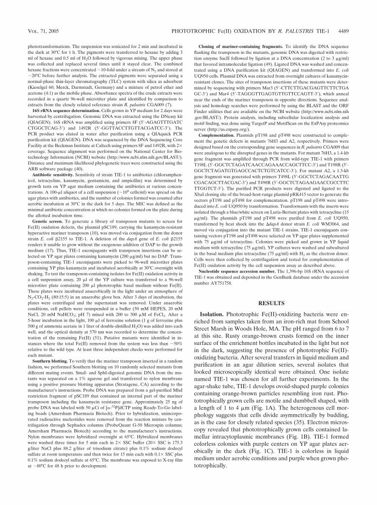
Isolation. Phototrophic Fe(II)-oxidizing bacteria were en-riched from samples taken from an iron-rich mat from SchoolStreet Marsh in Woods Hole, MA. The pH ranged from 6 to 7at this site. Rusty orange-brown crusts formed on the innersurface of the enrichment bottles incubated in the light but notin the dark, suggesting the presence of phototrophic Fe(II)-oxidizing bacteria. After several transfers in liquid medium andpurification in an agar dilution series, several isolates thatlooked microscopically identical were obtained. One isolatenamed TIE-1 was chosen for all further experiments. In theagar-shake tube, TIE-1 develops ovoid-shaped purple coloniescontaining orange-brown particles resembling iron rust. Pho-totrophically grown cells are motile and dumbbell shaped, witha length of 1 to 4 �m (Fig. 1A). The heterogenous cell mor-phology suggests that cells divide asymmetrically by budding,as is the case for closely related species (35). Electron micros-copy revealed that phototrophically grown cells contained la-mellar intracytoplasmic membranes (Fig. 1B). TIE-1 formedcolorless colonies with purple centers on YP agar plates aer-obically in the dark (Fig. 1C). TIE-1 is colorless in liquidmedium under aerobic conditions and purple when grown pho-totrophically.
VOL. 71, 2005 PHOTOTROPHIC Fe(II) OXIDATION BY R. PALUSTRIS TIE-1 4489
Phylogeny. The 16S rRNA sequence was compared with thesequences from representative species of its close relativesalong with the phototrophic Fe(II)-oxidizing bacteria that havebeen isolated so far (Fig. 1D). The 16S rRNA sequence ofTIE-1 shares an identity of 98.9% and 99% to that of Rhodo-pseudomonas palustris ATCC 17001T and strain CGA009, re-spectively, belonging to the �-subdivision of the Proteobacteria,affiliated closely with the nitrogen-fixing phototrophic rhizobia.TIE-1 clusters differently from the other known purple non-sulfur Fe(II)-oxidizing phototrophs, such as Rhodobacter sp.strain SW2 and Rhodovulum sp. strains N1 and N2.
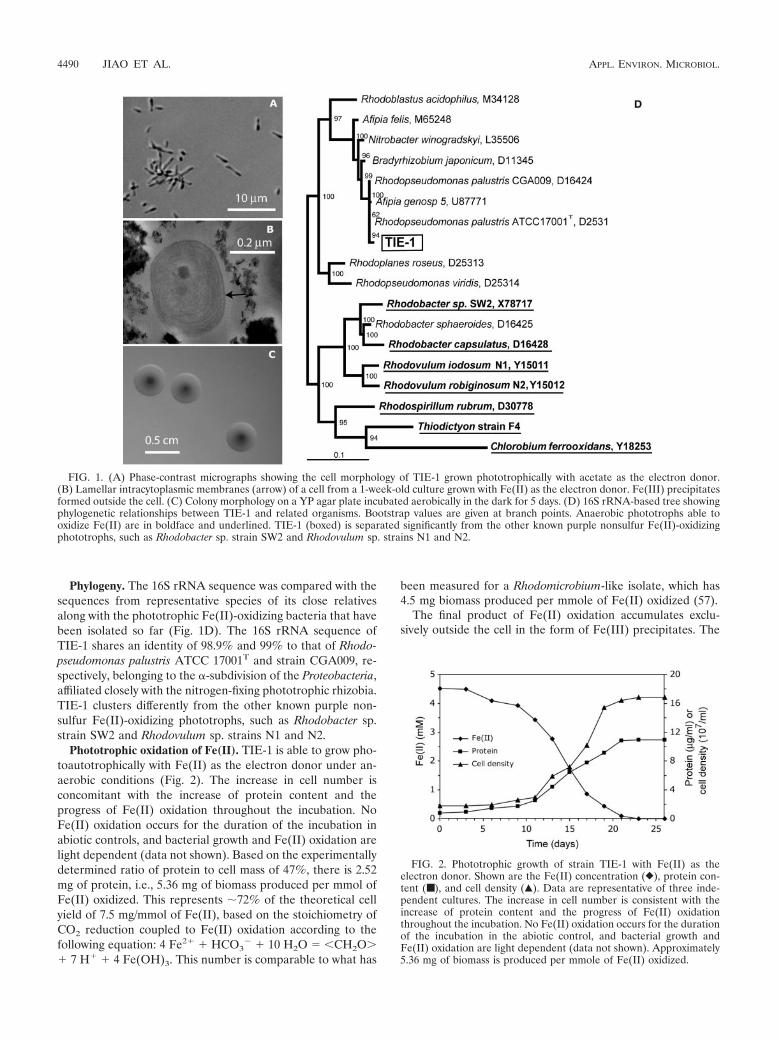
Phototrophic oxidation of Fe(II). TIE-1 is able to grow pho-toautotrophically with Fe(II) as the electron donor under an-aerobic conditions (Fig. 2). The increase in cell number isconcomitant with the increase of protein content and theprogress of Fe(II) oxidation throughout the incubation. NoFe(II) oxidation occurs for the duration of the incubation inabiotic controls, and bacterial growth and Fe(II) oxidation arelight dependent (data not shown). Based on the experimentallydetermined ratio of protein to cell mass of 47%, there is 2.52mg of protein, i.e., 5.36 mg of biomass produced per mmol ofFe(II) oxidized. This represents �72% of the theoretical cellyield of 7.5 mg/mmol of Fe(II), based on the stoichiometry ofCO2 reduction coupled to Fe(II) oxidation according to thefollowing equation: 4 Fe2� � HCO3
� 10 H2O � CH2O�� 7 H� � 4 Fe(OH)3. This number is comparable to what has
been measured for a Rhodomicrobium-like isolate, which has4.5 mg biomass produced per mmole of Fe(II) oxidized (57).
The final product of Fe(II) oxidation accumulates exclu-sively outside the cell in the form of Fe(III) precipitates. The
FIG. 1. (A) Phase-contrast micrographs showing the cell morphology of TIE-1 grown phototrophically with acetate as the electron donor.(B) Lamellar intracytoplasmic membranes (arrow) of a cell from a 1-week-old culture grown with Fe(II) as the electron donor. Fe(III) precipitatesformed outside the cell. (C) Colony morphology on a YP agar plate incubated aerobically in the dark for 5 days. (D) 16S rRNA-based tree showingphylogenetic relationships between TIE-1 and related organisms. Bootstrap values are given at branch points. Anaerobic phototrophs able tooxidize Fe(II) are in boldface and underlined. TIE-1 (boxed) is separated significantly from the other known purple nonsulfur Fe(II)-oxidizingphototrophs, such as Rhodobacter sp. strain SW2 and Rhodovulum sp. strains N1 and N2.
FIG. 2. Phototrophic growth of strain TIE-1 with Fe(II) as theelectron donor. Shown are the Fe(II) concentration (}), protein con-tent (■ ), and cell density (Œ). Data are representative of three inde-pendent cultures. The increase in cell number is consistent with theincrease of protein content and the progress of Fe(II) oxidationthroughout the incubation. No Fe(II) oxidation occurs for the durationof the incubation in the abiotic control, and bacterial growth andFe(II) oxidation are light dependent (data not shown). Approximately5.36 mg of biomass is produced per mmole of Fe(II) oxidized.
4490 JIAO ET AL. APPL. ENVIRON. MICROBIOL.
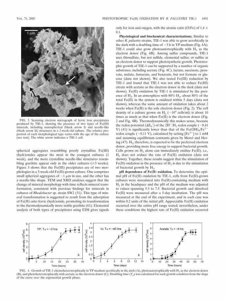
spherical aggregates resembling poorly crystalline Fe(III)(hydr)oxides appear the most in the youngest cultures (1week), and the more crystalline needle-like structures resem-bling goethite appear only in the older cultures (�3 weeks).Figure 3 shows that the Fe(III) precipitates are of two mor-phologies in a 3-week-old Fe(II)-grown culture. One comprisessmall spherical aggregates of �1 �m in size, and the other hasa needle-like shape. TEM and XRD analyses suggest that thechange of mineral morphology with time reflects mineral trans-formation, consistent with previous findings for minerals incultures of Rhodobacter sp. strain SW2 (31). This type of min-eral transformation is suggested to result from the adsorptionof Fe(II) onto ferric (hydr)oxide, promoting its transformationto the thermodynamically more stable goethite (61). Elementalanalysis of both types of precipitates using EDS gives signals
only for iron and oxygen, with the atomic ratio (O/Fe) of 1.4 �0.1.
Physiological and biochemical characterizations. Similar toother R. palustris strains, TIE-1 was able to grow aerobically inthe dark with a doubling time of �3 h in YP medium (Fig. 4A).TIE-1 could also grow photoautotrophically with H2 as theelectron donor (Fig. 4B). Among sulfur compounds, TIE-1uses thiosulfate, but not sulfide, elemental sulfur, or sulfite asan electron donor to support photosynthetic growth. Phototro-phic growth of TIE-1 can be supported by a number of organicsubstrates, including acetate (Fig. 4C), lactate, succinate, pyru-vate, malate, fumarate, and benzoate, but not formate or glu-cose (data not shown). We also tested Fe(III) reduction byTIE-1 and found that TIE-1 was not able to reduce Fe(III)citrate with acetate as the electron donor in the dark (data notshown). Fe(II) oxidation by TIE-1 is stimulated by the pres-ence of H2. In an atmosphere with 80% H2, about 80% of thetotal Fe(II) in the system is oxidized within 5 days (data notshown), whereas the same amount of oxidation takes about 2weeks when Fe(II) is the sole electron donor (Fig. 2). The celldensity of a culture grown on H2 (�109 cells/ml) is about 10times as much as that when Fe(II) is the electron donor (Fig.2 and Fig. 4B). Thermodynamically this makes sense, becausethe redox potential (�E0�) of the 2H�/H2 redox couple (0.41V) (41) is significantly lower than that of the Fe(OH)3/Fe2�
redox couple (0.11 V), calculated by setting [Fe2�] to 1 mMand assuming equilibrium constants given by Morel and Her-ing (47). H2, therefore, is expected to be the preferred electrondonor, providing more free energy to support bacterial growth.Cells grown on H2 alone can immediately oxidize Fe(II), i.e.,H2 does not reduce the rate of Fe(II) oxidation (data notshown). Together, these results suggest that the stimulation ofFe(II) oxidation in the presence of H2 is due to the stimulationof bacterial growth by H2.
pH dependence of Fe(II) oxidation. To determine the opti-mal pH of Fe(II) oxidation by TIE-1, cells from Fe(II)-growncultures were inoculated into Fe(II)-containing medium withH2 in the headspace and the pH of the medium was adjustedto values spanning 5.5 to 7.5. Bacterial growth and dissolvedFe(II) were measured after a 5-day incubation. The pH wasmeasured at the end of the experiment, and in each case waswithin 0.2 units of the initial pH. Appreciable Fe(II) oxidationoccurred over the entire pH range tested; nevertheless, underthese conditions the highest rate of Fe(II) oxidation occurred
FIG. 3. Scanning electron micrograph of ferric iron precipitatesproduced by TIE-1, showing the presence of two types of Fe(III)minerals, including nanospherical (black arrow I) and needle-like(black arrow II) structures in a 2-week-old culture. The relative pro-portion of each morphological type varies with the age of the culture(see text). The white arrow indicates a TIE-1 cell.
FIG. 4. Growth of TIE-1 chemoheterotrophically in YP medium aerobically in the dark (A), photoautotrophically with H2 as the electron donor(B), and photoheterotrophically with acetate as the electron donor (C). Doubling time (Td) was calculated for each growth condition from the slopeof the curve over the exponential growth phase.
VOL. 71, 2005 PHOTOTROPHIC Fe(II) OXIDATION BY R. PALUSTRIS TIE-1 4491

between pH 6.5 and 6.9 (Fig. 5A), similar to the pH rangemeasured for other phototrophic Fe(II)-oxidizing bacteria (20,28). In contrast, the amount of bacterial growth (representedby the protein content) was maximal and not appreciably dif-ferent within the pH range 6.5 to 7.5. This suggests that Fe(II)oxidation did not significantly contribute to cell growth in theseexperiments (i.e., cells were growing on H2). We also observedthat the mineral product of Fe(II) oxidation was pH depen-dent, with poorly crystalline ferric (hydr)oxide and goethitedominating at lower pH and magnetite at pH of �7.2 � 0.2(Fig. 3 and 5B). The same pH trend in iron mineralogy wasobserved for cultures grown on Fe(II) alone (data not shown).
Because the total amount of dissolved Fe(II) remained inexcess of 1.5 mM at a pH of �6.9, it seems unlikely that theamount of Fe(II) oxidation measured over this time period waslimited by soluble Fe(II). However, the possibility exists thatchanges in concentration of a minor species of Fe(II) mighthave controlled the rate of Fe(II) oxidation. Considering this,we used MINEQL� to calculate the equilibrium concentra-tions of soluble Fe(II) species over this pH range in the contextof the composition of our medium. As pH increases from 6.5 to7.5, MINEQL� predicts that Fe2� is the major Fe(II) speciesand only decreases by 0.4 mM over this pH range. Three minorFe(II) species, including Fe(OH)3
, Fe(OH)2(aq), andFe(OH)�, increase in concentration 1,000-, 80-, and 10-fold,respectively. In the Discussion section, we consider howchanges in these species’ behaviors with increasing pH mightaffect the overall Fe(II) oxidation rate.
Pigment characterization. The absorption spectrum ofwhole cells of TIE-1 grown with acetate as the electron donorshows three major peaks at 590, 805, and 871 nm, similar tothat of R. palustris CGA009, indicating the presence of bacte-riochlorophyll a. The absorption spectrum obtained fromTIE-1 grown with Fe(II) as the electron donor is similar andshows approximately the same peaks of absorbance as cellsgrown on acetate, except that the peak at 871 nm is wider for
the Fe(II)-grown culture (Fig. 6A). The absorption spectra ofcarotenoid extracts (Fig. 6B) and thin-layer chromatographyseparations (data not shown) obtained from TIE-1 andCGA009 grown on acetate also look identical. The absorption
FIG. 5. (A) pH dependence of phototrophic Fe(II) oxidation in the presence of H2: dissolved Fe(II) concentration in the supernatant at theend of the experiment (}) and protein content after the incubation (�). The optimal pH for Fe(II) oxidation occurs from 6.5 to 6.9. The totalamount of Fe(II) oxidized decreases significantly at pH higher than 7.0, although the amount of bacterial growth represented by the protein contentremains constant compared to that at pH 6.8. (B) X-ray diffractograms of the product of Fe(II) oxidation by TIE-1 shows goethite and magnetiteformation at media pHs of 6.8 and 7.5, respectively. For comparison, reference diffractograms of goethite and magnetite mineral standards areincluded.
FIG. 6. Pigment analysis. (A) Absorption spectra of cell suspen-sions of strain TIE-1 grown with acetate (a) and Fe(II) (b) as theelectron donor and of strain CGA009 grown with acetate as the elec-tron donor (c) under phototrophic conditions. All curves look similarand show major absorption peaks at 590, 805, and 871 nm, indicatingthe presence of bacteriochlorophyll a. (B) Absorption spectra of ca-rotenoid extraction from strain TIE-1 (a) and strain CGA009 (b)grown with acetate as the electron donor, showing major peaks thatoverlapped intensively between 400 and 600 nm, indicating the pres-ence of the normal spirilloxanthin series.
4492 JIAO ET AL. APPL. ENVIRON. MICROBIOL.

spectra of carotenoid extracts show major peaks overlappingbetween 400 and 600 nm, suggesting the presence of carote-noids commonly present in R. palustris, including spheroidene(450, 482, and 514 nm), okenone (521 nm), lycopene, andrhodopin (463, 490, and 524 nm) (43).
Characterization of antibiotic sensitivity. Growth of �107
cells on YP solid or liquid medium is inhibited completely bychloramphenicol (300 �g/ml), tetracycline (75 �g/ml), kana-mycin (100 �g/ml), gentamicin (300 �g/ml), and ampicillin (50�g/ml). These results indicate that it should be possible toselect for the acquisition of these antibiotic resistance markers,which will facilitate genetic manipulation.
Transposon mutagenesis and mutant characterization. Weused transposon mutagenesis to identify genes involved in pho-totrophic Fe(II) oxidation in strain TIE-1. The frequency oftransposon insertion obtained for TIE-1 is �105 with themariner transposon. Southern blot analysis of 10 randomlyselected isolates derived from independent transpositionevents indicated that the transposon integrates as a singleevent in random locations (data not shown).
We performed a limited screen of �12,000 transposon in-sertion mutants for defects in phototrophic Fe(II) oxidationusing a cell suspension assay. Based on the assumptions thatstrain TIE-1 has the same number of genes as strain CGA009and that the transposition is purely random, this screen is�88% saturated assuming a Poisson distribution (26). Four-teen mutants were identified as being defective in Fe(II) oxi-dation: eight mutants had general photosynthetic growth de-fects, and the other six were specifically defective in Fe(II)oxidation. BLAST analysis performed on DNA sequencesflanking the mariner insertions revealed that the sequenceflanking the transposon has significant similarity to sequencesfrom the genome of R. palustris strain CGA009 (38) in allcases.
The eight mutants exhibiting general growth defects grew atleast 50% less on acetate or H2 compared to the wild type(data not shown). Two of these mutants were disrupted ingenes that are homologs of bchZ and bchX, known to encodeproteins involved in bacteriochlorophyll synthesis (8). It is notsurprising that our screen picked up components of the generalphotosynthetic electron transport system, given the large vari-ance in cell density in the step prior to the cell suspensionassay. Two mutants, however, were identified that are specifi-cally defective in Fe(II) oxidation: 76H3 and A2. 76H3 is arepresentative of five mutants that have transposon insertionsat different locations in the same gene, whereas A2 was onlyisolated once. Both mutants exhibit normal photosyntheticgrowth in minimal medium with H2 as the electron donor, buttheir ability to oxidize Fe(II) is less than 10% of the wild type(Fig. 7A and B). Complementation of the disrupted genesindicates that their expression is necessary and sufficient torestore nearly wild-type levels of activity, suggesting that Fe(II)oxidation defects are not caused by the downstream genes (Fig.7C and D).
Because the sequence fragments from TIE-1 flanking thetransposon insertions were highly similar to sequences fromstrain CGA009, we designed primers based on the CGA009genome to sequence the regions surrounding the transposoninsertions in 76H3 and A2 (Fig. 7D). Both regions containedhomologs of genes found in the same order in CGA009. Mu-
tant 76H3 has a transposon insertion in a gene that shares 99%identity over the entire gene sequence (791 bp) to geneRPA0198 in R. palustris CGA009, which encodes a putativeintegral membrane protein. The BLAST search predicts thatthe protein encoded by this gene shares 100% identity to apossible transport protein in R. palustris CGA009, 85% identityto a probable ABC transport permease in Bradyrhizobium ja-ponicum, and 60% identity to a hypothetical transmembraneprotein from Magnetospirillum sp. strain MS-1. It is predictedto encode a cytoplasmic membrane protein with six internalhelices, based on sequence analysis with the Psort program(http://www.psort.org/). No known motifs could be identified inthis protein by the MotifScan program (http://myhits.isb-sib.ch/cgi-bin/motif_scan). Based on the annotation of the CGA009genome, the upstream genes encode a putative ABC trans-porter permease (RPA0197) and a putative ABC transporterATP-binding protein (RPA0196). The downstream gene(RPA0199) encodes a putative phosphinothricin acetyltrans-ferase.
Mutant A2 has a transposon insertion in a gene that shares99% identity over the entire gene sequence (995 bp) to geneRPA0498 in R. palustris CGA009, which is annotated as a cobSgene. The translated protein sequence is 100% identical to aputative CobS in strain CGA009, 93% identical to a putativeCobS from Bradyrhizobium japonicum, 80% identical to a well-studied CobS from Pseudomonas denitrificans, and 76% and71% identical to MoxR-like ATPases from Rhodospirillumrubrum and Rhodobacter sphaeroides, respectively. Studies ofCobS function in P. denitrificans have shown that CobS is acobaltochelatase—a cytoplasmic protein involved in cobalt in-sertion into porphyrin rings (16). MoxR-like ATPases belongto the AAA superfamily of proteins with associated ATPaseactivity (30). Not surprisingly, members of the MoxR familyfunction as chaperones/chelatases in the assembly of specificmetal-containing enzymatic complexes. Based on the annota-tion of the CGA009 genome, the genes downstream appear toencode an N-acetylglutamate synthase and related acetyltrans-ferases (RPA0497), a CobT homolog (RPA0496), and a con-served hypothetical protein (RPA0495).
DISCUSSION
We have isolated and characterized a genetically tractableFe(II)-oxidizing bacterium, Rhodopseudomonas palustris strainTIE-1. Two Fe(II)-oxidizing strains of R. palustris have beenreported previously (20, 28, 57). Based strictly on morpholog-ical characteristics, an Fe(II)-oxidizing R. palustris-like strainwas first isolated from an iron-rich ditch in Germany (20). Thisisolate did not oxidize iron completely and ceased to grow at agrayish green intermediate oxidation state; it was not main-tained in culture collections (F. Widdel, personal communica-tion). Although the type strain R. palustris DSM123T was foundto be incapable of Fe(II) oxidation by Ehrenreich and Widdel(20), Heising and Schink (28) claimed it was capable of Fe(II)oxidation (28). In this study, we tested the Fe(II) oxidationcapacity of the R. palustris CGA009 strain, whose genome hasbeen sequenced. Strain CGA009 is not able to grow photoau-totrophically on H2 or Fe(II); however, photoheterotrophicallygrown cells (using acetate as the electron donor) can slowlyoxidize Fe(II) in the cell suspension assay at a similar rate to
VOL. 71, 2005 PHOTOTROPHIC Fe(II) OXIDATION BY R. PALUSTRIS TIE-1 4493

that achieved by acetate-grown cell suspensions of strain TIE-1(unpublished data). In contrast, strain TIE-1 can grow photo-autotrophically with H2 and Fe(II), and H2-grown cell suspen-sions readily oxidize Fe(II) at rates much higher than thoseachieved by acetate-grown cell suspensions.
Rates and products of phototrophic Fe(II) oxidation byTIE-1 are pH dependent. The amount of Fe(II) oxidation issignificantly less at pHs higher than 7.0, compared to thatoxidized at optimal pH, but this is not due to a growth defect.Previous interpretations of a similar result by Heising andSchink suggested that this might be due to the lower solubilityof Fe(II) at higher pH (28). Although our measurements in-dicate that the total amount of dissolved Fe(II) in our systemdoes not appreciably decrease as pH increases, more subtlespecies dynamics may control the bioavailability of Fe(II) un-der these conditions. The transformation from poorly crystal-line ferric (hydr)oxides to more crystalline ferric (hydr)oxidesis promoted by the adsorption of Fe(II) species onto the solidphase (61). Given that the point of zero charge for ferric(hydr)oxide likely occurs at the upper end of our pH spectrum(3), we would expect cationic Fe(II) species to adsorb to theferric (hydr)oxides as the point of zero charge is reached andthen exceeded with increasing pH. Interestingly, the abiotic
oxidation rate of Fe(OH)� in both freshwater and seawaterhas been found to be 107 times greater than that of Fe2� (45).Moreover, Fe(OH)2(aq) is thought to be the most readily oxi-dized form of Fe(II) over a pH range of 6 to 8 and the rate-limiting step for the oxidation of Fe2� under these conditions(45). Assuming Fe(OH)2(aq) or Fe(OH)� is the preferred spe-cies taken up or bound by TIE-1, the decrease in Fe(II) oxi-dation measured coincident with magnetite formation in ourmedium could be explained if sorption of these species byferric (hydr)oxides out-competed their sorption/uptake by thecell. Alternatively, it is possible that the decrease in Fe(II)oxidation is due to the inactivation of a biomolecule involved inFe(II) binding, uptake, and/or oxidation at high pH.
Independent of whether magnetite formation affects the bio-availability of Fe(II), it is noteworthy that magnetite formationcan be associated with this type of metabolism. Previous stud-ies with other Fe(II)-oxidizing phototrophs only found variousforms of ferric oxides (e.g., goethite and lepidocrocite) to ac-cumulate in the culture medium over time; magnetite wasnever observed (14, 31, 52). In contrast, magnetite formationfollowing an intermediate state of green rust was reported forthe nitrate-dependent Fe(II)-oxidizing bacterium Dechloro-soma suillum strain PS (9). Magnetite formation has been
FIG. 7. Mutants 76H3 and A2 are specifically defective in Fe(II) oxidation. (A) Normal growth of mutants 76H3 and A2 with H2 as the electrondonor. Data are representative of two independent cultures. (B) Defects in phototrophic Fe(II) oxidation for mutants 76H3 and A2 compared towild type. Growth was stimulated with H2 present in the headspace initially. Data are representative of duplicate cultures. (C) Mutants 76H3 andA2 carrying plasmids pT198 and pT498, respectively, show 80% Fe(II) oxidation compared to the wild type in the cell suspension assay.(D) Organization of the genomic regions surrounding the mutated genes in mutants 76H3 and A2. The black arrows indicate the disrupted genes,and the transposon insertion sites are marked by the open triangles. The numbers provided below the open reading frames (all arrows) areconsistent with the numbers given for the identical regions from the CGA009 genome.
4494 JIAO ET AL. APPL. ENVIRON. MICROBIOL.

reported for dissimilatory iron-reducing bacteria (DIRB), withmagnetite formed through the reduction of ferric oxide (39).Magnetite formation by TIE-1 is unlikely to be formed throughthe re-reduction of Fe(III), however, based on the evidencethat TIE-1 is unable to reduce Fe(III) citrate with acetate asthe electron donor in the dark. Considering the differences inthe chemistry of the medium used to grow these bacteria, thesimplest way to account for magnetite formation in some, butnot all, of these cases is that differences in medium chemistrycontrolled the amount and speed of Fe(II) adsorption ontoferric (hydr)oxides (61). Because pH 7.5 is a reasonable pHvalue for ancient seawater (25), it is possible that the primarymagnetite found in BIFs may record the activity of Fe(II)-oxidizing phototrophs. However, this interpretation does notexclude the possibility that magnetite in BIFs may also havebeen facilitated by DIRB or abiotic processes (48).
To begin to identify genes involved in phototrophic Fe(II)oxidation, we first needed to develop an efficient method forgenerating random chromosomal insertions in TIE-1. Trans-poson mutagenesis has been shown to work in the Rhodospi-rillaceae family, but with mixed success (18). For example,transposition by Tn5 derivatives was found to transpose in R.capsulatus and R. rubrum with frequencies of 104 to 105 (24,33); however, for R. palustris strain CGA009 and strainEPT100, Tn5 derivatives were either not successful or veryinefficient (21). The fact that the hyperactive mariner transpo-son used in this study transposes randomly and at high fre-quencies in TIE-1 suggests that this type of transposon may bean effective mutagenic tool for other R. palustris strains.
Out of a total of 12,000 mutants screened for their ability tooxidize Fe(II) in the cell suspension assay, only 6 were identi-fied as being specifically defective in Fe(II) oxidation and only2 genes were implicated in this process. It is intriguing thatboth of these genes are also present in R. palustris strainCGA009, although this organism cannot grow on Fe(II). Giventhat photoheterotrophically grown cells of CGA009 can oxi-dize Fe(II) in the cell suspension assay comparably to TIE-1when grown under the same conditions, this indicates thatFe(II) oxidation can be decoupled from growth. However, ourcell suspension assay did not decouple Fe(II) oxidation fromthe photosynthetic apparatus, as no Fe(II) oxidation occurredin the dark. It will be interesting to learn what allows TIE-1 butnot CGA009 to conserve energy from Fe(II) oxidation forgrowth. It is possible that essential genes for this process aremissing from CGA009, mutated, or not expressed. To resolvethis, a screen could be performed to identify TIE-1 mutantsthat are incapable of phototrophic growth on Fe(II), orCGA009 could be complemented for growth on Fe(II) throughprovision of genes from TIE-1 (13).
Although much remains to be learned about how TIE-1oxidizes and grows on Fe(II), the two mutants identified in thisstudy provide important new information. Strain A2 contains adisruption in a homolog of a cobalt chelatase (CobS). Becausethe structures of cobaltochelatases and ferrochelatases [whichinsert Fe(II) into porphyrin rings] are similar, it has beensuggested that they have similar enzymatic activities (15, 50).While it is possible that the phenotype of A2 might be due tothe disruption of an enzyme that inserts Fe(II) into a protein ora cofactor that is involved in Fe(II) oxidation, this seems un-likely, because cobatochelatases and ferrochelatases are typi-
cally different at the amino acid level (15). We hypothesize,instead, that a protein involved in Fe(II) oxidation requirescobalamin as cofactor indirectly. In contrast, strain 76H3 isdisrupted in a gene that appears to encode a component of anABC transport system that is located in the cytoplasmic mem-brane. While a variety of things could be transported by thissystem, whatever is being transported [e.g., the Fe(II) oxidaseor a protein required for its assembly] likely resides at leastmomentarily in the periplasm. This raises the question ofwhere Fe(II) is oxidized in the cell. Because Fe(II) is known toenter the periplasmic space of gram-negative bacteria throughporins in the outer membrane, it is conceivable that Fe(II)could be oxidized in this compartment; alternatively, the Fe(II)oxidase could reside in the outer membrane and face theexternal environment, as has been inferred for Fe(II)-oxidizingacidophilic bacteria (23, 59). Determining what catalyzesFe(II) oxidation and where it is localized are the most impor-tant next steps in our investigation of the molecular basis ofphototrophic Fe(II) oxidation. The isolation of the geneticallytractable strain TIE-1 will enable these studies.
ACKNOWLEDGMENTS
We are indebted to Randall E. Mielke for TEM imaging and Eliz-abeth A. Ottesen for help in constructing the 16S phylogenetic tree.We thank Arash Komeili and Jeff Gralnick for guidance throughoutthis study and all the Newman lab members for helpful discussions.
This work was supported by a grant from the Packard Foundation toD.K.N. and a postdoctoral fellowship from the German ResearchFoundation to A.K.
REFERENCES
1. Appia-Ayme, C., A. Bengrine, C. Cavazza, M. T. Giudici-Orticoni, M. Brus-chi, M. Chippaux, and V. Bonnefoy. 1998. Characterization and expression ofthe co-transcribed cyc1 and cyc2 genes encoding the cytochrome c4 (c552) anda high-molecular-mass cytochrome c from Thiobacillus ferrooxidansATCC33020. FEMS Microbiol. Lett. 167:171–177.
2. Appia-Ayme, C., N. Guiliani, J. Ratouchniak, and V. Bonnefoy. 1999. Char-acterization of an operon encoding two c-type cytochromes, an aa3-typecytochrome oxidase, and rusticyanin in Thiobacillus ferrooxidans ATCC33020. Appl. Environ. Microbiol. 65:4781–4787.
3. Baltpurvins, K. A., R. C. Burns, G. A. Lawrance, and A. D. Stuart. 1997.Effect of Ca2�, Mg2�, and anion type on the aging of iron(III) hydroxideprecipitates. Environ. Sci. Technol. 31:1024–1032.
4. Benz, M., A. Brune, and B. Schink. 1998. Anaerobic and aerobic oxidation offerrous iron at neutral pH by chemoheterotrophic nitrate-reducing bacteria.Arch. Microbiol. 169:159–165.
5. Blake, R. C., E. A. Shute, J. Waskovsky, and A. P. J. Harrison. 1992.Respiratory components in acidophilic bacteria that respire iron. Geomicro-biol. J. 10:173–192.
6. Bradford, M. M. 1976. Rapid and sensitive method for quantitation ofmicrogram quantities of protein utilizing principle of protein-dye binding.Anal. Biochem. 72:248–254.
7. Britton, G. 1995. Structure and properties of carotenoids in relation tofunction. FASEB J. 9:1551–1558.
8. Burke, D. H., M. Alberti, and J. E. Hearst. 1993. The Rhodobacter capsulatuschlorin reductase-encoding locus, bchA, consists of three genes, bchX, bchY,and bchZ. J. Bacteriol. 175:2407–2413.
9. Chaudhuri, S. K., J. G. Lack, and J. D. Coates. 2001. Biogenic magnetiteformation through anaerobic biooxidation of Fe(II). Appl. Environ. Micro-biol. 67:2844–2848.
10. Chiang, S. L., and E. J. Rubin. 2002. Construction of a mariner-basedtransposon for epitope-tagging and genomic targeting. Gene 296:179–185.
11. Cobley, J. G., and B. A. Haddock. 1975. Respiratory chain of Thiobacillusferrooxidans—reduction of cytochromes by Fe2� and preliminary character-ization of rusticyanin, a novel blue copper protein. FEBS Lett. 60:29–33.
12. Cox, J. C., and D. H. Boxer. 1978. Purification and some properties ofrusticyanin, a blue copper protein involved in iron(II) oxidation from Thio-bacillus ferrooxidans. Biochem. J. 174:497–502.
13. Croal, L. R., J. A. Gralnick, D. Malasarn, and D. K. Newman. 2004. Thegenetics of geochemistry. Annu. Rev. Genet. 38:175–202.
14. Croal, L. R., C. M. Johnson, B. L. Beard, and D. K. Newman. 2004. Ironisotope fractionation by Fe(II)-oxidizing photoautotrophic bacteria.Geochim. Cosmochim. Acta 68:1227–1242.
VOL. 71, 2005 PHOTOTROPHIC Fe(II) OXIDATION BY R. PALUSTRIS TIE-1 4495

15. Dailey, H. A., T. A. Dailey, C. K. Wu, A. E. Medlock, K. F. Wang, J. P. Rose,and B. C. Wang. 2000. Ferrochelatase at the millennium: structures, mech-anisms and 2Fe-2S clusters. Cell. Mol. Life Sci. 57:1909–1926.
16. Debussche, L., M. Couder, D. Thibaut, B. Cameron, J. Crouzet, and F.Blanche. 1992. Assay, purification, and characterization of cobaltochelatase,a unique complex enzyme catalyzing cobalt insertion in hydrogenobyrinicacid a,c-diamide during coenzyme B12 biosynthesis in Pseudomonas denitri-ficans. J. Bacteriol. 174:7445–7451.
17. Dehio, C., and M. Meyer. 1997. Maintenance of broad-host-range incompat-ibility group P and group Q plasmids and transposition of Tn5 in Bartonellahenselae following conjugal plasmid transfer from Escherichia coli. J. Bacte-riol. 179:538–540.
18. Donohue, T. J., and S. Kaplan. 1991. Genetic techniques in Rhodospiril-laceae. Methods Enzymol. 204:459–485.
19. Edwards, K. J., P. L. Bond, T. M. Gihring, and J. F. Banfield. 2000. Anarchaeal iron-oxidizing extreme acidophile important in acid mine drainage.Science 287:1796–1799.
20. Ehrenreich, A., and F. Widdel. 1994. Anaerobic oxidation of ferrous iron bypurple bacteria, a new-type of phototrophic metabolism. Appl. Environ.Microbiol. 60:4517–4526.
21. Elder, D. J. E., P. Morgan, and D. J. Kelly. 1993. Transposon Tn5 mutagen-esis in Rhodopseudomonas palustris. FEMS Microbiol. Lett. 111:23–30.
22. Emerson, D., and C. Moyer. 1997. Isolation and characterization of noveliron-oxidizing bacteria that grow at circumneutral pH. Appl. Environ. Mi-crobiol. 63:4784–4792.
23. Fukumori, Y., T. Yano, A. Sato, and T. Yamanaka. 1988. Fe(II)-oxidizingenzyme purified from Thiobacillus ferrooxidans. FEMS Microbiol. Lett. 50:169–172.
24. Ghosh, R., D. J. E. Elder, R. Saegesser, D. J. Kelly, and R. Bachofen. 1994.An improved procedure and new vectors for transposon Tn5 mutagenesis ofthe phototrophic bacterium Rhodospirillum rubrum. Gene 150:97–100.
25. Grotzinger, J. P., and J. F. Kasting. 1993. New constraints on precambrianocean composition. J. Geol. 101:235–243.
26. Hawley, R. S., and M. Y. Walker. 2003. Advanced genetic analysis. BlackwellScience Ltd., Malden, Mass.
27. Heising, S., L. Richter, W. Ludwig, and B. Schink. 1999. Chlorobium fer-rooxidans sp. nov., a phototrophic green sulfur bacterium that oxidizes fer-rous iron in coculture with a “Geospirillum” sp. strain. Arch. Microbiol.172:116–124.
28. Heising, S., and B. Schink. 1998. Phototrophic oxidation of ferrous iron bya Rhodomicrobium vannielii strain. Microbiol. UK 144:2263–2269.
29. Ingledew, W. J., and J. G. Cobley. 1980. A potentiometric and kinetic studyon the respiratory chain of ferrous-iron-grown Thiobacillus ferrooxidans. Bio-chim. Biophys. Acta 590:141–158.
30. Iyer, L. M., D. D. Leipe, E. V. Koonin, and L. Aravind. 2004. Evolutionaryhistory and higher order classification of AAA plus ATPases. J. Struct. Biol.146:11–31.
31. Kappler, A., and D. K. Newman. 2004. Formation of Fe(III) minerals byFe(II)-oxidizing photoautotrophic bacteria. Geochim. Cosmochim. Acta 68:1217–1226.
32. Keen, N. T., S. Tamaki, D. Kobayashi, and D. Trollinger. 1988. Improvedbroad-host-range plasmids for DNA cloning in gram-negative bacteria. Gene70:191–197.
33. Kelly, D. J., D. J. Richardson, S. J. Ferguson, and J. B. Jackson. 1988.Isolation of transposon Tn5 insertion mutants of Rhodobacter capsulatusunable to reduce trimethylamine-N-oxide and dimethylsulfoxide. Arch. Mi-crobiol. 150:138–144.
34. Kim, M. K., and C. S. Harwood. 1991. Regulation of benzoate-CoA ligase inRhodopseudomonas palustris. FEMS Microbiol. Lett. 83:199–203.
35. Kompantseva, E. I., E. E. Panteleeva, E. V. Ariskina, A. M. Lysenko, J. F.Imhoff, and V. M. Gorlenko. 1996. Phylogenetic relationships among bud-ding purple bacteria of the genus Rhodopseudomonas. Microbiology 65:344–351.
36. Konhauser, K. O., T. Hamade, R. Raiswell, R. C. Morris, F. G. Ferris, G.Southam, and D. E. Canfield. 2002. Could bacteria have formed the pre-cambrian banded iron formations? Geology 30:1079–1082.
37. Lack, J. G., S. K. Chaudhuri, R. Chakraborty, L. A. Achenbach, and J. D.Coates. 2002. Anaerobic biooxidation of Fe(II) by Dechlorosoma suillum.Microb. Ecol. 43:424–431.
38. Larimer, F. W., P. Chain, L. Hauser, J. Lamerdin, S. Malfatti, L. Do, M. L.Land, D. A. Pelletier, J. T. Beatty, A. S. Lang, F. R. Tabita, J. L. Gibson, T. E.
Hanson, C. Bobst, J. Torres, C. Peres, F. H. Harrison, J. Gibson, and C. S.Harwood. 2004. Complete genome sequence of the metabolically versatilephotosynthetic bacterium Rhodopseudomonas palustris. Nat. Biotechnol. 22:55–61.
39. Lovley, D. R., J. F. Stolz, G. L. Nord, and E. J. P. Phillips. 1987. Anaerobicproduction of magnetite by a dissimilatory iron- reducing microorganism.Nature 330:252–254.
40. Ludwig, W., O. Strunk, R. Westram, L. Richter, H. Meier, Yadhukumar, A.Buchner, T. Lai, S. Steppi, G. Jobb, W. Forster, I. Brettske, S. Gerber, A. W.Ginhart, O. Gross, S. Grumann, S. Hermann, R. Jost, A. Konig, T. Liss, R.Lussmann, M. May, B. Nonhoff, B. Reichel, R. Strehlow, A. Stamatakis, N.Stuckmann, A. Vilbig, M. Lenke, T. Ludwig, A. Bode, and K. H. Schleifer.2004. ARB: a software environment for sequence data. Nucleic Acids Res.32:1363–1371.
41. Madigan, M. T., J. M. Martinko, and J. Parker. 2002. Brock biology ofmicroorganisms, 10th ed., p. A-4. Prentice Hall, Upper Saddle River, N.J.
42. Mansch, R., and W. Sand. 1992. Acid-stable cytochromes in ferrous ironoxidizing cell-free preparations from Thiobacillus ferrooxidans. FEMS Mi-crobiol. Lett. 92:83–88.
43. Mehrabi, S., U. M. Ekanemesang, F. O. Aikhionbare, K. S. Kimbro, and J.Bender. 2001. Identification and characterization of Rhodopseudomonasspp., a purple, non-sulfur bacterium from microbial mats. Biomol. Engin.18:49–56.
44. Miller, J. H. 1992. A short course in bacterial genetics. Cold Spring HarborLaboratory Press, Cold Spring Harbor, N.Y.
45. Millero, F. J. 1985. The effect of ionic interactions on the oxidation of metalsin natural waters. Geochim. Cosmochim. Acta 49:547–553.
46. Mjoji, N., and C. F. Kulpa. 1988. Identification of a unique outer membraneprotein required for Fe(II) oxidation in Thiobacillus ferrooxidans, p. 89–102.In P. R. Norris and D. P. Kelly (ed.), Biohydrometallurgy. STL, Kew Surrey,United Kingdom.
47. Morel, F., and J. G. Hering. 1993. Principles and applications of aquaticchemistry. Wiley-Interscience, New York, N.Y.
48. Morris, R. C. 1993. Genetic modeling for banded iron-formation of theHamersley group, Pilbara craton, Western Australia. Precambrian Res. 60:243–286.
49. Raponi, M., I. W. Dawes, and G. M. Arndt. 2000. Characterization of flank-ing sequences using long inverse PCR. BioTechniques 28:838–842.
50. Roth, J. R., J. G. Lawrence, and T. A. Bobik. 1996. Cobalamin (coenzymeB-12): synthesis and biological significance. Annu. Rev. Microbiol. 50:137–181.
51. Stookey, L. L. 1970. Ferrozine—a new spectrophotometric reagent for iron.Anal. Chem. 42:779–781.
52. Straub, K. L., M. Benz, and B. Schink. 2001. Iron metabolism in anoxicenvironments at near neutral pH. FEMS Microbiol. Ecol. 34:181–186.
53. Straub, K. L., F. A. Rainey, and F. Widdel. 1999. Rhodovulum iodosum sp.nov. and Rhodovulum robiginosum sp. nov., two new marine phototrophicferrous-iron-oxidizing purple bacteria. Int. J. Syst. Bacteriol. 49:729–735.
54. Suter, D., C. Siffert, B. Sulzberger, and W. Stumm. 1988. Catalytic dissolu-tion of iron(III) (hydr)oxides by oxalic acid in the presence of Fe(II). Natur-wissenschaften 75:571–573.
55. Temple, K. L., and A. R. Colmer. 1951. The autotrophic oxidation of iron bya new bacterium, Thiobacillus ferrooxidans. J. Bacteriol. 62:605–611.
56. Valkova-Valchanova, M. B., and S. H. P. Chan. 1994. Purification and char-acterization of 2 new c-type cytochromes involved in Fe2� oxidation fromThiobacillus ferrooxidans. FEMS Microbiol. Lett. 121:61–69.
57. Widdel, F., S. Schnell, S. Heising, A. Ehrenreich, B. Assmus, and B. Schink.1993. Ferrous iron oxidation by anoxygenic phototrophic bacteria. Nature362:834–836.
58. Yamanaka, T., and Y. Fukumori. 1995. Molecular aspects of the electrontransfer system which participates in the oxidation of ferrous ion by Thio-bacillus ferrooxidans. FEMS Microbiol. Rev. 17:401–413.
59. Yamanaka, T., T. Yano, M. Kai, H. Tamegai, A. Sato, and Y. Fukumori.1991. The electron transfer system in an acidophilic iron-oxidizing bacte-rium, p. 223–246. In Y. Mukohata (ed.), New era of bioenergetics. AcademicPress, Tokyo, Japan.
60. Yarzabal, A., G. Brasseur, and V. Bonnefoy. 2002. Cytochromes c of Acidi-thiobacillus ferrooxidans. FEMS Microbiol. Lett. 209:189–195.
61. Zachara, J. M., R. K. Kukkadapu, J. K. Fredrickson, Y. A. Gorby, and S. C.Smith. 2002. Biomineralization of poorly crystalline Fe(III) oxides by dis-similatory metal reducing bacteria (DMRB). Geomicrobiol. J. 19:179–207.
4496 JIAO ET AL. APPL. ENVIRON. MICROBIOL.
Related Documents


