Neuron Article Isoform-Specific Dephosphorylation of Dynamin1 by Calcineurin Couples Neurotrophin Receptor Endocytosis to Axonal Growth Daniel Bodmer, 1,2 Maria Ascan ˜ o, 1,2 and Rejji Kuruvilla 1, * 1 Department of Biology, Johns Hopkins University, Baltimore, MD 21218, USA 2 These authors contributed equally to this work *Correspondence: [email protected] DOI 10.1016/j.neuron.2011.04.025 SUMMARY Endocytic events are critical for neuronal survival in response to target-derived neurotrophic cues, but whether local axon growth is mediated by endo- cytosis-dependent signaling mechanisms remains unclear. Here, we report that Nerve Growth Factor (NGF) promotes endocytosis of its TrkA receptors and axon growth by calcineurin-mediated dephos- phorylation of the endocytic GTPase dynamin1. Conditional deletion of calcineurin in sympathetic neurons disrupts NGF-dependent innervation of peripheral target tissues. Calcineurin signaling is required locally in sympathetic axons to support NGF-mediated growth in a manner independent of transcription. We show that calcineurin associates with dynamin1 via a PxIxIT interaction motif found only in specific dynamin1 splice variants. PxIxIT-con- taining dynamin1 isoforms colocalize with surface TrkA receptors, and their phosphoregulation is selec- tively required for NGF-dependent TrkA internaliza- tion and axon growth in sympathetic neurons. Thus, NGF-dependent phosphoregulation of dynamin1 is a critical event coordinating neurotrophin receptor endocytosis and axonal growth. INTRODUCTION Neurotrophins are trophic factors secreted by target tissues that coordinate multiple aspects of neuronal development, including cell survival, axonal and dendritic growth, and synapse formation (Huang and Reichardt, 2001). In polarized neurons, neurotro- phins elicit their effects by activating signaling pathways charac- terized by their subcellular site of action (Heerssen and Segal, 2002). Local signaling in distal axons and growth cones mediates acute responses including rapid axon growth, branching, and guidance. In contrast, retrograde signaling to the cell body and nucleus elicits long-term changes in gene expression necessary for neuronal survival and differentiation. The neurotrophin, NGF, secreted by peripheral target tissues, supports survival of sympathetic and sensory neurons by regulating endocytosis and retrograde vesicular trafficking of NGF:TrkA complexes (Zweifel et al., 2005). Although much is known about the mech- anisms regulating retrograde survival signaling to the nucleus, how target-derived NGF activates TrkA receptors in nerve termi- nals to induce axonal outgrowth remains unclear. In the developing sympathetic nervous system, the neurotro- phins NT-3 and NGF act through the same TrkA receptor to orchestrate sequential stages of axon growth (Glebova and Ginty, 2005; Kuruvilla et al., 2004). NT-3, which is highly ex- pressed in intermediate targets such as the vasculature, promotes early stages of axon growth. NGF, which is highly ex- pressed in final peripheral targets, supports final target innerva- tion (Glebova and Ginty, 2004; Kuruvilla et al., 2004). Unlike NGF, NT-3 cannot promote endocytosis and retrograde transport of TrkA (Kuruvilla et al., 2004). Although both NGF and NT-3 promote robust axon growth in sympathetic neurons, only NGF supports neuronal survival. Thus, differential trafficking of TrkA seems to be responsible only for differences in the ability of NGF and NT-3 to promote neuronal survival. Consistent with the idea that activation of cell-surface TrkA receptors is sufficient to support local axonal growth, NGF immobilized on beads elicits acute axonal responses, including growth cone extension, branching, and guidance (Gallo et al., 1997; Gallo and Letour- neau, 1998). However, axon growth along intermediate targets is characteristically distinct from final stages of target innervation (Rubin, 1985). Furthermore, NGF- and NT-3-treated neurons display distinct morphological responses (Orike et al., 2001). Currently, it remains unclear whether NGF and NT-3 employ distinct signaling mechanisms downstream of a common TrkA receptor to promote axonal growth. In particular, the contribu- tion of endocytic trafficking of TrkA receptors to neurotrophin- mediated axonal growth remains poorly defined. In sensory neurons, a calcineurin/NFAT-dependent transcrip- tional program has been reported to control axonal growth in response to NGF and NT-3 (Graef et al., 2003). Calcineurin is a calcium-responsive serine/threonine phosphatase, consisting of a catalytic subunit (calcineurin A) and a regulatory subunit (cal- cineurinB). Ca 2+ -dependent activation of calcineurin results in dephosphorylation and nuclear import of NFAT transcription factors (NFAT1-4) (Flanagan et al., 1991). Mice deficient in calci- neurin/NFAT signaling show defects in neurotrophin-dependent sensory axon growth, without any disruption of neuronal differentiation or survival (Graef et al., 2003). Although NFAT has received the most attention among calcineurin substrates, Neuron 70, 1085–1099, June 23, 2011 ª2011 Elsevier Inc. 1085

Welcome message from author
This document is posted to help you gain knowledge. Please leave a comment to let me know what you think about it! Share it to your friends and learn new things together.
Transcript

Neuron
Article
Isoform-Specific Dephosphorylation of Dynamin1by Calcineurin Couples Neurotrophin ReceptorEndocytosis to Axonal GrowthDaniel Bodmer,1,2 Maria Ascano,1,2 and Rejji Kuruvilla1,*1Department of Biology, Johns Hopkins University, Baltimore, MD 21218, USA2These authors contributed equally to this work
*Correspondence: [email protected] 10.1016/j.neuron.2011.04.025
SUMMARY
Endocytic events are critical for neuronal survivalin response to target-derived neurotrophic cues,but whether local axon growth is mediated by endo-cytosis-dependent signaling mechanisms remainsunclear. Here, we report that Nerve Growth Factor(NGF) promotes endocytosis of its TrkA receptorsand axon growth by calcineurin-mediated dephos-phorylation of the endocytic GTPase dynamin1.Conditional deletion of calcineurin in sympatheticneurons disrupts NGF-dependent innervation ofperipheral target tissues. Calcineurin signaling isrequired locally in sympathetic axons to supportNGF-mediated growth in a manner independent oftranscription. We show that calcineurin associateswith dynamin1 via a PxIxIT interaction motif foundonly in specific dynamin1 splice variants. PxIxIT-con-taining dynamin1 isoforms colocalize with surfaceTrkA receptors, and their phosphoregulation is selec-tively required for NGF-dependent TrkA internaliza-tion and axon growth in sympathetic neurons. Thus,NGF-dependent phosphoregulation of dynamin1 isa critical event coordinating neurotrophin receptorendocytosis and axonal growth.
INTRODUCTION
Neurotrophins are trophic factors secreted by target tissues that
coordinate multiple aspects of neuronal development, including
cell survival, axonal and dendritic growth, and synapse formation
(Huang and Reichardt, 2001). In polarized neurons, neurotro-
phins elicit their effects by activating signaling pathways charac-
terized by their subcellular site of action (Heerssen and Segal,
2002). Local signaling in distal axons and growth conesmediates
acute responses including rapid axon growth, branching, and
guidance. In contrast, retrograde signaling to the cell body and
nucleus elicits long-term changes in gene expression necessary
for neuronal survival and differentiation. The neurotrophin, NGF,
secreted by peripheral target tissues, supports survival of
sympathetic and sensory neurons by regulating endocytosis
and retrograde vesicular trafficking of NGF:TrkA complexes
(Zweifel et al., 2005). Although much is known about the mech-
anisms regulating retrograde survival signaling to the nucleus,
how target-derived NGF activates TrkA receptors in nerve termi-
nals to induce axonal outgrowth remains unclear.
In the developing sympathetic nervous system, the neurotro-
phins NT-3 and NGF act through the same TrkA receptor to
orchestrate sequential stages of axon growth (Glebova and
Ginty, 2005; Kuruvilla et al., 2004). NT-3, which is highly ex-
pressed in intermediate targets such as the vasculature,
promotes early stages of axon growth. NGF, which is highly ex-
pressed in final peripheral targets, supports final target innerva-
tion (Glebova andGinty, 2004; Kuruvilla et al., 2004). Unlike NGF,
NT-3 cannot promote endocytosis and retrograde transport of
TrkA (Kuruvilla et al., 2004). Although both NGF and NT-3
promote robust axon growth in sympathetic neurons, only NGF
supports neuronal survival. Thus, differential trafficking of TrkA
seems to be responsible only for differences in the ability of
NGF and NT-3 to promote neuronal survival. Consistent with
the idea that activation of cell-surface TrkA receptors is sufficient
to support local axonal growth, NGF immobilized on beads
elicits acute axonal responses, including growth cone extension,
branching, and guidance (Gallo et al., 1997; Gallo and Letour-
neau, 1998). However, axon growth along intermediate targets
is characteristically distinct from final stages of target innervation
(Rubin, 1985). Furthermore, NGF- and NT-3-treated neurons
display distinct morphological responses (Orike et al., 2001).
Currently, it remains unclear whether NGF and NT-3 employ
distinct signaling mechanisms downstream of a common TrkA
receptor to promote axonal growth. In particular, the contribu-
tion of endocytic trafficking of TrkA receptors to neurotrophin-
mediated axonal growth remains poorly defined.
In sensory neurons, a calcineurin/NFAT-dependent transcrip-
tional program has been reported to control axonal growth in
response to NGF and NT-3 (Graef et al., 2003). Calcineurin is
a calcium-responsive serine/threonine phosphatase, consisting
of a catalytic subunit (calcineurin A) and a regulatory subunit (cal-
cineurinB). Ca2+-dependent activation of calcineurin results in
dephosphorylation and nuclear import of NFAT transcription
factors (NFAT1-4) (Flanagan et al., 1991). Mice deficient in calci-
neurin/NFAT signaling show defects in neurotrophin-dependent
sensory axon growth, without any disruption of neuronal
differentiation or survival (Graef et al., 2003). Although NFAT
has received the most attention among calcineurin substrates,
Neuron 70, 1085–1099, June 23, 2011 ª2011 Elsevier Inc. 1085
Neuron
Dynamin Dephosphorylation in NGF-Dependent Growth
calcineurin has many other downstream targets that may play
important roles in neuronal development (Li et al., 2011). Here,
we identify a new endocytic mechanism by which calcineurin
regulates neurotrophin-dependent axonal growth. We found
that calcineurin activity is specifically required for NGF-medi-
ated, but not NT-3-mediated, axon growth in sympathetic
neurons. We identified dynamin1 as a local target of calcineurin
signaling in axons that is critical for NGF-mediated growth, in
a manner independent of transcription. A PxIxIT box present
within specific dynamin1-splicing isoforms promotes interac-
tions with calcineurin. Phosphoregulation of these PxIxIT-con-
taining dynamin1 isoforms by NGF is required for TrkA internali-
zation and axon growth. Together, our results point to a novel
regulatory pathway by which NGF promotes axonal growth via
calcineurin-mediated dephosphorylation of PxIxIT motif-con-
taining dynamin1 isoforms and endocytosis of TrkA receptors.
RESULTS
Calcineurin Is Required for NGF-Mediated, but NotNT-3-Mediated, Sympathetic Axon GrowthTo assess the role of calcineurin in neurotrophin-dependent
sympathetic axon growth in vivo, we examined innervation of
target tissues in mice with conditional ablation of calcineurin in
neurons. Selective disruption of calcineurin in neurons was
accomplished by crossing mice harboring a LoxP-based condi-
tional calcineurin allele (CaNB1fl/fl mice) (Zeng et al., 2001) to
Nestin-Cre transgenic mice (Tronche et al., 1999). There are
two mammalian isoforms of the calcineurin regulatory subunit,
CalcineurinB; CaNB1 is ubiquitously expressed whereas
CaNB2 is only expressed in testes. Immunoblotting analyses of
sympathetic ganglia from CaNB1fl/fl;Nestin-Cre mice showed
reductions in the levels of CaNB1 and the calcineurinA catalytic
subunit (CaNA) (see Figure S1A available online). A whole-mount
tyrosine hydroxylase (TH) immunohistochemical assay was em-
ployed to visualize axonal growth out of sympathetic ganglia and
innervation of several peripheral targets at late embryonic stages
(E16.5–E18.5). TH immunostaining of E16.5 embryos revealed
sympathetic fibers beginning to innervate the heart in both
CaNB1fl/fl;Nestin-Cre and wild-type littermates (Figures 1A–1D).
However, in CaNB1fl/fl;Nestin-Cre mutants, sympathetic axons
were shorter and less branched (Figures 1B and 1D) as com-
pared to that in wild-type embryos (Figures 1A and 1C). Deficits
in sympathetic innervation were also observed in the dorsal face
of the heart (Figures S1B–S1E), and in the kidneys (Figures
S1F–S1I). At E18.5, although the main axonal fibers continued
to elaborate into finer branches in the heart in wild-type mice
(Figures 1E and 1G; Figures S1J and S1L), there were marked
reductions in terminal extension and arborization of sympathetic
fibers in CaNB1fl/fl;Nestin-Cre mice (Figures 1F and 1H; Figures
S1K and S1M). Similar deficits were observed in E18.5 salivary
glands (Figures 1I–1L) and kidneys (Figures S1N–S1Q). In
contrast to innervation deficits observed in final targets, axonal
outgrowth from sympathetic ganglia (Figures 1M and 1N) and
projections along the vasculature (Figures S1R and S1S) ap-
peared normal in CaNB1fl/fl;Nestin-Cre embryos. In addition,
there were no differences in overall morphology of the sympa-
thetic chain between mutant and wild-type embryos (Figures
1086 Neuron 70, 1085–1099, June 23, 2011 ª2011 Elsevier Inc.
1M and 1N). These results suggest that calcineurin is required
for sympathetic innervation of final target tissues, an NGF-medi-
ated process, but that axon growth along the vasculature, an
NT-3-mediated process, occurs via calcineurin-independent
mechanisms.
To directly test the requirement for calcineurin in promoting
growth downstream of NGF and NT-3, we examined neurotro-
phin-dependent growth in compartmentalized cultures. In this
culture system, neuronal cell bodies and axon terminals are
segregated into distinct fluid compartments by a teflon-grease
barrier (Figure 1O). Target-derived neurotrophins can be applied
exclusively to axon terminals, recapitulating the in vivo situation.
To genetically disrupt calcineurin activity in vitro, compartmen-
talized sympathetic cultures established from P0.5 CaNB1fl/fl
mice were infected with adenoviral vectors expressing either
Cre recombinase or LacZ as control. Immunoblotting analyses
showed significant reductions in the levels of CaNB and CaNA
48 hr after infecting CaNB1fl/fl sympathetic neurons with Cre
adenovirus (Figure S1T). CaNB1fl/fl axons were then exposed
to either NGF or NT-3, and growth was measured over 0–8 hr
and 0–24 hr. NGF (100 ng/ml) supports approximately 60 mm
of axon growth over 8 hr and 130 mm of axon growth over
24 hr (Figure 1T). Similar rates of axon growth were observed
with NT-3 (100 ng/ml; 52 ± 6.4 mm and 117 ± 11.8 mm over 8 hr
and 24 hr, respectively). Cre-mediated calcineurin depletion
significantly decreased NGF-dependent axonal growth (Figures
1P, 1Q, and 1T). In contrast, NT-3-mediated axonal growth was
not affected by the absence of calcineurin at 8 hr and largely was
unaffected at 24 hr (Figures 1R, 1S, and 1T). Together with our
in vivo results, these findings provide evidence that calcineurin
activity in sympathetic neurons is required for axon growth in
response to NGF, but not NT-3.
Calcineurin Signaling in Distal Axons Is Required forNGF-Mediated Axon GrowthBecause target-derived NGF can activate calcineurin signaling
either locally in axons or retrogradely in cell bodies, we asked
whether calcineurin activity was required in cell bodies or in
axons to promote axonal growth. Cell bodies or axons of rat
sympathetic neurons grown in compartmentalized cultures
were exposed to the calcineurin inhibitors Cyclosporin A (CsA)
(2 mg/ml) and FK506 (0.2 mg/ml), and growth in response to
axon-applied NGF (100 ng/ml) was assessed. As reported previ-
ously (Graef et al., 2003), pharmacological inhibition of calci-
neurin activity in neurons required the use of CsA and FK506
together because only partial inhibition was observed with either
alone. NGF-dependent axon growth (Figures 2A and 2B) was
markedly reduced when calcineurin inhibitors were added to
distal axons (Figure 2C), but not cell bodies (Figures 2D).
Decrease in NGF-dependent growth of axons exposed to calci-
neurin inhibitors was observed within 8 hr (Figure 2E), suggesting
that calcineurin activity in axons is required for rapid axonal
extension in response to NGF. Quantification revealed that calci-
neurin inhibition in distal axons significantly reduced NGF-
dependent axonal growth by 51% over 8 hr and by 54% over
24 hr (Figure 2E). Consistent with our previous results, NT-3-
dependent axon growth was not affected by the addition of
CsA and FK506 to distal axons or cell bodies (Figures 2F–2J).

Figure 1. Calcineurin Is Required for NGF-Mediated, but Not NT-3-Mediated, Axon Growth in Sympathetic Neurons
(A–L) Whole-mount TH immunostaining shows reduced sympathetic fibers in target tissues in CaNB1fl/fl;Nestin-Cre mice as compared to CaNB1fl/fl controls, at
E16.5 (heart: A–D) and E18.5 (heart: E–H; salivary glands: I–L). Higher magnification images are shown in the lower panels. Black arrows (I and J) indicate
sympathetic fibers approaching the salivary glands. Scale bar, 500 mm.
(M and N) There are no differences in sympathetic chain organization between E16.5 CaNB1fl/fl (M) and CaNB1fl/fl;Nestin-Cre (N) mice. White arrow indicates
TH-positive sympathetic fibers extending from sympathetic ganglia in both wild-type and mutant mice. Scale bar, 500 mm (n = 2 embryos for each genotype at
E16.5, and at E18.5).
(O) CaNB1fl/fl sympathetic neurons were infected with adenoviral vectors expressing Cre (Ad-Cre) or LacZ (Ad-LacZ). Neurotrophins were added only to distal
axons (da).
(P–S) Cre-mediated calcineurin deletion specifically decreases NGF-mediated, but not NT-3-mediated, axon growth. Axons were stained with b-III-tubulin for
visualization after quantification of axon growth. Scale bar, 80 mm.
(T) Quantification of axon growth in compartmentalized cultures over 0–8 hr and 0–24 hr (**p < 0.01, ***p < 0.001). Results aremean ± SEM from n = 5 experiments.
Neuron
Dynamin Dephosphorylation in NGF-Dependent Growth
Given that NGF-dependent, but not NT-3-dependent, axon
growth requires calcineurin, we considered whether these two
neurotrophins differ in their ability to activate calcineurin in
sympathetic neurons. It is likely that neurotrophin signaling
promotes activation of calcineurin through recruitment of PLC-
g to TrkA receptors (Graef et al., 2003) and the subsequent ability
of PLC-g to release Ca2+ from intracellular stores (Huang and
Reichardt, 2003). To assess activation of the PLC-g pathway in
sympathetic neurons treated with either NGF (100 ng/ml) or
NT-3 (100 ng/ml), we examined phosphorylation of TrkA at
Tyr794, previously identified as the PLC-g binding site on rat
TrkA (Loeb et al., 1994). Immunoblotting analyses with a phos-
pho-specific antibody (Rajagopal et al., 2004) revealed that
NGF increased TrkA phosphorylation at the PLC-g interaction
site (Y794), as compared to untreated control cultures, or
cultures treated with NT-3 (Figures 2K and 2L). Recruitment
of PLC-g to TrkA upon neurotrophin stimulation leads to
tyrosine phosphorylation of PLC-g (Loeb et al., 1994), which is
Neuron 70, 1085–1099, June 23, 2011 ª2011 Elsevier Inc. 1087
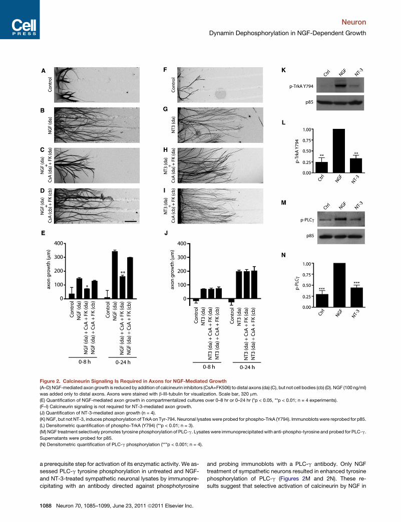
Figure 2. Calcineurin Signaling Is Required in Axons for NGF-Mediated Growth
(A–D) NGF-mediated axon growth is reduced by addition of calcineurin inhibitors (CsA+FK506) to distal axons (da) (C), but not cell bodies (cb) (D). NGF (100 ng/ml)
was added only to distal axons. Axons were stained with b-III-tubulin for visualization. Scale bar, 320 mm.
(E) Quantification of NGF-mediated axon growth in compartmentalized cultures over 0–8 hr or 0–24 hr (*p < 0.05, **p < 0.01; n = 4 experiments).
(F–I) Calcineurin signaling is not required for NT-3-mediated axon growth.
(J) Quantification of NT-3-mediated axon growth (n = 4).
(K) NGF, but not NT-3, induces phosphorylation of TrkA on Tyr-794. Neuronal lysates were probed for phospho-TrkA (Y794). Immunoblots were reprobed for p85.
(L) Densitometric quantification of phospho-TrkA (Y794) (**p < 0.01; n = 3).
(M) NGF treatment selectively promotes tyrosine phosphorylation of PLC-g. Lysates were immunoprecipitatedwith anti-phospho-tyrosine and probed for PLC-g.
Supernatants were probed for p85.
(N) Densitometric quantification of PLC-g phosphorylation (***p < 0.001; n = 4).
Neuron
Dynamin Dephosphorylation in NGF-Dependent Growth
a prerequisite step for activation of its enzymatic activity. We as-
sessed PLC-g tyrosine phosphorylation in untreated and NGF-
and NT-3-treated sympathetic neuronal lysates by immunopre-
cipitating with an antibody directed against phosphotyrosine
1088 Neuron 70, 1085–1099, June 23, 2011 ª2011 Elsevier Inc.
and probing immunoblots with a PLC-g antibody. Only NGF
treatment of sympathetic neurons resulted in enhanced tyrosine
phosphorylation of PLC-g (Figures 2M and 2N). These re-
sults suggest that selective activation of calcineurin by NGF in
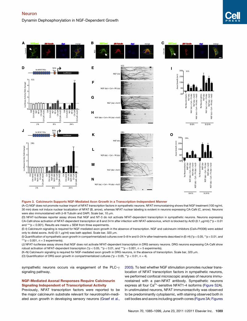
Figure 3. Calcineurin Supports NGF-Mediated Axon Growth in a Transcription-Independent Manner
(A–C) NGF does not promote nuclear import of NFAT transcription factors in sympathetic neurons. NFAT immunostaining shows that NGF treatment (100 ng/ml,
30 min) does not induce nuclear localization of NFAT (B, arrow), whereas NFAT nuclear labeling is evident in neurons expressing CA-CaN (C, arrow). Neurons
were also immunostained with b-III Tubulin and DAPI. Scale bar, 10 mm.
(D) NFAT-luciferase reporter assay shows that NGF and NT-3 do not activate NFAT-dependent transcription in sympathetic neurons. Neurons expressing
CA-CaN show activation of NFAT-dependent transcription at 8 and 24 hr after infection with NFAT-adenovirus, which is blocked by ActD (0.1 mg/ml) (**p < 0.01
and ***p < 0.001). Results are means ± SEM from three experiments.
(E–I) Calcineurin signaling is required for NGF-mediated axon growth in the absence of transcription. NGF and calcineurin inhibitors (CsA+FK506) were added
only to distal axons. ActD (0.1 mg/ml) was bath applied. Scale bar, 320 mm.
(I) Quantification of sympathetic axon growth in compartmentalized cultures over 0–8 hr and 0–24 hr after treatments described in (E–H) (*p < 0.05, **p < 0.01, and
***p < 0.001, n = 3 experiments).
(J) NFAT-luciferase assay shows that NGF does not activate NFAT-dependent transcription in DRG sensory neurons. DRG neurons expressing CA-CaN show
robust activation of NFAT-dependent transcription (*p < 0.05, **p < 0.01, and ***p < 0.001, n = 5 experiments).
(K–N) Calcineurin signaling is required for NGF-mediated axon growth in DRG neurons, in the absence of transcription. Scale bar, 320 mm.
(O) Quantification of DRG axon growth in compartmentalized cultures (*p < 0.05, **p < 0.01, n = 4).
Neuron
Dynamin Dephosphorylation in NGF-Dependent Growth
sympathetic neurons occurs via engagement of the PLC-g
signaling pathway.
NGF-Mediated Axonal Responses Require CalcineurinSignaling Independent of Transcriptional ActivityPreviously, NFAT transcription factors were reported to be
the major calcineurin substrate relevant for neurotrophin-medi-
ated axon growth in developing sensory neurons (Graef et al.,
2003). To test whether NGF stimulation promotes nuclear trans-
location of NFAT transcription factors in sympathetic neurons,
we performed confocal microscopic analyses of neurons immu-
nostained with a pan-NFAT antibody. Sympathetic neurons
express all four Ca2+-sensitive NFAT1-4 isoforms (Figure S2A).
In unstimulated neurons, NFAT immunoreactivity was observed
to be predominantly cytoplasmic, with staining observed both in
cell bodies and axons including growth cones (Figure 3A; Figures
Neuron 70, 1085–1099, June 23, 2011 ª2011 Elsevier Inc. 1089
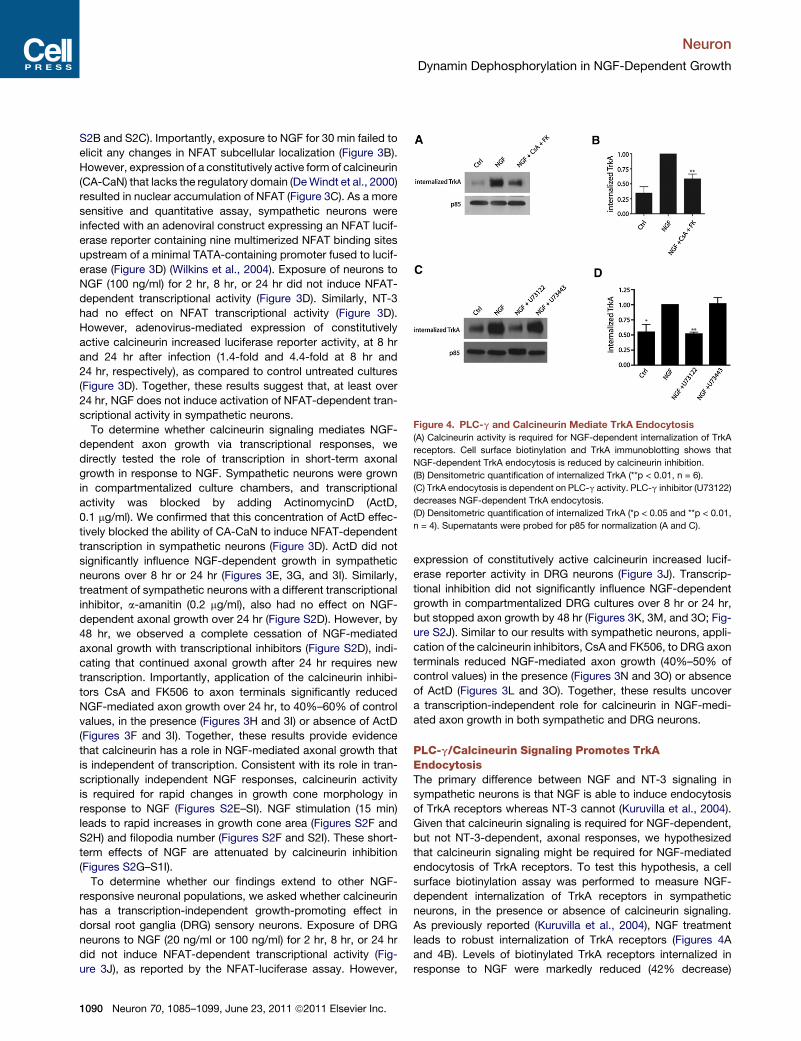
Figure 4. PLC-g and Calcineurin Mediate TrkA Endocytosis
(A) Calcineurin activity is required for NGF-dependent internalization of TrkA
receptors. Cell surface biotinylation and TrkA immunoblotting shows that
NGF-dependent TrkA endocytosis is reduced by calcineurin inhibition.
(B) Densitometric quantification of internalized TrkA (**p < 0.01, n = 6).
(C) TrkA endocytosis is dependent on PLC-g activity. PLC-g inhibitor (U73122)
decreases NGF-dependent TrkA endocytosis.
(D) Densitometric quantification of internalized TrkA (*p < 0.05 and **p < 0.01,
n = 4). Supernatants were probed for p85 for normalization (A and C).
Neuron
Dynamin Dephosphorylation in NGF-Dependent Growth
S2B and S2C). Importantly, exposure to NGF for 30 min failed to
elicit any changes in NFAT subcellular localization (Figure 3B).
However, expression of a constitutively active form of calcineurin
(CA-CaN) that lacks the regulatory domain (DeWindt et al., 2000)
resulted in nuclear accumulation of NFAT (Figure 3C). As a more
sensitive and quantitative assay, sympathetic neurons were
infected with an adenoviral construct expressing an NFAT lucif-
erase reporter containing nine multimerized NFAT binding sites
upstream of a minimal TATA-containing promoter fused to lucif-
erase (Figure 3D) (Wilkins et al., 2004). Exposure of neurons to
NGF (100 ng/ml) for 2 hr, 8 hr, or 24 hr did not induce NFAT-
dependent transcriptional activity (Figure 3D). Similarly, NT-3
had no effect on NFAT transcriptional activity (Figure 3D).
However, adenovirus-mediated expression of constitutively
active calcineurin increased luciferase reporter activity, at 8 hr
and 24 hr after infection (1.4-fold and 4.4-fold at 8 hr and
24 hr, respectively), as compared to control untreated cultures
(Figure 3D). Together, these results suggest that, at least over
24 hr, NGF does not induce activation of NFAT-dependent tran-
scriptional activity in sympathetic neurons.
To determine whether calcineurin signaling mediates NGF-
dependent axon growth via transcriptional responses, we
directly tested the role of transcription in short-term axonal
growth in response to NGF. Sympathetic neurons were grown
in compartmentalized culture chambers, and transcriptional
activity was blocked by adding ActinomycinD (ActD,
0.1 mg/ml). We confirmed that this concentration of ActD effec-
tively blocked the ability of CA-CaN to induce NFAT-dependent
transcription in sympathetic neurons (Figure 3D). ActD did not
significantly influence NGF-dependent growth in sympathetic
neurons over 8 hr or 24 hr (Figures 3E, 3G, and 3I). Similarly,
treatment of sympathetic neurons with a different transcriptional
inhibitor, a-amanitin (0.2 mg/ml), also had no effect on NGF-
dependent axonal growth over 24 hr (Figure S2D). However, by
48 hr, we observed a complete cessation of NGF-mediated
axonal growth with transcriptional inhibitors (Figure S2D), indi-
cating that continued axonal growth after 24 hr requires new
transcription. Importantly, application of the calcineurin inhibi-
tors CsA and FK506 to axon terminals significantly reduced
NGF-mediated axon growth over 24 hr, to 40%–60% of control
values, in the presence (Figures 3H and 3I) or absence of ActD
(Figures 3F and 3I). Together, these results provide evidence
that calcineurin has a role in NGF-mediated axonal growth that
is independent of transcription. Consistent with its role in tran-
scriptionally independent NGF responses, calcineurin activity
is required for rapid changes in growth cone morphology in
response to NGF (Figures S2E–SI). NGF stimulation (15 min)
leads to rapid increases in growth cone area (Figures S2F and
S2H) and filopodia number (Figures S2F and S2I). These short-
term effects of NGF are attenuated by calcineurin inhibition
(Figures S2G–S1I).
To determine whether our findings extend to other NGF-
responsive neuronal populations, we asked whether calcineurin
has a transcription-independent growth-promoting effect in
dorsal root ganglia (DRG) sensory neurons. Exposure of DRG
neurons to NGF (20 ng/ml or 100 ng/ml) for 2 hr, 8 hr, or 24 hr
did not induce NFAT-dependent transcriptional activity (Fig-
ure 3J), as reported by the NFAT-luciferase assay. However,
1090 Neuron 70, 1085–1099, June 23, 2011 ª2011 Elsevier Inc.
expression of constitutively active calcineurin increased lucif-
erase reporter activity in DRG neurons (Figure 3J). Transcrip-
tional inhibition did not significantly influence NGF-dependent
growth in compartmentalized DRG cultures over 8 hr or 24 hr,
but stopped axon growth by 48 hr (Figures 3K, 3M, and 3O; Fig-
ure S2J). Similar to our results with sympathetic neurons, appli-
cation of the calcineurin inhibitors, CsA and FK506, to DRG axon
terminals reduced NGF-mediated axon growth (40%–50% of
control values) in the presence (Figures 3N and 3O) or absence
of ActD (Figures 3L and 3O). Together, these results uncover
a transcription-independent role for calcineurin in NGF-medi-
ated axon growth in both sympathetic and DRG neurons.
PLC-g/Calcineurin Signaling Promotes TrkAEndocytosisThe primary difference between NGF and NT-3 signaling in
sympathetic neurons is that NGF is able to induce endocytosis
of TrkA receptors whereas NT-3 cannot (Kuruvilla et al., 2004).
Given that calcineurin signaling is required for NGF-dependent,
but not NT-3-dependent, axonal responses, we hypothesized
that calcineurin signaling might be required for NGF-mediated
endocytosis of TrkA receptors. To test this hypothesis, a cell
surface biotinylation assay was performed to measure NGF-
dependent internalization of TrkA receptors in sympathetic
neurons, in the presence or absence of calcineurin signaling.
As previously reported (Kuruvilla et al., 2004), NGF treatment
leads to robust internalization of TrkA receptors (Figures 4A
and 4B). Levels of biotinylated TrkA receptors internalized in
response to NGF were markedly reduced (42% decrease)

Neuron
Dynamin Dephosphorylation in NGF-Dependent Growth
when calcineurin activity was blocked with CsA and FK506
(Figures 4A and 4B). Calcineurin inhibitors had no effect on
surface levels of TrkA in the absence of NGF (Figures S3A and
S3B), indicating that calcineurin signaling is not required for
maintenance of TrkA receptors on the plasma membrane.
Because our results suggest that NGF-mediated activation of
calcineurin occurs via recruitment of the TrkA effector, PLC-g,
we tested whether PLC-g activity is required for TrkA endocy-
tosis. Inhibition of PLC-g activity with a selective inhibitor,
U73122 (10 mM), markedly reduced NGF-dependent endocy-
tosis of TrkA receptors (Figures 4C and 4D). However, treatment
of neurons with an inactive analog, U73443, had no effect. These
results suggest that NGF promotes endocytosis of TrkA recep-
tors by activation of a PLC-g/calcineurin signaling pathway.
Calcineurin Mediates NGF-Dependent Growth viaDephosphorylation of DynaminGiven that calcineurin signaling is required for TrkA endocytosis,
we asked which calcineurin substrate mediates this response.
Our clue came from previous studies in synaptic vesicle endocy-
tosis (SVE), where calcineurin-dependent dephosphorylation of
the endocytic GTPase dynamin1 is essential for the retrieval of
synaptic vesicle membranes (Liu et al., 1994). Nerve terminal
depolarization leads to calcineurin-dependent dephosphoryla-
tion of dynamin1 on at least two serine residues, Ser-774 and
Ser-778, located within a phospho-box region in the proline-
rich C terminus (Clayton et al., 2009). Site-directed mutagenesis
indicated that phosphoregulation of these residues on dynamin1
is required for calcineurin-dependent endocytosis of synaptic
vesicles (Clayton et al., 2009). To ask whether NGF stimulation
leads to dynamin dephosphorylation in a calcineurin-dependent
manner, sympathetic neurons were exposed to NGF for 20 min,
and levels of phosphorylated dynamin1 were assessed using
phospho-specific antibodies that specifically recognize dyna-
min1 phosphorylated on Ser-774 and Ser-778. NGF induced
a significant decrease in dynamin1 phosphorylation on Ser-774
and Ser-778 (26.2% and 28.5%, respectively), which was
blocked by CsA and FK506 treatment (Figures 5A and 5B). As
predicted, the phosphorylation status of dynamin1 was unaf-
fected by NT-3 treatment (Figures 5A and 5B). Thus, NGF, but
not NT-3, leads to calcineurin-dependent dephosphorylation of
dynamin, providing further support for this mechanism under-
lying the differential trafficking of TrkA receptors downstream
of NGF and NT-3 (Kuruvilla et al., 2004).
Given that target-derived NGF acts directly on projecting
axons to promote growth, we tested whether axon-applied
NGF locallymodulates dynamin1 phosphorylation in nerve termi-
nals, in vitro, and in vivo. Sympathetic axons in compartmental-
ized cultures were stimulated with NGF (100 ng/ml, 20 min) and
axonal lysates were immunoblotted with phospho-dynamin1
(Ser-778) antibody. Similar to results inmass cultures, NGF treat-
ment of distal axons leads to a reduction (24%decrease) in phos-
phorylated dynamin1, in comparison to control treatment (Fig-
ure 5C and 5D). To test whether NGF regulates phosphorylation
of dynamin1 in axons in vivo, we analyzed the levels of phos-
pho-dynamin1 in a sympathetic target tissue, the salivary glands,
in both wild-type and heterozygous NGF (NGF+/�) mice. Given
that dynamin1 is neuron specific (Urrutia et al., 1997), immuno-
blotting of salivary gland lysates with the phospho-dynamin1
antibody should reveal the status of dynamin1 phosphorylation
locally in sympathetic nerve terminals that innervate the target
tissue. If target-derived NGF regulates dynamin1 phosphoryla-
tion in vivo, then we would expect to see increased dynamin1
phosphorylation levels under conditions of reduced NGF
signaling. We employed NGF+/� mice for this analysis because
these mice display haploinsufficiency with reduced levels of
NGF and sympathetic target innervation (Brennan et al., 1999;
Ghasemlou et al., 2004), in contrast to homozygous NGF null
mice, which completely lack sympathetic innervation (Glebova
and Ginty, 2004). We found that NGF+/� mice have higher levels
of phosphorylated dynamin1 on Ser-778 in sympathetic axons
innervating the salivary glands, compared to wild-type animals
(11.2% ± 2% increase; Figures 5E and 5F). These findings
provide in vivo evidence for NGF-dependent phosphoregulation
of dynamin1 locally in sympathetic axons.
To assess the role of dynamin1 dephosphorylation in support-
ing neurotrophin-dependent axon growth, sympathetic neurons
were exposed for 24 hr to a cell-permeable peptide spanning the
dynamin1 phospho-box (amino acids 769–784, incorporating
Ser-774 and Ser-778) in which the two serines 774/778 were
replaced with alanine (Ser774/778-Ala, dyn1769-784AA). The
dyn1769-784AA peptide blocks dephosphosphorylation-depen-
dent dynamin1 functions by binding and sequestering down-
stream effector molecules, such as syndapin1 (Anggono et al.,
2006). Delivery of dyn1769-784AA (300 mM) into sympathetic
neurons reduced NGF-mediated axon growth from an average
of 177 ± 14 mm/day to 90.6 ± 7.2 mm/day (Figures 5G, 5H, and
5M). In contrast, introduction of the phospho-mimetic peptide
dyn1769-784EE (in which the serines 774/778 were substituted
with glutamate) had no effect on NGF-mediated axon growth
(Figures 5I and 5M). NT-3-mediated axon growth was not
affected by delivery of either dyn1769-784AA or dyn1769-784EE(Figures 5J, 5K, 5L, and 5M). Together, these results provide
evidence that calcineurin-mediated dephosphorylation of dyna-
min1 is a key signalingmechanism necessary for NGF-mediated,
but not NT-3-mediated, axon growth.
Isoform-Specific Interaction of Calcineurin withDynamin1 via a PxIxIT DomainThere are three dynamin genes expressed in mammals, with
dynamin1 reported to be neuron specific, dynamin2 being ubiqui-
tously expressed, and dynamin3 expressed in brain, lungs, and
testes (Urrutia et al., 1997). We asked whether calcineurin inter-
acted with all three dynamins by performing calcineurinA-GST
pull-down assays of rat brain lysates and probing for calcineurin
interaction using antibodies specific to dynamin1, dynamin2, and
dynamin3. Aspreviously demonstrated (Lai et al., 1999), dynamin1
binds calcineurinA-GST (Figure S4A); in contrast, dynamin2 and
dynamin3 do not detectably bind calcineurinA-GST (Figure S4A).
While exploring themechanism of calcineurin-dynamin1 asso-
ciation, we observed that the dynamin1 C-terminal proline
rich domain (PRD) harbors a putative calcineurin interaction
sequence, PRITIS, within the amino acids 844–849 (Figure 6A).
This motif has high sequence identity to the ‘‘PxIxIT box,’’
a consensus sequence present in NFAT transcription factors
that mediates the docking of calcineurin to the NFAT regulatory
Neuron 70, 1085–1099, June 23, 2011 ª2011 Elsevier Inc. 1091

Figure 5. NGF Promotes Axon Growth through Dynamin Dephosphorylation
(A) NGF stimulation results in dephosphorylation of dynamin1 in a calcineurin-dependent manner. Neuronal lysates were immunoblotted using phospho-Ser774
and phospho-Ser778 dynamin antibodies. Immunoblots were stripped and reprobed for total dynamin1.
(B) Densitometric quantification of phospho-dynamin1 levels (*p < 0.05, n = 6).
(C) NGF stimulation results in dephosphorylation of dynamin1 (Ser 778) in distal axons. Immunoblots were reprobed for total dynamin1.
(D) Densitometric quantification of phospho-dynamin1 (Ser778) in axons (*p < 0.05, n = 3).
(E and F) NGF+/� mice have increased levels of phospho-dynamin1 in sympathetic axons in vivo. Salivary gland lysates from P0.5 wild-type and NGF+/� mice
were immunoblotted using phospho-dynamin1 (Ser778) antibody. Immunoblots were reprobed for total dynamin1.
(F) Densitometric quantification of phospho-dynamin1 (Ser778) after treatments, as described in (E), is represented as a scatter plot with 95% confidence
intervals. n = 6 pups for each genotype.
(G) Dephosphorylation-dependent dynamin1 function is required for NGF-mediated axon growth.
(H–L) Introduction of dyn1(769-784 AA) (H), but not the dyn1(769-784 EE) (I) peptide, decreased NGF-dependent axon growth over 24 hr. NT-3-mediated growth
was unaffected by introduction of dyn1 phosphopeptides (J–L). Scale bar, 100 mm.
(M) Quantification of axon growth (**p < 0.01, n = 3).
Neuron
Dynamin Dephosphorylation in NGF-Dependent Growth
domains (Aramburu et al., 1998). Deletion studies using a yeast-
two hybrid assay had restricted the calcineurin-interaction
region of dynamin1 to the last 135 amino acids at the C terminus
(Lai et al., 1999), encompassing this putative PxIxIT box. To test
whether calcineurin-dynamin1 interaction is mediated by the
PxIxITmotif present in dynamin1, we took advantage of the VIVIT
peptide, a high-affinitymolecular mimic of the PxIxIT box domain
1092 Neuron 70, 1085–1099, June 23, 2011 ª2011 Elsevier Inc.
that acts as a competitive inhibitor of calcineurin-PxIxIT box
interactions (Aramburu et al., 1999). CalcineurinA-GST pull-
down assays of rat brain lysates were performed either in the
presence or absence of VIVIT, and immunoblotting was per-
formed to detect dynamin1 interaction. The VIVIT peptide
completely blocked calcineurin-dynamin1 interaction, whereas
a control peptide, VEET, had no effect (Figure 6B).
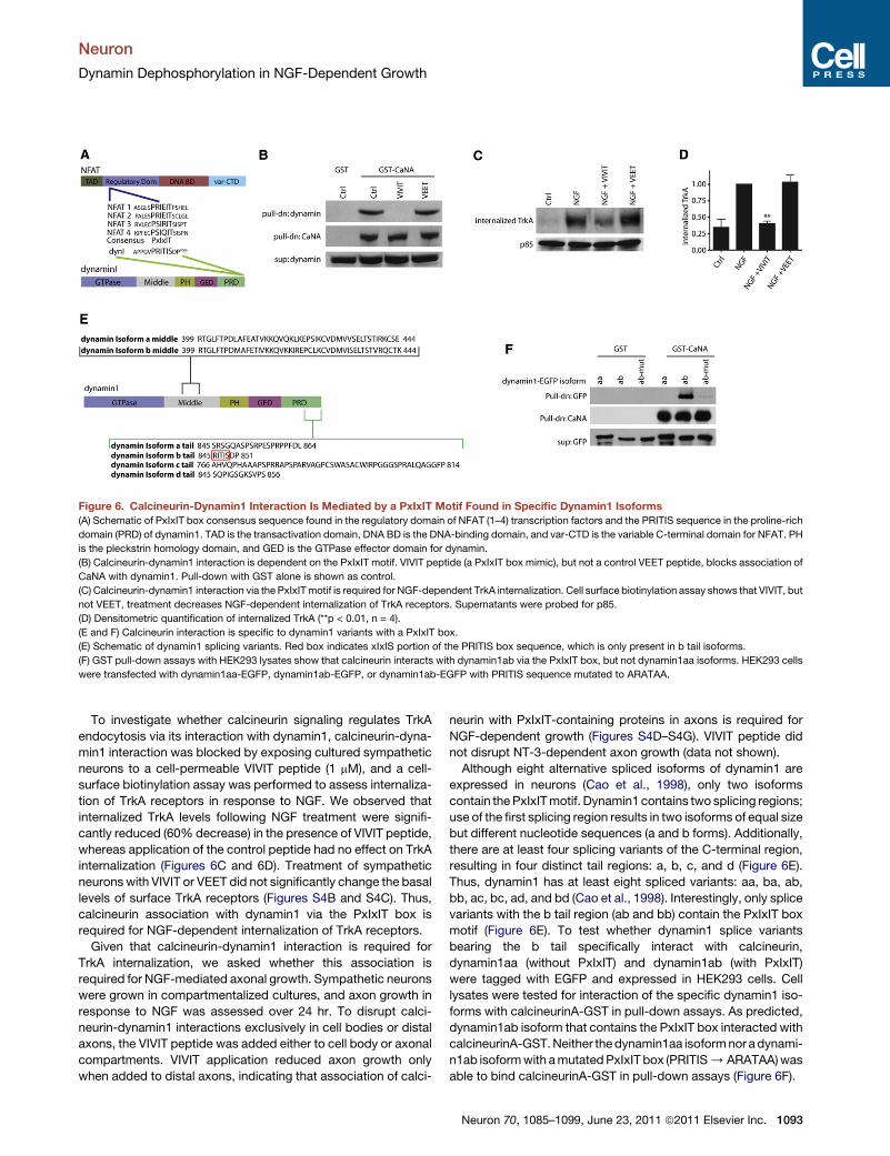
Figure 6. Calcineurin-Dynamin1 Interaction Is Mediated by a PxIxIT Motif Found in Specific Dynamin1 Isoforms
(A) Schematic of PxIxIT box consensus sequence found in the regulatory domain of NFAT (1–4) transcription factors and the PRITIS sequence in the proline-rich
domain (PRD) of dynamin1. TAD is the transactivation domain, DNA BD is the DNA-binding domain, and var-CTD is the variable C-terminal domain for NFAT. PH
is the pleckstrin homology domain, and GED is the GTPase effector domain for dynamin.
(B) Calcineurin-dynamin1 interaction is dependent on the PxIxIT motif. VIVIT peptide (a PxIxIT box mimic), but not a control VEET peptide, blocks association of
CaNA with dynamin1. Pull-down with GST alone is shown as control.
(C) Calcineurin-dynamin1 interaction via the PxIxITmotif is required for NGF-dependent TrkA internalization. Cell surface biotinylation assay shows that VIVIT, but
not VEET, treatment decreases NGF-dependent internalization of TrkA receptors. Supernatants were probed for p85.
(D) Densitometric quantification of internalized TrkA (**p < 0.01, n = 4).
(E and F) Calcineurin interaction is specific to dynamin1 variants with a PxIxIT box.
(E) Schematic of dynamin1 splicing variants. Red box indicates xIxIS portion of the PRITIS box sequence, which is only present in b tail isoforms.
(F) GST pull-down assays with HEK293 lysates show that calcineurin interacts with dynamin1ab via the PxIxIT box, but not dynamin1aa isoforms. HEK293 cells
were transfected with dynamin1aa-EGFP, dynamin1ab-EGFP, or dynamin1ab-EGFP with PRITIS sequence mutated to ARATAA.
Neuron
Dynamin Dephosphorylation in NGF-Dependent Growth
To investigate whether calcineurin signaling regulates TrkA
endocytosis via its interaction with dynamin1, calcineurin-dyna-
min1 interaction was blocked by exposing cultured sympathetic
neurons to a cell-permeable VIVIT peptide (1 mM), and a cell-
surface biotinylation assay was performed to assess internaliza-
tion of TrkA receptors in response to NGF. We observed that
internalized TrkA levels following NGF treatment were signifi-
cantly reduced (60% decrease) in the presence of VIVIT peptide,
whereas application of the control peptide had no effect on TrkA
internalization (Figures 6C and 6D). Treatment of sympathetic
neurons with VIVIT or VEET did not significantly change the basal
levels of surface TrkA receptors (Figures S4B and S4C). Thus,
calcineurin association with dynamin1 via the PxIxIT box is
required for NGF-dependent internalization of TrkA receptors.
Given that calcineurin-dynamin1 interaction is required for
TrkA internalization, we asked whether this association is
required for NGF-mediated axonal growth. Sympathetic neurons
were grown in compartmentalized cultures, and axon growth in
response to NGF was assessed over 24 hr. To disrupt calci-
neurin-dynamin1 interactions exclusively in cell bodies or distal
axons, the VIVIT peptide was added either to cell body or axonal
compartments. VIVIT application reduced axon growth only
when added to distal axons, indicating that association of calci-
neurin with PxIxIT-containing proteins in axons is required for
NGF-dependent growth (Figures S4D–S4G). VIVIT peptide did
not disrupt NT-3-dependent axon growth (data not shown).
Although eight alternative spliced isoforms of dynamin1 are
expressed in neurons (Cao et al., 1998), only two isoforms
contain thePxIxITmotif. Dynamin1 contains two splicing regions;
use of the first splicing region results in two isoforms of equal size
but different nucleotide sequences (a and b forms). Additionally,
there are at least four splicing variants of the C-terminal region,
resulting in four distinct tail regions: a, b, c, and d (Figure 6E).
Thus, dynamin1 has at least eight spliced variants: aa, ba, ab,
bb, ac, bc, ad, and bd (Cao et al., 1998). Interestingly, only splice
variants with the b tail region (ab and bb) contain the PxIxIT box
motif (Figure 6E). To test whether dynamin1 splice variants
bearing the b tail specifically interact with calcineurin,
dynamin1aa (without PxIxIT) and dynamin1ab (with PxIxIT)
were tagged with EGFP and expressed in HEK293 cells. Cell
lysates were tested for interaction of the specific dynamin1 iso-
forms with calcineurinA-GST in pull-down assays. As predicted,
dynamin1ab isoform that contains the PxIxIT box interacted with
calcineurinA-GST.Neither thedynamin1aa isoformnor adynami-
n1ab isoformwith amutatedPxIxIT box (PRITIS/ARATAA)was
able to bind calcineurinA-GST in pull-down assays (Figure 6F).
Neuron 70, 1085–1099, June 23, 2011 ª2011 Elsevier Inc. 1093

Neuron
Dynamin Dephosphorylation in NGF-Dependent Growth
1094 Neuron 70, 1085–1099, June 23, 2011 ª2011 Elsevier Inc.

Neuron
Dynamin Dephosphorylation in NGF-Dependent Growth
Phosphoregulation of PxIxIT-Containing Dynamin1Isoforms Is Required for TrkA Endocytosis and AxonGrowthDifferent dynamin1 splicing isoforms display different subcellular
localization in heterologous expression systems (Cao et al.,
1998). To examine the subcellular localization of dynamin1aa
and dynamin1ab isoforms, sympathetic neurons were electro-
porated with vectors expressing EGFP-tagged dynamin1aa
or dynamin1ab. The two dynamin isoforms showed striking
differences in subcellular localization in sympathetic neurons.
Dynamin1aa-EGFP showed diffuse cytoplasmic localization
throughout the cell bodies (Figure 7A) and axons (Figure 7B). In
stark contrast, dynamin1ab-EGFP expression resulted in a punc-
tate staining along the plasma membrane and throughout the
cytoplasm in cell bodies (Figure 7C) and axons (Figure 7D).
Because the amino acid sequences of these two isoforms differ
only in the C-terminal region containing the PxIxIT motif (Fig-
ure 6E), these results indicate that relatively small differences
in primary sequence can result in striking changes in cellular
localization.We next examinedwhether the punctate distribution
of dynamin1ab-EGFP colocalizes with surface TrkA receptors.
To visualize surface TrkA receptors, a live-cell antibody feeding
assay was performed in sympathetic neurons (Ascano et al.,
2009) expressing both N-terminal FLAG-tagged TrkA receptors
and dynamin1-EGFP isoforms. Using an antibody directed
against the extracellular FLAG epitope of the TrkA receptors,
we found that the punctate distribution of dynamin1ab-EGFP
fluorescence at the plasma membrane was in close apposition
to surface TrkA immunoreactivity in neuronal soma (Figure 7C)
and axons (Figure 7D), under nonpermeabilizing conditions. In
contrast, little colocalization between surface TrkA labeling and
dynamin1aa-EGFPwas observed (Figures 7A and 7B). Together,
these results suggest that dynamin1ab isoforms might mediate
TrkA endocytosis in sympathetic neurons.
To test whether phosphoregulation of dynamin1 is critical for
NGF-dependent endocytosis of TrkA receptors, we generated
phosphomutants of the dynamin1aa and dynamin1ab isoforms.
Because NGF stimulation results in dephosphorylation of
dynamin1 on Ser 774 and 778, we generated dynamin1aa and
dynamin1ab mutants bearing mutations of both serine residues
to either alanine (Ser774/778-Ala; nonphosphorylatable forms)
or glutamate (Ser774/778-Glu; phosphomimetic forms).
Previous studies had shown that both the nonphosphorylatable
Figure 7. Phosphoregulation of Dynamin1ab Is Required for TrkA Endo
(A–D) Dynamin1aa isoform (A and B) shows diffuse cytoplasmic localization, whe
and axons. FLAG immunostaining shows surface FLAG-TrkA receptors in cell bo
(E–J) Phosphoregulation of dynamin1aa is not required for NGF-dependent TrkA
dynamin1aa-EGFP (E and F), Ser-Glu 774/778 dynamin1aa-EGFP (G andH), or Se
(I). Axons are shown in (F), (H), and (J). Scale bar, 10 mm.
(K) Quantification of NGF-dependent TrkA internalization in cell bodies and axon
(L–Q) Phosphomutants of dynamin1ab disrupt NGF-dependent internalization
dynamin1ab-EGFP (L and M), Ser-Glu 774/778 dynamin1ab-EGFP (N and O), or S
and (P). Axons are shown in (M), (O), and (Q). Scale bar, 10 mm.
(R) Quantification of internalized TrkA (*p < 0.01, n = 3).
(S–V) Phosphoregulation of dynamin1ab is required for NGF-dependent, but not N
neurons expressing dynamin1ab-EGFP Ser-Glu 774/778 (T) as compared to wild
Scale bar, 50 mm.
(W) Quantification of neurite length (**p < 0.01, n = 3).
and phosphomimetic forms of dynamin1 act as dominant nega-
tive inhibitors of activity-dependent synaptic vesicle endocytosis
(Anggono et al., 2006; Clayton et al., 2009).
To label and follow endocytic trafficking of surface TrkA recep-
tors, sympathetic neurons coexpressing FLAG-TrkA and the
dynamin1 constructs were live-labeled with a calcium-sensitive
FLAG antibody. After exposure to NGF for 30 min to allow inter-
nalization of labeled receptors, surface-bound antibodies were
stripped, leaving antibodies bound only to the internalized pool
of receptors. FLAG antibodies bound to internalized receptors
were then visualized with Alexa-546-labeled secondary anti-
bodies. We observed robust internalization of TrkA receptors in
cell bodies and axons in response to NGF stimulation in cells ex-
pressing wild-type (Figures 7E and 7F), phosphomimic (Ser774/
778 to Glu) (Figures 7G and 7H), or phosphomutant (Ser774/778
to Ala) (Figures 7I and 7J) dynamin1aa-EGFP. In contrast,
expression of either dynamin1ab-EGFP phosphomimeticmutant
(Ser774/778-Glu) (Figure 7N) or the nonphosphorylatable
dynamin1ab-EGFP mutant (Ser774/778-Ala) (Figure 7P) signifi-
cantly reduced NGF-mediated TrkA internalization in cell bodies
to 39% and 50%, respectively, when compared to neurons ex-
pressing wild-type dynamin1ab-EGFP (Figures 7L and 7R).
Expression of both phosphomutant forms of dynamin1ab-
EGFP similarly reduced NGF-dependent internalization in axons
(63% decrease) (Figures 7M, 7O, 7Q, and 7R). Expression of
mutant dynamin1ab-EGFP forms did not affect surface expres-
sion of FLAG-TrkA receptors in the absence of NGF treatment,
nor did it influence the ability of FLAG antibodies to bind surface
receptors (Figures S5A–S5C), indicating that decreased intracel-
lular accumulation of FLAG-TrkA in mutant dynamin1ab-
expressing cells indeed reflects a block in endocytosis. The
finding that both the nonphosphorylatable (Ser-Ala) and phos-
phomimetic (Ser-Glu) mutations inhibited receptor internaliza-
tion suggests that, similar to synaptic vesicle endocytosis
(Anggono et al., 2006), the cycle between dynamin phosphoryla-
tion and dephosphorylation is critical for NGF-dependent endo-
cytosis of TrkA receptors. Together, these results indicate that
NGF promotes internalization of its receptors through calci-
neurin-mediated dephosphorylation of specific spliced variants
of dynamin1 harboring a PxIxIT interaction motif.
To determine whether phosphoregulation of dynamin1ab
isoforms is also important for NGF-dependent axon growth,
we infected sympathetic neurons with adenoviruses expressing
cytosis and Axon Growth
reas dynamin1ab isoform (C and D) shows punctate localization in cell bodies
dies (A and C) and in axons (B and D). Scale bar, 10 mm.
internalization. Neurons were transfected with FLAG-TrkA and either wild-type
r-Ala 774/778 dynamin1aa-EGFP (I and J). Cell bodies are shown in (E), (G), and
s (n = 3).
of TrkA. Neurons were transfected with FLAG-TrkA and either wild-type
er-Ala 774/778 dynamin1ab-EGFP (P and Q). Cell bodies are shown in (L), (N),
T-3-dependent, axon growth. NGF-mediated growth is blocked in sympathetic
-type dynamin1ab-EGFP (S). NT-3-mediated growth was unaffected (U and V).
Neuron 70, 1085–1099, June 23, 2011 ª2011 Elsevier Inc. 1095
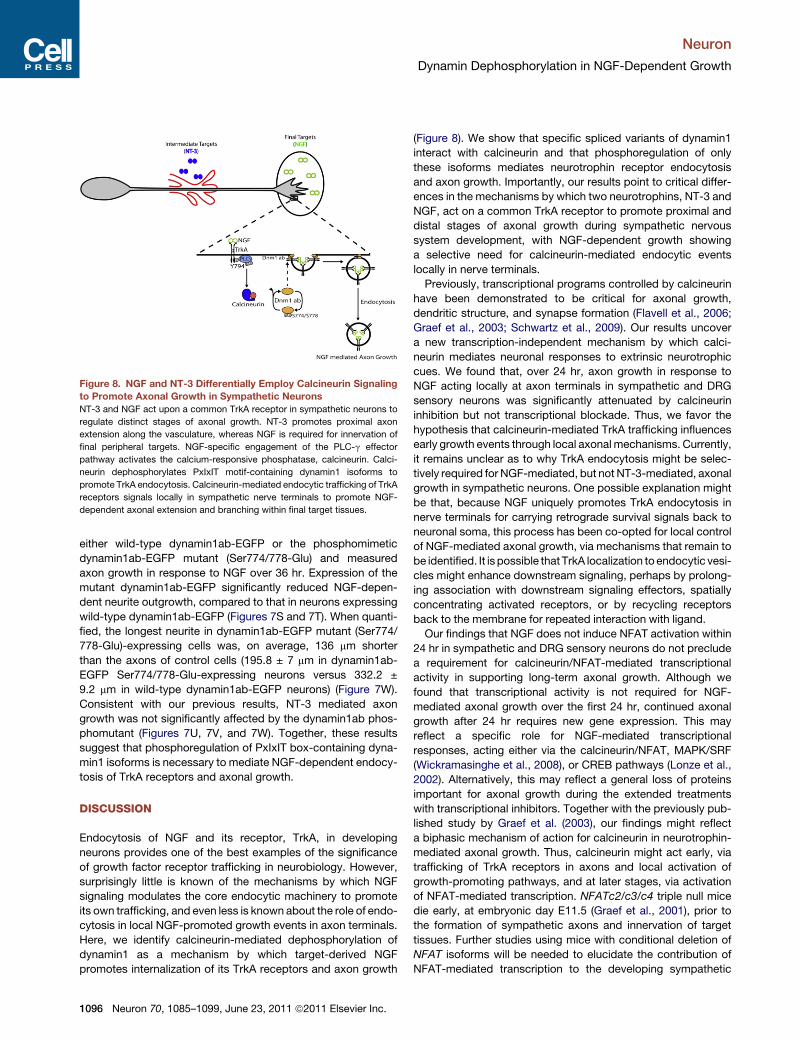
Figure 8. NGF and NT-3 Differentially Employ Calcineurin Signaling
to Promote Axonal Growth in Sympathetic Neurons
NT-3 and NGF act upon a common TrkA receptor in sympathetic neurons to
regulate distinct stages of axonal growth. NT-3 promotes proximal axon
extension along the vasculature, whereas NGF is required for innervation of
final peripheral targets. NGF-specific engagement of the PLC-g effector
pathway activates the calcium-responsive phosphatase, calcineurin. Calci-
neurin dephosphorylates PxIxIT motif-containing dynamin1 isoforms to
promote TrkA endocytosis. Calcineurin-mediated endocytic trafficking of TrkA
receptors signals locally in sympathetic nerve terminals to promote NGF-
dependent axonal extension and branching within final target tissues.
Neuron
Dynamin Dephosphorylation in NGF-Dependent Growth
either wild-type dynamin1ab-EGFP or the phosphomimetic
dynamin1ab-EGFP mutant (Ser774/778-Glu) and measured
axon growth in response to NGF over 36 hr. Expression of the
mutant dynamin1ab-EGFP significantly reduced NGF-depen-
dent neurite outgrowth, compared to that in neurons expressing
wild-type dynamin1ab-EGFP (Figures 7S and 7T). When quanti-
fied, the longest neurite in dynamin1ab-EGFP mutant (Ser774/
778-Glu)-expressing cells was, on average, 136 mm shorter
than the axons of control cells (195.8 ± 7 mm in dynamin1ab-
EGFP Ser774/778-Glu-expressing neurons versus 332.2 ±
9.2 mm in wild-type dynamin1ab-EGFP neurons) (Figure 7W).
Consistent with our previous results, NT-3 mediated axon
growth was not significantly affected by the dynamin1ab phos-
phomutant (Figures 7U, 7V, and 7W). Together, these results
suggest that phosphoregulation of PxIxIT box-containing dyna-
min1 isoforms is necessary to mediate NGF-dependent endocy-
tosis of TrkA receptors and axonal growth.
DISCUSSION
Endocytosis of NGF and its receptor, TrkA, in developing
neurons provides one of the best examples of the significance
of growth factor receptor trafficking in neurobiology. However,
surprisingly little is known of the mechanisms by which NGF
signaling modulates the core endocytic machinery to promote
its own trafficking, and even less is known about the role of endo-
cytosis in local NGF-promoted growth events in axon terminals.
Here, we identify calcineurin-mediated dephosphorylation of
dynamin1 as a mechanism by which target-derived NGF
promotes internalization of its TrkA receptors and axon growth
1096 Neuron 70, 1085–1099, June 23, 2011 ª2011 Elsevier Inc.
(Figure 8). We show that specific spliced variants of dynamin1
interact with calcineurin and that phosphoregulation of only
these isoforms mediates neurotrophin receptor endocytosis
and axon growth. Importantly, our results point to critical differ-
ences in the mechanisms by which two neurotrophins, NT-3 and
NGF, act on a common TrkA receptor to promote proximal and
distal stages of axonal growth during sympathetic nervous
system development, with NGF-dependent growth showing
a selective need for calcineurin-mediated endocytic events
locally in nerve terminals.
Previously, transcriptional programs controlled by calcineurin
have been demonstrated to be critical for axonal growth,
dendritic structure, and synapse formation (Flavell et al., 2006;
Graef et al., 2003; Schwartz et al., 2009). Our results uncover
a new transcription-independent mechanism by which calci-
neurin mediates neuronal responses to extrinsic neurotrophic
cues. We found that, over 24 hr, axon growth in response to
NGF acting locally at axon terminals in sympathetic and DRG
sensory neurons was significantly attenuated by calcineurin
inhibition but not transcriptional blockade. Thus, we favor the
hypothesis that calcineurin-mediated TrkA trafficking influences
early growth events through local axonal mechanisms. Currently,
it remains unclear as to why TrkA endocytosis might be selec-
tively required for NGF-mediated, but not NT-3-mediated, axonal
growth in sympathetic neurons. One possible explanation might
be that, because NGF uniquely promotes TrkA endocytosis in
nerve terminals for carrying retrograde survival signals back to
neuronal soma, this process has been co-opted for local control
of NGF-mediated axonal growth, via mechanisms that remain to
be identified. It is possible that TrkA localization toendocytic vesi-
cles might enhance downstream signaling, perhaps by prolong-
ing association with downstream signaling effectors, spatially
concentrating activated receptors, or by recycling receptors
back to the membrane for repeated interaction with ligand.
Our findings that NGF does not induce NFAT activation within
24 hr in sympathetic and DRG sensory neurons do not preclude
a requirement for calcineurin/NFAT-mediated transcriptional
activity in supporting long-term axonal growth. Although we
found that transcriptional activity is not required for NGF-
mediated axonal growth over the first 24 hr, continued axonal
growth after 24 hr requires new gene expression. This may
reflect a specific role for NGF-mediated transcriptional
responses, acting either via the calcineurin/NFAT, MAPK/SRF
(Wickramasinghe et al., 2008), or CREB pathways (Lonze et al.,
2002). Alternatively, this may reflect a general loss of proteins
important for axonal growth during the extended treatments
with transcriptional inhibitors. Together with the previously pub-
lished study by Graef et al. (2003), our findings might reflect
a biphasic mechanism of action for calcineurin in neurotrophin-
mediated axonal growth. Thus, calcineurin might act early, via
trafficking of TrkA receptors in axons and local activation of
growth-promoting pathways, and at later stages, via activation
of NFAT-mediated transcription. NFATc2/c3/c4 triple null mice
die early, at embryonic day E11.5 (Graef et al., 2001), prior to
the formation of sympathetic axons and innervation of target
tissues. Further studies using mice with conditional deletion of
NFAT isoforms will be needed to elucidate the contribution of
NFAT-mediated transcription to the developing sympathetic
Neuron
Dynamin Dephosphorylation in NGF-Dependent Growth
nervoussystem.Nevertheless, our results indicate thatNFAT tran-
scription factors are not the sole targets of calcineurin relevant for
neurotrophin-mediated axon growth. Our identification of a novel
endocytic mechanism by which calcineurin signaling promotes
neurotrophin-dependent axonal growth provides insight into the
versatility of calcineurin signaling in nervous system development
and, in particular, neurotrophin-mediated functions.
Our study suggests parallels between neurotrophin receptor
endocytosis in developing neurons and local synaptic vesicle
recycling in mature nerve terminals. During synaptic vesicle
endocytosis, dynamin1 is among a group of structurally distinct
proteins collectively called dephosphins that undergo a cycle of
dephosphorylation and rephosphorylation in nerve terminals to
mediate synaptic vesicle recycling and synaptic transmission
(Cousin and Robinson, 2001). We show that calcineurin-depen-
dent dephosphorylation of dynamin1 is a common mechanism
underlying TrkA and synaptic vesicle endocytosis. Neurotro-
phins modulate synaptic transmission in mature neurons (Lu,
2004), and our results suggest that a potential target for neuro-
trophin actions at presynaptic terminals might be the regulation
of calcineurin-dynamin1-dependent retrieval of synaptic vesi-
cles after exocytosis.
Alternative splicing of the three dynamin genes generates over
25 different variants (Cao et al., 1998) that could greatly increase
the diversity of dynamin functions in the mammalian nervous
system. We provide evidence that specific dynamin1-splicing
isoforms exhibit distinct subcellular localizations in neurons
and perform discrete biological functions. In addition to synaptic
vesicle retrieval, calcineurin-dynamin1-mediated endocytosis
has been shown to be critical for regulation of AMPA receptor
densities at postsynaptic spines during paradigms of synaptic
plasticity, such as long-term depression (LTD) (Beattie et al.,
2000; Lin et al., 2000). Our findings raise the possibility that
PxIxIT-containing dynamin1 isoforms might mediate all other
calcineurin-regulated endocytosis in neurons.
The role of NGF-dependent regulation of calcineurin in endo-
cytosis and axon outgrowth may have implications that extend
beyond early neural development to the pathogenesis of some
neurodegenerative disorders. Defective NGF trafficking in basal
forebrain cholinergic neurons has been implicated in degenera-
tion and atrophy of these neurons in Down’s syndrome and
Alzheimer’s disease (Cooper et al., 2001; Salehi et al., 2006).
Overexpression of Regulator of Calcineurin 1 (RCAN1) encoding
for an endogenous calcineurin inhibitor has also been implicated
in neuropathology of Down’s syndrome and Alzheimer’s disease
(Ermak et al., 2001; Fuentes et al., 2000). In future experiments, it
will be intriguing to investigate the role of regulated calcineurin-
dependent endocytosis in the trafficking of TrkA receptors and
in maintaining the integrity of basal forebrain cholinergic neurons
in normal and diseased states.
EXPERIMENTAL PROCEDURES
Animals
To generate conditional mutants of CaNB1, floxed CaNB1 (CaNB1fl/fl) mice
(Jackson Laboratory) were crossed to Nestin-Cre mice (Jackson Laboratory).
NGF+/� mice (Crowley et al., 1994) were obtained from the Jackson Labora-
tory. All procedures relating to animal care and treatment conformed to
institutional and NIH guidelines.
Immunostaining
Whole-mount tyrosine hydroxylase immunohistochemistry was performed on
E16.5–E18.5 mouse embryos, as previously described (Kuruvilla et al., 2004).
For NFAT immunostaining, sympathetic neurons were treated with 100 ng/ml
NGF for 30 min, and neurons were fixed and immunostained using pan-NFAT
antibody, b-III-tubulin, and DAPI (40,6-diamidino-2-phenylindole). Images
representing 1 mmoptical slices were acquired using a Zeiss LSM510 confocal
scanning microscope equipped with diode (405 nm), Ar (458–488 nm), and
He/Ne (543–633) lasers.
Neuronal Cultures
Sympathetic neurons were harvested from P0.5 Sprague-Dawley rats and
were grown in mass cultures or compartmentalized cultures, as described
previously (Kuruvilla et al., 2004). Dissociated DRG neurons were isolated
from E15–16 rats and were grown in mass cultures or compartmentalized
cultures, using culture conditions similar to that described for sympathetic
neurons. Plasmids, adenoviral vectors, pharmacological reagents, and anti-
bodies used in this study are described in detail in Supplemental Experimental
Procedures.
Axon Growth Assays
Axon growth in compartmentalized cultures was quantified by capturing phase
contrast images of the distal axon compartments over 8 hr or consecutive
24 hr intervals using a Zeiss Axiovert 200 microscope with a Retiga EXi
camera. Rate of axonal growth (mm/day) was measured using Openlab 4.04.
For all neurite growth assays in mass cultures, images were taken using an
Axio Imager M1 (Zeiss) microscope, and length of the longest neurite was
measured using Axiovision software (Zeiss). Measurements from 30 to 50
neurons were averaged for each condition for a single experiment. Details of
analyses of neurotrophin-dependent neurite growth with dynamin1 phospho-
peptides and short-term changes in growth cone morphologies are described
in Supplemental Experimental Procedures.
Luciferase Reporter Assays
Sympathetic neurons were infected with NFAT-luciferase reporter adenovirus
for 24 hr, and then neurons were stimulated with control media, NGF, or NT-3
(100 ng/ml) for 2, 8, and 24 hr; reporter gene activity was assessed with Lucif-
erase Reporter Assay System (Promega, E1910). Similar analyses were used
to report NFAT transcriptional activity in DRG neurons.
TrkA Receptor Internalization Assays
Cell-surface biotinylation assays were performed in cultured sympathetic
neurons as previously described (Kuruvilla et al., 2004). Live cell antibody
feeding assays were performed as previously described (Ascano et al., 2009).
Immunoblotting, Immunoprecipitation, and Pull-Down Assays
For analysis of tyrosine phosphorylation of PLC-g, sympathetic neurons were
treated with NGF or NT-3 (100 ng/ml) for 30 min at 37�C. Cells were lysed with
RIPA solution, and lysates were subjected to immunoprecipitation with anti-
phosphotyrosine (PY-20; Sigma) and were incubated with Protein-A agarose
beads (Santa Cruz Biotechnology). Immunoprecipitates were then immunoblot-
ted for PLC-g. Todetect phosphorylated dynamin1 in sympathetic axons in vitro,
sympathetic neurons grown in compartmentalized cultureswere stimulatedwith
NGF applied to distal axons for 20 min or were treated with control medium;
axonal lysates were prepared and subjected to immunoblotting with the phos-
pho-dynamin1 (Ser 778) antibody. To assess dynamin1 phosphorylation in
sympathetic nerve terminals in vivo, salivary glands harvested from P0.5 wild-
type and NGF+/� mice were subjected to immunoblotting with the phospho-
dynamin1 (Ser 778) antibody. All immunoblots were visualized with ECL Plus
DetectionReagent (GEHealthcare) andwere scannedwith aTyphoon9410Vari-
ableMode Imager (GEHealthcare). For pull-down assays, CalcineurinA-GST re-
combinant protein expressionwas inducedwith 100 mM IPTG for 4–6 hr at 25�C.Calcineurin-GSTproteinwas immunoprecipitated frombacterial cell lysateswith
500 ml of 50% glutathione-agarose. CalcineurinA-GSTwas resuspended in PBS
plus phenylmethanesulphonylfluoride (PMSF, 1 mM) plus sodium azide (10 mM).
P0.5 rat brain (1 g) was homogenized in calcium-containing lysis buffer (50 mM
Tris-HCl [pH 7.4], 100 mM NaCl, 2 mM CaCl2, 2 mM MgCl2, 0.2% Triton
Neuron 70, 1085–1099, June 23, 2011 ª2011 Elsevier Inc. 1097
Neuron
Dynamin Dephosphorylation in NGF-Dependent Growth
X-100, 0.5 mM B-mercaptoethanol, 5 mg/ml aprotinin, 1 mg/ml leupeptin, 1 mM
PMSF, and 10 mM sodium azide) and centrifuged. Calcineurin-GST pull-down
assays of rat brain lysates were performed at 4�C for 1 hr. A similar protocol
was used for Calcineurin-GST pull-down assays from HEK293 lysates.
Statistical Analyses
InStat software was used for statistical analyses. All Student’s t tests were
performed assuming Gaussian distribution, two-tailed, unpaired, and a confi-
dence interval of 95%. One-way or two-way ANOVA analyses were performed
when more than two groups were compared.
SUPPLEMENTAL INFORMATION
Supplemental Information includes five figures and Supplemental Experi-
mental Procedures and can be found with this article online at doi:10.1016/j.
neuron.2011.04.025.
ACKNOWLEDGMENTS
We thank Antonella Riccio, Samer Hattar, and Haiqing Zhao for insightful
comments on this manuscript. We thank Mark McNiven for providing dyna-
min1 constructs, Moses Chao for the P-TrkA (Y794) antibody, and Lois Greene
for the adenovirus-Cre. This work was supported by US National Institutes of
Health (grant R01 MH080738) and a Whitehall Foundation award to R.K.
Accepted: April 7, 2011
Published: June 22, 2011
REFERENCES
Anggono, V., Smillie, K.J., Graham, M.E., Valova, V.A., Cousin, M.A., and
Robinson, P.J. (2006). Syndapin I is the phosphorylation-regulated dynamin I
partner in synaptic vesicle endocytosis. Nat. Neurosci. 9, 752–760.
Aramburu, J., Garcia-Cozar, F., Raghavan, A., Okamura, H., Rao, A., and
Hogan, P.G. (1998). Selective inhibition of NFAT activation by a peptide span-
ning the calcineurin targeting site of NFAT. Mol. Cell 1, 627–637.
Aramburu, J., Yaffe, M.B., Lopez-Rodriguez, C., Cantley, L.C., Hogan, P.G.,
and Rao, A. (1999). Affinity-driven peptide selection of an NFAT inhibitor
more selective than cyclosporin A. Science 285, 2129–2133.
Ascano, M., Richmond, A., Borden, P., and Kuruvilla, R. (2009). Axonal target-
ing of Trk receptors via transcytosis regulates sensitivity to neurotrophin
responses. J. Neurosci. 29, 11674–11685.
Beattie, E.C., Carroll, R.C., Yu, X., Morishita, W., Yasuda, H., von Zastrow, M.,
and Malenka, R.C. (2000). Regulation of AMPA receptor endocytosis by
a signaling mechanism shared with LTD. Nat. Neurosci. 3, 1291–1300.
Brennan, C., Rivas-Plata, K., and Landis, S.C. (1999). The p75 neurotrophin
receptor influences NT-3 responsiveness of sympathetic neurons in vivo.
Nat. Neurosci. 2, 699–705.
Cao, H., Garcia, F., andMcNiven, M.A. (1998). Differential distribution of dyna-
min isoforms in mammalian cells. Mol. Biol. Cell 9, 2595–2609.
Clayton, E.L., Anggono, V., Smillie, K.J., Chau, N., Robinson, P.J., and Cousin,
M.A. (2009). The phospho-dependent dynamin-syndapin interaction triggers
activity-dependent bulk endocytosis of synaptic vesicles. J. Neurosci. 29,
7706–7717.
Cooper, J.D., Salehi, A., Delcroix, J.D., Howe, C.L., Belichenko, P.V., Chua-
Couzens, J., Kilbridge, J.F., Carlson, E.J., Epstein, C.J., and Mobley, W.C.
(2001). Failed retrograde transport of NGF in a mouse model of Down’s
syndrome: reversal of cholinergic neurodegenerative phenotypes following
NGF infusion. Proc. Natl. Acad. Sci. USA 98, 10439–10444.
Cousin,M.A., and Robinson, P.J. (2001). The dephosphins: dephosphorylation
by calcineurin triggers synaptic vesicle endocytosis. Trends Neurosci. 24,
659–665.
Crowley, C., Spencer, S.D., Nishimura, M.C., Chen, K.S., Pitts, M.S., Armanini,
M.P., Ling, L.H., MacMahon, S.B., Shelton, D.L., and Levinson, A.D. (1994).
1098 Neuron 70, 1085–1099, June 23, 2011 ª2011 Elsevier Inc.
Mice lacking nerve growth factor display perinatal loss of sensory and sympa-
thetic neurons yet develop basal forebrain cholinergic neurons. Cell 76,
1001–1011.
De Windt, L.J., Lim, H.W., Taigen, T., Wencker, D., Condorelli, G., Dorn, G.W.,
2nd, Kitsis, R.N., and Molkentin, J.D. (2000). Calcineurin-mediated hyper-
trophy protects cardiomyocytes from apoptosis in vitro and in vivo: an
apoptosis-independent model of dilated heart failure. Circ. Res. 86, 255–263.
Ermak, G., Morgan, T.E., and Davies, K.J. (2001). Chronic overexpression of
the calcineurin inhibitory gene DSCR1 (Adapt78) is associated with
Alzheimer’s disease. J. Biol. Chem. 276, 38787–38794.
Flanagan, W.M., Corthesy, B., Bram, R.J., and Crabtree, G.R. (1991). Nuclear
association of a T-cell transcription factor blocked by FK-506 and cyclosporin
A. Nature 352, 803–807.
Flavell, S.W., Cowan, C.W., Kim, T.K., Greer, P.L., Lin, Y., Paradis, S., Griffith,
E.C., Hu, L.S., Chen, C., and Greenberg, M.E. (2006). Activity-dependent
regulation of MEF2 transcription factors suppresses excitatory synapse
number. Science 311, 1008–1012.
Fuentes, J.J., Genesca, L., Kingsbury, T.J., Cunningham, K.W., Perez-Riba,
M., Estivill, X., and de la Luna, S. (2000). DSCR1, overexpressed in Down
syndrome, is an inhibitor of calcineurin-mediated signaling pathways. Hum.
Mol. Genet. 9, 1681–1690.
Gallo, G., Lefcort, F.B., and Letourneau, P.C. (1997). The trkA receptor medi-
ates growth cone turning toward a localized source of nerve growth factor.
J. Neurosci. 17, 5445–5454.
Gallo, G., and Letourneau, P.C. (1998). Localized sources of neurotrophins
initiate axon collateral sprouting. J. Neurosci. 18, 5403–5414.
Ghasemlou, N., Krol, K.M., Macdonald, D.R., and Kawaja, M.D. (2004).
Comparison of target innervation by sympathetic axons in adult wild type
and heterozygous mice for nerve growth factor or its receptor trkA. J. Pineal
Res. 37, 230–240.
Glebova, N.O., and Ginty, D.D. (2004). Heterogeneous requirement of NGF for
sympathetic target innervation in vivo. J. Neurosci. 24, 743–751.
Glebova, N.O., and Ginty, D.D. (2005). Growth and survival signals controlling
sympathetic nervous system development. Annu. Rev. Neurosci. 28, 191–222.
Graef, I.A., Chen, F., Chen, L., Kuo, A., and Crabtree, G.R. (2001). Signals
transduced by Ca(2+)/calcineurin and NFATc3/c4 pattern the developing
vasculature. Cell 105, 863–875.
Graef, I.A., Wang, F., Charron, F., Chen, L., Neilson, J., Tessier-Lavigne, M.,
and Crabtree, G.R. (2003). Neurotrophins and netrins require calcineurin/
NFAT signaling to stimulate outgrowth of embryonic axons. Cell 113, 657–670.
Heerssen, H.M., and Segal, R.A. (2002). Location, location, location: a spatial
view of neurotrophin signal transduction. Trends Neurosci. 25, 160–165.
Huang, E.J., and Reichardt, L.F. (2001). Neurotrophins: roles in neuronal
development and function. Annu. Rev. Neurosci. 24, 677–736.
Huang, E.J., and Reichardt, L.F. (2003). Trk receptors: roles in neuronal signal
transduction. Annu. Rev. Biochem. 72, 609–642.
Kuruvilla, R., Zweifel, L.S., Glebova, N.O., Lonze, B.E., Valdez, G., Ye, H., and
Ginty, D.D. (2004). A neurotrophin signaling cascade coordinates sympathetic
neuron development through differential control of TrkA trafficking and
retrograde signaling. Cell 118, 243–255.
Lai, M.M., Hong, J.J., Ruggiero, A.M., Burnett, P.E., Slepnev, V.I., De Camilli,
P., and Snyder, S.H. (1999). The calcineurin-dynamin 1 complex as a calcium
sensor for synaptic vesicle endocytosis. J. Biol. Chem. 274, 25963–25966.
Li, H., Rao, A., and Hogan, P.G. (2011). Interaction of calcineurin with
substrates and targeting proteins. Trends Cell Biol. 21, 91–103.
Lin, J.W., Ju, W., Foster, K., Lee, S.H., Ahmadian, G., Wyszynski, M., Wang,
Y.T., and Sheng, M. (2000). Distinct molecular mechanisms and divergent
endocytotic pathways of AMPA receptor internalization. Nat. Neurosci. 3,
1282–1290.
Liu, J.P., Sim, A.T., and Robinson, P.J. (1994). Calcineurin inhibition of
dynamin I GTPase activity coupled to nerve terminal depolarization. Science
265, 970–973.
Neuron
Dynamin Dephosphorylation in NGF-Dependent Growth
Loeb, D.M., Stephens, R.M., Copeland, T., Kaplan, D.R., and Greene, L.A.
(1994). A Trk nerve growth factor (NGF) receptor point mutation affecting
interactin with phospholipase C-gamma 1 abolishes NGF-promoted periph-
erin induction byt not neurite outgrowth. J. Biol. Chem. 269, 8901–8910.
Lonze, B.E., Riccio, A., Cohen, S., and Ginty, D.D. (2002). Apoptosis, axonal
growth defects, and degeneration of peripheral neurons in mice lacking
CREB. Neuron 34, 371–385.
Lu, B. (2004). Acute and long-term synaptic modulation by neurotrophins.
Prog. Brain Res. 146, 137–150.
Orike, N., Thrasivoulou, C.,Wrigley, A., and Cowen, T. (2001). Differential regu-
lation of survival and growth in adult sympathetic neurons: an in vitro study of
neurotrophin responsiveness. J. Neurobiol. 47, 295–305.
Rajagopal, R., Chen, Z.Y., Lee, F.S., and Chao, M.V. (2004). Transactivation of
Trk neurotrophin receptors by G-protein-coupled receptor ligands occurs on
intracellular membranes. J. Neurosci. 24, 6650–6658.
Rubin, E. (1985). Development of the rat superior cervical ganglion: ganglion
cell maturation. J. Neurosci. 5, 673–684.
Salehi, A., Delcroix, J.D., Belichenko, P.V., Zhan, K., Wu, C., Valletta, J.S.,
Takimoto-Kimura, R., Kleschevnikov, A.M., Sambamurti, K., Chung, P.P.,
et al. (2006). Increased App expression in amousemodel of Down’s syndrome
disrupts NGF transport and causes cholinergic neuron degeneration. Neuron
51, 29–42.
Schwartz, N., Schohl, A., and Ruthazer, E.S. (2009). Neural activity regulates
synaptic properties and dendritic structure in vivo through calcineurin/NFAT
signaling. Neuron 62, 655–669.
Tronche, F., Kellendonk, C., Kretz, O., Gass, P., Anlag, K., Orban, P.C., Bock,
R., Klein, R., and Schutz, G. (1999). Disruption of the glucocorticoid receptor
gene in the nervous system results in reduced anxiety. Nat. Genet. 23, 99–103.
Urrutia, R., Henley, J.R., Cook, T., and McNiven, M.A. (1997). The dynamins:
redundant or distinct functions for an expanding family of related GTPases?
Proc. Natl. Acad. Sci. USA 94, 377–384.
Wickramasinghe, S.R., Alvania, R.S., Ramanan, N., Wood, J.N., Mandai, K.,
and Ginty, D.D. (2008). Serum response factor mediates NGF-dependent
target innervation by embryonic DRG sensory neurons. Neuron 58, 532–545.
Wilkins, B.J., Dai, Y.S., Bueno, O.F., Parsons, S.A., Xu, J., Plank, D.M., Jones,
F., Kimball, T.R., and Molkentin, J.D. (2004). Calcineurin/NFAT coupling
participates in pathological, but not physiological, cardiac hypertrophy. Circ.
Res. 94, 110–118.
Zeng, H., Chattarji, S., Barbarosie, M., Rondi-Reig, L., Philpot, B.D.,
Miyakawa, T., Bear, M.F., and Tonegawa, S. (2001). Forebrain-specific calci-
neurin knockout selectively impairs bidirectional synaptic plasticity and
working/episodic-like memory. Cell 107, 617–629.
Zweifel, L.S., Kuruvilla, R., and Ginty, D.D. (2005). Functions and mechanisms
of retrograde neurotrophin signalling. Nat. Rev. Neurosci. 6, 615–625.
Neuron 70, 1085–1099, June 23, 2011 ª2011 Elsevier Inc. 1099
Related Documents