RESEARCH ARTICLE Open Access Ischemia modified albumin increase indicating cardiac damage after experimental subarachnoid hemorrhage Şerefden Açıkgöz 1* , Nurullah Edebali 2 , Figen Barut 3 , Murat Can 1 , İshak Özel Tekin 4 , Çağatay Büyükuysal 5 and Bektaş Açıkgöz 2 Abstract Background: Cardiac complications are often developed after subarachnoid hemorrhage (SAH) and may cause sudden death of the patient. There are reports in the literature addressing ischemia modified albumin (IMA) as an early and useful marker in the diagnosis of ischemic heart events. The aim of this study is to evaluate serum IMA by using the albumin cobalt binding (ACB) test in the first, second, and seventh days of experimental SAH in rats. Twenty-eight Wistar albino rats were divided into four groups each consisting of seven animals. These were classified as control group, 1st, 2nd and 7th day SAH groups. SAH was done by transclival basilar artery puncture. Blood samples were collected under anesthesia from the left ventricles of the heart using the cardiac puncture method for IMA measurement. Histopathological examinations were performed on the heart and lung tissues. Albumin with by colorimetric, creatine kinase (CK), aspartate aminotransferase (AST), lactate dehydrogenase (LDH) were determined on an automatic analyser using the enzymatic method. IMA using by ACB test was detected with spectrophotometer. Results: Serum IMA (p = 0.044) in seventh day of SAH were higher compared to the control group. Total injury scores of heart and lung tissue, also myocytolysis at day 7 were significantly higher than control group (p = 0.001, p = 0.001, p = 0.001), day 1 (p = 0.001, p = 0.001, p = 0.001) and day 2 (p = 0.001, p = 0.007, p = 0.001). A positive correlation between IMA - myocytolysis (r = 0.48, p = 0.008), and between IMA – heart tissue total injury score (r = 0.41, p = 0.029) was found. Conclusion: The results revealed that increased serum IMA may be related to myocardial stress after SAH. Keywords: Ischemia modified albumin, Cardiac damage, Experimental subarachnoid hemorrhage Background Cardiac effects of intracranial hemorrhage were initially described in 1903 by Cushing, who noted alterations in blood pressure and cardiac rhythm in patients [1]. There are three main theories explaining the pathogen- esis of subarachnoid hemorrhage (SAH)-induced cardiac dysfunction [2]. The first is a multi-vessel coronary artery spasm causing ischemia. Microvascular dysfunction was identified as an another factor. Catecholamine hypothesis was the most responsible [2]. The catecholamine mediated injury is the likely cause of cardiac damage [3-5]. Cathe- colamine secretion, even in toxic amounts, from sym- pathetic nerve terminals within the myocardium have been reported [6]. In literature, cardiac complications, including cardiac arrhytmias [7,8], wall motion abnormalities [7,9], left ventricular dysfunction [10,11], myocardial necrosis [1,4], pulmonary edema [12-14] were noted in SAH patients and experimental SAH models. The dramatic end of the afore mentioned abnormalities was the sudden death of the patients. In diagnosis, electrocardiographic changes [7-9,11,15,16] and biochemical cardiac markers such as creatine kinase * Correspondence: [email protected] 1 Department of Biochemistry, Faculty of Medicine, Bülent Ecevit University (Formerly, Zonguldak Karaelmas University), 67630, Esenköy, Kozlu, Zonguldak, Turkey Full list of author information is available at the end of the article © 2014 Açıkgöz et al.; licensee BioMed Central Ltd. This is an Open Access article distributed under the terms of the Creative Commons Attribution License (http://creativecommons.org/licenses/by/2.0), which permits unrestricted use, distribution, and reproduction in any medium, provided the original work is properly credited. Açıkgöz et al. BMC Neuroscience 2014, 15:33 http://www.biomedcentral.com/1471-2202/15/33

Welcome message from author
This document is posted to help you gain knowledge. Please leave a comment to let me know what you think about it! Share it to your friends and learn new things together.
Transcript
Açıkgöz et al. BMC Neuroscience 2014, 15:33http://www.biomedcentral.com/1471-2202/15/33
RESEARCH ARTICLE Open Access
Ischemia modified albumin increase indicatingcardiac damage after experimental subarachnoidhemorrhageŞerefden Açıkgöz1*, Nurullah Edebali2, Figen Barut3, Murat Can1, İshak Özel Tekin4, Çağatay Büyükuysal5
and Bektaş Açıkgöz2
Abstract
Background: Cardiac complications are often developed after subarachnoid hemorrhage (SAH) and may causesudden death of the patient. There are reports in the literature addressing ischemia modified albumin (IMA) as anearly and useful marker in the diagnosis of ischemic heart events. The aim of this study is to evaluate serum IMAby using the albumin cobalt binding (ACB) test in the first, second, and seventh days of experimental SAH in rats.Twenty-eight Wistar albino rats were divided into four groups each consisting of seven animals. These wereclassified as control group, 1st, 2nd and 7th day SAH groups. SAH was done by transclival basilar artery puncture.Blood samples were collected under anesthesia from the left ventricles of the heart using the cardiac puncturemethod for IMA measurement. Histopathological examinations were performed on the heart and lung tissues.Albumin with by colorimetric, creatine kinase (CK), aspartate aminotransferase (AST), lactate dehydrogenase (LDH)were determined on an automatic analyser using the enzymatic method. IMA using by ACB test was detected withspectrophotometer.
Results: Serum IMA (p = 0.044) in seventh day of SAH were higher compared to the control group. Total injuryscores of heart and lung tissue, also myocytolysis at day 7 were significantly higher than control group (p = 0.001,p = 0.001, p = 0.001), day 1 (p = 0.001, p = 0.001, p = 0.001) and day 2 (p = 0.001, p = 0.007, p = 0.001). A positivecorrelation between IMA - myocytolysis (r = 0.48, p = 0.008), and between IMA – heart tissue total injury score(r = 0.41, p = 0.029) was found.
Conclusion: The results revealed that increased serum IMA may be related to myocardial stress after SAH.
Keywords: Ischemia modified albumin, Cardiac damage, Experimental subarachnoid hemorrhage
BackgroundCardiac effects of intracranial hemorrhage were initiallydescribed in 1903 by Cushing, who noted alterations inblood pressure and cardiac rhythm in patients [1].There are three main theories explaining the pathogen-
esis of subarachnoid hemorrhage (SAH)-induced cardiacdysfunction [2]. The first is a multi-vessel coronary arteryspasm causing ischemia. Microvascular dysfunction wasidentified as an another factor. Catecholamine hypothesiswas the most responsible [2]. The catecholamine mediated
* Correspondence: [email protected] of Biochemistry, Faculty of Medicine, Bülent Ecevit University(Formerly, Zonguldak Karaelmas University), 67630, Esenköy, Kozlu,Zonguldak, TurkeyFull list of author information is available at the end of the article
© 2014 Açıkgöz et al.; licensee BioMed CentraCommons Attribution License (http://creativecreproduction in any medium, provided the or
injury is the likely cause of cardiac damage [3-5]. Cathe-colamine secretion, even in toxic amounts, from sym-pathetic nerve terminals within the myocardium havebeen reported [6].In literature, cardiac complications, including cardiac
arrhytmias [7,8], wall motion abnormalities [7,9], leftventricular dysfunction [10,11], myocardial necrosis [1,4],pulmonary edema [12-14] were noted in SAH patientsand experimental SAH models.The dramatic end of the afore mentioned abnormalities
was the sudden death of the patients.In diagnosis, electrocardiographic changes [7-9,11,15,16]
and biochemical cardiac markers such as creatine kinase
l Ltd. This is an Open Access article distributed under the terms of the Creativeommons.org/licenses/by/2.0), which permits unrestricted use, distribution, andiginal work is properly credited.
Açıkgöz et al. BMC Neuroscience 2014, 15:33 Page 2 of 7http://www.biomedcentral.com/1471-2202/15/33
(CK), CKMB [4,5,9,15,17], and cardiac troponin (cTn)[1,4,7,15] have been used.Since in cardiac events the increases in serum amounts
of intracellular enzymes and proteins occur after tissue ne-crosis [18], those mentioned markers may not be sensitivein periods before necrosis or in cases where necrosis wasnot found.The N terminal of albumin molecule was the primary
site where transitional metals such as Co(II), Cu(II) andNi(II) bind [19]. During acute ischemic conditions, themetal binding capacity of albumin for transition metalsis reduced to what is commonly known as ischemiamodified albumin (IMA) [20]. IMA may be potentiallyused as an early marker of myocardial ischemia beforenecrosis occurs [19,21,22]. Serum IMA was measuredusing the albumin cobalt binding (ACB) test that measuresthe binding capacity of albumin to cobalt [19]. It has beenapproved by the US Food and Drug Administration [23].In this study the role of IMA, as an early marker, in
myocardial stress after experimental SAH was investi-gated. In rats, after experimental SAH, the changes inIMA on the 1st, 2nd and 7th days were noted and com-pared with control group animals. Also histopathologicchanges in heart and pulmonary tissues were investigated.
MethodsThe study protocol was approved by the Bülent EcevitUniversity (Formerly, Zonguldak Karaelmas University),and the Animal Ethics commitee (Ethics approval num-ber: B.30.2.Z.K.Ü.0.20.00.00/HADYEK-39).
SubjectsThe animal study was performed at the ExperimentalSurgery, Animal Care and Research Unit of Bulent EcevitUniversity, Faculty of Medicine, Zonguldak. Twenty-eightmale adult Wistar albino rats, weight 200–300 g, wereincluded in the study. All rats were kept at standardconditions, 21 ± 2°C, appropriate humidity, a 12 h-12 hlight–dark cycle, and given sufficient fluids and food.The rats were divided into four groups as follows: firstday group (n = 7), second day group (n = 7), seventhday group (n = 7) with SAH and control group (n = 7).
Surgical procedureRats were anesthetized by intraperitoneal injection ofketamine (60 mg/kg) and xylazine (10 mg/kg). Experi-mental SAH was created with a technique similar to thatdescribed by Barry et al. [24]. Briefly, through a midlinecervical incision in the supine position, using an operatingmicroscope (Takagi OM-5, Japan) the anterior paraphar-yngeal approach was used to expose the clivus. A bonywindow in front of the basilar artery was created usinglarge bore needles with extreme care not to open theprepontine cistern. A suture needle with an outer
diameter of 75 μm (Ethicon, Scotland, UK) was insertedinto the basilar artery. Withdrawal of the needle causedan extensive hemorrhage into the subarachnoid spacewith an even distribution up to the olfactory area.The rats were kept alive for the 1st, 2nd, and 7th days,
after SAH under appropriate conditions.Under anesthesia, blood samples of the animals were
collected using the cardiac puncture method from leftventricles then the heart and lung tissues of the animalswere removed immediately for histopathologic evaluation.
Laboratory testsAfter the blood clotted, the samples were centrifugedand the serum was immediately stored at −80°C untilassayed for analysis. Serum albumin levels (g/dL) usingthe colorimetry method (Bromocresol Green [BCG]),(intra-assay CV of < %1.1, inter-assay CV of < %1.8, ana-lytical range 1-6 g/dL), creatine phosphokinase (CK U/L)(intra-assay CV of < %1.9, inter-assay CV of < %4.6, analyt-ical range 0-1300U/L), aspartate aminotransferase (ASTU/L) (intra-assay CV of < %2.5, inter-assay CV of < %3.3,analytical range 0-1000U/L), and lactate dehydrogenase(LDH U/L) (intra-assay CV of < %0.9, inter-assay CVof < %1.3, analytical range 20-700U/L) activity usingthe enzymatic method were assayed with Advia 2400unit (Siemens, Tarrytown, USA).The levels of serum IMA was measured by the ACB
test using Bar- Or’s method [19]. The purpose of the testwas the colorimetric measurement of coloured complexof cobalt, unconjugated to serum albumin after cobalt-albumin binding has occured, with ditiyotreitol (DTT).Using a spectrophotometer (Shimadzu UV‐1601, Tokyo,Japan) at 470 nm, color development with DTT wascompared to a serum-cobalt blank without DTT andnoted as absorbance unit (ABSU) (Intra-assay CV of < %3.5 and inter-assay CV of < %6.1, analytical range 0.324-0.897 ABSU).
Histopathologic evaluationTissue specimens from the lung and heart were fixed in10% neutral formalin solution and embedded in paraffin.Sections were cut with a cryostat at 4–5 μm thicknessfrom the paraffin blocks of each tissue. Specimens werethen deparaffinized and stained with hematoxylin andeosin (H&E). A pathologist microscopically examinedthe H&E stained sections in blinded fashion, recordingresults for each the lung and the heart tissue injury. Alight microscope was used for evaluation. Tissue damageseverity was semi-quantitatively assessed. Scores weregiven as absent (0), slight change (1), moderate change(2), and severe change (3) for each criteria. The injurycriteria used for each tissue for the scoring systems isdescribed below:
Table 2 Correlation coefficients of IMA and heart, lungtissues total injury scores
(n =28) Correlation coefficient (r) p value
IMA-myocytolysis 0.480 0.008
IMA-heart total injury score 0.410 0.029
IMA-lung total injury score 0.250 0.191
IMA-LDH 0.103 0.601
IMA-AST 0.199 0.309
Açıkgöz et al. BMC Neuroscience 2014, 15:33 Page 3 of 7http://www.biomedcentral.com/1471-2202/15/33
Lung: 1) interstitial edema, 2) hemorrhage, 3) airwayepithelial-cell damage, 4) hyaline membrane formation,5) neutrophil infiltration, 6) emphysematous changes, 7)lymphocytic infiltration and 8) total lung injury score.Heart: 1) myocardial edema, 2) myocytolysis, 3) focalhemorrhage, 4) leukocytic infiltration, 5) loss of crossstriation, and 6) total lung injury score. The microscopicscore of each tissue was calculated as the sum of thescores given to each criteria.
IMA-CK 0.269 0.166
Significant difference (p < 0,05).
Statistical analysisThe Statistical Package for Social Sciences 13.0 (SPSSInc, Chicago, IL, USA) was used in all data analyses. Theresults were expressed as median, minimum and max-imum. The normality distribution of the variables wastested using the Shapiro Wilk test. For the variableswithout normal distribution, the Kruskal Wallis test wasused for the four group comparisons. The Mann WhitneyU-test with the Bonferroni adjustment was used for twogroup comparisons of significant variables among thefour groups. Spearman’s correlation analysis was usedto determine the relationship between continuous variablesstate measurement. p-values less than 0.05 were consideredstatistically significant.ResultsBiochemical findingsSeventh day group showed significantly enhanced Me-dian ± (Min-Max) serum IMA values 0,798 (0,495 –0,897) absorbance units (ABSU) compared with controlgroup 0,452 (0,347-0,614) ABSU (p = 0.044). Serum albu-min, CK, AST, and LDH levels of the 1st, 2nd, and 7th daygroups did not have significant differences compared tocontrol group (p = 0.991, p = 0.077, p = 0,646, p = 0,178)respectively (Table 1).A positive correlation between the IMA - myocytolysis
(r = 0.48, p = 0.008) and between IMA–heart tissue totalinjury score (r = 0.41, p = 0.029) were given in Table 2.There were not significant correlation between IMA andLDH (r = 0.103, p = 0.601), AST(r = 0.199, p = 0.309) andCK(r = 0.269, p = 0.166) (Table 2).
Table 1 Levels of LDH, AST, CK and IMA in the serum of SAH
Control group Day 1 group
n = 7 n = 7
IMA(ABSU) 0,452 (0,347 - 0,614) 0,553 (0,341 – 0,864)
Albumin (g/dL) 3,4 (3,1 – 4,0) 3,6 (3,1 – 3,8)
CK (U/L) 859 (532–2492) 533 (417–2508)
AST (U/L) 145 (99–178) 244 (118–315)
LDH (U/L) 635 (446–856) 618 (213–3054)
Median (Min - Max).Significant difference (p < 0,05); *different from control group.
Histopathologic findingsThe histopathologic examination findings of lung andheart tissues at days 1, 2 and 7 compared with normalswere given in Tables 3 and 4. Total injury scores ofheart tissues (p = 0.001, p = 0.001, p = 0.001), lung tis-sues (p = 0.001, p = 0.001, p = 0.001) and myocytolysis(p = 0.004, p = 0.001, p = 0.001) at days 1, 2 and 7 werefound significantly higher when compared with thecontrol group. Total injury scores of heart and lung tissue,also myocytolysis at day 7 were significantly higher thanday 1 (p = 0.001, p = 0.001, p = 0.001) and day 2 (p = 0.001,p = 0.007, p = 0.001).
DiscussionCardiac complications after SAH may cause sudden death[14,25]. There are studies in the literature adressing thecardiac problems seen after SAH. The authors mostlyidentified increased sympathetic activity.In dogs, experimental studies revealed high plasma
catecholamine levels [4,5,9], myocardial necrosis [4],micro infarctions with electron microscopy [9], leftventricular dysfunction [5,11] and wall motion abnor-malities [9].In clinical studies, in SAH patients, chances of ECG,
high cardiac markers [1,7,15,25], wall motion abnormal-ities [7,25] were noted. Szabo et al. investigated myocardialperfusion with myocardial scintigraphy. They reportedthat myocardial ischemia might be seen without specificECG changes [26].
and normal rats
Day 2 group Day 7 group p value
n = 7 n = 7
0,700 (0,324 – 0,857) 0,798 (0,495 – 0,897)* 0.044
3,6 (3,1 – 3,9) 3,6 (3,3 – 3,7) 0.991
760 (638–2649) 1134 (340–11024) 0.077
151 (114–392) 223 (109–597) 0.646
1042 (469–5521) 1222 (447–3559) 0.178
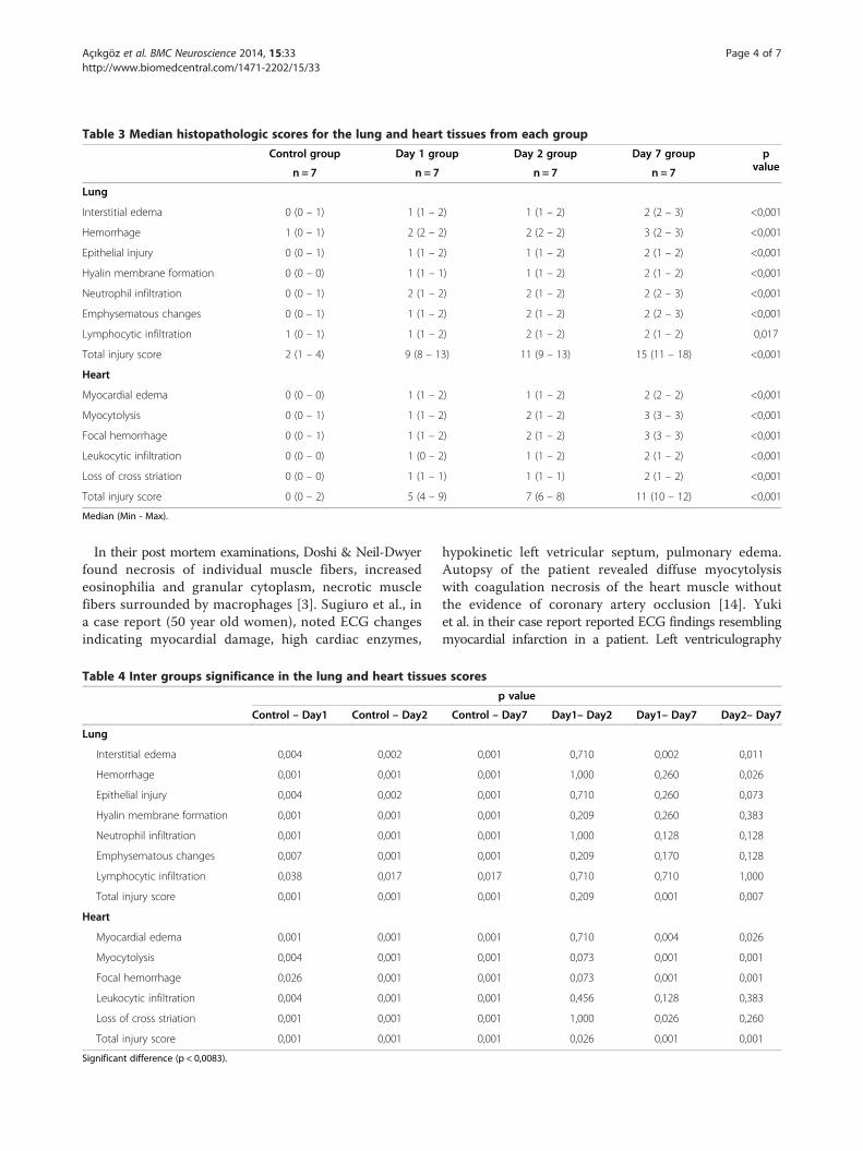
Table 3 Median histopathologic scores for the lung and heart tissues from each group
Control group Day 1 group Day 2 group Day 7 group pvaluen = 7 n = 7 n = 7 n = 7
Lung
Interstitial edema 0 (0 – 1) 1 (1 – 2) 1 (1 – 2) 2 (2 – 3) <0,001
Hemorrhage 1 (0 – 1) 2 (2 – 2) 2 (2 – 2) 3 (2 – 3) <0,001
Epithelial injury 0 (0 – 1) 1 (1 – 2) 1 (1 – 2) 2 (1 – 2) <0,001
Hyalin membrane formation 0 (0 – 0) 1 (1 – 1) 1 (1 – 2) 2 (1 – 2) <0,001
Neutrophil infiltration 0 (0 – 1) 2 (1 – 2) 2 (1 – 2) 2 (2 – 3) <0,001
Emphysematous changes 0 (0 – 1) 1 (1 – 2) 2 (1 – 2) 2 (2 – 3) <0,001
Lymphocytic infiltration 1 (0 – 1) 1 (1 – 2) 2 (1 – 2) 2 (1 – 2) 0,017
Total injury score 2 (1 – 4) 9 (8 – 13) 11 (9 – 13) 15 (11 – 18) <0,001
Heart
Myocardial edema 0 (0 – 0) 1 (1 – 2) 1 (1 – 2) 2 (2 – 2) <0,001
Myocytolysis 0 (0 – 1) 1 (1 – 2) 2 (1 – 2) 3 (3 – 3) <0,001
Focal hemorrhage 0 (0 – 1) 1 (1 – 2) 2 (1 – 2) 3 (3 – 3) <0,001
Leukocytic infiltration 0 (0 – 0) 1 (0 – 2) 1 (1 – 2) 2 (1 – 2) <0,001
Loss of cross striation 0 (0 – 0) 1 (1 – 1) 1 (1 – 1) 2 (1 – 2) <0,001
Total injury score 0 (0 – 2) 5 (4 – 9) 7 (6 – 8) 11 (10 – 12) <0,001
Median (Min - Max).
Açıkgöz et al. BMC Neuroscience 2014, 15:33 Page 4 of 7http://www.biomedcentral.com/1471-2202/15/33
In their post mortem examinations, Doshi & Neil-Dwyerfound necrosis of individual muscle fibers, increasedeosinophilia and granular cytoplasm, necrotic musclefibers surrounded by macrophages [3]. Sugiuro et al., ina case report (50 year old women), noted ECG changesindicating myocardial damage, high cardiac enzymes,
Table 4 Inter groups significance in the lung and heart tissue
Control – Day1 Control – Day2
Lung
Interstitial edema 0,004 0,002
Hemorrhage 0,001 0,001
Epithelial injury 0,004 0,002
Hyalin membrane formation 0,001 0,001
Neutrophil infiltration 0,001 0,001
Emphysematous changes 0,007 0,001
Lymphocytic infiltration 0,038 0,017
Total injury score 0,001 0,001
Heart
Myocardial edema 0,001 0,001
Myocytolysis 0,004 0,001
Focal hemorrhage 0,026 0,001
Leukocytic infiltration 0,004 0,001
Loss of cross striation 0,001 0,001
Total injury score 0,001 0,001
Significant difference (p < 0,0083).
hypokinetic left vetricular septum, pulmonary edema.Autopsy of the patient revealed diffuse myocytolysiswith coagulation necrosis of the heart muscle withoutthe evidence of coronary artery occlusion [14]. Yukiet al. in their case report reported ECG findings resemblingmyocardial infarction in a patient. Left ventriculography
s scores
p value
Control – Day7 Day1– Day2 Day1– Day7 Day2– Day7
0,001 0,710 0,002 0,011
0,001 1,000 0,260 0,026
0,001 0,710 0,260 0,073
0,001 0,209 0,260 0,383
0,001 1,000 0,128 0,128
0,001 0,209 0,170 0,128
0,017 0,710 0,710 1,000
0,001 0,209 0,001 0,007
0,001 0,710 0,004 0,026
0,001 0,073 0,001 0,001
0,001 0,073 0,001 0,001
0,001 0,456 0,128 0,383
0,001 1,000 0,026 0,260
0,001 0,026 0,001 0,001
Açıkgöz et al. BMC Neuroscience 2014, 15:33 Page 5 of 7http://www.biomedcentral.com/1471-2202/15/33
revealed cardiac dysfunction. Two months later, in thepostmortem examination of the patient, after her deathfrom cancer, no evidence of myocardial necrosis wasrevealed [27]. The authors discussed coronary vasospasmand reversible postischemic stunned myocardium withoutnecrosis which might happened after SAH [27].In ischemic cardiac events, since intracellular enzymes
and proteins increase in serum after the occurence oftissue necrosis [18], they may be insufficient in the diag-nosis in the lack of necrosis or in cases where necrosisdid not happen yet. Sinha et al., examined IMA, ECGand cTnT in patients who were admitted to an emergencydepartment with acute chest pain. The sensitivity (82%),specifity (46%), negative predictive value (59%), positivepredictive value (72%) of IMA were calculated. Oncontrary the sensitivity of ECG (45%) and cTnT (20%)were noted. They underlined the possible role of IMAin detecting ischemia before necrosis happen [28]. Inour experimental SAH study, in rats, we could notdemonstrate significant changes in CK, AST and LDHtests when compared with control group at days 1, 2and 7. There were significant increases of IMA at 7thday after SAH compared with the control group. Weobserved that the serum albumin values for all groupsdid not differ from the control group, thus we can saythat high IMA in SAH may not be due to low albuminlevels.In the light microscopic examination myocardial ne-
crosis was not seen in all groups but total injury scoresof heart and lung tissues at days 1, 2 and 7 were foundincreased when compared to the control group. Also indays 1, 2 and 7 myocytolysis, which was described byTurillazzi as a specific histological marker of congestiveheart failure without relation to coronary blood flow,myocardial hypoxia and myocardial fibrosis [29], wasfound increased compared to the control group. Both totalinjury scores of heart and lung tissues and myocytolysis ofheart tissue at days 7 were found significantly increasedcompared to days 1 and 2.In literature research we did not find an experimental
study investigating IMA after experimental SAH.There were clinical studies in the literature referring
to increases of IMA after myocardial ischemia [19], tran-sient myocardial ischemia [21], acute coronary syndrome[22,28].There were studies investigating IMA changes after
pulmonary emboli [30], skeletal muscle ischemia [31-33]and cerebrovascular events including SAH [23,34].Zapica-Muniz et al., after skeletal muscle forearm is-
chemia test, in their volunteer subjects, found decreasedlevels of IMA at 1st, 3rd and 5th minutes compared tobaseline values, in addition to low ammonia and highlactate levels. In lactate serum pool the authors founddecreased levels of IMA after increases in lactic acid
concentrations. The authors underlined the decreaseddiagnostic sensitivity of IMA because of increased lactatelevels [31].Roy et al., in their patients with peripheral vascular
disease (PVD) and leg claudication, performed an exercisetest inducing leg ischemia and found decreased levels ofIMA during stress peak. They also performed a dobutaminstress echocardiography and found unchanged levels ofIMA. In their patients regional wall motion abnormalitieswere not detected. They noted ischemia of tissues otherthan myocardium might be responsible for decreases ofIMA [32].IMA levels were found increased in volunteers of a
marathon race for 24–48 hours [33]. In another studyFalkensammer et al. noted increased IMA levels after calfmuscle ischemia induced by exercise in volunteers [35].Sbarouni et al., in their review article, stated that the
changes of IMA after exercises were not homogenous,they might be due to ischemic areas in the gastrointestinaltract and skeletal muscle. They also underlined that therole of hemoconcentration seen after physical exercises,might cause increases of albumin serum levels and be-cause of decreased unbound cobalt to albumin, IMAmight be lowered. They noted interference of lacticacidosis with IMA analysis [36].Han et al. demonstrated increases in IMA after cere-
bral infarction, intracerebral hemorrhage and SAH (18patients) compared to control group and they stated apositive correlation between IMA and lipid levels. In thisstudy, patients with pulmonary emboli and coronary ar-tery thrombosis within 6 months were excluded [34].Gündüz et al. examined and compared the levels of IMA
in intracerebral hemorrhage, SAH and brain infarctionpatients within 24 hours after their symptoms started.They found increased levels of IMA compared to con-trol patients, and in brain infarction the increases weresignificant compared to SAH, and they stated that thisfinding might be used in differential diagnosis of SAHwith brain infarction. The authors excluded patients withacute coronary syndrome, acute myocardial infarction,cardiac insufficiency, and pulmonary embolism in theirstudy [23].
ConclusionIn our study we observed increased levels of IMA atdays 1 and 2 after SAH, in blood samples obtained bycardiac pucture (left ventricle). We also observed signifi-cantly higher levels of IMA at day 7. There was a posi-tive correlation between the IMA and cardiac totalinjury scores and the cardiac myocytolysis. Our study isan isolated study compared to clinical studies. In thelight of our findings we can state that the increases ofIMA might be related to a probable myocardial stressafter SAH. We believe there is the need for futher
Açıkgöz et al. BMC Neuroscience 2014, 15:33 Page 6 of 7http://www.biomedcentral.com/1471-2202/15/33
studies to find and to develope specific markers besidesIMA to detect cardiac stress which may cause the suddendeath of patients after SAH.
Competing interestsThe authors declare that they have no competing interests.
Authors’ contributionsSA conceived the study, in the design of the study, collected and analyzeddata, carried out biochemical tests, drafted the manuscript, interpreted data; NEparticipated collected and analyzed data; carried out in vivo experiments; FB,collected and analyzed data, participated in the carried out histopathology tests;MC, participated in the carried out biochemical tests; IOT participated in thedesign of the study, collected data; CB participated in the carried out statisticalanalysis; BA participated in the design of the study, carried out in vivoexperiments, interpreted data and coordination of the study. Moreover,all the authors have read and approved the final manuscript.
AcknowledgementsWe are grateful to Eileen Remley for her kindly help in the English editing ofthe manuscript.
Author details1Department of Biochemistry, Faculty of Medicine, Bülent Ecevit University(Formerly, Zonguldak Karaelmas University), 67630, Esenköy, Kozlu,Zonguldak, Turkey. 2Department of Neurosurgery, Faculty of Medicine, BülentEcevit University (Formerly, Zonguldak Karaelmas University), Zonguldak,Turkey. 3Department of Pathologia, Faculty of Medicine, Bülent EcevitUniversity (Formerly, Zonguldak Karaelmas University), Zonguldak, Turkey.4Department of Immunologia, Faculty of Medicine, Bülent Ecevit University(Formerly, Zonguldak Karaelmas University), Zonguldak, Turkey. 5Departmentof Biostatistics, Faculty of Medicine, Bülent Ecevit University (Formerly,Zonguldak Karaelmas University), Zonguldak, Turkey.
Received: 6 April 2013 Accepted: 12 February 2014Published: 24 February 2014
References1. Tung P, Kopelnik A, Banki N, Ong K, Ko N, Lawton MT, Gress D, Drew B,
Foster E, Parmley W, Zaroff J: Predictors of neurocardiogenic injury aftersubarachnoid hemorrhage. Stroke 2004, 35:548–551.
2. Lee VH, Oh JK, Mulvagh SL, Wijdicks EF: Mechanisms in neurogenic stresscardiomyopathy after aneurysmal subarachnoid hemorrhage. NeurocritCare 2006, 5:243–249.
3. Doshi R, Neil-Dwyer G: Hypothalamic and myocardial lesions aftersubarachnoid hemorrhage. J Neurol Neurosurg Psychiatry 1977, 40:821–826.
4. Masuda T, Sato K, Yamamoto S, Matsuyama N, Shimohama T, Matsunaga A,Obuchi S, Shiba Y, Shimizu S, Izumi T: Sympathetic nervous activity andmyocardial damage immediately after subarachnoid hemorrhage in aunique animal model. Stroke 2002, 33:1671–1676.
5. Sato K, Masuda T, Izumi T: Subarachnoid hemorrhage and myocardialdamage clinical and experimental studies. Jpn Heart J 1999, 40:683–701.
6. Mertes PM, Carteaux JP, Jaboin Y, Pinelli G, el Abassi K, Dopff C, Atkinson J,Villemot JP, Burlet C, Boulange M: Estimation of myocardial interstitialnorepinephrine release after brain death using cardiac microdialysis.Transplantation 1994, 57:371–377.
7. Hravnak M, Frangiskakis JM, Crago EA, Chang Y, Tanabe M, Gorcsan J 3rd,Horowitz MB: Elevated cardiac troponin I and relationship to persistenceof electrocardiographic and echocardiographic abnormalities afteraneurysmal subarachnoid hemorrhage. Stroke 2009, 40:3478–3484.
8. Lacy PS, Earle AM: A small animal model for electrocardiographicabnormalities observed after an experimental subarachnoidhemorrhage. Stroke 1983, 14:371–377.
9. Elrifai AM, Bailes JE, Shih SR, Dianzumba S, Brillman J: Characterizationof the cardiac effects of acute subarachnoid hemorrhage in dogs.Stroke 1996, 27:737–742.
10. Franco C, Khaled B, Afonso L, Raufi M: Acute subarachnoid hemorrhageand cardiac abnormalities: takotsubo cardiomyopathy or neurogenicstunned myocardium? A case report. Cases J 2010, 3:81.
11. Zaroff JG, Rordorf GA, Titus JS, Newell JB, Nowak NJ, Torchiana DF, Aretz HT,Picard MH, Macdonald RL: Regional myocardial perfusion afterexperimental subarachnoid hemorrhage. Stroke 2000, 31:1136–1143.
12. Schievink WI, Wijdicks EF, Parisi JE, Piepgras DG, Whisnant JP: Sudden deathfrom aneurysmal subarachnoid hemorrhage. Neurology 1995, 45:871–874.
13. Stevens RD, Nyquist PA: The systemic implications of aneurysmalsubarachnoid hemorrhage. J Neurol Sci 2007, 261:143–156.
14. Sugiura M, Yozawa Y, Kubo O, Kitamura K, Kimata S, Hagiwara H, Joshita H:Myocardial damage (myocytolysis) caused by subarachnoid hemorrhage.No To Shinkei 1985, 37:1155–1161.
15. Parekh N, Venkatesh B, Cross D, Leditschke A, Atherton J, Miles W, Winning A,Clague A, Rickard C: Cardiac troponin I predicts myocardial dysfunction inaneurysmal subarachnoid hemorrhage. J Am Coll Cardiol 2000, 36:1328–1335.
16. Sakr YL, Ghosn I, Vincent JL: Cardiac manifestations after subarachnoidhemorrhage: a systematic review of the literature. Prog Cardiovasc Dis2002, 45:67–80.
17. Fabinyi G, Hunt D, McKinley L: Myocardial creatine kinase isoenzyme inserum after subarachnoid hemorrhage. J Neurol Neurosurg Psychiatry 1977,40:818–820.
18. Storrow AB, Gibler WB: Chest pain centers: diagnosis of acute coronarysyndromes. Ann Emerg Med 2000, 35:449–461.
19. Bar‐Or D, Lau E, Winkler JV: A novel assay for cobalt‐albumin binding andits potential as a marker for myocardial ischemia a preliminary report.J Emerg Med 2000, 19:311–315.
20. Lippi G, Montagnana M, Guidi GC: Albumin cobalt binding and ischemiamodified albumin generation: an endogenous response to ischemia?Int J Cardiol 2006, 108:410–411.
21. Bar-Or D, Winkler JV, Vanbenthuysen K, Harris L, Lau E, Hetzel FW: Reducedalbumin-cobalt binding with transient myocardial ischemia after electivepercutaneous transluminal coronary angioplasty: a preliminary comparisonto creatine kinase-MB, myoglobin, and troponin I. Am Heart J 2001,141:985–991.
22. Immanuel S, Sanjaya AI: Albumin cobalt binding (ACB) test: its role as anovel marker of acute coronary syndrome. Acta Med Indones 2006, 38:92–96.
23. Gunduz A, Turedi S, Mentese A, Altunayoglu V, Turan I, Karahan SC, TopbasM, Aydin M, Eraydin I, Akcan B: Ischemia-modified albumin levels incerebrovascular accidents. Am J Emerg Med 2008, 26:874–878.
24. Barry KJ, Gogjian MA, Stein BM: Small animal model for investigation ofsubarachnoid hemorrhage and cerebral vasospasm. Stroke 1979, 10:538–541.
25. Van der Bilt IA, Hasan D, Vandertop WP, Wilde AA, Algra A, Visser FC, RinkelGJ: Impact of cardiac complications on outcome after aneurysmalsubarachnoid hemorrhage: a meta-analysis. Neurology 2009, 72:635–642.
26. Szabo MD, Crosby G, Hurford WE, Strauss HW: Myocardial perfusionfollowing acute subarachnoid hemorrhage in patients with an abnormalelectrocardiogram. Anesth Analg 1993, 76:253–258.
27. Yuki K, Kodama Y, Onda J, Emoto K, Morimoto T, Uozumi T: Coronaryvasospasm following subarachnoid hemorrhage as a cause of stunnedmyocardium. Case report J Neurosurg 1991, 75:308–311.
28. Sinha MK, Roy D, Gaze DC, Collinson PO, Kaski JC: Role of “Ischemia modifiedalbumin”, a new biochemical marker of myocardial ischaemia, in the earlydiagnosis of acute coronary syndromes. Emerg Med J 2004, 21:29–34.
29. Turillazzi E, Baroldi G, Silver MD, Parolini M, Pomara C, Fineschi V: Asystematic study of a myocardial lesion: colliquative myocytolysis. Int JCardiol 2005, 104:152–157.
30. Turedi S, Gunduz A, Mentese A, Karahan SC, Yilmaz SE, Eroglu O, Nuhoglu I,Turan I, Topbas M: Value of ischemia-modified albumin in the diagnosisof pulmonary embolism. Am J Emerg Med 2007, 25:770–773.
31. Zapico-Muniz E, Santalo-Bel M, Merce-Muntanola J, Montiel JA,Martinez-Rubio A, Ordonez-Llanos J: Ischemia-modified albumin duringskeletal muscle ischemia. Clin Chem 2004, 50:1063–1065.
32. Roy D, Quiles J, Sharma R, Sinha M, Avanzas P, Gaze D, Kaski JC: Ischemia-modified albumin concentrations in patients with peripheral vasculardisease and exercise-induced skeletal muscle ischemia. Clin Chem 2004,50:1656–1660.
33. Apple FS, Quist HE, Otto AP, Mathews WE, Murakami MM: Releasecharacteristics of cardiac biomarkers and ischemia-modified albumin asmeasured by the albumin cobalt-binding test after a marathon race.Clin Chem 2002, 48:1097–1100.
34. Han K, Jia N, Yang L, Min LQ: Correlation between ischemia-modified al-bumin and lipid levels in patients with acute cerebrovascular disease.Mol Med Rep 2012, 6:621–624.
Açıkgöz et al. BMC Neuroscience 2014, 15:33 Page 7 of 7http://www.biomedcentral.com/1471-2202/15/33
35. Falkensammer J, Stojakovic T, Huber K, Hammerer-Lercher A, Gruber I,Scharnagl H, Fraedrich G, Santner W, Schocke M, Greiner A: Serum levels ofischemia-modified albumin in healthy volunteers after exercise-inducedcalf-muscle ischemia. Clin Chem Lab Med 2007, 45:535–540.
36. Sbarouni E, Georgiadou P, Kremastinos DT, Voudris V: Ischemia modifiedalbumin: is this marker of ischemia ready for prime time use? Hellenic JCardiol 2008, 49:260–266.
doi:10.1186/1471-2202-15-33Cite this article as: Açıkgöz et al.: Ischemia modified albumin increaseindicating cardiac damage after experimental subarachnoid hemorrhage.BMC Neuroscience 2014 15:33.
Submit your next manuscript to BioMed Centraland take full advantage of:
• Convenient online submission
• Thorough peer review
• No space constraints or color figure charges
• Immediate publication on acceptance
• Inclusion in PubMed, CAS, Scopus and Google Scholar
• Research which is freely available for redistribution
Submit your manuscript at www.biomedcentral.com/submit
Related Documents