DRAFT- DO NOT CITE Is there a social brain? 1 Draft of a chapter to appear in Other Minds, a book edited by Bertram Malle & Sarah Hodges Is there a ‘social brain’? Lessons from eye-gaze following, joint attention, and autism Diego Fernandez-Duque & Jodie A. Baird Villanova University The aim of this volume is to explore what it means to understand other minds. Drawing on philosophical, developmental, and psychological perspectives, the chapters in this book address a myriad of issues including how an understanding of minds develops, its significance for social interaction, and its relation to other social and cognitive achievements. Our chapter brings yet another perspective to bear on these issues. Taking a neuroscientific approach, our discussion centers on how understanding other minds – a central aspect of social-information processing – is represented in the brain. Currently in the literature there are two very different models of how the brain is organized for processing social information. One model, advocated by evolutionary psychologists, poses the existence of a ‘social brain’. On this view, the human mind has evolved a set of domain-specific solutions to particular problems. As a consequence, there exists a set of mental systems for perceiving and reasoning about social stimuli, which act independently from the mental systems involved in perceiving and reasoning about non-social things. It is the content of the information – its social nature – that determines the dichotomy, rather than the structure of the problem (Cosmides & Tooby, 1992). These social modules are thought to be unique in their input (i.e., the information they process) as well as in their mechanism (i.e., the rules that govern their processing). Finally, their content-specificity is ostensibly caused by phylogenetic evolution rather than by familiarity, expertise, or ontogenetic development. In its neuroscientific strand, the

Welcome message from author
This document is posted to help you gain knowledge. Please leave a comment to let me know what you think about it! Share it to your friends and learn new things together.
Transcript
DRAFT- DO NOT CITE Is there a social brain? 1
Draft of a chapter to appear in Other Minds, a book edited by Bertram Malle & Sarah Hodges
Is there a ‘social brain’? Lessons from eye-gaze following, joint attention, and autism
Diego Fernandez-Duque & Jodie A. Baird
Villanova University
The aim of this volume is to explore what it means to understand other minds. Drawing
on philosophical, developmental, and psychological perspectives, the chapters in this book
address a myriad of issues including how an understanding of minds develops, its significance
for social interaction, and its relation to other social and cognitive achievements. Our chapter
brings yet another perspective to bear on these issues. Taking a neuroscientific approach, our
discussion centers on how understanding other minds – a central aspect of social-information
processing – is represented in the brain.
Currently in the literature there are two very different models of how the brain is
organized for processing social information. One model, advocated by evolutionary
psychologists, poses the existence of a ‘social brain’. On this view, the human mind has evolved
a set of domain-specific solutions to particular problems. As a consequence, there exists a set of
mental systems for perceiving and reasoning about social stimuli, which act independently from
the mental systems involved in perceiving and reasoning about non-social things. It is the content
of the information – its social nature – that determines the dichotomy, rather than the structure of
the problem (Cosmides & Tooby, 1992). These social modules are thought to be unique in their
input (i.e., the information they process) as well as in their mechanism (i.e., the rules that govern
their processing). Finally, their content-specificity is ostensibly caused by phylogenetic evolution
rather than by familiarity, expertise, or ontogenetic development. In its neuroscientific strand, the

DRAFT- DO NOT CITE Is there a social brain? 2
view posits the existence of dedicated brain areas for the processing of social information
(Duchaine, Cosmides, & Tooby, 2001; Stone, 2002).
A different model, with origins in information-processing research, views the mind as a
set of subsystems, each of which processes a collection of basic computations. Although the
model allows for the existence of domain-specific processes and algorithms, this is not its
defining feature. Rather, what dictates which brain areas become engaged are the computational
operations necessary to solve the problem. For example, tasks requiring the suppression of
prepotent stimuli in favor of internally generated responses tend to activate a common network
of brain regions. Domain specificity may exist in the sense that motor suppression and verbal
suppression may engage neighboring brain areas. However, this is not the defining feature; rather
it is the specific computations performed by particular brain areas. Thus, some areas will perform
suppression of prepotent representations, others will be engaged in the perception of effort, and
still others will maintain representations in a mental buffer (Fernandez-Duque & Johnson, 2002).
Most cognitive neuroscience research in attention, memory, imagery, and executive functions
has followed this information-processing approach (Posner & Raichle, 1994).
Tasks of social relevance often engage the same network of brain areas. At first glance,
this appears consistent with the idea of a ‘social brain’. However, those brain areas are
sometimes driven by non-social tasks too, which contradicts the ‘social brain’ argument. A main
goal of this chapter is to put forward a synthesis that accommodates these disparate results. In
agreement with information-processing models, we argue that there is no ‘social brain’ but rather
isolable subsystems that perform basic computations. But how does this explain that social tasks
tend to activate the same network of brain areas? We argue that the recruitment of similar areas
by social stimuli happens because social stimuli disproportionately tap onto certain basic
DRAFT- DO NOT CITE Is there a social brain? 3
computations, such as those related to affective processes. Importantly, this correlation between
social stimuli and basic computations is not perfect, leaving room for non-social stimuli to
engage those same brain areas, and also allowing social stimuli not to engage those areas on
occasion. The absence of a perfect correlation between social stimuli and basic computational
operations also makes the distinction between these two levels useful and theoretically
important, rather than a mere re-description of the same phenomena.
As evidence for our main thesis, we focus on one social skill in particular – namely, the
ability to detect and follow eye gaze. We review behavioral and neurological findings on eye-
gaze following, and discuss its relation to other perceptual skills of social importance, such as
facial emotion recognition and face identification (in depth reviews of these other skills already
exist in the literature: for facial emotion recognition see Blair, 2003; Calder, Lawrence, &
Young, 2001; for face identification see Kanwisher, 2000). Next, we discuss whether eye-gaze
following is related to mental state attributions and joint attention. We end with a discussion of
how the impairment of these abilities in autism may both shed light on their typical development
and provide answers to how the brain is organized in its processing of social information.
Eye-Gaze Perception: Behavioral Evidence
Both in humans and other primates, the ability to follow the direction of gaze is of great
importance for social interaction. Primates such as chimpanzees and macaques spontaneously
follow the eye gaze of con-specifics, and direction of gaze conveys social dominance
(Tomasello, Call, & Hare, 1998). Humans automatically cue their attention in the direction of
gaze: 3-month-old infants can follow perceived gaze (Hood, Willen, & Driver, 1998), and even
newborns can discriminate between direct and averted gaze, thus suggesting a strong genetic
determinism with little environmental variability (Farroni, Csibra, Simion, & Johnson, 2002).
DRAFT- DO NOT CITE Is there a social brain? 4
What is less clear is whether and to what extent these processes depend on mental state
attributions. In non-human primates, eye-gaze following appears to be a bottom-up, non-
mentalistic process (Povinelli, Bering, & Giambrone, 2000). In human adults, mental state
inferences may not be necessary, even if mental attributions often co-exist with gaze following.
This counterintuitive fact is well known to any basketball player who has found herself following
the adversary’s gaze despite knowing his intention to deceive. In experimental paradigms,
subjects automatically follow the direction of the eyes even when eye gaze predicts that the
target will occur in the opposite location (Driver, Davis, Ricciardelli, Kidd, Maxwell, & Baron-
Cohen, 1999; Langton & Bruce, 1999). These findings, together with the early development of
eye-gaze cueing in infancy, suggest a rigid, non-mentalistic system of gaze following that is
driven bottom-up by the physical properties of the stimulus, such as the relation of the dark iris
to the white sclera (Anstis, Mayhew, & Morley, 1969; Ricciardelli, Baylis, & Driver, 2000) and
possibly also by the orientation of the head (Langton, Watt, & Bruce, 2000).
Eye-Gaze Perception: Neurological Evidence
The system for eye-gaze detection is ideal for exploring neural mechanisms of relevance
to social interactions, even if in itself, eye-gaze detection is an elementary part of social
behavior. For one, eye-gaze perception can be explored in non-human primates using methods
such as lesions and single neuron recordings, yielding highly specific, localized information on
the brain areas involved. Second, since eye-gaze perception is relatively independent from
language, it is less problematic to make generalizations across species. Finally, since eye-gaze
detection is driven by external stimuli, it is possible to systematically manipulate the system.
The first question to address is whether there is an isolable neural system for eye-gaze
detection in its most rudimentary form. An affirmative answer would require a direct mapping of
DRAFT- DO NOT CITE Is there a social brain? 5
function to brain region. Such direct mapping is admittedly a philosophically contentious issue,
which includes the difficulty of conceptually defining ‘function’. Nevertheless, over the last two
decades, neuroscience has provided several good examples of direct mapping. Here we briefly
mention two of them, before describing the eye-gaze system.
The first example is a region of the posterior temporal lobe, area MT, which has been
implicated as a direct neural correlate of perceived visual movement. Area MT is activated by
the aware perception of motion (Tootell, Reppas, Dale, Look, et al., 1996), its neurons respond
selectively to the direction and velocity of movement, and their electrical stimulation biases the
behavioral response to moving stimuli (Salzman, Murasugi, Britten, & Newsome, 1992). All
these factors suggest that area MT is a specialized subsystem that computes direction and
velocity of motion. The second example is the face fusiform area (FFA), an area of the ventral
temporo-occipital cortex that responds selectively to faces. Subjective experience of faces
activates face fusiform area even in the absence of sensory stimuli, as in the case of visual
imagery (O’Craven & Kanwisher, 2000). Lesion to this area leads to impaired recognition of
facial identity (Damasio, Damasio, & Van Hoesen, 1982). Thus, it appears that face fusiform
area encodes structural aspects of faces, or possibly, other familiar stimuli that are decoded
globally. Both area MT and FFA are specialized subsystems, but this does not mean that they are
informationally encapsulated. In fact, attending to movement increases MT activation, and
attention to face processing increases FFA activation (Hoffman & Haxby, 2000; O’Craven,
Rosen, Kwong, Treisman, & Savoy, 1997).
Along the same lines, several studies have pointed to an area of the posterior part of
superior temporal sulcus (STS) as critical for the encoding of eye gaze. In neuroimaging studies,
this area is activated by faces changing the direction of gaze, and even by static displays of faces

DRAFT- DO NOT CITE Is there a social brain? 6
with averted eyes (for a review, see Allison, Puce, & McCarthy, 2000). Furthermore, when the
task requires that subjects pay attention to eye gaze, the activity of STS is increased, while the
activation of FFA, the area that encodes the structural aspect of faces, remains invariant (as
expected, paying attention to face identity leads to the opposite pattern) (Hoffman & Haxby,
2000). Single neuron recording studies in monkeys reveal some neurons in the anterior part of
STS that respond specifically to gaze direction (Perrett, Hietanen, Oram, & Benson, 1992), and
gaze perception is disrupted by lesions to this area (Campbell, Heywood, Cowey, Regard, &
Landis, 1990). Finally, STS has functional connectivity with regions important for shifting
visuospatial attention to the periphery, such as the intraparietal sulcus, thus being an integral part
of the neural circuit for eye-gaze following (George, Driver, & Dolan, 2001).
The Relation of Eye-Gaze Perception to Other Aspects of Facial Information Processing:
Behavioral and Electrophysiological Evidence
Even though eye-gaze perception depends critically on the STS, the impact of eye gaze
on behavior is modulated by other factors. For example, whether attention is sustained in the
direction of gaze depends on the facial emotion. When the person whose eyes serve as a cue
looks happy, attention is sustained, but when the person looks angry, attention is relocated
somewhere else (Fenske, Frischen, & Tipper, 2004). Thus, faces expressing emotion modulate
gaze-evoked shifts of attention. Fearful faces are more effective at cueing attention than neutral
faces, at least in subjects who are anxiety-prone (Mathews, Fox, Yiend, & Calder, 2003). Direct
gaze in primates indicates social dominance and is part of the threat display, while in humans
direct gaze may indicate intimacy or aggression, depending on the accompanying facial emotion.
Faces with direct gaze are easier to identify than faces with averted gaze, suggesting that gaze
also interacts with facial recognition (Macrae, Hood, Milne, Rowe, & Mason, 2002).
DRAFT- DO NOT CITE Is there a social brain? 7
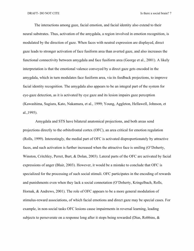
The interactions among gaze, facial emotion, and facial identity also extend to their
neural substrates. Thus, activation of the amygdala, a region involved in emotion recognition, is
modulated by the direction of gaze. When faces with neutral expression are displayed, direct
gaze leads to stronger activation of face fusiform area than averted gaze, and also increases the
functional connectivity between amygdala and face fusiform area (George et al., 2001). A likely
interpretation is that the emotional valence conveyed by a direct gaze gets encoded in the
amygdala, which in turn modulates face fusiform area, via its feedback projections, to improve
facial identity recognition. The amygdala also appears to be an integral part of the system for
eye-gaze detection, as it is activated by eye gaze and its lesion impairs gaze perception
(Kawashima, Sugiura, Kato, Nakamura, et al., 1999; Young, Aggleton, Hellawell, Johnson, et
al.,1995).
Amygdala and STS have bilateral anatomical projections, and both areas send
projections directly to the orbitofrontal cortex (OFC), an area critical for emotion regulation
(Rolls, 1999). Interestingly, the medial part of OFC is activated disproportionately by attractive
faces, and such activation is further increased when the attractive face is smiling (O’Doherty,
Winston, Critchley, Perret, Burt, & Dolan, 2003). Lateral parts of the OFC are activated by facial
expressions of anger (Blair, 2003). However, it would be a mistake to conclude that OFC is
specialized for the processing of such social stimuli. OFC participates in the encoding of rewards
and punishments even when they lack a social connotation (O’Doherty, Kringelbach, Rolls,
Hornak, & Andrews, 2001). The role of OFC appears to be a more general modulation of
stimulus-reward associations, of which facial emotions and direct gaze may be special cases. For
example, in non-social tasks OFC lesions cause impairments in reversal learning, leading
subjects to perseverate on a response long after it stops being rewarded (Dias, Robbins, &

DRAFT- DO NOT CITE Is there a social brain? 8
Roberts, 1996; Fellows & Farah, 2003; Hornak, O'Doherty, Bramham, Rolls, Morris, Bullock, &
Polkey, 2004). These findings are a good example of how a brain area can be recruited by social
stimuli even if the area also participates in other, non-social processes.
Eye-Gaze Processing: Social Module or Information-Processing?
The behavioral, functional, and anatomical results described above reveal a network of
brain structures, including area MT, FFA, STS, amygdala, and OFC, which act in concert to
compute many aspects of facial information that are important for social interactions. Much like
the findings from other cognitive systems, these results reveal a division of labor, with each area
performing a computation that is smaller than the task as a whole. At the same time, each
subsystem is heavily modulated by the others, and by general resources such as the allocation of
attention, thus arguing against any type of strong modularity.
The available evidence also argues against a brain area – or even a network of areas –
dedicated to the exclusive processing of social stimuli. For example, area MT responds to
biological motion, but it also responds to random dot movement (O’Craven et al., 1997; Salzman
et al., 1992). STS is sensitive to eye gaze, but it is also sensitive to aspects of language (Martin,
2003), semantic knowledge (Chao, Haxby, & Martin, 1999), visuo-spatial attention, and target
detection (Corbetta & Shulman, 2002). The amygdala is important for eye-gaze detection and the
recognition of facial expressions of fear, but it also processes many other aspects of fear, for
example, Pavlovian classical conditioning, and response to aversive tastes and odors (Büchel &
Dolan, 2000; Davis & Whalen, 2001). Orbito-frontal cortex may be important for recognizing
certain facial emotions, but it is also critical for modulating non-social stimulus-reward
associations, such as the relation between key press and food. Finally, although parts of the
fusiform gyrus are particularly sensitive to faces, which are a type of social stimuli, these same

DRAFT- DO NOT CITE Is there a social brain? 9
brain areas are also sensitive to non-social stimuli that are globally encoded, such as expert
recognition of cars or birds (Gauthier & Nelson, 2001, but see Farah, Rabinowitz, Quinn, & Liu,
2000, Grill-Spector, Knouf, & Kanwisher, 2004).
In sum, there is a network of brain regions disproportionately engaged by social stimuli
such as faces. But those areas are also engaged in basic computations of no social relevance.
Thus, their engagement by social stimuli is insufficient proof for the existence of a ‘social brain’.
Rather, it suggests that social stimuli often carry particular properties – valence, reward value,
spatial arrangement of its features—that disproportionately tap certain basic operations.
Eye-Gaze Perception: A Window into Mental State Attribution?
There is no doubt that encoding and following eye gaze are important social skills for
both human and non-human primates. Nor is there any doubt that normal human adults often
make mental state attributions about the eye-gaze patterns they detect: When an agent directs his
eyes to an object, adults infer that he is seeing the object (i.e., creating a mental representation
that can be used for guiding future behavior, enriching knowledge, and so forth). For normal
human adults, the behavioral description (eye gaze) and its mentalistic re-description (seeing)
almost always go hand in hand. Moreover, during development, the co-variance between the
mentalistic and non-mentalistic levels (i.e., between the ‘looking at’ and the alignment of gaze
direction and attended object) may play a role in fostering the emergence of mental state
attribution. That is, such co-variance may help to bootstrap mental state attribution in typically
developing human infants who are experience-ready. But there is no principled reason why this
should be true in other populations. Newborns, monkeys, and individuals with autism may detect
and follow eye gaze without taking the extra step of attributing mental states to such behaviors
(Povinelli et al., 2000).

DRAFT- DO NOT CITE Is there a social brain? 10
On the whole, knowing about eye-gaze detection tells us little about whether mental state
attributions are being made. Similarly, identifying the brain areas involved in eye-gaze detection
tells us little about which areas are critical, or even necessary, for the attribution of mental states.
Just because behavior and mental states tend to co-occur does not mean that the same brain areas
involved in detecting the behavior would also be involved in the attribution of mental states to
that behavior. In fact, eye-gaze following implicates homologous brain areas in humans and in
species incapable of mental state attribution, such as macaque monkeys. This homology is
sometimes interpreted as evidence that those brain areas, in the course of phylogenetic evolution,
gave rise to mentalizing abilities (Frith, 2001). However, this is not necessarily the case. An
alternative view is that the across-species similarity in the brain mechanisms of eye-gaze
detection is in fact evidence against a central role of these structures in mental state attribution.
In other words, if both humans and macaques recruit homologous brain areas to detect eye gaze,
but only humans make mental state attributions, it is reasonable to conclude that mental state
attribution depends on some other brain structures. To put it bluntly, computing the direction of
gaze is not the same as using the direction of gaze to infer another’s intention, nor need it depend
on the same brain structures.
Development: From Reflexive Eye Cueing to Joint Attention
So far, we have been describing the rudimentary system of eye-gaze detection, but in
typically developing humans this rudimentary system becomes part of a more sophisticated
system rather quickly in development. At the age of 9 months, infants start using direction of
gaze in a more flexible manner that takes into account people’s communicative intentions, a skill
that forms part of what has been labeled ‘joint attention’. Joint attention is the ability to
coordinate attention between interactive social partners with respect to objects or events. It
DRAFT- DO NOT CITE Is there a social brain? 11
reveals an understanding by infants that adults are intentional agents; that is, that adults
voluntarily attend to objects and that their attention can be shared, directed, and followed.
Besides responding to another’s gaze shift, joint attention includes behaviors such as proto-
declarative pointing (i.e., pointing to refer to an object, as opposed to pointing to request an
object), imitative learning (i.e., acting on objects the way that adults are acting on them), and
social referencing (i.e., infants’ reference to adults for information about the approachability,
desirability, and other features of objects) (Tomasello & Rakoczy, 2003). All four of these skills
– the flexible use of eye-gaze following, proto-declarative pointing, imitative learning, and social
referencing-- are correlated, develop in synchrony between the ages of 9 and 24 months, and
show little influence from environmental variables. Across these behaviors, there seems to be the
expression of a single, underlying skill – the understanding of persons as intentional agents--
which is almost completely absent in other primates (Povinelli et al., 2000; Tomasello &
Rakoczy, 2003). As such, it may be the single, most important developmental milestone in the
understanding of other minds. At the same time, the features that make it so remarkable, namely
its early emergence in development and its exclusivity to humans, are the same that make it
inaccessible to most of the methods of cognitive neuroscience.1 Although adult neuropsychology
and neuroimaging provide good approximations and hypotheses, it would be erroneous to simply
1 Currently, there are two non-invasive neuroimaging methods suitable for infant studies: event-related potentials (ERPs) and event-related optical signal (EROS). Other methodologies are either too invasive (e.g., positron emission tomography, PET) or require subjects to remain still for several minutes (fMRI). ERPs are recordings of electrical activity from the scalp, averaged over many trials time-locked to the stimulus onset. It has an excellent temporal resolution of 4 ms, and therefore can be used to determine the stage in information processing at which two conditions diverge. In contrast, its spatial resolution is only modest, due to the many layers of tissue that exist between the brain source and the surface of the scalp, which tend to disperse the signal. Certain methodological considerations, such as the contamination by muscle activity, require that subjects are compelled not move their eyes. In EROS an external source emits near infra-red light from the scalp surface. This light is scattered by the brain and received by a detector, also in the scalp surface. Neuronal activity modulates the optical scattering properties of the brain, and this is used to assess which brain areas are active. Unlike ERPs, EROS has not only good temporal resolution but also good spatial resolution. However, only certain regions of the brain surface are suitable to the techinque. Although it is a very recent methodology, EROS is a promising approach for studying cognitive neuroscience in human infants (Gratton, 2001).
DRAFT- DO NOT CITE Is there a social brain? 12
extrapolate from these models to the case of development (Karmiloff-Smith, 1998). Therefore,
researchers have been interested in diseases of development, autism in particular, to provide a
more complete picture of how these developing abilities map onto brain structures.
Developmental Disorders: The Case of Autism
Autism is a neurodevelopmental disorder with a strong genetic component and a
heterogeneous neurological substrate. Abnormalities have been reported in the limbic system
(anterior cingulate, amygdala, hippocampus and orbitofrontal cortex), cerebellum, frontal lobes,
superior temporal gyrus, and subcortical structures including the thalamus and the basal ganglia
(Lord, Cook, Leventhal, & Amaral, 2000). This broad range of neurological abnormalities is
matched at the behavioral level by a broad phenotype that includes motor, linguistic, social, and
emotional deficits (Joseph, 1999). Individuals with autism exhibit stereotypic and repetitive
motor behavior, and their use of language is both delayed and disrupted. At its core, however,
autism is a disorder of social interaction and communication.
Poor eye-gaze following is a specific marker of autism, and one of its earliest signs,
evident in children as young as 18 months of age. Some children with autism even fail to use
gaze as a cue to locate an object (Leekam & Moore, 2001). This is especially remarkable given
that chimpanzees, who are incapable of joint attention, can nevertheless use gaze as an
instrumental cue (Povinelli & Eddy, 1997). In one study, school-aged children with autism were
tested in a naturalistic environment for their ability to use eye gaze as an orienting cue. In the
autistic group, children with high mental age performed normally, but those with low mental age
were impaired relative to developmentally delayed children matched for mental age. Autistic
children of low mental age were capable of following gaze when a target was observable --a skill
that emerges at 6 months of age in typically developing infants -- but were impaired when the

DRAFT- DO NOT CITE Is there a social brain? 13
target was absent – a skill that normally develops at the age of 9 months and that indicates the
emergence of joint attention (Leekam & Moore, 2001). Another study tested high-functioning
10-year-olds with autism in a computer-based paradigm and found normal cueing effects
(Sweettenham, Condie, Campbell, Milne, & Coleman, 2003). Using a variant of the Posner
cueing task, each trial displayed a face with averted gaze and a peripheral target in close
temporal sequence. Although the direction of gaze did not reliably predicted the location of the
future target, responses were fastest when gaze was directed to the target location. This effect
was present when the target appeared just 100 ms after the cue, consistent with an automatic
cueing of attention. Thus, consistent with the findings from Leekam and Moore (2001), reflexive
eye cueing appears to be spared in high-functioning children with autism. Taken together, these
two studies suggest that eye-gaze cueing deficits in autism vary according to the severity of
impairment in general intelligence.
In contrast, joint attention deficits in autism occur independently of general intelligence,
and are an early marker of the disease. Children with autism show deviant patterns of reciprocal
gaze behavior with their caregivers as well as deficits in the triadic coordination among
themselves, adult, and object (Charman, 2003). Interestingly, joint attention in 3- and 4-year-old
children with autism is positively correlated with orbitofrontal function, as measured by tasks
that engage this region in normal subjects (Dawson et al., 2002).2 Orbitofrontal cortex is
necessary for adding flexibility to stimulus-reward associations (Fellows & Farah, 2003; Rolls,
1999). An inability to assign stimulus-reward associations and flexibly modify them could be
detrimental to the development of joint attention, as joint attention depends on social rewards,
2 Ventro-medial prefrontal function was measured with tasks that are disrupted by lesions to the orbitofrontal cortex in monkeys and human adults. These are tasks that require a modification of behavioral response when a particular response is no longer rewarded, such as the delayed nomatching to sample with brief delay task and object discrimination reversal task.
DRAFT- DO NOT CITE Is there a social brain? 14
such as smiles, that are more variable than non-social rewards. Consistent with this hypothesis,
autistic infants and toddlers prefer highly contingent, non-variable feedback, while typically
developing children instead prefer variable, imperfect feedback (Gergely & Watson, 1999). The
contingency hypothesis illustrates the main problem of arguing for a module devoted exclusively
to social stimuli. Even if certain brain regions do process social stimuli preferentially, it does not
follow that such preferential processing is due to the ‘social’ nature of the stimuli. In the
aforementioned example, the driving force is the variability of the stimulus-reward association,
which is typical of social stimuli but can present in non-social stimuli.
Individuals with autism also show abnormalities in the processing of facial and emotional
stimuli. ERPs (event-related potentials) reveal that children with autism are impaired in the
discrimination of novel versus familiar faces, despite their normal discrimination of novel versus
familiar objects. In adults with autism, faces trigger reduced activity in fusiform area, amygdala,
and superior temporal sulcus, among other areas (Critchley, Daly, Bullmore, & Williams, 2000).
One possible interpretation of these data is that they provide evidence for a social-perception
module, whose impairment in autism accounts for the social deficits exhibited by this group.
Although this analysis captures much of what is wrong in autism, it makes the same mistake
previously described in our analysis of stimulus-reward associations. Just because social stimuli
such as faces happen to activate face fusiform area, it does not follow that faces activate this area
because they are stimuli of social relevance. Non-social stimuli such as cars and birds can
similarly activate this region, provided that observers are experts in recognizing those objects
and encode them globally. The critical factor, therefore, is not the social nature of the stimulus
but its holistic encoding. In fact, preliminary evidence suggests that the face fusiform area of
individuals with autism is activated when they are engaged in holistic encoding of non-social
DRAFT- DO NOT CITE Is there a social brain? 15
stimuli. For example, a child with autism who had great expertise in Digimon cartoon characters
– which presumably are non-social stimuli – showed face fusiform activation to Digimon figures
but not to human faces (Grelotti, Klin, Gauthier, Skudlarski, et al., 2004). Interestingly,
amygdala also activated in this child, suggesting that such figures were emotionally laden,
despite their lack of social relevance.
Of course, this is not to say that difficulties in face perception, emotion recognition, and
gaze following do not have consequences for mentalizing. On the contrary, such deficits may put
children with autism at a severe disadvantage in their development of mental state understanding.
Relative to typically developing children, children with autism have difficulties using the eyes as
cues for attributing mental states (Baron-Cohen, Wheelwright, Hill, Raste, & Plumb, 2001) and
in using faces to judge the approachability and trustworthiness of people (Adolphs, Sears, &
Piven, 2001). There is also evidence that children with autism have difficulties understanding
gaze in mentalistic terms. In one task, children were shown a cartoon face and asked whether the
character was looking straight ahead at them or was looking away. Children with autism were
capable of correctly discriminating between these two displays. However, when asked to select
which of four items (e.g., types of candy) the character liked best, the same children were unable
to use eye gaze to infer that the best-liked item was the one the character was staring at (Baron-
Cohen, 1995).
So, is autism best characterized as a deficit in a social module or as a general disorder of
basic processes that apply to both social and non-social domains? Individuals with autism have
deficits in joint attention and mental state attributions, but they also have motor deficits, visual
attention deficits, and deficits in feedback processing, set switching, executive processing, and
other non-social abilities (Joseph, 1999). Thus, although autism may present first and foremost as
DRAFT- DO NOT CITE Is there a social brain? 16
a problem of social cognition, it also manifests itself in non-social problems. This dual deficit
poses a challenge to both the social-module view (why should there be non-social deficits?) and
the domain-general view (why should the deficit be mostly social?). Our proposal is that the
solution to this paradox lies not in the brain itself but in the external stimuli that the brain
processes. More specifically, we argue that attributes of social stimuli may correlate with basic
information-processing computations. According to our framework, a deficit in the basic
computation would affect primarily, but not exclusively, the processing of social information.
We have already discussed the examples of variable stimulus-reward associations and holistic
visual encoding. In both cases, there is a correlation between the type of stimulus (social/non-
social) and a computational feature (variable/non-variable reward; holistic/feature-based
encoding). We are not claiming that these two basic computations account for all deficits in
autism; rather, we use these examples to illustrate the larger point that general computational
deficits may account for what appear to be domain-specific deficits.
Stimulus category (social/non-social) may correlate with other basic computations, such
as those involved in affective processing. Since social stimuli carry more affective valence than
non-social stimuli they should disproportionately tap limbic structures such as the amygdala and
the orbitofrontal cortex. Limbic areas are activated by affective stimuli such as faces. These brain
areas send feedback projections to regions of the temporal lobe including the fusiform gyrus.
During normal development, this affective loop is likely to play a role in modulating the
plasticity of the fusiform gyrus, which with experience becomes a dedicated system for face
recognition (i.e., the face fusiform area). To put it in psychological terms, faces are attractive
stimuli which capture the child’s attention, thus becoming the focus of preferred processing, and
with the passage of time, a stimulus the child is expert with. But for children with autism, faces
DRAFT- DO NOT CITE Is there a social brain? 17
do not seem to carry their appropriate valence, and thus these children seem uninterested in
faces. Within a social-module framework, there is little one could do to rectify this problem.
Within an information-processing framework, however, it should be possible to pair facial
stimuli with non-social rewards that are valued by the autistic child, and in this way engage the
affective loop. Through extensive training in such a paradigm, it might be possible for children
with autism to achieve the type of expertise with facial stimuli that typically developing children
gain naturally (Carver & Dawson, 2002).
Also related to the affective dimension, type of stimulus (social/non-social) may correlate
with orienting effectiveness. For example, while the orienting deficit in autism is most severe for
social stimuli, such as faces or being called by name, individuals with autism are also impaired in
their orienting to non-social stimuli, such as a jack-in-the-box (Dawson, Meltzoff, Osterling,
Rinaldi, & Brown, 1998). Interestingly, deficits in joint attention correlate with deficits in
orienting toward social stimuli but not with non-social orienting. This result is consistent with
the idea raised earlier that the co-variance of mental and non-mental levels (orienting to a
location, find a social stimuli at that location) may foster the mental state attribution process.
Conclusions
The re-description of social information into basic information-processing computations
may prove to be a powerful tool. We mention examples in the context of autism, but it is easy to
see how the logic can generalize to typical development. Importantly, such re-description
provides the system for processing social stimuli with a flexibility that is central to social
interactions, and that a social-module framework cannot easily account for. Imagine that
somebody stares at you with an angry face. Depending on the context, you might feel amused
(the person is an actor you are watching in a play), frightened (you are in a shady part of town),

DRAFT- DO NOT CITE Is there a social brain? 18
or disgusted (the person is the leader of the world superpower justifying his next imperialistic
adventure). In other words, it is easy to imagine that one and the same stimulus will be processed
very differently depending on the context (Lange, Williams, Young, Bullmore, et al., 2003).
Such context effects cannot be explained by models posing a strict separation of social and non-
social information. In contrast, an information-processing account can easily accommodate for
these contextual effects, as it assumes that general cognitive abilities can influence the
processing of information. Re-conceptualizing the social brain in terms of basic information
processes also allows researchers to ask ‘how’ – in computational terms – social information is
processed. Finally, it entrenches social neuroscience within the cognitive neuroscience tradition
that has been so fruitful over the last two decades. Just as the brain does not make a strict
separation between social and cognitive information, neither should brain researchers.

DRAFT- DO NOT CITE Is there a social brain? 19
Author Note
This research was supported by a HSFO postdoctoral fellowship # F4866 to Diego Fernandez-
Duque.
DRAFT- DO NOT CITE Is there a social brain? 20
References
Adolphs, R., Sears, L., & Piven, J. (2001). Abnormal processing of social information
from faces in autism. Journal of Cognitive Neuroscience, 13(2), 232-240.
Allison, T., Puce, A., & McCarthy, G. (2000). Social perception from visual cues: the
role of the STS region. Trends in Cognitive Science, 4(7), 267-278
Anstis, S. M., Mayhew, J. W., & Morley, T. (1969). The perception of where a face or
television 'portrait' is looking. American Journal of Psychology, 82, 474-489.
Baron-Cohen, S. (1995). Mindblindness: An essay on autism and theory of mind.
Cambridge, MA: MIT Press.
Baron-Cohen, S., Wheelwright, S., Hill, J., Raste, Y., & Plumb, I. (2001). The "Reading
the Mind in the Eyes" Test revised version: a study with normal adults, and adults with Asperger
syndrome or high-functioning autism. Journal of Child Psychology and Psychiatry and Allied
Disciplines, 42(2), 241-251.
Blair, R. J. R. (2003). Facial expressions, their communicatory functions and neuro-
cognitive substrates. Philosophical Transactions of the Royal Society of London. Series B, 358,
561-572.
Calder, A. J., Lawrence, A.D., & Young, A.W. (2001). Neuropsychology of fear and
loathing. Nature Reviews Neuroscience, 2, 352-363.
Campbell, R., Heywood, C. A., Cowey A., Regard M., & Landis T. (1990). Sensitivity to
eye gaze in prosopagnosic patients and patients with superior temporal sulcus.
Neuropsychologia, 28, 1123-1142.
Carver, L. J. D., G. (2002). Development and neural bases of face recognition in autism.
Molecular Psychiatry, 7, 18-20.
DRAFT- DO NOT CITE Is there a social brain? 21
Chao, L. L., Haxby, J. V., & Martin, A. (1999). Attribute-based neural substrates in
temporal cortex for perceiving and knowing about objects. Nature Neuroscience, 2, 913-919.
Charman, T. (2003). Why is joint attention a pivotal skill in autism. Philosophical
Transactions of the Royal Society of London, Series B, 358, 315-324.
Corbetta, M., & Shulman, G.L. (2002). Control of goal-directed and stimulus-driven
attention. Nature Reviews Neuroscience, 3, 215-229.
Cosmides, L., & Tooby, J. (1992). Cognitive adaptations for social exchange. In J.
Barkow, Cosmides, L., & Tooby, J. (Ed.), The adapted mind: Evolutionary psychology and the
generation of culture. New York: Oxford University Press.
Critchley, H. D., Daly, E. M., Bullmore, E.T., Williams, S. C. R., Van Amelsvoort, T.,
Robertson, D. M., Rowe, A., Phillips, M., McAlonan, G., Howlin, P., & Murphy, D. G. M.
(2000). The functional neuroanatomy of social behaviour: Changes in cerebral blood flow when
people with autistic disorder process facial expressions. Brain, 123, 2203-2212.
Damasio, A. R., Damasio, H., & Van Hoesen, G.W. (1982). Prosopagnosia: Anatomic
basis and behavioral mechanisms. Neurology, 33(331-341).
Davis, M., & Whalen, P. J. (2001). The amygdala: vigilance and emotion. Molecular
Psychiatry, 6, 13-34.
Dawson, G., Meltzoff, A. N., Osterling, J., Rinaldi, J., & Brown, E. (1998). Children with
autism fail to orient to naturally occurring social stimuli. Journal of Autism and Developmental
Disorders, 28(6), 479-485.
Dawson, G., Munson, J., Estes, A., Osterling, J., McPartland, J., Toth, K., Carver, L., &
Abbott, R. (2002). Neurocognitive function and joint attention ability in young children with
autism spectrum disorder versus developmental delay. Child Development, 73(2), 345-358
DRAFT- DO NOT CITE Is there a social brain? 22
Dias, R., Robbins, T. W., & Roberts, A. C. (1996). Dissociation in prefrontal cortex of
affective and attentional shifts. Nature, 380, 69-72.
Driver, J., Davis, G., Ricciardelli, R. Kidd, P., Maxwell, E.& Baron-Cohen, S. (1999).
Gaze perception triggers reflexive visuospatial orienting. Visual Cognition, 6(5), 509-540.
Duchaine, B. C., Cosmides, L., & Tooby, J. (2001). Evolutionary psychology and the
brain. Current Opinion in Neurobiology, 11, 225-230.
Farah, M. J., Rabinowitz, C., Quinn, G. E., & Liu, G. T. (2000). Early commitment of
neural substrates for face recognition. Cognitive Neuropsychology, 17, 117-123.
Farroni, T., Csibra, G., Simion, F., & Johnson, M. H. (2002). Eye contact detection in
humans from birth. Proceedings of the National Academy of Science, 99(14), 9602-9605.
Fellows, L. K., & Farah, M. J. (2003). Ventromedial frontal cortex mediates affective
shifting in humans: evidence from a reversal learning paradigm. Brain, 126, 1830-1837.
Fenske, M. J., Frischen, A., & Tipper, S. P. (submitted). Faces expressing emotion
modulate gaze-evoked shifts of attention.
Fernandez-Duque, D., & Johnson, M. L. (2002). Cause and effect theories of attention:
The role of conceptual metaphors. Review of General Psychology, 6(2), 153-165.
Frith, U. (2001). Mind blindness and the brain in autism. Neuron, 32, 969-979.
Gauthier, I., & Nelson, C. A. (2001). The development of face expertise. Current Opinion
in Neurobiology, 11, 219-224.
George, N., Driver, J., & Dolan, R. J. (2001). Seen gaze-directio modulates fusiform
activity and its coupling with other brain areas during face processing. Neuroimage, 13, 1102-
1112.

DRAFT- DO NOT CITE Is there a social brain? 23
Gergely, G., & Watson, J. S,. (1999). Early socio-emotinal development: Contingency
perception and the social-biofeedback model. In P. Rochat (Ed.), Early social cognition:
Understanding others in the first months of life (pp. 101-136). Mahwah, NJ: Erlbaum.
Gratton, G. (2001). Shedding light on brain function: the event-related optical signal.
Trends in Cognitive Science, 5(8), 357-363.
Grelotti, D. J., Klin, A. J., Gauthier, I., Skudlarski, P., Cohen, D. J., Gore, J. C.,
Volkmar, F. R., & , & Schultz, R. T. (submitted). fMRI activation of the fusiform gyrus and
amygdala to cartoon characters but not to faces in a boy with autism.
Grill-Spector, K., Knouf, N., & Kanwisher, N. (2004). The fusiform face area subserves
face perception, not generic within-category. Nature Neuroscience, 555 - 562.
Hoffman, E. A., & Haxby, J. V. (2000). Distinct representations of eye gaze and identity
in the distributed human neural system for face perception. Nature Neuroscience, 3(1), 80-84.
Hood, B. M., Willen, J. D., & Driver, J. (1998). Adults' eyes trigger shifts of visual
attention in human infants. Psychological Science, 9, 131-134.
Hornak, J., O'Doherty, J., Bramham, J., Rolls, E.T., Morris, R. G., Bullock, P. R. &
Polkey, C. E. (2004). Reward-related reversal learning after surgical excisions in orbito-frontal
or dorsolateral prefrontal cortex in humans. Journal of Cognitive Neuroscience, 16(3), 463-478.
Joseph, R. M. (1999). Neuropsychological frameworks for understanding autism.
International Review of Psychiatry, 11(4), 309-325.
Kanwisher, N. (2000). Domain specificity in face perception. Nature Neuroscience,
2000(3), 759-763.

DRAFT- DO NOT CITE Is there a social brain? 24
Kawashima, R., Sugiura, M., Kato, T., Nakamura, A., Hatano, K., Ito, K., Fukuda, H.,
Kojima, S., & Nakamura, K. (1999). The human amygdala plays an important role in gaze
monitoring: a PET study. Brain, 122, 779-783.
Karmiloff-Smith, A. (1998). Development itself is the key to understanding
developmental disorders. Trends in Cognitive Science, 2(10), 389-398.
Lange, K., Williams, L. M, Young, A. W., Bullmore, E. T., Brammer, M. J., Williams, S.
C., Gray, J. A., & Phillips, M. L. (2003). Task instructions modulate neural responses to fearful
facial expressions. Biological Psychiatry, 53(3), 226-232.
Langton, S. R. H., & Bruce, V. (1999). Reflexive visual orienting in response to the
social attention of others. Visual Cognition, 6(5), 541-567.
Langton, S. R. H., Watt, R. J., & Bruce, V. (2000). Do the eyes have it? Cues to the
direction of social attention. Trends in Cognitive Science, 4(2), 50-59.
Leekam, S. R., & Moore, C. (2001). The development of attention and joint attention in
children with autism. In J. A. Burack, Charman, T., Yirmiya, N., & Zelazo, P. R. (Ed.), The
development of autism: perspectives from theory and practice (pp. 105-129). New Jersey:
Lawrence Erlbaum.
Lord, C., Cook, E. H., Leventhal, B. L., & Amaral, D. G. (2000). Autism spectrum
disorder. Neuron, 28, 355-363.
Macrae, C. N., Hood, B. M., Milne, A. B., Rowe, A. C., & Mason, M. F. (2002). Are you
looking at me? Eye gaze and person perception. Psychological Science, 13(5), 460-464.
Martin, R. C. (2003). Language processing: Functional organization and neuroanatomical
basis. Annual Review of Psychology, 54, 55-89.
DRAFT- DO NOT CITE Is there a social brain? 25
Mathews, A., Fox, E., Yiend, J., & Calder, A. (2003). The face of fear: Effects of eye
gaze and emotion on visual attention. Visual Cognition, 10(7), 823-835.
O' Doherty, J., Kringelbach, M. L., Rolls E. T., Hornak, J., & Andrews, C. (2001).
Abstract reward and punishment representations in the human orbitofrontal cortex. Nature
Neuroscience, 4(1), 95-102.
O' Doherty, J., Winston, J., Critchley, H. Perret, D., Burt, D.M., & Dolan, R.J. (2003).
Beauty in a smile: the role of medial orbitofrontal cortex in facial attractiveness.
Neuropsychologia, 41, 147-155.
O'Craven, K. M., & Kanwisher, N. (2000). Mental imagery of faces and places activates
corresponding stimulus-specific brain regions. Journal of Cognitive Neuroscience, 12, 1013-
1023.
O'Craven, K. M., Rosen, B. R., Kwong, K. K, Treisman, A., Savoy, R. L. (1997).
Voluntary attention modulates fMRI activity in Human MT-MST. Neuron, 18, 591-598.
Perrett, D. I., Hietanen, J. K., Oram, M. W., & Benson, P. J. (1992). Organization and
functions of cells responsive to faces in the temporal cortex. Philosophical Transactions of the
Royal Society of London, Series B, 335, 23-30.
Posner, M. I., & Raichle, M. E. (1994). Images of Mind. NY: Scientific American
Library.
Povinelli, D. J., & Eddy, T. J. (1997). Specificity of gaze-following in young
chimpanzees. British Journal of Developmental Psychology, 15, 213-222.
Povinelli, D. J., Bering, J. M., & Giambrone, S. (2000). Toward a science of other minds:
Escaping the argument by analogy. Cognitive Science, 24(3), 509-541.
DRAFT- DO NOT CITE Is there a social brain? 26
Ricciardelli, P., Baylis, G., & Driver, J. (2000). The positive and negative of human
expertise in gaze perception. Cognition, 77, B1-B14.
Rolls, E. T. (1999). The functions of the orbitofrontal cortex. Neurocase, 5, 301-312.
Salzman, C. D., Murasugi, C. M., Britten, K. H., & Newsome, W. T. (1992).
Microstimulation in visual area MT: Effects on direction discrimination performance. Journal of
Neuroscience, 12(6), 2331-2355.
Stone, V. E. (2002). Selective impairment of reasoning about soical exchange in a patient
with bilateral limbic system damage. Proceedings of the National Academy of Science, 99(17),
11531-11536.
Sweettenham, J., Condie, S., Campbell, R., Milne, E., & Coleman, M. (2003). Does the
perception of moving eyes trigger reflexive visual orienting in autism? Philosophical
Transactions of the Royal Society of London, Series B, 358, 325-334.
Tomasello, M., & Rakoczy, H. (2003). What makes human cognition unique? From
individual to shared to collective intentionality. Mind and Language, 18, 121-147.
Tomasello, M., Call, J., & Hare, A. (1998). Five primate species follow the gaze of con-
specifics. Animal Behavior, 55, 1063-1069.
Tootell, R. B., Reppas, J.B., Dale, A.M., Look, R.B., Sereno, M.I., Malach, R., Brady,
T.J., & Rosen, B.R. (1996). Visual motion aftereffects in human cortical area MT reveal by
functional magnetic resonance imaging. Nature, 375, 139-141.
Young, A. W., Aggleton, J. P., Hellawell, D. J., Johnson, M., Broks, P., & Hanley, J. R.
(1995). Face processing impairments after amygdalotomy. Brain, 118, 15-24.
Related Documents