Journal of Clinical Medicine Article Is Shear-Wave Elastography a Clinical Severity Indicator of Myofascial Pain Syndrome? An Observational Study Juan Antonio Valera-Calero 1, * , Sandra Sánchez-Jorge 2 , Jorge Buffet-García 2 , Umut Varol 3 , Gracia María Gallego-Sendarrubias 1 and Javier Álvarez-González 2 Citation: Valera-Calero, J.A.; Sánchez-Jorge, S.; Buffet-García, J.; Varol, U.; Gallego-Sendarrubias, G.M.; Álvarez-González, J. Is Shear-Wave Elastography a Clinical Severity Indicator of Myofascial Pain Syndrome? An Observational Study. J. Clin. Med. 2021, 10, 2895. https:// doi.org/10.3390/jcm10132895 Academic Editors: Tomasz Halski and Emmanuel Andrès Received: 9 May 2021 Accepted: 26 June 2021 Published: 29 June 2021 Publisher’s Note: MDPI stays neutral with regard to jurisdictional claims in published maps and institutional affil- iations. Copyright: © 2021 by the authors. Licensee MDPI, Basel, Switzerland. This article is an open access article distributed under the terms and conditions of the Creative Commons Attribution (CC BY) license (https:// creativecommons.org/licenses/by/ 4.0/). 1 Department of Physical Therapy, Universidad Camilo José Cela, Calle Castillo de Alarcón 49, Villanueva de la Cañada, 28692 Madrid, Spain; [email protected] 2 Health Sciences Faculty, Universidad Francisco de Vitoria, Pozuelo de Alarcón, 28223 Madrid, Spain; [email protected] (S.S.-J.); [email protected] (J.B.-G.); [email protected] (J.Á.-G.) 3 IE School of Human Sciences and Technology, 28006 Madrid, Spain; [email protected] * Correspondence: [email protected] Abstract: Since manual palpation is a subjective procedure for identifying and differentiate Myofas- cial Trigger Points -MTrPs-, the use of Shear Wave Elastography -SWE- as an objective alternative is increasing. This study aimed to analyze pain pressure thresholds -PPTs- and SWE differences between active MTrPs, latent MTrPs and control points located in the upper trapezius to analyze the association of SWE features with clinical severity indicators (e.g., pain extension area, PPTs, neck pain and neck disability). An observational study was conducted to calculate the correlation and to analyze the differences of sociodemographic, clinical and SWE features on 34 asymptomatic subjects with latent MTrPs and 19 patients with neck pain and active MTrPs. Significant PPT differ- ences between active with latent MTrPs (p < 0.001) and control points (p < 0.001) were found, but no differences between latent MTrPs and control points (p > 0.05). No stiffness differences were found between active MTrPs with latent MTrPs or control points (p > 0.05). However, significant control point stiffness differences between-samples were found (p < 0.05). SWE showed no significant correlation with clinical severity indicators (p > 0.05). No stiffness differences between active and latent MTrPs were found. Neck pain patients showed increased control point stiffness compared with asymptomatic subjects. SWE showed no association with clinical severity indicators. Keywords: myofascial pain syndromes; trigger points; neck pain; elasticity imaging techniques 1. Introduction Neck pain is the fourth highest condition on number of years lived with disability with a 20% of estimated prevalence, a lifetime prevalence up to the 70% and high recurrence [1]. Current evidence suggests that a higher prevalence of Myofascial Trigger Points -MTrP- in the upper quarter muscles (e.g., upper trapezius, infraspinatus, and levator scapulae) could be found in patients with chronic neck pain compared with healthy controls [2,3]. A MTrP is defined as “a hyperirritable spot in skeletal muscle that is associated with a hypersensitive palpable nodule in a taut band. This spot is painful on manual compression and can give rise to characteristic referred pain, referred tenderness, motor dysfunction and autonomic phenomena” and could be classified as active (upon stimulation reproduce any symptom experienced by the patient, either partially or completely, whereby the symptom is recognized as a familiar experience by the patient, even though it may not be present at the moment of the examination) or latent (upon stimulation do not reproduce any symptom experienced by a subject, symptomatic or asymptomatic, and the subject does not recognize the elicited symptom as familiar) based on previous histological, neurophysiological, biochemical, sonographic and somatosensorial findings [4,5]. However, since the etiology of neck pain seems to be multifactorial [6], there is still certain controversy about whether MTrP presence is a necessary condition for Myofascial J. Clin. Med. 2021, 10, 2895. https://doi.org/10.3390/jcm10132895 https://www.mdpi.com/journal/jcm

Welcome message from author
This document is posted to help you gain knowledge. Please leave a comment to let me know what you think about it! Share it to your friends and learn new things together.
Transcript
Journal of
Clinical Medicine
Article
Is Shear-Wave Elastography a Clinical Severity Indicator ofMyofascial Pain Syndrome? An Observational Study
Juan Antonio Valera-Calero 1,* , Sandra Sánchez-Jorge 2, Jorge Buffet-García 2, Umut Varol 3,Gracia María Gallego-Sendarrubias 1 and Javier Álvarez-González 2
�����������������
Citation: Valera-Calero, J.A.;
Sánchez-Jorge, S.; Buffet-García, J.;
Varol, U.; Gallego-Sendarrubias, G.M.;
Álvarez-González, J. Is Shear-Wave
Elastography a Clinical Severity
Indicator of Myofascial Pain
Syndrome? An Observational Study.
J. Clin. Med. 2021, 10, 2895. https://
doi.org/10.3390/jcm10132895
Academic Editors: Tomasz Halski
and Emmanuel Andrès
Received: 9 May 2021
Accepted: 26 June 2021
Published: 29 June 2021
Publisher’s Note: MDPI stays neutral
with regard to jurisdictional claims in
published maps and institutional affil-
iations.
Copyright: © 2021 by the authors.
Licensee MDPI, Basel, Switzerland.
This article is an open access article
distributed under the terms and
conditions of the Creative Commons
Attribution (CC BY) license (https://
creativecommons.org/licenses/by/
4.0/).
1 Department of Physical Therapy, Universidad Camilo José Cela, Calle Castillo de Alarcón 49,Villanueva de la Cañada, 28692 Madrid, Spain; [email protected]
2 Health Sciences Faculty, Universidad Francisco de Vitoria, Pozuelo de Alarcón, 28223 Madrid, Spain;[email protected] (S.S.-J.); [email protected] (J.B.-G.); [email protected] (J.Á.-G.)
3 IE School of Human Sciences and Technology, 28006 Madrid, Spain; [email protected]* Correspondence: [email protected]
Abstract: Since manual palpation is a subjective procedure for identifying and differentiate Myofas-cial Trigger Points -MTrPs-, the use of Shear Wave Elastography -SWE- as an objective alternativeis increasing. This study aimed to analyze pain pressure thresholds -PPTs- and SWE differencesbetween active MTrPs, latent MTrPs and control points located in the upper trapezius to analyzethe association of SWE features with clinical severity indicators (e.g., pain extension area, PPTs,neck pain and neck disability). An observational study was conducted to calculate the correlationand to analyze the differences of sociodemographic, clinical and SWE features on 34 asymptomaticsubjects with latent MTrPs and 19 patients with neck pain and active MTrPs. Significant PPT differ-ences between active with latent MTrPs (p < 0.001) and control points (p < 0.001) were found, butno differences between latent MTrPs and control points (p > 0.05). No stiffness differences werefound between active MTrPs with latent MTrPs or control points (p > 0.05). However, significantcontrol point stiffness differences between-samples were found (p < 0.05). SWE showed no significantcorrelation with clinical severity indicators (p > 0.05). No stiffness differences between active andlatent MTrPs were found. Neck pain patients showed increased control point stiffness comparedwith asymptomatic subjects. SWE showed no association with clinical severity indicators.
Keywords: myofascial pain syndromes; trigger points; neck pain; elasticity imaging techniques
1. Introduction
Neck pain is the fourth highest condition on number of years lived with disability witha 20% of estimated prevalence, a lifetime prevalence up to the 70% and high recurrence [1].Current evidence suggests that a higher prevalence of Myofascial Trigger Points -MTrP-in the upper quarter muscles (e.g., upper trapezius, infraspinatus, and levator scapulae)could be found in patients with chronic neck pain compared with healthy controls [2,3].
A MTrP is defined as “a hyperirritable spot in skeletal muscle that is associated with ahypersensitive palpable nodule in a taut band. This spot is painful on manual compressionand can give rise to characteristic referred pain, referred tenderness, motor dysfunction andautonomic phenomena” and could be classified as active (upon stimulation reproduce anysymptom experienced by the patient, either partially or completely, whereby the symptomis recognized as a familiar experience by the patient, even though it may not be present atthe moment of the examination) or latent (upon stimulation do not reproduce any symptomexperienced by a subject, symptomatic or asymptomatic, and the subject does not recognizethe elicited symptom as familiar) based on previous histological, neurophysiological,biochemical, sonographic and somatosensorial findings [4,5].
However, since the etiology of neck pain seems to be multifactorial [6], there is stillcertain controversy about whether MTrP presence is a necessary condition for Myofascial
J. Clin. Med. 2021, 10, 2895. https://doi.org/10.3390/jcm10132895 https://www.mdpi.com/journal/jcm
J. Clin. Med. 2021, 10, 2895 2 of 11
Pain Syndrome -MPS- diagnosis. Although it has been found that the prevalence andpain sensitivity of MTrPs are enough sensitive to make distinctions between patients withchronic non-traumatic neck pain and healthy subjects, the evidence supporting the highprevalence of active and latent MTrPs in patients with neck pain is based on few studieswith small sample sizes and design limitations [1].
Although several diagnostic modalities aimed to understand the MTrP pathophysi-ology (assessed as texture-based, doppler and echo-intensity US features, microdialysis,electromyography, infrared thermography and magnetic resonance imaging), the currentgold standard for the diagnosis of MTrP is the manual palpation [7]. However, sincethere is no consensus regarding the physical findings associated with MPS, the diagnosisaccuracy is associated with the examiner clinical experience, index of suspicion, trainingand palpation skills [8].
Shear-wave elastography -SWE- is a non-invasive imaging technology, sensitive totissue stiffness and develop to provide quantitative and objective data (e.g., Young mod-ulus -kPa- and local shear wave speed -m/s-) [9] which has been used previously forassessing MTrP characteristics and showing controversial findings [10–13]. Whereas strainsonoelastography -SSE- was the first introduced elastography technique which measuresphysical tissue displacement parallel to the applied normal stress [14], SWE provides amore objective, reliable and valid method for quantifying muscle stiffness based on acous-tically induced shear waves which travel perpendicularly to the compression waves withvariate velocity depending on tissue stiffness [15].
Since current literature assessing stiffness properties of MTrPs used heterogeneousUS sonoelastography methods and sample sizes are limited, the aims of this observationalstudy were (1) to analyze pain pressure thresholds -PPTs- and SWE (e.g., Young modulusand shear-wave speed) differences between active MTrPs, latent MTrPs and control pointsin both samples of healthy subjects and patients with chronic neck pain and (2) to analyzethe association of SWE features with MPS clinical severity indicators (e.g., pain extensionarea, PPTs, neck pain and neck disability).
2. Materials and Methods2.1. Study Design
An observational study was conducted to calculate (1) the SWE and PPT differencesbetween active MTrPs, latent MTrPs and control points and (2) the association of SWEYoung modulus and shear wave speed with sociodemographic features and clinical severityindicators (e.g., pain extension area, PPTs, neck pain and neck disability). his study followedthe Strengthening the Reporting of Observational studies in Epidemiology (STROBE)guidelines and checklist [16], was conducted according to the Declaration of Helsinki, andapproved by the by the Institutional Ethics Committee of Clinical Research of Francisco deVitoria University (UFV 38/2021).
2.2. Participants
Two consecutive samples of asymptomatic volunteers and patients with chronic neckpain were screened for eligibility criteria via flyer announcements from February 2021 toApril 2021 from a private clinic located in Francisco de Vitoria University (Madrid, Spain).To be eligible for participation, volunteers had to be aged from 18 to 65 years old. Specificinclusion criteria for the asymptomatic sample were the bilateral presence of at least onelatent MTrP located in the upper trapezius muscle and absence of neck pain during theprevious 12 months. Specific inclusion criteria for the neck pain sample were presence of atleast one active MTrP in each side of the upper trapezius muscle, neck pain evolution of atleast 6 months, a Neck Disability Index (NDI) score > 8% and a Visual Analogue Scale (VAS)score > 3. Exclusion criteria were being under medical, pharmacological or physiotherapytreatment affecting pain perception or muscle tone 6 months previous to their inclusion,traumatic diseases (e.g., whiplash associated disorders, fractures or fissures), previous neck
J. Clin. Med. 2021, 10, 2895 3 of 11
surgeries, cervical radiculopathy or myelopathy and any other medical condition (e.g.,fibromyalgia or tumour).
2.3. Asessments
Sociodemographic data (e.g., sex, age, height, weight and body mass index) wascollected by using a standardized history. In addition, prior to the subjects’ participation inthe study, neck pain intensity and neck disability were assessed. A 100 mm VAS was usedfor assessing the neck pain intensity. In this scale, “0” means complete absence of pain and“100” the worst imaginable pain. Cut-off scores were interpreted as follows: VAS < 34: mildpain, 35 < VAS < 64: moderate and 65 < VAS: severe [17]. Since the Neck Disability Index(NDI) is a valid tool for measuring perceived disability associated with neck pain [18],it was used for assessing the neck disability. This self-reported questionnaire consists of10 items with a final score ranging 0-to-50 and expressed as percentage. Scores ranging0–8% were considered as no disability, 10–28% as mild disability, 30–48% as moderate,50–68% as severe and 70–100% as complete disability [19].
2.3.1. Blinding
To ensure the methodological quality, three different blinded assessors were involvedin this study: (1) The first assessor located and marked location of the points (control point,active MTrP and latent MTrP); (2) The second assessor measured the PPTs blinded to theparticipants’ group (symptomatic/asymptomatic) and point category (control point, activeMTrP or latent MTrP); (3) The third assessor performed the SWE assessment blinded to theparticipants’ group, point category and PPT score.
2.3.2. Myofascial Trigger Points Identification
Since manual palpation if performed by an experienced clinician is considered toreliably identify MTrP locations in the upper trapezius muscle by using a palpation proto-col [20], a single experienced clinician (more than 10 years of experience in the assessmentand treatment of MPS) conducted a physical examination to confirm the active or latentMTrP location by using a manual palpation protocol and assessment of the responses of thepatients about specific questions about painful symptoms following the criteria for identi-fying active and latent MTrPs provided by Fernández-de-las-Peñas and Dommerholt [4]:
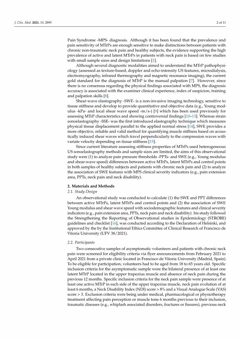
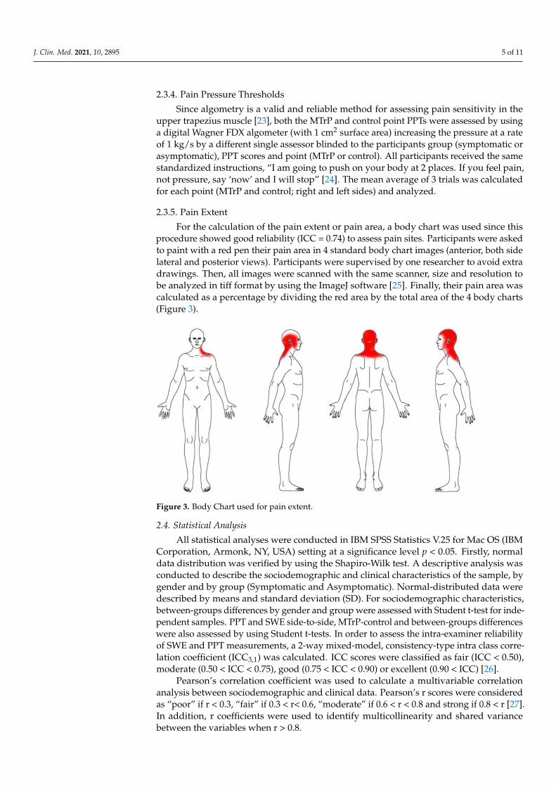
Participants were placed in the prone position and the examiner assessed bilaterallythe entire length of the upper trapezius muscle by using a pincer palpation to locate (1)a palpable taut band, (2) a hypersensitive tender spot in the taut band and (3) a localtwitch response provoked by the snapping palpation of the taut band. MTrPs that uponstimulation reproduced partially or totally familiar symptoms experienced by the patientswere considered active MTrPs and those that upon stimulation did not reproduce anysymptom experienced and the subject did not recognize the elicited symptom as familiarwas considered as a latent MTrP [4]. If there was more than one active MTrP, the mostsymptomatic site was selected and marked with a blue skin marking pen. Then, a controlpoint located 3 cm laterally to the MTrP following the fibers direction (within the taut band)was also marked with the same blue skin marking pen (Figure 1).
We selected this control point location based on the pain difference during the manualpalpation and based on a previous study [21] reporting that there are electromyographicdifferences between MTrPs and control points separated 1cm following the fibers direction(within the same taut band) and located in the upper trapezius muscle (200 to 700 µVand 10 µV respectively). Examiners were blinded to this assignation of MTrP and controllocations during the measurement procedures.
J. Clin. Med. 2021, 10, 2895 4 of 11
Figure 1. (A) Participants positioning; (B) Measurement points location.
2.3.3. Shear Wave Elastography-Image Acquisition
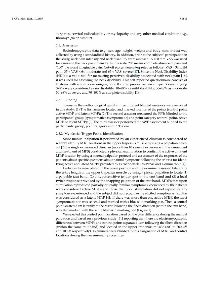
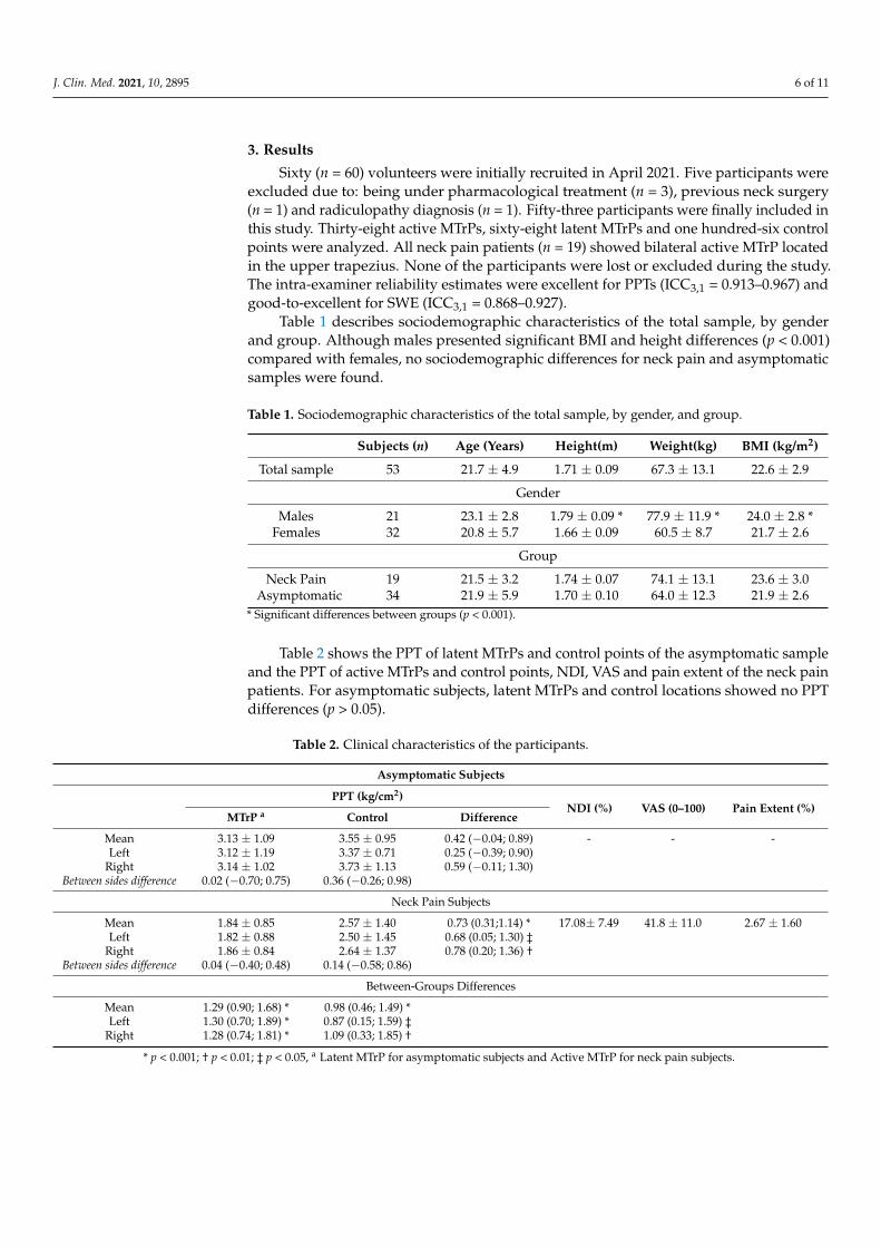
All ultrasound assessments were performed by the same examiner with more than10 years of experience in musculoskeletal evaluation. A Canon Aplio A with a PLT-1005-BT14L5 (5–14 MHz) transducer (Canon Medical Corp, 1385 Shimoishigami, Otawara, Tochigi324-8550, Japan) were used for all the images capturing and measuring procedures. Allparticipants were positioned in the same orientation used for the MTrP identificationprotocol. Since the longitudinal plane showed higher intra- and inter-examiner reliability(ICC = 0.66–0.74) than the transversal assessment [22], all images were acquired with thetransducer oriented longitudinally with the muscle fibers and positioning the center of theprobe over the MTrP and the control point locations. Two images were obtained from eachpatient and measurement point to improve the accuracy of the measurements.
In order to avoid bias regarding the MTrP surface selection based on the stiffness scale,regions of interest (ROIs) for shear wave data were positioned to cover (1) the superficialand deep internal echogenic fascia of upper trapezius (in order to include completelythe muscle thickness) and (2) 1 cm of width (correspondent with the algometer surface)(Figure 2). Finally, both the Young Modulus and the Shear Wave Speed data were collected.The sonographer was blinded to the sample group (symptomatic or asymptomatic) andthe point assessed (MTrP or control).
Figure 2. Shear Wave Elastography: Image measurement.
J. Clin. Med. 2021, 10, 2895 5 of 11
2.3.4. Pain Pressure Thresholds
Since algometry is a valid and reliable method for assessing pain sensitivity in theupper trapezius muscle [23], both the MTrP and control point PPTs were assessed by usinga digital Wagner FDX algometer (with 1 cm2 surface area) increasing the pressure at a rateof 1 kg/s by a different single assessor blinded to the participants group (symptomatic orasymptomatic), PPT scores and point (MTrP or control). All participants received the samestandardized instructions, “I am going to push on your body at 2 places. If you feel pain,not pressure, say ‘now’ and I will stop” [24]. The mean average of 3 trials was calculatedfor each point (MTrP and control; right and left sides) and analyzed.
2.3.5. Pain Extent
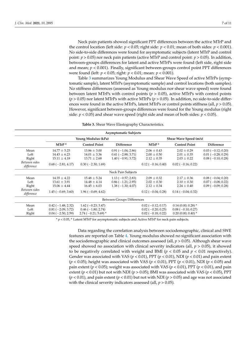
For the calculation of the pain extent or pain area, a body chart was used since thisprocedure showed good reliability (ICC = 0.74) to assess pain sites. Participants were askedto paint with a red pen their pain area in 4 standard body chart images (anterior, both sidelateral and posterior views). Participants were supervised by one researcher to avoid extradrawings. Then, all images were scanned with the same scanner, size and resolution tobe analyzed in tiff format by using the ImageJ software [25]. Finally, their pain area wascalculated as a percentage by dividing the red area by the total area of the 4 body charts(Figure 3).
Figure 3. Body Chart used for pain extent.
2.4. Statistical Analysis
All statistical analyses were conducted in IBM SPSS Statistics V.25 for Mac OS (IBMCorporation, Armonk, NY, USA) setting at a significance level p < 0.05. Firstly, normaldata distribution was verified by using the Shapiro-Wilk test. A descriptive analysis wasconducted to describe the sociodemographic and clinical characteristics of the sample, bygender and by group (Symptomatic and Asymptomatic). Normal-distributed data weredescribed by means and standard deviation (SD). For sociodemographic characteristics,between-groups differences by gender and group were assessed with Student t-test for inde-pendent samples. PPT and SWE side-to-side, MTrP-control and between-groups differenceswere also assessed by using Student t-tests. In order to assess the intra-examiner reliabilityof SWE and PPT measurements, a 2-way mixed-model, consistency-type intra class corre-lation coefficient (ICC3,1) was calculated. ICC scores were classified as fair (ICC < 0.50),moderate (0.50 < ICC < 0.75), good (0.75 < ICC < 0.90) or excellent (0.90 < ICC) [26].
Pearson’s correlation coefficient was used to calculate a multivariable correlationanalysis between sociodemographic and clinical data. Pearson’s r scores were consideredas “poor” if r < 0.3, “fair” if 0.3 < r< 0.6, “moderate” if 0.6 < r < 0.8 and strong if 0.8 < r [27].In addition, r coefficients were used to identify multicollinearity and shared variancebetween the variables when r > 0.8.
J. Clin. Med. 2021, 10, 2895 6 of 11
3. Results
Sixty (n = 60) volunteers were initially recruited in April 2021. Five participants wereexcluded due to: being under pharmacological treatment (n = 3), previous neck surgery(n = 1) and radiculopathy diagnosis (n = 1). Fifty-three participants were finally included inthis study. Thirty-eight active MTrPs, sixty-eight latent MTrPs and one hundred-six controlpoints were analyzed. All neck pain patients (n = 19) showed bilateral active MTrP locatedin the upper trapezius. None of the participants were lost or excluded during the study.The intra-examiner reliability estimates were excellent for PPTs (ICC3,1 = 0.913–0.967) andgood-to-excellent for SWE (ICC3,1 = 0.868–0.927).
Table 1 describes sociodemographic characteristics of the total sample, by genderand group. Although males presented significant BMI and height differences (p < 0.001)compared with females, no sociodemographic differences for neck pain and asymptomaticsamples were found.
Table 1. Sociodemographic characteristics of the total sample, by gender, and group.
Subjects (n) Age (Years) Height(m) Weight(kg) BMI (kg/m2)
Total sample 53 21.7 ± 4.9 1.71 ± 0.09 67.3 ± 13.1 22.6 ± 2.9
Gender
Males 21 23.1 ± 2.8 1.79 ± 0.09 * 77.9 ± 11.9 * 24.0 ± 2.8 *Females 32 20.8 ± 5.7 1.66 ± 0.09 60.5 ± 8.7 21.7 ± 2.6
Group
Neck Pain 19 21.5 ± 3.2 1.74 ± 0.07 74.1 ± 13.1 23.6 ± 3.0Asymptomatic 34 21.9 ± 5.9 1.70 ± 0.10 64.0 ± 12.3 21.9 ± 2.6
* Significant differences between groups (p < 0.001).
Table 2 shows the PPT of latent MTrPs and control points of the asymptomatic sampleand the PPT of active MTrPs and control points, NDI, VAS and pain extent of the neck painpatients. For asymptomatic subjects, latent MTrPs and control locations showed no PPTdifferences (p > 0.05).
Table 2. Clinical characteristics of the participants.
Asymptomatic Subjects
PPT (kg/cm2)NDI (%) VAS (0–100) Pain Extent (%)
MTrP a Control Difference
Mean 3.13 ± 1.09 3.55 ± 0.95 0.42 (−0.04; 0.89) - - -Left 3.12 ± 1.19 3.37 ± 0.71 0.25 (−0.39; 0.90)
Right 3.14 ± 1.02 3.73 ± 1.13 0.59 (−0.11; 1.30)Between sides difference 0.02 (−0.70; 0.75) 0.36 (−0.26; 0.98)
Neck Pain Subjects
Mean 1.84 ± 0.85 2.57 ± 1.40 0.73 (0.31;1.14) * 17.08± 7.49 41.8 ± 11.0 2.67 ± 1.60Left 1.82 ± 0.88 2.50 ± 1.45 0.68 (0.05; 1.30) ‡
Right 1.86 ± 0.84 2.64 ± 1.37 0.78 (0.20; 1.36) †Between sides difference 0.04 (−0.40; 0.48) 0.14 (−0.58; 0.86)
Between-Groups Differences
Mean 1.29 (0.90; 1.68) * 0.98 (0.46; 1.49) *Left 1.30 (0.70; 1.89) * 0.87 (0.15; 1.59) ‡
Right 1.28 (0.74; 1.81) * 1.09 (0.33; 1.85) †
* p < 0.001; † p < 0.01; ‡ p < 0.05, a Latent MTrP for asymptomatic subjects and Active MTrP for neck pain subjects.
J. Clin. Med. 2021, 10, 2895 7 of 11
Neck pain patients showed significant PPT differences between the active MTrP andthe control location (left side: p < 0.05; right side: p < 0.01; mean of both sides: p < 0.001).No side-to-side differences were found for asymptomatic subjects (latent MTrP and controlpoint: p > 0.05) nor neck pain patients (active MTrP and control point: p > 0.05). In addition,between-groups differences for latent and active MTrPs were found (left side, right sideand mean; p < 0.001). Finally, significant between-groups control point PPT differenceswere found (left: p < 0.05; right: p < 0.01; mean: p < 0.001).
Table 3 summarizes Young Modulus and Shear Wave Speed of active MTrPs (symp-tomatic sample), latent MTrPs (asymptomatic sample) and control locations (both samples).No stiffness differences (assessed as Young modulus nor shear wave speed) were foundbetween latent MTrPs with control points (p > 0.05), active MTrPs with control points(p > 0.05) nor latent MTrPs with active MTrPs (p > 0.05). In addition, no side-to-side differ-ences were found in the active MTrPs, latent MTrPs or control points stiffness (all, p > 0.05).However, significant between-groups differences were found for the Young modulus (rightside: p < 0.05) and shear wave speed (right side and mean of both sides: p < 0.05).
Table 3. Shear Wave Elastography Characteristics.
Asymptomatic Subjects
Young Modulus (kPa) Shear Wave Speed (m/s)
MTrP a Control Point Difference MTrP a Control Point Difference
Mean 14.77 ± 5.25 13.86 ± 3.00 0.91 (−1.04; 2.86) 2.06 ± 0.43 2.02 ± 0.29 0.03 (−0.12; 0.20)Left 14.43 ± 6.23 14.01 ± 3.36 0.41 (−2.88; 3.71) 2.00 ± 0.50 2.01 ± 0.35 0.01 (−0.28; 0.29)
Right 15.11 ± 4.18 13.71 ± 2.68 1.40 (−0.91; 3.72) 2.12 ± 0.35 2.03 ± 0.22 0.08 (−0.10; 0.28)Between sides
difference 0.68 (−2.81; 4.17) 0.30 (−2.30; 1.69) 0.12 (−0.16; 0.40) 0.02 (−0.16; 0.22)
Neck Pain Subjects
Mean 14.35 ± 4.22 15.48 ± 5.24 1.13 (−0.57; 2.83) 2.09 ± 0.32 2.17 ± 0.36 0.08 (−0.04; 0.20)Left 13.61 ± 3.91 14.48 ± 4.14 0.86 (−1.21; 2.95) 2.02 ± 0.30 2.10 ± 0.30 0.07 (−0.08; 0.22)
Right 15.06 ± 4.44 16.45 ± 6.03 1.38 (−1.30; 4.07) 2.12 ± 0.34 2.24 ± 0.40 0.09 (−0.09; 0.28)Between sides
difference 1.45 (−0.69; 3.60) 1.96 (−0.69; 4.62) 0.12 (−0.04; 0.28) 0.14 (−0.04; 0.32)
Between-Groups Differences
Mean 0.42 (−1.48; 2.32) 1.62 (−0.23; 3.47) 0.02 (−0.12; 0.17) 0.14 (0.00; 0.28) *Left 0.81 (−2.09; 3.72) 0.46 (−1.80; 2.74) 0.02 (−0.20; 0.25) 0.08 (−0.10; 0.27)
Right 0.04 (−2.50; 2.59) 2.74 (−0.21; 5.69) * 0.02 (−0.18; 0.22) 0.20 (0.00; 0.40) *
* p < 0.05, a Latent MTrP for asymptomatic subjects and Active MTrP for neck pain subjects.
Data regarding the correlation analysis between sociodemographic, clinical and SWEfeatures are reported on Table 4. Young modulus showed no significant association withthe sociodemographic and clinical outcomes assessed (all, p > 0.05). Although shear wavespeed showed no association with clinical severity indicators (all, p > 0.05), it showedto be negatively correlated with weight and BMI (p < 0.05 and p < 0.01 respectively).Gender was associated with VAS (p < 0.01), PPT (p < 0.01), NDI (p < 0.01) and pain extent(p < 0.05); height was associated with VAS (p < 0.01), PPT (p < 0.01), NDI (p < 0.05) andpain extent (p < 0.05); weight was associated with VAS (p < 0.01), PPT (p < 0.01), and painextent (p < 0.01) but not with NDI (p > 0.05); BMI was associated with VAS (p < 0.05), PPT(p < 0.01), and pain extent (p < 0.01) but not with NDI (p > 0.05) and age was not associatedwith the clinical severity indicators assessed (all, p > 0.05).
J. Clin. Med. 2021, 10, 2895 8 of 11
Table 4. Pearson-product moment correlation matrix.
1 2 3 4 5 6 7 8 9 10
1. Gender
2. Height −0.659 **
3. Weight −0.644 ** 0.752 **
4. BMI −0.381 ** 0.260 ** 0.826 **
5. Age −0.231 ** 0.174 * n.s. n.s.
6. Young Modulus n.s. n.s. n.s. n.s. n.s.
7. Shear Wave Speed n.s. n.s. −0.141 * −0.230 ** n.s. 0.937 **
8. VAS 0.324 ** −0.194 ** −0.230 ** −0.175 * n.s. n.s. n.s.
9. NDI 0.200 ** −0.152 * n.s. n.s. n.s. n.s. n.s. 0.859 **
10. PPT −0.386 ** 0.193 ** 0.258 ** 0.185 ** n.s. n.s. n.s. −0.417 ** −0.362 **
11. Pain Extent 0.177 * −0.195 * −0.273 ** −0.301 ** n.s. n.s. n.s. 0.295 ** 0.359 ** n.s.
n.s. non-significant, * p < 0.05; ** p < 0.01.
4. Discussion4.1. Findings
This is the first study analyzing the clinical relevance of SWE as indicator of MPSseverity. In this study, (1) we compared the stiffness of neck pain patients with active MTrPswith asymptomatic subjects with latent MTrPs, (2) we compared the stiffness of both latentand active MTrPs with control points and (3) we conducted a correlation analysis betweenstiffness assessed as Young modulus and shear wave speed and clinical severity assessedas pain intensity, pain sensitivity, disability and pain extent. The most interesting finding ofthis study was that, although there were important sensitivity differences between activeMTrPs with latent MTrPs and control points, we found no stiffness differences. However,neck pain patients and asymptomatic subjects showed significant stiffness differences forthe control point.
Since MTrP are classically defined as hard palpable nodules within confirmed tautbands [28] and are characterized by tenderness associated with pain [4], it would be logicalfind stiffness and PPT differences between active MTrPs, latent MTrPs and control pointswithin the same muscle. However, even if MTrP showed an increased pain sensitivity(lower PPT) compared with control point locations, we did not find stiffness differencesbetween active or latent MTrPs with control points. Our results are consistent with aprevious study since it was reported that the trapezius muscle hardness does not directlyreflect the subjective shoulder stiffness [29].
Since palpation is shown to be successful for identifying the most sensitive areas(active and latent MTrPs), is the most popular diagnosis procedure used for assessingstiffness differences. However, manual palpation is a high subjectivity test based on thepain response of the patients which could imply bias and, therefore, objective methods(with high sensitivity, specificity, reliability and validity) are prioritized [30]. AlthoughSWE is an objective alternative to manual palpation which showed to be a feasible toolfor assessing the upper trapezius muscle intra-session stiffness [22], the main limitationfound for this method is that its reliability showed to be moderate just if performed inrigorous conditions (e.g., reporting both shear wave speed and young modulus assessedin the longitudinal plane [31]. Therefore, those studies assessing transverse planes [32,33]or using operator-dependent elastography methods (e.g., strain elastography) should beinterpreted carefully [11].
A previous study conducted by Ballyns et al. [11] reported no correlation betweentrigger point areas and PPTs for either latent or active MTrPs. Therefore, even if theyfound that active MTrPs show greater size compared with latent MTrPs, there are othermechanisms contributing to the trigger point sensitivity such as sensitizing biochemicals asreported by Shah et al. [34] derived from the capillary constriction consistent with Doppler
J. Clin. Med. 2021, 10, 2895 9 of 11
US findings [11]. Our results are consistent with this hypothesis since we found in additionto the sensitivity differences between healthy and clinical populations regarding the controlpoint locations, greater stiffness scores in neck pain patients.
We also found stiffness differences in the control point location between neck painpatients and asymptomatic subjects. This is an interesting finding since the general musclestiffness showed to be a more differential factor compared with the MTrP stiffness. Futurestudies could include the general muscle stiffness as an additional outcome to assess theeffects of different interventions on the general stiffness and its correlation with otherclinical features. In fact, emerging evidence is considering this proposal and consideringthe SWE as a tool for clinical evaluation and monitorization of treatment responses [35].
Although our results suggest that SWE cannot be used as clinical severity indicatoror a tool to differentiate active and latent MTrPs based on Young modulus and shearwave speed, this field requires further investigation to consolidate these findings. In fact,we found no correlation between age and stiffness. One likely reason explaining thediscrepancy with the current evidence [36] is the small range of ages included in this study.Therefore, our results cannot be extrapolated to the general population and further researchshould consider greater ranges of age, height, weight, BMI and pain intensity/disability toconfirm these findings.
4.2. Limitations
Even if the results of this study are promising and we considered the blinding of theassessors and the inclusion of one sample of asymptomatic subjects as a control group, somepotential limitations should be recognized. Firstly, we measured the locations stiffness justin the longitudinal plane due to reliability restrictions previously reported. Since the imageassessed limited fibers and MTrP are located in taut bands, this could show increasedstiffness in the control points selected. Secondly, additional associations between SWE withDoppler US, electromyography and biochemical analysis would clarify several aspectsof our findings and confirm the presence of active or latent MTrPs. Finally, the samplewas relatively small and further research including greater samples with wide standarddeviations is needed.
5. Conclusions
This study showed that active MTrPs located in the upper trapezius identified bymanual palpation in neck pain patients are more sensitive compared with control pointwithin the same muscle and with latent MTrPs in asymptomatic subjects. We found nostiffness differences assessed as SWE Young modulus and shear wave speed betweenactive and latent MTrPs. However, neck pain patients showed increased control pointstiffness compared with asymptomatic subjects. Therefore, future studies could assess howdifferent interventions change the general muscle stiffness (not focusing just in MTrPs)and its relevance during the clinical evaluation and follow-up by analyzing the correlationbetween clinical features with the general stiffness (control points), specific stiffness (MTrPs)and stiffness change. Finally, we found no association between SWE features with clinicalseverity indicators of MPS assessed as pain extent, pain intensity, sensitivity and neckdisability.
Author Contributions: Conceptualization, J.A.V.-C., U.V. and J.Á.-G.; methodology, J.A.V.-C., J.Á.-G.,J.B.-G. and G.M.G.-S.; formal analysis, J.A.V.-C. and U.V.; investigation, J.A.V.-C., J.Á.-G., S.S.-J., J.B.-G. and G.M.G.-S.; resources, J.Á.-G. and S.S.-J.; data curation, J.A.V.-C. and U.V.; writing—originaldraft preparation, J.A.V.-C.; writing—review and editing, J.A.V.-C.; visualization, J.A.V.-C. and U.V.;supervision, J.A.V.-C.; project administration, J.A.V.-C. and S.S.-J.; funding acquisition, J.A.V.-C. Allauthors have read and agreed to the published version of the manuscript.
Funding: This research and the APC were funded by Camilo José Cela University, grant numberSTIFMYTPO.
J. Clin. Med. 2021, 10, 2895 10 of 11
Institutional Review Board Statement: The study was conducted according to the guidelines of theDeclaration of Helsinki and approved by the Institutional Ethics Committee of Francisco de VitoriaUniversity (UFV 38/2021).
Informed Consent Statement: Informed consent was obtained from all subjects involved in the study.
Data Availability Statement: The data that support the findings of this study are available from thecorresponding author (JA Valera-Calero), upon reasonable request.
Conflicts of Interest: The authors declare no conflict of interest. The funders had no role in the designof the study; in the collection, analyses, or interpretation of data; in the writing of the manuscript, orin the decision to publish the results.
References1. Ribeiro, D.C.; Belgrave, A.; Naden, A.; Fang, H.; Matthews, P.; Parshottam, S. The prevalence of myofascial trigger points in
neck and shoulder-related disorders: A systematic review of the literature. BMC Musculoskelet. Disord. 2018, 19, 252. [CrossRef][PubMed]
2. Liu, L.; Huang, Q.M.; Liu, Q.G.; Ye, G.; Bo, C.Z.; Chen, M.J.; Li, P. Effectiveness of dry needling for myofascial trigger pointsassociated with neck and shoulder pain: A systematic review and meta-analysis. Arch. Phys. Med. Rehabil. 2015, 96, 944–955.[CrossRef] [PubMed]
3. Muñoz-Muñoz, S.; Muñoz-García, M.T.; Alburquerque-Sendín, F.; Arroyo-Morales, M.; Dommerholt, M.; Fernández-de-las-Peñas,C. Myofascial trigger points, pain, disability, and sleep quality in individuals with mechanical neck pain. J. Manip. Physiol. Ther.2012, 35, 608–613. [CrossRef]
4. Fernández-de-Las-Peñas, C.; Dommerholt, J. International Consensus on Diagnostic Criteria and Clinical Considerations ofMyofascial Trigger Points: A Delphi Study. Pain Med. 2018, 19, 142–150. [CrossRef] [PubMed]
5. Li, L.; Stoop, R.; Clijsen, R.; Hohenauer, E.; Fernández-de-Las-Peñas, C.; Huang, Q.; Barbero, M. Criteria Used for the Diagnosis ofMyofascial Trigger Points in Clinical Trials on Physical Therapy: Updated Systematic Review. Clin. J. Pain 2020, 36, 955–967.[CrossRef] [PubMed]
6. Jun, D.; Zoe, M.; Johnston, V.; O’Leary, S. Physical risk factors for developing non-specific neck pain in office workers: Asystematic review and meta-analysis. Int. Arch. Occup. Environ. Health 2017, 90, 373–410. [CrossRef] [PubMed]
7. Do, T.P.; Heldarskard, G.F.; Kolding, L.T.; Hvedstrup, J.; Schytz, H.W. Myofascial trigger points in migraine and tension-typeheadache. J. Headache Pain 2018, 19, 84. [CrossRef] [PubMed]
8. Shah, J.P.; Thaker, N.; Heimur, J.; Aredo, J.V.; Sikdar, S.; Gerber, L. Myofascial Trigger Points Then and Now: A Historical andScientific Perspective. PM&R 2015, 7, 746–761. [CrossRef]
9. Taljanovic, M.S.; Gimber, L.H.; Becker, G.W.; Latt, L.D.; Klauser, A.S.; Melville, D.M.; Gao, L.; Witte, R.S. Shear-Wave Elastography:Basic Physics and Musculoskeletal Applications. Radiographics 2017, 37, 855–870. [CrossRef]
10. Sikdar, S.; Shah, J.P.; Gebreab, T.; Yen, R.H.; Gilliams, E.; Danoff, J.; Gerber, L.H. Novel applications of ultrasound technology tovisualize and characterize myofascial trigger points and surrounding soft tissue. Arch. Phys. Med. Rehabil. 2009, 90, 1829–1838.[CrossRef]
11. Ballyns, J.J.; Shah, J.P.; Hammond, J.; Gebreab, T.; Gerber, L.H.; Sikdar, S. Objective sonographic measures for characterizingmyofascial trigger points associated with cervical pain. J. Ultrasound Med. 2011, 30, 1331–1340. [CrossRef]
12. Lv, H.; Li, Z.; Hu, T.; Wang, Y.; Wu, J.; Li, Y. The shear wave elastic modulus and the increased nuclear factor kappa B (NF-kB/p65)and cyclooxygenase-2 (COX-2) expression in the area of myofascial trigger points activated in a rat model by blunt trauma to thevastus medialis. J. Biomech. 2018, 66, 44–50. [CrossRef]
13. Kang, J.J.; Kim, J.; Park, S.; Paek, S.; Kim, T.H.; Kim, D.K. Feasibility of Ultrasound-Guided Trigger Point Injection in Patients withMyofascial Pain Syndrome. Healthcare 2019, 7, 118. [CrossRef]
14. Sigrist, R.M.S.; Liau, J.; Kaffas, A.E.; Chammas, M.C.; Willmann, J.K. Ultrasound Elastography: Review of Techniques and ClinicalApplications. Theranostics 2017, 7, 1303–1329. [CrossRef]
15. Ozturk, A.; Grajo, J.R.; Dhyani, M.; Anthony, B.W.; Samir, A.E. Principles of ultrasound elastography. Abdom. Radiol. 2018, 43,773–785. [CrossRef]
16. Cuschieri, S. The STROBE guidelines. Saudi J. Anaesth. 2019, 13, S31–S34. [CrossRef]17. Boonstra, A.M.; Schiphorst Preuper, H.R.; Balk, G.A.; Stewart, R.E. Cut-off points for mild, moderate, and severe pain on the
visual analogue scale for pain in patients with chronic musculoskeletal pain. Pain 2014, 155, 2545–2550. [CrossRef]18. Andrade Ortega, J.A.; Delgado Martínez, A.D.; Almécija Ruiz, R. Validation of the spanish version of the neck disability index.
Spine 2010, 35, E114–E118. [CrossRef]19. Vernon, H.; Mior, S. The neck disability index: A study of reliability and validity. J. Manip. Physiol. Ther. 1991, 14, 409–415.20. Barbero, M.; Bertoli, P.; Cescon, C.; Macmillan, F.; Coutts, F.; Gatti, R. Intra-rater reliability of an experienced physiotherapist in
locating myofascial trigger points in upper trapezius muscle. J. Manip. Physiol. Ther. 2012, 20, 171–177. [CrossRef]21. Hubbard, D.R.; Berkoff, G.M. Myofascial trigger points show spontaneous needle EMG activity. Spine 1993, 18, 1803–1807.
[CrossRef] [PubMed]
J. Clin. Med. 2021, 10, 2895 11 of 11
22. Kozinc, Ž.; Šarabon, N. Shear-wave elastography for assessment of trapezius muscle stiffness: Reliability and association withlow-level muscle activity. PLoS ONE 2020, 15, e0234359. [CrossRef] [PubMed]
23. Sciotti, V.M.; Mittak, V.L.; DiMarco, L.; Ford, L.M.; Plezbert, J.; Santipadri, E.; Wigglesworth, J.; Ball, K. Clinical precision ofmyofascial trigger point location in the trapezius muscle. Pain 2001, 93, 259–266. [CrossRef]
24. Úbeda-D’Ocasar, E.; Valera-Calero, J.A.; Hervás-Pérez, J.P.; Caballero-Corella, M.; Ojedo-Martín, C.; Gallego-Sendarrubias, G.M.Pain Intensity and Sensory Perception of Tender Points in Female Patients with Fibromyalgia: A Pilot Study. Int. J. Environ. Res.Public Health 2021, 18, 1461. [CrossRef]
25. Silva, A.G.; De Francesco, S.; Rodrigues, M.; Queirós, A.; Cerqueira, M. Test-Retest Reliability of Pain Measures in InstitutionalizedOlder Adults: Number of Painful Body Sites, Pain Intensity, and Pain Extent. Pain Pract. 2021, 21, 270–276. [CrossRef]
26. Koo, T.K.; Li, M.Y. A guideline of selecting and reporting intraclass correlation coefficients for reliability research. J. Chiropr. Med.2016, 15, 155–163. [CrossRef]
27. Chan, Y.H. Biostatistics 104: Correlational analysis. Singap. Med. J. 2003, 44, 614–619.28. Chen, Q.; Wang, H.J.; Gay, R.E.; Thompson, J.M.; Manduca, A.; An, K.N.; Ehman, R.E.; Basford, J.R. Quantification of Myofascial
Taut Bands. Arch. Phys. Med. Rehabil. 2016, 97, 67–73. [CrossRef]29. Sawada, T.; Okawara, H.; Nakashima, D.; Iwabuchi, S.; Matsumoto, M.; Nakamura, M.; Nagura, T. Reliability of Trapezius Muscle
Hardness Measurement: A Comparison between Portable Muscle Hardness Meter and Ultrasound Strain Elastography. Sensors2020, 20, 7200. [CrossRef]
30. Valera-Calero, J.A.; Guodemar-Pérez, J.; Cleland, J.A.; Ojedo-Martín, C.; Gallego-Sendarrubias, G.M. Physical therapist attitudeand opinion about cervical spine examination: A national Spanish survey. Int. J. Clin. Pract. 2020, e13781. [CrossRef]
31. Cortez, C.D.; Hermitte, L.; Ramain, A.; Mesmann, C.; Lefort, T.; Pialat, J.B. Ultrasound shear wave velocity in skeletal muscle: Areproducibility study. Diagn. Interv. Imaging 2016, 97, 71–79. [CrossRef]
32. Sánchez-Infante, J.; Bravo-Sánchez, A.; Jiménez, F.; Abián-Vicén, J. Effects of Dry Needling on Muscle Stiffness in LatentMyofascial Trigger Points: A Randomized Controlled Trial. J. Pain 2021. [CrossRef]
33. Maher, R.M.; Hayes, D.M.; Shinohara, M. Quantification of dry needling and posture effects on myofascial trigger points usingultrasound shear-wave elastography. Arch. Phys. Med. Rehabil. 2013, 94, 2146–2150. [CrossRef]
34. Shah, J.P.; Danoff, J.V.; Desai, M.J.; Parikh, S.; Nakamura, L.Y.; Phillips, T.M.; Gerber, L.H. Biochemicals associated with pain andinflammation are elevated in sites near to and remote from active myofascial trigger points. Arch. Phys. Med. Rehabil. 2008, 89,16–23. [CrossRef]
35. Olchowy, C.; Wieckiewicz, M.; Sconfienza, L.M.; Łasecki, M.; Seweryn, P.; Smardz, J.; Hnitecka, S.; Dominiak, M.; Olchowy, A.Potential of Using Shear Wave Elastography in the Clinical Evaluation and Monitoring of Changes in Masseter Muscle Stiffness.Pain Res. Manag. 2020, 2020, 4184268. [CrossRef]
36. Alfuraih, A.M.; Tan, A.L.; O’Connor, P.; Emery, P.; Wakefield, R.J. The effect of ageing on shear wave elastography muscle stiffnessin adults. Aging Clin Exp. Res. 2019, 31, 1755–1763. [CrossRef]
Related Documents