© Turkish Society of Radiology 2010 293 U lcerative colitis is a chronic disease characterized by diffuse mu- cosal inflammation that is localized to the colon. In approxi- mately 95% of cases, ulcerative colitis involves the rectum and may extend proximally in a symmetrical, circumferential, and uninter- rupted pattern to involve part or all of the large intestine. Ulcerative colitis is usually associated with recurrent attacks, with complete remis- sion of symptoms in the interim (1–4). Disease activity indices are prediction rules that are used to objec- tively measure the activity of disease, to judge patients’ responses to treatment in clinical trials and to choose which drug and dose to administer. Disease activity can be assessed clinically and endoscopi- cally. Because the rectum is invariably involved in ulcerative colitis, flexible rectosigmoidoscopy is sufficient for depicting disease activity (1, 3). Despite its use as a gold standard, endoscopy is an invasive technique and has limitations. Due to the presence of severe inflam- mation in the colon, the use of colonoscopy in patients with active ulcerative colitis carries a high risk of perforation of the tissue. In ad- dition, orientation cannot be ascertained in newly diagnosed patients with active ulcerative colitis. Furthermore, the view of the endoscopist is limited to the colonic lumen; the thickness of the colonic wall and the presence of lymph nodes cannot be assessed. Patient discomfort and the potential risks associated with obtaining biopsies via endos- copy have prompted the evaluation of various radiological imaging techniques to identify and quantify inflammatory bowel disease (IBD) activity in the colon (5). The part of the gastrointestinal tract in which magnetic resonance imaging (MRI) studies have been the most successful is the rectum (2). MRI has been shown to be an excellent means of accurately assessing inflammatory diseases and congenital malformations following cancers in the rectosigmoid region (2–4). Several studies have reported MRI pa- rameters that can be used to determine disease activity; these include increased contrast uptake in the colonic wall, thickening of the bowel wall, quantification of lymph nodes, and loss of haustral folds (5, 6). In addition, a non-invasive technique called diffusion-weighted MRI (DW- MRI), which was previously restricted to imaging of the central nervous system, has recently been implemented for the diagnosis and follow-up of various abdominal diseases. The purpose of this study was to deter- mine disease activity in patients with ulcerative colitis by calculating the apparent coefficient values (ADC) of the colonic wall and pericolonic lymph nodes (PCLNs) using non-invasive DW-MRI. Materials and methods A total of 28 patients with ulcerative colitis (9 endoscopically active, 10 subacute and 9 in remission) were prospectively evaluated with DW-MRI ABDOMINAL IMAGING ORIGINAL ARTICLE Is quantitative diffusion-weighted MRI a reliable method in the assessment of the inflammatory activity in ulcerative colitis? Özgür Kılıçkesmez, Aliye Soylu, Nurgül Yaşar, Tuna Demirbaş, Can Dolapçıoğlu, Şule Poturoğlu, İsa Sevindir, Tan Cimilli From the Department of Radiology (Ö.K. okilickesmez@yahoo. com), Yeditepe University School of Medicine, İstanbul, Turkey; Gastroenterology Clinic (A.S.), Radiology Clinic (T.D., T.C.), and Internal Medicine Clinic (N.Y., İ.S.), Bakırköy Training and Research Hospital, İstanbul, Turkey; Gastroenterology Clinic (C.D.), Kartal Training and Research Hospital, İstanbul, Turkey; and Gastroenterology Clinic (Ş.P.), Haseki Training and Research Hospital, İstanbul, Turkey. Received 23 July 2009; revision requested 1 September 2009; revision received 17 September 2009; accepted 19 October 2009. Published online 9 August 2010 DOI 10.4261/1305-3825.DIR.2989-09.1 PURPOSE We investigated the relationship between the apparent dif- fusion coefficient (ADC) values of the colonic wall and the pathologic pericolonic lymph nodes (PCLNs) and inflamma- tory activity in ulcerative colitis patients by diffusion-weighted magnetic resonance imaging (DW-MRI). MATERIALS AND METHODS A total of 28 ulcerative colitis patients (9 endoscopically active, 10 subacute and 9 in remission) were evaluated by DW-MRI with 0, 500 and 1000 s/mm 2 b-values. The ADC values of the rectum and sigmoid colon walls and the adjacent PCLNs were obtained for quantitative analysis. The DW-MRI findings were compared to the disease activity. RESULTS The ADC values of the sigmoid colon were similar in pa- tients with active, subacute and remissive ulcerative colitis (P = 0.472). The ADC values of the rectum were different (P = 0.009) between patients in the active (1.08 ± 0.14×10 ¯ ³ mm 2 /s) and subacute phases (1.13 ± 0.23×10 ¯ ³ mm 2 /s) of disease and those in remission (1.29 ± 0.17×10 ¯ ³ mm 2 /s). The ADC values of the PCLNs (P = 0.899) did not differ with respect to disease activity. CONCLUSION DW-MRI is useful in identifying disease activity in ulcerative colitis patients, especially with respect to the rectum. The ADC values of the rectum increase during remission and decrease in patients with active distal colitis. The ADC values of the PCLNs were not useful in determining disease activity. Key words: • ulcerative colitis • inflammatory bowel diseases • diffusion magnetic resonance imaging Diagn Interv Radiol 2010; 16:293–298

Welcome message from author
This document is posted to help you gain knowledge. Please leave a comment to let me know what you think about it! Share it to your friends and learn new things together.
Transcript
© Turkish Society of Radiology 2010
293
U lcerative colitis is a chronic disease characterized by diffuse mu-cosal inflammation that is localized to the colon. In approxi-mately 95% of cases, ulcerative colitis involves the rectum and
may extend proximally in a symmetrical, circumferential, and uninter-rupted pattern to involve part or all of the large intestine. Ulcerative colitis is usually associated with recurrent attacks, with complete remis-sion of symptoms in the interim (1–4).
Disease activity indices are prediction rules that are used to objec-tively measure the activity of disease, to judge patients’ responses to treatment in clinical trials and to choose which drug and dose to administer. Disease activity can be assessed clinically and endoscopi-cally. Because the rectum is invariably involved in ulcerative colitis, flexible rectosigmoidoscopy is sufficient for depicting disease activity (1, 3). Despite its use as a gold standard, endoscopy is an invasive technique and has limitations. Due to the presence of severe inflam-mation in the colon, the use of colonoscopy in patients with active ulcerative colitis carries a high risk of perforation of the tissue. In ad-dition, orientation cannot be ascertained in newly diagnosed patients with active ulcerative colitis. Furthermore, the view of the endoscopist is limited to the colonic lumen; the thickness of the colonic wall and the presence of lymph nodes cannot be assessed. Patient discomfort and the potential risks associated with obtaining biopsies via endos-copy have prompted the evaluation of various radiological imaging techniques to identify and quantify inflammatory bowel disease (IBD) activity in the colon (5).
The part of the gastrointestinal tract in which magnetic resonance imaging (MRI) studies have been the most successful is the rectum (2). MRI has been shown to be an excellent means of accurately assessing inflammatory diseases and congenital malformations following cancers in the rectosigmoid region (2–4). Several studies have reported MRI pa-rameters that can be used to determine disease activity; these include increased contrast uptake in the colonic wall, thickening of the bowel wall, quantification of lymph nodes, and loss of haustral folds (5, 6). In addition, a non-invasive technique called diffusion-weighted MRI (DW-MRI), which was previously restricted to imaging of the central nervous system, has recently been implemented for the diagnosis and follow-up of various abdominal diseases. The purpose of this study was to deter-mine disease activity in patients with ulcerative colitis by calculating the apparent coefficient values (ADC) of the colonic wall and pericolonic lymph nodes (PCLNs) using non-invasive DW-MRI.
Materials and methods A total of 28 patients with ulcerative colitis (9 endoscopically active, 10
subacute and 9 in remission) were prospectively evaluated with DW-MRI
ABDOMINAL IMAGINGORIGINAL ARTICLE
Is quantitative diffusion-weighted MRI a reliable method in the assessment of the inflammatory activity in ulcerative colitis?
Özgür Kılıçkesmez, Aliye Soylu, Nurgül Yaşar, Tuna Demirbaş, Can Dolapçıoğlu, Şule Poturoğlu, İsa Sevindir, Tan Cimilli
From the Department of Radiology (Ö.K. [email protected]), Yeditepe University School of Medicine, İstanbul, Turkey; Gastroenterology Clinic (A.S.), Radiology Clinic (T.D., T.C.), and Internal Medicine Clinic (N.Y., İ.S.), Bakırköy Training and Research Hospital, İstanbul, Turkey; Gastroenterology Clinic (C.D.), Kartal Training and Research Hospital, İstanbul, Turkey; and Gastroenterology Clinic (Ş.P.), Haseki Training and Research Hospital, İstanbul, Turkey.
Received 23 July 2009; revision requested 1 September 2009; revision received 17 September 2009; accepted 19 October 2009.
Published online 9 August 2010DOI 10.4261/1305-3825.DIR.2989-09.1
PURPOSEWe investigated the relationship between the apparent dif-fusion coefficient (ADC) values of the colonic wall and the pathologic pericolonic lymph nodes (PCLNs) and inflamma-tory activity in ulcerative colitis patients by diffusion-weighted magnetic resonance imaging (DW-MRI).
MATERIALS AND METHODSA total of 28 ulcerative colitis patients (9 endoscopically active, 10 subacute and 9 in remission) were evaluated by DW-MRI with 0, 500 and 1000 s/mm2 b-values. The ADC values of the rectum and sigmoid colon walls and the adjacent PCLNs were obtained for quantitative analysis. The DW-MRI findings were compared to the disease activity.
RESULTSThe ADC values of the sigmoid colon were similar in pa-tients with active, subacute and remissive ulcerative colitis (P = 0.472). The ADC values of the rectum were different (P = 0.009) between patients in the active (1.08 ± 0.14×10¯³ mm2/s) and subacute phases (1.13 ± 0.23×10¯³ mm2/s) of disease and those in remission (1.29 ± 0.17×10¯³ mm2/s). The ADC values of the PCLNs (P = 0.899) did not differ with respect to disease activity.
CONCLUSIONDW-MRI is useful in identifying disease activity in ulcerative colitis patients, especially with respect to the rectum. The ADC values of the rectum increase during remission and decrease in patients with active distal colitis. The ADC values of the PCLNs were not useful in determining disease activity.
Key words: • ulcerative colitis • inflammatory bowel diseases • diffusion magnetic resonance imaging
Diagn Interv Radiol 2010; 16:293–298
Kılıçkesmez et al.294 • December 2010 • Diagnostic and Interventional Radiology
an axial 3D gradient-echo T1-weighted MRI sequence after intravenous ad-ministration of gadopentetate dimeg-lumine (Magnevist; Schering, Berlin, Germany) at a dose of 0.1 mmol/kg of body weight as a bolus injection.
Within a period of two to seven days (mean, 5.2 days) following MRI, a colonoscopy was performed by an experienced gastroenterologist. After visual assessment, a biopsy of the co-lonic wall was obtained. The reports of the endoscopic examination and the results of the pathology report were reviewed. Visualization of an inflamed mucosa by endoscopy or evidence of bowel inflammation in the biopsy were accepted as proof of inflamma-tion and noted for each segment. The quantitative DW-MRI findings were compared to the results of the tradi-tional colonoscopy.
Image interpretation The DW-MRI datasets were trans-
ferred to an independent workstation (Leonardo Console software version 2.0; Siemens) for post-processing. The MRI studies were interpreted, and measurements were performed by two radiologists experienced in abdominal radiology. The DW-MRI images were of diagnostic quality in all cases, and no cases were excluded from the study. The ADC maps were generated and mapped using the imaging software. For the measurement of the ADC val-ues, the images were magnified, and oval region of interests (ROIs) were placed on the largest possible area covering the bowel wall. The measure-ments were made on the area of the bowel wall with the brightest signal. The ROI was then copied to the corre-sponding ADC map.
ColonoscopyAll the patients underwent an endo-
scopic colonoscopy two to seven days (mean, 5.2 days) following the MR examination. The patients consumed a low-fiber diet on the day prior to the colonoscopy. In addition, 3000 cc of an electrolyte solution (Golytely, Braintree Laboratories, Braintree, Mas-sachusetts, USA) were ingested by the patients on the evening before the examination. All the procedures were performed by a gastroenterologist with 10 years of experience in conventional (video) colonoscopy using standard equipment (EC-250WL5 Fujinon, Ja-
pan). The attending gastroenterologist was blinded to the MR findings. Seda-tives (midazolam hydrochloride/Dor-micum, Roche, Germany) and/or an-algesics (pethidin/Dolantin, Hoechst, Germany) were administered before the procedure. Inflamed segments of the colon were recorded, and we ob-tained endoscopic biopsies from the most inflamed segments of the colon in 21 patients. The biopsies were sub-sequently analyzed by histopathology. Upon endoscopic examination of the mucosa of the patients, we observed signs of inflammation and active dis-ease, including the presence of hyper-emia, edema, fragility, and superficial and deep ulcers, granular and atrophic appearance of the mucosa, and visuali-zation of the vessel network. The en-doscopic activity was evaluated using the Rachmilewitz criteria (7). Each pa-tient was monitored in the endoscopy unit for at least 1 hour following the colonoscopy.
Statistical analysisAll statistical analyses were per-
formed using SPSS 15.0 software. The ADC values are reported as the mean ± standard deviation. The normality of distribution of the parameters was as-sessed using the Kolmogorov-Smirnov test. The Student’s t-test was used to compare differences in the ADC values between groups. P values of less than 0.05 were considered to be statistically significant.
ResultsThe ADC values of the sigmoid co-
lon were similar between patients in the active, subacute and remission phases of ulcerative colitis (P = 0.472). The ADC values of the rectum were different (P = 0.009) between patients in the active (1.08 ± 0.14×10-³ mm²/s), subacute (1.13 ± 0.23×10-³ mm²/s) and remission (1.29 ± 0.17×10-³ mm²/s) phases. The rectum ADC values of the patients in remission were higher than the rectum ADC values of patients in the active (P = 0.009) and subacute (P = 0.007) phases and were similar in pa-tients in the active and subacute phas-es of the disease (P > 0.05). The aver-age ADC values of the rectum+sigmoid colon were different between patients in the active, subacute and remission phases (P = 0.047), and the values were higher in patients in remission than in those with active disease (P = 0.012)
prior to endoscopy. The research pro-tocol was approved by the local ethics committee, and written consent was obtained from all the patients prior to the study. Patients were excluded from the study if they presented contraindi-cations to MRI such as the presence of a pacemaker, metallic implants in the central nervous system, or claustro-phobia. All the patients were referred for MRI because of known ulcerative colitis affecting the distal large bowel. One patient was excluded from the study because endoscopic examination could not be performed.
The ADC values of the rectum and sigmoid colon walls and the adjacent PCLNs were measured for quantitative analysis. As controls for comparison, we also measured the ADC values of normal subcentimeter lymph nodes of the inguinal subcutaneous tissue in most subjects. These lymph nodes had a normal appearance, were ovoid-shaped and had sharp borders. The nodes were considered ovoid if the long axis/short axis ratio was greater than or equal to two. Short axis diam-eters were less than 7 mm. The DW-MRI findings in individual patients were compared with the disease ac-tivity in those patients. The MRI was performed on a 1.5 T scanner (Avanto; Siemens, Erlangen, Germany) with a 33 mT/m maximum gradient capabil-ity using an eight-channel phased-ar-ray body coil. The sequences used for pelvic MRI were as follows: axial, tur-bo spin-echo T2-weighted sequence repetition time (TR), 4320 ms; echo time (TE), 87 ms; axial fat-saturated 3D gradient-echo T1-weighted MRI sequence (VIBE) TR, 5.32 ms; TE, 2.53 ms; axial, turbo spin-echo T1-weight-ed sequence TR, 536 ms; TE, 11 ms; sagittal, turbo spin-echo T2-weighted sequence TR, 5030 ms; TE, 101 ms; coronal, turbo spin-echo TIRM se-quence TR, 4980 ms; TE, 84 ms. This sequence was followed by a diffusion-weighted single-shot spin-echo echo-planar sequence using chemical shift selective fat-suppression technique; TR/TE, 4900/93; matrix, 192×192; slice numbers, 30; slice thickness, 6 mm; interslice gap, 35%; FOV, 45 cm; averages, 5; acquisition time, approxi-mately 3 minutes; PAT factor, 2; PAT mode, GRAPPA with b factors of 0, 500 and 1000 s/mm².
Following the DW-MRI, contrast-en-hanced imaging was performed using

Assessment of the inflammatory activity in ulcerative colitis • 295Volume 16 • Issue 4
Figure 1. Scatter plot of the apparent diffusion coefficient (ADC) values of patients with different phases of ulcerative colitis involving the rectum.
(Table 1). The ADC values of the PCLNs (P = 0.899) were not different with respect to disease activity. How-ever, the ADC values of patients in the active and subacute phases were statis-tically different for the PCLNs and in-cidental reactive lymph nodes (IRLNs) (P < 0.05). The box plots of the ADC values of the rectum are shown in Fig. 1, and two sample cases are shown in Figs. 2 and 3.
To evaluate the contrast-enhanced images, we used the criteria published by Rimola et al (6). These criteria clas-sify ulcerative colitis according to the alterations observed in the colon, as follows: 1) mild: mild thickening of the colonic wall and consistent infrequent mucosal alterations; 2) moderately ac-tive: moderate thickening of the colon-ic wall or mural edema, ulcers, blunting or loss of haustra, hyperenhancement after intravenous administration of contrast material, and vascular dilata-tion; and 3) chronic longstanding (re-mission): no mucosal alterations, loss
Figure 2. a–c. MR images of the rectosigmoid colon of a 34-year-old man with ulcerative colitis in the remission phase (arrows, distal sigmoid colon). T2-weighted MR image (a) shows mild thickening of the distal sigmoid colon wall. The diffusion-weighted MR image (b=1,000 s/mm2) (b) shows the involvement of the colonic wall and hyperintensity. On apparent diffusion coefficient (ADC) map (c), the colonic wall shows mild hypointensity (restricted diffusion). The region of interest (ROI) was placed on the colonic wall (ROI 1). The ADC value was 1.28×10-³ mm²/s.
ba
c
Table 1. Mean ± standard deviation of the apparent diffusion coefficient (ADC) values (×10-³ mm²/s) of the sigmoid, rectum, sigmoid + rectum, incidental reactive lymph nodes (IRLNs) and the pathologic pericolonic lymph nodes (PCLNs)
Sigmoid ADC
Rectum ADC
Sigmoid + rectum ADC PCLN IRLN
Active 1.30 ± 0.09 1.08 ± 0.14 1.19 ± 0.10 0.99 ± 0.09 1.11 ± 0.07
Subacute 1.45 ± 0.28 1.13 ± 0.23 1.29 ± 0.23 0.95 ± 0.11 1.13 ± 0.10
Remission 1.50 ± 0.24 1.29 ± 0.16 1.39 ± 0.17 1.00 ± 0.31 1.13 ± 0.10

Kılıçkesmez et al.296 • December 2010 • Diagnostic and Interventional Radiology
of haustra, smooth contours, tubular narrowing or rigidity. With the excep-tion of one patient who exhibited only mild mural thickening and enhance-ment, all the patients with active ul-cerative colitis (9 patients) presented moderate mural thickening, marked enhancement, submucosal sparing, and prominent vasa recta (Fig. 4). Of the patients with subacute colitis (10 patients), all but three exhibited mild mural thickening and enhancement.
These three patients presented altera-tions that are characteristic of chronic longstanding ulcerative colitis. With the exception of one patient who pre-sented with mural thickening and en-hancement, the patients with chronic ulcerative colitis (9 patients) exhibited alterations that were consistent with their disease phase (Table 2). Enhance-ment was considered as positive when it was more intense than in the neigh-boring normal segment.
DiscussionThe assessment of disease activity in
ulcerative colitis is important so that patients with active inflammation can be identified and optimal therapy can be prescribed. In light of the recent advances in the treatment of active ulcerative colitis, the need for accu-rate assessment of disease activity has become even more important (8). En-doscopic biopsy is considered the gold standard for the detection and quanti-fication of IBD. However, its invasive-ness, discomfort for the patient, risk of perforation, and poor patient ac-ceptance have prompted the explora-tion of alternatives for diagnosing and characterizing IBD (5, 9). To date, MRI and MR colonography have been used for this purpose. MRI has been shown to be an excellent means of accurately
Table 2. Assessment of the contrast-enhanced 3D GRE T1-weighted (VIBE) MR sequence
Subgroup Number of cases Hyperenhancement Submucosal sparing Vascular dilatation Mean ADC (×10-³ mm²/s)
Active 9 9 (100 %) 8 (88.8%) 9 (100 %) 1.080.14
Subacute 9 8 (88.8%) 4 (44.4%) 4 (44.4%) 1.13±0.23
Chronic 10 2 (20%) 0 1 (10%) 1.29±0.17
ADC, apparent diffusion coefficient.
Figure 3. a–c. MR images of the colon of a 42-year-old female with active ulcerative colitis involving the rectum (white arrows, rectum, black arrows, pericolonic lymph nodes, long white arrows, incidental reactive lymph nodes). T2-weighted MR image (a) shows hyperintense, markedly thickened rectal wall. The diffusion-weighted MR image (b=1,000 s/mm2) (b) shows hyperintensity. In the perirectal fatty tissue, there are numerous lymph nodes with restricted diffusion. On apparent diffusion coefficient (ADC) map (c), the rectal wall and the lymph nodes exhibit hypointensity (restricted diffusion), consistent with inflammation. Regions of interest (ROIs) were placed on the rectal wall (ROI 1) and the lymph node (ROI 2). The ADC values were 1.16×10-³ mm²/s, and 1.04×10-³ mm²/s, respectively.
ba
c
Assessment of the inflammatory activity in ulcerative colitis • 297Volume 16 • Issue 4
assessing inflammatory diseases, espe-cially in the rectosigmoid region (2).
DW-MRI, which is frequently used in neuroradiology, is a relatively new tech-nique that can be used for the evalua-tion of abdominal diseases, especially in oncology (10). The diagnostic value of DW-MRI for hepatic, urinary, and pelvic malignancies (10–13) has been shown in multiple studies. In addition, the ADC values determined using this method were found to be lower in kid-ney and liver failure, concordant with the severity of the disease. DW-MRI has also been used in the evaluation of liver fibrosis and in the assessment of Crohn’s disease activity (14–17).
In the present study, we aimed to evaluate the efficacy of DW-MRI in the assessment of disease activity in ulcera-tive colitis. Our results indicate that inflammation of the bowel wall causes restricted diffusion and that DW-MRI
yields quantitative information (de-creased ADC values) that can be help-ful in the evaluation of bowel inflam-mation. To our knowledge, this is the first report showing that DW-MRI and the measurement of ADC values in the PCLNs can be used for the evaluation of bowel inflammation in patients with ulcerative colitis. In the patients in this study, the segments of the bowel with active inflammation were identified by their emission of a high-intensity sig-nal detectable by DW-MRI. However, because normal bowel segments can also exhibit high intensity, we did not perform a visual assessment. A study by Kiryu et al. (16) based on visual evalu-ation, showed an overall accuracy of 82.4%, with an accuracy of 93.3% in the small bowel, where the assessment of disease activity is difficult because of poor accessibility. However, segments of the large bowel without active in-
flammation showed a high-intensity signal by DW-MRI in some patients, and this appeared to decrease the spe-cificity in the evaluation of bowel in-flammation. These authors attributed the high-intensity signal to the pres-ence of viscous fluid. In another study by Oto et al. (17), which analyzed 53 segments (34 normal and 19 with in-flammation), analysis by DW-MRI de-tected inflammation in 18/19 (94.7%) segments with inflammation and in 28/34 (82.4%) normal segments.
According to Oto et al. (17), quanti-tative assessment disease activity using the ADC is also feasible with the DW-MRI technique. In bowel segments with active disease, the ADC value is lower than in segments with inactive disease. On quantitative analysis, these authors found statistically significant differences between the ADC values of inflamed and normal bowel segments
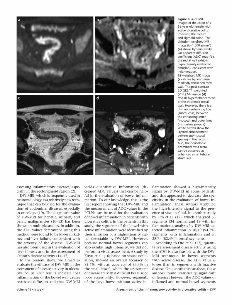
Figure 4. a–d. MR images of the colon of a 56-year-old female with active ulcerative colitis involving the rectum and sigmoid colon. The diffusion-weighted MR image (b=1,000 s/mm2) (a) shows hyperintensity. On apparent diffusion coefficient (ADC) map (b), the rectal wall exhibits hypointensity (restricted diffusion), consistent with inflammation. T2-weighted MR image (c) shows hyperintense, markedly thickened rectal wall. The post-contrast 3D GRE T1-weighted (VIBE) MR image (d) reveals hyperenhancement of the thickened rectal wall. However, there is a thin non-enhancing line (submucosa) between the enhancing inner (mucosa) and outer lines (muscularis propria). White arrows show the layered enhancement pattern-submucosal sparing in the rectum. Also, the pericolonic prominent vasa recta can be observed as enhanced small tubular structures.
ba
c d

Kılıçkesmez et al.298 • December 2010 • Diagnostic and Interventional Radiology
of patients with Crohn’s disease (0.47–2.60×10-³ mm²/s and 1.39–4.03×10-³ mm²/s for inflamed and normal, re-spectively; P < 0.05). A quantitative study by Kiryu et al. (16) also found that the ADC values for the small and large bowel in patients with active dis-ease were lower than those in patients with inactive disease (1.61±0.44×10-³ mm²/s vs. 2.56±0.51×10-³ mm²/s for the small bowel and 1.52±0.43 ± 10-
³ mm²/s vs. 2.31±0.59 ± 10-³ mm²/s for the large bowel, respectively; P < 0.001). Similarly, we found that in-creased disease activity correlates with decreased ADC values. It may be spec-ulated that the possible causes of the restricted diffusion we observed in our patients may be related to inflamma-tion, fibrosis and increased cell volume of the intestinal wall. Quantitative analysis may be useful in monitoring the status of a disease and the effects of therapy. However, the usefulness of the ADC for long-term follow-up of pa-tients with ulcerative colitis warrants further investigation (16).
In patients with suspected or known IBD, the clinically important issues are diagnosis of the disease, determina-tion of the extent and severity of the disease, discrimination between ac-tive inflammation and fibrosis, and monitoring the patient’s response to treatment. DW-MRI of the bowel may provide information that is relevant to addressing these clinically impor-tant issues. The ability to use quantita-tive parameters such as ADC values to evaluate IBD can allow a more objec-tive evaluation of the disease. Studies in which a larger number of patients are examined are needed in order to better understand the importance of DW-MRI in the evaluation of patients with IBD (1).
Our study has several limitations. First, the number of patients in our study was relatively low. Second, we at-tempted to magnify the images of the
bowel as accurately as possible and to use an oval ROI to exclusively examine the bowel wall; however, we cannot completely exclude the possibility of a partial volume effect, especially from the normal bowel wall, on the ADC measurements. Third, we used free-breathing DW-MRI to evaluate the pa-tients; however, breath-holding DW-MRI may provide better image quality.
It is important to note that the DW-MRI technique is completely non-in-vasive, does not require ionizing radia-tion or injection of contrast material, and does not cause discomfort to the patients. DW-MRI can be easily added to an MR examination protocol because it requires only a very short prolonga-tion of the examination time. Thus, DW-MRI represents a useful evaluation technique to add to conventional eval-uation protocols (12).
In conclusion, the ADC values of the distal colon wall and the PCLNs obtained using DW-MRI increased in ulcerative colitis patients on remission and decreased in patients with active distal colitis. Thus, DW-MRI is useful in evaluating disease activity in the rectum. However, the ADC values of the PCLNs may not be helpful in evalu-ating disease activity.
References 1. Kornbluth A, Sachar DB. Practice Parameters
Committee of the American College of Gastroenterology. Ulcerative colitis prac-tice guidelines in adults (update): American College of Gastroenterology, Practice Parameters Committee. Am J Gastroenterol 2004; 99:1371–1385.
2. Hoeffel CC, Azizi L, Mourra N, et al. MRI of rectal disorders. AJR Am J Roentgenol 2006; 187:275–284.
3. Paley MR, Ros PR. MRI of the gastrointesti-nal tract. Eur Radiol 1997; 7:1387–1397.
4. Paley MR, Ros PR. MRI of the rectum: non-neoplastic disease. Eur Radiol 1998; 8:3–8.
5. Ajaj WM, Lauenstein TC, Pelster G, et al. Magnetic resonance colonography for the detection of inflammatory diseases of the large bowel: quantifying the inflammatory activity. Gut 2005; 54:257–263.
6. Rimola J, Rodríguez S, García-Bosch O, et al. Role of 3.0-T MR colonography in the evaluation of inflammatory bowel disease. Radiographics 2009; 29:701–719.
7. Rachmilewitz D. Coated mesalazine (5-aminosalicylic acid) versus sulphasalazine in the treatment of active ulcerative colitis: a randomised trial. BMJ 1989; 298:82–86.
8. Koh DM, Miao Y, Chinn RJ, et al. MR im-aging evaluation of the activity of Crohn’s disease. AJR Am J Roentgenol 2001; 177:1325–1332.
9. Fiocca R, Ceppa P. The diagnostic reli-ability of endoscopic biopsies in diagnosis colitis. J Clin Pathol 2003; 56:321–322.
10. Koh DM, Collins DJ. Diffusion-weighted MRI in the body: applications and chal-lenges in oncology. AJR Am J Roentgenol 2007; 188:1622–1635.
11. Kilickesmez O, Cimilli T, Inci E, et al. Diffusion-weighted MRI of urinary bladder and prostate cancers. Diagn Interv Radiol 2009; 15:104–110.
12. Kilickesmez O, Bayramoglu S, Inci E, Cimilli T. Value of apparent diffusion co-efficient measurement for discrimination of focal benign and malignant hepatic masses. J Med Imaging Radiat Oncol 2009; 53:50–55.
13. Hosonuma T, Tozaki M, Ichiba N, et al. Clinical usefulness of diffusion-weighted imaging using low and high b-values to detect rectal cancer. Magn Reson Med Sci 2006; 5:173–177.
14. Taouli B, Tolia AJ, Losada M, et al. Diffusion-weighted MRI for quantification of liver fibrosis: preliminary experience. AJR Am J Roentgenol 2007; 189:799–806.
15. Taouli B, Chouli M, Martin AJ, Qayyum A, Coakley FV, Vilgrain V. Chronic hepatitis: role of diffusion-weighted imaging and diffusion tensor imaging for the diagnosis of liver fibrosis and inflammation. J Magn Reson Imaging 2008; 28:89–95.
16. Kiryu S, Dodanuki K, Takao H, et al. Free-breathing diffusion-weighted imaging for the assessment of inflammatory activity in Crohn’s disease. J Magn Reson Imaging 2009; 29:880–886.
17. Oto A, Zhu F, Kulkarni K, Karczmar GS, Turner JR, Rubin D. Evaluation of dif-fusion-weighted MR imaging for detec-tion of bowel inflammation in patients with Crohn’s disease. Acad Radiol 2009; 16:597–603.
Related Documents