Short Communication Is acetylcholinesterase a biomarker of susceptibility in Daphnia magna (Crustacea, Cladocera) after deltamethrin exposure? Héla Toumi a,b , Moncef Boumaiza a , Maurice Millet c , Claudemir Marcos Radetski d , Vincent Felten b , Jean François Férard b,⇑ a Laboratoire de Bio-surveillance de l’Environnement (LBE), Unité d’Hydrobiologie littorale et limnique, Université de Carthage, Faculté des Sciences de Bizerte, 7021 Zarzouna, Bizerte, Tunisia b Université de Lorraine (UdL), Laboratoire Interdisciplinaire des Environnements Continentaux (LIEC) – UMR 7360 CNRS, Campus Bridoux, Bât. IBiSE, 8, rue du Général Delestraint, 57070 Metz, France c ICPEES (UMR 7515 CNRS – Université de Strasbourg), 1 rue Blessig, 67084 Strasbourg Cedex, France d Laboratório de Remediação Ambiental, Universidade do Vale do Itajaí, Rua Uruguai, 458, Itajaí, SC 88302-202, Brazil highlights LOECs of AChE activity differ by four-fold between the three strains of Daphnia magna. AChE is a biomarker of susceptibility after deltamethrin exposure in Daphnia magna. Ratios of AChE IC50 and the immobility EC50-48 h is a potential indication of genetic heterogeneity. article info Article history: Received 17 January 2014 Received in revised form 22 July 2014 Accepted 28 July 2014 Handling Editor: A. Gies Keywords: Pyrethroid Biomarker Acetylcholinesterase Daphnia magna Inter-strain sensitivity abstract In the present study, we explored the possibility of using the acetylcholinesterase (AChE) as a biomarker after deltamethrin (pyrethroid insecticide) exposure with three strains of the cladoceran Daphnia magna. Four calculated time-weighted deltamethrin concentrations (20.1, 40.3, 80.6 and 161.3 ng L 1 ) were com- pared against control acetylcholinesterase activity. Our results showed that after 48 h of deltamethrin exposure, all treatments induced a significant decrease of AChE activities whatever the three considered strains. However, diverse responses were registered in terms of lowest observed effect concentrations (LOEC: 80.6 ng L 1 for strain 1 and 20.1 ng L 1 for strains 2 and 3) revealing differences in sensitivity among the three tested strains of D. magna. Our results suggest that after deltamethrin exposure, the AChE activity responses can be also used as a biomarker of susceptibility (i.e., variation of strain specific response). More- over, our results show that strain 1 is the less sensitive in terms of IC50-48 h of AChE, whereas it became the most sensitive when considering the EC50-48 h estimated in the standard ecotoxicity test. Ó 2014 Elsevier Ltd. All rights reserved. 1. Introduction Biomarkers are efficient tools to identify perturbations of diverse physiological and biochemical parameters and have been generally used to evaluate the exposure of organisms to a range of pollutants in the environment, including pesticides (Stegeman et al., 1992; Peakall and Shugart, 1993). Among the most commonly used bio- markers, acetylcholinesterase (AChE) activity was widely employed to detect and reveal exposure in vertebrates (Sharma and Khanna, 1993; Bocquené et al., 1997) and invertebrates (Bocquené et al., 1997; Barata et al., 2004; Xuereb et al., 2007) to organophosphate and carbamate insecticides. Indeed, AChE is a main enzyme respon- sible of the rapid hydrolysis of acetylcholine at cholinergic synapses and at neuromuscular junctions, allowing per consequent the control and modulation of neural transmission. Pyrethroids are among the most toxic pesticides known (Silvério et al., 2009). They are generally considered to be of low acute mammalian toxicity (Grosman and Diel, 2005), but are highly ecotoxic to non-target organisms such as honeybees, fish and aquatic arthropods even at very low concentrations (Saha and Kaviraj, 2008). They are widely used in household, in field pest control particularly in agriculture and forestry because of their high activity against a broad spectrum of insect pests (Villarini et al., 1998). They are also employed for malaria control programs, for example in India (Ansari and Razdan, 2001). The use of these http://dx.doi.org/10.1016/j.chemosphere.2014.07.087 0045-6535/Ó 2014 Elsevier Ltd. All rights reserved. ⇑ Corresponding author. Tel.: +33 387378503; fax: +33 387378512. E-mail address: [email protected] (J.F. Férard). Chemosphere 120 (2015) 351–356 Contents lists available at ScienceDirect Chemosphere journal homepage: www.elsevier.com/locate/chemosphere

Welcome message from author
This document is posted to help you gain knowledge. Please leave a comment to let me know what you think about it! Share it to your friends and learn new things together.
Transcript
Chemosphere 120 (2015) 351–356
Contents lists available at ScienceDirect
Chemosphere
journal homepage: www.elsevier .com/locate /chemosphere
Short Communication
Is acetylcholinesterase a biomarker of susceptibility in Daphnia magna(Crustacea, Cladocera) after deltamethrin exposure?
http://dx.doi.org/10.1016/j.chemosphere.2014.07.0870045-6535/� 2014 Elsevier Ltd. All rights reserved.
⇑ Corresponding author. Tel.: +33 387378503; fax: +33 387378512.E-mail address: [email protected] (J.F. Férard).
Héla Toumi a,b, Moncef Boumaiza a, Maurice Millet c, Claudemir Marcos Radetski d, Vincent Felten b,Jean François Férard b,⇑a Laboratoire de Bio-surveillance de l’Environnement (LBE), Unité d’Hydrobiologie littorale et limnique, Université de Carthage, Faculté des Sciences de Bizerte, 7021 Zarzouna,Bizerte, Tunisiab Université de Lorraine (UdL), Laboratoire Interdisciplinaire des Environnements Continentaux (LIEC) – UMR 7360 CNRS, Campus Bridoux, Bât. IBiSE, 8, rue du GénéralDelestraint, 57070 Metz, Francec ICPEES (UMR 7515 CNRS – Université de Strasbourg), 1 rue Blessig, 67084 Strasbourg Cedex, Franced Laboratório de Remediação Ambiental, Universidade do Vale do Itajaí, Rua Uruguai, 458, Itajaí, SC 88302-202, Brazil
h i g h l i g h t s
� LOECs of AChE activity differ by four-fold between the three strains of Daphnia magna.� AChE is a biomarker of susceptibility after deltamethrin exposure in Daphnia magna.� Ratios of AChE IC50 and the immobility EC50-48 h is a potential indication of genetic heterogeneity.
a r t i c l e i n f o
Article history:Received 17 January 2014Received in revised form 22 July 2014Accepted 28 July 2014
Handling Editor: A. Gies
Keywords:PyrethroidBiomarkerAcetylcholinesteraseDaphnia magnaInter-strain sensitivity
a b s t r a c t
In the present study, we explored the possibility of using the acetylcholinesterase (AChE) as a biomarkerafter deltamethrin (pyrethroid insecticide) exposure with three strains of the cladoceran Daphnia magna.Four calculated time-weighted deltamethrin concentrations (20.1, 40.3, 80.6 and 161.3 ng L�1) were com-pared against control acetylcholinesterase activity. Our results showed that after 48 h of deltamethrinexposure, all treatments induced a significant decrease of AChE activities whatever the three consideredstrains. However, diverse responses were registered in terms of lowest observed effect concentrations(LOEC: 80.6 ng L�1 for strain 1 and 20.1 ng L�1 for strains 2 and 3) revealing differences in sensitivity amongthe three tested strains of D. magna. Our results suggest that after deltamethrin exposure, the AChE activityresponses can be also used as a biomarker of susceptibility (i.e., variation of strain specific response). More-over, our results show that strain 1 is the less sensitive in terms of IC50-48 h of AChE, whereas it became themost sensitive when considering the EC50-48 h estimated in the standard ecotoxicity test.
� 2014 Elsevier Ltd. All rights reserved.
1. Introduction
Biomarkers are efficient tools to identify perturbations of diversephysiological and biochemical parameters and have been generallyused to evaluate the exposure of organisms to a range of pollutantsin the environment, including pesticides (Stegeman et al., 1992;Peakall and Shugart, 1993). Among the most commonly used bio-markers, acetylcholinesterase (AChE) activity was widely employedto detect and reveal exposure in vertebrates (Sharma and Khanna,1993; Bocquené et al., 1997) and invertebrates (Bocquené et al.,1997; Barata et al., 2004; Xuereb et al., 2007) to organophosphate
and carbamate insecticides. Indeed, AChE is a main enzyme respon-sible of the rapid hydrolysis of acetylcholine at cholinergic synapsesand at neuromuscular junctions, allowing per consequent the controland modulation of neural transmission.
Pyrethroids are among the most toxic pesticides known(Silvério et al., 2009). They are generally considered to be of lowacute mammalian toxicity (Grosman and Diel, 2005), but arehighly ecotoxic to non-target organisms such as honeybees, fishand aquatic arthropods even at very low concentrations (Sahaand Kaviraj, 2008). They are widely used in household, in field pestcontrol particularly in agriculture and forestry because of theirhigh activity against a broad spectrum of insect pests (Villariniet al., 1998). They are also employed for malaria control programs,for example in India (Ansari and Razdan, 2001). The use of these

352 H. Toumi et al. / Chemosphere 120 (2015) 351–356
compounds has been continuously increasing during the two lastdecades (Oliveira et al., 2012): in 2008, they represented 15.1% ofthe wordly market of insecticides (Bodereau-Dubois, 2011). Theexcessive use of these neurotoxic pesticides could lead to diversedamages after contamination of aquatic ecosystems. They havebeen found in several compartments, in waters (0.04–24 lg L�1),sediments (3–5 ng g�1), plants (281–1375 ng g�1) and animals(3–50 ng g�1) (Pawlisz et al., 1998). Effects on diverse organismshave been recently reviewed by Fojut et al. (2012).
Deltamethrin (DM) is a synthetic type II pyrethroid (as itincludes a cyano group) that is known to be toxic to diverse aquaticorganisms (US EPA, 2013). It acts primarily on the nervous system,especially on the voltage-dependent ion channel in excitable mem-brane, but also on various secondary targets involved in signaltransduction, as pointed by Narahashi (1992), or in up and downregulation of the proteome as registered by Toumi et al. (2014).According to Oliveira et al. (2012), acute ecotoxicity data (as LC50) ranged from 20 (Ceriodaphnia cf. dubia) to 5130 ng L�1 (Poeciliareticulata), but Adam et al. (2010) reported a LC50-48 h of 4 ng L�1
with Gammarus fossarum. Concerning Daphnia magna, we recentlypublished acute and chronic ecotoxicity data (Toumi et al., 2013).
The cladoceran D. magna is a standard organism in ecotoxico-logy and is routinely used as a model species to determine ecotox-icity of chemicals for risk assessment (Pereira et al., 2010). Thisspecies has often been used in bioassays and environmental mon-itoring of aquatic systems because it is easy to maintain in the lab-oratory, has a short life cycle and is sensitive to a large number ofchemicals (Hanazato, 1998) such as organophosphorus compounds(Guilhermino et al., 1996; Barata et al., 2001) and pyrethroids (Renet al., 2009; Toumi et al., 2013). It is largely known that organo-phosphorous and carbamate pesticides induce reduction of AChEactivity in D. magna, but other environmental contaminants couldalso exert the same effect. In this respect, there are several studiesshowing decrease of AChE activities under DM exposure withdiverse aquatic organisms such as fish, Cyprinus carpio (Bàlintet al., 1995; Szegletes et al., 1995) and Oncorhynchus mykiss(Velisek et al., 2007), and crustacean Penaeus monodon (Tu et al.,2012). For example, in carp (C. carpio), Bàlint et al. (1995) foundthat AChE activity was decreased by 28% after a 24 h DM exposureat 2 lg L�1. Nevertheless some studies have reported increase ofAChE activity after DM exposure (Oliveira et al., 2012). Accordingto Ensibi et al. (2014) and Guiloski et al. (2013), AChE activitywas not affected after DM exposure of the assay, respectively inthe brain and muscle of the common carp C. carpio and the brainof the freshwater fish Corydoras paleatus.
We have focused our study on the AChE activity of D. magna andits alteration after acute DM exposure because (i) this biomarker isknown to be very useful in indicating organisms have been exposedin vivo to a cholinesterase inhibiting compound and (ii) there is nopreviously published research showing effects of acute DM expo-sure on D. magna AChE activity. The aim of this study was (i) toreveal if AChE could be considered as a sensitive biomarker afteracute DM exposure of D. magna, and (ii) to compare the AChE activ-ities between three strains of D. magna, after acute DM exposure, inorder to reveal that AChE could be considered as a biomarker of sus-ceptibility (genetic variability) and illustrate D. magna inter-strainsensitivity (i.e. ability of a species to respond to effective stress inthe environment such as contamination by chemical compounds).
2. Materials and methods
2.1. Test organisms
Experiments were conducted with three strains of D. magna.The strain 1 was maintained for more than 35 years at the LIEC
laboratory (Lorraine University, France) and it has been identifiedpreviously as clone A by the laboratory of Professor Calow, Shef-field University, UK. The strain 2 was obtained from the NationalInstitute for Environmental Studies (NIES, Tsukuba, Japan). Thisstrain has been maintained for more than 5 years at the LIEC labo-ratory, but its clonal identification remains unknown. The laststrain (strain 3) was obtained from IRSTEA (Lyon, France), that isconsidered belonging to clone A, and it was maintained for morethan 3 years in LIEC laboratory.
In our study, we defined the term ‘‘clone’’ as an identified strainaccording to the classification given by Baird and Barata (1998) andconsequently the term ‘‘strain’’ was also used when the clone wasnot identified. As strains 1 and 3 were identified as clone A andstrain 2 was not identified, we have chosen to employ the sameterm (i.e. strain) all along the manuscript.
The three strains were reared under the same laboratory condi-tions (see above). Briefly, parthenogenetic cultures were reared in1 L aquaria at 20 �C with a mixture (20/80) of Lefevre-Czarda (LC)medium (Ionescu et al., 2006) and French (Volvic) mineral waterwith a hardness of 250 mg L�1 (expressed as CaCO3). The daphnidswere maintained under a 16–8 h light–dark photoperiod and at adensity of 40 animals per liter (Manar et al., 2009). The culturemedium was supplemented with a mixture of vitamins(0.1 mL L�1) containing thiamine HCl (750 mg L�1), vitamin B12(10 mg L�1), and biotin (7.5 mg L�1) and was renewed three timesweekly. Daphnids were fed three times a week with a mixture ofthree algal species and every Daphnia was fed with an amount ofalgae expressed in cells/day at a rate of: 5 � 106 Pseudokirchneriellasubcapitata/Daphnia/day, 2.5 � 106 Desmodesmus subspicatus/Daphnia/day, and 2.5 � 106 Chlorella vulgaris/Daphnia/day.
2.2. Test chemical
The chemical name of DM (CAS 52918-63-5, C22H19Br2NO3) is(s)-Cyano-(3-phenoxyphen) methyl (lR)-cis-3-(2,2-dibromovinyl)-2,2-dimethylcyclopropanecarboxylate). DM is the active substanceof the formulation DECIS EC25 (25 g L�1) commercialized by Bayer(Germany) that was tested in this study. Other ingredients weretetrapropylene benzene sulfonate, calcium salt (between 1% and5%), 2-methylpropan-1-ol (between 1% and 5%) and solvent naph-tha (petroleum), light aromatic (>50%) as indicated in the safetydatasheet available on http://bluemsds.tdgmond.be/files/msds/DECIS%20EC25_2007-09-19_FR_118389.PDF.
2.3. Bioassays
Two different series of bioassays have been performed. A firstseries was carried out to determine the acute ecotoxicity of DMon strain 3 (data related to strains 1 and 2 were published else-where by Toumi et al., 2013). A second series was run to explorethe effect of DM against AChE of the three strains with a slightlymodified procedure.
Acute ecotoxicity of DM was determined during 24 and 48 h ofexposure using nominal DM concentrations from 0.25 to 51 lg L�1.Experiments were performed according to the International Orga-nization for Standardization procedure 6341 (ISO, 1996) for thedetermination of mobility inhibition of D. magna. Briefly, four rep-licates of five neonates (aged <24 h) were placed in glass test tubescontaining 10 mL for each test concentration (ISO medium withDM concentrations) and control (ISO medium). The neonates werenot fed during the test. The assessment endpoint examined wasimmobilization. More precisely juveniles that were able to swimwere considered mobile and those which still moved their anten-nae but did not swim within 15 s after a gentle shaking were con-sidered immobile.

Table 1Relationship between acute toxicity (EC50 48 h) of deltamethrin on the three Daphniamagna strains and effect on acetylcholinesterase activities (IC50-48 h) in lg L�1.Values are expressed as measured concentrations (see Section 2).
Strains EC50-48 h (Immobility) IC50-48 h (AChE) IC50/EC50
Strain 1 0.32 (0.21–0.43)* 0.058 (0.040–0.072) 0.18Strain 2 0.63 (0.53–0.72)* 0.018 (0.015–0.064) 0.03Strain 3 0.88 (0.79–0.98) 0.016 (0.014–0.024) 0.02
* Values have been determined in a previous study (Toumi et al., 2013).
H. Toumi et al. / Chemosphere 120 (2015) 351–356 353
For AChE effect experimentations, exposure design was similarto acute ecotoxicity tests design defined by ISO (1996) except thattwenty juvenile (<24 h) of each strain were placed in 60 mL glassbeakers containing 50 mL of test solution, thus daphnid densityis the same as in the acute bioassay. For each strain, four differentnominal DM concentrations (37.5, 75, 150 and 300 ng L�1) weretested with three replicates: each replicate consisted in five vesselsand each vessel containing at the departure twenty daphnids.Daphnids were not fed during the test. After 48 h of static expo-sure, there were no mortality in controls and the three lowest con-centrations, but at a nominal concentration of 300 ng L�1, amaximum of 30% mortality was observed for the strain 1. Deaddaphnids were strictly eliminated and removed in order to useonly alive organisms, and then animals from 5 beakers were pooledin order to have sufficient biological material for AChE analysis.Finally, three replicates were made by treatment.
2.4. Analytical determinations
At three times (0, 24 and 48 h), three nominal DM concentra-tions from acute test (0.55; 15.2 and 51 lg L�1 at t0) wereextracted from test solutions with dichloromethane (CH2Cl2). Afterextraction, the solvent was concentrated to 1 mL with a rotaryevaporator maintained at 45 �C and 800 mbar to evaporate CH2Cl2.DM was analyzed using a gas chromatograph equipped with an iontrap mass spectrometer (FOCUS-ITQ 700 ThermoScientific Inc) inelectronic impact mode. DM extract was injected (2 lL) in thesplitless mode (1 min) on a Varian VF 5 capillary column(30 m � 0.25 mm � 0.25 lm film thickness) and detection wasmade in MSMS mode (parent ion 181 m/z; daughter ion: 153 m/z). Detection and quantification limits were calculated for theextract as 0.2 and 0.5 lg L�1 respectively with an uncertainty of8%. In this respect, calculation of detection and quantification lim-its in the exposure medium (depending of the enrichment factors)varied from 4 to 5 ng L�1 and 10 to 12.5 ng L�1 respectively. Themeasured DM concentrations at t0 were 0.49; 13.8 and 49.6 lg L�1,but after 24 and 48 h, all nominal concentrations decreased by45.40% and 73.95%, respectively. Therefore we expressed eachtested concentrations by measured time-weighted means (TWM)according to the equation below (OECD, 2008).
TWM ¼ Conc d0� Conc d1LnðConc d0Þ � LnðConc d1Þ
þ Conc d1� Conc d2LnðConc d1Þ � LnðConc d2Þ
�2 ð1Þ
where Conc dx is the concentration at the considered day.
2.5. Enzyme activity determinations
Although, it cannot be excluded that DM was completelyremoved from the outer carapax, daphnids were washed 3 timeswith 2 mL of homogenization buffer (phosphate buffer 50 mM,pH 7.0) combined with ethylenediaminetetraacetic acid (EDTA)5 mM in order to remove potential bias caused by possiblein vitro interactions. Using a ball mill homogenizer (RETSCH,MM301, ball size = 0.1 mm), each sample of daphnids was homog-enized in 0.6 mL of homogenization buffer (phosphate buffer50 mM, pH 7.0) during 3 min (30 Hz), and then centrifuged during15 min at 15000g and 4 �C. The enzyme activities in each samplewere measured in triplicate, and determined according to themethod of Ellman et al. (1961) slightly modified by Jemec et al.(2007). The reaction mixture (5 mL) was prepared in potassiumphosphate buffer (100 mM, pH 7.4) containing acetylthiocholinechloride (1 mM) and 5,50 dithiobis-2-nitrobenzoic acid (0.5 mM),then 100 lL of protein supernatant were added to start the
reaction. The absorbance at 412 nm was measured at 25 �C during15 min with a spectrophotometer UVIKON 933. AChE activity was expressed in nmoles of acetylcholine chloride hydrolyzedmin�1 mg�1 of protein (extinction coefficient = 13600 M�1 cm�1).The protein concentration in the samples was also determined intriplicate by the Bradford method (Bradford, 1976), using bovineglobulin (BIORAD 5000204) as standard.
2.6. Statistical analyses
Acute EC50 values were calculated by a non linear regression onHill’s model using REGTOX software (version 7.0.3, available athttp://www.normalesup.org/~vindimian/fr_index.html): briefly aMicrosoft Excel� spreadsheet (Redmond, WA, USA) enabled auto-mated macro-calculation of EC50 values. The homogeneity of vari-ances and normality among replicates were tested by Bartlett’s testand Kolmogorov–Smirnov test, respectively. LOECs were deter-mined by one-way analysis of variance followed by Dunnett’s posthoc test. Significant differences were established at p < 0.05 andperformed with Statistica 6.0 software. AChE IC50-48 h (i.e. after48 h of exposure) values were estimated using the Inhibition Con-centration (ICp) method (US EPA, 1994). This method provides, byinterpolation, a point estimate of a single concentration causing aspecified percentage effect. Statistically significant differencesbetween IC50, EC50 and LOEC values were determined using ran-dom sampling of thirty data taken from each confidence intervalsgiven by the ICp method. Then, populations of random values werecompared by one-way ANOVA test followed by Tukey test at the 5%level.
3. Results and discussion
All strains satisfied the validity conditions of the ISO standard(1996) in terms of control mortality and potassium dichromatesensitivity. DM EC50s (i.e. concentrations at which 50% of the num-ber of neonates were immobilized) of the three strains reported inthe Table 1 were statistically significantly different. The ecotoxicityof strain 3 was lower and is represented by a higher EC50 valuewhen compared to strains 1 and 2 (Table 1). AChE specific activitiesof the three controls showed no statistically significant differences,but AChE specific activity of strain 3 controls was higher(4.44 ± 0.76 nmol min�1 mg�1 of proteins) that both other strains(2.86 ± 0.29 and 3.11 ± 0.92 nmol min�1 mg�1 of proteins, respec-tively). These variations cannot be attributed to variation of exper-imental conditions because the strains were maintained under thesame culture and exposure conditions for at least three years andthe same conditions were applied during DM exposure (see Sec-tion 2). By consequence, it is assumed that these variations couldbe probably assigned to genetic variation.
The control data we observed were in agreement and in therange with those reported by Barata et al. (2004) after 24 h of testexperimentation (3.5 nmol min�1 mg�1 of proteins). It is also inter-esting to compare our results with those of Jemec et al. (2007)because we used the same experimental conditions. It is likely that
354 H. Toumi et al. / Chemosphere 120 (2015) 351–356
the observed differences (more than a factor two) could be due tothe tested strain or to some suspected differences in culture condi-tions, but we cannot exclude some variations in the measurementmethod between laboratories. Although we used the same strain(i.e. clone A) as that used by Diamantino et al. (2000, 2003) andGuilhermino et al. (1996, 2000), they reported, after 24 h or 48 hof test experimentation, higher control data (ranging from 8.25to 8.90 nmol min�1 mg�1 of proteins) than ours. However, usingthe same clone, Printes and Callaghan (2003) found, in controls, adata of 0.72 nmol min�1 mg�1 of proteins after 48 h of test exper-imentation. Moreover, the same team later reported an AChE spe-cific activity of ca. 4.7 ± 0.2 nmol min�1 mg�1 of proteins in fedcontrols (Printes et al., 2008). All these data demonstrate that thereis a lot of variation in AChE specific activity when examining liter-ature results. These variations could be related to inter-clonal (orinter-strain) or intra-clonal (or intra-strain) differences. The formercan be attributed to genetic heterogeneity (Soares et al., 1992), andthe latter to the use of neonates from different broods (Barata et al.,2001) or to the culture medium as was the case for D. similis(Printes and Callaghan, 2003). The quality and quantity of foodcould also have some effects but this factor has not been yetevaluated.
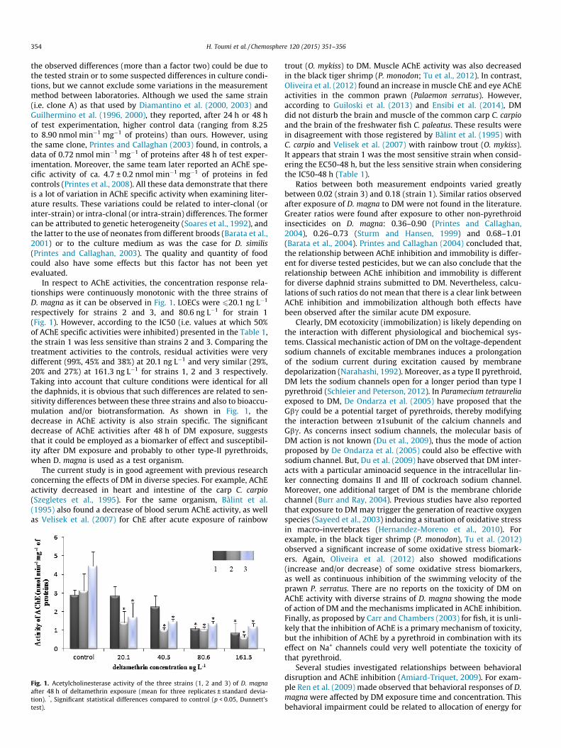
In respect to AChE activities, the concentration response rela-tionships were continuously monotonic with the three strains ofD. magna as it can be observed in Fig. 1. LOECs were 620.1 ng L�1
respectively for strains 2 and 3, and 80.6 ng L�1 for strain 1(Fig. 1). However, according to the IC50 (i.e. values at which 50%of AChE specific activities were inhibited) presented in the Table 1,the strain 1 was less sensitive than strains 2 and 3. Comparing thetreatment activities to the controls, residual activities were verydifferent (99%, 45% and 38%) at 20.1 ng L�1 and very similar (29%,20% and 27%) at 161.3 ng L�1 for strains 1, 2 and 3 respectively.Taking into account that culture conditions were identical for allthe daphnids, it is obvious that such differences are related to sen-sitivity differences between these three strains and also to bioaccu-mulation and/or biotransformation. As shown in Fig. 1, thedecrease in AChE activity is also strain specific. The significantdecrease of AChE activities after 48 h of DM exposure, suggeststhat it could be employed as a biomarker of effect and susceptibil-ity after DM exposure and probably to other type-II pyrethroids,when D. magna is used as a test organism.
The current study is in good agreement with previous researchconcerning the effects of DM in diverse species. For example, AChEactivity decreased in heart and intestine of the carp C. carpio(Szegletes et al., 1995). For the same organism, Bàlint et al.(1995) also found a decrease of blood serum AChE activity, as wellas Velisek et al. (2007) for ChE after acute exposure of rainbow
Fig. 1. Acetylcholinesterase activity of the three strains (1, 2 and 3) of D. magnaafter 48 h of deltamethrin exposure (mean for three replicates ± standard devia-tion). *, Significant statistical differences compared to control (p < 0.05, Dunnett’stest).
trout (O. mykiss) to DM. Muscle AChE activity was also decreasedin the black tiger shrimp (P. monodon; Tu et al., 2012). In contrast,Oliveira et al. (2012) found an increase in muscle ChE and eye AChEactivities in the common prawn (Palaemon serratus). However,according to Guiloski et al. (2013) and Ensibi et al. (2014), DMdid not disturb the brain and muscle of the common carp C. carpioand the brain of the freshwater fish C. paleatus. These results werein disagreement with those registered by Bàlint et al. (1995) withC. carpio and Velisek et al. (2007) with rainbow trout (O. mykiss).It appears that strain 1 was the most sensitive strain when consid-ering the EC50-48 h, but the less sensitive strain when consideringthe IC50-48 h (Table 1).
Ratios between both measurement endpoints varied greatlybetween 0.02 (strain 3) and 0.18 (strain 1). Similar ratios observedafter exposure of D. magna to DM were not found in the literature.Greater ratios were found after exposure to other non-pyrethroidinsecticides on D. magna: 0.36–0.90 (Printes and Callaghan,2004), 0.26–0.73 (Sturm and Hansen, 1999) and 0.68–1.01(Barata et al., 2004). Printes and Callaghan (2004) concluded that,the relationship between AChE inhibition and immobility is differ-ent for diverse tested pesticides, but we can also conclude that therelationship between AChE inhibition and immobility is differentfor diverse daphnid strains submitted to DM. Nevertheless, calcu-lations of such ratios do not mean that there is a clear link betweenAChE inhibition and immobilization although both effects havebeen observed after the similar acute DM exposure.
Clearly, DM ecotoxicity (immobilization) is likely depending onthe interaction with different physiological and biochemical sys-tems. Classical mechanistic action of DM on the voltage-dependentsodium channels of excitable membranes induces a prolongationof the sodium current during excitation caused by membranedepolarization (Narahashi, 1992). Moreover, as a type II pyrethroid,DM lets the sodium channels open for a longer period than type Ipyrethroid (Schleier and Peterson, 2012). In Paramecium tetraureliaexposed to DM, De Ondarza et al. (2005) have proposed that theGbc could be a potential target of pyrethroids, thereby modifyingthe interaction between a1subunit of the calcium channels andGbc. As concerns insect sodium channels, the molecular basis ofDM action is not known (Du et al., 2009), thus the mode of actionproposed by De Ondarza et al. (2005) could also be effective withsodium channel. But, Du et al. (2009) have observed that DM inter-acts with a particular aminoacid sequence in the intracellular lin-ker connecting domains II and III of cockroach sodium channel.Moreover, one additional target of DM is the membrane chloridechannel (Burr and Ray, 2004). Previous studies have also reportedthat exposure to DM may trigger the generation of reactive oxygenspecies (Sayeed et al., 2003) inducing a situation of oxidative stressin macro-invertebrates (Hernandez-Moreno et al., 2010). Forexample, in the black tiger shrimp (P. monodon), Tu et al. (2012)observed a significant increase of some oxidative stress biomark-ers. Again, Oliveira et al. (2012) also showed modifications(increase and/or decrease) of some oxidative stress biomarkers,as well as continuous inhibition of the swimming velocity of theprawn P. serratus. There are no reports on the toxicity of DM onAChE activity with diverse strains of D. magna showing the modeof action of DM and the mechanisms implicated in AChE inhibition.Finally, as proposed by Carr and Chambers (2003) for fish, it is unli-kely that the inhibition of AChE is a primary mechanism of toxicity,but the inhibition of AChE by a pyrethroid in combination with itseffect on Na+ channels could very well potentiate the toxicity ofthat pyrethroid.
Several studies investigated relationships between behavioraldisruption and AChE inhibition (Amiard-Triquet, 2009). For exam-ple Ren et al. (2009) made observed that behavioral responses of D.magna were affected by DM exposure time and concentration. Thisbehavioral impairment could be related to allocation of energy for
H. Toumi et al. / Chemosphere 120 (2015) 351–356 355
detoxification and antioxidant protection instead of swimmingactivity as postulated by Oliveira et al. (2012).
4. Conclusion
This study shows that DM induces a decrease in AChE activitywith the three strains of D. magna. Differences were registeredbetween strains for the controls and in terms of LOECs of AChEinhibition after DM exposure, which suggest that there is an int-erclonal (or interstrain) variability in AChE activities. Such resultsconfirm that AChE could be used as a DM biomarker of exposure,effect and susceptibility in case of D. magna DM exposure. Never-theless, the presented data will serve up as a basis for further stud-ies on AChE as a biomarker of pyrethroid exposure. The linkbetween AChE inhibition and acute ecotoxic effects of pyrethroidshas yet to be analyzed and established in further studies.
Acknowledgments
This study was supported by the Tunisian Ministry of HigherEducation (which we sincerely thank) and by Lorraine University(France). The present work was also funded by the research pro-gram PHC-Utique 2012 managed by the CMCU. The authors wouldlike to thank Christian Blaise (Emeritus researcher of EnvironmentCanada) for his assistance with the English revision of this manu-script. We are also grateful to the two reviewers for their appropri-ate and constructive suggestions and for their proposed correctionsto improve the quality of the paper.
References
Adam, O., Degiorgi, F., Crini, G., Badot, P.M., 2010. High sensitivity of Gammarus sp.juveniles to deltamethrin: outcomes for risk assessment. Ecotox. Environ. Saf.73, 1402–1407.
Amiard-Triquet, C., 2009. Behavioral disturbances: the missing link between sub-organismal and supra-organismal responses to stress? Prospects based onaquatic research. Human Ecol. Risk Assess. 15, 87–110.
Ansari, M.A., Razdan, R.K., 2001. Concurrent control of mosquitoes and domesticpets by use of deltamethrin-treated curtains in the New Delhi munipalcommittee. India. J. Am. Mosq. Control Assoc. 17 (2), 131–136.
Baird, D.J., Barata, C., 1998. Genetic variation in the response of Daphnia to toxicsubstances: implications for risk assessment. In: Forbes, V.E. (Ed.), Genetic andEcotoxicology. Taylor and Francis, Philadelphia, USA, pp. 207–220.
Bàlint, T., Szegletes, T., Szegletes, Z., Halasy, K., Nemesok, J., 1995. Biochemical andsubcellular changes in carp exposed to the organophosphorus methidathionand the pyrethroid deltamethrin. Aquat. Toxicol. 33, 279–295.
Barata, C., Baird, D.J., Soares, A.M.V.M., Guilhermino, L., 2001. Biochemical factorscontributing to response variation among resistant and sensitive clones ofDaphnia magna Straus exposed to ethyl parathion. Ecotox. Environ. Saf. 49, 155–163.
Barata, C., Solayen, A., Porte, C., 2004. Role of B-esterases in assessing toxicity oforganophosphorus (chlorpyrifos, malathion) and carbamate (carbofuran)pesticides to Daphnia magna. Aquat. Toxicol. 66, 125–139.
Bocquené, G., Galgani, F., Walker, C.H., 1997. Les cholinestérases, biomarqueurs deneurotoxicité. In: Lagadic, L. et al. (Eds.), Biomarqueurs en Ecotoxicologie:Aspects fondamentaux. Masson, Paris, pp. 209–240.
Bodereau-Dubois, B., 2011. Récepteurs nicotiniques neuronaux d’insectes etinsecticides : caractérisation de facteurs cellulaires impliques dans lamodulation de l’efficacité des néonicotinoïdes. Thèse de Doctorat, spécialité :Biologie des Organismes. Université Angers. 195p.
Bradford, M., 1976. A rapid and sensitive method for the quantification ofmicrogram quantities of protein utilizing the principle of protein-dye binding.Anal. Biochem. 72, 248–254.
Burr, S.A., Ray, D.E., 2004. Structure–activity and interaction effects of 14 differentpyrethroids on voltage-gated chloride ion channels. Toxicol. Sci. 77, 341–346.
Carr, R.L., Chambers, J.E., 2003. In: Schlenk, D., Benson, W.H. (Eds.), Target OrganToxicity in Marine and Freshwater Teleosts: Systems. CRC Press, Boca Raton, FL,pp. 27–96.
De Ondarza, J., Symington, S.B., Van Houten, J.L., Marshall Clark, J., 2005. G-proteinmodulators alter the swimming behavior and calcium influx of Parameciumtetraurelia. J. Eukaryot. Microbiol. 50, 349–355.
Diamantino, T.C., Guilhermino, L., Almeida, E., Soares, A.M.V.M., 2000. Toxicity ofsodium molybdate and sodium dichromate to Daphnia magna Straus evaluatedin acute, chronic and acetylcholinesterase inhibition tests. Ecotoxicol. Environ.Saf. 45, 253–259.
Diamantino, T.C., Almeida, E., Soares, A.M.V.M., Guilhermino, L., 2003.Characterization of cholinesterases from Daphnia magna Straus and theirinhibition by zinc. Bull. Environ. Contam. Toxicol. 71, 219–225.
Du, Y., Nomura, Y., Luo, N., Liu, Z., Lee, J.E., Khambay, B., Dong, K., 2009. Moleculardeterminants on the insect sodium channel for the specific action of type IIpyrethroid insecticides. Toxicol. Appl. Pharmacol. 234, 266–272.
Ellman, L.G., Courtney, K.D., Andres Jr., V., Featherstone, R.M., 1961. A new and rapidcolorimetric determination of acetylcholinesterase activity. Biochem.Pharmacol. 7, 88–95.
Ensibi, C., Hernández-Moreno, D., Míguez Santiyán, M.P., Daly Yahya, M.N.,Rodríguez, F.S., Pérez-López, M., 2014. Effects of carbofuran and deltamethrinon acetylcholinesterase activity in brain and muscle of the common carp.Environ. Toxicol. 29, 386–393.
Fojut, T.L., Palumbo, A.J., Tjeerdema, R.S., 2012. Aquatic life water quality Derivedvia the UC davis Method: II. Pyrethroid insecticides. In: Tjeerdema, R.S. (Ed.),Reviews of Environmental Contamination and Toxicology. Springer, New YorkDordrecht Heidelberg London, pp. 50–103.
Grosman, N., Diel, F., 2005. Influence of pyrethroids and piperonyl butoxide on theCa2+-ATPase activity of rat brain synaptosomes and leukocyte membranes. Int.Immunopharmacol. 5, 263–270.
Guilhermino, L., Lopes, M.C., Carvalho, A.P., Soares, A.M.V.M., 1996. Inhibition ofacethylcholinesterase activity as effect criterion in acute tests with juvenileDaphnia magna. Chemosphere 32, 727–738.
Guilhermino, L., Lacerda, M.N., Nogueira, A.J.A., Soares, A.M.V.M., 2000. In vitro andin vivo inhibition of Daphnia magna acetylcholinesterase by surfactant agents:possible implications for contamination biomonitoring. Sci. Total Environ. 247,137–141.
Guiloski, I.C., Rossi, S.C., da Silva, C.A., de Assis, H.C., 2013. Insecticides biomarkerresponses on a freshwater fish Corydoras paleatus (Pisces: Callichthyidae). J.Environ. Sci. Health Part B 48, 272–277.
Hanazato, T., 1998. Growth analysis of Daphnia early juvenile stages as analternative method to test the chronic effect of chemicals. Chemosphere 36,1903–1909.
Hernandez-Moreno, S.F., Miguez, M.P., Perez-Lopez, M., 2010. Brainacetylcholinesterase, malondialdehyde and reduced glutathione as biomarkersof continuous exposure of tench, Tinca tinca, to carbofuran or deltamethrin. Sci.Total Environ. 408, 4976–4983.
Ionescu, R.E., Abu-Rabeah, K., Cosnier, S., Durrieu, C., Chovelon, J.M., Marks, R.S.,2006. Amperometric algal Chlorella vulgaris cell biosensors based on alginateand polypyrrole-alginate gels. Electroanalysis 18, 1041–1046.
ISO, 1996. Water quality. Determination of the inhibition of the mobility of Daphniamagna Straus (Cladocera, Crustacea) acute toxicity test. ISO 6341, Geneva,Switzerland.
Jemec, A., Drobne, D., Tisler, T., Trebse, P., Ros, M., Sepcic, K., 2007. The applicabilityof acetylcholinesterase and glutathione S-transferase in Daphnia magna toxicitytest. Comp. Biochem. Physiol. Part C 144, 303–309.
Manar, R., Bessi, H., Vasseur, P., 2009. Reproductive effects and bioaccumulation ofchlordane in Daphnia magna. Environ. Toxicol. Chem. 28, 2150–2159.
Narahashi, T., 1992. Nerve membrane Na+ channels as targets of insecticides.Trends Pharmacol. Sci. 13, 236–241.
Oliveira, C., Almeida, J., Guilhermino, L., Soares, A.M.V.M., Gravato, C., 2012. Acuteeffects of deltamethrin on swimming velocity and biomarkers of the commonprawn Palaemon serratus. Aquat. Toxicol. 124–125, 209–216.
Organization for Economic Cooperation and Development, 2008. Guidelines forTesting of Chemicals. Daphnia magna Reproduction Test. OECD 211. Paris,France.
Pawlisz, J., Busnarda, J., McLauchlin, A., Caux, P.Y., Kent, R.A., 1998. Canadian waterquality guidelines for deltamethrin. Environ. Toxicol. Water Qual. 13, 175–210.
Peakall, D.B., Shugart, L.R., 1993. Biomarkers; Research and Application in theAssessment of Environmental Health. Springer-Verlag, Berlin.
Pereira, J.L., Hill, C.J., Sibly, R.M., Bolshakov, V.N., Goncalves, F., Heckmann, L.H.,Callaghan, A., 2010. Gene transcription in Daphnia magna: effects of acuteexposure to a carbamate insecticide and an acetanilide herbicide. Aquat.Toxicol. 97, 268–276.
Printes, L.B., Callaghan, A., 2003. Intraclonal variability in Daphniaacetylcholinesterase activity: the implications for its applicability as abiomarker. Environ. Toxicol. Chem. 22, 2042–2047.
Printes, L.B., Callaghan, A., 2004. A comparative study on the relationship betweenacetylcholinesterase activity and acute toxicity in Daphnia magna exposed toanticholinesterase insecticides. Environ. Toxicol. 23, 1241–1247.
Printes, L.B., Fellowes, M.D.E., Callaghan, A., 2008. Clonal variation inacetylcholinesterase biomarkers and life history traits following OP exposurein Daphnia magna. Ecotoxicol. Environ. Saf. 71, 519–526.
Ren, Z., Li, Z., Ma, M., Wang, Z., Fu, R., 2009. Behavioral responses of Daphnia magnato stresses of chemicals with different toxic characteristics. Bull. Environ.Contam. Toxicol. 82, 310–316.
Saha, S., Kaviraj, A., 2008. Acute toxicity of synthetic pyrethroid cypermethrin tosome freshwater organisms. Bull. Environ. Contam. Toxicol. 80, 49–52.
Sayeed, I., Parvez, S., Pandey, S., Bin-Hafeez, B., Haquee, R., Raisuddin, S., 2003.Oxidative stress biomarkers of exposure to deltamethrin in freshwater fish,Channa punctatus Bloch. Ecotoxicol. Environ. Saf. 56, 295–301.
Schleier III, J.J., Peterson, R.K.D., 2012. The joint toxicity of type I, II and non-esterpyrethroid insecticide. J. Econ. Entomol. 105, 85–91.
Sharma, K., Khanna, Y.P., 1993. Interaction of carbaryl with acetylcholinesterase ofthe teleost, Clarias batrachus. Toxicol. Environ. Chem. 39, 147–152.
356 H. Toumi et al. / Chemosphere 120 (2015) 351–356
Silvério, F.O., de Alvarenga, E.S., Moreno, S.C., Pocabço, M.C., 2009. Synthesis andinsecticidal activity of new pyrethroids. Pest. Manag. Sci. 65 (8), 900–905.
Soares, A., Baird, D.J., Calow, P., 1992. Interclonal variation in the performanceof Daphnia magna (Straus) in chronic bioassays. Environ. Toxicol. Chem. 11,1477–1483.
Stegeman, J.J., Brouwer, M., Richard, T.D.G., Förlin, L., Fowler, B.A., Sanders, B.M., VanVeld, P.A., 1992. Molecular responses to environmental contamination: enzymeand protein systems as indicators of chemical exposure and effect. In: Huggett,R.J. et al. (Eds.), Biomarkers. Lewis Publishers, London, pp. 235–335.
Sturm, A., Hansen, P.D., 1999. Altered cholinesterase and monooxygenase levels inDaphnia magna and Chironomus riparius exposed to environmental pollutants.Ecotoxicol. Environ. Saf. 42, 9–15.
Szegletes, T., Polyhos, S.C., Balint, T., Rady, A.A., Lang, G., Kufcsak, O., Nemcsok, J.,1995. In vivo effects of deltamethrin on some biochemical parameters of carp(Cyprinus carpio L.). Environ. Monit. Assess. 35, 97–111.
Toumi, H., Boumaiza, M., Millet, M., Radetski, C.M., Felten, V., Fouque, C., Férard, J.F.,2013. Effects of deltamethrin (pyrethroid insecticide) on growth, reproduction,embryonic development and sex differentiation in two strains of Daphniamagna (Crustacea, Cladocera). Sci. Total Environ. 458, 47–53.
Toumi, H., Boumaiza, M., Immel, F., Sohm, B., Felten, V., Férard, J.F., 2014. Effect ofdeltamethrin (pyrethroid insecticide) on two strains of Daphnia magna(Crustacea, Cladocera): a proteomic investigation. Aquat. Toxicol. 148, 40–47.
Tu, H.T., Silvestre, F., De Meulder, B., Thome, J.P., Phuong, N.T., Kestemont, P., 2012.Combined effects of deltamethrin, temperature and salinity on oxidative stressbiomarkers and acetylcholinesterase activity in the black tiger shrimp (Penaeusmonodon). Chemosphere 86, 83–91.
US EPA 2013. Risks of deltamethrin use to federally threatened bay checkerspotbutterfly (Euphydryas editha bayensis), valley elderberry longhorn beetle(Desmocerus californicus dimorphus), California tiger salamander (Ambystomacaliforniense), central California distinct population segment, and delta smelt(Hypomesus transpacificus), and the federally endangered California clapper rail(Rallus longirostris obsoletus), California freshwater shrimp (Syn caris pacificus),California tiger salamander (Ambystoma californiense), Sonoma county distinctpopulation segment and Santa Barbara county distinct population segment, SanFrancisco garter snake (Thamnophis sirtalis tetrataenia), and tidewater goby(Eucyclogobius newberryi) available at <https://www.epa.gov/espp/litstatus/effects/redleg-frog/2013/deltamethrin/analysis.pdf>.
USEPA, 1994. Short-term methodology for estimating the chronic toxicity ofeffluents and receiving waters to freshwater organisms, third ed. United StatesEnvironmental Protection Agency Report 1994, EPA-600-4-91-002. Cincinnati,OH, USA.
Velíšek, J., Jurcíková, J., Dobšíková, R., Svobodová, Z., Piacková, V., Máchová, J.,Novotny, L., 2007. Effects of deltamethrin on rainbow trout (Oncorhynchusmykiss). Environ. Toxicol. Pharmacol. 23, 297–301.
Villarini, M., Moretti, M., Pasquini, R., Scassellati- Sforzolini, G., Fatigoni, C., SilvanoMonarca, M.M., Rodriguez, A.V., 1998. In vitro genotoxic effects of theinsecticide deltamethrin in human peripheral blood leukocytes: DNA damage(‘comet assay’) in relation to the induction of sister chromatid exchanges andmicronuclei. Toxicology 130, 129–139.
Xuereb, B., Noury, P., Felten, V., Garric, J., Geffard, O., 2007. Cholinesterase activity inGammarus pulex (Crustacea Amphipoda): characterization and effects ofchlorpyrifos. Toxicology 236, 178–189.
Related Documents