Iron-Sulfur (Fe-S) Cluster Assembly THE SufBCD COMPLEX IS A NEW TYPE OF Fe-S SCAFFOLD WITH A FLAVIN REDOX COFACTOR * □ S Received for publication, March 26, 2010, and in revised form, May 10, 2010 Published, JBC Papers in Press, May 11, 2010, DOI 10.1074/jbc.M110.127449 Silke Wollers ‡§¶1 , Gunhild Layer ‡§¶1,2 , Ricardo Garcia-Serres ‡§¶ , Luca Signor , Martin Clemancey ‡§¶ , Jean-Marc Latour ‡§¶ , Marc Fontecave ‡§ ** 3 , and Sandrine Ollagnier de Choudens ‡§¶4 From the ‡ Commissariat a ` l’Energie Atomique, iRTSV/LCBM, 38054 Grenoble Cedex 09, § CNRS, UMR 5249, 38054 Grenoble, ¶ Universite ´ Joseph Fourier, 38054 Grenoble, the **Colle `ge de France, 11 Place Marcelin Berthelot, 75231 Paris Cedex 05, and the Laboratoire de Spectrome ´trie de Masse des Prote ´ines, Institut de Biologie Structurale, CNRS/Commissariat a ` l’Energie Atomique/ Universite ´ Joseph Fourier, 41 Rue J. Horowitz, 38027 Grenoble Cedex 1, France Assembly of iron-sulfur (Fe-S) clusters and maturation of Fe-S proteins in vivo require complex machineries. In Esche- richia coli, under adverse stress conditions, this process is achieved by the SUF system that contains six proteins as follows: SufA, SufB, SufC, SufD, SufS, and SufE. Here, we provide a detailed characterization of the SufBCD complex whose func- tion was so far unknown. Using biochemical and spectroscopic analyses, we demonstrate the following: (i) the complex as iso- lated exists mainly in a 1:2:1 (B:C:D) stoichiometry; (ii) the com- plex can assemble a [4Fe-4S] cluster in vitro and transfer it to target proteins; and (iii) the complex binds one molecule of fla- vin adenine nucleotide per SufBC 2 D complex, only in its reduced form (FADH 2 ), which has the ability to reduce ferric iron. These results suggest that the SufBC 2 D complex functions as a novel type of scaffold protein that assembles an Fe-S cluster through the mobilization of sulfur from the SufSE cysteine des- ulfurase and the FADH 2 -dependent reductive mobilization of iron. Proteins that contain an iron-sulfur (Fe-S) 5 cluster as a pros- thetic group are widely utilized in all living organisms for a great variety of cellular processes, including respiratory and photo- synthetic electron transport, metabolic and biosynthetic reac- tions, and in the regulation of gene expression (1, 2). Fe-S clus- ters are not spontaneously formed in the cells. Genetic and biochemical studies have so far revealed three distinct systems responsible for Fe-S cluster biosynthesis, termed NIF, ISC, and SUF, which are encoded by the nif, isc, and suf operon, respec- tively (1–3). The NIF system is responsible for the maturation of nitrogenase, but it is also distributed in some anaerobic organisms lacking nitrogenase (4). The ISC machinery is found in the majority of prokaryotes and in mitochondria (5). The SUF pathway is present in cyanobacteria and in the chloroplasts of higher plants as well as in bacteria, including human patho- gens such as Yersinia pestis and Mycobacterium tuberculosis (6, 7). It is generally admitted that the SUF machinery is involved in biosynthesis of Fe-S clusters during adverse stress conditions such as iron starvation and oxidative and heavy metal stresses (8 –10). The SUF machinery has been the focus of intense studies at the biochemical level, especially in Escherichia coli. The sufAB- CDSE operon in E. coli encodes six proteins. SufS is a cysteine desulfurase that mobilizes sulfur from free L-cysteine in the form of a protein-bound persulfide (11, 12). SufE accepts sulfur from SufS and provides it to proteins for Fe-S cluster assembly (13). In doing so, SufE acts as a sulfur transfer protein that stimulates SufS activity (14, 15). The function of SufA was more enigmatic. Some in vitro experiments had shown that SufA can bind ferric iron and transfer it to IscU during cluster assembly (16). However, other in vitro experiments had demonstrated that SufA can assemble Fe-S clusters and transfer them to apoproteins (17–19). Recently, the nature of its metal cofactor as well as its role were clarified by the characterization of the protein isolated after co-expression in E. coli with its cognate partner proteins from the suf operon, SufBCDSE (20). This study unambiguously demonstrated that SufA binds a [2Fe-2S] cluster that can be transferred to target apo-proteins (20). Con- sequently, SufA could be defined either as an Fe-S scaffold pro- tein, defined as the primary site of cluster assembly, or as a carrier protein, defined as a system transferring Fe-S clusters from a scaffold to a target protein. Genetic studies supported the latter concept, and SufA was included in the family of the so-called A-type carriers (21). The three additional components of the SUF machinery, SufB, SufC, and SufD, were shown to be essential for in vivo Fe-S biosynthesis under oxidative stress and iron limitation conditions (8, 9, 22). SufC is a soluble ATPase that exhibits striking structural similarity to the ATPase subunits of ABC transporters (23). SufB and SufD share limited sequence simi- larity with each other and interact with SufC to form a tight SufBCD complex (9, 14). Binding of either SufB or SufD to SufC was shown to enhance the basal ATPase activity of SufC (24, 25). Physical interaction between SufBCD and the SufSE com- plex results in further stimulation of the cysteine desulfurase * This work was supported by the Re ´gion Rho ˆ ne-Alpes, Contract CIBLE 07 016335. □ S The on-line version of this article (available at http://www.jbc.org) contains supplemental Figs. 1– 6 and Table 1. 1 Both authors contributed equally to this work. 2 Present address: Institut fu ¨ r Mikrobiologie, Technische Universita ¨t Braun- schweig, D-38106 Braunschweig, Germany. 3 To whom correspondence may be addressed. Tel.: 33-438789103; Fax: 33-438789124; E-mail: [email protected]. 4 To whom correspondence may be addressed. Tel.: 33-438789115; Fax: 33-438789124; E-mail: [email protected]. 5 The abbreviations used are: Fe-S, iron-sulfur; DTT, dithiothreitol; AcnB, acon- itase B. THE JOURNAL OF BIOLOGICAL CHEMISTRY VOL. 285, NO. 30, pp. 23331–23341, July 23, 2010 © 2010 by The American Society for Biochemistry and Molecular Biology, Inc. Printed in the U.S.A. JULY 23, 2010 • VOLUME 285 • NUMBER 30 JOURNAL OF BIOLOGICAL CHEMISTRY 23331 by guest on May 30, 2018 http://www.jbc.org/ Downloaded from

Welcome message from author
This document is posted to help you gain knowledge. Please leave a comment to let me know what you think about it! Share it to your friends and learn new things together.
Transcript

Iron-Sulfur (Fe-S) Cluster AssemblyTHE SufBCD COMPLEX IS A NEW TYPE OF Fe-S SCAFFOLD WITH A FLAVIN REDOXCOFACTOR*□S
Received for publication, March 26, 2010, and in revised form, May 10, 2010 Published, JBC Papers in Press, May 11, 2010, DOI 10.1074/jbc.M110.127449
Silke Wollers‡§¶1, Gunhild Layer‡§¶1,2, Ricardo Garcia-Serres‡§¶, Luca Signor�, Martin Clemancey‡§¶,Jean-Marc Latour‡§¶, Marc Fontecave‡§**3, and Sandrine Ollagnier de Choudens‡§¶4
From the ‡Commissariat a l’Energie Atomique, iRTSV/LCBM, 38054 Grenoble Cedex 09, §CNRS, UMR 5249, 38054 Grenoble,¶Universite Joseph Fourier, 38054 Grenoble, the **College de France, 11 Place Marcelin Berthelot, 75231 Paris Cedex 05, and the�Laboratoire de Spectrometrie de Masse des Proteines, Institut de Biologie Structurale, CNRS/Commissariat a l’Energie Atomique/Universite Joseph Fourier, 41 Rue J. Horowitz, 38027 Grenoble Cedex 1, France
Assembly of iron-sulfur (Fe-S) clusters and maturation ofFe-S proteins in vivo require complex machineries. In Esche-richia coli, under adverse stress conditions, this process isachieved by the SUF system that contains six proteins as follows:SufA, SufB, SufC, SufD, SufS, and SufE. Here, we provide adetailed characterization of the SufBCD complex whose func-tion was so far unknown. Using biochemical and spectroscopicanalyses, we demonstrate the following: (i) the complex as iso-lated existsmainly in a 1:2:1 (B:C:D) stoichiometry; (ii) the com-plex can assemble a [4Fe-4S] cluster in vitro and transfer it totarget proteins; and (iii) the complex binds one molecule of fla-vin adenine nucleotide per SufBC2D complex, only in itsreduced form (FADH2), which has the ability to reduce ferriciron. These results suggest that the SufBC2D complex functionsas a novel type of scaffold protein that assembles an Fe-S clusterthrough the mobilization of sulfur from the SufSE cysteine des-ulfurase and the FADH2-dependent reductive mobilization ofiron.
Proteins that contain an iron-sulfur (Fe-S)5 cluster as a pros-thetic group arewidely utilized in all living organisms for a greatvariety of cellular processes, including respiratory and photo-synthetic electron transport, metabolic and biosynthetic reac-tions, and in the regulation of gene expression (1, 2). Fe-S clus-ters are not spontaneously formed in the cells. Genetic andbiochemical studies have so far revealed three distinct systemsresponsible for Fe-S cluster biosynthesis, termed NIF, ISC, andSUF, which are encoded by the nif, isc, and suf operon, respec-tively (1–3). The NIF system is responsible for the maturationof nitrogenase, but it is also distributed in some anaerobicorganisms lacking nitrogenase (4). The ISCmachinery is found
in the majority of prokaryotes and in mitochondria (5). TheSUFpathway is present in cyanobacteria and in the chloroplastsof higher plants as well as in bacteria, including human patho-gens such as Yersinia pestis andMycobacterium tuberculosis (6,7). It is generally admitted that the SUFmachinery is involved inbiosynthesis of Fe-S clusters during adverse stress conditionssuch as iron starvation and oxidative and heavy metal stresses(8–10).The SUF machinery has been the focus of intense studies at
the biochemical level, especially in Escherichia coli. The sufAB-CDSE operon in E. coli encodes six proteins. SufS is a cysteinedesulfurase that mobilizes sulfur from free L-cysteine in theform of a protein-bound persulfide (11, 12). SufE accepts sulfurfrom SufS and provides it to proteins for Fe-S cluster assembly(13). In doing so, SufE acts as a sulfur transfer protein thatstimulates SufS activity (14, 15). The function of SufAwasmoreenigmatic. Some in vitro experiments had shown that SufA canbind ferric iron and transfer it to IscU during cluster assembly(16). However, other in vitro experiments had demonstratedthat SufA can assemble Fe-S clusters and transfer them toapoproteins (17–19). Recently, the nature of its metal cofactoras well as its role were clarified by the characterization of theprotein isolated after co-expression in E. coli with its cognatepartner proteins from the suf operon, SufBCDSE (20). Thisstudy unambiguously demonstrated that SufA binds a [2Fe-2S]cluster that can be transferred to target apo-proteins (20). Con-sequently, SufA could be defined either as an Fe-S scaffold pro-tein, defined as the primary site of cluster assembly, or as acarrier protein, defined as a system transferring Fe-S clustersfrom a scaffold to a target protein. Genetic studies supportedthe latter concept, and SufA was included in the family of theso-called A-type carriers (21).The three additional components of the SUF machinery,
SufB, SufC, and SufD, were shown to be essential for in vivoFe-S biosynthesis under oxidative stress and iron limitationconditions (8, 9, 22). SufC is a soluble ATPase that exhibitsstriking structural similarity to the ATPase subunits of ABCtransporters (23). SufB and SufD share limited sequence simi-larity with each other and interact with SufC to form a tightSufBCD complex (9, 14). Binding of either SufB or SufD to SufCwas shown to enhance the basal ATPase activity of SufC (24,25). Physical interaction between SufBCD and the SufSE com-plex results in further stimulation of the cysteine desulfurase
* This work was supported by the Region Rhone-Alpes, Contract CIBLE 07016335.
□S The on-line version of this article (available at http://www.jbc.org) containssupplemental Figs. 1– 6 and Table 1.
1 Both authors contributed equally to this work.2 Present address: Institut fur Mikrobiologie, Technische Universitat Braun-
schweig, D-38106 Braunschweig, Germany.3 To whom correspondence may be addressed. Tel.: 33-438789103; Fax:
33-438789124; E-mail: [email protected] To whom correspondence may be addressed. Tel.: 33-438789115; Fax:
33-438789124; E-mail: [email protected] The abbreviations used are: Fe-S, iron-sulfur; DTT, dithiothreitol; AcnB, acon-
itase B.
THE JOURNAL OF BIOLOGICAL CHEMISTRY VOL. 285, NO. 30, pp. 23331–23341, July 23, 2010© 2010 by The American Society for Biochemistry and Molecular Biology, Inc. Printed in the U.S.A.
JULY 23, 2010 • VOLUME 285 • NUMBER 30 JOURNAL OF BIOLOGICAL CHEMISTRY 23331
by guest on May 30, 2018
http://ww
w.jbc.org/
Dow
nloaded from

activity of the SufSE complex (14, 26). Very recently, SufA wasalso shown to interact with SufBCD (27).Despite the progress in elucidating some of its biochemical
properties, including three-dimensional crystal structures ofSufC, SufD2, and SufC2D2 proteins (23, 28, 29), our understand-ing of the role of the SufBCD complex and themolecularmech-anism by which it functions remains elusive. Genetic studieshave recently shown that the simultaneous inactivation of iscU,encoding the scaffold protein IscU of the ISC system, and suf-BCD in E. coli is lethal and that none of the A-type carriers(IscA, SufA, and ErpA) is able to promote maturation of Fe-Sproteins, thus supporting the hypothesis of SufBCD function-ing as a scaffold protein (21). This is in agreement with ourfinding that the SufB protein is a [4Fe-4S] protein (26), and arecent report by Chahal et al. (27) showing that SufBCD bindsan Fe-S cluster that can be transferred to apo-SufA, whereasSufA is unable to transfer its cluster to SufBCD. These resultsclarify the SufBCD-SufA duality, with SufBCD being the scaf-fold protein, and SufA is a cluster carrier protein with an uni-directional Fe-S cluster transfer from SufBCD to SufA. Moreinterestingly, we propose in this paper that the SufBCD com-plex is a novel type of scaffold protein on the basis of the unex-pected observation that the anaerobically purified SufBCDcomplex contains 1 eq of FADH2, the flavin adenine nucleotidein its reduced form, which readily reacts with oxidants such asoxygen and ferric ions.We suggest that SufBCDuses FADH2 asa redox cofactor for mobilizing iron during assembly of its owncluster.
EXPERIMENTAL PROCEDURES
Materials and Plasmids—Ferric-dicitrate was made by mix-ing a 2-fold excess of citric acid with ferrous ammonium sulfatein water. During neutralization with NaOH, iron oxidizes,and the solution turns green-brown. Plasmid pGSO164 con-taining the entire suf operon from E. coli was used to expressand purify the SufBCD complex (14). Plasmids pET-Shis, pET-Ehis encoding the His-tagged SufS and SufE, as well aspET3aSufB, pET3aSufC, and pET3aSufD encoding SufB, SufC,and SufD proteins were obtained as described previously (13,19, 26). Plasmid pG5783 encoding aconitase B was a gift fromJ. R. Guest (Norwich, UK).Strains and Growth Conditions—(His)6-SufE and SufAB-
CDSE were produced in E. coli TOP10 cells (Invitrogen);(His)6-SufS, SufB, SufC, and SufD as well as AcnB were pro-duced in E. coli BL21(DE3) plysS cells (Invitrogen) as describedpreviously (14, 19, 26, 30). Cells were grown in LB medium inthe presence of 100 �g/ml ampicillin or 30 �g/ml chloram-phenicol at 37 °C to an A600 � 0.5 before induction with 0.2%L-arabinose (w/v) or 0.5–1mM isopropyl 1-thio-�-D-galactopy-ranoside. Cells were grown at 37 °C for a further 5 h with theexception of SufB (26).Protein Purification—For SufBCD, cell lysis was achieved by
three freeze/thaw cycles in the presence of 0.7 mg/ml lysozymefollowed by centrifugation at 45,000 � g for 90 min. Theobtained supernatant was cleared of DNA with 2% (w/v) strep-tomycin sulfate and loaded onto a Q-Sepharose FF anionexchange column (GE Healthcare). SufBCD was eluted with alinear gradient of 0–1 M NaCl. Fractions containing SufBCD
were pooled, diluted 1:2 with 50 mM Tris-HCl, pH 7.5, 50 mM
NaCl, 2 M (NH4)2SO4, loaded onto a butyl-Sepharose FF hydro-phobic column, and eluted with a linear gradient of 1–0 M
(NH4)2SO4. SufE-(His)6,(His)6-SufS, SufB, SufC, SufD, andAcnB were purified as described previously (15, 19, 26, 30).Mass Spectrometry—Noncovalent mass spectrometry mea-
surements were performed on a Q-TOF micro mass spectro-meter equipped with a Z-spray electrospray ion source (Micro-mass, Manchester, UK). Mass spectra were acquired with aneedle voltage of 3 kV, sample cone of 30 V, extraction cone of0.1, source temperature of 80 °C, and desolvation temperatureof 150 °C. Backing Pirani pressure was set at 7.3 mbar. TheSufBCD sample was infused continuously at a 10 �l/min flowrate in 50 mM ammonium acetate buffer, pH 6.8. Data wererecorded in the positive ion mode in the 1800–7000m/z rangewith a 1-s scan time and processed with MassLinx 4.0 software(Waters). A 1 mg/ml CsI solution in isopropyl alcohol/water(1:1, v/v) was used to calibrate the instrument.Mossbauer Spectroscopy—Mossbauer spectra were recorded
at 4.2K, either on a low fieldMossbauer spectrometer equippedwith a Janis SVT-400 cryostat or on a strong field Mossbauerspectrometer equipped with an Oxford Instruments Spec-tromag 4000 cryostat containing an 8 tesla split pair supercon-ducting magnet. Both spectrometers were operated in a con-stant acceleration mode in transmission geometry. The isomershifts are referenced against that of a metallic iron foil at roomtemperature. Analysis of the data was performed with the pro-gramWMOSS (WEB Research).Reconstitution of Suf Proteins with Flavin and Binding
Measurements—The proteins SufB, SufC, SufD, and SufBC2Dwere incubated in 50 mM Tris-HCl, pH 7.5, under anaerobicconditions with a 5 molar excess of FAD. Photo-inducedreduction of the flavin was achieved by irradiation with acommercial slide projector placed at a distance of 3 cm in thepresence of 5–10 mMDTT (31). The resulting colorless solu-tion was desalted via a NAP-25 (GE Healthcare) column toremove unbound flavin. After aerobic heat denaturation ofthe protein, the concentration of protein-bound FADH2 wascalculated from the absorbance of free oxidized FAD at 450nm (� � 11,300 M�1�cm�1).
Determination of the dissociation constant for the SufBC2D-FADH2 complex was performed by an ultrafiltration assay (32).0–200 �M free reduced flavin were anaerobically co-incubatedfor 60 min at 18 °C either with 20 �M SufBC2D or in a controlsample without protein. After incubation, unbound flavin wasseparated from SufBC2D by filtration with a 100,000 molecularweight cut-off Vivaspin concentrator (Sartorius). The concen-trations of SufBC2D-FADH2 ([FADbound]) and apo-SufBC2Dwere determined according to the calculated concentrations ofunbound and total FADH2 and the known amount of total apo-SufBC2D. The protein-bound FADH2 as a function of unboundFADH2 in solution was then plotted. The data have been fittedby a saturation hyperbola according to Equation 1,
�FADbound� � �SufBC2Dtot� � �FADfree�/Kd � �FADfree�
(Eq. 1)
Biosynthesis of Iron-Sulfur Clusters by the Suf Machinery
23332 JOURNAL OF BIOLOGICAL CHEMISTRY VOLUME 285 • NUMBER 30 • JULY 23, 2010
by guest on May 30, 2018
http://ww
w.jbc.org/
Dow
nloaded from
Cofactor Analysis—Anaerobically purified SufBC2D wasboiled for 10 min, chilled on ice, and microcentrifuged for 10min to precipitate the protein. The supernatantwas analyzed bythin layer chromatography on SilicaGel 60 F254 (Merck) with abutan-1-ol/acetic acid/water (12:3:5 by volume) developmentsystem. Pure FMN and FAD were run as flavin standards.Iron-Sulfur Cluster Reconstitution on SufBC2D—Purified
SufBC2D (135 �M) was incubated with catalytic amounts (1.5�M) of SufS and SufE, an excess (2 mM) of L-cysteine, and a5-fold excess (810 �M) of Fe(NH4)2(SO4)2 or 57FeCl3 in thepresence of 5 mM DTT at 18 °C under anaerobic conditions.After 4 h of incubation, EDTA (135�M)was added, and after 15min, the mixture was desalted using a NAP-25 column (GEHealthcare). UV-visible spectrum of reconstituted SufBC2Dwas recorded on a Cary 1 Bio (Varian) spectrophotometer. Theiron and sulfur content of the complex was determined asdescribed previously (26).Iron-Sulfur Cluster Transfer Reactions—All Fe-S transfer
experiments were performed anaerobically at 18 °C. AconitaseB in its apo-form (0.2 nmol) was incubated in 50 mM Tris-HCl,pH 7.6, containing 5 mM DTT with either a 1.5-fold molarexcess of the SufBC2D complex (0.3 nmol) to provide sufficientequivalents of iron and sulfide to build a [4Fe-4S]/AcnB or 5molar excess of iron and sulfide. Aconitase activity was assayedafter 5min of incubation in 100�l bymonitoring the formationof NADPH via the increase of absorbance at 340 nm asdescribed by Gardner and Fridovitch (33). For the experimentin the absence of DTT during the FeS transfer, apo-aconitase Bwas first pretreatedwith 5mMDTT for 30min, before desaltingthe protein solution via aMicroBiospin column (Bio-Rad). Theresulting protein (0.2 nmol) was tested for aconitase activity asdescribed above. For the Fe-S transfer experiment in the pres-ence of the iron chelator bathophenanthroline disulfate, apo-aconitase B (0.2 nmol) was incubated anaerobically with either[4Fe-4S] SufBC2D (0.3 nmol) (providing 4 eq of iron and sulfuratoms/apoAcnb) or 5-fold molar excess of Fe2� and S2� in 50mM Tris-HCl, pH 7.6, 5 mM DTT with increased amounts ofbathophenanthroline, and the aconitase activity was measuredafter 15 min of incubation as described above.Ferric Reduction by the SufBC2D-FADH2 Complex—
SufBC2D-FADH2 was incubated anaerobically with ferric cit-rate (10 molar excess with regard to SufBC2D) in 50 mM Tris-HCl, pH 7.5. Either the oxidation of the flavin at 450 nm or thereduction of Fe3� from ferric citrate was followed over time.Reduction and mobilization of iron were monitored after addi-tion to the reaction mixture of the Fe2�-chelator ferrozine(30–50 molar excess/SufBC2D) via the increase of the absorb-ance at 562 nm corresponding to the formation of a ferrozine-Fe2� complex (� � 27,900 M�1�cm�1). When CyaY was used asferric iron source, 10 �M of the intermediate form of CyaY-Fe3�, containing an average of 20 iron atoms/monomer (34),was incubated with 10 �M of SufBC2D-FADH2 (0.93 FADH2/complex). Ferrozinewas added, and formation of the ferrozine-Fe2� complex was followed at 562 nm.Determination of Protein Concentration—Protein concen-
trationsweremeasured by themethodof Bradford using bovineserumalbumin as a standard that in the case of SufBC2Dunder-estimates the concentration by a factor of 1.14, as determined
by the quantitative amino acid analysis of the purified SufBC2Dcomplex.
RESULTS
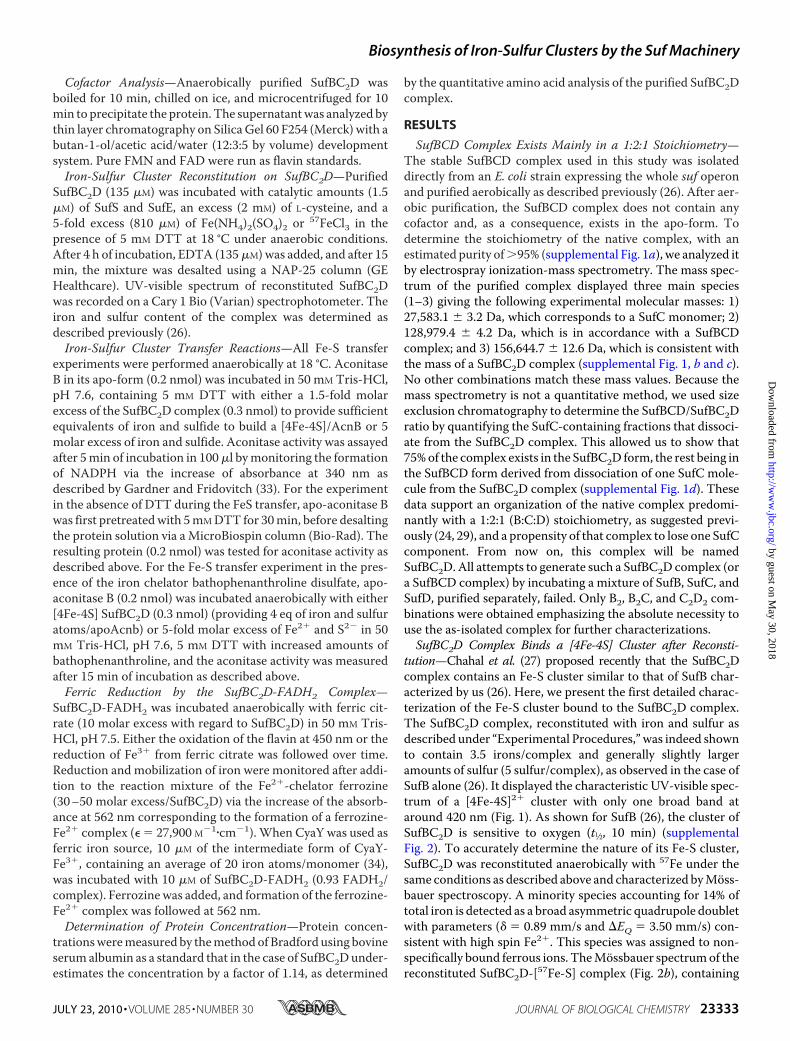
SufBCD Complex Exists Mainly in a 1:2:1 Stoichiometry—The stable SufBCD complex used in this study was isolateddirectly from an E. coli strain expressing the whole suf operonand purified aerobically as described previously (26). After aer-obic purification, the SufBCD complex does not contain anycofactor and, as a consequence, exists in the apo-form. Todetermine the stoichiometry of the native complex, with anestimated purity of�95% (supplemental Fig. 1a), we analyzed itby electrospray ionization-mass spectrometry. The mass spec-trum of the purified complex displayed three main species(1–3) giving the following experimental molecular masses: 1)27,583.1 � 3.2 Da, which corresponds to a SufC monomer; 2)128,979.4 � 4.2 Da, which is in accordance with a SufBCDcomplex; and 3) 156,644.7 � 12.6 Da, which is consistent withthe mass of a SufBC2D complex (supplemental Fig. 1, b and c).No other combinations match these mass values. Because themass spectrometry is not a quantitative method, we used sizeexclusion chromatography to determine the SufBCD/SufBC2Dratio by quantifying the SufC-containing fractions that dissoci-ate from the SufBC2D complex. This allowed us to show that75%of the complex exists in the SufBC2D form, the rest being inthe SufBCD form derived from dissociation of one SufC mole-cule from the SufBC2D complex (supplemental Fig. 1d). Thesedata support an organization of the native complex predomi-nantly with a 1:2:1 (B:C:D) stoichiometry, as suggested previ-ously (24, 29), and a propensity of that complex to lose one SufCcomponent. From now on, this complex will be namedSufBC2D. All attempts to generate such a SufBC2D complex (ora SufBCD complex) by incubating a mixture of SufB, SufC, andSufD, purified separately, failed. Only B2, B2C, and C2D2 com-binations were obtained emphasizing the absolute necessity touse the as-isolated complex for further characterizations.SufBC2D Complex Binds a [4Fe-4S] Cluster after Reconsti-
tution—Chahal et al. (27) proposed recently that the SufBC2Dcomplex contains an Fe-S cluster similar to that of SufB char-acterized by us (26). Here, we present the first detailed charac-terization of the Fe-S cluster bound to the SufBC2D complex.The SufBC2D complex, reconstituted with iron and sulfur asdescribed under “Experimental Procedures,” was indeed shownto contain 3.5 irons/complex and generally slightly largeramounts of sulfur (5 sulfur/complex), as observed in the case ofSufB alone (26). It displayed the characteristic UV-visible spec-trum of a [4Fe-4S]2� cluster with only one broad band ataround 420 nm (Fig. 1). As shown for SufB (26), the cluster ofSufBC2D is sensitive to oxygen (t1⁄2, 10 min) (supplementalFig. 2). To accurately determine the nature of its Fe-S cluster,SufBC2D was reconstituted anaerobically with 57Fe under thesame conditions as described above and characterized byMoss-bauer spectroscopy. A minority species accounting for 14% oftotal iron is detected as a broad asymmetric quadrupole doubletwith parameters (� � 0.89 mm/s and EQ � 3.50 mm/s) con-sistent with high spin Fe2�. This species was assigned to non-specifically bound ferrous ions. TheMossbauer spectrumof thereconstituted SufBC2D-[57Fe-S] complex (Fig. 2b), containing
Biosynthesis of Iron-Sulfur Clusters by the Suf Machinery
JULY 23, 2010 • VOLUME 285 • NUMBER 30 JOURNAL OF BIOLOGICAL CHEMISTRY 23333
by guest on May 30, 2018
http://ww
w.jbc.org/
Dow
nloaded from
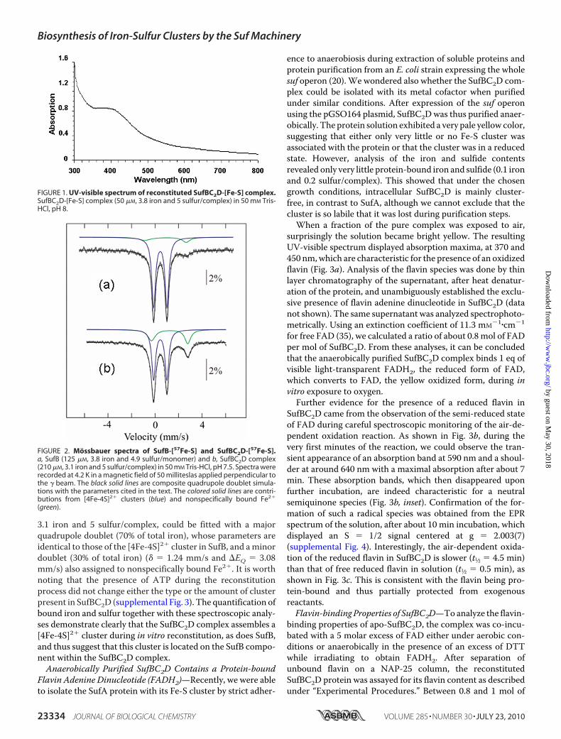
3.1 iron and 5 sulfur/complex, could be fitted with a majorquadrupole doublet (70% of total iron), whose parameters areidentical to those of the [4Fe-4S]2� cluster in SufB, and aminordoublet (30% of total iron) (� � 1.24 mm/s and EQ � 3.08mm/s) also assigned to nonspecifically bound Fe2�. It is worthnoting that the presence of ATP during the reconstitutionprocess did not change either the type or the amount of clusterpresent in SufBC2D (supplemental Fig. 3). The quantification ofbound iron and sulfur together with these spectroscopic analy-ses demonstrate clearly that the SufBC2D complex assembles a[4Fe-4S]2� cluster during in vitro reconstitution, as does SufB,and thus suggest that this cluster is located on the SufB compo-nent within the SufBC2D complex.Anaerobically Purified SufBC2D Contains a Protein-bound
Flavin Adenine Dinucleotide (FADH2)—Recently, we were ableto isolate the SufA protein with its Fe-S cluster by strict adher-
ence to anaerobiosis during extraction of soluble proteins andprotein purification from an E. coli strain expressing the wholesuf operon (20). We wondered also whether the SufBC2D com-plex could be isolated with its metal cofactor when purifiedunder similar conditions. After expression of the suf operonusing the pGSO164 plasmid, SufBC2Dwas thus purified anaer-obically. The protein solution exhibited a very pale yellow color,suggesting that either only very little or no Fe-S cluster wasassociated with the protein or that the cluster was in a reducedstate. However, analysis of the iron and sulfide contentsrevealed only very little protein-bound iron and sulfide (0.1 ironand 0.2 sulfur/complex). This showed that under the chosengrowth conditions, intracellular SufBC2D is mainly cluster-free, in contrast to SufA, although we cannot exclude that thecluster is so labile that it was lost during purification steps.When a fraction of the pure complex was exposed to air,
surprisingly the solution became bright yellow. The resultingUV-visible spectrum displayed absorption maxima, at 370 and450 nm,which are characteristic for the presence of an oxidizedflavin (Fig. 3a). Analysis of the flavin species was done by thinlayer chromatography of the supernatant, after heat denatur-ation of the protein, and unambiguously established the exclu-sive presence of flavin adenine dinucleotide in SufBC2D (datanot shown). The same supernatant was analyzed spectrophoto-metrically. Using an extinction coefficient of 11.3 mM�1�cm�1
for free FAD (35), we calculated a ratio of about 0.8 mol of FADper mol of SufBC2D. From these analyses, it can be concludedthat the anaerobically purified SufBC2D complex binds 1 eq ofvisible light-transparent FADH2, the reduced form of FAD,which converts to FAD, the yellow oxidized form, during invitro exposure to oxygen.Further evidence for the presence of a reduced flavin in
SufBC2D came from the observation of the semi-reduced stateof FAD during careful spectroscopic monitoring of the air-de-pendent oxidation reaction. As shown in Fig. 3b, during thevery first minutes of the reaction, we could observe the tran-sient appearance of an absorption band at 590 nm and a shoul-der at around 640 nm with a maximal absorption after about 7min. These absorption bands, which then disappeared uponfurther incubation, are indeed characteristic for a neutralsemiquinone species (Fig. 3b, inset). Confirmation of the for-mation of such a radical species was obtained from the EPRspectrum of the solution, after about 10 min incubation, whichdisplayed an S � 1/2 signal centered at g � 2.003(7)(supplemental Fig. 4). Interestingly, the air-dependent oxida-tion of the reduced flavin in SufBC2D is slower (t1⁄2 � 4.5 min)than that of free reduced flavin in solution (t1⁄2 � 0.5 min), asshown in Fig. 3c. This is consistent with the flavin being pro-tein-bound and thus partially protected from exogenousreactants.Flavin-binding Properties of SufBC2D—Toanalyze the flavin-
binding properties of apo-SufBC2D, the complex was co-incu-bated with a 5 molar excess of FAD either under aerobic con-ditions or anaerobically in the presence of an excess of DTTwhile irradiating to obtain FADH2. After separation ofunbound flavin on a NAP-25 column, the reconstitutedSufBC2D protein was assayed for its flavin content as describedunder “Experimental Procedures.” Between 0.8 and 1 mol of
FIGURE 1. UV-visible spectrum of reconstituted SufBC2D-[Fe-S] complex.SufBC2D-[Fe-S] complex (50 �M, 3.8 iron and 5 sulfur/complex) in 50 mM Tris-HCl, pH 8.
FIGURE 2. Mossbauer spectra of SufB-[57Fe-S] and SufBC2D-[57Fe-S].a, SufB (125 �M, 3.8 iron and 4.9 sulfur/monomer) and b, SufBC2D complex(210 �M, 3.1 iron and 5 sulfur/complex) in 50 mM Tris-HCl, pH 7.5. Spectra wererecorded at 4.2 K in a magnetic field of 50 milliteslas applied perpendicular tothe � beam. The black solid lines are composite quadrupole doublet simula-tions with the parameters cited in the text. The colored solid lines are contri-butions from [4Fe-4S]2� clusters (blue) and nonspecifically bound Fe2�
(green).
Biosynthesis of Iron-Sulfur Clusters by the Suf Machinery
23334 JOURNAL OF BIOLOGICAL CHEMISTRY VOLUME 285 • NUMBER 30 • JULY 23, 2010
by guest on May 30, 2018
http://ww
w.jbc.org/
Dow
nloaded from

FAD per mol of SufBC2D could be reproducibly determined inthe case of the anaerobic reaction mixture containing FADH2and SufBC2D, whereas no protein-bound flavin could bedetected in the case of the aerobic FAD/SufBC2D incubationmixture clearly showing that only FADH2 binds to the proteincomplex. Accordingly, when the FADH2-containing proteinwas exposed to air and then desalted on a NAP-25 column,flavin could not be detected anymore on SufBC2D. Duringincubation of SufBC2D with FMNH2 or reduced riboflavininstead of FADH2, less flavin remained protein-bound after thedesalting step (0.4 and 0.1 mol/mol of SufBC2D, respectively).Again, no binding of oxidized FMN or riboflavin could beobserved after co-incubationunder aerobic conditions.We alsoinvestigated the ability of the SufBC2D complex, in its iron-sulfur cluster form ([4Fe-4S]), to bind the reduced flavin. Thepresence of the cluster or addition of ATP had no influence onthe flavin content, which was determined to be approximatelyone reduced flavin per complex (supplemental Table 1). Thefact that the reduced flavin and the cluster under its �2 oxida-
tion state co-existed in the same complex shows that no elec-tron transfer between the two species could occur.To determine which protein subunits of the SufBC2D com-
plex are involved in the binding of reduced flavin, we repeatedthe same experiment as above with single proteins SufB, SufC,and SufD and also with some combinations of the three pro-teins. After treatment with a 5 molar excess of FAD underanaerobic conditions and irradiation, only SufB alone or SufB inthe presence of SufC was able to bind FADH2, albeit to a lesserextent (0.1–0.3mol of FADH2 permol of protein, respectively).SufC alone and SufD were shown not to bind the flavin. Thus,only the whole SufBC2D complex could bind 1mol of flavin permol of complex demonstrating that the association of the threeproteins is required for full binding of FADH2.
To determine the dissociation constant (Kd) for the bindingof FADH2 to SufBC2D, an ultrafiltration assay was used asdescribed under “Experimental Procedures.” The flavin-freeapo-SufBC2D complex (20 �M) was co-incubated at 18 °Canaerobically with different concentrations of FADH2 (0–200�M) obtained by photo-induced reduction of FAD. After co-incubation, the samples were transferred to a 100,000 molecu-lar weight cutoff concentrator, and unbound FADH2 was sep-arated from SufBC2D by ultrafiltration. As a control, the sameexperimentwas performed in the absence of apo-SufBC2D.Theflavin content of the flow-through fraction of the samples incu-bated in the presence of apo-SufBC2D was determined asdescribed under “Experimental Procedures.” The amount ofcomplex (SufBC2D-FADH2) and the concentrations of freeSufBC2D were determined according to the calculated concen-trations of free and total FADH2 and the known amount of totalapo-SufBC2D. Under these conditions, a dissociation constantof 12 �M was determined (Fig. 4).Bound Flavin Is Not Required for SufBC2D [4Fe-4S] Cluster
Transfer—Aconitase B (AcnB), an enzyme containing a [4Fe-4S] cluster in its active form, was used as a target for Fe-S trans-fer experiments. Both forms of holo-SufBC2D ([Fe-S] and[Fe-S] � FADH2) were used as a potential source of clusters. Ina typical experiment, an excess of holo-SufBC2D (0.3 nmol) wasco-incubated anaerobically with apo-aconitase B (0.2 nmol) toprovide a sufficient amount of Fe-S cluster to build a [4Fe-4S]cluster in AcnB. After 5 and 20 min of reaction, AcnB activitywas monitored as described under “Experimental Procedures.”As shown in Fig. 5a, AcnB is fully active after 5 min of reaction,and no significant differences could be observed between thetwo forms of holo-SufBC2D used as the Fe-S source. A similaractivation of aconitase could be achieved when apo-AcnB wasincubatedwith a 5-foldmolar excess of iron and sulfide but onlyin the presence of DTT in the reaction mixture (Fig. 5a).Indeed, very little activity in the control was detected in theabsence of DTT (Fig. 5b). On the contrary, AcnB can be matu-rated in a time-dependentmanner by both SufBC2D-[Fe-S] andSufBC2D-[Fe-S]�FADH2 even in the absence ofDTT (Fig. 5b).Thus, these data show for the first time a cluster transfer fromSufBC2D to a target protein different from SufA and alsoexclude a role of the reduced flavin in this process. Finally, theexperiment shown in Fig. 5c nicely differentiates the SufBC2D-dependent and the chemical aconitase activation. Indeed, addi-tion of increasing concentrations of a strong iron chelator,
FIGURE 3. Properties of SufBC2D-FADH2. (a) UV-visible spectra of anaerobi-cally purified SufBC2D complex after exposure to air; (b) kinetic oxidation ofSufBC2D complex (t � 1 min (bold black line) and t � 3, 5, and 7 min (black line);t � 10, 15, 20, and 30 min (dashed bold black line)) showing formation of theneutral semiquinone species (FADH�) at 590 nm (maximum formation at 7min (black line)). Inset, enlargement of the 500 –700 nm region. c, comparisonof the air oxidation kinetic of the flavin within the SufBC2D complex (21 �M
flavin) (f) with free reduced flavin in solution (16 �M) (Œ).
Biosynthesis of Iron-Sulfur Clusters by the Suf Machinery
JULY 23, 2010 • VOLUME 285 • NUMBER 30 JOURNAL OF BIOLOGICAL CHEMISTRY 23335
by guest on May 30, 2018
http://ww
w.jbc.org/
Dow
nloaded from

bathophenanthroline, to the standard reaction mixture hadvery little effect on the Fe-S cluster transfer from SufBC2D toAcnB, although it completely inhibited the chemical reconsti-tution of the aconitase. These data, the lack of requirement forDTT and inhibition by a chelator, thus show that cluster trans-fer from SufBC2D to aconitase is a concerted process that doesnot involve intermediate disassembly of the cluster, release ofiron and sulfur in solution, and then reassembly in the targetprotein.Reduction of Ferric Complexes by the SufBC2D-FADH2
Complex—Reduced flavins are excellent ferric iron-reducingagents (36, 37). We thus investigated the potential of thereduced flavin of the SufBC2D complex for reduction of ferriccomplexes. This was tested using ferric citrate, a small ironcomplex (38), and CyaY, the bacterial frataxin homologue (34,39), as electron acceptors. SufBC2D-FADH2 complex (30 �M, 1FADH2/complex) was incubated anaerobically with ferric cit-rate (300 �M), and electron transfer from FADH2 to ferric cit-rate was monitored by UV-visible spectroscopy from theincrease of the absorbance at 450 nm, reflecting formation ofoxidized flavin. As a control experiment, SufBC2D-FADH2complex was incubated with buffer instead of ferric citrate. Atime-dependent (t1⁄2 � 14 min) oxidation of the flavin wasobserved in the reaction mixture containing the ferric citrate(Fig. 6a), whereas no oxidation of the flavin occurred in thecontrol experiment (Fig. 6a, inset). From the absorption at 450nm, we could calculate, at the end of the reaction (60 min)(Fig. 6a), that 26 �M of the flavin was oxidized (90% yield). Wealsomonitored the reduction of ferric iron by the FADH2 cofac-tor using ferrozine in excess as an effective Fe2� chelator and anFe2� probe. The chelator mobilizes ferrous ions from the ironsource and forms a complex with a maximal absorption at 562nm. Under these conditions and using 10 �M of SufBC2D-
FADH2 complex containing 0.74 FADH2/complex, weobserved a time-dependent formation of the ferrozine-Fe2�
complex (t1⁄2 � 5min) indicating reduction of ferric citrate byFADH2 and mobilization of Fe2� by ferrozine (Fig. 6b). At theend of the reaction, about 14 �M of ferrozine-Fe2� complex
FIGURE 4. Binding affinity of FADH2 to the apo-SufBC2D complex. Theapo-SufBC2D (20 �M) complex was incubated anaerobically with differentconcentrations of FADH2 (0 –200 �M) obtained by photo-induced reductionof FAD. A molecular weight cutoff concentrator was used to separateunbound FADH2 from protein-bound FADH2, and the flavin content of eachwas determined outside the glove box after oxidation, heat denaturation,centrifugation, and UV-visible analysis of the supernatant. The concentrationof SufBC2D-FADH2 ([FADbound]) and that of apo-SufBC2D was determinedaccording to the calculated concentrations of unbound and total FADH2 andthe known amount of total apo-SufBC2D. The protein-bound FADH2 as a func-tion of unbound FADH2 in solution was then plotted. The data have beenfitted by a saturation hyperbola according to Equation 1.
FIGURE 5. Iron-sulfur cluster transfer from SufBC2D to AcnB. Holo-SufBC2Dcomplex (0.3 nmol), [Fe-S] (gray bars), or [Fe-S] � FADH2 (hatched bars), wasco-incubated in 10 �l of 50 mM Tris-HCl, pH 7.6, with (a) and without (b) 5 mM
DTT with apo-AcnB (0.2 nmol). After 5 and 20 min of incubation, the activity ofAcnB was measured by monitoring the absorption at 340 nm. For this, a mix-ture of 1.2 mM MnCl2, 25 mM citrate, 0.5 unit of isocitrate dehydrogenase, and0.25 mM NADP� was added to the protein mixture in a final volume of 100 �l.As a control, apo-AcnB was incubated with a 5 molar excess of iron and sul-fide, and the activity was assayed (black bars). c, intact cluster transfer fromSufBC2D to AcnB. Apo-AcnB (0.2 nmol) was incubated anaerobically witheither [4Fe-4S] SufBC2D complex (0.3 nmol) (Œ) or 5-fold molar excess of Fe2�
and S2� (f) in 100 �l of 50 mM Tris-HCl, pH 7.6, 5 mM DTT with increasingamounts of bathophenanthroline, and the AcnB activity was measured after20 min of incubation.
Biosynthesis of Iron-Sulfur Clusters by the Suf Machinery
23336 JOURNAL OF BIOLOGICAL CHEMISTRY VOLUME 285 • NUMBER 30 • JULY 23, 2010
by guest on May 30, 2018
http://ww
w.jbc.org/
Dow
nloaded from
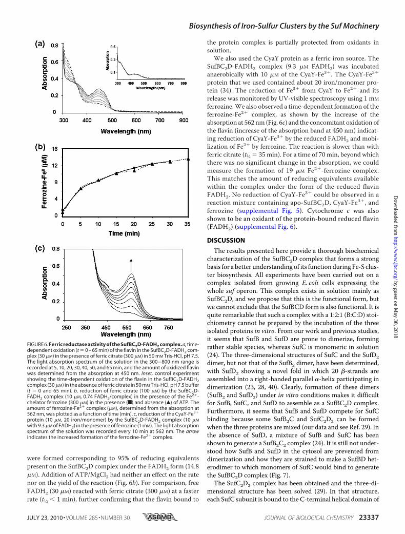
were formed corresponding to 95% of reducing equivalentspresent on the SufBC2D complex under the FADH2 form (14.8�M). Addition of ATP/MgCl2 had neither an effect on the ratenor on the yield of the reaction (Fig. 6b). For comparison, freeFADH2 (30 �M) reacted with ferric citrate (300 �M) at a fasterrate (t1⁄2 � 1 min), further confirming that the flavin bound to
the protein complex is partially protected from oxidants insolution.We also used the CyaY protein as a ferric iron source. The
SufBC2D-FADH2 complex (9.3 �M FADH2) was incubatedanaerobically with 10 �M of the CyaY-Fe3�. The CyaY-Fe3�
protein that we used contained about 20 iron/monomer pro-tein (34). The reduction of Fe3� from CyaY to Fe2� and itsrelease was monitored by UV-visible spectroscopy using 1 mM
ferrozine.We also observed a time-dependent formation of theferrozine-Fe2� complex, as shown by the increase of theabsorption at 562 nm (Fig. 6c) and the concomitant oxidation ofthe flavin (increase of the absorption band at 450 nm) indicat-ing reduction of CyaY-Fe3� by the reduced FADH2 and mobi-lization of Fe2� by ferrozine. The reaction is slower than withferric citrate (t1⁄2 � 35min). For a time of 70min, beyond whichthere was no significant change in the absorption, we couldmeasure the formation of 19 �M Fe2�-ferrozine complex.This matches the amount of reducing equivalents availablewithin the complex under the form of the reduced flavinFADH2. No reduction of CyaY-Fe3� could be observed in areaction mixture containing apo-SufBC2D, CyaY-Fe3�, andferrozine (supplemental Fig. 5). Cytochrome c was alsoshown to be an oxidant of the protein-bound reduced flavin(FADH2) (supplemental Fig. 6).
DISCUSSION
The results presented here provide a thorough biochemicalcharacterization of the SufBC2D complex that forms a strongbasis for a better understanding of its function during Fe-S clus-ter biosynthesis. All experiments have been carried out on acomplex isolated from growing E. coli cells expressing thewhole suf operon. This complex exists in solution mainly asSufBC2D, and we propose that this is the functional form, butwe cannot exclude that the SufBCD form is also functional. It isquite remarkable that such a complex with a 1:2:1 (B:C:D) stoi-chiometry cannot be prepared by the incubation of the threeisolated proteins in vitro. From our work and previous studies,it seems that SufB and SufD are prone to dimerize, formingrather stable species, whereas SufC is monomeric in solution(24). The three-dimensional structures of SufC and the SufD2dimer, but not that of the SufB2 dimer, have been determined,with SufD2 showing a novel fold in which 20 �-strands areassembled into a right-handed parallel �-helix participating indimerization (23, 28, 40). Clearly, formation of these dimers(SufB2 and SufD2) under in vitro conditions makes it difficultfor SufB, SufC, and SufD to assemble as a SufBC2D complex.Furthermore, it seems that SufB and SufD compete for SufCbinding because some SufB2C and SufC2D2 can be formedwhen the three proteins aremixed (our data and see Ref. 29). Inthe absence of SufD, a mixture of SufB and SufC has beenshown to generate a SufB2C2 complex (24). It is still not under-stood how SufB and SufD in the cytosol are prevented fromdimerization and how they are strained to make a SufBD het-erodimer to which monomers of SufC would bind to generatethe SufBC2D complex (Fig. 7).
The SufC2D2 complex has been obtained and the three-di-mensional structure has been solved (29). In that structure,each SufC subunit is bound to the C-terminal helical domain of
FIGURE6.Ferric reductase activity of the SufBC2D-FADH2 complex. a, time-dependent oxidation (t � 0 – 65 min) of the flavin in the SufBC2D-FADH2 com-plex (30 �M) in the presence of ferric citrate (300 �M) in 50 mM Tris-HCl, pH 7.5.The light absorption spectrum of the solution in the 300 – 800 nm range isrecorded at 5, 10, 20, 30, 40, 50, and 65 min, and the amount of oxidized flavinwas determined from the absorption at 450 nm. Inset, control experimentshowing the time-dependent oxidation of the flavin in the SufBC2D-FADH2complex (30 �M) in the absence of ferric citrate in 50 mM Tris-HCl, pH 7.5 buffer(t � 0 and 65 min). b, reduction of ferric citrate (100 �M) by the SufBC2D-FADH2 complex (10 �M, 0.74 FADH2/complex) in the presence of the Fe2�-chelator ferrozine (300 �M) in the presence (f) and absence (Œ) of ATP. Theamount of ferrozine-Fe2� complex (�M), determined from the absorption at562 nm, was plotted as a function of time (min). c, reduction of the CyaY-Fe3�
protein (10 �M, 20 iron/monomer) by the SufBC2D-FADH2 complex (10 �M
with 9.3 �M of FADH2) in the presence of ferrozine (1 mM). The light absorptionspectrum of the solution was recorded every 10 min at 562 nm. The arrowindicates the increased formation of the ferrozine-Fe2� complex.
Biosynthesis of Iron-Sulfur Clusters by the Suf Machinery
JULY 23, 2010 • VOLUME 285 • NUMBER 30 JOURNAL OF BIOLOGICAL CHEMISTRY 23337
by guest on May 30, 2018
http://ww
w.jbc.org/
Dow
nloaded from
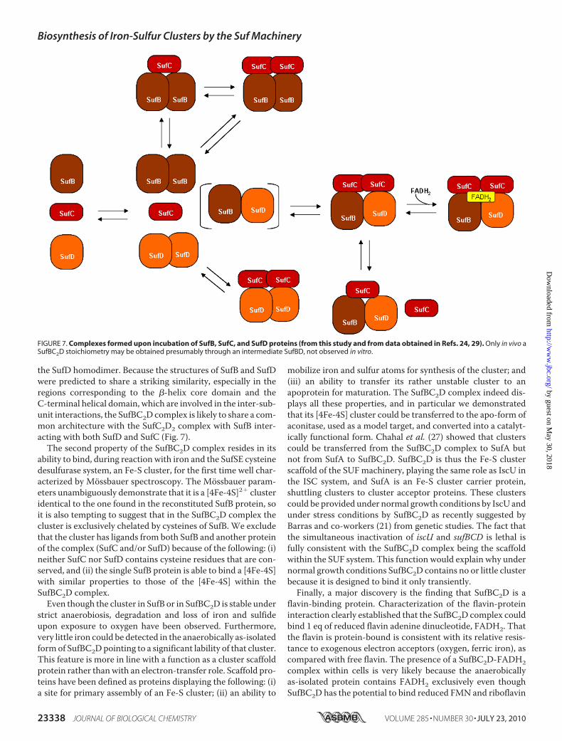
the SufD homodimer. Because the structures of SufB and SufDwere predicted to share a striking similarity, especially in theregions corresponding to the �-helix core domain and theC-terminal helical domain, which are involved in the inter-sub-unit interactions, the SufBC2D complex is likely to share a com-mon architecture with the SufC2D2 complex with SufB inter-acting with both SufD and SufC (Fig. 7).The second property of the SufBC2D complex resides in its
ability to bind, during reaction with iron and the SufSE cysteinedesulfurase system, an Fe-S cluster, for the first time well char-acterized by Mossbauer spectroscopy. The Mossbauer param-eters unambiguously demonstrate that it is a [4Fe-4S]2� clusteridentical to the one found in the reconstituted SufB protein, soit is also tempting to suggest that in the SufBC2D complex thecluster is exclusively chelated by cysteines of SufB. We excludethat the cluster has ligands from both SufB and another proteinof the complex (SufC and/or SufD) because of the following: (i)neither SufC nor SufD contains cysteine residues that are con-served, and (ii) the single SufB protein is able to bind a [4Fe-4S]with similar properties to those of the [4Fe-4S] within theSufBC2D complex.Even though the cluster in SufB or in SufBC2D is stable under
strict anaerobiosis, degradation and loss of iron and sulfideupon exposure to oxygen have been observed. Furthermore,very little iron could be detected in the anaerobically as-isolatedformof SufBC2Dpointing to a significant lability of that cluster.This feature is more in line with a function as a cluster scaffoldprotein rather thanwith an electron-transfer role. Scaffold pro-teins have been defined as proteins displaying the following: (i)a site for primary assembly of an Fe-S cluster; (ii) an ability to
mobilize iron and sulfur atoms for synthesis of the cluster; and(iii) an ability to transfer its rather unstable cluster to anapoprotein for maturation. The SufBC2D complex indeed dis-plays all these properties, and in particular we demonstratedthat its [4Fe-4S] cluster could be transferred to the apo-form ofaconitase, used as a model target, and converted into a catalyt-ically functional form. Chahal et al. (27) showed that clusterscould be transferred from the SufBC2D complex to SufA butnot from SufA to SufBC2D. SufBC2D is thus the Fe-S clusterscaffold of the SUF machinery, playing the same role as IscU inthe ISC system, and SufA is an Fe-S cluster carrier protein,shuttling clusters to cluster acceptor proteins. These clusterscould be provided under normal growth conditions by IscU andunder stress conditions by SufBC2D as recently suggested byBarras and co-workers (21) from genetic studies. The fact thatthe simultaneous inactivation of iscU and sufBCD is lethal isfully consistent with the SufBC2D complex being the scaffoldwithin the SUF system. This function would explain why undernormal growth conditions SufBC2D contains no or little clusterbecause it is designed to bind it only transiently.Finally, a major discovery is the finding that SufBC2D is a
flavin-binding protein. Characterization of the flavin-proteininteraction clearly established that the SufBC2D complex couldbind 1 eq of reduced flavin adenine dinucleotide, FADH2. Thatthe flavin is protein-bound is consistent with its relative resis-tance to exogenous electron acceptors (oxygen, ferric iron), ascompared with free flavin. The presence of a SufBC2D-FADH2complex within cells is very likely because the anaerobicallyas-isolated protein contains FADH2 exclusively even thoughSufBC2D has the potential to bind reduced FMN and riboflavin
FIGURE 7. Complexes formed upon incubation of SufB, SufC, and SufD proteins (from this study and from data obtained in Refs. 24, 29). Only in vivo aSufBC2D stoichiometry may be obtained presumably through an intermediate SufBD, not observed in vitro.
Biosynthesis of Iron-Sulfur Clusters by the Suf Machinery
23338 JOURNAL OF BIOLOGICAL CHEMISTRY VOLUME 285 • NUMBER 30 • JULY 23, 2010
by guest on May 30, 2018
http://ww
w.jbc.org/
Dow
nloaded from
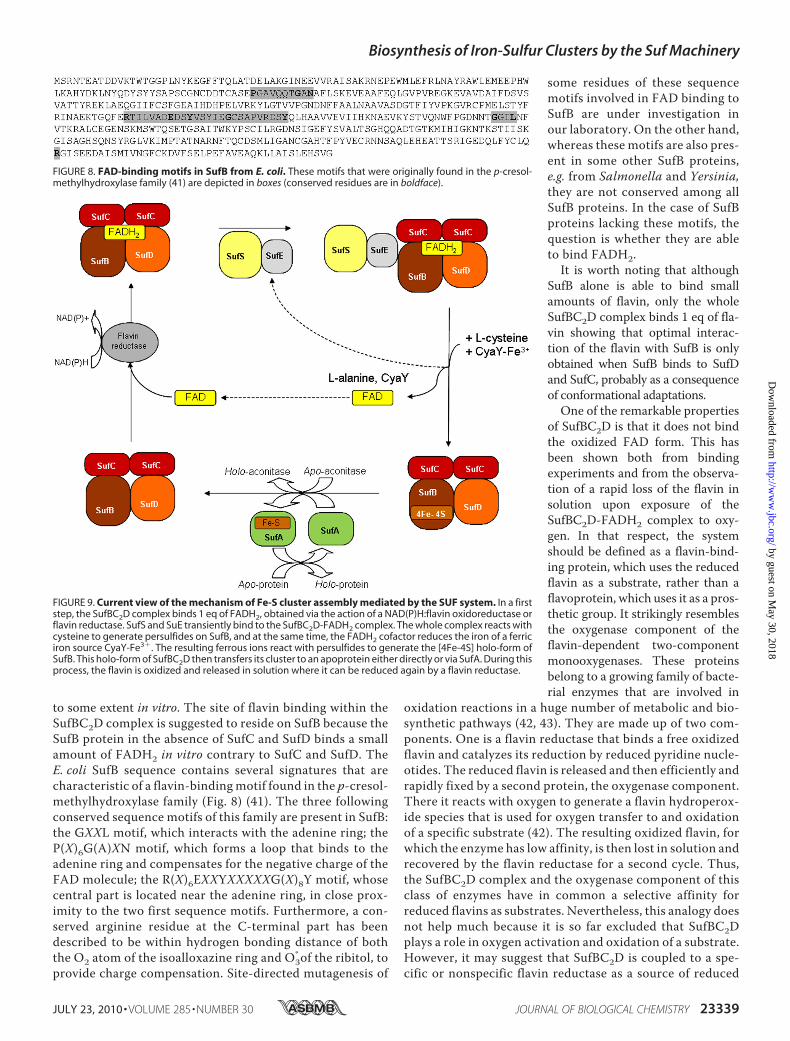
to some extent in vitro. The site of flavin binding within theSufBC2D complex is suggested to reside on SufB because theSufB protein in the absence of SufC and SufD binds a smallamount of FADH2 in vitro contrary to SufC and SufD. TheE. coli SufB sequence contains several signatures that arecharacteristic of a flavin-bindingmotif found in the p-cresol-methylhydroxylase family (Fig. 8) (41). The three followingconserved sequence motifs of this family are present in SufB:the GXXL motif, which interacts with the adenine ring; theP(X)6G(A)XN motif, which forms a loop that binds to theadenine ring and compensates for the negative charge of theFAD molecule; the R(X)6EXXYXXXXXG(X)8Y motif, whosecentral part is located near the adenine ring, in close prox-imity to the two first sequence motifs. Furthermore, a con-served arginine residue at the C-terminal part has beendescribed to be within hydrogen bonding distance of boththe O2 atom of the isoalloxazine ring and O3
*of the ribitol, toprovide charge compensation. Site-directed mutagenesis of
some residues of these sequencemotifs involved in FAD binding toSufB are under investigation inour laboratory. On the other hand,whereas these motifs are also pres-ent in some other SufB proteins,e.g. from Salmonella and Yersinia,they are not conserved among allSufB proteins. In the case of SufBproteins lacking these motifs, thequestion is whether they are ableto bind FADH2.
It is worth noting that althoughSufB alone is able to bind smallamounts of flavin, only the wholeSufBC2D complex binds 1 eq of fla-vin showing that optimal interac-tion of the flavin with SufB is onlyobtained when SufB binds to SufDand SufC, probably as a consequenceof conformational adaptations.One of the remarkable properties
of SufBC2D is that it does not bindthe oxidized FAD form. This hasbeen shown both from bindingexperiments and from the observa-tion of a rapid loss of the flavin insolution upon exposure of theSufBC2D-FADH2 complex to oxy-gen. In that respect, the systemshould be defined as a flavin-bind-ing protein, which uses the reducedflavin as a substrate, rather than aflavoprotein, which uses it as a pros-thetic group. It strikingly resemblesthe oxygenase component of theflavin-dependent two-componentmonooxygenases. These proteinsbelong to a growing family of bacte-rial enzymes that are involved in
oxidation reactions in a huge number of metabolic and bio-synthetic pathways (42, 43). They are made up of two com-ponents. One is a flavin reductase that binds a free oxidizedflavin and catalyzes its reduction by reduced pyridine nucle-otides. The reduced flavin is released and then efficiently andrapidly fixed by a second protein, the oxygenase component.There it reacts with oxygen to generate a flavin hydroperox-ide species that is used for oxygen transfer to and oxidationof a specific substrate (42). The resulting oxidized flavin, forwhich the enzyme has low affinity, is then lost in solution andrecovered by the flavin reductase for a second cycle. Thus,the SufBC2D complex and the oxygenase component of thisclass of enzymes have in common a selective affinity forreduced flavins as substrates. Nevertheless, this analogy doesnot help much because it is so far excluded that SufBC2Dplays a role in oxygen activation and oxidation of a substrate.However, it may suggest that SufBC2D is coupled to a spe-cific or nonspecific flavin reductase as a source of reduced
FIGURE 8. FAD-binding motifs in SufB from E. coli. These motifs that were originally found in the p-cresol-methylhydroxylase family (41) are depicted in boxes (conserved residues are in boldface).
FIGURE 9. Current view of the mechanism of Fe-S cluster assembly mediated by the SUF system. In a firststep, the SufBC2D complex binds 1 eq of FADH2, obtained via the action of a NAD(P)H:flavin oxidoreductase orflavin reductase. SufS and SuE transiently bind to the SufBC2D-FADH2 complex. The whole complex reacts withcysteine to generate persulfides on SufB, and at the same time, the FADH2 cofactor reduces the iron of a ferriciron source CyaY-Fe3�. The resulting ferrous ions react with persulfides to generate the [4Fe-4S] holo-form ofSufB. This holo-form of SufBC2D then transfers its cluster to an apoprotein either directly or via SufA. During thisprocess, the flavin is oxidized and released in solution where it can be reduced again by a flavin reductase.
Biosynthesis of Iron-Sulfur Clusters by the Suf Machinery
JULY 23, 2010 • VOLUME 285 • NUMBER 30 JOURNAL OF BIOLOGICAL CHEMISTRY 23339
by guest on May 30, 2018
http://ww
w.jbc.org/
Dow
nloaded from
flavins in a novel two-component system and uses reducedflavin to reduce a specific substrate.Thus, what could be the function of the reducing power pres-
ent in SufBC2D? Our results exclude a role of the flavin in thetransfer of the clusters from SufBC2D to an apoprotein such asaconitase. In the process of Fe-S cluster synthesis, electrons arerequired particularly for iron reduction andmobilization of fer-rous ions from ferric iron sources. In the case of the ISC system,the only redox protein is the product of the fdx gene, the [2Fe-2S] ferredoxin. It is generally proposed that indeed Fdx has aredox function during ISC-dependent Fe-S cluster assembly,but this still requires more experimental evidence. An effect ofFdx has only been observed during conversion of the [2Fe-2S]cluster of IscU into a [4Fe-4S] cluster, which requires ironreduction (44). Obviously, there is a huge literature, includingfromour laboratory, illustrating the potential of reduced flavinsfor ferric reduction and for mobilization of iron from ferritins,ferrisiderophores, and ferric citrate (38, 45). It has been dem-onstrated in E. coli that reduced flavins efficiently promote oxi-dative DNA damage, including DNA strand breaks, by deliver-ing electrons to free iron and allowing production of hydroxylradicals (46). In 1994, we emphasized such a reactivity with apaper entitled “Flavin Reductases or Ferric Reductases?” (37).The data reported here demonstrate clearly that the reducedFADH2 cofactor of SufBC2D is accessible and reactive enoughto give its electrons not only to small iron complexes such asferric citrate but also to iron-binding proteins such as CyaY, thebacterial frataxin, an important component of the Fe-S clusterassembly machinery as it represents one of the potentialsources of iron (34). We previously showed that reducingagents such as cysteine (E � �250 mV at pH 7.4) could reduceCyaY ferric iron, and this provides a mechanism to mobilizeiron from CyaY because the protein binds Fe2� only veryweakly (34). Reduced flavins display comparably low redoxpotentials, and thus, even though we have not measured that ofthe SufBC2D-FADH2 complex, it is not surprising from a ther-modynamic point of view that this complex is able to reduceCyaY-bound iron. Thus, we propose that the SufBC2D complexis a different version of the flavin-dependent two-componentsystems inwhich the flavin serves to reduce ferric iron from a sofar unknown iron source rather than oxygen and, furthermore,that SufBC2D is a novel type of cluster scaffold protein, inte-grating a scaffold and a redox function.The novel observations reported here lead us to suggest a
mechanism for Fe-S cluster assembly by the SUF system (Fig.9). In a first step, the SufBC2D complex binds 1 eq of FADH2.This reduced flavin is produced via the action of a NAD(P)H:flavin oxidoreductase or flavin reductase. Then the compo-nents of the cysteine-desulfurase, SufS and SufE, transientlybind to the SufBC2D-FADH2 complex. The whole complexreacts with cysteine to generate persulfides on SufB (26)through trans-persulfuration reactions from SufS to SufE andthen to SufB, as previously shown, and SufSE is released. At thesame time, the FADH2 cofactor reduces ferric iron from CyaY,and the resulting ferrous ions are chelated by SufB where theyreact with persulfides to generate the [4Fe-4S] cluster. Duringthis reaction, the flavin is oxidized and released in solutionwhere it can be recycled by a flavin reductase. Thereafter, the
holo-form of SufBC2D can transfer its cluster to an apoproteineither directly or via SufA (Fig. 9).We conclude by suggesting that there is an advantage of
using a flavin-dependent system for reduction reactions underoxidative stress and iron limitation conditions, underwhich theSUFmachinery operates, as compared with an iron-sulfur elec-tron transfer enzyme, such as the ferredoxin, involved in theISC machinery. Indeed, under such deleterious conditions, anFe-S enzymewould be degraded and be unable to fulfill its func-tion. Flavins, in contrast, are not sensitive to reactive oxygenspecies and obviously not to a lack of iron. It is well establishedthat the synthesis of nonessential iron-requiring proteins isdecreased (47) and that flavodoxins substitute for ferredoxinsunder iron-limited growth conditions in a number of microor-ganisms (48). Here, we have an additional example of a shiftfrom ferredoxin to a flavin-dependent enzyme, associated withthe shift from ISC to SUF, when the growth conditions becometoo adverse.
REFERENCES1. Johnson, D. C., Dean, D. R., Smith, A. D., and Johnson,M. K. (2005)Annu.
Rev. Biochem. 74, 247–2812. Fontecave, M., and Ollagnier-de-Choudens, S. (2008)Arch. Biochem. Bio-
phys. 474, 226–2373. Ayala-Castro, C., Saini, A., and Outten, F. W. (2008)Microbiol. Mol. Biol.
Rev. 72, 110–1254. Tokumoto, U., Kitamura, S., Fukuyama, K., and Takahashi, Y. (2004)
J. Biochem. 136, 199–2095. Lill, R., and Muhlenhoff, U. (2008) Annu. Rev. Biochem. 77, 669–7006. Balk, J., and Lobreaux, S. (2005) Trends Plant Sci. 10, 324–3317. Huet, G., Daffe, M., and Saves, I. (2005) J. Bacteriol. 187, 6137–61468. Outten, F. W., Djaman, O., and Storz, G. (2004) Mol. Microbiol. 52,
861–8729. Nachin, L., Loiseau, L., Expert, D., and Barras, F. (2003) EMBO J. 22,
427–43710. Ranquet, C., Ollagnier-de-Choudens, S., Loiseau, L., Barras, F., and Fon-
tecave, M. (2007) J. Biol. Chem. 282, 30442–3045111. Mihara, H., Fujii, T., Kato, S., Kurihara, T., Hata, Y., and Esaki, N. (2002)
J. Biochem. 131, 679–68512. Mihara, H., Kurihara, T., Yoshimura, T., and Esaki, N. (2000) J. Biochem.
127, 559–56713. Ollagnier-de-Choudens, S., Lascoux, D., Loiseau, L., Barras, F., Forest, E.,
and Fontecave, M. (2003) FEBS Lett. 555, 263–26714. Outten, F. W., Wood, M. J., Munoz, F. M., and Storz, G. (2003) J. Biol.
Chem. 278, 45713–4571915. Loiseau, L., Ollagnier-de-Choudens, S., Nachin, L., Fontecave, M., and
Barras, F. (2003) J. Biol. Chem. 278, 38352–3835916. Lu, J., Yang, J., Tan, G., and Ding, H. (2008) Biochem. J. 409, 535–54317. Ollagnier-de Choudens, S., Nachin, L., Sanakis, Y., Loiseau, L., Barras, F.,
and Fontecave, M. (2003) J. Biol. Chem. 278, 17993–1800118. Ollagnier-de-Choudens, S., Sanakis, Y., and Fontecave, M. (2004) J. Biol.
Inorg. Chem. 9, 828–83819. Sendra, M., Ollagnier de Choudens, S., Lascoux, D., Sanakis, Y., and Fon-
tecave, M. (2007) FEBS Lett. 581, 1362–136820. Gupta, V., Sendra, M., Naik, S. G., Chahal, H. K., Huynh, B. H., Outten,
F. W., Fontecave, M., and Ollagnier de Choudens, S. (2009) J. Am. Chem.Soc. 131, 6149–6153
21. Vinella, D., Brochier-Armanet, C., Loiseau, L., Talla, E., and Barras, F.(2009) PLoS Genet. 5, e1000497
22. Takahashi, Y., and Tokumoto, U. (2002) J. Biol. Chem. 277, 28380–2838323. Kitaoka, S., Wada, K., Hasegawa, Y., Minami, Y., Fukuyama, K., and Taka-
hashi, Y. (2006) FEBS Lett. 580, 137–14324. Petrovic, A., Davis, C. T., Rangachari, K., Clough, B., Wilson, R. J., and
Eccleston, J. F. (2008) Protein Sci. 17, 1264–127425. Eccleston, J. F., Petrovic, A., Davis, C. T., Rangachari, K., andWilson, R. J.
Biosynthesis of Iron-Sulfur Clusters by the Suf Machinery
23340 JOURNAL OF BIOLOGICAL CHEMISTRY VOLUME 285 • NUMBER 30 • JULY 23, 2010
by guest on May 30, 2018
http://ww
w.jbc.org/
Dow
nloaded from
(2006) J. Biol. Chem. 281, 8371–837826. Layer, G., Gaddam, S. A., Ayala-Castro, C. N., Ollagnier-de Choudens, S.,
Lascoux, D., Fontecave, M., and Outten, F. W. (2007) J. Biol. Chem. 282,13342–13350
27. Chahal, H. K., Dai, Y., Saini, A., Ayala-Castro, C., andOutten, F.W. (2009)Biochemistry 48, 10644–10653
28. Badger, J., Sauder, J. M., Adams, J. M., Antonysamy, S., Bain, K., Bergseid,M. G., Buchanan, S. G., Buchanan,M. D., Batiyenko, Y., Christopher, J. A.,Emtage, S., Eroshkina, A., Feil, I., Furlong, E. B., Gajiwala, K. S., Gao,X.,He,D., Hendle, J., Huber, A., Hoda, K., Kearins, P., Kissinger, C., Laubert, B.,Lewis, H. A., Lin, J., Loomis, K., Lorimer, D., Louie, G.,Maletic,M.,Marsh,C. D., Miller, I., Molinari, J., Muller-Dieckmann, H. J., Newman, J. M.,Noland, B.W., Pagarigan, B., Park, F., Peat, T. S., Post, K.W., Radojicic, S.,Ramos, A., Romero, R., Rutter, M. E., Sanderson, W. E., Schwinn, K. D.,Tresser, J., Winhoven, J., Wright, T. A., Wu, L., Xu, J., and Harris, T. J.(2005) Proteins 60, 787–796
29. Wada, K., Sumi, N., Nagai, R., Iwasaki, K., Sato, T., Suzuki, K., Hasegawa,Y., Kitaoka, S.,Minami, Y., Outten, F.W., Takahashi, Y., and Fukuyama, K.(2009) J. Mol. Biol. 387, 245–258
30. Jordan, P. A., Tang, Y., Bradbury, A. J., Thomson, A. J., and Guest, J. R.(1999) Biochem. J. 344, 739–746
31. Song, S. H., Dick, B., Penzkofer, A., andHegemann, P. (2007) J. Photochem.Photobiol. B 87, 37–48
32. Mickowska, B., Dulinski, R., and Kozik, A. (2000) J. Biochem. Biophys.
Methods 44, 95–10733. Gardner, P. R., and Fridovich, I. (1992) J. Biol. Chem. 267, 8757–876334. Layer, G., Ollagnier-de Choudens, S., Sanakis, Y., and Fontecave, M.
(2006) J. Biol. Chem. 281, 16256–1626335. Massey, V., and Palmer, G. (1966) Biochemistry 5, 3181–318936. Halle, F., and Meyer, J. M. (1992) Eur. J. Biochem. 209, 621–62737. Fontecave, M., Coves, J., and Pierre, J. L. (1994) Biometals 7, 3–838. Coves, J., and Fontecave, M. (1993) Eur. J. Biochem. 211, 635–64139. Bedekovics, T., Gajdos, G. B., Kispal, G., and Isaya, G. (2007) FEMS Yeast
Res. 7, 1276–128440. Watanabe, S., Kita, A., and Miki, K. (2005) J. Mol. Biol. 353, 1043–105441. Dym, O., and Eisenberg, D. (2001) Protein Sci. 10, 1712–172842. Valton, J., Filisetti, L., Fontecave, M., and Niviere, V. (2004) J. Biol. Chem.
279, 44362–4436943. Galan, B., Díaz, E., Prieto, M. A., and García, J. L. (2000) J. Bacteriol. 182,
627–63644. Chandramouli, K., Unciuleac, M. C., Naik, S., Dean, D. R., Huynh, B. H.,
and Johnson, M. K. (2007) Biochemistry 46, 6804–681145. Coves, J., Niviere, V., Eschenbrenner, M., and Fontecave, M. (1993) J. Biol.
Chem. 268, 18604–1860946. Imlay, J. A., and Linn, S. (1988) Science 240, 1302–130947. Andrews, S. C., Robinson, A. K., andRodríguez-Quinones, F. (2003) FEMS
Microbiol. Rev. 27, 215–23748. Sancho, J. (2006) Cell. Mol. Life Sci. 63, 855–864
Biosynthesis of Iron-Sulfur Clusters by the Suf Machinery
JULY 23, 2010 • VOLUME 285 • NUMBER 30 JOURNAL OF BIOLOGICAL CHEMISTRY 23341
by guest on May 30, 2018
http://ww
w.jbc.org/
Dow
nloaded from
Jean-Marc Latour, Marc Fontecave and Sandrine Ollagnier de ChoudensSilke Wollers, Gunhild Layer, Ricardo Garcia-Serres, Luca Signor, Martin Clemancey,
OF Fe-S SCAFFOLD WITH A FLAVIN REDOX COFACTORIron-Sulfur (Fe-S) Cluster Assembly: THE SufBCD COMPLEX IS A NEW TYPE
doi: 10.1074/jbc.M110.127449 originally published online May 11, 20102010, 285:23331-23341.J. Biol. Chem.
10.1074/jbc.M110.127449Access the most updated version of this article at doi:
Alerts:
When a correction for this article is posted•
When this article is cited•
to choose from all of JBC's e-mail alertsClick here
Supplemental material:
http://www.jbc.org/content/suppl/2010/05/11/M110.127449.DC1
http://www.jbc.org/content/285/30/23331.full.html#ref-list-1
This article cites 48 references, 18 of which can be accessed free at
by guest on May 30, 2018
http://ww
w.jbc.org/
Dow
nloaded from
Related Documents