This content has been downloaded from IOPscience. Please scroll down to see the full text. Download details: IP Address: 202.152.22.34 This content was downloaded on 17/10/2013 at 07:17 Please note that terms and conditions apply. Iron oxide-based conjugates for cancer theragnostics View the table of contents for this issue, or go to the journal homepage for more 2012 Adv. Nat. Sci: Nanosci. Nanotechnol. 3 033001 (http://iopscience.iop.org/2043-6262/3/3/033001) Home Search Collections Journals About Contact us My IOPscience

Welcome message from author
This document is posted to help you gain knowledge. Please leave a comment to let me know what you think about it! Share it to your friends and learn new things together.
Transcript
This content has been downloaded from IOPscience. Please scroll down to see the full text.
Download details:
IP Address: 202.152.22.34
This content was downloaded on 17/10/2013 at 07:17
Please note that terms and conditions apply.
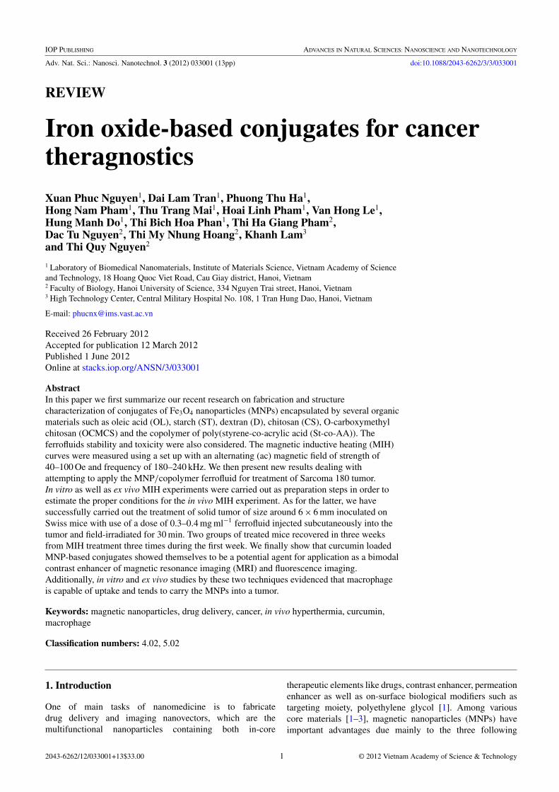
Iron oxide-based conjugates for cancer theragnostics
View the table of contents for this issue, or go to the journal homepage for more
2012 Adv. Nat. Sci: Nanosci. Nanotechnol. 3 033001
(http://iopscience.iop.org/2043-6262/3/3/033001)
Home Search Collections Journals About Contact us My IOPscience
IOP PUBLISHING ADVANCES IN NATURAL SCIENCES: NANOSCIENCE AND NANOTECHNOLOGY
Adv. Nat. Sci.: Nanosci. Nanotechnol. 3 (2012) 033001 (13pp) doi:10.1088/2043-6262/3/3/033001
REVIEW
Iron oxide-based conjugates for cancertheragnosticsXuan Phuc Nguyen1, Dai Lam Tran1, Phuong Thu Ha1,Hong Nam Pham1, Thu Trang Mai1, Hoai Linh Pham1, Van Hong Le1,Hung Manh Do1, Thi Bich Hoa Phan1, Thi Ha Giang Pham2,Dac Tu Nguyen2, Thi My Nhung Hoang2, Khanh Lam3
and Thi Quy Nguyen2
1 Laboratory of Biomedical Nanomaterials, Institute of Materials Science, Vietnam Academy of Scienceand Technology, 18 Hoang Quoc Viet Road, Cau Giay district, Hanoi, Vietnam2 Faculty of Biology, Hanoi University of Science, 334 Nguyen Trai street, Hanoi, Vietnam3 High Technology Center, Central Military Hospital No. 108, 1 Tran Hung Dao, Hanoi, Vietnam
E-mail: [email protected]
Received 26 February 2012Accepted for publication 12 March 2012Published 1 June 2012Online at stacks.iop.org/ANSN/3/033001
AbstractIn this paper we first summarize our recent research on fabrication and structurecharacterization of conjugates of Fe3O4 nanoparticles (MNPs) encapsulated by several organicmaterials such as oleic acid (OL), starch (ST), dextran (D), chitosan (CS), O-carboxymethylchitosan (OCMCS) and the copolymer of poly(styrene-co-acrylic acid (St-co-AA)). Theferrofluids stability and toxicity were also considered. The magnetic inductive heating (MIH)curves were measured using a set up with an alternating (ac) magnetic field of strength of40–100 Oe and frequency of 180–240 kHz. We then present new results dealing withattempting to apply the MNP/copolymer ferrofluid for treatment of Sarcoma 180 tumor.In vitro as well as ex vivo MIH experiments were carried out as preparation steps in order toestimate the proper conditions for the in vivo MIH experiment. As for the latter, we havesuccessfully carried out the treatment of solid tumor of size around 6 × 6 mm inoculated onSwiss mice with use of a dose of 0.3–0.4 mg ml−1 ferrofluid injected subcutaneously into thetumor and field-irradiated for 30 min. Two groups of treated mice recovered in three weeksfrom MIH treatment three times during the first week. We finally show that curcumin loadedMNP-based conjugates showed themselves to be a potential agent for application as a bimodalcontrast enhancer of magnetic resonance imaging (MRI) and fluorescence imaging.Additionally, in vitro and ex vivo studies by these two techniques evidenced that macrophageis capable of uptake and tends to carry the MNPs into a tumor.
Keywords: magnetic nanoparticles, drug delivery, cancer, in vivo hyperthermia, curcumin,macrophage
Classification numbers: 4.02, 5.02
1. Introduction
One of main tasks of nanomedicine is to fabricatedrug delivery and imaging nanovectors, which are themultifunctional nanoparticles containing both in-core
therapeutic elements like drugs, contrast enhancer, permeationenhancer as well as on-surface biological modifiers such astargeting moiety, polyethylene glycol [1]. Among variouscore materials [1–3], magnetic nanoparticles (MNPs) haveimportant advantages due mainly to the three following
2043-6262/12/033001+13$33.00 1 © 2012 Vietnam Academy of Science & Technology
Adv. Nat. Sci.: Nanosci. Nanotechnol. 3 (2012) 033001 X P Nguyen et al
properties (see e.g. review papers [2, 4]). Firstly, the uniqueability of MNPs to be guided by an external magneticfield has been utilized for targeted drug and gene delivery,tissue engineering, cell tracking and bioseparation. Secondly,with the ability to perturb magnetic local field, they canserve as effective contrast enhancer in magnetic resonanceimaging (MRI). Finally, MNPs can effectively adsorbenergy from external alternating magnetic field to createa nanosized heating source that is used as thermoseedin magnetic inductive heating (MIH) hyperthermia. Thecombination of the first with the second and/or thethird application makes MNPs, in fact, a multifunctionalnanovector. Up to now, several MNP-based nanovectorshave been designed and fabricated with the use of differentmagnetic materials for the core as well as various materialsfor the capping [5–9]. Although new magnetic materials suchas exchanged-coupled CoFe2O4@MnFe2O4 core-shell [8]and FeCo [9] nanoparticles have recently been shown tobe promising candidates for biomedical applications, ironoxide-based nanoconjugates are most widely investigatedfor using in MRI diagnosis [4, 10–13] and hyperthermiatreatment [4, 10, 11, 14–17] of cancer.
In this paper we will first summarize the results recentlyachieved by the Laboratory of Biomedical Nanomaterialsin fabrication of Fe3O4 magnetic nanoparticles encapsulatedwith different organic materials [18–24]. The preliminarystudy to apply the MNPs capped with a synthesizedcopolymer for hyperthermia will be presented. The loadingof curcumin onto the MNPs will be, finally, shown todemonstrate a possibility to fabricate a drug delivery systemwith more than two functions.
2. Experimental
2.1. Fabrication
2.1.1. Materials. All the chemicals used were of reagentgrade. Ferric chloride hexa-hydrate (FeCl3 · 6H2O), ferrouschloride tetra-hydrate (FeCl2 · 4H2O), sodium hydroxide(NaOH), ammonia (NH3), hydrochloric acid (HCl),acetone ((CH3)2CO), acrylic acid (AA), styrene (St),ammonium persulfate ((NH4)2S2O8) and oleic acid(OL) were purchased from Aldrich and used withoutfurther purification. Chitosan (CS) with molecularweight of 400 000 and degree of acetylation of 70%was received from Nha Trang Aquatic Institute (Vietnam) andcharacterized by infrared (IR) spectroscopy and viscometrymeasurements. Curcumin (Cur) (chemical name: 1,7-bis(4-hydroxy-3-methoxyphenyl)-1,6-heptadiene-3,5-dione)was provided by the Institute of Chemistry (Vietnam).
Cells were cultured in Roswell Park Memorial Institute(RPMI) 1640 medium (by Gibco). This medium was addedwith 10% fetal bovine serum (Invitrogen), 100 IU ml−1
penicillin-streptomycine (Invitrogen), 2 mM glatumine(Invitrogen). Cells were grown in a humidified chamber inthe presence of 5% CO2, at 37 ◦C. Human Buffy coat wasreceived from the National Institute of Hematology andTransfusion (Vietnam). Mononuclear cells were isolated bydensity gradient centrifugation using 1.077 g ml−1 Ficoll.Cells were cultured in RPMI 1640 medium with 1 µg ml−1
of human granucocyte macrophage colony stimulatingfactor HGM-CSF (MPBiomedicals). 7–12 week-old Swissmice were obtained from the National Institute of Hygieneand Epidemiology (Vietnam). Human monocyte or mouseprimary peritoneal macrophages were grown for 24 h on glasscoverslips. 106 cells were incubated with 0.05 mg MNPs for2–15 h, then treated with either anti-human CD14 antibody(BioLegend) or actins antibody (Invitrogen) for taking laserscanning confocal microscope (LSCM) images.
2.1.2. Synthesis of curcumin loaded Fe3 O4/oleic acidand Fe3 O4/chitosan ferrofluids. OL-coated Fe3O4 andCS-coated Fe3O4 ferrofluids (OLF and CSF) were preparedby co-precipitation of Fe3 and Fe2 by NaOH in thepresence of OL and CS, respectively. Briefly, OLF andCSF were synthesized by the co-precipitation from ironchloride solution (with Fe3+/Fe2+ ratio of 2:1). Then, Cur(preliminarily solved in ethanol) was attached by adsorptionon the Fe3O4 surface of the OLF and CSF. Several types offerrofluid with and without Cur have been prepared for furtherfluorescent and magnetic imaging studies. More details of thesynthesis procedure can be found in [21].
2.1.3. Synthesis of Fe3 O4/poly(St/co-AA) ferrofluid. TheFe3O4/poly(St-co-AA) ferrofluid (named also as copolymer,or abbreviated as AAF) was prepared by both ex situ or insitu means depending on the capping process. In the ex situapproach, the Fe3O4 nanoparticles and poly(St-co-AA) were,correspondingly, co-precipitated and polymerized beforethey were mixed to form the core-shell Fe3O4/copolymerferrofluid. In the in situ case, the encapsulating process wasundertaken during the polymerization of poly(St-co-AA) inthe presence of the readily made (by co-precipitation) Fe3O4
nanoparticles. The procedures are described in more detailin [22].
2.2. Characterization
The crystalline structure of Fe3O4 was determined byx-ray diffraction (XRD) equipment SIEMENS D-5000.Field emission scanning electron microscope (FE-SEM)and transmission electron microscope (TEM) images wereanalyzed by Hitachi S-4800 and JEM-1200EX equipment,respectively. The magnetic properties of the MNP powder andferrofluids were determined by a homemade vibrating samplemagnetometer (VSM) as well as a Quantum Design physicalproperty measurement system (PPMS). The binding betweenthe Fe3O4 core and the organic capping materials werecharacterized by use of infrared (IR) and ultraviolet-visible(UV–Vis) spectra, which were recorded with Nicolet6700 Fourier transform infrared (FT-IR) spectrometer andUV–Vis Agilent 8453 spectrophotometer, correspondingly.The formation of the poly(St-co-AA) copolymer was studiedadditionally by proton nuclear magnetic resonance (1H-NMR)on a 500 MHz Bruker spectrometer. Differential thermalanalysis (DTA) was performed on a DT-60H and thefluorescence images were recorded by use of Zeis-510 LSCMmicroscope. Hydrodynamic diameters were characterized bydynamic light scattering (DLS) technique on a MalvernZetasizer.
2
Adv. Nat. Sci.: Nanosci. Nanotechnol. 3 (2012) 033001 X P Nguyen et al
a
b c
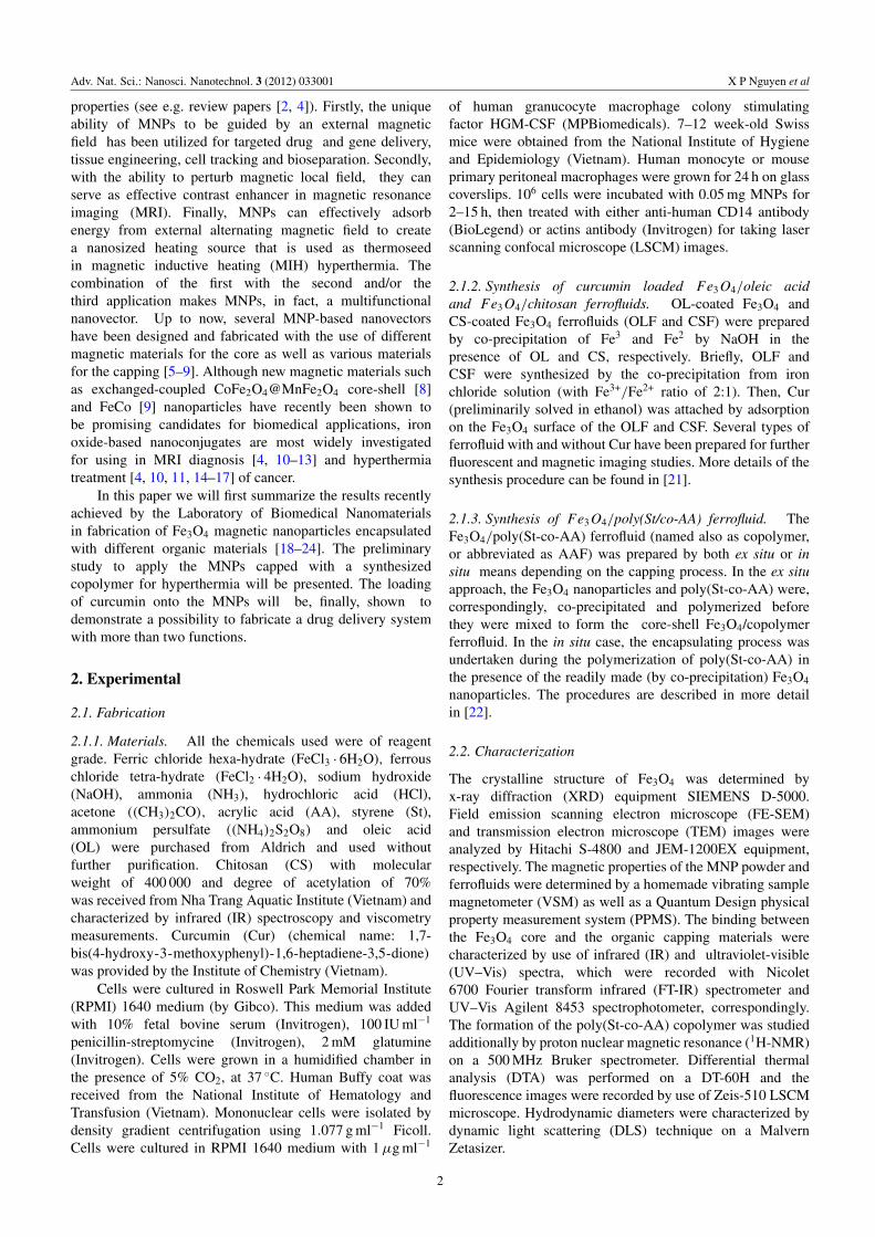
Figure 1. MIH experimental set-up.
2.3. In vitro and in vivo hyperthermia experiments
All the MIH experiments were carried out on the set upwith the use of a commercial generator (RDO HFI 5 kW)providing an alternating magnetic field of amplitude from40 to 100 Oe, and frequency of 180–240 kHz. The sampletemperature was measured online by optical thermometer(Opsens). For characterization of the heating performance,ferrofluid samples of various particle concentrations (dilutedin water) were prepared and kept in a round-bottom-shapedglass holder, so that the temperature sensor was imbeddeddirectly in them. The same experimental arrangement wasalso applied for in vitro experiment while the sample was amixture of ferrofluid with Sarcoma cells. In vivo hyperthermiaexperiment was designed for treatment of a Sarcoma tumorof size of around 6 × 6 mm2, which had been transplantedsubcutaneously on Swiss mice. The mouse was introducedinto a plastic tube of inner diameter of 30 mm, which then wasinserted into a 10 turns coil of diameter of 30 mm. Figure 1presents pictures of the used MIH set-up (a), and in vitro or exvivo (b), and in vivo (c) sample arrangement.
3. Results and discussion
3.1. Ferrofluid characteristics
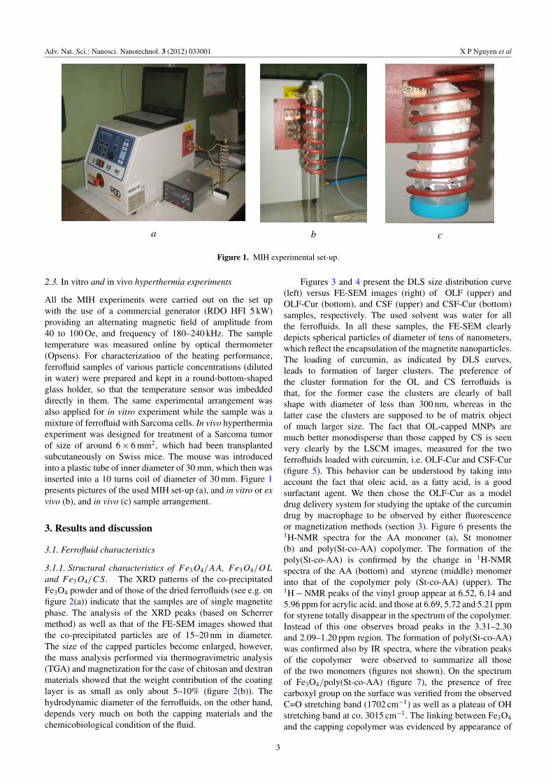
3.1.1. Structural characteristics of Fe3 O4/AA, Fe3 O4/O Land Fe3 O4/C S. The XRD patterns of the co-precipitatedFe3O4 powder and of those of the dried ferrofluids (see e.g. onfigure 2(a)) indicate that the samples are of single magnetitephase. The analysis of the XRD peaks (based on Scherrermethod) as well as that of the FE-SEM images showed thatthe co-precipitated particles are of 15–20 nm in diameter.The size of the capped particles become enlarged, however,the mass analysis performed via thermogravimetric analysis(TGA) and magnetization for the case of chitosan and dextranmaterials showed that the weight contribution of the coatinglayer is as small as only about 5–10% (figure 2(b)). Thehydrodynamic diameter of the ferrofluids, on the other hand,depends very much on both the capping materials and thechemicobiological condition of the fluid.
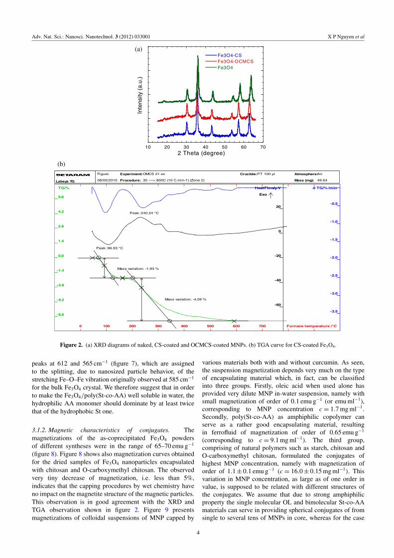
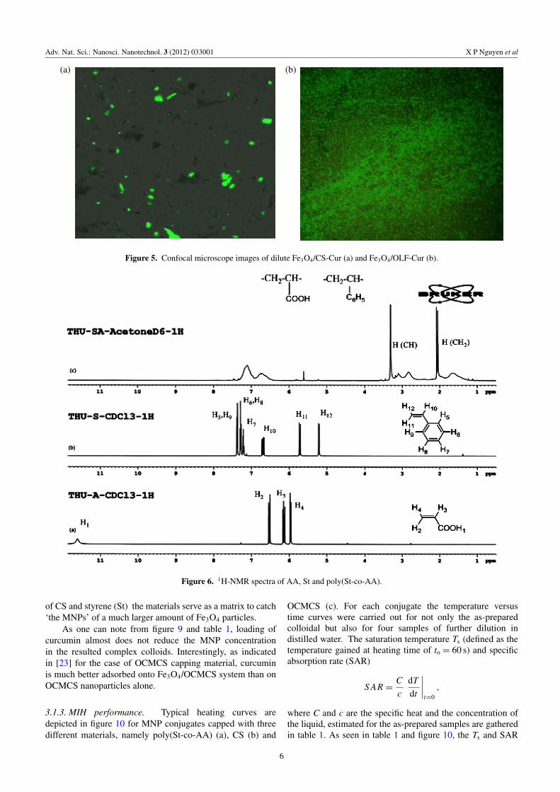
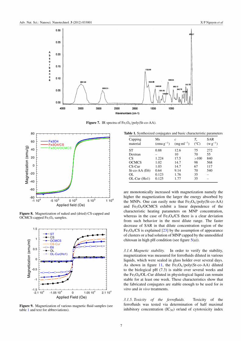
Figures 3 and 4 present the DLS size distribution curve(left) versus FE-SEM images (right) of OLF (upper) andOLF-Cur (bottom), and CSF (upper) and CSF-Cur (bottom)samples, respectively. The used solvent was water for allthe ferrofluids. In all these samples, the FE-SEM clearlydepicts spherical particles of diameter of tens of nanometers,which reflect the encapsulation of the magnetite nanoparticles.The loading of curcumin, as indicated by DLS curves,leads to formation of larger clusters. The preference ofthe cluster formation for the OL and CS ferrofluids isthat, for the former case the clusters are clearly of ballshape with diameter of less than 300 nm, whereas in thelatter case the clusters are supposed to be of matrix objectof much larger size. The fact that OL-capped MNPs aremuch better monodisperse than those capped by CS is seenvery clearly by the LSCM images, measured for the twoferrofluids loaded with curcumin, i.e. OLF-Cur and CSF-Cur(figure 5). This behavior can be understood by taking intoaccount the fact that oleic acid, as a fatty acid, is a goodsurfactant agent. We then chose the OLF-Cur as a modeldrug delivery system for studying the uptake of the curcumindrug by macrophage to be observed by either fluorescenceor magnetization methods (section 3). Figure 6 presents the1H-NMR spectra for the AA monomer (a), St monomer(b) and poly(St-co-AA) copolymer. The formation of thepoly(St-co-AA) is confirmed by the change in 1H-NMRspectra of the AA (bottom) and styrene (middle) monomerinto that of the copolymer poly (St-co-AA) (upper). The1H − NMR peaks of the vinyl group appear at 6.52, 6.14 and5.96 ppm for acrylic acid, and those at 6.69, 5.72 and 5.21 ppmfor styrene totally disappear in the spectrum of the copolymer.Instead of this one observes broad peaks in the 3.31–2.30and 2.09–1.20 ppm region. The formation of poly(St-co-AA)was confirmed also by IR spectra, where the vibration peaksof the copolymer were observed to summarize all thoseof the two monomers (figures not shown). On the spectrumof Fe3O4/poly(St-co-AA) (figure 7), the presence of freecarboxyl group on the surface was verified from the observedC=O stretching band (1702 cm−1) as well as a plateau of OHstretching band at co. 3015 cm−1. The linking between Fe3O4
and the capping copolymer was evidenced by appearance of
3
Adv. Nat. Sci.: Nanosci. Nanotechnol. 3 (2012) 033001 X P Nguyen et al
10 20 30 40 50 60 70
Fe3O4-CSFe3O4-OCMCSFe3O4
Inte
nsity
(a.
u.)
2 Theta (degree)
Furnace temperature /°C0 100 200 300 400 500 600 700
TG/%
-5.6
-4.2
-2.8
-1.4
0.0
1.4
2.8
4.2
5.6
d TG/% /min
-3.5
-3.0
-2.5
-2.0
-1.5
-1.0
-0.5
HeatFlow/µV
-60
-40
-20
0
20
Mass variation: -1.93 %
Mass variation: -4.09 %
Peak :96.93 °C
Peak :240.24 °C
Figure:
08/05/2010 Mass (mg): 49.64
Crucible:PT 100 µl Atmosphere:AirExperiment:OMCS 21 ex
Procedure: 30 ----> 800C (10 C.min-1) (Zone 2)Labsys TG
Exo
(a)
(b)
Figure 2. (a) XRD diagrams of naked, CS-coated and OCMCS-coated MNPs. (b) TGA curve for CS-coated Fe3O4.
peaks at 612 and 565 cm−1 (figure 7), which are assignedto the splitting, due to nanosized particle behavior, of thestretching Fe–O–Fe vibration originally observed at 585 cm−1
for the bulk Fe3O4 crystal. We therefore suggest that in orderto make the Fe3O4/poly(St-co-AA) well soluble in water, thehydrophilic AA monomer should dominate by at least twicethat of the hydrophobic St one.
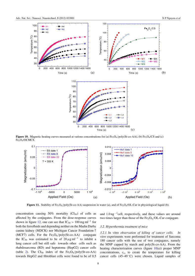
3.1.2. Magnetic characteristics of conjugates. Themagnetizations of the as-coprecipitated Fe3O4 powdersof different syntheses were in the range of 65–70 emu g−1
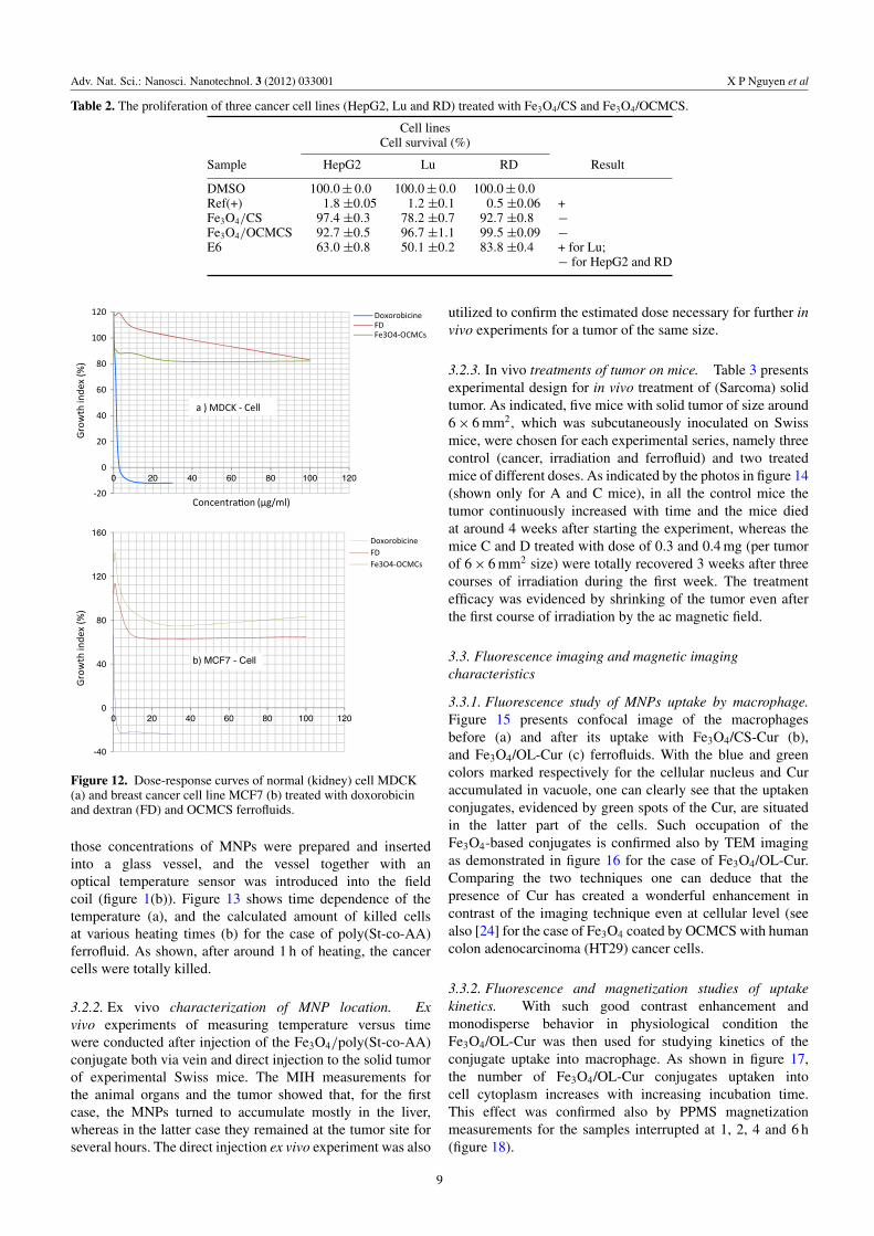
(figure 8). Figure 8 shows also magnetization curves obtainedfor the dried samples of Fe3O4 nanoparticles encapsulatedwith chitosan and O-carboxymethyl chitosan. The observedvery tiny decrease of magnetization, i.e. less than 5%,indicates that the capping procedures by wet chemistry haveno impact on the magnetite structure of the magnetic particles.This observation is in good agreement with the XRD andTGA observation shown in figure 2. Figure 9 presentsmagnetizations of colloidal suspensions of MNP capped by
various materials both with and without curcumin. As seen,the suspension magnetization depends very much on the typeof encapsulating material which, in fact, can be classifiedinto three groups. Firstly, oleic acid when used alone hasprovided very dilute MNP in-water suspension, namely withsmall magnetization of order of 0.1 emu g−1 (or emu ml−1),corresponding to MNP concentration c = 1.7 mg ml−1.
Secondly, poly(St-co-AA) as amphiphilic copolymer canserve as a rather good encapsulating material, resultingin ferrofluid of magnetization of order of 0.65 emu g−1
(corresponding to c = 9.1 mg ml−1). The third group,comprising of natural polymers such as starch, chitosan andO-carboxymethyl chitosan, formulated the conjugates ofhighest MNP concentration, namely with magnetization oforder of 1.1 ± 0.1 emu g−1 (c = 16.0 ± 0.15 mg ml−1). Thisvariation in MNP concentration, as large as of one order invalue, is supposed to be related with different structures ofthe conjugates. We assume that due to strong amphiphilicproperty the single molecular OL and bimolecular St-co-AAmaterials can serve in providing spherical conjugates of fromsingle to several tens of MNPs in core, whereas for the case
4
Adv. Nat. Sci.: Nanosci. Nanotechnol. 3 (2012) 033001 X P Nguyen et al
0 100 200 300 400 500 600 700 8000
5
10
15
20
25
30
Vol
ume
(%)
Particle size (nm)
Figure 3. DLS (left) and FE-SEM image (right) of OLF (upper) and OLF-Cur (bottom) conjugates.
Figure 4. DLS (left) and FE-SEM image (right) of CSF (upper) and CSF-Cur (bottom) conjugates.
5
Adv. Nat. Sci.: Nanosci. Nanotechnol. 3 (2012) 033001 X P Nguyen et al
(a) (b)
Figure 5. Confocal microscope images of dilute Fe3O4/CS-Cur (a) and Fe3O4/OLF-Cur (b).
Figure 6. 1H-NMR spectra of AA, St and poly(St-co-AA).
of CS and styrene (St) the materials serve as a matrix to catch‘the MNPs’ of a much larger amount of Fe3O4 particles.
As one can note from figure 9 and table 1, loading ofcurcumin almost does not reduce the MNP concentrationin the resulted complex colloids. Interestingly, as indicatedin [23] for the case of OCMCS capping material, curcuminis much better adsorbed onto Fe3O4/OCMCS system than onOCMCS nanoparticles alone.
3.1.3. MIH performance. Typical heating curves aredepicted in figure 10 for MNP conjugates capped with threedifferent materials, namely poly(St-co-AA) (a), CS (b) and
OCMCS (c). For each conjugate the temperature versustime curves were carried out for not only the as-preparedcolloidal but also for four samples of further dilution indistilled water. The saturation temperature Ts (defined as thetemperature gained at heating time of to = 60 s) and specificabsorption rate (SAR)
S AR =C
c
dT
dt
∣∣∣∣t=0
,
where C and c are the specific heat and the concentration ofthe liquid, estimated for the as-prepared samples are gatheredin table 1. As seen in table 1 and figure 10, the Ts and SAR
6
Adv. Nat. Sci.: Nanosci. Nanotechnol. 3 (2012) 033001 X P Nguyen et al
Figure 7. IR spectra of Fe3O4/poly(St-co-AA).
-80
-60
-40
-20
0
20
40
60
80
-1 104 -5 103 0 100 5 103 1 104
Fe3O4Fe3O4/CSFe3O4/OCMCS
Mag
netiz
atio
n (e
mu/
g)
Applied field (Oe)
Figure 8. Magnetization of naked and (dried) CS-capped andOCMCS-capped Fe3O4 samples.
-1.5
-1
-0.5
0
0.5
1
1.5
-2.1 104 -1.05 104 0 1.05 104 2.1 104
STCSOCMCSCS-CurE6OLOL-Cur(Ho1)
Mag
netiz
atio
n (e
mu/
ml)
Applied Field (Oe)
Figure 9. Magnetization of various magnetic fluid samples (seetable 1 and text for abbreviations).
Table 1. Synthesized conjugates and basic characteristic parameters
Capping Ms c Ts SARmaterial (emu g−1) (mg ml−1) (oC) (w g−1)
ST 0.88 12.6 75 272Dextran – 10 70 55CS 1.224 17.5 >100 840OCMCS 1.02 14.7 98 568CS-Cur 1.03 14.7 67 117St-co-AA (E6) 0.64 9.14 70 540OL 0.123 1.76 35 –OL-Cur (Ho1) 0.125 1.77 35 –
are monotonically increased with magnetization namely thehigher the magnetization the larger the energy absorbed bythe MNPs. One can easily note that Fe3O4/poly(St-co-AA)and Fe3O4/OCMCS exhibit a linear dependence of thecharacteristic heating parameters on MNP concentration,whereas in the case of Fe3O4/CS there is a clear deviationfrom such behavior in the most dilute range. The fasterdecrease of SAR in that dilute concentration region of theFe3O4/CS is explained [23] by the assumption of appearanceof clusters or a bad solution of MNP capped by the unmodifiedchitosan in high pH condition (see figure 5(a)).
3.1.4. Magnetic stability. In order to verify the stability,magnetization was measured for ferrofluids diluted in variousliquids, which were sealed in glass holder over several days.As shown in figure 11, the Fe3O4/poly(St-co-AA) dilutedto the biological pH (7.3) is stable over several weeks andthe Fe3O4/OL-Cur diluted in physiological liquid can remainstable for at least one week. These characteristics show thatthe fabricated conjugates are stable enough to be used for invitro and in vivo treatments.
3.1.5. Toxicity of the ferrofluids. Toxicity of theferrofluids was tested via determination of half maximalinhibitory concentration (IC50) or/and of cytotoxicity index
7
Adv. Nat. Sci.: Nanosci. Nanotechnol. 3 (2012) 033001 X P Nguyen et al
30
40
50
60
70
80
90
100
0 200 400 600 800 1000 1200 1400 1600
N1
N2
N3
N4
N5T
empe
ratu
re (
o C)
Time (s) (a)
40
60
80
100
120
0 200 400 600 800 1000 1200 1400 1600
Fe3O
4/CS
M1M2M3
M4M5
Tem
pera
ture
(o C
)
Time (s) (b)
30
40
50
60
70
80
90
100
110
0 200 400 600 800 1000 1200 1400 1600
L1
L2
L3
L4
L5
Tem
pera
ture
(o C
)
Time (s) (c)
Figure 10. Magnetic heating curves measured at various concentrations for (a) Fe3O4/poly(St-co-AA), (b) Fe3O4/CS and (c)Fe3O4/OCMCS.
-0.1
-0.05
0
0.05
0.1
-1 104 -5000 0 5000 1 104
E6 date 1
E6 date 8
E6 date 22
Mag
ne
tiza
tion
(e
mu/m
l)
Applied Field (Oe)
T = 305 K
(a)
-0.012
-0.008
-0.004
0
0.004
0.008
0.012
-1 104 -5 103 0 100 5 103 1 104
Ho1 date 1
Ho1 date 5
Ho1 date 15
Mag
netization (
em
u/m
l)
Applied Field (Oe) (b)
Figure 11. Stability of Fe3O4/poly(St-co-AA) suspension in water (a), and of Fe3O4/OL-Cur in physiological liquid (b).
concentration causing 50% mortality (CI50) of cells asaffected by the conjugates. From the dose-response curvesshown in figure 12, one can see that IC50 > 100 mg ml−1 forboth the ferrofluids and depending neither on the Madin Darbycanine kidney (MDCK) nor Michigan Cancer Foundation-7(MCF7) cells. For the Fe3O4/poly(St-co-AA) conjugatethe IC50 was estimated to be of 20 µg ml−1 to inhibit alung cancer cell but still safe towards other cells such asrhabdosarcoma (RD) and heptatoma (HepG2) cancer cells(table 2). The CI50 index of the Fe3O4/poly(St-co-AA)towards HepG2 and fibroblast cells were found to be of 0.5
and 1.0 ng −1cell, respectively, and these values are aroundtwo times larger than those of the Fe3O4/OL-Cur conjugate.
3.2. Hyperthermia treatment of mice
3.2.1. In vitro observation of killing of cancer cells. Invitro experiments were performed for treatment of Sarcoma180 cancer cells with the use of two conjugates, namelythe MNP capped by starch and poly(St-co-AA). From theheating characterization curves (figure 10(a)) proper MNPconcentrations, co, to create the temperature for killingcancer cells (45–49 ◦C) were chosen. Liquid samples of
8
Adv. Nat. Sci.: Nanosci. Nanotechnol. 3 (2012) 033001 X P Nguyen et al
Table 2. The proliferation of three cancer cell lines (HepG2, Lu and RD) treated with Fe3O4/CS and Fe3O4/OCMCS.
Cell linesCell survival (%)
Sample HepG2 Lu RD Result
DMSO 100.0 ± 0.0 100.0 ± 0.0 100.0 ± 0.0Ref(+) 1.8 ±0.05 1.2 ±0.1 0.5 ±0.06 +Fe3O4/CS 97.4 ±0.3 78.2 ±0.7 92.7 ±0.8 −
Fe3O4/OCMCS 92.7 ±0.5 96.7 ±1.1 99.5 ±0.09 −
E6 63.0 ±0.8 50.1 ±0.2 83.8 ±0.4 + for Lu;− for HepG2 and RD
-20
0
20
40
60
80
100
120
0 20 40 60 80 100 120
DoxorobicineFDFe3O4-OCMCs
a ) MDCK - Cell
Concentra�on (μg/ml)
Grow
th in
dex
(%)
-40
0
40
80
120
160
0 20 40 60 80 100 120
DoxorobicineFDFe3O4-OCMCs
b) MCF7 - Cell
Grow
th in
dex
(%)
Figure 12. Dose-response curves of normal (kidney) cell MDCK(a) and breast cancer cell line MCF7 (b) treated with doxorobicinand dextran (FD) and OCMCS ferrofluids.
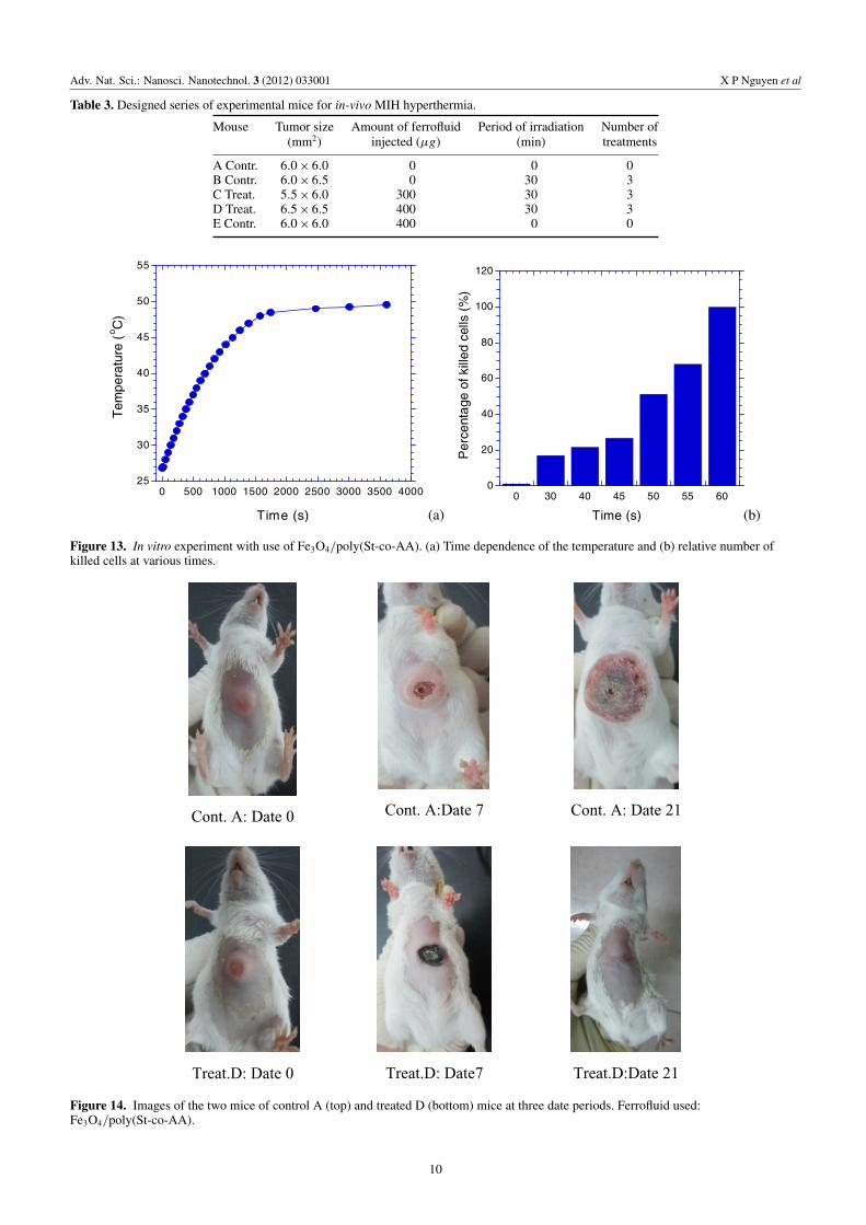
those concentrations of MNPs were prepared and insertedinto a glass vessel, and the vessel together with anoptical temperature sensor was introduced into the fieldcoil (figure 1(b)). Figure 13 shows time dependence of thetemperature (a), and the calculated amount of killed cellsat various heating times (b) for the case of poly(St-co-AA)ferrofluid. As shown, after around 1 h of heating, the cancercells were totally killed.
3.2.2. Ex vivo characterization of MNP location. Exvivo experiments of measuring temperature versus timewere conducted after injection of the Fe3O4/poly(St-co-AA)conjugate both via vein and direct injection to the solid tumorof experimental Swiss mice. The MIH measurements forthe animal organs and the tumor showed that, for the firstcase, the MNPs turned to accumulate mostly in the liver,whereas in the latter case they remained at the tumor site forseveral hours. The direct injection ex vivo experiment was also
utilized to confirm the estimated dose necessary for further invivo experiments for a tumor of the same size.
3.2.3. In vivo treatments of tumor on mice. Table 3 presentsexperimental design for in vivo treatment of (Sarcoma) solidtumor. As indicated, five mice with solid tumor of size around6 × 6 mm2, which was subcutaneously inoculated on Swissmice, were chosen for each experimental series, namely threecontrol (cancer, irradiation and ferrofluid) and two treatedmice of different doses. As indicated by the photos in figure 14(shown only for A and C mice), in all the control mice thetumor continuously increased with time and the mice diedat around 4 weeks after starting the experiment, whereas themice C and D treated with dose of 0.3 and 0.4 mg (per tumorof 6 × 6 mm2 size) were totally recovered 3 weeks after threecourses of irradiation during the first week. The treatmentefficacy was evidenced by shrinking of the tumor even afterthe first course of irradiation by the ac magnetic field.
3.3. Fluorescence imaging and magnetic imagingcharacteristics
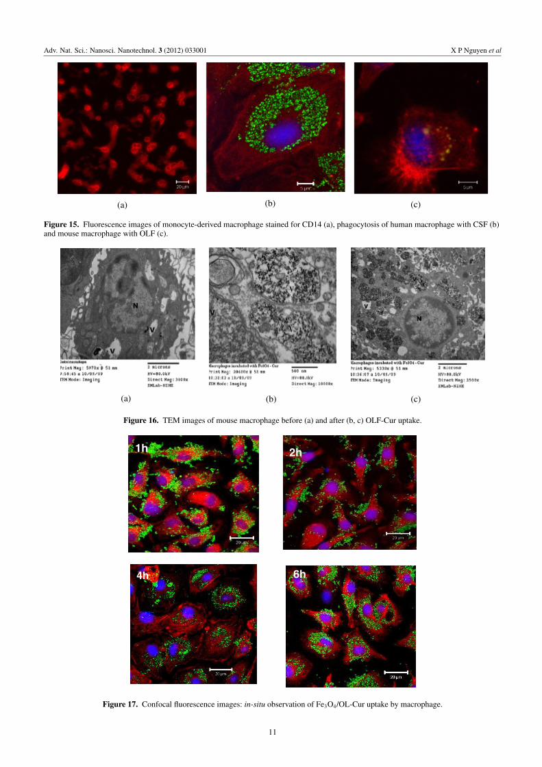
3.3.1. Fluorescence study of MNPs uptake by macrophage.Figure 15 presents confocal image of the macrophagesbefore (a) and after its uptake with Fe3O4/CS-Cur (b),and Fe3O4/OL-Cur (c) ferrofluids. With the blue and greencolors marked respectively for the cellular nucleus and Curaccumulated in vacuole, one can clearly see that the uptakenconjugates, evidenced by green spots of the Cur, are situatedin the latter part of the cells. Such occupation of theFe3O4-based conjugates is confirmed also by TEM imagingas demonstrated in figure 16 for the case of Fe3O4/OL-Cur.Comparing the two techniques one can deduce that thepresence of Cur has created a wonderful enhancement incontrast of the imaging technique even at cellular level (seealso [24] for the case of Fe3O4 coated by OCMCS with humancolon adenocarcinoma (HT29) cancer cells.
3.3.2. Fluorescence and magnetization studies of uptakekinetics. With such good contrast enhancement andmonodisperse behavior in physiological condition theFe3O4/OL-Cur was then used for studying kinetics of theconjugate uptake into macrophage. As shown in figure 17,the number of Fe3O4/OL-Cur conjugates uptaken intocell cytoplasm increases with increasing incubation time.This effect was confirmed also by PPMS magnetizationmeasurements for the samples interrupted at 1, 2, 4 and 6 h(figure 18).
9
Adv. Nat. Sci.: Nanosci. Nanotechnol. 3 (2012) 033001 X P Nguyen et al
Table 3. Designed series of experimental mice for in-vivo MIH hyperthermia.
Mouse Tumor size Amount of ferrofluid Period of irradiation Number of(mm2) injected (µg) (min) treatments
A Contr. 6.0 × 6.0 0 0 0B Contr. 6.0 × 6.5 0 30 3C Treat. 5.5 × 6.0 300 30 3D Treat. 6.5 × 6.5 400 30 3E Contr. 6.0 × 6.0 400 0 0
25
30
35
40
45
50
55
0 500 1000 1500 2000 2500 3000 3500 4000
Tem
pera
ture
(o C
)
Time (s) (a)
0
20
40
60
80
100
120
0 30 40 45 50 55 60
Per
cent
age
of k
illed
cel
ls (
%)
Time (s) (b)
Figure 13. In vitro experiment with use of Fe3O4/poly(St-co-AA). (a) Time dependence of the temperature and (b) relative number ofkilled cells at various times.
Cont. A: Date 0 Cont. A:Date 7 Cont. A: Date 21
Treat.D: Date 0 Treat.D: Date7 Treat.D:Date 21
Figure 14. Images of the two mice of control A (top) and treated D (bottom) mice at three date periods. Ferrofluid used:Fe3O4/poly(St-co-AA).
10
Adv. Nat. Sci.: Nanosci. Nanotechnol. 3 (2012) 033001 X P Nguyen et al
(a) (b) (c)
Figure 15. Fluorescence images of monocyte-derived macrophage stained for CD14 (a), phagocytosis of human macrophage with CSF (b)and mouse macrophage with OLF (c).
(a) (b) (c)
Figure 16. TEM images of mouse macrophage before (a) and after (b, c) OLF-Cur uptake.
1h 2h
4h 6h
Figure 17. Confocal fluorescence images: in-situ observation of Fe3O4/OL-Cur uptake by macrophage.
11
Adv. Nat. Sci.: Nanosci. Nanotechnol. 3 (2012) 033001 X P Nguyen et al
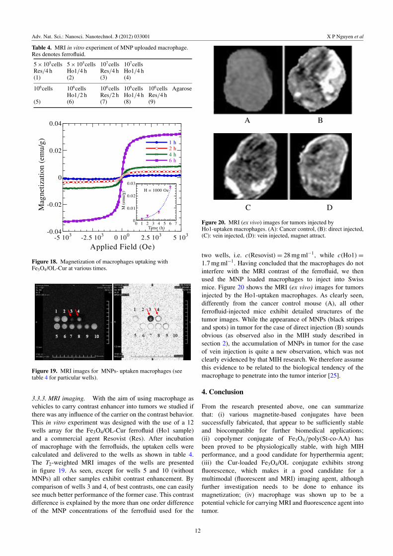
Table 4. MRI in vitro experiment of MNP uploaded macrophage.Res denotes ferrofluid.
5 × 10⁵cells 5 × 10⁵cells 107cells 107cellsRes/4 h Ho1/4 h Res/4 h Ho1/4 h(1) (2) (3) (4)
106cells 106cells 106cells 106cells 106cells AgaroseHo1/2 h Res/2 h Ho1/4 h Res/4 h
(5) (6) (7) (8) (9)
-0.04
-0.02
0
0.02
0.04
-5 103 -2.5 103 0 100 2.5 103 5 103
1 h2 h4 h6 h
Mag
neti
zati
on (
emu/
g)
Applied Field (Oe)
0
0.01
0.02
0.03
0 1 2 3 4 5 6 7
M (
emu/
g)
Time (h)
H = 1000 Oe
Figure 18. Magnetization of macrophages uptaking withFe3O4/OL-Cur at various times.
Figure 19. MRI images for MNPs- uptaken macrophages (seetable 4 for particular wells).
3.3.3. MRI imaging. With the aim of using macrophage asvehicles to carry contrast enhancer into tumors we studied ifthere was any influence of the carrier on the contrast behavior.This in vitro experiment was designed with the use of a 12wells array for the Fe3O4/OL-Cur ferrofluid (Ho1 sample)and a commercial agent Resovist (Res). After incubationof macrophage with the ferrofluids, the uptaken cells werecalculated and delivered to the wells as shown in table 4.The T2-weighted MRI images of the wells are presentedin figure 19. As seen, except for wells 5 and 10 (withoutMNPs) all other samples exhibit contrast enhancement. Bycomparison of wells 3 and 4, of best contrasts, one can easilysee much better performance of the former case. This contrastdifference is explained by the more than one order differenceof the MNP concentrations of the ferrofluid used for the
A B
C D
Fgure 20. MRI (ex vivo) images for tumors injected byHo1-uptaken macrophages. (A): Cancer control, (B): direct injected,(C): vein injected, (D): vein injected, magnet attract.
two wells, i.e. c(Resovist) = 28 mg ml−1, while c(Ho1) =
1.7 mg ml−1. Having concluded that the macrophages do notinterfere with the MRI contrast of the ferrofluid, we thenused the MNP loaded macrophages to inject into Swissmice. Figure 20 shows the MRI (ex vivo) images for tumorsinjected by the Ho1-uptaken macrophages. As clearly seen,differently from the cancer control mouse (A), all otherferrofluid-injected mice exhibit detailed structures of thetumor images. While the appearance of MNPs (black stripesand spots) in tumor for the case of direct injection (B) soundsobvious (as observed also in the MIH study described insection 2), the accumulation of MNPs in tumor for the caseof vein injection is quite a new observation, which was notclearly evidenced by that MIH research. We therefore assumethis evidence to be related to the biological tendency of themacrophage to penetrate into the tumor interior [25].
4. Conclusion
From the research presented above, one can summarizethat: (i) various magnetite-based conjugates have beensuccessfully fabricated, that appear to be sufficiently stableand biocompatible for further biomedical applications;(ii) copolymer conjugate of Fe3O4/poly(St-co-AA) hasbeen proved to be physiologically stable, with high MIHperformance, and a good candidate for hyperthermia agent;(iii) the Cur-loaded Fe3O4/OL conjugate exhibits strongfluorescence, which makes it a good candidate for amultimodal (fluorescent and MRI) imaging agent, althoughfurther investigation needs to be done to enhance itsmagnetization; (iv) macrophage was shown up to be apotential vehicle for carrying MRI and fluorescence agent intotumor.
12
Adv. Nat. Sci.: Nanosci. Nanotechnol. 3 (2012) 033001 X P Nguyen et al
Acknowledgments
The authors are indebted in acknowledging the financialsupport mainly from the MOST grant no. 4/2/742/2009-HD-DTDL and partly from a research grant of VAST forthe 2009–2010 period. Sincere thanks are due to ProfessorAcademician Nguyen Van Hieu for his encouragement indoing such a new and interdisciplinary research.
References
[1] Ferrieri M 2005 Nature Rev. Cancer 5 161Brigger I, Dubernet C and Couvreur P 2002 Adv. Drug Deliv.
Rev. 54 63[2] Shubayev V, Pisanic T R and Jin S H 2009 Adv. Drug Rev.
61 467[3] Takae S, Akiyama Y, Otsuka H, Nakamura T, Nagasaki Y and
Kataoka K 2005 Biomacromol. 6 818[4] Panchurst Q A, Connolly J, Jones S K and Dobson J 2003
J. Phys. D: Appl. Phys. 36 R167Pankhurst Q A, Thanh N K T, Jones S K and Dobson J 2009 J.
Phys. D: Appl. Phys. 42 4001[5] Moenet S, Vasseur S, Grasset F and Duguet E 2004 J. Mater.
Chem. 14 2161[6] Gupta A K and Gupta M 2005 Biomaterials 26 3995[7] Lu A H, Salabas E L and Schulth F 2007 Angew Chem., Int.
Ed. Engl. 46 1222[8] Lee J-H, Jiang J-T, Choi J-S, Moon S H, Noh S-H, Kim J-W,
Kim J-G, Park K I and Cheon J 2011 Nature Nanotechnol.6 418
[9] Park J K, Jung J, Subramania P, Shah B P, Kim C, Lee J K,Cho J H, Lee C and Lee K-B 2011 Small 7 1647
[10] Pantic I 2010 Rev. Adv. Mater. Sci. 26 67
[11] Lacroix L-M, Ho D and Sun S 2010 Curr. Top. Med. Chem.10 1184
[12] Wang Y-X J, Hussain S M and Krestin G P 2001 Eur. Radiol.11 2319
[13] Prashant C, Dipak M, Yang C-T, Chuang K-H, Jun D and FengS-S 2010 Biomaterials 31 5588
[14] Yanase M, Shinkai M, Honda H, Wakabayashi T, Yoshida Jand Kobayashi T 1998 Japan. J. Cancer Res. 89 463
Ito A, Shinkai M, Honda H and Kobayashi T 2001 CancerGene Ther. 8 649
[15] Brusentsov N A, Nikitin L V, Brusentsova T N, KuznetsovA A, Bayburskiy F S, Shumakov L I and Jurchenko N Y2002 J. Magn. Magn. Mater. 252 378
[16] Gneveckow U, Jordan A, Scholz R, Brub V, Waldofner N,Ricke J, Feussner A, Hildebrant B, Rau B and Wust P 2004Med. Phys. 31 1444
[17] Hilger I, Hergt R and Kaiser W A 2005 IEEProc.—Nanotechnol. 152 33
[18] Pham H L, Do H M, Tran D L, Le V H, Nguyen X P, NguyenA T, Nguyen T N and Vu A T 2011 Int. J. Nanotechnol.8 399
[19] Pham H L, Nguyen C T, Nguyen A T, Pham V T, Tran C Y,Nguyen T Q, Hoang T M N, Phi T X, Nguyen X P and LeV H 2009 J. Phys.: Conf. Series 187 012008
[20] Ha P T, Tran T M N, Pham H D, Nguyen Q H and Nguyen X P2010 Adv. Nature. Sci.: Nanosci Nanotechnol. 1 015012
[21] Tran D L et al 2010 Colloid. Surf. A 371 104[22] Luong T T et al 2011 Colloid. Surf. A 384 23[23] Mai T T, Ha P T, Pham H N, Le T T H, Pham H L, Phan
T B H, Tran D L and Nguyen X P 2012 Adv. Nature Sci.:Nanosci. Nanotechnol. 3 015006
[24] Ha P T et al 2011 Chem. Lett. 40 1264[25] Muthana M, Scott S D, Farrow M, Morrow F, Murdoch C,
Grubb S, Brown N, Dobson J and Lewis C E 2008 GeneTher. 15 902
13
Related Documents