Abstract The bhuTUV and bhuO genes play vital roles in the ability of Brucella abortus to use heme as an iron source and are regulated in an iron-responsive manner by RirA and Irr by Jenifer F. Ojeda April, 2012 Dissertation Advisor: RM Roop II Department of Microbiology and Immunology Brucella abortus is a Gram negative intracellular pathogen that causes the zoonotic disease brucellosis. Antibiotic treatment for brucellosis in humans is prolonged and sometimes followed by relapses. Currently, the United States employs prevention of the illness in humans through cattle vaccinations, eliminating the bacterium in its natural host. Unfortunately, these vaccine strains cause the disease in humans, and Brucella research ultimately aims to identify new vaccine targets as well as alternative treatment options. Brucella abortus resides in the phagosomal compartment of the host macrophage where essential nutrients such as iron are limited. Most bacteria need iron, and within the macrophage, heme is a likely source of iron due to the breakdown of red blood cells by the host macrophage. Heme transporters in Gram negative bacteria are highly conserved, and include components for outer membrane, periplasmic, and cytoplasmic membrane transport. BhuA has been previously characterized as the outer membrane heme transporter of Brucella abortus and here we report that BhuT, BhuU, and BhuV (BhuTUV) are the periplasmic and cytoplasmic heme transport components and that they are required in order for Brucella abortus to transport heme as an iron source. Utilization of heme as an iron source requires the breakdown of heme into ferrous iron,

Welcome message from author
This document is posted to help you gain knowledge. Please leave a comment to let me know what you think about it! Share it to your friends and learn new things together.
Transcript

Abstract
The bhuTUV and bhuO genes play vital roles in the ability of
Brucella abortus to use heme as an iron source and are regulated
in an iron-responsive manner by RirA and Irr
by
Jenifer F. Ojeda
April, 2012
Dissertation Advisor: RM Roop II
Department of Microbiology and Immunology
Brucella abortus is a Gram negative intracellular pathogen that causes the zoonotic disease
brucellosis. Antibiotic treatment for brucellosis in humans is prolonged and sometimes followed
by relapses. Currently, the United States employs prevention of the illness in humans through
cattle vaccinations, eliminating the bacterium in its natural host. Unfortunately, these vaccine
strains cause the disease in humans, and Brucella research ultimately aims to identify new
vaccine targets as well as alternative treatment options.
Brucella abortus resides in the phagosomal compartment of the host macrophage where
essential nutrients such as iron are limited. Most bacteria need iron, and within the macrophage,
heme is a likely source of iron due to the breakdown of red blood cells by the host macrophage.
Heme transporters in Gram negative bacteria are highly conserved, and include components for
outer membrane, periplasmic, and cytoplasmic membrane transport. BhuA has been previously
characterized as the outer membrane heme transporter of Brucella abortus and here we report
that BhuT, BhuU, and BhuV (BhuTUV) are the periplasmic and cytoplasmic heme transport
components and that they are required in order for Brucella abortus to transport heme as an iron
source. Utilization of heme as an iron source requires the breakdown of heme into ferrous iron,

carbon monoxide, and biliverdin by a heme oxygenase. BhuO has been identified as a heme
oxygenase in Brucella abortus, and although there seems to be more than one heme oxygenase in
Brucella, this study shows that BhuO is needed for the use of heme as an iron source under iron
starvation conditions in vitro. Further, both bhuTUV and bhuO are regulated in an iron-
responsive manner. The iron responsive regulator Irr directly represses bhuO, which shares an
operon with rirA. Then the rhizobial iron regulator RirA in turn represses the bhuTUV operon.
Together, these regulators help to maintain iron homeostasis within the bacterial cell, protecting
it from damaging hydroxyl radicals produced by Fenton chemistry.


The bhuTUV and bhuO genes play vital roles in the ability of Brucella abortus to use heme as an
iron source and are regulated in an iron-responsive manner by RirA and Irr
A Dissertation
Submitted to the Graduate Faculty of the Brody School of Medicine at
East Carolina University
In Partial Fulfillment of the Requirements for the
degree of Doctor of Philosophy
in the Department of Microbiology and Immunology
By
Jenifer F. Ojeda
April 2012

© Jenifer F. Ojeda, 2012

The bhuTUV and bhuO genes play vital roles in the ability of Brucella abortus to use heme as an
iron source and are regulated in an iron-responsive manner by RirA and Irr
By
Jenifer F. Ojeda
APPROVED BY:
DIRECTOR OF DISSERTATION_________________________________
Roy M. Roop II, Ph.D.
COMMITTEE MEMBER________________________________________
Charles J. Smith, Ph.D.
COMMITTEE MEMBER________________________________________
Everett C. Pesci, Ph.D.
COMMITTEE MEMBER________________________________________
Cindy Putnam-Evans, Ph.D.
COMMITTEE MEMBER________________________________________
Mark D. Mannie, Ph.D.
CHAIR OF THE DEPARTMENT OF MICROBIOLOGY AND IMMUNOLOGY
_________________________________
Charles J. Smith, Ph.D.
DEAN OF THE GRADUATE SCHOOL
_________________________________
Paul J. Gemperline, Ph.D.

Dedication
This dissertation is dedicated to my husband Dan, who by all accounts earned this degree
with me. He held the magic to obtaining the most obscure reference articles, the most visually
appealing graphs, and had the wildest imagination when it came to scientific solutions. He rode
the emotional turmoil of graduate school with me, and this work truly would not have been
possible without his constant love and support.

Acknowledgements
First and foremost I offer my sincerest gratitude to my supervisor, Dr. Marty Roop, who
has supported me thoughout my project with his patience and knowledge while still allowing me
the room to work in my own way. I would like to thank him for taking the time to teach me how
to fish (literally!) and for showing me that with the right attitude you can catch fish no matter
how many pieces your fishing pole has broken into. One simply could not wish for a better or
friendlier supervisor.
I would also like to thank JT Paulley for taking the time to train me properly. I have
missed the late night beer and cigar talks with JT, Eric, and Mike, who instilled in me a joy for
arguing science. So many side projects began this way....
I would like to thank my committee for their patience and understanding during all of the
curve balls life threw my way. I appreciate your guidance throughout this project.
Lastly, I would like to thank the current members of the Roop Lab for the teamwork and
fun that we have shared over the years. It is nice have such strong resources for help when
technical challenges arise.

Table of Contents
CHAPTER 1
Literature Review.............................................................................................................................1
Brucella ....................................................................................................................................1
The causative agent of brucellosis .......................................................................................1
A stealthy intracellular pathogen .........................................................................................4
The bacterial necessity for iron ................................................................................................7
The chemistry of iron ..........................................................................................................7
Bacterial adaptations to the chemistry of iron .....................................................................8
Availability of iron within the host ........................................................................................13
Iron trafficking within the host ..........................................................................................13
Host iron sources during infection ....................................................................................16
Pathogenic bacteria acquire iron from the host ......................................................................17
Acquisition of ferric and ferrous iron from the host..........................................................17
Acquisition of heme from the host ....................................................................................19
References ......................................................................................................................................25
Figures............................................................................................................................................48
STATEMENT OF THE PROBLEM .........................................................................................56
CHAPTER 2
bhuTUV encodes proteins required for heme transport in Brucella abortus 2308 and is
regulated by RirA ...........................................................................................................................59
Abstract ......................................................................................................................................59
Introduction ................................................................................................................................60
Materials and Methods ...............................................................................................................63
Results and Discussion ...............................................................................................................68
Acknowledgements ....................................................................................................................74
References ..................................................................................................................................75

Figures and Tables .....................................................................................................................81
CHAPTER 3
The bhuO gene encodes a heme oxygenase that contributes to the ability of Brucella abortus
2308 to use heme as an iron source and is regulated by Irr ...........................................................93
Abstract ......................................................................................................................................93
Introduction ................................................................................................................................94
Materials and Methods ...............................................................................................................95
Results and Discussion .............................................................................................................102
Acknowledgements ..................................................................................................................107
References ................................................................................................................................108
Figures and Tables ...................................................................................................................115
CHAPTER 4
Summary and Future Directions ..................................................................................................133
The requirement for heme in Brucella .....................................................................................133
Unknown heme transport components .....................................................................................134
The complexity of regulation for these heme transport genes .................................................135
Brucella must acquire iron from the heme ...............................................................................137
Unanswered questions about bacterial heme oxygenases ........................................................138
References ................................................................................................................................140
Figures and Tables ...................................................................................................................144

List of Figures and Tables
CHAPTER 1
Figure 1.1 Host iron sources during infection ............................................................................48
Figure 1.2 RirA and Irr regulate several iron acquisition genes ................................................50
Figure 1.3 Shigella heme transport model..................................................................................52
Figure 1.4 BhuA contributes to the virulence of B. abortus ......................................................54
CHAPTER 2
Table 2.1 Bacterial strains used in this study .............................................................................81
Table 2.2 Primers used in this study ..........................................................................................83
Figure 2.1 Genetic organization of the heme transport genes in B. abortus 2308 .....................85
Figure 2.2 The bhuTUV mutant exhibits a growth defect during iron deprivation that can be
rescued by FeCl3 but not heme or hemoglobin ..........................................................................87
Table 2.3 Capacity of the bhuTUV mutant to use heme and FeCl3 as iron sources in a solid
medium-based ............................................................................................................................89
Figure 2.3 bhuT transcription is iron-responsive in B. abortus 2308 and regulated by both Irr
and RirA .....................................................................................................................................91
CHAPTER 3
Table 3.1 Bacterial strains used in this study ...........................................................................115
Table 3.2 Primers used in this study ........................................................................................117
Figure 3.1 The B. abortus BhuO protein shares amino acid homology with HmuD/Q and
IsdG/I ........................................................................................................................................119
Figure 3.2 B.abortus DCO1 (2308 ΔbhuO) produces significantly more siderophore than B.
abortus 2308 in response to iron deprivation ...........................................................................121
Figure 3.3 B. abortus JFO1 (2308 ΔdhbCΔbhuO) has a growth defect in the presence of
heme as the sole iron source. ....................................................................................................123
Figure 3.4 rirA and bhuO are cotranscribed as an operon in B. abortus 2308 .........................125

Figure 3.5 rirA and bhuO transcription is increased in response to iron deprivation in B.
abortus BEA2 (Δirr) ................................................................................................................127
Figure 3.6 Irr binds directly to the rirA promoter in B. abortus 2308 and protects a 28
nucleotide sequence in a DNase I footprint analysis ................................................................129
Figure 3.7 Proposed model for the role of BhuO in allowing the transcriptional regulator RirA
to recognize heme as an iron source in Brucella ......................................................................131
CHAPTER 4
Figure 4.1 Heme transport in Brucella abortus 2308 ...............................................................144
Figure 4.2 Genetic organization of the genes associated with heme uptake and utilization ....146
Figure 4.3 The similar stuctures of cobalamin (B12) and heme ..............................................148
Figure 4.4 Model of possible regulation of heme uptake genes in B. abortus 2308 ................150

Chapter 1: Literature Review
Brucella
The causative agent of brucellosis
Brucella spp. are Gram negative pathogens that belong to the phylogenetic branch of the
alpha proteobacteria, which encompasses a large variety of organisms, including other
pathogenic bacteria, such as Bartonella quintana as well as Rhizobium leguminosarum, a
bacterium that has adapted a symbiosis with its plant hosts (Williams 2007). Despite their
apparent differences, many of the bacteria within this grouping share similar t raits. For instance,
several species within the alpha proteobacteria develop chronic, long term relationships with
higher eukaryotic organisms (Batut 2004). In fact, the ability to evade the immune system and
establish a chronic infection is the key factor in Brucella virulence (Barquero-Calvo 2007).
Brucella are found within a wide variety of animal reservoirs, with the individual strains
named according to their natural hosts. B. ceti is found in porpoises and dolphins, B.
pinnipedialis is found in seals, and B. microti is found in voles and red foxes (Pappas 2010).
While there are many different species of Brucella, the three primary species that cause zoonotic
infections in humans are B. melitensis, B. abortus, and B. suis strains which naturally infect
goats, cattle, and swine, respectively (Solera 2010).
Humans are not a natural host for Brucella strains, and human brucellosis is strictly a
zoonotic disease. Within the context of their animal hosts, most of these strains cause abortion
and infertility, however, the symptoms are more flu-like in humans, characterized by a spiking
and remitting fever and general malaise (Spera 2006). Due to the generalized nature of the

2
symptoms of brucellosis in humans, diagnosis is often slow, and in spite of heavy, sustained
antibiotic treatment such as a combination of rifampin and doxycycline for six weeks, there is a
10-30% relapse rate (Skendros 2006). Since treatment of human brucellosis is not always
effective, prevention is the best course of action.
Human consumption of unpasteurized milk from a diseased animal is a real problem in
endemic areas of the world (Pappas 2010), and human brucellosis is a common occurrence;
however, in the United States a successful vaccination program has led to the eradication of the
disease in food animals. The vaccines used to control the disease in animals are RB51 and strain
19 in cattle, and in other parts of the world, Rev1 in sheep and goats (Ko 2003). Unfortunately,
these vaccines cause the disease in humans, and to date there are no known human vaccines for
the prevention of brucellosis (Olsen 2006). Therefore, the best prevention available is to keep our
food animals brucellosis-free. Unfortunately, there is a large wildlife population that still carries
Brucella species, and occasional interaction between livestock and these diseased animals can
result in reintroduction of brucellosis to the unvaccinated mammals (Pappas 2010).
Cattle are the natural host of Brucella abortus, and within this host the brucellae establish
either a chronic infection or, in a pregnant cow, an acute infection (Detilleux 1990). Brucella
resides within the cells of its host, leading to evasion of the host immune response (Detilleux
1990). During an acute infection, the brucellae reside within the placental trophoblast epithelial
cells, living on the unique erythritol carbon source that is only made during the third trimester in
ruminants, which results in the third trimester abortion of the fetus (Acha 1980). Erythritol is the
preferred carbon source for Brucella, and erythritol catabolism in Brucella is heavily iron-
dependent, which greatly increases the cell’s iron requirement as cofactors in this reaction

3
(Sperry 1975). The brucellae in this niche replicate in large numbers within the placental
trophoblast, causing abortion. For the establishment of a more chronic infection in cattle, the
brucellae take up residence within the bovine macrophages. Because erythritol is not found in
humans, humans tend to develop a more chronic infection, and the ideal environment in this
niche for the brucellae is within the macrophage (He 2006).
While most pathogenic bacteria rely on classical virulence factors such as toxins,
pathogenicity islands and virulence plasmids, Brucella species utilize a more subtle approach to
establishing and maintaining infection within their host. In fact, Brucella pathogenesis is mainly
based on its ability to survive and multiply within the host macrophages. One coined term,
“virulome” is used to describe the set of genes needed for survival of Brucella in the
macrophages (Köhler 2002). The virulome genes are often associated with metabolism, and
transposon mutagenesis of the Brucella chromosomes led to striking attenuation of the mutants
in which amino acid biosynthesis genes were disrupted. This suggests that these corresponding
metabolites are not available in the phagosomal compartment. Other attenuated mutants involved
genes that are associated with cellular stress response, such as hfq and rsh (a rel/spoT homolog),
and these mutations resulted in lowered resistance to stress induced by peroxides and acidic
conditions. These results collectively lead to the conclusion that the Brucella environment is low
in nutrients and oxygen (Köhler 2002). Therefore, it is important to take a closer look at the
Brucella genes associated with nutrient uptake and metabolism in order to better understand the
pathogenic nature of this bacterium.

4
A stealthy intracellular pathogen
Animal models of infection using mice and natural hosts (goats and cattle) have been
developed (Kahl-McDonagh 2007) in order to gain a better understanding of what takes place
during infection of a mammalian host with brucellae. Two popular mouse models are Balb/c
mice and C57BL/6 mice. These mice strains provide different immunological responses to
Brucella infection, with the C57BL/6 strain producing a more effective, cell-mediated response
than Balb/c mice (Silva 2011). These differences have provided key insights into understanding
the host immune response and have helped to reveal the relationship between cell-mediated Th1
host response and clearance of the brucellae organisms in vivo. The Th1 immunologic response
of the C57BL/6 mice often results in clearance of the wild type brucellae after 2 months of
infection, whereas the Th2 immunologic response of the Balb/c mouse during Brucella infection
is ineffective and brucellae organisms may persist for over 6 months in vivo. It has been shown
throughout the literature that the cell-mediated immune response (Th1), and not the humoral
immune response (Th2), is required for an effective clearance of Brucella organisms that have
chronically infected the host (de Jong 2010).
Brucella are extremely stealthy, and both avoid and suppress the host innate immune
response (de Jong 2010). Brucella enter their host mostly through a mucosal route, via ingestion
or inhalation, where they first encounter the host immune system. The O-chain component of the
Brucella LPS interacts with the surface of the macrophage, allowing for unopsonized entry of the
brucellae into these cells (Porte 2003). This intracellular niche protects the Brucella from
complement and antibodies; however, even those brucellae that have not yet made it to their
intracellular niche have ways to avoid the immune response. Most notably, the

5
lipopolysaccharide (LPS) of Brucella triggers an ineffective Th2 immunological response. The
pathogen- associated molecular patterns (PAMPs) often recognized by the host toll – like
receptors (TLRs) do not bind very well to the LPS of Brucella. For instance, the lipid A of
Brucella contains a very long fatty acid residue that is not recognized well by TLR4, allowing
the bacteria to avoid induction of a strong inflammatory response (Lapaque 2006). Also, the
perosamine O-chain of the Brucella LPS is not broken down well by macrophages, and it forms
complexes with MHC class II molecules, inhibiting their ability to present antigens for further
host immune activation (Forestier 2000). Additionally, because of its aberrant O-antigen,
Brucella LPS does not interact very well with C3 of the host’s pro-inflammatory complement
system, which also interferes with a robust response to infection (Hoffmann 1983).
In addition to having low TLR agonist and complement activity, Brucella produces
proteins that interfere with the host immune signaling. TcpB contains a Toll/interleukin-1 (TIR)
domain that blocks TLR2 and TLR4 – mediated induction of NF-kB expression by inducing the
degradation of MAL, which reduces the ability of the host cell to produce proinflammatory
cytokines such as TNF-α and IL-12 (Salcedo 2008). PrpA is a proline racemase produced by
Brucella that stimulates the production of IL-10, an anti-inflammatory cytokine (Spera 2006).
All of these initial evasions of the host innate immune response are vital to the establishment of
infection.
Intracellular survival for the brucellae is not quite so stealthy, however, and within
phagocytic cells about 90% of the bacteria are immediately killed by the oxidative burst of the
macrophage (Jiang 1993). However, a small population of brucellae that enter the host survive,
and it is with these successful pathogens that our interest lies.

6
Research to date has revealed a high level of interaction between the host and Brucella
pathogen during the establishment and maintenance of chronic infection. A successful brucellae
is able to resist being digested in the cellular phagosome, which temporarily fuses with the
lysosome and acidifies, escape constitutive phagosomal fusion with the lysosome, and move to
the safer compartment of the endoplasmic reticulum (ER), where it is able to replicate. Brucella
strains enter the cell within a phagosomal compartment referred to as the Brucella-containing
vacuole (BCV) (Celli 2005). The BCV interacts with early endosomal compartments, taking on
several endosomal markers and causing acidification of the BCV (Bellaire 2005). The BCV
quickly (10 minutes) loses its endosomal markers and takes on more lysosomal – like markers,
such as Lamp-1, which remain detectable for up to 4 hours postinfection (Starr 2008).
Simultaneously, the macrophage begins to express a natural resistance - associated macrophage
protein -1 (Nramp1) within the membrane of the BCV. Nramp1 is a divalent cation transporter
that creates an efflux of these precious nutrients from the phagosomal compartment, and is a vital
protein for cellular resistance to S. enterica serovar Typhimurium, Leishmania donovani, or
various species of Mycobacterium (Vidal 1995). This is a critical time for the brucellae, and they
must survive in this extremely low- nutrient, acidic compartment. However, these harsh events in
the BCV are necessary for Brucella survival, and it is this drop in pH and nutrients that signals
Brucella to express genes to assemble the type IV secretion machinery (Porte 1999). The type IV
secretion system releases effectors that are responsible for the BCV progression to the ER niche,
and Brucella strains that lack a functional type IV secretion system are digested before they
reach the ER (Celli 2003). At this point, the fusion between the BCV and the ER ensures the
success of the brucellae, since this is a safe niche where it can replicate (Celli 2006).

7
The bacterial necessity for iron
The chemistry of iron
Iron, the most common metal on earth, is an essential metal for most living organisms.
While iron can exist in an extensive range of oxidation states, the most common, biologically
relevant forms are Fe2+
(ferrous) and Fe3+
(ferric). Ferric iron, which is insoluble at neutral pH, is
predominantly found in the environment, and the ferrous form is typically found in environments
low in either oxygen or pH (Rajasekaran, 2010). It is because of the ability of iron to alter its
redox state that it is used as the catalytic site for many different proteins (Waldron, 2009). The
reactivity of iron is a double – edged sword, however, since iron can also convert oxygen to
reactive species (ROS). The Haber – Weiss reaction describes the interaction of ferrous iron with
hydrogen peroxide. The product of this reaction is ferric iron, a hydroxyl radical, and a hydroxyl
anion (Haber, 1932). Furthermore, the Fenton reaction involves ferric iron reacting with
hydrogen peroxide to yield ferrous iron, a peroxide radical, and a proton (Fenton, 1894). The end
result of these two reactions, commonly known as Fenton chemistry, is the production of very
reactive, damaging hydroxyl radicals from the cyclic interaction of both forms of iron with
hydrogen peroxide.
(1) Fe2+
+ H2O2 → Fe3+
+ OH· + OH−
(2) Fe3+
+ H2O2 → Fe2+
+ OOH· + H+
The hydroxyl radical is remarkably reactive and damaging to cellular components, and it can
cause the potential for further damage by freeing iron from Fe-S centers of proteins and from the

8
iron storage protein ferritin (Arosio, 2009). Superoxide and ascorbate can also reduce ferric iron
to its ferrous, reactive form (Kell 2010). Among other things, these hydroxyl radicals also react
with DNA, causing damage that can lead to cell death (Loft 1996 and Lloyd, 1998).
Iron participates in a substantial number of cellular processes, including respiration, the
tricarboxylic acid (TCA) cycle, oxygen transport, lipid metabolism, DNA synthesis, and gene
regulation (Cairo 2006). Because iron is redox reactive, it is the metal commonly used in the
active site of many important cellular respiration enzymes, such as cytochromes (Massey, 1963).
In the TCA cycle, iron is needed for the aconitase enzyme, a critical enzyme in this metabolic
pathway (Glusker, 1968). Iron also forms complexes with molecular oxygen in hemoglobin and
myoglobin, both proteins are involved in oxygen transport within mammals (Perutz, 1960). In
fact, within the host, heme is often the main source of iron. Heme consists of a tetrapyrolle ring
surrounding an iron center, and is itself a very toxic molecule (Kumar 2005). Free heme damages
lipids, proteins, and DNA through the generation of reactive oxygen species (Schmitt 1993).
Heme and heme proteins have been implicated in a variety of toxic effects through the oxidation
of lipids. Heme can aggregate in the cell membrane and promote oxidation, leading to the
enhancement of permeability and membrane disruption, which may cause cell lysis.
Bacterial adaptations to the chemistry of iron
Bacterial iron metabolism encompasses three major processes: acquisition, storage, and
release. The overall goal of these processes is to create and maintain iron homeostasis on a
cellular level, since either too much or too little iron can wreak havoc on the survival of the
organism. Bacteria acquire iron through specific transport systems (detailed later); any unused

9
iron must be either stored immediately or released out of the cell in order to protect the cell
against the dangerous reactivity of iron.
Bacteria contain three types of iron storage proteins, the non-heme iron containing
ferritin, the heme-containing bacterioferritin and a smaller protein, Dps (Andrews 1998). Ferritin
contains 24 subunits that make up a protein “nanocage”. Its center contains a ferroxidase activity
that catalyzes the oxidation of ferrous iron to its ferric form, at least initially, as the iron core
builds up. Likewise, bacterioferritin is very similar to ferritin and contains 24 heme molecules
per holomer. Although research is still very active in this area, it is believed that a bacterioferritin
associated ferroxidase (Bfd), which is genetically coded next to bfr in many bacteria, resides at
the center of Bfr, performing a similar ferroxidase activity to that found in ferritin, within this
protein aggregate by catalyzing the oxidation of ferrous iron (Hawkins, 1996). Dps forms smaller
spherical cages made up of 12 subunits that can also function as a reservoir for iron (Andrews
1998).
Bacterial iron export is a fairly new area of research, with the recent identification of FieF
in E. coli as a ferrous iron efflux pump fueled by the respiratory chain (Grass 2005), and a
protective function against heme toxicity attributed to the product of ght in Neisseria
meningitidis, which may be working as a heme exporter (Rasmussen 2005). Most recently, a
TolC-dependent efflux system in E. coli has been shown to export porphyrins to avoid a toxic
accumulation (Tatsumi 2008). Due to the toxic nature of both iron and heme, it is believed that
more such protective export systems will be discovered in the near future.
Thus far emphasis has been placed on the importance of iron and heme storage and efflux
in bacterial cells, however, as before mentioned, excessive iron in the cell interacts with

10
hydrogen peroxide, resulting in the production of toxic hydroxyl radicals. Therefore, iron uptake
must be tightly regulated in order to maintain a delicate balance of iron within the cell. In the
majority of Gram negative bacteria, the genes encoding iron uptake systems are regulated by the
ferric iron uptake (Fur) regulator (Szafran 2008). Fur acts as a negative regulator of transcription
by forming a dimeric complex with ferrous iron and binding specific sites within the promoter
region of the target operon, blocking access by the transcriptional machinery. Therefore, when
there is sufficient iron in the cell, Fur represses iron acquision genes. The equivalent iron –
dependent regulator in some Gram positive bacteria like Corynebacterium diphtheriae is DtxR
(Boyd 1990). DtxR was originally shown to regulate toxin production, but it also regulates the
heme oxygenase genes in these organisms (Qian 2002). Interestingly, in the Gram negative alpha
proteobacteria, Fur regulates manganese uptake genes, not iron metabolism genes, and is called
Mur (manganese uptake regulator) (Rudolph 2006). The Brucella abortus Mur has recently been
shown to regulate the manganese transporter mntH (Menscher 2012).
Instead of Fur, growing evidence implicates two regulators with a high degree of
interplay in the alpha proteobacteria, RirA and Irr, in the iron – responsive regulation of iron
metabolism genes. Originally found in Rhizobium leguminosarum, the rhizobial iron regulator
(RirA) is a member of the Rrf2 family of regulators. Rrf2 itself regulates cytochrome
biosynthesis in Desulfovibrio while IscR, another regulator of this family found in E. coli
regulates the synthesis of iron-sulfur clusters (Johnston 2007). RirA can have both positive and
negative effects on iron metabolism genes, and although it is not thought to directly bind iron
like Fur does, it does contain an iron-sulfur center (Todd 2005). The iron-responsive operator
(IRO) is the conserved sequence motif that RirA is proposed to bind, since mutation of this IRO
box eliminates the iron responsive repression of the iron gene being studied (Yeoman 2004). To
11
date, RirA has been shown to regulate a variety of genes involved with iron uptake, energy
metabolism, and heme biosynthesis (Rudolph 2006) (see Figure 1.2).
While RirA is more “Fur-like” in nature, its role in iron regulation appears to be minor in
the alpha proteobacteria when compared with that of Irr. Whereas RirA senses cellular iron in the
form of FeS clusters, Irr, an iron response regulator (Irr) first found in Bradyrhizobium
japonicum (Hamza 1998), senses the metabolic intracellular concentrations of iron through direct
interaction with heme molecules. Ferrochelatase is the last enzyme of the heme biosynthesis
pathway, inserting an iron molecule into protoporphyrin IX to create heme. Irr binds to
ferrochelatase in the absence of heme, however it is displaced in the presence of iron. The
binding of the newly synthesized heme to Irr causes the protein to degrade, and therefore Irr is
only present under iron limiting conditions (Qi 1999). Under iron limiting conditions, Irr
represses the transcription of the heme biosynthesis genes, since heme biosynthesis requires iron,
and at the same time increases the transcription of iron acquisition genes.
Bacteria compensate for iron starvation both by inducing iron transport systems and by
decreasing the cell’s iron demands, and in the alpha proteobacteria, Irr functions in both of those
roles (Rudolf 2006) (see Figure 1.2). For example, under iron limited conditions, Irr
transcriptionally represses the biosynthesis of heme, thus reducing the cell’s iron needs
(Martinez 2005). Under iron limitation, Irr also increases the expression of catechol siderophore
and heme uptake genes, which allows the cell to bring in more iron (Martinez 2006, Anderson
2011). Irr only functions under low iron conditions, since the Irr protein is degraded in the
presence of sufficient iron levels (Rudolf 2006). Irr binds to motifs known as iron control
element (ICE) boxes within the promoter regions of several iron and heme responsive genes

12
(Rudolf 2006b), and Irr is implicated in the regulation of a growing number of iron transport
genes of Brucella, including bhuA, the outer membrane heme transporter (Anderson 2011).
RirA primarily functions under sufficient iron conditions, whereas Irr is only present
when cellular iron levels are low. The interplay of the RirA and Irr regulators allows for fine-
tuning of cellular regulation of not only iron and heme, but also those genes inevitably affected
by iron levels such as those genes implicated with the oxygen and carbon utilization and cellular
stress (Johnston 2007). As a result, in high iron medium RirA will be more repressive due to the
accumulation of FeS clusters and Irr will be less so, because the increased heme levels will lead
to degradation of this regulator. However, under iron limited conditions, Irr would be the major
player, allowing for transcription of genes both bringing in more iron and repressing the cell’s
iron needs.
Another heme – sensing regulator of iron metabolism genes is the two component
system ChrSA, first discovered in Corynebacterium to positively regulate the heme oxygenase
gene (Schmitt 1999) as well as an ABC transporter, HrtAB, that protects the cell against heme
toxicity (Bibb 2010). In Brucella abortus, the chrSA genes are operonic and are predicted to
encode a sensor kinase (ChrS) and a cognate response regulator (ChrA). Preliminary data
suggests that both iron- and heme- responsive transcription of the genes encoding the outer
membrane heme transporter are dependent on ChrA in Brucella, suggesting a similar role for
ChrSA in this organism (Paulley 2007 dissertation). Ongoing experiments will tell us if ChrSA is
truly a heme responsive regulator.

13
Availability of iron within the host
Iron trafficking in the host
Because iron is a necessary nutrient for most organisms, there are many different
methods employed by host cells to take up iron. Systemically, within mammals, dietary iron is
absorbed into the luminal side of duodenal enterocytes of the gut. Ferric iron must be reduced to
ferrous iron in order to be transported across the cell membrane. Dcytb is a ferric reductase that
often performs this function (Wyman 2008). The ferrous iron is then transported across the
membrane by DMT1 (Cannone-Hergocts 2001). Moreover, heme constitutes a minor part of
dietary iron, but it is transported very efficiently through the host cell membrane by HCP1
(Shayeghi 2005).
The diffusion of Fe2+
across the basolateral membrane of the duodenal enterocyte and
into the blood is assisted by iron-regulated transporter 1 (IREG1), a transmembrane iron
transporter protein (McKie et al., 2000). Hephaestin, a membrane-bound protein, promotes
oxidation of Fe2+
to Fe3+
(Vulpe et al., 1999). Once it is exported, this ferric iron is promptly
bound to apotransferrin and circulated in the blood plasma. Overall, the level of duodenal iron
absorption decreases as the total body iron levels increase, but an increase in red blood cell
production or hypoxic conditions leads to an increase in iron uptake (Cairo, 2006).
Within mammals, all cells need to take in the iron absorbed from the gut in order to
perform metabolic functions. The iron of the body is often found in the blood plasma,
transported by a protein called transferrin. Transferrin (Tf) has two binding sites for iron (Baker
2003), and this diferric transferrin is taken into cells via Tf receptors expressed to varying

14
degrees on the surface of every cell (De Silva 1996). In times of higher metabolic need, Tf
receptor gene expression can be increased to allow for higher iron uptake into the cell. Receptor
– mediated endocytosis engulfs Tf, which releases its iron at the acidic pH of the endosome
(Cheng 2004, Dautry-Varsat 1983, Dhungana 2004, Giannetti 2003, Klausner 1983). The iron is
transferred to the cytosol by DMT1 and the apo-transferrin is returned to the cell surface for
release into the blood plasma to bind and transport more iron.
Yet another great source of iron uptake in host cells is the phagocytosis of senescent red
blood cells. Erythrophagocytosis involves the engulfment of a damaged red blood cell into the
phagosomal compartment of a macrophage. Trafficking of the red blood cell follows
phagolysosomal fusion resulting in acidification of the vacuole and breakdown of hemoglobin
into heme molecules (Taketani 2005). Importantly, Nramp1, a divalent metal transporter
expressed within phagolysosomal membranes, is required for the efficient recycling of heme in
these macrophages. Without Nramp1, iron accumulates within the liver and spleen of mice (Soe-
Lin 2009). Through an unknown mechanism the heme is trafficked to the endoplasmic reticulum
where its iron center is liberated via a eukaryotic heme oxygenase (HO-1) and the iron gets
recycled for use in the body. Heme oxygenases are required for this iron recycling, and provide a
substantial amount of iron back to the body.
Heme oxygenase enzymes catalyze the oxidation of the tetrapyrolle ring of heme in order
to break it open, releasing the iron and producing carbon monoxide (CO) and biliverdin in the
process (Maines 1988). All three of these products of heme oxygenase activity have been
implicated in host cellular protection during infection (Chung 2009). Specifically, initial
pathogen – host interactions stimulate a host inflammatory response, where activated white
15
blood cells secrete cytokines, chemokines, antimicrobial molecules, and free radicals in order to
aggressively fight the infection (Silva 2005). However, to avoid tissue injury from a continued
inflammatory response, anti-inflammatory signals must be released to resolve the inflammation.
Heme oxygenase-1 is thought to be responsible for this anti-inflammatory signaling, at least in
part. For example, CO has been shown to reduce the inflammatory response through signaling
through the p38 MAPK pathway (Otterbein 2000). Further, biliverdin and its downstream
product bilirubin are both reducing agents, and are therefore considered antioxidants (Zhu 2011).
They can reduce transcription of genes encoding iNOS and inflammatory cytokines, thus
lowering the overall inflammatory response. Iron itself is a signal for the increased expression of
ferritin, which protects the cell against oxidative damage (Balla 1992).
About 20% of the iron in the human body is stored iron, and this iron can be found
mostly in the parenchymal liver cells and reticuloendothelial macrophages. These macrophages
are particularly noteworthy to this discussion because they are found in the spleen, liver, and
bone marrow and function to recycle senescent red blood cells. The iron released from heme
acquired via erythrophagocytosis is first destined to be stored in ferritin, like all excess iron of
the cell. Ferritin is a cytosolic molecule that can multimerize into a “nanocage” capable of
binding and storing up to 4500 iron atoms (De Domenico, 2006). As the amount of iron in the
cell increases, a larger percentage is deposited in hemosiderin, an insoluble, aggregated form of
partially digested ferritin. The highest concentrations of hemosiderin in the body are found in the
reticuloendothelial macrophages (Bothwell, 1979). Cells can remove iron from ferritin for heme
synthesis and for export through ferroportin, and ferroportin expression levels decrease in
response to hepcidin (discussed later) (Nemeth 2004). Together, it can be surmised that copious
amounts of iron and heme are constantly trafficking through these macrophages. There are still

16
many areas of heme trafficking within the cell to be elucidated (Hamza 2006), and there appear
to be other, as yet unknown, ways for macrophages to take in iron (Chen 2005).
Host iron sources during infection
A universal feature of host infection is the disturbance of normal iron homeostasis (Brock
1999). The host has adapted several mechanisms of iron deprivation in an attempt to starve
pathogens of this necessary nutrient, the simplest being a decrease in DMT1 iron uptake in the
intestinal lumen in response to hepcidin (Mena 2008). Hepcidin, a signal produced by the liver
during inflammation, causes a systemic hypoferremia intended to resist microbial infection by
decreasing the amount of extracellular iron available to the microbe (see Figure 1.1) (Roeser
1980). This hypoferremic response acts to posttranslationally regulate ferroportin expression.
Hepcidin can bind directly to ferroportin, causing the iron exporter to be taken into the cell,
which decreases the amount of cellular iron exported (Nemeth 2004). In the host serum, iron is
tightly bound to proteins such as transferrin with a high affinity in order inhibit its availability,
and during the hypoferremic response, inflammatory signals cause the upregulation of hepcidin,
which increases the cell surface expression of Tf receptors (Ganz 2011). This response causes
the temporary uptake of serum iron into cells, where it is stored as ferritin.
However, IFN-γ activation of the host macrophage by cells of the acquired immune
response causes transferrin receptors on the surface of pathogen-infected cells to be
downregulated, and also causes Nramp1 to be integrated into the phagosomal compartments
(Wyllie 2002). It is postulated that Nramp1 can then function to efflux iron from the
phagosomal compartment, creating an extremely low iron environment for the pathogen. Further,

17
this IFN-γ activation of infected cells serves to increase expression of ferroportin, causing
intracellular iron to be transported out of the cell (Nairz 2010).
Pathogenic bacteria acquire iron from the host
Acquisition of ferric and ferrous iron from the host
Iron is an essential nutrient for most pathogenic microorganisms and plays a vital role in
microbial pathogenesis. To survive within the iron-limited environment of the host, bacteria
utilize iron-siderophore complexes, iron-binding proteins (transferrin, lactoferrin), free heme and
heme bound to hemoproteins (hemoglobin, haptoglobin, hemopexin) as iron sources.
In aerobic environments where ferric iron is dominant, it is often necessary to chelate the
iron using siderophores (Andrews 2003). Siderophores are molecules with a high affinity for
ferric iron, and under iron-limitation, most bacteria produce and excrete siderophores into the
host environment. Once bound to iron, these ferri-siderophore complexes are transported back
into the cell using specific receptors that are energized by the TonB/ExbB/ExbD system. B.
abortus produces two siderophores, 2, 3-DHBA and brucebactin (González-Carreró 2002).
Brucebactin is constructed from 2, 3-DHBA because mutation of the genes involved in 2, 3-
DHBA biosynthesis also results in loss of brucebactin production (González-Carreró 2002). The
first gene in the 2, 3-DHBA operon encodes an isochorismate synthase, and mutating this gene
(dhbC) results in a loss of all siderophore production. The role of siderophore production by B.
abortus during infection of a pregnant cow is very significant, and a dhbC mutation results in
loss of virulence within this acute model of infection (Bellaire 2003). However, 2,3-DHBA and
18
brucebactin are not required for the survival and replication of B. abortus 2308 in the mouse
model of chronic Brucella infection suggesting that there must be an alternative source of iron
within the macrophage of the host (Bellaire 1999).
Still, the brucellae within the BCV must be able to overcome the Nramp1 – associated
nutrient deprivation of divalent cations such as iron (Moreno 2002) and although the iron
requirements for Brucella are extremely low by bacterial standards, obtaining such small
amounts of needed iron can be challenging (Waring 1953). In addition to ferric acquisition by
siderophore, there are several identified high affinity ferric iron ABC transporters. For example,
SfuABC makes up a high affinity ferric iron transport system in Serratia marcescens that
transports Fe3+
through the inner membrane of the cell (Angerer 1992). Brucella contains two
operons with high homology to this sfu gene cluster, and these putative Brucella iron transporters
have yet to be characterized to determine their role during infection (Roop 2011).
Host-associated bacteria such as Brucella and E. coli O157:H7 are often exposed to
ferrous iron either within the acidified compartment of the host cell environment (Brucella) or in
an anaerobic environment (E. coli K12), and therefore have adapted methods to take in these
forms of iron (Halaas 2010). The most well-characterized ferrous iron transporter was first found
in E. coli K12, and is known as the Feo system (Hantke 1987). Extracellular Fe2+
is presumed to
diffuse into the periplasm via undefined porins. It is then transported across the cytoplasmic
membrane into the cytoplasm by FeoB through an apparent ATP/GTP-driven active transport
process.
In an aerobic organism and because of the soluble nature of ferrous iron, a ferroxidase
enzyme may convert Fe2+
to the less reactive Fe3+
form in the periplasm, and there is a conserved
ferroxidase enzyme encoded on the Brucella genome near the putative ferrous transport genes.
19
While there are no Brucella homologs to feoAB, a more recently described efeUOB operon in E.
coli 0157:H7 (Grosse 2006) bears homology to an operon in Brucella (recently named bfe). The
ferrous transport system of Brucella is currently being investigated to confirm its function and
evaluate its contribution to virulence, but initial studies of these genes in the mouse model of
chronic infection suggests that they are highly correlated to Brucella virulence and that ferrous
iron is a relevant source of iron during infection.
Acquisition of heme iron from the host
While both pathogenic and nonpathogenic bacteria have the ability to utilize heme as an
iron source, in pathogenic bacteria this requirement for heme is often linked to virulence of the
organism. In fact, in Staphylococcus aureus, heme is the preferred iron source over transferrin in
vitro (Skaar 2004). Because extracellular heme in the host is found complexed with
hemoproteins such as hemoglobin, extracellular pathogens in the host possess mechanisms for
freeing the heme from these proteins, sometimes even directly from red blood cells. Hemolysins
are exotoxins produced by bacteria like S. aureus that lyse red blood cells to release the
hemoglobin (Kaplan 1963). Many bacterial outer membrane proteins can bind directly to
hemoglobin before acting as proteases to remove the heme and transport it into the cell (Barton
2005). Still other bacteria produce hemophores, which are siderophore – like small molecules
with a high affinity for heme (Cescau 2007). Like siderophores, hemophores require active
transport and the genes encoding them are tightly regulated.
Intracellular pathogens within the host are faced with a different type of host iron
availability. A major function of the host macrophage is the recycling of senescent red blood
cells (RBCs) (Crichton 2002). These RBCs are phagocytosed and their components, such as

20
heme, are transported to the ER where the eukaryotic heme oxygenase (HO-1) degrades the
heme in order to utilize its iron center (Taketani 2005). Many bacterial pathogens have adapted
to live within host cells, including Shigella, Salmonella, Mycobacterium, and Yersinia, and have
adapted to utilize iron sources specific to their final location within the cell. For instance, due to
the interactions of the brucellae-containing vacuole with the ER, heme may represent a relevant
iron source for the brucellae during chronic infection (Roop 2004).
Heme transport systems are present in both Gram positive and Gram negative bacteria. In
Gram positive bacteria, the major challenge is overcoming the effects of the redox reactive heme
molecule passing through the extensive peptidoglycan cell wall. As a result, there are several cell
wall – anchored proteins that function to pass the heme molecule through the peptidoglycan
layer. Heme transporters have been confirmed in Streptococcus pyogenes (Lei 2003), Listeria
monocytogenes (Jin 2006), Corynebacterium diphtheriae (Allen 2011), and the most thoroughly
studied, Staphylococcus aureus (Torres 2006). In S. aureus, IsdB and IsdH acquire heme from
hemoglobin and other hemeproteins and pass it through to IsdA and IsdC. These proteins transfer
the heme moiety to a transmembrane ABC transporter, IsdDEF, and IsdG and IsdI degrade the
heme for its iron once it has entered the cell (Mazmania n 2003). The genes of these Gram
positive bacterial heme uptake systems are, in all known cases, regulated by iron and heme either
through ChrAS, DtxR, Fur, or some combination of these with other unknown regulators. While
the heme transport system in S. aureus has been shown to contribute slightly to absess formation,
mutation of components of the heme acquisition system in L. monocytogenes reduces virulence
of the organism by over 50 fold (Torres 2006, Jin 2006). Together, these data support the
hypothesis that the ability to derive iron from heme can contribute to survival of some bacteria in
vivo.

21
Heme, a tetrapyrolle ring with iron as its center, is taken into Gram negative bacteria
using a highly conserved method of transport. Bacterial pathogens including Shigella dysenteriae
(Burkhard 2008), Pseudomonas aeruginosa (Ochsner 2000), Escherichia coli (Torres 1997),
Vibrio cholerae (Mey 2001), Yersinia pestis (Thompson 1999), Haemophilus influenzae (Seale
2006), Bradyrhizobium japonicum (Nienaber 2001), and the Bordetella spp. (Murphy 2002)
produce heme transporters and have the capacity to use heme as an iron source. In Gram
negative bacteria, this transport involves the binding of a heme molecule to a TonB – dependent
outer membrane spanning protein, where energy from the proton motive force shuttled from the
inner membrane via a TonB, ExbB/D protein complex is used to transport the heme molecule
into the periplasm. A periplasmic binding protein that is specific for heme binds to the molecule
and the protein-heme complex then binds to an inner membrane permease, where ATP from an
associated ATPase is hydrolyzed to provide energy to bring the heme molecule into the bacterial
cell (see Tong 2009 for review). Once in the cytoplasm, the heme is either broken down for its
iron center by a heme oxygenase or used directly as a cofactor.
A well-characterized heme transport system is that of Shigella dysenteriae (see Figure
1.3). The outer membrane transporter, ShuA, has conserved structural motifs consistent with
TonB-dependent transport (a TonB box – Asp- Thr- Leu- Val- Val- Thr- Ala- Asn) (Köck 1987)
as well as two critical histidine residues, His119 and His448, that are necessary for heme binding
(Bracken 1999). ShuT is a monomeric protein having a molecular mass of 28.5 kDa following
proteolytic processing of the periplasmic signaling peptide. ShuT binds one heme per monomer
with high affinity (Eakanunkul 2005). The high-affinity PBPs are critical in maintaining the
selectivity and specificity of transport. Not only do the PBPs determine specificity, but they also
play an integral part in the transport process by complexing to the ATP-Binding Cassette (ABC)

22
proteins (U and V) to trigger release of their substrate. The release of substrate from the PBP to
the ABC transporter is thought to be coupled to the conformational changes in the periplasmic
protein (Eakanunkul 2005).
Translocation of heme into the cytoplasm is facilitated by a heme-specific ABC
transporter, ShuUV. The heme ABC transporter consists of two membrane-spanning domains
(MSDs) of ShuU, which form a translocation pathway, and two nucleotide-binding domains
(NBDs) of ShuV. Interaction of the PBP with the transporter triggers the closing of the NBD
interface, generating the open conformation of the PBP such that the affinity for the ligand is
reduced. This conformational change in ShuUV opens the translocation channel and brings the
NBDs together. Following ATP hydrolysis, the PBP is released, and the NBD dimer reopens,
releasing the substrate and resetting the transporter (Burkhard 2008). In several bacteria, the
release of the heme into the cytoplasm of the cell also requires binding of a shuttle protein ShuS,
which transports the heme to a heme degrading enzyme.
A second type of heme transport involves the use of hemophores, which function much
like siderophores to bind with high affinity to heme molecules in the environment and then be
transported back into the cell. Several Gram negative bacteria produce hemophores, including
Haemophilus influenzae, Serratia marcescens, Pseudomonas aeruginosa, Pseudomonas
fluorescens, Yersinia pestis and Yersinia enterocolitica (Wandersman 2004). Although the
essential design for the ABC transporters is similar, more and more exceptions are being
discovered. For example, the heme-binding protein HmuT of Yersinia pestis actually binds two
heme molecules instead of the typical one (Mattle 2010). Also, in Escherichia coli K-12, which
does not contain a genuine heme transport system, heme is transported by the Dpp peptide ABC
transporter (Letoffé 2006). Another E. coli protein, NikA, binds heme in the periplasm and can

23
function in heme transport even though it also functions to bind nickel (Shepherd 2007). In
Sinorhizobium meliloti and Pseudomonas aeruginosa, heme and the siderophore can share the
proteins of an ABC transporter (Cuív 2008, Rossi 2001).
In most cases, both the heme and siderophore uptake systems are Fur regulated. Notably,
Shigella dysenteriae heme transport regulation includes both Fur and a sRNA RhyB, and in the
alpha proteobacteria, where Fur actually functions as a Mur, the heme transport machinery is
regulated by both RirA and Irr. In many cases, the contribution of heme transport to virulence is
unknown or insignificant, however heme uptake is required for virulence in Haemophilus
influenzae (Morton 2007), enterohaemorrhagic E. coli (Torres 1997), and Bordetella pertussis
(Brickman 2007).
Once inside the cell, the heme molecule must be broken down for iron utilization or
storage. One or more heme oxygenase enzyme(s) is responsible for the degradation, and
functions to release iron from its tetrapyrolle ring through a series of oxidative steps (Maines
1988). The reaction requires a total of three oxygen molecules and seven electrons for the
cleavage of one heme molecule to release the iron atom (Montellano 2000). The HmuO heme
oxygenase protein from Corynebacterium ulcerans was the first bacterial heme oxygenase to be
identified, and has many similarities to the eukaryotic HO-1 heme oxygenase (Schmitt 1997).
Other heme oxygenases were quick to be identified based on their homology to HmuO of C.
ulcerans, including those of Pseudomonas aeruginosa and Neisseria meningitidis (Ratliff 2001
and Zhu 2000). More recently, novel heme oxygenases have been identified in pairs of two in
both Staphylococcus aureus and Bradyrhizobium japonicum (Skaar 2004b and Puri 2006). In
fact, a heme oxygenase from Brucella, BhuO, was identified based on its high amino acid
homology to those from B. japonicum. Bacterial heme oxygenases are a hot topic in research

24
these days, and Wilkes et al have identified chemical heme oxygenase inhibitors with the intent
to find better drug targets (Furci 2007).
It is worth noting, however, that although the main mechanism of heme uptake in these
bacteria appears to be quite similar, there are often other genes of unknown function encoded
within the same locus and shown to be required for utilization of heme as an iron source. Some
are thought to function as heme shuttles, transporting the heme to the heme oxygenase within the
cytoplasm, such as ShuS of Shigella (Burkhard 2008). Other proteins encoded within these heme
uptake loci are still “hypothetical” or else denoted with letters from the end of the alphabet to
follow the T, U, and V annotations of the cytoplasmic membrane transporter, and although often
required for utilization of heme as an iron source such as HugWXZ, their exact function has yet
to be elucidated (Henderson 2001).
B. abortus 2308 has been shown to utilize heme as an iron source in vitro (Paulley 2007),
and in keeping with heme transport in other bacteria, B. abortus uses a specific outer membrane
receptor (BhuA) as well as a cytoplasmic membrane – spanning heme transport complex. The B.
abortus heme uptake proteins have high amino acid similarity with that of the well-characterized
Shigella dysenteriae heme uptake system. A B. abortus bhuA mutant strain grows poorly in low
iron media unless exogenous iron, such as FeCl3, is added to the cells. This mutant strain cannot
utilize heme as an iron source in vitro (Paulley 2007). Also, BhuA is required to maintain a
chronic spleen infection in experimentally infected BALB/c mice, indicating a role for brucellae
iron acquisition from heme in vivo (see Figure 1.4). While the regulation of bhuA is still being
elucidated, recent work shows that transcription of bhuA requires the iron responsive regulator
Irr (Anderson 2011).

References
Acha, PN and Szyfres B. 1980. Zoonoses and communicable diseases common to man and
animals. Pan American Health Organization, Washington, D.C. 28-45.
Allen CE and Schmitt MP. 2011. Novel hemin binding domains in the Corynebacterium
diphtheriae HtaA protein interact with hemoglobin and are critical for heme iron utilization by
HtaA. J Bacteriol. 193:5374-5385.
Anderson, ES, Paulley, JT, Martinson, DA, Gaines, JM, Steele, KH and Roop RM 2nd
.
2011. The iron responsive regulator Irr is required for wild-type expression of the gene encoding
the heme transporter BhuA in Brucella abortus 2308. J Bacteriol. 19:5359-5364.
Andrews SC. 1998. Iron storage in bacteria. Adv Microb Physiol. 40:281-351.
Andrews SC, Robinson AK and Rodriguez-Quinones F. 2003. Bacterial iron homeostasis.
FEMS Microbiol Rev. 27: 215-237.
Angerer A, Klupp B and Braun V. 1992. Iron transport systems of Serratia marcescens. J.
Bacteriol. 174: 1378-1387.
Arosio P, Ingrassia R and Cavadini P. 2009. “Ferritins: a family of molecules for iron storage,
antioxidation and more”. Biochim Biophys Acta 1790:589–599.
Baker HM, Anderson BF and Baker EN. 2003. “Dealing with iron: common structural
principles in proteins that transport iron and heme”. Proc. Natl. Acad. Sci. USA 100:3579–3583.

26
Balla G, Jacob HS, Balla J, Rosenberg M, Nath K, Apple F, Eaton JW and Vercellotti GM.
1992. Ferritin: a cytoprotective antioxidant stratagem of endothelium. J Biol chem.. 267: 18148-
18153.
Barton L. 2005. Structural and functional relationships in prokaryotes. Springer. p.493-494.
Barquero-Calvo E, Chaves-Olarte E, Weiss DS, Guzman-Verri C, Chacon-Diaz C,
Rucavado A, Moriyón I and Moreno E.2007. Brucella abortus uses a stealthy strategy to avoid
activation of the innate immune system during the onset of infection. PLoS ONE 2:e631.
Batut J, Andersson SG and O’Callaghan D. 2004. The evolution of chronic infection
strategies in the alpha-proteobacteria. Nat. Rev. Microbiol. 2: 933-945.
Bellaire BH, Elzer PH, Baldwin CL and Roop RM 2nd
. 1999. The siderophore 2,3-
dihydroxybenzoic acid is not required for virulence of Brucella abortus in BALB/c mice. Infect
Immun. 67: 2615-2618.
Bellaire BH, Elzer PH, Hagius S, Walker J, Baldwin CL and RM Roop 2nd
. 2003. Genetic
organization and iron-responsive regulation of the Brucella abortus 2, 3- dihydroxybenzoic acid

27
biosynthesis operon, a cluster of genes required for wild-type virulence in pregnant cattle. Infect.
Immun. 71: 1794-1803.
Bellaire BH, Roop RM 2nd
and Cardelli JA. 2005. Opsonized virulent Brucella abortus
replicates within nonacidic, endoplasmic reticulum-negative, LAMP-1-positive phagosomes in
human monocytes. Infec Immun. 73: 3702–3713.
Bibb LA and Schmitt MP. 2010. The ABC transporter HrtAB confers resistance to hemin
toxicity and is regulated in a hemin-dependent manner by the ChrAS two-component system in
Corynebacterium diphtheriae. J. Bacteriol. 192:4606-4617.
Bothwell TH, Charlton RW, Cook JD and Finch CA. 1979. “Iron metabolism in man”.
Oxford: Blackwell.
Boyd J, Oza MN and Murphy JR. 1990. Molecular cloning and DNA-sequence analysis of a
diphtheria toxin iron-dependent regulatory element (dtxR) from Corynebacterium diphtheriae.
Proc Natl Acad Sci U S A 87, 5968-5972.

28
Bracken CS, Baer MT, Abddur-Rashid A, Helms W and Stojiljkovic I. 1999. Use of heme-
protein complexes by the Yersinia enterocolitica HemR receptor: histidine residues are essential
for receptor function. J. Bacteriol. 181:6063-6072.
Brickman TJ, Anderson MT and Armstrong SK. 2007. Bordetella iron transport and
virulence.Biometals. 20:303-322.
Brock J. 1999. Benefits and dangers of iron during infection. Current Opinion in Clinical
Nutrition and Metabolic Care. 2:507-510.
Burkhard KA and Wilks A. 2008. Functional characterization of the Shigella dysenteriae heme
ABC transporter. Biochemistry. 47:7977 - 7979.
Cairo G, Bernuzzi F and Recalcati S. 2006. “A precious metal: iron, an essential nutrient for
all cells”. Genes & Nutrition. 1:25-40.
Canonne-Hergaux F, Zhang AS, Ponka P and Gros P. 2001. “Characterization of the iron
transporter DMT1 (NRAMP2/DCT1) in red blood cells of normal and anemic mk/mk mice”.
Blood. 98:3823–3830.
Celli J, de Chastellier C, Franchini DM, Pizarro-Cerda J, Moreno E and Gorvel JP. 2003.
Brucella evades macrophage killing via VirB-dependent sustained interactions with the
endoplasmic reticulum. J Exp Med. 198: 545-556.

29
Celli J, Salcedo SP and Gorvel JP. 2005. Brucella coopts the small GTPase Sar1 for
intracellular replication. Proc Nat Acad Sci USA. 102:1673-1678.
Celli J. 2006. Surviving insida a macrophage: the many ways of Brucella. Res. Microbiol. 157:
93-98.
Cescau S, Cwerman H, Letoffe S, Delepelaire P, Wandersman C and Biville F. 2007. Heme
acquisition by hemophores. Biometals. 20: 603-613.
Chen TT, Li L, Chung DH, Allen CD, Torti SV, Torti FM, Cyster JG, Chen CY, Brodsky
FM, Niemi EC, Nakamura MC, Seaman WE and Daws MR. 2005.“TIM-2 is expressed on B
cells and in liver and kidney and is a receptor for H-ferritin endocytosis”. Journal of
Experimental Medicine 202:955-965.
Cheng Y, Zak O, Aisen P, Harrison SC and Walz T. 2004. Structure of the human transferrin
receptor-transferrin complex. Cell 116:565–576.
Chung SW, Hall S and Perrella MA. 2009. Role of heme oxygenase-1 in microbial host
defense. Cell Microbiol. 11:199-207.
Crichton RR, Wilmet S, Legssyer R and Ward RJ. 2002. Molecular and cellular mechanisms
of iron homeostasis and toxicity in mammalian cells. J. Inorg. Biochem. 91:9-18.

30
Cuı´v PO, Keogh D, Clarke P and O’Connell M. 2008. The hmuUV genes of Sinorhizobium
meliloti 2011 encode the permease and ATPase components of an ABC transport system for the
utilization of both haem and the hydroxamate siderophores, ferrichrome and ferrioxamine B. Mol
Microbiol. 70:1261-1273.
Dautry-Varsat A, Ciechanover A and Lodish HF. 1983. pH and the recycling of transferrin
during receptor-mediated endocytosis. Proc. Natl. Acad. Sci. USA 80:2258–2262.
De Domenico I, Vaughn MB, Li L, Bagley D, Musci G, Ward DM and Kaplan J. 2006.
“Ferroportin-mediated mobilization of ferritin iron precedes ferritin degradation by the
proteasome” EMBO Journal. 25: 5396-5404.
De Jong, Maarten F, Rolan HG and Tsolis RM. 2010. Innate immune encounters of the
(Type) 4th
kind: Brucella. Cellular Microbiology 12: 1195–1202.
De Silva DM, Askwith CC and Kaplan J. 1996. “Molecular mechanisms of iron uptake in
eukaryotes”. Physiol Rev. 76:31-47.
Detilleux PG, Deyoe BL and Cheville NF. 1990. "Penetration and intracellular growth of
Brucella abortus in nonphagocytic cells in vitro." Infection and Immunity. 58:2320-2328.

31
Dhungana S, Taboy CH, Zak O, Larvie M, Crumbliss AL and Aisen P. 2004. Redox
properties of human transferrin bound to its receptor. Biochemistry 43:205–209.
Eakanunkul S, Lukat-Rodgers GS, Sumithran S, Ghosh A, Rodgers KR, Dawson JH and
Wilks A. 2005. Characterization of the periplasmic heme-binding protein ShuT from the heme
uptake system of Shigella dysenteriae. Biochemistry, 44:13179 - 13191.
Fenton HJH. 1894. "Oxidation of tartaric acid in presence of iron". J. Chem. Soc., Trans. 65:
899–911.
Forestier C, Deleuil F, Lapaque N, Moreno E and Gorvel JP. 2000. Brucella abortus
lipopolysaccharide in murine peritoneal macrophages acts as a down-regulator of T cell
activation. J Immunol 165:5202–5210
Furci LM, Lopes P, Eakanunkul S, Zhong S, MacKerell Jr AD and Wilkes A. 2007.
Inhibition of the bacterial heme oxygenases from Pseudomonas aeruginosa and Neisseria
meningitidis: novel antimicrobial targets. J. Med. Chem. 50: 3804-3813.
Ganz T. 2011. “Hepcidin and iron regulation, 10 years later.” Blood. 117:4425-4433.
Giannetti AM, Snow PM, Zak O and Bjorkman PJ. 2003. Mechanism for multiple ligand
recognition by the human transferrin receptor. PLoS Biol. 1:341–350.

32
Glusker JP. 1968. Mechanism of aconitase action deduced from crystallographic studies of its
substrates. J. Mol. Bid. 38:149-162.
González-Carreró J, Sangari JA and Garcia Lobo JM. 2002. Brucella abortus strain 2308
produces brucebactin, a highly efficient catecholic siderophore. Microbiol. 148:353-360.
Grass G, Otto M, Fricke B, Haney CJ, Rensing C, Nies DH and Munkelt D. 2005. FieF
(YiiP) from Escherichia coli mediates decreased cellular accumulation of iron and relieves iron
stress. Arch Microbiol 183: 9–18.
Grosse C, J Scherer, D Koch, M Otto, N Taudte and G Grass. 2006. A new ferrous iron-
uptake transporter, EfeU (YcdN), from Escherichia coli. Mol. Microbiol. 62: 120-131.
Haber F and Weiss J. 1932. "Über die Katalyse des Hydroperoxydes (On the catalysis of
hydroperoxide)". Naturwissenschaften 20: 948–950.
Halaas O, Steigedal M, Haug M, Awuh JA, Ryan L, Brech A, Sato S, Husebye H, Cangelosi
GA, Akira S, Strong RK, Espevik T and Flo TH. 2010. Intracellular Mycobacterium avium
intersect transferrin in the Rab11(+) recycling endocytic pathway and avoid lipocalin 2
trafficking to the lysosomal pathway. J Infect Dis. 201: 783-792.
Hamza I, Chauhan S, Hassett R and O’Brian MR. 1998. The bacterial Irr protein is required
for coordination of heme biosynthesis with iron availability. J. Biol. Chem. 273: 21669-21674.

33
Hamza I. 2006. Intracellular trafficking of porphyrins. ACS Chem Biol. 1:627-629.
Hantke K. 1987 Ferrous iron transport mutants in Escherichia coli K-12. FEMS Microbiol Lett
44: 53–57.
Hawkins C, Treffry A, Mackey JB, Williams JM, Andrews SC, Guest JR and Harrison
PM. 1996. Iron (II) species formed during iron(I) oxidation and iron-core formation in the the
bacterioferritin of Escherichia coli. I Nuovo Cimento 18D, 347-352.
Henderson DP, Wyckoff EE, Rashidi CE, Verlei H and Oldham AL. 2001. Characterization
of the Plesiomonas shigelloides genes encoding the heme iron utilization system. J. Bacteriol.
183: 2715-2723.
He Y, Reichow S, Ramamoorthy S, Ding X, Lathigra R, Craig JC, Sobral BWS, Schurig
GG, Sriranganathan N and Boyle SM. 2006. “Brucella melitensis triggers time-dependent
modulation of apoptosis and down-regulation of mitochondrion-associated gene expression in
mouse macrophages.” Infection and Immunity. 74:5035–5046.
Hoffmann EM and Houle JJ. 1983. Failure of Brucella abortus lipopolysaccharide (LPS) to
activate the alternative pathway of complement. Vet Immunol Immunopathol 5:65–76.
Jiang X, Leonard B, Benson R and Baldwin CL. 1993. Macrophage control of Brucella
abortus: role of reactive oxygen intermediates and nitric oxide. Cell Immunol. 151:309-319.

34
Jin B, Newton SM, Shao Y, Jiang X, Charbit A and Klebba PE. 2006. Iron acquisition
systems for ferric hydroxamates, haemin and haemoglobin in Listeria monocytogenes.Mol
Microbiol. 59:1185-1198.
Johnston, AWB, Todd JD, Curson AR, Lei S, Nikolaidou-Katsaridou N, Gelfand MS and
Rodionov DA. 2007. Living without Fur: the subtlety and complexity of iron-responsive gene
regulation in the symbiotic bacterium Rhizobium and other a-proteobacteria. Biometals. 20: 501-
511.
Kahl-McDonagh M M, Arenas-Gamboa AM and Ficht TA. 2007. Aerosol infection of
BALB/c mice with Brucella melitensis and Brucella abortus and protective efficacy against
aerosol challenge. Infect. Immun. 75:4923-4932.
Kaplan MT and Appleman MD. 1963. Toxic effect of staphylococcal lysins for goldfish. Appl
Microbiol. 11: 69-74.
Kell DB. 2010. “Towards a unifying, systems biology understanding of large-scale cellular death
and destruction caused by poorly liganded iron: Parkinson’s, Huntington’s, Alzheimer’s, prions,
bactericides, chemical toxicity and others as examples”. Arch. Toxicol. 84:825-889.
Klausner RD, Van Renswoude J, Ashwell G, Kempf C, Schechter AN, Dean A and Bridges
KR. 1983. Receptor-mediated endocytosis of transferrin in K562 cells. J. Biol. Chem. 258:4715–
4724.

35
Ko J and Splitter GA. 2003. Molecular host-pathogen interaction in brucellosis: current
understanding and future approaches to vaccine development for mice and humans. Clin
Microbiol Rev.16: 65-78.
Köck J, Olschlager T, Kamp RM and Braun V. 1987.Primary structure of colicin M, an
inhibitor of murein biosynthesis.J. Bacteriol. 169:3358-3361.
Köhler S, Foulongne V, Ouahrani-Bettache S, Bourg G, Teyssier J, Ramuz M and
Liautard JP. 2002. The analysis of the intramacrophagic virulome of Brucella suis deciphers
the environment encountered by the pathogen inside the macrophage host cell. Proc. Natl. Acad.
Sci. USA 99:15711-15716.
Kumar S and Bandyopadhyay U. 2005. Free heme toxicity and its detoxification systems in
human. Toxicol Lett. 157: 175-188.
Lapaque N, Takeuchi O, Corrales F, Akira S, Moriyon I, Howard JC and Gorvel P. 2006.
Differential inductions of TNF-alpha and IGTP, IIGP by structurally diverse classic and non-
classic lipopolysaccharides. Cell Microbiol. 8: 401–413.
Lei B, Liu M, Voyich JM, Prater CI, Kala SV, DeLeo FR and Musser JM. 2003.
Identification and characterization of HtsA, a second heme-binding protein made by
Streptococcus pyogenes.Infect Immun.71:5962-9.

36
Letoffe´ S, Delepelaire P and Wandersman C. 2006. The house-keeping dipeptide permease is
the Escherichia coli heme transporter and functions with two optional peptide binding proteins.
Proc Natl Acad Sci USA. 103:12891-12896.
Lloyd DR, Carmichael PL and Phillips DH. 1998. “Comparison of the formation of 8-
hydroxy-20-deoxyguanosine and single- and double-strand breaks in DNA mediated by Fenton
reactions”. Chem Res Toxicol 11:420–427.
Loft S and Poulsen HE .1996. “Cancer risk and oxidative DNA damage in man”. J Mol Med
74:297–312.
Maines MD. 1988. Heme oxygenase: function, multiplicity, regulatory mechanism, and clinical
applications. FASEB J. 2:2557-2568.
Martinez M, Ugalde R and Almiron M. 2005. Dimeric Brucella abortus Irr protein controls its
own expression and binds haem. Microbiology. 151:3427-3433.
Martinez M, Ugalde RA and Almiron M. 2006. Irr regulates brucebactin and 2,3-
dihydroxybenzoic acid biosynthesis, and is implicated in the oxidative stress resistance and
intracellular survival of Brucella abortus. Microbiology. 152:2591-2598.
Massey V and Veeger C. 1963. “Biological Oxidations”. Annu. Rev. Biochem. 32:579-638.

37
Mattle D, Zeltina A, Woo JS, Goetz BA and Locher K. 2010. Two stacked heme molecules in
the binding pocket of the periplasmic heme-binding protein HmuT from Yersinia pestis. J Mol
Biol 404:220-231.
Mazmanian SK, Skaar EP, Gaspar AH, Humayun M, Gornicki P, Jelenska J, Joachmiak
A, Missiakas DM and Schneewind O. 2003. Passage of heme-iron across the envelope of
Staphylococcus aureus. Science. 299:906-909.
McKie AT, Marciani P, Rolfs A, Brennan K, Wehr K, Barrow D, Miret S, Bomford A,
Peters TJ and Farzaneh F. 2000. A novel duodenal iron-regulated transporter, IREG1,
implicated in the basolateral transfer of iron to the circulation. Mol Cell. 5:299–309.
Mena NP, Esparza A, Tapia V, Valdes P and Nunez MT. 2008. Hepcidin inhibits apical iron
uptake in intestinal cells. Mucosal Biology. 294:G192-198.
Menscher EA, Caswell CC, Anderson ES and Roop RM 2nd
. 2012. Mur regulates the gene
encoding the manganese transporter MntH in Brucella abortus 2308. J Bacteriol. 194:561-566.
Montellano PR. 2000. The mechanism of heme oxygenase. Curr Opin Chem Biol 4: 221-227.
Moreno and Moriyón. 2002. Brucella melitensis: A nasty bug with hidden credentials for
virulence Proc. Natl. Acad. Sci. USA 99: 1–3.

38
Morton DJ, Seale TW, Madore LL, VanWagoner TM, Whitby PW and Stull TL. 2007.
The haem-haemopexin utilization gene cluster (hxuCBA) as a virulence factor of Haemophilus
influenzae. Microbiology 153:215–224.
Murphy ER, Sacco E, Dickenson A, Metzger DJ, Hu Y, Orndorff PE and Connell TD.
2002. BhuR, a virulence-associated outer membrane protein of Bordetella avium, is required for
the acquisition of iron from heme and hemoproteins. Infect. Immun. 70:5390- 5403.
Nairz M, Schroll A, Sonnweber T and Weiss G. 2010. The struggle for iron – a metal at the
host-pathogen interface. Cellular Microbiology. 12: 1691-1702.
Nemeth E, Tuttle MS, Powelson, J, Vaughn, MB, Donovan A, Ward DM, Ganz T and
Kaplan J. 2004. “Hepcidin regulates cellular iron efflux by binding to ferroportin and inducing
its internalization” Science. 306:2090-2093.
Nienaber A, Hennecke H and Fischer HM. 2001. Discovery of a haem uptake system in the
soil bacterium Bradyrhizobium japonicum. Mol. Microbiol. 41:787-800.
Ochsner UA, Johnson Z and Vasil ML. 2000. Genetics and regulation of two distinct haem-
uptake systems, Phu and Has, in Pseudomonas aeruginosa. Microbiology 146:185–198.

39
Olsen SC, Fach SJ, Palmer MV, Sacco RE, Stoffregen WC and Waters WR. 2006. “Immune
responses of elk to initial and booster vaccinations with Brucella abortus strain RB51 or 19.”
Clinical and Vaccine Immunology. 13:1098-103.
Otterbein LE, Bach FH, Alam J, Soares M, Tao Lu H, Wysk M, Davis RJ, Flavell RA and
Choi AM. 2000. Carbon monoxide has anti-inflammatory effects involving the mitogen-
activated protein kinase pathway. Nat Med. 6: 422-428.
Pappas G. 2010. The changing Brucella ecology: novel reservoirs, new threats. International
Journal of Antimicrobial Agents. 36S:S8-11.
Paulley JT, Anderson ES and Roop RM 2nd
. 2007. Brucella abortus requires the heme
transporter BhuA for maintenance of chronic infection in BALB/c mice. Infect. Immun.
75:5248-5254.
Paulley JT. 2007. Production of BhuA by Brucella abortus is required for hemin utilization and
virulence and is dependent on the transcriptional regulators RirA and ChrA. Dissertation.
Microbiology and Immunology, East Carolina University.
Perutz MF, Rossmann MG, Cullis AF, Muirhead H, Will G and North ACT.1960.
"Structure of haemoglobin: a three-dimensional Fourier synthesis at 5.5-A. resolution, obtained
by X-ray analysis ". Nature 185: 416–422.

40
Porte F, Liautard JP and Kohler S. 1999. Early acidification of phagosomes containing
Brucella suis is essential for intracellular survival in murine macrophages. Infect Immun. 67:
4041-4047.
Porte F, Naroeni A, Ouahrani-Bettache S and Liautard JP. 2003. Role of the Brucella suis
lipopolysaccharide O antigen in phagosomal genesis and in inhibition of phagosome-lysosome
fusion in murine macrophages. Infect Immun 71:1481–1490.
Puri S and O’Brian MR. 2006. The hmuQ and hmuD genes from Bradyrhizobium japonicum
encode heme-degrading enzymes. J. Bacteriol. 188: 6476-6482.
Qi Z, Iqbal H and O’Brian MR. 1999. Heme is an effector molecule for iron-dependent
degradation of the bacterial iron response regulator (Irr) protein. PNAS. 96:13056-13061.
Qian Y, Lee JH and Holmes RK. 2002. Identification of a DtxR- regulated operon that is
essential for siderophore-dependent iron uptake in Corynebacterium diphtheriae. J Bacteriol.
184:4846-4856.
Rajasekaran MB, Nilapwar S, Andrews SC and Watson KA. 2010. EfeO-cupredoxins: major
new members of the cupredoxin superfamily with roles in bacterial iron transport”. Biometals.
23:1-17

41
Rasmussen AW, Alexander HL, Perkins-Balding D, Shafer WM and Stojiljkovic I. 2005.
Resistance of Neisseria meningitidis to the toxic effects of heme iron and other hydrophobic
agents requires expression of ght. J Bacteriol. 187:5214-5223.
Ratliff M, Zhu W, Deshmukh R, Wilks A and Stojiljkovic I. 2001. Homologues of Neisserial
heme oxygenase in gram-negative bacteria: degradation of heme by the product of the pigA gene
of Pseudomonas aeruginosa. J. Bacteriol. 183: 6394-6403.
Roeser HP. 1980. Iron metabolism in inflammation and malignant disease. In Iron in
Biochemistry and Medicine, II. A. Jacobs and M. Worwood, editors. Academic Press, Inc., New
York. 605-640.
Roop RM 2nd
, Bellaire BH, Anderson E and Paulley JT. 2004. Iron metabolism in Brucella.
López-Goñi, and I. Moriyón (ed.), Brucella: molecular and cellular biology, Horizon Bioscience,
Norfolk, UK. P. 243-262.
Roop RM 2nd
, Anderson E, Ojeda J, Martinson D, Menscher E and Martin DW. 2011.
Chapter 9. Metal acquisition by Brucella strains. “ Brucella: molecular microbiology and
genetics”. López-Goñi & D. O’Callaghan (eds.). Horizon Scientific Press.
Rossi MS, Fetherston JD, Létoffé S, Carniel E, Perry RD and Ghigo JM. 2001.Identification
and characterization of the hemophore-dependent heme acquisition system of Yersinia
pestis.Infect Immun.69:6707-6717.

42
Rudolph G, Hennecke H and Fischer HM. 2006. Beyond the Fur paradigm: iron-controlled
gene expression in rhizobia. FEMS Microbiol Rev. 30:631-648.
Rudolph G, Semini G, Hauser F, Lindemann A, Friberg M, Hennecke H and Fischer HM.
2006b. The iron control element, acting in positive and negative control of iron-regulated
Bradyrhizobium japonicum genes, is a target for the Irr protein. J Bacteriol. 188:733-744.
Salcedo SP, Marchesini MI, Lelouard H, Fugier E, Jolly G, Balor S, Muller A, Lapaque
N, Demaria O, Alexopoulou L, Comerci DJ, Ugalde RA, Pierre P and Gorvel JP. 2008.
Brucella control of dendritic cell maturation is dependent on the TIR-containing protein Btp1.
PLoS Pathog 4: e21.
Schmitt A, Frezzatti Jr W and Schreier S. 1993. Hemin-induced lipid membrane disorder and
increased permeability: a molecular model for the mechanism of cell lysis, Arch. Biochem.
Biophys. 207:96–103.
Schmitt MP. 1997. Utilization of host iron sources by Corynebacterium diphtheriae:
identification of a gene whose product is homologous to eukaryotic heme oxygenases and is
required for acquisition of iron from heme and hemoglobin. J. Bacteriol. 179:883-845.
Schmitt MP. 1999. Identification of a two-component signal transduction system from
Corynebacterium diphtheriae that activates gene expression in response to the presence of heme
and hemoglobin. J. Bacteriol. 181: 5330-5340.

43
Seale TW, Morton JP, Whitby PW, Wolf R, Kosanke SD, VanWagoner TM and Stull TL.
2006. Complex role of hemoglobin and hemoglobin-haptoglobin binding proteins in
Haemophilus influenzae virulence in the infant rat model of invasive infection. Infect. Immun.
74: 6213-6225.
Shayeghi M, Latunde-Dada GO, Oakhill JS, Laftah AH, Takeuchi K, Halliday N, Khan Y,
Warley A, McCann FE, Hider RC, Frazer DM, Anderson GJ, Vulpe CD, Simpson RJ and
McKie AT. 2005. “Identification of an intestinal heme transporter”. Cell. 122: 789–801.
Shepherd M, Heath MD and Poole RK. 2007. NikA binds heme: a new role for an Escherichia
coli periplamic nickel-binding protein. Biochemistry. 46:5030-5037.
Silva G, Grégoire IP, Tokaji L, Chora A, Seldon MP, Cavalcante MCM and Soares MP.
2005. Heme oxygenase -1: A protective gene that regulates inflammation and immunity. Nova
Publishers. Heme oxygenase: The elegant orchestration of its products in medicine. p.141 – 170.
Silva TMA, Costa EA, Paixao TA, Tsolis RM and Santos RL. 2011. Laboratory animal
models for Brucellosis research. Journal of Biomedicine and Biotechnology. 2011:518323.
Skaar EP, Humayun M, Bae T, DeBord KL and Schneewind O. 2004. Iron-source preference
of Staphylococcus aureus infections. Science. 305:1626–1628.
Skaar EP, Gaspar AH and Schneewind O. 2004b. IsdG and IsdI, heme degrading
enzymes in the cytoplasm of Staphylococcus aureus. J. Biol. Chem.279:436–443.

44
Skendros P, Boura P, Kamiria F and Raptopoulou-Gigi M. 2006. CD80/CD28 co-stimulation
in human brucellosis. Clin. Exp. Immunol. 146:400-408.
Soe-Lin S, Apte SS, Andriopoulos Jr. B, Andrews MC, Schranzhofer M, Kahawita T,
Garcia-Santos D and Ponka P. 2009. Nramp1 promotes efficient macrophage recycling of iron
following erythrophagocytosis in vivo. PNAS. 106:5960-5965.
Solera J. 2010. Update on brucellosis: therapeutic challenges. International Journal of
Antimicrobial Agents. 36S:S18-20.
Spera JM, Ugalde JE, Mucci J, Comerci DJ and Ugalde RA. 2006. “A B lymphocyte
mitogen is a Brucella abortus virulence factor required for persistent infection” Proc Natl Acad
Sci U S A. 103: 16514–16519.
Sperry JF and Robertson D. 1975. Erythritol catabolism by Brucella abortus. J. Bacteriol.
121:619-630.
Starr T, Ng TW, Wehrly TD, Knodler LA and Celli J. 2008. Brucella intracellular replication
requires trafficking through the late endosomal/lysosomal compartment. Traffic. 9:678-694.
Szafran M and Olczak T. 2008. Regulation of gene expression in the bacterial cell by Fur
family proteins. Postepy Biochem. 54: 423-430.

45
Taketani S. 2005. Acquisition, mobilization and utilization of cellular iron and heme: endless
findings and growing evidence of tight regulation. Tohoku J Exp Med 205:297–318.
Tatsumi R and Wachi M. 2008. TolC-dependent exclusion of porphyrins in Escherichia coli. J.
Bacteriol. 190:6228-6233.
Thompson JM, Jones HA and Perry RD. 1999. Molecular characterization of the hemin
uptake locus (hmu) from Yersinia pestis and analysis of hmu mutants for hemin and hemoprotein
utilization. Infect. Immun. 67:3879-3892.
Todd JD, Sawers G and Johnston AWB. 2005. Proteomic analysis reveals the wide-ranging
effects of the novel, iron-responsive regulator RirA in Rhizobium leguminosarum. Mol. Gen.
Genom. 273: 197-206.
Tong Y and Guo M. 2009. Bacterial heme transport proteins and their heme coordination
modes. Arch Biochem Biophys. 481:1-15.
Torres AG and Payne SM. 1997. Haem iron-transport system in enterohaemorrhagic
Escherichia coli O157:H7. Mol Microbiol.23:825–833.

46
Torres VJ, Pishchany G, Humayun M, Schneewind O and Skaar EP. 2006. Staphylococcus
aureus IsdB is a hemoglobin receptor require for heme iron utilization. J. Bacteriol. 188:8421-
8429.
Vidal S, Tremblay ML, Govoni G, Gauthier S, Sebastiani G, Malo D, Skamene E, Olivier
M, Jothy S and Gros P. 1995. The ity/lsh/bcg locus: natural resistance to infection with
intracellular parasites is abrogated by disruption of the nramp1 gene. J. Exp. Med. 182:655–666.
Vulpe CD, Kuo YM, Murphy TL, Cowley L, Askwith C, Libina N, Gitschier J and
Anderson GJ. 1999. Hephaestin, a ceruloplasmin homologue implicated in intestinal iron
transport, is defective in the sla mouse. Nat Genet. 21:195–199.
Waldron KJ and Robinson, NJ. 2009.” How do bacterial cells ensure that metalloproteins get
the correct metal?” Nat. Rev. Microbiol. 6:25-35.
Waring W, Saford E, Schneider P and Green W. 1953. The role of iron in the biology of
Brucella suis. J. Bacteriol. 66:82-91.
Williams KP, Sobral BW and Dickerman AW. 2007. "A robust species tree for the
alphaproteobacteria". J. Bacteriol. 189: 4578–4586.
Wyllie S, Seu P and Goss JA. 2002. The natural resistance-associated macrophage protein 1
Slc11a1 (formerly Nramp1) and iron metabolism in macrophages. Microbes Infect. 4:351-359.

47
Wyman S, Simpson RJ, McKie AT and Sharp PA. 2008. “Dcytb (Cybrd1) functions as both a
ferric and a cupric reductase in vitro”. FEBS. Lett. 582:1901-1906.
Yeoman KH, Curson ARJ, Todd JD, Sawers G and Johnston AWB. 2004. Evidence that
the Rhizobium regulatory protein RirA binds to cis-acting iron-responsive operators (IROs) at
promoters of some Fe-regulated genes. Microbiology. 150:4065–4074.
Zhu X, Fan W, Li D, Kung H and Lin MCM. 2011. Heme oxygenase-1 system and
gastrointestinal inflammation: A short review. World J Gastroenterol 17: 4283- 4288.
Zhu W, Wilks A and Stojiljkovic I. 2000. Degradation of heme in gram-negative bacteria: the
product of the hemO gene of Neisseriae is a heme oxygenase. J. Bacteriol. 182:6783-6790.

48
Figure 1.1 Host iron sources during infection. Bacterial infection of the host causes an
increase in the production of hepcidin by the liver, which downregulates expression of DMT1,
resulting in less intestinal lumen iron uptake. Hepcidin also starves extracellular bacteria for iron
by increasing cellular transferrin (Tf) receptors and by causing ferroportin to be taken into the
cell, both which result in decreased serum iron levels and increased intracellular ferritin iron
storage. Alternately, intracellular bacteria stimulate IFN-γ production, which causes a decrease in
Tf receptors on pathogen-infected cells as well as an increase in phagosomal expression of
NRAMP1. IFN-γ also causes the increased expression of ferroportin on the cell surface, causing
more iron to be pumped out of the cell. The result of IFN-γ is a decrease in overall cellular and
phagosomal iron within infected cells.

49
Figure 1.1

50
Figure 1.2 RirA and Irr regulate several iron acquisition genes. RirA senses iron through its
FeS center, and under high iron cellular conditions it binds to its cognate IRO box motifs,
repressing the expression of several iron acquisition genes. In contrast, Irr protein binds to its
cognate ICE box motifs under low cellular iron conditions, and Irr is degraded under high iron
conditions. Abbreviations of bacterial species: Bradyrhizobium japonicum (B.j.), Rhizobium
leguminosarum (R.l.), Brucella abortus (B.a.), and Sinorhizobium meliloti (S.m.).

51
Figure 1.2

52
Figure 1.3. Shigella heme transport model. Heme uptake in Gram negative bacteria is highly
conserved and has been very well-characterized in Shigella. Transport of heme through the outer
membrane is carried out by ShuA, a TonB/ExbB/ExbD – energized transporter. Heme is bound
by ShuT in the periplasm and shuttled to ShuUV, a cytoplasmic permease energized by an
ATPase. Once in the cytoplasm, the heme is shuttled to a heme oxygenase (HO) where it is
broken down into iron, carbon monoxide, and biliverdin.

53
Figure 1.3

54
Figure 1.4 BhuA contributes to the virulence of B. abortus. Previous work with BhuA, the
outer membrane heme transport protein of B. abortus 2308, has demonstrated a link between B.
abortus heme utilization and its virulence within the mouse model. By the fourth week of
Brucella infection within the Balb/c mouse, the bhuA mutant is significantly attenuated
compared to B. abortus 2308 (Paulley 2007).

55
Figure 1.4

56
Statement of the problem
Brucella abortus is a stealthy pathogen that lacks the classical virulence factors of other
known pathogens. Instead, the pathogenesis of this organism depends solely on its ability to
invade and replicate within the phagocytic cell of the host. The brucellae invade the macrophage
into phagosomal compartments known as Brucella – containing vacuoles (BCV). The BCV
acquires Nramp1, a transporter that pumps divalent cations such as iron out of this phagosome.
Therefore, the ability to acquire iron in this niche is critical to Brucella survival.
Although iron is limited in the BCV, the macrophage recycles red blood cells,
transporting heme to the eukaryotic heme oxygenase located in the endoplasmic reticulum (ER)
of the cell. During its altered trafficking, the BCV fuses with the ER. The ER is the replicative
niche for the brucellae, suggesting a sudden increase in the availability of nutrients. It is likely
that the brucellae acquire their iron through the transport of heme in this environment. In fact, the
ability to transport heme through the Brucella outer membrane heme transporter, BhuA
(Brucella heme utilization), has been directly tied to the virulence of B. abortus. Because heme
transport is necessary for the survival of Brucella in the mouse model of infection, it is important
to identify and characterize the entire heme transport machinery. Typically, these components
would be encoded together on the chromosome, but in Brucella’s genome there were no genes
located near bhuA that contained the conserved motifs often associated with heme transport.
NCBI BLAST searches using the well-characterized Shigella heme transporter amino acid
sequences led to the identification of possible cytoplasmic membrane heme transport
components in Brucella abortus (BAB2_0483-0485). The research described in this dissertation
aims to evaluate the role of these proteins, BhuT, BhuU, and BhuV, (BhuTUV) in heme

57
transport by B. abortus 2308 with the intent to determine the contribution of these proteins to the
ability of B. abortus to maintain infection in the mouse model.
Virulence of Brucella is dependent on the ability to obtain heme from its environment,
but the brucellae must also be able to utilize this heme as an iron source. Freeing the iron from
heme usually requires the presence of one or more heme oxygenases, and one has been identified
in Brucella based on its high homology to HmuD in Bradyrhizobium japonicum as well as its
ability to remove iron from heme in a reaction typical to heme oxygenases. The overall
contribution of this heme oxygenase, renamed BhuO, to the ability of Brucella abortus 2308 to
acquire iron from heme is also assessed in this dissertation.
Additionally, the separation of these heme uptake and utilization genes on the
chromosome is unique to Brucella and raises the possibility of differential regulation of these
genes. The environmental stimuli often associated with heme uptake gene regulation involve
both heme- and iron- responsive mechanisms. In the other alpha proteobacteria, the RirA
(rhizobial iron regulator) and Irr (iron responsive regulator) proteins work together to carry out
this regulation. The research presented here aims to identify the roles of the iron – responsive
regulators, RirA and Irr, in the expression of bhuTUV and bhuO under physiologically relevant
conditions in Brucella abortus 2308.
The outer membrane heme transporter mutant, bhuA, establishes but cannot maintain
infection within the host model, making it attractive as a vaccine candidate since live, attenuated
vaccines are believed to be the most effective for prevention of brucellosis. Also, bacterial heme
oxygenases are very different from mammalian heme oxygenases, and researchers are
investigating them as potential drug targets. Improving our knowledge of the heme utilization

58
machinery in Brucella may one day be used by others to develop better vaccines and alternatives
to the current intensive antibiotic regimen that is used to treat brucellosis in humans.

Chapter 2: BhuTUV is required for heme utilization in Brucella
abortus 2308
Abstract
Brucella abortus is a Gram negative bacterium belonging to the alpha proteobacteria
class. B. abortus resides in the phagosomal compartment of the host macrophage where essential
nutrients such as iron are limited. Most bacteria need iron, and within this macrophage niche,
heme is a likely source of iron due to the breakdown of red blood cells by the host macrophage.
Heme transporters in Gram negative bacteria are highly conserved, and include components for
outer membrane, periplasmic, and cytoplasmic membrane transport. While BhuA has been
characterized as the outer membrane heme transporter of B. abortus, the periplasmic and
cytoplasmic components have only recently been identified. NCBI BLAST searches using the
well-characterized Shigella heme transporter amino acid sequences led to the identification of
possible cytoplasmic membrane heme transport components in Brucella abortus (BAB2_0483-
0485). Here we provide experimental evidence that these gene products, BhuT, BhuU, and BhuV
(BhuTUV) are the missing components for heme transport and that they are required in order for
B. abortus to use heme as an iron source. The bhuTUV locus is regulated in both an iron- and
growth phase- dependent manner, and transcription of these genes is repressed by both Irr and
RirA, the primary iron-responsive regulators in the alpha proteobacteria.

60
Introduction
Brucella abortus is a Gram-negative bacterium that causes the zoonotic infection
brucellosis in humans. Brucellosis is a flu-like illness that can be treated with a combination of
doxycycline and rifampin for 6 weeks, however, relapses may occur and symptoms can continue
for years (Ariza 2007). B. abortus is an intracellular pathogen, and primarily resides within the
phagosomal compartments of host macrophages. An infected macrophage will rid itself of
intracellular iron stores in order to deprive the pathogen of iron, and in the phagosomal
compartment the brucellae must overcome this iron deprivation in order to survive (Cellier 2007,
Byrd 1989, Nairz 2010). In vitro, B. abortus can utilize the iron sources 2,3-dihydroxybenzoic
acid (siderophore), heme, and FeCl3 (Bellaire 2003, Paulley 2007). A strain that lacks the ability
to produce siderophore shows no growth defect in the mouse model of chronic infection,
suggesting that iron uptake via siderophore is not the main source of iron within the macrophage.
Since macrophages recycle aged red blood cells, a physiologically relevant source of iron for the
brucellae within this niche is heme.
Many bacterial pathogens rely on heme for their iron, and the basic components of heme
transport are highly conserved in Gram negative organisms. This iron transport includes TonB-
dependent heme transport through the outer membrane and a periplasmic binding protein to
shuttle heme to an ATP-dependent cytoplasmic membrane transporter (Siudeja 2005). Once the
heme enters the cytoplasm of the cell, it is degraded for its iron component by one or more heme
oxygenase(s) (Frankenberg-Dinkel 2004).
In B. abortus, the outer membrane heme transporter BhuA (Brucella heme uptake) has
been characterized and is required for virulence in the mouse model of infection (Paulley 2007).

61
Because of this contribution of heme uptake to B. abortus virulence, it was imperative that we
identify the rest of the heme transport machinery. The heme transport components of Gram
negative bacteria typically share a genetic locus; however, in B. abortus there are no likely
candidates for the genes that encode cytoplasmic heme transport components near bhuA.
However, searches of the Brucella genome identified the locus BAB2_0483-0485 based on its
high homology to the genes encoding cytoplasmic components of Shigella heme uptake
machinery (shuTUV). Here we constructed an isogenic mutant in the locus encoding these gene
products, BhuT, BhuU, and BhuV (BhuTUV) in order to determine its role in heme uptake.
Excess iron in the cell can react with oxygen, in a reaction commonly referred to as
Fenton chemistry, creating toxic oxygen radicals that are very damaging to DNA and other
cellular components. Due to the highly toxic nature of excess iron in the cell, iron uptake is
regulated in an iron-responsive manner. In most bacteria, Fur is the iron-responsive regulator
responsible for repressing the heme uptake genes in the presence of iron (Andrews 2003).
However, most of the alpha proteobacteria lack Fur, and instead utilize two major regulators, Irr
and RirA, in order to moderate iron uptake in the cell. For instance, in Agrobacterium
tumefaciens RirA (rhizobial iron regulator) is an Fe-S protein that represses iron uptake genes
such as those encoding siderophore and heme uptake under iron replete conditions (Ngok-Ngam
2009). Irr (iron responsive regulator) performs a similar function in the alphas, but can act as
either a repressor or an activator, and is required for bhuA expression in B. abortus (Anderson
2011). Irr and RirA have been shown in the alphas to have interlinking roles that serve to control
intracellular iron levels (Hibbing 2011). These interlinking roles are possible because Irr is only
present in low iron conditions and RirA is only functional in high iron conditions (Hibbing
2011). This allows the cell to regulate iron under all conditions in a finite manner using both

62
regulators. Therefore, we sought to define the roles of these two regulators in controlling the
expression of the heme uptake genes bhuTUV in B. abortus 2308.

63
Materials and Methods
Bacterial strains, media, and growth conditions. Brucella abortus strains (Table 2.1) were
routinely grown in brucella broth at 37C with aeration, or on Schaedler agar supplemented with
5% bovine blood (SBA) at 37C under 5% CO2. Kanamycin (45 g/ml) and/or ampicillin (25
g/ml) were added to these media as appropriate for the selection of strains with antibiotic
resistance markers. Escherichia coli strain DH5 was used for the propagation of plasmids for
procedures involving recombinant DNA, and this strain was cultivated at 37C in LB broth or on
LB agar plates containing either 100 g/ml ampicillin (Sigma), or 45 g/ml kanamycin (Sigma)
when appropriate.
Construction of a B. abortus bhuTUV mutant.
Polymerase chain reaction (PCR) utilizing Taq polymerase (Invitrogen) with the
oligonucleotide primers bhuTUV-1F and bhuTUV-1R (Table 2.2) was used to amplify a 4309 bp
fragment encompassing the bhuTUV genes (BAB2_0483-0485) from B. abortus 2308 genomic
DNA. This fragment was then cloned into pGEM®-T Easy (Promega). The resulting vector,
pGEMbhuTUV, was digested with MscI to remove 705 bp from the 3’ end of bhuT and most of
bhuU. This fragment was ligated with a 1345 bp fragment containing the aph3a gene from pKS-
Kn (Kovach, 1995). The resulting construct, pGEMΔbhuTUV, was introduced into B. abortus
strain 2308 by electroporation and transformants were selected on SBA supplemented with 45
µg/ml kanamycin. Putative B. abortus bhuTUV deletion mutants were identified based on their
resistance to kanamycin and sensitivity to ampicillin, and their genotypes confirmed by PCR
analysis and DNA sequence analysis. Chromosomal DNA preparations from putative deletion

64
mutants and strain 2308 were prepared and oligonucleotides bhuTUV-1F and Kan R (Table 2.2)
were used to determine the presence of the aph3a-based gene in the proper orientation, the
absence of the ampicillin resistance gene from pGEM®-T Easy (Promega) (amp F and amp R),
and the absence of the 705 bp in the middle of the operon (bhuTUV -2F and bhuTUV -2R). One
confirmed B. abortus bhuTUV mutant was selected for further phenotypic evaluation and given
the designation JFO2.
Reconstruction of the bhuTUV locus in B. abortus JFO2.
Reconstruction of the mutated bhuTUV genes in JFO2 was chosen as a strategy for verifying
the link between genotype and the phenotype exhibited by this strain, rather than genetic
complementation with a plasmid-borne bhuTUV operon. A 4309 bp fragment encompassing the
bhuTUV operon from B. abortus strain 2308 genomic DNA was amplified using the primers
bhuTUV-1F and bhuTUV-1R (Table 2.2) and cloned into pNPTS138Ap (Table 2.1), an
ampicillin–resistant derivative of the sacB-containing ColE1-based pNPTS138 (Spratt, 1986).
The resulting plasmid, designated pNPTS138bhuTUV was introduced into B. abortus JFO2 by
electroporation, and a previously described sacB-based counterselection strategy (Bellaire et al.,
2003) was used to select for derivatives of these mutants in which the mutated bhuTUV genes
had been replaced by the parental bhuTUV genes. The genotype of the resulting B. abortus strain,
designated JFO1RC was confirmed by PCR amplification and sequence analysis.

65
Construction and genetic complementation of a B. abortus rirA mutant.
A 757 bp region encompassing the rirA gene (BAB2_0678) was amplified from B.
abortus 2308 genomic DNA by PCR (rirAF: 5’-CCGGCAGATCGCAGTAAATT-3’, rirAR: 5’-
CGCCGACTGTGTTACCTCAA-3’) and cloned into pGEM®-T Easy (Promega) (Anderson
2006). The resulting plasmid (pEA19) was digested with SmaI/AscI releasing a 303 bp fragment
internal to the rirA coding region. This linearized plasmid was then treated with the Klenow
fragment of DNA polymerase I and ligated to a 987 bp SmaI/HincII fragment containing the
chloramphenicol acetyltransferase (cat) gene from pBlueCM2 (Robertson 2000). This plasmid
(pEA20) was used to construct a rirA mutant (designated BEA4) in B. abortus 2308 via gene
replacement using previously described procedures (Elzer 1994). The genotype of the B. abortus
BEA4 was confirmed by PCR analysis of BEA5 chromosomal DNA with rirA-, cat- and
pGEM®-T Easy- specific primers.
A 775 bp fragment containing the rirA open reading frame was excised from pEA19 by
EcoRI digestion and ligated into EcoRI-digested pMR10 (Gee 2005) to produce pEA21. The
rirA containing EcoRI fragment was also inserted into pBBR1MCS4 (Kovach 1995) to produce
pEA30. These plasmids were introduced independently into B. abortus BEA4 by electroporation
(Elzer 1994).
Determination of the growth characteristics of B. abortus strains in iron limited culture
media.
B. abortus strains were grown on SBA plates at 37°C with 5% CO2 for 48 h and harvested
into PBS. The resulting cell suspensions were used to inoculate 25 ml low iron minimal medium
with the addition of 10 M ethylenediamine-N,N'-bis(2-hydroxyphenylacetic acid) (EDDHA) in

66
125 ml Erlenmeyer flasks at a final concentration of approximately 106 CFU/ml. When
applicable, the medium was supplemented with either 50 M FeCl3, 25 M deferrated hemin
(see below), or 25 M deferrated hemoglobin (hemoglobin was deferrated by adding 20 M
desferoxamine, a siderophore that cannot be utilized by Brucella). Cultures were incubated at
37°C with shaking at 165 rpm, and at 24 hour time points post inoculation these cultures were
serially diluted in PBS and plated on SBA, followed by incubation at 37°C under 5% CO2.
Capacity of the B. abortus strains to use heme as an iron source in vitro.
Free iron was removed from the hemin stock solutions used for the iron source utilization
assays using the procedure described by Staggs and Perry (Staggs 1991). To test for the capacity
of hemin to serve as an iron source, B. abortus strains were grown on SBA for 48 h at 37°C with
5% CO2. Bacterial cells were harvested into phosphate-buffered saline (pH 7.2), and the optical
density at 600 nm was adjusted to 0.15 (corresponding to 109 CFU/ml). One hundred-microliter
portions of these bacterial cell suspensions were then added to 500-ml flasks containing 100 ml
low-iron minimal medium, and the flasks were incubated at 37°C with shaking at 165 rpm.
Following 96 h of growth the optical densities at 600 nm of the bacterial cultures were adjusted
to 0.15, and 100 µl portions of the bacterial cell suspensions were mixed with 4 ml 0.7% Noble
agar (Difco). The mixtures were overlaid onto TSA plates containing 125 µM EDDHA. Seven-
millimeter sterile filter paper (Whatman no. 3) disks were placed onto the plates, 30 µl of a 25
mM solution of hemin, or 20 µl of a 50 mM solution of FeCl3 was added to the filter disks, and
the plates were incubated for 96 h at 37°C with 5% CO2. Following this incubation period, the
diameter (in millimeters) of the zone of bacterial growth around each filter disk was measured
and recorded.

67
Relative quantification of bhuT transcript levels using real-time RT-PCR.
Total cellular RNA was isolated from B. abortus 2308, BEA2 (2308 irr-) (Anderson
2011) and BEA4 (2308 rirA-) following growth in low-iron minimal medium and low-iron
minimal medium supplemented with 50 µM FeCl3 at 72 hours and 120 hours post inoculation
using a previously described procedure (Caswell 2012). The RNA was treated with RQ1 DNase
(Ambion) following the manufacturer's instructions to remove residual contaminating DNA. The
absence of DNA from the RNA preparations was confirmed via PCR analysis and lack of an
amplified product as visualized on an agarose gel confirmed that the RNA sample was free of
DNA contamination. Concentrations of RNA in the samples were determined by measuring the
absorbance at 260 nm using a NanoDrop ND-1000 Spectrophotometer.
The SuperScript III First-Strand Synthesis System for RT-PCR kit (Invitrogen) was used
to convert 1 µg of RNA from these preparations into cDNA for each preparation following the
manufacturer's instructions and using the random hexamers supplied with the kit. The cDNA
preparations were then used as the templates in real-time RT-PCR analysis along with FastStart
SYBR Green Master 2X (Roche) to evaluate
the relative levels of gene-specific mRNA
transcripts in the total cellular RNA preparations. Gene-specific oligonucleotide
primers were
utilized (see Table 2.2). The differences in the levels of the bhuT- specific transcripts present
under each condition were calculated using methods described by Pfaffl (Pfaffl, 2001), using the
16S gene as an internal standard. This gene encodes the ribosomal 16S protein, and its
expression is constitutive in B. abortus 2308 under the experimental conditions used here.
Statistical analysis.
All statistical analysis was performed using the Student’s two-tailed t-test. P values of
less than 0.05 were considered significant (Rosner, 2000).

68
Results and Discussion
Efficient iron acquisition is critical for an invading microbe’s survival and virulence. The
erythrophagocytosis by the macrophage provides a considerable heme flux throughout the cell
(Crichton 2002), making heme a biologically relevant source of iron for the brucellae (Roop
2011). B. abortus contains genes encoding several other iron acquisition pathways (Roop 2011),
however, no other iron uptake system is able to obtain enough iron to compensate for the loss of
the outer membrane heme transporter in B. abortus (Paulley 2007). Because heme uptake is
linked to the pathogenesis of Brucella abortus, it was essential for us to identify the rest of the
heme acquisition machinery. In most cases, the genes encoding heme uptake components are
located together in the same chromosomal region; therefore the logical candidates for encoding
the rest of the heme transporter are directly downstream from bhuA (BAB2_1148-1149) (Figure
2.1A). However, these genes, annotated as nagC and tauC, do not encode the conserved amino
acid residues found to be required for cytoplasmic heme transport in Shigella, and real time PCR
analysis of their transcripts (Figure 2.1C) showed a lack of iron-responsiveness that is usually
observed with heme transporters. Further, a search of the Brucella abortus 2308 genome
identified BAB2_0483-0485 as encoding much better candidates for cytoplasmic heme transport
with high amino acid homology to ChuTUV of Escherichia coli as well as to the well-
characterized ShuTUV transporter in Shigella dysenteriae (Torres 1997, Wyckoff 1998). In
Shigella, the Tyr-94 of the ShuT periplasmic binding protein has been shown to be necessary for
heme binding and correlates to the Tyr-89 of BhuT in Brucella (Eakanunkul 2005) (Figure
2.1D). ShuU contains two histidine residues required for binding of ShuT and subsequent release
of the heme, and BhuU also contains these histidines (Burkhard 2008). BhuV includes the
conserved features of an ATP – binding subunit. RT-PCR analysis using RNA from growth in

69
low iron conditions (Figure 2.1B) of this chromosomal region confirmed that bhuT, bhuU, and
bhuV are all cotranscribed and since their open reading frames overlap each other by 4 base
pairs, they are probably translationally coupled.
In order to determine whether or not B. abortus BhuTUV transports heme, an isogenic
mutant was constructed (JFO2) and subjected to low iron conditions in liquid and on solid media.
Growth in low iron miminal medium showed a limited growth defect for JFO2 in late stationary
phase (data not shown). However, the addition of the chelator ethylenediamine-N,N'-bis(2-
hydroxyphenylacetic acid) (EDDHA) to the low iron medium resulted in significant growth
impairment of JFO2 compared to B. abortus (Figure 2.2A). This level of iron restriction is
physiologically relevant because the brucellae require such low amounts of iron compared to
most bacteria (Gerhardt 1950) as well as due to the severe iron restriction created by the host
macrophage because of the active transport of iron out of the cell through ferroportin as well as
the removal of divalent cations by Nramp1 from the phagosome where the brucellae resides
(Cellier 2007, Byrd 1989, Nairz 2010). Importantly, growth of JFO2 can be restored with the
addition of 50 µM FeCl3 (Figure2. 2B), but not with the addition of 50 µM heme (Figure2. 2C)
as well as by genetic reconstruction of the locus (JFO2RC). These data demonstrate that the
bhuTUV mutant strain (JFO2) is unable to use heme as an iron source. To further confirm that
JFO2 has a defect in its ability to use heme as an iron source, its growth was compared to both
the parental B. abortus 2308 strain and HR1703 (2308 bhuA-) on chelated TSA plates with
varying iron sources provided on a paper disc centered in the plates (Table 2.3). Under these
conditions, both HR1703 and JFO2 are unable to grow when heme is provided as the sole source
of iron, but all strains are able to grow using FeCl3 as an iron source. These experimental

70
findings support the proposition that bhuTUV encodes the components that work in conjunction
with BhuA for the transport of heme into B. abortus 2308.
To our knowledge, this is the first formal report that Brucella can use hemoglobin as an
iron source, and the physiologic relevance of this within the macrophage niche is as yet
unknown. Also, because hemoglobin can be used as an iron source for B. abortus 2308 but not
for the JFO2 heme uptake mutant (Figure 2.2D), there must be a protein responsible for removal
of the heme from the hemoglobin. Perhaps, like Shigella, it is the outer membrane heme
transport protein itself that is performing this function (Cobessi 2010).
Due to the generation of toxic hydroxyl radicals through Fenton chemistry, iron and heme
transport genes are typically regulated in an iron – responsive manner (Andrews 2003). Real
time PCR analysis of the bhuT transcript under low iron conditions at 72 hours post inoculation
(log phase) and 120 hours post inoculation (stationary phase) demonstrated both an iron-
responsive component as well as a growth phase-dependent component to the regulation of this
operon (Figure 2.3). The two main iron responsive regulators found in alpha proteobacteria are
Irr and RirA (Hibbing 2011). Irr was recently found to bind to the promoter region of bhuA, and
is required for its iron - responsive expression (Anderson 2011). In order to assess the roles of Irr
and RirA in the iron responsive regulation of bhuTUV, the bhuT transcript was examined in iron
deplete and replete conditions in B. abortus 2308, BEA2 (2308 irr-), and BEA4 (2308 rirA-). In
contrast to the positive effect of Irr on the expression of bhuA, Irr appears to repress bhuT
expression during log phase growth; however it does not play an observable role later during
stationary phase of growth. RirA appears to have an inverse relationship, having no observable
effect on bhuT transcript during log phase growth, but clearly playing a repressive role on bhuT
during stationary phase.

71
The gene that encodes BhuA, the outer membrane heme transporter, is highly upregulated
in response to iron deprivation, but there is only a modest iron-responsive increase in expression
of bhuT by comparison (Paulley 2007, Anderson 2011). While both regulators repress bhuT
expression significantly in response to iron deprivation, there are no conserved binding motifs
for these regulators such as the Irr iron control element (ICE) found in Bradyrhizobium
(Rudolph 2006) or the RirA iron-responsive operator (IRO) found in Rhizobium (Yeoman 2004)
for them in the promoter region of bhuTUV and the authors believe that this may be an indirect
regulatory relationship involving a third regulator that may exhibit both iron- and growth phase-
responsive characteristics. In Bordetella avium, heme utilization is regulated by a heme-
responsive extracytoplasmic sigma function (ECF) called RhuI (Kirby 2001). RhuI belongs to a
subfamily of σ-70
type proteins that are iron regulated (usually by the global iron regulator Fur)
and turn on various iron acquisition genes as well as genes encoding adaptive proteins necessary
for pathogenesis of the organism. Brucella encodes the ECF homologs RpoE1 and RpoE2 that
should be examined for their potential role as this third regulator. Alternatively, ChrAS is a
heme-sensing two component regulator found in C. diphtheria to activate transcription of both a
heme export system and a heme oxygenase in the presence of heme (Schmitt 1999; Bibb 2010).
We have some preliminary evidence that ChrA is activating bhuTUV and also bhuA, which
would provide a feedback mechanism for the increased expression of bhuA, possibly allowing
for full expression of the heme uptake machinery.
These experimental findings conclusively show that BhuTUV is the cytoplasmic
membrane heme transporter that works in conjunction with BhuA. The original bhuA mutant was
constructed using a chloramphenicol resistance cassette. Recently the CDC instituted new
guidelines for this select agent that no longer allows chloramphenicol resistance to be introduced

72
into the animal models, and therefore a new bhuA kanamycin resistant mutant is currently under
construction with the intent to use it as a tool for comparison with the bhuTUV mutant in the
mouse model of infection. Since BhuA was shown to be required for the maintenance of chronic
infection in the Balb/c mouse model, it will be important to determine what role, if any,
BhuTUV plays in the virulence of Brucella.
To our knowledge, the separation of heme transport genes appears to be unique to
Brucella, and this genetic organization is conserved in all of the published Brucella genomes.
The fact that bhuA and bhuTUV show different patterns of iron-responsive regulation certainly
suggest that there is an evolutionary benefit to having these in separate loci. This separation
probably allows for fine-tuned regulation of the genes responsible for heme transport. Perhaps
the observation of growth phases is misleading, and actually counterintuitive to that which is
physiologically relevant. For instance, there are probably low iron requirements for the brucellae
as they first enter the macrophage, making more of a stationary phase phenotype that requires the
presence of RirA. In other alpha proteobacteria, the absence of RirA has been linked to an
increase in sensitivity to hydrogen peroxide (Ngok-Ngam 2009), and the presence of this
regulator during a time when the brucellae would encounter the oxidative burst of the
macrophage would make sense. Under these conditions, RirA would repress both bhuTUV and
siderophore biosynthesis genes, since the uptake of iron during oxidative stress could be
detrimental to the brucellae.
As the brucellae travel within the phagosome toward the endoplasmic reticulum (ER), it
would become more iron-starved, allowing for the presence of Irr. Irr would repress rirA
expression, as well as bhuTUV, since there would be little if any heme to transport. However, Irr
would turn on bhuA expression, and as soon as the brucella-containing phagosome fused with the

73
ER, BhuA would begin to take heme into the periplasm of the cell. Through low levels of
bhuTUV expression, cellular iron levels would increase and Irr levels would decrease, allowing
for greater transcription of bhuTUV, and ultimately bringing more heme iron into the cell. This
use of heme as an iron source would allow for the Brucella replication seen in the ER niche,
which more closely resembles the log phase of growth seen in vitro (Köhler 2003). This is at best
a hypothesis, and current work in our lab is aimed at understanding this regulatory loop.
The experimental findings presented here give a more complete picture of the genes
involved with heme transport in B. abortus. While we can only postulate what other sources of
iron these bacteria come into contact with during the course of infection, it is clear that no other
iron source can take the place of heme. The ability of the brucellae to utilize heme as an iron
source in vivo is paramount to the continuance of chronic infection in the mouse model, a model
which may provide relevant insights into the chronic brucellosis disease acquired by humans.
Therefore strains defective in their ability to use heme as an iron source may present viable
options for vaccine development in the future. Also, the heme utilization components may make
good targets for the development of antimicrobials, eliminating the need for the current intensive
antibiotic regimen for brucellosis.

74
Acknowledgements
This work was supported by a grant from National Institute of Allergy and Infectious
Disease (AI 63516) to R.M.R. II.

References
Anderson ES. 2006. Identification of the AraC-like transcriptional regulator DhbR and its role
in the regulation of the genes required for siderophore biosynthesis in Brucella abortus.
Dissertation. East Carolina University Brody School of Medicine.
Anderson ES, Paulley JT, Martinson DA, Gaines JM, Steele KH and Roop 2nd
RM. 2011.
The iron-responsive regulator Irr is required for wild-type expression of the gene encoding the
heme transporter BhuA in Brucella abortus 2308. J. Bacteriol. 193: 5359-5364.
Andrews SC, Robinson AK and Rogriguez-Quinones F. 2003. Bacterial iron homeostasis.
FEMS Microbiol. Rev. 27:215-237.
Ariza J, Bosilkovski M, Cascio A, Colmenero JD, Corbel MJ, Falagas ME, Memish ZA,
Roushan MRH, Rubinstein E, Sipsas NV, Solera J, Young EJ and Pappas G (2007).
Perspectives for the treatment of brucellosis in the 21st century: the Ioannina recommendations.
PLoS Medicine 4:e317.
Bellaire BH, Elzer PH, Baldwin CL and Roop 2nd
RM. 2003. Production of the siderophore
2,3-dihydroxybenzoic acid is required for wild-type growth of Brucella abortus in the presence
of erythritol under low-iron conditions in vitro. Infect. Immun. 71:2927-2932.
76
Burkhard KA and Wilks A. 2008. Functional characterization of the Shigella dysenteriae heme
ABC transporter. Biochemistry. 47:7977 - 7979.
Byrd TF and Horwitz MA. 1989. Interferon gamma-activated human monocytes downregulate
transferrin receptors and inhibit the intracellular multiplication of Legionella pneumophila by
limiting the availability of iron. J Clin Invest. 83:1457-65.
Caswell CC, Gaines JM and Roop RM 2nd
. 2012. The RNA chaperone Hfq independently
coordinates expression of the VirB type IV secretion system and the LuxR-type regulator BabR
in Brucella abortus 2308. J Bacteriol. 194: 3-14.
Cellier MF, Courville P and Campion C. 2007. Nramp1 phagocytic intracellular metal
withdrawal defense. Microbes Infect. 9:1662-1670.
Cobessi D, Meksem A and Brillet K. 2010. Structure of the heme/hemoglobin outer membrane
receptor ShuA from Shigella dysenteriae: heme binding by an induced fit
mechanism.Proteins. 78:286-294.
Crichton RR, Wilmet S, Legssyer R and Ward RJ. 2002. Molecular and cellular mechanisms
of iron homeostasis and toxicity in mammalian cells. J. Inorg. Biochem. 91:9-18.

77
Delpino MV, Cassataro J, Fossati CA, Goldbaum FA and Baldi PC. 2006. Brucella outer
membrane protein Omp31 is a haemin-binding protein. Microbes and Infection. 8:1203-1208.
Eakanunkul S, Lukat-Rodgers GS, Sumithran S, Ghosh A, Rodgers KR, Dawson JH and
Wilks A. 2005. Characterization of the periplasmic heme-binding protein ShuT from the heme
uptake system of Shigella dysenteriae Biochemistry 44:13179 - 13191.
Elzer PH, Phillips RW, Kovach ME, Peterson KM and Roop 2nd
RM. 1994. Characterization
and genetic complementation of a Brucella abortus high-temperature-requirement A (htrA)
deletion mutant. Infect. Immun. 62:4105–4110.
Frankenberg-Dinkel N. 2004. Bacterial heme oxygenases. Antioxid Redox Signal. 6:825-834.
Gee JM, Valderas MW, Kovach ME, Grippe VK, Robertson GT, Ng WL, Richardson JM,
Winkler ME and Roop 2nd
RM. 2005. The Brucella abortus Cu/Zn superoxide dismutase is
required for optimal resistance to oxidative killing by murine macrophages and wild-type
virulence in experimentally infected mice. Infect. Immun. 73:2873–2880.
Gerhardt P, Tucker LA and Wilson JB. 1950. The nutrition of Brucellae: utilization of single
amino acids for growth. J. Bacteriol. 59:777-782.

78
Hibbing ME and Fuqua C.2011. Antiparallel and interlinked control of cellular iron levels by
the Irr and RirA regulators of Agrobacterium tumefaciens. J Bacteriol. 193:3461-3472.
Kirby AE, Metzger DJ, Murphy ER and Connell TD. 2001. Heme Utilization in Bordetella
avium Is Regulated by RhuI, a Heme-Responsive Extracytoplasmic Function Sigma Factor.
Infect and Immun. 69:6951-6961.
Köhler S, Michaux-Charachon S, Porte F, Ramuz M and Liautard JP. 2003. What is the
nature of the replicative niche of a stealthy bug named Brucella? Trends Microbiol. 11:215-219.
Kovach ME, Elzer PH, Hill DS, Roop 2nd
RM and Peterson KM. 1995. Four new
derivatives of the broad-host-range cloning vector pBBR1MCS, carrying different antibiotic
resistance cassettes. Gene. 166:175–176.
Nairz M, Schroll A, Sonnweber T and Weiss G (2010). The struggle for iron – a metal at the
host-pathogen interface. Cellular Microbiology 12:1691-1702.
Ngok-Ngam P, Ruangkiattikul N, Mahavihakanont A, Virgem SS, Sukchawalit R and
Mongkolsuk S. 2009. Roles of Agrobacterium tumefaciens RirA in iron regulation, oxidative
stress response, and virulence. J Bacteriol. 191:2083-2090.

79
Paulley JT, Anderson ES and Roop 2nd
RM. 2007. Brucella abortus requires the heme
transporter BhuA for maintenance of chronic infection in BALB/c mice. Infect. Immun. 75:
5248–5254.
Pfaffl MW. 2001. A new mathematical model for relative quantification in real-time RT-PCR.
Nucleic Acids Res. 29:2002–2007.
Robertson GT, Kovach ME, Allen CA, Ficht TA and Roop RM 2nd
. 2000. The Brucella
abortus Lon functions as a generalized stress response protease and is required for wild-type
virulence in BALB/c mice. Mol Microbiol. 35:577-588.
Roop 2nd
RM, Anderson E, Ojeda J, Martinson D, Menscher E and Martin DW. 2011.
Metal acquisition by Brucella strains. I. López-Goñi and D. O’Callaghan (eds.), Brucella:
molecular microbiology and genetics, Horizon Scientific Press, Norfolk, UK. 179-199.
Rosner B. 2000. Fundamentals of biostatistics, 5th ed. Duxbury, Pacific Grove, CA.
Rudolph G, Semini G, Hauser F, Lindemann A, Friberg M, Hennecke H and Fischer HM.
2006. The iron control element, acting in positive and negative control of iron-regulated
Bradyrhizobium japonicum genes, is a target for the Irr protein. J Bacteriol. 188:733-744.

80
Siudeja K and Olczak T. 2005. Mechanisms and regulation of iron and heme utilization in
gram-negative bacteria. 51:198-208.
Spratt BG, Hedge PJ, Heesen ST, Edelman A and Broome-Smith JK. 1986. Kanamycin-
resistant vectors that are analogues of plasmids pUC8, pUC9, pEMBL8 and pEMBL9. Gene.
41:337-342.
Staggs TM and Perry RD. 1991. Identification and cloning of a fur regulatory gene in Yersinia
pestis. J. Bacteriol. 173:417–425.
Torres AG and Payne SM. 1997. Haem iron-transport system in enterohaemorrhagic
Escherichia coli O157:H7. Mol. Microbiol. 23:825–833.
Wyckoff EE, Duncan D, Torres AG, Mills M, Maase K and Payne SM. 1998. Structure of
the Shigella dysenteriae haem transport locus and its phylogenetic distribution in enteric
bacteria. Mol. Microbiol. 28:1139–1152.
Yeoman KH, Curson ARJ, Todd JD, Sawers G and Johnston AWB . 2004. Evidence that
the Rhizobium regulatory protein RirA binds to cis-acting iron-responsive operators (IROs) at
promoters of some Fe-regulated genes. Microbiology. 150:4065–4074.

81
Table 2.1. Bacterial strains used in this study.

82
TABLE 2.1. Bacterial strains used in this study
Strain or plasmid Genotype or description Reference or source
Strains
Escherichia coli
DH5α F¯ φ80lacZΔM15 Δ(lacZYA-argF)U169 recA1 endA1
hsdR17(rK¯ mK⁺) phoA supE44 thi-1 gyrA96 relA1 λ¯
Invitrogen
Brucella abortus
2308 Virulent challenge strain Laboratory stock
JFO2 705 bp gene deletion of bhuTUV (BAB2_0483-0485) with
an ahpA3 kanamycin resistance gene insertion
This study
JFO2RC JFO2 with bhuTUV (BAB2_0483-0485) reconstructed onto
the chromosome using pNPTS138
This study
BEA2 Δirr Anderson 2011, this
study
BEA4 ΔrirA This study
Plasmids
pGEM-T Easy ColE1-based cloning vector; ampicillin resistance Promega
pKS + Kan 794-bp aphA3 gene from TnphoA cloned into SalI-HindIII-
digested pBluescript II KS+
Kovach, 1995
pNPTS138 sacB-containing counterselection vector; ampicillin
resistance
Spratt, 1986
Table 2.1

83
Table 2.2. Primers used in this study.

84
TABLE 2.2. Primers used for this study
REV 5’ –AATGCATCAAATGGCAGGCA -3’
bhuTUV-1F 5’-GAAGATCTTCTCGTGATCTTGTCCGC-3’
bhuTUV-1R 5’-CGGTTTTGCGAGGCATC-3’
bhuTUV-2F 5’- CCGTAAAACGCAAGTTTCTGG-3’
bhuTUV-2R 5’- ATGAGGGGATCGTCTGGAGC-3’
amp - F 5’-TATTTCGTTCATCCATA-3’
amp - R 5’-GTTTTCCAATGATGAGC-3’
Kan R 5’-CCACTCCAGCATGAGAT-3’
16S Fwd 5’-TCTCACGACACGAGCTGACG-3’
16S Rev 5’-CGCAGAACCTTACCAGCCCT-3’
bhuT Fwd 5’-TGGGTCTCGGTGACAGGGTT-3’
bhuT Rev 5’-TCACATTCCTGATGCCGAGC-3’
Table 2.2

85
Figure 2.1. Genetic organization of the heme transport genes in B. abortus 2308. (A)
Brucella heme transport genes are located in separate areas of the chromosome. BAB2_1148 and
BAB2_1149 are predicted to encode cytoplasmic transporters but do not encode conserved heme
transport motifs. The black arrows indicate which genes are cotranscribed in an operon. (B)
Reverse transcription PCR of bhuTUV and surrounding genes shows that the ORFs designated
hyp, bhuT, bhuU, and bhuV are cotranscribed. Lanes are marked for ladder (L) and #1-6 are the
intragenic regions denoted by the arrows in part (A). (C) Real time PCR of BAB2_1148,
BAB2_1149, bhuT and bhuA transcripts in low iron medium compared to low iron medium
containing 50 µM FeCl3 which shows the iron-responsive nature of bhuA and bhuT, but lack of
iron-responsiveness of BAB2_1148 and BAB2_1149. (D) Amino acid alignments of BhuT and
ShuT as well as BhuU and ShuU.

86
Figure 2.1

87
Figure 2.2. The bhuTUV mutant exhibits a growth defect during iron deprivation that can
be rescued by FeCl3, but not heme or hemoglobin. Growth of B. abortus 2308 (triangles), B.
abortus JFO2 (2308 bhuTUV) (squares and dashed lines), and JFO2RC (JFO2 bhuTUV+)
(circles) in (A) low iron minimal medium, (B) low iron minimal medium containing 50 µM
FeCl3, (C) low iron minimal medium containing 25 µM hemin, and (D) low iron minimal
medium containing 25 µM hemoglobin. The data presented are from one experiment, but
representative of multiple experiments (>3) from which similar trends were observed. ** = P <
0.001 for comparisons of the data obtained for these strains in the Student’s two-tailed t-test
(Rosner, 2000).

88
Figure 2.2

89
Table 2.3 Capacity of the bhuTUV mutant to use heme and FeCl3 as iron sources in a solid
medium-based assay. Strains were spread onto chelated (125 µM EDDHA) tryptic soy agar
plates following 72 h growth in low iron minimal medium. Either deferrated hemin or ferric
chloride was added to paper discs and growth around the disks was measured in millimeters
(mm). The data presented is representative of multiple experiments (>3) from which similar
trends were observed.

90
Table 2.3
Strain
Heme
(mm)
FeCl3
(mm)
B. abortus 2308 25.5 44.3
bhuA 0 25
bhuTUV 0 45
bhuTUV – RC 26 44.7

91
Figure 2.3 bhuT transcription is iron-responsive in B. abortus 2308 and regulated by both
Irr and RirA. The fold induction presented on the Y- axis represents the difference between the
levels of bhuT transcripts detected by real-time RT-PCR in RNA preparations from cultures after
72 h and 120 h of growth in low-iron minimal medium compared to low iron medium plus 50
µM FeCl3 in B. abortus 2308 (black), BEA4 (rirA-) (grey), and BEA2 (irr-) (light grey). Both
transcripts were normalized against the 16S ribosomal RNA housekeeping gene, whose
expression is not altered by the conditions.

92
Figure 2.3

Chapter 3: The bhuO gene encodes a heme oxygenase that
contributes to the ability of Brucella abortus 2308 to use heme as an
iron source and is regulated by Irr
Abstract
The Brucella BhuO protein is a homolog of the Bradyrhizobium japonicum heme oxygenases
HmuD and HmuQ. To determine if this protein plays a role in the ability of Brucella abortus
2308 to use heme as an iron source, an isogenic bhuO mutant was constructed and its phenotype
evaluated. Although the B. abortus bhuO mutant DCO1 did not exhibit a defect in its capacity to
use heme as an iron source nor evidence of increased heme toxicity in vitro, this mutant
produced increased levels of siderophore in response to iron deprivation compared to 2308.
Introduction of a bhuO mutation into the B. abortus dhbC mutant BHB2 (which cannot produce
siderophores) resulted in a severe growth defect in the dhbC bhuO double mutant JFO1 during
cultivation under iron restricted conditions, which could be rescued by the addition of FeCl3, but
not heme, to the growth medium. The bhuO gene is co-transcribed with the gene encoding the
iron-responsive regulator RirA, and both of these genes are repressed by the other major iron-
responsive regulator in the –proteobacteria, Irr. The results of these studies suggest that B.
abortus 2308 has at least one other heme oxygenase that works in concert with BhuO to allow
this strain to efficiently use heme as an iron source. The genetic organization of the rirA-bhuO
operon also provides the basis for the proposition that BhuO may perform a previously
unrecognized function by allowing the transcriptional regulator RirA to recognize heme as an
iron source.

94
Introduction
Iron represents an essential micronutrient for Brucella strains (Roop 2011). Acquiring
sufficient iron to meet their physiological needs is particularly challenging for the brucellae
because these bacteria are found in nature almost exclusively in mammalian hosts, an
environment where the iron restriction faced by pathogenic microbes is well documented
(Schaible 2004). Brucella strains can use heme as an iron source in vitro, and studies with an
isogenic mutant have shown that the presence of the TonB-dependent outer membrane heme
transporter BhuA is required for the wild-type virulence of B. abortus 2308 in experimentally
infected mice (Paulley 2007), suggesting that heme is a biologically relevant source of iron for
the brucellae during infection.
Heme oxygenases catalyze the release of iron from heme, and these enzymes contribute to
the ability of a variety of bacteria to utilize heme as an iron source (Schmitt 1997; Zhu 2000;
Ratliff 2001). The product of the gene designated BMEII0706 in the B. melitensis 16M genome
sequence shares 58 and 50% amino acid identity with the heme oxygenases HmuD and HmuQ,
respectively, from Bradyrhizobium japonicum, and this Brucella protein exhibits heme
oxygenase activity in an in vitro assay (Puri 2006). Based on its documented biochemical
activity, we have given this protein the designation BhuO (Brucella heme utilization oxygenase).
The purpose of the experiments described in this report was to determine if the homologous
protein in B. abortus 2308 (which is encoded by BAB2_0677) plays a role in the capacity of this
strain to use heme as an iron source.

95
Materials and Methods
Bacterial strains, media, and growth conditions. Brucella abortus strains (Table 3.1) were
routinely grown in brucella broth at 37C with aeration, or on Schaedler agar supplemented with
5% bovine blood (SBA) at 37C under 5% CO2. Kanamycin (45 g/ml) (Sigma) and/or
ampicillin (25 g/ml) (Sigma) were added to these media as appropriate for the selection of
strains with antibiotic resistance markers. Escherichia coli strain DH5 was used for the
propagation of plasmids for procedures involving recombinant DNA, and this strain was
cultivated at 37C in LB broth or on LB agar plates containing either 100 g/ml ampicillin, or 45
g/ml kanamycin when appropriate. Gerhardt’s minimal medium (GMM) (Gerhardt 1950) and
low iron minimal medium (López-Goñi 1992) were prepared as previously described.
Construction of a B. abortus bhuO mutant and a dhbC bhuO double mutant.
Polymerase chain reaction (PCR) utilizing Taq polymerase (Invitrogen) with the
oligonucleotide primers bhuO-1F and bhuO-1R (Table 3.2) was used to amplify a 1605 bp
fragment encompassing the bhuO gene (BAB2_0677) from B. abortus 2308 genomic DNA. This
fragment was then cloned into pGEM®-T Easy (Promega). Inverse PCR employing AccuPrime
Pfx supermix (Invitrogen) with this plasmid as a template and the primers bhuO-2F and bhuO-2R
(Table 3.2) was then employed to generate a blunt-ended linear fragment from which 70 bp
internal to the bhuO coding region had been removed. This fragment was ligated with a 1345 bp
fragment containing the aph3a gene from pKS-Kn (Kovach 1995). The resulting construct,
pGEMΔbhuO, was introduced into B. abortus strain 2308 by electroporation and transformants
were selected on SBA supplemented with 45 µg/ml kanamycin. Putative B. abortus bhuO

96
deletion mutants were identified based on their resistance to kanamycin and sensitivity to
ampicillin, and their genotypes confirmed by PCR analysis and DNA sequence analysis.
Chromosomal DNA preparations from putative deletion mutants and strain 2308 were harvested
and oligonucleotides bhuO F1 and Kan R (Table 3.2) were used to determine the presence of the
aph3a-based gene in the proper orientation, the absence of the ampicillin resistance gene from
pGEM®-T Easy (Promega) (amp F and amp R), and the absence of the 70 bp in the middle of
bhuO (bhuO F1 and bhuO R1). One confirmed B. abortus bhuO mutant was selected for further
phenotypic evaluation and given the designation DCO1.
The approach described in the previous paragraph was also used to introduce a bhuO
mutation into B. abortus BHB2 (Bellaire 2003). BHB2 has an unmarked, in frame deletion in its
dhbC gene, which renders it unable to produce either of the two siderophores produced by
Brucella strains – 2,3-dihydroxybenzoic acid (López-Goñi 1992) or brucebactin (Gońzalez-
Carrero 2002). The B. abortus dhbC bhuO double mutant constructed in this fashion was given
the designation JFO1.
Reconstruction of the bhuO loci in the B. abortus bhuO and dhbC bhuO mutants.
Because the bhuO gene is the terminal gene in an operon and lies downstream of a
transcriptional regulator (Figure 3.4), reconstruction of the mutated bhuO genes in DCO1 and
JFO1 was chosen as a strategy for verifying the link between genotype and the phenotypes
exhibited by these strains, rather than genetic complementation with a plasmid-borne bhuO gene.
A 920 bp fragment encompassing the bhuO gene from B. abortus strain 2308 genomic DNA was
amplified by PCR using the primers bhuO-3F and bhuO-3F (Table 3.2) and cloned into the
BamHI and SalI sites of pNPTS138Ap (Table 3.1), an ampicillin–resistant derivative of the

97
sacB-containing ColE1-based vector pNPTS138 (Spratt 1986). The resulting plasmid, designated
pNPTS138bhuO was introduced into B. abortus DCO1 and JFO1 by electroporation, and a
previously described sacB-based counterselection strategy (Bellaire 2003) was used to select for
derivatives of these mutants in which the mutated bhuO genes had been replaced by the parental
bhuO gene. The genotypes of the resulting B. abortus strains, designated DCO1RC and JFO1RC
were confirmed by PCR amplification and DNA sequence analysis.
Measurement of siderophore production by Brucella strains.
Following growth of the B. abortus strains in low iron minimal medium (López-Goñi et al.,
1992) at 37°C with shaking (165 rpm), bacterial cells from 1.5 ml portions of the cultures were
pelleted by centrifugation (15,550 X g, 1 min, room temperature), 1 ml of the resulting
supernatant removed to a fresh tube (10 ml), and the level of catechol siderophore present
measured using the Arnow assay (Arnow 1937).
Determination of the growth characteristics of B. abortus strains in an iron limited culture
medium.
B. abortus strains were grown on SBA plates at 37°C with 5% CO2 for 48 h and harvested
into PBS. The resulting cell suspensions were used to inoculate 25 ml low iron minimal medium
in 125 ml Erlenmeyer flasks at a final concentration of approximately 106 CFU/ml. When
applicable, the medium was supplemented with either 50 M FeCl3 or 25M deferrated hemin
(Staggs 1991). Cultures were incubated at 37°C with shaking at 165 rpm, and at 24 hour time
points post inoculation these cultures were serially diluted in PBS and plated on SBA, followed
by incubation at 37°C under 5% CO2.

98
Relative quantification of bhuO transcript levels using real-time RT-PCR.
B. abortus 2308 and BEA2 (2308 irr) (Anderson 2011) were grown in low-iron minimal
medium and low-iron minimal medium supplemented with 50 µM FeCl3, or Gerhardt’s Minimal
Medium with or without 25 µM deferrated hemin. Total cellular RNA was isolated from these
cultures using a previously described procedure (Caswell 2012). The RNA was treated with RQ1
DNase (Ambion) following the manufacturer's instructions to remove residual contaminating
DNA. The absence of DNA from the RNA preparations was confirmed via PCR analysis and
lack of an amplified product as visualized on an agarose gel confirmed that the RNA sample was
free of DNA contamination. Concentrations of RNA in the samples were determined by
measuring the absorbance at 260 nm using a NanoDrop ND-1000 Spectrophotometer.
The SuperScript III First-Strand Synthesis System for RT-PCR kit (Invitrogen) was used
to convert 1 µg of RNA from these preparations into cDNA for each preparation following the
manufacturer's instructions and using the random hexamers supplied with the kit. The cDNA
preparations were then used as the templates in real-time RT-PCR analysis along with FastStart
SYBR Green Master 2X (Roche) to evaluate
the relative levels of gene-specific mRNA
transcripts in the total cellular RNA preparations. Gene-specific oligonucleotide
primers were
utilized for the following genes 16S, dhbC, bhuA, and bhuO (see Table 3.2). The differences in
the levels of the bhuO-, bhuA-, and dhbC -specific transcripts present were calculated using
methods described by Pfaffl (Pfaffl 2001), using the 16S gene as an internal standard. This gene
encodes the ribosomal 16S protein, and its expression is constitutive in B. abortus 2308 under
the experimental conditions used here.

99
Determination of the operonic organization of the ybaK, rirA, bhuO, and bfr genes in B.
abortus 2308.
Reverse transcriptase PCR was performed using cDNA prepared from clean RNA harvested
from B. abortus 2308 grown for 72 h in low iron medium. Primers that span the intragenic
regions between ybaK (BAB2_0679), rirA (BAB2_0678), bhuO (BAB2_0677), and bfr
(BAB2_0676) (Table 3.2) were used in order to verify the presence or absence of a continuous
transcript containing these genes. The resulting PCR products were separated by electrophoresis
in a 0.7% agarose gel and visualized by staining the gel with ethidium bromide.
Determination of the transcriptional start site for the rirA-bhuO operon.
The bhuO gene is co-transcribed with the upstream gene, rirA. In order to determine a
transcriptional start site for this operon, 5’ RNA ligase mediated rapid amplification of the
cDNA end (5’ RLM-RACE) was performed using a primer (Rev) anchored in the reverse
orientation within the rirA ORF following the manufacturer’s instructions (FirstChoice RLM-
RACE Kit, Ambion, AM1700). The PCR product generated from this reaction was cloned into
pCR2.1 (Invitrogen), and the authenticity of the PCR fragment verified by DNA sequence
analysis.
Identification of the Irr binding site in the rirA promoter region.
A recombinant version of the Brucella Irr was purified, and used in a DNase I footprint
analyis with the rirA promoter region from B. abortus 2308 using previously described methods
(Anderson 2011; Menscher 2012). Briefly, the oligonucleotide primers rirA F and rirA R (Table

100
3.2) were individually labeled with [γ-32
P]ATP (Perkin Elmer) using the T4 polynucleotide
kinase reaction (Promega, Madison, WI) prior to their use in PCR reactions with Pfx polymerase
to generate 300-bp DNA fragments representing the rirA promoter and transcriptional start site.
The resulting PCR products were subjected to agarose gel electrophoresis and purified by gel
extraction (Fermentas, Glen Burnie, MD). DNA probes corresponding to 8,000 cpm of the
forward labeled and reverse labeled templates were incubated separately in EMSA binding
buffer (10 mM Tris-HCl, pH 8, 40 mM KCl, 1 mM MgCl2, 1 mM dithiothreitol [DTT], 5%
glycerol) supplemented with 100 ng/ml bovine serum albumin (BSA) and 50 ng/ml salmon
sperm DNA (nonspecific competitor) in the presence of 100 μM MnCl2 and increasing
concentrations of the recombinant Brucella Irr protein. The reaction mixtures were incubated at
room temperature for 30 min prior to treatment with 0.08 U of DNase I freshly diluted in 10×
DNase I buffer (400 mM Tris-HCl [pH 8.0], 100 mM MgSO4, 10 mM CaCl2) for 4 min.
Reactions were stopped by the addition of 5 mM EDTA and heating at 65°C for 10 min.
Reaction mixtures were ethanol precipitated and resuspended in 4 μl of formamide loading
buffer (98% formamide, 10 mM EDTA [pH 8.0], 1 mg/ml xylene cyanol FF, 1 mg/ml
bromophenol blue). Digested DNA fragments were separated on a denaturing 6% (wt/vol)
acrylamide and 7 M urea sequencing gel in glycerol-tolerant buffer (Menscher 2012). Gels were
dried under vacuum dryer and subjected to autoradiography. The sequence protected by Irr was
determined by comparing the nucleotide sequences generated for a 100-bp region of the rirA
promoter region using the SequiTherm Excel II DNA sequencing kit (Epicentre, Madison, WI)
and B. abortus 2308 DNA preparations exposed to DNase I treatment with and without
recombinant Irr as templates.

101
Statistical analysis.
All statistical analysis was performed using the Student’s two-tailed t-test. P values of
less than 0.05 were considered significant (Rosner 2000).

102
Results and Discussion
Pfam analysis of the Brucella BhuO protein indicates that it belongs to the antibiotic
monooxygenase (ABM) family of heme oxygenases along with the HmuD and HmuQ proteins
from B. japonicum (Puri 2006) and the IsdI and IsdG proteins from Staphylococcus aureus
(Skaar 2004). Moreover, BhuO contains the conserved Asn 7, Trp 67 and His 77 residues shown
experimentally to be important for the heme oxygenase activity of IsdG (Wu 2005) (Figure 3.1),
and BhuO has been shown to bind and degrade heme in vitro (Puri 2006). Despite the
documuented heme oxygenase activity of BhuO, a bhuO mutant (DCO1) constructed from B.
abortus 2308 exhibited a comparable growth pattern in low iron minimal medium (López-Goñi
1992) and an equivalent resistance to the iron specific chlelator ethylenediamine-N,N'-bis(2-
hydroxyphenylacetic acid) (EDDHA) (Rogers 1973) to that displayed by the parental strain.
More importantly, both strains were also able to use heme as an iron source with equivalent
efficiency in a chelator based disk assay on a solid growth medium (Paulley 2007) (data not
shown).
A distinctive characteristic of the B. abortus bhuO mutant that was noticed during its
phenotypic characterization, however, was that this mutant consistently and reproducibly
produced more siderophore during growth in low iron minimal medium than the parental 2308
strain (Figure 3.2A). This increase in siderophore production was accompanied by a
corresponding increase in transcription of dhbC in the bhuO mutant in comparison to the parental
strain (2308) following growth under iron limiting conditions (Figure 3.2B). This phenotype
suggests that the B. abortus bhuO mutant is experiencing a greater degree of iron deprivation in
the low iron minimal medium than 2308, and in turn increases its siderophore production to
compensate. Enhanced siderophore production could explain why the B. abortus bhuO mutant

103
does not exhibit a readily detectable iron acquisition defect in in vitro assays such as growth in
low iron minimal medium or sensitivity to EDDHA. The ability of this mutant to use heme as an
iron source in the disk diffusion assays, however, indicates that B. abortus 2308 possesses one or
more additional heme oxygenases that can compensate for the loss of BhuO and allow the bhuO
mutant to use heme as an iron source. The identity of these other heme oxygenases is currently
unknown.
Brucella strains have the capacity to use multiple iron sources during in vitro growth (Roop
2011), and it is not usual for bacterial strains with mutations affecting single iron transporters to
exhibit little or no defect in iron utilization assays due to compensation by other iron transport
systems. Indeed, the fact that the B. abortus bhuO mutant exhibits increased siderophore
production in response to iron deprivation compared to the parental strain provides a potential
experimental avenue for assessing the role of BhuO in iron metabolism. Specifically, if the loss
of BhuO from B. abortus DCO1 is leading to an increased demand for iron, and siderophore
production is being increased in this mutant to meet this demand, then a derivative of this strain
that cannot produce siderophore (e.g. a B. abortus dhbC bhuO double mutant) would be expected
to show an enhanced iron deprivation phenotype compared to B. abortus 2308 or the bhuO
mutant when grown under iron limiting conditions. This is in fact the relationship that was
observed. As shown in Figure 3.3A, the dhbC bhuO double mutant JFO1 showed a greatly
enhanced growth defect compared to the parental BHB2 (dhbC mutant) strain when they were
cultivated in low iron minimal medium, a phenotype that was not observed when these strains
were grown in iron-replete medium (Figure 3.3B). More importantly, the enhanced growth
defect exhibited by the B. abortus dhbC bhuO double mutant during cultivation in low iron
minimal medium could be rescued by the addition of FeCl3 (Figure 3.3C), but not heme (Figure

104
3.3D), to the growth medium 48 hours after inoculation of the bacterial cultures. In contrast,
either FeCl3 (Figure 3.3C) or heme (Figure 3.3D) was able to rescue the growth defect exhibited
by the dhbC mutant BHB2 during growth under low iron conditions. To verify the link between
the bhuO mutations in B. abortus DCO1 and JFO1 and the phenotypes exhibited by these strains,
a sacB-based counterselection strategy (Bellaire 2003) was used to reconstruct the bhuO genes in
these mutants. The resulting strains, designated DCO1RC and JFO1RC, exhibited the expected
parental phenotype with regard to their production of siderophore in response to iron deprivation
(Figure 3.2), sensitivities to iron deprivation (Figures 3.3A and 3.3B) and their abilities to use
FeCl3 and heme as iron sources (Figures 3.3C and 3.3D). Although these experimental findings
establish a role for BhuO in the capacity of the B. abortus dhbC mutant to use heme as an iron
source, this activity appears to be masked in B. abortus 2308 by the activity of other heme
oxygenases when alternative iron sources are readily available in the growth medium. The
capacity of the B. abortus bhuO mutant to compensate for the loss of one heme oxygenase was
also observed in experimental models of infection, as this strain exhibited wild-type virulence in
cultured murine macrophages and experimentally infected BALB/c mice (data not shown).
RT-PCR analysis indicates that bhuO is the last gene in an operon transcribed as rirA
(BAB2_0678) - bhuO (BAB2_0677) in B. abortus 2308 (Figure 3.4). RirA is a well-
characterized regulator of iron metabolism genes in several of the other –proteobacteria (Todd
2002; Chao 2005; Ngok-Ngam 2009; Hibbing 2011). A predicted iron control element (ICE)
motif is located in the (-10) region of the promoter of the rirA-bhuO operon (Figure 3.4),
suggesting that the iron response regulator Irr regulates the expression of these genes in response
to cellular iron levels (Martínez 2006). In fact, when the expression patterns of the rirA and
bhuO genes in B. abortus 2308 were independently evaluated by real time PCR, both genes
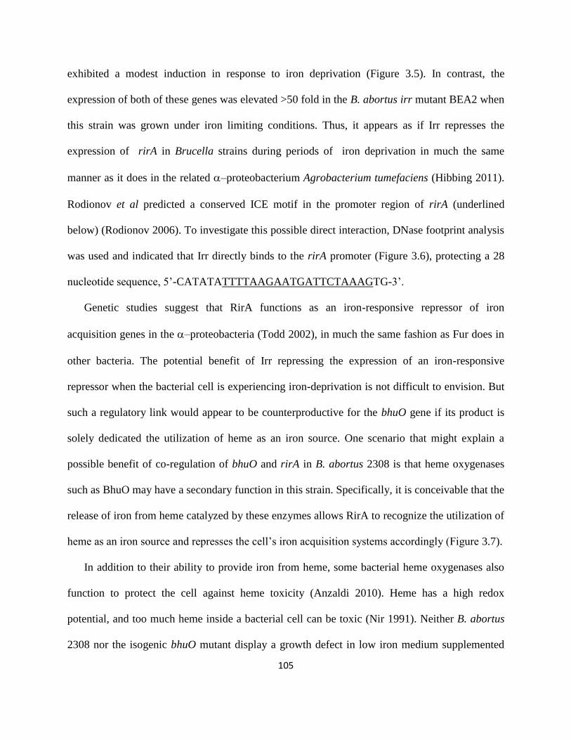
105
exhibited a modest induction in response to iron deprivation (Figure 3.5). In contrast, the
expression of both of these genes was elevated >50 fold in the B. abortus irr mutant BEA2 when
this strain was grown under iron limiting conditions. Thus, it appears as if Irr represses the
expression of rirA in Brucella strains during periods of iron deprivation in much the same
manner as it does in the related –proteobacterium Agrobacterium tumefaciens (Hibbing 2011).
Rodionov et al predicted a conserved ICE motif in the promoter region of rirA (underlined
below) (Rodionov 2006). To investigate this possible direct interaction, DNase footprint analysis
was used and indicated that Irr directly binds to the rirA promoter (Figure 3.6), protecting a 28
nucleotide sequence, 5’-CATATATTTTAAGAATGATTCTAAAGTG-3’.
Genetic studies suggest that RirA functions as an iron-responsive repressor of iron
acquisition genes in the –proteobacteria (Todd 2002), in much the same fashion as Fur does in
other bacteria. The potential benefit of Irr repressing the expression of an iron-responsive
repressor when the bacterial cell is experiencing iron-deprivation is not difficult to envision. But
such a regulatory link would appear to be counterproductive for the bhuO gene if its product is
solely dedicated the utilization of heme as an iron source. One scenario that might explain a
possible benefit of co-regulation of bhuO and rirA in B. abortus 2308 is that heme oxygenases
such as BhuO may have a secondary function in this strain. Specifically, it is conceivable that the
release of iron from heme catalyzed by these enzymes allows RirA to recognize the utilization of
heme as an iron source and represses the cell’s iron acquisition systems accordingly (Figure 3.7).
In addition to their ability to provide iron from heme, some bacterial heme oxygenases also
function to protect the cell against heme toxicity (Anzaldi 2010). Heme has a high redox
potential, and too much heme inside a bacterial cell can be toxic (Nir 1991). Neither B. abortus
2308 nor the isogenic bhuO mutant display a growth defect in low iron medium supplemented

106
with up to 200 µM deferrated hemin. It is possible, however, that any role that BhuO might be
playing in the detoxification of heme is masked by the activity of another heme oxygenase.
Another factor that may affect this observed lack of heme toxicity is that Brucella strains possess
multiple homologs of the outer membrane heme-binding proteins (Hbps) that have been
proposed to play a role in capturing heme and preventing its toxicity in Bartonella (Minnick
2009). In Brucella, these proteins are known as the Omp25/31 family of proteins (Cloeckaert
1996). Heme export systems have also been proposed as a means by which bacteria protect
themselves from heme toxicity (Anzaldi 2010). Although a heme exporter has not been identified
in Brucella strains, genes that potentially encode orthologs of proteins linked to porphyrin
(Tatsumi 2008) and heme (Rasmussen 2005) export in other bacteria can be found in the genome
sequence of B. abortus 2308.
In order to fully understand the role of BhuO in iron and heme metabolism in Brucella
strains, it will be imperative to identify the other heme oxygenase(s) present in these bacteria.
Phenotypic evaluation of mutants lacking combinations of these enzymes can then be used to
assess the relative contributions of the heme oxygenases to iron and heme metabolism, as well as
their potential role in modulating the regulatory capacity of RirA. Such studies may also provide
an added practical benefit, as prokaryotic heme oxygenases have been proposed to be targets for
the development of antimicrobial agents (Furci 2007). Brucellosis in humans is notoriously
difficult to treat, requiring a combination of antibiotics for a prolonged period (Ariza 2007).
Hence, the development of improved chemotherapeutic regimens for treating this disease would
of great benefit to the medical community.

107
Acknowledgements
This work was funded by a grant (AI 63516) from the National Institute of Allergy and
Infectious Diseases to R.M.R.

References
Anderson ES, Paulley JT, Martinson DA, Gaines JM, Steele KH and Roop 2nd
RM.
2011. The iron-responsive regulator Irr is required for wild-type expression of the gene
encoding the heme transporter bhuA in Brucella abortus 2308. J. Bacteriol. 193: 5359-5364.
Anzaldi LL and Skaar EP. 2010. Overcoming the heme paradox: heme toxicity and
tolerance in bacterial pathogens. Infect Immun. 78:4977-4989.
Ariza J, Bosilkovski M, Cascio A, Colmenero JD, Corbel MJ, Falagas ME, Memish Za,
Roushan MR, Rubinstein E, Sipsas NV, Solera J, Young EJ and Pappas G. 2007.
Perspectives for the treatment of brucellosis in the 21st century: the Ioannina
recommendations. PLoS Med 4: e317.
Arnow LE. 1937. Colorimetric determination of the components of 3,4-
dihydroxyphenyalanin-tyrosine mixtures. J. Biol. Chem. 118:531–537.
Bellaire BH, Elzer PH, Baldwin CL and Roop 2nd
RM. 1999. The siderophore 2,3-
dihydroxybenzoic acid is not required for virulence of Brucella abortus in BALB/c mice.
Infect. Immun. 67:2615-2618.
Bellaire BH, Elzer PH, Baldwin CL and Roop 2nd
RM. 2003. Production of the
siderophore 2,3-dihydroxybenzoic acid is required for wild-type growth of Brucella abortus

109
in the presence of erythritol under low-iron conditions in vitro. Infect. Immun. 71:2927-
2932.
Caswell CC, Gaines JM and Roop RM 2nd
. 2012. The RNA chaperone Hfq independently
coordinates expression of the VirB type IV secretion system and the LuxR-type regulator
BabR in Brucella abortus 2308. J Bacteriol. 194: 3-14.
Chao TC, Buhrmester J, Hansmeier N, Pühler A and Weidner S. 2005. Role of the
regulatory gene rirA in the transcriptional response of Sinorhizobium meliloti to iron
limitation. Appl. Environ. Microbiol. 71:5969-5982.
Cloeckaert A, Verger JM, Grayon M and Vizcaino N. 1996. Molecular and
immunological characterization of the major outer membrane proteins of Brucella. FEMS
Microbiol Lett. 145: 1-8.
Furci LM, Lopes P, Eakanunkul S, Zhong S, MacKerell AD Jr and Wilks A. 2007.
Inhibition of the bacterial heme oxygenases from Pseudomonas aeruginosa and Neisseria
meningitidis: novel antimicrobial targets. J Med Chem. 50:3804-3813.
Gerhardt P, Tucker LA and Wilson JB. 1950. The nutrition of Brucellae: utilization of
single amino acids for growth. J. Bacteriol. 59:777-782.

110
González Carreró MI, Sangari FJ, Agüero J and García Lobo JM. 2002. Brucella
abortus strain 2308 produces brucebactin, a highly efficient catecholic siderophore.
Microbiology 148: 353-360.
Hibbing ME and Fuqua C. 2011. Antiparallel and interlinked control of cellular iron levels
by the Irr and RirA regulators of Agrobacterium tumefaciens. J. Bacteriol. 193:3461-3472.
Kovach ME, Elzer PH, Hill DS, Roop 2nd
RM and Peterson KM. 1995. Four new
derivatives of the broad-host-range cloning vector pBBR1MCS, carrying different antibiotic
resistance cassettes. Gene. 166:175–176.
López-Goñi I, Moriyón I and Neilands JB. 1992. Identification of 2,3-dihydroxybenzoic
acid as a Brucella abortus siderophore. Infect. Immun. 60: 4496–4503.
Martínez M, Ugalde RA and Almiro´n M. 2006. Irr regulates brucebactin and 2,3-
dihydroxybenzoic acid biosynthesis, and is implicated in the oxidative stress resistance and
intracellular survival of Brucella abortus. Microbiology 152:2591–2598.
Menscher EA, Caswell CC, Anderson ES and Roop RM II. 2012. Mur regulates the gene
encoding the manganese transporter MntH in Brucella abortus 2308. J Bacteriol. 194:561-
566.

111
Minnick MF and Battisti JM. 2009. Pestilence, persistence and pathogenicity: infection
strategies of Bartonella. Future Microbiol. 4:743-758.
Ngok-Ngam P, Ruangkiattikul N, Mahavihakanont A, Virgem SS, Sukchawalit R and
Mongkolsuk S. 2009. Roles of Agrobacterium tumefaciens RirA in iron regulation, oxidative
stress response, and virulence. J. Bacteriol. 191:2083-2090.
Nir U, Ladan H, Malik Z and Nitzan Y. 1991. In vivo effects of porphyrins on bacterial
DNA. J Photochem Photobiol B. 11: 295-306.
Paulley JT, Anderson ES and Roop 2nd
RM. 2007. Brucella abortus requires the heme
transporter BhuA for maintenance of chronic infection in BALB/c mice. Infect. Immun. 75:
5248–5254.
Pfaffl MW. 2001. A new mathematical model for relative quantification in real-time RT-
PCR. Nucleic Acids Res. 29:2002–2007.
Puri S and O'Brian MR. 2006. The hmuQ and hmuD genes from Bradyrhizobium
japonicum encode heme-degrading enzymes. J. Bacteriol. 188:6476-6482.
Rasmussen AW, Alexander HL, Perkins-Balding D, Shafer WM and Stojiljkovic I.
2005. Resistance of Neisseria meningitidis to the toxic effects of heme iron and other
hydrophobic reagents requires expression of ght. J. Bacteriol. 187:5214-5223.

112
Ratliff M, Zhu W, Deshmukh R, Wilks A and Stojiljkovic I. 2001. Homologues of
neisserial heme oxygenase in Gram-negative bacteria: degradation of heme by the product of
the pigA gene of Pseudomonas aeruginosa. J. Bacteriol. 183:6394-6403.
Rodionov DA, Gelfand MS, Todd JD, Curson AR and Johnston AW. 2006.
Computational reconstruction of iron- and manganese – responsive transcriptional networks
in alpha-proteobacteria. PLoS Comput Biol. 2: e163.
Rogers HJ. 1973. Iron-binding catechols and virulence in Escherichia coli. Infect. Immun.
7:445-456.
Roop 2nd
RM, Anderson E, Ojeda J, Martinson D, Menscher E and Martin DW. 2011.
Metal acquisition by Brucella strains. I. López-Goñi and D. O’Callaghan (eds.), Brucella:
molecular microbiology and genetics, Horizon Scientific Press, Norfolk, UK. 179-199.
Rosner B. 2000. Fundamentals of biostatistics, 5th ed. Duxbury, Pacific Grove, CA.
Schaible UE and Kaufmann SHE. 2004. Iron and microbial infection. Nature Rev.
Microbiol. 2:946-953.

113
Schmitt MP. 1997. Utilization of host iron sources by Corynebacterium diphtheriae:
identification of a gene whose product is homologous to eukaryotic heme oxygenases and is
required for acquisition of iron from heme and hemoglobin. J. Bacteriol. 179:838–845.
Skaar EP, Gaspar AH and Schneewind O. 2004. IsdG and IsdI, heme-degrading enzymes
in the cytoplasm of Staphylococcus aureus. J. Biol. Chem. 279:436-443.
Spratt BG, Hedge PJ, te Heesen S, Edelman A and Broome-Smith JK. 1986.
Kanamycin-resistant vectors that are analogues of plasmids pUC8, pUC9, pEMBL8 and
pEMBL9. Gene. 41:337-342.
Staggs TM and Perry RD. 1991. Identification and cloning of a fur regulatory gene in
Yersinia pestis. J. Bacteriol. 173:417-425.
Tatsumi R and Wachi M. 2008. TolC-dependent exclusion of porphyrins in Escherichia
coli. J. Bacteriol. 190:6228-6233.
Todd JD, Wexler M, Sawers G, Yeoman KH, Poole PS and Johnston AWB. 2002. RirA,
an iron-responsive regulator in the symbiotic bacterium Rhizobium leguminosarum.
Microbiology 148:4059-4071.
Wu R, Skaar EP, Zhang R, Joachimiak G, Gornicki P, Schneewind O and Joachimiak
A. 2005. Staphylococcus aureus IsdG and IsdI, heme-degrading enzymes with structural
similarity to monooxygenases. J. Biol. Chem. 280:2840-2846.

114
Zhu W, Hunt JD, Richardson AR and Stojiljkovic I. 2000. Use of heme compounds as
iron sources by pathogenic Neisseriae requires the product of the hemO gene. J. Bacteriol.
182: 439-447.

115
Table 3.1 Bacterial strains used in this study.

116
TABLE 1. Bacterial strains used in this study
Strain or plasmid Genotype or description Reference or source
Strains
Escherichia coli
DH5α F¯ φ80lacZΔM15 Δ(lacZYA-argF)U169 recA1 endA1
hsdR17(rK¯ mK⁺) phoA supE44 thi-1 gyrA96 relA1 λ¯
Invitrogen
Brucella abortus
2308 Virulent challenge strain Laboratory stock
DCO1 70 bp gene deletion of bhuO (BAB2_0677) with an ahpA3
kanamycin resistance gene insertion
This study
DCO1RC DCO1 with bhuO (BAB2_0677) reconstructed onto the
chromosome using pNPTS138
This study
BHB2 Nonpolar, in frame deletion of dhbC Bellaire 1999
JFO1 ΔdhbC/ΔbhuO This study
JFO1RC ΔdhbC/ΔbhuO with bhuO (BAB2_0677) reconstructed
onto the chromosome using pNPTS138
This study
BEA2 Δirr Anderson 2011
Plasmids
pGEM-T Easy ColE1-based cloning vector; ampicillin resistance Promega
pKS + Kan 794-bp aphA3 gene from TnphoA cloned into SalI-HindIII-
digested pBluescript II KS+
Kovach 1995
pNPTS138 sacB-containing counterselection vector; ampicillin
resistance
Spratt 1986
Table 3.1

117
Table 3.2 Primers used in this study.

118
Primers used in this study bhuO-1F 5’-GACATTTCGCTGAAGACATA -3’
bhuO-1R 5’-GAGCTTACTTTACCGTTGGC-3’
bhuO-2F 5’-TCTATGCATCCCACACAGTC-3’
bhuO-2R 5’-GAGCTGGGAGTCACGGTTCT-3’
amp - F 5’-TATTTCGTTCATCCATA-3’
amp - R 5’-GTTTTCCAATGATGAGC-3’
Kan R 5’-CCACTCCAGCATGAGAT-3’
bhuO-3F 5’-ACTGGATCCCGGAGCTGTTTCTATTC-3’
bhuO-3F 5’-ACTGTCGACTTCTAAATATGAGTTAA-3’
16S Fwd 5’-TCTCACGACACGAGCTGACG-3’
16S Rev 5’-CGCAGAACCTTACCAGCCCT-3’
dhbC Fwd 5’-GTGCCAAGCTTGGTCTGTACTTC-3’
dhbC Rev 5’-CGTGGATTGTTTACCGGC-3’
bhuO Fwd 5’-CGGTAGCGAAACCGATTTTGAGAC-3’
bhuO Rev 5’-ATGCGCATGGCGAAACTGTT-3’
rirA F 5’-GCACCGCAGAAAATGGCCGA-3’
rirA R 5’-TGGAACATTCCCGAACCGGA-3’
Rev 5’-GAAGGCATTGAGTGCTTCGC-3’
Ybak/rirA F 5’-TAAAGGTGAGAAAAGGTTTC-3’
Ybak/rirA R 5’-GACGCATGATTATTCCTTTT-3’
rirA/bhuO F 5’-GGATAGCTGTGGCCTGAATG-3’
rirA/bhuO R 5’-GAGCTGGGAGTCACGGTTCT-3’
bhuO/bfr F 5’-GCTTCCGTATCGTCACTTTT-3’
bhuO/bfr R 5’-GAGCTTACTTTACCGTTGGC-3’
rirA F 5’-GCACCGCAGAAAATGGCCGA-3’
rirA R 5’-TGGAACATTCCCGAACCGGA-3’
Table 3.2
119
Figure 3.1. The B. abortus BhuO protein shares amino acid homology with HmuD/Q and
IsdG/I. The Brucella abortus (Ba) BhuO protein shares amino acid homology with the heme
oxygenases HmuD and HmuQ from Bradyrhizobium japonicum (Bj) and IsdG and IsdI from
Staphylococcus aureus (Sa) and contains the conserved Asn 7, Trp 67 and His 77 residues
(shown in larger, bold font) shown experimentally to be important for the heme oxygenase
activity of IsdG (Skaar 2004).

120
Figure 3.1

121
Figure 3.2. B. abortus DCO1 produces significantly more siderophore than B. abortus 2308
in response to iron deprivation. (A) Siderophore production by B. abortus 2308, DCO1
(ΔbhuO) and DCO1RC (DCO1 bhuO+) following 72 h growth in low iron minimal medium. The
values on the Y axis represent the levels of catechol siderophore detected by the Arnow assay
(Arnow 1937). ** = P < 0.01 for comparisons of the data obtained for these strains in the
Student’s two-tailed t-test (Rosner, 2000). (B) Iron-responsive expression of dhbC in B. abortus
2308 and DCO1 (2308 bhuO). ‘Fold induction’ on the Y axis represents the difference between
the levels of dhbC transcripts detected in RNA preparations from B. abortus 2308 and DCO1
cultures after 72 h of growth in low iron minimal medium compared to RNA preparations from
these cultures after growth in low iron minimal medium supplemented with 50 µM FeCl3.

122
Figure 3.2

123
Figure 3.3. B. abortus JFO1 has a growth defect in the presence of heme as the sole iron
source. Growth of B. abortus DCO1 (2308 ΔbhuO) (squares), BHB2 (2308 ΔdhbC) (circles),
JFO1 (2308 ΔdhbC/ΔbhuO) (triangles), and JFO1RC (JFO1 bhuO+) (diamonds) in (A) low iron
minimal medium, (B) low iron minimal medium containing 50µM FeCl3, (C) low iron minimal
medium supplemented with 50µM FeCl3 at 48 hours post-inoculation, and (D) low iron minimal
medium supplemented with deferrated 25µM hemin at 48 hours post inoculation. The data
presented are from one experiment, but representative of multiple experiments (>3) from which
similar trends were observed** = P < 0.01 and *** = P < 0.001 for comparisons of the data
obtained for these strains in the Student’s two-tailed t-test (Rosner 2000).

124
Figure 3.3

125
Figure 3.4. rirA and bhuO are cotranscribed as an operon in B. abortus 2308. The start site
(+1) for the rir-bhuO transcript is located 134 base pairs upstream from the rirA ORF (bold,
underlined), and the location of an Irr binding motif predicted by Rodionov et al. is denoted as a
‘ICE Box’ and shown in larger, bold-face type (Rodionov 2004), while the nucleotide sequence
protected by Irr is shown in a box. Primer sets 1, 2, and 3 denote the intragenic regions used to
define the transcript and the corresponding lanes are marked on the agarose gel picture below it.
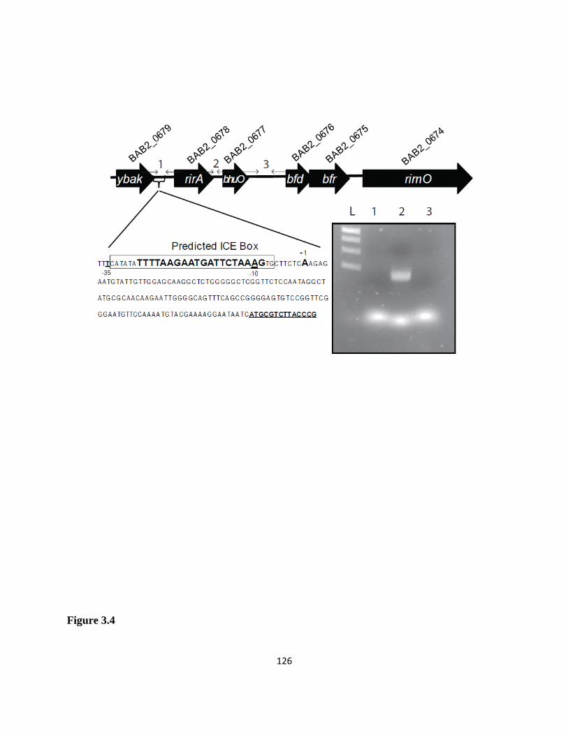
126
Figure 3.4

127
Figure 3.5. rirA and bhuO transcription is increased in response to iron deprivation in B.
abortus BEA2(Δirr). ‘Fold induction’ on the Y axis represents the difference between the levels
of rirA, bhuO and bhuA transcripts detected in RNA preparations from B. abortus 2308 and
BEA2 after 72 h of growth in low iron minimal medium compared to RNA preparations from
these cultures after growth in low iron minimal medium supplemented with 50 µM FeCl3. The
pattern of bhuA transcription was included in this figure because this gene exhibits elevated
expression in response to iron deprivation in B. abortus 2308, and this low iron-responsive
induction is dependent upon the presence of Irr (Anderson 2011).

128
Figure 3.5

129
Figure 3.6. Irr binds directly to the rirA promoter in B. abortus 2308 and protects a 28
nucleotide sequence in a DNase I footprint analysis. The triangle above the lanes indicates
that the corresponding reaction mixtures contain increasing concentrations (700 ng, 1.4 µg, 2.1
µg, and 3.5 µg) of recombinant Brucella Irr, and the nucleotide sequences shown to the right of
the gel photos denote the nucleotides protected from DNaseI digestion in the forward and reverse
strands of the rirA promoter sequence.

130
Figure 3.6.

131
Figure 3.7. Proposed model for the role of BhuO in allowing the transcriptional regulator
RirA to recognize heme as an iron source in Brucella.

132
Figure 3.7

Chapter 4: Summary and Future Directions
The requirement for heme in Brucella
The virulence of Brucella abortus within the mouse model of infection is
dependent on the ability of the brucellae to take in heme during their residence within the host
macrophage. Many pathogens have heme transporters, but only a few have been shown to be
important for virulence. To date, only Bordetella pertussis, enterohaemorrhagic Escherichia coli,
Listeria monocytogenes, and the heme auxotroph Haemophilus influenzae contain heme
transport systems that contribute significantly to virulence of the organism (Morton 2007, Torres
1997, Brickman 2007, Jin 2006). The data collected on BhuA and BhuTUV has demonstrated
that without one or more of these components, the cell is unable to take in heme as an iron
source, therefore this is the only heme transport system in B. abortus 2308. The original bhuA
mutant was constructed using a chloramphenicol resistance cassette. Recently the CDC instituted
new guidelines for this select agent that no longer allows chloramphenicol resistance to be
introduced into the animal models, and therefore a new bhuA kanamycin resistant mutant is
currently under construction with the intent to use it as a tool for comparison with the bhuTUV
mutant in the mouse model of infection. While it seems likely that the absence of bhuTUV in the
brucellae will produce a similar phenotype, experiments will need to be done to confirm this link
between BhuTUV and virulence.

134
Unknown heme transport components
Encoded within the bhuTUV region, and clearly sharing the same operon (see Figure 2.1)
are two hypothetical open reading frames (ORFs). The first hypothetical ORF is upstream,
overlaps bhuT by 44 base pairs and encodes a protein that contains no known homology to any
other protein. The second hypothetical ORF is located at the 3’ end of bhuV and also lacks
homology to any other known protein. The reason they are mentioned here is because it is not
unusual for small hypothetical ORFs to actually encode proteins of unknown function within
heme uptake operons, and it is possible that these two hypothetical ORFs do in fact serve a
purpose for heme uptake. One component that is clearly absent in the Brucella heme uptake
model (see Figure 4.1) is a shuttle to accept the heme once it is transported through BhuU. In
Shigella, ShuS serves this function, chaperoning the heme to the heme oxygenase, and studies
with reconstituted membranes have shown ShuS to be required for the heme to be released from
the cytoplasmic permease (Burkhard 2008). The shuttle, HemS, from Yersinia entercolitica has
been crystallized and the His-196 is required for functionality of the protein. This histidine is
highly conserved across the known shuttles (Wilks 2011). While there has been no such protein
found in Brucella, it is not unheard of for these proteins to only look similar in their native
conformations, which cannot be searched for in a traditional BLAST search. This hypothetical
protein is only 138 amino acids long, and therefore cannot have have a His – 196, however it
does have two conserved H-X-X-H domains that are also known heme binding motifs (Matias
2002).

135
The complexity of regulation of the B. abortus heme transport genes
Of particular interest is the unique placement of the genes encoding Brucella’s heme
transport machinery on different parts of chromosome II (see Figure 4.2). It is possible that this
distribution of genes is the result of evolutionary inheritance or perhaps lateral gene transfer of
the heme transport machinery in parts from other bacteria. While the brucellae do spend most of
their time isolated within the host macrophage, they are considered facultative intracellular
organisms because they can survive in the environment between infected animals.
Evolutionarily, some argue that the second chromosome (which incidently is the one with the
heme uptake genes) is auxiliary, and that it contains a region of homology with that of
Bartonella species, suggesting a common ancestor for the “auxiliary replicon” (Batut 2004) .
Obviously there is an evolutionary benefit to having these in separate loci because bhuA
and bhuTUV show different patterns of iron-responsive regulation. While we know that iron
metabolism genes must be carefully regulated in order to maintain a safe homeostasis, perhaps in
Brucella this seemingly complex web of regulation hints at the importance of the interactions of
the brucellae with heme in the host. For instance, RirA has been shown to be protective against
cellular oxidative stress, and an isogenic mutant rirA is more sensitive to hydrogen peroxide than
B. abortus 2308 (unpublished laboratory observation). Is it possible then that RirA is meant not
only to protect the brucellae against the oxidative burst of the macrophage as it first enters the
cell but also to repress iron uptake genes at a time when the detrimental effects of the Fenton
chemistry due to the combination of iron and oxygen would be most likely?
The interplay of Irr and RirA hints at two different cellular conditions that the brucellae
are exposed to in vivo, one that mimics log phase of growth and another that mimics stationary
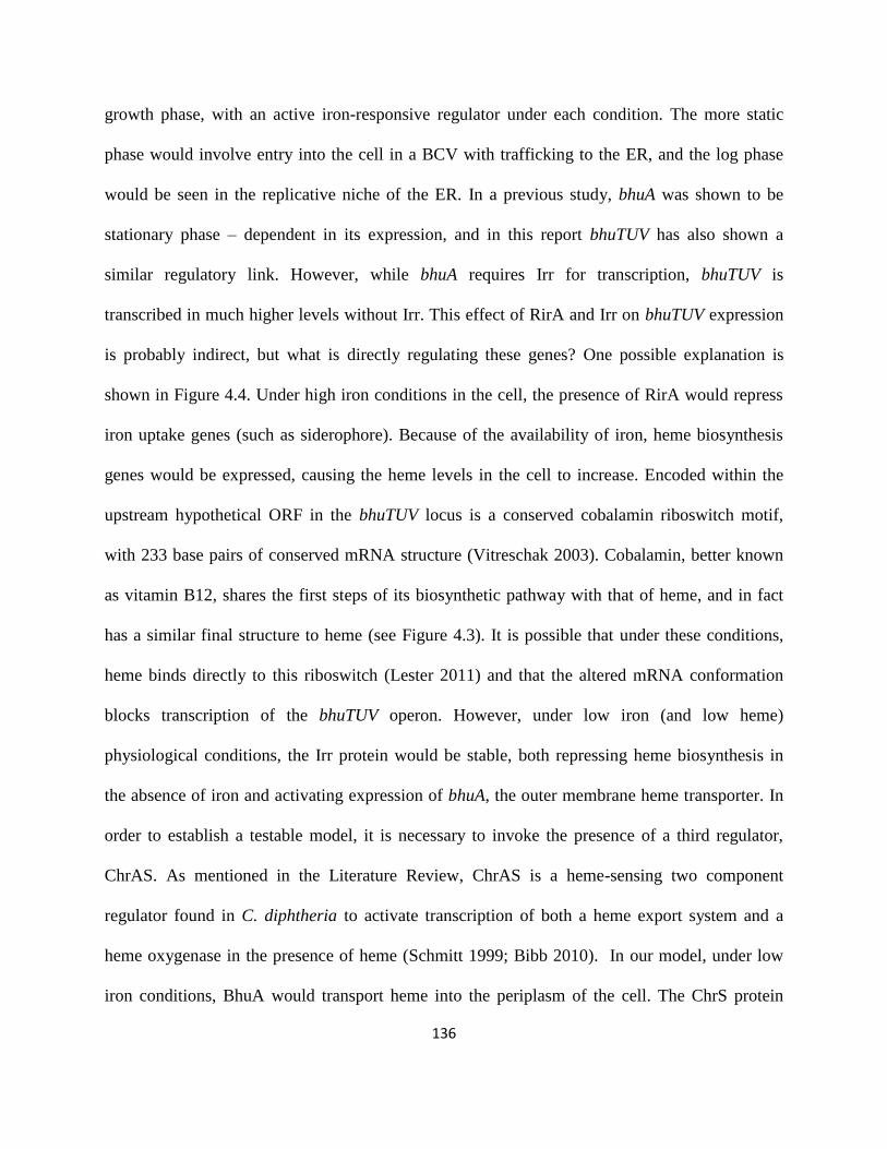
136
growth phase, with an active iron-responsive regulator under each condition. The more static
phase would involve entry into the cell in a BCV with trafficking to the ER, and the log phase
would be seen in the replicative niche of the ER. In a previous study, bhuA was shown to be
stationary phase – dependent in its expression, and in this report bhuTUV has also shown a
similar regulatory link. However, while bhuA requires Irr for transcription, bhuTUV is
transcribed in much higher levels without Irr. This effect of RirA and Irr on bhuTUV expression
is probably indirect, but what is directly regulating these genes? One possible explanation is
shown in Figure 4.4. Under high iron conditions in the cell, the presence of RirA would repress
iron uptake genes (such as siderophore). Because of the availability of iron, heme biosynthesis
genes would be expressed, causing the heme levels in the cell to increase. Encoded within the
upstream hypothetical ORF in the bhuTUV locus is a conserved cobalamin riboswitch motif,
with 233 base pairs of conserved mRNA structure (Vitreschak 2003). Cobalamin, better known
as vitamin B12, shares the first steps of its biosynthetic pathway with that of heme, and in fact
has a similar final structure to heme (see Figure 4.3). It is possible that under these conditions,
heme binds directly to this riboswitch (Lester 2011) and that the altered mRNA conformation
blocks transcription of the bhuTUV operon. However, under low iron (and low heme)
physiological conditions, the Irr protein would be stable, both repressing heme biosynthesis in
the absence of iron and activating expression of bhuA, the outer membrane heme transporter. In
order to establish a testable model, it is necessary to invoke the presence of a third regulator,
ChrAS. As mentioned in the Literature Review, ChrAS is a heme-sensing two component
regulator found in C. diphtheria to activate transcription of both a heme export system and a
heme oxygenase in the presence of heme (Schmitt 1999; Bibb 2010). In our model, under low
iron conditions, BhuA would transport heme into the periplasm of the cell. The ChrS protein
137
embedded in the cytoplasmic membrane would sense the heme and phosphorylate ChrA. In turn,
ChrA would activate expression of bhuTUV, allowing the heme to be transported from the
periplasm into the cell (see Figure 4.4). We have some preliminary evidence that ChrA is
activating bhuTUV and also bhuA, which would provide a feedback mechanism for the increased
expression of bhuA, possibly allowing for full expression of the heme uptake machinery.
Alternatively, in Chapter 2, the possibility of an uncharacterized regulator is mentioned.
This extracytoplasmic function sigma factor is an iron-responsive σ70
protein regulator, and there
are two such putative proteins in Brucella (RpoE1 and 2). The paradigm for iron-dependent
sigma factors is FecI from E. coli which depends on the outer membrane ferric dicitrate
transporter binding to ferric dicitrate to induce a signal to a plasma membrane protein (FecR)
which then activates the cytoplasmic FecI to promote transcription of the ferric dicitrate operon
(Kirby 2001). In fact, in Bordetella the FecIR homologs directly regulate expression of the
bhuRSTUV genes that encode the inner membrane heme transporter components (King 2005). In
Brucella, Irr activates the expression of bhuA, and perhaps BhuA binding to heme signals a
Brucella FecR which then activates the RpoE to promote transcription of the bhuTUV locus.
Brucella must acquire iron from the heme
There were two main goals to the research project described in this document: one was to
identify the inner membrane heme transport components of B. abortus 2308 and the second was
to determine the contribution of BhuO, a known heme oxygenase, to the iron requirements of the
Brucella. Because the bhuO mutant strain was still able to grow around heme on an agar plate
when heme was the sole source of iron and there was no significant phenotype for the bhuO

138
mutant strain compared to B. abortus 2308 in vivo, there is a second heme oxygenase in B.
abortus 2308. This is not suprising since the closest homologs of BhuO, IsdI/G and HmuD/Q are
both present in pairs (Skaar 2004, Puri 2006). What is suprising is that these other pairs of heme
oxygenases share high amino acid sequence homology, whereas multiple NCBI BLAST searches
using BhuO for comparison were unable to uncover a second Brucella heme oxygenase. In fact,
all known heme oxygenases were used for comparisons in these NCBI BLAST searches, and still
there is no likely candidate for this second heme oxygenase based on conserved amino acid
residues. Searches of the annotated B. abortus 2308 genome yielded a gene whose product was a
monooxygenase, a common annotation for heme oxygenases. This monooxygenase does not
contain the conserved asparagine residue near its proximal end, but does have a tryptophan and
histidine near the distal end of the protein, which in IsdG are known functional sites. Future work
should target this gene, BAB1_1911, for mutagenesis in a bhuO mutant background to determine
if the ability to use heme as an iron source has been completely removed. Alternatively, a
transposon mutagenesis library made in the bhuO mutant may reveal candidates for the other
heme oxygenase(s) by identifying any mutants with a heme utilization defect.
Unanswered questions about bacterial heme oxygenases
It has been shown in mammals that the products of heme degradation, biliverdin and
bilirubin, perform downstream functions to protect the cell by reducing the inflammatory
response (Chung 2009). While it has been observed for some time in our lab that there is a color
change consistent with what has been previously described as the presence of porphyrins in the
low iron medium containing growing B. abortus strains (Martinez 2005), no data to date has

139
been collected regarding what happens to the products of BhuO. If biliverdin in some form is
being pumped out of the cell, what could the physiological significance of this be, given that the
brucellae naturally reside within the mammalian cells? The mammalian HO-1 has been
implicated in the production of anti-inflammatory signaling (see literature review), perhaps the
Brucella heme oxygenase products are also released into the mammalian cell and act to “calm”
the immune response, allowing for the maintenance of a chronic infection.
Additionally, a question yet to be addressed in any bacterium but often asked: Is
exogenously acquired heme incorporated directly into bacterial proteins or is it always broken
down for iron first? This question would be easiest initially to answer using known heme
auxotrophs such as Haemophilus influenzae, Porphyromonas gingivalis, and even a close
relative to Brucella, Bartonella quintana (Hanson 1992, Bramanti 1991, Battisti 2007). While it
makes sense to conserve bacterial energy whenever possible and simply use the heme that is
transported, studies have yet to confirm this.

References
Battisti JM, Smitherman LS, Sappington KN, Parrow NL, Raghavan R and Minnick MF.
2007. Transcriptional regulation of the heme binding protein gene family of Bartonella
quintana is accomplished by a novel promoter element and iron response regulator. Infect.
Immun. 75: 4373-4385.
Batut J, Andersson SGE and O’Callaghan D. 2004. The evolution of chronic infection
strategies in the α–proteobacteria. Nature Reviews Microbiology 2, 933–945.
Bibb LA and Schmitt MP. 2010. The ABC transporter HrtAB confers resistance to hemin
toxicity and is regulated in a hemin-dependent manner by the ChrAS two-component system in
Corynebacterium diphtheriae. J. Bacteriol. 192:4606-4617.
Bramanti TE and Holt SC. 1991.Roles of porphyrins and host iron transport proteins in
regulation of growth of Porphyromonas gingivalis W50.J Bacteriol. 173:7330-7339.
Brickman TJ, Anderson MT and Armstrong SK. 2007. Bordetella iron transport and
virulence. Biometals. 20:303-322.

141
Burkhard KA and Wilks A. 2008. Functional characterization of the Shigella dysenteriae heme
ABC transporter. Biochemistry. 47:7977 - 7979.
Chung SW, Hall S and Perrella MA. 2009. Role of heme oxygenase-1 in microbial host
defense. Cell Microbiol. 11:199-207.
Hanson MS, Pelzel SE, Latimer J, Muller-Eberhard U and Hansen EJ. 1992. Identification
of a genetic locus of Haemophilus influenzae type b necessary for the binding and utilization of
heme bound to human hemopexin.Proc Natl Acad Sci U S A.89:1973-1977.
Jin B, Newton SM, Shao Y, Jiang X, Charbit A and Klebba PE. 2006. Iron acquisition
systems for ferric hydroxamates, haemin and haemoglobin in Listeria monocytogenes. Mol
Microbiol. 59:1185-1198.
King ND, Kirby AE and Connell TD. 2005. Transcriptional control of the rhuIR-bhuRSTUV
heme acquisition locus in Bordetella avium. Infect. Immun. 73: 1613-1624.
Kirby AE, Metzger DJ, Murphy ER and Connell TD. 2001. Heme Utilization in Bordetella
avium Is Regulated by RhuI, a Heme-Responsive Extracytoplasmic Function Sigma Factor.
Infect and Immun. 69:6951-6961.

142
Lester CH, Poon S, Methot P, Morabi-Pazooki W, Pio F, Bennet AJ and Sen D. 2011.
Guanine-rich RNAs and DNAs that bind heme robustly catalyze oxygen transfer reactions.
Journal of the American Chemical Society. 133: 1877-1884.
Martinez M, Ugalde R and Almiron M. 2005. Dimeric Brucella abortus Irr protein controls
its own expression and binds haem .Microbiology. 151:3427-3433.
Matias PM, Coelho AV, Valente FMA, Plácido D, LeGall J, Xavier AV, Pereira IAC and
Carrondo MA. 2002. Sulfate respiration in Desulfovibrio vulgaris Hildenborough: Structure of
the 16-heme Cytochrome c HmcA at 2.5 Å resolution and a view of its role in transmembrane
electron transfer. J Biol Chem. 277:47907-47916.
Morton DJ, Seale TW, Madore LL, VanWagoner TM, Whitby PW and Stull TL. 2007. The
haem-haemopexin utilization gene cluster (hxuCBA) as a virulence factor of Haemophilus
influenzae. Microbiology 153:215–224.
Paulley JT. 2007. Production of BhuA by Brucella abortus is required for hemin utilization and
virulence and is dependent on the transcriptional regulators RirA and ChrA. Dissertation.
Microbiology and Immunology, East Carolina University.

143
Puri S and O'Brian MR. 2006. The hmuQ and hmuD genes from Bradyrhizobium japonicum
encode heme-degrading enzymes. J. Bacteriol. 188:6476-6482.
Schmitt MP. 1999. Identification of a two-component signal transduction system from
Corynebacterium diphtheriae that activates gene expression in response to the presence of heme
and hemoglobin. J. Bacteriol. 181: 5330-5340.
Skaar EP, Gaspar AH and Schneewind O. 2004. IsdG and IsdI, heme-degrading enzymes in
the cytoplasm of Staphylococcus aureus. J. Biol. Chem. 279:436-443.
Torres AG and Payne SM. 1997. Haem iron-transport system in enterohaemorrhagic
Escherichia coli O157:H7. Mol Microbiol.23:825–833.
Vitreschak AG, Rodionov DA, Mironov AA and Gelfand MS. 2003. "Regulation of the
vitamin B12 metabolism and transport in bacteria by a conserved RNA structural element". RNA
9: 1084–1097.
Wilkes A and Barker KD. 2011. 72 Mechanisms of heme uptake and utilization in bacterial
pathogens. Handbook of Porphyrin Science. Volumes 11–15:357-398.

144
Figure 4.1 Heme transport in Brucella abortus 2308. In keeping with heme transport in other
bacteria, brucellae utilize a heme–specific membrane transport complex in order to bind heme
and bring it into the cell for use as an iron source. Once the heme has entered the cytoplasm of
the cell, a heme oxygenase (BhuO) breaks it down into iron, biliverdin, and carbon monoxide.

145
Figure 4.1

146
Figure 4.2 Genetic organization of the B. abortus genes associated with heme uptake and
utilization. The outer membrane heme transporter is encoded on one part of chromosome II,
located near a siderophore uptake regulator, the genes encoding the periplasmic and cytoplasmic
components of heme transport are located elsewhere on chromosome II and the heme oxygenase
is encoded in an operon with rirA, also separately from the rest and directly upstream from the
genes encoding the iron storage protein bacterioferritin. This unique separation of genes
responsible for heme uptake is conserved across the Brucella genomes. The black arrows
represent all of the genes in each operon.

147
Figure 4.2

148
Figure 4.3 The similar stuctures of cobalamin (B12) and heme. Cobalamin and heme share
the same initial biosynthetic steps, and the overall structures look similar.

149
Figure 4.3

150
Figure 4.4 Model of possible regulation of heme uptake genes in B. abortus 2308.

151
Figure 4.4
Related Documents


