IRON DEFICIENCY ANAEMIA DOLLY JOHNSON EWILI 1404

IRON DEFICIENCY ANAEMIA DOLLY JOHNSON EWILI 1404.
Dec 22, 2015
Welcome message from author
This document is posted to help you gain knowledge. Please leave a comment to let me know what you think about it! Share it to your friends and learn new things together.
Transcript

IRON DEFICIENCY ANAEMIA
DOLLY JOHNSON EWILI
1404

TABLE OF CONTENT INTRODUCTION
CAUSES IRON METABOLISMSYMPTOMS ASSOCIATED CONDITIONSFINDINGSDIFFERENTIALSREFERENCES

INTRODUCTION Iron deficiency is the most common cause of anaemia.
Iron is vital for all living organisms because it is essential for multiple metabolic processes, including oxygen transport, DNA synthesis, and electron transport.
Iron deficiency anaemia is caused by insufficient intake and absorption of iron, and/or iron loss from bleeding which can originate from a range of sources such as the intestinal, uterine or urinary tract. It affects women more than men and this is though to be because of the monthly loss of blood. Iron deficiency can also occur because your body needs more iron than normal (eg. During pregnancy or breastfeeding).

CAUSES Low dietary intake
Infants fed exclusively on milk, Impoverished, Elderly, Teenagers subsisting on junk food
Increased demands Pregnancy, Infancy
Malabsorption Celiac disease (problem with duodenum that impairs absorption) ,
Post-gastrectomy
Intravascular hemolysis Microangiopathic hemolytic anemia
Paroxysmal Nocturnal Hemoglobinuria

The most common cause of iron deficiency anaemia in developed countries is colon cancer especially in the elderly. Also, long standing peptic ulcer that bleeds.
The most significant cause of iron-deficiency anemia in developing world children is parasitic worms: hookworms, whipworms, and roundworms. Worms cause intestinal bleeding.
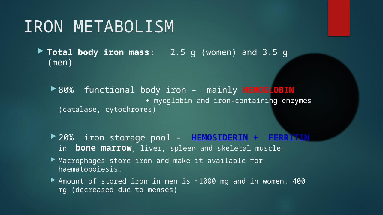
IRON METABOLISM Total body iron mass: 2.5 g (women) and 3.5 g (men)
80% functional body iron – mainly HEMOGLOBIN + myoglobin and iron-containing enzymes (catalase, cytochromes)
20% iron storage pool - HEMOSIDERIN + FERRITIN in bone marrow, liver, spleen and skeletal muscle
Macrophages store iron and make it available for haematopoiesis.
Amount of stored iron in men is ~1000 mg and in women, 400 mg (decreased due to menses)

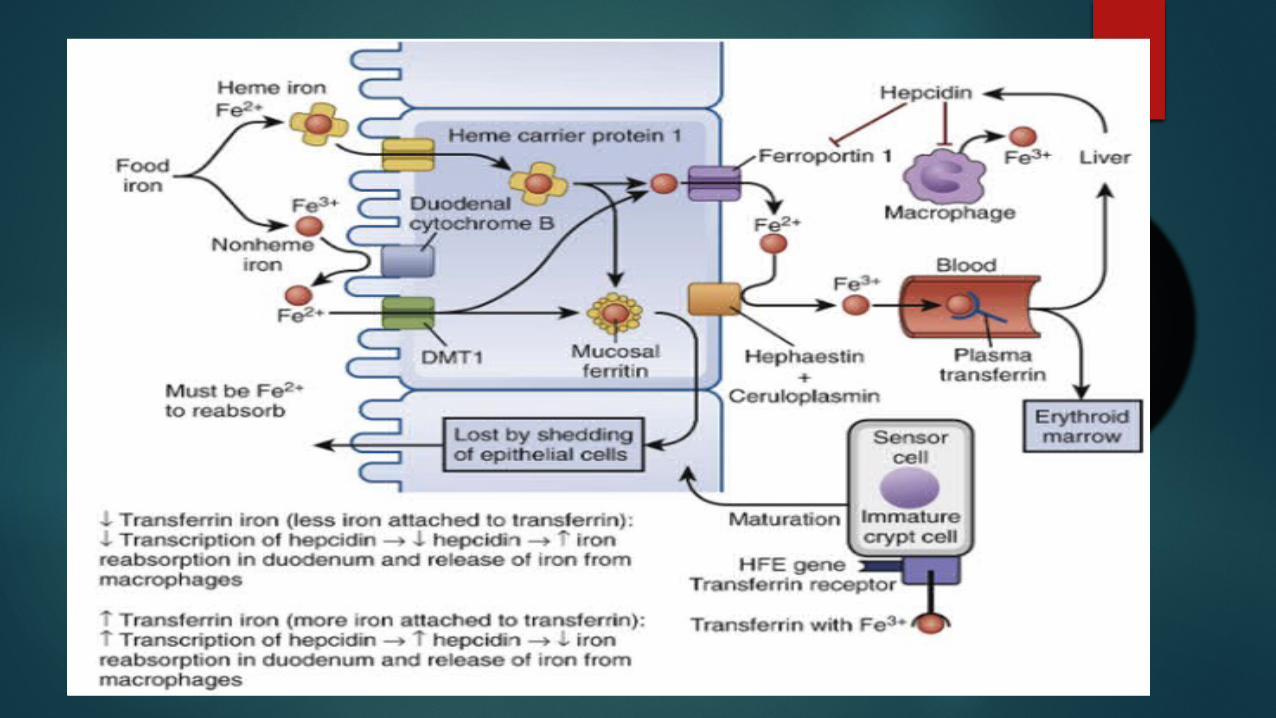
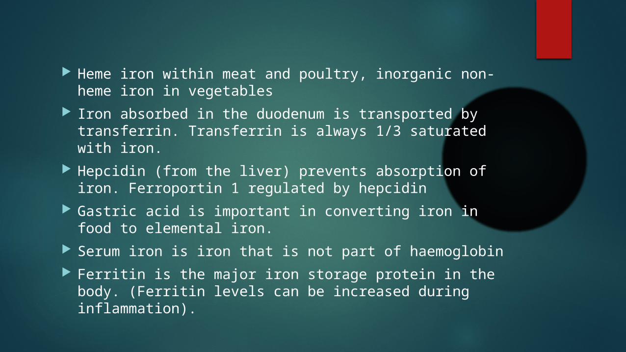
Heme iron within meat and poultry, inorganic non-heme iron in vegetables
Iron absorbed in the duodenum is transported by transferrin. Transferrin is always 1/3 saturated with iron.
Hepcidin (from the liver) prevents absorption of iron. Ferroportin 1 regulated by hepcidin
Gastric acid is important in converting iron in food to elemental iron.
Serum iron is iron that is not part of haemoglobin Ferritin is the major iron storage protein in the body.
(Ferritin levels can be increased during inflammation).

NORMAL VALUES
Serum iron levels: i) Men: 65 to 176 μg/dL
ii)Women: 50 to 170 μg/dL TIBC:- 240-450 μg/dL Serum ferritin levels:- 30–300 μg/L for males, and 6–
115 μg/L for females
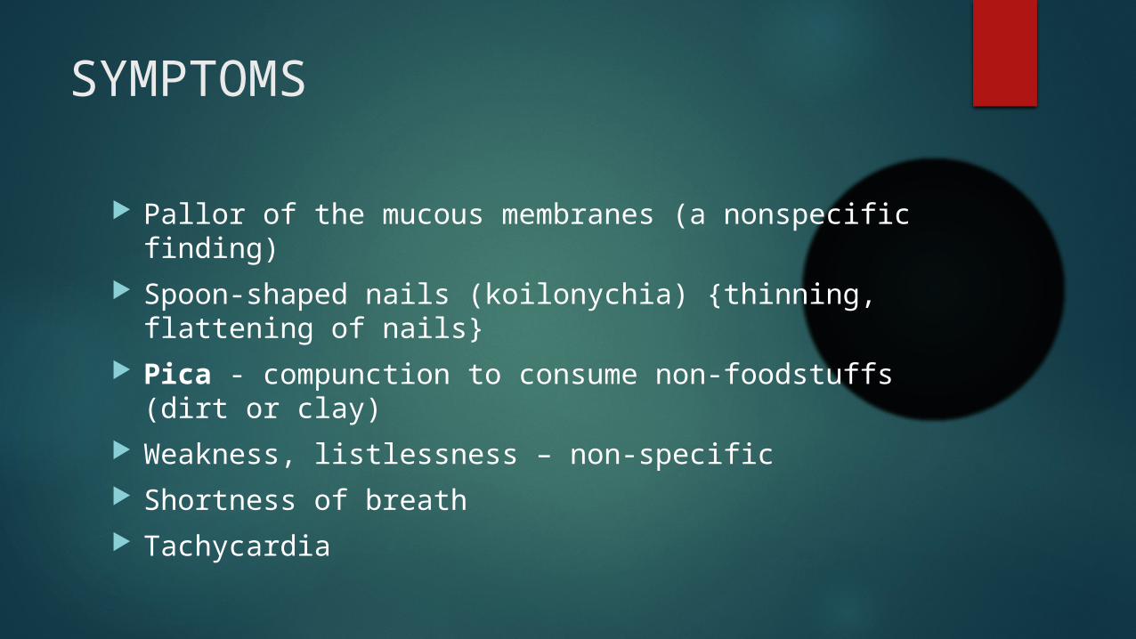
SYMPTOMS
Pallor of the mucous membranes (a nonspecific finding)
Spoon-shaped nails (koilonychia) {thinning, flattening of nails}
Pica - compunction to consume non-foodstuffs (dirt or clay)
Weakness, listlessness – non-specific Shortness of breath Tachycardia
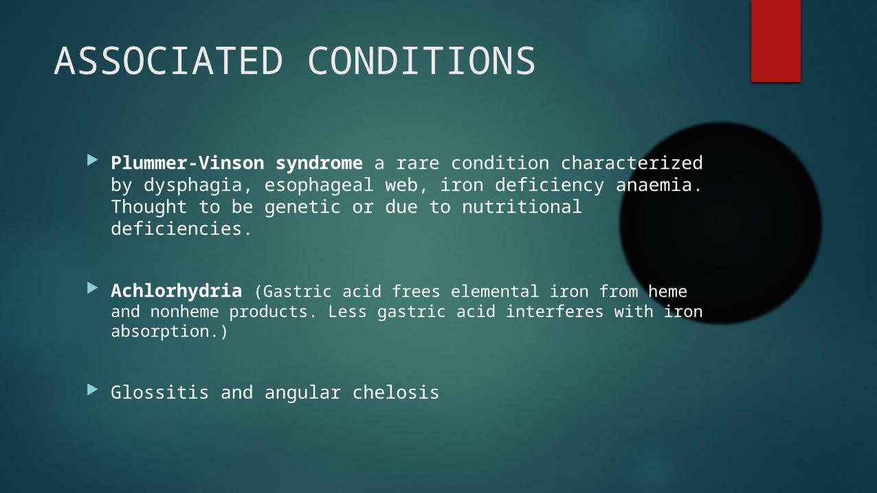
ASSOCIATED CONDITIONS
Plummer-Vinson syndrome a rare condition characterized by dysphagia, esophageal web, iron deficiency anaemia. Thought to be genetic or due to nutritional deficiencies.
Achlorhydria (Gastric acid frees elemental iron from heme and nonheme products. Less gastric acid interferes with iron absorption.)
Glossitis and angular chelosis
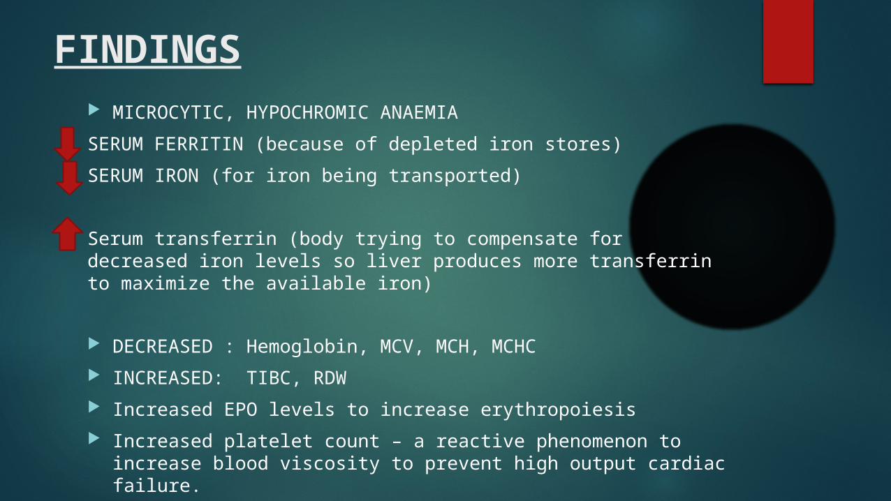
FINDINGS MICROCYTIC, HYPOCHROMIC ANAEMIA
SERUM FERRITIN (because of depleted iron stores)
SERUM IRON (for iron being transported)
Serum transferrin (body trying to compensate for decreased iron levels so liver produces more transferrin to maximize the available iron)
DECREASED : Hemoglobin, MCV, MCH, MCHC
INCREASED: TIBC, RDW
Increased EPO levels to increase erythropoiesis
Increased platelet count – a reactive phenomenon to increase blood viscosity to prevent high output cardiac failure.

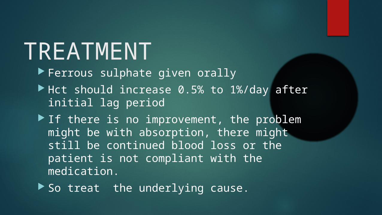
TREATMENT
Ferrous sulphate given orally Hct should increase 0.5% to 1%/day after
initial lag period If there is no improvement, the problem might
be with absorption, there might still be continued blood loss or the patient is not compliant with the medication.
So treat the underlying cause.

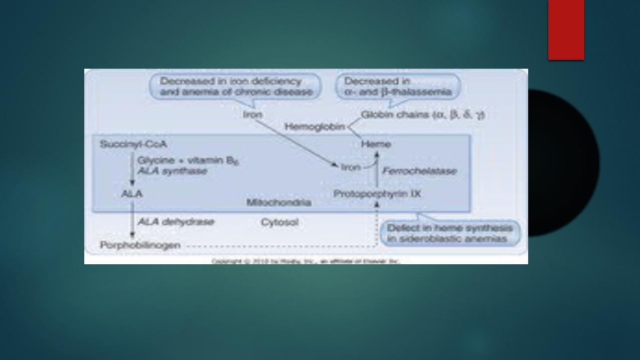
DIFFERENTIAL DIAGNOSIS Sideroblastic anaemia:- Also microcytic, hypochromic anaemia. In
this case, there is excess iron but the mitochondria of the RBC aren’t able to use it to make haemoglobin- defect in heme synthesis.
Increased serum iron, iron saturation, ferritin.
Reduced TIBC
Pappenhiemer bodies on normal staining.
Anaemia of Chronic Disease:- hypochromic microcytic anaemia. Due to chronic infections there is decrease in the amount of iron available to the micro-organisms. Cytokines cause increase hepcidin that prevents the release of iron from macrophages.
Increased ferritin
Decreased transferrin, TIBC, serum iron
B-thalassemia minor:- microcytic, hypochromic. RDW normal. Increased HbA2
REFERENCES http://www.hematology.org/Patients/Anemia/Iron-Deficiency.aspx
http://emedicine.medscape.com/article/202333-overview
http://www.nlm.nih.gov/medlineplus/ency/article/000584.htm
http://www.babycenter.com/0_iron-deficiency-anemia-in-babies_10860.bc
http://www.mayoclinic.org/diseases-conditions/iron-deficiency-anemia/basics/symptoms/con-20019327
http://en.wikipedia.org/wiki/Iron-deficiency_anemia
Related Documents