Iranian Journal of Basic Medical Sciences ijbms.mums.ac.ir Crocin prevents haloperidol-induced orofacial dyskinesia: possible an antioxidant mechanism Marzyeh Kamyar 1 , Bibi Marjan Razavi 2 , Faezeh Vahdati Hasani 1 , Soghra Mehri 3 , Amir Foroutanfar 1 , Hossein Hosseinzadeh 3 * 1 School of Pharmacy, Mashhad University of Medical Sciences, Mashhad, Iran 2 Targeted Drug Delivery Research Center, Department of Pharmacodynamy and Toxicology, School of Pharmacy, Mashhad University of Medical Sciences, Mashhad, Iran 3 Pharmaceutical Research Center, Department of Pharmacodynamy and Toxicology, School of Pharmacy, Mashhad University of Medical Sciences, Mashhad, Iran A R T I C L E I N F O A B S T R A C T Article type: Original article Objective(s): Long-term treatment with antipsychotics causes serious side effects such as tardive dyskinesia that characterized by abnormal movements in the orofacial region. Oxidative stress in the brain specific area is implicated in the pathophysiology of tardive dyskinesia. In this study the protective effect of crocin on haloperidol-induced orofacial dyskinesia was evaluated. Materials and Methods: Haloperidol (1 mg/kg, IP) and crocin (10, 20 and 40 mg/kg, IP) were administrated to rats for 21 days. Behavioral assessments such as orofacial dyskinesia movements, open field test and elevated plus maze (EPM) were evaluated every week. Malondealdehyde (MDA) and glutathione (GSH) levels in the hippocampus, cortex and striatum were also measured. Results: Haloperidol increased vacuous chewing movements (VCMs) and tongue protrusions (TPs) in rats and co-administration of crocin (20 and 40 mg/kg) significantly reduced them. Furthermore, haloperidol decreased the locomotor and exploratory activities (rearing) in the open field test and decreased the percentage of entries into open arms and the percentage of the time spent on open arms in the EPM. Pretreatment with crocin (10 mg/kg) modified haloperidol effects on these behavioral parameters. Haloperidol induced lipid peroxidation in three brain regions, whereas crocin co- administration reduced the MDA and restored the decreased GSH levels. Conclusion: Our finding suggests that oxidative stress has an important role in the development of tardive dyskinesia. Crocin showed protective effect against haloperidol induced tardive dyskinesia and as a potent naturally antioxidant could be a new and useful drug and a possible therapeutic option for the treatment of tardive dyskinesia. Article history: Received: Mar 14, 2016 Accepted: Apr 28, 2016 Keywords: Crocin Haloperidol Lipid peroxidation Orofacial dyskinesia Oxidative stress ►Please cite this article as: Kamyar M, Razavi BM, Vahdati Hasani F, Mehri S, Foroutanfar A, Hosseinzadeh H. Crocin prevents haloperidol-induced orofacial dyskinesia: possible an antioxidant mechanism. Iran J Basic Med Sci 2016; 19:1070-1079. Introduction Haloperidol is a typical antipsychotic belongs to the group of butyrophenone that is highly potent and commonly used in the treatment of both acute and chronic schizophrenia. Therapeutic effect of haloperidol and other typical antipsychotics is related to the blockade of dopamine D2 receptors especially in mesocortical and mesolimbic system in the brain (1, 2). Blockade of D2 receptors in the striatum and nigrostriatal system can also occur and cause movement disorders including tardive dyskinesia (2-4). TD is a potentially irreversible syndrome consisting of athetoid, repetitive, rhythmic and abnormal involuntary movements in the face, especially in the mouth region (5-7). TD is an important clinical problem that develops during antipsychotic treatment (8). The mechanisms involved in tardive dyskinesia development poorly understood, but several hypothesis have been proposed to explain TD pathophysiology include dopamine receptors supersensitivity, dysfunction of striatonigral GABAergic neurons, imbalance between dopaminergic and cholinergic systems, maladaptive synaptic plasticity and striatal neurodegenerative hypothesis (7, 9-13). It has been reported that oxidative stress and free radical production induced by chronic treatment with antipsychotics have an important role in TD pathophysiology (14, 15). In previous studies, rats treated with haloperidol showed higher levels of TBARS in striatum and some regions of the brain (15-17). Treatment with haloperidol causes an increase in dopamine turnover by blocking the dopamine receptors that leads to overproduction of free radicals by enzymatic *Corresponding author: Hossein Hosseinzadeh. Pharmaceutical Research Center, Department of Pharmacodynamy and Toxicology, School of Pharmacy, Mashhad University of Medical Sciences, Mashhad, Iran. Tel: +98-51-38819042; Fax: +98-51-38823251; email: [email protected]

Welcome message from author
This document is posted to help you gain knowledge. Please leave a comment to let me know what you think about it! Share it to your friends and learn new things together.
Transcript
Iranian Journal of Basic Medical Sciences
ijbms.mums.ac.ir
Crocin prevents haloperidol-induced orofacial dyskinesia: possible an antioxidant mechanism
Marzyeh Kamyar 1, Bibi Marjan Razavi 2, Faezeh Vahdati Hasani 1, Soghra Mehri 3, Amir Foroutanfar 1, Hossein Hosseinzadeh 3*
1 School of Pharmacy, Mashhad University of Medical Sciences, Mashhad, Iran 2 Targeted Drug Delivery Research Center, Department of Pharmacodynamy and Toxicology, School of Pharmacy, Mashhad University of
Medical Sciences, Mashhad, Iran 3 Pharmaceutical Research Center, Department of Pharmacodynamy and Toxicology, School of Pharmacy, Mashhad University of Medical
Sciences, Mashhad, Iran
A R T I C L E I N F O A B S T R A C T
Article type: Original article
Objective(s): Long-term treatment with antipsychotics causes serious side effects such as tardive dyskinesia that characterized by abnormal movements in the orofacial region. Oxidative stress in the brain specific area is implicated in the pathophysiology of tardive dyskinesia. In this study the protective effect of crocin on haloperidol-induced orofacial dyskinesia was evaluated. Materials and Methods: Haloperidol (1 mg/kg, IP) and crocin (10, 20 and 40 mg/kg, IP) were administrated to rats for 21 days. Behavioral assessments such as orofacial dyskinesia movements, open field test and elevated plus maze (EPM) were evaluated every week. Malondealdehyde (MDA) and glutathione (GSH) levels in the hippocampus, cortex and striatum were also measured. Results: Haloperidol increased vacuous chewing movements (VCMs) and tongue protrusions (TPs) in rats and co-administration of crocin (20 and 40 mg/kg) significantly reduced them. Furthermore, haloperidol decreased the locomotor and exploratory activities (rearing) in the open field test and decreased the percentage of entries into open arms and the percentage of the time spent on open arms in the EPM. Pretreatment with crocin (10 mg/kg) modified haloperidol effects on these behavioral parameters. Haloperidol induced lipid peroxidation in three brain regions, whereas crocin co-administration reduced the MDA and restored the decreased GSH levels. Conclusion: Our finding suggests that oxidative stress has an important role in the development of tardive dyskinesia. Crocin showed protective effect against haloperidol induced tardive dyskinesia and as a potent naturally antioxidant could be a new and useful drug and a possible therapeutic option for the treatment of tardive dyskinesia.
Article history: Received: Mar 14, 2016 Accepted: Apr 28, 2016
Keywords: Crocin Haloperidol Lipid peroxidation Orofacial dyskinesia Oxidative stress
►Please cite this article as: Kamyar M, Razavi BM, Vahdati Hasani F, Mehri S, Foroutanfar A, Hosseinzadeh H. Crocin prevents haloperidol-induced orofacial dyskinesia: possible an antioxidant mechanism. Iran J Basic Med Sci 2016; 19:1070-1079.
Introduction Haloperidol is a typical antipsychotic belongs to
the group of butyrophenone that is highly potent and commonly used in the treatment of both acute and chronic schizophrenia. Therapeutic effect of haloperidol and other typical antipsychotics is related to the blockade of dopamine D2 receptors especially in mesocortical and mesolimbic system in the brain (1, 2). Blockade of D2 receptors in the striatum and nigrostriatal system can also occur and cause movement disorders including tardive dyskinesia (2-4). TD is a potentially irreversible syndrome consisting of athetoid, repetitive, rhythmic and abnormal involuntary movements in the face, especially in the mouth region (5-7). TD is an important clinical problem that develops during antipsychotic treatment (8). The mechanisms
involved in tardive dyskinesia development poorly understood, but several hypothesis have been proposed to explain TD pathophysiology include dopamine receptors supersensitivity, dysfunction of striatonigral GABAergic neurons, imbalance between dopaminergic and cholinergic systems, maladaptive synaptic plasticity and striatal neurodegenerative hypothesis (7, 9-13). It has been reported that oxidative stress and free radical production induced by chronic treatment with antipsychotics have an important role in TD pathophysiology (14, 15). In previous studies, rats treated with haloperidol showed higher levels of TBARS in striatum and some regions of the brain (15-17). Treatment with haloperidol causes an increase in dopamine turnover by blocking the dopamine receptors that leads to overproduction of free radicals by enzymatic
*Corresponding author: Hossein Hosseinzadeh. Pharmaceutical Research Center, Department of Pharmacodynamy and Toxicology, School of Pharmacy,
Mashhad University of Medical Sciences, Mashhad, Iran. Tel: +98-51-38819042; Fax: +98-51-38823251; email: [email protected]

Crocin protects haloperidol induced tardive dyskinesia Kamyar et al
Iran J Basic Med Sci, Vol. 19, No.10, Oct 2016
1071
dopamine metabolism (18). Furthermore, haloperidol through its toxic metabolites such as RHPP+ and HP+ inhibits complex І in mitochondrial respiration chain that is associated with generation of reactive oxygen species (ROS) and oxidative stress (16, 19).
Crocin is a water-soluble bioactive carotenoid constituent of Crocus sativus L. (saffron) (20-23). Pharmacological studies have demonstrated that crocin possesses various beneficial effects such as antidepressant (24-26), hypolipidemic (27), anti-inflammatory (28, 29), antitumor (30), radical scavenger (31), neuroprotective (32, 33) and antioxidant effect (34-36). It has been reported that crocin prevented the death of PC-12 cells deprived of serum/glucose by inhibition of lipid peroxidation, increasing SOD activity and increasing GSH synthesis by enhancing the GR and γ-GCS activities (37, 38). The antioxidant effect of crocin was more potent than ɑ-tocopherol (37). Crocin can inhibit aggregation and deposition of amyloid β- peptide fibrils and development of Alzheimer’s disease by its antioxidant effects (39). Crocin also caused decreasing MDA level and increasing total thiol and glutathione peroxidase activity and attenuated renal oxidative stress and nephrotoxicity induced by cisplatin in rats (40). Crocin has shown protective effects against chronic stress-induced oxidative stress damage to the hippocampus and impairment of learning and memory in rats (34). Furthermore preventive effects of crocin on diazinon, acrylamide and acrolein induced oxidative stress have been studied (41, 42). It has been observed that natural antioxidants such as Bauhinia forficata (43) and curcumin (44) have protective effect against orofacial dyskinesia induced by haloperidol. Therefore, the aim of this study was to investigate the protective effect of crocin on orofacial dyskinesia and oxidative stress markers in different brain regions induced by haloperidol in rats.
Materials and Methods Animals
Male Wistar albino rats weighing between 200-250 g bred in Animal House of Mashhad University of Medical Sciences, School of Pharmacy, Iran, were used. The animals were kept in standard plastic cages in a colony room maintained on a 12-h light/dark cycle with a temperature of 21 ± 2°C and 40-50% humidity conditions and were allowed free access to water and food except in the time of behavioral assessment. The experimental protocols was approved by the ethical committee of Mashhad University of Medical Sciences and the animals were maintained and used in accordance to the institutional guidelines for the use and care of laboratory animals.
Crocin preparation Stigmas of saffron were purchased from Novin
Saffron Co. (Mashhad, Iran). Crocin was extracted and purified as previously described (45). 25 ml ethanol
80% was added to 10 g saffron stigmas powders at 0 °C and was suspended by vortex (2 min). The suspension was centrifuged at 1500 g for 10 min and then the supernatant was separated. The extraction was repeated 6 other times by addition of 25 ml ethanol 80% to precipitate. The resulting extract was kept in a thick walled glass container which placed at temperature of −5 °C in darkness for 24 days. The formed crystals were separated from the solution and washed with acetone to remove remaining water and then were dissolved in 120 ml ethanol 80% and kept at −5 °C for 20 extra days in darkness. The final amount of obtained crocin was 1.02 g and the purity of yielded crystals was determined by spectrophotometry and HPLC and was more than 97%.
Drugs and treatment schedules
Haloperidol obtained from Caspian tamin, Rasht, Iran and was dissolved in saline. Crocin was suspended in distilled water. All drugs and agents were administered once daily intraperitoneally (IP) for a period of 21 days.
The rats were randomly divided into 7 groups, consisted of six rats in each group: First group (negative control) received saline as the vehicle (1 ml/kg), second group received haloperidol (1 mg/kg), third group received haloperidol (1 mg/kg) plus crocin (10 mg/kg), fourth group received haloperidol (1 mg/kg) plus crocin (20 mg/kg), fifth group received haloperidol (1 mg/kg) plus crocin (40 mg/kg) (26), sixth group received only crocin (40 mg/kg) and last group (positive control) received haloperidol (1 mg/kg) plus vitamin E (200 mg/kg).
Induction of orofacial dyskinesia For the induction of oral dyskinesia, haloperidol (1
mg/kg; IP) was administered to rats for a period of 21 days (46-48).
Behavioral assessments All the behavioral assessments were carried out in
the first day (before the haloperidol administration), 11th day of study, and last behavioral assessment was done 24 hr after the last dose of haloperidol (22nd day) between 09:00 AM and 02:00 PM. The interval between different behavioral tests was 1 hr.
Assessment of orofacial dyskinesia Rats were placed individually in a small transparent
cage for the measurement of VCMs and TP in which mirrors were placed in the floor and back wall to enable better observation of VCM and TP when the rat was faced away from the observer. Rats were allowed 10 min to get acclimatized to the cage before behavioral assessments were carried out. The number of VCMs and TP were counted continuously during a 5 min observation period after adaptation and counting was stopped when the rat began grooming. VCMs are defined to as single mouth openings which resembled

Kamyar et al Crocin protects haloperidol induced tardive dyskinesia
Iran J Basic Med Sci, Vol. 19, No. 10, Oct 2016
1072
chewing, with or without TP, not directed toward any particular physical stimulus (48). Open field test
This test was carried out to analyze changes in spontaneous locomotors and exploratory activity caused by haloperidol and/or crocin administration. Each rat was placed in the center of an open field arena (100× 100 cm). The apparatus had a white floor divided by black lines into 25 squares and black plywood walls. The number of line crossings and rearing was measured during a period of 5 min (49). Elevated plus maze test
The elevated plus maze was used to evaluate the anxiety like state caused by haloperidol and crocin treatment. The apparatus consisted of two open arms and two closed arms and was located 50 cm above the floor. The rat was placed at the center of the plus maze and the number of the entries into the open arms and the time spent on the open arms were recorded during a period of 5 min by Maze router Software. The percentage of the entries into the open arms and the percentage of the time spent on the open arms were calculated by the below formula:
Number of the entries into the open arms and the time spent on the open arms/ total number or total time of the entries into open and closed arms × 100 (50). Tissue dissection
On the 22 nd day of haloperidol treatment, 24 h after the last haloperidol injection and after last behavioral assessments, the rats were sacrificed by decapitation. The brain was removed and placed on ice. The striatum, hippocampus and cortex were rapidly separated, washed by ice-cold saline and stored at − 80 °C for subsequent biochemical analysis. Lipid peroxidation assay
The amount of MDA, as an index of lipid peroxidation, was measured by the reaction with thiobarbituric acid (TBA) at 532 nm using spectrophotometer. The tissues weighed and were immediately homogenized on ice with cold 1.15% KC1 to prepare a 10% tissue homogenate. 500 μl of homogenate was pipetted into a tube and 3 ml 1% phosphoric acid and 1 ml 0.6% TBA was added to the tube. Then the mixture placed in a boiling water bath for 45 min. The tubes were cooled and 4 ml n-butanol was added and mixed by vortex for 1 min. The n-butanol phase was separated by centrifugation at 3000 g for 20 min and absorbance of colored layer was measured at 532 nm. The standard curve of MDA was constructed over the concentration range of 0-100 nmol/ml and levels of MDA were expressed as nmol/g tissue (51, 52).
Estimation of reduced glutathione The tissues weighed and were immediately
homogenized on ice with phosphate buffer (pH 7.4) to prepare a 10% tissue homogenate. 0.5 ml 10% TCA was added to 0.5 ml of tissue homogenate to precipitate it. After vortex, the mixture was centrifuged for removing the precipitate. 2.5 ml phosphate buffer (pH 8) and 0.5 ml DTNB (5-5'-DithioBis-(2-Nitrobenzoic acid) ( was added to supernatant and absorbance of the sample was read immediately at 412 nm. The standard curve of GSH was constructed over the concentration range of 0-150 nmol/ml and the amount of GSH was expressed as nanomol/g tissue (53). Statistical analysis
The data had normal distribution and were analyzed using Graph Pad Prism version 6.00 by analysis of variance (ANOVA) followed by Tukey-Kramer post test for multiple comparisons. All results are expressed as mean±SD and the differences between groups were considered statistically significant when P˂0.05.
Results Behavioral assessments results
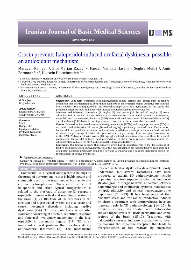
Effect of crocin on VCMs and TPs induced by haloperidol treatment Haloperidol (1 mg/kg, IP) administration significantly increased VCMs and TPs as compared to control group on day 22nd of experiment (P˂0.001 and P<0.01). Crocin co-administration (20 and 40 mg/kg) and Vit. E attenuated the VCMs and tongue protrusions as compared to haloperidol group (P˂0.01, P˂0.05) on day 22nd of experiment. Crocin (40 mg/kg) alone did not cause any significant increase in VCMs and tongue protrusions as compared to control group (Figure 1, 2).
Figure1. Effect of crocin (CR) on haloperidol (HP) induced vacuous chewing movements (VCMs) recorded on day 1, 11, 22 in rats. Data analyzed by ANOVA followed by Tukey-Kramer post test and is expressed as mean±SD (n=6). ### P˂0.001 as compared to control group and * P˂0.05 and ** P˂0.01 as compared to haloperidol group
Crocin protects haloperidol induced tardive dyskinesia Kamyar et al
Iran J Basic Med Sci, Vol. 19, No.10, Oct 2016
1073
Figure2. Effect of crocin (CR) on haloperidol (HP) induced tongue protrusions (TPs) recorded on day 1, 11, 22 in rats. Data analyzed by ANOVA followed by Tukey-Kramer post test and is expressed as mean±SD (n=6). #P˂0.05 and ## P˂0.01 as compared to control group and * P˂0.05 and ** P˂0.01 as compared to haloperidol group
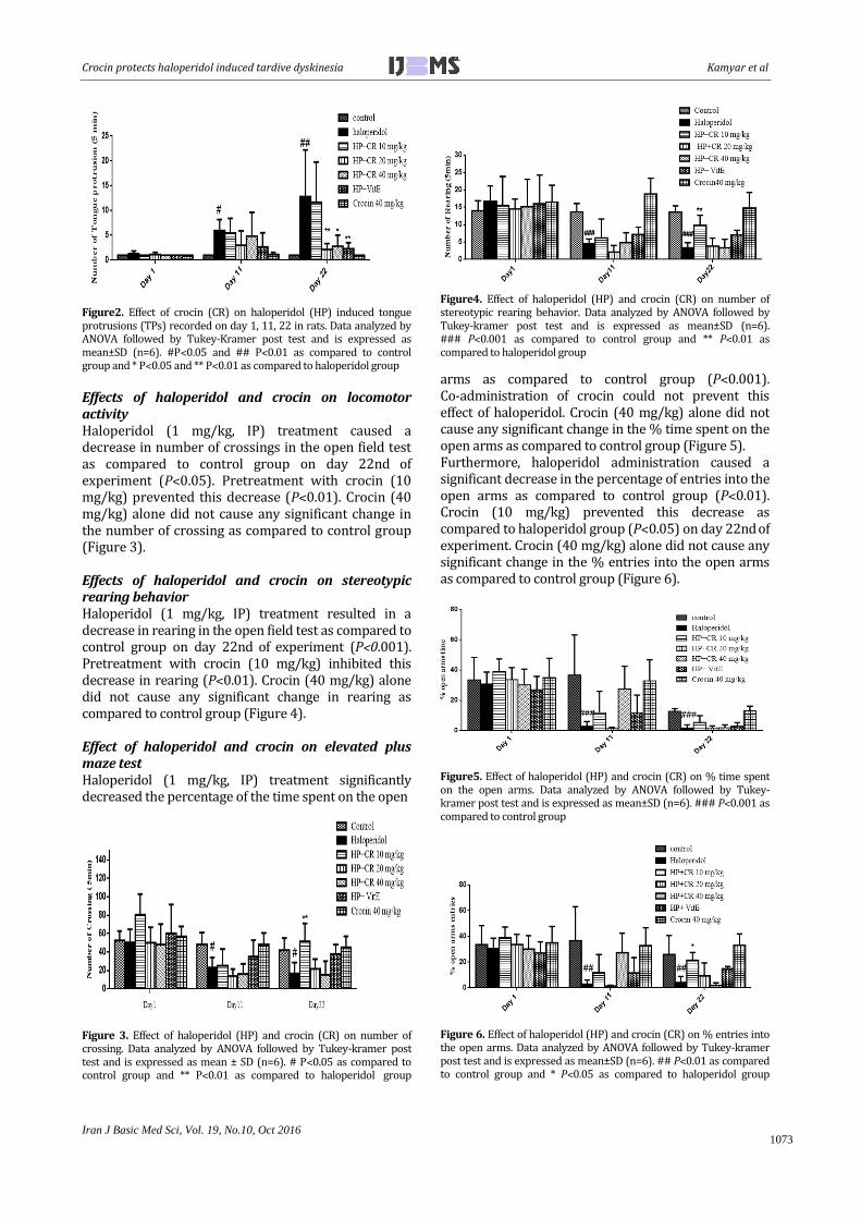
Effects of haloperidol and crocin on locomotor activity Haloperidol (1 mg/kg, IP) treatment caused a decrease in number of crossings in the open field test as compared to control group on day 22nd of
experiment (P˂0.05). Pretreatment with crocin (10 mg/kg) prevented this decrease (P˂0.01). Crocin (40 mg/kg) alone did not cause any significant change in the number of crossing as compared to control group (Figure 3). Effects of haloperidol and crocin on stereotypic rearing behavior Haloperidol (1 mg/kg, IP) treatment resulted in a decrease in rearing in the open field test as compared to control group on day 22nd of experiment (P˂0.001). Pretreatment with crocin (10 mg/kg) inhibited this decrease in rearing (P˂0.01). Crocin (40 mg/kg) alone did not cause any significant change in rearing as compared to control group (Figure 4). Effect of haloperidol and crocin on elevated plus maze test Haloperidol (1 mg/kg, IP) treatment significantly decreased the percentage of the time spent on the open
Figure 3. Effect of haloperidol (HP) and crocin (CR) on number of crossing. Data analyzed by ANOVA followed by Tukey-kramer post test and is expressed as mean ± SD (n=6). # P˂0.05 as compared to control group and ** P˂0.01 as compared to haloperidol group
Figure4. Effect of haloperidol (HP) and crocin (CR) on number of stereotypic rearing behavior. Data analyzed by ANOVA followed by Tukey-kramer post test and is expressed as mean±SD (n=6). ### P˂0.001 as compared to control group and ** P˂0.01 as compared to haloperidol group
arms as compared to control group (P˂0.001). Co-administration of crocin could not prevent this effect of haloperidol. Crocin (40 mg/kg) alone did not cause any significant change in the % time spent on the open arms as compared to control group (Figure 5). Furthermore, haloperidol administration caused a significant decrease in the percentage of entries into the open arms as compared to control group (P˂0.01). Crocin (10 mg/kg) prevented this decrease as compared to haloperidol group (P˂0.05) on day 22nd of
experiment. Crocin (40 mg/kg) alone did not cause any significant change in the % entries into the open arms as compared to control group (Figure 6).
Figure5. Effect of haloperidol (HP) and crocin (CR) on % time spent on the open arms. Data analyzed by ANOVA followed by Tukey-kramer post test and is expressed as mean±SD (n=6). ### P˂0.001 as compared to control group
Figure 6. Effect of haloperidol (HP) and crocin (CR) on % entries into the open arms. Data analyzed by ANOVA followed by Tukey-kramer post test and is expressed as mean±SD (n=6). ## P˂0.01 as compared to control group and * P˂0.05 as compared to haloperidol group

Kamyar et al Crocin protects haloperidol induced tardive dyskinesia
Iran J Basic Med Sci, Vol. 19, No. 10, Oct 2016
1074
Figure 7. Effect of haloperidol (HP) and crocin (CR) on MDA level in cortex (A), striatum (B) and hippocampus (C) of rat’s brain. Data analyzed by ANOVA followed by Tukey-kramer post test and is expressed as mean±SD (n=6). ### P˂0.001 as compared to control group and * P˂0.05, **P˂0.01 and *** P˂0.001 as compared to haloperidol group
Biochemical analysis results Effect of haloperidol and crocin on MDA levels in striatum, cortex and hippocampus Haloperidol treatment (1 mg/kg, IP) caused a significant increase in MDA level in cortex, striatum and hippocampus of the rat’s brain as compared to control group (P<0.001). Co-administration of crocin (10, 20 and 40 mg/kg) and Vit. E decreased the elevated level of MDA in three regions of the brain as compared to haloperidol group. Crocin (40 mg/kg) alone did not cause any significant change in MDA level as compared to control group (Figure 7). Effect of haloperidol and crocin on glutathione (GSH) levels in striatum, cortex and hippocampus Haloperidol treatment (1 mg/kg, IP) resulted in a significant decrease in GSH level in cortex, striatum and hippocampus of the rat’s brain as compared to control group (P<0.001). Co-administration of crocin (10,20 and 40 mg/kg) and Vit. E increased GSH level in three regions of the brain as compared to haloperidol group. Crocin (40 mg/kg) alone did not cause any significant change in GSH level as compared to control group (Figure 8).
Discussion Oxidative stress defines as an imbalance between production of oxidant (such as ROS and RNS) and antioxidant defense system. Lipids are the main bio-
logical target of oxidative stress and oxidation of lipids produces a number of secondary products. MDA is the main and principal product of lipid peroxidation and is a highly toxic molecule (54, 55). In the present study to evaluate the effect of haloperidol and crocin in induction of oxidative stress, the amount of MDA (as marker of lipid peroxidation) and glutathione (GSH) levels were measured in three region of the brain (striatum, cortex and hippocampus). Haloperidol treatment caused an increase in MDA level and decrease in GSH content in three region of the brain as compared to control group. Crocin decreased the elevated level of MDA and increased the glutathione content in haloperidol-treated rats. Administration of haloperidol also increased VCMs and TPs in rats. Co-administration of crocin significantly attenuated the induction of haloperidol-induced VCMs and TPs. In several studies using animal model of tardive dyskinesia (orofacial dyskinesia), administration of haloperidol caused an increase in MDA level and induction of VCMs and TPs (48, 56). Treatment with antioxidant compounds such as melatonin seems to be effective in the reduction of orofacial dyskinesia (46, 47, 57, 58). Crocin is a potent antioxidant and its antioxidant activity is responsible for the various pharmacological effects (59).
Existing evidences indicate that oxidative stress
Figure 8. Effect of haloperidol (HP) and crocin (CR) on reduced glutathione (GSH) level in cortex (A), striatum (B) and hippocampus (C) of rat’s brain. Data analyzed by ANOVA followed by Tukey-kramer post test and is expressed as mean±SD (n=6). ### P˂0.001 as compared to control group and * P˂0.05, ** P˂0.01 and *** P˂0.001 as compared to haloperidol group

Crocin protects haloperidol induced tardive dyskinesia Kamyar et al
Iran J Basic Med Sci, Vol. 19, No.10, Oct 2016
1075
induced by antipsychotic drugs contributes in the pathogenesis of tardive dyskinesia (60, 61). The brain is particularly vulnerable to oxidative stress damage because it uses high amount of energy and oxygen and contains large amount of oxidizable substrates such as catecholamines such as dopamine and polyunsaturated fatty acids which are targets for activity of lipid peroxidation cascades. Low levels of antioxidant enzymes such as catalase and high concentrations of transition metals such as iron, manganese and copper (which involve in free radical formation) in the brain also lead to vulnerability of brain to free radical damages (62, 63). Blockade of dopamine D2 receptors by typical antipsychotic such as haloperidol led to a secondary increase in synthesis and metabolism of dopamine. This excess dopamine is metabolized by MAO which leads to overproduction of hydrogen peroxide and hydroxyl radicals or can auto-oxidize to form dopamine quinones that are free radicals. It has been demonstrated that basal ganglia, a region of the brain that involves in motor function, is the main region implicated in tardive dyskinesia pathophysiology. The basal ganglia are rich of dopamine, oxygen, polyunsaturated membrane lipids and transition metals such as iron and produces excessive free radicals that leads to weakening of antioxidant brain system (5, 7, 14, 64).
It was reported that resveratrol, a potent antioxidant and monoamine oxidase enzyme activity inhibitor, showed protective effect against reserpine-induced orofacial dyskinesia (65). De Monte et al (2014) reported that crocin inhibited both MAO enzyme isoforms with non-competitive mechanism (66). Therefore, the protective effect of crocin on haloperidol-induced TD may be due to its inhibitory effect on MAO enzyme and decrease in dopamine metabolism. Dopamine quinones, cytotoxic metabolites of dopamine, also generate by enzymatic oxidation of dopamine by cyclooxygenase (COX) enzymes. NSAIDs drugs prevent oxidation of dopamine to dopamine quinone by inhibition of COX enzymes like prostaglandin H synthase (67). Recently it was reported that indomethacin, a nonselective cyclooxygenase enzyme inhibitor, reversed haloperidol-induced orofacial dyskinesia in rats (68). Crocin exhibited an inhibitory effect on COX-1 and COX-2 enzymes and its protective effect may be due to its COX inhibitory activity (69).
The involvement of nitric oxide (NO) in development of haloperidol-induced orofacial dyskinesia has been reported. L-NAME, a nitric oxide synthase inhibitor, suppressed haloperidol-induced VCMs (70). Crocin decreased in NO content and inhibited nitric oxide synthase (NOS) activity and inducible nitric oxide synthase (iNOS) expression (71, 72) So, the beneficial effect of crocin might be due to its inhibitory effect on NO and NOS activity.
Pharmacological evidence has demonstrated that glutamergic system participated in pathophysiology of
tardive dyskinesia. The reuptake of glutamate from the extracellular space is mediated by transporter proteins which contain reactive thiol groups in their structure. Oxidation of these groups leads to reduction in glutamate uptake. Reduction in glutamate transporter activity and glutamate uptake by haloperidol leads to extracellular increase in glutamate level which resulted in excitotoxicity and development of tardive dyskinesia (73). Further blockade of the presynaptic dopamine D2 receptors, which prevents the release of glutamate from excitatory cortical striatal pathway, by antipsychotic drugs such as haloperidol leads to an increase in synaptic release of glutamate and aspartate in the striatum. It has been known that persistent activation of glutamate ionotropic receptors leads to neuronal degeneration (74). The glutamate neurotoxicity damages many cell components that lead to cell death. In this process reactive oxygen species (ROS) are generated and oxidative stress is involved in excitotoxicity induced by glutamate (73, 75). It was reported that dizocilpine, a non-competitive N-methyl-D-aspartate (NMDA) receptor antagonist, attenuated the haloperidol-induced orofacial dyskinesia (70). It has been demonstrated that safranal caused a reduction in extracellular glutamate concentration and saffron inhibited glutamatergic synaptic transmission (76, 77). The inhibitory effect of saffron and crocin on glutamatergic system has been indicated previously (78). As crocin hasn’t any interaction with NMDA receptors (79), beneficial effect of crocin on haloperidol-induced TD may be due to reduction in extracellular glutamate concentration trough protection of glutamate transporter from oxidative stress damages or other mechanisms.
It was proposed that dysfunctional striatal γ-aminobutyric acid (GABA) containing neurons leading to decrease in inhibitory function of GABA in the basal ganglia and imbalance between direct and indirect basal ganglia pathways is implicated in the pathophysiology of tardive dyskinesia (10). Administration of haloperidol also caused a reduction in nigral L-glutamic acid decarboxylase (GAD) activity and GABA synthesizing enzyme, in rat with oral dyskinesia. This movement disorder is related to hypofunction in GABA-ergic neurons in substania nigra (80). Peixoto et al (2003) was reported that valproic acid, a GABA-mimetic drug, attenuated the orofacial dyskinesia in rats (81). In other study, co-administration of progabide (a GABA agonist) inhibited haloperidol-induced orofacial dyskinesia in rats (82). Saffron and its constituent crocin have neuroprotective effects and protected cells from apoptosis and death (83, 84). Crocin may protect GABA-ergic neurons from death and beneficial effect against TD may be due to its effects on GABA-ergic system.
In the present study, vitamin E decreased oxidative stress as well as VCMs and TPs induced by haloperidol administration. Vitamin E as an antioxidant and free radical scavenger was effective in the treatment of

Kamyar et al Crocin protects haloperidol induced tardive dyskinesia
Iran J Basic Med Sci, Vol. 19, No. 10, Oct 2016
1076
tardive dyskinesia (85). Co-administration of vitamin E with haloperidol improved PC12 cell viability after haloperidol exposure and suggested the potential benefit of vitamin E for preventing of tardive dyskinesia (64). Abilio et al (2003) reported that vitamin E attenuated the reserpine-induced orofacial dyskinesia and increased ratio of oxidized/reduced glutathione ratio (GSSG/GSH) in striatum of rats (86).
The effects of haloperidol and crocin on locomotor activity and stereotypic rearing in the rats were also evaluated in an open field test. Haloperidol administration resulted in a significant decrease in locomotor activity (total crossing) and rearing and crocin co-administration caused an increase in these parameters. It is known that blockade of dopamine D2 receptors and reduction of dopaminergic pathway function in nucleus accumbens led to suppression of locomotion and sedation by haloperidol (25, 87-89). It was reported that crocin increased stereotypic activity and locomotion in open field test which revealed that crocin can affect the dopaminergic system (25).
Effect of haloperidol in induction of anxiety was evaluated in this study by an elevated plus maze test. In this test anxiolytic compounds decrease the natural aversion of animals to the open arms. Therefore, increased the number of entry or time spent on the open arms reflect the anxiolytic effect of a compound (90). In the present study, administration of haloperidol decreased the percentage of time spent on the open arms. Crocin couldn’t improve alteration of this parameter. Haloperidol also decreased the percentage of entry to the open arms which increased by crocin. Karl et al (2006) reported that haloperidol administration increased anxiety levels in rats (87). The anxiolytic-like effect of crocin has demonstrated previously (91) and therefore positive effect of crocin on the parameters studied in this study may be due to its anxiolytic effect that needs to more investigation.
Conclusion The major findings of the present study suggested
that oxidative stress plays an important role in the development of haloperidol-induced orofacial dyskinesia and crocin co-administration reverses the behavioral and biochemical changes induced by haloperidol. Crocin, a naturally antioxidant, could be considered as a useful agent for treatment of tardive dyskinesia.
Acknowledgment This study is part of a MS thesis and is supported
by Mashhad University of Medical Sciences.
References 1. Kudo S, Ishizaki T. Pharmacokinetics of haloperidol. Clin pharm 1999; 37:435-456. 2. Raudenska M, Gumulec J, Babula P, Stracina T, Sztalmachova M, Polanska H, et al. Haloperidol
Cytotoxicity and Its Relation to Oxidative Stress. Mini Rev Med Chem 2013; 13:1993-1998. 3. Vallianatou K. Antipsychotics. Medicine 2012; 40:676-678. 4. Mitchell I, Cooper A, Griffiths M, Cooper A. Acute administration of haloperidol induces apoptosis of neurones in the striatum and substantia nigra in the rat. Neuroscience 2002; 109:89-99. 5. Cho C-H, Lee H-J. Oxidative stress and tardive dyskinesia: Pharmacogenetic evidence. Prog Neuro-Psychopharm Biol Psychiatry 2013; 46:207-213. 6. Aia PG, Revuelta GJ, Cloud LJ, Factor SA. Tardive dyskinesia. Curr treat options neurol 2011; 13:231-241. 7. Andreassen OA, Jørgensen HA. Neurotoxicity associated with neuroleptic-induced oral dyskinesias in rats: implications for tardive dyskinesia? Prog neurobiol 2000; 61:525-541. 8. Llorca P-M, Chereau I, Bayle F-J, Lancon C. Tardive dyskinesias and antipsychotics: a review. Eur Psychiatry 2002; 17:129-138. 9. Kumar AS, Reddy TSK. A Review on tardive dyskinesia. IJPPR 2012; 3:33-41. 10. Waln O, Jankovic J. An update on tardive dyskinesia: from phenomenology to treatment. Tremor Other Hyperkinet Mov 2013; 3:161-172. 11. Gerlach J, Reisby N, Randrup A. Dopaminergic hypersensivity and cholinergic hypofunction in the pathophysiology of tardive dyskinesia. Psychopharmacology (Berl) 1974; 34:21-35. 12. Teo JT, Edwards MJ, Bhatia K. Tardive dyskinesia is caused by maladaptive synaptic plasticity: a hypothesis. Mov Disord 2012; 27:1205-1215. 13. Kobayashi RM. Orofacial dyskinesia: Clinical features, mechanisms and drug therapy. West J Med 1976; 125:277-288. 14. Lister J, Nobrega JN, Fletcher PJ, Remington G. Oxidative stress and the antipsychotic-induced vacuous chewing movement model of tardive dyskinesia: evidence for antioxidant-based prevention strategies. Psychopharmacology (Berl) 2014; 231:2237-2249. 15. Elkashef A, Wyatt R. Tardive dyskinesia: possible involvement of free radicals and treatment with vitamin E. Schizophrenia Bull 1999; 25:731-740. 16. Polydoro M, Schröder N, Lima MNM, Caldana F, Laranja DC, Bromberg E, et al. Haloperidol-and clozapine-induced oxidative stress in the rat brain. Pharmacol Biochem Behav 2004; 78:751-756. 17. Burger M, Fachinetto R, Zeni G, Rocha J. Ebselen attenuates haloperidol-induced orofacial dyskinesia and oxidative stress in rat brain. Pharmacol Biochem Behav 2005; 81:608-615. 18. Arniaz S, Coronel M, Boveris A. Nitric oxide, superoxide and Hydrogen peroxide production in brain mitochondria after haloperidol treatment. Biol Chem 1999; 3:235-243. 19. Gumulec J, Raudenska M, Hlavna M, Stracina T, Sztalmachova M, Tanhauserova V, et al. Determination of oxidative stress and activities of antioxidant enzymes in guinea pigs treated with haloperidol. Exp Ther Med 2013; 5:479-484. 20. Hosseinzadeh H, Shariaty VM, Sameni AK, Vahabzadeh M. Acute and sub-acute toxicity of crocin,
Crocin protects haloperidol induced tardive dyskinesia Kamyar et al
Iran J Basic Med Sci, Vol. 19, No.10, Oct 2016
1077
a constituent of crocus sativus L. (saffron), in mice and rats. Pharmacologyonline 2010; 2:943-951. 21. Mollazadeh H, Emami SA, Hosseinzadeh H. Razi’s Al-Hawi and saffron (Crocus sativus): A review. Iran J Basic Med Sci 2015; 18:1153-1166. 22. Alavizadeh S, Hosseinzadeh H. Bioactivity assessment and toxicity of crocin: a Comprehensive Review. Food Chem Toxicol 2013; 11. 23. Hosseinzadeh H, Nassiri-Asl M. Avicenna's (Ibn Sina) the canon of medicine and saffron (Crocus sativus): A review. Phytother Res 2013; 27:475-483. 24. Vahdati Hassani F, Naseri V, Razavi B, Mehri S, Abnous K, Hosseinzadeh H. Antidepressant effects of crocin and its effects on transcript and protein levels of CREB, BDNF, and VGF in rat hippocampus. Daru 2014; 8:22 25. Hosseinzadeh H, Karimi G, Niapoor M. Antidepressant effects of crocus sativus stigma extracts and its constituents, crocin and safranal, in mice. J Med Plants 2004; 3:48-58. 26. Ardebili Dorri S, Hosseinzadeh H, Abnous K, Vahdati Hasani F, Yazdian Robati R, Razavi BM. Involvement of brain‐derived neurotrophic factor (BDNF) on malathion induced depressive‐like behavior in subacute exposure and protective effects of crocin. Iran J Basic Med Sci 2015; 18:958-966. 27. Sheng L, Qian Z, Zheng S, Xi L. Mechanism of hypolipidemic effect of crocin in rats: crocin inhibits pancreatic lipase. Eur J Pharmacol 2006; 543:116-122. 28. Nam KN, Park Y-M, Jung H-J, Lee JY, Min BD, Park S-U, et al. Anti-inflammatory effects of crocin and crocetin in rat brain microglial cells. Eur J Pharmacol 2010; 648:110-116. 29. Hosseinzadeh H, Younesi H. Antinociceptive and anti-inflammatory effects of Crocus sativus L. stigma and petal extracts in mice. BMC Pharmacol 2002; 2:7. 30. Festuccia C, Mancini A, Gravina G, Scarsella L, Llorens S, Alonso G, et al. Antitumor effects of saffron-derived carotenoids in prostate cancer cell models. BioMed Res Int 2014; 2014:135048. 31. Assimopoulou A, Sinakos Z, Papageorgiou V. Radical scavenging activity of Crocus sativus L. extract and its bioactive constituents. Phytother Res 2005; 19:997-1000. 32. Mehri S, Abnous K, Mousavi S, Motamed Shariaty V, Hosseinzadeh H. Neuroprotective effect of crocin on acrylamide-induced cytotoxicity in PC12 cells. Cell Mol Neurobiol 2012; 32:227-235. 33. Nassiri-Asl M, Hosseinzadeh H. Neuropharmaco-logy effects of saffron (Crocus sativus) and Its active constituents. Bioactive Nutraceuticals and Dietary Supplements in Neurological and Brain Disease. Prev Ther 2014: 29-39. 34. Ghadrdoost B, Vafaei AA, Rashidy-Pour A, Hajisoltani R, Bandegi AR, Motamedi F, et al. Protective effects of saffron extract and its active constituent crocin against oxidative stress and spatial learning and memory deficits induced by chronic stress in rats. Eur J Pharmacol 2011; 667:222-229. 35. Altinoz E, Oner Z, Elbe H, Cigremis Y, Turkoz Y. Protective effects of saffron (its active constituent, crocin) on nephropathy in streptozotocin-induced diabetic rats. Hum Exp Toxicol 2015; 34:127-134.
36. Hosseinzadeh H, Shamsaie F, Mehri S. Antioxidant activity of aqueous and ethanolic extracts of Crocus sativus L. stigma and its bioactive constituents crocin and safranal. Pharmacogn Mag 2010; 5:419-424. 37. Ochiai T, Ohno S, Soeda S, Tanaka H, Shoyama Y, Shimeno H. Crocin prevents the death of rat pheochromyctoma (PC-12) cells by its antioxidant effects stronger than those of alpha-tocopherol. Neurosci Lett 2004; 362:61-64. 38. Ochiai T, Shimeno H, Mishima K-i, Iwasaki K, Fujiwara M, Tanaka H, et al. Protective effects of carotenoids from saffron on neuronal injury in vitro and in vivo. Biochim Biophys Acta (BBA)-General subjects 2007; 1770:578-584. 39. Magdalini A, Charalambos D, Moschos G, Spyros E, Paul C, Marigoula M, et al. Inhibitory activity on amyloid-â aggregation and antioxidant properties of Crocus sativus stigmas extract and its crocin constituents. J Agric Food Chem 2006; 54:8762-8768. 40. Naghizadeh B, Mansouri S, Mashhadian N. Crocin attenuates cisplatin-induced renal oxidative stress in rats. Food Chem Toxicol 2010; 48:2650-2655. 41. Razavi BM, Hosseinzadeh H. Saffron as an antidote or a protective agent against natural or chemical toxicities. DARU J Pharm Sci 2015; 23:31. 42. Rashedinia M, Lari P, Abnous K, Hosseinzadeh H . Protective effect of crocin on acrolein-induced tau phosphorylation in the rat brain. Acta Neurobiol Exp 2015; 75:208-219. 43. Peroza LR, Busanello A, Leal CQ, Röpke J, Boligon AA, Meinerz D, et al. Bauhinia forficata prevents vacuous chewing movements induced by haloperidol in rats and has antioxidant potential in vitro. Neurochem Res 2013; 38:789-796. 44. Bishnoi M, Chopra K, Kulkarni SK. Protective effect of Curcumin, the active principle of turmeric (Curcuma longa) in haloperidol-induced orofacial dyskinesia and associated behavioural, biochemical and neurochemical changes in rat brain. Pharmacol Biochem Behav 2008; 88:511-522. 45. Hadizadeh F, Mohajeri S, Seifi M. Extraction and purification of crocin from saffron stigmas employing a simple and efficient crystallization method. Pak J Biol Sci 2010; 13:691-698. 46. Naidu PS, Singh A, Kulkarni SK. Quercetin, a bioflavonoid, attenuates haloperidol-induced orofacial dyskinesia. Neuropharmacol 2003; 44:1100-1106. 47. Naidu PS, Singh A, Kulkarni SK. Effect of Withania somnifera root extract on haloperidol-induced orofacial dyskinesia: possible mechanisms of action. J Med food 2003; 6:107-114. 48. Naidu PS, Singh A, Kulkarni SK. Carvedilol attenuates neuroleptic‐induced orofacial dyskinesia: possible antioxidant mechanisms. British J Pharmacol 2002; 136:193-200. 49. Pardon M, Perez Diaz F, Joubert C, Cohen Salmon C. Age dependent effects of a chronic ultramild stress procedure on open field behavior in B6D2F1 female mice. Physiol Behav 2000; 70:7-13. 50. Fachinetto R, Villarinho J, Wagner C, Pereira R, Avila D, Burger M, et al. Valeriana officinalis does not alter the orofacial dyskinesia induced by haloperidol in rats: Role of dopamine transporter. Prog Neuropsychopharmacol Biol Psychiatry 2007; 31:1478-1486.

Kamyar et al Crocin protects haloperidol induced tardive dyskinesia
Iran J Basic Med Sci, Vol. 19, No. 10, Oct 2016
1078
51. Ohkawa H, Ohishi N, Yagi K. Assay for lipid peroxides in animal tissues by thiobarbituric acid reaction. Anal Biochem 1979; 95:351-358. 52. Uchiyama M, Mihara M. Determination of malonaldehyde precursor in tissues by thiobarbituric acid test. Anal Biochem 1978; 86:271-278. 53. Moron M, Depierre J, Mannervik B. Levels of glutathione, glutathione reductase and glutathione S transferase activities in rat lung and liver. Biochim Biophys Acta 1979; 582:67-78. 54. Abuja PM, Albertini R. Methods for monitoring oxidative stress, lipid peroxidation and oxidation resistance of lipoproteins. Clin Chim Acta 2001; 306:1-17. 55. Jimenez del rio M, Velez-Pardo C. Paraquat induces apoptosis inhumanlymphocytes: Protective and rescue effects of glucose, cannabinoids and insulin-like growth factor-1. Growth Factors 2008; 26:49-60. 56. Thaakur S, Himabindhu G. Effect of alpha lipoic acid on the tardive dyskinesia and oxidative stress induced by haloperidol in rats. J neural transm 2009; 116:807-814. 57. Naidu PS, Singh A, Kaur P, Sandhir R, Kulkarni SK. Possible mechanism of action in melatonin attenuation of haloperidol-induced orofacial dyskinesia. Pharmacol Biochem Behav 2003; 74:641-648. 58. Bishnoi M, Chopra K, Kulkarni SK. Protective effect of rutin, a polyphenolic flavonoid against haloperidol‐induced orofacial dyskinesia and associated behavioural, biochemical and neurochemical changes. Fund Clin Pharmacol 2007; 21:521-529. 59. Kawabata K, Tung NH, Shoyama Y, Sugie S, Mori T, Tanaka T. Dietary crocin inhibits colitis and colitis-associated colorectal carcinogenesis in male ICR mice. Evid Based Complement Altern Med 2012; 2012. 60. Cadet J, Lohr J, Jeste D. Free radicals and tardive dyskinesia. Trends Neurosci 1986; 9:107-108. 61. Ramos ZR, Fortunato JJ, Agostinho FR, Martins MR, Correa M, Schetinger MR, et al. Influence of malathion on acetylcholinesterase activity in rats submitted to a forced swimming test. Neurotox Res 2006; 9:285-290. 62. Lohr JB, Kuczenski R, Niculescu AB. Oxidative mechanisms and tardive dyskinesia. CNS Drugs 2003; 17:47-62. 63. Halliwell B, Gutteridge JM. Oxygen radicals and the nervous system. Trends Neurosci 1985; 8:22-26. 64. Galili-Mosberg R, Gil-Ad I, Weizman A, Malamed E, Offen D. Haloperidol-induced neurotoxicity- possible implication for tardive dyskinesia. J Neural Transm 2000; 107:479-490. 65. Busanello A, Barbosa NBV, Peroza LR, Farias LE, Burger ME, Barreto KP, et al. Resveratrol protects against a model of vacuous chewing movements induced by reserpine in mice. Behav Pharmacol 2011; 22:71-75. 66. De Monte C, Carradori S, Chimenti P, Secci D, Mannina L, Alcaro F, et al. New insights into the biological properties of Crocus sativus L.: chemical modifications, human monoamine oxidases inhibition and molecular modeling studies. Eur J Med Chem 2014; 82:164-171.
67. Miyazaki I, Asanuma M. Approaches to prevent dopamine quinone-induced neurotoxicity. Neurochem Res 2009; 34:698-706. 68. Naidu PS, Kulkarni SK. Possible involvement of prostaglandins in haloperidol-induced orofacial dyskinesia in rats. Eur J Pharmacol 2001; 430:295-298. 69. Xu G-L, Li G, Ma H-P, Zhong H, Liu F, Ao G-Z. Preventive effect of crocin in inflamed animals and in LPS-challenged RAW 264.7 cells. J Agr Food Chem 2009; 57:8325-8330. 70. Naidu P, Kulkarni S. Excitatory mechanisms in neuroleptic‐induced vacuous chewing movements (VCMs): possible involvement of calcium and nitric oxide. Behav Pharmacol 2001; 12:209-216. 71. Zheng YQ, Liu JX, Wang JN, Xu L. Effects of crocin on reperfusion-induced oxidative/nitrative injury to cerebral microvessels after global cerebral ischemia. Brain Res 2007; 1138:86-94. 72. Kim JH, Park GY, Bang SY, Park SY, Bae SK, Kim Y. Crocin suppresses LPS-stimulated expression of inducible nitric oxide synthase by upregulation of heme oxygenase-1 via calcium/calmodulin-dependent protein kinase 4. Mediators Inflamm 2014; 2014:728709. 73. Burger ME, Fachineto R, Alves A, Callegari L, Rocha JBT. Acute reserpine and subchronic haloperidol treatments change synaptosomal brain glutamate uptake and elicit orofacial dyskinesia in rats. Brain Res 2005; 1031:202-210. 74. Lu MC, Tzang BS, Kuo WW, Wu FL, Chen YS, Tsai CH, et al. More activated cardiac mitochondrial-dependent apoptotic pathway in obese zucker rats. Obesity 2007; 15:2634-2642. 75. Atlante A, Calissano P, Bobba A, Giannattasio S, Marra E, Passarella S. Glutamate neurotoxicity, oxidative stress and mitochondria. FEBS lett 2001; 497:1-5. 76. Hosseinzadeh H, Sadeghnia HR, Rahimi A. Effects of safranal on extracellular hippocampal levels of glutamate and aspartate during kainic acid treatment in anesthetized rats. Planta Med 2008; 74:1441-1445. 77. Vanamala J, Kester AC, Heuberger AL, Reddivari L. Mitigation of Obesity-Promoted Diseases by Nigella sativa and Thymoquinone . Plant Foods Human Nut 67:111-119. 78. Halataei BAS, Khosravi M, Arbabian S, Sahraei H, Golmanesh L, Zardooz H, et al. Saffron (Crocus sativus) aqueous extract and its constituent crocin reduces stress-induced anorexia in mice. Phytother Res 2011; 25:1833-1838. 79. Imenshahidi M, Zafari H, Hosseinzadeh H. Effects of crocin on the acquisition and reinstatement of morphine-induced conditioned place preference in mice. Pharmacologyonline 2011; 1:1007-1013. 80. Gunne LM, Häggström J-E. Reduction of nigral glutamic acid decarboxylase in rats with neuroleptic-induced oral dyskinesia. Psychopharmacology 1983; 81:191-194. 81. Peixoto MF, Abı́lio VC, Silva RH, Frussa-Filho R. Effects of valproic acid on an animal model of tardive dyskinesia. Behav Brain Res 2003; 142:229-233. 82. Kaneda H, Shirakawa O, Dale J, Goodman L, Bachus SE, Tamminga CA. Co-administration of progabide inhibits haloperidol-induced oral dyskinesias in rats. Eur J Pharmacol 1992; 212:43-49.
Crocin protects haloperidol induced tardive dyskinesia Kamyar et al
Iran J Basic Med Sci, Vol. 19, No.10, Oct 2016
1079
83. Purushothuman S, Nandasena C, Peoples CL, El Massri N, Johnstone DM, Mitrofanis J, et al. Saffron pre-treatment offers neuroprotection to nigral and retinal dopaminergic cells of MPTP-treated mice. J Park dis 2013; 3:77-83. 84. Soeda S, Ochiai T, Paopong L, Tanaka H, Shoyama Y, Shimeno H. Crocin suppresses tumor necrosis factor-alpha-induced cell death of neuronally differentiated PC-12 cells. Life Sci 2001; 69:2887-2898. 85. Soares-weiser K, Maayan N, McGrath J. Vitamin E for neuroleptic-induced tardive dyskinesia. Cochrane Database of Syst Rev 2011; 2. 86. Abilio VC, Araujo CC, Bergamo M, Calvente PR, D'Almeida V, Ribeiro RdA, et al. Vitamin E attenuates reserpine-induced oral dyskinesia and striatal oxidized glutathione/reduced glutathione ratio (GSSG/GSH) enhancement in rats. Prog Neuro-Psychopharm Biol Psychiatry 2003; 27:109-114. 87. Karl T, Duffy L, O’Brien E, Matsumoto I, Dedova I. Behavioural effects of chronic haloperidol and
risperidone treatment in rats. Behav Brain Res 2006;171:286-94. 88. Kelley AE, Bakshi VP, Dells JM, Lang CG. Cholinergic stimulation of the ventrolateral striatum elicits mouth movements in rats: pharmacological and regional specificity. Psychopharmacology 1989; 99:542-549. 89. Salamone JD, Ishiwari K, Betz AJ, Farrar AM, Mingote SM. Dopamine/adenosine interactions related to locomotion and tremor in animal models: possible relevance to parkinsonism. Parkinsonism Relat Disord 2008; 2:130-134. 90. Hosseinzadeh H, Noraei NB. Anxiolytic and hypnotic effect of Crocus sativus aqueous extract and its constituents, crocin and safranal, in mice. Phytother Res 2009; 23:768-774. 91. Pitsikas N, Boultadakis A, Georgiadou G, Tarantilis P, Sakellaridis N. Effects of the active constituents of Crocus sativus L., crocins, in an animal model of anxiety. Phytomedicine 2008; 15:1135-1139.
Related Documents