Cellular Biology iPS Programmed Without c-MYC Yield Proficient Cardiogenesis for Functional Heart Chimerism Almudena Martinez-Fernandez, Timothy J. Nelson, Satsuki Yamada, Santiago Reyes, Alexey E. Alekseev, Carmen Perez-Terzic, Yasuhiro Ikeda, Andre Terzic Rationale: Induced pluripotent stem cells (iPS) allow derivation of pluripotent progenitors from somatic sources. Originally, iPS were induced by a stemness-related gene set that included the c-MYC oncogene. Objective: Here, we determined from embryo to adult the cardiogenic proficiency of iPS programmed without c-MYC, a cardiogenicity-associated transcription factor. Methods and Results: Transgenic expression of 3 human stemness factors SOX2, OCT4, and KLF4 here reset murine fibroblasts to the pluripotent ground state. Transduction without c-MYC reversed cellular ultrastructure into a primitive archetype and induced stem cell markers generating 3-germ layers, all qualifiers of acquired pluripotency. Three-factor induced iPS (3F-iPS) clones reproducibly demonstrated cardiac differentiation properties characterized by vigorous beating activity of embryoid bodies and robust expression of cardiac Mef2c, -actinin, connexin43, MLC2a, and troponin I. In vitro isolated iPS-derived cardiomyocytes demonstrated functional excitation-contraction coupling. Chimerism with 3F-iPS derived by morula-stage diploid aggregation was sustained during prenatal heart organogenesis and contributed in vivo to normal cardiac structure and overall performance in adult tumor-free offspring. Conclusions: Thus, 3F-iPS bioengineered without c-MYC achieve highest stringency criteria for bona fide cardiogenesis enabling reprogrammed fibroblasts to yield de novo heart tissue compatible with native counterpart throughout embryological development and into adulthood. (Circ Res. 2009;105:648-656.) Key Words: induced pluripotent stem cells chimera cardiac nuclear reprogramming I nduced pluripotent stem cell (iPS) technology offers a promising approach to produce unlimited supplies of replacement tissues from autologous sources. 1–3 Ectopic transgene expression with a set of 4 stemness factors OCT3/4, SOX2, KLF4, and c-MYC has proven adequate to reprogram somatic cell nuclei, inducing a pluripotent ground state. 4 By demonstrating reversibility of cell fate through coerced re- programming of gene expression, this robust platform has unmasked the atavistic potential of ordinary cells 5–9 and the gain of regenerative aptitude. 10 –12 Progeny that acquires a stem cell–like capacity with spontaneous and efficient differentiation, a quality reserved for embryonic stem cells, has produced diverse cytotypes including authentic cardiac myocytes. 13–17 With the prospect of obtaining bioengineered heart tissue from somatic sources, validated iPS create unprecedented opportunities for disease-specific model systems, tailored drug discovery, and individualized regenerative therapeu- tics. 17–19 To this end, iPS technology must reproducibly generate functional lineage-specific tissues without provok- ing long-term dysregulated cell growth. Nuclear reprogram- ming was originally identified according to the transgene expression of stemness-related gene foursomes that included oncogenes, such as c-Myc, to dedifferentiate the cell and acquire pluripotent features. Multiple strategies are used to reduce oncogenic load, including nonintegrating systems to prevent deleterious genomic modifications. 20 –24 Eliminating proto-oncogenes during reprogramming effectively decreases the risk of tumorigenic transformation within offspring. 25 However, analysis of stem cell-like pools produced with alternative gene sets has focused on criteria of functional pluripotency, 26 without delineating the impact on cardiac- specific differentiation. Cardiogenicity of murine and human iPS cells, repro- grammed using c-Myc or an equivalent Lin28, is consistent with cardiac differentiation established with native embry- onic stem cells. 13–17 In fact, c-Myc– dependent pathways are documented in the induction of the cardiac fetal gene expres- sion profile, 27 and expression in transgenic hearts activates adaptive stress response provoking cell cycle reentry. 28 Al- though c-Myc is linked to cardiovascular gene expres- sion, 27–29 it is unknown how removal of this transcription factor would affect reprogrammed iPS to reproducibly gen- Original received June 17, 2009; revision received July 28, 2009; accepted August 11, 2009. From the Marriott Heart Disease Research Program, Division of Cardiovascular Diseases, Departments of Medicine, Molecular Pharmacology and Experimental Therapeutics, and Medical Genetics (A.M.F., T.J.N., S.Y., S.R., A.E.A., C.P.T., A.T.), the Department of Physical Medicine and Rehabilitation (C.P.T.), and the Department of Molecular Medicine (Y.I.), Mayo Clinic, Rochester, Minn. Correspondence to Andre Terzic, MD, PhD, Mayo Clinic, 200 First Street SW, Rochester, MN 55905. E-mail [email protected] © 2009 American Heart Association, Inc. Circulation Research is available at http://circres.ahajournals.org DOI: 10.1161/CIRCRESAHA.109.203109 648 by guest on August 20, 2015 http://circres.ahajournals.org/ Downloaded from

Welcome message from author
This document is posted to help you gain knowledge. Please leave a comment to let me know what you think about it! Share it to your friends and learn new things together.
Transcript
Cellular Biology
iPS Programmed Without c-MYC Yield ProficientCardiogenesis for Functional Heart Chimerism
Almudena Martinez-Fernandez, Timothy J. Nelson, Satsuki Yamada, Santiago Reyes,Alexey E. Alekseev, Carmen Perez-Terzic, Yasuhiro Ikeda, Andre Terzic
Rationale: Induced pluripotent stem cells (iPS) allow derivation of pluripotent progenitors from somatic sources.Originally, iPS were induced by a stemness-related gene set that included the c-MYC oncogene.
Objective: Here, we determined from embryo to adult the cardiogenic proficiency of iPS programmed withoutc-MYC, a cardiogenicity-associated transcription factor.
Methods and Results: Transgenic expression of 3 human stemness factors SOX2, OCT4, and KLF4 here resetmurine fibroblasts to the pluripotent ground state. Transduction without c-MYC reversed cellular ultrastructureinto a primitive archetype and induced stem cell markers generating 3-germ layers, all qualifiers of acquiredpluripotency. Three-factor induced iPS (3F-iPS) clones reproducibly demonstrated cardiac differentiationproperties characterized by vigorous beating activity of embryoid bodies and robust expression of cardiac Mef2c,�-actinin, connexin43, MLC2a, and troponin I. In vitro isolated iPS-derived cardiomyocytes demonstratedfunctional excitation-contraction coupling. Chimerism with 3F-iPS derived by morula-stage diploid aggregationwas sustained during prenatal heart organogenesis and contributed in vivo to normal cardiac structure andoverall performance in adult tumor-free offspring.
Conclusions: Thus, 3F-iPS bioengineered without c-MYC achieve highest stringency criteria for bona fidecardiogenesis enabling reprogrammed fibroblasts to yield de novo heart tissue compatible with nativecounterpart throughout embryological development and into adulthood. (Circ Res. 2009;105:648-656.)
Key Words: induced pluripotent stem cells � chimera � cardiac � nuclear reprogramming
Induced pluripotent stem cell (iPS) technology offers apromising approach to produce unlimited supplies of
replacement tissues from autologous sources.1–3 Ectopictransgene expression with a set of 4 stemness factors OCT3/4,SOX2, KLF4, and c-MYC has proven adequate to reprogramsomatic cell nuclei, inducing a pluripotent ground state.4 Bydemonstrating reversibility of cell fate through coerced re-programming of gene expression, this robust platform hasunmasked the atavistic potential of ordinary cells5–9 and thegain of regenerative aptitude.10–12 Progeny that acquires a stemcell–like capacity with spontaneous and efficient differentiation,a quality reserved for embryonic stem cells, has produceddiverse cytotypes including authentic cardiac myocytes.13–17
With the prospect of obtaining bioengineered heart tissuefrom somatic sources, validated iPS create unprecedentedopportunities for disease-specific model systems, tailoreddrug discovery, and individualized regenerative therapeu-tics.17–19 To this end, iPS technology must reproduciblygenerate functional lineage-specific tissues without provok-ing long-term dysregulated cell growth. Nuclear reprogram-ming was originally identified according to the transgene
expression of stemness-related gene foursomes that includedoncogenes, such as c-Myc, to dedifferentiate the cell andacquire pluripotent features. Multiple strategies are used toreduce oncogenic load, including nonintegrating systems toprevent deleterious genomic modifications.20–24 Eliminatingproto-oncogenes during reprogramming effectively decreasesthe risk of tumorigenic transformation within offspring.25
However, analysis of stem cell-like pools produced withalternative gene sets has focused on criteria of functionalpluripotency,26 without delineating the impact on cardiac-specific differentiation.
Cardiogenicity of murine and human iPS cells, repro-grammed using c-Myc or an equivalent Lin28, is consistentwith cardiac differentiation established with native embry-onic stem cells.13–17 In fact, c-Myc–dependent pathways aredocumented in the induction of the cardiac fetal gene expres-sion profile,27 and expression in transgenic hearts activatesadaptive stress response provoking cell cycle reentry.28 Al-though c-Myc is linked to cardiovascular gene expres-sion,27–29 it is unknown how removal of this transcriptionfactor would affect reprogrammed iPS to reproducibly gen-
Original received June 17, 2009; revision received July 28, 2009; accepted August 11, 2009.From the Marriott Heart Disease Research Program, Division of Cardiovascular Diseases, Departments of Medicine, Molecular Pharmacology and
Experimental Therapeutics, and Medical Genetics (A.M.F., T.J.N., S.Y., S.R., A.E.A., C.P.T., A.T.), the Department of Physical Medicine andRehabilitation (C.P.T.), and the Department of Molecular Medicine (Y.I.), Mayo Clinic, Rochester, Minn.
Correspondence to Andre Terzic, MD, PhD, Mayo Clinic, 200 First Street SW, Rochester, MN 55905. E-mail [email protected]© 2009 American Heart Association, Inc.
Circulation Research is available at http://circres.ahajournals.org DOI: 10.1161/CIRCRESAHA.109.203109
648 by guest on August 20, 2015http://circres.ahajournals.org/Downloaded from
erate cardiac tissue. Tissue-specific differentiation from iPSand proper integration into host tissue are metrics that must bemet to ensure usefulness of the reprogramming technology.30
Thus it is warranted to determine iPS-based cardiac lineagederivation independent of the c-Myc transgene, and probe themulti-dimensional process of development needed for functionalcardiogenesis.
In the absence of c-MYC, nuclear reprogramming ofordinary fibroblasts with 3 human stemness-related factorsOCT3/4, SOX2, and KLF4 generated 3 factor–induced plu-ripotent stem cells (3F-iPS). The acquired cardiogenicityenabled formation of de novo tissue with initiation of force-generating electro-mechanical coupling that functioned ac-cording to stage-specific cardiac progenitors. Proficient car-diac lineage from 3F-iPS created synchronized cardiogenesiswithin the developing embryo and throughout adult chimera.
MethodsFibroblast TransductionMouse embryonic fibroblasts (MEF), plated in Dulbecco modifiedEagle medium with 10% FCS, 1% L-glutamine, and penicillin/streptomycin (Invitrogen) at 105 per 24-well plate, were infectedfor 12 hours with full-length human OCT3/4, SOX2, and KLF4cDNAs (Open Biosystems) using a lentivirus system.31 Therationale for using human genes for reprogramming was todetermine whether human cDNA is phylogenetically conserved toproduce iPS with cardiogenic potential. MEF were maintained inDulbecco modified Eagle medium supplemented with pyruvate(Lonza) and L-glutamine, nonessential amino acids (Mediatech),2-mercaptoethanol (Sigma-Aldrich), 15% FCS, and LIF (Millipore).Within 3 weeks, iPS clones were isolated and labeled with LacZand luciferase using pLenti6/UbC/V5-GW/LacZ (Invitrogen) and apSIN-Luc luciferase-expressing vector.31 Vector integration wasPCR confirmed from genomic DNA (Sigma-Aldrich, XNAT2) usingprimers for OCT4-R AGCCGCCTTGGGGCACTAGCCC, KLF4-RCGCAAGCCGCACCGGCTCCGCC, SOX2-R AGCCTCGTC-GATGAACGGCCGC, and SFFVprom-F CTCACTCGGCGCGC-CAGTCCTC. PCR products were resolved on 1% agarose gelelectrophoresis.
Cell Sorting and Electron MicroscopyLacZ labeled clonal populations were trypsinized, incubated withFluorescein di[�-D-galactopyranoside] (Sigma-Aldrich, F2756),32
and sorted using a FACS Aria SE flow cytometer (BD Biosciences).On fixation with 1% glutaraldehyde and 4% formaldehyde in 0.1mol/L phosphate buffered saline (pH 7.2), cells were examined on aHitachi 4700 field emission scanning microscope.33 For ultrastruc-tural evaluation, fixed cells were ultramicrotome cut and stained withlead citrate before examination on a JEOL 1200 EXII electronmicroscope.34
Immunostaining and Confocal MicroscopyCells were stained with anti-SSEA1 antibody (MAB4301, dilution1:50; Millipore) along with secondary goat antimouse IgG AlexaFluor 568 (Sigma A11031; 1:250) or alkaline phosphatase detectionkit (Millipore, SCR004). Immunostaining of derivatives was per-formed using monoclonal mouse anti-�-actinin (Sigma A7811,1:200), rabbit anticonnexin 43 (Zymed 483000, 1:200), rabbitanti-Mef2c (proteintech 10056-1-AP, 1:50) and monoclonal mouseantimyosin light chain 2a (MLC2a, Synaptic Systems 311011,1:250), Cardiac Troponin I (Abcam 47003, 1:500). Secondaryantibodies (Invitrogen) were used at a 1:250 dilution (ie, goatantimouse IgG Alexa Fluor 568, donkey antimouse IgG Alexa Fluor488 and goat antirabbit IgG Alexa Fluor 488). Nuclei were labeledwith 4,6�-diamidino-2-phenylindole (DAPI; Invitrogen). Imageswere taken using laser confocal microscopy (Zeiss LSM 510
Axiovert). For LacZ staining, samples were fixed with 0.25%gluteraldehyde for 15 minutes at room temperature before�-galactosidase staining.
In Vivo and In Vitro DifferentiationTransduced fibroblasts were injected subcutaneously into the flankskin of anesthetized athymic nude mice or immunocompetentC57BL/6 strain of mice at 500 000/50 �L medium. Local growthwas monitored daily until tissue was harvested and processed byrapid freezing, and cryosectioned for hematoxylin/eosin procedures.Separately, iPS were differentiated into 3-layer embryoid bodies(EB) using the hanging-drop method.35,36 Digital serial images wereanalyzed with Metamorph (Visitron Universal Imaging).
Gene ExpressionExpression of pluripotent, gastrulation, and cardiac markers was detected byRT-PCR.37,38 Mouse Gapdh (4352932E; Applied Biosystems) was used ascontrol. Analyzed genes included Sox2 (Mm00488369_s1), Oct4(Mm00658129_gH), Fgf4 (Mm00438917_m1), Gsc (Mm00650681_g1),Sox17 (Mm00488363_m1), Mesp2 (Mm00655937_m1), Tbx5(Mm00803521_m1), Nkx2.5 (Mm00657783_m1), and Mef2c(Mm01340839_m1; Applied Biosystems).
Patch Clamp and Calcium ImagingDerived cardiomyocytes, enriched by dual interface Percoll gradient(Invitrogen),39 were plated on laminin coated coverglass for �24hours. Membrane electric activity was determined by patch-clamprecording in the whole cell configuration using current- or voltage-clamp mode (Axopatch 1C, Axon Instruments). Action potentialprofiles and voltage-current relations were acquired and analyzedwith the Bioquest software. Cells were superfused with Tyrodesolution containing (in mmol/L) 137 NaCl, 5.4 KCl, 2 CaCl2, 1MgCl2, 10 HEPES, and 10 glucose (with pH adjusted to 7.3 withNaOH) or calcium-free Tyrode in which CaCl2 was replaced byEGTA 5 mmol/L. Patch pipettes (5 to 10 mol/L�) containing(in mmol/L) 140 KCl, 1 MgCl2, 10 HEPES, 5 EGTA, and supple-mented with 5 mmol/L ATP (with pH adjusted to 7.3 with KOH)were used for electrophysiological measurements performed at34�1°C set by a Peltier thermocouple temperature controller. Toassess intracellular Ca2� dynamics, cells were loaded with theCa2�-fluorescent probe Fluo 4-AM (Invitrogen), imaged with aZeiss LSM live 5 laser confocal microscope, and analyzed usingLSM software.39
Chimeric Blastocyst Formation and InUtero OrganogenesisCD1 embryos were harvested at 2.5 days postcoitum (dpc) andplated as pairs in microwells for diploid aggregation.17,40 LacZ-labeled cells cultured for at least 2 passages after thawing werepartially digested using trypsin 0.25%-EDTA and preplated for 45minutes to allow attachment of feeders. Floating clumps (8 to 15cells) were coincubated with embryo pairs in microwells. Theaggregation complex was incubated for 24 hours until cavitation ofblastocysts.17,40 Surrogate mothers were anesthetized (2% to 3%inhaled isoflurane), uterus dissected through a minimal flank inci-sion, and blastocyst-stage chimeric aggregates transferred into theuterus.40 Pseudopregnant females supported pregnancy until days 8.0to 9.5 dpc, when embryos were harvested and analyzed for LacZ-labeled progenitors using a ProgRes C3 camera-equipped Zeissstereo Discovery V20 microscope. Embryos were fixed with 0.25%
Non-standard Abbreviations and Acronyms
iPS induced pluripotent stem cells
3F-iPS three factor-induced pluripotent stem cells
Martinez-Fernandez et al 3F-iPS and Cardiac Chimerism 649
by guest on August 20, 2015http://circres.ahajournals.org/Downloaded from
gluteraldehyde for 15 minutes at room temperature before�-galactosidase staining.
Molecular ImagingLuciferase-transfected iPS were cultured for multiple passages in-cluding a freeze/thaw cycle before expansion and transplantation intorecipients. Cells were tracked with the IVIS 200 BioluminescenceImaging System (Xenogen) after intraperitoneal injection of 150mg/kg D-luciferin (Xenogen), and signals were analyzed with theLiving Image Software (Xenogen).17
Electrocardiography and EchocardiographyIn age-matched control and iPS-chimera mice under anesthesia(1.5% isoflurane), heart rate and rhythm were measured using 4-limblead electrocardiography (MP150, Biopac). Cardiac structure and leftventricular contractility were quantified by transthoracic echocardi-ography using a 30 MHz MS400 transducer (Vevo2100, VisualSonics).41
StatisticsData are presented as mean�SEM. Student t test was used toevaluate significance of PCR data. Wilcoxon test was used toevaluate physiological parameters between chimeric and nonchi-meric cohorts. A probability value �0.05 was predetermined.
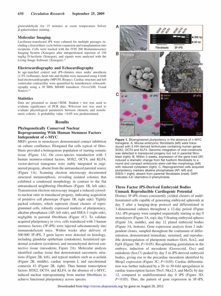
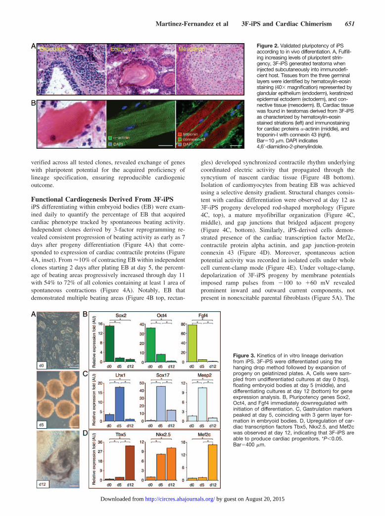
ResultsPhylogenetically Conserved NuclearReprogramming With Human Stemness FactorsIndependent of c-MYCMEFs grown in monolayers demonstrated contact inhibitionon culture confluence. Elongated flat cells typical of fibro-blasts provided a homogenous population of starting somatictissue (Figure 1A). On cross-species transduction with 3human stemness-related factors, SOX2, OCT4, and KLF4,vector-derived transgenes were stably integrated in engi-neered progeny, absent from the untransduced parental source(Figure 1A). Scanning electron microscopy documentedstructural metamorphosis, revealing isolated colonies thatexhibited a condensed morphology in contrast to the flatuntransduced neighboring fibroblasts (Figure 1B, left side).Transmission electron microscopy imaged a reduced cytosol-to-nuclear ratio in transduced progeny, indicating acquisitionof primitive cell phenotype (Figure 1B, right side). Tightlypacked colonies, which represent clonal clusters of repro-grammed cells, robustly expressed markers of pluripotency,alkaline phosphatase (AP; left side), and SSEA-1 (right side),negligible in parental fibroblasts (Figure 1C). To validateacquired pluripotency in vivo, cells transduced with 3 humanstemness factors (3F-iPS) were injected subcutaneously intoimmunodeficient mice. Within weeks after delivery of500 000 3F-iPS, 3 germ layers were detected on histology,including glandular epithelium (endoderm), keratinized epi-dermal ectoderm (ectoderm), and mesenchymal derived con-nective tissue (mesoderm; Figure 2A). Molecular analysisidentified cardiac tissue that demonstrated sarcomeric stria-tions (Figure 2B, left), and typical markers such as �-actinin(Figure 2B, middle), cardiac troponin I, and sarcolemmalconnexin 43 (Figure 2B, right). Thus human transcriptionfactors SOX2, OCT4, and KLF4, in the absence of c-MYC,induced nuclear reprogramming from murine fibroblasts toachieve functional pluripotency across species.
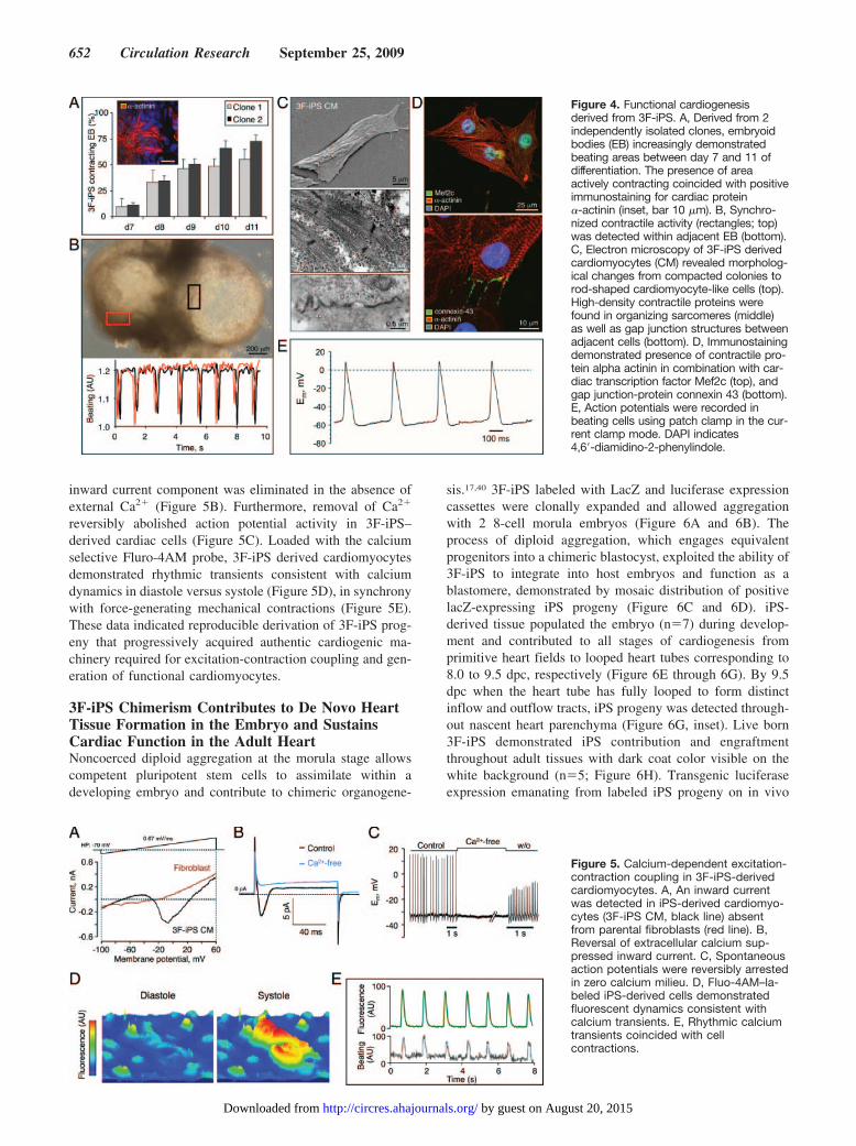
Three Factor iPS-Derived Embryoid BodiesUnmask Reproducible Cardiogenic PotentialDistinct 3F-iPS clones consistently yielded clusters of undif-ferentiated cells capable of generating embryoid spheroids atday 5 after a hanging-drop protocol and differentiated in3-dimensional cultures throughout a 12-day period (Figure3A). iPS progeny were sampled sequentially starting at day 0monolayers (Figure 3A, top), day 5 floating embryoid spheres(Figure 3A, middle), and day 12 plated embryoid bodies(Figure 3A, bottom). Gene expression analysis from 2 inde-pendent clones, sampled throughout the continuum of differ-entiation, demonstrated immediate, sustained, and reproduc-ible downregulation of pluripotent markers Oct4, Sox2, andFgf4 (Figure 3B; P�0.05). Recapitulating gastrulation in theembryo, induction of mesoderm (Goosecoid, Gsc) andendoderm (Sox17) peaked by day 5 in iPS-derived embryoidbodies, giving rise to the precardiac mesoderm identified byMesp2 expression (Figure 3C; P�0.05). Cardiac differentia-tion was further indicated by a 20- to 30-fold upregulation incardiac transcription factors Tbx5, Nkx2.5, and Mef2c by day12, compared to undifferentiated day 0 iPS (Figure 3D;P�0.05). Thus, the pattern of gene expression in 3F-iPS,
Figure 1. Bioengineered pluripotency in the absence of c-MYCtransgene. A, Mouse embryonic fibroblasts (left) were trans-duced with 3 HIV-derived lentiviruses containing human genesSOX2, OCT4 and KLF4. Genomic integration of viral constructswas detected in transduced progeny but not in parental fibro-blast (right). B, Within 3 weeks, expression of the gene triad (3F)induced a dramatic change from flat fusiform fibroblasts to around and compact embryonic-stem-cell-like morphology (left)with reduced cytoplasm (right). C, Reprogrammed cells acquiredpluripotency markers alkaline phosphatase (AP; left) andSSEA-1 (right), absent from parental fibroblasts (inset). DAPIindicates 4,6�-diamidino-2-phenylindole.
650 Circulation Research September 25, 2009
by guest on August 20, 2015http://circres.ahajournals.org/Downloaded from
verified across all tested clones, revealed exchange of geneswith pluripotent potential for the acquired proficiency oflineage specification, ensuring reproducible cardiogenicoutcome.
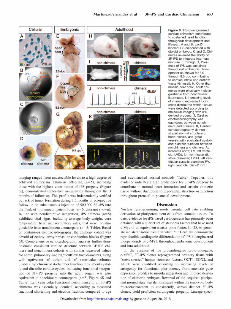
Functional Cardiogenesis Derived From 3F-iPSiPS differentiating within embryoid bodies (EB) were exam-ined daily to quantify the percentage of EB that acquiredcardiac phenotype tracked by spontaneous beating activity.Independent clones derived by 3-factor reprogramming re-vealed consistent progression of beating activity as early as 7days after progeny differentiation (Figure 4A) that corre-sponded to expression of cardiac contractile proteins (Figure4A, inset). From �10% of contracting EB within independentclones starting 2 days after plating EB at day 5, the percent-age of beating areas progressively increased through day 11with 54% to 72% of all colonies containing at least 1 area ofspontaneous contractions (Figure 4A). Notably, EB thatdemonstrated multiple beating areas (Figure 4B top, rectan-
gles) developed synchronized contractile rhythm underlyingcoordinated electric activity that propagated through thesyncytium of nascent cardiac tissue (Figure 4B bottom).Isolation of cardiomyocytes from beating EB was achievedusing a selective density gradient. Structural changes consis-tent with cardiac differentiation were observed at day 12 as3F-iPS progeny developed rod-shaped morphology (Figure4C, top), a mature myofibrillar organization (Figure 4C,middle), and gap junctions that bridged adjacent progeny(Figure 4C, bottom). Similarly, iPS-derived cells demon-strated presence of the cardiac transcription factor Mef2c,contractile protein alpha actinin, and gap junction-proteinconnexin 43 (Figure 4D). Moreover, spontaneous actionpotential activity was recorded in isolated cells under wholecell current-clamp mode (Figure 4E). Under voltage-clamp,depolarization of 3F-iPS progeny by membrane potentialsimposed ramp pulses from �100 to �60 mV revealedprominent inward and outward current components, notpresent in nonexcitable parental fibroblasts (Figure 5A). The
Figure 2. Validated pluripotency of iPSaccording to in vivo differentiation. A, Fulfill-ing increasing levels of pluripotent strin-gency, 3F-iPS generated teratoma wheninjected subcutaneously into immunodefi-cient host. Tissues from the three germinallayers were identified by hematoxylin-eosinstaining (40 magnification) represented byglandular epithelium (endoderm), keratinizedepidermal ectoderm (ectoderm), and con-nective tissue (mesoderm). B, Cardiac tissuewas found in teratomas derived from 3F-iPSas characterized by hematoxylin-eosinstained striations (left) and immunostainingfor cardiac proteins �-actinin (middle), andtroponin-I with connexin 43 (right).Bar10 �m. DAPI indicates4,6�-diamidino-2-phenylindole.
Figure 3. Kinetics of in vitro lineage derivationfrom iPS. 3F-iPS were differentiated using thehanging drop method followed by expansion ofprogeny on gelatinized plates. A, Cells were sam-pled from undifferentiated cultures at day 0 (top),floating embryoid bodies at day 5 (middle), anddifferentiating cultures at day 12 (bottom) for geneexpression analysis. B, Pluripotency genes Sox2,Oct4, and Fgf4 immediately downregulated withinitiation of differentiation. C, Gastrulation markerspeaked at day 5, coinciding with 3 germ layer for-mation in embryoid bodies. D, Upregulation of car-diac transcription factors Tbx5, Nkx2.5, and Mef2cwas observed at day 12, indicating that 3F-iPS areable to produce cardiac progenitors. *P�0.05.Bar400 �m.
Martinez-Fernandez et al 3F-iPS and Cardiac Chimerism 651
by guest on August 20, 2015http://circres.ahajournals.org/Downloaded from
inward current component was eliminated in the absence ofexternal Ca2� (Figure 5B). Furthermore, removal of Ca2�
reversibly abolished action potential activity in 3F-iPS–derived cardiac cells (Figure 5C). Loaded with the calciumselective Fluro-4AM probe, 3F-iPS derived cardiomyocytesdemonstrated rhythmic transients consistent with calciumdynamics in diastole versus systole (Figure 5D), in synchronywith force-generating mechanical contractions (Figure 5E).These data indicated reproducible derivation of 3F-iPS prog-eny that progressively acquired authentic cardiogenic ma-chinery required for excitation-contraction coupling and gen-eration of functional cardiomyocytes.
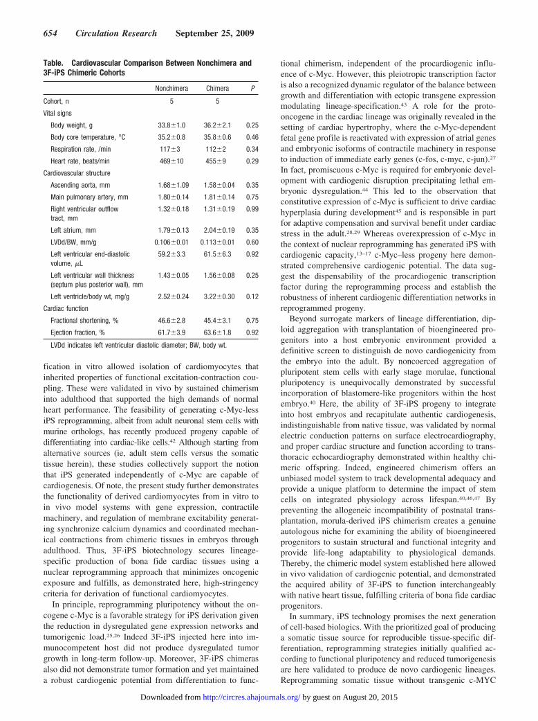
3F-iPS Chimerism Contributes to De Novo HeartTissue Formation in the Embryo and SustainsCardiac Function in the Adult HeartNoncoerced diploid aggregation at the morula stage allowscompetent pluripotent stem cells to assimilate within adeveloping embryo and contribute to chimeric organogene-
sis.17,40 3F-iPS labeled with LacZ and luciferase expressioncassettes were clonally expanded and allowed aggregationwith 2 8-cell morula embryos (Figure 6A and 6B). Theprocess of diploid aggregation, which engages equivalentprogenitors into a chimeric blastocyst, exploited the ability of3F-iPS to integrate into host embryos and function as ablastomere, demonstrated by mosaic distribution of positivelacZ-expressing iPS progeny (Figure 6C and 6D). iPS-derived tissue populated the embryo (n7) during develop-ment and contributed to all stages of cardiogenesis fromprimitive heart fields to looped heart tubes corresponding to8.0 to 9.5 dpc, respectively (Figure 6E through 6G). By 9.5dpc when the heart tube has fully looped to form distinctinflow and outflow tracts, iPS progeny was detected through-out nascent heart parenchyma (Figure 6G, inset). Live born3F-iPS demonstrated iPS contribution and engraftmentthroughout adult tissues with dark coat color visible on thewhite background (n5; Figure 6H). Transgenic luciferaseexpression emanating from labeled iPS progeny on in vivo
Figure 4. Functional cardiogenesisderived from 3F-iPS. A, Derived from 2independently isolated clones, embryoidbodies (EB) increasingly demonstratedbeating areas between day 7 and 11 ofdifferentiation. The presence of areaactively contracting coincided with positiveimmunostaining for cardiac protein�-actinin (inset, bar 10 �m). B, Synchro-nized contractile activity (rectangles; top)was detected within adjacent EB (bottom).C, Electron microscopy of 3F-iPS derivedcardiomyocytes (CM) revealed morpholog-ical changes from compacted colonies torod-shaped cardiomyocyte-like cells (top).High-density contractile proteins werefound in organizing sarcomeres (middle)as well as gap junction structures betweenadjacent cells (bottom). D, Immunostainingdemonstrated presence of contractile pro-tein alpha actinin in combination with car-diac transcription factor Mef2c (top), andgap junction-protein connexin 43 (bottom).E, Action potentials were recorded inbeating cells using patch clamp in the cur-rent clamp mode. DAPI indicates4,6�-diamidino-2-phenylindole.
Figure 5. Calcium-dependent excitation-contraction coupling in 3F-iPS-derivedcardiomyocytes. A, An inward currentwas detected in iPS-derived cardiomyo-cytes (3F-iPS CM, black line) absentfrom parental fibroblasts (red line). B,Reversal of extracellular calcium sup-pressed inward current. C, Spontaneousaction potentials were reversibly arrestedin zero calcium milieu. D, Fluo-4AM–la-beled iPS-derived cells demonstratedfluorescent dynamics consistent withcalcium transients. E, Rhythmic calciumtransients coincided with cellcontractions.
652 Circulation Research September 25, 2009
by guest on August 20, 2015http://circres.ahajournals.org/Downloaded from
imaging ranged from undetectable levels to a high degree ofachieved chimerism. Chimeric offspring (n5), includingthose with the highest contribution of iPS progeny (Figure6I), demonstrated tumor-free assimilation throughout the 3months of follow-up. This profile was independently verifiedby lack of tumor formation during 7.5 months of prospectivefollow-up on subcutaneous injection of 500 000 3F-iPS intothe flank of immunocompetent hosts (n6, data not shown).In line with nondisruptive integration, iPS chimera (n5)exhibited vital signs, including average body weight, coretemperature, heart and respiratory rates, that were indistin-guishable from nonchimera counterparts (n5; Table). Basedon continuous electrocardiography, the chimeric cohort wasdevoid of ectopy, arrhythmias, or conduction blocks (Figure6J). Comprehensive echocardiography analysis further dem-onstrated consistent cardiac structure between 3F-iPS chi-mera and nonchimera cohorts with similar measured valuesfor aortic, pulmonary, and right outflow tract diameters, alongwith equivalent left atrium and left ventricular volumes(Table). Synchronized 4-chamber function throughout systol-ic and diastolic cardiac cycles, indicating functional integra-tion of 3F-iPS progeny into the adult organ, was alsoequivalent to nonchimera counterparts (n5, Figure 6K andTable). Left ventricular functional performance of all 3F-iPSchimeras was essentially identical, according to measuredfractional shortening and ejection fraction, compared to age
and sex-matched normal controls (Table). Together, thisevidence indicates a high proficiency for 3F-iPS progeny tocontribute to normal heart formation and sustain chimerictissue without disruption to myocardial structure or functionthroughout prenatal to postnatal development.
DiscussionNuclear reprogramming resets parental cell fate enablingderivation of pluripotent stem cells from somatic tissues. Todate, evidence for iPS-based cardiogenesis has primarily beenobtained with a quartet set of stemness factors that have usedc-Myc or an equivalent transcription factor, Lin28, to gener-ate isolated cardiac tissue in vitro.13–17 Here, we demonstratereproducible cardiogenic differentiation of iPS bioengineeredindependently of c-MYC throughout embryonic developmentand into adulthood.
In the absence of the procardiogenic proto-oncogene,c-MYC, 3F-iPS clones reprogrammed ordinary tissue with“cross-species” human stemness factors, OCT4, SOX2, andKLF4, were qualified according to increasing levels ofstringency for functional pluripotency from atavistic geneexpression profiles to morula integration and in utero deriva-tion of chimeric embryos. Reversal of the acquired pluripo-tent ground state was demonstrated within the embryoid bodymicroenvironment to consistently, across distinct 3F-iPSclones, yield proficient cardiogenic progeny. Lineage speci-
Figure 6. iPS-bioengineeredcardiac chimerism contributesto sustained heart functionthroughout development andlifespan. A and B, LacZ-labeled iPS coincubated withdiploid embryos. C and D, Chi-meras revealed the ability of3F-iPS to integrate into hostmorulae. E through G, Pres-ence of iPS was sustainedthroughout embryonic devel-opment as shown for 8.0through 9.5 dpc contributingto cardiac inflow and outflowtracts (G, inset). H, Other thanmosaic coat color, adult chi-meras were physically indistin-guishable from nonchimericlittermates. I, Increasing levelsof chimeric expressed lucif-erase distributed within tissueswere detected according tomolecular imaging with iPS-derived progeny. J, Cardiacelectrocardiography wasequivalent between nonchi-mera and chimera. K, Cardiacechocardiography demon-strated normal structure ofheart, valves, and great-vessels with equivalent systolicand diastolic function betweennonchimera and chimera. Aoindicates aorta; LV, left ventri-cle; LVDd, left ventricular dia-stolic diameter; LVSd, left ven-tricular systolic diameter; RV,right ventricle. Bar2 mm.
Martinez-Fernandez et al 3F-iPS and Cardiac Chimerism 653
by guest on August 20, 2015http://circres.ahajournals.org/Downloaded from
fication in vitro allowed isolation of cardiomyocytes thatinherited properties of functional excitation-contraction cou-pling. These were validated in vivo by sustained chimerisminto adulthood that supported the high demands of normalheart performance. The feasibility of generating c-Myc-lessiPS reprogramming, albeit from adult neuronal stem cells withmurine orthologs, has recently produced progeny capable ofdifferentiating into cardiac-like cells.42 Although starting fromalternative sources (ie, adult stem cells versus the somatictissue herein), these studies collectively support the notionthat iPS generated independently of c-Myc are capable ofcardiogenesis. Of note, the present study further demonstratesthe functionality of derived cardiomyocytes from in vitro toin vivo model systems with gene expression, contractilemachinery, and regulation of membrane excitability generat-ing synchronize calcium dynamics and coordinated mechan-ical contractions from chimeric tissues in embryos throughadulthood. Thus, 3F-iPS biotechnology secures lineage-specific production of bona fide cardiac tissues using anuclear reprogramming approach that minimizes oncogenicexposure and fulfills, as demonstrated here, high-stringencycriteria for derivation of functional cardiomyocytes.
In principle, reprogramming pluripotency without the on-cogene c-Myc is a favorable strategy for iPS derivation giventhe reduction in dysregulated gene expression networks andtumorigenic load.25,26 Indeed 3F-iPS injected here into im-munocompetent host did not produce dysregulated tumorgrowth in long-term follow-up. Moreover, 3F-iPS chimerasalso did not demonstrate tumor formation and yet maintaineda robust cardiogenic potential from differentiation to func-
tional chimerism, independent of the procardiogenic influ-ence of c-Myc. However, this pleiotropic transcription factoris also a recognized dynamic regulator of the balance betweengrowth and differentiation with ectopic transgene expressionmodulating lineage-specification.43 A role for the proto-oncogene in the cardiac lineage was originally revealed in thesetting of cardiac hypertrophy, where the c-Myc-dependentfetal gene profile is reactivated with expression of atrial genesand embryonic isoforms of contractile machinery in responseto induction of immediate early genes (c-fos, c-myc, c-jun).27
In fact, promiscuous c-Myc is required for embryonic devel-opment with cardiogenic disruption precipitating lethal em-bryonic dysregulation.44 This led to the observation thatconstitutive expression of c-Myc is sufficient to drive cardiachyperplasia during development45 and is responsible in partfor adaptive compensation and survival benefit under cardiacstress in the adult.28,29 Whereas overexpression of c-Myc inthe context of nuclear reprogramming has generated iPS withcardiogenic capacity,13–17 c-Myc–less progeny here demon-strated comprehensive cardiogenic potential. The data sug-gest the dispensability of the procardiogenic transcriptionfactor during the reprogramming process and establish therobustness of inherent cardiogenic differentiation networks inreprogrammed progeny.
Beyond surrogate markers of lineage differentiation, dip-loid aggregation with transplantation of bioengineered pro-genitors into a host embryonic environment provided adefinitive screen to distinguish de novo cardiogenicity fromthe embryo into the adult. By noncoerced aggregation ofpluripotent stem cells with early stage morulae, functionalpluripotency is unequivocally demonstrated by successfulincorporation of blastomere-like progenitors within the hostembryo.40 Here, the ability of 3F-iPS progeny to integrateinto host embryos and recapitulate authentic cardiogenesis,indistinguishable from native tissue, was validated by normalelectric conduction patterns on surface electrocardiography,and proper cardiac structure and function according to trans-thoracic echocardiography demonstrated within healthy chi-meric offspring. Indeed, engineered chimerism offers anunbiased model system to track developmental adequacy andprovide a unique platform to determine the impact of stemcells on integrated physiology across lifespan.40,46,47 Bypreventing the allogeneic incompatibility of postnatal trans-plantation, morula-derived iPS chimerism creates a genuineautologous niche for examining the ability of bioengineeredprogenitors to sustain structural and functional integrity andprovide life-long adaptability to physiological demands.Thereby, the chimeric model system established here allowedin vivo validation of cardiogenic potential, and demonstratedthe acquired ability of 3F-iPS to function interchangeablywith native heart tissue, fulfilling criteria of bona fide cardiacprogenitors.
In summary, iPS technology promises the next generationof cell-based biologics. With the prioritized goal of producinga somatic tissue source for reproducible tissue-specific dif-ferentiation, reprogramming strategies initially qualified ac-cording to functional pluripotency and reduced tumorigenesisare here validated to produce de novo cardiogenic lineages.Reprogramming somatic tissue without transgenic c-MYC
Table. Cardiovascular Comparison Between Nonchimera and3F-iPS Chimeric Cohorts
Nonchimera Chimera P
Cohort, n 5 5
Vital signs
Body weight, g 33.8�1.0 36.2�2.1 0.25
Body core temperature, °C 35.2�0.8 35.8�0.6 0.46
Respiration rate, /min 117�3 112�2 0.34
Heart rate, beats/min 469�10 455�9 0.29
Cardiovascular structure
Ascending aorta, mm 1.68�1.09 1.58�0.04 0.35
Main pulmonary artery, mm 1.80�0.14 1.81�0.14 0.75
Right ventricular outflowtract, mm
1.32�0.18 1.31�0.19 0.99
Left atrium, mm 1.79�0.13 2.04�0.19 0.35
LVDd/BW, mm/g 0.106�0.01 0.113�0.01 0.60
Left ventricular end-diastolicvolume, �L
59.2�3.3 61.5�6.3 0.92
Left ventricular wall thickness(septum plus posterior wall), mm
1.43�0.05 1.56�0.08 0.25
Left ventricle/body wt, mg/g 2.52�0.24 3.22�0.30 0.12
Cardiac function
Fractional shortening, % 46.6�2.8 45.4�3.1 0.75
Ejection fraction, % 61.7�3.9 63.6�1.8 0.92
LVDd indicates left ventricular diastolic diameter; BW, body wt.
654 Circulation Research September 25, 2009
by guest on August 20, 2015http://circres.ahajournals.org/Downloaded from
expression offers a reliable platform to ensure proficientiPS-derived cardiogenesis that achieves seamless integrationand functional contribution to pre and postnatal cardiacperformance.
AcknowledgmentsWe thank Lois Rowe for expert histological analysis.
Sources of FundingThis work was supported by National Institutes of Health(R01HL083439, T32HL007111, R01HL085208, R56AI074363),American Heart Association, American Society for Clinical Pharma-cology and Therapeutics, La Caixa Foundation Graduate Program,Marriott Individualized Medicine Program, Marriott Heart DiseaseResearch Program, Gerstner Family Career Development Award inIndividualized Medicine, and Mayo Clinic.
DisclosuresNone.
References1. Yamanaka S. A fresh look at iPS cells. Cell. 2009;137:13–17.2. Hochedlinger K, Plath K. Epigenetic reprogramming and induced pluri-
potency. Development. 2009;136:509–523.3. Nishikawa S, Goldstein RA, Nierras CR. The promise of human induced
pluripotent stem cells for research and therapy. Nat Rev Mol Cell Biol.2008;9:725–729.
4. Takahashi K, Okita K, Nakagawa M, Yamanaka S. Induction of pluri-potent stem cells from mouse embryonic and adult fibroblast cultures bydefined factors. Cell. 2006;126:663–676.
5. Markoulaki S, Hanna J, Beard C, Carey BW, Cheng AW, Lengner CJ,Dausman JA, Fu D, Gao Q, Wu S, Cassady JP, Jaenisch R. Transgenicmice with defined combinations of drug-inducible reprogrammingfactors. Nat Biotechnol. 2009;27:169–171.
6. Hanna J, Markoulaki S, Schorderet P, Carey BW, Beard C, Wernig M,Creyghton MP, Steine EJ, Cassady JP, Foreman R, Lengner CJ, DausmanJA, Jaenisch R. Direct reprogramming of terminally differentiated matureB lymphocytes to pluripotency. Cell. 2008;133:250–264.
7. Park I-H, Arora N, Huo H, Maherali N, Ahfeldt T, Shimamura A, LenschMW, Cowan C, Hochedlinger K, Daley GQ. Disease-specific inducedpluripotent stem cells. Cell. 2008;134:877–886.
8. Dimos JT, Rodolfa KT, Niakan KK, Weisenthal LM, Mitsumoto H,Chung W, Croft GF, Saphier G, Leibel R, Goland R, Wichterle H,Henderson CE, Eggan K. Induced pluripotent stem cells generated frompatients with ALS can be differentiated into motor neurons. Science.2008;321:1218–1221.
9. Yu J, Vodyanik MA, Smuga-Otto K, Antosiewicz-Bourget J, Frane JL,Tian S, Nie J, Jonsdottir GA, Ruotti V, Stewart R, Slukvin I, ThomsonJA. Induced pluripotent stem cell lines derived from human somatic cells.Science. 2007;318:1917–1920.
10. Hanna J, Wernig M, Markoulaki S, Sun CW, Meissner A, Cassady JP,Beard C, Brambrink T, Wu LC, Townes TM, Jaenisch R. Treatment ofsickle cell anemia mouse model with iPS cells generated from autologousskin. Science. 2007;318:1920–1923.
11. Wernig M, Zhao JP, Pruszak J, Hedlund E, Fu D, Soldner F, Broccoli V,Constantine-Paton M, Isacson O, Jaenisch R. Neurons derived fromreprogrammed fibroblasts functionally integrate into the fetal brain andimprove symptoms of rats with Parkinson’s disease. Proc Natl Acad SciU S A. 2008;105:5856–5861.
12. Xu D, Alipio Z, Fink LM, Adcock DM, Yang J, Ward DC, Ma Y.Phenotypic correction of murine hemophilia A using an iPS cell-basedtherapy. Proc Natl Acad Sci U S A. 2009;106:808–813.
13. Schenke-Layland K, Rhodes KE, Angelis E, Butylkova Y, Heydarkhan-Hagvall S, Gekas C, Zhang R, Goldhaber JI, Mikkola HK, Plath K,MacLellan WR. Reprogrammed mouse fibroblasts differentiate into cellsof the cardiovascular and hematopoietic lineages. Stem Cells. 2008;26:1537–1546.
14. Mauritz C, Schwanke K, Reppel M, Neef S, Katsirntaki K, Maier LS,Nguemo F, Menke S, Haustein M, Hescheler J, Hasenfuss G, Martin U.Generation of functional murine cardiac myocytes from induced pluri-potent stem cells. Circulation. 2008;118:507–517.
15. Narazaki G, Uosaki H, Teranishi Mm, Okita K, Kim B, Matsuoka S,Yamanaka S, Yamashita JK. Directed and systematic differentiation of car-diovascular cells from mouse induced pluripotent stem cells. Circulation.2008;118:498–506.
16. Zhang J, Wilson G, Soerens A, Koonce C, Yu J, Palecek S, Thomson J,Kamp T. Functional cardiomyocytes derived from human induced pluri-potent stem cells. Circ Res. 2009;104:e30–e41.
17. Nelson TJ, Martinez-Fernandez A, Yamada S, Perez-Terzic C, Ikeda Y,Terzic A. Repair of acute myocardial infarction with human stemnessfactors induced pluirpotent stem cells. Circulation. 2009;120:408–416.
18. Nelson TJ, Behfar A, Terzic A. Stem cells: biologics for regeneration.Clin Pharmacol Ther. 2008;84:620–623.
19. Tanaka T, Tohyama S, Murata M, Nomura F, Kaneko T, Chen H, HattoriF, Egashira T, Seki T, Ohno Y, Koshimizu U, Yuasa S, Ogawa S,Yamanaka S, Yasuda K, Fukuda K. In vitro pharmacologic testing usinghuman induced pluripotent stem cell-derived cardiomyocytes. BiochemBiophys Res Commun. 2009;385:497–502.
20. Stadtfeld M, Nagaya M, Utikal J, Weir G, Hochedlinger K. Inducedpluripotent stem cells generated without viral integration. Science. 2008;322:945–949.
21. Okita K, Nakagawa M, Hyenjong H, Ichisaka T, Yamanaka S. Generationof mouse induced pluripotent stem cells without viral vectors. Science.2008;322:949–953.
22. Woltjen K, Michael IP, Mohseni P, Desai R, Mileikovsky M, HamalainenR, Cowling R, Wang W, Liu P, Gertsenstein M, Kaji K, Sung HK, NagyA. piggyBac transposition reprograms fibroblasts to induced pluripotentstem cells. Nature. 2009;458:766–770.
23. Yu J, Hu K, Smuga-Otto K, Tian S, Stewart R, Slukvin II, Thomson JA.Human Induced pluripotent stem cells free of vector and transgenesequences. Science. 2009;324:797–801.
24. Nelson TJ, Terzic A. Induced pluripotent stem cells: Reprogrammedwithout a trace. Regen Med. 2009;4:333–335.
25. Nakagawa M, Koyanagi M, Tanabe K, Takahashi K, Ichisaka T, Aoi T,Okita K, Mochiduki Y, Takizawa N, Yamanaka S. Generation of inducedpluripotent stem cells without Myc from mouse and human fibroblasts.Nat Biotechnol. 2008;26:101–106.
26. Wernig M, Meissner A, Cassady JP, Jaenisch R. c-Myc is dispensable fordirect reprogramming of mouse fibroblasts. Cell Stem Cell. 2008;2:10–12.
27. Izumo S, Nadal-Ginard B, Mahdavi V. Protooncogene induction andreprogramming of cardiac gene expression produced by pressureoverload. Proc Natl Acad Sci U S A. 1988;85:339–343.
28. Xiao G, Mao S, Baumgarten G, Serrano J, Jordan MC, Roos KP, FishbeinMC, MacLellan WR. Inducible activation of c-Myc in adult myocardiumin vivo provokes cardiac myocyte hypertrophy and reactivation of DNAsynthesis. Circ Res. 2001;89:1122–1129.
29. Zhong W, Mao S, Tobis S, Angelis E, Jordan MC, Roos KP, FishbeinMC, de Alboran IM, MacLellan WR. Hypertrophic growth in cardiacmyocytes is mediated by Myc through a Cyclin D2-dependent pathway.EMBO J. 2006;25:3869–3879.
30. Amabile G, Meissner A. Induced pluripotent stem cells: current progressand potential for regenerative medicine. Trends Mol Med. 2009;15:59–68.
31. Nelson TJ, Martinez-Fernandez AJ, Yamada S, Mael AA, Terzic A, IkedaY. Induced pluripotent reprogramming from promiscuous humanstemness-related factors. Clin Translation Sci. 2009;2:118–126.
32. Guo W, Lasky JL, Chang CJ, Mosessian S, Lewis X, Xiao Y, Yeh JE,Chen JY, Iruela-Arispe ML, Varella-Garcia M, Wu H. Multi-geneticevents collaboratively contribute to Pten-null leukaemia stem-cell for-mation. Nature. 2008;453:529–533.
33. Perez-Terzic C, Faustino RS, Boorsma BJ, Arrell DK, Niederlander NJ,Behfar A, Terzic A Stem cells transform into a cardiac phenotype withremodeling of the nuclear transport machinery. Nat Clin Pract CardiovascMed. 2007;4:S68–S76.
34. Perez-Terzic C, Behfar A, Mery A, van Deursen JM, Terzic A, Puceat M.Structural adaptation of the nuclear pore complex in stem cell-derivedcardiomyocytes. Circ Res. 2003;92:444–452.
35. Behfar A, Zingman LV, Hodgson DM, Rauzier JM, Kane GC, Terzic A,Puceat M. Stem cell differentiation requires a paracrine pathway in theheart. FASEB J. 2002;16:1558–1566.
36. Behfar A, Perez-Terzic C, Faustino RS, Arrell DK, Hodgson DM,Yamada S, Puceat M, Niederlander N, Alekseev AE, Zingman LV, TerzicA. Cardiopoietic programming of embryonic stem cells for tumor-freeheart repair. J Exp Med. 2007;204:405–420.
Martinez-Fernandez et al 3F-iPS and Cardiac Chimerism 655
by guest on August 20, 2015http://circres.ahajournals.org/Downloaded from
37. Nelson TJ, Chiriac A, Faustino RS, Crespo-Diaz RJ, Behfar A, Terzic A.Lineage specification of Flk-1� progenitors is associated with divergentSox7 expression in cardiopoiesis. Differentiation. 2009;77:248–255.
38. Nelson TJ, Faustino RS, Chiriac A, Crespo-Diaz R, Behfar A, Terzic A.CXCR4�/FLK-1� biomarkers select a cardiopoietic lineage fromembryonic stem cells. Stem Cells. 2008;26:1464–1473.
39. Hodgson DM, Behfar A, Zingman LV, Kane GC, Perez-Terzic C,Alekseev AE, Puceat M, Terzic A. Stable benefit of embryonic stem celltherapy in myocardial infarction. Am J Physiol Heart Circ Physiol.2004;287:H471–H479.
40. Nelson TJ, Martinez-Fernandez A, Terzic A. KCNJ11 knockout morulareengineered by stem cell diploid aggregation. Phil Trans R Soc Lond BBiol Sci. 2009;364:269–276.
41. Yamada S, Nelson TJ, Crespo-Diaz RJ, Perez-Terzic C, Liu XK, Miki T,Seino S, Behfar A, Terzic A. Embryonic stem cell therapy of heart failurein genetic cardiomyopathy. Stem Cells. 2008;26:2644–2653.
42. Kim JB, Sebastiano V, Wu G, Arauzo-Bravo MJ, Sasse P, Gentile L, KoK, Ruau D, Ehrich M, van den Boom D, Meyer J, Hubner K, BernemannC, Ortmeier C, Zenke M, Fleischmann BK, Zaehres H, Scholer HR.
Oct4-induced pluripotency in adult neural stem cells. Cell. 2009;136:411–419.
43. Guo Y, Niu C, Breslin P, Tang M, Zhang S, Wei W, Kini AR, Paner GP,Alkan S, Morris SW, Diaz M, Stiff PJ, Zhang J. c-Myc-mediated controlof cell fate in megakaryocyte-erythrocyte progenitors. Blood. In press.
44. Baudino TA, McKay C, Pendeville-Samain H, Nilsson JA, Maclean KH,White EL, Davis AC, Ihle JN, Cleveland JL. c-Myc is essential forvasculogenesis and angiogenesis during development and tumor pro-gression. Genes Dev. 2002;16:2530–2543.
45. Jackson T, Allard MF, Sreenan CM, Doss LK, Bishop SP, Swain JL. Thec-myc proto-oncogene regulates cardiac development in transgenic mice.Mol Cell Biol. 1990;10:3709–3716.
46. Yamada S, Nelson TJ, Behfar A, Crespo-Diaz RJ, Fraidenraich D, TerzicA. Stem cell transplant into pre-implantation embryo yields myocardialinfarction-resistant adult phenotype. Stem Cells. 2009;27:1697–1705.
47. Stillwell E, Vitale J, Zhao Q, Beck A, Schneider J, Khadim F, Elson G,Altaf A, Yehia G, Dong J-H, Liu J, Mark W, Bhaumik M, Grange R,Fraidenraich D. Blastocyst injection of wild type embryonic stem cellsinduces global corrections in mdx mice. PLos ONE. 2009;4:e4759.
656 Circulation Research September 25, 2009
by guest on August 20, 2015http://circres.ahajournals.org/Downloaded from
Alekseev, Carmen Perez-Terzic, Yasuhiro Ikeda and Andre TerzicAlmudena Martinez-Fernandez, Timothy J. Nelson, Satsuki Yamada, Santiago Reyes, Alexey E.
ChimerismiPS Programmed Without c-MYC Yield Proficient Cardiogenesis for Functional Heart
Print ISSN: 0009-7330. Online ISSN: 1524-4571 Copyright © 2009 American Heart Association, Inc. All rights reserved.is published by the American Heart Association, 7272 Greenville Avenue, Dallas, TX 75231Circulation Research
doi: 10.1161/CIRCRESAHA.109.2031092009;105:648-656; originally published online August 20, 2009;Circ Res.
http://circres.ahajournals.org/content/105/7/648World Wide Web at:
The online version of this article, along with updated information and services, is located on the
http://circres.ahajournals.org//subscriptions/
is online at: Circulation Research Information about subscribing to Subscriptions:
http://www.lww.com/reprints Information about reprints can be found online at: Reprints:
document. Permissions and Rights Question and Answer about this process is available in the
located, click Request Permissions in the middle column of the Web page under Services. Further informationEditorial Office. Once the online version of the published article for which permission is being requested is
can be obtained via RightsLink, a service of the Copyright Clearance Center, not theCirculation Researchin Requests for permissions to reproduce figures, tables, or portions of articles originally publishedPermissions:
by guest on August 20, 2015http://circres.ahajournals.org/Downloaded from
Related Documents