Ipilimumab-dependent cell-mediated cytotoxicity of regulatory T cells ex vivo by nonclassical monocytes in melanoma patients Emanuela Romano a,b,c,1 , Monika Kusio-Kobialka b , Periklis G. Foukas c,d , Petra Baumgaertner c , Christiane Meyer c , Pierluigi Ballabeni e , Olivier Michielin a,c , Benjamin Weide f , Pedro Romero c , and Daniel E. Speiser c a Service of Medical Oncology, b Laboratory of Tumor Immunobiology, c Ludwig Cancer Research Center and Department of Oncology, and e Institute of Social and Preventive Medicine, University Hospital of Lausanne, 1011 Lausanne, Switzerland; and d Department of Pathology, University of Athens Medical School, 11527 Athens, Greece; and f Department of Dermatology, University Medical Center, 72076 Tübingen, Germany Edited by Ira Mellman, Genentech, Inc., South San Francisco, CA, and approved March 30, 2015 (received for review September 9, 2014) Enhancing immune responses with immune-modulatory mono- clonal antibodies directed to inhibitory immune receptors is a promising modality in cancer therapy. Clinical efficacy has been demonstrated with antibodies blocking inhibitory immune check- points such as cytotoxic T lymphocyte–associated antigen 4 (CTLA-4) or PD-1/PD-L1. Treatment with ipilimumab, a fully human CTLA-4– specific mAb, showed durable clinical efficacy in metastatic mela- noma; its mechanism of action is, however, only partially under- stood. This is a study of 29 patients with advanced cutaneous melanoma treated with ipilimumab. We analyzed peripheral blood mononuclear cells (PBMCs) and matched melanoma metastases from 15 patients responding and 14 not responding to ipilimumab by multicolor flow cytometry, antibody-dependent cell-mediated cytotoxicity (ADCC) assay, and immunohistochemistry. PBMCs and matched tumor biopsies were collected 24 h before (i.e., baseline) and up to 4 wk after ipilimumab. Our findings show, to our knowl- edge for the first time, that ipilimumab can engage ex vivo FcγRIIIA (CD16)-expressing, nonclassical monocytes resulting in ADCC-medi- ated lysis of regulatory T cells (Tregs). In contrast, classical CD14 ++ CD16 - monocytes are unable to do so. Moreover, we show that patients responding to ipilimumab display significantly higher base- line peripheral frequencies of nonclassical monocytes compared with nonresponder patients. In the tumor microenvironment, re- sponders have higher CD68 + /CD163 + macrophage ratios at baseline and show decreased Treg infiltration after treatment. Together, our results suggest that anti–CTLA-4 therapy may target Tregs in vivo. Larger translational studies are, however, warranted to substantiate this mechanism of action of ipilimumab in patients. monocytes | macrophages | Tregs | ipilimumab | ADCC T he clinical benefit of immune checkpoint blockade represents arguably one of the most significant advances of modern oncology, as it has definitively proven to produce substantial clinical responses, not only in tumors traditionally perceived as “immunogenic” but also in a variety of solid tumors (1). Two pivotal phase III clinical trials in patients with advanced mela- noma of the fully human, anti–cytotoxic T lymphocyte–associ- ated antigen 4 (CTLA-4) IgG1 monoclonal antibody ipilimumab (Yervoy; previously MDX-010; Medarex/Bristol Myers-Squibb) reported significantly increased overall survival, which led to its approval by the US Food and Drug Administration (2, 3). Ipi- limumab represents the first of a new class of cancer therapies that function by enhancing host antitumor immunity. However, clinical benefit following immune checkpoint blockade of CTLA-4 is limited to a fraction (10–15%) of treated patients, and the mechanism(s) of action are still poorly understood. Moreover, no reliable biomarkers of clinical efficacy are available to date. Although CTLA-4 belongs to the family of inhibitory lymphocyte receptors, it does not appear to transmit inhibitory signals into effector T cells (4). Rather, CTLA-4 may control key functions of regulatory T cells (Tregs) and of antigen-presenting cells (5–7). More recently, studies in mice revealed that the antitumor activity of CTLA-4 blockade is mediated by Fc gamma receptor (FcγRIV)- expressing macrophages in the tumor microenvironment (TME) via in trans depletion of tumor-infiltrating Tregs (8–10). We speculated that a similar mechanism might operate in melanoma patients, who show a response to ipilimumab. To investigate this hypothesis, we interrogated peripheral blood mononuclear cells (PBMCs) and matched melanoma metastases from 15 patients responding and 14 nonresponding to ipilimumab. Results High Baseline Frequencies of Nonclassical CD14 + CD16 ++ Monocytes in Patients Responding to Ipilimumab. Between September 2010 and July 2013, 29 patients with stage IV cutaneous melanoma, pro- gressing to at least one prior line of therapy, received a maximum of four cycles of 3 mg/kg ipilimumab i.v. every 3 wk. Mean age was 62 y (range, 37–85 y). The majority of patients were male (72%) and had stage IV M1c disease (21 of 29 patients, 72%; Table 1). Median follow-up was 9.41 mo (range, 2–39 mo). Tu- mor response was assessed 16 or 24 wk after starting ipilimumab by immune-related response criteria (11). To compare data sets, we selected equal numbers of patients who showed objective Significance mAbs directed to inhibitory immune receptors represent a very promising class of immunotherapeutics. This study suggests a potential mechanism of action of ipilimumab (a fully human anti–cytotoxic T lymphocyte–associated antigen 4), by which FcγRIIIA (CD16)-expressing nonclassical monocytes kill regula- tory T cells ex vivo via antibody-dependent cell-mediated cy- totoxicity. Notably, at baseline, responder patients display significantly higher peripheral frequencies of nonclassical mono- cytes and a selective enrichment in tumor-infiltrating CD68 + CD16 + macrophages compared with nonresponder patients. If further confirmed, these findings may contribute to the generation of predictive biomarker panels, antibody design, and the develop- ment of rational combination therapies to promote antitumor immunity. Author contributions: E.R. and D.E.S. designed research; E.R., M.K.-K., P.G.F., P. Baumgaertner, and C.M. performed research; P.G.F. contributed new reagents/analytic tools; E.R., M.K.-K., P.G.F., P. Baumgaertner, C.M., P. Ballabeni, P.R., and D.E.S. analyzed data; E.R., M.K.-K., P.G.F., P. Baumgaertner, C.M., P. Ballabeni, O.M., B.W., P.R., and D.E.S. wrote the paper; and O.M. and B.W. provided patient samples. Conflict of interest statement: P.R. holds a consultant or advisory role with Immatics Biotechnologies and Formune. O.M. holds a consultant or advisory role with Bristol-Myers Squibb, Hoffmann-La Roche, MSD, and Glaxo-SmithKline. B.W. receives honoraria and research funding from Bristol-Myers Squibb. This article is a PNAS Direct Submission. 1 To whom correspondence should be addressed. Email: [email protected]. This article contains supporting information online at www.pnas.org/lookup/suppl/doi:10. 1073/pnas.1417320112/-/DCSupplemental. 6140–6145 | PNAS | May 12, 2015 | vol. 112 | no. 19 www.pnas.org/cgi/doi/10.1073/pnas.1417320112

Welcome message from author
This document is posted to help you gain knowledge. Please leave a comment to let me know what you think about it! Share it to your friends and learn new things together.
Transcript

Ipilimumab-dependent cell-mediated cytotoxicity ofregulatory T cells ex vivo by nonclassical monocytes inmelanoma patientsEmanuela Romanoa,b,c,1, Monika Kusio-Kobialkab, Periklis G. Foukasc,d, Petra Baumgaertnerc, Christiane Meyerc,Pierluigi Ballabenie, Olivier Michielina,c, Benjamin Weidef, Pedro Romeroc, and Daniel E. Speiserc
aService of Medical Oncology, bLaboratory of Tumor Immunobiology, cLudwig Cancer Research Center and Department of Oncology, and eInstitute ofSocial and Preventive Medicine, University Hospital of Lausanne, 1011 Lausanne, Switzerland; and dDepartment of Pathology, University of Athens MedicalSchool, 11527 Athens, Greece; and fDepartment of Dermatology, University Medical Center, 72076 Tübingen, Germany
Edited by Ira Mellman, Genentech, Inc., South San Francisco, CA, and approved March 30, 2015 (received for review September 9, 2014)
Enhancing immune responses with immune-modulatory mono-clonal antibodies directed to inhibitory immune receptors is apromising modality in cancer therapy. Clinical efficacy has beendemonstrated with antibodies blocking inhibitory immune check-points such as cytotoxic T lymphocyte–associated antigen 4 (CTLA-4)or PD-1/PD-L1. Treatment with ipilimumab, a fully human CTLA-4–specific mAb, showed durable clinical efficacy in metastatic mela-noma; its mechanism of action is, however, only partially under-stood. This is a study of 29 patients with advanced cutaneousmelanoma treated with ipilimumab. We analyzed peripheral bloodmononuclear cells (PBMCs) and matched melanoma metastasesfrom 15 patients responding and 14 not responding to ipilimumabby multicolor flow cytometry, antibody-dependent cell-mediatedcytotoxicity (ADCC) assay, and immunohistochemistry. PBMCs andmatched tumor biopsies were collected 24 h before (i.e., baseline)and up to 4 wk after ipilimumab. Our findings show, to our knowl-edge for the first time, that ipilimumab can engage ex vivo FcγRIIIA(CD16)-expressing, nonclassical monocytes resulting in ADCC-medi-ated lysis of regulatory T cells (Tregs). In contrast, classical CD14++
CD16− monocytes are unable to do so. Moreover, we show thatpatients responding to ipilimumab display significantly higher base-line peripheral frequencies of nonclassical monocytes comparedwith nonresponder patients. In the tumor microenvironment, re-sponders have higher CD68+/CD163+ macrophage ratios at baselineand show decreased Treg infiltration after treatment. Together, ourresults suggest that anti–CTLA-4 therapy may target Tregs in vivo.Larger translational studies are, however, warranted to substantiatethis mechanism of action of ipilimumab in patients.
monocytes | macrophages | Tregs | ipilimumab | ADCC
The clinical benefit of immune checkpoint blockade representsarguably one of the most significant advances of modern
oncology, as it has definitively proven to produce substantialclinical responses, not only in tumors traditionally perceived as“immunogenic” but also in a variety of solid tumors (1). Twopivotal phase III clinical trials in patients with advanced mela-noma of the fully human, anti–cytotoxic T lymphocyte–associ-ated antigen 4 (CTLA-4) IgG1 monoclonal antibody ipilimumab(Yervoy; previously MDX-010; Medarex/Bristol Myers-Squibb)reported significantly increased overall survival, which led to itsapproval by the US Food and Drug Administration (2, 3). Ipi-limumab represents the first of a new class of cancer therapiesthat function by enhancing host antitumor immunity. However,clinical benefit following immune checkpoint blockade of CTLA-4is limited to a fraction (10–15%) of treated patients, and themechanism(s) of action are still poorly understood. Moreover,no reliable biomarkers of clinical efficacy are available to date.Although CTLA-4 belongs to the family of inhibitory lymphocytereceptors, it does not appear to transmit inhibitory signals intoeffector T cells (4). Rather, CTLA-4 may control key functionsof regulatory T cells (Tregs) and of antigen-presenting cells (5–7).
More recently, studies in mice revealed that the antitumor activityof CTLA-4 blockade is mediated by Fc gamma receptor (FcγRIV)-expressing macrophages in the tumor microenvironment (TME)via in trans depletion of tumor-infiltrating Tregs (8–10). Wespeculated that a similar mechanism might operate in melanomapatients, who show a response to ipilimumab. To investigate thishypothesis, we interrogated peripheral blood mononuclear cells(PBMCs) and matched melanoma metastases from 15 patientsresponding and 14 nonresponding to ipilimumab.
ResultsHigh Baseline Frequencies of Nonclassical CD14+CD16++ Monocytes inPatients Responding to Ipilimumab. Between September 2010 andJuly 2013, 29 patients with stage IV cutaneous melanoma, pro-gressing to at least one prior line of therapy, received a maximumof four cycles of 3 mg/kg ipilimumab i.v. every 3 wk. Mean agewas 62 y (range, 37–85 y). The majority of patients were male(72%) and had stage IV M1c disease (21 of 29 patients, 72%;Table 1). Median follow-up was 9.41 mo (range, 2–39 mo). Tu-mor response was assessed 16 or 24 wk after starting ipilimumabby immune-related response criteria (11). To compare data sets,we selected equal numbers of patients who showed objective
Significance
mAbs directed to inhibitory immune receptors represent a verypromising class of immunotherapeutics. This study suggests apotential mechanism of action of ipilimumab (a fully humananti–cytotoxic T lymphocyte–associated antigen 4), by whichFcγRIIIA (CD16)-expressing nonclassical monocytes kill regula-tory T cells ex vivo via antibody-dependent cell-mediated cy-totoxicity. Notably, at baseline, responder patients displaysignificantly higher peripheral frequencies of nonclassical mono-cytes and a selective enrichment in tumor-infiltrating CD68+CD16+
macrophages compared with nonresponder patients. If furtherconfirmed, these findings may contribute to the generation ofpredictive biomarker panels, antibody design, and the develop-ment of rational combination therapies to promote antitumorimmunity.
Author contributions: E.R. and D.E.S. designed research; E.R., M.K.-K., P.G.F., P. Baumgaertner,and C.M. performed research; P.G.F. contributed new reagents/analytic tools; E.R., M.K.-K.,P.G.F., P. Baumgaertner, C.M., P. Ballabeni, P.R., and D.E.S. analyzed data; E.R., M.K.-K., P.G.F.,P. Baumgaertner, C.M., P. Ballabeni, O.M., B.W., P.R., and D.E.S. wrote the paper; and O.M.and B.W. provided patient samples.
Conflict of interest statement: P.R. holds a consultant or advisory role with ImmaticsBiotechnologies and Formune. O.M. holds a consultant or advisory role with Bristol-MyersSquibb, Hoffmann-La Roche, MSD, and Glaxo-SmithKline. B.W. receives honoraria andresearch funding from Bristol-Myers Squibb.
This article is a PNAS Direct Submission.1To whom correspondence should be addressed. Email: [email protected].
This article contains supporting information online at www.pnas.org/lookup/suppl/doi:10.1073/pnas.1417320112/-/DCSupplemental.
6140–6145 | PNAS | May 12, 2015 | vol. 112 | no. 19 www.pnas.org/cgi/doi/10.1073/pnas.1417320112
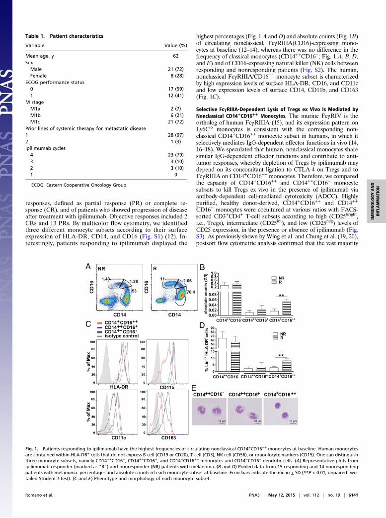
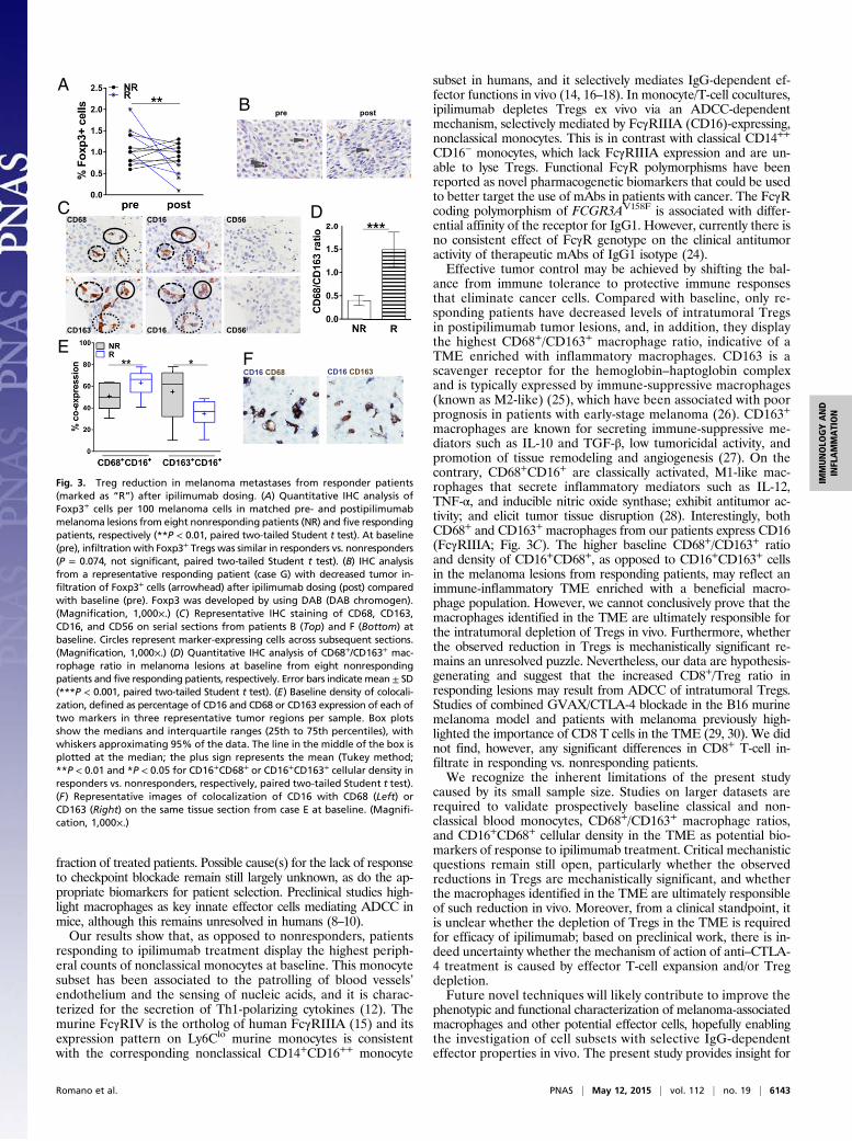
responses, defined as partial response (PR) or complete re-sponse (CR), and of patients who showed progression of diseaseafter treatment with ipilimumab. Objective responses included 2CRs and 13 PRs. By multicolor flow cytometry, we identifiedthree different monocyte subsets according to their surfaceexpression of HLA-DR, CD14, and CD16 (Fig. S1) (12). In-terestingly, patients responding to ipilimumab displayed the
highest percentages (Fig. 1 A and D) and absolute counts (Fig. 1B)of circulating nonclassical, FcγRIIIA(CD16)-expressing mono-cytes at baseline (12–14), whereas there was no difference in thefrequency of classical monocytes (CD14++CD16−; Fig. 1 A, B, D,and E) and of CD16-expressing natural killer (NK) cells betweenresponding and nonresponding patients (Fig. S2). The human,nonclassical FcγRIIIA/CD16++ monocyte subset is characterizedby high expression levels of surface HLA-DR, CD16, and CD11cand low expression levels of surface CD14, CD11b, and CD163(Fig. 1C).
Selective FcγRIIIA-Dependent Lysis of Tregs ex Vivo Is Mediated byNonclassical CD14+CD16++ Monocytes. The murine FcγRIV is theortholog of human FcγRIIIA (15), and its expression pattern onLy6Clo monocytes is consistent with the corresponding non-classical CD14+CD16++ monocyte subset in humans, in which itselectively mediates IgG-dependent effector functions in vivo (14,16–18). We speculated that human, nonclassical monocytes sharesimilar IgG-dependent effector functions and contribute to anti-tumor responses, whereby depletion of Tregs by ipilimumab maydepend on its concomitant ligation to CTLA-4 on Tregs and toFcγRIIIA on CD14+CD16++ monocytes. Therefore, we comparedthe capacity of CD14+CD16++ and CD14++CD16− monocytesubsets to kill Tregs ex vivo in the presence of ipilimumab viaantibody-dependent cell-mediated cytotoxicity (ADCC). Highlypurified, healthy donor-derived, CD14+CD16++ and CD14++
CD16− monocytes were cocultured at various ratios with FACS-sorted CD3+CD4+ T-cell subsets according to high (CD25bright,i.e., Tregs), intermediate (CD25int), and low (CD25neg) levels ofCD25 expression, in the presence or absence of ipilimumab (Fig.S3). As previously shown by Wing et al. and Chung et al. (19, 20),postsort flow cytometric analysis confirmed that the vast majority
Table 1. Patient characteristics
Variable Value (%)
Mean age, y 62SexMale 21 (72)Female 8 (28)
ECOG performance status0 17 (59)1 12 (41)
M stageM1a 2 (7)M1b 6 (21)M1c 21 (72)
Prior lines of systemic therapy for metastatic disease1 28 (97)2 1 (3)Ipilimumab cycles4 23 (79)3 3 (10)2 3 (10)1 0
ECOG, Eastern Cooperative Oncology Group.
CD14++CD16
C CD14 CD16 +CD14 CD16 ++ +CD14 CD16 + + -isotype control
xaMf o
%
CD11b
CD11c CD163
xaMf o
%
HLA-DR
100
80
60
40
20
0
100
80
60
40
20
0
100
80
60
40
20
0
100
80
60
40
20
0
- CD14++CD16+ CD14+CD16++
A B
D
E
++
Fig. 1. Patients responding to ipilimumab have the highest frequencies of circulating nonclassical CD14+CD16++ monocytes at baseline. Human monocytesare contained within HLA-DR+ cells that do not express B-cell (CD19 or CD20), T-cell (CD3), NK cell (CD56), or granulocyte markers (CD15). One can distinguishthree monocyte subsets, namely CD14++CD16−, CD14++CD16+, and CD14+CD16++ monocytes and CD14−CD16− dendritic cells. (A) Representative plots fromipilimumab responder (marked as “R”) and nonresponder (NR) patients with melanoma. (B and D) Pooled data from 15 responding and 14 nonrespondingpatients with melanoma: percentages and absolute counts of each monocyte subset at baseline. Error bars indicate the mean ± SD (**P < 0.01, unpaired two-tailed Student t test). (C and E) Phenotype and morphology of each monocyte subset.
Romano et al. PNAS | May 12, 2015 | vol. 112 | no. 19 | 6141
IMMUNOLO
GYAND
INFLAMMATION

of CD3+CD4+CD25bright T cells expressed the highest levels of theTreg-associated suppressive markers CTLA-4 and Foxp3, whichwere reduced by at least half in CD4+CD25int T cells and in CD4+
CD25neg T cells (Fig. 2 D and E and Fig. S4). CD3+CD4+
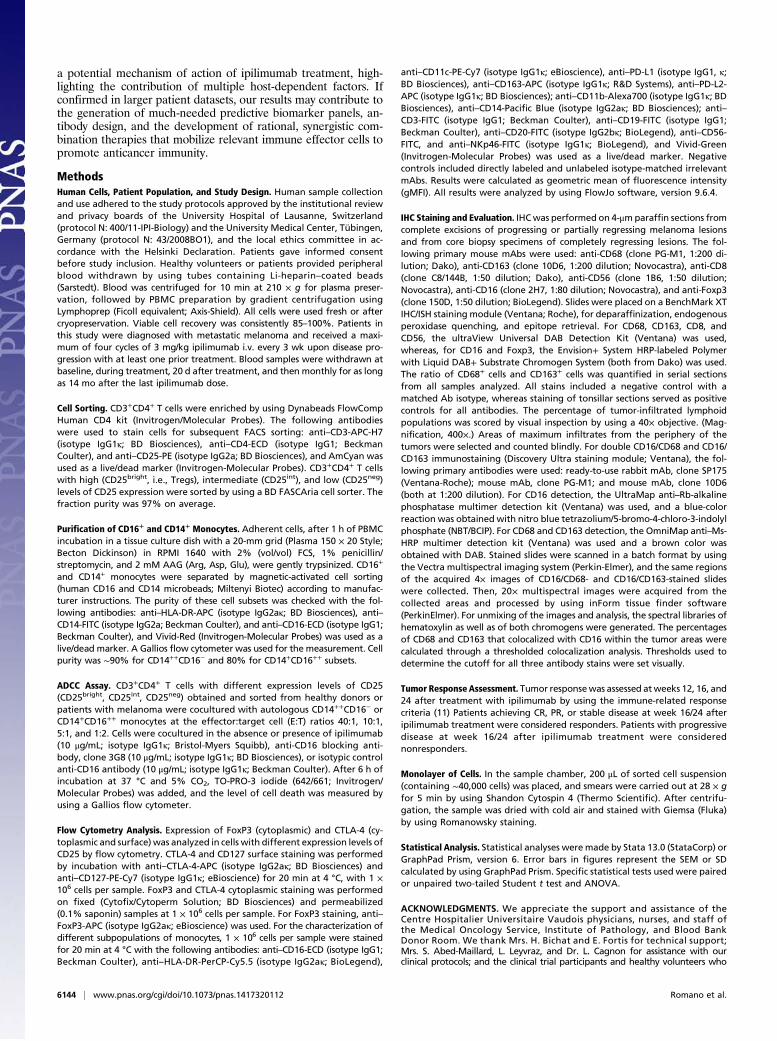
CD25bright T cells were also CD127-negative (Fig. S4) (19–22).After a 6-h incubation, CD14+CD16++ cells, but not CD14++
CD16− cells purified from the same donor, induced selective lysisof CD3+CD4+CD25bright Tregs (Fig. 2 A and C, Left). Thus, ipi-limumab-mediated ADCC correlated with the relative density ofCTLA-4 expression on target cells. Importantly, the primary CD14+
CD16++ monocyte subset also lysed CD3+CD4+CD25bright Tcells from patients with metastatic melanoma (Fig. 2 B and C).The interaction of FcγRIIIA(CD16) with ipilimumab (IgG1) wascritical to CD14+CD16++ monocyte-mediated elimination ofCD3+CD4+CD25bright Tregs because blocking CD14+CD16++
monocytes with anti-CD16 during the 6-h incubation completelyabrogated target cell lysis (Fig. 2 A, Left, and C). In contrast toCD14+CD16++, CD14++CD16− monocytes purified from thesame donors did not mediate ADCC-mediated lysis of Tregs(Fig. 2 B, Right, and C).
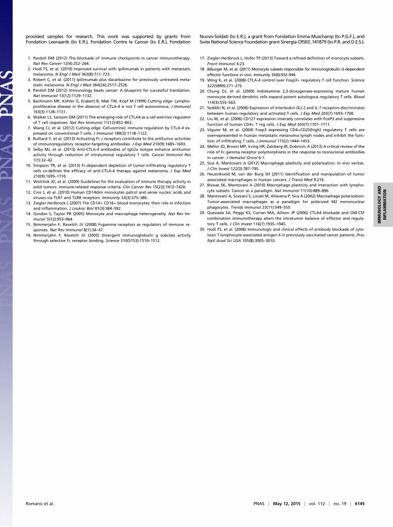
Baseline Enrichment of CD68+CD16+ Macrophages in the TME ofPatients Responding to Ipilimumab. To investigate the TME, westudied matched pre- and postipilimumab metastatic lesionsfrom 13 patients with melanoma and assessed by immunohisto-chemistry (IHC) the presence of CD8+ T cells, CD56+ NK cells,Foxp3+ Tregs, and CD68+ or CD163+ macrophages in the tumornests. Tissue from melanoma metastases was obtained accordingto the study protocol at baseline and 3–4 wk after the last ipili-mumab dose from shrinking lesions from responding patients orprogressing lesions from nonresponding patients, respectively.Whereas, at baseline, Foxp3+ tumor-infiltrating Treg countswere similar in responding vs. nonresponding patients (Fig. 3A),they were significantly lower in postipilimumab lesions fromresponding patients than in those from nonresponding patients(Fig. 3 A and B). Tregs are a major component of the immu-nosuppressive microenvironment of melanoma-infiltrated lymphnodes, and their depletion may favor an immune-promotingTME (23). We evaluated macrophage infiltration of melanomalesions by assessing the distribution of CD68+ and CD163+ cells.We initially evaluated the distribution of CD68, a well-estab-lished macrophage marker. Melanoma-infiltrating CD68+ mac-rophages were present in melanoma metastases (Fig. 3C). Astumor-associated macrophages represent a heterogeneous pop-ulation with distinct immune properties, we set out to furtherevaluate the distribution of CD163+ macrophages (Fig. 3C).CD68+ and CD163+ macrophages from our patients expressedCD16 (FcγRIIIA; Fig. 3C). At baseline, responding patients hadincreased CD68+/CD163+ ratios compared with nonrespondingpatients (Fig. 3D). Next, to quantify more precisely the coloc-alization of CD16- and CD68- or CD163-expressing cells on thesame tissue section at baseline, we used an automated stainingand a standardized acquisition technique by assessing the coexpres-sion of each of two markers in three representative tumor regions(Fig. 3 E and F). Consistent with our previous observations,responding patients had significantly higher intratumoral CD16+
CD68+ cell densities than nonresponding patients. In contrast,the latter displayed higher CD16+CD163+ densities (Fig. 3E). Fi-nally, we did not find any significant differences in CD8+ T-cellinfiltrate in responding vs. nonresponding patients, and CD56+
NK cell infiltrates were negligible for both patient groups andtime points (Fig. 3C).
DiscussionImmune checkpoint blockade arguably represents a milestone inmodern medicine, and will likely become a major treatment optionfor patients with cancer, with increased long-term efficacy at reducedtoxicity. However, immune checkpoint blockade works well in only a
DCE 4
DC
TO-PRO3
bright
neg
D
CD14 CD16++ -CD14 CD16+ ++CD25+ ipilimumab
CD25+ ipilimumab
bright
E
BC
A
+ ipilimumab+ anti-CD16
CD25
49.2
92.3
50.8
7.8
4.4695.5
97.2 2.77
93.3
81
6.7
19
Fig. 2. Selective FcγRIIIA-dependent lysis ex vivo of Tregs (CD3+CD4+
CD25brightCD127−Foxp3++CTLA-4++ Tregs) by nonclassical CD14+CD16++
monocytes. (A) ADCC: killing of sorted CD3+CD4+CD25bright, CD25int, or CD25neg
T cells (target) labeled with anti-CD4ECD from healthy donors by purifiedCD14+CD16++ (Left) or CD14++CD16− (Right) autologous monocytes (effec-tor) at the indicated E:T ratios in the presence of ipilimumab with (opensymbols) or without (filled symbols) anti-CD16 blocking Ab. (B) Similar as-sessment for ADCC of CD3+CD4+CD25bright, CD25int, or CD25neg T cells frompatients with melanoma by purified CD14+CD16++ (open symbols) or CD14++
CD16− (filled symbols) autologous monocytes. (A–C) ADCC assessments useda flow cytometry-based assay whereby lysed target cells took up an other-wise membrane-impermeable DNA stain, TO-PRO3. Specific lysis was basedon the frequency of CD4ECD+ TO-PRO3+ relative to CD4ECD+ TO-PRO3− events.With colorimetric labeling, specific lysis was plotted against the y axeswith respect to the E:T ratio shown along the x axes. Data points are theaverages ± SEM of triplicate means from three or four independent exper-iments in three different healthy donors and four different patients withmelanoma (***P < 0.001 for pairwise comparisons between CD25bright vs.CD25int or CD25neg T cells targeted by CD14+CD16++ monocytes blocked ornot with anti-CD16 for all E:T ratios tested; P value not significant for pair-wise comparisons between CD25bright vs. CD25int or CD25neg T cells targetedby CD14++CD16− monocytes blocked or not with anti-CD16 for all E:T ratiostested; ANOVA). (C) Representative ADCC plots at the top E:T ratio forCD25bright and CD25neg T cells from one patient with melanoma and forCD25bright T cells from one normal donor with or without anti-CD16 blockingAb. (D and E) gMFI of CTLA-4 and Foxp3 expression by sorted CD3+CD4+
CD25bright, CD25int, or CD25neg T cells from healthy donors. Mean gMFImeasurements of CTLA-4 are 18,100 ± 4,700, 6,600 ± 2,600, and 5,400 ±2,500 in CD3+CD4+CD25bright, CD3+CD4+CD25int, and CD3+CD4+CD25neg T cells,respectively. Mean gMFI measurements of Foxp3 are 59,300 ± 13,000, 32,300 ±17,000, and 26,100 ± 12,400 in CD3+CD4+CD25bright, CD3+CD4+CD25int, and inCD3+CD4+CD25neg T cells, respectively. Data represent the averages ± SDfrom three or four independent experiments (**P < 0.01 and ***P < 0.001,unpaired two-tailed Student t test).
6142 | www.pnas.org/cgi/doi/10.1073/pnas.1417320112 Romano et al.
fraction of treated patients. Possible cause(s) for the lack of responseto checkpoint blockade remain still largely unknown, as do the ap-propriate biomarkers for patient selection. Preclinical studies high-light macrophages as key innate effector cells mediating ADCC inmice, although this remains unresolved in humans (8–10).Our results show that, as opposed to nonresponders, patients
responding to ipilimumab treatment display the highest periph-eral counts of nonclassical monocytes at baseline. This monocytesubset has been associated to the patrolling of blood vessels’endothelium and the sensing of nucleic acids, and it is charac-terized for the secretion of Th1-polarizing cytokines (12). Themurine FcγRIV is the ortholog of human FcγRIIIA (15) and itsexpression pattern on Ly6Clo murine monocytes is consistentwith the corresponding nonclassical CD14+CD16++ monocyte
subset in humans, and it selectively mediates IgG-dependent ef-fector functions in vivo (14, 16–18). In monocyte/T-cell cocultures,ipilimumab depletes Tregs ex vivo via an ADCC-dependentmechanism, selectively mediated by FcγRIIIA (CD16)-expressing,nonclassical monocytes. This is in contrast with classical CD14++
CD16− monocytes, which lack FcγRIIIA expression and are un-able to lyse Tregs. Functional FcγR polymorphisms have beenreported as novel pharmacogenetic biomarkers that could be usedto better target the use of mAbs in patients with cancer. The FcγRcoding polymorphism of FCGR3AV158F is associated with differ-ential affinity of the receptor for IgG1. However, currently there isno consistent effect of FcγR genotype on the clinical antitumoractivity of therapeutic mAbs of IgG1 isotype (24).Effective tumor control may be achieved by shifting the bal-
ance from immune tolerance to protective immune responsesthat eliminate cancer cells. Compared with baseline, only re-sponding patients have decreased levels of intratumoral Tregsin postipilimumab tumor lesions, and, in addition, they displaythe highest CD68+/CD163+ macrophage ratio, indicative of aTME enriched with inflammatory macrophages. CD163 is ascavenger receptor for the hemoglobin–haptoglobin complexand is typically expressed by immune-suppressive macrophages(known as M2-like) (25), which have been associated with poorprognosis in patients with early-stage melanoma (26). CD163+
macrophages are known for secreting immune-suppressive me-diators such as IL-10 and TGF-β, low tumoricidal activity, andpromotion of tissue remodeling and angiogenesis (27). On thecontrary, CD68+CD16+ are classically activated, M1-like mac-rophages that secrete inflammatory mediators such as IL-12,TNF-α, and inducible nitric oxide synthase; exhibit antitumor ac-tivity; and elicit tumor tissue disruption (28). Interestingly, bothCD68+ and CD163+ macrophages from our patients express CD16(FcγRIIIA; Fig. 3C). The higher baseline CD68+/CD163+ ratioand density of CD16+CD68+, as opposed to CD16+CD163+ cellsin the melanoma lesions from responding patients, may reflect animmune-inflammatory TME enriched with a beneficial macro-phage population. However, we cannot conclusively prove that themacrophages identified in the TME are ultimately responsible forthe intratumoral depletion of Tregs in vivo. Furthermore, whetherthe observed reduction in Tregs is mechanistically significant re-mains an unresolved puzzle. Nevertheless, our data are hypothesis-generating and suggest that the increased CD8+/Treg ratio inresponding lesions may result from ADCC of intratumoral Tregs.Studies of combined GVAX/CTLA-4 blockade in the B16 murinemelanoma model and patients with melanoma previously high-lighted the importance of CD8 T cells in the TME (29, 30). We didnot find, however, any significant differences in CD8+ T-cell in-filtrate in responding vs. nonresponding patients.We recognize the inherent limitations of the present study
caused by its small sample size. Studies on larger datasets arerequired to validate prospectively baseline classical and non-classical blood monocytes, CD68+/CD163+ macrophage ratios,and CD16+CD68+ cellular density in the TME as potential bio-markers of response to ipilimumab treatment. Critical mechanisticquestions remain still open, particularly whether the observedreductions in Tregs are mechanistically significant, and whetherthe macrophages identified in the TME are ultimately responsibleof such reduction in vivo. Moreover, from a clinical standpoint, itis unclear whether the depletion of Tregs in the TME is requiredfor efficacy of ipilimumab; based on preclinical work, there is in-deed uncertainty whether the mechanism of action of anti–CTLA-4 treatment is caused by effector T-cell expansion and/or Tregdepletion.Future novel techniques will likely contribute to improve the
phenotypic and functional characterization of melanoma-associatedmacrophages and other potential effector cells, hopefully enablingthe investigation of cell subsets with selective IgG-dependenteffector properties in vivo. The present study provides insight for
CCD68 CD16 CD56
CD163 CD16 CD56
B pre post
EF
CD16 CD163CD16 CD68
D
A
Fig. 3. Treg reduction in melanoma metastases from responder patients(marked as “R”) after ipilimumab dosing. (A) Quantitative IHC analysis ofFoxp3+ cells per 100 melanoma cells in matched pre- and postipilimumabmelanoma lesions from eight nonresponding patients (NR) and five respondingpatients, respectively (**P < 0.01, paired two-tailed Student t test). At baseline(pre), infiltration with Foxp3+ Tregs was similar in responders vs. nonresponders(P = 0.074, not significant, paired two-tailed Student t test). (B) IHC analysisfrom a representative responding patient (case G) with decreased tumor in-filtration of Foxp3+ cells (arrowhead) after ipilimumab dosing (post) comparedwith baseline (pre). Foxp3 was developed by using DAB (DAB chromogen).(Magnification, 1,000×.) (C) Representative IHC staining of CD68, CD163,CD16, and CD56 on serial sections from patients B (Top) and F (Bottom) atbaseline. Circles represent marker-expressing cells across subsequent sections.(Magnification, 1,000×.) (D) Quantitative IHC analysis of CD68+/CD163+ mac-rophage ratio in melanoma lesions at baseline from eight nonrespondingpatients and five responding patients, respectively. Error bars indicate mean ± SD(***P < 0.001, paired two-tailed Student t test). (E) Baseline density of colocali-zation, defined as percentage of CD16 and CD68 or CD163 expression of each oftwo markers in three representative tumor regions per sample. Box plotsshow the medians and interquartile ranges (25th to 75th percentiles), withwhiskers approximating 95% of the data. The line in the middle of the box isplotted at the median; the plus sign represents the mean (Tukey method;**P < 0.01 and *P < 0.05 for CD16+CD68+ or CD16+CD163+ cellular density inresponders vs. nonresponders, respectively, paired two-tailed Student t test).(F) Representative images of colocalization of CD16 with CD68 (Left) orCD163 (Right) on the same tissue section from case E at baseline. (Magnifi-cation, 1,000×.)
Romano et al. PNAS | May 12, 2015 | vol. 112 | no. 19 | 6143
IMMUNOLO
GYAND
INFLAMMATION
a potential mechanism of action of ipilimumab treatment, high-lighting the contribution of multiple host-dependent factors. Ifconfirmed in larger patient datasets, our results may contribute tothe generation of much-needed predictive biomarker panels, an-tibody design, and the development of rational, synergistic com-bination therapies that mobilize relevant immune effector cells topromote anticancer immunity.
MethodsHuman Cells, Patient Population, and Study Design. Human sample collectionand use adhered to the study protocols approved by the institutional reviewand privacy boards of the University Hospital of Lausanne, Switzerland(protocol N: 400/11-IPI-Biology) and the University Medical Center, Tübingen,Germany (protocol N: 43/2008BO1), and the local ethics committee in ac-cordance with the Helsinki Declaration. Patients gave informed consentbefore study inclusion. Healthy volunteers or patients provided peripheralblood withdrawn by using tubes containing Li-heparin–coated beads(Sarstedt). Blood was centrifuged for 10 min at 210 × g for plasma preser-vation, followed by PBMC preparation by gradient centrifugation usingLymphoprep (Ficoll equivalent; Axis-Shield). All cells were used fresh or aftercryopreservation. Viable cell recovery was consistently 85–100%. Patients inthis study were diagnosed with metastatic melanoma and received a maxi-mum of four cycles of 3 mg/kg ipilimumab i.v. every 3 wk upon disease pro-gression with at least one prior treatment. Blood samples were withdrawn atbaseline, during treatment, 20 d after treatment, and then monthly for as longas 14 mo after the last ipilimumab dose.
Cell Sorting. CD3+CD4+ T cells were enriched by using Dynabeads FlowCompHuman CD4 kit (Invitrogen/Molecular Probes). The following antibodieswere used to stain cells for subsequent FACS sorting: anti–CD3-APC-H7(isotype IgG1κ; BD Biosciences), anti–CD4-ECD (isotype IgG1; BeckmanCoulter), and anti–CD25-PE (isotype IgG2a; BD Biosciences), and AmCyan wasused as a live/dead marker (Invitrogen-Molecular Probes). CD3+CD4+ T cellswith high (CD25bright, i.e., Tregs), intermediate (CD25int), and low (CD25neg)levels of CD25 expression were sorted by using a BD FASCAria cell sorter. Thefraction purity was 97% on average.
Purification of CD16+ and CD14+ Monocytes. Adherent cells, after 1 h of PBMCincubation in a tissue culture dish with a 20-mm grid (Plasma 150 × 20 Style;Becton Dickinson) in RPMI 1640 with 2% (vol/vol) FCS, 1% penicillin/streptomycin, and 2 mM AAG (Arg, Asp, Glu), were gently trypsinized. CD16+
and CD14+ monocytes were separated by magnetic-activated cell sorting(human CD16 and CD14 microbeads; Miltenyi Biotec) according to manufac-turer instructions. The purity of these cell subsets was checked with the fol-lowing antibodies: anti–HLA-DR-APC (isotype IgG2aκ; BD Biosciences), anti–CD14-FITC (isotype IgG2a; Beckman Coulter), and anti–CD16-ECD (isotype IgG1;Beckman Coulter), and Vivid-Red (Invitrogen-Molecular Probes) was used as alive/dead marker. A Gallios flow cytometer was used for the measurement. Cellpurity was ∼90% for CD14++CD16− and 80% for CD14+CD16++ subsets.
ADCC Assay. CD3+CD4+ T cells with different expression levels of CD25(CD25bright, CD25int, CD25neg) obtained and sorted from healthy donors orpatients with melanoma were cocultured with autologous CD14++CD16− orCD14+CD16++ monocytes at the effector:target cell (E:T) ratios 40:1, 10:1,5:1, and 1:2. Cells were cocultured in the absence or presence of ipilimumab(10 μg/mL; isotype IgG1κ; Bristol-Myers Squibb), anti-CD16 blocking anti-body, clone 3G8 (10 μg/mL; isotype IgG1κ; BD Biosciences), or isotypic controlanti-CD16 antibody (10 μg/mL; isotype IgG1κ; Beckman Coulter). After 6 h ofincubation at 37 °C and 5% CO2, TO-PRO-3 iodide (642/661; Invitrogen/Molecular Probes) was added, and the level of cell death was measured byusing a Gallios flow cytometer.
Flow Cytometry Analysis. Expression of FoxP3 (cytoplasmic) and CTLA-4 (cy-toplasmic and surface) was analyzed in cells with different expression levels ofCD25 by flow cytometry. CTLA-4 and CD127 surface staining was performedby incubation with anti–CTLA-4-APC (isotype IgG2aκ; BD Biosciences) andanti–CD127-PE-Cy7 (isotype IgG1κ; eBioscience) for 20 min at 4 °C, with 1 ×10⁶ cells per sample. FoxP3 and CTLA-4 cytoplasmic staining was performedon fixed (Cytofix/Cytoperm Solution; BD Biosciences) and permeabilized(0.1% saponin) samples at 1 × 10⁶ cells per sample. For FoxP3 staining, anti–FoxP3-APC (isotype IgG2aκ; eBioscience) was used. For the characterization ofdifferent subpopulations of monocytes, 1 × 10⁶ cells per sample were stainedfor 20 min at 4 °C with the following antibodies: anti–CD16-ECD (isotype IgG1;Beckman Coulter), anti–HLA-DR-PerCP-Cy5.5 (isotype IgG2aκ; BioLegend),
anti–CD11c-PE-Cy7 (isotype IgG1κ; eBioscience), anti–PD-L1 (isotype IgG1, κ;BD Biosciences), anti–CD163-APC (isotype IgG1κ; R&D Systems), anti–PD-L2-APC (isotype IgG1κ; BD Biosciences); anti–CD11b-Alexa700 (isotype IgG1κ; BDBiosciences), anti–CD14-Pacific Blue (isotype IgG2aκ; BD Biosciences); anti–CD3-FITC (isotype IgG1; Beckman Coulter), anti–CD19-FITC (isotype IgG1;Beckman Coulter), anti–CD20-FITC (isotype IgG2bκ; BioLegend), anti–CD56-FITC, and anti–NKp46-FITC (isotype IgG1κ; BioLegend), and Vivid-Green(Invitrogen-Molecular Probes) was used as a live/dead marker. Negativecontrols included directly labeled and unlabeled isotype-matched irrelevantmAbs. Results were calculated as geometric mean of fluorescence intensity(gMFI). All results were analyzed by using FlowJo software, version 9.6.4.
IHC Staining and Evaluation. IHCwas performed on 4-μmparaffin sections fromcomplete excisions of progressing or partially regressing melanoma lesionsand from core biopsy specimens of completely regressing lesions. The fol-lowing primary mouse mAbs were used: anti-CD68 (clone PG-M1, 1:200 di-lution; Dako), anti-CD163 (clone 10D6, 1:200 dilution; Novocastra), anti-CD8(clone C8/144B, 1:50 dilution; Dako), anti-CD56 (clone 1B6, 1:50 dilution;Novocastra), anti-CD16 (clone 2H7, 1:80 dilution; Novocastra), and anti-Foxp3(clone 150D, 1:50 dilution; BioLegend). Slides were placed on a BenchMark XTIHC/ISH staining module (Ventana; Roche), for deparaffinization, endogenousperoxidase quenching, and epitope retrieval. For CD68, CD163, CD8, andCD56, the ultraView Universal DAB Detection Kit (Ventana) was used,whereas, for CD16 and Foxp3, the Envision+ System HRP-labeled Polymerwith Liquid DAB+ Substrate Chromogen System (both from Dako) was used.The ratio of CD68+ cells and CD163+ cells was quantified in serial sectionsfrom all samples analyzed. All stains included a negative control with amatched Ab isotype, whereas staining of tonsillar sections served as positivecontrols for all antibodies. The percentage of tumor-infiltrated lymphoidpopulations was scored by visual inspection by using a 40× objective. (Mag-nification, 400×.) Areas of maximum infiltrates from the periphery of thetumors were selected and counted blindly. For double CD16/CD68 and CD16/CD163 immunostaining (Discovery Ultra staining module; Ventana), the fol-lowing primary antibodies were used: ready-to-use rabbit mAb, clone SP175(Ventana-Roche); mouse mAb, clone PG-M1; and mouse mAb, clone 10D6(both at 1:200 dilution). For CD16 detection, the UltraMap anti–Rb-alkalinephosphatase multimer detection kit (Ventana) was used, and a blue-colorreaction was obtained with nitro blue tetrazolium/5-bromo-4-chloro-3-indolylphosphate (NBT/BCIP). For CD68 and CD163 detection, the OmniMap anti–Ms-HRP multimer detection kit (Ventana) was used and a brown color wasobtained with DAB. Stained slides were scanned in a batch format by usingthe Vectra multispectral imaging system (Perkin-Elmer), and the same regionsof the acquired 4× images of CD16/CD68- and CD16/CD163-stained slideswere collected. Then, 20× multispectral images were acquired from thecollected areas and processed by using inForm tissue finder software(PerkinElmer). For unmixing of the images and analysis, the spectral libraries ofhematoxylin as well as of both chromogens were generated. The percentagesof CD68 and CD163 that colocalized with CD16 within the tumor areas werecalculated through a thresholded colocalization analysis. Thresholds used todetermine the cutoff for all three antibody stains were set visually.
Tumor ResponseAssessment. Tumor responsewas assessed atweeks 12, 16, and24 after treatment with ipilimumab by using the immune-related responsecriteria (11) Patients achieving CR, PR, or stable disease at week 16/24 afteripilimumab treatment were considered responders. Patients with progressivedisease at week 16/24 after ipilimumab treatment were considerednonresponders.
Monolayer of Cells. In the sample chamber, 200 μL of sorted cell suspension(containing ∼40,000 cells) was placed, and smears were carried out at 28 × gfor 5 min by using Shandon Cytospin 4 (Thermo Scientific). After centrifu-gation, the sample was dried with cold air and stained with Giemsa (Fluka)by using Romanowsky staining.
Statistical Analysis. Statistical analyses weremade by Stata 13.0 (StataCorp) orGraphPad Prism, version 6. Error bars in figures represent the SEM or SDcalculated by using GraphPad Prism. Specific statistical tests used were pairedor unpaired two-tailed Student t test and ANOVA.
ACKNOWLEDGMENTS. We appreciate the support and assistance of theCentre Hospitalier Universitaire Vaudois physicians, nurses, and staff ofthe Medical Oncology Service, Institute of Pathology, and Blood BankDonor Room. We thank Mrs. H. Bichat and E. Fortis for technical support;Mrs. S. Abed-Maillard, L. Leyvraz, and Dr. L. Cagnon for assistance with ourclinical protocols; and the clinical trial participants and healthy volunteers who
6144 | www.pnas.org/cgi/doi/10.1073/pnas.1417320112 Romano et al.
provided samples for research. This work was supported by grants fromFondation Leenaards (to E.R.), Fondation Contre le Cancer (to E.R.), Fondation
Nuovo-Soldati (to E.R.), a grant from Fondation EmmaMuschamp (to P.G.F.), andSwiss National Science Foundation grant Sinergia CRSII3_141879 (to P.R. andD.E.S.).
1. Pardoll DM (2012) The blockade of immune checkpoints in cancer immunotherapy.Nat Rev Cancer 12(4):252–264.
2. Hodi FS, et al. (2010) Improved survival with ipilimumab in patients with metastaticmelanoma. N Engl J Med 363(8):711–723.
3. Robert C, et al. (2011) Ipilimumab plus dacarbazine for previously untreated meta-static melanoma. N Engl J Med 364(26):2517–2526.
4. Pardoll DM (2012) Immunology beats cancer: A blueprint for successful translation.Nat Immunol 13(12):1129–1132.
5. Bachmann MF, Köhler G, Ecabert B, Mak TW, Kopf M (1999) Cutting edge: Lympho-proliferative disease in the absence of CTLA-4 is not T cell autonomous. J Immunol163(3):1128–1131.
6. Walker LS, Sansom DM (2011) The emerging role of CTLA4 as a cell-extrinsic regulatorof T cell responses. Nat Rev Immunol 11(12):852–863.
7. Wang CJ, et al. (2012) Cutting edge: Cell-extrinsic immune regulation by CTLA-4 ex-pressed on conventional T cells. J Immunol 189(3):1118–1122.
8. Bulliard Y, et al. (2013) Activating Fc γ receptors contribute to the antitumor activitiesof immunoregulatory receptor-targeting antibodies. J Exp Med 210(9):1685–1693.
9. Selby MJ, et al. (2013) Anti-CTLA-4 antibodies of IgG2a isotype enhance antitumoractivity through reduction of intratumoral regulatory T cells. Cancer Immunol Res1(1):32–42.
10. Simpson TR, et al. (2013) Fc-dependent depletion of tumor-infiltrating regulatory Tcells co-defines the efficacy of anti-CTLA-4 therapy against melanoma. J Exp Med210(9):1695–1710.
11. Wolchok JD, et al. (2009) Guidelines for the evaluation of immune therapy activity insolid tumors: immune-related response criteria. Clin Cancer Res 15(23):7412–7420.
12. Cros J, et al. (2010) Human CD14dim monocytes patrol and sense nucleic acids andviruses via TLR7 and TLR8 receptors. Immunity 33(3):375–386.
13. Ziegler-Heitbrock L (2007) The CD14+ CD16+ blood monocytes: their role in infectionand inflammation. J Leukoc Biol 81(3):584–592.
14. Gordon S, Taylor PR (2005) Monocyte and macrophage heterogeneity. Nat Rev Im-munol 5(12):953–964.
15. Nimmerjahn F, Ravetch JV (2008) Fcgamma receptors as regulators of immune re-sponses. Nat Rev Immunol 8(1):34–47.
16. Nimmerjahn F, Ravetch JV (2005) Divergent immunoglobulin g subclass activitythrough selective Fc receptor binding. Science 310(5753):1510–1512.
17. Ziegler-Heitbrock L, Hofer TP (2013) Toward a refined definition of monocyte subsets.Front Immunol 4:23.
18. Biburger M, et al. (2011) Monocyte subsets responsible for immunoglobulin G-dependenteffector functions in vivo. Immunity 35(6):932–944.
19. Wing K, et al. (2008) CTLA-4 control over Foxp3+ regulatory T cell function. Science322(5899):271–275.
20. Chung DJ, et al. (2009) Indoleamine 2,3-dioxygenase-expressing mature humanmonocyte-derived dendritic cells expand potent autologous regulatory T cells. Blood114(3):555–563.
21. Seddiki N, et al. (2006) Expression of interleukin (IL)-2 and IL-7 receptors discriminatesbetween human regulatory and activated T cells. J Exp Med 203(7):1693–1700.
22. Liu W, et al. (2006) CD127 expression inversely correlates with FoxP3 and suppressivefunction of human CD4+ T reg cells. J Exp Med 203(7):1701–1711.
23. Viguier M, et al. (2004) Foxp3 expressing CD4+CD25(high) regulatory T cells areoverrepresented in human metastatic melanoma lymph nodes and inhibit the func-tion of infiltrating T cells. J Immunol 173(2):1444–1453.
24. Mellor JD, BrownMP, Irving HR, Zalcberg JR, Dobrovic A (2013) A critical review of therole of Fc gamma receptor polymorphisms in the response to monoclonal antibodiesin cancer. J Hematol Oncol 6:1.
25. Sica A, Mantovani A (2012) Macrophage plasticity and polarization: In vivo veritas.J Clin Invest 122(3):787–795.
26. Heusinkveld M, van der Burg SH (2011) Identification and manipulation of tumorassociated macrophages in human cancers. J Transl Med 9:216.
27. Biswas SK, Mantovani A (2010) Macrophage plasticity and interaction with lympho-cyte subsets: Cancer as a paradigm. Nat Immunol 11(10):889–896.
28. Mantovani A, Sozzani S, Locati M, Allavena P, Sica A (2002) Macrophage polarization:Tumor-associated macrophages as a paradigm for polarized M2 mononuclearphagocytes. Trends Immunol 23(11):549–555.
29. Quezada SA, Peggs KS, Curran MA, Allison JP (2006) CTLA4 blockade and GM-CSFcombination immunotherapy alters the intratumor balance of effector and regula-tory T cells. J Clin Invest 116(7):1935–1945.
30. Hodi FS, et al. (2008) Immunologic and clinical effects of antibody blockade of cyto-toxic T lymphocyte-associated antigen 4 in previously vaccinated cancer patients. ProcNatl Acad Sci USA 105(8):3005–3010.
Romano et al. PNAS | May 12, 2015 | vol. 112 | no. 19 | 6145
IMMUNOLO
GYAND
INFLAMMATION
Related Documents