IOF World Congress on Osteoporosis ȑ International Osteoporosis Foundation and National Osteoporosis Foundation 2004 PL1. NEW TECHNIQUES FOR THE NON INVASIVE ASSESSMENT OF BONE QUALITY Genant HK; University of California, San Francisco, CA, USA Noninvasive and/or nondestructive techniques can provide struc- tural information about bone, beyond standard bone mineral densitometry (BMD). While the latter provides important infor- mation about osteoporosis diagnosis and fracture risk assessment, considerable evidence indicates that BMD only partially explains bone strength and fracture resistance. Quantitative assessment of macrostructural characteristics such as geometry and section modulus, and microstructural features such as relative trabecular volume, and trabecular spacing, number and connectivity may improve our understanding of osteoporosis and our ability to estimate bone strength and predict fractures. The rationale for imaging bone macro/micro structure, therefore, is to obtain infor- mation beyond BMD, improve fracture risk prediction, clarify the pathophysiology of skeletal disease, define the skeletal response to therapy, and assess biomechanical relationships. The methods for quantitatively assessing the macrostructure of bone include, (besides conventional radiography) computed tomography, especially high resolution computed tomography (hrCT) at 100–400l and volumetric quantitative computed tomog- raphy (vQCT), and high resolution magnetic resonance imaging (hrMR) at 100–200l. The strengths of these approaches include they are widely available, non-invasive and non-destructive methods, providing both macro structure and bone density information, are moderately precise and accurate, and permit serial measurement of most any body site, while their limitations include, for vQCT, the modest exposure to ionizing radiation and the lack of derived microstructural information and, for hrCT and hrMR, the provision of only approximations of microstructural parameters, with con- siderable threshold and resolution dependence. The methods for assessing the microstructure of bone noninva- sively and/or nondestructively include, micro computed tomography (lCT) at 1–100l, and micro magnetic resonance imaging (lMR) at 20–200l. The strengths of the former, lCT, are the automated 2D and 3D evaluation, the nondestructive nature of the imaging, per- mitting mechanical or other testing of the sample, and the highly precise and accurate measurement, while the limitations are the high exposure to ionizing radiation, the requirements for invasive biopsy with large sampling errors or for animal studies, and the expense and limited availability of the equipment. The strengths and weaknesses of lMR are similar, except for the absence of ionizing radiation, and the greater complexity and expense of this technology. Despite the considerable progress made in bone imaging over the past decade, a number of challenges remain. Technically, the chal- lenges reflect the balances and trade-offs between spatial resolution, sampling size, signal-to-noise, radiation exposure and acquisition time, or between the complexity and expense of the imaging tech- nologies versus their availability and accessibility. Clinically, the challenges for bone imaging include balancing the advantages of standard densitometric information versus the more complex architectural features of bone, or the deeper research requirements in the laboratory versus the broader needs in clinical practice. The biological differences between the peripheral appendicular skeleton and the central axial skeleton and their impact on the relevant bone imaging methods must be further clarified. Finally, the relative merits of these sophisticated imaging techniques must be weighed with respect to their applications as diagnostic procedures, requiring high accuracy or reliability, versus their applications as monitoring procedures, requiring high precision or reproducibility. PL2. ULTRASTRUCTURAL DEFECTS, BONE MINERALIZATION AND RESPONSE TO TREATMENT Burr DB; Indiana University School of Medicine, Indianapolis, IN, USA Current pharmacologic agents used to treat osteoporosis have effects on mineralization and microdamage accumulation that are independent of change in bone mineral density. A high degree of mineralization can reduce the amount of energy required to cause fracture. Agents that suppress bone remodeling can increase the normal age-related elevation of min- eralization. Anti-resorptive agents also increase the homogeneity of the bone tissue at the microscopic level as more of the tissue becomes mineralized to the same degree. The increased minerali- zation can be associated with a greater propensity to initiate microdamage; the increased homogeneity will make the tissue matrix less effective at stopping microcracks once they have begun. Agents such as teriparatide that accelerate bone turnover make the matrix transiently less mineralized and more heterogeneous, which may limit microdamage initiation and growth. Anti-resorptive agents increase the cross-linking of collagen in the bone matrix, and reverse the osteoporosis-related reduction in non-reducible cross-linking. This may provide an added benefit in fracture risk reduction. Microdamage is naturally initiated in bone tissue, and is nor- mally repaired through physiologic bone remodeling processes. Microdamage accumulation in bone reduces the strength, elastic modulus and fracture energy of the bone tissue. The microdamage burden in bone increases with age, perhaps because there is an inherent fragility of the tissue matrix that allows initiation of cracks to occur more readily. The impact of pharmacologic interventions for osteoporosis on microdamage accumulation depends on the degree of remodeling suppression, which partly determines the balance between damage initiation and repair. Over-suppression of bone remodeling is associated with increased microdamage and decreased bone toughness, properties that are highly correlated in a non-linear fashion to the rate of bone turnover. The overall efficacy of a compound in reducing fracture risk is partly dependent on its effects on the matrix, and this may help to explain the observation that all pharmacologic agents have about the same efficacy on vertebral fracture risk even though they each are associated with larger or smaller changes in BMD. Osteoporos Int (2004) 15 (Suppl 1): S1–S145 DOI 10.1007/s00198-004-1620-7

Welcome message from author
This document is posted to help you gain knowledge. Please leave a comment to let me know what you think about it! Share it to your friends and learn new things together.
Transcript
IOF World Congress on Osteoporosis
� International Osteoporosis Foundation and National Osteoporosis Foundation 2004
PL1. NEW TECHNIQUES FOR THE NON INVASIVE ASSESSMENT OFBONE QUALITY
Genant HK; University of California, San Francisco, CA, USA
Noninvasive and/or nondestructive techniques can provide struc-tural information about bone, beyond standard bone mineraldensitometry (BMD). While the latter provides important infor-mation about osteoporosis diagnosis and fracture risk assessment,considerable evidence indicates that BMD only partially explainsbone strength and fracture resistance. Quantitative assessment ofmacrostructural characteristics such as geometry and sectionmodulus, and microstructural features such as relative trabecularvolume, and trabecular spacing, number and connectivity mayimprove our understanding of osteoporosis and our ability toestimate bone strength and predict fractures. The rationale forimaging bone macro/micro structure, therefore, is to obtain infor-mation beyond BMD, improve fracture risk prediction, clarify thepathophysiology of skeletal disease, define the skeletal response totherapy, and assess biomechanical relationships.
The methods for quantitatively assessing the macrostructure ofbone include, (besides conventional radiography) computedtomography, especially high resolution computed tomography(hrCT) at 100–400l and volumetric quantitative computed tomog-raphy (vQCT), and high resolution magnetic resonance imaging(hrMR) at 100–200l. The strengths of these approaches include theyare widely available, non-invasive and non-destructive methods,providing both macro structure and bone density information, aremoderately precise and accurate, and permit serial measurement ofmost any body site, while their limitations include, for vQCT, themodest exposure to ionizing radiation and the lack of derivedmicrostructural information and, for hrCT and hrMR, the provisionof only approximations of microstructural parameters, with con-siderable threshold and resolution dependence.
The methods for assessing the microstructure of bone noninva-sively and/or nondestructively include,micro computed tomography(lCT) at 1–100l, and micro magnetic resonance imaging (lMR) at20–200l. The strengths of the former, lCT, are the automated 2Dand 3D evaluation, the nondestructive nature of the imaging, per-mitting mechanical or other testing of the sample, and the highlyprecise and accurate measurement, while the limitations are the highexposure to ionizing radiation, the requirements for invasive biopsywith large sampling errors or for animal studies, and the expense andlimited availability of the equipment. The strengths and weaknessesof lMRare similar, except for the absence of ionizing radiation, andthe greater complexity and expense of this technology.
Despite the considerable progress made in bone imaging over thepast decade, a number of challenges remain. Technically, the chal-lenges reflect the balances and trade-offs between spatial resolution,sampling size, signal-to-noise, radiation exposure and acquisitiontime, or between the complexity and expense of the imaging tech-nologies versus their availability and accessibility. Clinically, thechallenges for bone imaging include balancing the advantages ofstandard densitometric information versus the more complex
architectural features of bone, or the deeper research requirements inthe laboratory versus the broader needs in clinical practice. Thebiological differences between the peripheral appendicular skeletonand the central axial skeleton and their impact on the relevant boneimagingmethodsmust be further clarified.Finally, the relativemeritsof these sophisticated imaging techniques must be weighed withrespect to their applications as diagnostic procedures, requiring highaccuracy or reliability, versus their applications as monitoringprocedures, requiring high precision or reproducibility.
PL2. ULTRASTRUCTURAL DEFECTS, BONE MINERALIZATIONAND RESPONSE TO TREATMENT
Burr DB; Indiana University School of Medicine, Indianapolis,IN, USA
Current pharmacologic agents used to treat osteoporosis haveeffects on mineralization and microdamage accumulation that areindependent of change in bone mineral density.
A high degree of mineralization can reduce the amount ofenergy required to cause fracture. Agents that suppress boneremodeling can increase the normal age-related elevation of min-eralization. Anti-resorptive agents also increase the homogeneity ofthe bone tissue at the microscopic level as more of the tissuebecomes mineralized to the same degree. The increased minerali-zation can be associated with a greater propensity to initiatemicrodamage; the increased homogeneity will make the tissuematrix less effective at stopping microcracks once they have begun.Agents such as teriparatide that accelerate bone turnover make thematrix transiently less mineralized and more heterogeneous, whichmay limit microdamage initiation and growth.
Anti-resorptive agents increase the cross-linking of collagen inthe bone matrix, and reverse the osteoporosis-related reduction innon-reducible cross-linking. This may provide an added benefit infracture risk reduction.
Microdamage is naturally initiated in bone tissue, and is nor-mally repaired through physiologic bone remodeling processes.Microdamage accumulation in bone reduces the strength, elasticmodulus and fracture energy of the bone tissue. The microdamageburden in bone increases with age, perhaps because there is aninherent fragility of the tissue matrix that allows initiation of cracksto occur more readily. The impact of pharmacologic interventionsfor osteoporosis on microdamage accumulation depends on thedegree of remodeling suppression, which partly determines thebalance between damage initiation and repair. Over-suppression ofbone remodeling is associated with increased microdamage anddecreased bone toughness, properties that are highly correlated in anon-linear fashion to the rate of bone turnover. The overall efficacyof a compound in reducing fracture risk is partly dependent on itseffects on the matrix, and this may help to explain the observationthat all pharmacologic agents have about the same efficacy onvertebral fracture risk even though they each are associated withlarger or smaller changes in BMD.
Osteoporos Int (2004) 15 (Suppl 1): S1–S145DOI 10.1007/s00198-004-1620-7
PL3. BONE–SYNOVIUM INTERACTION
Goldring SR; Beth Israel Deaconess Medical Center, NewEngland Baptist Bone and Joint Institute, Harvard Institutes ofMedicine, Boston, MA, USA
Rheumatoid arthritis (RA) represents a paradigm for investigat-ing the role of synovial inflammation on articular and systemicbone remodeling. In RA, proliferation of the synovial lining ofdiarthrodial joints is accompanied by progressive localized artic-ular bone loss manifest radiographically by the development offocal joint erosions. The joint inflammation is also associated withsystemic bone loss, and patients with RA exhibit an increased riskof hip and vertebral fracture. Histopathological analysis of jointtissues from patients with RA indicates that osteoclasts partici-pate in the pathogenesis of the focal joint erosions. Animalmodels of inflammatory arthritis, including adjuvant arthritis(Kong et al. Nature 1999; 402:304), serum transfer arthritis (Pettitet al. Am J Pathol 2001;159:1689); TNF-transgenic mice withspontaneous arthritis (Redlich et al. Arthritis Rheum 2002;46:785); and collagen-induced arthritis (Romas et al. Am J Pathol2002, 161:1419–27), confirm that osteoclasts are the principal celltype responsible for the pathogenesis of focal joint erosions.Additional studies have helped to identify the cytokines andinflammatory mediators that are involved in the recruitment andactivation of bone resorbing cells associated with inflammatoryarthritis. Tumor necrosis factor alpha, interleukin-1, receptoractivator of NF-kb ligand (RANKL) and a number of otherproducts of activated T cells, macrophages and synovial fibro-blasts are among the factors implicated in the increased focalarticular and systemic bone loss. Pro-inflammatory cytokines thatregulate bone remodeling represent rational therapeutic targetsfor specifically inhibiting or slowing the progressive bone lossassociated with RA and related inflammatory disorders.
PL4. OSTEOCYTES AND MECHANICAL TRANSDUCTION
Bonewald LF; University of Missouri, Kansas City, MO, USA
As osteoblasts become embedded in osteoid, they undergo adramatic change in morphology and function to become osteo-cytes. Long dendritic processes are generated that travel throughcanaliculi to connect with the dendritic tips of existing osteocytescreating a three dimensional syncytium within the mineralizedmatrix. Osteocytes can send signals of both resorption and for-mation depending on the ‘window’ of mechanical strain such as1) low or no strain resulting in disuse-associated bone loss,2) physiological strain that maintains homeostasis, 3) high-endphysiological strain that results in modeling, and 4) supra-phys-iological strain that causes injury-associated repair. The osteocyteis ideally suited to translate strain into biochemical signals and toorchestrate the resulting complex biological responses.
Osteocytes appear to respond to strain individually or as apopulation. Osteocytes may act individually in response to strainwhere a specific magnitude of strain elicits a specific gene response,especially for antigens highly expressed in osteocytes such as dentinmatrix protein 1, OF45/MEPE, or E11/gp38. Whereas the functionof E11/gp38 appears to be in the generation of dendritic processes,the function of Dentin Matrix Protein 1 or OF45/MEPE inosteocytes is unknown. When osteocytes respond as a population,the overall strain may be averaged to generate a specific response inall cells. With either of these two types of responses, gene expres-sion is not detectable until a particular threshold is reached,demonstrating that gene expression can be strain thresholddependent. Hormones such as estrogen and parathyroid hormonecan modulate these thresholds.
Osteocyte viability is essential for the maintenance of boneintegrity. Osteocytes, unlike osteoblasts and osteoclasts, are longlived, for decades, and make up over 90% of all bone cells. Oste-ocyte programmed cell death occurs with 1). immobilizationor space flight 2). damage in the form of microcracks, and 3).
post-menopausal and steroid induced osteoporosis. Although allthree conditions are characterized by osteocyte apoptosis and boneresorption, the mechanisms for induction of cell death and signalsof resorption are distinctly different. The mechanisms for inductionof cell death include hypoxia, physical cell damage, decreasedviability due to a lack of estrogen, cytokines such as Interleukin-1or tumor necrosis factor-induced apoptosis, or steroid-inducedapoptosis. These observations emphasize the importance of con-sidering osteocyte viability and function in the development of drugand/or exercise regimens to prevent or treat bone loss.
PL5. NEUROTRANSMITTERS IN BONE: NEW PLAYERS IN BONEPHYSIOLOGY
Skerry TM; Royal Veterinary College, London, UK
Recent interest in the role of the CNS in providing an inhibitoryinfluence on bone formation has raised interest in the activity ofneurotransmitters on the skeleton. This discovery is significantbecause it demonstrates that the dogma of regulation of bonemass by interactions of local and circulating systemic osteotropicmediators is flawed. However, as functionally adaptive responsesoccur in cells that are not innervated, and there is not sufficientneural bandwidth to account for many of the responses of bone toosteotropic influences, it is still unlikely that the highly focusedregulation of site specific remodelling that provides the linkbetween skeletal form and function can be mediated by other thanlocal effects.
In addition to the effects of what can be termed ‘‘extrinsic’’neurotransmitters, acting via central and peripheral innervation,there is in addition a history of the effects of the actions of‘‘intrinsic’’ neurotransmitters directly on their receptors on bonecells. Studies on the actions of classical neurotransmitters such asbradykinin, neuropeptide Y and substance P on receptors onosteoblasts show that paracrine interactions in the bone microen-vironment involve many molecules originally thought to be specificto the nervous system. Recently the role of the excitatory neuro-transmitter glutamate has been explored in this context. Originallyconceived to be involved as a result of studies to identify genesregulated by mechanical loading in vivo that identified a glutamatetransporter known only in the CNS before, it is now clear thatglutamate has a multitude of functions in bone. Glutamate sig-nalling mediates bone formation and bone resorption, by directeffects on glutamate receptors on osteoblasts and osteoclasts. Inaddition, glutamate signalling between osteoblasts and osteoclastprecursors regulates osteoclast precursor differentiation, whilemultipotent bone marrow osteoblast precursors are incapable ofdifferentiation down osteoblastic lineages if glutamate signalling isinhibited. Furthermore, specific temporal and spatial regulation ofglutamate receptor expression and function in mesenchymal con-densations in developing limbs controls the chondrogenesis that is aprerequisite for skeletogenesis.
One further feature of the skeleton’s response to external stimulimay involve mechanisms that parallel those in the CNS. It has beenclear for some time that only brief periods of time are needed tosaturate the response of bone to loading, so that a few seconds orminutes of loading in each day initiate the same changes as longerperiods. This suggests that there is a retention of loading history bybone that persists for between 24 and 48 hours. Interestingly themolecules underlying the long term potentiation (LTP) that is thebasis for memory formation are also expressed in osteoblasts.Loading in vitro induces the same changes in CAM kinase II inosteoblasts from a calcium dependent to a calcium independent statethat are seen in LTP in the CNS.
Like so many ‘‘tissue specific’’ agents, neurotransmitters mayhave been misnamed, in that their sites of expression and functionare becoming found to be steadily more ubiquitous. Such discov-eries have significant impact on possible drug development asagents discovered and tested for neuroscience applications, mayhave utility in the skeleton for the control of bone cell function.
S2
PL6. ROLE OF SEX HORMONES IN BONE GROWTH
Vanderschueren D; Katholieke Universiteit Leuven, Leuven,Belgium
A substantial amount of bone mineral is acquired during a rela-tively short pubertal period of rapid skeletal growth. This phaseof accelerated skeletal modeling is essential to meet the mechan-ical loads imposed on the skeleton by the growing body.
During puberty, greater periosteal than endosteal bone expan-sion results in longer and wider, but also in thicker bones. To thisend, sex steroids provide a disproportionately stronger stimulus toosteoblastic bone formation than to osteoclastic bone resorption,as a result of interaction with specific sex steroid receptors in dif-ferent bone compartments and regions. Not surprisingly, pubertyrepresents a vulnerable period during which deficiency or evendelay of sex steroid activity may have a deleterious and potentiallyirreversible impact on bone mass and structure.
In both genders, estrogens (in males resulting from conversionof androgens) have biphasic, estrogen receptor (ER)-alpha medi-ated effects on growth: in early puberty, estrogens stimulate bothlongitudinal and radial growth; by the end of puberty, on the otherhand, they induce closure of the epiphyseal growth plates and limitlength growth and ultimate bone size. During this process, site-specific differences emerge with axial growth continuing longerthan appendicular growth.
Skeletal sexual dimorphism may in part result from estrogen-mediated inhibitory effects on female skeletal growth with relativelyless exposure to growth-stimulatory estrogen effects. Estrogensinteract with the growth hormone-insulin-like growth factor-I(GH-IGF-I) axis, stimulating (both directly and indirectly) GH andIGF-I at the start of puberty. Moreover, estrogen reduces the bonemarrow cavity by endosteal bone contraction in the female.Thickening of trabeculae in the axial skeleton may also be partiallyestrogen-mediated.
To accommodate to the increasing mechanical demands duringpuberty ,androgens stimulate periosteal bone formation throughthe androgen receptor (AR) . AR-mediated androgen action has nomajor direct effect on the growth plate and non-aromatisableandrogens do not interact with GH-IGF-I axis. However, directAR-mediated anabolic effects on muscle tissue have been well-documented; the resulting increase in mechanical loading isthought to be important in upregulating skeletal modeling duringmale puberty.
In summary, AR-activation by androgens and ER-alpha-acti-vation by estrogens have both direct and indirect anabolic effectson the development of the skeleton during puberty. In line with theincreasing mechanical loading of bone – due to pubertal changes inbody size and body composition – bone mass increases. Sex ste-roids are critically involved in this continuous, time- and site-spe-cific adaptation process. Clinicians should take great care to detectand correct deficiencies and/or delays in sex steroid activity duringpuberty.
PL7. WHAT HAS GENETICS CONTRIBUTED TO OSTEOPOROSIS?
Seeman E; Austin Hospital, University of Melbourne, Mel-bourne, Australia
What do we want genetics to contribute? The clinical goals ofgenetic research are to identify persons likely to fracture, per-sons with low peak bone mass, rapid bone loss, sensitive tocorticosteroids, responsive to drugs, exercise, or calcium. Thesegoals have not been achieved, partly because of problems indefining the phenotype and the questions. The relevant pheno-type is structural failure. However, fractures are uncommon andinvolve varying trauma and bone fragility so that little evidencefor heritability of fracture exists, except perhaps for the Col1A1gene. In lieu of fracture, bone mineral density (BMD) is theaccepted surrogate of bone strength because BMD predictsfracture and its variance is largely genetic. However, allelicvariants in ‘candidate’ genes account for little of the geneticvariance. Associations between a genotype and BMD or rates ofBMD ‘loss’ are usually negative or contradictory, and when
positive (eg COL1A1 gene), differences in the mean BMD be-tween genotypes are small and the scatter of values is large sothat the BMD difference attributable to the genotype is 1–2%;few of the fractures in the community are explained by thisgenotype. Genotype specific responses to drugs, calcium, orexercise are not based on trials with prior stratification bygenotype then randomisation to placebo or intervention, sodifferences in response may be due to covariates unevenly dis-tributed by genotype, not the genotype. These results do notsupport genotyping in diagnosis, risk assessment or therapy.
Revealing the genetic regulation of bone in man with mice. Thelimited success may be, in part, a problem of the ambiguity ofBMD, a phenotype that is the product of a multitude of geneticfactors that regulate the cellular activity on bone’s periosteal andendosteal surfaces throughout life. By contrast, many insights intothe central, systemic and local regulation of skeletal growth andageing in mice have been obtained by studying the genetic regula-tion of specific phenotypes. Leptin controls bone mass throughcentral sympathetic nervous regulation and neuropeptide re-ceptorY4 may contribute to the sex specific regulation of trabeculardensity. Knockouts (KOs) of the genes for receptors for PTHrP,growth hormone, estrogen, provide insights into the regulation oftrabecular and cortical bone, and sex specific regulation of bonegrowth. The lipoxygenase gene (Alox15) is associated with lowpeak bone mass and gene deletion produces high peak bone mass.Genetic ablation of products promoting osteoclastogenesis(RANK, RANKL) produce osteopetrosis. Genetic ablation of in-hibitors of osteoclastogenesis (OPG) produce osteoporosis, whileKO of genes producing osteoblasts results in mice with a cartila-ginous skeleton. Family studies of individuals with a high bonemass reveal linkage to a mutation in the LDL receptor relatedprotein 5. Studies of families with fractures and low BMD reveallinkage to variants in BMP 2 gene, an interesting finding given thatBMPs participate in skeletogenesis, and over expression of the genefor noggin, a BMP 2 antagonist, produces osteoporosis and frac-tures in mice.
What do we want from the Genie before we rub the bottle? In-tegrating these and other insights into bone physiology obtained inmice to whole bone strength in human subjects remains a challenge.The purpose of genetic regulation is to construct a whole bone withproperties suited to the contradictory functions it must perform asboth a lever and spring – stiffness for leverage, yet flexibility (de-formability) for energy absorption, lightness for movement yetstrength for loading. Thus, the unifying purpose of genetic reg-ulation may be to orchestrate the central, systemic and local reg-ulators of the cellular activity on the periosteal and endostealsurfaces to adapt bone’s material composition (mineral, collagen)and structure (trabecular number, thickness, connectivity, corticalthickness, porosity) to the prevailing loads imposed on itthroughout life. Adaptation by one trait in the face of a defect inanother can maintain whole bone strength. For example, collagendefects in the MOV 13 mouse are compensated by structuraladaptations (periosteal apposition) but collagen defect in the BrittleIV mouse are compensated for by material adaptations (mineral/collagen ratio). Severe defects in oim/oim mice result in failedadaptive increases in remodelling because the tissue produced isdefective.
So, what genes regulate and co-regulate endosteal remodelling,BMU balance and periosteal bone formation to keep whole bone‘just right’? Why is adaptation so successful during growth, notageing? There is no ‘osteoporosis’ gene. Is bone fragility a geneticdisease of failed adaptation, a problem in which the cellular eventsthat adapt bone’s material and structural properties to prevailingloads fail, so bone deforms too much during its function as a leveror too little during its function as a spring.
PL8. INDICATIONS TO TREATMENT
Kanis JA; WHO Collaborating Centre for Metabolic BoneDiseases, University of Sheffield Medical School, Sheffield, UK
The objective of any treatment strategy in osteoporosis is toidentify those who will most benefit from treatment and to avoid
S3
unnecessary treatment in those at low risk. The implied treatmentthreshold depends upon costs and consequences of fracture andthe costs, effects and side effects of intervention. Where inter-ventions are inexpensive and safe, global strategies can be envis-aged, for example the use of calcium and vitamin D in theinstitutionalised elderly. More widespread global strategies, suchas avoidance of smoking, physical exercise and other lifestylemeasures have not been tested. For this reason high risk strategiesare the most viable option where individuals are identified on thebasis of high fracture risk.
To date, assessment of bone mineral density (BMD) has beenthe corner-stone for the diagnosis of osteoporosis, and the WHOdefinition of osteoporosis, based on the T-score, is widely acceptedas an intervention threshold for drug development and in practiceguidelines. The recommended diagnostic test is BMD at theproximal femur by dual energy X-ray absorptiometry. The sameBMD threshold for osteoporosis for diagnosis (a T-score for BMDof -2.5 SD or less in young healthy women) can be used in bothmen and women.
Since the determinants of osteoporotic fracture are multifac-torial, risk prediction will always be imperfect. Hip fracture pre-diction with BMD alone is, however, at least as good as bloodpressure readings to predict stroke. Like blood pressure tests, thetest has high specificity, but its sensitivity (detection rate) forfracture outcome is low over most reasonable assumptions. Whenthe WHO thresholds are used, the majority of fractures will occurin those individuals characterised to be at low risk.
The predictive value of BMD can be enhanced by the use ofother factors such as biochemical indices of bone resorption andclinical risk factors. In meta-analyses of prospectively studied co-horts, several risk factors have been identified that contribute tofracture risk independently of BMD. They include age, previousfragility fracture, secondary causes of osteoporosis, smoking, highintakes of alcohol, a family history of hip fracture and the pro-longed use of corticosteroids. Since the presence of such factorsincreases fracture risk over and above that which can be explainedon the basis of BMD, thresholds for intervention that are based onthe use of these risk factors can be less stringent than those basedon BMD alone. The choice of risk factors to use depends onwhether they identify a risk that is amenable to the treatmentenvisaged. With this caveat, diagnostic thresholds differ fromintervention thresholds.
Because of the many techniques available for fracture riskassessment, the absolute (e.g. ten year) probability of fracture is thedesirable parameter to determine intervention thresholds. The set-ting of intervention thresholds is ultimately dependent on healtheconomic considerations. Intervention can be directed to individ-uals cost-effectively where hip fracture probability ranges from 2%to 10% (depending on age). When BMD is used as a test alone, anintervention threshold of -2.5 SD is cost-effective. These thresholds,derived from Sweden, require modification in different countries totake account of different costs and risks that vary markedly indifferent regions of the world.
S4
OC1. A META-ANALYSIS OF BMD AS A PREDICTOR OF FRACTURE RISK
Johnell O, Kanis JA, Oden A, Johansson H, De Laet C, Delmas P,Eisman JA, Garnero P, Fujiwara S, Kroger H, McCloskey EV,Mellstrom D, Melton LJ, O’Neill T, Pols H, Reeve J, Silman A,Tenenhouse A; WHO Centre for Metabolic Bone Diseases,University of Sheffield, UK
The aim of this study was to quantify the relationship between hipbone mineral density (BMD) and fracture risk (all, osteoporotic,hip fractures) and examine the influence of this relationship withage, gender and time since assessment of BMD.
We studied nearly 39000 men and women from twelvepopulation-based cohorts comprising Rotterdam, EVOS/EPOS,CaMos, Rochester, Sheffield, DOES, EPIDOS, OFELY, twocohorts from Gothenburg, Kuopio and Hiroshima with a totalfollow-up of almost 170,000 person-years. The effect of BMD onfracture risk was examined using a Poisson model in each cohortseparately. Results of the different studies was then merged usingweighted coefficients.
BMD was a strong predictor of fractures, especially hipfractures with a similar predictive value observed between menand women. At the age of 65 years, hip fracture risk increased inmen by 2.94 (2.02–4.27) and in women by 2.88 (2.31–3.59) foreach SD decrease in BMD. However, the effect was dependent onage with a significantly higher gradient of risk at age 50 than atage 80 years. For any fracture and for osteoporotic fractures, thegradient of risk was lower than for hip fractures, and predictivevalue increased with age. At age 65 years, osteoporotic fracturerisk increased in men by 1.41 (1.33–1.51) and in women by 1.38(1.28–1.48) for each SD decrease in BMD. For hip fractures, therewas a non-significant reduction of predictive ability with timeafter measurement. For the prediction of osteoporotic frac-tures (and any fracture) there was a higher predictive abilitythe lower the BMD value. At a T-score –4 SD the risk ratio was2.10 (1.63–2.71), and at a T-score of –1 SD the risk ratio was 1.73(1.59–1.89). A similar but less pronounced and non-significanteffect was observed for hip fractures. We conclude that BMD is arisk factor for fracture of substantial importance, and is similarin both sexes. This validation on an international basis permitsits use in case-finding strategies. Its use should, however, takeaccount of the variations in predictive value with age and BMD.
OC2. ASSESSING VERTEBRAL FRACTURE RISK USING BIOMECHANI-CAL PRINCIPLES: FRACTURE RISK INDEX
Duan Y1, Duboeuf F2, Munoz F2, Turner CH3, Delmas PD2,Seeman E1, 1The University of Melbourne, Melbourne, Australia,2INSERM Unit 403, Hopital Edouard Herriot, Lyon, France,3Department of Orthopaedic Surgery and The Biomechanics andBiomaterials Research Center, Indianapolis, USA
Aims: Structural failure becomes increasingly likely as the load onbone approximates or exceeds the bone’s ability to withstand it.The vertebral fracture risk index (FRI) expresses the risk forstructural failure as a ratio of the load per unit area/strength.The purpose of this study was to determine whether the FRIprovides a more sensitive and specific predictor of vertebralfracture risk than spinal areal BMD (aBMD) or volumetric BMD(vBMD).
Methods: We conducted two studies, a case-control study of 89postmenopausal women with vertebral fractures and 306 post-menopausal controls in Melbourne, Australia, and a prospectivestudy of 30 postmenopausal women with incident vertebral frac-tures and 150 age-comparable controls in Lyon, France. FRI andvBMD of the third lumbar vertebral body and spine aBMD werederived from dual x-ray absorptiometry
Results: In the cross-sectional analysis, after adjusting for age,each SD increase in FRI was associated with 2.1-fold (95% CI,1.55–2.73) increase in the risk of fracture, while each SD reductionin aBMD or vBMD was associated with 4.0-fold (95% CI, 2.69–6.18 and 2.65–6.94, respectively) increase in the risk. Using ROCanalysis, the FRI had a lower sensitivity and specificity than aBMDin discriminating cases and controls (area under ROC curve, 0.76
vs 0.84, p<0.01), but was similarly compared to vBMD (0.76 vs0.79). Prospective analysis suggested that the FRI was no betterpredictor than aBMD [HR, 1.2 (95% CI:0.9–1.7) vs 2.4 (95%CI:1.5–3.8); area under ROC, 0.64 vs 0.79, p<0.01]. There was alsolower sensitivity using a cut off of FRI >=1 compared withaBMD T-score of )2.5 SD in both studies. FRI was inverselyassociated with aBMD T-score in patients with prevalent but notincident fractures. There was poor agreement (Kappa=0.11–0.18)between FRI and aBMD T-scores in diagnosing osteoporoticfractures.
Conclusion: Within the constraints of the small sample size, weconcluded that applying biomechanical index such as FRI at thespine did not provide a better discriminatory ability to distinguishfracture cases and controls than aBMD or vBMD.
OC3. FALL INDEX PREDICTS HIP FRACTURE INDEPENDENT OF AGEAND BONE DENSITY
Faulkner KG1, Wacker WK1, Barden HS1, Burke PK2; 1GEMedical Systems Lunar, Madison WI USA, 2OsteoporosisDiagnostic and Treatment Center, Richmond VA, USA
Hip fracture risk is related to several factors, including bonemineral density (BMD), bone distribution, age, height, andweight. Modern bone densitometers can measure structuralparameters beyond BMD, including cross sectional moment ofinertia (CSMI) and cross sectional area (CSA) at the femoralneck. Models have been proposed that combine density, structure,age, height, and weight to produce a Fall Index (FI). FI estimatesthe ability of a hip to withstand a fall on the greater trochanter,with larger values indicating greater strength and decreased risk(Yoshikawa et al, JBMR 9:1053–1064).
In this study, we compared femoral BMD with CSMI, CSA,and FI for assessing hip fracture risk. DXA scans were obtained in422 women, 58 with prior hip fracture and 364 controls using theLunar Prodigy (GE Medical Systems). For the fracture subjects,DXA measurements were performed on the non-fractured femur.BMD of the femoral neck was determined, as well as CSMI, CSA,and FI using the Lunar Hip Strength Analysis program.
Age Height Weight Neck BMD CSMI CSA FI
Fracture 77 yrs 160 cm 61.7 kg 0.659 g/cm2* 10689 149 cm2 1.54*
Controls 76 yrs 157 cm 62.6 kg 0.748 g/cm2 10284 148 cm2 1.62
*Significantly different than controls (p<0.01)
There was no significant trend with age for the BMD-adjustedCSA, CSMI and FI values. Results for fracture cases and controlswere compared using an unpaired t-test. Femoral neck BMD wassignificantly lower in the fracture group compared to controls.After adjustment for BMD, neither CSMI nor CSA were signifi-cantly different between groups. However, FI was significantlylower in the fracture group, consistent with a reduced capacity towithstand a fall.
We conclude that femoral neck BMD is an important predictorof femoral fracture. Measurements of femoral geometry, which arebased on BMD distribution, did not provide additional predictivepower compared to BMD alone. The Fall Index, which combinesBMD, geometry, age, height, and weight into a single risk factor, isa significant predictor of hip fracture, even after adjustment for ageand BMD.
OC4. EFFECT OF DIFFERING VERTEBRAL AREAS ON INTERPRETATIONOF SERIAL DENSITOMETRY IN CLINICAL PRACTICE
Kline GA, Hanley DA; University of Calgary, Alberta, Canada
Background: In comparing serial BMD measurements, the ISCDrecommends vertebral projected areas should not differ by >2%to avoid measurement error from change in apparent bone size.This study assessed whether this guideline is followed in routinedensitometry.
S5
Methods: Analysis of 103 consecutive paired BMD reportsperformed by qualified radiologists. Scans were excluded if tech-nical errors present. We analyzed the difference between projectedareas of individual vertebrae in serial scans from individualpatients. We excluded all vertebrae differing by >2% and recal-culated both scans, provided at least 2 vertebrae were suitable foranalysis. After re-calculating the results, we compared correctedBMD to the original reported change using 0.025 g/cm2 for leastsignificant change.
Results: Mean differences in projected areas of L1, L2, L3 andL4 were 5.6%, 3.8%, 3.6% and 4.6% respectively. Only 4.9% ofscans had all 4 vertebrae differ by <2%. Despite greatest vari-ability, a change in L1 area only accounted for 33% of ‘‘failed’’scans. 12% of scans were unacceptable, having 3 or more verte-brae differing by >5%.
Analysis excluding all vertebrae differing by >2% showed amean 0.012 g/cm2 change from the original reported change.However, 11% had results differing from reported change by>0.025 g/cm2. If analysis requires at least 2 vertebrae with areasdiffering by <2%, 55.3% of DXA’s were either unsuitable foranalysis, or re-analysis changed results by >0.025 g/cm2. Of the12% of paired DXA having 3 or more vertebrae with >5% arealdifference, 66% changed by >0.03 g/cm2.
To determine clinical impact, we re-analyzed scans excludingvertebrae >2% different and using LSC of 0.025 g/cm2. Afterre-analysis, 26% of reports changed clinical interpretation: clini-cally significant increase or decrease became non-significant or viceversa.
Conclusions: Half of serial DXA measurements did not meetISCD criteria. This resulted in 20% of DXA reports being inaccu-rate in quantifying the true BMD change. 26% of properly per-formed DXA erroneously reported the significance of an observedchange by not excluding vertebrae with large differences in projectedareas. Serial densitometry results should be used with caution inpatient management if projected area analyses are not performed.
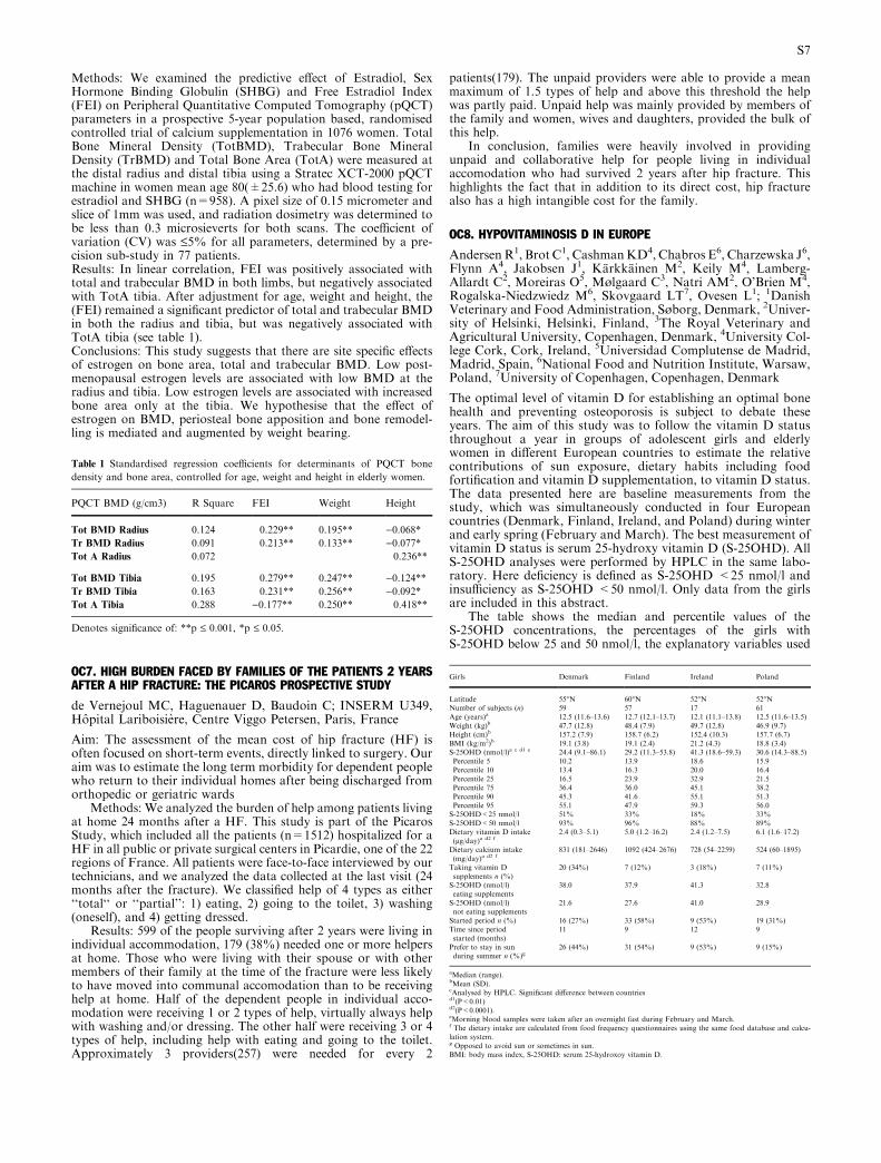
OC5. NON-DESTRUCTIVE ASSESSMENT OF TRABECULAR MICRO-STRUCTURE IN THE LUMBAR SPINE
Goldhahn J1,3, Stauber M2, Reinhold M3,4, Gabriel M5, MullerR2; 1Schulthess Clinic, Zurich, Switzerland, 2Institute for Bio-medical Engineering ETH and University, Zurich, Switzerland,3AO Research Institute, Davos, Switzerland, 4Clinic for Trau-matology of the University, Innsbruck, Austria, 5Clinic forNuclear Medicine of the University, Innsbruck, Austria
Aim: Structural bone parameters like trabecular thickness andtrabecular number predict the mechanical properties of cancellousbone better than bone mineral density. Accurate data are requiredto test new spinal instrumentations especially for the treatment ofthe osteoporotic spine. Since conventional DXA provides BMDdata only for L1 to L4 and does not distinguish between cancellousand cortical bone, 3D high-resolution peripheral quantitativecomputer tomography (3D-pQCT) was employed to quantifystructural parametersnon-destructivelyprior tomechanical testing.
Methods: Thirteen human cadaveric spines ranging from T11 toL5 were measured with DXA and 3D-pQCT. DXA measurements(Hologic QDR 4500W) included AP view from L1 to L4 and lateralview for L2 and L3. PQCT was performed using a new generation invivo 3D-pQCT scanner (Radios, Scanco Medical, Switzerland)providing an isotropic nominal resolution of 93 lm measuring alllevels (Fig. 1). Subsequently, direct 3D morphometry was used tocompute cancellous bone volume density (BV/TV), trabecularthickness (Tb.Th), number (Tb.N) and separation (Tb.Sp). Corre-lations between DXA and pQCT and between structural parametersand BV/TV were calculated.
Results:WhereasAP and lateral view in theDXA correlated onlymoderately (r=0.77; r2=0.59), 3D-pQCTvalues for cancellousbonedensity (BV/TV) correlated well with AP DXA (r=0.86; r2=0.72).Tb.Th ranged from 0.14 to 0.27mm, Tb.N from 0.13 to 0.97 per mm.BV/TV was linearly correlated to Tb.N (r=0.97) and an inverselycorrelated to Tb.Sp (r=–0.83). Additionally, two cysts and oneintraosseous calcification were discovered.
Conclusions: The findings suggest a good correlation between3D-pQCT and DXA. However, the microtomography-based evalu-ation allows scanning of the whole spine segment at once, detectspathological alterations and provides additional structural parame-ters. Although the technique is currently suitable for cadaveric spinesonly it offers a great potential for in vivo measurements of the distalradius or the heel.
OC6. ESTROGEN HAS SITE SPECIFIC EFFECTS ON TRABECULAR ANDTOTAL BONE DENSITY AND BONE AREA MEASURED BY PQCT AND ISDEPENDENT ON WEIGHTBEARING
Groombridge LK1,2, Devine A1, Smith TJ1, Dick I1, Prince RL1,2;1School of Medicine and Pharmacology, University of WesternAustralia, Perth, Australia, 2Department of Endocrinology andDiabetes, Sir Charles Gairdner Hospital, Perth, Australia
Aims: Endogenous estrogen levels decline after menopause andaccelerate bone loss. We propose the magnitude of this estrogeneffect is site specific and is dependent on weight bearing. Estrogenmay affect the trabecular bone density and mechanical distribu-tion of bone mass by influencing periosteal bone apposition.
Fig. 1 3D-pQCT reconstruction of a lumbar spine from a 84-year-oldfemale
S6
Methods: We examined the predictive effect of Estradiol, SexHormone Binding Globulin (SHBG) and Free Estradiol Index(FEI) on Peripheral Quantitative Computed Tomography (pQCT)parameters in a prospective 5-year population based, randomisedcontrolled trial of calcium supplementation in 1076 women. TotalBone Mineral Density (TotBMD), Trabecular Bone MineralDensity (TrBMD) and Total Bone Area (TotA) were measured atthe distal radius and distal tibia using a Stratec XCT-2000 pQCTmachine in women mean age 80(±25.6) who had blood testing forestradiol and SHBG (n=958). A pixel size of 0.15 micrometer andslice of 1mm was used, and radiation dosimetry was determined tobe less than 0.3 microsieverts for both scans. The coefficient ofvariation (CV) was £5% for all parameters, determined by a pre-cision sub-study in 77 patients.Results: In linear correlation, FEI was positively associated withtotal and trabecular BMD in both limbs, but negatively associatedwith TotA tibia. After adjustment for age, weight and height, the(FEI) remained a significant predictor of total and trabecular BMDin both the radius and tibia, but was negatively associated withTotA tibia (see table 1).Conclusions: This study suggests that there are site specific effectsof estrogen on bone area, total and trabecular BMD. Low post-menopausal estrogen levels are associated with low BMD at theradius and tibia. Low estrogen levels are associated with increasedbone area only at the tibia. We hypothesise that the effect ofestrogen on BMD, periosteal bone apposition and bone remodel-ling is mediated and augmented by weight bearing.
Table 1 Standardised regression coefficients for determinants of PQCT bone
density and bone area, controlled for age, weight and height in elderly women.
PQCT BMD (g/cm3) R Square FEI Weight Height
Tot BMD Radius 0.124 0.229** 0.195** )0.068*Tr BMD Radius 0.091 0.213** 0.133** )0.077*Tot A Radius 0.072 0.236**
Tot BMD Tibia 0.195 0.279** 0.247** )0.124**Tr BMD Tibia 0.163 0.231** 0.256** )0.092*Tot A Tibia 0.288 )0.177** 0.250** 0.418**
Denotes significance of: **p £ 0.001, *p £ 0.05.
OC7. HIGH BURDEN FACED BY FAMILIES OF THE PATIENTS 2 YEARSAFTER A HIP FRACTURE: THE PICAROS PROSPECTIVE STUDY
de Vernejoul MC, Haguenauer D, Baudoin C; INSERM U349,Hopital Lariboisiere, Centre Viggo Petersen, Paris, France
Aim: The assessment of the mean cost of hip fracture (HF) isoften focused on short-term events, directly linked to surgery. Ouraim was to estimate the long term morbidity for dependent peoplewho return to their individual homes after being discharged fromorthopedic or geriatric wards
Methods: We analyzed the burden of help among patients livingat home 24 months after a HF. This study is part of the PicarosStudy, which included all the patients (n=1512) hospitalized for aHF in all public or private surgical centers in Picardie, one of the 22regions of France. All patients were face-to-face interviewed by ourtechnicians, and we analyzed the data collected at the last visit (24months after the fracture). We classified help of 4 types as either‘‘total‘‘ or ‘‘partial’’: 1) eating, 2) going to the toilet, 3) washing(oneself), and 4) getting dressed.
Results: 599 of the people surviving after 2 years were living inindividual accommodation, 179 (38%) needed one or more helpersat home. Those who were living with their spouse or with othermembers of their family at the time of the fracture were less likelyto have moved into communal accomodation than to be receivinghelp at home. Half of the dependent people in individual acco-modation were receiving 1 or 2 types of help, virtually always helpwith washing and/or dressing. The other half were receiving 3 or 4types of help, including help with eating and going to the toilet.Approximately 3 providers(257) were needed for every 2
patients(179). The unpaid providers were able to provide a meanmaximum of 1.5 types of help and above this threshold the helpwas partly paid. Unpaid help was mainly provided by members ofthe family and women, wives and daughters, provided the bulk ofthis help.
In conclusion, families were heavily involved in providingunpaid and collaborative help for people living in individualaccomodation who had survived 2 years after hip fracture. Thishighlights the fact that in addition to its direct cost, hip fracturealso has a high intangible cost for the family.
OC8. HYPOVITAMINOSIS D IN EUROPE
AndersenR1, Brot C1, CashmanKD4, Chabros E6, Charzewska J6,Flynn A4, Jakobsen J1, Karkkainen M2, Keily M4, Lamberg-Allardt C2, Moreiras O5, Mølgaard C3, Natri AM2, O’Brien M4,Rogalska-Niedzwiedz M6, Skovgaard LT7, Ovesen L1; 1DanishVeterinary and Food Administration, Søborg, Denmark, 2Univer-sity of Helsinki, Helsinki, Finland, 3The Royal Veterinary andAgricultural University, Copenhagen, Denmark, 4University Col-lege Cork, Cork, Ireland, 5Universidad Complutense de Madrid,Madrid, Spain, 6National Food and Nutrition Institute, Warsaw,Poland, 7University of Copenhagen, Copenhagen, Denmark
The optimal level of vitamin D for establishing an optimal bonehealth and preventing osteoporosis is subject to debate theseyears. The aim of this study was to follow the vitamin D statusthroughout a year in groups of adolescent girls and elderlywomen in different European countries to estimate the relativecontributions of sun exposure, dietary habits including foodfortification and vitamin D supplementation, to vitamin D status.The data presented here are baseline measurements from thestudy, which was simultaneously conducted in four Europeancountries (Denmark, Finland, Ireland, and Poland) during winterand early spring (February and March). The best measurement ofvitamin D status is serum 25-hydroxy vitamin D (S-25OHD). AllS-25OHD analyses were performed by HPLC in the same labo-ratory. Here deficiency is defined as S-25OHD <25 nmol/l andinsufficiency as S-25OHD <50 nmol/l. Only data from the girlsare included in this abstract.
The table shows the median and percentile values of theS-25OHD concentrations, the percentages of the girls withS-25OHD below 25 and 50 nmol/l, the explanatory variables used
Girls Denmark Finland Ireland Poland
Latitude 55�N 60�N 52�N 52�NNumber of subjects (n) 59 57 17 61Age (years)a 12.5 (11.6–13.6) 12.7 (12.1–13.7) 12.1 (11.1–13.8) 12.5 (11.6–13.5)Weight (kg)b 47.7 (12.8) 48.4 (7.9) 49.7 (12.8) 46.9 (9.7)Height (cm)b 157.2 (7.9) 158.7 (6.2) 152.4 (10.3) 157.7 (6.7)BMI (kg/m2)b 19.1 (3.8) 19.1 (2.4) 21.2 (4.3) 18.8 (3.4)S-25OHD (nmol/l)a c d1 e 24.4 (9.1–86.1) 29.2 (11.3–53.8) 41.3 (18.6–59.3) 30.6 (14.3–88.5)Percentile 5 10.2 13.9 18.6 15.9Percentile 10 13.4 16.3 20.0 16.4Percentile 25 16.5 23.9 32.9 21.5Percentile 75 36.4 36.0 45.1 38.2Percentile 90 45.3 41.6 55.1 51.3Percentile 95 55.1 47.9 59.3 56.0
S-25OHD<25 nmol/l 51% 33% 18% 33%S-25OHD<50 nmol/l 93% 96% 88% 89%Dietary vitamin D intake(lg/day)a d2 f
2.4 (0.3–5.1) 5.0 (1.2–16.2) 2.4 (1.2–7.5) 6.1 (1.6–17.2)
Dietary calcium intake(mg/day)a d2 f
831 (181–2646) 1092 (424–2676) 728 (54–2259) 524 (60–1895)
Taking vitamin Dsupplements n (%)
20 (34%) 7 (12%) 3 (18%) 7 (11%)
S-25OHD (nmol/l)eating supplements
38.0 37.9 41.3 32.8
S-25OHD (nmol/l)not eating supplements
21.6 27.6 41.0 28.9
Started period n (%) 16 (27%) 33 (58%) 9 (53%) 19 (31%)Time since periodstarted (months)
11 9 12 9
Prefer to stay in sunduring summer n (%)g
26 (44%) 31 (54%) 9 (53%) 9 (15%)
aMedian (range).bMean (SD).cAnalysed by HPLC. Significant difference between countriesd1(P<0.01)d2(P<0.0001).eMorning blood samples were taken after an overnight fast during February and March.f The dietary intake are calculated from food frequency questionnaires using the same food database and calcu-lation system.g Opposed to avoid sun or sometimes in sun.BMI: body mass index, S-25OHD: serum 25-hydroxoy vitamin D.
S7
in the multiple regression analyses performed to explain the S-25OHD levels, which are log-transformed in the statistical analyses.The only significant determinants of vitamin D status were country(P<0.05) and use of vitamin D supplements (P<0.001). As shownin the table girls not eating supplements had lower S-25OHDconcentration compared to girls eating supplements.
Nine out of ten girls were vitamin D insufficient and possibly inrisk of not reaching optimal peak bone mass with increased risk ofosteoporosis later in life.Acknowledgment: The study is part of the OPTIFORD-project’Towards a strategy for optimal vitamin D fortification’, financedby EU, the 5th Framework Programme (QLK1-CT-2000-00623).
OC9. LOW 25-HYDROXYCHOLECALCIFEROL (25OHD) LEVEL IS A RISKFACTOR OF ACCELERATED BONE LOSS IN ELDERLY MEN: THE MINOSSTUDY
Szulc P1, Munoz F1, Marchand F2, Delmas PD1; 1INSERMResearch Unit 403, Lyon, France, 2SSMB, Montceau les Mines,France
We have shown that 25OHD is a determinant of BMD and boneturnover in elderly men (Szulc et al. Calcif Tissue Int, 2003). Wehypothesised that low 25OHD level is a risk factor of acceleratedbone loss in elderly men. We assessed the bone loss in 699 menaged 50 to 85 followed up prospectively for 58±18 months. BMDwas measured at the hip by the HOLOGIC 1000 W device and atthe distal forearm with OSTEOMETER DTX 100 device. Aver-age bone loss varied from 0.11%/year at the femoral neck to0.31%/year at the distal radius. Rate of bone loss in ten-year agegroups accelerated with ageing (total hip – p<0.003, distal fore-arm – p<0.001). After the age of 70, bone loss was twice as highthan before the age of 60 (hip - 0.39 vs 0.21%/year, p<0.0001).After adjustment for age and season, bone loss was more rapid inmen with baseline 25OHD level below 27 ng/ml. We definedaccelerated bone loss as > 1%/year. After adjustment for age andseason, 25OHD below the median (£ 26 ng/ml) was predictive ofthe accelerated bone loss at the total hip (n=53, O.R.=2.2, 95%C.I. - 1.36, 3.69) and at the distal forearm (n=62, O.R.=2.85,95% C.I. - 1.29 - 5.34). Baseline level of parathyroid hormone,calcium intake and leisure physical activity (associated with sun-light exposure) did not correlate with the rate of bone loss at anysite of measurement nor interacted with 25OHD.
We conclude that the concentration of 25OHD £ 26 ng/mL (65pmol/L) predicts accelerated bone loss at the hip and distal radiusin elderly men.
25OHD (ng/ml) Distal forearm (%/yr) Total hip (%/yr)
QI – £ 19 )0.44 ± 0.71 )0.34 ± 0.53QII )19.1)26 )0.36 ± 0.60 )0.34 ± 0.51QIII )26.1)34 )0.23 ± 0.47 )0.22 ± 0.39QIV ) > 34.1 )0.21 ± 0.35 )0.22 ± 0.36
p = 0.002 p = 0.02
Supported by a contract INSERM/Merck Sharp & DohmeChibret, France
OC10. EFFECT OF ANNUAL INTRAMUSCULAR VITAMIN D SUPPLE-MENTATION ON FRACTURE RISK: POPULATION-BASED, RANDOMISED,DOUBLE-BLIND, PLACEBO-CONTROLLED TRIAL
Smith H1, Anderson F2, Raphael H1, Crozier S3, Cooper C3;1Health Care Research Unit, Southampton General Hospital,Southampton, UK, 2Department of Geriatric Medicine, South-ampton General Hospital, Southampton, UK, 3MRC Environ-mental Epidemiology Unit, University of Southampton,Southampton, UK
Aims: To determine the effect of an annual intramuscular vitaminD injection on fracture rate among men and women aged 75 yearsand over, living in the general population.
Methods:
(a) Design: Randomised, double-blind placebo-controlled trial of300,000 IU intramuscular vitamin D (ergocalciferol) injectionor matching placebo administered every autumn over threeyears.
(b) Subjects: 9,440 people (4,354 men and 5,086 women) aged 75years and over, living in the general community, recruited fromthe patient registers of general practitioners in Wessex, England.
(c) Outcome measures: Hip, wrist and all non-vertebral fractures.
Results: After three years of follow up, 609 men and women hadincident fractures (hip 110, wrist 107, ankle 24). Hazard ratios inthe vitamin D group compared with the placebo group were 1.10(95% CI 0.94–1.29 p=0.25) for any first fracture and 1.48 (95%CI 1.01–2.17, p=0.04) for first hip fracture; and 1.17 (95% CI0.80–1.71, p=0.43) for first wrist fracture, controlling for age andsex. Although the findings were similar among men and women,the difference between treatment groups for hip fracture appearedmore pronounced among those aged 80 years and over, andamong those without previous fractures. No apparent protectiveeffect was observed when the cohort was stratified by age, pre-vious fracture, or level of mobility. Analysis of serum PTH and25-hydroxyvitamin D concentrations in a subset of subjects sug-gested that the intervention achieved a 20% suppression in peakwinter PTH levels.
Conclusion An annual intramuscular injection of 300,000 IUvitamin D is not effective in preventing hip and other non-spinefractures among elderly men and women resident in the generalpopulation.
OC11. HIGH RISK OF FALLS RELATED TO LOW D-HORMONE SYN-DROME AND ITS TREATMENT WITH ALFACALCIDOL
Dukas L1, Schacht E2, Stahelin HB1; 1Geriatric University Clinic,Basel, Switzerland, 2Metabolic Bone Disease Unit, Universitats-klinik Balgrist, Zurich, Switzerland
Aims: Treatment with D-hormone analogues significantly reducesthe number of fallers and falls in elderly. Since impaired renalfunction is detrimental to the activation of calcitriol (D-hormone)we determined the cutoff levels of creatinine clearance (CrCl) atwhich D-hormone serum levels decline and investigated if otherrisk factors are associated with low D-hormone. Using thedetermined cutoff, we further investigated in post hoc analyses ofa double-blind randomized study, if CrCl is associated with therisk of falls and whether treatment with Alfacalcidol can reducethis risk.
Methods: 378 community-dwelling elderly men and womenreceived for 36 weeks randomly 1lg Alfacalcidol (Alpha-D3�TEVA) or placebo daily. Serum calcitropic hormones were regu-larly measured by radioimmunoassay Falls were assessed by aquestionnaire. The risk of becoming a faller and the risk of fallingwere assessed in multivariate-controlled logistic regression modelsaccording to treatment groups and according to a CrCl cutoff at65ml/min. The results are from ITT analyses.
Results: D-hormone serum levels were in multivariate-con-trolled analyses, significantly associated with CrCl (p<0.0001) andsteadily declined below a CrCl of 65ml/min. A CrCl of <65ml/min,the use of diuretics and a diagnosis of adult-onset diabetes were inmultivariate controlled analyses associated with significantly lowD-hormone serum levels (p=0.0008, p=0.001 res. p=0.003). Inthe Placebo group we observed significantly more fallers in par-ticipants having a CrCl of <65ml/min as compared to participantswith a CrCl of ‡65ml/min (OR 4.01, 95%CI 1.48-10.98, p=0.006).In participants with a CrCl of <65ml/min the 36 weeks of treat-ment with Alfacalcidol was, compared to placebo, associated with asignificant reduction in the number of fallers (OR 0.26, 95%CI0.08-0.80, p=0.019), and a reduction of the number of falls(OR 0.29, 95%CI 0.09-0.88, p=0.028). We observed no clinicallyrelevant hypercalcemia.
Conclusion: Within other risk factors (serum-cytokines, gluco-corticoid-treatment) associated with low D-hormone, a reduced
S8
CrCl of <65ml/min is also associated with a significant increasedrisk of falls. In a community-dwelling elderly population with aCrCl of <65ml/min, treatment with Alfacalcidol can significantlyand safely reduce the low CrCl associated increased number offallers and the high risk of falls.
OC12. BONE ANABOLIC RESPONSE TO PTH(1-34) TREATMENTS ISATTENUATED IN RATS FED AN ISOCALORIC LOW PROTEIN DIET
Ammann P1, Gasser J2, Rizzoli R1; 1Division of Bone Diseases,University Hospital, Geneva, Switzerland, 2Novartis, Bale,Switzerland
Stimulators of bone formation can improve bone structure andincrease bone strength, being thereby particularly suitable for themanagement of patients with severe osteoporosis. This conditionis frequently associated with malnutrition in elderly. Whetherprotein intake could influence the response to PTH is unknown.To address this issue, six-month old female rats were fed isoca-loric diets containing 2.5% (low Protein) or 15% (normal Protein)casein for 2 weeks. Then, PTH(1-34) (5 or 40 lg/kg BW) or itssolvent were given subcutaneously to rats on either diet daily for 4weeks. Effect on bone strength and its determinants like BMD,geometry and microarchitecture were measured at the level of theproximal and midshaft tibia. PTH(1–34) treatment dose-depen-dently increased ultimate strength (+55.3%±14.3* and+96.5%±16.1*) and BMD (10.0%±2.2* and +21.5%±2.2*),in rats treated with 5 or 40 lg/kg BW and fed the normal proteindiet. In rats fed a low protein diet only the higher dose of PTHsignificant increased ultimate strength (+4.2%±8.4 and+43.8%±13.0*) and BMD (+4.12%±1.98 and +11.0%±2.7*). At thelevel of the midshaft tibia, the highest dose of PTH significantlyincreased ultimate strength and BMD in rat fed a normal caseindiet; but not in rat fed low protein diet. MicroCT analysis indi-cated a dose-dependent increment of trabecular bone volume andthickness in rat fed the normal protein diet. This change was lesspronounced in rat fed a low protein diet. At the midshaft tibialevel, a dose-dependent significant increment of external diameter,bone volume and cortical thickness was observed in rats fed thenormal protein diet but not in those fed the low protein diet.These observations suggest that under a low protein diet, boneformation and thus anabolic effect of PTH is reduced. Thechanges on bone geometry and micro-architecture, depending onthe various protein intakes, could explain most of the attenuatedPTH effect on bone strength in rats fed a low protein diet. Theseresults indicate that an isocaloric protein restriction attenuates theanabolic response to PTH by reducing its positive effect on boneformation and thus bone geometry and micro-architecture.
OC13. FRACTURES AND ALL-CAUSE MORTALITY IN A POPULATIONSAMPLE OF ELDERLY WOMEN: OBSERVATIONS FROM THE MRC HIPSTUDY
Vasireddy S1, Cliffe J1, Reaney L1, Charlesworth D1, McGurk C1,Jalava T2, Beneton MNC1, Kanis JA1, McCloskey EV1; 1Uni-versity of Sheffield, Sheffield, UK, 2Schering Oy, Helsinki,Finland
Hip fracture is associated with increased mortality, a significantproportion of which is due to co-morbidity. The associationbetween other fracture types, co-morbidity and mortality has notbeen widely reported. We studied the contribution of severalclinical risk factors and incident fractures to all-cause mortality ina cohort of elderly women.
5212 women aged 75 years or over participated in the MRCHIPS study, a community based study of risk factors for hipfractures combined with a placebo controlled trial of clodronate(Bonefos�). Self-reported medical history was recorded at studyentry. Incident fractures were confirmed independently and definitehigh trauma fractures were excluded from analysis.
The incidence of fracture and mortality was lower than pre-dicted for this population reflecting a ‘‘healthy participant’’ bias.After a median follow-up of 4 years, 184 (3.5%) participants
sustained hip fractures, 76 (1.5%) sustained clinical vertebralfractures, and 448 (8.6%) sustained appendicular (non-hip) frac-tures. Of the latter, 380 (7.3%) sustained exclusively limb fractures.755 (14.5%) subjects died of various causes in the study period. Atbaseline, the mortality group were older, had lower body-massindex, and lower hip and forearm bone mineral density (BMD) (allP<0.01). In univariate analysis rheumatoid arthritis (relative risk,95% CI, 1.80, 1.13–2.85), current corticosteroid (CS) use (1.79,1.24–2.57), stroke (2.70, 1.84–3.98), Parkinson’s disease (2.04,1.03–4.07), type 1 diabetes (2.26, 1.12–4.54), type 2 diabetes (1.82,1.32–2.49), hip fracture (2.71, 1.96–3.75), and limb fracture(0.41, 0.28–0.62) were significantly associated with mortality. Inmultivariate logistic regression models, age (odds ratio, 95% CI,1.09, 1.07–1.11), 1 standard deviation decrease in hip BMD (1.30,1.20–1.42), CS use (1.74, 1.19–2.55), stroke (2.51, 1.68–3.75), type 1diabetes (2.64, 1.26–5.52), type 2 diabetes (2.04, 1.46–2.83), hipfracture (2.20, 1.56–3.10), and limb fracture (0.42, 0.28–0.64) wereindependently associated with mortality. Limb fracture remainedsignificant even when classified as upper limb (0.44, 0.27–0.70) andlower limb fracture (0.37, 0.16–0.87).
The study confirms the independent associations between hipfracture, co-morbidity and increased mortality. The mechanismof the association between incident limb (non-hip) fracture anddecreased mortality requires further investigation and examinationin other population samples.
OC14. STRUCTURAL BASIS FOR DIFFERENCES IN FEMORAL NECKFRAGILITY IN CHINESE AND CAUCASIANS
Wang XF1, Duan Y1, Beck TJ2, Seeman E1; 1The University ofMelbourne, Melbourne, Australia, 2The Johns Hopkins Univer-sity, Baltimore, USA
Aims: We hypothesized that structural characteristics may bebetter maintained in Chinese than Caucasians in old age,accounting for the lower hip fracture rates in Chinese. A fasterrate of periosteal apposition maintains bending strength, while aslower rate of endocortical resorption reduce the increased risk ofbuckling with age.
Methods: We measured femoral neck (FN) dimensions andBMD using DXA, estimated endocortical diameter, corticalthickness, section modulus, and buckling ratio in 738 Chinese (490females) and 1181 Caucasians (788 females) aged 18–93 years.
Results: In young women, after adjusting for racial differencesin height and weight, FN axis length and diameter remained 4–8%lower in Chinese, while cortical thickness and vBMD were nodifferent by race. Thus, growth produced racial differences in FNgeometry; the same cortical thickness was distributed further fromthe FN neutral axis conferring 22.3% greater bending strength inCaucasians than Chinese. However, buckling ratio was 5.2% lowerin Chinese than Caucasian women. In young men, bending strengthwas 12.5% lower while buckling ratio was no different in Chinesecompared to Caucasians. From young (�30yrs) to old age(�70yrs), FN periosteal diameter (height and weight adjusted)increased less in Chinese than Caucasian men (1.0% vs. 9.1%), butincreased similarly in Chinese and Caucasian women (4.6% vs.3.3%). Endocortical diameter increased less in Chinese thanCaucasian men (2.6% vs. 12.5%), but similarly in Chinese andCaucasian women (8.5% vs. 6.5%). Bending strength decreased by6.9% in Chinese men but maintained in Caucasian men, whilebending strength decreased similarly in Chinese and Caucasianwomen (4.0% vs 6.9%). Buckling ratio increased less in Chinesethan Caucasian men (14.5% vs 28.4%) but increased similarlyamong Chinese and Caucasian women (28.8% vs 31.2%). Thesechanges produced 17.4–25.0% lower bending strength and6.9–8.7% lower buckling ratio in elderly Chinese than Caucasiansin both sexes.
Conclusion: Despite the smaller FN diameter and lower bend-ing strength, the relatively thicker cortex in a narrower diameter inelderly Chinese suggest a lower risk of structural failure by localbuckling than Caucasians. These structural differences are likely tobe established during both growth and aging.
S9
OC15. HOW CAN WE IDENTIFY OSTEOPENIC WOMEN AT HIGH RISK OFFRACTURE : THE OFELY STUDY
Sornay-Rendu E, Munoz F, Garnero P, Duboeuf F, Delmas PD;Unit 403, INSERM, Lyon, France
Although a low BMD is the most important fracture risk factor inpostmenopausal women, about half of patients with fractureshave a T score >)2.5. Bone turnover markers (BTM) and priorfracture are BMD independent fracture risk factors. Aim : toidentify within osteopenic women, those at risk of fracture.Methods : We measured BMD by DXA at the spine and total hipand BTM in 668 postmenopausal women (mean age : 62 yr).Women were categorized in 3 groups : normal (T score spine andhip>)1), osteopenic ()2.5<T score <)1) and osteoporotic (Tscore spine or hip =)2.5). During a median 9.1 yr (IQ :2.9) offollow-up, 158 incident fractures including 50 vertebral fractureswere recorded in 116 women : 8% in normal, 48% in osteopenicand 44% in osteoporotic women.
Ten well known predictors of fractures were tested in osteope-nic women and except for BMD, three of them were independentlyassociated with a increased fracture risk (age, BMD, prior fracture,BTM). Prior fractures, high levels of bone ALP and their combi-nation were associated with a 2.2 to 2.6 increased fracture risk(p<0.01).The ten years probability of fracture is 29 % if at leastone predictor is present contrasting with 19% in all osteopenic and38% in osteoporotic women. Among osteopenic women, 59% ofincident fractures could be identified with assessment of priorfractures and a single BTM measurement (Table). Similar resultswere obtained with serum osteocalcin or CTX, and in womenbelow or above the age of 65 yr.
Predictors n HR (95% CI)* Fractureprobability/10yr
Sensitivity Specificity
Prior fracture 44 2.2 (1.2–4.3) 38% 29 89Bone ALP(highest quartile)
90 2.2 (1.4–3.8) 28% 43 75
Bone ALP and/or 122prior fracture 2.6 (1.5–4.5) 29% 59 66
*age-adjusted
Conclusion: In postmenopausal women with osteopenia, anincreased BTM and prior fracture allow to identify a subgroup ofwomen at high risk of vertebral and non vertebral fracture. Theirassessment may play an important role in identifying osteopenicwomen who may benefit from a therapeutic intervention.Fracture risk in osteopenic women.
OC16. LARGE WAIST CIRCUMFERENCE AND RELATED LOW APO-APREDICT ACCELERATED BONE LOSS FROM THE HIP: RESULTS FROM A9-YEAR PROSPECTIVE STUDY
Tanko LB, Alexandersen P, Bagger B, Qin Q, Christiansen C;Center for Clinical and Basic Research, Ballerup, Denmark
Aims: Recent observations implicated low hip bone mineraldensity (BMD) as an independent predictor of cardiovascularmorbidity and mortality. Linking mechanisms to atherogenesis,however, remain obscure. The aim was to investigate whethercentral obesity and related dyslipidemia are among the commonunderlying mechanisms.
Methods: Participants were 457 women with mean age 60±7years. Follow-up period was 9 years. Study parameters werebaseline measures of waist and hip circumference, serum glucoseand lipids (triglyceride, total cholesterol, LDL-C. HDL-C, apo-A,apo-B), which were related to the 9-year changes in bone markers(s-CTx and s-osteocalcin) and in hip BMD (DEXA). The impact onatherogenesis was estimated by follow-up measure of the severity ofaortic calcification (lateral x-rays).
Results: Baseline waist circumference was directly correlatedwith both baseline and follow-up measures of serum glucose,triglyceride, and LDL, whereas it was inversely correlated with
HDL and apo-A (all p<0.001) independetly of years since meno-pause (YSM), BMI, smoking, and previous hormone therapy.Under the same boundaries, waist circumference was inverselycorrelated with the 9-year changes in s-CTx and hip BMD(p<0.05). Of the different metabolic factors, apo-A was inverselycorrelated with baseline s-CTx and s-OC and directly correlatedwith the 9-year change in hip BMD. Furthermore, the change inapo-A was also inversely correlated with the simultaneous 9-yearchanges in s-CTx and s-OC. In a backward multiple regressionanalysis, the independent predictors of the variation in the changeof hip BMD were YSM, baseline LDL, apo-A, and CTx (R=0.34,p<0.001). Finally, low apo-A predicted severe AC.
Conclusion: Central obesity is associated with low apo-A,which in turn appears to accelerate not only atherogenesis, but alsobone turnover and bone loss from the hip.
OC17. SMOKING AND FRACTURE RISK: A META-ANALYSIS
Kanis JA, Johnell O, Oden A, Johansson H, De Laet C, EismanJA, Fujiwara S, Kroger H, McCloskey EV, Mellstrom D, MeltonLJ, Pols H, Reeve J, Silman A, Tenenhouse A; WHO Centre forMetabolic Bone Diseases, University of Sheffield, UK
Smoking is widely considered to be a risk factor for future frac-ture. The aim of this study was to quantify this risk on aninternational basis and to explore the relationship of the risk withage, sex and bone mineral density (BMD).
We studied 59,232 men and women (74% female) from tenprospectively studied cohorts comprising EVOS/EPOS, DOES,CaMos, Rochester, Sheffield, Rotterdam, Kuopio, Hiroshima andtwo cohorts from Gothenburg. Cohorts were followed for a total of250,000 person-years. The effect of current or past smoking on therisk of any fracture, any osteoporotic fracture and hip fracturealone was examined using a Poisson model for each sex from eachcohort. Covariates examined were age, sex, body mass index (BMI)and BMD. The results of the different studies were merged by usingthe weighted b-coefficients.
Current smoking was associated with a significantly increasedrisk ratio (RR) for any fracture compared to non-smokers(RR=1.25; 95% CI=1.15–1.36). The RR was marginally down-ward adjusted when account was taken of BMD (RR=1.13). Foran osteoporotic fracture, the risk was marginally higher(RR=1.29; 95% CI=1.13–1.28). The highest risk was observed forhip fracture (RR=1.84; 95% CI=1.52–2.22), but this was alsosomewhat lower after adjustment for BMD (RR=1.60; 95%CI=1.27–2.02) or BMI. Low BMD accounted for only 23% of therisk of hip fracture conferred by smoking. The RR was significantlyhigher in men than in women for all fractures and osteoporoticfractures, but not for hip fracture. For osteoporotic fracture, theRR increased with age, but decreased with age for hip fracture.A smoking history was associated with a significantly increased riskof fracture compared with individuals with no smoking history, butthe RR was lower than for current smoking.We conclude that a history of smoking confers a risk of fracture ofsubstantial importance beyond that explained by measurement ofBMD. Its validation on an international basis permits the use ofthis risk factor in case finding strategies.
OC18. HIP FRACTURE RISK IN STATIN USERS: A POPULATION BASEDDANISH CASE-CONTROL STUDY
Rejnmark L1, Olsen ML2, Johnsen SP2, Vestergaard P1, SorensenHT2, Mosekilde L1; 1Dept. of Endocrinology and Metabolism,Aarhus, Denmark, 2Dept. of of Clinical Epidemiology, Aarhus,Denmark
Background: Statins have been suggested as potential agents inthe treatment of osteoporosis. In some but not all previous epi-demiological studies, treatment with statins has been associatedwith a reduced fracture risk.
Aim: To examine associations between statin treatment and hipfracture risk.
Subjects and methods: In a population-based case-controlstudy design, a total of 6,660 subjects with hip fracture and 33,274
S10
age-matched population controls were retrieved using the HospitalPatient Register in North Jutland County, Denmark and the DanishCentral Personal Registry, respectively. Data on redeemed pre-scriptions for statins within the last five years before the index datewere retrieved from a population-based prescription database. Weused conditional logistic regression to estimate odds ratios (ORs) forhip fracture according to use of statin prescriptions adjusted forpotential confounding factors, i.e., gender, diseases and use of otherdrugs known to affect bone metabolism and fracture risk.
Results: Risk of hip fracture decreased as number of statinprescriptions increased. After adjustment for potential confound-ers, statin treatment was associated with a reduced hip fracture risk(OR 0.68; 95% CI 0.49–0.92) for those who had redeemed morethan three prescriptions for a statin drug. Stratified analyses ongender and age did not reveal any major differences between menand women or between different groups on the association betweenuse of statins and hip fracture risk.
Conclusion: Our finding supports an effect of statin treatmenton hip fracture risk. A reduced fracture risk may be a positive sideeffect of statin treatment. Further studies are needed to determienwhether this association is causal.
OC19. LASOFOXIFENE: A NEXT GENERATION SELECTIVE ESTROGENRECEPTOR MODULATOR (SERM) FOR THE PREVENTION OF BONE LOSSIN POSTMENOPAUSAL WOMEN
Bolognese MA1, Weiss SR2, Ettinger MP3, Moffett AH4, Lee A5;1The Bethesda Health Research Center, Bethesda, MD, USA,2Radiant Research, San Diego, CA, USA, 3Radiant Research,Stuart, FL, USA, 4Ob-Gyn Associates of Mid-Florida, Leesburg,FL, USA, 5Pfizer Inc., New London, CT, USA
Aims: In an initial phase 2 study, lasofoxifene doses ranging from0.4–10 mg/d resulted in statistically significant reductions inmarkers of bone turnover and LDL-C and statistically significantincreases in lumbar spine BMD at 3 months and 1 year comparedwith placebo. We conducted a second phase 2 study to explore theefficacy and safety of 1 year of lasofoxifene treatment at doseslower than those previously studied.
Methods: Healthy postmenopausal women (n=394) aged 50–74 years were randomized to receive lasofoxifene 0.017 mg/d, 0.05mg/d, 0.15 mg/d, 0.5 mg/d, or placebo, in addition to calcium andvitamin D for 1 year. The primary efficacy end point was per-centage change from baseline in lumbar spine BMD at 1 year.Secondary analyses included percentage change from baseline inbiochemical markers of bone turnover and LDL-C at 6 months.
Results: All lasofoxifene doses resulted in statistically signifi-cant increases in lumbar spine BMD compared with placebo at 1year (P < 0.001). Approximately 75% of lasofoxifene- and 43% ofplacebo-treated subjects experienced increases in lumbar spineBMD. At 6 months, lasofoxifene therapy resulted in statisticallysignificant decreases in secondary efficacy parameters includingN-telopeptide, deoxypyridinoline, alkaline phosphatase, osteo-calcin, and LDL-C compared with placebo. No cases of endome-trial cancer, hyperplasia, or other abnormal histopathology werereported. At 1 year, mean endometrial thickness in the lasofoxifenegroups ranged from 3.25–4.09 mm compared with 2.51 mm forplacebo (P = 0.007). Vasodilatation, leg cramps, and leukorrhea
were the most frequently reported adverse events among lasofox-ifene-treated subjects. No clinically significant mammographicchanges or breast cancer cases were observed during the trial.
Conclusions: One year of lasofoxifene therapy is well toleratedin postmenopausal women, has an acceptable safety profile, andproduces significant increases in lumbar spine BMD and significantdecreases in biochemical markers of bone metabolism and LDL-Cover a broad range of doses.
OC20. RISK ASSESSMENT TOOLS FOR OSTEOPOROSIS: SCOPE ANDLIMITS
Richy F1, Gourlay M2, Ross P3, Sen SS3, Radican L3, DeceulaerF1, Ben Sedrine W1, Ethgen O1, Reginster JY1; 1Dept of PublicHealth, Epidemiology and Health Economics, University ofLiege, BELGIUM; WHO Collaborating Center for the PublicAspects of Osteoarticular Disorders, Belgium, 2University ofNorth Carolina-Chapel Hill, School of Medicine, USA, 3Merck &Co, Inc, Rahway, New Jersey, USA
Objectives: Several indices have been developed and validated toidentify individuals at risk of osteoporosis: OST,ORAI,SCOREand OSIRIS. This study was designed to compare their discrim-inatory performances in identifying osteoporotic postmenopausalwomen.
Material and methods: The data needed to calculate all indicesand corresponding BMD values for the total hip, femoral neck andlumbar areas, were obtained from 4035 postmenopausal Caucasianwomen. The OST, ORAI, SCORE and OSIRIS indices werederived according to the algorithms suggested by their developers.The following cutoffs for DXA referral were used: <2 for OST, >7for SCORE, >8 for ORAI and <1 for OSIRIS. Also, 3 risk cat-egories were used for each index, according to their developer’srecommendations. BMD were obtained using dual X-Rayabsorptiometry (DXA) technology (Hologic QDR series equip-ment) and converted into t-scores using local reference values.Internal and external validity of the four tools were investigatedusing sensitivity/specificity and positive/negative predictive values(PPV, NPV), respectively. ROC curves were drawn for each tool aswell and for bone status categories of )2 and )2.5 standard normaldeviates to t-score.
Results: At the recommended cutoffs, OST showed the highestsensitivity (81–97%) and NPV (77–99%) for predicting osteopo-rosis or severe osteopenia at the femoral, hip or lumbar levels,followed by SCORE, ORAI and OSIRIS. Conversely, OSIRISreached the highest specificity level (63–69%), followed by ORAI,SCORE and OST. Using these tools to predict osteoporosis at anysite, OST and SCORE were found to be equivalent in sensibility(78–86%) and NPV (71–86%) whilst OSIRIS was the only indice toreach 50–65% in PPV. Notwithstanding OST, OSIRIS andSCORE truly identified 81,2–85% of osteopenic or osteoporoticsubjects of their respective high risk categories.
Conclusions: The high NPV at all skeletal sites demonstrated theusefulness of these indices to exclude women fromDXA testing. TheOST performed at least as well compared to the other indices, even ifits calculation rule is simpler. We conclude that these tools areeffective in referring high-risk subjects to DXA and to exclude thosewith very low risk ones, in two-steps mass screening enterprises.
OC21. USEFULNESS OF BODY SWAY IN PREDICTING FALLS IN OLDERWOMEN WITH VERTEBRAL FRACTURES: A PROSPECTIVE STUDY
Masud T1, Morris RO1, Sahota O2, Harwood R2; 1NottinghamCity Hospital, Nottingham, UK, 2Queens Medical Centre,Nottingham, UK
Introduction: Vertebral fracture patients have an increased risk ofhip fractures. Identifying those at increased falls risk in thispopulation should allow targeting of fall and fracture preventionmeasures. Body sway (BS) has been advocated as a measure forpredicting falls. This study aimed to assess if BS could predict fallsin women with vertebral fractures.
Methods: Women aged ‡60 years with ‡1 vertebral fracturewere recruited. Baseline demographics and previous falls history
Percentage ChangeFrom Baseline
Lasofoxifene0.017 mg/d
Lasofoxifene0.05 mg/d
Lasofoxifene0.15 mg/d
Lasofoxifene0.5 mg/d
Placebo P Value*
Lumbar spine BMD�
(percentage of responders)1.75 (76.7) 1.80 (78.0) 1.97 (74.1) 2.19 (78.7) )0.25 (42.6) <0.001
N-telopeptide� )27.5±33.7 )27.1±30.8 )37.1±24.5 )35.6±31.0 )0.8±43.3 £ 0.001Deoxypyridinoline� )5.9±20.7 )15.3±22.9 )14.0±18.0 )16.0±16.5 0.2±23.2 <0.001§
Alkalinephosphatase�
)9.3±23.2 )16.7±16.3 )19.8±17.1 )23.6±17.5 )4.1±23.4 <0.001§
Osteocalcin� )4.3±18.7 )14.2±15.5 )14.3±20.6 )18.9±14.0 3.1±23.9 <0.001§
LDL-C�
(raw value [mg/dL]±SD))12.8±13.0(119.2±25.2)
)16.3±11.7(111.0±25.9)
)20.6±12.3(107.3±26.4)
)21.1±14.3(106.5±23.2)
)6.1±15.5(128.9±29.9)
<0.001§
*P value for each of the lasofoxifene dose groups vs placebo.�Percentage change from baseline at Month 12; least squares means value.� Percentage change from baseline at Month 6; raw mean value ± SD.§P<0.001 vs placebo for all doses of lasofoxifene except 0.017 mg/d.
S11
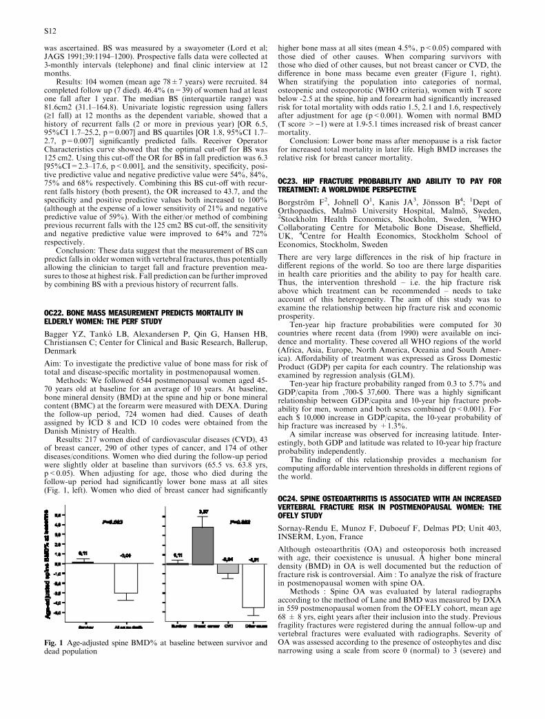
was ascertained. BS was measured by a swayometer (Lord et al;JAGS 1991;39:1194–1200). Prospective falls data were collected at3-monthly intervals (telephone) and final clinic interview at 12months.
Results: 104 women (mean age 78±7 years) were recruited. 84completed follow up (7 died). 46.4% (n=39) of women had at leastone fall after 1 year. The median BS (interquartile range) was81.6cm2 (31.1–164.8). Univariate logistic regression using fallers(‡1 fall) at 12 months as the dependent variable, showed that ahistory of recurrent falls (2 or more in previous year) [OR 6.5,95%CI 1.7–25.2, p=0.007] and BS quartiles [OR 1.8, 95%CI 1.7–2.7, p=0.007] significantly predicted falls. Receiver OperatorCharacteristics curve showed that the optimal cut-off for BS was125 cm2. Using this cut-off the OR for BS in fall prediction was 6.3[95%CI=2.3–17.6, p<0.001], and the sensitivity, specificity, posi-tive predictive value and negative predictive value were 54%, 84%,75% and 68% respectively. Combining this BS cut-off with recur-rent falls history (both present), the OR increased to 43.7, and thespecificity and positive predictive values both increased to 100%(although at the expense of a lower sensitivity of 21% and negativepredictive value of 59%). With the either/or method of combiningprevious recurrent falls with the 125 cm2 BS cut-off, the sensitivityand negative predictive value were improved to 64% and 72%respectively.
Conclusion: These data suggest that the measurement of BS canpredict falls in older womenwith vertebral fractures, thus potentiallyallowing the clinician to target fall and fracture prevention mea-sures to those at highest risk. Fall prediction can be further improvedby combining BS with a previous history of recurrent falls.
OC22. BONE MASS MEASUREMENT PREDICTS MORTALITY INELDERLY WOMEN: THE PERF STUDY
Bagger YZ, Tanko LB, Alexandersen P, Qin G, Hansen HB,Christiansen C; Center for Clinical and Basic Research, Ballerup,Denmark
Aim: To investigate the predictive value of bone mass for risk oftotal and disease-specific mortality in postmenopausal women.
Methods: We followed 6544 postmenopausal women aged 45-70 years old at baseline for an average of 10 years. At baseline,bone mineral density (BMD) at the spine and hip or bone mineralcontent (BMC) at the forearm were measured with DEXA. Duringthe follow-up period, 724 women had died. Causes of deathassigned by ICD 8 and ICD 10 codes were obtained from theDanish Ministry of Health.
Results: 217 women died of cardiovascular diseases (CVD), 43of breast cancer, 290 of other types of cancer, and 174 of otherdiseases/conditions. Women who died during the follow-up periodwere slightly older at baseline than survivors (65.5 vs. 63.8 yrs,p<0.05). When adjusting for age, those who died during thefollow-up period had significantly lower bone mass at all sites(Fig. 1, left). Women who died of breast cancer had significantly
higher bone mass at all sites (mean 4.5%, p<0.05) compared withthose died of other causes. When comparing survivors withthose who died of other causes, but not breast cancer or CVD, thedifference in bone mass became even greater (Figure 1, right).When stratifying the population into categories of normal,osteopenic and osteoporotic (WHO criteria), women with T scorebelow -2.5 at the spine, hip and forearm had significantly increasedrisk for total mortality with odds ratio 1.5, 2.1 and 1.6, respectivelyafter adjustment for age (p<0.001). Women with normal BMD(T score >)1) were at 1.9-5.1 times increased risk of breast cancermortality.
Conclusion: Lower bone mass after menopause is a risk factorfor increased total mortality in later life. High BMD increases therelative risk for breast cancer mortality.
OC23. HIP FRACTURE PROBABILITY AND ABILITY TO PAY FORTREATMENT: A WORLDWIDE PERSPECTIVE
Borgstrom F2, Johnell O1, Kanis JA3, Jonsson B4; 1Dept ofOrthopaedics, Malmo University Hospital, Malmo, Sweden,2Stockholm Health Economics, Stockholm, Sweden, 3WHOCollaborating Centre for Metabolic Bone Disease, Sheffield,UK, 4Centre for Health Economics, Stockholm School ofEconomics, Stockholm, Sweden
There are very large differences in the risk of hip fracture indifferent regions of the world. So too are there large disparitiesin health care priorities and the ability to pay for health care.Thus, the intervention threshold – i.e. the hip fracture riskabove which treatment can be recommended – needs to takeaccount of this heterogeneity. The aim of this study was toexamine the relationship between hip fracture risk and economicprosperity.
Ten-year hip fracture probabilities were computed for 30countries where recent data (from 1990) were available on inci-dence and mortality. These covered all WHO regions of the world(Africa, Asia, Europe, North America, Oceania and South Amer-ica). Affordability of treatment was expressed as Gross DomesticProduct (GDP) per capita for each country. The relationship wasexamined by regression analysis (GLM).
Ten-year hip fracture probability ranged from 0.3 to 5.7% andGDP/capita from ,700-$ 37,600. There was a highly significantrelationship between GDP/capita and 10-year hip fracture prob-ability for men, women and both sexes combined (p<0.001). Foreach $ 10,000 increase in GDP/capita, the 10-year probability ofhip fracture was increased by +1.3%.
A similar increase was observed for increasing latitude. Inter-estingly, both GDP and latitude was related to 10-year hip fractureprobability independently.
The finding of this relationship provides a mechanism forcomputing affordable intervention thresholds in different regions ofthe world.
OC24. SPINE OSTEOARTHRITIS IS ASSOCIATED WITH AN INCREASEDVERTEBRAL FRACTURE RISK IN POSTMENOPAUSAL WOMEN: THEOFELY STUDY
Sornay-Rendu E, Munoz F, Duboeuf F, Delmas PD; Unit 403,INSERM, Lyon, France
Although osteoarthritis (OA) and osteoporosis both increasedwith age, their coexistence is unusual. A higher bone mineraldensity (BMD) in OA is well documented but the reduction offracture risk is controversial. Aim : To analyze the risk of fracturein postmenopausal women with spine OA.
Methods : Spine OA was evaluated by lateral radiographsaccording to the method of Lane and BMD was measured by DXAin 559 postmenopausal women from the OFELY cohort, mean age68 ± 8 yrs, eight years after their inclusion into the study. Previousfragility fractures were registered during the annual follow-up andvertebral fractures were evaluated with radiographs. Severity ofOA was assessed according to the presence of osteophytes and discnarrowing using a scale from score 0 (normal) to 3 (severe) and
Fig. 1 Age-adjusted spine BMD% at baseline between survivor anddead population
S12
graded from 0 to 2 (moderate or severe osteophyte and/or nar-rowing).
Results : Osteophytes and disc narrowing were present in 75and 64% of women at the lumbar spine and in 88 and 51% atthe thoracic spine, increasing with age. BMD increased withseverity of osteophytosis at the lumbar spine, hip and wholebody whereas severity of narrowing was associated with a higherBMD only at the spine. Ninety-six fractures, including 48vertebral fractures, occurred before OA assessment, representing19% of women with OA grade 2 and 16 % with grade 0 or 1.No significant association was found between spine OA and allfragility fractures. In contrast, disc narrowing was associatedwith an increased risk of vertebral fracture with odds-ratio(95% CI) of 3.2 (1.1–9.3) after adjusting for age, BMI andBMD. The risk of vertebral fracture increased with the aggra-vation of narrowing. In comparison with the score 0, the odds-ratio varied from 2.8 (0.9–8.7) to 4.6 (1.2–16.9) according to thenarrowing score. There was no association between fracture riskand the osteophytosis score.
Conclusions : despite a higher BMD, women with spine OA donot have a reduced risk of fracture. Disc narrowing is associatedwith a significant increased vertebral fracture risk. The risk ofosteoporotic fracture should not be underestimated in women withspine OA.
OC25. INFLUENCE OF PHYSICAL ACTIVITY AND VITAMIN D ON BONEMINERAL GAIN AMONG PERIPUBERTAL FINNISH GIRLS: A 3-YEARPROSPECTIVE STUDY
Lehtonen-Veromaa M1, Mottonen T2, Heinonen OJ1, Irjala K3,Viikari J1,2; 1University of Turku, Turku, Finland, 2TurkuUniversity Central Hospital, Turku, Finland, 3Mehilainen Turku,Turku, Finland
Aims: To investigate the influence of physical activity on bonemineral content (BMC) accrual at the femoral neck and lumbarspine.Methods: A total of 171 healthy peripubertal girls aged 9–15 years(62 gymnasts, 58 runners, and 51 nonathletic controls) wereincluded. Weight, height, and the type and the amount of physicalactivity were recorded at 6-months’ intervals over 3 year. Serum25-hydroxyvitamin D (S-25(OH)D) was measured at baseline.BMC of the femoral neck and lumbar spine were measured bydual-energy x-ray absorptiometry (DXA).
Results: By stepwise linear regression analysis the increase ofbone area and weight, the amount of physical activity, and thebaseline BMC presented as significant variables and accounted for67.3% of the variation of the 3-year change of BMC (dBMC) at thefemoral neck. At the lumbar spine, the increase of bone area, heightandweight, the amount of physical activity, S-25(OH)Dand baselineBMC were the significant predictors for dBMC and accounted for90.6%. The adjusted (for increase of height, weight, and bone area)dBMC was 41.7% (P<0.001) and 13.6% (P<0.001) greater in thephysically most active tertile than in the physically least active tertileof the participants at the femoral neck and at the lumbar spine,respectively. The corresponding figures for the physicallymost activetertile compared with the middle tertile were 37.7% (P<0.001) and12.1% (P<0.05) greater at the femoral neck and lumbar spine,respectively.
Conclusions: High-impact loading during the peripubertal yearsis extremely important and beneficial for the growing skeleton, andparticularly for the acquisition of BMC of the femoral neck.Vitamin D has effect on BMC at the lumbar spine.
OC26. UNIQUE EVIDENCE FOR LOAD INDUCED MODIFICATION IN TRA-BECULAR BONE ARCHITECTURE AS WELL AS INCREASED CORTICALBONE FORMATION IN A NON-INVASIVE AXIAL LOADING OF MOUSETIBIAE
de Souza RL1,3, Matsuura M2, Eckstein F2, Lanyon LE1,Pitsillides AA1; 1Royal Veterinary College, London, UK, 2Lug-wig-Maximillians-Universitat, Munich, Germany, 3CAPES -Brazilian Ministry of Education, Brasilia, Brazil
Although bone’s response to mechanical loads can be studied inseveral in vivo models, axially loading the murine tibia, non-invasively, through its articulations would have advantages.These include: i) larger bone size; ii) scope to apply disuse;iii) larger region of cancellous bone volume, and iv) scope toapply strain distributions similar to those engendered duringlocomotion. Herein, we explore such a model in which corti-cal and cancellous bone adaptation to mechanical loading isexamined.
We used 8 groups of female, 14 week old C57Bl/J6 mice: 5groups were loaded so that 1200–2000lE were produced on thelateral midshaft cortex. 2 groups were submitted to sciatic neur-ectomy (SN, 114 days) with and without loading. 1 group wassham-operated. Animals were loaded on alternate days for 3 weeks.They received fluorochrome label on third and last days and werekilled 3 days later. Tibiae (+contralateral controls) were embed-ded; transverse confocal images from 5 diaphyseal sites wereanalysed histomorphometrically. Proximal tibial epiphyses wereanalysed by lCT scans, which extended 0.75 mm distally of thegrowth plate.
We found that murine tibia midshaft exhibited low physio-logical strains during normal locomotion (<300 lE). Loading at2000 lE significantly increased periosteal bone formation at allsites. Increased endosteal formation was only evident at sitesdistal to the midshaft. In contrast, an increase in cortical boneformation was not observed in tibiae loaded at less than1200 lE. lCT scans showed that loading induced significantincreases in trabecular bone thickness; SN-induced ‘disuse’significantly decreased bone volume fraction and increasedtrabecular spacing, while loading after ‘disuse’ also increasedtrabecular thickness.
We show that: i) loading induces an osteogenic response oftrabecular bone architecture and, at least partly, rescues the effectsof SN, ii) tibial cortices, which encounter low strains during loco-motion, require these to be exceeded during axial loading to inducebone formation. These results suggest that the murine tibia offers anovel model for studying the effects of loading on cortical andcancellous bone. To our knowledge this is the first evidence fordirect load-induced changes in trabecular architecture in an animalmodel.
OC27. INTERACTION BETWEEN CALCIUM INTAKE AND MENARCHEALAGE ON BONE MASS GAIN: AN 8-YEAR FOLLOW-UP STUDY FROM PRE-PUBERTY TO POST-MENARCHE
Chevalley T1, Bonjour JP1, Hans D2, Rizzoli R1; 1Service of BoneDiseases, WHO Collaborating Center for Osteoporosis and BoneDiseases, Department of Rehabilitation and Geriatrics, 2Serviceof Nuclear Medicine, Department of Radiology, UniversityHospitals Geneva, Switzerland
Both late menarche and low Ca intake during growth are con-sidered as risk factors for osteoporosis, probably by impairingoptimal peak bone mass achievement. We investigated whetherthe response of bone mineral mass accrual to increased Ca intakecould vary according to menarcheal age and, conversely, whetherCa intake could influence menarcheal age. In an initial study girlsaged 7.9±0.1 years (mean±sem) were randomized in a doubleblind controlled trial to receive either a Ca supplement (Ca-sup-pl.) of 850 mg/d or placebo during one year. We now report onresults obtained when subjects were 16.4±0.1 years. Arealbone mineral density (aBMD) was determined by DXA at 6skeletal sites: radius metaphysis, radius diaphysis, femoral neck,trochanter, femoral diaphysis and L2–L4 vertebrae. During theintervention the mean yearly aBMD gains at these 6 sites were21±2 and 27±2 mg/cm2 (p<0.005) in the Placebo and Ca-suppl.group, respectively. Subjects were re-examined at 1.0, 3.5 and 7.5years after the end of intervention and menarcheal age was re-corded. Spontaneous Ca intake was assessed by frequency ques-tionnaires at baseline, 6 and 12 months and at each follow-upvisit. A significant earlier mean age of menarche was observed inthe Ca-suppl as compared to the placebo group (p<0.05) and a
S13
significant negative correlation was found between menarchealage and Ca intake (R=)0.35, p<0.0001), and between gain inaBMD from age 8.0 to 16.4 years and menarcheal age at all 6skeletal sites (R range: from )0.41 to )0.22, p<0.0001 –p<0.016). The positive effect of Ca-suppl. on mean aBMD gain(mg/cm2) from baseline remained significantly greater in girlsbelow but not in those above the median of menarcheal age (13.0years). Early menarcheal age (12.1±0.1): placebo: 286±8; Ca-suppl. 317±8, p<0.01. Late menarcheal age (13.9±01): placebo284±10, Ca-suppl. 276±16, p>0.05. This study suggests that thelevel of Ca intake during pre-puberty could influence the timing ofmenarche which in turn, appears to influence long term bone massgain in response to Ca supplementation. Thus, both early men-archeal age determinants and high Ca intake may positivelyinteract on bone mineral mass accrual.
OC28. POSTTRANSLATIONAL MODIFICATIONS OF COLLAGEN CON-TRIBUTE TO BONE STRENGTH INDEPENDENTLY OF BONE MINERALDENSITY (BMD)
Garnero P1,2, Borel O1, Gineyts E1, Duboeuf F1, ChristiansenC3,4, Delmas PD1; 1Inserm unit 403, Lyon, France, 2Synarc,Lyon, France, 3Center for Clinical and Basic Research, Copen-hagen, Denmark, 4NordicBioscience,Copenhagen, Denmark
Bone strength depends on its mass and geometry, but also on thematerial properties of its matrix.Mineral accounts for the stiffnessof bone and collagen mainly influences its toughness although therelative contribution of each is unclear.
Aim: To analyze the role of posttranslational modifications ofcollagen on the mechanical properties of cortical bone using anin vitro model where the extent of crosslinking can be modified,keeping constant the size and the mineral content of bone.
Methods: Calibrated fetal bovine cortical bone specimens(11 animals, 41 samples in total) characterized by a low degree ofpostranslational modifications were incubated for 0, 60, 90 and 120days at 37 �C to induce collagen crosslinking. At each time, 3 pointbending mechanical test was performed to determine the stiffnessand the yield strength. The concentration of enzymatic (PYD,DPD) and non-enzymatic (pentosidine) crosslinks was measured byHPLC and the extent of C-telopeptide (CTX) isomerisation wasevaluated by ELISA of native (a CTX) and isomerised (b CTX)forms.
Results: After 60 days of incubation, yield strength wasdecreased by 40% (p=0.01) whereas no effect was observed onstiffness and BMD. The decline of yield strength was associatedwith increases of PYD (+98%, p=0.005), DPD (+42%,p=0.013), pentosidine (+ 55 fold, p=0.005) and b/a CTX ratio(+4.9 fold, p=0.005). In multivariate analyses, the prediction ofyield strength was significantly improved by combining BMD(r2=0.36) with PYD (r2 increasing to 0.58, p=0.0003), pentosidine(r2=0.58, p<0.0001) or b/a CTX (r2=0.49, p=0.003. The b/aCTX ratio highly correlated with PYD (r=0.63), DPD (r=0.60)and pentosidine (r=0.7), (p<0.001 for all). We measured the uri-nary ratio b/a CTX in 404 postmenopausal women followed for 7years and found that it was associated with incident fractureindependently of BMD and bone turnover.
Conclusion: These data indicate that the degree of posttrans-lational modifications of collagen matrix plays an independent rolein determining the mechanical competence of cortical bone. Theratio of b/a CTX may reflect the extent of posttranslational mod-ifications of collagen and its measurement in urine could provide anin vivo marker of bone collagen quality.
OC29. LEPTIN RECEPTOR (LEPR) POLYMORPHISMS CONTRIBUTE TOBONE MASS IN HEALTHY PRE-PUBERTAL BOYS
FerrariSL,ManenD,ValentD,ChevalleyT,BonjourJP,RizzoliR;Division of Bone Diseases, Dept of Rehabilitation and Geriatrics,Geneva University Hospital, Switzerland
The adipocyte-derived satiety factor, Leptin, also appears tonegatively regulate bone formation in mice. However, its contri-
bution to bone mass determination in humans remains unclear. AGln223Arg missense polymorphism (c.668A>G) in the leptinreceptor gene (LEPR) and a promoter polymorphism()2548G>A) in the leptin gene (LEP), both previously associatedwith leptin levels and obesity in humans, were investigated fortheir association with bone mass in healthy European-Caucasianboys (n=235, 7.4 ±0.4 yrs).
LEPR A>G (MspI) and LEP )2548G>A (HhaI) polymor-phisms were determined by PCR. Bone mineral content (BMC,gm) was measured by DXA at the lumbar spine (LS), femoralneck (FN), total hip (TH), midshaft femur (FS), distal and ul-tradistal radius (DR, UR). Dietary intake of calcium (Ca) andproteins, and the level of physical exercise (PE) were recorded.
Frequency of the LEPR G and LEP –2548A allele was 0.30and 0.43, respectively. LEP promoter genotypes were not asso-ciated with any variable. In contrast, LEPR genotypes were sig-nificantly associated with height (p=0.044) and BMC at FN(p=0.035), TH (p=0.022), FS (p=0.027), DR (p=0.007), andmarginally at UR (p=0.066) and LS (p=0.099). Thus, BMC was8–12% significantly lower in GG (n=21) compared to AA(n=108), and intermediate in GA. BMI, age, Ca and proteinintake were similar among LEPR genotypes. However, PE sig-nificantly differed among genotypes (p=0.021) and was signifi-cantly related to BMC at all sites (by multiple regression furtherincluding age, Ca and proteins). Subsequently, significant p valuesfor interaction between PE and LEPR genotypes were found atseveral skeletal sites (by 2F-ANOVA). Indeed, BMC was signifi-cantly lower in GG compared to AA among children with low PE(£median, 222 kcal/d), but not with higher PE levels. Afteradjustment for age, height, BMI, PE and dietary intakes, LEPRalleles contribution to FN BMC variance was 1.5% in all boysand 2.9% in those with low PE.
In conclusions, LEPR Gln223Arg polymorphism is associatedwith BMC in healthy pre-pubertal boys, particularly in those withlow levels of physical exercise. These data support the possibilitythat the leptin system may influence bone mass in humans inrelation to energy expenditure.
OC30. ACIDIFICATION OF THE OSTEOCLASTIC RESORPTION LACUNAEIMPINGES ON THE COUPLING OF BONE RESORPTION TO BONE FOR-MATION
Karsdal MA1, Henriksen K1, Schaller S1, Gram J2, Bollerslev J3,Christiansen C1; 1Nordic Bioscience/CCBR, Herlev/Ballerup,Denmark, 2Ribe County Hospital, Denmark, 3Section of Endo-crinology, National University Hospital, Oslo, Norway
Bone resorption is tightly coupled to bone formation in normalhealthy adults. However, in autosomal dominant osteopetrosis II(ADOII) caused by a mutation in the ClC-7 chloride channel geneessential for acidification of the osteoclastic resorption lacunaeand resorption, decreased levels of bone resorption, normal levelsof bone formation, but 5 fold-increased levels of osteoclasts aredetected. This strongly suggests that bone turnover has beenuncoupled in these patients.
We found that osteoclastogenesis of ADOII osteoclasts wasnormal, albeit the resorption was reduced to 10–25%. Whenmature osteoclasts were cultured on bovine bone slices, osteoclastviability was augmented either by 20% in ADOII osteoclasts or by50–80% through complete abrogation of acidification by either achloride channel inhibitor (NS5818 10microM) or an inhibitor ofthe proton pump, Bafilomycin 30nM. This suggests that attenuatedosteoclast apoptosis is responsible for the increased osteoclastlevels in the ADOII patients.
To investigate if decreased resorption is compatible with unaf-fected bone formation, we used a chloride channel inhibitor in therat OVX model. We observed a significant 40% decrease in theresorption marker (Dpd), but no effect on the bone formationmarker (osteocalcin). Moreover, we found a significant increase inserum TRAP levels, in alignment with the attenuated ClC-7 activityin ADOII patients.
In conclusion, osteoclasts with attenuated dissolution of theinorganic phase of bone have prolonged viability. The increased
S14
levels of inactive osteoclasts generate formation signals to osteo-blasts, either directly or by an increased area of bone surfaceexposed to resorption. These results suggest that acidification of theosteoclastic resorption compartment may be involved in the cou-pling of bone resorption to bone formation.
OC31. RNA PROFILING OF ANTIRESORPTIVES REVEAL ALENDRONATEAND ESTROGEN DECREASE BONE FORMATION GENES WHILE RALOXIF-ENE MAINTAINS THEIR INCREASE IN THE OVARIECTOMIZED RAT
Helvering LM, Onyia JE, Dow E, Wei T, Gelbert L, Adams C,Lawrence F, Bemis K, Halladay D, Miles RR, Kulkarni N,Huang S, Chen P, Chandrasekhar S, Frolik C, Sato M, Ma L,Bryant H; Eli Lilly Research Labs, Indianapolis, IN, USA
The preservation of bone by three classes of compounds (Estro-gen, SERMS, and bisphosphanates) have been well described inthe ovariectomized rat model by histomorphometric and histo-logical analysis but a global analysis comparing their effects ongene expression has not yet been described. This study utilized anAffymetrix microarray to analyze RNA from the OB-enrichedproximal femur of sham, ovariectomized rats, and ovariectomizedrats treated for 1 day and 5 weeks with 17a-ethinyl estradiol (E) at0.1 mg/kg, Raloxifene (Ral) at 3.0 mg/kg, EM652 at 0.1 mg/kg,and Alendronate (Ald) at 8 lg/kg. Genes significantly changed(p<0.05) from sham levels following ovariectomy, which werealso changed by any drug following 5 weeks of treatment, wereidentified and subjected to clustering analysis. Genes associatedwith bone formation (i.e. type I collagen a 1 & 2, osteocalcin,osteonectin, and biglycan) clustered together indicating a similarpattern of regulation. Ovariectomy, known to increase the rate ofbone formation, induced these genes within 12 d and sustainedthis over time. Following 5 weeks, Ral only slightly lowered thelevels of these genes while E and Ald suppressed them below thatof sham. EM652 suppressed these genes to a level intermediatebetween Ral and Ald. At the earlier timepoint Ald begandecreasing some of these formation genes but E did not. It can beconcluded that at the molecular level the three classes of antire-sorptives can be identified by contrasting their effects on mRNAlevels of osteoblastic genes. Ald began turning down the expres-sion of osteoblastic genes within 24 h and then lowered thembelow sham levels with time. E did not alter expression at 24 h,but suppressed these genes below that of sham at 5 weeks. TheSERMS were also differentiated by a partial suppression of for-mation genes at both timepoints with EM652 and a negligiblechange by Ral only at 5 weeks. In summary, ovariectomy rapidlyinduced markers of bone formation and these levels are main-tained with Ral treatment but are suppressed with Ald and E.Additionally, these data provide evidence that treatment with Aldnot only inhibits resorption but also suppresses osteoblasticactivity.
OC32 POSITIVE CORRELATIONS BETWEEN EARLY CHANGES IN BIO-CHEMICAL MARKERS OF BONE FORMATION AND VOLUMETRIC BMDAFTER TERIPARATIDE THERAPY: FURTHER EVIDENCE FOR DIVERGINGEFFECTS OF ALENDRONATE AND TERIPARATIDE ON BONE
Bandeira F1, Eriksen EF2, Szejnfeld V3, McClung MR4, DonleyDW2, San Martin J2, Miller PD5; 1Hospital Agamenon Magal-haes, University of Pernambuco, Recife, BRAZIL, 2LillyResearch Laboratories, Eli Lilly and Company, Indianapolis,IN, USA, 3Escola Paulista de Medicina, Sao Paulo SP, BRAZIL,4Oregon Osteoporosis Center, Portland, OR, USA, 5ColoradoCenter for Bone Research, Lakewood, CO, USA
We conducted an 18-month randomized controlled trial com-paring the effects of teriparatide 20 mcg/day [rhPTH (1-34),TPTD20] and alendronate 10 mg/day (ALN10) on bone inpostmenopausal women with osteoporosis. Previously reportedresults have shown significant increases of 100 to 300% in boneturnover markers (BTM) during the first 6 months of TPTD20treatment compared with decreases of 60 to 70% during ALN10treatment (McClung et al. ASBMR 2003). Both lumbar spine
areal BMD (aBMD) by DXA and trabecular volumetric BMD(vBMD) by QCT increased in each treatment group. However,while increases in aBMD were nearly 2-fold, vBMD increaseswere 4 to 5-fold greater in TPTD20 versus ALN10 treatedpatients.
Aims: To assess the correlation of early changes in BTM andimprovements in bone mass as reflected in lumbar spine aBMD andvBMD changes after 18 months of ALN10 or TPTD20 therapy.
Methods: Early changes in the collagen bone formation mark-ers (PINP, PICP) were assessed using the area under the curve(AUC) from the first 6 months of treatment. Spearman’s correla-tion coefficient was calculated between 6-month AUC values andlast observed aBMD and vBMD.
Results: In TPTD20-treated patients, early changes in PINPand PICP had similar significant positive correlations with lumbarspine aBMD and vBMD (Table). After ALN10 therapy, earlychanges in PINP and PICP had significant negative correlationswith lumbar spine aBMD, but lesser negative and nonsignificantcorrelations with lumbar spine vBMD.
BMD parameterBiochemical marker
Spearman Correlation
ALN10 TPTD20
Change in Lumbar spine aBMD(DXA) at study endpoint
(N = 89) (N = 90)
PINP first 6-months AUC )0.51* 0.53*PICP first 6-months AUC )0.27* 0.42*
Change in Lumbar spine vBMD(QCT) at study endpoint
(N = 23) (N = 26)
PINP first 6-months AUC )0.20 0.51*PICP first 6-months AUC )0.19 0.55*
*P<0.01
Conclusions: Early changes in PICP and PINP correlate posi-tively to ultimate aBMD and vBMD response in teriparatide-treated patients and inversely to aBMD in alendronate-treatedpatients. In alendronate-treated patients no correlation to vBMDwas demonstrable. These results highlight the differences inmechanism of action between the two therapies, and indicate thatPINP and PICP are good markers for the skeletal response toteriparatide.
OC33. BONE TURNOVER AFTER ONE YEAR OF ALENDRONATE ANDFRACTURE RISK: THE FRACTURE INTERVENTION TRIAL
Bauer DC1, Hochberg MC2, Garnero P3,4, Ross P5, Santora A5,Ewing S1; 1University of California, San Francisco, CA,2University of Maryland, Baltimore, MD, 3Synarc, Lyon, France,4INSERM 403, Lyon, France, 5MRL, Rahway, NJ
Short-term changes in biochemical markers of bone turnoverpredict subsequent spine and non-spine fractures among bis-phosphonate treated women, but the utility of a single on-treat-ment measurement is unknown. In addition, some havehypothesized that very low turnover on therapy may be associ-ated with impaired bone quality and increased fragility.
In the Fracture Intervention Trial we measured bone specificalkaline phosphatase (bone ALP, Hybritech) and N-terminalpropeptide (P1NP, Orion) after 1 year of therapy with alendro-nate (ALN), 5 mg/d (n=3105) or placebo (n=3081). AmongALN-treated women, during a mean follow-up of 3.6 yearsafter baseline, 226 non-spine fractures (including 15 hip fractures),and 119 incident vertebral fractures on paired lateral spinex-rays were documented. Age-adjusted logistic and hazardmodels were used to examine the relationship between marker levelafter 1 yr. of ALN therapy and subsequent spine and non-spinefracture.
After 1 year of ALN, lower bone ALP levels (per SD) wereassociated with fewer vertebral fractures (OR=0.75, 95% CI: 0.65,0.86) but not non-spine fractures (RH=0.99, 95% CI: 0.87, 1.14).
S15
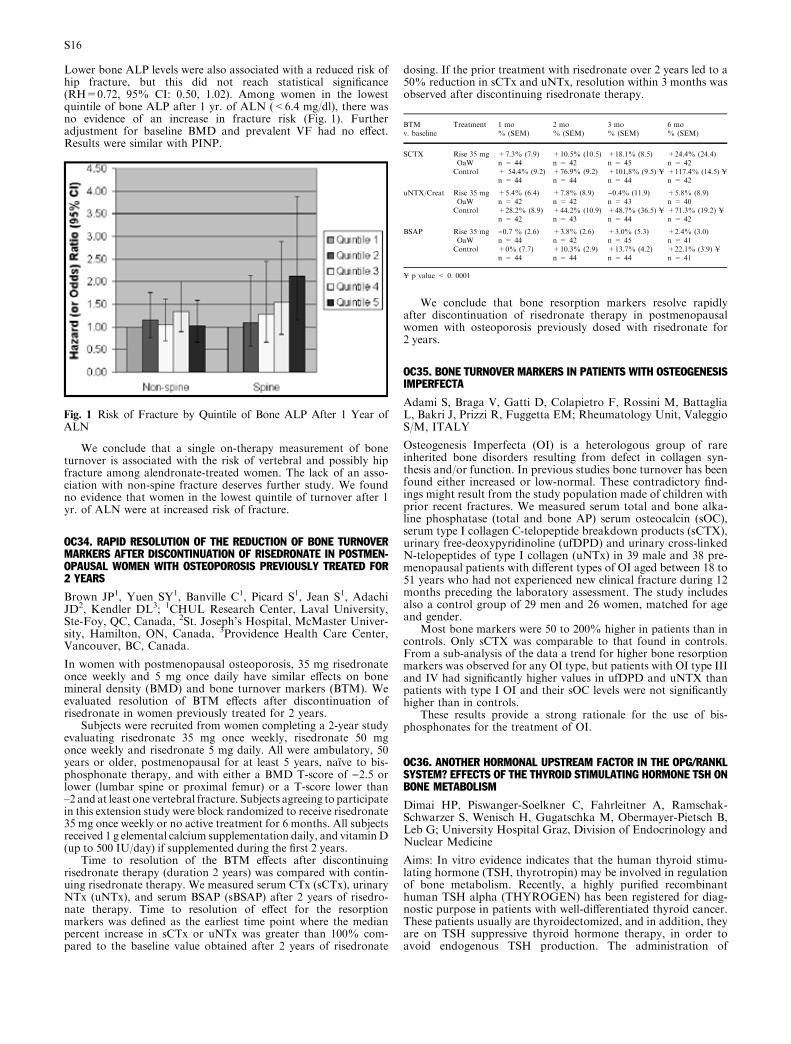
Lower bone ALP levels were also associated with a reduced risk ofhip fracture, but this did not reach statistical significance(RH=0.72, 95% CI: 0.50, 1.02). Among women in the lowestquintile of bone ALP after 1 yr. of ALN (<6.4 mg/dl), there wasno evidence of an increase in fracture risk (Fig. 1). Furtheradjustment for baseline BMD and prevalent VF had no effect.Results were similar with PINP.
We conclude that a single on-therapy measurement of boneturnover is associated with the risk of vertebral and possibly hipfracture among alendronate-treated women. The lack of an asso-ciation with non-spine fracture deserves further study. We foundno evidence that women in the lowest quintile of turnover after 1yr. of ALN were at increased risk of fracture.
OC34. RAPID RESOLUTION OF THE REDUCTION OF BONE TURNOVERMARKERS AFTER DISCONTINUATION OF RISEDRONATE IN POSTMEN-OPAUSAL WOMEN WITH OSTEOPOROSIS PREVIOUSLY TREATED FOR2 YEARS
Brown JP1, Yuen SY1, Banville C1, Picard S1, Jean S1, AdachiJD2, Kendler DL3; 1CHUL Research Center, Laval University,Ste-Foy, QC, Canada, 2St. Joseph’s Hospital, McMaster Univer-sity, Hamilton, ON, Canada, 3Providence Health Care Center,Vancouver, BC, Canada.
In women with postmenopausal osteoporosis, 35 mg risedronateonce weekly and 5 mg once daily have similar effects on bonemineral density (BMD) and bone turnover markers (BTM). Weevaluated resolution of BTM effects after discontinuation ofrisedronate in women previously treated for 2 years.
Subjects were recruited from women completing a 2-year studyevaluating risedronate 35 mg once weekly, risedronate 50 mgonce weekly and risedronate 5 mg daily. All were ambulatory, 50years or older, postmenopausal for at least 5 years, naıve to bis-phosphonate therapy, and with either a BMD T-score of )2.5 orlower (lumbar spine or proximal femur) or a T-score lower than–2 and at least one vertebral fracture. Subjects agreeing to participatein this extension study were block randomized to receive risedronate35 mg once weekly or no active treatment for 6 months. All subjectsreceived 1 g elemental calcium supplementation daily, and vitaminD(up to 500 IU/day) if supplemented during the first 2 years.
Time to resolution of the BTM effects after discontinuingrisedronate therapy (duration 2 years) was compared with contin-uing risedronate therapy. We measured serum CTx (sCTx), urinaryNTx (uNTx), and serum BSAP (sBSAP) after 2 years of risedro-nate therapy. Time to resolution of effect for the resorptionmarkers was defined as the earliest time point where the medianpercent increase in sCTx or uNTx was greater than 100% com-pared to the baseline value obtained after 2 years of risedronate
dosing. If the prior treatment with risedronate over 2 years led to a50% reduction in sCTx and uNTx, resolution within 3 months wasobserved after discontinuing risedronate therapy.
We conclude that bone resorption markers resolve rapidlyafter discontinuation of risedronate therapy in postmenopausalwomen with osteoporosis previously dosed with risedronate for2 years.
OC35. BONE TURNOVER MARKERS IN PATIENTS WITH OSTEOGENESISIMPERFECTA
Adami S, Braga V, Gatti D, Colapietro F, Rossini M, BattagliaL, Bakri J, Prizzi R, Fuggetta EM; Rheumatology Unit, ValeggioS/M, ITALY
Osteogenesis Imperfecta (OI) is a heterologous group of rareinherited bone disorders resulting from defect in collagen syn-thesis and/or function. In previous studies bone turnover has beenfound either increased or low-normal. These contradictory find-ings might result from the study population made of children withprior recent fractures. We measured serum total and bone alka-line phosphatase (total and bone AP) serum osteocalcin (sOC),serum type I collagen C-telopeptide breakdown products (sCTX),urinary free-deoxypyridinoline (ufDPD) and urinary cross-linkedN-telopeptides of type I collagen (uNTx) in 39 male and 38 pre-menopausal patients with different types of OI aged between 18 to51 years who had not experienced new clinical fracture during 12months preceding the laboratory assessment. The study includesalso a control group of 29 men and 26 women, matched for ageand gender.
Most bone markers were 50 to 200% higher in patients than incontrols. Only sCTX was comparable to that found in controls.From a sub-analysis of the data a trend for higher bone resorptionmarkers was observed for any OI type, but patients with OI type IIIand IV had significantly higher values in ufDPD and uNTX thanpatients with type I OI and their sOC levels were not significantlyhigher than in controls.
These results provide a strong rationale for the use of bis-phosphonates for the treatment of OI.
OC36. ANOTHER HORMONAL UPSTREAM FACTOR IN THE OPG/RANKLSYSTEM? EFFECTS OF THE THYROID STIMULATING HORMONE TSH ONBONE METABOLISM
Dimai HP, Piswanger-Soelkner C, Fahrleitner A, Ramschak-Schwarzer S, Wenisch H, Gugatschka M, Obermayer-Pietsch B,Leb G; University Hospital Graz, Division of Endocrinology andNuclear Medicine
Aims: In vitro evidence indicates that the human thyroid stimu-lating hormone (TSH, thyrotropin) may be involved in regulationof bone metabolism. Recently, a highly purified recombinanthuman TSH alpha (THYROGEN) has been registered for diag-nostic purpose in patients with well-differentiated thyroid cancer.These patients usually are thyroidectomized, and in addition, theyare on TSH suppressive thyroid hormone therapy, in order toavoid endogenous TSH production. The administration of
Fig. 1 Risk of Fracture by Quintile of Bone ALP After 1 Year ofALN
BTM Treatment 1 mo 2 mo 3 mo 6 mov. baseline % (SEM) % (SEM) % (SEM) % (SEM)
SCTX Rise 35 mg +7.3% (7.9) +10.5% (10.5) +18.1% (8.5) +24.4% (24.4)OaW n = 44 n = 42 n = 45 n = 42Control + 54.4% (9.2) +76.9% (9.2) +101,8% (9.5) ¥ +117.4% (14.5) ¥
n = 44 n = 44 n = 44 n = 42
uNTX/Creat Rise 35 mg +5.4% (6.4) +7.8% (8.9) )0.4% (11.9) +5.8% (8.9)OaW n = 42 n = 42 n = 43 n = 40Control +28.2% (8.9) +44.2% (10.9) +48.7% (36.5) ¥ +71.3% (19.2) ¥
n = 42 n = 43 n = 44 n = 42
BSAP Rise 35 mg )0.7 % (2.6) +3.8% (2.6) +3.0% (5.3) +2.4% (3.0)OaW n = 44 n = 42 n = 45 n = 41Control +0% (7.7) +10.3% (2.9) +13.7% (4.2) +22.1% (3.9) ¥
n = 44 n = 44 n = 44 n = 41
¥ p value < 0. 0001
S16
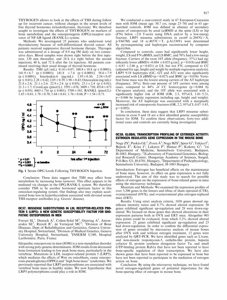
THYROGEN allows to look at the effects of TSH during followup for recurrent cancer, without changes in the serum levels offree thyroid hormones (triiodothyronin, thyroxin). We thereforesaught to investigate the effects of THYROGEN on markers ofbone metabolism and the osteoprotegerin (OPG)/receptor acti-vator of NF-kB ligand (RANK-L) system.
Methods: We investigated 25 patients who underwent totalthyroidectomy because of well-differentiated thyroid cancer. Allpatients received suppressive thyroid hormone therapy. Thyrogenwas administered at a dosage of 0.9 mg IM daily on two consec-utive days. Blood samples were drawn right before the first injec-tion, 120 min thereafter, and 24 h (i.e right before the secondinjection), 48 h, and 72 h after the 1st injection. All patients con-tinued receiving their usual dosage of thyroid hormone.
Results: TSH (lU/mL): 0.14±0.07; 100.8±10.8 (p £ 0.0001);141.9±6.7 (p £ 0.0001); 141.8 ±7.4 (p £ 0.0001); 58.6±7.9(p £ 0.0001); Interleukin-6 (pg/mL): 1.95±0.36; 2.54±0.47(p £ 0.01); 2.28±0.42; 2.05±0.39; 1.98±0.43; Osteocalcin (ng/mL):21.8±2.6; 21.1±2.6; 22.9±2.6 (p £ 0.05); 23.1±2.4 (p £ 0.01);21.3±1.7; CrossLaps (pmol/L): 3593±676; 3609±710; 4514±673(p £ 0.05); 4465±716 (p £ 0.001); 3709±541; RANKL (pmol/L):1.65±0.61; 1.78±0.70; 1.64±0.61; 1.76±0.66; P<1.54±0.71.
Conclusion: These data suggest that TSH may affect bonemetabolism by increasing bone turnover. This effect appears to bemediated via changes in the OPG/RANK-L system. We thereforeconsider TSH to be another hormonal upstream factor in thisosteoclast-regulating system. Our findings also may explain accel-erated bone loss in hyperthyroidism associated with elevated serumTSH-receptor antibodies (e.g. Graves’ disease).
OC37. MISSENSE SUBSTITUTIONS IN LDL RECEPTOR-RELATED PRO-TEIN 5 (LRP5): A NEW GENETIC SUSCEPTIBILITY FACTOR FOR IDIO-PATHIC OSTEOPOROSIS IN MEN
Ferrari SL1, Deutsch A2, Cohen-Solal M3, Ostertag A3, Anton-arakis SE2, Rizzoli R1, de Vernejoul MC3; 1Division of BoneDiseases, Dept of Rehabilitation and Geriatrics, Geneva Univer-sity Hospital, Switzerland, 2Division of Medical Genetics, GenevaUniversity Hospital, Switzerland, 3INSERM U349, HospitalLariboisiere, Paris, France
Idiopathic osteoporosis in men (IOM) is a non-mendelian disorderwith strong poly-genetic determinants. IOM results from decreasedbone formation leading to low peak bone mass, particularly in thevertebrae. Mutations in LDL receptor-related protein 5 (LRP5),which mediates the effects of Wnt on osteoblasts, cause osteopo-rosis-pseudoglioma (OPPG) and ‘‘high bonemass’’ syndromes.Wepreviously reported that LRP5 polymorphisms are associated withvertebral bone mass in healthy males. We now hypothesize thatLRP5 polymorphisms could play a role in IOM.
We conducted a case-control study in 67 European-Caucasianmen with IOM (mean age: 50.7 yrs, range 23–70) and in 65 age-matched controls. IOM was defined in absence of secondarycauses of osteoporosis by areal (a)BMD at the spine (LS) or hip(FN) below )2.0 T-score using DXA and/or by a low-energyfracture. LRP5 missense substitutions in exon9 (c.2047G>A,p.V667M) and 18 (c.4037C>T, p.A1330V) were determinedby pyrosequencing and haplotypes reconstructed by computeralgorythm.
Compared to controls, cases had significantly lower height,weight, LS and FN aBMD, and LSBMC, and 70%had a low energyfracture. Carriers of the exon 18T allele (frequency, 37%) had sig-nificantly lower aBMD ()0.084±0.035 g/cm2, p=0.018) and BMC()4.83 ±2.06 g, p=0.021) at LS, but not FN, compared to CC(adjusted for age, height and weight by multiple regression analysis).LRP5 9/18 haplotypes (GC, GT and AT) were also significantlyassociated with LS aBMD (p=0.027) and BMC (p=0.034). Verte-bral bone mass was the lowest among carriers of the AT haplotype(frequency, 20%). Sixty-one percent of 18T carriers were amongcases, compared to 44% of CC homozygotes (p=0.064 byChi-square analysis), and the 18T allele was associated with asignificantly higher risk of IOM (OR, 2.3, 95%CI, 1.01–5.21,p=0.046 by logistic regression including age, height and weight).Moreover, the AT haplotype was associated with a marginallyincreased risk of osteoporotic fractures (OR, 2.2, 95%CI, 0.87–5.85,p=0.092).
In conclusion, these data suggest that LRP5 missense substi-tutions in exon 9 and 18 are a first identified genetic susceptibilityfactor for IOM. To confirm these observations, forty-two addi-tional cases and controls are currently being investigated.
OC38. GLOBAL TRANSCRIPTION PROFILING OF ESTROGEN ACTIVITY:ESTROGEN REGULATES GENE EXPRESSION IN THE MOUSE BONE
NagyZS1; PuskasGL2; Zvara A2; NagyBZS2; SpeerG1; Takacs I1;Bajnok E1; Kosa J1; Lakatos P1; Hamar P3; Kokeny G3; 11stDepartment of Medicine, Semmelweis University, Budapest,H-1083, Hungary, 22Laboratory of Functional Genomics, Biolog-ical Research Center, Hungarian Academy of Sciences, Szeged,P.O.Box 521, H-6701, Hungary, 3Department of Pathophysiology,Semmelweis University, Budapest, H-1083 Hungary
Introduction: Estrogen has beneficial effects on the maintenanceof bone mass, however, its effect on gene expression is not fullyunderstood. The aim of this study was to search for possibleeffects of estrogen on the expression of bone-related genes in miceusing the microarray technique.
Materials and Methods: We examined the expression profiles ofover 3,200 genes in the femurs and tibiae of sham operated (CTR),ovariectomized (OVX), and ovariectomized plus estrogen-replacedmice (ERT).
Results: Using strict analysis criteria, 1038 genes showed sig-nificant intensity ratios and 6.7% showed altered expression: 30genes exhibited significant up-regulation and 28 were down-reg-ulated. We focused on those genes that showed alterations in theirexpression patterns both in OVX and ERT mice. Altogether 905data points could be evaluated, from which 5.2% showed alteredexpression: 23 genes exhibited significant up-regulation and 23had down-regulation. In order to confirm the differential expres-sion of genes revealed by microarray analysis of mouse bonesafter OVX with and without estrogen treatment, 12 genes wereanalyzed by QRT-PCR. We have identified genes such as L1cam,and non-muscle tropomyosin-5, cathelin-like protein, heparincofactor II, protein synthesis elongation factor Tu, and smallGTP-binding protein Rab1a that have not been reported to havebone-specific regulation of their transcription. We have alsoshown genes that have been reported to be expressed in bone buthave not been reported to participate in the mediation of estrogenaction on bone.
Conclusion: By using the microarray technique, we have foundnovel estrogen-regulated genes of potential importance for thebone-sparing effect of estrogen in mouse bone.
Fig. 1 Serum OPG Levels Following THYROGEN Injection.
S17
OC39. PATIENTS AT HIGH RISK OF HIP FRACTURE BENEFIT FROMTREATMENT WITH STRONTIUM RANELATE
Rizzoli R1, Reginster JY2, Diaz-Curiel M3, Ortolani S4, Benha-mou C5, Compston J6, Meunier PJ7; 1Div. of Bone Diseases, Deptof Rehabilitation and Geriatrics, Univ. Hospital, Geneva,SWITZERLAND, 2Dept of Epidemiology and Public Health,University of Liege, Liege, BELGIUM, 3Servicio de MedicinaInterna, Fundacion Jimenez Diaz, Madrid, SPAIN, 4Centre forMetabolic Bone Disease, Istituto Auxologico Italiano, Milano,ITALY, 5Hopital de la Madeleine, Orleans, FRANCE, 6Dept ofMedicine, University of Cambridge, Cambridge, UK, 7Dept ofRheumatology and Bone Diseases, Edouard Herriot Hospital,Lyon, FRANCE
The lifetime risk of a hip fracture from age 50 years has beenestimated at 17% for Caucasian women and the incidence risesexponentially in women over 74 yearsold.
Strontium ranelate has been shown to significantly reduce therisk of vertebral fracture in women with established post-meno-pausal osteoporosis by 41% (SOTI study) and by 45% in thosepatients without prevalent vertebral fracture (TROPOS study) over3 years.
The international TROPOS study was designed to evaluate theefficacy of strontium ranelate in reducing the risk of non-vertebralfractures. A total of 5091 patients aged above 70 years, with a lowfemoral neck BMD (T-score <)2.5 SD) were randomized to re-ceive strontium ranelate 2g daily orally over 3 years. A significantreduction of 16% (p=0.04) and 19% (p=0.031) respectively in therelative risk of non-vertebral osteoporotic fracture and majorosteoporosis-related fracture was demonstrated in the intention totreat population. The efficacy of strontium ranelate in reducing therisk of hip fracture was investigated in a subset of particularmedical interest, namely women of 74 years and above and with abaseline femoral neck BMD T-score lower or equal to )3 (calcu-lated according to the centralized normative data). A total of 1977patients are represented in this subset: 982 patients in the strontiumranelate group and 995 patients in the placebo group. The mainbaseline characteristics of this subset were similar between treat-ment and control groups and were as follows: mean (SD) age of79.6 (4.5) years; menopause duration of 31.5 (7.0) years; femoralneck BMD T-score of )3.6 (0.5)..
In ITT, over 3 years, a significant reduction of 36% in therelative risk of hip fracture risk was observed (RR=0.64, 95%CI[0.412;0.997]; p=0.046).
These results demonstrate that strontium ranelate is a new andinnovative anti-osteoporotic treatment which is effective in reduc-ing hip fracture in high risk, osteoporotic postmenopausal women.
OC40. BONE TURNOVER MARKER FEEDBACK AND LONG-TERM PER-SISTENCE WITH RISEDRONATE: IMPROVING MEASUREMENTS OFPERSISTENCE ON ACTONEL TREATMENT (IMPACT) STUDY
Delmas PD1, Vrijens B2; Le-Moigne-Amrani A3, Roux C4, EastellR5, Grauer A6, Pols HAP7, Ringe JD8, Cahall D3, Watts NB9;1INSERM Research Unit 403, Lyon, France, 2AARDEX Ltd,Zug, Switzerland, 3Aventis Pharmaceuticals, Bridgewater, NJ,USA, 4University Rene-Descartes, Paris, France, 5University ofSheffield, Sheffield, UK, 6Procter & Gamble Pharmaceuticals,Cincinnati, OH, USA, 7Erasmus University, Rotterdam, TheNetherlands, 8Klinikum Leverkusen, Leverkusen, Germany,9University of Cincinnati, Cincinnati, OH, USA
Aim: Long-term persistence is important for treating postmeno-pausal osteoporosis. The IMPACT study assessed the effect ofphysician reinforcement using urinary N-telopeptide [uNTX]changes on persistence and BMD responses with risedronate(RIS).
Methods: 2302 osteoporotic women (65–80 years; spine/hip T-score £)2.5 or spine/hip T-score £)1.0 with a low-traumaticfracture >45 years) received RIS 5 mg/d for 1 year. Centers wererandomized into reinforcement (RE+; feedback based on uNTX)or non-reinforcement (RE-) groups. In the RE+ group, 3 rein-forcement messages were given based on uNTX changes from
baseline at weeks 10 and 22: positive, >30% decrease; neutral,)30% to +30% change; negative, >30% increase. Electroniccaps measured daily compliance. Persistence, defined as thenumber of days from first dose until discontinuation, was com-pared between groups using a Cox proportional hazard modeladjusted for a cluster-randomized design.
Results: The type of message significantly affected persistence(P=0.017). Compared to RE- patients, a positive messageincreased persistence (HR=0.7, P=0.02) while a negative mes-sage decreased persistence (HR=2.24, P=0.005). BMD followeda similar pattern; concordant BMD changes were observed inthe positive and negative message groups (P<0.05), with noBMD change in the neutral message group. To ensure thesedifferences were due to the reinforcement message and not adifferential RIS response, we compared the 3 RE+ groups toRE- groups with similar uNTX changes. Reinforcementincreased persistence in patients with a positive uNTX response(HR=0.89, P=0.044) or neutral uNTX response (HR=0.91,P=0.058) and decreased persistence in patients with a negativeuNTX response (HR=1.19, P=0.025), compared to theirrespective RE- groups. The neutral and negative uNTX groupsdisplayed a concordant BMD response. In the positive uNTXgroup, there was no further increase in BMD despite improvedpersistence at 1 year.
Conclusions: These data indicate the effect of reinforcement onpatient persistence depends on the type of reinforcement. Positiveand neutral uNTX response reinforcement improves persistence ontreatment; negative uNTX response reinforcement decreases per-sistence. We conclude that either positive or neutral reinforcementimproves overall persistence in postmenopausal women treatedwith RIS.
OC41. TOPICAL NATURAL PROGESTERONE CREAM EFFECT ON POST-MENOPAUSAL BONE LOSS: A TWO YEAR DOUBLE BLIND, RANDO-MISED, PLACEBO CONTROLLED TRIAL
Pearson GC1, McCullough WL2, Taylor PA1, Crozier SR3,Cooper C3; 1Southampton General Hospital, Southampton, UK,2Northampton General Hospital, Northampton, UK, 3MRCEnvironmental Epidemiology Unit, Southampton, UK
Concern about Hormone Replacement Therapy (HRT) has at-tracted attention to alternative interventions at the menopause.Observational data suggesting progesterone cream increases bonemass in postmenopausal women has encouraged widespread useto prevent osteoporosis.
This study investigated the efficacy and safety of 1.5% USPprogesterone cream on postmenopausal bone loss.
We randomised 45 non-hysterectomised postmenopausalwomen (aged 49–70) to apply topical progesterone cream twicedaily containing progesterone 40mg/d [Group (1)] or placebo[Group (2)] for one year. In the second year (open-label) Group (1)doubled the dose applied (progesterone 80 mg/d), whilst Group 2started using progesterone 40mg/d plus an oral mineral and vita-min supplement. A parallel group of 15 postmenopausal women[(Group (3)] used transdermal continuous combined HRT (oes-tradiol 50mcg; norethisterone 170 mcg/d) throughout the study.Bone mineral density (BMD) was assessed by DXA at baseline, 6,12 and 24 months. Urine NTX to creatinine ratio was measured toassess bone turnover. Blood, saliva and urine assays were measuredto determine absorption of progesterone. Endometrial assessmentswere made annually. A validated Womens Health Questionnairewas completed at each visit.
Results: Mean lumbar spine BMD in Groups (1) and (2) haddecreased by 12 months. Mean change Group (1) )1.38% (95% CI)2.50, )0.26); mean change Group (2) )2.58% (95% CI )4.41,)0.75) [n/s difference (p=0.23)], compared to an increase in BMDin Group (3) of 5.54% (95% CI 2.10, 8.97). Decrease in BMD forGroup (1) after 24 months was )1.93% (95%CI )3.38, )0.47).Urine NTX/creatinine did not significantly change from baseline inGroups (1) or (2) throughout the 2yr study, but had decreased inGroup (3) by 35% at 12 months.
S18
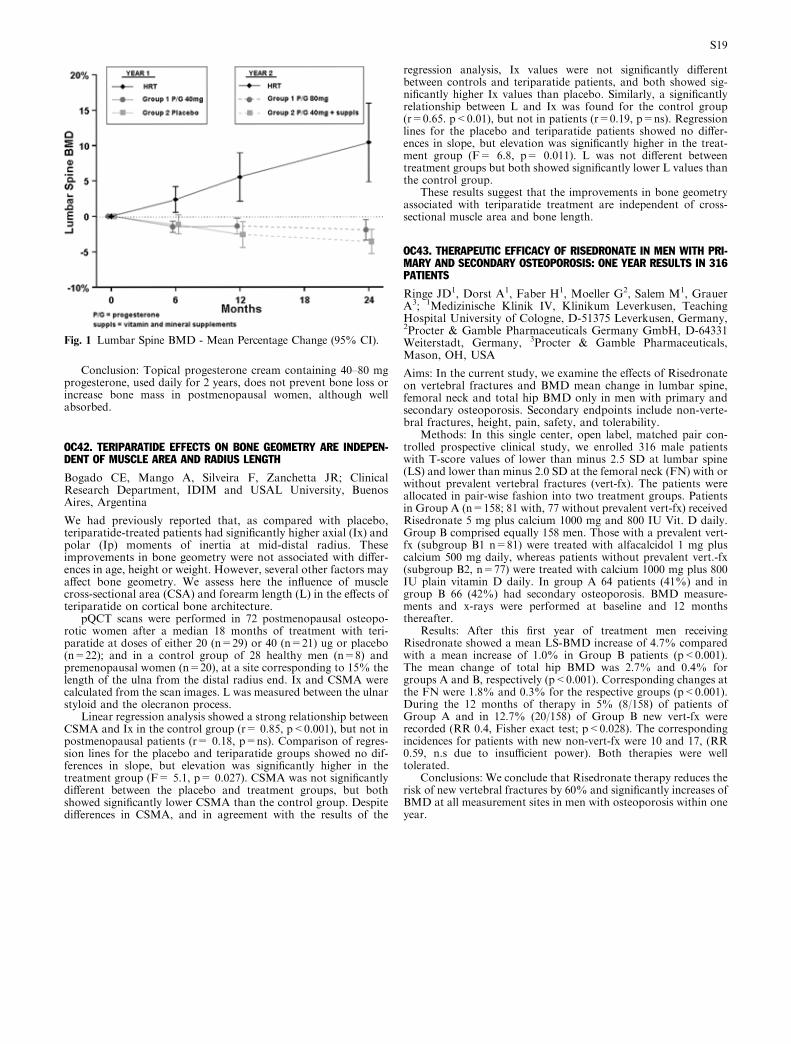
Conclusion: Topical progesterone cream containing 40–80 mgprogesterone, used daily for 2 years, does not prevent bone loss orincrease bone mass in postmenopausal women, although wellabsorbed.
OC42. TERIPARATIDE EFFECTS ON BONE GEOMETRY ARE INDEPEN-DENT OF MUSCLE AREA AND RADIUS LENGTH
Bogado CE, Mango A, Silveira F, Zanchetta JR; ClinicalResearch Department, IDIM and USAL University, BuenosAires, Argentina
We had previously reported that, as compared with placebo,teriparatide-treated patients had significantly higher axial (Ix) andpolar (Ip) moments of inertia at mid-distal radius. Theseimprovements in bone geometry were not associated with differ-ences in age, height or weight. However, several other factors mayaffect bone geometry. We assess here the influence of musclecross-sectional area (CSA) and forearm length (L) in the effects ofteriparatide on cortical bone architecture.
pQCT scans were performed in 72 postmenopausal osteopo-rotic women after a median 18 months of treatment with teri-paratide at doses of either 20 (n=29) or 40 (n=21) ug or placebo(n=22); and in a control group of 28 healthy men (n=8) andpremenopausal women (n=20), at a site corresponding to 15% thelength of the ulna from the distal radius end. Ix and CSMA werecalculated from the scan images. L was measured between the ulnarstyloid and the olecranon process.
Linear regression analysis showed a strong relationship betweenCSMA and Ix in the control group (r= 0.85, p<0.001), but not inpostmenopausal patients (r= 0.18, p=ns). Comparison of regres-sion lines for the placebo and teriparatide groups showed no dif-ferences in slope, but elevation was significantly higher in thetreatment group (F= 5.1, p= 0.027). CSMA was not significantlydifferent between the placebo and treatment groups, but bothshowed significantly lower CSMA than the control group. Despitedifferences in CSMA, and in agreement with the results of the
regression analysis, Ix values were not significantly differentbetween controls and teriparatide patients, and both showed sig-nificantly higher Ix values than placebo. Similarly, a significantlyrelationship between L and Ix was found for the control group(r=0.65. p<0.01), but not in patients (r=0.19, p=ns). Regressionlines for the placebo and teriparatide patients showed no differ-ences in slope, but elevation was significantly higher in the treat-ment group (F= 6.8, p= 0.011). L was not different betweentreatment groups but both showed significantly lower L values thanthe control group.
These results suggest that the improvements in bone geometryassociated with teriparatide treatment are independent of cross-sectional muscle area and bone length.
OC43. THERAPEUTIC EFFICACY OF RISEDRONATE IN MEN WITH PRI-MARY AND SECONDARY OSTEOPOROSIS: ONE YEAR RESULTS IN 316PATIENTS
Ringe JD1, Dorst A1, Faber H1, Moeller G2, Salem M1, GrauerA3; 1Medizinische Klinik IV, Klinikum Leverkusen, TeachingHospital University of Cologne, D-51375 Leverkusen, Germany,2Procter & Gamble Pharmaceuticals Germany GmbH, D-64331Weiterstadt, Germany, 3Procter & Gamble Pharmaceuticals,Mason, OH, USA
Aims: In the current study, we examine the effects of Risedronateon vertebral fractures and BMD mean change in lumbar spine,femoral neck and total hip BMD only in men with primary andsecondary osteoporosis. Secondary endpoints include non-verte-bral fractures, height, pain, safety, and tolerability.
Methods: In this single center, open label, matched pair con-trolled prospective clinical study, we enrolled 316 male patientswith T-score values of lower than minus 2.5 SD at lumbar spine(LS) and lower than minus 2.0 SD at the femoral neck (FN) with orwithout prevalent vertebral fractures (vert-fx). The patients wereallocated in pair-wise fashion into two treatment groups. Patientsin Group A (n=158; 81 with, 77 without prevalent vert-fx) receivedRisedronate 5 mg plus calcium 1000 mg and 800 IU Vit. D daily.Group B comprised equally 158 men. Those with a prevalent vert-fx (subgroup B1 n=81) were treated with alfacalcidol 1 mg pluscalcium 500 mg daily, whereas patients without prevalent vert.-fx(subgroup B2, n=77) were treated with calcium 1000 mg plus 800IU plain vitamin D daily. In group A 64 patients (41%) and ingroup B 66 (42%) had secondary osteoporosis. BMD measure-ments and x-rays were performed at baseline and 12 monthsthereafter.
Results: After this first year of treatment men receivingRisedronate showed a mean LS-BMD increase of 4.7% comparedwith a mean increase of 1.0% in Group B patients (p<0.001).The mean change of total hip BMD was 2.7% and 0.4% forgroups A and B, respectively (p<0.001). Corresponding changes atthe FN were 1.8% and 0.3% for the respective groups (p<0.001).During the 12 months of therapy in 5% (8/158) of patients ofGroup A and in 12.7% (20/158) of Group B new vert-fx wererecorded (RR 0.4, Fisher exact test; p<0.028). The correspondingincidences for patients with new non-vert-fx were 10 and 17, (RR0.59, n.s due to insufficient power). Both therapies were welltolerated.
Conclusions: We conclude that Risedronate therapy reduces therisk of new vertebral fractures by 60% and significantly increases ofBMD at all measurement sites in men with osteoporosis within oneyear.
Fig. 1 Lumbar Spine BMD - Mean Percentage Change (95% CI).
S19
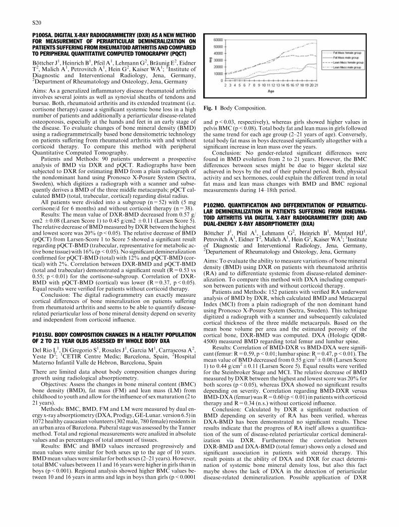
P100SA. DIGITAL X-RAY RADIOGRAMMETRY (DXR) AS A NEW METHODFOR MEASUREMENT OF PERIARTICULAR DEMINERALIZATION ONPATIENTS SUFFERING FROM RHEUMATOID ARTHRITIS AND COMPAREDTO PERIPHERAL QUANTITATIVE COMPUTED TOMOGRAPHY (PQCT)
Bottcher J1,HeinrichB1, Pfeil A1, LehmannG2, BraunigE2, EidnerT2, Malich A1, Petrovitch A1, Hein G2, Kaiser WA1; 1Institute ofDiagnostic and Interventional Radiology, Jena, Germany,2Department of Rheumatology and Osteology, Jena, Germany
Aims: As a generalized inflammatory disease rheumatoid arthritisinvolves several joints as well as synovial sheaths of tendons andbursae. Both, rheumatoid arthritis and its extended treatment (i.e.cortisone therapy) cause a significant systemic bone loss in a highnumber of patients and additionally a periarticular disease-relatedosteoporosis, especially at the hands and feet in an early stage ofthe disease. To evaluate changes of bone mineral density (BMD)using a radiogrammetrically based bone densitometric technologyon patients suffering from rheumatoid arthritis with and withoutcorticoid therapy. To compare this method with peripheralQuantitative Computed Tomography.
Patients and Methods: 90 patients underwent a prospectiveanalysis of BMD via DXR and pQCT. Radiographs have beensubjected to DXR for estimating BMD from a plain radiograph ofthe nondominant hand using Pronosco X-Posure System (Sectra,Sweden), which digitizes a radiograph with a scanner and subse-quently derives a BMD of the three middle metacarpals; pQCT cal-culated BMD (total, trabecular, cortical) regarding distal radius.
All patients were divided into a subgroup (n=52) with (5 mgcortisone/d for 6 months) and without corticoid therapy (n=38).
Results: The mean value of DXR-BMD decreased from 0.57 g/cm2 ±0.08 (Larsen Score 1) to 0.45 g/cm2 ±0.11 (Larsen Score 5).The relative decrease ofBMDmeasuredbyDXRbetween the highestand lowest score was 20% (p<0.05). The relative decrease of BMD(pQCT) from Larsen-Score 1 to Score 5 showed a significant resultregarding pQCT-BMD (trabecular, representative for metabolic ac-tive bone tissue)with 16%(p<0.05).No significant demineralizationconfirmed for pQCT-BMD (total) with 12% and pQCT-BMD (cor-tical) with 2%. Correlation between DXR-BMD and pQCT-BMD(total and trabecular) demonstrated a significant result (R=0.53 vs0.55; p<0.01) for the cortisone-subgroup. Correlation of DXR-BMD with pQCT-BMD (cortical) was lower (R=0.37, p<0.05).Equal results were verified for patients without corticoid therapy.
Conclusion: The digital radiogrammetry can exactly measurecortical differences of bone mineralization on patients sufferingfrom rheumatoid arthritis and seems to be able to quantify disease-related periarticular loss of bone mineral density depend on severityand independent from corticoid influence.
P101SU. BODY COMPOSITION CHANGES IN A HEALTHY POPULATIONOF 2 TO 21 YEAR OLDS ASSESSED BY WHOLE BODY DXA
Del Rio L1, Di Gregorio S1, Rosales J1, GarciaM1, Carrascosa A2,Yeste D2; 1CETIR Centre Medic; Barcelona, Spain, 2HospitalMaterno Infantil Valle de Hebron, Barcelona, Spain
There are limited data about body composition changes duringgrowth using radiological absorptiometry.
Objectives: Assess the changes in bone mineral content (BMC)bone density (BMD), fat mass (FM) and lean mass (LM) fromchildhood to youth and allow for the influence of sexmaturation (2 to21 years).
Methods: BMC, BMD, FM and LM were measured by dual en-ergyx-rayabsorptiometry (DXA,Prodigy,GE-Lunar. version6.5) in1072 healthy caucasian volunteers (302male, 780 female) residents inanurbanarea ofBarcelona. Puberal stagewas assessed by theTannermethod. Total and regional measurements were analized in absolutevalues and as percentages of total amount of tissues.
Results: BMC and BMD values increased progressively andmean values were similar for both sexes up to the age of 10 years.BMDmean valueswere similar for both sexes (2–21 years).However,total BMCvalues between 11 and 16 years were higher in girls than inboys (p<0.001). Regional analysis showed higher BMC values be-tween 10 and 16 years in arms and legs in boys than girls (p<0.0001
and p<0.03, respectively), whereas girls showed higher values inpelvis BMC (p<0.08). Total body fat and leanmass in girls followedthe same trend for each age group (2–21 years of age). Conversely,total body fat mass in boys decreased significantly altogether with asignificant increase in lean mass over the years.
Conclusion: No gender-related significant differences werefound in BMD evolution from 2 to 21 years. However, the BMCdifferences between sexes might be due to bigger skeletal sizeachieved in boys by the end of their puberal period. Both, physicalactivity and sex hormones, could explain the different trend in totalfat mass and lean mass changes with BMD and BMC regionalmeasurements during 14–18th period.
P102MO. QUANTIFICATION AND DIFFERENTIATION OF PERIARTICU-LAR DEMINERALIZATION IN PATIENTS SUFFERING FROM RHEUMA-TOID ARTHRITIS VIA DIGITAL X-RAY RADIOGRAMMETRY (DXR) ANDDUAL-ENERGY X-RAY ABSORPTIOMETRY (DXA)
Bottcher J1, Pfeil A1, Lehmann G2, Heinrich B1, Mentzel HJ1,Petrovitch A1, Eidner T2, Malich A1, Hein G2, Kaiser WA1; 1Instituteof Diagnostic and Interventional Radiology, Jena, Germany,2Departement of Rheumatology and Osteology, Jena, Germany
Aims: To evaluate the ability tomeasure variations of bonemineraldensity (BMD) using DXR on patients with rheumatoid arthritis(RA) and to differentiate systemic from disease-related deminer-alization. To compare this method with DXA including compari-son between patients with and without corticoid therapy.
Patients and Methods: 152 patients with verified RA underwentanalysis of BMD by DXR, which calculated BMD and MetacarpalIndex (MCI) from a plain radiograph of the non dominant handusing Pronosco X-Posure System (Sectra, Sweden). This techniquedigitized a radiograph with a scanner and subsequently calculatedcortical thickness of the three middle metacarpals. Based on themean bone volume per area and the estimated porosity of thecortical bone, DXR-BMD was computed. DXA (Hologic QDR-4500) measured BMD regarding total femur and lumbar spine.
Results: Correlation of BMD-DXR vs BMD-DXA were signifi-cant (femur:R=0.59, p<0.01; lumbar spine:R=0.47, p<0.01). Themean value of BMDdecreased from 0.55 g/cm2±0.08 (Larsen Score1) to 0.44 g/cm2±0.11 (Larsen Score 5). Equal results were verifiedfor the Steinbroker Stage and MCI. The relative decrease of BMDmeasured byDXRbetween the highest and lowest score was 20% forboth scores (p<0.05), whereas DXA showed no significant resultsdepending on severity. Correlation regarding BMD-DXR versusBMD-DXA(femur)wasR=0.60 (p<0.01) in patientswith corticoidtherapy and R=0.34 (n.s.) without corticoid influence.
Conclusion: Calculated by DXR a significant reduction ofBMD depending on severity of RA has been verified, whereasDXA-BMD has been demonstrated no significant results. Theseresults indicate that the progress of RA itself allows a quantifica-tion of the sum of disease-related periarticular cortical demineral-ization via DXR. Furthermore the correlation betweenDXR-BMD and DXA-BMD (total femur) shows only a closed andsignificant association in patients with steroid therapy. Thisresult points at the ability of DXA and DXR for exact determi-nation of systemic bone mineral density loss, but also this factmaybe shows the lack of DXA in the detection of periarticulardisease-related demineralization. Possible application of DXR
Fig. 1 Body Composition.
S20
should be the additional BMD-calculation on routinely performedfollow-up radiographs for quantification and monitoring ofdemineralization.
P103SA. DESCRIPTIVE STUDY OF BONE MINERAL DENSITY MEASUREDBY PERIPHERAL DENSITOMETRY: ARGENTINE EXPERIENCE
Massari F1, Goncalves S1, Hentschel D2, Zanchetta JR1; 1IDIMInstituto de InvestigacionesMetabolicas, BuenosAires, Argentina,2Merck
Aim: To describe the prevalence of osteopenia and osteoporosis byperipheral densitometry in women aged 50 years or older inArgentina.
Methods: Secondary analysis of data fromadatabasewith 41,118ambulatory subjects (38,525 women) from 180 primary care centersfrom 7 regions of the Argentine Republic participating in a freecampaign for osteoporosis detection. The examination was per-formed by Norland p-DXA equipment, in the no-dominant distalforearm.
Results: Demographics: Age: 63.9 (+/) 9.2) years; Weight: 69(+/) 12.3) kg; Height: 158 (+/) 0.07) cm; BMI (BodyMass Index):27.6 (+/) 4.8). Age distribution : 50–59 years: n=14374 (37.3% ofthe sample); 60 to 69 years: n=13194 (34.2%); 70 to 79 years:n=9057 (23.5%); 80 years or older: n=1899 (5%).Diagnosis (WorldHealth Organization (WHO) criteria): Osteoporosis (T-score <)2.5): n=6731, 17.47%; Osteopenia (T-score of )1 to )2.49):n=17034, 44.22%; Normal (T-score > to )1): n=14760, 38.3%.The prevalence of osteoporosis increased in accordance with the ageprogression and it was similar between different regions of thecountry. Using National Osteoporosis Foundation (NOF) criteria,the high-risk group (30.7%) was bigger than the osteoporotic groupusing WHO criteria (17.47%) (Table 1).
Conclusions: We observed a significant difference in the per-centage of potential treated patients according to the criteriaused. Our results presents some differences with the results of theNational Osteoporosis Risk Assessment (NORA) study, performedwith the same method, peripheral densitometry, and identicalage distribution in the sample, but with different racial compo-sition of the sample (we dont include black subjects). Given the lowercosts and the accessibility of p-DXA, we are in agreement with otherinternational authors that this method seems to be a useful screeningtool to detect subjects with low bone mineral density.
P104SU. CHANGES IN BONE MASS IN ADOLESCENTS WITH SEVEREOBESE DURING WEIGHT REDUCTION
Dao HH1, Frelut ML2, Oberlin F1, Peres G1, Navarro J2,Bourgeois P1; 1CHU Pitie-Salpetriere, Paris, France, 2CHURobert-Debre, Paris, France
Background: Osteoporosis and obesity become a major publichealth concern in many countries. Primary preventive method ofosteoporosis is to reach an optimal peak bone mass which is takenplace during adolescence. In obese adults, some studies indicatedthat weight loss is associated with bone mass loss but the interpre-tationof theseresultsareoftencontroversial.Littleknownabout thechanges of bonemass duringweight reduction in obese adolescents.
Aim: The aim of this investigation is to evaluate the effects of amarked weight loss resulting from dietary and physical activitesinterventions on total and regional bone mass in obese adolescents.
Methods: Thirty three girls and 22 boys, aged 13.4±3.6 y withBMI 34.6±3.6 kg/m2 were included in a 9±3 months multidisci-plinary weight reduction programme including a slight caloricrestriction and submaximal aerobic physical training. Total andregional (lumbar spine and femoral neck) were measured by DEXAHologic QDR 1000 at baseline and after treatment.
Results: Mean weight loss was 23.5±8.9 kg (p<0.0001), heightgrowth was 2.7±1.6 cm (p<0.001). Fat mass decreased (p<0.0001)whereas lean mass maintained. In whole body: BMC and bone areadecreased significantly by 253±131 g and 212±14 cm2 (bothp<0.0001) whereas BDM did not vary (p=0.3). The changes oftotal BMC was highly correlated to the diminution of fat mass(r=0.88, p<0.0001).At lumbar spine: BMC, bone area and BMD
increased significantly in both sexes. At femoral neck: BMC, bonearea and BMD did not vary.
Conclusions: Total and regional areal BMD maintained duringmajor weight loss resulting from a dietary and physical activitiesprogramme in obese adolescents. Total BMC decreased whereasregional BMC (both trabecular and cortical bone) increased ormaintained may suggest the limitation of interpretation of DEXAin assessing bone mass during a marked weight loss.
P105MO. CAN FEMUR BMD AND STIFFNESS INDEX PREDICT FUTUREOSTEOPOROTIC FRACTURE IN BRAZILIAN ELDERLY WOMEN? A 5YEAR FOLLOW-UP.
Pinheiro MM, Castro CHM, Oliveira KRB, Heymann RE,Genaro PS, Szejnfeld VL; Rheumatology Division at Unifesp/EPM, Brazil
Previous studies have suggested that low bone mineral density(BMD) is associated with increased risk of fracture and mortality.However, the relationship between quantitative ultrasound(QUS) and fracture andmortality is still controversial. Our aimwasevaluate the ability of the BMD and QUS to predict future osteo-porotic fracture in 275 Brazilian postmenopausal women.
Patients and methods: Spine and femur BMD (Lunar) and heelQUS (Lunar) measurements were performed in all patients. Riskfactors to osteoporosis and fractures were evaluated by a specificquestionnaire. Patients were followed-up for 5 years and newfracture or death were investigated as main outcomes. Lateralthoracic and lumbar radiographs were taken at baseline and 5 yearslater to survey for the presence of vertebral fractures. All reporteddeaths were confirmed by review of the hospital records and clas-sified according to the ICD- 9 code.
Results: 208 (75.6%) women completed the protocol study, 42(15.3%) died and 25 (9.1%) lost follow-up. Mean age, height,weight and bone mass index (BMI) for the group were 75.2±6.5years, 60.3±10.2 kg, 1.50±0.8 m and 26.9±4.2 kg/ m2, respec-tively. Forty-two patients (20.2%) had new osteoporotic fracture atany skeletal site. After adjustments for age, weight, previous frac-ture, familiar history of hip fracture, smoking, physical activity,drugs and others diseases, each 1 SD reduction in stiffness indexwas associated with hazard ratio of 2.2 (95% CI 1.3; 3.8) of futurefracture. Femural neck BMD (HR 2.0) [95% CI 1.3; 3.2] and tro-chanter BMD (HR 1.62) [95% CI 1.1; 2.4] were also significantlyassociated with the risk of future fracture. The most relevant haz-ard ratio to death were trochanter BMD (HR 1.59) [95% CI 1.1;2.4], stiffness index (HR 1.57) [95% CI 1.1; 2.4] and femural neckBMD (HR 1.44) [95% CI 1.1; 2.2]. Mortality from cancer, car-diovascular or infectious diseases was not significantly associatedwith BMD or QUS measurements.
In conclusion, low QUS and femur BMD measurements wereable to predict future osteoporotic fracture and mortality, inde-pendently of the age, health status and other associated diseases inthis cohort of Brazilian elderly women.
P106SA. PREVALENCE AND IDENTIFICATION OF VERTEBRAL FRAC-TURES: COMPARISON OF VISUAL INSPECTION, DIGITAL COMPUTER-IZED MORPHOMETRY AND VISUAL SEMIQUANTITATIVE ASSESSMENT
Bucchieri S1, Guglielmi G2, Sferrazza C1, Napoli N1, Masi L3,Falchetti A3, Maggio C1, Di Fede G1, Rini GB1, Brandi ML3;1Department of Internal Medicine, University of Palermo,Palermo, Italy, 2Scientific Institute Hospital ‘‘CSS’’, San GiovanniRotondo, Foggia, Foggia, Italy, 3Department of InternalMedicine, University of Florence, Florence, Italy
This study aimed to determine the percentage of vertebral frac-tures eluding x-ray reports and to calculate the precision of thedigital morphometric system used for vertebral body heights x-rayevaluation.
We analyzed 233 postmenopausal women (aged 63.9±0.5) whohave undergone clinical evaluation in a bone metabolic diseasesoutpatient service. They had lateral and posterior-anterior thoracicand lumbar spine x-ray assessment; radiology reports described thepresence of at least one vertebral fracture in 12% of the subjects.
S21
The T4-L4 film analysis was performed by digital computerizedmorphometry [DCM] (Spine-X Analyser, CAM Diagnostics, Mi-lan, Italy), calculating vertebral height ratios on the basis of theidentification method of 6 standard points of 3 vertebral heights; a20% reduction of any vertebral heights ratio was chosen asthreshold value to assess vertebral deformity. Thus, a prevalence offractures of 46.25% was observed. Furthermore, Genant’s semi-quantitative grading scheme for the assessment of vertebral frac-tures [VQA] was performed by an expert radiologist. A fracturesprevalence of 49.75% was observed.
By comparing both methods, it was deduced that most fracturesusually occur in the T6-T9 interval and that end-plate fractures arethe most common (68.9% by DCM, 58.4% by VQA).
Thus, a good agreement (0.839) between the VQA and theDCM method by using k-score was found.
Sensitivity (0.655) and specificity (0.957) of vertebral fracturesidentification by VQA, were calculated in connection with DCMmethod, the latter considered as the gold standard.
We suggest that in clinical practice, vertebral fractures are oftenundiagnosed and not adequately reported by inspection of radio-graphs. Moreover, both the methods, DCM and VQA, representeffective tools to be applied to clinical research enabling the dem-onstration of higher prevalence of vertebral fractures.
P107SU. REFERENCE DATA OF BONE MINERAL MASS AND DENSITIESAS WELL AS MUSCLE-BONE RELATIONSHIP INDICATORS ASSESSEDUSING DXA METHOD IN HEALTHY POLISH CHILDREN
Matusik H, Pludowski P, Lorenc RS; Department of Biochem-istry and Experimental Medicine, The Children’s MemorialHealth Institute, Warsaw, Poland
Aim: To assess total body, spine and hip bone mineral mass anddensity reference data for healthy Polish children using DXAmethod and to compare established reference data with data pro-vided by DXA manufacturer and reference data-sets publishedelsewhere.
Methods: 500 healthy children (250 girls) aged 5–18 years weremeasured using DPX-L machine. Age and sex-matched means andSDs were calculated for BMD and BMC of total body, lumbar spineand femoral neck. Total lean body mass (LBM) and body height(BH)/LBM, TBBMC/LBM, spine (S)BMC/LBM ratios werecalculated. The relationships between BMD, BMC, BH/LBM,TBBMC/LBM, SBMC/LBM and chronological age (CA), heightage (HA), bone age (BA), body height (BH), weight (W) wereanalyzed using correlation analysis. 24 DXA children’s referencedata-sets from 12 countries and reference provided by DXA manu-facturer were compared with our results (mainly BMD) usingANOVA.
Results: Gender related differences were noted for the tempo ofbone mineralization evaluated on the basis of BMC and BMDvalues of analyzed ROIs. Stronger relationship was noticedbetween BH(r=0.92;r=0.93), W(r=0.95;r=0.95), CA(r=0.90;r=0.88), HA(r=0.88;r=0.90), BA(r=0.89;r=0.89) and TBBMCthan TBBMD (BH:r=0.84;r=0.87;W:r=0.90;r=0.90;CA:r=0.87;r=0.87;HA:r=0.80;r=0.80;BA:r=0.88;r=0.88), for girls andboys respectively. The muscle-bone relationship analysis based onBMC, LBM values and TBBMC/LBM, SBMC/LBM ratios re-vealed that pubertal and post-pubertal girls stored more bone perLBM unit than boys (p<0.0001). Lack of major differences wasfound between BMD reference data-sets provided by DXA man-ufacturer or published elsewhere and our results only for total bodyand spine, but not for femoral neck BMD, probably as an effect ofmarkedly lower number of studies which provide femoral neckBMD data and technical problems with bone edge detection for hipmeasurements in children.
Conclusion: Data from healthy children are essential to evalu-ate bone mineralization during childhood as well as for properdiagnosis when bone disorder is suspected. Complete and unifiedchildren’s reference data is strongly needed. Lack of reference forhip bone mineralization and still, only one normative study ofmuscle-bone treated as a unit in children are available. Therefore,we provide age and gender related reference BMD and BMC data
expanded by hip bone mineralization parameters and muscle-bonerelationship indicators.
P108MO. BONE LOSS AFTER CHEMOTHERAPY IN WOMEN WITHEARLY BREAST CANCER
Pinheiro MM1,2, Siniscalchi AR2, Carvalho SMT3, Rigon Jr HJ2,Mourao Netto M3, Szejnfeld VL1; 1Rheumatology Division ofUnifesp/ EPM, Brazil, 2Medicine Interne Division of Hospital doCancer AC Camargo, Brazil, 3Breast’s Surgery Division ofHospital do Cancer AC Camargo, Brazil
Our aim was evaluate the impact of the chemotherapy (CT) onbone mineral density (BMD) and quantitative ultrasound (QUS)measurements in patients with early breast cancer (EBC).
Patients and methods: 154 women were enrolled (54 with EBCand 100 healthy controls matched to age, body mass index [BMI]and status menopausal). Patients with history of another cancer,previous CT or metastatic disease were excluded. Spine and femurBMD (Lunar, DPX-L) and heel QUS (Achilles+, Lunar) wereperformed in all patients.
Results: 46 (85.2%) patients with EBC and 83 (83%) healthycontrols completed follow-up. The mean age, weight, height andBMI of the EBC patients were 52.1±10 years, 68.2±14.3 kg,1.57±0.7 m and 27.6±5.4 kg/m2, respectively. Twenty (43.5%)patients with EBC were premenopausal and six (13%) had previousatraumatic fracture. The protocols of the CT used were [fluoro-uracil, doxorubicin, cyclophosphamide] (71.7%) or [cyclophos-phamide, methotrexate, fluorouracil] (28.3%) with mean number of5.8±1.8 or 7.7±2.1 cycles, respectively. At baseline, the patientswith EBC had BMD and QUS measures 5% higher than healthycontrols. After CT (8.5±2.7 months), the women with EBC lowerBMD and QUS measurements (table below).
In conclusion, women with EBC lost 2% and 3.2% on spineBMD and QUS measurements, respectively, after chemotherapy.
P109SA. TWO YEAR FOLLOW-UP STUDY OF BONE MODELING INCYSTIC FIBROTIC CHILDRENS AND ADOLESCENTS
Papp I1, Ujhelyi R2, Treszl A1, Vasarhelyi B3, Holics K2, Toth M4,Arato A5, Szathmari M1; 1First Department of Medicine,Semmelweis University, Budapest, Hungary, 2Heim Pal Children’sHospital, Budapest, Hungary, 3Research Laboratory of Pediatricsand Nephrology, Hungarian Academy of Sciences, 4SecondDepartment of Medicine, Semmelweis University, Budapest,Hungary, 5First Department of Pediatrics, Semmelweis University,Budapest, Hungary
Cystic fibrosis (CF) is caused by mutations of CF-transmem-brane-conductance-regulator (CFTR) gene, and is the mostcommon lethal autosomal recessive (AR) genetic disease (1).Several data suggest that an inherited genetic susceptibility con-tributes to the development of bone deficit.
Aim: To investigate bone mineral density (BMD) and boneturnover (BTM) in cystic fibrosis (CF) patients in a two-year per-iod, and to test the role of inheritance (Inh +/)).Methods: Thirty-eight clinically stable CF patients (11 children,16 adolescents, 11 young adults) were enrolled. None of thepatients was treated with corticosteroids prior to or during thestudy. Weight (a) and the height (b) Z-scores and bone mineraldensity (BMD) Z-score (c) values at the femoral neck (FN) and
Table. 1 BMD and QUS measures before and after the chemotherapy in women with EBC compared to healthycontrols.
Baseline After CT D%
EBC Controls EBC Controls EBC Controls
Spine BMD (g/ cm2) 1.170 ± 0.2 1.092 ± 0.2* 1.141 ± 0.2 1.104 ± 0.2* )2.5 1.1*Femur BMD (g/cm2) 0.940 ± 0.1 0.871 ± 0.1* 0.919 ± 0.1 0.884 ± 0.1* )2.2 1.5*BUA (dB/ MHz) 114 ± 11 108 ± 10* 112 ± 9 110 ± 9* )1.7 1.8*SOS (m/s) 1561 ± 30 1532 ± 28* 1554 ± 32 1539 ± 29* )0.5 0.7*Stiffness Index 93 ± 13 88 ± 13* 90 ± 13 89 ± 13* )3.2 1.1*
D% (% difference between measures); *p < 0.05
S22
the lumbar spine (LS) were recorded at the beginning of the studyand two years later. Bone turnover markers (BTM) includingosteocalcin and cross-link (DPR) excretion were also measured.The correlations between BMD, bone turnover parameters, dis-ease severity, pubertal stage and nutritional state were investi-gated. As an extension of the study, BMD values of the mothersof patients (carriers of CFTR gene) were also determined andrelated to patients’ BMD.
Results: Height and weight Z-scores were normal in children,while decreased in adolescents. Puberty was delayed in most ofpatients. Bone age was lower than chronological age in adolescents.Lumbar spine and femoral neck BMD Z-scores were lower thannormal in each age group. Disease severity (i.e. Schwachmanscores) correlated with lumbar BMD (r=0.45, p<0.02). Motherand patient lumbar and femoral BMD correlated significantly(r=0.51, p<0.01, and r=0.54, p<0.01, respectively).
Conclusion: Bone mineral and turnover decrease was present incystic fibrosis (CF) adolescent patients without steroid treatment.The genetic factors additionally to chronic inflammation might beresponsible for the delayed puberty. Correlation of BMD valuesbetween homozygous patients, and heterozygous mothers supportthe presence of genetic factors in the pathogenesis (Inh+).
P110SU. BONE MINERAL DENSITY, HIP STRENGTH PARAMETERS ANDFALL INDEX FOR HIP FRACTURE PREDICTION
Sergio Ragi Eis SRE; CEDOES - Centro de Diagnostico ePesquisa da Osteoporose do ES, Brasil
Introduction: It is accepted that BMD is not the only predictorfor hip fracture. Other parameters such as age, height and weightalso should be taken into account.
Femur structure factors such as Cross Sectional Area (CSA), andCross Sectional Moment of Inertia (CSMI) now can be measured bymodern DXA equipment. As some investigators have proposed, itis possible that combining bone mineral density, structure, age,height, andweight in a Fall Index (FI) thatmight predict hip fracturemore accurately thanBMDorage alone.Aspublished byYoshikawaet al, (JBMR 9:1053–1064,1994) FI can estimate the ability of a hipto withstand the impact of a fall on the greater trochanter.
Methods: Femoral Neck BMD, CSMI, CSA, and FI wereevaluated for assessing hip fracture risk in a group of fracturedsubjects and compared to non-fracture controls. DXA scans wereobtained in 362 women, 72 with prior hip fracture and 289 con-trols, using Lunar Prodigy (GE Medical Systems). BMD of thefemoral neck, CSMI, and CSA were determined using the HipStrength Analysis software (version 8.00.256), and FI was calcu-lated according to the equations developed by Yoshikawa et al.Geometric results were adjusted for BMD, and the fractured andcontrol subjects were compared using a unpaired t-test.
Results: Both Femur Neck BMD and the Fall Index were sig-nificantly lower in the fracture group when compared to controls(p<0.001).
Conclusions: According to the results of this sample, we con-clude that fractured subjects have a reduced capacity to withstandthe impact of a fall without fracturing. Additionally, the Fall Index,which combines BMD, femur geometry, age, weight and height, is asignificant predictor of hip fracture.
P111MO. PATIENT EDUCATION AND TREATMENT FOLLOWING BONEDENSITOMETRY
Arnason JA, Pickney CS; University of Wisconsin, USA
Introduction: Treatment of osteoporosis is often inadequate. Onereason can be insufficient patient education following diagnosticbone densitometry (DEXA). Therefore we studied how patientsare informed and treated following their first DEXA. Individualswho had DEXA at a rural hospital in Wisconsin, USA weresurveyed with a questionnaire regarding their post-test educationand prescribed treatment. Their DEXA results and the specialtyof their clinician were also recorded.
Results: Eighty percent of the 1014 participants were informedof their results. Of the 341 participants who had normal BMD,63% reported correct results, 31% of the 309 who had osteopeniaand 50% of the 364 who had osteoporosis. Accuracy in reportingwas not affected by the patients’ age or the specialty of their cli-nician. Calcium supplements were recommended to 65% of thosenot taking calcium prior to DEXA. Internists were more likely thanfamily practitioners to recommend calcium (p=0.0003). FollowingDEXA, 339 patients were started on medications (33%), 86% ofthose remained on some prescribed therapy, but 140 (41%) did notcontinue the original medication. Reasons for discontinuation in-cluded sideffects (48%) and cost (26%). Patients with low BMD,who correctly reported their results, were more likely to have re-ceived a medication and to continue to take it (p<0.0001).
Conclusions: While most participants are informed of the re-sults of their DEXAs, the retained information may not be accu-rate. Participants who correctly reported the results of theirDEXAs were more likely to have received a medication and toremain on treatment.
P112SA. THE USE OF FINGERNAIL AS A MEANS OF ASSESSING BONEHEALTH: A PILOT STUDY
Pillay I1, Lyons D1, German MJ2, Lawson NS2, Pollock HM3,Saunders J4, Chowdhury S5, Moran P6, Towler MR6; 1ClinicalAge Assessment Unit, Limerick Regional Hospital, Limerick,Ireland, 2Materials & Surface Science Institute, University ofLimerick, Ireland, 3Department of Biological Sciences, Universityof Lancaster, UK, 4Statistical Consulting Unit, Mathematics andStatistics Department, University of Limerick, Limerick, Ireland,5Department of Physics, University of Lancaster, UK, 6Mechan-ical & Aeronautical Engineering Department, University ofLimerick, Ireland
Aim: Patients report increasing hardness of their nails withinmonths of starting treatment for osteoporosis. A pilot study wasundertaken to assess if the properties of nail and bone could belinked in a comparable, measurable way.
Methods: Two groups of ten subjects were identified. The firstgroup were diagnosed, by DEXA (Lunar Prodigy GE, Medicalsystems), as osteoporotic. The second group were non-osteopo-rotic. Fingernail clippings were obtained from all subjects. A ded-icated, laboratory-built nano-indenter was employed to measurehardness and elastic modulus of the nail. Raman Spectroscopy(Labram Integrated system) was configured to analyze between 300and 700 cm-1. This is where disulphide bond breakdown and C-Sbond shift would be detectable.
Results: The mean moduli of fingernails from patients with lowBMDare about 25% lower than those with normal BMD. Themeandifference in mean modulus between the groups was found to be0.996 but this was not significant at the 5% level (p=0.147). This pvalue is quite low and indicates that a significant difference might befound if a further study is undertaken which is sufficiently powered.The spectroscopy data also showed differences between the two setsof nails. The disulphide bond content of the nails sourced fromosteoporotic patients was much lower than those from healthypatients. There was also a significant shift in c-s bond detection.
Conclusion: We hypothesise that whilst bone collagen and nailkeratin are two distinct structural proteins, they share the need forprotein sulphation and disulphide bond formation for their struc-tural integrity. A disorder of either process may lead to disordered
Table 1: Summary of the main results
Age Height Weight AverageBMDNeck
CSMI CSA MedianFI
Fracture Group n = 7271.8 154.6 58.5 0.684* 6985.11 100.6 1.4930*
No Fracture Group n = 28969.9 152.56 59.7 0.804* 7149.86 114.25 1.7140*
*Significantly different than controls (p<0.001)
S23
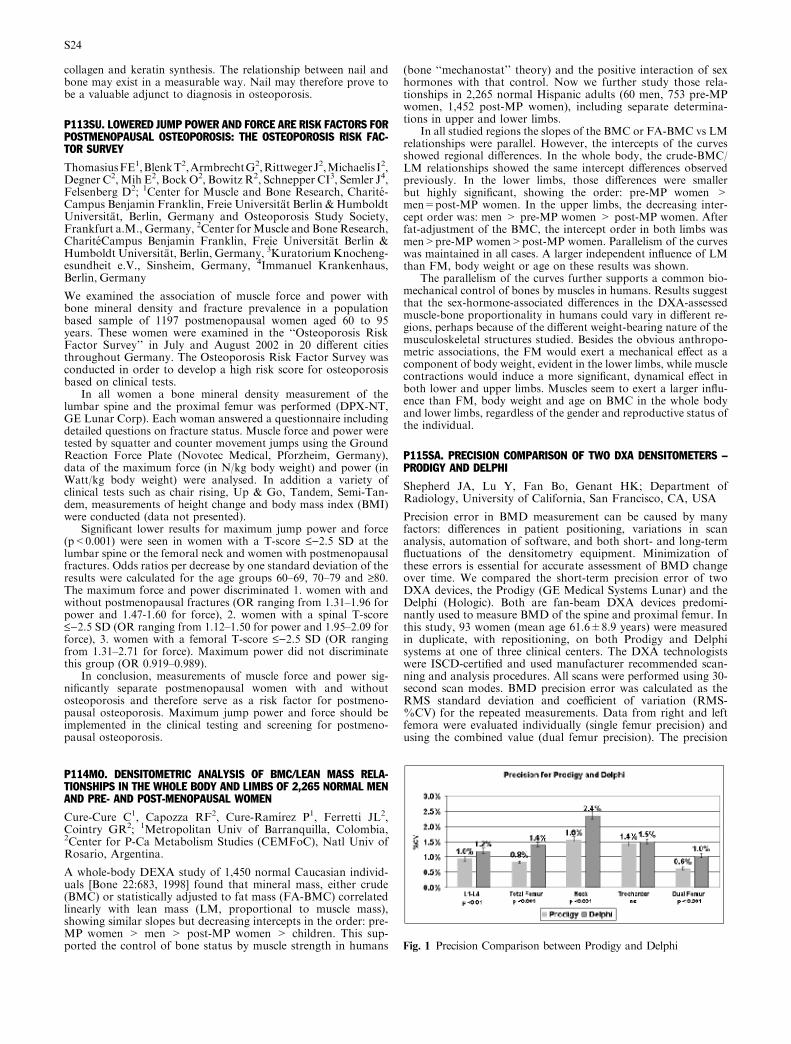
collagen and keratin synthesis. The relationship between nail andbone may exist in a measurable way. Nail may therefore prove tobe a valuable adjunct to diagnosis in osteoporosis.
P113SU. LOWERED JUMP POWER AND FORCE ARE RISK FACTORS FORPOSTMENOPAUSAL OSTEOPOROSIS: THE OSTEOPOROSIS RISK FAC-TOR SURVEY
ThomasiusFE1,BlenkT2,ArmbrechtG2,RittwegerJ2,MichaelisI2,Degner C2,Mih E2, BockO2, Bowitz R2, Schnepper CI3, Semler J4,Felsenberg D2; 1Center for Muscle and Bone Research, Charite-Campus Benjamin Franklin, Freie Universitat Berlin & HumboldtUniversitat, Berlin, Germany and Osteoporosis Study Society,Frankfurt a.M., Germany, 2Center forMuscle and Bone Research,ChariteCampus Benjamin Franklin, Freie Universitat Berlin &Humboldt Universitat, Berlin, Germany, 3KuratoriumKnocheng-esundheit e.V., Sinsheim, Germany, 4Immanuel Krankenhaus,Berlin, Germany
We examined the association of muscle force and power withbone mineral density and fracture prevalence in a populationbased sample of 1197 postmenopausal women aged 60 to 95years. These women were examined in the ‘‘Osteoporosis RiskFactor Survey’’ in July and August 2002 in 20 different citiesthroughout Germany. The Osteoporosis Risk Factor Survey wasconducted in order to develop a high risk score for osteoporosisbased on clinical tests.
In all women a bone mineral density measurement of thelumbar spine and the proximal femur was performed (DPX-NT,GE Lunar Corp). Each woman answered a questionnaire includingdetailed questions on fracture status. Muscle force and power weretested by squatter and counter movement jumps using the GroundReaction Force Plate (Novotec Medical, Pforzheim, Germany),data of the maximum force (in N/kg body weight) and power (inWatt/kg body weight) were analysed. In addition a variety ofclinical tests such as chair rising, Up & Go, Tandem, Semi-Tan-dem, measurements of height change and body mass index (BMI)were conducted (data not presented).
Significant lower results for maximum jump power and force(p<0.001) were seen in women with a T-score £)2.5 SD at thelumbar spine or the femoral neck and women with postmenopausalfractures. Odds ratios per decrease by one standard deviation of theresults were calculated for the age groups 60–69, 70–79 and ‡80.The maximum force and power discriminated 1. women with andwithout postmenopausal fractures (OR ranging from 1.31–1.96 forpower and 1.47-1.60 for force), 2. women with a spinal T-score£)2.5 SD (OR ranging from 1.12–1.50 for power and 1.95–2.09 forforce), 3. women with a femoral T-score £)2.5 SD (OR rangingfrom 1.31–2.71 for force). Maximum power did not discriminatethis group (OR 0.919–0.989).
In conclusion, measurements of muscle force and power sig-nificantly separate postmenopausal women with and withoutosteoporosis and therefore serve as a risk factor for postmeno-pausal osteoporosis. Maximum jump power and force should beimplemented in the clinical testing and screening for postmeno-pausal osteoporosis.
P114MO. DENSITOMETRIC ANALYSIS OF BMC/LEAN MASS RELA-TIONSHIPS IN THE WHOLE BODY AND LIMBS OF 2,265 NORMAL MENAND PRE- AND POST-MENOPAUSAL WOMEN
Cure-Cure C1, Capozza RF2, Cure-Ramırez P1, Ferretti JL2,Cointry GR2; 1Metropolitan Univ of Barranquilla, Colombia,2Center for P-Ca Metabolism Studies (CEMFoC), Natl Univ ofRosario, Argentina.
A whole-body DEXA study of 1,450 normal Caucasian individ-uals [Bone 22:683, 1998] found that mineral mass, either crude(BMC) or statistically adjusted to fat mass (FA-BMC) correlatedlinearly with lean mass (LM, proportional to muscle mass),showing similar slopes but decreasing intercepts in the order: pre-MP women > men > post-MP women > children. This sup-ported the control of bone status by muscle strength in humans
(bone ‘‘mechanostat’’ theory) and the positive interaction of sexhormones with that control. Now we further study those rela-tionships in 2,265 normal Hispanic adults (60 men, 753 pre-MPwomen, 1,452 post-MP women), including separate determina-tions in upper and lower limbs.
In all studied regions the slopes of the BMC or FA-BMC vs LMrelationships were parallel. However, the intercepts of the curvesshowed regional differences. In the whole body, the crude-BMC/LM relationships showed the same intercept differences observedpreviously. In the lower limbs, those differences were smallerbut highly significant, showing the order: pre-MP women >men=post-MP women. In the upper limbs, the decreasing inter-cept order was: men > pre-MP women > post-MP women. Afterfat-adjustment of the BMC, the intercept order in both limbs wasmen>pre-MP women>post-MP women. Parallelism of the curveswas maintained in all cases. A larger independent influence of LMthan FM, body weight or age on these results was shown.
The parallelism of the curves further supports a common bio-mechanical control of bones by muscles in humans. Results suggestthat the sex-hormone-associated differences in the DXA-assessedmuscle-bone proportionality in humans could vary in different re-gions, perhaps because of the different weight-bearing nature of themusculoskeletal structures studied. Besides the obvious anthropo-metric associations, the FM would exert a mechanical effect as acomponent of body weight, evident in the lower limbs, while musclecontractions would induce a more significant, dynamical effect inboth lower and upper limbs. Muscles seem to exert a larger influ-ence than FM, body weight and age on BMC in the whole bodyand lower limbs, regardless of the gender and reproductive status ofthe individual.
P115SA. PRECISION COMPARISON OF TWO DXA DENSITOMETERS –PRODIGY AND DELPHI
Shepherd JA, Lu Y, Fan Bo, Genant HK; Department ofRadiology, University of California, San Francisco, CA, USA
Precision error in BMD measurement can be caused by manyfactors: differences in patient positioning, variations in scananalysis, automation of software, and both short- and long-termfluctuations of the densitometry equipment. Minimization ofthese errors is essential for accurate assessment of BMD changeover time. We compared the short-term precision error of twoDXA devices, the Prodigy (GE Medical Systems Lunar) and theDelphi (Hologic). Both are fan-beam DXA devices predomi-nantly used to measure BMD of the spine and proximal femur. Inthis study, 93 women (mean age 61.6±8.9 years) were measuredin duplicate, with repositioning, on both Prodigy and Delphisystems at one of three clinical centers. The DXA technologistswere ISCD-certified and used manufacturer recommended scan-ning and analysis procedures. All scans were performed using 30-second scan modes. BMD precision error was calculated as theRMS standard deviation and coefficient of variation (RMS-%CV) for the repeated measurements. Data from right and leftfemora were evaluated individually (single femur precision) andusing the combined value (dual femur precision). The precision
Fig. 1 Precision Comparison between Prodigy and Delphi
S24
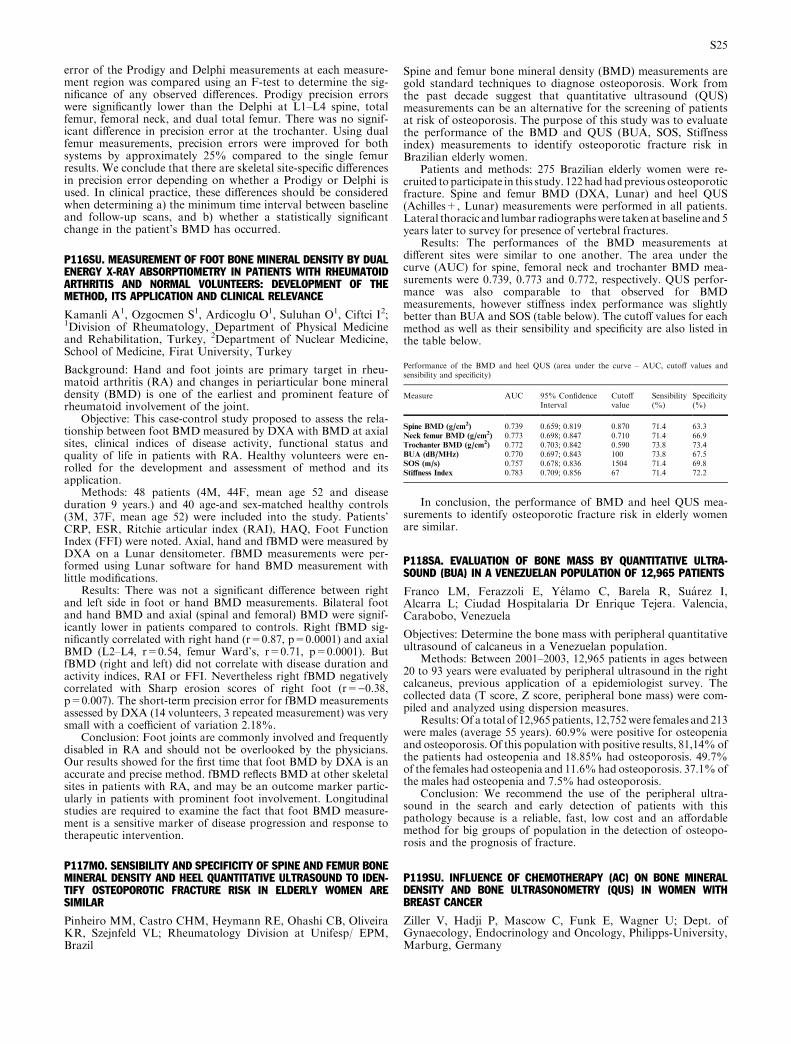
error of the Prodigy and Delphi measurements at each measure-ment region was compared using an F-test to determine the sig-nificance of any observed differences. Prodigy precision errorswere significantly lower than the Delphi at L1–L4 spine, totalfemur, femoral neck, and dual total femur. There was no signif-icant difference in precision error at the trochanter. Using dualfemur measurements, precision errors were improved for bothsystems by approximately 25% compared to the single femurresults. We conclude that there are skeletal site-specific differencesin precision error depending on whether a Prodigy or Delphi isused. In clinical practice, these differences should be consideredwhen determining a) the minimum time interval between baselineand follow-up scans, and b) whether a statistically significantchange in the patient’s BMD has occurred.
P116SU. MEASUREMENT OF FOOT BONE MINERAL DENSITY BY DUALENERGY X-RAY ABSORPTIOMETRY IN PATIENTS WITH RHEUMATOIDARTHRITIS AND NORMAL VOLUNTEERS: DEVELOPMENT OF THEMETHOD, ITS APPLICATION AND CLINICAL RELEVANCE
Kamanli A1, Ozgocmen S1, Ardicoglu O1, Suluhan O1, Ciftci I2;1Division of Rheumatology, Department of Physical Medicineand Rehabilitation, Turkey, 2Department of Nuclear Medicine,School of Medicine, Firat University, Turkey
Background: Hand and foot joints are primary target in rheu-matoid arthritis (RA) and changes in periarticular bone mineraldensity (BMD) is one of the earliest and prominent feature ofrheumatoid involvement of the joint.
Objective: This case-control study proposed to assess the rela-tionship between foot BMD measured by DXA with BMD at axialsites, clinical indices of disease activity, functional status andquality of life in patients with RA. Healthy volunteers were en-rolled for the development and assessment of method and itsapplication.
Methods: 48 patients (4M, 44F, mean age 52 and diseaseduration 9 years.) and 40 age-and sex-matched healthy controls(3M, 37F, mean age 52) were included into the study. Patients’CRP, ESR, Ritchie articular index (RAI), HAQ, Foot FunctionIndex (FFI) were noted. Axial, hand and fBMD were measured byDXA on a Lunar densitometer. fBMD measurements were per-formed using Lunar software for hand BMD measurement withlittle modifications.
Results: There was not a significant difference between rightand left side in foot or hand BMD measurements. Bilateral footand hand BMD and axial (spinal and femoral) BMD were signif-icantly lower in patients compared to controls. Right fBMD sig-nificantly correlated with right hand (r=0.87, p=0.0001) and axialBMD (L2–L4, r=0.54, femur Ward’s, r=0.71, p=0.0001). ButfBMD (right and left) did not correlate with disease duration andactivity indices, RAI or FFI. Nevertheless right fBMD negativelycorrelated with Sharp erosion scores of right foot (r=)0.38,p=0.007). The short-term precision error for fBMD measurementsassessed by DXA (14 volunteers, 3 repeated measurement) was verysmall with a coefficient of variation 2.18%.
Conclusion: Foot joints are commonly involved and frequentlydisabled in RA and should not be overlooked by the physicians.Our results showed for the first time that foot BMD by DXA is anaccurate and precise method. fBMD reflects BMD at other skeletalsites in patients with RA, and may be an outcome marker partic-ularly in patients with prominent foot involvement. Longitudinalstudies are required to examine the fact that foot BMD measure-ment is a sensitive marker of disease progression and response totherapeutic intervention.
P117MO. SENSIBILITY AND SPECIFICITY OF SPINE AND FEMUR BONEMINERAL DENSITY AND HEEL QUANTITATIVE ULTRASOUND TO IDEN-TIFY OSTEOPOROTIC FRACTURE RISK IN ELDERLY WOMEN ARESIMILAR
Pinheiro MM, Castro CHM, Heymann RE, Ohashi CB, OliveiraKR, Szejnfeld VL; Rheumatology Division at Unifesp/ EPM,Brazil
Spine and femur bone mineral density (BMD) measurements aregold standard techniques to diagnose osteoporosis. Work fromthe past decade suggest that quantitative ultrasound (QUS)measurements can be an alternative for the screening of patientsat risk of osteoporosis. The purpose of this study was to evaluatethe performance of the BMD and QUS (BUA, SOS, Stiffnessindex) measurements to identify osteoporotic fracture risk inBrazilian elderly women.
Patients and methods: 275 Brazilian elderly women were re-cruited toparticipate in this study. 122hadhadprevious osteoporoticfracture. Spine and femur BMD (DXA, Lunar) and heel QUS(Achilles+, Lunar) measurements were performed in all patients.Lateral thoracic and lumbar radiographswere taken at baseline and5years later to survey for presence of vertebral fractures.
Results: The performances of the BMD measurements atdifferent sites were similar to one another. The area under thecurve (AUC) for spine, femoral neck and trochanter BMD mea-surements were 0.739, 0.773 and 0.772, respectively. QUS perfor-mance was also comparable to that observed for BMDmeasurements, however stiffness index performance was slightlybetter than BUA and SOS (table below). The cutoff values for eachmethod as well as their sensibility and specificity are also listed inthe table below.
In conclusion, the performance of BMD and heel QUS mea-surements to identify osteoporotic fracture risk in elderly womenare similar.
P118SA. EVALUATION OF BONE MASS BY QUANTITATIVE ULTRA-SOUND (BUA) IN A VENEZUELAN POPULATION OF 12,965 PATIENTS
Franco LM, Ferazzoli E, Yelamo C, Barela R, Suarez I,Alcarra L; Ciudad Hospitalaria Dr Enrique Tejera. Valencia,Carabobo, Venezuela
Objectives: Determine the bone mass with peripheral quantitativeultrasound of calcaneus in a Venezuelan population.
Methods: Between 2001–2003, 12,965 patients in ages between20 to 93 years were evaluated by peripheral ultrasound in the rightcalcaneus, previous application of a epidemiologist survey. Thecollected data (T score, Z score, peripheral bone mass) were com-piled and analyzed using dispersion measures.
Results:Of a total of 12,965 patients, 12,752were females and 213were males (average 55 years). 60.9% were positive for osteopeniaand osteoporosis. Of this population with positive results, 81,14%ofthe patients had osteopenia and 18.85% had osteoporosis. 49.7%of the females had osteopenia and 11.6%had osteoporosis. 37.1%ofthe males had osteopenia and 7.5% had osteoporosis.
Conclusion: We recommend the use of the peripheral ultra-sound in the search and early detection of patients with thispathology because is a reliable, fast, low cost and an affordablemethod for big groups of population in the detection of osteopo-rosis and the prognosis of fracture.
P119SU. INFLUENCE OF CHEMOTHERAPY (AC) ON BONE MINERALDENSITY AND BONE ULTRASONOMETRY (QUS) IN WOMEN WITHBREAST CANCER
Ziller V, Hadji P, Mascow C, Funk E, Wagner U; Dept. ofGynaecology, Endocrinology and Oncology, Philipps-University,Marburg, Germany
Performance of the BMD and heel QUS (area under the curve – AUC, cutoff values andsensibility and specificity)
Measure AUC 95% ConfidenceInterval
Cutoffvalue
Sensibility(%)
Specificity(%)
Spine BMD (g/cm2) 0.739 0.659; 0.819 0.870 71.4 63.3
Neck femur BMD (g/cm2) 0.773 0.698; 0.847 0.710 71.4 66.9
Trochanter BMD (g/cm2) 0.772 0.703; 0.842 0.590 73.8 73.4
BUA (dB/MHz) 0.770 0.697; 0.843 100 73.8 67.5SOS (m/s) 0.757 0.678; 0.836 1504 71.4 69.8Stiffness Index 0.783 0.709; 0.856 67 71.4 72.2
S25
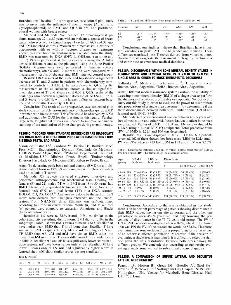
Introduction: The aim of this prospective, case-control pilot studywas to investigate the influence of chemotherapy (Adriamycin/Cyclophosphamid) on BMD and QUS in pre- and postmeno-pausal women with breast cancer.
Material and Methods: We included 32 premenopausal pa-tients, mean age 37.1±8.3 years with an incident diagnose of breastcancer who received a chemotherapy (4 cycles of AC) and 32 age-and BMI-matched controls. Women with metastases, a history ofosteoporosis with or without fracture, diseases or treatmentsknown to affect bone metabolism were excluded from the study.BMD was measured by DXA (DPX-L, GE/Lunar) at spine andhip. QUS was performed at the os calcaneus using the Achillesdevice (GE/Lunar) and at the phalanges using the Bone-Profiler(IGEA). Measurements were performed at baseline (beforechemotherapy), after 6 and 12 months and were compared withmeasurement results of the age- and BMI-matched control group.
Results: DXA results of the spine and hip showed a significantdecrease of T- and Z-score in patients with chemotherapy com-pared to controls (p £ 0.001). In accordance to QUS results,measurement at the os calcaneus showed a similar, significant,linear decrease of T- and Z-score (p £ 0.001). QUS results of thephalanges also showed a significant decrease of AD-SOS, for T-and Z-Score (p £ 0.001), with the largest difference between base-line and 12 months T-score (p £ 0.001).
Conclusion: The result of our prospective, case controlled pilotstudy confirms the deleterious influence of chemotherapy on BMDin women with breast cancer. This effect could be observed by DXAand additionally by QUS for the first time in this regard. Further,large scale longitudinal studies are needed to improve our under-standing of the mechanism of bone changes during chemotherapy.
P120MO. T-SCORES FROM STANDARD REFERENCES ARE INADEQUATEFOR BRAZILIANS: A MULTI-ETHNIC POPULATION BASED STUDY FROMRIBEIRAO PRETO, SAO PAULO
Sisson de Castro JA1, Cardoso V2, Bettiol H2, Barbieri MA2,Foss MC3; 1Endocrinology Division Faculdade de Medicina-UFRGS, Porto Alegre, Brazil, 2Pediatric Department Faculdadede Medicina-USP, Ribeirao Preto, Brazil, 3EndocrinologyDivision Faculdade de Medicina-USP, Ribeirao Preto, Brazil
Aims: To determine peak bone mineral density (BMD) in a multi-ethnic cohort born in 1978/79 and compare with reference valuesused to calculate T scores.
Methods: 529 subjects answered structured interviews andperformed anthropometric and biochemical tests. Healthy 226females (F) and 221 males (M) with BMI from 18 to 30 kg/m2 hadBMD determined by qualified technicians at L1-L4 vertebrae (LS),femoral neck (FN) and total femur (TF) in a DXA scanner,HOLOGIC QDR 4500A�. Analyses were done by the author. LS Tscores were derived from Hologic’s references and the femoralregions from NHANES’ data. Ethnicity was self-determinedaccording to Brazilian census criteria. White (w) and Mixed-race(m) persons were compare to caucasian Americans and Blacks(b) to Afro-Americans.
Results: 81.4% were w, 7.8% b and 10.7% m, similar to thecohort and city age-ethnic distributions. BMI did not differ in thesubgroups. Table 1 shows BMD values as mean ±SD. Brazilian Mhave higher peak BMD than F in all bone sites. Brazilian F havesimilar LS BMD despite ethnicity. bF and mF have higher FN andTF BMD than wF. wM and mM have similar BMD values butlower than bM in all sites. T scores differences from standards arein table 2. Brazilian wF and bF have significantly lower scores in allbone regions. mF have lower values only at LS. Brazilian M havelower T scores only at LS. wM had significantly higher scores atfemoral sites. mM show similar scores but not significant.
Conclusions: our findings indicate that Brazilians have impor-tant variations in peak BMD due to gender and ethnicity. Thesedifferences translated into T scores derived from values gatheredelsewhere may exagerate the assessment of fragility fracture riskand contribute to erroneous medical decisions.
P121SA. DISCORDANCE WITHIN BONE MINERAL DENSITY VALUES INLUMBAR SPINE AND FEMORAL NECK: IS IT VALID TO ANALYZE ASINGLE AREA IN ORDER TO MAKE THERAPEUTIC DECISIONS?
Sedlinsky C1, Medina L1, Schurman L1,2; 1Hospital Frances,Buenos Aires, Argentina, 2TcBA, Buenos Aires, Argentina
Aims: Different medical insurance systems sustain the reliabilty ofassessing bone mineral density (BMD) of a single area to performthe diagnosis of a patient presumably osteoporotic. We decided tocarry out this study in order to evaluate the power to discriminaterisk populations of a single area assessment, by determining if arethere discrepancies between both sites, lumbar spine (L2L4) andfemoral neck (FN), BMD.
Methods: 687 postmenopausal women between 45–79 years old,free of medication and other risk factors known to affect bone masswere studied. Values of BMD in L2L4 and FN were evaluated byDEXA using a Lunar DPX IQ equipment. Total predictive value(PV) of BMD in L2L4 and FN was determined.
Results: Results are displayed in table 1. Of the 687 patientsscreened, 462 of them showed low bone mass (LBM) in L2L4 and itPV was 89% whereas 413 had LBM in FN and it PV was 82.6%.
Conclusions: According to the results obtained in this study,there is an important percentage of patients showing discordance intheir BMD values, having one site as normal and the other onepathologic between 45–74 years old, and only lowering the per-centage of discordance in the 75–79 years old group. The PV ofL2L4 BMD as a sole investigated site was 89%, whilst if the chosenarea was FN the PV of the assessment would be 82.6%. Therefore,evaluating one zone excludes from a proper diagnosis a large partof an otherwise affected population. Moreover, if the decision ofevaluating a single area is mantained, it is difficult to select the rightone given the data distribution between both areas among thedifferent groups. We conclude that according to our results eval-uating a single zone will led to suboptimal disease diagnosis.
P122SU. A COMPARISON OF SUPINE LATERAL AND DECUBITUSLATERAL MORPHOMETRY
Pearson D1, Horton B1, Green DJ1, Goodby A2, Steel SA2,Stevens P3, Verboven C3; 1Nottingham City Hospital NHS Trust,Nottingham, UK, 2Centre for Metabolic Bone Disease, Hull,UK, 3GE Lunar
Table 1. (*) g/cm3
BMD* WF n=190 bF n=14 mF n=22 wM n=174 bM n=21 MM n=26
LS .991±.098 .997±.087 .991±.129 1.056±.118 1.126±.162 1.055±.135FN .833±.104 .859±.093 .854±.122 .991±.147 1.101±.158 .986±.160TF .898±.104 .916±.073 .926±.147 1.076±.142 1.188±.155 1.076±.158
Table 2. (*) significant differences from mean reference values, p<.01
T)scores wF bF mF wM bM mM
LS ).47* )1.38* ).54* ).31* ).66* ).33FN ).15* ).65* .04 .42* .18 .41TF ).35* ).73* ).13 .28* .06 .28
Table 1 Discordance between L2L4 an FN values: normal bone mass (NBM) vs.low bone mass(LBM). Distribution of the discordant areas.
Age(years)
n NBM inboth areas
LBM inboth areas
Discordants
n LBM in L2L4 LBM in FN
45–49 113 53 (46.9%) 32 (28.3%) 28 (24.8%) 20 (71.4%) 8 (28.6%)50–54 98 32 (32.6%) 35 (35.7%) 31 (31.76%) 18 (58%) 13 (42%)55–59 142 25 (17.6%) 65 (45.8%) 52 (36.6%) 37 (71.1%) 15 (28.9%)60–64 133 18 (13.53%) 82 (61.65%) 32 (24.06%) 19 (59.4%) 13 (40.6%)65–69 130 17 (13.07%) 80 (61.53%) 34 (26.15%) 18 (52.9%) 16 (47.1%)70–74 44 4 (9%) 26 (59%) 14 (32%) 6 (42.8%) 8 (57.2%)75–79 27 1 (3.7%) 23 (85%) 3 (11.3%) 1 (33.3%) 2 (66.6%)
Total 687 150 (21.8%) 343 (50%) 194 (28.2%) 119 (61.3%) 75 (38.7%)
S26
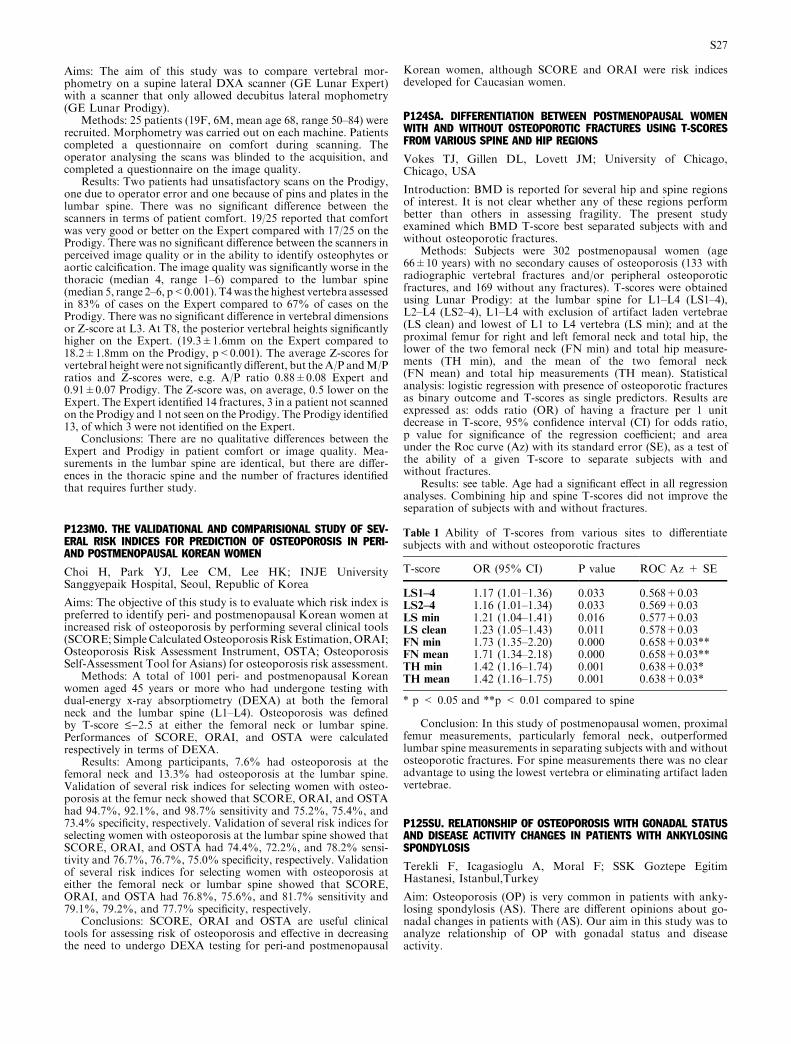
Aims: The aim of this study was to compare vertebral mor-phometry on a supine lateral DXA scanner (GE Lunar Expert)with a scanner that only allowed decubitus lateral mophometry(GE Lunar Prodigy).
Methods: 25 patients (19F, 6M, mean age 68, range 50–84) wererecruited. Morphometry was carried out on each machine. Patientscompleted a questionnaire on comfort during scanning. Theoperator analysing the scans was blinded to the acquisition, andcompleted a questionnaire on the image quality.
Results: Two patients had unsatisfactory scans on the Prodigy,one due to operator error and one because of pins and plates in thelumbar spine. There was no significant difference between thescanners in terms of patient comfort. 19/25 reported that comfortwas very good or better on the Expert compared with 17/25 on theProdigy. There was no significant difference between the scanners inperceived image quality or in the ability to identify osteophytes oraortic calcification. The image quality was significantly worse in thethoracic (median 4, range 1–6) compared to the lumbar spine(median 5, range 2–6, p<0.001). T4was the highest vertebra assessedin 83% of cases on the Expert compared to 67% of cases on theProdigy. There was no significant difference in vertebral dimensionsor Z-score at L3. At T8, the posterior vertebral heights significantlyhigher on the Expert. (19.3±1.6mm on the Expert compared to18.2±1.8mm on the Prodigy, p<0.001). The average Z-scores forvertebral height were not significantly different, but theA/P andM/Pratios and Z-scores were, e.g. A/P ratio 0.88±0.08 Expert and0.91±0.07 Prodigy. The Z-score was, on average, 0.5 lower on theExpert. The Expert identified 14 fractures, 3 in a patient not scannedon the Prodigy and 1 not seen on the Prodigy. The Prodigy identified13, of which 3 were not identified on the Expert.
Conclusions: There are no qualitative differences between theExpert and Prodigy in patient comfort or image quality. Mea-surements in the lumbar spine are identical, but there are differ-ences in the thoracic spine and the number of fractures identifiedthat requires further study.
P123MO. THE VALIDATIONAL AND COMPARISIONAL STUDY OF SEV-ERAL RISK INDICES FOR PREDICTION OF OSTEOPOROSIS IN PERI-AND POSTMENOPAUSAL KOREAN WOMEN
Choi H, Park YJ, Lee CM, Lee HK; INJE UniversitySanggyepaik Hospital, Seoul, Republic of Korea
Aims: The objective of this study is to evaluate which risk index ispreferred to identify peri- and postmenopausal Korean women atincreased risk of osteoporosis by performing several clinical tools(SCORE; SimpleCalculatedOsteoporosisRiskEstimation,ORAI;Osteoporosis Risk Assessment Instrument, OSTA; OsteoporosisSelf-Assessment Tool for Asians) for osteoporosis risk assessment.
Methods: A total of 1001 peri- and postmenopausal Koreanwomen aged 45 years or more who had undergone testing withdual-energy x-ray absorptiometry (DEXA) at both the femoralneck and the lumbar spine (L1–L4). Osteoporosis was definedby T-score £)2.5 at either the femoral neck or lumbar spine.Performances of SCORE, ORAI, and OSTA were calculatedrespectively in terms of DEXA.
Results: Among participants, 7.6% had osteoporosis at thefemoral neck and 13.3% had osteoporosis at the lumbar spine.Validation of several risk indices for selecting women with osteo-porosis at the femur neck showed that SCORE, ORAI, and OSTAhad 94.7%, 92.1%, and 98.7% sensitivity and 75.2%, 75.4%, and73.4% specificity, respectively. Validation of several risk indices forselecting women with osteoporosis at the lumbar spine showed thatSCORE, ORAI, and OSTA had 74.4%, 72.2%, and 78.2% sensi-tivity and 76.7%, 76.7%, 75.0% specificity, respectively. Validationof several risk indices for selecting women with osteoporosis ateither the femoral neck or lumbar spine showed that SCORE,ORAI, and OSTA had 76.8%, 75.6%, and 81.7% sensitivity and79.1%, 79.2%, and 77.7% specificity, respectively.
Conclusions: SCORE, ORAI and OSTA are useful clinicaltools for assessing risk of osteoporosis and effective in decreasingthe need to undergo DEXA testing for peri-and postmenopausal
Korean women, although SCORE and ORAI were risk indicesdeveloped for Caucasian women.
P124SA. DIFFERENTIATION BETWEEN POSTMENOPAUSAL WOMENWITH AND WITHOUT OSTEOPOROTIC FRACTURES USING T-SCORESFROM VARIOUS SPINE AND HIP REGIONS
Vokes TJ, Gillen DL, Lovett JM; University of Chicago,Chicago, USA
Introduction: BMD is reported for several hip and spine regionsof interest. It is not clear whether any of these regions performbetter than others in assessing fragility. The present studyexamined which BMD T-score best separated subjects with andwithout osteoporotic fractures.
Methods: Subjects were 302 postmenopausal women (age66±10 years) with no secondary causes of osteoporosis (133 withradiographic vertebral fractures and/or peripheral osteoporoticfractures, and 169 without any fractures). T-scores were obtainedusing Lunar Prodigy: at the lumbar spine for L1–L4 (LS1–4),L2–L4 (LS2–4), L1–L4 with exclusion of artifact laden vertebrae(LS clean) and lowest of L1 to L4 vertebra (LS min); and at theproximal femur for right and left femoral neck and total hip, thelower of the two femoral neck (FN min) and total hip measure-ments (TH min), and the mean of the two femoral neck(FN mean) and total hip measurements (TH mean). Statisticalanalysis: logistic regression with presence of osteoporotic fracturesas binary outcome and T-scores as single predictors. Results areexpressed as: odds ratio (OR) of having a fracture per 1 unitdecrease in T-score, 95% confidence interval (CI) for odds ratio,p value for significance of the regression coefficient; and areaunder the Roc curve (Az) with its standard error (SE), as a test ofthe ability of a given T-score to separate subjects with andwithout fractures.
Results: see table. Age had a significant effect in all regressionanalyses. Combining hip and spine T-scores did not improve theseparation of subjects with and without fractures.
Conclusion: In this study of postmenopausal women, proximalfemur measurements, particularly femoral neck, outperformedlumbar spine measurements in separating subjects with and withoutosteoporotic fractures. For spine measurements there was no clearadvantage to using the lowest vertebra or eliminating artifact ladenvertebrae.
P125SU. RELATIONSHIP OF OSTEOPOROSIS WITH GONADAL STATUSAND DISEASE ACTIVITY CHANGES IN PATIENTS WITH ANKYLOSINGSPONDYLOSIS
Terekli F, Icagasioglu A, Moral F; SSK Goztepe EgitimHastanesi, Istanbul,Turkey
Aim: Osteoporosis (OP) is very common in patients with anky-losing spondylosis (AS). There are different opinions about go-nadal changes in patients with (AS). Our aim in this study was toanalyze relationship of OP with gonadal status and diseaseactivity.
Table 1 Ability of T-scores from various sites to differentiatesubjects with and without osteoporotic fractures
T-score OR (95% CI) P value ROC Az + SE
LS1–4 1.17 (1.01–1.36) 0.033 0.568+0.03LS2–4 1.16 (1.01–1.34) 0.033 0.569+0.03LS min 1.21 (1.04–1.41) 0.016 0.577+0.03LS clean 1.23 (1.05–1.43) 0.011 0.578+0.03FN min 1.73 (1.35–2.20) 0.000 0.658+0.03**FN mean 1.71 (1.34–2.18) 0.000 0.658+0.03**TH min 1.42 (1.16–1.74) 0.001 0.638+0.03*TH mean 1.42 (1.16–1.75) 0.001 0.638+0.03*
* p < 0.05 and **p < 0.01 compared to spine
S27
Method: The study group consisted of 51 male patients diag-nosed with AS according to Modified New York Criteria and 32patients followed with pain complaints formed the control group.Exclusion criteria were discovery of any disorders that could causeOP or hypogonadism.
Lumbar spine and femoral neck areas were assessed withLunar DPX. Erythrocyte sedimentation rate (ESR) and C-reactiveprotein (CRP) were used to identify the disease activity of thecases. The follicle-stimulating hormone (FSH), luteinizinghormone (LH), total testosterone and sex hormone binding glob-ulin (SHBG) levels of the control and study groups were measured.Testosterone free index values were calculated. Graphpad PrismaV.3 program package was used for the statistical analyze of thedata.
Results: The femoral neck bone mineral density (BMD) valuesof the study group were lower than the control group. However thelumbar spine values were not significantly different between the twogroups. ESR and CRP values were higher in the study group.Femoral neck BMD values were correlated with ESR but not withCRP. FSH levels of the study group were lower than the controlgroup. Total testosterone, LH, SHBG levels were not different inboth control and study groups. There was a negative correlationbetween the SHBG and femoral neck BMD values of the studygroup but the SHBG values were not statistically different betweenthe two groups.
Conclusions: The BMD measurements of the femoral neck arelower in patients with AS and are in correlation with diseaseactivity (ESR). The absence of gonadal status changes in patientswith AS suggests that gonadal status is not relative in thesepatients.
P126MO. QUANTITATIVE MORPHOMETRY OF T12 THORACIC VERTE-BRAL BODY IN DETECTION OF PREVALENT FRACTURE: SHOULDMEASUREMENT ON LUMBAR SPINE FILM FOR THOARCIC DEPICTIONOF T12 BE AN ALTERNATIVE TECHNIQUE?
Wu C1, Fuerst T1, Ingersleben GV1, Miaux Y1, Genant HK1,2;1Synarc Inc., 2DepartmentofRadiology,SanFrancisco,California,USA
Aims: Quantitative morphometry (QM) of the 12th thoracicvertebral body (T12) on poorly projected lateral thoracicspine film could lead to mis-measurement of the vertebral bodyheight. We compared the measurements of T12 from boththoracic and lumbar films and validated the solution of usinglumbar film as an alternative method.
Methods: Women with a suspected osteoporotic vertebralfracture participating in a clinical trial by visual assessmentwere retrospectively investigated. Of a total of 103 women, 83women were confirmed to have a prevalent fracture by anexperienced radiologist using a semiquantitative (SQ) method.The anterior (Ha), middle (Hm), and posterior heights (Hp) atT12 were measured on thoracic spine film using six pointstechnique corresponding to the contour of the vertebral bodyand the measurements were repeated on lumbar spine film. Thevertebral heights were used to assess fracture prevalence using aform of the widely adopted Melton method: height ratioreduction y ‡ 3 standard deviation (SD) at the level fromnormal reference data.
Results: The three vertebral dimensions measured on either thethoracic or lumbar film were on average all within 1% different(Ha=0.27±6.86%; Hm=0.60±8.00%; Hp=0.45±7.01%). Ver-tebral heights measured on lumbar film are found highly correlatedwith thoracic film (Ha: R2=0.94, Hm: R2=0.88) with the exceptionof Hp (R2=0.62). Although QM on both thoracic and lumbar filmsprovided very similar vertebral dimensions, assessing T12 on thelumbar film was overall equal or better than on the thoracic film(Table).
Conclusions: Measurement of T12 vertebral dimension withlumbar spine film provides equal or better results than the samemeasurement on the thoracic film. QMmeasurement on the lumbarspine film when T12 is poorly depicted on the thoracic film is anacceptable alternative technique.
P127SA. COMPARISON OF QUANTITATIVE ULTRASONOGRAPHY WITHDUAL ENERGY ABSORPTIOMETRY AND INVESTIGATION OF THECORRELATION BETWEEN QUANTITATIVE ULTRASONOGRAPHYPARAMETERS
Altan L1, Bingol U1, Kumas FF1, Ercan I2, Yurtkuran M1;1Uludag University Medical Faculty Ataturk RehabilitationCenter, Bursa, Turkey, 2Uludag University Medical Faculty,Biostatistics Department, Bursa, Turkey
Aims: To evaluate the correlation between the two QUS param-eters: broadband ultrasound attenuation (BUA) and speed ofsound (SOS), and also to investigate correlation of QUS and dualenergy X-ray absorptiometry (DEXA) measurements.
Methods: Calcaneal QUS measurements were performed in astandard fashion in a total of 1755 patients. Then BMD mea-surements using DEXA were performed in 485 (444 females and41males) of the QUS patient population. Sensitivity, specificity,and positive and negative accuracy were calculated for BUA andSOS values and correlation of QUS and DEXA values werecalculated by Pearson correlation coefficient.
Results: Calcaneal QUS measurements showed a positivemoderate correlation (r=0.43, p<0.01) for SOS and BUA ampli-tudes, and a positive low correlation (r=0.37, p<0.01) for SOSand BUA T-scores. By taking SOS values as reference, sensitivityand negative accuracy were found to be 0.88, and specificity andpositive accuracy 0.47 and 0.45, respectively, for BUA values.These results suggest a higher probability for accuracy of thefindings when either SOS measurements show osteoporosis or BUAgives normal values, whereas they also suggest that a positive resultwith BUA or a negative result with SOS should be approached withsuspicion. A low correlation (r=0.13–0.37, p<0.001) betweenDEXA and BUA amplitudes and T-scores was found. Correlationwas also low (r=0.16–0.29, p<0.001) between DEXA and SOST-scores and not significant between DEXA and SOS amplitudes.Separate evaluation of men, premenopausal women, and post-menopausal women showed a significant correlation (r=0.25–0.36,p<0.001) only for the postmenopausal group.
Conclusion: The results of our study suggest that QUS can giveresults comparable in reliability to those of DEXA particularly inthe postmenopausal women. However, the overall low to moderatecorrelation between QUS and DEXA results emphasizes the needfor further research for delineation of the definitive role of QUS indiagnosing osteoporosis and predicting fracture risk.
P128SU. QUALITY OF LIFE IN POSTMENOPAUSAL OSTEOPOROTICWOMEN
Brankovic S, Pilipovic N, Palic-Obradovic D; Institute ofRheumatology, Belgrade, Serbia and Montenegro
The aim of study was to assess quality of life in postmenopausalwomen (PMW) with low bone mineral density (BMD) and inPMW with fracture. We compare Health assessment Question-naire (HAQ), Osteoporosis Quality of Life Questionnaire(OQLQ) and Short Form 36-item Health Survey Questionnaire(SF-36). BMD was measured on lumbar spine using dual x-rayabsorptiometry. HAQ was used to assess physical disability in 8
Table 1 Comparison of T12 measurements on thoracic and lumbar spine films
N Thoracic Film Lumbar Film
T12 depiction: adequate / poor 103 50/53 75/28QM Fracture Rate(3 SD reduction from normal)
103 57 56
Agreement with SQ (Kappa Score) 103 0.42 0.45Overall Anterior height reduction (%) 103 29.45±14.71 29.1±14.76Overall Middle height reduction (%) 103 25.08±13.18 24.53±13.62
Anterior height reduction (%) related to SQ Grades:SQ = 0.5 (questionable height reduction) 20 19.15±4.74 19.03±4.91SQ = 1 (Mild height reduction 20–25%) 32 23.05±5.59 22.70±4.55SQ = 2 (Moderate height reduction 25–40%) 40 30.79±8.02 30.29±8.56SQ = 3 (Severe height reduction > 40%) 11 61.96±17.46 61.86±17.59
S28

categories of function. OQLQ is a specific measure and includes30 questions, which are grouped into five domains: Symptoms,Emotional function, Physical function, Activities of Daily living(ADL), Leisure and Social activities with a score between 1 and 7.The SF-36 is a generic measure, and assessed 8 areas of generalhealth each with a score between 0 and 100. High scores reflectbetter quality of life, and may also be converted to 2 summaryscales: the physical and mental component summary scores.Spearman‘s rank method and Kruskal-Wallis Anova by rankstest were used to analyse the strength of relationships betweenvariables. We assessed 114 PMW stratified in 4 groups: I (22)with normal BMD (T-0.19), mean age 52.6, HAQ 0.79; II (33)with Osteopenia (T-1.71), mean age 59.8, HAQ 0.57; III (27) withosteoporosis (T-3.28), mean age 62.1, HAQ 0.84 and IV (32) withfracture (T-2.49), mean age 65.9, HAQ 0.95. Results are on thegraph. There was significant correlation between HAQ index,OQLQ and SF-36 in all groups. There were significant lowervalues of HAQ index (p<0.001), OQLQ (p<0.001) and SF-36median scores (p<0.01) in group with fracture, as might be ex-pected. The study showed that both, the SF-36 and OQLQ arepotentially useful measures on wider aspects of quality of life inPM osteoporotic women.
P129MO. EVALUATION OF CORTICAL BONE BY PERIPHERAL QUAN-TITATIVE COMPUTED TOMOGRAPHY (PQCT) IN PERITONEAL DIALYSIS(PD) PATIENTS
Negri AL1, Lombas C1, Barone R2, Bogado CE1, Zanchetta JR1;1Instituto de Investigaciones Metabolicas, Buenos Aires, Argen-tina, 2STR Hurlingham SRL, Buenos Aires, Argentina
Peripheral QCT allows the non-invasive evaluation of corticaland trabecular bone separately as well as the geometricalproperties of the radius. We investigated cortical bone by pQCTin 22 patients (6 males and 16 females) on maintenance PD;comparisons were made with 28 normal controls. PeripheralQCT (XCT 960, Stratec, Pforheim, Germany) was performed atdistal radius of the nondominant forearm (15% the length of theulna from endplate). We evaluated Total and cortical bonemineral density (TBMD, cBMD), Total (cross-sectional) andcortical area (TA, cA), cortical thickness (cThk), endosteal andperiosteal perimeters and buckling ratio. Intact PTH levels weremeasured by IRMA. Correlations were made with age, totaltime in dialysis and serum iPTH. DP patients had a markeddecrease in cThK (1.90 vs 2.95 mm; p<0.0001) and a markedincrease in endosteal perimeter (31.2 vs 23.9 mm; p<0.0001)Buckling ratio [r/cThK] (3.81 vs 2.21; p<0.0001) and TA.TBMD and cBMD correlated negatively with total time indialysis (p<0.01); no correlations were found between cA,cBMD and cThk with iPTH. Age correlated positively withTA, endosteal and periosteal perimeter and negatively cBMD.Our results show a cortical thinning of the radius with corticalparameters correlating predominantly with total time in dialysisand that bone adaptations to aging (peripherization) occurdespite disturbances in endocrine-metabolic environment ofdialysis.
P130SA. ANALYSIS OF BONE MINERAL DENSITY IN PATIENTS WITHHYPERPARATHYROIDISM SECONDARY TO END-STAGE RENAL DISEASEAND INDICATION OF PARATHYROIDECTOMY
Lacativa PGS1,5, Salvarezza B1,5, do Monte CMG1,5,Mendonca LMC2,5, Goncalves MDC3,5, Patrıcio Filho PJM4,5,Farias MLF1,5; 1Endocrine Service, Department of InternalMedicine, 2Reumathology Service, Department of InternalMedicine, 3Department of Surgery, 4Nephrology Service, Depart-ment of Internal Medicine, 5Clementino Fraga Filho UniversityHospital, Federal University of Rio de Janeiro, Brazil
Background: Patients with hyperparathyroidism secondary toend-stage renal disease (HPT2) have moderate to severe bonedisease and increased fracture risk compared to normal popula-tion. Some reports had demonstrated marked improvement inbone mass after successful parathyroidectomy (PTX).
Aims: To evaluate bone density in patients with HPT2 imme-diately before PTX.
Methods: 34 patients with HPT2 and indication for parathy-roidectomy were studied. Serum PTH was above 600 pg/mL in allpatients, and half of them had hypercalcemia. Serum gonadotro-phins, testosterone and estradiol were evaluated by routine meth-ods. Bone densitometry was performed to evaluate whole bodydensity and composition, using dual x-ray absorptiometry (DXA).All exams were performed in the same densitometer, Lunar Cor-poration. Any evidence of aluminium intoxication such as highserum levels or a compatible bone biopsy was considered excludingcriteria. Other factors, chronic diseases and medications that canalterate BMD were also valuated.
Results: Hypogonadism was present in 14 patients (41.2%), lowbody mass index in 10 (29.4%), HCV infection in 10 (29.4%),chronic smoking in 8 (23.5%), immobilization in 6 (17.6%),chronic glucocorticoid exposure in 2 (5.9%), one patient hadhyperthyroidism and another systemic lupus erythematosus. Nocase of HIV infection or alcoholism was found. All patients hadincreased bone resorption in skeletal radiographs. 8 patients(23.5%) reported pathologic fractures. The median value ofZ-score of total body, arms, legs and trunk were respectively )3.1;)2.7; )3.85; e )1.9. All these differences were statistically significant.
Conclusions: The severe bone loss observed in all patients ismainly due to HPT2, but comorbidities usually found in patientswith end-stage renal disease also contribute. Bone densitometrywas useful to quantitatively estimate bone loss and to demonstratethat skeletal sites rich in cortical bone were mostly affected, whichis compatible with PTH’s effects on bone.
P131SU. PAMIDRONATE AND ZOLEDRONATE EFFECTS IN THEINCREMENT OF BONE MINERAL DENSITY: COMPARING CONTROL,PROTEIC AND APROTEIC DIETS, OOPHORECTOMIZED AND NON- OO-PHORECTOMIZED FEMALE RAT GROUPS
Cruz Jr AF1,2, Guarniero R3, Buchpiguel CA4, Barbieri A5;1Nucleo de Diagnosticos Maringa, 2Ultramed de Londrina,3Instituto de Ortopedia e Traumatologia da FM-USP, 4Centrode Medicina Nuclear da FM-USP, 5Departamento de Diagnos-tico por Imagem da EPM-UNIFESP, Brazil
Aims: Compare the increment of bone mineral density (BMD)with pamidronate, zoledronate and the isolated effect of proteindiet, with bone densitometry and histomorphometry, in malnu-trition oophorectomized and not oophorectomized female rats;and validate the indexes of BMD.
Methods: 60 young female Lewis rats divided in 5 experimentaland a control group with and without oophorectomy and theadministration of the drugs were submitted to two diets (proteicand aproteic). The variables were: weight, densitometry, histolog-ical and biochemistry evolution.
Results: Weight Evaluation: 1st interval (after 1st dose) showedincrease in all groups, statically significant in the groups oophorec-tomized. 2nd interval (2nd dose) showed decrease, not significant, inall groups. Densitometric Evaluation: 1st interval (after aproteicdiet) showed decrease in all groups, significant in the 4 medicatedgroups. 2nd interval (after 1st dose) showed increase in all groups.
Fig. 1 Precision Comparison between Prodigy and Delphi
S29
3rd interval (after 2nd dose) showed increase in all groups, significantin the two not oophorectomized. Laboratorial Evaluation: theinterval (at the beginning and at the end of medications) showed:increase of total proteins and globulin, confirming that there was notmore malnutrition, decrease of alkaline phosphatase (except for thetwo ophorectomized groups), decrease of phosphorus and calciumin the fourmedicated groups and increase of phosphorus and calciumin the two groups not medicated, confirming that the medicationscause hypocalcemia. Histomorphometric Evaluation: the ophorec-tomized group had smaller increment than that of not ophorectom-ized, confirming the discoveries of densitometric study.
Conclusions: The pamidronate and zoledronate were showneffective in the increment of BMD, in malnutrition and oopho-rectomized or not oophorectomized rats. These preliminary resultsindicate that the proteic diet, separately, possesses therapeuticeffect in BMD, however in a significant less way, compared withthe medicated animals. The results of histomorphometry, allowsvalidating the bone densitometry, in this experimental model.
P132MO. THE LUNAR ACHILLES AS A SCREENING TOOL FOR OSTEO-POROSIS: COMPARISON WITH SPINE DXA
Poriau S1, Geusens P2,3, Van den Bosch F1, De Keyser F1;1Locomotorisch Centrum, Sijsele-Damme, Belgium, 2BiomedicalResearch Institute, Diepenbeek, Belgium, 3University Hospital,Maastricht, The Netherlands
Quantitative ultrasound of the os calcis is accepted as an effective,low-cost method to assess osteoporotic fracture risk. Recently theInternational Society for Clinical Densitometry (ISCD) recom-mended the use of peripheral densitometry (such as heel ultras-onometry) to identify patients who might have osteoporosis andshould therefore undergo BMD testing at the hip and spine. Thisrecommendation requires the use of a device specific T-scorecutpoint on the peripheral device that detects 90% of individualswith osteoporosis (T-score £ )2.5) at either the spine or hip. Inthis study, we wished to determine the 90% sensitivity cutpointthat could be used with the Achilles bone ultrasonometer.
A total of 1087 women aged 50 years and older (mean age68±10 years) had DXA measurements of the spine (L1–L4) using aLunar Prodigy (GE Medical Systems) or a Lunar DPX-NT, as wellas heel ultrasound measurement using Achilles Solo. The heel T-score cutpoint with 90% sensitivity for identifying subjects withosteoporosis (T-score £ )2.5) at the spine was determined usingROC analysis with the ROCKIT program (Charles Metz, Uni-versity of Chicago).
From the DXA results, 332 out of the 1087 patients wereclassified as osteoporotic in the spine. At a heel T-score of )1.4,sensitivity was 91% and specificity was 38%. Higher specificity ofnearly 50% could be obtained with a T-score cutpoint of )1.8,which has a sensitivity of 85%. We conclude that the Achilles boneultrasonometer can be used as a valid screening tool for osteopo-rosis according to ISCD recommendations. In situations wherecentral DXA measurements are not ready available, the Achilles isa useful device to identify those patients who should be consideredfor spine and hip bone density assessment.
P133SA. DENSITOMETRIC DIFFERENTIAL DIAGNOSIS BETWEEN‘‘DISUSE’’ AND ‘‘SYSTEMIC’’ OSTEOPENIAS: REFERENCE CHARTSAND CLINICAL APPLICATIONS
Capozza RF1, Cointry GR1, Cure-Ramırez P2, Ferretti JL1, Cure-Cure C2; 1CEMFoC, Natl Univ of Rosario, Argentina., 2Metro-politan Univ of Barranquilla, Colombia
DEXA assessment of bone mass (BMC) can be complemented bymeasuring lean mass (LM, regarded as proportional to musclemass within certain limits) in either the whole body (WB) or se-lected regions. This little-profitted resource may provide a dif-ferential diagnosis between ‘‘disuse’’ and ‘‘systemic’’ osteopenias.We have showed linear, parallel BMC(y)-vs-LM(x) relationshipsin the WB and limbs of 1,450 Argentine and 3,000 Colombiannormal boys/girls, men, and pre- and post-MP women. Parallel-ism of the curves would reflect the biological control of bones bymuscles (bone mechanostat theory). However, the intercepts ofthe graphs differed between groups, in the order: boys/girls <post-MP women < men < pre-MP women, suggesting a positivemodulation of that control by sex hormones. The variance of theBMC/LM relationship was substantially lower in lower limbsthan in WB.
These results allowed performing z-scored graphs, specific forgender, reproductive status, region studied, race, and DEXAequipment employed, suitable for evaluating the bone/muscle massproportion. They may allow evaluating whether an eventually lowBMC value is or not adequately proportionate to the individual’sWB or regional muscle mass. ‘‘Disuse-related’’ osteopenias (as wellas small or lean, normally active individuals) should show a normalz-score for the BMC/LM relationship. ‘‘Systemic’’ osteopeniascaused by alterations of bone cells (either primary or secondary tochanges in their endocrine-metabolic environment) should showlow BMC/LM z-scores. Such cases ought to be further studiedemploying other technologies to determine whether bone strengthis or not affected; i.e. for diagnosing an osteoporosis as a (meta-bolic) ‘‘osteopenic fragility’’ (NIH criterion, out of the DEXAscope).
We have tested the ability of our WB or lower-limb BMC/LMz-score reference charts to detect ‘‘metabolic’’ osteopenias in a.haemodialysis patients (z-scores decaying as time on dialysis orserum PTH increased); b. obese hyperinsulinemic euglycemic pa-tients (z-scores diminishing as body-mass index or fasting plasmainsulin increased, or insulin sensitivity decreased); c. female balletdancers (z-scores decaying as calciuria increased presumably be-cause of a disturbed estrogen metabolism), and d. hypopituitarymen and women before and after treatment with GH (z-scoresimproving as serum IGF-I levels increased).
P134SU. ASSESSMENT OF TRABECULAR BONE TEXTURE IN THEDISTAL RADIUS USING PERIPHERAL QUANTITATIVE COMPUTEDTOMOGRAPHY (PQCT)
MacArtain AM1, Webber CE1, Inglis D2, Gordon CL1; 1MedicalPhysics & Applied Radiation Sciences Unit, McMaster Univer-sity, Canada, 2Department of Civil Engineering, McMasterUniversity, Canada
Aim: The purpose of this study was to investigate the distributionof bone and the changes in trabecular bone texture along thedistal radius using peripheral quantitative computed tomography(pQCT) and to assess the accuracy of such measurements usingmicro-computed tomography (lCT) as a gold standard. It is wellestablished that trabecular bone architecture, in addition to bonemineral density, is an important indicator for people at risk offracture.
Methods: 12 dry human cadaver radii, of unknown gender orage, were imaged in air using a Stratec XCT-2000 pQCT scanner.Consecutive transaxial images were acquired with an in-plane pixelresolution of 0.2 · 0.2 mm and a slice thickness of 2.2 mm, cov-ering 12% of bone length proximal from the radioulnar joint. lCTscans were performed on those two radii, determined from x-rayradiographs, to be the most and least osteoporotic. lCT imageswere acquired using a GE eXplore RS system with an isotropic
Sensitivity and Specificity of Osteoporosis at Spine Based onHeel T-Score
Heel T-Score Sensitivity Specificity
)0.6 95% 20%)0.8 94% 24%)1.0 94% 27%)1.2 91% 31%)1.4 91% 38%)1.6 88% 43%)1.8 85% 49%)2.0 82% 55%
S30
voxel resolution of 47 lm covering a 40 mm section at the distalradius. All texture analyses were performed using an in-housealgorithm, which determined the mean hole size, number of holesand a connectivity index.
Results: The accuracy of pQCT assessment of texture is directlyinfluenced by partial volume averaging due to the limited spatialresolution of the image data. Preliminary results indicate similartrends for connectivity index, number of holes and average holesize for all bones along their length as measured by pQCT (Fig).
Conclusions: The similar patterns of change in connectivity andmean hole size between radii will allow the distal radius to be usedclinically as a site for trabecular bone texture measurement. Dif-ferences between sequential measurements of texture will beattributable to changes in bone architecture and will not be theconsequence of poor reproducibility arising from patient posi-tioning uncertainties.
P135MO. BONE MINERAL DENSITY IN PATIENTS WITH PARKINSON’SDISEASE
Ardicoglu O1, Kamanli A1, Ozgocmen S1, Yoldas TK2; 1FiratUniversity, Division of Rheumatology, Department of PMR,Turkey, 2Firat University, Department of Neurology, Turkey
Objective: This study proposed to assess bone mineral density(BMD) - lumbar spine, femoral and hand - and the relationshipbetween BMD and disease duration, Hoehn and Yahr staging inTurkish patients with Parkinson’s disease (PD).
Background: PD is mainly a disease of elderly adults and cancause severe disability and injury resulting from falls. Osteoporosisis a serious problem for older adults, especially for postmenopausalwomen leads to disabling falls and fractures. To best of ourknowledge this report is the first assesing hand BMD in PD.
Methods: Twenty-four PD patients and age- and sex- matched27 control were recruited from outpatient clinic of Neurology ofour hospital. We evaluated lumbar spine (L2–L4), femoral (neck,Ward’s triangle, trochanteric) and bilateral hand BMD using dual-energy x-ray absorbsiometry (DXA) in both groups and Hoehnand Yahr staging (HYS) in patients.
Results: There was no significant difference in right hand BMD(rHBMD), L2–L4 spinal BMD, and right femoral (neck, ward’striangle, trochanteric) BMD between patients and controls.However, female patients’ hand BMD and right femoral neckBMD were significantly lower than female controls (p<0.05). Malepatients had no significant difference in BMD measurements in allsites with respect to controls. Patients’ HYS and disease durationwere negatively correlatedwithBMDat all sites except L2–L4BMD.
Conclusions:We underscore an increased risk for osteoporosis infemale patients with PD that is more prominent in femoral and handBMDmeasurements. Female patients should be carefully examinedfor osteoporosis to prevent bone loss and associated disability.
P136MO. AGE RELATED DECLINE IN BONE MASS MEASURED BY DUALENERGY X-RAY ABSORPTIOMETRY AND QUANTITATIVE ULTRASOUNDIN A POPULATION BASED SAMPLE OF BOTH SEXES: IDENTIFICATIONOF USEFUL ULTRASOUND VALUES FOR SCREENING FOR OSTEOPO-ROSIS
SigurdssonG1,GudmundsdottirSL1,IndridasonOS1,FranzsonL2;1Department of Endocrinology and Metabolism, 2Department ofClinical Chemistry, University Hospital, Reykjavik, Iceland
Aims: The aim of this population based study was to compareage related changes in bone mass measured by quantitativeultrasound (QUS) and dual x-ray absorptiometry (DXA) inorder to identify a useful QUS values for screening for osteo-porosis.
Methods: The study population was a random sample of 1630individuals (1041 females, 589 males) age 30-85 years. Bone masswas measured by DXA (Hologic QDR 4500) in lumbar spine andproximal end of femur (total hip) and in calcaneus by QUS (Lunar,Achilles+). Biochemical bone markers (serum osteocalcin andserum CTX) were measured and fractures were reported. Individ-uals with DXA t-score <)2.5 at the lumbar spine or hip wereidentified and receiver operating curves (ROC) were used to cal-culate cut-off points for QUS. Sensitivity, specificity and kappastatistics were calculated.
Results: Age related decline in bone mass was significantlylarger with calcaneal QUS than DXA at both sites in women. Formen, the curves were similar for QUS and DXA in the hip.Similar correlations were found between QUS and DXA indifferent age groups of both sexes (0.45–0.53). Bone markerscorrelated inversely and similarly with QUS and DXA measure-ments. For women age 50–65 years a QUS t-score >)0.5 wasfound the most applicable for identifying normal BMD by DXA.In the age group 70–85 years, t-score >)2.0 for women and>)1.0 for men seemed reasonable cut-offs for identifying normalBMD (sensitivity around 90%, specificity 30–40%, discordancerate around 50%).
Conclusions: Calcaneal QUS cannot be used for the diagnosisof osteoporosis according to WHO criteria for DXA but can be ofuse to exclude osteoporosis in 30–40% of our cases.
P137SU. COMPARISONS OF BMD, BMC AND BONE AREA IN PENCILAND FAN BEAM DENSITOMETRY WITH THE USE OF ANTHROPOMETRICSPINE PHANTOM
Pludowski P, Jaworski M, Matusik H, Lorenc RS; Department ofBiochemistry and Experimental Medicine, The Children’s Memo-rial Health Institute, Warsaw, Poland
Aim: Introduction of newer DXA technology replaced earlierpencil beam x-ray by fan beam. The new fan beam devices ensurea high resolution images at multiple sites of skeleton. Changes inthe hardware and software allowed assessments of bone mineraldensities of various regions with much increased speed of mea-surement. However, little is known about the concordance be-tween densitometric results established using pencil and fan beamdevices. To address this issue, we performed a repeated mea-surements of anthropometric spine phantom using pencil- andfan-beam densitometers.
Methods: 440 measurements of anthropometric spine phantomperformed using DPX-L and Prodigy machines were analyzed.Bone mineral densities and contents, as well as areas were evalu-ated in L1, L2, L3, L4, L1–L4, L2–L4, L3–L4 regions of interest.
Results: The stability of BMD measurements was very high inboth devices (range of results within 1.5%). Significant differencesbetween Prodigy and DPX-L assessed densities, contents and areaswere found in all analyzed ROIs (p<0.0001). Evaluation of meanvalues revealed lower densities, contents but not areas establishedusing fan beam technology (Prodigy) when compared to pencilbeam (DPX-L). The differences (in g/cm2) between means of BMDassessed in L1-L4, L2-L4 and L3-L4 tended to increase withdecreasing number of analyzed vertebrae ()0.0365±0.0004,p<0.0001;)0.0417±0.0004, p<0.0001; and )0.0429±0.0006,p<0.0001), respectively. In opposite, the differences (in g) between
Fig. 1 Connectivity Index (%) along radii from pQCTmeasurements
S31
means of BMC tended to decrease with decreasing number ofanalyzed vertebrae ()2.053±0.0177, p<0.0001; )1.544±0.0166,p<0.0001; and )0.998±0.0134, p<0.0001), respectively. Similartendency has not been found for differences between means ofareas. Coefficients of variations for all data sets of analyzedparameters were lower for Prodigy, indicating lower variability ofmeasurement results reflecting improved edge detection whencompared to DPX-L.
Conclusion: On the basis of our results it can be concludedthat there is substantial need to establish correction algorithmswhich allow comparison of measurements performed using fan andpencil beam devices what is essential in time when increasingnumber of fan beam machines is being installed.
P138MO. STUDY OF BONE MINERAL DENSITY IN PATIENTS WITHTHYROTOXICOSIS
Chandratreya SA, Shah NS, Menon PS; KEM Hospital,Mumbai, India
Aim and objectives: To evaluate bone density in patients withuncontrolled thyrotoxicosis and to study the impact of variousfactors like severity and duration of thyrotoxicosis, parity,duration of lactation, socioeconomic status, nutritional factors,physical activity, vitamin D status on bone density in studypopulation.
Materials and methods: A total of 45 premenopausal femalepatients having Graves disease with untreated or uncontrolledthyrotoxicosis between age group 30–50 yrs were studied. Inaddition to clinical parameters, hormonal and biochemicalparameters like serum T3, T4, TSH, calcium, phosphorus, alkalinephosphatase, albumin, 25-OH D3 and urinary calcium and creat-inine values were evaluated. Bone density was obtained by dualenergy x-ray absorptiometry (DXA; Hologic Inc.) and was com-pared with the Indian normative data.
Results: As compared to Indian normative data, the studypopulation had significantly reduced bone density. The mean Zscore was )0.69 and )0.65 at lumbar spine and femoral neckrespectively. 37.78% and 35.55% of patients had Z scores less than)1.0 at lumbar spine and femoral neck, respectively. 78% patientshad hypovitaminosis D (£20 ng/ml).
Weight, body mass index, total caloric and calcium intake perday correlated positively with bone densities at various sites. Sr. T3,parity, lactation, correlated negatively with bone density at varioussites.
Physical activity, sunlight exposure, socioeconomic status,vitamin D status and other biochemical parameters did not showsignificant correlation with bone density at any site.
Conclusion: Bone density is reduced in patients with thyro-toxicosis as compared to the general population. In addition tothyrotoxicosis, a combination of various other factors contribute tolow bone mass observed in this condition. In the absence of uni-versal guidelines to treat bone disease in thyrotoxicosis, optimumcontrol of thyrotoxicosis, as well as maintenance of proper nutri-tion, adequate vitamin D and calcium intake and control of otherrisk factors for osteoporosis appears to be the correct strategy.
P139SA. BONE MINERAL DENSITY, SERUM MARKERS OF BONETURNOVER AND THEIR RELATIONSHIPS IN PERITONEAL DIALYSIS
NegriAL1, BaroneR2,AlvarezQuirogaM3,BravoM3,MarinoA1,Fradinger E1, Bogado CE1, Zanchetta JR1; 1Instituto de Investi-gacionesMetabolicas, BuenosAires, Argentina, 2STRHurlinghamS.R.L., Buenos Aires, Argentina, 3FMC Argentina Caballito,Buenos Aires, Argentina
Background: The usefulness of bone mass measurements andbone turnover markers to estimate fracture risk or the type ofunderlying renal osteodystrophy are not well established inpatients on peritoneal dialysis (PD).
Objective: To asses bone mass using total and regional bonedensitometry in a group of patients on PD and to determine if serummarkers of bone turnover identify patients with low bone mass.
Methods: Bone densitometry was studied by dual-energy X-rayabsorptiometry (DXA) and bone turnover using several serummarkers in 65 patients on PD. Bone mass was classified as normal,osteopenic or osteoporotic according to World Health Organiza-tion criteria based on bone mineral density T scores.
Results: T scores in osteopenic range were present in 44.6%(45% of men and 44.4% of females) of patients at the lumbar spine(LS) and in 56% (55% of men and 58% of females) at the femoralneck (FN). T scores in the osteoporotic range were present in 13%of patients (10% of men and 15.5% of women) at the LS and in21% (30% of men and 17.7% of women) at the FN. Patients withBMD T scores in osteoporotic range at both regions had increasedserum iPTH levels compared to patients in the osteopenic/normalrange. TBMC correlated negatively with iPTH (r=)0.34) and withtotal time in dialysis (r=)0.26); in multivariate analysis only iPTHcorrelated negatively with TBMC (B=)0.26; p=0.03). No corre-lations were found between the other bone markers and BMD Tscores at the FN or LS. There were no significant differences inabsolute BMD or BMD T scores at the LS or FN between patientswith or without fractures.
Conclusions: BMD T scores in osteopenia osteoporosis rangewere observed in 58.4% of these patients on PD at the LS and in78.4% at the FN. TBMC correlated negatively with iPTH. Therewere no correlations between markers of bone turnover and bonemass measurements at the two skeletal regions, although patientswith BMD T scores in osteoporotic range had increased serumiPTH levels. Bone mass measurements were not different betweenpatients with or without fractures.
P140SU. PREDICTION OF LUMBAR SPINE BONE MINERAL DENSITYFROM WHOLE BODY DUAL-ENERGY X-RAY ABSORPTIOMETRY
Boyanov M1, Protich M2, Boneva Zh2, Harangoso E1; 1Endocri-nology Clinic, Alexandrovska Hospital, Medical University Sofia,Bulgaria, 2Endocrinology Unit, Central Police Hospital, Sofia,Bulgaria
Whole body Dual-energy x-ray absorptiometry (DXA) is usedmost often in studies of body composition analysis. Bone mineraldensity (BMD) values are then generated for different body partsincluding the lumbar spine.
Aim: The present study compared BMD values (in g/cm2)yielded in standard DXA of the antero-posterior spine with lumbarspine BMD derived from whole body scans.
Methods: 100 women aged 52.6 (SD 12.1) years were included.Their mean body mass index was 30.5 (SD 6.6) kg/m2. 55 of themwere postmenopausal with mean age at menopause 46.3 years. Allparticipants received consecutively standard AP spine DXA andwhole body scans on a Hologic QDR 4500 A bone densitometer.The manufacturer’s reference database for white women was used.Correlation analysis was performed.
Results: AP DXA usually yielded higher values for bone min-eral content (BMC, g) and scanned area (cm2). As BMD representsBMC divided by the area, the difference in BMD was partly cor-rected. BMD correlations derived from both measurements werestrong and statistically significant (r=0.88). The correlation wasweaker in those participants with very high BMI.
Conclusions: Whole body DXA may be used for lumbar spineBMD prediction. This approach would be particularly useful incases when a DXA whole body scan had been performed at ayounger age without standard AP spine DXA. Results derivedfrom the whole body scan may be extrapolated to lumbar spineBMD measured at an older age by AP spine DXA to allow com-parison.
P141MO. BONE MINERAL DENSITY IN ACQUIRED IMMUNODEFI-CIENCY SYNDROME
Fonseca ECR1, Borges MF1, Ferreira BP1, Vergara MLS1,Filho DC1, Junior VR1, Rezende GP2, Raphael CM2; 1FaculdadedeMedicinadoTrianguloMineiro,Uberaba,Brazil, 2AssociacaodeCombate ao Cancer do Brasil Central, Uberaba, Brazil
S32
Acquired immunedeficiency syndrome (AIDS) patients presentseveral comorbidities; endocrine abnormalities as adrenal insuf-ficiency, hypogonadism and hypothyrodism. This study aimed toassess bone mineral density (BMD) and its correlation with thecomplex universe of AIDS patients during highly active antiret-roviral therapy (HAART). We evaluated 24 males with AIDS,aged 37.4 yr (range=24–55 yr), BMI was 22.4±3,6 kg/m2, 10healthy men were considered as normal control, aged 34.7yr(range=25–49 yr), BMI was 23.8±4,7 kg/m2. All of them weresubmitted to bone mineral density determination employing adouble photon emission densitometer (HOLOGIC QDR 1000Plus). BMD was evaluated at the level of lumbar spine (L1–L4)and standardized femoral sites: T score between )1 and )2.5 SDwas indicative of osteopenia and T score £ 2.5 negative SD wasindicative of osteoporosis. Mann-Whitney’s test was used tocompare the results between groups and Chi square-test was usedto compare frequencies. Normal control patients had normalBMD at spine and femoral level. Decreased BMD was found in15 (62.5%) patients: 4 (16.6%) with osteopenia at lumbar spine,1 (4.1%) with osteopenia at femoral level, 1(4.1%) with osteo-porosis at lumbar spine, 4 (16.6%) with osteoporosis at lumbarspine and osteopenia at femoral level, and 5 (20.8%) osteopeniaat lumbar and femoral level. We concluded that AIDS patientshave decreased BMD of multiple etiologies to be further investi-gated.
P142SA. DENSITOMETRIC EVALUATION OF ORAL PAMIDRONATE ONJAW OF PATIENTS WITH PERIODONTOPATHIES
Montangero VE1, Tuber G1, Roldan EJ2, 1pQCT SA; BuenosAires, Argentina, 2Gador S.A., Buenos Aires, Argentina
Bisphosphonates are compounds used in different osteopathieslike osteoporosis. However, the effects of bisphosphonates haveonly been quantified in a limited number of skeletal sites. Theavailability of a quantitative densitometric technique, theperipheral quantitative computing tomography (pQCT, XCT3000-D, Stratec. Pforzheim) allows easy access to specific skeletalsites like jaw peridental bone.
Twenty adult patients (male and female, range 30–60 years old)with periodontal disease were studied after corresponding peri-odontal treatment and the administration of 200 mg/d of oralpamidronate (AMINOMUX� provided by Gador S.A.) during 6months. Clinical studies, x-ray and pQCT studies were performedbefore and after the bisphosphonate treatment. Clinical controlsshowed the absence of new infections during the 6-month follow-up. X-ray studies showed no apparent change while the pQCTstudy showed changes of bone mineral density (BMD) in corticalbone from 572.9±64.1 mg cm3 to 588.8±39.5 mg cm3 (+1.1%,p:n.s.) and BMD changes in underlying trabecular bone from240.8±21.6 mg cm3 to 242.7±14.6 mg cm3 (+1.1%, p:n.s.). Theconservation of regional bone mass under these conditions isconsidered a positive finding.
P143SU. SCINTIGRAPHIC QUANTITATIVE DETERMINATION OF THEBONE METABOLIC ACTIVITY AT THE HUMAN MANDIBLE
Montangero V1, Alak C2, Tubert G1, De Grossi O2, Roldan EJA3;1Grupo deOsteologıaMaxilar PQCTSA,BuenosAires, Argentina,2Service of Nuclear Medicine, Inst Argentino Diagnostico yTratamiento, Buenos Aires, Argentina, 3Gador SA, Buenos Aires,Argentina
The bone metabolic activity of mandibles is affected by loadingderived from regional muscles, teeth and the hormonal environ-ment as well. Bone turnover is much higher than in other skeletalsites and uptake of bone binding substances as bisphosphonates isalso remarkable. In order to study such metabolism, quantita-tively, and by a practical non invasive method, we studied 10volunteers who gave consent to participate. They were all adults,from 31 to 50 years old, in good health conditions, and normalparameters of bone metabolism. All participants were clinicallyexamined, including panoramic x-ray of the mandible and
maxilla, ionic calcium and serum alkaline phosphatase. Threehours before scintigraphy, a 40 microcuries/kg b.w. dose of MDP-Tc99m was injected. Tracer activity was explored at whole body,facial region, and both humeri by an SPECT system (Siemens).Small regions of interest (ROI) were elected at each mandible,separating the basal portion from the peri-alveolar portion(peri-dental). Vertical ROIs were defined in order to distinguishalveolar bone surrounding dentate from edentate portions. Usingthe system software figures are expressed as counts/pixel. Theactivity at mandible was found always higher than in humeri(1.7 fold, P<0.05), average values 31.6 at mandible and 18.6 athumerus. Comparing basal vs. peri-alveolar portions of themandible, a higher tracer uptake was observed in the later. Meanactivity at basal portion was 17.8; and at peri-alveolar boneportions of 27.5 (p<0.05). Activity was also higher in peri-alve-olar bone at dentate sites than at edentate sites (p<0.05).Therefore it is shown that the higher metabolic sites at mandiblecan be identified and quantified with SPECT, mostly at peri-alveolar portions. The higher activity could be attributed tovascularity and high bone turnover. This technique deservesdevelopment, combined with absorptiometric systems, it mayprovide useful quantitative regional information on bone turn-over changes.
P144MO. HISTOMORPHOLOGY IN YOUNG WOMEN WITH SUSPECTEDOSTEOPATHIES
Muche B1, Delling G2, Semler J1; 1Dept. Osteology, Immanuel-Krankenhaus Berlin, Germany, 2Inst. Osteopathology, Universi-taetsklinikum Hamburg, Germany
Background: Osteoporosis (OPO) is the most common osteopa-thy in postmenopausal women. But there are clinically relevantosteopathies in young/premenopausal women, too.
Methods: We reviewed 272 women with max. age of 50 years(18–50, mean 41 years). They suffered from back pain and heightloss or atraumatic fractures. We detected common risk factorsfor OPO. Bone mineral density (BMD) with DXA Hologic QDR2000 at lumbar spine (LS) and hip, laboratory tests and bone his-tomorphology (Yamshidi punction) were performed.
Results: 200/272 patients (74%) had OPO by histomorphology(66 high-turnover, 79 low-turnover, 55 not differentiated). 30showed osteomalacia (11%), another 25 had ‘‘osteomalacial com-ponents’’. 11 of those 55 patients were mediterranean immigrants. 6cases had primary hyperparathyreoidism. And 5% of the patientshad severe bone marrow diseases: systemic mastocytosis, plasmo-cytoma, chronic lymphatic leucaemia, hairy cell leucaemia, im-munocytoma and toxic bone marrow changes. 23 women hadnormal biopsies (8%).
In OPO by histomorphology, mean T-score at LS was )2.26, atlowest vertebra )2.90 and at hip )1.84. Patients with osteomalaciahad lower BMD (LS )3.21, lowest )3.88 and hip –3.13).
Following risk factors for OPO were found: positive familiaranamnesis (23%), smoking >10 cigarettes/day (23%), systemicsteroids (17%), rheumatism (6%), menopause <40th year (15%),thyroid diseases (9%), low intake of calcium/vitamin D (10%).Other risk factors were rare, like anorexia, inflammatory boweldiseases, history of malignant tumors. 24% had no risk factors forbone diseases.
10% of our OPO patients had elevated levels of cholesterol(>300 mg/dl), but a low BMI of 20,8 kg/m2.
One third had prevalent low-trauma fractures with no differ-ence between the diseases, even 4 premenopausal low-energy hipfractures.
Discussion:
• Normal BMD or osteopenia does not exclude histomorphologicOPO.
• Risk factors are comparable in pre- and postmenopausal OPO,but there was no group for comparison to healthy controls.
• Hypercholesterolaemia in thin premenopausal women couldplay a pathogenetic role and might have possible theurapeuticconsequences. Prospective studies on this point should be done.
S33
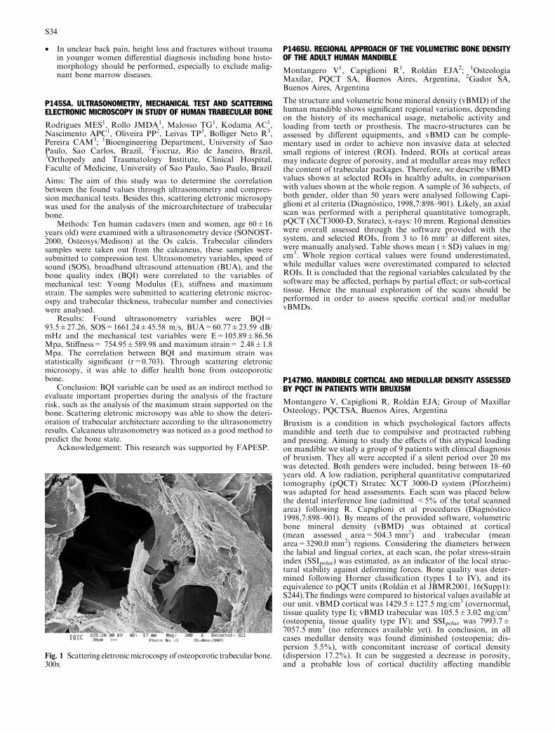
• In unclear back pain, height loss and fractures without traumain younger women differential diagnosis including bone histo-morphology should be performed, especially to exclude malig-nant bone marrow diseases.
P145SA. ULTRASONOMETRY, MECHANICAL TEST AND SCATTERINGELECTRONIC MICROSCOPY IN STUDY OF HUMAN TRABECULAR BONE
Rodrigues MES1, Rollo JMDA1, Malosso TG1, Kodama AC1,Nascimento APC1, Oliveira PP2, Leivas TP3, Bolliger Neto R3,Pereira CAM3; 1Bioengineering Department, University of SaoPaulo, Sao Carlos, Brazil, 2Fiocruz, Rio de Janeiro, Brazil,3Orthopedy and Traumatology Institute, Clinical Hospital,Faculte of Medicine, University of Sao Paulo, Sao Paulo, Brazil
Aims: The aim of this study was to determine the correlationbetween the found values through ultrasonometry and compres-sion mechanical tests. Besides this, scattering eletronic microsopywas used for the analysis of the microarchitecture of trabecularbone.
Methods: Ten human cadavers (men and women, age 60±16years old) were examined with a ultrasonometry device (SONOST-2000, Osteosys/Medison) at the Os calcis. Trabecular cilinderssamples were taken out from the calcaneus, these samples weresubmitted to compression test. Ultrasonometry variables, speed ofsound (SOS), broadband ultrasound attenuation (BUA), and thebone quality index (BQI) were correlated to the variables ofmechanical test: Young Modulus (E), stiffness and maximumstrain. The samples were submitted to scattering eletronic microc-ospy and trabecular thickness, trabecular number and conectivieswere analysed.
Results: Found ultrasonometry variables were BQI=93.5±27.26, SOS=1661.24±45.58 m/s, BUA=60.77±23.59 dB/mHz and the mechanical test variables were E=105.89±86.56Mpa, Stiffness= 754.95±589.98 and maximum strain= 2.48±1.8Mpa. The correlation between BQI and maximum strain wasstatistically significant (r=0.703). Through scattering eletronicmicrosopy, it was able to differ health bone from osteoporoticbone.
Conclusion: BQI variable can be used as an indirect method toevaluate important properties during the analysis of the fracturerisk, such as the analysis of the maximum strain supported on thebone. Scattering eletronic microsopy was able to show the deteri-oration of trabecular architecture according to the ultrasonometryresults. Calcaneus ultrasonometry was noticed as a good method topredict the bone state.
Acknowledgement: This research was supported by FAPESP.
P146SU. REGIONAL APPROACH OF THE VOLUMETRIC BONE DENSITYOF THE ADULT HUMAN MANDIBLE
Montangero V1, Capiglioni R1, Roldan EJA2; 1OsteologiaMaxilar, PQCT SA, Buenos Aires, Argentina, 2Gador SA,Buenos Aires, Argentina
The structure and volumetric bone mineral density (vBMD) of thehuman mandible shows significant regional variations, dependingon the history of its mechanical usage, metabolic activity andloading from teeth or prosthesis. The macro-structures can beassessed by different equipments, and vBMD can be comple-mentary used in order to achieve non invasive data at selectedsmall regions of interest (ROI). Indeed, ROIs at cortical areasmay indicate degree of porosity, and at medullar areas may reflectthe content of trabecular packages. Therefore, we describe vBMDvalues shown at selected ROIs in healthy adults, in comparisonwith values shown at the whole region. A sample of 36 subjects, ofboth gender, older than 50 years were analysed following Capi-glioni et al criteria (Diagnostico, 1998,7:898–901). Likely, an axialscan was performed with a peripheral quantitative tomograph,pQCT (XCT3000-D, Stratec), x-rays: 10 mrem. Regional densitieswere overall assessed through the software provided with thesystem, and selected ROIs, from 3 to 16 mm2 at different sites,were manually analysed. Table shows mean (±SD) values in mg/cm3. Whole region cortical values were found underestimated,while medullar values were overestimated compared to selectedROIs. It is concluded that the regional variables calculated by thesoftware may be affected, perhaps by partial effect; or sub-corticaltissue. Hence the manual exploration of the scans should beperformed in order to assess specific cortical and/or medullarvBMDs.
P147MO. MANDIBLE CORTICAL AND MEDULLAR DENSITY ASSESSEDBY PQCT IN PATIENTS WITH BRUXISM
Montangero V, Capiglioni R, Roldan EJA; Group of MaxillarOsteology, PQCTSA, Buenos Aires, Argentina
Bruxism is a condition in which psychological factors affectsmandible and teeth due to compulsive and protracted rubbingand pressing. Aiming to study the effects of this atypical loadingon mandible we study a group of 9 patients with clinical diagnosisof bruxism. They all were accepted if a silent period over 20 mswas detected. Both genders were included, being between 18–60years old. A low radiation, peripheral quantitative computarizedtomography (pQCT) Stratec XCT 3000-D system (Pforzheim)was adapted for head assessments. Each scan was placed belowthe dental interference line (admitted <5% of the total scannedarea) following R. Capiglioni et al procedures (Diagnostico1998,7:898–901). By means of the provided software, volumetricbone mineral density (vBMD) was obtained at cortical(mean assessed area=504.3 mm2) and trabecular (meanarea=3290.0 mm2) regions. Considering the diameters betweenthe labial and lingual cortex, at each scan, the polar stress-strainindex (SSIpolar) was estimated, as an indicator of the local struc-tural stability against deforming forces. Bone quality was deter-mined following Horner classification (types I to IV), and itsequivalence to pQCT units (Roldan et al JBMR2001, 16(Supp1):S244).The findings were compared to historical values available atour unit. vBMD cortical was 1429.5±127.5 mg/cm3 (overnormal,tissue quality type I); vBMD trabecular was 105.5±3.02 mg/cm3
(osteopenia, tissue quality type IV); and SSIpolar was 7993.7±7057.5 mm3 (no references available yet). In conclusion, in allcases medullar density was found diminished (osteopenia; dis-persion 5.5%), with concomitant increase of cortical density(dispersion 17.2%). It can be suggested a decrease in porosity,and a probable loss of cortical ductility affecting mandible
Fig. 1 Scattering eletronicmicrocospy of osteoporotic trabecular bone.300x
S34
strength in these patients. Peridental tissue could be even moreaffected, but was not assessed in this sample. If these observationsare confirmed the mandible of patients suffering bruxism could bemechanically less elastic. According to our present data the man-dible, is indeed, different from the one of normal dentate adults.
P148SA. INFLUENCE OF AGE ON THE MECHANICAL EVALUTION OFBENDING-COMPRESSION IN INTACT AND OVARIECTOMIZED RATS
Rodrigues MES, Kodama AC, Nascimento APC, Oliveira FB,Malosso TG, Rollo JMDA; Bioengineering Department, Univer-sity of Sao Paulo, Brazil
Aim: Evalute the influence on two different ages in intact andovariectomized rats using bending-compression on the proximalfemur.
Methods: This study evaluated the age influence of overload in37 female Wistar rats. The overload was compared between intactrats at 5 and 7 months of age, after that they were compared to 7months ovariectomized rats. A surgery was made in order to induceosteopenia and the animals were sacrificed after two months. Theoverload was got through the bending-compression test on theproximal femur and was used as a evaluation criteria.
Results: The analysis of the maximum overload in intact ani-mals between 5 months and 7 months of age showed an increasingbone resistance, as well as weight increasing, but with a non-sig-nificant difference. Ovariectomized animals at 5 and 7 months ofage showed a significant decreasing of maximum overload inrelation to the intact rats at the same age. A nonsignificant differ-ence between 5 and 7 months ovariectomized rats was found.
Conclusion: There was a decreasing on bone resistance, whichwas evaluated through maximum overload, between 5 and 7months ovariectomized animals and their control group (5 and 7months intact animals). This study showed that age does not have asignificant influence on 5 and 7 months of age intact animals, aswell as on 5 and 7 months of age ovariectomized rats.Acknowledgement: This research was supported by FAPESP.
P149SU. THE DIAGNOSIS OF OSTEOPOROSIS BY STANDARD SPINEDUAL-ENERGY X-RAY ABSORPTIOMETRY AND WHOLE BODY SCANS
Christov V1, Boyanov M1, Boneva Zh2, Harangoso E1; 1Endo-crinology Clinic, Alexandrovska Hospital, Medical UniversitySofia, Sofia, Bulgaria, 2Endocrinology Unit, Central PoliceHospital, Sofia, Bulgaria
In whole body dual-energy x-ray absorptiometry (DXA) bonemineral density (BMD) values are generated for different bodyparts and whole body T- and Z-scores are then calculated.
Aim: The present study compared T- and Z-scores (in SDs)yielded in standard DXA of the antero-posterior spine and in DXAwhole body scans.
Methods: 100 women aged 52.6 (SD 12.1) years were included.Their mean body mass index was 30.5 (SD 6.6) kg/m2. 55 of themwere postmenopausal with mean age at menopause 46.3 years. Allparticipants received consecutively standard AP spine DXA andwhole body scans on a Hologic QDR 4500, a bone densitometer.The manufacturer’s reference database for white women was usedas well as the WHO definition for osteoporosis. Correlation anal-ysis was performed.
Results: AP DXA yielded similar proportions of osteopenic andosteoporotic women as standard AP spine DXA. But only in 50%of the cases a woman was classified in the same BMD group byboth methods. The T-score correlation was weaker (r=0.75) thanthe Z-score correlation (r=0.85).
Conslusions: Whole body BMD T-scores possibly reflect min-eralization of the whole skeleton including both cortical and tra-becular bone. Heterogeneity of bone loss is inherent in the processof ageing. This may be the reason why the trabecular-rich lumbarspine does not always yield the same T-score in a particular personas the whole body scan. We conclude that whole body BMD T-scores may be more useful in studies of the growing skeleton inchildren and adolescents as they reflect the integral process of bonemineralization.
P150MO. EVALUATION OF THE BRAVO: PRECISION AND ACCURACY OFA NEW COMPACT BONE DENSITOMETER
Simonelli C, Schoeller M; HealthEast Clinics, Woodbury, MN,USA
The Bravo (GE Medical Systems) is a new DXA bone densi-tometer designed to fit into smaller offices that cannot accom-modate a full-sized DXA system. The Bravo scan arm swings 90degrees to the side for easy access to the scan table. Bone mineraldensity (BMD) of the spine and bilateral femurs are measured inone exam, using the OneScan acquisition protocol that eliminatesthe need to reposition the patient between scans. We comparedthe precision and accuracy of the Bravo measurements with a fullsize densitometer, the Lunar Prodigy (GE Medical Systems).Thirty women aged 45 to 60 years (mean age 52.6; SD 6.4 years)had spine/bilateral femur scans twice with the Bravo and oncewith the Prodigy. Patients were repositioned between the Bravoscans. One subject was removed from the study because of con-siderable positioning differences between scans. BMD precisionerror was calculated as the root-mean-square coefficient of vari-ation for repeated measurements. Paired T-tests were used todetermine significance of BMD differences between Bravo andProdigy measurement results. Left and right femur results wereaveraged for bilateral total femur (n=29), and were pooled(n=58) for femur neck, trochanter and total femur. Bravo BMDprecision error (CV) ranged from 0.7% for spine L1-L4 and 1.0%for total femur to 1.9% for femur neck. Average differences inmeasured BMD were negligible for femur sites and were clinicallyinsignificant at the spine site (< 2%). Regression of Bravo BMDwith Prodigy BMD showed very high correlations (r=0.99) withno significant differences in the regression slopes or intercepts. Weconclude that the Bravo provided accurate BMD for spine andfemur measurement sites, with precision that compared favorablywith published values for full size DXA systems.
P151SA. IN THE SEARCH OF OPTIMAL DIAGNOSTIC AND MONITORINGPROCEDURES IN CHILDREN USING DXA METHOD: THE STUDY OFIDIOPATHIC JUVENILE OSTEOPOROSIS SUBJECTS
Pludowski P, Lebiedowski M, Matusik H, Lorenc RS, Depart-ment of Biochemistry and Experimental Medicine, The Children’sMemorial Health Institute, Warsaw, Poland
Aim: Various diagnostic approaches to interpret and monitorDXA results in IJO cases were compared and utilized in the aimof developing optimal evaluation scheme of studied cases.
Methods: Study population comprised 61 IJO and 481 healthychildren. IJO cases were analyzed according to type of IJO (mild,moderate, severe) and stage (acute, chronic). DXA derived LBM,as a surrogate of muscle mass, total and L2-L4 BMC (TBBMC,SBMC) and densities were assessed. Lumbar spine volumetricdensity (BMAD, g/cm3) was calculated according to Carter’salgorithm. Utilizing mechanostat thesis, body height (BH)/LBM,TBBMC/LBM, SBMC/LBM ratios were employed as diagnosticindicators of musculoskeletal system. Regression curves fitted toage-dependent data were assessed in healthy or moderate IJO casesand the results of each mild or severe IJO subject were converted tothe % of expected value what allowed to monitor skeleton inrelation to healthy or typical IJO cases. Additionally, data wereadjusted for age, height and weight as the second possible approachfor monitoring of skeleton to avoid influence of growth.
Results: The highest deficits of analyzed parameters (except BH/LBM) were observed during acute phase of severe type of IJO. Per-centages of expected values for TBBMC/LBM and SBMC/LBM
Comparison of Bravo with Prodigy densitometer
FemurNeck
Trochanter TotalFemur
Bilateral TotalFemur
SpineL1-L4
Bravo (%CV) 1.9% 1.6% 1.0% 0.9% 0.7%Bravo BMD (g/cm2) 0.96 0.80 0.99 0.99 1.21Prodigy BMD (g/cm2) 0.96 0.81 1.00 1.00 1.23BMD Difference (p) ns ns <0.01 <0.05 <0.001
S35
ratios, in severe type of IJO, but not mild, were low according toexpected values for healthy andmoderate IJO. Significant differenceswere found between adjusted TBBMD, SBMD, TBBMC,SBMC,BMAD, TBBMC/LBM and SBMC/LBM, but not BH/LBM, calculated in moderate vs. severe (p<0.001), moderate vs.mild (p<0.05) and mild vs. severe (p<0.001) types of IJO andbetween acute and chronic phases (p<0.001), in both genders.Lack of major differences between healthy and mild type of IJO wasnoted.
Conclusion: Percentages of expected values allowed diagnosisand monitoring of single individual IJO case not only in relation tohealthy but also to typical IJO (moderate type) what seems to beessential for proper therapeutical management. Anthropometricalcorrection of DXA results allowed to diagnose and monitorchanges in bone mineralization regardless of changing body size.Type-phase-dependent impact of IJO on skeleton, but not onmuscles, was noted and primary bone disorder diagnosed utilizingBH/LBM, TBBMC/LBM and/or SBMC/LBM ratios.
P152SU. OSTEOPOROSIS SCREENING: COMPARISON OF THE HEELULTRASOUND MEASUREMENT TO CALCULATED RISK ASSESSMENTTOOLS
Gambacciani M, Genazzani AR; Department of Obstetrics andGynecology, University of Pisa, Pisa, Italy
Quantitative ultrasound of the os calcis is accepted as an effective,low-cost method to identify women likely to have osteoporosis atthe hip or spine as measured by DXA. In this study, we wished todetermine the specific T-score cutpoint on the Lunar AchillesInSight (GE Medical Systems) that detects 90% of individualswith osteoporosis (T-score £)2.5) at either the spine or hip. TheAchilles InSight is an imaging heel ultrasonometer with 8–10second measurement time. We also compared the performance ofultrasound screening to a simple risk assessment tool based onweight and age.
A total of 272 women aged 40 to 83 years (mean age 58±7years) had DXA measurements of the spine and hip using a LunarDPX (GE Medical Systems) and heel ultrasound measurementusing the Achilles InSight. Osteoporosis was diagnosed if the lowestDXA T-score at spine (L1–L4) or hip (total femur) was £2.5. Theheel T-score cutpoint with 90% sensitivity was determined usingROC analysis. For each subject the value of the osteoporosis riskassessment tool, risk tool ((Weight in kg – age)*0.2) was also cal-culated.
From the DXA results, 54 of 272 patients were classified asosteoporotic. At a heel T-score of )1.0, sensitivity was 91% andspecificity was 46%. Based on ROC analysis, the area under thecurve (AUC) for the heel T-score was 0.79, which was significantlygreater than the AUC for the risk tool, 0.69 (p<0.05).
We conclude that the Achilles bone ultrasonometer can be usedas a valid screening tool for osteoporosis, with 90% sensitivity at aT-score of )1.0 and good specificity. Moreover, the Achilles per-forms significantly better in identifying those patients who shouldbe considered for spine and hip bone density assessment comparedto the calculated osteoporosis risk assessment tool.
P153MO. CHANGES IN BONE MINERAL DENSITY IN SUBJECTSWITHOUT OSTEOPOROSIS TREATMENT
Anijar JR, Azevedo E, Lazarski CFS, Chahade WH, Soares AH;Hospital Servidor Publico Estadual, Sao Paulo, Brazil
Aim: Evaluate the bone mineral density (BMD) behavior in sub-jects without osteoporosis treatment, submitted to a newbone scan.
Materials and Methods: A total of 392 women aged above 44underwent a second lumbar spine and hip bone mineral densitymeasurement between April and November of 2001 by dual energyx-ray absorptiometry (DXA), DPX-L device. None of the subjectsreceived bone protective treatment.
Results:Among the 392patients, 326 (83.2%)wereCaucasian, 52(13.3%) Mulatto and 14 (3.6%) Asian descent. The mean age was60.1±9.4 yrs (45–87). Surprisingly, 220 (56.1%) subjects presentedosteopenia or osteoporosis at the first bone scan. The BMD wasmeasured at an interval of 33.3±15.8 months. Regarding longitu-dinal changes in BMD, it was)1.6±4.5% inL2–L4 and)1.4±4.1%in femoral neck. Significant correlations were observed betweenBMD and age or measurement interval (p<0.001). No correlationswere observed between BMD and race or body mass index.
Conclusion: These results would suggest that the losses of BMDare increased in younger subjects and with longer interval betweenmeasurements. Unfortunately, most of patients were not in osteo-porosis treatment, besides previous diagnosis of low bone mass.
P154SA. LUMBAR AND FEMORAL BONE MINERAL DENSITY IN PA-TIENTS WITH ANKYLOSING SPONDYLITIS
Yildirim K, Karatay S, Melikoglu Alkan M, Erdal A, Ugur M,Senel K; Ataturk University,Medicine Faculty, Physical Medicineand Rehabilitation, Erzurum, Turkey
This study was carried out to determine the bone mineral density(BMD) values of the lumbar spine and femoral neck in ankylosingspondylitis (AS) patients. Eighteen outpatients who fulfilled themodified New York criteria for AS and also 18 healthy controlswere consecutively included in the study. BMD of lumbar spineand femoral neck was evaluated by dual energy x-ray absorpti-ometry (DEXA). Laboratory parameters included erythrocytesedimentation rate (ESR) and C-reactive protein (CRP). Thedemographic variables such as age,-sex-and BMI were similarbetween patients and controls (p>0.05). The biochemicalparameters ESR and CRP were found to be different between thepatient and control groups (p<0.001 for both).
BMD values of lumbar and femoral regions in AS patients were0.98±0.2 g/cm2 and 0.87±0.1 g/cm2. BMD values of lumbar andfemoral area in control subjects were 1.02±0.13 g/cm2 and0.97±0.12 g/cm2. Patients with AS had reduced BMD in theirlumbar spine and femoral neck regions (p<0.05, p<0.01 respec-tively). Femoral measurements exhibited greater severity of re-duced BMD than lumbar values when average BMD scores werecompared. Consequently, related to the structural possible changesseen in the lumbar area, the lumbar region BMDmeasurements canbe misleading when evaluating the extent of bone mass loss in ASpatients. Therefore, alternative sites or the femoral region shouldbe used to evaluate bone mass in AS patients.
P155SA. THE RELATIONSHIP BETWEEN BODY COMPOSITION ANDREGIONAL BONE MINERAL DENSITY IN PREMENOPAUSAL WOMENWITH RHEUMATOID ARTHRITIS
Karatay S1, Yildirim M2, Melikoglu A1, Seven B2, Yildirim K1,Senel K1, 1Dept. Physical Medicine and Rehabilitation, School ofMedicine, Ataturk University, Erzurum, Turkey, 2Dept. NuclearMedicine, School of Medicine, Ataturk University, Erzurum,Turkey
Objective: This study was performed to investigate whether thebody composition parameters and disease characteristics had anyeffect on bone mineral density (BMD) of regional, total body,lumbar spine and femoral neck in premenopausal women withrheumatoid arthritis (RA).
Material and Methods: 23 premenopausal women with RA whowere diagnosed according to the criteria of the American College ofRheumatology and with disease duration of more than 1 year and31 age and sex-matched healthy controls with right side dominancewere recruited. Disease duration, morning stiffness duration,patient’s pain evaluation (VAS), assessments of joints (Ritchiearticular index) and functional disability (HAQ), erythrocyte
AUC values, sensitivity and specificity for osteoporosis screening tools
AUC 90% SensitivityCutpoint
Sensitivity Specificity
Heel T-score 0.79 (0.72–0.85) )1.0 90.7% 46.3%Risk Tool 0.69 (0.62–0.77) 2.4 90.7% 28.9%
For the combination of the Heel T-score and Risk Tool the AUC is 0.81 (0.75–0.87).
S36
sedimentation rate (ESR) and C reactive protein (CRP) wererecorded in RA patients. BMD (arms, legs, L1–L4 lumbar spine,femoral neck and total body) and body composition (lean mass ofregional-total body, fat mass of regional-total body and percentageof body fat) were measured by whole-body scanning with dualx-ray absorptiometry (DXA).
Results: BMD values of all body sites were significantly lower inpremenopausal women with RA compared with the healthy con-trols, while body composition determinants were no different be-tween the two groups. However, BMD of arms was correlated withany of regional lean or fat mass, while BMD of legs, femoral neckand total body were affected by lean mass of legs and total body asindependent from body weight in RA patients. Only diseaseduration and HAQ scores were correlated with BMD among thedisease characteristics. HAQ scores were inversely correlated withBMD of all body sites, except for lumbar spine and total body.Disease duration was negatively correlated with BMD of lowerlimbs (legs and femoral neck) and lean mass of legs.
Conclusion: Lean mass of legs and total body may be significantdeterminants of BMD on the regions of femoral neck, total bodyand legs. Also, the measuring of regional BMD by DXA may beuseful to determine functional disability and risk of morbidity andmortality in premenopausal women with RA.
P156MO. THE EFFECT OF A SHORT DURATION OF LOW INTENSITYEXERCISE ON QUANTITATIVE ULTRASOUND MEASUREMENTS INHEALTHY YOUNG ADULTS
Oral A1, Tarakci D2, 1Istanbul University, Istanbul MedicalFaculty, 2School of Physical Therapy and Rehabilitation,Istanbul, Turkey
The aim of this study was to examine the influence of a shortduration of low intensity exercise on bone tissue as measured bycalcaneal quantitative ultrasound(QUS).
40 nonathletic young adults (20 men and 20 women, aged 19–30years) were allocated either to a group who received exercise train-ining (walking on a treadmill for 30 minutes three times a week forthree months) or to a groupwho did not. Calcaneal QUS parameters(BUA-in dB/MHz, SOS-in m/s, QUI, and estimated heel BMD ing/cm2) were measured in all subjects using a gel coupled device at thebeginning and after three months. In statistical analyses, before andafter values were compared using ‘‘paired t-test’’. ‘‘Chi-squareanalysis’’ was used to compare the number of subjects who showedimprovement greater than that would be expected from the root-mean-square coefficient of variation (RMSCV) in the exercise andcontrol groups. The short and long-term precision of the measure-ments were also examined with duplicate measurements.
No significant difference was observed between before and aftermean values of QUS indices in either the exercise or the controlgroup.The number of subjects who showed improvement greaterthan RMSCV % for QUS indices was similar in both groups. Theshort-term precision of this device was satisfactory, but the long-term precision was poor, enabling us to detect subtle differenceswhich occured during time.
In conclusion, we were not able to show favorable effects of ashort duration of low intensity exercise on bone properties asmeasured by calcaneal QUS. This issue remains to be elucidated bystudies including larger size of subjects and/or different durationand/or different intensity of exercise.
P157SA. BONE MINERAL DENSITY AND OSTEOPROTEGERIN (OPG) INPATIENTS AFFECTED BY THALASSEMIA MAJOR
D’AmoreM1,PansiniN1,2,FontanaA1,2,FanelliM1,3,D’AmoreS1,Scagliusi P1,MinennaG1, 1Dimimp SezioneDIReumatologia BariItalia, 2Unita’ Operativa Patologia Clinica I A. O. Bari Italia,3DimimpSezioneDiagnostica Per Immagini StatisticaMedica BariItalia
Bone disease is an important cause of morbidity in patients af-fected by Thalassemia Major; it is a multifactor disease: massivebone marrow expansion, delayed puberty, hypogonadotropichypogonadism, other hormonal disorder (hypothyroidism,
hypoparathyroidism, diabetes mellitus), iron overload and dele-terious effects of chelant therapy on osteoblasts. OPG is a solubledecoy-receptor, and is produced in different tissues, e.g. bone,skin, liver, stomach, intestine and lung. As a so-called ‘‘decoyreceptor’’ OPG inhibits the binding of RANK to RANKL (OPG-L, osteoclast differentiation factor, ODF) and thus inhibits therecruitment, proliferation and activation of osteoclasts.
The main aims of this study are to evaluate the bone mineraldensity in 31 patients affected by Thalassemia Major (12 males and19 females); age average 24.5 (range 15–38) and the OPG serumconcentrations.
Methods andmaterials: Bonemineral density has beenmeasuredby BMD of lumbar spine (L2–L4), femoral neck and distal radiususing the ACN Unigamma x-ray Plus. OPG serum concentrationshave beendeterminedusing the ‘‘sandwichELISA’’method. Inorderto examine the level of OPG in serum, we considered 82 healthysubjects, equally divided between male and female, with an averageage of 15–38. Correlation between variables has been calculatedaccording to Pearson’s method. The differences between the twoconsidered groups have been analysed using Student’s test.
Results and conclusions: Analysing the results, we have foundthis evidence. The two groups could be easily compared as for theage parameter (t=1.11, p=0.27). Bone mineral density loss ap-pears first of all at the distal radius level in all patients tested. Infact distal radius T-score mean is –2.23, and it is significant inferiorto the normality limit that is –1 (t= )7.53, p<0.01). OPG serumconcentrations mean in healthy subjects is 2.45 pmol/l with stan-dard deviation of 0.63, while in patients it is 3.95 with standarddeviation of 1.51. The difference between the two groups is sig-nificant (t= )5.39, p<0.001). We have found a kind of correlationstatistically significant between distal radius T-score and OPGserum concentrations.
P158SU. COMPARATIVE EVALUATION OF BONE MINERAL DENSITYMEASURED IN DIFFERENT SITES AND INCIDENCE OF VERTEBRALFRACTURES
Ilic-Stojanovic O1, Letic M1, Popovic G1, Kostic S1, Kanjuh Z2,1Institute for Rehabilitation, Medical School, University ofBelgrade, Serbia and Montenegro, 2Clinic for RehabilitationM. Zotovic, Medical School, University of Belgrade, Serbia andMontenegro
The aim of this study was to evaluate correlation between bonemineral densities (BMD) measured by dual energy x-ray absorpti-ometry of bilaterally forearm (DTX-200 Osteometer-Denmark),femoral neck and lumbar vertebral body (L2–L4) using LunarDPX-Ldevice.Wealso aimed to examine the frequency of vertebralbone fracture using thoracic and lumbar lateral x-rays.
We were studing 130 patients, 125 female (aged 45–83,61.9±9.35) and 5 male. All of 125 investigated women were inmenopause (ranged from 33–58 years, 47.9±5.04). In investigatedgroup we devided the patients according to the osteoporosis(46.9%), osteopenia (29.23%) and referent values (23.87%). Hav-ing used Spearman’s test we confirmed highly positive correlationbetween dominant and nondominant forearm (n=128, p<0.0001),forearm and lumbar spine (n=113, p<0.0001) and spine vs. fem-oral neck (n=30, p=0.01), but we didn’t find correlation betweenneck and forearm (n=45, p>0.05). In 39 (30%) cases we registeredthe fractures of thoracic or lumbar spine. The frequency of frac-tures in the examined sample is biggest in the group with BMD onthe level of osteopenia. We found positive correlation between ageand fracture (p<0.01).
In the cases with osteoporosis at the forearm we found theosteoporosis at the lumbar spine and osteopenia in the femoralneck, in cases with osteopenia in forearm we found also osteopeniaon femoral neck and on spine, but in cases with osteopenia in theneck we registered the cases of osteoprosis on both forearm andspine. More complicated situation we found in cases within referentvalues (measured on any of the three different measuring points)-because we noticed same possibility of appearance of the cases withlowered BMD on any of the two other measuring spots. (Generallinear modeling – GLM, Statistica for Windows 6.0)
S37
So it can be concluded that if we get referent values for BMDon one measuring spot, the measuring should be done on, at least,one more spot. And according to the examined sample it seems thatmeasuring of BMD on the level of the forearm and lumbar spine isbetter prediction of the loss of BMD than BMD measured on thefemoral neck.
P159MO. BONE MINERAL DENSITY AND SERUM OSTEOCALCIN AREREDUCED IN YOUNG ADULTS WITH IDIOPATHIC SHORT STATURE
Hellingman AA, Slingerland AS, Hokken-Koelega ACS; Depart-ment of Pediatrics, Division of Endocrinology, Sophia Children’sHospital/Erasmus MC, Rotterdam, The Netherlands
Aim: To examine differences in risks of osteoporosis, we mea-sured the bone mineral density (BMD) and the serum osteocalcinin four groups of young adults with different growth patterns aspart of the PROGRAM study. The PROGRAM study(PROgramming factors for GRowth And Metabolism) is uniquein determining risk profiles for diabetes, cardiovascular diseaseand osteoporosis in four groups, distinguishing weight and heigthat birth, and weight and height in young adulthood.
Methods: Our four study groups: 1.Small for Gestational Age(SGA;birth length and/or birth weight <–2SDS) with no catchup(current height <–2SDS) 2. SGA with catch up (current height>–2 SDS) 3. Idiopathic Short Stature (ISS;birth length and/orbirth weight >-2SDS but current height <–2 SDS) 4. controlgroup(birth length and/or birth weight as well as current height>–2sds). Up to now, we completed group 3 and 4. 27 young adultswith ISS (13 girls, 14 boys) were included. Our control groupconsisted of 31 young adults (17 girls, 14 boys). All were healthyCaucasian 18-23 years’old, and born after 37 weeks’of gestation.BMDs(g/cm3)of the lumbar spine (LS) and the total body (TB)were measured using dual energy x-ray absorptiometry. Osteocal-cin was measured using radioimmuno-assay.
Results: We found the mean BMD to be significantly lower ingirls of group 3 than of girls in group 4 for both the LS and TB(p=0.005, p=0.004). Boys of group 3 had significantly lowerTBMD (p=0.015) and serum osteocalcin (p=0.026) than boys ofgroup 4. After adjustment for birth length, birth weight, age, BMI,cigarette smoking, alcohol comsumption and sports participation,these differences remained significant.
Conclusions: Our results indicate that girls and boys with ISShave a significant reduction in TB BMD, that girls with ISS have asignificant reduction in LS BMD, and that serum osteocalcin issignificantly lower in boys with ISS. We suggest that ISS childrenbe carefully monitored in order to start preventive care for osteo-porosis at an early age.
P160SA. OSTEOPOROSIS IN VENEZUELAN MEN: QUANTITATIVEULTRASOUND STUDY
Franco LM, Ferazzoli E, Yelamo C, Barela R, Suarez I, AlcarraL; Ciudad Hospitalaria Dr. Enrique Tejera, Valencia, Carabobo,Venezuela
Objective: Evaluate the osteoporosis frequency in the Venezuelanmale population by quantitative calcaneus ultrasound.
Methods: Of a total of 12,965 patients, the bone mass of 213Venezuelan males in ages between 35 and 83 years was evaluated.The collected data was analyzed by chi square, average, DS andstandard error.
Results: Of the 213 evaluated males, 79 had osteopenia (37.1%)and 16 had osteoporosis (7.5%) representing a 44.6% of positivepatients in screening. The higher frequency of positive results wasobtained in males 55 years old or older.
Conclusions: The peripheral ultrasound is a reliable, fast, lowcost and a affordable method for big groups of population. There isnot a comparative data of males evaluated with DXA equipmentagainst ultrasound. 44.6% of our patients had positive results,which represents a high frequency and reveals the importance ofthe evaluation of the male population. A follow up of this group ofpatients and confirmation by other methods is required.
P161SU. DIAGNOSTIC ABILITY OF THE PERIPHERAL MEASUREMENTSOF BONE DENSITY: ANALYSIS BY ROC CURVES
Lopez Gavilanez E1, Segale Bajana A2, Vera Vargas F3, SolanoGalarza M2, Villacıs Ponce Ch2; 1Servicio de Endocrinologıa delHospital Docente de la Policıa Nacional No-2, Guayaquil,2Servicio de Medicina Interna del Hospital Docente de la PolicıaNacional No-2, Guayaquil, 3Dispensario Norte del InstitutoEcuatoriano de Seguridad Social (IESS), Guayaquil, Ecuador
Aims: Identify postmenopausal women with osteoporosis in theaxial and peripheral skeleton, look for a correlation between thedifferent measured regions and evaluate the diagnostic ability ofthe peripheral measurements using ROC curves.
Methods: We measured bone density (BD) in 105 postmeno-pausal women in forearm(FA) with an Osteometer DTX-200equipment, and simultaneously in lumbar spine (LS) and femoralneck (FN) with a DEXA equipment. We analyzed FA by ROCcurves, established the cut-offs and calculated Sensibility, Speci-ficity, Predictive Positive Value(PPV), and Predictive NegativeValue(PNV). We considered the measurement in FN as goldstandard to diagnosis osteoporosis by WHOs criteria.
Results: Average age 58.6±10.2 years, menopause period 13±7years, correlation between FA and FN r=0.46 (p<0.05), correla-tion between FA and LS r=0.65 (p<005). Area under curve(AUC) for FA is 0.83 (SE=0.014) (CI=0.80-0.86). For a cutoff)2.5 in FA: Sensibility 59% (CI 39-76), Specificity 88%.(CI 79–94),PPV 64%.(CI 43–81), and PNV 86%.(CI 76–92).For a cutoff –1 inFA: Sensibility is 76% (CI 65–85), Specificity 78% (CI 59–90), PPV88% (CI 78–95), and PNV 58% (CI 43–74).
Conclusions: BD measurement in forearm is useful for initialscreening of osteoporosis and for identifying patients who requireadditional investigation with axial DEXA. There is a moderatecorrelation between FA, LS and FN. Could be no proper to con-sider DEXA cutoffs for peripheral measurements in all patientswith possible osteoporosis.
P162MO. MEASUREMENTS OF BONE CALCIUM THROUGH THE TOTALBODY DENSITOMETRY
Sampaio Netto O, Hasselmann CL, Coutinho LOL, Souza DC,Misael NCS, Chaves EP, Faria NLA; Universidade Catolica deBrasılia, Brasılia - DF, Brazil
Objectives: To measure bone calcium through the total bodydensitometry with normal levels of bone mineral density. Tocompare the bone calcium results in different levels of bonemineral density (osteopenia and osteoporosis levels).
Methods: We evaluated the total body densitometry at theCatholic University of Brasilia with DEXA Lunar� DPX-IQ dur-ing the period from January 2002 to October 2003. The inclusioncriteria were: age above 20 years, with no visual evidence of bonebreaking, bone deformity or alteration of the bone distribution inthe carried bone densitometry. Analysis statistics were made withMSExcel XP�.
Results: We analyzed 300 total body densitometry in men andwomen from 20–82 years of age (average of 47.4±20.7 years).Based on the World Health Organization criteria (WHO) for thebone density classification (normal, osteopenia and osteoporosis),we used as a base the T-Score. We divided the examinations intothree groups: a) Normal, with 212 examinations; b) Osteopenia,with 68 examinations; c) Osteoporosis, with 20 examinations. Ineach group, we verified the average value of bone calcium, findingthe following results: normal, 1084.9±214.4 g of calcium (varying
Table 1 Results of Forearm
Cut-offs Sensibility(CI)
Specificity(CI)
PPV (CI) PNV (CI)
<)1 76% (65–85) 78% (59–90) 88% (78–95) 58% (43–74)<)2.5 59% (39–76) 88% (79–94) 64% (43–81) 86% (76–92)
S38
of 678–1588 g); osteopenia, 720.8±97.0 g of calcium (varying of528–1012 g); osteoporosis, 594.7±54.4 g of calcium (varying of529–706 g). Comparing the three groups, we obtained a significantdifference between them with p<0.001. In normal group, we ver-ified the value of bone calcium in men and women, with therespective average: 1247.1 and 906.6.
Conclusions: The value of the bone calcium measurements inthe results of the total body densitometry is a value not used in theresults of the densitometry. We verified the existence of differentand significant values when we compared examinations with dif-ferent levels of bone density. This suggests that this value must bemore understood as a form to be used in the evaluations of thebone mineral density.
P163SA. COMPARISON OF BONE MINERAL DENSITY IN CHILRDENFROM MANY POPULATIONS: A LITERATURE REVIEW
Jaworski M, Bielecka L, Matusik H, Lorenc RS; The ChildrensMemorial Health Institute, Warsaw, Poland
Aim: To compare bone mineral density data assessed in healthychildren from different populations in the aim to evaluatewhether or not the substantial differences between children’sbone mineralization datasets provided worldwide are present.
Methods: A literature search was performed for papers pub-lished during last ten years. 184 papers providing BMD data as-sessed in healthy children were found. To be eligible for the furtheranalysis papers must to fit to chosen criteria such as: DXA pencilbeam device used for total body and lumbar spine measurements,BMD data presented as g/cm2, Caucasian children aged 4–20 yrs.Finally, 57 papers matched our criteria and BMD datasets wereanalyzed according to age and gender as well as the origin of DXAdevices (Hologic and Lunar). 142 groups of datasets were analyzedusing Statistica 5.0 software.
Results: Significant differences in age-, gender and device-matched BMD data were found in 45 of 145 analyzed groups(32%). Only 1 population specific BMD data assessed in healthychildren markedly differed from the other datasets in wholeanalyzed age range. The apparent differences found in 32% ofanalyzed datasets suggested that the tempo of bone mineralizationas well as amount of achieved BMD might be population specific.However, those differences might be also the consequence ofdifferent study design (cohort, prospective) as well as various sci-entific purposes. Moreover, the number of children included intoage and sex groups was markedly different, ranging from 1 to 455.
Conclusion: Our results indicated that substantial differences inBMD data are existent at least in some age groups of healthychildren. However, those differences might be also the effect ofdifferent study purposes and study designs. Therefore, there issubstantial need to establish population specific reference BMDdata basing on well prepared studies and than unify BMD referencefor whole children.
P164SU. ANALYSIS OF ANTHROPOMETRICAL AND MECHANICALPROPERTIES OF CORTICAL BONE IN FEMUR OF OSTEOPENIC FEMALERATS
Rodrigues MES, Rollo JMDA, Kodama AC, Malosso TG,Nascimento APC; Bioengineering Department, University of SaoPaulo, Sao Carlos, Brazil
Aim: Osteoporosis is the progressive fragility of bones, which aremore and more induced to suffer fractures. Although it canhappen to men and women, it is more common in the secondgroup due to the decreasing of the female hormone, estrogen.Osteoporosis is a ‘‘slow-motion’’ disease, without symptom and itis usually noticed only after a fracture. Once osteoporosis is in thebone, it is necessary to avoid bone lack in order to preventfractures. The treatment is based on inibitory drugs for boneresorption, which cannot supply what has already been de-stroyed, it can only help to stop the process. The aim of this studyis to evalute the effects of ovariectomy on cortical bones of 29female rats in different periods of osteopenia.
Methods: Osteopenia caused by ovariectomy was analysed inrats at 3 months of age during six different periods: 5 animals weresacrified 30 days after surgery; and the other animals, every 15 daysuntil 105th day after surgery. The symptoms of osteopenia wereevaluated through bending test of the femurs.
Results: The results found were compared using statisticalmethods: t-Student for body weights and ANOVA and Student-Newmam-Keuls for femur length and weight, cross-sectional area,maximum overload, stiffness and tensile.
Conclusion: Ovarectomy is a factor that does not cause signif-icant alterations in cortical bone of femur in osteopenic rats.Acknowledgement: This research was supported by FAPESP.
P165MO. PERFORMANCE OF COMPUTER ASSISTED DENSITOMETRY(CAD) IN SPINE AND FEMUR ANALYSIS: COMPARISON WITH VISUALASSESSMENT BY EXPERIENCED DENSITOMETRISTS
Schwartz EN, Steinberg DM; Foundation for OsteoporosisResearch and Education, Oakland, CA, USA
Although DXA systems provide semi-automated analysis of spineand femur bone mineral density (BMD), visual assessment by theuser is necessary to a) assure proper acquisition and positioning,b) assure accurate identification of regions of interest (ROI), andc) identify abnormalities that could artificially elevate BMD.Recently, automated software known as Computer AssistedDensitometry (CAD) (GE Medical Systems Lunar) was intro-duced to identify scans with common acquisition and analysisirregularities. We examined whether CAD agreed with theassessment of experienced DXA users, and whether CAD iden-tified abnormalities missed by visual assessment. A total of 71spine and 70 femur scans were analyzed, with 67% exhibitingabnormal conditions flagged by CAD. An ISCD-certified team(densitometrist and technologist) evaluated the scans for abnor-malities, unaware of the CAD results. The team’s blindedassessment was compared to CAD. Scans where CAD and theteam disagreed were reviewed to determine if an abnormality wasmissed by the team. Results showed strong agreement betweenCAD and visual assessment. Experienced users agreed with CADassessment in 76% to 86% of spine scans and 63% to 97% offemur scans, prior to knowing the CAD result. Review of scanswhere disagreement was present, after revealing the CAD rec-ommendations, resulted in an assessment change by the team in20% to 40% of spine cases, and in 0% to 79% of femur cases.After CAD assessment was known, spine scan agreement in-creased to 83% to 92%, and femur scan agreement increased to66% to 97%. One potential source of disagreement is a stricterexpectation of the ideal scan by experienced users. We conclude
Agreement (%) between cad and expert evaluation of spine andfemur scans
CAD vs. Experts Blinded toCAD Results
AfterCADResultsKnown
SPINESpine Centered (%) 86 92Spine Straight (%) 79 83ROIs Accurate (%) 82 86No Unusual High Density (%) 80 87No Unusual T-Score Variation (%) 76 86No Spine Curvature (%) 83 90
FEMURShaft/Pelvis Separation (%) 71 94Shaft Straight (%) 63 66ROIs Accurate (%) 80 89Tissue Regions Accurate (%) 97 97Edges, Point Typing Accurate (%) 86 97No Unusual High Density (%) 83 83
S39
that use of CAD can 1) provide valuable information for inex-perienced users regarding DXA scan quality and 2) assist expe-rienced densitometrists in identifying potentially abnormal scanswhich might be missed by visual assessment alone.
P166SA. BONE MINERAL DENSITY IN LONG TERM HEMODIALYSISPATIENTS
Nakai T1, Masuhara K2, Kanbara N3; 1Osaka Police Hospital,Osaka, Japan, 2Osaka Kosei-nenkin Hospital, Osaka, Japan,3Kanbara Hospital, Osaka, Japan
Aims: The purpose of this study was to report the bone mineraldensity and the hip joint radiographic findings in 18 patients whohave been undergoing hemodialysis for 20 or more years.
Methods: Ten male and eight female patients on maintenancehemodialysis treatment for more than 20 years were enrolled in thisstudy from the outpatient clinic of our institution. The mean agewas 56 years, and the mean duration of hemodialysis 286 months atthe investigation. Bone mineral density was measured at the 1/3distal radius by dual-energy X-ray absorptiometry. The radio-graphs of the hip joint were evaluated for the presence of bone cystsand joint space narrowing. Serum levels of total calcium, phos-phorus, alkaline phosphatase, intact-parathyroid hormone, andbeta-2-microglobulin were measured.
Results: The mean bone mineral density was 0.462 g/cm2. Bonecysts were found in 12 patients (67%) and joint space narrowingwas found in 4 patients (22%). The radiographic abnormalitieswere frequently bilateral in bone cysts (82%) and joint space nar-rowing (92%). The mean values of calcium, phosphorus, alkalinephosphatase, intact-parathyroid hormone, and beta-2-microglob-ulin were 4.4 mEq/L, 6.7 mg/dl, 294.1 IU/L, 12.9 pg/ml, and 39.2lg/ml, respectively. Beta-2-microglobulin was significantly high,with other values being within normal limits. Hip arthroplastieswere performed in 6 patients (33%) suffering from femoral neckfracture associated with bone cysts (4 patients) and joint spacenarrowing (2 patients). All of 4 femoral neck fracture patientsshowed marked bone loss (mean 0.371 g/cm2).
Conclusions: Bone mineral density was markedly decreased inlong-term hemodialysis patients. Bone cysts and joint space nar-rowing were found in the hip joints of 16 of 18 patients (89%).Thus, we concluded that measures should be taken to prevent hiparthropathy in patients undergoing long-term hemodialysis.
P167SU. ACCURACY OF THE HOLOGIC QDR EXPLORER WHOLE BODYDENSITOMETER
Molloy PJ1, Kelly TL2, Wierzbowski LA2, Wilson KE2; 1Rheu-matology Associates of South Eastern Massachusetts, PlymouthMA, MA, 2Hologic, Inc., Bedford MA, USA
The QDR Explorer is an axial linear scanning fan beam densi-tometer that supports Whole Body applications. A BMD corre-lation was performed to compare the Explorer to another Hologicfan beam densitometer, the Discovery, as well as a Hologic pencilbeam densitometer, the QDR-4000.
Twenty-three subjects ages 25 to 85 years old were scanned atthe spine, hip, forearm and whole body on both the Explorer andDiscovery. Linear correlation was done with the intercepts unre-stricted and results were highly correlated and reported in the table.Since the intercepts were not statistically significant, the slopes andRMSE results are reported with the intercept restricted to zero.None of the slopes were statistically different from unity. See tablefor full results.
The same twenty-three subjects were also measured at the spineon the QDR-4000. Linear correlation between the Explorer andthe QDR-4000 was similiarly high (r=0.992). The slope was0.995 and was not statistically different from unity. The RMSE was0.025 g/cm2.
Precision of the Explorer was also measured in vivo on fourteensubjects, with triplicate measurments with complete repositioning.The coefficient of variation was found to be 0.80% at the AP spine,0.86% at the total hip, 0.94% at the forearm, and 0.85% for thewhole body BMD and BMC.
The QDR Explorer measures BMD equivalently to otherHologic fan beam and pencil beam densitometers and has betterthan 1% precision in vivo.
P168MO. COMPARISON OF RIGHT AND LEFT FEMUR MEASUREMENTSWITH DEXA
Vargas JC; Consejo Dominicano Contra La Osteoporosis, SantoDomingo, Dominican Republic
Measurement of femur bone density with DXA is the goldstandard for assessing hip fracture risk. Recent enhancements tohip densitometry include the ability to automatically measurebone mineral density (BMD) at both the right and left femora ina single scan sequence, without the time consuming reposition-ing and scan set up. Bilateral femur scans may be useful in casesof femoral BMD asymmetry to detect patients with low BMDand elevated fracture risk that could be missed if only one femurwas measured. We measured bilateral femur BMD with theLunar Prodigy densitometer (GE Medical Systems) in 120 wo-men (mean age 57.5, SD 12.6 years) who presented at an oste-oporosis clinic. Paired T-tests were used to identify significantside-to-side differences. World Health Organization (WHO)guidelines were used to indicate osteoporosis and osteopenia. AT-score of <)2.0 was used as the threshold for treatment.Results showed significant side-to-side difference in averageBMD at femur neck and trochanter, but not total femur. Leftvs. right T-scores differed by more than 0.5 units in 14%, 13%,and 8% of subjects at the neck, trochanter, and total femur,respectively. Diagnoses using WHO guidelines disagreed in 12%and 15% of subjects at neck and total femur, respectively. Forall subjects, there was a 96% agreement in treatment decisionusing the 2.0, T-score threshold. However, of the 18%of subjectswith a total femur T-score less than )2.0, 23% were less than )2 atonly one femur. Thus, approximately 12% of subjects with lowfemur T-score would not receive treatment if BMDwere measuredon only one femur. We conclude that there were significant side-to-side BMD differences at some femur sites. WHO diagnoses woulddiffer in 12% to 15% of subjects if single rather than bilateralfemoral BMD was measured.
P169SA. OSTEOPOROSIS SCREENING FOR HIGH AND LOW RISKPATIENTS USING HEEL ULTRASOUND
Burke PK1, Burke GN1, Wacker WK2, Faulkner KG2; 1Osteo-porosis Diagnostic and Treatment Center, Richmond VA, USA,2GE Medical Systems Lunar, Madison WI, USA
Heel ultrasound can provide low-cost measurements to identifywomen likely to have osteoporosis at the hip or spine as measuredby DXA. We determined T-score cutpoints for the Achillesbone ultrasonometer to identify women at high and low risk for
Table 1 Linear correlation between the Explorer and Discoverydensitometers. N.S. is not significant.
Region r Slope±SEE Intercept RMSE
AP Spine 0.993 0.999±0.004 N.S. 0.019Total Hip 0.996 1.004±0.003 N.S. 0.016Femoral Neck 0.995 0.993±0.004 N.S. 0.016Radius+Ulna 0.993 0.998±0.003 N.S. 0.008Total Body BMD 0.991 1.000±0.003 N.S. 0.016Total Body BMC 0.998 1.000±0.003 N.S. 31 g
Right vs. Left Neck Trochanter Total
Mean BMD Difference 0.03 g/cm2 0.03 g/cm2 0.03 g/cm2
Mean T-score Difference 0.3 0.3 0.2T-score Difference >0.5 14% 13% 8%Subjects with R/L Diagnosis Discordance 12% 16% 15%Subjects with R/L Treatment Discordance 4% 11% 4%
S40
osteoporosis. The Lunar Achilles InSight (GE Medical Systems)is an imaging ultrasonometer with 8–10 second measurementtime; the complete examination takes 3–5 minutes.
A total of 163 women (mean age 67±12 years) who presentedfor spine/femur BMD testing were assessed. Each subject had DXAmeasurements of the spine and both femurs (Lunar Prodigy) and aheel ultrasound measurement (Achilles InSight). Osteoporosis wasdiagnosed if the lowest DXA T-score at spine (L1–L4) or left orright femur (neck, trochanter or total) was £)2.5. Using the bi-normal fit to the ROC data, InSight T-score cutoffs for a likelihoodratio for a positive test (LR+) ‡ 5 and negative test (LR)) £ 0.2were calculated.
We found 86% of the women with InSight T-score > )1.0 didnot have osteoporosis at the spine/hip (negative DXA). The 88%sensitivity for a negative test is statistically not different fromthe 90% level recommended by the ISCD for referring subjectsfrom peripheral ultrasonometry to central DXA. Also, 83% of thewomen with an InSight T-score=)1.8 had osteoporosis at thespine or hip. Women with InSight T-Scores £ )1.8 are at high risk,and could be considered for treatment and DXA monitoringmeasurements. Women with InSight T-scores > )1.0 could beconsidered at low risk and scheduled for retesting in futuredepending on risk factors. Women with an InSight T-score between)1.8 and )1.0 should be referred for DXA assessment.
We conclude that the Achilles InSight can be used as a validscreening tool to select candidates for axial DXA.
P170SU. COMPARISON OF 10-SECOND AND 30-SECOND SCAN MODESON LUNAR PRODIGY
Vazquez E1, Cole L2, Dawson-Hughes B1, McClung M2; 1JeanMayer United States Department of Agriculture Human Nutri-tion Research Center on Aging at Tufts University, Boston, MAUSA, 2Oregon Osteoporosis Center, Portland, OR, USA
Clinicians rely on knowledge of instrument precision to determinewhether a measured change in bone mineral density (BMD)indicates a significant change in that patient’s BMD. Precisionerrors result from improper positioning, inconsistent analysis, andshort and long-term instability in densitometry equipment. Pre-cision may also be affected by scan mode attributes. Faster scansmay exhibit somewhat lower precision due to differences inphoton flux or pixel size. In this study, we compared BMD andprecision of two scan modes on the Lunar Prodigy (GE MedicalSystems).
A total of 60 women aged 50 to 78 years (mean age 59.8; SD 8.5yrs) were measured at the spine and both femora with the Prodigy.Each subject was measured twice using the standard 30-second scanmode, and twice using the 10-second QuickView mode, with repo-sitioning between each scan. BMD precision was calculated as theroot-mean-square coefficient of variation for repeated measure-ments.Right and left femur valueswere pooled (n = 120) andpairedT-tests assessed significance of BMD differences between the modes.
Precision for the 10-second mode ranged from 0.8% for bilat-eral total femur to 1.8% for single femur neck, compared with0.5% and 1.3% for corresponding precision with standard (30-second) mode. Values for BMD were highly correlated (r > 0.98)between scan modes and differences were not statistically signifi-cant, except for total femur, which showed slightly lower (�0.8%)values with the 10-second mode that were not considered clinicallysignificant.
In conclusion, spine precision with the 10-second mode wasequivalent to standard mode, and femur precision error was 1% orlower for total femur and only modestly higher than Standardmode at other sites. QuickView BMD results were virtually iden-tical to those measured with Standard mode.
P171MO. EVALUATION OF BONE MINERAL DENSITOMETRY IN POST-MENOPAUSAL WOMEN WITH BACK PAIN
Savadkoohi SH1, Heidari BE1,2; 1Department of Medicine,Shahid Beheshti Hospital, Iran, 2Babol University of MedicalScience, Babol, Iran
Objective: Osteoporosis(OP)is the most prevalent metabolic bonedisease and important cause of morbidity and mortality in theelderly. Backpain(BP), the most presenting symptom of OP, maybe a heralding sign of vertebral fracture. The frequency of occu-rence of fractures vary widely among patients. To determine thefrequecy of bone loss in postmenopausal women with BP in ageographic region in north of iran. This study was designed.
Methods: BMD at spine and hip of postmenopausal patientspresented with BP was measured with DXA by Norland scanner.These patients attended at Shahid Beheshti clinic from November2001 to December 2002. T-score and Z-score at regions of L2–L4and total hip were evaluated with respect to age and duration ofmenopause. Patients with history of using hormones, inflammatoryarthritis and ovarectomy were excluded from the study.
Results: 111 patients with mean(SD) age of 64(8.4) years andthe menopausal duration of 19(17) years were studied. The fre-quency of osteopenia and OP (T-score of <)1 and )2.5 respec-tively) were detected in 48%, 27% and 26%<49% for l2–l4 andhip regions respectively. There were sighnificant differences in theage and the menopausal duration of patients with and without OP[65(11.6), 18.6(11.6) years and 58(13.2)10.4(9) years, P<0.01, and<0.001 respectively]. Menopause duration over than 10 years wasassociated with OP risk of 5.62. Conclusion: In north of Iran themajority of postmenopausal women presenting with BP are oste-oporotic and are at increased risk for fracture.
P172SA. PRECISION AND ACCURACY OF THE HOLOGIC DISCOVERYDENSITOMETER
Laster AJ1, Kelly TL2, Ruth CC2, Wierzbowski LA2, Wilson KE2;1Arthritis and Osteoporosis Consultants of the Carolinas,Charlotte NC, USA, 2Hologic, Inc. Bedford MA, USA
A BMD correlation and precision study was performed toexamine the relationship between the Hologic Discovery andDelphi models. The Discovery is a new densitometer which sup-ports a 10s scan with reduced radiation dose (30%) to the patient.
For the correlation study, BMD at the AP spine and hip of 50women, age range 28 to 88 years old, was measured using the‘‘Express’’ 10 second scanning mode on a Discovery and using the‘‘Fast’’ 30 second scanning mode on a Delphi. Linear regression ofthe BMD’s was performed with the BMDmeasured with Discoveryas the dependent variable. If the intercept was not significantlydifferent than zero at the 95% confidence level, the intercept wasrestricted to zero.
The two models were very highly linearly correlated and did nothave offsets different from zero. See Table.
The precision was measured on Discovery by measuring 30women, age 41 to 70 years old in triplicate with them getting offfrom the table after each hip and spine measurement. The precisionfor this 10 second scan was 1.06% at the AP Spine and 0.93% forthe total hip.
Standard Quick View r BMDDifference
BMD %CV BMD % CV p
Femur Neck 0.88 1.3 0.89 1.8 0.98 nsTrochanter 0.74 1.1 0.74 1.4 0.99 nsFemur Total 0.91 0.7 0.92 1.0 0.99 <0.01BilateralTotal Femur
0.91 0.5 0.92 0.8 0.99 <0.05
Spine L1-L4 1.13 1.2 1.13 1.3 0.99 ns
DXA Test InSightT-score
Sensitivity Specificity LR+ LR)
Negative >)1.0 88% 58% 2.1 0.2Positive £ )1.8 58% 89% 5.0 0.5
S41

The Discovery’s 10 second Express scan mode was found to beprecise and highly linearly correlated to the Delphi’s 30 second Fastscan. No significant offsets were detected between the two scanners,and slopes were not statistically different from unity for the spineand total hip. Other studies have found similar results when com-paring the 30 second Fast scan mode and the 60 second Array scanmodes, and when comparing the QDR Delphi to the QDR-4500.Therefore, all of these models and scan modes provide equivalentdiagnostic and BMD information, however, the scanning time andradiation dose has been significantly decreased on the Discovery.
P173SU. BONE MINERAL DENSITY AND PREVALENCE OF OSTEOPO-ROSIS IN A HEALTHY POPULATION OF PHYSICALLY ACTIVE WOMENOLDER THAN 50 LIVING IN SAO CAETANO DO SUL CITY, BRAZIL
Pedrosa MAC1, Marin RV2, Ferreira MT2, Saraiva GL1, MaedaSS1, Matsudo SMM2, Lazaretti-Castro M1; 1Division of Endo-crinology, Federal University of Sao Paulo (UNIFESP), SaoPaulo, SP, Brazil, 2Studies Center of the Physical FitnessResearch Laboratory of Sao Caetano do Sul (CELAFISCS),Sao Caetano do Sul, SP, Brazil
Aims: Sao Caetano do Sul (SCS) is a suburb of Sao Paulo havingthe highest per capita income of Brazil and has one of the highestlife expectancies (78 years). Osteoporosis increases with the aging,phenomenon that occurs also in Brazil. This study aims to assessthe prevalence of osteoporosis and to analyze the factors thatwould influence BMD in a healthy physically active womenpopulation older than 50 living in SCS.
Methods: 107 postmenopausal women 50–84 years (68.0±7.2years) involved in an exercise program participated in this study.Bone mineral density (BMD) (g/cm2) of the lumbar spine (LS),proximal femur (femoral neck, FN), total hip (TH) and trochanter(T), and the body composition were assessed by DEXA. Osteope-nia and osteoporosis were defined in according to WHO criterion.The results were expressed as mean±standard deviation, and linearregression was used to determine the factors would influence BMD.The level of significance was set at P<0.05.
Results: BMD was not evaluated in 3 participants at proximalfemur and in 3 at LS due to anatomical limitations. The prevalenceof osteopenia and osteoporosis, as expected, was higher at LS(40.4% and 26.0%, respectively). While 6 (5.7%) women hadosteoporosis at FN, only 3 (2.9%) had it at TH and 4 (3.8%) at T.No one with normal BMD at LS had osteoporosis at FN or T, but1 at TH. Out of 6 women with osteoporosis at FN, 3 had oste-openia, and the other 3 osteoporosis at LS. There was a significantcorrelation of LS BMD with weight and height, but not with age,lean mass index (LMI) or body mass index (BMI). Weight, BMI,age and LMI significantly correlated with BMD at all sites fromproximal femur.
Conclusions: Osteoporosis in at least one of the investigatedsites was detected in 30% of the physically active women older than50 living in SCS. FN showed to be more sensitive to diagnosis oste-oporosis at proximal femur than TH. If we had used BMD at THonly, we would miss the half of the patients with osteoporosis at FN.
P174MO. THE LIFESTYLE FACTORS IN POSTMENOPAUSAL WOMENAND QUANTITATIVE ULTRASOUND VALUES OF CALCANEUS
Manica AMC, Flores GLF, Siviero JS, Maia MFM, Cruz IBM,Schneider RHS; Geriatrics and Gerontology Institute of PUCRS,Porto Alegre, Brazil
Aum: From the viewpoint of preventive medicine, we hope to finda convenient, accurate tool by which to screen communities based
bone mass for high risk groups of osteoporosis or osteopenia indeveloping countries such as Brazil. Calcaneal QUS system wasselected because it is simple, safe, low cost. The purpose of thisstudy was evaluate the correlation between quantitative ultra-sound values of calcaneus and lifestyle factors in postmenopausalwomen.
Materials and methods: Samples were obtained from subjectsincluded in the Genesis Program, composed of 108 postmenopausalwomen from Gravatai City, aged 56–79 years. The subjects weredivided into two groups (G1: below )2.0 T-score and G2: above)2.0 T-score). They were interviewed about smoking, physicalactivity, calcium intake, nutritional screening initiative, age ofmenopause, body mass index, hormone replacement therapy. Themeasure QUS was obtained in right foot with the Achilles Expressby Lunar Corp.
Results: In G1 was allocated 28 subjects and 81 in G2. Meanage G1: 68.8 (±5.03) and G2: 66.7(±5.43) years. The mean ofT score of G1 was )2.46 (±std 0.45) and G2, )0.66 (±1.03); meanBMI was 29.07 (±4.93) in G1 and G2 29.87 (±4.58); meanmenopause age, 44.7 (±7.2) in G1 and G2, 48.9 (±4.3); calciumintake in G1, 545.6 mg/d (±219.5) and G2 was 769.1 mg/d(±100.8); In the group G1 only 13.8% subjects realized regularphysical activity, while in G2 17.6% realized it. Variables withdifferences between two groups: menopause age (p=0.01), calciumintake (p=0.05) and physical activity (p=0.03). The anothervariables, smoking, hormone replacement and BMI dont havedifferences between two groups.
Conclusion: Our results suggest that women that realized regu-lar physical activity and have higher calcium intake and late men-opause age, showed increase calcaneal ultrasound values. However,studies with greater sample will be implemented to confirm theseresults, and this technical screening can serve to reinforce the indi-cation for bone densitometry in this kind of population.Acknowledgment: Genesis Program, Gravatai City Hall,AVENTIS PHARMA Brazil, CNPq, CAPES (GovernmentalOrganization of Brazil)
P175SA. THE DIFFERENCE OF BONE MINERAL DENSITY BETWEENBOTH HIPS INFLUENCES THE WHO CLASSIFICATION
Mansur JL, Martella A, Cianciosi MC; Center of Endocrinologyand Osteoporosis, La Plata , Argentina.
Aims: Some articles have shown a good correlation between theBMD of both hips. However a significant number of women havedifference between sides. We wanted to know if this differencecould alter the WHO classification.
Methods: We prospectively studie the BMD of both hips(Lunar Prodigy) of 115 postmenopausal women. The presence ofscoliosis and previous treatments were exclusion criteria. Theywere classified in Normal(n), osteopenia(op) or Osteoporosis(OP)in both sides. We report the difference in % and T-Score, inFemoral neck(FN), Ward(W), Trochanter(Troch), and TotalHip(TH). We study the group as a whole and divided in tertils ofage and weight.
Results:Age: 60.2 (SD: 9.8) years. Menop: 48.6(4.5) years. Weight: 67.9
(13) kg.
– Difference mean: FN:4.1% (4.1SD), W:4.3% (4.4), Troch:4.7%(3.9), TH:3.0% (2.8)
– Correlation between sides: FN:0.88, W:0.90, Troch:0.92,Shaft:0.95, TH:0.95
– % patients with difference between sides:
in FN >5% in 22% of the women and >0.5 SD in 15%in TH >5% in 16% of the women and >0.5 SD in 9.6%In FN 83.5% of the women have concordance between both
sides (n-n, op-op or OP-OP) and 16.5% have discordance (n-op in12.2% and op-OP in 4.3%)
In TH 85.2% of the women have concordance between bothsides (n-n, op-op or OP-OP) and 14.8% have discordance (n-op in10.4% and op-OP in 4.4%)
Table 1 Linear correlation results. N.S. is not significant.
Region r Slope ± SEE Intercept RMSE
AP Spine 0.99 1.000 ± 0.003 N.S. 0.021Total Hip 0.99 0.997 ± 0.004 N.S. 0.022Femoral Neck 0.98 1.015 ± 0.007 N.S. 0.036
S42
In older women (>60 years) discordance were present in 28%of 47 women in FN, and in 15% in TH.
Dividing them into tertiles, heavier (>71 kg) and older (>60years) women have more difference between both sides in Trochand TH.
In multivariate analysis the difference between both Troch andbetween both TH increases when weight and age increases.
Conclusion: Although the correlation between both hips ishigh, a significant number of patients without scoliosis havedifference between sides and nearly 15–16% is classified in a dif-ferent way if we choose only one side. Bilateral DXA measure-ments are recommended, especially in older and heavier patients.
P176SU. OSTEOPOROSIS IN TURKISH HIV/AIDS PATIENTS: COMPAR-ATIVE ANALYSIS BY DUAL ENERGY X-RAY ABSORPTIOMETRY ANDDIGITAL X-RAY RADIOGRAMMETRY
Ozcakar L1, Guven GS2, Unal S3, Akinci A1; 1HacettepeUniversity Medical School, Physical Medicine and Rehabilita-tion, Ankara, TURKEY, 2Hacettepe University Medical School,Internal Medicine, Ankara, TURKEY, 3Hacettepe UniversityMedical School, Internal Medicine, Section of Infectious Dis-eases, Ankara, TURKEY
Aims: Recently, the concept of osteoporosis has gained anintriguing concern among the physicians treating AIDS patients.With its yet uncovered exact etiology, many factors are assumedto play a role in the pathogenesis. This study was run to assess thebone mineral density measurements (BMD) of our HIV/AIDSpatients using two methodologies: dual energy x-ray absorpti-ometry (DXA) and digital x-ray radiogrammetry (DXR) – arelatively new, convenient and less expensive method.
Methods: The study comprised 27 AIDS patients (15 male, 12female) who were recruited from our infectious diseases departmentamong those under routine follow up. Bone mineral density mea-surements using DXA (HOLOGIC QDR-4500) were performedfrom lumbar spine, femur and distal radius. DXR evaluations weredone by Pronosco X-posure system using the x-ray graphies of thepatients’ nondominant hands. The patients were diagnosed to haveosteopenia (T score between –2.5 and –1) and osteoporosis (T score<–2.5) according to the DXA measurements. Estimated BMD,metacarpal index, cortical thickness and bone width parameterswere recorded by DXR evaluations. The statistical analysis werecarried out using SPSS (Statistical Package for Social Sciences)10for windows.
Results: The ages of the patients ranged between 21–62 years(39.48±12.47). The disease duration ranged between 0–2846 days(869.66±743.59). Their antiretroviral treatment protocols included2 nucleoside analogues and 1 protease inhibitor (n=23) or 2nucleoside analogues with 1 non-nucleoside analogue (n=2) and 2patients were not receiving any treatment. Nine patients werefound to have osteoporosis, 14 osteopenia and 4 were normal.Estimated BMD results of DXR evaluations were significantlycorrelated with lumbar, femoral and radial DXA measurements (allp<0.05 and 0.54<r<0.66).
Conclusions: Overall, we imply that digital x-ray radiogram-metry (DXR), being a convenient and less expensive technique, canwell be used during the follow up of HIV/AIDS patients’ bonemineral density evaluations. This way we believe that our patientsmight become more compliant in their follow up that is actually fullof a surplus of diagnostic investigations.
P177MO. QUANTIFICATION OF BONE MINERAL DENSITY OF THE HANDIN WOMEN
Sampaio Netto O, Hasselmann CL, Misael NCS, Janiques FC,Castro RS, Vieira PHC, Lima BMM, Correa E, Faria NLA;Universidade Catolica de Brasılia, Brasılia – DF, Brazil
Objectives: To determine the bone mineral density in the hand ofyoung women.
Methods: We evaluated the results of hand densitometry studiesat the Catholic University of Brasilia with DEXA Lunar� DPX-IQduring the period from October 2002 to May 2003. Inclusion
criteria included: feminine gender, age between 20–35 years, and noblack racial characteristics. The exclusion was due to the presenceof reduction of bone mineral density in the femoral neck, Wards ortotal femur observed by densitometry of the proximal femur.Analysis statistics were done with MSExcel XP�.
Results: We analyzed 50 results of hand densitometry in womenfrom 20–35 years of age (mean=22.10±3.35 years). Hand densi-tometry values were evaluated for women who presented normalvalues in proximal femur densitometry in the femoral neck,Wards, or total femur according to criteria of the World HealthOrganization (WHO) based on T-score and within the range ofZ-score. 42 examinations with normal values of proximal femurdensitometry in relation to young adults (T-score) and age-matched(Z-score) were verified. The results of the bone mineral density ofthe hand presented an average of 0.407 g/cm2 with a standard devi-ation of 0.030 g/cm2.
Conclusions: Bone mineral density of the hand is a non-stan-dard aspect of bone densitometry; therefore, if it is found to berelevant, research must be done to standardization because in theliterature of rheumatic diseases there exist reports of a reduction ofbone mineral density of the hand that precedes alterations oflumbar spine densitometry and proximal femur densitometry.
P178SA. MARKED IMPROVEMENT IN BONE MASS AFTER PARATHY-ROIDECTOMY IN SEVERE HYPERPARATHYROIDISM SECONDARY TOEND-STAGE RENAL DISEASE: CASE REPORT
Lacativa PGS1,5, Salvarezza B1,5, do Monte CMG1,5, MendoncaLMC2,5, Goncalves MDC3,5, Patrıcio Filho PJM4,5, FariasMLF1,5; 1Endocrine Service, Department of Internal Medicine,2Reumathology Service, Department of Internal Medicine,3Department of Surgery, 4Nephrology Service, Department ofInternal Medicine, 5Clementino Fraga Filho University Hospital,Federal University of Rio de Janeiro, Brazil
Background: Hyperparathyroidism is a common complication ofend-stage renal insufficiency and lead to severe osteoporosis.Several reports show marked improvement in bone mass aftersuccessful parathyroidectomy (PTX) in primary hyperparathy-roidism, ranging from 5–10% in the first year.
Aim: To evaluate the postoperative effects of parathyroidec-tomy in bone density in a patient with hyperparathyroidism sec-ondary to end-stage renal disease.
Methods: We report the case of a 22-year-old white womanwith end-stage renal disease, on maintenance haemodialysis fornine years, that developed severe and symptomatic secondaryhyperparathyroidism (HPT2). She presented with bone painrefractory to medical treatment that caused immobilization, path-ologic fracture of the right arm, blood vessels and soft-tissue cal-cifications, bone deformities, serum intact parathyroid hormone(PTH) higher than 10 times the upper limit of normality (3300;normal, 7–53 pg/ml) and maxillary brown tumor that caused localdestruction and discomfort. Severe osteitis fibrosa cystica was de-tected in skeletal radiographs. Total parathyroidectomy was suc-cessfully performed, and serum PTH level fell to 44.8 pg/ml. Wholebody mineral density was evaluated using dual x-ray absorptiom-etry (DXA) before and one year after PTX. Both exams wereperformed in the same densitometer, Lunar Corporation.
Results: In few months after total parathyroidectomy bone paindisappeared enabling her to walk again and the brown tumor grad-ually diminished. Total body BMD increased 23%, from 0.767 to0.945 g/cm2, corresponding to a change in Z-score from)4.5 to)2.4.Arms BMD increased 21%, from 0.544 to 0.657 g/cm2, corre-sponding toachange inZ-score from)4.0 to)2.7.LegsBMDshoweda 23.5% increase, from 0.493 to 0.609 g/cm2, corresponding to achange in Z-score from )7.4 to )6.2. Trunk BMD increased 22.5%,from0.767 to0.945 g/cm2, corresponding toachange inZ-score from)3.0 to )1.1. Total bone calcium increased from 441 to 648 g.
Conclusion: The case reported here illustrates the postoperativebenefits in severe secondary hyperparathyroidism, including therapid and marked improvement in bone density and deformities.Skeletal sites where cortical bone predominates weremostly affected,but the percentual increase in bone density was similar in all sites.
S43
P179SU. NORMAL VALUES OF VERTEBRAL HEIGHTS IN A REPRE-SENTATIVE POPULATION SURVEY IN HUNGARY
Kiss C1, Szilagyi M1, Felsenberg D2, O’Neill TW3, Silman A3,Poor G1; 1National Institute of Rheumatology and Physiother-apy, Metabolic Bone Unit Budapest, Hungary, 2Freie Universi-tate, Department of Radiology, Berlin, Germany, 3ManchesterUniversity, Epidemiology Unit, UK
Aims: The authors’ aim was to derive Hungarian normal vertebralheights, height ratios and threshold values.
Methods: The mean -3 SD of these ratios give them thethreshold values for defining normal vertebrae. They examined thestandardized vertebral morphometric measurements obtained in across-sectional population survey. Radiographs were takenaccording to standardized protocol and morphomeric measure-ments of anterior, central and posterior heights from thoracic 4 tolumbar 4 were made with a semiautomatic technique. The anterior,central, posterior I and posterior II height ratios were calculatedfor each vertebra. The mean and standard deviation of these ratiosfor each sex were derived using a statistical procedure to normalizethe distribution. From the normally distributed vertebral heightratios the mean and standard deviation give us the threshold valuesfor defining normal vertebraes. Anterior and central vertebralheight ratios were smaller in males than females. The authorscompared the ratios and threshold values in different Europeancenters using the same method.
Results: The data confirm that vertebral height ratios varybetween and within populations and the authors suggest that normalvalues for vertebral height ratios should be derived separately formales and females at each vertebral level. Having the normal valuesthe knowledge of the Hungarian normal vertebral height ratios givesthe possibility to carry on multicentre clinical, therapeutic and epi-demiologic studies of vertebral deformity in Hungary.
Conclusions: The authors suggest the widespread use of mor-phometry to evaluate vertebral osteoporosis because it can be donein every radiology unit, it is a cheap and easy method for measuringthe bone mineral content.
P180MO. THE EFFECT OF OSTEOPOROSIS RISK FACTORS ON QUAN-TITATIVE ULTRASOUND MEASUREMENTS AT RADIUS
Akyuz G, Keskin D; Ankara Numune Education and ResearchHospital, PMR Clinic, Ankara, Turkey
Osteoporosis is a disease that culminates with fragility fracturesand therefore, imposes a major burden on health economy. Indealing with this worldwide condition, it is prudent to use areliable, inexpensive, portable diagnostic means that does not useionizing radiation and is capable of measuring bone properties atseveral sites. Quantitative ultrasound (QUS) techniques have beenshown to be as good as bone mineral density (BMD) assessed bydual-energy x-ray. QUS technique at radius could increase sub-stantially the accessibility to a reliable bone osteoporosis riskevaluation, but little is known regarding the relationship of QUSto risk factors that have been found to predict DXA-BMD values.
The aim of the study was to evaluate the relationship betweenthe most relevant risk factors for osteoporosis and speed of sound(SoS) measurements at radius. We report the reference database forspeed of sound (SoS) at the radius with Sunlight Omnisence 7000S.
We studied 87 postmenopausal women between 40–77 years ofage with a mean age of 52.7±0.17. All participants were questionedon lifestyle habits and on their medical history. After a physicalexamination SoS was measured by Omnisence distal third of theradius.
The SoS at radius was significantly related with T radius(p £ 0.01) and, body mass index (p £ 0.01) and, reduced bodyheight (p £ 0.05) and, dairy calcium intake and, tea consumptionwere significantly related with SoS at radius (p £ 0.05). The age atmenopause and, the duration of menopause were negatively cor-related with SoS (p £ 0.05). Daily coffee intake and smoking, thetime spent outdoors exercising had no effect on SoS.
In conclusion, the most of the risk factors for osteoporosis areassociated with SoS. It should be pointed out that the SoS value is
predictive method in detecting patients with osteoporosis. Pro-spective studies are needed to support the role of Omnisence inassessing the risk factors of osteoporosis.
P181SA. CORRELATION BETWEEN THE AMOUNT OF BLEEDING ANDBONE MINERAL DENSITY IN PATIENTS UNDERGOING TOTAL HIPARTHROPLASTY
Zietek PA, Gusta AN, Bohatyrewicz AN, Kotrych DA; MedicalUniversity, Szczecin, POLAND
Aims: Evaluation of correlation between amount of bleeding andbone mineral density (BMD) in patients undergoing total hiparthroplasty.
Methods: In group of 48 patients, prior to procedure of totalhip replacement we performed BMD test of the neck of femur andvertebral bodies. We monitored postoperative bleeding.
Results: Mean bone density in the neck of femur amounted)0.41.Both superficial and deep draining systems within 12, 24 hafter operation and later presented blood loss increase related tothe growth of BMD score.Mean BMD score in the area of Ward’striangle of the neck of femur was )0.84.Postoperative bleedingincreases together with the growth of BMD in Ward’s trian-gle.Mean value of BMD around the area of greater trochanterwas );0.12.We have not ascertained the influence of BMD scorein greater trochanter on the amount of postoperative bleeding.Mean value of BMD for neck of femur was )0.33.The increase ofpostoperative blood loss is related to the growth of mean BMDvalues for the neck of femur.The increased mean BMD value forthe neck of femur implicated more abundant bleeding at 12th and24th h period as well as greater total amount of blood loss aftersurgery.Mean value of BMD for the neck of femur was 1.18.Wehave observed the tendency to increased bleeding relative togrowth of BMD around the proximal femur.Total drainageamount in the first 24 h after operation was greatest in patientswith normal spinal BMD score, and smallest in operated osteo-porotic patients.As BMD score decreases according to a patient’sage, we attempted to evaluate amount of bleeding in relation tothat factor. Blood loss in elderly patients is smaller than inoperated patients of younger age.
Conclusions: Smaller postoperative bleeding occured at thedownfall of BMD. Blood loss in the period of the first 24 h afteroperation was most intense in patients with normal values ofBMD compared to patients with decreased level of BMD. Theage of operated patients does not have an effect on postoperativedrainage amount significantly. BMD test should be considered asone of the prognostic factors of the amount of postoperativeblood loss.
P182SU. DXA AND QUS CAN COMPARABLY DISCRIMINATE PATIENTSWITH OSTEOPOROTIC HIP FRACTURE FROM MATCHED CONTROLS
Hadji P1, Esser G1, Schnabel M2, Mann D2, Wagner U1; 1Dept.of Gynaecology, Endocrinology and Oncology, Philipps-Univer-sity of Marburg, Germany, 2Dept. of Traumatology, Philipps-University of Marburg, Germany
Introduction: The aim of this pilot study was to evaluate theability to discriminate patients with hip fracture from healthycontrols using DXA and QUS in postmenopausal women.
Material and Methods: We included 22 patients mean age76.5±5.4 years with an incident osteoporotic hip fracture and 22age and BMI matched controls. Women in the control group with ahistory of osteoporosis or with a fracture or diseases or treatmentsknown to affect bone metabolism were excluded from the study.BMD was measured by DXA (Prodigy, GE/Lunar) at the spineand hip. QUS was performed at the os calcaneus using the Achil-les+ device as well as the Insight device (GE/Lunar).
Results: DXA results of women with hip fractures at thefemoral neck showed statistically significant lower T-score of )2.6and Z-score of )0.8 compared to T-score of )1.7 and a Z-score of0.1 in healthy controls (p £ 0.008 and p £ 0.01). In women with hipfracture, DXA results of the spine (L1–L4) showed a T-score of)2.1 and a Z-score of )0.3, compared to age and BMI matched
S44
controls who showed a T-score of )1.4 (L1-L4) and a Z-score of 0.3(L1–L4) (difference not significant). In accordance to QUS results,measurement at the os calcaneus (Achilles + and Insight) alsoshowed significant differences between the groups. The T-score was)3.3 and )2.6 in women with hip fracture compared to a T-score of)2.3 and )1.6 (p £ 0.01) in controls. The Z-score was )0.9 and )0.3in women with hip fracture compared to 0.1 and 0.8 (p £ 0.01 andp £ 0.005) in controls.
Conclusion : The results of our pilot study confirm the capa-bility of DXA and QUS devices to discriminate patients withprevalent hip fracture from healthy controls. This significantdifference could be observed by DXA at the hip and both QUSdevices measuring at the os calcaneus but not for DXA of the spine.Further large scale longitudinal studies are needed to evaluate thediagnostical capabilities of DXA and QUS.
P183MO. IS THERE A SIGNIFICANT DIFFERENCE BETWEEN BONEMINERAL DENSITY OF MAJOR DEPRESSIVE PATIENTS AND NORMALSUBJECTS?
Demir H1, Turan T2, Esel E2, Kula M3, Ozsoy S2, Kartalci S2;1Erciyes Uni. Medical Faculty, Department of Physical Medicineand Rehabilitation, Kayseri, TURKEY, 2Erciyes Uni. MedicalFaculty, Department of Psychiatry, Kayseri, TURKEY, 3ErciyesUni. Medical Faculty, Department of Nuclear Medicine, Kayseri,TURKEY
Aims: Osteoporosis is one of the important diseases of metabolicbone diseases and is characterized by a decrease in bone mineraldensity (BMD) which results in an increase in bone fracture. Itsetiology is still unknown. Depression is associated with alterationin behavior and hypothalamic dysfunction that are risk factors fordecreased BMD. Some studies demonstrated that major depres-sion was associated with marked osteoporosis. Hypercortisolism isa frequent finding in depressed patients and cause bone loss.
Methods: We measured BMD at lumbar vertebrae and femurneck in 39 patients (19 males and 20 females) with major depressionand 23 normal subjects (11 males and 12 females) matched for ageand gender. BMD was measured by dual energy x-ray absorpti-ometry (DEXA). The patients with the diseases or risk factors maycause osteoporosis were excluded from the study.
Results: There was not a statistically significant difference be-tween the ages of the groups (mean age 36.07±7.41, age range18–45 years in depression group; mean age 33.73±7.16, age range20–45 years in control group). There was not also significant dif-ference according to gender distribution with Chi-square test. Thelumbar and femoral neck BMDs were 0.96±1.26 g/cm2 and0.85±0.20 g/cm2 in depression group and 0.99±0.09 and0.82±0.12 g/cm2 in control groups. We did not find statisticallysignificant difference between the values of BMDs and t-scores oflumbar vertebra and femoral neck in depression and control groups(p<0.05).
Conclusion:Althoughwe and also someother studies did not finda correlation between depression and osteoporosis, many studiesdemonstrated that major depression is a risk factor for bone loss.However it can be explained by someneuroendocrinmechanism, andthere are still unknown mechanisms. Further studies including morepatients, more parameters and longer observations are needed.
P184SA. PERFORMANCE EVALUATION OF A COMPACT DXA SYSTEM:THE LUNAR BRAVO
del Rio L, Di Gregorio S, Rosales J; CETIR Centre Medic,Barcelona, Spain
The Lunar Bravo (GE Medical Systems) is a small footprintspine/hip DXA scanner designed for offices with limited space.The Bravo scanner arm swings to the side for easy patient accessand positioning. It includes several features designed for simpli-fied scan acquisition and analysis. These include a combined spineand dual femur measurement in a single exam without patientrepositioning, and analysis software that automatically identifiesscans with potential irregularities. We compared the performanceof the Bravo with a full-size table bone densitometer.
Twenty-six women (average age 55±10 years) had spine anddual femur measurements on Bravo and on the Lunar Prodigy(GE Medical Systems). Each subject was measured 3 times onBravo and on Prodigy using standard patient positioning (legs-elevated for the spine scan, dual femur positioner for the femur).Subjects were repositioned between scans. BMD results weredetermined using manufacturer-recommended analysis protocols.Precision error was calculated as the RMS standard deviation forthe repeat measurements (%CV). Bravo spine and hip BMDvalues were compared to Prodigy values using a two-tailed, pairedt-test based on the first measurement obtained from each densi-tometer.
Bravo precision error was slightly higher than with the Prodigybut consistent with published values for other fan-beam DXAsystems. Bravo and Prodigy spine BMD values and femur neckBMD values were highly correlated (r=0.98 and 0.99 respectively)and not significantly different. There were small but significantdifferences (�2%) in Bravo and Prodigy trochanter and total femurBMD values. We conclude that the Bravo provides accurate andprecise spine and hip DXA measurements, consistent with resultsfrom other bone densitometers, making it a valuable alternative forpractices with space limitations.
P185SU. BONE MINERAL DENSITY IN CHRONIC AUTOIMMUNE THY-ROIDITIS AND CALCITONIN DEFICIENCY
Ferreira BP1, Borges MF1, Fonseca ECR1, Resende EAMR1,Lima MA1, Carneiro MC1, Pereira IT2, Raffaeli CM2, PradalMG3; 1Faculdade de Medicina do Triangulo Mineiro, Uberaba,Brazil, 2Associacao de Combate ao Cancer do Brasil Central,Uberaba, Brazil, 3Central de Radioimunoensaio de Sao Paulo,Sao Paulo, Brazil
We evaluated bone mineral density (BMD) in euthyroid patientson l-thyroxine replacement therapy presenting Chronic Autoim-mune Thyroiditis (CAT) and calcitonin (CT) deficiency. Conju-gated estrogens were given when they entered the climactericperiod. Four groups were studied: Group 1, composed by 9normal women, median age 32 years (23–53), median BMI 24.0kg/m2 (21.4–31.3), matched to a CAT diffuse goiter group(DGG); Group 2 constituted of 12 normal women, median age 47years (31–55), median BMI 26.9 kg/m2 (20.9–32.7), matched to aCAT group presenting the atrophic form (AFG). The DGG andAFG groups were composed by 9 and 12 women, with agesbetween 22–53 years (median: 32) and 32–55 years (median: 47.5),and median BMI 25.0 (19.1–30.9) and 26.1 kg/m2 (20.4–34.1),respectively. BMD was evaluated at lumbar spine (L1–L4) andstandardized femoral sites by a HOLOGIC QDR 1000 PLUSdensitometer: T-score between )1 and )2.5 SD was indicative ofosteopenia, and >2.5 SD negative was indicative of osteoporosis.Serum measures of biochemical markers of bone formation,osteocalcin (OC) and bone alkaline phosphatase (BAP), andbone resorption in the urine, deoxypyridinoline (DPD), were alsoobtained, as well as the daily calcium intake. Statistical analysiswas performed by: analysis of variance, to compare BMD andT-score between groups; the Kruska Wallis test, to compare theresults of OC, BAP and DPD between groups; correlations bySpearman test, to study the relationship between BMD and CTmeasurement before and after the secretagogues infusion (calciumand pentagastrin), and Chi-square test, to compare the frequencyof osteopenia and osteoporosis between controls and affected.BMD of patients suffering from CAT and CT deficiency didnot differ from the controls (p=0.137), however T-score
Table 1
L1-L4Spine
FemurNeck
FemurTroch
TotalFemur
DualTotal Femur
Bravo Precision (%CV) 1.5% 1.7% 1.4% 0.9% 0.6%Prodigy Precision (%CV) 1.1% 1.1% 1.1% 0.8% 0.5%Bravo BMD (g/cm2) 1.091 0.868 0.740 0.925 0.926Prodigy BMD (g/cm2) 1.096 0.865 0.753 0.943 0.943
S45
demonstrated larger prevalence of osteopenia and osteoporosis(p<0.05) in the affected group mainly in lumbar spine. There wasno correlation between CT and BMD or T-score (p>0.05), butCT deficiency seems to play a role regarding osteopenia andosteoporosis events, especially when associated with estrogendeficiency. Biochemical markers of bone formation and resorp-tion didn’t demonstrate acceleration of bone remodeling cycle.
P186MO. BONE MINERAL DENSITY AND HORMONAL CHANGES IN THEPATIENTS MAJOR DEPRESSION: A PRELIMINARY REPORT
Esel E1, Turan T1, Demir H2, Kula M3, Ozsoy S1; 1Erciyes Uni.Medical Faculty, Department of Psychiatry, Kayseri-TURKEY.,2Department of Physical Medicine and Rehabilitation, Kayseri-TURKEY, 3Department of Nuclear Medicine, Kayseri-TURKEY.
Aims: Osteoporosis is characterized by decreased bone mineraldensity (BMD). Decreased BMD has recently been reported inpatients suffering from several mental disorders, includingschizophrenia and major depression. Endocrine factors such asdepression-induced hypersecretion of corticotropin-releasinghormone and hypercortisolism, hypogonadism, growth hormonedeficiency and increased concentration of circulating interleukin6, might play a crucial role in the bone loss observed in subjectssuffering from major depression.
Accelerated decrease in BMD can be attributed to also drug-induced decreases in levels of estrogen and testosterone, and tohyperprolactinemia and hypercortisolemia. We aimed to measureand correlate BMD and the level of some hormones such as pro-lactin, growth hormone (GH) and leptin in the patients with majordepression.
Methods: We included patients with major depression in thestudy to measure BMD, body mass index (BMI), the levels ofprolactin, GH and leptin. Patients having other risk factors ordiseases for osteoporosis were not included. BMD was measured atlumbar spine and hip. The results from ten patients with majordepression are presented.
Results: The results of ten patients (8 males, 2 females) wereevaluated and correlated with each other. The mean age:41.40±9.85 (25–55) years, BMI 26.61±4.04 (20.82–32.88) kg/m2,BMD at lumbar spine 0.91±0.14 (0.74–1.16) g/cm2, GH:0.88±1.69 (0.04–5.29) mU/L, prolactin: 15.10±11.75 (3.21–30.84)mU/L, leptin: 10.80±10.27 (0.78–32.54) ng/ml were found. Therewas a positive correlation between age and BMI, GH level andlumbar BMD. We found also negative correlation between age andBMD, age and GH. There was no significant correlation betweenthe other parameters.
Conclusions: Because correlation in BMD and GH-prolactinlevels is well known, the decrease in BMD in depressive patientscan be explained with hormonal alterations in depression. Furtherstudies are needed to investigate the presence of osteoporosis andits mechanism in depressive patients.
P187SA. ULTRASOUND DENSITOMETRY PARAMETERS OF UKRAINIANWOMEN IN POSTMENOPAUSAL PERIOD: NORMATIVE DATA
Povoroznjuk VV, Grygoryeva NV; Dept. Clinical Physiology &Pathology of Locomotor Apparatus, Institute of GerontologyAMS Ukraine, Kiev, Ukraine
In order to create a normative database for ultrasound densi-tometry parameters 302 healthy postmenopausal women wereexamined. The following methods were used: ultrasound densi-tometry, height and weight determination. Ultrasound charac-teristics of structural-functional bone state depending on age andduration of postmenopausal period were presented for Ukrainianwomen (Table 1). It was found out that the ultrasound parame-ters characterizing state of spongy bone tissue and its densitydecrease after 45 years old. A most loss of bone mass in Ukrai-nian women from data of ultrasound densitometry was observedduring first period (from 1 to 2–3 years) after menopause. The
SOS accordingly decrease on 0.9%, BUA – on 3.5%; Stiffness –on 7.2%. The temp of bone loss gradually decreased to 12 yearspostmenopausal period. For period from one to 12 years observedthe SOS index lowering is on 2.2%, BUA – on 5.0% and toStiffness – on 13.9%. In further statistically dynamics in SOS,BUA and Stiffness dependency on duration postmenopausalperiod (from 12 to 21 years) was not founded.
P188SU. BONE MINERAL DENSITY AND VERTEBRAL FRACTURES
Anijar JR, Azevedo E, Lazarski CFS, Chahade WH, Soares AH;Hospital Servidor Publico Estadual, Sao Paulo, Brazil
Aim: Analyze the results of bone mineral density (BMD) from 48patients that evolved with new vertebral fractures in region ofinterest (L1 to L4).
Materials and Methods: We reviewed the archived exams since1994 that showed densitometric fracture characteristics: a signifi-cant increase of BMD of one or more vertebrae and heightreduction. We evaluated BMD of lumbar spine and femoral neckbefore and after the fractures, by dual-energy x-ray absorptiome-try, DPX-L device. Some radiographies were obtained to confirmfracture.
Results: We studied 48 patients (44 women, 4 men), ages 46 to88 years. Among them, 42 (87.5%) were Caucasian and 6 (12.5%)were Asian descents. BMD was measured at an interval of22.0±10.6 months. Nine patients had more than one vertebralfracture. The most fractured vertebra was L1 (28 in 59 fractures,p<0.007). Initial mean BMD was 0.794±0.17 g/cm2 in lumbarspine before fracture and increased to 0.878±0.17 g/cm2
(p<0.001). Femoral neck BMD evolved from 0.673±0.13 g/cm2 to0.660±0.13 g/cm2 (p<0.05). Initial mean T score was )3.25±1.4SD in lumbar spine and )2.64±1.2 SD in femoral neck. Regardinglongitudinal changes in BMD, it was 12.12±12.2% (p<0.001)in L1–L4 and )1.12±5.4% (p<0.005) in femoral neck. Most ofpatients (72.9%) had initial T score less than )2.5 SD.
Conclusion: We concluded that low BMD in the past was agood prediction of fractures. It was well established that thora-columbar transition is positively correlated with fractures, with L1being the most affected vertebra. Femoral neck measurement canrepresent a better parameter for follow up in these patients.
P189MO. BONE TURNOVER & BONE MASS IN HIGH–RISK NEONATES
Hamoud HS1, Al-Kheshen GA1,2, Mansour IA1,2, Al-Keiy MT1,2,Hanan AA1,2; 1Al-Azhar University, Cairo, Egypt, 2ZagazigoUniversity, Zagazigo, Egypt
Hundred newborns were chosen to determine the level of osteo-calcin (reflecting osteoblastic activity) to denote the state of boneformation and BMC, to denote state of bone mass and correlatethem with different health parameters. According to their gesta-tional age and weights, they were classified into 4 groups: 20normal control infants (PGA), 20 large for gestational age (LGA),20 small for gestational age(SGA), and 40 preterm infants. Serumosteocalcin (denoting osteoblastic activity) levels were reduced ingroup II (LGA) and group III (SGA), and markedly reduced inprematures (P<0.001); i.e. osteoblastic activity (bone formation)are reduced in LGA and SGA, and markedly reduced in prema-tures. T-scores were parallel and compatible to osteocalcin levels,whereas T-scores were reduced in LGA and SGA, and markedlyreduced in prematures (P<0.001).
Table 1 Data of ultrasound densitometry of postmenopausal women.
Data All group 45–49 years 50–54 years 55–59 years 60–64 years 65–70 years
SOS, m/s 1541.3±29.1a 1547.5±27.0a 1554.3±30.0a 1544.4±29.3a,b 1534.7±22.2a,b,c 1520.5±28.5a,b,cBUA,dB/MG
106.5±11.0a 107.6±10.8a 110.5±10.4a 106.9±10.1a,b 104.9±10.5a,b,c 101.1±13.0a,b,c
Stiffness, % 82.6±13.7a 84.9±12.8a 88.7±14.0a 83.8±12.8a,b 76.9±11.1a,b,c 73.2±14.8a,b,c
Notes: p<0.05 (a– compared with women aged 40–44 years in premenopausal period, a- compared with womenaged 50–54 years in postmenopausal period, c- compared with women aged 55–59 years in postmenopausalperiod).
S46
P190SA. BONE DENSITOMETRY AND RADIOLOGIC IMAGES IN LUMBARAND FEMORAL AFFECTIONS
Anijar JR, Azevedo E, Lazarski CFS, Chahade WH, Soares AH;Hospital Servidor Publico Estadual, Sao Paulo, Brazil
Aim: Illustrative cases of diseases affecting lumbar and femoralregions, with radiographic correlations, were presented.
Materials and Methods: Bone scans were selected from BoneDensitometry Division of HSPE, by using dual x-ray absorptiom-etry (DXA), DPX-L device.
Results: Some different diseases were presented, includingosteoarthritis, scoliosis, vertebral and femoral fractures, laminec-tomy, ankylosing spondylitis, Paget’s disease, bone tumors, lithia-sis, and several forms of abdominal calcifications. All cases werecorrelated with radiographies or computed tomography scans.
Conclusion: It was considered that DXA scan images cannotreplace radiographs. There is, however, much information in theseimages which can aid the correct interpretation of bone mineraldensity results.
P191SU. CASE REPORT OF HUNGRY BONE SYNDROME AFTER ABLA-TION OF THE PARATHYROID INTRATHYMIC ADENOMA WITH SEVERALBONE AND RENAL DISEASE
De Souza R; 1. Unidade De Endocrinologia e MetabologiaClınica e infantil s/c, Ribeirao preto, Brazil, Unimed de Ribeiraopreto, Ribeirao preto, BRAZIL
A boy 16 years old with normal pubertal and growth development,presented hyperparathyroidism due to a parathyroid intrathymicadenoma.
He had nephrolithiasis at pyelocaliceal and ureteral stones, it waspromoting partial urinary obstruction tract and hydronephrosis.
He presented osteitis fibrosa cystica too, and great reduction inbone mineral density at trabecular bone (BMD IN SPINE=0.699g/cm2).
Previously 13 years old he had slipped capital femoral epiphy-sis, which was treated with pinning in situ, it was impossible todetermine BMD at cortical bone in HIP.
Endocrine investigation showed intact pth=1.258 pg/ml (7–53),serum total calcium=12.3mg/dl (8.8–10.8), serum phosphorus=2.6mg/dl, alkaline phosphatase=1331 u/l, urinary calcium=237mg/24h (60–200) and creatinine clearance=73 ml/min/1.73 m2 (60–120).
99mTc-Sestamibi spect scan showed intense uptake in medias-tinal region and absence of uptake in cervical region.
He was submitted to radio guided sternotomy to hand-heldgamma probe that localize the adenoma in right lobe of the thymus,which was removed successful.
Immediate before sternotomy, examinations were intactPTH=2.172 pg/ml, total calcium=10.7 mg/dl (8.4–10.2), ionizedcalcium=1.40 mmol/l (1.12–1.32), phosphorus=3.0 mg/dl (2.5–4,8) and magnesium=1.3 mg/dl (1.9–2.5).
Intraoperative PTH 10 and 20 min after removal the thymuswas respectively 146 pg/ml (6.72% of preoperative value) and 109pg/ml (5.01%). after 38 hours removal PTH=66.8 pg/ml (3.07%).
25 Hours after surgery he presented hungry bone syndrome,with serum total calcim=7.3 mg/dl, ionized calcium=1.12 mmol/l ,phosphorus=1.7 mg/dl, magnesium=1.3 mg/dl, when startedcalcium supplementation iv and oral with total daily doses of theelemental calcium changed 0.63–2.3 g.
He presented only one time chvostek’s sign 94 hours aftersurgery, and he keeps hypocalcemia, hypophophastemia andhypomagnesemia until 111 hours.
Calcium supplementation iv was suspended in fourth and dis-charge from hospital in sixth postoperative day with prescription of2.3 g elemental calcium by calcium carbonate.
P192MO. CASE REPORT OF PRIMARY HYPERPARATHYROIDISM DUEECTOPIC PARATHYROID INTRATHYMIC ADENOMA IN BOY 16 YEARSOLD, WITH SEVERAL BONE AND RENAL DISEASE
De Souza R1,2,3, Carneiro JJ2,3, Faria RCS4, Beato GL5, CoronatoS2,3, Kazava DK6; 1Unidade de Endocrinologia e MetabologiaClınica e Infantil s/c, Ribeirao Preto, Brazil, 2Unimed de Ribeirao
Preto, Ribeirao Preto, Brazil, 3Hospital Sao lucas s/a, RibeiraoPreto, Brazil, 4Instituto de Diagnostico Por Imagem, RibeiraoPreto, Brazil, 5Instituto de Medicina Nuclear – Dimen, RibeiraoPreto, Brazil, 6Laboratorio de Patologia Cirurgica e CitopatologiaLtda, Ribeirao Preto, Brazil
A Boy 16 years old, with normal pubertal and growth develop-ment, presented sporadic macroscopic hematuria 2 years ago, and15 months later nephrolithiasis due to bilateral pyelocaliceal andureteral stones.
Partial urinary tract obstruction with elevation of plasma cre-atinine to 2.3 mg/dl (0.4–1.4 mg/dl), promoted hydronephrosis,resolved through introduction of bilateral ureteral catheter pig tailtype, following lithotripsy.
Endocrine investigation showed ionized calcium=5.65 mg/dl(4.2–5.5), intact PTH=858 pg/ml (7–53), phosphorus=2.44 mg/dl(2.7–4.5), alkaline phosphatase=844 u/l (37–147).
Evaluate was repeated 4 months after and showed serum totalcalcium=12.3 mg/dl (8.8–10.8), intact PTH=1.258 pg/ml, serumphosphorus=2.6 mg/dl, alkaline phosphatase=1.331 u/l, urinarycalcium=237 mg/24 h (60–200) and was normalized creatinineclearance=73 ml/min/1.73m2 (60–120), that discarded secondaryhyperparathyroidism due renal insufficiency.
Ultrasonography revealed normal thyroid gland and hypoecoicnodular lesion below left thyroid lobe. chest CT and MRI con-firmed nodular lesion behind left clavicle and next subclavianvessels and also another intrathymic nodular lesion.
99mTc-Sestamibi spect scan showed intense uptake in medias-tinal region and absence of uptake in cervical region.
X-Ray revealed osteitis fibrosa cystica.Great reduction BMD at trabecular bone (BMD in
spine=0.699 g/cm2) was detected in densitometry that was notmade at cortical bone due to slipped capital femural epiphysistreated with pinning in situ.
Preoperative sestamibi scintigraphy and marking of focal ade-noma uptake followed sternotomy with intraoperative hand-heldgamma probe localized adenoma in right lobe of the thymus, whichwas removed.
Intact PTH immediate before sternotomy was 2.172 pg/ml, andintraoperative 10 min, 20 min and 38 h after removal of the thymuswas respectively 146 pg/ml, 109 pg/ml and 66.8 pg/ml.
25 Hours after surgery, the patient presented hungry bonesyndrome.
The tracer uptake ratio thymic adenoma/cervical=3.5; thymicadenoma/precordium=1.8.
Anatomicophatologic examination of the tissue confirmed anectopic parathyroid adenoma in right lobe of the thymus.
P193SA. COMPARISON OF THE SENSITIVITY, PRECISION, ACCURACYBETWEEN QUANTITATIVE CT AND DXA IN MEASUREMENT OF BONEMINERAL DENSITY
Chen D, Wu F, Wang Y, Wei S, Jia W, Huang J, Wu Y, Wang Y;West China Hospital, Sichuan University, P.R.CHINA
Objective: From statistical analysis of the sensitivity, precision,accuracy of quantitative CT(QCT) and DXA(dual energy x-rayabsorptiometry) in measurement of bone mineral density(BMD),the measuring value of QCT and DXA in BMD changes wereinvestigated.
Methods: If there were no fracture or other malformation,the BMD of nearby vertebra would be getting close and theBMD of inferior vertebra would be higher than that of thesuperior. For had been confirmed with no malformations such asfracture by posteroanterior and lateral x-ray pictures, 75 Hanpostmenopause females had been measured the BMD of lumbarvertebra 2 to 4 (L2–4) by QCT or DXA, among them, 32 caseswere measured by QCT (the QCT group) and 43 cases byDXA(the DXA group). The average and standard deviation ofBMD of L2, L3 and L4 in every individual by groups werecalculated and it would reflect the vicariance degree in the threenearby vertebra and its regression was indirect of the precisionof the two methods. The average and standard deviation of the
S47
two groups were calculated and the vicariance degree of theBMD between every individual would reflect the sensitivities ofthe two methods indirectly. The accuracy of QCT and DXAwere showed by the ratio of cases whose BMD L2<L3<L4 intwo groups and compared with X2 test in statistics. Supposedthe sensitivity, precision and accuracy of QCT was 100%, thoseof DXA would be calculated, respectively.
Results: There were no difference in age, sex and race betweentwo groups. The average BMD of L2 to L4 were 98.63±35.15(mg/cm3), 0.816±0.121(g/cm2) and the standard deviation of L2 to L4BMD were 5.92±3.23, 0.0477±0.0314 by QCT and DXArespectively. Supposed the sensitivity, precision and accuraty ofQCT was 100%, those of DXA were 40.8%, 82.8% and 82.8%.There was no difference in accuraty by QCT or DXA.
Conclusions: For measuring the BMD of lumbar, the sensitivityof QCT was higher markedly than that of DXA, the precision andaccuracy of QCT were a little higher than those of DXA also. Forreflecting the change of BMD, QCT was more sensitive than DXA.
P194SU. THE RISK OF FOREARM FRACTURE AMONG 6801 POST-MENOPAUSAL WOMEN
Olsson JO1, Oden AO2, Johnell OJ3, Mellstrom DM1; 1Centre forbone research at Sahlgrenska Academy Gothenburg Sweden,2Dept.of statistics and mathematical University of ChalmersGothenburg Sweden, 3Dept.of Orthopedics University of MalmoSweden
Background: In general the prediction of fracture among youngerwomen is poorer than among older, where hip fractures aremore common. Studies have shown that a prior fracture is anindependent risk factor for new fractures. Forearm fracture is thefirst osteoporotic fracture with a mean age of 64 years in Swedishwomen.The question is if it is possible to predict a forearmfracture with BMD and risk factors. Population and methods:6801 women, mean age 59.0 years,were participating in a simul-taneously screening of breast cancer (mammography) and oste-oporosis (forearm BMD, Osteometer-200). The woman werefollowed for 44893 person years. Fractures were collected from aregister at the X-ray department. 733 new fractures were regis-tered,198 women had a forearm fracture. A set of risk variableswere studied with respect to their ability to predict forearmfractures among women. Besides age the variables were BMD atradius,previous fracture, area at radius, corticosteroids, BMI,body height, body weight, smoking, number of walkings per weekand coffee intake per day.
Results: Age, BMD and previous fracture were the onlyvariables contributing significantly to the prediction of forearmfracture. The gradient of risk per 1 standard deviation was 1.41for BMD. The risk ratio of forearm fracture for a woman withprevious fracture versus a woman without a previous fracture was1.77(95%CI:1.30–2.41) provided that age and BMD wereequal.The increase of forearm fracture risk was 5.6% per year ofage(95%CI:3.5–7.9%).Conclusions: In this prospective study only age, forearm BMDand prior fracture contributed to the prediction of forearm frac-ture.
P195MO. IS THE RISK OF NON-VERTEBRAL FRACTURE ASSOCIATEDWITH TYPE I DIABETES GENDER SPECIFIC?
Ahmed LA1, Joakimsen RM2, Schirmer H1, Berntsen GK1,Fønnebø V1, Størmer J2; 1Institute of community medicine,University of Tromsø, Norway, 2University hospital of Tromsø,Norway
Aim: In a follow up of 12270 young subjects from a generalpopulation (1988–95), lean diabetic females and females with typeI diabetes were at high risk of non-vertebral fractures. Due to lackof power, we could not stratify on all three variables of interest.A new larger cohort of 27159 persons with a larger age rangebecame available. The aim was to test the hypothesis of a highgender specific fracture risk in lean type I diabetics.
Methods: This is a population based study of all those whoattended the fourth survey (1994/95) in the Tromsø Study,followed until the 31st of December 2000 with respect tonon-vertebral fractures. Diabetes mellitus cases were defined byself-report in questionnaires, then validated in hospital records. Allnon-vertebral fractures were registered by computerized searchin radiographic archives in the sole provider of radiographicservice in the area.
Results: We validated 390 cases of diabetes mellitus (191 women,199 men), and 1251 non-vertebral fracture cases (799 women, 452men). The crude relative risk (RR) of fracture was 2.1 (95% confi-dence interval (CI) 1.4–3.3) and 2.0 (95% CI 1.3–3.8) for diabeticwomen and men respectively compared to the reference groupof non-diabetics. No increased risk was found among womenwith either type of diabetes or among lean (BMI<25kg/m2) women when adjusting for age. Among lean men the inde-pendent RR of fracture was 2.8 (95% CI 1.4–5.6) when adjustingfor age. Lean men with type I diabetes had a RR of 3.9 (95% CI1.2–12.1). Type II diabetic men had no increase fracture risk.
Conclusion: This study found a higher risk of non-vertebralfractures in lean male type I diabetics as compared to non-diabeticcontrols. The contradictory results in the two studies could becaused by the first cohort being a much younger one, as all vari-ables of interest as well as fracture risk are closely linked to age.There is a need for pooling of the two datasets enabling stratifiedanalysis on all variables of interest and examining age interactionsin a larger population with a longer follow up period.
P196SA. EVALUATION OF THE RELATIONSHIP BETWEEN IGF-I, IGF-BP3, BMD AND AGE IN MEN PRESENTING AT A MULTIPLE RISKDETECTION CAMPAIGN
Hanssens L1, Tancredi A1, De Ceulaer F1, Bruyere O1, Zegels B2,Halkin V3, Pire G4, Maassen P5, Reginster JY1; 1Dept of publichealth, epidemiology and health economics,Univerity of Liege,Belgium; WHO Collaborating Center for the public healthaspects of osteoarticular disorders, Belgium, 2Bone and cartilageunit CHU Liege, Belgium, 3Pharmaceutical Research Institute,Bristol Myers Squibb, Waterloo Belgium, 4Provincial representa-tive in Charge of Health, Environment and Quality of Life,province of Liege, Belgium, 5General Director, Health andEnvironement, Province of Liege, Belgium.
Background: Bone loss related to aging or unloading is charac-terised by diminished osteoblast proliferation and reduced localconcentrations of IGFs.
Objectives: To evaluate the relationship between IGF-I, IGF-BP3 and BMD in a population of men between 50 and 70 years oldwho presented at an age related multiple risk detection campaign.
Population and methods: The Province of Liege has organized amultiple risk detection campaign for men aged 50 to 70. 183 werescreened. The tests consisted, in a prostate cancer screening, anandropause screening, a physical performance tests and osteporosisscreening. Participants had to be fasting. Total IGF-I and IGF-BP3levels were determined. The osteoporosis screening was performedby DEXA (Hologic, QDR1000+) at the total hip and at thefemoral neck. 110 participants had both a BMD, IGF-I andIGFBP-3 assessed. The study population was stratified by age intofive groups. Group 1 (50–54 years old, n=28), group 2 (55–59 yearsold, n=32), group 3 (60–64 years old, n=17), group 4 (65–70 yearsold, n=20) and group 5 (=70 years old, n=4).
Results: The general characteristics are: mean age (SD) was59.1(±5.8) years, mean Total hip BMD was 1.007 (±0.170) g/cm2,mean T-score at the hip was –0.172 (±1.128), mean femoral neckBMD was 0.821 (±0.130), mean T-score at the femoral neck was0.145 (± 0.951). The mean IGF-I level was 212.409 (±59.489) ng/ml and the IGFBP-3 level was 3063.818 (±587.730) ng/ml. MeanIGF-I levels positively correlated with BMD at the femoral neck(r=0.25, p<0.005) and to a lesser extend at the Total hip (r=0.19,p<0.005). No correlations were observed between IGFBP-3 andBMD at either sites. The IGF-I/IGFBP-3 ratio positively correlateswith the BMD at the femoral neck site(r=0.24, p<0.005). TotalIGF-I levels and IGFBP-3 decreased significantly with age.
S48
Conclusion: Bone loss related to aging in men could be relatedto alterations in the production of and or in cell responsiveness tolocal IGFs. Several studies show that growth factors may stimulatebone formation. These findings could lead to new therapeuticopportunities for the prevention of osteopenia and osteoporosis.
P197SU. EFFECT OF SEASONAL DIFFERENCES IN SERIAL DENSI-TOMETRY ON POSTMENOPAUSAL BONE LOSS
Sirola J1,2, Kroger H1,2, Honkanen R1, Jurvelin JS1,3, Tuppurai-nen M1,4, Saarikoski S4; 1Bone and Cartilage Research Unit,University of Kuopio, 2Department of Surgery, Kuopio Univer-sity Hospital, 3Department of Clinical Physiology & NuclearMedicine, Kuopio University Hospital, 4Department of Obstet-rics and Gynaecology, Kuopio University Hospital, Finland
The aim of the present study was to investigate the effect ofDXA densitometry season on bone mineral density (BMD) andbone loss.
The study population, 954 peri- and postmenopausal women,was a random sample of the OSTPRE-study cohort (n=13 100) inKuopio, Finland. A 3-category season of densitometry variable wasformed: Group 1 (January-April), Group 2 (May-August) andGroup 3 (September-December). The seasonal difference index(SDI) was computed as follows: SDI=(season group number atbaseline)-(season group number at follow-up). Accordingly, thenumeric values of SDI were )2, )1, 0, 1 and 2. BMD at lumbar spine(LS) and femoral neck (FN) was measured with dual x-ray absorp-tiometry at baseline in 1989–91 and at the five year follow-up in 1994–97. All measurement prints were manually reviewed by specialists toexclude any measurement errors or severe bone deformities.
There were no differences between season and LS or FN BMDat either baseline or follow-up. In contrast, women with low SDIhad significantly greater bone loss than women with high SDI(p<0.001 between SDI groups )2 and 1 or 2) (Fig. 1). Adjustmentfor age, height, weight, months since menopause, calcium intake,use of HRT, duration of follow-up, exercise level and use of boneaffecting medications or diseases did not change any of the results.As an example, the protective effects of HRT on bone loss werefound highly dependent on SDI (e.g., p<0.001 within SDI 0 incontrast to p=ns within SDI 1 for bone loss rate between HRTusers and non-users).
In summary, seasonal difference between two successive DXAmeasurements may significantly distort the evaluation of post-menopausal bone loss rate. This supports seasonal matching of thefollow-up measurements or adjustments for seasonal differences inRCTs, treatment monitoring and follow-up studies.
P198MO. PREVALENCE OF VERTEBRAL FRACTURES IN MEXICO: APOPULATION-BASED STUDY
Clark P1, Deleze M2, Cons-Molina F3, Salmeron J4, Palermo L5,Cummings S5; 1Clinical Epidemiology Unit, Centro MedicoNacional, IMSS-Faculty of Medicine UNAM, Mexico City,Mexico, 2Clinica de Osteoporosis, Puebla, Mexico, Mexico,3Unidad de Diagnostico de Osteoporosis, Mexicali, Mexico,4Epidemiology & Health Systems Unit, IMSS, Morelos, Mexico,5UCSF Coordinating Center, San Francisco, CA, USA
The rate of vertebral fractures in Latin America has never beenstudied in population-based samples.
Objective: We designed the Latin American Vertebral Osteo-porosis Study (LAVOS) to determine the prevalence of vertebralfractures in women over 50 years in several countries. We reporthere preliminarily results from the Mexico survey.
Methods: An age-stratified sample of 400 randomly selectedwomen from Puebla Mexico was surveyed in a face-to-face inter-view. A questionnaire to get information on demographics, OPconventional risk factors, and some lifestyles were applied. BMD intwo regions and lateral dorsal/lumbar x-rays were obtained in allcases accordingly with international protocols to be able to havecross-national comparisons. Digital Morphometry was used todetermine vertebral deformities by Eastell criterion.
Results: The overall prevalence of vertebral fractures was17.5% and increased exponentially with age. Comparing to studiesthat used very similar methods and criteria, the rate in Mexicanwomen is very similar to that in Caucasian women and higher thenthe prevalence found in African American and Chinese women.
Conclusion: This first population-based study of radiographi-cally confirmed vertebral fractures in a Latin American countrythat shows indicates that Mexican women have a risk of vertebralfracture that is similar to white US women and greater than the riskof Chinese and African-American women. Treatments to preventvertebral fracture as important for Mexican as for US women.
P199SA. RISK FACTORS FOR OSTEOPOROSIS IN A UK MALE COM-MUNITY-DWELLING POPULATION: THE NOTTINGHAM MALE OSTEO-POROSIS (NOMOS) STUDY
Pande I, Taylor R, Pearson D, Masud T; City Hospital,Nottingham, UK
Osteoporosis in men is increasingly recognized as a major publichealth problem. Few studies address this issue in community-dwelling men.The NOMOS study comprises older men recruitedfrom General Practice age-sex registers as part of the longitudinalNottingham Community Osteoporosis (NOCOS) Study. Atbaseline, the dataset consists of basic demographics, risk factorsfor fracture and falls using standardised questionnaires, heel BMD(GE Lunar PIXI) and assessment of social deprivation (Jarmanscore). Previous analysis in women from this cohort showed thatthe least socially deprived were protected from osteoporosis. Theaim of this analysis was firstly to assess the extent of osteoporosisin this male population, and secondly to determine which baselinefactors, including deprivation levels, predict osteoporosis. As theWHO criteria cannot be applied to the heel, osteoporosis andosteopenia were diagnosed using the manufacturer’s recom-mended WHO-equivalent T-score thresholds of T-1.6 and T-0.6respectively. 325 men (mean age 70, range 52–89) were studied.Theprevalence of osteoporosis and osteopenia was 18% and 32%respectively. In this group of community dwelling elderly men, the
Fig. 1 Effect of SDI on mean annual LS and FN (dotted line) BMDchange (%), n=954
Table 1 Demographic data for the four groups ,Figure 6 Comparetive study between controlgroup and all studied groups regarding T-score
Age SOF Whites Prev(IC95%)
SOF AA Prev(IC95%)
Beijing Prev(IC95%)
Mexico Prev(IC95%)
50–59 – – 3.9 (0.2–7.7) 8.3 (2.7–13.8)60–69 14.5* (13.4–15.5) – 10.5 (4.6–16.3) 12.6 (6.1–19.1)70–79 22.0 (20.8–23.3) 9.4 (6.3–12.6) 15.0 (8.0–22.0) 18.6 (10.7–26.4)80 + 33.9 (30.9–36.9) 17.4 (11.0–23.9) 31.2 (21.8–40.6) 37.9 (28.3–47.4)
*>65 years
S49
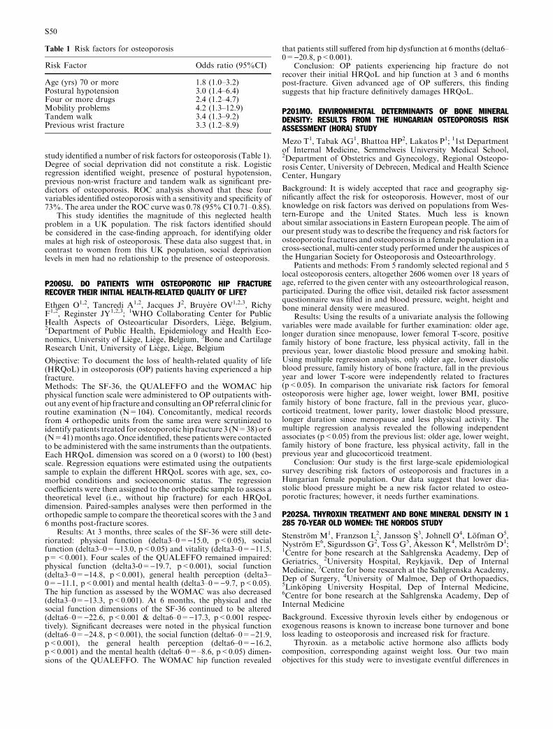
study identified a number of risk factors for osteoporosis (Table 1).Degree of social deprivation did not constitute a risk. Logisticregression identified weight, presence of postural hypotension,previous non-wrist fracture and tandem walk as significant pre-dictors of osteoporosis. ROC analysis showed that these fourvariables identified osteoporosis with a sensitivity and specificity of73%. The area under the ROC curve was 0.78 (95%CI 0.71–0.85).
This study identifies the magnitude of this neglected healthproblem in a UK population. The risk factors identified shouldbe considered in the case-finding approach, for identifying oldermales at high risk of osteoporosis. These data also suggest that, incontrast to women from this UK population, social deprivationlevels in men had no relationship to the presence of osteoporosis.
P200SU. DO PATIENTS WITH OSTEOPOROTIC HIP FRACTURERECOVER THEIR INITIAL HEALTH-RELATED QUALITY OF LIFE?
Ethgen O1,2, Tancredi A1,2, Jacques J2, Bruyere OV1,2,3, RichyF1,2, Reginster JY1,2,3; 1WHO Collaborating Center for PublicHealth Aspects of Osteoarticular Disorders, Liege, Belgium,2Department of Public Health, Epidemiology and Health Eco-nomics, University of Liege, Liege, Belgium, 3Bone and CartilageResearch Unit, University of Liege, Liege, Belgium
Objective: To document the loss of health-related quality of life(HRQoL) in osteoporosis (OP) patients having experienced a hipfracture.Methods: The SF-36, the QUALEFFO and the WOMAC hipphysical function scale were administered to OP outpatients with-out any event of hip fracture and consulting anOP referral clinic forroutine examination (N=104). Concomitantly, medical recordsfrom 4 orthopedic units from the same area were scrutinized toidentify patients treated for osteoporotic hip fracture 3 (N=38) or 6(N=41)months ago.Once identified, these patients were contactedto be administered with the same instruments than the outpatients.Each HRQoL dimension was scored on a 0 (worst) to 100 (best)scale. Regression equations were estimated using the outpatientssample to explain the different HRQoL scores with age, sex, co-morbid conditions and socioeconomic status. The regressioncoefficients were then assigned to the orthopedic sample to assess atheoretical level (i.e., without hip fracture) for each HRQoLdimension. Paired-samples analyses were then performed in theorthopedic sample to compare the theoretical scores with the 3 and6 months post-fracture scores.
Results: At 3 months, three scales of the SF-36 were still dete-riorated: physical function (delta3–0=)15.0, p<0.05), socialfunction (delta3–0=)13.0, p<0.05) and vitality (delta3–0=)11.5,p=<0.001). Four scales of the QUALEFFO remained impaired:physical function (delta3-0=)19.7, p<0.001), social function(delta3–0=)14.8, p<0.001), general health perception (delta3–0=)11.1, p<0.001) and mental health (delta3–0=)9.7, p<0.05).The hip function as assessed by the WOMAC was also decreased(delta3–0=)13.3, p<0.001). At 6 months, the physical and thesocial function dimensions of the SF-36 continued to be altered(delta6–0=)22.6, p<0.001 & delta6–0=)17.3, p<0.001 respec-tively). Significant decreases were noted in the physical function(delta6–0=)24.8, p<0.001), the social function (delta6–0=)21.9,p<0.001), the general health perception (delta6–0=)16.2,p<0.001) and the mental health (delta6–0=–8.6, p<0.05) dimen-sions of the QUALEFFO. The WOMAC hip function revealed
that patients still suffered from hip dysfunction at 6 months (delta6–0=)20.8, p<0.001).
Conclusion: OP patients experiencing hip fracture do notrecover their initial HRQoL and hip function at 3 and 6 monthspost-fracture. Given advanced age of OP sufferers, this findingsuggests that hip fracture definitively damages HRQoL.
P201MO. ENVIRONMENTAL DETERMINANTS OF BONE MINERALDENSITY: RESULTS FROM THE HUNGARIAN OSTEOPOROSIS RISKASSESSMENT (HORA) STUDY
Mezo T1, Tabak AG1, Bhattoa HP2, Lakatos P1; 11st Departmentof Internal Medicine, Semmelweis University Medical School,2Department of Obstetrics and Gynecology, Regional Osteopo-rosis Center, University of Debrecen, Medical and Health ScienceCenter, Hungary
Background: It is widely accepted that race and geography sig-nificantly affect the risk for osteoporosis. However, most of ourknowledge on risk factors was derived on populations from Wes-tern-Europe and the United States. Much less is knownabout similar associations in Eastern European people. The aim ofour present study was to describe the frequency and risk factors forosteoporotic fractures and osteoporosis in a female population in across-sectional, multi-center study performed under the auspices ofthe Hungarian Society for Osteoporosis and Osteoarthrology.
Patients and methods: From 5 randomly selected regional and 5local osteoporosis centers, altogether 2606 women over 18 years ofage, referred to the given center with any osteoarthrological reason,participated. During the office visit, detailed risk factor assessmentquestionnaire was filled in and blood pressure, weight, height andbone mineral density were measured.
Results: Using the results of a univariate analysis the followingvariables were made available for further examination: older age,longer duration since menopause, lower femoral T-score, positivefamily history of bone fracture, less physical activity, fall in theprevious year, lower diastolic blood pressure and smoking habit.Using multiple regression analysis, only older age, lower diastolicblood pressure, family history of bone fracture, fall in the previousyear and lower T-score were independently related to fractures(p<0.05). In comparison the univariate risk factors for femoralosteoporosis were higher age, lower weight, lower BMI, positivefamily history of bone fracture, fall in the previous year, gluco-corticoid treatment, lower parity, lower diastolic blood pressure,longer duration since menopause and less physical activity. Themultiple regression analysis revealed the following independentassociates (p<0.05) from the previous list: older age, lower weight,family history of bone fracture, less physical activity, fall in theprevious year and glucocorticoid treatment.
Conclusion: Our study is the first large-scale epidemiologicalsurvey describing risk factors of osteoporosis and fractures in aHungarian female population. Our data suggest that lower dia-stolic blood pressure might be a new risk factor related to osteo-porotic fractures; however, it needs further examinations.
P202SA. THYROXIN TREATMENT AND BONE MINERAL DENSITY IN 1285 70-YEAR OLD WOMEN: THE NORDOS STUDY
Stenstrom M1, Franzson L2, Jansson S3, Johnell O4, Lofman O5,Nystrom E6, Sigurdsson G2, Toss G5, Akesson K4, Mellstrom D1;1Centre for bone research at the Sahlgrenska Academy, Dep ofGeriatrics, 2University Hospital, Reykjavik, Dep of InternalMedicine, 3Centre for bone research at the Sahlgrenska Academy,Dep of Surgery, 4University of Malmoe, Dep of Orthopaedics,5Linkoping University Hospital, Dep of Internal Medicine,6Centre for bone research at the Sahlgrenska Academy, Dep ofInternal Medicine
Background. Excessive thyroxin levels either by endogenous orexogenous reasons is known to increase bone turnover and boneloss leading to osteoporosis and increased risk for fracture.
Thyroxin. as a metabolic active hormone also afflicts bodycomposition, corresponding against weight loss. Our two mainobjectives for this study were to investigate eventful differences in
Table 1 Risk factors for osteoporosis
Risk Factor Odds ratio (95%CI)
Age (yrs) 70 or more 1.8 (1.0–3.2)Postural hypotension 3.0 (1.4–6.4)Four or more drugs 2.4 (1.2–4.7)Mobility problems 4.2 (1.3–12.9)Tandem walk 3.4 (1.3–9.2)Previous wrist fracture 3.3 (1.2–8.9)
S50
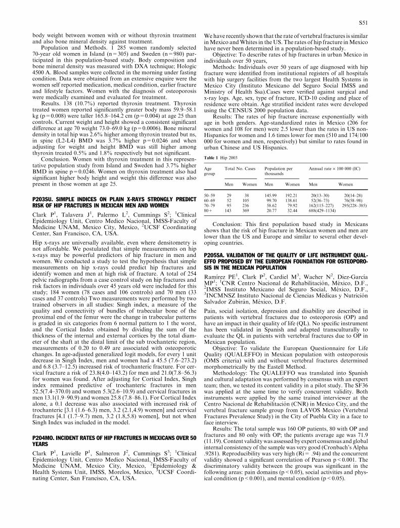
body weight between women with or without thyroxin treatmentand also bone mineral density against treatment.
Population and Methods. 1 285 women randomly selected70-year old women in Island (n=305) and Sweden (n=980) par-ticipated in this population-based study. Body composition andbone mineral density was measured with DXA technique; Hologic4500 A. Blood samples were collected in the morning under fastingcondition. Data were obtained from an extensive enquire were thewomen self reported medication, medical condition, earlier fractureand lifestyle factors. Women with the diagnosis of osteoporosiswere medically examined and evaluated for treatment.
Results. 138 (10.7%) reported thyroxin treatment. Thyroxintreated women reported significantly greater body mass 59.9–58.1kg (p=0.008) were taller 165.8–164.2 cm (p=0.004) at age 25 thancontrols. Current weight and height showed a consistent significantdifference at age 70 weight 73.0–69.0 kg (p=0.0006). Bone mineraldensity in total hip was 2.6% higher among thyroxin treated but ns,in spine (L2-L4) BMD was 3.7% higher p=0.0246 and whenadjusting for weight and height BMD was still higher amongthyroxin treated 0.5% and 1.8% respectively but not significant.
Conclusion. Women with thyroxin treatment in this represen-tative population study from Island and Sweden had 3.7% higherBMD in spine p=0.0246. Women on thyroxin treatment also hadsignificant higher body height and weight this difference was alsopresent in those women at age 25.
P203SU. SIMPLE INDICES ON PLAIN X-RAYS STRONGLY PREDICTRISK OF HIP FRACTURES IN MEXICAN MEN AND WOMEN
Clark P1, Talavera J1, Palermo L2, Cummings S2; 1ClinicalEpidemiology Unit, Centro Medico Nacional, IMSS-Faculty ofMedicine UNAM, Mexico City, Mexico, 2UCSF CoordinatingCenter, San Francisco, CA, USA.
Hip x-rays are universally available, even where densitometry isnot affordable. We postulated that simple measurements on hipx-rays may be powerful predictors of hip fracture in men andwomen. We conducted a study to test the hypothesis that simplemeasurements on hip x-rays could predict hip fractures andidentify women and men at high risk of fracture. A total of 254pelvic radiographs from a case control study on hip fractures andrisk factors in individuals over 45 years old were included for thisstudy; 184 women (78 cases and 106 controls) and 70 men (33cases and 37 controls) Two measurements were performed by twotrained observers in all studies: Singh index, a measure of thequality and connectivity of bundles of trabecular bone of theproximal end of the femur were the change in trabecular patternsis graded in six categories from 6 normal pattern to 1 the worst,and the Cortical Index obtained by dividing the sum of thethickness of the internal and external cortices by the total diam-eter of the shaft at the distal limit of the sub trochanteric region,measurements of 0.20 to 0.49 are associated with osteoporoticchanges. In age-adjusted generalized logit models, for every 1 unitdecrease in Singh Index, men and women had a 45.5 (7.6–273.2)and 6.8 (3.7–12.5) increased risk of trochanteric fracture. For cer-vical fracture a risk of 23.8(4.0–143.2) for men and 21.0(7.8–56.3)for women was found. After adjusting for Cortical Index, Singhindex remained predictive of trochanteric fractures in men52.5(7.4–370.0) and women 5.3(2.6–10.9) and cervical fractures inmen 13.1(1.9–90.9) and women 25.8 (7.8–86.1). For Cortical Indexalone, a 0.1 decrease was also associated with increased risk oftrochanteric [3.1 (1.6–6.3) men, 3.2 (2.1,4.9) women] and cervicalfractures [4.1 (1.7–9.7) men, 3.2 (1.8,5.8) women], but not whenSingh Index was included in the model.
P204MO. INCIDENT RATES OF HIP FRACTURES IN MEXICANS OVER 50YEARS
Clark P1, Lavielle P1, Salmeron J2, Cummings S3; 1ClinicalEpidemiology Unit, Centro Medico Nacional, IMSS-Faculty ofMedicine UNAM, Mexico City, Mexico, 2Epidemiology &Health Systems Unit, IMSS, Morelos, Mexico, 3UCSF Coordi-nating Center, San Francisco, CA, USA.
Wehave recently shown that the rate of vertebral fractures is similarinMexico andWhites in theUS. The rates of hip fracture inMexicohave never been determined in a population-based study.
Objective: To describe rates of hip fractures in urban Mexico inindividuals over 50 years,
Methods: Individuals over 50 years of age diagnosed with hipfracture were identified from institutional registers of all hospitalswith hip surgery facilities from the two largest Health Systems inMexico City (Instituto Mexicano del Seguro Social IMSS andMinistry of Health Ssa).Cases were verified against surgical andx-ray logs. Age, sex, type of fracture, ICD-10 coding and place ofresidence were obtain. Age stratified incident rates were developedusing the CENSUS 2000 population data.
Results: The rates of hip fracture increase exponentially withage in both genders. Age-standardized rates in Mexico (206 forwomen and 108 for men) were 2.5 lower than the rates in US non-Hispanics for women and 1.6 times lower for men (510 and 174/100000 for women and men, respectively) but similar to rates found inurban Chinese and US Hispanics.
Conclusion: This first population based study in Mexicansshows that the risk of hip fracture in Mexican women and men arelower than the US and Europe and similar to several other devel-oping countries.
P205SA. VALIDATION OF THE QUALITY OF LIFE INSTRUMENT QUAL-EFFO PROPOSED BY THE EUROPEAN FOUNDATION FOR OSTEOPORO-SIS IN THE MEXICAN POPULATION
Ramırez PE1, Clark P2, Cardiel M3, Wacher N2, Diez-GarcıaMP1; 1CNR Centro Nacional de Rehabilitacion, Mexico, D.F.,2IMSS Instituto Mexicano del Seguro Social, Mexico, D.F.,3INCMNSZ Instituto Nacional de Ciencias Medicas y NutricionSalvador Zubiran, Mexico, D.F.
Pain, social isolation, depression and disability are described inpatients with vertebral fractures due to osteoporosis (OP) andhave an impact in their quality of life (QL). No specific instrumenthas been validated in Spanish and adapted transculturally toevaluate the QL in patients with vertebral fractures due to OP inMexican population.
Objective: To validate the European Questionnaire for LifeQuality (QUALEFFO) in Mexican population with osteoporosis(OMS criteria) with and without vertebral fractures determinedmorphometrically by the Eastell Method.
Methodology: The QUALEFFO was translated into Spanishand cultural adaptation was performed by consensus with an expertteam; then, we tested its content validity in a pilot study. The SF36was applied at the same time to verify concurrent validity. Bothinstruments were applied by the same trained interviewer at theCentro Nacional de Rehabilitacion (CNR) in Mexico City, and thevertebral fracture sample group from LAVOS Mexico (VertebralFractures Prevalence Study) in the City of Puebla City in a face toface interview.
Results: The total sample was 160 OP patients, 80 with OP andfractures and 80 only with OP; the patients average age was 71.9(11.19). Content validitywas assessed by expert consensus and globalinternal consistency of the sample was very good (Cronbach’s Alpha.9281). Reproducibility was very high (Ri= .94) and the concurrentvalidity showed a significant correlation of Pearson p<0.001. Thediscriminatory validity between the groups was significant in thefollowing areas: pain domains (p<0.05), social activities and phys-ical condition (p<0.001), and mental condition (p<0.05).
Table 1 Hip 2003
Agegroup
Total No. Cases Population perthousands
Annual rate · 100 000 (IC)
Men Women Men Women Men Women
50–59 29 38 145.99 192.21 20(13–30) 20(14–28)60–69 52 105 99.70 138.61 52(36–73) 76(58–98)70–79 95 236 58.62 79.92 162(115–227) 295(228–385)80+ 143 369 20.77 32.44 688(429–1134)
S51

Conclusion: The QUALEFFO had excellent psychometriccharacteristics in our sample of Mexican patients; it proved to beconsistent and homogeneous and to have discriminatory potentialin 3 different domains of QL. We conclude that the QUALEFFOcan be used in Mexican population to measure QL due to VertebralFractures attributable to OP.
P206SU. THE ASSOCIATION BETWEEN NUTRITIONAL RISK DETER-MINED BY MINI-NUTRITIONAL ASSESSMENT (MNA) AND OSTEOPO-ROSIS IN ELDERLY WOMEN: A CROSS-SECTIONAL STUDY
Salminen H1, Saaf M2, Johansson S1, Ringertz H3, Strender L1;1Family Medicine Stockholm, Karolinska Institutet, Sweden,2DepartmentofMolecularMedicine,EndocrineandDiabetesUnit,Karolinska Hospital, Sweden, 3Department of Surgical Sciences,Section for Diagnostic Radiology, Karolinska Institutet, Sweden
Aims: The aim of this study was to investigate the associationbetween bone mineral density and the nutritional risk determinedby Mini Nutritional Assessment (MNA) questionnaire in a groupof home living elderly Swedish women.
Methods: The study sample consists of 584 women. 60% or 351women (mean age 73 years) participated. Their nutritional statuswas determined by MNA, a test consisting of 18 point-weightedquestions in four categories: anthropometric measurements, clinicaland functional evaluations, assessment of dietary intake and self-assessment of nutritional status and health. 30 points is the maxi-mum, a score <17 indicates malnourished, >17 and <24 at risk ofmalnutrition and >24 well nourished.
Bone mineral density (BMD) values were determined at the lefthip and the lumbar spine, L1-L4, using HologicQDR 4500 equip-ment for dual energy x-ray absorptiometry (DXA). The data wereanalysed by logistic regression.
Results: The median score of MNA was 27. Only 7.4% of thewomen were at risk of malnutrition and only one subject wasclassified as malnourished. Osteoporosis in the femoral neck wasfound in 52.9% of the women and 31% had a history of fractureafter the age of 50 years. Those with <23.5p at MNA had a 70%larger age-adjusted risk of having osteoporosis than those withnormal scores but this finding was not statistically significant(p=0.24). ROC analysis showed an AUC of 0.67 and a highspecificity of 95% combined with a very poor sensitivity of 9%.The following items in the MNA questionnaire had the strongestassociation with osteoporosis at the femoral neck: age (OR= 1.12;CI=1.02–1.24) per 1 year increase, calf circumference (OR= 0.91;CI= 0.83–0.99) per 1 cm increase and BMI over 26 (OR= 0.19; CI=0.04–0.93). Reduced appetite (OR= 2.55; CI=0.79–8.20) andthe highest protein score (OR= 0.41; CI= 0.11–1.48) were notstatistically significantly associated with osteoporosis.
Conclusions: The anthropometric measurements of the MNAquestionnaire were highly associated with osteoporosis but theMNA questionnaire is not sensitive enough to detect osteoporosis.
P207MO. THE INFLUENCE OF FAMILY HISTORY OF OSTEOPOROTICFRACTURE ON THE RISK OF VERTEBRAL OSTEOPOROSIS IN WOMEN
Ragi Eis S1, Albergaria BH1, Clark P2; 1CEDOES ) Centro deDiagnostico e Pesquisa da Osteoporose do ES, Vitoria (ES)Brasil, 2UIM Epidemiologıa Clınica, CMN Siglo XXI-Facultadde Medicina UNAM, Mexico, D.F.
Introduction Bone mass is an important determinant of bonestrength and risk for osteoporotic fractures (OF). The majority offamily studies of bone mass indicates that it is under strong ge-netic control. However, there are few data exploring clustering ofosteoporosis within families in Latin America.
Objectives The aim of this study was to determine the influence offamily history of OF on the risk of vertebral osteoporosis and oc-currence of OF in a set of Brazilian women included in Lavos Study.
Material and Methods 407 women aged 50 and over, wererecruited from population based sampling across Vitoria metro-politan area, Brazil. Subjects were invited to participate, signed aninformed consent form and a trained interviewer applied a standar-dized questionnaire. A DXA measurement at L-Spine and Hip was
performed, as well as an X-Ray of Thoracic and L-Spine. Vertebralosteoporosis was defined using NIH criteria (T-score= –2.0SD)
Results A family history of OF was associated with a 14%increased risk of vertebral osteoporosis [odds ratio (OR) 1.14(95%CI:1.127–1.166)] and with a 44% increased risk of personalOF [OR 1.44 (95%CI:1.424–1.461)]. We also observed significantlylower BMD in subjects who reported OFs (n=62), with a meanBMD of 0.897 (+0.037) and of 0.974 (+0.021) (p=0.00026) in thegroups with and without history of OF (n=345), respectively. Thenumber of relatives with past of OF as well as the number of OFthese relatives suffered, was statistically correlated with BMD(r=)0.33, p=0.009 and r=)0.285, p=0.025).
When the mean BMD of subjects who reported relatives withOF was compared with those without family history of OF, thecorrelation was not statistically significant (mean BMD of0.947+0.188 and 0.958+0.204 [p=0.36]) in the groups with andwithout family history of OF, respectively).
Conclusion As seen in other populations, family history of OFappears to be an important risk factor for OF in Brazilian women.This risk factor seems to be determined not only by BMD genetichereditariness. As the risk of OF increases with the number of re-latives with past of OF, this particular information should be con-sidered when looking for risk factors for osteoporosis and fractures.
P208SA. THE GLOBAL BURDEN OF HIP FRACTURE
Johnell O1, Kanis JA2; 1Malmo General Hospital, Malmo,Sweden, 2University of Sheffield, UK
The aim of this study was to quantify the global burden ofosteoporosis as judged by hip fracture and the burden in differentsocio-economic regions of the world. The population mortality in1990 and the incidence of hip fracture in different regions wereidentified, where possible in 1990. Excess mortality from hipfracture used data for Sweden, and disability weights were as-signed to survivors from hip fracture.
In 1990 there were 1.31 million new hip fractures and the preva-lence of hip fractures with disability was 4.48 millions. There were738,116 deaths associated with hip fracture and 1.7 million life-yearslost reduced to 951 thousands with weighting for age. Disabilityadjusted life years lost accounted for 1.75 million disability adjustedlife years, representing 0.1% of the global burden of disease worldwide and 1.4% of the burden amongst women from the establishedmarket economies. We conclude that hip fracture is a significantcause of morbidity and mortality world-wide.
P209SU. COSTS AND QUALITY OF LIFE RELATED TO VERTEBRALFRACTURES: PRELIMINARY RESULTS BASED ON AN ONGOINGSWEDISH PROSPECTIVE STUDY
Zethraeus N1, Borgstrom F2, Jonsson B1; 1Stockholm School ofEconomics, Stockholm, Sweden, 2Stockholm Health Economics,Stockholm, Sweden
Aims: There are few studies investigating the consequences ofosteoporotic (low bone density) fractures in terms of costs andhealth outcomes. A previous Swedish pilot study assessed thecosts and quality of life related to fractures of the hip, spine, wristand shoulder in the southern part of Sweden. Data were collectedusing a questionnaire administered by a nurse at Malmo Uni-versity Hospital in the south of Sweden. The study indicated thatspine fractures are associated with higher costs and lower qualityof life than previously assumed. Based on the pilot study a large-scale prospective study was designed in a Swedish setting. Thepurpose of this ongoing study is to collect cost and health effectdata related to osteoporotic fractures of the hip, wrist and spinefor a large number of patients, e.g. to be able to assess thedeterminants of costs and quality of life.
Methods: In total data for 2 000 patients at 7 study centres willbe included. The patient enrolment started in June 2002 and willcontinue during the year 2003 and 2004. In September (2003), 103spine fracture patients were enrolled in the study, out of which 76were hospitalised and 27 were discharged the same day as the firstcontact with the health care.
S52
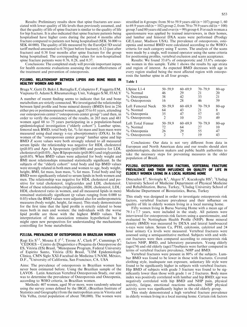
Results: Preliminary results show that spine fractures are asso-ciated with lower quality of life levels than previously assumed, andthat the quality of life of spine fracture is similar to the quality of lifefor hip fracture. It is also indicated that spine fracture patients beinghospitalised have higher costs during the period 4 months afterfracture compared to patients not being hospitalised (SEK 56 000 vsSEK 44 000). The quality of life measured by the EuroQol 5D socialtariff method amounted to 0.70 (just before fracture), 0.12 (just afterfracture) and 0.38 four months after spine fracture for the groupbeing hospitalised. The corresponding values for non-hospitalisedspine fracture patients were 0.76, 0.28, and 0.37.
Conclusions: The completed study will provide important inputsfor health economic evaluations assessing the cost-effectiveness ofthe treatment and prevention of osteoporosis.
P210MO. RELATIONSHIP BETWEEN LIPIDS AND BONE MASS INHEALTHY WOMEN AND MEN
BragaV,Gatti D, Bakri J, Battaglia E, Colapietro F, Fuggetta EM,Viapiana O, Adami S; Rheumatology Unit, Valeggio S/M, ITALY
A number of recent finding seems to indicate that fat and bonemetabolism are strictly connected.We investigated the relationshipbetween lipid profile and bone mineral density (BMD) first in 236either pre or postmenopausalwomen, aged 35 to 81 years, attendingour osteoporosis center (‘‘osteoporosis center group’’) and then, inorder to verify the consistency of the results, in 265 men and 481women aged 68 to 75 years participating in a population-basedepidemiological investigation (‘‘elderly cohort’’). Lumbar spine,femoral neck BMD, total body fat, % fat mass and lean mass weremeasured using dual energy x-ray absorptiometry (DXA). In thewomen of the ‘‘osteoporosis center group’’ lumbar spine and hipBMD Z score values were both strongly related to all measuredserum lipids: the relationship was negative for HDL cholesterol(p£0.05) and Apo A lipoprotein (p£0.000) and positive for LDLcholesterol (p£0.05) , ApoB lipoprotein (p£0.001) and triglycerides(p£0.05). When BMD values were adjusted for body weight andBMI most relationships remained statistically significant. In thesubjects of the ‘‘elderly cohort’’ total body and hip BMD valueswere strongly related in both men and women to age, body weight,height, BMI, fat mass, lean mass, % fat mass. Total body and hipBMDwere significantly related to serum lipids in both women andmen. The relationship was negative for HDL cholesterol and po-sitive for total cholesterol, triglycerides and LDL cholesterol.Most of these relationships (triglycerides, HDL cholesterol, LDL/HDL cholesterol ratio in women, and all measured lipids in men)remained statistically significant (p values ranging from 0.000 to0.03) when the BMD values were adjusted also for anthropometricmeasures (body weight, height, fat mass). This study demonstratesfor the first time that the lipid profile is strictly related to bonemass both in men and women: the individuals with the worselipid profile are those with the highest BMD values. Theinterpretation of this association remains hypothetical but itmight open new perspectives for understanding the mechanismscontrolling for bone metabolism.
P211SA. PREVALENCE OF OSTEOPOROSIS IN BRAZILIAN WOMEN
Ragi Eis S1,2, Moana E J1,2, Tirone A1, Clark P3, Cummings S4;1CEDOES ) Centro de Diagnostico e Pesquisa da Osteoporose doES, Vitoria (ES) Brasil, 2Menopause Program, Federal Universityof Espirito Santo, Vitoria (ES) Brasil, 3UIM EpidemiologıaClınica, CMN Siglo XXI-Facultad de Medicina UNAM, Mexico,D.F., 4University of California, San Francisco, CA. USA
Aims: The prevalence of osteoporosis in Brazilian women hasnever been estimated before. Using the Brazilian sample of theLAVOS – Latin American Vertebral Osteoporosis Study, our aimwas to determine the prevalence of Osteoporosis and Osteopenia,according to WHO criteria, in Brazilian women.
Methods: 407 women, aged 50 or more, were randomly selectedusing the survey zones defined by the IBGE, (Brazilian Institute ofStatistics andGeography) from the metropolitan area in Vitoria andVila Velha, (total population of about 700,000). The women were
stratified in 4 groups: from 50 to 59.9 years old (n=107) group 1, 60to 69.9 years old (n=102) group 2, from 70 to 79.9 years old (n=100)group 3and 80 years old andover (n=99) group4. For eachwomanaquestionnaire was applied by trained interviewers, in their homes,and lumbar and femoral DXA scans were performed (ProdigyGE/Lunar, Madison USA). The prevalence of osteoporosis, oste-openia and normal BMD were calculated according to the WHO’scriteria for each category using T scores. The analysis of the scanswere made by a single, well trained operator using the same criteriafor positioning profiles, vertebral exclusion and scans acceptance.
Results: We found 33.6% of osteoporotic and 33.8% osteope-nic women in this sample. Table 1 shows the results by age strataand region of interest. As expected BMD decreases with age inevery region studied being the most affected region with osteopo-rosis the lumbar spine in all four groups.
Conclusions: Our data is not very different from data inEuropean and North American data and our results should alertepidemiologists, decision makers and public health authorities totake the necessary steps for preventing measures in the olderpopulation of Brazil.
P212SU. OSTEOPOROSIS RISK FACTORS, VERTEBRAL FRACTUREPREVALENCE AND THEIR INFLUENCE ON QUALITY OF LIFE INELDERLY WOMEN LIVING IN A LOCAL NURSING HOME
Dincsahin E1, Sivrioglu K1, Akgoz S2, Kucukoglu HS1; 1UludagUniversity School of Medicine Department of Physical Medicineand Rehabilitation, Bursa, Turkey, 2Uludag University School ofMedicine Department of Biostatistics, Bursa, Turkey
This study was designed in order to investigate osteoporosis riskfactors, vertebral fracture prevalence and their influence onquality of life in elderly women living in a local nursing home.
Fifty women living in Bursa Nursing Home with a mean age of79.1±7.7 years, were included in the study. All patients wereinterviewed for osteoporosis risk factors using a questionnaire, andevaluated by Nottingham Health Profile (NHP). Bone mineraldensity (BMD) was measured by DEXA and lateral lumbosacralx-rays were taken. Serum Ca, PTH, calcitonin, calcitriol and 24hour urinary Ca levels were measured. Vertebral fractures wereassessed using a semiquantitative method. Subjects with and with-out fractures were then compared according to osteoporosis riskfactors NHP, BMD, and laboratory parameters. Young elderly(age£74) and old elderly (age‡75)subjects were further compared interms of vertebral fracture prevalence, NHP and BMD.
Vertebral fractures were present in 80% of the subjects. Lum-bar BMD was found to be lower in those with fractures. Coveredclothing style, inadequate sun exposure, sedantary life style werefound to be significantly higher in subjects with vertebral fracture.Hip BMD of subjects with grade 3 fracture was found to be sig-nificantly lower than those with grade 1 or 2 fractures. Body massindex was positively correlated with lumbar and hip BMD; age wasnegatively correlated with hip BMD and NHP pain, physicalactivity, fatigue, emotional reactions subscales; NHP physicalactivity score was significantly higher in the old elderly group.
This study demonstrated a high vertebral fracture prevalencein elderly women living in a local nursing home. Certain risk factors
Table 1
LSpine L1-4 50–59,9 60–69,9 70–79,9 80-up% Normal 46 29 21 20% Osteopenia 38 42 33 21% Osteoporosis 16 28 46 59
Left Femoral Neck 50–59,9 60–69,9 70–79,9 80-up% Normal 64 43 21 8% Osteopenia 35 47 57 43% Osteoporosis 2 10 23 49
Left Total Femur 50–59,9 60–69,9 70–79,9 80-up% Normal 72 64 27 10% Osteopenia 26 29 55 47% Osteoporosis 2 7 19 43
S53
such as covered clothing style, inadequate sun exposure and sedan-tary life style were associated with a high prevalence of vertebralfractures. Quality of life was found to be influenced only byincreasing age. We believe that these findings may provide an insightfor larger epidemiological studies as well as contribute to osteopo-rosis prevention and treatment efforts in this high risk population.
P213MO. BMD IN MEN: WHICH NORMATIVE DATA?
Richy F1, Gourlay M2, Ethgen O1, Bruyere O1, Reginster JY1; 1ofPublic Health, Epidemiology and Health Economics, Universityof Liege, BELGIUM; WHO Collaborating Center for the PublicAspects of Osteoarticular Disorders, Belgium, 2Robert WoodJohnson Clinical Scholars Program, 5034 Old Clinic Building, CB#7105, UNC-Chapel Hill, Chapel Hill, NC 27599-7105, USA
Objectives: Controversy surrounds which normative data shouldbe used to estimate osteoporosis prevalence in men. Prevalenceestimates may vary significantly when different normative stan-dards are applied. Our goal was to determine to which extentdifferent normative sets of data could impact the prevalence ofosteoporosis in men.
Material and methods: Five normative data sets (NHANESfemale norms, local female norms, Hologic densitometer manufac-turer female norms, NHANES male norms, Hologic male norms)were used to estimate the prevalence of osteoporosis byWorldHealthOrganization diagnostic criteria in a study population of 321 con-secutivemen aged 20 to 91 (mean59.3 years) referred to anoutpatientosteoporosis center between January 1996 and December 1998.
Results: Statistically significant variations were seen in osteo-porosis prevalence measured at three anatomical sites. The greatestrelative variation was seen for the total femur, where osteoporosisprevalence ranged from 5.6 percent (NHANES and Hologic femalenorms) to 12.5 percent (NHANES male norms). The least relativevariation was seen at the lumbar spine, where prevalence rangedfrom 15.6 percent (Hologic female norms) to 27.1 percent (localfemale norms). When considering osteoporosis at any site, preva-lence was lowest (20.3 percent) based on Hologic female norms andhighest (32.7 percent) based on local female norms.
Conclusion: Interpretation of prevalence data should include anassessment of how normative standards influence reporting of thepopulation at high risk of fracture.
P214SA. EVALUATION OF THE OSTEOPOROSIS SELF-ASSESSMENTTOOL (OST) AS A SCREENING TEST FOR OSTEOPOROSIS AND OSTE-OPENIA IN MEN
De Ceulaer F1, Hanssens L1, Tancredi A1, Bruyere O1, Richy F1,Pire G2, Maassen P3, Reginster J-Y1; 1Dept of public health,epidemiology and health economics,Univerity of Liege, Belgium;WHO Collaborating Center for the public health aspects ofosteoarticular disorders, Belgium, 2Provincial representative inCharge of Health, Environment and Quality of Life, province ofLiege, Belgium, 3General Director, Health and Environment,Province of Liege, Belgium
Background: Several indices have been developed to screenwomen with a high risk of having low bone mineral density, whoshould benefit of BMD testing. No risk indice has been developedfor men so far. The Osteoporosis Self-Assessment Tool (OST), thesimplest indice to calculate, might be suitable for ageing men.
Objective: To evaluate the accuracy of OST as a screening tool,predicting osteopenia and osteoporosis risk in a male population.
Methods: 183 men who presented spontaneously at the multipleriskdetection campaignorganizedby theProvinceofLiege (Belgium)inJune2003.Wehavecollectedanthropometricaldataandperformeda BMD testing in 161 subjects using DEXA technology (Hologic,QDR 1000+). The ability of OST to identify men with osteopenia(T-score £)1.0) or osteoporosis (T-score £)2.5) was evaluated.
Results: The mean age of the sample was 59.4 (±5.8) years. Theprevalence of osteoporosis was 9.25% at the femoral neck and1.25% at the total hip. The prevalence of osteopenia was 30% atthe femoral neck and 8% at the total hip. BMD measures wereinversely correlated with the values of OST, r=)0.38 (p<0.005)
and )0.35 (p<0.005) respectively for the femoral neck and the totalhip. The area under the ROC curve (AUC) was 0.737 for thefemoral neck and 0.760 for the total hip, close to the values ob-served in women. For the cut-off value of <4.7 , the sensitivity ofOST in identifying men at risk of osteopenia ranged from 75.0%for the femoral neck to 84.6% at the total hip. The specificity ofOST was 58.4% for the femoral neck and 51.4% for the total hip.Conclusions: The performance of OST among this sample of menwas acceptable. In the future, we need a bigger sample to validatethe cut-off value for OST. We conclude that the fitted OST riskindice could be an effective and efficient tool to identify increasedrisk men for BMD testing.
P215SU. EVALUATION OF PREVALENCE OF OSTEOPOROSIS AND OS-TEOPENIA IN MEN PRESENTING AT A MULTIPLE RISK DETECTIONCAMPAIGN
Hanssens LC1, De Ceulaer F1, Tancredi A1, Bruyere O1, RichyF1, Halkin V2, Pire G3, Maassen P4, Reginster JY1; 1Dept ofpublic health, epidemiology and health economics, Univerity ofLiege, Belgium; WHO Collaborating Center for the public healthaspects of osteoarticular disorders, Belgium, 2PharmaceuticalResearch Institute, Bristol Myers Squibb, Waterloo Belgium,3Provincial representative in Charge of Health, Environment andQuality of Life, province of Liege, Belgium, 4General Director,Health and Environement, Province of Liege, Belgium
Objectives To describe the risk for osteopenia and osteoporosis ina population of men between 50 and 75 years old who presentedat an age related multiple risk detection campaign.
Population and methods The Province of Liege has organized amultiple risk detection campaign for men aged 50 to 70. The par-ticipants were randomly selected from a database containing100000 men. From the 1300 subjects who received an invitation,183 presented for the multiple risk screening. The tests consisted, ina prostate cancer screening, an andropause screening, a physicalperformance tests and osteporosis screening. The subjects were alsoasked to perform a One Minute Risk Test (OMRT).
The osteoporosis screening was performed by DEXA (Hologic,QDR 1000+) at the total hip site and at the femoral neck. 162subjects had a BMD performed. Correlations with the OMRT andthe Osteoporosis Self-Assessment Tool were calculated.
Results The general characteristics of the population are asfollows: mean age was 59.4 (±5.8) years, mean Total hip BMD was1.007 (± 0.170) g/cm2, mean T-score at the hip was )0.172(± 1.128), mean femoral neck BMD was 0.821 (± 0.130) g/cm2,mean T-score at the femoral neck was 0.145 (± 0.951). The meanOMRT score was 5.536 (± 2.616).
The overall prevalence of osteoporosis was 9.25 % at thefemoral neck and 1.25 % at the hip site. The prevalence of oste-openia was 30 % at the femoral neck and 8 % at the hip site.
Conclusion The prevalence of osteoporosis in the selectedpopulation appears to be lower than expected. As a result, systematicscreening for osteoporosis using DEXA is not recommended in thisage range. However, BMD measures inversely correlated with theOST, r=)0.38 for the femoral neck and )0.35 for the total hip.
OST is a very simple instrument which would be more suitableto use in such a screening campaign. Indeed it is easy to administerat low cost and resource investment and would give a good pre-liminary assessment of the osteoporosis risk in such a population.
P216MO. FRACTURE INCIDENCE AFTER FRAGILITY FRACTURESADMITTED TO THE HOSPITAL
van Helden SH1, Geusens P1,2, Bulstra S1, Brink PRG1, Koeyers W1;1Academic hospital Maastricht, Maastricht, Netherlands, 2BiomedicalResearch Institute – Limburg University, Diepenbeek, Belgium
Aims: To determine the incidence of new clinical fractures in pa-tients admitted to the hospital because of a recent clinical fracture.
Method: We analysed all patients aged over 50 years with afragility fracture who were treated in our hospital from January toSeptember 2001. Data were collected from questionnaires sent topatients and research in the hospital database for patients who did
S54
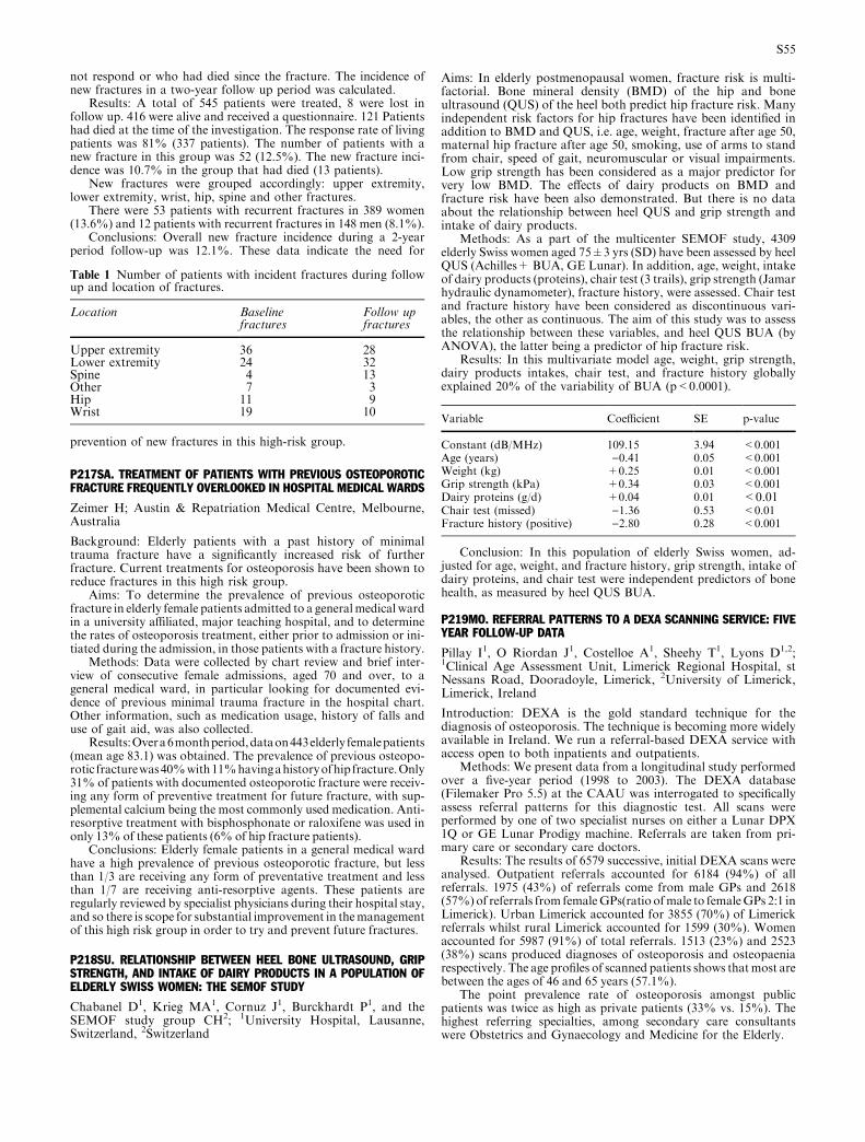
not respond or who had died since the fracture. The incidence ofnew fractures in a two-year follow up period was calculated.
Results: A total of 545 patients were treated, 8 were lost infollow up. 416 were alive and received a questionnaire. 121 Patientshad died at the time of the investigation. The response rate of livingpatients was 81% (337 patients). The number of patients with anew fracture in this group was 52 (12.5%). The new fracture inci-dence was 10.7% in the group that had died (13 patients).
New fractures were grouped accordingly: upper extremity,lower extremity, wrist, hip, spine and other fractures.
There were 53 patients with recurrent fractures in 389 women(13.6%) and 12 patients with recurrent fractures in 148 men (8.1%).
Conclusions: Overall new fracture incidence during a 2-yearperiod follow-up was 12.1%. These data indicate the need for
prevention of new fractures in this high-risk group.
P217SA. TREATMENT OF PATIENTS WITH PREVIOUS OSTEOPOROTICFRACTURE FREQUENTLY OVERLOOKED IN HOSPITAL MEDICAL WARDS
Zeimer H; Austin & Repatriation Medical Centre, Melbourne,Australia
Background: Elderly patients with a past history of minimaltrauma fracture have a significantly increased risk of furtherfracture. Current treatments for osteoporosis have been shown toreduce fractures in this high risk group.
Aims: To determine the prevalence of previous osteoporoticfracture in elderly female patients admitted to a generalmedical wardin a university affiliated, major teaching hospital, and to determinethe rates of osteoporosis treatment, either prior to admission or ini-tiated during the admission, in those patients with a fracture history.
Methods: Data were collected by chart review and brief inter-view of consecutive female admissions, aged 70 and over, to ageneral medical ward, in particular looking for documented evi-dence of previous minimal trauma fracture in the hospital chart.Other information, such as medication usage, history of falls anduse of gait aid, was also collected.
Results:Overa6monthperiod,dataon443elderly femalepatients(mean age 83.1) was obtained. The prevalence of previous osteopo-rotic fracturewas40%with11%havingahistoryofhip fracture.Only31% of patients with documented osteoporotic fracture were receiv-ing any form of preventive treatment for future fracture, with sup-plemental calcium being the most commonly used medication. Anti-resorptive treatment with bisphosphonate or raloxifene was used inonly 13% of these patients (6% of hip fracture patients).
Conclusions: Elderly female patients in a general medical wardhave a high prevalence of previous osteoporotic fracture, but lessthan 1/3 are receiving any form of preventative treatment and lessthan 1/7 are receiving anti-resorptive agents. These patients areregularly reviewed by specialist physicians during their hospital stay,and so there is scope for substantial improvement in themanagementof this high risk group in order to try and prevent future fractures.
P218SU. RELATIONSHIP BETWEEN HEEL BONE ULTRASOUND, GRIPSTRENGTH, AND INTAKE OF DAIRY PRODUCTS IN A POPULATION OFELDERLY SWISS WOMEN: THE SEMOF STUDY
Chabanel D1, Krieg MA1, Cornuz J1, Burckhardt P1, and theSEMOF study group CH2; 1University Hospital, Lausanne,Switzerland, 2Switzerland
Aims: In elderly postmenopausal women, fracture risk is multi-factorial. Bone mineral density (BMD) of the hip and boneultrasound (QUS) of the heel both predict hip fracture risk. Manyindependent risk factors for hip fractures have been identified inaddition to BMD and QUS, i.e. age, weight, fracture after age 50,maternal hip fracture after age 50, smoking, use of arms to standfrom chair, speed of gait, neuromuscular or visual impairments.Low grip strength has been considered as a major predictor forvery low BMD. The effects of dairy products on BMD andfracture risk have been also demonstrated. But there is no dataabout the relationship between heel QUS and grip strength andintake of dairy products.
Methods: As a part of the multicenter SEMOF study, 4309elderly Swiss women aged 75±3 yrs (SD) have been assessed by heelQUS (Achilles+ BUA, GE Lunar). In addition, age, weight, intakeof dairy products (proteins), chair test (3 trails), grip strength (Jamarhydraulic dynamometer), fracture history, were assessed. Chair testand fracture history have been considered as discontinuous vari-ables, the other as continuous. The aim of this study was to assessthe relationship between these variables, and heel QUS BUA (byANOVA), the latter being a predictor of hip fracture risk.
Results: In this multivariate model age, weight, grip strength,dairy products intakes, chair test, and fracture history globallyexplained 20% of the variability of BUA (p<0.0001).
Conclusion: In this population of elderly Swiss women, ad-justed for age, weight, and fracture history, grip strength, intake ofdairy proteins, and chair test were independent predictors of bonehealth, as measured by heel QUS BUA.
P219MO. REFERRAL PATTERNS TO A DEXA SCANNING SERVICE: FIVEYEAR FOLLOW-UP DATA
Pillay I1, O Riordan J1, Costelloe A1, Sheehy T1, Lyons D1,2;1Clinical Age Assessment Unit, Limerick Regional Hospital, stNessans Road, Dooradoyle, Limerick, 2University of Limerick,Limerick, Ireland
Introduction: DEXA is the gold standard technique for thediagnosis of osteoporosis. The technique is becoming more widelyavailable in Ireland. We run a referral-based DEXA service withaccess open to both inpatients and outpatients.
Methods: We present data from a longitudinal study performedover a five-year period (1998 to 2003). The DEXA database(Filemaker Pro 5.5) at the CAAU was interrogated to specificallyassess referral patterns for this diagnostic test. All scans wereperformed by one of two specialist nurses on either a Lunar DPX1Q or GE Lunar Prodigy machine. Referrals are taken from pri-mary care or secondary care doctors.
Results: The results of 6579 successive, initial DEXA scans wereanalysed. Outpatient referrals accounted for 6184 (94%) of allreferrals. 1975 (43%) of referrals come from male GPs and 2618(57%)of referrals from femaleGPs(ratio ofmale to femaleGPs 2:1 inLimerick). Urban Limerick accounted for 3855 (70%) of Limerickreferrals whilst rural Limerick accounted for 1599 (30%). Womenaccounted for 5987 (91%) of total referrals. 1513 (23%) and 2523(38%) scans produced diagnoses of osteoporosis and osteopaeniarespectively. The age profiles of scanned patients shows thatmost arebetween the ages of 46 and 65 years (57.1%).
The point prevalence rate of osteoporosis amongst publicpatients was twice as high as private patients (33% vs. 15%). Thehighest referring specialties, among secondary care consultantswere Obstetrics and Gynaecology and Medicine for the Elderly.
Variable Coefficient SE p-value
Constant (dB/MHz) 109.15 3.94 <0.001Age (years) )0.41 0.05 <0.001Weight (kg) +0.25 0.01 <0.001Grip strength (kPa) +0.34 0.03 <0.001Dairy proteins (g/d) +0.04 0.01 <0.01Chair test (missed) )1.36 0.53 <0.01Fracture history (positive) )2.80 0.28 <0.001
Table 1 Number of patients with incident fractures during followup and location of fractures.
Location Baselinefractures
Follow upfractures
Upper extremity 36 28Lower extremity 24 32Spine 4 13Other 7 3Hip 11 9Wrist 19 10
S55
Conclusion: More than 60% of those scanned had significantreductions in bone mineral density. Far fewer men are referredforDEXA than one would predict based on the known prevalence ofthe disease in men. There is gender bias in referral for DEXA,in terms of both patient and doctor. Urban Limerick is dis-proportionately represented. There is some evidence that self-selec-tion for DEXA scanning takes place andmay be influenced by socio-economic status. The highest number of scans according to age-range corresponds to the group most at risk for vertebral fracture.Considerable education is needed to raise the awareness of osteo-porosis diagnosis amongst specialties that are high steroid users.
P220SA. AN EDUCATIONAL INTERVENTION PROGRAMME IMPROVESTHE UNDER-RECOGNITION OF VERTEBRAL FRACTURES AMONG GEN-ERAL INTERNIST RESIDENTS
Casez P1, Uebelhart B1, Louis-Simonet M2, Gaspoz JM2, GarciaJ3, Ferrari S1, Rizzoli R1; 1Service of bone diseases, department ofrehabilitation and geriatrics, university hospital, Geneva, Switzer-land, 2Clinic of medicine, department of internal medicine,university hospital, Geneva, Switzerland, 3Department of radi-ology, university hospital, Geneva, Switzerland
Background: Vertebral fractures (VFx) are strong predictors ofsubsequent fracture risk, but they are largely under-recognized.As a consequence, spinal osteoporosis remains very often under-treated. In order to evaluate the impact of an educationalintervention programme on the resident’s recognition of VFxof patients hospitalized in the general internal medicine services,we prospectively evaluated VFX on collected X-rays routinelyobtained upon patients hospitalization.
Methods: In an initial phase of 3.5-month-duration, we analyzedlateral spinal or chest X-rays of 405 consecutive inpatients ‡60 years.X-ray filmswere reviewedby two independent and trainedphysiciansas investigators and VFx detected and graded according to Genant’ssemiquantitative method (SQ1,2,3). The results were compared withthe radiologist’s report as well as with the resident’s discharge sum-mary. In a second phase of 2-month-duration, all internists, but notradiologists, were actively educated to diagnose VFx using a broadrange of lectures, posters and flyers. 284 inpatients were included andthe percentage of VFx diagnosed was re-evaluated.
Results: Patient’s characteristics: Phase 1: 405 patients included;54% men, 46% women mean age ± SD: 76 ± 9; range: 60–97 yrs.Phase 2: 284 patients included; 56% men, 44% women; meanage ± SD: 76 ± 8; range: 60–100 yrs. Detection of patients withprevalent VFx by both investigators, internists and radiologists inPhases 1 and 2 was as follows (Table)
Conclusions: Our results of this prospective survey do, atfirst, confirm the large under-recognition of VFx in hospitalizedpatients, even with moderate/severe VFx. Our educational pro-gramme did significantly improve the VFx detection by the residentsas compared to the absence of any change among radiologists whodid not benefit from the programme. This study demonstrates theneed for andefficacyof suchan in-hospital educational campaignandshould be broadly implemented in order to improve both the detec-tion of VFx and the therapy of osteoporosis.
P221SU. BONE MINERAL DENSITY IN ASIAN INDIAN WOMEN:IMPLICATIONS FOR DIAGNOSIS OF OSTEOPOROSIS
Nangia S1, Reddy PG2,Mithal A3, Arya V1, Gujral RB1, RaoDS2;1Sanjay Gandhi PGIMS, Lucknow, India, 2Henry Ford Hospital,Detroit, USA, 3Indraprastha Apollo Hospitals, New Delhi, India
Background: Genetics and ethnicity are the two most importantdeterminants of bonemineral density (BMD), thereby necessitatingthe need for separate reference databases for different ethnicgroups.We sought to establish the firstBMDreference database forAsian Indian women so that appropriate comparisons can bemadein evaluating individuals of Asian Indian origin for osteoporosis.
Methods: 401 apparently healthy women between the ages of20–59 years from Lucknow, India, were recruited both to develop areference database in order to calculate ethnic-specific T-scores andto determine age related bone loss. BMD was measured at thelumbar spine, hip, and non-dominant forearm by Hologic 4500dual energy x-ray absorptiometry. Height, weight, BMI, age atmenarche and menopause, and the estimated dietary calcium intakewere determined in all subjects.
Results: BMD in Asian Indians was significantly lower thanthat of U.S. white women at all relevant measurement sites andacross all age groups studied. There was the expected age-relateddecline in BMD. In the 75 women aged 20–29 years, the peak adultBMD reference group, BMD was 0.927±0.097 g/cm2 at the lumbarspine (L1–L4), 0.767±0.090 g/cm2 at the femoral neck,0.839±0.095 g/cm2 at the total hip, and 0.590±0.050 g/cm2 at theforearm (distal 1/3 radius). When compared with U.S. white wo-men, peak adult BMD was 10.6% lower at the left femoral neckand total hip; 11.8% lower at the lumbar spine; and 35% lower atthe distal forearm. Dietary intake of calcium in Asian Indians inthis reference group was 556.3±253.2 mg/d.
Conclusions: At all measurement sites, BMD was significantlylower in Asian Indian women, as compared to U.S. white women,although the lower BMI, calcium intake, and vitamin D nutritionmight explain some or most of this difference. Nevertheless, untilthe BMD-fracture relationship is established in this ethnic group,we recommend the use of the peak adult BMD from this study tocalculate T-scores for Asian Indian women.
P222MO. RISK FACTORS FOR AND FREQUENCY OF OSTEOPOROTICFRACTURES IN URBAN POPULATION OF ANTALYA
Urhan S, Kacar C, Gilgil E, Tuncer T, Butun B, Sunbuloglu G,Yildirim C, Arikan V, Dundar D, Oksuz MC; Department ofPhysical Medicine and Rehabilitation, Akdeniz University,Antalya, Turkey
Aims: The aim of this study is to determine possible risk factorsfor osteoporosis and frequency of osteoporotic fractures inTurkish urban citizens.
Methods: This cross sectional study was carried out in urbanpopulation of Antalya (508 840 inhabitants). By random clustersampling 3173 individuals aged 16 and over were interviewed faceto face. The individuals were subjected to a structured interviewcomprising questions on risk factors for osteoporosis. In post-menopausal women, information was obtained on decreased bodyheight, hunchbacks and fractures. All statistical calculations (chi-square and logistic regression analysis) were carried out by SPSSversion 9.05 for Windows.
Results: 1536 men and 1637 women were evaluated. Mean agewas 37,7 ± 14,6 years. Of subjects, 655 (20,6%) were aged 50 orover, 234 (14,3%) women were postmenopausal (61 (26%) pre-mature menopause). Mean birth number was 2,9 ± 1,9. The fre-quencies of the following variables showed significant differencebetween men and women (respectively): no walking 13% vs 18,2%,no exercise 65,2% vs 72%, low sun exposure 91,3% vs 68,4%,intake of tea 94,4% vs 92,7%, smoking 53,8% vs 24,3%(p < 0,001). History of any fracture was significantly higher in men(24,1% vs 13,4% p < 0,001)). In postmenopausal women therewere 22 osteoporotic fractures (2 hip, 13 wrist, 4 vertebral and 3rib). History of decreased body height, hunchbacks and use ofmedications for osteoporosis were found in 59, 41 and 42 post-menopausal women, respectively. 32 women had family history ofosteoporosis. History of decreased body height, hunchbacks andwrist fractures were significantly higher in women with prematuremenopause (p < 0,01, p < 0,01 and p < 0,05, respectively). Inmultiple logistic regression analysis, calcium intake (based onconsumption of dairy products) (OR: 0,052, 95% CI:0,03-0,87), tea
PHASE 1 PHASE 2
Invest. Intern. Radiol. Invest. Intern. Radiol.
Patients with VFx
SQ 1+2+3
n=88
(100%)
n=17
(19%)
n=26
(29%)
n=54
(100%)
n=20#
(37%)
n=11
(20%)
Patients with VFx
SQ 2+3
n=59
(100%)
n=13
(20%)
n=23*
(39%)
n=41
(100%)
n=20##
(49%)
n=10**
(24%)
*p=0.05 (vs. Intern.) ; **p=0.02 (vs. Intern.);#p=0.02 (vs. Intern. in Phase 1); ##p=0.005 (vs. Intern. in Phase 1)
S56
intake (OR:0,084, 95% CI:0,01-0,54) and premature menopause(OR: 13,191, 95% CI:1,88-92,56) were found to be significant de-terminants for wrist fractures.
Conclusions: In adult population of Antalya, despite its Med-iterranean climate, sun exposure is still lower in women. Sportingactivites were also lower in women. In postmenopausal women wefound 22 osteoporotic fractures. Low calcium and tea intake andpremature menopause were significant determinants for wristfractures.
P223SA. SEVERE VITAMIN D DEFICIENCY AMONG PAKISTANI LIVINGIN DENMARK
Andersen R1, Brot C1, Jakobsen J1, Lamberg-Allardt C2,Mølgaard C3, Skovgaard LT4, Ovesen L1; 1Danish Veterinaryand Food Administration, Søborg, Denmark, 2University ofHelsinki, Helsinki, Finland, 3The Royal Veterinary andAgricultural University, Copenhagen, Denmark, 4University ofCopenhagen, Copenhagen, Denmark
Severe vitamin D deficiency leads to rickets in children and toosteomalacia in adults, muscle weakness and, through a decreasedintestinal calcium absorption, probably also osteoporosis inthe elderly. The best measurement of vitamin D status is serum25-hydroxy vitamin D (S-25OHD). There is no internationalconsensus on cut-off levels for vitamin D deficiency and vitaminD insufficiency. Here severe deficiency is defined as S-25OHD<10 nmol/l, deficiency as <25 nmol/l, and insufficiency as<50 nmol/l.
The aim of this study was to determine the vitamin D status inan immigrant group in Europe. The subjects were adolescent girls,women and men with Pakistani origin (immigrants or descendants)living in Denmark (55�N). This study includes baseline data from aone-year blinded placebo-controlled intervention study with twodoses of vitamin D aiming at assessing the dose necessary to re-plenish vitamin D status and determining the effect on bone mass.
The table shows the median and percentile values of the S-25OHD concentrations. It also shows the percentages of the sub-jects who have S-25OHD concentrations below 10, 25, and50 nmol/l, respectively. More than 4/5 of the girls and women arevitamin D deficient and more than 2/5 have severe deficiency.About 2/3 of the men have vitamin D deficiency and 1/8 havesevere deficiency. Nearly all have vitamin D insufficiency.
In conclusion, an alarming number of the Pakistani im-migrants living in Denmark have severe vitamin D deficiency withpossible serious consequences for the muscle function and thebone health.
Acknowledgment: The study is part of the OPTIFORD-project’Towards a strategy for optimal vitamin D fortification’, financedby EU, the 5th Framework Programme (QLK1-CT-2000-00623).
P224SU. POSTMENOPAUSAL BONE LOSS AT THE FOREARM: THENORD-TRØNDELAG HEALTH STUDY, (HUNT-STUDY) NORWAY
Lilleeng SE1, Lydersen S2, Langhammer A1, Schei B1, Forsmo S1;1Dept. of Public Health and General Practice, NorwegianUniversity of Science and Technology, Trondheim, Norway,2Dept. of Cancer Reseach and Molecular Medicine, NorwegianUniversity of Science and Technology, Trondheim, Norway
Aims: The purpose of this study was to estimate the post-menopausal bone loss at the forearm.
Methods: As part of a multipurpose population-based healthstudy in the county of Nord-Trøndelag during 1995-97 (the HUNTstudy), a 30% random sample (N=2136) of all women aged 50–59 years was invited for forearm bone densitometry (SXA tech-nology). The measurements were performed at distal radius andulna (10–20% trabecular bone) and ultradistal radius (50–70%trabecular bone). 82% of the women attended and they wereinvited to a follow-up study including a second bone mineralmeasurement (BMD) in 2001. Among the women with two BMDmeasurements (N=1376) a study population with postmenopausalwomen (>1 year since menopause, YSM) reporting no history ofhormone therapy was selected (N=511). Average time betweenbaseline and follow-up measurement was 4.6 years (sd=0.5 year).The baseline and follow-up BMD measurements were analysed in amultiple regression model with age, weight (kg), change in weight(kg), height, YSM and seasonal effect as explaining factors.
Results: In cross-sectional analyses, YSM and weight were sig-nificantly associated to BMD at both sites. The BMD changed)3.2 mg/cm2 (sd=0.6 mg/cm2) per one year increase in YSM cor-responding to )0.7% at distal site and )0.9% at ultradistal site. Inlongitudinal analyses, the annual bone loss was 5.5 mg/cm2
(sd=1.8 mg/cm2) and 6.0 mg/cm2 (sd=1.9 mg/cm2) at distal andultradistal site, respectively, corresponding to 1.2% at distal site and1.7% at ultradistal site. The bone loss was negatively associated tobaselineweight (0.3 mg/cm2, sd=0.07 mg/cm2)atbothsites.Further, theultradistal bone loss was negatively associated to YSM (0.5 mg/cm2,sd=0.2) and to weight gain (2.5 mg/cm2, sd=0.5 mg/cm2). Atultradistal site, there was a reduction in bone loss along with reducedbaseline level of BMD, which was not found at distal site.
Conclusions: Bone loss is significantly associated with bodyweight at baseline. At ultradistal site the bone loss is reduced byweight gain and number of years since menopause. The long-itudinal change in BMD is larger than the cross-sectional bone lossboth at distal and ultradistal site.
P225MO. HIGH PREVALENCE OF VITAMIN D DEFICIENCY ANDSECONDARY HYPERPARATHYROIDISM IN AN ELDERLY POPULATIONIN THE CITY OF SAO PAULO, BRAZIL
Saraiva GL, Cendoroglo MS, Ramos JR, Kunii I, Quirino ML,Hayashi LF, Borba VCZ, Vieira JG, Lazaretticastro M; Endo-crinologia and Geriatric Departments, UNIFESP, Sao Paulo,Brazil
Osteoporotic fractures, mainly at the hip are events of highmorbi-mortality in elderly population. European and Americanstudies show correlation between these fractures, vitamin D defi-ciency and secondary hyperparathyroidism. In Brazil, a sunnycountry, vitamin D deficiency was never thought to be a commonproblem. We evaluated 422 patients: 180 (122 women and 58 men)living in nursery homes for at least 6 months (group A) with amedian age of 76 years old(yo) (60–100yo), and 242 patients(168 women and 74 men) who live in their own houses (group B)with median age of 79yo (61–93yo). A third group (C) of younghealthy volunteers (median 32yo) was used as control. We excludedrecent bedfast patients or those with renal or hepatic insufficiency.
Fasting blood samples were collected for intact-PTH (normalrange (NR): 10–70 pg/ml), 25-hydroxivitamin-D3 (25OHD)(NR: 16–74 ng/ml) and ionized calcium (Cai) (NR: 1.20–1.40 mM).All patients had a quantitative calcaneus ultrasound measurement(USQ-Sahara, Hologic). The data are presented as mean±standard deviation. Statistics analyses: Mann-Whitney test andSpearman correlation. A p<0.05 was considered significant.
Girls Women Men
Number of subjects (n) 37 116 96Age (years)a 12.2 (10.1–14.7) 36.2 (18.1–52.7) 38.7 (17.9–63.5)Weight (kg)b 46.9 (9.5) 68.0 (14.2) 78.9 (10.9)Height (cm)b 153.3 (6.6) 157.6 (5.4) 172.4 (7.5)BMI (kg/m2)b 19.9 (3.5) 27.4 (5.7) 26.6 (3.4)S-PTH (pmol/l)a 3.9 (1.8–29.7) 3.6 (1.7–13.6) 3.3 (1.5–13.6)S-TAP (U/l)a 508 (156–1346) 140 (55–290) 153 (84–331)S-ionized calcium (mmol/l)a 1.29 (1.14–1.42) 1.25 (1.12–1.40) 1.26 (1.18–1.43)U-calcium (mmol/l) a 1.74 (0.37–12.01) 1.38 (0.28–7.32) 1.95 (0.30–7.16)S-25OHD (nmol/l) a c d 10.9 (1.8–51.2) 12.1 (2.3–61.2) 21.1 (3.0–62.7)Percentile 5 1.9 4.0 7.2Percentile 10 3.3 4.8 9.1Percentile 25 5.2 8.0 14.8Percentile 75 19.7 20.2 27.9Percentile 90 32.7 31.8 35.4Percentile 95 50.8 41.0 53.0
S-25OHD < 10 nmol/l 46% 40% 13%S-25OHD < 25 nmol/l 81% 84% 64%S-25OHD < 50 nmol/l 95% 97% 95%
aMedian (range) bMean (SD) cAnalysed by HPLC dSignificant difference (P<0.0001).BMI: body mass index, S-PTH: serum parathyroid hormone, S-TAP: serum total alkalinephosphatase, S-25OHD: serum 25-hydroxy vitamin D.Morning blood samples were taken after an overnight fast, from January to October.
S57
The mean values of 25OHD were 15.2±0.96ng/ml for groupsA, 18.9±0.8ng/ml for group B and 37.8±13.7ng/ml for group C.25OHD was below the normal range in 54% of the patients ofgroup A and in 35.5% of the patients of group B but in none ofgroup C. The PTH levels were higher at group A (99.6±5.9 pg/ml)than B (88.4±3.8 pg/ml) and C (46.1±19.3pg/ml). The median ofCai also differ between the three groups (A=1.29±0.004 mM;B=1.25±0.04mM; and C=1.20±0.05mM. Elevated Cai(>1.40 mmol/l) associated to high levels of PTH was observed in 7cases (4%) of group A and 6 cases (2.7%) of group B. The BroadUltrasound Attenuation were lower in group A compared to groupB (p<0.0001), and showed a positive correlation with 25OHD(r=0.12; p=0.05) when both groups were analyzed together.
This study showed a high prevalence of vitamin D deficiencyand secondary hyperparathyroidism in this studied population andit was more severe in patients living in nursery homes.
According to this study, Sao Paulo elderly population should beadvised to intake vitamin D.
P226SA. DISTRIBUTION AND CORRELATES OF SERUM VITAMIN DLEVELS IN A SAMPLE OF HIP FRACTURE PATIENTS
Pieper CF1, Colon-Emeric C1, Caminis J2, Betchyk K2, Zhang Z2,JanningC3, Shostack J3, LeBoffMS4,LylesKW1; 1Center onAgingand Human Development, Duke University Medical Center,Durham, NC, USA, 2Novartis Pharmaceuticals Corporation, EastHanover, NJ, USA, 3Duke Clinical Research Institute, Durham,NC, USA, 4Brigham and Women’s Hospital, Harvard MedicalSchool, Boston, MA, USA
Aims: To assess the distribution and correlates of serum 25,OHvitamin D levels in 392 women and men with hip fractures.
Methods: 25,OH vitamin D levels were measured among 312enrolled from 392 screened subjects from 50 clinical centersworldwide who had undergone surgical repair of a hip fracture inthe preceding 90 days. Male and female subjects at least 50 yearsold were part of the HORIZON RFT trial, a randomized, pla-cebo-controlled, double-blind trial testing the efficacy of a yearlyintravenous bisphosphonate, zoledronic acid, in preventing newclinical fractures in patients with recent hip fracture repair. Atscreening or randomization, demographic data, and laboratoryvalues for calcium, phosphorous, and creatinine were determined.
Results: Among patients screened, levels of 25,OH vitamin Dwere low (median=14.7 ng/mL, IQR=21.65,7.60), and 51%were ator below the clinically meaningful threshold of 15 ng/mL. Further,level of 25,OH vitaminDwas slightly lower in those who were screenfailures (13.85 ng/mL vs. 15.05 ng/mL, p=0.31, by Wilcoxon ranksum test). Among those enrolled and randomized, in bivariateanalyses by Spearman correlation, level of 25,OH vitamin D wasrelated to male sex (p=0.04), but not to age, race, body mass index(BMI), home as place of residence, screening calcium level, alkalinephosphatase, or creatinine. In multivariate models predicting levelsless than 15 ng/mL, higher calcium and creatinine levels were sig-nificantly related to low levels of vitamin D (OR=0.74 (95%CI=0.54,0.99), 0.96 (95% CI=0.93,0.99) respectively).
Conclusions: In 312 of 1714 eventual subjects with recent hipfracture enrolled in an ongoing clinical trial, 51% of subjects hadinsufficient 25,OH vitamin D concentrations. Low vitamin D hasimportant implications for healing and repair of fracture, and,possibly, in preventing subsequent fractures. Physicians should beencouraged to check and monitor levels of vitamin D as well as totreat for vitamin D deficiency in patients experiencing a low traumahip fracture. This trial will provide important information on thepotential additional benefits of an active treatment, zoledronic acid,among this patient population, in whom vitamin D deficiency hasbeen corrected.
P227SU. OSTEOPOROSIS AND EFFECTS ON QUALITY OF LIFE
Cankurtaran M2, Yavuz BB1, Khalil M1, Dagli N2, Ariogul S1;1Hacettepe University Medical Faculty Department Of InternalMedicine Division Of Geriatric Medicine, 2Hacettepe UniversityMedical Faculty Department Of Internal Medicine
Aim: To determine if there is any association between functionalstatus, quality of life and osteoporosis in elderly patients.
Method: Total number of 1247 patients over 64 years old wereadmitted to Hacettepe University Geriatric Medicine outpatientclinic between February 2002 and July 2003. Osteoporosis diag-nosis were made by measuring bone mineral density of every pa-tient. Functional status and quality of life of all patients wereevaluated by means of geriatric evaluation scales such as Daily LifeActivities Scale, Instrumental Daily Life Activities Scale, GeriatricDepression Scale (GDS), Mini Mental State Examination Test(MMSE) and Mini-Nutritional Status Scale.
Results: 783 female and 464 male patients were examinedaccording to quality of life and osteoporosis. 176 (24.5%) of allosteoporotic cases got a score of less than 24 in MMSE testwhereas only 11 (8.7%) patients with normal bone density got ascore of <24 (p<0.001). Mini-Nutritional Status Scale scores didnot differ between osteoporosis, osteopenia and normal groups(p=0.061). Daily Life Activities Scale and Instrumental DailyLife Activities Scale scores were not found to be associated withosteoporosis (p=0.076 and 0.755 respectively). 237 (33%) of theosteoporotic patients, 29.5% of osteopenic, 20.6% of normalpatients got a score of >5 in GDS. This difference was statisticallysignificant in favor of osteoporosis (p=0.018). When male andfemale patients were examined separately, the significant differencein MMSE scores was not found in male patients. In females, 29.8%of the osteoporotic cases got low score in spite of only 13.3% ofnormal cases geting low MMSE scores (p=0.002). In comparisonof osteoporosis and Daily Life Activities Scale, Instrumental DailyLife Activities Scale, Mini-Nutritional Status Scale according togender no significant difference was found.
Conclusion: Our study showed there was not any significantcorrelation between Daily Life Activities Scale, Instrumental DailyLife Activities Scale and Mini-Nutritional Status Scale, while therewas a statistical relation between MMSE and GDS scores andosteoporosis. Osteoporotic patients should be evaluated withquality of life tests spesific for osteoporosis.
P228MO. WRIST FRACTURE MEDICAL IMPACT
Sanchez SM, Tocchetton M, Wasserman L, Lopez MO, Gear O,Coppa C, Cianzio A, Massari FEG, Zanchetta JR; Instituto deInvestigaciones Metabolicas )IDIM, Buenos Aires, Argentina,School of Medicine, USAL University, Buenos Aires, Argentina
Aims: To analyse fracture features and medical decisions relatedto wrist fracture (WF).
Methods: From a database of 30,000 womenwho had undergonea bone mineral density (BMD) assessment of lumbar spine andfemoral neck, we extracted a population of 1,229 postmenopausal(PM)women withWF and did a retrospective, cross-sectional study.
From this latter group, we only analysed the ones who referredhaving fallen from their height (n=1,151). A physician, speciallytrained, asked each and every one of them about her demographiccharacteristics, fracture-related factors and post-fracture medicalindications (validated questionnaire/data referred by the patient).
W.H.O. criterion for osteoporosis (OP) was applied on theregion with the lowest T-score value
Results: Demographic characteristics (mean values): Age: 71years (R: 51–101); Menoage: 47 years (R: 22–55); Height: 154.3 cm(R: 127–174); Weight: 67.38 kg (R: 34.7–103); BMI: 28.31 (R: 14–44.7). Age at the moment of WF: 62 years
HRT use:n=192 (16.68%).Preventive Measures:Dairy Products: 552 (48.82%) daily; 428
(38.05%) sometimes; 171 (15.8%) never/Solar Exposure: 235(20.31%) daily; 660 (57.34%) sometimes; 256 (22.32%) never/Physical Activity: 611 (53.51%) daily; 431 (37.61%) sometimes; 109(9.47%) never
Other osteoporotic fractures: n=126 (10.94%) Height loss:n=529 (45.96%); average loss= 4 cm
Maternal background related to OP: Height loss: n=475(41.26%); Hip fracture: n=123 (10.68 %)
Place where she fell/fractured:Outdoors, n=775 (63%)Post-fracturemedical indications: BMDreferredby thephysician
in n=324 (28.14%). Diagnosis: OP n=137 (11.9%); osteopenian=111 (9.64%); normal n=57 (4.95%); unknown n=19 (1.65%).
S58
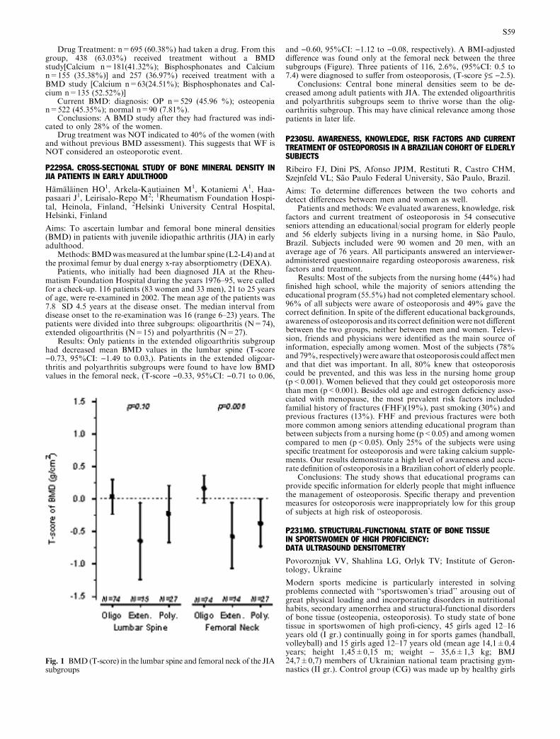
Drug Treatment: n=695 (60.38%) had taken a drug. From thisgroup, 438 (63.03%) received treatment without a BMDstudy[Calcium n=181(41.32%); Bisphosphonates and Calciumn=155 (35.38%)] and 257 (36.97%) received treatment with aBMD study [Calcium n=63(24.51%); Bisphosphonates and Cal-cium n=135 (52.52%)]
Current BMD: diagnosis: OP n=529 (45.96 %); osteopenian=522 (45.35%); normal n=90 (7.81%).
Conclusions: A BMD study after they had fractured was indi-cated to only 28% of the women.
Drug treatment was NOT indicated to 40% of the women (withand without previous BMD assessment). This suggests that WF isNOT considered an osteoporotic event.
P229SA. CROSS-SECTIONAL STUDY OF BONE MINERAL DENSITY INJIA PATIENTS IN EARLY ADULTHOOD
Hamalainen HO1, Arkela-Kautiainen M1, Kotaniemi A1, Haa-pasaari J1, Leirisalo-Repo M2; 1Rheumatism Foundation Hospi-tal, Heinola, Finland, 2Helsinki University Central Hospital,Helsinki, Finland
Aims: To ascertain lumbar and femoral bone mineral densities(BMD) in patients with juvenile idiopathic arthritis (JIA) in earlyadulthood.
Methods: BMDwasmeasured at the lumbar spine (L2-L4) and atthe proximal femur by dual energy x-ray absorptiometry (DEXA).
Patients, who initially had been diagnosed JIA at the Rheu-matism Foundation Hospital during the years 1976–95, were calledfor a check-up. 116 patients (83 women and 33 men), 21 to 25 yearsof age, were re-examined in 2002. The mean age of the patients was7.8 SD 4.5 years at the disease onset. The median interval fromdisease onset to the re-examination was 16 (range 6–23) years. Thepatients were divided into three subgroups: oligoarthritis (N=74),extended oligoarthritis (N=15) and polyarthritis (N=27).
Results: Only patients in the extended oligoarthritis subgrouphad decreased mean BMD values in the lumbar spine (T-score)0.73, 95%CI: )1.49 to 0.03,). Patients in the extended oligoar-thritis and polyarthritis subgroups were found to have low BMDvalues in the femoral neck, (T-score )0.33, 95%CI: )0.71 to 0.06,
and )0.60, 95%CI: )1.12 to )0.08, respectively). A BMI-adjusteddifference was found only at the femoral neck between the threesubgroups (Figure). Three patients of 116, 2.6%, (95%CI: 0.5 to7.4) were diagnosed to suffer from osteoporosis, (T-score y£ )2.5).
Conclusions: Central bone mineral densities seem to be de-creased among adult patients with JIA. The extended oligoarthritisand polyarthritis subgroups seem to thrive worse than the olig-oarthritis subgroup. This may have clinical relevance among thosepatients in later life.
P230SU. AWARENESS, KNOWLEDGE, RISK FACTORS AND CURRENTTREATMENT OF OSTEOPOROSIS IN A BRAZILIAN COHORT OF ELDERLYSUBJECTS
Ribeiro FJ, Dini PS, Afonso JPJM, Restituti R, Castro CHM,Szejnfeld VL; Sao Paulo Federal University, Sao Paulo, Brazil.
Aims: To determine differences between the two cohorts anddetect differences between men and women as well.
Patients and methods: We evaluated awareness, knowledge, riskfactors and current treatment of osteoporosis in 54 consecutiveseniors attending an educational/social program for elderly peopleand 56 elderly subjects living in a nursing home, in Sao Paulo,Brazil. Subjects included were 90 women and 20 men, with anaverage age of 76 years. All participants answered an interviewer-administered questionnaire regarding osteoporosis awareness, riskfactors and treatment.
Results: Most of the subjects from the nursing home (44%) hadfinished high school, while the majority of seniors attending theeducational program (55.5%) had not completed elementary school.96% of all subjects were aware of osteoporosis and 49% gave thecorrect definition. In spite of the different educational backgrounds,awareness of osteoporosis and its correct definitionwere not differentbetween the two groups, neither between men and women. Televi-sion, friends and physicians were identified as the main source ofinformation, especially among women. Most of the subjects (78%and79%, respectively)were aware that osteoporosis could affectmenand that diet was important. In all, 80% knew that osteoporosiscould be prevented, and this was less in the nursing home group(p<0.001). Women believed that they could get osteoporosis morethan men (p<0.001). Besides old age and estrogen deficiency asso-ciated with menopause, the most prevalent risk factors includedfamilial history of fractures (FHF)(19%), past smoking (30%) andprevious fractures (13%). FHF and previous fractures were bothmore common among seniors attending educational program thanbetween subjects from a nursing home (p<0.05) and among womencompared to men (p<0.05). Only 25% of the subjects were usingspecific treatment for osteoporosis and were taking calcium supple-ments. Our results demonstrate a high level of awareness and accu-rate definition of osteoporosis in a Brazilian cohort of elderly people.
Conclusions: The study shows that educational programs canprovide specific information for elderly people that might influencethe management of osteoporosis. Specific therapy and preventionmeasures for osteoporosis were inappropriately low for this groupof subjects at high risk of osteoporosis.
P231MO. STRUCTURAL-FUNCTIONAL STATE OF BONE TISSUEIN SPORTSWOMEN OF HIGH PROFICIENCY:DATA ULTRASOUND DENSITOMETRY
Povoroznjuk VV, Shahlina LG, Orlyk TV; Institute of Geron-tology, Ukraine
Modern sports medicine is particularly interested in solvingproblems connected with ‘‘sportswomen’s triad’’ arousing out ofgreat physical loading and incorporating disorders in nutritionalhabits, secondary amenorrhea and structural-functional disordersof bone tissue (osteopenia, osteoporosis). To study state of bonetissue in sportswomen of high profi-ciency, 45 girls aged 12–16years old (I gr.) continually going in for sports games (handball,volleyball) and 15 girls aged 12–17 years old (mean age 14,1±0,4years; height 1,45±0,15 m; weight ) 35,6±1,3 kg; BMJ24,7±0,7) members of Ukrainian national team practising gym-nastics (II gr.). Control group (CG) was made up by healthy girls
Fig. 1 BMD (T-score) in the lumbar spine and femoral neck of the JIAsubgroups
S59
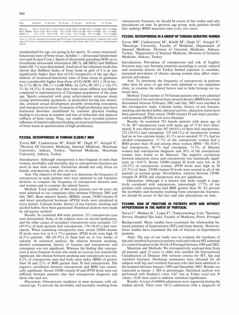
standardized for age, not going in for sports. To assess structural-functional state of bone tissue ;Achilles+; ultrasound densitometrwas used (LunarCorp.). Speed of ultrasound spreading (SOS,m/s),broadband ultrasound attenuation (BUA, dB/MHz) and StiffnessIndex (SI,%) were determined. Analysis of the obtained results hasshown that Stiffness Index of bone tissue in girls of I st gr wassignificantly higher than that of CG irrespective of the age (fig.).Indexes of structural-functional state of bone tissue in gymnastswere considerably higher than those of CG (SOS: 1611±28m/sec ;by 2,1%; BUA: 108,7±1,6 dB/MHz ; by 5,6%; SI: 107,1±3,by 3%11; by 14,3%). It means that their bone tissue stiffness was highercompared to representatives of Ukrainian population of the sameage. Sports connected with great achievements witnesses manyyoung sportswomen (gymnasts, in particular) having late menar-che, arrested sexual development possibly promoting osteopeniaand osteoporosis in future. Gymnasts of high proficiency may havehormonal disorders annihilated by constant physical loadingleading to in-crease in number and size of trabecular and improvestiffness of bone tissue. Thus, our studies have revealed positiveinfluence of intensive physical leading on structural-functional stateof bone tissue in sportswomen of high proficiency.
P232SA. OSTEOPOROSIS IN TURKISH ELDERLY MEN
Yavuz BB2, Cankurtaran M1, Khalil M1, Dagli N2, Ariogul S1;1Division Of Geriatric Medicine, Internal Medicine, HacettepeUniversity, Ankara, Turkey, 2Internal Medicine, HacettepeUniversity, Ankara, Turkey
Introduction: Although osteoporosis is less frequent in men thanwomen, morbidity and mortality due to osteoporotic fractures aremore in men than women. Geriatrists should not only focus onfemale osteoporosis, but also on men.
Aim: The objective of this study is to determine the frequency ofosteoporosis in male patients who were admitted to our Geriatricdivision, to compare the frequency of osteoporosis between menand women and to examine the related factors.
Method: Total number of 464 male patients over 64 years oldwere admitted to our outpatient clinic between February 2002 andJuly 2003. Bone mineral density (BMD), serum 25OH-vitamin Dand intact parathyroid hormone (iPTH) levels were calculated inevery patient. Calcium intake, history of any fracture, smoking andalcohol habits, have been questioned. Statistical analysis were madeby chi-square method.
Results: In examined 464 male patients, 213 osteoporosis casewere determined. None of the subjects were on steroid medication,and the other causes of secondary osteoporosis were excluded. 213(45.9%) of male patients had osteoporosis, 170 (36.6%) had oste-openia. When examining osteoporotic men, serum 25OH-vitaminD levels were low in 8 (1.7%) patients, iPTH levels were high 29(6.3%) patients. 380 (81.9%) of them had no or low intake ofcalcium. In statistical analysis, the relation between smoking,alcohol consumption, history of fracture and osteoporosis andosteopenia was not significant. Whereas the finding that osteopo-rosis is more frequent in men who made no exercise was statisticallysignificant, the relation between smoking and osteoporosis was not.4.2% of osteoporotic men had body mass index (BMI) of greaterthan 30 and 22% of BMI greater than 30 had osteoporosis. Thenegative correlation between osteoporosis and BMI was statisti-cally significant. Serum 25OH-vitamin D and iPTH levels were notdifferent between patients who had osteoporosis diagnosis andthose who had not.
Discussion: Osteoporosis incidence in men increases with ad-vanced age. To prevent the morbidity and mortality resulting from
osteoporotic fractures, we should be aware of this reality and takeprecautions on time. In geriatric age group, male patients shouldalso undergo BMD measures once every two years.
P233SU. OSTEOPOROSIS IN A GROUP OF TURKISH GERIATRIC WOMEN
Yavuz BB2, Cankurtaran M1, Khalil M1, Dagli N2, Ariogul S1;1Hacettepe University, Faculty of Medicine, Department ofInternal Medicine, Division of Geriatric Medicine, Ankara,Turkey, 2Department of Internal Medicine, Division of GeriatricMedicine, Ankara, Turkey
Introduction: Prevalence of osteoporosis and risk of fragilityfractures may vary between countries according to social, culturaland economic factors. In Turkey limited exposure to sunshine,increased prevalence of obesity among women may affect osteo-porosis prevalence.
Aim: To determine the frequency of osteoporosis in patientsolder than 64 years of age who were admitted to our outpatientclinic, to examine the related factors and to help forming our na-tional data.
Method: Total number of 783 female patients whowere admittedtoGeriatricsUnit andwhose bonemineral density (BMD) have beendetermined between February 2002 and July 2003 were enrolled inthis retrospective study. Calcium intake, history of any fracture,smoking and alcohol habits, physical activity, education status havebeen questioned. Their serum 25OH-vitamin D and intact parathy-roid hormone (iPTH) levels were obtained.
Results: In examined 783 female patients with mean age of71.34, 507 osteoporosis cases with mean age of 71.82 were deter-mined. It was observed that 507 (64.8%) of them had osteoporosis,232 (29.6%) had osteopenia. 325 (64.1%) of osteoporotic womenhad no or low calcium intake, 81.1% never smoked, 97.2% had noalcohol consumption. 17.7% of the osteoporotic women had aBMI greater than 30 and among obese women (BMI>30) 58.8%had osteoporosis, 30.7% had osteopenia. 71.5% of illiteratewomen had osteoporosis diagnosis and 36% of the osteoporoticwomen were found to be illeterate. The negative correlationbetween education status and osteoporosis was statistically signif-icant (p=0.017). Serum 25OH-vitamin D levels were low in 20(10.2%) of osteoporotic women, iPTH levels were high in 50(25.5%) of them. 25OH-vitamin D levels were normal in all of thepatients in normal group. Nevertheless, relation between 25OH-vitamin D, iPTH and osteoporosis was not significant.
Discussion: Although it is known that body weight is nega-tively correlated with osteoporosis, interestingly 17.7% of ourpatients with osteoporosis had BMI greater than 30. To preventthe morbidity and mortality resulting from osteoporotic fractures,we should be aware of this fact and take precautions on time.
P234MO. RISK OF FRACTURE IN PATIENTS WITH AND WITHOUTHYPERTENSION IN THE NORTH OF PORTUGAL
Neves C1, Medina JL1, Lopes F2; 1Endocrinology Unit, 2StatisticsService, Hospital Sao Joao, Faculty of Medicine, Porto, Portugal
Backgrounds: Many studies have examined the relationship be-tween presence of hypertension (HT) and bone density. However,fewer studies have examined the risk of fracture in hypertensivepatients.
Aims: The aim of our study was to evaluate the incidence ofhip and vertebral fractures in patients with andwithoutHT admittedto a central hospital in theNorth of Portugal between 1989 and 2002.
Materials and Methods: We retrospectively analysed data fromall patients aged 25 years or older who satisfied the InternationalClassification of Diseases (9th version) criteria for HT, hip andvertebral fractures. Discharge summaries were obtained for allsubjects with hip and vertebral fractures who had been admitted tothe hospital between January 1989 and December 2002. Results areexpressed as means ± SD or percentages. Statistical analysis wasperformed with Student’s t-test, Chi2 test or Fisher exact test. Pvalues <0.05 were used to indicate statistical significance.
Results: A total of 644884 admissions were registered during thestudied period. There were 38112 admissions with a diagnosis of
Data All group 45–49 years 50–54 years 55–59 years 60–64 years 65–70 years
SOS, m/s 1541.3±29.1a 1547.5±27.0 a 1554.3±30.0a 1544.4±29.3a,b 1534.7±22.2a,b,c 1520.5±28.5a,b,cBUA, dB/MG 106.5±11.0a 107.6±10.8a 110.5±10.4a 106.9±10.1a,b 104.9±10.5a,b,c 101.1±13.0a,b,cStiffnes, % 82.6±13.7a 84.9±12.8a 88.7±14.0a 83.8±12.8a,b 76.9±11.1a,b,c 73.2±14.8a,b,c
Notes: p<0.05 (a– compared with women aged 40–44 years in premenopausal period, a- compared with womenaged 50–54 years in postmenopausal period, c- compared with women aged 55–59 years in postmenopausalperiod).
S60
fractures without HT and 1300 admissions with a diagnosis offractures and HT. Hypertensive patients [42.5% men (M) and57.5% women (W)] were significantly older than normotensivepatients (48.4% M and 51.6% W) (73.6 ± 2.6 vs 43.6 ± 3.7 years,p < 0.001). The duration of hospitalisation was significantly higherin hypertensive than in normotensive patients (21.4 ± 6.9 vs12.1 ± 2.6 days, p < 0.001). There were no significant differencesin the incidence of vertebral fractures in hypertensive and normo-tensive patients (0.5% vs 0.4%, p = NS). The incidence of hipfractures was significantly higher in hypertensive than in normo-tensive patients (29.2% vs 12.1%, p < 0.001).
Conclusions: This study indicates that HT is a risk factor forhip fractures, suggesting that fracture prevention efforts should bea consideration in the treatment of hypertensive patients.
P235SA. EXAMINATION OF THE EFFECT OF DIETARY HABITS ON BONEMINERAL DENSITY IN HEALTHY MEN AGED 15 TO 49 YEARS LIVING INTHE CITY OF DEBRECEN.
Bhattoa HP, Bettembuk P, Balogh A; Regional OsteoporosisCenter, Medical and Health Science Center, University ofDebrecen, Hungary
Introduction: Diet is known to be one of the most importantpredictors of bone mineral density, the aim of the present study isto characterize the dietary habits and examine its effects on bonemineral density (BMD) in a healthy male population aged 15–49 years.
Patients and Methods: The local population based register wasused to select a population based random sample. Subjects withhistory of non-traumatic fracture, a longer than 3 month history ofchronic disease affecting bone metabolism were excluded. A mod-ified version of the WHO osteoporosis questionnaire was used tointerview the participants about daily dietary calcium, protein,carbohydrate, fat and energy intake. BMD was measured at theL2-L4 lumbar spine and the femur using the Lunar DPX-L DEXAdensitometer.
Results: A total of 177 (average age 39.9 ± 8.7 years) men wereenrolled in the study. It was not possible to estimate peak bone massdue to the marked fluctuation in mean BMD across the 5-year age-bands (15–19, 20–24, 25–29, 30–34, 35–39, 40–44, 45–49 years). Themean spine BMD was lowest in the oldest age-band. Mean femoralneck and trochanter BMD was highest between 25–29 yearsand gradually decreased with age, the lowest values being seen inthe 35–39 years age-band. There was significant correlation betweenL2-L4 BMD and daily dietary calcium (r=0.273, p<0.001) andprotein (r=0.163, p=0.030) intake; femur neck BMD and dailydietary calcium (r=0.271, p<0.001), protein (r=0.228, p=0.002),fat (r=0.188, p=0.012), and energy (r=0.172, p=0.022) intake.
Conclusion: Our findings suggest that among the dietary com-ponents examined the daily dietary calcium, protein, fat and energyintake correlated significantly with femur neck BMD and only thedaily dietary calcium and protein intake correlated with lumbarspine BMD. The knowledge of dietary components best responsiblefor a healthy bone mass may help formulate adequate diet orientedpreventive strategies in those between 15–49 years of age as suchreduce the burden of osteoporosis later in life.
P236SU. BASIC AWARENESS ON OSTEOPOROSIS AND THE RISKFACTORS FOR THE DISEASE AMONG COVERED WOMEN
Keskin Y1, Gunduz OH2, Sur H1, Ofluoglu D2, Eker HH3, AkyuzG2; 1Marmara University, Health Education Faculty, Istanbul,Turkey, 2MarmaraUniversity, School ofMedicine, Department ofP.&M.R., Istanbul, Turkey, 3Istanbul Municipality, Istanbul,Turkey
Objective: This study was designed to evaluate the basic aware-ness level among covered women above 40 years of age, and tofind out the risk factors for osteoporosis and osteopenia in them.
Design: 517 covered women, who live in Istanbul, Turkey, wereincluded in the study. Questionnaires were applied by interviewersand the bone mineral density (BMD) measurements were done withDXL (Calscan).
Results: The mean age of 517 women was 49.5±7.4 years.Mean height, bodyweight and body mass index were; 154±5.3 cm;77.7±12.5 kg; and 32.8±5.2 kg/m2, respectively. Education andsocioeconomical levels were low in these women.
Most of the women (59.8%) heard the name of the disease(OP), and the majority (60.6%) learned it from their friends, 12.3%from the doctors, and 7.3 from the media. However, only 6.2% hadenough information about OP.
The most common concomitant diseases in this study groupwere hypertension (18.3%), gastric ulcer (9%), rheumatoid arthritisand osteoarthritis (6.1%), and diabetes mellitus (4.7%). Continueddrug use among our study population is relatively high: 30.2%. Theratio of birth control pill use was 23.2%, and 18.8% were givenhormone replacement therapy (HRT) after their menopause. Theratio of women who had hysterectomy and/or oopherectomy wasvery high (16%). The ratio of women, who had history of onefracture, was 11.4%. Only 6.8% regularly perform exercises. Phy-sical activity levels of these women both between 15–25 years ofages, and 25–50 years age were higher.
There was a significant relation between the bone mineral den-sities and physical activity level (p<0.05).No significant relationwasfound between BMD’s and performance of regular exercises, con-comitant drug, birth control pill, andHRTuses, and smoking status.
Osteoporotic patients had a significantly higher rate of historyof fractures (p<0.05). The ratio of fractures among covered wo-men who were osteoporotic were common around wrist/forearm.Hip fractures were rare.
Conclusion: Covering can effect vitamin D synthesis and cal-cium absorption. Education and socioeconomic level among cov-ered women was lower. Peer education is important for informingthese women about OP. There is a relation between physicalactivity level and osteoporosis among covered women.
P237MO. PREVALENCE OF OSTEOPENIA (MILD, MODERATE, SEVERE)AND OSTEOPOROSIS IN 4000 ARGENTINE POSTMENOPAUSAL WOMEN
Lopez MO, Wasserman L, Bron D, Tocchetton M, Sanchez SM,Volta SM, Massari FEG, Zanchetta JR; Instituto de Investiga-ciones Metabolicas )IDIM, Buenos Aires, Argentina, School ofMedicine, USAL University, Buenos Aires, Argentina
Aims: To analyse the prevalence of densitometric diagnoses inambulatory postmenopausal women and its relation with demo-graphic characteristics.
Methods: Descriptive, cross-sectional study. From a databaseof 25,510 women who participated in a free osteoporosis detectioncampaign with BMD assessment and answered a questionnaire, acomputer software selected at random 4,000 patients (1,000 foreach age decile = 50–59, 60–69, 70–79 and 380). Diagnoses: Basedon the region (lumbar spine /femoral neck) with lowest T-scorevalue (WHO criteria). Osteopenia was classified in mild (T-score�1.0 to �1.49), moderate (T-score �1.5 to �1.99) and severe(T-score �2.0 to �2.49).
Results: Demographic Characteristics (mean/SD): Age: 69.41±9.94 years, Menoage: 19.62±10.55 years, Body Mass Index:28.06±4.5, Weight: 66.99±11.26 kg. and Height: 1.54±0.06 cm.Densitometric Diagnoses (table 1)
Osteopenia classification: severe 19.32%; moderate 17.75%;and mild 14%.
The prevalence of fractures in severe Osteopenia was notdifferent from Osteoporosis (p=0.103) This was also seen in BMI(28 vs 26), Menoage (16 vs 22), and Age (66 vs 71).
Conclusions: 1) The prevalence of Osteoporosis is similar to theone defined in other international studies. 2) We deemed important
Table 1.
Prevalence in % (n)
Age decile 50–60 60–70 70–80 80–99 Total
Osteoporosis 20.8 (208) 33.3 (333) 40.2 (402) 45.6 (456) 34.97 (1399)Osteopenia 52.6 (526) 51.5 (515) 51.4 (514) 48.8 (488) 51.05 (2043)Normal 26.6 (266) 15.2 (152) 8.4 (84) 5.6 (56) 13.98 (558)
S61
to classify Osteopenia in groups, because fracture risk is higher inSevere Osteopenia. 3) Following the guidelines of previous con-sensus, BMD measured in only one region, does NOT allow todiagnose Osteoporosis in 19.15% of the women <65 years’old andin 27.51% of the women 3 65 years’old.
P238SA. VALIDATION OF A QUESTIONNAIRE ABOUT RISK FACTORSFOR THE DIAGNOSIS OF OSTEOPOROSIS
Messina OD, Pate DA, Peralta A, Riopedre AM; RheumatologyDept., Cosme Argerich Hospital, Buenos Aires, Argentina
We evaluated in a previous study the predictive value of riskfactors related to osteoporosis in a population of 785 womendesigning a score for diagnostic purposes (score OPAR, osteo-porosis Argentina). The aim of the present study is to validate theOPAR score.
Patients and methods: Score was built, including body massindex, age, early onset menopause, history of atraumatic previousfractures, concomitant diseases and chronic use of glucocorticoids.
502 postmenopausal women were included. DEXA (LunarDPX alpha) measurements were determined in all of them (lumbarspine LS, and proximal femur PF).Statistical analysis was per-formed by multiple logistic regression to evaluate the association ofrisk factors as predictors of low bone mineral density values ac-cording to T score (WHO classification 1994). OPAR score rangedfrom 0 to 9 points according to the number and type answers to therisk factors ) based questionnaire.
Results: 138 out of the 502 women (27.49%) presented osteo-porosis (T score below )2.5 in LS or PF). OPAR score correlatedwith the diagnosis of osteoporosis (e.g. OPAR 3.5 to 4=prob-ability of OP 45.5%, OPAR 4.5 or greater than 4.5=probability ofOP is 65.7%)
Area under curve (AUC) was 0.7318.For practical purposes we propose a cut-off value ( cut-off va-
lue=3 points; sensitivity=63%, specificity=71.7 %, positive pre-dictive value 45.79% and negative predictive value 83.65%).
We conclude that OPAR is a useful score to focus bone mineraldensity measurements requests based on risk factors for osteo-porosis. After applying OPAR score one out of two women hadosteoporosis according to the WHO criteria. This score might beused for general practitioners as a useful tool to identify women tomeasure bone mineral density in a cost-effective fashion.
P239SU. THE INFLUENCE OF BODY COMPOSITION ON BONE MINERALDENSITY OF BRAZILIAN WOMEN
Pippa MGB1, Latorre MRO2, Zerbini CAF3; 1Heliopolis Hospi-tal Rheumatology Department, 2School of Public Health,University of Sao Paulo, 3Heliopolis Hospital RheumatologyDepartment, Sao Paulo, Brazil
Bone mineral density (BMD) is under the influence of bodycomposition (BC), mainly fat and lean mass, and also it dependson race. Considering that Brazilian people is composed of a widevariety of races, including black, white and mestizos, we studiedthe influence of BC in BMD of Brazilian women, according to thestatus of menopause and race. 160 women (90 white and 72nonwhite) from 20-79 years old were studied. Dual energy x-rayabsorptiometry was used to measure the total BMD (TBMD),lumbar spine BMD(LBMD), femur BMD (FBMD), fat mass(FM)and lean mass (LM).
Variables were evaluated as descriptive patterns using mean,standard deviation, median, minimum and maximun values. Forthe comparison of means of TBMD, regional BMDs and measuresof body composition according to status of menopause and race,analysis of variance to one factor was used. For multiple com-parisons we used Tukey-HSD test. To compare the difference ofmeans between the variables of white and nonwhite women, theStudent t-test was used. The homogeneity of variance was evalu-ated by Lavene test and the adherence to normal curve by Kol-mogorov Smirnov test. Correlation among variables was estimatedutilizing the coefficient of Pearson correlation. Multiple regressionanalysis was carried out to verify the effect of altogether significative
correlation found. We concluded: 1) there were no significant dif-ferences when we compare themeans of LBMDandFBMDbetweenthe 162 white and nonwhite women, 2) the mean of TBMD in non-white premenopausal women was higher than the mean of whitewomen in the same group, 3) in white premenopausal women thebodymass index was the greatest determinant of BMDon all studiedregions, 4) in nonwhite premenopausal women lean mass was thegreatest determinant on TBMD and on LBMD, while on FBMD itwas the age, 5) fat mass was an important determinant of BMD inwhite postmenopausal women on all studied regions, 6)in nonwhitepostmenopausal women lean mass was the significant determinanton TBMD and regional BMDs.
P240MO. FEEFING PATTERNS OF ADOLESCENTS ATTENDING PUBLICTEACHING CENTERS IN URUGUAY
Severi C1,2, Girona A1,2, Aldabe I1,2, Ramagli A1; 1Study Groupon Bone Diseases of the Uruguayan Society of Rheumatology,Montevideo, Uruguay, 2Department of Basic Nutrition-School ofNutrition, Montevideo, Uruguay
Objective: To analyze the behavior and feeding habits of ado-lescents attending teaching centers in Uruguay.
Methodology: the sample included 3,000 adolescents represen-tative of the various areas in the country and the various socio-economic levels, attending first and third year high school. This isan exploratory descriptive study. The data were taken through aself-administered questionnaire presented by a nutritionist in eachgroup of adolescents.
Results: The results show that 25% of adolescents do no drinkmilk daily, being calcium intake insufficient in 44% of the casesstudied (assuming that 60% come from dairy sources). The averageconsumption of calcium for Montevideo was 928.46 with a SD of550.07, while for the interior of the country it was 974.21 with a SDof 614.14 mg. There is a mild tendency to reduce dairy productconsumption as age increases, with lower consumption in third yearstudents versus first year students. Seventy five per cent of theadolescents have breakfast every day.
Fifty per cent report no physical activity outside the curricularactivity (2 hours a week). As a counterpart, 42% devote from 2 to 4hours in sedentary activities as watching TV and 32% devote morethan 4 hours a day. Ninety per cent report a great interest on issuesrelated to nutrition and 60% express that they receive their infor-mation on food from their families, followed by 30% who statethey receive them from their teachers.
Conclusions: We conclude that dairy product consumption islow in the adolescents attending high school. Designing andimplementing programs promoting a healthy lifestyle, especiallyincreasing dairy product consumption and physical activity is apriority. These programs must include the adolescents’ educatorsand families, and it is essential to evaluate them after they areimplemented.
P241SA. COMPARISON OF VERTEBRAL FRACTURE PREVALENCE ANDOSTEOPOROSIS RISK FACTORS BETWEEN COMMUNITY DWELLINGOSTEOPOROSIS PATIENTS AND NURSING HOME RESIDENTS
Sivrioglu K1, Bayram S1, Dincsahin E1, Akgoz S2, KucukogluHS1; 1Uludag University School of Medicine Department ofPhysical Medicine and Rehabilitation, Bursa, Turkey, 2UludagUniversity School of Medicine Department of Biostatistics,Bursa, Turkey
This study was designed in order to compare community dwellingosteoporosis patients and nursing home residents according tovertebral fracture prevalence and osteoporosis risk factors.
Study population consisted of 56 women aged 70–80 years.First group consisted of 32 osteoporosis patients who were beingfollowed up in a university outpatient clinic and the second groupconsisted of 24 women living in Bursa Nursing Home. All patientswere interviewed for osteoporosis risk factors using a question-naire, bone mineral density (BMD) was measured by DEXA andlateral lumbosacral x-rays were taken. Serum Ca, PTH, calcitonin,calcitriol and 24 hour urinary Ca levels were measured. Vertebral
S62
fractures were assessed using a semiquantitative method. The twostudy groups were compared according to vertebral fracture prev-alence, osteoporosis risk factors, BMD and laboratory parameters.
The study groups were similar in terms of mean age (Group1:74.3±3.2, Group2: 75.2±3.1 years) and body mass index (Group1:27.8±4.7, Group2: 27.8±6.6 kg/m2), while nursing home residentshad a longer postmenopausal duration (Group1 27.4±8.0,Group2: 33.3±9.5 years, p=0.014). Although vertebral fractureprevalence was found to be lower in the first group (65.6% vs79.2%), this difference was not statistically significant. Number offractures was also similar in both groups. Significantly more sub-jects in the second group had at least one hormonal risk factor suchas nulliparity, early menopause and surgical menopause (p=0.002).BMD measurements were similar in both groups except femur neckBMD which was significantly lower in the second group (Group1:0.613±0.119, Group2: 0.523±0.139 g/cm2, p=0.017). Laboratoryparameters were also similar in both groups except 24 hour urinaryCa excretion which was found to be significantly lower in the sec-ond group (p=0.021).
In conclusion, no significant difference in vertebral fractureprevalence was found between community dwelling osteporosispatients and nursing home residents. Low femur neck BMD innursing home residents might play a role in hip fracture risk but didnot correlate with vertebral fracture prevalence in this study pop-ulation.
P242SU. QUALITY OF LIFE IN POSTMENOPAUSAL OSTEOPOROSIS
Borman P, Tanyolac O, Atan C, Bodur H; Numune Training andResearch Hospital, Dept. of PMR, Ankara, Turkey
The aim of this study was to determine the impact of osteoporosison the quality of life in a group of postmenopausal women.
Thirty females with osteoporosis, 30 females with osteopeniaand 30 healthy control subjects; all age-matched were included tothe study. Demographic data, lumbar spine and femoral neck bonemineral density (BMD) were collected. Quality of life questionnaireof the European Foundation for Osteoporosis (QUALEFFO) wasused to assess health-related quality of life.
The mean age of the subjects was 58.1±8.32 years. There was 9patients with non-traumatic lumbar and/or femoral fracture his-tory. The mean scores of pain and physical activity domains ofQUALEFFO were significantly higher in the osteoporotic groupthan in the osteopenic females and control subjects (pain:56.8±21.1 vs 42.9±20.2 vs 36.1±21.9 ; physical activity:47.2±16.7 vs 36.7±12.5 vs 331.7±17.9, respectively). The scoresof psychosocial domains were also higher in the osteoporotic fe-males than in other subjects but the difference did not reach tosignificance. Women who experienced a fracture had significantlyhigher total QUALEFFO scores, indicating lower quality of life,compared with non-fractured females (48.8 ± 15.2 vs 31.6 ± 11.3,respectively). The BMD levels were similar betweeen the femaleswith and without fractures.
In conclusion, osteoporotic females have a reduced quality oflife, especially related with fractures. The negative impact of oste-oporosis on quality of life, seems to be more related to physicalsubgroups rather than psychosocial domains.
P243MO.SIFRAR)ANORIGINALSCORETOASSESSRISKOFPREVALENTVERTEBRAL FRACTURES IN POSTMENOPAUSAL WOMEN: PRELIMINARYRESULTS
Goncalves S, Arias MA, Cachizumba MO, Aguero RG, VoltaSM, Gallo RA, Massari FEG, Zanchetta JR; Instituto deInvestigaciones Metabolicas )IDIM, Buenos Aires, Argentina.,School of Medicine, USAL University, Buenos Aires, Argentina.
Aims: To design a score with clinical and densitometric parame-ters in order to be able to predict the prevalence of vertebralfractures in postmenopausal women.
Methods: Data was obtained from a database with multipleclinical and densitometric parameters collected during an Osteo-porosis Detection Campaign. SIFRAR score was calculatedaccording to: Menoage: (year of test minus last menstrual year)
divided by 10; Hip T-score multiplied by 1 or Spine T-score mul-tiplied by 2 turning T-score into a positive value (Lower T-scorewas selected from both); Height loss obtained from the differencebetween the current height and the calculated maximal height(expressed as an absolute value); History of vertebral fractures:6 points; Previous non-vertebral fractures: 1 point; Self-reportedosteoporosis diagnosis: 1 point; Maternal history of hip fracture:1 point; Maternal history of vertebral fracture: 1 point; Body MassIndex: 0 (>26); 1 (25-26); 2 (23-24); 3 (21-22); 4 (<20); Self-reported health status: 2 (bad); 1 (regular); 0 (good). SIFRAR wascalculated on 4,345 women whose maximal height was calculatedeither from the knee height or the refered maximal height or thearm span. For these preliminary results 829 patients, with maximalheight calculated from the knee height and with a score over 7, wereselected. A dorsolumbar spine radiography was performed on thissample and reviewed by trained physicians.
Results:Demographics: Mean Age: 69.88 ±6.28; Mean BMI:27.04 ±3.64; Mean Menoage: 22.58 ±7.92.The prevalence ofvertebral fractures in our sample was 15.92%, with a proportionalincrease related to the score. The sample distribution and theprevalence of fractures are shown on Table 1.
Conclusions:We consider this score a useful tool which wouldallow to identify those women to whom a spinal radiographyassessment should be recommended. A SIFRAR score over 9would identify postmenopausal women at high risk of prevalentvertebral fractures.
P244SA. EPIDEMIOLOGICAL MULTICENTER STUDY ON OSTEOPOROSIS:THE EDMUSTO STUDY
Gokce-Kutsal Y1, Ozcakar L1, Eskiyurt N2, Eryavuz M3,Marozyuska KG4, Emam M5, Dursun N6, Korap E6, Aktas A7,BarakA7,Karaagaoglu E7; 1HacettepeUniversityMedical School,Physical Medicine and Rehabilitation, Ankara, TURKEY, 2Istan-bul University Istanbul Medical School, Physical Medicine andRehabilitation, Istanbul, TURKEY, 3Istanbul University Cerrah-pasa Medical School, Physical Medicine and Rehabilitation,Istanbul, TURKEY, 4Medical Academy, Rheumatology,Wroclaw, POLAND, 5Longman Rheumatology Center, Tehran,IRAN, 6Kocaeli University Medical School, Physical Medicineand Rehabilitation, Kocaeli, TURKEY, 7Hacettepe UniversityMedical School, Biostatistics, Ankara, TURKEY
Aims: Osteoporosis is a systemic skeletal disease characterized bylow bone mass and microarchitectural deterioration of bone tis-sue, with a consequent increase in bone fragility and susceptibilityto fractures. Although osteoporosis is a worldwide problem thataffects every population and occurs in all geographic areas studiedthus far, the incidence differs among different populations andethnic groups –greatest in whites and Asians and least in blacks.In this study, what we aimed to accomplish was the comparativeanalysis of bone mineral density measurements of postmeno-pausal women from 3 different countries. This preliminary reportsummarizes the early data from Turkey, Iran and Poland.
Methods: The study comprised 679 postmenopausal women;472 from Turkey, 104 from Iran and 103 from Poland. Demo-graphical data )including age, age at menopause, body weight,height, etc.- daily calcium intake and physical activity levels of theindividuals were recorded. Bone mineral density measurementswere performed using dual energy x-ray absorptiometry. Statisticalanalysis was done by using Statistical Package for Social Sciences.Results: The mean ages at menopause were respectively45.85±6.20, 47.60±3.75, 47.30±5.30 and the difference betweenthe countries was found to be statistically significant (p=0.009).
SIFRAR Subjectsn (%)
Subjects with PrevalentVertebral Fracture n (%)
<9)‡7 466 (56.22 %) 41 ( 8.79 %)‡9 363 (43.78 %) 91 (25.06 %)
TOTAL 829 (100%) 132 (15.92 %)
S63
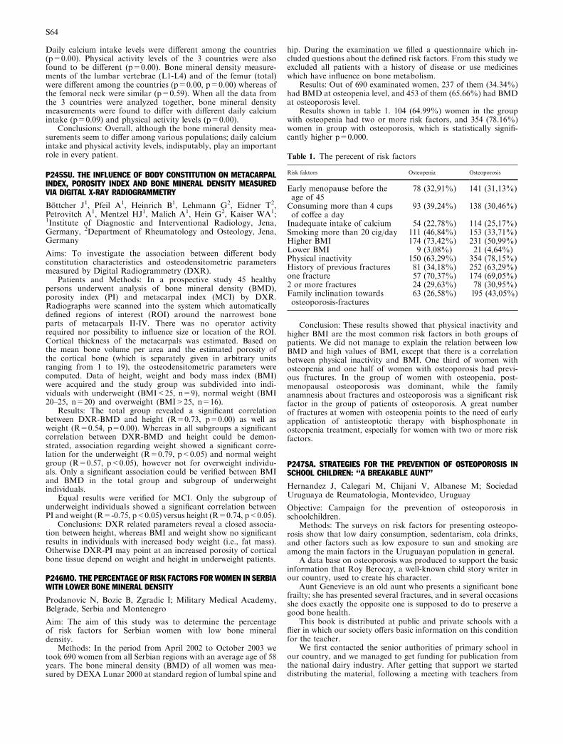
Daily calcium intake levels were different among the countries(p=0.00). Physical activity levels of the 3 countries were alsofound to be different (p=0.00). Bone mineral density measure-ments of the lumbar vertebrae (L1-L4) and of the femur (total)were different among the countries (p=0.00, p=0.00) whereas ofthe femoral neck were similar (p=0.59). When all the data fromthe 3 countries were analyzed together, bone mineral densitymeasurements were found to differ with different daily calciumintake (p=0.09) and physical activity levels (p=0.00).
Conclusions: Overall, although the bone mineral density mea-surements seem to differ among various populations; daily calciumintake and physical activity levels, indisputably, play an importantrole in every patient.
P245SU. THE INFLUENCE OF BODY CONSTITUTION ON METACARPALINDEX, POROSITY INDEX AND BONE MINERAL DENSITY MEASUREDVIA DIGITAL X-RAY RADIOGRAMMETRY
Bottcher J1, Pfeil A1, Heinrich B1, Lehmann G2, Eidner T2,Petrovitch A1, Mentzel HJ1, Malich A1, Hein G2, Kaiser WA1;1Institute of Diagnostic and Interventional Radiology, Jena,Germany, 2Department of Rheumatology and Osteology, Jena,Germany
Aims: To investigate the association between different bodyconstitution characteristics and osteodensitometric parametersmeasured by Digital Radiogrammetry (DXR).
Patients and Methods: In a prospective study 45 healthypersons underwent analysis of bone mineral density (BMD),porosity index (PI) and metacarpal index (MCI) by DXR.Radiographs were scanned into the system which automaticallydefined regions of interest (ROI) around the narrowest boneparts of metacarpals II-IV. There was no operator activityrequired nor possibility to influence size or location of the ROI.Cortical thickness of the metacarpals was estimated. Based onthe mean bone volume per area and the estimated porosity ofthe cortical bone (which is separately given in arbitrary unitsranging from 1 to 19), the osteodensitometric parameters werecomputed. Data of height, weight and body mass index (BMI)were acquired and the study group was subdivided into indi-viduals with underweight (BMI<25, n=9), normal weight (BMI20–25, n=20) and overweight (BMI>25, n=16).
Results: The total group revealed a significant correlationbetween DXR-BMD and height (R=0.73, p=0.00) as well asweight (R=0.54, p=0.00). Whereas in all subgroups a significantcorrelation between DXR-BMD and height could be demon-strated, association regarding weight showed a significant corre-lation for the underweight (R=0.79, p<0.05) and normal weightgroup (R=0.57, p<0.05), however not for overweight individu-als. Only a significant association could be verified between BMIand BMD in the total group and subgroup of underweightindividuals.
Equal results were verified for MCI. Only the subgroup ofunderweight individuals showed a significant correlation betweenPI and weight (R=-0.75, p<0.05) versus height (R=0.74, p<0.05).
Conclusions: DXR related parameters reveal a closed associa-tion between height, whereas BMI and weight show no significantresults in individuals with increased body weight (i.e., fat mass).Otherwise DXR-PI may point at an increased porosity of corticalbone tissue depend on weight and height in underweight patients.
P246MO. THE PERCENTAGE OF RISK FACTORS FOR WOMEN IN SERBIAWITH LOWER BONE MINERAL DENSITY
Prodanovic N, Bozic B, Zgradic I; Military Medical Academy,Belgrade, Serbia and Montenegro
Aim: The aim of this study was to determine the percentageof risk factors for Serbian women with low bone mineraldensity.
Methods: In the period from April 2002 to October 2003 wetook 690 women from all Serbian regions with an average age of 58years. The bone mineral density (BMD) of all women was mea-sured by DEXA Lunar 2000 at standard region of lumbal spine and
hip. During the examination we filled a questionnaire which in-cluded questions about the defined risk factors. From this study weexcluded all patients with a history of disease or use medicineswhich have influence on bone metabolism.
Results: Out of 690 examinated women, 237 of them (34.34%)had BMD at osteopenia level, and 453 of them (65.66%) had BMDat osteoporosis level.
Results shown in table 1. 104 (64.99%) women in the groupwith osteopenia had two or more risk factors, and 354 (78.16%)women in group with osteoporosis, which is statistically signifi-cantly higher p=0.000.
Conclusion: These results showed that physical inactivity andhigher BMI are the most common risk factors in both groups ofpatients. We did not manage to explain the relation between lowBMD and high values of BMI, except that there is a correlationbetween physical inactivity and BMI. One third of women withosteopenia and one half of women with osteoporosis had previ-ous fractures. In the group of women with osteopenia, post-menopausal osteoporosis was dominant, while the familyanamnesis about fractures and osteoporosis was a significant riskfactor in the group of patients of osteoporosis. A great numberof fractures at women with osteopenia points to the need of earlyapplication of antisteoptotic therapy with bisphosphonate inosteopenia treatment, especially for women with two or more riskfactors.
P247SA. STRATEGIES FOR THE PREVENTION OF OSTEOPOROSIS INSCHOOL CHILDREN: ‘‘A BREAKABLE AUNT’’
Hernandez J, Calegari M, Chijani V, Albanese M; SociedadUruguaya de Reumatologia, Montevideo, Uruguay
Objective: Campaign for the prevention of osteoporosis inschoolchildren.
Methods: The surveys on risk factors for presenting osteopo-rosis show that low dairy consumption, sedentarism, cola drinks,and other factors such as low exposure to sun and smoking areamong the main factors in the Uruguayan population in general.
A data base on osteoporosis was produced to support the basicinformation that Roy Berocay, a well-known child story writer inour country, used to create his character.
Aunt Genevieve is an old aunt who presents a significant bonefrailty; she has presented several fractures, and in several occasionsshe does exactly the opposite one is supposed to do to preserve agood bone health.
This book is distributed at public and private schools with aflier in which our society offers basic information on this conditionfor the teacher.
We first contacted the senior authorities of primary school inour country, and we managed to get funding for publication fromthe national dairy industry. After getting that support we starteddistributing the material, following a meeting with teachers from
Table 1. The perecent of risk factors
Risk faktors Osteopenia Osteoporosis
Early menopause before theage of 45
78 (32,91%) 141 (31,13%)
Consuming more than 4 cupsof coffee a day
93 (39,24%) 138 (30,46%)
Inadequate intake of calcium 54 (22,78%) 114 (25,17%)Smoking more than 20 cig/day 111 (46,84%) 153 (33,71%)Higher BMI 174 (73,42%) 231 (50,99%)Lower BMI 9 (3,08%) 21 (4,64%)Physical inactivity 150 (63,29%) 354 (78,15%)History of previous fractures 81 (34,18%) 252 (63,29%)one fracture 57 (70,37%) 174 (69,05%)2 or more fractures 24 (29,63%) 78 (30,95%)Family inclination towardsosteoporosis-fractures
63 (26,58%) l95 (43,05%)
S64
several schools. This material was later on integrated into theteaching activities at schools, and was disseminated to parents.
Results:There has been an excellent acceptance at all school-teaching levels. Lectures on the book were organized at teachingcenters, stating that next year it will be distributed and the materialwill be worked out.Television programs discussing health topicsalso gave us space to present educational material for prevention.Conclusions: Health prevention has succeeded in calling uponmany sectors involved in health, teaching and the mass mediaaround a topic that is highly sensitive as bone health in children.Dissemination results are favorable throughout the country.Rheumatologists and teachers have been working jointly in theimplementation of the strategies for the prevention of osteopo-rosis.
P248SU. OSTEOPOROSIS AND RHEUMATOID ARTHRITIS INCIDENCEOF FRACTURES
Gonzalez G1,2, Chijani V1, Souto R1, Gomez E1, Barreto G1,Moyano M1,2, Neves G1,2; 1Sociedad Uruguaya de Reumato-logia, Montevideo, Uruguay, 2Catedra de Reumatologia, Mon-tevideo, Uruguay
It is well known that patients with RA are at a higher risk ofpresenting fractures due to osteoporosis. This led to the study of apopulation of patients with RA, the incidente of fractures in thatpopulation, and their bone mass, to assess their relation with thestage of the disease and the therapies received, especially Gluco-corticosteroids.Material and Methods Ninety patients with RA ( as per ACRcriteria) were selected. They were all worked out with densitom-etry using the dual Dexa method, regardless of their functionalclass, their time of onset and therapy received. The prospectiveprotocol developed (2002 – 2003) included the following:
– Personal data– Age of onset of the RA, time since onset, radiological degree,extraarticular manifestations ( Nodules, Vasculitis, Pleuro-pulmonar Involvement, etc), presence of comorbidity, gastro-intestinal manifestations
– (gastritis, ulcera), glandular diseases: (diabetes, thyroid dis-ease), cardiac disease
– Hypertension– Gyneco-obstetric history, age at menopause, hormonereplacement therapy
– History of joint replacements– Presence of fractures, time elapsed at the time of protocoli-zation
– Clinical laboratory: rheumatoid factor and acute phasereactants
– CDM using the Dexa method– Bone resorption or formation markers– Treatments administered: NSAIDs, Corticosteroids– prednisone equivalent doses– 5mgMethotrexate, Leflunomide, Sulfazalacine, Azathioprina,Hydroxyclhoroquine dosaging and time since the patient hasbeen on the medication
– Calcium vitamin D, Biphosphonates, other medications, timeand doses received
Results: Average age of patients at the time of protocolizationwas 57 years, with a frank female prevalence Mean age of onset ofthe disease was 49 years.-22 % presented subcutaneous nodules, 11% vasculitis, 25 % other complications. 52 % didn’t present extra-articular complications Bone Densitometry confirmed osteoporosisin 52%, osteopenia in 22% and normal bone mineral density in26%.- 11% presented fractures: vertebral, wrist, CF, shoulder,elbow
Conclusions: 80 % of the patients with RA have a reduced bonemineral density, 11% presented fractures as a complication of theirosteoporosis.-We highlight the importance of an early diagnosis ofthis comorbidity and an early therapy, tending to reduce the inci-dence of fractures.
P249MO. BIALYSTOK OSTEOPOROSIS STUDY (BOS): BMD ANDFRACTURES
Badurski JE1, Nowak NA1, Supronik J2, Dobrenko A1, Lis J3,Daniluk S1, Jeziernicka E1; 1Polish Foundation of Osteoporosis,Bialystok, Poland, 2Sniadecki Hospital, Bialystok, Poland, 3E.Lilly Poland, Pharmaeconomic Dept., Warsaw, Poland
Hip T-score –2.5 and below is a WHO diagnostic threshold ofosteoporosis. It arises a question, at what BMD fragility fracturesoccur, which are a main feature of this disease. We tried to answerthis question on the basis of BOS and comparison of its data withothers (1,2). To asses relations between number and frequency offractures and BMD, 1005 women aged 46–89, mean 59.3±9.4,filled in the questionnaire concerning known fracture and osteo-porosis risk factors and 727 of them have had their bone massdensity (BMD) examined by Hologic QDR4500SL in L1-L4vertebra of spine, neck and total hip. Distribution of age in BOScohort and whole Polish population is similar.T-score below –2.5 were observed in 18.4% of women if measuredin the spine, in 14.8% if in neck and 2.4% if in total hip. 222 outof 727 (30.5%) have experienced fractures in the past, 53% ofthem were Colles ones, 9.4% of the ribs, 2.3% of the hip, 3.6% ofthe vertebra, and 31.5% of others. Mean neck T-score of womenwith previous fracture were –1.6 (vs –0.96 of that without frac-ture), and L1-L4 T-score –1.5 (vs –0.99 respectively). 50.0 % offractures occurred in women with BMD between neck T-score –1.0 and –2.5, and 26.7 % within normal values and only 24.3%within osteoporotic one. Neck T-score of women with previousfracture is the lower the more advanced age, but segment ofwomen with neck T-score below –2.5 is most numerous in the ageof 65–74 as well as one year fracture risk increases with age, butabsolute number of fractures in BOS cohort is highest in womenaged 45–64. Very similar phenomenon, namely, more numerousfractures at the range of osteopenic and normal bone mass hasbeen described by others (1,2).
References:1. Burger H et al.: Am J Epidemiol 1998,147:871.2. Wainwright SA et al.: J Bone Miner Res 2001, 16:1077/S155.
P250SA. SKELETAL STATUS IN EASTERN SLOVAKIA MALES ASSESSEDBY DXA AND QUANTITATIVE ULTRASOUND
Tomkova S1, Payer J2, Killinger Z2; 1Hospital Kosice-Saca, SlovakRepublic, 2University Hospital Bratislava, Slovak Republic
There are only a few reports of bone mineral density status inmales in Slovakia. The aim of the study was to assess theoccurrence of osteopenia and osteoporosis in males, analyse thecause of osteoporosis and to interpret obtained results.
In the study we examined a total of 1681 males, mean age 51.2years, from East Slovakia ironworks involved in regular preventivemedical check-up. In 1602 males bone status was measured byquantitative ultrasound (Sahara, Hologic) and in a subgroup of 519males BMD was assessed by DXA at lumbar spine and hip (Ho-logic 4500 A). In males examined by DXA osteopenia was found in53% and osteoporosis in 8.3% of subjects, in 6.7% in lumbar spineand in 1.5% in hip. In subjects examined by QUS, osteopenia wasfound in 28% and osteoporosis only in 3.4% of males. In maleswith osteoporosis the cause of decreased BMD was further analy-sed. 62.3% of males were classified as idiopathic osteoporosis and37.7% as secondary osteoporosis. In 69% hepatopathia was foundas a most frequent cause of secondary osteoporosis and in 31% ofthem ethyltoxic aetiology was assessed. Endocrine disorder wasfound in 1%, drugs causing osteoporosis in 1.5 %, rheumatic dis-eases in 3.4%, gastrointestinal diseases in 3%, nefropathia in 17%and diabetes mellitus in 5% of males.
In our nonselected group of relatively young males involved inpreventive medical check-up osteoporosis was found in quite highproportion, comparing with overall estimated prevalence of oste-oporosis in European population. Hepatopathia was surprisinglyfound the most common cause of secondary osteoporosis amongthe males in our study. Measurement of lumbar spine by DXA
S65
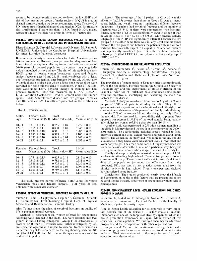
seems to be the most sensitive method to detect the low BMD andrisk of fractures in our group of males subjects. If QUS is used inskeletal status evaluation we do not recommend to use T score )2.5SD (osteoporosis threshold assessed and used in DXA) for fracturerisk stratification. T score less than )1 SD, found in 32% males,represent already the high risk group in terms of fracture risk.
P251SU. BONE MINERAL DENSITY REFERENCE VALUES IN MALESAND FEMALES 10 TO 21 YEARS OF AGE IN VENEZUELAN SUBJECTS
Riera-Espinoza G, Carvajal R, Velasquez G, Naressi M, Ramos J;UNILIME. Universidad de Carabobo, Hospital UniversitarioDr. Angel Larralde, Valencia, Venezuela
Bone mineral density reference values for Latin American popu-lations are scarce. However, comparison for diagnosis of lowbone mineral density in adults requires normal reference values of20-40 years old control population. In contrast, in young indi-viduals, matched by sex and age. The aim of this study is to showBMD values in normal young Venezuelan males and femalessubjects between ages 10 and 21. 191 healthy subjects with at leastone Venezuelan progenitor, ages between 10 and 21, without anysystemic disease of drug use which affects bone metabolism wererecruited for bone mineral density assessment. None of the sub-jects were under heavy physical therapy or training nor hadprevious fracture. BMD was measured by DEXA (LUNARDPX. Variation Coefficient: 1.5%) at femoral neck and lumbarspine (L1-L4). Subjects were divided into two groups: 89 malesand 102 females. BMD results are presented in the 2 tables asfollow.
This study presents normal reference BMD values for youngVenezuelan males and females subjects, 10–21 years of age,obtained with Lunar densitometer.
P252MO. EFFECT OF VERTEBRAL FRACTURE ON QUALITY OF LIFE
Yilmaz F, Sahin F, Caglayan A, Taspinar S, Ozcan B, DurlanikG, Kuran B; Sisli Etfal Teaching Hospital, Dept. of PhysicalMedicine and Rehabilitation, Istanbul, Turkey
Aim: To investigate the effect of vertebral fractures on quality oflife in postmenopausal women.
Method: 41 postmenopausal women referred for osteoporosisscreening were included in the study.They were classified into twogroups as those having osteopenia (Group I) or osteoporosis (tscore £)2.5)(Group II). Patients were further investigated by lat-eral spine radiographs with respect to vertebral fracture defined as25 percent height loss compared to the neighbouring vertebra. SF36,QUALEFFO 41 and NHP were the questionnaires used toevaluate life quality.
Results: The mean age of the 11 patients in Group I was sig-nificantly (p£0.05) greater than those in Group II. Age at meno-pause, height and weight were not significantly different betweenthe groups. 16 patients had vertebral fractures and the number offractures was 25. 84% of them were compression type fractures.Energy subgroup of SF 36 was significantly lower in Group II thanin Group I (37.3±16 vs 48.2±11, p £ 0.05). Only physical activitysubgroup of the NHP was significantly different between the twogroups. On the other hand, there was not any significant differencebetween the two groups and between the patients with and withoutvertebral fractures with respect to life quality. Number of fractureswas significantly correlated (r=0.31) with the home activitiessubgroup of QUALLEFFO 41 and physical activity subgroup ofNHP (r=0.32).
P253SA. OSTEOPOROSIS IN THE URUGUAYAN POPULATION
Chijani V1, Hernandez J1, Severi C2, Girona A2, Aldabe I2;1Uruguayan Society of rheumatology, Montevideo, Uruguay,2School of nutrition and Dietetics. Dpto of Basci Nutrition,Montevideo, Uruguay
The prevalence of osteoporosis in Uruguay affects approximately5% of the population. For this reason, the Uruguayan Society ofRheumatology and the Department of Basic Nutrition of theSchool of Nutrition of UDELAR have conducted some studieswith the objective of identifying and describing the main riskfactors for the disease.
Methods: A study was conducted from June to August, 1999, on asample of 3,503 adult patients attending the office. They filled aquestionnaire with questions on the educational level, life style, foodconsumption and elements of the personal and family medical history.
While 67% of the women fell into the risk group, only 16% ofthe men did. The threshold for susceptibility risk to present Oste-oporosis was present in 59.1% of the total sample, being remark-ably higher for women (67.1% ) than for men (16.8%).
Another study was performed in 256 adult women attending tothe clinic in Montevideo and the south of the country in the 2000 )2001 period. The questionnaire included aspects related to food,lifestyles, current and usual weight records and personal and familyhistory. The women in the study had lived more in the city and didless exercise< they had a lower educational level, a lower BMI andlower body weight. The urban conditions of Uruguayan women wasfound to be associated with OP in a most particular way, being therisk higher in those women who change from rural life to city life.
Finally a descriptive study was carried out on a sample of 1,300adolescents attending high school. Twenty five per cent do notconsume milk daily. There is an insufficient intake of calcium in44% of the population (assuming that 60% come from dairyproducts). Fifty per cent do not practice sports apart from thephysical activity in high school. Twenty one per cent declaredhaving suffered some fracture.
Conclusions: The studies conducted clearly show the lifestyleand consumption habits as risk factors that are present and mightbe conditioning the early occurrence of osteoporosis with its seriousconsequences.
P254SU. MUNICIPALITIES’ LEVEL HEALTH EDUCATION FOR OSTEO-POROSIS IN JAPAN
Satomura K, Nakahara T, Iwanaga S, Noami M, Komatsu A,Sakamoto R, Sakurami T; Dept. of Public Health, Faculty ofMedicine, Kyoto University, Japan
Aim: In Japan health education for osteoporosis is very impor-tant because one of the causes of it is low intake of calcium.Osteoporosis is one of the targets of Healthy Japan 21, which is ahealth promotion framework in Japan. Main carrier of thiseducation is municipalities. We surveyed their health educationprograms and their cooperation with other organizations.
Subjects and Method: A questionnaire asking their healtheducation programs for osteoporosis was sent to all municipalitiesin Japan. Their cooperation with other organizations for healtheducation was also inquired.
Table 1 Reference Values
Males. Femoral Neck Troch L1–L4Age Mean value (SD) Mean value (SD) Mean value (SD)
10–11 0.867 � 0.13 0.755 � 0.09 0.779 � 0.1112–13 0.956 � 0.11 0.830 � 0.08 0.846 � 0.1114–15 1.032 � 0.18 0.911 � 0.16 0.986 � 0.1616–17 1.086 � 0.10 0.913 � 0.10 1.103 � 0.1618–19 1.135 � 0.10 0.845 � 0.04 1.243 � 0.0520–21 0.956 � 0.11 0.752 � 0.12 1.092 � 0.03
Females. Femoral Neck Troch L1–L4Age Mean value (SD) Mean value (SD) Mean value (SD)
10–11 0.754 � 0.15 0.655 � 0.11 0.815 � 0.1012–13 0.913 � 0.11 0.782 � 0.11 0.981 � 0.1414–15 0.965 � 0.12 0.775 � 0.10 1.037 � 0.1316–17 0.995 � 0.07 0.816 � 0.07 1.094 � 0.1118–19 1.001 � 0.11 0.759 � 0.08 1.116 � 0.0720–21 0.959 � 0.11 0.785 � 0.11 1.156 � 0.13
S66
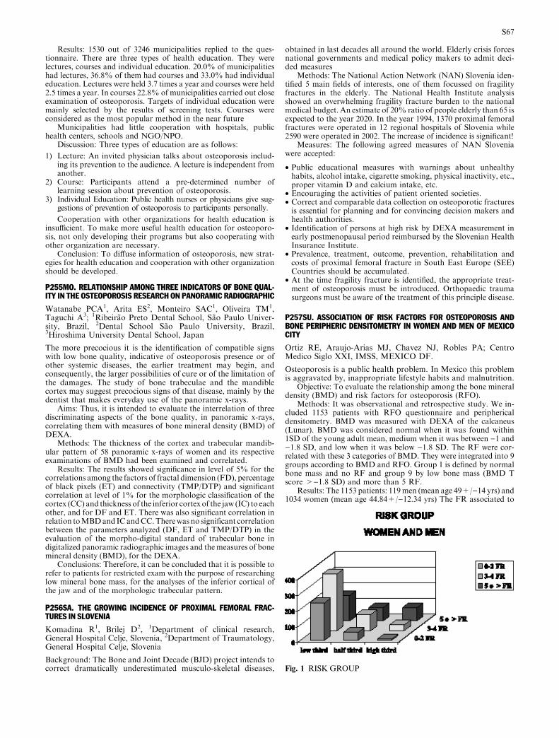
Results: 1530 out of 3246 municipalities replied to the ques-tionnaire. There are three types of health education. They werelectures, courses and individual education. 20.0% of municipalitieshad lectures, 36.8% of them had courses and 33.0% had individualeducation. Lectures were held 3.7 times a year and courses were held2.5 times a year. In courses 22.8% of municipalities carried out closeexamination of osteoporosis. Targets of individual education weremainly selected by the results of screening tests. Courses wereconsidered as the most popular method in the near future
Municipalities had little cooperation with hospitals, publichealth centers, schools and NGO/NPO.
Discussion: Three types of education are as follows:
1) Lecture: An invited physician talks about osteoporosis includ-ing its prevention to the audience. A lecture is independent fromanother.
2) Course: Participants attend a pre-determined number oflearning session about prevention of osteoporosis.
3) Individual Education: Public health nurses or physicians give sug-gestions of prevention of osteoporosis to participants personally.
Cooperation with other organizations for health education isinsufficient. To make more useful health education for osteoporo-sis, not only developing their programs but also cooperating withother organization are necessary.
Conclusion: To diffuse information of osteoporosis, new strat-egies for health education and cooperation with other organizationshould be developed.
P255MO. RELATIONSHIP AMONG THREE INDICATORS OF BONE QUAL-ITY IN THE OSTEOPOROSIS RESEARCH ON PANORAMIC RADIOGRAPHIC
Watanabe PCA1, Arita ES2, Monteiro SAC1, Oliveira TM1,Taguchi A3; 1Ribeirao Preto Dental School, Sao Paulo Univer-sity, Brazil, 2Dental School Sao Paulo University, Brazil,3Hiroshima University Dental School, Japan
The more precocious it is the identification of compatible signswith low bone quality, indicative of osteoporosis presence or ofother systemic diseases, the earlier treatment may begin, andconsequently, the larger possibilities of cure or of the limitation ofthe damages. The study of bone trabeculae and the mandiblecortex may suggest precocious signs of that disease, mainly by thedentist that makes everyday use of the panoramic x-rays.
Aims: Thus, it is intended to evaluate the interrelation of threediscriminating aspects of the bone quality, in panoramic x-rays,correlating them with measures of bone mineral density (BMD) ofDEXA.
Methods: The thickness of the cortex and trabecular mandib-ular pattern of 58 panoramic x-rays of women and its respectiveexaminations of BMD had been examined and correlated.
Results: The results showed significance in level of 5% for thecorrelations among the factors of fractal dimension (FD), percentageof black pixels (ET) and connectivity (TMP/DTP) and significantcorrelation at level of 1% for the morphologic classification of thecortex (CC) and thickness of the inferior cortex of the jaw (IC) to eachother, and for DF and ET. There was also significant correlation inrelation toMBDand ICandCC.Therewas no significant correlationbetween the parameters analyzed (DF, ET and TMP/DTP) in theevaluation of the morpho-digital standard of trabecular bone indigitalized panoramic radiographic images and themeasures of bonemineral density (BMD), for the DEXA.
Conclusions: Therefore, it can be concluded that it is possible torefer to patients for restricted exam with the purpose of researchinglow mineral bone mass, for the analyses of the inferior cortical ofthe jaw and of the morphologic trabecular pattern.
P256SA. THE GROWING INCIDENCE OF PROXIMAL FEMORAL FRAC-TURES IN SLOVENIA
Komadina R1, Brilej D2, 1Department of clinical research,General Hospital Celje, Slovenia, 2Department of Traumatology,General Hospital Celje, Slovenia
Background: The Bone and Joint Decade (BJD) project intends tocorrect dramatically underestimated musculo-skeletal diseases,
obtained in last decades all around the world. Elderly crisis forcesnational governments and medical policy makers to admit deci-ded measures
Methods: The National Action Network (NAN) Slovenia iden-tified 5 main fields of interests, one of them focussed on fragilityfractures in the elderly. The National Health Institute analysisshowed an overwhelming fragility fracture burden to the nationalmedical budget. An estimate of 20% ratio of people elderly than 65 isexpected to the year 2020. In the year 1994, 1370 proximal femoralfractures were operated in 12 regional hospitals of Slovenia while2590 were operated in 2002. The increase of incidence is significant!
Measures: The following agreed measures of NAN Sloveniawere accepted:
• Public educational measures with warnings about unhealthyhabits, alcohol intake, cigarette smoking, physical inactivity, etc.,proper vitamin D and calcium intake, etc.
• Encouraging the activities of patient oriented societies.• Correct and comparable data collection on osteoporotic fracturesis essential for planning and for convincing decision makers andhealth authorities.
• Identification of persons at high risk by DEXA measurement inearly postmenopausal period reimbursed by the Slovenian HealthInsurance Institute.
• Prevalence, treatment, outcome, prevention, rehabilitation andcosts of proximal femoral fracture in South East Europe (SEE)Countries should be accumulated.
• At the time fragility fracture is identified, the appropriate treat-ment of osteoporosis must be introduced. Orthopaedic traumasurgeons must be aware of the treatment of this principle disease.
P257SU. ASSOCIATION OF RISK FACTORS FOR OSTEOPOROSIS ANDBONE PERIPHERIC DENSITOMETRY IN WOMEN AND MEN OF MEXICOCITY
Ortiz RE, Araujo-Arias MJ, Chavez NJ, Robles PA; CentroMedico Siglo XXI, IMSS, MEXICO DF.
Osteoporosis is a public health problem. In Mexico this problemis aggravated by, inappropriate lifestyle habits and malnutrition.
Objective: To evaluate the relationship among the bone mineraldensity (BMD) and risk factors for osteoporosis (RFO).
Methods: It was observational and retrospective study. We in-cluded 1153 patients with RFO questionnaire and periphericaldensitometry. BMD was measured with DEXA of the calcaneus(Lunar). BMD was considered normal when it was found within1SD of the young adult mean, medium when it was between )1 and)1.8 SD, and low when it was below )1.8 SD. The RF were cor-related with these 3 categories of BMD. They were integrated into 9groups according to BMD and RFO. Group 1 is defined by normalbone mass and no RF and group 9 by low bone mass (BMD Tscore >)1.8 SD) and more than 5 RF.
Results: The 1153 patients: 119men (mean age 49+/)14 yrs) and1034 women (mean age 44.84+/)12.34 yrs) The FR associated to
Fig. 1 RISKGROUP
S67
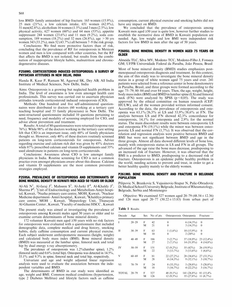
loss BMD: family antecedent of hip fracture. 165 women (15.9%),25 men (21%), a low calcium intake, 651 women (62.9%),51men(42.8%), alcoholism 16women (1.5%) and 3men (2.5%), lowphysical activity, 627 women (60%) and 64 men (53%), appetitesuppressant 244 women (23.6%) and 11 men (9.2%), soda con-sumption, 189 women (18.2%) and 32 men (26.8%), age >50 yrswomen 343 (33.2%), men 52 (43.7%) all between 7–9 the risk group.
Conclusions: We find more protective factors than of risk,concluding that the prevalence of RF for osteoporosis in Mexicanwomen and men is low compared with other countries, but the RFthat affects the BMD is not isolated, but results from the combi-nation of inappropriate lifestyle habits, malnutrition and chronic-degenerative diseases.
P258MO. CORTICOSTEROID INDUCED OSTEOPOROSIS: A SURVEY OFPHYSICIAN ATTITUDES IN NEW DELHI, INDIA
Handa R, Kaur P, Ramam M, Agarwal SK, Dey AB; All IndiaInstitute of Medical Sciences, New Delhi, India
Aims: Osteoporosis is a growing but neglected health problem inIndia. The level of awareness is low even amongst health careprofessionals. This survey was carried out to ascertain physicianattitudes towards corticosteroid induced osteoporosis (CIO).
Methods: One hundred and five self-administered question-naires were distributed to doctors (60 working at a tertiary careteaching hospital and 55 family physicians) in New Delhi. Thesemi-structured questionnaire included 10 questions pertaining toneed, frequency and modality of screening employed for CIO, andadvice about prevention and treatment.
Results: A total of 80 questionnaires were returned (response rate76%). While 98% of the doctors working in the tertiary care settingfelt that CIO is an important issue, only 60% of family physiciansthought so. However, only 40% of these doctors routinely screenedpatients on long-term corticosteroids for osteoporosis. Adviceregarding exercise and calcium rich diet was given by 45% doctorswhile 87% prescribed calcium and vitamin D supplements and 35%used alendronate in patients on long-term corticosteroids.
Conclusions: Awareness about CIO is low amongst familyphysicians in India. Routine screening for CIO is not a commonpractice even amongst physicians aware about this disease. Calciumand vitamin D supplements are the most common preventivestrategies employed.
P259SA. PREVALENCE OF OSTEOPOROSIS AND DETERMINANTS OFBONE MINERAL DENSITY IN KUWAITI MEN AGED 50 YEARS OR OLDER
Al-Ali N1, Al-faraj J2, Mahseen S3, Al-Jarki F4, Al-Khaldy J5,Sharma P6; 1Unit of Endocrinology and Metabolism-Amiri hospi-tal, Kuwait, 2Nuzha primary care centre, MOH, Kuwait, 3NuclearMedicine department- Amiri hospital,. Kuwait, 4Khaldiya primarycare centre, MOH , Kuwait, 5Hepatology Unit, ThunayanAl-Ghanim Center , Kuwait, 6Faculty of medicine/HSCC ,Kuwait
The present study was aimed at investigating the prevalence ofosteoporosis among Kuwaiti males aged 50 years or older and toexamine certain determinants of bone mineral density.
172 volunteer Kuwaiti men aged ‡50 years with no risk factorsfor osteoporosis were evaluated with a questionnaire that includeddemographic data, complete medical and drug history, smokinghabits, daily caffeine consumption and current physical exercise.Each subject underwent anthropometric measures (height, weight)and calculated body mass index (BMI). Bone mineral density(BMD) was measured at the lumbar spine, femoral neck and totalhip by dual energy x-ray absorptiometry.
The prevalence of osteoporosis was 1.2%(lumbar spine), 1.2%(femoral neck) and 0.6% (total hip). Osteopenia was detected in 14.5%,33.1% and 9.3% in spine, femoral neck and total hip, respectively.
Univariate and age and weight adjusted linear regressionanalysis were used to evaluate the association between the inde-pendent variables and BMD.
The determinants of BMD in our study were identified asage, weight and BMI. Common medical conditions (hypertension,type 2 Diabetes Mellitus) and lifestyle factors such as caffeine
consumption, current physical exercise and smoking habits did nothave any impact on BMD.
We concluded that the prevalence of osteoporosis amongKuwaiti men aged ‡50 year is quite low, however further studies toestablish the normative data of BMD in Kuwaiti population areneeded. Age, low weight and low BMI were independent riskfactors for low BMD in men after the age of 50 years.
P260SU. BONE MINERAL DENSITY IN WOMEN AGED 75 YEARS OROLDER
Almeida TLC, Silva MV, Modesto TCC, Modesto-Filho J, FonsecaGM; UFPB Universidade Federal da Paraıba, Joao Pessoa, Brasil
Most of bone mineral density (BMD) studies emphasizes post-menopausal osteoporosis diagnosis and treatment. In this context,the aim of this study was to investigate the bone mineral densitystatus in a group of white women aged 75 years and over. 241patients were selected from a reference center in bone densitometryin Paraıba, Brasil, and three groups were formed according to theage: 75–79; 80–84 and over 84 years. Then, the age, weight, height,bodymass index (BMI) andBMDin lumbar spine (LS) and femoralneck (FN) were analyzed by SPSS (p<0,05). The study wasapproved by the ethical committee on human research (CEP/HULW), and all the women provided written informed consent.According to the data, the prevalence of osteoporosis and oste-openia was 63,1%/26,5% at LS and 53,9%/38,6% at FN. Theanalysis between LS and FN showed 42,3% concordance forosteoporosis, 14,1% for osteopenia and 2,9% for the normalstatus. The main discordant results were between osteoporotic LSand osteopenia FN (19,1%) while the minor was between osteo-porotic LS and normal FN (1,7%). It was observed that the cor-relation and regression analysis were positive between BMD andBMI but were not significant between BMD and age in thethree groups. Almost all data showed different levels of bone loss,mainly with osteoporosis status in LS and FN in all groups. Theadvanced of the age raise the bone mass decrease, predisposing toan increased risk of fracture. However, it was observed that theBMI is a predictor to BMD, predisposing to a decreased risk offracture. Osteoporosis is an epidemic public healthy problem inthe world, needing actions to prevent and treat, in order to get abetter healthy quality mainly to the elderly women.
P261MO. BONE MINERAL DENSITY AND FRACTURE IN BELGRADEPOPULATION
Pilipovic N, Brankovic S, Vujasinovic-Stupar N, Palic-ObradovicD;MedicalSchoolUniversityBelgrade, InstituteofRheumatology,Belgrade, Serbia and Montenegro
Objective: We examined 527 women aged 20–79 (48.54±12.36)and 126 man aged 20–77 (50.22±15.83) from urban part of
Table 1 Results
Decade Age Sex No of pts Osteoporosis Osteopenia Fracture
I 20–29 F 42 0 6 (14,3%) 0M 21 0 3 (14,3%) 0
II 30–39 F 63 1 (1,6%) 10 (15,9%) 0M 10 0 2 (20%) 0
III 40–49 F 186 5 (2,7%) 37 (18,9%) 23 (12,4%)M 27 2 (7,1%) 14 (51,9%) 4 (14,8%)
IV 50–59 F 131 12 (9,2%) 55 (42%) 26 (19,9%)M 22 3 (13,7%) 3 (13,6%) 1 (4,6%)
V 60–69 F 81 22 (27,2%) 28 (34,6%) 27 (33,3%)M 28 4 (14,3%) 9 (32,1%) 3 (10,7%)
VI 70–79 F 24 8 (33,3%) 5 (20,8%) 5 (20,8%)M 18 3 (16,7%) 4 (22,2%) 3 (16,7%)
TOTAL 20–79 F 527 48 (9,1%) 141 (26,8%) 81 (15,4%M 126 12 (9,5%) 35 (27,8%) 11 (8,7%)
S68
Belgrade, randomly selected from population register to establishbone mineral density (BMD) and frequency of fracture.
Methods: BMD of spine was measured by dual-energy x–rayabsorptiometry (DEXA), using a Lunar DPX-L device. Womenand men were stratified per decade of age. Results are on table.Out of 81 women with fracture, 31 (38.27%) had normal BMD,osteoporosis was found in 16 (19.8%) and osteopenia in 34 (42%).Out of 11 men with fracture 4 (36.4%) had normal BMD, osteo-porosis was found in 3 (27.3%) and osteopenia in 4 (36.4%).
Conclusion: Our study confirmed previous findings of otherauthors that for development of fracture, beside low BMD, theother factors are important.
P262SA. CLINICAL AND DENSITOMETRIC FINDINGS IN MALE LOWBONE DENSITY VENEZUELAN PATIENTS
VitaleM1,Riera-EspinozaG1,2,Cedeno-TabordaJ1,3; 1UNILIME,Hospital Universitario Dr. Angel Larralde, Universidad deCarabobo, Valencia, Estado Carabobo, Venezuela, 2UNIDADMETABOLICA, Centro Policlınico Valencia, Venezuela, 3UNI-CORP, Caracas, Venezuela
Male Osteoporosis is an underestimated condition. The aim ofthis study is to define the clinical and densitometric findings ofmale patients with low bone density.
We reviewed 150 clinical records of male patients with lowbone density. Age, height, weight, psychosocial habits, risks fac-tors, cause of osteoporosis or osteopenia, associated clinicalconditions, medical treatments, initial BMD and bone markerswere analyzed.
Age was 60.6 years. In the first visit back pain was present in46.4% of the patients, polyarticular pain in 24.1%, bone fractures in9.8%, neck pain in 5.4% and lower extremities paresis in 3.6%. Only10.7%came for apreventive visit.History of rheumatic diseaseswerepresent in 29.4%, hypertension 21.7%, neoplastic disease 19.4%,sexual dysfunction 15.3%, renal stones 10.5% and gastric pepticdisease 4.2%. Bone fractures in 20.6%, vertebral fractures in 10.6%,hip fractures in 3.3%. 66.6% had unique and 33.4% multiplefractures. Risk factors for osteoporosis were: sedentary habits in52.4% of the patients, smoking 33.6%, low calcium intake 74.1%,and alcoholic habits 12.6%. The initial BMD values were: femoralneck 0.759 g/cm2±0.32, T score -2.11±0.93; lumbar spine 1.012 g/cm2±0.17, T score -1.83±1.45. Osteopenia was present in 48.7%and osteoporosis in 51.3%. The initial values of bone biochemicalmarkers were: TRAP 9.4±3.23 IU/l, urine NTx 83.78±64.14nmBCE/mmcrea, Alkaline Phosphatase 45.64±18.26 IU/l.Regarding bone turnover 29% were low and 71% were high boneturnover.Causes ofOsteoporosiswere: idiopathic 48.7%, inducedbysteroids (SIO) 19.5%, hypogonadism 14.2%, alcoholism 8%, otherscauses 9.8%. There are not significant differences between differentcauses of osteoporosis, regarding BMD, bone biochemical markers,age, weight, height and BMI. Significant correlation was presentbetween TRAP, NTX and AP.
In conclusion our data show similarities with other publisheddata. We found that male osteoporosis is associated with otherchronic diseases frequently polymedicated. The three principalcauses are idiopathic, SIO and secondary to hypogonadism. Thevast majorities are sedentary, has a low calcium intake and showshigh bone turnover.
P263SU. THE PREVALENCE OF RHEUMATIC DISEASES IN RURAL EGYPT
Abdel-Tawab RR1, Abdel-Nasser AM1, Mahmoud JA1, SammyA2, Abdel-Fattah M3; 1Rheumatology & Rehabilitation Dept,Minia University, Egypt, 2Radiology Dept, Minia University,Egypt, 3Epidemiology Dept, Minia University, Egypt
Aims: Epidemiological studies of rheumatic diseases in the com-munity have shown variable prevalence rates between Westernand Oriental (Asian and African) populations, and between urbanand rural areas. Since there are no published studies of rheumaticdiseases prevalence in Egypt, this study was conducted to estimatethe prevalence of rheumatic diseases in a rural area in Egypt.
Methods: The study was conducted in Makosa village ofAl-Minia governorate in Southern Egypt in 4 phases. Of the wholeadult population (15 years and older) of the village comprising 5208individuals, 88 (1.7%) declined to participate in the study and 5120subjects (98.7%, 2562 female and 2558 male) were screened by apreliminary WHO-ILAR-COPCORD questionnaire inquiringabout pain, swelling, stiffness or tenderness in the bones, joints,and muscles in the last 7 days. Those who answered positivelywere administered a more detailed questionnaire and invited forexamination and laboratory and radiological investigations asrequired. A rheumatologist administered the 2 questionnaires face-to-face and carried out examinations to ensure reliability and validityof data collection. Consequently, rheumatic cases were classified intospecific diagnostic categories according to appropriate criteria.
Results:A total of 857people answeredpositively to the screeningquestionnaire and accepted clinical examination either at clinic or athome. Only 829 (16.2%) individuals had rheumatic diseases afterexamination and investigations, including 526 females (20.5%prevalence) and 303 males (11.8% prevalence). The prevalence ofspecific rheumatic disorders was as follows, osteoarthritis 8.5%,spinal disorders 6.4%, soft tissue rheumatism 6.6%, fibromyalgia1.3%, rheumatoid arthritis 0.29%, systemic lupus erythematosus0.05%, ankylosing spondylitis 0.09%, Reiter’s syndrome 0.04%,undifferentiated spondyloarthropathy 0.02%, pseudogout 0.08%,and dysplasias 0.02%. Among patients with rheumatic diseases, theknee was involved in 55.9%, the back in 30.3%, the neck in 12.9%,the hands in 10.9%, while the elbows, feet and shoulders were af-flicted in less than 10% each. It is of concern that a significant pro-portion of rheumatic diseases have remained undiagnosed in thecommunity.
Conclusion: The prevalence of rheumatic diseases in ruralEgypt is 16.2%. Knee and back pain, resulting mostly from jointdegeneration, caused the greatest burden of disease.
P264MO. ANKLE FRACTURES IN WOMEN AND OSTEOPOROTIC FRAC-TURE?
Nieto E, Salinas JR, Batalla A, Rivas E, Ferrer L; Bone &Metabolic Research Group (GIMO), Orthopedics & Traumatol-ogy Dept, University of Los Andes, Merida, Venezuela
Ankle fractures in the elderly are common but only few researchwe find in the literature about this problem in the women over 50years old. An observational study of 50 women oldest than 50years was performed among Merida, Venezuela, resident during 5years. We studied postmenopausal women ages 50-79 years (meanage 59.8±7.7years) with displaced ankle fractures. All thepatients were independent in the house and the community. Only2.0% from an geriatric hospital. The risk factors was smoking in14.0%, arterial hypertension in 22%, parity between 2 to 542.1%, nuliparity 7,9%, Body Mass Index 21.5 kg/m2.Fracturedue to moderate trauma in 80.0% (slide), outside of house in98.0% of them. Weber B2 Type predominate(54.0%) and 68.0%of surgical procedure in accordance with AO/ASIF principle wasperformed and the results were good and excellent in 92.0% ofcases by the modified Olerud and Molander scoring system. Inconclusion ankle fractures is a typical osteoporotic fracture.
P265SA. RISK FACTORS FOR OSTEOPOROSIS IN 446 WOMEN ABOVE40 YEARS OF VASSOURAS CITY, RIO DE JANEIRO STATE, BRAZIL
Lima MN, Goldfarb M, Guedes DSN, Reis TB, Barcellos AN,Lima MIA, Brilhante IP, Junior SDB, Alem A, Goulart FO;Universidade Severino Sombra, Vassouras-RJ, Brazil
Aim: Analysis of the risk factors to Osteoporosis directly relatedwith lifestyle(poor absorption of the calcium, smoking, non-practice of the physical exercises)in women above 40 years old.
Methods: Between september and december of 2002, interwieswere realized with 492 women above 40 years in Vassouras city-countryside of Rio de Janeiro state, Brazil(representing 10% of thefeminine population of the city above 40 years).Through the per-forming of the questionaries by these women from different socialclasses and areas of the city. They were inquired about the lifestyle
S69
aspects that could come to influence in the loss of the bone masslike regular practice of physical exercises, absorption of the foodsrich in calcium and smoking. The women that had a daily calciumabsorption <1000mg/day were considered in the risk group ofOsteoporosis ) also smokers and sedentaries.
Results: Fourty six women were eliminated in this study becausethey had a previous diagnosis of Osteoporosis. From 446 interwiees,401(89,9%) had a daily calcium absorption <1000mg and only45(10,1%) presented a daily absorption>1000mg; 138 (30,9%)weresmokers and 267(59,9%)didn’t have regular practice of physicalexercises)86women (19,3%)were simultaneously in the tree factors.
Conclusion: According to National Research Council (USA),when the women estrogenus levels begin to decrease, the dailycalcium dose becomes to 1000 mg. The regular practice of physicalexercises influences the bone and it has been one of the mosteffective mode to combat the decline of the bone mass as well as thedrop out the smoking.
P266SU. ASSESSMENT OF MODIFIABLE OSTEOPOROSIS RISK FAC-TORS IN PORTUGUESE HEALTHY INDIVIDUALS
Canhao H1,3, Queiroz MV2,3; 1Garcia Orta Hospital, Lisbon,Portugal, 2Santa Maria Hospital, Lisbon, Portugal, 3MedicineFaculty, Lisbon, Portugal
Aims: To assess and characterise habits and behaviours of healthymale and female individuals that represent modifiable risk factorsfor osteoporosis (OP); to detect differences between genders andyoung/elder subjects regarding these habits.
Methods: We have performed a transversal study with 188healthy subjects of both genders and, according to their age, di-vided in 2 groups: age between 18–29 years (peak bone mass) and50 years (fast reabsorption). We have applied a questionnaire tocharacterise physical activity, calcium, protein and salt intake,alcohol, cigars and coffee consumption.
Results: (Table 1) Men are taller and heavier than women.Young persons were taller and weight increased with age. Alcoholconsumption rose with age and was superior in males. Youngpersons preferred beer and elderly persons preferred wine. Alcoholregular consumption in men more than 50 was very high (71,4%).On the contrary, smoking was greater among young females andthe majority of men more than 50 years had stopped smoking.Calcium intake was high in all groups.
Conclusion: We registered differences between individual’sgender and age regarding modifiable OP risk factors. Awarenesscampaigns that can contribute to OP prevention should be adaptedto the targeted populations. Knowing specific habits and behav-iours of different groups is essential for their success.
P267MO. RISK OF OSTEOPOROSIS IN A VENEZUELAN POPULATIONACCORDING TO EPIDEMIOLOGICAL VARIABLES
Franco LM, Ferazzoli E, Yelamo C, Barela R, Suarez I, AlcarraL; Ciudad Hospitalaria Dr. Enrique Tejera, Valencia, Carabobo,Venezuela
Objective: To determine the osteoporosis frequency in the Vene-zuelan population according to a epidemiologic survey of riskfactors and to evaluate the impact of these factors in the population.
Methods: During a two years period (2001–2003) 12,965 pa-tients of both sexes, ages between 20 and 90 years were evaluatedunder a densitometric study, and a self applied survey on modifi-able and non-modifiable factors of risk for osteoporosis. The col-lected data was analyzed by descriptive measures of linearregression and test of fisher.
Results: From the studied variables; calcium ingestion, tobaccoand steroids use, exercise and precociousmenopause were the factorsof higher impact for the presenceor absence of osteoporosis. Fromallthe patients diagnosed with osteoporosis, 50% did not consumecalcium, 70% of women with osteoporosis smoked and 62% con-sumed steroids at least during 30 consecutive days, 60.4% did notexercise and 70% had precocious menopause. Alcohol ingestion andthe digestive problems did not have a high significance in our study.
Conclusions: The prevention of modifiable risk factors is one ofthe elements of great relevance in the occurrence of female osteo-porosis. It is not clear the risk prevention, except the tobaccoconsumption in men.
P268SA. IDENTIFICATION OF RISK FACTORS OF OSTEOPOROSIS INTURKISH MEN
Seven SO, Kirazli Y, Erkisi F, Durmaz B; Ege University Facultyof Medicine Physical Therapy and Rehabilitation Department,Izmir, TURKEY
Aims: To identify the risk factors of osteoporosis(OP) in Turkishhealthy men
Methods: 150 men with age 30-80 years are included in thestudy. Secondary causes of OP were accepted as the exclusioncriteria. Patients were evaluated according to bone densitometer(DEXA)parameters, Body Mass Index(BMI), routine biochemicalanalysis, biochemical markers of OP, estrodiol, free testesterone,PTH, FSH, LH, PRL, cortisole and EVOS assessment question-naire, and food score.
Statistical analysis: Oneway, Mann-Whitney U TEST, Spear-man and Pearson were used.
Results: Current BMI index and BMI of age 25, age, currenthealth status and ability to run were found to be correlated with allDEXAparameters. The difference between current height and heightat 25 years, deoxypyridolidine, FSH,LH, cortisole, free-testesterone,activity, ability to stand at least 30minutes, ability towash hair at thebasin, ability to reach something which is located on the uppershelves and ability to carry an object of 10 kg 10meters were found tobe correlated with the DEXA parameters of the hip region.
Conclusion: Daily activities of life and muscle strength shouldbe categorized as risk factors which could be modified while it isnot to possible to modify some risk factors such as age. Althugh itis well known that genetic factors are very important for thedevelopment of OP, it is mandatory to arrange the environmentalfactors taking into consideration that genetic and environmentalfactors both work together in the development of OP.
P269SU. STUDY OF THE FACTORS OF RISK OF OSTEOPOROSIS IN APOPULATION WHO ARE BETWEEN 50 AND 80 YEARS OLD
Marı Solivellas B, Cabanes Martin T; Consell Insular deMallorca, Servicio de Medecina del Deporte
Aims: To evaluate the effectiveness of the valuation of the factorsof risk with the real prevalence of osteoporosis in a populationwho are between 50 and 80 years old three or more factors of theosteoporosis risk.
Methods: The study was made in urban and rural areas ofMallorca Island. Of the 2000 surveys, all made by doctors, 1949were selected and 51 of them were discarded because of not bringcomplete or well filled up. To the 1949 patients with three or morefactors of risk selected, a densitometry of column and hip was madeto the 1949 to inquire out the relation between the factors of riskand the potential index of prevalence of the patology.
Results: Of the individuals selected for the study, 93%of the menand 95% of the women make physical activity in a regular way(1 hour three days a week), mainly in it has bipedestation. Around80%of the reviewed population has an ingestion superior to 1000mg
Table 1
A Group B Group C Group D GroupWomen 18–29y Men 18–29y Women >50y Men >50y
Subjects (n) 65 32 56 35Mean age � SD 21,1 � 1,9 20,8 � 2,0 59,1 � 6,4 64,6 � 8,6Weight 56,6 � 8,3 70,9 � 7,9 68,4 � 14,1 75,5 � 12,1Height 165,4 � 5,1 178,7 � 6 158 � 6,7 168,2 � 5,4Alcoholic habits 6,2% 31,3% 8,9% 71,4%
(100% beer) (70% beer) (100% wine) (96% wine)Smoke 15,4% 9,4% 12,5% 45,7%
(0% stopped) (33% stopped) (57% stopped) (87% stopped)Calcium intake 92,3% 87,5% 75% 74,3%Physical activity 35,4% 56,3% 5,4% 14,3%
S70
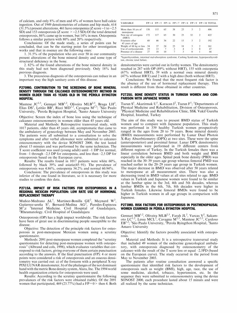
of calcium, and only 8% of men and 4% of women have hull calcicingestion. Out of 1949 densitometries of column and hip made, 614(31.5%) present alterations, 461 have osteopemia (Z score)1 to)2.5SD) and 153 osteoporosis (Z score >)2.5 SD).Of the total detectedosteoporosis, 86% came up in women, but 14% in men. Osteopemiafollows a similar pattern with 80% and 20% respectively.
Conclusions: Of the made study, a series of points can beconcluded, that can be the starting point for other investigationworks and that in essence are the following ones:
1. 31.5% of the population who are over 50 in our communitypresent alterations of the bone mineral density and some type ofstructural deficiency in the bone.
2. 82% of the found alterations of the bone mineral density inthis study had not been diagnosed previously. Only 18% hadprevious diagnosis.
3. The precocious diagnosis of the osteoporosis can reduce in animportant way the high sanitary costs of this disease.
P270MO. CONTRIBUTION TO THE SCREENING OF BONE MINERALDENSITY THROUGH THE CALCANEO OSTEOSONOMETRY METHOD INWOMEN OLDER THAN 45 YEARS OLD IN THE PEROLA BYINGTONHOSPITAL
Mantese JC2,3, Gennari MB1,2, Oliveira MLB2,3, Braga LH2,Elias DS2, Leitao BB2, Ricci MD1,2, Cavagna M2,3; 1Sao PauloUniversity, 2Perola Byington Hospital, 3Santo Amaro University
Objective: Screen the index of bone loss using the technique ofcalcaneo osteosonometry in women older than 45 years old.
Material and Methods: Retrospective transversal study, with1017 patients, older than 45 years old, asymptomatic that came tothe ambulatory of gynecology between May and November 2003.The patients were all submitted to a consultation to solve thesymptoms and after verbal allowance were submitted to calcaneoosteosonometry with the device SONOST 2000, the test lastedabout 15 minutes and was performed by the same technician. TheT score coefficient was used (young adult) until –1 DP for normal,from –1 through –2,5 DP for osteopenia, and less than –2,5 DP forosteoporosis based on the European curve.
Results: The results found in 1017 patients were white 60%,followed by black 39% and asiatic(45–91). The prevalence ofosteoporosis was 4.86%, osteopenia 34.2% and normal 60.94%.
Conclusion: The prevalence of osteoporosis in this study wasinferior of the one found in literature, so it is necessary for morestudies to confirm this data.
P271SA. IMPACT OF RISK FACTORS FOR OSTEOPOROSIS IN AREGIONAL MEXICAN POPULATION: LOW RATE USE OF HORMONALREPLACEMENT THERAPY
Munoz-Medrano AL1, Martinez-Bonilla GE2, Maynard W2,Gutierrez-urena S2, Bernard-Medina AG2, Paredes-EspinozaM1;z 1Internal Medicine. Civil Hospital of Guadalajara,2Rheumatology. Civil Hospital of Guadalajara
Osteoporosis (OP) has a high impact worldwide. The risk factorshave been of great use in discriminating people susceptible to thedisease.
Objective: The detection of the principle risk factors for osteo-porosis in post-menopause Mexican women using a scrutinyquestionnaire.
Methods: 2091 post-menopause women completed the ‘‘scrutinyquestionnaire for detecting post-menopause women with osteopo-rosis’’ (Albrand and cols, 1998), which evaluates variables that cor-respond to risk factors, giving everyone of them certain punctuationaccording to the answers. If the final punctuation (FP): 4 or morepoints were considered a risk of osteoporosis and an osseous densi-tometry was carried out: a) of the forearm with a peripheral X-rayPIXILUNARdensitometer, b) of the phalanges of the not dominanthandwith themetricBone density system,Alara, Inc. The 1994worldhealth organization criteria for osteoporosis were used.
Results: According to the scrutiny questionnaire the followingprevalances of the risk factors were obtained (table). Of the 2091women that participated, 469 (23.77%) had a FP=0> then 4. Both
densitometries were carried out in forthy women. The densitometryresulted in 287 with OP (89% without HRT), 155 with osteopenia(67% without HRT), 59 with a normal osea mineral density(47% without HRT) and 2 with a high dmo (both without HRT).
Conclusions: We found that the most frequent risk factor isthe absence of the use of hormonal replacement therapy. Thisresult is different from those obtained in other countries.
P272SU. BONE DENSITY STATUS IN TURKISH WOMEN AND COM-PARISON WITH JAPANESE WOMEN
Tuzun S1, Akarirmak U1, Karacan I2, Tuzun F1; 1Departments ofPhysical Medicine and Rehabilitation, Division of Osteoporosis,2Physical Medicine and Rehabilitation Clinic, SSK Vakif GurebaHospital, Istanbul, Turkey
The aim of this study was to present BMD status of Turkishwomen and to compare with Japanese population. This studywas performed in 339 healthy Turkish women prospectivelyranged in the ages from 20 to 79 years. Bone mineral density(BMD) measurements were performed by Lunar Dual PhotonX-Ray Absorbtiometry (DPX) at two sites. These were the spine(anteroposterior) and proximal femur. Lunar DPX densitometrymeasurements were performed in 19 different centers fromdifferent regions of Turkey. In the Turkish females there was alinear correlation between BMD and both weight and BMIespecially in the older ages. Spinal peak bone density (PBD) wasreached in the 30–39 years age group whereas femoral PMD wasreached earlier in the 20–29 years age group in Turkish women.The major decrease in BMD was observed after 50 years relatedto menopause at all measurement sites. There was also adecreasing trend in BMD values at all sites related to age. BMDvalues in Turkish and Japanese women were found to be similarat the lumbar spine in the 3rd, 4th and 5th decades, whereaslumbar BMDs in the 6th, 7th, 8th decades were higher inTurkish females. Likewise femoral BMDs were found to behigher in Turkish women in all age groups in comparison withJapanese.
P273MO. RISK FACTORS FOR OSTEOPOROSIS IN POSTMENOPAUSALWOMEN EXAMINED IN PEROLA BYINGTON HOSPITAL
Gennari MB1,2, Oliveira MLB2,3, Farah JL2, Varcas V2, Sakam-oto LC1,2, Lessa MCL2, Cavagna M2,3, Mantese JC2,3, CardosoEB1,2; 1Sao Paulo University, 2Perola Byingthon Hospital, 3SantoAmaro University
Objective: Identify the factors possibly associated with osteopo-rosis.
Material and Methods: It is a retrospective transversal studythat included 49 women of the endocrine gynecological ambula-tory, with osteoporosis diagnosed by osteosonometry of thecalcaneo with the result of the T score less or equal –2,5PD (basedon the European curve). The study occurred in the period fromMay to November 2003.
The patients after routine consultation answered a specificquestionnaire that identified risk factors to the development ofosteoporosis such as weight (BMI), high, age, race, the use ofsome medicine, alcohol, tobacco, hypertension, etc. In thesequence they were submitted to osteosonometry using the deviceSONOST 2000, each procedure lasted about 15 minuts and wereall realized by the same technician.
VARIABLE FP=4 FP=5 FP=6 FP=7 FP=8 FP=9 FP=10 TOTAL
More then 10 yrs ofmenopause
158 115 65 38 25 3 2 406
Non use of estrogens(HRT)
172 117 65 30 25 32 2 422
Fractures after45 years of age
68 46 36 22 15 3 2 192
Weight of 60 kg or less 54 37 24 23 18 3 2 161Use of corticosteroids 19 13 7 3 4 1 0 47Presence of a disease 27 18 10 7 3 0 1 66
*hyperthyroidism, intestinal mal-absorption syndrome, Cushing Syndrome, hyperparathyroid-ism, chronic renal failure.
S71
\Results: The results from the 49 patients as epidemiological datawe had that the majority were white women 68%, followed by blackwith 30%, and Asiatic with 2%. The median age was 61 years old(45–83), the median time since menopause was 15.4 years (2–36), themedium body mass index (BMI) was 29.40 (17–44). About the riskfactors we could verify the presence of blood hypertension in 57.1%,familiar history of osteoporosis in 8.2%, postmenopausal state in88%, diabetes in 18.4% and use of tobacco in 16.3%.
Conclusions: We can conclude that the factors more frequentlyassociated to osteoporosis were postmenopausal state, age andwhite race.
P274SA. CLINICAL AND EPIDEMIOLOGICAL FEATURES BETWEENOSTEOPOROTIC PATIENTS
Vargas RJ, Mio FE, Vidal L, Castaneda M, Flores J; NationalUniversity of Piura, Cayetano Heredia Hospital, Piura, Peru
Objective: To know clinical and epidemiological features of ourpatients.
Material and Methods: We evaluated clinical history of patientswith diagnosis of osteoporosis, established with DEXA studies,who attended Cayetano Heredia Hospital, Piura, Peru during 2001.
Results: Of 124 patients, 95.2% (118) were women, mean agewas 67.0 +/)10.0, 23.4% were menopaused before age 40. 16.1%had previous osteoporotic fracture. Comorbities were: hyperten-sion (25.8%), diabetes (16.9%), rheumatoid arthritis(10.4%),hypothyroidism (4.8%) and aAsthma (4%). BMD mean T at ver-tebral and hip level were )3.18 +/)0.98 and )2.37 +/)0.89respectively. Results showed:Treatment with Alendronate had 89.5%, HRT 50 % and both 49.1%.
The treatment‘s recuperation in BMD at L1–L4 and hip levelwere 4.8% +/)3.6 and 3.3 +/)2.7 respectively.
Conclusions: One out of six had severe osteoporosis. Therewere differences in BMD between patients with fracture and dia-betes. Recuperation of BMD was better at vertebral level.
P275SU. POSSIBLE ROLE OF FIBROBLAST GROWTH FACTOR 23(FGF23) IN THE PATHOGENESIS OF OSTEODYSTROPHY IN END STAGEKIDNEY DISEASE AND RENAL TRANSPLANTATION
Diaz K1, Rojas E1, Suniaga O1, Carlini RG1, Hruska K2,Weisinger JR1, Bellorin-Font E1; 1Division of Nephrology,Hospital Universitario de Caracas, Caracas, DF, Venezuela,2The Pediatric Renal Division, Washington University MedicalSchool, St. Louis, MO, USA
FGF23 is a recently discovered hormone regulating renal andintestinal PO4 transport. FGF23 excess is a cause of osteomala-cia. Since ROD is associated with mineralization defects wequestioned whether FGF23 was a factor in ESKD associateddefective mineralization. Therefore, we examined serum levels ofFGF23 in 20 patients with an age of 36.5±12 years, subjected tobone biopsy prior to and within the first six months of a renaltransplant. Pre-transplant biopsies showed increased osteoidvolume and thickness, osteoblast surface, as well as resorptionand osteoclast surfaces. Serum FGF23 was markedly elevatedin ESKD patients on hemodialysis (2115 pg/ml) prior to renaltransplantation. At this time, FGF23 levels correlated withosteoid surface (R=)0.993, p<0.01) and volume (R=)0.690,P<0.05). Since we have shown that post-transplant osteoporosisis related to osteoblast apoptosis and hypophosphatemia inthe early post-transplant period, and FG23 is a candidate weanalyzed FG23 levels in two groups of patients subjected to bonebiopsy within the first six months post transplant ) those withand those without osteoblast apoptosis post-transplant. High
pre-transplant FG23 levels were predictive of post-transplantosteoblast apoptosis and correlated with changes in osteoblastsurface and mineral apposition. Thus, pre-transplant FGF23levels were higher in patients with post-transplant osteoblastapoptosis (3296±373 vs. 1138±1462, p<0.05) and correlatednegatively with post-transplant osteoblast surface (R=)0.533,p<0.05). After transplantation, FGF23 decreased to normallevels in patients with or without osteoblast apoptosis (34.9±21.8vs. 119±241, p=NS), but no correlation was found betweenFGF23 and post-transplant hypophosphatemia. We concludethat FG23 accumulates in ESKD and is associated with decreasedosteoid accumulation and decreased mineral apposition. Highlevels of FG23 also appear to be a factor in determining post-transplant decreased osteoblast function contributed in part byincreased rates of osteoblast apoptosis.
P276MO. PREVALENCE AND CORRELATES OF VERTEBRAL FRACTURESIN ADULTS WITH CYSTIC FIBROSIS
Rossini M1, Del Marco A2, Dal Santo F1, Gatti D1, Braggion C2,James G1, Adami S1; 1Osteoporosis Center,University of Verona,ULSS, Verona, Italy, 2Cystic Fibrosis Center, Azienda Ospedal-iera of Verona, Italy
Introduction: Osteoporosis associated with Cystic Fibrosis (CF)is becoming increasingly important as the life expectancy of pa-tients continues to increase.
Methods: We studied 191 adults with CF (18–50 years old; 100men, 91 women). Total body, lumbar spine, and total proximalfemur bone mineral density (BMD) were measured by DXA andlateral spinal radiographs were taken for assessment of vertebralfractures. A range of anthropometric, clinical and biochemicalvariables were evaluated as potential correlates.
Results: T score values at the lumbar spine lower than )2.5 SDwere observed in 27.3% and 11.2% of male and female patients,respectively. These proportions fell to 14% and 9.9% for total hipand 10.4% and 12.1% for total body, in men and women respec-tively. Vertebral deformities were identified in 26.7% of the patientswith a slightly higher prevalence in males (32%) than in females(21%, p=0.058). Multiple vertebral deformities were observed in12% and 7.7% of men and women, respectively. BMD values weresignificantly related to body weight, FEV1, age of puberty andoccasionally to cumulative steroid dose in both genders. BMDvalues were also significantly related with serum albumin, IgG andcholinesterase. Serum estradiol levels were found below the normalrange in 23% of the women and 27% of the men, and was signif-icantly related to femur BMD values in both women and men.Significantly lower serum estradiol and free testosterone levels wereobserved in men with vertebral fractures. Serum osteocalcin wasbelow the normal range in 36% and urinary deoxypyridinolineabove the normal range in 51% of the patients.
Conclusions: Osteoporosis is a common complication of CF,being related to disease progression and apparently due to bothexcess bone resorption and inadequate bone formation. Estradioldeficiency may have a significant role in the pathogenesis in bothgenders. Vertebral fracture prevalence is high and greater thanexpected from prevalent BMD values.
P277SA. EFFECTS OF 4-YEAR TREATMENT WITH ONCE-WEEKLYCLODRONATE ON PREVENTION OF CORTICOSTEROID- INDUCED BONELOSS AND FRACTURES IN PATIENTS WITH ARTHRITIS
Frediani B, Falsetti P, Baldi F, Acciai C, Filippou G, MarcolongoR; Institute of Rheumatology, University of Siena, Siena, Italy
The aim of this placebo-controlled study was to determine whe-ther once weekly clodronate (Difosfonal) could prevent osteo-porosis in patients with arthritis at the start of corticosteroidtherapy. 163 patients, 18–90 years of age, with rheumatoid orpsoriatic arthritis, were randomly assigned to receive eitherclodronate (100 mg im/week) plus calcium and vitamin D(1000 mg and 800 IU, respectively) or calcium and vitamin Dalone. Patients had started therapy with prednisone or its equiv-alent within the previous 100 days and had bone mineral density
BMD With fracture Without fracture p
Hip )2.9 DS 1.1 )2.2 DS 0.7 0.007With Diabetes Without Diabetes
L1-L4 )2.6 DS 1.1 )3.3 DS 0.9 0.011
S72
£2.5 SD below mean young normal values at the lumbar spine orfemoral neck. The primary outcome was the difference betweenthe two treatment groups at months 12, 14, 23 and 48 in the meanpercentage change from baseline in bone mineral density of thelumbar spine, femur and total body. Secondary measurementsincluded changes in the stiffness index evaluated by ultrasoundmeasurements and the rate of new vertebral fractures. The bonedensity and stiffness did not change significantly in the clodronateplus clacium and vitamin D group, whereas it declined signifi-cantly in the calcium and vitamin D group. Depending on theskeletal regions evaluated, 85–98% of patients treated withclodronate had a densitometric change lower than the lowestsignificant densitometric difference. 100% of patients treated withcalcium plus vitamin D had a densitometric decrease greater thanlowest sigificant difference. The relative risk of vertebral fracturesand multiple vertebral fractures in the clodronate goup comparedto the calcium plus vitamin D group was 0.63 (0.35–0.98, 95% CI)and 0.25 (0.15–0.91, 95% CI), respectively. We concluded thatpulsatory administration of im clodronate once weekly is a safetherapy for preventing corticosteroid induced osteoporosis inpatients with arthritis.
P278SU. RATE OF BONE LOSS IN MULTIPLE SCLEROSIS PATIENTSTREATED WITH GLUCOCORTICOIDS IS BEST EXPLAINED BY PHYSICALPERFORMANCE
Stepan JJ, Havrdova E, Tyblova M, Horakova D, Ticha V,Novakova I, Zikan V; Charles University Faculty of Medicine 1,Prague, Czech Republic
The aim of this study was to evaluate the relative contribution ofphysical inactivity, glucocorticoid (GC) treatment, and otherfactors, to rate of bone loss (RBL) in patients with multiplesclerosis (MS).
Patients and Methods: The study population involved 156 fe-males (mean ± SD, age, 44±9 years, weight, 64±11 kg, height,166±6 cm, duration of MS 13±8 years) and 34 males (age, 43±11years, weight, 75±11 kg, height, 178±7 cm, duration of MS,14±6 years). The average dose of GC was 7.1±2.7 mg/d. Thepatients were not treated with antiresorptive drugs, they receivedthe daily recommended dose of calcium and vitamin D. Motorfunction of the patients was evaluated using the Kurtzke scale(KEDDS) useful in measuring the ability to walk that is decisive fornormal remodeling of bone. BMD was measured (Delphi A, Ho-logic, MA) at the lumbar spine and proximal femur (total andneck), at baseline and after 2±0.9 years. Serum aminoterminalpropeptide of type I collagen, osteocalcin, and type 1 collagen crosslinked C telopeptide (CTX) were measured at the baseline.
Results: BMD‡)1 T-score was found in 18% of the patients(KEDDS, 3.8±1.5, RBL at the total femur, )1.8±2.1%/year).BMD T-score of )1 to )2.5, was found in 52 % of the patients(KEDDS, 4.4±1.7, RBL, )1.8±2.3%/year). BMD£2.5 T-score,was found in 30% of the patients (KEDDS, 5.5±1.4, RBL,)3.4±4%/year). In the multiple regression analysis, the rate of boneloss was related to the KEDDS (p£0.001). The other variables, age,weight, height, sex hormone deficiency, baseline BMD, smoking,duration of MS, duration of GC treatment and the average andcumulative dose of GC, did not enter the equation. The rate of boneloss could also be predicted by serum CTX (p<0.01).
Conclusion: In adults with MS treated with low dose GC,immobilization was the main determinant of the rate of bone loss.
P279MO. BONE MINERAL DENSITY AND SERUM LEVELS OF 25 OHVITAMIN D IN CHRONIC USERS OF ANTIEPILEPTIC DRUGS
Kulak CAM1, Borba VZC1, Bilezikian JP2, Silvado CE3, de PaolaL3, Boguszewski CL1; 1Servico de Endocrinologia e MetabologiaDa UFPr (SEMPR), Curitiba, Brazil, 2Department of Medicine,College of Physicians & Surgeons, Columbia University, NewYork, USA., 3Servico de Neurologia da UFPR, Curitiba, Brazil
Aims: The aim of this cross sectional study was to evaluate bonemineral density (BMD) and serum levels of 25-hydroxy vitamin D
(25OHD) in a group of patients taking anti epileptic drugs (AED)for a seizure disorder.
Methods: Between May 2001 and January 2003, we evaluated58 patients (40 women/18 men), [34.4+6 years old (25–47)] livingin Curitiba, who were on anti seizure therapy for 2–38 years. Thegroup was matched by age, gender, and bone mass index to 29healthy subjects (20 women/9 men); 34.2+5.9 years old). Medicalhistory and physical exam were performed on all subjects withparticular information sought about fractures and risks factors forosteoporosis. Blood samples were collected for total serum calcium,albumin, phosphorus, creatinine, total alkaline phosphatase (AP),and liver function tests. Bone mineral density (BMD) of the lumbarspine, femur and forearm was determined by dual energy x-rayabsorptiometry (Hologic QDR 1000). Between February and April2003, other blood samples were collected to measure 25OHD,intact PTH and calcium.
Results: Fifteen patients had a fracture history all of whichoccurred during a seizure. The BMD of the lumbar spine(0.975+0.13 g/cm2 vs. 1.058+0.1 g/cm2, p<0.03) and of the totalfemur (0.930+0.1 g/cm2 vs. 0.988+0.12 g/cm2, p<0.02) was lowerin patients than in controls. 63.5% of patients and 24.1 % ofcontrols had a T-score < )1.0 at least one site. The AED users hadhigher AP and lower 25OHD (p<0.02). No correlations betweenBMD and 25OHD were found. The use of phenytoin was corre-lated with a greater incidence of fractures (RR: 2.38).
Conclusions: We conclude that patients on chronic use of AEDhave alterations at the bone metabolism characterized in this studyby lower BMD of the lumbar spine and total femur and lowerserum concentrations of 25OHD and more fractures.
P280SA. RISK FACTORS FOR BONE LOSS IN WOMEN WITH EARLY,ACTIVE RHEUMATOID ARTHRITIS: A PROSPECTIVE STUDY IN THECOBRA TRIAL
Geusens P1,2, Landewe R1, Garnero P3, Verhoeven A1, Boers M4,van der Linden S1; 1Rheumatology Dept, University Hospital,University of Maastricht, The Netherlands, 2Biomedical ResearchInstitute, Limburg University Center, Diepenbeek, Belgium,3Inserm Research Unit 403, Molecular Markers, Synarc, Lyon,France, 4Clinical Epidemiology & Biostatistics, VU UniversityMedical Center, Amsterdam, The Netherlands
Rheumatoid arthritis (RA) is associated with an increased risk forfractures. The degree of bone loss early in the disease process isstill a matter of debate. In a double blind randomised trial duringone year, patients with early, active RA were treated with sulfa-salazine (n=79) or a combination of sulfasalazine, methotrexateand prednisolone (60 mg/d in week 1 tapered to 7.5 mg mainte-nance dose in week 7) during 6 months, followed by sulfasalazinealone for 6 months (n=76). All had supplements of calcium (500mg/day) and, if baseline serum levels of 25(OH)D3 were low, withvitamin D (400 IU/day). Bone mineral density (BMD) was mea-sured by dual X-ray absorptiometry, and bone resorptionbycollagen-1 telepopeptide (CTX-1) in urine by Elisa.
At baseline, BMD in spine and hip was lower in postmenopausalwomen, and inversely related to CTX1. CTX1 was related to CRP(r=0.312, p<0.01). After one year, bone loss was found in the spine[mean and 95% confidence intervals (C.I.): )0.8% ()1.7, 0.2)] andhip [)1.8% ()3.0, )0.8)]. In a univariate analysis, bone loss after 12months was dependent on baseline CRP ()2.5% in spine and )2.9%in the hip in women with CRP >50, versus )0.4% and )0.7% inwomen with CRP < 50, respectively) and on menopausal status()2.5% in spine and)3.3% in hip in postmenopausal women, versus0.2%and)0.2%, respectively in premenopausal women), but not onbaseline joint damage, HAQ-score, treatment group, rheumatoidfactor and radiographic progression of joint damage. In a multipleregression analysis including these factors, only menopausal statuswas significantly associated with bone loss in the spine and hip.Postmenopausal women had a higher baseline CRP, osteocalcin andCTX1 as compared to premenopausal women.
We conclude that in women with early active RA, generalisedbone loss (in the spine) and periarticular bone loss (in the hip) aremainly determined by menopausal status and are related to bone
S73
resorption and inflammation. These results indicate a role for estro-gens in the occurrence of bone loss and inflammation in early RA.
P281SU. LONGITUDINAL ASSESSMENT OF HAND AND AXIAL BONEMINERAL DENSITY IN RHEUMATOID ARTHRITIS: A COMPARISON OFBONE MINERAL DENSITY CHANGES IN EARLY VERSUS LATE DISEASE
Ozgocmen S, Ardicoglu O, Kamanli A, Kaya A; Firat University,Faculty of Medicine, Department of PM&R, Division ofRheumatology, Elazig, Turkey
Background: Measurement of hand bone mineral density (BMD)using dual X-ray absorptiometry (DXA) is an accurate, repro-ducible, and sensitive method to quantify hand BMD in patientswith rheumatoid arthritis (RA).
Objective: This study proposed to assess hand and axial BMDin RA and to compare BMD changes in patients with early versuslate disease followed for one-year.
Methods: One-hundred patients with RA (11M, 89F, mean-age49, mean-disease duration 7.4 years) were included into the study.Patients who were under treatment of high dose or pulse steroids,or had diseases other than RA which influence bone mass wereexcluded. Patients were assigned into two groups: early RA (n=32,with a disease duration less than 3 years) and late RA (n=68, morethan 3 years). In longitudinal assessment patients currentlyreceiving treatments for osteoporosis other than vit-D and/or cal-cium supplementation were excluded. Patients’ hand (bilateral),spine and femur BMD were measured by DXA at entry and after12-months. The short-term precision for hand BMD was 0.8 %coefficient of variation.
Results: Patients with late RA had significantly lower right andleft hand BMD than patients with early RA. There was not asignificant difference between right and left hand BMD in bothgroups. In early RA group right hand BMD correlated with BMDat spine and hip (r=0.64-0.73, p=0.0001) and negatively correlatedwith disease duration (r=)0.46, p=0.008). In late RA group righthand BMD correlated with BMD at spine and hip (r=0.58–0.73,p=0.0001) and negatively correlated with age (r=)0.37, p=0.002)and ESR (r=)0.44, p=0.0001). Thirty-five patients (13 early RA,22 late RA) completed one-year follow-up. The percent loss ofBMD in right and left hand, spine, femur neck and trochanter inearly RA patients were )3.9, )3.4, )1.8, )0.8, and )5.3, respec-tively. The percent loss of BMD in right and left hand, spine, femurneck, Ward’s and trochanter in late RA patients were )2.2, )3.4,)0.8, )3.8, )2.6, and )5.6, respectively.
Conclusion: RA causes bone loss which is more prominent inhandsof patientswith early disease. Longitudinal assessment of handBMD is potentially helpful predicting disease progression in RA.
P282MO. RISK FACTORS ASSOCIATED TO LOW-IMPACT FRACTURESIN MEN WITH ANKYLOSING SPONDYLITIS
Luz KR, Nappo L, Vilela R, Pinheiro MM, Heymann RE,Szejnfeld VL; Rheumatology Division at Unifesp/EPM, Brazil
Ankylosing spondylitis (AS) is associated to low bone mineraldensity (BMD). Whether low bone mass in these patients leads toincreased fracture rate is still not clear. The purpose of this studywas to investigate risk factors associated to low-impact fracture inmen with AS.
PATIENTS AND METHODS: 86 men were enrolled in thiscross-sectional study (41 healthy controls). Clinical risk factorswere evaluated by a questionnaire that included details about as-pects of lifestyle, diet, drug use and previous fracture. All thesubjects performed spine and femur BMD (DPX-L, Lunar) andheel QUS (Achilles +, Lunar) measurements. Lateral thoracic andlumbar radiographs were taken to check for the presence of ver-tebral fractures, according to the Genant method.
Results: AS patients were younger, thinner and shorter thanhealthy controls (HC). The mean age, weight and height were42.6±10.5 vs. 38.3±10 years, 71.8±8.5 vs. 67±17 kg, and1.68±0.07 vs. 1.64±0.08 m, respectively (HC vs. AS). Vertebraldeformity (grade II and III) was identified in 21 AS patients and 12HC (p<0.05). Spine and femur BMD and QUS measurements
were not different between AS patients and healthy controls. Themean spine BMD and stiffness index were 1.172±0.01 vs.1.187±0.02 g/cm2 and 96±17 vs. 91±19, respectively (HC vs. AS)(NS). After adjustments for age, the most relevant risk factors todiscriminate AS patients with low-impact fractures from healthynon-fracture controls were prolonged immobilization, currentsmoking, poor perception of the global health, corticoid use andprevious fracture. Patients with AS hadmore appendicular and axialfractures than healthy controls even though BMD and heel QUSmeasurements were similar between the two groups. In conclusion,the clinical risk factors for fractures were more important than bonemass measurements to discriminate AS patients with low-impactfracture from healthy non-fracture controls. Interestingly, AS pa-tients presented fractures even with BMD and QUS values higherthan that observed in the population in general.
P283SA. EFFECTS OF ANTIVIRAL DRUGS FOR CHRONIC HEPATITIS CIN BONE TURNOVER AND BONE DENSITY
Fonteles JFS1, Moreira RO1, Azevedo SLW1, Cerqueira ADB1,Paixao JB2, Coelho HSM2, Farias MLF1; 1Service of Endocri-nology, HUCFF, Federal University of Rio de Janeiro, Brazil,2Service of Gastroenterology, HUCFF, UFRJ, Brazil
Hepatitis Cvirus infection is considered a worldwide pandemia.Nowadays, HCV infection accounts for most patients indicatedfor liver transplantation, and osteoporotic fractures are frequentafter transplantation. Combined therapy with Ribavirin withInterferon (Ifn) induces a sustained virological response in abouthalf of the patients, but these drugs have been related to bone lossin a cross-sectional study.
AIM, PATIENTS AND METHODS: In order to investigatethe longitudinal effect of therapy on bone turnover and bonedensity, thirty-one patients with chronic active hepatitis C werestarted on Ribavirin plus Interferon-alfa2b for one year. Twelve ofthem were males 34 to 58 yrs and 19 females 27 to 61 yrs, of whom5 had regular menses, 5 were on estrogen replacement for meno-pause and 9 did not receive hormones for menopause. Fastingserum bone specific alkaline phosphatase (BSAP), and urinary N-terminal telopeptide of type 1 collagen (NTx) were measured atbaseline, 3 and 6 months on therapy. Bone density was evaluatedby DEX Lunar Corp. at lumbar spine (LS) and femoral neck (FN)at baseline and twelve months. Anova for repeated measures,paired and unpaired t tests were used.
Results: There was a significant decrease in urinary NTx at 3and 6 months (54.9 +/)44.6 to 39.8 +/)53.5 and then to 31.3+/)22.1 mBCE/mMCr, p<0.005). Serum BSAP did not changeduring treatment. Before starting antiviral drugs, 9 patients(7 females and 3 males) had osteopenia at LS and 9 patients(7 females and 2 males) had osteopenia at FN. Further 3 femaleshad osteoporosis at LS. In the sixteen patients re-evaluated after1 year, BMD increased in six and decreased in four, and bone losswas more evident at FN. When the whole group was considered,variations of bone mass were not significant, but women lostmore bone than men (p=0.016). There was a trend for correlationbetween delta-6 months NTx and delta-12 months BMD at FN(r )0.447 p 0.0824).
Our results suggest that antiviral treatment decreases boneturnover but other factors related to hepatitis C probably justifythe heterogeneous response of bone mineral density.
P284SU. BONE DENSITY IN ANKYLOSING SPONDYLITIS ASSESSED BYDUAL ENERGY X-RAY ABSORPTIOMETRY AND QUANTITATIVE ULTRA-SOUND: THE RELATIONSHIP OF BONE MINERAL DENSITY WITHFUNCTIONAL AND DISABILITY STATUS
Ozgocmen S, Kaya A, Kamanli A, Ardicoglu O; Firat University,Faculty of Medicine, Department of Physical Medicine andRehabilitation, Division of Rheumatology, Elazig, Turkey
Background: Ankylosing spondylitis (AS) is a chronic inflam-matory disease involving the sacroiliac joints and axial skeletonand is typically associated with sclerotic changes and syndesmo-phytes in the spine. Patients with AS usually have difficulties in
S74
activities of daily living and disabling nature of the disease isusually depends on musculoskeletal restrictions.
Objective: This study proposed to investigate bone mineraldensity (BMD) in AS by using both dual X-ray absorptiometry(DXA) and quantitative ultrasound (QUS) and to assess rela-tionship between BMD and patients’ functional and disabilitystatus.
Methods: Forty-two patients with AS (age 33.2,disease dura-tion 8.9 years) were included into the study. BMD of the APlumbar spine and femoral neck, Wards and trochanter wasmeasured using DXA in all patients and broadband ultrasoundattenuation (BUA), speed of sound (SOS) were measured at thecalcaneus using an Hologic Sahara bone sonometer in seventeenof the patients without talalgia. Daugados Functional Index(DFI), and Articular Index (DAI), Bath AS Disease ActivityIndex (BASDAI), Bath AS Functional Index (BASFI), LeedsDisability Index (LDI) were used to estimate disease activity,functional and disability status.
Results: According to the L2-L4 t scores, 28.6% of patients hadosteoporosis, 33.3% osteopenia, and 38.1% had normal BMD.Femoral neck, Wards and trochanter BMD negatively correlatedwith BASFI (r=)0.38, r=)0.39, r=)0.44, p£0.01 respectively)and LDI (r=)0.33, r=)0.36, r=)0.30, p£0.05 respectively)whereas L2-L4 BMD had no significant correlation. BUA and SOSsignificantly correlated with femoral neck, Wards and trochanter(n=17, r=0.50 to 0.60) but not significantly correlated with L2-L4BMD. BUA and SOS did not significantly correlated with func-tional or disability indices. BASDAI correlated negatively withonly femural neck (r:)0.31, p£0.05) and trochanter (r=)0.41,p£0.01) BMD.
Conclusion: These results indicate close relationship betweenfunctional and disability status and BMD in AS. DXA measure-ment of femoral neck, Wards’ or trochanter BMD may be muchmore predictive for functional or disability status in AS rather thanAP spine measurements. Nevertheless QUS measurements predictbone density in AS, this technique fails to detect patients’ func-tional or disability status.
P285MO. THE EFFECT OF LOW DOSE METHOTREXATE ON BONEDENSITY IN PSORIATIC ARTHRITIS PATIENTS
Pinto P, Bernardo A, Brito I, Ventura F; Rheumatology Unit,Hospital Sao Joao, Porto, Portugal
OBJECTIVE: The use of high dose methotrexate (MTX) has beenlinked with bone loss. However, it is not clear whether longtermlow dose MTX used in the treatment of psoriatic arthritis isassociated with bone loss. We compared the effect of low doseMTX on bone density in prospectively recruited patients withpsoriatic arthritis (PsA).
Methods: 50 PsA men taking MTX were compared to 50 con-trols with PsA and not taking MTX. In all patients, evaluation wasmade of disease duration, inflammation indices (erythrocyte sedi-mentation rate, C-reactive protein), functional indices (Steinbroc-ker scale), and the Health Assessment Questionnaire (HAQ). Bonemineral density (BMD) of the lumbar spine, trochanter, and fem-oral neck was measured using Lunar dual energy x-ray absorpti-ometry. Student t tests were used to detect differences in bonedensity (using T scores) of the MTX group versus controls. Anal-ysis of covariance was used to examine for confounders includingdisease duration, disease activity and age.
Results: BMD of the femoral neck and trochanter did not differsignificantly between the MTX treated groups and controls whenanalyzed by T scores.
The mean difference between the MTX group and controls ofthe femoral neck was 0.060 (95% CI 0.15). The absolute BMD ofthe lumbar spine (L2-L4) did not differ significantly between thegroup and the controls.
Conclusion: The study suggests that low dose of MTX does nothave a negative effect on bone density. Osteoporosis was related tothe indices of inflammation but not with disease duration. Therewas a correlation with HAQ score.
P286SA. BONE MINERAL DENSITY IN FEMALE PATIENTS WITH EATINGDISORDER
Kocjan T1, Robinson P2, Prelevic GM2,3; 1University MedicalCentre, Ljubljana, Slovenia, 2Royal Free Hospital, London, UK,3Middlesex Hospital, London, UK
Aims: To determine predictive factors for bone mineral density(BMD) loss and fractures in females with eating disorder and toidentify any possible benefit of estrogen therapy on bones in thesubgroup of patients with amenorrhea.
Methods: The cross-sectional study involved 62 consecutivefemale patients with eating disorder (30 with anorexia nervosa –restrictive type (AN-R), 16 with anorexia nervosa – bulimic type(AN-B) and 16 with bulimia nervosa – purging type (BN-P)). Char-acteristics:mean age26.3 years (SD6.1),meandurationof the disease8.7 years (SD 6.3), mean BMI 15.9 (SD 1.8). The studied outcomeswere BMD and Z-score of the spine and of the hip, and presence ofbone fractures. Several potential predictors were assessed: weight,BMI, years of disease, disease onset before menarche, duration ofamenorrhea, estrogen therapy, smoking, alcohol consumption, useof laxatives, vomiting, binge eating and vegetarian diet.
Results: In the multivariate analyses (linear and logistic regres-sion), weight and BMI were found to be the only statisticallysignificant independent predictors of both spine and hip BMD andZ-score (p<0.01). Themodel for predicting presence of fractureswasnot statistically significant. Bivariate analyses confirmed just some ofthe expected relations (positive impact of weight and BMI; negativeimpact of years of disease, duration of amenorrhea and disease onsetbefore menarche). Most importantly, no impact of estrogen therapywas found, regardless of its type (HRT or contraceptives). Finally,the differences between types of eating disorder were tested, con-trolling for BMI and duration of amenorrhea/disease onset beforemenarche. Except for hip Z-score, significant differences were foundbetween the three groups (p<0.05), whereby the patients with BN-Phad the highest and AN-R patients had the lowest average values.
Conclusions: Treatment of eating disorder (e.g. weight gain)seems to be crucial for preserving bones of female patients, asweight and BMI were the most important predictors of BMD andZ-score in our group, whereby no benefit of the estrogen therapywas proven. Unfortunately, there were too few fractures in ourgroup for any reliable conclusions regarding this outcome.
P287SU. PARATHYROID FUNCTION AND MARKER OF BONE RESORP-TION IN HEMODIALYSIS (HD) PATIENTS, THEIR DYNAMICS ON ALFA-CALCIDOL THERAPY
Rozhinskaya LY, Borisov AV, Iljina AN, Ermakova IP; NationalResearch Center for Endocrinology,, Center of Hemodialysis‘‘FESPHARM’’, Research Institute of Transplantology andArtificial Organs, Moscow, Russia
The most common type of renal bone disease is renal osteodys-trophy, and its severity is closely related to levels of intact PTH.
Objective: Investigate interrelations between PTH and beta-crossLaps levels in HD patients, estimation effectiveness ofalfacalcidol treatment. 224 patients in age from 18–70 were inves-tigated. Intact PTH was measured by IRMA in baseline, 3, 6, 12months. Beta-crossLaps were measured by Elecsys Systems 2010modular analytics.
Secondary hyperparathyroidism (SHPT) was diagnosed in PTHlevel above 260 pg/ml. Optimal levels of PTH (130–260 pg/ml) wererevealed in 27% (60 patients). 28% (62 patients) showed PTHlevels less 130 pg/ml. SHPT was determined in 102 patients: mild(PTH 260–400 pg/ml) in 30%, moderate (PTH 400–800) in 36%,severe (PTH>800 pg/ml) in 34% patients. b-crossLaps levels wereincreased in all patients. T-score was ranged from 4.7–25 inaccordance PTH groups (mild, moderate and severe SPHT). Therewas significant positive correlation between b-crossLaps and PTHlevels (r=0.844). Patients with SPHT were treated with alfacalcidol(Alpha D3-Teva) 3 times/week. In 90% patients PTH fell by morethan 50%: from 327 to 163 pg/ml in 84% patients with mild SHPT(mean dose alfacalcidol was 1.5 mcg/week); from 589 to 200 pg/mlin 90% patients with moderate SHPT (mean dose alfacalcidol was
S75
2.0 mcg/week); from 1352 to 596 pg/ml in 96% patients with severeSHPT (mean dose alfacalcidol was 2.5 mcg/week). Dynamic ofb-crossLaps levels on alfacalcidol therapy was estimated in 36 pa-tients. There was revealed decrease of beta-crossLaps on 38.2%.
Conclusion: SHPT was determined in 45.5% patients in HD.The positive correlation was showed between beta-crossLaps andPTH.Moderate doses of alfacalcidol are effective in themajorityHDpatients to maintain optimal level of PTH. Patients with mild andmoderate SHPT are more susceptible to suppression by alfacalcidolthan those patients with severe SHPT at the onset of treatment.
P288MO. 25 HYDROXY-VITAMIN D LEVELS (25OHD) IN A HEALTHYYOUNG POPULATION IN THE CITY OF SAO PAULO AND ITS CORRE-LATION WITH OCCUPATIONAL ASPECTS AND CALCIUM METABOLICPARAMETERS
Maeda SS, Kunii I, Hayashi L, Pereira RL, Lazaretti-Castro M;Division of Endocrinology, Federal University of Sao Paulo(UNIFESP), Brazil
Aim: The serum level of 25OHD is accepted as a good index ofvitamin D storage. In literature there is a discussion about thenormal levels of 25OHD. Sex, age, occupation, lifestyle, ethnicalgroup and latitude can influence 25OHD levels. The objective ofthis study was to establish the normal levels of 25OHD in ahealthy young population living in Sao Paulo, at the age of theirpeak of bone mass, and correlate these levels with othersparameters.
Methods: 99 healthy young subjects (men=40 and women=59;median age 25.0±2.8 years old,range: 17 to 33) were requested toparticipate in the study. Twenty subjects were medical students, 52were residents in a School Hospital and the other 28 worked inplants. Fasting blood samples were collected for 25OHD (RIANichols Institute), ionized calcium, total calcium, intact PTH,albumin, osteocalcin, alcaline phosphatase, phosphorus, creatinineand glucose. Pearson correlation, linear regression, and WilcoxonSigned Rank tests were used in statistical analysis and P< 0.05 wasconsidered to be significant. All values were expressed byMean±Standard Deviation.
Results: The level of 25OHD in the studied population was31.5±13.6 ng/mL (range: 10–56). Men had higher levels thanwomen (34.1±15.4 and 29.9±12.1 ng/ml, respectively, P<0.05).We found a significant correlation of 25OHD with osteocalcin(R=0.29) and creatinine (R=0.43),but not with ionized Ca, totalCa or PTH levels. According to the occupational distribution, thelowest 25 OHD values were among the residents (26.6±10.4 ng/ml,range: 10–54), compared to the students (36.5±17.0 ng/ml, range:11–86) and workers (37.9±13.0 ng/ml, range: 14–64).
Conclusions: The normal range for 25OHD in this younghealthy population was 37.3±14.7 ng/ml(range: 11–86), excludingthe residents. Even in this healthy young population we foundsignificant differences in 25OHD levels considering gender andoccupational aspects. The residents usually have a heavy scheduleand no time to stay outdoors. The 25OHD concentrations corre-lated with osteocalcin levels, probably reflecting some systemic ef-fect on bone metabolism.
P289SA. FACTORS INVOLVED IN OSTEOPOROSIS IN YOUNG MALEPATIENTS WITH CHRONIC VIRUS C HEPATITIS
Cerqueira ADB1, Duarte LM1, Duarte MPC1, Oliveira MCD1,Fonteles JFS1, Coelho HSM2, Farias MLF1; 1Service of Endo-crinology, HUCFF, Federal University of Rio de Janeiro,2Service of Gastroenterology, HUCFF, UFRJ, Brazil
Objective: Evaluation of factors involved in bone loss in men withchronic C hepatitis.
Patients and Methods: Bone mineral density (BMD) was ob-tained by DEXA, Expert-Lunar Corp., at lumbar spine (LS) andproximal femur (FN), in 74 patients with viral C hepatitis con-firmed by positive anti-HCV. According to liver biopsy and/orbiochemical tests, patients were classified as: non-cirrhotic(Ncir=38) and cirrhotic (Cir=37). Twenty-one of Cir patients are
waiting for liver transplantation. Serum bone-specific alkalinphosphatase (BALP), insulin growth factor-1 (IGF-1), testosterone(T), and urinary N-telopeptide of type 1 collagen (NTX) wereevaluated. Unpaired t test was utilized to compare the groups.
Results: No difference was found concerning age (NCir 48.9+/)9.8 and Cir 52.1+/)11.2 years). We found osteoporosis(T score<)2.5) at LS in 3 NCir and 6 Cir patients, but meanBMD at LS did not differ between groups: Ncir =1.198 +/)0.182and Cir 1.184 +/)0.214 g/cm2, p=0.7. At femoral neck, 1 NCirand 4 Cir patients had osteoporosis, and FN BMD was signifi-cantly lower in Cir (0.971 +/)0.172 g/cm2) vs NCir (1.045 +/)0.154 g/cm2) p=0.043. Serum IGF1 (median and quartiles) wasalso lower in Cir=21.2 ng/mL(8.5–36.7) than Ncir=91ng/mL (57–111.8), p=0.0001. Serum T (median and quartiles) was 480.5 pg/dL(374–565.5) in Cir and 613.3 pg/dL (507–766.5) in Ncir, p=0.067.No difference was found between groups in BALP and NTx, butthese markers of bone turnover were above normal in about 30%of all patients.
Conclusions: We detected low bone mass in young male pa-tients with virus C hepatitis, as 12.1% of them showed osteopo-rosis at LS and 6.7% had osteoporosis at FN. Bone density atFN and serum IGF1 were lower in cirrhotic patients, who alsotended to have lower serum Testosterone. These alterations inanabolic hormones and in bone turnover probably have a nega-tive influence in skeletal homeostasis. We thus suggest that allpatients with chronic hepatitis C should be screened for bonedensity and treated with anti-resorptives and/or anabolic drugs inorder to prevent osteoporosis and fractures after liver transplan-tation.
P290SU. THE SPECTRUM OF RENAL BONE DISEASE IN CHRONICKIDNEY DISEASE (CKD) STAGES 3 TO 5: POSSIBLE CONTRIBUTION OFOSTEOPOROSIS
Infante M1, Hernandez G1, Rojas E2, Fuenmayor N1, DominguezJ1, Suniaga O2, Carlini RG2, Weisinger JR2, Bellorin-Font E2;1Division of Nephrology, Hospital M Perez Carreno, 2Divisionof Nephrology, Hospital Universitario de Caracas, Caracas,Venezuela
The term renal osteodystrophy (ROD) includes high boneturnover, low turnover and mixed lesions. However, littleattention has been paid to the possible contribution of osteo-porosis to the bone lesions of CKD, particularly in theincreasing population of aging patients. The present study wasdesigned to examine the spectrum of ROD in patients withmoderate (stage 3) and severe (stage 4) decrease in GFR com-pared with kidney failure on dialysis (stage 5). Forty patients(18 female and 22 male), with a mean age of 54 13 years andGFR of 25.8 12.5 ml/min (CKD stages 3 and 4) were comparedwith 53 patients with kidney failure on dialysis. All patientswere subjected to bone biopsy for histomorphometric analysis.As shown in the table, in CKD stages 3 and 4 the prevalence ofOP accounted for 25% of the patients and was the second typeof bone disease after 2nd HPT. In 12.5% of the patients bonehistology was normal. In contrast, in CKD stage 5, 2nd HPT
Bone alterations in CKD
Diagnosis CKDStage 3–4,N (%)
CKDStage 5,N (%)
2nd Hyperparathyroidism (2nd HPT) 12 (30) 24 (45.3)Mixed Bone Disease (MBD) 6 (15) 15 (28.4)Adynamic bone disease (ABD) 7 (17.5) 11 (20.7)Osteomalacia (OM) 0 2 (3.7)Osteoporosis (OP) 10 (25) 1 (1.9)Normal bone histology (NB) 5 (12.5) 0
Total 40 (100) 53 (100)
S76
was the most prevalent form of ROD, followed by MBD, ABDand osteomalacia, whereas OP was observed in only one pa-tient. CKD patients stages 3 and 4 with OP had lower serumcreatinine (2.60.9 mg/dl vs. 4.11.3 in 2nd HPT, and 5.01.7 inMBD, p<0.01) and tend to be older (61.411 years). Six out ofthese 10 patients were postmenopausal women In addition, OPpatients had lower serum PTH levels (71.3.148.3 pg/ml) com-pared with all other type of ROD, except ABD. In conclusion,osteoporosis is frequent in CKD, particularly in older post-menopausal women with moderate to severe decrease in GFR.It is possible that the severity of the typical alterations of RODmay mask the possible contribution of osteoporosis in thisprocess.
P291MO. BONE MINERAL DENSITY EVALUATION IN PATIENTS WITHINFLAMMATORY BOWEL DISEASE
Lora FL1, Borba VZC2, Kulak CAM2, Pisani JC1, Carmes ER1,Miasaki FY2, Amarante HMB1; 1Gastroenteroly Department,Federal University of Parana, Brazil, 2SEMPR / EndocrinologyDepartment, Federal University of Parana, Brazil
Inflammatory Bowel Disease (IBD) is a potential cause of sec-ondary osteoporosis.
Aim: The aim of this cross sectional study was to evaluate thebone mineral density (BMD) in a group of patients with IBD andcorrelate the results with disease activity index and other clinicalcharacteristics.
Methods: Between May and October 2003, 90 patients con-secutively seen at the Gastroenterology Clinic of the Federal Uni-versity of Parana were selected and 76 completed the evaluation.They were divided into two groups, regarding their diagnosis, 39(21 men, 18 women, mean age: 32.1±8.8 ys) had Crohn disease(CD) and 37 (12 men, 25 women, mean age: 35.9±8.5 ys) hadulcerative colitis (UC). They were compared with 40 healthy sub-jects (16 men, 24 women, mean age: 34.1±7.1 ys) matched bygender, race and age. Postmenopausal women and subjectswith other diseases known to cause osteoporosis were excluded.Medical history was performed in all patients with emphasis onrisk factors to osteoporosis and use of glucocorticoids. BMD oflumbar spine and hip was performed by DEXA (Hologic QDR1000/W).
Results: The groups were homogeneous in all the epidemiologicaspects. BMD of IBD patients was lower than the control group atlumbar spine and total hip (p<0.004 and p<0.004 respectively).No difference was found at BMD between UC and CD patients.Body mass index (BMI) of the IBD group was negatively correlatedto the BMD (p=0,01). No other correlation between BMD andvariables analyzed were found.
Conclusion: Low BMD was found in IBD patients. Among theanalyzed variables, only the BMI had a significant correlation withBMD, while the use of corticosteroids, disease activity index, pre-vious surgery and physical activity, did not had any correlationwith BMD in these groups of patients.
P292SA. LONG TERM EFFECTS OF GROWTH HORMONE REPLACEMENTTHERAPY ON BONE METABOLISM IN PATIENTS WITH GROWTH HOR-MONE DEFICIENCY WITH SPECIAL RESPECT TO GENDER
Kovacs L1, Hubina E1, Szigeti-Csucs N1, Kovacs GL1, SzabolcsI1, Toth M2, Racz K2, Goth MI1; 1National Medical Center,Budapest, Hungary, 2Semmelweis University, Budapest, Hungary
Aims: The effects of growth hormone replacement therapy onbone mineral density and on bone metabolism were investigatedwith special respect to gender.
Methods: 20 patients (11 men, 9 women, mean age: 43 years)with growth hormone deficiency were involved in the study.Parameters of bone metabolism and bone mineral density of thelumbar spine, the left femoral neck and the forearm were checkedprior to and at 3, 6, 12, 18, 24 and 36 months after the start oftreatment.
Results: Serum calcium (seCa) increased only after 3 and 6months; alkaline phosphatase (AP) increased until the first year;phosphate (seP) remained elevated until 18 months. The changes ofseCa, AP and seP were more pronounced in males than in females(p=0.001, 0.04, and 0.05, respectively). PTH decreased in bothsexes until the first year, then increased, and finally exceeded thebaseline level. Before the study, osteoporosis was found in 2 menand 1 woman, osteopenia in 3 men and 4 women. The lumbar andfemoral BMD increased first after 12 months of treatment, andfurther improvement was seen until the end of the study (lumbar:1.019±0.12 vs 0.941±0.13 g/cm2, p<0.001; femoral: 0.886±0.12vs 0.836±0.11 g/cm2, p=0.01). The radial BMD started to im-prove after 3 years (0.664±0.1 vs 0.602±0.1 g/cm2, p=0.01). Al-though the lumbar BMD and T-scores were increased in bothsexes, this effect was greater in males than in females (p<0.05). Thefemoral and radial BMD and T-scores were increased only in malepatients (p<0.01).
Conclusions: GH replacement therapy has temporary effects onbone metabolism: a significant but transient increase in seCa, sePand AP levels and a biphasic change in PTH values could be ob-served. The beneficial effect of GH treatment on the BMD ap-peared only after 12 months of treatment but, in spite of the abovetemporary changes, continuously improved further until the end ofthe study. The results indicate that generally more markedimprovement can be obtained in males than in females duringreplacement therapy.
P293SU. BONE LOSS IN THE EARLY AND PROLONGED POSTTRANS-PLANTATION PERIOD FOR LIVER TRANSPLANTATION
Atamaz F1, Hepguler S1, Kilic M2, Tokat Y2, Karasu Z3;1Department of Physical Medicine and Rehabilitation, EgeUniversity, Izmir, Turkey, 2Department of Surgery, Ege Univer-sity, Izmir, Turkey, 3Department of Gastroenterology andHepatology, Ege University, Izmir, Turkey
Objective: To evaluate the bone density, bone turnover, gonadalfunction after the liver transplantation in the both of the early andprolonged period.
Method and Patients: In the early period (first 3 months) 42patients aged 19–69 years and in the prolonged period (after 1styear) 37 patients aged 25–72 years without treatment of osteo-porosis, a total 79 patients after liver transplantation were eval-uated. All patients were on treatment with immunosuppressiveagent such as tacrolimus or cyclocporine and on prednisolone.Measurements of lumbar spine and proximal femur and totalbody BMD were performed by dual x-ray absorptiometry.Biochemical markers of bone resorption and bone formation withserum calcium and phosphorus, urinary calcium and gonadalhormones, growth hormone, thyroid function tests, parathyroidhormone, testosterone and spine radiographs were evaluated onthe same times.
Results: In the early period of the posttransplantation, meanT scores for bone mineral density (BMD) decreased by –2.22()6.33-0.21)at the lumbar spine and by –2.79 ()7.39—0.42) atthe femur total. At one year of posttransplantation mean T scoresfor BMD were –1.98 ()3.69–0.03) at the lumbar spine and–1.25 ()2.87–0.07) at the femur total. In the both of the period,urine deoxypyridinoline as a marker of bone resorption wasincreased in all patients. In the prolonged period, osteocalcine asa marker of bone formation was significantly higher than inearly period (p<0.05). 14 patients in early period, 24 patients inprolonged period had high values for PTH. In the both of group,the women had low values of gonadal hormones, the men hadlow values of testosterone. In the 6 patients who were clinicallyconfirmed had a history of vertebral fracture following trans-plantation.
Conclusion: Our results demonstrates a high prevalence of boneloss which occur during the first months in patients with livertransplantation. Morever, these results suggest that the risk offracture is considerably increased in patients who did not receivetreatment for osteoporosis after transplantation.
S77
P294MO. BONE MASS IN HIV INFECTED CHILDREN
Vargas DM1,3, Noriler K1, Pissatto M1, Galvao JC2, Gallo AM3,Pinho MSLG3; 1Universidade Regional de Blumenau, SC-Brasil,2Hospital Dia, Secretaria de Saude de Blumenau, SC-Brasil,3OsteoDens, Cl de Densitomertria Ossea Santa Catarina, Blume-nau, SC-Brasil
Aims: To evaluate the ocurrence of osteopenia in children infectedwith human immunodeficiency virus (HIV).
Methods: 27 patients with vertical HIV infection aged 7.3±3.8years (14 male) were measured by bone mineral density (BMD),serum calcium, serum alkaline phosphatase, serum phosphorous,calciuria, CD4 and CD8 lymphocyte counts and HIV RNA copynumbers. Clinical variables included sex, age, weigth, heigth andclinical categories. All patients were receiving antiretroviral ther-apy. BMD were performed at lumbar spine (L1-L4) and expressedin deviation standard score (SDS) by age and sex. Weigth andheigth was referred to NCHS standards and expressed in SDS.Simple linear regression was used to analise correlations betweenvariables and the U-Mann-Whitney to compare groups.
Results: BMD average was normal ()0.74±1.08 SDS). How-ever we verified that 40.7% of the patients had osteopenia(BMD<)1 SDS). When comparing data from osteopenic patients(n=11) with non-osteopenic patients (n=16), we observed thatweigth (SDS) from osteopenic group was lower than that from non-osteopenic ()1.29±0.66 vs 0.66±1.10; p<0.05), as well as CD4lymphocyte count (576.4±300.0 vs 925.1±761.4 count/microliter;p<0.05). BMD correlated positively with weigth (R=0.37;p<0.05), CD4 lymphocyte count (R=0.43; p=0.02) and CD8lymphocyte count (R=0.53; p=0.003); and negatively with age(R=)0.55; p=0.008).
Conclusions: Osteopenia occurs in children with vertical HIVinfection. Osteopenia was related to lower weigth and lower CD4lymphocyte count. Our results suggest that duration of HIVinfection affects bone mineral acquisition as well as the degree ofimmunological supression. HIV infection during childhood maypredispone to a reduced peak bone mass.
P295SA. SEASONAL VARIATION ON PLASMA LEVELS OF 25 HYDROXY-VITAMIN D (25OHD) IN A HEALTHY ELDERLY POPULATION FROMBRAZIL
Maeda SS, Kunii I, Hayashi L, Pereira RL, Lazaretti-Castro M;Division of Endocrinology, Federal University of Sao Paulo(UNIFESP), Brazil
Aim: The major source of vitamin D comes from its synthesis inthe skin by sunlight ultraviolet rays. Aging, skin pigmentationand sun screens can interfere in this production. Seasonal varia-tion of plasma concentrations and vitamin D deficiency is widelydescribed in extreme latitudes, but there aren’t consistent dataabout these levels in Brazil. The objective of this study is toinvestigate this variation in a elderly population in Sao Paulo,located in a subtropical region (23�34¢).
Methods: One hundred one healthy subjects (men=46 andwomen=55, mean 67.5±5.3 years old, range: 55–83) participantsin a fitness program for the elderly were requested to participated inthis study. Fasting blood samples were collected in August (end ofthe winter) and in December beginning of the summer) and ana-lyzed for serum 25OHD (RIA Nichols Institute;normal range: 9.2–45.2 ng/ml). We excluded from analyses 6 subjects taking vitaminD supplements. Paired Student’s t-test and Wilcoxon Signed RankTest were used, and P<0.05 was considered significant. Valueswere expressed by Mean±Standard Deviation.
Results: We included in the analysis 95 subjects. The 25OHD atthe end of winter was 30.8±11.7 ng/ml and significantly increasedat the beginning of summer (36.6±12.7 ng/ml). According togender, the increment was significant only in men (32.6±12.1 to43.4±12.4 ng/ml), but not in women (29.1±11.2 to 29.7±8.6 ng/ml). There was no difference in 25OHD levels between ethnicalgroups (Caucasian, Japanese, Afro-Brazilian, and Brazilian-na-tives). A significant increase on 25OHD levels in summer was only
seen in the Caucasian (31.4±11.4 to 37±14.1 ng/ml) and Japanese(31.4±14.5 to 37.7±9.9 ng/ml). In the other groups we did not findany difference between seasons.
Conclusion: There was a seasonal variation on the 25OHDlevels in this healthy elderly studied group. The values were lowerafter winter and higher in summer. This significant increment insummer was due exclusively to the men. In the same way, subjectswith darker skin were not able to increase the synthesis of 25OHDin summer. These data suggest that women and people with morepigmented skin are at higher risk of vitamin D deficiency, even in asunny country like Brazil.
P296SU. PREVALENCE OF OSTEOPOROTIC VERTEBRAL FRACTURES INPATIENTS WITH INFLAMMATORY BOWEL DISEASE
Oliveira KRB, Heymann RE, Pinheiro MM, Szejnfeld VL,Ohashi CB, Castro CHM; Sao Paulo Federal University, SaoPaulo, Brazil
Aims: To study the prevalence of osteoporotic fractures andassociated risk factors in patients with Crohn’s disease (CD) andulcerative colitis (UC).
Patients and methods: This analysis included 43 patients con-secutively evaluated at the Sao Paulo Hospital, Sao Paulo FederalUniversity, Sao Paulo, Brazil. Potential risk factors associated withfractures were assessed by a validated questionnaire (EVOS –European Vertebral Osteoporosis Study). All patients had bonemineral density (BMD) measurements done by dual energy x-rayabsorptiometry at the lumbar spine and proximal right femur(DPX, Lunar). Quantitative ultrasound of the calcaneus (Achilles+, Lunar) was also performed in all patients. All patients werescreened with spine x-ray and the prevalence of vertebral fractureswas determined by Genant’s method. Non-vertebral fractures wererecorded from hospital records and interviews with the patients.
Results: Forty-three patients (24 women and 19 men, mean age39.3 years, range 20.5–58.3 years) were enrolled in this study. 49%of the patients had CD and 51, 2% had UC. Osteoporosis wasobserved in 11.4% of our sample (9.1% in CD patients and 13.6%in patients with UC), while osteopenia was diagnosed in most ofthe patients (72.7% and 40.9% in CD and UC patients, respec-tively). The prevalence of vertebral fractures in this population was43.2% (43.7% and 42.8 in CD and UC patients, respectively). Halfof the patients in the fracture group were female. After adjustmentsfor age and clinical variables, broadband ultrasound attenuation(BUA) values were significantly lower in patients with fractures ascompared to those without. In all other subgroup analyses, nostatistically significant differences were found between patients withand without fractures.
P297MO. PREVALENCE OF OSTEOPOROSIS AND FRACTURES IN CHI-NESE PATIENTS CHRONICALLY TREATED WITH GLUCOCORTICOIDS
Chan FKW, Tiu SC, Ng YW, Choi CH, Kong APS, Ng JCM;Division of Endocrinology, Department of Medicine, QueenElizabeth Hospital, Hong Kong, China
Aims: A cross-sectional study was conducted to measure the bonemineral density and fracture prevalence in patients with autoim-mune diseases treated with glucocorticoids in a regional hospitalin Hong Kong.
Methods: 118 Chinese patients who have been put on chronicglucocorticoid therapy were asked to fill in an osteoporosis riskfactors questionnaire and undergo a DEXA (Lunar) examinationof lumbar spine and proximal femur to measure bone mineraldensity. Radiological examination of spine was done to detect anyvertebral fractures. 82.2% (n=97) were female. 43% (n=42) werepost-menopausal without estrogen replacement. Primary diagnosesincluded systemic lupus erythematosus (n=75), glomerulonephritis(n=22), rheumatoid arthritis (n=6), and others including mixedconnective tissue disease, fibrosing alveolitis and dermatomyositis(n=15). The mean age was 42.4±11.8 years. The mean bodyweight and height were 56.0±9.8 kg and 157.5±7.3 cm. The mean
S78
duration of glucocorticoid treatment was 111±81 months. Currentdaily dose of prednisolone was 9.0±6.1 mg/d and mean cumulativedose was 34.9±27.4 g.
Results: The mean BMD of lumbar spine (L2-4) was1.031±0.133 g/cm2 at T-score of )0.85 SD and Z-score of )0.72SD. The mean femoral neck BMD was 0.800±0.137 g/cm2 atT-score of )0.91 and Z-score of )0.45. One-Sample T-test showedZ-score was statistically different from zero at the lumbar spine andfemoral neck (p<0.001). 13.6% (n=16) of the patients had T-scoreless than )2.5 either at lumbar spine or at femoral neck. 11.9%(n=14) had history of fractures. 4.2% (n=5) had radiologicalevidence of vertebral fractures. 25% (9/42) of postmenopausalfemale on chronic glucocorticoid therapy and 3.6% (2/55) of pre-menopausal female experienced fractures. 14.3% (3/21) of malehad fractures. Those who fractured had statistically significantlower T-score of )1.600 at lumbar spine compared to a meanT-score of )0.749 for those who did not fracture.
Conclusions: Prevalence of osteoporosis and fractures wererespectively 13.6%and 11.9% in patients on chronic steroid therapy.Post-menopausal female and male were at a higher risk for fracture.Thosewhohave low lumber spine BMDatT-score< )1.5 should beconsidered for prophylactic therapy against fractures.
P298SA. IS BEHCET’S DISEASE RISK FOR OSTEOPOROSIS?
Kirnap M1, Calis M1, Kaya N1, Muhtaroglu S2; 1ErciyesUniversity Medical Faculty Department of Physical Medicineand Rehabilitation, Kayseri, Turkey, 2Erciyes University MedicalFaculty Department of Clinical Biochemistry, Kayseri, Turkey
Behcet’s disease is a complex, multisystemic, chronic inflammatorydisorder characterized clinically by recurrent oral ulcerations,genital ulcerations and uveitis. The etiology and pathogenesisof this syndrome remain obscure. However, some factors are sus-pected, including immunological abnormalities, genetic propen-sity, and. infectious precipitants, Considering the chronicity andunclear etiology of the disease, In this study we aimed that isBehcet’s disease a risk for lower bone mineral density (BMD).
This study was carried out on sixty patients (24 males and 36females, mean age 35.65±7.93 years) diagnosed accorging to theInternational Study Group Criteria with Behcet’s disease and 24sex- and age-matched healthy controls (8 males and 16 females,mean age 33.587±6.47 years). Postmenopausal women withBehcet’s disease and patients receiving oral corticosteroid and otherdrugs which caused of osteoporosis were excluded from the study.The mean disease duration was 7.23±6.14 years. BMD wasmeasured with dual x-ray absorptiometry at the lumbar spine andleft femur. The mean Z scores of the patient and control groupswere )0.77±1.06 and )0.20±0.84 at the lumbar spine, respec-tively, and )0.58±0.89 and )0.50±0.69 at the left femur, respec-tively. The mean BMD values of the patients and control groupswere 0.917±0.278 and 1.033±0.313 at the lumbar spine, respec-tively, and 0.843±0.147 and 0.847±0.194 at the left femur,respectively. There was significant differences in BMD values and Zscores of lumbar spine (p<0.05). No significant differences inBMD values and Z scores of the left femur were detected in thegroups (p>0.05). Disease duration did not influence bone mineraldensity, and age had a positive correlation with bone mineraldensity in patients with Behcet’s disease.
In conclusion, our study confirms that bone mineral density inBehcet’s disease was lower than in healthy subjects at the lumbarspine Behcets disease can be risk for osteoporosis.
P299SU. OSTEOPOROSIS IN SPINAL CORD INJURED PATIENTS
Gunduz B, Erhan B; SB 70.Yil Istanbul Physical Therapy andRehabilitation Training Hospital, Istanbul, TURKEY
Osteoporosis is a potential problem which increases morbidity ofthe spinal cord injured patients. The purpose of this study was toinvestigate the possible risk factors and the characteristics ofosteoporosis in spinal cord injured patients. Ninety three spinal
cord injured patients were investigated and compared with 30control subjects. The patient group was evaluated with respect tolevel and severity of injury, ambulation capacity, duration of sunexposure, drug intake, nutrition and endocrinal pathologies. Alsothe fractures present in the history of the patients were recorded.The bone mineral density (BMD) measurements of lumbar spine,hip (femoral neck, Ward’s triangle and trochanter) and total body(arms, legs and total) of the two groups were obtained using dualenergy x-ray absorbtiometry. T-test, one way ANOVA test,regression analysis were used as statistical analysis.
The BMD values at leg, femoral neck, trochanter and Ward’striangle were statistically lower in the patient group compared withthe control group whereas the BMD values at the arm were sig-nificantly higher in the patient group. No statistical difference wasdetected at lumbar and total BMD values. Eleven (11.5%) of thepatients reported fractures with minor trauma. There was nosignificant difference related with severity and level of theinjury, ambulation, smoking and nutritional habits (calciumintake, cafeine). When the patients were grouped according todrug consumption related with osteoporosis, interestingly theBMD values of the patients using these drugs were statisticallyhigher.
Osteoporosis after SCI constitutes a potential problem in therehabilitation of these patients. The clinical importance of theosteoporosis lies in the increased risk of fractures which worsensthe quality of life of these patients. During the rehabilitation perioddiagnosis and management of osteoporosis should be considered.Further studies investigating the risk factors of osteoporosis inspinal cord injured patients are needed.
P300MO. INFLUENCE OF CORTICOSTEROID THERAPY ON BIOCHEMI-CAL, DENSITOMETRIC AND HISTOMORPHOMETRIC BONE PARAMETERSIN POSTMENOPAUSAL WOMEN
Eidner T, Lehmann G, Hein G; Dpt. of Internal Medicine III,Rheumatology & Osteology, Friedrich-Schiller-University ofJena, Germany
Aims: We investigated the influence of longterm corticosteroidtherapy on bone mineral density, biochemical markers and his-tomorphometric parameters of bone turnover in postmenopausalwomen.
Methods: Bone mineral density (BMD; DXA, QDR Hologic4500A) at lumbar spine (LS) and femoral neck (FN), biochemicalmarkers of bone turnover (serumosteocalcin)OC,urinary excretionof pyridinolin ) PYD and desoxypyridinolin ) DPD) and bonehistomorphometric parameters from iliac crest biopsy were deter-mined in 42 postmenopausal women without previous bisphospho-nate or fluoride therapy. 25 patients (group A, mean 62.6 y) hadprimary osteoporosis. 17 patients (group B, mean 63.0 y) had long-term corticosteroid treatment (for at least one year, cumulative doses>4 g of prednison-equivalent) due to inflammatory rheumatic dis-ease (13), COPD (2) or chronic inflammatory liver disease (2).
Results: In patients with longterm corticosteroid therapy(group B) we found a significantly lower BMD at FN (0.58±0.08 vs0.65±0.09 g/cm2, p=0.03) despite a comparable BMD at LS(0.72±0.18 vs 0.73±0.12), a significantly lower osteocalcin level(5.8±2.9 vs 8.1±2.4 ng/ml, p=0.02, reference value 8.5±3.5) asmarker of reduced bone formation, an elevated PYD-level (79.5±42vs 54.7±30 nmol/mmol Krea, p=0.06, ref. 40±10) as marker ofincreased bone and additional cartilage resorption (predominantlyinflammatory rheumatic diseases) as well as a comparably elevatedDPD-level (21.3±9.0 vs 20.8±8.2 nmol/mmol Krea, n.s., ref.10±3.5) as marker of increased bone resorption in bothgroups. Bone histomorphometry showed a tendency towards a lowerbone formation rate under corticosteroids (0.058±0.029 vs0.081±0.084 lm2/lm2xd, n.s.) and comparable osteoid and erosionsurfaces(OS: 11.9±10 vs 9.8±8.1%, n.s., ES: 15.5±13 vs 14.3±6.0%, n.s.).
Conclusions: Postmenopausal women with osteoporosis tend toincreased bone resorption relatively independent of longterm cor-ticosteroid therapy, but there seems to be an additional inhibitoryeffect of corticosteroids on bone formation.
S79
P301SA. CHRONIC DEPRESSION AS RISK FACTOR FOR OSTEOPOROSIS
Petronijevic MR1, Ivkovic M2, Paunovic VR2, Glisic BC1,Petronijevic ND3, Stefanovic DZ1; 1Clinic of rheumatology,MMA, Belgrade, Yugoslavia, 2Institute of psichiatry, UniversityClinical Center, Belgrade, Yugoslavia, 3Institute of biochemistry,Medical Faculty, Belgrade, Yugoslavia
WHO ranked depression as one of the most urgent healthproblems worldwide. Once believed to be essentially an adultdisorder, depression is increasingly diagnosed in younger pop-ulations and the incidence shows permanent rise. Hormonal,immunologic and habitual features of this illness have somesomatic influences and between the others it can alter metabo-lism of bone cells. The aim of our study was estimation ofinfluences of depression, its duration, heaviness and treatmenton bone mineral density (BMD).
We investigated BMD of lumbar spine and hip (measured byDEXA method on LUNAR DPX-IQ) in 23 premenopausal womenwith at least two years course of established diagnosis of chronicrecurrent major depression (F33 according to ICD-10 and DSM-IVcriteria). Results were compared with 22 healthy premenopausalcontrols. Both groups were statistically identical toward ages of lifeand menarche, numbers of pregnancies and deliveries as well assmoking habit and body mass index. No one woman from inves-tigated as well as control group had any other known risk forosteoporosis or was treated for it.
On lumbar spine we established osteodensitometric diagnosisof osteoporosis in 9 (39%) and of osteopenia in 5 (21%) womenwith depression while in control group no one has osteoporosisand only 4 (18%) has osteopenia. BMD was significantlylower in depression group (1.051±0.135 vs. 1.235±0.043 g/cm2).Measuring of BMD on the hip showed that 8 depression patients(35%) have osteoporosis and 5 (22%) have osteopenia. Inthe control group no one has osteoporosis and 3 (14%) hasosteopenia. BMD was also significantly lower 0.968±0.159 vs.1.212±0.078 g/cm2. Duration of depression and its heaviness(expressed as number of recidivism and through HamiltonDepression Scale) indicates strong correlation with BMD decreasewhile treatment had no any influence.
Our results indicate that chronic depressions especially thosewith long and heavy course may represent a new not well-recog-nized risk factor for osteoporosis.
P302SU. THE EVALUATION OF DENSITOMETRICAL FINDINGS INDIABETES MELLITUS TYPE 2 PATIENTS: OUR EXPERIENCES
Troskot R1, Marinac I1, Balic S1, Krpan D1, Sonicki Z2, RudanD1, Baretic M1, Galic E1; 1Department of Internal Medicine,Sveti Duh General Hospital, Zagreb, Croatia, 2Andrija StamparSchool of Public Health, Zagreb, Croatia
AIM: To determine correlation between glycemic control andosteoporosis in diabetes mellitus type 2 patients (DM type 2).
Methods: The subjects were divided into one group consistingof 60 DM type 2 patients and control group made up of 27 non-diabetic subjects randomized according to their age and gender.
The bone mineral density was measured by DXA densitometryat the lumbar spine and in the hip-neck region. Osteoporosis wasdefined as T-score: normal (T<)1.0), osteopenia ()1.0<T<)2.5)and osteoporosis (T>)2.5 SD).
Results: Distribution of results in DM type 2 patients suggestthat osteopenia is most frequent among male participants (60%),with more than half the total number of instances located at thelumbar spine (55.56%); female osteopenia on the other hand, wasmostly found at the neck of the hip. The most frequent finding inwomen (51.87%) were cases of osteoporosis, with over two-thirdsof such cases being found within the lumbar spine.
The densitometrical result of normal was almost equally dividedbetween patients above and below the age of 60 years in bothgroups of participans (OR=1.07 vs. OR=0.94) but osteopenia wasmore frequent within older patients in both groups (OR=6).Osteoporosis was more common among older diabetic women thanamong controls (OR=2.6).
There was no significantly established correlation between bodymass index (BMI) and densitometrical results neither within dia-betic patients (p=0.631) nor controls (p=0.343).
Elevated values of glycosylated haemoglobin (HbA1C) ‡6.5%were found in 80.95% of diabetic patients, with 30.95% of themdisplaying severely elevated levels (‡11.5%). Osteoporosis was themost frequent finding (47.6%) in diabetic women with elevatedHbA1C. According to the results, significant positive correlationwas established between HbA1C and alternation of bone mineraldensity in all DM type 2 patients (r=0.52, p£0.05).
However, greater risk of osteoporotic fractures was establishedin diabetic patients (RR=1.36) and especially in diabetic women(RR=1.75).
Conclusion: Badly controlled glycemia is an important factor,and accelerates the reduction of bone mineral density according toage, gender and obesity but also independently of those factors.This is our preliminary exploration which will be continued with alarger number of participants.
P303MO. OSTEOPOROSIS SECONDARY TO SYSTEMIC MASTOCYTOSISIMPROVES WITH BISPHOSPHONATES
Lim AYN, Ostor AJK, Love SA, Crisp AJ; Department ofRheumatology and Metabolic Bone Diseases, Addenbrooke’sHospital, Cambridge, UK
Aim: Mastocytosis comprises a heterogeneous group of disordersof mast cell proliferation. Skeletal symptoms are the presentingclinical manifestation in 5% of patients and are present in up to75% of all patients. This report aims to highlight effective treat-ment of osteoporosis associated with systemic mastocytosis (SM)with bisphosphonates.
Methods: We report six patients with osteoporosis secondary toSM who were treated with bisphosphonates. Hologic bone mineraldensities (BMD) of the lumbar spine and hip were recorded atbaseline and following intervention.
Results: The mean age of subjects was 58 years (range 40–70years), mean duration of SM was 8 years (range 3–13 years) andmean duration of treatment with a bisphosphonate was 8 years(range 1–13 years). An increase in BMD was seen in 5 out of the 6patients at the lumbar spine (the 6th patient had fractured alllumbar vertebrae). BMD at the hip increased in 3 patients andstabilised in the remainder. No further fractures occurred duringthe study period. Recalcitrant bone pain improved in all subjectsfollowing bisphosphonate treatment.
Conclusion: The aetiology of osteoporosis associated with SMis multifactorial. Mast cells uncouple bone formation and resorp-tion with the balance in favour of bone loss. Mast cells secretetryptase which activates peripheral blood mononuclear cellsresulting in the synthesis and release of tumour necrosis factoralpha and interleukin –6 (IL-6). The degree of osteoporosis and theseverity of symptoms correlate with IL-6 levels. Heparin alsoproduced by mast cells has resorptive and collagenolytic effects.
Although rare, osteoporosis secondary to SM is associated withsignificant morbidity. We recommend serial BMD estimation andbisphosphonate therapy. Furthermore, bisphosphonates have ananalgesic role in refractory bone pain in SM.
P304SA. BONE MINERAL DENSITY EVALUATION AFTER PARATHY-ROIDECTOMY IN PRIMARY HYPERPARATHYROIDISM
Ohe MN1, Santos RO2, Barros ER1, Kunii IS1, Lazaretti-CastroM1, Abrahao M2, Cervantes O2, Vieira JGH1; 1Division ofEndocrinology, UNIFESP-EPM, 2Division of Head and NeckSurgery, UNIFESP-EPM, Brazil
Primary hyperparathyroidism (PHP) is a hypercalcemic disorderdue to excessive secretion of parathyroid hormone (PTH) fromone or more parathyroid glands. PHP has been associated withbone loss, but traditional bone involvement such as osteitis fib-rosa has become uncommon, and PHP is increasingly being de-tected during the asymptomatic phase. The need forparathyroidectomy has been questioned in such patients becausethere may be no disease progression in the absence of surgery. Inorder to evaluate the parathyroidectomy impact on bone disease
S80
in PHP patients, we studied in a prospective way bone density inPHP patients who underwent parathyroidectomy between June2000 to June 2003. There were 24 patients, 16 females and 8males, aged 12–80 years (mean 52 years). The diagnosis wasconfirmed by high total and/or ionized calcium levels and highlevels of PTH. The mean total calcium was 11.7 mg/dl (range9.6–15.8), mean ionized calcium was 1.57 mmol/l (range 1.34–2.08) and mean PTH was 391.6 pg/ml (range 90–2500). Histo-pathological diagnosis was: adenoma in 19 patients, double-ade-noma in 1, hyperplasia in 3 and carcinoma in 1 patient. In 13patients bone density was available in post-operative follow-upof 13 months (range 2–30 months). Among these patients,69.2% (9 patients) and 61.5% (8 patients) presented with anincrease in bone mineral density (BMD) higher than 3%at lumbar spine and femoral neck respectively. Bone mineraldensity (BMD) analysis showed an increase of 4% on average atlumbar spine and an increase of 3.14% in femoral neck. Takinginto account patients age, BMD analysis showed a remarkableincrease in younger ones, as expected. Among younger patients(mean 35.4 years), BMD increased 5.1% and 5.6% at lumbarspine and femoral neck respectively, while in older ones(mean 68.4 years) BMD increased only 2.1% and 2.8% at sameregions.
Conclusion: there is a considerable impact of parathyroidec-tomy in BMD in PHP patients. This trend was remarkable amongyounger ones, regardless of small number of our series. Findingssuggest that surgical approach in PHP patients might be of interestmainly in younger patients, in whom remarkable improvement ofbone status was seen.
P305SU. PROGNOSTIC FACTORS OF BONE MINERAL DENSITY INSYSTEMIC SCLEROSIS
Sampaio-Barros PD, Paiva-Costa L, Filardi S, Samara AM,Marques-Neto JF; StateUniversity ofCampinas, Campinas, Brazil
Objective: To analyse the results of bone densitometry in a groupof patients with systemic sclerosis (SSc), evaluating the prognosticfactors associated to a low bone mineral density (BMD).
Methods: Cross-sectional study analysing 74 female SScpatients, aged 22 to 51 years, who performed a bone densitometryusing dual x-ray absorptiometry. BMD values (lumbar spine,femoral neck, Ward and trochanter) were compared according tohormonal status (fertile and postmenopausal), SSc clinical variant(limited and diffuse), race and the previous use of corticosteroidsand cyclophosphamide. These results were compared with 64fertile and 60 postmenopausal healthy women; it was usedANOVA for comparing the SSc patients with the control group,excluding the influence of age, age at menopause and body massindex (BMI).
Results: Thirty-four SSc patients presented normal densito-metric values, 26 presented osteopenia and 14 densitometricosteoporosis. In the SSc group, there was a low BMD associ-ated to menopause in the four densitometric sites, predominat-ing in patients with more than 10 years of menopause inlumbar spine, femoral neck and Ward triangle. No statisticalassociation was found between BMD values and SSc clinicalvariants, race and previous use of corticosteroids and cyclo-phosphamide. BMD values in the four studied sites were sig-nificantly lower in the SSc fertile group compared to the fertilecontrols, and this statistical significance persisted in femoralneck, Ward triangle and trochanter after ANOVA for rankcorrection for age and BMI. The densitometric values in all thestudied sites were also significantly lower in the SSc postmen-opausal group compared to the postmenopausal controls, butwhen corrected for BMI values, there was only a statisticaltrend related to femoral neck.
Conclusion: In the present study, a low BMD and densito-metric osteoporosis in SSc patients were associated to menopause,independent of the SSc clinical variants, race and previous use ofcorticosteroids and cyclophosphamide. When compared to acontrol group, it was observed a low BMD in the fertile SScgroup, after correction for age and BMI.
P307SA. BONE MINERAL DENSITY IN LONG-TERM SURVIVORS OFHIGHLY-MALIGNANT OSTEOSARCOMA
Holzer G1, Krepler P1, Koschat MA2, Grampp S3, Dominkus M1,Kotz R1; 1Department of Orthopaedics, University of Vienna,Vienna, Austria, 2New York University, New York, NY, USA,3Department of Osteoradiology, University of Vienna, Austria
This study presents evidence of the long-term effects on bonemineral density (BMD) in long-term survivors of highly-malig-nant osteosarcoma treated with the chemotherapy protocols ofthe German-Swiss-Austrian Osteosarcoma Study Group (COSS)which includes high-dose methotrexate. Forty-eight subjects(mean age: 31±4.2 years, mean follow-up: 16±2.2 years) par-ticipated in the study. BMD of lumbar spine and proximal femurof the non-operated side were measured by dual energy X-rayabsorptiometry. A questionnaire was administered to determinepersonal and life style factors as well as patients’ medical historyand medication. In the sample, ten patients were osteoporotic,twenty one osteopenic, and seventeen normal according to WHOdefinition. Eighteen patients reported bone fractures afterreceiving chemotherapy. The sample had statistically significantlylower BMD levels for all sites measured. In conclusion, long-termsurvivors of highly-malignant osteosarcoma treated with one ofthe COSS protocols showed lower BMD values that are statisti-cally as well as medically significant.
P308SU. PERIARTICULAR BONE MINERAL DENSITY IN PATIENTSWITH RHEUMATOID ARTHRITIS
Glisic BC, Petronijevic MR, Stefanovic DZ; Clinic of rheuma-tology, MMA, Belgrade, Yugoslavia
The hand is the principal site of inflammation in rheumatoidarthritis. Periarticular osteoporosis is an early finding in the handsof patients with rheumatoid arthritis, due to release of bone re-sorbing cytokines from the inflamed synovium, increased vascu-larity and immobility of affected joints. In clinical practice,rheumatologist and radiologist refer to periarticular osteoporosisthat is visible on plan radiographs. Recently, dual x ray absorpti-ometry (DEXA) has been used to measure global bone mineraldensity (BMD) of hand. Only few studies used DEXA to moredetailedmeasurements of periarticular BMD. The aim of this studywas to examined the global and periarticular BMD of hand inpatientswith establishedRAand compare its with healthy controls.
Fourteen female patients with RA and 30 healthy female wererecruited. Bone mineral density was measured at the no dominanthand using Lunar DPX-IQ densitometer. Periarticular BMD wasmeasured at 12 predetermined regions of interest (ROIs) from thesecond and third fingers.
Mean global hand BMD was significantly (p<0.05) lower inpatients with RA (table 1.) Patients with RA also had significantly(p<0.05) lower BMD at each of the ROIs.Periarticular osteopo-rosis was related to the DAS28 and grip strength, but not toduration of disease.
In conclusion, hand BMD measurement using DEXA may be auseful routine clinical measure of disease activity and severity.
P309MO. INDIUM-111 OCTREOTIDE SCINTIGRAPHY AS A DIAGNOSTICTOOL IN ONCOGENIC OSTEOMALACIA
Gergely JR P1, Judak A1, Jonap I1, Szilvasi I2, Poor G1,1National Institute of Rheumatology and Physiotherapy, Buda-pest, Hungary, 2National Health Institute, Budapest, Hungary
Table 1 Results
Patients with RA Healthy controls
No 40 30Age 56 15 57 9Mean global BMDof hand (g/cm2)
0c .382 0.046 0.395 0.039
S81
Aims: Acquired, adult-presenting hypophosphatemic osteoma-lacia, a rare form of bone mineral loss is either idiopathic orassociated with tumours. These usually small mesenchymaltumours are often benign and difficult to find. As the mostrecent evidence suggests, osteomalacia-inducing tumours canexpress somatostatin receptors. This raises the possibility thatradiolabeled octreotide scintigraphy may be used in their local-ization. Our aim was to locate such tumours by scintigraphy.
Methods: Here we present a case of hypophoshatemic osteo-malacia of possibly oncogenic origin. A 60-year-old man wasexamined for chronic lower extremity pain that had not beenrelieved by nonsteroidal anti-inflammatory drugs. The serum cal-cium level was normal, but phosphate values were low and serumalkaline phosphatase activity was elevated. A bone scan revealedmultiple foci of activity. Causes of hyperparathyroid disease wereruled out and a possibility of oncogenic osteomalacia was raised.However, conventional methods (x-ray, CT, MRI, ultrasound)were unable to find a tumour. Osteomalacia was diagnosed andvitamin D3 therapy was started. Nevertheless, within a few monthsthe disease got worse and fractures of the left hip and ribs occurred.Only 1,25 dihydroxycolecalciferol therapy and phosphate sup-plementation was able to prevent the patients from further dete-rioration. For further examination, Indium-111 octreotidescintigraphy was applied.
Results: Indium-111 octreotide scintigraphy located a small(approx. 2 cm diameter) solid somatostatin receptor expressingtumour right below the liver. A second CT scan, although it wasnegative at the onset of the disease, also revealed a tumour at thesame location.
Conclusion: Our results suggest that Indium-111 octreotidescintigraphy can be a suitable diagnostic tool to locate osteoma-lacia-inducing tumours even if conventional methods fail. Furtherstudies on a large number of patients are necessary to address thisimportant issue because removal of the tumour is usually curativein oncogenic osteomalacia.
P310MO. RISK FACTORS FOR REDUCED BONE DENSITY IN HAEMOD-IALYSIS PATIENTS
Gulsen G1, Ones K1, Gorcin B2; 1Istanbul 70 y PMR Hospital,2Turkish Kidney Foundation Hospital, Istanbul, Turkey
We report a prospective study examining the prevalance ofreduced bone mineral density (BMD) and its association with awide range of factors, in a heterogenous group of 97 chronichaemodialysis patients. Femoral neck and lumbar BMD weremeasured by dual energy x-ray absorptiometry (DEXA) Oste-openia was defined as greater than standart deviation (SD) lessthan the mean of peak bone mass (T score < )1) osteoporosiswas defined as gerater than 2.5 SDs less than the mean of peakbone mass (T score < )2.5) Stepwise multiple linear regressionanalysis was used to identify risk factors associated with low bonemass.
The patients were 50.5±4.2 years of age and 48% were men;the patients had received dialytic therapy for 6.7±2.5 years(3–192 months). BMD was reduced in dialysis patients in bothsexes in comparison with the healthy subjects. There was signifi-cant correlation between BMD and the serum b 2 microglobulin,parathyroid hormone (PTH), magnesium, kreatinin, ferritin,alkaline phosphatase and osteocalcin levels. We found that oste-openia in 39.1% patients, osteoporosis in 43.7% patients. Sig-nificant relationship was found among hemodialysis duration andosteopeni/osteoporosis. The BMD had significant association withage, female gender, age at menarche, weekly heparin dose, dailyphysical activity.
Secondary hyperparathyroidism, a dynamic bone disease andosteomalacia, the main bone problems in chronic renal failure, mayall be responsible for a reduction in BMD. This can result in anincreased fracture risk. When considering bone disease amongpatients with end stage renal disease, physicians should also con-sider osteoporosis and impact of race on BMD.
P311SU. BONE MINERAL DENSITY IN PATIENTS USING COUMADINS
Real JJ1, De Stefano G1, de la Vega MC2, Messina OD2,Guidobono R1, Maneyro A1, Ouvina S1, Sassot MJ1, Quiroga L1,Palmer L1; 1Churruca Hospital, Buenos Aires, Argentina,2Argerich Hospital, Buenos Aires, Argentina
Coumadins are known to impair the vitamin K metabolism. Thisinhibitory effect affects bone mineralization mediated by osteo-calcin, a protein related to vitamin K activity. Therefore, it hasbeen proposed that chronic oral anticoagulation produces oste-oporosis. Data avaible is controversial.
Aims: To evaluate the bone mineral density (BMD) of a groupof anticoagulated patients against a healthy population.
Methods: 62 consecutive patients from the external offices of anHaematology Service were included. 34 were currently receivingcoumadins since 3 years ago or more (cases), 28 had never receivedoral anticoagulation (controls). All patients receiving corticoster-oids, diuretics,anticonvulsivantes, hormones or bone related drugswere excluded. Femoral neck (FM) and lumbar spine (LS) BMDwere measured by DEXA (Hologic Delphi).
Results: The media age of cases was 61 years old (30–70) andfor control was 59 years old (42–69). FN BMD of cases was0.837 g/cm2 while in controls was 0.828 g/cm2 (p 0.72); T-score ofFN was )0.652 vs. )0.75, respectively (p 0.62). LS BMD of caseswas 0.975 g/cm2 and controls showed LS BMD 1.015 g/cm2
(p 0.42): T-scores were )0.55 and )0.66 respectively (p 0.66).Conclusions: BMD was not diminished in a population treated
with oral anticoagulants for several years and chronic oral anti-coagulation would not be a cause for secondary osteoporosis.
P312MO. EARLY BONE LESION DETECTION WITH MAGNETIC RESO-NANCE IN DIAGNOSTICS OF RHEUMATOID ARTHRITIS
Platkajis A1,2, Lejnieks A1,3, Mikazane H1,3, Mikazans V4; 1RigaStradins University, 2Clinical Hospital ‘‘Gailezers’’, 3Hospital‘‘Linezers’’, 4Non-Profit State Joint-Stock Company ‘‘StateAgency of Medicines’’, Riga, Latvia
Objective: To assess the possibilities of magnetic resonance (MR)in the early diagnostics of bone changes in rheumatoid arthritis(RA).
Material and method: 16 patients (13 women and 4 men) withpolyarthralgia and clinical signs of rheumatoid polyarthritis wereexamined with magnetic resonance. For all the patient the con-ventional x-ray of both hands in two projections were performed,as well as clinical laboratorical analyses. MR examination wascarried out, using GE 1.0 T Signa Horizon LX Highspeed systemwith the examination of head with reel and the carpal part of thehands, including the wrist joint and metacarpal bone and the jointsof proximal parts of phalanxes in accordance with a standardisedMR examination protocol, including into the series of examina-tions also administrations of intravenous contrast substance Gd-DTPA.
Results: 3 patients were diagnosed uneven thickening of thesynovium around the metacarpophanalngeal joint of the first fin-ger, 7 patients ) in the joint of the wrist ) radiocarpal, ulnocarpal,intercarpal bone joints, 4 patients had the aforementioned changesin symmetrical locations. 7 patients were diagnosed with inflam-matory type changes in the flexor and extensor tendon sheaths. 10patients were diagnosed minor erosions up till 1 cm in diameter, oslunatum ) 4, os scphoideum ) 5, os triquertum ) 6, os trapezium )2 . 1 patient was not diagnosed any changes in the wrist bones andjoints.
Conclusions: MR is a highly sensitive method for visualisinginflammatory type changes in the hand joints ) bones and syno-vium and is a perspective method for using MR for radiologicaldiagnostics of RA for the prognostication of the course of thedisease and the assessment of pharmacotherapy. This aloud pre-vents developing of peripheral osteoporosis and bone lesions. MRfindings in cases of RA are not specific, therefore precise diag-nostics of RA call for correlation with clinical laboratorical data.
S82
P313SA. PROGNOSTIC FACTORS OF LOW BONE MINERAL DENSITY INANKYLOSING SPONDYLITIS
Sampaio-Barros PD, Filardi S, Samara AM, Marques-Neto JF;State University of Campinas, Campinas, Brazil
Objective: Ankylosing spondylitis (AS) is a disease characterisedby a low bone mineral density (BMD) in the early stages of thedisease. It was studied the epidemiologic features associated toBMD in a series of AS patients.
Methods: The authors studied the BMD values in 83 maleAS patients, analysing the importance of race, disease durationand HLA-B27 in the determination of BMD; there was also acomparison between 27 AS patients and a control healthy groupof 27 individuals paired by age, race and body mass index(BMI).
Results: African-Brazilian AS patients presented a higherBMD in Ward (p=0.028) and trochanter (p=0.019). Patientswith less than 10 years of disease duration presented a signifi-cantly higher BMD in femoral neck (p=0.031), Ward (p=0.021)and trochanter (p=0.049). HLA-B27 positive patients had alower BMD at the lumbar spine (p=0.036) than HLA-B27 neg-ative patients. Age at onset, positive family history and previoususe of methotrexate did not represent a significant statisticalfactor in the studied patients. Comparing 27 AS patients with 27age, race and weight-paired controls, it was observed a significantlower BMD in lumbar spine (p=0.009) and femoral neck(p=0.006) in the AS patients.
Conclusion: Race, disease duration and HLA-B27 representedprognostic factors of BMD values in the present series of ASpatients.
P314SU. BONE MINERAL DENSITY IN PAEDIATRIC SYSTEMIC LUPUSERYTHEMATOSUS
Pinto SP1,2, Brito IH1,2, Ventura FS1,2,3, Afonso C3; 1PeadiatricRheumatology Unit, Hospital Sao Joao, Porto, Portugal,2Rheumatology Unit, Hospital Sao Joao, Porto, Portugal,3Peadiatric Department, Hospital Sao Joao, Porto, Portugal
OBJECTIVE: To determine the bone mineral density (BMD)status of our Paediatric Systemic Lupus Erythematosus (PSLE)population and to compare the frequency of osteoporosis inpatients with active disease requiring high dose corticosteroidswith that requiring low dose of corticosteroids
Methods: Medical reports of all children diagnosed as SystemicLupus Erythematosus at our hospital, between 1990 and 2003,were reviewed, including clinical data, disease activity, dose andduration of corticosteroid therapy. BMD measurements of thelumbar spine (L1-L4) were performed using dual x-ray absorpti-ometry (DEXA). Z scores were calculated from the BMD data forcomparison with normative data.
Results: A total of 30 patients were assessed: 20 requiring highdose of corticosteroid therapy 10 requiring low dose corticosteroidtherapy Baseline BMD measurements and Z scores demonstratedosteoporosis in the majority of patients requiring high dose ofcorticosteroids (18 of the 20) and only in half ( 5) of the 10 patientsrequiring low dose of corticosteroids.
Conclusion: Osteoporosis is common in patients with activedisease requiring high dose corticosteroids. Treatment must beconsidered in order to prevent osteoporosis complications but it isnecessary to study the beneficial/risks effects of osteoporotictreatments in children.
P315MO. OSTEOPOROSIS IN PATIENTS WITH TERMINAL STAGE OFCHRONIC RENAL INSUFFICIENCY
Ilic-Stojanovic O1, Rasic-Milutinovic Z2, Perunicic-Pekovic G2,Letic M1, Popovic G1; 1Institute for rehabilitation MedicalSchool University of Belgrade Serbia and Montenegro, 2ClinicalEndocrinology, Nephrology and Chemodialysis Unit, Universityhospital Zemun/Belgrade, Serbia and Montenegro
Osteoporosis is common associated disease in patients with endstage of chronic renal failure.
We studied the prevalence of osteoporosis in patients with endstage renal failure treated with chemodialysis three times a week.The patients were examined to determinate the bone mineraldensity (BMD) measured by DXA on dominant and non domi-nant forearm via DTX-200 Osteometer-Denmark. Also wenoticed the level of alkaline phosphatase (ALP) and parathormon(PTH).
The mean age of patients on chemodialysis (19 male and 12female) was 53.74±11.94, and the mean of duration of chemodi-alysis was 4.82±4.2 years. 7 of 12 females (mean age 49.8±11.98)were in postmenopausal period (45.3±7.0 years of menopausalage). The mean BMD (g/cm2) of values for dominant forearm were0.367±0.103, T score )3.65±1.7SD, and 0.369±0.108, Tscore)3.34 ±1.7 SD for non dominant side. We found significantly(p<0.001) positive correlation between the BMD values, inde-pendent of applied side of arterio-venous shunt. The levels of ALP(441.09±344.2) and PTH (32.15±32.02 pmol/l) were significantlyhigher than in control group of healthy donors (p<0.001). Theobtained results showed significant correlation between ALP andPTH (r=0.77, p<0.001) and between biochemical parameters andBMD, as well as between duration of chemodialysis and loss ofBMD (r=)0.62, p<001), but we didn’t find correlation betweenduration of chemodialysis and ALP i PTH. Only in 8 (mean odduration of chemodialysis were 2.56±1.39) of 31 cases we didn’tfind osteoporosis (3 of them were in referent ranges of BMD, ALPand PTH, 5 in osteopenia but in 3 of those cases ALP and PTHwere elevated). In other wise in 38.7% of patients ALP were inreferent or border line values, and BMD were in osteoporosis.
So, we obtained osteoporosis in 23 of 31 (74%) investigatedchemodialysis patients. Secondary hyperparathyroidism (47%) wasone of causes of osteoporosis in patients with end-stage renalfailure. These results seem to provide evidence that BMD measuredby DXA on forearm is a good early predictor of osteoporosis inpatients with terminal stage of chronic renal failure who requirerenal replacement therapy.
P316SA. IS FIBROMYALGIA IN MEN ASSOCIATED TO LOW BONEMINERAL DENSITY OR OSTEOPOROTIC FRACTURES ?
Nappo L, Vilela R, Luz KR, Heymann RE, Pinheiro MM,Helfenstein M, Feldman D, Szejnfeld VL; Rheumatology Divi-sion at Unifesp/EPM, Brazil
The relationship between low bone mineral density (BMD) andosteoporosis in patients with fibromyalgia is still controversial.Our main aim was to investigate the association between BMDand quantitative ultrasound (QUS) measurements and low-impact fractures in male patients with fibromyalgia (FM) com-pared to male healthy controls.
Patients and Methods: Forty men with FM, according toAmerican College of Rheumatology Criteria for FM, were enrolledin this study. Forty-two ethnically matched healthy men were usedas controls. Risk factors for osteoporosis and fractures were eval-uated by a questionnaire that included details concerning aspects ofdiet, lifestyle, hormonal factors, previous fracture and drug use.Spine and femural BMD (DPX-L, Lunar) and heel QUS (Achilles+, Lunar) measurements were performed in all patients. Patientssuspected to have secondary osteoporosis were excluded. Lateralthoracic and lumbar radiographs were taken to survey for thepresence of vertebral fractures. Genant method was used to identifyand classify vertebral deformities in these patients.
Results: FM patients were younger than healthy controls(42.1±10.9 and 47±12 years, respectively; p=0.05). The groupswere matched to height (1.68±0.8 vs. 1.68±0.7 m) and weight(71.7±8.4 vs. 74.7±12.3 kg). Spine and femural BMD and QUSmeasurements were not different between FM patients and healthycontrols (spine BMD: 1.174±0.2 vs. 1.187±0.2 g/cm2, femurBMD 1.012±0.1 vs. 1.025±0.2 g/cm2 ; stiffness index: 96±17 vs.95±14 , respectively). Surprisingly, 11 healthy controls had verte-bral deformities (graded II or III) compared to only 3 FM patients(p=0.01).
In conclusion, fibromyalgia in men is not associated to lowbone mass or osteoporotic fractures.
S83
P317SU. RISEDRONATE THERAPY OF SECONDARY OSTEOPOROSIS INOSTEOCHONDRODYSPLASIA
Eidner T1, Lehmann G1, Seidel J2, Hein G1; 1Dpt. of InternalMedicine III, Rheumatology & Osteology, Friedrich-Schiller-University of Jena, Germany, 2Dpt. of Pediatrics, ClinicalGenetics, Friedrich-Schiller-University of Jena, Germany
Introduction: Secondary osteoporosis is a common feature inosteochondrodysplasia. Some types of this heterogeneous groupof diseases are characterized by platyspondylia and should beconsidered as differential diagnosis in cases of generalized verte-bral deformities. There are only few reports on osteologicaltherapy in these rare hereditary diseases.
Case report: Spinal x-ray of a 42-year-old woman with dwarf-ism (1.32 m) showed generalized platyspondylia. Bone mineraldensity at lumbar spine (BMD-LS, DXA, Hologic QDR4500A)corresponded with osteoporosis (T-score –3.5). Most osteologicalserum parameters were normal (calcium, phosphate, iPTH, 25-OH-vitamin-D, bone alkaline phosphatase, osteocalcin), in contrast to adistinctly elevated excretion of collagen-crosslinks: PYD 121 (ref.30–50), DPD 19.2 nmol/mmol Crea (ref. 6.5–13.5). Iliac crestbiopsy with histomorphometry revealed a high-turnover osteopo-rosis. Monitoring under therapy with risedronate (Actonel�) 5 mg/d: Excretion of collagen-crosslinks (PYD 44, DPD 9.8 nmol/mmolCrea) had normalized after 3 months. BMD-LS increased by 4.6%per year. An increase in bone volume (BV/TV from 13.2 to 18.6%)and a decrease in parameters of bone formation and resorption wasfound by histomorphometry after 2 years of treatment.
Discussion:1. Most cases of osteochondrodysplasias are caused by muta-
tions of collagen-genes or of the fibroblast growth receptor 3. Thesuperficial manifestation at the spinal column in our patient wasprimarily suggestive of Dysplasia spondyloepiphysarea tarda. Asdifferential diagnosis, an osteochondrodysplasia due to muco-polysaccharidosis IV A (M. Morquio A, characterized by muta-tions in the N-acetylgalactosamin-6-sulphatase-gene ) GALNS)had to be considered. Urine search tests for MPS-IV were negative,but activity of galactose-6-sulphate-sulphatase in leucocytes wasmarkedly reduced. Molecular genetic examinations revealed amutation in GALNS-gene, thus making M. Morquio probable. Acausal treatment is not available.
2. There are no reports on the effectiveness of bisphosphonatesin the treatment of osteochondrodysplasias. After 3 months oftherapy, risedronate caused a distinct reduction in the excretion ofcollagen-crosslinks in our patient. After 2 years, densitometryshowed a marked increase in BMD-LS. Moreover, bone volumehad increased and bone turnover had normalized according tohistomorphometry.
Conclusion: Bisphosphonates seem to be effective drugs in thetreatment of osteochondrodysplasia such as MPS-IV and should beconsidered in early disease.
P318MO. ORAL BIPHOSPHONATES IN OSTEOPOROTIC LONG-TERMSURVIVORS OF HIGHLY-MALIGNANT OSTEOSARCOMA
Holzer G1, Noske H1, Lang T1, Koschat MA2, Dominkus M1,Kotz R1; 1Department of Orthopaedics, University of Vienna,Austria, 2New York University, New York, NY, USA
Two thirds of long-term survivors of highly-malignant osteosar-coma treated with chemotherapy protocols including high-dosemethotrexate (MTX) have low bone mineral density (BMD) andone third report about fractures after completion of chemother-apy. Ten patients with BMD < 2.5 STD (6 male, 4 female; meanage: 33±1.4 years) participated in this study. Patients receivedrisedronate orally once daily (5 mg) or weekly (35 mg) plus acalcium / vitamin D combination daily. BMD of lumbar spine(LS) and proximal femur of the non-operated side (PF) weremeasured by dual energy X-ray absorptiometry at twelve monthsafter beginning of the treatment (mean time after chemotherapyat beginning of oral biphosphonate treatment: 13.8±2.3 years).After one year of oral bisphosphonate treatment BMD increasedat a mean by 2.28 % in the LS, whereas in the PF no changes were
seen. No fracture occurred during the study time. In conclusion, itwas shown that even in long-term survivors of highly-malignantosteosarcoma treated with chemotherapy protocols includingMTX increases in BMD values are possible.
P319SA. INTERMITTENT REGIMEN OF SALMON CALCITONIN IN THETREATMENT OF GLUCOCORTICOID-INDUCED OSTEOPOROSIS (GIO)
Martchenkova LA1, Dreval AV1, Polyakova EY1, Ermakova IP2,Buzulina VP2; 1Moscow Regional Research Clinical Institute,Moscow, Russia, 2Research Institute of Transplantology andArtificial Organs, Moscow, Russia
The aim of this study was to investigate effectiveness of inter-mittent treatment of salmon calcitonin in GIO and usefulness ofbone turnover biochemical markers for assessment of response tocalcitonin therapy.
Methods: 15 postmenopausal women with GIO (age 55±8.2years) received nasal spray of salmon calcitonin (Miacalcic,Novartis) in dosage 200 IU/day intermittently for 12 months.Control group consisted of 10 postmenopausal women with GIOdid not receive any treatment for study period. BMD was measuredby DEXA at baseline and after 12 months. Serum osteocalcin, totalalkaline phosphatase (AP), cross-laps, parathyroid hormone,calcium, magnesium and inorganic phosphorus were measured atbaseline and after 3 months.
Results: There was a significant increase (p<0.05 vs baseline) inBMD of lumbar spine in 3%, total proximal femur in 4%, femoralneck in 3.4% and trochanter in 4.3% and no significant change indistal forearm BMD in treated patients. We found a decrease intotal magnesium from 0.81±0.05 to 0.75±0.04 mmol/l (p<0.01),ionized magnesium from 0.58 ±0.04 to 0.54 ±0.03 mmol/l(p<0.001), osteocalcin from 28 ±19 to 24 ±18 ng/ml (p<0.001)and cross-laps from 0.53 ±0.33 to 0.45 ±0.30 ng/ml (p<0.05) intreated women. There were a decrease in osteocalcin from 30 ±19to 25 ±19 ng/ml (p<0.01) and cross-laps from 0.51 ±0.29 to0.40 ±0.26 ng/ml (p<0.05), and an increase in AP from 98 ±24to 106 ±27 U/l (p<0.05) in responders to calcitonin treatment byspinal BMD (n=9). There were no any significant changes inbiochemical picture in non-responders.
Conclusions: These results demonstrated that intermittenttreatment of salmon calcitonin increases BMD in axial andperipheral skeleton and decreases bone remodeling in patients withGIO. Osteocalcin, cross-laps and AP are sensitive biochemicalmarkers for early monitoring of efficacy of intermittent salmoncalcitonin therapy.
P320SU. SOCIO-ECONOMIC ASPECTS OF OSTEOPOROSIS IN PATIENTSWITH PRIMARY HYPERPARATHYROIDISM
Borissova AM, Kovatcheva R, Shinkov A, Ivanova RB, SarafovaA, Ivanova R, Lozanov B; University Hospital of Endocrinology,Sofia, Bulgaria
A retrospective study was carried out on 77 subjects investigatedin the clinic for hyperparathyroidism(HPT) for a 7-year interval(1996–2002). Primary HPT(PHPT) was proved in 31 sub-jects(40.26%), secondary HPT(SHPT) ) in 16 subjects (20.78%)with chronic renal failure, functional SHPT in cases with post-menopausal osteoporosis, senile osteoporosis and vitaminD-resistant or deficient states was found in 16 subjects (20.78%),paraneoplastic syndrome in 1 (1.3%), and in 13 subjects (16.88%)the diagnosis was overthrown. PHPT is more common in thefemales: 24 (77.42%) in our group. 16 (66.66%) of them werepostmenopausal and preserved menstrual cycle was observed in 8(33.34%) of them. 7 (22.58%) of the PHPT subjects were male.The first signs and symptoms that raised suspicion for PHPTwere: 1) Bone pain in 17 subjects (54.84%), 2) Nephrolithiasis in12 subjects (38.71%), 3) Hypercalcemia in 2 subjects (6.45%). Thelack of officially regulated, unlimited free screening for Ca/Pmetabolism abnormalities was the reason only two of the subjectsin the current study were investigated for PHPT after these cheapand widely available tests. The vast majority of the subjects were
S84
admitted to the clinic for investigation at an advanced stage of thedisease; in two of them the nephrocalcinosis had already led tochronic renal failure and all patients with bone involvement hadvery low bone mineral density (85.7% of the female subjects hadhigh fracture risk, 2 male subjects were with osteopenia and 2others with osteoporosis).
Currently subjects with PHPT can be diagnosed through: 1)Thyroid ultrasound and an accidental finding of enlarged para-thyroid glands(cost 8 Euro); 2) Bone mineral density measurementand additional investigation of the causes of osteoporosis by clin-ical chemistry and hormonal assays (cost 13–50 Euros); 3) Inves-tigation of the subjects undergoing lithotripcy (cost 160 Euro).These procedures are available only for patients who can affordthem.
Conclusion: Health authorities must with no delay introducefree, unrestricted clinical chemistry screening for Ca/P metabolismabnormalities to permit the early and economically efficient diag-nosis of this disorder which is rather frequent after the menopause.
P321MO. NOTTINGHAM HEALTH PROFILE IN WITH POSTMENO-PAUSAL OSTEOPOROSIS
Ones K, Gulsen G; PM&R Education and Research Hospital,Istanbul, Turkey
Osteoporosis is a metabolic bone disease characterized by loss ofbone density and increase susceptibility to fracture. It affects largenumbers of individuals worldwide, especially postmenopausalwomen. Pain, activity loss, fractures from osteoporosis makesdaily life difficult. The aim of study is to assess pain and quality oflife in patients with osteoporosis. 80 postmenopausal women wereincluded to the study. 45 postmenopausal women were osteopo-rotic, 35 postmenopausal women were normal, according toBMD measurements. Notthingham Health Profile-1(NHP-1)quality of life measurement scale was applied to each individual,and 6 scores (pain, physical activity, energy level, sleep, socialisolation and emotional reaction)were evaluated.
There was no statistically significant difference between twogroups in quality of life messured by NSP-1 (P>0.05), except pain,physical activity scores. NHP-1 may be reliably used in postmen-opausal osteoporosis. Because it is easy to apply and it can evaluatethe clinical consequences like pain, together with emotionalparameters of the osteoporosis.
P322SA. AGE PECULIARITIES OF GLUCOCORTICOID-INDUCED OSTEO-POROSIS AT RHEUMATOID ARTHRITIS
Yatsyshyn RI; Medical Academy, Ivano-Frankivsk, Ukraine
On the basis of complex investigations of structural and func-tional state of bone tissue in 215 patients with rheumatoidarthritis, that protractedly received systemic glucocorticoid ther-apy (for more than 6 months), the age peculiarities of formingosteoporosis were determined.
Methods: It was established that age is an important determi-nant of bone loss and glucocorticoid-induced osteoporosis. Struc-tural and functional state of bone tissue was investigated byultrasound densitometry methods [ultrasound densitometry«Achilles+» Lunar Corp., Madison, USA, 1995) and echooste-ometry (ultrasound device «Echoosteometry» EOM-01c)].
Results: It was established that systemic glucocorticoid therapycauses impetuous bone loss with osteoporosis forming, that wasdiagnosed in 50.67% of patients. The most significant changes ofstructural and functional state of bone tissue under the influence ofglucocorticoids were discovered among junior patients (20–44years) ) osteoporosis was diagnosed in 45.2% of this age group.The highest percentage of osteoporosis (68.12%) was discoveredamong senior patients (60–74 years). Systemic glucocorticoidtherapy provokes profound changes of qualitative indices ofultrasound densitometry and spongy bone tissue.
Conclusions: Glucocorticoid induced osteoporosis is a relevantclinical feature of rheumatoid arthritis. It is necessary to allow for itat assigning glucocorticoids to senior patients (60–74 years).
P323SU. THERAPEUTIC ACTIVITY OF BISPHOSPHONATES INPATIENTS WITH PROSTATIC CANCER
Bertoldo F; Depatment of biomedical and Surgical Science,University of Verona, Italy
Bone metastases are frequent occurences in prostate cancer.Theimpact of bone metastasis in prostate cancer is remarkablyprominent due to the following factor: together with breast andpulmonary carcinoma bone metastasis from prostate carcinomaaccount for more than 80% af all bone metastatic involvement.Bone metastatic pain is a symptom of the disease onset in 10 to20% of patients. Patients with hormone-refractory metastaticprostate cancer are particularly prone to incapacitating progres-sive bone disease. Prostate cancer bone metastasis characteristi-cally appear on radiographs as area of increased bone density,suggesting excessive bone formation by osteoblasts as the pre-dominant reaction to metastatic tumor. Hormonal therapy hassince become the focus of the management of advanced prostatecancer as is widespread use of early as well as adjuvant hormonetherapy. One of the drowbacks of long-term androgen depriva-tion is the bone loss and the risk of osteoporotic fractures.Osteoporosis is often more severe than in postmenopausal womenand the mortality after osteoporotic hip fracture in men is highermortality then in aged matched women. Futhermore in men withprostate cancer those with fragility hip fracture have a highermortality than those without fracture, independently from bonemetastatic disease. Antineoplastic treatment options are limitedespecially for those who are ederly and may have additionalcomplicating medical conditions. Bisphosphonates have beenshown to be effective in reducing bone complications in withosteolytic metastases as well as osteoblastic metastases, given thesubstantial osteoclastic activity in this type of metastasis. Anemerging rationale for the use of bisphosphonates in prostatecancer is the reduction of the fracture risk and the prevention ofbone loss.
Parenteral clodronate (Difosfonal) had proven to be effective inreducing symptomatic bone progression-free survival and severebone complications and recent data of randomized controlled trialwith clodronate suggests that starting bisphosphonates earlier inthe metastatic state may give better results.
P324MO. EFFECTS OF HYALURONAN ON BONE RESORPTION ANDBONE MINERAL DENSITY IN A RAT MODEL OF ESTROGEN DEFICIENCY-INDUCED OSTEOPENIA
Stancikova M1, Svik K1, Istok R1, Stancik R1, Velebny V2;1National Institute of Rheumatic Diseases, Piestany, SlovakRepublic, 2CPN Ltd., Dolni Dobrouc, Czech Republic
Objective: Hyaluronan (or hyaluronic acid, HA) is an essentialcomponent of extracellular matrices. HA of appropriate molec-ular weight and concentration can induce osteoblast differentia-tion and bone formation in vitro. The aim of our study was toevaluate the effects of HA of different molecular weights onovariectomy (OVX) induced bone loss in rats.
Methods: Adult female Sprague Dawley rats (270±10 g) weresubjected to bilateral ovariectomy or sham surgical procedure.Fifty animals were divided into 5 groups: sham-operated (SHAM);OVX controls; OVX rats treated with: HA of molecular weight of0.75 MDa in the dose of 1 mg/kg/d; with HA of molecular weightof 1.62 MDa in the dose of both 0.5 mg/kg/d and 1 mg/kg/d. HAwas applied orally once a day for the period of 8 weeks afterovariectomy. Body weight, urinary pyridinoline (Pyr), deoxypyri-dinoline (Dpyr) corrected to urinary creatinine, serum nitrite/nitrate concentrations, whole-body and the femoral bone mineraldensity (BMD) were measured.
Results: HA treatment had no effect on body weight gain inOVX rats. Excretion of urinary Pyr and Dpyr significantlyincreased in OVX rats compared to SHAM controls. The highermolecular weight HA (1.62 MDa) significantly reduced urinary Pyrand Dpyr concentrations measured on day 28 after ovariectomy(p<0.001). Serum concentrations of NO metabolites, nitrite/nitratesignificantly decreased in OVX rats in comparison with SHAM
S85
controls (p<0.001). HA of both molecular weights (0.75 MDaand 1.62 MDa) significantly enhanced serum nitrite/nitrate con-centrations in OVX rats, exceeding the levels measured for SHAMcontrols. The higher molecular weight HA reduced both whole-body and femoral BMD loss in OVX rats in a dose-dependentmanner.
Conclusion: Our study represents the first report suggesting thatorally applied high molecular weight HA (1.62 MDa) inhibits boneresorption and provides a protective effect on bone density inovariectomized rats.
P325SA. LOW DOSE ESTRADIOL CONTINUOULSY COMBINED WITHTRIMEGESTONE EFFECTIVELY PROTECTS AGAINST BONE LOSS INPOSTMENOPAUSAL OSTEOPENIC WOMEN
Riis BJ1, Warming L1, Bagger YZ1, Tanko LB1, Spielmann D2,Delmas PD3, Christiansen C1; 1Center for Clinical and BasicResearch, Ballerup, Denmark, 2Wyeth CR&D, USA, 3INSERMResearch Unit 403, Hopital Edouard Herriot, Lyon, France
Aim: To determine the efficacy of a novel low-dose hormonereplacement therapy for the prevention of postmenopausal boneloss in osteopenic women.
Design: This was a multicenter, double-blind, randomized,placebo-controlled clinical trial including 360 healthy postmeno-pausal women with osteopenia (bone mineral density (BMD) at thelumbar spine between 1.0 to )2.5 SD below young normal mean).Women were allocated to receive treatment with either 1 mg17beta-estradiol continuously combined with 0.125 mg trimege-stone (n=179) or placebo (n=181) for 2 years. All participantsreceived calcium and vitamin D supplementation throughout thestudy. Efficacy parameters were BMD at the lumbar spine, totalhip, femoral neck and serum markers of bone formation (osteo-calcin, bone-specific alkaline phosphatase) and resorption (serumCTx and urinary CTx) measured at regular intervals.
Results: Seventy percent of women completed the trial. BMDincreased significantly in the active treatment group at all sitesmeasured (spine: 6.3%, total hip: 3.9%, femoral neck: 3.8%, allp<0.001). The corresponding changes in serum and urinary CTxwere 52 and 54%, whereas the changes in OC and BSAP were40 and 33%, respectively (p<0.05). Seventy-five percent ofwomen had amenorrhea from the first cycle, and 5% withdrewprematurely due to drug-related adverse event (mastalgia,metrorrhagia).
Conclusion: This novel low-dose estrogen plus progestin ther-apy provides an effective and safe prevention of postmenopausalbone loss in osteopenic women.
P326SU. EFFECTIVE DOSES OF IBANDRONATE DO NOT INFLUENCETHE 3-YEAR PROGRESSION OF AORTIC CALCIFICATION IN ELDERLYOSTEOPOROTIC WOMEN
Qin G, Tanko LB, Alexandersen P, Bagger YZ, Christiansen C;Center for Clinical and Basic Research, Ballerup, Denmark
Aim: Recent animal experiments demonstrated an anticalcifyingeffect of aminobisphosphonates, which might have implicationsfor plaque instability and risk for thromboembolic complica-tions. The aim of the present study was to assess the influence ofclinical doses of intravenous or oral ibandronate on the 3-yearprogression of aortic calcification (AC) in elderly women to testwhether these experimental observations are applicable forclinical settings.
Methods: Participants were 473 elderly osteoporotic women,who were randomized to receive treatment with either oral (2.5 mgdaily or 20 mg intermittent) or intravenous (0.5 mg or 1.0 mg every3 months) ibandronate. Bone mineral density (BMD) was mea-sured at the lumbar spine and the total hip using dual-energy X-rayabsorptiometry (DEXA) on a yearly basis. Aortic calcification wasvisualized on lateral lumbar radiographs on a yearly basis and theseverity was graded by a validated scoring system.
Results: At baseline, there was a significant inverse correlationbetween the severity of AC and BMD at the hip (r=)0.151,p=0.003), but not at the lumbar spine. This association wasindependent of age, BMI, and smoking habits. The two oral dosesand the 1.0 mg iv dose evoked statistically significant increases inboth hip and spine BMD compared with placebo, whereas the0.5 mg was significant only at the hip (p<0.05). Neither the yearlyrate of progression nor the 3-year change of AC was significantlydifferent between the ibandronate and placebo-treated groups(p>0.05). Furthermore, there was no statistically significant cor-relation between the 3-year change in hip or spine BMD and thesimultaneous change in AC.
Conclusion: The present study demonstrates that clinical dosesof ibandronate have no influence on the 3-year progression of ACin osteoporotic elderly women indicating that previous experi-mental results are not applicable to the doses of ibandronate usedin clinical settings. Ibandronate is safe to be used for the long-termtreatment of osteoporosis.
P327MO. THE PHARMACOLOGICAL TREATMENT OF OSTEOPOROSIS INCLINICAL PRACTICE (‘‘TOP STUDY’’)
Rossini M1, Bianchi G2, Di Munno O3, Giannini S4, Minisola S5,Sinigaglia L6, Adami S1; 1Centro Osteoporosi, RheumatologyUnit, University of Verona, Italy, 2Rheumatology Unit, Arenz-ano Hospital, Genova, Italy, 3Rheumatology Unit, University ofPisa, Italy, 4Medical Science Department, University of Padova,Italy, 5Clinical Science Department, University ‘‘La Sapienza’’,Rome, Italy, 6RheumatologyUnit,Orthopaedic Institute ‘‘GaetanoPini’’, Milano, Italy
Aims: Gather information from Italian clinical practice regarding:1) influence of risk factors, bone mass measurement and special-ization background of the prescriber on pharmacological treat-ment threshold and treatment choice; 2) compliance; 3) primarymotivation for discontinuing or modifying drug dosage; 4)influence of concomitant medications.
Methods: The study included women of any age who wereprescribed a pharmacologic therapy for osteoporosis and returnedfor a follow-up visit after at least 12 months or for adverse events.141 centers throughout Italy were involved.
Results: Information was obtained from 10,189 women aged64 ±9 SD years; 61% of the patients were taking at least one drugfor concomitant disease and 30% took two or more drugs. Themost frequent concomitant medications were antihypertensives(29%), NSAIDs (13%), drugs for gastrointestinal disease (11%)and statins (8%).
74% of the patients had recognized risk factors; mainly reducedphysical activity (37%), low body weight (27%), early menopause(21%) and smoking (13%).
Only 53% and 37% of the patients had BMD values £ )2.5 SDat the spine and hip respectively; 48%–64% of the patients had Zscore values ‡ )1.
23% of the patients with previous osteoporotic vertebral or hipfractures were not on therapy with drugs that had evidence ofefficacy (estrogens, raloxifene, alendronate and risedronate).
Alendronate was the most frequently prescribed drug bymost specialists, but Gynecologists and General Practitionersprescribed mostly estrogens or intramuscular clodronate,respectively.
25% of the patients discontinued therapy before the secondyear: 17% within the first year and 10% within six months. Themedication most frequently interrupted within six months wasweekly intramuscular clodronate (24%) or daily oral alendronate(17%).
Only 71% of the patients took the drug according to the indi-cations of the manufacturer. Once weekly alendronate had the bestglobal compliance. The main motivations for discontinuing orfailing to observe the dosage instructions were in order of fre-quence: the appearance of drug related side effects, insufficientmotivation to treatment, fear of side effects, cost, inconvenientmodality of administration, lacking documentation of beneficialeffects on bone density or laboratory tests.
S86
P328SA. DAILY AND WEEKLY ALENDRONATE COMPLIANCE: A SOUTHAMERICAN EXPERIENCE
Negri AL1, Roldan EJA2; 1Laboratorios Casasco SAIC, BuenosAires, Argentina, 2Gador SA, Buenos Aires, Argentina
The therapeutic efficiency of a given anti-osteoporotic treatmentcan be assessed by the NNT (number of patients to be treated) toprevent one fracture. The NNT at clinical practice may vary fromthe one calculated in clinical trials due to low rates of adherence,early withdrawals, or frequent mistakes with the administrationprocedures. Furthermore, there are also geographic reasonslinked to health education, and pharmacoeconomic aspects whichmay greatly influence the therapeutic efficiency. Therefore, re-gional compliance studies are needed in order to identify localfactors affecting therapeutics outcomes. The ECMO I and IImulti-center studies are educational programs aimed to improveadherence to prescriptions and compliance to daily administra-tion of alendronate 10 mg (MARVIL� 10) or weekly of 70 mg(Marvil� 70). Both formulations have proved in vitro and in vivoequivalence with the reference compound. A total of 4,917 pa-tients with primary osteoporosis diagnosed either by DXA (WHOcriteria) or by the existence of a low energy fracture (mainly spinecrush) were included in both studies. The sample includes a greatvariety of sanitary, economic, and technical conditions that willallow the further clustering of data and the observation of par-ticular factors affecting compliance. Adherence was monitoredthrough an independent telephone network. Of the patientsevaluated treated either daily (n=1875), or weekly (n=2552) withalendronate, 5.5% and 4.0% never bought the medication; 15.4and 13.2 abandoned during the first 6 month. The main reasons toquit in half of them was the cost of the medication (14 USDmonthly), adverse reactions and personal reasons (educationalreasons?) were other relevant factors. Initial adherence andcompliance rate proved to be high with both schedules (over80%/6 months) showing that upper gastrointestinal discomfort isnot the major problem in compliance. Being tolerability accept-able and using bio-equivalent formulations, economy and edu-cation appears as the factors challenging an NNT comparable tothe one of clinical trials.
P329SU. CLINICAL AND ECONOMIC IMPACT OF COMPLIANCE WITHOSTEOPOROSIS MEDICATION
Huybrechts KF1, Ishak KJ2, Caro JJ1,3, Naujoks C4, Raggio G1;1Caro Research Institute, Concord, MA, USA, 2Caro ResearchInstitute, Montreal, Quebec, Canada, 3McGill University, Mon-treal, Quebec, Canada, 4Novartis Pharma AG, Basel, Switzerland
Drug therapy for osteoporosis has been shown in trials to reducefracture risk by up to 40%. This benefit is obtained only if womenconsistently take medication for a year or more. We studied theimpact of compliance to treatment on health and economic out-comes in a real-world setting.
Data on demographics, prescription drug dispensing, physicianservices and hospitalizations were obtained from the ProtocareSciences databases for women with osteoporosis who were dis-pensed an osteoporosis medication between 1997 and 2002. Avalidated computer algorithm was used to reconstruct each sub-ject’s pattern of osteoporosis medication use. Subjects were con-sidered ‘‘highly’’ compliant over a given period if the medicationdispensed covered at least 80% of that interval. Fracture occur-rences, hospitalization rates and general medical costs were derivedfrom hospitalization and medical service records. The impact ofcompliance on fracture rates was analyzed with proportional haz-ards regression, with compliance defined as a time-dependentvariable, and controlling for other risk factors. Hospitalizationrates were analyzed with a Poisson regression model and (log-transformed) costs were analyzed using linear regression, control-ling for history of hospitalizations as an indicator for severity.
38,120 women suffering from osteoporosis were identified withan average follow-up of 1.6 years. Almost 74% did not havemedication available to cover at least 80% of the time they werefollowed. The overall fracture rate was 6.2% per year. High
compliance decreased the fracture rate by 14% (95% CI 8% to20%), controlling for other known risk factors. High compliancelowers the risk of all-cause hospitalization by about 27%, whereasa history of hospitalization was associated with a threefoldincrease in risk (RR=3.13) regardless of the compliance level.Average monthly costs for all medical services – including hospi-talization and physician service costs – were higher among lowcompliers: 600 USD vs. 340 USD/month (P<0.0001, controllingfor severity).
Conclusions: The desired goal of keeping patients with osteo-porosis on chronic treatment is not being achieved adequately inactual practice and the social and economic implications of thisbehavior are substantial. Until compliance is improved we willcontinue to fail in meeting an important public health goal.
P330MO. VITAMIN D SUPPLEMENTATION IN OLDER SUBJECTS
Crilly RG1, Klosek MH2; 1Department of Medicine, Universityof Western Ontario, London, Canada, 2Faculty of HealthSciences, University of Western Ontario, London, Canada
Vitamin D status in the elderly has been universally found to bedeficient. Current theory suggests part of the bone loss of agingmay be due to secondary hyperparathyroidism from D insuffi-ciency. The level of 25 OH Vitamin D (25 OHD) where PTH startto rise has been suggested to be about 60 nmol/l.
Aims: The object of this study was to describe the vitamin Dstatus in our elderly population, show what levels of 25OHD produced maximum reduction in PTH level and what level oforal supplementation was needed to achieve that level in mostpeople.
Methods: The study population consisted of a conveniencesample of 150 older subjects, 50 men and 100 women, (mean age73.2±14 S.D.). As well as clients with spinal osteoporosis, weincluded those (n=33) with a history of hip fracture and frailolder people attending the Geriatric Day Hospital for rehabilita-tion with a variety of disorders. A history of vitamin D supple-mentation was taken, including vitamin D in multivitaminpreparation. Blood was drawn for measurement of PTH, 25 OHD,BUN and creatinine. Subjects were included if on a stable dose ofvitamin D for over three months and excluded if BUN andCreatinine were abnormal.
Results: Baseline 25 OHD levels in our population showed12.6% to be deficient (<25 nmol/l), 54% to be between 25 and60 nmol/l (insufficient) and 33% over 60 nmol/l. Including peopleon supplementation, it was apparent that PTH levels reached thelowest plateau at 25 OHD levels of between 80–100 nmol/l. Toachieve this level in most older people, a supplement of at least1000 IU per day is required. It was found that even on a dose of1000 IU/day for over three months only 83% of subjects achievedlevels over 80 nmol/l and only 52% over 100 nmol/l.
Conclusions: We conclude that in order to maximize thereduction in bone loss due to a degree of secondary hyper-parathyroidism, in elderly subjects, a daily supplement of 1000IU is required for most and more may be required for somesubjects.
P331SA. THE COST-EFFECTIVENESS OF CALCIUM AND VITAMIN DSUPPLEMENTATION IN WOMEN WITH LOW DIETARY INTAKEOF THESE NUTRIENTS AND AT RISK OF OSTEOPOROSIS
Stevenson MD1, Lloyd-Jones M1, Brazier JE1, Calvert N1,Oakley J2, Kanis JA3; 1ScHARR, University of Sheffield,Sheffield, England, 2Department of Probability and Statistics,University of Sheffield, England, 3University of Sheffield MedicalSchool, Sheffield, England
Aims: The objective of this study was to evaluate for England andWales the cost-effectiveness of supplementing calcium and vita-min D in women with low dietary intakes of those nutrients givenage, T-score and prior fracture criteria.
Method: A systematic review was undertaken of randomizedcontrolled trials which measured the efficacy of calcium and
S87
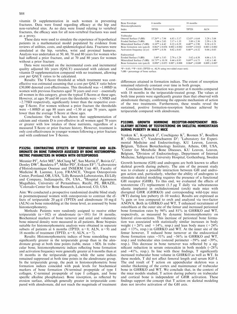
vitamin D supplementation in such women in preventingfractures. Data were found regarding efficacy at the hip andnon-vertebral sites. As no data were found relating to vertebralfractures, the efficacy seen for all non-vertebral fractures was usedas a proxy.
These data were used to simulate the experience of hypotheticalpatients in a mathematical model populated by evidence fromreviews of utilities, costs, and epidemiological data. Fractures weresimulated at the hip, vertebra, wrist and proximal humerus.Analysis was undertaken at 50, 60, 70 and 80 years for women whohad suffered a prior fracture, and at 70 and 80 years for womenwithout a prior fracture.
Data were recorded on the incremental costs and incrementalquality adjusted life years (QALY) associated with calcium andvitamin D supplementation compared with no treatment, allowingcost per QALY ratios to be calculated.
Results: The T-Score threshold at which treatment was cost-effective was estimated assuming that a cost per QALY ratio below£30,000 denoted cost-effectiveness. This threshold was )1.00SD inwomen with previous fractures aged 70 years and over – essentiallyall women in this category, given the typical T-Scores at these ages.However, at ages 50 and 60 years the thresholds were )3.50SD and)2.75SD respectively, significantly lower than the respective aver-age T-Score. For women without a prior fracture the thresholdswere )1.00SD at age 80 years and )1.50 at age 70 years, againlower than the average T-Score at these ages.
Conclusions: Our work has shown that supplementation ofcalcium and vitamin D is cost-effective in all women aged 70 yearsor greater with low intakes of these nutrients, regardless of ameasured T-Score or prior fracture history. However, treatment isonly cost-effectiveness in younger women following a prior fractureand with confirmed low T-Scores.
P332SU. CONTRASTING EFFECTS OF TERIPARATIDE AND ALEN-DRONATE ON BONE TURNOVER ASSESSED BY BONE HISTOMORPHO-METRIC PARAMETERS IN WOMEN WITH OSTEOPOROSIS
Meunier PJ1, Arlot ME1, McClung M2, San Martin J3, Boivin G1,Donley DW3, Bandeira F4, Miller PD5, Eriksen EF3; 1Laboratoired’Histodynamique Osseuse and INSERM Unit 403, Faculty ofMedicine R. Laennec, Lyon, FRANCE, 2Oregon OsteoporosisCenter, Portland,OR,USA, 3LillyResearch Laboratories, Eli Lillyand Company, Indianapolis, IN, USA., 4Hospital AgamenonMagalhaes, University of Pernambuco, Recife, PE, BRAZIL,5Colorado Center for Bone Research, Lakewood, CO, USA
Aim: We conducted a prospective randomized double blind studyin postmenopausal women with osteoporosis to contrast the ef-fects of teriparatide 20 lg/d (TPTD) and alendronate 10 mg/d(ALN) on bone remodeling at the tissue level, as assessed by bonehistomorphometry.
Methods: Patients were randomly assigned to receive eitherteriparatide (n=102) or alendronate (n=101) for 18 months.Biochemical markers of bone turnover and areal and volumetricbone mineral density were measured in all patients. Bone biopsiessuitable for histomorphometric analysis were obtained in separatesubsets of patients at 6 months (TPTD, n=8; ALN, n=9) and18 months of treatment (TPTD, n=8; ALN, n=7).
Results: Histomorphometric indices of bone remodeling weresignificantly greater in the teriparatide group than in the alen-dronate group at both time points (table, mean ±SD). In trabe-cular bone, histomorphometric indices reflecting bone formationand activation frequency were generally greater at 6 months than at18 months in the teriparatide group, while the same indicesremained suppressed at both time points in the alendronate group.In the teriparatide group, the peak in histomorphometric boneformation indices coincided with peak levels for biochemicalmarkers of bone formation (N-terminal propeptide of type Icollagen, C-terminal propeptide of type I collagen, and bonespecific alkaline phosphatase). Bone resorption, as reflected byerosion surface, although generally greater in teriparatide com-pared with alendronate, did not reach the magnitude of treatment
differences attained in formation indices. The extent of resorptionremained relatively constant over time in both groups.
Conclusion: Bone formation was greater at 6 months comparedwith 18 months in the teriparatide-treated group. The values atboth time points were significantly greater than that observed withalendronate therapy, confirming the opposite mechanism of actionof the two treatments. Furthermore, these results reveal thesustained, positive formation-resorption balance achieved byteriparatide compared with alendronate.
P333MO. GROWTH HORMONE RECEPTOR-INDEPENDENT REG-ULATORY ACTIONS OF TESTOSTERONE ON SKELETAL HOMEOSTASISDURING PUBERTY IN MALE MICE
Venken K1, Kopchick J2, Coschigano K2, Boonen S3, BouillonR1, Ohlsson C4, Vanderschueren D1; 1Laboratory for Experi-mental Medicine and Endocrinology, KU Leuven, Leuven,Belgium, 2Edison Biotechnology Institute, Athens, OH, USA,3Center for Metabolic Bone Diseases, KU Leuven, Leuven,Belgium, 4Division of Endocrinology, Department of InternalMedicine, Sahlgrenska University Hospital, Gothenburg, Sweden
Growth hormone (GH) and androgens are both known to affectskeletal growth during puberty. The aim of the present studywas to evaluate a possible interaction between GH and andro-gen action and, particularly, whether the ability of androgens tostimulate skeletal modeling requires the presence of a functionalGH receptor (GHR). To this end, we evaluated the effects oftestosterone (T) replacement (1.5 lg T daily via subcutaneoussilastic implants) in orchidectomized (orch) male mice withdisrupted GHR (GHRKO) and corresponding wild-type mice(WT) during late puberty (6–10 weeks). Results are expressed as% gain or loss compared to orch and analyzed via two-factorANOVA. Both in GHRKO and WT, T enhanced recruitment ofosteoblasts at the outer site of the femur and increased periostealbone formation rates by 94% and 81% in GHRKO and WT,respectively, as measured by dynamic histomorphometry onfemoral cross-sections. This increase of periosteal bone forma-tion was associated with statistically similar increases in bodyweight (+26% and +14%, resp.) and lean body mass (+24%and +13%, resp.) in GHRKO and WT. At the inner site of thefemur however, T reduced bone turnover at the endocortical(bone formation rates )51% and )56% in GHRKO and WT,resp.) and trabecular sites (osteoid perimeter )59% and )39%,resp.). This decrease in bone turnover was reflected by a sig-nificant reduction in serum osteocalcin in both models ()28%and )41%, resp.). In line with these findings, T significantlyincreased trabecular bone volume in GHRKO as well as WT. Inthese models, T did not affect femoral length and serum IGF-I.The end result of T action on appendicular skeleton was asimilar thickening of the cortex and maintenance of trabecularbone in GHRKO and WT. We conclude that, in the context ofthe mice models studied, T action during puberty on trabecularand cortical bone is independent of GHR activation. Thesefindings support the concept that T action on skeletal modelingdoes not involve activation of the GH axis.
Bone EnvelopeHistomorphometric
6 months 18 months
Index TPTD ALN TPTD ALN
TrabecularOsteoid Surface (%BS) 17.26**� 7.94 6.83� 5.17 12.63*� 6.64 5.29� 3.04Eroded Surface (%BS) 3.08� 2.03 2.17� 1.35 3.89� 2.30 2.59� 1.28Mineralized Surface (%BS) 8.10**� 4.42 0.22� 0.29 4.40**� 2.90 0.38� 0.31Bone formation rate (lm/d) 0.062*� 0.036 0.002� 0.002 0.030*� 0.022 0.003� 0.002Activation frequency (#/yr) 0.99**� 0.56 0.02� 0.03 0.46**� 0.32 0.04� 0.03
EndocorticalEroded Surface (%BS) 5.60*� 3.83 2.79� 1.29 5.62� 4.58 4.06� 3.51Mineralized Surface (%BS) 18.73**� 10.38 0.44� 0.95 9.69**� 6.73 1.02� 1.40Bone formation rate (lm/d) 0.098*� 0.053 0.007� 0.008 0.064*� 0.049 0.009� 0.007
*P<0.05; **P<0.01 (TPTD vs. ALN using two-sided exact test).%BS=percentage of bone surface.
S88
P334SA. RAPID PAIN RELIEF AND REMISSION OF STERNOCOSTO-CLAVICULAR HYPEROSTOSIS (SCCH) AFTER INTRAVENOUS IBAN-DRONATE THERAPY
Ringe JD, Faber H, Farahmand P; University of Cologne,Leverkusen, Germany
Aims: Sternocostoclavicular hyperostosis (SCCH) is a rare, loca-lised disturbance of bone metabolism that is extremely painful andleads to significant deterioration in QoL. It is often diagnosed lateor remains undiagnosed. The aetiology of SCCH is unknown. Thecondition flares up sporadically, causing pain and swelling in thesternoclavicular region with considerable impairment of shouldergirdle movement. The pain is frequently resistant to standardanalgesic therapy. Typical X-ray signs are hyperostosis in themedial third of the clavicula, the anterior insertion of the first riband the manubrium. Reduction of localised high bone turnoverwith bisphosphonates may improve outcome in this condition.
Methods: Three women, aged 40, 55 and 68 years, with 5–15year histories of severe localised and therapy resistant pain hadconsulted numerous doctors and hospitals without definitive helpor relief. Biopsies in two patients produced no clear diagnosisbeyond ‘an analgesic-resistant local inflammatory process’. Ourdiagnosis of SCCH was based on typical localization, X-ray find-ings and high uptake at bone scanning. Pain, measured on a10-point visual analogue scale (VAS), averaged 9.7 before treat-ment. Erythrocyte sedimentation rate, and in one case alkalinephosphatase levels, were slightly elevated, but no typical laboratorypattern emerged. All three women had systemic osteopenia or os-teoporosis. We treated patients with initial infusions of 4mg iban-dronate followed by 2 mg i.v. ibandronate injections every3 months for up to 1 year, with daily calcium and vitamin Dsupplementation throughout the study and follow-up.
Results: Pain relief was rapid and persistent: average VAS painscores were 4.7 after 2 weeks, 1.7 after 3 months and 0.3 after 12months. Bone scans after 3 months showed significant decreases intracer uptake and area of involved bones. X-rays after 12 monthsshowed structural improvements. Mean BMD increased by 9.1% atthe lumbar spine and 7.0% at the femoral neck after 1 year. Followup to month 24 revealed no symptoms of recurrent disease.
Conclusions: Treatment with i.v. ibandronate injections forSCCH was successful in terms of pain relief, improvement in QoLand disease remission in this condition of localised increased boneturnover of unknown aetiology.
P335SU. CHLORIDE CHANNEL INHIBITION PREVENTS BONE RE-SORPTION IN OVARIECTOMIZED RATS WITHOUT CHANGING BONEFORMATION
Karsdal MA1, Henriksen K1, Schaller S1, Christophersen P2,Christiansen C1; 1Nordic Bioscience A/S & CCBR, Herlev/Ballerup, Denmark, 2Neuroseach A/S, Ballerup, Denmark
Chloride channel activity (ClCN7) is essential for osteoclasticacidification of the resorption lacunae and thereby for dissolutionof the inorganic phase of bone. Consequently, inhibition of theosteoclastic chloride channel should prevent bone resorption.Accordingly, we have tested chloride channel inhibitors on boneturnover. Most interesting, we found that bone resorption couldbe inhibited without affecting bone formation in vivo. This studyindicates that chloride channel inhibitors are highly promising fortreatment of osteoporosis.
The chloride channel inhibitor, NS3736 (1-[4-Bromo-2-(1H-tetrazol-5-yl)-phenyl]-3-(4-chloro-3-trifluoromethyl-phenyl)-urea),blocked osteoclastic acidification and resorption in vitro with anIC50 value of 30 microM. When tested in the rat ovariectomymodel for osteoporosis, daily treatment with 30 mg/kg p.o.restored bone resorption to control levels as measured by CTXand protected bone strength and bone mineral density byapproximately 50% 6 weeks post-surgery. Most interestingly,bone formation assessed by osteocalcin, mineral apposition rateand mineralized surface index was not inhibited.
Analysis of chloride channels in human osteoclasts revealedthat ClC-7 and CLIC1 were highly expressed. Furthermore, by
electrophysiology we detected a volume activated anion channel onhuman osteoclasts. Screening 50 different human tissues showed abroad expression for CLIC1 and a restricted immunoreactivity forClC-7, appearing mainly in osteoclasts, ovaries, appendix andPurkinje cells. This highly selective distribution predicts that in-hibition of ClC-7 should target specifically osteoclasts in vivo. Wesuggest that NS3736 is inhibiting ClC-7, leading to a bone specificeffect in vivo.
In conclusion, we show for the first time that chloride channelinhibitors can be used for prevention of ovariectomy-induced boneloss without impeding bone formation. We speculate that thecoupling of bone resorption to bone formation is linked to theacidification of the resorption lacunae, thereby enabling compoundsthat directly interfere with this process to be able to positive impingeon this process resulting in a net gain of bone mass and strength.
P336MO. ALENDRONATE THERAPY OF PEDIATRIC PATIENTS WITHOSTEOGENESIS IMPERFECTA
Vyskocil VV; Bone Disease Center, Charles University HospitalPilsen, Czech Republic
Osteogenesis imperfecta (OI) is a rare crippling disorder leadingto fractures and bone deformities. Previously employed therapywith calcitonin, calcium and vitamin D resulted in minimal in-crease in bone mineral density (BMD) and did not reduce theincidence of fractures. As there have been reports on promisingeffect of pamidronate treatment in children with OI, we intitiatedtherapy with another bisphosphonate, alendronate, in a group ofchildren with OI.
Parental and patient consent was obtained prior to this treat-ment. We observed 30 children with OI, mean age 13.7 years.Alendronate was administered orally for one year: children aged4–10 years received 5 mg/day, children above 10 years of age weregiven 10 mg/day. Prior to the onset of therapy, spinal BMDZ-score (measured by DXA) was below )2.1 in all children and allpatients had more than 2 fractures/year. After one year of therapy,we observed significant increase in spinal BMD (+14.5%,p<0.05). After another 2 years of therapy, there was yet furtherincrease in spinal BMD (+6.3%). The changes in BMD wererelated to the mobility status of the patients, as immobilizedpatients had a less apparent increase in BMD. The interpretationof BMD measurements in hip and forearm was not possible dueto multiple bone deformities. The values of bone markers(S-osteocalcin, S-procollagen III, U-deoxypyridinoline crosslinks)decreased significantly (p<0.05) after one year of therapy. In thefollowing 2 years there were no further significant changes in theseparameters with the exception of U-deoxypyridinoline. In thecourse of the first year of alendronate therapy we observed just onefracture in the entire group, this as a result of a skiing accident in apatient with OI type Sillence I. We did not observe any adverseeffect of alendronate therapy regarding gastrointestinal tract orbiochemical changes. The x-rays of long bones did not reveal anynegative effect of alendronate on metaphysis or growth cartilage. Inconclusion, alendronate seems to be a safe and effective drug inpediatric patients with OI.
P337SA. RISEDRONATE REDUCES FRACTURE RISK IN OSTEOPOROTICWOMEN OVER 80 YEARS OF AGE: IMPLICATIONS FOR THE USE OFANTIRESORPTIVE AGENTS IN THE OLDEST
Boonen S1, McClung M2, Eastell R3, Fuleihan G4, Barton I5,Delmas P6; 1Katholieke Universiteit Leuven, Belgium, 2OregonOsteoporosis Center, USA, 3University of Sheffield, UK,4American University of Beirut, LEbanon, 5Procter & GamblePharmaceuticals, UK, 6INSERM Unit 403, Lyon, France
In persons over 80 years of age (the ‘‘oldest old’’), there is nopublished evidence that antiresorptive agents lead to greaterreductions in fracture risk than calcium and vitamin D alone. Therisedronate database includes almost 1400 individuals over 80 yearsof age with confirmed osteoporosis. The aim of this study was todetermine the efficacy of risedronate in reducing vertebral fracturerisk in women 80 years of age or older with osteoporosis.
S89
We performed a pooled analysis of data from three rando-mized, double-blind, controlled, 3-year fracture endpoint trialsconducted from November 1993 to April 1998 (HIP, VERT-MN,and VERT-NA) conducted in Europe, North America, andAustralasia. Patients included were those with documented osteo-porosis (a femoral neck BMD T-score <)2.5SD and/or at leastone prevalent vertebral fracture) 80 years of age or older. Patientsreceived placebo (n=688) or risedronate 5 mg/day (n=704) for upto 3 years. All patients received 1000 mg/d calcium and, if baselinelevels were low, up to 500 IU/d vitamin D. Vertebral radiographswere taken at baseline and at yearly intervals to determineprevalent and incident vertebral fractures.
At baseline, the mean femoral neck T-score was )3.05 SD, and84% of the patients had at least one prevalent vertebral fracture.After 1 year, the risk of new vertebral fractures in the risedronategroup was reduced by 81% compared with placebo (95% con-fidence interval [CI], 60%–91%; P<.001). The number of womenwho needed to be treated to prevent one new vertebral fractureafter 1 year was 12. This early onset of efficacy was consistentacross the clinical programs. Risedronate continued to show anti-fracture efficacy over 3 years. Risedronate was well tolerated, witha safety profile comparable to placebo.
We conclude that antiresorptive treatment provides an anti-fracture benefit in addition to that afforded by calcium and vitaminD in women 80 years of age or older with documented osteo-porosis. These findings provide the first evidence that, even in theoldest old, reducing bone resorption rate remains an effectivetreatment strategy for osteoporosis.
P0338SU. INFLUENCE OF CONVENTIONAL AND HIGH DOSES OFESTROGEN ON THE DEGREE OF MINERALISATION OF BONE TISSUE:A QUANTITATIVE MICRORADIOGRAPHIC ANALYSIS IN POST-MENOPAUSAL WOMEN
Boivin G1, Vedi S2, Compston JE2, Meunier PJ1; 1INSERMUnite 403, Faculte de Medecine R. Laennec, Lyon, France,2Department of Medicine, University of Cambridge, School ofClinical Medicine, Cambridge, UK
The beneficial skeletal effects of menopausal estrogen replacementtherapy (HRT) are well documented and are believed to be medi-ated predominantly by inhibition of osteoclastic bone resorption.The role of secondary mineralization of bone in the preservation ofbone quality is also well established in postmenopausal womentreated with bisphosphonates or SERMs. The aim of the presentstudy was to investigate the effect of conventional and high doseestrogen on the degree of mineralization of bone (DMB).
Transiliac bone biopsies were obtained from 20 patients beforeand after 24 months (18 to 38 months) treatment with conventionalHRT, and from 19 patients who had received high doses of estra-diol (implant 100 mg every 3–6 months for at least 14 years). Bonesamples were embedded in LR White Resin. Thick sections wereground to a uniform thickness of 100 lm and then micro-radiographed with an aluminum step-wedge. DMB was measuredin compact, cancellous and total bone and expressed as g mineral/cm3 of bone (mean ± SEM).
Values obtained in patients before HRT were lower than thosereported in premenopausal women and similar to those reported forpostmenopausal women with untreated osteoporosis. After con-ventional HRT, the increase in DMB in total bone was 4.4±1.9%when compared to pre-treatment values (4.1±2.1% in compactbone, 4.5±2.3% in cancellous bone); these differences were notsignificant. In women who had received high dose estradiol therapy,DMB in total bone was 6.9±1.9% higher than in untreated women(8.6±2.1% in compact bone, 6.5±2.1% in cancellous bone); thesedifferences are statistically significant (p <= 0.03). After high dosetherapy the greatest change was noted in cortical bone as previouslyreported by histomorphometry. The increases of DMB were due toa shift of the curves towards high DMB with a decrease of thelow DMB values, but without changes in the heterogeneity of thedistributions (preservation of the quality of mineralization).
Our results demonstrate that estrogen therapy is associated withan increased degree of mineralization of bone (prolongation of
secondary mineralization) similar to that observed with other anti-resorptive agents. However, it was about 2-fold lower than thatobserved after alendronate therapy(10 mg/day/3 years).
P0339MO. RISK ASSESSMENT FOR OSTEOPOROTIC FRACTURE ININSTITUTIONALISED RESIDENTS IN COMPARISON TO A COMMUNITYDWELLING POPULATION: THE UK NATIONAL OSTEOPOROSIS SOCIETY(NOS), OSTEOPOROSIS NURSE INITIATIVE (ONI) PROGRAMME
Vines B1, Masud T2, Stone M3, Selby P4, Torgerson D5, SutcliffeA6, Hodson J7, Parrington J1, Mohammed M8, Cooper C9;1National Osteoporosis Society, Bath, UK, 2Nottingham CityHospital, Nottingham, UK, 3Llandough Hospital, Cardiff, UK,4Manchester Royal Infirmary,Manchester, UK, 5YorkUniversity,York, UK, 6Freeman Hospital, Newcastle, UK, 7Bridge HouseMedical Centre, Stratford-upon-Avon, UK, 8RioMed, South-ampton, UK, 9SouthamptonGeneral Hospital, Southampton, UK
Aims: Identifying people at high risk of fracture has the potentialfor cost-effective targeting of fracture prevention measures. In2001 the UK National Osteoporosis Society launched a primarycare programme [ONI], with the aim of identifying older subjectsat increased risk of fracture via a structured audit programme.This subsequent analysis aimed to determine osteoporotic fracturerisk in institutionalized residents in comparison to community-dwellers.
Methods: Five specialist nurses across the UK used a modified,validated fracture risk assessment tool (Black et al, Osteoporosis Int2001) on women aged over 75 years identified from general practicedatabases. The variables considered were: age, previous fragilityfracture, maternal hip fracture, weight<57 kg, inability to rise froma chair without using arms, and current smoker. Resulting scoreswere converted in each subject to give a low, moderate or high riskstatus for fracture. Falls history (previous year) was recorded.
Results: The mean age of the overall population (n=14955) was81 years (SD=5). Proportion of subjects classified as high, moderateand low fracture risk were 30%, 59% and 11% respectively. 374subjects [mean age 88 (SD=6) were identified as nursing or carehome residents. In these institutionalized subjects the proportionclassified as high, moderate and low fracture risk (compared tocommunity-dwelling age-matched controls) were 79% (vs 63%),20% (vs 35%), and 1% (vs 2%) respectively [ChiSquare= 24.5,df=2, p<0.001]. Inability to rise from a chair without using armswas significantly different between the institutionalized and age-matched community controls [87% vs 63%; ChiSquare=54.0,p<0.001], and there was a tendency towards a difference in previousfracture history [40% vs 34% respectively; ChiSquare=2.8, p<0.1].There were no significant differences in the other assessment tool riskfactors. Institutionalized residents were more likely to be recurrentfallers than controls [29% vs 21%; ChiSquare=5.6, p=0.018].
Conclusions: Institutionalised residents demonstrate a higherfracture risk profile than community-dwelling subjects. The formergroup therefore should be particularly targeted for fracture riskidentification and subsequent preventionmeasures including calciumand vitamin D, hip protectors and fall prevention. The next 2 yearphase of theUKONIproject will concentrate on this high risk group.
P340SA. HYPOPHOSPHATEMIC RICKETS
Fabian K; Borsod County Teaching Hospital, Child WelfareCenter, Miskolc, Hungary
Aims: Review of the literature of rare x-linked or autosomaldominant perhaps acquired hypophosphatemic rickets and com-pared the literature of the disease with experiences obtained during3 years disease history of a 2.5 year-old girl at time of diagnosis.
Methods: Analysis of the family and own history of the patientand diagnostic (laboratory data, x-ray, ultrasound) and differencialdiagnostic investigations. Treatment and its effects have beenfollowed for 3 years.
Results: The family history did not reveal any individual withbone disease, but the mother had a body height and serum phos-phate level below the average. From the age of 6 months, lowerextremity of the patient had become extremely curved. Her body
S90
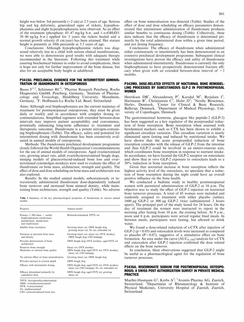
height was below 3rd percentile ()2 cm) at 2.5 years of age. Seriouship and leg deformity, generalized signs of rickets, hypophos-phatemia and slight hyperphosphaturia were observed. As an effectof the treatment (phosphate: 45–47 mg/kg b.w. and 1-a-(OH)D3:38–46 ng/kg b.w.) applied for 3 years the rickets healed and anormal growth velocity (8 cm/year) has been attained. Her statureheight is presently at 10th percentile at 5.5 years of age.
Conclusions: Although hypophosphatemic rickets was diag-nosed relatively late in a child with serious clinical manifestations,we were able to demonstrate good results with adequate therapyrecommended in the literature. Following this treatment whileassuring biochemical balance in order to avoid complications, thereis hope not only for further improvement of the bone disease, butalso for an acceptable body height at adulthood.
P341SU. PRECLINICAL EVIDENCE FOR THE INTERMITTENT ADMINIS-TRATION OF IBANDRONATE IN OSTEOPOROSIS
Bauss F1,2, Schimmer RC3; 1Pharma Research Penzberg, RocheDiagnostics GmbH, Penzberg, Germany, 2Institute of Pharma-cology and Toxicology, Heidelberg University. Mannheim,Germany, 3F. Hoffmann-La Roche Ltd, Basel, Switzerland
Aims: Although oral bisphosphonates are the current mainstay oftreatment for postmenopausal osteoporosis, they must be givendaily or weekly and in accordance with complex dosing re-commendations. Simplified regimens with extended between-doseintervals may improve patient acceptability and convenience,potentially enhancing long-term adherence to treatment andtherapeutic outcomes. Ibandronate is a potent nitrogen-contain-ing bisphosphonate (Table). The efficacy, safety and potential forintermittent dosing with ibandronate has been extensively eval-uated in animal models of osteoporosis.
Methods: The ibandronate preclinical development programmeclosely followed theWorld Health Organization’s recommendationsfor the use of animal models of osteoporosis. Aged ovariectomisedrats, growing intact rats, adult ovariohysterectomised beagle dogs,minipig models of glucocorticoid-induced bone loss and ovar-iectomised cynomolgus monkeys were used to evaluate the effect ofibandronate on bone mass, architecture, strength and quality. Theeffect of dose and dose scheduling on bonemass and architecture wasalso explored.
Results: In the studied animal models, subcutaneously or in-travenously administered ibandronate dose-dependently reducedbone turnover and increased bone mineral density, while main-taining bone architecture, strength and quality (Table). No adverse
effect on bone mineralisation was detected (Table). Studies of theeffect of dose and dose scheduling on efficacy parameters demon-strated that intermittent administration of ibandronate producessimilar benefits to continuous dosing (Table). Collectively, thesedata indicate that the efficacy of ibandronate is determined pri-marily by the total administered dose within a given time period,and not dosing frequency.
Conclusions: The efficacy of ibandronate when administeredeither continuously or intermittently has been demonstrated in anextensive preclinical development programme. Subsequent clinicalinvestigations have proven the efficacy and safety of ibandronatewhen administered intermittently. Ibandronate is currently the onlybisphosphonate to have prospectively demonstrated fracture effi-cacy when given with an extended between-dose interval of >2months.
P342MO. DOSE-RELATED EFFECTS OF NOCTURNAL BONE REMODEL-LING PROCESSES BY SUBCUTANEOUS GLP-2 IN POSTMENOPAUSALWOMEN
Henriksen DB1, Alexandersen P2, Karsdal M1, Byrjalsen I1,Hartmann B3, Christiansen C2, Holst JJ3; 1Nordic Bioscience,Herlev, Denmark, 2Center for Clinical & Basic Research,Ballerup, Denmark, 3Department of Medical Physiology, Uni-versity of Copenhagen, Denmark
The gastrointestinal hormone, glucagon like peptide-2 (GLP-2)has been suggested as a key regulator of the postprandial reduc-tion of bone resorption. Bone resorption when assessed withbiochemical markers such as CTX has been shown to exhibit asignificant circadian variation. This circadian variation is nearlyeliminated upon fasting and induced by nutritional intake. Wehave shown that the acute postprandial reduction of boneresorption coincides with the release of GLP-2 from the intestineand thus GLP-2 could be involved in an entero-osseous axis,which coordinates bone resorption in response to nutrient intake.In accordance, we have located the GLP-2 receptor on osteoclastsand show that in vitro GLP-2 exposure to osteoclasts leads to a50% reduction in bone resorption.
Given that nocturnal increase in bone resorption reflects thehighest activity level of the osteoclasts, we speculate that a reduc-tion of bone resorption during the night could have an overallpositive influence on the bone health.
We conducted a bedtime study in healthy postmenopausalwomen with parenteral administration of GLP-2 at 10 p.m. Theobjective was to study the effect of GLP-2 injection on nocturnalbone turnover processes. A total of 45 women were included andrandomly assigned to treatment with either placebo (saline),1600 lg GLP-2 or 800 lg GLP-2 twice (administered 3 hoursapart). The principal part of the study lasted for 24 hours. On theday of treatment the women were instructed to report in themorning after fasting from 10 p.m. the evening before. At 9 a.m.,noon and 6 p.m. participants were served regular fixed meals. Inbetween meals, participants were fasting, but allowed to drinkwater.
We found a dose-related reduction of s-CTX after injection ofGLP-2 (p<0.05) and osteocalcin levels were increased as comparedto placebo (P=0.07), suggestive of a stimulative effect on boneformation. An area under the curve (AUC0–10h) analysis for s-CTXand osteocalcin after GLP-2 injection confirmed the dose relatedeffects on the bone turnover.
In conclusion, these observations suggested that GLP-2 mightbe useful as a pharmacological agent for the regulation of boneturnover processes.
P343SA. RISEDRONATE SODIUM FOR POSTMENOPAUSAL OSTEOPO-ROSIS: A SWISS POST AUTHORIZATION SURVEY IN PRIVATE MEDICALPRACTICE
Mueller-Husmann G1, Krebs A2; 1Aventis Pharma AG, Zuerich,Switzerland, 2Department of Rheumatology & Institute ofPhysical Medicines, University Hospital of Zuerich, Zuerich,Switzerland
Table 1 Summary of the key pharmacological properties of ibandronate in various animalmodels
Property Animal model
Potency 2–500 times > earlierbisphosphonates (clodronate,pamidronate, alendronateand risedronate)
Retinoid-stimulated TPTX rat
Inhibits bone resorption Growing intact rat; OHX beagle dog;growing intact rat; Nx rat; unloaded rat
Increases or prevents bone massand density
Growing intact rat; intact rat; OVX monkey;OHX beagle dog; CIO minipigs
Prevents deterioration of bonearchitecture
OHX beagle dog; OVX monkey, aged OVX rat
Preserves bone strength Intact rat; OVX monkeyMaintains or restores bone quality OHX beagle dog; aged OVX rat; OVX monkey;
intact rat; CIO minipigs
No adverse effect on bone mineralisation Growing intact rat; OHX beagle dog
Prevents increase in erosion depth OHX beagle dog
Efficacy with intermittent dosing OHX beagle dog; aged OVX rat; OVX monkey;intact rat; CIO minipigs; Nx rat; unloaded rat
Efficacy determined primarily bycumulative dose
OHX beagle dog; aged OVX rat; growingintact rat
TPTX: thyroparathyroidectomisedOHX: ovariohysterectomisedOVX: ovariectomisedNx: nephrectomisedCIO: corticoid-induced bone loss
S91
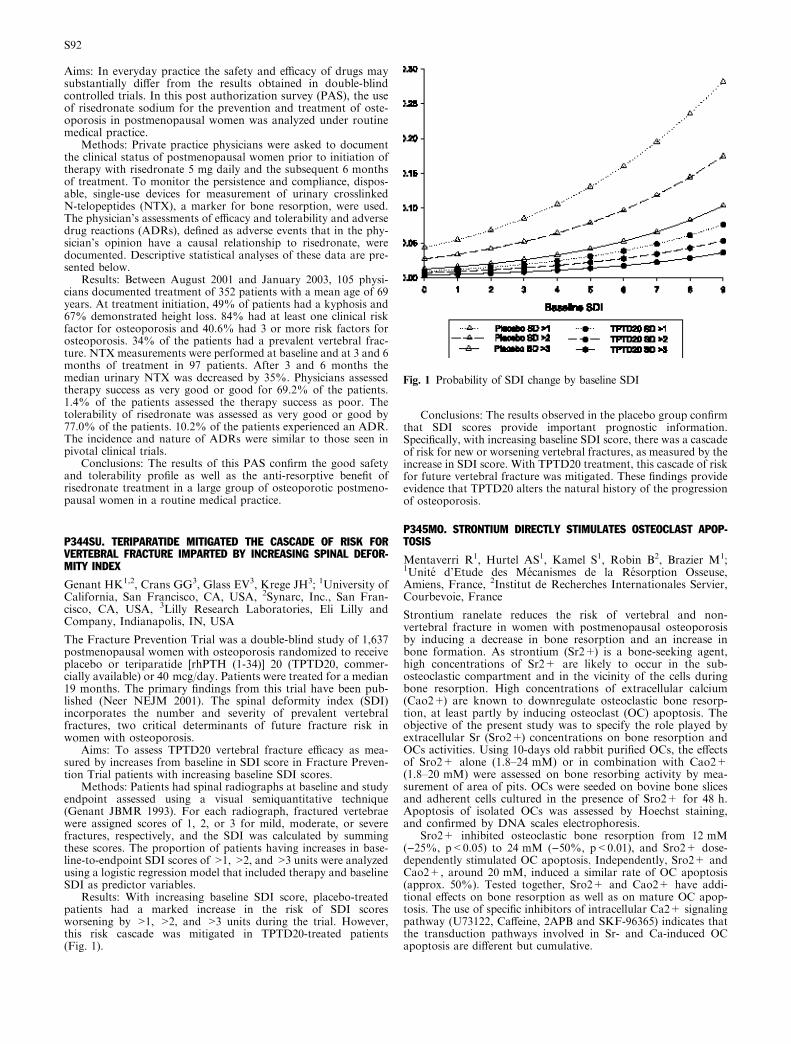
Aims: In everyday practice the safety and efficacy of drugs maysubstantially differ from the results obtained in double-blindcontrolled trials. In this post authorization survey (PAS), the useof risedronate sodium for the prevention and treatment of oste-oporosis in postmenopausal women was analyzed under routinemedical practice.
Methods: Private practice physicians were asked to documentthe clinical status of postmenopausal women prior to initiation oftherapy with risedronate 5 mg daily and the subsequent 6 monthsof treatment. To monitor the persistence and compliance, dispos-able, single-use devices for measurement of urinary crosslinkedN-telopeptides (NTX), a marker for bone resorption, were used.The physician’s assessments of efficacy and tolerability and adversedrug reactions (ADRs), defined as adverse events that in the phy-sician’s opinion have a causal relationship to risedronate, weredocumented. Descriptive statistical analyses of these data are pre-sented below.
Results: Between August 2001 and January 2003, 105 physi-cians documented treatment of 352 patients with a mean age of 69years. At treatment initiation, 49% of patients had a kyphosis and67% demonstrated height loss. 84% had at least one clinical riskfactor for osteoporosis and 40.6% had 3 or more risk factors forosteoporosis. 34% of the patients had a prevalent vertebral frac-ture. NTX measurements were performed at baseline and at 3 and 6months of treatment in 97 patients. After 3 and 6 months themedian urinary NTX was decreased by 35%. Physicians assessedtherapy success as very good or good for 69.2% of the patients.1.4% of the patients assessed the therapy success as poor. Thetolerability of risedronate was assessed as very good or good by77.0% of the patients. 10.2% of the patients experienced an ADR.The incidence and nature of ADRs were similar to those seen inpivotal clinical trials.
Conclusions: The results of this PAS confirm the good safetyand tolerability profile as well as the anti-resorptive benefit ofrisedronate treatment in a large group of osteoporotic postmeno-pausal women in a routine medical practice.
P344SU. TERIPARATIDE MITIGATED THE CASCADE OF RISK FORVERTEBRAL FRACTURE IMPARTED BY INCREASING SPINAL DEFOR-MITY INDEX
Genant HK1,2, Crans GG3, Glass EV3, Krege JH3; 1University ofCalifornia, San Francisco, CA, USA, 2Synarc, Inc., San Fran-cisco, CA, USA, 3Lilly Research Laboratories, Eli Lilly andCompany, Indianapolis, IN, USA
The Fracture Prevention Trial was a double-blind study of 1,637postmenopausal women with osteoporosis randomized to receiveplacebo or teriparatide [rhPTH (1-34)] 20 (TPTD20, commer-cially available) or 40 mcg/day. Patients were treated for a median19 months. The primary findings from this trial have been pub-lished (Neer NEJM 2001). The spinal deformity index (SDI)incorporates the number and severity of prevalent vertebralfractures, two critical determinants of future fracture risk inwomen with osteoporosis.
Aims: To assess TPTD20 vertebral fracture efficacy as mea-sured by increases from baseline in SDI score in Fracture Preven-tion Trial patients with increasing baseline SDI scores.
Methods: Patients had spinal radiographs at baseline and studyendpoint assessed using a visual semiquantitative technique(Genant JBMR 1993). For each radiograph, fractured vertebraewere assigned scores of 1, 2, or 3 for mild, moderate, or severefractures, respectively, and the SDI was calculated by summingthese scores. The proportion of patients having increases in base-line-to-endpoint SDI scores of >1, >2, and >3 units were analyzedusing a logistic regression model that included therapy and baselineSDI as predictor variables.
Results: With increasing baseline SDI score, placebo-treatedpatients had a marked increase in the risk of SDI scoresworsening by >1, >2, and >3 units during the trial. However,this risk cascade was mitigated in TPTD20-treated patients(Fig. 1).
Conclusions: The results observed in the placebo group confirmthat SDI scores provide important prognostic information.Specifically, with increasing baseline SDI score, there was a cascadeof risk for new or worsening vertebral fractures, as measured by theincrease in SDI score. With TPTD20 treatment, this cascade of riskfor future vertebral fracture was mitigated. These findings provideevidence that TPTD20 alters the natural history of the progressionof osteoporosis.
P345MO. STRONTIUM DIRECTLY STIMULATES OSTEOCLAST APOP-TOSIS
Mentaverri R1, Hurtel AS1, Kamel S1, Robin B2, Brazier M1;1Unite d’Etude des Mecanismes de la Resorption Osseuse,Amiens, France, 2Institut de Recherches Internationales Servier,Courbevoie, France
Strontium ranelate reduces the risk of vertebral and non-vertebral fracture in women with postmenopausal osteoporosisby inducing a decrease in bone resorption and an increase inbone formation. As strontium (Sr2+) is a bone-seeking agent,high concentrations of Sr2+ are likely to occur in the sub-osteoclastic compartment and in the vicinity of the cells duringbone resorption. High concentrations of extracellular calcium(Cao2+) are known to downregulate osteoclastic bone resorp-tion, at least partly by inducing osteoclast (OC) apoptosis. Theobjective of the present study was to specify the role played byextracellular Sr (Sro2+) concentrations on bone resorption andOCs activities. Using 10-days old rabbit purified OCs, the effectsof Sro2+ alone (1.8–24 mM) or in combination with Cao2+(1.8–20 mM) were assessed on bone resorbing activity by mea-surement of area of pits. OCs were seeded on bovine bone slicesand adherent cells cultured in the presence of Sro2+ for 48 h.Apoptosis of isolated OCs was assessed by Hoechst staining,and confirmed by DNA scales electrophoresis.
Sro2+ inhibited osteoclastic bone resorption from 12 mM()25%, p<0.05) to 24 mM ()50%, p<0.01), and Sro2+ dose-dependently stimulated OC apoptosis. Independently, Sro2+ andCao2+, around 20 mM, induced a similar rate of OC apoptosis(approx. 50%). Tested together, Sro2+ and Cao2+ have addi-tional effects on bone resorption as well as on mature OC apop-tosis. The use of specific inhibitors of intracellular Ca2+ signalingpathway (U73122, Caffeine, 2APB and SKF-96365) indicates thatthe transduction pathways involved in Sr- and Ca-induced OCapoptosis are different but cumulative.
Fig. 1 Probability of SDI change by baseline SDI
S92
Our data strongly suggest that exposure of OCs to an increasingamount of Sro2+ is responsible for a decrease in the bone resorptionprocess mediated, at least in part, by the induction of OC apoptosis.Although, Sro2+ and Cao2+ both stimulate a G protein-coupledreceptor, which could be the calcium-sensing receptor, they havedifferential intracellular effects which independently trigger OCapoptosis and could act in a cooperative manner. These resultssupport the mechanism of reduced bone resorption observed invarious in vivo and in vitro experiments with strontium ranelate.
P346SA. CATABOLIC BONE RESPONSE TO GH AND ATTENUATED BUTMAINTAINED ANABOLIC RESPONSE TO PTH(1–34) IN RATS FED ALOW PROTEIN DIET
Ammann P1, Gasser J2, Rizzoli R1; 1Division of Bone Diseases,University Hospital, Geneva, Switzerland, 2Novartis, Bale,Switzerland
Stimulators of bone formation can improve bone structure andstrength, being thereby particularly suitable for the managementof patient with severe osteoporosis. Protein undernutrition fre-quently occurs in elderly probably contributing to the patho-genesis of osteoporosis. Whether protein intake could influencethe response to bone anabolic agents is unknown. To address thisissue, six-month old female rats were fed isocaloric diets con-taining 2.5% (low Protein) or 15% (normal Protein) casein for 2weeks. Then, bGH (0.5 or 2.5 mg/kg BW) or its solvent weregiven subcutaneously to rats on either diet twice daily for 4 weeks.In a second series of experiment using the same experimentaldesign, PTH(1–34) (5 or 40 lg/kg BW) or its solvent were given inrats fed the two diets. Proximal tibia bone mineral density (BMD)and ultimate strength were measured. bGH treatment dose-dependently decreased BMD ()5.1%±2. 5 and )10.7%±1.8*,with 0.5 or 2.5 mg/kg BW of bGH) and bone strength()20.1%±4.9* and )44.0%±5.6*) in rats fed a low proteindiet. No significant effects were observed in rats fed the normalprotein diet after this short period of treatment. PTH(1–34)treatment dose-dependently increased BMD (+10.0%±2.2* and+21.5%±2.2*, with 5 or 40 lg/kg BW of PTH(1–34)) and ulti-mate strength (+55.3% ±14.3* and +96.5%±16.1*) in rats fedthe normal protein diet. In rats fed a low protein diet, BMD andultimate strength were +4.1%±2.0 and +11.0%±2.7*, and+4.2%±8.4 and +43.8%±13.0*, respectively. Thus to achievethe same effect on BMD or strength a dose 8 times higher ofPTH(1–34) was required in rats fed an isocaloric low protein diet.GH treatment resulted in a negative bone balance when proteinintake was reduced. In contrast PTH(1–34) induced a positivebone balance but at a higher dose than in rats with the normalprotein diet. These results indicate that protein isocaloricrestriction could markedly influence response to anabolic agents,emphasizing thus the major importance of dietary protein intakein the bone response to GH or PTH administration.
P347SU. STRONTIUM RANELATE DOSE-DEPENDENTLY INCREASESBONE STRENGTH AND INTRINSIC BONE QUALITY IN INTACT FEMALERATS
Ammann P, Rizzoli R; University Hospital, Geneva, Switzerland
Recent clinical studies have demonstrated that strontium ranelatereduces the risk of vertebral and non-vertebral fracture in post-menopausal osteoporotic women. In the present study, weinvestigated the long-term effects of strontium ranelate on bonestrength in intact female rats at the level of L4 lumbar vertebra.Four groups of 30 rats (seven-week old at treatment initiation)were fed ad libitum a diet containing strontium ranelate at a dailydose of 0 (control), 225, 450 or 900 mg/kg/day, for 104 weeks.From the load deflection curve, obtained by compression of thevertebral body, maximal load, stiffness, yield point, total energy(E), and elastic and plastic energy were measured.
Strontium ranelate treatment dose-dependently increasedmaximal load (up to +20% at 900 mg/kg/d, p<0.05) and yieldpoint (up to +13%, ns) without affecting stiffness, indicating no
mineralization defect, thus suggesting an improvement of the bonequality. This was confirmed by the increase of energy to failure (upto +54.5%, p<0.05) achieved with strontium ranelate treatment at900 mg/kg/d, which was essentially due to a significant increase inplastic energy (+136%, p<0.01), with an increase in elastic energy(+26%, NS). These results strongly suggest that bone formedunder strontium ranelate treatment is able to withstand greaterdeformation before fracture while possessing similar elastic prop-erties to normal bone. Such modifications observed under stron-tium ranelate treatment are in agreement with an improvement ofintrinsic bone quality leading to greater bone resistance.
P348MO. LONG TERM TREATMENT OF OVARIECTOMIZED RATS WITHBLACK COHOSH: SITE SPECIFIC EFFECTS AND CALCIUM BALANCE
Nisslein T, Freudenstein J; Schaper & Bruemmer Co., Salzgitter,Germany
Black cohosh (Actea (=Cimicifuga) racemosa) is a herbal remedytraditionally used to treat symptoms of peri- and postmenopause.Its efficacy in alleviating hot flushes, night sweats and somaticclimacteric complaints has been proven in clinical studies which,however, took at the most few months. For monitoring significantosteoprotective effects in humans, such a short duration oftreatment has proven insufficient, even though short-term animalstudies have been able to demonstrate a positive influence of blackcohosh on metabolic bone parameters [Nisslein and Freudenstein2003. J Bone Miner Metab 21(6) 370–6].
We therefore performed a study, where adult female SpragueDawley rats (20 animals per group) were ovariectomized (ovx) andthe subsequently established imbalance of bone metabolism wasmonitored via urinary crosslink measurements. Intervention ther-apy with an isopropanolic black cohosh extract (60 or 300 mgherbal drug/kg b.w./d) was started in two treatment groups,whereas raloxifene-treated animals (3 mg/kg b.w.) served as posi-tive controls and placebo-treated ovx- or sham-operated animalsserved as two negative control groups. Treatments lasted for 10months and besides crosslink measurements, urinary calciumexcretion was continuously monitored. At the end of the study,femoral bone density and fracture resistance at the femoral headand lumbar vertebral site were measured.
As expected, ovx-induced alterations in bone metabolism wererapidly evident in all monitored parameters, and corroborated laterin necropsy findings of reduced bone density and fracture resis-tance. Raloxifene significantly antagonized ovx-induced osteopo-rosis as evident in parameters of bone strength and urinalysis.Black cohosh also proved effective against ovx-induced boneresorption: Levels of urinary crosslinks were significantly reducedcompared to untreated ovx-controls. Effects on bone density andfracture resistance were however more pronounced at peripheralbones than at the spine. Interestingly, in black cohosh treatedanimals, the observed reduction in calcium excretion was dose-dependent. After treatment with high-dose black cohosh, calciumlevels were even below pre-ovx values.
We observed no adverse events in high-dose long-term treatedrats and therefore we consider the isopropanolic extract of blackcohosh, as contained in Remifemin�, as a safe alternative formenopausal women with a positive influence on bone metabolismand calcium balance.
P349SA. STRONTIUM RANELATE REDUCES THE RISK OF VERTEBRALAND NON-VERTEBRAL FRACTURES IN CAUCASIAN WOMEN WITHPOSTMENOPAUSAL OSTEOPOROSIS
AdamiS1,Meunier PJ2,Devogelaer JP3,HoszowskiK4, FardelloneP5, Benhamou V6, Brixen K7, Bonidan O8, Marcelli C9, ReginsterJY10, Fechtenbaum J11; 1Centro Ospedialero Clinicizato diMedicina, Valeggio sul Mincio, ITALY, 2Dept of Rheumatologyand Bone Diseases, Edouard Herriot Hospital, Lyon, FRANCE,3Dept of Rheumatology, Catholic University of Louvain, Brussels,BELGIUM, 4Dept of Internal Medicine, Railway Hospital,Warsaw, POLAND, 5Dept ofRheumatology,UniversityHospital,Amiens, FRANCE, 6Hopital de laMadeleine, Orleans, FRANCE,
S93
7Odense University Hospital, Odense, DENMARK, 8HopitalEmile Muller, Mulhouse, FRANCE, 9Dept of Rheumatology,University Hospital, Caen, FRANCE, 10Dept of Epidemiologyand Public Health, University of Liege, Liege, BELGIUM,11CEMO, Hopital Cochin, Paris, FRANCE
Strontium ranelate, a new orally active anti-osteoporotic agent,has been reported to have a dual action on bone metabolism,simultaneously increasing bone formation and decreasing boneresorption. A large phase 3 program including 2 multinational,double blind, placebo controlled studies has been performed. Inboth studies, patients were randomly assigned to receive stron-tium ranelate 2g/day or placebo for 3 years associated to calciumand vitaminD supplementation according to the patient’s status.
The SOTI study (Spinal Osteoporosis Therapeutic Intervention)focused on vertebral anti-fracture efficacy of strontium ranelate, in1649 post menopausal women with osteoporosis [age: 69.7(7.3)years; Lumbar Bone Mineral Density (BMD Tscore: )3.6(1.3);mean(SD)], 87.5% having at least one prevalent vertebral fracture(2.2 per patient), has demonstrated a rapid and sustained vertebralanti-fracture efficacy of strontium ranelate in the intent-to-treatpopulation, with a relative risk reduction of vertebral fracture of49% (p<0.001) the first year of treatment and 41% (p<0.001)over 3 years. At 3 years lumbar BMD in strontium ranelate groupincreased by 14.4% as compared to placebo (p<0.001) and femoralneck BMD by 8.3% (p<0.001).
The TROPOS study (Treatment Of Peripheral OSteoporosis)investigated the efficacy of strontium ranelate on non-vertebralfractures in 5091 women with post-menopausal osteoporosis [age:76.8(5) years, Femoral Neck BMD T-score )3.1(0.6), 36.8% had atleast one prevalent non-vertebral fracture]. In the intent-to-treatpopulation, the risk for experiencing a new non-vertebral fractureduring the 3 years was reduced by 16% (RR=0.84 95%CI[0.702;0.995], p=0.04, adjusted cox model on pre-defined influen-tial covariates age, BMI, FN BMD and country). 41% reduction ofthe hip fracture risk (RR=0.59; 95%CI [0.37;0.95], p=0.03) wasalso achieved in patients having taken strontium ranelate for atleast 18 months.
At 3 years femoral neck BMD in strontium ranelate group in-creased by 8.2% as compared to placebo (p<0.001) and lumbarBMD by 14.7% (p<0.001).
In both studies strontium ranelate had a good bone and generalsafety profile.
The present data support the efficacy of strontium ranelate inreducing the risk of vertebral and non vertebral fracture in post-menopausal women with osteoporosis, representing a new candi-date in the treatment of postmenopausal osteoporosis.
P350SU. RALOXIFENE TREATMENT IMPROVES THE STRUCTURALGEOMETRY OF THE HIP: RESULTS FROM THE MULTIPLE OUTCOMESOF RALOXIFENE EVALUATION (MORE) TRIAL
Uusi-Rasi K1, Beck TJ1, Semanick LM1, Crans GG2, Pinette KV2,Harper KD2; 1Department of Radiology, The John HopkinsUniversity School of Medicine, Baltimore, MD, USA, 2LillyResearch Laboratories, Eli Lilly and Company, Indianapolis, IN,USA
Aims: Raloxifene treatment increases lumbar spine and femoralneck BMD and reduces the risk of vertebral fractures. In thisstudy our objective was to determine the effect of raloxifenetreatment on the structural geometry of the proximal femur. Asubset of postmenopausal women with osteoporosis (n=1903,mean age 67±7 y) enrolled in the MORE trial was randomized toraloxifene 60mg/d (n=632, RLX60), raloxifene 120 mg/d(n=648, RLX120) or placebo (n=623).
Methods: Study participants received hip DXA scans at base-line and 1, 2 and 3 year time points. The DXA scans were analyzedwith a Hip Structure Analysis (HSA) program that measured BMDand geometric properties of the proximal femur traversing regionsof the femoral neck, intertrochanter and proximal femoral shaft.Mean response to treatment (area under curve of the repeatedmeasurements) was calculated as a summary measure and used a
dependent variable in analysis of covariance with the baseline valueas a covariate.
Results: Mean differences following three-year treatment withraloxifene, compared to placebo, are presented in the table below.Following treatment with raloxifene, BMD increased and thebuckling ratio (an index of structural instability) decreased at allregions, compared to placebo. The cross sectional area and bendingstrength (section modulus) increased at the narrow-neck and in-tertrochanter for both RLX60 and RLX120, but femoral shaftstrength showed significant improvement only in the RLX120group, compared to placebo. There were no differences in boneouter diameter between groups, suggesting the rate of periostealapposition following raloxifene treatment cannot be detected by theHSA technique.
Conclusions: Raloxifene treatment improves indices ofmechanical strength and stability at all regions of the proximalfemur with no apparent dose effect except at the femoral shaftwhere fragility fractures are rare.
P351MO. EFFECTS OF ALENDRONATE (ALN) ON THE ‘‘ELASTIC’’ (PRE-YIELD) AND ‘‘PLASTIC’’ (POST-YIELD) BEHAVIOR OF CORTICAL BONEIN OVARIECTOMIZED (OX) RATS.
Chiappe MA2, Capozza RF1, Iorio G2, Alvarez E2, Ferretti JL1,Cointry GR1; 1CEMFoC, Fac of Medicine, Natl Univ of Rosario,Argentina, 2Fac of Veterinary, Univ of Buenos Aires, Argentina
Effects of bisphosphonates on bone mineralization and strengthare not necessarily correlative. To analyze ALN effects on bonestrength, forty 3-month old rats were OX and given immediately 0(OX-C, n=13), 5 (OX-5, n=13) or 25 ug/kg sc (OX-25, n=14) 2/wk for 6 months, keeping further 15 as sham controls. Theirfemurs were scanned by pQCT and tested in bending.
Despite not affecting bone mineralization (cortical vBMD) andcross-sectional diaphyseal geometry (diameters, moment of inertia)MI-), OX impaired the intrinsic stiffness of cortical tissue (elasticmodulus, E) and the structural stiffness of femur shafts (load/deformation ratio), and reduced yield and fracture loads (Wy, Wf).The post-yield fraction of Wf (Wp=Wf )Wy) was significantlyenhanced by OX, perhaps because of the naturally inverse rela-tionship between the tissue’s ability to prevent crack generation(impaired) and progress (improved). Effects of ALN were dose-dependent. The highest ALN dose prevented all negative effects ofOX and improved Wf over sham values. No changes in Wy wereobserved in treated rats (no effect on crack generation). However,Wp (bone toughness) was enhanced in a similar proportion than itwas in OX rats. The naturally negative ‘‘distribution/quality’’curves (correlations between cortical architecture, MI and intrinsicstiffness, E) shifted to the ‘‘anti-anabolic’’ region (lower-left) in thegraphs for OX rats and to the ‘‘anti-catabolic’’ region (upper-right)for ALN-treated rats with respect to sham controls. This wouldindicate negative or positive interactions of OX and ALN,respectively, with the feed-back control of bone architecture as afunction of bone stiffness and mechanical usage of the skeleton(bone mechanostat theory).
In agreement with previous observations in intact rats treatedwith Olpadronate, lack of effects on bone mineralization andgeometry in this study suggests that both OX and ALN treatmentwould have improved Wp (and additionally ALN would haveimproved Wf) by affecting some microstructural determinant(s)of bone material’s stiffness and toughness (creeping factors)
Mean Percent Change from Placebo
*p<0.002 Femoral Neck Intertrochanteric Femoral Shaft
RLX60 RLX120 RLX60 RLX120 RLX60 RLX120
BMD 2.2 %* 2.2 %* 1.7 %* 1.9 %* 1.1 %* 1.4 %*Cross Sectional Area 2.2 %* 2.3 %* 1.7 %* 1.8 %* 0.7 % 1.5 %*Outer Diameter 0.0 % 0.1 % 0.0 % 0.0 % )0.5 % 0.0 %Section modulus 2.1 %* 2.2 %* 1.3 %* 1.7 %* )0.1 % 1.1 %*Buckling ratio )2.5 %* )2.4 %* )2.3 %* )2.1 %* )1.6 %* )1.4 %*
S94
independently of bone mineralization. These novel effects ofbisphosphonates may explain the striking dissociation observedbetween induced improvements in BMD and fracture incidence inlarge studies with postmenopausal osteoporotic women.
P352SA. SPATIAL DISTRIBUTION OF CORTICAL BMD DID NOT COM-PROMISE THE BENEFICIAL EFFECTS OF TERIPARATIDE ON RADIUSGEOMETRY IN POSTMENOPAUSAL OSTEOPOROTIC WOMEN
Zanchetta JR, Mango A, Silveira F, Bogado CE; ClinicalReasearch Department, IDIM and USAL University, BuenosAires, Argentina
We had previously reported that, relative to placebo, teriparatide-treated patients had significantly higher axial (Ix) and polar (Ip)moments of inertia at distal radius,suggesting an improvement inbone strength and resistance to fractures. However, bone strengthis not only determined by geometry, but also by bone materialproperties. Using bone mineral content (BMC) and density(BMD) as surrogates for bone material quality we had attemptedto show that for any level of BMC or BMD, teriparatide-treatedpatients had better distribution of bone, as represented by Ix andIp. This analysis assumed a homogenous distribution of corticalBMD in the bone cross-section. However, teriparatide may in-crease bone porosity and newly formed bone may includeincompletely mineralized bone with lower BMD.
We assess here the influence of BMD distribution at the cross-sectional area on the estimation of cortical bone architecture atdistal radius.
pQCT scans were performed in 72 postmenopausal osteopo-rotic women after a median 18 months of treatment with teri-paratide at doses of either 20 (n=29) or 40 (n=21) ug or placebo(n=22), using a Stratec 960 pQCT machine at a site correspondingto 15% the length of the ulna from the distal radius end. Thedensity-weighted cortical area axial (Ixw) and polar (Ipw) momentsof inertia were calculated by correcting the area of each pixel by theratio of the individual pixel density and the average density of thecortex. Using this approach, incompletely mineralized or high-porosity bone areas contribute less to the moments of inertia values.
Cortical BMD was not significantly lower in the teriparatide-treated patients as compared with placebo (886.9±103.5 vs.904.5±118.4 mg/ccm). Ix and Ip values were significantly higher inthe teriparatide-treated patients as compared with the placebogroup (Ix 789.4±238.9 vs. 654.7±145.8 mm4, p=0.008; Ip2986.4±800.7 vs. 2716.1±728.7 mm4, p=0.016). After correctedfor BMD, Ixw and Ipw were still significantly higher in the treat-ment group as compared with placebo (Ixw 1071.4±346.8 vs.892.5±215.9, p=0.010; Ipw 3265.9±1050.7 vs. 2850.6±756.2mm4, p=0.005).
These results suggest that the putative effects of teriparatide oncortical BMD do not prevent the improvements in cortical bonearchitecture associated with teriparatide treatment.
P353SU. BONE LOSS PREVENTION AFTER TOTAL HIP ARTHROPLASTY:EFFECT OF INTRAMUSCULAR CLODRONATE. ITALIAN MULTICENTERSTUDY
Priano F1, Adravanti A2, Castellacci E3, Ferdani M4, Rosa D5,ZoppiU6, CoariGC4; 1Casa di cura ‘‘ VillaMontallegro’’, Genova,Italy, 2Casa di cura ‘‘ Citta di Parma ‘‘ , Parma, Italy, 3U.O.Med. eTrauma. ‘‘USL 2’’, Lucca , Italy, 4Casa diCura ‘‘S. Camillo’’, Fortedei Marmi, Lucca, Italy, 5Clin. Ort. ‘‘ Univ. Federico II’’, Napoli,Italy, 6U.O. Ort. ‘‘ Osp Civile’’, Teramo, Italy
One of most common cause of prosthetic failure after total hiparthroplasty is acute periprosthetic bone loss. Aim of this studywas to evaluate the effectiveness of a pharmacological prophylaxisfor this process. Primary outcome was the effect of intramuscularclodronate (Difosfonal) on early periprosthetic bone mineraldensity (BMD) after 1 year in N=100 patients undergoing totalhip arthroplasty. Secondary outcomes were the evaluation ofbiochemical bone turnover markers and radiological and clinicalevaluations.A significant reduction in bone loss in clodronatetreated group, compared with placebo group (calcium 1000 mg
plus vitamin D 800UI), was seen either in proximal femur and inthe pelvis.A significant suppression of all biochemical boneturnover markers, compared with placebo, was associated withclodronate prophylaxis. No clodronate inducted interference wasseen regarding the symptoms improvement or radiological out-comes; no significant adverse events were associated with clodr-onate treatment. The study suggests that clodronate may beeffective and safe in preventing bone loss after total hip arthro-plasty; more studies regarding this issue will be helpful to deter-mine a large scale efficacy of this prophylaxis in patientsundergoing total hip arthroplasty.
P354MO. PATIENT COMPLIANCE AND PREFERENCE OF ALENDRO-NATE¢S ONCE WEEKLY ADMINISTRATION IN COMPARISON WITH ALLDAILY TREATMENTS FOR OSTEOPOROSIS IN POSTMENOPAUSAL WOMEN
Sarakiotis A1, Sarpakis A1, Karanikolas G1, Kaltsas T2, MixalakiA1, Sfountouris X1, Banavas K2, Marketos G1, Voudouris K2,Georgiadis AE1; 1Osteoporosis Center, Gynecological HospitalLITO, Athens, Greece, 2Rheuma Clinic, Agios Paulos Hospital,Thessaloniki, Greece
It is widely accepted that compliance in drug therapy is veryimportant in chronic diseases. In addition to that, the end ther-apeutic result appears to be closely connected to the good or badcompliance of the patient during treatment. However compliancestudies in Osteoporosis (OP) are very rare event in the interna-tional litterature although OP is a chronic disease.
In order to examine the patients compliance under differenttreatment regimens an open label, randomized, crossover 12months study has been performed at 98 Rheuma or Endocrinocenters all over Greece. 1180 patients (mean age 61.3 years) weretreated for 6 ms with Alendronate 10 mg/d (52.2%), Raloxifen60 mg/d (27.3%) and Risedronate 5 mg (20.5%) and then replacedthe above drugs with Alendronate 70 Once Weekly (OW) foranother six months.
After experiencing both regimens, a questionnaire was admin-istered which addressed compliance, preference, convenience andwillingness to take the regimens long term. Results were evaluatedusing Chi square and Gart¢s test.
After the analysis of the results, the overall compliance duringdaily treatment was 73.4% and during OW treatment 95.6%. Inaddition 95.1% of the patients preferred the OW regimen while the98.2% of them found that OW regimen was more convenient andthe 98.3% chose the OW regimen as the one that they would bemore willing to use in long term. All the results were statisticallysignificant (p<0,001 for each of them) and consistent across thesubgroups (eg, the drug used during the first 6 months).
In conclusion this study demonstrates that the patient¢s com-pliance is much better with the Alendronate OW dosing regimenthan with all other tested regimen methods. In addition to that, thestudy shows that the patients preferred this regimen from all othersdaily OP treatments.
P355SA. EFFECTS OF INTERMITTENT MONTHLY I.V. ADMINISTRATIONOF CLODRONATE ON BONE MINERAL DENSITY IN WOMEN WITHPOSTMENOPAUSAL OSTEOPOROSIS: A TWO-YEAR FOLLOW-UP STUDY
Bianchi G, Girasole G, Siccardi V, Giovale M, Garzia V,Beltrametti P; Rheumatology Department, ‘‘La Colletta’’ Hospi-tal, A.S.L.3 Genovese, Arenzano, Genova, Italy
Gastrointestinal (GI) intolerance represents the main adverse ef-fect of long term oral aminobisphosphonate therapy in osteopo-rosis. This makes patients reclutant to undergo or continue oraltreatment. We carried out this study to evaluate the two-yeareffects of an intermittent monthly i.v. infusion of clodronate(Difosfonal), 600 mg in 500 ml of saline, on bone mineraldensity (BMD) in 50 osteoporotic postmenopausal women(T-score £ )2.5 at the lumbar spine L2–L4) who were intolerantto oral administration of aminobisphosphonates. Clodronate-treated patients received oral supplementation of calcium (600mg)plus vitamin D3(400 IU) every day for 2 years. A control group of21 osteoporotic women only received oral supplementation of
S95
calcium and vitamin D3 at the same dose and for the same lengthof time. The control group showed a progressive but not statis-tically significant percent decrease in both spine ()1.4%) and totalhip ()1.2%) BMD over the 2-year follow up. In contrast, in thegroup treated with clodronate spine BMD significantly rose at thefirst yearly check-up (L2–L4, Mean±SE: 3.0±0.5; p£0.001 vsbaseline) and a further but not statistically significant increase wasseen after the second year of treatment (+0.32%). The increase inBMD at the total hip was not statistically significant during thefirst and second year of treatment, being 1.0% and 0.91%respectively. In 19 women treated with clodronate, serum CTXlevels was significantly suppressed after 6 months ()30.2%;p£0.05 vs baseline) with no subsequent change at the second yearof the study. In the clodronate group the treatment was with-drawn in 2 women for hypotension during the infusion (n=1) andfor urtical skin reaction (n=1). Two patients in the clodronategroup and 2 subjects in the placebo group did not concludethe study for GI intolerance to calcium supplementation. Theseresults indicate that monthly i.v. clodronate administration is analternative safe treatment that may provide clinically relevantbenefits to skeletal bone density in osteoporotic postmenopausalwomen, improving patient compliance to long-term therapy withbisphosphonate.
P356SU. A MULTI-CENTERED, RANDOMIZED, DOUBLE-BLIND PLACEBOCONTROLLED STUDY OF RISEDRONATE ON BONE MINERAL DENSITYAND BONE TURNOVER MARKERS IN OSTEOPOROTIC CHINESE WOMEN
Kung AWC1, Ip TP2, Leung J3, Lee G4, Ho AYY1; 1Departmentof Medicine, the University of Hong Kong, Queen MaryHospital, Hong Kong, China, 2Department of Medicine, TungWah East Hospital, Hong Kong, China, 3Department ofMedicine, Ruttonjee Hospital, Hong Kong, China, 4Departmentof Medicine, Pamela Youde Nethasole Eastern Hospital, HongKong, China
Risedronate has been shown to be effective in preventing fracturesin Caucasian women. To assess the efficacy and tolerability ofRisedronate 5 mg/d versus placebo in Chinese postmenopausalwomen, we studied 65 osteoporotic women with BMD T score£2.5 at the spine. BMD at L1-4 lumbar spine, femoral neck andtotal hip region, serum osteocalin and urine N-telopeptide wereassessed at 3 monthly intervals. No difference in BMD and boneturnover markers was observed at baseline between the 2 groups.Risedronate significantly increased BMD at 3 months [lumbarspine 3.5% (p<0.0001), femoral neck 1.9% (p<0.02), total hip2.7% (p<0.0001)], and 12 months [lumbar spine 6.2%(p<0.0001), femoral neck 1.9% (p<0.02), total hip 2.7%(p<0.0001)], whereas no difference in BMD was seen in theplacebo group. Risedronate significantly decreased osteocalcin bymeans of 2.9% and 11.4% and N-telopeptide by means of 31.7%and 40.1% at 3 and 6 month respectively from placebo. Signifi-cant differences were already observed at the lumbar spine BMDand urinary N-telopeptide at month 3 between the 2 treatmentgroups. The change in NTx at 3 months correlated to the changein BMD at 12 months. In conclusion, as similar to Caucasianwomen, early efficacy of Risedronate on BMD and bone turnovermarkers is seen in postmenopausal osteoporotic Asian women.
P357MO. CALCIUM SUPPLEMENTATION FAILS TO REDUCE BONETURNOVER IN ELDERLY WOMEN WITH OSTEOPOROSIS OR OSTEOPE-NIA WITH VITAMIN D INSUFFICIENCY
Ferencz V1, Bors K2, Meszaros S1, Csupor E3, Toth E4, LakatosP1, Horvath C1; 1Semmelweis University, Faculty of Medicine 1stDepartment of Internal Medicine, 2Regional Osteoporosis CentreFerencvaros, Budapest, 3The Health Service, Budavar LocalAuthorities, Budapest, 4Flor Ferenc CountyHospital, Departmentof Reumatology, Kistarcsa, Hungary
Introduction: The effect of short term calcium supplementationby a clinical nutriment on bone turnover was studied in elderlywomen with normal or decreased calcidiol serum level.
Methods: Eighty eight postmenopausal women (60–75 years)were enrolled in the study to investigate the effect of 1120 mgcalcium and 208 IU vitamin D in a complex composition (Forti-mel�, Nutricia) on bone turnover after 4 weeks treatment. Allwomen suffered from osteopenia or osteoporosis detected by bonedensitometry. Serum parathyroid hormone levels and bone turn-over markers (serum b-CrossLaps, osteocalcin, alkaline phospha-tase) were determined before and after the treatment. Moreover,calcidiol serum level was also measured at the beginning of thestudy. A questionnare was used to define gastrointestinal side ef-fects and urinary calcium/creatinine ratio was calculated to esti-mate the risk of kidney stone development.
Results: The serum level of b-CrossLaps was elevated(526.97±29.26 pg/ml) before the study and reduced during thetreatment (485.58±28.27 pg/ml, p=0.03). Both of the serumosteocalcin (28.58±1.37 vs. 27.03±1.36 ng/ml, p=0.025) andalkaline phosphatase (200.46±8.72 vs. 186.94±11.64 U/l,p=0.033) decreased. The serum 25-OH vitamin D3 was below 30ng/ml in 20 patients before the treatment. A correlation (r=0.508,p<0.001) between the decreasing of bone formation and thedecreasing of bone resorption was found only in patients withnomal serum 25-OH vitamin D3 concentration (‡30 ng/ml).However, the bone turnover did not decreased in patients withcalcidiol deficincy. Urinary calcium/creatinine ratio remained un-changed during the treatment, but two patients suffered fromconstipation and one of them had diarrhea due to the calciumsupplementation.
Conclusion: Calcium supplementation in a complex clinicalnutriment proved to be able to decrease bone turnover, even after ashort treatment period of not more than 4 weeks. However, thistreatment was ineffective in patients with vitamin D deficiencysuggesting the importance of serum calcidiol measurement beforecalcium supplementation. Calcium in complex with other nutrientsdid not increase the risk of renal stone developments and hardlycaused gastrointestinal problems.
P358SA. ACUTE NEPHROTOXICITY OF THREE INTRAVENOUSBISPHOSPHONATES IN THE RAT
Pfister T1, Atzpodien E1, Bohrmann B1, Bauss F2,3; 1PreclinicalResearch and Development, F. Hoffmann-La Roche Ltd, Basel,Switzerland, 2Pharma Research Penzberg, Roche DiagnosticsGmbH, Penzberg, Germany, 3Institute of Pharmacology andToxicology, Heidelberg University, Mannheim, Germany
Aims: Several bisphosphonates have been associated with acuterenal toxicity, particularly when high doses are administered byrapid intravenous (i.v.) infusion. The present study, conducted inrats, compares the experimental acute nephrotoxicity of three i.v.bisphosphonates: the highly-potent, nitrogen-containing bis-phosphonates ibandronate and zoledronate and the less potent,non-nitrogen-containing bisphosphonate, clodronate.
Methods: The female rats used in the various studies were aged6, 11 or 34 weeks and weighed 110–319g. Ibandronate andzoledronate were given as a single i.v. dose over a range of 1–20 mg/kg and 1–10 mg/kg, respectively. Clodronate was adminis-tered as two intraperitoneal (i.p.) injections of 200 mg/kg on asingle day. Clinical biochemistry and kidney histopathology wereperformed on the first and fourth day after dosing.
Results: Tubular degenerative changes were evident (by his-tology) only on the fourth day after dosing with 1–20 mg/kg i.v.ibandronate, 3–10 mg/kg i.v. zoledronate, or 200 mg/kg i.p. twice-daily clodronate. These findings were similar in the younger andolder animals. Characteristically, the changes comprised degener-ation and single cell necrosis of the proximal convoluted tubules(PCT). The lesions induced by ibandronate and clodronate werelocally restricted to the S1 and S2 segments of the PCT while, withhigher doses of zoledronate, the S3 segments and distal tubuleswere also affected. The dose-effect relationship was stronger forzoledronate than for ibandronate: the ratio between the lowestlethal dose and the minimally nephrotoxic dose was 25 foribandronate, but only 3.3 for zoledronate. Clinical biochemistryand urinalysis parameters such as serum creatinine and urea,
S96
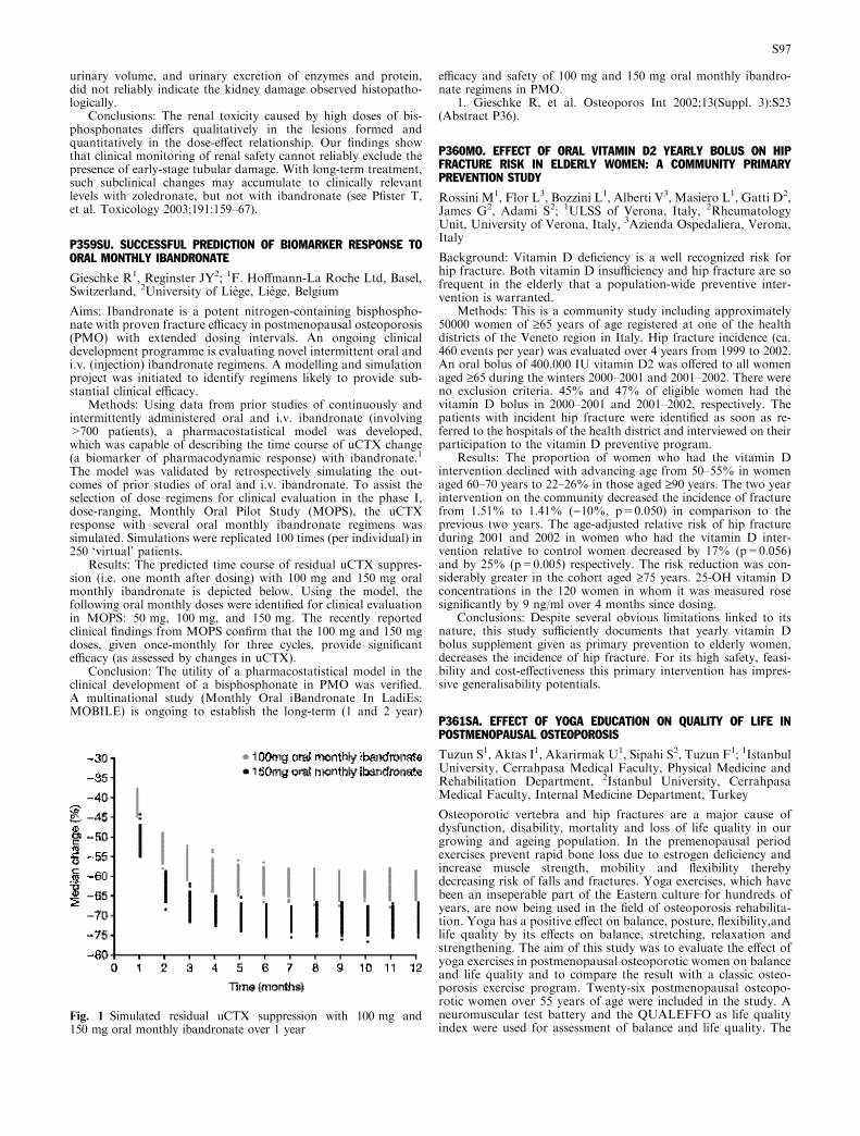
urinary volume, and urinary excretion of enzymes and protein,did not reliably indicate the kidney damage observed histopatho-logically.
Conclusions: The renal toxicity caused by high doses of bis-phosphonates differs qualitatively in the lesions formed andquantitatively in the dose-effect relationship. Our findings showthat clinical monitoring of renal safety cannot reliably exclude thepresence of early-stage tubular damage. With long-term treatment,such subclinical changes may accumulate to clinically relevantlevels with zoledronate, but not with ibandronate (see Pfister T,et al. Toxicology 2003;191:159–67).
P359SU. SUCCESSFUL PREDICTION OF BIOMARKER RESPONSE TOORAL MONTHLY IBANDRONATE
Gieschke R1, Reginster JY2; 1F. Hoffmann-La Roche Ltd, Basel,Switzerland, 2University of Liege, Liege, Belgium
Aims: Ibandronate is a potent nitrogen-containing bisphospho-nate with proven fracture efficacy in postmenopausal osteoporosis(PMO) with extended dosing intervals. An ongoing clinicaldevelopment programme is evaluating novel intermittent oral andi.v. (injection) ibandronate regimens. A modelling and simulationproject was initiated to identify regimens likely to provide sub-stantial clinical efficacy.
Methods: Using data from prior studies of continuously andintermittently administered oral and i.v. ibandronate (involving>700 patients), a pharmacostatistical model was developed,which was capable of describing the time course of uCTX change(a biomarker of pharmacodynamic response) with ibandronate.1
The model was validated by retrospectively simulating the out-comes of prior studies of oral and i.v. ibandronate. To assist theselection of dose regimens for clinical evaluation in the phase I,dose-ranging, Monthly Oral Pilot Study (MOPS), the uCTXresponse with several oral monthly ibandronate regimens wassimulated. Simulations were replicated 100 times (per individual) in250 ‘virtual’ patients.
Results: The predicted time course of residual uCTX suppres-sion (i.e. one month after dosing) with 100 mg and 150 mg oralmonthly ibandronate is depicted below. Using the model, thefollowing oral monthly doses were identified for clinical evaluationin MOPS: 50 mg, 100 mg, and 150 mg. The recently reportedclinical findings from MOPS confirm that the 100 mg and 150 mgdoses, given once-monthly for three cycles, provide significantefficacy (as assessed by changes in uCTX).
Conclusion: The utility of a pharmacostatistical model in theclinical development of a bisphosphonate in PMO was verified.A multinational study (Monthly Oral iBandronate In LadiEs:MOBILE) is ongoing to establish the long-term (1 and 2 year)
efficacy and safety of 100 mg and 150 mg oral monthly ibandro-nate regimens in PMO.
1. Gieschke R, et al. Osteoporos Int 2002;13(Suppl. 3):S23(Abstract P36).
P360MO. EFFECT OF ORAL VITAMIN D2 YEARLY BOLUS ON HIPFRACTURE RISK IN ELDERLY WOMEN: A COMMUNITY PRIMARYPREVENTION STUDY
Rossini M1, Flor L3, Bozzini L1, Alberti V3, Masiero L1, Gatti D2,James G2, Adami S2; 1ULSS of Verona, Italy, 2RheumatologyUnit, University of Verona, Italy, 3Azienda Ospedaliera, Verona,Italy
Background: Vitamin D deficiency is a well recognized risk forhip fracture. Both vitamin D insufficiency and hip fracture are sofrequent in the elderly that a population-wide preventive inter-vention is warranted.
Methods: This is a community study including approximately50000 women of ‡65 years of age registered at one of the healthdistricts of the Veneto region in Italy. Hip fracture incidence (ca.460 events per year) was evaluated over 4 years from 1999 to 2002.An oral bolus of 400.000 IU vitamin D2 was offered to all womenaged ‡65 during the winters 2000–2001 and 2001–2002. There wereno exclusion criteria. 45% and 47% of eligible women had thevitamin D bolus in 2000–2001 and 2001–2002, respectively. Thepatients with incident hip fracture were identified as soon as re-ferred to the hospitals of the health district and interviewed on theirparticipation to the vitamin D preventive program.
Results: The proportion of women who had the vitamin Dintervention declined with advancing age from 50–55% in womenaged 60–70 years to 22–26% in those aged ‡90 years. The two yearintervention on the community decreased the incidence of fracturefrom 1.51% to 1.41% ()10%, p=0.050) in comparison to theprevious two years. The age-adjusted relative risk of hip fractureduring 2001 and 2002 in women who had the vitamin D inter-vention relative to control women decreased by 17% (p=0.056)and by 25% (p=0.005) respectively. The risk reduction was con-siderably greater in the cohort aged ‡75 years. 25-OH vitamin Dconcentrations in the 120 women in whom it was measured rosesignificantly by 9 ng/ml over 4 months since dosing.
Conclusions: Despite several obvious limitations linked to itsnature, this study sufficiently documents that yearly vitamin Dbolus supplement given as primary prevention to elderly women,decreases the incidence of hip fracture. For its high safety, feasi-bility and cost-effectiveness this primary intervention has impres-sive generalisability potentials.
P361SA. EFFECT OF YOGA EDUCATION ON QUALITY OF LIFE INPOSTMENOPAUSAL OSTEOPOROSIS
Tuzun S1, Aktas I1, Akarirmak U1, Sipahi S2, Tuzun F1; 1IstanbulUniversity, Cerrahpasa Medical Faculty, Physical Medicine andRehabilitation Department, 2Istanbul University, CerrahpasaMedical Faculty, Internal Medicine Department, Turkey
Osteoporotic vertebra and hip fractures are a major cause ofdysfunction, disability, mortality and loss of life quality in ourgrowing and ageing population. In the premenopausal periodexercises prevent rapid bone loss due to estrogen deficiency andincrease muscle strength, mobility and flexibility therebydecreasing risk of falls and fractures. Yoga exercises, which havebeen an inseperable part of the Eastern culture for hundreds ofyears, are now being used in the field of osteoporosis rehabilita-tion. Yoga has a positive effect on balance, posture, flexibility,andlife quality by its effects on balance, stretching, relaxation andstrengthening. The aim of this study was to evaluate the effect ofyoga exercises in postmenopausal osteoporotic women on balanceand life quality and to compare the result with a classic osteo-porosis exercise program. Twenty-six postmenopausal osteopo-rotic women over 55 years of age were included in the study. Aneuromuscular test battery and the QUALEFFO as life qualityindex were used for assessment of balance and life quality. The
Fig. 1 Simulated residual uCTX suppression with 100 mg and150 mg oral monthly ibandronate over 1 year
S97
results showed that yoga education has a positive effect on pain,physical functions, social functions, general health perception andbalance. As a conclusion yoga appears to be an alternativemodality for the rehabilitation of osteoporotic subjects.
P362SU. EFFECTS OF EXERCISE AND NUTRITION ON BALANCE ANDRISK OF FALLING IN ELDERLY PEOPLE WITH DECREASED BONEMINERAL DENSITY
Swanenburg J, Stauffacher M, Knols R, Baschung P, UeblehartD; Department of Rheumatology and Institue of PhysicalMedicine, University Hospital Zurich, Zurich, Switzerland
Aims: A pilot study was set up at the Centre for Prevention andTreatment of Osteoporosis at the University of Zurich. It is aimedat determining if the risk of falling can be influenced by exerciseand nutrition in a population of elderly people diagnosed withsevere osteopenia or osteoporosis.
Methods: The study was designed as a randomized controlledprospective trial of 12-month duration with an initial 3-monthintervention program aimed at testing the combined effects ofexercise with protein and calcium/vitamin D supplementation asopposed to calcium/vitamin D supplementation alone. A total of22 female patients, aged 72.2±7.1 SD yrs, BMI: 23.2±2.8 SD kg/m2 presenting with severe osteopenia or osteoporosis ± fractureswere included after they gave their written informed consent. Pri-mary outcome is risk of falling (Berg Balance Test), whereas sec-ondary outcomes include muscle strength, postural sway, aerobiccapacity, activity level and general health assessed upon entry, thenat month 3, 6, 9 and 12 respectively. In addition, spine and hipBMD as well as WBC including bone, fat and lean mass aremeasured at baseline and after 12 months by DEXA (HologicQDR 4500 Elite). Various biological parameters, including serumVit. D and IGF-1 levels are measured initially and after 12 months.
Results: At the end of the first 3-months, the intervention grouppresented with a significant decrease in the risk of falling ()7%), asignificant increased muscle strength (+22%) and aerobic capacity(+39%) as compared to the control group. These results weremaintained by month 6. The BMD, WBC and biological parame-ters results will be provided at the end of the study by month 12.
Conclusions: These initial results are promising and clearly doreflect a better body balance and a decreased risk of falling in theintervention group. The initial assessment of total hip and femoralneck BMD, WBC fat mass, lean mass, serum Vit. D and IGF1 arein agreement with previous published work in elderly severe oste-openic or osteoporotic women. Overall data and conclusion will bepresented at the congress.
P363MO. UNCOUPLING BONE TURNOVER WITH STRONTIUM RANE-LATE, A PROMISING AGENT FOR THE TREATMENT OF POSTMENO-PAUSAL OSTEOPOROSIS
Christiansen C, Tanko LB; Center for Clinical and BasicResearch, Ballerup, Denmark
Aim: Traditional antiresorptive agents such as hormone replace-ment therapy, selective-estrogen receptor modulators, and bis-phosponates inhibit bone resorption but also bone formation(coupling) leading to considerable limitations in terms ofincreasing bone mass and thus curing osteoporosis. Ongoingreseach targets the identification of drug-candidates that canprovide inhibition of bone resorption with favorable or no effecton bone formation (uncoupling). The aim of the present studywas to investigate the simultaneous effect of strontium ranelate onbiomarkers of bone formation and resorption assessed in samplescollected from a 3-year fracture study (SOTI) on a large numberof osteoporotic women.
Design: Study participants were 1649 postmenoausal womenwith osteoporosis. Women were randomly selected to receivetreatment with either 2 g/day strontium ranelate or placebo for3 years. All women received treatment with calcium and vitamin D.Efficacy parameters were the changes in biochemical markers ofbone resorption (serum C-telopeptide of colagen type 1, s-CTX)
and bone formation (bone-specific alkaline phosphatase, BSAP;serum osteocalcin, s-OC).
Results: Strontium ranelate evoked moderate but sustaineddecrease in s-CTX (15%) and simultaneous increase in s-OC andBSAP (10%).
These changes in bone turnover translate to a sustained positivecalcium balance corresponding to �50 mg per day. This positivecalcium balance kept over a 3-year treatment period may theoret-ically result in a �10% increase in bone mineral content of theskeletal system contributing to decreased risk for osteoporoticfractures.
Conclusion: The moderate, but sustained and lasting uncou-pling of bone formation from bone resorption makes strontiumranelate a promising agent for the treatment of postenopausalosteoporosis. Further studies are required to better understand themechanisms of action of this drug-candidate.
P364SA. IN VIVO AND IN VITRO BIOEQUIVALENCE OF TWO FORMU-LATIONS CONTAINING ALENDRONATE
Quattrocchi O, Zanetti D, Roldan EJA, Piccinni E; Gador SA,Buenos Aires , Argentina
Alendronate formulations are effectively administered to preventnew fractures due to osteoporosis. In many countries genericversions have been authorised in order to lower the cost of therapyand enhance compliance rates, but equivalency with the referencecompound is unknown. As the absorption of alendronate isextremely low, and the digestive side effects can be associated toformulations (the pill effect), it is of importance to describe theequivalence of any available alendronate formulation.
Marvil� tablets (Alend/ro� in some countries), containalendronate 10 and 70 mg, obtained by an independent method.The crystals of its hydrated molecule are quite identified (Vegaet al, Acta Cryst 1996,C52:2198–2201), and the product has beentested in experimental assays and its formulations used in clinicalstudies. Data have been published by independent institutions. Wehereby report in vitro and in vivo equivalence tests performedunder GLP and GCP norms. Reference formulations were pur-chased in USA. In vitro dissolution assays show quick, completeand comparable dissolution rates with both formulations. Thesestudies are being further confirmed by an international audit assay.Two clinical bioequivalence studies with either 10 and 70 mg tab-lets were performed at the Buenos Aires Italian Hospital andFLENI MRC unit respectively, following a double blind, rando-mised, parallel, cross-over design, including 12 and 24 healthysubjects. Urine samples were obtained at intervals of 6 hour each,during 24 hours and submitted to analysis at the University ofLeiden (The Netherlands). The absorption rate of the four testedformulations was found between 0.5 and 0.7%, with great inter-individual variability, as expected, within the range of the referencecompound (p<0.05, beta power >0.80). In conclusion, Marvil�formulations are comparable to the alendronate formulationscommercialized in USA.
P365SU. BACK PAIN RISK IS REDUCED IN POSTMENOPAUSAL WO-MEN WITH OSTEOPOROSIS TREATED WITH TERIPARATIDE AS COM-PARED WITH ALENDRONATE: RESULTS FROM TWO DOUBLE-BLINDEDCOMPARATOR TRIALS
Dore RK1, Krege JH2, Chen P2, Glass EV2, San Martin J2, MillerPD3; 1UCLA, Anaheim, CA, USA, 2Lilly Research Laboratories,Eli Lilly and Company, Indianapolis, IN, USA, 3Colorado Centerfor Bone Research, Lakewood, CO, USA
Compared to placebo, teriparatide 20 and 40 mcg/d (TPTD20and TPTD40) treated postmenopausal women with osteoporosishad significantly reduced risk of back pain, moderate or severeback pain, and severe back pain (Neer NEJM 2001, GenantASBMR 2003).
Aims: To compare the incidence of back pain occurring inpostmenopausal women treated with teriparatide or with alendro-nate.
S98
Methods: Two double-blind trials compared oral alendronate10 mg/d (ALN10) plus placebo injection with oral placebo plusteriparatide injection in postmenopausal women with osteoporosis.Patient inclusion and exclusion criteria were similar in the trials.Back pain data were collected during adverse event monitoring. Instudy A, women were randomized to TPTD20 or ALN10 for 18months. In study B, women were randomized to TPTD40 orALN10 for a median 14 months and women completing this trialwere enrolled in a follow-up study.
Results: In each trial, baseline demographics in the two groupswere similar. Compared with ALN10, women treated withTPTD20 had reduced risk of back pain (P=0.051), moderate orsevere back pain (P=0.003), and severe back pain (P=0.04), withrelative risk reductions of 27%, 44%, and 52%, respectively (Table,I). Women treated with TPTD40 had reduced risk of back pain(P=0.012) and moderate or severe back pain (P=0.016), withrelative risk reductions of 71% and 80%, respectively, versusALN10 (Table, II). During the trial plus 18 months of follow-up,women in the TPTD40 group had reduced risk of back pain(P=0.015), and moderate or severe back pain (P=0.016), withrelative risk reductions of 66% and 80%, respectively, versus theALN10 group (Table, III).
Conclusions: In two clinical trials, women randomized to teri-paratide had substantially reduced risk of back pain compared withwomen randomized to alendronate.
P366MO. HORIZON-PIVOTAL FRACTURE TRIAL: UNIQUE DESIGN OF ARANDOMIZED, PLACEBO-CONTROLLED TRIAL TO EXAMINE THE EF-FECT OF ANNUAL INFUSION OF ZOLEDRONIC ACID (5 MG) ON HIP ANDSPINE FRACTURE REDUCTION
Black DM1, Rosario-Jansen T2, Delmas PD3, Eastell R4, LauEMC5, Boonen S6, Caminis J2, Flood M2, Cummings SR1;1Universtity of California, San Francisco, CA, USA, 2NovartisPharmaceuticals, East Hanover, NJ, USA, 3Hopital EdouardHerriot, Lyon, Cedex, France, 4University of Sheffield, Sheffield,Yorkshire, UK, 5Chinese University of Hong Kong, Shatin, NewTerritories, Hong Kong, 6Center for Metabolic Bone Disease,Leuven, Belgium
A single infusion of zoledronic acid has been shown to increasebone mineral density (BMD) at the spine by approximately 5%and decrease bone resorption by about 50% at 12 months com-pared with placebo. However, the potential to reduce fracture riskis not known. To evaluate the effect of once-yearly zoledronic acid
on modifying fracture risk, we are conducting a 3-year, ran-domized, placebo-controlled trial known as HORIZON-PFT.
7764 women between the ages of 65 and 89 years were recruitedfrom 230 clinical centers in 27 countries, representing a diversepopulation with respect to socioeconomic, racial and culturalbackground. Major inclusion criteria were: a femoral neck BMDt-score less than or equal to )2.5, or between )1.5 and )2.5 in thepresence of at least one vertebral deformity. Participants wererandomized to placebo or zoledronic acid (5 mg delivered in 100 mlvolume, 15 minute infusion). Study medication is administeredannually, and participants are followed for 3 years. All participantsreceive 1000 to 1500 mg of elemental calcium plus 400 to 1200 IUof vitamin D per day (minimum background therapy). Co-primaryendpoints are hip fractures and new vertebral fractures. Secondaryefficacy endpoints include: non-vertebral fractures; change in BMDby DXA; changes in biochemical markers of bone metabolism; andchanges in bone density and size by QCT. Safety endpoints includeevaluation of adverse events, assessment of bone histology by his-tomorphometry, and post-dose monitoring for acute changes inrenal lab values.
An innovation in this trial is that women who were taking non-bisphosphonate medications for osteoporosis, including HT,SERMS, and calcitonin (‘‘usual care’’), were allowed to participate.For the co-primary endpoints, the effect of the study medication onhip fractures will be tested in all women, whereas the effect onvertebral fractures will be tested in the subset (6113) who at base-line were not taking ‘‘usual care’’ medication. The study was de-signed under the expectation that about 58% of participants wouldbe taking ‘‘usual care’’ medications, but in fact only 21% wereactually enrolled into this subset.
P367SA. OSTEOPOROSIS IN GENERAL PRACTICE
Maggiani G; Mktg Dept. ‘‘ Societa Prodotti Antibiotici’’, Milano,Italy
An informative research regarding general practice was carriedout nearby GPs consulting rooms during first semester of 2003, inorder to verify if an adequate diagnosis of osteoporosis is usuallyperformed and wich are the consequent therapies. A fully privacyrespectful questionnaire, including 14 questions, was submitted to30,000 GPs all over national territory. An high percentage of dulyfilled forms was collected; data suggest that osteoporosis is stillunderestimated, probably due to the patients low perception ofthe disease. In a chronic pathology, an effective treatment, safeand allowing a good compliance should be useful, but not anybisphosphonate is able to warrant all these characteristics.
P368SU. INFLUENCE OF REGULAR EXERCISE ON BONE MINERALDENSITY IN ELDERLY WOMEN
Erkisi F, On AY, Kirazli Y, Durmaz B; Ege University, PhysicalMedicine and Rehabilitation Department, Izmir, Turkey
Osteoporosis is a disease in which the density and quality of boneare reduced, leading to weakness of the skeleton and increasedrisk of fracture. Bone mineral density (BMD) measurements areeffective in assessing fracture risk, confirming a diagnosis ofosteoporosis and monitoring the effect of treatment.
Non-medical therapy, such as a healthy diet and exercise, hasbeen shown to have a beneficial effect on bone.
Physical activity during childhood and adolescence is positivelyrelated to bone density but effects in postmenopausal women aremodest. Elderly people who have experienced some bone lossmay benefit from a program of weight-bearing and enduranceexercises.
The aim of this study was to assess the effect of regular, longterm and supervized exercise program on bone mineral density inelderly women.
Seventy-six women with postmenopausal osteoporosis (meanage 60±10 yrs) were enrolled in the study. Control group wasconsisted from 22 postmenopausal osteoporotic women. No sig-nificant diferences existed between exercise and control group for
I. Study A
ALN10N=101
TPTD20N=102
Relative Risk(P-value)
n % n %
Back Pain 39 38.6 26 25.5 .73 (0.051)Moderate or SevereBack Pain
33 32.6 15 14.7 .56 (0.003)
Severe Back Pain 12 11.9 4 3.9 .48 (0.04)
II. Study B
ALN10N=73
TPTD40N=73
n % n %
Back Pain 14 19.2 4 5.5 .29 (0.012)Moderate or SevereBack Pain
10 13.7 2 2.7 .20 (0.016)
Severe Back Pain 2 2.7 1 1.4 .52 (NS)
III. Study B+18 Months Follow-up
ALN10N=53
TPTD40N=52
n % n %
Back Pain 15 28.3 5 9.6 .34 (0.015)Moderate or SevereBack Pain
10 18.9 2 3.9 .20 (0.016)
Severe Back Pain 3 5.7 1 1.9 .33 (NS)
S99
age, years since menopause, body mass index or height (p>0.05).The exercise regimen involved aerobic exercises, strength trainingand stretching exercises. We used some equipment such as ankle-wrist weights, dumbbells, and theraband for strengthening localsites. Supervised exercises were performed once a week at thehospital and than patients continued regularly exercise at home 3times a week for one year.
A Hologic Delphi-W dual x-ray absorbtiometry was used toassess BMD of the lumbar spine and total hip.
Differences between the 0 and 12 months in the groups wereestimated using paired t tests. Differences between the two groupswere estimated using unpaired t tests .
In the exercise group there was a significant increase in % BMDchange between 0 and 12 months in the femoral neck, and T scorewas increased significantly in the same region (p<0.05). In com-parison, percentage BMD change and T score were also significantin the exercise group (p<0.05).
Our results suggested that, regular, supervized exercises have apositive effect on BMD, especially in the femaral neck in the elderlyosteoporotic women.
P369MO. THE EFFECT OF KYPHOSIS ON CARDIOPULMONARY EXER-CISE TEST AND EXERCISE TOLERANCE IN PATIENTS WITH SEVEREOSTEOPOROSIS: PRELIMINARY RESULTS
Ordu Gokkaya NK, Koseoglu F, Albayrak N; Ankara PhysicalMedicine and Rehabilitation Education and Research Hospital,IV. Department of Physical Medicine and Rehabilitation, Turkey
Background: Loss of bone in osteoporosis (OP) can lead to gradualcompression or wedging of the vertebrae and narrowing of theintervertebral discs. With resultant increase in the kyphotic curve.
Aim: To evaluate the effects of the deformation of the thoraciccage on cardiopulmonary exercise test parameters and aerobiccapacity in patients with OP.
Methods: 34 patients with OP were consented to participate inthe study. The patients were divided into two groups according totime since the diagnosis of OP. Group I was newly diagnosed (lessthan one year) and the group II was diagnosed late (more than sixyears). Anteroposterior and lateral radiographs of the thoracic andlumbar spine and dual energy x-ray densitometry were obtained inall patients. The number and severity of vertebral deformity weremeasured by using the Kleerekoper and Gennant index in spineradiographs. The degree of dorsal kyphosis was measured by usingthe Cobb technique.
Pulmonary function was spirometrically assessed, cardiopul-monary exercise test was performed with a treadmill by usingbreath by breath analysis of oxygen consumption.
Results: Mean time since diagnosis of OP was 11.1 months ingroup I and 80.1 months in group II. Mean age was 56.2 years and55.8 years respectively. When compared with group I, there was aslight decrease in some of the cardiopulmonary exercise test andexercise tolerance in group II.
Conclusion: When compared with newly diagnosed patients,longer duration of OP might produce a slight respiratory dys-function and a decline in exercise tolerance in women. Thereforeactive prevention or treatment of OP is needed to retard thedevelopment of vertebral deformity and such cardiopulmonarydysfunctions.
P370SA. BONE TURNOVER RESPONSE AND BONE MINERAL DENSITYCHANGES AFTER 1 YEAR TREATMENT WITH RISEDRONATE 35MGWEEKLY IN VENEZUELAN TYPE I POSTMENOPAUSAL OSTEOPOROTICWOMEN
Velasquez G, Perez-Angulo Y, Riera-Espinoza G, Ramos J;UNILIME-UC, Universidad de Carabobo, Hospital Universi-tario Dr. Angel Larralde IVSS, Valencia, Venezuela
Suppression of bone remodeling and increase of Bone MineralDensity (BMD) are two targets of osteoporosis treatment. Theaims of this study are to show changes in bone turnover andBMD after one year treatment with Risedronate in a group ofVenezuelan postmenopausal osteoporotic women.
117 postmenopausal women with type I Osteoporosis received35 mg/weekly of Risedronate on fasting state during one year. Allreceived Calcium Citrate 650 mg/d plus 400 IU vitamin D. BMDwas assessed by DEXA (LUNAR Prodigy, VC: 1.5%), Biochemi-cal markers of bone turnover were measured as follow: NTxOsteomark, Ostex, Seattle, USA. Tartrate Resistant Acid Phos-phatase, TRAP: Hydrolysis of paranitrophenyl phosphate at 4.8pH. Total Alkaline Phosphatase, TAP: Labtest, Roy modified.
Mean age was 63±8.39 (30–86), age of menopause was47±5.79 (25–58). BMD at femoral neck increased from 0.749±0.1to 0.759±1 (p<0.05). At lumbar spine from 0.869±0.1 to0.883±0.1 (p<0.05). T score decreased from )1.941 to )1.925p=NS at femoral neck and at lumbar spine from )2.74 to )2.57(p<0.0001); percent changes in BMD were +3.4% at femoral neckand +4.9% at lumbar spine. Variation of bone markers at initial,six and twelve months were NTx 122±80±39 and 64±50 nmol/BCE/mmol/creat (p<0.001 initial vs 6 and 12 months), percentchanges were )52% at 6 months and )47% at 12 months, TAP46±12, 30±13, 36±10 IU/l (p<0.0001 initial vs 6 and 12 month),Percent changes were )34% at 6 months and )22% at 12 months.TRAP, 8±3, 7±2, 7±3 IU/l (p<0.001 initial vs 6 month andp<0.06 initial versus 12 month) Percent changes were )13% at 6months and at year.
In conclusion, the use of 35 mg/week of Risedronate during oneyear decreased bone markers significantly; NTx 47%, TAP 22%and TRAP 13%. BMD increased significantly, +3.4% at femoralneck and +4.9% at lumbar spine.
P371SU. BONE MARKERS AND BONE MINERAL DENSITY AFTER TWOYEARS TREATMENT WITH ALFACALCIDOL IN HIGH REMODELINGPOSTMENOPAUSAL WOMEN WITH LOW MINERAL DENSITY
Naressi M, Riera G, Ramos J, Marcano L; UNILIMEUniversidad de Carabobo Hospital Dr. Angel Larralde ValenciaVenezuela
Vitamin D metabolites have been used for osteoporosis treat-ment. Main actions on bone are: mineralization, stimulation ofosteoblast and diminish resortion. However, available informa-tion about action of alfacalcidol on resorption markers in aprospective double-blind, placebo control trial are scarce.
92 postmenopauseal women with low BMD (T-score betweenSD-1 and SD-3 at L2-L4 or femoral neck) and high turnover:N-telopeptide (NTx), Tartrate Resistant Acid Phosphatase (TRAP)or Total Alkaline Phosphatase (TAP) 1 SD or more above thepremenopausal mean were randomized in a double-blind, placebocontrol, twelve months trial 1 lg/day with alfacalcidol. The vari-ation rate was NTx )28%, TRAP )23%, AP )16%, BMD re-mained stable in both groups. A twelve months extension, as anopen label study, was offered to patients on alfacalcidol. Out of the45 patients on alfacalcidol 18 agreed to follow the extension pro-tocol. At 24 months there was significant reduction in alkalinephosphatase ()31.25%), no significant changes were observed inNTx ()22.6%) and TRAP ()16%), BMD remained stable, butthere was a tendency to decrease.
Conclusion: Alfacalcidol, at 1 lg/day in patients with low BMDand high turnover maintained BMD and suppressed bone remod-eling during the first year. After switching to 0.5 lg/day for asecond year resorption bone markers like NTx and TRAP tend torise while bone formation and TAP remain suppressed. BMDshowed a tendency to decrease during the second year of the trial.
Initial 12 months 24 months
Serum Ca 9.25± 0.51 9.05±5.55 9.34±0.40AP (IU/L) 48.63±15.37 40.80±10.4** 33.30±6.3***TRAP (IU/L) 9.92±2.9 7.58±2.35*** 8.33±0.28NTx (BCE/Creat) 128.72±67.28 92.97±55.78* 99.53±41.5L2-L4 grs/cm2 0951±0.09 0.95±0.12 0.877±0.06Fem neck grs/cm2 0.793±0.9 0.790±55.78 0.773±0.05
*** = p < 0.001 ** = p < 0.01 * = p < 0.05 vs initial
S100
P372MO. A COMPARATIVE STUDY BETWEEN INTERMITTENT CYCLICALAND CONTINUOUS ALENDRONATE IN THE TREATMENT OF POSTMENO-PAUSAL OSTEOPOROSIS
Hepguler S, Cabucak FP, Karatepe AG, Tas I; Department ofPhysical Medicine and Rehabilitation, Ege University, Izmir,Turkey
Aims: To compare the efficacy of intermittent cyclical and con-tinuous alendronate in the treatment of postmenopausal osteo-porosis.
Patients and Method: 30 postmenapousal women were enrolledin the study. The patients were randomized into two treatmentgroups. The first group was given 10 mg daily alendronate withcalcium 500 mg/d and Vitamin D3 300.000 IU/M, the secondgroup was given cyclical intermittent 10 mg daily alendronate for 2months, followed by 2 months of drug-free period with calcium500 mg/d and Vitamin D3 300.000 IU/M for one year. Measure-ments of lumbar spine and proximal femur BMD were performedat baseline and at 12th month by dual x-ray absorptiometry. Bio-chemical markers of bone resorption and formation, serum calciumand phosphorus, urinary calcium and serum alkaline phosphataselevels were evaluated at same time intervals. Radiographs of thethoracic and lumbar spine were taken at baseline and at 12thmonth.
Results: There were no statistically significant differences inthe baseline mean BMD and T scores between two groups. At theend of the study, the group receiving intermittent alendronateshowed statistically significant increases in T scores of the femoralneck (p<0.05). In the group receiving alendronate 10 mgcontinuously, T scores of the lumbar spine and femoral necksignificantly increased from baseline (p<0.05). However, theimprovement in the lumbar spine BMD was found in favour ofpatients receiving alendronate 10 mg continuously (p<0.05).Compared to baseline, no significant difference was found in themarkers of bone resorption and formation in both treatmentgroups (p<0.05).
Conclusions: With regard to our results, 10 mg continuouslyadministration of alendronate may be better than intermittentadministration in patients with postmenopausal osteoporosis.
P373SA. BRAZILIAN EXPERIENCE WITH RALOXIFENE AND ALENDRO-NATE: COMPLIANCE AND PATIENTS’ CLINICAL FEATURES
Bracco OL1, Ito LT1, Marson JE1, Souza AC2, Borges JL1,2,Oliveira J1, Kayath MJ1,2; 1Medical Division , Eli Lilly Brazil,2Geriatric Service, PUC, RS, Brazil
Choose is a prospective observational 12 month-study designedto evaluate patient satisfaction and compliance related withraloxifene or alendronate in a natural setting. In Brazil, 32 phy-sicians were involved in the study that included 247 postmeno-pausal women over 60 years old with either osteopenia orosteoporosis. Raloxifene was prescribed for 181 women andalendronate was prescribed for 95 women (10 mg/d or 70 mgweekly, 23% and 77%, respectively). The median chronologicalage for raloxifene group was 69.0, and 71.3 years for thealendronate group (p=0.011). The time since menopause onsetwas 19.8 + 8.2 (median + SD) and 22.5 + 9.8 years for ra-loxifene and alendronate groups, respectively (p=0.016). HRTwas the most frequent prior treatment in the raloxifene oralendronate groups (38% and 13% of women, respectively,p<0.001). DEXA result was the most frequent clinical criteria toprescribe an anti-resorptive treatment in both groups. 13% ofwomen in the raloxifene group and 24% in the alendronate grouphad a previous osteoporotic fracture (p<0.001) at baseline. 22%of the raloxifene patients and 20% of alendronate patientsabandoned treatment after 12 months of treatment (N.S.). Patientsatisfaction, evaluated by a visual analog scale, was not statisti-cally different between groups before and after 12 months oftreatment (mean=89.1 cm for raloxifene and 79.6 for alendro-nate, N.S.). Thus, raloxifene was prescribed for younger post-menopausal women with shorter time since menopause than forthose alendronate was prescribed. Besides, raloxifene was more
commonly prescribed than alendronate after HRT, although agreat number of patients with prevalent osteoporotic fractureswere on alendronate. Raloxifene and alendronate treatments hadthe same discontinuation rate and both treatments were welltolerated by women with osteoporoses although the majority ofthe patients on alendronate were on weekly dose. In conclusion,patients profile for raloxifene and alendronate were different,but discontinuation rate were not different in an observationalBrazilian study.
P374SU. DRUG-DRUG INTERACTIONS ARE UNLIKELY WITH IBANDR-ONATE
Bauss F1,2, Kling L1, Worth E3, Barrett J3; 1Pharma ResearchPenzberg, Roche Diagnostics GmbH, Penzberg, Germany, 2Insti-tute of Pharmacology and Toxicology, Heidelberg University,Mannheim, Germany, 3Roche Products Ltd, Welwyn GardenCity, UK
Aims: Many patients treated with ibandronate, whether forosteoporosis or metastatic bone disease, will be receiving con-comitant medications. It is therefore important to investigate thepotential of ibandronate for drug-drug interactions.
Methods: Ibandronate has been the subject of extensive phar-macological and pharmacokinetic examination in both animals andhuman subjects. In-vitro and in-vivo preclincal studies have ex-plored renal excretion, protein binding and effects on cytochromeP450 (CYP) activity. Pharmacokinetic studies in human volunteersand patients have provided further data on the possibility ofinteractions with concomitant drugs.
Results and Discussion: In rats, high doses of classicalinhibitors of either anionic or cationic renal transport systemsdid not affect the renal clearance of ibandronate or the glo-merular filtration rate. These results indicate that ibandronate isnot secreted by either anionic or cationic renal transport systemsand therefore would be unlikely to interact with drugs excretedby these systems. Protein binding, a factor known to influencedrug-drug interactions, at the range of concentrations ofibandronate found in vivo in human serum is relatively low(86%). In-vivo studies in rats indicate that ibandronate does notinduce CYP activity in the liver. In human liver microsomes invitro, ibandronate at concentrations of up to at least 100 lM(360 lg/ml) showed no affinity for any of the main human liverCYP isoenzymes (1A2, 2A6, 2C9, 2C19, 2D6, 2D1 or 3A4).These findings suggest that ibandronate is unlikely to producedrug-drug interactions through inhibition of CYP activity. No,or only minor, clinically irrelevant pharmacokinetic changes wereobserved when ibandronate was taken concomitantly with hor-mone replacement therapy, tamoxifen, or melphalan and pred-nisolone. These drugs are frequently taken by postmenopausalwomen, women with breast cancer, and patients with multiplemyeloma, respectively. A minor increase in the AUC (20%)of ibandronate in combination with ranitidine was attributableto an increase in gastric pH and was not considered clinicallyrelevant.
Conclusions: Preclinical findings demonstrate that ibandronatehas negligible effects on renal excretion, or CYP activity, and showsrelatively low protein binding. These data are consistent with thosefrom human pharmacokinetic studies indicating that ibandronate isunlikely to produce drug-drug interactions.
P375MO. INTERMITTENT INTRAVENOUS IBANDRONATE INJECTIONS:RENAL SAFETY PROFILE
Miller PD1, Brown JP2, Stepan JJ3, Schimmer RC4, Bauss F5,6,Pfister T4; 1CCBR, Lakewood, USA, 2Centre de recherche duCHUL, Laval University, Quebec, Canada, 3Charles UniversityFaculty of Medicine, Prague, Czech Republic, 4F. Hoffmann-LaRoche Ltd, Basel, Switzerland, 5Pharma Research Penzberg,Roche Diagnostics GmbH, Penzberg, Germany, 6Institute ofPharmacology andToxicology,HeidelbergUniversity,Mannheim,Germany
S101
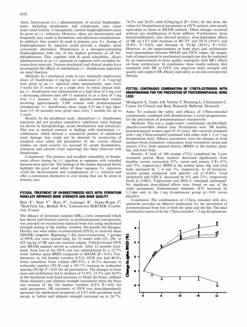
Aims: Intravenous (i.v.) administration of several bisphospho-nates, including alendronate and zoledronate, may causeacute renal toxicity. Consequently, current bisphosphonates mustbe given as i.v. infusions. However, these are inconvenient andfrequently may result in thrombotic and infectious complications.In addition, they cannot be used in primary care. I.v. dosing ofbisphosphonates by injection could provide a simpler, moreconvenient, alternative. Ibandronate is a nitrogen-containingbisphosphonate with one of the highest potencies of all bis-phosphonates. This, together with its good tolerability, allowsadministration as an i.v. injection in regimens with extended be-tween-dose intervals. Various preclinical and clinical studies haveinvestigated the effects of intermittent i.v. ibandronate injectionson renal function.
Methods: In a preclinical study in rats, minimally nephrotoxicdoses of ibandronate (1 mg/kg) or zoledronate (1 or 3 mg/kg)were given as an i.v. injection either intermittently once every3 weeks for 25 weeks or as a single dose. In initial clinical stud-ies, i.v. ibandronate was administered at a high dose of 6 mg overa decreasing infusion time (60–15 minutes) or at a dose of 3 mgover 1–2 minutes. In subsequent placebo-controlled studiesinvolving approximately 3,500 women with postmenopausalosteoporosis, i.v. ibandronate (dose range 0.25 mg–2 mg) injec-tions (15–30 seconds) were given once every 3 months for up to3 years.
Results: In the preclinical study, intermittent i.v. ibandronateinjections did not produce cumulative subclinical renal damage(as shown by histopathology, serum biochemistry and urinalysis).This was in marked contrast to findings with intermittent i.v.zoledronate, which showed a cumulative pattern of subclinicalrenal damage that could not be detected by monitoring ofstandard renal safety lab parameters. In the various clinicalstudies, no renal toxicity (as assessed by serum biochemistry,urinalysis and adverse event reporting) has been observed withibandronate.
Conclusions: The potency and excellent tolerability of ibandr-onate allows dosing by i.v. injection in regimens with extendedbetween-dose intervals. The findings of the studies discussed hereinhighlight the good renal safety of these regimens. I.v. injectionsavoid the inconvenience and complications of i.v. infusion andoffer a convenient alternative to oral dosing that can be given inprimary care.
P376SA. TREATMENT OF OVARIECTOMIZED RATS WITH STRONTIUMRANELATE IMPROVES BONE STRENGTH AND BONE QUALITY
Bain S1, Shen V1, Hara P1, Leininger R1, Dupin-Roger I2;1SkeleTech, Inc., Bothell, WA, 2Laboratoires SERVIER, Courbe-voie, France
The efficacy of strontium ranelate (SR), a new compound whichhas shown anti-fracture activity in postmenopausal osteoporosis,was assessed on ovariectomy-induced bone loss using mechanicalstrength testing of the lumbar vertebra. Six-month old Sprague-Dawley rats were either ovariectomized (OVX) or received sham(SHAM) surgeries. Beginning 1 day post-ovariectomy, 3 groupsof OVX rats were treated daily for 52 weeks with 125, 250, or625 mg/kg of SR and one received vehicle. Vehicle-treated OVXand SHAM animals served as controls. After 12 months treat-ment, bone loss in the OVX rats was substantiated by a 12.7%lower lumbar spine BMD compared to SHAM (P<0.01). Fur-thermore, in 3rd lumbar vertebra (LV3), OVX rats had 49.0%lower cancellous bone volume (BV/TV), a 36.3% decrease intrabecular number (Tb.N) and a 107.7% increase in trabecularspacing (Tb.Sp; P<0.01 for all parameters). The changes in bonemass and architecture led to declines of 31.9%, 33.3% and 34.9%in the maximum load (load necessary to break the bone), stiffness(bone elasticity) and ultimate strength (maximum stress the bonecan sustain) of the 5th lumbar vertebra (LV5; P<0.01 foreach parameter). SR treatment of OVX rats dose-dependentlyincreased the mechanical properties of LV5 with maximum load,energy to failure and ultimate strength increased up to 24.7%,
74.5% and 26.4% with 625mg/kg/d (P<0.01). At this dose, thevalues for biomechanical parameters in OVX animals were nearlyequivalent to those in SHAM animals. These changes occurredwithout any modification of bone stiffness. Furthermore, bonehistomorphometry also showed positive, dose-dependent effectsof SR on LV3 with increases in BV/TV and Tb.N (39.6% and28.0%; P<0.05), and decrease in Tb.Sp (30.8%; P<0.01).However, as the improvements in bone mass and architecturewere intermediate between SHAM and OVX values, the magni-tude of improvement in mechanical strength can also be explainedby an improvement in bone quality synergistic with SR’s effectson bone architecture. In conclusion, these results indicate thattreatment with SR in OVX rats improves bone strength andquality and support SR efficacy and safety as an anti-osteoporotictherapy.
P377SU. CONTINUOUS COMBINATION OF 17BETA-ESTRADIOL WITHDROSPIRENONE FOR THE PREVENTION OF POSTEMENOPAUSAL BONELOSS
Moelgaard A, Tanko LB, Nielsen T, Warming L, Christiansen C;Center for Clinical and Basic Research, Ballerup, Denmark
Aims: To evaluate the safety and efficacy of 17beta-estradiolcontinuously combined with drospirenone, a novel progesterone,for the prevention of postmenopausal osteoporosis.
Methods: This was a single-center, randomized, double-blind,placebo-controlled clinical trial. Participants were 240 healthypostmenopausal women aged 45–65 years, who received treatmentwith 1 mg 17beta-estradiol combined with either with 1, 2 or 3 mgdrospirenone daily. Efficacy parameters were the changes in bonemarkers (bone formation: osteocalcin, bone resorption: serum andurinary CTx), bone mineral density (BMD) at the lumbar spine,hip, and total body.
Results: A total of 180 women (75%) completed the 2-yeartreatment period. Bone markers decreased significantly frombaseline (serum osteocalcin 52%, serum and urinary CTx 67%and 75%, respectively). BMD at the lumbar spine, hip, and totalbody increased by 7, 4 and 3%, respectively, in all hormone-treated groups compared with placebo (all p<0.001). Totalcholesterol and LDL-C decreased by 8% and 13%, respectively(both p<0.001). Triglyceride and HDL-C remained unchanged.No significant dose-related effects were found on any of thestudy parameters. Endometrium thickness (ET) increased by1.2 mm only in the 1 mg drospirenone group (p<0.01 versusplacebo).
Conclusion: The combination of 17-beta estradiol with dro-spirenone provides an effective medication for the prevention ofpostmenopausal bone loss in both the spine and the hip. The idealcombination seems to be the 17beta-estradiol +2 mg drospirenone.
S102
P378MO. EFFECTS OF HORMONE REPLACEMENT THERAPY (HRT) ANDSELECTIVE ESTROGEN RECEPTOR MODULATORS (SERM) ON CARTI-LAGE DEGRADATION IN POSTMENOPAUSAL WOMEN
Garnero P1,2, Sornay-Rendu E1, Christiansen C3,4, Delmas P.D.1;1INSERM research unit 403, Lyon, France, 2Synarc, Lyon,France, 3Center for Clinical and Basic Research, Copenhagen,Denmark, 4Nordic Biosciences, Copenhagen, Denmark
Aim: The aim of our study was to investigate the effects of HRTand SERMs on cartilage turnover assessed by urinary CTX-II, amarker of type II collagen degradation, in postmenopausalwomen.
Methods: The effects of HRT were assessed in a study of 531postmenopausal women; 58–94 years (mean 68 years) from theOFELY cohort. Lateral spine and fixed flexion radiographs of theknees were scored for narrowing and osteophytes by the methodsof Lane and Altman, respectively. Clinical examination of thehands osteoarthritis (OA) was performed in all women. The effectsof the SERMs raloxifene and levormeloxifene on CTX-II wereinvestigated in 234 postmenopausal women participating in tworandomized placebo controlled studies.
Results In the OFELY cohort, the prevalence of lumbar spinedisc degeneration, knee and clinical hand OA was respectively of65%, 32% and 56%. Spine, knee and hand OA contributed inde-pendently of each other to increased CTX-II levels (p=0.0014,0.003 and 0.004, respectively). In women without OA, CTX-II wasnot associated with HRT.
After adjustment for age and BMI, OA patients on currentHRT had CTX-II levels 30% (p<0.0001) lower than never users,with values similar to non OA women. Past users had CTX-II levelssimilar to never users. After 6 months of treatment with raloxifeneat doses of 30, 60 or 120 mg/d, CTX-II decreased by 35% and by50% in women receiving levormeloxifene at doses of 1.25 to 20 mg/d (p<0.001 vs placebo) with no significant dose dependent effectwith raloxifene.
Conclusion: HRT and treatment with SERMs are associatedwith decreased type II collagen degradation in postmenopausalwomen, with a more pronounced effect in those with OA charac-terized by increased cartilage turnover. The potential structuremodifying effects of HRT and SERMs in postmenopausal womenwith OA should be further investigated.
P379SA. OSTEOPOROSIS DELAYS FRACTURE HEALING: RESULTSFROM AN ANIMAL STUDY OF OSTEOPOROSIS
Schneider E1, Goldhahn J1,2, Hesseln J1, Eckhardt C1, Lill CA1,3;1AO Research Institute Davos, Switzerland, 2Schulthess Clinic,Zurich, Switzerland, 3Department of Orthopaedic Surgery,University of Heidelberg, Germany
Aim: Osteoporosis does not only cause fractures – it also influ-ences fracture treatment. The surgical treatment is affected two-fold: by reduction of the fixation strength in weak bone and by adelay in fracture healing. However, clinical and experimentalstudies concerning the influence of osteoporosis on fracturehealing are inconsistent. The aim of this study therefore was toevaluate the healing of a long bone defect using an establishedsheep model of osteoporosis.
Methods: Osteoporosis was induced in seven Swiss mountainsheep using a combination protocol of ovariectomy, low calciumdiet, movement restriction and highdose steroids. The inductionwas terminated 1 month prior to operation to avoid any influenceon fracture healing. A standardized mid-shaft tibial osteotomy wasperformed and stabilized using a custom-made external fixator for8 weeks. In vivo bending stiffness was measured in weekly intervals,callus area, density and osteoporosis status at 0, 4 and 8 weeks.Bending and torsional stiffness were measured ex vivo. Another 7sheep with mid-shaft tibial osteotomy served as a control.
Results: The increase of in vivo bending stiffness was delayed by2 weeks in osteoporotic sheep (a<0.05) and stiffness did not reachinitial values after 8 weeks. Torsional stiffness at 8 weeks was re-duced 33% and bending stiffness 21% as compared to the controlgroup. Callus density as well as callus area were significantly re-duced (a<0.05) in the osteoporotic group (callus density )16% at 4weeks, )23% at 8 weeks, callus area )29% at 4 weeks, )21 % at 8weeks). Bending stiffness correlated with callus density at 8 weeks(r=0.76) and torsional stiffness with callus area (r=0.60). Can-cellous bone density recovery was linear at a rate of 3.4% in theosteoporotic sheep.
Conclusions: Osteoporosis seems to retard callus formation andmaturation, thus influencing fracture healing. These findingsshould be taken into account during operative and conservativefracture treatment when healing time is critical. Adequate osteo-porosis treatment should supplement surgical intervention whendensity is below normal.
P380SU. SAFETY AND EFFICACY OF THE USE OF ZOLEDRONIC ACID(ZA) IN CHILDREN WITH OSTEOGENESIS IMPERFECTA (OI) TYPE IIIAND IV: PRELIMINARY DATA
Barros ER, Saraiva GL, Maeda SS, Canto MH, Pedrosa MA,Castaldoni A, Pinto JA, Lazaretti-Castro M; Division ofEndocrinology, Federal University of Sao Paulo (UNIFESP),Brazil
OI is a genetic disease characterized by bone fragility and in-creased fracture risk. Treatment with pamidronate has shown tobe safe and efficient to reduce fractures and improve the lifequality in OI. Zometa� (ZA) is the most powerful bisphospho-nate available and has a faster time of infusion than pamidronate.This makes a great difference mainly for children. Its efficacy andsafety in children with OI, however, has not yet been tested.
Aim: To verify the safety and efficacy of ZA during one yeartreatment in children with OI type III or IV.
Methods: Nine children were selected to participate in a re-search protocol to receive ZA at a dose of 0.4±0.08 mg/kg/year ina 30 min i.v. infusion every three or four months. Five patientshave finished one year of treatment and the remaining, still undertreatment, have received at least one infusion. Before each infusion,
Fig. 1 Changes in spine BMD
Urinary CTX-II levels (ng/mmol Cr)
Spine and/or knee and/or hand OA (n = 443) Non OA (n = 88)
HRT use
Current (n = 116) Never (n = 283) Past (n = 44)
169 ± 100* 243 ± 123� 281 ± 109� 163 ± 82
* p<0.0001 vs Never and Past users; � p<0.0001 vs Non OA
S103
blood was collected for total and ionized calcium, alkaline phos-phatase, phosphate, CTx and osteocalcin in serum, intact PTH,renal and hepatic function and hemogram. Bone mineral density(BMD) at lumbar spine and total body were measured at thebaseline and at the end of the study.
Results:Out of 9 patients (7 boys, 2 girls), 5 had type IVand4 typeIII. The average age was 5.1±2.8 (from 1.6–9.4). The adverse eventswere: fever (9), nausea (5) and vomiting (2) during the 24 hoursfollowing the first infusion. Except for one patient with anemia atbaseline, all had normal laboratory parameters at baseline andthere were no significant changes before each infusion until the endof the study. From baseline to the end of treatment, spine BMDincreased significantly (0.310±0.073 to 0.462±0.019 mg/cm2,respectively, p<0.016) and total body BMD almost reached signif-icance (0.672±0.126 to 0.758±0.064 mg/cm2, p<0.059).
Conclusion: ZA demonstrated to be safe and efficient inincreasing BMD in this group of children with OI. Due to its fastprocedure, it is more convenient for children. However, a ZA effecton fracture risk still has to be proved, so it can be a new option inthe treatment of OI.
P381MO. BIOMECHANICAL EFFECTS OF RECOMBINANT HUMANGROWTH HORMONE (RHGH) ON BONES AND MUSCLES OF OVARIEC-TOMIZED (OX) RATS
Cointry GR, Ferretti JL, Capozza RF, Reina P, Fracalossi M,Feldman S; Center for P-Ca Metabolism Studies (CEMFoC),Natl Univ of Rosario, Argentina
Doses of 150 IU/kg/d were given during 3 months to 3-month oldrats, either intact of OX. At the end of the study their diaphyseswere scanned by pQCT and tested in bending. The fresh gas-trocnemius muscles were weighed.
OXreducedbone tissuemineralization and stiffness.A significantenhancement of bone growth in width improved significantly thediaphyseal architecture (cross-sectional moment of inertia, CSMI).This geometric improvement overcompensated the negative impactof the OX-induced impairment on bone material mineralization andstiffness, thus the diaphyseal strength was increased. The assayeddose of rhGh was little effective in intact rats. However, it preventedthe OX-induced impairment in bone tissue mineralization (notstiffness) and improved additively the OX-enhanced geometricvariables. These effects of OX and rhGH were correlative withadditive increases inmusclemass. Simple regression analyses showedthat the impact of the muscular improvement was more evident onbone architecture than it was on bone strength.
The positive OX and rhGH effects on cortical bone mass andarchitecture seemed to have derived from the induction of an‘‘anabolic’’ shift of the bone mechanostat threshold for triggeringbone modeling during growth, with a positive biomechanical im-pact on the diaphyses (larger CSMI and fracture load than con-trols). The apparent incongruence between the repercussion of theadditive improvement in muscle mass induced by OX and rhGH onbone geometry (larger impact) and strength (lower impact) can beexplained by the impairment in bone material stiffness induced byOX and not prevented by rhGH (perhaps because rhGH did notact on the microstructure of the mineralized tissue). Based onoriginal arguments, these evidences support the possible ability ofrhGH to improve human postmenopausal osteopenias with a rel-atively large impairment in cortical bone mass and/or distribution.However, the actual benefit of the positive rhGH effects on bonemass and architecture in any species would remain uncertain aslong as the nature of rhGH effects on the OX-impaired bonematerial stiffness is unknown. In addition, these results are inter-esting because they defy the prevailing view that the remainingbone tissue in metabolic osteopenias is normal.
P382SA. NEW INSIGHTS ON OLPADRONATE EFFECTS ON BONEMATERIAL AND STRUCTURAL PROPERTIES RELATED TO STRENGTH.
Cointry GR1, Mondelo N2, Capozza RF1, Ferretti JL1; 1Centerfor P-Ca Metabolism Studies (CEMFoC), Natl Univ of Rosario,2Gador SA, Buenos Aires, Argentina
Doses of 45–90 mg/kg/d of OPD (IG-8801, Gador SA, BuenosAires, carcinogenicity dose-range finding study) were orally givenduring 3 months to 20 male and 24 female rats 4–5-sem old (7 & 9controls). The cortical vBMD, cross-sectional perimeters (PM),area (CSA) and moment of inertia (MI) of femur diaphyses andtheir structural stiffness (load/deformation ratio) and strengthduring the successive ‘‘elastic’’, reversible (pre-yield, no micro-cracks) and ‘‘plastic’’, irreversible (post-yield, microcrack accu-mulation) deformation periods were determined by pQCT andbending tests. The pre-yield stiffness of cortical tissue (elasticmodulus, E) and a Bone Strength Index, BSI=vBMD * MI(which can predict ultimate strength but does not capture anymicrostructural indicator of cortical tissue) were calculated fromthose data.
No effects on growth were observed. Treatment improved sig-nificantly CSA and MI by increasing both endosteal and periostealPMs, more evidently in male than female rats, with no effects oncortical vBMD and E. As a result, mild increases in diaphysealstiffness and strength at yield (only significant in males) were ob-served. Diaphyseal ultimate strength was substantially enhanced(males, +38%, p<0.001; females, +17%, p<0.01) chiefly becauseof a large increase in the post-yield fraction of ultimate load (bone‘‘toughness’’; males, +300%, p<0.001; females, +80%, p<0.05).The BSI failed to predict ultimate load in treated animals.
The positive effects of the assayed OPD doses on pre-yieldbone behavior would reflect an anabolic improvement in dia-physeal geometry induced independently of bone material’smineralization and elastic stiffness (i.e., beyond the homeostaticcontrol of bone structure as predicted by bone mechanostat the-ory). The large effects on bones’ post-yield behavior and ultimatestrength should be assigned to changes in some ‘‘creeping’’ factorsnot determined in the study, affecting crack progress withincortical tissue (‘‘plastic’’ deformation period, bone toughness)previously to fracture. Failure of BSI to predict ultimate strengthsuggests that the observed bone strengthening would have beendetermined chiefly through changes in some mineralization-unrelated, microstructural factors in this study. These resultspoint out some novel bisphosphonate effects on bone strength andmechanism of fracture with no apparent involvement of bonemineralization.
P383SU. ADDITIONAL EFFECTS OF GROWTH HORMONE ON BONETURNOVER AND BONE DENSITY IN PATIENTS WITH IDIOPATHICOSTEOPOROSIS MAINTAINED ON ALENDRONATE
Lopes RF, Vaisman M, Coeli CM, Farias MLF; Service ofEndocrinology, HUCFF, Federal University of Rio de Janeiro,Brazil
Idiopathic osteoporosis (IO) is a rare condition usually related toosteoblastic dysfunction and decreased IGF-1 concentration bothin serum and bone. Clinical presentation and bone turnover areheterogeneous, and both anti-resorptive as well as anabolicdrugs have been used. In GH-deficient patients, treatment withrecombinant human growth hormone (hGH) induces a transitoryincrease in bone turnover, which delays the increment in bonedensity.
Aim: To evaluate the additional effects of hGH on bone densityand turnover in patients with IO receiving alendronate (ALN).
Patients and Methods: Eight patients, 3 men (20–42yrs) and 5women (30–49yrs) with IO, that were on chronic alendronate70 mg/week, also received recombinant human (hGH) 2.0 IU/dayfor one year. All patients were calcium and vitamin replete. Fastingmorning serum and urine samples were obtained at baseline andalong treatment for determination of insulin growth factor 1(IGF1) and urinary N-telopeptide of type 1 colagen (NTx). Lum-bar spine and femoral neck bone mineral density were determinedby DEX, Expert-Lunar Corp.at baseline and 1 year.
Results: hGH was well tolerated. Serum IGF1 remained abovebaseline values, but within upper third of normal range during alltreatment (p=0.0003). Urinary NTx significantly increased at 45days (p=0.0336), but returned to baseline values at 1 year (13.8 +/)6.8 to 24.1 +/)11.5 and then to 14.7 +/)11.3 nMBCE/mMCr,
S104
respectively), p=0.02. Compared to previous year with ALN alone,hGH promoted a beneficial effect on lumbar spine bone density in2/3 of the patients, but the effects at FN were heterogeneous.
Conclusions: ALN did not prevent the increase in bone turn-over usually seen in the first six months of hGH therapy, but theassociation of both drugs optimized the positive response of bonedensity.
P384MO. TRICKERY, DECEPTION AND ECONOMIC EVALUATIONS: ISTHERE MORE HARM THAN GOOD?
Goeree R, Blackhouse G; Program for Assessment of Technologyin Health, McMaster University, Hamilton, Canada, ClinicalEpidemiology and Biostatisitcs, McMaster University, Hamilton,Canada, Centre for Evaluation of Medicines, St. Joseph’sHospital, Hamilton, Canada
Background:The use of economic evaluations, and in particulardecision analytic modeling studies, has increased dramaticallyover the past decade. There is good reason for this increase asdecision makers need information on both costs and effects inorder to decide which programs, drugs or services to provide.However, it is common to see very different results and conclu-sions from two separate evaluations of the same disease andinterventions. Reasons for these differences lie in part because ofdifferent data sources or assumptions made by the investigators.However, these differences also reflect alternatives in general ap-proach, assumptions made, or how results are presented. Sincethere is a potential for investigator bias and misrepresentationthat may interfere with a fair assessment of cost-effectiveness,users of these analyses should be armed with the tools to criticallyappraise cost-effectiveness studies.
Aims: The purpose of this presentation is to present somecommon methodological issues in economic appraisal that usersshould be aware of in order to decided how useful is the infor-mation presented and what impact these issues might have on theoverall results.
Methods: Using a practical example of a cost-effectivenessmodel developed for postmenopausal osteoporosis in Canada, wetest the importance on the results achieved of using various datasources, analysis techniques and assumptions typically made byauthors of cost-effectiveness modeling studies. In particular, weexplore the impact of using different data sources, omitting certainhealth states or adverse events and including/excluding selectivecomparators from the analysis.
Results: The findings from this analysis show that the resultsand conclusions drawn from the results are heavily influenced bythe data sources used, the health states/conditions included in theanalysis and the treatment comparators considered for the analysis.
Conclusions: The results from this study shed some light onwhy cost-effectiveness results vary so much from one study to an-other. Users of cost-effectiveness studies should be aware of thepotential for bias and misrepresentation in these studies. In light ofthe findings from this study, increasing scrutiny should be devotedinto who conducted the analysis, the funding source for the studyand any potential conflicts of interest.
P385SA. THE SECONDARY PREVENTION OF OSTEOPOROSIS IN PA-TIENTS ATTENDING FRACTURE CLINIC
Hussain N, Shawis T; Essex Rivers NHS Trust, Colchester, UK
Aim: Identification and management of osteoporosis and falls willprevent fractures. However, the majority of patients remainundiscovered until they present with their first fracture. Our aimwas to investigate whether attendance to fracture clinic wasutilized for prevention of further fractures.
Method: A retrospective study was conducted of 107 peoplewho had attended fracture clinic during the period January 2003 toOctober 2003. Clinic records were checked for whether investiga-tion and management for osteoporosis and falls was suggested.
Results: Out of 107 patients, the age ranged from 50 to 94yrs,with an average age of 68. The majority of patients were female 71(66.4%), consultants and junior staff saw 53.3% and 46.7% of
patients respectively. 22 Colles fractures were identified, 1 vertebralfracture and 8 neck of femurs, the rest were miscellaneous fractures.14 (13.1%) of the patients had a previously documented fracture.The majority of fractures were caused by falls 77 (72.0%), however45 of these falls were of an undetermined nature. Of these, 3 (6.7%)were referred for further investigation into the cause of the fall. Intotal, 2 (1.9%) patients were referred for investigation into osteo-porosis. Not one patient was started on treatment, neither was itsuggested to the GP or the patient. No patients bone mineraldensity was known. 91 (85.0%) of patients are currently notreceiving any treatment for possible osteoporosis. Out of those ontreatment, 3 (2.8%) were on calcium alone, 9 (8.4%) were on cal-cium and vitamin D, and 4 (3.8%) were on a combination of cal-cium, vitamin D and bisphosphonate.
Conclusion: We are currently not adequately investigating ortreating people with possible osteoporosis and falls in fractureclinic. Screening of patients in fracture clinic provides an oppor-tunity to identify those who may benefit from treatment of osteo-porosis and prevention of further falls.
P386SU. BONE FORMATION ACTIVITIES BY NOVEL SYNTHETIC BONEANABOLIC PEPTIDES
Rosen DM1, Otaka A2, Blacher R1, Bain S3, Shen V3, Yoneda T4,Kumagai Y1; 1Acologix Inc, Emeryville, CA, USA, 2University ofKyoto, Kyoto, Japan, 3Skeletech, Bothell, WA, USA, 4Universityof Texas, HSC, San Antonio, TX, USA
Many therapeutic agents have been developed for the preventionof osteoporosis, but PTH 1–34 is the first anabolic agent to beapproved. Although it has been met with great enthusiasm, thereis still a need for other anabolic agents. Here we report the initialfindings of novel synthetic peptides (AC-130 series) on bonemetabolism in vitro.
Matrix proteins thought to be involved in the bone formationand/or mineralization process often contain characteristic sequencemotifs. Many of these proteins contain regions of acidic residues aswell as the modified amino acid, phosphoserine (Pse). Based on thisconcept, we synthesized a series of small peptides containing Gly,Ser, and Pse and evaluated their activities on bone metabolismin vitro.
To assess effects on bone formation, peptides were evaluated ina standard neonatal mouse calvaia assay. After 7 days in culture,several analogs demonstrated an increase in osteoblast number (upto �60.7% compared to vehicle) and a corresponding increase inbone thickness (up to �56.9 % compared to vehicle). A more de-tailed dose response study for the 93002-G analogue was performedusing the same assay. In this study, a maximal effect on osteoblastactivity and bone formation occurred at doses between 10–100 ng/ml. Higher doses (up to 100 ug/ml) were tested and were found tobe less effective. Furthermore, when compared to the peptidebackbone containing no Pse groups, the peptide containing Pse wasmore potent by roughly one order of magnitude.
Various analogs were also evaluated for effects on osteoclastformation and bone resorption in vitro. None of the analogs testedshowed any effect on either 1,25 vitamin D induced osteoclastformation or on PTHrP induced bone resorption.
In summary, we have identified a series of unique peptides thatpreferentially stimulate bone formation in vitro. We also concludethat the presence of Pse groups is important in the biologicalactivity of these compounds. Preliminary data indicate that suchcompounds may also be orally available due to their small size andlack of any known proteolytic sites. Additional studies are under-way to assess the activities of these peptides in vivo.
P387MO. ORAL MONTHLY IBANDRONATE IS WELL TOLERATED ANDEFFICACIOUS IN POSTMENOPAUSAL WOMEN: RESULTS FROM THEMONTHLY ORAL PILOT STUDY (MOPS)
Reginster J-Y1, Dumont E2, Wiese C3, Wilson K3, SchimmerRC3; 1University of Liege, Liege, Belgium, 2GlaxoSmithKline,Collegeville, Pennsylvania, USA, 3F. Hoffmann-La Roche Ltd,Basel, Switzerland
S105
Aims: Women receiving bisphosphonate therapy for postmeno-pausal osteoporosis prefer, and are therefore more likely tocomply with, less frequent dosing schedules. A once-monthlyregimen could enhance therapeutic outcome by optimally com-bining convenience and effectiveness. Ibandronate, a potent,nitrogen-containing bisphosphonate with proven antifractureefficacy when given with extended between-dose intervals, is inclinical development as a once-monthly oral formulation. TheMonthly Oral Pilot Study (MOPS) is a randomized, double-blind,dose-finding, phase I study exploring the tolerability, pharmaco-kinetics and pharmacodynamics of this formulation in post-menopausal women.
Methods: A total of 144 postmenopausal women (aged 55–80years; time since menopause ‡3 years) were randomized to 3months’ treatment with one of four oral monthly regimens: pla-cebo, 50 mg, 100 mg, or 150 mg ibandronate. Calcium and vitaminD supplements were withheld to ensure detection of all treatment-related effects. To isolate the effects of dose from those of first-timetreatment, the 50 mg arm was divided after the first treatment cy-cle, with women receiving either 50 mg or 100 mg ibandronate forthe last two cycles.
Results: The safety profile of once-monthly ibandronate wassimilar to placebo. Adverse events (AEs) were typical of the bis-phosphonate class and mostly mild to moderate in intensity. Therewere no reports of serious AEs or unexpected safety issues. Sys-temic exposure (AUC and Cmax) to ibandronate increased in adose-related manner. Dose-dependent suppression of biochemicalmarkers of bone resorption was also observed: serum CTX andurinary CTX/creatinine levels changed by )12.3% and )5.5%,respectively, in the placebo group and )56.7% and )54.1%,respectively, in the 150 mg group, relative to baseline after 3months (i.e. 30 days after final dose).
Conclusions: At the studied doses, once-monthly oral ibandr-onate was well tolerated and highly effective in suppressing boneturnover. These findings demonstrate the significant potential oforal monthly ibandronate in the management of postmenopausalbone loss. Further evaluation of the efficacy and tolerability ofonce-monthly ibandronate in postmenopausal women with osteo-porosis is under-way in a large, randomised, controlled trial(Monthly Oral iBandronate In LadiEs: MOBILE).
P388SA. THE RIGOROUS FRACTURE DIAGNOSIS METHODOLOGY USEDIN THE PIVOTAL PHASE III STUDY SUPPORTS THE ANTIFRACTUREEFFICACY OF ORAL DAILY AND INTERMITTENT IBANDRONATE
Felsenberg D1, Armbrecht G1, Blenk T1, Chesnut III CH2,Gardner J2, Voningersleben G2, Gilbride J3; 1Universitatsklini-kum Benjamin Franklin, Berlin, Germany, 2University ofWashington, Seattle, USA, 3F. Hoffmann-La Roche Ltd, Basel,Switzerland
Aims: The BONE study (oral iBandronate Osteoporosis verte-bral fracture trial in North America and Europe) reported asignificant reduction in the risk of new vertebral fractures withoral daily and intermittent ibandronate (62% and 50%,respectively). The study was performed in 73 centres in Europeand North America. Standardization of X-ray technique andreproducibility of fracture diagnosis are important to therobustness of results of clinical trials in osteoporosis, espe-cially when numerous centres are involved. A process of cross-validating fracture diagnoses was developed for BONE to ensureconsistency of diagnosis between centres and enhance the high-quality protocol.
Methods: X-rays were read and diagnoses made at single cen-tres in Europe and North America. Prevalent and new incidentfractures were established by morphometric criteria and requiredqualitative confirmation, including differential diagnosis of defor-mation, by an experienced radiologist. To improve and assesshomogeneity of diagnoses between centres, the European centrereviewed all North American films (qualitative assessment only),blind to initial North American diagnoses. The North Americancentre reassessed films in light of the European readings beforesubmitting final diagnoses.
Results: For incident vertebral fractures, agreement betweendiagnoses by morphometric and qualitative methods was excellent:there were only two discrepancies in final diagnosis of 995 film setsreviewed by both centres (kappa coefficient: 0.97; 95% CI, 0.91,1.0). Agreement between the European and North American cen-tres on final morphometric diagnoses was also excellent: there werediscrepancies in four patients (kappa coefficient: 0.94; 95% CI,0.88, 1.0), resulting in a potential difference of two fractures in thetrial results. Prevalent fractures were diagnosed from single base-line X-rays, rather than the series available for new fractures,making diagnosis more complex. Because of differences in defor-mation aetiology (e.g. degenerative, traumatic, etc), 21% (359/1715) of prevalent fractures identified by morphometric analysisdid not meet qualitative criteria for osteoporotic fracture (kappacoefficient: 0.87; 95% CI 0.85, 0.88). Nevertheless, agreement be-tween centres on final diagnoses of prevalent osteoporotic fractureswas achieved in all but six patients (8%).
Conclusion: The excellent consistency of fracture diagnosisbetween centres offers robust support for the methodology used inBONE and associated outcomes.
P389SU. UNDERESTIMATED MUSCULOSKELETAL ADVERSE EFFECTSOF ORAL TREATMENT WITH ONCE WEEKLY ALENDRONATE 70 MG ANDRISEDRONATE 35 MG: INCLUDING POSSIBILITIES FOR THEIR PRE-VENTION
Bock O, Boerst H, Degner C, Stephan-Oelkers M, Felsenberg D;Charite (University Medicine in Berlin) – Campus BenjaminFranklin, Centre for Muscle and Bone Research, Berlin, Germany
Aims: Intravenous applications of nitrogen containing bis-phosphonates are associated with acute phase reactions includinggeneralized myalgia, arthralgia and bone pain. The aim of thestudy was to examine if similar effects might occur also in oraltreatment regimens.
Methods: Consecutive patients with oral alendronate (ALN) orrisedronate (RSN), given daily (d) or once weekly (ow), wereexamined and followed up. Any musculoskeletal adverse effectsoccurring during the first month of treatment were registered andanalyzed according to causality, severity and impact on furtherdrug intake.
Results: 612 patients (mean age 68.2+/)9.7 yrs; 527 females, 85males) with ALN or RSN treatment were analyzed. Treatment wasinitialized with: ALN 10d (n=273), ALN 70ow (n=134), RSN 5d(n=177), and RSN 35ow (n=28) respectively. Severe musculo-skeletal adverse effects (leading to treatment discontinuation) werereported in 34 cases: 27 in ALN 70ow (27/134=20.1%) and 7 inRSN 35ow (7/28=25.0%) with no statistically significant differencebetween these groups. No cases were reported for d treatmentregimens. None of 302 patients initially treated with a bisphosph-onate reported any musculoskeletal adverse effect when laterswitching to ow administration (218 pat. to ALN 70ow and 84 pat.to RSN 35ow). 27/34 patients (80%) with significant musculo-skeletal adverse events after newly given ow bisphosphonate agreedto a reexposure starting with d dosage for 14 days and switchingthen back to the ow regimen. No reappearance of musculoskeletaladverse events was reported in those cases.
Conclusions: Musculoskeletal adverse effects of oral nitrogencontaining bisphosphonates given ow are an underestimated fact inclinical practice. They seem to occur just if first bisphosphonatetreatment is started with the ow regimen (higher single dose). Thiscan be avoided by initialising the treatment with the d dose forabout 14 days before switching to the generally more convenientow regimen.
P390MO. OUTCOME IN PATIENTS WITH OSTEOPOROTIC FEMORALNECK FRACTURES DEPENDS ON THE TREATMENT USED
Brilej D, Komadina R; Department of Traumatology, GH Celje,Slovenia
Hip fractures among the elderly are related to a high degree ofmortality and disability. The views of the treatment methods ofelderly patients with femoral neck fractures are opposing. In the
S106
4-year period the General and Teaching Hospital Celje admitted164 patients older than 65 years for treatment due to a dislo-cated femoral neck fracture (Garden 3,4). The patients weredivided into two groups according to the fracture managementapproach. 52 patients were treated for dislocated femoral neckfracture through internal fixation with three screws (Group A).81 patients were treated for dislocated femoral neck fracturethrough cemented bipolar prosthesis (Group B). The one-yearsurvivability was examined and compared between the twogroups. A multivariant analysis was used to examine the impacton the final outcome of the treatment and one- year surviv-ability.
A good treatment outcome was reported in 44% elderly man-aged with osteosynthesis and in 66% patients treated with pros-thesis. A cumulative one-year survivability rate was 64.6% inpatients treated with osteosynthesis while in patients treated withprosthesis the rate was 75.7%. The pre-fracture medical condition(3.88 odds ratio), treatment approach (0.33 odds ratio) and clinicalrehabilitation efficiency (0.07 odds ratio) had a statistically signif-icant impact on the functional one-year post-fracture result. Theage of the patients (0.30 odds ratio) and hospital rehabilitationefficiency (2.89 odds ratio) had a significant impact on the post-fracture survivability.
The authors claim that a primary cemented bipolar prosthesisis the treatment choice for elderly with dislocated femoral neckfracture.
P391SA. LONGER TERM EFFECTIVENESS OUTCOMES OF NONCOM-PLIANCE AND NONPERSISTENCE WITH DAILY REGIMEN BISPHOSPH-ONATE THERAPY IN PATIENTS WITH OSTEOPOROSIS TREATED INTERTIARY SPECIALIST CARE
Sebaldt RJ1, Shane LG7, Pham B7, Cook R2, Thabane L1, PetrieA1, Olszynski WP3, Hanley DA4, Brown J5, Adachi JD1, MurrayTM6, Josse R6, Papaioannou A1; 1McMaster University, Ham-ilton, ON, Canada, 2University of Waterloo, Waterloo, ON,Canada, 3University of Saskatchewan, Saskatoon, SK, Canada,4University of Calgary, Calgary, AB, Canada, 5Laval University,Ste-Foy, QC, Canada, 6University of Toronto, Toronto, ON,Canada, 7Toronto, ON, Canada
Background: Bisphosphonate (BP) therapies for osteoporosis(OP) must be taken longer-term to provide full benefits. Patientnon-compliance and non-persistence with chronic therapies arecostly.
Methods: We studied patients with OP treated with daily BPregimens in routine clinical care by tertiary care specialists collab-orating in a systematic prospective observational clinical datacollection program. We report follow-up rates in the program andinconsistent use of BP therapy, either early discontinuation of BPor self-report of taking BP less than 80% of the time over thefollow-up interval. We compared longer-term clinical outcomesbetween patient groups who reported inconsistent and consistentuse of BP therapy: change in BMD (analysis of covariance) andincidence of fracture (Poisson regression).
Results: From 1990 to 2002, 4,405 patients with OP (t-score< )2.5) (mean age 64 (SD=12), 86% female) were treatedwith BP. Cumulative follow-up rates in the program after 1, 2,and 3 years were 78%, 63% and 52%. Complete data foranalysis of longer-term clinical outcomes were available for1,041 patients (23.6%). This subset excludes patients who didnot return for follow-up and is considered significantly enrichedfor patients who adhere to therapeutic advice rather than rep-resentative of the average patient population. In consistent BPusers (n=920, 88%), lumbar spine BMD increased significantlyfrom baseline after 1, 2 and 3 years: 3.3% (95% CI: 0.6%,6.1%), 4.9% (2.2%, 7.7%) and 6.5% (3.7%, 9.3%). In incon-sistent BP users (n=121, 12%), no significant improvementoccurred until a modest gain of 3.2% (0.03%, 6.3%) in Year 3,with a significant difference trend in the BMD increase(p=0.002). The differences in BMD increase between consistent
and inconsistent users after 1, 2 and 3 years were significant at1.4% (0.5%, 2.3%), 2.4% (1.2%, 3.6%) and 3.3% (1.5%,5.2%). There was a trend of a 27% greater 10-year fracturerisk in inconsistent compared to consistent BP users (adjustedrelative risk 1.27 (0.9, 1.8), p=0.18).
Conclusions: This study demonstrates that patients who use BPinconsistently do not attain the proven clinical benefits of BPtherapy.
P392SU. STRONTIUM IS A FULL AGONIST OF THE EXTRACELLULARCALCIUM-SENSING RECEPTOR (CAR) TRANSFECTED IN HUMANEMBRYONIC KIDNEY CELLS
Quinn SJ1, Kifor O1, Chattopahay Ye N1, Robin B2, Brown EM1;1Brigham and Women’s Hospital, Boston, Ma, USA, 2Institut deRecherches Internationales Servier, Courbevoie, France
Strontium ranelate has been shown to be effective in reducingfracture risk in women with postmenopausal osteoporosis butits cellular mechanism of action has not yet been fully eluci-dated. Extracellular strontium (Sr2+o), similarly to extracel-lular calcium (Ca2+o), could exert its actions, in part, via theextracellular calcium-sensing receptor (CaR), as the atomic andionic structures of strontium and calcium are close. The goal ofthis study was to evaluate whether Sr2+o directly activates theCaR by assessing changes in intracellular transduction path-ways and biological responses namely, elevations in the cyto-solic calcium concentration (Ca2+i), accumulation of inositolphosphates (IPs), activation of mitogen-activated protein kinase(MAPK) and stimulation of the activity of a non-selectivecation channel (NCC). These pathways were tested inCaR-transfected or non-transfected (control) HEK293 cells.Raising the level of Ca2+o (0.1–10 mM) produced a dose-dependent activation of the CaR in CaR-transfected HEK293cells as assessed by increases in Ca2+i, enhanced accumulationof IPs, activation of MAPK, and increased activity of theNCC. Sr2+o (0.1–10 mM) also dose-dependently activates theCaR in CaR-transfected HEK293 cells as assessed by the samefour parameters. Sr2+o efficacy is similar to that of Ca2+ofor activation of the NCC and MAPK, and about 30% lowerfor stimulating increases in Ca2+i and accumulation of IPs.Neither Sr2+o nor Ca2+o had any effect on these fourparameters in non-transfected cells. The results obtained in thisstudy show that Sr2+o is a full agonist of the CaR. ThusSr2+o could exert some of its actions in vivo via the CaRreceptor.
P393MO. THE COMPARISON OF EFFECTIVENESS OF HORMONEREPLACEMENT THERAPY (HRT) IN PREVENTION OF GLUCOCORTI-COID-INDUCED AND POSTMENOPAUSAL OSTEOPOROSIS
Dreval AV1, Martchenkova LA1, Polyakova EY1, Kryukova IV1,Tishenina RS1, Gasparyan ND2, Grigorieva DV2, Koroleva AV2;1Moscow Regional Research Clinical Institute, Moscow, Russia,2Moscow Regional Research Institute of Obstetrics and Gyne-cology, Moscow, Russia
The aim of this study was to investigate effectiveness and toler-ability of HRT in perimenopausal women receiving oral gluco-corticoids (GC).
Methods: HRT (estradiol 2 mg, dydrogesterone 10 mg) wascyclically prescribed in two study groups for 6 months. Group 1consisted of 19 perimenopausal women constantly treated with oralGC (16.0±9.5 mg/day) for at least 1 year (age 51.3±4.5 yrs),group 2 comprised 17 perimenopausal women never received GC(age 51.9±2.8 yrs). BMD and biochemical data were measured atbaseline and after 6 months.
Climacteric symptoms were assessed at baseline and in 1, 3 and6 months.
Results: We found a significant decrease in serum calcium from2.6±0.13 to 2.4±0.2 mmol/l (p=0.04) in group 1 and from
S107
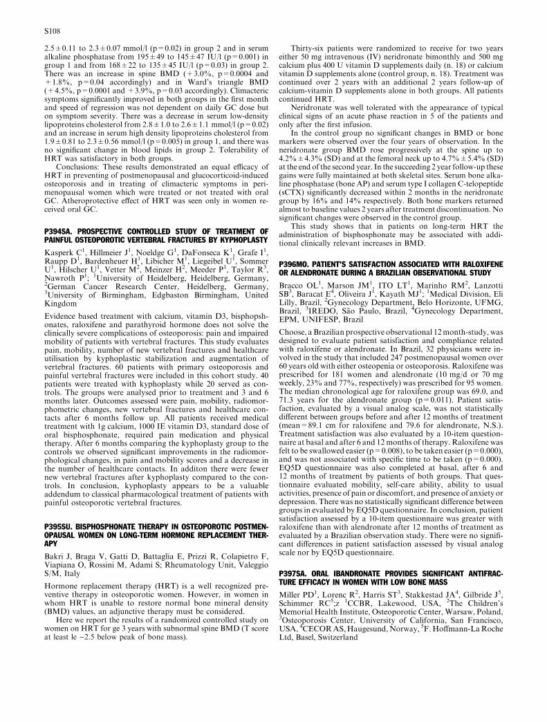
2.5±0.11 to 2.3±0.07 mmol/l (p=0.02) in group 2 and in serumalkaline phosphatase from 195±49 to 145±47 IU/l (p=0.001) ingroup 1 and from 168±22 to 135±45 IU/l (p=0.03) in group 2.There was an increase in spine BMD (+3.0%, p=0.0004 and+1.8%, p=0.04 accordingly) and in Ward’s triangle BMD(+4.5%, p=0.0001 and +3.9%, p=0.03 accordingly). Climactericsymptoms significantly improved in both groups in the first monthand speed of regression was not dependent on daily GC dose buton symptom severity. There was a decrease in serum low-densitylipoproteins cholesterol from 2.8±1.0 to 2.6±1.1 mmol/l (p=0.02)and an increase in serum high density lipoproteins cholesterol from1.9±0.81 to 2.3±0.56 mmol/l (p=0.005) in group 1, and there wasno significant change in blood lipids in group 2. Tolerability ofHRT was satisfactory in both groups.
Conclusions: These results demonstrated an equal efficacy ofHRT in preventing of postmenopausal and glucocorticoid-inducedosteoporosis and in treating of climacteric symptoms in peri-menopausal women which were treated or not treated with oralGC. Atheroprotective effect of HRT was seen only in women re-ceived oral GC.
P394SA. PROSPECTIVE CONTROLLED STUDY OF TREATMENT OFPAINFUL OSTEOPOROTIC VERTEBRAL FRACTURES BY KYPHOPLASTY
Kasperk C1, Hillmeier J1, Noeldge G1, DaFonseca K1, Grafe I1,Raupp D1, Bardenheuer H1, Libicher M1, Liegeibel U1, SommerU1, Hilscher U1, Vetter M2, Meinzer H2, Meeder P1, Taylor R3,Nawroth P1; 1University of Heidelberg, Heidelberg, Germany,2German Cancer Research Center, Heidelberg, Germany,3University of Birmingham, Edgbaston Birmingham, UnitedKingdom
Evidence based treatment with calcium, vitamin D3, bisphopsh-onates, raloxifene and parathyroid hormone does not solve theclinically severe complications of osteoporosis: pain and impairedmobility of patients with vertebral fractures. This study evaluatespain, mobility, number of new vertebral fractures and healthcareutilisation by kyphoplastic stabilization and augmentation ofvertebral fractures. 60 patients with primary osteoporosis andpainful vertebral fractures were included in this cohort study. 40patients were treated with kyphoplasty while 20 served as con-trols. The groups were analysed prior to treatment and 3 and 6months later. Outcomes assessed were pain, mobility, radiomor-phometric changes, new vertebral fractures and healthcare con-tacts after 6 months follow up. All patients received medicaltreatment with 1g calcium, 1000 IE vitamin D3, standard dose oforal bisphosphonate, required pain medication and physicaltherapy. After 6 months comparing the kyphoplasty group to thecontrols we observed significant improvements in the radiomor-phological changes, in pain and mobility scores and a decrease inthe number of healthcare contacts. In additon there were fewernew vertebral fractures after kyphoplasty compared to the con-trols. In conclusion, kyphoplasty appears to be a valuableaddendum to classical pharmacological treatment of patients withpainful osteoporotic vertebral fractures.
P395SU. BISPHOSPHONATE THERAPY IN OSTEOPOROTIC POSTMEN-OPAUSAL WOMEN ON LONG-TERM HORMONE REPLACEMENT THER-APY
Bakri J, Braga V, Gatti D, Battaglia E, Prizzi R, Colapietro F,Viapiana O, Rossini M, Adami S; Rheumatology Unit, ValeggioS/M, Italy
Hormone replacement therapy (HRT) is a well recognized pre-ventive therapy in osteoporotic women. However, in women inwhom HRT is unable to restore normal bone mineral density(BMD) values, an adjunctive therapy must be considered.
Here we report the results of a randomized controlled study onwomen on HRT for ge 3 years with subnormal spine BMD (T scoreat least le )2.5 below peak of bone mass).
Thirty-six patients were randomized to receive for two yearseither 50 mg intravenous (IV) neridronate bimonthly and 500 mgcalcium plus 400 U vitamin D supplements daily (n. 18) or calciumvitamin D supplements alone (control group, n. 18). Treatment wascontinued over 2 years with an additional 2 years follow-up ofcalcium-vitamin D supplements alone in both groups. All patientscontinued HRT.
Neridronate was well tolerated with the appearance of typicalclinical signs of an acute phase reaction in 5 of the patients andonly after the first infusion.
In the control group no significant changes in BMD or bonemarkers were observed over the four years of observation. In theneridronate group BMD rose progressively at the spine up to4.2%±4.3% (SD) and at the femoral neck up to 4.7%±5.4% (SD)at the end of the second year. In the succeeding 2 year follow-up thesegains were fully maintained at both skeletal sites. Serum bone alka-line phosphatase (bone AP) and serum type I collagen C-telopeptide(sCTX) significantly decreased within 2 months in the neridronategroup by 16% and 14% respectively. Both bone markers returnedalmost to baseline values 2 years after treatment discontinuation.Nosignificant changes were observed in the control group.
This study shows that in patients on long-term HRT theadministration of bisphosphonate may be associated with addi-tional clinically relevant increases in BMD.
P396MO. PATIENT’S SATISFACTION ASSOCIATED WITH RALOXIFENEOR ALENDRONATE DURING A BRAZILIAN OBSERVATIONAL STUDY
Bracco OL1, Marson JM1, ITO LT1, Marinho RM2, LanzottiSB3, Baracat E4, Oliveira J1, Kayath MJ1; 1Medical Division, EliLilly, Brazil, 2Gynecology Department, Belo Horizonte, UFMG,Brazil, 3IREDO, Sao Paulo, Brazil, 4Gynecology Department,EPM, UNIFESP, Brazil
Choose, aBrazilian prospective observational 12month-study, wasdesigned to evaluate patient satisfaction and compliance relatedwith raloxifene or alendronate. In Brazil, 32 physicians were in-volved in the study that included 247 postmenopausal women over60 years old with either osteopenia or osteoporosis. Raloxifene wasprescribed for 181 women and alendronate (10 mg/d or 70 mgweekly, 23% and 77%, respectively) was prescribed for 95 women.The median chronological age for raloxifene group was 69.0, and71.3 years for the alendronate group (p=0.011). Patient satis-faction, evaluated by a visual analog scale, was not statisticallydifferent between groups before and after 12 months of treatment(mean=89.1 cm for raloxifene and 79.6 for alendronate, N.S.).Treatment satisfaction was also evaluated by a 10-item question-naire at basal and after 6 and 12months of therapy. Raloxifene wasfelt to be swallowed easier (p=0.008), to be taken easier (p=0.000),and was not associated with specific time to be taken (p=0.000).EQ5D questionnaire was also completed at basal, after 6 and12 months of treatment by patients of both groups. That ques-tionnaire evaluated mobility, self-care ability, ability to usualactivities, presence of pain or discomfort, andpresence of anxiety ordepression. There was no statistically significant difference betweengroups in evaluated by EQ5D questionnaire. In conclusion, patientsatisfaction assessed by a 10-item questionnaire was greater withraloxifene than with alendronate after 12 months of treatment asevaluated by a Brazilian observation study. There were no signifi-cant differences in patient satisfaction assessed by visual analogscale nor by EQ5D questionnaire.
P397SA. ORAL IBANDRONATE PROVIDES SIGNIFICANT ANTIFRAC-TURE EFFICACY IN WOMEN WITH LOW BONE MASS
Miller PD1, Lorenc R2, Harris ST3, Stakkestad JA4, Gilbride J5,Schimmer RC5;z 1CCBR, Lakewood, USA, 2The Children’sMemorial Health Institute, Osteoporotic Center, Warsaw, Poland,3Osteoporosis Center, University of California, San Francisco,USA, 4CECORAS,Haugesund,Norway, 5F.Hoffmann-LaRocheLtd, Basel, Switzerland
S108
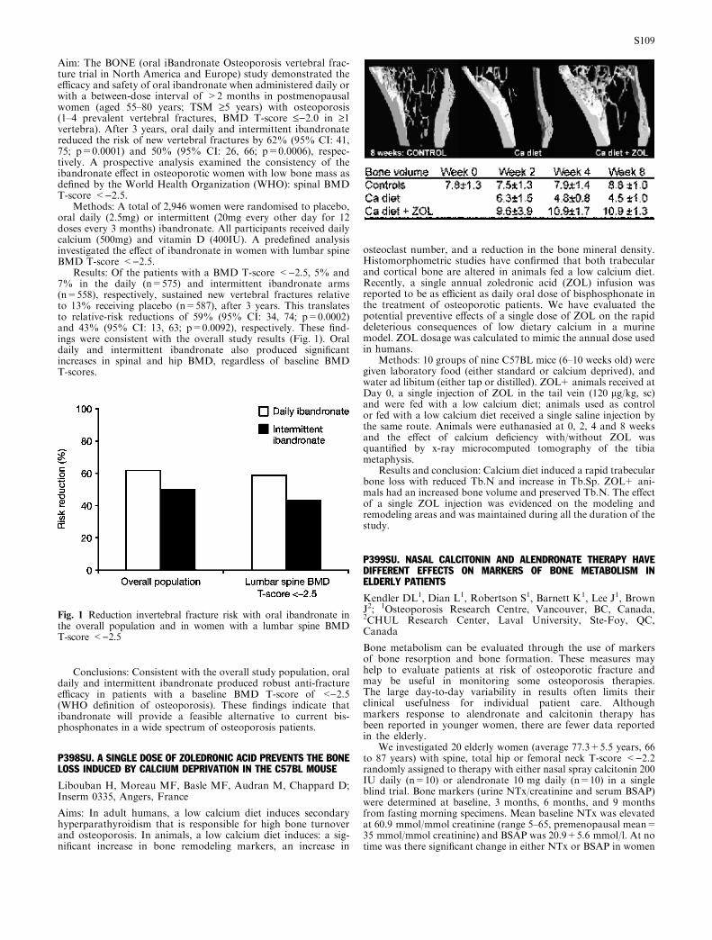
Aim: The BONE (oral iBandronate Osteoporosis vertebral frac-ture trial in North America and Europe) study demonstrated theefficacy and safety of oral ibandronate when administered daily orwith a between-dose interval of >2 months in postmenopausalwomen (aged 55–80 years; TSM ‡5 years) with osteoporosis(1–4 prevalent vertebral fractures, BMD T-score £)2.0 in ‡1vertebra). After 3 years, oral daily and intermittent ibandronatereduced the risk of new vertebral fractures by 62% (95% CI: 41,75; p=0.0001) and 50% (95% CI: 26, 66; p=0.0006), respec-tively. A prospective analysis examined the consistency of theibandronate effect in osteoporotic women with low bone mass asdefined by the World Health Organization (WHO): spinal BMDT-score <)2.5.
Methods: A total of 2,946 women were randomised to placebo,oral daily (2.5mg) or intermittent (20mg every other day for 12doses every 3 months) ibandronate. All participants received dailycalcium (500mg) and vitamin D (400IU). A predefined analysisinvestigated the effect of ibandronate in women with lumbar spineBMD T-score <)2.5.
Results: Of the patients with a BMD T-score <)2.5, 5% and7% in the daily (n=575) and intermittent ibandronate arms(n=558), respectively, sustained new vertebral fractures relativeto 13% receiving placebo (n=587), after 3 years. This translatesto relative-risk reductions of 59% (95% CI: 34, 74; p=0.0002)and 43% (95% CI: 13, 63; p=0.0092), respectively. These find-ings were consistent with the overall study results (Fig. 1). Oraldaily and intermittent ibandronate also produced significantincreases in spinal and hip BMD, regardless of baseline BMDT-scores.
Conclusions: Consistent with the overall study population, oraldaily and intermittent ibandronate produced robust anti-fractureefficacy in patients with a baseline BMD T-score of <)2.5(WHO definition of osteoporosis). These findings indicate thatibandronate will provide a feasible alternative to current bis-phosphonates in a wide spectrum of osteoporosis patients.
P398SU. A SINGLE DOSE OF ZOLEDRONIC ACID PREVENTS THE BONELOSS INDUCED BY CALCIUM DEPRIVATION IN THE C57BL MOUSE
Libouban H, Moreau MF, Basle MF, Audran M, Chappard D;Inserm 0335, Angers, France
Aims: In adult humans, a low calcium diet induces secondaryhyperparathyroidism that is responsible for high bone turnoverand osteoporosis. In animals, a low calcium diet induces: a sig-nificant increase in bone remodeling markers, an increase in
osteoclast number, and a reduction in the bone mineral density.Histomorphometric studies have confirmed that both trabecularand cortical bone are altered in animals fed a low calcium diet.Recently, a single annual zoledronic acid (ZOL) infusion wasreported to be as efficient as daily oral dose of bisphosphonate inthe treatment of osteoporotic patients. We have evaluated thepotential preventive effects of a single dose of ZOL on the rapiddeleterious consequences of low dietary calcium in a murinemodel. ZOL dosage was calculated to mimic the annual dose usedin humans.
Methods: 10 groups of nine C57BL mice (6–10 weeks old) weregiven laboratory food (either standard or calcium deprived), andwater ad libitum (either tap or distilled). ZOL+ animals received atDay 0, a single injection of ZOL in the tail vein (120 lg/kg, sc)and were fed with a low calcium diet; animals used as controlor fed with a low calcium diet received a single saline injection bythe same route. Animals were euthanasied at 0, 2, 4 and 8 weeksand the effect of calcium deficiency with/without ZOL wasquantified by x-ray microcomputed tomography of the tibiametaphysis.
Results and conclusion: Calcium diet induced a rapid trabecularbone loss with reduced Tb.N and increase in Tb.Sp. ZOL+ ani-mals had an increased bone volume and preserved Tb.N. The effectof a single ZOL injection was evidenced on the modeling andremodeling areas and was maintained during all the duration of thestudy.
P399SU. NASAL CALCITONIN AND ALENDRONATE THERAPY HAVEDIFFERENT EFFECTS ON MARKERS OF BONE METABOLISM INELDERLY PATIENTS
Kendler DL1, Dian L1, Robertson S1, Barnett K1, Lee J1, BrownJ2; 1Osteoporosis Research Centre, Vancouver, BC, Canada,2CHUL Research Center, Laval University, Ste-Foy, QC,Canada
Bone metabolism can be evaluated through the use of markersof bone resorption and bone formation. These measures mayhelp to evaluate patients at risk of osteoporotic fracture andmay be useful in monitoring some osteoporosis therapies.The large day-to-day variability in results often limits theirclinical usefulness for individual patient care. Althoughmarkers response to alendronate and calcitonin therapy hasbeen reported in younger women, there are fewer data reportedin the elderly.
We investigated 20 elderly women (average 77.3+5.5 years, 66to 87 years) with spine, total hip or femoral neck T-score <)2.2randomly assigned to therapy with either nasal spray calcitonin 200IU daily (n=10) or alendronate 10 mg daily (n=10) in a singleblind trial. Bone markers (urine NTx/creatinine and serum BSAP)were determined at baseline, 3 months, 6 months, and 9 monthsfrom fasting morning specimens. Mean baseline NTx was elevatedat 60.9 mmol/mmol creatinine (range 5–65, premenopausal mean=35 mmol/mmol creatinine) and BSAP was 20.9+5.6 mmol/l. At notime was there significant change in either NTx or BSAP in women
Fig. 1 Reduction invertebral fracture risk with oral ibandronate inthe overall population and in women with a lumbar spine BMDT-score <)2.5
S109
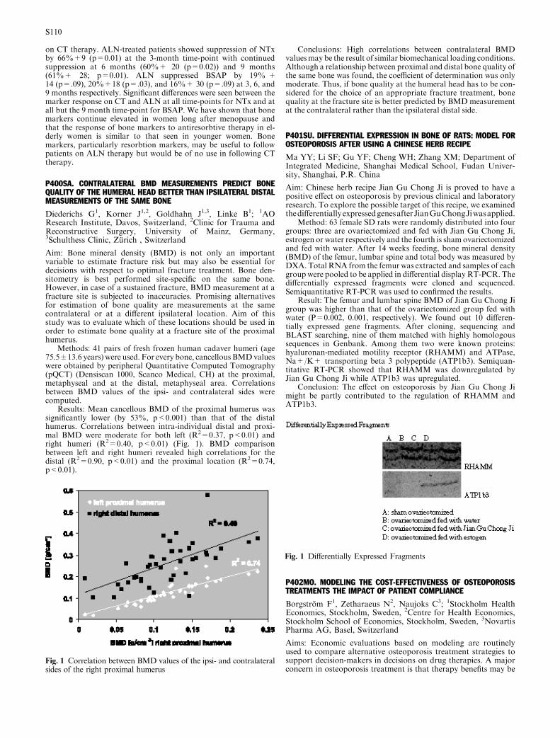
on CT therapy. ALN-treated patients showed suppression of NTxby 66%+9 (p=0.01) at the 3-month time-point with continuedsuppression at 6 months (60%+ 20 (p=0.02)) and 9 months(61%+ 28; p=0.01). ALN suppressed BSAP by 19% +14 (p=.09), 20%+18 (p=.03), and 16%+ 30 (p=.09) at 3, 6, and9 months respectively. Significant differences were seen between themarker response on CT and ALN at all time-points for NTx and atall but the 9 month time-point for BSAP. We have shown that bonemarkers continue elevated in women long after menopause andthat the response of bone markers to antiresorbtive therapy in el-derly women is similar to that seen in younger women. Bonemarkers, particularly resorbtion markers, may be useful to followpatients on ALN therapy but would be of no use in following CTtherapy.
P400SA. CONTRALATERAL BMD MEASUREMENTS PREDICT BONEQUALITY OF THE HUMERAL HEAD BETTER THAN IPSILATERAL DISTALMEASUREMENTS OF THE SAME BONE
Diederichs G1, Korner J1,2, Goldhahn J1,3, Linke B1; 1AOResearch Institute, Davos, Switzerland, 2Clinic for Trauma andReconstructive Surgery, University of Mainz, Germany,3Schulthess Clinic, Zurich , Switzerland
Aim: Bone mineral density (BMD) is not only an importantvariable to estimate fracture risk but may also be essential fordecisions with respect to optimal fracture treatment. Bone den-sitometry is best performed site-specific on the same bone.However, in case of a sustained fracture, BMD measurement at afracture site is subjected to inaccuracies. Promising alternativesfor estimation of bone quality are measurements at the samecontralateral or at a different ipsilateral location. Aim of thisstudy was to evaluate which of these locations should be used inorder to estimate bone quality at a fracture site of the proximalhumerus.
Methods: 41 pairs of fresh frozen human cadaver humeri (age75.5±13.6 years) were used. For every bone, cancellous BMDvalueswere obtained by peripheral Quantitative Computed Tomography(pQCT) (Densiscan 1000, Scanco Medical, CH) at the proximal,metaphyseal and at the distal, metaphyseal area. Correlationsbetween BMD values of the ipsi- and contralateral sides werecomputed.
Results: Mean cancellous BMD of the proximal humerus wassignificantly lower (by 53%, p<0.001) than that of the distalhumerus. Correlations between intra-individual distal and proxi-mal BMD were moderate for both left (R2=0.37, p<0.01) andright humeri (R2=0.40, p<0.01) (Fig. 1). BMD comparisonbetween left and right humeri revealed high correlations for thedistal (R2=0.90, p<0.01) and the proximal location (R2=0.74,p<0.01).
Conclusions: High correlations between contralateral BMDvaluesmay be the result of similar biomechanical loading conditions.Although a relationship between proximal and distal bone quality ofthe same bone was found, the coefficient of determination was onlymoderate. Thus, if bone quality at the humeral head has to be con-sidered for the choice of an appropriate fracture treatment, bonequality at the fracture site is better predicted by BMDmeasurementat the contralateral rather than the ipsilateral distal side.
P401SU. DIFFERENTIAL EXPRESSION IN BONE OF RATS: MODEL FOROSTEOPOROSIS AFTER USING A CHINESE HERB RECIPE
Ma YY; Li SF; Gu YF; Cheng WH; Zhang XM; Department ofIntegrated Medicine, Shanghai Medical School, Fudan Univer-sity, Shanghai, P.R. China
Aim: Chinese herb recipe Jian Gu Chong Ji is proved to have apositive effect on osteoporosis by previous clinical and laboratoryresearch. To explore the possible target of this recipe, we examinedthedifferentiallyexpressedgenesafterJianGuChongJiwasapplied.
Method: 63 female SD rats were randomly distributed into fourgroups: three are ovariectomized and fed with Jian Gu Chong Ji,estrogen or water respectively and the fourth is sham ovariectomizedand fed with water. After 14 weeks feeding, bone mineral density(BMD) of the femur, lumbar spine and total body was measured byDXA. Total RNA from the femur was extracted and samples of eachgroup were pooled to be applied in differential display RT-PCR. Thedifferentially expressed fragments were cloned and sequenced.Semiquantitative RT-PCR was used to confirmed the results.
Result: The femur and lumbar spine BMD of Jian Gu Chong Jigroup was higher than that of the ovariectomized group fed withwater (P=0.002, 0.001, respectively). We found out 10 differen-tially expressed gene fragments. After cloning, sequencing andBLAST searching, nine of them matched with highly homologoussequences in Genbank. Among them two were known proteins:hyaluronan-mediated motility receptor (RHAMM) and ATPase,Na+/K+ transporting beta 3 polypeptide (ATP1b3). Semiquan-titative RT-PCR showed that RHAMM was downregulated byJian Gu Chong Ji while ATP1b3 was upregulated.
Conclusion: The effect on osteoporosis by Jian Gu Chong Jimight be partly contributed to the regulation of RHAMM andATP1b3.
P402MO. MODELING THE COST-EFFECTIVENESS OF OSTEOPOROSISTREATMENTS THE IMPACT OF PATIENT COMPLIANCE
Borgstrom F1, Zetharaeus N2, Naujoks C3; 1Stockholm HealthEconomics, Stockholm, Sweden, 2Centre for Health Economics,Stockholm School of Economics, Stockholm, Sweden, 3NovartisPharma AG, Basel, Switzerland
Aims: Economic evaluations based on modeling are routinelyused to compare alternative osteoporosis treatment strategies tosupport decision-makers in decisions on drug therapies. A majorconcern in osteoporosis treatment is that therapy benefits may be
Fig. 1 Correlation between BMD values of the ipsi- and contralateralsides of the right proximal humerus
Fig. 1 Differentially Expressed Fragments
S110
lower in due to non-compliance. However, economic conse-quences of non-compliance are seldom included in economicmodeling. The objective with this study was to build a computersimulation model explicitly including the impact of patient com-pliance with osteoporosis treatments, and estimate the potentialcost-effectiveness of different drug alternatives that differ incompliance compared to no treatment applied to a US setting.
Methods: An individual state transition model was built toreflect different compliance patterns during drug intervention. Themodel takes a life-time perspective: all female patients are followedfrom treatment start to age of 100 years or death taking intoaccount yearly changing risks of hip, vertebral or wrist fractures, orto remain in a certain health status without any fracture or to die.Women (T-score=)2.5 and a previous vertebral fracture) starttreatment with 70 years of age. The model considers effects ofmorbidity and mortality during the first year after fracture as wellas long-term effects of fractures beyond that first year. During theintervention period which is modeled to be 5 years long, yearlychanging probabilities of discontinuation of the comparative drugtreatment is simulated. The model was populated with US data.Fracture risk reductions as well as yearly drug costs are assumed tobe identical. The cost-effectiveness was estimated for differentassumptions about the rates of compliance with osteoporosistreatments. Discount rate for costs and effects is 3%.
Results: A therapy offering full compliance and a therapy notoffering full compliance was both found to be cost-effective com-pared to no treatment. When comparing the therapy alternativesthe treatment offering full compliance was associated with highereffects (QALYs) and lower average lifetime fracture costs.
Conclusions: The results indicate that an osteoporosis drugwhich guarantees a full year patient compliance might havepotential gains with regard to cost-effectiveness and potentialdecrease of osteoporosis-related fracture risk.
P403SA. THE COST-EFFECTIVENESS OF BISPHOSPHONATES,RALOXIFENE AND HORMONE REPLACEMENT THERAPY IN WOMENWITH ESTABLISHED OSTEOPOROSIS AND REPLETE OF CALCIUM ANDVITAMIN D
Stevenson MD1, Brazier JE1, Lloyd-Jones M1, Calvert N1,Oakley J2, Kanis JA3; 1ScHARR; Sheffield; England, 2Depart-ment of Probability and Statistics, Sheffield, England, 3Universityof Sheffield Medical School, Sheffield, England
Aims: This paper evaluates, within a UK setting, the cost-effec-tiveness of bisphosphonates, raloxifene, and HRT in women withestablished osteoporosis and replete of calcium and vitamin D.
Methods: A mathematical model, constructed to simulate theexperience of hypothetical patients, was populated by evidencefrom a systematic review of randomised controlled trials of clinicalefficacy, and reviews of utilities, costs, and epidemiological data.Adverse events included were hip, vertebral, wrist and proximalhumerus fractures, and breast cancer and coronary heart disease(CHD). Analyses were conducted for women aged 50, 60, 70 and 80years at the threshold of osteoporosis (T-score=)2.5SD) with aprior fracture and for women with double this fracture risk, due toa lower T-Score or other risk factors.
Results: The main outcome measure for each intervention wasthe cost per quality adjusted life year (QALY) gained comparedwith no treatment. In patients at greatest risk of osteoporoticfracture, alendronate and risedronate had superior cost-effective-ness results. In younger women with a T-Score of )2.5SD raloxifeneappeared the most cost-effective intervention, although this was dueto the assumed beneficial effects on breast cancer. HRT was esti-mated to be harmful to women aged 60 or older due to the assumedadverse effect on breast cancer and on CHD. Etidronate was alwaysinferior to alendronate and risedronate. Cost-effectiveness resultsare presented in the table assuming double the fracture risk com-pared with women at the threshold of osteoporosis.
Conclusions: It was most cost-effective to treat women withestablished osteoporosis aged 70 years or older with alendronate orrisedronate. For women aged 50 or 60 years, the first-line inter-vention was dependant on the absolute fracture risk, at the
threshold of osteoporosis the most cost-effective intervention wasraloxifene, where this risk was doubled, the most cost-effectiveinterventions became alendronate or risedronate.Incremental costs and QALYs of each intervention compared tono treatment for 100 women with established osteoporosis andT-Scores of )2.5 SD assuming that the expected risks of fractureare doubled.
P404SU. OUR EXPERIENCES WITH BALLOON KYPHOPLASTY IN THETREATMENT OF OSTEOPOROTIC VERTREBRAL COMPRESSION FRAC-TURES (VCF)
Borgulya J1, Gunther C2, Thumler P3; 1Chirurgisch-Orthopadi-sche Fachklinik, Lorsch, Germany, 2Johannesbad Reha-KlinikenAG&Co.KG, Bad Fussing, Germany, 3St. Vinezen Krankenhaus,Dusseldorf, Germany
Aims: To look for pain relief and correction of spinal deformityafter balloon-kyphoplasty. In addition to this we were interestedin new fractures nearby the operated segments.
Methods: All together 85 vertebrae were treated in 54 patients(48 women, 6 man, age 72 years). These 85 vertebrae were dividedinto 49 with acute fractures (AF, operation shorter than four weeksafter fracture) and 20 subacute fractures (SAF, four to eightweeks). From this, 54 patients had 11 adequate trauma, 15 had anon-adequate accident and 28 had spontaneous fractures. For painestimation we used the VAS-scale, for measurement of kyphosisangle we used lateral x-ray of the spine.
Results: AF-group (n=49) showed an improved kyphosis angleof 11.5 grad postoperative and 10.7 grad after four weeks. In theSAF-group we could see an improvement of 1.8/1.7 grad. Lookingto the pain-score the AF showed the following VAS values: 8.8preoperative, 4.1 postoperative and 3.2 after four weeks. SAFshowed: 8.3, 4.7 and 4.2. In no case had we seen any disturbance inwound heeling. Leakage of cement occured in 12 cases (14% of 85),there was only one case with radiculopathy (1.2% from 85 verte-bras, 1.85% from 54 patients).
Conclusions: 1. Fresh osteoporotic vertebral fractures can bewell restored within the first 4 weeks after fracture. 2. Subacutefractures do not allow good improvement of the kyphosis angle. 3.In both cases, good pain relief is possible. 4. The complicationshappened within the first month of using of this method (i.e. withinthe learning phase). Therefore, good training is necessary. 5. Insome cases new fractures happened in the vertebrae nearby theoperated segment. 6. Our two years experience showed thatendangered adjacent segments should be treated also. 7. Balloon-kyphoplasty is a very safe therapy option in the treatment of acutevertrebral fracture. 8. Furthermore, osteoporotic patients withfractures must also be treated with changes in nutritional behav-iour, movement therapy, evidenced based medical therapy (modernbisphosphonates, SERMs, parathormone, calcium and vitamin D),as well as psychological care.
P405MO. DIAGNOSIS AND TREATEMENT OF OSTEOPOROSIS: FRE-QUENCY IN A RIO DE JANEIRO OSTEOPOROSIS CENTER
Russo LAT1,2,3, Gregorio LH1,2,3, Melazzi ACC1,2, Martins SR1,Carvao C1, Oliveira R3, Mattaini AS1,2; 1Densso ) Center forDiagnosis of Osteoporosis, Rio de Janerio, Brazil, 2CCBR Brasil) Center for Clinical and Basic Research, Rio de Janeiro, Brazil,3Pro Matre Hospital, Rio de Janeiro, Brazil
Age 50 years Age 60 years Age 70 years Age 80 yearsMean Costper QALY(£000) (95% CI)
Mean Costper QALY(£000) (95% CI)
Mean Costper QALY(£000) (95% CI)
Mean Costper QALY(£000) (95% CI)
Alendronate 25 (18 – 50) 16 (10 – 41) D-ing (D-ing – 14) D-ing (D-ing – 3)Etidronate 45 (35 – 70) 41 (32 – 65) 12 (9 – 20) 17 (11 – 32)Risedronate 26 (19 – 42) 19 (12 – 37) 2 (D-ing – 13) D-ing (D-ing – D-ing)Raloxifene 29 (24 – 41) 20 (16 – 29) 22 (17 – 32) 17 (12 – 26)HRT 34 (14 – D-ed) D-ed (12 – D-ed) 10 (D-ing – D-ed) 38 (D-ing – D-ed)
S111
Aims: Osteoporosis is a common disease charachterized by adecrease in bone mineral density with an increase in fracture risk.It is nowadays a worldwide public health problem with increasingcosts. The purpose of this study is to evaluate the rate of oste-openia and osteoporosis in patients examined in a OsteoporosisDiagnostic Center in Rio de Janeiro and the use of pharmacho-logical therapy in these patients.
Methods: The results of 2270 subjects underwent to a bonedensity tests in a period of 90 days. The authors studied the dis-tribution of rate about sex, age, diagnosis, skeletal sites andtreatment used. In data analysis the Chi square test was used. Incomparison of the proportion between treatment and diagnosis, theZ test of normal curve was used.
Results: We observed that the average age of the subjects was59±10.9 years. 2237 were females and 33 were males. The resultsshowed that 46.9% had osteopenia and 19.4% had osteoporosis(figure). Among those patients with osteopenia 55.1% were usingsome pharmachological therapy and among those with osteopo-rosis only 59,6% were being treated: 7.3% were in use of alendr-onate, 4.2% in use of raloxifene and 48.1% in use of TRH,Calcium with Vitamin D or Calcium alone (figure).
Conclusions: The authors concluded that despite the correct useof bone density test and the diagnosis, a large number of patientswith osteopenia and osteoporosis are not being properly treated.
P406SA. THE EFFECT OF DIFFERENT BISPHOSPHONATE TREATMENTIN PAGET’S DISEASE OF BONE
Donath J1, Toth L2, Poor G1; 1National Institute of Rheumatol-ogy and Physiotherapy, Budapest, Hungary, 2St. Imre Hospital,Budapest, Hungary
Objective: To asses the usefulness of quantitative bone scintigra-phy (QBS) in the monitoring of bisphosphonate treated patientsand to evaluate the relationship between biochemical marker ofbone turnover and bone scan indices of disease activity.
Methods: We investigated one year efficacy of three differentbisphosphonates in 71 patients with Paget‘s disease. Patientsreceived infusion pamidronate 30 mg/d for 6 days (39 patients),oral tiludronate 300 mg/d for 90 days (25 patients) and oralalendronate 10 mg/d for 12 months (7 patients). Serum sampleswere obtained from 71 patients with Paget‘s disease to determinethe levels of total serum alkaline phosphatase (total AP). QBS wasperformed in every patient and the results were expressed as aratio, obtained by comparing isotope uptake at an effected and anuneffected control site. Reduction in bone pain was assessed usinga pain scale. Efficacy and side effects were monitored for a followup period of up to one year.
Results: Total AP levels decreased significantly after pamidro-nate (p<0.001) and tiludronate treatment (p<0.001) respectively.QBS ratio changed significantly after pamidronate (p<0.001) andtiludronate treatment, p<0.001) respectively. The pain scale scoredecreased significantly in all cases. The side effectswere the following:fever at five cases, bone pain at three cases and stomach-ache at twocases.
Conclusion: The treatment with iv pamidronate, oral tiludro-nate and oral alendronate results decreased pain, total AP levelsand QBS ratio.
P407SU. DOES INJECTABLE BIOMATERIAL RESTORE BONE MASS ANDARCHITECTURE IN A RAT MODEL OF OSTEOPOROSIS (ORCHIDECTOMYAND DISUSE)?
Blouin S1, Moreau MF1, Weiss P2, Daculsi G2, Basle MF1,Chappard D1; 1INSERMEMI335-LHEA, Angers, France, 2INSERMEMI9903, Faculte de Chirurgie Dentaire, Nantes, France
Aims: The use of osteoconductive biomaterials is often required inosteoporotic patients who need orthopaedics or maxillo-facialsurgery. The purpose of this study was to use an injectable bonesubstitute (IBS-1) in a rat model of osteoporosis obtained bycombining orchidectomy (ORX) and disuse. The effects of ORX,localized paralysis by botulinum toxin (BTX) and prevention byIBS-1 were investigated.
Methods: 52 aged male rats were randomised into 4 groups:SHAM operated; ORX-BTX (right hindlimb). One month aftersurgery, ORX-BTX rats had a bone marrow ablation (ORX-BTX-ABL) combined with injection with IBS-1 (ORX-BTX-ABL-IBS-1). Animals were studied 1 and 2 months after IBS-1 injection. BV/TV and Structure Modeling Index (SMI) were measured by X-raymicrocomputed tomography in the secondary spongiosa. Histo-morphometry was performed on the femur to measure osteoid,osteoclast activity and cell repartition.
Results: BV/TV was decreased 2 months after ORX-BTX;ORX and BTX effects were cumulative on bone loss since signifi-cant differences were observed between left and right limb. ABLhad no effect on bone volume compared with ORX-BTX results.The combined volume of bone and material (B+Mat)V/TV waselevated at 1 month but returned at normal level on the 2ndmonth. SMI increased significantly in ORX-BTX (confirming anincreased conversion of plates into pillars) and in ORX-BTX-IBSdue to the remanence of biomaterial. Histomorphometry showedmarked osseous formation apposed onto the biomaterial granulesbut bone had a woven texture. A dramatic increase in the numberof non-osteoclastic TRAcP positive cells was found in theimplanted area.
Conclusion: Our findings suggest that IBS-1 and newly formedbone are resorbed after a restoration period in this model ofosteoporosis. Biomaterial trials must be conducted with long-termimplantation periods in aged and osteoporotic animals to deter-mine their potent ability to permanently restore trabecular mic-roarchitecture.
Fig. 1 Percentual distribution related to the treatment of the patientswith Osteoporosis
S112
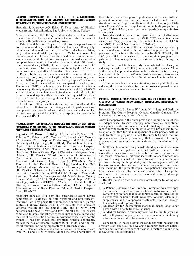
P408MO. COMPARISON OF THE EFFECTS OF ALFACALCIDOL-ALENDRONATE-CALCIUM AND VITAMIN D-ALENDRONATE-CALCIUMTREATMENTS IN POSTMENOPAUSAL OSTEOPOROSIS
Hepguler S; Karatepe AG; Erkisi F; Department of PhysicalMedicine and Rehabilitation, Ege University, Izmir, Turkey
Aims: To compare the efficacy of alfacalcidol with alendronate-calcium and Vit-D with alendronate-calcium in the treatment ofwomen with postmenopausal osteoporosis.
Patients and Method: 72 patients with postmenopausal osteo-porosis were randomly treated with either alendronate 10 mg daily,calcium and alfacalcidol (Group 1, n=33) or alendronate 10 mgdaily, calcium and Vit-D (Group 2, n=39) for one year. Bio-chemical markers of bone resorption and bone formation withserum calcium and phosphorus, urinary calcium and serum alka-line phosphatase were performed at baseline and at 12th month.Bone mineral density (BMD) of lumbar spine, femur neck and totalfemur was measured by dual x-ray absorptiometry (DEXA) at theinitial assessment and after 12 months of treatment.
Results: In the baseline measurements, there were no differencesbetween age, body weight and length variables, whereas body massindex (BMI) in group 1 was greater than group 2 (25.23 versus27.18, p<0.05). At the end of one year, compared to baseline, bothT scores and BMD in the lumbar spine, femur neck and total femurincreased significantly in patients receiving alfacalcidol (p<0.05). Tscores of lumbar spine, femur neck, total femur and BMD of totalfemur increased significantly in patients receiving Vit-D after oneyear (p<0.05). There were no significant differences in BMD and Tscores between both groups.
Conclusions: These results indicate that both Vit-D and alfa-calcidol were effective in the management of postmenopausalosteoporosis when combined with alendronate and calcium. Thetwo treatment groups did not differ with respect to increases in theT scores and BMD.
P409SA. STRONTIUM RANELATE REDUCES THE RISK OF VERTEBRALFRACTURES IN OSTEOPOROTIC POSTMENOPAUSAL WOMEN WITHOUTPREVALENT VERTEBRAL FRACTURE
Reginster JY1, Rizzoli R2, Balogh A3, Badurski J4, Spector T5,Tulassay Z6, Felsenberg D7, Cannata JB8, Phenekos C9, OrtolaniS10, Meunier PJ11; 1Dept of Epidemiology and Public Health,University of Liege, Liege, BELGIUM, 2Div. of Bone Diseases,Dept of Rehabilitation and Geriatrics, University Hospital,Geneva, SWITZERLAND, 3University of Debrecen, MedicalHealth and Sciences Center, Dpt of Obstetrics and Gynaecology,Debrecen, HUNGARY, 4Polish Foundation of Osteoporosis,Center for Osteoporosis and Osteo-Articular Diseases, Dpt ofMedicine and Rheumatology, Bialystok, POLAND, 5SaintThomas’ Hospital, Dept of Rheumatology, London, UK, 62ndDept of Internal Medicine, Semmelweis University, Budapest,HUNGARY, 7Charite ) University Medicine Berlin, CampusBenjamin Franklin, Berlin, GERMANY, 8Hospital Central deAsturias, Unidad de Investigacion del Metabolismo Oseo yMineral, Oviedo, SPAIN, 9Red Cross Hospital, Dept of Endo-crinology, Athens, GREECE, 10Centre for Metabolic BoneDisease, Istituto Auxologico Italiano, Milan, ITALY, 11Dept ofRheumatology and Bone Diseases, Edouard Herriot Hospital,Lyon, FRANCE
Strontium ranelate is a new anti-osteoporotic agent havingdemonstrated its efficacy on both vertebral and non vertebralfractures: Two large phase III randomized, double blind, placebocontrolled clinical trials, SOTI (1649 patients with prevalentvertebral fracture and low lumbar BMD) and TROPOS (5091patients aged above 70 years and low femoral neck BMD) wereconducted to assess the efficacy of strontium ranelate in reducingthe risk of osteoporotic fractures in postmenopausal osteoporoticwomen. It has been shown that strontium ranelate significantlyreduces the risk of vertebral fracture (SOTI study) and non ver-tebral fractures including hip fractures (TROPOS study).
A pre-planned meta analysis was performed on the pooled datafrom SOTI and TROPOS trials. Among the whole population of
these studies, 2605 osteoporotic postmenopausal women withoutprevalent vertebral fracture (VF) were included and receivedstrontium ranelate 2 g/day orally (n=1285) or placebo (n=1320)plus a Calcium/Vitamin D supplementation in both groups during3 years. Vertebral X-rays were performed yearly (semi-quantitativeassessment).
No statistical differences between groups were detected for mainbaseline characteristics: mean age (SD): 75(5) years; time sincemenopause: 26(7) years; mean(SD) Lumbar T-score: )2.70(1.53);mean(SD), Femoral Neck T-score: )2.97(0.56).
A significant reduction in the incidence of patients experiencinga VF was demonstrated in the intent-to-treat population over 3years with a reduction of the relative risk by 48 % (95%CI [0.40;0.67], p<0.001). 87 patients in strontium ranelate group and 161patients in placebo experienced a vertebral fracture during thestudy.
Strontium ranelate has already demonstrated its efficacy inreducing the risk of VF (41%) in patients with prevalent VF inSOTI study. The present analysis confirms its antifracture efficacy(reduction of the risk of 48%) in postmenopausal osteoporoticwomen without prevalent VF. Strontium ranelate is well-toler-ated.
Strontium ranelate is a new anti-osteoporotic agent effective inreducing the risk of vertebral fracture in post-menopausal womenwith or without prevalent vertebral fracture.
P410SU. FRACTURE PREVENTION STRATEGIES ON A GERIATRIC UNIT:A SURVEY OF PATIENT KNOWLEDGE/ATTITUDES AND RESOURCE KITDEVELOPMENT
Byszewski A1,2, Ho J2, Power B1,2, Azad N1,2; 1Regional GeriatricProgram, The Ottawa Hospital, Ottawa, Ontario, Canada, 2TheUniversity of Ottawa, Ottawa, Ontario
Aims: Osteoporosis in the older person is a leading cause of lossof independance. Despite emerging new therapies, suboptimalmanagement is still an issue in patients discharged from acutecare following fractures. The objective of this project was to de-velop an algorithm for the management of older persons with anacute fracture, to prepare a Patient Resource Kit and a fracture-specific medical discharge summary that is provided to familyphysicians on discharge from an acute setting for continuity ofcare.
Methods: Interviews using standardized questionnaires wereconducted with ten patients admitted with a fracture. Sub-sequently, a focus group was held to further assess patient needsand review relevence of different materials. A chart review wasperformed using a standard format to assess the interventionsperformed during the hospital stay and the management offered.Discussions were also held with the interdisciplinary team mem-bers, including: the physiotherapist, occupational therapist, die-tician, social worker, pharmacist and nursing staff. This posterwill present the process of needs assessment, resource develop-ment and evaluation.
Results: Based on the above needs assessment the following wasdeveloped:
1) A Patient Resource Kit on Fracture Prevention was developedand subsequently evaluated using a telephone follow up. The kitcontains five sections that cover topics: general information onosteoporosis-including local resources, calcium/Vitamin Dsupplements and osteoporosis treatments, exercise therapy,home safety and hip protectors.
2) An algorithm for the interdisciplinary management of an olderperson with an acute fracture (attached).
3) A Medical Discharge Summary form for the family physicianwho will provide ongoing care in the community, containinginformation relevant to fracture prevention.
Conclusions: A needs assessment conducted with patients andprogram staff can assist in developing resources that are patientspecific and relevant to the care of those with fracture risk and raisethe awareness of osteoporosis.
S113
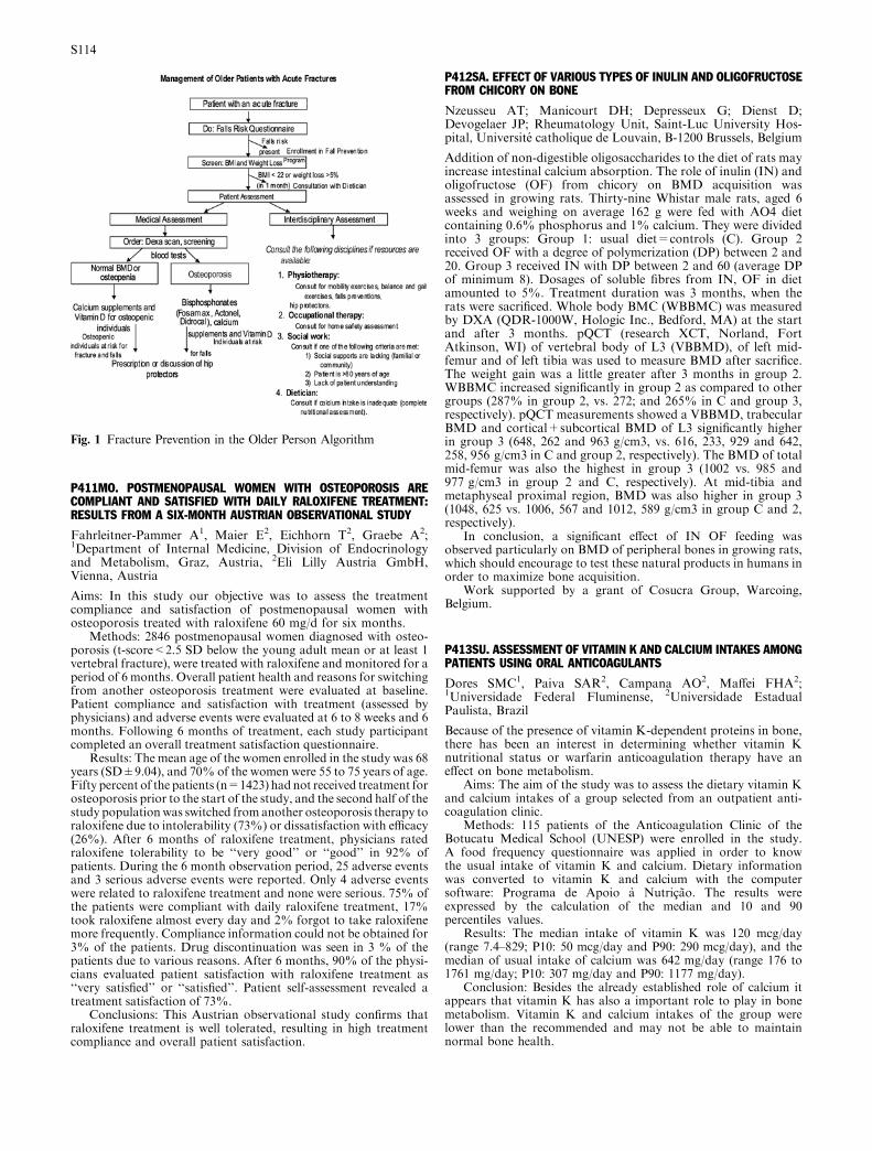
P411MO. POSTMENOPAUSAL WOMEN WITH OSTEOPOROSIS ARECOMPLIANT AND SATISFIED WITH DAILY RALOXIFENE TREATMENT:RESULTS FROM A SIX-MONTH AUSTRIAN OBSERVATIONAL STUDY
Fahrleitner-Pammer A1, Maier E2, Eichhorn T2, Graebe A2;1Department of Internal Medicine, Division of Endocrinologyand Metabolism, Graz, Austria, 2Eli Lilly Austria GmbH,Vienna, Austria
Aims: In this study our objective was to assess the treatmentcompliance and satisfaction of postmenopausal women withosteoporosis treated with raloxifene 60 mg/d for six months.
Methods: 2846 postmenopausal women diagnosed with osteo-porosis (t-score<2.5 SD below the young adult mean or at least 1vertebral fracture), were treated with raloxifene and monitored for aperiod of 6 months. Overall patient health and reasons for switchingfrom another osteoporosis treatment were evaluated at baseline.Patient compliance and satisfaction with treatment (assessed byphysicians) and adverse events were evaluated at 6 to 8 weeks and 6months. Following 6 months of treatment, each study participantcompleted an overall treatment satisfaction questionnaire.
Results: The mean age of the women enrolled in the study was 68years (SD±9.04), and 70% of the women were 55 to 75 years of age.Fifty percent of the patients (n=1423) had not received treatment forosteoporosis prior to the start of the study, and the second half of thestudy populationwas switched from another osteoporosis therapy toraloxifene due to intolerability (73%) or dissatisfaction with efficacy(26%). After 6 months of raloxifene treatment, physicians ratedraloxifene tolerability to be ‘‘very good’’ or ‘‘good’’ in 92% ofpatients. During the 6 month observation period, 25 adverse eventsand 3 serious adverse events were reported. Only 4 adverse eventswere related to raloxifene treatment and none were serious. 75% ofthe patients were compliant with daily raloxifene treatment, 17%took raloxifene almost every day and 2% forgot to take raloxifenemore frequently. Compliance information could not be obtained for3% of the patients. Drug discontinuation was seen in 3 % of thepatients due to various reasons. After 6 months, 90% of the physi-cians evaluated patient satisfaction with raloxifene treatment as‘‘very satisfied’’ or ‘‘satisfied’’. Patient self-assessment revealed atreatment satisfaction of 73%.
Conclusions: This Austrian observational study confirms thatraloxifene treatment is well tolerated, resulting in high treatmentcompliance and overall patient satisfaction.
P412SA. EFFECT OF VARIOUS TYPES OF INULIN AND OLIGOFRUCTOSEFROM CHICORY ON BONE
Nzeusseu AT; Manicourt DH; Depresseux G; Dienst D;Devogelaer JP; Rheumatology Unit, Saint-Luc University Hos-pital, Universite catholique de Louvain, B-1200 Brussels, Belgium
Addition of non-digestible oligosaccharides to the diet of rats mayincrease intestinal calcium absorption. The role of inulin (IN) andoligofructose (OF) from chicory on BMD acquisition wasassessed in growing rats. Thirty-nine Whistar male rats, aged 6weeks and weighing on average 162 g were fed with AO4 dietcontaining 0.6% phosphorus and 1% calcium. They were dividedinto 3 groups: Group 1: usual diet=controls (C). Group 2received OF with a degree of polymerization (DP) between 2 and20. Group 3 received IN with DP between 2 and 60 (average DPof minimum 8). Dosages of soluble fibres from IN, OF in dietamounted to 5%. Treatment duration was 3 months, when therats were sacrificed. Whole body BMC (WBBMC) was measuredby DXA (QDR-1000W, Hologic Inc., Bedford, MA) at the startand after 3 months. pQCT (research XCT, Norland, FortAtkinson, WI) of vertebral body of L3 (VBBMD), of left mid-femur and of left tibia was used to measure BMD after sacrifice.The weight gain was a little greater after 3 months in group 2.WBBMC increased significantly in group 2 as compared to othergroups (287% in group 2, vs. 272; and 265% in C and group 3,respectively). pQCT measurements showed a VBBMD, trabecularBMD and cortical+subcortical BMD of L3 significantly higherin group 3 (648, 262 and 963 g/cm3, vs. 616, 233, 929 and 642,258, 956 g/cm3 in C and group 2, respectively). The BMD of totalmid-femur was also the highest in group 3 (1002 vs. 985 and977 g/cm3 in group 2 and C, respectively). At mid-tibia andmetaphyseal proximal region, BMD was also higher in group 3(1048, 625 vs. 1006, 567 and 1012, 589 g/cm3 in group C and 2,respectively).
In conclusion, a significant effect of IN OF feeding wasobserved particularly on BMD of peripheral bones in growing rats,which should encourage to test these natural products in humans inorder to maximize bone acquisition.
Work supported by a grant of Cosucra Group, Warcoing,Belgium.
P413SU. ASSESSMENT OF VITAMIN K AND CALCIUM INTAKES AMONGPATIENTS USING ORAL ANTICOAGULANTS
Dores SMC1, Paiva SAR2, Campana AO2, Maffei FHA2;1Universidade Federal Fluminense, 2Universidade EstadualPaulista, Brazil
Because of the presence of vitamin K-dependent proteins in bone,there has been an interest in determining whether vitamin Knutritional status or warfarin anticoagulation therapy have aneffect on bone metabolism.
Aims: The aim of the study was to assess the dietary vitamin Kand calcium intakes of a group selected from an outpatient anti-coagulation clinic.
Methods: 115 patients of the Anticoagulation Clinic of theBotucatu Medical School (UNESP) were enrolled in the study.A food frequency questionnaire was applied in order to knowthe usual intake of vitamin K and calcium. Dietary informationwas converted to vitamin K and calcium with the computersoftware: Programa de Apoio a Nutricao. The results wereexpressed by the calculation of the median and 10 and 90percentiles values.
Results: The median intake of vitamin K was 120 mcg/day(range 7.4–829; P10: 50 mcg/day and P90: 290 mcg/day), and themedian of usual intake of calcium was 642 mg/day (range 176 to1761 mg/day; P10: 307 mg/day and P90: 1177 mg/day).
Conclusion: Besides the already established role of calcium itappears that vitamin K has also a important role to play in bonemetabolism. Vitamin K and calcium intakes of the group werelower than the recommended and may not be able to maintainnormal bone health.
Fig. 1 Fracture Prevention in the Older Person Algorithm
S114
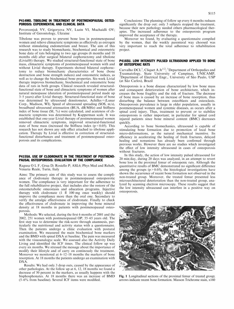
P414MO. TIBOLONE IN TREATMENT OF POSTMENOPAUSAL OSTEO-POROSIS: EXPERIMENTAL AND CLINICAL DATA
Povoroznjuk VV, Grigoryeva NV, Luzin VI, Muchaidli OV;Institute of Gerontology, Ukraine
Tibolone was proven to prevent bone loss in postmenopausalwomen and relieve climacteric symptoms as effectively as estrogenwithout stimulating endometrium and breast. The aim of thisresearch was to study biomechanic, biochemical and osteometricbone data of rats belonging to two age groups (6 months and 18months old) after surgical bilateral oophorectomy and tibolone(Livial�) therapy. We studied structural-functional state of bonemass, climacteric symptoms of postmenopausal women with andwithout Livial therapy. Experiments showed bilateral oophorec-tomy to reduce biomechanic characteristics of bone (bonedestruction and bone strength indices) and osteometric indices, aswell as to change the biochemical bone properties. Six-week Livialtherapy improves biomechanic, biochemical and osteometric bonedata of rats in both groups. Clinical research revealed structural-functional state of bone and climacteric symptoms of women afternatural menopause (duration of postmenopausal period made up3–5 years) after Livial therapy. Structural-functional state of bonewas determined by ultrasound densitometer, Achilles+(LunarCorp., Madison, WI). Speed of ultrasound spreading (SOS, m/s),broadband ultrasound attenuation (BUA, dB/MHz) and Stiffnessindex of bone tissue (SI, %) were calculated, and acuteness of cli-macteric symptoms was determined by Kupperman scale. It wasestablished that one-year Livial therapy of postmenopausal womenremoved climacteric symptoms, improved structural-functionalstate of bone mass (increased the Stiffness index (p<0.05). Thisresearch has not shown any side effect attached to tibolone appli-cation. Therapy by Livial is effective in correction of structural-functional disturbances and treatment of postmenopausal osteo-porosis and its complications.
P415SA. USE OF CLODRONATE IN THE TREATMENT OF POSTMENO-PAUSAL OSTEOPOROSIS: EVALUATION OF THE COMPLIANCE
Ragusa O L F, Grua D; Rosiello P; ASL6; Phys Med and Rehab,Venaria Reale, Turin, Italy
Aims: The primary aim of this study was to assess the compli-ance of clodronate therapy in postmenopausal osteoporoticwomen. The compliance is very important for the adherence tothe full rehabilitative project, that includes also the restore of theosteometabolic omeostasis and education programs. Injectivetherapy with clodronate (1 fl 100 mg once weekly) shouldimprove the compliance more than the oral one. Secondary toverify the antalgic effectiveness of clodronate. Finally to checkthe effectiveness of clodronate in improving the bone mineraldensity at 18 months in patients with postmenopausal osteo-porosis.
Methods: We selected, during the first 6 months of 2001 and the2002, 251 women with postmenopausal OP, 55–65 years old. Thefirst step was to determine the risk-score through anamnesis, par-ticularly the nutritional and activity status with a questionnaire.Then the patients undergo a clinic evaluation with posturalexamination. We measured the main biochemical bone markersand the BMD with spinal DXA at baseline. The pain was measuredwith the visuoanalogyc scale. We assessed also the Activity DailyLiving and identified the ICF itmes. The clinical follow up wasevery six months. We stressed the message about the importance ofmodify their lifestyle and of carry on continously the treatment.Moreover we monitored at 6–12–18 months the markers of boneresorption. At 18 months the patients undergo an examination withDXA.
Results: We had only 3 drop outs, caused by the appearance ofother pathologies. At the follow up at 6, 12, 18 months we found adecrease of 50 percent in the markers, as usually happens with thebisphosphonates. At 18 months there was an increase of BMD(5–6% from baseline). Several ICF items were modified.
Conclusions: The planning of follow up every 6 months reducessignificantly the drop out: only 3 subjects stopped the treatment,because their new pathology needed others pharmacological ther-apies. The increased adherence to the osteoporosis programimproved the acceptance of the therapy.
Moreover we found, by evaluating a questionnaire compiledby the women, that the weekly parenteral way choosed wasvery important to reach the total adherence to rehabilitationproject.
P416SU. LOW INTENSITY PULSED ULTRASOUND APPLIED TO BONEOF OSTEOPENIC RATS
Carvalho DCL1, Cliquet A Jr1;2; 1Department of Orthopedics andTraumatology, State University of Campinas, UNICAMP,2Department of Electrical Engr., University of Sao Paulo, USP(at Sao Carlos), Brazil
Osteoporosis is a bone disease characterized by low bone massand consequent deterioration of bone architecture, which in-creases the bone fragility and the risk of fracture. The decreaseof bone mass is caused by an increase of bone resorption, thusdisturbing the balance between osteoblasts and osteoclasts.Osteoporosis prevalence is large in older population, usually inpostmenopausal women and systemic diseases, as occurs after aspinal cord injury. Thus, treatment to prevent or to stabilizeosteoporosis is rather important; in particular for spinal cordinjured patients since bone mineral content (BMC) decreasesquickly.
According to bone biomechanics, ultrasound is capable ofstimulating bone formation due to promotion of local bonemicro-deformations, as the natural mechanical incentive. Itsefficiency in accelerating the healing of fresh fractures, delayedhealing and nonunions has already been confirmed throughprevious works. However there are no studies which investigatedthe effect of low intensity ultrasound in cases of osteoporosiswithout fractures.
In this study, the action of low intensity pulsed ultrasound for20 min/day, during 20 days was analyzed, in an attempt to revertbone loss in the proximal femur of osteopenic rats. Although thequantitative results of BMC demonstrated no significant differenceamong the groups (p>0.05), the histological investigations haveshown the occurrence of recent bone formation not observed in thenon-treated group. Moreover, the treated femur presented lessmicroarchitectural deterioration than the non-treated group, ana-lyzed by scanning electron microscopy. These results suggest thatthe low intensity ultrasound can interfere in a positive way onosteoporosis.
Fig. 1 Longitudinal sections of the proximal femur of treated group;arrows indicate recent bone formation. Masson Trichrome stain, x160
S115
Acknowledgements: The State of Sao Paulo Foundation forResearch (FAPESP).Carvalho DCL, Cliquet Jr A et al. Non-pharmacological treat-
ments in the stimulation of osteogenesis. Journal of PublicHealth 36: 647–654, 2002.
P417MO. CALCITONIN AND ALENDRONATE COMBINED THERAPY INPATIENTS WITH RHEUMATOID ARTHRITIS
Yildirim M, Ozoran K, Onder M; Ankara Numune Educationand Research Hospital, Clinic of Physical Medicine andRehabilitation, Ankara, Turkey
Aim: Rheumatoid Arthritis (RA) is an autoimmune disorder ofunknown etiology characterized by symmetric, erosive synovitisand in some cases, extraarticular involvement. Localized andgeneralized osteoporosis is one of the extraarticular manifesta-tions of RA. In this study we evaluated the effects of Calcitoninand Alendronate therapy either alone or in combined manner inpatients with RA.
Method: Eighty patients with RAwere included to the study. Allthe patients were using methotrexate 10 mg/week and prednisone7.5 mg/day. Randomly dividing the patients in to four groups, thefirst group consisting of 20 patients were given alendronate 70 mg/week, second group consisting of 21 patients were given 200 IU/daycalcitonin nasal spray and the third group consisting of 15 patientswere given combined therapyof 70mg/weekalendronate and200 IU/day calcitonin nasal spray. 24 patients in the fourth group or thecontrol groupwere given none of the abovementioned antiresorptiveagents and all the patients in these groups were also using 600 mgcalcium and 400 IU vitamin D. The DEXA bone mineral density(BMD) of lumbar, hip and forearm regions and laboratory investi-gations of all the patients were performed before and at the 12thmonth of the therapy.
Result: At the end of 12th month, only the combined therapygroup displayed statistically significant decreases of alkalinephosphatase levels which points out that the high bone turnoverseen in RA patients can only be normalized by the combinationtherapy of these two potent antiresorptive agents. At the end offirst year, the combined therapy group showed statistically signifi-cant increases at the lumbar and hip regions where as at the fore-arm regions they stabilised the BMD value.
Conclusion: We recommend the use of calcitonin and alendr-onate combined therapy especially for the severe active cases of RAbut further prospective studies consisting of larger patient popu-lations were needed to confirm the additive effects of this combinedtherapy on the fracture risk in these patients.
P418SA. STRONTIUM CONCENTRATIONS IN BONE, WHOLE BLOODAND URINE FROM OSTEOPOROTIC SUBJECTS WITH PROXIMAL FEMURFRACTURES
Nieto E1, Salinas J1, Burguera M2, Burguera J2, DI Bernardo M2,Alarcon O2; 1Faculty of Medicine, Metabolic Bone ResearchGroup, Venezuela, 2Faculty of Sciences, Venezuelan, AndeanInstitute for Chemical Research, Venezuela
Aims: The purpose of this work was to show the correlationbetween [Sr] in samples of different types (bone, blood and urine)from osteoporotic patients who had undergone surgery due toproximal femur fracture.
Material and methods: This study involved 25 patients, 17female and 8 male, with age ranking from 47–89 years old(67.6±12.3 years), all of them with hip fractures. For the deter-mination of Sr in bone digest blood and urine a Perkin-Elmermodel 41000 ATOMIC ABSORPTION SPECTROMETERequipped with nitrous oxide/acetylene flame was used. Statisticalevaluation: Statistical Analysis System (SAS) software operatedunder UNIX using ANOVA and Pearson correlation and pairedt-Student tests for a confidence level of 95% (p=0.05).
Results: Women were older than men (70.9/65.1). Sr concen-tration in bone and blood was higher in the female group but itselimination is higher in the male group. According to the age, theSr concentration in bone (28.00±7 lg/g) has a highly positive
correlation (IC=96%) with the blood content (25.17±6.28 lg/l)and less marked of them (IC=32%) with urine (87.43±18.94 lg/l).Sr in the three samples has an inverse relationship with age, andurine is the most representative (62% vs 38%) and 35% bone andblood, in a respective way.
Conclusions: Sr in blood, urine and bones had an inverse corre-lation with age. It is probably due to the narrow age range of thesubjects selected for this study. There is a highly positive statisticalcorrelation between [Sr] in bones and blood assuring the homeostaticcondition thus the determination of Sr in blood could be useful fordiagnosis purposes. The behavior of Sr in urinemight be related withthe decreased renal function in old people. Extending this study to amore numerous elderly population and sampling urine over a 24 hperiod might be useful to: a) investigate the interactions of Sr withother elements like Ca; b) get a better insight into the renal clearancein osteoporotic subjects. It, is possible white blood Sr modificationwith osteoporotic treatment in prospective study.
P419SU. STRONTIUM RANELATE TREATMENT PREVENTS OVARIEC-TOMY INDUCED BONE LOSS IN RATS BY MAINTAINING THE BONEFORMATION AT A HIGH LEVEL
Bain S1, Shen V1, Zheng H1, Dupin-Roger I2; 1SkeleTech; Inc.,Bothell, Washington, USA, 2Institut de Recherches Internatio-nales Servier, Courbevoie, France
Strontium Ranelate is a new active compound in postmenopausalosteoporosis. Static and dynamic bone histomorphometry as-sessed its effects in prevention of ovariectomy-induced bone loss.Six-month old Sprague-Dawley rats were either ovariectomized(OVX) or received sham (SHAM) surgeries. One day afterovariectomy, 3 OVX groups were treated daily for 52 weeks with125, 250, or 625 mg/kg of strontium ranelate and one receivedvehicle. Vehicle-treated OVX and SHAM animals served ascontrols. Regarding the control groups after 1-year treatment, a72% reduction in cancellous bone volume (BV/TV) in OVX vs.SHAM (p<0.01) was noted in the proximal tibia. The reducedBV/TV was related to a 70% decrease in trabecular number(Tb.N) and a 394% increase in trabecular spacing (Tb.Sp;bothp<0.01). Similar findings were observed in the lumbar vertebra:decreases of 49% and 36% in BV/TV and Tb.N, respectively anda 108% increase in Tb.Sp (p<0.01 for all parameters). Significantincreases in bone formation were also observed at these two sites,confirming the presence of high-turnover bone loss in the OVXcontrols. At the proximal tibia, strontium ranelate treatmentshowed positive, dose-dependent effects on all parameters com-pared to OVX: increased BV/TV (116%) and Tb.N (64%) anddecreased Tb.Sp (53%) in rats treated with 625 mg/kg/d ofstrontium ranelate (p<0.01 for all parameters). In this treatedgroup, trabecular thickness (Tb.Th) was increased by 23%. Invertebrae, strontium ranelate at the dose of 625 mg/kg/d in-creased BV/TV by 40% (p<0.05) and Tb.N by 28% (p<0.05),increased Tb.Th by 12% and decreased Tb.Sp by 31% (p<0.01),respectively, in comparison with OVX. In the proximal tibia as inlumbar vertebrae, bone structure improvements appeared to be aconsequence of strontium ranelate’s effects on bone formation asthe bone formation rates (BFR/BS) in strontium ranelate treatedanimals were equivalent to those observed in OVX. Finally, theabsence of any mineralization defect under strontium ranelatetreatment was confirmed as no modification of the osteoid tissueand of the mineral apposition rate was noted.
These results indicate that strontium ranelate treatment pre-vents OVX-induced bone loss via a pathway that stimulates boneformation at a high level when bone resorption is decreased.
P420MO. STRONTIUM RANELATE TREATMENT PRESERVES BONECRYSTAL CHARACTERISTICS AND BONE MINERAL REACTIVITY
LeGeros RZ1, Lin S1, LeGeros JP1, Cazalbou S2, Combes C2,Dupin-Roger I3, Rey C2; 1New York University College ofDentistry, New York, USA, 2ENSIACET, CIRIMAT UMR5085, Equipe PCP, Toulouse, France, 33nstitut de RecherchesInternationales Servier, Courbevoie, France
S116
This work determined the effects of strontium ranelate on thecharacteristics of bone mineral crystals, ionic exchanges at themineral level and dissolution properties of bone apatite. FemaleCynomolgus monkeys were treated with 0, 200, 500, 1250 mg/kg/d of strontium ranelate for 52 weeks, 4 were sacrificed at theend of treatment, and 2 after a 10-week reversibility period. Onpowdered samples of the humeral diaphysis, metaphysis andepiphysis, the Ca, Sr, P, Mg, and CO3 ions contents (chemicalmeasurements), crystals size (X-ray diffraction) and fine struc-tural characteristics (FTIR spectroscopy) were determined.Diaphysis powdered samples underwent an exchange test (in asolution of Ca(NO3)2 for 30 min) or a dissolution test (in ace-tate, 0.1 M, pH 5, for 60 min at 37 �C). Powders were analysedbefore and after these tests by the techniques previouslydescribed. Release of ions in the acidic buffer during dissolution,was determined by chemical measurements and Ca release witha Ca-selective electrode.
Strontium ranelate treatment induced a dose-related increasein the bone Sr content with no modification of the stoichiometry,crystal size and nonapatitic environments. Sr uptake as well asits release was observed preferentially in the epiphysis andmetaphysis. This can be due to higher surface contact with bodyfluids for the epiphysis and metaphysis than for the diaphysisand/or to differences in turnover rate and crystals characteristicsin these locations. The exchange test demonstrated a slightconstant exchange rate of 9–12% Sr in diaphysis crystals, inde-pendent of the treatment dose and bone Sr content. This indi-cated that easily exchangeable Sr from bone was located in labilehydrated nonapatitic environments on crystal surface. Strontiumranelate treatment had no effect on the bone dissolution rate, oron the amounts of Ca, Mg and P ions released in the buffer.The amount of Sr ions released was dependent on the bone Srcontent.
These data demonstrate the strontium ranelate safety at boneapatite crystal level and the preservation of the apatite reactivity,based on the absence of changes on exchange and dissolutionproperties after long-term treatment using a dose up to 40 times thehuman therapeutic dose of 2 g/day.‘
P421SA. OSTEOPLASTY OF VERTEBRAL COMPRESSION FRACTURES: ARETROSPECTIVE ANALYSIS IN OSTEOPOROTIC PATIENTS
Barreto EC, Ribeiro R, Perpetuo D; Neurosurgical Department,Hospital Quinta D’Or, Rio de Janeiro. Brazil
Study design: Retrospective review of 50 patients with osteopo-rosis and vertebral compression fractures (VCFs) treated withpercutaneous osteoplasty (PO) and percutaneous vertebroplasty(PV).
Objective: To evaluate clinical effect of PO and PV patients withVCFs due to osteoporosis
Summary of Background Data: Osteoporotic patients withVCFs have a long time recovery period and patients develop time-related clinical complications (e.g.: prolonged bed rest or medica-tion side-effects). Osteoplasty and vertebroplasty improve outcomein short and long-term follow-up.
Material and Methods: 50 patients (age between 65 and 101years old) had a total of 80 VCFs. Patients failed to improve aftershort-time conservative treatment (at least 2 weeks). All patientshad primary osteoporosis VCFs. Surgical treatment was doneunder general anesthesia. Transpedicular vertebroplasty withbilateral approach was done in 15 VCFs (T12) and unilateralextrapedicular osteoplasty in 65 VCFs. Osteoplasty and verteb-roplasty were done using low-pressure injection devices for cementdelivery (AOM Interpore, Irvine, CA, USA). The majority ofVCFs in osteoporosis occurred at lumbar and lower thoracic spine.Patients were evaluated with Visual Analogic Scale (VAS),Oswestry Disability Index Score (ODIS) and Prolo FunctionalOutcome Scale (PFOS) at 6, 12 and 24 months follow-up. Allpatients had MR examinations with STIR technique (except onewith a previous history of cerebral aneurysm surgery).
Results: Vertebroplasty and osteoplasty were statically sig-nificant in VAS, OSDI and PFOS. There was no infection.
Complications with neurological compromise requiring surgicaldecompression occurred in 2 patients (failure to recognizeposterior cortical wall rupture). 2 patients had axial loadingfractures related to the adjacent level treated.
Conclusion: PO and PV have a positive clinical effect in oste-oporotic patients. Early treatment reduce pain and morbidityrelated with VCFs. Low-pressure injection systems had fewercomplication rate and further prospective trials are needed to assesthe role of prophylactic treatment in high-risk patients.
P422SU. LOW-INTENSITY PULSED ULTRASOUND EFFECTS ON A BONEMODEL OF OVARIECTOMIZED FEMALE RATS
Rodrigues MES, Kodama AC, Rollo JMDA, Nascimento APC,Malosso TG; Bioengineering Department, University of SaoPaulo, Sao Carlos, Brazil
Aim: To analyse low-intensity pulsed ultrasound effects on aovarectomized (OVX) female rats bone model through flexion-compression test.
Methods: Female Wistar rats were dividid in three groups(OVX group, intact group that did not suffer any kind of inter-vention and treated group where animals were both OVX andexposed to ultrasound). In the treated group, the ultrasoundexposition was initiated on the first day after the surgery, and it wasextended for nine weeks, six days per week, during twenty minutesper day. Through the flexion-compression test, carried out on thefemur’s proximal extremity, the maximum limit was evaluated.
Results: The maximum limit deformation wasn’t altered inthese different groups, (Intact 125.2±20.3; OVX 106.4±11.2;treated 122.9±14.9.Though, the maximum limit load average inthe ovariectomized group was significantly lower when comparedto the intact and treated groups (Intact 0.51±0.13; OVX0.51±0.07; treated 0.47±0.08), and between the intact and treatedgroups, the average wasn’t statistically different, suggesting thatdespite of the treated group was submitted to ovariectomy, theaverage value of the maximum limit load was similar to the intactgroup by the ultrasound influence.
Conclusion: There is possibility of the bone loss of bone masscaused the maximum limit load decreasing and the low-intensitypulsed ultrasound stimilation on femurs of ovariectomized femalerats contributed to the preservation of this mechanical parameter.
Acknowledgement: This research was supported by FAPESP.
P423MO. EFFECTS OF 6-MONTHS VIBRATION LOADING AND RESIS-TANCE TRAINING ON MUSCLE STRENGTH AND HIP DENSITY INPOSTMENOPAUSAL WOMEN: A COMPARATIVE TRIAL
Boonen S1, Vanderschueren D1, Roelants M2, Delecluse C2,Swinnen S2, Verschueren S2; 1Leuven University Center forMetabolic Bone Diseases, 2Laboratory of Exercise Physiologyand Biomechanics, Katholieke Universiteit Leuven, Belgium
High-frequency mechanical strain stimulates bone strength indifferent animal models. However, randomized controlled data onthe safety and efficacy of vibration loading on the human skeletonare lacking. The aim of this randomised controlled trial was toassess the musculoskeletal effects of high-frequency loading bymeans of whole-body vibration in postmenopausal women.
Fig. 1 Diagram load versus deformation OVX group and treatedgroup
S117
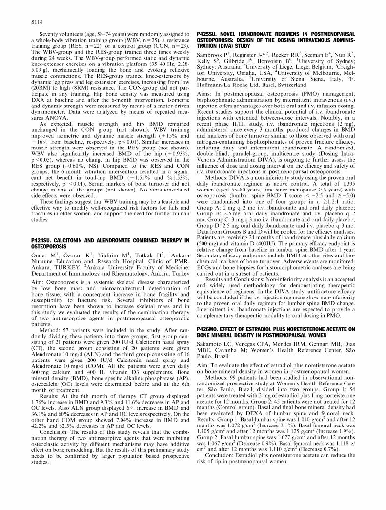
Seventy volunteers (age, 58–74 years) were randomly assigned toa whole-body vibration training group (WBV, n=25), a resistancetraining group (RES, n=22), or a control group (CON, n=23).The WBV-group and the RES-group trained three times weeklyduring 24 weeks. The WBV-group performed static and dynamicknee-extensor exercises on a vibration platform (35–40 Hz, 2.28-5.09 g), mechanically loading the bone and evoking reflexivemuscle contractions. The RES-group trained knee-extensors bydynamic leg press and leg extension exercises, increasing from low(20RM) to high (8RM) resistance. The CON-group did not par-ticipate in any training. Hip bone density was measured usingDXA at baseline and after the 6-month intervention. Isometricand dynamic strength were measured by means of a motor-drivendynamometer. Data were analyzed by means of repeated mea-sures ANOVA.
As expected, muscle strength and hip BMD remainedunchanged in the CON group (not shown). WBV trainingimproved isometric and dynamic muscle strength (+15% and+16% from baseline, respectively, p<0.01). Similar increases inmuscle strength were observed in the RES group (not shown).WBV also significantly increased BMD of the hip (+0.93%,p<0.05), whereas no change in hip BMD was observed in theRES group ()0.60%, NS). Compared to the RES and CONgroups, the 6-month vibration intervention resulted in a signifi-cant net benefit in total-hip BMD (+1.51% and %1.53%,respectively, p <0.01). Serum markers of bone turnover did notchange in any of the groups (not shown). No vibration-relatedside effects were observed.
These findings suggest that WBV training may be a feasible andeffective way to modify well-recognized risk factors for falls andfractures in older women, and support the need for further humanstudies.
P424SU. CALCITONIN AND ALENDRONATE COMBINED THERAPY INOSTEOPOROSIS
Onder M1, Ozoran K1, Yildirim M1, Tutkak H2; 1AnkaraNumune Education and Research Hospital, Clinic of PMR,Ankara, TURKEY, 2Ankara University Faculty of Medicine,Department of Immunology and Rheumatology, Ankara, Turkey
Aim: Osteoporosis is a systemic skeletal disease characterizedby low bone mass and microarchitectural deterioration ofbone tissue, with a consequent increase in bone fragility andsusceptibility to fracture risk. Several inhibitors of boneresorption have been shown to increase skeletal mass and inthis study we evaluated the results of the combination therapyof two antiresorptive agents in postmenopausal osteoporoticpatients.
Method: 57 patients were included in the study. After ran-domly dividing these patients into three groups, first group con-sisting of 21 patients were given 200 IU/d Calcitonin nasal spray(CT), the second group consisting of 20 patients were givenAlendronate 10 mg/d (ALN) and the third group consisting of 16patients were given 200 IU/d Calcitonin nasal spray andAlendronate 10 mg/d (COM). All the patients were given daily600 mg calcium and 400 IU vitamin D3 supplements. Bonemineral density (BMD), bone spesific alkaline phosphatase (AP),osteocalcin (OC) levels were determined before and at the 6thmonth of treatment.
Results: At the 6th month of therapy CT group displayed1.76% increase in BMD and 9.3% and 11.6% decreases in AP andOC levels. Also ALN group displayed 6% increase in BMD and36.1% and 60% decreases in AP and OC levels respectively. On theother hand COM group showed 7.04% increase in BMD and42.2% and 62.5% decreases in AP and OC levels.
Conclusion: The results of this study reveals that the combi-nation therapy of two antiresorptive agents that were inhibitingosteoclastic activity by different mechanisms may have additiveeffect on bone remodeling. But the results of this preliminary studyneeds to be confirmed by larger populaton based prospectivestudies.
P425SU. NOVEL IBANDRONATE REGIMENS IN POSTMENOPAUSALOSTEOPOROSIS: DESIGN OF THE DOSING INTRAVENOUS ADMINIS-TRATION (DIVA) STUDY
Sambrook P1, Reginster J-Y2, Recker RR3, Seeman E4, Nuti R5,Kelly S6, Gilbride J6, Bonvoisin B6; 1University of Sydney;Sydney; Australia; 2University of Liege, Liege, Belgium, 3Creigh-ton University, Omaha, USA, 4University of Melbourne, Mel-bourne, Australia, 5University of Siena, Siena, Italy, 6F.Hoffmann-La Roche Ltd, Basel, Switzerland
Aims: In postmenopausal osteoporosis (PMO) management,bisphosphonate administration by intermittent intravenous (i.v.)injection offers advantages over both oral and i.v. infusion dosing.Recent studies support the clinical potential of i.v. ibandronateinjections with extended between-dose intervals. Notably, in arecent phase II/III study, i.v. ibandronate injections (2 mg),administered once every 3 months, produced changes in BMDand markers of bone turnover similar to those observed with oralnitrogen-containing bisphosphonates of proven fracture efficacy,including daily and intermittent ibandronate. A randomised,double-blind, parallel-group, multicentre study (Dosing Intra-Venous Administration: DIVA), is ongoing to further assess theinfluence of dose and dosing interval on the efficacy and safety ofi.v. ibandronate injections in postmenopausal osteoporosis.
Methods: DIVA is a non-inferiority study using the proven oraldaily ibandronate regimen as active control. A total of 1,395women (aged 55–80 years, time since menopause ‡ 5 years) withosteoporosis (lumbar spine BMD T-score < )2.5 and ‡ )5.0)were randomised into one of four groups in a 2:1:2:1 ratio:Group A: 2 mg q 2 mo i.v. ibandronate and oral daily placebo;Group B: 2.5 mg oral daily ibandronate and i.v. placebo q 2mo; Group C: 3 mg q 3 mo i.v. ibandronate and oral daily placebo;Group D: 2.5 mg oral daily ibandronate and i.v. placebo q 3 mo.Data from Groups B and D will be pooled for the efficacy analyses.Patients are receiving 24 months of ibandronate plus daily calcium(500 mg) and vitamin D (400IU). The primary efficacy endpoint isrelative change from baseline in lumbar spine BMD after 1 year.Secondary efficacy endpoints include BMD at other sites and bio-chemical markers of bone turnover. Adverse events are monitored.ECGs and bone biopsies for histomorphometric analyses are beingcarried out in a subset of patients.
Results and Conclusions: Non-inferiority analysis is an acceptedand widely used methodology for demonstrating therapeuticequivalence of regimens. In the DIVA study, antifracture efficacywill be concluded if the i.v. injection regimens show non-inferiorityto the proven oral daily regimen for lumbar spine BMD change.Intermittent i.v. ibandronate injections are expected to provide acomplementary therapeutic modality to oral dosing in PMO.
P426MO. EFFECT OF ESTRADIOL PLUS NORETISTERONE ACETATE ONBONE MINERAL DENSITY IN POSTMENOPAUSAL WOMEN
Sakamoto LC, Venegas CPA, Mendes IRM, Gennari MB, DiasMBE, Cavanha M; Women’s Health Reference Center, SaoPaulo, Brazil
Aim: To evaluate the effect of estradiol plus noretisterone acetateon bone mineral density in women in postmenopausal women.
Methods: 99 patients had been studied in observational non-randomized prospective study at Women’s Health Reference Cen-ter, Sao Paulo, Brazil, divided into two groups. Group 1: 54patients were treated with 2 mg of estradiol plus 1 mg norteisteroneacetate for 12 months. Group 2: 45 patients were not treated for 12months (Control group). Basal and final bone mineral density hadbeen evaluated by DEXA of lumbar spine and femoral neck.Results: Group 1: Basal lumbar spine was 1.040 g/cm2 and after 12months was 1.072 g/cm2 (Increase 3.1%). Basal femoral neck was1.105 g/cm2 and after 12 months was 1.125 g/cm2 (Increase 1.9%).Group 2: Basal lumbar spine was 1.077 g/cm2 and after 12 monthswas 1.067 g/cm2 (Decrease 0.9%). Basal femoral neck was 1.118 g/cm2 and after 12 months was 1.110 g/cm2 (Decrease 0.7%).
Conclusion: Estradiol plus noretisterone acetate can reduce therisk of rip in postmenopausal women.
S118
P427SA. DMARDS TREATMENT AND BONE MINERAL DENSITY INPATIENTS WITH RHEUMATOID ARTHRITIS
Taukumova LA, Smirnov AV; Institute of Rheumatology,Moscow, Russia
Objective: To investigate the influence of DMARDs treatment onbone mineral density (BMD) and in patients (pts) with rheuma-toid arthritis (RA).
Methods: 109 active RA pts (97 women and 12 men) with meanage 55.6, mean disease duration 14.7, mean number of previousDMARDs 3.6 were included in this study. All patients were takinga DMARDs during of 1 year observation: Methotrexate (MTX) in35%, Sulfasalazine (SF) in 9%, a intramuscular gold salts (IMG)in 10%, a steroids in 13%, combination of a DMARD and steroidsin 26% cases. 51 pts (46.7%) were taking a steroids as the onlymedication and as in combination with a DMARD. BMD atlumbar spine (L1–L4), at the left hip (femoral neck and Ward’striangle) and at the right forearm was measured with DEXA. BMDwas assessed at baseline and after 1 year of DMARDs treatment.
Results: At baseline an osteoporosis at L1–L4 was found in18.6% pts, osteopenia ) in 28.8% pts, at the femoral neck – in19.5% and 29.6%, Ward’s triangle – in 33.1% and 33.9%,respectively. BMD was not changed in 1 year observation. IMGwere significantly delayed an osteoporosis progression at all sites,but higher at femoral neck and Ward’s triangle in comparison withMTX (p<0.001 and p<0.001), SF (p<0.003 and p<0.0003),steroids (p<0.04 and p<0.002) and its combination withDMARD (p<0.001 and 0.001), respectively. A steroids weresignificantly more decreased BMD at femoral neck (p<0.03).However, a combination of DMARD and steroids were notnegatively influenced on BMD at any site.
Conclusion: DMARDs treatment was delayed the rate of BMDloss, an IMG have demonstrated more substantial effect. A steroidsin combination with a DMARD can achieve a positive influence ondisease activity and bone mass. An efficacy of DMARDs treatmentis a strong reserved factor of osteoporosis.
P428SU. PATIENT EDUCATION PRACTICES AMONG BONE DENSI-TOMETRY TECHNOLOGISTS
Ludwig RL1, Turner LW2, O’sullivan PS3; 1University ofArkansas for Medical Sciences, Little Rock, AR, USA, 2Univer-sity of Arkansas, Fayetteville, AR, USA, 3University of Arkansasfor Medical Sciences, Little Rock, AR, USA
Aims: The Osteoporosis Counseling Survey (OCS) was adminis-tered to ionizing bone densitometry technologists to assess theirknowledge about osteoporosis, beliefs about the disease, attitudestowards osteoporosis preventive care, and bone health promotioncounseling habits. The descriptive study was conducted to analyzepredictors for osteoporosis prevention patient education.Increasing counseling maximizes the potential for health benefitsfrom medical care for patients at risk.Methods: The sample consisted of 158 bone densitometryequipment operators in Arkansas and 417 radiologic technolo-gists (RTs) registered by the American Registry of RadiologicTechnologists (ARRT) with advanced certification in bone den-sity (RTBDs). The study subjects performing bone density examsin Arkansas were health professionals such as nurses and RTs.Although no RTs registered by the ARRT in bone densitometry(RTBDs) were employed in Arkansas, 14 responders in Arkansaswere clinical densitometry technologists (CDTs) certified by theInternational Society for Clinical Densitometry (ISCD).Results: Response rates were 52% from the Arkansas sample and60% from the national RTBD sample, for 58% overall. Althoughthe responders reported counseling a majority of their patientsabout osteoporosis prevention (mean 82%+3.6), they appearedto define patient counseling in different ways. BD certified pro-viders were more likely than non-certified providers to havehigher internal motivation and perceptions of fewer barriers tohealth counseling. Providers with high internal motivation andlow personal barriers were more likely to counsel their patientsthan providers with low personal interest and greater concerns
related to having inadequate knowledge, time, confidence, orencouragement to engage in counseling. Knowledge, beliefs, andattitudes accounted for 25% of the variation in the regressionmodel, while another 16% of the variation related to the age ofthe provider, certification in BD, and employment where patientswere assessed for their health behaviors related to osteoporosis.Conclusions: Recommendations for increasing bone health pro-motion during densitometry exams include increasing equipmentoperator motivation to provide health education through sensi-tivity training, promoting models for clinical integration of pre-ventive services, and creating educational opportunities tostrengthen counseling skills. Future studies should more clearlydefine health counseling or incorporate measures for counselingto increase reporting accuracy.
P429MO. BISPHOSPHONATES ANTALGIC ACTIVITY IN RECENT VER-TEBRAL FRACTURE: A CLODRONATE VS NERIDRONATE COMPARISON
Muratore M, Calcagnile F, Serra M, Falappone P, Carlino G,Casentino L, Quarta E; UO di Reumatologia, Ospedale ‘‘A.Gala-teo’’, S.Cesario di Lecce (LE)
Fractures are the most common and dangerous osteoporosisinvolvement. A vertebral breakdown, by stimulating one or morealgogenic structure, will decrease the motility of involved skeletalsections, compress intervertebral discs and/or cause interapo-physary distractions, responsible of a further myoceptive stimu-lation. Paravertebral muscles splinting brings about lumbarstiffness and antalgic postures; during vertebral pain severalphysiopathologic mechanisms cause asymmetric effects thatworsen pelvis motility and posture. These are the pathogeneticconditions that cause an alteration of either static or dynamicpostural balancing of foot rest during lumbar pain. Pressure onfeet and length of the step will change due to an antalgic actionand a lower pressure will be applied on the foot in the painful site.Objective of this study is to evaluate if lumbar or sacral pain isable to cause an alteration of foot rest and if pain improvement isrelated with a walk and foot rest modification. The previouslyknown Clodronate (Difosfonal) antalgic activity was compared toNeridronate (Nerixia) one. 16 women (mean age 72 years) withrecent dorsal or lumbar pain due to a vertebral fracture wereenrolled in the study. In any patient with radiologic diagnosis ofvertebral fracture, the following evaluations at T0 (baseline), T1(15 days), and T2 (40 days), were performed, clinical examination,subjective evaluation of pain on visual analogic scale, podometricexamination of static foot rest and walk dynamics, performedwith ‘‘DINATTO 2,5’’ machine. The patients were randomized toreceive 300 mg/die i.v. of Clodronate in 250 cc of physiologicalsaline during first 10 days and then 100 mg i.m. of Clodronate any3 days up to day 30 (CLD group), or 100 mg i.v. of Neridronatein 250 cc of physiological saline at days 1 and 30 (NER group).The study suggests that only Clodronate performs a doubletherapeutic action; in patients with vertebral fracture, Clodronatesupplies an anti-osteoporotic action, by reducing bone resorptionand a basic antalgic action, improving the highly painful symp-tomatology and allowing a reduction in NSAID intake.
P430SA. STRONTIUM RANELATE REDUCES THE RISK OF VERTEBRALFRACTURES IN POSTEMENOPAUSAL WOMEN WITH OSTEOPENIA
Sawicki A1, Reginster JY2, Roux C3, Rubinacci A4, Diaz-CurielM5, Kaufman J6, Seeman E7, De Vernejoul MC8, Aquino JP9,Meunier PJ10; 1Warszawskie Centrum Osteoporozy, Warsaw,POLAND, 2Dept of Epidemiology and Public Health, Universityof Liege, Liege, BELGIUM, 3Hopital Cochin, Paris, FRANCE,4Bone Metabolic Unit, Scientific Inst. H. San Raffael, Milano,ITALY, 5Servicio de Medicina Interna, Fundacion Jimenez Diaz,Madrid, SPAIN, 6Polyclinique d’Endocrinologie, Gent Univer-sity Hospital, Gent, BELGIUM, 7Endocrine Unit, AugustineHospital, Melbourne, AUSTRALIA, 8Hopital Lariboisiere,Paris, FRANCE, 9Clinique Medicale de la Porte Verte, Versailles,FRANCE, 10Dept of Rheumatology and Bone Diseases, EdouardHerriot Hospital, Lyon, FRANCE
S119
Strontium ranelate 2 g/day is an orally active anti-osteoporoticagent which reduces over 3 years the risk of vertebral fractures by41%, non-vertebral fractures by 16% and hip fractures by 41% inpostmenopausal women with osteoporosis based on results of aphase III program including 2 international randomised, doubleblind, placebo controlled clinical studies: SOTI (1649 patientswith low lumbar BMD and having at least one prevalent vertebralfracture) and TROPOS (5091 patients with low femoral neckBMD).
An analysis was performed on the pooled data from SOTI andTROPOS studies. Amongst the whole population, 409 patientswith lumbar and/or femoral neck T-score between )1 and )2.5 andboth T-scores >)2.5, with or without prevalent fractures wereincluded and received strontium ranelate 2 g/d orally or placebofor 3 years, associated to calcium and vitamin D supplementationaccording to the patient’s status. Vertebral X-rays were performedyearly.
No relevant differences between groups were detected for themain baseline characteristics: mean(SD) age: 73(6) years; time sincemenopause: 25(8) years; mean(SD) Lumbar T-score: )1.20(1.15);mean(SD) Femoral Neck T-score: )2.06(0.44).In the intent-to-treat population, strontium ranelate was associ-ated with a 62% reduction in the relative risk of vertebral fractureover 3 years (as assessed through semi-quantitative method by acentral reading centre) (RR=0.38, 95%CI[0.21;0.70], p=0.001).Amongst the 409 described patients, 43% of patients presented anosteopenia according to their BMD values (described above) andhad no prevalent fracture. In this subgroup, strontium ranelatereduced the risk of vertebral fracture by 72% over 3 years(RR=0.28; 95% CI [0.07; 0.99]) (p= 0.045).
We infer that strontium ranelate, a new anti-osteoporotic agent,reduces the risk of vertebral fractures in women with osteopenicrange of BMD by 62% and in women with osteopenia without anyprevalent fracture by 72%.
P431SU. THE HORIZON-TOP TRIALS PROGRAM: A RANDOMIZED,DOUBLE-BLIND COMPARISON OF ZOLEDRONIC ACID AND RISEDRO-NATE IN THE TREATMENT OF PAGET’S DISEASE OF BONE
Saidi Y1, Pak J2, Zelenakas K2, Fashola T1, Luchi M2, Su G2;1Novartis Pharma AG, Basel, Switzerland, 2Novartis Pharma-ceuticals Corporation, East Hanover, New Jersey, USA
Bisphosphonates are the mainstay for the treatment of Paget’sdisease of bone (PDB). The ongoing Health Outcomes andReduced Incidence with Zoledronic Acid Once Yearly – Treat-ment Of Paget’s (HORIZON-TOP) program was designed todemonstrate non-inferiority of zoledronic acid (ZA; a potent thirdgeneration, N-containing bisphosphonate) to risedronate (RIS)for the treatment of PDB. HORIZON-TOP is the largest programto date in PDB and comprises two similarly designed, interna-tional, multi-center, randomized, double-blind trials. 357 patientswere randomized to either one ZA 5 mg i.v. infusion (15 minutes)and 2 months daily oral placebo, or one i.v. placebo infusion and2 months daily oral RIS 30 mg, and followed for 6 months. Theprimary endpoint is the proportion of patients achieving a ther-apeutic response, defined as a reduction of at least 75% frombaseline in serum alkaline phosphatase (sALP) excess or sALPnormalization at the end of six months. The secondary efficacyendpoints are reductions in serum C-telopeptide and urine alphaC-telopeptide levels and reductions in pain severity and interfer-ence as measured by the Brief Pain Inventory. Additionalexploratory efficacy parameters are propeptide of N-terminal typeI collagen, physical and mental quality of life and gait speed.Safety evaluations include the monitoring of clinical and labora-tory adverse events. Following the 6-month trial period,responders (patients who achieve the primary endpoint) wereoffered the opportunity to enter an extension observation period,during which the sALP level is monitored every 6 months until itreturns to within 20% of the baseline value. Therefore, the po-tential for rapid and sustained remission in PDB with ZA will beevaluated by serial monitoring of biochemical markers of boneturnover. Patient follow-up will continue until offset of effect is
observed. Furthermore, this trial will provide an opportunity toassess the acceptability and convenience of i.v. bisphosphonateadministration to patients, compared with oral dosing.
P432MO. KYPHOPLASTY IN THE TREATMENT AND PREVENTION OFCOMPRESSION VERTEBRAL FRACTURES
Cesaroni A1, Nardi PV1, Masala S2, Simonetti G2; 1PoliclinicoCasilino Neurochirurgia, Roma, Italy, 2Universita Tor VergataRadiologia, Roma, Italy
In the management of osteoporotic vertebral fractures Kyph-hoplasty is presently able to solve the spinal deformation and thepainful symptomatology and it can also prevent further bone lossand fracture, but the increased risk of this complication is not wellevaluated. From November 2002 to November 2003 we havetreated 78 patients with vertebral compression fractures (VCF) dueto osteoporosis, in 25 cases there were present two or more verte-bral body fractures. All the patients underwent percutaneoustreatment with Kyphoplasty in a period ranging from 30–180 daysafter VCF. In 46 patients treated early (30–60 days after VCF) wehave not found new fractures at the follow up, on the other side in32 patients treated after more than 90 days from VCF, we havenoted a new vertebral fracture at the short-term follow up (1–3months) in 12 of them. All the new fractures are present in a lowerlevel of the column with respect to prior treatment; 8 of them havehad two or more vertebral fractures. In these cases we have pro-ceeded to a new Kyphoplasty and no other VCF were found at thistime.
The presence of one or more vertebral fractures increase risk ofsustaining a vertebral fracture by 5 fold per year and the earlytreatment is basic in the prevention of further VCF.
Kyphoplasty is in our experience a valid treatment option bothto solve the vertebral fracture and to prevent new VCF.
P433SA. A FIVE-YEAR ALENDRONATE TREATMENT OF POSTMENO-PAUSAL OSTEOPOROSIS IN PATIENTS WITH IMPAIRED FASTINGGLUCOSE
Cokolic M1, Hren R2; 1Department of Endocrinology andDiabetology, Internal Clinic, Teaching Hospital, Maribor, Slove-nia, 2Institute of Mathematics, Physics, and Mechanics, Univer-sity of Ljubljana, Ljubljana, Slovenia
Background and Aims: While osteoporosis can nowadays beeffectively treated, it is important that osteoporosis treatmentregimen does not worsen patients concomitant diseases. The aimof this study was to monitor in patients with impaired fastingglucose (IFG: serum levels of fasting glucose ‡6.1 and £6.9mmol/l) that are treated for postmenopausal osteoporosis bothbone mineral density (BMD) and levels of blood glucose andHbA1c.
Material and Methods: Eleven women with postmenopausalosteoporosis (T-score below )2.5 SD) and IFG were enrolled in afive-year prospective study. Patients age was 59 to 74 years old(mean: 65 years) and 8 to 25 years (mean: 16 years) after themenopause. They were treated with alendronate (10 mg/d duringthe first four years, 70 mg/w during the fifth year) in combinationwith 500-mg/d elemental calcium. During the five-year follow-up,the BMD in the lumbar spine (L1–L4) and left hip was measured inall patients using dual energy X-ray densitometry (Hologic QDR2000+). The serum levels of glucose, HbA1c, Ca, alkaline phos-phatase (ALP) and creatinine were measured every 6 months. Allpatients were treated for IFG only with diabetic diet.
Results: In 5 years, BMD increased on average by 8.1 % (range0.2–9.6 %) in the lumbar spine (L1–L4) and by 4.8 % (range 0.5–8.7 %) in the left hip. Levels of Ca, ALP and creatinine were withinnormal limits during the treatment, and no clinical side effects wereobserved during the study. Serum levels of fasting glucose showedno statistically significant changes during the alendronate treat-ment, with an average value of 6.3 mmol/l (range 6.1–6.6 mmol/l)in the beginning of the treatment and 6.5 mmol/l (range 6.4–6.8mmol/l) after 5 years of the treatment. Similarly, the average level
S120
of HbA1c was 6.5 % (range 6.2–6.6 %) at the start of the treatmentand 6.7 % (range 6.2–6.8 %) after 5 years of the treatment.
Conclusions: Results of our study indicate that osteoporosiscan be effectively treated in patients with IFG for up to five years,while maintaining (with little variation) serum levels of fastingglucose and HbA1c.
P434SU. EFFICACY OF INTRAMUSCULAR CLODRONATE (DIFOSFONAL)IN PATIENTS WITH COLLES’ FRACTURE
Priano F1, Versace F1, Coari GC2; 1Casa di cura ‘‘VillaMontallegro’’, Genova, Italy, 2Casa di cura ‘‘S. Camillo’’, Fortedei Marmi, Lucca, Italy
The recent literature underlined the efficacy of the bisphospho-nates on mineralisation of bone. However only clodronate has aconsiderable antalgic effect without any interference on callus re-pair after Colles’ fracture. In a randomized and prospective studywe studied 40 postmenopausal women whether 12 weeks treatedwith clodronate 100 mg/d i.m. for the first week and that 100 mgi.m./weekly (clodronate group) or calcium 1000 mg and 880 UIvitamin D3 (placebo group). Treatment was given for 12 weeks.The bone mineral density (BMD) of the forearm was measuredwith DEXA (Hologic 1000W) at 1, 3, 6 and 12 months after thefracture. The efficacy of intramuscular clodronate on minerali-sation of fracture callus in this study was substantial, the BMDhaving increased by 23% after 3 months of clodronate treated.The intramuscular clodronate was usually well tolerated. Inconclusion the pharmacologic features of the intramuscularclodronate offer new opportunities to the treatment for the min-eralisation of callus after Colles’ frecture. Intramuscular clodro-nate was proved to have a peculiar and fast activity on bone pain.
P435MO. ANTALGIC EFFICACY OF THE BISPHOSPHONATES IN VER-TEBRAL FRACTURES OF RECENT ONSET
Muratore M, Calcagnile F, Quarta E; U.O. Reumatologia,Ospedale Galateo, ASL LE1 S. Cesario di Lecce, Lecce, Italy
The effectiveness of the bisphosphonates in increasing bone massand in reducing the risk of fractures in patients suffering fromosteoporosis has been widely documented in the literature. Thesame cannot absolutely be said as regards their possible antalgiceffectiveness. The authors therefore assess the reduction in pain inosteoporotic patients suffering from vertebral fracture on recentonset, treated with 3 different bisphosphonates (clodronate,alendronate, risedronate). Apart from the traditional visual ana-logic scale, foot support static analysis and step dynamics wereemployed using a computerized objective method. The resultsobtained confirmed that i.m. clodronate (Difosfonal) leads to amarked reduction in pain symptomatology, unlike alendronateand risedronate. It can be stated that clodronate presents note-worthy antalgic activity compared to alendronate and risedro-nate, reducing the extremely intense and frequent painsymptomatology in the most feared complication of osteoporosis.
P436SA. THE EFFECTIVENESS OF ALENDRONATE, RISEDRONATE ANDCALCITONIN TREATMENT IN POSTMENOPAUSAL OSTEOPOROSIS
Yildirim K, Gureser G, Karatay S, Senel K; Ataturk University,Medicine Faculty, Physical Medicine and Rehabilitation, Erzu-rum, Turkey
The aim study was to investigate the efficacy of alendronate,risedronate and calcitonin in the treatment of postmenopausalosteoporosisFifty four patients with postmenopausal osteoporosis wererandomly assigned into equal 3 treatment groups. First groupreceived 10 mg/day alendronate, second group were treated with5mg/day risedronate, three group received 200 IU/day calcitonin.All groups received a daily supplement of 1000 mg calcium.Treatment regimens were applied for 12 months. Dual energyX-ray absorptiometry (DXA) was used for the measurement ofbone mineral density (BMD) of the lumbar spine and left hipbefore and after the study period.
In the first group, a significant increases in lumbar spine (L1-L4)and left hip (total) BMD was detected in patients with post-menopausal osteoporosis at the end of first year (p<0.03,p<0.001 respectively). A significant increase both regions deter-mined in the second group at the end of first year (p<0.001,p<0.01 respectively). In the three group, a significant increases inboth sites was observed in the patients at the end of first year(p<0.05, p<0.01 respectively).This study clearly demonstrates the efficacy of alendronate,risedronate and calcitonin in preventing the bone loss related topostmenopausal osteoporosis.
P437SU. TWO YEARS EXPERIENCE IN THE TREATMENT OF POST-MENOPAUSAL OSTEOPOROSIS WITH ALFACALCIDOL
Galesanu C1, Galesanu MR2; 1University of Medicine andPharmacy, Iasi, Romania, 2Radiological and Imagistic Center,Iasi, Romania
Postmenopausal osteoporosis is characterised by an increasedresorption of trabecular bone mainly due to a higher respon-siveness of osteoblastes to parathormone (PTH) action, a conse-quence of estrogen deficit. Increased resorption results in reducedsecretion of PTH, reduced renal activation of the 25-OH-chole-calciferol in 1.25 (OH2D3 and a reduction of the absorption ofintestinal calcium. Treatment with alfacalcidol is physiological; ithas been shown to be effective in increasing intestinal absorptionof calcium and reducing the loss of bone mass.
Aims: To determine the efficacy of alfacalcidol (Alpha D3�) on
bone mass.Method: 1465 patients had bone densitometry performed using
DXR-BMD (Pronosco-X-posure System). From among these 1018were in postmenopause: inclusion criteria were healthy women withosteopenia: 391 patients (T score: )1 to )2.4) and osteoporoticwomen: 165 patients (T-score lower than )2.5). The other 462women were represented by the control group. The treatment: thegroup with osteopenia received 0.5 lg alfacalcidol/day, and thegroup with osteoporosis 1 lg alfacalcidol/day. After 12 and 24months all patients ware evaluated for change in BMD.
Results: In the group with osteopenia the BMD increased by3.4%and 2.3%after one and two year respectively. In the groupwithosteoporosis, BMD increased by 1.8% and 2.4% after one and twoyears respectively. The decreased of BMD in control group wasbetween 1.8%and 3.4% in the first year and second year respectively.
Conclusions: Alfacalcidol confirmed the abolishment of the lossof bone mass in our study, and also increased the bone mass densitysignificantly compared with the control group.
P438MO. ORAL IBANDRONATE PRODUCES SIGNIFICANT ANTI-FRACTURE EFFICACY WHEN ADMINISTERED LESS FREQUENTLY THANCURRENT BISPHOSPHONATES
Chesnut III CH1, Stakkestad JA2, Recker RR3, Gilbride J4,Schimmer RC4; 1University of Washington, Seattle, USA,2CECOR AS, Haugesund, Norway, 3Creighton University,Omaha, USA, 4F. Hoffmann-La Roche Ltd, Basel, Switzerland
Aims: Patients taking current oral bisphosphonates must followstringent dosing guidelines every day or week. This may causeinconvenience, thereby reducing therapeutic compliance and re-sulting in suboptimal clinical outcomes. Ibandronate is a potentnitrogen-containing bisphosphonate that has been designed toaddress unmet needs in osteoporosis management, namely dosingsimplicity, leading to greater dosing convenience. A multinational,phase III study recently examined the efficacy and tolerability of anintermittent ibandronate regimen given with an extended between-dose interval of >2 months (oral iBandronate Osteoporosisvertebral fracture trial in North America and Europe: BONE).
Methods: In this study, postmenopausal women (aged 55–80years, ‡ 5 years postmenopause) with osteoporosis (BMD T-score £)2.0 at the lumbar spine in ‡1 vertebra [L1-L4], 1-4 prevalentvertebral fractures [T4–L4]) were randomised to receive placebo(n=982) or oral intermittent ibandronate (20mg every other day
S121
for 12 doses every 3 months; n=982) or oral daily (2.5mg; n=982)ibandronate for 3 years. All participants received daily calcium(500 mg) and vitamin D (400IU) supplementation.
Results: After 3 years, oral intermittent ibandronate sig-nificantly increased lumbar spine and total hip BMD (5.7% and2.9%, respectively; p<0.0001 for both sites versus baseline), sig-nificantly decreased biochemical markers of bone resorption(p<0.0001) and formation (p<0.0001), significantly decreasedheight loss (p=0.0144) and significantly reduced the risk of newvertebral fractures (by 50%; p=0.0006), relative to placebo. This isthe first time that a bisphosphonate has prospectively demonstratedantifracture efficacy in a regimen with a dosing interval >2months, in the overall population of a well-designed trial. Oralintermittent ibandronate was well tolerated, with an incidence ofadverse events similar to placebo. This is notable as: one third ofpatients had pre-existing upper GI disorders; patients received acumulative ibandronate dose of 240 mg in just 24 days.
Conclusions: The robust findings from this study demonstratethat ibandronate is highly effective when given in less frequentregimens than once weekly. As a result, a large trial has beeninitiated to investigate a simple, once monthly oral ibandronateregimen, which is predicted to enhance patient management inpostmenopausal osteoporosis by optimising dosing convenienceand consequently improving treatment adherence.
P439SA. USE IN HCV-HIV-INFECTED WITH BONE MASS LOSS OFCALCITONIN AND ALENDRONATE
Bazarra-Fernandez A;University of La Coruna, La Coruna, Spain
Introduction: Hepatitis C virus (HCV) is an RNA virus is a majorcause of chronic hepatitis, cirrhosis, and hepatocellular carci-noma. Owing to shared routes of transmission, HCV and humanimmunodeficiency virus (HIV) coinfection are common, affectingapproximately one-third of all HIV-infected persons. Low bonemineral density may be yet another common adverse effect ofprotease inhibitor combination therapy.
Objective: Determining if the combined use of calcitonin andalendronate influences on bone mass loss.
Material and method: We studied for 6 months 21 women whowere 44 to 64 years old at base line, were within 2 and 11 years ofmenopause, and had a bone mineral density at the lumbar spinebetween 145 mg/cc and 50 mg/cc measured by the QBMAP systemwith a spiral CT Picker PQ-S densitometer at L2, L3, L4 and L5.Of all the women, 10 were assigned to 10 mg of alendronate, 800 IUof vitamin D3 and 1 g of calcium carbonate supplementation. 11were treated with 10 mg of alendronate, 200 IU of intranasal cal-citonin, 800 IU of vitamin D3 and 1 g of calcium carbonate sup-plementation. The SPSS programme was used for statiscal analysis.
Results: The characteristics of the women recruited for bothgroups were similar. Mean mineral bone density at the lumbarspine was between 1 and 3 DS below the mean value for 30 yearsold normal premenopausal women. After a treatment of 12 monthsno statistically significant difference was found among both groupsas for the bone mineral density at the lumbar spine.
Conclusions: It is necessary to carry out a wider and longerstudy, among
HIV-HCV patients, but it seems that alendronate contributeadvantages to decrease bone mass loss, at least, at lumbar spine,without calcitonin. Osteoporosis is a multifactorial disease, maybeits best treatment and prevention is combining several drugs andattitudes. It would be good to test several adjusted doses todecrease side effects. These results can be interesting for HIV-HCVinfected, who are prescribed a lot of medication.
P440SU. PULSED SIGNAL THERAPY (PST) FOR THE TREATMENT OFOSTEOPOROSIS
Markoll R, Da Silva Ferreira DM; infinomed, Munich, Germany
Aims: Biophysically, it is known that bone possesses electrome-chanical properties and natural biopotentials essential in boneremodeling. Pioneers, Yasuda, Bassett and others, observed thatrepair and adaptive remodeling processes, occurred in response to
mechanical loading and that such responses could be elicited byan electrical stimulus – an exogenously applied electrical current,including PEMF.
Methods: Recently, science has focused on the biomolecularproperties of bone and cartilage, and the similarity of their lineages,such that reconstitution of cartilage has been found to essentiallyparallel that of bone. BMSCs (bone marrow stromal cells) havebeen shown to differentiate into osteoblasts, chondrocytes andspecialized connective tissue cells, giving rise to skeletal tissues.Additionally, several factors, including growth factors, affectBMSC proliferation rate and osteogenic, and/or chondrogenic,potential, and FGF2 has been shown to maintain BMSCs in animmature state as chondro-osteo-progenitor cells. The latestresearch findings have identified Cbfa1 as a late transcription factorin both bone and cartilage development.
Results: Biophysically, it has been established that PST� emu-lates the innate physiological and mechanical stresses evoked, andrequired, in bone formation. It passively induces fluid flow andionic displacement, thereby generating a piezoelectric (‘‘streamingpotential’’) and eventually activating various signaling networkpaths – as inmechanotransduction. Basically, a specific pulsed signalis carried on an ELF electromagnetic field, and transferred to boneand through adjacent tissue, resulting in reconstitution of the dis-turbed electrical field, reestablishment of innate regenerative pro-cesses, and reactivation of cartilage, bone, and other connectivetissue.Followingmore than25 years of PST� success in the treatmentof connective tissue disorders, a pilot study on postmenopausalwomen with osteoporosis was initiated. Preliminary results havedemonstrated a significantly increasing trend in vBMD (namely,trabecular bone density) post-PST� and an associated decreasein pain. International medical regulatory approval for the treatmentof osteoporosis with PST� technology was granted in 2003.
Conclusion: PST� has already been shown to increase collagenlevels, and other matrix components in cartilage, such that proto-cols to investigate PST� anabolic effects in bone, by increasingmRNA expression levels (BMP-2 and BMP-4 by rtPCR, forexample), are being developed.
P441MO. EFFICACY OF INTRAMUSCULAR CLODRONATE IN PATIENTSWITH COLLES’ FRACTURE
Priano F1, Versace F1, Coari GC2; 1Casa di cura ‘‘VillaMontallegro’’, Genova, Italy, 2Casa di cura ‘‘S. Camillo’’, Fortedei Marmi, Lucca, Italy
Recent literature underlined the efficacy of bisphosphonates onmineralisation and resorption of bone. However only clodronatehas a considerable antalgic effect without any interference oncallus repair after Colles’ fracture. In a randomized and pro-spective study of 40 postmenopausal women with Colles’ fracturewe studied wthether 12 weeks of treatment with intramuscularclodronate (Difosfonal). The patients were randomized into 2groups: CLD group (100 mg/d of im clodronate for the first weekand that 100 mg weekly) and PLC group (1000 mg of calciumand 800 IU of vitamin D3/d). The BMD (bone mineral density) ofthe forearm bones was measured with DEXA (Hologic 1000 W)1, 3, 6 and 12 months after the fracture. The pharmacologicfeatures of the intramuscular clodronate offer new opportunitiesto the treatment for the mineralisation of callus after Colles’fracture, and clodronate was proved to have a peculiar and fastactivity on bone pain: a 100 mg/d of im clodronate regimen hasshown to remarkably relieve patients from pain in the first weekof administration while reducing analgesics consumption to agreat extent if compared to the placebo group.
P442SA. INTERDISCIPLINARY APPROACH TO VERTEBROPLASTY ANDBALLOON KYPHOPLASTY IN THE TREATMENT OF OSTEOPOROTICVERTEBRAL COMPRESSION FRACTURES
Franck H, WZ-Kliniken; Center of Rheumatology, Oberammergau,Germany
Aim: Osteoporotic vertebral compression fractures (VCFs) areassociated with a series of clinical consequences leading to
S122
increased morbidity and even mortality. Traditional medicaloptions including bed rest, analgesics and bracing have proven tobe insufficient. Furthermore, the problem of osteoporosis isunderestimated and often not diagnosed. Therefore, early diag-nosis and therapeutic intervention is desirable in order to remo-bilise patients and prevent further bone loss. Here, vertebroplastyand balloon kyphoplasty may provide immediate pain relief byminimally invasive fracture stabilisation. Due to the complexnature of systemic osteoporosis coupled with the intricate bio-mechanics of vertebral fractures, patient selection and follow-upshould be conducted in a clinical setting which is ideally treated inan interdisciplinary manner by the rheumatologist and spinesurgeon.
Method: With vertebroplasty, a needle (usually a bone biopsyneedle) is percutaneously introduced into the affected vertebralbody via a transpedicular or extrapedicular approach. Bone cement(polymethylmethacrylate) is then injected directly into the vertebralbody at moderate to high pressure at low viscosity in order toachieve trabecular filling. Balloon Kyphoplasty, which involves theinsertion of an inflatable bone tamp into the fractured vertebralbody, is a modification of vertebroplasty that has the potential toreduce kyphosis and restore the normal sagittal alignment of thespine. Balloon Kyphoplasty also increases operative safety, asPMMA is injected at high viscosity and low pressure into the cavitycreated during balloon inflation.
Results: Biomechanically, both procedures are very efficient inrestoring vertebral strength and alleviating spinal pain. However,the sagittal balance of the spine should be taken into account.While vertebroplasty essentially freezes the deformity, balloonkyphoplasty has been found to reduce segmental kyphosis onaverage by 6–18�.
Conclusion: While patients appear to benefit from this bothprocedures, neither procedure alone has been shown to preventfurther vertebral fractures. Hence, it is of outmost importance toinhibit the vertebral fracture rate in these patients with continuedpharmacotherapy and physiotherapy.
P443SU. EFFECT OF SODIC ALENDRONATE ON BONE MINERAL DEN-SITY IN POSTMENOPAUSAL WOMEN
Sakamoto LC, Carvalho CP, Ribeiro MNR, Gennari MB,Mantese JC; Women’s Health Reference Center, Sao Paulo,Brazil
Aim: To evaluate the effect of sodic alendronate on bone mineraldensity in postmenopausal women.
Methods: 35 patients had been studied in observational non-randomized prospective study at Women’s Health Reference Cen-ter, Sao Paulo, Brazil, were treated with 10 mg of sodic alendronateplus 1 mg of calcium carbonate per day for 24 months. Basal andfinal bone mineral density had been evaluated by DEXA of lumbarspine and femoral neck.
Results: Basal lumbar spine was 0.867 g/cm2 and after 24months was 0.914 g/cm2 (Increase 5.5%). Basal femoral neck was0.783 g/cm2 and after 24 months was 0.800 g/cm2 (Increase 2.2%).No patients evidenced clinical rip during the treatment.
Conclusion: Sodic alendronate can reduce the risk of rip inpostmenopausal women.
P444MO. HORMONE THERAPY IN THE TREATMENT OF OSTEOPORO-SIS: DILEMMA AT THE BEGINNING OF THE MILLENNIUM
Blanco EC, Fonseca AM, Pinotti JA, Gennari MB; Faculdade deMedicina da Universidade de Sao Paulo, Sao Paulo, Brasil
Estrogens can be utilized to prevent bone loss, which results inosteoporosis. In case of established disease, estrogens can stabilizeor even increase the bone mineral density. It is proven thatestrogens have a favourable effect in 90-100% of women, inde-pendent of age.
In general, estrogens should not be prescribed isolated. Con-sequently, estrogenprogestative combined hormone therapy isindicated in women who have an uterus. Progesterone protects in
an effective way against endometrial cancer. The majority of thewomen who were hysterectomized can make isolated use of theestrogens in their different form of administration.
Hormone therapy can contribute to prevent postmenopausalosteoporosis, but should not be used as the first choice. It presentsas a secondary advantage and offers a certain protection for thebones when it is administered during a short period of time with theintention to reduce climacteric symptoms. When they are reducedor eliminated, other nonhormonal therapies must be considered.
In determined situations, hormone therapy can be appropriatewhen the menopause begins near the age of 40 years, particularly ifthe woman presents multiple risk factors for the disease and whenosteodensitometry reveals reduction of bone density. It is impor-tant to individualize the hormonal treatment in each woman with arigorous clinical and laboratorial follow-up.
Hormone therapy is beneficial for bone health, but its use im-plies risks, which must be evaluated. Considering this, the authorsanalyse the importance of individualized hormone therapy and itstherapeutical limits.
P445SA. COMPARISON OF ALENDRONATE AND RISEDRONATE IN THETREATMENT OF OSTEOPOROSIS IN MEN
Zygmunt A1,2, Migdalska-Sek M2, Zasada K2, Lewinski A1,2;1Department of Endocrinology and Isotope Therapy, MedicalUniversity of Lodz, Lodz, Poland, 2Regional Centre of Meno-pause and Osteoporosis, Department of Thyroidology, UniversityHospital No 3 in Lodz, Poland
Biphosphonates are widely used in the treatment of osteoporosisin women, however these drugs are also used in the treatment ofosteoporosis diagnosed in men.
A group of 36 men with osteoporosis was examined. Thepatients from Group I were administered alendronate at the dose of10 mg daily, while the patients from Group II received risedronateat the dose of 5 mg daily, for 12 months. In addition, all thepatients obtained calcium and vitamin D in individually calculateddoses, based on serum and urine calcium concentrations. Densi-tometric examination of the hip was performed in all the patients atthe beginning of the therapy and after 12 months of treatment.
BMD in the femoral neck increased by 4.7% in Group I(p<0.01), and by 10.1% in Group II (p<0.05) following bis-phosphonate treatment vs BMD value before treatment. Theobserved changes of total hip BMD values and of BMD in other hipsubregions were not statistically significant. Dyspeptic symptomswere observed in one patient fromGroup I, and in two patients fromGroup II. Those patients continued the therapy with bisphospho-nates, combined with specimens protecting gastric mucosa. We didnot observe new osteoporotic fractures in those patients.
Both alendronate and risedronate caused an increase of BMDin the femoral neck and their efficacy did not differ in any signifi-cant way. Alendronate seemed to be better tolerated by the patientsthan risedronate.
P446SU. ADIPONECTIN: A PATHOGENIC FACTOR OR A CONFOUNDEROF BONE MASS?
Alexandersen P, Tanko LB, Bagger YZ, Qin G, Christiansen C;Center fro Clinical and Basic Research, Ballerup, Denmark
Methods: Participants were 290 women selected from a popula-tion-based cohort to represent various distinct forms of body fatdistribution (leanness, central, peripheral, and general adiposity).Central and peripheral (arm+leg) fat mass as well as bone min-eral density (BMD) at the hip and spine were estimated with dualenergy x-ray absorptiometry. Furthermore, we measured serumadiponectin, estradiol, SHBG and calculated free-estradiol indexas estradiol/SHBG.
Results: Women with peripheral adiposity showed the highest(4.95±0.23 lg/ml), whereas women with central adiposity thelowest (3.12±0.20 lg/ml) levels of serum adiponectin (p<0.05). Incontrast, women with central adiposity revealed the highest(1.56 · 10)3 ng/ml, n=48), whereas women with peripheral
S123
adiposity the lowest (0.781 · 10-3, n=44) levels of serum free-estradiol (p<0.05), independently of total fat mass. There was asignificant inverse correlation between serum free-estradiol andadiponectin even after adjustment for age, BMI, and central fatmass (r=)0.29, p<0.001). Free-estradiol was directly correlatedwith BMD at both measured anatomical sites (p<0.001). Althoughadiponectin was significant inverse correlate of both spine and hipBMD, these assocations were no longer apparent after ajustmentfor free-estradiol (p>0.05).
Conclusions: The results suggest that the true mediator of thebone protective effects of visceral fat mass is endogenous free-estra-diol, which either directly or indirectly inhibit the secretion ofadiponectin. Accordingly, adiponectin seems to be a confounder ofBMD. However, further research is required to clarify whether theantiatherogenic effects of adiponectin has any implications for hipBMD.
P447MO. NO EFFECT OF VITAMIN A INTAKE ON BONE MINERALDENSITY AND FRACTURE RISK IN PERIMENOPAUSAL WOMEN
Rejnmark L1, Vestergaard P1, Charles P1, Hermann AP2, BrotC3, Eiken P4, Mosekilde L1; 1Dept. of Endocrinology andMetabolism, Arhus, Denmark, 2Dept. of Endocrinology, Odense,Denmark, 3The Osteoporosis Research Centre, Hvidovre, Den-mark, 4Dept. of Endocrinology and Clinical Physiology andNuclear Medicine, Hilleroed, Denmark
Background: In recent studies from Sweden and USA, a highvitamin A intake has been associated with low bone mineraldensity (BMD) and increased fracture risk. In Sweden and USA,food items such as milk and breakfast cereals are fortified withvitamin A, whereas in Denmark there is no mandatory fortifica-tion with vitamin A.
Aim: We investigated relations between vitamin A intake andBMD and fracture risk in a Danish population consuming mostlyunfortified food items. Within a population-based cohort study in2016 perimenopausal women, associations between BMD andvitamin A intake was assessed at baseline and after 5-year follow-up. Moreover, associations between baseline vitamin A intake and5-year changes in BMD were studied. Finally, fracture risk wasassessed in relation to vitamin A intake.
Results: In our cohort, dietary retinol intake (0.53 mg/day) waslower than the intake reported in recent studies form Sweden(0.78 mg/day) and USA (1.66 mg/day). Cross-sectional and longi-tudinal analyses showed no associations between intake of vitaminAand BMD of the femoral neck or lumbar spine. Neither did BMDdiffer between those 5%who had the highest- and those 5%who hadthe lowest vitamin A intake. During the 5-year study period, 163subjects sustained a fracture (cases). Compared to 978 controls,logistic regression analyses revealed no difference in vitamin A intake.
Conclusion: In a Danish population, average vitamin A intakeis lower than in Sweden and USA and not associated with detri-mental effects on bone. Further studies should explore whetherfortification of food items (as in Sweden and USA) is harmful tobone health.
P448SA. ABOUT ESTROGENS EFFECTS ON MEN: BONE MORPHOLOGY,MINERAL DENSITY AND RESISTANCE INDEX IN TWO ESTROGEN-DEFICIENT MALE ADULTS
Maffei L1, Antunez P1, Tubert G1, Rochira V2, Chavassieux P3,Murata Y4, Montangero V5, Simpson E4, Meunier P3, Carani C2,Roldan EJA5; 1Consultorios Asoc de Endocrinologıa, BuenosAires, Argentina, 2Dept Int Medicine, Chair of Endocrinology,Univ. of Modena and Regio Emilia, Italy, 3Unite INSERM 403,Faculte Laennec, Lyon, France, 4Prince Henrys Inst MedicalResearch, 5Grupo Osteol Maxilar PQCTSA, Buenos Aires,Argentina
The effects of estrogens on bone tissue in males have not been wellestablished. We had the opportunity to observe several boneparameters in two estrogen deficient men (both were 28 years old).They presented a typical phenotype of a congenital aromatasedeficiency. The first patient presented an homozigous mutation of
the exon V of the CYP 19 gene from ACG to ACA affecting thesplicing of the RNA and associated with an androgen deficiencyand a crypthorquidism. He had dolic biotype features in head,mainly due to basal jaw development. Mid-shaft cortical area(Act), cross sectionals moments of inertia (CSMI), volumetricbone mineral density (vBMD) and stress-strain indexes (SSI) ofbone resistance to deformations were estimated at different sites(tibia, radio, jaw) by tridimensional tomography (XCT 3000-Stratec, Pforzheim). Structural deformations were clearly ob-served at mid-shafts Act; over-normal vBMD values were clearlyobserved at jaws (from 1255-1355 mg/cm3); threshold analysisshowed an irregular distribution of the area with a high bonedensity (over 700 mg); SSIpolarand SSIx.axis indexes were appar-ently normal but CSMIs were variable.
The second patient had a mutation in the exon IV from ATG toAGG, methionine to arginine. But it is an heterozygous mutationof the CYP19 gen. He presented a phenotype of estrogen andaromatase deficiencies. Cephalometrywas over normal value for therace and sex (+3 SD). Specifically, perialveolar, not basals, arepreserved. Histomorphometric analysis of transiliac bone biopsyperformed after double tetracycline labelling showed a high boneturnover with an active osteoblastic synthesis. The eroded surfaceswere 2.7 fold higher than controls, osteoid surfaces and volumewere increased by x2.6 and x2.8 respectively. The mineralisingsurfaces were extended at 21.7 %. There was no mineralizationdefect. These findings may be explained from different viewpoints,including: 1- Mechanically, bone alterations may correspond to anon-directional modelling and remodelling since born, and corticalchanges may be an adaptive mechanism 2- Metabolically, long-termnon-hormonal controlled bone turnover may disturb directionalmodelling and remodelling resulting in deformations. Despitemechanisms, bone quality in these two males without estrogenssynthesis is abnormal, although resistance is still acceptable.
P449SU. DO WE HAVE TO REVISE THE PLASMA VALUES OF 25OHD INNORMAL ELDERLY SUBJECTS?
Devogelaer JP1, De Nayer P2, Ketelslegers JM3, Depresseux G1;1Rheumatology Unit, 2Nuclear Medicine Unit, 3Clinical Bio-chemistry Unit, Saint-Luc University Hospital, Universite cath-olique de Louvain, B-1200 Brussels, Belgium
The normal plasma level of 25OHD in elderly population is notyet universally accepted. According to the values provided by thekits we use, the lower limit of normal is 25 nmol/L. It is, however,crucial in elderly population to reach appropriate values of25OHD in order to avoid secondary hyperparathyroidism and itsdeleterious consequences for skeleton integrity.
We, therefore, looked at the relation between 25OHD levels andthe values of iPTH as measured by 3 different assays: intact PTH[IRMA (NV 10-60 pg/ml)], ‘‘automated iPTH advantage’’ (NV 11–74 pg/ml) [comprising for both former assays PTH (1–l84)+(7–84)],bio-iPTH (NV 7–43 pg/ml) in 30 ambulatory postmenopausalfemales aged 67.7 (6.4 SD) years with an initial 25OHD level lowerthan 75 nmol/L, prior to and after supplementation with dailyvitamin D (880 IU) plus 1 g elemental calcium for 2 months. Aftertherapy, therewas no significant change neither in serum calciumnorin creatinine clearance. IPTH values decreased significantly in the 3dosages. The correlation between assays was the best between bio-iPTH and ‘‘automated iPTH’’ both prior to and after therapy(r2=0.96 and 0.94, respectively). There was no apparent excessiveproductionof the 7–84PTHfractionneither before nor after therapy.
The cut-off of 25OHD for a iPTH level just below the upperlimit of normal in the 3 kits was between 62.5 and 75.0 nmol/L,thus values significantly higher than those usually considered asnormal according to the kits used in clinical practice.
In conclusion, to avoid secondary hyperparathyroidism inelderly population, even ambulatory, the lower limit of plasma25OHD should be set at least at 62.5 nmol/L instead of 25, whichmeans that the vast majority of elderly patients have too lowvalues, and are exposed to secondary osteoporosis.
This value should ideally be met before any other antiosteo-porotic therapy should be considered.
S124
P450MO. HYPERPARATHYROIDISM AND OSTEOPOROSIS IN 735WOMEN: THE NORDOS STUDY
Waern E1, Jansson S2, Nystrom E3, Johnell O4, Stenstrom M1,Akesson K4, Mellstrom D1; 1Centre for Bone Research, DeptGeriatric Medicine, The Sahlgrenska Academy at GoteborgUniversity,Sweden, 2Dept of Surgery, Goteborg University,Sweden, 3Dept of Medicin, Goteborg University, Sweden, 4Deptof Orthopaedics, Malmo University, Sweden
Primary hyperparathyroidism (pHPT) is a disorder, which isfrequently asymtomatic, and the long-term implications for thebone are not fully known. Vitamin D insufficiency in the elderly isaccompanied by elevation of parathyroid hormone (PTH).
The aim of the present study was to examine the relationshipbetween serum calcium, PTH, thyroxin and bone mineral density.
Population and Method: 735 women aged 70 years old fromrandom samples in Goteborg and malmo participated in the study.Fasting blood samples were collected and serum levles of calcium,intact parathyroid hormone (iPTH), Nichols, and free thyroxinwere analysed. Bone density was measured with Hologic 4500a.Phantom measurements in hip and lumbar spine showed nosignificant differences between Goteborg and Malmo.
pHPT was defined as Ca> 2.40 mmol/l and PTH> 65.0 ng/lor Ca >2.60 mmol/l and PTH >0.0 ng/l. Age related hyperpara-thyroidism was defined as Ca >2.40 mmol/l and PTH >65 ng/l.
Results: 3.0% (N=21) had a pHPT and 4.8% (N=34) had agerelated hyperparathyroidism. Mean serum calcium in the groupwith pHPT was 2.53, in the group with age related hyperparathy-roidism 2.30 and for the control group 2.35 mmol/l. Cut off levelsfor upper quartile and quintile limits of serum calcium was 2.38 and2.40 respectively. Mean PTH in the different groups was 85.3 and91.9 respectively and in the controls 36.1 ng/l. There was a corre-lation between PTH and serum calcium (r=0.08, p<0.02). In thegroup with pHPT 38% had osteoporosis, according to the WHOcriteria. In women with age related hyperparathyroidism 56% hadosteoporosis compared to the contorl group, 33%, OR2.6 (1.297–5.210). If pHPT was defined as Ca >2.60 and PTH >65 there wasno relation to decreased BMD or osteoporosis.
Women with hyperparathyroidism had increased prevalence ofthyroid disease (p<0.03)
Conclusion: In this study of a representative sample of 70 yearold women we found a higher risk for osteoporosis in women withage related hyperparathyroidism but not in those with pHPT.Elderly with age related hyperparathyroidism should be treatedwith calcium and vitamin D to prevent osteoporosis.
P451SA. THE ANALYSIS OF THE RELATIONSHIP BETWEEN VERTEBRALFRACTURES, BONE MINERAL DENSITY AND THE TRABECULAR BONEARCHITECTURE: A STUDY OF PATIENTS WITH BACK PAIN
Ksiezopolska-Orlowska K, Chaberek S; A. Gruca ClinicalHospital of Postgraduated Education Medical Center, Depart-ment of Rehabilitation, Warsaw, Poland
It is well known that bone dynamically adapts throughout life toouter forces, such as compression, extending and bending forcesby structural adaptation mechanisms and that the mechanicalsensitivity of octeocytes is essential in those processes.
Disturbed microarchitecture of trabecular bone leads to verte-bral fractures even when flexion, lateral flexion and rotation of thevertebral column occur under physiological axial load.
Aim: Analysis of the relationship between bone trabeculararchitecture (bone structure) and bone mineral density as well asbone fractures in patients with degenerative changes and back pain.
Methods: 58 patients were measured using DXA and radiog-raphy methods. BMD of L3 vertebrea as well as t-score of L3 wereassessed using Lunar DPX-plus. Radiographic images of L3 weredigitalized using an Arcus II. Black and white mode was selectedfor further analysis and the histogram of intensity with 8 definedparameters was performed. All statistical analyzes were performedusing the STATISTICA 5.0 software. A discriminant functionanalysis was performed for compute the classification functions andMahalanobis distance for all groups. The patients were divided
according to existence of fracture: with (20 persons) and without(38 persons) vertebral fractures.
Results: Significant differences were found between fracturedand non-fractured patients concerning the microarchitecture ofanalyzed vertebrae. In the fractured group significantly disturbedtrabecular bone microarchitecture of L3 was noted. Basing onmicroarchitecture of L3 it was possible to differentiate studiedpatients according to number of diagnosed fractures. Moreover, inthe fractured group, but not in non-fractured, the significant rela-tion between 8 defined parameters and BMD was found.
Conclusions: It can be concluded that analysis of microarchi-tecture limited only to L3 using Arcus II system allowed to differour patients into fractured and non-fractured groups. Moreover,basing on analysis of microarchitecture of L3, it was possible toproperly discriminate patients with increased risk of fracturelocated in other vertebrae. Lack of significant relationship betweenmicroarchitecture of L3 analyzed using our system and BMD of L3assessed in non-fractured group indicates that mechanical compe-tence of bone is related not only to bone mineralization but also toit’s structural quality.
P452SU. INFLUENCE OF TESTOSTERONE ON BONE MINERAL DENSITYOF MEN WITH PROLACTINOMA
Naliato ECO1,2, Tabet AL3, Braucks GR1,2, Gadelha M1,2,Mendonca L2,4, Costa FSR1,2, Sato EAH1,2, Zylberberg D1,2,Violante A1,2, Farias MLF1,2; 1Endocrine Unit HUCFF/UFRJ,Rio de Janeiro, Brazil, 2Medical School ) Federal University ofRio de Janeiro, Rio de Janeiro, Brazil, 3Endocrine Unit HUPE/UERJ, Rio de Janeiro, Brazil, 4Osteolab, Rio de Janeiro, Brazil
Aim: To evaluate the correlation between serum testosterone (T)and bone mineral density (BMD) in 30 men with prolactinomatreated with dopamine agonists (group P) and 24 age-matchedcontrol subjects (group C). Mean time since diagnosis of prolac-tinoma was 48 months.
Methods: BMD was evaluated by dual energy x-ray (DXA) oflumbar spine (LS), femoral neck (FN), trochanter (TR) and totalfemur (TF). At this time, serum prolactin (PRL), T and SHBGwere analyzed and the means of PRL and T were calculated for theperiod of 12 months previous to DXA.
Results: In group P, the prevalence of osteopenia was 44.4% atLS, 33.3% at FN, 30.8% at TR and 19.2% at TF. In the controlgroup, there were no cases of osteopenia at LS and the prevalenceof osteopenia at other regions was 9.1% at FN, 11.1% at TR and11.1% at TF. Differences between groups P and C were significantat LS (p=0.0001) and FN (p=0.043). BMD was associated withage at LS (p=0.003) and FN (p=0.007). Mean serum T during theprevious 12 months and by the time of DXA was below normalrange in 43.3% of the patients. Testosterone was negatively asso-ciated with mean PRL during the previous 12 months (p=0.002)and by the time of DXA (p<0.0001). TR BMD was negativelyassociated with mean 12-month PRL levels (p=0.047). Mean 12-month testosterone levels influenced LS (p=0.045), TR (p=0.019)and TF (p=0.037) BMD. By the time of DXA, LS BMD wasassociated with PRL (p=0.012), T (p=0.028) and free testosteroneindex (p=0.04). Prevalence of osteopenia was influenced by apositive family history at TR (p=0.016) and TF (p=0.01) and bysmoking at FN (p=0.012).
Conclusion: In our study, about half of the male patientstreated for prolactinoma maintained low testosterone levels. Ourresults suggest that hypogonadism accounts for the high frequencyof osteopenia in this population.
P453MO. MUSCLE AND BONE VARIABLES INTERRELATE IN NORMALTHORACIC CURVES BUT NOT IN KYPHOTIC PATIENTS: OBSERVATIONSFROM THE THIRD LUMBAR SPINE
Capiglioni R1, Montangero V1, Roldan EJA2; 1Instituto deTecnologıa Especiales y Grupo de Osteologıa PQCTSA, BuenosAires, Argentina, 2Gador SA, Buenos Aires, Argentina
Bone strength and/or stability depends on the macro andmicrostructure adaptation of a functional part of the skeleton
S125
with daily forces loading the area. Hence regional musclescontribute to the corresponding structure stability. As it isdebatable whether exercises benefit deformed spines, as found inosteoporosis, we wanted to know upto which degree of thoraciccurve muscle-bone interrelationship is conserved. Accordingly,in conditions where bone variables correlate with muscle vari-ables, exercise should be approachable, but when it does not, itcould be useless or harmless. The tomographic variables, at L3level of 232 women, with different thoracic curve were studiedby quantitative computarized tomography (QCT). Volumetricbone mineral density (vDMO), area (A) and bone mineralcontent (BMC) at total bone, cortical and trabecular sites;regional muscle density and areas at total, anterior (right andleft sides) and posterior sites; as well as DMO, z and t scores,were assessed and correlated with anthropomorphometricparameters, including kyphosis angle as an indicator of spinedeformation. Patients age (mean and SEM) was 58.6 (0.6) years;height 162.2 (0.4) cm; b.w. 69.8 (0.8) kg, including pre andpostmenopausal women, with and without extra-spine fractures,and other features. Kyphosis angle (average Cob angle 42.3range 26.6–69.2 degrees) was not associated with other fracturesshowing regionality. Correlation of this indicator with DMO()0.69; p<0.05), and vDMO cortical ()0.79; p<0.05) vDMOtrabecular ()0.65: p<0.05) were significant but modest.Least square curves show that negative linearity of kyphosisangle with density, area and muscular variables is maintained upto 40 degrees of spine deformation. Over such value any rela-tionship is completely lost. Our primary conclusions is thatmuscle development by exercises could influence spine stabilitywhen: 1) loading are locally applied; 2) the deformation isbelow 40 degrees; and 3)muscle and bone variables should beassessed before prescribing thoracic exercises in mild deformedspines. In conclusion, muscle development helps healthypeople only, and/or deformations appear when the bone-muscleinterrelationship is broken, suggesting a regional mechanostaticdefect.
P454SA. EFFECTS OF RUNNING OR RESISTANCE EXERCISE ON BONEAGING IN MALE RATS
Sitta MC, Santarem JM, Jorgetti V, Jacob-Filho W; University ofSao Paulo, Faculdade de Medicina (FMUSP), Sao Paulo, Brazil
71 male Wistar rats were submitted to different types of physicalactivity (sedentary control, running exercise and resistance exer-cise) were employed to study bone mineral density (BMD), bio-mechanical properties and cortical thickness. The animals weresacrificed at 3, 8 and 18 months of age. The specific training beganat 3 months of age after the femur was obtained from the initialcontrol. The running exercise was performed on a runningtreadmill, three times a week, for 60 min a day, at 16 m/min. Theresistance exercise was performed three times a week on an effortboard specially designed in our laboratory with a slope at 18degrees with weights progressively bigger during the wholeexperiment. The bone density was measured using dual-energyx-ray absorptiometry and the biomechanical properties (strengthand stiffness) were measured using universal test equipmentKratos Model k5002. The 8-month old rats had significantresults in total BMD (0.2365±0.007 g/cm2) and stiffness(198.80±38.61 kN/mm) in the group trained with running exer-cise when compared to the control or resistance exercise group(p<0.05 ) ANOVA). The 18-month old group had higher andsignificant results in total BMD (0.2388±0.003 g/cm2), Strength(154.8±16.9 N) and cortical thickness (559.7±54.9 m) withresistance training when compared with sedentary and runninggroups (p<0.05). We found that the control and running groupshad higher values at 8 months old and a tendency to decrease at18 months of age. The resistance group inverted this tendencybecause the best and significant values of bone density, strengthand cortical thickness were in the 18-month old rats. The physicalactivities, specially the resistance exercise, delayed the bone lossand changed the natural tendency of aging, improving the bonequality.
P455SU. A CALCIUM-RAISING MECHANISM LOWERS PTH LEVELS ANDBONE TURNOVER WITH PRESERVATION OF BONE MASS INDICES ININSTITUTIONALIZED ELDERLY AT HIGH RISK OF FRACTURE
Roth M, Fahrleitner A, Piswanger-Solkner CJ, Obermayer-Pietsch B, Tiran A, Leb G, Dobnig H; Auenbrugger-University,Graz, Austria
Introduction: Little is known on the relationship between thepresence of diabetes mellitus and possible effects on bonemetabolism in institutionalized elderly who are at very high riskof fracture.
Patients and methods: We compared 301 female residents withDM with 1398 non-diabetic individuals living in 95 homes for el-derly. Patients age was between 70 and 100 years. Exclusion criteriawere known malignancies, hypercalcemia and predefined signifi-cant impairments of liver- and kidney function. We did quantita-tive bone ultrasound measurements at the calcaneus, radius and theproximal third phalanx. Biochemical measurements included serumcalcium, albumin, creatinine, total alkaline phosphatase, osteocal-cin, serum cross laps (sCTX), PTH and vitamin D levels. Patientswere prospectively followed for fractures over two years.
Results: Diabetic patients had significantly higher bone massmeasurements at the calcaneal- and radial site and also higherreadings at the phalanx. All analyses were performed afteradjustment for age, BMI and renal function. Whereas non-diabeticresidents showed an age-associated decline in bone ultrasoundmeasurements, diabetic patients did not. In diabetic subjects serumPTH, osteocalcin and sCTX levels were significantly lower andprotein-normalized serum calcium levels higher when contrastedwith non-diabetic patients (see table). A total of 110 prospectivehip- and 242 non-vertebral fractures occurred. Incidence rates fornon-vertebral fractures were 15% (DM) and 14% (non-DM) andfor hip fractures 6% (DM) and 7% (non-DM), respectively, andnone of these differences were significantly different.
Conclusion: Bone ultrasound measurements were markedlyhigher in elderly institutionalized residents with DM at all ages.Our data suggest the presence of a calcium-raising mechanism thatlowers PTH levels and bone turnover. These beneficial changes inbone metabolism of patients with DM may be offset by a higherpropensity of falls since fracture rates were comparable to non-diabetic subjects.
P456MO. RALOXIFENE PREVENTS BONE LOSS IN CASTRATED MALEMICE
Broulik PD; 3rd Medical Clinic, 1st Medical School, Prague,Czech Republic
Selective estrogen receptor modulator (SERM) raloxifene wasadministered to intact and castrated male mice, and its effect ontibial bones and circulatory calcium, phosphate and testosteronewere compared with controls and castrated animals.
Raloxifene has estrogen agonist effects on bone but is notfeminizing. Adult male strain H mice were used for the experimentand were divided into four groups of 8 animals each. 1) intact, 2)intact fed by raloxifene for 2 months, 3) animals 2 months aftercastration, 4) castrated mice fed by raloxifene for 2 months.Raloxifene(Evista Eli Lilly) was mixed into the diet at the dose of0.1 mg/kg/day for 2 months. When animals were killed, blood waswithdrawn from the heart and the seminal vesicles and tibia boneswere removed. Bone density, ash and mineral content of the tibiawere determined. Morphometric measurements were performeddirectly on the x-rays after maginification with fine caliper.
Table 1 Bone relevant parameters in institutionalized patients with and without diabetes mell-itus (DM)
Z-scorecalcaneus
Z-scoreradius
Z-scorephalanx
Total ALP(IU/L)
PTH(pg/ml)
OC(ng/ml)
sCTx(ng/ml)
Ca(mmol/l)
NIDDM 0,027** )0,496 ** )0,655 115 63,9** 30,4** 0,32** 2,38**Controls 0,465 )0,787 )0,776 120 80,5 39,8 0,39 2,34
**p<0.01 compared to controls
S126
Raloxifene in a dose used in humans for treatment decreased theweight of seminal vesicles, an organ which is highly sensitive to theandrogen effect, decreased the concentration of testosterone, but didnot have any negative effect on bone density or mineral content inintact mice. When castrated mice with extraordinary low concen-tration of testosterone and weight of seminal vesicles were treatedwith raloxifene, the changes in bone density and bone mineralresulting from castration were not entirely prevented, but increasedabove the values of intact mice. At the same time cortical bone waslost in orchidectomizedmice and this decrease in cortical thickness offemur was completely prevented by raloxifene treatment.
Therapy with estrogen agonist raloxifene in castrated micesupports the hypothesis that estrogens may also have physiologicalskeletal effects in male mice.
These pilot data support the theoretical usefulness of the ra-loxifene as a therapeutic regimen for male osteoporosis.
This work was supported by a grant from the Czech Min. ofHealth IGA 7508–3,03.
P457SA. TROCHANTERIC FRACTURE OCCURRENCE ASSOCIATED WITHIMBALANCE BETWEEN SERUM STIMULATORY AND INHIBITORYINSULIN-LIKE GROWTH FACTOR BINDING PROTEINS
Boonen S1, Vanderschueren D1, Bouillon R1, Aerssens J1, MohanS2, Baylink D2; 1Leuven University Center for Metabolic BoneDiseases, Leuven, Belgium, 2Loma Linda University Depart-ments of Medicine and Biochemistry, Loma Linda, California
Intertrochanteric fracture occurrence is associated with a moresevere degree of osteoporosis than femoral neck fracture. In thisstudy, we addressed the hypothesis that differences in skeletalfragility between the two fracture types might be related to dif-ferences in exposure to stimulatory and inhibitory components ofthe insulin-like growth factor (IGF) system. While IGFBP-5modulates IGF actions on bone positively, IGFBP-4 has beendocumented to inhibit IGF actions on bone cells.
We measured circulating concentrations of IGF-I, IGF-II, IGFbinding protein IGFBP-4, and IGFBP-5 in 69 women after sus-taining a fracture of the proximal femur (50 femoral neck fracturesand 19 intertrochanteric fractures, respectively). All patients weresampled within 18 hours, prior to surgery, and protein concentra-tions were unrelated to the time elapsed after fracture.
Compared to femoral neck fractures, trochanteric fractureswere found to be associated with lower trochanteric BMD(p=0.01), a deficit in circulating IGF-II and IGFBP-5 (p<0.001),and an increase in serum levels of the inhibitory IGFBP-4(p<0.001), even after adjusting for age. No differences wereobserved in calciotropic hormones or overall rate of bone turnover(not shown). A similar deficit in serum stimulatory components ofthe IGF system was observed when comparing trochanteric frac-ture patients with elderly controls (not shown). In age-adjustedanalyses on pooled data from both fracture types, trochantericBMD was associated positively with IGFBP-5 (p=0.007) andnegatively with IGFBP-4 (p=0.02).
These findings suggest that, in the context of age-associatedosteoporosis of the proximal femur, low trabecular bone density isassociated with deficiency of the IGF system. Although limited byits cross-sectional and observational design, this study providesfurther evidence for a partially different pathophysiology betweenthe two types of hip fractures and supports the hypothesis thatactivity of the IGF system contributes to the preservation of bonemass in old age.
P458SU. CHRONIC EFFECTS OF ALUMINUM ACCUMULATION ON THE‘‘ELASTIC’’ (PRE-YIELD) AND ‘‘PLASTIC’’ (POST-YIELD) BEHAVIOR OFRAT CORTICAL BONE
Cointry GR1, Negri AL2, Ferretti JL1; 1Center for P-CaMetabolism Studies (CEMFoC), Fac of Medicine, Natl Univ ofRosario, Argentina, 2Research Department, Del Salvador Uni-versity, Buenos Aires, Argentina
In order to analyze the effects of Al accumulation on pre- andpost-yield behavior of cortical bone, 14 rats aged 90 days received
ip doses of 27 mg/d of elemental Al as Al(OH)3 during 26 weekswhile other 14 remained as controls. Their femur diaphyses werestudied tomographically (pQCT) and tested in bending. The load/deformation curves obtained showed the successive, linearlyelastic (Hookean, pre-yield) and nonlinear, ‘‘plastic’’ (non-Hookean, post-yield) deformation periods of bones, separated bythe yield point.
No effects on body weight were observed. Aluminemia andbone histological and ash data confirmed Al accumulation.Treatment reduced cortical bone mineralization (volumetric corti-cal BMD, p<0.01) with a negative impact on the intrinsic stiffnessof cortical tissue (Young’s elastic modulus, E, p<0.05). Despite theabsence of any cortical mass increase (cross-sectional area), animprovement of the spatial distribution of the available corticaltissue (cross-sectional moment of inertia, MI, p<0.05) occurredthrough a directional modulation of the modeling drifts duringgrowth. Up to the yield point, neither the strength, strain, orstructural stiffness (load/deformation ratio) of the diaphyses wereaffected by treatment. However, Al intoxication reduced signifi-cantly the ultimate load, Wmax and the ‘‘post-yield’’ fraction Wpof that load (bone ‘‘toughness’’, p<0.01). A positive correlationbetween Wmax and Wp for all the studied animals as a whole wasobserved.
The presumably adaptive response of bone modeling (asassessed by the MI) to the induced impairment of the intrinsicstiffness (E) of bone tissue should have resulted adequate formaintaining a normal structural stiffness (load/deformation ratio)of femur diaphyses according to the bone ‘‘mechanostat’’ theory,but not so to provide a complete neutralization of the impaireddiaphyseal strength (Wmax). Although a relative inhibition of boneformation could not be discarded, an Al-induced impairment ofbone ‘‘toughness’’ (Wp) should have caused the striking disruptionobserved between effects on bone stiffness and strength. In additionto describe an unusual finding, these results suggest that themicrostructural elements affecting the post-yield behavior of cor-tical bone in these conditions (‘‘creeping factors’’) should be furtherinvestigated as a novel, promising field in skeletal research.
P459MO. PERSISTENCE OF THE DEFICIT OF BONE MINERAL DENSITY(BMD) IN ADULT SUBJECTS WITH CELIAC DISEASE IN DIETARYTREATMENT
Sferrazza C1, Carroccio A1, Avila D1, Falchetti A2, Gullotti D1,Affronti M1, Di Fede G1, Rini GB1, Brandi ML2; 1DepartmentOf Internal Medicine, University Of Palermo, Palermo, Italy,2Department Of Internal Medicine, University Of Florence,Florence, Italy
Low bone mass is common in celiac patients due to reduction ofboth calcium and vitamin D absorption and to secondaryhyperparathyroidism. Although it has been demonstrated thatdietary therapy may improve the BMD, normalizing vitamin Dand PTH levels, it is still unclear if this is a diet-dependent effect.To clarify the role of diet in these patients, we studied 21 subjects,15 women and 6 men, (average age 45.7±1.5 y, ranging 33-35),affected by celiac disease, clinically and instrumentally diagnosed.All patients followed a dietary therapy since from the establish-ment of diagnosis (2.9±0.5 y, range 1–9 y). Lumbar BMD eval-uation by DXA technique has been performed and circulatinglevels of calcium, phosphorous, osteocalcin (Oc), bone alkalinephosphatase (BAP), C-telopeptide (CTX), 25-OH-vitamin D,PTH and 24 h urinary calcium and phosphate excretion havebeen also evaluated. Twenty-five healthy subjects, sex- and age-matched (15 women and 10 men), represented the populationcontrol. In affected patients lumbar BMD exhibited L1–L4T-score values=)1.84±3): 8 patients (38.1%) were osteoporotic,6 (28.6%) osteopenic and 7 patients (33.3%) had a normal BMD.67% of patients showed reduction of BMD. All the patientsexhibited normal levels of calcemia, phosphoremia, PTH, 25–OHvitamin D, calciuria and phosphaturia. Among four affectedsubject, one had low values of 25 OH vitamin D and threeexhibited a partial deficit (19%), but a similar percentage wasfound in the control group. Bone turnover markers resulted
S127
slightly increased (Oc 21.8±2.9 ng/ml; BAP 20.9±1.7 g/l; CTX4762±650 pmol/l), without statistically significant difference withthe control group. A significant correlation was observed betweenBMD and patients age (r)0.55, p<0.05) and BMD and age atdiagnosis of celiac disease (r )0.68, p<0.01). No significant cor-relation between BMD and duration of the dietary therapy hasbeen observed. In conclusion, 2/3 of patients had low BMD,mostly not attributable to secondary hyperparathyroidism and/orvitamin D deficiency or increase of bone turnover, probably asconsequence of celiac disorder. An early diagnosis is important toprotect from a persistent skeletal damage. The opportunity toassociate diet to drugs acting onbone turnover should be evaluated.
P460SA. INCREASED ACTIVITY OF INSULIN-LIKE GROWTH FACTORBINDING PROTEIN-4 PROTEASE IN TROPHOBLASTIC DISEASEPATIENTS
Byun DW1, Seo WS2, Rah SK2, Lee HH3, Mok JO1, Park HG1,Kim CH1, Suh KI1, YooMH1; 1Department of Internal Medicine,2Department of Orthopedic Surgery, 3Department of Obstetrics &Gynecology, Soonchunhyang University, Seoul, Korea
Trophoblastic disease is composed of hydatidiform mole, invasivemole and choriocarcinoma. 18–29% of patients with completemole will develop a persistent trophoblastic tumor while 1–10%of patients with partial mole will develop a trophoblastic tumor.Therefore the early diagnosis and follow up after operation oftrophoblastic disease are very important. Recently pregnancyassociated plasma protein-A(PAPP-A) was proved to have a samerole for IGF binding protein-4(IGFBP-4) protease which showsan increasing function for fetal growth by degradation of IGFBP-4 and increasing IGF in pregnancy serum. We hypothesized thattrophoblastic disease which shows placental hyperplasia will alsohave a IGFBP-4 protease activity and this activity may be used asan early diagnosis and follow up of trophoblastic disease.
Serum samples from 6 non-pregnant, 18 pregnant(5 of 1st tri-mester, 10 of 2nd trimester, 3 of 3rd trimester), 12 postpartumwomen and 3 trophoblastic disease patients (2 of complete H-molepatients, 1 of partial H-mole patient) were collected and measruedfor b-HCG, IGF, PAPP-A level and IGFBP-4 protease activity byIGF-II ligand blot analysis and electrophoresis method.
Results from in vitro protease assays using recombinant IG-FBP-4 revealed that IGFBP-4 proteolysis is determined and sig-nificantly increasing during the first(56%) and secondtrimesters(90%) and reached a plateau by the third trimes-ters(94%). In the trophoblastic disease, the IGFBP-4 proteolyticactivity was 97% which was nearly the same activity of terminalpregnancy. This activity was gradually decreased by 75% after 1week, 58.7% after 2 weeks, 33% after 3 weeks of operation.The b-HCG was also decreased from 490,400 mIU/mL to123,822.2 mIU/mL after 1 week, 1,352.3 mIU/mL after 2 weeks,128.5 mIU/mL after 3 weeks of operation. PAPP-A level was alsodecreased gradually from 34.87 lg/ml to 25.5 lg/ml after 1 week,12.0 lg/ml after 2 weeks, 2.7 lg/ml after 3 weeks of operation.
These results demonstrated that IGFBP-4 protease activity wassignificantly increased during pregnancy and also extremely ele-vated in the early stage of trophoblastic disease and graduallydecreased after removal of molar tissue. Therefore it can be esti-mated that measuring IGFBP-4 pretease activity may play animportant role for early diagnosis and follow up of trophoblasticdisease.
P461SU. EFFECTS OF CADMIUM ON MINERAL STATUS ANDMECHANICAL PROPERTIES OF RAT BONE
Moniuszko-Jakoniuk J, Brzoska Mm; Department Of Toxicol-ogy, Medical University Of Bialystok, Poland
Bone damage (osteopenia, osteoporosis and/or osteomalacia)belongs to the main effects of chronic exposure to cadmium (Cd).Recently, in a rat model of human exposure (environmental andoccupational), we have noted that even low exposure to Cd maylead to disorders in bone mineral status [1, 2]. Skeletal demineral-ization is one of the main causes of bone deformities and fractures.
The aim of the present study was to investigate the risk of tibiafracture during chronic exposure to Cd.
Tibia samples collected from control and Cd-exposed femalerats (1, 5, 50 and 100 mg Cd/L in drinking water as cadmiumchloride for 12 months) were subjected to densitometric (LunarDPX-L) measurements of bone mineral content (BMC) and bonemineral density (BMD), mechanical testing (a three-point bendingtest; Instron 4301 universal testing machine) and Cd analysis (AASmethod, Hitachi).
In all the Cd-exposed rats, a decrease in the tibia BMC andBMD was observed compared to control. The treatment with Cdalso resulted in a weakness in the bone strength reflected in adecrease in the yield load and ultimate load. The Cd-inducedweakness in the tibia mechanical properties well correlated withdisorders in its mineral status and Cd accumulation.
The results show that even low level exposure to Cd, corre-sponding to human environmental exposure, affects the minerali-zation of the tibia making the bone more vulnerable to fractures.This study together with our previous findings [1] confirms thehypothesis that Cd may be a risk factor for bone damage at lowchronic exposure.1. Brzoska M. M., Jurczuk M., Moniuszko-Jakoniuk J.: Osteo-
porosis Int., 2003, 14 (Suppl. 6), S22.2. Brzoska M. M., Moniuszko-Jakoniuk J.: Pol. Jour. Environ.
Stud., 2003, 12 (Suppl. 1), 137-142.This study was financially supported by Grant No. 6PO5D 093
20 from the Committee for Scientific Research (KBN, Poland).
P462MO. VITAMIN K STATUS IN OSTEOPOROTIC PATIENTS
Guenther C1, Nachtigall D2, Kapner A1, von Baehr V2;1Johannesbad Reha-Kliniken AG&Co.KG, Bad Fussing, 2Strath-mann AG, Hamburg, Germany
Aims: New studies show the important role of vitamin K in bonemetabolism. Therefore it was the aim of our study to investigatethe vitamin K status and the level of undercarboxylated osteo-calcin (uOC) in patients with osteoporosis/osteopenia.
Material und Methods: Samples from 129 patients with osteo-porosis/osteopenia were taken (age 68.0 years, 104 women, 25men). The levels of serum vitamin K (HPLC) and uOC were esti-mated by ELISA.
Results: (see Table 1) Only 19% of osteoporotic patients showlow (subnormal) vitamin K levels regarding the up to date (normal)low limit but half of patients showed increased values of uOC.Patients with severe osteoporosis (with fractures) showed a ten-dency (not significant) to highest values of uOC. On basis of theshown results half of the osteoporotic patients appear to have asubnormal intake (food and drug) of vitamin K.
Conclusions: Several investigations show that lowBMDand/or ahigher fracture risk is associated with a 5–8 times higher concentra-tion of uOC. There is evidence that concentrations of vitamin K,considered as normal levels today are not adequate to guarantee a fullcarboxylation of osteocalcin. By vitamin K supplementation inpharmacological doses uOC can be significantly reduced. We con-clude that adequate vitamin K for keeping sufficient bone metabo-lismmaybemuchhigher than the intake for holding upnormal bloodcoagulation. Under these aspects the actual recommendation for thedaily vitamin K support seem to be not of adequate sufficiency.
P463SA. IMPAIRED GLUCOSE TOLERANCE AND BONE MINERALCONTENT IN OVERWEIGHT LATINO CHILDREN WITH FAMILY HISTORYOF TYPE 2 DIABETES
Afghani A1, Cruz ML2, Goran MI2; 1Touro University Interna-tional, Cypress, CA, USA, 2University of Southern CaliforniaKeck School of Medicine, Los Angeles, CA, USA
Mittelwert SD Minimum Maximum N
Vitamin K (ng/l) 421,11 303,60 39,00 1663,00 n=113lc-Osteocalcin (ng/ml) 3,30 8,45 0,05 95,00 n=129
S128
Research on the skeletal status of pre-diabetic (type 2) childrenand the important predictors of bone mass in this population iswarranted. We examined the hypothesis that bone mineral con-tent (BMC) and density (BMD) will be lower in children withimpaired (IGT) versus normal glucose tolerance (NGT). Bodycomposition, total body BMC and BMD of 185 overweightLatino children (107 boys, 78 girls, 11.9±1.7 years) with familyhistory of type 2 diabetes were measured using dual-energy x-rayabsorptiometry (DXA). Blood was sampled and assayed forglucose and insulin during a 2-hour oral glucose tolerance test.Area under the insulin curve (AUC) was used to assess thecumulative insulin response to oral glucose. Insulin sensitivity (SI)and the acute insulin response to glucose (AIR) were also deter-mined by an intravenous glucose tolerance test. Partial correla-tions controlling for lean mass, age, and Tanner stage revealed aninverse relationship between AIR and both BMC (r=)0.29,p=0.00) and BMD (r=)0.22, p=0.003). AUC was also inverselyrelated to BMC (r=)0.28, p=0.00) and BMD (r=)0.31,p=0.00). Fasting (r=)0.16, p=0.04) and 2-hour insulin(r=)0.16, p=0.04) were inversely related to BMC; the correla-tions between these variables and BMD were similar. There wasno significant difference in bone mineral between IGT (n=46)versus NGT (n=138) children. Stepwise multiple linear regressionrevealed that 88% of the variance in log-transformed BMC isattributed to lean mass (86%), age (1%), and AIR (1%). Simi-larly, log-transformed BMD was explained by lean mass (66%),Tanner stage (3%), and AUC (2%) for a total of 71% of thevariance. The findings of this study suggest that in overweightchildren with family history of type 2 diabetes, lean mass is theprimary predictor of BMC and BMD, while age, Tanner stage,and the acute and cumulative insulin responses to oral glucosemake subtle independent contributions to the total variances. Inaddition, poor glycemic control does not seem to be detrimentalto bone mass in this cross-sectional study of a group of pre-diabetic children.
P464SU. GASTRIC MUCOSA, HELICOBACTER PYLORI INFECTION ANDBONE MINERAL DENSITY IN POSTMENOPAUSAL WOMEN
Kakehasi AM, Mendes CMC, Coelho LGV, Castro LP, BarbosaAJA; Laboratory of Digestive and Neuroendocrine Pathology,Federal University of Minas Gerais, Belo Horizonte, Brazil
Osteoporosis is a result of progressive bone loss and affects 30%of postmenopausal women. Gastrectomy is a risk factor forosteoporosis and the lost of acid gastric secretion could beinvolved, but disease mechanisms are still not completely under-stood. Experimental evidences suggest a connection between anill-defined oxyntic mucosa factor and calcium metabolism (Pers-son, 1989). Helicobacter pylori (Hp) infection is associated withgastritis, glandular atrophy and endocrine cell abnormalities. Westudied the lumbar spine mineral density, and the histophato-logical changes of gastric mucosa, including Hp infection andendocrine cell density, in postmenopausal women. 50 postmeno-pausal women, mean age 61.7±7 y, without hormonal replace-ment therapy were submitted to gastroduodenal endoscopy andbone densitometry (DEXA). WHO criteria for osteoporosis wasused. Tissue samples were collected from gastric mucosa for his-tology, and the study of parietal cells (PC; immunoperoxidasestaining), and argyrophil cell (AgrC, Grimelius staining) densityin the oxyntic mucosa. The diagnosis of gastric mucosa atrophywas based on PC density in oxyntic mucosa sections. Hp infectionwas defined by positivity in at least two methods: urease, histol-ogy and 13C-urea breath test. Statistical analysis was performedwith Student t test and linear regression (level of significance was0.05). Thirty-two (64%) patients presented active pangastritis, 18(38%) had predominantly antral gastritis (n=7) or normal gastricmucosa (n=11). The prevalence of Hp infection was 68%(34 patients). The prevalence of lumbar spine osteoporosis was36% (18 patients). PC density (PC/mm2) in osteoporotic patients(n=18; 36%) was 948±188, and 804±203 in patients withoutosteoporosis (n=32; 64%), p=0.038. AgrC density (ArgC/mm2)was, respectively, 190±101 and 173±66 in patients with and
without osteoporosis (p>0.05). Hp was positive in 56% (10/18)of the osteoporotic patients and in 75% (24/32) of those withoutosteoporosis (p>0.05). As our results show that women withlumbar spine osteoporosis have a better preserved oxyntic gastricmucosa, the connection between bone metabolism and stomachwould not be related to the acid secretion or Hp infection, andother factors should be considered, such as the possible partici-pation of a gastric hormone described in experimental studies.
P465MO. ABNORMALITIES OF BONE AND CALCIUM METABOLISM INELDERLY PATIENTS WITH DEMENTIA
Morii H1, Higashitani T1, Koumi H1, Ishitani Y1, Ishimoto T1,Kameyama M1, Kondo M1, Sakai T1, Koyama A1, Naka H2,Miki T2, Nakatsuka K2, Nishizawa Y2, Iba K3, Shiraishi A4,Takeda M5, Hide K5, Nonaka K6; 1Aino Hospital, Ibaraki,2Osaka City University, Osaka, 3Kohsaiin Hospital, Suita,4Chugai Pharmaceutical Co, Tokyo, 5Furuno Electric Co,Nishinomiya, 6Elk Co, Osaka, Japan
While dementia has been supposed as a risk factor for osteopo-rotic fracture, vertebral fracture was evaluated in addition toultrasonic measurement of calcaneous bones and determinationof biochemical markers.
Subjects (119 females with 79.9 years of age and 43 males with74.1 years of age) were institutionalized in dementia wards of AinoHospital and 17 female elderly individuals without dementia with80.2 years of age were institutionalized at Kohsaiin Hospital.Vertebral fracture was evaluated from lateral view of X-ray film ofTh4 to L5. Ultrasonic measurement of calcaneous bones (SOS:speed of sound) was performed by CM-100 (Furuno, Nish-inomiya).
Number of subjects with dementia who showed vertebral frac-ture was 140 among 163 and that in 17 non-dementia subjects were10 and the incidence of fracture was significantly different(p=0.0105 by Fisher’s exact test). SOS was significantly higher innon-dementia female group than in dementia female group. Uri-nary deoxypyridinoline/creatinine was significantly elevated infemale dementia group than in non-dementia female group. Serumbone alkaline phosphatase was significantly lower in dementia fe-male group than in non-dementia female group. Serum 25OHDwas 15.8±4.0 ng/ml in dementia female group and that in non-dementia female group was 18.6±4.6 ng/ml, and the difference wasnot significant. Intact PTH did not show significant difference ei-ther.
In conclusion, elderly patients with dementia showed higherincidence of vertebral fracture in addition to low bone mineraldensity, accelerated bone resorption and reduced bone formation.
P466SA. BONE MINERALIZATION AND DIETARY INTAKE OF SELECTEDNUTRITIONAL COMPOUNDS IN SCHOOL AGE CHILDREN
Blaszczyk AJ, Chlebna-Sokol D; Department of PeadiatricPropudeutics, Institute of Paediatrics, Medical University, Lodz,Poland
The aim of the study was to answer the question about the state ofbone mineralization in relation to dietary intake of selectednutritional compounds in school children.
Material and methods: The study comprised 253 healthy chil-dren aged 9.5–13.5 years, attending schools in Lodz, Poland. Bonemineralization was evaluated by ultrasound examination of thecalcaneous with Achilles Solo apparatus. Speed of sound (SOS),broadband ultrasound attenuation (BUA) and automatically cal-culated Stiffness index were analysed. In all children mean dietaryintake of selected nutritional compounds, such as calcium, phos-phorus, potassium, natrium, magnesium, vitamin D and proteinwas assessed by 3-day diet interviews.
Results: Decreased Z-score values of at least one of the ultra-sound parameters were observed in 114 (45%) of school children.Mean values of ultrasound parameters were higher in the group ofboys as compared to girls. Among the abnormalities in dietaryintake of chosen nutritional compounds the most frequent werecalcium deficiency in 224 (88.5%) of examined children (mean daily
S129
intake in girls: 57.4% and in boys: 66.5% of reference uptake), lowvitamin D intake in all school kids (22.4% of reference intake) andmagnesium diet deficiency in about half of the studied group. In allexamined persons high natrium consumption was found, and alsoincreased protein intake in 84.2% and phosphorus in 76% of them.In children consuming the lowest amount of calcium (I quartile:below 479 mg/24 h) the mean values of ultrasound parameterswere lower than in the group of the highest intake of this micro-element (IV quartile: above 836.9 mg/24 h). In the studied groupwe observed statistically significant, negative correlation betweenprotein intake and absolute values of ultrasound parameters.
Conclusions: Results of the study indicate that calcium andvitamin D deficiency and evaluated consumption of natrium, pro-tein and phosphorus are very common in the diet of school chil-dren. These abnormalities may be the reason of the decrease ofskeletal mineralization in the developmental age, and lowering ofbone mass later in life, which can be connected with the risk ofosteoporosis occurrence.
The study was sponsored by the State Committee for ScientificResearch grant No. 3P05E13322.
P467SU. LIVER METABOLITE OF VITAMIN D AND INDICES OF CAL-CIUM-PHOSPHATE METABOLISM IN CHILDREN WITH OSTEOPOROSISAND OSTEOPENIA
Loba-Jakubowska E, Chlebna-Sokol D, Blaszczyk AJ, RusinskaA; Department of Paediatric Propedeutics, Institute of Paediat-rics, Medical University, Lodz, Poland
Low concentration of liver metabolite of vitamin D can indicatethe decreased supply of this vitamin. The literature data show thatconsiderable deficiency in supply of vitamin D in the period ofintensive growth can cause decreased bone mass.
The aim of this study was the assessment of the liver metaboliteof vitamine D concentration and calcium-phosphate metabolism inchildren with osteopenia (OPn) and osteoporosis (OP).
Patients and methods: The study comprised 107 children aged5.2–18 years: 43 girls and 64 boys. Among them 58 had OP (24-primary and 34- secundary, mainly caused by long glucocorticoidtherapy). In all children 25OHD serum concentration was deter-mined by radiocompetitive method; the concentration of calcium,phosphorus and magnesium by widely accepted methods and PTHby radioimmunometric method. Urine 24 h elimination of calciumand phosphorus ions was also defined.
Results: In 50 children (26 with OP and 24 with OPn) thedecreased (<20 ng/ml) concentration of 25OHD in serum wasobserved. In biochemical markers of calcium and phosphatemetabolism such abnormalities as hypomagnesemia in 28 children(4 with OP and 14 with OPn), hypocalcemia in 15 children (11 withOP and 4 with OPn) and increased urine concentration of calciumin 13 children (adequately 6 and 7) were found. In both groups ofchildren a significant correlation between concentration of 25OHD,bone mineralization and other biochemical markers of calcium-phosphate metabolism was not found. A statistically significantpositive correlation between serum concentration of calcium andbone mineral density was observed.
Conclusions: Results of our study show a significant deficiency ofvitaminD in childrenwithOP andOPn.Moreover, our observationshave an important value for etiopathogenesis and treatment of thedecreased bone mineralisation in the period of intensive growth.
The study was sponsored by Medical University of Lodz fromresources assigned by the State Committe for Scientific Researchgrant No. 502–11–831.
P468MO. PROSPECTIVE STUDY OF BONE MASS IN HEALTHY PRE-MENOPAUSAL WOMEN AND ITS DEPENDENCY ON CORPUS LUTEUMFUNCTION
Knauerhase A1, Hampel R1, Hansch S2, Schober HC3, ZinglerC4, Demuth M1; 1Klinik fur Innere Medizin, Abteilung furEndokrinologie/Stoffwechsel, 2Klinik fur Frauenheilkunde, 3Kre-iskrankenhaus Wolgast, 4Institut fur Klinische Chemie undPathobiochemie
In fertile women with long-term amenorrhoea or shortened lutealphase, a decrease in the axial trabecular bone density has beenobserved. It was found that giving gestagen increased the tra-becular and cortical bone density or halted the breakdown process.
Pursuing the question of the influence of endogenous gestagenon the premenopausal loss of bone mass, we performed a two-yearprospective study on bone mineral content in the axial skeleton(lumbar vertebra 1) and in the distal radius (trabecular and corticalcompartments) in 35 to 45-year old premenopausal women, eithernon-ovulating or with insufficient luteal phases (n=21) and com-pared them to women of the same age with intact ovulatory cycles(n=18). In addition, the turnover parameters deoxypyridinoline,alkaline osteoblast phosphatase and osteocalcin were measured.
After a two-year observation period, the bone density showedno statistically differing results (trabecular bone axial andperiphereal, cortical bone periphereal) between the two groups. Inthe longitudinal comparison within the groups, no differences be-tween study time 0 and 2 years were found. In addition the bonebuild-up and breakdown parameters did not demonstrate signifi-cant differences, neither between the groups nor within each groupduring the follow up time of two years.
Either a two-year observation period is not adequate for reg-istering the effects of an endogenous lack of gestagen on bone massor the length of the luteal phase does not play a significant oste-opprotective role as previously assumed.
P469SA. MULTIPLE BONE FRACTURES IN THE EVALUATION OF OS-TEOPENIA AND OSTEOPOROSIS RISK IN THE DEVELOPMENTAL AGE
Chlebna-Sokol D, Blaszczyk AJ, Loba-Jakubowska E, KulinskaK; Department of Paediatric Propedeutics, Institute of Paediat-rics, Medical University, Lodz, Poland
The aim of this study was to evaluate bone mineralization andchosen indices of calcium-phosphorus metabolism in childrenwith multiple bone fractures.
Patients and methods: The study comprised 48 children, aged6–17 years, with at least 3 bone fractures in anamnesis; mainly withlong bones, but also with vertebral fractures. Most of these frac-tures were connected with low energy trauma; in 22 patients theyrepeated during one year. In all children bone mineralization wasassessed by dual energy x-ray absorptiometry method in total bodyand spine programme. Osteoporosis (OP) was diagnosed whenbesides clinical symptoms Z-score values were below )2.0 andosteopenia (OPn) when ranged from )1.0 to )2.0. In all patientscalcium, magnesium and phosphorus concentrations were deter-mined in serum and in diurnal urine collection of these ions. Theconcentration of parathormone and liver metabolite of vitamin D(25OHD) were also assessed in serum. The results were evaluatedindividually using available standard norms.
Results: In 10/48 children OP (in 7 primary and in 3 secondary)and in 20/48 OPn (16 and 4, respectively) were diagnosed. Thedecreased bone mineralization was than observed in 30 children(62.5%). Such biochemical abnormalities as the decrease of 25OHD(below 20 ng/ml) in 21, the decrease of magnesium concentration inserum in 15, and hypercalciuria in 9 of the examined children wereobserved. The statistically significant, negative correlation betweenphosphorus diurnal urine elimination and absolute values of bonemineral density in total body and spine programme was assessed.
Conclusions:1. Decreased skeletal mineralization was observed in more than
half (62.5%) of the studied children with multiple bone frac-tures in anamnesis.
2. The decrease of 25OHD concentration in serum or hypomag-neasemia seem to be factors which may be connected withrepeated bone fracters.
3. The results of this study indicate that multiple bone fractures canbe one of the clinical symptoms of osteopenia and osteoporosisin the developmental age and the condition of risk of osteo-porosis in the future.
The study was sponsored by Medical University in Lodz from TheState Committee for Scientific Research founds No 50711744.
S130
P470SU. CALCIUM LOSS IN URINE IN THE COURSE OF RHEUMATOIDARTHRITIS
Olas J, Bucka J, Dworak A, Wloch A; Department ofRheumatology, Cracow Hospital of Rheumatology and Rehabil-itation, Cracow, Poland
Aims: To determine if duration of Rheumatoid Arthritis (RA) hasan impact of calcium loss in urine.
Methods: 120 adult patients diagnosed with RA were studied(2002–2003). Demographics and laboratory findings were collected.Calcium to creatinine ratio in urine was used to determine calciumloss in urine. Morning sample of urine was evaluated once. Sta-tistical analyses were performed.
Results: 118 patients entered the study: 93 women and 25 men.Their mean age was 58. The mean time of the duration of the diseasewas 8.4 years. Patients were allocated to 3 groups depending ondisease duration. Group 1: patients up do 5 years since the firstdiagnosis of RA (43 patients, mean duration of the disease 2.7 years),group 2: patients in a period 6-10 years since first diagnosis (35 pa-tients,meanduration of the disease 8.2), and group 3: patients havingRA longer than 10 years (40 patients, mean duration of the disease14.3 years). The mean score of calcium to creatinine ratio was as-sessed in subgroups. It was 0.36, 0.37 and 0.39 in group 1, 2 and 3,respectively (p<0.05).
Conclusions: In assessed groups, patients with longer history ofRA showed to have significant larger loss of calcium in urine thanpatients with short history. Calcium to creatinine ratio in urineincreases in the course of the disease. Basing on the results of thestudy we consider the duration of the RA as another risk factor ofdeveloping of osteoporosis. Further studies on larger group ofpatients are needed.
Correlation between calcium to creatinine ratio and duration ofRheumatoid Arthritis
P471MO. USEFULNESS OF SELECTED BIOCHEMICAL MARKERS OFBONE METABOLISM IN CHILDREN WITH PRIMARY OSTEOPOROSISAND OSTEOPENIA
Rusinska A, Chlebna-Sokol D, Michalus I, Blaszczyk AJ, Loba-Jakubowska E; Department of Paediatric Propedeutics, Instituteof Paediatrics, Medical University, Lodz, Poland
The aim of the study was to assess biochemical markers of cal-cium–phosphate metabolism and selected bone turnover markersin children with primary decrease of skeletal mineralisation toestablish clinical course of the disturbances.
Patients and methods: The study comprised 50 children, aged6–18 years, in whom on the basis of clinical, biochemical, radio-logical and densitometric estimation primary (idiopathic) osteo-porosis was diagnosed in 24 and osteopenia in 26 children.
Clinical evaluation included data from the history concerningamong others, occurrence of fractures and/or pain complaints, aswell as present symptoms the children demonstrated on admissionto hospital. All children were subjected to bone densitometry(DEXA method) in total body and spine programme. Osteoporosiswas diagnosed with Z-score < )2.0 SD, while osteopenia withZ-score in the range below )1.0 to )2.0 (with other symptomssuggesting these diseases). Secondary causes of bone mass decreasewere excluded in all the examined children.
Calcium, phosphorus and magnesium concentrations weredetermined in serum and their diurnal elimination with urine. Theconcentration of parathormone (PTH) was determined in serum byradioimmunometric method; liver metabolite of vitamin D(25OHD) by radiocompetitive method and osteocalcin concentra-tion and bone alkaline phosphatase isoenzyme activity with ELI-SA. In urine elimination of crosslinked N-telopeptides of collagentype I (Ntx) with ELISA.
Results: The most frequently found abnormalities among bio-chemical bone metabolism markers were increase of osteocalcin(39/50) and decreased concentration of 25OHD in serum (in 30/50children below 20 ng/ml). Moreover, in 20/50 children increasedurine elimination of Ntx was observed.
Conclusions1. Accelerated bone turnover expressed by the increase of both
bone formation as well as resorption markers may be a factor ofincreased risk of fractures in the investigated group of children.
2. Decreased concentration of liver metabolite of vitamin D in60% of children points to the necessity of its pharmacologicalsupplementation.
The study was partly sponsored by the State Committee forScientific Research grant No 3 PO5E 05 624.
P472SA. HYPOMAGNESEMIA IS ASSOCIATED WITH AN INCREASEDBONE FRAGILITY IN POST-MENOPAUSAL WOMEN? PRELIMINARY DATA
Ragno A1, Cavallaro E1, Catucci AE1, Bevilacqua M2, Di GiulioC3, Latini A1, Leoni M1, D’Erasmo E4, Martin Martin LS1,Pagano A1; 1Department of Internal Medicine, Regina Apos-tolorum Hospital, Albano Laziale (Roma), 2Department ofRadiology, Regina Apostolorum Hospital, 3Unit of ClinicalChemestry, Regina Apostolorum Hospital, 4Departement ofClinical Science, University of Rome, Rome, Italy
Several animal studies show that magnesium deficiency results indecreased bone mineral density, therefore it may be a risk factorfor osteoporotic bone fractures.
Aim of our study is to evaluate the percentage of vertebralfractures in postmenopausal women and whether it is associatedwith redacted level of serum magnesium.
We studied 26 female patients treated with calcium and vitaminD supplementation, mean age 68.92±9.28, mean years sincemenopause 16.86±8.24, without any other known causes ofhypomagnesemia and that did not show any diseases of calciumphosphate metabolism, as assessed by normal serum level of cal-cium, phosphate and alkaline phosphatase (Ca: 9.18±0.36 mg/dl,P: 3.61±0.58 mg/dl, ALP: 152.22±22.25 mU/ml).
In all patients the presence of vertebral fractures was detectedby interpreting lateral spine films, according to a protocol adaptedfrom the semiquantitative technique described by Genant HK(JBMR 1993).
An increased percentage of vertebral fractures was observed inpatients with redacted levels of magnesium. The mean value ofmagnesium in patients with vertebral fracture was 1.78±0.11 mg/dlvs 1.99±0.19 mg/dl in patients without fractures (p=0.042 withstudent’s t-test).
These data show that there is a statistic correlation between lowserum levels of magnesium and vertebral fractures, although theseare preliminary data in a small population. In order to demonstrateif hypomagnesemia is an independent risk factor for osteoporoticfractures other clinical studies are necessary.
Bibliography1) Ilich JZ, Kerstetter JE. Nutrition in Bone Health Revisited:
A Story Beyond Calcium. J Am Coll Nutr 19 (6), 715–737, 2000.2) Rude RK, Gruber HE, Wei LY, Frausto A, Mills BG.
Magnesium Deficiency:Effect on Bone and Mineral Metabolism inthe Mouse. Calcif Tissue Int 72: 32 –41, 2003.
3) Gruber HE, Rude RK, Wei LY, Frausto A, Mills BG,Norton HJ. Magnesium deficiency:effect on bone mineral density inthe mouse appendicular skeleton. BMCMusculoskeletal Disorders,4: 1–5, 2003.
P473SU. THE VALUE OF PARATHORMONE IN SERA OF PATIENTS WITHOSTEOPENIA AND OSTEOPOROSIS
Sokolovic S1, Kucukalic E2, Gavrankapetanovic F3, Halimic M4;1University Clinical Center, Department of Rheumatology,Sarajevo, Bosnia and Herzegovina, 2University Clinical Center,Institute for Nuclear Medicine, Sarajevo, Bosnia and Herzegovi-na, 3University Clinical Center, Sarajevo, Bosnia and Herzego-vina, 4University Clinical Center, Sarajevo, Bosnia andHerzegovina
Aims: The purpose of this study was to investigate the levels ofParathormone Intact-IRMA in patients presenting with oste-openia and osteoporosis and to evaluate its clinical importance.
S131
Methods: An open clinical randomized study was designed inthis research.
Total of 65 patients, with 97% women, were included in thisstudy. The majority of them were in the age range of 50–60 yearsold. Osteoporosis using T-score method was found in 66% ofsubjects and 34% had osteopenia. The mean value of body massindex was 24.29 kg/m2. Immunoradiometric analysis (IRMAmethod) was used for measuring of parathormone value in sera.The normal range for parathormone level was considered between10–65 pg/ml. The diagnostic ultrasound procedure of the neck andscintigraphy of parathyroid glands were performed.
Results: The results obtained in this study showed the follow-ing: The normal level of I-Parathormone was found in 73.69% ofpatients, increased level in 15.79% and decreased ones in 10.53% ofexamined individuals.
No parathyroid gland enlargement was detected on ultrasoundand scintigraphy procedure.
Conclusion: The measuring of I-Parathormone is a usefulparameter in the evaluation and management of osteoporosis. It isespecially important when option of parathormone as therapy forosteoporosis is considered. The mild hyperplasia of the parathyroidglands at cellular level could not be excluded.
P474MO. GHRELIN STIMULATES PROLIFERATION AND INHIBITSAPOPTOSIS IN MC3T3-E1 CELLS
Kim SW1, Her SJ1, Kim JA1, KimDH1, Park NH2, Kim SY1, ShinCS1; 1Department of InternalMedicine, Seoul National UniversityCollege of Medicine, 2Department of Obstetrics and Gynecology,Seoul National University College of Medicine, South Korea
Ghrelin is a 28-amino acid peptide that has recently been dis-covered in human and rat stomach. Ghrelin strongly stimulatesthe release of growth hormone and is a natural ligand of thegrowth hormone secretagogue receptor (GHSR), which belongsto a seven transmembrane receptor family. Previous studies haveshown that GHSRs are expressed mainly in the brain and pitui-tary, but are also detected in a variety of tissues including myo-cardium, adrenal, and gonads. This study was undertaken toinvestigate the expression and role of ghrelin and GHSR inosteoblasts using mouse calvarial osteoblast cell line MC3T3-E1.We have identified the expression of both ghrelin and GHSR byRT-PCR analysis of MC3T3-E1 cells. Treatment of these cellswith ghrelin from 10)11 to 10)8 M showed dose-dependent stim-ulation of proliferation as assessed by MTT assay. [3H]-Thymi-dine uptake was also increased by 41% after treatment with 10)12
M of ghrelin. Moreover, when apoptosis was evaluated usingfluorescence microscopy and flow cytometry after cell stainingwith DAPI or Hoechst 33342, ghrelin treatment suppressed serumdeprivation- and TNFalpha-induced apoptosis in this cell line.
We examined the mitogen-activated protein kinase (MAPK)pathway as a possible downstream signaling of ghrelin in the reg-ulation of proliferation and apoptosis. Ghrelin (10-9M) elicited arapid phosphorylation of Erk1/2 (p42/p44 MAPK) in MC3T3-E1cells, which was abolished by treatment with MEK inhibitor,PD98059 and U0126. Activation of MAPK pathway by ghrelinwas abolished by treatment with PKC inhibitor, staurosporin,suggesting that PKC-MEK cascade is used for ghrelin signaling viaits receptor in osteoblasts. Ghrelin treatment has no effect on thedifferentiation of osteoblasts as assessed by alkaline phosphataseactivity and expression of osteocalcin. Taken together, ghrelinstimulates cell growth as well as inhibits apoptosis withoutapparent effect on differentiation. We suggest ghrelin as a directmitogen and survival factor of osteoblast.
P475SA. SHORT-TERM EFFECTS OF LEPTIN ON HUMAN OSTEOBLASTSARE INFLUENCED BY IL-6 AND POTENTIALLY MEDIATED BY BONEMARROW STROMAL CELLS
Chavassieux PM, Simi C, Meunier PJ; INSERM Unite 403,Faculte Laennec, Lyon, France
The influence of leptin on bone tissue is not clearly defined. Acentral action on hypothalamus has been proposed but a
peripheral effect on bone cells is also suggested. As humanosteoblasts express leptin receptor which shares homology withthose of cytokines from the IL-6 family, the short-term directeffects of leptin and the influence of IL-6 were investigated inhuman osteoblasts derived from primary cultures (hOB). Between10 and 200 ng/ml, leptin significantly (p<0.01) and dose-depen-dently decreased the hOB proliferation. At higher doses, thiseffect was not significant. When hOB were simultaneouslyexposed to leptin 10 ng/ml and IL-6 0.1 ng/ml, their inhibitoryeffects were amplified: leptin : )5.2±2.6%; IL-6:)10.8±5.9%;leptin+IL-6:)18.3±9.3% vs controls. But with higher doses ofleptin or IL-6, IL-6 tended to reduce the effects of leptin. WhenhOB were previously exposed to IL-6 before adding leptin, theinhibition of hOB proliferation was always higher than with IL-6or leptin alone. In contrast, leptin significantly increased alkalinephosphatase activity but this effect tended to decrease with thedose. The presence of IL-6 did not modify or tended to inhibit thiseffect. A possible indirect effect of leptin through bone marrowstromal cells (hBMS) was also investigated in a model of co-culture between hOB and hBMS cells. After exposure to 200 ng/mlof leptin, hBMS cells induced an increase by 38% of hOB pro-liferation when compared to hOB cultured in presence of controlhBMS. This suggested that leptin-treated hBMS may releasefactors which secondary acted on hOB. This was confirmed by thestimulatory effect of the conditioned medium of leptin-treatedhBMS on hOB proliferation (+21%). In conclusion, leptinbetween 10 and 200 ng/ml inhibits the hOB proliferation andincreases the alkaline phosphase activity suggesting that leptinmay favor the osteoblast maturation. These effects are influencedby the presence of IL-6. Leptin may act directly on the humanosteoblastic cells but the effects may also depend on the presenceof cytokines potentially released by bone marrow stromal cellsand present in the microenvironment of the osteoblasts.
P476SU. CALCIUM PLAYS A CENTRAL ROLE IN PARATHYROIDHORMONE-STIMULATED RAT INTESTINAL CELLS
Gentili CR, Morelli SA, Russo de Boland AJ; UniversidadNacional del SUR, Bahia Blanca, Argentina
At present , PTH is commonly used in osteoporosis treatment.Although the effects of PTH in bone cells is well characterized, thehormone action in rat intestinal cells is not completely defined.Previous studies demonstrated that in intestinal cells (entero-cytes), the hormone increases intracellular Ca2+ levels by pro-moting an IP3- mediated mobilization of Ca2+ from inner storesand by Ca2+ influx through voltage-dependent Ca2+ channels.PTH also stimulates in these cells the phosphorylation and acti-vation of mitogen-activated protein kinase (MAPK) and theactivity of phosphoinositide 3-kinase (PI3K), a lipid kinase whichplays an important role in mitogenesis. We also found that PI3Kcontributes to MAPK phosphorylation by PTH. In the presentstudy we examined whether Ca2+ is upstream mediator of PTH-induced MAPK activation. Immunoblot analysis revealed thatremoval of external Ca2+ (EGTA 0.5 mM), chelation of intra-cellular Ca2+ (BAPTA 5 lM), or blockade of L-type Ca2+-channels with verapamil (10 lM) significantly decreased PTH-activation of MAPK. Furthermore, a similar degree of phos-phorylation of MAPK was elicited by the Ca2+ mobilizing agentthapsigargin, the Ca2+ ionophore A23187, ionomycin andmembrane depolarization with high K+. Inclusion of fluphen-azine (50 lM) did not prevent hormone effects on MAPK, rulingout the involvement of calmodulin in this process. We alsostudied whether Ca2+ is upstream mediator of PTH-inducedPI3K activation by assessment of PI3K activity. BAPTA (5 lM)significantly decrease PTH-activation of PI3K and tyrosinephosphorylation of p85,the regulatory subunit of PI3K.
Our results suggest that Ca2+ plays a central role in the sig-nalling pathway leading to PI3K and MAPK activation by PTH inrat intestinal cells. Impairment of PTH activation of both enzymesmay result in abnormal proliferation in the duodenum and thesource of several diseases.
S132
P477MO. PARTICIPATION OF A PROTEIN PHOSPHATASE IN THEREGULATION OF INTRACELLULAR CALCIUM CONCENTRATION IN OS-TEOBLASTS BY OLPADRONATE AND LIDADRONATE
Santillan E1, Katz S1, Stockman G1, Scoladero P1, Morelli S1,Boland R1, Roldan EJA2; 1Universidad Nacional del Sur. SanJuan 670 (8000) Bahıa Blanca, Argentina, 2Gador SA, Darwin429 (1414) Buenos Aires, Argentina
In previous studies we found that the bisphosphonates (BPs)olpadronate (OPD) and NH2-olpadronate (lidadronate; LID) areable to regulate, in the short term, cytosolic Ca2+ levels ([Ca2+]i)in ROS 17/2.8 rat osteoblastic-like cells. This effect is dependenton prestimulation with the osteotropic agent ATP and due mainlyto influx of the cation from the outside through voltage-operatedcalcium channels (VDCC) and purinergic activation of PLC. Inthe present work, we evaluated the mechanism by which these BPsmodulate the Ca2+ response in osteoblasts. By using Fura-2-loaded ROS17/2.8 cells, cytoplasmic Ca2+ changes were recordedby fluorimetry. The bisphosphonate-induced rapid changes in[Ca2+]i were not observed in a Ca2+ )free medium or in mediumwith 1.5 mM Ca2+ plus 5 lM nifedipine or 5 lM verapamil,involving extracellular Ca2+influx through VDCC channels inBPs effects. The protein phosphatase inhibitors orthovanadateand sodium fluoride mimicked the purinergic-dependent BPs-in-duced Ca2+ response at low concentrations (1–200 lM) but athigher levels caused a more sustained Ca2+influx blocking theaction of BPs. Previous binding assays using [3H]-olpadronate inwhole cells showed the presence of a specific, saturable and highaffinity binding site for OPD. We now observed that an importantproportion of the BPs binder is located in the osteoblast plasmamembrane. In addition, like olpadronate and lidadronate, 8 mMp-nitro-phenylphosphate or a-naphthylphosphate (phosphatasesubstrates), compete for binding to this site, whereas purinergicagonists and antagonists, and both protein phosphatase inhibitors(0.2–8 mM) did not displace [3H]OPD. These results suggest theexistence of cell membrane target for bisphosphonates, presum-ably a protein phosphatase, through which BPs modulate thepurinergic Ca2+signaling and in turn trigger a cellular response inosteoblasts.
P478MO. ACTIVATION OF NORMAL HUMAN OSTEOCLASTS BY ACI-DOSIS
Brandao-Burch A, Arnett TR; Dept of Anatomy & Develop-mental Biology, University College London, London, UK.
Aims: Rodent and avian osteoclasts (OC) are strongly activatedto excavate resorption pits when extracellular pH is reduced, andacidification is required for resorption to occur. Systemic acidosisin humans causes bone loss and may play a role in the patho-genesis of osteoporosis. We have now investigated the effects ofpH on the function of normal human OC.
Methods: Human OC were generated by culturing peripheralblood mononuclear cells from healthy donors on bovine bone chips(2–5 · 105 cells/chip; n=6–8) for 16d with RANKL (5 ng/ml) andM-CSF (20 ng/ml) in pH 7.42 medium at 5% CO2. The resultingOC cultures were then acidified with 0-15 mmol/l H+ and main-tained for a further 3d. Acid-base parameters were monitored byblood gas analyser; multinucleated OC and resorption pit areaswere assessed ‘blind’.
Results: Reducing extracellular pH for the final 3d of culturecaused striking, progressive increases in resorption pit formation byhuman OC, with peak stimulations of 5–6-fold at pH�6.9)7.0.Numbers of OC (�100/ bone chip) were not significantly affectedby acidification over 3d but were reduced in cultures kept contin-uously at low pH for 19d. The acid response curve of human OCdiffered from that of rodent OC, in that it was shifted markedly inthe alkaline direction, with ½-maximal activation at pH�7.3 (asopposed to pH�7.1 for rodents). Other experiments showed thatcathepsin K and tartrate-resistant acid phosphatase were upregu-lated at pH 7.0 in human OC cultures.
Conclusions: Acid-activation is a fundamental property of allOC studied to date, and extracellular H+ appears to be the long
sought-after ‘osteoclast activation factor’. The pH-activation pro-file of human OC corresponds with that of the H+-sensing humanG-protein-coupled receptors reported present on bone cells (Nature425:93–8, 2003). Such pH sensors may present an interesting newclass of drug targets.
P479SU. BISPHOSPHONATES AFFECT THE GROWTH, DIFFERENTIA-TION AND CYTOSKELETON OF UMR106 OSTEOBLASTS IN CULTURE
Vaisman DN, McCarhty AD, Cortizo AM; Bioquimica Patolog-ica, Facultad de Ciencias Exactas, Universidad Nacional de LaPlata, La Plata, Argentina
Bisphosphonates (BP) are analogs of pyrophosphate used in thetreatment of bone loss. They act by decreasing osteoclasticresorption, although recent evidence suggests that BP may alsoact indirectly through osteoblastic functions. We have investi-gated the effect of three BP; alendronate (from Elea, Argentina),pamidronate and zoledronate (from Novartis, Switzerland) on theproliferation (crystal violet assay), differentiation (alkaline phos-phatase [ALP] specific activity), morphological and cytoskeletonalterations (actin, tubulin and focal adhesion kinase [FAK]immunofluorescence staining) of UMR106 rat osteosarcoma-de-rived cells. After 24 h incubation, 10–4 M pamidronate andzoledronate significantly inhibited cell growth (82–87% basal),while alendronate showed no effect. Longer incubation periodswith alendronate (48 h) induced a biphasic effect: low doses of thisBP stimulated cell proliferation (ranging from 109–113 % basal,at 10–10 to 10–7 M), while cell growth was inhibited at doses of10–4 M (74% basal). After 48 h of incubation, pamidronate andzoledronate inhibited UMR106 proliferation (ranging from59–78% basal, at 10-5 to 10-4 M). A biphasic effect was alsoobserved on osteoblastic differentiation when cells were exposedto alendronate: low doses inhibited ALP specific activity (89%basal at 10–10 M), while high concentrations stimulated it (115%basal for 10–4 M). Pamidronate and zolendronate stimulatedosteoblastic differentiation in a dose-dependent manner (rangingfrom 111–132% basal, 10–10 to 10–5 M). UMR106 cellsuntreated with BP showed a regular distribution of actin stressfibers and tubulin in the cytoplasm, while fluorescence associatedwith FAK was mainly concentrated in the nuclei. Cells exposed to10–4 M BP displayed redistribution of actin, mainly concentratedin the plasma membrane, loss of cytoplasm and intercellular
Fig. 1 Acid activation of human osteoclast from two donors
S133
processes, and a diffuse network of tubulin was also observed. Wealso assessed the direct effect of BP on the ALP present in anUMR106-Triton X-100 extract. High concentrations (10-5 to 10-4M) of all three BP were able to inhibit ALP with similar potenciesin this in vitro assay (85-50% basal). In conclusion, BP canaffect osteblastic growth and differentiation, and inducemorphological alterations at high doses. Zoledronate andpamidronate were more potent than alendronate in inhibitingproliferation, enhancing differentiation and inducing cytoskeletalalterations.
P480MO. ENDOTHELIN-1 INDUCES MMP-2 AND MMP-9 SYNTHESISAND ACTIVATION IN HUMAN OSTEOSARCOMA CELLS.
Felx M1, Shipkolye F1, Moreau A2, Moldovan F1; 1ResearchCenter CHUM Hopital Notre-Dame, Montreal, Canada,2Research Center Hopital Ste-Justine, Montreal, Canada
Degradation of extracellular matrix (ECM) is an essential step inthe invasion of malignant tumor including osteosarcoma. Matrixmetalloproteases (MMPs) and endothelin-1 (ET-1) are among thefactors contributing to ECM degradation and are a potentialtarget in cancer. This study investigates the effect of ET-1 and itsprecursor, Big ET-1, on MMP-2 and MMP-9 synthesis andactivity in osteosarcoma (MG63) and chondrosarcoma (SW1353)cell lines using Western Blot, zymography, RT-PCR and North-ern Blot. The effect of specific inhibitors including those of Nf-kBand furin convertase was also investigated. First, we showed thatET-1 and its two receptors (ETA and ETB) are constitutivelyexpressed in both osteosarcoma and chondrosarcoma cells. Then,we demonstrated that MMP-2 protein expression and enzymaticactivity are significantly induced by both ET-1 and Big ET-1 andthat the enzymatic activity of MMP-2 is significantly increasedwhen compared to MMP-9. Furthermore, inhibition of IkB-alphaphosphorylation blocked MMP-2 production and activity indi-cating the involvement of Nf-kB, a ubiquitous trancription factorplaying a central role in the differentiation, proliferation andmalign transformation process. Similarly, inhibition of Big ET-1maturation by the furin convertase inhibitor, abrogated MMP-2synthesis and enzymatic activity. We conclude that increasedlevels of MMP-2 and MMP-9, two major enzymes which havebeen associated with the invasive cancer process, can be inducedby ET-1 and Big ET-1 in the tumor cells MG63 and SW1353.These findings demonstrate that ET-1 acts as an autocrinemediator in osteosarcoma and chondrosarcoma cells via induc-tion of MMP-2 and MMP-9 synthesis and activity. Thus wecould attribute to ET-1 a causal role in the tumor cell growthpromotion.
P481SA. CALCITONIN DEFICIENCY IN PATIENTS WITH ACQUIREDIMMUNODEFICIENCY SYNDROME
Borges MF1, Ferreira BP1, Masson AL1, Reis JD1, Lara BHJ2,Vergara MLS1, Filho DC1; 1Faculdade de Medicina do TrianguloMineiro, Uberaba, Brazil, 2Faculdade de Medicina da Univer-sidade de Uberaba, Uberaba, Brazil
The aim of this study is to evaluate calcitonin secretion stimulatedby calcium and pentagastrin in 14 males infected by HIV andsuffering from the disease according to the CDC (1987/1993)criteria. They were 33.5 years old (range: 24–53 y), body massindex (BMI) was 20.8±3.3 kg/m2, 10 healthy men were consid-ered as normal control, aged 34.7 years (range: 25–49 y), BMI23.8±4.7 kg/m2. All of them underwent a stimulation test withcalcium gluconate (2 mg elemental calcium/kg i.v. during 60 s)followed by a bolus of 0.5 lg/kg pentagastrin (Peptavlon TM,Ayerst, USA) i.v. Sampling times were )10, )5, 0, 2, 5, 10 min.Serum Ct concentration was determined by immunoradiometricassay (IRMA) (CIS, France). Mann Whitney test was used tocompare the results between groups, expressed in the table. Weconcluded that patients suffering from AIDS have decreasedsecretory reserve of calcitonin and can be considered as calcitonindeficient.
P482SU. EFFECTS OF RALOXIFENE, A SELECTIVE ESTROGEN RECEP-TOR MODULATOR, ON BONE MINERAL DENSITY AND BONE TURNOVERMARKERS IN ELDERLY MEN
Lunar L, Serafin M, Da Silva R, Machado M, Gonzales A,Alfonzo S; Endocrinologyst Foundation, Las Acacias, Caracas,Venezuela
Background: Men with osteoporosis have been neglected andonly a few therapeutic trials have been performed. Severalinvestigations evidence implicate estrogen deficiency as a cause ofbone loss in elderly men.
Methods: In a 18 months double-blind trial, we studied the effectof 60 mg of Raloxifene (a selective estrogen receptor modulator[SERM] that has an agonist effect on bone but not feminizing) orplacebo, given daily, on bone mineral density and bone turnovermarkers in 158 men (mean age±SD, 79.1±12.4 years) with osteo-porosis; 33% had low serum free testosterone concentrations at baseline. Men with secondary causes of osteoporosis were excluded. Allpatients received calcium and vitamin D supplements.
Results: The men who received Raloxifene had a mean (±SE)increase in bone mineral density of 5.2±0.5% at the lumbar spine,2.2±0.2% at the femoral neck, 2.74±0.4 at the trochanter and1.7±0.6% for the total body (P<0.001 for all comparisons withbase line). In contrast, men who received placebo had an increase inlumbar spine bone mineral density of 1.1±0.3% (P<0.001 for thecomparison with base line) and no significant changes in femoralneck or total body bone mineral density. Mean changes in urinarycross-linked N-telopeptide of type I collagen (NTX) excretionwere related directly to the baseline serum estradiol level in theraloxifene group r=0.67; p=0.003) but not in the placebo treated(r=0.18; p=0.574) men (p=0.015).
The incidence of vertebral fractures was lower in the Raloxifenegroup than in the placebo group (1.2% vs. 6.4%, P=0.02). Men inthe placebo group had a 1.9 mm decrease in height, as comparedwith a decrease of 0.4 mm in the Raloxifene group (P=0.02).
Conclusions: In men with osteoporosis, Raloxifene significantlyincreases spine, hip, and total body bone mineral density anddecreases bone turnover markers in elderly men.
P483MO. REDUCTION OF THE PERIPROSTHETIC BONE REMODELINGIN UNCEMENTED FEMORAL HIP IMPLANTS USING CLODRONATE: APROSPECTIVE STUDY WITH A CONTROL GROUP
Massari L1, De Rito G1, Lo Caputo A2, Brunoro A1, Traina GC1;1Orthopaedic Institute of University, Ferrara, ITALY, 2Depart-ment of Reumatology, S.Anna Hospital, Ferrara, ITALY
Objectives: To study the effect of Clodronate in periprostheticbone remodelling around uncemented femoral hip implants in theearly phases.
Methods: The authors studied 21 patients operated on unce-mented femoral hip implants with proximal hydroxylapatite coat-ings (ABG�, Stryker Howmedica). The study protocol consideredthe administration of Difosfonal 100 mg i/m everyday during the1st week, then 100 mg i/m every week for 6 months and 100 mg i/mevery 2 weeks for the next 18 months. The DEXA evaluations wereperformed since the 15th post-operative day followed by scans at 3,6, 12 and 24 months. The control group considered 24 patientsoperated using the same technique and the same implant butwithout administration of Clodronate.
Results: The results denoted a different behaviour of the peri-prosthetic bone mineral density (BMD) between the two groups,with a significant decrease of BMD in the study group. The dif-ferences were statistically significant for the global periprostheticdensity (–3.41% vs –9.75%), the lateral metaphyseal region
Group Mean basal Ct# (ng/l)Peak Delta (Peak – basal)
NC 2.72 (0.09 – 8.52) 35.73 (29.14 – 103.64) 34.01 (15.24 – 99.58)AIDS 2.02 (2.02 – 21.8) 12.06 (0 – 224.44) 9.07 (0 – 220.7)p 0.923 0.017* 0.005*
# Values expressed as median (min- max).
S134
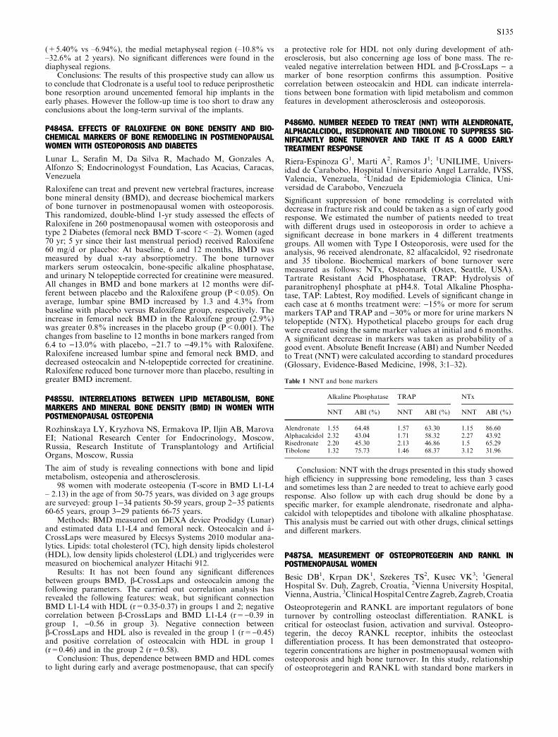
(+5.40% vs –6.94%), the medial metaphyseal region (–10.8% vs–32.6% at 2 years). No significant differences were found in thediaphyseal regions.
Conclusions: The results of this prospective study can allow usto conclude that Clodronate is a useful tool to reduce periprostheticbone resorption around uncemented femoral hip implants in theearly phases. However the follow-up time is too short to draw anyconclusions about the long-term survival of the implants.
P484SA. EFFECTS OF RALOXIFENE ON BONE DENSITY AND BIO-CHEMICAL MARKERS OF BONE REMODELING IN POSTMENOPAUSALWOMEN WITH OSTEOPOROSIS AND DIABETES
Lunar L, Serafin M, Da Silva R, Machado M, Gonzales A,Alfonzo S; Endocrinologyst Foundation, Las Acacias, Caracas,Venezuela
Raloxifene can treat and prevent new vertebral fractures, increasebone mineral density (BMD), and decrease biochemical markersof bone turnover in postmenopausal women with osteoporosis.This randomized, double-blind 1-yr study assessed the effects ofRaloxifene in 260 postmenopausal women with osteoporosis andtype 2 Diabetes (femoral neck BMD T-score<–2). Women (aged70 yr; 5 yr since their last menstrual period) received Raloxifene60 mg/d or placebo: At baseline, 6 and 12 months, BMD wasmeasured by dual x-ray absorptiometry. The bone turnovermarkers serum osteocalcin, bone-specific alkaline phosphatase,and urinary N telopeptide corrected for creatinine were measured.All changes in BMD and bone markers at 12 months were dif-ferent between placebo and the Raloxifene group (P<0.05). Onaverage, lumbar spine BMD increased by 1.3 and 4.3% frombaseline with placebo versus Raloxifene group, respectively. Theincrease in femoral neck BMD in the Raloxifene group (2.9%)was greater 0.8% increases in the placebo group (P<0.001). Thechanges from baseline to 12 months in bone markers ranged from6.4 to )13.0% with placebo, )21.7 to )49.1% with Raloxifene.Raloxifene increased lumbar spine and femoral neck BMD, anddecreased osteocalcin and N-telopeptide corrected for creatinine.Raloxifene reduced bone turnover more than placebo, resulting ingreater BMD increment.
P485SU. INTERRELATIONS BETWEEN LIPID METABOLISM, BONEMARKERS AND MINERAL BONE DENSITY (BMD) IN WOMEN WITHPOSTMENOPAUSAL OSTEOPENIA
Rozhinskaya LY, Kryzhova NS, Ermakova IP, Iljin AB, MarovaEI; National Research Center for Endocrinology, Moscow,Russia, Research Institute of Transplantology and ArtificialOrgans, Moscow, Russia
The aim of study is revealing connections with bone and lipidmetabolism, osteopenia and atherosclerosis.
98 women with moderate osteopenia (T-score in BMD L1-L4– 2.13) in the age of from 50-75 years, was divided on 3 age groupsare surveyed: group 1)34 patients 50-59 years, group 2)35 patients60-65 years, group 3)29 patients 66-75 years.
Methods: BMD measured on DEXA device Prodidgy (Lunar)and estimated data L1-L4 and femoral neck. Osteocalcin and a-CrossLaps were measured by Elecsys Systems 2010 modular ana-lytics. Lipids: total cholesterol (TC), high density lipids cholesterol(HDL), low density lipids cholesterol (LDL) and triglycerides weremeasured on biochemical analyzer Hitachi 912.
Results: It has not been found any significant differencesbetween groups BMD, b-CrossLaps and osteocalcin among thefollowing parameters. The carried out correlation analysis hasrevealed the following features: weak, but significant connectionBMD L1-L4 with HDL (r=0.35-0.37) in groups 1 and 2; negativecorrelation between b-CrossLaps and BMD L1-L4 (r=)0.39 ingroup 1, )0.56 in group 3). Negative connection betweenb-CrossLaps and HDL also is revealed in the group 1 (r=)0.45)and positive correlation of osteocalcin with HDL in group 1(r=0.46) and in the group 2 (r=0.58).
Conclusion: Thus, dependence between BMD and HDL comesto light during early and average postmenopause, that can specify
a protective role for HDL not only during development of ath-erosclerosis, but also concerning age loss of bone mass. The re-vealed negative interrelation between HDL and b-CrossLaps ) amarker of bone resorption confirms this assumption. Positivecorrelation between osteocalcin and HDL can indicate interrela-tions between bone formation with lipid metabolism and commonfeatures in development atherosclerosis and osteoporosis.
P486MO. NUMBER NEEDED TO TREAT (NNT) WITH ALENDRONATE,ALPHACALCIDOL, RISEDRONATE AND TIBOLONE TO SUPPRESS SIG-NIFICANTLY BONE TURNOVER AND TAKE IT AS A GOOD EARLYTREATMENT RESPONSE
Riera-Espinoza G1, Marti A2, Ramos J1; 1UNILIME, Univers-idad de Carabobo, Hospital Universitario Angel Larralde, IVSS,Valencia, Venezuela, 2Unidad de Epidemiologia Clinica, Uni-versidad de Carabobo, Venezuela
Significant suppression of bone remodeling is correlated withdecrease in fracture risk and could be taken as a sign of early goodresponse. We estimated the number of patients needed to treatwith different drugs used in osteoporosis in order to achieve asignificant decrease in bone markers in 4 different treatmentsgroups. All women with Type I Osteoporosis, were used for theanalysis, 96 received alendronate, 82 alfacalcidol, 92 risedronateand 35 tibolone. Biochemical markers of bone turnover weremeasured as follows: NTx, Osteomark (Ostex, Seattle, USA).Tartrate Resistant Acid Phosphatase, TRAP: Hydrolysis ofparanitrophenyl phosphate at pH4.8. Total Alkaline Phospha-tase, TAP: Labtest, Roy modified. Levels of significant change ineach case at 6 months treatment were: )15% or more for serummarkers TAP and TRAP and )30% or more for urine markers Ntelopeptide (NTX). Hypothetical placebo groups for each drugwere created using the same marker values at initial and 6 months.A significant decrease in markers was taken as probability of agood event. Absolute Benefit Increase (ABI) and Number Neededto Treat (NNT) were calculated according to standard procedures(Glossary, Evidence-Based Medicine, 1998, 3:1–32).
Conclusion: NNT with the drugs presented in this study showedhigh efficiency in suppressing bone remodeling, less than 3 casesand sometimes less than 2 are needed to treat to achieve early goodresponse. Also follow up with each drug should be done by aspecific marker, for example alendronate, risedronate and alpha-calcidol with telopeptides and tibolone with alkaline phosphatase.This analysis must be carried out with other drugs, clinical settingsand different markers.
P487SA. MEASUREMENT OF OSTEOPROTEGERIN AND RANKL INPOSTMENOPAUSAL WOMEN
Besic DB1, Krpan DK1, Szekeres TS2, Kusec VK3; 1GeneralHospital Sv. Duh, Zagreb, Croatia, 2Vienna University Hospital,Vienna,Austria, 3ClinicalHospitalCentreZagreb,Zagreb,Croatia
Osteoprotegerin and RANKL are important regulators of boneturnover by controlling osteoclast differentiation. RANKL iscritical for osteoclast fusion, activation and survival. Osteopro-tegerin, the decoy RANKL receptor, inhibits the osteoclastdifferentiation process. It has been demonstrated that osteopro-tegerin concentrations are higher in postmenopausal women withosteoporosis and high bone turnover. In this study, relationshipof osteoprotegerin and RANKL with standard bone markers in
Table 1 NNT and bone markers
Alkaline Phosphatase TRAP NTx
NNT ABI (%) NNT ABI (%) NNT ABI (%)
Alendronate 1.55 64.48 1.57 63.30 1.15 86.60Alphacalcidol 2.32 43.04 1.71 58.32 2.27 43.92Risedronate 2.20 45.30 2.13 46.86 1.5 65.29Tibolone 1.32 75.73 1.46 68.37 3.12 31.96
S135
serum was investigated in 50 postmenopausal women aged 48–80years. Osteoporosis of the spine was detected in 28 and of the hipin 5 patients. T-score of the spine and hip was significantly lowerin patients receiving antiresorptive therapy(n=29). Correlation ofhip T-score was significantly positive with menopause duration.Bone resorption marker (Crosslaps serum, Osteometer) correlatedpositively with total alkaline phosphatase and with hip T-score.No statistically significant relationship was obtained either forosteoprotegerin or RANKL with other bone markers,T-scoresfor the hip and spine, antiresorptive therapy, age and durationof menopause. Measurement of osteoprotegerin and RANKLdid not contribute in this patient sample to better understandingof bone turnover in the postmenopause. Clinical significance ofosteoprotegerin and RANKL, in comparison to standard bonemarkers was not established in this patient sample.
P488SU. BIOMARKER IN REHABILITATION OF OSTEOPOROSIS
Guenther C, Kapner A, Guenther O, Kießling H; JohannesbadReha-Kliniken AG&Co.KG, Bad Fussing, Germany
Aims: The intervals of investigations of used bone markers ininternational studies are three or more months. But our patientsare only three weeks in our clinic for rehabilitation of osteopo-rosis and therefore are the time intervals too long for estimationof the activity of therapy? The aims of the study are: 1) Is a ultrashort time monitoring of biomarkers and therapy of osteoporosisuseful?, 2) Is it possible from the decrease of resorption markersto prognosticate an increase in bone density?
Methods: We investigated 38 osteoporosis patients (31 women,7 men) with t-score (Lunar DPX IQ) <–2.5 WHO and estimatedthe resorption marker beta-CTX in Serum (Elecsys-beta-Cross-lapsTM Serum Roche Diagnostics GmbH, Mannheim) and NTXin urine (Ortho Clinical Diagnostics, Neckargemund). Investiga-tion times: Baseline and two and six weeks after the start of ther-apy. Used therapy: Basic therapy with calcium und vitamin D(1000/1000) and 1x1 Fosamax 70 mg/week or 1x1 Actonel 5 mg/dor 1x1 Evista 60 mg/d.
Results: 1) Regardless of the used therapy, beta-crosslaps inserum showed after two weeks a decrease of 43.8% and after sixweeks of 59.3% to baseline. 2) NTX in urine decrease 27.3% aftertwoweeks and 65.5%after six weeks. 3) In head-to-head comparisonof both methods under the same therapy (Fosamax 70mg+ calciumvitamin D) the beta-crosslaps in serum showed a decrease of 46.2%and NTX in urine a decrease of 30.7% after two weeks.
Conclusions: The resorption marker beta-CTX in serum andNTX in urine showed an effective reduction of bone resorptionalready after two weeks. Both markers are useful in the estimationof the therapy effects and the of patients compliance. The marker inserum seems to be more sensitive. We think that after a three weeksrehabilitation procedure from the decrease of the resorption mar-ker can estimated the increase of bone density in the following year.We can answer it exactly when our study is finished after one yearwith the first control of bone density.
P489MO. CHANGES OF BIOCHEMICAL RESORPTION MARKERS DUR-ING FRACTURE HEALING IN OSTEOPOROSIS: RESULTS OF A PRO-SPECTIVE STUDY IN PROXIMAL FEMUR FRACTURES
Hoesel ML1, Wehr U2, Rambeck W2, Schnettler R1, Heiss C1;1Department of Trauma Surgery, Justus-Liebig-University Gies-sen, Germany, 2Institute of Physiology, Ludwig-Maximilians-University Munich, Germany
Aims: The aim of this exlorative study was to evaluate thedevelopment of biochemical resorption markers during the frac-ture healing in patients with osteoporotic fractures of the proxi-mal femur.
Methods: This prospective study included 33 patients with afracture of the proximal femur, 25 control persons without afracture and in addition 35 patients with a fracture of the distalforearm. The concentration of the biochemical resorption markersN-terminal telopeptide (NTx), Desoxypyridinoline (D-Pyr) and
Pyridinoline (Pyr) were measured in the first urine spot preopera-tively and at day 2, 4, 10 and 14 postoperatively. Further the bonemineral density (BMD) of the lumbar spine was determined in allpatients with the quantitative computed tomography (qCT) todiagnose osteoporosis.
Results: Among the 33 study patients with a proximal femurfracture were 60% women and 40% men, osteoporosis was diag-nosed in 90% of the patients, while only 10% showed a normalbone density. We found a positive correlation between age andBMD in both sexes (female p=0.002; men p=0.01). It was noticedthat pertrochanteric fractures were more common with 64% thanfemoral neck fractures with 36%. Looking at the fracture classifi-cation systems a preponderance of high-grade fractures was visiblein females with femoral neck fractures.
A statistically significant increase was found in the three mea-sured biochemical resorption markers NTx, D-Pyr and Pyr duringthe observing period of 14 days of fracture healing (p<0.001).Concerning the biochemical bone markers we found clear trendsbetween the subgroups. The measured resorption markers NTx,D-Pyr and Pyr reflected higher concentrations in patients withosteoporosis, in female patients, in patients who sustained a per-trochanteric femur fracture and after a cemented implantation ofthe total hip prosthesis.
Conclusion: The biochemical bone resorption markers changeddifferently depending upon the BMD, sex and fracture type duringthe first 14 days of fracture healing. Furthermore our data suggest,that the ability of fracture healing is not impaired in osteoporosis.
P490SA. RELATIONSHIP OF BONE MINERAL DENSITY AND BIO-CHEMICAL MARKERS OF BONE TURNOVER IN POSTMENOPAUSALWOMEN
Vuceljic M1, Vucetic D2, Mihajlovic R3, Ilic-Stojanovic O3,Nikolajevic R1; 1Institute for Biochemistry, Military MedicalAcademy, Belgrade, Serbia and Montenegro, 2Institute forTransfusiology, Military Medical Academy, Belgrade, Serbiaand Montenegro, 3Institute for Rehabilitation, Medical School,University of Belgrade, Serbia and Montenegro
Bone loss is most rapid in women in the first few years aftermenopause but continues in the postmenopausal years. The aimof this study was to evaluate the levels of bone markers in 83postmenopausal women with low bone mineral densities (BMD)in comparison with 10 healthy young women.
We measured the serum levels of bone-specific alkaline phos-phatase (BAP) and C-terminal propeptide of type I collagen(CICP) as markers of bone formation and deoxypyridinoline (Dpd)as marker of bone resorption. All markers were determined bymonoclonal competitive enzyme immunoassay (EIA, Metra Bio-system). BMD was measured by dual energy x-ray absorptiometryof forearm (DTX-200 Osteometer).
The control group women aged from 27–35 (30.3±3.6) with Tscore )0.58±0.40. Postmenopausal women aged from 43–83(67.3±9.11) and menopausal period started from 33–56(47.8±5.05) years. In elementary group we divided the patientsaccording osteoporosis in 83.2% cases (T score ) 3.46±0.73), andosteopenia in 16.8% cases (T score –2.15±0.20). We found com-pressive vertebral fracture in 33.7% patients using thoracic andlumbal lateral x-rays.
Obtained results showed that BAP (U/l) was significantly higher(p<0.01) in elementary (14.8±4.4) group at whole and osteopo-rosis (15.7±4.42) patients vs. control group (11.5±1.94). Thelevels of CICP, Dpd and Dpd/creatinine were not significantlydifferent in any investigated group vs. control group. However, 11of 69 cases with osteoporosis had elevated levels of CICP (four ofthem had compressive vertebral fractures). Correlation analysesshowed significant negative association between BMD and BAP(r=)0.307, p<0.01), Dpd (r=)0.248, p<0.05) and CICP(r=)0.224, p<0.05) in elementary group, and positive associationbetween BMD and frequencies of fracture (p<0.001) in cases withosteoporosis. Despite of these results we found the correlationbetween BAP and CICP (p<0.001), while Dpd and Dpd/cre werenot correlated with BAP and CICP.
S136
All investigated bone markers showed relation with BMD, butdespite found correlation between markers of bone formation(BAP and CICP) we did not find correlation with marker of boneresorption (Dpd), that indicates uncoupled balance between boneformation and bone resorption in patients with low BMD.
P491SU. VALUES OF BONE TURNOVER MARKER IN MEXICAN HEAL-THY POSTMENOPAUSAL WOMEN: URINARY N-TELOPETIDE AND URI-NARY CALCIUM/CREATININE
Araujo-Arias MJ, Perez-Perez A, Cadenas-Salazar JL, Robles-Paramo JA; Unidad Clinica de Enfermedades Oseo Metabolicas(OSTEOMED) , Centro Medico Dalinde, Mexico DF
A high bone turnover, as assessed by the levels of biochemicalmarkers, may lead to increased skeletal fragility, estrogen defi-ciency after spontaneous as well as artificial menopause results inan increase in bone remodeling. A sustained increase in boneturnover induces a faster bone loss and therefore an increased riskof osteoporosis, predict the occurrence of osteoporotic fractures,and to monitor the efficacy of treatment, especially anti-resorptivetherapies. It has also been suggested that measurement of boneturnover before treatment might be useful for selecting the type oftherapy
The purpose of this study was to evaluate the usefulness ofurine N- telopeptides (U-NTX) and Ca/Cr in healthy postmeno-pausal women.
Methods: In this observational and retrospective study, afterexclusion of women with bone diseases or treatments, we ana-lyzed the results of 684 healthy postmenopausal women charac-terized for 1–5 years after cessation of menses. We assessed in allpatients bone mineral density (BMD) of the spine and femoralneck regions utilizing DEXA (HOLOGIC) to establish the diag-nosis of normal according to WHO criteria. We have measuredCa/Cr and U-NTX levels (ELISA) in a sample from secondmorning urine.
Results: 684 healthy women 45–60 years were studied. Wefound values of U-NTX with range of 27–400 nM/mM Cr (mean of100 and median 80), Ca/Cr values range of 0.01–0.75 (mean 0.17and median 0.15), in 456 the age menopause was 5 yrs (33%),97<5 yrs (14%) and 17 with 1 yr (2.4%), the correlation betweenNTX and age menopausal were significant (p<0.01).
Conclusion: The results showed that N- telopeptides correlatewith age of menopause. The biological markers of bone turnoverare elevated in 5 years following menopause, this is suggestive oftheir potential usefulness in the selection of high risk patient inpostmenopausal osteoporosis. However, the normal values shouldbe established for all bone markers in large samples of healthypremenopausal women, with normal BMD at the spine and hipmeasured by DEXA, geographic areas, races have been investi-gated in prospective studies.
P492MO. CAN BONE MARKERS BE EFFECTIVELY DECREASED INEARLY PHASE OF CALCITONIN TREATMENT?
Akyuz G1, Ofluoglu D1, Unay O2, Kayhan O1; 1MarmaraUniversity School of Medicine, Dept. of PM&R, Istanbul,Turkey, 2Marmara University School of Medicine, Dept. ofBiochemistry, Istanbul, Turkey
Objective: The purpose of this study was to evaluate efficacy ofcalcitonin on bone markers in patients with postmenopausalosteoporosis in the early phases of treatment. Design: There were30 patients with a mean age of 58.2±5.4 years in the treatmentgroup, and 26 patients with a mean age of 58.8±5.2 years in theplacebo group in this randomized placebo-controlled study. Thepatients received 100 IU salmon calcitonin or placebo injectionssubcutaneously and 1000 mg elementary calcium for 2 weeks.Baseline plasma osteocalcin (OC) and urine deoxypyridinoline(DPD) were measured and repeated after two weeks.
Results: Baseline OC and DPD levels were 8.3±4.8 ng and7.7±7.2 mM/m in the treatment group while those were 7.8±6.3 ngand 4.8±1.7 mM/m in the placebo group, respectively. PlasmaOC and urine DPD levels were 8.5±4.1 ng, 4.3±2.4 mM/m and
8.5±5.1 ng, 6.2±6.8 mM/m at the end of the study in the treatmentand placebo group, respectively. Although there was a decrease inurine DPD levels in the treatment group at the end of the secondweek, the difference was not statistically significant. However, therewas no change between plasmaOC levels at the baseline and at end ofthe study in both the treatment and placebo group.
Conclusion: Bone resorption markers such as DPD candecrease even after 2 weeks treatment with subcutaneous calcitonininjection in patients with postmenopausal osteoporosis.
P493SA. EVALUATION OF SERUM SIALIC ACID LEVELS IN POSTMEN-OPAUSAL PERIOD
Yildirim K1, Karatay S1, Cerrah R1, Gureser G1, Kiziltunc A2,Ugur M1, Senel K1; 1Ataturk University,Medicine Fac-ulty,Department of Physical Medicine and Rehabilitation Erzu-rum Turkey, 2Ataturk University,Medicine Faculty,Departmentof Biochemistry Erzurum Turkey
Objective: Sialic acid is one of the small chemicals which are acomponent of a number of more complex chemical structures inthe human body. SA is found in glycoproteins, gangliosides, andglycolipids. The aim of our study was to investigate clinicalusefulness of serum levels of sialic acid in postmenopausalperiod.
Materials and methods: We studied 20 patients with postmen-opausal and 20 healthy women subjects. Serum sialic acid levelswere determined according to the method of Shamberger.
Results: Serum SA levells were 1.75±0.64 mmol/I for thepostmenapausal group, 1.66±0.53 mmol/I for the healthy controlgroup. The SA levels of the patients with postmenopausal periodwere higher than those of the healthy control group, but no sig-nificant difference was found between the patient and controlgroups (p>0.05).
Conclusion: Our study demonstrated that the serum levels ofSA can not be used as a useful and novel marker for evaluating thedisease status in the postmenopausal period.
P494SU. SERUM C TELOPEPTIDE (CROSSLAPS) VALUES IN NORMALDENSITOMETRIC PREMENOPAUSAL WOMEN IN VENEZUELA
Marcano L, Ramos J, Gonzalez A, Riera-Espinoza G; UNI-LIME, Universidad de Carabobo, Hospital UniversitarioDr. Angel Larralde, Valencia, Venezuela
C-telopeptide has been reported as a very specific resorption bonemarker. It is a collagen type I degradation product which indi-cates osteoclastic activity. Its suppression after antiresorptivetreatments has been correlated with fracture prevention efficacy.There have been reported regional differences in bone markersaround the world, so each laboratory should investigate its ownreference range. The aim of this study is to show the normal rangeof serum C-telopeptide (b CrossLaps) in normal densitometricpremenopausal women in Venezuela.
93 premenopausal women with at least one Venezuelan progen-itor, age 39±3.5 (32–47), with normal BMD at lumbar spine andfemoral neck (LUNAR DPX) and without any systemic disease ordrug use which affects bone metabolism were included. None of thewomen were under heavy physical therapy or training nor had pre-vious fracture. Fast blood samples were taken and frozen at )70�C.C-telopeptide (b CrossLaps) was measured by electrochemilu-minescense immunoassay on theRoche Elecsys 1010. All assaysweredone at the same time. Intra-assay variation 2.6%
BMD values were at lumbar spine 1.2406±0.13 g/cm2, T-score+0.34 and at femoral neck 1.002±0.12 g/cm2, T-score +0.19.Mean CTx values were 0.281±0.134 ng/ml (range: 0.092–0.645),Normal range: (mean±1SD): 0.147–0415 ng/ml. Percentile 10–90:0.126–0.492 ng/ml. Normal reference values reported by Roche forpremenopausal women are: 0.299±0.137 ng/ml. Normal range:mean±1SD: 0.159–0.439. Significant negative correlation wasfound between serum CTx and weight (r=)258, p<0.05) and notwith age or height.
S137

Reference values of C telopeptide (b CrossLaps) in normaldensitometric premenopausal Venezuelan women are: mean±1SD:0.281±0.134 ng/ml, range (mean±1SD): 0.147–0415 ng/ml. Thesevalues are similar to the ones supported by Roche based in follow-up measurements of samples from the OFELY study.
P495MO. SEGREGATION OF THE CODON 404 MUTATION IN EXON 8 OFSEQUESTOSOME 1 GENE IN MEMBERS AFFECTED BY PAGET’S DIS-EASE OF BONE FROM AN ITALIAN FAMILY
Falchetti A1, Di Stefano M2, Marini F1, Del Monte F1, Raugei J1,Carbonell S1, Fossi N1, Guazzini L1, Isaia G2, Brandi ML1;1Department of Internal Medicine, University of Florence,Florence, Italy, 2Department of Internal Medicine, Universityof Turin, Turin, Italy
Paget’s disease of bone (PDB) is a metabolic bone disorderaffecting up to 3% of Caucasian populations over 55 years of age.Focal and disorganized increase of bone turnover characterizesPDB. Recently, mutations in the gene encoding sequestosome 1(SQSTM1) have been identified as a common cause of sporadicand familial PDB in French Canadian, British descendent and USpatients. Mutations were reported to localize at exons 7 and 8 ofthe gene, affecting the highly conserved ubiquitin-binding domain(UBA). We originally performed mutational analysis of exon 7and 8 in 62 PDB Italian patients, identifying one ‘‘classical’’P392L and two novel mutations, M404V and G425R, at exon 8,the latters consisting, respectively, of A>G and G>A transver-sion. Recently, we had the opportunity to perform mutationalanalysis in 20 individuals, 4 affected by PDB and 16 unaffectedmembers from the Italian family of patient exhibiting the M404Vmutation. Affected subjects were clinically evaluated both bybiochemical and imaging tests. The M404V mutation was foundin 8 individuals: 3 with clinically diagnosed polyostotic PDB and5 ‘‘asymptomatic’’ offspring (age range from 41 to 53 years) ofthree affected patients. Not mutated individuals did not exhibitany clinical evidence of PDB. Mutation M404V consists of ahighly conservative amino acid substitution, and Methionineresidue at position 404 is highly conserved among other species,rat and mouse, suggesting an important role in the functionalityof the SQSTM1/p62 protein. This familial segregation of M404Vmutation with PDB phenotype strongly supports the hypothesisthat this mutation is involved in PDB pathogenesis, according to apossible dominant negative mechanism of action. Moreover, itslocation at exon 8 level confirms the evidence of a clustered muta-tional area at this level in this disorder, supporting the role of theUBA domain in the biological properties of SQSTM1/p62 protein.Both instrumental and biochemical evaluation of the 5 ‘‘asymp-tomatic carriers’’ will potentially provide new important acquisi-tions on the pathogenesis of this metabolic disorder of bone.
P496SA. ESTROGEN RECEPTOR ALPHA AND AROMATASE GENEPOLYMORPHISMS: RESPONSE IN BONE MINERAL DENSITY TO HRT INPOSTMENOPAUSAL WOMEN
Masi L1, Ottanelli S2, Del Monte F1, Carbonell S1, Guazzini L1,Fossi N1, Gozzini A1, Mavilia A1, Falchetti A1, Amedei A1,Imbriaco R1, Ghinoi V1, Colli E1, Raugei I1, Brandi ML1;1Department of Internal Medicine, 2Department of Gynaecology,Italy
Genetic factors regulate BMD and possibly development ofosteoporosis. Estrogens play a pivotal role in maintaining bone.The formation of estrogens from C19 steroids is catalyzed byaromatase in women and men. It is known that polymorphismat the human ERalpha and at the aromatase genes are associ-ated with low BMD in postmenopausal women. We evaluatedthe possibility of interaction between aromatase and ERalphagenotypes with bone mass and we assessed the response in BMDto HRT in postmenopausal women. Subjects consisted of 209Italian postmenopausal women with a range of age 36–76 years(mean 61.3+/)8.6). Subjects under HRT received TTS50 microg/d and nomegestrol acetate 5 mg/d (12 days/month).LS-BMD was measured at the baseline and after 1 year. PvuII
and XbaI polymorphism of the ER-alpha was determined byPCR and TTTA repeats by sequence analysis. The capital P andX and the lower-case p and x represent respectively the absenceand the presence of the restriction site. For the TTTA repeatspolymorphism the subjects were divided on the basis of themean TTTA repeats: low (<8) medium (8–10) and high (>10).The genotype distribution for ER-alpha was as follow: XX42.3%; Xx 43.9%; xx:13.76% (c2 analysis: p=0.3) and PP:33.3%; Pp:47.09%; pp: 19.53% (c2 analysis: p=0.07) and forthe aromatase TTTA repeats was: low: 41%; medium 40% andhigh:19%. The genotype with a low number TTTA repeats wasmore frequent in osteoporotic and osteopenic subjects incomparison with normal (42.8 and 39.2% vs.17.86%). Ancovaanalysis did not show any statistical differences in the LS-BMDof various ER-alpha genotypes (p=0.6), although LS-BMDtended to be lower in subjects with pp and xx genotypes.During HRT an increase of the LS-BMD was present in allthe genotypes suggesting a feeble influence of the polymorphismon the hormone response. The same results were observed forthe aromatase gene polymorphism. The absence of difference inthe LS-BMD in subjects with or without HRT suggests a lowinfluence of the aromatase gene on the HRT response. In con-clusion, ER-alpha and aromatase gene polymorphism do notseem to influence the response to HRT.
P497SU. TOMOGRAPHIC AND BIOMECHANICAL ANALYSIS OF MUS-CLE-BONE INTERACTIONS IN MICE ARTIFICIALLY SELECTED FORBODY CONFORMATION
Cointry GR1, Di Masso RJ2, Capozza RF1, Ferretti JL1, FontMT2; 1Center for P-Ca Metab Studies (CEMFoC), Fac ofMedicine, Natl Univ of Rosario, Argentina, 2Exp Genetics Inst(IGE), Fac of Medicine, Natl Univ of Rosario, Argentina
Antagonistic artificial selection of adult male and female mice withwide variation in body conformation produced animals with lightbody/long skeleton (CBi/L) or heavy body/short skeleton (CBi/C)from a parental line CBi. On changing the natural proportionsbetween body and skeletal size/shape, this procedure allowedanalyzing correlations between the body and gastrocnemius weightand indicators of material, geometric and structural (mechanical)properties of cortical bone of the femur diaphyses (as assessed bypQCT and bending tests at a low strain rate) avoiding the natural,allometric associations which normally blunt the biomechanicalinterrelationships between muscles and bones.
As expected, the selection procedure altered the natural pro-portions between gastrocnemius mass, body weight and femurlength, and between femur length and diaphyseal cross-sectionalproperties (moment of inertia, CSMI). The CSMI correlated closerwith gastrocnemius weight than it did with body weight. Diaphy-seal strength correlated significantly with CSMI, gastrocnemiusweight and body weight. Correlation of CSMI with gastrocnemiusweight was closer than with body weight and was the only graphdescribing the studied association as a single (linear) function forall the 3 strains studied as a whole.
Results suggest that 1. muscle mass would not depend alo-metrically on body weight in any circumstance; 2. the geometricproportions between long-bone length and cross-sectional proper-ties would not be independent determinants of bone structure orstrength; 3. muscle development would not depend on bonedevelopment; 4. the diaphyseal design would be adapted to muscleability to directionally deform the skeleton rather than to theweight of the supported biomass; and 5. the biomechanical adap-tation of bone strength to customary mechanical usage as allowedby the biochemical and microstructural constitution of the skeletonwould be determined more closely by the dynamic influence ofmuscle contractions than by the static, gravitational load of thebody weight. Those relationships, difficult to assess in naturalconditions, are crucial for interpreting the biomechanical homeo-stasis of the skeletal structure and the etiopathogenesis of allosteopenias and osteoporoses. This knowledge could be extrapo-latable to the pathogenetic analysis of many human bone-weak-ening diseases.
S138
P498MO. THE RELATIONSHIP BETWEEN COLI A1 POLYMORPHISMS(SP 1) AND COLI A2 POLYMORPHISMS (ECO R1 AND PUV II) WITHBONE MINERAL DENSITY IN CHINESE MEN AND WOMEN
Lau EMC1, Choy DTK2, Li M3, Woo J3, Chung T3, Sham A1;1Department of Community and Family Medicine, 2Jockey ClubCentre for Osteoporosis Care and Control, 3Department ofMedicine and Therapeutics, The Chinese University of HongKong, Hong Kong, China
Aims: The objectives of this study are to document the frequency ofCOLI A1 and COLI A2 polymorphisms, as well as their relation-ship with bone mineral density in Chinese men and women.
Methods: Two groups of Chinese subjects were studied. Theywere 450 women aged 50–79 years and 233 men aged 70–79 years.The study subjects were recruited through social centers in thedistrict of Shatin, Hong Kong.
Results: In 100 men and women, COLI A1 Sp 1 polymorphismwas not found, which was consistent with other previous studies inAsian population. However a significant relationship was observedbetween COLI A2 Eco R1 and Puv II genotypes among the Chi-nese men studied. The mean bone mineral density was consistentlylower in men of the EE and PP genotype (p<0.05 by ANOVA)than in men of the ee and pp genotypes. However, no associationbetween bone mineral density and the Eco R1 or Puv II genotypeswere observed in Chinese women (p>0.05 by ANOVA).
Conclusions: We conclude that COLI A1 Sp 1 binding site isabsent in Hong Kong Chinese, while the COLI A2 Eco R1 and PuvII genetic polymorphism may be associated with the bone mineraldensity of elderly Chinese men.
P499SA. RELATIONSHIP BETWEEN PARATHYROID HORMONE GENEPOLYMORPHISM, BONE MINERAL DENSITY, AND BONE RESPON-SIVENESS TO HORMONE REPLACEMENT THERAPY IN POSTMENO-PAUSAL KOREAN WOMEN
KimJG,KuSY,KimSH,ChoiYM,MoonSH;Dept. ofObstetrics& Gynecology, Seoul National University Hospital, Seoul, Korea
Aims: To evaluate the relationship between parathyroid hormone(PTH) gene BstBI polymorphism, bone mineral density (BMD)and bone responsiveness to hormone replacement therapy (HRT).
Methods: PTH BstBI polymorphism was determined byrestriction fragment length polymorphism (RFLP) in 444 post-menopausal Korean women. Among these women, 309 womenreceived sequential HRT for 1 year. Serum bone alkaline phos-phatase, CrossLaps, osteocalcin, calcitonin, and PTH levels weremeasured by immunoassay and serum calcium and phosphoruslevels by atomic absorptiometry. BMD at the lumbar spine andproximal femur was determined by dual energy X–ray absorpti-ometry before and after HRT of 1 year.
Results: PTH genotype frequencies were 81.1% for BB, 18.0%for Bb, and 1.2% for bb (uppercase letters signifying the absenceand lowercase letters the presence of the restriction site). BMD atthe femoral neck in women with the bb genotype was higher thanthat in women with the Bb (p<0.01) or BB (p<0.005) genotyperespectively. Similar trends were found in BMD of lumbar spineand Ward’s triangle. PTH genotypes were not distributed differ-ently between HRT–responders and HRT–nonresponders (womenwho lose more than 3% of bone mass per year) and were notrelated with annual percent change of BMD after HRT. There wereno significant differences in levels of calcitonin, PTH, calcium,phosphorus, and bone turnover markers, or their 6 month per-centage changes after HRT among PTH genotypes.
Conclusions: PTH BstBI polymorphism is not associated withbone responsiveness to HRT but BMD in Korean women.
P500SU. ASSOCIATION STUDY OF THE ESTROGEN RECEPTOR THY-MINE–ADENINE REPEAT POLYMORPHISM WITH THE EFFECTS OFHORMONE THERAPY ON SERUM LIPID AND BONE DENSITY IN POST-MENOPAUSAL WOMEN
Han KO, Yim CH, Yoon HK, Han IK; Department of Medicine,Samsung Cheil Hospital, School of Medicine, SungkyunkwanUniversity, Seoul, Korea
Several biologically plausible mechanisms have been proposedfor estrogen–associated changes in lipid and bone metabolism.These effects are thought to be mediated via estrogen receptor(ER). Several polymorphisms in the gene encoding estrogenreceptor alpha may modify the effects of hormone–replacementtherapy on lipid and bone mineral density in postmenopausalwomen.
We examined 284 postmenopausal women for thymine–adenine(TA) repeat polymorphism at the ER gene locus and its relation-ship to lipid and bone density. Their mean age was 52.2 ± 5.0years. We also investigated the association between ER TA repeatpolymorphism and changes in lipid and bone density after 3months and 1 year of hormone therapy. According to the meannumber of TA repeats, the women were divided into two groups:group H, with higher number of repeats (TA>16)(n=110); groupL, with lower number of repeats (TA=16)(n=174). Group Lshowed significantly increased changes in total and LDL choles-terol levels after 3 month estrogen replacement therapy than ingroup H (changes in total cholesterol: )8.4 ± 11.3% vs. )4.2 ±12.5%, p=0.019), (changes in LDL cholesterol: )18.7 ± 18.4% vs)8.8 ± 30.1%, p=0.006). There was no significant relationshipbetween TA repeat polymorphism and changes in HDL choles-terol, triglyceride levels and bone mineral density after 1 yearhormone therapy.
These data suggest that ERTA repeat polymorphism may pre-dict the response of lipid profile to estrogen replacement therapy.
P501MO. RELATIONSHIP BETWEEN POLYMORPHISMS OF ESTROGENRECEPTOR GENE AND SPINE BONE MINERAL DENSITY IN PRE– ANDPERIMENOPAUSAL WOMEN FROM A CITY OF ARGENTINA
Ulla MR2, Elias V1, Pretel E1, Mamana M1, Babini S2, Binci M1,Costero B1, Noriega R2, Tolosa de Talamoni N1; 1Lab. Dr.Canas, Cat. de Bioquımica y Biol. Molecular, Fac. de CienciasMedicas. Univ Nac de Cordoba, Argentina, 2Centro de Endocri-nologıa y Metabolismo, Cordoba, Argentina
Aims: To determine the estrogen receptor (ER) genotypes in preand perimenopausal women from Cordoba, city located in thecentral part of Argentina, in relation with their lumbar spine bonemineral density (BMD) and other variables associated to thecalcium and phosphorus metabolism .
Methods: Recruitment was achieved through voluntaryresponse to advertisement for a study on genetic markers affectingbone density in healthy pre and perimenopausal women. 98women, divided in three age groups (21–30, 31–40 and 41–55 yearsold) were studied. The exclusion criteria included hepatic or renaldiseases, malabsorption, hyperparatiroidism, malignity in the last 5years and use in the last 3 months of estrogen, glucocorticoids orother drugs known to influence on calcium metabolism. Serumcalcium and phosphorus were measured by spectrophotometrictechniques and PTH–intact molecule was quantitated by IRMA.DNA was isolated from blood, the appropriate segment wasamplified by PCR and the ER genotypes were determined by usingPvuII and XbaI as restriction enzymes. PP and XX representedhomozygotes without the site of restriction, pp and xx homozyg-otes with the restriction sites and Pp and Xx were the heterozyg-otes. BMD was measured in lumbar spine using a Nordland dual–energy–X ray absorptiometer.
Results: Values of calcemia, phosphatemia and serum PTH didnot show any differences between women from the different groups.The genotype frequencies for the two polymorphic sites of ER wereas follows: XX 8.42%, Xx 44.21% and xx 47.36% (for XbaI) andPP 10.52%, Pp 49.47% and pp 40.00%. Spine BMD was signifi-cantly lower in the group 21–30 years old as compared to the othertwo groups (ANOVA P< 0.05). In the group 31–40 years old,women with genotype XX had a higher spine BMD than womenwith either genotype xx or Xx (ANOVA P=0.05).
Conclusions: The data suggest that premenopausal women ofthis city would reach the bone mass peak in spine after 30 years ofage and the genotype XX would be favorable to acquire a betterspine BMD.
S139
P502SA. 1,25(OH)2 VITAMIN D INCREASES THE BONE DENSITY OFTHE LUMBAR SPINE OF OSTEOPENIC WOMEN IN ASSOCIATION WITHTAQI POLYMORPHISM OF THE GENE FOR VITAMIN D RECEPTOR
Zofkova I, Zajickova K, Hill M; Institute of Endocrinology,Prague, Czech Republic
1,25(OH)2 vitamin D has direct anti–resorption and anaboliceffects on bone (1). However, the degree of increase in bonemineral density (BMD) differs from case to case when takingthis hormone. The aim of the present study was to monitor thechanges in the BMD of the lumbar spine and the proximalfemur in 35 osteopenic, but otherwise normal women (27 ofwhich were post–menopausal) given the two–year taking of1,25(OH)2 vitamin D (ROCALTROL, Hoffmann La Roche) ina dosage of 0.5 ug/d, together with calcium (500 mg/d). BMDwas measured with the aid of dual x–ray absorptiometry(DXA) before the beginning of treatment and afterwards in thecourse of the second year of treatment itself. Its growth wasexpressed in terms of a percentage of the initial value. TaqIpolymorphism was determined using a restriction analysis of thePCR product.
Results: There was an increase in BMD of the lumbar spine(P<0.001) and the proximal femur (P<0.01) in the second year oftreatment with Rocaltrol. A higher growth of BMD in the lumbarspine was observed in women with a TT genotype than in carriers ofallele t (genotypes tt and Tt) (P<0.028, ANCOVA). The response ofBMD in the proximal femur was independent of genotype.
Conclusions: The study confirmed that there is a significantincrease in BMD of both the parts of the skeleton measured in thesecond year of treatment with Rocaltrol. In the process, the growthof BMD values in the lumbar spine was dependent on TaqI poly-morphism. The results of three–year monitoring will bring a finalanswer to question as to whether the gene for vitamin D receptorhas a predictive significance for the curative effect of 1,25 (OH)2vitamin D on the osteopenic lumbar spine.
References. 1. Ikeda K, Ogata E: Curr Opin Orthopedics 1999,10:339
P503SU. ESTROGEN RECEPTOR ALPHA XBAI POLYMORPHISM, TOXICADENOMA, AND THYROID HORMONE–STIMULATED BONE LOSS
Takacs I, Bajnok E, Tabak A, Speer G, Nagy Z, Horvat C,Meszaros S, Lakatos P; First Department of Medicine Sem-melweis University, Budapest, Hungary
The genetic background of toxic adenoma (TA), and thyroidhormone–stimulated bone loss have not yet been established. Inthis study, we investigated the possible functional contributionof the estrogen receptor alpha gene (ERa) XbaI, vitamin Dreceptor gene BsmI and interleukin–1 receptor antagonist pro-tein gene VNTR polymorphisms to the pathogenesis of TA andthyroid hormone–stimulated bone loss. We genotyped 296F(18–79 years) women: 107 patients with TA, and 189 healthycontrols (C). In a subgroup of postmenopausal women (71 TA,189 C), bone mineral density (BMD) of the lumbar spine,femoral neck and distal radius was measured. The xx allele ofERa was present significant less frequently in TA group com-pared to the controls. There was no other significant differencein the distribution of genotypes in the study groups. There wasno correlation between genotypes and BMD adjusted for ageand BMI at any site in the postmenopausal subgroups. Our datasuggest that ERa XbaI polymorphism might play a role in thedevelopment of toxic adenoma. At the same time, the studiedgenetic variations do not appear to have an impact on thyroidhormone–related bone loss.
P504MO. COLLAGEN TYPE I GENE ABNORMALITIES AND OSTEOPO-ROSIS
Abdel–Nasser AM; Minia University Hospital, Minia, Egypt
Background: The role of collagen gene abnormalities in osteo-porosis (OP) has garnered a lot of research attention ever since atype I collagen gene abnormality was demonstrated in patientswith osteogenesis imperfecta (OI) in 1984. Subsequently type Icollagen genes were cloned, several gene mutations were describedand correlated with clinical varieties in different diseases, andmost recently, attempts to correct the genetic abnormalities bybone marrow transplantation and gene therapy have been carriedout. In this article, the biology and molecular genetics of type Icollagen is presented, and the role of type I collagen geneabnormalities in the aetiopathogenesis, clinical expression andtreatment of OP is reviewed.
Methods: A Medline search was carried out for publicationsaddressing the biology, genetics and disease related to type I col-lagen gene abnormalities. Other sources of information includedthe online human genome database, and the web–based OnlineMendelian Inheritance in Man (OMIM).
Results: Type I collagen, the most abundant protein in humans,is inherited by 2 different genes on chromosomes 17 and 7. Over300 mutations in type I collagen genes resulting in human diseasehave been described. Mutations in either of the 2 genes areresponsible for 90% of cases of OI, a hereditary form of OP, thearthrocalsia and dermatospraxis types of Ehlers–Danlos syndrome(formerly EDS VII), and some cases of idiopathic OP. The type andsite of mutation are correlated with the severity of OP, with dele-tions resulting in milder forms than substitutions, and mutationsnear the carboxy– terminal resulting in the most severe forms ofOP. Bisphosphonates have shown beneficial effects in reducingfractures in all those diseases. Allogenetic bone marrow trans-plantation (BMT) has been attempted in severe OI, with encour-aging results, and gene therapy experiments in animal models havebeen published.
Conclusion: Collagen type I gene abnormalities are the defini-tive cause for most cases of hereditary OP, and they have aprominent role in idiopathic OP, together with other known riskfactors. Bisphosphonates are the treatment of choice for hereditaryforms of OP, while BMT and gene therapy show promising resultsfor severe cases.
P505SA. CALCIUM METABOLISM AND ENDOCRINE FUNCTIONS IN AFAMILY WITH FAMILIAL HYPOCALCIURIC HYPERCALCEMIA
Speer G1, Toth M2, Takacs I1, Nagy ZS1, Lakatos P1; 11stDepartment of Medicine Semmelweis University, Budapest,Hungary, 22nd Department of Medicine Semmelweis University,Budapest, Hungary
We report two Hungarian patients with familial hypocalciurichypercalcaemia (FHH) caused by a mutation of the calcium–sensing receptor (CaSR) at codon 55. The proband and her fatherwere heterozygous for this mutation. Design: We have performeddetailed clinical and laboratory assessments of this family tocharacterize the effects of CaSR mutation on bone turnover andon several endocrine organs expressing CaSR. Interestingly, wecould not detect any failure in the function of any tissues ) suchas bone ) we examined, except in serum calcium levels. Bothsubjects have had normal bone mineral density. To our knowl-edge, this has been the first report from Eastern and CentralEurope showing P55L mutation of the CaSR, as well as the firstpublication discussing the effect of this mutation on severalendocrine systems containing CaSR.
S140
A New Dimension in the Treatment of PostmenopausalOsteoporosis
Sponsor: SERVIER
SY1. PREVENTION OF VERTEBRAL FRACTURES
Compston JE; Department of Medicine, University of CambridgeSchool of Clinical Medicine, Cambridge, UK
The use of strontium ranelate, a bone–seeking compound with anovel mechanism of action, in the prevention of fractures inpostmenopausal women with osteoporosis has been investigatedin 2 randomized controlled trials, SOTI (Spinal OsteoporosisTherapeutic Intervention) and TROPOS (TReatment OfPeripheral OSteoporosis). Strontium ranelate was given orally ata dose of 2 g daily for 3 years and vertebral fractures wereassessed by the semi–quantitative method. The study populationin SOTI consisted of 1649 women, mean age 69.7 years, andthat in TROPOS of 5091 women with a mean age of 76.8 years.Prior to the study, the calcium and vitamin D status of thepatients started to be normalized, and during the study allpatients received appropriate calcium and vitamin D supple-mentation.
In the SOTI study, there was a 41% reduction in the relativerisk (RR) of vertebral fracture (RR=0.59; 95% confidence interval[CI], 0.48–0.73; P<0.001) over 3 years of treatment. This beneficialeffect occurred rapidly, so that after 1 year of treatment, thereduction in the RR was 49% (RR=0.51; 95% CI, 0.36–0.74;P<0.001). The proportion of women experiencing a new vertebralfracture in the treatment and control groups was 20.9% and32.8%, respectively, after 3 years, and 6.4% and 12.2%, respec-tively, after 1 year. Over 3 years, the risk of developing a newclinical vertebral fracture was also significantly reduced (RR=0.62;95% CI, 0.47–0.83; P<0.001), this reduction being observed fromthe first year (RR=0.48; 95% CI, 0.29–0.80; P=0.003). The ben-eficial effects of strontium ranelate on vertebral fractures wereconfirmed in TROPOS with a significant reduction of 45% in pa-tients without prevalent vertebral fracture over 3 years of treatment(RR=0.55; 95% CI, 0.42–0.72; P<0.001). In a pooled analysis ofSOTI and TROPOS, the RR of new vertebral fracture over 3 yearsof treatment was reduced by 48% in patients without prevalentvertebral fractures (RR=0.52; 95% CI, 0.40–0.67; P<0.001). Asignificant and similar increase in lumbar spine bone mineral den-sity was seen in both studies in strontium ranelate–treated womenafter 3 years compared with the control group.
The beneficial effect on vertebral fractures was also seen inpatients with osteopenia (femoral or lumbar bone mineral densityT–score between )1 and )2.5 and no T–score lower than )2.5) andwith or without prevalent vertebral fracture (RR=0.38; 95% CI,0.21–0.70; P=0.001) over 3 years of treatment.
These results demonstrate that strontium ranelate significantlyreduces vertebral fractures in postmenopausal women regardless ofthe severity of osteoporosis and in postmenopausal women withosteopenia.
SY2. PREVENTION OF NONVERTEBRAL FRACTURES
Reginster JY; CHU de Liege, Belgium
Strontium ranelate is a new antiosteoporotic agent with a uniquemode of action, simultaneously increasing bone formation anddecreasing bone resorption. The vertebral antifracture efficacy ofstrontium ranelate in postmenopausal women with osteoporosiswas confirmed in the SOTI (Spinal Osteoporosis TherapeuticIntervention) trial. The TROPOS (TReatment Of PeripheralOSteoporosis) trial aimed to assess the nonvertebral antifractureefficacy of strontium ranelate. Both trials were international,prospective, randomized, double–blind, and placebo–controlled,and strontium ranelate was given orally at a dose of 2 g daily.Normalization of calcium and vitamin D status began prior toinclusion during the run–in study FIRST (Fracture International
Run–in for Strontium ranelate Trials), appropriate supplemen-tation being continued throughout the studies.
The study population in TROPOS consisted of 5091 women,mean age 76.8 years, mean femoral neck bone mineral density(BMD) T–score=)3.1, and mean lumbar spine BMD T–score=)2.8, 36.8% of them having at least one prevalent nonvertebralfracture.
In TROPOS, there was a significant reduction in the relative risk(RR) of nonvertebral fracture (RR=0.84; 95% confidence interval[CI], 0.702–0.995; P=0.04) over 3 years of treatment. When con-sidering the major nonvertebral sites of osteoporotic fractures, e.g.,humerus, pelvis and sacrum, ribs, hip, clavicle, andwrist, a significantdecrease in theRRwas also observed (RR=0.81; 95%CI, 0.66–0.98;P=0.031). Although the study was not designed to demonstrateantifracture efficacy at the hip, a 36% decrease in the RR of hipfracture was shown in patients ‡74 years and with a femoral neckBMD T–score <)3 (RR=0.64; 95% CI, 0.412–0.997; P=0.046),over 3 years. Furthermore, a 41% decrease in the RR of hip fracturewas shown in patients having taken the drug for at least the first 18months (RR=0.59; 95% CI, 0.37–0.95; P=0.025).
In addition, a significant increase in femoral neck and in lumbarspine BMD was observed with a relative change from baseline to 3years of +8.2% and +14.7% compared with placebo, respectively.Strontium ranelate was well tolerated throughout the study.
These results show that strontium ranelate significantly reducesnonvertebral fracture risk and, more specifically, major osteopo-rotic nonvertebral fracture and hip fracture risks in postmeno-pausal women with osteoporosis.
SY3. A NOVEL MODE OF ACTION OPTIMIZING BONE FORMATION ANDBONE RESORPTION
Marie PJ; INSERM U349, Lariboisiere Hospital, Paris, France
Strontium ranelate has been demonstrated to decrease the risk offractures in postmenopausal women. Its efficacy in clinical studiesis based on its original mode of action, on both bone resorptionand formation. Pharmacological studies in animal models showedthat strontium ranelate decreases bone resorption but maintainsor increases bone formation, resulting in improved bone mass. Inthe model of ovariectomized (OVX) rats, strontium ranelateprevented the reduction in bone mineral content and the decreasein trabecular bone volume induced by estrogen deficiency(+115% in tibia and +50% in the vertebra in comparison withOVX rats in a 52–week study). In this model of osteopenia,strontium ranelate acted by decreasing bone resorption whereasbone formation was maintained at a high level, as documented byplasma biochemical markers and histomorphometric indices ofbone formation. In the model of osteopenia induced by hind limbimmobilization in rats, strontium ranelate reduced histomor-phometric parameters of bone resorption (osteoclast surface:)16%) and partially limited long bone loss, as assessed by bonemineral content (+7.5%), bone volume, and biochemical indicesof bone resorption. In normal mice, strontium ranelate increasedbone formation and vertebral bone mass (+36% and +59% inmales and females, respectively). In normal rats, strontiumranelate also increased the bone trabecular volume (+41%)without any alteration of mineralization. The unique mode ofaction of strontium ranelate both on bone formation andresorption was supported by in vitro studies. In rat calvaria cul-ture systems and rat osteoblastic cell cultures, strontium ranelateenhanced preosteoblastic cell replication and increased collagensynthesis by osteoblasts. Moreover, strontium ranelate decreasedbone resorption in organ cultures and decreased the resorbingactivity of isolated mouse osteoclasts. The evaluation of bonemarkers in clinical trials (SOTI and TROPOS) supports the modeof action of strontium ranelate: bone alkaline phosphatase levelsincreased and C–telopeptide of type I collagen levels decreased intreated patients compared with the placebo group at all time–points. Thus, pharmacological and clinical studies suggest thatstrontium ranelate optimizes bone resorption and formationresulting in increased bone mass, which may be of great value inthe treatment of osteoporosis.
S141
SY4. A NOVEL MODE OF ACTION LEADING TO RENEWED BONEQUALITY
Ammann P; HUGE, Hopital Cantonal, Geneve, Switzerland
Various bone resorption inhibitors have been shown to decreasethe risk of osteoporotic fractures. However, there is still a need foragents promoting bone formation by inducing positive uncou-pling between bone formation and bone resorption. Strontiumranelate represents a good candidate.
In vitro studies showed that strontium ranelate stimulates boneformation and inhibits bone–resorbing activity. In vivo, strontiumranelate decreased bone resorption whereas bone formation wasmaintained in ovariectomized rats, preventing trabecular bone lossinduced by estrogen deficiency. In intact female rats, strontiumranelate was demonstrated to significantly increase alkaline phos-phatase, a marker of bone formation. The dramatic increment intrabecular bone volume and in bone diameter are further elementsin favor of an in vivo stimulation of bone formation.
In intact female rats, a 2–year period of exposure to strontiumranelate mixed in the diet did not cause any alteration of bonemineralization as assessed by histomorphometry or bone stiffnessevaluation. A dose–dependent increase in bone strength, bonemass, and bone microarchitecture was observed at the level of thevertebral body, which contains a large proportion of trabecularbone, and at the level of the midshaft femur, which mainly containscortical bone, these results being confirmed in ovariectomized rats.The increment in bone mechanical properties was characterized byan increase in ultimate strength but also by a dramatic improve-ment in energy to failure, which was essentially due to an incrementin plastic energy, without significant influence on elastic energy.These results strongly suggest that bone formed on strontiumranelate treatment is able to withstand greater deformation beforefracture while possessing similar elastic properties as normal bone.Such modifications observed on strontium ranelate treatment are ingood agreement with an improvement in intrinsic bone quality andalso in trabecular bone mass leading to greater bone resistance.
These data indicate that strontium ranelate stimulates boneformation and inhibits bone resorption, and increases bone masswithout modification of the bone mineralization process, resultingin bone strength improvement.
SY5. CLINICAL BENEFITS FOR THE PATIENT
Delmas PD; INSERM Unit 403 and Rheumatology Departmentof Hopital E.Herriot, Lyon, France
The efficacy and tolerability profile are essential factors in thechoice of first–line treatment in a chronic disease such as post-menopausal osteoporosis. Furthermore, quality of life hasrecently appeared as an important outcome in clinical studies.
Strontium ranelate, 2 g/day orally, a novel antiosteoporoticagent, has been evaluated in more than 6700 patients in 2 largerandomized, double–blind, placebo–controlled clinical trials (SOTI[Spinal Osteoporosis Therapeutic Intervention] and TROPOS[TReatment Of Peripheral OSteoporosis]). These studies showedthat strontium ranelate reduces the vertebral and nonvertebralfracture risk in postmenopausal osteoporotic women. The reduc-tion in the relative risk of vertebral fracture has also been dem-onstrated in patients with osteopenia as well as in osteoporoticpatients, with or without prevalent fractures.
The Health–Related Quality Of Life (HRQOL) was evaluatedin 1240 patients from 11 countries with the QUALIOST� ques-tionnaire (developed by Servier and MAPI for patients with ver-tebral osteoporosis). The global score was significantly improved inthe strontium ranelate group whereas a deterioration was observedin the placebo group (P=0.03). This improved quality of life withstrontium ranelate is likely to be related to the significant decreasein the incidence of patients experiencing new vertebral or clinicalvertebral fractures, to the reduction in back pain and the smallerdecrease in body height as compared to the placebo group, as wellas to the good tolerability profile of strontium ranelate, including atthe upper gastrointestinal level.
Thus, strontium ranelate is an antiosteoporotic agent thatcombines efficacy, tolerability, and ease of administration(at bedtime). Taken together, these results illustrate the clinicalbenefits of strontium ranelate in the treatment of postmenopausalwomen with osteopenia or osteoporosis, with or without vertebralfractures.
Bone and Healthy Aging: The Spectrum of Risk
Sponsor: PFIZER
SY6. HEALTH BEYOND MENOPAUSE
Utian W
With more women now living well beyond the age of 65, anincreasing number of years of a woman’s life are spent after themenopause. Because of this, both the acute symptoms and thelong–term diseases associated with menopause and aging, such asosteoporosis, cardiovascular disease, breast cancer, urogenitalchanges, and female sexual dysfunction, will have a major impacton the quality of life of older women and on the health caresystem. These conditions may be prevented and/or treated ifappropriate screening is offered in a timely manner.
As a result of the shift away from the use of estrogen–progestintherapy in postmenopausal women, there is now a growing unmetneed for new drugs that can improve the symptoms of themenopauseand prevent osteoporosis and cardiovascular disease withoutincreasing breast cancer risk. This presentation will discuss thepotential of new therapies to address the varied health care needs ofpostmenopausal women. It will also describe how the current healthcare system is failing those women who do not go to their doctorswhile they are transitioning menopause. As a consequence, thesepostmenopausalwomendonot get the appropriate health education,counseling and preventive treatment that could make enormousimprovements to their long–term medical outcomes. Fundamentalchanges need to occur in the way health care providers and third–party payers deliver and pay for health care, and in providing healtheducation, counseling and preventive medicine to all women >50years of age. Only then can we provide ‘healthy aging’ to women.
SY7. BONE HEALTH BEYOND MENOPAUSE: ASSESSING WHO IS ATRISK OF DEVELOPING OSTEOPOROSIS
Siris E
Postmenopausal estrogen deficiency is the primary cause ofincreased bone loss associated with menopause and as a conse-quence large numbers of postmenopausal women are at risk ofdeveloping osteoporosis. The costs of osteoporosis are high, bothin terms of the financial cost to society and the individual costs towomen who experience the morbidity associated with fractures.
TheNationalOsteoporosis RiskAssessment (NORA) study is anobservational registry of postmenopausal women in the UnitedStates that started collecting data in 1997. Its database containsperipheral and centralmeasurements of bonemineral density (BMD)in addition to information on risk factors, treatment patterns, andosteoporosis history and fracture data. To date, 200,000 postmeno-pausal women have joined the registry and provide data andhealth information on a regular basis. Although diagnostic tools formeasuring BMD and effective therapies for treating osteoporosisexist, they are commonly underutilized when treating older femalepatients. The NORA study has shown that new, less expensiveperipheral technologies for measuring low BMD could be used forroutine screening of older women and has previously reported theprevalence of osteoporosis in asymptomatic postmenopausalwomen and the prevalence of lowBMD, and the associationwith riskfactors for osteoporosis and fracture incidence.
This presentation will show recent data from the NationalOsteoporosis Risk Assessment and discuss how postmenopausalosteoporosis should be best managed, both in terms of screeningand treatment.
S142
S8. NEXT GENERATION SERMS: ENHANCED TREATMENTFOR OSTEOPOROSIS AND GENERAL MENOPAUSAL HEALTH
Thompson D
Selective estrogen receptor modulators (SERMs) are a diverseclass of agents that have emerged as safe and effective treatmentalternatives to HRT for patients requiring preventive pharma-cotherapy for osteoporosis. They have the potential to offer thephysician a major advantage over estrogens in that they exertselective agonist or antagonist effects on a range of estrogen–target tissues. Each SERM has a unique profile of biologicalactivity that is dependent upon three interactive mechanisms:tissue–specific estrogen receptor expression, differential estrogenreceptor conformation on ligand binding, and differentialexpression and binding of cofactor proteins. Together theseprofiles of SERMs allow differential tissue responses rangingfrom estrogen antagonist effects in the breast and uterus to ago-nist effects in bone. This presentation will describe the uniquebiological properties of the SERM class, present proposedmechanisms of action, and characterize the different tissueactivities.
Glucocorticoid/Inflammation–induced Osteoporosis – PleiotropicEffects of D–Hormone Analogs
Sponsor: TEVA & TEIJIN
SY9. D–HORMONE AND THE IMMUNE SYSTEM
Cantorna MT; The Pennsylvania State University, PA, USA
D–hormone (1,25(OH)2D3) is an important immune system reg-ulator. 1,25(OH)2D3 has been shown to inhibit the developmentof autoimmune diseases including experimental inflammatorybowel disease (IBD), rheumatoid arthritis (RA), multiple sclerosis(MS) and type 1 diabetes. Paradoxically other immune mediateddiseases (experimental asthma) and immunity to infectiousorganisms were unaffected by 1,25(OH)2D3 treatment. Theeffectiveness of 1,25(OH)2D3 treatment of autoimmune diseasescomes as a result of the inhibition of the development and func-tion of Th1 cells and the induction of other Th cells including Th2cells. Interestingly Th2 cells express as much as 64 times moremRNA for VDR than Th1 cells. In addition there are a numberof genes which are differentially regulated by 1,25(OH)2D3 in Th2cells versus Th1 cells. In IBD the 1,25(OH)2D3 mediated mech-anism includes the inhibition of three TNFa (TNFa, LPS–in-duced TNFa, and TNF receptor) related genes in the colon. D–hormone is a selective regulator of the immune system and theoutcome of 1,25(OH)2D3 treatment depends on the nature(infectious disease, asthma, autoimmune disease etc.) of the im-mune response.
SY10. PREVENTION OF BONE LOSS IN HIGH RISK PATIENTSBY D–HORMONE ANALOGS
Reginster JY, Richy F; WHO Collaborating Center for PublicHealth Aspects of Rheumatic Diseases, University of Liege,Liege, Belgium
Alfacalcidol has been investigated, in high quality randomizedcontrolled trials, for the management of involutional and gluco-corticoid–induced osteoporosis. All these trials conclude thatalfacalcidol induces a substantial effect on bone loss and fracturerates in osteoporosis and is beneficial in preventing bone loss inglucocorticoid–induced osteoporosis. In patients requiring treat-ment with high doses of glucocorticoids (up to 46 mg/day),alfacalcidol and calcium significantly prevented bone loss at thelevel of the lumbar spine. These positive results were obtainedwithout inducing a significant increase in serum or urinary cal-cium. These results were confirmed with doses of alfacalcidolfrom 0.25 lg to 1 lg/day, at the level of the lumbar spine, femoralneck and radius, in women aged 32 to 52 years starting steroidtreatment for rheumatic disorders or asthma. These results
compares favourably with the negative outcomes observed whenplain vitamin D (50 000 IU/day + 1000 mg calcium) was inves-tigated in patients with glucocorticoid–induced osteoporosis. In ameta–analysis assessing the effect of D–Hormones in patientswith primary or secondary osteoporosis, D–Hormones haveshown to prevent spinal bone loss both in postmenopausal andglucocorticoid–induced osteoporosis. D–Hormones were alsoshown to significantly reduce the overall fracture rates, these ef-fects being predominant in vertebral fracture rates observed inprimary osteoporosis. When comparing the effect of D–hormoneto those of native vitamin D, D–Hormones appear to exhibit ahigher preventive effect on bone loss and fracture rates in patientswith primary osteoporosis. When comparing the adjusted globalrelative risks for fractures when allocated to D–Hormones (riskdifference=10%) or native vitamin D (risk difference=2%),D–Hormones provided a more marked preventive efficacyagainst fractures. Lower fracture rates for D–Hormones userswere confirmed for spinal and non–spinal fracture rates. Inconclusion, D–Hormones have shown significant effect in pre-venting glucocorticoid–induced bone loss and in reducingfracture rates in postmenopausal osteoporosis. In meta–analyses,their effect has been shown to be more pronounced than the effectof native vitamin D, in primary and secondary osteoporosis.
SY11. ALFACALCIDOL VERSUS PLAIN VITAMIN D IN INFLAMMATION–INDUCED BONE LOSS
Scharla SH1, LempertUG1, Schacht E2; 1Praxis fur InnereMedizinund Endokrinologie, Bad Reichenhall, Germany, Ludwig–Maximilians–University, Munich, Germany, 2Metabolic BoneDisease Unit, University Clinic Balgrist, Zurich, Switzerland
Inflammatory diseases lead to systemic osteoporosis. Causal fac-tors are increased circulating concentrations of inflammatorycytokines such as interleukin 6 (IL–6) and Tumor necrosis factoralpha (TNFalpha), glucocorticoid medication, and reducedphysical activity. In addition, disturbances of vitamin D metab-olism play an important role for the development of inflammationinduced osteoporosis. Therefore, D–hormone analogs offer animportant treatment option. 1,25–dihydroxyvitamin D (D–hor-mone) prevented bone loss in the rat model of inflammationmediated osteopenia (IMO) and in an arthritis model. Oneexplanation is the fact that animals and humans with inflamma-tory diseases exhibit markedly reduced circulating concentrationsof D–hormone, partly the result of inhibition of renal 1–alpha–hydroxylase by TNFalpha. Moreover, the number of vitamin Dreceptors is reduced by glucocorticoids. Moreover, D–hormonehas pleiotropic effects not only on calcium homoeostasis but alsoon muscle (improving strength), nerve system, and on the immunesystem. D–hormone inhibits the release of cytokines (IL–1,IL–6,TNFalpha) from macrophages and stimulates osteoprotegerinsecretion in vitro and improves arthritis in animal models.
In our own study, 71 patients with rheumatoid arthritis andosteopenia (mean age 65 years) were randomised to receive eithervitamin D (1000 IU/day) or the prohormone Alfacalcidol (AlphaD3
� TEVA, 1 lg/day). All patients were supplemented with Cal-cium 500 mg/day. Blood and urine samples were obtained before, 2and 4 weeks after treatment. A pain score was also obtained.Muscle power was determined by isokinetic test (leg extension).
Alfacalcidol did not cause hypercalcemia. There was a slightdecrease in serum TNF–alpha in patients receiving Alfacalcidol(p<0.03), other cytokines and osteoprotegerin did not change.After 4 weeks, calciuria was increased (mostly within the normalrange) in patients receiving Alfacalcidol and slightly in the vitaminD group. Only Alfacalcidol decreased PTH (p<0.002) as well asthe urinary bone resorption marker NTX (p<0.003). A significantdecrease of pain score occurred only in the Alfacalcidol group(p<0.0001). In patients receiving Alfacalcidol muscle powerincreased by 58% compared to 18% in patients of the vitamin Dgroup (p<0.05).
Alfacalcidol, but not plain vitamin D, has pleiotropic effectsimproving bone metabolism and clinical symptoms in patients withrheumatoid arthritis.
S143
SY12. D–HORMONES FOR PREVENTION OF BONE LOSS AFTER ORGANTRANSPLANTATION
Sambrook P; Institute of Bone and Joint Research, University ofSydney, Sydney, Australia
Active vitamin D metabolites such as 1,25 dihydroxy vitamin D(calcitriol) and 1–a hydroxyvitamin D (alfacalcidol), also calledD–Hormones, have been shown to be effective in preventing boneloss in patients starting glucocorticoids, suggesting a role for theseagents in transplant osteoporosis. Vitamin D metabolites couldreduce post–transplantation bone loss by reversing glucocorti-coid–induced decreases in intestinal calcium absorption andmitigating secondary hyperparathyroidism (1), which appears tobe a prominent mechanism of post transplant bone loss (2). Apotential reduction in immunosuppressive requirements withactive vitamin D metabolites (3) is an additional consideration intheir use.
In a two year randomised double blind study of 65 patientsundergoing cardiac or single lung transplantation, the efficacy oftreatment with calcitriol in primary prevention was examined (4).Patients were randomly allocated to receive either placebo orcalcitriol (0.5–0.75 mcg/day), the latter for either 12 months or24 months after transplantation (ie 3 groups). All groups received600 mg calcium/day. Bone loss at the proximal femur was signifi-cantly reduced or prevented at all three sites by treatment withcalcitriol for 2 years compared with treatment with calcium alone.Bone loss at 24 months averaged 8.3% for those treated with cal-cium alone, compared to 5.0% for those treated with calcitriol for2 years. However treatment with calcitriol for 12 months followedby calcium for 12 months resulted in similar proximal femoral boneloss to that seen in those patients treated with calcium alone for24 months (7.4%), suggesting prophylaxis with calcitriol needed tobe continued beyond 12 months. Over two years, 22 new vertebralfractures/deformities occurred in 4 patients treated with calciumalone compared with one new vertebral fracture in one patienttreated with calcitriol. Mild hypercalcemia was not uncommon, butthere were no significant differences between groups in serum cre-atinine after two years. These data suggest calcitriol in doses of 0.5mcg or greater daily may have a role in reducing bone loss aftercardiac or lung transplantation, but treatment needs to be contin-ued beyond one year. A more recent study further supports thesefindings.
In a two year double blind randomised trial in 149 patients aftercardiac transplantation, alendronate 10 mg daily was comparedwith calcitriol 0.5 mcg daily (5). Rates of bone loss were comparedto 27 control subjects concurrently transplanted, but not rando-mised to therapy. Subjects randomised to alendronate and calcitrioldid not experience significant bone loss in contrast to the controlgroup. The change in spinal BMD was +0.3% with alendronate,–0.6% with calcitriol and –3.2% in controls. The change in hipBMD was )1.3% with alendronate, –0.4% with calcitriol and–6.2% in controls. Urinary NTX fell by 34% with alendronate,26% with calcitriol but were unchanged with controls. New ver-tebral fractures occurred in 6.8% of subjects treated with alendr-onate, 3.6% of subjects treated with calcitriol and 13.6% of thecontrol subjects. In the second year after discontinuation of bothagents, BMD remained stable despite marked increases in boneturnover in the calcitriol group.
These studies suggest a role for active vitamin D metabolites orD–Hormones in preventing bone loss in patients after transplan-tation.
References:
1. Sambrook P, Alfacalcidol and calcitriol in the prevention ofbone loss after organ transplantation. Calcif Tissue Int, 1999,65:341–343.
2. Cooper, A. M., Locke, T. J., Eastell R. Secondary hyper-parathyroidism associated with heart transplantation isreversed by 1 b hydroxyvitamin D. J Bone Miner Res,1996,11, S544.
3. Henderson–Briffa, K., Keogh, K., Sambrook, P.N., Eisman,J.A., Reduction by calcitriol of immunosuppressive therapyrequirements in heart transplantation, Transplantation, 2003,75: 2133–2134
4. Sambrook, P., Henderson, N. K., Keogh, A., MacDonald, P.,Glanville, A., Spratt, P., Bergin, P., Ebeling, P., Eisman, J,Effect of calcitriol on bone loss after cardiac or lung trans-plantation. J Bone Miner Res, 2000, 15(9), 1818–24.
5. Shane, E., Addesso, V., Namerow, P. B., McMahon, D. J., Lo,S. H., Staron, R. B., Zucker, M., Pardi, S., Maybaum,S.,Mancini, D. Alendronate or calcitriol and prevention ofbone loss after cardiac transplantation. New Engl J Med 2004,350, in press.
SY13. ALFACALCIDOL VERSUS PLAIN VITAMIN D IN THE TREATMENTOF GLUCOCORTICOID/ INFLAMMATION–INDUCED OSTEOPOROSIS
Ringe JD, Faber H, Farahmand P; Klinikum Leverkusen,University of Cologne, Germany
The aim of our study was to compare directly plain vitamin D andalfacalcidol in patients with established glucocorticoid–inducedosteoporosis (GIOP). 204 patients on long–term glucocorticoid(GC) therapy were included as matched–pairs to receive ran-domly either 1 lg alfacalcidol (Alpha–D3�, TEVA) plus 500 mgcalcium per day (group A, n=103) or 1000 IU vitamin D3 plus500 mg calcium (group B, n=101). Correspondingly the twogroups were well matched in terms of mean age, sex ratio, meanheight and weight, daily dosage and duration of GC–therapy andthe percentages of the underlying diseases (chronic obstructivepulmonary disease, rheumatoid arthritis, polymyalgia rheumati-ca). The mean bone mineral density (BMD) values at baseline forthe two groups were at the lumbar spine of a T–score of )3.26(alfacalcidol) and )3.25 (vitamin D3) and at the femoral neck)2.81 and )2.84 resp. Rates of prevalent vertebral and non–ver-tebral fractures were not different between the groups.
During the three year study we observed a median percentageincrease of BMD at the lumbar spine of 2.4% in group A comparedwith a loss of 0.8% in group B (p < 0,0001). The 3 years rate ofpatients with at least one new vertebral fracture was 9.7 percentamong those assigned to the alfacalcidol group, as compared to24.8 percent among those assigned to the vitamin D group (RiskReduction: 0.61; 95% CI 0.24 to 0.81; p=0.005). The 3 years rateof patients with at least one new non–vertebral fracture was 15percent in the alfacalcidol group, as compared to 25 percent in thevitamin D group (Risk Reduction: 0.41; 95% CI )0.06 to 0.68;p=0.081). In accordance with the observed fracture rates, thealfacalcidol group showed a substantially larger decrease in backpain than the plain vitamin D group (p < 0,0001). Generally sideeffects in both groups were mild and only 3 patients in the alfa-calcidol group and 2 patients in the vitamin D group had moderatehypercalcemia.
We conclude that alfacalcidol plus calcium is highly superior toplain vitamin D3 plus calcium in the treatment of establishedGIOP.
SY14. REDUCTION OF FALLS AND FALLERS IN HIGH RISK PATIENTSBY D–HORMONE ANALOGS
Dukas L1, Schacht E2, Stahelin HB1; 1Geriatric University Clinic,Basel, Switzerland, 2Metabolic Bone Disease Unit, Universitats-klinik Balgrist, Zurich, Switzerland
Aims: The number of fallers and falls in elderly can significantlybe reduced by treatment with D–hormone analogues. For theactivation of calcitriol (D–hormone) renal function is detrimental.We therefore determined the cutoff levels of creatinine clearance(CrCl), used as a marker of renal function, at which D–hormoneserum levels declines. Using the data of a double–blind random-ized study and the determined cutoff, we analyzed if CrCl isassociated with the risk of falls and whether treatment withAlfacalcidol can reduce this risk.
S144

Methods: For 36 weeks randomly 378 community–dwellingelderly men and women received 1lg Alfacalcidol (Alpha–D3
�
TEVA) or placebo daily. Incidence and number of falls wereregularly assessed with the help of a questionnaire. In multivari-ate–controlled logistic regression models, we assessed, accordingto treatment groups and according to a CrCl cutoff at 65 ml/min,the risk to become a faller and the risk of falls. Presented resultsare from ITT analyses.Results: In multivariate–controlled analyses D–hormone serumlevels were significantly associated with CrCl (p<0.0001) andsteadily declined below a CrCl of 65 ml/min. In multivariate con-trolled analyses parameters associated with lowD–hormone serumlevels were in order of significance: a CrCl of <65ml/min(p=0.0008), the use of diuretics (p=0.001) and a diagnosis of adultonset diabetes (p=0.003). In the Placebo group, we observed
significantly more fallers in participants with a CrCl of<65ml/mincompared to participants with a CrCl of ‡65ml/min (OR 4.01,95%CI 1.48–10.98, p=0.006). 36 weeks of treatment with Alfa-calcidol was in participants with a CrCl of <65ml/min, comparedto placebo, associated with a significant reduction in the number offallers (OR 0.26, 95%CI 0.08–0.80, p=0.019), and a significantreduction of number of falls (OR 0.29, 95%CI 0.09–0.88,p=0.028). No clinically relevant hypercalcemia were observed.Conclusion: A reduced CrCl of <65ml/min is, similarly to otherrisk factors (serum–cytokines , glucocorticoid–treatment), signif-icantly associated with low D–hormone and with a significantincreased risk of falls. Treatment with Alfacalcidol significantlyand safely reduces in a community–dwelling elderly populationwith a CrCl of <65ml/min, the low CrCl associated increasednumber of fallers and the high risk of falls.
S145
Related Documents