Inwardly Rectifying K 1 Channels in Spermatogenic Cells: Functional Expression and Implication in Sperm Capacitation Carlos Mun ˜ oz-Garay,* Jose ´ L. De la Vega-Beltra ´ n,* Ricardo Delgado,² Pedro Labarca,² Ricardo Felix,‡ and Alberto Darszon* *Departamento de Gene ´ tica y Fisiologı ´a Molecular, Instituto de Biotecnologı ´a, UNAM, Cuernavaca, Me ´ xico; ‡Departamento de Fisiologı ´a, Biofı ´sica y Neurociencias, Cinvestav-IPN, Me ´xico DF, Me ´ xico; ²Centro de Estudios Cientı ´ficos, Valdivia, Chile To fertilize, mammalian sperm must complete a maturational process called capacitation. It is thought that the membrane potential of sperm hyperpolarizes during capacitation, possibly due to the opening of K 1 channels, but electrophysiological evidence is lacking. In this report, using patch-clamp recordings obtained from isolated mouse spermatogenic cells we document the presence of a novel K 1 -selective inwardly rectifying current. Macroscopic current activated at membrane potentials below the equilibrium potential for K 1 and its magnitude was dependent on the external K 1 concentration. The channels selected K 1 over other monovalent cations. Current was virtually absent when external K 1 was replaced with Na 1 or N-methyl-D-glucamine. Addition of Cs 1 or Ba 21 (IC 50 of ;15 mM) to the external solution effectively blocked K 1 current. Dialyzing the cells with a Mg 21 -free solution did not affect channel activity. Cytosolic acidification reversibly inhibited the current. We verified that the resting membrane potential of mouse sperm changed from 252 6 6 to 266 6 9 mV during capacitation in vitro. Notably, application of 0.3–1 mM Ba 21 during capacitation prevented this hyperpolarization and decreased the subsequent exocytotic response to zona pellucida. A mechanism is proposed whereby opening of inwardly rectifying K 1 channels may produce hyperpolarization under physiological conditions and contribute to the cellular changes that give rise to the capacitated state in mature sperm. © 2001 Academic Press Key Words: K 1 channel; inward-rectifier; spermatogenic cells; sperm capacitation; sperm acrosome reaction. INTRODUCTION Sperm interaction with the oocyte’s zona pellucida (ZP) initiates the acrosome reaction (AR), an essential exocy- totic event in fertilization. Before the AR, mammalian sperm must complete in the female genital tract a poorly understood maturational process called capacitation. Sperm capacitation can be also accomplished in vitro by incubat- ing sperm in defined medium (Yanagimachi, 1994; Visconti et al., 1995). In vitro studies have shown that capacitation is accompanied by a plasma membrane hyperpolarization that is thought to be mediated by an increase in K 1 permeability (Zeng et al., 1995; Arnoult et al., 1999). Capacitation takes from minutes to hours and during this process sperm membrane potential changes tens of milli- volts, reaching values as negative as 280 mV (Arnoult et al., 1999). K 1 channels are a family of transmembrane proteins that allow the passive diffusion of K 1 down its electrochemical gradient (Hille, 1992; Coetzee et al., 1999). Biochemical and molecular biology data suggest the presence of different K 1 channels in both mammalian testis and spermatozoa (Schreiber et al., 1998; Salvatore et al., 1999; Jacob et al., 2000). However, electrophysiological investigation of ion channels in the plasma membrane of mature sperm has been precluded by the small size of these cells (Espinosa et al., 1998). An alternative approach has been to study these channels in spermatogenic cells, developmental precursors that synthesize proteins for subsequent use in the transla- tionally quiescent mature sperm. Initial studies revealed the presence of only one functional type of K 1 channel in rodent spermatogenic cells. This current was independent of external Ca 21 concentration and blocked by tetraethyl- ammonium chloride (Hagiwara and Kawa, 1984). More recently, the combination of electrophysiology with mo- lecular biology methods has been used to specifically study Developmental Biology 234, 261–274 (2001) doi:10.1006/dbio.2001.0196, available online at http://www.idealibrary.com on 0012-1606/01 $35.00 Copyright © 2001 by Academic Press All rights of reproduction in any form reserved. 261

Welcome message from author
This document is posted to help you gain knowledge. Please leave a comment to let me know what you think about it! Share it to your friends and learn new things together.
Transcript

pc
D
dr
Developmental Biology 234, 261–274 (2001)doi:10.1006/dbio.2001.0196, available online at http://www.idealibrary.com on
Inwardly Rectifying K1 Channels in SpermatogenicCells: Functional Expression and Implicationin Sperm Capacitation
Carlos Munoz-Garay,* Jose L. De la Vega-Beltran,* Ricardo Delgado,†Pedro Labarca,† Ricardo Felix,‡ and Alberto Darszon**Departamento de Genetica y Fisiologıa Molecular, Instituto de Biotecnologıa, UNAM,Cuernavaca, Mexico; ‡Departamento de Fisiologıa, Biofısica y Neurociencias, Cinvestav-IPN,Mexico DF, Mexico; †Centro de Estudios Cientıficos, Valdivia, Chile
To fertilize, mammalian sperm must complete a maturational process called capacitation. It is thought that the membranepotential of sperm hyperpolarizes during capacitation, possibly due to the opening of K1 channels, but electrophysiologicalevidence is lacking. In this report, using patch-clamp recordings obtained from isolated mouse spermatogenic cells wedocument the presence of a novel K1-selective inwardly rectifying current. Macroscopic current activated at membraneotentials below the equilibrium potential for K1 and its magnitude was dependent on the external K1 concentration. Thehannels selected K1 over other monovalent cations. Current was virtually absent when external K1 was replaced with Na1
or N-methyl-D-glucamine. Addition of Cs1 or Ba21 (IC50 of ;15 mM) to the external solution effectively blocked K1 current.ialyzing the cells with a Mg21-free solution did not affect channel activity. Cytosolic acidification reversibly inhibited the
current. We verified that the resting membrane potential of mouse sperm changed from 252 6 6 to 266 6 9 mV duringcapacitation in vitro. Notably, application of 0.3–1 mM Ba21 during capacitation prevented this hyperpolarization andecreased the subsequent exocytotic response to zona pellucida. A mechanism is proposed whereby opening of inwardlyectifying K1 channels may produce hyperpolarization under physiological conditions and contribute to the cellular changes
that give rise to the capacitated state in mature sperm. © 2001 Academic Press
Key Words: K1 channel; inward-rectifier; spermatogenic cells; sperm capacitation; sperm acrosome reaction.
agm
2cbactttro
INTRODUCTION
Sperm interaction with the oocyte’s zona pellucida (ZP)initiates the acrosome reaction (AR), an essential exocy-totic event in fertilization. Before the AR, mammaliansperm must complete in the female genital tract a poorlyunderstood maturational process called capacitation. Spermcapacitation can be also accomplished in vitro by incubat-ing sperm in defined medium (Yanagimachi, 1994; Viscontiet al., 1995). In vitro studies have shown that capacitationis accompanied by a plasma membrane hyperpolarizationthat is thought to be mediated by an increase in K1
permeability (Zeng et al., 1995; Arnoult et al., 1999).Capacitation takes from minutes to hours and during thisprocess sperm membrane potential changes tens of milli-volts, reaching values as negative as 280 mV (Arnoult et al.,1999).
K1 channels are a family of transmembrane proteins that
0012-1606/01 $35.00Copyright © 2001 by Academic PressAll rights of reproduction in any form reserved.
llow the passive diffusion of K1 down its electrochemicalradient (Hille, 1992; Coetzee et al., 1999). Biochemical andolecular biology data suggest the presence of different K1
channels in both mammalian testis and spermatozoa(Schreiber et al., 1998; Salvatore et al., 1999; Jacob et al.,000). However, electrophysiological investigation of ionhannels in the plasma membrane of mature sperm haseen precluded by the small size of these cells (Espinosa etl., 1998). An alternative approach has been to study thesehannels in spermatogenic cells, developmental precursorshat synthesize proteins for subsequent use in the transla-ionally quiescent mature sperm. Initial studies revealedhe presence of only one functional type of K1 channel inodent spermatogenic cells. This current was independentf external Ca21 concentration and blocked by tetraethyl-
ammonium chloride (Hagiwara and Kawa, 1984). Morerecently, the combination of electrophysiology with mo-
lecular biology methods has been used to specifically study261
1smo
ii
si
ct
gdm
w
iptiTCpcltir54Dwp
sicCm
aaGg
4Ad
fa6fl
u
262 Munoz-Garay et al.
particular K1 channel subtypes from sperm. Hence, a novelgene abundantly expressed in mammalian spermatocytesencoding a unique type of K1 channel regulated by bothintracellular pH and membrane voltage (called mSlo3) wascloned and expressed in Xenopus oocytes (Schreiber et al.,998). Its functional expression in spermatogenic cells andperm is yet to be determined. Likewise, RT-PCR experi-ents revealed high levels of mRNA expression in rat testis
f Kir 5.1, a member of the inward rectifier K1 channelsuperfamily. However, the function of this channel remainsunknown since it does not form functional channels whenexpressed in Xenopus oocytes (Salvatore et al., 1999). It hasbeen also suggested that mammalian sperm may possess aCa21-activated K1 (MaxiK-type) channel, which has beenmplicated in the process of sperm activation and gametenteraction (Wu et al., 1998). Considering this, we further
characterized the functional whole-cell K1 currents inmouse spermatogenic cells and examined their influence inthe development of the capacitated state in mature sperm.
MATERIALS AND METHODS
Cell preparation. Spermatogenic cells were obtained followingthe procedure previously described by Espinosa et al. (1999) withminor modifications. Briefly, testes from adult CD1 mice wereexcised and suspended in ice-cold dissociation solution containing(in mM) NaCl 130; KCl 3; MgCl2 2; NaHCO3 1; NaH2PO4 0.5;Hepes 5; glucose 10 (pH 7.4) supplemented with DNase I (100mg/ml). The tunica albuginea was removed and the seminiferoustubules were separated using tweezers under a stereoscopic micro-scope. Tubules were dispersed into individual cells or synplastsusing Pasteur pipettes. The cells were collected by centrifugation(at 2500 rpm for 4 min), resuspended in external recording solution(see below), and stored on ice until assayed. Under these condi-tions, spermatogenic cells remained totally healthy for as long as8 h. Although this cell suspension contains a heterogeneous cellpopulation, individual spermatogenic cells or synplasts at threedifferent stages of differentiation are preferentially present asdetermined by bright-field microscopy: pachytene spermatocytes,round spermatids, and condensing spermatids. These cells wereused routinely in the electrophysiological recording. Inasmuch assimilar results were obtained from these stages, data were pooledfor presentation. Ten-microliter aliquots of cell suspension weredispensed into a recording chamber (;200-ml total volume) on thetage of an inverted microscope and subjected to electrophysiolog-cal recording as detailed below.
Caudal epididymal mouse mature sperm were mechanicallyollected from CD1 mice and placed in 1.5-ml microcentrifugeubes containing 700 ml of medium 199 supplemented with BSA
(0.4% wt/vol), sodium pyruvate (30 mg/liter), and NaHCO3 (4.2/liter) at 37°C. After 5–10 min the fraction of motile sperm wasetermined by visual inspection and preparations with ,75%otile cells were discarded. For in vitro capacitation, 300- to 400-ml
aliquots of the sperm suspension (4–5 3 106 cells/ml) were incu-bated at 37°C as described previously (Visconti and Kopf, 1998;Visconti et al., 1999). Following an incubation period of 30 min,sperm were collected and then assayed for the AR.
Electrophysiology. K1 currents were recorded according to the
hole-cell patch-clamp technique (Hamill et al., 1981). All record- (Copyright © 2001 by Academic Press. All right
ngs were performed at room temperature using an Axopatch 200 Aatch-clamp amplifier (Axon Instruments, Foster City, CA) and 2-o 4-MV micropipettes manufactured from borosilicate glass cap-llary tubes (KIMAX-51; Kimble Division, Owens-Illinois, Inc.,oledo, OH) on a horizontal puller (Sutter Instrument Co., Novato,A). Unless otherwise stated, cells were clamped at a holdingotential of 0 mV and capacity transients were electronicallyompensated. Currents were evoked by 200- to 1800-ms hyperpo-arizing voltage steps (0.5 Hz) to test potentials ranging from 2120o 150 mV. Pulse protocols, data capture, and analysis of record-ngs were performed using pCLAMP software (Axon). Currentecords were captured on-line and digitized at a sampling rate of–10 kHz following filtering of the current record (2 kHz; internal-pole Bessel filter) using a personal computer attached to aigiData 1200 interface (Axon). Linear leak-current componentas subtracted off-line using the clampfit analysis routine ofCLAMP. To isolate inwardly rectifying K1 currents, cells were
bathed in different solutions as listed in Table 1. Bath solutionswere adjusted to pH 7.3 with KOH and 300 6 5 mosml liter21 withucrose. The composition of internal (patch pipette) solutions usedn this study is also given in Table 1. The control internal solutiononsisted of (mM): K1-methanesulfonate (MeSO3) 122; KF 20;aCl2 2.5; MgCl2 1; EGTA 5; Hepes 10; (pH 7.3/KOH, 275 6 5osml liter21 with sucrose).Assay for acrosome reaction. As one end point of capacitation,
we analyzed the ZP-induced AR in sperm, based on the premisethat only capacitated sperm would undergo exocytosis in responseto ZP. The percentage of AR was measured using Coomassie blueG-250 as described elsewhere (Visconti et al., 1999). Briefly, spermwere incubated at 37°C for 30 min followed by the addition of 5 ZPeq/ml. After an additional 30 min incubation at 37°C, an equalvolume of 23 fixative solution (10% formaldehyde in phosphate-buffered saline) was added to each tube. Following fixation, 10-mlliquots of sperm suspension were spread onto glass slides andir-dried. The slides were then stained with 0.22% Coomassie blue-250 in 50% methanol and 10% glacial acetic acid for 3–5 min,
ently rinsed with deionized H2O, air-dried, and mounted with50% (v/v) glycerol in phosphate-buffered saline. To calculate thepercentage of AR, at least 100 sperm were assayed per experimentalcondition for the presence or absence of the characteristic dark blueacrosomal crescent.
Measurement of membrane potential. Mature sperm (40 ml at0 3 106 cells/ml) were capacitated in vitro as described above.fter a 30-min incubation, the potential sensitive dye 3,39-ipropylthiadicarbocyanine iodide (DiSC3(5); Molecular Probes,
Eugene, OR) was added to the cell suspension at a final concentra-tion of 1 mM. The fluorescence was then monitored with aHansatech MkII fluorometer (Norfolk, UK) at 620/670-nmexcitation/emission wavelength pair (Espinosa and Darszon, 1995).Cell hyperpolarization decreases the dye fluorescence. Recordingswere initiated after dissipating mitochondrial membrane potentialwith 100 nM carbonyl cyanide m-chlorophenylhydrazone (CCCP;Sigma) and reaching a steady-state fluorescence (1–3 min). Fluores-cence changes were converted to membrane potential (Vm) asollows. First, extracellular K1 concentration was changed bydding KCl (1.25 to 10 mM final concentration) in the presence of50 nM valinomycin (Sigma Chemical Co., St. Louis, MO), and theuorescence was measured. Vm was then calculated according to
the Nernst equation (Hille, 1992), assuming an intracellular K1
concentration of 120 mM and that the membrane is a K1 electrodender the influence of the K1-selective ionophore valinomycin
Espinosa and Darszon, 1995). Finally, Vm in the absence and
s of reproduction in any form reserved.
dw
abc
mcw
ilom
trrvl
P
263Inward-Rectifier Channels in Spermatogenic Cells
presence of 1 mM Ba21 was calculated from the fluorescenceintensity using this calibration curve.
RESULTS
A Hyperpolarization-Activated Inward Current
Previous patch-clamp studies have shown that the K1
permeability of the plasma membrane in spermatogeniccells is controlled mainly by K1-selective channels of theelayed rectifying type (Hagiwara and Kawa, 1984). Herein,e present evidence for an inwardly rectifying K1 current in
mammalian spermatogenic cells. The presence of inwardlyrectifying K1 channels in these cells was documented with
series of whole-cell patch-clamp experiments in whichoth the patch electrode and the cell bathing solutionontained 150 mM K1 (BSI, PSI; Table 1). Examples of the
resulting current records are presented in Fig. 1A, whichshows typical fast activating, noninactivating whole-cellinward currents of decreasing amplitude from an isolatedmouse spermatogenic synplast clamped to 0 mV, in re-sponse to voltage steps of 200-ms duration applied from2110 to 150 mV in 10-mV increments. Immediately afterthe instantaneous jump in the inward direction, the cur-rents underwent a slight reduction in amplitude most likelydue to external Mg21 and/or Ca21 blockade (Jow and Nu-
ann, 1998). Even though negative applied potentialslearly activated macroscopic currents, no detectable out-
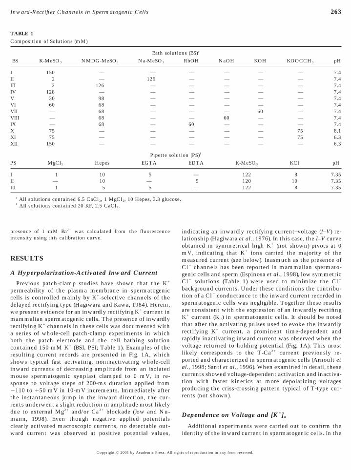
TABLE 1Composition of Solutions (mM)
Bath solBS K-MeSO3 NMDG-MeSO3 Na-MeSO3
I 150 — —II 2 — 126III 2 126 —IV 128 — —V 30 98 —VI 60 68 —VII — 68 —VIII — 68 —IX — 68 —X 75 — —XI 75 — —XII 150 — —
Pipette sS MgCl2 Hepes EGTA
I 1 10 5II — 10 —III 1 5 5
a All solutions contained 6.5 CaCl2, 1 MgCl2, 10 Hepes, 3.3 glucb All solutions contained 20 KF, 2.5 CaCl2.
ard current was observed at positive potential values,
Copyright © 2001 by Academic Press. All right
ndicating an inwardly rectifying current–voltage (I–V) re-ationship (Hagiwara et al., 1976). In this case, the I–V curvebtained in symmetrical high K1 (not shown) pivots at 0V, indicating that K1 ions carried the majority of the
measured current (see below). Inasmuch as the presence ofCl2 channels has been reported in mammalian spermato-genic cells and sperm (Espinosa et al., 1998), low symmetricCl2 solutions (Table 1) were used to minimize the Cl2
background currents. Under these conditions the contribu-tion of a Cl2 conductance to the inward current recorded inspermatogenic cells was negligible. Together these resultsare consistent with the expression of an inwardly rectifingK1 current (Kir) in spermatogenic cells. It should be notedhat after the activating pulses used to evoke the inwardlyectifying K1 current, a prominent time-dependent andapidly inactivating inward current was observed when theoltage returned to holding potential (Fig. 1A). This mostikely corresponds to the T-Ca21 current previously re-
ported and characterized in spermatogenic cells (Arnoult etal., 1998; Santi et al., 1996). When examined in detail, thesecurrents showed voltage-dependent activation and inactiva-tion with faster kinetics at more depolarizing voltagesproducing the criss-crossing pattern typical of T-type cur-rents (not shown).
Dependence on Voltage and [K1]o
Additional experiments were carried out to confirm the
s (BS)a
RbOH NaOH KOH KOOCCH3 pH
— — — — 7.4— — — — 7.4— — — — 7.4— — — — 7.4— — — — 7.4— — — — 7.4— — 60 — 7.4— 60 — — 7.460 — — — 7.4— — — 75 8.1— — — 75 6.3— — — — 6.3
on (PS)b
EDTA K-MeSO3 KCl pH
— 122 8 7.355 120 10 7.35
— 122 8 7.35
ution
oluti
ose.
identity of the inward current in spermatogenic cells. In the
s of reproduction in any form reserved.

r
V
oBa
cf
264 Munoz-Garay et al.
FIG. 1. Inwardly rectifying K1 current in mouse spermatogenic cells. (A) Typical current responses recorded by using 10-mV voltage stepsbetween 2110 and 150 mV starting from a holding potential of 0 mV in symmetrical K1 recording solutions (Table 1, BSI and PSI). The inwardectifier activates at potentials negative to VK. Schematic representation of the voltage protocol is shown above the traces. All voltage-clamp
recordings shown in this and subsequent figures are single traces and were obtained at room temperature. (B) Normalized conductance–voltage(g–V) curve at the steady state of currents obtained in a number of cells as illustrated in A. The voltage dependence of the measured conductancefor Kir channels followed a typical Boltzmann function with saturation. The conductance (gK) at each membrane potential was estimated as gK 5IK/(Vm 2 VK), where IK is peak current amplitude and Vm 2 VK represents the driving force. Assuming that gK was maximal when the commandvoltage step from 0 to 2110 mV was applied, the conductance values were normalized as a function of membrane potential, and then the datawere fitted to the Boltzmann equation, IK 5 [g(Vm 2 VK)]/(1 1 exp[(Vm 2 V1/2)/s)], where g is the conductance, Vm represents the test potential,
K is the apparent reversal potential, and s is the range within the potential changes e-fold around V1/2. Symbols represent means 6 SEM of nineexperiments; most of the SEM bars were smaller than symbols. (C) The reversal potential of the inward rectifier current in spermatogenic cellsis displaced according to VK. Currents were recorded from isolated cells in response to voltage steps in 10-mV increments from a holding potentialf 0 mV (range 2110 to 150 mV) in the presence of 30 (open squares), 60 (open triangles), and 128 mM (open circles) external K1 (Table 1; BSV,SVI, and BSIV, respectively). I–V curves at the steady state of currents are shown. Zero-current values (reversal potentials) of inward currentsre in close agreement with theoretical VK calculated by the Nernst equation (indicated by inverted filled triangles on the abscissas). The inset
shows the relationship between reversal potential of the currents and extracellular K1, measured by extrapolating the I–V curves. Each symbolrepresents the mean 6 SD of at least five cells. (D) Normalized g–V curves of Kir channels in a spermatogenic cell bathed with solutionsontaining 128, 60, and 30 mM potassium-MeSO3, as in C. Conductance is plotted versus the membrane voltage and fitted by a Boltzmannunction (smooth curves). The arrows denote VK and the region to the right of the dotted line in each curve is the fraction of open channels at
potentials positive to EK.
Copyright © 2001 by Academic Press. All rights of reproduction in any form reserved.
tg
1rtf
d
r
ct
aap
(
Ce
K
icw
ppg
1
aeKtiiM
M
265Inward-Rectifier Channels in Spermatogenic Cells
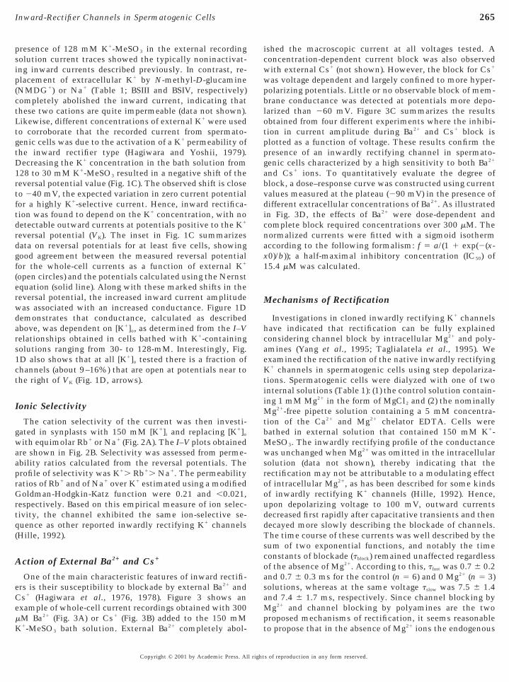
presence of 128 mM K1-MeSO3 in the external recordingsolution current traces showed the typically noninactivat-ing inward currents described previously. In contrast, re-placement of extracellular K1 by N-methyl-D-glucamine(NMDG1) or Na1 (Table 1; BSIII and BSIV, respectively)completely abolished the inward current, indicating thatthese two cations are quite impermeable (data not shown).Likewise, different concentrations of external K1 were usedo corroborate that the recorded current from spermato-enic cells was due to the activation of a K1 permeability of
the inward rectifier type (Hagiwara and Yoshii, 1979).Decreasing the K1 concentration in the bath solution from28 to 30 mM K1-MeSO3 resulted in a negative shift of theeversal potential value (Fig. 1C). The observed shift is closeo 240 mV, the expected variation in zero current potentialor a highly K1-selective current. Hence, inward rectifica-
tion was found to depend on the K1 concentration, with noetectable outward currents at potentials positive to the K1
reversal potential (VK). The inset in Fig. 1C summarizesdata on reversal potentials for at least five cells, showinggood agreement between the measured reversal potentialfor the whole-cell currents as a function of external K1
(open circles) and the potentials calculated using the Nernstequation (solid line). Along with these marked shifts in thereversal potential, the increased inward current amplitudewas associated with an increased conductance. Figure 1Ddemonstrates that conductance, calculated as describedabove, was dependent on [K1]o, as determined from the I–Velationships obtained in cells bathed with K1-containing
solutions ranging from 30- to 128-mM. Interestingly, Fig.1D also shows that at all [K1]o tested there is a fraction ofhannels (about 9–16%) that are open at potentials near tohe right of VK (Fig. 1D, arrows).
Ionic Selectivity
The cation selectivity of the current was then investi-gated in synplasts with 150 mM [K1]i and replacing [K1]o
with equimolar Rb1 or Na1 (Fig. 2A). The I–V plots obtainedre shown in Fig. 2B. Selectivity was assessed from perme-bility ratios calculated from the reversal potentials. Therofile of selectivity was K1. Rb1. Na1. The permeability
ratios of Rb1 and of Na1 over K1 estimated using a modifiedGoldman-Hodgkin-Katz function were 0.21 and ,0.021,respectively. Based on this empirical measure of ion selec-tivity, the channel exhibited the same ion-selective se-quence as other reported inwardly rectifying K1 channelsHille, 1992).
Action of External Ba21 and Cs1
One of the main characteristic features of inward rectifi-ers is their susceptibility to blockade by external Ba21 and
s1 (Hagiwara et al., 1976, 1978). Figure 3 shows anxample of whole-cell current recordings obtained with 300
mM Ba21 (Fig. 3A) or Cs1 (Fig. 3B) added to the 150 mM
1-MeSO3 bath solution. External Ba21 completely abol-Copyright © 2001 by Academic Press. All right
shed the macroscopic current at all voltages tested. Aoncentration-dependent current block was also observedith external Cs1 (not shown). However, the block for Cs1
was voltage dependent and largely confined to more hyper-polarizing potentials. Little or no observable block of mem-brane conductance was detected at potentials more depo-larized than 260 mV. Figure 3C summarizes the resultsobtained from four different experiments where the inhibi-tion in current amplitude during Ba21 and Cs1 block islotted as a function of voltage. These results confirm theresence of an inwardly rectifying channel in spermato-enic cells characterized by a high sensitivity to both Ba21
and Cs1 ions. To quantitatively evaluate the degree ofblock, a dose–response curve was constructed using currentvalues measured at the plateau (290 mV) in the presence ofdifferent extracellular concentrations of Ba21. As illustratedin Fig. 3D, the effects of Ba21 were dose-dependent andcomplete block required concentrations over 300 mM. Thenormalized currents were fitted with a sigmoid isothermaccording to the following formalism: f 5 a/(1 1 exp(2(x-x0)/b)); a half-maximal inhibitory concentration (IC50) of5.4 mM was calculated.
Mechanisms of Rectification
Investigations in cloned inwardly rectifying K1 channelshave indicated that rectification can be fully explainedconsidering channel block by intracellular Mg21 and poly-mines (Yang et al., 1995; Taglialatela et al., 1995). Wexamined the rectification of the native inwardly rectifying1 channels in spermatogenic cells using step depolariza-
ions. Spermatogenic cells were dialyzed with one of twonternal solutions (Table 1): (1) the control solution contain-ng 1 mM Mg21 in the form of MgCl2 and (2) the nominally
g21-free pipette solution containing a 5 mM concentra-tion of the Ca21 and Mg21 chelator EDTA. Cells werebathed in external solution that contained 150 mM K1-MeSO3. The inwardly rectifying profile of the conductancewas unchanged when Mg21 was omitted in the intracellularsolution (data not shown), thereby indicating that therectification may not be attributable to a modulating effectof intracellular Mg21, as has been described for some kindsof inwardly rectifying K1 channels (Hille, 1992). Hence,upon depolarizing voltage to 100 mV, outward currentsdecreased first rapidly after capacitative transients and thendecayed more slowly describing the blockade of channels.The time course of these currents was well described by thesum of two exponential functions, and notably the timeconstants of blockade (tblock) remained unaffected regardlessof the absence of Mg21. According to this, tfast was 0.7 6 0.2and 0.7 6 0.3 ms for the control (n 5 6) and 0 Mg21 (n 5 3)solutions, whereas at the same voltage tslow was 7.5 6 1.4and 7.4 6 1.7 ms, respectively. Since channel blocking by
g21 and channel blocking by polyamines are the twoproposed mechanisms of rectification, it seems reasonable
to propose that in the absence of Mg21 ions the endogenouss of reproduction in any form reserved.

cM
a(
(bAm
o
r
r
266 Munoz-Garay et al.
polyamines are responsible for inward rectification of theKir channels recorded in spermatogenic cells.
Modulation by Internal pH
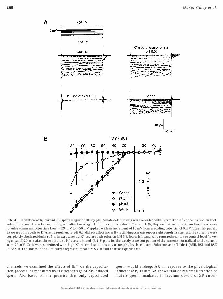
We next investigated whether inwardly rectifying K1
macroscopic whole-cell currents from spermatogenic cellswere subjected, like other types of inward rectifiers, to pHregulation (Hagiwara et al., 1978). A complete family ofcurrents illustrating the effect of lowering pH in a synplastis given in Fig. 4A. The cell was maintained at 0 mVthroughout the recording. In this experiment, the pH of theextracellular recording solution (150 mM K1-MeSO3) washanged from 7.4 (upper left panel) to 6.3 by titrating witheSO3 (upper right panel) and the current was measured.
Under these recording conditions, in no case did a reduction
FIG. 2. Cation selectivity of inwardly rectifying K1 currents in spesponse to 200-ms hyperpolarizing pulses applied from a holding p
containing 68 mM NMDG1 and 60 mM XOH, where X was K1,elationships for whole-cell inward currents in the same cells show
and Rb1-containing solutions (open symbols).
in external pH (pHo) cause significant changes in current
Copyright © 2001 by Academic Press. All right
mplitude (n 5 4). To examine the effects of internal pHpHi) on macroscopic conductance we used a bath solutioncontaining (in mM) 75 methanesulfonate and 75 K1 acetatepH 6.3). As previously described (Choe et al., 1997), pHi cane modified with membrane-permeable acetate solutions.lthough pHi was not directly measured in this experi-ent, in contrast to what was observed when pHo was
lowered in the solution containing the membrane-impermeable K1 methanesulfonate, equimolar replacementf [K1]o with the membrane-permeable K1 acetate solution
completely inhibited the current (Fig. 4, lower left panel).This inhibition could be partially reversed by returning tothe 150 mM K1-MeSO3 control bath solution (lower rightpanel). Figure 4B compares current values at different volt-ages recorded during a decrease in the bath pH from 7.4 to6.3 in several spermatogenic cells. Inward currents were
atogenic cells. (A) Inward currents recorded in mouse synplasts intial of 0 mV to 2110 mV. Cells were bathed in external solutions, or Rb1 (Table 1; BSVII, BSVIII, and BSIX, respectively). (B) I–VA obtained during exposure to control (filled symbols) and to Na1-
ermotenNa1
n in
normalized to the maximum value in the control. Filled
s of reproduction in any form reserved.

r
t
s
f
oid f
267Inward-Rectifier Channels in Spermatogenic Cells
triangles depict control inward currents where the bathrecording solution contained 150 mM K1-MeSO3; the I–Velation was linear below VK. Open circles are normalized
current values after changing pHo (with no change in pHi) to6.3. Filled circles denote inhibited inward currents mea-sured 5 min after the change in the composition of the bathsolution as described above. At this time the currents andpresumably pHi should both be in a steady state. Openriangles represent the normalized currents after the K1
FIG. 3. Ba21 and Cs1 block the inwardly rectifying current. (Apotential of 0 mV before and after superfusion of BSI supplemented wor a number of cells as shown in A and B. Note that the I–V curv
at potentials in excess of 260 mV revealing a voltage-dependent blocexperiments. (D) Dose-dependent block of the steady-state inward200-ms hyperpolarizations to 290 mV from a holding potential of 0represent mean values of the fraction of current blocked at variousThe smooth curve is the least-squares fit of mean values to a sigm
acetate solution was washed out. Together, these results
Copyright © 2001 by Academic Press. All right
trongly suggest that the Kir channels in spermatogeniccells are pHi sensitive.
Implication in Sperm Capacitation
It is well known that during in vitro capacitation, themembrane potential of mature mammalian sperm hyperpo-larizes from approximately 250 mV to 280 mV (Arnoult etal., 1999). To establish whether this hyperpolarization is
) Current responses to voltage steps to 290 mV from a holding300 mM Ba21 or Cs1, respectively. (C) Steady-state I–V relationshipsthe presence of Cs1 shows a region of negative slope conductanceby this cation. Symbols represent means 6 SD of four independent
rents at 290 mV by Ba21. Currents were obtained in response to, measured at steady state and normalized to the control. Symbolsconcentrations, and vertical lines indicate means 6 SD (n 5 3–7).unction (see Results) with an IC50 value of 15.4 mM.
and Bith
e inkingcurmV
Ba21
correlated with the activation of inwardly rectifying K1
s of reproduction in any form reserved.
si
s
to n
268 Munoz-Garay et al.
channels we examined the effects of Ba21 on the capacita-tion process, as measured by the percentage of ZP-induced
FIG. 4. Inhibition of Kir currents in spermatogenic cells by pHi. Whides of the membrane before, during, and after lowering pHo from a
to pulse command potentials from 2120 mV to 150 mV applied withExposure of the cells to K1-methanesulfonate, pH 6.3, did not affect inwcompletely abolished during a 5-min exposure to a K1-acetate bath solright panel) 20 min after the exposure to K1 acetate ended. (B) I–V plotat 2120 mV. Cells were superfused with high K1 external solutions ato BSXII). The points in the I–V curves represent means 6 SD of four
sperm AR, based on the premise that only capacitated m
Copyright © 2001 by Academic Press. All right
perm would undergo AR in response to the physiologicalnductor (ZP). Figure 5A shows that only a small fraction of
ll currents were recorded with symmetric K1 concentration on bothol value of 7.4 to 6.3. (A) Representative current families in responsecrement of 10 mV from a holding potential of 0 mV (upper left panel).y rectifying currents (upper right panel). In contrast, the currents were(pH 6.3; lower left panel) and returned near to the control level (lower
the steady-state component of the currents normalized to the currentious pHo levels as listed. Solutions as in Table 1 (PSIII, BSI, and BSXine experiments.
ole-cecontran inardl
utions fort var
ature sperm incubated in medium devoid of ZP under-
s of reproduction in any form reserved.

sh
B
269Inward-Rectifier Channels in Spermatogenic Cells
FIG. 5. Effects of extracellular Ba21 on the ZP-induced sperm AR and membrane hyperpolarization during capacitation. (A) External Ba21
inhibits the ZP-induced sperm AR by acting on capacitation. Capacitated mouse sperm were incubated for 30 min in the presence of ZPand AR was measured as described under Materials and Methods. Bars represent spontaneous AR that occurs in the absence of thephysiological inductor and the percentage of acrosome-reacted sperm after ZP-induction in the absence or presence of Ba21 (300 mM) duringcapacitation or the AR (5 s before ZP addition), as indicated. Two of the samples were incubated for 30 min in the presence (15 mM) of thecation ionophore ionomycin (Iono) alone or in combination with 1 mM Ba21 as positive controls (n 5 5 and 10, respectively). One hundredperm were evaluated per assay. Data represent means 6 SEM of at least four independent experiments. (B) Sperm membraneyperpolarization associated with capacitation. Sperm membrane potential was measured using the fluorescence dye DiSC3(5) as indicated
under Materials and Methods. Symbols represent the mean with standard deviation bars of membrane potential in uncapacitated as wellas in capacitated sperm when 1 mM Ba21 was added to the capacitating medium, as listed. The hyperpolarization that occurs with spermcapacitation is statistically significant (t value 5 4.796; P , 0.001; n 5 10 separate determinations obtained from four animals). (C) External
a21 blocks the inward but not the outward K1 currents. Superimposed records of inward (top) and outward (bottom) K1 currents obtainedfrom representative spermatogenic cells before and after incubation in the presence of Ba21 (1 mM). Dotted horizontal lines markzero-current level. Recordings were obtained using the voltage protocol illustrated above the traces. (D) Statistical summary of steady-state
K1 current percentage in spermatogenic cells subjected to Ba21 blockade as in C (n 5 4).Copyright © 2001 by Academic Press. All rights of reproduction in any form reserved.

cdb
m
15i
ica
mt
Te
b(
s
1
2
270 Munoz-Garay et al.
went spontaneous AR. In contrast, a significant percentageof AR was observed when sperm were exposed to ZP,indicating that they were capacitated. The ZP-inducedincrease in the percentage of AR was significantly reducedwhen the cells were preincubated with 300 mM Ba21 duringapacitation, but remained unaffected when Ba21 was addeduring AR. Furthermore, the reduction of the AR inducedy Ba21 seemed to be specifically related to an effect on K1
channels, since this divalent did not affect the ability ofsperm to undergo AR when incubated with the Ca21 iono-phore ionomycin, an agent capable of inducing acrosomalexocytosis in a nonregulated fashion (Brucker and Lipford,1995).
A second series of experiments provided an alternativemethod to examine the consequences of manipulating theinwardly rectifying K1 current during mature sperm in vitrocapacitation, by monitoring the resting membrane poten-tial using a voltage-sensitive dye. The mean resting mem-brane potential of mature sperm determined by this methodwas 252 6 6 mV (n 5 10 separate determinations). Aftercompleting in vitro capacitation sperm underwent an;14-mV hyperpolarization (266 6 6 mV, n 5 10; Fig. 5B).Notably, blockade of the inward-rectifying K1 current by 1
M Ba21 prevented hyperpolarization (Fig. 5B) and Vm
values were comparable with those of the resting mem-brane potential (253 6 7 mV). Since high concentrations ofBa21 can block other K1 channels (Nichols and Lopatin,997), the blocking effect of 1 mM Ba21 was compared (Fig.C) in isolated synplasts expressing either inwardly rectify-ng or the outwardly noninactivating K1 channels previ-
ously described (Hagiwara and Kawa, 1984). It can be seenthat Ba21 addition to the bath solution markedly reducednward currents (upper panel) with no effect on the outwardurrents (lower panel). A comparison of normalized currentmplitude in the presence and absence of Ba21 is shown in
Fig. 5D. Data were obtained from the currents recorded inresponse to the voltage steps indicated above the traces inFig. 5C.
DISCUSSION
Here we describe the identification and characterizationof a novel inwardly rectifying K1 current expressed in
ammalian spermatogenic cells. This current is carriedhrough a new and unique type of K1 channel that may
participate in the preliminary phase of mammalian spermactivation, designated “capacitation,” which regulates theefficiency of the AR during the early stages of fertilization.
The small size, complex geometry, and highly differenti-ated and motile nature of mature sperm complicate theirelectrophysiological characterization. To overcome thesedifficulties, an alternative approach has been to examinethe ion channels of testicular spermatogenic cells using thepatch-clamp technique (Lievano et al., 1996; Arnoult et al.,1998). Here we have found that most of the mouse synplasts
of round and condensing spermatids display a rapidly acti-Copyright © 2001 by Academic Press. All right
vating inward current whose average amplitude was sus-tained throughout the duration of the voltage pulse andshowed rectification around the K1 equilibrium potential.
he magnitude of the currents was strongly dependent onxternal K1 concentration and the channels were highly
selective for K1 over other monovalent cations (Figs. 1 and2). Addition of micromolar concentrations of Cs1 and Ba21
in the external solution resulted in a drastic inhibition ofthe whole-cell inward currents in spermatogenic cells. Thedegree of Cs1 block was increased substantially by hyper-polarization, while a voltage-dependent block of macro-scopic currents by Ba21 is less evident (Fig. 3). In addition,because channel blockade by Mg21 and channel blockade bypolyamines are the two major mechanisms of channelrectification, it is reasonable to propose that in the absenceof Mg21 ions the endogenous polyamines are responsible forinward rectification of the Kir channels recorded in sper-matogenic cells.
Inhibition of strong inwardly rectifying K1 channels maye mediated by decreases in intra- and extracellular pHZhu et al., 2000). To investigate whether inwardly rectify-ing K1 channels in spermatogenic cells have pH sensingmechanisms, whole-cell currents were measured in pH 6.3bath solutions containing either 150 mM K1 methanesul-fonate or 75 mM K1 methanesulfonate and 75 mM K1
acetate. Current inhibition was observed only in cellsbathed in the membrane-permeable acetate solution, indi-cating that Kir is inhibited by intracellular acidification.
It is well established that in excitable cells, the hyperpo-larized resting membrane potential (RP) is maintained byinward rectifier K1 channels (Reimann and Ashcroft, 1999).In addition to stabilizing the RP, this current also plays animportant role in modulating cellular excitability and itsblockade leads to membrane depolarization (Nichols et al.,1996). In addition, it is known that to achieve successfulfertilization in vivo, mammalian spermatozoa must firstundergo capacitation and then the AR, the exocytotic eventthat allows sperm to penetrate the zona pellucida and fusewith the oocyte.
Several physiological events lead to capacitation. Theseinclude an increase in [Ca21]i, an increase in pHi, activationof adenylyl cyclase by bicarbonate and hydrogen peroxide,production of cAMP, stimulation of PKA, and phosphory-lation of certain proteins (Baldi et al., 1996; Visconti andKopf, 1998). Based on the effects of external Ba21, our resultsuggest that the inwardly rectifying K1 current could be
involved in sperm capacitation. To test this possibility,membrane potential (Vm) was investigated in mature spermpopulations using a membrane potential-sensitive fluores-cent dye (DiSC3(5)) as described previously (Zeng et al.,995; Espinosa and Darszon, 1995). The calculated Vm value
of uncapacitated sperm was 252 6 6 mV. In contrast, invitro capacitated populations of sperm have a Vm value of
66 6 6 mV (n 5 10). It has been suggested that Vm
differences may regulate the activation of sperm, therebysuppressing premature AR in uncapacitated sperm and
permitting capacitated sperm to respond to the physiologi-s of reproduction in any form reserved.
cb
tb
a
u
hysi
271Inward-Rectifier Channels in Spermatogenic Cells
cal agonist ZP. Notably, the addition of 1 mM Ba21 to theapacitating medium prevented the development of mem-rane hyperpolarization. Since this Ba21 concentration com-
pletely blocked inward K1 current in spermatogenic cells,but had no effect on the outward K1 current, it is suggestedthat membrane hyperpolarization during capacitation isdue, at least in part, to an enhanced K1 permeabilitythrough Kir channels.
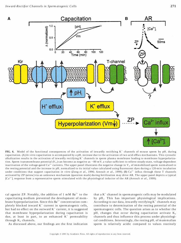
FIG. 6. Model of the functional consequences of the activationcapacitation. (A) In vitro capacitation is accompanied by a pHi increlkalization results in the activation of inwardly rectifying K1 cha
tion. Sperm transmembrane potential (Vm) can become as negativeinactivation of the voltage-gated Ca21 currents. The upper panel illuthe resting potential and the increase in pHi normalized to its initi
nder conditions that support capacitation in vitro (Zeng et al.,activated by ZP (arrow) via an unknown mechanism (question mar[Ca21]i response from a representative sperm stimulated with the p
As discussed above, our findings are the first indication
Copyright © 2001 by Academic Press. All right
hat a K1 channel in spermatogenic cells may be modulatedy pHi. This has important physiological implications.
According to our data, inwardly rectifying K1 channels maycontribute to determination of the resting potential of thespermatogenic cells. The question arises as to whether thepHi changes that occur during capacitation activate Kir
channels and thus influence this process under physiologi-cal conditions. Interestingly, the resting pHi of mammalian
inwardly rectifying K1 channels of mouse sperm by pHi duringue to the activation of two acid efflux mechanisms. This cytosolicin sperm plasma membrane leading to membrane hyperpolariza-0 mV, a value sufficient to relieve steady-state, voltage-dependent
tes the negative change in Vm of immobilized sperm normalized tolue calculated using fluorescent dyes during a 120-min incubation; Arnoult et al., 1999). (B) Ca21 influx through these T channelsring fertilization may drive AR. The upper panel depicts a typicalological inductor of the AR (Arnoult et al., 1999).
ofase dnnelsas 28stra
al va1996k) du
sperm is relatively acidic compared to values routinely
s of reproduction in any form reserved.

std
spa
(l
tKhsv
toacb
pm
riaa
fK
pc(d
A
B
B
B
B
272 Munoz-Garay et al.
observed in somatic cells, as determined by indicator dyes(Zeng et al., 1996), and may impose a functionally quiescenttate. Intracellular pH regulation results from the activa-ion of two acid efflux pathways: one mechanism depen-ent upon extracellular Na1, Cl2, and HCO3
2, and a secondpathway selectively inhibited by arylaminobenzoates (Zenget al., 1996). During storage in the epididymis sperm mustmaintain viability and suppress spontaneous AR. An acidicpHi contributes to the maintenance of an uncapacitatedtate (Parrish et al., 1989) and this is associated withrolonged viability (Yanagimachi, 1994). In addition, ancidic pHi may act as a negative regulator of sperm Kir
channels, thereby maintaining depolarized membrane po-tential values and indirectly preventing unregulated Ca21
entry and thus AR. On the other hand, in mature sperm pHi
may increase during capacitation by more than 0.2 pH units(Zeng et al., 1996), a change sufficient to induce a 0.5- to3-fold increase in the open probability of some Kir channelsChoe et al., 1997; Qu et al., 1999). Hence, under physio-ogical conditions, an increase in pHi would activate Kir
channels, permitting K1 ions to flow out of the cell, drivingthe potential toward VK (Johns et al., 1999), and hyperpo-larizing sperm (Fig. 6). Consistent with this, the g–V curvein Figs. 1B and 1D shows that there is a small but signifi-cant fraction (9–16%) of open channels at potentials morepositive to VK. Inasmuch as K1 ions are at higher concen-rations intracellularly than extracellularly, the opening of
1 channels causes these cations to leave the cell andyperpolarize the membrane. Hyperpolarization duringperm capacitation is sufficient to relieve steady-stateoltage-dependent inactivation of the T-type Ca21 currents
(Santi et al., 1996; Arnoult et al., 1998), recruiting themfrom an inactivated state to a closed state that can besubsequently activated to initiate AR. Indeed, previousstudies have suggested that ZP-evoked Ca21 entry intosperm through T-type Ca21 channels is an essential step inhe AR (Arnoult et al., 1999; Darszon et al., 1999). More-ver, the presence of a novel pHi-sensitive Kir channel maylso help to explain early observations in which the in-rease of [Ca21]i in mammalian sperm induced by mem-rane depolarization with elevated K1 is dependent on an
increase in pHi that results from treatment in alkalinemedium (Babcock et al., 1983; Babcock and Pfeiffer, 1987).
Finally, it should be noted that the native inwardlyrectifying K1 channels of spermatogenic cells share bio-hysical and pharmacological properties with the clonedembers of the Kir2 and Kir4 superfamilies previously
expressed in heterologous systems (Coetzee et al., 1999).There is a strong correlation between structure and func-tion within these subfamilies. Kir2 channels are inwardectifiers and are subject to modulation by various effectorsncluding pHo, intracellular ATP, PKC activity, G-protein,nd Mg21 (Reimann and Ashcroft, 1999), while Kir4 channelctivity is subject to modulation by pHi (Tsai et al. 1995).
Interestingly enough, Kir4.1 has additionally been shown toorm novel functional heteromultimeric channels with
ir5.1 (Pessia et al., 1996; Salvatore et al., 1999), a channel
Copyright © 2001 by Academic Press. All right
protein highly expressed in spermatozoa and spermatogeniccells that does not form functional homomeric channelswhen expressed in heterologous systems. Although thefunctional relevance of Kir5.1 is not yet clear, it has beenroposed recently that it may play an important role inonferring pH sensitivity to Kir4.1 recombinant channelsTucker et al., 2000). Further studies will be needed toetermine whether Kir channels form heteromultimers in
native tissue and whether the inwardly rectifying currentsexpressed in spermatogenic cells are homo- or heterotet-ramers and to determine their molecular identity.
Taken as a whole, our findings about a novel inwardlyrectifying current of mouse spermatogenic cells contributeto establish the repertoire of K1 channels present in thesecells. Our results suggest that this inwardly rectifyingchannel plays an important role during capacitation andmay participate in other physiological events during sper-matogenesis.
ACKNOWLEDGMENTS
This work was supported by grants from DGAPA (UNAM),ICGEB, and CONACyT to A.D. C.M.G. was the recipient of aDGEP (UNAM) predoctoral fellowship. We are grateful to Drs. F.Gomez-Lagunas and A. Hernandez-Cruz for valuable advice andhelpful discussions, Dr. T. Nishigaki for help in preparing illustra-tions and commenting on the manuscript, and Dr. C. L. Trevino forhelpful comments on a draft of this paper.
REFERENCES
Arnoult, C., Villaz, M., and Florman, H. M. (1998). Pharmacologicalproperties of the T-type Ca21 current of mouse spermatogeniccells. Mol. Pharmacol. 53, 1104–1111.rnoult, C., Kaza, I. G., Visconti, P. E., Kopf, G. S., Villaz, M., andFlorman, H. M. (1999). Control of the low voltage-activatedcalcium channel of mouse sperm by egg ZP3 and by membranehyperpolarization during capacitation. Proc. Natl. Acad. Sci.USA 96, 6757–6762.
abcock, D. F., Rufo, G. A. Jr., and Lardy, H. A. (1983). Potassium-dependent increases in cytosolic pH stimulate metabolism andmotility of mammalian sperm. Proc. Natl. Acad. Sci. USA 80,1327–1331.
abcock, D. F., and Pfeiffer, D. R. (1987). Independent elevation ofcytosolic [Ca21] and pH of mammalian sperm by voltage-dependent and pH-sensitive mechanisms. J. Biol. Chem. 262,15041–15047.
aldi, E., Luconi, M., Bonaccorsi, L., Krausz, C., and Forti, G.(1996). Human sperm activation during capacitation and acro-some reaction: Role of calcium, protein phosphorylation andlipid remodelling pathways. Front. Biosci. 1, d189–d205.
rucker, C., and Lipford, G. B. (1995). The human sperm acrosomereaction: Physiology and regulatory mechanisms. An update.
Hum. Reprod. Update 1, 51–62s of reproduction in any form reserved.

D
E
H
H
H
H
H
H
J
J
J
L
N
N
P
P
S
T
T
V
V
W
273Inward-Rectifier Channels in Spermatogenic Cells
Choe, H., Zhou, H., Palmer, L. G., and Sackin, H. (1997). Aconserved cytoplasmic region of ROMK modulates pH sensi-tivity, conductance, and gating. Am. J. Physiol. 273, F516 –F529.
Coetzee, W. A., Amarillo, Y., Chiu, J., Chow, A., Lau, D., McCor-mack, T., Moreno, H., Nadal, M. S., Ozaita, A., Pountney, D.,Saganich, M., Vega-Saenz de Miera, E., and Rudy, B. (1999).Molecular diversity of K1 channels. Ann. NY Acad. Sci. 868,233–285.arszon, A., Labarca, P., Nishigaki, T., and Espinosa, F. (1999). Ionchannels in sperm physiology. Physiol. Rev. 79, 481–510.
spinosa, F., and Darszon, A. (1995). Mouse sperm membranepotential: Changes induced by Ca21. FEBS Lett. 372, 119 –125.
Espinosa, F., De la Vega-Beltran, J. L., Lopez-Gonzalez, I., Delgado,R., Labarca, P., and Darszon, A. (1998). Mouse sperm patch-clamp recordings reveal single Cl2 channels sensitive to niflumicacid, a blocker of the sperm acrosome reaction. FEBS Lett. 426,47–51.
Espinosa, F., Lopez-Gonzalez I., Serrano, C. J., Gasque, G., de laVega-Beltran, J. L., Trevino, C. L., and Darszon, A. (1999). Anionchannel blockers differentially affect T-type Ca21 currents ofmouse spermatogenic cells, a1E currents expressed in Xenopusoocytes and the sperm acrosome reaction. Dev. Genet. 25,103–114.agiwara, S., and Kawa, K. (1984). Calcium and potassium currentsin spermatogenic cells dissociated from rat seminiferous tubules.J. Physiol. 356, 135–149.agiwara, S., Miyazaki, S., Moody, W., and Patlak, J. (1978).Blocking effects of barium and hydrogen ions on the potassiumcurrent during anomalous rectification in the starfish egg.J. Physiol. 279, 167–185.agiwara, S., Miyazaki, S., and Rosenthal, N. P. (1976). Potassiumcurrent and the effect of cesium on this current during anoma-lous rectification of the egg cell membrane of a starfish. J. Gen.Physiol. 67, 621–638.agiwara, S., and Yoshii, M. (1979). Effects of internal potassiumand sodium on the anomalous rectification of the starfish egg asexamined by internal perfusion. J. Physiol. 292, 251–265.amill, O. P., Marty, A., Neher, E., Sakmann, B., and Sigworth, F. J.(1981). Improved patch-clamp techniques for high resolutioncurrent recording from cells and cell-free membrane patches.Pflugers Arch. 391, 85–100.ille, B. (1992). “Ionic Channels in Excitable Membranes,” 2nd ed.,Sinauer, Sunderland, MA.
acob, A., Hurley, I. R., Goodwin, L. O., Cooper, G. W., and Benoff,S. (2000). Molecular characterization of a voltage-gated potas-sium channel expressed in rat testis. Mol. Hum. Reprod. 6,303–313.
ohns, D. C., Marx, R., Mains, R. E., O’Rourke, B., and Marban, E.(1999). Inducible genetic suppression of neuronal excitability.J. Neurosci. 19, 1691–1697.
ow, F., and Numann, R. (1998). Divalent ion block of inwardrectifier current in human capillary endothelial cells andeffects on resting membrane potential. J. Physiol. 512, 119 –128.
ievano, A., Santi, C. M., Serrano, C. J., Trevino, C., Bellve, A. R.,Hernandez-Cruz, A., and Darszon, A. (1996). T-type Ca21 chan-nels and a1E expression in spermatogenic cells, and their possiblerelevance to the sperm acrosome reaction. FEBS Lett. 388,
150–154.Copyright © 2001 by Academic Press. All right
ichols, C. G., and Lopatin, A. N. (1997). Inward rectifier potas-sium channels. Annu. Rev. Physiol. 59, 171–191.ichols, C. G., Makhina, E. N., Pearson, W. L., Sha, Q., andLopatin, A. N. (1996). Inward rectification and implications forcardiac excitability. Circ. Res. 78, 1–7.
arrish, J. J., Susko-Parrish, J. L., and First, N. L. (1989). Capacita-tion of bovine sperm by heparin: Inhibitory effect of glucose androle of intracellular pH. Biol. Reprod. 41, 683–699.
essia, M., Tucker, S. J., Lee, K., Bond, C. T., and Adelman, J. P.(1996). Subunit positional effects revealed by novel hetero-meric inwardly rectifying K1 channels. EMBO J. 15, 2980 –2987.
Qu, Z., Zhu, G., Yang, Z., Cui, N., Li, Y., Chanchevalap, S.,Sulaiman, S., Haynie, H., and Jiang, C. (1999). Identification of acritical motif responsible for gating of Kir2.3 channel by intra-cellular protons. J. Biol. Chem. 274, 13783–13789.
Reimann, F., and Ashcroft, F. M. (1999). Inwardly rectifying potas-sium channels. Curr. Opin. Cell. Biol. 11, 503–508.
Salvatore, L., D’Adamo, M. C., Polishchuk, R., Salmona, M., andPessia, M. (1999). Localization and age-dependent expression ofthe inward rectifier K1 channel subunit Kir 5.1 in a mammalianreproductive system. FEBS Lett. 449, 146–152.
anti, C. M., Darszon, A., and Hernandez-Cruz, A. (1996). Adihydropyridine-sensitive T-type Ca21 current is the main Ca21
current carrier in mouse primary spermatocytes. Am. J. Physiol.271, C1583–C1593.
Schreiber, M., Wei, A., Yuan, A., Gaut, J., Saito, M., and Salkoff, L.(1998). Slo3, a novel pH-sensitive K1 channel from mammalianspermatocytes. J. Biol. Chem. 273, 3509–3516.aglialatela, M., Ficker, E., Wible, B. A., and Brown, A. M.(1995). C-terminus determinants for Mg21 and polyamineblock of the inward rectifier K1 channel IRK1. EMBO J. 14,5532–5541.
Tsai, T. D., Shuck, M. E., Thompson, D. P., Bienkowski, M. J., andLee, K. S. (1995). Intracellular H1 inhibits a cloned rat kidneyouter medulla K1 channel expressed in Xenopus oocytes. Am. J.Physiol. 268, C1173–C1178.ucker, S. J., Imbrici, P., Salvatore, L., D’Adamo, M. C., and Pessia,M. (2000). pH dependence of the inwardly rectifying potassiumchannel, kir5.1, and localization in renal tubular epithelia.J. Biol. Chem. 275, 16404–16407.
Visconti, P. E., Bailey, J. L., Moore, G. D., Pan, D., Olds-Clarke, P.,and Kopf, G. S. (1995). Capacitation of mouse spermatozoa. I.Correlation between the capacitation state and protein tyrosinephosphorylation. Development 121, 1129–1137.isconti, P. E., Galantino-Homer, H., Ning. X., Moore, G. D.,Valenzuela, J. P., Jorgez, C. J., Alvarez, J. G., and Kopf, G. S.(1999). Cholesterol efflux-mediated signal transduction inmammalian sperm. b-cyclodextrins initiate transmem-brane signaling leading to an increase in protein tyrosinephosphorylation and capacitation. J. Biol. Chem. 274, 3235–3242.isconti, P. E., and Kopf, G. S. (1998). Regulation of proteinphosphorylation during sperm capacitation. Biol. Reprod. 59,1–6.u, W. L., So, S. C., Sun, Y. P., Zhou, T. S., Yu, Y., Chung, Y. W.,Wang, X. F., Bao, Y. D., Yan, Y. C., and Chan, H. C. (1998).Functional expression of a Ca21-activated K1 channel in Xenopusoocytes injected with RNAs from the rat testis. Biochim. Bio-
phys. Acta 1373, 360–365.s of reproduction in any form reserved.
Z
274 Munoz-Garay et al.
Yanagimachi, R. (1994). Mammalian fertilization. In “The Physi-ology of Reproduction” (E. Knobil and J. D. Neil, Eds.), pp.189–317. Raven Press, New York.
Yang, J., Jan, Y. N., and Jan, L. Y. (1995). Control of rectification andpermeation by residues in two distinct domains in an inwardrectifier K1 channel. Neuron 14, 1047–1054.
Zeng, Y., Clark, E. N., and Florman, H. M. (1995). Sperm membranepotential: Hyperpolarization during capacitation regulates zonapellucida-dependent acrosomal secretion. Dev. Biol. 171, 554–563.
Zeng, Y., Oberdorf, J. A., and Florman, H. M. (1996). pH regulation
in mouse sperm: Identification of Na1, Cl2, and HCO32-
Copyright © 2001 by Academic Press. All right
dependent and arylaminobenzoate-dependent regulatory mecha-nisms and characterization of their roles in sperm capacitation.Dev. Biol. 173, 510–520.hu, G., Liu, C., Qu, Z., Chanchevalap, S., Xu, H., and Jiang, C.(2000). CO2 inhibits specific inward rectifier K1 channels bydecreases in intra- and extracellular pH. J. Cell Physiol. 183,53–64.
Received for publication August 7, 2000Revised December 29, 2000Accepted January 19, 2001
Published online April 25, 2001
s of reproduction in any form reserved.
Related Documents