Involvement of the β-cinnamomin elicitin in infection and colonisation of cork oak roots by Phytophthora cinnamomi Marília Horta & Paula Caetano & Clara Medeira & Isabel Maia & Alfredo Cravador Accepted: 15 March 2010 / Published online: 15 April 2010 # KNPV 2010 Abstract The virulence of two wild type (PA45 and PA37) and two genetically modified (13C: hygromycin resistant; FATSS: hygromycin resistant and β-cin knock-down) Phytophthora cinnamomi strains towards cork oak (Quercus suber) was assessed via a quanti- tative evaluation of disease symptoms arising from a soil infestation assay, and by a histological analysis of root colonization. Comparison of virulence, as expressed by symptom severity, resulted in the following ranking: highly virulent (wild type strains), medium virulence (strain 13C) and weakly virulent (FATSS). Both transgenic strains were compromised in their virulence, as expressed by symptom severity, but strain 13C was much less affected than FATSS. Microscopic observation showed that the FATSS strain was unable to effectively invade the root, while 13C and the two wild type strains were all able to rapidly colonize the whole root, including the vascular tissue. These results strengthen the notion that elicitins are associated, either directly or indirectly, with the infection process of Phytophthora. Keywords Elicitin . Pathogenicity . Quercus Introduction A number of species within the genus Phytophthora are known to be responsible for heavy losses in crop production, and are recognized to also be among the causative organisms for forest decline and dieback. Their impact in biodiversity and sustainability of forest ecosystems is further enhanced by the interna- tional trade of plants (Brasier 2008) and climate change (Brasier 1996; Bergot et al. 2004; Jung 2009). Thus, a better understanding of the biology and ecology of Phytophthora species is needed for the elaboration of effective control and management protocols for forest ecosystems. The evergreen oak Quercus suber has a particular ecological role. Products provided by cork oak are Eur J Plant Pathol (2010) 127:427–436 DOI 10.1007/s10658-010-9609-x M. Horta : A. Cravador IBB—Institute for Biotechnology and Bioengineering, Centre of Genetics and Biotechnology, Plant and Animal Genomic Group, UTAD, P.O. Box 1013, 5001-801 Vila Real, Portugal M. Horta (*) : A. Cravador Faculdade de Ciências e Tecnologia, Universidade do Algarve, Campus de Gambelas, 8005-139 Faro, Portugal e-mail: [email protected] P. Caetano Faculdade de Ciências e Tecnologia, Universidade do Algarve, Campus de Gambelas, 8005-139 Faro, Portugal C. Medeira : I. Maia Instituto Nacional de Recursos Biológicos—Instituto Nacional de Investigação Agrária, Avenida da República, 2784-505 Oeiras, Portugal

Welcome message from author
This document is posted to help you gain knowledge. Please leave a comment to let me know what you think about it! Share it to your friends and learn new things together.
Transcript
Involvement of the β-cinnamomin elicitin in infectionand colonisation of cork oak roots by Phytophthora cinnamomi
Marília Horta & Paula Caetano & Clara Medeira &
Isabel Maia & Alfredo Cravador
Accepted: 15 March 2010 /Published online: 15 April 2010# KNPV 2010
Abstract The virulence of two wild type (PA45 andPA37) and two genetically modified (13C: hygromycinresistant; FATSS: hygromycin resistant and β-cinknock-down) Phytophthora cinnamomi strains towardscork oak (Quercus suber) was assessed via a quanti-tative evaluation of disease symptoms arising from asoil infestation assay, and by a histological analysis ofroot colonization. Comparison of virulence, asexpressed by symptom severity, resulted in thefollowing ranking: highly virulent (wild type strains),
medium virulence (strain 13C) and weakly virulent(FATSS). Both transgenic strains were compromised intheir virulence, as expressed by symptom severity, butstrain 13C was much less affected than FATSS.Microscopic observation showed that the FATSS strainwas unable to effectively invade the root, while 13Cand the two wild type strains were all able to rapidlycolonize the whole root, including the vascular tissue.These results strengthen the notion that elicitins areassociated, either directly or indirectly, with theinfection process of Phytophthora.
Keywords Elicitin . Pathogenicity .Quercus
Introduction
A number of species within the genus Phytophthoraare known to be responsible for heavy losses in cropproduction, and are recognized to also be among thecausative organisms for forest decline and dieback.Their impact in biodiversity and sustainability offorest ecosystems is further enhanced by the interna-tional trade of plants (Brasier 2008) and climatechange (Brasier 1996; Bergot et al. 2004; Jung 2009).Thus, a better understanding of the biology andecology of Phytophthora species is needed for theelaboration of effective control and managementprotocols for forest ecosystems.
The evergreen oak Quercus suber has a particularecological role. Products provided by cork oak are
Eur J Plant Pathol (2010) 127:427–436DOI 10.1007/s10658-010-9609-x
M. Horta :A. CravadorIBB—Institute for Biotechnology and Bioengineering,Centre of Genetics and Biotechnology,Plant and Animal Genomic Group, UTAD,P.O. Box 1013, 5001-801 Vila Real, Portugal
M. Horta (*) :A. CravadorFaculdade de Ciências e Tecnologia,Universidade do Algarve,Campus de Gambelas,8005-139 Faro, Portugale-mail: [email protected]
P. CaetanoFaculdade de Ciências e Tecnologia,Universidade do Algarve,Campus de Gambelas,8005-139 Faro, Portugal
C. Medeira : I. MaiaInstituto Nacional de Recursos Biológicos—InstitutoNacional de Investigação Agrária,Avenida da República,2784-505 Oeiras, Portugal
obtained from live trees in contrast to other forestspecies. Therefore, cork oak plantations are a perma-nent component of the landscape and a significantbarrier against forest fires, a characteristic due to thenon flammable attribute of the cork. Cork oak exhibitsa high degree of edaphic and climatic adaptability. Itcan grow in different soil types except limestone, inregions with mean monthly temperature ranging from3 to 24°C and mean annual rainfall ranging from 450to 2,000 mm. It also provides an effective shelteragainst sun heat, prevents soil erosion and constitutestogether with holm oak (Q. ilex. subsp. rotundifolia), afirst line barrier against desertification in southernsemi-arid regions characterised by poor soils exposedto adverse climatic conditions. Their disappearancewould leave extensive areas highly unproductive, inparticular in the central and southern regions ofPortugal where Q. suber is the most representativecomponent of the indigenous flora. As far as fauna isconcerned, the cork oak ecosystem is one of the richestin Europe (Correia 1993; Joffre et al. 1999; Correiaand Oliveira 2002).
Quercus suber has an important economic impactin the Mediterranean basin, and is suffering from asevere decline associated with its colonization by theroot parasite Phytophthora cinnamomi (Brasier et al.1993; Moreira-Marcelino 2001; Sanchez et al. 2002;Caetano 2007). This wide-range host necrotrophpersists in the soil or on plant material in the formof chlamydospores, which can either form germ tubesable to infect living root tissue, or produce sporangia.Mature sporangia release motile zoospores, which inturn are chemotactically attracted to living roots,where they proceed to attach, encyst, form germtubes and after penetration develop ramifying hyphaeinto the host plant tissue (Hardham 2005). Somecellular alterations in roots in response to theirinfection and colonisation by Phytophthora spp. havebeen described for Eucalyptus and Acacia spp. and P.cinnamomi (Cahill et al. 1989), Citrus spp. and P.nicotianae and P. palmivora (Widmer et al. 1998),and Q. robur and P. quercina (Brummer et al. 2002).
Phytophthora cinnamomi secretes abundantly cinna-momins, proteins from the elicitin family whose biolog-ical role remains challenging (for a revision see Ponchetet al. 1999). These proteins are thought to work aseffectors and play a key role in the host-Phytophthorainteraction (Kamoun 2007). In compatible interactions,effectors promote infection by suppressing defence
responses, enhancing susceptibility or inducing diseasesymptoms; in incompatible interactions they are recog-nized by the products of resistance genes of the plant,resulting in host cell death and an effective defenceresponse.
Elicitins are encoded by complex gene families.Jiang et al. (2006) proposed a novel classificationsystem for elicitin and elicitin-like genes known invarious Phytophthora species. The elicitins sharing ahighly conserved 98-amino acid domain with sixcysteine residues and a typical elicitin type cysteinespacing pattern were classified as ELIs. Elicitin-likeproteins possessing shorter or longer elicitin domainsthat are more diverse at the sequence level than theconserved domains in ELIs are classified as ELLs. Inthe expression overview made by Jiang et al. (2006)the conclusion was that overall, the expression levelsof eli genes seem to be higher than those of ell genes.
Elicitins belonging to clade ELI-1 obtained by thephylogenetic reconstruction of ELIs and ELLs (Jianget al. 2006) were the first to be discovered and are themost studied. It is known that α-elicitin genes aredown-regulated during the early stages of a successfulinfection of both potato by P. infestans (Kamoun et al.1997) and tobacco by P. parasitica (Colas et al.2001), although the expression of a P. parasitica α-elicitin was maintained throughout the compatibleinteraction with tomato (Colas et al. 2001). Thesilencing of the α-elicitin inf1 gene in P. infestansdid not alter the ability of the pathogen to colonizepotato leaves (Kamoun et al. 1998). Quercinin, a P.quercina β-elicitin, is produced by the pathogenduring its growth in Q. robur infected root tissue;the protein is present within the hyphal cell wall,intercellular spaces and within the invaded host cells(Brummer et al. 2002). In P. cinnamomi, a genecluster consisting of four elicitin genes has beenidentified by Duclos et al. (1998). It was recentlyshown that the β-cinnamomin gene (β-cin) is tran-scribed during the active growth of the pathogenwhen infecting newly germinated cork oak roots, andthat the effect of silencing the β-cin also reduces theexpression level of other elicitin genes in the cluster(Horta et al. 2008); furthermore, at the phenotypiclevel, it delays the in planta growth of the pathogen.
The objectives of the present research work were todescribe in detail the cellular alterations induced by P.cinnamomi when it infects cork oak roots, and help toelucidate the role of elicitins, by comparing the
428 Eur J Plant Pathol (2010) 127:427–436
responses to infection by two wild type strains andtwo transgenic, hygromycin resistant strains, one ofwhich has been β-cin silenced.
Materials and methods
Phytophthora cinnamomi strains
Phytophthora cinnamomi strains PA45 and PA37were isolated from soil of the rhizosphere of corkoak trees in declining stands in the Algarve region(southern Portugal). Isolations were carried out usingpieces of young leaves of Q. suber seedlings as baitsfloated over flooded soil (Moreira-Marcelino 2001).Infected brownish leaflets, which normally appearedafter 3±5 days, were plated onto selective PARPHmedium (Jeffers and Martin 1986) and incubated at25°C in the dark. After 48 h, Phytophthora hyphaewere transferred to V8 agar.
The strains were multiplied by growing in the dark at25°C either on semi-solid V8 agar or in a liquid V8medium. The V8 media were prepared by adding 4.5 gCaCO3 to 330 ml V8 juice (Campbell Soup Co.,Camden, N.J., U.S.A.) and stirring for 30 min. Themixture was transferred to 1,000 ml centrifuge flasksand centrifuged at 2,590 g for 15 min at 20°C. Thesupernatant was then poured into a new flask withoutdisturbing the pellet. The cleared V8-juice was thendiluted 10 fold with distilled water. V8 agar wasprepared by adding 15 g agar per 1,000 ml V8. Bothmedia were sterilized at 121°C for 20 min. The strainswere identified by their morphological characters aswell as by a colorimetric molecular assay (Coelho et al.1997).
As P. cinnamomi proved to be difficult to transform(Horta et al. 2008), it was not possible to obtain astable β-cin knock-down transformant and a hygrom-ycin resistant control strain from the same wild typestrain. Thus, the genetically transformed strains FATSS(derived from PA45) and 13C (derived from PA37)(Horta et al. 2008) were used here. Strain 13C ishygromycin resistant, while FATSS is both hygromycinresistant and β-cin silenced. The transgenic strains weregrown in V8 media supplemented with 250 μg/mlhygromycin. Both the wild type and the transgenicstrains were stored at the Mycological Library of theLaboratory of Molecular Biotechnology and Phytopa-thology, University of Algarve.
Histological studies of colonized root tissue
Histological sections were made from 2-months oldQ. suber seedlings, grown from seeds collected fromone tree located in Alentejo, Portugal. The acornswere surface–disinfested and germinated in sterilevermiculite in a growth chamber (300 μmol.m-2.s-1
light intensity over a 14 h light (20–25°C) / 10 h dark(10–15°C) photoperiod at 60–80% relative humidity).From germination until infection seedlings weregrown in vermiculite irrigated with sterile water.
Seedlings were removed from vermiculite and theroots washed in sterile water. The lateral and fine rootsof four plants per pathogen strain were inoculated byplacing a 2 cm2 V8 agar plug containing activelygrowing mycelium in contact with the root surface at adistance of 5 mm from the root tip for 3 days.
Infected roots (five root pieces per plant) were cutinto 2-3 mm3 fragments and fixed for 16 h at 4°C in0.1 M cacodylate buffer pH 7.2 containing 4% w/vparaformaldehyde and 0.5% v/v glutaraldehyde.(Lherminier et al 2003) The root material was thendehydrated by passing it through an ethanol series,embedded in acrylic embedding agent LR White(London Resin Company) and polymerized for 24 hat 57-58°C (Roland and Vian 1991).
From each strain, 20 root fragments were sectionedby microtome into 2-3 μm thick slices, which werestained in 0.5% aqueous toluidine blue. The sectionswere studied by light microscopy. In total, 400sections per sample were analysed.
Soil infestation assay
Young (12–18 months old) Q. suber plants wereobtained from a nursery (Direcção Geral de Florestas,Montegordo, Portugal) for pathogenicity assessmentof the strains by using a soil infestation assay.
As the genetically transformed strains have animpaired ability to produce zoospores (Horta et al.2008), chlamydospore production was evaluated inorder to be used as initial inoculum in soil infestationassays. The strains were grown in the dark at 25°C inclarified 10% V8 agar during 4 months. Fifteen ml ofsterile water were added to each Petri dish (three perstrain) to cover the mycelium. With an L–shaped rod,360°C rotations were done repeatedly until themycelia were reduced into small pieces, to yield free
Eur J Plant Pathol (2010) 127:427–436 429
chlamydospores. This suspension was transferred toa 50 ml falcon and centrifuged at 2,590 g for10 min. The supernatant was discharged keeping500 µl. This concentrated suspension was homoge-nised and the number of chlamydospores wasdetermined using a Fuchs-Rosenthal countingchamber (Hausser Scientific Company, Horsham,USA). Microscopic counting showed that all strainsproduced similar numbers of chlamydospores (from5x103 to 1,5x10⁴ chlamydospores/ml). Thus, thesepropagules were used as inoculum in the soilinfestation test.
Phytophthora cinnamomi liquid inoculum (Sanchezet al. 2002) was prepared by growing separately thestrains in Petri dishes (Ø=92 mm) (36 plates per strain)containing 20 ml V8 liquid medium at 25°C in thedark for 30 days. At the end of this period, the mediumwas filtered and the mycelium washed. For each strain,the content of three Petri dishes was added to 100 mlsterile water and homogenised for 3 min. Thisinoculum suspension was distributed evenly into the
root ball of each plant. Each plant was then transferredto a pot containing 4 l of substrate (a non sterile 4:1mixture of peat and sand).
Twelve plants per strain were inoculated whileanother twelve plants were used as control, by theaddition of water instead of the inoculum suspension.All the plants were transferred to a greenhouse, groupedinto separate trays to avoid cross-contamination andwaterlogged 2 days per week during 3 months.
At the end of the experiment, each plant wasremoved from its pot, and roots washed to removesoil particles. The severity of foliar symptoms, ie.chlorosis, necrosis or abscission was assessed on a0–4 scale, with 0 representing 0% symptomaticleaves, and 1–3 representing 1–33%, 34–66%, and67–99% symptomatic leaves, respectively, while 4denoted a dead plant (Sanchez et al. 2002). Rootdamage was quantified according to the same 0–4scale, reflecting the abundance of roots and thepercentage of roots showing necrotic symptoms. Ananalysis of variance was performed for both foliar
a
20 µm
b
H
H
20 µm
d
H
H
20 µm
X
P
20 µm c
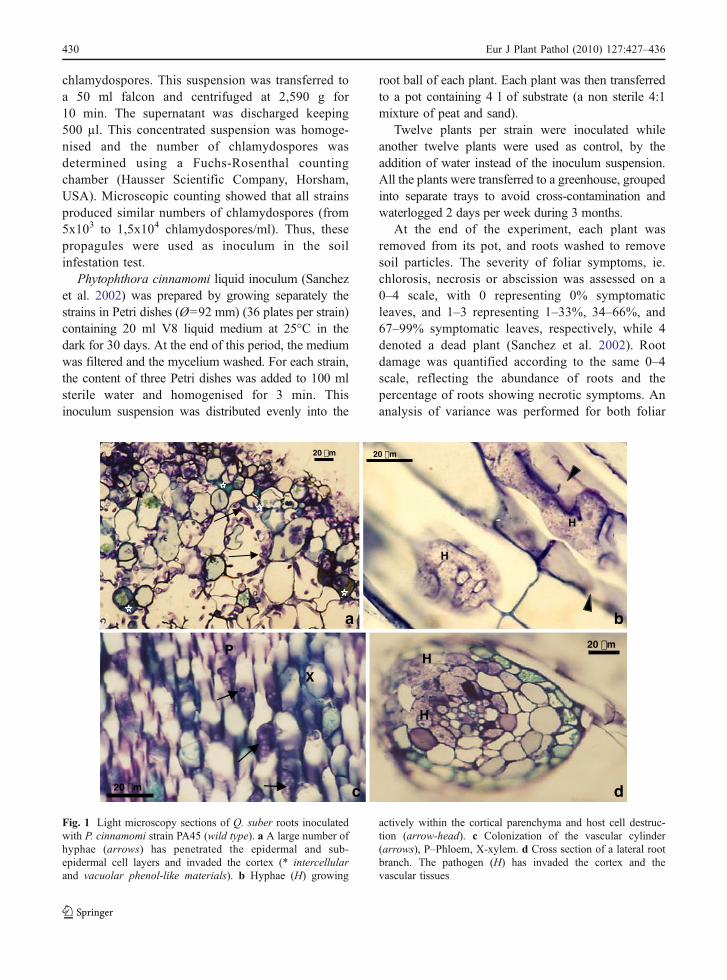
Fig. 1 Light microscopy sections of Q. suber roots inoculatedwith P. cinnamomi strain PA45 (wild type). a A large number ofhyphae (arrows) has penetrated the epidermal and sub-epidermal cell layers and invaded the cortex (* intercellularand vacuolar phenol-like materials). b Hyphae (H) growing
actively within the cortical parenchyma and host cell destruc-tion (arrow-head). c Colonization of the vascular cylinder(arrows), P–Phloem, X-xylem. d Cross section of a lateral rootbranch. The pathogen (H) has invaded the cortex and thevascular tissues
430 Eur J Plant Pathol (2010) 127:427–436
and root symptom severity, and mean values werecompared to each other by the Fisher’s protectedLSD test (Steel and Torrie 1985), using SPSS v14.0software (SPSS for Windows 2001).
Fragments of roots from inoculated and controlplants were washed in running tap water during90 min and plated on PARPH medium for re-isolation of P. cinnamomi (Caetano 2007).
Results
Histological studies of colonized root tissue
All the inoculated roots appeared necrotic at theinoculation point. There was no noticeable differencebetween the apparent infectivity of strains PA45,PA37 and 13C, all being able to rapidly invade the
b
X
10 µm a10 µm
X
10 µm d10 µm
c
*
* *
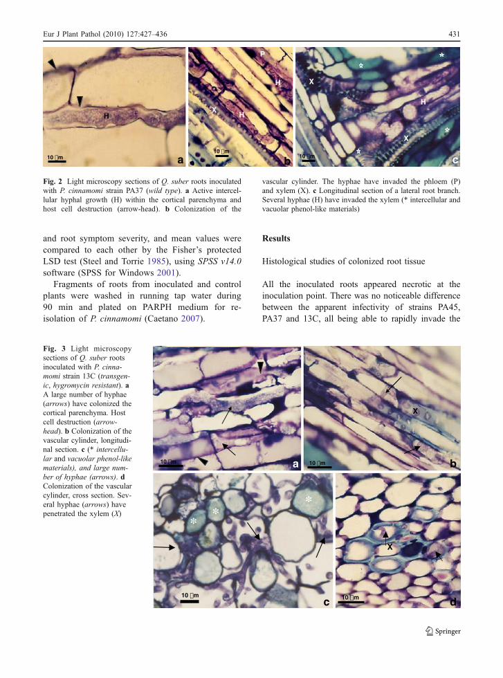
Fig. 3 Light microscopysections of Q. suber rootsinoculated with P. cinna-momi strain 13C (transgen-ic, hygromycin resistant). aA large number of hyphae(arrows) have colonized thecortical parenchyma. Hostcell destruction (arrow-head). b Colonization of thevascular cylinder, longitudi-nal section. c (* intercellu-lar and vacuolar phenol-likematerials), and large num-ber of hyphae (arrows). dColonization of the vascularcylinder, cross section. Sev-eral hyphae (arrows) havepenetrated the xylem (X)
b
X
P
H
H
10 µm
c
X
H
X
10 µm a
H
10 µm
Fig. 2 Light microscopy sections of Q. suber roots inoculatedwith P. cinnamomi strain PA37 (wild type). a Active intercel-lular hyphal growth (H) within the cortical parenchyma andhost cell destruction (arrow-head). b Colonization of the
vascular cylinder. The hyphae have invaded the phloem (P)and xylem (X). c Longitudinal section of a lateral root branch.Several hyphae (H) have invaded the xylem (* intercellular andvacuolar phenol-like materials)
Eur J Plant Pathol (2010) 127:427–436 431
cortical parenchyma both inter–and intracellularly(Figs. 1, 2 and 3). A large number of hyphae werevisible in epidermic, sub-epidermic and inner corticalcell layers. Active growth of the pathogen in thecytoplasm and host cell destruction was observed.Many hyphae ramified throughout the cortex pene-trating xylem and phloem vessels in the samplesobserved. About 10% intercellular spaces and 25%cell vacuoles turned a brilliant blue green whenstained with toluidine blue.
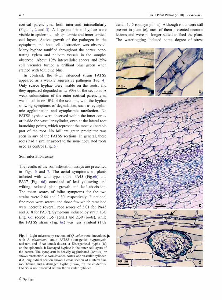
In contrast, the β-cin silenced strain FATSSappeared as a weakly aggressive pathogen (Fig. 4).Only scarce hyphae were visible on the roots, andthey appeared degraded in ca 90% of the sections. Aweak colonization of the outer cortical parenchymawas noted in ca 10% of the sections, with the hyphaeshowing symptoms of degradation, such as cytoplas-mic agglutination and cytoplasmic rarefaction. NoFATSS hyphae were observed within the inner cortexor inside the vascular cylinder, even at the lateral rootbranching points, which represent the most vulnerablepart of the root. No brilliant green precipitate wasseen in any of the FATSS sections. In general, theseroots had a similar aspect to the non-inoculated rootsused as control (Fig. 5)
Soil infestation assay
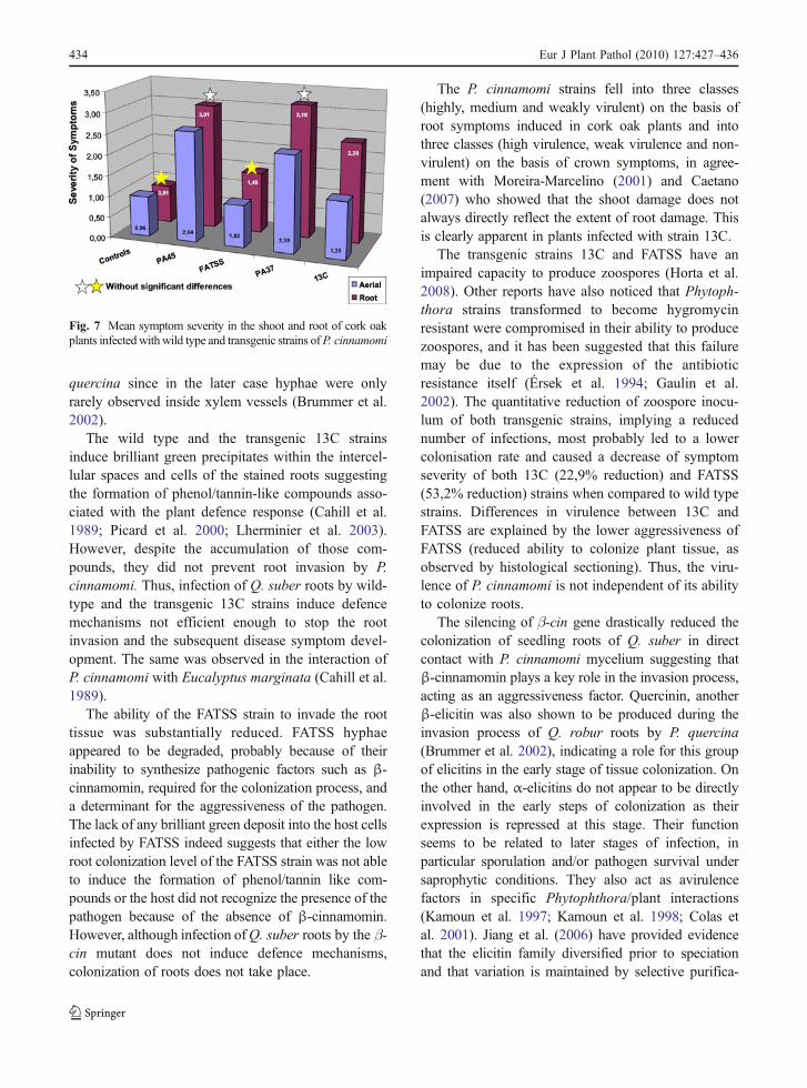
The results of the soil infestation assays are presentedin Figs. 6 and 7. The aerial symptoms of plantsinfected with wild type strains PA45 (Fig.6b) andPA37 (Fig. 6d) consisted of leaf yellowing andwilting, reduced plant growth and leaf abscission.The mean scores of foliar symptoms for the twostrains were 2.64 and 2.30, respectively. Functionalfine roots were scarce, and those few which remainedwere necrotic (overall root scores of 3.01 for PA45and 3.18 for PA37). Symptoms induced by strain 13C(Fig. 6e) scored 1.35 (aerial) and 2.39 (roots), whilethe FATSS strain (Fig. 6c) was less virulent (1.02
aerial, 1.45 root symptoms). Although roots were stillpresent in plant (e), most of them presented necroticlesions and were no longer suited to feed the plant.The waterlogging induced some degree of stress
d40 µm
a
H
10 µm
b10 µm
c200 µm
Fig. 4 Light microscopy sections of Q. suber roots inoculatedwith P. cinnamomi strain FATSS (transgenic, hygromycinresistant and β-cin knock-down). a Disorganized hypha (H)on the epidermis. b Damaged hyphae in the outer cell layers ofthe cortex. The cytoplasm is heavily agglutinated (arrows) orshows rarefaction. c Non-invaded cortex and vascular cylinder.d A longitudinal section shows a cross section of a lateral fineroot branch and a damaged hypha (arrow) on the epidermis.FATSS is not observed within the vascular cylinder
b
432 Eur J Plant Pathol (2010) 127:427–436
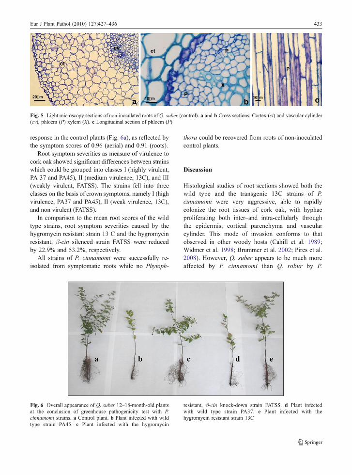
response in the control plants (Fig. 6a), as reflected bythe symptom scores of 0.96 (aerial) and 0.91 (roots).
Root symptom severities as measure of virulence tocork oak showed significant differences between strainswhich could be grouped into classes I (highly virulent,PA 37 and PA45), II (medium virulence, 13C), and III(weakly virulent, FATSS). The strains fell into threeclasses on the basis of crown symptoms, namely I (highvirulence, PA37 and PA45), II (weak virulence, 13C),and non virulent (FATSS).
In comparison to the mean root scores of the wildtype strains, root symptom severities caused by thehygromycin resistant strain 13 C and the hygromycinresistant, β-cin silenced strain FATSS were reducedby 22.9% and 53.2%, respectively.
All strains of P. cinnamomi were successfully re-isolated from symptomatic roots while no Phytoph-
thora could be recovered from roots of non-inoculatedcontrol plants.
Discussion
Histological studies of root sections showed both thewild type and the transgenic 13C strains of P.cinnamomi were very aggressive, able to rapidlycolonize the root tissues of cork oak, with hyphaeproliferating both inter–and intra-cellularly throughthe epidermis, cortical parenchyma and vascularcylinder. This mode of invasion conforms to thatobserved in other woody hosts (Cahill et al. 1989;Widmer et al. 1998; Brummer et al. 2002; Pires et al.2008). However, Q. suber appears to be much moreaffected by P. cinnamomi than Q. robur by P.
ct
a
cv
20µm
Pct
X
P
b10µm
P
c10µm
Fig. 5 Light microscopy sections of non-inoculated roots of Q. suber (control). a and b Cross sections. Cortex (ct) and vascular cylinder(cv), phloem (P) xylem (X). c Longitudinal section of phloem (P)
a b d e c
Fig. 6 Overall appearance of Q. suber 12–18-month-old plantsat the conclusion of greenhouse pathogenicity test with P.cinnamomi strains. a Control plant. b Plant infected with wildtype strain PA45. c Plant infected with the hygromycin
resistant, β-cin knock-down strain FATSS. d Plant infectedwith wild type strain PA37. e Plant infected with thehygromycin resistant strain 13C
Eur J Plant Pathol (2010) 127:427–436 433
quercina since in the later case hyphae were onlyrarely observed inside xylem vessels (Brummer et al.2002).
The wild type and the transgenic 13C strainsinduce brilliant green precipitates within the intercel-lular spaces and cells of the stained roots suggestingthe formation of phenol/tannin-like compounds asso-ciated with the plant defence response (Cahill et al.1989; Picard et al. 2000; Lherminier et al. 2003).However, despite the accumulation of those com-pounds, they did not prevent root invasion by P.cinnamomi. Thus, infection of Q. suber roots by wild-type and the transgenic 13C strains induce defencemechanisms not efficient enough to stop the rootinvasion and the subsequent disease symptom devel-opment. The same was observed in the interaction ofP. cinnamomi with Eucalyptus marginata (Cahill et al.1989).
The ability of the FATSS strain to invade the roottissue was substantially reduced. FATSS hyphaeappeared to be degraded, probably because of theirinability to synthesize pathogenic factors such as β-cinnamomin, required for the colonization process, anda determinant for the aggressiveness of the pathogen.The lack of any brilliant green deposit into the host cellsinfected by FATSS indeed suggests that either the lowroot colonization level of the FATSS strain was not ableto induce the formation of phenol/tannin like com-pounds or the host did not recognize the presence of thepathogen because of the absence of β-cinnamomin.However, although infection ofQ. suber roots by the β-cin mutant does not induce defence mechanisms,colonization of roots does not take place.
The P. cinnamomi strains fell into three classes(highly, medium and weakly virulent) on the basis ofroot symptoms induced in cork oak plants and intothree classes (high virulence, weak virulence and non-virulent) on the basis of crown symptoms, in agree-ment with Moreira-Marcelino (2001) and Caetano(2007) who showed that the shoot damage does notalways directly reflect the extent of root damage. Thisis clearly apparent in plants infected with strain 13C.
The transgenic strains 13C and FATSS have animpaired capacity to produce zoospores (Horta et al.2008). Other reports have also noticed that Phytoph-thora strains transformed to become hygromycinresistant were compromised in their ability to producezoospores, and it has been suggested that this failuremay be due to the expression of the antibioticresistance itself (Érsek et al. 1994; Gaulin et al.2002). The quantitative reduction of zoospore inocu-lum of both transgenic strains, implying a reducednumber of infections, most probably led to a lowercolonisation rate and caused a decrease of symptomseverity of both 13C (22,9% reduction) and FATSS(53,2% reduction) strains when compared to wild typestrains. Differences in virulence between 13C andFATSS are explained by the lower aggressiveness ofFATSS (reduced ability to colonize plant tissue, asobserved by histological sectioning). Thus, the viru-lence of P. cinnamomi is not independent of its abilityto colonize roots.
The silencing of β-cin gene drastically reduced thecolonization of seedling roots of Q. suber in directcontact with P. cinnamomi mycelium suggesting thatβ-cinnamomin plays a key role in the invasion process,acting as an aggressiveness factor. Quercinin, anotherβ-elicitin was also shown to be produced during theinvasion process of Q. robur roots by P. quercina(Brummer et al. 2002), indicating a role for this groupof elicitins in the early stage of tissue colonization. Onthe other hand, α-elicitins do not appear to be directlyinvolved in the early steps of colonization as theirexpression is repressed at this stage. Their functionseems to be related to later stages of infection, inparticular sporulation and/or pathogen survival undersaprophytic conditions. They also act as avirulencefactors in specific Phytophthora/plant interactions(Kamoun et al. 1997; Kamoun et al. 1998; Colas etal. 2001). Jiang et al. (2006) have provided evidencethat the elicitin family diversified prior to speciationand that variation is maintained by selective purifica-
Fig. 7 Mean symptom severity in the shoot and root of cork oakplants infectedwithwild type and transgenic strains ofP. cinnamomi
434 Eur J Plant Pathol (2010) 127:427–436
tion. This strongly suggests that each elicitin group hasits own distinct set of functions. The differentialexpression of each type of elicitin, with highly variableisoelectric points (pI) may be an adaptive response ofthe pathogen to regulate interactions within differentsurrounding environments, regardless of whether theirfunction is distinct or not.
It cannot be excluded that the transformation processitself generated some collateral disruption of thepathogen genome, which may have altered physiolog-ical features of the genetically transformed strains, suchas reducing the aggressiveness of the FATSS strain orthe competitiveness of both transformed strains withinthe non-sterile environment used for the infestation test(Erwin and Ribeiro 1996, Zentmyer 1980). Moreover,HAE-α-cin transcripts are also inhibited in FATSS(Horta et al. 2008) and thus it can not be excluded thatthis protein is involved in the colonization process.However, HAE elicitins have not yet been isolated andstudied. To clarify these issues it would be useful togenerate other elicitin-knock down transformants;unfortunately this is a major technical challenge as P.cinnamomi has proven to be rather recalcitrant totransformation (Horta et al. 2008).
The molecular events associated with variation inpathogenicity of Phytophthora spp. remain the objectof intensive research. In the present work we were ableto show that the pathogenicity of P. cinnamomi isassociated with the production of β-cinnamomin actingas an aggressiveness factor. The high virulence of P.cinnamomi to cork oak roots is primarily a conse-quence of its high aggressiveness although it cannot beexcluded that virulence factors can also be involved.
Acknowledgements This research was financed by the EC—III Framework Programme for Research and TechnologicalDevelopment, co-financed by the European Social Fund (ESF)and by national funding from the Portuguese Ministério daCiência e do Ensino Superior (MCES) (PTDC/AGR-AAM/67628/2006). M. Horta thanks Fundação para a Ciência e aTecnologia (FCT) and ESF (EC—III Framework Programme)for her grant (SFRH/BD/1249/2000).
References
Bergot, M., Cloppet, E., Pérarnaud, V., Déque, M., Marcais, B.,& Desprez-Loustau, M. L. (2004). Simulation of potentialrange expansion of oak disease caused by Phytophthoracinnamomi under climate change. Global Change Biology,10, 1539–1552.
Brasier, C. M. (1996). Phytophthora cinnamomi and oak declinein southern Europe. Environmental constraints includingclimate change. Annales des Sciences Forestières, 53, 347–358.
Brasier, C. M. (2008). The biosecurity threat to the UK andglobal environment from international trade in plants.Plant Pathology, 57, 792–808.
Brasier, C. M., Robredo, F., & Ferraz, J. F. P. (1993). Evidencefor Phytophthora cinnamomi involvement in Iberian oakdecline. Plant Pathology, 42, 140–145.
Brummer, M., Arend, M., Fromm, J., Schlenzig, A., & Oβwald,W. F. (2002). Ultrastructural changes and immunocytochem-ical localization of the elicitin quercinin in Quercus robur L.roots infected with Phytophthora quercina. Physiologicaland Molecular Plant Pathology, 61, 109–120.
Caetano, P. (2007). Envolvimento de Phytophthora cinnamomino declínio de Quercus suber e Q. rotundifolia: estudo dainfluência de factores bióticos e abióticos na progressão dadoença. Possibilidades de controlo químico do declínio.Tese de Doutoramento (p 321) Universidade do Algarve.
Cahill, D., Legge, N., Grant, B., & Weste, G. (1989). Cellular andhistological changes induced by Phytophthora cinnamomi ina group of plant species ranging from fully susceptible tofully resistant. Phytopathology, 79, 417–424.
Coelho, A. C., Cravador, A., Bollen, A., Ferraz, J. F. P., Moreira,A. C., Fauconnier, A., et al. (1997). Highly specific andsensitive non-radioactive identification of Phytophthoracinnamomi. Mycological Research, 101, 1499–1507.
Colas, V., Conrod, S., Venard, P., Keller, H., Ricci, P., &Panabiéres, F. (2001). Elicitin genes expressed in vitro bycertain tobacco isolates of Phytophthora parasitica aredown regulated during compatible interactions. MolecularPlant-Microbe Interactions, 14, 326–335.
Correia, T. P. (1993). Threatened landscape in Alentejo,Portugal: the “montado” and other “agro-silvo-pastoral”systems. Landscape and Urban Planning, 24, 43–48.
Correia, A. V., & Oliveira, A. C. (2002). Principais espéciesflorestais com interesse para Portugal, Zonas de Influên-cia Mediterrânica. Estudos e Informação nº 318, 2ª Ed.,Direcção Geral das Florestas, Lisboa.
Duclos, J., Fauconnier, A., Coelho, A. C., Bollen, A., Cravador, A., &Godfroid, E. (1998). Identification of an elicitin gene cluster inPhytophthora cinnamomi. DNA Sequence—Journal of Se-quencing and Mapping, 9, 231–237.
Érsek, T., Schoelz, J. E., & English, J. T. (1994). Characteriza-tion of selected drug resistant mutants of Phytophthoracapsici P. parasitica and P. citrophthora. Acta Phytopatho-logica et Entomologica Hungarica, 29, 215–229.
Erwin, D. C., & Ribeiro, O. K. (1996). Phytophthora diseasesworldwide. St Paul: American Phytopathological Society.
Gaulin, E., Jauneau, A., Villalba, F., Rickauer, M., Esquerré-Tugayé, M.-T., & Bottin, A. (2002). The CBEL glycoproteinof Phytophthora parasitica var. Nicotianae is involved incell wall deposition and adhesion to cellulosic substrates.Journal of Cell Science, 115, 4565–4575.
Hardham, A. (2005). Pathogen profile: Phytophthora cinnamomi.Molecular Plant Pathology, 6, 589–604.
Horta, M., Sousa, N., Coelho, A. C., Neves, D., & Cravador, A.(2008). In vitro and in vivo quantification of elicitinexpression in Phytophthora cinnamomi. Physiological andMolecular Plant Pathology, 73, 48–57.
Eur J Plant Pathol (2010) 127:427–436 435
Jeffers, N. S., & Martin, J. B. (1986). Comparison of two mediaselective for Phytophthora and Pythium species. PlantDisease, 70, 1038–1043.
Jiang, R. H. Y., Tyler, B. M., Whisson, S. C., Hardham, A. R.,& Govers, F. (2006). Ancient origin of elicitin geneclusters in Phytophthora genomes. Molecular Biology andEvolution, 23, 338–351.
Joffre, R., Rambal, S., & Ratte, P. J. (1999). The dehesa systemof southern Spain and Portugal as a natural ecosystemmimic. Agroforestry Systems, 45, 57–79.
Jung, T. (2009). Beech decline in Central Europe driven by theinteraction between Phytophthora infections and climaticextremes. Forest Pathology, 38, 73–94.
Kamoun, S. (2007). Groovy times: filamentous pathogen effectorsrevealed. Current Opinion in Plant Biology, 10, 358–365.
Kamoun, S., Van West, P., De Jong, A. J., De Groot, K. E.,Vleeshouwers, V. G. A. A., & Govers, F. (1997). A geneencoding a protein elicitor of Phytophthora infestans isdown-regulated during infection of potato. MolecularPlant-Microbe Interactions, 10, 13–20.
Kamoun, S., vanWest, P., Vleeshouwers, V. G., de Groot, K. E., &Govers, F. (1998). Resistance of Nicotiana benthamiana toPhytophthora infestans is mediated by the recognition of theelicitor protein INF1. Plant Cell, 10, 1413–1425.
Lherminier, J., Benhamou, N., Larrue, J., Milat, M.-L. Milat,Boudon-Padieu, E., Nicole, M., et al. (2003). Cytologicalcharacterization of elicitin-induced protection in tobaccoplants infected by Phytophthora parasitica or phyto-plasma. Phytopathology, 93, 1308–1319.
Moreira-Marcelino, A. C. M. (2001). Aspectos da interacçãoentre Phytophthora cinnamomi e a doença do declínio emQ. suber e Q. rotundifolia. Tese de Doutoramento (p 279)Universidade do Algarve.
Picard, K., Ponchet, M., Blein, J. P., Rey, P., Tirilly, Y., &Benhamou, N. (2000). Oligandrin, a proteinaceous mole-cule produced by the mycoparasite Pythium oligandruminduces resistance to Phytophthora parasitica infection intomato plants. Plant Physiology, 124, 379–395.
Pires, N., Maia, I., Moreira, A., & Medeira, C. (2008). Earlystages of infection of cork and holm oak trees byPhytophthora cinnamomi. In J. Vázquez & H. Pereira(Eds.), Suberwood: New challenges for the integration ofcork oak forests and products (pp. 275–282). Spain:Universidad de Huelva.
Ponchet, M., Panabières, F., Milat, M.-L., Mikes, V., Montillet,J.-L., Suty, L., et al. (1999). Are elicitins cryptograms inthe plant—Oomycete communications? Cellular andMolecular Life Sciences, 56, 1020–1047.
Roland, J. C., & Vian, B. (1991). General preparation andstaining of thin sections. In J. L. Hall & C. Hawes (Eds.),Electron microscopy of plant cells (pp. 1–66). London:Academic.
Sánchez, M. E., Caetano, P., Ferraz, J., & Trapero, A. (2002).Phytophthora disease of Quercus ilex in south-westernSpain. Forest Pathology, 32, 5–18.
SPSS for Windows, Rel. 11.0.1. 2001. Chicago: SPSS Inc.Steel, G., & Torrie, J. (1985). Bioestadística: Principios y
Procedimientos. Bogotá: MacGraw-Hill.Widmer, T. L., Graham, J. H., & Mitchell, D. J. (1998).
Histological comparison of fibrous root infection ofdisease-tolerant and susceptible citrus hosts by Phytoph-thora nicotianae and P. palmivora. Phytopathology, 88,389–395.
Zentmyer, G. A. (1980). Phytophthora cinnamomi and thediseases it causes. Monograph No. 10 (p. 96). St Paul:American Phytopathological Society.
436 Eur J Plant Pathol (2010) 127:427–436
Related Documents