Clinical Science (2016) 130, 349–363 doi: 10.1042/CS20150385 Involvement of the Hippo pathway in regeneration and fibrogenesis after ischaemic acute kidney injury: YAP is the key effector Jing Xu* 1 , Pei-Xue Li† 1 , Jun Wu* 1 , Yi-Jun Gao‡, Meng-Xin Yin†, Ye Lin*, Ming Yang*, Dong-Ping Chen*, Hai-Peng Sun§, Zeng-Bo Liu, Xiang-Chen Gu¶, Hong-Ling Huang**, Li-Li Fu*, Hui-Min Hu*, Liang-Liang He*, Wen-Qing Wu†, Zhao-Liang Fei†, Hong-Bin Ji†, Lei Zhang† and Chang-Lin Mei* *Division of Nephrology, Kidney Institute of CPLA, Changzheng Hospital, Second Military Medical University, 415 Feng Yang Road, Shanghai 200003, P .R. China †State Key Laboratory of Cell Biology, Innovation Center for Cell Signaling Network, Institute of Biochemistry and Cell Biology, Shanghai Institutes for Biological Sciences, Chinese Academy of Sciences, 320 Yue-Yang Road, Shanghai 200031, P .R. China ‡Memorial Sloan Kettering Cancer Center, New York, NY 10065, U.S.A. §Division of Nephrology, Central Hospital of TaiAn, TaiAn, Shandong 271000, P .R. China Division of Nephrology, No. 456 Hospital of PLA, Jinan, Shandong 250031, P .R. China ¶Division of Nephrology, Yueyang Hospital, Shanghai 200437, P .R. China **Laboratory of Angiogenesis and Neurovascular link, Vesalius Research Center, VIB, Leuven, B-3000, Belgium Abstract Renal tubule cells can recover after they undergo AKI (acute kidney injury). An incomplete repair of renal tubules can result in progressive fibrotic CKD (chronic kidney disease). Studies have revealed the relationship between tubular epithelial cells and kidney fibrogenesis. However, the underlying mechanism remains unclear. Hippo pathway components were evaluated in complete/incomplete repair of I/R (ischaemia/reperfusion) AKI rat models, HK-2 cells and AKI human renal biopsy samples. We found that the expression levels of the Hippo pathway components changed dynamically during kidney regeneration and fibrogenesis in rat models of I/R-induced AKI and human renal biopsy samples. The transcription cofactor YAP (Yes-associated protein) might be a key effector of renal regeneration and fibrogenesis. Our results showed further that YAP might elicit both beneficial and detrimental effects on I/R AKI. After I/R injury occurred, YAP could promote the repair of the injured epithelia. The constant YAP increase and activation might be related to interstitial fibrosis and abnormal renal tubule differentiation. These results indicate that the proper modulation of the Hippo pathway, specifically the transcription cofactor YAP , during repair might be a potent therapeutic target in AKI–CKD transition after I/R injury. Key words: acute kidney injury, chronic kidney disease, fibrogenesis, Hippo pathway, repair, Yes-associated protein (YAP). INTRODUCTION AKI (acute kidney injury) is a set of clinical syndromes charac- terized by the rapid deterioration of the glomerular filtration rate [1]. After AKI occurs, the kidney possesses profound regener- ative potential. It can also recover either completely or incom- pletely after undergoing regeneration and repair [2,3]. Complete repair leads to the full recovery of the kidney and leaves no evidence of injury. Incomplete repair can cause progressive or- Abbreviations: AKI, acute kidney injury; AQP1, aquaporin 1; BDL, bile duct ligation; CKD, chronic kidney disease; CTGF, connective tissue growth factor; DSS, dextran sodium sulfate; E-cadherin, epithelial cadherin; GAPDH, glyceraldehyde-3-phosphate dehydrogenase; H&E, haematoxylin and eosin; IHC, immunohistochemistry; I/R, ischaemia/reperfusion; Lats1/2, large tumour suppressor 1/2; Mst1/2, mammalian sterile 20-like kinase 1/2; PAS, periodic acid–Schiff; PCNA, proliferating-cell nuclear antigen; RT, reverse transcription; αSMA, α-smooth muscle actin; TEAD, TEA domain family member; Vgll4, vestigial-like protein 4; YAP , Yes-associated protein. Correspondence: Professor Chang-Lin Mei (email [email protected]) or Professor Lei Zhang (email [email protected]). 1 These authors contributed equally to this work. gan dysfunction and increase the risk of CKD (chronic kidney disease) [4]. Studies have substantiated a direct role for the tubule epi- thelium in the pathogenesis of the AKI–CKD transition [5,6]. Despite these previous findings, regulatory signalling during the transition should be clarified further. Kidney repair and regen- eration after AKI occurs through the phenotypic switch of sur- viving epithelial cells from a mature quiescent to a proliferative state [5,6]. Repair and regeneration are similar to nephrogenic 349 c 2016 The Author(s) This is an open access article published by Portland Press Limited and distributed under the Creative Commons Attribution License 3.0

Welcome message from author
This document is posted to help you gain knowledge. Please leave a comment to let me know what you think about it! Share it to your friends and learn new things together.
Transcript

Clinical Science (2016) 130, 349–363 doi: 10.1042/CS20150385
Involvement of the Hippo pathway in regenerationand fibrogenesis after ischaemic acute kidneyinjury: YAP is the key effectorJing Xu*1, Pei-Xue Li†1, Jun Wu*1, Yi-Jun Gao‡, Meng-Xin Yin†, Ye Lin*, Ming Yang*, Dong-Ping Chen*,Hai-Peng Sun§, Zeng-Bo Liu‖, Xiang-Chen Gu¶, Hong-Ling Huang**, Li-Li Fu*, Hui-Min Hu*, Liang-Liang He*,Wen-Qing Wu†, Zhao-Liang Fei†, Hong-Bin Ji†, Lei Zhang† and Chang-Lin Mei*
*Division of Nephrology, Kidney Institute of CPLA, Changzheng Hospital, Second Military Medical University, 415 Feng Yang Road, Shanghai 200003,P.R. China†State Key Laboratory of Cell Biology, Innovation Center for Cell Signaling Network, Institute of Biochemistry and Cell Biology, Shanghai Institutes forBiological Sciences, Chinese Academy of Sciences, 320 Yue-Yang Road, Shanghai 200031, P.R. China‡Memorial Sloan Kettering Cancer Center, New York, NY 10065, U.S.A.§Division of Nephrology, Central Hospital of TaiAn, TaiAn, Shandong 271000, P.R. China‖Division of Nephrology, No. 456 Hospital of PLA, Jinan, Shandong 250031, P.R. China¶Division of Nephrology, Yueyang Hospital, Shanghai 200437, P.R. China**Laboratory of Angiogenesis and Neurovascular link, Vesalius Research Center, VIB, Leuven, B-3000, Belgium
AbstractRenal tubule cells can recover after they undergo AKI (acute kidney injury). An incomplete repair of renal tubules canresult in progressive fibrotic CKD (chronic kidney disease). Studies have revealed the relationship between tubularepithelial cells and kidney fibrogenesis. However, the underlying mechanism remains unclear. Hippo pathwaycomponents were evaluated in complete/incomplete repair of I/R (ischaemia/reperfusion) AKI rat models, HK-2cells and AKI human renal biopsy samples. We found that the expression levels of the Hippo pathway componentschanged dynamically during kidney regeneration and fibrogenesis in rat models of I/R-induced AKI and human renalbiopsy samples. The transcription cofactor YAP (Yes-associated protein) might be a key effector of renalregeneration and fibrogenesis. Our results showed further that YAP might elicit both beneficial and detrimentaleffects on I/R AKI. After I/R injury occurred, YAP could promote the repair of the injured epithelia. The constant YAPincrease and activation might be related to interstitial fibrosis and abnormal renal tubule differentiation. Theseresults indicate that the proper modulation of the Hippo pathway, specifically the transcription cofactor YAP, duringrepair might be a potent therapeutic target in AKI–CKD transition after I/R injury.
Key words: acute kidney injury, chronic kidney disease, fibrogenesis, Hippo pathway, repair, Yes-associated protein (YAP).
INTRODUCTION
AKI (acute kidney injury) is a set of clinical syndromes charac-terized by the rapid deterioration of the glomerular filtration rate[1]. After AKI occurs, the kidney possesses profound regener-ative potential. It can also recover either completely or incom-pletely after undergoing regeneration and repair [2,3]. Completerepair leads to the full recovery of the kidney and leaves noevidence of injury. Incomplete repair can cause progressive or-
Abbreviations: AKI, acute kidney injury; AQP1, aquaporin 1; BDL, bile duct ligation; CKD, chronic kidney disease; CTGF, connective tissue growth factor; DSS, dextran sodium sulfate;E-cadherin, epithelial cadherin; GAPDH, glyceraldehyde-3-phosphate dehydrogenase; H&E, haematoxylin and eosin; IHC, immunohistochemistry; I/R, ischaemia/reperfusion; Lats1/2,large tumour suppressor 1/2; Mst1/2, mammalian sterile 20-like kinase 1/2; PAS, periodic acid–Schiff; PCNA, proliferating-cell nuclear antigen; RT, reverse transcription; αSMA,α-smooth muscle actin; TEAD, TEA domain family member; Vgll4, vestigial-like protein 4; YAP, Yes-associated protein.
Correspondence: Professor Chang-Lin Mei (email [email protected]) or Professor Lei Zhang (email [email protected]).1 These authors contributed equally to this work.
gan dysfunction and increase the risk of CKD (chronic kidneydisease) [4].
Studies have substantiated a direct role for the tubule epi-thelium in the pathogenesis of the AKI–CKD transition [5,6].Despite these previous findings, regulatory signalling during thetransition should be clarified further. Kidney repair and regen-eration after AKI occurs through the phenotypic switch of sur-viving epithelial cells from a mature quiescent to a proliferativestate [5,6]. Repair and regeneration are similar to nephrogenic
349c© 2016 The Author(s) This is an open access article published by Portland Press Limited and distributed under the Creative Commons Attribution License 3.0

J. Xu and others
mesenchyme differentiation during embryonic development[1,10]. This similarity indicates that pathways related to em-bryonic development may play regulatory roles in regeneration,and malfunction may elicit causal effects on the AKI–CKDtransition.
With advances in developmental biology, the signalling path-ways essential for organism development have been identified[7]. One important pathway is the Hippo pathway [8]. Origin-ally discovered in Drosophila, the Hippo pathway is named afterDrosophila Hippo kinase [9]. The core component of the mam-malian Hippo pathway is a three-step kinase cascade composedof Mst1/2 (mammalian sterile 20-like kinase 1/2), Lats1/2 (largetumour suppressor 1/2) and YAP (Yes-associated protein) [11].The Hippo signalling pathway is also necessary to co-ordinatecell proliferation, death and differentiation [10,11]. Mutationsand the down-regulation of Hippo pathway components, such asMst1/2 and Lats1/2, have been observed in multiple tumours.The Hippo pathway major downstream effector YAP functionsas an oncogene in many cancers [13]. Studies have also re-vealed the roles of this pathway in heart, liver and intestinalinjuries and regeneration [14–20]. Nevertheless, the mechan-ism by which YAP affects renal regeneration after AKI oc-curs, specifically the effect on the AKI–CKD transition, remainsunknown.
In the present study, we evaluated the expression of core Hippopathway components and the expression of differentiation andproliferation markers over time in complete/incomplete repair ofI/R (ischaemia/reperfusion) AKI rat models. The results indic-ated that YAP may be a key effector of the Hippo pathway in AKIregulation. In vitro overexpression and RNAi studies revealedproliferative and pro-fibrotic dual-functional effects of YAP onHK-2 cells. Furthermore, we used digitoxin, a YAP WW domainmodulator identified through in silico analysis by Sudol et al. [21],to increase YAP activity in vitro and in vivo. We found that digit-oxin elicited pro-fibrotic and proliferative effects in the I/R AKIrat model. We measured the expression of YAP in AKI humanrenal biopsy samples. YAP protein levels in the cytoplasm andthe nucleus increased in regenerative and poorly differentiatedrenal tubules compared with the control group. The results in-dicate that the Hippo pathway is involved in the regeneration andfibrogenesis stages after acute I/R injury occurs. In these stages,YAP may be the main effector, providing a potential therapeutictarget of the AKI–CKD transition.
MATERIALS AND METHODS
Induction of acute kidney injury in ratsMale Sprague–Dawley rats weighing 200–250 g were purchasedfrom the Shanghai SLAC Laboratory Animal Co., ShanghaiLaboratory Animal Center. The rats were housed in a specificpathogen-free environment at the Animal Center of the SecondMilitary Medical University at optimal temperature with a 12 hlight/12 h dark cycle. The rats were also provided free access towater and standard rat chow. Animal experiments were performedin strict accordance with the animal use protocol approved by the
Institutional Animal Care and Use Committee of the Second Mil-itary Medical University.
The rats were randomly assigned and anaesthetized intraperi-toneally with a ketamine (50 mg/kg) and xylazine (5 mg/kg)mixture. The rats were then placed on a heating table (37 ◦C)to maintain constant body temperature during surgery. Througha midline incision, mild or severe ischaemia was induced byclamping the dissected bilateral renal arteries with non-traumaticmicrovascular clips (Kang Wei Medical Instrument Co.) for 30or 45 min as reported previously [22–24]. The clamps were re-leased, and reperfusion was confirmed visually. Sham operationswere performed by exposing bilateral renal arteries without indu-cing ischaemia. During ischaemic interval and anaesthesia, therats were maintained on warm heating pads (37 ◦C) to maintainbody temperature. Pre-warmed saline (3 ml; 37 ◦C) was instilledintraperitoneally as volume supplement before the abdomen wasclosed in two layers.
The rats were anaesthetized at the indicated times afterischaemia occurred and were sacrificed at 0 h, 12 h, 24 h, 48 h,72 h, 5 days, 7 days, 14 days and 4 weeks (n = 5). The left kid-neys were immediately perfused with PBS from the left ventricle,quickly removed and processed for histological evaluation, pro-tein extraction or RNA extraction. Sham operation groups wereset at 24 h, 48 h, 5 days, 14 days and 4 weeks (n = 3).
Renal functionA blood sample from each animal was extracted from the venacava after the rats were killed. Serum blood urea nitrogen and cre-atinine levels were determined (at the Di-An Medical LaboratoryCenter, Shanghai).
Renal histology and immunohistochemistryThe kidneys were removed and fixed in 4 % (w/v) paraformal-dehyde, embedded in paraffin and cut into 2 μm sections. Kid-ney sections were stained with H&E (haematoxylin and eosin)and PAS (periodic acid–Schiff) for histopathological examina-tion. Sirius Red, Masson’s trichrome and monoclonal anti-mouseαSMA (α-smooth muscle actin) (Sigma, 1:5000 dilution) stainswere used to assess collagen. IHC (immunohistochemistry) wasperformed as described previously [22,23,25]. In brief, the sec-tions were deparaffinized and rehydrated. Endogenous peroxidasewas inactivated by incubating in 3 % H2O2 for 15 min. The sec-tions were incubated in a blocking solution at 37 ◦C for 15 min andin primary antibody overnight at 4 ◦C. The following antibodieswere used: monoclonal rabbit anti-YAP (Cell Signaling Techno-logy, 1:100 dilution) and anti-vimentin (Cell SignalingTechnology, 1:100 dilution), rabbit anti-AQP1 (aquaporin 1)(Millipore, 1:200 dilution), rabbit anti-megalin (Abcam, 1:200 di-lution), rabbit anti-pSmad2/3 (Santa Cruz Biotechnology, 1:5000dilution), rabbit anti-E-cadherin (epithelial cadherin) (SantaCruz Biotechnology, 1:100 dilution), and mouse anti-PCNA(proliferating-cell nuclear antigen) (Cell Signaling Technology,1:4000 dilution). On the following day, the sections were washedthree times with TBST (0.1 %) and incubated with a second-ary antibody at 37 ◦C for 15 min. Positive staining was consec-utively revealed by horseradish peroxidase-labelled streptavidinand diaminobenzidine substrate. Nuclei were counterstained with
350 c© 2016 The Author(s) This is an open access article published by Portland Press Limited and distributed under the Creative Commons Attribution License 3.0

Hippo pathway in ischaemic acute kidney injury
haematoxylin. In the control group, a section was stained withsecondary antibody only or without antibodies.
Renal semi-quantitative morphometric evaluationThe sections from the corticomedullary area of each kidney weregraded in terms of the severity of interstitial fibrosis: 0, no evid-ence of interstitial fibrosis; 1, <10 % involvement; 2, 10 % to<25 % involvement; 3, 25 % to <50 % involvement; 4, 50 %to <75 % involvement; and 5, >75 % involvement. The score ofeach section was recorded as the mean for ten random fields persection at magnification of ×40 [26,27].
The distribution and expression of cytosolic and nuclear YAPin the corticomedullary region were evaluated as the mean forten random fields per section at magnification of ×40: 1 (+/−),>25 % involvement; 2 (+), 25 % to <50 % involvement; 3 (++),50 % to <75 % involvement; and 4 (+++), >75 % involvement.
Cell culture and treatmentThe human HK-2 proximal tubule cell line (CRL-1571, A.T.C.C.,Manassas, VA, U.S.A.) was cultured in the base medium K-SFMsupplemented with EGF (5 ng/ml epidermal growth factor), BPE(50 μg/ml bovine pituitary extract) and 1 % (v/v) FBS [22,24].Cells were seeded at 105 cells/ml before the experiment wasconducted and grown until 90 % confluent. In other experiments,HK-2 cells were incubated with 0.1, 1 or 10 μM digitoxin for24 h and then collected using RIPA buffer. DMSO was added ascontrol.
Overexpression or knockdown of YAP throughlentivirus infectionYAP overexpression or YAP knockdown was performed. In thisprocedure, recombinant lentiviruses were produced by transi-ently transfecting HEK (human embryonic kidney)-293T cells(2×10⁵/ml) with the indicated plasmids along with the pack-aging plasmid VSVG (vesicular stomatitis virus glycoprotein)and �8.2 in accordance with the calcium phosphate precipita-tion method [28]. The following plasmids were used: pCDH-puro-CMV-FLAG, pCDH-puro-YAP, pCDH-puro-YAP S127Aand pCDH-puro-YAP 5SA (provided by Professor Qun-YingLei, Fudan University, Shanghai, China), and pLKO.1-puro-Luc,pLKO.1-puro-shYAP-1 and pLKO.1-puro-shYAP-2 (provided byProfessor Hong-Bin Ji, Shanghai Institute of Biochemistry andCell Biology, Shanghai, China). Plasmid DNA (5 μg) was trans-fected per 6 cm diameter dish. Supernatants were harvested after48 h and filtered through a 0.45 μm pore size filter. HK-2 cells(105/ml) were subsequently infected using the same volume ofthe supernatants supplemented with 1 % (v/v) FBS and selectedwith 2 μg/ml puromycin after 48 h of incubation.
Cell proliferation assayThe cells were plated in triplicate wells in a 96-well plate at 103
cells/well and cultured under normal conditions to evaluate theeffect of YAP interference and overexpression on HK-2 prolifer-ation. The daily number of metabolically active mitochondria andviable cells was determined at D490 by using Thiazolyl Blue Tet-razolium Bromide (Amresco) at a final concentration of 1 mg/ml.
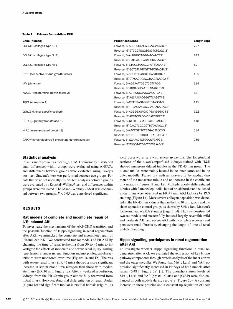
Quantification of mRNA through real-time RT(reverse transcription)–PCRTotal RNA was extracted from the cells or tissues with TRIzolreagent (Invitrogen). RNA (1 μg) from each sample was reverse-transcribed into first-strand cDNA by using ReverTra Ace qPCRRT Master Mix with gDNA Remover (Toyobo). cDNAs werethen subjected to real-time PCR analysis using a CFX96 Real-Time PCR system (Bio-Rad Laboratories) with a SYBR GreenMaster PCR mix (Toyobo). The primers used in this proced-ure are listed in Table 1. All of the samples were amplified induplicate. Each experiment was repeated independently threetimes. Relative gene expression was converted using the 2−��CT
method against the internal control GAPDH (glyceraldehyde-3-phosphate dehydrogenase).
Western blot analysisRenal tissue from the corticomedullary area or HK-2 cells waslysed and denatured at 100 ◦C for 5 min in a SDS buffer andseparated by 8 %, 10 % or 12 % PAGE gels. The proteins weretransferred on to a PVDF membrane, blocked for 1 h with 5 %(w/v) dried non-fat skimmed milk powder in TBST (TBS con-taining 0.1 % Tween 20) and probed with the indicated antibod-ies. The following primary antibodies were used: rabbit antibod-ies against YAP, pYAP, Mst1, pMst1, vimentin (Cell SignalingTechnology, 1:1000 dilution), αSMA (Abcam, 1:1000 dilution),AQP1 (Millipore, 1:1000 dilution) and CTGF (connective tis-sue growth factor) (Santa Cruz Biotechnology, 1:400 dilution);and mouse antibodies against PCNA (Cell Signaling Techno-logy, 1:2000 dilution) and Na+/K+-ATPase (DSHB, 1:1000 di-lution). Horseradish peroxidase-conjugated secondary antibod-ies were applied, and ECL (Pierce) was conducted to detectproteins. A GAPDH-specific antibody (Sigma) was used for load-ing controls on stripped membranes. Quantification of immun-oblots compared the relative ratio of the gray value of pYAP, YAPand pYAP/YAP at all time points with the controls. The resultswere measured using ImageJ software (NIH) and normalized toGAPDH.
Human kidney tissue studyThe use of human tissues was approved by the Institutional Re-view Board of Shanghai Changzheng Hospital. All materials wereretrieved from renal biopsy samples of hospitalized patients in theDepartment of Nephrology, Changzheng Hospital, from 1 Janu-ary 2012 to 30 May 2014. All patients provided written informedconsent. Patients who suffered from AKI, but without underlyingchronic kidney diseases or glomerular nephritis were recruited toexamine YAP expression in their biopsy samples. Dedifferenti-ated status was also confirmed by positive vimentin staining of tu-bular epithelial cells. The demographic information of all patientswas collected. The patients were categorized into the regeneratinggroup (<30 days) and the fibrogenesis group (�30 days) accord-ing to the days of biopsy from the time of onset. The regeneratinggroup was divided further into mild and dialysis subgroups ac-cording to whether dialysis was received during treatment. In thesame period, ten patients who were diagnosed with minor lesionsor mild mesangial proliferation without tubular interstitial injuryin renal pathology were randomly chosen as control subjects.
351c© 2016 The Author(s) This is an open access article published by Portland Press Limited and distributed under the Creative Commons Attribution License 3.0
J. Xu and others
Table 1 Primers for real-time PCR
Gene (human) Primer sequence Length (bp)
COL1A1 (collagen type 1α1) Forward, 5′ -AGGGCCAAGACGAAGACATC-3′ 227
Reverse, 5′ -GTCGGTGGGTGACTCTGAGC-3′
COL3A1 (collagen type 3α1) Forward, 5′ -A AGGGCAGGGAACAACT-3′ 143
Reverse, 5′ -GATGAAGCAGAGCGAGAAG-3′
COL4A1 (collagen type 4α1) Forward, 5′ -CTGCCTGGAGGAGTTTAGAA-3′ 82
Reverse, 5′ -GCTGTAAGCGTTTGCGTAGTA-3′
CTGF (connective tissue growth factor) Forward, 5′ -TGGCTTTAGGAGCAGTGGG-3′ 135
Reverse, 5′ -CTACAGGCAGGTCAGTGAGCA-3′
VIM (vimentin) Forward, 5′ -AGGAAATGGCTCGTCAC-3′ 114
Reverse, 5′ -AGGTGGCAATCTCAATGTC-3′
TGFB1 (transforming growth factor β) Forward, 5′ -ACTACGCCAAGGAGGTCA-3′ 83
Reverse, 5′ -AGCAACACGGGTTCAGGTA-3′
AQP1 (aquaporin 1) Forward, 5′ -CCATTTAGAGGGTGAAGGA-3′ 115
Reverse, 5′ -CTGACAAGAGGGAGTAGAGAA-3′
CDH16 (kidney-specific cadherin) Forward, 5′ -AGGGGAGACACAGAAGGGACT-3′ 122
Reverse, 5′ -ACCACCACCACCACCTCAT-3′
GGT1 (γ -glutamyltransferase 1) Forward, 5′ -GTTTGTGGATGTGACTGAGG-3′ 118
Reverse, 5′ -GAACTCGGGCTTGTAGTAGG-3′
YAP1 (Yes-associated protein 1) Forward, 5′ -AACCGTTTCCCAGACTACCT-3′ 234
Reverse, 5′ -GCTCCTCTCCTTCTATGTTCA-3′
GAPDH (glyceraldehyde-3-phosphate dehydrogenase) Forward, 5′ -GGAAACTGTGGCGTGATG-3′ 285
Reverse, 5′ -TGGGTGTCGCTGTTGAAG-3′
Statistical analysisResults are expressed as means+−S.E.M. For normally distributeddata, differences within groups were evaluated using ANOVA,and differences between groups were evaluated using Tukey’spost-test. Student’s t test was performed between two groups. Fordata that were not normally distributed, analyses between groupswere evaluated by a Kruskal–Wallis H test, and differences withingroups were evaluated. The Mann–Whitney U-test was conduc-ted between two groups. P < 0.05 was considered significant.
RESULTS
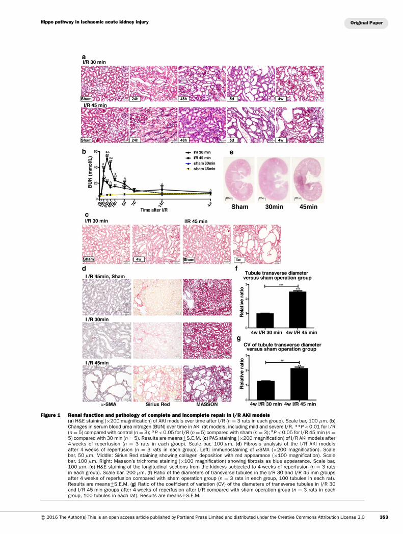
Rat models of complete and incomplete repair ofI/R-induced AKITo investigate the mechanisms of the AKI–CKD transition andthe possible function of Hippo signalling in renal regenerationafter AKI, we mimicked the complete and incomplete repair ofI/R-induced AKI. We constructed two rat models of I/R AKI bychanging the time of renal ischaemia from 30 to 45 min to in-vestigate the effects of moderate and severe renal injury. Duringreperfusion, changes in renal function and morphological charac-teristics were monitored over time (Figures 1a and 1b). The ratswith severe renal injury (I/R 45 min) showed a more significantincrease in serum blood urea nitrogen than those with moder-ate injury (I/R 30 min; Figure 1a). After 4 weeks of reperfusion,kidneys from the I/R 30 min group almost fully recovered frominitial injury. However, abnormal differentiation of renal tubules(Figure 1c) and significant tubular interstitial fibrosis (Figure 1d)
were observed in rats with severe ischaemia. The longitudinalsections of the 4-week-reperfused kidneys stained with H&Eshowed numerous dilated tubules in the I/R 45 min group. Thedilated tubules were mainly located in the inner cortex and in theouter medulla (Figure 1e), with an increase in the median dia-meter of the transverse tubule and an increase in the coefficientof variation (Figures 1f and 1g). Multiple poorly differentiatedtubules with flattened epithelia, loss of brush border and widenedinterstitium were observed in I/R 45 min AKI kidneys by PASstaining (Figure 1c). More severe collagen deposition was detec-ted in the I/R 45 min kidneys than in the I/R 30 min group and thesham operation control group, as shown by Sirius Red, Masson’strichrome and αSMA staining (Figure 1d). Thus we constructedtwo rat models and successfully induced largely reversible mildand moderate AKI and severe AKI with incomplete recovery andpersistent renal fibrosis by changing the length of time of renalpedicle clamping.
Hippo signalling participates in renal regenerationafter AKITo investigate whether Hippo signalling functions in renal re-generation after AKI, we evaluated the expression of key Hippopathway components through protein analysis of the inner cortexand the outer medulla. We found that Mst1, Lats1 and YAP ex-pression significantly increased in kidneys of both models afterrepair (�48 h; Figure 2a) [1]. The phosphorylation levels ofMst1, Lats1 and YAP (pMst1, pLats1 and pYAP) were also en-hanced in both models during recovery (Figure 2b). A constantincrease in these proteins and a constant up-regulation of their
352 c© 2016 The Author(s) This is an open access article published by Portland Press Limited and distributed under the Creative Commons Attribution License 3.0
Hippo pathway in ischaemic acute kidney injury
Figure 1 Renal function and pathology of complete and incomplete repair in I/R AKI models(a) H&E staining (×200 magnification) of AKI models over time after I/R (n = 3 rats in each group). Scale bar, 100 μm. (b)Changes in serum blood urea nitrogen (BUN) over time in AKI rat models, including mild and severe I/R. **P < 0.01 for I/R(n = 5) compared with control (n = 3); �P < 0.05 for I/R (n = 5) compared with sham (n = 3); #P < 0.05 for I/R 45 min (n =5) compared with 30 min (n = 5). Results are means+−S.E.M. (c) PAS staining (×200 magnification) of I/R AKI models after4 weeks of reperfusion (n = 3 rats in each group). Scale bar, 100 μm. (d) Fibrosis analysis of the I/R AKI modelsafter 4 weeks of reperfusion (n = 3 rats in each group). Left: immunostaining of αSMA (×200 magnification). Scalebar, 50 μm. Middle: Sirius Red staining showing collagen deposition with red appearance (×100 magnification). Scalebar, 100 μm. Right: Masson’s trichrome staining (×100 magnification) showing fibrosis as blue appearance. Scale bar,100 μm. (e) H&E staining of the longitudinal sections from the kidneys subjected to 4 weeks of reperfusion (n = 3 ratsin each group). Scale bar, 200 μm. (f) Ratio of the diameters of transverse tubules in the I/R 30 and I/R 45 min groupsafter 4 weeks of reperfusion compared with sham operation group (n = 3 rats in each group, 100 tubules in each rat).Results are means+−S.E.M. (g) Ratio of the coefficient of variation (CV) of the diameters of transverse tubules in I/R 30and I/R 45 min groups after 4 weeks of reperfusion after I/R compared with sham operation group (n = 3 rats in eachgroup, 100 tubules in each rat). Results are means+−S.E.M.
353c© 2016 The Author(s) This is an open access article published by Portland Press Limited and distributed under the Creative Commons Attribution License 3.0

J. Xu and others
Figure 2 Changes in expression of the key Hippo pathway components and differentiation proteins over time(a) Western blot of Mst1/pMst1, Lats1/pLats1 and YAP/pYAP in I/R 30 min rats (n = 3). (b) Western blot of Mst1/pMst1,Lats1/pLats1 and YAP/pYAP in I/R 45 min rats (n = 3). (c) Western blot of CTGF, TEAD2, TEAD3 and Vgll4 in I/R 30 minrats (n = 3). (d) Western blot of CTGF, TEAD2, TEAD3 and Vgll4 in I/R 45 min rats (n = 3). (e) Western blot of differentiationand dedifferentiation markers in I/R 30 min rats (n = 3). (f) Western blot of the differentiation and dedifferentiation markersin I/R 45 min rats (n = 3). (g) Quantification of immunoblots of YAP and pYAP and relative ratio of pYAP to total YAP in 30and I/R 45 min rats normalized to GAPDH (n = 3). con, control.
354 c© 2016 The Author(s) This is an open access article published by Portland Press Limited and distributed under the Creative Commons Attribution License 3.0

Hippo pathway in ischaemic acute kidney injury
phosphorylation status were also detected in rats with severeischaemia during reperfusion. However, this increase returned tolevels similar to those in the sham operation and control groups inrats with mild ischaemia. Quantification of immunoblots showedthat the increase in YAP after injury was more prominent in theI/R 45 min group than in the I/R 30 min group (Figure 2g). Therelative pYAP/total YAP ratio initially decreased at 48 h. This de-crease remained significant at 4 weeks in the I/R 45 min group,indicating that YAP activation increased in severe renal injuriesafter repair and regeneration occurred.
YAP was subjected to IHC staining to detect YAP distributionin kidney tissue sections and to verify the findings shown in Fig-ure 2. YAP was mainly located in tubular epithelial cells and wasevenly distributed in the cytoplasm and the nuclei (Figure 3a). In afew cases, YAP was detected in the renal interstitium. At 4 weeksafter reperfusion, the YAP expression in the kidneys with mild I/RAKI returned to sham operation levels. However, YAP was highlyexpressed in the cytoplasm and nuclei of tubular epithelial cellsof the poorly differentiated tubules in the kidneys with severe I/RAKI. This result indicated the hyperactivation of YAP protein.YAP is the main effector of the Hippo pathway. We thereforeverified whether the expression of YAP activity-related proteinswas altered (Figures 2c and 2d). CTGF is one of the commonly re-ported YAP downstream targets and critical regulatory factors inkidney fibrosis [29]. TEAD2 (TEA domain family member 2) andTEAD3 are TEAD/TEF (transcriptional enhancer factor) tran-scription factors and well-characterized YAP partners [9,12,13].Vgll4 (vestigial-like protein 4) is another Hippo pathway memberthat competes with YAP in terms of binding to TEADs [30,31].Western blot analyses revealed that CTGF, TEAD2 and TEAD3expression changed similarly to YAP expression. However, Vgll4expression changed in a different pattern. The Vgll4 level re-mained significantly decreased at 4 weeks compared with thesham operation and blank control groups. These results indicatethe role of the Hippo pathway in the pathophysiological processof AKI, particularly during the repair stage. Therefore Hippo sig-nalling may regulate renal regeneration after AKI occurs, and thechanges in the irreversible protein levels of the Hippo pathwaycomponents may induce persistent renal fibrosis.
Change in differentiation markers correlates withYAP protein levelsTo determine the underlying mechanism of renal repair afterI/R-induced AKI, we evaluated further the expression of severaldifferentiation markers (Figures 2e and 2f). The dedifferentiationmarkers vimentin, αSMA and PCNA showed changes in proteinlevels similar to YAP during recovery in both rat models (compareFigures 2e and 2f with Figures 2a and 2b). The protein levelsof markers such as Na+/K+-ATPase and AQP1 indicated thatquiescent cells decreased during repair (Figures 2e and 2f). Inthe I/R 30 min group, the expression of Na+/K+-ATPase andAQP1 returned to sham operation and blank control levels after4 weeks of reperfusion (Figure 2e), whereas in the I/R 45 mingroup, the expression of these two differentiation markers ofmature cells was lower than that of the control groups (Figure 2f).Therefore Hippo signalling may regulate renal regeneration andcell redifferentiation after AKI.
We also evaluated the distributions of dedifferentiation mark-ers and quiescent tubular markers with YAP co-expression inpoorly differentiated tubules (Figure 3b). YAP showed a sim-ilar expression pattern to vimentin, pSmad2/3 and PCNA in thepoorly redifferentiated tubules in the corticomedullary areas ofthe I/R 45 min models. By contrast, the expression of the qui-escent tubular cell markers including megalin, AQP1 and E-cadherin in the YAP-positive tubules was decreased or absent(Figure 3b).
YAP is essential for cell proliferation andpro-fibrotic functions in HK-2 cellsTo evaluate the in vitro function of YAP in kidney tubule epithelialcells, we overexpressed or knocked down YAP in HK-2 cells. Wefound that YAP overexpression significantly promoted, whereasknockdown inhibited, HK-2 cell proliferation (Figures 4a and4b). As YAP increased, the mRNA level of fibrotic proteins in-cluding type I, III and IV collagen increased (Figure 4c). CTGFalso increased, indicating that kidney fibrosis was promoted. Con-versely, the mRNA levels of mature tubule cell markers includingkidney-specific cadherin, AQP1 and γ -glutamyltransferase werereduced (Figure 4c). The up-regulation of YAP induced an in-crease in G2/M- and S-phase and a decrease in G0/G1-phase HK-2 cells (Figure 4d). An increase in G2/M-phase proximal renaltubular cells mediates kidney fibrosis after acute injury [22]. TheYAP mutants S127A and 5SA, which escape cytoplasm retentionand exhibit hyperactivity, elicited more prominent effects thanwild-type YAP (Figures 4c and 4d). These results indicate thatan increase in YAP activity not only promotes cell proliferationbut also enhances pro-fibrotic effects.
YAP up-regulation by digitoxin treatment promotesproliferation and fibrogenesis during repair andregeneration of AKIWe regulated the YAP expression levels and found that YAPpromoted cell proliferation and the expression of pro-fibrotic-related genes, as well as the percentage of HK-2 cells in G2/M-and S-phase. We confirmed these findings in I/R-induced AKI ratmodels to determine the underlying mechanism. On the basis ofthe results of the in silico analysis reported by Sudol et al. [21],the cardiac glycoside digitoxin binds to several residues lining thehydrophobic groove within the WW domain of YAP, includingTyr188, Leu190, Thr197 and Trp199, which are critical to the bind-ing of canonical PPXY ligands. Therefore digitoxin can be usedto promote the nuclear translocation of YAP to increase YAPactivity. As the digitoxin concentration increased, the relativeratio of pYAP to total YAP in HK-2 cells decreased in a par-tially dose–response manner after 24 h of incubation (Figure 5a).Compared with the DMSO-treated group, the relative pYAP/totalYAP ratio in rat kidneys also decreased after 4 weeks of reperfu-sion when digitoxin was injected intraperitoneally after AKI wasinduced (Figure 5b). We also evaluated the expression of YAP-related proteins (Figure 5c). The CTGF, TEAD2 and TEAD3protein levels increased further in the digitoxin-treated group. Incontrast, the Vgll4 protein levels decreased further after treat-ment was administered. The dedifferentiation markers vimentin,PCNA and αSMA changed in a similar trend to YAP (Figure 5d).
355c© 2016 The Author(s) This is an open access article published by Portland Press Limited and distributed under the Creative Commons Attribution License 3.0

J. Xu and others
Figure 3 Immunohistochemical staining of YAP and immunostaining of serial sections of YAP co-expressed with differ-entiation markers(a) Immunostaining of YAP (×400 magnification) over time after I/R in 30 and 45 min groups. Scale bar, 20 μm. (b)Immunostaining of serial sections of YAP co-expressed with differentiation markers (×400 magnification) over time afterI/R in 30 and 45 min groups. Scale bar, 20 μm.
356 c© 2016 The Author(s) This is an open access article published by Portland Press Limited and distributed under the Creative Commons Attribution License 3.0

Hippo pathway in ischaemic acute kidney injury
Figure 4 Analysis of YAP overexpression and RNAi in HK-2 cells(a) YAP RNAi slowed the proliferation of HK-2 cells. Results are means+−S.E.M. (b) YAP overexpression (OE) promoted theproliferation of HK-2 cells. Results are means+−S.E.M. (c) Quantification of mRNA levels of pro-fibrogenic and differentiationgenes in YAP-overexpressing HK-2 cells. The mRNA levels are presented as mean+−S.E.M. fold induction over the emptyvector-transfected control. *P < 0.05 compared with control. (d) Cell cycle analysis through propidium iodide staining ofYAP-overexpressing HK-2 cells. Results in the histogram are means+−S.E.M. For comparison of G2/M phases, *P < 0.05compared with control; **P < 0.01 compared with control. CMV, cytomegalovirus; WT, wild-type.
357c© 2016 The Author(s) This is an open access article published by Portland Press Limited and distributed under the Creative Commons Attribution License 3.0

J. Xu and others
The expression of tubular epithelial markers including Na+/K+-ATPase and AQP1 also decreased. Masson’s trichrome analysisshowed more severe renal interstitial fibrosis in the digitoxin-treated group after I/R AKI than in the DMSO-treated group andthe sham operation control group (Figure 5e). This result indic-ates that a decrease in the relative pYAP/total YAP ratio and anincrease in YAP activation could prevent the redifferentiation ofdedifferentiated renal tubular cells and promote proliferation andfibrogenesis during repair and regeneration of AKI, which mightbe a key contributor to the AKI–CKD transition.
Expression and localization of YAP in human kidneybiopsy samplesWe verified YAP expression in the kidney biopsy samples of pa-tients with AKI. A total of 33 patients were recruited. Of thesepatients, 23 were included in the regenerating group and ten wereincluded in the fibrogenesis group. Table 2 summarizes the demo-graphic features of the patients and the ten control subjects withminor lesions or mild mesangial proliferation. AKI of these pa-tients was caused by ischaemia, drug-induced nephrotoxicity andrhabdomyolysis. The patients in the fibrogenesis group were olderthan those in the mild-regenerating group (P = 0.028). Masson’strichrome staining showed that collagen deposition increased inthe kidney tissue of patients in the fibrogenesis group comparedwith the control group (Figure 6a). PAS staining defined the lossof brush borders in the renal tubules in regenerating and fibro-genesis groups. The expression of YAP in the control subjectswas also relatively low (Figures 6a, 6b and 6c). Cytoplasmic YAPsignificantly increased in the mild-regenerating group comparedwith the control group. YAP in the cytoplasm and the nucleusalso increased in the dialysis regenerating and fibrogenesis groupcompared with the control subjects. Nuclear YAP significantlyincreased in these two groups compared with the mild regenerat-ing subgroup (Figures 6a, 6b and 6c). These results are consistentwith our findings in the rat models. Therefore YAP is likely tobe involved in regenerating and fibrotic kidneys after acute I/Rinjury occurs.
DISCUSSION
In contrast with the heart and the central nervous system, whichexhibit limited capability of regeneration after initial injury, kid-neys can undergo dramatic recovery [5,6,32]. Successful regen-eration requires rapid replacement of damaged tubular epithelialcells and reconstitution of normal tubular function. However,maladaptive repair results in progressive fibrotic chronic kidneydisease [1–3]. Studies have revealed the relationship between tu-bular epithelial cells and kidney fibrogenesis, indicating that thesefactors are implicated in the AKI–CKD transition [12,23,26,33].Nevertheless, the underlying mechanism remains unclear.
The present study identified the involvement of the Hippopathway in the pathophysiological process of I/R-induced AKI,particularly during repair. The pattern of changes in the complete(I/R 30 min) repair model differed from that in the incomplete(I/R 45 min) repair model. Although the expression of Mst1,
Lats1 and YAP changed in the same manner, YAP was prob-ably suppressed by upstream pMst1 and pLasts1 and retained inthe cytoplasm in the phosphorylated and deactivated form. Infact, the activation of YAP significantly increased after I/R injuryoccurred, as indicated by the decreased relative ratio of pYAPto total YAP. The activation of YAP was more prominent andremained significant until 4 weeks in the I/R 45 min group.
We evaluated the change in YAP-related proteins. The repor-ted YAP downstream target CTGF and the two YAP partnersTEAD2 and TEAD3 changed in a similar manner to YAP. In con-trast, expression of the YAP-competing protein Vgll4 changed inan opposite manner. These results implicate YAP in the regula-tion of I/R-induced AKI, and the activation of the core upstreamkinases of the Hippo pathway may occur as feedback reactions.
Further analyses of differentiation markers showed that vi-mentin, αSMA and PCNA expression changed in a similar pat-tern to YAP. By contrast, the quiescent polarized cell markerssuch as Na+/K+-ATPase at the basolateral side and AQP1 at theapical side decreased during repair. The level of these two pro-teins did not return to control levels in the severe I/R group after4 weeks of reperfusion, and YAP remained at a high level. IHCanalysis showed further that the majority of YAP was detected inthe cytoplasm and nuclei of tubular epithelial cells. Serial sec-tions revealed that YAP was strongly expressed in the cytosol andnuclei of the epithelial cells of the deformed tubules. YAP wasalso co-expressed with dedifferentiation and proliferation mark-ers, including vimentin, pSmad2/3 and PCNA, but was changedinversely with mature cell markers. These findings indicated thatYAP might be relevant to the proliferation and redifferentiationof the reconstituted epithelia after acute I/R injury. Thus thismechanism might be related to the AKI–CKD transition.
In vitro overexpression and RNAi analyses suggested thatYAP could be involved in the proliferation, pro-fibrogenesis anddedifferentiation of HK-2 cells, which are promoted further bythe nuclear-localized mutants S127A and 5SA. After the YAPagonist digitoxin was administered, proliferation was promoted,redifferentiation was prevented and fibrosis was exacerbated inthe kidneys of I/R-induced AKI rat models. These experimentssuggest that YAP might exhibit bidirectional functions in I/RAKI. During repair, YAP mainly exerted beneficial effects on theproliferation of the injured renal tubular epithelial cells. In con-trast, constant increase and activation of YAP could exacerbatekidney fibrosis and impede the redifferentiation of dedifferenti-ated tubular cells.
We evaluated YAP expression in human AKI biopsy sampleswith different degrees of severity. YAP in the cytosol and nucleusincreased in the mild and dialysis subgroups and in the fibrogen-esis group compared with the control group, which exhibited lowYAP expression. Moreover, YAP signal significantly increased inthe nuclei of cells in the dialysis and fibrogenesis groups com-pared with the mild subgroup. Thus YAP activation increased inthese two groups.
The involvement of YAP in tissue repair and regenera-tion has been observed in other organs in mammals [16–20,26]. YAP mainly elicits beneficial effects on myocardialischaemic injury [34]. In mice suffering from myocardial in-farction, cardiomyocyte-specific inactivation of YAP increases
358 c© 2016 The Author(s) This is an open access article published by Portland Press Limited and distributed under the Creative Commons Attribution License 3.0

Hippo pathway in ischaemic acute kidney injury
Figure 5 YAP agonist treatment in vitro and in vivo in the I/R 45 min AKI models(a) Western blot of the pYAP/YAP ratio in HK-2 cells with an increased gradient of digitoxin concentration after 24 h ofincubation. DMSO was added as control. Quantification of immunoblots of the pYAP/YAP ratio. Results are means+−S.E.M.***P < 0.001 compared with control. (b) Western blot of pYAP and YAP in DMSO- and digitoxin-treated I/R 45 min ratmodels 4 weeks after reperfusion. Quantification of immunoblots comparing pYAP/YAP ratio between groups. Resultsare means+−S.E.M. *P < 0.05 compared with DMSO-treated sham operation group (n = 3); **P < 0.01 compared withDMSO-treated sham operation group (n = 3). (c) Serum blood urea nitrogen (BUN) after I/R injury followed by DMSO ordigitoxin treatment for 4 weeks. Results are means+−S.E.M. **P < 0.01 for digitoxin-treated I/R (n = 4) and sham operation(n = 3) compared with DMSO-treated sham operation (n = 3); *P < 0.05 for DMSO-treated I/R (n = 3) compared withsham operation (n = 3); #P < 0.05 for digitoxin-treated I/R (n = 4) compared with DMSO-treated I/R (n = 3). (d) Westernblot of CTGF, TEAD2, TEAD3 and Vgll4 (n = 3). (e) Western blot of the differentiation and dedifferentiation markers (n =3). (f) Masson’s trichrome staining (×100 magnification) showing fibrosis as blue appearance (n = 3). Scale bar, 100 μm.Quantification results are means+−S.E.M. Digi, digitoxin.
359c© 2016 The Author(s) This is an open access article published by Portland Press Limited and distributed under the Creative Commons Attribution License 3.0

J. Xu and others
Table 2 Demographic features of all AKI patients and control subjectsData are expressed as number (%), mean (with S.D.) or median (with range) as appropriate. eGFR, estimated glomerularfiltration rate.
AKI
Regenerating
Characteristic Total Control Mild Dialysis Fibrogenesis
Number 43 10 15 8 10
Gender
Male 24 6 12 5 1
Female 19 4 3 3 9
Age 48 (15–73) 45 (23–60) 43 (15–66) 42.5 (29–73) 57 (38–69)
Aetiology
Renal ischaemia 11 0 3 5 3
Nephrotoxic drug 3 0 1 0 2
Ischaemia or drug 14 0 7 2 5
Rhabdomyolysis 5 0 4 1 0
Minimal change 7 7 0 0 0
Mild mesangial proliferation 3 3 0 0 0
Time of biopsy after onset – – 8.5 (2.7) 11.1 (5.6) 42.3 (14.1)
eGFR (Cockcroft–Gault) 25.2 (3.71–106.6) 88.45 (73.5–106.6) 22.89 (7.33–68.31) 8.32 (3.71–10.41) 20.34 (7.34–34.12)
myocyte apoptosis and fibrosis [20]. Conversely, restoration ofYAP activity decreases infarct size and improves cardiac func-tion and survival [35–37].
The function of YAP has also been evaluated in organs similarto kidneys, such as the liver and small intestine. In a rat partialhepatectomy model, the Hippo pathway was altered after partialhepatectomy was performed [19]. Increased YAP activation andnuclear localization, as well as decreased activation of its up-stream regulatory kinases Mst1/2 and Lats1/2 were identified at1 day. However, the nuclear YAP protein levels and the expressionof YAP target genes returned to basal levels when the liver reachedthe pre-hepatectomy size, which occurs after 7 days. In chroniccholestatic liver diseases, a more active nuclear YAP is found inthe bile ducts of liver samples from patients with primary scler-osing cholangitis and primary biliary cirrhosis, as well as in thewhole liver of the BDL (bile duct ligation) mouse model [18,26].Ablating Yap in the mouse liver compromises bile duct prolifer-ation, enhances hepatocyte necrosis and suppresses hepatocyteproliferation after BDL through the down-regulation of survivinexpression. Likewise, increased YAP levels have been identifiedin regenerating crypts in a DSS (dextran sodium sulfate)-inducedcolonic regeneration model [20]. Inactivation of YAP severelyimpairs intestinal regeneration after DSS is administered.
YAP is involved in the regulation of proliferation and differ-entiation of adult cells. The inactivation of the Hippo pathwayinduces dedifferentiation of adult hepatocytes into cells bearingprogenitor characteristics [38]. YAP induction is also respons-ible for smooth muscle phenotypic modulation from a contractilephenotype to a synthetic state in rat aortic smooth muscle cellsand murine arterial injury models [39]. During kidney regener-ation, YAP probably performs beneficial and detrimental func-tions. Considering the difference between complete and incom-plete repair of I/R AKI, we proposed that YAP activity should becontrolled in tissue regeneration to successfully complete com-
pensatory proliferation and thus replace injured cells. The in-crease in YAP should be inhibited promptly during regenerationto prevent pro-fibrotic potential and allow appropriate differenti-ation of renal tubules.
The reported YAP antagonists verteporfin and Vgll4-mimicking peptide, which elicit positive effects on tumour growthsuppression in liver and gastric cancers, may cause anti-fibroticeffects in an incomplete repair I/R AKI model [30,40]. Unfortu-nately, these agents were not available in the present study. Weused dobutamine, which can stimulate YAP translocation fromthe nucleus to the cytoplasm in an osteosarcoma cell line andsignificantly suppress YAP–TEAD complex-mediated gene tran-scription [21]. However, the administration of the medicine for28 days after I/R 45 min AKI induction did not improve the le-sions in the incomplete repair models or delay the process ofregeneration (results not shown). The proper modulation of YAPlevels to improve renal tubule differentiation and prevent fibro-genesis might occur more efficiently by targeting its upstreamregulatory proteins in addition to simply changing its phos-phorylation. Kidney conditional Yap-knockout transgenic micewere also not available in the present study. Nevertheless, theprecise stage at which YAP is specifically knocked out after theregeneration stage of I/R AKI may be difficult to achieve usingthe present transgenic mouse system. On the basis of all of theevidence we have observed, the regenerative effect of YAP in thismodel still has to be confirmed in future study.
In conclusion, the Hippo pathway is involved in the regen-eration and fibrogenesis of kidneys after acute I/R injury. YAPalso performs proliferative and pro-fibrotic functions during therecovery. A constant increase and activation of YAP may be re-lated to interstitial fibrosis and abnormal differentiation of renaltubules. Thus the appropriate modulation of this protein duringrepair may be a potent therapeutic target in the AKI–CKD trans-ition after I/R injury occurs.
360 c© 2016 The Author(s) This is an open access article published by Portland Press Limited and distributed under the Creative Commons Attribution License 3.0

Hippo pathway in ischaemic acute kidney injury
Figure 6 Pathological features and YAP immunostaining in AKI human biopsy samples(a) PAS staining (×400 magnification) showing morphology changes in AKI groups. Scale bar, 20 μm (top). Masson’strichrome staining (×400 magnification) showing fibrosis as blue appearance. Scale bar, 20 μm (middle). YAP immunos-taining (×400 magnification). Scale bar, 20 μm (bottom). (b) Cytosolic and nuclear YAP expression in different AKI groupscompared with controls. **P < 0.01 compared with control; ***P < 0.001 compared with control; #P < 0.05 comparedwith mild-regenerating group.
361c© 2016 The Author(s) This is an open access article published by Portland Press Limited and distributed under the Creative Commons Attribution License 3.0

J. Xu and others
CLINICAL PERSPECTIVES
• The incomplete repair after AKI (acute kidney injury) cancause progressive organ dysfunction, but the underlying mech-anism remains unknown. In the present study, we found thatthe Hippo pathway was closely involved in regeneration andfibrogenesis after ischaemic AKI, and we also identified YAPas a key effector.
• YAP might elicit both beneficial and detrimental effects onI/R AKI. After I/R injury occurred, YAP could promote therepair of the injured epithelia. The constant YAP increase andactivation might be related to interstitial fibrosis and abnormalrenal tubule differentiation.
• The results provide new insight into the mechanism of AKI–CKD (chronic kidney disease) transition. A proper modulationof the Hippo pathway and specifically the transcription co-factor YAP during repair might be a potent therapeutic target.
AUTHOR CONTRIBUTION
Jing Xu contributed to the study design, animal model construction,experimentation, data collection, data analysis, data interpretation,literature search, Figure generation and preparation of the paper.Pei-Xue Li and Jun Wu contributed to data collection, data analysis,data interpretation and literature search. Yi-Jun Gao and Meng-Xin Yin contributed to Figure generation and preparation of thepaper. Ye Lin participated in IHC sample preparation. Ming Yang,Dong-Ping Chen, Hai-Peng Sun, Zeng-Bo Liu, Xiang-Chen Gu andHong-Ling Huang carried out the experiments. Li-Li Fu, Hui-Min Hu,Liang-Liang He, Wen-Qing Wu and Zhao-Liang Fei provided relevantinformation. Hong-Bing Ji, Lei Zhang and Chang-Lin Mei supervisedthe entire process. All authors were involved in preparation of thepaper and provided final approval of the submitted and publishedversions.
ACKNOWLEDGEMENTS
We are grateful to Dr Yi-Jun Gao from the Memorial Sloan Ketter-ing Cancer Center, Professor Li Lin from the Translational MedicalCenter, Tongji University Affiliated Dongfang Hospital, Professor XinZeng, Professor Yong Lin and Professor Wei-Fen Xie from the De-partment of Gastroenterology, and Professor Zhi-Guo Mao from theDepartment of Nephrology, Changzheng Hospital, Second MilitaryMedical University, for their selfless support and instruction on thestudy.
FUNDING
This research was supported by the Major Fundamental ResearchProgram of Shanghai Committee of Science and Technology [grantnumber 12DJ1400301], the National Nature Science Fund forYoung Scientists of China [grant number 81200499], the NatureScience Fund for Young Scientists of Shanghai Committee ofScience and Technology [grant number 12ZR1454700], the 973Program of the National Basic Research Program of China [grantnumber 2012CB945001], the Strategic Priority Research Programof the Chinese Academy of Sciences [grant numberXDA01010406], the State Key Program of National Natural
Science of China [grant number 31530043], and the ‘Cross andcooperation in science and technology innovation team’ project ofthe Chinese Academy of Sciences, the Key Projects in the NationalScience and Technology Pillar Program during the Twelfth Five-yearPlan Period [grant number 2011BAI10B07] and the Shanghai TopPriority Key Clinical Disciplines Construction Project.
REFERENCES
1 Bonventre, J.V. and Yang, L. (2011) Cellular pathophysiology ofischemic acute kidney injury. J. Clin. Invest. 121, 4210–4221CrossRef PubMed
2 Bonventre, J.V. (2010) Pathophysiology of AKI: injury and normaland abnormal repair: contributions to nephrology. 165, 9–17
3 Venkatachalam, M.A., Griffin, K.A., Lan, R., Geng, H., Saikumar,P. and Bidani, A.K. (2010) Acute kidney injury: a springboard forprogression in chronic kidney disease. Am. J. Physiol. RenalPhysiol. 298, F1078–F1094 CrossRef PubMed
4 Leung, K.C., Tonelli, M. and James, M.T. (2013) Chronic kidneydisease following acute kidney injury-risk and outcomes. Nat.Rev. Nephrol. 9, 77–85 CrossRef PubMed
5 Cirio, M.C., de Groh, E.D., de Caestecker, M.P., Davidson, A.J.and Hukriede, N.A. (2014) Kidney regeneration: common themesfrom the embryo to the adult. Pediatr. Nephrol. 29, 553–564CrossRef PubMed
6 Bacallao, R. and Fine, L.G. (1989) Molecular events in theorganization of renal tubular epithelium: from nephrogenesis toregeneration. Am. J. Physiol. 257, F913–F924 PubMed
7 Yin, M. and Zhang, L. (2011) Hippo signaling: a hub of growthcontrol, tumor suppression and pluripotency maintenance. J.Genet. Genomics 38, 471–481 CrossRef PubMed
8 Wu, S., Huang, J., Dong, J. and Pan, D. (2003) hippo encodes aSte-20 family protein kinase that restricts cell proliferation andpromotes apoptosis in conjunction with salvador and warts. Cell114, 445–456 CrossRef PubMed
9 Pan, D. (2010) The hippo signaling pathway in development andcancer. Dev. Cell 19, 491–505 CrossRef PubMed
10 Guo, T., Lu, Y., Li, P., Yin, M.X., Lv, D., Zhang, W., Wang, H., Zhou,Z., Ji, H., Zhao, Y. and Zhang, L. (2013) A novel partner ofScalloped regulates Hippo signaling via antagonizingScalloped–Yorkie activity. Cell Res. 23, 1201–1214CrossRef PubMed
11 Zhao, B., Tumaneng, K. and Guan, K.L. (2011) The Hippopathway in organ size control, tissue regeneration and stem cellself-renewal. Nat. Cell Biol. 13, 877–883 CrossRef PubMed
12 Yu, F.X. and Guan, K.L. (2013) The Hippo pathway: regulatorsand regulations. Genes Dev. 27, 355–371 CrossRef PubMed
13 Harvey, K.F., Zhang, X. and Thomas, D.M. (2013) The Hippopathway and human cancer. Nat. Rev. Cancer 13, 246–257CrossRef PubMed
14 Yamamoto, S., Yang, G., Zablocki, D., Liu, J., Hong, C., Kim, S.J.,Soler, S., Odashima, M., Thaisz, J., Yehia, G. et al. (2003)Activation of Mst1 causes dilated cardiomyopathy by stimulatingapoptosis without compensatory ventricular myocyte hypertrophy.J. Clin. Invest. 111, 1463–1474 CrossRef PubMed
15 Odashima, M., Usui, S., Takagi, H., Hong, C., Liu, J., Yokota, M.and Sadoshima, J. (2007) Inhibition of endogenous Mst1prevents apoptosis and cardiac dysfunction without affectingcardiac hypertrophy after myocardial infarction. Circ. Res. 100,1344–1352 CrossRef PubMed
16 Del Re, D.P., Yang, Y., Nakano, N., Cho, J., Zhai, P., Yamamoto, T.,Zhang, N., Yabuta, N., Nojima, H., Pan, D. and Sadoshima, J.(2013) Yes-associated protein isoform 1 (Yap1) promotescardiomyocyte survival and growth to protect against myocardialischemic injury. J. Biol. Chem. 288, 3977–3988CrossRef PubMed
362 c© 2016 The Author(s) This is an open access article published by Portland Press Limited and distributed under the Creative Commons Attribution License 3.0

Hippo pathway in ischaemic acute kidney injury
17 Barry, E.R., Morikawa, T., Butler, B.L., Shrestha, K., de la Rosa,R., Yan, K.S., Fuchs, C.S., Magness, S.T., Smits, R., Ogino, S.et al. (2013) Restriction of intestinal stem cell expansion andthe regenerative response by YAP. Nature 493, 106–110CrossRef PubMed
18 Bai, H., Zhang, N., Xu, Y., Chen, Q., Khan, M., Potter, J.J., Nayar,S.K., Cornish, T., Alpini, G., Bronk, S. et al. (2012)Yes-associated protein regulates the hepatic response after bileduct ligation. Hepatology 56, 1097–1107 CrossRef PubMed
19 Grijalva, J.L., Huizenga, M., Mueller, K., Rodriguez, S., Brazzo, J.,Camargo, F., Sadri-Vakili, G. and Vakili, K. (2014) Dynamicalterations in Hippo signaling pathway and YAP activation duringliver regeneration. Am. J. Physiol. Gastrointest. Liver Physiol.307, G196–G204 CrossRef PubMed
20 Cai, J., Zhang, N., Zheng, Y., de Wilde, R.F., Maitra, A. and Pan,D. (2010) The Hippo signaling pathway restricts the oncogenicpotential of an intestinal regeneration program. Genes Dev. 24,2383–2388 CrossRef PubMed
21 Sudol, M., Shields, D.C. and Farooq, A. (2012) Structures of YAPprotein domains reveal promising targets for development of newcancer drugs. Semin. Cell Dev. Biol. 23, 827–833CrossRef PubMed
22 Yang, L., Besschetnova, T.Y., Brooks, C.R., Shah, J.V. andBonventre, J.V. (2010) Epithelial cell cycle arrest in G2/Mmediates kidney fibrosis after injury. Nat. Med. 16, 535–543CrossRef PubMed
23 Geng, H., Lan, R., Wang, G., Siddiqi, A.R., Naski, M.C., Brooks,A.I., Barnes, J.L., Saikumar, P., Weinberg, J.M. andVenkatachalam, M.A. (2009) Inhibition of autoregulated TGFβsignaling simultaneously enhances proliferation anddifferentiation of kidney epithelium and promotes repair followingrenal ischemia. Am. J. Pathol. 174, 1291–1308CrossRef PubMed
24 Kobayashi, T., Terada, Y., Kuwana, H., Tanaka, H., Okado, T.,Kuwahara, M., Tohda, S., Sakano, S. and Sasaki, S. (2008)Expression and function of the Delta-1/Notch-2/Hes-1 pathwayduring experimental acute kidney injury. Kidney Int. 73,1240–1250 CrossRef PubMed
25 Humphreys, B.D., Czerniak, S., DiRocco, D.P., Hasnain, W.,Cheema, R. and Bonventre, J.V. (2011) Repair of injured proximaltubule does not involve specialized progenitors. Proc. Natl. Acad.Sci. U.S.A. 108, 9226–9231 CrossRef PubMed
26 Gurda, G.T., Zhu, Q., Bai, H., Pan, D., Schwarz, K.B. and Anders,R.A. (2014) The use of Yes-associated protein expression in thediagnosis of persistent neonatal cholestatic liver disease. Hum.Pathol. 45, 1057–1064 CrossRef PubMed
27 Happe, H., van der Wal, A.M., Leonhard, W.N., Kunnen, S.J.,Breuning, M.H., de Heer, E. and Peters, D.J. (2011) AlteredHippo signalling in polycystic kidney disease. J. Pathol. 224,133–142 CrossRef PubMed
28 Gao, Y., Xiao, Q., Ma, H., Li, L., Liu, J., Feng, Y., Fang, Z., Wu, J.,Han, X., Zhang, J. et al. (2010) LKB1 inhibits lung cancerprogression through lysyl oxidase and extracellular matrixremodeling. Proc. Natl. Acad. Sci. U.S.A. 107, 18892–18897CrossRef PubMed
29 Phanish, M.K., Winn, S.K. and Dockrell, M.E. (2010) Connectivetissue growth factor (CTGF, CCN2): a marker, mediator andtherapeutic target for renal fibrosis. Nephron Exp. Nephrol. 114,e83–e92 CrossRef PubMed
30 Jiao, S., Wang, H., Shi, Z., Dong, A., Zhang, W., Song, X., He, F.,Wang, Y., Zhang, Z. and Wang, W. (2014) A peptide mimickingVGLL4 function acts as a YAP antagonist therapy against gastriccancer. Cancer Cell 25, 166–180CrossRef PubMed
31 Zhang, W., Gao, Y., Li, P., Shi, Z., Guo, T., Li, F., Han, X., Feng, Y.,Zheng, C., Wang, Z. et al. (2014) VGLL4 functions as a newtumor suppressor in lung cancer by negatively regulating theYAP–TEAD transcriptional complex. Cell Res. 24, 331–343CrossRef PubMed
32 Wang, H.L., Liu, N.M. and Li, R. (2014) Role of adult residentrenal progenitor cells in tubular repair after acute kidney injury. J.Integr. Med. 12, 469–475CrossRef PubMed
33 Pagtalunan, M.E., Olson, J.L., Tilney, N.L. and Meyer, T.W. (1999)Late consequences of acute ischemic injury to a solitary kidney.J. Am. Soc. Nephrol. 10, 366–373PubMed
34 Papizan, J.B. and Olson, E.N. (2014) Hippo in the path to heartrepair. Circ. Res. 115, 332–334 CrossRef PubMed
35 Lin, Z., von Gise, A., Zhou, P., Gu, F., Ma, Q., Jiang, J., Yau, A.L.,Buck, J.N., Gouin, K.A. and van Gorp, P.R. (2014) Cardiac-specificYAP activation improves cardiac function and survival in anexperimental murine MI model. Circ. Res. 115, 354–363CrossRef PubMed
36 Xin, M., Kim, Y., Sutherland, L.B., Murakami, M., Qi, X., McAnally,J., Porrello, E.R., Mahmoud, A.I., Tan, W., Shelton, J.M. et al.(2013) Hippo pathway effector Yap promotes cardiacregeneration. Proc. Natl. Acad. Sci. U.S.A. 110, 13839–13844CrossRef PubMed
37 Shao, D., Zhai, P., Del Re, D.P., Sciarretta, S., Yabuta, N., Nojima,H., Lim, D.S., Pan, D. and Sadoshima, J. (2014) A functionalinteraction between Hippo–YAP signalling and FoxO1 mediatesthe oxidative stress response. Nat. Commun. 5, 3315PubMed
38 Yimlamai, D., Christodoulou, C., Galli, G.G., Yanger, K.,Pepe-Mooney, B., Gurung, B., Shrestha, K., Cahan, P., Stanger,B.Z. and Camargo, F.D. (2014) Hippo pathway activity influencesliver cell fate. Cell 157, 1324–1338CrossRef PubMed
39 Wang, X., Hu, G., Gao, X., Wang, Y., Zhang, W., Harmon, E.Y., Zhi,X., Xu, Z., Lennartz, M.R., Barroso, M. et al. (2012) Theinduction of Yes-associated protein expression after arterialinjury is crucial for smooth muscle phenotypic modulation andneointima formation. Arterioscler. Thromb. Vasc. Biol. 32,2662–2669 CrossRef PubMed
40 Liu-Chittenden, Y., Huang, B., Shim, J.S., Chen, Q., Lee, S.J.,Anders, R.A., Liu, J.O. and Pan, D. (2012) Genetic andpharmacological disruption of the TEAD–YAP complexsuppresses the oncogenic activity of YAP. Genes Dev. 26,1300–1305 CrossRef PubMed
Received 28 May 2015/30 October 2015; accepted 16 November 2015
Accepted Manuscript online 16 November 2015, doi: 10.1042/CS20150385
363c© 2016 The Author(s) This is an open access article published by Portland Press Limited and distributed under the Creative Commons Attribution License 3.0
Related Documents