2004;64:472-481. Cancer Res David Bernard, Karo Gosselin, Didier Monte, et al. in Keratinocyte Senescence B Transcription Factors κ Involvement of Rel/Nuclear Factor- Updated version http://cancerres.aacrjournals.org/content/64/2/472 Access the most recent version of this article at: Cited Articles http://cancerres.aacrjournals.org/content/64/2/472.full.html#ref-list-1 This article cites by 87 articles, 35 of which you can access for free at: Citing articles http://cancerres.aacrjournals.org/content/64/2/472.full.html#related-urls This article has been cited by 9 HighWire-hosted articles. Access the articles at: E-mail alerts related to this article or journal. Sign up to receive free email-alerts Subscriptions Reprints and . [email protected] Department at To order reprints of this article or to subscribe to the journal, contact the AACR Publications Permissions . [email protected] Department at To request permission to re-use all or part of this article, contact the AACR Publications Research. on March 13, 2014. © 2004 American Association for Cancer cancerres.aacrjournals.org Downloaded from Research. on March 13, 2014. © 2004 American Association for Cancer cancerres.aacrjournals.org Downloaded from

Welcome message from author
This document is posted to help you gain knowledge. Please leave a comment to let me know what you think about it! Share it to your friends and learn new things together.
Transcript

2004;64:472-481. Cancer Res David Bernard, Karo Gosselin, Didier Monte, et al. in Keratinocyte Senescence
B Transcription FactorsκInvolvement of Rel/Nuclear Factor-
Updated version
http://cancerres.aacrjournals.org/content/64/2/472
Access the most recent version of this article at:
Cited Articles
http://cancerres.aacrjournals.org/content/64/2/472.full.html#ref-list-1
This article cites by 87 articles, 35 of which you can access for free at:
Citing articles
http://cancerres.aacrjournals.org/content/64/2/472.full.html#related-urls
This article has been cited by 9 HighWire-hosted articles. Access the articles at:
E-mail alerts related to this article or journal.Sign up to receive free email-alerts
SubscriptionsReprints and
To order reprints of this article or to subscribe to the journal, contact the AACR Publications
Permissions
To request permission to re-use all or part of this article, contact the AACR Publications
Research. on March 13, 2014. © 2004 American Association for Cancercancerres.aacrjournals.org Downloaded from
Research. on March 13, 2014. © 2004 American Association for Cancercancerres.aacrjournals.org Downloaded from
[CANCER RESEARCH 64, 472–481, January 15, 2004]
Involvement of Rel/Nuclear Factor-�B Transcription Factors inKeratinocyte Senescence
David Bernard,1 Karo Gosselin,1 Didier Monte,1 Chantal Vercamer,1 Fatima Bouali,1 Albin Pourtier,2
Bernard Vandenbunder,1 and Corinne Abbadie1
1UMR 8117 CNRS-Institut Pasteur de Lille-Universite Lille 1, Institut de Biologie de Lille, Lille Cedex, France, and 2Laboratoire de Biologie du Developpement, Universite Lille1, Villeneuve d’Ascq Cedex, France
ABSTRACT
After a finite doubling number, normal cells become senescent, i.e.,nonproliferating and apoptosis resistant. Because Rel/nuclear factor(NF)-�B transcription factors regulate both proliferation and apoptosis,we have investigated their involvement in senescence. cRel overexpressionin young normal keratinocytes results in premature senescence, as definedby proliferation blockage, apoptosis resistance, enlargement, and appear-ance of senescence-associated �-galactosidase (SA-�-Gal) activity. Normalsenescent keratinocytes display a greater endogenous Rel/NF-�B DNAbinding activity than young cells; inhibiting this activity in presenescentcells decreases the number of cells expressing the SA-�-Gal marker.Normal senescent keratinocytes and cRel-induced premature senescentkeratinocytes overexpressed manganese superoxide dismutase (MnSOD),a redox enzyme encoded by a Rel/NF-�B target gene. MnSOD transformsthe toxic O2
. into H2O2, whereas catalase and glutathione peroxidaseconvert H2O2 into H2O. Neither catalase nor glutathione peroxidase isup-regulated during cRel-induced premature senescence or during nor-mal senescence, suggesting that H2O2 accumulates. Quenching H2O2 bycatalase delays the occurrence of both normal and premature cRel-induced senescence. Conversely, adding a nontoxic dose of H2O2 to theculture medium of young normal keratinocytes induces a prematuresenescence-like state. All these results indicate that Rel/NF-�B factorscould take part in the occurrence of senescence by generating an oxidativestress via the induction of MnSOD.
INTRODUCTION
When explanted from tissue intoin vitro culture, normal cellsundergo a finite number of divisions and thereafter enter a nonrepli-cative state termed replicative or cellular senescence. This phenome-non was first described for human fibroblasts (1) and extended to avariety of cell types and species, from yeast to mammals (2, 3).Human senescent cells display a characteristic enlarged and spreadmorphology, accompanied by an accumulation of lipofuscine and asignificant percentage of polynucleation (4–11). They are irreversiblycell cycle arrested, preferentially but not exclusively at the G1-Sboundary (12), and they are apoptosis resistant (13–16). They expressa particular senescence-associated�-galactosidase (SA-�-Gal) activ-ity at pH 6 that represents the most universal molecular biomarker ofsenescence (17). Two main mechanisms were shown to promotesenescence: telomere erosion and cumulative oxidative damage (18).The most widely accepted model assumes that telomeres shorten
because of the so-called end-replication problem; this shorteningengages a DNA damage signal that leads to proliferation blockage(19). Reactive oxygen species (ROS) produced along all cell lifeconstantly attack DNA and other macromolecules, resulting in accu-mulation of undegradable oxidized material (lipofuscine) and DNAdamage-induced cell cycle arrest (11, 20–22).
Rel/nuclear factor (NF)-�B proteins are ubiquitous transcriptionfactors recognized as central regulators of cell growth. In vertebrates,the Rel/NF-�B family comprises five members able to form homo- orheterodimers: cRel; RelA (p65); RelB; NF-�B1 (p50); and NF-�B2(p52). Rel/NF-�B dimers are constitutively present in the cytoplasmof numerous cell types, sequestered by I�B proteins. On stimulation,I�B proteins are phosphorylated, ubiquitinated, and degraded, there-fore freeing Rel/NF-�B dimers that become able to translocate to thenucleus and activate the transcription of their target genes. I�B phos-phorylation is under the control of high molecular weight complexeswith I�B kinase (IKK) activity. The best characterized of thesecomplexes, the IKK signalsome, is composed of two IKKs, IKK1 andIKK2, and a regulatory protein, NEMO [NF-�B essential modulator(23, 24)]. RelA�/�, IKK2�/� and NEMO�/� embryos die be-tween embryonic day 12.5 and 15, with massive liver apoptosis due toenhanced sensitivity to tumor necrosis factor (TNF)-� toxicity (25–28), demonstrating that Rel/NF-�B factors behave as antiapoptoticfactors. In contrast, IKK1�/�mouse embryos display multiple de-velopmental defects, including a reduced number of apoptotic cells inthe interdigital areas of the limb bud (29). This demonstration of apossible proapoptotic effect of Rel/NF-�B factors corroborates previ-ous investigations showing that the expression of cRel correlates withthe occurrence of apoptosis in chicken embryos, particularly in themesenchyme of interdigital areas and in thymocytes, and that cRelinduces massive cell death when overexpressed in bone marrow cells(30, 31). Transgenic mice overexpressing RelA or I�B� specificallyin the epidermis show epidermal hypoplasia or hyperplasia, respec-tively (32), indicating that Rel/NF-�B factors can negatively controlproliferation. In NEMO�/� embryos, some skin defects develop,which are due to both hyperproliferation and increased apoptosis ofkeratinocytes (33), suggesting that Rel/NF-�B factors may controlboth proliferation and apoptosis in the same cells, at the same time.Similarly, the disruption of the gene encoding cRel induces a defaultof activation of mature B and T lymphocytes in response to numerousmitogenic stimuli, due to both a cell cycle block and elevated activa-tion-induced apoptosis (34, 35). In this last case, the Rel/NF-�Btranscription factor seems to have a proproliferative effect instead ofan antiproliferative one. cRel also displays an antiapoptotic functionredundant with that of RelA because double knockout mice for RelAand cRel die with liver apoptosis 2 days before single RelA-deficientmice (36).
Because the senescent state implies profound modifications in theproliferative and apoptotic potentials of cells, Rel/NF-�B factors arelikely to participate in the control of this phenomenon. However, veryfew studies have formally investigated this point (for review, see Ref.37). Gel retardation assays or transactivation assays performed withWI38, IMR90, or normal diploid fibroblasts derived from foreskin or
Received 1/6/03; revised 10/17/03; accepted 11/6/03.Grant support: Grants from the Centre National de la Recherche Scientifique,
Universities Lille 1 and Lille 2, the Association pour la Recherche sur le Cancer, the Liguecontre le Cancer (Comite du Nord), the Institut Pasteur de Lille, the Conseil RegionalNord/Pas-de-Calais, and the European Regional Development Fund. K. Gosselin has afellowship from the Institut Pasteur de Lille and the Region Nord/Pas-de-Calais. C.Abbadie is Maıtre de Conferences at the Universite Lille 1.
The costs of publication of this article were defrayed in part by the payment of pagecharges. This article must therefore be hereby markedadvertisement in accordance with18 U.S.C. Section 1734 solely to indicate this fact.
Notes: D. Bernard and K. Gosselin contributed equally to this work. Present address:D. Bernard, Free University of Brussels, Laboratory of Molecular Virology, Faculty ofMedicine CP614, 808 route de Lennik, 1070 Brussels, Belgium.
Requests for reprints: Corinne Abbadie, UMR 8117, Institut de Biologie de Lille, 1rue Calmette, BP 447, 59021 Lille Cedex, France. Phone: 33-3-20-87-11-02; Fax: 33-3-20-87-11-11; E-mail: [email protected].
472
Research. on March 13, 2014. © 2004 American Association for Cancercancerres.aacrjournals.org Downloaded from
oral mucosa indicate that Rel/NF-�B activity and Sp1 and AP1activities either do not change or decrease during senescence (38–42).In contrast, TNF-�-inducible Rel/NF-�B activity was shown to in-crease in senescent smooth muscle cells, due to much faster and moreextensive I�B� degradation than in young cells (43). To our knowl-edge, no such investigations were performed with normal primaryepithelial cells. However, our previous studies on the function of cRelin HeLa epithelial transformed cells have indicated that it inducesproliferation blockage, resistance to TNF-�-induced apoptosis,polynucleation, and lipofuscine accumulation via the up-regulationof manganese superoxide dismutase (MnSOD) and the generation ofROS (44, 45), i.e., biological effects and mechanisms reminiscent ofcellular senescence. Our aim in this study was to formally establishthe involvement of Rel/NF-�B transcription factors in cellular senes-cence by using primary epithelial cells that normally senesce in vitro(normal human keratinocytes). We demonstrate that (a) overexpress-ing cRel in young keratinocytes induces a premature senescent phe-notype, (b) the endogenous Rel/NF-�B activity increases during nor-mal keratinocyte senescence, (c) reducing this Rel/NF-�B activitywith pharmacological inhibitors decreases the appearance of the SA-�-Gal marker, (d) both cRel-induced premature senescence and nor-mal senescence are accompanied by an increase in MnSOD expres-sion, and (e) the occurrence of both cRel-induced prematuresenescence and normal senescence relies on the accumulation ofhydrogen peroxide (H2O2).
MATERIALS AND METHODS
Cell Culture and Reagents. Normal human epithelial keratinocytes(NHEKs), purchased from Clonetics (CC-2501), were obtained from threedifferent donors, all females, but of different races and different ages (Cauca-sian, 65 years old; black, 58 years old; black, 33 years old). They were grownat 37°C in an atmosphere of 5% CO2 in KGM-2 BulletKit medium consistingof modified MCBD 153 with 0.15 mM calcium and supplemented with bovinepituitary extract, epidermal growth factor, insulin, hydrocortisone, transferrin,and epinephrin (CC-3107; Clonetics). Such a serum-free, low-calcium mediumwas shown to minimize keratinocyte terminal differentiation (46). Cells wereseeded as recommended by the supplier and passaged at 70% confluence. Thenumber of population doublings (PDs) was calculated at each passage by usingthe following equation: PD � ln(number of collected cells/number of platedcells)/ln2. Catalase and gliotoxine were purchased from Calbiochem, andsulfasalazine was purchased from Sigma.
Adenoviral Vectors. The open reading frame part of the human c-relcDNA (47) was amplified by PCR using the High Fidelity PCR Master kit(Roche) according to the manufacturer’s recommendations with the followingoligonucleotides: RelForward, 5�-AAGCTTACCATGGCCTCCGGTGCG-TATAAC-3�; and RelReverse, 5�-GATCTCTAGATTATACTTGAAAAAA-TTCATATGGAAAGG-3�. The amplification product was inserted into thepAdCMV2 vector. Recombinant adenovirus vectors (AdRel) were obtained byhomologous recombination in Escherichia coli as described in Ref. 48 (detailsare available on request). Viral stocks were then created as described in Ref.49. The generation of a recombinant adenovirus encoding green fluorescenceprotein (AdGFP) was described in Ref. 50. Viral titers were determined by aplaque assay on 293 cells and defined as plaque-forming units/ml. Cells wereinfected by adding virus stocks directly to the culture medium at an inputmultiplicity of 100 viral particles/cell.
Western Blotting. Infected cells were lysed directly in the SDS-PAGEloading buffer [125 mM Tris-HCl (pH 6.8), 4% SDS, 10% glycerol, 0.02%bromphenol blue, and 10% �-mercaptoethanol]. Noninfected cells were lysedin a solution of 27.5 mM HEPES (pH 7.6), 1.1 M urea, 0.33 M NaCl, 0.1 M
EGTA, 2 mM EDTA, 60 mM KCl, 1 mM DTT, and 1.1% NP40, and the totalprotein concentration was measured with the Bio-Rad protein assay. Nuclearextracts were done as described for EMSA analysis. Proteins were resolved bySDS-PAGE and transferred to nitrocellulose membranes (Hybond-C extra;Amersham). Equal loading was verified after a Ponceau Red coloration of themembranes. Primary antibodies used were antihuman cRel mouse IgG1 (sc-
6955; Santa Cruz Biotechnology), antihuman I�B� mouse IgG1 (sc-1643;Santa Cruz Biotechnology), antihuman histone H2A rabbit IgG (sc-10807;Santa Cruz Biotechnology), antihuman MnSOD sheep IgG (Calbiochem),antihuman Cu/Zn superoxide dismutase (Cu/ZnSOD) sheep IgG (Calbio-chem), antihuman catalase sheep immunoglobulin (The Binding Site), antihu-man glutathione peroxidase (GPX) sheep immunoglobulin (The Binding Site),and antihuman actin goat IgG (sc-1616; Santa Cruz Biotechnology). Secondaryantibodies used were a peroxidase-conjugated rabbit antisheep IgG (JacksonImmunoResearch Laboratories), a peroxidase-conjugated goat antimouse IgG(Jackson ImmunoResearch Laboratories), or a peroxidase-conjugated donkeyantirabbit IgG. Peroxidase activity was revealed using an enhanced chemilu-minescence kit (Amersham).
Immunofluorescence. Cells were fixed with 4% paraformaldehyde in PBSand permeabilized with 0.2% Triton X-100. Cells were incubated with anti-human cRel mouse IgG1 (sc-6955; Santa Cruz Biotechnology), washed threetimes with PBS, and incubated with Rhodamine Red-conjugated antimouseIgG (715-296-150; Jackson ImmunoResearch Laboratories). Nuclei werestained by Hoechst 33258 at 1 �g/ml for 3 min.
Semiquantitative Reverse Transcription-PCR. Cells were homogenizedin Trizol (Gibco-BRL), and total RNAs were isolated according to the man-ufacturer’s recommendations. cDNAs were synthesized using the Gene AmpRNA PCR kit (Perkin-Elmer) and amplified with the gene Amp 9600 PCRsystem (Perkin-Elmer) in a final volume of 50 �l of buffer containing 2.5 �lof the retrotranscription product, all four deoxynucleotide triphosphates at 150�M, MgCl2 (3 mM for cRel, 2 mM for others), 1 unit of Taq gold polymerase(Roche), and each primer at 1 �M. Primers used were as follows: cRel forward,AGAGGGGAATGCGTTTTAGATACA; cRel reverse, CAGGGAGAAAAA-CTTGAAAACACA; I�B� forward, CGCCCAAGCACCCGGATACAGC;I�B� reverse, TGGGGTCAGTCACTCGAAGCACAA; and primer for �-actin was as described in Ref. 51. Thirty cycles were done at 94°C for 1 min;53.3°C (cRel), 55°C (�-actin), or 56.9°C (I�B�) for 1 min; and 72°C for 1 min,with an initial step of 5 min at 95°C. PCR product lengths were 420 (cRel), 503(I�B�), and 661 bp (�-actin).
Bromodeoxyuridine (BrdU) Incorporation Assays, Apoptosis Assays,and SA-�-Gal Assays. To mark proliferating cells, cells were incubated withBrdU (Roche) at 10 �M for 6 h. Cells were subsequently fixed and permeabi-lized as described above and incubated with 40 units/ml DNase I (Promega)and 20 units/ml Exonuclease III (Roche) for 30 min at 37°C. BrdU wasrevealed by incubations with anti-BrdU mouse IgG (Dako) and RhodamineRed-conjugated antimouse IgG (715-296-150; Jackson ImmunoResearch Lab-oratories). Apoptosis was induced by treating cells with recombinant humanTNF-� (10 ng/ml; R&D Systems) and cycloheximide (10 �g/ml; Sigma)overnight. Cells were fixed as described above. Apoptotic cells were identifiedby phase-contrast microscopy according to their condensation and their begin-ning to detach from dish. SA-�-Gal assays were done as described in Ref. 17.
Electophoretic Mobility Shift Assays. Nuclear extracts were prepared asdescribed in Ref. 52. Nuclear protein concentrations were measured with theBio-Rad protein assay. The �B (5�-AGT-TGA-GGG-GAC-TTT-CCC-AGG-C-3�), m�B (5�-AGT-TGA-GGC-GAC-TTT-CCC-AGG-C-3�), and Sp1 (5�-ATT-CGA-TCG-GGG-CGG-GGC-GAG-C-3�) probes (from Promega orSanta Cruz Biotechnology) were labeled according to the recommendations ofPromega and purified using QIAquick Nucleotide Removal kit (28304; Qia-gen). Two �g of nuclear extract were incubated with 0.035 pmol of radiola-beled probe according to the recommendations of Promega. Cold competitionswere performed by preincubating nuclear extracts with a 100-, 10-, or 1-foldexcess of cold �B or Sp1 probe before the incubation with the radiolabeledprobe. For supershift experiments, nuclear extracts were preincubated with 2�l of anti-cRel, anti-RelA, or anti-p50 antibodies (antibodies used were thosedescribed in Ref. 53) before the incubation with the radiolabeled �B probe.DNA-protein complexes were separated from unbound probe by migration onnative 4% polyacrylamide gels at 200 V for 2 h.
Statistics. P calculations were performed with ANOVA (StatView). Dif-ferences were considered significant when P was �0.05.
RESULTS
Overexpression of cRel in Young Primary Keratinocytes.NHEKs are able to achieve approximately 20 PDs under our cultureconditions before they reach the senescence growth plateau and dis-
473
NF-�B AND THE CONTROL OF SENESCENCE
Research. on March 13, 2014. © 2004 American Association for Cancercancerres.aacrjournals.org Downloaded from

play the characteristic senescent morphology. The ability of cRel toinduce senescence was tested by describing the effects of its overex-pression in young NHEKs at 5 PDs and comparing them with thephenotype of senescent NHEKs at 20 PDs. We have constructed arecombinant adenovirus encoding cRel (AdRel) as a vector to over-express cRel. Cells infected with AdGFP and noninfected cells wereused as controls (54).
To check the overexpression of cRel in AdRel-infected youngNHEKs, reverse transcription-PCR, immunoblotting, and immunoflu-orescence experiments were performed 24 h after infection. As shownin Fig. 1A, cRel mRNA and protein were detected in great amounts inAdRel-infected cells. Immunofluorescence experiments revealed cReloverexpression in 70–100% of cells (Fig. 1B). The overexpressedcRel was localized mainly in the nucleus, suggesting that it is tran-scriptionally active (Fig. 1B).
To further investigate the transcriptional activity of the overex-pressed cRel, we first examined the expression of I�B�, a well-established Rel/NF-�B target gene (55, 56). I�B� mRNA and proteinaccumulated in AdRel-infected cells (Fig. 1A), suggesting that cRelwas indeed transcriptionally active. We then checked the ability of theoverexpressed cRel to bind DNA. EMSA analyses were performedusing nuclear extracts of infected cells 24 and 36 h after infection. Thespecific Rel/NF-�B DNA binding activity was much higher in AdRel-infected cells than in AdGFP-infected cells at 36 h after infection (Fig.2A). Supershift experiments revealed that the involved dimers arecomposed mainly of cRel and p50 (Fig. 2B). This suggests that cReloverexpression increased the expression of p50, which is indeedencoded by a known Rel/NF-�B target gene (57). The up-regulation
and nuclear localization of p50 together with cRel in AdRel-infectedcells were confirmed by Western blot analysis performed with nuclearextracts (Fig. 2C).
Young Keratinocytes Overexpressing cRel Acquire a FlattenedMorphology, Stop Proliferating, Resist TNF-�-Induced Apopto-sis, and Display SA-�-Gal-Activity. We first evaluated by phase-contrast microscopy the effects of cRel overexpression on the mor-phology of young NHEKs at 5 PDs and compared it with themorphology of senescent NHEKs at 20 PDs. Forty-eight h afterinfection, numerous young AdRel-infected cells were enlarged andflattened, compared with young AdGFP-infected cells or young non-infected cells (Fig. 3A). Many of the young AdRel-infected cellscontained granules and vacuole-like structures around the nucleus,and some displayed two or three nuclei (Fig. 3B). These features weresimilarly observed in senescent NHEKs (Fig. 3, A and B).
To examine whether cRel could reproduce a proliferation blockagesimilar to that of senescent cells, we performed a BrdU incorporationassay on senescent NHEKs at 20 PDs and on young infected NHEKsat 5 PDs, 48 h after infection with AdRel or AdGFP. The results showthat the proliferation rate of senescent keratinocytes is about one-thirdthat of young keratinocytes and that the proliferation rate of youngAdRel-infected keratinocytes is about one-half that of AdGFP-infected cells. A statistical analysis indicates that these differences aresignificant, whereas the difference between the proliferation rate ofsenescent cells and that of young AdRel-infected cells is not (Fig. 4A).
We also compared the apoptosis resistance of young NHEKs over-expressing cRel with that of normal noninfected senescent cells.Apoptosis was induced by an 18-h treatment with TNF-� in thepresence of the protein synthesis inhibitor cycloheximide. This treat-
Fig. 1. cRel overexpression in young primary keratinocytes. Young normal humanepithelial keratinocytes at 5 PDs were infected with AdGFP or AdRel or not infected (n.i.),and 24 h later, the expression of cRel and I�B� was analyzed. A, reverse transcription-PCR. B, Western blot. Actin was used as a loading control. C, immunofluorescence.Nuclei were stained with Hoechst 33258. Notice that cRel-expressing cells are enlargedand that cRel is preferentially located in the nuclei.
Fig. 2. DNA binding activity of the overexpressed cRel. A, young normal humanepithelial keratinocytes at 5 PDs were infected with AdGFP or AdRel, and 24 or 36 h later,nuclear extracts were performed for EMSA analysis. Two �g of each nuclear extract wereincubated either with a radiolabeled �B consensus probe (�B*), in the presence or absenceof an excess of 100-, 10-, or 1-fold of cold �B or unrelated Sp1 probe, or with a mutatedradiolabeled �B probe (m�B*). s indicates the specific band, and ns indicates thenonspecific one. B, supershift experiments. Nuclear extracts of AdRel-infected cells 36 hafter infection were incubated with the radiolabeled �B consensus probe in the presenceof antibodies directed against cRel, RelA, p50, or actin. Arrowheads indicate the super-shifted bands. C, detection of cRel and p50 by Western blot analysis in nuclear extractsof noninfected (n.i.), AdGFP-infected, or AdRel-infected cells, 36 h after infection.
474
NF-�B AND THE CONTROL OF SENESCENCE
Research. on March 13, 2014. © 2004 American Association for Cancercancerres.aacrjournals.org Downloaded from
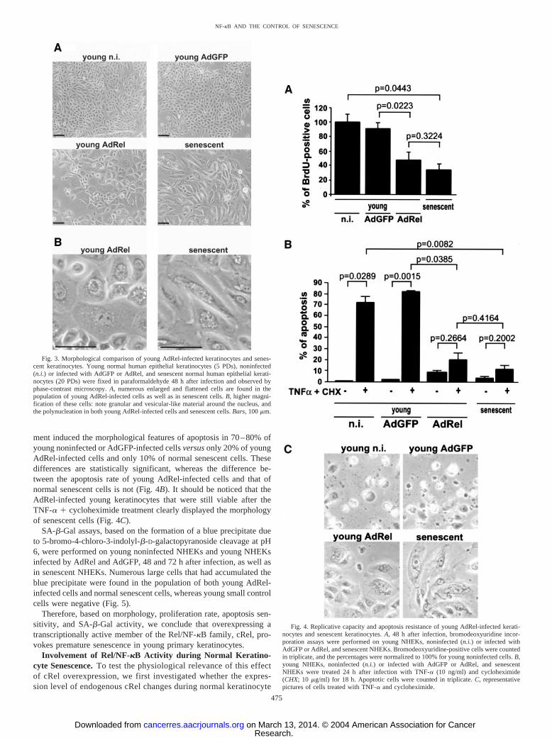
ment induced the morphological features of apoptosis in 70–80% ofyoung noninfected or AdGFP-infected cells versus only 20% of youngAdRel-infected cells and only 10% of normal senescent cells. Thesedifferences are statistically significant, whereas the difference be-tween the apoptosis rate of young AdRel-infected cells and that ofnormal senescent cells is not (Fig. 4B). It should be noticed that theAdRel-infected young keratinocytes that were still viable after theTNF-� � cycloheximide treatment clearly displayed the morphologyof senescent cells (Fig. 4C).
SA-�-Gal assays, based on the formation of a blue precipitate dueto 5-bromo-4-chloro-3-indolyl-�-D-galactopyranoside cleavage at pH6, were performed on young noninfected NHEKs and young NHEKsinfected by AdRel and AdGFP, 48 and 72 h after infection, as well asin senescent NHEKs. Numerous large cells that had accumulated theblue precipitate were found in the population of both young AdRel-infected cells and normal senescent cells, whereas young small controlcells were negative (Fig. 5).
Therefore, based on morphology, proliferation rate, apoptosis sen-sitivity, and SA-�-Gal activity, we conclude that overexpressing atranscriptionally active member of the Rel/NF-�B family, cRel, pro-vokes premature senescence in young primary keratinocytes.
Involvement of Rel/NF-�B Activity during Normal Keratino-cyte Senescence. To test the physiological relevance of this effectof cRel overexpression, we first investigated whether the expres-sion level of endogenous cRel changes during normal keratinocyte
Fig. 3. Morphological comparison of young AdRel-infected keratinocytes and senes-cent keratinocytes. Young normal human epithelial keratinocytes (5 PDs), noninfected(n.i.) or infected with AdGFP or AdRel, and senescent normal human epithelial kerati-nocytes (20 PDs) were fixed in paraformaldehyde 48 h after infection and observed byphase-contrast microscopy. A, numerous enlarged and flattened cells are found in thepopulation of young AdRel-infected cells as well as in senescent cells. B, higher magni-fication of these cells: note granular and vesicular-like material around the nucleus, andthe polynucleation in both young AdRel-infected cells and senescent cells. Bars, 100 �m.
Fig. 4. Replicative capacity and apoptosis resistance of young AdRel-infected kerati-nocytes and senescent keratinocytes. A, 48 h after infection, bromodeoxyuridine incor-poration assays were performed on young NHEKs, noninfected (n.i.) or infected withAdGFP or AdRel, and senescent NHEKs. Bromodeoxyuridine-positive cells were countedin triplicate, and the percentages were normalized to 100% for young noninfected cells. B,young NHEKs, noninfected (n.i.) or infected with AdGFP or AdRel, and senescentNHEKs were treated 24 h after infection with TNF-� (10 ng/ml) and cycloheximide(CHX; 10 �g/ml) for 18 h. Apoptotic cells were counted in triplicate. C, representativepictures of cells treated with TNF-� and cycloheximide.
475
NF-�B AND THE CONTROL OF SENESCENCE
Research. on March 13, 2014. © 2004 American Association for Cancercancerres.aacrjournals.org Downloaded from
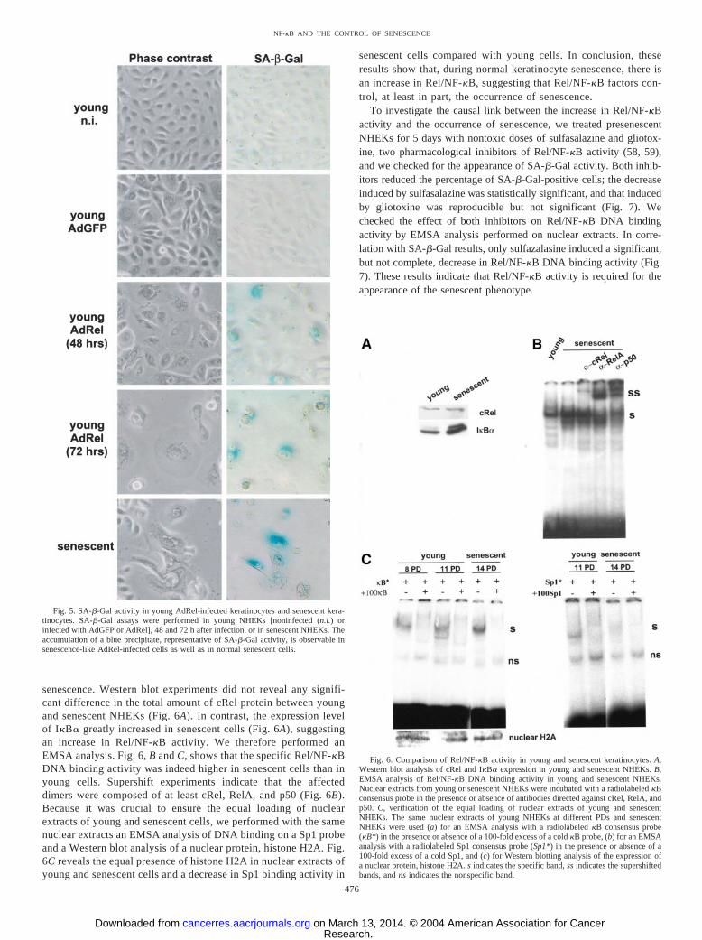
senescence. Western blot experiments did not reveal any signifi-cant difference in the total amount of cRel protein between youngand senescent NHEKs (Fig. 6A). In contrast, the expression levelof I�B� greatly increased in senescent cells (Fig. 6A), suggestingan increase in Rel/NF-�B activity. We therefore performed anEMSA analysis. Fig. 6, B and C, shows that the specific Rel/NF-�BDNA binding activity was indeed higher in senescent cells than inyoung cells. Supershift experiments indicate that the affecteddimers were composed of at least cRel, RelA, and p50 (Fig. 6B).Because it was crucial to ensure the equal loading of nuclearextracts of young and senescent cells, we performed with the samenuclear extracts an EMSA analysis of DNA binding on a Sp1 probeand a Western blot analysis of a nuclear protein, histone H2A. Fig.6C reveals the equal presence of histone H2A in nuclear extracts ofyoung and senescent cells and a decrease in Sp1 binding activity in
senescent cells compared with young cells. In conclusion, theseresults show that, during normal keratinocyte senescence, there isan increase in Rel/NF-�B, suggesting that Rel/NF-�B factors con-trol, at least in part, the occurrence of senescence.
To investigate the causal link between the increase in Rel/NF-�Bactivity and the occurrence of senescence, we treated presenescentNHEKs for 5 days with nontoxic doses of sulfasalazine and gliotox-ine, two pharmacological inhibitors of Rel/NF-�B activity (58, 59),and we checked for the appearance of SA-�-Gal activity. Both inhib-itors reduced the percentage of SA-�-Gal-positive cells; the decreaseinduced by sulfasalazine was statistically significant, and that inducedby gliotoxine was reproducible but not significant (Fig. 7). Wechecked the effect of both inhibitors on Rel/NF-�B DNA bindingactivity by EMSA analysis performed on nuclear extracts. In corre-lation with SA-�-Gal results, only sulfazalasine induced a significant,but not complete, decrease in Rel/NF-�B DNA binding activity (Fig.7). These results indicate that Rel/NF-�B activity is required for theappearance of the senescent phenotype.
Fig. 5. SA-�-Gal activity in young AdRel-infected keratinocytes and senescent kera-tinocytes. SA-�-Gal assays were performed in young NHEKs [noninfected (n.i.) orinfected with AdGFP or AdRel], 48 and 72 h after infection, or in senescent NHEKs. Theaccumulation of a blue precipitate, representative of SA-�-Gal activity, is observable insenescence-like AdRel-infected cells as well as in normal senescent cells.
Fig. 6. Comparison of Rel/NF-�B activity in young and senescent keratinocytes. A,Western blot analysis of cRel and I�B� expression in young and senescent NHEKs. B,EMSA analysis of Rel/NF-�B DNA binding activity in young and senescent NHEKs.Nuclear extracts from young or senescent NHEKs were incubated with a radiolabeled �Bconsensus probe in the presence or absence of antibodies directed against cRel, RelA, andp50. C, verification of the equal loading of nuclear extracts of young and senescentNHEKs. The same nuclear extracts of young NHEKs at different PDs and senescentNHEKs were used (a) for an EMSA analysis with a radiolabeled �B consensus probe(�B*) in the presence or absence of a 100-fold excess of a cold �B probe, (b) for an EMSAanalysis with a radiolabeled Sp1 consensus probe (Sp1*) in the presence or absence of a100-fold excess of a cold Sp1, and (c) for Western blotting analysis of the expression ofa nuclear protein, histone H2A. s indicates the specific band, ss indicates the supershiftedbands, and ns indicates the nonspecific band.
476
NF-�B AND THE CONTROL OF SENESCENCE
Research. on March 13, 2014. © 2004 American Association for Cancercancerres.aacrjournals.org Downloaded from
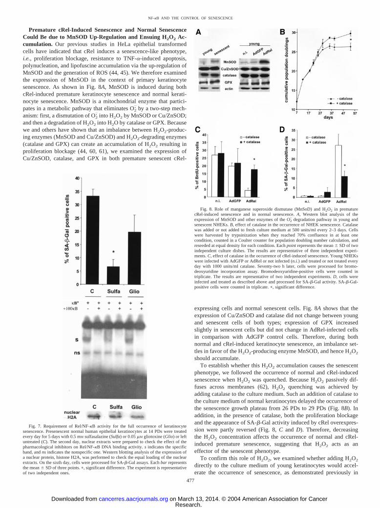
Premature cRel-Induced Senescence and Normal SenescenceCould Be due to MnSOD Up-Regulation and Ensuing H2O2 Ac-cumulation. Our previous studies in HeLa epithelial transformedcells have indicated that cRel induces a senescence-like phenotype,i.e., proliferation blockage, resistance to TNF-�-induced apoptosis,polynucleation, and lipofuscine accumulation via the up-regulation ofMnSOD and the generation of ROS (44, 45). We therefore examinedthe expression of MnSOD in the context of primary keratinocytesenescence. As shown in Fig. 8A, MnSOD is induced during bothcRel-induced premature keratinocyte senescence and normal kerati-nocyte senescence. MnSOD is a mitochondrial enzyme that partici-pates in a metabolic pathway that eliminates O2
. by a two-step mech-anism: first, a dismutation of O2
. into H2O2 by MnSOD or Cu/ZnSOD;and then a degradation of H2O2 into H2O by catalase or GPX. Becausewe and others have shown that an imbalance between H2O2-produc-ing enzymes (MnSOD and Cu/ZnSOD) and H2O2-degrading enzymes(catalase and GPX) can create an accumulation of H2O2 resulting inproliferation blockage (44, 60, 61), we examined the expression ofCu/ZnSOD, catalase, and GPX in both premature senescent cRel-
expressing cells and normal senescent cells. Fig. 8A shows that theexpression of Cu/ZnSOD and catalase did not change between youngand senescent cells of both types; expression of GPX increasedslightly in senescent cells but did not change in AdRel-infected cellsin comparison with AdGFP control cells. Therefore, during bothnormal and cRel-induced keratinocyte senescence, an imbalance set-tles in favor of the H2O2-producing enzyme MnSOD, and hence H2O2
should accumulate.To establish whether this H2O2 accumulation causes the senescent
phenotype, we followed the occurrence of normal and cRel-inducedsenescence when H2O2 was quenched. Because H2O2 passively dif-fuses across membranes (62), H2O2 quenching was achieved byadding catalase to the culture medium. Such an addition of catalase tothe culture medium of normal keratinocytes delayed the occurrence ofthe senescence growth plateau from 26 PDs to 29 PDs (Fig. 8B). Inaddition, in the presence of catalase, both the proliferation blockageand the appearance of SA-�-Gal activity induced by cRel overexpres-sion were partly reversed (Fig. 8, C and D). Therefore, decreasingthe H2O2 concentration affects the occurrence of normal and cRel-induced premature senescence, suggesting that H2O2 acts as aneffector of the senescent phenotype.
To confirm this role of H2O2, we examined whether adding H2O2
directly to the culture medium of young keratinocytes would accel-erate the occurrence of senescence, as demonstrated previously in
Fig. 7. Requirement of Rel/NF-�B activity for the full occurrence of keratinocytesenescence. Presenescent normal human epithelial keratinocytes at 14 PDs were treatedevery day for 5 days with 0.5 mM sulfasalazine (Sulfa) or 0.05 �M gliotoxine (Glio) or leftuntreated (C). The second day, nuclear extracts were prepared to check the effect of thepharmacological inhibitors on Rel/NF-�B DNA binding activity. s indicates the specificband, and ns indicates the nonspecific one. Western blotting analysis of the expression ofa nuclear protein, histone H2A, was performed to check the equal loading of the nuclearextracts. On the sixth day, cells were processed for SA-�-Gal assays. Each bar representsthe mean � SD of three points. �, significant difference. The experiment is representativeof two independent ones.
Fig. 8. Role of manganese superoxide dismutase (MnSoD) and H2O2 in prematurecRel-induced senescence and in normal senescence. A, Western blot analysis of theexpression of MnSOD and other enzymes of the O2
. degradation pathway in young andsenescent NHEKs. B, effect of catalase in the occurrence of NHEK senescence. Catalasewas added or not added to fresh culture medium at 500 units/ml every 2–3 days. Cellswere harvested by trypsinization when they reached 70% confluence in at least onecondition, counted in a Coulter counter for population doubling number calculation, andreseeded at equal density for each condition. Each point represents the mean � SD of twoindependent culture dishes. The results are representative of three independent experi-ments. C, effect of catalase in the occurrence of cRel-induced senescence. Young NHEKswere infected with AdGFP or AdRel or not infected (n.i.) and treated or not treated everyday with 1000 units/ml catalase. Seventy-two h later, cells were processed for bromo-deoxyuridine incorporation assay. Bromodeoxyuridine-positive cells were counted intriplicate. The results are representative of two independent experiments. D, cells wereinfected and treated as described above and processed for SA-�-Gal activity. SA-�-Gal-positive cells were counted in triplicate. �, significant difference.
477
NF-�B AND THE CONTROL OF SENESCENCE
Research. on March 13, 2014. © 2004 American Association for Cancercancerres.aacrjournals.org Downloaded from
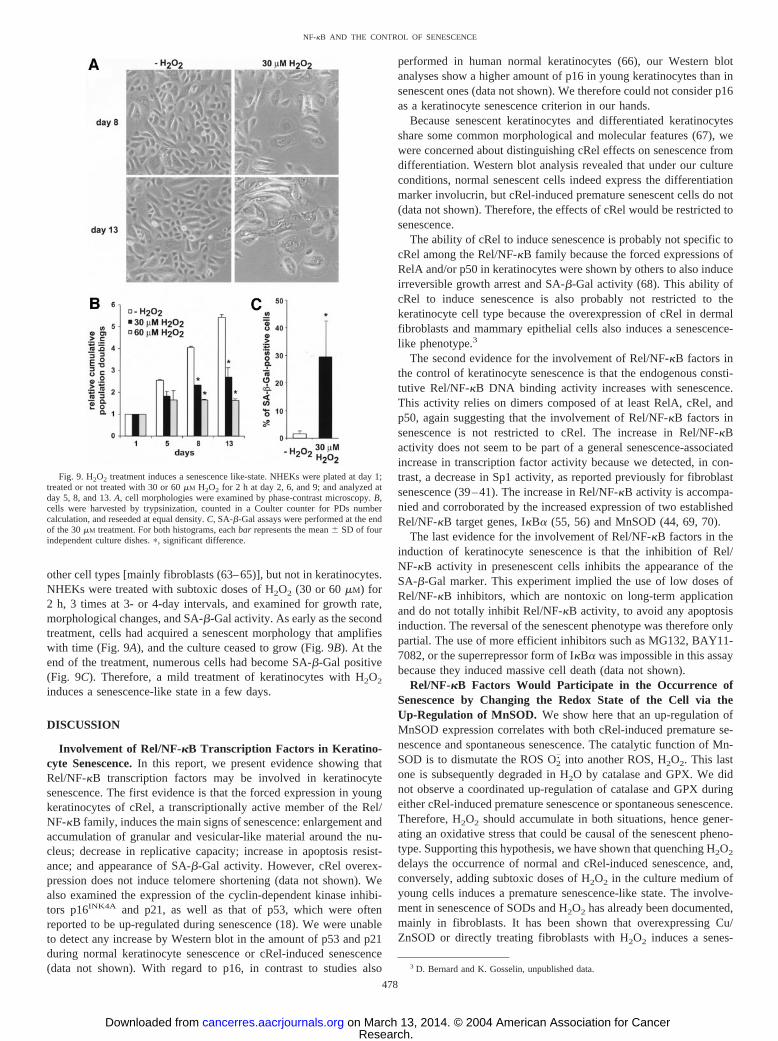
other cell types [mainly fibroblasts (63–65)], but not in keratinocytes.NHEKs were treated with subtoxic doses of H2O2 (30 or 60 �M) for2 h, 3 times at 3- or 4-day intervals, and examined for growth rate,morphological changes, and SA-�-Gal activity. As early as the secondtreatment, cells had acquired a senescent morphology that amplifieswith time (Fig. 9A), and the culture ceased to grow (Fig. 9B). At theend of the treatment, numerous cells had become SA-�-Gal positive(Fig. 9C). Therefore, a mild treatment of keratinocytes with H2O2
induces a senescence-like state in a few days.
DISCUSSION
Involvement of Rel/NF-�B Transcription Factors in Keratino-cyte Senescence. In this report, we present evidence showing thatRel/NF-�B transcription factors may be involved in keratinocytesenescence. The first evidence is that the forced expression in youngkeratinocytes of cRel, a transcriptionally active member of the Rel/NF-�B family, induces the main signs of senescence: enlargement andaccumulation of granular and vesicular-like material around the nu-cleus; decrease in replicative capacity; increase in apoptosis resist-ance; and appearance of SA-�-Gal activity. However, cRel overex-pression does not induce telomere shortening (data not shown). Wealso examined the expression of the cyclin-dependent kinase inhibi-tors p16INK4A and p21, as well as that of p53, which were oftenreported to be up-regulated during senescence (18). We were unableto detect any increase by Western blot in the amount of p53 and p21during normal keratinocyte senescence or cRel-induced senescence(data not shown). With regard to p16, in contrast to studies also
performed in human normal keratinocytes (66), our Western blotanalyses show a higher amount of p16 in young keratinocytes than insenescent ones (data not shown). We therefore could not consider p16as a keratinocyte senescence criterion in our hands.
Because senescent keratinocytes and differentiated keratinocytesshare some common morphological and molecular features (67), wewere concerned about distinguishing cRel effects on senescence fromdifferentiation. Western blot analysis revealed that under our cultureconditions, normal senescent cells indeed express the differentiationmarker involucrin, but cRel-induced premature senescent cells do not(data not shown). Therefore, the effects of cRel would be restricted tosenescence.
The ability of cRel to induce senescence is probably not specific tocRel among the Rel/NF-�B family because the forced expressions ofRelA and/or p50 in keratinocytes were shown by others to also induceirreversible growth arrest and SA-�-Gal activity (68). This ability ofcRel to induce senescence is also probably not restricted to thekeratinocyte cell type because the overexpression of cRel in dermalfibroblasts and mammary epithelial cells also induces a senescence-like phenotype.3
The second evidence for the involvement of Rel/NF-�B factors inthe control of keratinocyte senescence is that the endogenous consti-tutive Rel/NF-�B DNA binding activity increases with senescence.This activity relies on dimers composed of at least RelA, cRel, andp50, again suggesting that the involvement of Rel/NF-�B factors insenescence is not restricted to cRel. The increase in Rel/NF-�Bactivity does not seem to be part of a general senescence-associatedincrease in transcription factor activity because we detected, in con-trast, a decrease in Sp1 activity, as reported previously for fibroblastsenescence (39–41). The increase in Rel/NF-�B activity is accompa-nied and corroborated by the increased expression of two establishedRel/NF-�B target genes, I�B� (55, 56) and MnSOD (44, 69, 70).
The last evidence for the involvement of Rel/NF-�B factors in theinduction of keratinocyte senescence is that the inhibition of Rel/NF-�B activity in presenescent cells inhibits the appearance of theSA-�-Gal marker. This experiment implied the use of low doses ofRel/NF-�B inhibitors, which are nontoxic on long-term applicationand do not totally inhibit Rel/NF-�B activity, to avoid any apoptosisinduction. The reversal of the senescent phenotype was therefore onlypartial. The use of more efficient inhibitors such as MG132, BAY11-7082, or the superrepressor form of I�B� was impossible in this assaybecause they induced massive cell death (data not shown).
Rel/NF-�B Factors Would Participate in the Occurrence ofSenescence by Changing the Redox State of the Cell via theUp-Regulation of MnSOD. We show here that an up-regulation ofMnSOD expression correlates with both cRel-induced premature se-nescence and spontaneous senescence. The catalytic function of Mn-SOD is to dismutate the ROS O2
. into another ROS, H2O2. This lastone is subsequently degraded in H2O by catalase and GPX. We didnot observe a coordinated up-regulation of catalase and GPX duringeither cRel-induced premature senescence or spontaneous senescence.Therefore, H2O2 should accumulate in both situations, hence gener-ating an oxidative stress that could be causal of the senescent pheno-type. Supporting this hypothesis, we have shown that quenching H2O2
delays the occurrence of normal and cRel-induced senescence, and,conversely, adding subtoxic doses of H2O2 in the culture medium ofyoung cells induces a premature senescence-like state. The involve-ment in senescence of SODs and H2O2 has already been documented,mainly in fibroblasts. It has been shown that overexpressing Cu/ZnSOD or directly treating fibroblasts with H2O2 induces a senes-
3 D. Bernard and K. Gosselin, unpublished data.
Fig. 9. H2O2 treatment induces a senescence like-state. NHEKs were plated at day 1;treated or not treated with 30 or 60 �M H2O2 for 2 h at day 2, 6, and 9; and analyzed atday 5, 8, and 13. A, cell morphologies were examined by phase-contrast microscopy. B,cells were harvested by trypsinization, counted in a Coulter counter for PDs numbercalculation, and reseeded at equal density. C, SA-�-Gal assays were performed at the endof the 30 �M treatment. For both histograms, each bar represents the mean � SD of fourindependent culture dishes. �, significant difference.
478
NF-�B AND THE CONTROL OF SENESCENCE
Research. on March 13, 2014. © 2004 American Association for Cancercancerres.aacrjournals.org Downloaded from
cence-like state (60, 63, 65). Conversely, N-t-butyl hydroxylamine,which inhibits the formation of O2
. and hence that of H2O2, delayssenescence of fibroblasts (71). Furthermore, fibroblasts derived fromindividuals with Down syndrome, who possess a supernumerary copyof the Cu/ZnSOD gene, were shown to senesce earlier than corre-sponding controls (60). Our results extend the involvement of SODsand H2O2 in senescence to keratinocytes and document the control ofthis mechanism by Rel/NF-�B factors.
The Apoptosis Resistance of Senescent Cells Could Also Rely onRel/NF-�B Activity and Up-Regulation of MnSOD. Senescentcells are apoptosis resistant. This counterintuitive fact was demon-strated for normal human senescent fibroblasts treated with serumwithdrawal (13) or DNA-damaging agents [UV radiation, actinomy-cin D, cisplatin, or ROS (16, 72)]; for senescent T cells treated withdexamethasone, anti-Fas, anti-CD3, galectin-1, IL-2 withdrawal, stau-rosporine (a protein kinase C inhibitor), or heat shock (15); and forsenescent keratinocytes treated with UV radiation (14). In addition,we show here that senescent keratinocytes are resistant to apoptosisinduced by TNF-� in the presence of cycloheximide. This is in goodagreement with the involvement of Rel/NF-�B factors in senescencebecause their protective effect against TNF-�-induced apoptosis waslargely documented in numerous cell types (73), including keratino-cytes (74). MnSOD was also shown to have a protective effect againstTNF-�- and serum deprivation-induced apoptosis (75–77) and toparticipate in the antiapoptotic function of Rel/NF-�B factors (44, 78).The primary function of MnSOD is to eliminate the toxic O2
. that isconstantly generated in the cell and that can be highly produced inresponse to some apoptosis inducers such as TNF-� (44, 79, 80), UVradiation (81, 82), cisplatin and actinomycin D (83), anti-Fas (84), andserum deprivation (77). Senescent cells are resistant to apoptosisinduced by all these compounds. The up-regulation of MnSOD byRel/NF-�B factors that we show in this work would account for thisproperty. In all, the up-regulation of MnSOD by Rel/NF-�B factorsduring senescence would have a dual effect: on one hand, it wouldmake cells more resistant to apoptosis inducers that produce O2
.; buton the other hand, because the H2O2-degrading enzymes are notcoordinately up-regulated, H2O2 would accumulate, causing someoxidative injuries responsible for the main alterations in senescentcells, i.e., morphological changes, proliferation blockage, and SA-�-Gal activity.
Are Rel/NF-�B Factors Oncogenes or Tumor SuppressorGenes? Until recently, Rel/NF-�B factor-encoding genes were con-sidered proto-oncogenes. Indeed, v-rel, the viral oncogene derivedfrom the avian c-rel, is transforming in vitro and in vivo (85). Humanand murine c-rel are also transforming in vitro and in vivo, but aftera long delay (86, 87). Chromosomal amplification, rearrangement, ormutations of genes coding for several Rel/NF-�B members are foundin many hematopoietic and solid human tumors (88). ConstitutiveRel/NF-�B activity was also described in several cancer types, due toeither a constitutive activation of the upstream kinases or to mutationsin genes encoding I�B proteins (88). As oncogenes, Rel/NF-�Bfactors would be assumed to allow cells to evade senescence andacquire an immortal and transformed phenotype. However, our resultsshow that Rel/NF-�B factors could, in contrast, be involved in theoccurrence of senescence, suggesting that they behave as tumor sup-pressor genes. In support of this view, targeted expression of I�B� inthe epidermis by transgenesis predisposes to the development ofspontaneous squamous cell carcinomas (89). Similarly, human recon-stituted skin implanted in mouse becomes hyperplastic when it over-expresses I�B� or develops squamous cell carcinoma when it coex-presses I�B� and Ras (90). In addition, it was demonstrated that animmortalized fibroblast cell line derived from a RelA�/� mousedisplays numerous properties of a transformed cell line (91). Consid-
ering that Rel/NF-�B factors are often regarded as promising targetsin cancer therapy, additional experiments are needed to clarifywhether they display oncogenic or tumor suppressor properties.
ACKNOWLEDGMENTS
We thank N. Rice for the human c-rel plasmid; J. Hiscott for the anti-cRel,RelA, and p50 antibodies; and Philippe Becuwe for helpful discussions.
REFERENCES
1. Hayflick, L. The limited in vitro lifetime of human diploid cell strains. Exp. Cell Res.,37: 614–636, 1965.
2. Jazwinski, M. Longevity, genes, and aging. Science (Wash. DC), 273: 54–59, 1996.3. Smith, J. R., and Pereira-Smith, O. M. Replicative senescence: implications for in
vivo aging and tumor suppression. Science (Wash. DC), 273: 63–67, 1996.4. Pignolo, R. J., Rotenberg, M. O., and Cristofalo, V. J. Alterations in contact and
density-dependent arrest state in senescent WI-38 cells. In Vitro Cell. Dev. Biol.,30A: 471–476, 1994.
5. Angello, J. C., Pendergrass, W. R., Norwood, T. H., and Prothero, J. Cell enlarge-ment: one possible mechanism underlying cellular senescence. J. Cell. Physiol., 140:288–294, 1989.
6. Norsgaard, H., Clark, B. F., and Rattan, S. I. Distinction between differentiation andsenescence and the absence of increased apoptosis in human keratinocytes undergoingcellular aging in vitro. Exp. Gerontol., 31: 563–570, 1996.
7. Guhe, C., and Follmann, W. Growth and characterization of porcine urinary bladderepithelial cells in vitro. Am. J. Physiol., 266: F298–F308, 1994.
8. Lima, L., and Macieira-Coelho, A. Parameters of aging in chicken embryo fibroblastscultivated in vitro. Exp. Cell Res., 70: 279–284, 1972.
9. Sherwood, S. W., Rush, D., Ellsworth, J. L., and Schimke, R. T. Defining cellularsenescence in IMR-90 cells: a flow cytometric analysis. Proc. Natl. Acad. Sci. USA,85: 9086–9090, 1988.
10. Brunk, U. T., Jones, C. B., and Sohal, R. S. A novel hypothesis of lipofuscinogenesisand cellular aging based on interactions between oxidative stress and autophagocy-tosis. Mutat. Res., 275: 395–403, 1992.
11. Sitte, N., Merker, K., Von Zglinicki, T., Grune, T., and Davies, K. J. Protein oxidationand degradation during cellular senescence of human BJ fibroblasts. Part I: effects ofproliferative senescence. FASEB J., 14: 2495–2502, 2000.
12. Goldstein, S. Replicative senescence: the human fibroblast comes of age. Science(Wash. DC), 249: 1129–1133, 1990.
13. Wang, E. Senescent human fibroblasts resist programmed cell death, and failure tosuppress bcl2 is involved. Cancer Res., 55: 2284–2292, 1995.
14. Chaturvedi, V., Qin, J-Z., Denning, M. F., Choubey, D., Diaz, M. O., and Nickoloff,B. J. Apoptosis in proliferating, senescent, and immortalized keratinocytes. J. Biol.Chem., 274: 23358–23367, 1999.
15. Spaulding, C., Guo, W., and Effros, R. B. Resistance to apoptosis in human CD8�T cells that reach replicative senescence after multiple rounds of antigen-specificproliferation. Exp. Gerontol., 34: 633–644, 1999.
16. Seluanov, A., Gorbunova, V., Falcovitz, A., Sigal, A., Milyavsky, M., Zurer, I.,Shohat, G., Goldfinger, N., and Rotter, V. Change of the death pathway in senescenthuman fibroblasts in response to DNA damage is caused by an inability to stabilizep53. Mol. Cell. Biol., 21: 1552–1564, 2001.
17. Dimri, G. P., Lee, X., Basile, G., Acosta, M., Scott, G., Roskelley, C., Medrano, E. E.,Linskens, M., Rubelj, I., Pereira-Smith, O., Peacocke, M., and Campisi, J. A biomar-ker that identifies senescent human cells in culture and in aging skin in vivo. Proc.Natl. Acad. Sci. USA, 92: 9363–9367, 1995.
18. Lundberg, A. S., Hahn, W. C., Gupta, P., and Weinberg, R. A. Genes involved insenescence and immortalization. Curr. Opin. Cell Biol., 12: 705–709, 2000.
19. Vaziri, H., and Benchimol, S. From telomere loss to p53 induction and activation ofa DNA-damage pathway at senescence: the telomere loss/DNA damage model of cellaging. Exp. Gerontol., 31: 295–301, 1996.
20. Chen, Q., Fischer, A., Reagan, J. D., Yan, L. J., and Ames, B. N. Oxidative DNAdamage and senescence of human diploid fibroblast cells. Proc. Natl. Acad. Sci. USA,92: 4337–4341, 1995.
21. Toussaint, O., Medrano, E. E., and von Zglinicki, T. Cellular and molecular mech-anisms of stress-induced premature senescence (SIPS) of human diploid fibroblastsand melanocytes. Exp. Gerontol., 35: 927–945, 2000.
22. Harman, D. Extending functional life span. Exp. Gerontol., 33: 95–112, 1998.23. Karin, M. How NF-�B is activated: the role of the I�B kinase (IKK) complex.
Oncogene, 18: 6867–6874, 1999.24. Peters, R. T., Liao, S. M., and Maniatis, T. IKK� is part of a novel PMA-inducible
I�B kinase complex. Mol. Cell, 5: 513–522, 2000.25. Beg, A. A., Sha, W. C., Bronson, R. T., Ghosh, S., and Baltimore, D. Embryonic
lethality and liver degeneration in mice lacking the RelA component of NF-KB.Nature (Lond.), 376: 167–170, 1995.
26. Rudolph, D., Yeh, W. C., Wakeham, A., Rudolph, B., Nallainathan, D., Potter, J.,Elia, A. J., and Mak, T. W. Severe liver degeneration and lack of NF-�B activationin NEMO/IKK�-deficient mice. Genes Dev., 14: 854–862, 2000.
27. Li, Q., Van Antwerp, D., Mercurio, F., Lee, K-F., and Verma, I. M. Severe liverdegeneration in mice lacking the I�B kinase 2 gene. Science (Wash. DC), 284:321–324, 1999.
28. Doi, T. S., Marino, M. W., Takahashi, T., Yoshida, T., Sakakura, T., Old, L. J., andObata, Y. Absence of tumor necrosis factor rescues RelA-deficient mice fromembryonic lethality. Proc. Natl. Acad. Sci. USA, 96: 2994–2999, 1999.
479
NF-�B AND THE CONTROL OF SENESCENCE
Research. on March 13, 2014. © 2004 American Association for Cancercancerres.aacrjournals.org Downloaded from
29. Hu, Y., Baud, V., Delhase, M., Zhang, P., Deerinck, T., Ellisman, M., Johnson, R.,and Karin, M. Abnormal morphogenesis but intact IKK activation in mice lacking theIKKa subunit of I�B kinase. Science (Wash. DC), 284: 316–320, 1999.
30. Abbadie, C., Kabrun, N., Bouali, F., Smardova, J., Stehelin, D., Vandenbunder, B.,and Enrietto, P. High levels of c-rel expression are associated with programmed celldeath in the developing avian embryo and in bone marrow cells in vitro. Cell, 75:899–912, 1993.
31. Huguet, C., Mattot, V., Bouali, F., Stehelin, D., Vandenbunder, B., and Abbadie, C.The avian transcription factor c-Rel is induced and translocates into the nucleus ofthymocytes undergoing apoptosis. Cell Death Differ., 4: 413–422, 1997.
32. Seitz, C. S., Lin, Q., Deng, H., and Khavari, P. A. Alterations in NF-�B function intransgenic epithelial tissue demonstrate a growth inhibitory role for NF-�B. Proc.Natl. Acad. Sci. USA, 95: 2307–2312, 1998.
33. Schmidt-Supprian, M., Bloch, W., Courtois, G., Addicks, K., Israel, A., Rajewsky, K.,and Pasparakis, M. NEMO/IKK �-deficient mice model incontinentia pigmenti. Mol.Cell, 5: 981–992, 2000.
34. Kontgen, F., Grumont, R. J., Strasser, A., Metcalf, D., Li, R., Tarlinton, D., andGerondakis, S. Mice lacking the c-rel proto-oncogene exhibit defects in lymphocyteproliferation, humoral immunity, and interleukin-2 expression. Genes Dev., 9: 1965–1977, 1995.
35. Grumont, R. J., Rourke, I. J., O’Reilly, L. A., Strasser, A., Miyake, K., Sha, W., andGerondakis, S. B lymphocytes differentially use the Rel and nuclear factor �B1(NF-�B1) transciption factors to regulate cell cycle progression and apoptosis inquiescent and mitogen-activated cells. J. Exp. Med., 187: 663–674, 1998.
36. Grossmann, M., Metcalf, D., Merryfull, J., Beg, A., Baltimore, D., and Gerondakis,S. The combined absence of the transcription factors Rel and RelA leads to multiplehemopoietic cell defects. Proc. Natl. Acad. Sci. USA, 96: 11848–11853, 1999.
37. Gosselin, K., and Abbadie, C. Involvement of Rel/NF-�B transcription factors insenescence. Exp. Gerontol., in press, 2003.
38. Aggarwal, B. B., Totpal, K., LaPushin, R., Chaturvedi, M. M., Pereira-Smith, O. M.,and Smith, J. R. Diminished responsiveness of senescent normal human fibroblasts toTNF-dependent proliferation and interleukin production is not due to its effect on thereceptors or on the activation of a nuclear factor NF-�B. Exp. Cell Res., 218:381–388, 1995.
39. Dimri, G. P., and Campisi, J. Altered profile of transcription factor-binding activitiesin senescent human fibroblasts. Exp. Cell Res., 212: 132–140, 1994.
40. Helenius, M., Hanninen, M., Lehtinen, S. K., and Salminen, A. Changes associatedwith aging and replicative senescence in the regulation of transcription factor nuclearfactor-�B. Biochem. J., 318: 603–608, 1996.
41. Helenius, M., Makelainen, L., and Salminen, A. Attenuation of NF-�B signalingresponse to UVB light during cellular senescence. Exp. Cell Res., 248: 194–202,1999.
42. Ikebe, T., Jimi, E., Beppu, M., Takeuchi, H., Nakayama, H., and Shirasuna, K.Aging-dependent proteolysis of NF-�B in human fibroblasts. J. Cell. Physiol., 182:247–255, 2000.
43. Yan, Z. Q., Sirsjo, A., Bochaton-Piallat, M. L., Gabbiani, G., and Hansson, G. K.Augmented expression of inducible NO synthase in vascular smooth muscle cellsduring aging is associated with enhanced NF-�B activation. Arterioscler. Thromb.Vasc. Biol., 19: 2854–2862, 1999.
44. Bernard, D., Quatannens, B., Begue, A., Vandenbunder, B., and Abbadie, A. Anti-proliferative and anti-apoptotic effects of cRel may occur within the same cells via theup-regulation of MnSOD. Cancer Res., 61: 2656–2664, 2001.
45. Bernard, D., Slomianny, C., Vandenbunder, B., and Abbadie, C. cRel inducesmitochondrial alterations in correlation with proliferation arrest. Free Radic. Biol.Med., 31: 943–953, 2001.
46. Boyce, S. T., and Ham, R. G. Calcium-regulated differentiation of normal humanepidermal keratinocytes in chemically defined clonal culture and serum-free serialculture. J. Investig. Dermatol., 81: 33s–40s, 1983.
47. Brownell, E., Mittereder, N., and Rice, N. R. A human rel proto-oncogene cDNAcontaining an Alu fragment as a potential coding exon. Oncogene, 4: 935–942, 1989.
48. Chartier, C., Degryse, E., Gantzer, M., Dieterle, A., Pavirani, A., and Mehtali, M.Efficient generation of recombinant adenovirus vectors by homologous recombina-tion in Escherichia coli. J. Virol., 70: 4805–4810, 1996.
49. Nevins, J. R., DeGregori, J., Jakoi, L., and Leone, G. Functional analysis of E2Ftranscription factor. Methods Enzymol., 283: 205–219, 1997.
50. Guerardel, C., Deltour, S., Pinte, S., Monte, D., Begue, A., Godwin, A. K., andLeprince, D. Identification in the human candidate tumor suppressor gene HIC-1 ofa new major alternative TATA-less promoter positively regulated by p53. J. Biol.Chem., 276: 3078–3089, 2001.
51. Kasibhatla, S., Brunner, T., Genestier, L., Echeverri, F., Mahboubi, A., and Green,D. R. DNA damaging agents induce expression of Fas ligand and subsequentapoptosis in T lymphocytes via the activation of NF-�B and AP-1. Mol. Cell, 1:543–551, 1998.
52. Lin, Y. C., Brown, K., and Siebenlist, U. Activation of NF-�B requires proteolysis ofthe inhibitor I�B-a : signal-induced phosphorylation of I�B-a alone does not releaseactive NF-�B. Proc. Natl. Acad. Sci. USA, 92: 552–556, 1995.
53. Pepin, N., Roulston, A., Lacoste, J., Lin, R., and Hiscott, J. Subcellular redistributionof HTLV-1 Tax protein by NF-�B/Rel transcription factors. Virology, 204: 706–716,1994.
54. Bernard, D., Monte, D., Vandenbunder, B., and Abbadie, C. The c-Rel transcriptionfactor can both induce and inhibit apoptosis in the same cells via the upregulation ofMnSOD. Oncogene, 21: 4392–4402, 2002.
55. De Martin, R., Vanhove, B., Cheng, Q., Hofer, E., Csizmadia, V., Winkler, H., andBach, F. H. Cytokine-inducible expression in endothelial cells of an I�Ba-like geneis regulated by NF-�B. EMBO J., 12: 2773–2779, 1993.
56. Sun, S-C., Ganchi, P. A., Ballard, D. W., and Greene, W. C. NF-�B controlsexpression of inhibitor I�Ba: evidence for an inducible autoregulatory pathway.Science (Wash. DC), 259: 1912–1914, 1993.
57. Ten, R. M., Paya, C. V., Israel, N., Le, B. O., Mattei, M. G., Virelizier, J. L.,Kourilsky, P., and Israel, A. The characterization of the promoter of the geneencoding the p50 subunit of NF-�B indicates that it participates in its own regulation.EMBO J., 11: 195–203, 1992.
58. Wahl, C., Liptay, S., Adler, G., and Schmid, R. M. Sulfasalazine: a potent and specificinhibitor of nuclear factor �B. J. Clin. Investig., 101: 1163–1174, 1998.
59. Pahl, H. L., Krauss, B., Schulze-Osthoff, K., Decker, T., Traenckner, E. B., Vogt, M.,Myers, C., Parks, T., Warring, P., Muhlbacher, A., Czernilofsky, A. P., and Baeuerle,P. A. The immunosuppressive fungal metabolite gliotoxin specifically inhibits tran-scription factor NF-�B. J. Exp. Med., 183: 1829–1840, 1996.
60. de Haan, J. B., Cristiano, F., Ianello, R., Bladier, C., Kelner, M. J., and Kola, I.Elevation of the ratio of Cu/Zn-superoxide dismutase to glutathione peroxidaseactivity induces features of cellular senescence and this effect is mediated by hydro-gen peroxide. Hum. Mol. Genet., 5: 283–292, 1996.
61. Li, N., Zhai, Y., and Oberley, T. D. Two distinct mechanisms for inhibition of cellgrowth in human prostate carcinoma cells with antioxidant enzyme imbalance. FreeRadic. Biol. Med., 26: 1554–1568, 1999.
62. Burdon, R. H. Superoxide and hydrogen peroxide in relation to mammalian cellproliferation. Free Radic. Biol. Med., 18: 775–794, 1995.
63. Chen, Q., and Ames, B. N. Senescence-like growth arrest induced by hydrogenperoxide in human diploid fibroblast F65 cells. Proc. Natl. Acad. Sci. USA, 91:4130–4134, 1994.
64. Bladier, C., Wolvetang, E. L., Hutchinson, P., de Haan, J. B., and Kola, I. Responseof a primary human fibroblast cell line to H2O2: senescence-like growth arrest orapoptosis? Cell Growth Differ., 8: 589–598, 1997.
65. Dumont, P., Burton, M., Chen, Q. M., Gonos, E. S., Frippiat, C., Mazarati, J. B.,Eliaers, F., Remacle, J., and Toussaint, O. Induction of replicative senescencebiomarkers by sublethal oxidative stresses in normal human fibroblast. Free Radic.Biol. Med., 28: 361–373, 2000.
66. Dickson, M. A., Hahn, W. C., Ino, Y., Ronfard, V., Wu, J. Y., Weinberg, R. A., Louis,D. N., Li, F. P., and Rheinwald, J. G. Human keratinocytes that express hTERT andalso bypass a p16INK4a-enforced mechanism that limits life span become immortal yetretain normal growth and differentiation characteristics. Mol. Cell. Biol., 20: 1436–1447, 2000.
67. Gandarillas, A. Epidermal differentiation, apoptosis, and senescence: common path-ways? Exp. Gerontol., 35: 53–62, 2000.
68. Seitz, C. S., Deng, H., Hinata, K., Lin, Q., and Khavari, P. A. Nuclear factor �Bsubunits induce epithelial cell growth arrest. Cancer Res., 60: 4085–4092, 2000.
69. Xu, Y., Kiningham, K. K., Devalaraja, M. N., Yeh, C. C., Majima, H., Kasarskis,E. J., and St Clair, D. K. An intronic NF-�B element is essential for induction of thehuman manganese superoxide dismutase gene by tumor necrosis factor-� and inter-leukin-1�. DNA Cell Biol., 18: 709–722, 1999.
70. Kiningham, K. K., Xu, Y., Daosukho, C., Popova, B., and St Clair, D. K. Nuclearfactor �B-dependent mechanisms coordinate the synergistic effect of PMA andcytokines on the induction of superoxide dismutase 2. Biochem. J., 353: 147–156,2001.
71. Atamna, H., Paler-Martinez, A., and Ames, B. N. N-t-Butyl hydroxylamine, ahydrolysis product of �-phenyl-N-t-butyl nitrone, is more potent in delaying senes-cence in human lung fibroblasts. J. Biol. Chem., 275: 6741–6748, 2000.
72. Gansauge, S., Gansauge, F., Gause, H., Poch, B., Schoenberg, M. H., and Beger,H. G. The induction of apoptosis in proliferating human fibroblasts by oxygenradicals is associated with a p53- and p21WAF1CIP1 induction. FEBS Lett., 404:6–10, 1997.
73. Barkett, M., and Gilmore, T. D. Control of apoptosis by Rel/NF-�B transcriptionfactor. Oncogene, 18: 6910–6924, 1999.
74. Seitz, C. S., Freiberg, R. A., Hinata, K., and Khavari, P. A. NF-�B determineslocalization and features of cell death in epidermis. J. Clin. Investig., 105: 253–260,2000.
75. Wong, G. H., Elwell, J. H., Oberley, L. W., and Goeddel, D. V. Manganoussuperoxide dismutase is essential for cellular resistance to cytotoxicity of tumornecrosis factor. Cell, 58: 923–931, 1989.
76. Wong, G. H., and Goeddel, D. V. Induction of manganous superoxide dismutase bytumor necrosis factor: possible protective mechanism. Science (Wash. DC), 242:941–944, 1988.
77. Tanaka, H., Matsumura, I., Ezoe, S., Satoh, Y., Sakamaki, T., Albanese, C., Machii,T., Pestell, R. G., and Kanakura, Y. E2F1 and c-Myc potentiate apoptosis throughinhibition of NF-�B activity that facilitates MnSOD-mediated ROS elimination. Mol.Cell, 9: 1017–1029, 2002.
78. Delhalle, S., Deregowski, V., Benoit, V., Merville, M. P., and Bours, V. NF-�B-dependent MnSOD expression protects adenocarcinoma cells from TNF-�-inducedapoptosis. Oncogene, 21: 3917–3924, 2002.
79. Moreno-Manzano, V., Ishikawa, Y., Lucio-Cazana, J., and Kitamura, M. Selectiveinvolvement of superoxide anion, but not downstream compounds hydrogen peroxideand peroxynitrite, in tumor necrosis factor-�-induced apoptosis of rat mesangial cells.J. Biol. Chem., 275: 12684–12691, 2000.
80. Condliffe, A. M., Hawkins, P. T., Stephens, L. R., Haslett, C., and Chilvers, E. R.Priming of human neutrophil superoxide generation by tumour necrosis factor-� issignalled by enhanced phosphatidylinositol 3,4,5-trisphosphate but not inositol 1,4,5-trisphosphate accumulation. FEBS Lett., 439: 147–151, 1998.
81. Jones, S. A., McArdle, F., Jack, C. I., and Jackson, M. J. Effect of antioxidantsupplementation on the adaptive response of human skin fibroblasts to UV-inducedoxidative stress. Redox Rep., 4: 291–299, 1999.
480
NF-�B AND THE CONTROL OF SENESCENCE
Research. on March 13, 2014. © 2004 American Association for Cancercancerres.aacrjournals.org Downloaded from
82. Gorman, A., McGowan, A., and Cotter, T. G. Role of peroxide and superoxide anionduring tumour cell apoptosis. FEBS Lett., 404: 27–33, 1997.
83. Ikeda, K., Kajiwara, K., Tanabe, E., Tokumaru, S., Kishida, E., Masuzawa, Y., andKojo, S. Involvement of hydrogen peroxide and hydroxyl radical in chemicallyinduced apoptosis of HL-60 cells. Biochem. Pharmacol., 57: 1361–1365, 1999.
84. Suzuki, Y., Ono, Y., and Hirabayashi, Y. Rapid and specific reactive oxygen speciesgeneration via NADPH oxidase activation during Fas-mediated apoptosis. FEBSLett., 425: 209–212, 1998.
85. Gilmore, T. D. Multiple mutations contribute to the oncogenicity of the retroviraloncoprotein v-Rel. Oncogene, 18: 6925–6937, 1999.
86. Gilmore, T. D., Cormier, C., Jean-Jacques, J., and Gapuzan, M. E. Malignanttransformation of primary chicken spleen cells by human transcription factor c-Rel.Oncogene, 20: 7098–7103, 2001.
87. Romieu-Mourez, R., Kim, D. W., Shin, S. M., Demicco, E. G., Landesman-Bollag,E., Seldin, D. C., Cardiff, R. D., and Sonenshein, G. E. Mouse mammary tumor virus
c-rel transgenic mice develop mammary tumors. Mol. Cell. Biol., 23: 5738–5754,2003.
88. Rayet, B., and Gelinas, C. Aberrant rel/nfkb genes and activity in human cancer.Oncogene, 18: 6938–6947, 1999.
89. van Hogerlinden, M., Rozell, B. L., Ahrlund-Richter, L., and Toftgard, R. Squamouscell carcinomas and increased apoptosis in skin with inhibited Rel/nuclear factor-�Bsignaling. Cancer Res., 59: 3299–3303, 1999.
90. Dajee, M., Lazarov, M., Zhang, J. Y., Cai, T., Green, C. L., Russell, A. J.,Marinkovich, M. P., Tao, S., Lin, Q., Kubo, Y., and Khavari, P. A. NF-�B blockadeand oncogenic Ras trigger invasive human epidermal neoplasia. Nature (Lond.), 421:639–643, 2003.
91. Gapuzan, M. E., Yufit, P. V., and Gilmore, T. D. Immortalized embryonic mousefibroblasts lacking the RelA subunit of transcription factor NF-�B have a malignantlytransformed phenotype. Oncogene, 21: 2484–2492, 2002.
481
NF-�B AND THE CONTROL OF SENESCENCE
Research. on March 13, 2014. © 2004 American Association for Cancercancerres.aacrjournals.org Downloaded from
Related Documents