Colloids and Surfaces B: Biointerfaces 127 (2015) 233–240 Contents lists available at ScienceDirect Colloids and Surfaces B: Biointerfaces j o ur nal ho me pa ge: www.elsevier.com/locate/colsurfb Investigating the role of surface micro/nano structure in cell adhesion behavior of superhydrophobic polypropylene/nanosilica surfaces Iman Hejazi a , Javad Seyfi b , Ehsan Hejazi c , Gity Mir Mohamad Sadeghi a,∗ , Seyed Hassan Jafari d , Hossein Ali Khonakdar e,f a Department of Polymer Engineering & Color Technology, Amirkabir University of Technology, Tehran, Iran b Department of Chemical Engineering, Islamic Azad University, Shahrood Branch, P.O. Box 36155-163, Shahrood, Iran c Department of Clinical Nutrition and Dietetics, Faculty of Nutrition Sciences and Food Technology, Shahid Beheshti University of Medical Sciences, Tehran, Iran d Department of Polymer Engineering, Faculty of Engineering, South Tehran Branch, Islamic Azad University, P.O. Box 19585-466, Tehran, Iran e Leibniz-Institut für Polymerforschung Dresden e.V., Hohe Strasse 6, D-01069 Dresden, Germany f Iran Polymer and Petrochemical Institute, P.O. Box 14965/115, Tehran, Iran a r t i c l e i n f o Article history: Received 13 December 2014 Received in revised form 20 January 2015 Accepted 29 January 2015 Available online 7 February 2015 Keywords: Superhydrophobic Cell adhesion Micro/nano structure Nanosilica Non-solvent a b s t r a c t The main aim of the current study was to investigate the effects of different topographical features on the biological performance of polypropylene (PP)/silica coatings. To this end, a novel method includ- ing combined use of nanoparticles and non-solvent was used for preparation of superhydrophobic PP coatings. The proposed method led to a much more homogeneous appearance with a better adhe- sion to the glass substrate. Moreover, a notable reduction was observed in the required contents of nanoparticles (100–20 wt% with respect to the polymer) and non-solvent (35.5–9 vol%) for achieving superhydrophobicity. Surface composition and morphology of the coatings were also investigated via X-ray photoelectron spectroscopy and scanning electron microscopy. Based on both qualitative and quantitative evaluations, it was found that the superhydrophobic coatings with only nano-scale rough- ness strongly prevented adhesion and proliferation of 4T1 mouse mammary tumor cells as compared to the superhydrophobic surfaces with micro-scale structure. Such results demonstrate that the cell behavior could be controlled onto the polymer and nanocomposite-based surfaces via tuning the surface micro/nano structure. © 2015 Elsevier B.V. All rights reserved. 1. Introduction Superhydrophobic surfaces have attracted considerable inter- ests over the past decade due to their promising potential applications in numerous fields [1–6]. Such attention can be attributed to the unique wetting properties of these surfaces, for instance extreme water-repellent, self-cleaning, anti-fouling, anti- icing, anti-sticking and anti-contamination. Based on the general definition, a water droplet on superhydrophobic surfaces keeps almost its spherical shape with a water contact angle higher than 150 ◦ and sliding angle lower than 10 ◦ ; therefore, it can easily roll off when the surfaces are slightly tilted [7]. Superhydrophobicity like other numerous phenomena has been inspired from nature. Many plant leaves and insects for example lotus leaves and butterfly wings have the superhydrophobic property. Although the surface ∗ Corresponding author. Tel.: +98 21 64542442; fax: +98 21 66468243. E-mail address: [email protected] (G.M.M. Sadeghi). chemical composition is an important factor for obtaining super- hydrophobicity, its roughness is also vital. The maximum water contact angle that can be achieved by a smooth non-porous surface of closest hexagonal packed –CF 3 groups is 120 ◦ which has been obtained in Teflon ® surfaces [8]. Therefore, surfaces having con- tact angles higher than 150 ◦ can only be attained through inducing roughness and porosity [9]. Generally, there exist two approaches for preparing superhy- drophobic surfaces, that is, to make a rough surface from a low surface energy material or modify a rough surface with a material of low surface energy [10]. Up to now, numerous methods have been developed to fabricate superhydrophobic surfaces, for exam- ple sol–gel process, template-based techniques, chemical etching, self-assembly procedure, phase separation and others [11–13]. So far, many of the used methods are either expensive or require rig- orous preparation conditions which limit their applications. Polypropylene (PP) is an intrinsically hydrophobic material, due to having no polar groups in its chemical composition, with a melt- ing temperature about 170 ◦ C, and shows various morphologies http://dx.doi.org/10.1016/j.colsurfb.2015.01.054 0927-7765/© 2015 Elsevier B.V. All rights reserved.

Welcome message from author
This document is posted to help you gain knowledge. Please leave a comment to let me know what you think about it! Share it to your friends and learn new things together.
Transcript
Ib
ISa
b
c
Id
e
f
a
ARRAA
KSCMNN
1
eaaiida1olMw
h0
Colloids and Surfaces B: Biointerfaces 127 (2015) 233–240
Contents lists available at ScienceDirect
Colloids and Surfaces B: Biointerfaces
j o ur nal ho me pa ge: www.elsev ier .com/ locate /co lsur fb
nvestigating the role of surface micro/nano structure in cell adhesionehavior of superhydrophobic polypropylene/nanosilica surfaces
man Hejazia, Javad Seyfib, Ehsan Hejazi c, Gity Mir Mohamad Sadeghia,∗,eyed Hassan Jafarid, Hossein Ali Khonakdare,f
Department of Polymer Engineering & Color Technology, Amirkabir University of Technology, Tehran, IranDepartment of Chemical Engineering, Islamic Azad University, Shahrood Branch, P.O. Box 36155-163, Shahrood, IranDepartment of Clinical Nutrition and Dietetics, Faculty of Nutrition Sciences and Food Technology, Shahid Beheshti University of Medical Sciences, Tehran,
ranDepartment of Polymer Engineering, Faculty of Engineering, South Tehran Branch, Islamic Azad University, P.O. Box 19585-466, Tehran, IranLeibniz-Institut für Polymerforschung Dresden e.V., Hohe Strasse 6, D-01069 Dresden, GermanyIran Polymer and Petrochemical Institute, P.O. Box 14965/115, Tehran, Iran
r t i c l e i n f o
rticle history:eceived 13 December 2014eceived in revised form 20 January 2015ccepted 29 January 2015vailable online 7 February 2015
eywords:uperhydrophobicell adhesion
a b s t r a c t
The main aim of the current study was to investigate the effects of different topographical features onthe biological performance of polypropylene (PP)/silica coatings. To this end, a novel method includ-ing combined use of nanoparticles and non-solvent was used for preparation of superhydrophobic PPcoatings. The proposed method led to a much more homogeneous appearance with a better adhe-sion to the glass substrate. Moreover, a notable reduction was observed in the required contents ofnanoparticles (100–20 wt% with respect to the polymer) and non-solvent (35.5–9 vol%) for achievingsuperhydrophobicity. Surface composition and morphology of the coatings were also investigated viaX-ray photoelectron spectroscopy and scanning electron microscopy. Based on both qualitative and
icro/nano structureanosilicaon-solvent
quantitative evaluations, it was found that the superhydrophobic coatings with only nano-scale rough-ness strongly prevented adhesion and proliferation of 4T1 mouse mammary tumor cells as comparedto the superhydrophobic surfaces with micro-scale structure. Such results demonstrate that the cellbehavior could be controlled onto the polymer and nanocomposite-based surfaces via tuning the surfacemicro/nano structure.
. Introduction
Superhydrophobic surfaces have attracted considerable inter-sts over the past decade due to their promising potentialpplications in numerous fields [1–6]. Such attention can bettributed to the unique wetting properties of these surfaces, fornstance extreme water-repellent, self-cleaning, anti-fouling, anti-cing, anti-sticking and anti-contamination. Based on the generalefinition, a water droplet on superhydrophobic surfaces keepslmost its spherical shape with a water contact angle higher than50◦ and sliding angle lower than 10◦; therefore, it can easily rollff when the surfaces are slightly tilted [7]. Superhydrophobicity
ike other numerous phenomena has been inspired from nature.any plant leaves and insects for example lotus leaves and butterflyings have the superhydrophobic property. Although the surface
∗ Corresponding author. Tel.: +98 21 64542442; fax: +98 21 66468243.E-mail address: [email protected] (G.M.M. Sadeghi).
ttp://dx.doi.org/10.1016/j.colsurfb.2015.01.054927-7765/© 2015 Elsevier B.V. All rights reserved.
© 2015 Elsevier B.V. All rights reserved.
chemical composition is an important factor for obtaining super-hydrophobicity, its roughness is also vital. The maximum watercontact angle that can be achieved by a smooth non-porous surfaceof closest hexagonal packed –CF3 groups is 120◦ which has beenobtained in Teflon® surfaces [8]. Therefore, surfaces having con-tact angles higher than 150◦ can only be attained through inducingroughness and porosity [9].
Generally, there exist two approaches for preparing superhy-drophobic surfaces, that is, to make a rough surface from a lowsurface energy material or modify a rough surface with a materialof low surface energy [10]. Up to now, numerous methods havebeen developed to fabricate superhydrophobic surfaces, for exam-ple sol–gel process, template-based techniques, chemical etching,self-assembly procedure, phase separation and others [11–13]. Sofar, many of the used methods are either expensive or require rig-
orous preparation conditions which limit their applications.Polypropylene (PP) is an intrinsically hydrophobic material, dueto having no polar groups in its chemical composition, with a melt-ing temperature about 170 ◦C, and shows various morphologies
2 es B: B
bmmciwafpt
wpemsedpnwcoott
oipalofcbsrn[fiatsspawblbTewsa
2
2
Cs
34 I. Hejazi et al. / Colloids and Surfac
ased on the employed processing conditions [14]. Engineeringaterials such as PP, polyvinyl chloride, polyethylene, polymethyl-ethacrylate with superhydrophobic properties are the most likely
andidates for applications in daily life. Due to PP’s balanced phys-cal and chemical properties, nontoxicity, and low cost, it has been
idely used in many areas such as home appliances, automotive,nd package films [14]. Therefore, the surface wettability is criticalor its practical applications [15]. In addition, PP-based nanocom-osites have also been under intense investigations within the lastwo decades [14,16,17].
Several authors have studied superhydrophobic PP surfacesithin the recent years. They have used very expensive and com-lex methods to fabricate superhydrophobic surfaces. Rasilainent al. [18] prepared superhydrophobic PP surfaces by injectionolding. They also fabricated superhydrophobic hierarchical-
tructured PP surfaces using template method [9]. In 2003, Erbilt al. [19] reported a simple approach for fabricating superhy-rophobic surfaces with PP via controlling the phase separationrocess by adding proper amounts of non-solvent. Since then,umerous papers have been published based on this method for aide range of polymers and nanocomposites. Hou et al. [20] fabri-
ated superhydrophobic PP surfaces by using a proper combinationf non-solvents via phase separation method. In another work byur group, superhydrophobic surfaces were prepared using PP andhe corresponding nanocomposites containing zinc oxide nanopar-icles [21].
In the field of biomedicine and tissue engineering, investigationn the interactions between cells/proteins and different surfacess of crucial importance from both theoretical and practical view-oints [22,23]. Most known mammalian cells have the tendency todhere onto a surface in order to conduct normal metabolism, pro-iferation and differentiation [24]. Apart from physiological activityf cells, it is known that cell adhesion is highly governed by sur-ace properties including surface energy, roughness, and chemicalomposition [25]. Such complex interrelationships are the possi-le reasons for the contradictory reported behaviors. For example,uperhydrophobic surfaces were reported to be extremely cellepellent [26,27] whereas it was shown, in another work, that cellsot only adhere but also proliferate on superhydrophobic surfaces28]. Therefore, in this study, two types of superhydrophobic sur-aces with different surface topographical features were preparedn order to determine their corresponding influence on the celldhesion behavior. To this end, a combined use of silica nanopar-icles and a proper solvent/non-solvent system is suggested toignificantly reduce the extent of needed nanoparticles and non-olvent to achieve superhydrophobicity. The complete coverage ofolymer surface by nanoparticles is not necessary in this method,nd instead, formation of a unique surface topography togetherith partial migration of nanoparticles onto the top layer could
e regarded as the main criteria for achieving ultra water repel-ent surfaces. For the first time, such method has been presentedy our group, in which the used solvent/non-solvent system wasHF/ethanol [2]; however, the current system is p-xylene/methylthyl ketone (MEK). Moreover, as mentioned earlier, cell cultureas conducted for the as-prepared control and superhydrophobic
urfaces to determine the effect of surface topography on the celldhesion property.
. Material and methods
.1. Materials
PP (SEETEC H5300, density = 0.9 g/cm3) was purchased from LGorporation with melt flow rate of 3.5 g/10 min which was mea-ured under a load of total mass of 2.16 kg at a temperature of
iointerfaces 127 (2015) 233–240
230 ◦C. The hydrophobic fumed silica, used in this study, is a com-mercial product (Aerosil R805) which was purchased from EvonicCo. (Germany) and used as received. Aerosil R805 has a specific sur-face area of 150 ± 25 m2 g−1 and primary particle size of 12 nm, andis produced by treating SiO2 with Octylsilane (C8H17SiH3). Methylethyl ketone (MEK) and Xylene were supplied by Merck and usedas received.
2.2. Preparation of PP coatings
All the superhydrophobic surfaces were fabricated throughtypical solution casting technique. Regarding singular use of non-solvent, 200 mg PP granules were dissolved completely in 10 mLp-xylene at 120 ◦C. After preparing a homogeneous solution, dif-ferent contents of MEK, as a non-solvent, (0, 1, 2, 3, 4, 5, 6, 7and 8 mL) were added drop-wise into PP solution under stirringand then dispersed for 2 min prior to drop casting on the pre-cleaned glass slides. Regarding singular use of silica nanoparticles,different amounts of nanosilica (50, 100, 150 mg) were first vig-orously stirred in 10 mL PP solution, and after that, mixed in anultrasonic bath for 1 h prior to drop-casting onto the substrates.Finally, regarding combined use of nanosilica and non-solvent, dif-ferent amounts of nanosilica (10, 20 mg) were dispersed in 10 mLp-xylene followed by addition of various contents of MEK (1, 2 mL)into PP/nanosilica solution under stirring, after that, the solutionwas mixed for another 2 min prior to drop casting on the pre-cleaned glass slides. For simplicity, the prepared non-solvent-basedsamples are named MEK1, MEK2, MEK3, MEK4, MEK5, MEK6 andMEK7 in which the number accounts for the volume of added MEKinto 10 mL p-xylene. Moreover, the samples containing differentamounts of nanosilica are named Si50, Si100 and Si150, and thesamples containing both nanosilica and MEK are named MEK1Si10,MEK1Si20 and MEK2Si10.
2.3. Characterization
A video-based contact angle measurement system (OCA 15, Dat-aPhysics Instruments GmbH, Filderstadt, Germany) was employedto determine the water contact angle values of the samples. Thewater contact angle measurements of each sample was conductedat least three times across the sample surface using the sessile dropmethod by dispensing 4 �L drops of deionized water (surface ten-sion � lv = 72.8 mN m−1 at 25 ◦C) on the samples surfaces. All watercontact angle values were measured under ambient laboratory con-ditions at 25 ◦C.
Morphologies of the coating surfaces were evaluated on a digitalscanning electron microscope (KYKY-EM3200, KYKY TechnologyDevelopment, Beijing, China) operated at 25 kV. To avoid electriccharging all samples were plated with gold particles.
X-ray photoelectron spectroscopy (XPS) analysis was performedby using XPS spectroscopy with a monochromatic AlK� X-raysource (1486.6 eV photons), operated at 180 W (12 kV and 15 mA)and under ultra-high vacuum conditions.
2.4. Cell-culture experiments
The 4T1 mouse mammary tumor cell line (estrogen receptor(ER)/progesterone receptor (PR)-negative) (National cell bank ofIran, Pasteur institute, Tehran, Iran) was cultured in RPMI 1640medium (Gibco BRL Inc., Grand Island, NY, USA), supplementedwith adding 10% heat-inactivated fetal bovine serum, 1% peni-cillin/streptomycin, 1 mM pyruvate and 2 mM l-glutamine, and
maintained in humidified incubator at 37 ◦C in a 5% CO2 atmo-sphere. Tumor cell suspensions of greater than 90% viability wereprepared from subconfluent cultures with 0.25% trypsin (Invitro-gen, CA, USA) and 0.02% EDTA.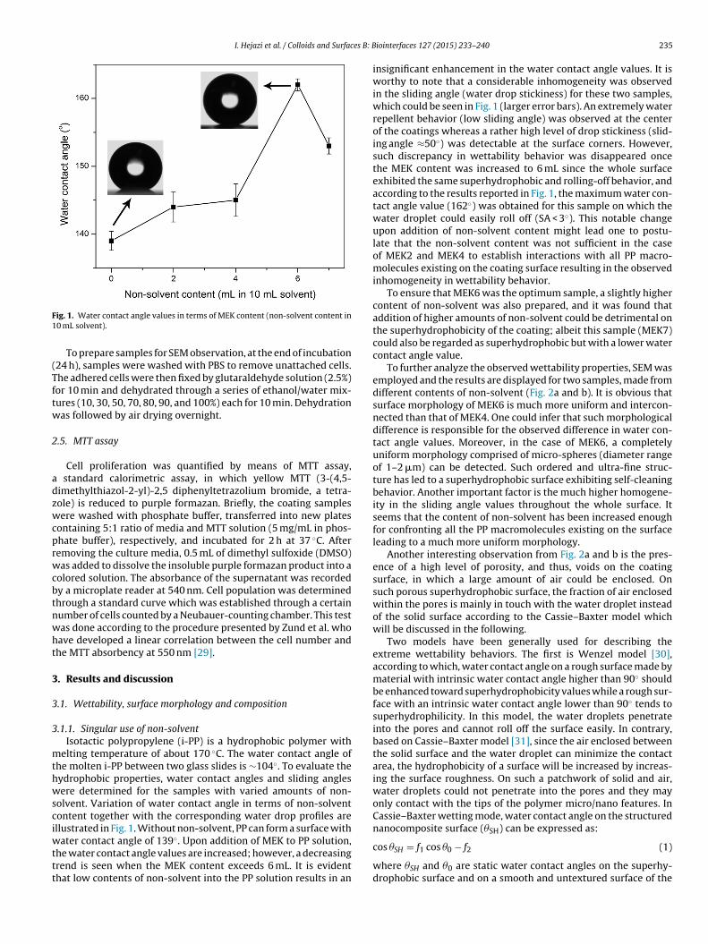
I. Hejazi et al. / Colloids and Surfaces B: B
F1
(Tftw
2
adzwcprwcbtnwht
3
3
3
mthwsciwttt
ig. 1. Water contact angle values in terms of MEK content (non-solvent content in0 mL solvent).
To prepare samples for SEM observation, at the end of incubation24 h), samples were washed with PBS to remove unattached cells.he adhered cells were then fixed by glutaraldehyde solution (2.5%)or 10 min and dehydrated through a series of ethanol/water mix-ures (10, 30, 50, 70, 80, 90, and 100%) each for 10 min. Dehydrationas followed by air drying overnight.
.5. MTT assay
Cell proliferation was quantified by means of MTT assay, standard calorimetric assay, in which yellow MTT (3-(4,5-imethylthiazol-2-yl)-2,5 diphenyltetrazolium bromide, a tetra-ole) is reduced to purple formazan. Briefly, the coating samplesere washed with phosphate buffer, transferred into new plates
ontaining 5:1 ratio of media and MTT solution (5 mg/mL in phos-hate buffer), respectively, and incubated for 2 h at 37 ◦C. Afteremoving the culture media, 0.5 mL of dimethyl sulfoxide (DMSO)as added to dissolve the insoluble purple formazan product into a
olored solution. The absorbance of the supernatant was recordedy a microplate reader at 540 nm. Cell population was determinedhrough a standard curve which was established through a certainumber of cells counted by a Neubauer-counting chamber. This testas done according to the procedure presented by Zund et al. whoave developed a linear correlation between the cell number andhe MTT absorbency at 550 nm [29].
. Results and discussion
.1. Wettability, surface morphology and composition
.1.1. Singular use of non-solventIsotactic polypropylene (i-PP) is a hydrophobic polymer with
elting temperature of about 170 ◦C. The water contact angle ofhe molten i-PP between two glass slides is ∼104◦. To evaluate theydrophobic properties, water contact angles and sliding anglesere determined for the samples with varied amounts of non-
olvent. Variation of water contact angle in terms of non-solventontent together with the corresponding water drop profiles arellustrated in Fig. 1. Without non-solvent, PP can form a surface with
ater contact angle of 139◦. Upon addition of MEK to PP solution,he water contact angle values are increased; however, a decreasingrend is seen when the MEK content exceeds 6 mL. It is evidenthat low contents of non-solvent into the PP solution results in an
iointerfaces 127 (2015) 233–240 235
insignificant enhancement in the water contact angle values. It isworthy to note that a considerable inhomogeneity was observedin the sliding angle (water drop stickiness) for these two samples,which could be seen in Fig. 1 (larger error bars). An extremely waterrepellent behavior (low sliding angle) was observed at the centerof the coatings whereas a rather high level of drop stickiness (slid-ing angle ≈50◦) was detectable at the surface corners. However,such discrepancy in wettability behavior was disappeared oncethe MEK content was increased to 6 mL since the whole surfaceexhibited the same superhydrophobic and rolling-off behavior, andaccording to the results reported in Fig. 1, the maximum water con-tact angle value (162◦) was obtained for this sample on which thewater droplet could easily roll off (SA < 3◦). This notable changeupon addition of non-solvent content might lead one to postu-late that the non-solvent content was not sufficient in the caseof MEK2 and MEK4 to establish interactions with all PP macro-molecules existing on the coating surface resulting in the observedinhomogeneity in wettability behavior.
To ensure that MEK6 was the optimum sample, a slightly highercontent of non-solvent was also prepared, and it was found thataddition of higher amounts of non-solvent could be detrimental onthe superhydrophobicity of the coating; albeit this sample (MEK7)could also be regarded as superhydrophobic but with a lower watercontact angle value.
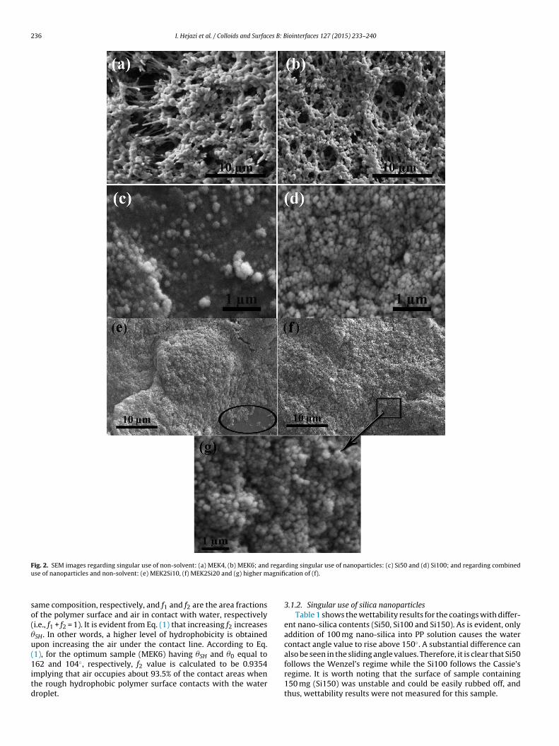
To further analyze the observed wettability properties, SEM wasemployed and the results are displayed for two samples, made fromdifferent contents of non-solvent (Fig. 2a and b). It is obvious thatsurface morphology of MEK6 is much more uniform and intercon-nected than that of MEK4. One could infer that such morphologicaldifference is responsible for the observed difference in water con-tact angle values. Moreover, in the case of MEK6, a completelyuniform morphology comprised of micro-spheres (diameter rangeof 1–2 �m) can be detected. Such ordered and ultra-fine struc-ture has led to a superhydrophobic surface exhibiting self-cleaningbehavior. Another important factor is the much higher homogene-ity in the sliding angle values throughout the whole surface. Itseems that the content of non-solvent has been increased enoughfor confronting all the PP macromolecules existing on the surfaceleading to a much more uniform morphology.
Another interesting observation from Fig. 2a and b is the pres-ence of a high level of porosity, and thus, voids on the coatingsurface, in which a large amount of air could be enclosed. Onsuch porous superhydrophobic surface, the fraction of air enclosedwithin the pores is mainly in touch with the water droplet insteadof the solid surface according to the Cassie–Baxter model whichwill be discussed in the following.
Two models have been generally used for describing theextreme wettability behaviors. The first is Wenzel model [30],according to which, water contact angle on a rough surface made bymaterial with intrinsic water contact angle higher than 90◦ shouldbe enhanced toward superhydrophobicity values while a rough sur-face with an intrinsic water contact angle lower than 90◦ tends tosuperhydrophilicity. In this model, the water droplets penetrateinto the pores and cannot roll off the surface easily. In contrary,based on Cassie–Baxter model [31], since the air enclosed betweenthe solid surface and the water droplet can minimize the contactarea, the hydrophobicity of a surface will be increased by increas-ing the surface roughness. On such a patchwork of solid and air,water droplets could not penetrate into the pores and they mayonly contact with the tips of the polymer micro/nano features. InCassie–Baxter wetting mode, water contact angle on the structurednanocomposite surface (�SH) can be expressed as:
cos �SH = f1 cos �0 − f2 (1)
where �SH and �0 are static water contact angles on the superhy-drophobic surface and on a smooth and untextured surface of the
236 I. Hejazi et al. / Colloids and Surfaces B: Biointerfaces 127 (2015) 233–240
F regaru agnifi
so(�u(1itd
ig. 2. SEM images regarding singular use of non-solvent: (a) MEK4, (b) MEK6; andse of nanoparticles and non-solvent: (e) MEK2Si10, (f) MEK2Si20 and (g) higher m
ame composition, respectively, and f1 and f2 are the area fractionsf the polymer surface and air in contact with water, respectivelyi.e., f1 + f2 = 1). It is evident from Eq. (1) that increasing f2 increasesSH. In other words, a higher level of hydrophobicity is obtainedpon increasing the air under the contact line. According to Eq.1), for the optimum sample (MEK6) having �SH and �0 equal to
62 and 104◦, respectively, f2 value is calculated to be 0.9354mplying that air occupies about 93.5% of the contact areas whenhe rough hydrophobic polymer surface contacts with the waterroplet.
ding singular use of nanoparticles: (c) Si50 and (d) Si100; and regarding combinedcation of (f).
3.1.2. Singular use of silica nanoparticlesTable 1 shows the wettability results for the coatings with differ-
ent nano-silica contents (Si50, Si100 and Si150). As is evident, onlyaddition of 100 mg nano-silica into PP solution causes the watercontact angle value to rise above 150◦. A substantial difference canalso be seen in the sliding angle values. Therefore, it is clear that Si50
follows the Wenzel’s regime while the Si100 follows the Cassie’sregime. It is worth noting that the surface of sample containing150 mg (Si150) was unstable and could be easily rubbed off, andthus, wettability results were not measured for this sample.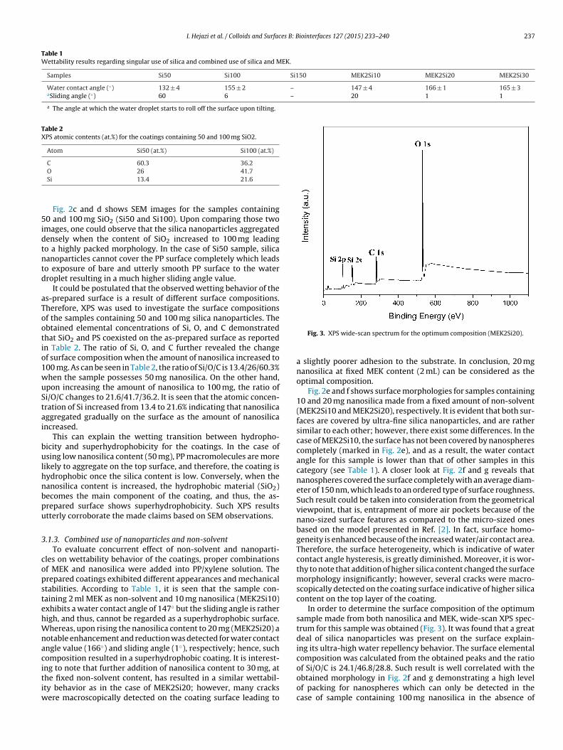
I. Hejazi et al. / Colloids and Surfaces B: Biointerfaces 127 (2015) 233–240 237
Table 1Wettability results regarding singular use of silica and combined use of silica and MEK.
Samples Si50 Si100 Si150 MEK2Si10 MEK2Si20 MEK2Si30
Water contact angle (◦) 132 ± 4 155 ± 2 – 147 ± 4 166 ± 1 165 ± 3aSliding angle (◦) 60 6 – 20 1 1
a The angle at which the water droplet starts to roll off the surface upon tilting.
Table 2XPS atomic contents (at.%) for the coatings containing 50 and 100 mg SiO2.
Atom Si50 (at.%) Si100 (at.%)
C 60.3 36.2
5idtntd
aTootio1wuStai
bulhnbpu
3
copstehWnacitiw
O 26 41.7Si 13.4 21.6
Fig. 2c and d shows SEM images for the samples containing0 and 100 mg SiO2 (Si50 and Si100). Upon comparing those two
mages, one could observe that the silica nanoparticles aggregatedensely when the content of SiO2 increased to 100 mg leadingo a highly packed morphology. In the case of Si50 sample, silicaanoparticles cannot cover the PP surface completely which leadso exposure of bare and utterly smooth PP surface to the waterroplet resulting in a much higher sliding angle value.
It could be postulated that the observed wetting behavior of thes-prepared surface is a result of different surface compositions.herefore, XPS was used to investigate the surface compositionsf the samples containing 50 and 100 mg silica nanoparticles. Thebtained elemental concentrations of Si, O, and C demonstratedhat SiO2 and PS coexisted on the as-prepared surface as reportedn Table 2. The ratio of Si, O, and C further revealed the changef surface composition when the amount of nanosilica increased to00 mg. As can be seen in Table 2, the ratio of Si/O/C is 13.4/26/60.3%hen the sample possesses 50 mg nanosilica. On the other hand,pon increasing the amount of nanosilica to 100 mg, the ratio ofi/O/C changes to 21.6/41.7/36.2. It is seen that the atomic concen-ration of Si increased from 13.4 to 21.6% indicating that nanosilicaggregated gradually on the surface as the amount of nanosilicancreased.
This can explain the wetting transition between hydropho-icity and superhydrophobicity for the coatings. In the case ofsing low nanosilica content (50 mg), PP macromolecules are more
ikely to aggregate on the top surface, and therefore, the coating isydrophobic once the silica content is low. Conversely, when theanosilica content is increased, the hydrophobic material (SiO2)ecomes the main component of the coating, and thus, the as-repared surface shows superhydrophobicity. Such XPS resultstterly corroborate the made claims based on SEM observations.
.1.3. Combined use of nanoparticles and non-solventTo evaluate concurrent effect of non-solvent and nanoparti-
les on wettability behavior of the coatings, proper combinationsf MEK and nanosilica were added into PP/xylene solution. Therepared coatings exhibited different appearances and mechanicaltabilities. According to Table 1, it is seen that the sample con-aining 2 ml MEK as non-solvent and 10 mg nanosilica (MEK2Si10)xhibits a water contact angle of 147◦ but the sliding angle is ratherigh, and thus, cannot be regarded as a superhydrophobic surface.hereas, upon rising the nanosilica content to 20 mg (MEK2Si20) a
otable enhancement and reduction was detected for water contactngle value (166◦) and sliding angle (1◦), respectively; hence, suchomposition resulted in a superhydrophobic coating. It is interest-
ng to note that further addition of nanosilica content to 30 mg, athe fixed non-solvent content, has resulted in a similar wettabil-ty behavior as in the case of MEK2Si20; however, many cracksere macroscopically detected on the coating surface leading to
Fig. 3. XPS wide-scan spectrum for the optimum composition (MEK2Si20).
a slightly poorer adhesion to the substrate. In conclusion, 20 mgnanosilica at fixed MEK content (2 mL) can be considered as theoptimal composition.
Fig. 2e and f shows surface morphologies for samples containing10 and 20 mg nanosilica made from a fixed amount of non-solvent(MEK2Si10 and MEK2Si20), respectively. It is evident that both sur-faces are covered by ultra-fine silica nanoparticles, and are rathersimilar to each other; however, there exist some differences. In thecase of MEK2Si10, the surface has not been covered by nanospherescompletely (marked in Fig. 2e), and as a result, the water contactangle for this sample is lower than that of other samples in thiscategory (see Table 1). A closer look at Fig. 2f and g reveals thatnanospheres covered the surface completely with an average diam-eter of 150 nm, which leads to an ordered type of surface roughness.Such result could be taken into consideration from the geometricalviewpoint, that is, entrapment of more air pockets because of thenano-sized surface features as compared to the micro-sized onesbased on the model presented in Ref. [2]. In fact, surface homo-geneity is enhanced because of the increased water/air contact area.Therefore, the surface heterogeneity, which is indicative of watercontact angle hysteresis, is greatly diminished. Moreover, it is wor-thy to note that addition of higher silica content changed the surfacemorphology insignificantly; however, several cracks were macro-scopically detected on the coating surface indicative of higher silicacontent on the top layer of the coating.
In order to determine the surface composition of the optimumsample made from both nanosilica and MEK, wide-scan XPS spec-trum for this sample was obtained (Fig. 3). It was found that a greatdeal of silica nanoparticles was present on the surface explain-ing its ultra-high water repellency behavior. The surface elementalcomposition was calculated from the obtained peaks and the ratioof Si/O/C is 24.1/46.8/28.8. Such result is well correlated with the
obtained morphology in Fig. 2f and g demonstrating a high levelof packing for nanospheres which can only be detected in thecase of sample containing 100 mg nanosilica in the absence of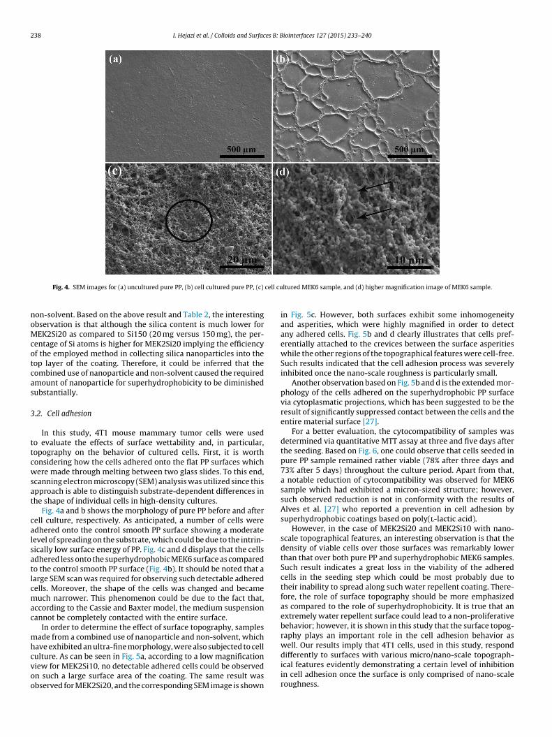
238 I. Hejazi et al. / Colloids and Surfaces B: Biointerfaces 127 (2015) 233–240
cell cu
noMcotcas
3
ttcwsat
calsatlcmac
mhcvoo
Fig. 4. SEM images for (a) uncultured pure PP, (b) cell cultured pure PP, (c)
on-solvent. Based on the above result and Table 2, the interestingbservation is that although the silica content is much lower forEK2Si20 as compared to Si150 (20 mg versus 150 mg), the per-
entage of Si atoms is higher for MEK2Si20 implying the efficiencyf the employed method in collecting silica nanoparticles into theop layer of the coating. Therefore, it could be inferred that theombined use of nanoparticle and non-solvent caused the requiredmount of nanoparticle for superhydrophobicity to be diminishedubstantially.
.2. Cell adhesion
In this study, 4T1 mouse mammary tumor cells were usedo evaluate the effects of surface wettability and, in particular,opography on the behavior of cultured cells. First, it is worthonsidering how the cells adhered onto the flat PP surfaces whichere made through melting between two glass slides. To this end,
canning electron microscopy (SEM) analysis was utilized since thispproach is able to distinguish substrate-dependent differences inhe shape of individual cells in high-density cultures.
Fig. 4a and b shows the morphology of pure PP before and afterell culture, respectively. As anticipated, a number of cells weredhered onto the control smooth PP surface showing a moderateevel of spreading on the substrate, which could be due to the intrin-ically low surface energy of PP. Fig. 4c and d displays that the cellsdhered less onto the superhydrophobic MEK6 surface as comparedo the control smooth PP surface (Fig. 4b). It should be noted that aarge SEM scan was required for observing such detectable adheredells. Moreover, the shape of the cells was changed and becameuch narrower. This phenomenon could be due to the fact that,
ccording to the Cassie and Baxter model, the medium suspensionannot be completely contacted with the entire surface.
In order to determine the effect of surface topography, samplesade from a combined use of nanoparticle and non-solvent, which
ave exhibited an ultra-fine morphology, were also subjected to cell
ulture. As can be seen in Fig. 5a, according to a low magnificationiew for MEK2Si10, no detectable adhered cells could be observedn such a large surface area of the coating. The same result wasbserved for MEK2Si20, and the corresponding SEM image is shownltured MEK6 sample, and (d) higher magnification image of MEK6 sample.
in Fig. 5c. However, both surfaces exhibit some inhomogeneityand asperities, which were highly magnified in order to detectany adhered cells. Fig. 5b and d clearly illustrates that cells pref-erentially attached to the crevices between the surface asperitieswhile the other regions of the topographical features were cell-free.Such results indicated that the cell adhesion process was severelyinhibited once the nano-scale roughness is particularly small.
Another observation based on Fig. 5b and d is the extended mor-phology of the cells adhered on the superhydrophobic PP surfacevia cytoplasmatic projections, which has been suggested to be theresult of significantly suppressed contact between the cells and theentire material surface [27].
For a better evaluation, the cytocompatibility of samples wasdetermined via quantitative MTT assay at three and five days afterthe seeding. Based on Fig. 6, one could observe that cells seeded inpure PP sample remained rather viable (78% after three days and73% after 5 days) throughout the culture period. Apart from that,a notable reduction of cytocompatibility was observed for MEK6sample which had exhibited a micron-sized structure; however,such observed reduction is not in conformity with the results ofAlves et al. [27] who reported a prevention in cell adhesion bysuperhydrophobic coatings based on poly(l-lactic acid).
However, in the case of MEK2Si20 and MEK2Si10 with nano-scale topographical features, an interesting observation is that thedensity of viable cells over those surfaces was remarkably lowerthan that over both pure PP and superhydrophobic MEK6 samples.Such result indicates a great loss in the viability of the adheredcells in the seeding step which could be most probably due totheir inability to spread along such water repellent coating. There-fore, the role of surface topography should be more emphasizedas compared to the role of superhydrophobicity. It is true that anextremely water repellent surface could lead to a non-proliferativebehavior; however, it is shown in this study that the surface topog-raphy plays an important role in the cell adhesion behavior aswell. Our results imply that 4T1 cells, used in this study, respond
differently to surfaces with various micro/nano-scale topograph-ical features evidently demonstrating a certain level of inhibitionin cell adhesion once the surface is only comprised of nano-scaleroughness.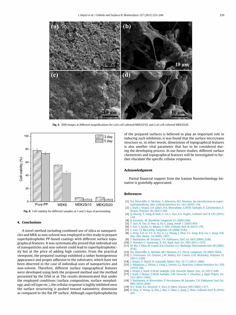
I. Hejazi et al. / Colloids and Surfaces B: Biointerfaces 127 (2015) 233–240 239
Fig. 5. SEM images at different magnifications for (a,b) cell
4
csgoivabnwptota
[[
[[
[[
[[
Fig. 6. Cell viability for different samples at 3 and 5 days of postseeding.
. Conclusions
A novel method including combined use of silica as nanoparti-les and MEK as non-solvent was employed in this study to prepareuperhydrophobic PP-based coatings with different surface topo-raphical features. It was systematically proved that individual usef nanoparticles and non-solvent could lead to superhydrophobic-ty but at the price of adding high contents. From the practicaliewpoint, the prepared coatings exhibited a rather homogeneousppearance and proper adhesion to the substrates, which have noteen observed in the case of individual uses of nanoparticles andon-solvent. Therefore, different surface topographical featuresere developed using both the proposed method and the methodresented by the Erbil et al. The results demonstrated that under
he employed conditions (surface composition, surface morphol-gy, and cell type etc.), the cellular response is highly inhibited oncehe surface structuring is pushed toward nanometric dimensions compared to the flat PP surface. Although superhydrophobicity[
[[
cultured MEK2Si10, and (c,d) cell cultured MEK2Si20.
of the prepared surfaces is believed to play an important role ininducing such inhibition, it was found that the surface micro/nanostructure or, in other words, dimensions of topographical featuresis also another vital parameter that has to be considered dur-ing the developing process. In our future studies, different surfacechemistries and topographical features will be investigated to fur-ther elucidate the specific cellular responses.
Acknowledgment
Partial financial support from the Iranian Nanotechnology Ini-tiative is gratefully appreciated.
References
[1] N.J. Shirtcliffe, G. McHale, S. Atherton, M.I. Newton, An introduction to super-hydrophobicity, Adv. Colloid Interface Sci. 161 (2010) 124.
[2] J. Seyfi, I. Hejazi, S.H. Jafari, H.A. Khonakdar, G.M.M. Sadeghi, A. Calvimontes, F.Simon, Polymer 56 (2015) 358.
[3] Q. Huang, Y. Yang, R. Hub, C. Lin, L. Sun, E.A. Vogler, Colloids Surf. B 125 (2015)134.
[4] R. Furstner, W. Barthlott, Langmuir 21 (2005) 956.[5] T. Sun, H. Tan, D. Han, Q. Fu, L. Jiang, Small 1 (2005) 959.[6] L. Lizi, T. Junfei, Li. Miaosi, S. Wei, Colloids Surf. B (2013) 176.[7] L. Gao, T.J. McCarthy, Langmuir 24 (2008) 9183.[8] L. Feng, S.H. Li, Y.S. Li, H.J. Li, L.J. Zhang, J. Zhai, Y.L. Song, B.Q. Liu, L. Jiang, D.B.
Zhu, Adv. Mater. 14 (2002) 1857.[9] T. Rasilainen, M. Suvanto, T.A. Pakkanen, Surf. Sci. 603 (2009) 2240.10] Y. Haowei, C. Yuanrong, X. Fei, Appl. Surf. Sci. 258 (2011) 1572.11] M. Ma, Y. Mao, M. Gupta, K.K. Gleason, G.C. Rutledge, Macromolecules 38 (2005)
9742.12] N.J. Shirtcliffe, G. McHale, M.I. Newton, C.C. Perry, Langmuir 19 (2003) 5626.13] S. Srinivasan, S.S. Chatter, J.M. Mabry, R.E. Cohen, G.H. McKinley, Polymer 52
(2011) 3209.14] I. Hejazi, F. Sharif, H. Garmabi, Mater. Des. 32 (7) (2011) 3803.15] L. Mingxian, J. Zhixin, L. Fang, J. Demin, G.J. Baochun, Colloid Interface Sci. 350
(2010) 186.16] I. Hejazi, J. Seyfi, G.M.M. Sadeghi, S.M. Davachi, Mater. Des. 32 (2011) 649.17] J. Seyfi, I. Hejazi, G.M.M. Sadeghi, S.M. Davachi, S. Ghanbar, J. Appl. Polym. Sci.
123 (2011) 2492.
18] T. Rasilainen, A. Kirveslahti, P. Nevalainen, M. Suvanto, T.A. Pakkanen, Surf. Sci.604 (2010) 2036.19] H.Y. Erbil, A.L. Demirel, Y. Avcı, O. Mert, Science 299 (2003) 1377.20] X. Hou, X. Wang, Q. Zhu, J. Bao, C. Mao, L. Jiang, J. Shen, Colloids Surf. B (2010)
247.

2 es B: B
[
[[[[[
[
40 I. Hejazi et al. / Colloids and Surfac
21] I. Hejazi, B. Hajalizadeh, J. Seyfi, G.M.M. Sadeghi, S.H. Jafari, H.A. Khonakdar,Appl. Surf. Sci. 293 (2014) 116.
22] L.C. Xua, J.W. Bauerb, C.A. Siedlecki, Colloids Surf. B (2014) 49.23] R. Langer, D.A. Tirrell, Nature 428 (2004) 487.24] W. Song, J.F. Mano, Soft Matter 9 (2013) 2985.25] U. Schwarz, Soft Matter 3 (2007) 263.26] W.L. Song, D.D. Veiga, C.A. Custodio, J.F. Mano, Adv. Mater. 21 (2009) 1830.
[[
[[
iointerfaces 127 (2015) 233–240
27] N.M. Alves, J. Shi, E. Oramas, J.L. Santos, H. Tomas, J.F. Mano, J. Biomed. Mater.Res. A 91 (2009) 480.
28] S.C. Luo, S.S. Liour, H.H. Yu, Chem. Commun. 46 (2010) 4731.29] G. Zund, Q. Ye, S.P. Hoerstrup, A. Schoeberlein, A.C. Schmid, J. Grunenfelder, P.
Vogt, M. Turina, Eur. J. Cardiothorac. Surg. 15 (1999) 519.30] R.N. Wenzel, Ind. Eng. Chem. Res. 28 (1936) 988.31] A.B.D. Cassie, S. Baxter, Trans. Faraday Soc. 40 (1944) 546.
Related Documents