continued on page 2 Summer 2020 Vol. 27, No. 2 MISSION STATEMENT The NIDCD Naonal Temporal Bone, Hearing and Balance Pathology Resource Registry was established in 1992 by the Naonal Instute on Deafness and Other Communicaon Disorders (NIDCD) of the Naonal Instutes of Health (NIH) to connue and expand upon the former Naonal Temporal Bone Banks (NTBB) Program. The Registry promotes research on hearing and balance disorders and serves as a resource for the public and scienfic communies about research on the pathology of the human auditory and vesbular systems. Featured Research Invasion Paerns of External Auditory Canal Squamous Cell Carcinoma: A Histopathology Study....................1 Liquid Biopsy of the Human Inner Ear and Interpretaon of Novel Molecular Biomarkers in Paents with Vesbular Schwannomas: Measuring Mediators of Hearing Loss and Predicng Surgical Outcomes.............................6 Registry News Otopathology Mini-Travel Fellowship .......................................11 Order Form for Temporal Bone Donaon Brochures........................12 THE Newsletter of the NIDCD National Temporal Bone, Hearing and Balance Pathology Resource Registry CONTENTS Invasion Patterns of External Auditory Canal Squamous Cell Carcinoma– A Histopathology Study T emporal bone (TB) malignant neoplasms are rare, occurring in 1:5000-20,000 ear disorders 1 . ese malignant tumors can be categorized as primary or secondary. Primary TB malignant tumors originate in the TB, most commonly in the external auditory canal (EAC). Secondary tumors arise from extra-TB tissue and invade it by means of local extension or metastatic spread. e most common advanced stage malignancy is squamous cell carcinoma (SCC) of the EAC 2 . EAC SCC tends to expand through consistent patterns such as invasion of compact bone, along blood vessels, cranial nerves and areas of osseous weaknesses like sutures and along the TB air cells tracks. e aim of this study is to examine the invasion patterns of advanced stage SCC of the EAC and to compare the histological findings to clinical data. To the best of our knowledge, this is the first analysis of a series of TBs of patients with primary SCC of the TB. Nine TBs from 9 patients were included in the cohort. Patients were diagnosed from 1968 to 2010. All cases presented with T4 stage disease according to the modified Pittsburgh Omer J. Ungar 1 , Felipe Santos 2 , Joseph B. Nadol, Jr. 2 , Gilad Hrowitz 1 , Dan M. Fliss 1 , William C. Faquin 3 , Ophir Handzel 1 1 Departments of Otolaryngology Head and Neck Surgery and Maxillofacial Surgery, Tel-Aviv Sourasky Medical Center, Sackler School of Medicine. Tel-Aviv University, Tel-Aviv, Israel 2 Department of Otolaryngology, Massachuses Eye and Ear, Boston, Massachuses, U.S.A., and the Department of Otolaryngology and Harvard Medical School, Boston, Massachuses, U.S.A. 3 Department of Pathology, Massachuses Eye and Ear, Boston, Massachuses, U.S.A., and the Department of Pathology Massachuses General Hospital and Harvard Medical School, Boston, Massachuses, U.S.A.

Welcome message from author
This document is posted to help you gain knowledge. Please leave a comment to let me know what you think about it! Share it to your friends and learn new things together.
Transcript
continued on page 2
Summer 2020Vol. 27, No. 2
MISSION STATEMENT
The NIDCD National Temporal Bone, Hearing and Balance Pathology Resource Registry was established in 1992 by the National Institute on Deafness and Other Communication Disorders (NIDCD) of the National Institutes of Health (NIH) to continue and expand upon the former National Temporal Bone Banks (NTBB) Program. The Registry promotes research on hearing and balance disorders and serves as a resource for the public and scientific communities about research on the pathology of the human auditory and vestibular systems.
Featured Research Invasion Patterns of External Auditory Canal Squamous Cell Carcinoma: A Histopathology Study....................1
Liquid Biopsy of the Human Inner Ear and Interpretation of Novel Molecular Biomarkers in Patients with Vestibular Schwannomas: Measuring Mediators of Hearing Loss and Predicting Surgical Outcomes.............................6
Registry NewsOtopathology Mini-Travel Fellowship .......................................11
Order Form for Temporal Bone Donation Brochures........................12
THE
Newsletter of the NIDCD National Temporal Bone, Hearing and Balance Pathology Resource Registry
CONTENTS
Invasion Patterns of External Auditory Canal
Squamous Cell Carcinoma– A Histopathology Study
Temporal bone (TB) malignant neoplasms are rare, occurring in 1:5000-20,000 ear disorders1. These malignant tumors can be categorized as primary or secondary. Primary TB malignant tumors originate in the TB, most commonly in the external auditory canal (EAC). Secondary tumors arise from extra-TB tissue and invade it by means of local extension or metastatic spread. The most common advanced
stage malignancy is squamous cell carcinoma (SCC) of the EAC2.
EAC SCC tends to expand through consistent patterns such as invasion of compact bone, along blood vessels, cranial nerves and areas of osseous weaknesses like sutures and along the TB air cells tracks.
The aim of this study is to examine the invasion patterns of advanced stage SCC of the EAC and to compare the histological findings to clinical data. To the best of our knowledge, this is the first analysis of a series of TBs of patients with primary SCC of the TB.
Nine TBs from 9 patients were included in the cohort. Patients were diagnosed from 1968 to 2010. All cases presented with T4 stage disease according to the modified Pittsburgh
Omer J. Ungar1, Felipe Santos2, Joseph B. Nadol, Jr.2, Gilad Hrowitz1, Dan M. Fliss1, William C. Faquin3,
Ophir Handzel1 1Departments of Otolaryngology Head and Neck Surgery and Maxillofacial Surgery,
Tel-Aviv Sourasky Medical Center, Sackler School of Medicine. Tel-Aviv University, Tel-Aviv, Israel2Department of Otolaryngology, Massachusetts Eye and Ear, Boston, Massachusetts, U.S.A., and the Department of Otolaryngology and Harvard Medical School, Boston, Massachusetts, U.S.A.
3Department of Pathology, Massachusetts Eye and Ear, Boston, Massachusetts, U.S.A., and the Department of Pathology Massachusetts General Hospital and Harvard Medical School,
Boston, Massachusetts, U.S.A.
Vol. 27.2 | Summer 2020THE
staging system3 (Figure 1). The demographics and clinical presentation are found in table 1. The most common presenting symptom was hearing loss (7 patients), the majority of which was severe to profound (5 patients). The most common otoscopic finding was obstructing mass EAC (5 patients).
Several soft tissue elements within the TB were found to serve as barriers, limiting tumor invasion. The tympanic membrane was found to limit tumor extension medially in 4 patients. In these patients, the pathway of spread from the EAC to the middle ear (ME) was through the bony posterior EAC wall, to the mastoid air cells system (MACS) and the antrum. In five patients the tumor invaded the ME from the EAC directly, through the TM. The vestibulo-stapedial (annular) ligament was found to be a significant anatomic barrier for tumor spread from the middle to the inner ear. The resistance against tumor spread was so effective, that otic capsule invasion was seen adjacent to intact vestibulo-stapedial ligament. The round window membrane (RWM) was not invaded, although its niche was filled with tumor in 3 subjects. Examples of the relationship between these soft tissue barriers and tumor extension are shown in figure 2.
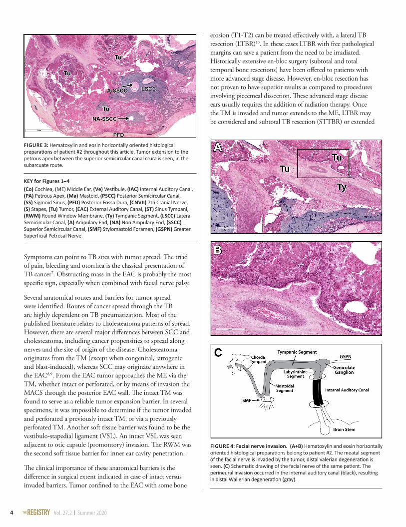
Several patterns of tumor spread were identified: besides serving as a route to the ME, the MACS was found to serve as a tumor conduit to the tegmen mastoideum and overlying dura, the posterior fossa dura, the vertical segment of the facial nerve, the ME and the lateral semicircular canal. The supra and infra labyrinthine pneumatization patterns allowed direct routes of tumor spread to the petrous apex (PA), leaving the otic capsule intact and most easily demonstrated in the vertical oriented histological preparations. The petro-mastoid canal served as an additional route to the PA via the subarcuate route (Figure 3). Trans labyrinthine PA invasion was seen in 2 patients. Once in the ME, tumors tended to spread to the anterior and posterior attic. We found no evidence that surgical modification of the TB anatomy created iatrogenic pathways for tumor spread. The ottic capsule itself was involved in 6 patients. The most common locus of otic capsule invasion was the cochlea, followed by the lateral semicircular canal (LSCC) and vestibule.
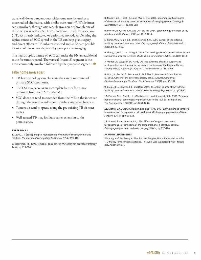
The tympanic segment of facial nerve was involved in 5 subjects and the vertical segment was involved in 3, resulting in varying degrees of facial nerve weakness. Wallerian degeneration was present distal to the proximal-most site of neural invasion (Figure 4).
Primary TB SCC is a rare, aggressive tumor, comprising 80% of primary TB malignancies 4, 5. This tumor carries a substantial risk for morbidity and mortality because of its aggressive nature, location at the skull base and the common delay in diagnosis6. Early symptoms can be easily attributed to more common conditions such as otitis media and externa. This delay is likely to have a detrimental effect on treatment and outcome.
DIRECTORSJoseph B. Nadol, Jr., MDM. Charles Liberman, PhDAlicia M. Quesnel, MDFelipe Santos, MD
COORDINATORCsilla Haburcakova, PhD
ADMINISTRATIVE STAFFKristen Kirk-Paladino
EDITORMedical: Felipe Santos, MD
DESIGNERGaryfallia Pagonis
NIDCD National Temporal Bone, Hearing and Balance Pathology Resource Registry
Massachusetts Eye and Ear243 Charles StreetBoston, MA 02114
(800) 822-1327 Toll-Free Voice(617) 573-3711 Voice(617) 573-3838 Fax
Email: [email protected]: www.tbregistry.org
The reports in the Registry Newsletter are not peer reviewed.
2
THE
continued on page 4
Vol. 27.2 | Summer 2020THE 3
FIGURE 1: Malignant invasion throughout the temporal bone, indicating stage T4. (A-I) These H&E horizontal oriented histological preparations belong to patient #2 (A) Middle ear and petrous apex air cells are filled with the tumor. Stapes is partially subluxated. The otic capsule is invaded near the basal turn of the cochlea and internal auditory canal (asterisk). (B) High power magnification of figure 1A. The vestibular-stapedial (annular) ligament is intact, limiting tumor invasion to the vestibule (arrow). The tumor invades the otic capsule towards the cochlea and vestibule. (C) Low power magnification of the same TB. The tumor fills the external ear canal, mastoid and petrous apex. The posterior fossa dura over the mastoid is partially dehiscent. (D) The tumor invades the round window niche and the otic capsule overlying the posterior semicircular canal and cochlea (arrows). (E) Low power magnification of the petrous apex. The tumor encases the carotid canal, which is partially dehiscent. (F) High power magnification of figure 1E. The tumor invades the tunica externa of the petrous segment of the carotid artery. (G) Petrous apex air cells and internal auditory canal are filled with tumor. (H) High power magnification of figure 1G. The internal auditory canal is partially invaded by the tumor, as well as the posterior fossa dura. (I) Pure tone audiogram of the same patient showing profound conductive hearing loss, with reduced discrimination.
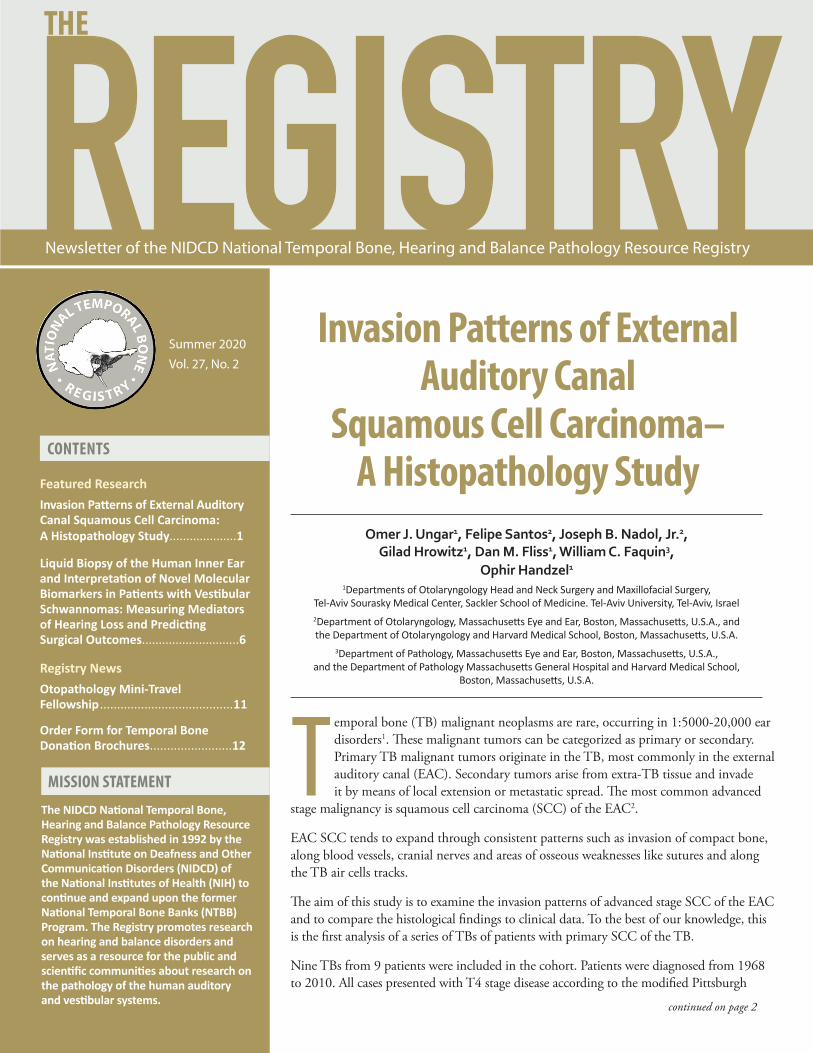
FIGURE 2: Soft tissue barriers limit SCC spread throughout the TB. (A) H&E horizontal oriented histological preparations of patient #1. This patient was treated with high dose of XRT (120 Gy) to the TB, replacing the SCC by fibrotic mass. EAC is filled with fibrotic mass. The TM is intact, effectively preventing medial invasion. Mastoid is invaded through the EAC posterior wall. The facial nerve is intact. (B-F) H&E preparation from the temporal bone described in figure 1, belong to patient #2 (B) The mastoid is invaded by the tumor, totally destroying the air cells system. (C) Low power magnification of figure 2B. The dura is invaded, as well as the otic capsule around the vestibule, which is partially dehiscent. (D) The vestibular-stapedial ligament limits medial tumor extension to the vestibule. The tumor invades the otic capsule around the basal and medium cochlear turns (asterisk). (E) High power magnification of figure 2D. (F) The tumor fills the round window niche. The round window is intact, preventing tumor extension to the inner ear spaces.
Vol. 27.2 | Summer 2020THE4
Symptoms can point to TB sites with tumor spread. The triad of pain, bleeding and otorrhea is the classical presentation of TB cancer7. Obstructing mass in the EAC is probably the most specific sign, especially when combined with facial nerve palsy.
Several anatomical routes and barriers for tumor spread were identified. Routes of cancer spread through the TB are highly dependent on TB pneumatization. Most of the published literature relates to cholesteatoma patterns of spread. However, there are several major differences between SCC and cholesteatoma, including cancer propensities to spread along nerves and the site of origin of the disease. Cholesteatoma originates from the TM (except when congenital, iatrogenic and blast-induced), whereas SCC may originate anywhere in the EAC8,9. From the EAC tumor approaches the ME via the TM, whether intact or perforated, or by means of invasion the MACS through the posterior EAC wall. The intact TM was found to serve as a reliable tumor expansion barrier. In several specimens, it was impossible to determine if the tumor invaded and perforated a previously intact TM, or via a previously perforated TM. Another soft tissue barrier was found to be the vestibulo-stapedial ligament (VSL). An intact VSL was seen adjacent to otic capsule (promontory) invasion. The RWM was the second soft tissue barrier for inner ear cavity penetration.
The clinical importance of these anatomical barriers is the difference in surgical extent indicated in case of intact versus invaded barriers. Tumor confined to the EAC with some bone
erosion (T1-T2) can be treated effectively with, a lateral TB resection (LTBR)10. In these cases LTBR with free pathological margins can save a patient from the need to be irradiated. Historically extensive en-bloc surgery (subtotal and total temporal bone resections) have been offered to patients with more advanced stage disease. However, en-bloc resection has not proven to have superior results as compared to procedures involving piecemeal dissection. These advanced stage disease ears usually requires the addition of radiation therapy. Once the TM is invaded and tumor extends to the ME, LTBR may be considered and subtotal TB resection (STTBR) or extended
FIGURE 3: Hematoxylin and eosin horizontally oriented histological preparations of patient #2 throughout this article. Tumor extension to the petrous apex between the superior semicircular canal crura is seen, in the subarcuate route.
FIGURE 4: Facial nerve invasion. (A+B) Hematoxylin and eosin horizontally oriented histological preparations belong to patient #2. The meatal segment of the facial nerve is invaded by the tumor, distal valerian degeneration is seen. (C) Schematic drawing of the facial nerve of the same patient. The perineural invasion occurred in the internal auditory canal (black), resulting in distal Wallerian degeneration (gray).
KEY for Figures 1–4(Co) Cochlea, (ME) Middle Ear, (Ve) Vestibule, (IAC) Internal Auditory Canal, (PA) Petrous Apex, (Ma) Mastoid, (PSCC) Posterior Semicircular Canal, (SS) Sigmoid Sinus, (PFD) Posterior Fossa Dura, (CNVII) 7th Cranial Nerve, (S) Stapes, (Tu) Tumor, (EAC) External Auditory Canal, (ST) Sinus Tympani, (RWM) Round Window Membrane, (Ty) Tympanic Segment, (LSCC) Lateral Semicircular Canal, (A) Ampulary End, (NA) Non Ampulary End, (SSCC) Superior Semicircular Canal, (SMF) Stylomastoid Foramen, (GSPN) Greater Superficial Petrosal Nerve.
Vol. 27.2 | Summer 2020THE 5
canal wall down tympano-mastoidectomy may be used as a more radical alternative, with similar cure rates11,12. While inner ear is involved, through otic capsule invasion or through one of the inner ear windows, STTBR is indicated. Total TB resection (TTBR) is rarely indicated or performed nowadays. Defining the typical routes of SCC spread in the TB can help plan surgery, and direct efforts to TB subsites involved and anticipate possible location of disease not depicted by pre-operative imaging.
The neurotrophic nature of SCC can make the FN an additional route for tumor spread. The vertical (mastoid) segment is the most commonly involved followed by the tympanic segment. l
Take home messages:• TB histopathology can elucidate the extension routes of
primary SCC carcinoma.
• The TM may serve as an incomplete barrier for tumor extension from the EAC to the ME.
• SCC does not tend to extended from the ME to the inner ear through the round window and vestibule-stapedial ligament.
• Tumors do tend to spread along the pre-existing TB air-tract routes.
• Well aerated TB may facilitate easier extension to the petrous apex.
REFERENCES1. Lewis, J. S. (1983). Surgical management of tumors of the middle ear and mastoid. The Journal of Laryngology & Otology, 97(4), 299-312.
2. Koriwchak, M., 1993. Temporal bone cancer. The American Journal of Otology, 14(6), pp.623-626.
3. Moody, S.A., Hirsch, B.E. and Myers, E.N., 2000. Squamous cell carcinoma of the external auditory canal: an evaluation of a staging system. Otology & Neurotology, 21(4), pp.582-588.
4. Morton, R.P., Stell, P.M. and Derrick, P.P., 1984. Epidemiology of cancer of the middle ear cleft. Cancer, 53(7), pp.1612-1617.
5. Kuhel, W.I., Hume, C.R. and Selesnick, S.H., 1996. Cancer of the external auditory canal and temporal bone. Otolaryngologic Clinics of North America, 29(5), pp.827-852.
6. Zhang, T., Dai, C. and Wang, Z., 2013. The misdiagnosis of external auditory canal carcinoma. European Archives of Oto-rhino-laryngology, 270(5), pp.1607-1613
7. Moffat DA, Wagstaff SA, Hardy DG. The outcome of radical surgery and postoperative radiotherapy for squamous carcinoma of the temporal bone. Laryngoscope. 2005 Feb;115(2):341-7. PubMed PMID: 15689763.
8. Ouaz, K., Robier, A., Lescanne, E., Bobillier, C., Moriniere, S. and Bakhos, D., 2013. Cancer of the external auditory canal. European Annals of Otorhinolaryngology, Head and Neck Diseases, 130(4), pp.175-182.
9. Breau, R.L., Gardner, E.K. and Dornhoffer, J.L., 2002. Cancer of the external auditory canal and temporal bone. Current Oncology Reports, 4(1), pp.76-80.
10. Pensak, M.L., Gleich, L.L., Gluckman, J.L. and Shumrick, K.A., 1996. Temporal bone carcinoma: contemporary perspectives in the skull base surgical era. The Laryngoscope, 106(10), pp.1234-1237.
11. Moffat, D.A., Grey, P., Ballagh, R.H. and Hardy, D.G., 1997. Extended temporal bone resection for squamous cell carcinoma. Otolaryngology–Head and Neck Surgery, 116(6), pp.617-623.
12. Prasad, S. and Janecka, I.P., 1994. Efficacy of surgical treatments for squamous cell carcinoma of the temporal bone: a literature review. Otolaryngology—Head and Neck Surgery, 110(3), pp.270-280.
ACKNOWLEDGEMENTSWe are grateful to Meng Yu Zhu, Barbara Burgess, Diane Jones, and Jennifer T. O’Malley for technical assistance. This work was supported by NIH-NIDCD (U24DC013983-01)
Vol. 27.2 | Summer 2020THE6
Liquid Biopsy of the Human Inner Ear and Interpretation of
Novel Molecular Biomarkers in Patients With Vestibular Schwannomas:
Measuring Mediators of Hearing Loss and Predicting Surgical Outcomes
Yin Ren1,2,3, Samuel Early1,2,3, Konstantina M. Stankovic1,2,4,5
1Department of Otolaryngology – Head and Neck Surgery and Eaton-Peabody Laboratories, Massachusetts Eye and Ear, Boston, MA, USA 2Department of Otolaryngology – Head and Neck Surgery, Harvard Medical School, Boston, MA, USA
3Division of Otolaryngology, Department of Surgery, School of Medicine, University of California, San Diego, CA, USA4Program in Speech and Hearing Bioscience and Technology, Harvard Medical School, Boston, MA, USA
5Harvard Program in Therapeutic Science, Harvard Medical School, Boston, MA, USA
Vestibular schwannoma (VS) is the fourth most common intracranial tumor and constitutes 90% of all cerebellopontine angle (CPA) lesions1. It is a benign, slow-growing tumor arising from neoplastic Schwann cells lining the eighth cranial nerve. While
the majority of VS occurs sporadically, a portion develops in association with neurofibromatosis type II (NF2). Despite its non-malignant nature, growing VS can be associated with significant morbidity including sensorineural hearing loss (SNHL) that affects up to 95% of patients, tinnitus, dizziness, increased rates of hospitalization, and poor mental health2. As tumors grow larger, they can cause additional symptoms such as palsy of the facial nerve and other cranial nerves, and potentially life-threatening complications including brainstem compression. The goal of contemporary VS management is to balance long-term tumor control, neurological function, and quality of life3. The standard treatment to remove the tumor is limited to surgery, as there are currently no FDA-approved pharmacotherapies for the treatment of VS or associated SNHL. Nonetheless, surgery can be associated with significant morbidities such as facial paralysis and deafness.
While numerous retrospective studies have attempted to identify clinical factors associated with disease severity and prognosis, there are no established reliable biomarkers to predict the degree of VS-associated SNHL or surgical
outcomes after tumor resection. Clinical factors such as tumor size, tumor growth rate, and radiographic imaging features often suffer from poor sensitivity and specificity. Histological findings such as tumor proliferative index, while useful, are obtained only after tumor extirpation and do not offer prognostic information4. Moreover, there is great variability in surgical outcomes when patients undergo attempted microsurgical tumor resection. Factors such as tumor consistency, presence of cystic changes, and adhesions between the tumor capsule and the facial nerve have all been associated with an increased likelihood of subtotal tumor resection5. There have been over twenty clinical trials in the last 20 years on experimental therapies for NF1 and NF2, yet no biomarker-driven trial for NF2 has been performed6,7. Further, little is known about the molecular underpinnings of VS tumorigenesis that predisposes certain tumors towards incomplete resections.
Molecular biomarkers are objectively measured indicators of disease pathophysiology and have the potential to facilitate early detection of tumors, monitor disease progression, and impact the outcomes of treatment. Proteases, a class of enzymes that plays a fundamental role in both normal tissue remodeling and aberrant cellular growth, are candidates for biomarkers for VS8. In many human cancers, protease biomarkers have been well established to assess treatment response and even predict
Vol. 27.2 | Summer 2020THE 7
continued on page 8
survival9,10. Various members of the matrix metalloprotease (MMP) family have been implicated in promoting VS growth via angiogenesis, remodeling of the extracellular matrix, or enhancing growth factor signaling pathways11-13. While these results are promising, the studies are limited to a small number of patients, are retrospective, and address only a particular protease candidate. Additionally, the majority of the published studies rely on immunohistochemical staining of tumor specimens rather than measuring serum levels as a readout, which precludes preoperative patient counseling and forecasting of clinical outcomes.
Since hearing loss is the most common consequence of VS, a clinically useful biomarker should differentiate tumors with good hearing from those with poor hearing. Furthermore, given the variability in clinical outcomes such as the extent of surgical resection, the biomarker should provide information about the likelihood of gross total tumor removal and the functional outcome of the facial nerve; two features that would greatly impact both long-term prognosis and the patient’s quality of life. A protease-based biomarker has the potential to outperform existing biomarkers which are often disease by-products with insufficient sensitivity, through leveraging the catalytic nature of
protease signal amplification. Finally, a safe, reliable, minimally-invasive device for liquid biopsy of the inner ear would be necessary to collect diagnostically useful volumes of human perilymph.
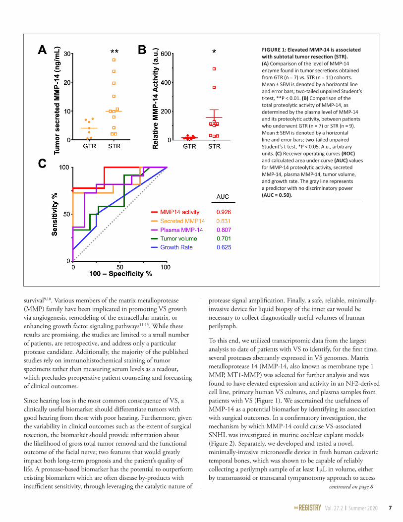
To this end, we utilized transcriptomic data from the largest analysis to date of patients with VS to identify, for the first time, several proteases aberrantly expressed in VS genomes. Matrix metalloprotease 14 (MMP-14, also known as membrane type 1 MMP, MT1-MMP) was selected for further analysis and was found to have elevated expression and activity in an NF2-derived cell line, primary human VS cultures, and plasma samples from patients with VS (Figure 1). We ascertained the usefulness of MMP-14 as a potential biomarker by identifying its association with surgical outcomes. In a confirmatory investigation, the mechanism by which MMP-14 could cause VS-associated SNHL was investigated in murine cochlear explant models (Figure 2). Separately, we developed and tested a novel, minimally-invasive microneedle device in fresh human cadaveric temporal bones, which was shown to be capable of reliably collecting a perilymph sample of at least 1μL in volume, either by transmastoid or transcanal tympanotomy approach to access
FIGURE 1: Elevated MMP-14 is associated with subtotal tumor resection (STR). (A) Comparison of the level of MMP-14 enzyme found in tumor secretions obtained from GTR (n = 7) vs. STR (n = 11) cohorts. Mean ± SEM is denoted by a horizontal line and error bars; two-tailed unpaired Student’s t-test, **P < 0.01. (B) Comparison of the total proteolytic activity of MMP-14, as determined by the plasma level of MMP-14 and its proteolytic activity, between patients who underwent GTR (n = 7) or STR (n = 9). Mean ± SEM is denoted by a horizontal line and error bars; two-tailed unpaired Student’s t-test, *P < 0.05. A.u., arbitrary units. (C) Receiver operating curves (ROC) and calculated area under curve (AUC) values for MMP-14 proteolytic activity, secreted MMP-14, plasma MMP-14, tumor volume, and growth rate. The gray line represents a predictor with no discriminatory power (AUC = 0.50).
Vol. 27.2 | Summer 2020THE8
continued on page 10
the round window membrane, with minimal contamination from middle ear fluids (Figure 3); this volume has previously been shown to be diagnostically meaningful across multiple institutions14-16. This device is designed with utmost safety in mind, including safeguards to monitor depth of penetration through the round window membrane and protect intracochlear structures. Trauma to the round window membrane with this device is minimal, smaller and more controlled than that typically associated with spontaneous perilymphatic fistula formation, and likely amenable to the highly successful repair strategies that are already available, such as placement of an absorbable gelatin sponge or a tiny fat graft17.
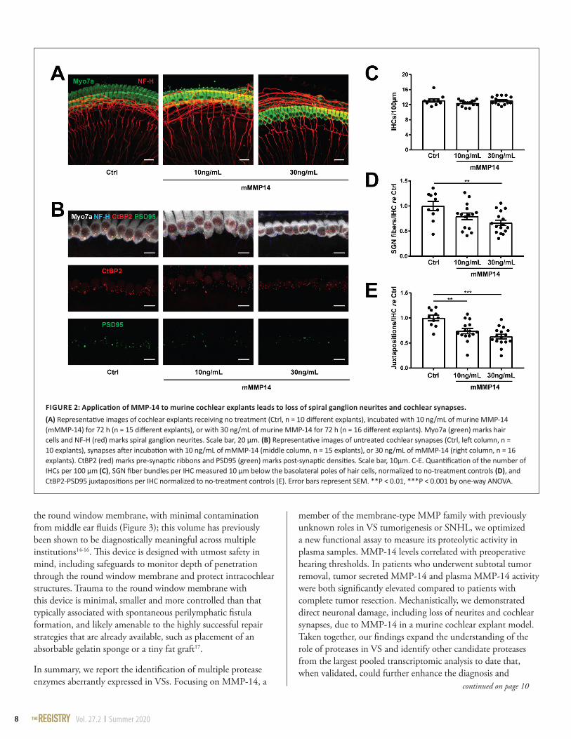
In summary, we report the identification of multiple protease enzymes aberrantly expressed in VSs. Focusing on MMP-14, a
member of the membrane-type MMP family with previously unknown roles in VS tumorigenesis or SNHL, we optimized a new functional assay to measure its proteolytic activity in plasma samples. MMP-14 levels correlated with preoperative hearing thresholds. In patients who underwent subtotal tumor removal, tumor secreted MMP-14 and plasma MMP-14 activity were both significantly elevated compared to patients with complete tumor resection. Mechanistically, we demonstrated direct neuronal damage, including loss of neurites and cochlear synapses, due to MMP-14 in a murine cochlear explant model. Taken together, our findings expand the understanding of the role of proteases in VS and identify other candidate proteases from the largest pooled transcriptomic analysis to date that, when validated, could further enhance the diagnosis and
FIGURE 2: Application of MMP-14 to murine cochlear explants leads to loss of spiral ganglion neurites and cochlear synapses. (A) Representative images of cochlear explants receiving no treatment (Ctrl, n = 10 different explants), incubated with 10 ng/mL of murine MMP-14 (mMMP-14) for 72 h (n = 15 different explants), or with 30 ng/mL of murine MMP-14 for 72 h (n = 16 different explants). Myo7a (green) marks hair cells and NF-H (red) marks spiral ganglion neurites. Scale bar, 20 µm. (B) Representative images of untreated cochlear synapses (Ctrl, left column, n = 10 explants), synapses after incubation with 10 ng/mL of mMMP-14 (middle column, n = 15 explants), or 30 ng/mL of mMMP-14 (right column, n = 16 explants). CtBP2 (red) marks pre-synaptic ribbons and PSD95 (green) marks post-synaptic densities. Scale bar, 10µm. C-E. Quantification of the number of IHCs per 100 µm (C), SGN fiber bundles per IHC measured 10 µm below the basolateral poles of hair cells, normalized to no-treatment controls (D), and CtBP2-PSD95 juxtapositions per IHC normalized to no-treatment controls (E). Error bars represent SEM. **P < 0.01, ***P < 0.001 by one-way ANOVA.

Vol. 27.2 | Summer 2020THE 9
FIGURE 3: Positioning of the microneedle for perilymph sampling across the round window membrane. (A-F) Demonstrate transmastoid facial recess approach in a left ear: (A) Access via transmastoid facial recess approach with identification of the facial nerve (f) and chorda tympani nerve (c), drilling of the round window overhang (o) to expose the round window membrane (*), with additional key landmarks including the stapedial tendon (s) and incudostapedial joint (j); (B) Same photo unlabeled; (C) Application of reference solution (blue) and placing of reference electrode into a mastoid air cell (not pictured); (D) Positioning of the microneedle adjacent to the round window membrane; (E) Perforation of the round window membrane and aspiration of perilymph; (F) Perforated round window membrane at low magnification; (G) Perforated round window membrane at high magnification, with arrowhead at site of perforation. (H-N) Demonstrate transcanal tympanotomy approach in a right ear: (H) Round window access via transcanal tympanotomy with lifting of the tympanic membrane, showing the round window niche (+) and fibrous annulus of the tympanic membrane (a); (I) Drilling of the round window overhang to expose the round window membrane (*); (J) Application of reference solution (blue) and placing of reference electrode into the inferior mesotympanum; (K) Positioning of the microneedle adjacent to the round window membrane; (L) Perforation of the round window membrane and aspiration of perilymph; (M) Perforated round window membrane at low magnification; (N) Perforated round window membrane at high magnification, with arrowhead at site of perforation. Scale bar = 1.0 mm for all pictures, assessed at depth of round window membrane.
Vol. 27.2 | Summer 2020THE10
Want to go digital?
The editors of The Registry are now offering an electronic version of the newsletter for those who are interested. Mailed copies will continue to be distributed; however, if you prefer to receive an email with a PDF version of the newsletter instead, let us know and we will add you to our email list!
In addition, all current and previous issues can be found on our website at https://masseyeandear.org/tbregistry/research-resources
Interested in receiving digital newsletter copies?Email Felipe Santos, MD, at Mass. Eye and Ear
personalized treatment of VS. The microneedle diagnostic platform which we have developed in parallel provides a potential avenue for minimally-invasive liquid biopsy of the inner ear, with potential to evaluate tumor-secreted MMP-14 levels in patients under consideration for VS resection to optimize counseling on expected outcomes. Predicting surgical outcomes using a minimally-invasive diagnostic can allow for more tailored selection of surgical candidates, and continued identification of predictive markers for VS or other SNHL-causing pathologies will further expand the value of this diagnostic platform. l
REFERENCES1. Mahaley, M. S. Jr., Mettlin, C., Natarajan, N. Jr., Laws, E. R., and Peace, B. B. (1990). Analysis of patterns of care of brain tumor patients in the united states: a study of the brain tumor section of the AANS and the CNS and the commission on cancer of the ACS. Clin Neurosurg. 36, 347–352
2. Genther, D. J., Frick, K. D., Chen, D., Betz, J., and Lin, F. R. (2013). Association of hearing loss with hospitalization and burden of disease in older adults. JAMA 309, 2322–2324. doi: 10.1001/jama.2013.5912.
3. Carlson, M. L., Tveiten, O. V., Driscoll, C. L., Goplen, F. K., Neff, B. A., Pollock, B. E., et al. (2015b). Long-term quality of life in patients with vestibular schwannoma: an international multicenter cross-sectional study comparing microsurgery, stereotactic radiosurgery, observation, and nontumor controls. J. Neurosurg. 122, 833–842. doi: 10.3171/2014.11.jns14594
4. Niemczyk, K., Vaneecloo, F. M., Lecomte, M. H., Lejeune, J. P., Lemaitre, L., Skarzynski, H., et al. (2000). Correlation between Ki-67 index and some clinical aspects of acoustic neuromas (vestibular schwannomas). Otolaryngol. Head Neck Surg. 123, 779–783. doi: 10.1067/mhn.2000.111356
5. Moon, K.-S., Jung, S., Seo, S.-K., Jung, T.-Y., Kim, I.-Y., Ryu, H.-H., et al. (2007). Cystic vestibular schwannomas: a possible role of matrix metalloproteinase-2 in cyst development and unfavorable surgical outcome. J. Neurosurg. 106, 86–871. doi: 10.3171/jns.2007.106.5.866
6. Plotkin, S. R., Blakeley, J. O., Dombi, E., Fisher, M. J., Hanemann, C. O., Walsh, K. S., et al. (2013). Achieving consensus for clinical trials: the REiNS international collaboration. Neurology 81, S1–S5. doi: 10.1212/01.wnl. 0000435743.49414.b6
7. Hanemann, C. O., Blakeley, J. O., Nunes, F. P., Robertson, K., StemmerRachamimov, A., Mautner, V., et al. (2016). Current status and recommendations for biomarkers and biobanking in neurofibromatosis. Neurology 87, S40–S48. doi: 10.1212/WNL.0000000000002932
8. Kessenbrock, K., Plaks, V., and Werb, Z. (2010). Matrix metalloproteinases: regulators of the tumor microenvironment. Cell 141, 52–67. doi: 10.1016/j.cell. 2010.03.015
9. Michael, M., Babic, B., Khokha, R., Tsao, M., Ho, J., Pintilie, M., et al. (1999). Expression and prognostic significance of metalloproteinases and their tissue inhibitors in patients with small-cell lung cancer. J. Clin. Oncol. 17, 1802–1808. doi: 10.1200/JCO.1999.17.6.1802
10. Ricci, S., Guadagno, E., Bruzzese, D., Del Basso De Caro, M., Peca, C., Sgulò, F. G., et al. (2017). Evaluation of matrix metalloproteinase type IV-collagenases in serum of patients with tumors of the central nervous system. J. Neurooncol. 131, 223–232. doi: 10.1007/s11060-016-2297-4
11. Cayé-Thomasen, P., Baandrup, L., Jacobsen, G. K., Thomsen, J., and Stangerup, S.- E. (2003). Immunohistochemical demonstration of vascular endothelial growth factor in vestibular schwannomas correlates to tumor growth rate. Laryngoscope 113, 2129–2134. doi: 10.1097/00005537-200312000- 00014
12. Moon, K.-S., Jung, S., Seo, S.-K., Jung, T.-Y., Kim, I.-Y., Ryu, H.-H., et al. (2007). Cystic vestibular schwannomas: a possible role of matrix metalloproteinase-2 in cyst development and unfavorable surgical outcome. J. Neurosurg. 106, 86–871. doi: 10.3171/jns.2007.106.5.866
13. Møller, M. N., Werther, K., Nalla, A., Stangerup, S.-E., Thomsen, J., BøgHansen, T. C., et al. (2010). Angiogenesis in vestibular schwannomas: expression of extracellular matrix factors MMP-2, MMP-9, and TIMP-1. Laryngoscope 120, 657–662. doi: 10.1002/lary.20834
14. Lysaght, A.C., Kao, S.Y., Paulo, J.A., Merchant, S.N., Steen, H., Stankovic, K.M. (2011). Proteome of human perilymph. J. Proteome Res. 10 (9), 3845e3851.
15. Schmitt, H.A., Pich, A., Schroder, A., et al. (2017). Proteome analysis of human perilymph using an intraoperative sampling method. J. Proteome Res. 16 (5), 1911e1923.
16. Shew, M., Warnecke, A., Lenarz, T., Schmitt, H., Gunewardena, S., Staecker, H. (2018). Feasibility of microRNA profiling in human inner ear perilymph. Neuroreport 29 (11), 894e901.
17. Alzahrani, M., Fadous, R., Dufour, J.J., Saliba, I. (2015). Perilymphatic fistulas: can we predict the diagnosis? Eur. Arch. Oto-Rhino-Laryngol. 272 (8), 1885e1891
Vol. 27.2 | Summer 2020THE 11
Otopathology Mini-Travel Fellowship Program
The NIDCD National Temporal Bone Registry’s mini-travel fellowships provide funds for research technicians and young investigators to visit a temporal bone laboratory for a brief educational visit, lasting approximately one week. The emphasis is on the training of research assistants, technicians, and junior faculty.
These fellowships are available to:
• U.S. hospital departments who aspire to start a new temporal bone laboratory
• Inactive U.S. temporal bone laboratories who wish to reactivate their collections
• Active U.S. temporal bone laboratories who wish to learn new research techniques
Up to two fellowship awards will be made each year ($1,000 per fellowship). The funds may be used to defray travel and lodging expenses. Applications will be decided on merit.
Interested applicants should submit the following:
• An outline of the educational or training aspect of the proposed fellowship (1–2 pages)
• Applicant’s curriculum vitae
• Letter of support from a temporal bone laboratory director or department chairman
• Letter from the host temporal bone laboratory indicating willingness to receive the traveling fellow
Applications should be submitted to:Felipe Santos, MDNIDCD Temporal Bone RegistryMassachusetts Eye and Ear243 Charles Street, Boston, MA [email protected]
Interested? Email us at [email protected]
Temporal Bone Removal
Technicians Needed
Nationwide!
s
s
Seeking trained autopsy technicians for the removal of
temporal bones on an on-call basis. Technicians must be
in the U.S. and are paid by case.
12
NIDCD National Temporal Bone, Hearing and BalancePathology Resource Registry Massachusetts Eye and Ear 243 Charles Street Boston, MA 02114-3096
Free Brochures for your Office or Clinic about Temporal Bone Research and DonationThe Gift of Hearing and Balance: Learning About Temporal Bone Donation is a 16-page, full-color booklet that describes in detail the benefits of temporal bone research. It also answers commonly asked questions regarding the temporal bone donation process. Dimensions: 7”x10”
If you would like to display this brochure, please complete the form below and return it to the Registry by mail or fax. The brochures will be sent to you free of charge. Please circle the amount requested for each brochure or write in the amount if not listed.
The Gift of Hearing and Balance __________ 25 50 100
Name: ________________________________________________________________________________________________
Address: ______________________________________________________________________________________________
City, State, Zip: _________________________________________________________________________________________
Telephone: ____________________________________________________________________________________________
Mail or fax this form to the Registry at: NIDCD National Temporal Bone, Hearing and Balance Pathology Resource Registry Massachusetts Eye and Ear, 243 Charles Street, Boston, MA 02114 Toll-free phone: (800) 822-1327, Fax: (617) 573-3838 Email: [email protected]
NON-PROFIT ORGU.S. POSTAGE PAID
BOSTON, MAPERMIT NO. 53825
Related Documents