Introduction to Head CT Imaging Dr. Azmy M Hadidy

Welcome message from author
This document is posted to help you gain knowledge. Please leave a comment to let me know what you think about it! Share it to your friends and learn new things together.
Transcript
Introduction to Head CT Imaging
Dr. Azmy M Hadidy

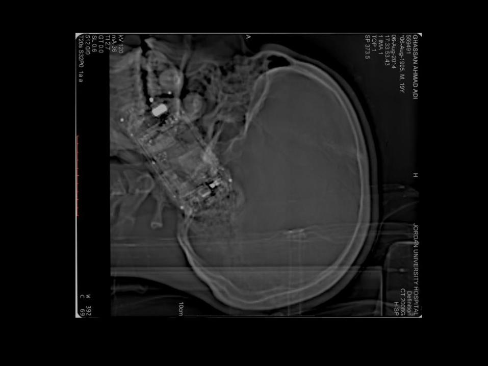

High resolution brain CT
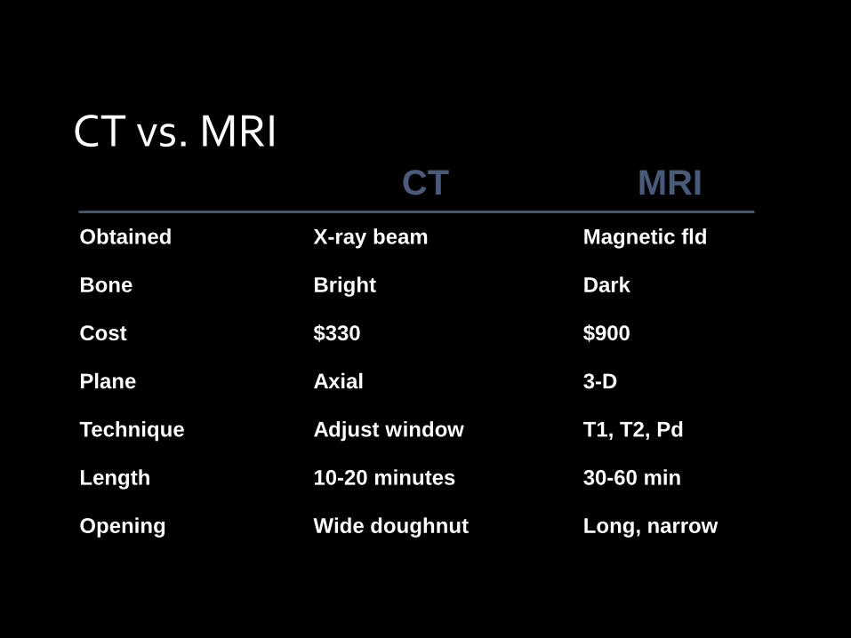
CT vs. MRI
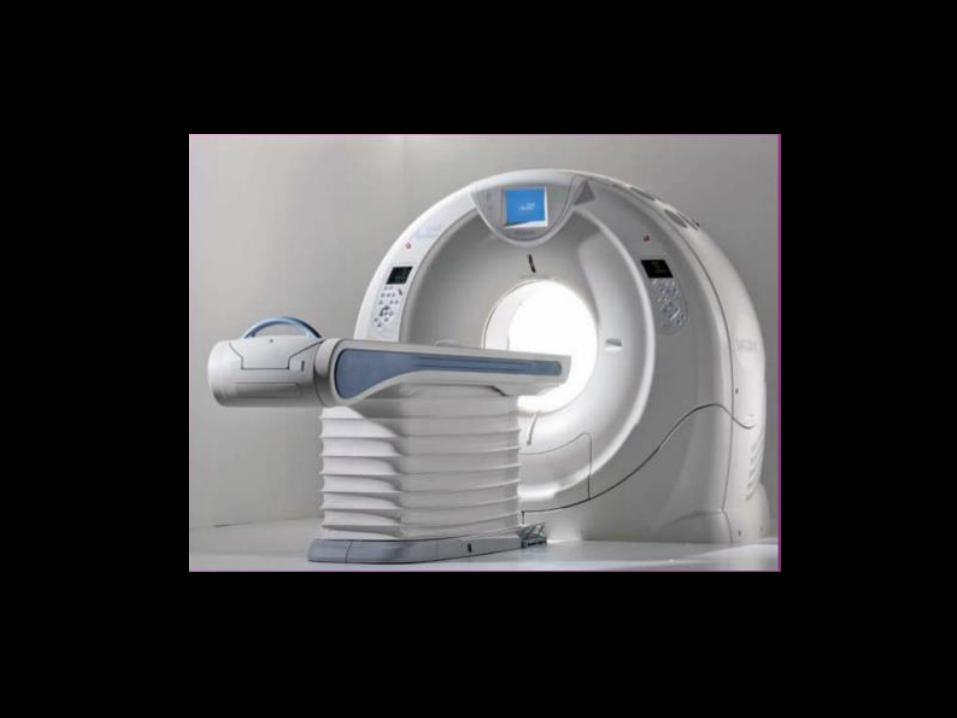
Wide doughnut Opening
10-20 minutes Length
Adjust window Technique
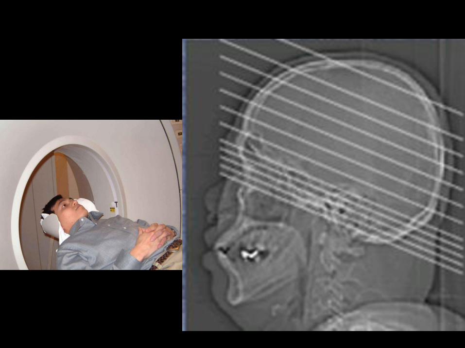
Axial Plane
$330 Cost
Bright Bone
Long, narrow
30-60 min
T1, T2, Pd
3-D
$900
Dark
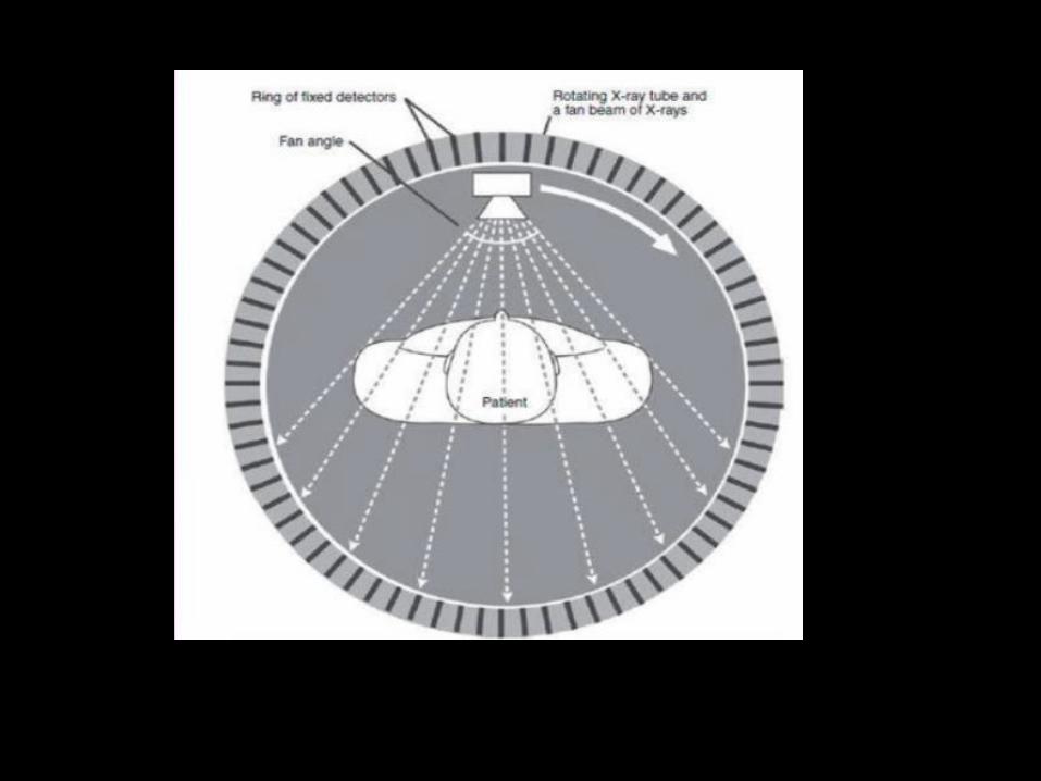
Magnetic fld X-ray beam Obtained
MRI CT

Advantages to CT
• Costs less than MRI
• Better access
• Shows up acute bleed
• A good quick screen
• Good visualization of bony structures and calcified lesions
Disadvantages to CT
• Resolution
• Beam-hardening artifact
• Limited views of the posterior fossa and poor visualization of white-matter disease

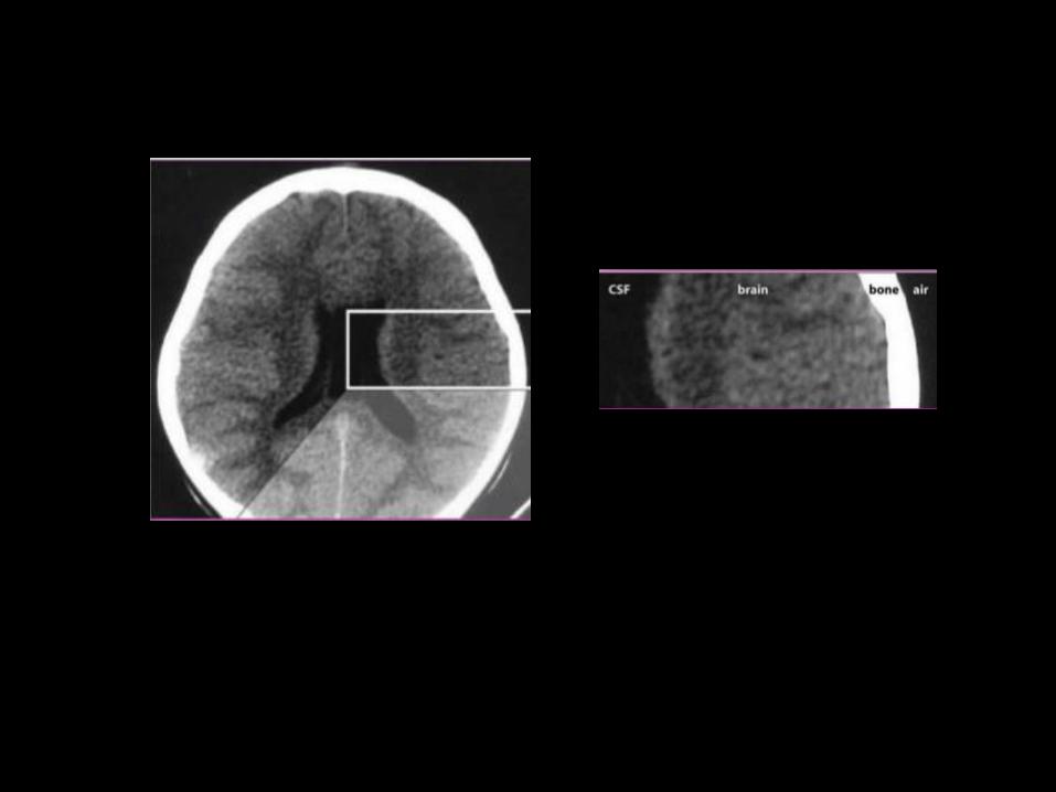
CT density
Black Structure/ Tissue
Hounsfield units
Air -1000 to -600
Fat -100 to -60
Water 0
CSF +8 to 18
White matter +30 to 41
Gray matter +37 to 41
Acute blood +50 to 100
Calcification +140 to 200
Bone +600 to 2000
White
Normal brain CT scan report :
No brain focal lesion
No midline shift
No hydrocephalus


Sella turcica
(contains pituitary
gland)
Petrous bone
Mastoid air cells
Pons
4th ventricle
Cerebellum
Eye
Optic nerve
Sphenoid bone
Temporal lobe
Normal Brain anatomy
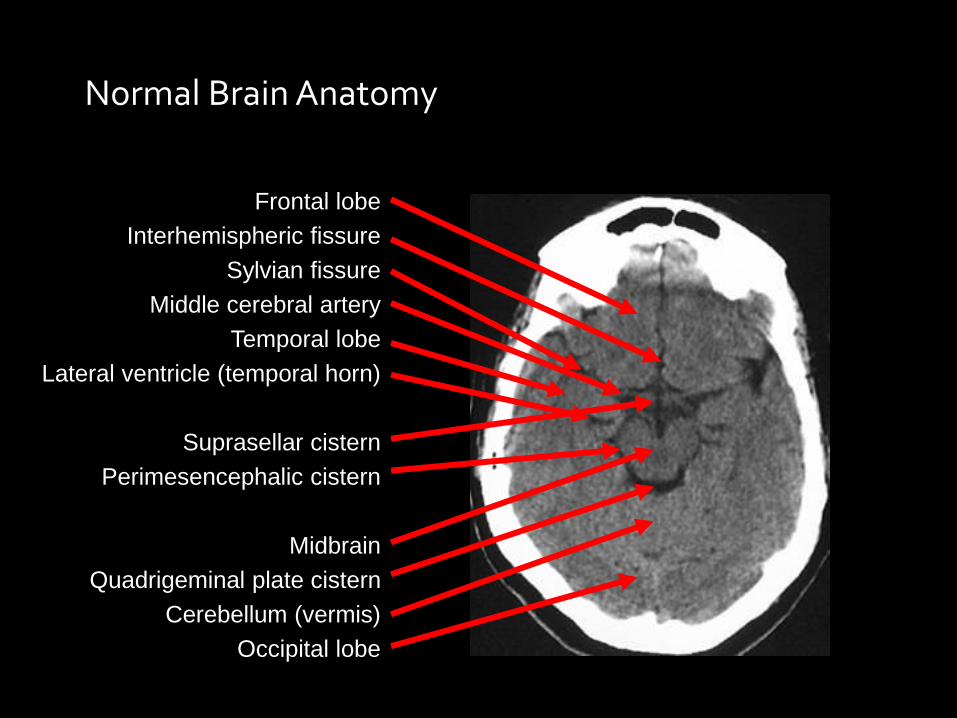
Normal Brain Anatomy
Frontal lobe
Interhemispheric fissure
Sylvian fissure
Middle cerebral artery
Temporal lobe
Lateral ventricle (temporal horn)
Suprasellar cistern
Perimesencephalic cistern
Midbrain
Quadrigeminal plate cistern
Cerebellum (vermis)
Occipital lobe
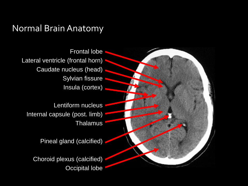
Normal Brain Anatomy
Lateral ventricle (frontal horn)
Frontal lobe
Caudate nucleus (head)
Sylvian fissure
Insula (cortex)
Lentiform nucleus
Internal capsule (post. limb)
Thalamus
Pineal gland (calcified)
Choroid plexus (calcified)
Occipital lobe

Look for:
Brain focal lesions
Midline shift
Dilated ventricular system (Hydrocephalus)
Brain Radiology Report
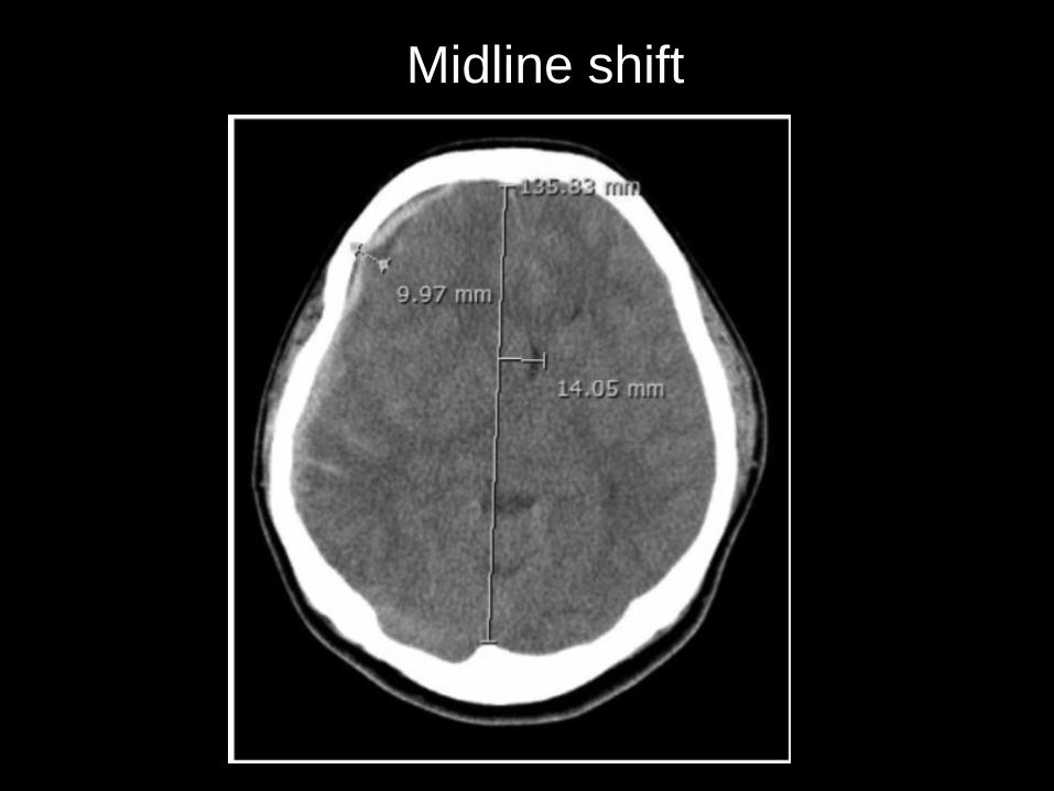
Midline shift
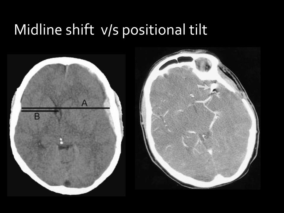
Midline shift v/s positional tilt
Not always there is midline
shift (MLS) , there could be
pathologies that don’t cause
MLS

1) Bilateral
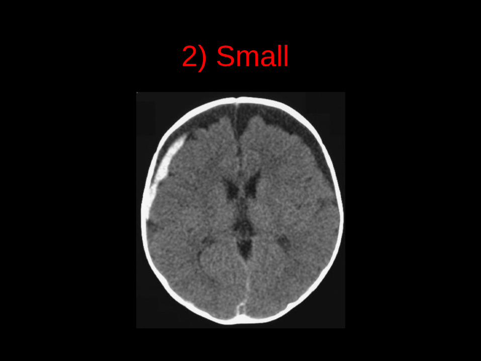
2) Small
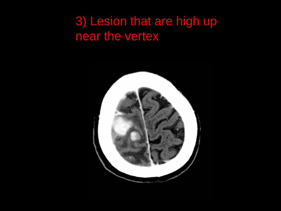
3) Lesion that are high up
near the vertex

Ex vacuo dilatation: due to diffuse brain atrophy
Hydrocephalus : communicating and non communicating
Ventricular Dilatation
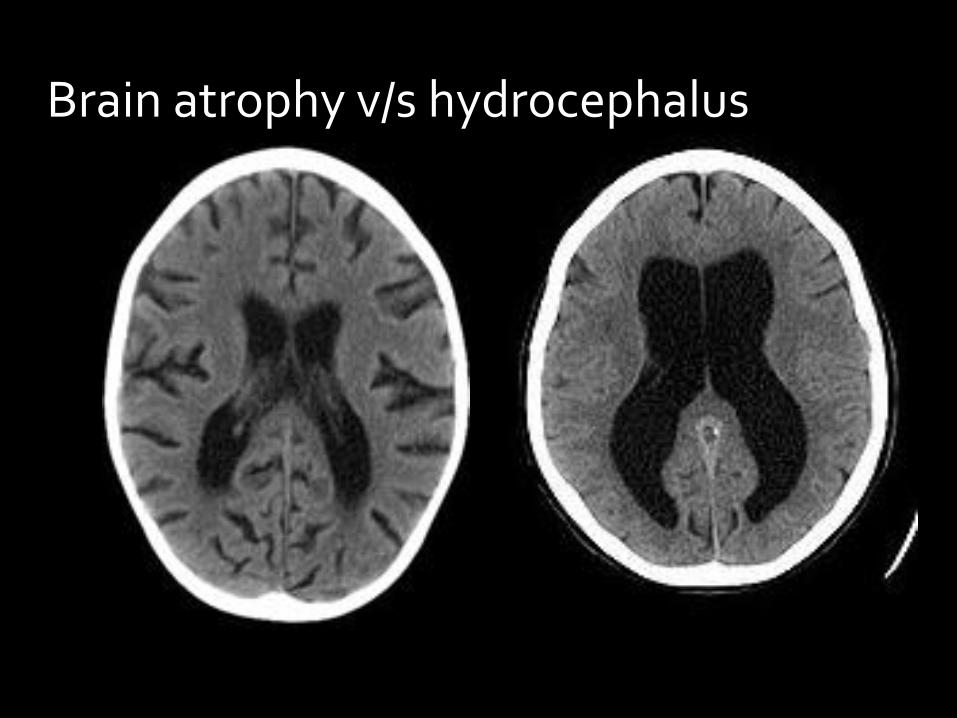
Brain atrophy v/s hydrocephalus
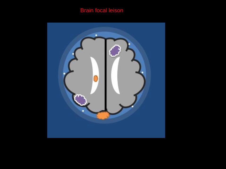
Brain focal leison

Abnormalities divided into :
Intraventricular
Intra-axial
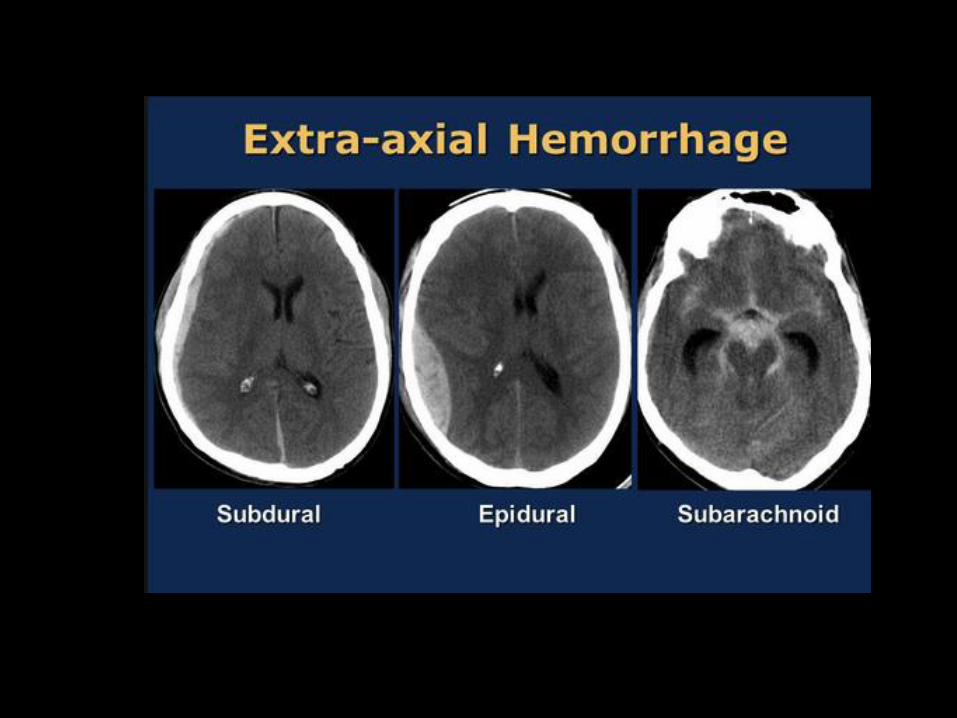
Extra-axial
Intra-osseous
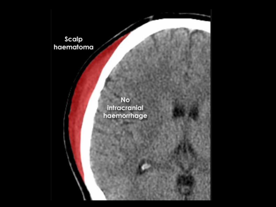
Scalp
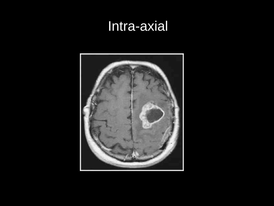
Intra-axial
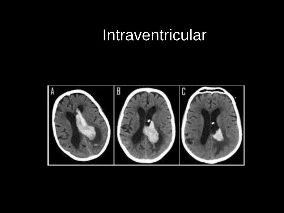
Intraventricular
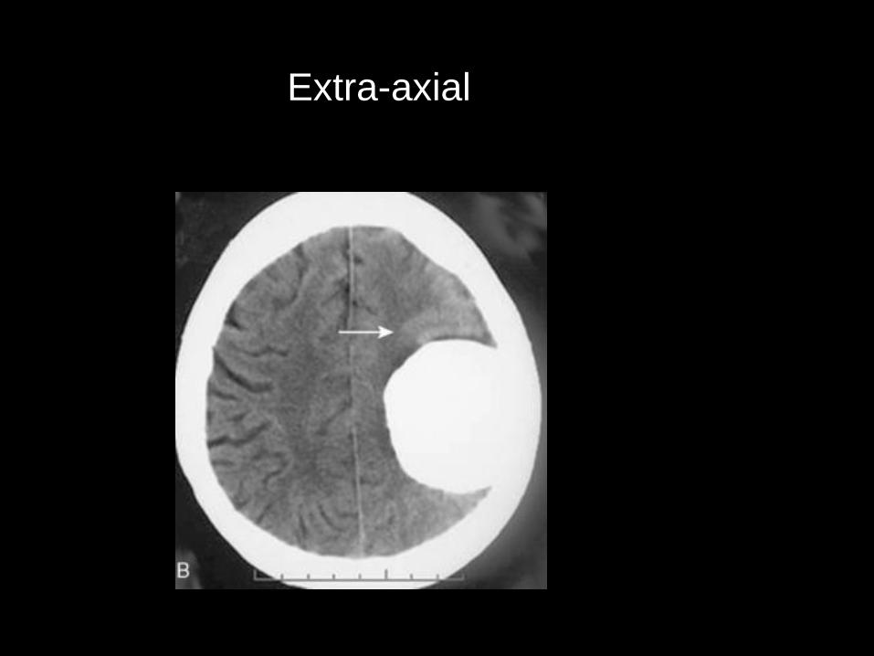
Extra-axial
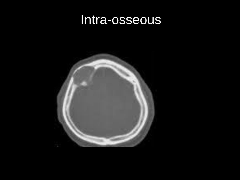
Intra-osseous
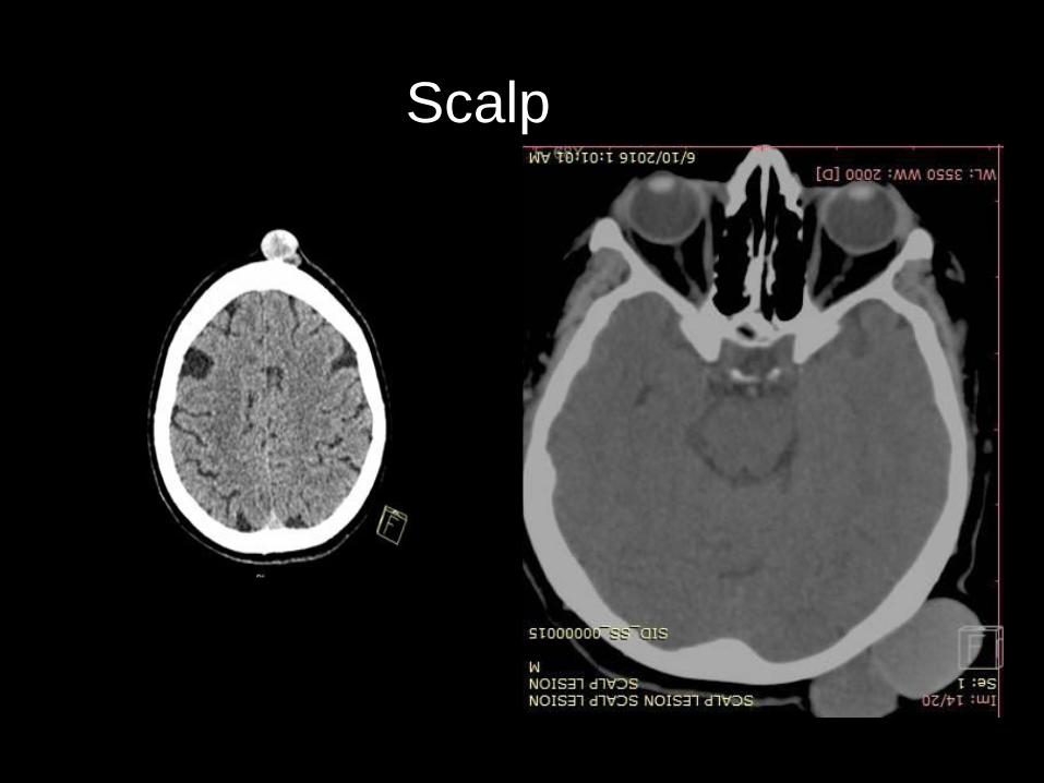
Scalp

Brain pathology could be divided to two type according to their density on CT:
1- Hyperdense lesions
2- Isodense lesions
3- Hypodense lesions
Brain Pathology - CT
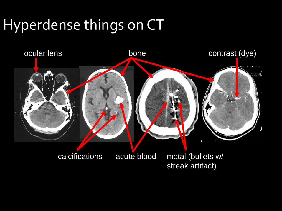
Hyperdense things on CT
acute blood
ocular lens
calcifications
contrast (dye) bone
metal (bullets w/
streak artifact)
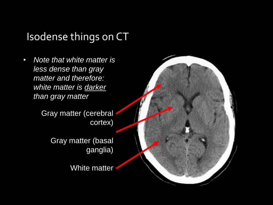
Isodense things on CT
• Note that white matter is
less dense than gray
matter and therefore:
white matter is darker
than gray matter
Gray matter (cerebral
cortex)
Gray matter (basal
ganglia)
White matter

Hypodense things on CT
fat
air
CSF
(water)
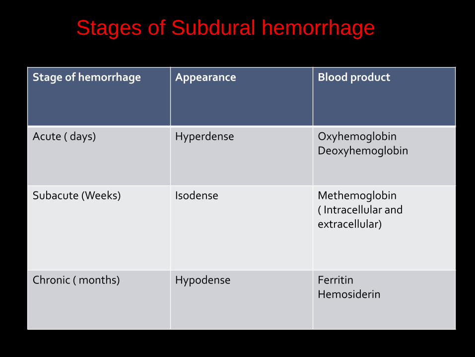
Stage of hemorrhage Appearance Blood product
Acute ( days) Hyperdense Oxyhemoglobin Deoxyhemoglobin
Subacute (Weeks) Isodense Methemoglobin ( Intracellular and extracellular)
Chronic ( months) Hypodense Ferritin Hemosiderin
Stages of Subdural hemorrhage
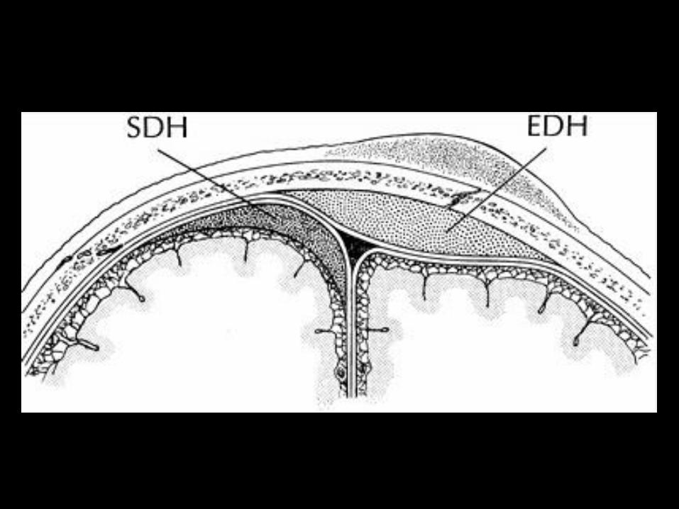

Origin: Arterial ( middle meningeal artery ) – associated with skull fracture
Lens in shape
Treatment: surgical - Craniotomy
Epidural hemorrhage

Acute epidural hemorrhage

Acute epidural hemorrhage with overlying fracture

Note the soft tissue swelling adjacent to the hematoma
explaining the mechanism of the injury
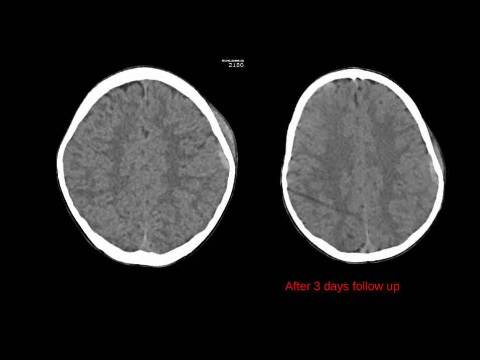
After 3 days follow up

The epidural with hypodensity : Clotting
Pneumocephlus
whirl sign
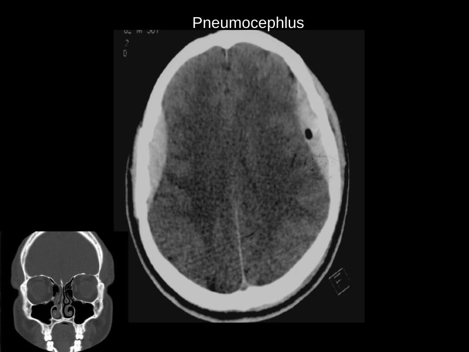
Pneumocephlus

Clotting


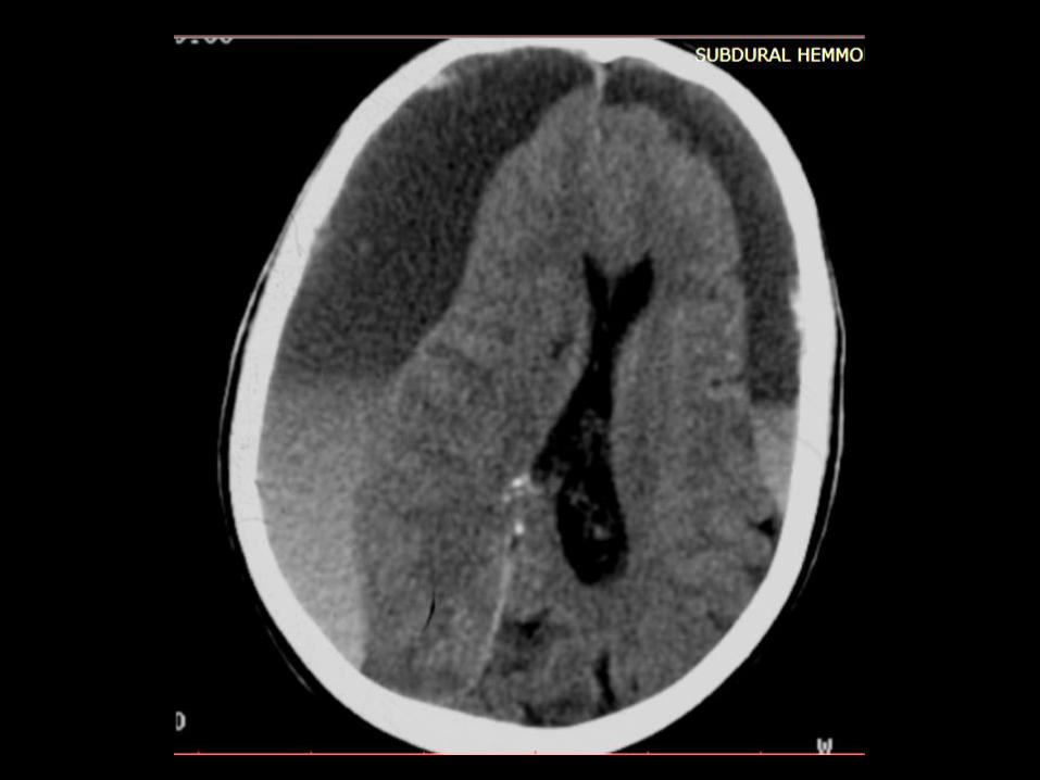
Origin: Venous
Treatment: Burr hole
Cresent ( semilunar ) in shape
Subdural hemorrhage
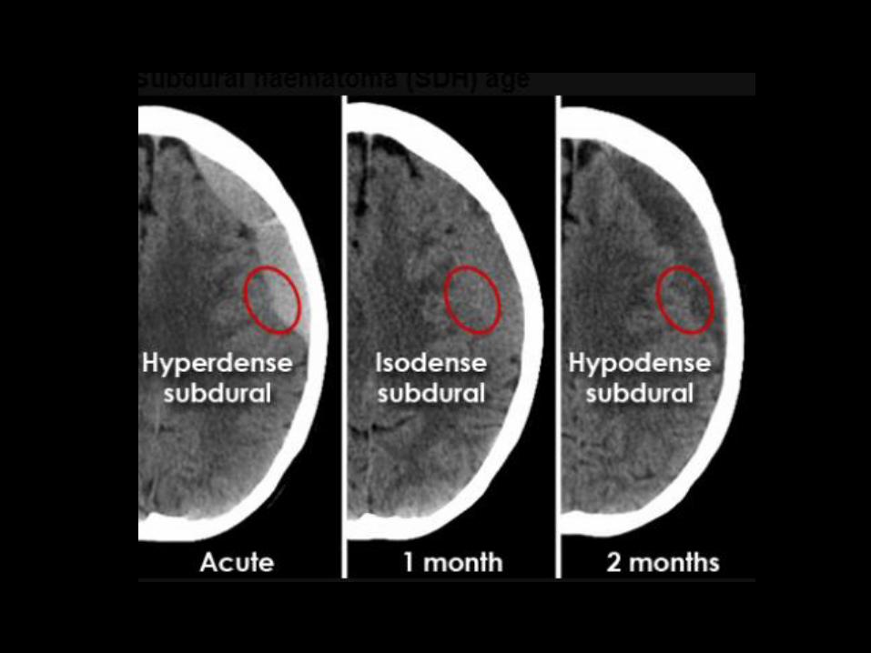
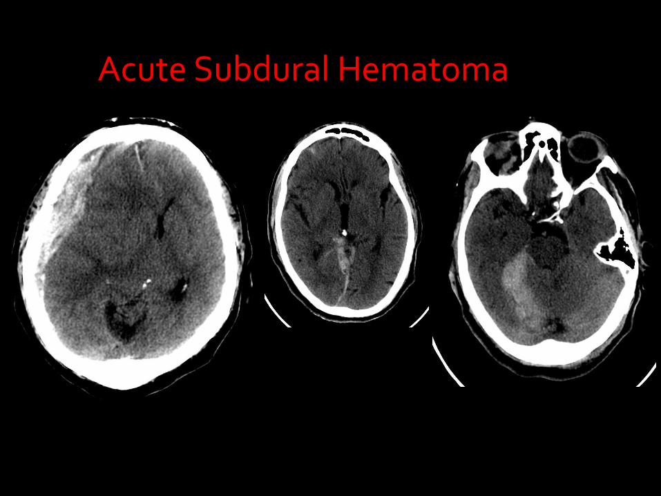
Acute Subdural Hematoma


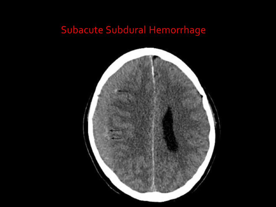
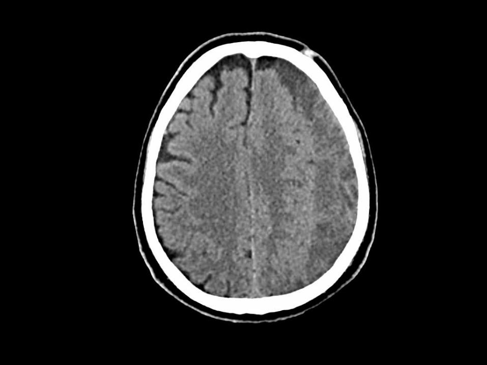
Subacute Subdural Hemorrhage
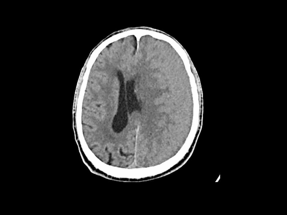
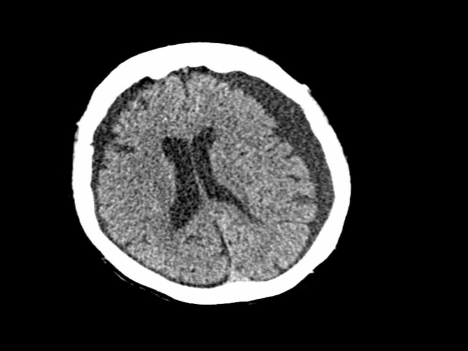
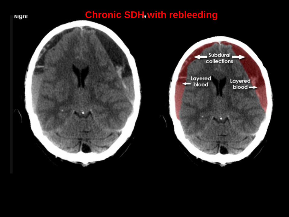
Chronic SDH with rebleeding
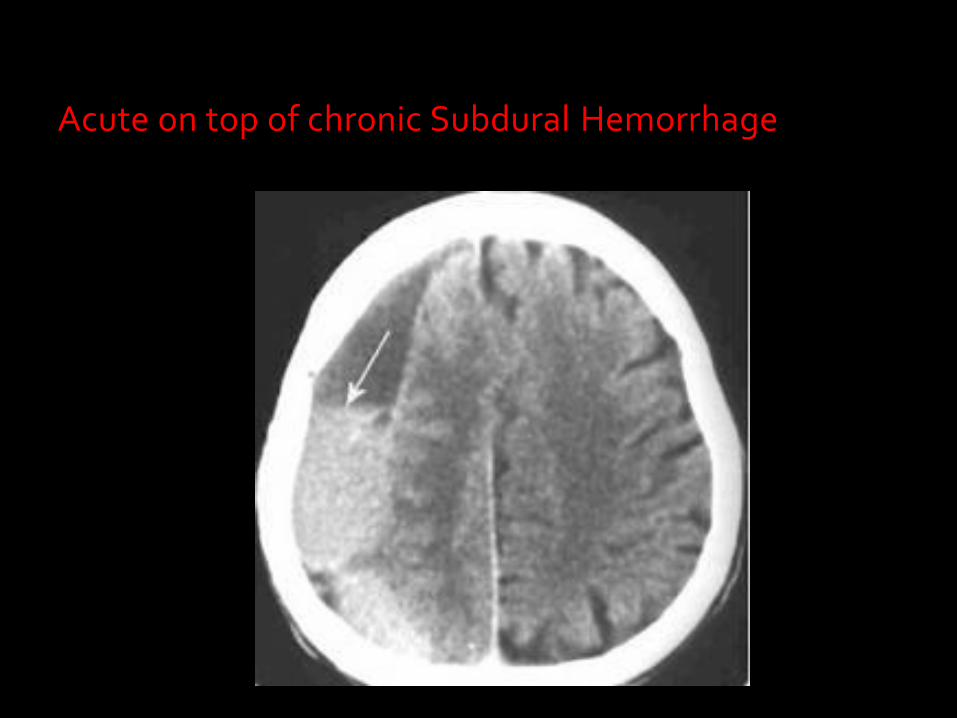
Acute on top of chronic Subdural Hemorrhage
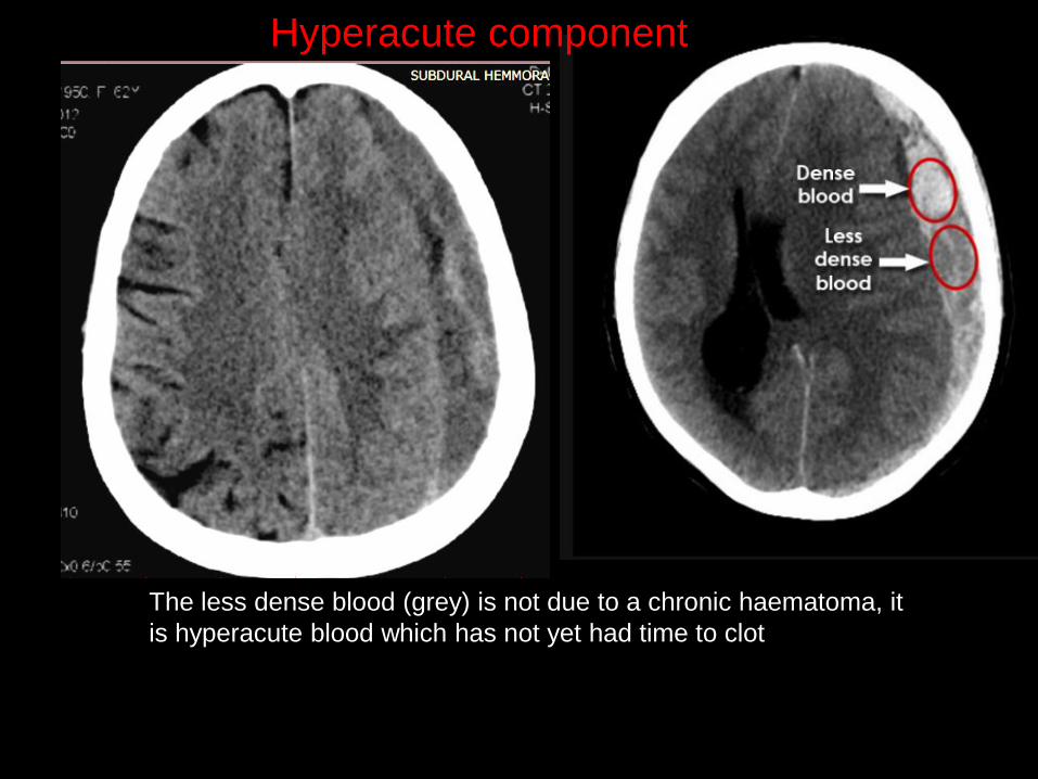
The less dense blood (grey) is not due to a chronic haematoma, it
is hyperacute blood which has not yet had time to clot
Hyperacute component
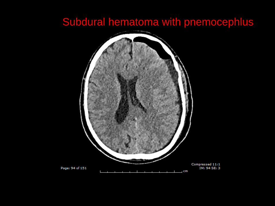
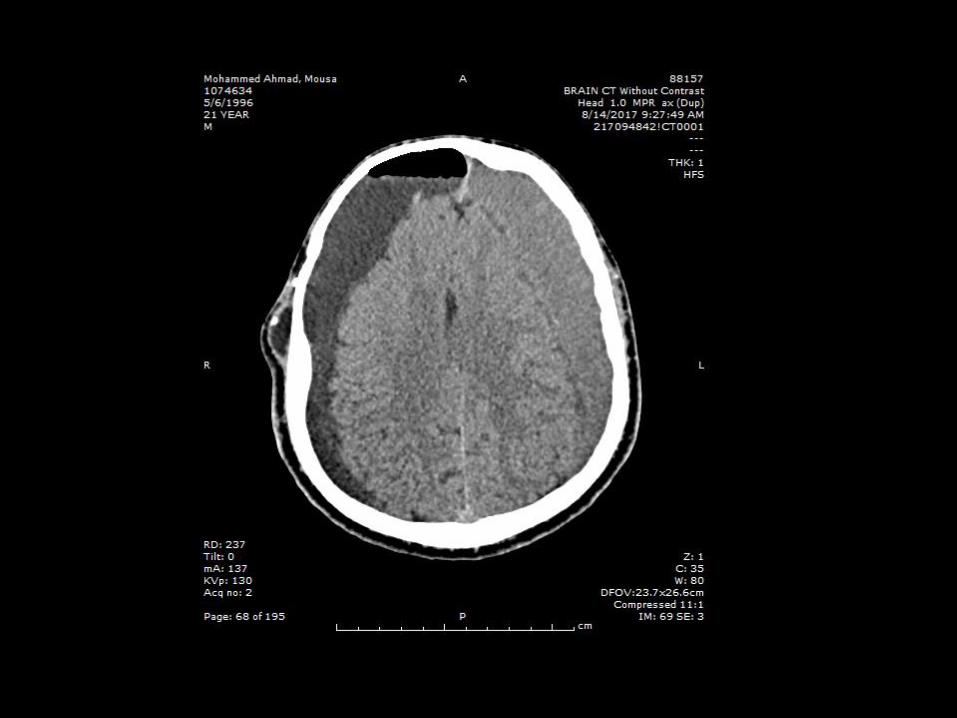
Subdural hematoma with pnemocephlus
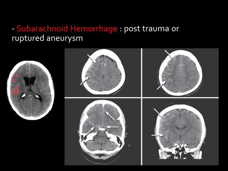
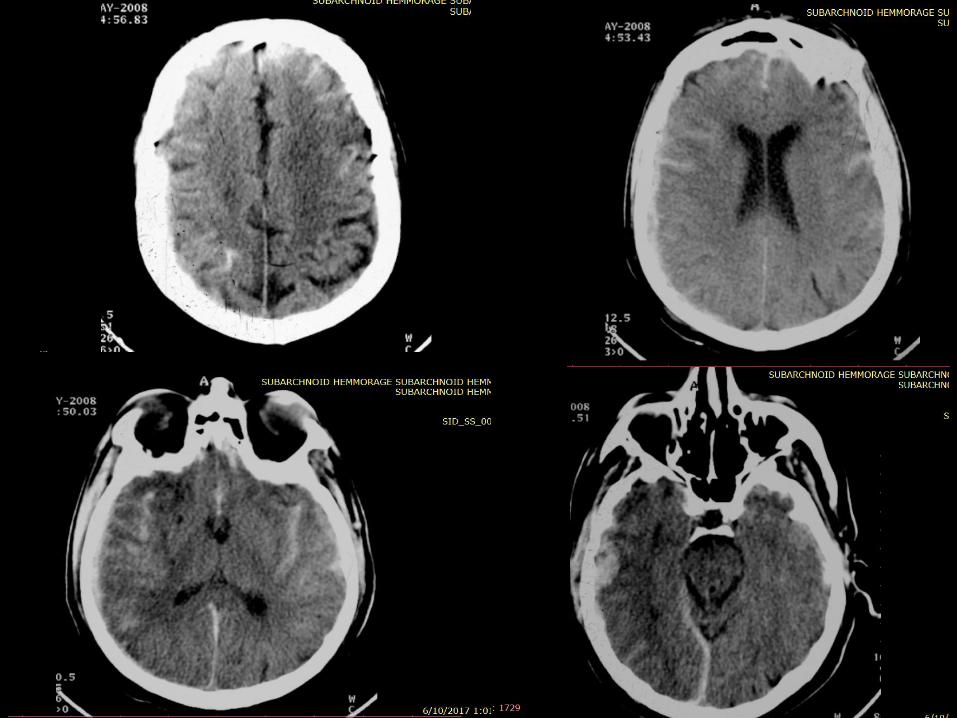
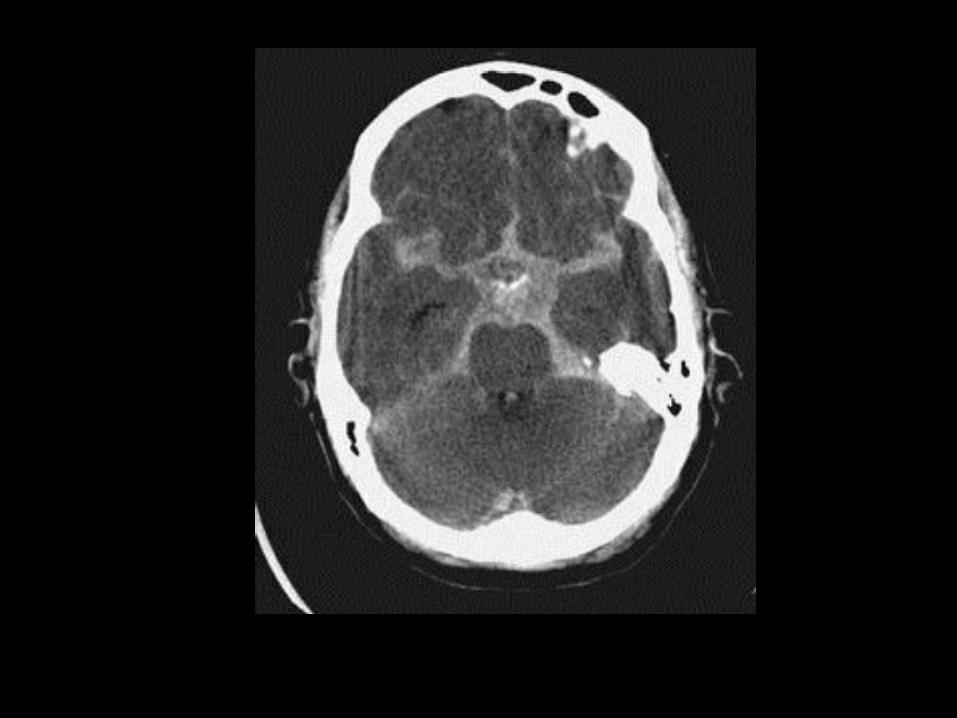
- Subarachnoid Hemorrhage : post trauma or ruptured aneurysm


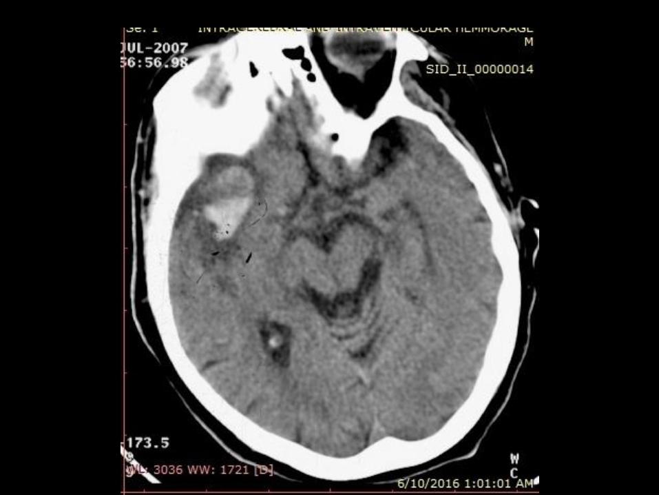
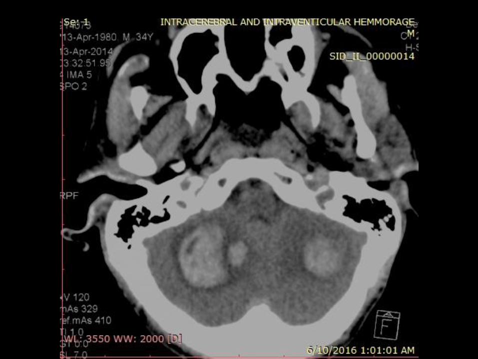
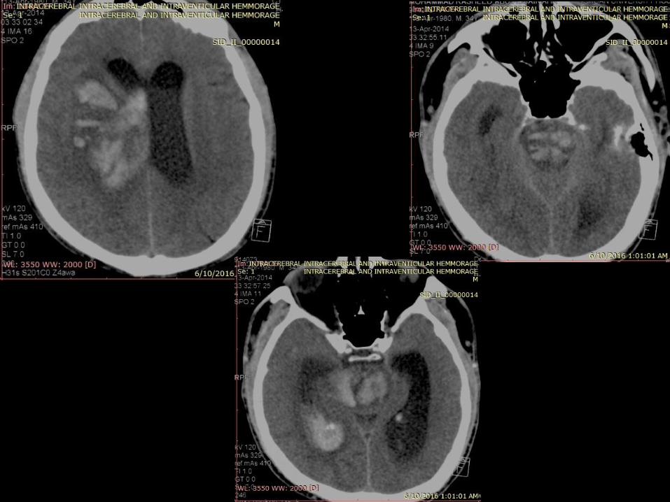
Intraventricular
hemorrhage

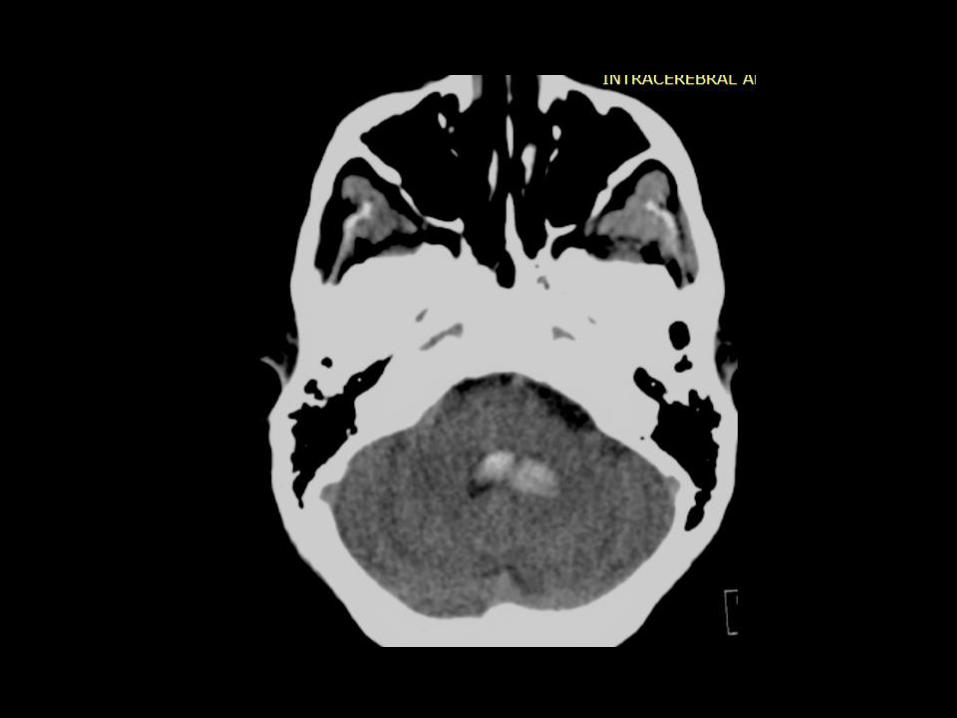
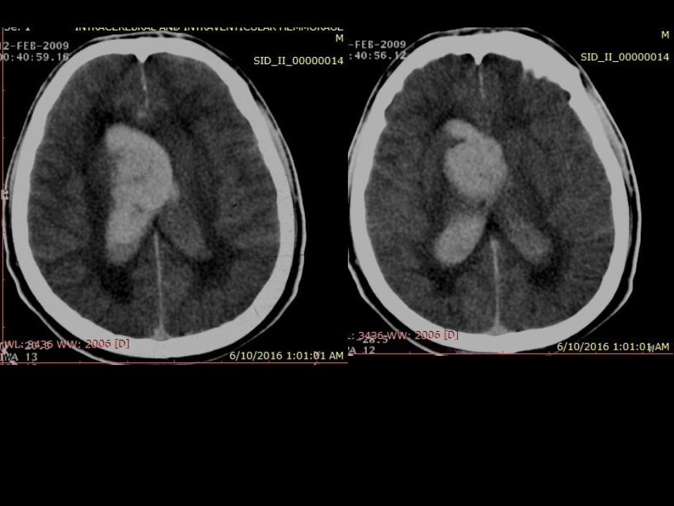
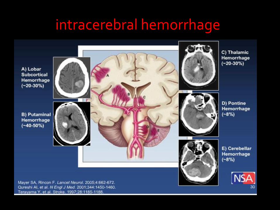
intracerebral hemorrhage
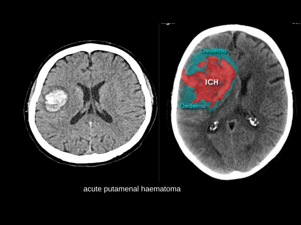
acute putamenal haematoma


Infarct with haemorrhagic transformation
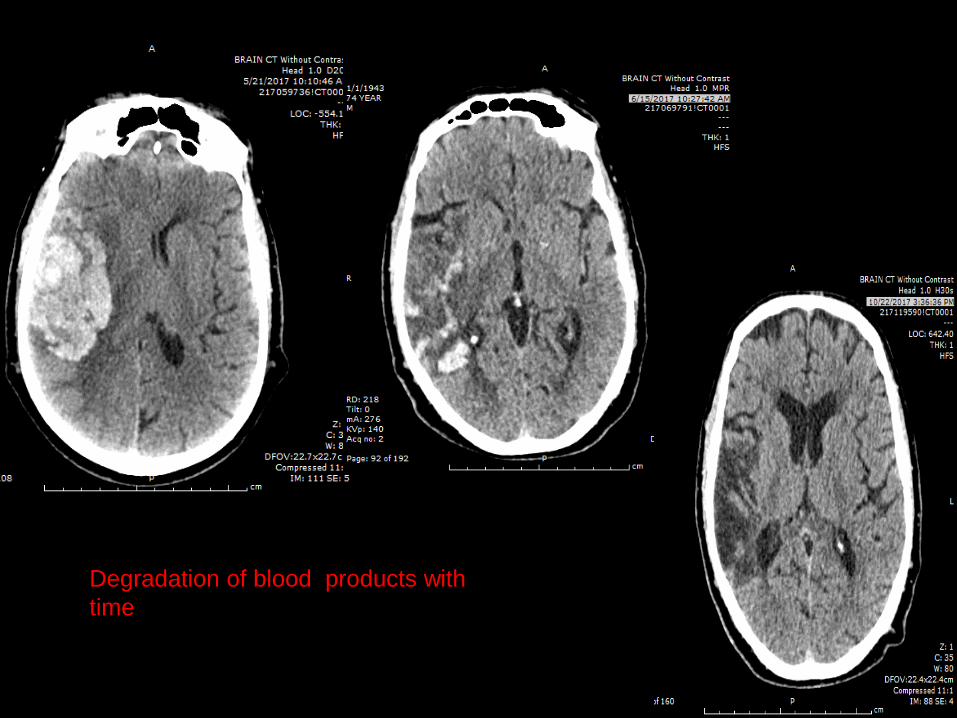
Degradation of blood products with
time

Different types of hemorrhage in the
same patient

Another example

Cephalohematoma
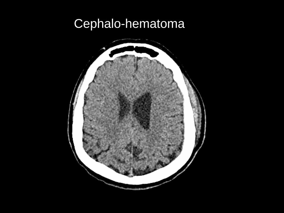
Cephalo-hematoma

• Normal variation
• Pathological :
1- AVM
2- Infection ( congenital in pediatrics)
3- Tumors
4 – metabolic
Calcification
1- Normal variation/ normal aging
basal ganglia , vascular calcifications ,Choroid plexus , Pineal gland , dentate nuclues , calcified falx .
Calcification
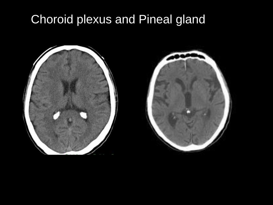
Choroid plexus and Pineal gland

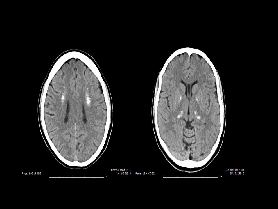
Basal ganglia

Basal ganglia

Dentate nucleus Dentate nucleus

vascular calcification
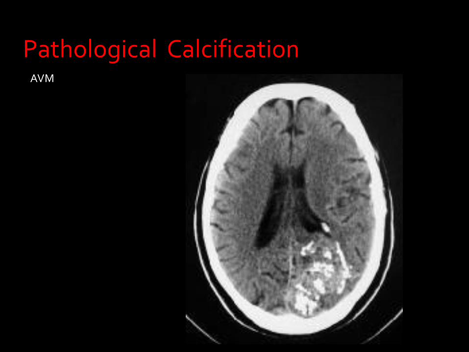
AVM
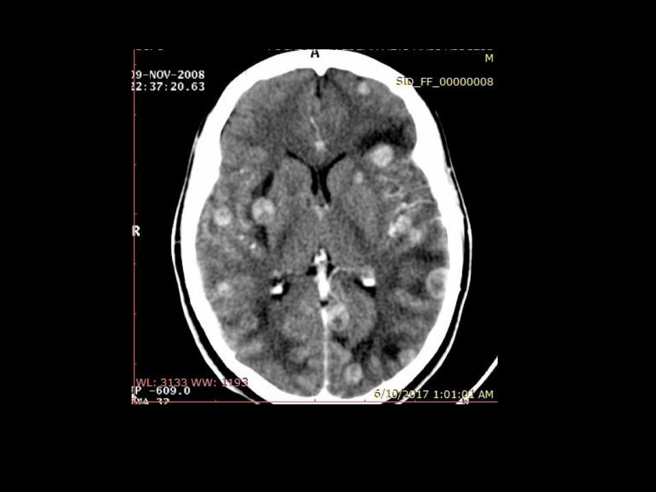
Pathological Calcification

Infection ( congenital) : TORCH
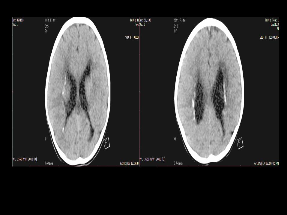
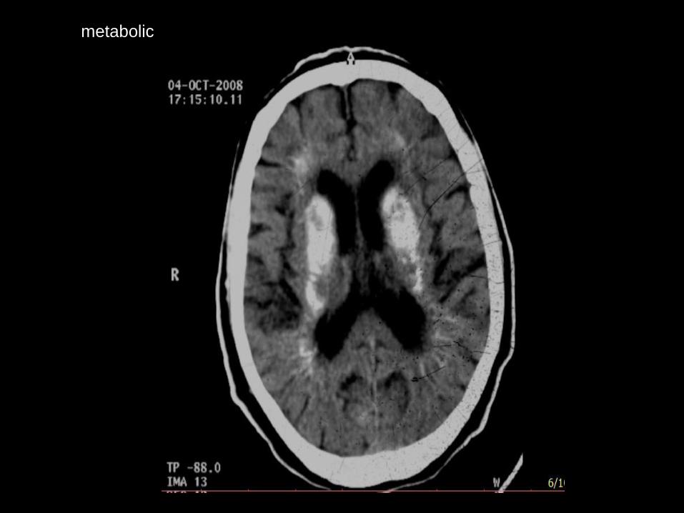
metabolic
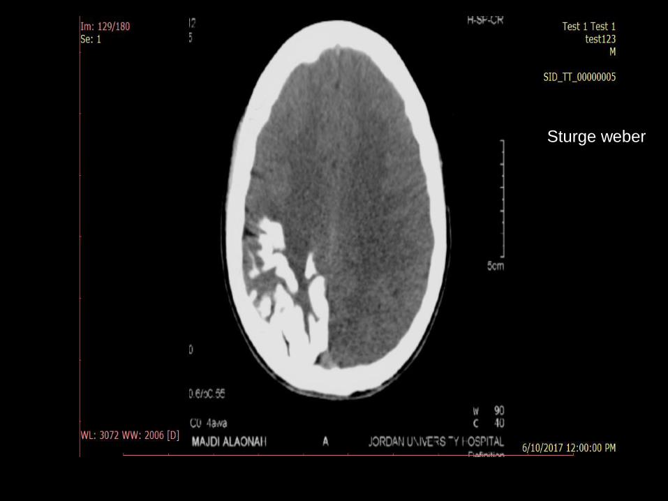
Sturge weber
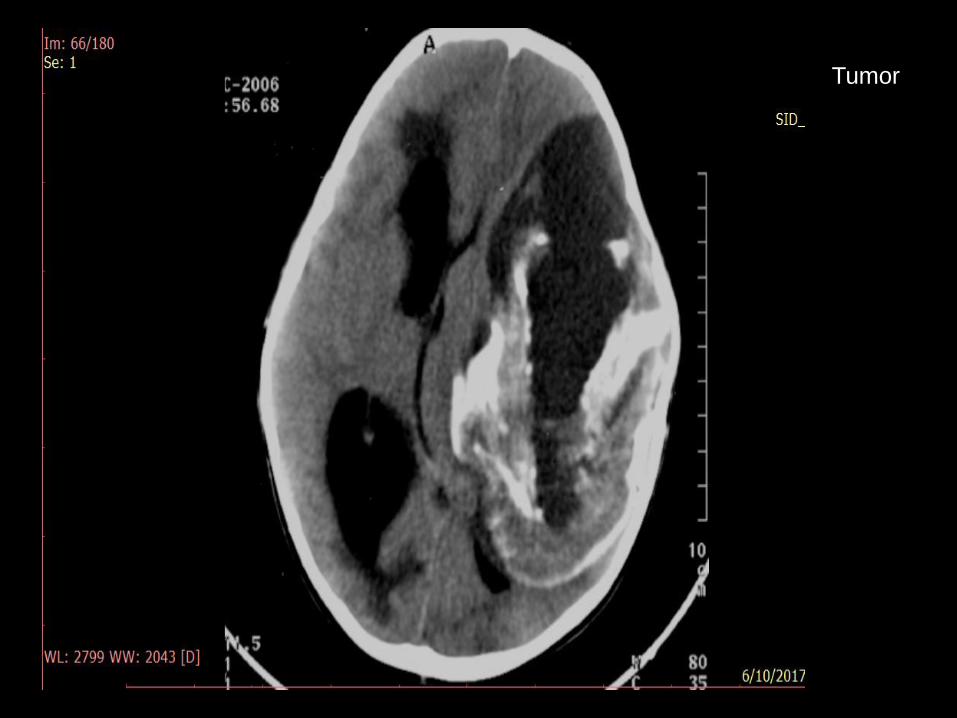
Tumor

Tumors that usually calcifies :
Meningioma
Craniopharangioma
Low grade astrocytoma
Oligodendroglioma

Meningioma

Craniopharangioma

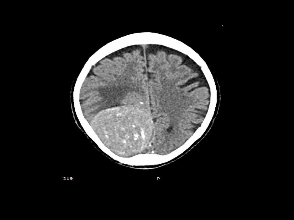
Low grade Astrocytoma
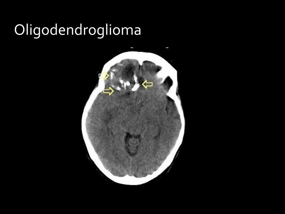
Oligodendroglioma
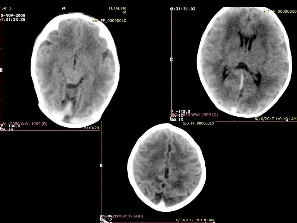
Thrombosed cerebral venous
sinuses

Thrombosed cerebral venous sinuses


Fetal HB

Benign
Malignant
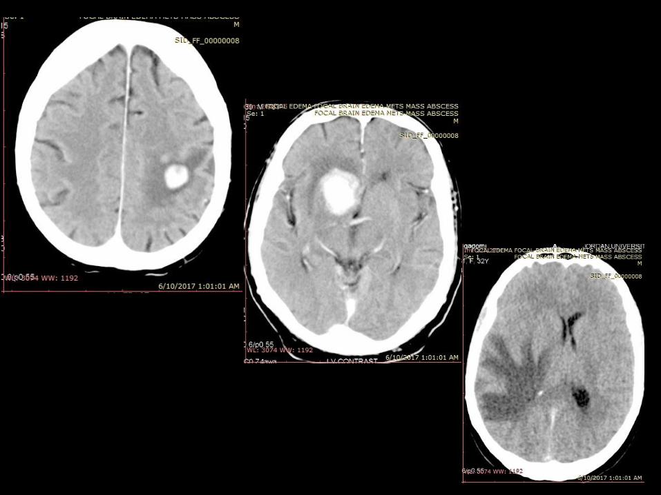
Contrast enhancing lesions
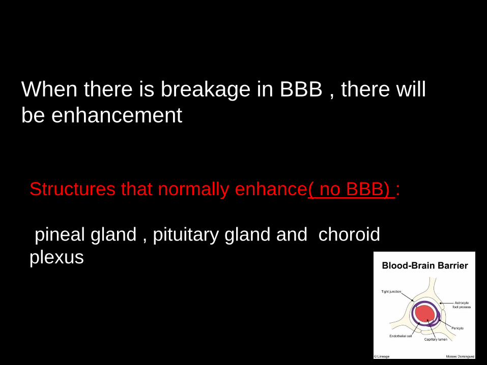
When there is breakage in BBB , there will
be enhancement
Structures that normally enhance( no BBB) :
pineal gland , pituitary gland and choroid
plexus
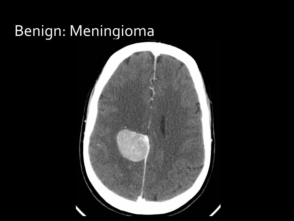
Benign: Meningioma

Benign: Abscess
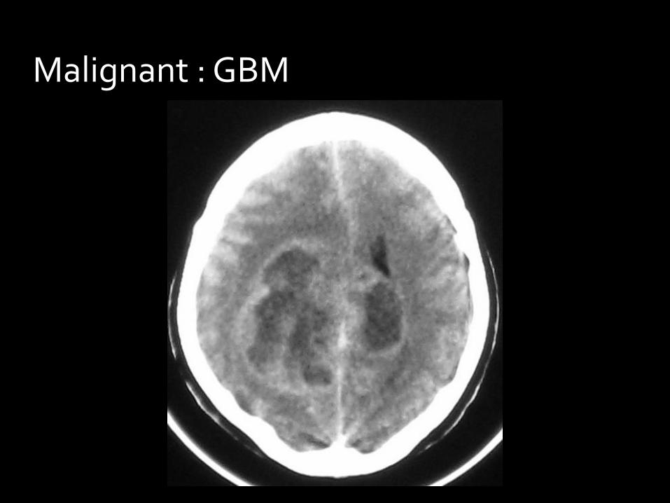
Malignant : GBM
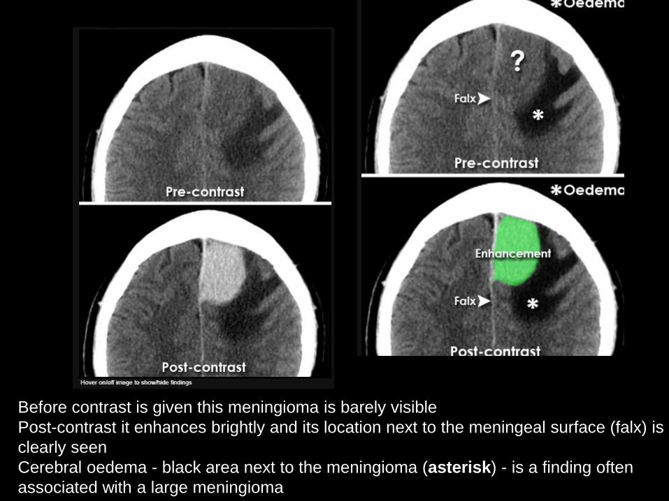
Before contrast is given this meningioma is barely visible
Post-contrast it enhances brightly and its location next to the meningeal surface (falx) is
clearly seen
Cerebral oedema - black area next to the meningioma (asterisk) - is a finding often
associated with a large meningioma

1- Fluid
2- air
3- Fat
Hypodense Lesions:

Fluid:
CSF: normal v/s hydrocephalus
Edema: vasogenic v/s cytotoxic
Diffuse brain edema
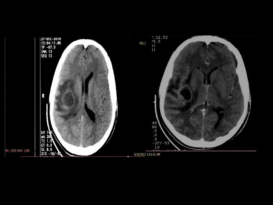
Necrotic tissue: tumor v/s abscess
Hypodense Lesions:
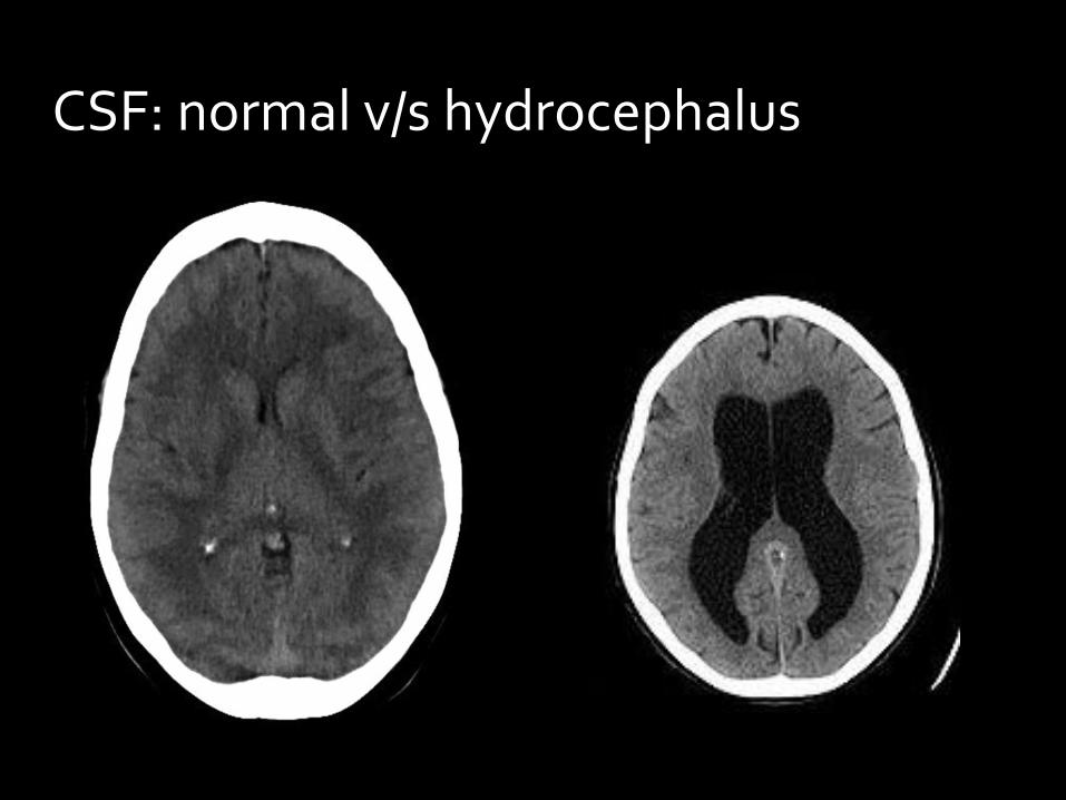
CSF: normal v/s hydrocephalus
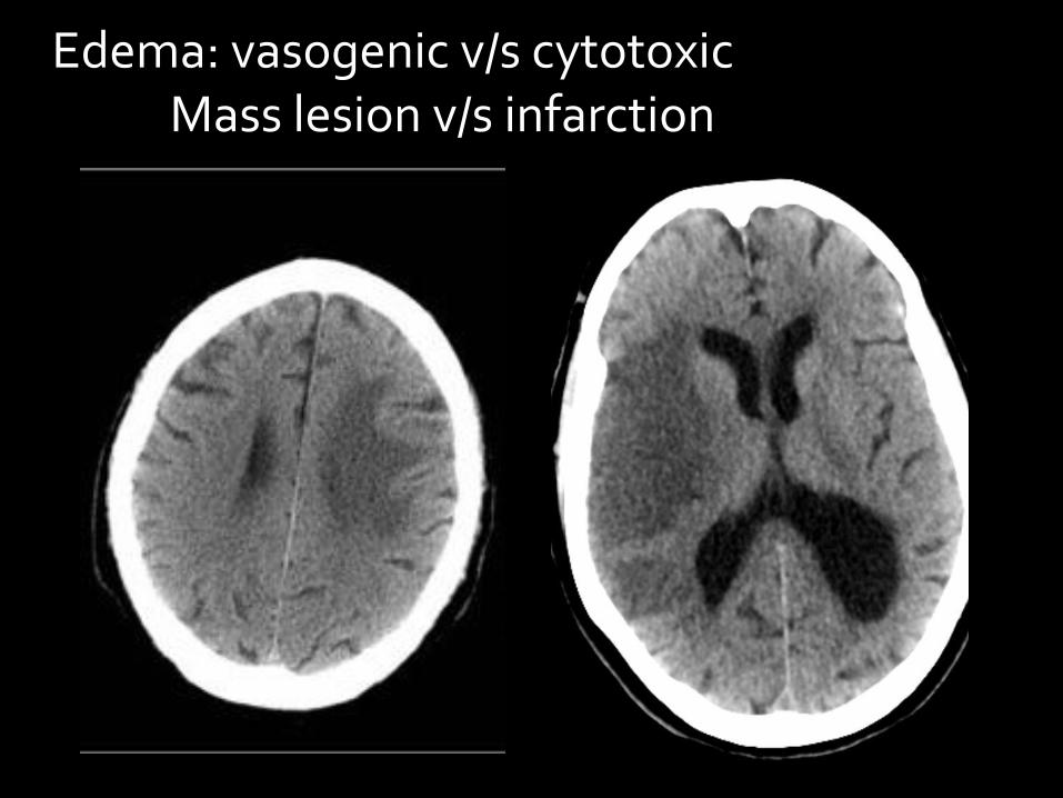
Edema: vasogenic v/s cytotoxic Mass lesion v/s infarction
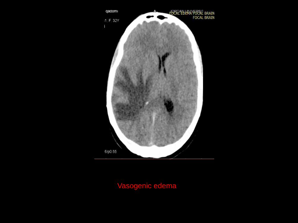
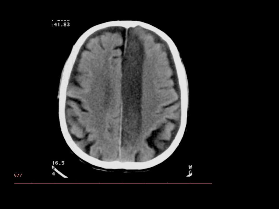
Vasogenic edema

Cytotoxic edema
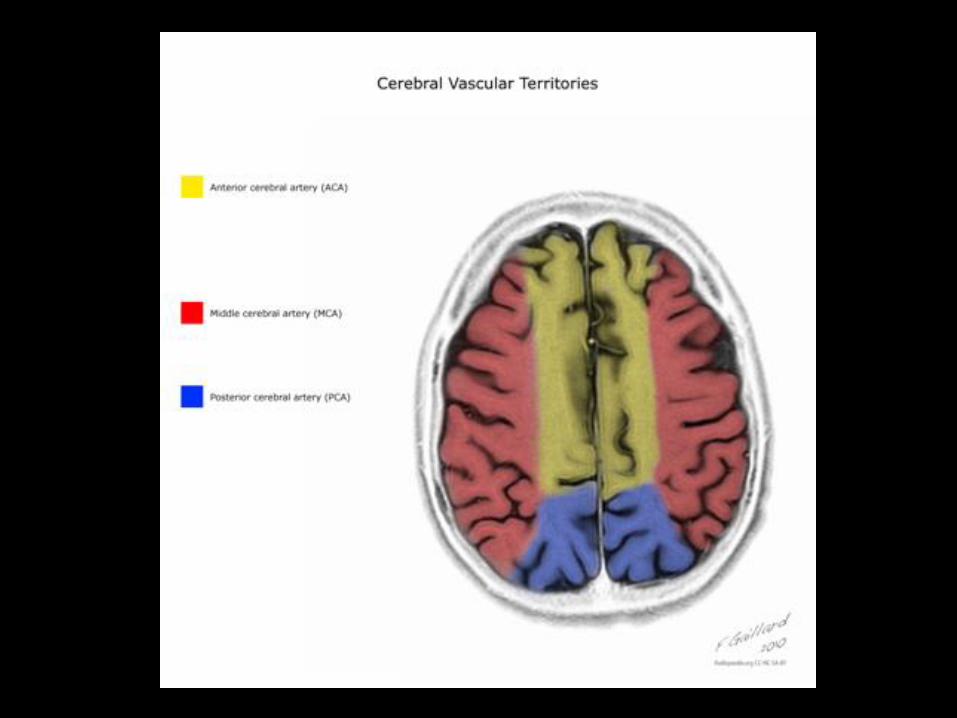

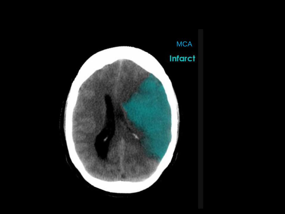
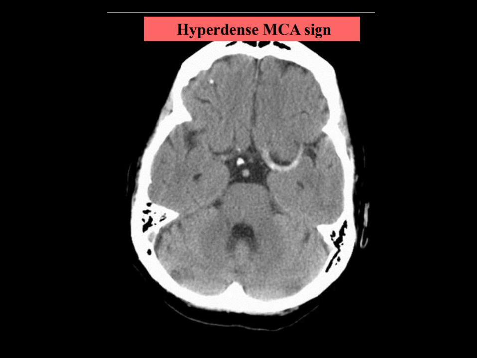
MCA
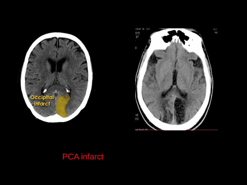
PCA infarct


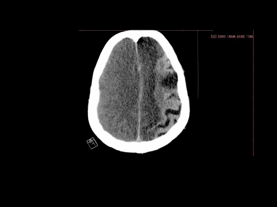


16/5 18/5
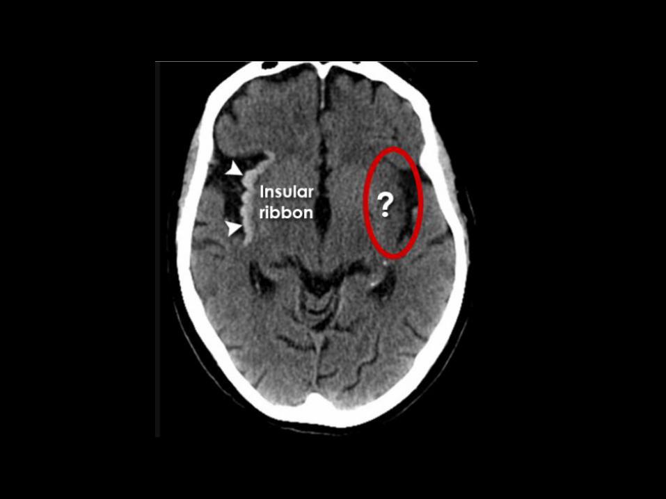
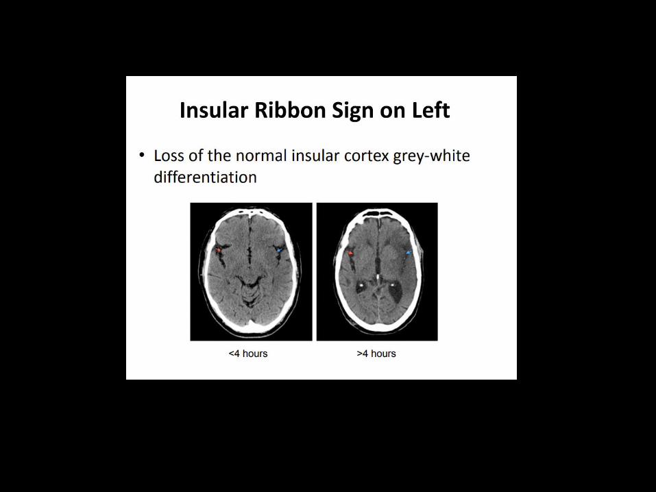
Insular ribbon sign
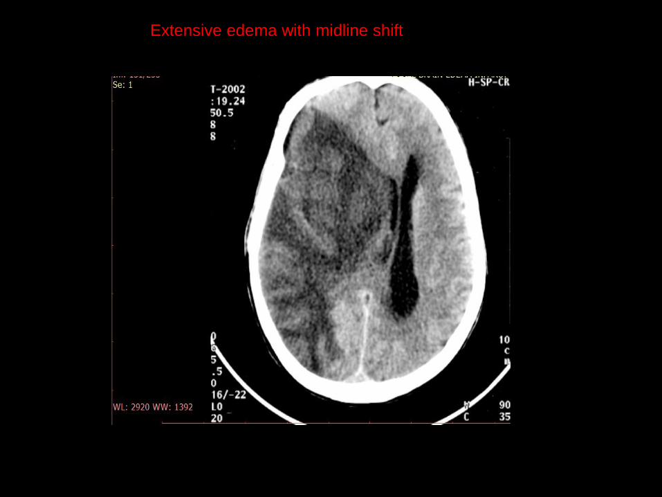
Extensive edema with midline shift
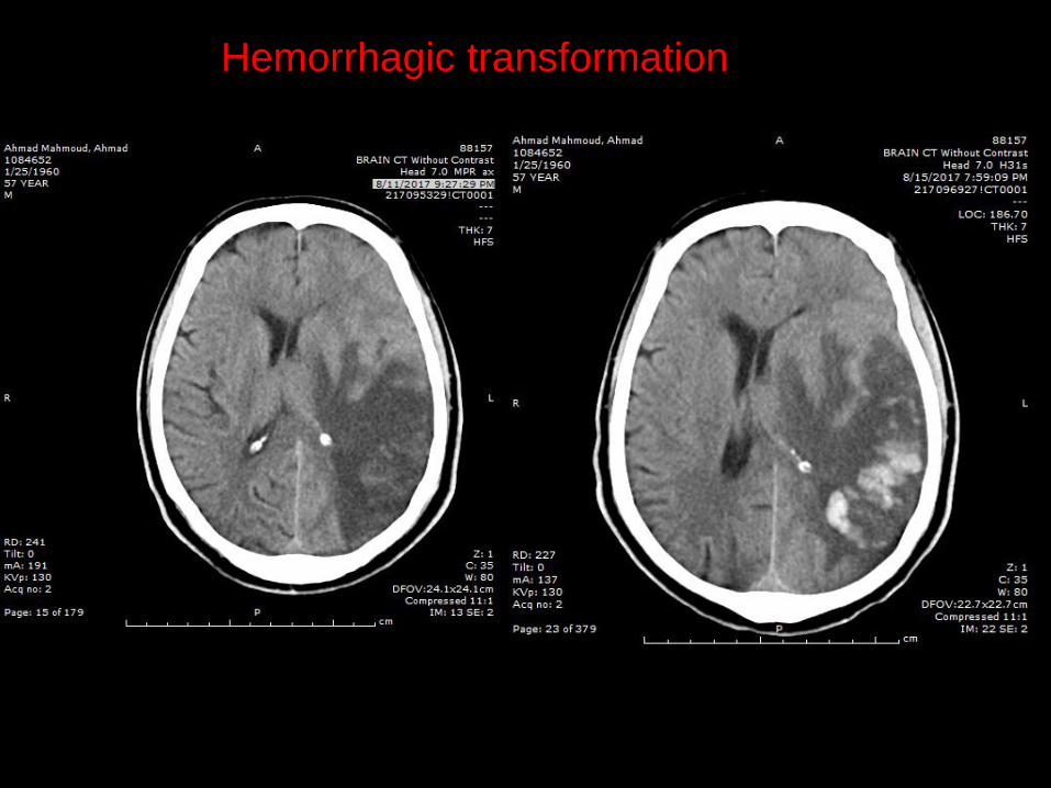
Hemorrhagic transformation
Vasogenic edema
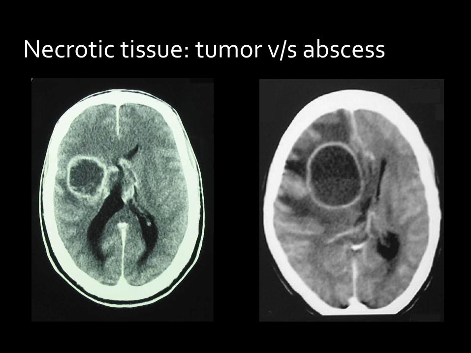
Necrotic tissue: tumor v/s abscess
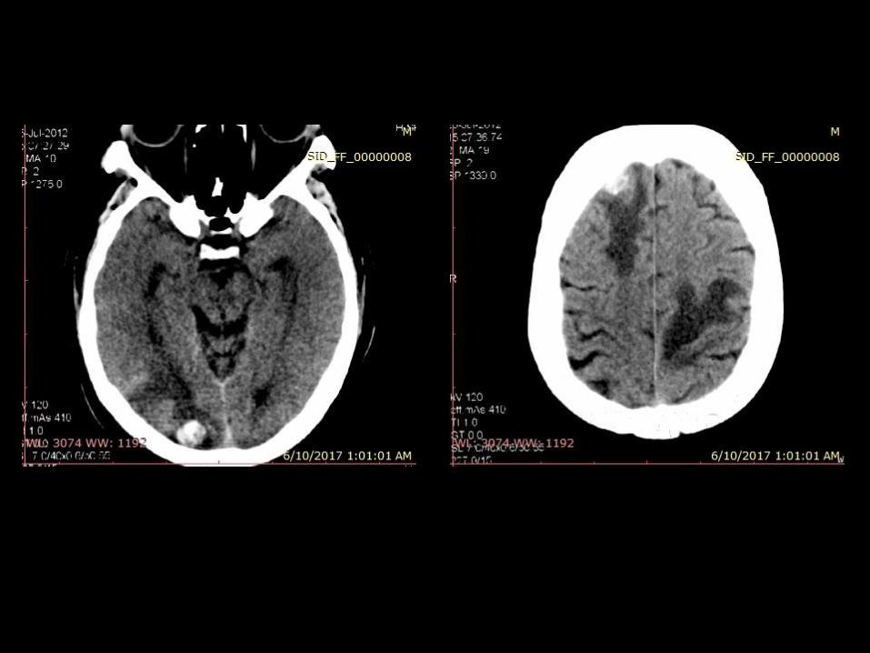


Diffuse brain edema
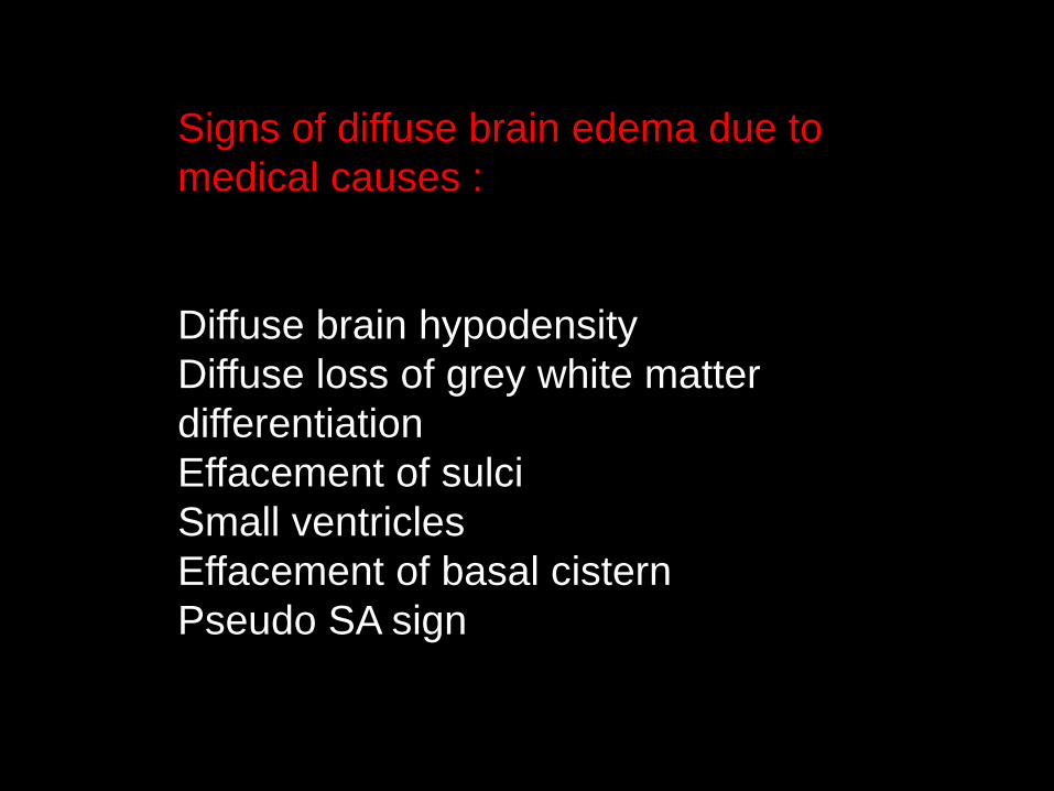
Signs of diffuse brain edema due to
medical causes :
Diffuse brain hypodensity
Diffuse loss of grey white matter
differentiation
Effacement of sulci
Small ventricles
Effacement of basal cistern
Pseudo SA sign
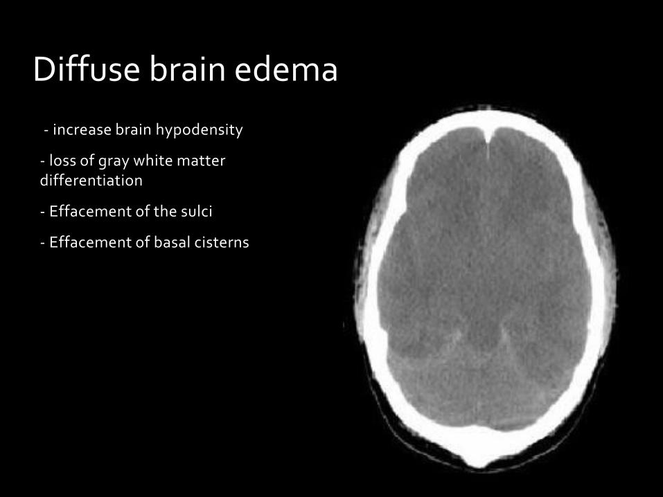
- increase brain hypodensity
- loss of gray white matter differentiation
- Effacement of the sulci
- Effacement of basal cisterns
Diffuse brain edema


Air:
Normal : sinuses
Hypodense Lesions:
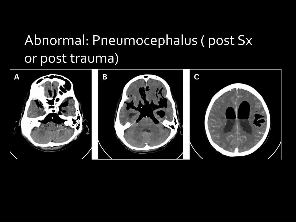
Abnormal: Pneumocephalus ( post Sx or post trauma)


Fat:
Lipoma, dermoid cyst
Hypodense Lesions:

Dermoid Cyst
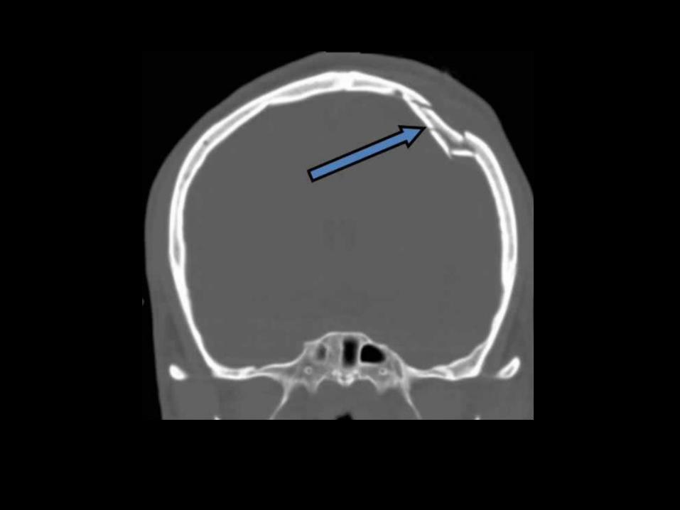
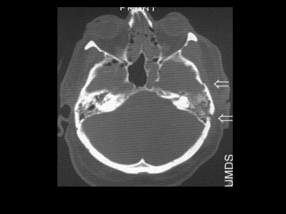
Linear
Depressed
Growing Fracture
Skull Fractures
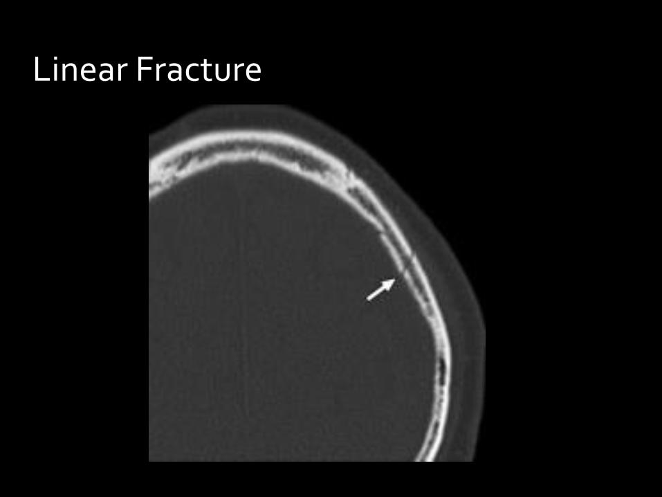
Linear Fracture

Depressed Fracture
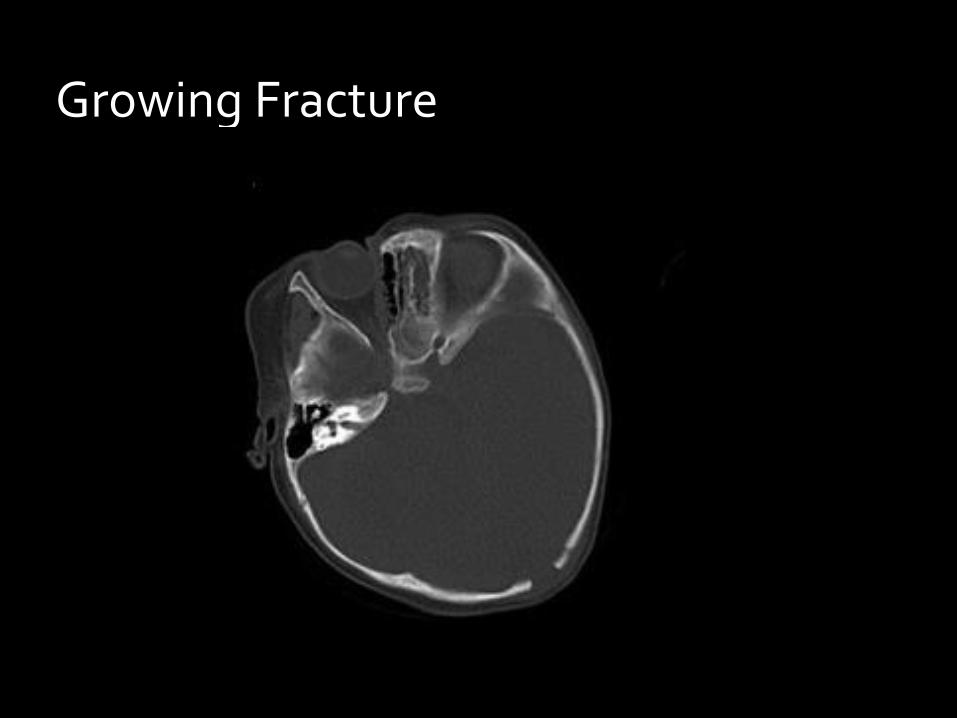
Growing Fracture


Related Documents