Introduction
Jan 01, 2016
Benign tumours of salivary glands and sialoceles : classification, histological structure, clinic, differential diagnostics, treatment. Malignant tumours of salivary glands: histological structure, clinical forms, differential diagnostics, treatment. Introduction. Epidemiology Staging - PowerPoint PPT Presentation
Welcome message from author
This document is posted to help you gain knowledge. Please leave a comment to let me know what you think about it! Share it to your friends and learn new things together.
Transcript

Epidemiology Staging Histologic subtypes Diagnosis Treatment

TX Primary tumor cannot be assessed
T0 No evidence of primary tumor
T1 Tumor 2 cm or less in greatest dimension without gross extraparenchymal extension
T2 Tumor more than 2 cm but not more than 4 cm in greatest dimension without gross extraparenchymal extension
T3 Tumor more than 4 cm and/or tumor having gross extraparenchymal extension
T4a Tumor invades skin, mandible, ear canal, and/or facial nerve
T4b Tumor invades skull base and/or pterygoid plates and/or encases carotid artery

NX Regional lymph nodes cannot be assessed
N0 No regional lymph node metastasis
N1 Metastasis in a single ipsilateral lymph node, 3 cm or less in greatest dimension
N2a Metastasis in a single ipsilateral lymph node, more than 3 cm but not more than 6 cm in greatest dimension
N2b Metastasis in multiple ipsilateral lymph nodes, none more than 6 cm in greatest dimension
N2c Metastasis in bilateral or contralateral lymph nodes, none more than 6 cm in greatest dimension
N3 Metastasis in a lymph node more than 6 cm in greatest dimension
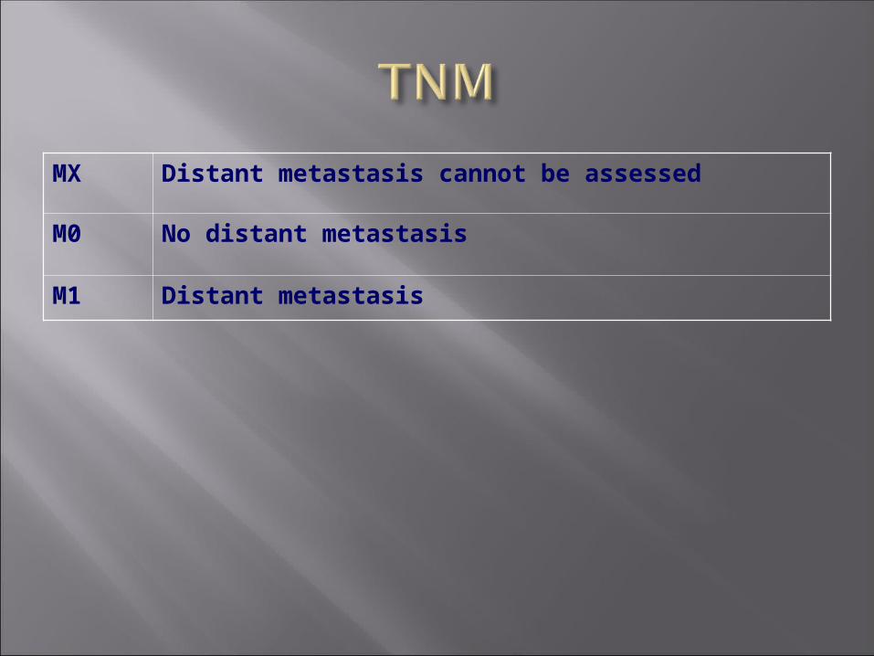
MX Distant metastasis cannot be assessed
M0 No distant metastasis
M1 Distant metastasis

Stage I T1 N0 M0
Stage II T2 N0 M0
Stage III T3 N0 M0
T1 N1 M0
T2 N1 M0
T3 N1 M0
Stage IVA T4a N0 M0
T4a N1 M0
T1 N2 M0
T2 N2 M0
T3 N2 M0
T4a N2 M0
Stage IVB T4b Any N M0
Any T N3 M0
Stage IVC Any T Any N M1
Histologic grading is applicable only to some types of salivary gland cancer (mucoepidermoid carcinoma, adenocarcinoma not otherwise specified)
In most instances, the histologic type defines the grade (i.e. salivary duct carcinoma is high grade, basal cell adenoma is low grade)
Mucoepidermoid carcinoma Adenoid cystic carcinoma Acinic cell carcinoma Carcinoma ex-pleomorphic adenoma Additional rare types


Most common salivary malignancy accounting for 29% to 43% of tumors
Mucoepidermoid cancer is histologically classified into low and high grade. A higher grade correlates with a poorer outcome
Low-grade tumors have a higher percentage of mucinous cells
Epithelial cells predominate in high-grade. The presence of four or more mitotic figures per 10 high-power fields, neural invasion, necrosis, intracystic component <20%, and cellular anaplasia indicate high-grade behavior.
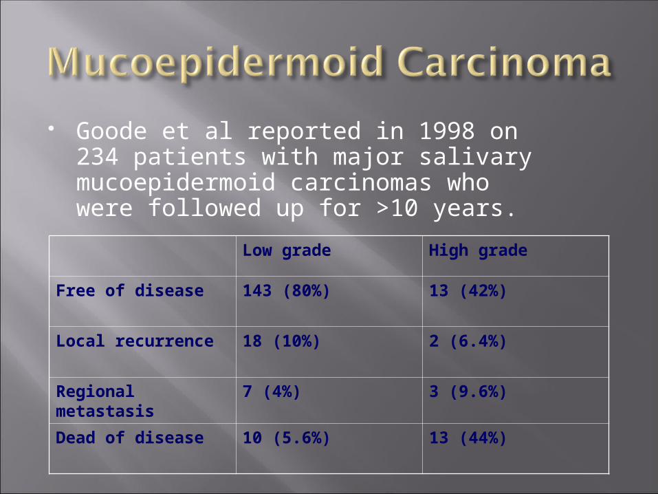
Goode et al reported in 1998 on 234 patients with major salivary mucoepidermoid carcinomas who were followed up for >10 years.
Low grade High grade
Free of disease 143 (80%) 13 (42%)
Local recurrence 18 (10%) 2 (6.4%)
Regional metastasis 7 (4%) 3 (9.6%)
Dead of disease 10 (5.6%) 13 (44%)

The authors' findings also indicated that patients with tumors of equal histopathologic grade have a better prognosis when their tumors are in the parotid gland than when their tumors are in the submandibular gland


Adenoid cystic carcinoma is the most common malignancy of the
submandibular gland Adenoid cystic carcinoma is characterized
by slow growth, neurotropism, local recurrence, and distant metastasis.
Exhibits a predilection for neurotropic spread, often leading to recurrences at the skull base after surgical and radiation treatment
Three distinct histologic patterns, cribriform, tubular, or solid, although the histologic patterns may coexist in the same tumor
The cribiform pattern has a glandular architecture and is reported to have the best prognosis.
The solid pattern is more epithelial in nature and is associated with a poorer prognosis.
The tubular pattern has a clinical prognosis of intermediate nature between the other two patterns.
This tumor has a low-grade behavior and has the best survival rate of any salivary malignancy
Parotid gland was the most common site of origin
Malignant degeneration can occur in 3% to 7% of pleomorphic adenomasThe risk of malignant degeneration is estimated at 1.5% in the first 5 years and 9.5% after 15 years.
Histologic findings include those of benign pleomorphic adenoma with carcinomatous degeneration.
A typical clinical history includes a longstanding salivary mass that begins to rapidly enlarge, often to substantial size, although many patients have no history of a prior
Polymorphous low-grade adenocarcinoma Epithelial-myoepithelial carcinoma Basal cell adenocarcinoma Papillary cystadenocarcinoma Myoepithelial carcinoma

Squamous cell carcinoma Small cell carcinoma Sebaceous carcinoma Mucinous adenocarcinoma Oncocytic carcinoma Adenocarcinoma Salivary duct carcinoma
Does my patient have salivary gland cancer?

Malignant salivary neoplasms present as a painless mass in approximately 75% of patients. Rarely, patients are initially seen with pain or facial nerve palsy.
A palpable mass arising in a salivary gland, associated with pain, and/or nerve paralysis is more likely to be malignant than benign.
It is believed that episodic pain suggests continued obstruction, whereas constant pain is more suggestive of malignancy.
Trismus, cervical adenopathy, fixation, numbness, loose dentition, or bleeding also suggest the presence of malignancy.
Traditionally, FNA has been performed preoperatively for histologic confirmation of malignancy and to aid in operative planning, such as planning for elective neck dissection
In 1997, Tew and others evaluated 195 FNAs and 159 intraoperative frozen sections for parotid tumors
They found that FNA had a 90% sensitivity for malignancy if non-diagnostic biopsies were excluded
They also found that frozen section had a 96% sensitivity for malignancy
An incisional biopsy at a site that can be excised during the definitive surgery approximates 100% accuracy and is therefore preferable in those patients in whom the extent of the surgery (e.g., no surgery, nerve sacrifice, total vs superficial parotidectomy) would change with a change in histologic diagnosis.
Ultrasound can provide guidance in obtaining fine-needle biopsy specimens from deep parotid or parapharyngeal space tumors. In patients with cystic or heterogeneous masses, ultrasound ensures sampling of the solid component and may also be helpful in biopsy masses that are difficult to palpate.
Computed tomography (CT) with intravenous contrast is routinely used preoperatively and provides excellent detail of the tumor volume, its relation to vascular and bony structures, as well as surveillance of the regional lymphatics
Magnetic resonance imaging (MRI) provides excellent soft tissue detail, which is superior to that of CT and has the advantage of not requiring contrast for vascular detail or ionizing radiation
The usefulness of PET scanning in the setting of salivary gland malignancy is yet to be clearly defined.
Keyes and others performed preoperative PET imaging on 26 patients with parotid tumors. A PET scan accurately predicted the nature of the neoplasm in 69%, demonstrated 100% sensitivity for malignancy, and a false-positive rate of 30%.
What surgery should I perform on the primary tumor?
Does this patient need postoperative radiation?
Superficial parotidectomy has become the widely accepted form of intervention for most parotid tumors. A higher risk of facial nerve injury and the potential for intraoperative seeding of tumor resulting in recurrence of the tumor has been associated with the use of lesser procedures. Therefore, a superficial parotidectomy has been touted as the minimal surgery of the parotid gland. Overall, the safety of parotidectomy has been well established, and the complication rate remains low.
Total parotidectomy may be necessary for tumor extension into the deep parotid lobe or when the tumor primarily arises in the deep lobe. This can be performed with preservation of the facial nerve
Occasionally, patients may require extended parotidectomy, which includes resection of the masseter muscle or the ascending portion of the mandible.
Facial nerve sacrifice is not routinely advocated. Nerve preservation in primary salivary malignancy is recommended if the nerve is functioning normally before surgery. Every attempt to dissect the tumor from the individual branches should be undertaken. If tumor is completely encasing the nerve branches, neural sacrifice is limited to the involved branches.

In general, tumors of the submandibular gland require complete excision of the gland.
How should I treat the neck in my patient?
Observation, elective neck dissection, or elective neck irradiation?
There is little dispute that patients with clinical evidence of cervical nodal metastasis require treatment of the neck
Dispute in the literature still exists, though on whether or not to treat clinically negative (N0) necks
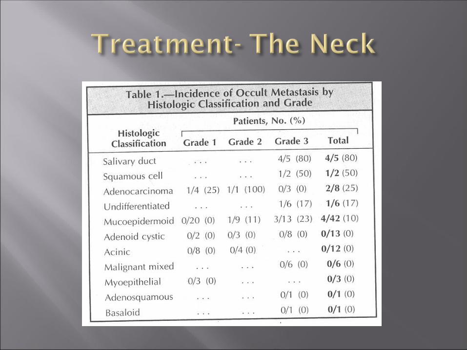
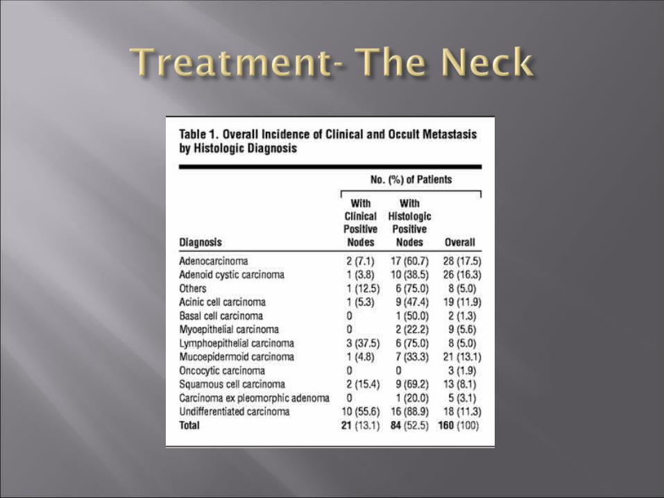
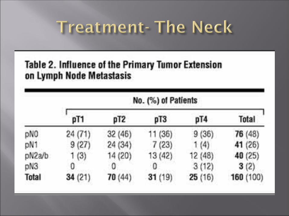
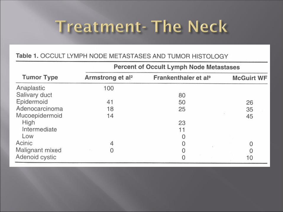
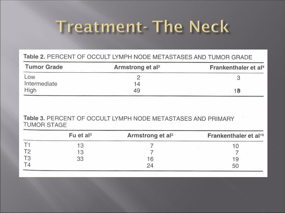
The risk of occult nodal disease is widely varied in the literature
Does my patient have cancer in the lymph nodes of his/ her neck?

Should I treat a patient’s neck, if I don’t know for sure if they have nodal metastasis?
Appropriately treating the neck in salivary malignancy is important for patient outcomes
For instance, overall 5 year survival of patients with and without involvement of the regional nodes is estimated at 10% and 75% respectively for the parotid and 9% and 41% for the submandibular gland

Medina in 1998 proposed a rationale for neck dissection on N0 necks
He proposed that patients that have factors that are indications for post-operative radiation are also the same ones that are at high risk for nodal metastasis and that these patients should simply undergo neck irradiation simultaneously

Medina emphasized that, at the time of his report, the effectiveness of XRT on controlling neck disease had not been studied
Occult metastases were detected in 8 (20%) of 41 cNO staged patients who underwent elective neck dissection.
Among these patients, 5 had a high-grade carcinoma and 3 had a low-grade carcinoma
The primary carcinoma of these 8 patients was classified as T2 in 4, as T3 in 1, and as T4 in 3 cases.

Regional recurrence occurred in none of the patients with an elective neck dissection and in 7 patients in the “observation” group (17%) (P = 0.006).
Of the 7 patients without neck dissection and neck recurrence, 2 patients were initially given adjuvant radiotherapy to the neck.

The actuarial and the disease free survival rates at 5 years for patients with neck dissection were 80% and 86% and 83% and 69% for patients without neck dissection.
Based on this study, the authors dispute Medina’s treatment paradigm and recommend elective neck dissection in all primary parotid carcinomas

Can radiation effectively treat the neck as well or better than neck dissection?
Chen and others reported in 2006 on 251 patients with clinically N0 necks who received postoperative radiation therapy after gross total tumor resection
Their results showed that none of the 131 patients who received ENI had neck failure compared with 24 of 120 who did not receive ENI. The corresponding 10-year estimates of nodal relapse were 0% and 26%, respectively (p = 0.0001).
Notably, there were no significant differences in the distribution of clinical and disease characteristics with respect to age, perineural invasion, T-stage, and primary site, among patients treated with and without ENI.
The highest crude rates of nodal relapse among those treated without ENI were found in patients with squamous cell carcinoma (67%), undifferentiated carcinoma (50%), adenocarcinoma (34%), and mucoepidermoid carcinoma (29%).
There were no neck relapses among patients treated either with or without ENI for patients with adenoid cystic or acinic cell histology.

It is clear that, for many patients with clinically N0 necks, based on histology, the risk of harboring occult disease in the regional lymph nodes is low enough that ENI is not warranted.
Patients with adenocarcinoma or mucoepidermoid carcinoma appear to be at increased risk for developing nodal relapses without neck treatment, and ENI should strongly be considered for these histologies.
These findings demonstrate that it is reasonable to use ENI as an alternative to neck dissection, especially if postoperative radiation will be administered to the primary tumor.
Malignancies of the major salivary glands represent a rare and diverse group of cancers
Knowledge about tumor staging and histologic grading is necessary for prognostic predictions, patient counseling, and treatment planning
Surgical treatment should be the primary therapy with removal of all gross disease as the surgical goal

Patients should receive postoperative radiation to the primary site if the tumor is stage III or IV, or if the pathology shows positive margins or perineural invasion
Careful consideration must be given to treatment of the neck, with clinical disease as definite indication for neck dissection and/or neck XRT

Patients with N0 necks may have a higher incidence of occult metastasis than previously thought
Consideration should be given for neck dissection in the N0 neck, especially if ther exists high incidence of occult neck metastasis based on histology, stage, and grade
Strong evidence suggests that radiation therapy is effective at controlling neck disease and consideration should be given to elective neck irradiation in lieu of neck dissection
Future studies are needed to compare outcomes of elective neck irradiation versus elective neck dissection versus observation in clinically negatives necks
Related Documents