47 Intrinsic renal cell and leukocyte-derived TLR4 aggravate experimental anti-MPO glomerulonephritis Chapter 3 Summers SA, van der Veen BS, O’Sullivan KM, Gan PY, Ooi JD, Heeringa P, Satchell SC, Mathieson P, Saleem MA, Visvanathan K, Holdsworth SR and Kitching AR Kidney Int, accepted for publication

Welcome message from author
This document is posted to help you gain knowledge. Please leave a comment to let me know what you think about it! Share it to your friends and learn new things together.
Transcript

47
Intrinsic renal cell and leukocyte-derived TLR4 aggravate experimental anti-MPO
glomerulonephritis
Chapter 3
Summers SA, van der Veen BS, O’Sullivan KM, Gan PY, Ooi JD, Heeringa P, Satchell SC, Mathieson P, Saleem MA, Visvanathan K, Holdsworth SR and Kitching AR Kidney Int, accepted for publication

48
Chapter 3
3
ABSTRACT
Anti-myeloperoxidase antibodies can cause crescentic glomerulonephritis and pulmonary
haemorrhage, infections initiate disease and provoke relapses. Toll like receptors (TLR) respond
to infectious agents activating host defences. Neutrophils are the key eff ector cells of injury in
experimental models, disease does not occur in their absence and injury is enhanced by LPS. In
these experiments highly purifi ed LPS (a pure TLR4 ligand) acted with anti-myeloperoxidase
antibodies to synergistically increase kidney and lung neutrophil recruitment and functional injury,
eff ects abrogated in TLR4 defi cient mice. Increased kidney TLR4 expression after stimulation was
predominantly glomerular endothelial cell in origin. Enhanced glomerular neutrophil recruitment
correlated with increased kidney mRNA expression of CXCL1 and CXCL2, homologs of human CXCL8,
while pre-emptive neutralization of CXCL1 or CXCL2 decreased neutrophil recruitment. Induction
of disease in bone marrow chimeric mice demonstrated that TLR4 in bone marrow and renal
parenchymal cells is required for maximal neutrophil recruitment and glomerular injury. Studies in
LPS stimulated human glomerular cell lines revealed glomerular endothelial cells were prominent
sources of CXCL8. These studies defi ne a role for TLR4 expression in bone marrow derived cells and
glomerular endothelial cells in neutrophil recruitment and subsequent functional and histological
renal injury in experimental anti-myeloperoxidase glomerulonephritis.

49
Both renal cell and leukocyte TLR4 aggravate glomerulonephritis
3
INTRODUCTION
Small vessel vasculitis and pauci-immune necrotizing glomerulonephritis (GN) induced by anti-
neutrophil cytoplasmic antibodies (ANCA) target specifi c neutrophil cytoplasmic antigens,
myeloperoxidase (MPO) and proteinase 3 (PR3).1, 2 Combined renal and pulmonary disease is
common in ANCA vasculitis and has considerable morbidity and mortality. Experimental animal
studies have shown that ANCA are pathogenic. Passive transfer of ANCA can induce necrotizing GN
and/or pulmonary capillaritis.3-7 Links between infection and ANCA vasculitis are well established.
Seasonal variation in patients presenting with the disease suggests a correlation with microbial
infection,8 infection may predate disease initiation and/or relapse9-12 and prophylactic antibiotic
therapy has also been shown to successfully decrease disease relapses in ANCA vasculitis.13
Infection is likely to be important in experimental ANCA models and lipopolysaccharide (LPS) dose
dependently increases renal injury after the passive transfer of MPO-ANCA.6
Neutrophils are amongst the fi rst immune cells to traffi c to infl amed sites. In experimental
ANCA induced GN neutrophils are the primary eff ector cells and neutrophil depletion protects
mice from renal injury.4 In humans, the chemokine CXCL8 (interleukin-8) is a potent neutrophil
chemoattractant.14 Renal biopsies from patients with ANCA disease demonstrated positive CXCL8
immunostaining in crescentic glomerular lesions, suggesting that CXCL8 contributes to glomerular
injury seen in ANCA-associated GN.15 The murine chemokines CXCL1 (KC) and CXCL2 (MIP-2), that
bind to CXCR2, are homologs of human CXCL816 and serve as major chemoattractants for neutrophils
in mice.17
Toll like receptors (TLR) recognize pathogen associated molecular patterns from infectious
agents and after ligation activate immune cells. TLR4 is expressed on neutrophils18 and augments
their migratory responses.19 Previously TLR4 has been demonstrated in the kidney; in the glomerulus
on mesangial cells, epithelial cells20 and also in proximal and distal tubular epithelial cells,21 but not
in glomerular endothelial cells.
Starting from the known capacity of LPS to enhance the activity of anti-MPO antibodies in
experimental systems,6, 22 we aimed to defi ne and explore a pathogenic and mechanistic role for
TLR4 in experimental ANCA induced glomerular neutrophil recruitment. We studied the eff ect of
LPS and anti-MPO antibodies on glomerular and pulmonary neutrophil recruitment, which develops
early in the diseases process and is TLR4 dependent. Functional and histological renal injury, which
develops later, is also TLR4 dependent. TLR4 expression in the glomerulus increases after LPS and
anti-MPO antibody stimulation. TLR4 is produced by glomerular endothelial cells (GEnC), which are
also positive for CXCL1 and CXCL2. In vitro LPS stimulation of human glomerular cell lines implicates
GEnC as a major source of TLR4. We demonstrate a functional relationship between CXCL1 and
CXCL2, glomerular neutrophil recruitment and subsequent renal injury and demonstrate that
full expression of these chemokines is TLR4 dependent. Furthermore, we demonstrate in human
cell lines that CXCL8 mRNA and protein production increases considerably after LPS stimulation,
predominantly mediated through GEnC. Finally using bone marrow (BM) chimeric mice we identify

50
Chapter 3
3
the individual contributions of BM and tissue cell (TC) TLR4 to neutrophil recruitment and glomerular
injury.
MATERIALS AND METHODS
Generation of Mouse anti-MPO IgG, control mouse anti-OVA IgG and hpLPS
Murine MPO (mMPO) was generated as described previously.45 Globulin was precipitated (50%
ammonium sulphate) and IgG affi nity purifi ed by fast protein liquid chromatography and dialyzed
against PBS. For anti-OVA antibodies, Mpo-/- mice were immunized with OVA using the same
protocol. Endotoxin concentration measured <0.01ng/ml, Limulus Amebocyte Lysate E-TOXATE
(Sigma-Aldrich). For in vivo experiments lipopolysaccharide (LPS)-L2654 (Sigma-Aldrich) was re-
purifi ed to ensure TLR4 specifi city.46
Experimental Design and Statistical Analyses
Eight week old male WT (C57BL/6 [CD45.2] and for some chimeric studies congenic CD45.1 mice) and
TLR4-/- mice were used. Mice were from Monash University Animal Services (Melbourne, Australia),
TLR4-/- mice originally from Prof. S. Akira.47 Studies adhered to the National Health and Medical
Research Council of Australia guidelines for animal experimentation. For neutrophil accumulation,
mice were injected intraperitonally with hpLPS (10μg), followed 2h later by intravenous injection
of IgG (anti-MPO antibodies 50μg/ anti-OVA [control] antibodies 50μg), 3h later animals were
sacrifi ced. For studies analysing functional and histological renal injury, mice were injected
with hpLPS 10μg followed by anti-MPO antibodies (100 μg/g) and sacrifi ced on d6. Neutralizing
monoclonal antibodies directed against CXCL1 or CXCL2 (MAB 453 and MAB 452 respectively; R&D
systems, Minneapolis, MN) were used with appropriate control antibodies, (rat IgG2a and IgG2b). 1h
prior to disease induction, mice were administered; anti-CXCL1-Ab (100μg), anti-CXCL2-Ab (100μg)
or control Ab (100μg) iv. BM (BM) chimeric mice were generated as previously described.48 Flow
cytometry demonstrated >92% re-constitution. Data are expressed as mean ± SEM. Groups of data
were analyzed using student’s t test for analysis of 2 groups and one-way ANOVA (Tukey’s post test)
for more than two groups of data. GraphPad Prism software (GraphPad Software, San Diego, CA)
was used to analyse the data. A P value of <0.05 was considered signifi cant.
Assessment of glomerular neutrophil accumulation, CXCL1 and CXCL2, histological and
functional renal injury and pulmonary MPO
Neutrophils were demonstrated by immunoperoxidase staining of periodate lysine para-
formaldehyde previously described,49 a minimum of 50 glomeruli were assessed. Results are expressed
as neutrophils per glomerular cross-section (n/gcs). CXCL1 and CXCL2 immunohistochemical
staining was performed as previously described.50 To assess the intensity of glomerular and
juxtaposed tubular CXCL1 and CXCL2 staining a semi-quantative intensity staining scale was used,
previously described and published50. Values are expressed as numbers ranging from 0-4.where 0)

51
Both renal cell and leukocyte TLR4 aggravate glomerulonephritis
3
no staining seen 1) staining <25%; 2) 25 to 50%; 3) 51 to 75%; and 4) >75% of the glomerulus and
adjacent tubules.
For histological assessment 40 consecutive glomeruli/mouse were examined. Kidneys were
fi xed with Bouin’s fi xative, embedded in paraffi n and stained with Periodic Acid-Schiff reagent. Fibrin
staining was performed as previously described.51 and results expressed as percentage of glomeruli
with fi brin deposition. Albuminuria was measured on 24 h urine collections using a Mouse Albumin
ELISA Quantifi cation Kit (Bethyl Laboratories Inc, Montgomery, TX). Hematuria was measured using
urinary dipstix (Combur Tests, Roche, Basel, Switzerland). Lung MPO was used to assess pulmonary
neutrophil accumulation as previously described.52 One unit of MPO activity was defi ned as a change
in Δ460 of 1.0 after 2 min; results are expressed as U of MPO activity/g of lung tissue (U/g).
For co-localization experiments using confocal microscopy, kidneys were perfused and fi xed
with zinc fi xative, paraffi n embedded and sectioned (3μm). For endothelial cell/TLR4 co-localization,
sections were blocked (10% chicken and donkey serum in 5% BSA), then incubated with rabbit anti-
mouse TLR4 (10μg/ml, Invitrogen, Carlsbad, CA) and rat anti-mouse CD31 (1:50, BD Biosciences,
San Jose, CA), followed by chicken anti-rabbit Alexa Fluor 488 and donkey anti-rat Alexa Fluor 594
(both 1:200, Molecular Probes, Invitrogen). For co-localization of neutrophil chemokines, TLR4 and
endothelial cells, anti-CD31 antibodies were followed by donkey anti-rat Alexa Fluor 647 (1:200,
Molecular Probes). For CXCL1 co-localization, antibodies were rabbit anti-mouse TLR4 (20μg/ml),
and goat anti-mouse CXCL1 (20μg/ml, R&D Systems), then chicken anti-rabbit Alexa Fluor 488 and
chicken anti-goat Alexa Fluor 594 (1:200, Molecular Probes). For CXCL2 co-localization, antibodies
were goat anti-mouse TLR4 (20μg/ml, Santa Cruz), with chicken anti-goat Alexa Fluor 488, and rabbit
anti-mouse CXCL2 (10μg/ml, R&D Systems). CXCL2 signal was amplifi ed with swine anti-rabbit
HRP conjugated antibody (1:100, DAKO, Glostrup, Denmark) with a Cyanine-3-Tyramide Signal
Amplifi cation kit (PerkinElmer, Waltham, MA). For all samples concurrent negative controls included
substituting the primary antibodies for non-immune goat, rat or rabbit IgG, and additionally for
TLR4, using TLR4-/- mice receiving hpLPS and anti-MPO antibodies. Images were acquired using
a Nikon C1 confocal laser scan head attached to a Nikon Ti-E inverted microscope using 488nm,
561nm and 637nm lasers. Single plane 512x512x12bit images were captured in a line sequential
manner (3 line averaging). Confocal images were converted using Macbiophotonics Image J
software (NIH, Bethesda, MD).
Culture of human glomerular cells and assessment of TLR4, IL-8, CXCL1 and CXCL2 mRNA and
protein
Human ciGEnC,53 podocytes 54, 55 and human immortalized glomerular mesangial cells, kindly
provided by Dr. Banas (Ludwig-Maximilians University, Munich, Germany),56 were cultured. Cells
were treated with 1 g/ml LPS (Escherichia Coli, serotype O26:B6; Sigma) for 2, 4, or 24 h, medium,
collected and cells lysed for RNA isolation. For laser micro-dissection, glomeruli (2.72±0.29x10⁶
m2) and surrounding tubulo-interstitial tissue (2.88±0.25 x 106 μm2) were dissected from renal

52
Chapter 3
3
cryosections of anti-MPO IgG/LPS treated mice from a previous study26 using the Laser Robot
Microbeam System (P.A.L.M. Micro laser Technology, Bernried, Germany) described previously.57
Real-time PCR analysis on cDNA was carried out using the ABI Prism 7900HT Sequence Detection
System (Applied Biosystems, Nieuwerkerk a/d IJssel, The Netherlands) using assay on demand
primer-probe sets for TLR4 (Hs00152939_m1), IL-8 (Hs00174103_m1) and GAPDH (Hs99999905_m1)
as the house keeping gene. For measurement of CXCL1 and CXCL2 real-time PCR was as previously
described, standardised to 18S, expressed as a fold increase relative to untreated WT mice.58
For FACS analysis of TLR4 protein expression on ciGEnC 2.3x10⁵ cells were cultured for 5days,
subsequently, TLR4 protein was detected using PE-Cy7 anti-human TLR4 antibody (eBioscience,
San Diego, CA). Mean fl uorescence intensity of PE-Cy7 was measured using a LSR-II fl ow cytometer.
CXCL8 was measured in culture medium from ciGEnC, podocytes and mesangial cells using ELISA
(R&D systems).
RESULTS
ANCA/ LPS induced glomerular neutrophil recruitment and lung MPO activity
Glomerular and pulmonary neutrophil recruitment was induced by administering either highly
purifi ed lipopolysaccharide (hpLPS, which specifi cally engages TLR4) with anti-MPO antibodies,
anti-MPO antibodies alone or hpLPS and control (anti-ovalbumin [OVA]) antibodies to genetically
intact wild type (WT) C57BL/6 mice. WT mice were injected with hpLPS, then anti-MPO or anti-OVA
antibodies at two hours and killed after a further three hours. LPS, anti-MPO antibodies, or both
induced glomerular leukocyte recruitment (Figure 1a). Control anti-OVA antibodies alone had no
eff ect on glomerular neutrophil recruitment compared to untreated mice (untreated mice 0.25±0.01
neutrophils/glomerular cross section [n/gcs], mice given anti-OVA antibodies 0.40±0.02n/gcs).
Mice injected with anti-MPO antibodies alone exhibited a signifi cant glomerular neutrophil infl ux,
similar to that observed in mice treated with hpLPS and anti-OVA antibodies. Together, hpLPS and
anti-MPO antibodies synergistically increased glomerular neutrophil recruitment. Representative
photomicrographs of glomerular neutrophil recruitment are shown (Figure 1, c-e). In the same
studies, neutrophil accumulation in lung tissue was assessed by measuring pulmonary MPO
activity (Figure 1b). Untreated WT mice had 0.62±0.03 Units (U) of MPO activity per g of lung tissue.
Administration of either hpLPS and anti-OVA antibodies, or anti-MPO antibodies alone increased
MPO activity to a similar degree. Adminis tration of hpLPS and anti-MPO antibodies led to a further
increase in lung MPO activity. Anti-OVA antibodies alone had minimal eff ect (1.04±0.29 U/g).
The requirement for TLR4 in maximal neutrophil recruitment was confi rmed by comparing TLR4-/-
mice with WT mice (Figure 1f and g). Glomerular neutrophil recruitment in untreated TLR4-/- mice
(0.23±0.06n/gcs) was similar to untreated WT controls. Neutrophil recruitment in TLR4-/- mice given
hpLPS (with anti-OVA antibodies) was similar to untreated TLR4-/- mice. TLR4-/- mice given anti-MPO
antibodies had similar glomerular neutrophil numbers to WT mice given anti-MPO antibodies,
but TLR4-/- mice given hpLPS and anti-MPO antibodies recruited fewer neutrophils to glomeruli

53
Both renal cell and leukocyte TLR4 aggravate glomerulonephritis
3
Figure 1 Leukocyte recruitment after anti-MPO antibody administration. (A) glomerular neutrophil recruitment to C57BL/6 wild type (WT, n = 6) mice. Highly purifi ed lipopolysaccharide (hpLPS) and anti-OVA (αOVA) antibodies (n = 6) or anti-myeloperoxidase (αMPO) antibodies alone (n = 6) increased glomerular neutrophil recruitment compared to untreated WT animals (n = 4), represented as a dotted line (P<0.001). Co-administration of hpLPS and αMPO antibodies (n = 6) induced more neutrophil recruitment compared to hpLPS and αOVA antibodies or αMPO antibodies alone. (B) Pulmonary MPO activity in WT mice. Untreated WT mice had 0.62 Units (U) of MPO activity/g (dotted line); MPO activity was increased in all antibody injected groups, compared with untreated mice. Signifi cantly more pulmonary MPO activity was seen in the WT mice treated with hpLPS and αMPO antibodies compared to other treatment groups. Photomicrographs representative of glomerular neutrophil recruitment, with one glomerular neutrophil in WT mice treated with hpLPS and αOVA antibodies (C) or αMPO antibodies alone (D), or three neutrophils in mice treated with LPS and αMPO antibodies (E) are demonstrated. In Panels (F) and (G) glomerular and lung neutrophil recruitment in WT mice are compared to recruitment in TLR4-/- mice (n = 4). Neutrophil recruitment in untreated TLR4-/- mice (n = 5) did not diff er from untreated WT mice (dotted line). Glomerular neutrophil recruitment (F) and lung MPO activity (G) decreased in TLR4-/- mice compared to WT mice. * P < 0.05, *** P < 0.001. Original magnifi cation x400.

54
Chapter 3
3
compared with WT given hpLPS and anti-MPO antibodies. Similar TLR4 dependent patterns were
present in pulmonary leukocyte recruitment, although in the absence of TLR4 (baseline activity in
untreated TLR4-/- mice 0.61±0.07 U/g), pulmonary MPO activity was reduced in all three groups of
mice: those given hpLPS and anti-OVA antibodies, hpLPS and anti-MPO antibodies, as well as those
injected with anti-MPO antibodies alone.
TLR4 is expressed in murine glomeruli and produced by murine glomerular endothelial cells
and human intrinsic glomerular cells
We then examined TLR4 production in murine kidneys and human glomerular cells. TLR4 protein
was readily detected in glomeruli of WT mice treated with LPS and anti-MPO antibodies. TLR4-/-
mice treated with anti-MPO antibodies were a negative control. Using confocal microscopy TLR4
was co-localized with glomerular endothelial cells. Other glomerular cell types, probably podocytes
and possibly mesangial cells also expressed TLR4. Illustrative photomicrographs are shown in
Figure 2a-d. TLR4 mRNA expression from glomerular and tubular-interstitial compartments was
assessed using laser capture micro-dissection (Figure 2e; the mean value for the tubulo-interstitium
was assigned a value of 1). Baseline TLR4 mRNA expression in glomeruli was 20-fold higher than
the tubulointerstitium and increased further 24 h after LPS and anti-MPO antibodies. There was no
change in tubulointerstitial TLR4 expression. We then analyzed TLR4 mRNA expression in human
conditionally immortalized GEnC (ciGEnC), podocytes and mesangial cell lines after stimulation
with LPS (Figure 2f; the mean value for podocyte basal TLR4 mRNA expression was assigned a value
of 1). Basal TLR4 mRNA expression was highest in ciGEnC and not detected in mesangial cells. Flow
cytometric analysis of ciGEnC for TLR4 protein confi rmed the expression of TLR4 protein (Figure 2g).
After LPS, TLR4 mRNA expression was increased at 24 h in ciGEnC, but podocyte and mesangial cell
TLR4 mRNA expression was unchanged.
CXCL1 and CXCL2 is induced in kidney tissue after hpLPS and anti-MPO antibodies
To investigate mechanisms of glomerular neutrophil recruitment we studied the neutrophil
chemoattractants CXCL1 and CXCL2 in renal tissue. CXCL1 and CXCL2 expression was assessed
fi ve hours after both hpLPS and anti-MPO antibodies. Compared to untreated WT mice (means
for untreated WT CXCL1 or CXCL2 mRNA expression were assigned a value of 1), CXCL1 mRNA
expression increased fi ve hours after hpLPS and anti-MPO antibodies, in a partly TLR4 dependent
manner (Figure 3a). A similar pattern was seen with CXCL2 mRNA expression, but the reduction
in gene expression in the absence of TLR4 was more profound (Figure 3b). Immunohistochemical
examination of glomeruli for CXCL1 and CXCL2 demonstrated minimal signal in untreated WT mice,
with increased expression in all experimental groups (data not shown). WT mice given hpLPS and
anti-MPO antibodies exhibited increased glomerular (with surrounding tubular) staining of CXCL1
and CXCL2 when compared to mice given either hpLPS with anti-OVA antibodies, or anti-MPO
antibodies alone. As hypothesized, compared to WT mice CXCL1 and CXCL2 staining was decreased

55
Both renal cell and leukocyte TLR4 aggravate glomerulonephritis
3
Figure 2 TLR4 staining and expression in the kidney. Positive TLR4 staining (green) was detectable in glomeruli of WT mice treated with LPS and anti-MPO antibodies (A). No TLR4 staining was visible in TLR4-/- mice treated with anti-MPO antibodies (B). Glomeruli from WT mice were stained with anti-CD31 antibodies to identify endothelial cells (red staining) (C). Merged image of TLR4 and endothelial cell staining that identifi es endothelial cell TLR4 production (yellow) (D). After stimulation with LPS and anti-MPO (αMPO) antibodies there was an increase in TLR4 mRNA expression in micro-dissected murine glomeruli, little change in TLR4 expression was seen in the tubulo-interstitium (n = 4) (D). In human conditionally immortalized glomerular endothelial cell lines (ciGEnC), baseline TLR4 expression was increased compared to both podocytes and mesangial cells (which did not express TLR4), n = 6 for all experimental groups (F). After LPS stimulation TLR4 expression increased only in endothelial cells. (G) TLR4 protein expression by ciGEnC. Flow cytometric analysis of cultured ciGEnC incubated with no antibody (i), isotype control antibody (ii), and anti-human TLR4 antibody (iii) demonstrated TLR4 protein expression. * P < 0.05, ** P < 0.01, *** P < 0.001. Original magnifi cation, 800x. (color image on page 182)

56
Chapter 3
3
Figure 3 CXCL1 and CXCL2 kidney mRNA expression and immunostaining. Using tissues from experiments detailed in Figure 1, kidney CXCL1 mRNA expression was increased in WT mice after hpLPS and anti-OVA (αO VA) antibodies (P<0.001), n = 6, αMPO antibodies alone (P<0.05), n = 6, and hpLPS and αMPO antibodies (P<0.001), n = 6. Compared to WT mice given hpLPS and αMPO antibodies, TLR4-/- mice (closed bars) treated with hpLPS and αMPO antibodies expressed less CXCL1 (A), n = 4. Kidney CXCL2 mRNA expression in WT mice increased after treatment with hpLPS and αMPO antibodies (P<0.05). Compared to WT mice treated with hpLPS and αMPO antibodies, TLR4-/- mice treated with hpLPS and αMPO antibodies expressed less CXCL2 (B). Immunostaining sho wed that CXCL1 (C) and CXCL2 (D) were signifi cantly increased in WT mice treated with hpLPS and αMPO antibodies compared to all other groups. Representative glomerular sections from (E) WT mice and (F) TLR4-/- mice treated with hpLPS and αMPO antibodies and immunostained for CXCL1 are shown. Similarly treated (G) WT and (H) TLR4-/- glomeruli immunostained for CXCL2 are shown. Black arrow-heads represent areas where staining intensity was most pronounced. Increased staining was seen in WT mice. * P < 0.05, ** P < 0.01, *** P < 0.001. Original magnifi cation, 400x. (color image on page 182)

57
Both renal cell and leukocyte TLR4 aggravate glomerulonephritis
3
in TLR4-/- mice given hpLPS and anti-MPO antibodies (Figure 3, c and d). Representative kidney
sections of CXCL1 (Figure 3, e and f ) and CXCL2 (Figure 3, g and h) immunostaining in WT and TLR4-/-
mice treated with hpLPS and anti-MPO antibodies are shown.
Figure 4 Glomerular endothelial ce ll, CXCL1, CXCL2 and TLR4 co-localization. Kidneys from WT mice given LPS and anti-MPO antibodies were stained for (A) CD31 (blue), (B) TLR4 (green) and (C) CXCL1 (red). Endothelial cells and CXCL1 co-localized (magenta) (D). CXCL1 and TLR4 also co-localized (yellow) (E), while a merged three colour image showed that some endothelial cells were positive for both CXCL1 and TLR4 (white) (F). To assess CXCL2 production kidneys were stained for (G) CD31 (blue), (H) TLR4 (green) and (I) CXCL2 (red). CXCL2 co-localized with glomerular endothelial cells (magenta) (J) and TLR4 (yellow) (K). Merged three colour image showing that some endothelial cells were positive for both CXCL2 and TLR4 (white) (L). Original magnifi cation, 800x. (color image on page 183)
58
Chapter 3
3
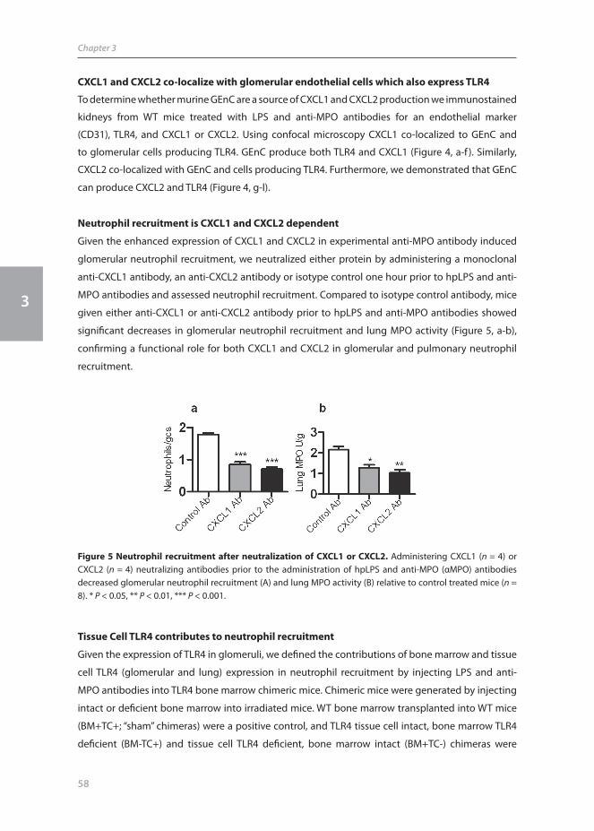
CXCL1 and CXCL2 co-localize with glomerular endothelial cells which also express TLR4
To determine whether murine GEnC are a source of CXCL1 and CXCL2 production we immunostained
kidneys from WT mice treated with LPS and anti-MPO antibodies for an endothelial marker
(CD31), TLR4, and CXCL1 or CXCL2. Using confocal microscopy CXCL1 co-localized to GEnC and
to glomerular cells producing TLR4. GEnC produce both TLR4 and CXCL1 (Figure 4, a-f ). Similarly,
CXCL2 co-localized with GEnC and cells producing TLR4. Furthermore, we demonstrated that GEnC
can produce CXCL2 and TLR4 (Figure 4, g-l).
Neutrophil recruitment is CXCL1 and CXCL2 dependent
Given the enhanced expression of CXCL1 and CXCL2 in experimental anti-MPO antibody induced
glomerular neutrophil recruitment, we neutralized either protein by administering a monoclonal
anti-CXCL1 antibody, an anti-CXCL2 antibody or isotype control one hour prior to hpLPS and anti-
MPO antibodies and assessed neutrophil recruitment. Compared to isotype control antibody, mice
given either anti-CXCL1 or anti-CXCL2 antibody prior to hpLPS and anti-MPO antibodies showed
signifi cant decreases in glomerular neutrophil recruitment and lung MPO activity (Figure 5, a-b),
confi rming a functional role for both CXCL1 and CXCL2 in glomerular and pulmonary neutrophil
recruitment.
Tissue Cell TLR4 contributes to neutrophil recruitment
Given the expression of TLR4 in glomeruli, we defi ned the contributions of bone marrow and tissue
cell TLR4 (glomerular and lung) expression in neutrophil recruitment by injecting LPS and anti-
MPO antibodies into TLR4 bone marrow chimeric mice. Chimeric mice were generated by injecting
intact or defi cient bone marrow into irradiated mice. WT bone marrow transplanted into WT mice
(BM+TC+; “sham” chimeras) were a positive control, and TLR4 tissue cell intact, bone marrow TLR4
defi cient (BM-TC+) and tissue cell TLR4 defi cient, bone marrow intact (BM+TC-) chimeras were
Figure 5 Neutrophil recruitment after neutralization of CXCL1 or CXCL2. Administering CXCL1 (n = 4) or CXCL2 (n = 4) neutralizing antibodies prior to the administration of hpLPS and anti-MPO (αMPO) antibodies decreased glomerular neutrophil recruitment (A) and lung MPO activity (B) relative to control treated mice (n = 8). * P < 0.05, ** P < 0.01, *** P < 0.001.

59
Both renal cell and leukocyte TLR4 aggravate glomerulonephritis
3
Figure 6 Assessment of glomerular leukocyte recruitment, lung MPO activity and glomerular chemokine staining in BM chimeric mice wild type (WT)WT mice (Bone marrow [BM]+, Tissue Cell [TC]+, n = 8), TLR4-/-WT mice (BM-TC+, n = 8) and WTTLR4-/- mice (BM+ TC-, n = 8) injected with hpLPS and anti-MPO (αMPO) antibodies. Glomerular neutrophil recruitment (A) was decreased in both BM-TC+ and BM+TC- compared to BM+TC+ mice. There was decreased glomerular neutrophil recruitment in BM-TC+ mice compared to BM+TC- mice. Lung MPO activity was decreased in both BM-TC+ and BM+TC- mice compared to BM+TC+ mice (B). Kidney CXCL1 (C) and CXCL2 (D) immunostaining was decreased in BM-TC+ and BM-TC+ mice compared to BM+TC+ mice. * P < 0.05, ** P < 0.01, *** P < 0.001.
studied. Both bone marrow and tissue cell TLR4 are required for maximal neutrophil recruitment.
Compared with BM+TC+ chimeras glomerular neutrophil recruitment was reduced in either TLR4
BM-TC+ chimeras or TLR4 BM+TC- mice (Figure 6a), but bone marrow derived TLR4 plays a more
prominent role. Both bone marrow and tissue cell TLR4 are required for maximum pulmonary
neutrophil recruitment (Figure 6b).
Glomerular CXCL1 and CXCL2 production was assessed in kidneys of chimeric mice. Compared to
‘sham’ chimeras (BM+TC+), semi-quantitative assessment of CXCL1 and CXCL2 showed a reduction
in both chemokines in TLR4 BM-TC+ mice and BM+TC- mice (Figure 6c and d), corresponding with
the reduction in neutrophil recruitment. The trend towards decreased CXCL1 and CXCL2 mRNA
expression in kidneys of BM-TC+ and BM+TC- chimeric mice did not reach statistical signifi cance
(data not shown).
CXCL8 is produced by human glomerular endothelial cells after LPS stimulation
Having established a role for TLR4 on intrinsic renal cells in inducing CXCL1 and CXCL2 dependent
experimental glomerular neutrophil recruitment, we analyzed human glomerular cells for CXCL8

60
Chapter 3
3
production (CXCL1 and CXCL2 are the murine homologues of CXCL8). Baseline mean ciGEnC mRNA
expression was assigned a value of 1. While CXCL8 mRNA expression was increased in ciGEnC,
podocyte and mesangial cells (Table 1), the relative increase was most pronounced in ciGEnC
(Figure 7a). Basal CXCL8 production was highest in podocytes (Table 1), however, after stimulation
the increase is most pronounced in ciGEnC, with signifi cant increases at 2 h, 4 h and 24 h (Figure 7b).
These increases in both mRNA expression and protein production demonstrate that GEnC are the
cell type most responsible for enhanced CXCL8 production.
Table 1 CXCL8 mRNA and protein production in human glomerular cell lines at 0 h, 2 h, 4 h and 24 h after LPS stimulation. Baseline mRNA expression in ciGEnC was assigned a value of 1. Increased expression is seen in all cell lines, most pronounced at 2 h. CXCL8 protein production also increases in each cell type, most pronounced after 24 h. # P < 0.01 ## P < 0.001 compared to t=0 h.
CXCL8 mRNA 0 h 2 h 4 h 24 h
ciGEnC 1.0±0.1 46.2±2.0## 33.4±2.6## 15.5±1.8##
Podocytes 2.3±0.2 28.0±1.9## 19.8±0.8## 17.9±0.9##
Mesangial Cells 0.0±0.0 0.4±0.0## 0.2±0.0# 0.1±0.0
CXCL8 protein (ng/ml) 0 h 2 h 4 h 24 h
ciGEnC 2.3±0.1 3.0±0.1 9.0±0.5## 35.8±1.9##
Podocytes 27.1±1.3 24.5±1.8 28.5±1.7 40.8±4.3#
Mesangial Cells 0.2±0.0 0.3±0.0 0.3±0.0 0.5±0.0##
Figure 7 CXCL8 mRNA and protein production in human glomerular cell lines. After LPS stimulation, CXCL8 mRNA expression was increased in all three cell types, most pronounced in glomerular endothelial cells (ciGEnC) (A). Similarly, ciGEnC produced the greatest proportional increase in CXCL8 protein production (B), n = 6 for all experimental groups. Absolute values (in pg/ml) of CXCL8 measurements are shown in Table 1. * P < 0.05, *** P < 0.001.

61
Both renal cell and leukocyte TLR4 aggravate glomerulonephritis
3
The contribution of TLR4, CXCL1, CXCL2, and Bone Marrow and Tissue Cell TLR4 to renal injury
Administration of hpLPS and anti-MPO antibodies (100μg/g) induced functional renal injury
(albuminuria and hematuria), and glomerular hypercellularity with focal and segmental lesions,
including fi brin deposition in WT mice at day 6 (Figure 8, a-d), signifi cantly decreased in TLR4-/-
mice. Representative photomicrographs of renal injury showing glomerular hypercellularity and
glomerular fi brin staining in WT, reduced in TLR4-/- mice are shown in Figure 8, e-h.
Figure 8 Functional and histological renal injury in WT and TLR4-/- mice treated with LPS and anti-MPO antibodies. Six days after the administration of ANCA/ LPS 24 h albuminuria (A), dipstick hematuria (B), glomerular fi brin deposition (C) and glomerular hypercellularity (D) was attenuated in TLR4-/- mice (n = 6) compared to WT controls (n = 6). The dotted line in (A) and (D) represents mean values in untreated WT mice. Representative fi gures are shown demonstrating glomerular injury in WT mice (E) compared to TLR4-/- mice (F). More glomerular fi brin deposition was seen in WT mice (G) compared to TLR4-/- mice (H). * P < 0.05, ** P < 0.01, *** P < 0.001. Original magnifi cation, 400x. (color image on page 184)

62
Chapter 3
3
As CXCL1 and CXCL2 were important in glomerular neutrophil recruitment we defi ned their
role in the development of histological and functional renal injury by extending studies to day 6.
Compared to mice given ANCA/LPS and isotope control antibodies, albuminuria and hematuria
were decreased after the administration of anti-CXCL2 antibody and ANCA/LPS, while a trend
to decreased injury was seen after the administration of anti-CXCL1 antibody (Figure 9a and b).
Albuminuria, measured in the initial 24 h in mice administered control antibody and ANCA/LPS
(170±39μg/24 h) was decreased in mice given anti-CXCL1 antibody and ANCA/LPS (55±14μg/24
h, P<0.001) and anti-CXCL2 and ANCA/LPS (87±10μg/24h, P<0.01). Glomerular histological injury
(Figure 9c and d) was attenuated after the administration of CXCL1 or CXCL2 neutralizing antibodies,
though reduced hypercellularity after anti-CXCL1 antibody did not reach statistical signifi cance.
Having demonstrated a role for both bone marrow and glomerular TLR4 in neutrophil
recruitment, we confi rmed that both are required for maximal renal injury. Compared to BM+TC+
‘sham’ chimeras, albuminuria was decreased in both BM-TC+ and BM+TC- mice, (Figure 10a and b;
trends to reduction in hematuria did not reach signifi cance). Histological injury was reduced in BM-
TC+ and BM+TC- mice with less glomerular fi brin deposition and hypercellularity (Figure 10c and d).
Figure 9 Functional and histological renal injury in WT mice treated with LPS and anti-MPO antibodies with prior neutralization of CXCL1 (n = 6) and CXCL2 (n = 6). Compared to control antibody treated mice (n = 6), 24 h albuminuria (A) and dipstick hematuria (B) were signifi cantly decreased after CXCL2 neutralization; trends after administration of CXCL1 neutralizing antibody did not reach signifi cance. Less glomerular fi brin deposition (C) was evident with neutralization of either CXCL1 or CXCL2, while compared to control antibody treated mice, hypercellularity was decreased with neutralization of CXCL2 (D). * P < 0.05, ** P < 0.01, *** P < 0.001.

63
Both renal cell and leukocyte TLR4 aggravate glomerulonephritis
3
DISCUSSION
These studies defi ne roles for both bone marrow cell and tissue cell-derived TLR4 in the
pathogenesis of neutrophil recruitment in LPS/anti-MPO antibody renal and lung injury, and the
roles of CXCL1 and CXCL2. After stimulation, the glomerulus is the major site of TLR4 expression in
the kidney, whilst GEnC are the cell type most responsible. Both bone marrow and tissue cell TLR4
are required for full expression of CXCL1 and CXCL2, and maximal neutrophil recruitment. Murine
GEnC are a source of TLR4, CXCL1 and CXCL2, with some GEnC producing both TLR4 and neutrophil
chemoattractants. Using human glomerular cells we demonstrated that after stimulation GEnC are
largely responsible for the increase in CXCL8 expression and protein production. In vivo stu dies at
a later time-point confi rmed the functional relevance of both immune cell and tissue cell derived
TLR4, and of neutrophil chemoattractants, especially CXCL2.
Experimental anti-MPO antibody induced GN is neutrophil dependent.4 Neutrophils express
TLR4, and LPS that engages TLR4, is a potent stimulus for neutrophil activation. TLR4 ligation has
pleiotropic eff ects on neutrophils including neutrophil adhesion,23, 24 delayed apoptosis, enhanced
chemokine production and increased superoxide generation.25 In the current studies, the fi rst series
Figure 10 Functional and histological renal injury in BM chimeric mice wild type (WT)WT mice (BM [BM]+, TC[TC]+, n = 7), TLR4-/-WT mice (BM-TC+, n = 6) and WTTLR4-/- mice (BM+ TC-, n = 7) injected with hpLPS and αMPO antibodies. Compared to “sham chimeras” (BM+TC+) 24 h albuminuria (A) was decreased in BM-TC+ and BM+TC-chimera. There was a non-signifi cant trend to decrease in dipstick hematuria (B). Glomerular fi brin deposition (C) and glomerular hypercellularity (D) was decreased in BM-TC+ and BM-TC+ chimeric mice compared to sham chimeras BM+TC+. * P < 0.05, ** P < 0.01, *** P < 0.001.

64
Chapter 3
3
of experiments showed that administering both hpLPS and anti-MPO antibodies led to increased
glomerular neutrophil recruitment and lung MPO activity. The eff ects of hpLPS on neutrophil
recruitment were not seen in TLR4-/- mice. These studies are in accordance with previous work
showing that LPS had the capacity to markedly increase anti-MPO antibody induced injury in
mice.6,26
In the current studies, we used three separate techniques to determine the glomerular cell types
responsible for TLR4 production. Firstly, using confocal microscopy we demonstrated that TLR4 is
present in murine glomeruli and co-localizes with GEnC. Other glomerular cells also express TLR4
in this model. Secondly, using micro-dissected glomeruli, TLR4 mRNA expression was quantitated
in glomeruli and the tubulo-interstitium. After the administration of LPS and anti-MPO antibodies
glomerular, but not tubulointerstitial TLR4 expression was increased. Thirdly, using isolated human
glomerular cells we demonstrated that GEnC express signifi cant amounts of TLR4, and that
enhanced mRNA expression after stimulation is attributable to GEnC.
Therefore, GEnC are signifi cant sources of glomerular TLR4 expression after the administration of
LPS and anti-MPO antibodies. The sites of intrarenal TLR4 production, both in the glomerulus and the
tubulo-interstitium have been addressed in several studies in other experimental models. TLR4 has
been localised to the glomerulus in other models of renal disease.20, 27, 28 In situ hybridization showed
that mesangial and epithelial cells can express TLR427 while in experimental cryoglobulinemic GN
podocytes express TLR4.20 Studies assessing TLR4 in murine models of tubulo-interstitial injury have
demonstrated TLR4 mRNA production from primary renal tubular epithelial cells,29 while confocal
microscopy has suggested TLR4 is present in proximal collecting tubules.30 From the existing
literature and the current studies, it is clear that TLR4 expression from intrinsic renal cells can vary
according to the nature of the injurious stimulus. In the current studies, the results of a combination
of in vivo and in vitro studies imply a role for the glomerular endothelium in TLR4 responses in the
context of LPS and anti-MPO antibodies as initiators of injury.
In vivo murine models have shown that the chemokines CXCL1 and CXCL2 directs neutrophil
recruitment to the cornea,31 peritoneum,32, 33 the joint.34 The addition of LPS to anti-glomerular
basement membrane (GBM) globulin enhances heterologous renal injury.27, 35 Furthermore, Brown
et al, demonstrated that neutralizing CXCL1 and CXCL2, which was TLR4 mediated and produced by
renal cells, resulted in decreased glomerular injury.27 We have demonstrated that renal CXCL1 and
CXCL2 expression (mRNA and protein) increases in a TLR4 dependent manner, both of which are
produced by GEnC. The receptor for CXCL1 and CXCL2, CXCR2, is present on neutrophils, but LPS
does not induce its expression or alter migration induced by neutrophil chemoattractants.23, 36 The
current studies show that CXCL1 and CXCL2 direct anti-MPO antibody glomerular and pulmonary
neutrophil recruitment, as neutralizing CXCL1 and CXCL2 decreased glomerular neutrophil
recruitment and lung MPO activity early, and functional and histological renal injury later in the
disease.
Both bone marrow derived cells37 and an activated glomerular endothelium38, 39 are thought

65
Both renal cell and leukocyte TLR4 aggravate glomerulonephritis
3
to be important in glomerular neutrophil recruitment in GN induced by anti-MPO antibodies. The
current studies demonstrate that both bone marrow and tissue cell TLR4 are required for maximal
glomerular and lung neutrophil requirement, underlying separate roles in the disease process.
These eff ects within the kidney extended out to at least six days, where mice defi cient in either bone
marrow cell or tissue cell TLR4 exhibited less injury, even in the face of more profound initial decrease
in glomerular neutrophil recruitment in BM-TC+ mice. As CXCL1 and CXCL2 staining was decreased
in BM-TC+ mice and BM+TC- mice, both bone marrow and tissue cell derived TLR4 are important
in the renal production of CXCL1 and CXCL2, required for glomerular neutrophil recruitment.
Neutralizing CXCL2 at the induction of injury resulted in attenuated functional and histological
glomerular injury after six days; the eff ects of CXCL1 blockade were less prolonged. While the
current studies did not assess the cell type in the lung that produces neutrophil chemoattractants,
previous studies have demonstrated that CXCL1 and CXCL2 are produced by Clara cells (non-ciliated
bronchoalveolar epithelial cells in the distal airways).40 Previous studies analysing lung MPO activity
to quantitate neutrophil recruitment in TLR4 chimeric mice have yielded confl icting results, with
one study implicating tissue cells,41 whilst another showed bone marrow cell TLR4 to be important.42
In experimental anti-MPO antibody induced neutrophil recruitment MPO activity is decreased in
both BM-TC+ and BM+TC- chimeric mice.
Observations in human renal biopsies suggest a pathogenic role for CXCL8, the key neutrophil-
attracting chemokine in ANCA GN.15 Previous studies have suggested that CXCL8 can be produced
by “generic” macrovascular endothelial cells (HUVECs)43, and cultured human mesangial cells.44 We
compared CXCL8 production by diff erent human glomerular cells, including ciGEnC, concurrently,
in a single study. Whilst baseline expression of CXCL8 mRNA and protein production was higher
in podocytes; ciGEnC showed the most signifi cant increase in expression and production after
stimulation, suggesting that during infl ammation GEnC produce CXCL8 which is responsible for
neutrophil recruitment.
We have demonstrated a pivotal role for both bone marrow and intrinsic renal cell TLR4 in
glomerular and lung neutrophil recruitment and injury in experimental ANCA disease. Maximal
neutrophil recruitment is dependent on CXCL1 and CXCL2, tissue cell expression, which is TLR4
dependent. Therefore, in addition to immune cell TLR4 mediated activation and recruitment, the
current studies demonstrate a role for the glomerular endothelium, which involves GEnC TLR4
expression, and CXC chemokine production, that enhances neutrophil recruitment. Results from
these studies add to evidence linking infection to autoimmune GN and provide evidence for
possible benefi ts of TLR inhibition in immune glomerular disease.
ACKNOWLEDGEMENTS
Professor Shizuo Akira is thanked for the TLR4-/- mice. Ms Alice Wright and Ms Sophia Ling are
thanked for technical assistance. Dr Banas is thanked for the immortalized human mesangial cells.
Dr Camden Lo, Monash Micro-imaging is thanked for help with confocal microscopy. Parts of these

66
Chapter 3
3
studies were published in abstract form (Nephrology 13(suppl 3): A140, J Am Soc Nephrol 2008
Nov(S) A659).
REFERENCES
1. Jennette JC, Xiao H, Falk RJ. Pathogenesis of vascular infl ammation by anti-neutrophil cytoplasmic antibodies. J Am Soc Nephrol 2006; 17: 1235-1242.
2. Falk RJ, Jennette JC. Anti-neutrophil cytoplasmic autoantibodies with specifi city for myeloperoxidase in patients with systemic vasculitis and idiopathic necrotizing and crescentic glomerulonephritis. N Engl J Med 1988; 318: 1651-1657.
3. Xiao H, Heeringa P, Hu P, et al. Antineutrophil cytoplasmic autoantibodies specifi c for myeloperoxidase cause glomerulonephritis and vasculitis in mice. J Clin Invest 2002; 110: 955-963.
4. Xiao H, Heeringa P, Liu Z, et al. The role of neutrophils in the induction of glomerulonephritis by anti-myeloperoxidase antibodies. Am J Pathol 2005; 167: 39-45.
5. Xiao H, Schreiber A, Heeringa P, et al. Alternative complement pathway in the pathogenesis of disease mediated by anti-neutrophil cytoplasmic autoantibodies. Am J Pathol 2007; 170: 52-64.
6. Huugen D, Xiao H, van Esch A, et al. Aggravation of anti-myeloperoxidase antibody-induced glomerulonephritis by bacterial lipopolysaccharide: role of tumor necrosis factor-alpha. Am J Pathol 2005; 167: 47-58.
7. Little MA, Smyth CL, Yadav R, et al. Antineutrophil cytoplasm antibodies directed against myeloperoxidase augment leukocyte-microvascular interactions in vivo. Blood 2005; 106: 2050-2058.
8. Tidman M, Olander R, Svalander C, et al. Patients hospitalized because of small vessel vasculitides with renal involvement in the period 1975-95: organ involvement, anti-neutrophil cytoplasmic antibodies patterns, seasonal attack rates and fl uctuation of annual frequencies. J Intern Med 1998; 244: 133-141.
9. Stegeman CA, Tervaert JW, Sluiter WJ, et al. Association of chronic nasal carriage of Staphylococcus aureus and higher relapse rates in Wegener granulomatosis. Ann Intern Med 1994; 120: 12-17.
10. Pinching AJ, Rees AJ, Pussell BA, et al. Relapses in Wegener’s granulomatosis: the role of infection. Br Med J 1980; 281: 836-838.
11. Capizzi SA, Specks U. Does infection play a role in the pathogenesis of pulmonary vasculitis? Semin Respir Infect 2003; 18: 17-22.
12. Arimura Y, Minoshima S, Kamiya Y, et al. Serum myeloperoxidase and serum cytokines in anti-myeloperoxidase antibody-associated glomerulonephritis. Clin Nephrol 1993; 40: 256-264.
13. Stegeman CA, Tervaert JW, de Jong PE, et al. Trimethoprim-sulfamethoxazole (co-trimoxazole) for the prevention of relapses of Wegener’s granulomatosis. Dutch Co-Trimoxazole Wegener Study Group. N Engl J Med 1996; 335: 16-20.
14. Yoshimura T, Matsushima K, Tanaka S, et al. Purifi cation of a human monocyte-derived neutrophil chemotactic factor that has peptide sequence similarity to other host defense cytokines. Proc Natl Acad Sci U S A 1987; 84: 9233-9237.
15. Cockwell P, Brooks CJ, Adu D, et al. Interleukin-8: A pathogenetic role in antineutrophil cytoplasmic autoantibody-associated glomerulonephritis. Kidney Int 1999; 55: 852-863.
16. Zlotnik A, Yoshie O. Chemokines: a new classifi cation system and their role in immunity. Immunity 2000; 12: 121-127.
17. Lee J, Cacalano G, Camerato T, et al. Chemokine binding and activities mediated by the mouse IL-8 receptor. J Immunol 1995; 155: 2158-2164.
18. Hayashi F, Means TK, Luster AD. Toll-like receptors stimulate human neutrophil function. Blood 2003; 102: 2660-2669.
19. Fan J, Malik AB. Toll-like receptor-4 (TLR4) signaling augments chemokine-induced neutrophil migration by modulating cell surface expression of chemokine receptors. Nat Med 2003; 9: 315-321.
20. Banas MC, Banas B, Hudkins KL, et al. TLR4 links podocytes with the innate immune system to mediate glomerular injury. J Am Soc Nephrol 2008; 19: 704-713.
21. Shirali AC, Goldstein DR. Tracking the toll of kidney disease. J Am Soc Nephrol 2008; 19: 1444-1450.

67
Both renal cell and leukocyte TLR4 aggravate glomerulonephritis
3
22. Kuligowski MP, Kwan RY, Lo C, et al. Antimyeloperoxidase antibodies rapidly induce alpha-4-integrin-dependent glomerular neutrophil adhesion. Blood 2009; 113: 6485-6494.
23. Sabroe I, Prince LR, Jones EC, et al. Selective roles for Toll-like receptor (TLR)2 and TLR4 in the regulation of neutrophil activation and life span. J Immunol 2003; 170: 5268-5275.
24. Sabroe I, Jones EC, Usher LR, et al. Toll-like receptor (TLR)2 and TLR4 in human peripheral blood granulocytes: a critical role for monocytes in leukocyte lipopolysaccharide responses. J Immunol 2002; 168: 4701-4710.
25. Parker LC, Whyte MK, Dower SK, et al. The expression and roles of Toll-like receptors in the biology of the human neutrophil. J Leukoc Biol 2005; 77: 886-892.
26. Huugen D, van Esch A, Xiao H, et al. Inhibition of complement factor C5 protects against anti-myeloperoxidase antibody-mediated glomerulonephritis in mice. Kidney Int 2007; 71: 646-654.
27. Brown HJ, Lock HR, Wolfs TG, et al. Toll-like receptor 4 ligation on intrinsic renal cells contributes to the induction of antibody-mediated glomerulonephritis via CXCL1 and CXCL2. J Am Soc Nephrol 2007; 18: 1732-1739.
28. Patole PS, Pawar RD, Lech M, et al. Expression and regulation of Toll-like receptors in lupus-like immune complex glomerul onephritis of MRL-Fas(lpr) mice. Nephrol Dial Transplant 2006; 21: 3062-3073.
29. Tsuboi N, Yoshikai Y, Matsuo S, et al. Roles of toll-like receptors in C-C chemokine production by renal tubular epithelial cells. J Immunol 2002; 169: 2026-2033.
30. Zhang B, Ramesh G, Uematsu S, et al. TLR4 signaling mediates infl ammation and tissue injury in nephrotoxicity. J Am Soc Nephrol 2008; 19: 923-932.
31. Lin M, Carlson E, Diaconu E, et al. CXCL1/KC and CXCL5/LIX are selectively produced by corneal fi broblasts and mediate neutrophil infi ltration to the corneal stroma in LPS keratitis. J Leukoc Biol 2007; 81: 786-792.
32. De Filippo K, Henderson RB, Laschinger M, et al. Neutrophil chemokines KC and macrophage-infl ammatory protein-2 are newly synthesized by tissue macrophages using distinct TLR signaling pathways. J Immunol 2008; 180: 4308-4315.
33. Wengner AM, Pitchford SC, Furze RC, et al. The coordinated action of G-CSF and ELR + CXC chemokines in neutrophil mobilization during acute infl ammation. Blood 2008; 111: 42-49.
34. Coelho FM, Pinho V, Amaral FA, et al. The chemokine receptors CXCR1/CXCR2 modulate antigen-induced arthritis by regulating adhesion of neutrophils to the synovial microvasculature. Arthritis Rheum 2008; 58: 2329-2337.
35. Fu Y, Xie C, Chen J, et al. Innate stimuli accentuate end-organ damage by nephrotoxic antibodies via Fc receptor and TLR stimulation and IL-1/TNF-alpha production. J Immunol 2006; 176: 632-639.
36. Sabroe I, Jones EC, Whyte MK, et al. Regulation of human neutrophil chemokine receptor expression and function by activation of Toll-like receptors 2 and 4. Immunology 2005; 115: 90-98.
37. Schreiber A, Xiao H, Falk RJ, et al. Bone marrow-derived cells are suffi cient and necessary targets to mediate glomerulonephritis and vasculitis induced by anti-myeloperoxidase antibodies. J Am Soc Nephrol 2006; 17: 3355-3364.
38. Calderwood JW, Williams JM, Morgan MD, et al. ANCA induces beta2 integrin and CXC chemokine-dependent neutrophil-endothelial cell interactions that mimic those of highly cytokine-activated endothelium. J Leukoc Biol 2005; 77: 33-43.
39. Nolan SL, Kalia N, Nash GB, et al. Mechanisms of ANCA-mediated leukocyte-endothelial cell interactions in vivo. J Am Soc Nephrol 2008; 19: 973-984.
40. Elizur A, Adair-Kirk TL, Kelley DG, et al. Clara cells impact the pulmonary innate immune response to LPS. Am J Physiol Lung Cell Mol Physiol 2007; 293: L383-392.
41. Andonegui G, Bonder CS, Green F, et al. Endothelium-derived Toll-like receptor-4 is the key molecule in LPS-induced neutrophil sequestration into lungs. J Clin Invest 2003; 111: 1011-1020.
42. Hollingsworth JW, Chen BJ, Brass DM, et al. The critical role of hematopoietic cells in lipopolysaccharide-induced airway infl ammation. Am J Respir Crit Care Med 2005; 171: 806-813.
43. Strieter RM, Kunkel SL, Showell HJ, et al. Endothelial cell gene expression of a neutrophil chemotactic factor by TNF-alpha, LPS, and IL-1 beta. Science 1989; 243: 1467-1469.
44. Brown Z, Strieter RM, Chensue SW, et al. Cytokine-activated human mesangial cells generate the neutrophil chemoattractant, interleukin 8. Kidney Int 1991; 40: 86-90.
68
Chapter 3
3
45. Apostolopoulos J, Ooi JD, Odobasic D, et al. The isolation and purifi cation of biologically active recombinant and native autoantigens for the study of autoimmune disease. J Immunol Methods 2006; 308: 167-178.
46. Hirschfeld M, Ma Y, Weis JH, et al. Cutting edge: repurifi cation of lipopolysaccharide eliminates signaling through both human and murine toll-like receptor 2. J Immunol 2000; 165: 618-622.
47. Takeuchi O, Hoshino K, Kawai T, et al. Diff erential roles of TLR2 and TLR4 in recognition of gram-negative and gram-positive bacterial cell wall components. Immunity 1999; 11: 443-451.
48. Timoshanko JR, Sedgwick JD, Holdsworth SR, et al. Intrinsic renal cells are the major source of tumor necrosis factor contributing to renal injury in murine crescentic glomerulonephritis. J Am Soc Nephrol 2003; 14: 1785-1793.
49. Huang XR, Holdsworth SR, Tipping PG. Evidence for delayed-type hypersensitivity mechanisms in glomerular crescent formation. Kidney Int 1994; 46: 69-78.
50. Roche JK, Keepers TR, Gross LK, et al. CXCL1/KC and CXCL2/MIP-2 are critical eff ectors and potential targets for therapy of Escherichia coli O157:H7-associated renal infl ammation. Am J Pathol 2007; 170: 526-537.
51. Drew AF, Tucker HL, Liu H, et al. Crescentic glomerulonephritis is diminished in fi brinogen-defi cient mice. Am J Physiol Renal Physiol 2001; 281: F1157-1163.
52. Odobasic D, Kitching AR, Semple TJ, et al. Endogenous Myeloperoxidase Promotes Neutrophil-Mediated Renal Injury, but Attenuates T Cell Immunity Inducing Crescentic Glomerulonephritis. J Am Soc Nephrol 2007;
53. Satchell SC, Tasman CH, Singh A, et al. Conditionally immortalized human glomerular endothelial cells expressing fenestrations in response to VEGF. Kidney Int 2006; 69: 1633-1640.
54. Saleem MA, O’Hare MJ, Reiser J, et al. A conditionally immortalized human podocyte cell line demonstrating nephrin and podocin expression. J Am Soc Nephrol 2002; 13: 630-638.
55. Coward RJ, Welsh GI, Koziell A, et al. Nephrin is critical for the action of insulin on human glomerular podocytes. Diabetes 2007; 56: 1127-1135.
56. Banas B, Luckow B, Moller M, et al. Chemokine and chemokine receptor expression in a novel human mesangial cell line. J Am Soc Nephrol 1999; 10: 2314-2322.
57. Asgeirsdottir SA, Kamps JA, Bakker HI, et al. Site-specifi c inhibition of glomerulonephritis progression by targeted delivery of dexamethasone to glomerular endothelium. Mol Pharmacol 2007; 72: 121-131.
58. Phoon RK, Kitching AR, Odobasic D, et al. T-bet defi ciency attenuates renal injury in experimental crescentic glomerulonephritis. J Am Soc Nephrol 2008; 19: 477-485.
Related Documents