Intravascular brachytherapy August 2002 MSAC application 1041 Assessment report

Welcome message from author
This document is posted to help you gain knowledge. Please leave a comment to let me know what you think about it! Share it to your friends and learn new things together.
Transcript

Intravascularbrachytherapy
August 2002
MSAC application 1041
Assessment report

© Commonwealth of Australia 2002
ISBN 0 642 82129 1
ISSN (Print) 1443-7120
ISSN (Online) 1443-7139
First printed January 2003
This work is copyright. Apart from any use as permitted under the Copyright Act 1968 no part may be reproduced by any process without written permission from AusInfo. Requests and inquiries concerning reproduction and rights should be directed to the Manager, Legislative Services, AusInfo, GPO Box 1920, Canberra, ACT, 2601.
Electronic copies of the report can be obtained from the Medical Services Advisory Committee’s Internet site at:
http://www.msac.gov.au
Hard copies of the report can be obtained from:
The Secretary
Medical Services Advisory Committee
Department of Health and Ageing
Mail Drop Point 107
GPO Box 9848
Canberra ACT 2601
Enquiries about the content of the report should be directed to the above address.
The Medical Services Advisory Committee is an independent committee which has been established to provide advice to the Commonwealth Minister for Health and Ageing on the strength of evidence available on new and existing medical technologies and procedures in terms of their safety, effectiveness and cost effectiveness. This advice will help to inform Government decisions about which medical services should attract funding under Medicare.
This report was prepared by the Medical Services Advisory Committee with the assistance of:
Ms. Kirsten Howard, Epidemiologist and Ms. Elizabeth Barr, Research Assistant from the NHMRC Clinical Trials Centre, University of Sydney. The report was endorsed by the Commonwealth Minister for Health and Ageing on 16th October 2002.
Publication approval number: 3132

MSAC recommendations do not necessarily reflect the views of allindividuals who participated in the MSAC evaluation.
Intravascular brachytherapy iii
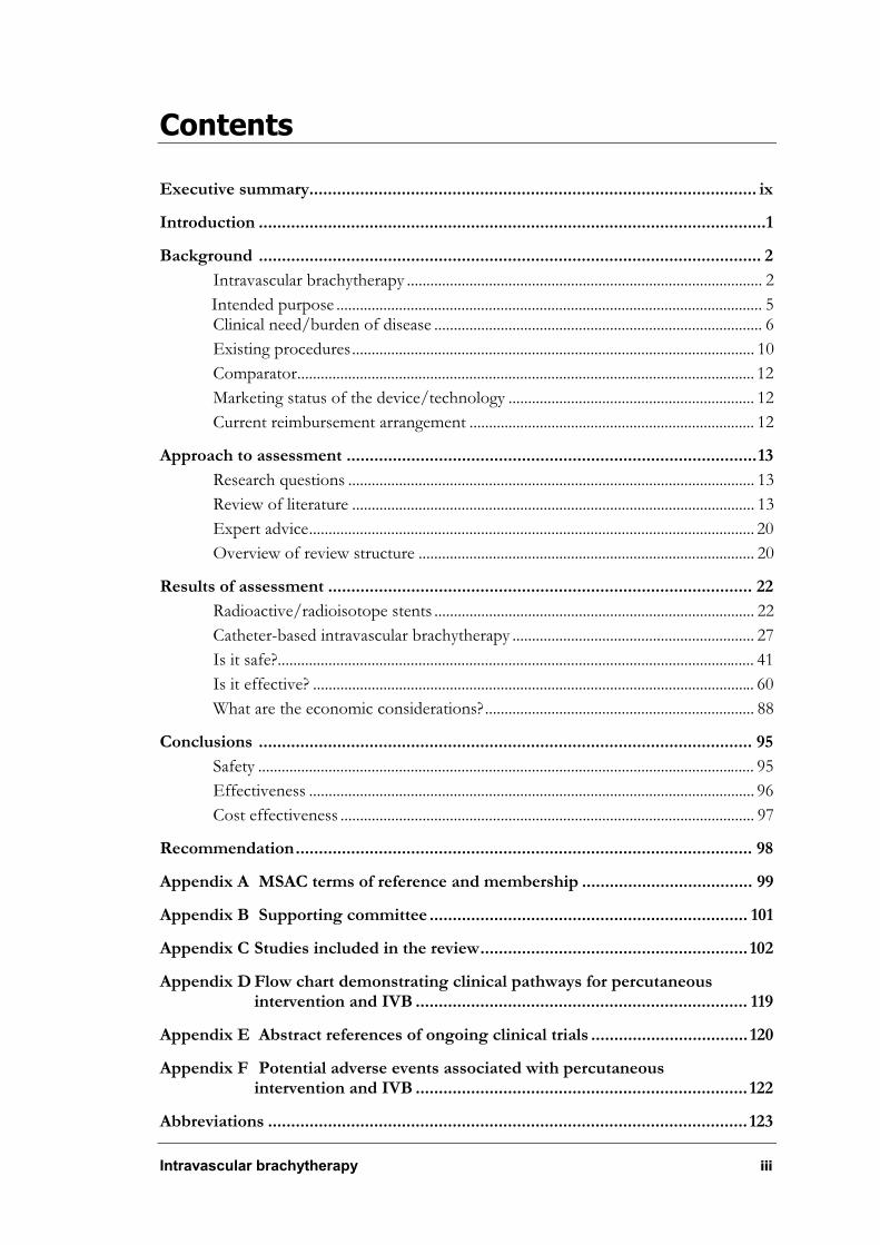
Contents
Executive summary................................................................................................. ix
Introduction ..............................................................................................................1
Background ............................................................................................................. 2
Intravascular brachytherapy ........................................................................................... 2
Intended purpose ............................................................................................................. 5Clinical need/burden of disease .................................................................................... 6
Existing procedures....................................................................................................... 10
Comparator..................................................................................................................... 12
Marketing status of the device/technology ............................................................... 12
Current reimbursement arrangement ......................................................................... 12
Approach to assessment .........................................................................................13
Research questions ........................................................................................................ 13
Review of literature ....................................................................................................... 13
Expert advice.................................................................................................................. 20
Overview of review structure ...................................................................................... 20
Results of assessment ............................................................................................ 22
Radioactive/radioisotope stents .................................................................................. 22
Catheter-based intravascular brachytherapy .............................................................. 27
Is it safe?.......................................................................................................................... 41
Is it effective? ................................................................................................................. 60
What are the economic considerations?..................................................................... 88
Conclusions ........................................................................................................... 95
Safety ............................................................................................................................... 95
Effectiveness .................................................................................................................. 96
Cost effectiveness .......................................................................................................... 97
Recommendation................................................................................................... 98
Appendix A MSAC terms of reference and membership ..................................... 99
Appendix B Supporting committee ..................................................................... 101
Appendix C Studies included in the review.......................................................... 102
Appendix D Flow chart demonstrating clinical pathways for percutaneous intervention and IVB ........................................................................ 119
Appendix E Abstract references of ongoing clinical trials .................................. 120
Appendix F Potential adverse events associated with percutaneous intervention and IVB ........................................................................ 122
Abbreviations ........................................................................................................ 123

iv Intravascular brachytherapy
References .......................................................................................................... 125
Intravascular brachytherapy v
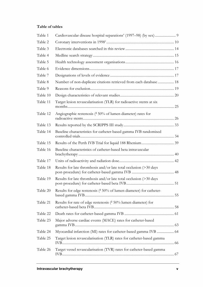
Table of tables
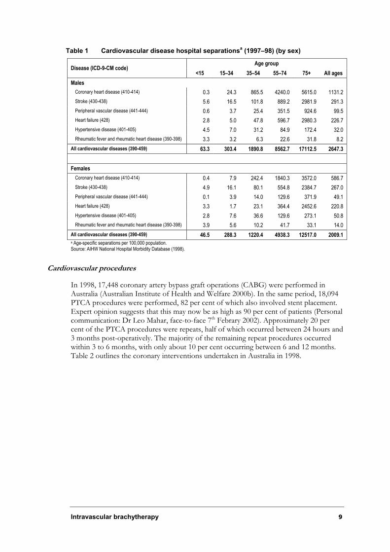
Table 1 Cardiovascular disease hospital separationsa (1997–98) (by sex) ....................... 9
Table 2 Coronary interventions in 1998a ........................................................................... 10
Table 3 Electronic databases searched in this review ...................................................... 14
Table 4 Medline search strategy .......................................................................................... 15
Table 5 Health technology assessment organisations...................................................... 16
Table 6 Evidence dimensions.............................................................................................. 17
Table 7 Designations of levels of evidence ....................................................................... 17
Table 8 Number of non-duplicate citations retrieved from each database .................. 18
Table 9 Reasons for exclusion............................................................................................. 19
Table 10 Design characteristics of relevant studies............................................................ 20
Table 11 Target lesion revascularisation (TLR) for radioactive stents at six months...................................................................................................................... 25
Table 12 Angiographic restenosis (²50% of lumen diameter) rates for radioactive stents..................................................................................................... 26
Table 13 Results reported by the SCRIPPS III study........................................................ 33
Table 14 Baseline characteristics for catheter-based gamma IVB randomised controlled trials........................................................................................................ 34
Table 15 Results of the Perth IVB Trial for liquid 188 Rhenium.................................... 39
Table 16 Baseline characteristics of catheter-based beta intravascular brachytherapy .......................................................................................................... 40
Table 17 Units of radioactivity and radiation dose............................................................. 42
Table 18 Results for late thrombosis and/or late total occlusion (>30 days post-procedure) for catheter-based gamma IVB ............................................... 48
Table 19 Results for late thrombosis and/or late total occlusion (>30 days post-procedure) for catheter-based beta IVB..................................................... 51
Table 20 Results for edge restenosis (²50% of lumen diameter) for catheter-based gamma IVB................................................................................................... 55
Table 21 Results for rate of edge restenosis (²50% lumen diameter) for catheter-based beta IVB......................................................................................... 58
Table 22 Death rates for catheter-based gamma IVB ....................................................... 61
Table 23 Major adverse cardiac events (MACE) rates for catheter-based gamma IVB.............................................................................................................. 63
Table 24 Myocardial infarction (MI) rates for catheter-based gamma IVB ................... 64
Table 25 Target lesion revascularisation (TLR) rates for catheter-based gamma IVB............................................................................................................................ 66
Table 26 Target vessel revascularisation (TVR) rates for catheter-based gamma IVB............................................................................................................................ 67
vi Intravascular brachytherapy
Table 27 Minimal lumen diameter (MLD) angiographic measurements ........................ 69
Table 28 Restenosis rate (²50% lumen diameter) for catheter-based gamma IVB............................................................................................................................ 70
Table 29 IVUS outcome measures for catheter-based gamma IVB................................ 72
Table 30 Death rates for catheter-based beta IVB............................................................. 74
Table 31 Major cardiac adverse events (MACE) for catheter-based beta IVB.............. 75
Table 32 Myocardial infarction (MI) events for catheter-based IVB .............................. 77
Table 33 Target lesion revascularisation (TLR) events for catheter-based beta IVB............................................................................................................................ 79
Table 34 Target vessel revascularisation (TVR) events for catheter-based beta IVB............................................................................................................................ 81
Table 35 Angiographic outcomes for catheter-based beta IVB....................................... 83
Table 36 Angiographic restenosis (²50% of lumen diameter) rates for catheter-based beta IVB........................................................................................................ 84
Table 37 IVUS outcome measures for catheter-based beta IVB..................................... 86
Table 38 Rate of restenosis (²50% of lumen diameter) of target lesion and margin for SCRIPPS .............................................................................................. 87
Table 39 Revascularisation rates for WRIST trial .............................................................. 87
Table 40 Results from Seto et al (2001) ............................................................................... 88
Table 41 Combined measures of major outcomes............................................................. 89
Table 42 Direct staff costs for intravascular brachytherapy (incremental costs over PCI alone) ....................................................................................................... 90
Table 43 Average public sector AR-DRG costs (1999–2000).......................................... 91
Table 44 Estimated ongoing medical costs for patients with symptomatic restenosis unsuitable for surgical/percutaneous intervention.......................... 92
Table 45 Results of incremental cost effectiveness analysis ............................................. 93
Table 46 Results of sensitivity analyses................................................................................ 93
Table 47 Catheter-based gamma intravascular brachytherapy trials* ............................ 102
Table 48 Catheter-based beta intravascular brachytherapy trials ................................... 112
Table 49 Catheter-based gamma IVB trials....................................................................... 120
Table 50 Catheter-based beta IVB trials ............................................................................ 121
Intravascular brachytherapy vii
Table of Figures
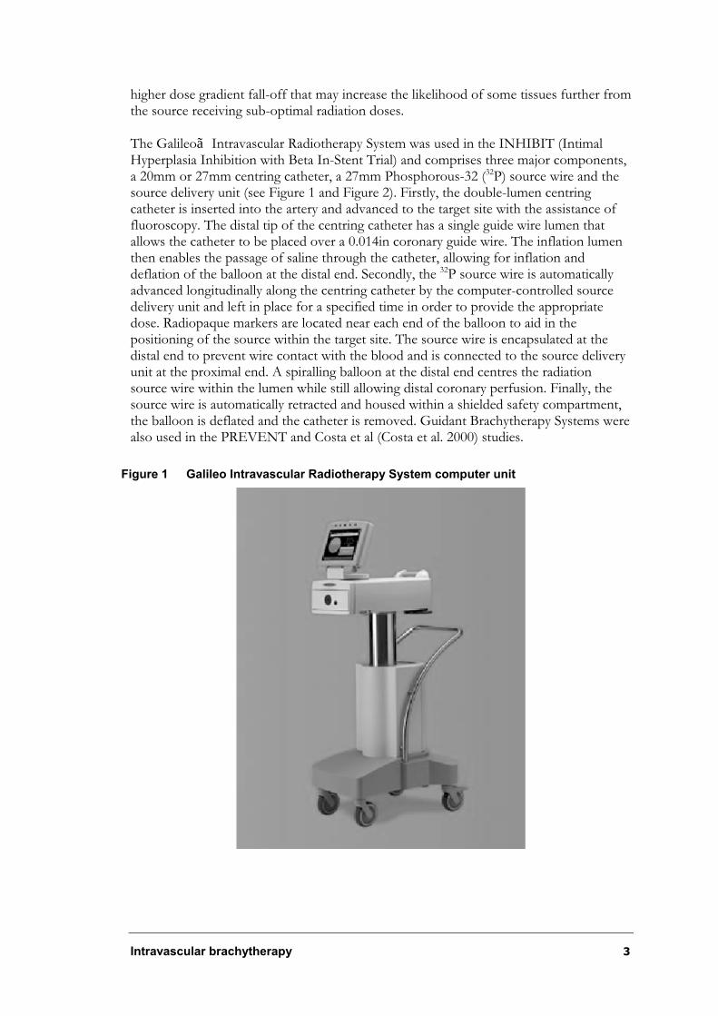
Figure 1 Galileo Intravascular Radiotherapy System computer unit ................................ 3
Figure 2 Galileo System source wire...................................................................................... 4
Figure 3 Pattern of restenosis in 35 of 74 lesions of patients who underwent angiographic follow-up at 4 to 6 months ............................................................ 24
Figure 4 Forest Plot of outcome of clinical late thrombosis (>30 days post-procedure) for catheter-based gamma IVB ........................................................ 48
Figure 5 Forest plot of outcome of clinical late thrombosis (>30 days post-procedure) for catheter-based beta IVB.............................................................. 52
Figure 6 Forest plot of outcome of survival for catheter-based gamma IVB ............... 62
Figure 7 Forest plot of outcome of MACE for catheter-based gamma IVB................ 63
Figure 8 Forest plot of outcome of MI for catheter-based gamma IVB ....................... 65
Figure 9 Forest plot of outcome of TLR for catheter-based gamma IVB .................... 66
Figure 10 Forest plot of outcome of TVR for catheter-based gamma IVB.................... 68
Figure 11 Forest plot of outcome of survival for catheter-based beta IVB .................... 74
Figure 12 Forest plot of outcome of MACE for catheter-based beta IVB ..................... 76
Figure 13 Forest plot of outcome of MI for catheter-bases beta IVB ............................. 77
Figure 14 Forest Plot of outcome of TLR for catheter-based beta IVB.......................... 79
Figure 15 Forest plot of outcome of TVR for catheter-based beta IVB ......................... 81
Figure 16 Baseline decision analytic model for cost effectiveness .................................... 90
Figure 17 Decision tree depicting calculation of average follow-up treatment costs (12 months).................................................................................................... 93

viii Intravascular brachytherapy
Intravascular brachytherapy ix
Executive summary
The procedure
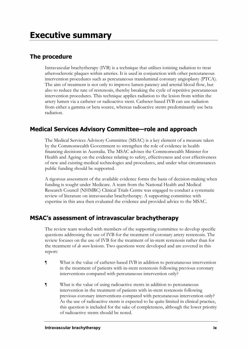
Intravascular brachytherapy (IVB) is a technique that utilises ionising radiation to treat atherosclerotic plaques within arteries. It is used in conjunction with other percutaneous intervention procedures such as percutaneous transluminal coronary angioplasty (PTCA). The aim of treatment is not only to improve lumen patency and arterial blood flow, but also to reduce the rate of restenosis, thereby breaking the cycle of repetitive percutaneous intervention procedures. This technique applies radiation to the lesion from within the artery lumen via a catheter or radioactive stent. Catheter-based IVB can use radiation from either a gamma or beta source, whereas radioactive stents predominantly use beta radiation.
Medical Services Advisory Committee—role and approach
The Medical Services Advisory Committee (MSAC) is a key element of a measure taken by the Commonwealth Government to strengthen the role of evidence in health financing decisions in Australia. The MSAC advises the Commonwealth Minister for Health and Ageing on the evidence relating to safety, effectiveness and cost effectiveness of new and existing medical technologies and procedures, and under what circumstances public funding should be supported.
A rigorous assessment of the available evidence forms the basis of decision-making when funding is sought under Medicare. A team from the National Health and Medical Research Council (NHMRC) Clinical Trials Centre was engaged to conduct a systematic review of literature on intravascular brachytherapy. A supporting committee with expertise in this area then evaluated the evidence and provided advice to the MSAC.
MSAC’s assessment of intravascular brachytherapy
The review team worked with members of the supporting committee to develop specific questions addressing the use of IVB for the treatment of coronary artery restenosis. The review focuses on the use of IVB for the treatment of in-stent restenosis rather than for the treatment of de novo lesions. Two questions were developed and are covered in this report:
¶ What is the value of catheter-based IVB in addition to percutaneous intervention in the treatment of patients with in-stent restenosis following previous coronary interventions compared with percutaneous intervention only?
¶ What is the value of using radioactive stents in addition to percutaneous intervention in the treatment of patients with in-stent restenosis following previous coronary interventions compared with percutaneous intervention only? As the use of radioactive stents is expected to be quite limited in clinical practice, this question is included for the sake of completeness, although the lower priority of radioactive stents should be noted.
x Intravascular brachytherapy
Clinical need
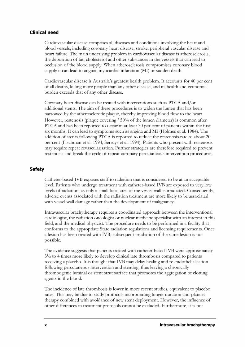
Cardiovascular disease comprises all diseases and conditions involving the heart and blood vessels, including coronary heart disease, stroke, peripheral vascular disease and heart failure. The main underlying problem in cardiovascular disease is atherosclerosis, the deposition of fat, cholesterol and other substances in the vessels that can lead to occlusion of the blood supply. When atherosclerosis compromises coronary blood supply it can lead to angina, myocardial infarction (MI) or sudden death.
Cardiovascular disease is Australia’s greatest health problem. It accounts for 40 per cent of all deaths, killing more people than any other disease, and its health and economic burden exceeds that of any other disease.
Coronary heart disease can be treated with interventions such as PTCA and/or additional stents. The aim of these procedures is to widen the lumen that has been narrowed by the atherosclerotic plaque, thereby improving blood flow to the heart.
However, restenosis (plaque covering ²50% of the lumen diameter) is common after PTCA and has been reported to occur in at least 30 per cent of patients within the first six months. It can lead to symptoms such as angina and MI (Holmes et al. 1984). The addition of stents following PTCA is reported to reduce the restenosis rate to about 20 per cent (Fischman et al. 1994; Serruys et al. 1994). Patients who present with restenosis may require repeat revascularisation. Further strategies are therefore required to prevent restenosis and break the cycle of repeat coronary percutaneous intervention procedures.
Safety
Catheter-based IVB exposes staff to radiation that is considered to be at an acceptable level. Patients who undergo treatment with catheter-based IVB are exposed to very low levels of radiation, as only a small local area of the vessel wall is irradiated. Consequently, adverse events associated with the radiation treatment are more likely to be associated with vessel wall damage rather than the development of malignancy.
Intravascular brachytherapy requires a coordinated approach between the interventional cardiologist, the radiation oncologist or nuclear medicine specialist with an interest in this field, and the medical physicist. The procedure needs to be performed in a facility that conforms to the appropriate State radiation regulations and licensing requirements. Once a lesion has been treated with IVB, subsequent irradiation of the same lesion is not possible.
The evidence suggests that patients treated with catheter-based IVB were approximately 3½ to 4 times more likely to develop clinical late thrombosis compared to patients receiving a placebo. It is thought that IVB may delay healing and re-endothelialisation following percutaneous intervention and stenting, thus leaving a chronically thrombogenic luminal or stent strut surface that promotes the aggregation of clotting agents in the blood.
The incidence of late thrombosis is lower in more recent studies, equivalent to placebo rates. This may be due to study protocols incorporating longer duration anti-platelet therapy combined with avoidance of new stent deployment. However, the influence of other differences in treatment protocols cannot be excluded. Furthermore, it is not
Intravascular brachytherapy xi
possible to evaluate the long-term effectiveness of these measures in reducing the incidence of late thrombosis beyond 12 months.
Edge restenosis appears to be more pronounced with the use of radioactive stents and beta catheter-based IVB than it does with gamma catheter-based radiation delivery systems. This may be due to beta radiation levels exhibiting a higher dose gradient fall-off compared with gamma radiation, which may increase the likelihood of some tissues further from the source receiving sub-optimal radiation doses. There is no significant difference in the occurrence of edge restenosis at six months between catheter-based gamma IVB and placebo groups. For catheter-based beta IVB, edge restenosis occurred at a rate of 5 to 29 per cent in the active group compared with a rate of 2 to 11 per cent for patients in the control group.
Effectiveness
Radioactive stents
Currently there is insufficient evidence on the use of radioactive stents for the treatment of coronary artery restenosis. The unacceptably high rate of edge restenosis associated with radioactive stents appears to be a fundamental safety issue that requires further investigation and evaluation in controlled clinical trial settings.
Catheter-based intravascular brachytherapy
Conclusions on the effectiveness of IVB were based on Level I evidence. The systematic review comprised reasonable Level II evidence with eight randomised controlled trials (13 papers) and Level III-3 evidence with six non-randomised controlled studies (seven papers).
In the short-term, catheter-based IVB appears to result in a statistically significant reduction in angiographic restenosis and need for clinical revascularisation procedures. IVB does not appear to have a statistically significant effect on the rate of myocardial infarction or survival in patients who undergo the procedure. It may be, however, that current trials are insufficiently powered to detect differences in these relatively rare outcomes.
- For beta IVB, the target lesion revascularisation (TLR) rate at 8 to 12 months for the active group was 11.4 per cent compared with 25.9 per cent in the control group. For the single study looking at clinically driven TLR, the difference was 13.1 per cent compared with 22.4 per cent, respectively.
- For beta IVB, the target vessel revascularisation (TVR) rate at 8 to 12 months for the active group was 18.4 per cent compared with 28.4 per cent in the control group. For the single study looking at clinically driven TVR, the difference was 16.0 per cent compared with 24.1 per cent, respectively.
Follow-up of patients is currently limited to 12 months to 2 years (except for one gamma IVB trial which has a reported three-year follow-up), and as such it is not possible to determine whether the benefits of IVB observed over this time are maintained in the long term. It is unclear whether IVB defers rather than prevents the onset of restenosis following intervention.
xii Intravascular brachytherapy
Significant technological and radiological differences between gamma and beta catheter-based IVB systems prevent direct comparison of the evidence pertaining to each system.
Results from independently performed randomised controlled trials suggest that the
Guidant Intravascular Radiotherapy System and the NovosteÑ Beta-CathãIntracoronary Radiation System show comparable effectiveness, however these systems have not been directly compared in the same group of patients.
The extent to which the short-term results on catheter-based IVB can be generalised to the wider patient population likely to be treated in clinical practice may be limited by the strict inclusion criteria of the trials.
Cost effectiveness
Using published randomised controlled evidence, the baseline cost per target lesion revascularisation prevented by using IVB is estimated to be approximately $31,500 per TLR prevented. A one-way sensitivity analysis over the 95 per cent confidence interval for the relative risk of TLR indicated the Incremental Cost-effectiveness Ratio (ICER) ranged from approximately $23,700 to $48,000. A one-way sensitivity analysis on the cost of IVB indicated the ICER ranged from approximately $17,500 to $39,000. Increasing the proportion of patients who undergo coronary artery bypass grafting (CABG) after TLR to 50 per cent increases the ICER to approximately $35,000. These analyses suggest that the estimate of cost-effectiveness of IVB is sensitive to estimates of the IVB treatment effect, baseline risk of TLR and, to a certain extent, the cost providing IVB. Furthermore, based on an annual incidence of between 500 and 1,000 cases, and an incremental cost of $4,409 of IVB over PCI alone, the estimated additional cost to government of IVB will be in the order of $2.2 to 4.4 million.
Recommendation
MSAC recommends that, on the strength of evidence pertaining to intravascular brachytherapy for the treatment of coronary artery restenosis (MSAC application 1041):
¶ There is insufficient evidence on the safety and effectiveness of implanting radioactive stents to support public funding for this procedure.
¶ The short- and medium-term data on the safety and effectiveness of catheter-based intravascular brachytherapy for the treatment of coronary artery restenosis is sufficient to warrant interim funding for this procedure.
¶ A review by MSAC is recommended in three years time to allow for consideration of both longer-term safety and cost-effectiveness data on the procedure, as well as the potential place of evolving techniques in this field (eg drug-coated stents).
Intravascular brachytherapy 1
Introduction
The Medical Services Advisory Committee (MSAC) has reviewed the use of intravascular brachytherapy (IVB), which is a therapeutic technology for coronary restenosis. The MSAC evaluates new and existing health technologies and procedures for which funding is sought under the Medicare Benefits Scheme in terms of their safety, effectiveness and cost effectiveness, while taking into account other issues such as access and equity. The MSAC adopts an evidence-based approach to its assessments, based on reviews of the scientific literature and other information sources, including clinical expertise.
The MSAC’s terms of reference and membership are in Appendix A. The MSAC is a multidisciplinary expert body, comprising members drawn from such disciplines as diagnostic imaging, pathology, surgery, internal medicine and general practice, clinical epidemiology, health economics, consumer health affairs and health administration.
This report summarises the assessment of current evidence for IVB for coronary artery restenosis.
2 Intravascular brachytherapy
Background
Intravascular brachytherapy
The procedure
Intravascular brachytherapy (IVB) is a technique that utilises ionising radiation to treat atherosclerotic plaques within arteries. It is used in conjunction with other percutaneous interventional procedures such as percutaneous transluminal coronary angioplasty (PTCA). Once a target lesion has been treated with IVB, subsequent irradiation of the same lesion is not possible. The aim of treatment is not only to improve lumen patency and arterial blood flow, but also to reduce the rate of restenosis, thereby breaking the cycle of repetitive percutaneous intervention procedures. This technique applies radiation to the lesion from within the artery lumen via a catheter or radioactive stent. Catheter-based IVB can use radiation from either a gamma or a beta source, whereas radioactive stents predominantly use beta radiation.
Catheter-based IVB
Catheter-based IVB systems utilise a catheter to advance the radiation source through the vascular system to the site of the target lesion. The radiation source is then left in place for a short period of time in order to irradiate the lesion and then retracted from the body via the catheter. Catheter-based systems use a variety of radioactive isotopes, the source of which may be presented in the form of seeds, ribbon, wire, liquid or gas. The unit may either require the hand delivery of the radioactive source along the catheter, or utilise an automatic afterloader to deliver the radioactive source to the target. The source may be positioned in the distal end of a catheter that does not centre the source within the lumen, or one that actively centres the radioactive source within the lumen.
Catheter-based gamma IVB
Catheter-based gamma IVB systems all use the radioisotope Iridium-192 (192Ir). The procedure involves taking angiographic measurements of the target vessel and calculating the position of the target site. Some institutions that have access to intravascular ultrasound (IVUS) may also take IVUS measurements at this stage. A closed-end non-centring catheter is then inserted into the coronary artery and advanced to the target site. The positioning catheter provides a guide for the 0.76mm diameter source ribbon containing 192Ir sealed source that is manually threaded into place by the radiation oncologist. The ribbon is left in place for a specified time, as calculated by the radiation physicist, in order to deliver an appropriate dose of radiation to the target site. It is then manually removed and placed into an appropriate sealed container.
Catheter-based beta IVB
Catheter-based beta IVB systems vary according to the type of radioisotope used. Radioisotopes used in the studies included in this review include Phosphorus-32, Yttrium-90 and Rhenium-188 liquid filled balloons. Generally, these systems utilise a centring catheter to place the source within the centre of the lumen. The centring of beta sources is more important than that of gamma systems, as beta radiation levels exhibit a
Intravascular brachytherapy 3
higher dose gradient fall-off that may increase the likelihood of some tissues further from the source receiving sub-optimal radiation doses.
The Galileoã Intravascular Radiotherapy System was used in the INHIBIT (Intimal Hyperplasia Inhibition with Beta In-Stent Trial) and comprises three major components, a 20mm or 27mm centring catheter, a 27mm Phosphorous-32 (32P) source wire and the source delivery unit (see Figure 1 and Figure 2). Firstly, the double-lumen centring catheter is inserted into the artery and advanced to the target site with the assistance of fluoroscopy. The distal tip of the centring catheter has a single guide wire lumen that allows the catheter to be placed over a 0.014in coronary guide wire. The inflation lumen then enables the passage of saline through the catheter, allowing for inflation and deflation of the balloon at the distal end. Secondly, the 32P source wire is automatically advanced longitudinally along the centring catheter by the computer-controlled source delivery unit and left in place for a specified time in order to provide the appropriate dose. Radiopaque markers are located near each end of the balloon to aid in the positioning of the source within the target site. The source wire is encapsulated at the distal end to prevent wire contact with the blood and is connected to the source delivery unit at the proximal end. A spiralling balloon at the distal end centres the radiation source wire within the lumen while still allowing distal coronary perfusion. Finally, the source wire is automatically retracted and housed within a shielded safety compartment, the balloon is deflated and the catheter is removed. Guidant Brachytherapy Systems were also used in the PREVENT and Costa et al (Costa et al. 2000) studies.
Figure 1 Galileo Intravascular Radiotherapy System computer unit

4 Intravascular brachytherapy
Figure 2 Galileo System source wire
Technically, other catheter-based beta IVB systems are similar, whereby the source is advanced either automatically or manually inside a catheter towards the distal tip, which is positioned over the target lesion. The system used in the Beta-WRIST (Beta-Washington Radiation for In-Stent Restenosis Trial) prospective cohort consisted of a source wire that was automatically advanced within a catheter towards a centring balloon at the distal tip. The computer within this device calculated the dwell time on the basis of
activity, prescription source, and vessel size (Waksman et al. 2000b). The NovosteáBeta-Cathã Intracoronary Radiation System, which was used in the START (Stents and Radiation Therapy) trials, is a manually operated system. The source train is hydraulically advanced by saline towards the distal end of the catheter via a syringe. The distal tip is very flexible, which allows it to respond to the pulsating blood flow, thus allowing for passive centring. The system used in the trial by Schühlen et al (2001) consisted of a slightly modified monorail PTCA balloon, a standard inflation device and the Isolation and Transfer Device (ISAT) developed by Vascular Therapies (Menlo Park, California; division of the United States Surgical Corporation, Norwalk, Connecticut). Once the catheter is correctly placed, it is then connected to the ISAT device, which transfers the Rhenium-188 source fluid into the catheter, thus inflating the centring balloon at the distal tip. After the appropriate dwell time, a drawing vacuum is created by the reverse hydraulic movement of the saline located within a separate chamber of the ISAT unit. The vacuum draws the Rhenium-188 source from the catheter back into the housing unit.
Radioactive stents
The rationale behind using radioisotope stents relates to the relative ease with which this technique may be used. As most patients with restenosis will be treated with stents, a procedure that combines stenting with delivery of radiation for prevention of further in-stent restenosis in one step is potentially useful. Fischell (1998) indicates that the radioisotope stent may have a number of potential advantages over catheter-based radiation delivery systems:
¶ the ability to deliver therapeutic treatment using pure beta (b) emitters with a much lower radioactivity compared to catheter-based sources (eg µCi vs mCi activity);
¶ lack of requirements for in-lab dosimetry calculations;
Intravascular brachytherapy 5
¶ homogeneous dose delivery along the length of stent; and
¶ time efficiency due to elimination of the catheter-based radiation delivery procedure.
Despite these potential advantages, the use of radioisotope stents is not as popular as might be expected. This is likely to be related to the occurrence of ‘edge restenosis’, as discussed in the safety section of the document.
How it works
When used to widen a stenotic coronary vessel, PTCA and/or stents injure the vessel wall and induce a wound healing response. Restenosis of the target site can occur within six months following these procedures when wound healing is excessive enough to occlude more than 50 per cent of the lumen diameter. This process is thought to be due to a combination of mechanisms, including excessive neointimal cellular proliferation, elastic recoil of the artery, local thrombus formation and vascular remodelling (Casscells 1992; Ip et al. 1991). Radiation has been effective in inhibiting cellular proliferation in cancers and in benign lesions such as keloid scar formation, heterotopic ossification, desmoid and aggressive fibromatosis and Peyronie’s disease by inhibiting fibroblastic activity (Bahrassa & Datta 1983; Enhamre & Hammar 1983; Reitamo 1983). As such, it has been postulated to be of value in inhibiting the cellular proliferation seen in the restenosis process. IVB has significantly reduced neointimal proliferation in animal models (Waksman et al. 1995b; Waksman et al. 1995a; Waksman et al. 1997). The exact mechanism of action is currently unknown; however, it is thought that radiation inhibits the proliferation of rapidly dividing smooth muscle cells and the recruitment and proliferation of adventitial myofibroblasts (Bass 1999; Sabate et al. 1999; Waksman et al. 1997), thus reducing the rate of restenosis following intervention.
Issues in evaluating intravascular brachytherapy
Intended purpose
In coronary artery disease, IVB is intended to be used in addition to other percutaneous intervention procedures such as PTCA, atherectomy, excimer laser and stents to treat atherosclerotic lesions and prevent restenosis. Once a lesion has been treated with IVB, subsequent irradiation of the same lesion is not possible. The flow chart in Appendix D outlines the potential clinical pathways for IVB treatment of coronary artery atherosclerotic lesions.
IVB has been used in clinical studies for the treatment of de novo and restenotic atherosclerotic lesions in native coronary arteries and saphenous vein grafts. There are few randomised trials pertaining to the use of IVB for de novo lesions, and there are a range of already available treatments for stenosis of de novo lesions. For these reasons, this report will focus on the safety and efficacy of IVB for the treatment of restenotic lesions, including in-stent restenosis. Expert opinion suggests that it is likely that IVB would be used predominantly for treating in-stent restenosis in the Australian clinical setting.

6 Intravascular brachytherapy
The research questions
The review team worked with members of the supporting committee to develop specific questions addressing the use of IVB for the treatment of coronary artery restenosis. These questions were formulated a priori from information on current practice (ie patterns of usage of IVB in Australia), the disease area and the purpose of the device (eg treatment of coronary artery restenosis). A flow chart (see Appendix D) depicting the clinical pathways for treating coronary artery restenosis was developed in conjunction with the supporting committee. This flow chart was used to define the potential role of IVB in the treatment of coronary artery in-stent restenosis. The supporting committee decided that this review would focus on the use of IVB for the treatment of in-stent restenosis rather than for the treatment of de novo lesions, as these patients were likely to reflect Australian clinical practice should the technology become available. Current information and evidence for the treatment of de novo lesions is limited and is predominantly based on uncontrolled case series. Furthermore, the supporting committee decided that evaluating the evidence for treatment of restenosis was more important, as restenosis is a greater clinical concern given the paucity of effective treatment measures at this stage. Based on this flow chart, two questions were developed and are covered in this report:
¶ What is the value of catheter-based IVB in addition to percutaneous intervention in the treatment of patients with in-stent restenosis following previous coronary interventions compared with percutaneous intervention only?
¶ What is the value of radioactive stents in addition to percutaneous intervention in the treatment of patients with in-stent restenosis following previous coronary interventions compared with percutaneous intervention only? As the use of radioactive stents is expected to be quite limited in clinical practice, this question is included for the sake of completeness, although the lower priority of radioactive stents should be noted.
Clinical need/burden of disease
Cardiovascular disease comprises all diseases and conditions involving the heart and blood vessels, including coronary heart disease, stroke, peripheral vascular disease and heart failure. The main underlying problem in cardiovascular disease is atherosclerosis, the deposition of fat, cholesterol and other substances in the vessels that can lead to occlusion of the blood supply. When atherosclerosis compromises coronary blood supply it can lead to angina, myocardial infarction (MI) or sudden death.
Cardiovascular disease is Australia’s greatest health problem. It accounts for 40 per cent of all deaths, killing more people than any other disease, and its health and economic burden exceeds that of any other disease. In 1993–94, cardiovascular disease accounted for the largest proportion of health system costs in Australia, $3.7 billion or 12 per cent of total health system costs (Mathers & Penm 1999). Cardiovascular disease accounted for 21.9 per cent of the disease burden in Australia in 1996—33.1 per cent of premature mortality (years of life lost, YLL) and 8.8 per cent of years of equivalent ‘healthy’ life lost through disease, impairment and disability (years lived with disability, YLD). Coronary heart disease accounts for 57 per cent of the cardiovascular disease burden (Mathers, Vos, & Stevenson 1999).
Intravascular brachytherapy 7
Based on the National Health Survey, an estimated 2.8 million Australians, or 16 per cent of the population, had cardiovascular conditions in 1995. High blood pressure was the most common condition for both males and females (Australian Institute of Health and Welfare 1999).
Much of the death, disability and illness caused by cardiovascular disease is preventable. Many Australians remain at high risk of the disease through smoking, being physically inactive, eating a diet high in saturated fats and/or being overweight. Many Australians have blood pressure and/or blood cholesterol levels above recommended levels, there has been little improvement in physical activity participation, and the proportion of overweight and obese Australians is increasing.
Coronary heart disease can be treated with interventions such as PTCA and/or stent insertion. The aim of these procedures is to widen the lumen that has been narrowed by the atherosclerotic plaque, thereby improving blood flow to the heart. However,
restenosis (plaque covering ²50% of the lumen diameter) is common after PTCA and has been reported to occur in at least 30 per cent of patients within the first six months. This can lead to symptoms such as angina and MI (Holmes et al. 1984). Patients who present with restenosis may require repeat revascularisation. Restenosis is due to a combination of mechanisms, including elastic recoil of the artery, local thrombus formation, vascular remodelling and excessive neointimal cellular proliferation (Casscells 1992; Ip et al. 1991). The addition of stents following PTCA is reported to reduce the restenosis rate to about 20 per cent (Fischman et al. 1994; Serruys et al. 1994). Stents are thought to reduce the vascular remodelling and elastic recoil; however, neointimal hyperplasia still occurs within the stent, thereby leading to in-stent restenosis (Mintz et al. 1996). Further strategies to prevent restenosis and break the cycle of repeat coronary percutaneous intervention procedures should therefore prevent late constrictive remodelling and enhancement of adaptive remodelling, as well as suppression of the intimal hyperplasia.
Incidence
Coronary heart disease
There are no national data on the incidence of coronary heart disease in Australia. However, the universities of Newcastle and Western Australia and the Queensland Department of Health have developed a method to estimate the rate of coronary events among people aged 35 to 69. Using this method, it is estimated that there were 19,910 coronary events (mainly heart attacks) among people aged 35 to 69 in 1995–96. Non-fatal heart attacks represented almost two-thirds (12,955 cases) of these events. Non-fatal heart attacks were three times more common among males than females in the 35 to 69 age group. Over the period of 1984 to 1993, rates of non-fatal heart attacks fell by about 3 per cent per year (Australian Institute of Health and Welfare 2000b).
Restenosis
The rate of restenosis of the target site following PTCA has been estimated to be between 30 and 50 per cent (Holmes et al. 1984). This rate falls to 20 to 30 per cent when stents have been used in addition to PTCA (Fischman et al. 1994; Serruys et al. 1994). Restenosis appears to be more likely in patients with diffuse or long lesions (>10mm), previous restenosis, and other comorbidities such as diabetes mellitus (Mehran
8 Intravascular brachytherapy
et al. 1999). It should be noted that only a proportion of patients who develop restenosis on imaging (eg angiography or IVUS) will actually develop clinical symptoms and therefore require repeat revascularisation. The incidence of restenosis in Australia is estimated to be approximately 10 to 20 per cent of PTCA cases (Australian Institute of Health and Welfare 2000b; Mahar 2002).
Mortality
Cardiovascular disease was the leading cause of death among Australians in 1998, accounting for 50,797 deaths or 40 per cent of all deaths. Coronary heart disease was the major cardiovascular cause of death, accounting for 55 per cent of all such deaths, followed by stroke (24%), heart failure (5%) and peripheral vascular disease (4%). Cardiovascular mortality is higher among Indigenous people of Australia, people living in rural areas, and among socio-economically disadvantaged groups (Department of Health and Aged Care & Australian Institute of Health and Welfare 1999).
Use of health services
General practice
A survey of general practice activity found that in 1998–99 cardiovascular problems represented 11 per cent of all problems managed by general practitioners (Britt et al. 1999). Hypertension was the most common cardiovascular problem managed and was the most frequent problem seen in general practice overall, accounting for 5.7 per cent of all problems. Other common cardiovascular activity and problems managed were cardiac check-up (0.9%), coronary heart disease without angina (0.8%) and heart failure (0.6%). Lipid disorders, although not strictly a cardiovascular problem, also rated highly, accounting for 1.7 per cent of problems managed.
Hospitalisation
In 1997–98, cardiovascular disease accounted for 434,748 hospital separations from all public acute and private hospitals in Australia. Of these, 37 per cent were attributed to coronary heart disease, 12 per cent to stroke, 10 per cent to heart failure, 10 per cent to cardiac dysrhythmias, 8 per cent to haemorrhoids, 5 per cent to varicose veins of lower extremities and 3 per cent to peripheral vascular disease (Australian Institute of Health and Welfare 2000a).
In 1998–99, coronary heart disease was the principal diagnosis in 158,131 hospitalisations (3% of all hospitalisations and 36 per cent of hospitalisations for cardiovascular disease). Acute MI accounted for 33,908 hospitalisations in 1998 –99, and 21 per cent of hospitalisations for coronary heart disease. Table 1 outlines the cardiovascular disease hospital separations for 1997–98.
Intravascular brachytherapy 9
Table 1 Cardiovascular disease hospital separationsa (1997–98) (by sex)
Age group Disease (ICD-9-CM code)
<15 15–34 35–54 55–74 75+ All ages
Males
Coronary heart disease (410-414) 0.3 24.3 865.5 4240.0 5615.0 1131.2
Stroke (430-438) 5.6 16.5 101.8 889.2 2981.9 291.3
Peripheral vascular disease (441-444) 0.6 3.7 25.4 351.5 924.6 99.5
Heart failure (428) 2.8 5.0 47.8 596.7 2980.3 226.7
Hypertensive disease (401-405) 4.5 7.0 31.2 84.9 172.4 32.0
Rheumatic fever and rheumatic heart disease (390-398) 3.3 3.2 6.3 22.6 31.8 8.2
All cardiovascular diseases (390-459) 63.3 303.4 1890.8 8562.7 17112.5 2647.3
Females
Coronary heart disease (410-414) 0.4 7.9 242.4 1840.3 3572.0 586.7
Stroke (430-438) 4.9 16.1 80.1 554.8 2384.7 267.0
Peripheral vascular disease (441-444) 0.1 3.9 14.0 129.6 371.9 49.1
Heart failure (428) 3.3 1.7 23.1 364.4 2452.6 220.8
Hypertensive disease (401-405) 2.8 7.6 36.6 129.6 273.1 50.8
Rheumatic fever and rheumatic heart disease (390-398) 3.9 5.6 10.2 41.7 33.1 14.0
All cardiovascular diseases (390-459) 46.5 288.3 1220.4 4938.3 12517.0 2009.1 a Age-specific separations per 100,000 population. Source: AIHW National Hospital Morbidity Database (1998).
Cardiovascular procedures
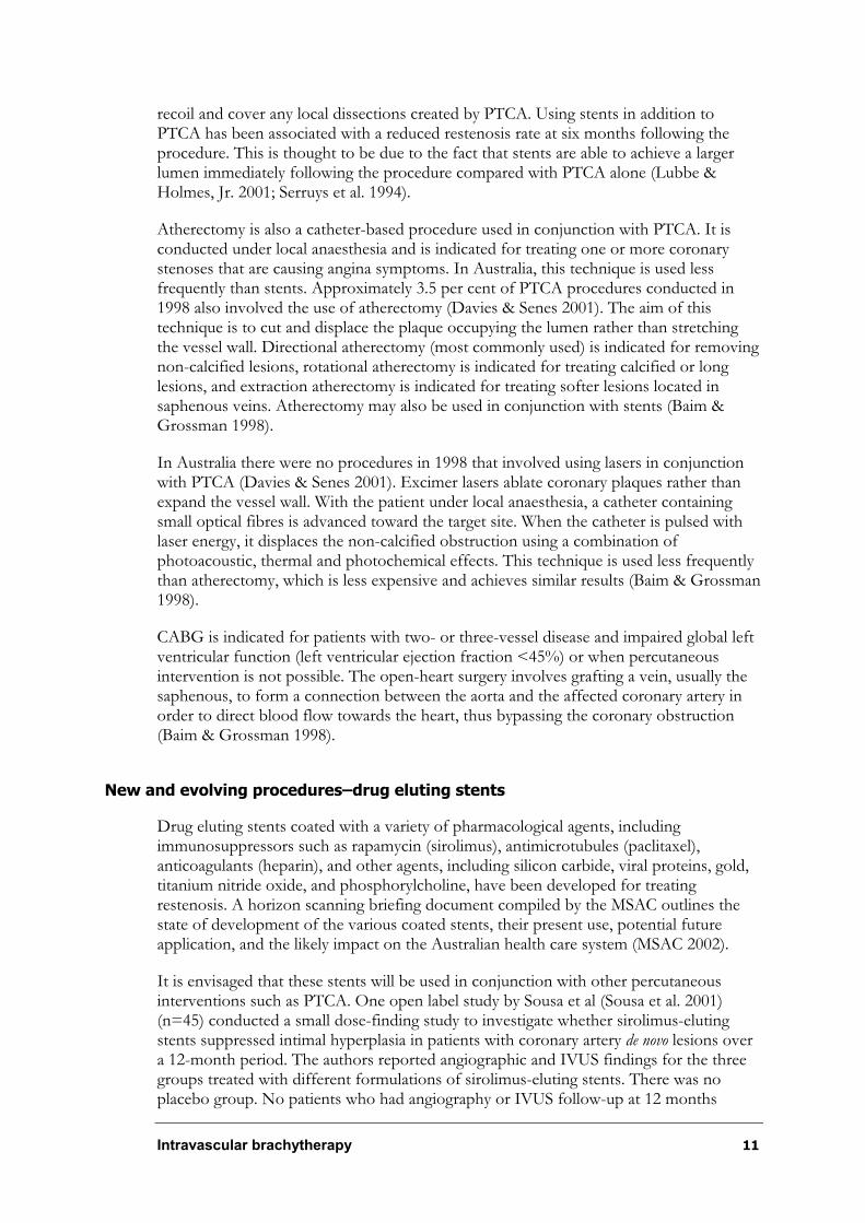
In 1998, 17,448 coronary artery bypass graft operations (CABG) were performed in Australia (Australian Institute of Health and Welfare 2000b). In the same period, 18,094 PTCA procedures were performed, 82 per cent of which also involved stent placement. Expert opinion suggests that this may now be as high as 90 per cent of patients (Personal communication: Dr Leo Mahar, face-to-face 7th Febrary 2002). Approximately 20 per cent of the PTCA procedures were repeats, half of which occurred between 24 hours and 3 months post-operatively. The majority of the remaining repeat procedures occurred within 3 to 6 months, with only about 10 per cent occurring between 6 and 12 months. Table 2 outlines the coronary interventions undertaken in Australia in 1998.

10 Intravascular brachytherapy
Table 2 Coronary interventions in 1998a
Procedure ICD-9-CM codes ICD-10-AM codes Total Number of procedures
Coronary artery bypass
36.1 Block 672
Codes 38497-00
38497-01
38497-02
38497-03
Block 673
Codes 38497-04
Block 674
Codes 38500-00
38503-00
17,448
Percutaneous transluminal coronary angioplasty (PTCA)
36.01
36.02
36.05
Block 670
Codes 35304-00
35305-00
(plus stenting codes below)
18,094
Stentingb 36.06
36.07
Block 671
Codes 35310-00
35310-01
35310-02
14,838c
Coronary angiography
88.55
88.56
88.57
Block 668
Codes 38215-00
38218-00
38218-01
38218-02
77,244
a Number of procedures for all interventional cardiology units in Australia, based on data from the AIHW National Hospital Morbidity Database (Australian Institute of Health and Welfare 2000b).
b These form a subset of the PTCA procedures and costs. c Patients rather than procedures.
Existing procedures
Procedures that are currently used to treat coronary artery atherosclerotic lesions include PTCA, stents, atherectomy, excimer laser, and CABG.
PTCA is indicated for the treatment of one or more coronary stenoses that can be reached by a catheter. The patient usually presents with moderate to severe chronic stable angina. The procedure is conducted under local anaesthesia and requires the patient to remain in hospital for an average of one to three days. A catheter loaded with an inflatable balloon is inserted into the target coronary artery, usually via the femoral artery and advanced to the target site. Radiopaque markers are used as an aid to correct positioning of the balloon. The balloon is then inflated to a size that will sufficiently stretch the vessel wall, widening the lumen. Repeated balloon inflation may be conducted until appropriate lumen patency is achieved. Once the procedure is completed the balloon is deflated and the catheter removed (Baim & Grossman 1998).
In Australia, expert opinion suggests that approximately 90 per cent of PTCA procedures also involve the addition of stents (Personal Communication: Dr. Leo Mahar, face-to-face, 7th Febrary 2002). These are metallic scaffolds that can be expanded to a specific size once positioned at the target site by a catheter. Stents help to prevent vessel elastic
Intravascular brachytherapy 11
recoil and cover any local dissections created by PTCA. Using stents in addition to PTCA has been associated with a reduced restenosis rate at six months following the procedure. This is thought to be due to the fact that stents are able to achieve a larger lumen immediately following the procedure compared with PTCA alone (Lubbe & Holmes, Jr. 2001; Serruys et al. 1994).
Atherectomy is also a catheter-based procedure used in conjunction with PTCA. It is conducted under local anaesthesia and is indicated for treating one or more coronary stenoses that are causing angina symptoms. In Australia, this technique is used less frequently than stents. Approximately 3.5 per cent of PTCA procedures conducted in 1998 also involved the use of atherectomy (Davies & Senes 2001). The aim of this technique is to cut and displace the plaque occupying the lumen rather than stretching the vessel wall. Directional atherectomy (most commonly used) is indicated for removing non-calcified lesions, rotational atherectomy is indicated for treating calcified or long lesions, and extraction atherectomy is indicated for treating softer lesions located in saphenous veins. Atherectomy may also be used in conjunction with stents (Baim & Grossman 1998).
In Australia there were no procedures in 1998 that involved using lasers in conjunction with PTCA (Davies & Senes 2001). Excimer lasers ablate coronary plaques rather than expand the vessel wall. With the patient under local anaesthesia, a catheter containing small optical fibres is advanced toward the target site. When the catheter is pulsed with laser energy, it displaces the non-calcified obstruction using a combination of photoacoustic, thermal and photochemical effects. This technique is used less frequently than atherectomy, which is less expensive and achieves similar results (Baim & Grossman 1998).
CABG is indicated for patients with two- or three-vessel disease and impaired global left ventricular function (left ventricular ejection fraction <45%) or when percutaneous intervention is not possible. The open-heart surgery involves grafting a vein, usually the saphenous, to form a connection between the aorta and the affected coronary artery in order to direct blood flow towards the heart, thus bypassing the coronary obstruction (Baim & Grossman 1998).
New and evolving procedures–drug eluting stents
Drug eluting stents coated with a variety of pharmacological agents, including immunosuppressors such as rapamycin (sirolimus), antimicrotubules (paclitaxel), anticoagulants (heparin), and other agents, including silicon carbide, viral proteins, gold, titanium nitride oxide, and phosphorylcholine, have been developed for treating restenosis. A horizon scanning briefing document compiled by the MSAC outlines the state of development of the various coated stents, their present use, potential future application, and the likely impact on the Australian health care system (MSAC 2002).
It is envisaged that these stents will be used in conjunction with other percutaneous interventions such as PTCA. One open label study by Sousa et al (Sousa et al. 2001) (n=45) conducted a small dose-finding study to investigate whether sirolimus-eluting stents suppressed intimal hyperplasia in patients with coronary artery de novo lesions over a 12-month period. The authors reported angiographic and IVUS findings for the three groups treated with different formulations of sirolimus-eluting stents. There was no placebo group. No patients who had angiography or IVUS follow-up at 12 months
12 Intravascular brachytherapy
(n=30) presented with stenosis greater than or equal to 50 per cent of the diameter. IVUS results showed minimal development of neointimal hyperplasia for the three groups. Apart from 1 patient experiencing a thrombotic event at 14 months post-procedure, no other clinical events were reported for 29 patients at 15 months, and for 14 patients at 9 months. While this data appears promising, there is insufficient evidence to assess the long-term impact drug eluting stents may have on the treatment of coronary restenosis.
The Horizon Scanning Briefing document concluded that, while drug-eluting stents appear to be a promising new technology, further evidence is still required on their relative effectiveness and safety compared with current coronary interventions to allow assessment of their cost effectiveness.
Comparator
In coronary artery disease, IVB is intended for use in addition to other percutaneous intervention procedures such as PTCA, stenting, atherectomy and/or excimer laser to treat atherosclerotic lesions and prevent restenosis. The safety and effectiveness of IVB in addition to PTCA, stenting, atherectomy and/or excimer laser will be compared with PTCA, stents and/or atherectomy alone. The flow chart in Appendix D outlines the potential comparators for IVB.
Marketing status of the device/technology
The following two IVB systems are listed on the Australian Register of Therapeutic Goods (ARTG) with the Therapeutic Goods Administration (TGA).
The Galileoã Intravascular Radiotherapy System ARTG listing numbers are:
¶ AUST L 74073
¶ AUST L 74520
¶ AUST L 23159
The NovosteÑ Beta-Cathã Intracoronary Radiation System ARTG listing numbers are:
¶ AUST L 69009
¶ AUST L 69087
Current reimbursement arrangement
The Galileoã Intravascular Radiotherapy System is not currently funded under the Medical Benefits Scheme.
No other intravascular brachytherapy systems are funded on the Medicare Benefits Schedule.
Intravascular brachytherapy 13
Approach to assessment
Research questions
The review team worked with members of the supporting committee to develop specific questions addressing the use of IVB the treating coronary artery restenosis. These questions were formulated a priori from information on current practice (ie patterns of usage of IVB in Australia), the disease area and the purpose of the device (eg treatment of coronary artery restenosis). A flow chart (Appendix D) depicting the clinical pathways for treating coronary artery restenosis was developed in conjunction with the supporting committee. This flow chart was used to define the potential role of IVB in the treatment of coronary artery in-stent restenosis. The supporting committee decided that this review would focus on the use of IVB for treating in-stent restenosis rather than for treating denovo lesions, as these patients were likely to reflect Australian clinical practice should the technology become available. Current information about and evidence for treating de novolesions is limited and is predominantly based on uncontrolled case series. Furthermore, the supporting committee decided that evaluating the evidence for treatment of restenosis was more important as restenosis is a greater clinical concern, given the paucity of effective treatment measures at this stage. Based on this flow chart, two questions were developed and are covered in this report:
¶ What is the value of catheter-based IVB in addition to percutaneous intervention in the treatment of patients with in-stent restenosis following previous coronary interventions compared with percutaneous intervention only?
¶ What is the value of radioactive stents in addition to percutaneous intervention in the treatment of patients with in-stent restenosis following previous coronary interventions compared with percutaneous intervention only? As the use of radioactive stents is expected to be quite limited in clinical practice, this question is included for the sake of completeness, although the lower priority of radioactive stents should be noted.
Review of literature
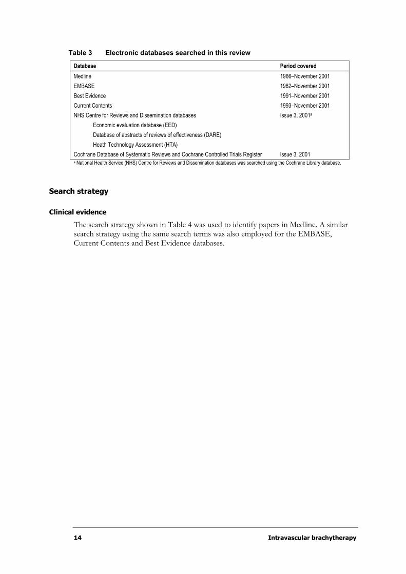
The MSAC’s recommendations are primarily based on the findings of a systematic literature review conducted by the National Health and Medical Research Council (NHMRC) Clinical Trials Centre (CTC). Papers were also identified from the MSAC application and by members of the MSAC IVB supporting committee (Appendix B) that was convened to evaluate the evidence and provide expert advice. The medical literature was searched to identify relevant studies and reviews for the period between 1966 and November 2001. Following a request by the supporting committee to include the results of the pre-published START trial, the search strategy was repeated in April 2002 to check for any newly published randomised controlled trials; however, no further studies were retrieved. Searches were conducted via electronic databases, as listed in Table 3.
14 Intravascular brachytherapy
Table 3 Electronic databases searched in this review
Database Period covered
Medline 1966–November 2001
EMBASE 1982–November 2001
Best Evidence 1991–November 2001
Current Contents 1993–November 2001
NHS Centre for Reviews and Dissemination databases
Economic evaluation database (EED)
Database of abstracts of reviews of effectiveness (DARE)
Heath Technology Assessment (HTA)
Issue 3, 2001a
Cochrane Database of Systematic Reviews and Cochrane Controlled Trials Register Issue 3, 2001 a National Health Service (NHS) Centre for Reviews and Dissemination databases was searched using the Cochrane Library database.
Search strategy
Clinical evidence
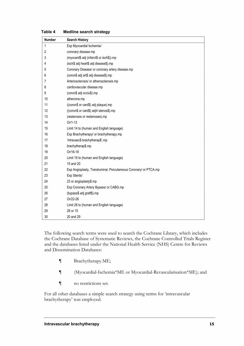
The search strategy shown in Table 4 was used to identify papers in Medline. A similar search strategy using the same search terms was also employed for the EMBASE, Current Contents and Best Evidence databases.
Intravascular brachytherapy 15
Table 4 Medline search strategy
Number Search History
1 Exp Myocardial Ischemia/
2 coronary disease.mp
3 (myocard$ adj (infarct$ or isch$)).mp
4 (isch$ adj heart$ adj disease$).mp
5 Coronary Disease/ or coronary artery disease.mp
6 (coron$ adj art$ adj disease$).mp
7 Arteriosclerosis/ or atherosclerosis.mp
8 cardiovascular disease.mp
9 (coron$ adj occlu$).mp
10 atheroma.mp
11 ((coron$ or card$) adj plaque).mp
12 ((coron$ or card$) adj4 stenos$).mp
13 (restenosis or restenoses).mp
14 Or/1-13
15 Limit 14 to (human and English language)
16 Exp Brachytherapy/ or brachytherapy.mp
17 ‘intravasc$ brachytherap$’.mp
18 brachytherap$.mp
19 Or/16-18
20 Limit 19 to (human and English language)
21 15 and 20
22 Exp Angioplasty, Transluminal, Percutaneous Coronary/ or PTCA.mp
23 Exp Stents/
24 23 or angioplasty$.mp
25 Exp Coronary Artery Bypass/ or CABG.mp
26 (bypass$ adj graft$).mp
27 Or/22-26
28 Limit 28 to (human and English language)
29 28 or 15
30 20 and 29
The following search terms were used to search the Cochrane Library, which includes the Cochrane Database of Systematic Reviews, the Cochrane Controlled Trials Register and the databases listed under the National Health Service (NHS) Centre for Reviews and Dissemination Databases:
¶ Brachytherapy.ME;
¶ (Myocardial-Ischemia*ME or Myocardial-Revascularisation*ME); and
¶ no restrictions set.
For all other databases a simple search strategy using terms for ‘intravascular brachytherapy’ was employed.
16 Intravascular brachytherapy
A list of abstracts provided by the applicant in the form of an endnote database was also compared with our search, and non-duplicate references were included in the final reference list.
Reference lists of publications were also searched for additional relevant citations that may have been inadvertently missed in searches of major databases.
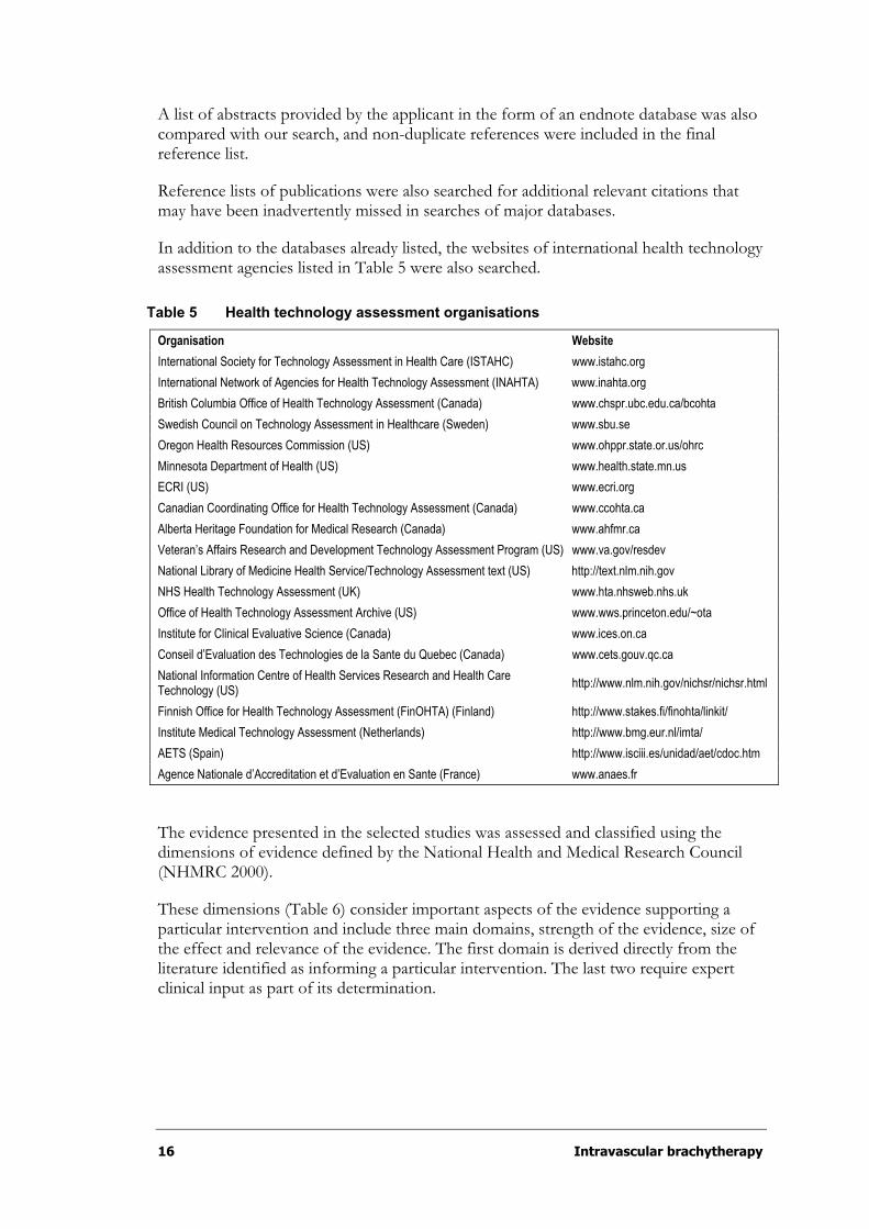
In addition to the databases already listed, the websites of international health technology assessment agencies listed in Table 5 were also searched.
Table 5 Health technology assessment organisations
Organisation Website
International Society for Technology Assessment in Health Care (ISTAHC) www.istahc.org
International Network of Agencies for Health Technology Assessment (INAHTA) www.inahta.org
British Columbia Office of Health Technology Assessment (Canada) www.chspr.ubc.edu.ca/bcohta
Swedish Council on Technology Assessment in Healthcare (Sweden) www.sbu.se
Oregon Health Resources Commission (US) www.ohppr.state.or.us/ohrc
Minnesota Department of Health (US) www.health.state.mn.us
ECRI (US) www.ecri.org
Canadian Coordinating Office for Health Technology Assessment (Canada) www.ccohta.ca
Alberta Heritage Foundation for Medical Research (Canada) www.ahfmr.ca
Veteran’s Affairs Research and Development Technology Assessment Program (US) www.va.gov/resdev
National Library of Medicine Health Service/Technology Assessment text (US) http://text.nlm.nih.gov
NHS Health Technology Assessment (UK) www.hta.nhsweb.nhs.uk
Office of Health Technology Assessment Archive (US) www.wws.princeton.edu/~ota
Institute for Clinical Evaluative Science (Canada) www.ices.on.ca
Conseil d’Evaluation des Technologies de la Sante du Quebec (Canada) www.cets.gouv.qc.ca
National Information Centre of Health Services Research and Health Care Technology (US)
http://www.nlm.nih.gov/nichsr/nichsr.html
Finnish Office for Health Technology Assessment (FinOHTA) (Finland) http://www.stakes.fi/finohta/linkit/
Institute Medical Technology Assessment (Netherlands) http://www.bmg.eur.nl/imta/
AETS (Spain) http://www.isciii.es/unidad/aet/cdoc.htm
Agence Nationale d’Accreditation et d’Evaluation en Sante (France) www.anaes.fr
The evidence presented in the selected studies was assessed and classified using the dimensions of evidence defined by the National Health and Medical Research Council (NHMRC 2000).
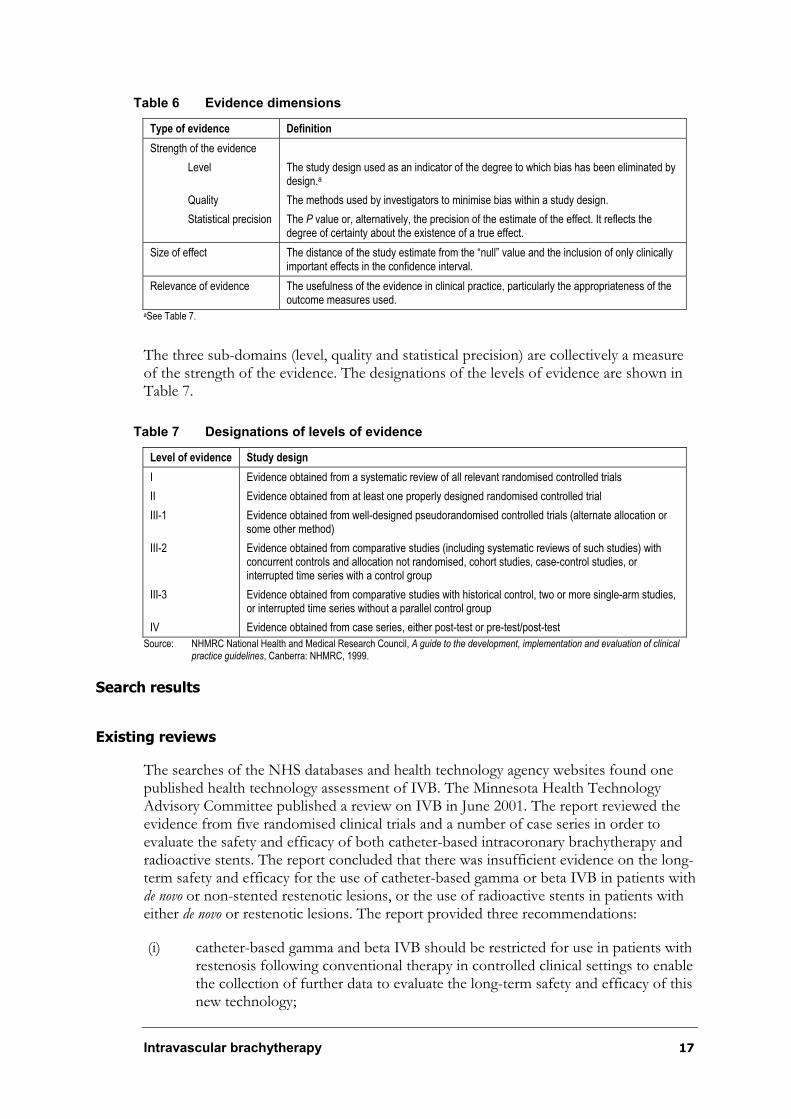
These dimensions (Table 6) consider important aspects of the evidence supporting a particular intervention and include three main domains, strength of the evidence, size of the effect and relevance of the evidence. The first domain is derived directly from the literature identified as informing a particular intervention. The last two require expert clinical input as part of its determination.
Intravascular brachytherapy 17
Table 6 Evidence dimensions
Type of evidence Definition
Strength of the evidence
Level
Quality
Statistical precision
The study design used as an indicator of the degree to which bias has been eliminated by design.a
The methods used by investigators to minimise bias within a study design.
The P value or, alternatively, the precision of the estimate of the effect. It reflects the degree of certainty about the existence of a true effect.
Size of effect The distance of the study estimate from the “null” value and the inclusion of only clinically important effects in the confidence interval.
Relevance of evidence The usefulness of the evidence in clinical practice, particularly the appropriateness of the outcome measures used.
aSee Table 7.
The three sub-domains (level, quality and statistical precision) are collectively a measure of the strength of the evidence. The designations of the levels of evidence are shown in Table 7.
Table 7 Designations of levels of evidence
Level of evidence Study design
I
II
III-1
III-2
III-3
IV
Evidence obtained from a systematic review of all relevant randomised controlled trials
Evidence obtained from at least one properly designed randomised controlled trial
Evidence obtained from well-designed pseudorandomised controlled trials (alternate allocation or some other method)
Evidence obtained from comparative studies (including systematic reviews of such studies) with concurrent controls and allocation not randomised, cohort studies, case-control studies, or interrupted time series with a control group
Evidence obtained from comparative studies with historical control, two or more single-arm studies, or interrupted time series without a parallel control group
Evidence obtained from case series, either post-test or pre-test/post-test
Source: NHMRC National Health and Medical Research Council, A guide to the development, implementation and evaluation of clinical practice guidelines, Canberra: NHMRC, 1999.
Search results
Existing reviews
The searches of the NHS databases and health technology agency websites found one published health technology assessment of IVB. The Minnesota Health Technology Advisory Committee published a review on IVB in June 2001. The report reviewed the evidence from five randomised clinical trials and a number of case series in order to evaluate the safety and efficacy of both catheter-based intracoronary brachytherapy and radioactive stents. The report concluded that there was insufficient evidence on the long-term safety and efficacy for the use of catheter-based gamma or beta IVB in patients with de novo or non-stented restenotic lesions, or the use of radioactive stents in patients with either de novo or restenotic lesions. The report provided three recommendations:
(i) catheter-based gamma and beta IVB should be restricted for use in patients with restenosis following conventional therapy in controlled clinical settings to enable the collection of further data to evaluate the long-term safety and efficacy of this new technology;
18 Intravascular brachytherapy
(ii) radioactive stents should only be used in clinical trials; and
(iii) neither catheter-based brachytherapy nor radioactive stents are recommended for patients with de novo or non-stented lesions.
Published literature
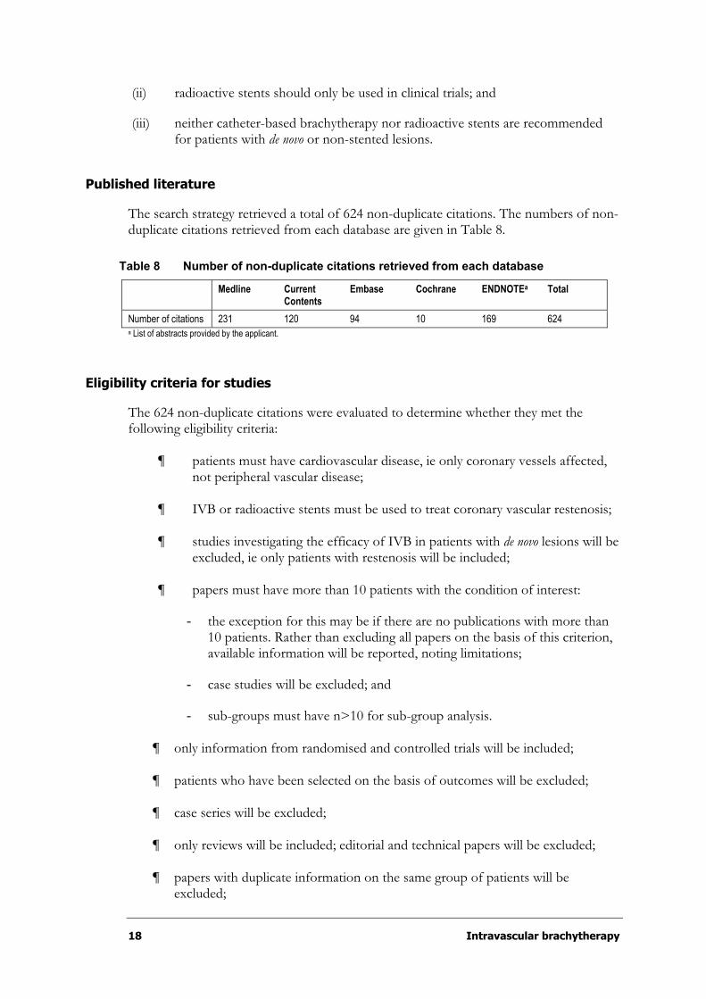
The search strategy retrieved a total of 624 non-duplicate citations. The numbers of non-duplicate citations retrieved from each database are given in Table 8.
Table 8 Number of non-duplicate citations retrieved from each database
Medline Current Contents
Embase Cochrane ENDNOTEa Total
Number of citations 231 120 94 10 169 624 a List of abstracts provided by the applicant.
Eligibility criteria for studies
The 624 non-duplicate citations were evaluated to determine whether they met the following eligibility criteria:
¶ patients must have cardiovascular disease, ie only coronary vessels affected, not peripheral vascular disease;
¶ IVB or radioactive stents must be used to treat coronary vascular restenosis;
¶ studies investigating the efficacy of IVB in patients with de novo lesions will be excluded, ie only patients with restenosis will be included;
¶ papers must have more than 10 patients with the condition of interest:
- the exception for this may be if there are no publications with more than 10 patients. Rather than excluding all papers on the basis of this criterion, available information will be reported, noting limitations;
- case studies will be excluded; and
- sub-groups must have n>10 for sub-group analysis.
¶ only information from randomised and controlled trials will be included;
¶ patients who have been selected on the basis of outcomes will be excluded;
¶ case series will be excluded;
¶ only reviews will be included; editorial and technical papers will be excluded;
¶ papers with duplicate information on the same group of patients will be excluded;

Intravascular brachytherapy 19
¶ data available in abstract form only will be excluded;
¶ papers which report no clinical results will be excluded;
¶ all non-English papers will be excluded;
¶ animal studies will be excluded; and
¶ where these criteria could not be evaluated from the abstract, full papers were examined.
These criteria were also used to evaluate full papers.
Based on these criteria, 606 papers (97%) were excluded from this review. The reasons for exclusion are listed in Table 9.
Table 9 Reasons for exclusion
Reason for exclusion Frequency (%)a
Non-controlled evidence on efficacy of intravascular brachytherapy on coronary restenosis
26 (4.2)
Not cardiovascular disease 135 (21.6)
Not intravascular brachytherapy 85 (13.6)
Efficacy of intravascular brachytherapy in peripheral vessels 9 (1.4)
Efficacy of intravascular brachytherapy in de novo coronary lesions (controlled studies)
2 (0.3)
Papers that included duplicate information on same patient groups 3 (0.5)
Reviews on intravascular brachytherapy 104 (16.7)
Technical documents on intravascular brachytherapy 85 (13.6 )
Editorials/letters on intravascular brachytherapy 51 (8.2)
Abstracts on intravascular brachytherapy 32 (5.1)
Case series/studies (n¢10) of intravascular brachytherapy 21 (3.4)
Animal studies of intravascular brachytherapy 30 (4.8)
Laboratory studies of intravascular brachytherapy 4 (0.6)
Studies of intravascular brachytherapy non-English language 8 (1.3)
Other 11 (1.8)
Total 606 (97.1) a Percentage of frequency is calculated as a percentage of the total 624 abstracts retrieved.
The information from 14 studies (20 papers) were included in this review and are listed in Table 10. The number of papers retrieved does not represent the number of individual trials, as often a number of papers will report the results of different outcome measures of a single study. Therefore, the number of individual trials is less than the number of papers reported. According to the NHMRC Levels of Evidence, eight studies (13 papers) were classified Level II evidence; six studies (seven papers) were classified Level III-3 evidence.

20 Intravascular brachytherapy
Table 10 Design characteristics of relevant studies
NHMRC Levels of Evidence Trials No of papers (%)a
Catheter-based IVB
Level II SCRIPPS 3
WRIST 3
GAMMA-1 2
PREVENT 1
Costa et al (2000) 1
Schühlen et al (2001) 1
INHIBIT 1
START 1
Subtotal 8 13 (2.1)
Level III-3 Long WRIST 1
High Dose (HD) WRIST 1
WRIST Plus 1
Beta WRIST 2
Subtotal 4 5 (0.8)
Radioactive stents
Level III-3 Albiero et al (2000a) 1
Albiero et al (2000b) 1
Subtotal 2 2 (0.3)
Total 14 20 (3.2) a Frequency is calculated as a percentage of the total 624 abstracts retrieved.
Expert advice
A supporting committee with expertise in cardiology, nuclear physics and radiation oncology was established to evaluate the evidence and provide advice to the MSAC from a clinical perspective. In selecting members for supporting committees, the MSAC’s practice is to approach the appropriate medical colleges, specialist societies and associations, and consumer bodies for nominees. Membership of the supporting committee is provided in Appendix B.
Overview of review structure
This review assesses the safety and effectiveness of radioactive stents and catheter-based IVB for the treatment of coronary artery in-stent restenosis. As the supporting committee decided that it was more important to focus on evaluating the evidence for catheter-based IVB, the evidence pertaining to radioactive stents is outlined briefly at the beginning of the ‘Results of assessment’ section.
The safety section for catheter-based IVB reports on a number of safety issues that may potentially be associated with the use of gamma or beta IVB. These issues include dosimetry, environmental exposure issues, late thrombosis and/or late total occlusion, edge restenosis and other late adverse events.

Intravascular brachytherapy 21
The effectiveness section for catheter-based IVB examines the efficacy of gamma and beta IVB separately by reporting on a number of clinical, angiographic and IVUS outcome measures.
All the values reported in this review are given as mean (°SD, standard deviation) unless stated otherwise.
22 Intravascular brachytherapy
Results of assessment
Radioactive/radioisotope stents
Potential role of radioactive stents
The rationale behind the use of radioisotope stents relates to the relative ease with which this technique may be used. As most patients with restenosis will be treated with stents, a procedure that combines stenting with delivery of radiation for preventing further in-stent restenosis in one step is potentially useful. Fischell (1998) indicates that the radioisotope stent may have the following potential advantages over catheter-based radiation delivery systems:
¶ the ability to deliver therapeutic treatment using pure beta (b) emitters with a much lower radioactivity compared to catheter-based sources (eg µCi vs mCi activity);
¶ lack of requirements for in-lab dosimetry calculations;
¶ homogeneous dose delivery along the length of stent; and
¶ time efficiency due to elimination of the catheter-based radiation delivery procedure.
Despite these potential advantages, the use of radioisotope stents is not as popular as might be expected. This is likely related to the occurrence of edge restenosis. Compared to catheter-based radiation therapy, there is only limited information on which to base an evaluation of the role of radioactive stents in preventing restenosis. Unfortunately, most patients in these studies had de novo lesions, so the results reported may not be directly applicable to patients who are treated for existing restenosis. Neither study has a true control group or was randomised. Rather, they compare varying doses of radiation, with no information provided as to how patients were allocated to each of the dose levels. In both cases, follow-up was only six months, with no data provided on longer term outcomes. The two studies are:
¶ Albiero et al (2000a) (Level III-3); and
¶ Albiero et al (2000b) (Level III-3).
Albiero et al (2000a)
Albiero et al (2000a) (n=82) conducted a non-randomised, single-centre, dose response study between October 1997 and October 1998 to evaluate the safety and efficacy of Phosphorus-32 (32P) radioactive stents for the prevention of restenosis at four and six month follow-up. This trial was not randomised and does not provide any data to indicate how patients were allocated to treatment groups. It also does not provide any information as to whether patients were recruited in a consecutive or selective manner. As a result, the influence of selection bias cannot be excluded. Inclusion criteria for enrolment in the study were the presence of a de novo or restenotic lesion of a major,
Intravascular brachytherapy 23
native coronary artery with a reference artery size visually estimated to be appropriate for the available stent diameters (3.0–3.5mm). The lesion had be treated with one or two tandem stents with a target lesion length visually estimated to be less than or equal to 28mm. Two types of stents (Fischell Isostent) were implanted. Initially, the Palmaz-Schatz stent with activity of 0.75 to 3.0 µCi (Group 1, n=23 patients, 27 lesions, 31 stents), and later the BX stent with higher activity level of 3.0 to 6.0µCi (Group 2, n=29 patients, 32 lesions, 39 stents) and activity level of 6.0 to 12.0µCi (Group 3, n=30 patients, 32 lesions, 53 stents). All patients received 325mg of aspirin daily (continued long-term) plus ticlopidine (250mg bid) for three months after the procedure. All patients were requested to return for clinical, angiographic and IVUS follow-up at four to six months after the procedure. There was no difference in the baseline clinical characteristics between groups, with the exception that Group 3 had a lower incidence of hypertension. More than 90 per cent of lesions treated were de novo lesions, so the applicability of data derived from this study to those patients with restenosis remains unclear.
Albiero et al (2000b)
This study reported a high restenosis rate at the edges of the 32P radioactive stents (activity 3–12µCi). The aim of this subsequent study was to determine whether higher activity stents (12–21µCi), combined with a non-aggressive stenting strategy to prevent balloon induced injury might prevent the edge restenosis. The study was not randomised, and it is unclear whether patients were recruited consecutively in a prospective manner or retrospectively. As the authors report that angiographic results of all lesions treated between October 1998 and April 1999 were reviewed, it suggests that this was a retrospective comparison. This study used a subset of patients from Albiero et al (2000a) as a ‘historical control’ (Group 1) and compared them to patients treated with stents of higher radioactivity deployed in a less aggressive manner (Group 2). The patients in Group 1 were selected on the basis of whether lesions were treated with only a single stent per lesion, although a patient could have more than one lesion treated with single stents. Patients from Albiero et al (2000a) that were treated with more than one stent were excluded from the ‘control’ group (n=17 patients, 22 lesions). Group 1 comprised 40 patients with 42 lesions previously treated with radioactive stents with an activity of between 3 and 12µCi. Group 2 comprised 40 patients with 54 lesions treated with a single radioactive stent per lesion with an activity of 12 to 21µCi. Nineteen patients with 22 lesions treated with less than 1 stent per lesion were excluded from Group 2. Post-treatment medication for Group 1 is as described above, while Group 2 were treated with long-term aspirin (325mg daily) plus either ticlopidine (250mg bid) or clopidogrel (75mg daily) for at least three months. Patients in Group 2 were requested to return for clinical, angiographic and IVUS follow-up at six months after the procedure. There was no difference in the baseline clinical characteristics between groups. More than 90 per cent of lesions treated in Group 1 and almost 80 per cent in Group 2 were de novo lesions, so the applicability of data derived from this study to those patients with restenosis remains unclear. Target lesion revascularisation (TLR) was performed in all patients with angiographic restenosis, regardless of whether patients were asymptomatic or had no objective evidence of ischaemia. It is therefore likely that TLR rates are an overestimate of the true number of patients who might require TLR based on ischaemic symptoms in a clinical setting.
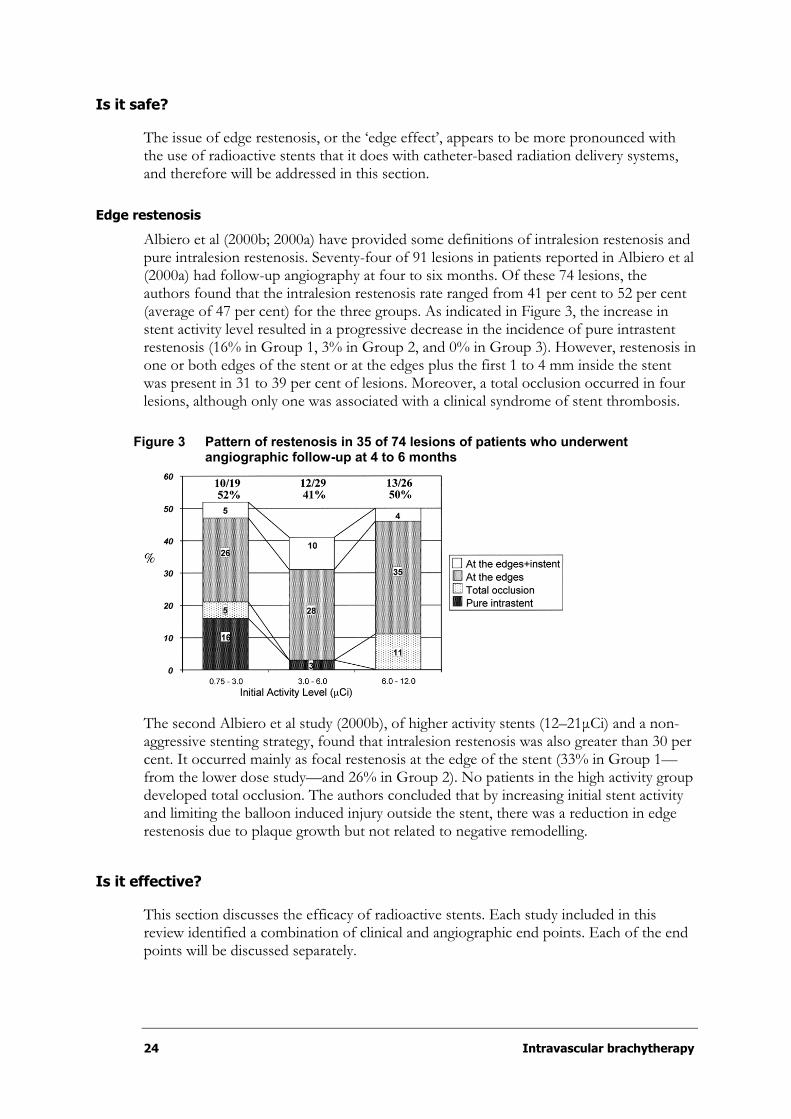
24 Intravascular brachytherapy
Is it safe?
The issue of edge restenosis, or the ‘edge effect’, appears to be more pronounced with the use of radioactive stents that it does with catheter-based radiation delivery systems, and therefore will be addressed in this section.
Edge restenosis
Albiero et al (2000b; 2000a) have provided some definitions of intralesion restenosis and pure intralesion restenosis. Seventy-four of 91 lesions in patients reported in Albiero et al (2000a) had follow-up angiography at four to six months. Of these 74 lesions, the authors found that the intralesion restenosis rate ranged from 41 per cent to 52 per cent (average of 47 per cent) for the three groups. As indicated in Figure 3, the increase in stent activity level resulted in a progressive decrease in the incidence of pure intrastent restenosis (16% in Group 1, 3% in Group 2, and 0% in Group 3). However, restenosis in one or both edges of the stent or at the edges plus the first 1 to 4 mm inside the stent was present in 31 to 39 per cent of lesions. Moreover, a total occlusion occurred in four lesions, although only one was associated with a clinical syndrome of stent thrombosis.
Figure 3 Pattern of restenosis in 35 of 74 lesions of patients who underwent angiographic follow-up at 4 to 6 months
The second Albiero et al study (2000b), of higher activity stents (12–21µCi) and a non-aggressive stenting strategy, found that intralesion restenosis was also greater than 30 per cent. It occurred mainly as focal restenosis at the edge of the stent (33% in Group 1—from the lower dose study—and 26% in Group 2). No patients in the high activity group developed total occlusion. The authors concluded that by increasing initial stent activity and limiting the balloon induced injury outside the stent, there was a reduction in edge restenosis due to plaque growth but not related to negative remodelling.
Is it effective?
This section discusses the efficacy of radioactive stents. Each study included in this review identified a combination of clinical and angiographic end points. Each of the end points will be discussed separately.

Intravascular brachytherapy 25
Clinical outcome measures
Survival
In the four to six month follow-up period for Albiero et al (2000a), no deaths were reported in the 82 patients across all three treatment groups (0.75–12.0µCi). Also, no deaths were reported in the patients treated with radioisotope stents of higher activity (12.0–21.0µCi) (Albiero et al. 2000b).
Major Adverse Cardiac Events (MACE)
Albiero et al (2000b; 2000a) defined major adverse cardiac events (MACE) as death, MI (Q-wave or non-Q-wave) and stent thrombosis. This definition is different from that used for MACE in many of the catheter-based radiation trials. Despite defining MACE, the authors have not reported this endpoint as a combined outcome, but they have reported rates of the individual events.
Myocardial infarction (MI)
Albiero et al (2000a) report that one patient in Group 3 (6.0–12.0µCi activity) experienced a sub-acute thrombosis with a Q-wave MI one week after he ceased aspirin and ticlopidine, three months after the stenting procedure. No further information is provided. No patients in the group treated with higher activity stents (12.0–21.0µCi) experienced a MI during the six months of follow-up (Albiero et al. 2000b).
Target lesion revascularisation (TLR)
Albiero et al (2000b; 2000a) indicated that a repeat percutaneous coronary intervention was performed in all the lesions with angiographic restenosis even if the patients were asymptomatic and had no objective evidence of ischaemia. This means that the rate of TLR is likely to be an overestimate of the true number of patients who would require re-intervention based on clinical symptoms. This data therefore may not be comparable to that reported in the studies of catheter-based radiation delivery systems. Table 11 summarises data on TLR reported by Albiero et al (2000b; 2000a).
Table 11 Target lesion revascularisation (TLR) for radioactive stents at six months
Albiero (2000a) Albiero (2000b)
Group 1
0.75–3.0 µCi
Group 2
3.0–6.0µCi
Group 3
6.0–12.0µCi
Group 1a
3.0–12.0µCi
Group 2
12.0–21.0µCi
Number of patients 23 29 30 40 40
CABG, n (%) 0 – 1 (3.4) 2 (6.6) NR – NR –
Repeat PTCA/number of lesions (%) 10/19 (52) 12/29 (41) 13/26 (50) – – – –
Any repeat revascularisation/ number of lesions (%) 10/19 (52) 13/30 (43) 14/27 (52) – 38* – 30*
* Per cent of lesion; no further data given. a Group 1 is a subset of patients with lesions treated with a single stent from the Albiero et al (2000a) trial.
Angiographic outcome measures
Data for quantitative angiographic restenosis (²50% of lumen diameter) rates at four to six month follow-up are detailed in Table 12.
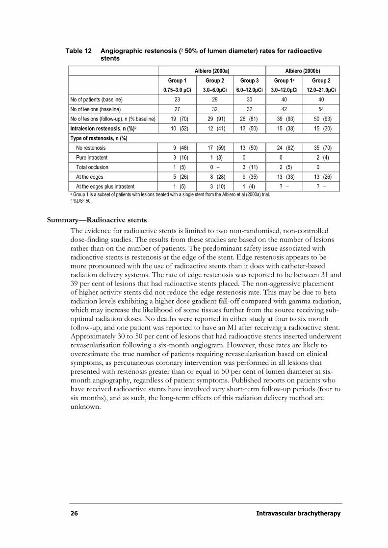
26 Intravascular brachytherapy
Table 12 Angiographic restenosis (²50% of lumen diameter) rates for radioactive stents
Albiero (2000a) Albiero (2000b)
Group 1
0.75–3.0 µCi
Group 2
3.0–6.0µCi
Group 3
6.0–12.0µCi
Group 1a
3.0–12.0µCi
Group 2
12.0–21.0µCi
No of patients (baseline) 23 29 30 40 40
No of lesions (baseline) 27 32 32 42 54
No of lesions (follow-up), n (% baseline) 19 (70) 29 (91) 26 (81) 39 (93) 50 (93)
Intralesion restenosis, n (%)b 10 (52) 12 (41) 13 (50) 15 (38) 15 (30)
Type of restenosis, n (%)
No restenosis 9 (48) 17 (59) 13 (50) 24 (62) 35 (70)
Pure intrastent 3 (16) 1 (3) 0 0 2 (4)
Total occlusion 1 (5) 0 – 3 (11) 2 (5) 0
At the edges 5 (26) 8 (28) 9 (35) 13 (33) 13 (26)
At the edges plus intrastent 1 (5) 3 (10) 1 (4) ? – ? – a Group 1 is a subset of patients with lesions treated with a single stent from the Albiero et al (2000a) trial. b %DS²50.
Summary—Radioactive stents
The evidence for radioactive stents is limited to two non-randomised, non-controlled dose-finding studies. The results from these studies are based on the number of lesions rather than on the number of patients. The predominant safety issue associated with radioactive stents is restenosis at the edge of the stent. Edge restenosis appears to be more pronounced with the use of radioactive stents than it does with catheter-based radiation delivery systems. The rate of edge restenosis was reported to be between 31 and 39 per cent of lesions that had radioactive stents placed. The non-aggressive placement of higher activity stents did not reduce the edge restenosis rate. This may be due to beta radiation levels exhibiting a higher dose gradient fall-off compared with gamma radiation, which may increase the likelihood of some tissues further from the source receiving sub-optimal radiation doses. No deaths were reported in either study at four to six month follow-up, and one patient was reported to have an MI after receiving a radioactive stent. Approximately 30 to 50 per cent of lesions that had radioactive stents inserted underwent revascularisation following a six-month angiogram. However, these rates are likely to overestimate the true number of patients requiring revascularisation based on clinical symptoms, as percutaneous coronary intervention was performed in all lesions that presented with restenosis greater than or equal to 50 per cent of lumen diameter at six-month angiography, regardless of patient symptoms. Published reports on patients who have received radioactive stents have involved very short-term follow-up periods (four to six months), and as such, the long-term effects of this radiation delivery method are unknown.
Intravascular brachytherapy 27
Catheter-based intravascular brachytherapy
Potential role of catheter-based intravascular brachytherapy
In coronary artery disease, IVB is intended to be used in addition to other percutaneous interventions such as PTCA, atherectomy, excimer laser, and stents in order to treat atherosclerotic lesions and prevent restenosis. Once a target lesion has been treated with IVB, subsequent irradiation of the same lesion is not possible. The flow chart in Appendix D outlines the potential clinical pathways for IVB treatment of coronary artery atherosclerotic lesions.
IVB has been used in clinical studies for the treatment of de novo and restenotic atherosclerotic lesions in native coronary arteries and saphenous vein grafts. There is limited evidence available on the use of IVB in patients with de novo lesions. Therefore, this review will focus on the use of IVB for the treatment of in-stent restenosis in native coronary vessels rather than on the use of IVB for the treatment of de novo lesions.
Methodological limitations
The methodological limitations of the studies included in this review should be borne in mind when interpreting data and include the following:
¶ Comparison across studies is limited, as outcome measures are often defined inconsistently and recorded at different times (these issues are raised further throughout the review where relevant).
¶ Some studies compared the results from the treatment group to a historical control group: WRIST Plus (Waksman et al. 2001a); Beta WRIST (Waksman et al. 2000b).
¶ Selection bias may have influenced the angiographic and IVUS outcomes, as most values were based on a subset of patients from the original cohort: SCRIPPS (Teirstein et al. 1997); GAMMA-1 (Leon et al. 2001; Mintz et al. 2000); WRIST (Ahmed et al. 2001c; Waksman et al. 2000c); Beta WRIST (Bhargava et al. 2000; Waksman et al. 2000b); PREVENT (Raizner et al. 2000); Schühlen et al (2001); and INHIBIT (Waksman et al. 2002).
¶ Differences in baseline characteristics between the treatment and control groups were not always made explicit; therefore, it is difficult to ascertain whether known potential confounders influenced the results: SCRIPPS (Teirstein et al. 1997); WRIST (Waksman et al. 2000c); Long WRIST (Ahmed et al. 2001c); HD Long WRIST (Ahmed et al. 2001b); WRIST Plus (Waksman et al. 2001a); GAMMA-1 (Leon et al. 2001; Mintz et al. 2000); Beta WRIST (Bhargava et al. 2000); Schühlen et al (2001); PREVENT (Raizner et al. 2000) and INHIBIT (Waksman et al. 2002).
¶ Studies did not report power analyses; therefore, it is not clear whether the sample sizes were appropriate to detect differences for all the outcome variables reported: SCRIPPS (Teirstein et al. 1997); WRIST (Waksman et al. 2000c); Long WRIST (Ahmed et al. 2001c); HD Long WRIST (Ahmed et al. 2001b); Beta
28 Intravascular brachytherapy
WRIST (Bhargava et al. 2000; Waksman et al. 2000b); Schühlen et al (2001), PREVENT (Raizner et al. 2000); Costa et al (2000).
¶ For multicentre studies, it is not clear whether results were homogenous between sites: GAMMA-1 (Leon et al. 2001); PREVENT (Raizner et al. 2000); INHIBIT (Waksman et al. 2002); START (Popma et al. 2002).
¶ Results were combined for patients with restenotic and de novo lesions, therefore limiting the extent to which these results can be compared with other studies in which all patients presented with restenotic lesions: PREVENT, (Raizner et al. 2000) and Beta WRIST (Waksman et al. 2000b).
¶ The extent to which results can be generalised to the wider patient community is limited by the methodological limitations described previously.
Overview of trial study design and methodology
This review includes the results from trials as outlined in Appendix C.
Catheter-based gamma intravascular brachytherapy
The following section briefly outlines the design and methodology of each of the clinical trials investigating the safety and efficacy of catheter-based gamma IVB. Baseline characteristics are summarised for the SCRIPPS, GAMMA-1 and WRIST trials in Table 14.
Randomised controlled trials (Level II):
¶ SCRIPPS (Teirstein et al. 1997; Teirstein et al. 1998; Teirstein et al. 1999; Teirstein et al. 2000);
¶ WRIST (Ahmed et al. 2000; Ahmed et al. 2001a; Kim et al. 2001; Lansky et al. 1999; Waksman et al. 1999; Waksman et al. 2000c; Waksman et al. 2001b); and
¶ GAMMA-1 (Leon et al. 2001; Mintz et al. 2000).
Non-randomised controlled trials (Level III-3):
¶ Long WRIST (Ahmed et al. 2001c);
¶ High Dose (HD) WRIST (Ahmed et al. 2001b); and
¶ WRIST PLUS (Waksman et al. 2001a).
Prospective cohort (not published, Level III-2):
¶ SCRIPPS III (Grise et al. 2002).
SCRIPPS (Scripps Clinic and Research Foundation)
Teirstein et al (1997) (n=55) conducted a single centre double-blind randomised controlled trial to investigate the safety and efficacy of catheter-based gamma (192-Iridium: 192Ir) IVB in patients who presented with coronary artery restenosis. Sixty-two

Intravascular brachytherapy 29
per cent of the sample presented with in-stent restenosis. The remaining patients in the sample were candidates for stent placement. Patients presented with lesions in native
coronary arteries (º75%) and saphenous vein grafts (º25%). It is difficult to determine from the paper whether each patient included in the sample had a single lesion, or whether patients presented with multiple lesions. After successful primary intervention of PTCA and IVUS-guided primary or additional stent placement, patients were randomised to receive either 192Ir ribbon with seed train source (Best Industries) (n=26) or a similar appearing placebo (n=29). Dosimetry was based on lesion geometry
determined by IVUS. The mean dwell time was reported to be 36°7 minutes. The mean
specific activity was calculated as 3.6°1.08Gbq (97.6°29.2mCi). The target was defined as the leading edge of the tunica media. The shortest mean-to-target distance was
1.02°0.16mm, which resulted in a mean maximum dose of 2651°349cGy. The longest
mean-to-target distance was 3.3°0.47mm, which resulted in a mean minimum dose of
732°83cGy.
Following the procedure, patients were prescribed aspirin (325mg daily) indefinitely and ticlopidine (250mg bid) was prescribed for two weeks for patients who received new stents. The predetermined primary end points were late luminal loss and late-loss index at six months, as measured by quantitative angiography. Secondary end points included clinical restenosis, defined as angiographic evidence of stenosis greater than or equal to 50 per cent of the luminal diameter at six months; the need for TLR at eight months; and a composite end point of MACE, which included death, MI or the need for repeat revascularisation. IVUS outcome measures were also included in the report.
Lansky et al (1999) (n=52) reported on the six-month angiographic results for the same sample of patients in the SCRIPPS trial as reported in the paper by Teirstein et al (1996). The angiographic results reported are marginally different from those reported in the Teirstein et al (1997) paper. This may have occurred as the results in the Lansky et al (1999) were based on a single culprit lesion for each patient, whereas the angiographic results in the Teirstein et al (1997) paper may have been based on multiple lesions for the same patient sample. Lansky et al (1999) also included angiographic results of the stent area, including the adjacent margins.
Teirstein et al (1999) (n=55) reported on the two-year clinical follow-up of patients initially enrolled in the SCRIPPS trial. Clinical records were read by an observer blinded to the patients’ treatment allocation and history. No angiographic measures were taken at this point in time.
Teirstein et al (2000) (n=55) reported on the three-year follow-up of patients initially enrolled in the SCRIPPS trial. Clinical (n=55) and angiographic (n=37) outcome measures were read by an observer blinded to the patients’ treatment allocation and history. Twelve patients (four in the radiation group and eight in the placebo group) who were symptom-free refused a follow-up angiogram. The restenosis rate reported for the placebo group may have been artificially inflated due to the large number of symptom-free patients in the placebo arm refusing angiography.
WRIST (Washington Radiation for In-stent Restenosis Trial)
Waksman et al (2000c) (n=130) conducted a single centre double-blind randomised controlled trial to investigate the effectiveness and safety of catheter-based gamma (192Ir)IVB in patients with a single in-stent restenotic lesion. Patients presented with lesions in both native coronary arteries (77%) and saphenous vein grafts (23%). Primary
30 Intravascular brachytherapy
intervention consisted of angioplasty in addition to possible ablative techniques and IVUS-guided additional stent placement (35%). Following successful primary intervention, patients were randomised to receive either 192Ir ribbon with seed train source (Best Industries) (n=65) or a similar appearing placebo (n=65). Radiation was prescribed at a fixed dose of 15Gy to a distance of 2mm from the surface of the source for vessels less than 4mm in diameter, or 15Gy to a distance of 2.4mm for vessels greater
than 4mm in diameter. The dwell time was reported to be 22.0°5.3 minutes. The mean
specific activity was calculated as 25°3.5mCi. The average near-wall dose (or maximum dose) was less than 45Gy, and the average far-wall dose (or minimum dose) was greater than 7.3Gy. Patients were prescribed ticlopidine (250mg bid) for one month after the procedure. The predetermined primary clinical end point was the cumulative outcome, MACE, which was defined as the occurrence of death, MI or repeat TLR at six months.
Secondary end points were angiographically determined restenosis at six months (²50%of the lumen diameter), the magnitude of late-loss, and the late-loss index. All patients had clinical follow-up at 1, 3, 6 and 12 months, in addition to six-month coronary angiography and IVUS analysis.
Waksman et al (2001b) (n=150) reported on the two-year clinical follow-up of the patients enrolled in the gamma WRIST and beta WRIST trials. The two-year clinical follow-up for the gamma WRIST only included 100 (n=50 from each arm) of the 130 patients originally enrolled in the trial. This sample comprised only patients with native coronary artery lesions. Patients with saphenous vein lesions (n=30) were not included in the two-year follow-up. Baseline characteristics across the three groups were reported to be similar; however, no P values are provided in the paper. Lesion length was shorter in beta WRIST.
Bhargava et al (2000) reported on the IVUS results for a subset of patients with native coronary artery lesions who were enrolled in the WRIST (n=130) randomised controlled trial and the Beta WRIST (n=50) prospective cohort. Patients (Beta WRIST, n=25; 192Irgroup WRIST, n=36; placebo group WRIST, n=39) that had complete post-operative and six-month IVUS follow-up were included in IVUS analysis. IVUS results for stent, lumen and intimal hyperplasia area were reported. The IVUS results for the WRIST trial duplicate the IVUS results reported in the Waksman et al (2000c) paper; however, there are some inconsistencies when comparing the results between these two papers.
GAMMA-1
Leon et al (2001) (n=252) conducted a multicentre randomised controlled trial to investigate the feasibility, safety and efficacy of catheter-based gamma (192Ir) IVB in patients who presented with a single in-stent restenosis. Patients presented with lesions in
native coronary arteries (º97%) and saphenous vein grafts (º3%). Primary intervention consisted of angioplasty or atheroblative techniques (rotational atherectomy or excimer laser) or both. IVUS-guided additional stents were placed where necessary in more than 80 per cent of the patients. Following successful primary intervention, patients were randomised to receive either 192Ir ribbon with seed train source (Best Industries) (n=131) or a similar appearing placebo (n=121). Further angioplasty and/or stenting was used following radiation or placebo treatment when more than 30 per cent of the lumen still presented with stenosis. Dosimetry was based on lesion geometry determined by IVUS. The target was defined as the external elastic membrane at the interface of the media and
the adventitia. The mean dose was calculated as 13.5°2.2Gy, 2mm from the source. The average near-wall dose (or maximum dose) was 20.25Gy, and the average far-wall dose (or minimum dose) was 7.95Gy. Patients were prescribed aspirin (325mg) and either
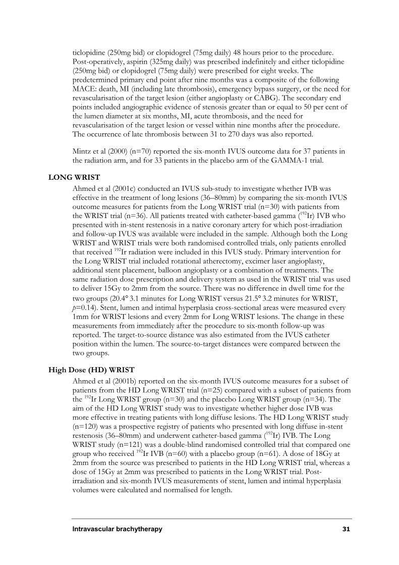
Intravascular brachytherapy 31
ticlopidine (250mg bid) or clopidogrel (75mg daily) 48 hours prior to the procedure. Post-operatively, aspirin (325mg daily) was prescribed indefinitely and either ticlopidine (250mg bid) or clopidogrel (75mg daily) were prescribed for eight weeks. The predetermined primary end point after nine months was a composite of the following MACE: death, MI (including late thrombosis), emergency bypass surgery, or the need for revascularisation of the target lesion (either angioplasty or CABG). The secondary end points included angiographic evidence of stenosis greater than or equal to 50 per cent of the lumen diameter at six months, MI, acute thrombosis, and the need for revascularisation of the target lesion or vessel within nine months after the procedure. The occurrence of late thrombosis between 31 to 270 days was also reported.
Mintz et al (2000) (n=70) reported the six-month IVUS outcome data for 37 patients in the radiation arm, and for 33 patients in the placebo arm of the GAMMA-1 trial.
LONG WRIST
Ahmed et al (2001c) conducted an IVUS sub-study to investigate whether IVB was effective in the treatment of long lesions (36–80mm) by comparing the six-month IVUS outcome measures for patients from the Long WRIST trial (n=30) with patients from the WRIST trial (n=36). All patients treated with catheter-based gamma (192Ir) IVB who presented with in-stent restenosis in a native coronary artery for which post-irradiation and follow-up IVUS was available were included in the sample. Although both the Long WRIST and WRIST trials were both randomised controlled trials, only patients enrolled that received 192Ir radiation were included in this IVUS study. Primary intervention for the Long WRIST trial included rotational atherectomy, excimer laser angioplasty, additional stent placement, balloon angioplasty or a combination of treatments. The same radiation dose prescription and delivery system as used in the WRIST trial was used to deliver 15Gy to 2mm from the source. There was no difference in dwell time for the
two groups (20.4°3.1 minutes for Long WRIST versus 21.5°3.2 minutes for WRIST, p=0.14). Stent, lumen and intimal hyperplasia cross-sectional areas were measured every 1mm for WRIST lesions and every 2mm for Long WRIST lesions. The change in these measurements from immediately after the procedure to six-month follow-up was reported. The target-to-source distance was also estimated from the IVUS catheter position within the lumen. The source-to-target distances were compared between the two groups.
High Dose (HD) WRIST
Ahmed et al (2001b) reported on the six-month IVUS outcome measures for a subset of patients from the HD Long WRIST trial (n=25) compared with a subset of patients from the 192Ir Long WRIST group (n=30) and the placebo Long WRIST group (n=34). The aim of the HD Long WRIST study was to investigate whether higher dose IVB was more effective in treating patients with long diffuse lesions. The HD Long WRIST study (n=120) was a prospective registry of patients who presented with long diffuse in-stent restenosis (36–80mm) and underwent catheter-based gamma (192Ir) IVB. The Long WRIST study (n=121) was a double-blind randomised controlled trial that compared one group who received 192Ir IVB (n=60) with a placebo group (n=61). A dose of 18Gy at 2mm from the source was prescribed to patients in the HD Long WRIST trial, whereas a dose of 15Gy at 2mm was prescribed to patients in the Long WRIST trial. Post-irradiation and six-month IVUS measurements of stent, lumen and intimal hyperplasia volumes were calculated and normalised for length.

32 Intravascular brachytherapy
WRIST PLUS
Waksman et al (2001a) (n=120) reported on a prospective consecutive cohort of patients prescribed anti-platelet therapy for six months in addition to catheter-based gamma (192Ir)IVB. The six-month clinical and angiographic outcomes were then compared to two historical control groups. One control group comprised all patients from the WRIST and Long WRIST trials who received gamma (192-Iridium) IVB and one month of anti-platelet treatment (n=125). The other control group comprised all patients from the WRIST and Long WRIST trials who received placebo IVB and one month anti-platelet treatment. The WRIST Plus patients initially were prescribed clopidogrel (300mg) as a loading dose prior to the intervention, then received 75mg daily for six months. Patients in the control groups received either clopidogrel or ticlopidine (250mg daily) for 30 days. Primary intervention for the WRIST Plus patients involved either PTCA, laser ablation or rotational atherectomy. Additional stenting was discouraged; however, 34 lesions (28.3%) were re-stented. The baseline characteristics of having diabetes, hypertension, hyperlipidemia, prior MI and being current smokers were reported to be similar across the three groups; however, no P values or tables were provided in the paper. The primary clinical end points were late thrombosis and the composite clinical events of death, MI and TLR at six months. Secondary angiographic end points were late total occlusion,
restenosis (²50% of the lumen) and late-loss (mm).
SCRIPPS III (not published)
Grise et al (Grise et al. 2002) (n=500) conducted a prospective cohort to investigate whether a strategy of extended anti-platelet therapy and reduced stent deployment reduced late thrombosis in patients with in-stent restenosis who received catheter-based gamma IVB (Cordis, Best Industries). The information and results pertaining to this study are based on a pre-publication report provided by the principle investigator, Dr. Paul S. Teirstein. The study followed and compared two concurrent non-randomised groups of patients, one group who received new stents (n=96), and another group who received no new stents (n=404). Patients presented with a single lesion in either a native coronary artery or saphenous vein graft. Following primary intervention with either
balloon angioplasty or Cutting BalloonÑ (Scimed, Maple Grove, Minnesota), each patient received treatment with catheter-based gamma (Best Industries) IVB. Further angioplasty and/or stenting was undertaken in patients in whom there was a new dissection or extensive recoil resulting in stenosis greater than 30 per cent of lumen diameter. A radiation dose of 14Gy was prescribed at a distance of 2mm from the centre of the catheter. The study ribbons contained multiple 3mm seeds, each pair separated by a 1mm space. Actual dose calculations were not provided. All patients were treated with extended clopidogrel therapy (mean 306.6 days). The new stent group received clopidogrel for a mean of 425.4 days, and the no new stent group received clopidogrel for a mean of 278.8 days. The authors report that, although study protocol initially prescribed clopidogrel to the new stent group for 12 months, most patients in this group continued to take the medication beyond 12 months.
As this study did not compare these groups with a control or placebo group, that is a group who did not receive catheter-based IVB, the results will be presented here briefly (see Table 13) and referred to in the ‘Is it safe?’ section rather than including the results in the ‘Is it effective?’ section. There are inherent limitations when comparing results between the non-randomised groups, as it is difficult to determine the extent to which the selection of patients as based on their clinical need for stenting explained the differences in outcomes between the two groups.
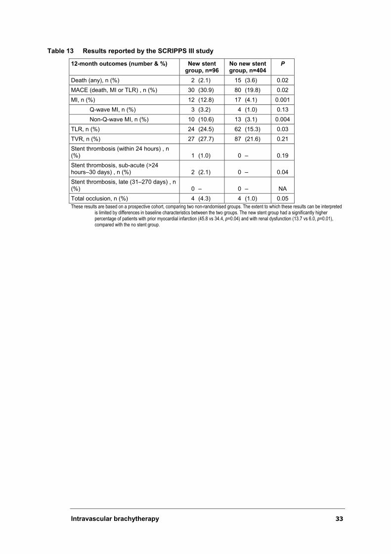
Intravascular brachytherapy 33
Table 13 Results reported by the SCRIPPS III study
12-month outcomes (number & %) New stent group, n=96
No new stent group, n=404
P
Death (any), n (%) 2 (2.1) 15 (3.6) 0.02
MACE (death, MI or TLR) , n (%) 30 (30.9) 80 (19.8) 0.02
MI, n (%) 12 (12.8) 17 (4.1) 0.001
Q-wave MI, n (%) 3 (3.2) 4 (1.0) 0.13
Non-Q-wave MI, n (%) 10 (10.6) 13 (3.1) 0.004
TLR, n (%) 24 (24.5) 62 (15.3) 0.03
TVR, n (%) 27 (27.7) 87 (21.6) 0.21
Stent thrombosis (within 24 hours) , n (%) 1 (1.0) 0 – 0.19
Stent thrombosis, sub-acute (>24 hours–30 days) , n (%) 2 (2.1) 0 – 0.04
Stent thrombosis, late (31–270 days) , n (%) 0 – 0 – NA
Total occlusion, n (%) 4 (4.3) 4 (1.0) 0.05
These results are based on a prospective cohort, comparing two non-randomised groups. The extent to which these results can be interpretedis limited by differences in baseline characteristics between the two groups. The new stent group had a significantly higher percentage of patients with prior myocardial infarction (45.8 vs 34.4, p=0.04) and with renal dysfunction (13.7 vs 6.0, p=0.01), compared with the no stent group.
Table 14 Baseline characteristics for catheter-based gamma IVB randomised controlled trials
Values in italics were calculated from information in papers to facilitate comparison; plus– minus values are means°SD. a Values in paper are not accurate. b Low–density lipoprotein cholesterol level above 130mg per decilitre; c Values as they appear in paper, however these may be incorrect.
Trial
SCRIPPS (n=55) GAMMA-1 (n=252) WRIST (n=130)
Baseline characteristics
192Ir group (n=26) Placebo (n=29) 192Ir group (n=131) Placebo group (n=121) 192Ir group (n=65) Placebo group (n=65)
Age (years) 69.8°9.7 68.8°10.8 58°12 61°11 63.2°10.9 62.3°10.2
Males, n (%) 19 (73) 22 (76) 98 (74.8) 90 (74.4) 66 72
In-stent restenosis, n (%) 16 (62) 18 (62) 100 100 100 100
Location of target lesion, n (%)
Saphenous vein
Left main
Left anterior descending artery
Left circumflex
Ostial
Aorto-ostial
Right coronary artery
6 (23)
–
8 (31)
–
8 (31)
3 (12)
–
9 (31)
–
11 (38)
–
12 (41)
5 (17)
–
4 (3.1)
–
59 (45.0)
27 (20.6)
–
–
40 (30.5)
3 (2.5)
–
38 (31.4)
36 (29.8)
–
–
44 (36.4)
15 (23)
3 (5)
18 (28)
15 (23)
–
–
14 (21)
15 (23)
2 (3)
16 (25)
15 (23)
–
–
17 (26)
Lesion length, mm 12.89°7.05 11.86°6.77 19.0°10.0 20.3°10.3 28.8°12.4 26.7°11.3
Reference vessel diameter pre-op, mm 2.88°0.58 2.78°0.47 2.69°0.51 2.73°0.50 2.71°0.53 2.72°0.56
Minimal lumen diameter pre-op, mm 1.10°0.46 1.03°0.46 0.98°0.45 0.96°0.38 0.94°0.42 0.81°0.42
% stenosis of the lumen pre-op, mm 62°14 62°18 63.3°15.7 64.6°13.4 65°14 70°14
Elevated cholesterol level, n (%) 14 (54) 17 (59) 96 (73.3)b 92 (76.0)b – –
Diabetes mellitus, n (%) 7 (27) 12 (41) 41 (31.3) 38 (31.4) 39 45
Unstable angina, n (%)
Exertional
At rest
11 (42) 16 (55) –
72 (55.0)
33 (25.2)
–
63 (52.1)
39 (32.2)
82 68
Previous myocardial infarction, n (%) 10 (38) 10 (34) 70 (53.4) 57 (47.1) 45 45
History of hypertension, n (%) 17 (65) 20 (69) 94 (71.8) 84 (69.4) 72 68
Previous restenosis (number)
>1, n (%)
>2, n (%)
>3, n (%)
2.1°1.4
13.5 (52)a
6 (23)
–
2.0°1.3
16 (55)
7 (24)
–
1.6°0.9
58 (44.3)
13 (9.9)
–
1.8°1.4
56 (46.3)
21 (17.4)
–
Previous in-stent restenosis:
31 (47)
Previous in-stent restenosis:
25 (39)
Left ventricular ejection fraction 46.7°19.8 48.9°16.3 53.6°10.1 53.8°10.7 0.47°0.11c 0.50°0.11c
34
In
trava
scu
lar b
rach
yth
era
py

Intravascular brachytherapy 35
Catheter-based beta intravascular brachytherapy
The following section briefly outlines the design and methodology of each of the clinical trials investigating the safety and efficacy of catheter-based beta IVB. Baseline characteristics for the Beta WRIST, PREVENT, Costa et al (2000), Schühlen et al (2001) and INHIBIT studies are summarised in Table 16.
Randomised controlled trials (Level II):
¶ studies using Guidant Brachytherapy System:
- PREVENT (Raizner et al. 2000);
- Costa et al (2000); and
- INHIBIT (Waksman et al. 2002).
¶ Studies using other catheter-based beta systems:
- Schühlen et al (2001); and
- START (Popma et al. 2002).
Non-randomised controlled trials (Level III-3):
¶ studies using catheter-based beta systems:
- Beta WRIST (Bhargava et al. 2000; Waksman et al. 2000b; Waksman et al. 2001b).
Beta WRIST
Waksman et al (2000b) reported on the results of patients with native coronary in-stent restenosis enrolled in the Beta WRIST (n=50) prospective cohort compared with a historical control group comprising patients with native coronary artery lesions in the placebo group (n=50) from the WRIST trial. The trial investigated the efficacy and safety of catheter-based beta (90-Yttrium: 90Y) IVB for preventing recurrent in-stent restenosis. Primary intervention for focal lesions consisted of balloon dilation, whereas diffuse lesions were treated with either excimer laser angioplasty or rotational atherectomy followed by balloon dilation. Some patients (n=18) received additional stents. All patients in the beta WRIST trial received radiation. The prescribed dose was 20.6Gy to a distance of 1.0mm from the surface of the inflated balloon. The calculated maximum dose to the vessel wall was 38Gy. For lesions greater than 25mm in length (n=17) the balloon catheter was positioned in two steps. The calculated dose at the overlapped area did not
exceed 70Gy to the vessel wall. The mean dwell time was reported to be 3.0°0.9 minutes. All patients were prescribed clopidogrel (75mg daily) and ticlopidine (500mg daily) for one month. The primary end point was MACE (death, MI or repeat TLR) at six months. Secondary angiographic endpoints were restenosis, late-loss (mm) and late-loss index. IVUS measurements at baseline and six-month follow-up were reported. Late total occlusion occurring between two and six months following the procedure was also reported. An external committee independently adjudicated all events in a blinded fashion.
Bhargava et al (2000) reported on the IVUS results for a subset of patients with native coronary artery lesions who were enrolled in the WRIST (n=130) randomised controlled

36 Intravascular brachytherapy
trial and Beta WRIST (n=50) prospective cohort. Patients (Beta WRIST, n=25; 192Irgroup WRIST, n=36; placebo group WRIST, n=39) who had complete post-operative and six-month IVUS follow-up were included in the IVUS analysis. IVUS results for stent, lumen and intimal hyperplasia area were reported.
PREVENT
Raizner et al (2000) (n=105) conducted a multicentre randomised controlled trial to investigate the safety and effectiveness of catheter-based beta (32P) IVB (Guidant Brachytherapy System) in a broad spectrum of patients with either a single de novo (70% of patients) or restenotic (30% of patients) lesion within a native coronary artery. Twenty-four per cent of patients with restenosis presented with in-stent restenosis. Following primary intervention, which involved angioplasty alone (39%) or additional stent placement (61%), patients were randomised to receive a placebo (n=25), 16Gy (n=23), 20Gy (n=25), or 24Gy (n=25) doses of IVB to 1mm beyond the lumen surface. The radiation prescription was based on the average of the lumen diameters at the proximal and distal reference segments, as measured by IVUS, quantitative coronary angiography or as determined from the known angioplasty balloon or stent sizes. The
mean activity reported was 70°22mCi (range 39–146mCi). The mean dwell time reported
was 4.6°2.0 minutes. All patients were prescribed aspirin (325mg) for six months, and ticlopidine (250mg bid) was prescribed for four weeks after the procedure for patients who received additional stents. The predetermined clinical end points were the combined (in-hospital) and the late (12-month) rate of MACE, defined as death, MI (Q-wave and non-Q-wave) or TLR. Secondary clinical end points included each of the individual MACE or target vessel revascularisations (TVRs) (for restenosis of the target site and adjacent segments). Angiographic end points were minimal lumen diameter (MLD), late
lumen loss, late-loss index and restenosis (²50% of lumen diameter) at six months. Both clinical and angiographic measures were read by blinded observers.
Costa et al
Costa et al (2000) (n=26) conducted a small single centre double-blind randomised controlled trial to determine the mechanism of catheter-based beta (32P) IVB (Guidant Brachytherapy System) in patients with a single de novo or restenotic lesion. Following IVUS-guided stenting or PTCA, patients were randomised to receive either a placebo (n=5) or one of three different doses (28, 35 or 42Gy at 0.5mm into the vessel wall) of radiation (n=21). The actual dose received by the target segment was not calculated. Seven (44%) patients in the radiation groups and three (60%) patients in the placebo group received additional stents. Aspirin (250mg daily) was prescribed to all patients, and ticlopidine (250mg daily) was prescribed only to patients who received additional stents. The period of anti-platelet therapy was not reported. Total vessel (EEM) and lumen 3-D quantitative IVUS volumetric measurements were obtained. Plaque volume was automatically calculated by subtracting lumen volume from the total vessel volume. IVUS measurements were taken post-operatively and six months following the procedure. Five patients (four intervention, one placebo) did not undergo the six-month IVUS procedure. In the intervention group, two patients presented with sub-acute thrombosis, one patient presented with late thrombosis (three months following the procedure), and another patient had a severe restenotic lesion. All of these lesions were determined angiographically. The placebo patient was symptom free with a negative stress test and refused IVUS.
Intravascular brachytherapy 37
Schühlen et al
Schühlen et al (2001) (n=21) initially planned to include 250 patients with in-stent restenosis in a randomised controlled trial to investigate the safety and effectiveness of liquid 188-Rhenium (188Re) catheter-based beta IVB; however, the trial was terminated prematurely after Vascular Therapies withdrew their support. Therefore, only 21 patients were randomised to receive radiation (n=11) or no radiation (n=10) in this single-centre study. Twenty patients had a single in-stent restenotic lesion within a native coronary artery, and one patient randomised to the radiation group presented with a single lesion within a saphenous vein graft. Primary intervention consisted of angioplasty (n=21) and PTCA plus additional stent placement (n=4). Glycoprotein IIb/IIa inhibitors were prescribed to four patients in the radiation group and two patients in the no radiation group. A dose of 28Gy was prescribed at 0.5mm into the vessel wall. Ticlopidine (500mg daily) was prescribed for two weeks for all patients and for four weeks for patients who received additional stents. Aspirin (200mg daily) was prescribed to all patients indefinitely. The primary end point was angiographic late lumen loss at six months. Secondary end points were angiographic restenosis at six months and MACE, defined as death, MI or repeat TVR at 12 months. Angiographic analysis was extended to include the edges 5mm proximal and distal to the radiated segment.
INHIBIT (Intimal Hyperplasia Inhibition with Beta Instent Trial)—GalileoãIntravascular Radiotherapy System
Waksman et al (2002) (n=332) conducted a multicentre, double-blind randomised controlled trial investigating the safety and efficacy of catheter-based beta (32P)
GalileoãIntravascular Radiotherapy System (Guidant Brachytherapy System) in patients with diffuse in-stent restenosis. All patients presented with a single native in-stent coronary lesion. Primary intervention consisted of a combination of PTCA, atherectomy and laser angioplasty, and additional stents were placed in 49 (30%) of the radiation patients and in 52 (31%) of the placebo patients. Following successful primary intervention, patients were randomised to receive 32P radiation (n=166) or a placebo (n=166). A dose of 20Gy at 1mm beyond the lumen diameter was prescribed. A proportion (38%) of patients with lesions longer than 22mm required tandem positioning of the source. It was reported that the dose at the overlapped segment for these patients could have been up to 30 per cent greater than the prescribed dose. The mean specific activity was reported to be 2.88 x 10⁹ Bq (range: 1.15 x 10⁹–5.33 x 10⁹ Bq). The mean dwell time was 4.1 minutes (SD 1.9) for patients who required single positioning of the source and 8.1 minutes (SD 3.6) for patients who required tandem positioning. Post-operatively, all patients were prescribed aspirin (325mg) for one year. The first 69 patients who received new stents were recommended to take ticlopidine for 90 days. The next 29 patients (with or without stent) were recommended to take ticlopidine or clopidogrel for 90 days. The authors reported that the antiplatelet regimes did not differ between the two groups. Overall, 129 (39%) patients received antiplatelet medication for one to three months, 103 (31%) for three to six months, and 100 (30%) for more than six months. The primary safety endpoints were MACE (death, MI, or TLR) at nine months. The primary efficacy endpoints were angiographic restenosis
(²50% lumen diameter) at nine months. The secondary endpoints included MACE (death, MI, TLR or TVR) at nine months, and the magnitude of angiographic late-loss and late-loss index at nine months. Results for late thrombosis and late total occlusion were also reported.

38 Intravascular brachytherapy
START (Stents and Radiation Therapy Trial)
Popma (2000) presented the data from the START trial at the 49th Annual Scientific Sessions of the American College of cardiology in Anaheim, California, USA (12–15 March 2000). A published report of this study is currently in-press and will soon be published in the peer reviewed journal Circulation. Results for START included in this report are based on the pre-published manuscript, ‘A randomised trial of 90Strontium/90Yttrium Beta Radiation versus Placebo Control for the treatment of in-stent restenosis’,provided by the chief investigator (Popma et al. 2002).
The START trial was a multicentre double-blind randomised placebo controlled trial that was conducted to determine the safety and efficacy of catheter-based beta (90-
Strontium/90-Yittium: 90Sr/90Y) IVB (Beta-Cathã System, NovosteÑ). The trial enrolled
476 patients with a single in-stent restenosis (²50% of lumen diameter) in a native coronary artery with a reference diameter of between 2.7 and 4.0mm. Primary intervention consisted of PTCA, and some patients also received additional treatment with rotational atherectomy (43.9% for treatment group vs 39.8% for placebo group), excimer laser (5.7% for the treatment group vs 7.4% for the placebo group) and directional atherectomy (0% for the treatment group vs 0.9% for the placebo group). New stents were deployed in 20.9 per cent of 90Sr/90Y patients and 19.8 per cent of placebo patients. Following successful primary intervention (<30% residual stenosis and no major coronary dissections), patients were randomised into 90Sr/90Y radiation (n=244) and placebo groups (n=232). The majority of patients (n=452) were suitable for
treatment with a 20mm balloon and received treatment with a 30mm BetaCathã(Novoste, Corporation, Norcross, GA) radioactive source train. Lesions treatable with a
30mm balloon required use of the 40mm BetaCathã source train (n=24). The prescription point was 2mm from the centreline of the axis of the radiation source train. The dosimetry depended on the reference vessel sizes. Vessels with a diameter of 2.7 to 3.35mm received 18.4Gy, whereas vessels with a diameter of 3.36 to 4.0mm received
23Gy. The mean activity of the 30mm to 12 source train was 39.96°2.5mCi and the
mean dose rate was 0.0923°0.0058Gy/sec. All patients were prescribed aspirin 325mg alone for the duration of the study. If a new stent was placed within the in-stent restenosis treatment site, patients enrolled from September 1998 until November 1999 received aspirin (325mg daily) for the duration of the study and ticlopidine (250mg bid) for 14 days following the procedure. After November 1998, patients with new stents were recommended to take aspirin and ticlopidine (250mg bid) or clopidogrel (75mg daily) for at least 60 days following the procedure. The primary study endpoint was TVR. TLR rates were also recorded. The primary safety endpoint was the occurrence of MACE (death, MI or TVR). The secondary efficacy endpoints included angiographic restenosis (>50% of lumen diameter), follow-up minimal lumen diameter and late lumen loss. Early and late stent thrombosis was also reported.
Trials conducted in Australia
Perth IVB Trial for liquid Rhenium-188 IVB (n=52)
Chief investigators: Mews, G. C.; Cope, G. D.; Fox, R. A.; Clugston, R. A.; Rankin, M.; Cumpston, G. N.; Horrigan, M.; and Rafter, A.
A pilot study was conducted at the Royal Perth Hospital, Perth, Western Australia, in 1997 and subsequently followed-up with a controlled trial.
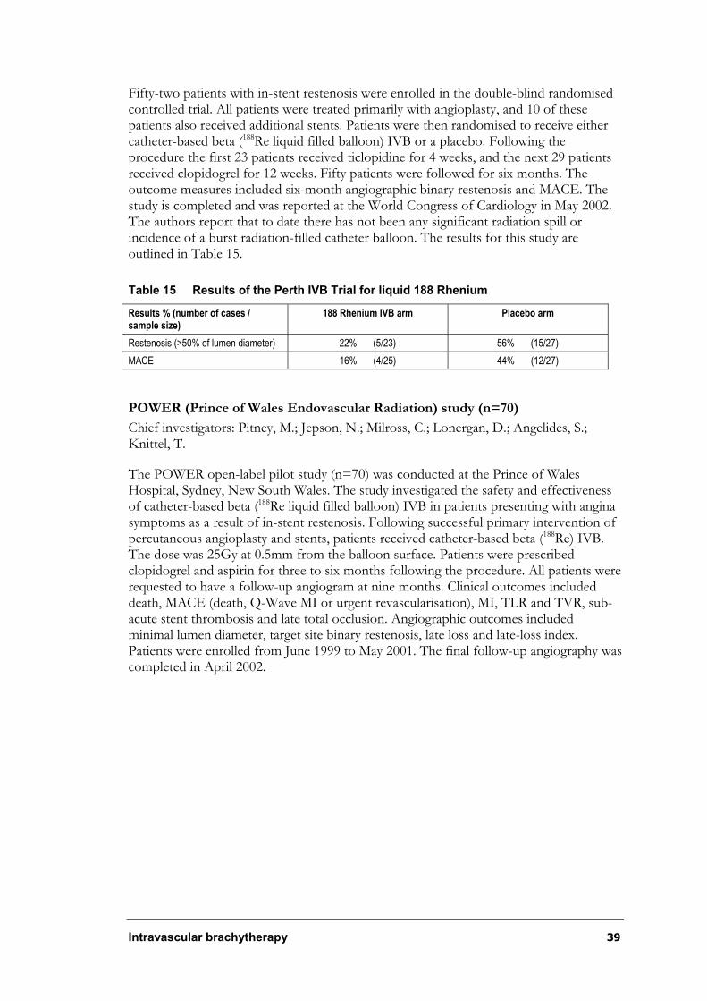
Intravascular brachytherapy 39
Fifty-two patients with in-stent restenosis were enrolled in the double-blind randomised controlled trial. All patients were treated primarily with angioplasty, and 10 of these patients also received additional stents. Patients were then randomised to receive either catheter-based beta (188Re liquid filled balloon) IVB or a placebo. Following the procedure the first 23 patients received ticlopidine for 4 weeks, and the next 29 patients received clopidogrel for 12 weeks. Fifty patients were followed for six months. The outcome measures included six-month angiographic binary restenosis and MACE. The study is completed and was reported at the World Congress of Cardiology in May 2002. The authors report that to date there has not been any significant radiation spill or incidence of a burst radiation-filled catheter balloon. The results for this study are outlined in Table 15.
Table 15 Results of the Perth IVB Trial for liquid 188 Rhenium
Results % (number of cases / sample size)
188 Rhenium IVB arm Placebo arm
Restenosis (>50% of lumen diameter) 22% (5/23) 56% (15/27)
MACE 16% (4/25) 44% (12/27)
POWER (Prince of Wales Endovascular Radiation) study (n=70)
Chief investigators: Pitney, M.; Jepson, N.; Milross, C.; Lonergan, D.; Angelides, S.; Knittel, T.
The POWER open-label pilot study (n=70) was conducted at the Prince of Wales Hospital, Sydney, New South Wales. The study investigated the safety and effectiveness of catheter-based beta (188Re liquid filled balloon) IVB in patients presenting with angina symptoms as a result of in-stent restenosis. Following successful primary intervention of percutaneous angioplasty and stents, patients received catheter-based beta (188Re) IVB. The dose was 25Gy at 0.5mm from the balloon surface. Patients were prescribed clopidogrel and aspirin for three to six months following the procedure. All patients were requested to have a follow-up angiogram at nine months. Clinical outcomes included death, MACE (death, Q-Wave MI or urgent revascularisation), MI, TLR and TVR, sub-acute stent thrombosis and late total occlusion. Angiographic outcomes included minimal lumen diameter, target site binary restenosis, late loss and late-loss index. Patients were enrolled from June 1999 to May 2001. The final follow-up angiography was completed in April 2002.

Table 16 Baseline characteristics of catheter-based beta intravascular brachytherapy
Baseline characteristics TRIAL
Beta WRIST (n=50) a PREVENT (n=105) Schühlen et al (n=21) Costa et al (n=21)c INHIBIT (n=332) START (n=476)
90Y Cohort (n=50)
Placebo from WRIST (n=50)
32P Group (n=80)
Placebo(n=25)
188Re Group (n=11)
No radiation (n=10)
32P Group (n=16) Placebo (n=5) 32P Group (n=166)
Placebo(n=166)
90Sr/90Y Group (n=244)
Placebo(n=232)
Age, years 60°10 61°10 63°11 63°8 665°13 66°10 59.2°9.6 56°10.9 62°11 61°11 61.5°11.5 61.1°10.4
Male sex , n (%) 30 (60) 36 (72) 51 (64) 19 (76) 8 (73) 6 (60) 11 (79) 4 (80) 116 (70) 121 (73) 167 (68) 147 (63)
De novo lesion, n (%)
Restenotic lesion, n (%)
In-stent restenosis, n (%)
50 (100)
50 (100)
50 (100)
50 (100)
54 (68)
26 (33)
19 (24)
19 (76)
6 (24)
6 (24)
100
100
100
100
12 (75)
4 (25)
–
4 (80)
1 (20)
–
100
100
100
100
100
100
100
100
Location of target lesion, n (%)
Saphenous vein
Left main artery
Left anterior descending art.
Left circumflex artery
Right coronary artery
0
2 (4)
12 (24)
18 (36)
18 (36)
0
2 (4)
16 (32)
15 (30)
17 (34)
37 (46)
13 (16)
30 (38)
10 (40)
6 (24)
9 (36)
1 (9)
4 (36)
2 (18)
4 (36)
0
2 (20)
3 (30)
5 (50)
7 (34)
–
–
3 (60)
–
–
0
75 (47)
45 (28)
40 (25)
0
70 (44)
34 (21)
56 (35)
0
105 (43)
63 (26)
70 (29)
0
95 (41)
55 (24)
77 (34)
Lesion length, mm 17.24°9.8 23.7°11.2 – – 13.3°7.3 14.6°7.4 – – 16.9°8.9 17.9°8 16.3°7.2 16.0°7.6
Reference vessel diameter at baseline, mm
2.73°0.65 2.65°0.45 2.99°0.48 2.97°0.55 3.09°0.35 2.91°0.41 – – 2.68°0.53 2.71°0.58 2.76°0.48 2.77°0.43
Minimal lumen diameter pre-op, mm
1.02°0.4 0.77°0.38 0.74°0.37 0.68°0.31 0.35°0.26 0.36°0.30 Minimal lumen area mm2 4.8°1.6
Minimal lumen area mm2 4.7°1.2
1.01°0.37 0.95°0.47 0.98°0.38 0.98°0.37
% stenosis of the lumen pre-op, mm
62.5°12.6 71.4°13.3 75°11 77°8 89°9 87°12 Plaque volume 198°63
Plaque volume 210°58
61.9°14.0 65.2°15.0 64.2°13.7 64.2°13.1
Elevated cholesterol level, n (%) 43 (86) 50 (100) 38 (48) 14 (56) 11 (100) 8 (80) 9 (56) 3 (60) – –
Diabetes mellitus, n (%) 12 (24) 20 (40) 16 (20) 6 (24) 2 (18) 4 (40) 0 0 54 (33) 45 (27) 75 (31) 75 (32)
Unstable angina, n (%) 38 (76) 46 (92) 49 (69)b 17 (71)b – – 12 (75)d 5 (100)d 86 (57)e 95 (63)e 180 (74) 183 (79)
Previous myocardial infarction, n (%)
28 (55) – 28 (35) 14 (56) – – 7 (44) 3 (60) 75 (45) 86 (52) 113 (47) 110 (48)
History of hypertension, n (%) 37 (74) 33 (66) 50 (63) 11 (44) 10 (91) 9 (90) 5 (31) 0 117 (71) 111 (67) 174 (72) 170 (74)
Current Smokers, n (%) 9 (18) – 19 (24) 10 (40) 6 (54) 4 (40) 6 (38) 2 (40) – – 29 (13) 18 (8)
Previous restenosis, (number) 1.46°0.46 – – – 3.7°0.9 3.7°1.2 – – – – – –
Left ventricular ejection fraction 0.51°0.11 0.50°0.12 60°11 58°16 – – – – – – 54.2°10.5 54.6°12.3
Values in italics calculated from paper to facilitate comparison across studies; plus–minus values are means°SD; a values for the Beta WRIST trial have been collated from two papers, Waksman et al (2000b) & Waksman et al (2001b); b angina status CCS III or IV; c Costa et al (2000) does not report angiographic lumen dimensions, only three dimensional IVUS measurements; d Canadian Cardiovascular Society angina status: e Canadian Cardiovascular Society III or IV.
40
Intra
vas
cu
lar b
rac
hyth
era
py
Intravascular brachytherapy 41
Is it safe?
There are important safety issues associated with IVB for treating coronary artery restenosis that require evaluation. The following section considers both the safety of the patient receiving IVB and the safety of the staff administering the treatment. Issues pertaining to the safety of the patient relate to the occurrence of clinical events such as late thrombosis (>30 days following the procedure), restenosis at the edges (termed the edge effect), aneurysm, late restenosis and other potential adverse events associated with radiation effects such as coronary atherosclerosis and malignancy.
Although there are potential procedural risks associated with IVB and other interventional cardiological procedures, as documented in Appendix F, there were no reported cases in the literature and IVB is not expected to cause significant procedural problems over and above the procedure of PTCA.
To ensure that IVB is conducted in a safe manner, the procedure requires a coordinated approach between the interventional cardiologist, the radiation oncologist or nuclear medicine specialist with an interest in this field, and the medical physicist. IVB needs to be performed in a facility that conforms to the appropriate state radiation regulations and licensing requirements. Once a target lesion has been treated with IVB, subsequent irradiation of the same lesion is not possible.
Dosimetry
The dose of radiation used in IVB may have implications for the potential safety and efficacy of this technology for the treatment of coronary stenosis. Generally, a low dose may not sufficiently treat the target lesion, thereby increasing the likelihood of restenosis following the procedure. However, a high dose may damage the vessel wall to the extent that healing is delayed, thus possibly contributing to the occurrence of late thrombosis. Dosimetry is a function of the treatment dose prescribed and the interaction the radiation energy has with the intended target tissue (Jani 1999). Different radioisotopes have been used in clinical studies thus far. Isotope selection will have implications on the effective energy available, the penetration properties, the dose gradient from target sites and the time it will take for the active radiation material to decay to one-half of its initial quantity (half-life) (Waksman 1998).
Gamma radioisotopes penetrate human tissues deeply, therefore making them ideal for treating large vessels. Furthermore, gamma radioisotopes are not shielded by stents, so this type of isotope can be used in treating in-stent restenosis. However, gamma isotopes cannot be shielded by the lead protection that is currently used to protect staff administering other technologies such as X-rays and fluoroscopy. Lead shields greater than 2.5cm need to be used, and all non-essential staff should vacate the catheterisation laboratory during the application of gamma IVB. Furthermore, gamma sources with lower specific activity are required to protect staff from radiation over-exposure; however, this means that longer dwell times (8–20 minutes) are required to deliver the appropriate dose (Coplan & Teirstein 2001), which may increase the risk of vessel occlusion and myocardial ischaemia (Ishiwata et al. 2000).
Beta radioisotopes are easily shielded with thick plastics. The specific activity can therefore be much higher, as exposure to staff is limited, thus allowing very short dwell
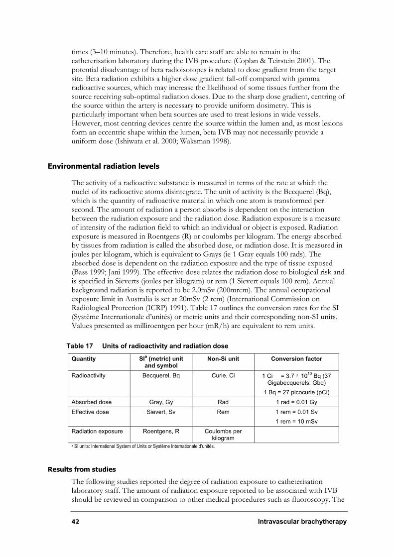
42 Intravascular brachytherapy
times (3–10 minutes). Therefore, health care staff are able to remain in the catheterisation laboratory during the IVB procedure (Coplan & Teirstein 2001). The potential disadvantage of beta radioisotopes is related to dose gradient from the target site. Beta radiation exhibits a higher dose gradient fall-off compared with gamma radioactive sources, which may increase the likelihood of some tissues further from the source receiving sub-optimal radiation doses. Due to the sharp dose gradient, centring of the source within the artery is necessary to provide uniform dosimetry. This is particularly important when beta sources are used to treat lesions in wide vessels. However, most centring devices centre the source within the lumen and, as most lesions form an eccentric shape within the lumen, beta IVB may not necessarily provide a uniform dose (Ishiwata et al. 2000; Waksman 1998).
Environmental radiation levels
The activity of a radioactive substance is measured in terms of the rate at which the nuclei of its radioactive atoms disintegrate. The unit of activity is the Becquerel (Bq), which is the quantity of radioactive material in which one atom is transformed per second. The amount of radiation a person absorbs is dependent on the interaction between the radiation exposure and the radiation dose. Radiation exposure is a measure of intensity of the radiation field to which an individual or object is exposed. Radiation exposure is measured in Roentgens (R) or coulombs per kilogram. The energy absorbed by tissues from radiation is called the absorbed dose, or radiation dose. It is measured in joules per kilogram, which is equivalent to Grays (ie 1 Gray equals 100 rads). The absorbed dose is dependent on the radiation exposure and the type of tissue exposed (Bass 1999; Jani 1999). The effective dose relates the radiation dose to biological risk and is specified in Sieverts (joules per kilogram) or rem (1 Sievert equals 100 rem). Annual background radiation is reported to be 2.0mSv (200mrem). The annual occupational exposure limit in Australia is set at 20mSv (2 rem) (International Commission on Radiological Protection (ICRP) 1991). Table 17 outlines the conversion rates for the SI (Système Internationale d’unités) or metric units and their corresponding non-SI units. Values presented as milliroentgen per hour (mR/h) are equivalent to rem units.
Table 17 Units of radioactivity and radiation dose
Quantity SIa (metric) unit
and symbol Non-Si unit Conversion factor
Radioactivity Becquerel, Bq Curie, Ci 1 Ci = 3.7 ³ 1010
Bq (37 Gigabecquerels: Gbq)
1 Bq = 27 picocurie (pCi)
Absorbed dose Gray, Gy Rad 1 rad = 0.01 Gy
Effective dose Sievert, Sv Rem 1 rem = 0.01 Sv
1 rem = 10 mSv
Radiation exposure Roentgens, R Coulombs per kilogram
a SI units: International System of Units or Système Internationale d’unités.
Results from studies
The following studies reported the degree of radiation exposure to catheterisation laboratory staff. The amount of radiation exposure reported to be associated with IVB should be reviewed in comparison to other medical procedures such as fluoroscopy. The
Intravascular brachytherapy 43
amount of radiation exposure to patients and staff undergoing and using fluoroscopy has been reported to be 0.2mSv and 3.9x10⁻³C/kg-hr, respectively.
Catheter-based gamma IVB
Generally, gamma radioisotopes are more penetrative and, as such, substantial 2.5cm lead shielding, long distances and short exposure times are required to protect a person from excessive radiation exposure (Coplan & Teirstein 2001; Ishiwata et al. 2000).
SCRIPPS
Teirstein et al (1997) reported that the mean time during which the 192Ir ribbon was in
place was 36°7 minutes, and the mean specific activity was 3.6°1.08 GBq. Mean radiation exposure levels in the control room immediately adjacent to the catheterisation
laboratory was 1.19°0.073mSv per hour, and it was 132.3°18.9mSv per hour at the patient’s side where the radiation oncologist stood while inserting the 192Ir ribbon. The radiation oncologist was exposed to radiation for five minutes for each procedure, and the interventional cardiologist was exposed for less than one minute. Therefore, the radiation oncologist who was exposed to radiation for five minutes would be exposed to approximately 11mSv. This would translate to 1.1mSv for 100 procedures.
WRIST
Waksman et al (2000c) reported that the mean dwell time was 22.0°5.3 minutes, and the
mean specific activity was 25.3°3.5mCi. Mean radiation exposure levels were reported as
follows: patient’s chest 5.0°0.2mR/h; catheterisation table 650°120mR/h; 1m from the
table 107°35mR/h; behind the leaded shield 53°24mR/h; and at the control room
0.23°0.06mR/h.
Catheter-based beta IVB
Beta radioisotopes are less penetrative compared with gamma radioactive sources, and are easily shielded with lead aprons and thick plastics (Ishiwata et al. 2000). The Beta WRIST cohort, the PREVENT and the INHIBIT trials provide some information on radiation exposure levels in the catheterisation laboratory.
Beta WRIST
Waksman et al (2000b) reported that the mean dwell time was 3.0°0.9 minutes. Mean
radiation exposure levels at the patient’s chest was reported at 7.0°0.8mrem/h, and at the
bedside 0.07°0.01mR/h.
PREVENT
Raizner et al (2000) reported that the mean dwell time was 4.6°2.0 minutes. The
radiation exposure at one metre from the source location was 0.46°0.35mrem/h.
INHIBIT
The FDA safety and efficacy evaluation of the Galileoã Intravascular Radiotherapy System (Food and Drug Administration (FDA) 2001) did not report on specific exposure levels;
however, it stated that radiation exposure to personnel using the Galileoã 32P source
44 Intravascular brachytherapy
were well within yearly limits set by the Nuclear Regulatory Commission. Waksman et al (2002) did not report on radiation exposure levels.
START
Popma et al (2002) reported that the operator at the patient’s bedside receives approximately 8.6x10⁻⁷C/Kg-hr for beta radiation using 90Sr/90Y, which is below the radiation exposure to staff from routine cardiac fluoroscopy.
Other data
Hausleiter et al (2000) reported on a case study where a patient was accidentally exposed to radioactive 188Re when leakage of a liquid-filled balloon system occurred. It was estimated that approximately 4mCi 188Re was released into the patient’s blood stream. A dose of 24Gy at 0.5mm was prescribed. Exposure readings taken within 20 minutes of the leakage were reported to be 10mR/h above the thorax and 9mR/h on the thigh. Total body scintigraphy demonstrated that 188Re activity was uniform and weak. It was suggested that the potassium perchlorate given to the patient pre-operatively reduced the ability of 188Re to concentrate in critical organs such as the thyroid and the stomach wall. The authors reported that at one-year follow-up the patient did not present with any adverse effects associated with the radiation exposure.
Summary—Radiation exposure
Catheter-based IVB exposes staff to radiation that is considered to be within acceptable levels according to the International Commission on Radiological Protection (International Commission on Radiological Protection (ICRP) 1991). Patients who undergo treatment with catheter-based IVB are exposed to very low levels of radiation, as only a small local area of the vessel wall is irradiated. Consequently, adverse events associated with the radiation treatment are more likely to be associated with vessel wall damage rather than the development of malignancy.
Clinical late thrombosis
Thrombosis is the formation or presence of a thrombus. A thrombus is an aggregation of blood factors, primarily platelets and fibrin, which can cause vascular obstruction (Gennaro et al. 1984). Thrombosis of coronary arteries can lead to angina, MI or death. Thrombotic occlusion following PTCA usually occurs within the first 24 hours after the procedure. Sub-acute thrombosis (<30 days following the procedure) is more likely to be associated with the application of stents. These clinical events have been largely prevented by using anti-platelet medication (Meijer et al. 1993; Wilson et al. 1999). Late thrombosis (>30 days following the procedure) and late-late thrombosis (more than six months following the procedure) have been associated with IVB. It is thought that radiation delays healing and re-endothelialisation following angioplasty and stenting, therefore leaving a chronically thrombogenic luminal or stent strut surface that promotes the aggregation of clotting agents in the blood (Coplan & Teirstein 2001; Ishiwata et al. 2000; Kaluza, Ali, & Raizner 2000). It has been proposed that long-term antiplatelet therapy may prevent the occurrence of late thrombosis associated with IVB. WRIST-12 and GAMMA-5 are new studies yet to be completed that were designed to address the safety and efficacy issues of prolonged antiplatelet therapy for the prevention of late thrombosis (Gruberg & Waksman 2001).
Intravascular brachytherapy 45
Results from studies
Catheter-based gamma intravascular brachytherapy
The following studies report on late thrombotic events:
Randomised controlled trials (Level II):
¶ SCRIPPS (Teirstein et al. 1997; Teirstein et al. 1999);
¶ WRIST (Waksman et al. 1999; Waksman et al. 2000c; Waksman et al. 2001b); and
¶ GAMMA-1 (Leon et al. 2001).
Non-randomised controlled study (Level III-3):
¶ WRIST Plus (Waksman et al. 2001a).
Prospective cohort study (not published, Level III-2):
¶ SCRIPPS III (Grise et al. 2002).
SCRIPPS
The SCRIPPS trial does not clearly document the occurrence of late thrombosis. Teristein et al (1997) (n=55) reported that one patient in the radiation group, who also received an additional stent at the time of the procedure, sustained a MI 18 days after the procedure as a result of a thrombosis.
Teirstein et al (1999) reported another patient in the radiation group underwent TLR 11 months following the index procedure; however, the reason for revascularisation is not provided. Two deaths as a result of MI occurred in the placebo group; however, the authors do not report whether these events were related to the target site.
WRIST
Waksman et al (2000c) (n=130) reported that 7.6 per cent (5 patients) in the group receiving the 192Ir radiation intervention and 3.5 per cent (2 patients) receiving the placebo intervention presented with late thrombosis at six months. At 12 months, an additional patient in the radiation group had a late thrombotic event; however, no further events were reported for the placebo group. The differences between the groups did not reach statistical significance.
Waksman et al (2001b) reported two-year follow-up data for patients with native coronary artery lesions (n=100). Follow-up on patients with saphenous vein graft lesions (n=30) were not reported. Late thrombosis occurred in 8 per cent (4 of the 50 patients) who received the radiation intervention. Two of these patients experienced non-Q-wave MIs. The authors did not provide any data on the 50 patients in the placebo group; however, they do state that the occurrence of events was not statistically significant.
Waksman et al (1999) reported on a sub-group of patients (n=39) from the placebo arm of the WRIST trial who were crossed over to receive 192Ir IVB after they developed recurrent in-stent stenosis with clinical angina and objective evidence of ischaemia. These patients were compared with the patients who originally received radiation treatment
46 Intravascular brachytherapy
(n=65). At six months, late thrombosis and total occlusion occurred in 15.4 per cent (6 of the 39) patients in the crossover group, and in 6.2 per cent (4 of the 65) patients in the primary treatment group (p=0.13). The rate of late thrombosis in the primary placebo group was 3.5 per cent (2 of 65 patients); however, no P value comparing the crossover group with the placebo group was reported.
GAMMA-1
Leon et al (2001) (n=252) defined late thrombosis as MI attributed to the target vessel, with angiographic documentation of thrombus or total occlusion, occurring 31 to 270 days after the index procedure. The rate of late thrombosis was significantly higher in patients who received radiation compared with those who received placebos (5.3% [ 7 patients] vs 0.8% [1 patient], p=0.07). The higher rate of late thrombosis was also associated with late MIs in patients receiving the radiation intervention (9.9% vs 4.1%, p=0.09). Late thrombosis was reported to have resulted in three Q-wave MIs and four non-Q-wave MIs in patients receiving radiation, and in one non-Q-wave MI for a patient in the placebo group. None of the patients who presented with late thrombosis died during the nine-month study period. All the patients in the radiation group who had late thrombosis also had additional stent placement within the in-stent lesion during the radiation procedure, and had stopped taking anti-platelet treatment.
WRIST Plus
Waksman et al (2001a) investigated whether the prescription of prolonged (six-month) anti-platelet treatment, in conjunction with avoiding new stent placement, reduced the late thrombosis rates among patients receiving 192Ir IVB. The authors reported rates of clinical late thrombotic events and angiographic late total occlusion events at six months for patients enrolled in the WRIST Plus registry. These rates were compared to two historical control groups comprising combined patient groups from the WRIST and Long WRIST trials. The rate of late clinical thrombosis was higher for patients who received 192Ir radiation and one-month anti-platelet treatment compared with patients who received 192Ir radiation and six months of anti-platelet treatment (9.6% vs 2.5%, p=0.02). The rate of late clinical thrombosis was not significantly different between patients who received 192Ir radiation and one month of anti-platelet treatment compared with patients who received placebo and one month of anti-platelet therapy (2.5% vs 0.8%, p=0.36). Rates of angiographic late total occlusion were higher for patients who received 192Ir radiation and one month of anti-platelet treatment compared with patients who received 192Ir and six months of anti-platelet treatment (13.6% vs 5.8%, p=0.04).The rate of angiographic late total occlusion for patients who received a placebo and one month of anti-platelet treatment was not significantly different compared with patients who received 192Ir radiation and one month of anti-platelet treatment (1.6% vs 5.8%, p=0.10). These results suggest that prolonged anti-platelet treatment reduces the likelihood of late thrombosis and late total occlusion. However, there are limitations in drawing conclusions when comparisons are made with historical control groups, as it is difficult to determine the extent to which unknown differences between the groups influence outcomes.
SCRIPPS III
Grise et al (2002) evaluated whether extended anti-platelet therapy and reduced stent deployment at the time of catheter-based gamma (192Ir) IVB reduced late thrombosis. The authors enrolled 500 patients with native coronary artery or saphenous vein graft in-
Intravascular brachytherapy 47
stent restenosis into a prospective cohort study. Patients who received new stents (n=96) at the time of IVB were compared to patients who did not receive new stents (n=404). The decision to deploy new stents was based on the occurrence of a new dissection and/or extensive elastic recoil resulting in stenosis greater than 30 per cent of the lumen diameter. These groups were not compared with a control group, that is patients who did not receive IVB. All patients were prescribed extended clopidogrel for a mean of 307 days. The mean duration for taking anti-platelet treatment was longer in the new stent group compared with patients in the no new stent group (425 days vs 279 days). The authors report that the majority of patients in the new stent group continued to take clopidogrel beyond the predetermined study protocol of 12 months. Following 12 months clinical follow-up, three patients sustained stent thrombosis in the new stent group compared with no patients in the no new stent group. One patient sustained stent thrombosis within 24 hours of the index procedure, and two patients sustained stent thrombosis between 24 hours and 30 days following the index procedure. No late thrombotic events (24–270 days) were reported for either group. The angiographic outcome of late total occlusion occurred at a rate of 4.3 per cent in the new stent group and at a rate of 1.0 per cent in the placebo group (p=0.05). Although this study suggests that new stent placement may be associated with an increased likelihood of late thrombosis and late total occlusion, the extent to which selection may have biased the outcome measures cannot be quantified.
Table 18 summarises the results of clinical late thrombosis and angiographic late total occlusion for the catheter-based gamma IVB studies. Figure 4 summarises the results of clinical late thrombosis only for WRIST and GAMMA-1 randomised controlled trials.
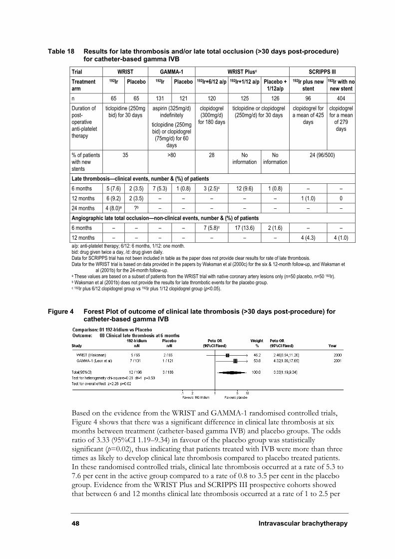
48 Intravascular brachytherapy
Table 18 Results for late thrombosis and/or late total occlusion (>30 days post-procedure) for catheter-based gamma IVB
Trial WRIST GAMMA-1 WRIST Plusc SCRIPPS III
Treatmentarm
192Ir Placebo 192Ir Placebo 192Ir+6/12 a/p 192Ir+1/12 a/p Placebo + 1/12a/p
192Ir plus new stent
192Ir with nonew stent
n 65 65 131 121 120 125 126 96 404
Duration of post-operative anti-platelet therapy
ticlopidine (250mg bid) for 30 days
aspirin (325mg/d) indefinitely
ticlopidine (250mg bid) or clopidogrel (75mg/d) for 60
days
clopidogrel (300mg/d)
for 180 days
ticlopidine or clopidogrel (250mg/d) for 30 days
clopidogrel for a mean of 425
days
clopidogrel for a mean
of 279 days
% of patients with new stents
35 >80 28 No information
Noinformation
24 (96/500)
Late thrombosis—clinical events, number & (%) of patients
6 months 5 (7.6) 2 (3.5) 7 (5.3) 1 (0.8) 3 (2.5)c 12 (9.6) 1 (0.8) – –
12 months 6 (9.2) 2 (3.5) – – – – – 1 (1.0) 0
24 months 4 (8.0)a ?b – – – – – – –
Angiographic late total occlusion—non-clinical events, number & (%) of patients
6 months – – – – 7 (5.8)c 17 (13.6) 2 (1.6) – –
12 months – – – – – – – 4 (4.3) 4 (1.0)
a/p: anti-platelet therapy; 6/12: 6 months, 1/12: one month. bid: drug given twice a day, /d: drug given daily. Data for SCRIPPS trial has not been included in table as the paper does not provide clear results for rate of late thrombosis. Data for the WRIST trial is based on data provided in the papers by Waksman et al (2000c) for the six & 12-month follow-up, and Waksman et
al (2001b) for the 24-month follow-up. a These values are based on a subset of patients from the WRIST trial with native coronary artery lesions only (n=50 placebo, n=50 192Ir). b Waksman et al (2001b) does not provide the results for late thrombotic events for the placebo group. c 192Ir plus 6/12 clopidogrel group vs 192Ir plus 1/12 clopidogrel group (p<0.05).
Figure 4 Forest Plot of outcome of clinical late thrombosis (>30 days post-procedure) for catheter-based gamma IVB
Based on the evidence from the WRIST and GAMMA-1 randomised controlled trials, Figure 4 shows that there was a significant difference in clinical late thrombosis at six months between treatment (catheter-based gamma IVB) and placebo groups. The odds ratio of 3.33 (95%CI 1.19–9.34) in favour of the placebo group was statistically significant (p=0.02), thus indicating that patients treated with IVB were more than three times as likely to develop clinical late thrombosis compared to placebo treated patients. In these randomised controlled trials, clinical late thrombosis occurred at a rate of 5.3 to 7.6 per cent in the active group compared to a rate of 0.8 to 3.5 per cent in the placebo group. Evidence from the WRIST Plus and SCRIPPS III prospective cohorts showed that between 6 and 12 months clinical late thrombosis occurred at a rate of 1 to 2.5 per
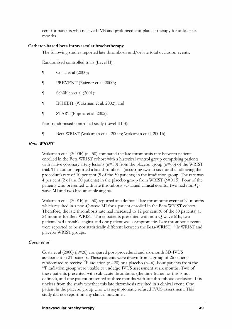
Intravascular brachytherapy 49
cent for patients who received IVB and prolonged anti-platelet therapy for at least six months.
Catheter-based beta intravascular brachytherapy
The following studies reported late thrombosis and/or late total occlusion events:
Randomised controlled trials (Level II):
¶ Costa et al (2000);
¶ PREVENT (Raizner et al. 2000);
¶ Schühlen et al (2001);
¶ INHIBIT (Waksman et al. 2002); and
¶ START (Popma et al. 2002).
Non-randomised controlled study (Level III-3):
¶ Beta-WRIST (Waksman et al. 2000b; Waksman et al. 2001b).
Beta-WRIST
Waksman et al (2000b) (n=50) compared the late thrombosis rate between patients enrolled in the Beta WRIST cohort with a historical control group comprising patients with native coronary artery lesions (n=50) from the placebo group (n=65) of the WRIST trial. The authors reported a late thrombosis (occurring two to six months following the procedure) rate of 10 per cent (5 of the 50 patients) in the irradiation group. The rate was 4 per cent (2 of the 50 patients) in the placebo group from WRIST (p=0.15). Four of the patients who presented with late thrombosis sustained clinical events. Two had non-Q-wave MI and two had unstable angina.
Waksman et al (2001b) (n=50) reported an additional late thrombotic event at 24 months which resulted in a non-Q-wave MI for a patient enrolled in the Beta WRIST cohort. Therefore, the late thrombosis rate had increased to 12 per cent (6 of the 50 patients) at 24 months for Beta WRIST. Three patients presented with non-Q-wave MIs, two patients had unstable angina and one patient was asymptomatic. Late thrombotic events were reported to be not statistically different between the Beta-WRIST, 192Ir WRIST and placebo WRIST groups.
Costa et al
Costa et al (2000) (n=26) compared post-procedural and six-month 3D-IVUS assessment in 21 patients. These patients were drawn from a group of 26 patients randomised to receive 32P radiation (n=20) or a placebo (n=6). Four patients from the 32P radiation group were unable to undergo IVUS assessment at six months. Two of these patients presented with sub-acute thrombosis (the time frame for this is not defined), and one patient presented at three months with late thrombotic occlusion. It is unclear from the study whether this late thrombosis resulted in a clinical event. One patient in the placebo group who was asymptomatic refused IVUS assessment. This study did not report on any clinical outcomes.
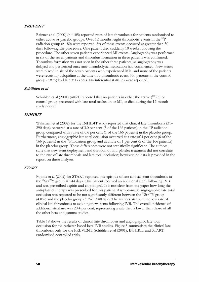
50 Intravascular brachytherapy
PREVENT
Raizner at el (2000) (n=105) reported rates of late thrombosis for patients randomised to either active or placebo groups. Over 12 months, eight thrombotic events in the 32Pradiation group (n=80) were reported. Six of these events occurred at greater than 30 days following the procedure. One patient died suddenly 10 weeks following the procedure. The other seven patients experienced MI events. Angiography was performed in six of the seven patients and thrombus formation in three patients was confirmed. Thrombus formation was not seen in the other three patients, as angiography was delayed and performed once anti-thrombolytic medication had commenced. New stents were placed in six of the seven patients who experienced MIs, and none of the patients were receiving ticlopidine at the time of a thrombotic event. No patients in the control group (n=25) had late MI events. No inferential statistics were reported.
Schühlen et al
Schühlen et al (2001) (n=21) reported that no patients in either the active (188Re) or control group presented with late total occlusion or MI, or died during the 12-month study period.
INHIBIT
Waksman et al (2002) for the INHIBIT study reported that clinical late thrombosis (31–290 days) occurred at a rate of 3.0 per cent (5 of the 166 patients) in the 32P radiation group compared with a rate of 0.6 per cent (1 of the 166 patients) in the placebo group. Furthermore, angiographic late total occlusion occurred at a rate of 4 per cent (6 of the 166 patients) in the 32P radiation group and at a rate of 1 per cent (2 of the 166 patients) in the placebo group. These differences were not statistically significant. The authors state that new stent deployment and duration of anti-platelet treatment did not correlate to the rate of late thrombosis and late total occlusion; however, no data is provided in the report on these analyses.
START
Popma et al (2002) for START reported one episode of late clinical stent thrombosis in the 90Sr/90Y group at 244 days. This patient received an additional stent following IVB and was prescribed aspirin and clopidogrel. It is not clear from the paper how long the anti-platelet therapy was prescribed for this patient. Asymptomatic angiographic late total occlusion was reported to be not significantly different between the 90Sr/90Y group (4.0%) and the placebo group (3.7%) (p=0.872). The authors attribute the low rate of clinical late thrombosis to avoiding new stents following IVB. The overall incidence of additional stent use was 20.4 per cent, representing a rate that is lower than those of all the other beta and gamma studies.
Table 19 shows the results of clinical late thrombosis and angiographic late total occlusion for the catheter-based beta IVB studies. Figure 5 summarises the clinical late thrombosis only for the PREVENT, Schühlen et al (2001), INHIBIT and START randomised controlled trials.
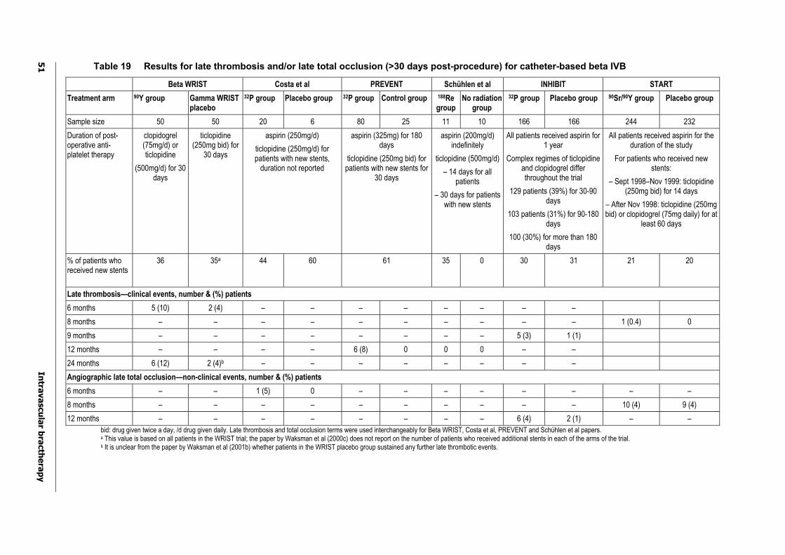
51
In
trava
scu
lar b
racth
era
py
Table 19 Results for late thrombosis and/or late total occlusion (>30 days post-procedure) for catheter-based beta IVB
Beta WRIST Costa et al PREVENT Schühlen et al INHIBIT START
Treatment arm 90Y group Gamma WRIST placebo
32P group Placebo group 32P group Control group 188Regroup
No radiationgroup
32P group Placebo group 90Sr/90Y group Placebo group
Sample size 50 50 20 6 80 25 11 10 166 166 244 232
Duration of post-operative anti-platelet therapy
clopidogrel (75mg/d) or ticlopidine
(500mg/d) for 30 days
ticlopidine (250mg bid) for
30 days
aspirin (250mg/d)
ticlopidine (250mg/d) for patients with new stents,
duration not reported
aspirin (325mg) for 180 days
ticlopidine (250mg bid) for patients with new stents for
30 days
aspirin (200mg/d) indefinitely
ticlopidine (500mg/d)
– 14 days for all patients
– 30 days for patients with new stents
All patients received aspirin for 1 year
Complex regimes of ticlopidine and clopidogrel differ throughout the trial
129 patients (39%) for 30-90 days
103 patients (31%) for 90-180 days
100 (30%) for more than 180 days
All patients received aspirin for the duration of the study
For patients who received new stents:
– Sept 1998–Nov 1999: ticlopidine (250mg bid) for 14 days
– After Nov 1998: ticlopidine (250mg bid) or clopidogrel (75mg daily) for at
least 60 days
% of patients who received new stents
36 35a 44 60 61 35 0 30 31 21 20
Late thrombosis—clinical events, number & (%) patients
6 months 5 (10) 2 (4) – – – – – – – –
8 months – – – – – – – – – – 1 (0.4) 0
9 months – – – – – – – – 5 (3) 1 (1)
12 months – – – – 6 (8) 0 0 0 – –
24 months 6 (12) 2 (4)b – – – – – – – –
Angiographic late total occlusion—non-clinical events, number & (%) patients
6 months – – 1 (5) 0 – – – – – – – –
8 months – – – – – – – – – – 10 (4) 9 (4)
12 months – – – – – – – – 6 (4) 2 (1) – –
bid: drug given twice a day, /d drug given daily. Late thrombosis and total occlusion terms were used interchangeably for Beta WRIST, Costa et al, PREVENT and Schühlen et al papers. a This value is based on all patients in the WRIST trial; the paper by Waksman et al (2000c) does not report on the number of patients who received additional stents in each of the arms of the trial. b It is unclear from the paper by Waksman et al (2001b) whether patients in the WRIST placebo group sustained any further late thrombotic events.
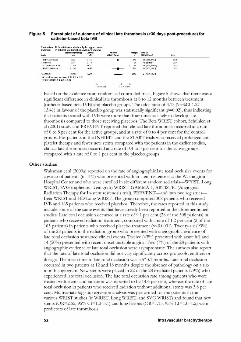
52 Intravascular brachytherapy
Figure 5 Forest plot of outcome of clinical late thrombosis (>30 days post-procedure) for catheter-based beta IVB
Based on the evidence from randomised controlled trials, Figure 5 shows that there was a significant difference in clinical late thrombosis at 8 to 12 months between treatment (catheter-based beta IVB) and placebo groups. The odds ratio of 4.13 (95%CI 1.27–13.41) in favour of the placebo group was statistically significant (p=0.02), thus indicating that patients treated with IVB were more than four times as likely to develop late thrombosis compared to those receiving placebos. The Beta WRIST cohort, Schühlen et al (2001) study and PREVENT reported that clinical late thrombosis occurred at a rate of 0 to 8 per cent for the active groups, and at a rate of 0 to 4 per cent for the control groups. For patients in the INHIBIT and the START trials who received prolonged anti-platelet therapy and fewer new stents compared with the patients in the earlier studies, clinical late thrombosis occurred at a rate of 0.4 to 3 per cent for the active groups, compared with a rate of 0 to 1 per cent in the placebo groups.
Other studies
Waksman et al (2000a) reported on the rate of angiographic late total occlusive events for a group of patients (n=473) who presented with in-stent restenosis at the Washington Hospital Center and who were enrolled in six different randomised trials—WRIST, Long WRIST, SVG (saphenous vein graft) WRIST, GAMMA-1, ARTISTIC (Angiograd Radiation Therapy for In-stent restenosis trial), PREVENT—and into two registries—Beta-WRIST and HD Long WRIST. The group comprised 308 patients who received IVB and 165 patients who received placebos. Therefore, the rates reported in this study include some of the same events that have already been reported in the aforementioned studies. Late total occlusion occurred at a rate of 9.1 per cent (28 of the 308 patients) in patients who received radiation treatment, compared with a rate of 1.2 per cent (2 of the 165 patients) in patients who received placebo treatment (p<0.0001). Twenty-six (93%) of the 28 patients in the radiation group who presented with angiographic evidence of late total occlusion sustained clinical events. Twelve (43%) presented with acute MI and 14 (50%) presented with recent onset unstable angina. Two (7%) of the 28 patients with angiographic evidence of late total occlusion were asymptomatic. The authors also report that the rate of late total occlusion did not vary significantly across protocols, emitters or
dosage. The mean time to late total occlusion was 5.5°3.1 months. Late total occlusion occurred in two patients at 12 and 18 months despite the absence of pathology on a six-month angiogram. New stents were placed in 22 of the 28 irradiated patients (79%) who experienced late total occlusion. The late total occlusion rate among patients who were treated with stents and radiation was reported to be 14.6 per cent, whereas the rate of late total occlusion in patients who received radiation without additional stents was 3.8 per cent. Multivariate logistic regression analysis was performed for the patients in the various WRIST studies (ie WRIST, Long WRIST, and SVG WRIST) and found that new stents (OR=2.55, 95% CI=1.0–5.1) and long lesions (OR=1.15, 95% CI=1.0–1.2) were predictors of late thrombosis.
Intravascular brachytherapy 53
Summary—Late thrombosis
Based on evidence from the randomised trials, meta-analysis indicated that there was a significant difference favouring the placebo group in the incidence of clinical late thrombosis between treatment and placebo groups, for both catheter-based gamma and beta IVB. Patients treated with active IVB were approximately 3½ to 4 times more likely to develop clinical late thrombosis compared to those treated with placebo. The incidence of late thrombosis is lower in more recent studies, equivalent to placebo rates. This may be due to study protocols incorporating longer duration anti-platelet therapy combined with avoidance of new stent deployment. However, the influence of other differences in treatment protocols cannot be excluded. Furthermore, it is not possible to evaluate the long-term effectiveness of these measures in reducing the incidence of late thrombosis beyond 12 months.
Edge effect
The ‘edge effect’ occurs when there is restenosis (²50% of lumen diameter) at either the distal or proximal margin of the target lesion following percutaneous intervention and IVB. A number of studies have attempted to analyse cause and predictors of edge restenosis. It has been reported to be a result of neointimal hyperplasia and an absence of radiation-induced positive vessel remodelling (Ahmed et al. 2001a). Additional analyses have suggested that it may be related to low dose beta radiation and vessel injury (Sabate et al. 2000), and to geographic miss (Kim et al. 2001; Sianos et al. 2001). Low doses of radiation are thought to stimulate neointimal proliferation at the edges, therefore creating restenosis, whereas ‘geographic miss’ is a term used to describe the scenario whereby the radiation source does not adequately cover the target lesion injured by the angioplasty procedure. The injured vessel wall not sufficiently covered by the radiation treatment initiates a wound healing response that results in accelerated intimal hyperplasia at the margin compared with the centre of the lesion, therefore resulting in edge restenosis also called the ‘candy wrapper effect’ (Bonan 2000; Coplan & Teirstein 2001; Kaluza, Ali, & Raizner 2000; Waksman 2000). This adverse event has been associated with gamma and beta catheter IVB as well as radioactive stents (Kaluza, Ali, & Raizner 2000). However, as beta radioisotopes tend to have a more rapid dose fall-off and are less penetrating compared with gamma radioisotopes (Waksman 1998), beta catheter-based IVB and beta radioactive stents may be more susceptible to this adverse event.
Results from studies
Catheter-based gamma intravascular brachytherapy
The following studies report on edge restenosis (²50% of the lumen diameter):
Randomised controlled trials (Level II):
¶ SCRIPPS (Lansky et al. 1999; Teirstein et al. 1997);
¶ WRIST (Waksman et al. 2000c); and
¶ GAMMA-1 (Leon et al. 2001).
Non-randomised controlled studies (Level III-3):
¶ WRIST Plus (Waksman et al. 2001a).
54 Intravascular brachytherapy
Prospective cohort (not published, Level III-2):
¶ SCRIPPS III.
SCRIPPS
Teirstein et al (1997) reported angiographic follow-up for 52 of the 55 patients enrolled in the SCRIPPS trial. The paper reports on the number of patients who presented at six months with restenosis of the stent and margin (the area beyond the stent but exposed to the radiation), and restenosis of the stent only. Therefore, edge restenosis was calculated to have occurred at a rate of 9 per cent (2 of 24 patients) in the 192Ir radiation group compared with a rate of 18 per cent (5 of the 28 patients) in the placebo group. Table 20 shows the restenosis rates for the target lesion and margin, target lesion only and calculated edge restenosis rates.
Lansky et al (1999) reported on the six-month angiographic results for 52 of the 55 patients enrolled in the SCRIPPS trial to identify luminal dimension changes within the stent alone compared to the stent and margin. The results reported in this paper differ slightly from the six-month angiographic results reported in the Teirstein et al (1997) paper, possibly because this paper included patients with only one single-focal lesion, whereas the Teirstein et al (1997) paper may have based their results on multiple lesions for some of the patients. The restenosis rate at the margin only was not significantly different between the 192Ir radiation group and placebo group (8% vs 14%, p=0.503).
WRIST
Waksman et al (2000c) reported on the six-month angiographic results for 118 of the 130 patients enrolled in the WRIST trial. The paper reports on the number of patients who presented with restenosis of the target lesion and margins, and restenosis of the target lesion only. Therefore, the edge restenosis was calculated to have occurred at a rate of 3 per cent (2 of the 59 patients) in the 192Ir radiation group compared to a rate of 2 per cent (1 of the 59 patients) in the placebo group. Table 20 shows the restenosis rates for the target lesion and margin, target lesion only and calculated edge restenosis rates.
GAMMA-1
Leon et al (2001) reported on the six-month angiographic results for 214 of the 252 patients enrolled in the GAMMA-1 trial. The paper reports on the number of patients who presented with restenosis of the target lesion and margins, and restenosis of the target lesion only. Edge restenosis was calculated to have occurred at a rate of 11 per cent (12 of the 111 patients) in the 192Ir radiation group compared to a rate of 5 per cent (5 of the 103 patients) in the placebo group. Table 20 shows the restenosis rates for the target lesion and margin, target lesion only and calculated edge restenosis rates.
Mintz et al (2000) (n=70) reported on the eight-month IVUS results for a subset of 70 of the 252 patients enrolled in the GAMMA-1 trial. There were no significant differences between the stent lumen area at the stent edge and the stent lumen area within the stent section in patients who received 192Ir compared with patients who received placebo. However, as these results were based on a subset of patients, selection bias could have implications for the results of this study.
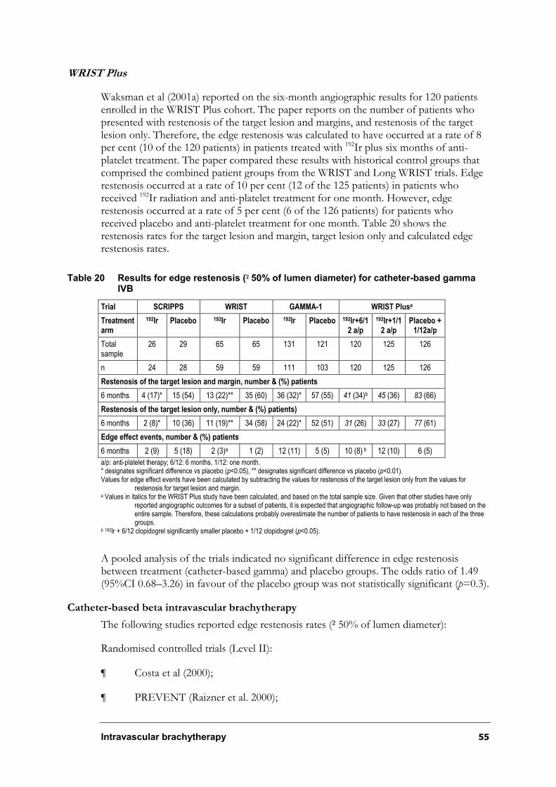
Intravascular brachytherapy 55
WRIST Plus
Waksman et al (2001a) reported on the six-month angiographic results for 120 patients enrolled in the WRIST Plus cohort. The paper reports on the number of patients who presented with restenosis of the target lesion and margins, and restenosis of the target lesion only. Therefore, the edge restenosis was calculated to have occurred at a rate of 8 per cent (10 of the 120 patients) in patients treated with 192Ir plus six months of anti-platelet treatment. The paper compared these results with historical control groups that comprised the combined patient groups from the WRIST and Long WRIST trials. Edge restenosis occurred at a rate of 10 per cent (12 of the 125 patients) in patients who received 192Ir radiation and anti-platelet treatment for one month. However, edge restenosis occurred at a rate of 5 per cent (6 of the 126 patients) for patients who received placebo and anti-platelet treatment for one month. Table 20 shows the restenosis rates for the target lesion and margin, target lesion only and calculated edge restenosis rates.
Table 20 Results for edge restenosis (²50% of lumen diameter) for catheter-based gamma IVB
Trial SCRIPPS WRIST GAMMA-1 WRIST Plusa
Treatmentarm
192Ir Placebo 192Ir Placebo 192Ir Placebo 192Ir+6/12 a/p
192Ir+1/12 a/p
Placebo + 1/12a/p
Total sample
26 29 65 65 131 121 120 125 126
n 24 28 59 59 111 103 120 125 126
Restenosis of the target lesion and margin, number & (%) patients
6 months 4 (17)* 15 (54) 13 (22)** 35 (60) 36 (32)* 57 (55) 41 (34)b 45 (36) 83 (66)
Restenosis of the target lesion only, number & (%) patients)
6 months 2 (8)* 10 (36) 11 (19)** 34 (58) 24 (22)* 52 (51) 31 (26) 33 (27) 77 (61)
Edge effect events, number & (%) patients
6 months 2 (9) 5 (18) 2 (3)a 1 (2) 12 (11) 5 (5) 10 (8) b 12 (10) 6 (5)
a/p: anti-platelet therapy; 6/12: 6 months, 1/12: one month. * designates significant difference vs placebo (p<0.05), ** designates significant difference vs placebo (p<0.01). Values for edge effect events have been calculated by subtracting the values for restenosis of the target lesion only from the values for
restenosis for target lesion and margin. a Values in italics for the WRIST Plus study have been calculated, and based on the total sample size. Given that other studies have only
reported angiographic outcomes for a subset of patients, it is expected that angiographic follow-up was probably not based on theentire sample. Therefore, these calculations probably overestimate the number of patients to have restenosis in each of the threegroups.
b 192Ir + 6/12 clopidogrel significantly smaller placebo + 1/12 clopidogrel (p<0.05).
A pooled analysis of the trials indicated no significant difference in edge restenosis between treatment (catheter-based gamma) and placebo groups. The odds ratio of 1.49 (95%CI 0.68–3.26) in favour of the placebo group was not statistically significant (p=0.3).
Catheter-based beta intravascular brachytherapy
The following studies reported edge restenosis rates (²50% of lumen diameter):
Randomised controlled trials (Level II):
¶ Costa et al (2000);
¶ PREVENT (Raizner et al. 2000);
56 Intravascular brachytherapy
¶ Schühlen et al (2001); and
¶ INHIBIT (Waksman et al. 2002).
Non-randomised controlled study (Level III-3):
¶ Beta WRIST (Waksman et al. 2000b).
Beta-WRIST
Waksman et al (2000b) reported on the six-month angiographic data for 41 of the 50 patients enrolled in the Beta-WRIST prospective cohort. The outcome measures for these patients were compared with a historical control group comprising patients in the placebo group who had native coronary artery lesions and six-month angiographic follow-up from the WRIST trial (n=45). The paper reports on the number of patients who presented with restenosis of the target lesion and margins, and restenosis of the target lesion only. The target lesion was defined as the target site plus more than 5mm beyond the irradiated segment. Therefore, the edge restenosis was calculated to have occurred at a rate of 12 per cent (5 of the 41 patients) in the 90Y radiation group compared with a rate of 4 per cent (2 of the 45 patients) in the WRIST placebo group. Table 21 shows the restenosis rates for the target lesion and margin, target lesion only and calculated edge restenosis rates.
Costa et al
Costa et al (2000) (n=26) reported on one severe restenotic lesion in the radiation group (n=20) that was located in an area proximal and adjacent to the 32P radiated area, but injured by angioplasty. No further discussion of edge restenosis was reported for the placebo group (n=6). No further follow-up was reported beyond six months. These results are included in Table 21.
PREVENT
Raizner et al (2000) reported six-month angiographic data for 96 of the 105 patients enrolled in PREVENT. However, restenosis of the target lesion was reported in 73 patients in the 32P radiation group, and on 23 patients in the placebo group, whereas restenosis of the target lesion plus margin was reported for 76 patients in the 32Pradiation group, and for 24 patients in the placebo group. As the angiographic restenosis events were based on different patient numbers, it is difficult to calculate the number of patients who sustained edge restenosis. Edge restenosis was calculated approximately to have occurred at a rate of 14 per cent for the 32P radiation group compared with a rate of 11 per cent for the placebo group. Table 21 shows the restenosis rates for the target lesion and margin, target lesion only and calculated edge restenosis rates.
Schühlen et al
Schühlen et al (2001) (n=21) reported no edge restenotic events for any patients in either the 188Re radiation group or the no radiation group at six months.
Intravascular brachytherapy 57
INHIBIT
Waksman et al (2002) reported nine-month angiographic data for patients enrolled in INHIBIT. The rates of restenosis of the stent, injured, radiated and analysis segments, with each of the segments being inclusive were outlined in a bar graph. The graph indicated a pattern in which restenosis rates increased for the 32P group for each increase in segment size analysed. This increasing gradient is not as marked for the placebo group. Nevertheless, the authors report that percentage diameter stenosis did not differ between
the treatment and placebo groups for either the proximal (20.5°25.0 vs 18.0°24.2,
p=0.47) or distal (23.7°26.2 vs 21.2°22.5, p=0.43) edges. It is not possible to extract accurate values from this graph to determine the rate of restenosis for each of the segments. The FDA Safety and Effectiveness Evaluation (Food and Drug Administration (FDA) 2001) does, however, provide nine-month angiographic restenosis rates for patients enrolled in INHIBIT. The report defined the analysis segment as ‘the segment that extends 5mm proximal and distal to the radiated or injured landmark, whichever was longest in length’. Restenosis of the ‘stent segment’ was reported on 127 patients in the 32P radiation group, and on 126 patients in the placebo group, whereas restenosis of the ‘analysis segment’ was reported for 129 patients in the 32P radiation group, and for 128 patients in the placebo group. As the angiographic restenosis events were based on different patient numbers, it is difficult to calculate the number of patients sustaining edge restenosis. To facilitate comparison across studies, edge restenosis was calculated approximately to have occurred at a rate of 11.4 per cent in the 32P radiation group compared to a rate of 2.4 per cent in the placebo group. Table 21 shows the restenosis rates for the target lesion and margin (analysis segment), target lesion only (stent segment) and calculated edge restenosis rates.
START
Popma et al (2002) reported eight-month angiographic follow-up in 198 of the 244 90Sr/90Y patients and in 188 of the 232 placebo patients enrolled in START. The rates of restenosis for the stented, injured, irradiated and analysis segments were reported. A similar pattern as observed in INHIBIT was noted, in which the rate of restenosis increased for the 90Sr/90Y group for each increase in segment size analysed. However, this increasing gradient noted for the 90Sr/90Y group was not as marked for the placebo group. Nethertheless, the authors report no significant differences in the mean per cent
diameter stenosis (proximal: 19.8% vs 24.9%; distal: 16.1 vs 18.3%) or restenosis (²50%of lumen diameter) rates (proximal: 12.5% vs 13.4%; distal: 6.7% vs 8.5%) at the edges of the source train in the active groups compared with the placebo group, respectively. To facilitate comparison across studies, edge restenosis was calculated to have occurred at a rate of 14.6 per cent (29 of the 198 patients) in the 90Sr/90Y group, and at a rate of 4.2 per cent (8 of the 188 patients) for the placebo group. Table 21 shows the restenosis rates for the target lesion plus margin (analysis segment), the target lesion (stent segment) and the calculated edge restenosis rate.
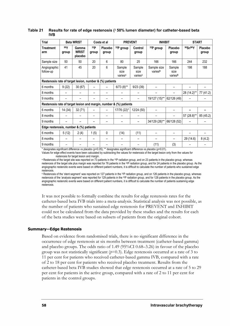
58 Intravascular brachytherapy
Table 21 Results for rate of edge restenosis (²50% lumen diameter) for catheter-based beta IVB
Trial Beta WRIST Costa et al PREVENT INHIBIT START
Treatmentarm
90Ygroup
GammaWRISTplacebo
32Pgroup
Placebo group
32P group Control group
32P group Placebo group
90Sr/90Y Placebo group
Sample size 50 50 20 6 80 25 166 166 244 232
Angiographic follow-up
41 45 20 6 Sample size
variesa
Samplesize
variesa
Sample size variesb
Samplesize
variesb
198 188
Restenosis rate of target lesion, number & (%) patients
6 months 9 (22) 30 (67) – – 6/73 (8)** 9/23 (39) – – – –
8 months – – – – – – – – 28 (14.2)** 77 (41.2)
9 months – – – – – – 19/127 (15)** 62/126 (49) – –
Restenosis rate of target lesion and margin, number & (%) patients
6 months 14 (34) 32 (71) – – 17/76 (22)* 12/24 (50) – – – –
8 months – – – – – – – – 57 (28.8)** 85 (45.2)
9 months – – – – – – 34/129 (26)** 66/128 (52) – –
Edge restenosis, number & (%) patients
6 months 5 (12) 2 (4) 1 (5) 0 (14) (11) – – – –
8 months – – – – – – – – 29 (14.6) 8 (4.2)
9 months – – – – – – (11) (3) – –
* designates significant difference vs placebo (p<0.05); ** designates significant difference vs placebo (p<0.01). Values for edge effect events have been calculated by subtracting the values for restenosis of the target lesion only from the values for
restenosis for target lesion and margin. a Restenosis of the target site was reported on 73 patients in the 32P radiation group, and on 23 patients in the placebo group, whereas restenosis of the target site plus margin was reported for 76 patients in the 32P radiation group, and for 24 patients in the placebo group. As the angiographic restenotic events were based on different patient numbers, it is difficult to calculate the number of patients who sustained edge restenosis. b Restenosis of the ‘stent segment’ was reported on 127 patients in the 32P radiation group, and on 126 patients in the placebo group, whereas restenosis of the ‘analysis segment’ was reported for 129 patients in the 32P radiation group, and for 128 patients in the placebo group. As the angiographic restenotic events were based on different patient numbers, it is difficult to calculate the number of patients sustaining edge restenosis.
It was not possible to formally combine the results for edge restenosis rates for the catheter-based beta IVB trials into a meta-analysis. Statistical analysis was not possible, as the number of patients who sustained edge restenosis for PREVENT and INHIBIT could not be calculated from the data provided by these studies and the results for each of the beta studies were based on subsets of patients from the original cohort.
Summary—Edge Restenosis
Based on evidence from randomised trials, there is no significant difference in the occurrence of edge restenosis at six months between treatment (catheter-based gamma) and placebo groups. The odds ratio of 1.49 (95%CI 0.68–3.26) in favour of the placebo group was not statistically significant (p=0.3). Edge restenosis occurred at a rate of 3 to 11 per cent for patients who received catheter-based gamma IVB, compared with a rate of 2 to 18 per cent for patients who received placebo treatment. Results from the catheter-based beta IVB studies showed that edge restenosis occurred at a rate of 5 to 29 per cent for patients in the active group, compared with a rate of 2 to 11 per cent for patients in the control groups.
Intravascular brachytherapy 59
Aneurysm
An aneurysm is a localised, abnormal dilatation of an artery, or laterally communicating blood-filled sac, which generally increases in size (Gennaro et al. 1984). Arterial aneurysm associated with IVB has been reported, although it appears to be a rare occurrence. The development of an aneurysm may be associated with high doses of radiation to the vessel wall. Two studies reported the occurrence of this adverse event.
Condado et al (1997) (n=21) reported that two of the nine patients who received higher doses of radiation (>100Gy) developed pseudoaneurysms, one immediately after the procedure that enlarged at 6 months, and one 60 days after the procedure that enlarged at eight months. Condado et al (1999) reported that two more patients developed aneurysms at six months. It should be highlighted that patients in this early study received much higher doses of radiation compared to patients in more recent studies.
Vandergroten et al (2000) reported on the development of a coronary aneurysm in a patient at five months after being enrolled in the BRIE (Beta Radiation in Europe) trial. The patient received treatment with balloon angioplasty, catheter-based beta (90Sr/90Y,
14Gy, Beta Cathã System) IVB and stent.
More recent trials have not reported on the development of aneurysm in patients who received either gamma or beta catheter-based IVB. The authors attribute this to the prescription of lower doses of radiation. However, extensive long-term (>3 years) follow-up has not been reported for these patients.
Long-term adverse events
Limited information has been published on the long-term clinical and angiographic follow-up for patients treated with IVB. The following studies provide some longer term results (two to three years post-treatment) for patients treated with catheter-based gamma IVB.
Condado et al (1997) (n=21) administered 192Ir catheter-based brachytherapy following primary treatment with angioplasty in a series of 21 patients (22 lesions). The majority of lesions were de novo, and two patients received stents at the time of the procedure. There was no control group to compare outcome measures. As mentioned previously, Condado et al (1997) (n=21) reported that two of the nine patients who received higher doses of radiation (>100Gy) developed pseudoaneurysms. Condado et al (1999) also reported that two more patients developed aneurysms at six months. The authors reported that no patients or staff developed complications or illnesses that could be related to the effects of the radiation procedure.
Teirstein et al (2000) (n=55) reported on the three-year clinical and angiographic results for patients enrolled in the SCRIPPS randomised controlled trial. The safety and efficacy endpoints are discussed in detail in this review. All patients were requested to undertake follow-up angiography at 36 months. No evidence of perforation, aneurysm or pseudoaneurysm was reported for the 192Ir group.
Other long-term adverse events that have been associated with radiation treatment have been reported for other nonvascular interventions. Such events include accelerated vascular disease and late malignancy. Accelerated vascular disease has been reported to
60 Intravascular brachytherapy
occur after nine years following radiation treatment for Hodgkin’s disease (Hancock, Tucker, & Hoppe 1993b). Smaller arteries appear to be more susceptible to radiation induced fibrosis or artherosclerosis compared with larger arteries (Hopewell et al. 1986; Stewart et al. 1995). Secondary haematologic malignancies and solid tumours have been associated with high doses of radiation treatment. These adverse events are usually seen within three to 10 years following initial treatment (Birdwell et al. 1997; Hancock, Tucker, & Hoppe 1993a). However, Coplan (2001) suggested that the risk of accelerated vascular disease or malignancy associated with the use of IVB may be much lower, as the radiation dose used is much smaller compared with the doses used in treating non-vascular indications.
Summary—Long-term adverse events
Limited long-term data suggests that safety issues related to IVB treatment for coronary restenosis may more likely be associated with local vessel wall damage rather than the development of coronary vascular disease or malignancy. However, until more evidence becomes available, it is difficult to make any conclusions on the long-term safety of IVB.
Is it effective?
Catheter-based gamma intravascular brachytherapy
This section discusses the efficacy of catheter-based gamma IVB. Each study included in this review identified a combination of clinical, angiographic or IVUS end points. Each of the end points will be discussed separately.
The studies outlined in this section include:
Randomised controlled trials (Level II):
¶ SCRIPPS (Teirstein et al. 1997; Teirstein et al. 1999; Teirstein et al. 2000);
¶ WRIST (Waksman et al. 2000c; Waksman et al. 2001b); and
¶ GAMMA-1 (Leon et al. 2001; Mintz et al. 2000).
Non-randomised controlled study (Level III-3):
¶ WRIST Plus (Waksman et al. 2001a).
Two studies that only provide IVUS outcome measures on a subset of patients (Level III-3):
¶ Long WRIST (Ahmed et al. 2001b; Ahmed et al. 2001c); and
¶ HD Long WRIST (Ahmed et al. 2001b).
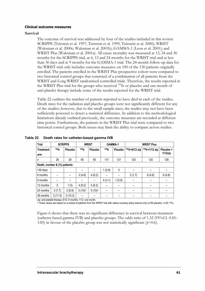
Intravascular brachytherapy 61
Clinical outcome measures
Survival
The outcome of survival was addressed by four of the studies included in this review: SCRIPPS (Teirstein et al. 1997; Teirstein et al. 1999; Teirstein et al. 2000); WRIST (Waksman et al. 2000c; Waksman et al. 2001b); GAMMA-1 (Leon et al. 2001); and WRIST Plus (Waksman et al. 2001a). All cause mortality was measured at 12, 24 and 36 months for the SCRIPPS trial, at 6, 12 and 24 months for the WRIST trial and at less than 30 days and at 9 months for the GAMMA-1 trial. The 24-month follow-up data for the WRIST trial only includes outcome measures on 100 of the 130 patients originally enrolled. The patients enrolled in the WRIST Plus prospective cohort were compared to two historical control groups that consisted of a combination of all patients from the WRIST and Long WRIST randomised controlled trials. Therefore, the results reported in the WRIST Plus trial for the groups who received 192Ir or placebo and one month of anti-platelet therapy include some of the results reported for the WRIST trial.
Table 22 outlines the number of patients reported to have died in each of the studies. Death rates for the radiation and placebo groups were not significantly different for any of the studies; however, due to the small sample sizes, the studies may not have been sufficiently powered to detect a statistical difference. In addition to the methodological limitations already outlined previously, the outcome measures are recorded at different time points. Furthermore, the patients in the WRIST Plus trial were compared to two historical control groups. Both issues may limit the ability to compare across studies.
Table 22 Death rates for catheter-based gamma IVB
Trial SCRIPPS WRIST GAMMA-1 WRIST Plus
Treatment
arm
192Ir Placebo 192Ir Placebo 192Ir Placebo 192Ir+6/12 a/p 192Ir+1/12 a/p Placebo + 1/12a/p
n 26 29 65 65 131 121 120 125 126
Death, number & (%) patients
<30 days – – 1 (0.8) 0 – – –
6 months – – 3 (4.6) 4 (6.2) – – 2 (1.7) 6 (4.8) 6 (4.8)
9 months – – – – 4 (3.1) 1 (0.8) – – –
12 months 0 1 (3) 4 (6.2) 4 (6.2) – – – – –
24 months 2 (7.7) 2 (6.9) 5 (10)a 5 (10)a – – – – –
36 months 3 (11.5) 3 (10.3) – – – – – – –
a/p: anti-platelet therapy; 6/12: 6 months, 1/12: one month. a These values are based on a subset of patients from the WRIST trial with native coronary artery lesions only (n=50 placebo, n=50 192Ir).
Figure 6 shows that there was no significant difference in survival between treatment (catheter-based gamma IVB) and placebo groups. The odds ratio of 1.32 (95%CI. 0.45–3.85) in favour of the placebo group was not statistically significant (p=0.6).
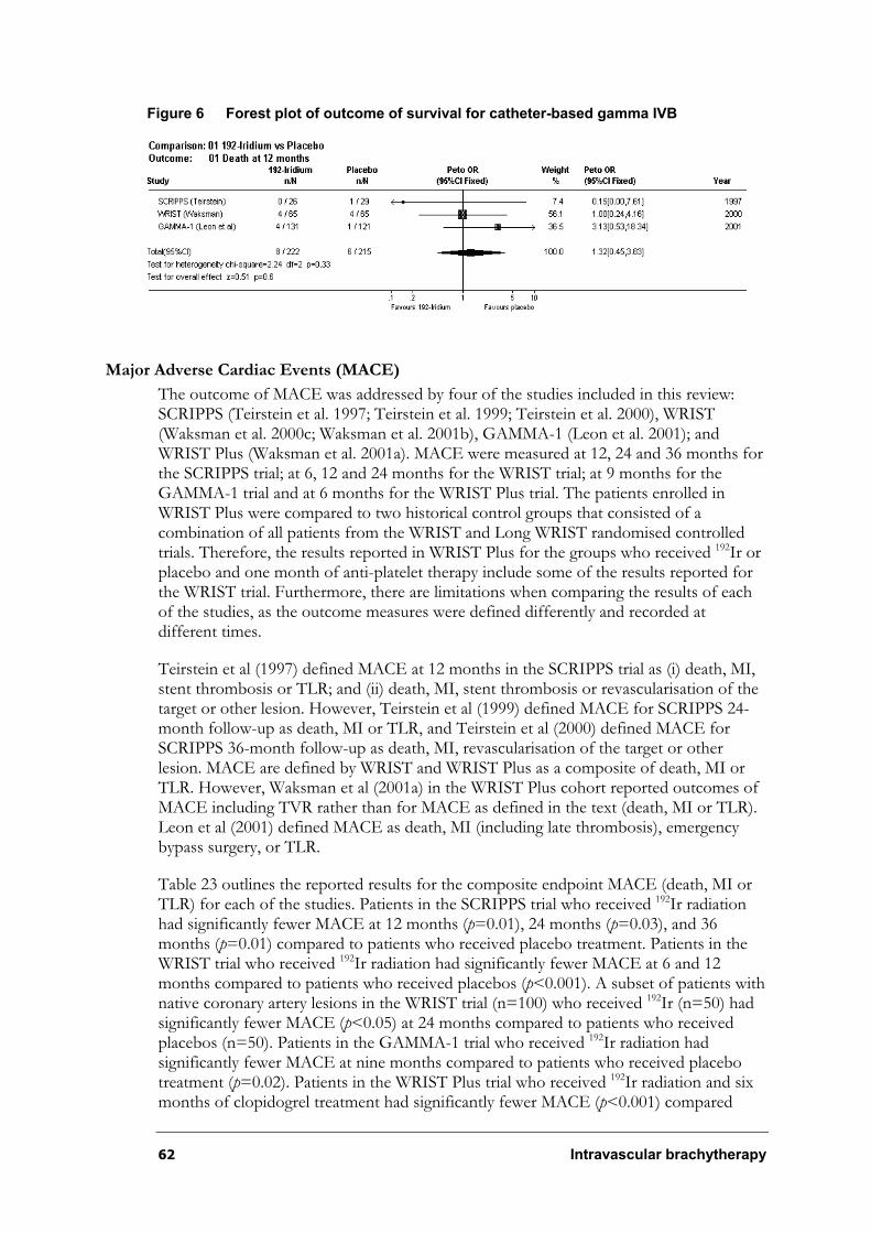
62 Intravascular brachytherapy
Figure 6 Forest plot of outcome of survival for catheter-based gamma IVB
Major Adverse Cardiac Events (MACE)
The outcome of MACE was addressed by four of the studies included in this review: SCRIPPS (Teirstein et al. 1997; Teirstein et al. 1999; Teirstein et al. 2000), WRIST (Waksman et al. 2000c; Waksman et al. 2001b), GAMMA-1 (Leon et al. 2001); and WRIST Plus (Waksman et al. 2001a). MACE were measured at 12, 24 and 36 months for the SCRIPPS trial; at 6, 12 and 24 months for the WRIST trial; at 9 months for the GAMMA-1 trial and at 6 months for the WRIST Plus trial. The patients enrolled in WRIST Plus were compared to two historical control groups that consisted of a combination of all patients from the WRIST and Long WRIST randomised controlled trials. Therefore, the results reported in WRIST Plus for the groups who received 192Ir or placebo and one month of anti-platelet therapy include some of the results reported for the WRIST trial. Furthermore, there are limitations when comparing the results of each of the studies, as the outcome measures were defined differently and recorded at different times.
Teirstein et al (1997) defined MACE at 12 months in the SCRIPPS trial as (i) death, MI, stent thrombosis or TLR; and (ii) death, MI, stent thrombosis or revascularisation of the target or other lesion. However, Teirstein et al (1999) defined MACE for SCRIPPS 24-month follow-up as death, MI or TLR, and Teirstein et al (2000) defined MACE for SCRIPPS 36-month follow-up as death, MI, revascularisation of the target or other lesion. MACE are defined by WRIST and WRIST Plus as a composite of death, MI or TLR. However, Waksman et al (2001a) in the WRIST Plus cohort reported outcomes of MACE including TVR rather than for MACE as defined in the text (death, MI or TLR). Leon et al (2001) defined MACE as death, MI (including late thrombosis), emergency bypass surgery, or TLR.
Table 23 outlines the reported results for the composite endpoint MACE (death, MI or TLR) for each of the studies. Patients in the SCRIPPS trial who received 192Ir radiation had significantly fewer MACE at 12 months (p=0.01), 24 months (p=0.03), and 36 months (p=0.01) compared to patients who received placebo treatment. Patients in the WRIST trial who received 192Ir radiation had significantly fewer MACE at 6 and 12 months compared to patients who received placebos (p<0.001). A subset of patients with native coronary artery lesions in the WRIST trial (n=100) who received 192Ir (n=50) had significantly fewer MACE (p<0.05) at 24 months compared to patients who received placebos (n=50). Patients in the GAMMA-1 trial who received 192Ir radiation had significantly fewer MACE at nine months compared to patients who received placebo treatment (p=0.02). Patients in the WRIST Plus trial who received 192Ir radiation and six months of clopidogrel treatment had significantly fewer MACE (p<0.001) compared

Intravascular brachytherapy 63
with all patients in the WRIST and Long WRIST trials who received placebos and one month of clopidogrel treatment (n=126). There were no significant differences in MACE between patients receiving 192Ir and six months of clopidogrel treatment compared with patients receiving 192Ir and one month of clopidogrel treatment (p=0.13). It is difficult to compare the results of each of the studies due to the limitations described previously.
Table 23 Major adverse cardiac events (MACE) rates for catheter-based gamma IVB
Trial SCRIPPS WRIST GAMMA-1 WRIST Plus
Treatment
arm
192Ir Placebo 192Ir Placebo 192Ir Placebo 192Ir+6/12 a/p 192Ir+1/12 a/p Placebo +1/12a/p
n 26 29 65 65 131 121 120 125 126
Major adverse cardiac events (MACE) , number & (%) patients
6 months – – 19 (29.2)a 44 (67.6) – – 28 (23.3)** 40 (32.0) 80 (63.5)
9 months – – – – 37 (28.2)* 53 (43.8) – – –
12 months 4 (15.3)** 14 (48.3) 23 (35.3)a** 44 (67.6) – – – – –
24 months 6 (23.1)* 15 (51.7) 24 (48.0)ba* 36 (72.0)a – – – – –
36 months 6 (23.1)** 16 (55.2) – – – – – – –
a/p: anti-platelet therapy; 6/12: 6 months, 1/12: one month. *designates significant difference vs placebo (p<0.05); ** designates significant difference vs placebo (p<0.01). a Waksman et al (2000c) does not clearly define the association the P values represent. In accordance with other papers reporting clinical
outcomes, it is possible that the P values reported in the paper describe the degree of association between the 192Ir group and placebo group at 12-months follow-up.
b These values are based on a subset of patients from the WRIST trial with native coronary artery lesions only (n=50 192Ir, n=50 placebo).
Figure 7 shows that there was a significant difference in MACE between treatment (catheter-based gamma IVB) and placebo groups. The odds ratio of 0.38 (95%CI 0.26–0.56) in favour of the treatment group was statistically significant (p<0.00001).
Figure 7 Forest plot of outcome of MACE for catheter-based gamma IVB
Myocardial Infarction (MI)
The outcome of MI was addressed by four of the studies included in this review: SCRIPPS (Teirstein et al. 1997; Teirstein et al. 1999; Teirstein et al. 2000); WRIST (Waksman et al. 2000c; Waksman et al. 2001b); GAMMA-1 (Leon et al. 2001); and WRIST Plus (Waksman et al. 2001a). MI was measured at 12, 24 and 36 months for the SCRIPPS trial; at 6, 12 and 24 months for the WRIST trial; at greater than 30 days and 9 months for the GAMMA-1 trial and at 6 months for the WRIST Plus prospective cohort. The patients enrolled in the WRIST Plus cohort were compared to two historical control groups that comprised a combination of all patients from the WRIST and Long WRIST randomised controlled trials. Therefore, the results reported in the WRIST Plus
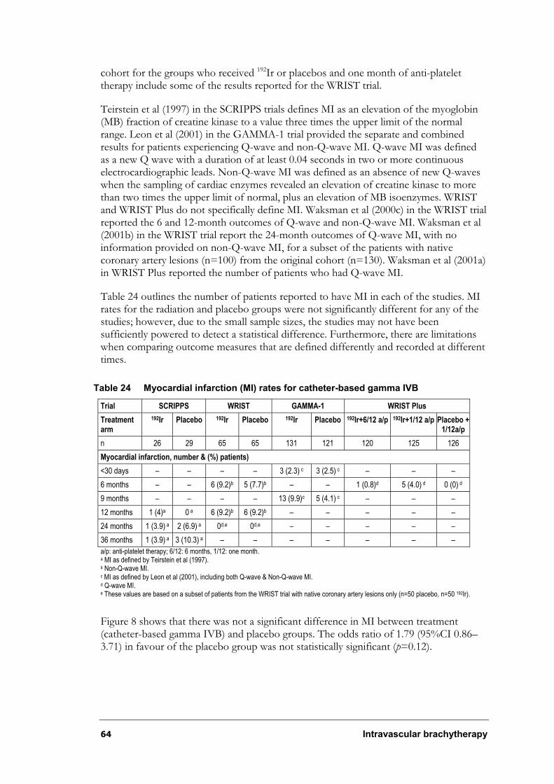
64 Intravascular brachytherapy
cohort for the groups who received 192Ir or placebos and one month of anti-platelet therapy include some of the results reported for the WRIST trial.
Teirstein et al (1997) in the SCRIPPS trials defines MI as an elevation of the myoglobin (MB) fraction of creatine kinase to a value three times the upper limit of the normal range. Leon et al (2001) in the GAMMA-1 trial provided the separate and combined results for patients experiencing Q-wave and non-Q-wave MI. Q-wave MI was defined as a new Q wave with a duration of at least 0.04 seconds in two or more continuous electrocardiographic leads. Non-Q-wave MI was defined as an absence of new Q-waves when the sampling of cardiac enzymes revealed an elevation of creatine kinase to more than two times the upper limit of normal, plus an elevation of MB isoenzymes. WRIST and WRIST Plus do not specifically define MI. Waksman et al (2000c) in the WRIST trial reported the 6 and 12-month outcomes of Q-wave and non-Q-wave MI. Waksman et al (2001b) in the WRIST trial report the 24-month outcomes of Q-wave MI, with no information provided on non-Q-wave MI, for a subset of the patients with native coronary artery lesions (n=100) from the original cohort (n=130). Waksman et al (2001a) in WRIST Plus reported the number of patients who had Q-wave MI.
Table 24 outlines the number of patients reported to have MI in each of the studies. MI rates for the radiation and placebo groups were not significantly different for any of the studies; however, due to the small sample sizes, the studies may not have been sufficiently powered to detect a statistical difference. Furthermore, there are limitations when comparing outcome measures that are defined differently and recorded at different times.
Table 24 Myocardial infarction (MI) rates for catheter-based gamma IVB
Trial SCRIPPS WRIST GAMMA-1 WRIST Plus
Treatmentarm
192Ir Placebo 192Ir Placebo 192Ir Placebo 192Ir+6/12 a/p 192Ir+1/12 a/p Placebo +1/12a/p
n 26 29 65 65 131 121 120 125 126
Myocardial infarction, number & (%) patients)
<30 days – – – – 3 (2.3) c 3 (2.5) c – – –
6 months – – 6 (9.2)b 5 (7.7)b – – 1 (0.8)d 5 (4.0) d 0 (0) d
9 months – – – – 13 (9.9)c 5 (4.1) c – – –
12 months 1 (4)a 0 a 6 (9.2)b 6 (9.2)b – – – – –
24 months 1 (3.9) a 2 (6.9) a 0d,e 0d,e – – – – –
36 months 1 (3.9) a 3 (10.3) a – – – – – – –
a/p: anti-platelet therapy; 6/12: 6 months, 1/12: one month. a MI as defined by Teirstein et al (1997). b Non-Q-wave MI. c MI as defined by Leon et al (2001), including both Q-wave & Non-Q-wave MI. d Q-wave MI. e These values are based on a subset of patients from the WRIST trial with native coronary artery lesions only (n=50 placebo, n=50 192Ir).
Figure 8 shows that there was not a significant difference in MI between treatment (catheter-based gamma IVB) and placebo groups. The odds ratio of 1.79 (95%CI 0.86–3.71) in favour of the placebo group was not statistically significant (p=0.12).
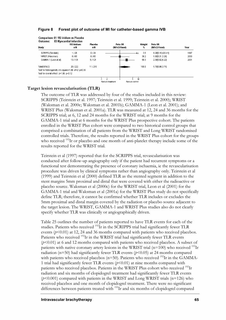
Intravascular brachytherapy 65
Figure 8 Forest plot of outcome of MI for catheter-based gamma IVB
Target lesion revascularisation (TLR)
The outcome of TLR was addressed by four of the studies included in this review: SCRIPPS (Teirstein et al. 1997; Teirstein et al. 1999; Teirstein et al. 2000); WRIST (Waksman et al. 2000c; Waksman et al. 2001b); GAMMA-1 (Leon et al. 2001); and WRIST Plus (Waksman et al. 2001a). TLR was measured at 12, 24 and 36 months for the SCRIPPS trial; at 6, 12 and 24 months for the WRIST trial; at 9 months for the GAMMA-1 trial and at 6 months for the WRIST Plus prospective cohort. The patients enrolled in the WRIST Plus cohort were compared to two historical control groups that comprised a combination of all patients from the WRIST and Long WRIST randomised controlled trials. Therefore, the results reported in the WRIST Plus cohort for the groups who received 192Ir or placebo and one month of anti-platelet therapy include some of the results reported for the WRIST trial.
Teirstein et al (1997) reported that for the SCRIPPS trial, revascularisation was conducted after follow-up angiography only if the patient had recurrent symptoms or a functional test demonstrating the presence of coronary ischaemia, ie the revascularisation procedure was driven by clinical symptoms rather than angiography only. Teirstein et al (1999) and Teirstein et al (2000) defined TLR as the stented segment in addition to the stent margins 5mm proximal and distal that were covered with either the radioactive or placebo source. Waksman et al (2000c) for the WRIST trial, Leon et al (2001) for the GAMMA-1 trial and Waksman et al (2001a) for the WRIST Plus study do not specifically define TLR; therefore, it cannot be confirmed whether TLR includes or excludes the 5mm proximal and distal margin covered by the radiation or placebo source adjacent to the target lesion. The WRIST, GAMMA-1 and WRIST Plus studies also do not clearly specify whether TLR was clinically or angiographically driven.
Table 25 outlines the number of patients reported to have TLR events for each of the studies. Patients who received 192Ir in the SCRIPPS trial had significantly fewer TLR events (p<0.01) at 12, 24 and 36 months compared with patients who received placebos. Patients who received 192Ir in the WRIST trial had significantly fewer TLR events (p<0.01) at 6 and 12 months compared with patients who received placebos. A subset of patients with native coronary artery lesions in the WRIST trial (n=100) who received 192Irradiation (n=50) had significantly fewer TLR events (p<0.05) at 24 months compared with patients who received placebos (n=50). Patients who received 192Ir in the GAMMA-1 trial had significantly fewer TLR events (p<0.01) at nine months compared with patients who received placebos. Patients in the WRIST Plus cohort who received 192Ir radiation and six months of clopidogrel treatment had significantly fewer TLR events (p<0.001) compared with patients in the WRIST and Long WRIST trials (n=126) who received placebos and one month of clopidogrel treatment. There were no significant differences between patients treated with 192Ir and six months of clopidogrel compared
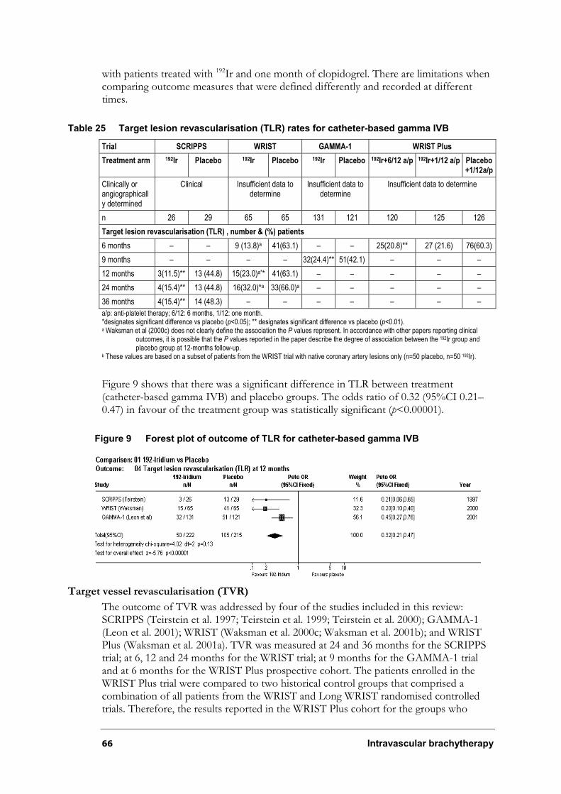
66 Intravascular brachytherapy
with patients treated with 192Ir and one month of clopidogrel. There are limitations when comparing outcome measures that were defined differently and recorded at different times.
Table 25 Target lesion revascularisation (TLR) rates for catheter-based gamma IVB
Trial SCRIPPS WRIST GAMMA-1 WRIST Plus
Treatment arm 192Ir Placebo 192Ir Placebo 192Ir Placebo 192Ir+6/12 a/p 192Ir+1/12 a/p Placebo +1/12a/p
Clinically or angiographically determined
Clinical Insufficient data to determine
Insufficient data to determine
Insufficient data to determine
n 26 29 65 65 131 121 120 125 126
Target lesion revascularisation (TLR) , number & (%) patients
6 months – – 9 (13.8)a 41(63.1) – – 25(20.8)** 27 (21.6) 76(60.3)
9 months – – – – 32(24.4)** 51(42.1) – – –
12 months 3(11.5)** 13 (44.8) 15(23.0)a** 41(63.1) – – – – –
24 months 4(15.4)** 13 (44.8) 16(32.0)*a 33(66.0)a – – – – –
36 months 4(15.4)** 14 (48.3) – – – – – – –
a/p: anti-platelet therapy; 6/12: 6 months, 1/12: one month. *designates significant difference vs placebo (p<0.05); ** designates significant difference vs placebo (p<0.01). a Waksman et al (2000c) does not clearly define the association the P values represent. In accordance with other papers reporting clinical
outcomes, it is possible that the P values reported in the paper describe the degree of association between the 192Ir group and placebo group at 12-months follow-up.
b These values are based on a subset of patients from the WRIST trial with native coronary artery lesions only (n=50 placebo, n=50 192Ir).
Figure 9 shows that there was a significant difference in TLR between treatment (catheter-based gamma IVB) and placebo groups. The odds ratio of 0.32 (95%CI 0.21–0.47) in favour of the treatment group was statistically significant (p<0.00001).
Figure 9 Forest plot of outcome of TLR for catheter-based gamma IVB
Target vessel revascularisation (TVR)
The outcome of TVR was addressed by four of the studies included in this review: SCRIPPS (Teirstein et al. 1997; Teirstein et al. 1999; Teirstein et al. 2000); GAMMA-1 (Leon et al. 2001); WRIST (Waksman et al. 2000c; Waksman et al. 2001b); and WRIST Plus (Waksman et al. 2001a). TVR was measured at 24 and 36 months for the SCRIPPS trial; at 6, 12 and 24 months for the WRIST trial; at 9 months for the GAMMA-1 trial and at 6 months for the WRIST Plus prospective cohort. The patients enrolled in the WRIST Plus trial were compared to two historical control groups that comprised a combination of all patients from the WRIST and Long WRIST randomised controlled trials. Therefore, the results reported in the WRIST Plus cohort for the groups who
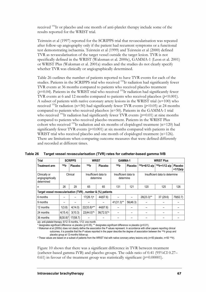
Intravascular brachytherapy 67
received 192Ir or placebo and one month of anti-platelet therapy include some of the results reported for the WRIST trial.
Teirstein et al (1997) reported for the SCRIPPS trial that revascularisation was repeated after follow-up angiography only if the patient had recurrent symptoms or a functional test demonstrating ischaemia. Teirstein et al (1999) and Teirstein et al (2000) defined TVR as revascularisation of the target vessel outside the target lesion. TVR is not specifically defined in the WRIST (Waksman et al. 2000c), GAMMA-1 (Leon et al. 2001) or WRIST Plus (Waksman et al. 2001a) studies and the studies do not clearly specify whether TVR was clinically or angiographically determined.
Table 26 outlines the number of patients reported to have TVR events for each of the studies. Patients in the SCRIPPS trial who received 192Ir radiation had significantly fewer TVR events at 36 months compared to patients who received placebo treatment (p=0.04). Patients in the WRIST trial who received 192Ir radiation had significantly fewer TVR events at 6 and 12 months compared to patients who received placebos (p<0.001).A subset of patients with native coronary artery lesions in the WRIST trial (n=100) who received 192Ir radiation (n=50) had significantly fewer TVR events (p<0.05) at 24 months compared to patients who received placebos (n=50). Patients in the GAMMA-1 trial who received 192Ir radiation had significantly fewer TVR events (p=0.01) at nine months compared to patients who received placebo treatment. Patients in the WRIST Plus cohort who received 192Ir radiation and six months of clopidogrel treatment (n=120) had significantly fewer TVR events (p<0.001) at six months compared with patients in the WRIST trial who received placebo and one month of clopidogrel treatment (n=126). There are limitations when comparing outcome measures that were defined differently and recorded at different times.
Table 26 Target vessel revascularisation (TVR) rates for catheter-based gamma IVB
Trial SCRIPPS WRIST GAMMA-1 WRIST Plus
Treatment arm 192Ir Placebo 192Ir Placebo 192Ir Placebo 192Ir+6/12 a/p 192Ir+1/12 a/p Placebo +1/12a/p
Clinically or angiographically determined
Clinical Insufficient data to determine
Insufficient data to determine
Insufficient data to determine
n 26 29 65 65 131 121 120 125 126
Target vessel revascularisation (TVR) , number & (%) patients
6 months – – 17(26.1)a 44(67.6) – – 28(23.3)** 37 (29.6) 79(62.7)
9 months – – – – 41(31.3)** 56(46.3) – – –
12 months 1(3.8) 4(14.0) 22(33.8)a** 44(67.6) – – – – –
24 months 4(15.4) 3(10.3) 22(44.0)*b 36(72.0)*a – – – – –
36 months 8(30.8)* 17(58.7) – – – – – – –
a/p: anti-platelet therapy; 6/12: 6 months, 1/12: one month. * designates significant difference vs placebo (p<0.05); ** designates significant difference vs placebo (p<0.01). a Waksman et al (2000c) does not clearly define the association the P values represent. In accordance with other papers reporting clinical
outcomes, it is possible that the P values reported in the paper describe the degree of association between the 192Ir group and placebo group at 12-months follow-up.
b These values are based on a subset of patients from the WRIST trial with native coronary artery lesions only (n=50 placebo, n=50 192Ir).
Figure 10 shows that there was a significant difference in TVR between treatment (catheter-based gamma IVB) and placebo groups. The odds ratio of 0.41 (95%CI 0.27–0.61) in favour of the treatment group was statistically significant (p=0.00001).
68 Intravascular brachytherapy
Figure 10 Forest plot of outcome of TVR for catheter-based gamma IVB
Summary—Clinical outcomes
Meta-analyses indicated that, compared with placebos, catheter-based gamma IVB appeared to be significantly associated with reduced MACE (OR= 0.38; 95%CI 0.26–0.56, p<0.0001), TLR events (OR=0.32; 95%CI 0.21–0.47, p<0.00001) and TVR events (OR=0.41; 95%CI 0.27–0.61, p=0.00001) at six months. Individual trial data suggested that gamma IVB may be associated with higher death and MI rates at six months. However, when data was combined in a meta-analysis, there were no significant differences between active and placebo groups for either outcome: survival (OR=1.32; 95%CI. 0.45–3.85, p=0.6) and MI (OR=1.79; 95%CI 0.86–3.71, p=0.12).
Caution should be used when interpreting these results as some outcomes were defined differently between studies and were reported at different times. As it is unclear in some studies whether revascularistaion was driven by angiography or clinical symptoms, it is possible that the TLR/TVR rates may overestimate the true number of patients requiring revascularisation in clinical practice. In addition to the limitations already raised previously in the report, these limitations should be considered when interpreting these results, and making generalisations to the wider patient population.
Angiographic outcome measures
Minimal lumen diameter (MLD)
The angiographic measure of MLD was addressed by four gamma studies included in this review: SCRIPPS (Teirstein et al. 1997; Teirstein et al. 1999; Teirstein et al. 2000); WRIST (Waksman et al. 2000c; Waksman et al. 2001b); GAMMA-1 (Leon et al. 2001); and WRIST Plus (Waksman et al. 2001a). MLD was measured pre-operatively, post-operatively and at six months by all four trials. The SCRIPPS trial defined MLD as including the area within the stent and its margins (the area beyond the stent but exposed to the radiation source). Waksman et al (2000c) in the WRIST trial defined MLD as including only the stent area. Leon et al (2001) in the GAMMA-1 trial defined MLD as: (i) including the segment of the vessel in which the stent was implanted (in-stent MLD); and (ii) including the in-stent segment in addition to the 5mm adjacent areas, as well as any additional area exposed to the radioactive ribbon (in-lesion MLD). Waksman et al (2001a) in the WRIST Plus prospective cohort did not clearly define MLD; however, it is implied to include the stent and adjacent 5mm area. Furthermore, it is unclear as to whether the angiographic results for the WRIST Plus study were based on the entire sample, or a subset of patients. The differing definitions of MLD used by the various papers limit the degree to which results can be compared. Table 27 outlines the results of MLD for each of the trials.
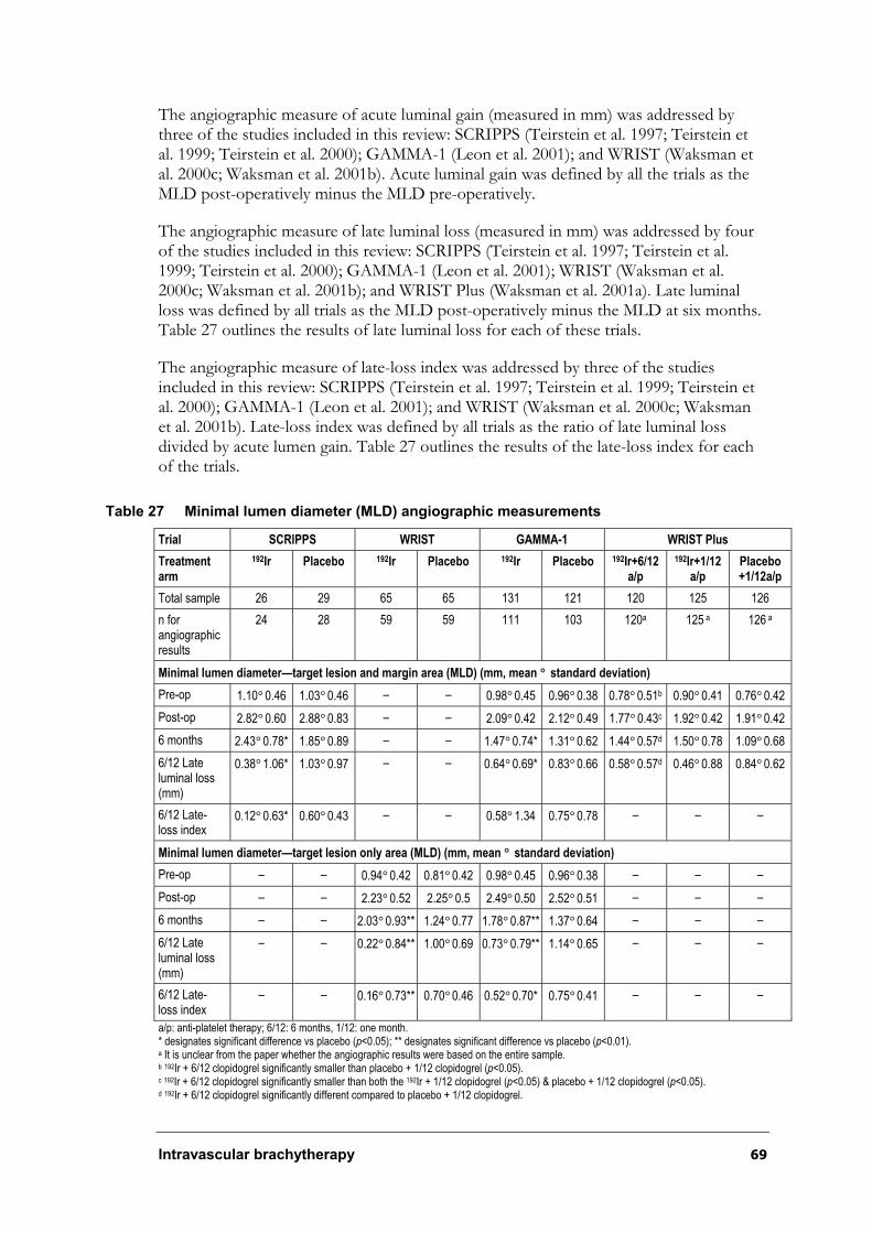
Intravascular brachytherapy 69
The angiographic measure of acute luminal gain (measured in mm) was addressed by three of the studies included in this review: SCRIPPS (Teirstein et al. 1997; Teirstein et al. 1999; Teirstein et al. 2000); GAMMA-1 (Leon et al. 2001); and WRIST (Waksman et al. 2000c; Waksman et al. 2001b). Acute luminal gain was defined by all the trials as the MLD post-operatively minus the MLD pre-operatively.
The angiographic measure of late luminal loss (measured in mm) was addressed by four of the studies included in this review: SCRIPPS (Teirstein et al. 1997; Teirstein et al. 1999; Teirstein et al. 2000); GAMMA-1 (Leon et al. 2001); WRIST (Waksman et al. 2000c; Waksman et al. 2001b); and WRIST Plus (Waksman et al. 2001a). Late luminal loss was defined by all trials as the MLD post-operatively minus the MLD at six months. Table 27 outlines the results of late luminal loss for each of these trials.
The angiographic measure of late-loss index was addressed by three of the studies included in this review: SCRIPPS (Teirstein et al. 1997; Teirstein et al. 1999; Teirstein et al. 2000); GAMMA-1 (Leon et al. 2001); and WRIST (Waksman et al. 2000c; Waksman et al. 2001b). Late-loss index was defined by all trials as the ratio of late luminal loss divided by acute lumen gain. Table 27 outlines the results of the late-loss index for each of the trials.
Table 27 Minimal lumen diameter (MLD) angiographic measurements
Trial SCRIPPS WRIST GAMMA-1 WRIST Plus
Treatmentarm
192Ir Placebo 192Ir Placebo 192Ir Placebo 192Ir+6/12a/p
192Ir+1/12a/p
Placebo +1/12a/p
Total sample 26 29 65 65 131 121 120 125 126
n for angiographic results
24 28 59 59 111 103 120a 125 a 126 a
Minimal lumen diameter—target lesion and margin area (MLD) (mm, mean ° standard deviation)
Pre-op 1.10°0.46 1.03°0.46 – – 0.98°0.45 0.96°0.38 0.78°0.51b 0.90°0.41 0.76°0.42
Post-op 2.82°0.60 2.88°0.83 – – 2.09°0.42 2.12°0.49 1.77°0.43c 1.92°0.42 1.91°0.42
6 months 2.43°0.78* 1.85°0.89 – – 1.47°0.74* 1.31°0.62 1.44°0.57d 1.50°0.78 1.09°0.68
6/12 Late luminal loss (mm)
0.38°1.06* 1.03°0.97 – – 0.64°0.69* 0.83°0.66 0.58°0.57d 0.46°0.88 0.84°0.62
6/12 Late-loss index
0.12°0.63* 0.60°0.43 – – 0.58°1.34 0.75°0.78 – – –
Minimal lumen diameter—target lesion only area (MLD) (mm, mean ° standard deviation)
Pre-op – – 0.94°0.42 0.81°0.42 0.98°0.45 0.96°0.38 – – –
Post-op – – 2.23°0.52 2.25°0.5 2.49°0.50 2.52°0.51 – – –
6 months – – 2.03°0.93** 1.24°0.77 1.78°0.87** 1.37°0.64 – – –
6/12 Late luminal loss (mm)
– – 0.22°0.84** 1.00°0.69 0.73°0.79** 1.14°0.65 – – –
6/12 Late-loss index
– – 0.16°0.73** 0.70°0.46 0.52°0.70* 0.75°0.41 – – –
a/p: anti-platelet therapy; 6/12: 6 months, 1/12: one month. * designates significant difference vs placebo (p<0.05); ** designates significant difference vs placebo (p<0.01). a It is unclear from the paper whether the angiographic results were based on the entire sample. b 192Ir + 6/12 clopidogrel significantly smaller than placebo + 1/12 clopidogrel (p<0.05). c 192Ir + 6/12 clopidogrel significantly smaller than both the 192Ir + 1/12 clopidogrel (p<0.05) & placebo + 1/12 clopidogrel (p<0.05). d 192Ir + 6/12 clopidogrel significantly different compared to placebo + 1/12 clopidogrel.
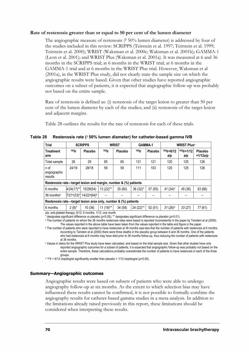
70 Intravascular brachytherapy
Rate of restenosis greater than or equal to 50 per cent of the lumen diameter
The angiographic measure of restenosis (²50% lumen diameter) is addressed by four of the studies included in this review: SCRIPPS (Teirstein et al. 1997; Teirstein et al. 1999; Teirstein et al. 2000); WRIST (Waksman et al. 2000c; Waksman et al. 2001b); GAMMA-1 (Leon et al. 2001); and WRIST Plus (Waksman et al. 2001a). It was measured at 6 and 36 months in the SCRIPPS trial; at 6 months in the WRIST trial; at 6 months in the GAMMA-1 trial and at 6 months in the WRIST Plus trial. However, Waksman et al (2001a), in the WRIST Plus study, did not clearly state the sample size on which the angiographic results were based. Given that other studies have reported angiographic outcomes on a subset of patients, it is expected that angiographic follow-up was probably not based on the entire sample.
Rate of restenosis is defined as: (i) restenosis of the target lesion to greater than 50 per cent of the lumen diameter by each of the studies; and (ii) restenosis of the target lesion and adjacent margins.
Table 28 outlines the results for the rate of restenosis for each of these trials.
Table 28 Restenosis rate (²50% lumen diameter) for catheter-based gamma IVB
Trial SCRIPPS WRIST GAMMA-1 WRIST Plusc
Treatmentarm
192Ir Placebo 192Ir Placebo 192Ir Placebo 192Ir+6/12a/p
192Ir+1/12a/p
Placebo +1/12a/p
Total sample 26 29 65 65 131 121 120 125 126
n of angiographic results
24/19 28/18 59 59 111 103 120 125 126
Restenosis rate—target lesion and margin, number & (%) patients
6 months 4/24(17)** 15/28(54) 13 (22)** 35 (60) 36 (32)* 57 (55) 41 (34)d 45 (36) 83 (66)
36 monthsa 7/21a(33)* 14/22a(64)b – – – – – – –
Restenosis rate—target lesion area only, number & (%) patients
6 months 2 (8)* 10 (36) 11 (19)** 34 (58) 24 (22)** 52 (51) 31 (26)d 33 (27) 77 (61)
a/p: anti-platelet therapy; 6/12: 6 months, 1/12: one month. * designates significant difference vs placebo (p<0.05); ** designates significant difference vs placebo (p<0.01). a The number of patients on whom the 36 months restenosis rates were based is reported inconsistently in the paper by Teirstein et al (2000).
The values reported in the above table have been taken from the values reported in the table and figure in the paper. b The number of patients who were reported to have restenosis at 36 months was less that the number of patients with restenosis at 6 months.
According to Teirstein et al (2000) there were three deaths in the placebo group between 6 and 36 months. One of the patients who had restenosis at 6 months may have died prior to 36 months follow-up, thus reducing the number of patients with restenosisat 36 months.
c Values in italics for the WRIST Plus study have been calculated, and based on the total sample size. Given that other studies have only reported angiographic outcomes for a subset of patients, it is expected that angiographic follow-up was probably not based on theentire sample. Therefore, these calculations probably overestimate the number of patients to have restenosis in each of the threegroups.
d 192Ir + 6/12 clopidogrel significantly smaller than placebo + 1/12 clopidogrel (p<0.05).
Summary—Angiographic outcomes
Angiographic results were based on subsets of patients who were able to undergo angiography follow-up at six months. As the extent to which selection bias may have influenced these results cannot be confirmed, it is not possible to formally combine the angiography results for catheter-based gamma studies in a meta-analysis. In addition to the limitations already raised previously in this report, these limitations should be considered when interpreting these results.
Intravascular brachytherapy 71
Based on six-month follow-up evidence from the randomised controlled trials, the following summaries can be made:
Minimal lumen diameter (MLD) of the target lesion and adjacent margin ranged from
1.47°0.74 to 2.43°0.78 mm for patients who received active treatment, compared with a
range of 1.31°0.62 to 1.85°0.89 mm for patients in the placebo group. MLD of the
target lesion only ranged from 1.78°0.87 to 2.03°0.93 mm for patients who received
active treatment, compared with a range of 1.24°0.77 to 1.37°0.64 mm for patients in the placebo group.
Late lumen loss of the target lesion and adjacent margin ranged from 0.38°1.06 to
0.64°0.69 mm for patients who received active treatment, compared with a range of
0.60°0.43 to 0.75°0.78 mm for patients in the placebo group. Late lumen loss of the
target lesion only ranged from 0.22°0.84 to 0.73°0.79 mm for patients who received
active treatment, compared with a range of 1.00°0.69 to 1.14°0.65 mm for patients in the placebo group.
Late-loss index at six months of the target lesion and adjacent margin ranged from
0.12°0.63 to 0.58°1.34 for patients who received active treatment, compared with a
range of 0.60°0.43 to 0.75°0.78 for patients in the placebo group. Late-loss index at the
target lesion only ranged from 0.16°0.73 to 0.52°0.70 for patients who received active
treatment, compared with a range of 0.70°0.46 to 0.75°0.41 for patients in the placebo group.
The restenosis rate (²50% of lumen diameter) of the target lesion and adjacent margin ranged from 17 to 32 per cent for patients who received the active treatment, compared with a range of 54 to 60 per cent for patients in the placebo group. The restenosis rate
(²50% of lumen diameter) of the target lesion only ranged from 8 to 22 per cent for patients who received active treatment, compared with a range of 36 to 58 per cent for patients in the placebo group.
Given the limitations stated previously, it would appear, therefore, that compared with patient groups receiving placebo, those who were treated with catheter-based gamma IVB presented with a wider lumen at six-month angiographic follow-up.
Intravascular ultrasound (IVUS) outcome measures
Table 29 outlines the IVUS results for each of the following studies:
¶ SCRIPPS (Teirstein et al. 1997);
¶ WRIST (Waksman et al. 2000c);
¶ Long WRIST (Ahmed et al. 2001c); and
¶ HD Long WRIST (Ahmed et al. 2001b).
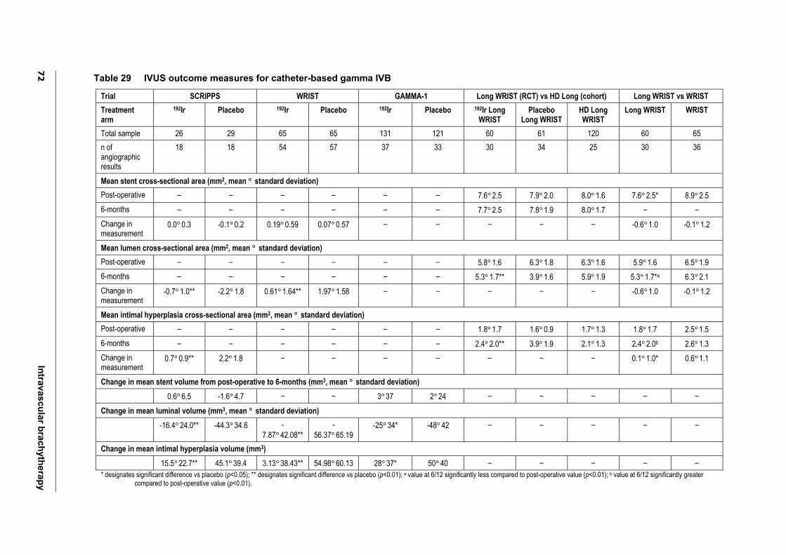
72
In
trav
as
cu
lar b
rac
hyth
era
py
Table 29 IVUS outcome measures for catheter-based gamma IVB
Trial SCRIPPS WRIST GAMMA-1 Long WRIST (RCT) vs HD Long (cohort) Long WRIST vs WRIST
Treatmentarm
192Ir Placebo 192Ir Placebo 192Ir Placebo 192Ir Long WRIST
Placebo Long WRIST
HD Long WRIST
Long WRIST WRIST
Total sample 26 29 65 65 131 121 60 61 120 60 65
n of angiographic results
18 18 54 57 37 33 30 34 25 30 36
Mean stent cross-sectional area (mm2, mean ° standard deviation)
Post-operative – – – – – – 7.6°2.5 7.9°2.0 8.0°1.6 7.6°2.5* 8.9°2.5
6-months – – – – – – 7.7°2.5 7.8°1.9 8.0°1.7 – –
Change in measurement
0.0°0.3 -0.1°0.2 0.19°0.59 0.07°0.57 – – – – – -0.6°1.0 -0.1°1.2
Mean lumen cross-sectional area (mm2, mean ° standard deviation)
Post-operative – – – – – – 5.8°1.6 6.3°1.8 6.3°1.6 5.9°1.6 6.5°1.9
6-months – – – – – – 5.3°1.7** 3.9°1.6 5.9°1.9 5.3°1.7*a 6.3°2.1
Change in measurement
-0.7°1.0** -2.2°1.8 0.61°1.64** 1.97°1.58 – – – – – -0.6°1.0 -0.1°1.2
Mean intimal hyperplasia cross-sectional area (mm2, mean ° standard deviation)
Post-operative – – – – – – 1.8°1.7 1.6°0.9 1.7°1.3 1.8°1.7 2.5°1.5
6-months – – – – – – 2.4°2.0** 3.9°1.9 2.1°1.3 2.4°2.0b 2.6°1.3
Change in measurement
0.7°0.9** 2.2°1.8 – – – – – – – 0.1°1.0* 0.6°1.1
Change in mean stent volume from post-operative to 6-months (mm3, mean ° standard deviation)
0.6°6.5 -1.6°4.7 – – 3°37 2°24 – – – – –
Change in mean luminal volume (mm3, mean ° standard deviation)
-16.4°24.0** -44.3°34.6 -7.87°42.08**
-56.37°65.19
-25°34* -48°42 – – – – –
Change in mean intimal hyperplasia volume (mm3)
15.5°22.7** 45.1°39.4 3.13°38.43** 54.98°60.13 28°37* 50°40 – – – – –
* designates significant difference vs placebo (p<0.05); ** designates significant difference vs placebo (p<0.01); a value at 6/12 significantly less compared to post-operative value (p<0.01); b value at 6/12 significantly greater compared to post-operative value (p<0.01).
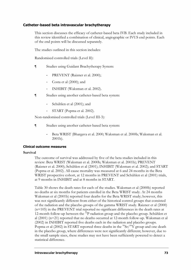
Intravascular brachytherapy 73
Catheter-based beta intravascular brachytherapy
This section discusses the efficacy of catheter-based beta IVB. Each study included in this review identified a combination of clinical, angiographic or IVUS end points. Each of the end points will be discussed separately.
The studies outlined in this section includes:
Randomised controlled trials (Level II):
¶ Studies using Guidant Brachytherapy System:
- PREVENT (Raizner et al. 2000);
- Costa et al (2000); and
- INHIBIT (Waksman et al. 2002).
¶ Studies using another catheter-based beta system:
- Schühlen et al (2001); and
- START (Popma et al. 2002).
Non-randomised controlled trials (Level III-3):
¶ Studies using another catheter-based beta system:
- Beta WRIST (Bhargava et al. 2000; Waksman et al. 2000b; Waksman et al. 2001b).
Clinical outcome measures
Survival
The outcome of survival was addressed by five of the beta studies included in this review: Beta WRIST (Waksman et al. 2000b; Waksman et al. 2001b); PREVENT (Raizner et al. 2000); Schühlen et al (2001); INHIBIT (Waksman et al. 2002); and START (Popma et al. 2002). All cause mortality was measured at 6 and 24 months in the Beta WRIST prospective cohort, at 12 months in PREVENT and Schühlen et al (2001) trials, at 9 months in INHIBIT and at 8 months in START.
Table 30 shows the death rates for each of the studies. Waksman et al (2000b) reported no deaths at six months for patients enrolled in the Beta WRIST study. At 24 months Waksman et al (2001b) reported four deaths for the Beta WRIST study; however, this was not significantly different from either of the historical control groups that consisted of the radiation and the placebo groups of the gamma WRIST study. Raizner et al (2000) (n=105) in the PREVENT trial reported no significant differences in the death rates at 12-month follow-up between the 32P radiation group and the placebo group. Schühlen et al (2001) (n=21) reported that no deaths occurred at 12-month follow-up. Waksman et al (2002) in INHIBIT reported five deaths each in the radiation and placebo groups. Popma et al (2002) in START reported three deaths in the 90Sr/90Y group and one death in the placebo group, where differences were not significantly different; however, due to the small sample sizes, these studies may not have been sufficiently powered to detect a statistical difference.
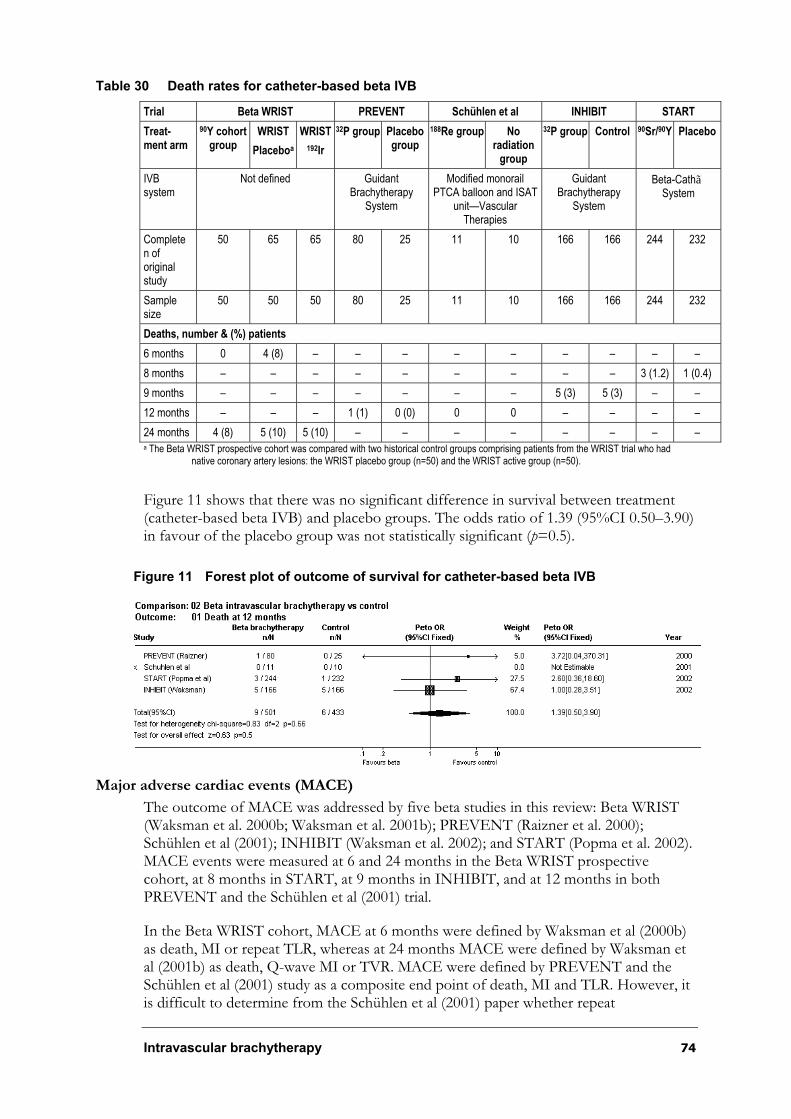
Intravascular brachytherapy 74
Table 30 Death rates for catheter-based beta IVB
Trial Beta WRIST PREVENT Schühlen et al INHIBIT START
Treat-ment arm
90Y cohort group
WRIST
Placeboa
WRIST
192Ir
32P group Placebo group
188Re group Noradiation
group
32P group Control 90Sr/90Y Placebo
IVBsystem
Not defined Guidant Brachytherapy
System
Modified monorail PTCA balloon and ISAT
unit—Vascular Therapies
GuidantBrachytherapy
System
Beta-CathãSystem
Completen of original study
50 65 65 80 25 11 10 166 166 244 232
Samplesize
50 50 50 80 25 11 10 166 166 244 232
Deaths, number & (%) patients
6 months 0 4 (8) – – – – – – – – –
8 months – – – – – – – – – 3 (1.2) 1 (0.4)
9 months – – – – – – – 5 (3) 5 (3) – –
12 months – – – 1 (1) 0 (0) 0 0 – – – –
24 months 4 (8) 5 (10) 5 (10) – – – – – – – – a The Beta WRIST prospective cohort was compared with two historical control groups comprising patients from the WRIST trial who had
native coronary artery lesions: the WRIST placebo group (n=50) and the WRIST active group (n=50).
Figure 11 shows that there was no significant difference in survival between treatment (catheter-based beta IVB) and placebo groups. The odds ratio of 1.39 (95%CI 0.50–3.90) in favour of the placebo group was not statistically significant (p=0.5).
Figure 11 Forest plot of outcome of survival for catheter-based beta IVB
Major adverse cardiac events (MACE)
The outcome of MACE was addressed by five beta studies in this review: Beta WRIST (Waksman et al. 2000b; Waksman et al. 2001b); PREVENT (Raizner et al. 2000); Schühlen et al (2001); INHIBIT (Waksman et al. 2002); and START (Popma et al. 2002). MACE events were measured at 6 and 24 months in the Beta WRIST prospective cohort, at 8 months in START, at 9 months in INHIBIT, and at 12 months in both PREVENT and the Schühlen et al (2001) trial.
In the Beta WRIST cohort, MACE at 6 months were defined by Waksman et al (2000b) as death, MI or repeat TLR, whereas at 24 months MACE were defined by Waksman et al (2001b) as death, Q-wave MI or TVR. MACE were defined by PREVENT and the Schühlen et al (2001) study as a composite end point of death, MI and TLR. However, it is difficult to determine from the Schühlen et al (2001) paper whether repeat
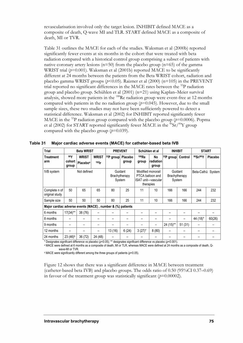
Intravascular brachytherapy 75
revascularisation involved only the target lesion. INHIBIT defined MACE as a composite of death, Q-wave MI and TLR. START defined MACE as a composite of death, MI or TVR.
Table 31 outlines the MACE for each of the studies. Waksman et al (2000b) reported significantly fewer events at six months in the cohort that were treated with beta radiation compared with a historical control group comprising a subset of patients with native coronary artery lesions (n=50) from the placebo group (n=65) of the gamma WRIST trial (p=0.001). Waksman et al (2001b) reported MACE to be significantly different at 24 months between the patients from the Beta WRIST cohort, radiation and placebo gamma WRIST groups (p<0.05). Raizner et al (2000) (n=105) in the PREVENT trial reported no significant differences in the MACE rates between the 32P radiation group and placebo group. Schühlen et al (2001) (n=21) using Kaplan–Meier survival analysis, showed more patients in the 188Re radiation group were event-free at 12 months compared with patients in the no radiation group (p=0.045). However, due to the small sample sizes, these two studies may not have been sufficiently powered to detect a statistical difference. Waksman et al (2002) for INHIBIT reported significantly fewer MACE in the 32P radiation group compared with the placebo group (p=0.0006). Popma et al (2002) for START reported significantly fewer MACE in the 90Sr/90Y group compared with the placebo group (p=0.039).
Table 31 Major cardiac adverse events (MACE) for catheter-based beta IVB
Trial Beta WRIST PREVENT Schühlen et al INHIBIT START
Treatmentarm
90Ycohortgroup
WRIST
Placeboa
WRIST
192Ir
32P group Placebo group
188Regroup
Noradiation
group
32P group Control 90Sr/90Y Placebo
IVB system Not defined Guidant Brachytherapy
System
Modified monorail PTCA balloon and
ISAT unit—vasculartherapies
GuidantBrachytherapy
System
Beta-Cathã System
Complete n of original study
50 65 65 80 25 11 10 166 166 244 232
Sample size 50 50 50 80 25 11 10 166 166 244 232
Major cardiac adverse events (MACE) , number & (%) patients
6 months 17(34)** 38 (76) – – – – – – – – –
8 months – – – – – – – – – 44 (18)* 60(26)
9 months – – – – – – – 24 (15)** 51 (31) – –
12 months – – – 13 (16) 6 (24) 3 (27)* 8 (80) – – – –
24 months 23 (46)b 36 (72) 24 (48) – – – – – – – –
* Designates significant difference vs placebo (p<0.05); ** designates significant difference vs placebo (p<0.001). a MACE were defined at 6 months as a composite of death, MI or TLR, whereas MACE were defined at 24 months as a composite of death, Q-
wave-MI or TVR. b MACE were significantly different among the three groups of patients (p<0.05).
Figure 12 shows that there was a significant difference in MACE between treatment (catheter-based beta IVB) and placebo groups. The odds ratio of 0.50 (95%CI 0.37–0.69) in favour of the treatment group was statistically significant (p=0.00002).
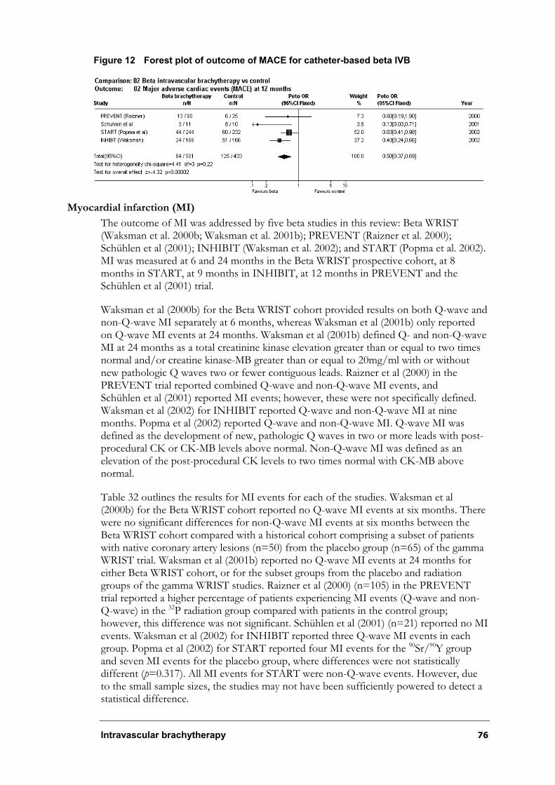
Intravascular brachytherapy 76
Figure 12 Forest plot of outcome of MACE for catheter-based beta IVB
Myocardial infarction (MI)
The outcome of MI was addressed by five beta studies in this review: Beta WRIST (Waksman et al. 2000b; Waksman et al. 2001b); PREVENT (Raizner et al. 2000); Schühlen et al (2001); INHIBIT (Waksman et al. 2002); and START (Popma et al. 2002). MI was measured at 6 and 24 months in the Beta WRIST prospective cohort, at 8 months in START, at 9 months in INHIBIT, at 12 months in PREVENT and the Schühlen et al (2001) trial.
Waksman et al (2000b) for the Beta WRIST cohort provided results on both Q-wave and non-Q-wave MI separately at 6 months, whereas Waksman et al (2001b) only reported on Q-wave MI events at 24 months. Waksman et al (2001b) defined Q- and non-Q-wave MI at 24 months as a total creatinine kinase elevation greater than or equal to two times normal and/or creatine kinase-MB greater than or equal to 20mg/ml with or without new pathologic Q waves two or fewer contiguous leads. Raizner et al (2000) in the PREVENT trial reported combined Q-wave and non-Q-wave MI events, andSchühlen et al (2001) reported MI events; however, these were not specifically defined. Waksman et al (2002) for INHIBIT reported Q-wave and non-Q-wave MI at nine months. Popma et al (2002) reported Q-wave and non-Q-wave MI. Q-wave MI was defined as the development of new, pathologic Q waves in two or more leads with post-procedural CK or CK-MB levels above normal. Non-Q-wave MI was defined as an elevation of the post-procedural CK levels to two times normal with CK-MB above normal.
Table 32 outlines the results for MI events for each of the studies. Waksman et al (2000b) for the Beta WRIST cohort reported no Q-wave MI events at six months. There were no significant differences for non-Q-wave MI events at six months between the Beta WRIST cohort compared with a historical cohort comprising a subset of patients with native coronary artery lesions (n=50) from the placebo group (n=65) of the gamma WRIST trial. Waksman et al (2001b) reported no Q-wave MI events at 24 months for either Beta WRIST cohort, or for the subset groups from the placebo and radiation groups of the gamma WRIST studies. Raizner et al (2000) (n=105) in the PREVENT trial reported a higher percentage of patients experiencing MI events (Q-wave and non-Q-wave) in the 32P radiation group compared with patients in the control group; however, this difference was not significant. Schühlen et al (2001) (n=21) reported no MI events. Waksman et al (2002) for INHIBIT reported three Q-wave MI events in each group. Popma et al (2002) for START reported four MI events for the 90Sr/90Y group and seven MI events for the placebo group, where differences were not statistically different (p=0.317). All MI events for START were non-Q-wave events. However, due to the small sample sizes, the studies may not have been sufficiently powered to detect a statistical difference.
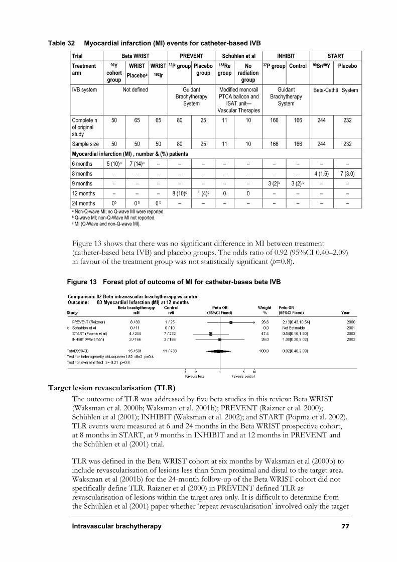
Intravascular brachytherapy 77
Table 32 Myocardial infarction (MI) events for catheter-based IVB
Trial Beta WRIST PREVENT Schühlen et al INHIBIT START
Treatmentarm
90Ycohortgroup
WRIST
Placeboa
WRIST
192Ir
32P group Placebo group
188Regroup
Noradiation
group
32P group Control 90Sr/90Y Placebo
IVB system Not defined Guidant Brachytherapy
System
Modified monorail PTCA balloon and
ISAT unit—Vascular Therapies
GuidantBrachytherapy
System
Beta-Cathã System
Complete n of original study
50 65 65 80 25 11 10 166 166 244 232
Sample size 50 50 50 80 25 11 10 166 166 244 232
Myocardial infarction (MI) , number & (%) patients
6 months 5 (10)a 7 (14)a – – – – – – – – –
8 months – – – – – – – – – 4 (1.6) 7 (3.0)
9 months – – – – – – – 3 (2)b 3 (2) b – –
12 months – – – 8 (10)c 1 (4)c 0 0 – – – –
24 months 0b 0 b 0 b – – – – – – – – a Non-Q-wave MI; no Q-wave MI were reported. b Q-wave MI; non-Q-Wave MI not reported. c MI (Q-Wave and non-Q-wave MI).
Figure 13 shows that there was no significant difference in MI between treatment (catheter-based beta IVB) and placebo groups. The odds ratio of 0.92 (95%CI 0.40–2.09) in favour of the treatment group was not statistically significant (p=0.8).
Figure 13 Forest plot of outcome of MI for catheter-bases beta IVB
Target lesion revascularisation (TLR)
The outcome of TLR was addressed by five beta studies in this review: Beta WRIST (Waksman et al. 2000b; Waksman et al. 2001b); PREVENT (Raizner et al. 2000); Schühlen et al (2001); INHIBIT (Waksman et al. 2002); and START (Popma et al. 2002). TLR events were measured at 6 and 24 months in the Beta WRIST prospective cohort, at 8 months in START, at 9 months in INHIBIT and at 12 months in PREVENT and the Schühlen et al (2001) trial.
TLR was defined in the Beta WRIST cohort at six months by Waksman et al (2000b) to include revascularisation of lesions less than 5mm proximal and distal to the target area. Waksman et al (2001b) for the 24-month follow-up of the Beta WRIST cohort did not specifically define TLR. Raizner et al (2000) in PREVENT defined TLR as revascularisation of lesions within the target area only. It is difficult to determine from the Schühlen et al (2001) paper whether ‘repeat revascularisation’ involved only the target
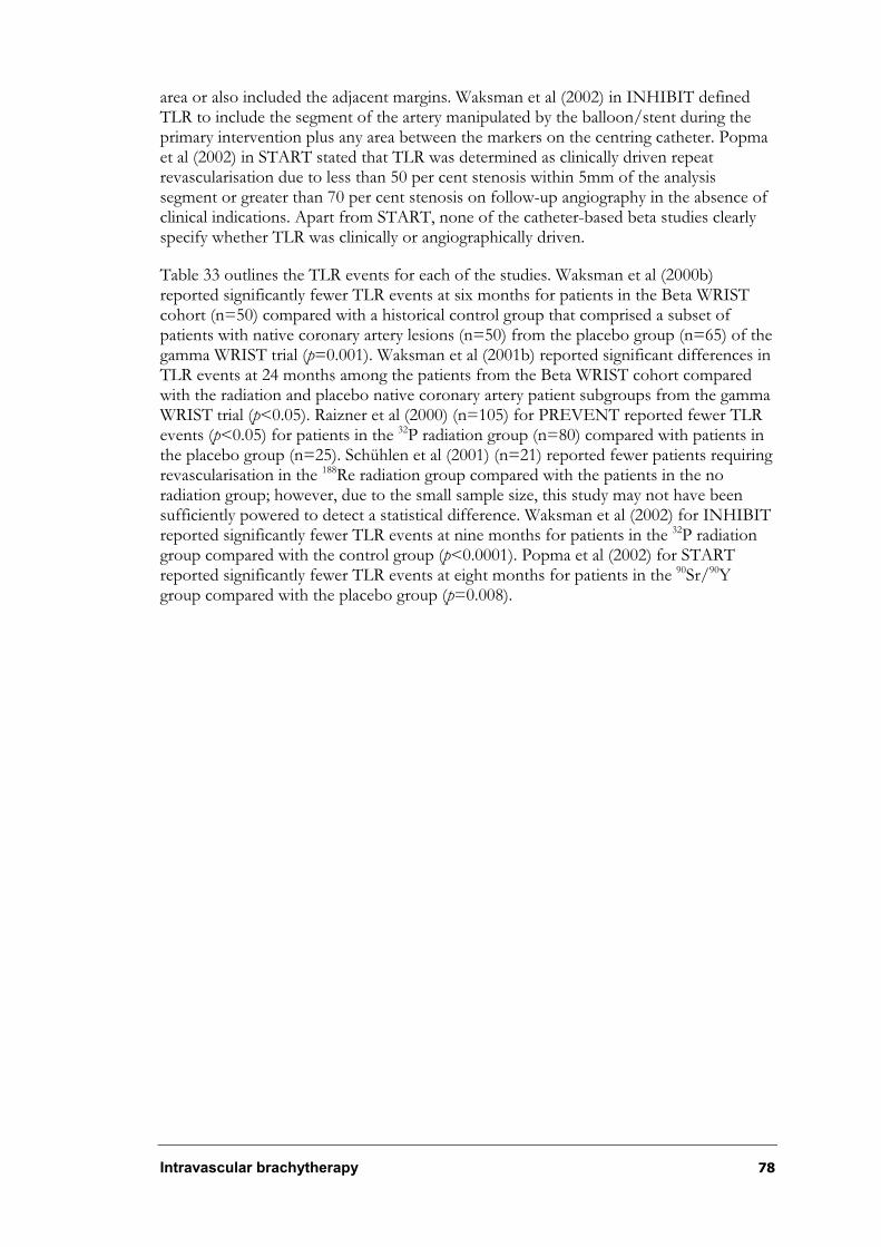
Intravascular brachytherapy 78
area or also included the adjacent margins. Waksman et al (2002) in INHIBIT defined TLR to include the segment of the artery manipulated by the balloon/stent during the primary intervention plus any area between the markers on the centring catheter. Popma et al (2002) in START stated that TLR was determined as clinically driven repeat revascularisation due to less than 50 per cent stenosis within 5mm of the analysis segment or greater than 70 per cent stenosis on follow-up angiography in the absence of clinical indications. Apart from START, none of the catheter-based beta studies clearly specify whether TLR was clinically or angiographically driven.
Table 33 outlines the TLR events for each of the studies. Waksman et al (2000b) reported significantly fewer TLR events at six months for patients in the Beta WRIST cohort (n=50) compared with a historical control group that comprised a subset of patients with native coronary artery lesions (n=50) from the placebo group (n=65) of the gamma WRIST trial (p=0.001). Waksman et al (2001b) reported significant differences in TLR events at 24 months among the patients from the Beta WRIST cohort compared with the radiation and placebo native coronary artery patient subgroups from the gamma WRIST trial (p<0.05). Raizner et al (2000) (n=105) for PREVENT reported fewer TLR events (p<0.05) for patients in the 32P radiation group (n=80) compared with patients in the placebo group (n=25). Schühlen et al (2001) (n=21) reported fewer patients requiring revascularisation in the 188Re radiation group compared with the patients in the no radiation group; however, due to the small sample size, this study may not have been sufficiently powered to detect a statistical difference. Waksman et al (2002) for INHIBIT reported significantly fewer TLR events at nine months for patients in the 32P radiation group compared with the control group (p<0.0001). Popma et al (2002) for START reported significantly fewer TLR events at eight months for patients in the 90Sr/90Ygroup compared with the placebo group (p=0.008).
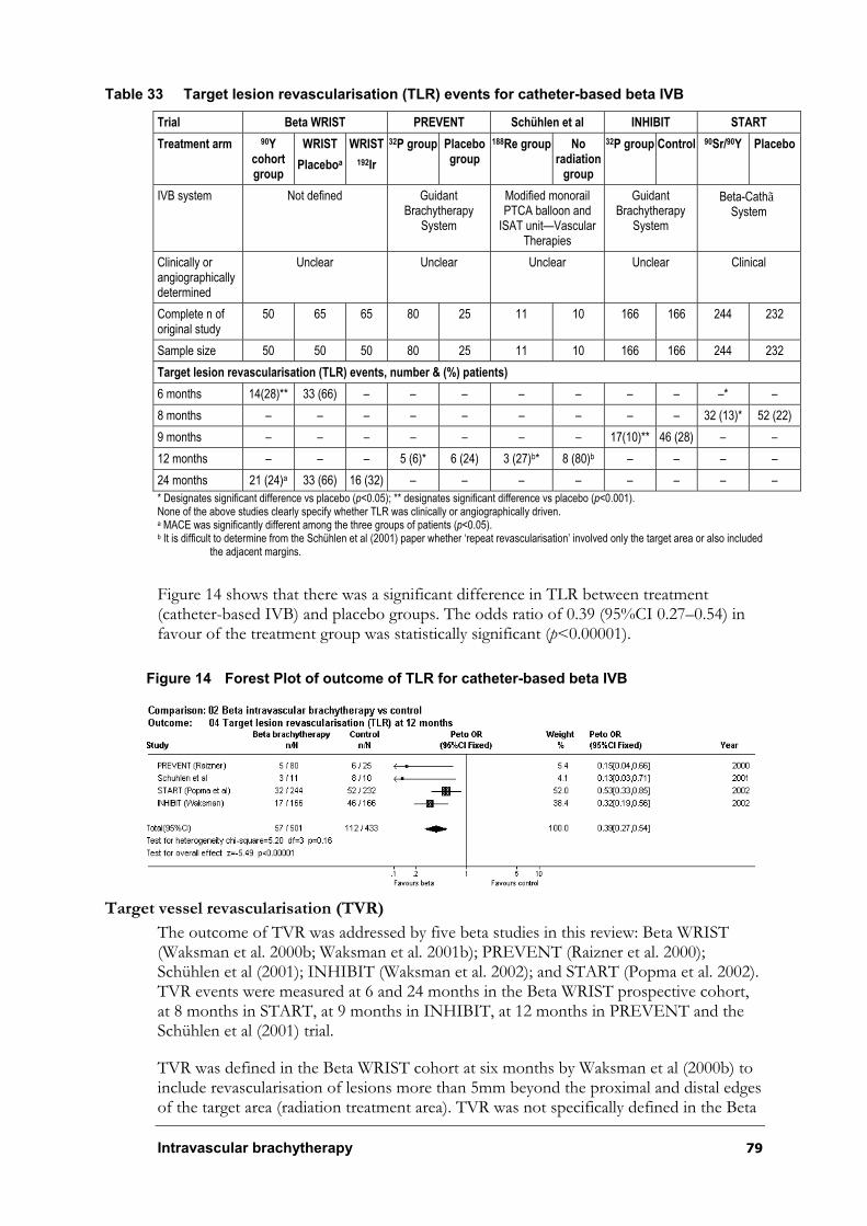
Intravascular brachytherapy 79
Table 33 Target lesion revascularisation (TLR) events for catheter-based beta IVB
Trial Beta WRIST PREVENT Schühlen et al INHIBIT START
Treatment arm 90Ycohortgroup
WRIST
Placeboa
WRIST
192Ir
32P group Placebo group
188Re group Noradiation
group
32P group Control 90Sr/90Y Placebo
IVB system Not defined Guidant Brachytherapy
System
Modified monorail PTCA balloon and
ISAT unit—Vascular Therapies
GuidantBrachytherapy
System
Beta-CathãSystem
Clinically or angiographically determined
Unclear Unclear Unclear Unclear Clinical
Complete n of original study
50 65 65 80 25 11 10 166 166 244 232
Sample size 50 50 50 80 25 11 10 166 166 244 232
Target lesion revascularisation (TLR) events, number & (%) patients)
6 months 14(28)** 33 (66) – – – – – – – –* –
8 months – – – – – – – – – 32 (13)* 52 (22)
9 months – – – – – – – 17(10)** 46 (28) – –
12 months – – – 5 (6)* 6 (24) 3 (27)b* 8 (80)b – – – –
24 months 21 (24)a 33 (66) 16 (32) – – – – – – – –
* Designates significant difference vs placebo (p<0.05); ** designates significant difference vs placebo (p<0.001). None of the above studies clearly specify whether TLR was clinically or angiographically driven. a MACE was significantly different among the three groups of patients (p<0.05). b It is difficult to determine from the Schühlen et al (2001) paper whether ‘repeat revascularisation’ involved only the target area or also included
the adjacent margins.
Figure 14 shows that there was a significant difference in TLR between treatment (catheter-based IVB) and placebo groups. The odds ratio of 0.39 (95%CI 0.27–0.54) in favour of the treatment group was statistically significant (p<0.00001).
Figure 14 Forest Plot of outcome of TLR for catheter-based beta IVB
Target vessel revascularisation (TVR)
The outcome of TVR was addressed by five beta studies in this review: Beta WRIST (Waksman et al. 2000b; Waksman et al. 2001b); PREVENT (Raizner et al. 2000); Schühlen et al (2001); INHIBIT (Waksman et al. 2002); and START (Popma et al. 2002). TVR events were measured at 6 and 24 months in the Beta WRIST prospective cohort, at 8 months in START, at 9 months in INHIBIT, at 12 months in PREVENT and the Schühlen et al (2001) trial.
TVR was defined in the Beta WRIST cohort at six months by Waksman et al (2000b) to include revascularisation of lesions more than 5mm beyond the proximal and distal edges of the target area (radiation treatment area). TVR was not specifically defined in the Beta

Intravascular brachytherapy 80
WRIST cohort at 24 months (Waksman et al. 2001b). Raizner et al (2000) for PREVENT defined TVR to include revascularisation of the target area and adjacent margins. It is difficult to determine from the Schühlen et al (2001) paper whether the repeat revascularisation reported involved only the target lesion. Waksman et al (2002) for INHIBIT defined TVR as the area outside the target area but within the target vessel. Popma et al (2002) for START defined TVR as clinically driven repeat revascularisation (by symptoms or laboratory testing using percutaneous intervention or bypass surgery), and less than 50 per cent stenosis within the treated vessel on follow-up angiography. Apart from START, none of the catheter-based beta IVB papers clearly specify whether TVR was clinically or angiographically driven.
Table 34 outlines the results for TVR events for each of the studies. Waksman et al (2000b) reported significantly fewer TVR events at six months for patients in the Beta WRIST cohort (n=50) compared with a historical control group that comprised a subset of patients with native coronary artery lesions (n=50) from the placebo group (n=65) of the gamma WRIST trial. Waksman et al (2001b) reported significant differences in TVR events at 24 months among the patients from Beta WRIST cohort compared with the radiation and placebo native coronary artery patient subgroups from the gamma WRIST trial (p<0.05). Raizner et al (2000) (n=105) for PREVENT reported no significant differences in the TVR events between the 32P radiation group and the placebo group. Schühlen et al (2001) (n=21) reported fewer patients requiring revascularisation in the 188Re radiation group compared with the patients in the no radiation group; however, due to the small sample size, this study may not have been sufficiently powered to detect a statistical difference. Waksman et al (2002) for INHIBIT reported significantly fewer TVR events at nine months for patients in the 32P radiation group compared with the placebo group (p<0.033). Popma et al (2002) for START reported significantly fewer TVR events at eight months for patients in the 90Sr/90Y group compared with the placebo group (p=0.026).

Intravascular brachytherapy 81
Table 34 Target vessel revascularisation (TVR) events for catheter-based beta IVB
Trial Beta WRIST PREVENT Schühlen et al INHIBIT START
Treatment arm 90Ycohortgroup
WRIST
Placeboa
WRIST
192Ir
32P group Placebo group
188Regroup
No radiation group
32P group Control 90Sr/90Y Placebo
IVB system Not defined Guidant Brachytherapy
System
Modified monorail PTCA balloon and
ISAT unit—Vascular Therapies
GuidantBrachytherapy
System
Beta-CathãSystem
Clinically or angiographically determined
Unclear Unclear Unclear Unclear Clinical
Complete n of original study
50 65 65 80 25 11 10 166 166 244 232
Sample size 50 50 50 80 25 11 10 166 166 244 232
Target vessel revascularisation (TVR) events, number & (%) patients
6 months 17(34)** 36 (72) – – – – – – – – –
8 months – – – – – – – – – 39 (16)* 56 (24)
9 months – – – – – – – 33 (20)* 51 (31) – –
12 months 23 (46)a 36 (72) 22 (44) 17 (21)b 8 (32) 3 (27)c* 8 (80)c – – – –
24 months 17(34)** 36 (72) – – – – – – – – –
* Designates significant difference vs placebo (p<0.05); ** designates significant difference vs placebo (p<0.001). None of the above studies clearly specified whether TVR events were clinically or angiographically driven. a Rates of TVR events were significantly different among the three groups of patients (p<0.05). b These results also include the number of TLR events. c It is difficult to determine from the Schühlen et al (2001) paper whether ‘repeat revascularisation’ involved only the target area or also included
the adjacent margins.
Figure 15 shows that there was a significant difference in TVR between treatment (catheter-based IVB) and placebo groups. The odds ratio of 0.55 (95%CI 0.40–0.75) in favour of the treatment group was statistically significant (p=0.0002).
Figure 15 Forest plot of outcome of TVR for catheter-based beta IVB
Summary—Clinical outcomes
Results from independently performed randomised controlled trials suggest that the
Guidant Intravascular Radiotherapy Systems and the NovosteÑ Beta-CathãIntracoronary Radiation System show comparable effectiveness; however, these systems have not been directly compared in the same group of patients. Meta-analysis did indicate, however, that compared with placebo, catheter-based beta IVB appeared to be significantly associated with reduced MACE (OR=0.50, 95%CI 0.37–0.69, p<0.0002),TLR events (OR=0.39; 95%CI 0.27–0.54, p<0.00001) and TVR events (OR=0.55; 95%CI 0.40–0.75, p=0.0002) at six months. Individual trial data suggested that beta IVB
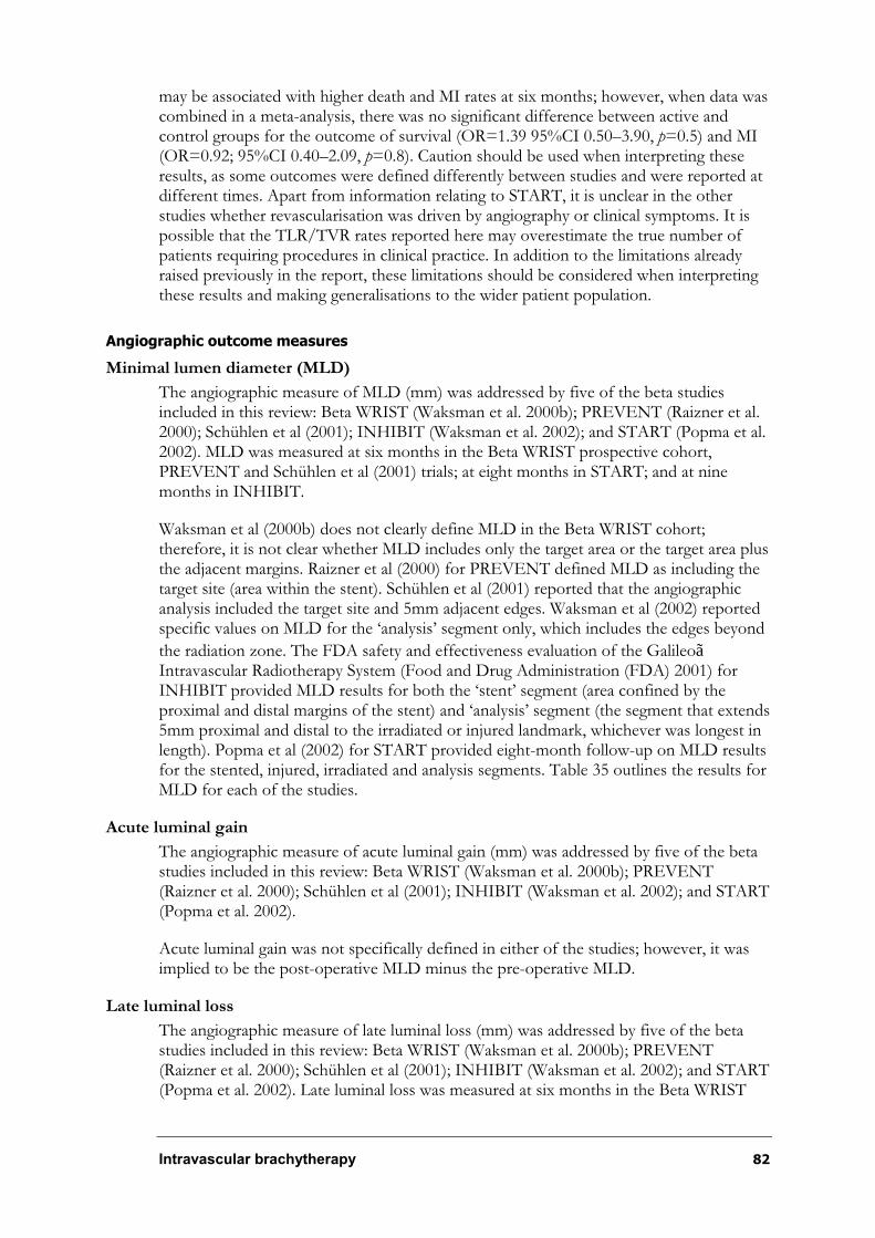
Intravascular brachytherapy 82
may be associated with higher death and MI rates at six months; however, when data was combined in a meta-analysis, there was no significant difference between active and control groups for the outcome of survival (OR=1.39 95%CI 0.50–3.90, p=0.5) and MI (OR=0.92; 95%CI 0.40–2.09, p=0.8). Caution should be used when interpreting these results, as some outcomes were defined differently between studies and were reported at different times. Apart from information relating to START, it is unclear in the other studies whether revascularisation was driven by angiography or clinical symptoms. It is possible that the TLR/TVR rates reported here may overestimate the true number of patients requiring procedures in clinical practice. In addition to the limitations already raised previously in the report, these limitations should be considered when interpreting these results and making generalisations to the wider patient population.
Angiographic outcome measures
Minimal lumen diameter (MLD)
The angiographic measure of MLD (mm) was addressed by five of the beta studies included in this review: Beta WRIST (Waksman et al. 2000b); PREVENT (Raizner et al. 2000); Schühlen et al (2001); INHIBIT (Waksman et al. 2002); and START (Popma et al. 2002). MLD was measured at six months in the Beta WRIST prospective cohort, PREVENT and Schühlen et al (2001) trials; at eight months in START; and at nine months in INHIBIT.
Waksman et al (2000b) does not clearly define MLD in the Beta WRIST cohort; therefore, it is not clear whether MLD includes only the target area or the target area plus the adjacent margins. Raizner et al (2000) for PREVENT defined MLD as including the target site (area within the stent). Schühlen et al (2001) reported that the angiographic analysis included the target site and 5mm adjacent edges. Waksman et al (2002) reported specific values on MLD for the ‘analysis’ segment only, which includes the edges beyond
the radiation zone. The FDA safety and effectiveness evaluation of the GalileoãIntravascular Radiotherapy System (Food and Drug Administration (FDA) 2001) for INHIBIT provided MLD results for both the ‘stent’ segment (area confined by the proximal and distal margins of the stent) and ‘analysis’ segment (the segment that extends 5mm proximal and distal to the irradiated or injured landmark, whichever was longest in length). Popma et al (2002) for START provided eight-month follow-up on MLD results for the stented, injured, irradiated and analysis segments. Table 35 outlines the results for MLD for each of the studies.
Acute luminal gain
The angiographic measure of acute luminal gain (mm) was addressed by five of the beta studies included in this review: Beta WRIST (Waksman et al. 2000b); PREVENT (Raizner et al. 2000); Schühlen et al (2001); INHIBIT (Waksman et al. 2002); and START (Popma et al. 2002).
Acute luminal gain was not specifically defined in either of the studies; however, it was implied to be the post-operative MLD minus the pre-operative MLD.
Late luminal loss
The angiographic measure of late luminal loss (mm) was addressed by five of the beta studies included in this review: Beta WRIST (Waksman et al. 2000b); PREVENT (Raizner et al. 2000); Schühlen et al (2001); INHIBIT (Waksman et al. 2002); and START (Popma et al. 2002). Late luminal loss was measured at six months in the Beta WRIST
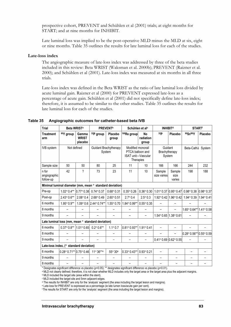
Intravascular brachytherapy 83
prospective cohort, PREVENT and Schühlen et al (2001) trials; at eight months for START; and at nine months for INHIBIT.
Late luminal loss was implied to be the post-operative MLD minus the MLD at six, eight or nine months. Table 35 outlines the results for late luminal loss for each of the studies.
Late-loss index
The angiographic measure of late-loss index was addressed by three of the beta studies included in this review: Beta WRIST (Waksman et al. 2000b); PREVENT (Raizner et al. 2000); and Schühlen et al (2001). Late-loss index was measured at six months in all three trials.
Late-loss index was defined in the Beta WRIST as the ratio of late luminal loss divided by acute luminal gain. Raizner et al (2000) for PREVENT expressed late-loss as a percentage of acute gain. Schühlen et al (2001) did not specifically define late-loss index; therefore, it is assumed to be similar to the other studies. Table 35 outlines the results for late luminal loss for each of the studies.
Table 35 Angiographic outcomes for catheter-based beta IVB
Trial Beta WRISTa PREVENTb Schühlen et alc INHIBITd STARTf
Treatmentarm
90Y group Gamma WRISTplacebo
32P group Placebo group
188Re group Noradiation
group
32P Placebo 90Sr/90Y Placebo
IVB system Not defined Guidant Brachytherapy System
Modified monorail PTCA balloon and
ISAT unit—Vascular Therapies
GuidantBrachytherapy
System
Beta-Cathã System
Sample size 50 50 80 25 11 10 166 166 244 232
n for angiographic follow-up
42 ? 73 23 11 10 Sample size varies
Samplesize
varies
198 188
Minimal luminal diameter (mm, mean ° standard deviation)
Pre-op 1.02°0.4** 0.77°0.38 0.74°0.37 0.68°0.31 0.35°0.26 0.36°0.30 1.01°0.37 0.95°0.47 0.98°0.38 0.98°0.37
Post-op 2.43°0.6** 2.08°0.4 2.68°0.49 2.60°0.51 2.7°0.4 2.5°0.3 1.92°0.42 1.96°0.42 1.94°0.39 1.94°0.41
6 months 1.95°0.9** 1.09°0.6 2.44°0.74** 1.55°0.70 1.84°0.99** 0.55°0.35 – – – –
8 months – – – – – – – – 1.65°0.64** 1.41°0.58
9 months – – – – – – 1.54°0.65 1.38°0.61 – –
Late luminal loss (mm, mean ° standard deviation)
6 months 0.37°0.8** 1.01°0.65 0.2°0.6** 1.1°0.7 0.81°0.93** 1.91°0.41 – – – –
8 months – – – – – – – – 0.28°0.56** 0.55°0.59
9 months – – – – – – 0.41°0.69 0.62°0.55 – –
Late-loss index, (° standard deviation)
6 months 0.28°0.71** 0.75°0.46 11°36**e 55°30e 0.33°0.43** 0.93°0.21 – – – –
9 months – – – – – – – – – –
8 months – – – – – – – – – –
* Designates significant difference vs placebo (p<0.05); ** designates significant difference vs placebo (p<0.01). a MLD not clearly defined; therefore, it is not clear whether MLD includes only the target area or the target area plus the adjacent margins. b MLD included the target site (area within the stent). c MLD included the target site and 5mm adjacent edges. d The results for INHIBIT are only for the ‘analysis’ segment (the area including the target lesion and margins). e Late-loss for PREVENT is expressed as a percentage (ie late lumen loss/acute gain per cent). f The results for START are only for the ‘analysis’ segment (the area including the target lesion and margins).
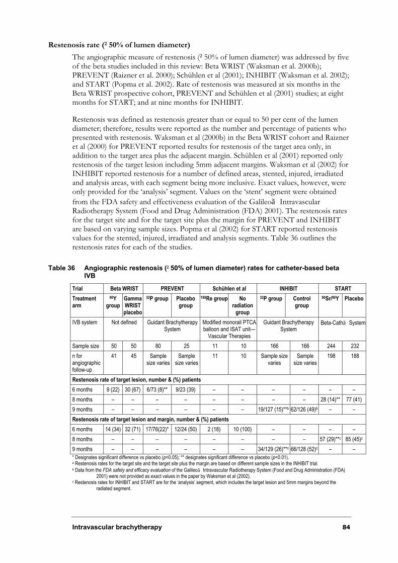
Intravascular brachytherapy 84
Restenosis rate (²50% of lumen diameter)
The angiographic measure of restenosis (²50% of lumen diameter) was addressed by five of the beta studies included in this review: Beta WRIST (Waksman et al. 2000b); PREVENT (Raizner et al. 2000); Schühlen et al (2001); INHIBIT (Waksman et al. 2002); and START (Popma et al. 2002). Rate of restenosis was measured at six months in the Beta WRIST prospective cohort, PREVENT and Schühlen et al (2001) studies; at eight months for START; and at nine months for INHIBIT.
Restenosis was defined as restenosis greater than or equal to 50 per cent of the lumen diameter; therefore, results were reported as the number and percentage of patients who presented with restenosis. Waksman et al (2000b) in the Beta WRIST cohort and Raizner et al (2000) for PREVENT reported results for restenosis of the target area only, in addition to the target area plus the adjacent margin. Schühlen et al (2001) reported only restenosis of the target lesion including 5mm adjacent margins. Waksman et al (2002) for INHIBIT reported restenosis for a number of defined areas, stented, injured, irradiated and analysis areas, with each segment being more inclusive. Exact values, however, were only provided for the ‘analysis’ segment. Values on the ‘stent’ segment were obtained
from the FDA safety and effectiveness evaluation of the Galileoã Intravascular Radiotherapy System (Food and Drug Administration (FDA) 2001). The restenosis rates for the target site and for the target site plus the margin for PREVENT and INHIBIT are based on varying sample sizes. Popma et al (2002) for START reported restenosis values for the stented, injured, irradiated and analysis segments. Table 36 outlines the restenosis rates for each of the studies.
Table 36 Angiographic restenosis (²50% of lumen diameter) rates for catheter-based beta IVB
Trial Beta WRIST PREVENT Schühlen et al INHIBIT START
Treatmentarm
90Ygroup
GammaWRISTplacebo
32P group Placebo group
188Re group Noradiation
group
32P group Control group
90Sr/90Y Placebo
IVB system Not defined Guidant Brachytherapy System
Modified monorail PTCAballoon and ISAT unit—
Vascular Therapies
Guidant Brachytherapy System
Beta-Cathã System
Sample size 50 50 80 25 11 10 166 166 244 232
n for angiographic follow-up
41 45 Sample size varies
Samplesize varies
11 10 Sample size varies
Samplesize varies
198 188
Restenosis rate of target lesion, number & (%) patients
6 months 9 (22) 30 (67) 6/73 (8)** 9/23 (39) – – – – – –
8 months – – – – – – – – 28 (14)** 77 (41)
9 months – – – – – – 19/127 (15)**b 62/126 (49)b – –
Restenosis rate of target lesion and margin, number & (%) patients
6 months 14 (34) 32 (71) 17/76(22)* 12/24 (50) 2 (18) 10 (100) – – – –
8 months – – – – – – – – 57 (29)**c 85 (45)c
9 months – – – – – – 34/129 (26)**c 66/128 (52)c – –
* Designates significant difference vs placebo (p<0.05); ** designates significant difference vs placebo (p<0.01). a Restenosis rates for the target site and the target site plus the margin are based on different sample sizes in the INHIBIT trial.b Data from the FDA safety and efficacy evaluation of the Galileoã Intravascular Radiotherapy System (Food and Drug Administration (FDA)
2001) were not provided as exact values in the paper by Waksman et al (2002). c Restenosis rates for INHIBIT and START are for the ‘analysis’ segment, which includes the target lesion and 5mm margins beyond the
radiated segment.
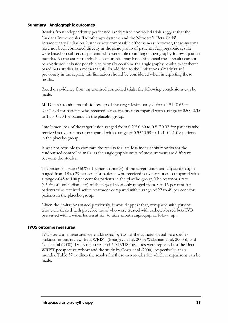
Intravascular brachytherapy 85
Summary—Angiographic outcomes
Results from independently performed randomised controlled trials suggest that the
Guidant Intravascular Radiotherapy Systems and the NovosteÑ Beta-CathãIntracoronary Radiation System show comparable effectiveness; however, these systems have not been compared directly in the same group of patients. Angiographic results were based on subsets of patients who were able to undergo angiography follow-up at six months. As the extent to which selection bias may have influenced these results cannot be confirmed, it is not possible to formally combine the angiography results for catheter-based beta studies in a meta-analysis. In addition to the limitations already raised previously in the report, this limitation should be considered when interpreting these results.
Based on evidence from randomised controlled trials, the following conclusions can be made:
MLD at six to nine month follow-up of the target lesion ranged from 1.54°0.65 to
2.44°0.74 for patients who received active treatment compared with a range of 0.55°0.35
to 1.55°0.70 for patients in the placebo group.
Late lumen loss of the target lesion ranged from 0.20°0.60 to 0.81°0.93 for patients who
received active treatment compared with a range of 0.55°0.59 to 1.91°0.41 for patients in the placebo group.
It was not possible to compare the results for late-loss index at six months for the randomised controlled trials, as the angiographic units of measurement are different between the studies.
The restenosis rate (²50% of lumen diameter) of the target lesion and adjacent margin ranged from 18 to 29 per cent for patients who received active treatment compared with a range of 45 to 100 per cent for patients in the placebo group. The restenosis rate
(²50% of lumen diameter) of the target lesion only ranged from 8 to 15 per cent for patients who received active treatment compared with a range of 22 to 49 per cent for patients in the placebo group.
Given the limitations stated previously, it would appear that, compared with patients who were treated with placebo, those who were treated with catheter-based beta IVB presented with a wider lumen at six- to nine-month angiographic follow-up.
IVUS outcome measures
IVUS outcome measures were addressed by two of the catheter-based beta studies included in this review: Beta WRIST (Bhargava et al. 2000; Waksman et al. 2000b); and Costa et al (2000). IVUS measures and 3D IVUS measures were reported for the Beta WRIST prospective cohort and the study by Costa et al (2000), respectively, at six months. Table 37 outlines the results for these two studies for which comparisons can be made.
Intravascular brachytherapy 86
Table 37 IVUS outcome measures for catheter-based beta IVB
Paper Beta WRIST (Bhargava et al. 2000) Costa et al (2000)
Treatmentarm
Beta WRIST 192Ir WRIST Placebo WRIST 32P group Placebo
Sample size 50 50 50 16 5
6 month IVUS follow-up
25 36 39 11 4
Mean lumen area (mm3 ° standard deviation)
Post-op 5.5°1.3 4.9°1.8 4.5°2.1 4.8°1.6 4.7°1.2
6 months 4.5°2.2a 4.1°2.1 2.5°1.4 4.7°1.3* 3.3°1.3
Lumen volume (mm3, mean ° standard deviation)
Post-op 189°83 186°100 174°135 185°60 205°62
6 months 165°105b 173°106 117°105 190°63 163°44
*Designates significant difference vs placebo (p<0.05); ** designates significant difference vs placebo (p<0.01). a Values significantly different between groups (p <0.0001). b Values significantly different between groups (p=0.0447).
What is the long-term effectiveness of catheter-based IVB?
At present, long-term clinical and angiographic follow-up of patients who have been treated with IVB is limited. The following studies provide some longer term results (two to three years post-treatment) for patients treated with catheter-based gamma IVB. At this stage, long-term results for catheter-based IVB are limited to two-year follow-up for one study (Beta WRIST).
Teirstein et al (2000) (n=55) reported on the three-year clinical and angiographic results for patients enrolled in the SCRIPPS randomised controlled trial. Table 38 outlines the rate of restenosis for both the 192Ir radiation and placebo groups. Assessment of restenosis at 36-month follow-up included only patients with angiographic follow-up beyond 27 months, unless TLR had occurred earlier. The authors reported that the rate
of angiographic restenosis (²50% of the lumen diameter) of the target site plus margin spanned by the active or placebo ribbon was significantly reduced at 36 months by 48 per cent in the 192Ir group compared with the placebo group (p<0.05). This difference was not as profound as that reported earlier at six months, where the rate of restenosis was significantly reduced by 69 per cent in the 192Ir group compared with the placebo group (p<0.01).
Teirstein et al (2000) also conducted a sub-group analysis on patients in the SCRIPPS trial who were alive at 36 months, underwent angiography and had not had a TLR procedure. The aim of this analysis was to determine the natural history of the effects of radiation on the treated vessel by comparing 6 and 36-month angiographic measures. The analysis included 17 of the 21 eligible patients from the 192Ir group and 10 of the 14 eligible patients from the placebo group. The mean minimal luminal diameter was
unchanged for the placebo group, and decreased for the 192Ir group from 2.49°0.81mm
at six months to 2.12°0.73mm at 36 months (p=0.15). Furthermore, the increase in mean per cent diameter stenosis between 6 and 36 months appeared to be greater in the 192Ir
group (14°28% to 26°28%, p=0.25) compared with the placebo group (21°24% to
23°17%, p=0.75). Although these results suggest there may be a trend whereby patients who received 192Ir showed delayed vessel narrowing, these results should be interpreted with caution, as these groups were selected and the sample size was very small.
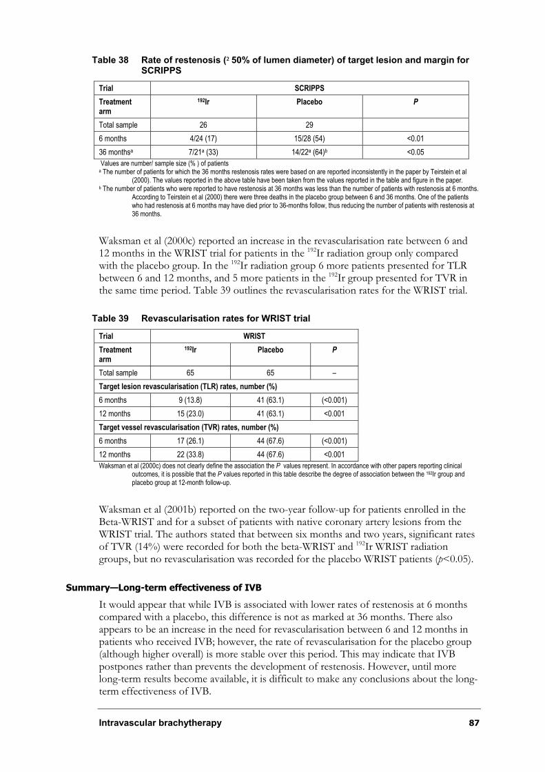
Intravascular brachytherapy 87
Table 38 Rate of restenosis (²50% of lumen diameter) of target lesion and margin for SCRIPPS
Trial SCRIPPS
Treatmentarm
192Ir Placebo P
Total sample 26 29
6 months 4/24 (17) 15/28 (54) <0.01
36 monthsa 7/21a (33) 14/22a (64)b <0.05
Values are number/ sample size (% ) of patients a The number of patients for which the 36 months restenosis rates were based on are reported inconsistently in the paper by Teirstein et al
(2000). The values reported in the above table have been taken from the values reported in the table and figure in the paper. b The number of patients who were reported to have restenosis at 36 months was less than the number of patients with restenosis at 6 months.
According to Teirstein et al (2000) there were three deaths in the placebo group between 6 and 36 months. One of the patients who had restenosis at 6 months may have died prior to 36-months follow, thus reducing the number of patients with restenosis at36 months.
Waksman et al (2000c) reported an increase in the revascularisation rate between 6 and 12 months in the WRIST trial for patients in the 192Ir radiation group only compared with the placebo group. In the 192Ir radiation group 6 more patients presented for TLR between 6 and 12 months, and 5 more patients in the 192Ir group presented for TVR in the same time period. Table 39 outlines the revascularisation rates for the WRIST trial.
Table 39 Revascularisation rates for WRIST trial
Trial WRIST
Treatmentarm
192Ir Placebo P
Total sample 65 65 –
Target lesion revascularisation (TLR) rates, number (%)
6 months 9 (13.8) 41 (63.1) (<0.001)
12 months 15 (23.0) 41 (63.1) <0.001
Target vessel revascularisation (TVR) rates, number (%)
6 months 17 (26.1) 44 (67.6) (<0.001)
12 months 22 (33.8) 44 (67.6) <0.001
Waksman et al (2000c) does not clearly define the association the P values represent. In accordance with other papers reporting clinical outcomes, it is possible that the P values reported in this table describe the degree of association between the 192Ir group and placebo group at 12-month follow-up.
Waksman et al (2001b) reported on the two-year follow-up for patients enrolled in the Beta-WRIST and for a subset of patients with native coronary artery lesions from the WRIST trial. The authors stated that between six months and two years, significant rates of TVR (14%) were recorded for both the beta-WRIST and 192Ir WRIST radiation groups, but no revascularisation was recorded for the placebo WRIST patients (p<0.05).
Summary—Long-term effectiveness of IVB
It would appear that while IVB is associated with lower rates of restenosis at 6 months compared with a placebo, this difference is not as marked at 36 months. There also appears to be an increase in the need for revascularisation between 6 and 12 months in patients who received IVB; however, the rate of revascularisation for the placebo group (although higher overall) is more stable over this period. This may indicate that IVB postpones rather than prevents the development of restenosis. However, until more long-term results become available, it is difficult to make any conclusions about the long-term effectiveness of IVB.
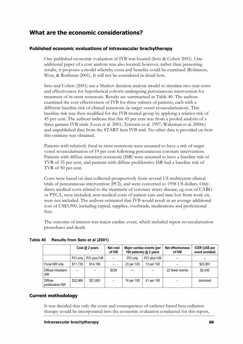
Intravascular brachytherapy 88
What are the economic considerations?
Published economic evaluations of intravascular brachytherapy
One published economic evaluation of IVB was located (Seto & Cohen 2001). One additional paper of a cost analysis was also located; however, rather than presenting results, it proposes a model whereby costs and benefits could be examined (Robinson, West, & Rothman 2001). It will not be considered in detail here.
Seto and Cohen (2001) use a Markov decision analytic model to simulate two-year costs and effectiveness for hypothetical cohorts undergoing percutaneous intervention for treatment of in-stent restenosis. Results are summarised in Table 40. The authors examined the cost effectiveness of IVB for three subsets of patients, each with a different baseline risk of clinical restenosis (ie target vessel revascularisation). This baseline risk was then modified for the IVB-treated group by applying a relative risk of 45 per cent. The authors indicate that this 45 per cent was from a pooled analysis of a three gamma IVB trials (Leon et al. 2001; Teirstein et al. 1997; Waksman et al. 2000c) and unpublished data from the START beta IVB trial. No other data is provided on how this estimate was obtained.
Patients with relatively focal in-stent restenosis were assumed to have a risk of target vessel revascularisation of 19 per cent following percutaneous coronary intervention. Patients with diffuse intrastent restenosis (ISR) were assumed to have a baseline risk of TVR of 35 per cent, and patients with diffuse proliferative ISR had a baseline risk of TVR of 50 per cent.
Costs were based on data collected prospectively from several US multicentre clinical trials of percutaneous intervention (PCI), and were converted to 1998 US dollars. Only direct medical costs related to the treatment of coronary artery disease, eg cost of CABG or PTCA, were included; non-medical costs of patient care and time lost from work etc. were not included. The authors estimated that IVB would result in an average additional cost of US$3,900, including capital, supplies, overheads, medications and professional fees.
The outcome of interest was major cardiac event, which included repeat revascularisation procedures and death.
Table 40 Results from Seto et al (2001)
Cost @ 2 years Net cost of IVB
Major cardiac events (per 100 patients) @ 2 years
Net effectiveness of IVB
ICER (US$ per event avoided)
PCI only PCI plus IVB – PCI only PCI plus IVB – –
Focal ISR only $11,739 $14,196 – 23 per 100 13 per 100 – $23,991
Diffuse intrastent ISR
– – $529 –– – 22 fewer events $2,430
Diffuseproliferative ISR
$22,966 $21,663 – 74 per 100 41 per 100 – dominant
Current methodology
It was decided that only the costs and consequences of catheter-based beta radiation therapy would be incorporated into the economic evaluation conducted for this report,

Intravascular brachytherapy 89
as likely costs of gamma IVB in Australia were unavailable. Furthermore, gamma IVB is more likely to attract higher capital costs compared with beta IVB, as more extensive modifications to the catheterisation laboratory are required to protect staff and patients from increased radiation exposure associated with gamma IVB.
Estimates of effectiveness
Clinically driven target lesion revascularisation at 12 months was considered an appropriate endpoint for the economic analysis. Angiographic restenosis was not used as an endpoint, as it incorporates a percentage of patients in whom the restenosis is asymptomatic and therefore do not require intervention.
A decision tree incorporating TLR, death and MI (ie the usual definition of MACE) was proposed. An a priori decision was made to include event types in the decision tree only when randomised evidence indicated a significant difference in patients treated with IVB compared to placebo. These events were also to be included in the tree if the difference was approaching clinical significance, but the trials were underpowered to detect a clinically meaningful difference.
Meta-analyses of the outcomes of TLR, MI and mortality from the trials of beta catheter-based IVB were conducted. Results from meta-analyses are indicated in Table 41, which shows that there were no significant differences in either MI or in death at 12 months between patients treated with IVB compared to those who received placebo. For that reason, these variables were not included in the model, and only TLR was used. A representation of the model is shown in Figure 16. Expert opinion suggests that, of the patients who require target lesion revascularisation after treatment of in-stent restenosis, approximately 70 per cent would proceed to CABG surgery, while the remaining 30 per cent would be treated with a repeat percutaneous intervention. This has been varied in a sensitivity analysis to 50 per cent proceeding to CABG surgery and 50 per cent being treated with repeat percutaneous intervention.
Table 41 Combined measures of major outcomes
Outcome Beta intravascular brachytherapy
Placebo arm rate Relative risk 95% CI
Target lesion revascularisation 0.259 0.46 0.34–0.61
Myocardial infarction 0.025 0.92* 0.40–2.11
Death 0.014 1.28* 0.47–3.45
* Not statistically significant
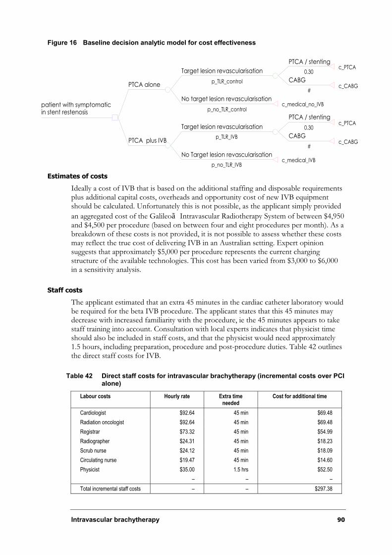
Intravascular brachytherapy 90
Figure 16 Baseline decision analytic model for cost effectiveness
Estimates of costs
Ideally a cost of IVB that is based on the additional staffing and disposable requirements plus additional capital costs, overheads and opportunity cost of new IVB equipment should be calculated. Unfortunately this is not possible, as the applicant simply provided
an aggregated cost of the Galileoã Intravascular Radiotherapy System of between $4,950 and $4,500 per procedure (based on between four and eight procedures per month). As a breakdown of these costs is not provided, it is not possible to assess whether these costs may reflect the true cost of delivering IVB in an Australian setting. Expert opinion suggests that approximately $5,000 per procedure represents the current charging structure of the available technologies. This cost has been varied from $3,000 to $6,000 in a sensitivity analysis.
Staff costs
The applicant estimated that an extra 45 minutes in the cardiac catheter laboratory would be required for the beta IVB procedure. The applicant states that this 45 minutes may decrease with increased familiarity with the procedure, ie the 45 minutes appears to take staff training into account. Consultation with local experts indicates that physicist time should also be included in staff costs, and that the physicist would need approximately 1.5 hours, including preparation, procedure and post-procedure duties. Table 42 outlines the direct staff costs for IVB.
Table 42 Direct staff costs for intravascular brachytherapy (incremental costs over PCI alone)
Labour costs Hourly rate Extra time needed
Cost for additional time
Cardiologist $92.64 45 min $69.48
Radiation oncologist $92.64 45 min $69.48
Registrar $73.32 45 min $54.99
Radiographer $24.31 45 min $18.23
Scrub nurse $24.12 45 min $18.09
Circulating nurse $19.47 45 min $14.60
Physicist $35.00 1.5 hrs $52.50
– – –
Total incremental staff costs – – $297.38
PTCA / stenting
0.30c_PTCA
CABG
#c_CABG
Target lesion revascularisation
p_TLR_control
No target lesion revascularisation
p_no_TLR_controlc_medical_no_IVB
PTCA alone
PTCA / stenting
0.30c_PTCA
CABG
#c_CABG
Target lesion revascularisation
p_TLR_IVB
No Target lesion revascularisation
p_no_TLR_IVBc_medical_IVB
PTCA plus IVB
patient with symptomatic in stent restenosis
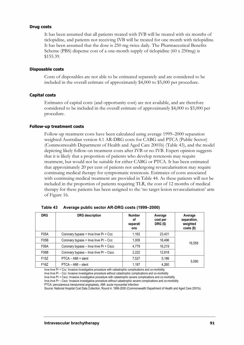
Intravascular brachytherapy 91
Drug costs
It has been assumed that all patients treated with IVB will be treated with six months of ticlopidine, and patients not receiving IVB will be treated for one month with ticlopidine. It has been assumed that the dose is 250 mg twice daily. The Pharmaceutical Benefits Scheme (PBS) dispense cost of a one-month supply of ticlopidine (60 x 250mg) is $155.39.
Disposable costs
Costs of disposables are not able to be estimated separately and are considered to be included in the overall estimate of approximately $4,000 to $5,000 per procedure.
Capital costs
Estimates of capital costs (and opportunity cost) are not available, and are therefore considered to be included in the overall estimate of approximately $4,000 to $5,000 per procedure.
Follow-up treatment costs
Follow-up treatment costs have been calculated using average 1999–2000 separation weighted Australian version 4.1 AR-DRG costs for CABG and PTCA (Public Sector) (Commonwealth Department of Health and Aged Care 2001b) (Table 43), and the model depicting likely follow-on treatment costs after IVB or no IVB. Expert opinion suggests that it is likely that a proportion of patients who develop restenosis may require treatment, but would not be suitable for either CABG or PTCA. It has been estimated that approximately 20 per cent of patients not undergoing revascularisation may require continuing medical therapy for symptomatic restenosis. Estimates of costs associated with continuing medical treatment are provided in Table 44. As these patients will not be included in the proportion of patients requiring TLR, the cost of 12 months of medical therapy for these patients has been assigned to the ‘no target lesion revascularisation’ arm of Figure 16.
Table 43 Average public sector AR-DRG costs (1999–2000)
DRG DRG description Number of
separations
Average cost per DRG ($)
Average separation, weightedcosts ($)
F05A Coronary bypass + Inva Inve Pr + Ccc 1,162 23,431
F05B Coronary bypass + Inva Inve Pr – Ccc 1,009 18,496
F06A Coronary bypass – Inva Inve Pr + Cscc 4,779 16,219
F06B Coronary bypass – Inva Inve Pr – Cscc 2,222 12,818
16,559
F15Z PTCA – AMI + stent 7,527 5,186
F16Z PTCA – AMI – stent 1,187 4,260 5,090
Inva Inve Pr + Ccc: Invasive investigative procedure with catastrophic complications and co-morbidity Inva Inve Pr – Ccc: Invasive investigative procedure without catastrophic complications and co-morbidity Inva Inve Pr + Cscc: Invasive investigative procedure with catastrophic severe complications and co-morbidity Inva Inve Pr – Cscc: Invasive investigative procedure without catastrophic severe complications and co-morbidity PTCA: percutaneous transluminal angioplasty, AMI: acute myocardial infarction Source: National Hospital Cost Data Collection, Round 4, 1999-2000 (Commonwealth Department of Health and Aged Care 2001b).
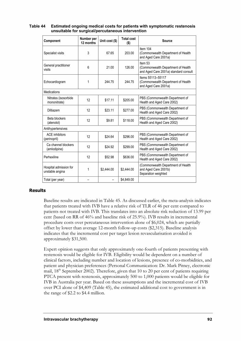
Intravascular brachytherapy 92
Table 44 Estimated ongoing medical costs for patients with symptomatic restenosis unsuitable for surgical/percutaneous intervention
ComponentNumber per 12 months
Unit cost ($) Total cost
($)Source
Specialist visits 3 67.65 203.00 Item 104 (Commonwealth Department of Health and Aged Care 2001a)
General practitioner visits
6 21.00 126.00 Item 53 (Commonwealth Department of Health and Aged Care 2001a) standard consult
Echocardiogram 1 244.75 244.75 Items 55113–55117 (Commonwealth Department of Health and Aged Care 2001a)
Medications
Nitrates (isosorbide mononitrate)
12 $17.11 $205.00 PBS (Commonwealth Department of Health and Aged Care 2002)
Diltiazem 12 $23.11 $277.00 PBS (Commonwealth Department of Health and Aged Care 2002)
Beta blockers (atenolol)
12 $9.81 $118.00 PBS (Commonwealth Department of Health and Aged Care 2002)
Antihypertensives
ACE inhibitors (perinopril)
12 $24.64 $296.00 PBS (Commonwealth Department of Health and Aged Care 2002)
Ca channel blockers (amlodipine)
12 $24.92 $299.00 PBS (Commonwealth Department of Health and Aged Care 2002)
Perhexiline 12 $52.98 $636.00 PBS (Commonwealth Department of Health and Aged Care 2002)
Hospital admission for unstable angina
1 $2,444.00 $2,444.00 (Commonwealth Department of Health and Aged Care 2001b) Separation weighted
Total (per year) – – $4,849.00
Results
Baseline results are indicated in Table 45. As discussed earlier, the meta-analysis indicates that patients treated with IVB have a relative risk of TLR of 46 per cent compared to patients not treated with IVB. This translates into an absolute risk reduction of 13.99 per cent (based on RR of 46% and baseline risk of 25.9%). IVB results in incremental procedure costs over percutaneous intervention alone of $6,024, which are partially offset by lower than average 12-month follow-up costs ($2,315). Baseline analysis indicates that the incremental cost per target lesion revascularisation avoided is approximately $31,500.
Expert opinion suggests that only approximately one-fourth of patients presenting with restenosis would be eligible for IVB. Eligibility would be dependent on a number of clinical factors, including number and location of lesions, presence of co-morbidities, and patient and physician preferences (Personal Communication: Dr. Mark Pitney, electronic mail, 18th September 2002). Therefore, given that 10 to 20 per cent of patients requiring PTCA present with restenosis, approximately 500 to 1,000 patients would be eligible for IVB in Australia per year. Based on these assumptions and the incremental cost of IVB over PCI alone of $4,409 (Table 45), the estimated additional cost to government is in the range of $2.2 to $4.4 million.

Intravascular brachytherapy 93
Figure 17 Decision tree depicting calculation of average follow-up treatment costs (12 months)
Table 45 Results of incremental cost effectiveness analysis
IVB plus PCI PCI alone Incremental difference
Target lesion revascularisation rate 11.9% 25.90% -13.99%
Cost of extra staff time per procedure $297.00 0 $297.00
Applicants procedure cost (incremental cost over PCI alone)
$4,950.00 0 $4,950.00
Disposables N/a N/a n/a
Capital (including opportunity cost) N/a N/a n/a
Drug costs (directly related to procedure) $932.34 $155.39 $777.00
Total procedure costs $6,179.00 $155.00 $6,024.00
Total average follow-up costs* $2,315.00 $3,930.00 -$1,615.00
Total costs (procedure + follow-up costs) $8,494.00 $4,085.00 $4,409.00
Cost effectiveness ($/TLR prevented) – – $31,527.00
* From decision analytic model.
Sensitivity analyses
The effect of variability of cost and efficacy data were tested in sensitivity analyses, including the relative risk of TLR, the cost of IVB and the proportion of patients who proceed to CABG after TLR. These results are detailed in Table 46.
Table 46 Results of sensitivity analyses
Incremental Cost-effectiveness ratio ($ per TLR prevented)
Variable Baseline (sensitivity range)
Baseline
Lowerbound
Upperbound
Relative risk of TLR* 0.46 (0.34–0.61) $31,527 $23,728 $48,055
Cost of procedure* $4,950 ($3,000–$6,000) $31,527 $17,584 $39,034
50% CABG: 50% PCI 30% CABG (50% CABG) $31,527 $34,814
* Baseline risk of TLR = 0.259.
Limitations of model
Clearly, the model presented here is a simplification of how a patient is likely to be treated following IVB. It uses the baseline risk of target lesion revascularisation from the
PTCA / stenting
0.300c_PTCA = $5,090
CABG
0.700c_CABG = $16,559
Target lesion revascularisation
0.259
No target lesion revascularisation
0.741c_medical_no_IVB = $719
PTCA alone$3,930
PTCA / stenting
0.300c_PTCA = $5,090; P = 0.036
CABG
0.700c_CABG = $16,559; P = 0.083
Target lesion revascularisation
0.119
No Target lesion revascularisation
0.881c_medical_IVB = $854; P = 0.881
PTCA plus IVB$2,315
patient with symptomatic
in stent restenosisPTCA plus IVB : $2,315
Intravascular brachytherapy 94
placebo arm of trials. To create a model that more closely depicts Australian clinical practice, we would want to use estimates of baseline risk of TLR that are based on Australian data. This means that the absolute risk reduction would be a more accurate representation of the likely benefit of IVB that might be seen in routine clinical use in Australia.
The true cost of providing IVB in Australia should be established (including disposable and capital components) instead of using the applicant’s estimate. As it is unclear on what data this estimate has been based, we are unsure whether this represents an accurate cost of service provision in Australia.
It should also be noted that the endpoint of ‘target lesion revascularisation’ is an intermediate endpoint and does not allow comparison of the cost effectiveness of IVB to other cardiac or non-cardiac interventions. To facilitate comparison across interventions, a longer term study of the effects of IVB on quality-adjusted patient survival would be required.
Conclusions
Using published randomised controlled evidence, the baseline cost per target lesion revascularisation prevented from the use of IVB is estimated to be approximately $31,500 per TLR prevented. A one-way sensitivity analysis over the 95 per cent confidence interval for the relative risk of TLR indicated the ICER ranged approximately from $23,700 to $48,000 per TLR prevented. Sensitivity analyses concerning the cost of IVB indicated the ICER ranged approximately from $17,500 to $39,000. Increasing the proportion of patients who undergo CABG after TLR to 50 per cent increases the ICER to approximately $35,000. These analyses suggest that the estimate of cost effectiveness of IVB is sensitive to estimates of IVB treatment effect, baseline risk of TLR and, to a certain extent, cost of the provision of IVB. Furthermore, based on an annual incidence of between 500 to 1,000 cases, and an incremental cost of $4,409 of IVB over PCI alone, the estimated additional cost to government of IVB will be in the order of $2.2 to 4.4 million.
Intravascular brachytherapy 95
Conclusions
Safety
The safety conclusions are:
¶ Catheter-based IVB is a safe procedure, with no reports of acute adverse events during the procedure.
¶ IVB requires a coordinated approach between the interventional cardiologist, the radiation oncologist, nuclear medicine specialist or the medical physicist with an interest in this field.
¶ IVB needs to be performed in a facility that conforms to the appropriate state radiation regulations and licensing requirements.
¶ Patients who undergo treatment with catheter-based IVB are exposed to very low levels of radiation, as only a small local area of the vessel wall is irradiated. Consequently, adverse events associated with the radiation treatment are more likely to be associated with vessel wall damage rather than the development of malignancy.
¶ The evidence suggests that patients treated with catheter-based IVB were approximately 3½ to 4 times more likely to develop clinical late thrombosis compared with patients receiving the placebo. It is thought that IVB may delay healing and re-endothelialisation following percutaneous intervention and stenting, thus leaving a chronically thrombogenic luminal or stent strut surface that promotes the aggregation of clotting agents in the blood.
¶ The incidence of late thrombosis in the active IVB group is lower in more recent studies, equivalent to placebo rates. This may be due to study protocols incorporating longer duration anti-platelet therapy combined with avoiding new stent deployment. However, the influence of other differences in treatment protocols cannot be excluded. Furthermore, it is not possible to evaluate the long-term effectiveness of these measures in reducing the incidence of late thrombosis beyond 12 months.
¶ Edge restenosis appears to be more pronounced with the use of radioactive stents and beta catheter-based IVB than it does with gamma catheter-based radiation delivery systems. This may be due to beta radiation levels exhibiting a higher dose gradient fall-off compared with gamma radiation, which may increase the likelihood of some tissues further from the source receiving sub-optimal radiation doses. There is no significant difference in the occurrence of edge restenosis at six months between catheter-based gamma brachytherapy and placebo groups. For catheter-based beta studies, edge restenosis occurred at a rate of 5 to 29 per cent in the active group compared with a rate of 2 to 11 per cent for patients in the control group.
Intravascular brachytherapy 96
Effectiveness
The specific research questions in relation to this review were:
¶ What is the value of catheter-based IVB in addition to percutaneous intervention in treating patients with in-stent restenosis following previous coronary interventions compared with that of percutaneous intervention only?
¶ What is the value of radioactive stents in addition to percutaneous intervention in the treatment of patients with in-stent restenosis following previous coronary interventions compared with that of percutaneous intervention only? As the use of radioactive stents is expected to be quite limited in clinical practice, this question is included for the sake of completeness, although the lower priority of radioactive stents should be noted.
The effectiveness conclusions were:
¶ Conclusions on the effectiveness of IVB were based on Level I evidence. The systematic review comprised reasonable Level II evidence with eight randomised controlled trials (13 papers) and Level III-3 evidence with six non-randomised controlled studies (seven papers).
¶ In the short-term, catheter-based IVB appears to result in a statistically significant reduction in angiographic restenosis and clinical revascularisation procedures. IVB does not appear to have a statistically significant effect on the rate of myocardial infarction or survival in patients who undergo the procedure. It may be, however, that current trials are insufficiently powered to detect differences in these relatively rare outcomes:
- For beta IVB, the TLR rate at 8 to12 months for the active group was 11.4 per cent compared with 25.9 per cent in the control group. For the single study looking at clinically driven TLR, the difference was 13.1 per cent compared with 22.4 per cent, respectively.
- For beta IVB, the TVR rate at 8 to 12 months for the active group was 18.4 per cent compared with 28.4 per cent in the control group. For the single study looking at clinically driven TVR, the difference was 16.0 per cent compared with 24.1 per cent, respectively.
¶ Follow-up of patients is currently limited to 12 months to 2 years (except for one gamma IVB trial which has reported three-year follow up), and for that reason it is not possible to determine whether the benefits of IVB observed over this time are maintained in the long-term. It is unclear whether IVB may defer rather than prevent the onset of restenosis following intervention.
¶ Significant technological and radiological differences between gamma and beta catheter-based IVB systems prevent direct comparison of the evidence pertaining to each system.
¶ Results from independently performed randomised controlled trials suggest that
the Guidant Intravascular Radiotherapy System and the NovosteÑ Beta-CathãIntracoronary Radiation System show comparable effectiveness; however, these systems have not been directly compared in the same group of patients.
Intravascular brachytherapy 97
¶ The extent to which the short-term results on catheter-based IVB can be generalised to the wider patient population likely to be treated in clinical practice may be limited by the strict inclusion criteria of the trials.
¶ Currently there is insufficient evidence on using radioactive stents for treating coronary artery restenosis. The unacceptably high rate of edge restenosis associated with radioactive stents appears to be a fundamental safety issue that requires further investigation and evaluation in controlled clinical trial settings.
Cost effectiveness
The cost effectiveness conclusions were:
¶ Using published randomised controlled evidence, the baseline cost per target lesion revascularisation prevented from the use of IVB is estimated to be approximately $31,500 per TLR prevented.
¶ A one-way sensitivity analysis over the 95 per cent confidence interval for the relative risk of TLR indicated the ICER ranged approximately from $23,700 to $48,000.
¶ A one-way sensitivity analysis on the cost of IVB indicated the ICER ranged approximately from $17,500 to $39,000.
¶ Increasing the proportion of patients who undergo CABG after TLR to 50 per cent increases the ICER to approximately $35,000.
¶ These analyses suggest that the estimate of cost effectiveness of IVB is sensitive to estimates of IVB treatment effect, baseline risk of TLR and, to a certain extent, cost of the provision of intravascular brachytherapy.
¶ Based on an annual incidence of between 500 and 1,000 cases, and an incremental cost of $4,409 of IVB over PCI alone, the estimated additional cost to government of IVB will be in the order of $2.2 to 4.4 million.
Intravascular brachytherapy 98
Recommendation
MSAC recommends that on the strength of evidence pertaining to intravascular brachytherapy for the treatment of coronary artery restenosis (MSAC Application 1041), interim public funding should be supported for this procedure.
This recommendation is to be reviewed no later than three years from the date of this report to ascertain whether longer term safety, effectiveness and cost-effectiveness has been proven and to determine the place of evolving technologies such as drug-coated stents in the treatment of in-stent restenosis.
The Minister for Health and Ageing accepted this recommendation on 16 October 2002.

Intravascular brachytherapy 99
Appendix A MSAC terms of reference and membership
The MSAC’s terms of reference are to:
¶ advise the Minister for Health and Ageing on the strength of evidence pertaining to new and emerging medical technologies and procedures in relation to their safety, effectiveness and cost effectiveness, and under what circumstances public funding should be supported;
¶ advise the Minister for Health and Ageing on which new medical technologies and procedures should be funded on an interim basis to allow data to be assembled to determine their safety, effectiveness and cost effectiveness;
¶ advise the Minister for Health and Ageing on references related either to new and/or existing medical technologies and procedures; and
¶ undertake health technology assessment work referred by the Australian Health Ministers’ Advisory Council (AHMAC) and report its findings to the AHMAC.
The membership of the MSAC comprises a mix of clinical expertise covering pathology, nuclear medicine, surgery, specialist medicine and general practice, plus clinical epidemiology and clinical trials, health economics, consumers, and health administration and planning:
Member Expertise or affiliation
Dr Stephen Blamey (Chair) general surgery
Professor Bruce Barraclough general surgery
Professor Syd Bell pathology
Dr Paul Craft clinical epidemiology and oncology
Professor Ian Fraser reproductive medicine
Professor Jane Hall
Dr Terri Jackson
health economics
health economics
Ms Rebecca James
Professor Brendon Kearney
consumer health issues
health administration and planning
Mr Alan Keith Assistant Secretary, Diagnostics and Technology Branch, Commonwealth Department of Health and Ageing
Associate Professor Richard King internal medicine
Dr Ray Kirk
Dr Michael Kitchener
health research
nuclear medicine
Mr Lou McCallum consumer health issues
Dr Ewa Piejko
Professor John Simes
general practice
clinical epidemiology and clinical trials
Intravascular brachytherapy 100
Professor Richard Smallwood Chief Medical Officer, Commonwealth Department of Health and Ageing
Dr Robert Stable Representing the Australian Health Ministers’ Advisory Council
Professor Bryant Stokes neurology
Professor Ken Thomson radiology
Dr Douglas Travis urology
Intravascular brachytherapy 101
Appendix B Supporting committee
Supporting committee for MSAC application 1041 Intravascular brachytherapy
Dr Michael Kitchener (Chair)
MBBS, FRACP
Nuclear Medicine Specialist, Adelaide
MSAC member
Mr Ivan Kayne
Secretary, Knox Branch of Heart Support Australia
Former member of National Heart Foundation
Associate member of St Vincent’s Hospital Community Advisory Committee
consumer representative nominated by the Consumers’ Health Forum of Australia
Dr Tony Knittel
BSc, MSc, PhD
Radiation Safety Officer and
Senior Scientific Officer
Prince of Wales Hospital, Sydney
nominated by the Australasian College of Physical Scientists and Engineers in Medicine
Dr Leo Mahar
MB, FRACP
Director, Cardiology Department
Royal Adelaide Hospital
co-opted Cardiologist
Dr Mark Pitney
MBBS, FRACP, MSCAI
Consultant Cardiologist and
Director, Cardiac Catheter Laboratories
Eastern Heart Clinic
Prince of Wales Hospital, Sydney
nominated by The Cardiac Society of Australia and New Zealand
Dr George Quong
MBBS (Hons), FRACP, FRANZCR, FAChPM
Director
Radiation Oncology Centre
Austin and Repatriation Medical Centre
nominated by the Royal Australian and New Zealand College of Radiologists
Ms Linda Marshall
BSc, BA, MBA
MSAC Project Manager
Health Technology Section
Department of Health and Ageing

10
2
Intra
va
scu
lar b
rach
yth
era
py
Appendix C Studies included in the review
Table 47 Catheter-based gamma intravascular brachytherapy trials*
Study n Study question Study design Patient characteristics Procedure Selected results Comments
192Ir Placebo P
Sample size 26 29 –
Quantitative angiography n=24 n=28 –
Reference vessel diameter (mm)
2.88°0.58 2.78°0.47 0.50
Follow-up (months) 6.9°1.8 6.4°2.7 NS
MLD pre-op (mm) 1.10°0.46 1.03°0.46 0.60
MLD post-op (mm) 2.82°0.60 2.88°0.83 0.78
MLD 6 months (mm) 2.43°0.78 1.85°0.89 0.02
Binary restenosis stent and margin, no (%)
4 (17) 15 (54) 0.01
Restenosis stent only, no (%) 2 (8) 10 (36) 0.02
IVUS outcome measures n=18 n=18 –
® in mean luminal CSA (mm2) 0.7°1.0 2.2°1.8 <0.01
® in mean luminal volume (mm2)
16.4°24.0 44.3°34.6 0.01
Clinical outcome measures (12/12)
Sample size 26 29 –
Follow-up (months) 12.0°2.8 12.2°3.1 NS
Death (%) 0 1 (3) NS
MI (%) 1 (4) 0 NS
TLR (%) 3 (11.5) 13 (44.8) 0.01
Death, MI , STa or TLR (%)* 4 (15.3) 14 (48.3) 0.01
Death, MI, STa or any revascularisation
5 (19.2) 18 (62.1) <0.01
(Teirstein et al. 1997)
SCRIPPS
Enrolment: March–Dec 1995
55 Investigate safety and efficacy of catheter-based intracoronary gamma radiation to reduce intimal hyperplasia after coronary stenting in patients with restenosis.
SYSTEM:
192Ir ribbon with seed train Best Industries
Randomisedcontrolled trial (RCT), single-centre.
Randomisation process not described, but concealment of process to all but two staff.
Double-blind
Placebo controlled
QAA and IVUS performed pre and post-procedure, and follow-up at 6 months; blinded analysis.
Clinical follow-up 1 month and 12 months
Intention-to-treat analysis
Patient group at higher risk of restenosis.
Baseline characteristics similar; however, not entirely even, with a trend towards more diabetics in placebo group.
Lesions in native coronary artery and saphenous vein.
Inclusion criteria:
–restenosis (62% had ISR) or candidate for stent
–previous Rx <4 weeks before enrolment
–reference vessel diameter 3–5mm, target lesion length >30mm
–successful procedure: <30% residual stenosis, delivery of radiation, no death, MI, CABG or stent thrombosis <30 days after index procedure.
Exclusion criteria:
–revascularisation not successful; angiographic evidence of thrombus in target lesion
–stent implanted as an emergency procedure.
Primary intervention:
Restenosis (no stent)—PTCA and stent placed, ISR—PTCA or additional stents (IVUS guided).
Study intervention:
192-iridium vs placebo.
Dosimetry:
–dosimetry based on lesion geometry determined by IVUS.
–mean specific activity 97.6°29.2mCi, shortest mean source-to-target distance 1.02°0.16mm mean max. dose 2651°349cGy. Longest mean distance 3.3°0.47mm mean min. dose 732°83cGy.
Discharge:
–aspirin (325mg daily, indefinitely); ticlopidine (250mg bid for 2/52 for patients with new stents).
ST—stent thrombosis.; other outcomes—radiation exposure.
Multiple logistic regression found Rx with 192Ir only predictor for freedom from angiographic stenosis (Wald chi-square=4.9, p=0.03).
Values are mean°SD unless otherwise stated.
Study terminated early by Data Safety Monitoring Group as differences between groups significant.
*Kaplan–Meier survival analysis found that, at 12/12, 85% of rad group and 52% of the placebo group were event free (p=0.01).
Limitations:
More diabetics in placebo group; no P value baseline characteristics
Three patients excluded from angiographic analysis (2 192Ir, 1 placebo); events possibly associated with Rad Rx; therefore, angiographic results may overestimate the effect.
Discrepancy between table and text on whether patients had single lesions.
Only patients who had successful procedures included in analysis (success at 30days in 96% 192Ir, 97% on placebo).
Values in italics calculated to facilitate comparison across studies.
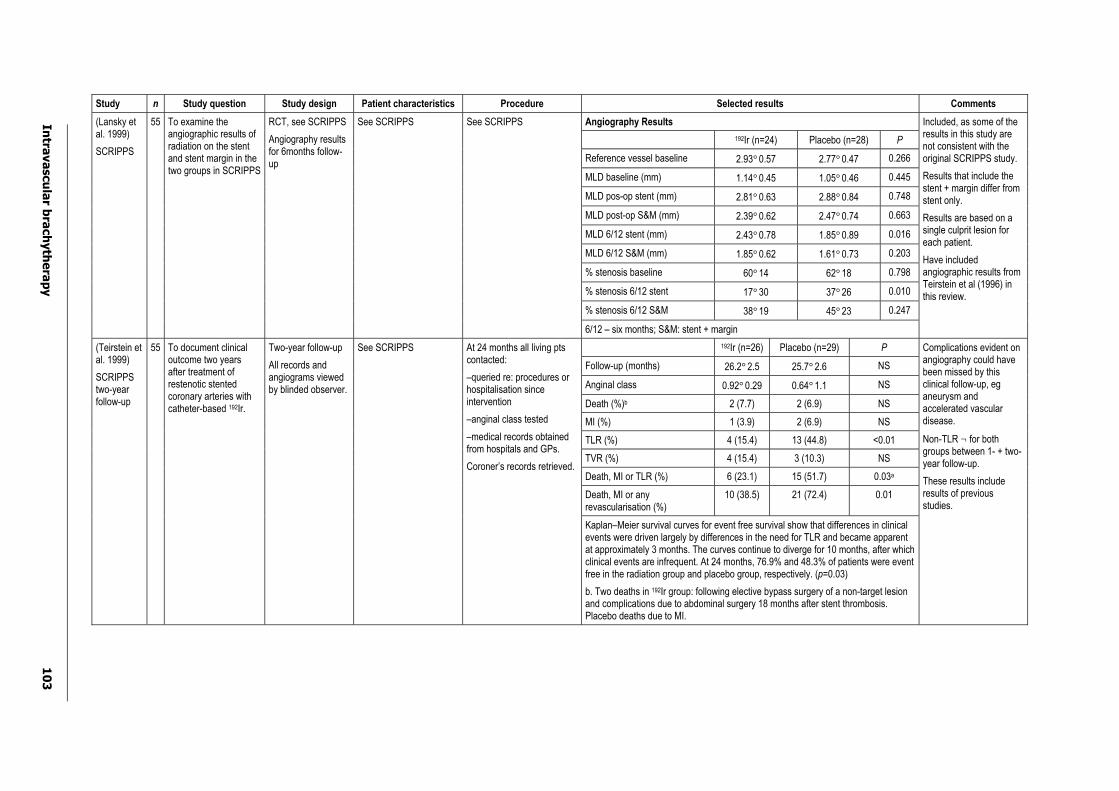
Intra
va
scu
lar b
rach
yth
era
py
10
3
Study n Study question Study design Patient characteristics Procedure Selected results Comments
Angiography Results
192Ir (n=24) Placebo (n=28) P
Reference vessel baseline 2.93°0.57 2.77°0.47 0.266
MLD baseline (mm) 1.14°0.45 1.05°0.46 0.445
MLD pos-op stent (mm) 2.81°0.63 2.88°0.84 0.748
MLD post-op S&M (mm) 2.39°0.62 2.47°0.74 0.663
MLD 6/12 stent (mm) 2.43°0.78 1.85°0.89 0.016
MLD 6/12 S&M (mm) 1.85°0.62 1.61°0.73 0.203
% stenosis baseline 60°14 62°18 0.798
% stenosis 6/12 stent 17°30 37°26 0.010
% stenosis 6/12 S&M 38°19 45°23 0.247
(Lansky et al. 1999)
SCRIPPS
55 To examine the angiographic results of radiation on the stent and stent margin in the two groups in SCRIPPS
RCT, see SCRIPPS
Angiography results for 6months follow-up
See SCRIPPS See SCRIPPS
6/12 – six months; S&M: stent + margin
Included, as some of the results in this study are not consistent with the original SCRIPPS study.
Results that include the stent + margin differ from stent only.
Results are based on a single culprit lesion for each patient.
Have included angiographic results from Teirstein et al (1996) in this review.
192Ir (n=26) Placebo (n=29) P
Follow-up (months) 26.2°2.5 25.7°2.6 NS
Anginal class 0.92°0.29 0.64°1.1 NS
Death (%)b 2 (7.7) 2 (6.9) NS
MI (%) 1 (3.9) 2 (6.9) NS
TLR (%) 4 (15.4) 13 (44.8) <0.01
TVR (%) 4 (15.4) 3 (10.3) NS
Death, MI or TLR (%) 6 (23.1) 15 (51.7) 0.03a
Death, MI or any revascularisation (%)
10 (38.5) 21 (72.4) 0.01
(Teirstein et al. 1999)
SCRIPPStwo-year follow-up
55 To document clinical outcome two years after treatment of restenotic stented coronary arteries with catheter-based 192Ir.
Two-year follow-up
All records and angiograms viewed by blinded observer.
See SCRIPPS At 24 months all living pts contacted:
–queried re: procedures or hospitalisation since intervention
–anginal class tested
–medical records obtained from hospitals and GPs.
Coroner’s records retrieved.
Kaplan–Meier survival curves for event free survival show that differences in clinical events were driven largely by differences in the need for TLR and became apparent at approximately 3 months. The curves continue to diverge for 10 months, after which clinical events are infrequent. At 24 months, 76.9% and 48.3% of patients were event free in the radiation group and placebo group, respectively. (p=0.03)
b. Two deaths in 192Ir group: following elective bypass surgery of a non-target lesion and complications due to abdominal surgery 18 months after stent thrombosis. Placebo deaths due to MI.
Complications evident on angiography could have been missed by this clinical follow-up, eg aneurysm and accelerated vascular disease.
Non-TLR ¬ for both groups between 1- + two-year follow-up.
These results include results of previous studies.
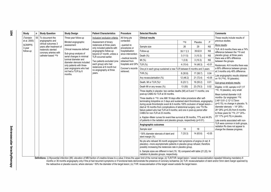
10
4
Intra
va
scu
lar b
rach
yth
era
py
Study n Study Question Study Design Patient Characteristics Procedure Selected Results Comments
Clinical results
192Ir Placebo P
Sample size 26 29 NS
Follow-up 39.1°2.3 39.62.8 NS
Death (%) 3 (11.5) 3 (10.3) NS
MI (%) 1 (3.9) 3 (10.3) NS
TLR (%) 4 (15.4) 14 (48.3) <0.01
One pt in each group sustained a new TLR between 6 months and 3 years
TVR (%) 8 (30.8) 17 (58.7) 0.04
Any revascularisation (%) 12 (46.2) 21 (72.4) <0.05
Death, MI or TLR (%)a 6 (23.1) 16 (55.2) 0.01
Death MI or any revasc (%) 13 (50) 23 (79.3) 0.02
Three deaths in placebo: two cardiac deaths (MI) at 8 and 11 months; one post-op CABG for TLR at 30 months.
Three deaths in 192Ir: one AMI 18 days after index procedure after self-terminating ticlopidine on 3 days and sustained stent thrombosis, angiography during acute thrombolytic event & 6 months 100% occlusion of target lesion—died at 18 months from complications of abdominal surgery; one 192Ir Rx-failure patient who had TLR at 8 months; and one in post-op period after CABG for non-TLR at 23 months.
a. Kaplan–Meier curves for event-free survival at 36 months, 77% and 44.8% of patients in the radiation and placebo groups, respectively (p=0.01).
Angiographic outcomes
Sample sizeb 19 18
²50% diameter stenosis of stent and stent margin (%)
7 (33.3) 14 (63.6) <0.05
(Teirstein et al. 2000)
SCRIPPS3-yearfollow-up
55 To document the angiographic and clinical outcomes 3 years after treatment of restenotic stented coronary arteries with catheter-based 192Ir
Three-year follow-up
Blinded angiographic assessment
Clinical measures defined
Sub-group analysis of serial changes in minimal luminal diameter and diameter stenosis included only patients with three-year angiograms who had not had a TLR by 6 months.
Inclusion/ exclusion criteria:
Assessment of binary restenosis at three years only included patients with angiographic follow-up beyond 27 month, unless a TLR occurred earlier.
Two patients excluded (one each group) who had restenosis at 6 month but no angiography at three years.
All living pts contacted:
–queried re: procedures or hospitalisation since intervention
–medical records obtained from hospitals and GPs
Coroner’s records retrieved.
No pts who refused 36 month angiogram had symptoms of angina (4 rad, 8 placebo)—more asymptomatic patients in placebo group refused, therefore possibly increasing the restenosis rate in placebo group.
b. Sample sizes are different in text (19, 18) compared with table (21,22), for radiation & placebo groups, respectively.
These results include results of previous studies.
More results:
TLR: At 6 months there was a 74% difference between the 192Ir and placebo group; at three years there was a 68% difference between the groups.
Restenosis: At 6 months there was a 69% difference between groups; at 3 months only 48% difference.
Late angiographic results obtained on 19 (192Ir), 18 (placebo).
Sub-group analysis results:
Eligible: n=35; sample n=27 (17 192Ir, 10 placebo), very small.
Mean luminal diameter ® (6 months--3yr angiogram 192Ir: 2.49°0.81–2.12°0.73mm (p=0.15); no change in placebo. % diameter stenosis ¬ 14°28%–26°28% (p=0.25) from 6 months to three years for 192Ir, 21°24%–23°17% (p=0.75) in placebo.
Late events associated with non-TLR were common in both groups, radiation Rx does not appear to change the disease progress.
Definitions: (i) Myocardial infarction (MI): elevation of MB fraction of creatine kinase to a value 3 times the upper limit of the normal range; (ii) TLR/TVR: target lesion / vessel revascularisation repeated following mandatory 6 months or 36 months angiography only if the pt had recurrent symptoms or if functional tests demonstrate the presence of coronary ischaemia; (iii) TLR: revascularisation of stent and/or 5mm stent margin spanned by the radioactive or placebo source, where stenosis ²50% the diameter of the target lesion; (iv) TVR: revascularisation of the target vessel outside the target lesion.
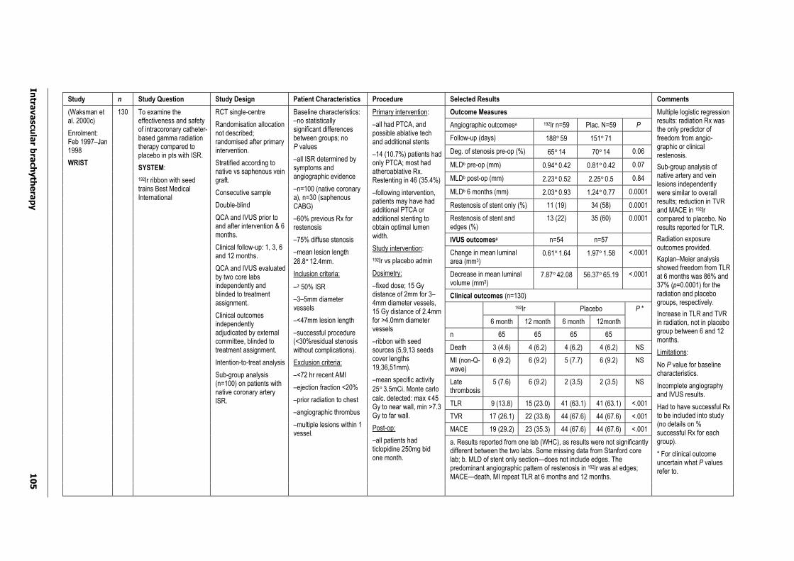
Intra
va
scu
lar b
rach
yth
era
py
10
5
Study n Study Question Study Design Patient Characteristics Procedure Selected Results Comments
Outcome Measures
Angiographic outcomesa 192Ir n=59 Plac. N=59 P
Follow-up (days) 188°59 151°71
Deg. of stenosis pre-op (%) 65°14 70°14 0.06
MLDb pre-op (mm) 0.94°0.42 0.81°0.42 0.07
MLDb post-op (mm) 2.23°0.52 2.25°0.5 0.84
MLDb 6 months (mm) 2.03°0.93 1.24°0.77 0.0001
Restenosis of stent only (%) 11 (19) 34 (58) 0.0001
Restenosis of stent and edges (%)
13 (22) 35 (60) 0.0001
IVUS outcomesa n=54 n=57
Change in mean luminal area (mm3)
0.61°1.64 1.97°1.58 <.0001
Decrease in mean luminal volume (mm3)
7.87°42.08 56.37°65.19 <.0001
Clinical outcomes (n=130)
192Ir Placebo
6 month 12 month 6 month 12month
P *
n 65 65 65 65
Death 3 (4.6) 4 (6.2) 4 (6.2) 4 (6.2) NS
MI (non-Q-wave)
6 (9.2) 6 (9.2) 5 (7.7) 6 (9.2) NS
Late thrombosis
5 (7.6) 6 (9.2) 2 (3.5) 2 (3.5) NS
TLR 9 (13.8) 15 (23.0) 41 (63.1) 41 (63.1) <.001
TVR 17 (26.1) 22 (33.8) 44 (67.6) 44 (67.6) <.001
MACE 19 (29.2) 23 (35.3) 44 (67.6) 44 (67.6) <.001
a. Results reported from one lab (WHC), as results were not significantly different between the two labs. Some missing data from Stanford core lab; b. MLD of stent only section—does not include edges. The predominant angiographic pattern of restenosis in 192Ir was at edges; MACE—death, MI repeat TLR at 6 months and 12 months.
(Waksman et al. 2000c)
Enrolment: Feb 1997–Jan 1998
WRIST
130 To examine the effectiveness and safety of intracoronary catheter-based gamma radiation therapy compared to placebo in pts with ISR.
SYSTEM:
192Ir ribbon with seed trains Best Medical International
RCT single-centre
Randomisation allocation not described; randomised after primary intervention.
Stratified according to native vs saphenous vein graft.
Consecutive sample
Double-blind
QCA and IVUS prior to and after intervention & 6 months.
Clinical follow-up: 1, 3, 6 and 12 months.
QCA and IVUS evaluated by two core labs independently and blinded to treatment assignment.
Clinical outcomes independently adjudicated by external committee, blinded to treatment assignment.
Intention-to-treat analysis
Sub-group analysis (n=100) on patients with native coronary artery ISR.
Baseline characteristics: –no statistically significant differences between groups; no P values
–all ISR determined by symptoms and angiographic evidence
–n=100 (native coronary a), n=30 (saphenous CABG)
–60% previous Rx for restenosis
–75% diffuse stenosis
–mean lesion length 28.8°12.4mm.
Inclusion criteria:
–²50% ISR
–3–5mm diameter vessels
–<47mm lesion length
–successful procedure (<30%residual stenosis without complications).
Exclusion criteria:
–<72 hr recent AMI
–ejection fraction <20%
–prior radiation to chest
–angiographic thrombus
–multiple lesions within 1 vessel.
Primary intervention:
–all had PTCA, and possible ablative tech and additional stents
–14 (10.7%) patients had only PTCA; most had atheroablative Rx. Restenting in 46 (35.4%)
–following intervention, patients may have had additional PTCA or additional stenting to obtain optimal lumen width.
Study intervention:
192Ir vs placebo admin
Dosimetry:
–fixed dose; 15 Gy distance of 2mm for 3–4mm diameter vessels, 15 Gy distance of 2.4mm for >4.0mm diameter vessels
–ribbon with seed sources (5,9,13 seeds cover lengths 19,36,51mm).
–mean specific activity 25°3.5mCi. Monte carlo calc. detected: max ¢45Gy to near wall, min >7.3 Gy to far wall.
Post-op:
–all patients had ticlopidine 250mg bid one month.
Multiple logistic regression results: radiation Rx was the only predictor of freedom from angio-graphic or clinical restenosis.
Sub-group analysis of native artery and vein lesions independently were similar to overall results; reduction in TVR and MACE in 192Ir compared to placebo. No results reported for TLR.
Radiation exposure outcomes provided.
Kaplan–Meier analysis showed freedom from TLR at 6 months was 86% and 37% (p=0.0001) for the radiation and placebo groups, respectively.
Increase in TLR and TVR in radiation, not in placebo group between 6 and 12 months.
Limitations:
No P value for baseline characteristics.
Incomplete angiography and IVUS results.
Had to have successful Rx to be included into study (no details on % successful Rx for each group).
* For clinical outcome uncertain what P valuesrefer to.
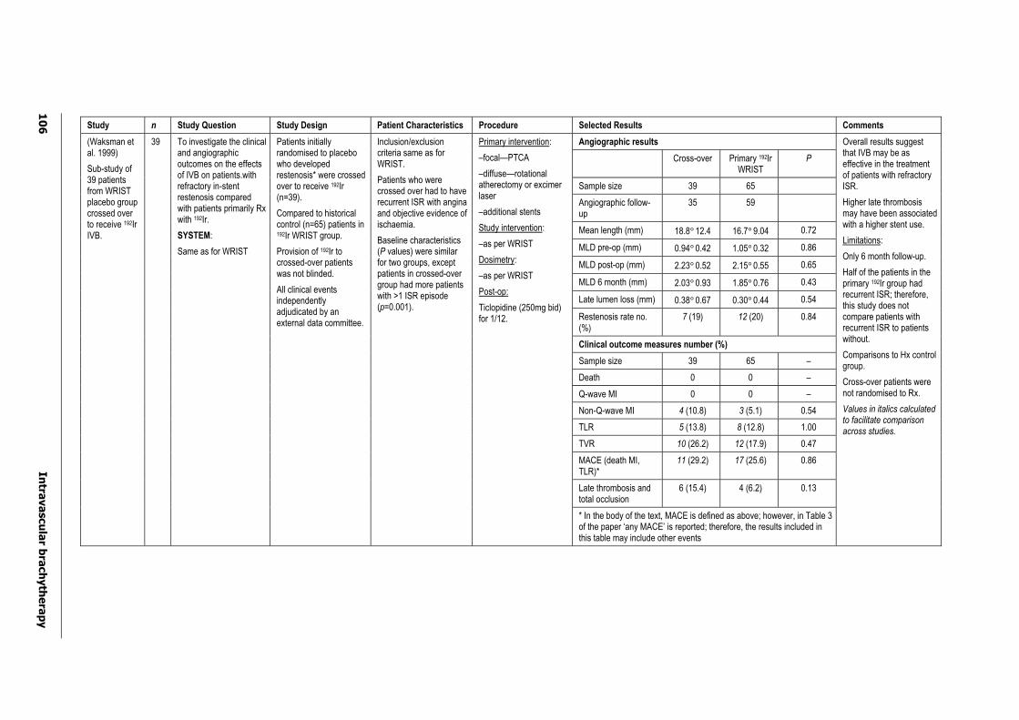
10
6
Intra
va
scu
lar b
rach
yth
era
py
Study n Study Question Study Design Patient Characteristics Procedure Selected Results Comments
Angiographic results
Cross-over Primary 192Ir WRIST
P
Sample size 39 65
Angiographic follow-up
35 59
Mean length (mm) 18.8°12.4 16.7°9.04 0.72
MLD pre-op (mm) 0.94°0.42 1.05°0.32 0.86
MLD post-op (mm) 2.23°0.52 2.15°0.55 0.65
MLD 6 month (mm) 2.03°0.93 1.85°0.76 0.43
Late lumen loss (mm) 0.38°0.67 0.30°0.44 0.54
Restenosis rate no. (%)
7 (19) 12 (20) 0.84
Clinical outcome measures number (%)
Sample size 39 65 –
Death 0 0 –
Q-wave MI 0 0 –
Non-Q-wave MI 4 (10.8) 3 (5.1) 0.54
TLR 5 (13.8) 8 (12.8) 1.00
TVR 10 (26.2) 12 (17.9) 0.47
MACE (death MI, TLR)*
11 (29.2) 17 (25.6) 0.86
Late thrombosis and total occlusion
6 (15.4) 4 (6.2) 0.13
(Waksman et al. 1999)
Sub-study of 39 patients from WRIST placebo group crossed over to receive 192Ir IVB.
39 To investigate the clinical and angiographic outcomes on the effects of IVB on patients.with refractory in-stent restenosis compared with patients primarily Rx with 192Ir.
SYSTEM:
Same as for WRIST
Patients initially randomised to placebo who developed restenosis* were crossed over to receive 192Ir (n=39).
Compared to historical control (n=65) patients in 192Ir WRIST group.
Provision of 192Ir to crossed-over patients was not blinded.
All clinical events independently adjudicated by an external data committee.
Inclusion/exclusion criteria same as for WRIST.
Patients who were crossed over had to have recurrent ISR with angina and objective evidence of ischaemia.
Baseline characteristics (P values) were similar for two groups, except patients in crossed-over group had more patients with >1 ISR episode (p=0.001).
Primary intervention:
–focal—PTCA
–diffuse—rotational atherectomy or excimer laser
–additional stents
Study intervention:
–as per WRIST
Dosimetry:
–as per WRIST
Post-op:
Ticlopidine (250mg bid) for 1/12.
* In the body of the text, MACE is defined as above; however, in Table 3 of the paper ‘any MACE’ is reported; therefore, the results included in this table may include other events
Overall results suggest that IVB may be as effective in the treatment of patients with refractory ISR.
Higher late thrombosis may have been associated with a higher stent use.
Limitations:
Only 6 month follow-up.
Half of the patients in the primary 192Ir group had recurrent ISR; therefore, this study does not compare patients with recurrent ISR to patients without.
Comparisons to Hx control group.
Cross-over patients were not randomised to Rx.
Values in italics calculated to facilitate comparison across studies.
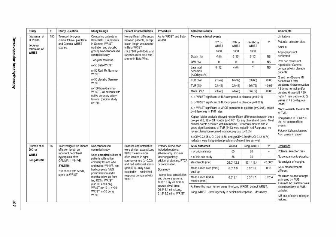
Intra
va
scu
lar b
rach
yth
era
py
10
7
Study n Study Question Study Design Patient Characteristics Procedure Selected Results Comments
Two-year clinical events
90Y b-WRIST
n=50
192IR g-WRIST
n=50
Placebo g-WRIST
n=50
P
Death (%) 4 (8) 5 (10) 5 (10) NS
QMI (%) 0 0 0 NS
Late total occlusion(>30days) (%)
6 (12) 4 (8) ? NS
TLR (%)a 21 (42) 16 (32) 33 (66) <0.05
TVR (%)b 23 (46) 22 (44) 36 (72) <0.05
MACE (%)b 23 (46) 24 (48) 36 (72) <0.05
(Waksman et al. 2001b)
two-year follow-up of WRIST
150 To report two-year clinical follow-up of Beta and Gamma WRIST studies.
Comparing patients in Beta-WRIST to patients in Gamma-WRIST (radiation and placebo group). Non-randomised controlled study.
Two-year follow-up
n=50 Beta-WRIST
n=50 Rad. Rx Gamma-WRIST
n=50 placebo Gamma-WRIST
n=100 from Gamma-WRIST—all patients with native coronary artery lesions, (original study n=130).
No significant differences between patients, except lesion length was shorter in Beta-WRIST (17.2°9.8, p=0.004), and radiation dwell time was shorter in Beta-Wrist.
As for WRIST and Beta-WRIST
a. b-WRIST significant ® TLR compared to placebo (p=0.016).
b. b-WRIST significant ® TLR compared to placebo (p=0.009).
c. b-WRIST significant ® MACE compared to placebo (p=0.008), driven by differences in TVR rates.
Kaplan–Meier analysis showed no significant differences between three groups at 6, 12 or 24 months (p>0.001) for any clinical end points. Most clinical events occurred within 6 months, Between 6 months and 2 years significant rates of TVR (14%) were noted in rad Rx groups; no revascularisation required in placebo group (p<0.05).
b (OR=0.22 95% CI 0.09–0.58) and g (OR=0.30 95% CI 0.12–0.74) radiation were independent predictors of event free survival.
Limitations:
Potential selection bias.
Small n.
Angiography not performed.
Post hoc results not reported for Gamma compared with placebo patients.
Q and non-Q-wave MI defined as a total creatinine kinase elevation ²2 times normal and/or creatine kinase-MB ²20ng/ml ° new pathologic Q waves in ²2 contiguous leads.
MACE—death, Q-wave MI or TVR.
Comparison to SCRIPPS trial re: pattern of late events.
Value in italics calculated from values in paper.
IVUS outcomes WRIST Long-WRIST P
n of original study 65 60 –
n of this sub-study 36 30 –
stent length (mm) 26.0°12.2 55.1°13.4 <0.0001
Mean lumen area (mm2)post-op
6.5°1.9 5.9°1.6 0.16
Mean lumen CSA 6 months (mm2)
6.3°2.1 5.3°1.7 0.0284
(Ahmed et al. 2001c)
WRIST
Long-WRIST
66 To investigate the impact of lesion length on recurrent neointimal hyperplasia after GAMMA-1 192Ir IVB.
SYSTEM:
192Ir ribbon with seeds, same as WRIST
Non-randomisedcontrolled study
Used complete subset of patients with native coronary lesions who underwent 192Ir IVB, and had complete IVUS postirradiation and 6 months follow-up from two RCTs: WRIST (n=130) and Long WRIST (n=121); n=36 WRIST, n=30 Long WRIST.
Baseline characteristics were similar, except Long WRIST lesions more often located in right coronary artery (p=0.02)and had additional stents (p=0.001)—may have resulted in ¬ neointimal response compared with WRIST.
Primary intervention included rotational atherectomy, excimer laser angioplasty, additional stenting, PTCA or combination.
Dosimetry:
–same dose prescription and delivery systems; fixed 15 Gy 2mm from source; dwell time: 20.4°3.1 mins Long, 21.5°3.2 mins. WRIST.
At 6 months mean lumen areas ® in Long WRIST, but not WRIST.
Long-WRIST ¬ heterogeneity in neointimal responsedosimetry.
Limitations:
Potential selection bias.
No comparison to placebo.
No analysis of margins.
IVUS measurements different.
Maximum source to target estimated by IVUS; assumes IVB catheter was placed similarly to IVUS catheter.
IVB less effective in longer lesions.
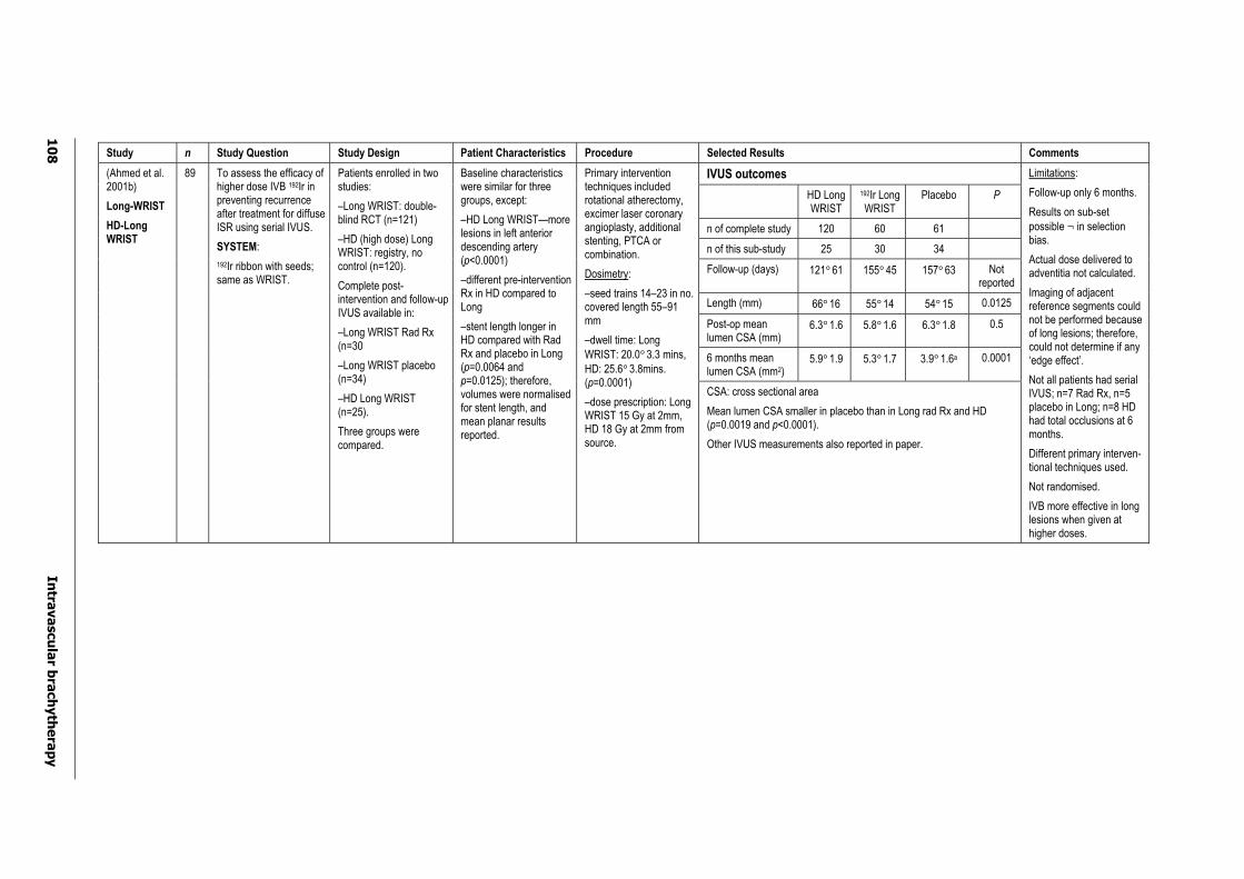
10
8
Intra
va
scu
lar b
rach
yth
era
py
Study n Study Question Study Design Patient Characteristics Procedure Selected Results Comments
IVUS outcomes
HD Long WRIST
192Ir Long WRIST
Placebo P
n of complete study 120 60 61
n of this sub-study 25 30 34
Follow-up (days) 121°61 155°45 157°63 Notreported
Length (mm) 66°16 55°14 54°15 0.0125
Post-op mean lumen CSA (mm)
6.3°1.6 5.8°1.6 6.3°1.8 0.5
6 months mean lumen CSA (mm2)
5.9°1.9 5.3°1.7 3.9°1.6a 0.0001
(Ahmed et al. 2001b)
Long-WRIST
HD-Long WRIST
89 To assess the efficacy of higher dose IVB 192Ir in preventing recurrence after treatment for diffuse ISR using serial IVUS.
SYSTEM:
192Ir ribbon with seeds; same as WRIST.
Patients enrolled in two studies:
–Long WRIST: double-blind RCT (n=121)
–HD (high dose) Long WRIST: registry, no control (n=120).
Complete post-intervention and follow-up IVUS available in:
–Long WRIST Rad Rx (n=30
–Long WRIST placebo (n=34)
–HD Long WRIST (n=25).
Three groups were compared.
Baseline characteristics were similar for three groups, except:
–HD Long WRIST—more lesions in left anterior descending artery (p<0.0001)
–different pre-intervention Rx in HD compared to Long
–stent length longer in HD compared with Rad Rx and placebo in Long (p=0.0064 and p=0.0125); therefore, volumes were normalised for stent length, and mean planar results reported.
Primary intervention techniques included rotational atherectomy, excimer laser coronary angioplasty, additional stenting, PTCA or combination.
Dosimetry:
–seed trains 14–23 in no. covered length 55–91 mm
–dwell time: Long WRIST: 20.0°3.3 mins, HD: 25.6°3.8mins. (p=0.0001)
–dose prescription: Long WRIST 15 Gy at 2mm, HD 18 Gy at 2mm from source.
CSA: cross sectional area
Mean lumen CSA smaller in placebo than in Long rad Rx and HD (p=0.0019 and p<0.0001).
Other IVUS measurements also reported in paper.
Limitations:
Follow-up only 6 months.
Results on sub-set possible ¬ in selection bias.
Actual dose delivered to adventitia not calculated.
Imaging of adjacent reference segments could not be performed because of long lesions; therefore, could not determine if any ‘edge effect’.
Not all patients had serial IVUS; n=7 Rad Rx, n=5 placebo in Long; n=8 HD had total occlusions at 6 months.
Different primary interven-tional techniques used.
Not randomised.
IVB more effective in long lesions when given at higher doses.
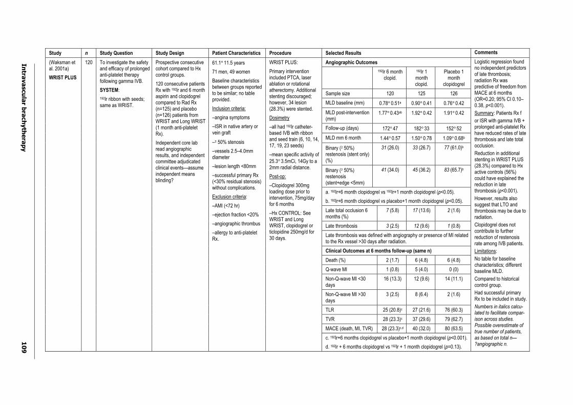
Intra
va
scu
lar b
rach
yth
era
py
10
9
Study n Study Question Study Design Patient Characteristics Procedure Selected Results Comments
Angiographic Outcomes
192Ir 6 month clopid.
192Ir 1 month clopid.
Placebo 1 month
clopidogrel
Sample size 120 125 126
MLD baseline (mm) 0.78°0.51a 0.90°0.41 0.76°0.42
MLD post-intervention (mm)
1.77°0.43ab 1.92°0.42 1.91°0.42
Follow-up (days) 172°47 182°33 152°52
MLD mm 6 month 1.44°0.57 1.50°0.78 1.09°0.68b
Binary (²50%) restenosis (stent only) (%)
31 (26.0) 33 (26.7) 77 (61.0)b
Binary (²50%) restenosis (stent+edge <5mm)
41 (34.0) 45 (36.2) 83 (65.7)b
a. 192Ir+6 month clopidogrel vs 192Ir+1 month clopidogrel (p<0.05).
b. 192Ir+6 month clopidogrel vs placebo+1 month clopidogrel (p<0.05).
Late total occlusion 6 months (%)
7 (5.8) 17 (13.6) 2 (1.6)
Late thrombosis 3 (2.5) 12 (9.6) 1 (0.8)
Late thrombosis was defined with angiography or presence of MI related to the Rx vessel >30 days after radiation.
Clinical Outcomes at 6 months follow-up (same n)
Death (%) 2 (1.7) 6 (4.8) 6 (4.8)
Q-wave MI 1 (0.8) 5 (4.0) 0 (0)
Non-Q-wave MI <30 days
16 (13.3) 12 (9.6) 14 (11.1)
Non-Q-wave MI >30 days
3 (2.5) 8 (6.4) 2 (1.6)
TLR 25 (20.8)c 27 (21.6) 76 (60.3)
TVR 28 (23.3)c 37 (29.6) 79 (62.7)
MACE (death, MI, TVR) 28 (23.3)c,d 40 (32.0) 80 (63.5)
(Waksman et al. 2001a)
WRIST PLUS
120 To investigate the safety and efficacy of prolonged anti-platelet therapy following gamma IVB.
SYSTEM:
192Ir ribbon with seeds; same as WRIST.
Prospective consecutive cohort compared to Hx control groups.
120 consecutive patients Rx with 192Ir and 6 month aspirin and clopidogrel compared to Rad Rx (n=125) and placebo (n=126) patients from WRIST and Long WRIST (1 month anti-platelet Rx).
Independent core lab read angiographic results, and independent committee adjudicated clinical events—assume independent means blinding?
61.1°11.5 years
71 men, 49 women
Baseline characteristics between groups reported to be similar; no table provided.
Inclusion criteria:
–angina symptoms
–ISR in native artery or vein graft
–²50% stenosis
–vessels 2.5–4.0mm diameter
–lesion length <80mm
–successful primary Rx (<30% residual stenosis) without complications.
Exclusion criteria:
–AMI (<72 hr)
–ejection fraction <20%
–angiographic thrombus
–allergy to anti-platelet Rx.
WRIST PLUS:
Primary intervention included PTCA, laser ablation or rotational atherectomy. Additional stenting discouraged; however, 34 lesion (28.3%) were stented.
Dosimetry
–all had 192Ir catheter-based IVB with ribbon and seed train (6, 10, 14, 17, 19, 23 seeds)
–mean specific activity of 25.3°3.5mCi, 14Gy to a 2mm radial distance.
Post-op:
–Clopidogrel 300mg loading dose prior to intervention, 75mg/day for 6 months
–Hx CONTROL: See WRIST and Long WRIST, clopidogrel or ticlopidine 250mg/d for 30 days.
c. 192Ir+6 months clopidogrel vs placebo+1 month clopidogrel (p<0.001).
d. 192Ir + 6 months clopidogrel vs 192Ir + 1 month clopidogrel (p=0.13).
Logistic regression found no independent predictors of late thrombosis; radiation Rx was predictive of freedom from MACE at 6 months (OR=0.20; 95% CI 0.10–0.38, p<0.001).
Summary: Patients Rx f
or ISR with gamma IVB + prolonged anti-platelet Rx have reduced rates of late thrombosis and late total occlusion.
Reduction in additional stenting in WRIST PLUS (28.3%) compared to Hx active controls (56%) could have explained the reduction in late thrombosis (p<0.001).
However, results also suggest that LTO and thrombosis may be due to radiation.
Clopidogrel does not contribute to further reduction of restenosis rate among IVB patients.
Limitations:
No table for baseline characteristics; different baseline MLD.
Compared to historical control group.
Had successful primary Rx to be included in study.
Numbers in italics calcu-lated to facilitate compar-ison across studies. Possible overestimate of true number of patients, as based on total n—?angiographic n.
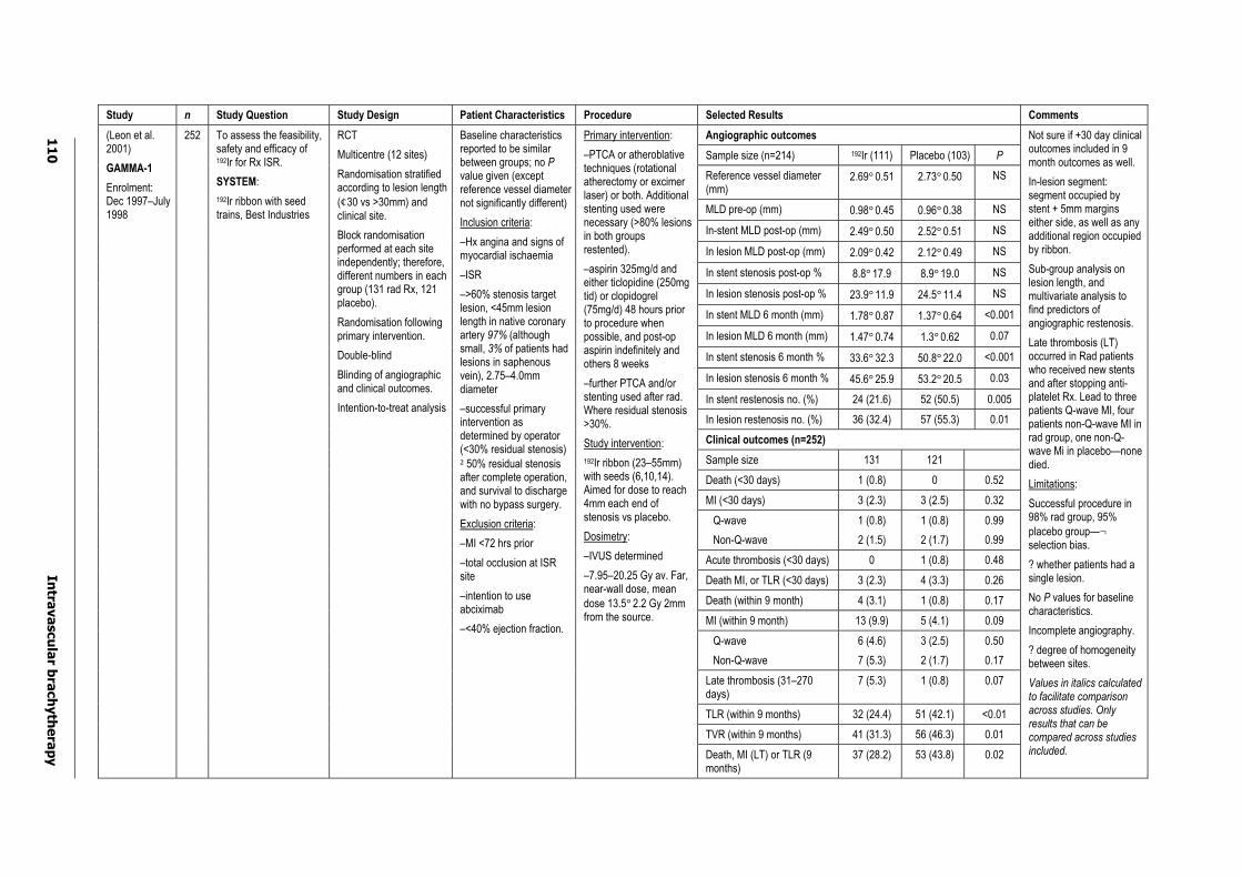
11
0
Intra
va
scu
lar b
rach
yth
era
py
Study n Study Question Study Design Patient Characteristics Procedure Selected Results Comments
Angiographic outcomes
Sample size (n=214) 192Ir (111) Placebo (103) P
Reference vessel diameter (mm)
2.69°0.51 2.73°0.50 NS
MLD pre-op (mm) 0.98°0.45 0.96°0.38 NS
In-stent MLD post-op (mm) 2.49°0.50 2.52°0.51 NS
In lesion MLD post-op (mm) 2.09°0.42 2.12°0.49 NS
In stent stenosis post-op % 8.8°17.9 8.9°19.0 NS
In lesion stenosis post-op % 23.9°11.9 24.5°11.4 NS
In stent MLD 6 month (mm) 1.78°0.87 1.37°0.64 <0.001
In lesion MLD 6 month (mm) 1.47°0.74 1.3°0.62 0.07
In stent stenosis 6 month % 33.6°32.3 50.8°22.0 <0.001
In lesion stenosis 6 month % 45.6°25.9 53.2°20.5 0.03
In stent restenosis no. (%) 24 (21.6) 52 (50.5) 0.005
In lesion restenosis no. (%) 36 (32.4) 57 (55.3) 0.01
Clinical outcomes (n=252)
Sample size 131 121
Death (<30 days) 1 (0.8) 0 0.52
MI (<30 days) 3 (2.3) 3 (2.5) 0.32
Q-wave
Non-Q-wave
1 (0.8)
2 (1.5)
1 (0.8)
2 (1.7)
0.99
0.99
Acute thrombosis (<30 days) 0 1 (0.8) 0.48
Death MI, or TLR (<30 days) 3 (2.3) 4 (3.3) 0.26
Death (within 9 month) 4 (3.1) 1 (0.8) 0.17
MI (within 9 month) 13 (9.9) 5 (4.1) 0.09
Q-wave
Non-Q-wave
6 (4.6)
7 (5.3)
3 (2.5)
2 (1.7)
0.50
0.17
Late thrombosis (31–270 days)
7 (5.3) 1 (0.8) 0.07
TLR (within 9 months) 32 (24.4) 51 (42.1) <0.01
TVR (within 9 months) 41 (31.3) 56 (46.3) 0.01
(Leon et al. 2001)
GAMMA-1
Enrolment: Dec 1997–July 1998
252 To assess the feasibility, safety and efficacy of 192Ir for Rx ISR.
SYSTEM:
192Ir ribbon with seed trains, Best Industries
RCT
Multicentre (12 sites)
Randomisation stratified according to lesion length (¢30 vs >30mm) and clinical site.
Block randomisation performed at each site independently; therefore, different numbers in each group (131 rad Rx, 121 placebo).
Randomisation following primary intervention.
Double-blind
Blinding of angiographic and clinical outcomes.
Intention-to-treat analysis
Baseline characteristics reported to be similar between groups; no Pvalue given (except reference vessel diameter not significantly different)
Inclusion criteria:
–Hx angina and signs of myocardial ischaemia
–ISR
–>60% stenosis target lesion, <45mm lesion length in native coronary artery 97% (although small, 3% of patients had lesions in saphenous vein), 2.75–4.0mm diameter
–successful primary intervention as determined by operator (<30% residual stenosis) ²50% residual stenosis after complete operation, and survival to discharge with no bypass surgery.
Exclusion criteria:
–MI <72 hrs prior
–total occlusion at ISR site
–intention to use abciximab
–<40% ejection fraction.
Primary intervention:
–PTCA or atheroblative techniques (rotational atherectomy or excimer laser) or both. Additional stenting used were necessary (>80% lesions in both groups restented).
–aspirin 325mg/d and either ticlopidine (250mg tid) or clopidogrel (75mg/d) 48 hours prior to procedure when possible, and post-op aspirin indefinitely and others 8 weeks
–further PTCA and/or stenting used after rad. Where residual stenosis >30%.
Study intervention:
192Ir ribbon (23–55mm) with seeds (6,10,14). Aimed for dose to reach 4mm each end of stenosis vs placebo.
Dosimetry:
–IVUS determined
–7.95–20.25 Gy av. Far, near-wall dose, mean dose 13.5°2.2 Gy 2mm from the source.
Death, MI (LT) or TLR (9 months)
37 (28.2) 53 (43.8) 0.02
Not sure if +30 day clinical outcomes included in 9 month outcomes as well.
In-lesion segment: segment occupied by stent + 5mm margins either side, as well as any additional region occupied by ribbon.
Sub-group analysis on lesion length, and multivariate analysis to find predictors of angiographic restenosis.
Late thrombosis (LT) occurred in Rad patients who received new stents and after stopping anti-platelet Rx. Lead to three patients Q-wave MI, four patients non-Q-wave MI in rad group, one non-Q-wave Mi in placebo—none died.
Limitations:
Successful procedure in 98% rad group, 95% placebo group—¬selection bias.
? whether patients had a single lesion.
No P values for baseline characteristics.
Incomplete angiography.
? degree of homogeneity between sites.
Values in italics calculated to facilitate comparison across studies. Only results that can be compared across studies included.
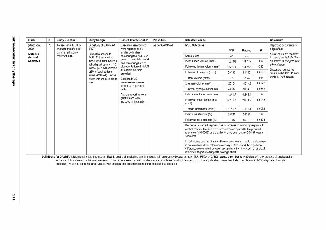
Intra
va
scu
lar b
rach
yth
era
py
11
1
Study n Study Question Study Design Patient Characteristics Procedure Selected Results Comments
IVUS Outcomes
192IR Placebo P
Sample size 37 33
Index lumen volume (mm3) 182°93 176°77 0.8
Follow-up lumen volume (mm3) 157°73 128°66 0.12
Follow-up IH volume (mm3) 58°36 81°43 0.0295
D stent volume (mm3) 3°37 2°24 0.9
D lumen volume (mm3) -25°34 -48°42 0.0225
D intimal hyperplasia vol (mm3) 28°37 50°40 0.0352
Index mean lumen area (mm2) 4.2°1.7 4.2°1.4 1.0
Follow-up mean lumen area (mm2)
3.2°1.8 2.0°1.2 0.0035
D mean lumen area (mm2) -2.2°1.8 -1.0°1.3 0.0032
Index area stenosis (%) 25°25 24°26 1.0
Follow-up area stenosis (%) 31°32 55°38 0.0124
(Mintz et al. 2000)
IVUS sub-study of GAMMA-1
70 To use serial IVUS to evaluate the effect of gamma radiation on recurrent ISR.
Sub-study of GAMMA-1 (RCT)
Four sites access to IVUS; 139 enrolled at these sites; final available paired (post-op and 8/12 follow-up); n=70 selected (28% of total patients from GAMMA-1). Unclear whether there is selection bias.
Baseline characteristics were reported to be similar both when comparing the IVUS sub-group to complete cohort and comparing Rx and placebo Patients in IVUS sub-study; no table provided.
Baseline IVUS measurements were similar, as reported in table.
Authors report no vein graft lesions were included in this study.
As per GAMMA-1
Decrease in stented segment due to increase in intimal hyperplasia. In control patients the ® in stent lumen area compared to the proximal reference (p=0.0202) and distal reference segment (p=0.0115) vessel segments.
In radiation group the ® in stent lumen area was similar to the decrease in proximal and distal reference areas (p=0.9 for both). No significant differences were noted between groups for either the proximal or distal reference segment—suggests no edge effect?
Report no occurrence of edge effect
More values are reported in paper; not included here as unable to compare with other studies.
Discussion compares results with SCRIPPS and WRIST, IVUS results.
Definitions for GAMMA-1: MI: including late thrombosis; MACE: death, MI (including late thrombosis: LT) emergency bypass surgery, TLR (PTCA or CABG); Acute thrombosis: (<30 days of index procedure) angiographic evidence of thrombosis or subacute closure within the target vessel, or death in which acute thrombosis could not be ruled out by the adjudication committee; Late thrombosis: (31–270 days after the index procedure) MI attributed to the target vessel, with angiographic documentation of thrombus or total occlusion.
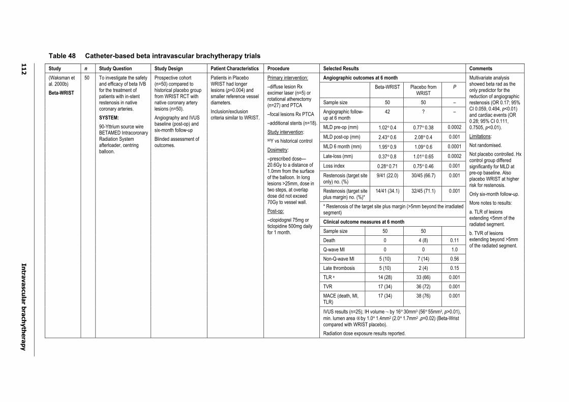
11
2
Intra
va
scu
lar b
rach
yth
era
py
Table 48 Catheter-based beta intravascular brachytherapy trials
Study n Study Question Study Design Patient Characteristics Procedure Selected Results Comments
Angiographic outcomes at 6 month
Beta-WRIST Placebo from WRIST
P
Sample size 50 50 –
Angiographic follow-up at 6 month
42 ? –
MLD pre-op (mm) 1.02°0.4 0.77°0.38 0.0002
MLD post-op (mm) 2.43°0.6 2.08°0.4 0.001
MLD 6 month (mm) 1.95°0.9 1.09°0.6 0.0001
Late-loss (mm) 0.37°0.8 1.01°0.65 0.0002
Loss index 0.28°0.71 0.75°0.46 0.001
Restenosis (target site only) no. (%)
9/41 (22.0) 30/45 (66.7) 0.001
Restenosis (target site plus margin) no. (%)*
14/41 (34.1) 32/45 (71.1) 0.001
* Restenosis of the target site plus margin (>5mm beyond the irradiated segment)
Clinical outcome measures at 6 month
Sample size 50 50
Death 0 4 (8) 0.11
Q-wave MI 0 0 1.0
Non-Q-wave MI 5 (10) 7 (14) 0.56
Late thrombosis 5 (10) 2 (4) 0.15
TLR a 14 (28) 33 (66) 0.001
TVR 17 (34) 36 (72) 0.001
MACE (death, MI, TLR)
17 (34) 38 (76) 0.001
(Waksman et al. 2000b)
Beta-WRIST
50 To investigate the safety and efficacy of beta IVB for the treatment of patients with in-stent restenosis in native coronary arteries.
SYSTEM:
90-Yttrium source wire BETAMED Intracoronary Radiation System afterloader, centring balloon.
Prospective cohort (n=50) compared to historical placebo group from WRIST RCT with native coronary artery lesions (n=50).
Angiography and IVUS baseline (post-op) and six-month follow-up
Blinded assessment of outcomes.
Patients in Placebo WRIST had longer lesions (p=0.004) and smaller reference vessel diameters.
Inclusion/exclusion criteria similar to WRIST.
Primary intervention:
–diffuse lesion Rx excimer laser (n=5) or rotational atherectomy (n=27) and PTCA
–focal lesions Rx PTCA
–additional stents (n=18).
Study intervention:
90Y vs historical control
Dosimetry:
–prescribed dose—20.6Gy to a distance of 1.0mm from the surface of the balloon. In long lesions >25mm, dose in two steps, at overlap dose did not exceed 70Gy to vessel wall.
Post-op:
–clopidogrel 75mg or ticlopidine 500mg daily for 1 month.
IVUS results (n=25); IH volume¬ by 16°30mm3 (56°55mm3, p>0.01), min. lumen area ® by 1.0°1.4mm2 (2.0°1.7mm2 ,p=0.02) (Beta-Wrist compared with WRIST placebo).
Radiation dose exposure results reported.
Multivariate analysis showed beta rad as the only predictor for the reduction of angiographic restenosis (OR 0.17; 95% CI 0.059, 0.494, p<0.01) and cardiac events (OR 0.28; 95% CI 0.111, 0.7505, p<0.01).
Limitations:
Not randomised.
Not placebo controlled. Hx control group differed significantly for MLD at pre-op baseline. Also placebo WRIST at higher risk for restenosis.
Only six-month follow-up.
More notes to results:
a. TLR of lesions extending <5mm of the radiated segment.
b. TVR of lesions extending beyond >5mm of the radiated segment.
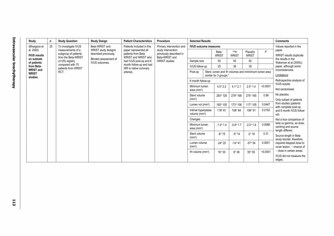
Intra
va
scu
lar b
rach
yth
era
py
11
3
Study n Study Question Study Design Patient Characteristics Procedure Selected Results Comments
IVUS outcome measures
Beta-WRIST
192IrWRIST
PlaceboWRIST
P
Sample size 50 50 50
IVUS follow-up 25 36 39
Post-op Stent, lumen and IH volumes and mininimum lumen area similar for 3 groups.*
6 month follow-up
Minimum lumen area (mm2)
4.5°2.2 4.1°2.1 2.5°1.4 <0.0001
Stent volume (mm3)
283°126 279°168 275°165 0.98
Lumen vol (mm3) 165°105 173°106 117°105 0.0447
Intimal hyperplasia volume (mm3)
118°61 106°84 158°91 0.0193
Changes
Minimum lumen area (mm2)
-1.0°1.4 -0.8°1.7 -2.0°1.8 0.0066
Stent volume (mm3)
-8°15 -5°14 -2°18 0.31
Lumen volume (mm3)
-24°25 -14°41 -57°54 0.0001
(Bhargava et al. 2000)
IVUS results on subsets of patients from Beta-WRIST and WRISTstudies
25 To investigate IVUS measurements of a subgroup of patients from the Beta-WRIST (n=25) registry compared with 75 patients from WRIST RCT.
Beta-WRIST and WRIST study designs described previously.
Blinded assessment of IVUS outcomes.
Patients included in this paper represented all patients from Beta WRIST and WRIST who had IVUS post-op and 6 month follow-up and had ISR in native coronary arteries.
Primary intervention and study intervention previously described in Beta-WRIST and WRIST studies
IH volume (mm3) 16°30 9°38 55°55 <0.0001
Values reported in the paper.
WRIST results duplicate the results in the Waksman et al (2000c) paper, although some inconsistencies.
Limitations:
Retrospective analysis of IVUS results.
Not randomised.
No placebo.
Only subset of patients from studies (patients with complete post-op and 6 month IVUS follow-up).
Not a true comparison of beta vs gamma, as dose, centring and source length differed.
Source length in Beta study shorter; therefore, required stepped dose to cover lesion; ¬ chance of ¬ dose in certain areas.
IVUS did not measure the edges.
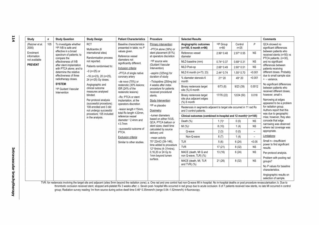
11
4
Intra
va
scu
lar b
rach
yth
era
py
Study n Study Question Study Design Patient Characteristics Procedure Selected Results Comments
Angiographic outcomes (n=105, 6 month n=96)
32P Group n=80
Control n=25
P
Reference vessel diameter
2.99°0.48 2.97°0.55 NS
MLD baseline (mm) 0.74°0.37 0.68°0.31 NS
MLD Post-op 2.68°0.49 2.60°0.51 NS
MLD 6 month (n=73; 23) 2.44°0.74 1.55°0.70 <0.001
% diameter stenosis 6 month
21°20 49°20 <0.001
Binary restenosis target site (%) 6 month
6/73 (8) 9/23 (39) 0.0012
Binary restenosis target site plus adjacent edges (%) 6 month
17/76 (22) 12/24 (50) 0.018
Restenosis in segments adjacent to target site occurred in 11 rad Rx and 3 control patients.
Clinical outcomes (combined in-hospital and 12 month)a (n=105)
Death (%) 1 (1)b 0 (0) NS
MI (%)c 8 (10) 1 (4) NS
Q-wave 2 (3) 0 (0) –
Non-Q-wave 6 (7) 1 (4) –
TLR 5 (6) 6 (24) <0.05
TVR 17 (21) 8 (32) NS
MACE (death, MI Q and non Q-wave, TLR) (%)
13 (16) 6 (24) NS
MACE (death, MI, TLR and TVR) (%)
21 (26) 8 (32) NS
(Raizner et al. 2000)
Enrolment information not available
PREVENT
105 To investigate whether 32P IVB is safe and effective in a broad spectrum of patients; to compare the effectiveness of IVB after stent implantation with PTCA alone; and to determine the relative effectiveness of three radiotherapy doses.
SYSTEM:
32P Guidant Vascular Intervention
RCT
Multicentre (6 international sites)
Randomisation process not reported.
Patients randomised to:
–0 (n=25) or
–16 (n=23), 20 (n=25), 24 (n=25) Gy doses.
Angiographic and clinical outcome measures analysed blinded.
Per-protocol analysis (successful procedure); 108 enrolled and 3 did not undergo successful procedure; 105 included in the analysis.
Baseline characteristics presented in table; no Pvalues given.
Reference vessel diameters not significantly different.
Inclusion criteria:
–PTCA of single native coronary artery
–de novo (70%) or restenotic (30% lesions; ISR (24% of the restenotic lesions)
–Rx: PTCA or stent implantation, at the operators discretion
–lesion length ¢15mm,total Rx length ¢22mm,reference vessel diameter ²2.4mm and ¢3.7mm
–successful outcome of PTCA.
Exclusion criteria:
Similar to other studies.
Primary intervention:
–PTCA alone (39%) or stent placement (61%) at operators discretion
–32P source wire (Guidant Vascular Intervention)
–aspirin (325mg) for duration of study
–Ticlopidine (250mg bid 4 weeks after index procedure for patients received procedural stents.
Study Intervention:
32P vs placebo
Dosimetry:
–lumen diameters based on either IVUS, QCA, PTCA balloon or stent sizes; dwell time calculated by source delivery unit
–mean activity 70°22mCi (39–146), time added to procedure 12°6mins (4–31mins); 0,16,20 or 24 Gy to 1mm beyond lumen surface.
QCA showed no significant differences between patients who received stents (n=50) vs PTCA patients. (n=30), and no significant differences between patients receiving different doses. Probably due to small sample size + ¬ variance.
No significant differences between patients who received different doses; however, small n.
Narrowing at edges appeared to be a problem for radiation group. Authors report that this was due to geographic miss; however, they also concede that edge narrowing was observed where rad coverage was appropriate.
Limitations:
Small n—insufficient power to find significant results.
Per-protocol analysis.
Problem with pooling rad groups?
No P values for baseline characteristics.
Angiographic results on selection of sample.
TVR: for restenosis involving the target site and adjacent (sites 5mm beyond the radiation zone); a. One rad and one control had non-Q-wave MI in hospital. No in-hospital deaths or post procedure revascularisation; b. Due to thrombotic occlusion received stent, stopped anti-platelet Rx 3 weeks after; c. Seven post- hospital MIs occurred in rad group due to acute occlusion. 6 of 7 patients received new stents, no late MI occurred in control group; Radiation survey reading 1m from source during active dwell time 0.46°0.35mrem/h (range 0.04–1.52mrem/h) ® fluoroscopy.
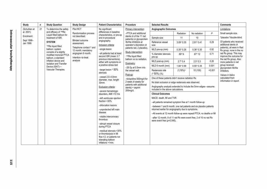
Intra
va
scu
lar b
rach
yth
era
py
11
5
Study n Study Question Study Design Patient Characteristics Procedure Selected Results Comments
Angiographic Outcomes
Radiation No radiation P
Sample size 11 10
Reference vessel size
3.09°0.35 2.91°0.41 0.29
MLD pre-op (mm) 0.35°0.26 0.36°0.30 0.92
% diameter stenosis pre-op
89°9 87°12 0.71
MLD post-op (mm) 2.7°0.4 2.5°0.3 0.26
MLD 6 month (mm) 1.84°0.99 0.55°0.35 0.001
Restenosis rate
(²50%) (%)
2 (18%)a 10 (100) <0.001
One of these patients didn’t receive radiation Rx.
No total occlusion or edge restenosis was observed.
Angiography analysis extended to include the 5mm edges—assume included in the above calculations.
(Schuhlen et al. 2001)
Enrolment:
Sept 1898– Jan 1999
21 To determine the safety and efficacy of 188ReLiquid filled balloon for treatment of ISR.
SYSTEM:
188Re liquid filled balloon: system consists of a slightly modified monorail PTCA balloon, a standard inflation device and Isolation and Transfer Device (ISAT)—Vascular Therapies.
Pilot RCT
Randomisation process not described.
Blinded outcome assessment.
Telephone contact 1 and 12 month; mandatory angiogram 6 month.
Intention–to-treat analysis
No significant differences in baseline characteristics, or pre-op and in-procedural angiography.
Inclusion criteria:
–single lesion
–all patients had at least second ISR (mean 3.7 previous interventions), either with symptoms or a positive stress test
–target lesion ²50%stenosis
–vessel 2.0–4.0mm diameter, max. length 30mm.
Exclusion criteria:
–severe hematologic disorders, AMI <72 hrs
–left ventricular ejection fraction <30%
–bifurcation lesions
–unprotected left main disease
–visible intercoronary thrombus
–abrupt vessel closure during PTCA
–residual stenosis >30% or thrombolysis in MI flow ¢2, or patients not tolerating balloon inflations >1min.
Primary intervention:
–PTCA and additional stents (4 of the 11 rad patients) or glycoproteinIIb/IIIa inhibitors at operator’s discretion (4 patients rad, 2 placebo).
Study intervention:
–188Re liquid filled balloon vs no radiation.
Dosimetry:
–28 Gy at 0.5mm into the vessel wall.
Post-op:
–ticlopidine 500mg/d for 2 week (4 weeks for patients with additional stents) + aspirin 200mg/d.
Clinical Outcomes
MACE: death, MI and TVR
–all patients remained symptom free at 1 month follow-up
–between 1 and 6 month, one rad patients and six placebo patients returned earlier for angiography due to symptoms.
–All events at 12 month follow-up were repeat PTCA; no deaths or MI
–after 12 month, 8 of 11 rad Rx were event free, 2 of 10 no rad Rx were event free (p=0.045).
Limitations:
Small sample size.
?placebo ?double-blind.
Of patients who received additional stents (4 patients), all were in Rad Rx group, none in the no rad Rx group. This may improve the outcome for the rad Rx group. Also more patients in rad group received glycoprotein IIb/IIIa inhibitors.
Values in italics calculated from information in report.

11
6
Intra
va
scu
lar b
rach
yth
era
py
Study n Study Question Study Design Patient Characteristics Procedure Selected Results Comments
IVUS results Rad (n=16) Plac. (n=5) P
Lumen volume (mm3) index 185°60 205°62 NS
Lumen volume (mm3) 6 month
190°63 163°44 NS
MLA (mm2) index 4.8°1.6 4.7°1.2 NS
MLA (mm2) 6 month 4.7 °1.3 3.3°1.3 0.046
PB at MLA(%) index 63°9 60°16 NS
PB at MLA(%) 6 month 64°9 76°14 0.042
(Costa et al. 2000)
21 To investigate the effect of beta radiation following PTCA or stenting, using IVUS.
System:
Guidant Brachytherapy System, 32P
RCT
Single-centre
Double-blind
Placebo, three doses (28, 35 and 42 Gy)
Baseline and post-op, 6 month 3D-IVUS follow-up
n=26 randomised, and n=21 included in final 6 month analysis, b/c five patients (1 placebo) did not undergo IVUS 6 month. Four rad patients had late thrombosis; placebo patient normal, but refused.
Baseline characteristics similar between groups, including % receiving additional stents.
Inclusion criteria:
Successful procedure, and 6 month IVUS follow-up.
Primary intervention:
–PTCA and stenting
–additional stents 7 patients (44%) in rad grp., three patients (60%) in placebo.
–IVUS guided
Study intervention:
–32P vs placebo
Dosimetry:
–actual dose received by the target segment not calculated.
Post-op:
–aspirin (250mg/d), ticlopidine (250mg/d only stented patients).
MLA: mean lumen area; PB: plaque burden; Index: post-op measure.
Other findings:
–no relationship between prescribed doses and volumetric change
–DLV +ve rad/ -ve placebo (p=0.01)
–for the irradiated group, compared stent (n=7) with (n=9)—no significant differences in volumetric changes.
Limitations:
Small n.
Four patients in rad group developed late events (2 sub-acute thrombosis, 1 late thrombosis, 1 severe restenotic lesion proximal to radiation site). No late events were reported in the placebo group. No significant differences in percent having additional stents between groups.

Intra
va
scu
lar b
rach
yth
era
py
11
7
Study n Study Question Study Design Patient Characteristics Procedure Selected Results Comments
Angiographic outcomes
32P Group Placebo P
Sample size 166 166
Post-op analysis seg.a MLD, mean°SD (n)
1.92°0.42 (161)
1.96°0.42 (161)
0.49
Angiographic follow-up 9 months
129 128
Analysis segment restenosis rate, no (%)
34 (26.4) 66 (51.6) <0.0001
Analysis segment MLD 9 months mean°SD
1.54 (0.65) 1.38 (0.61) 0.043
aAnalysis segment—extends 5mm proximal and distal to the radiated or injured landmark, which was longest in length.
Clinical outcome measures 9 months includes acute outcomes, number and (%)
Sample size 166 166 P
Death 5 (3) 5 (3) NS
Non-Q-wave MI 10 (6) 5 (3) No P
Q-wave MI 3 (2) 3 (2) NSb
TLR 17 (10) 46 (28) 0.0001b
TVR 33 (20) 51 (31) 0.033b
MACE (death Q-wave MI, TLR) 24 (15) 51 (31) 0.0006
Any MACE (death Q-wave MI, TLR, TVR)
39 (23.5) 56 (34) 0.05
Late thrombosis (31–290 days) 5/166 (3) 1/166 (0.6) 0.21
Late total occlusion (31–290 days)
6/166 (4) 2/166 (1.2) 0.28
(Waksman et al. 2002)
INHIBIT
Enrolment: 1998–1999
332
To investigate the safety and efficacy of 32P catheter-based IVB in patients with diffuse ISR.
SYSTEM:
GALILEOã 70
Intravascular Radiotherapy System
Designed for use with ¢47mm lesion length and reference vessel diameter 2.4mm–3.7mm.
RCT
Multicentre (24 sites)
Concealment of randomisation—envelope method.
Double blinded
Placebo controlled
Data recorded prospectively
All clinical events adjudicated by a blinded clinical events committee.
Power analysis reported.
Intention-to-treat analysis
All patients. ISR in single native coronary artery.
Baseline characteristics were reported to be similar to Ax groups, table presented: no P values.
Mean lesion length 17.4mm
Patients had to have successful primary intervention to be included; randomisation after intervention deemed successful.
Primary intervention
–PTCA, atherectomy, laser angioplasty, additional stents (rad: n=49 (30%)
–placebo: n=52 (31%)
Study intervention
–32P GALILEOã system
Dosimetry
–20Gy at 1mm beyond the lumen diameter, 2.88*109 Bq mean specific activity
– dose for patients with longer lesions (22–47mm) 30% higher due to tandem positioning
Post-op
–aspirin (325mg/ day) for 1 year or per institutional standard all patients
–complex regime that changes
–first 69 patients with new stents—ticlopidine for 30 days; next 29 with or without new stent—ticlopidine or clopidogrel for 90 days.
Acute and late clinical outcomes combined from Waksman paper to facilitate comparison across studies.
b P values have been obtained for combined values from the INHIBIT Clinical Summary Data, provided by Guidant.
Limitations:
Assume angiographic results are based on the analysis segment; this is not explicit in the paper.
Only 9 month follow-up.
Incomplete angiographic follow-up.
INHIBIT Definitions: MACE is a hierarchical tallyin which only the most significant event is counted per patient with a hierarchy of Death>MI>CABG>PTCA.
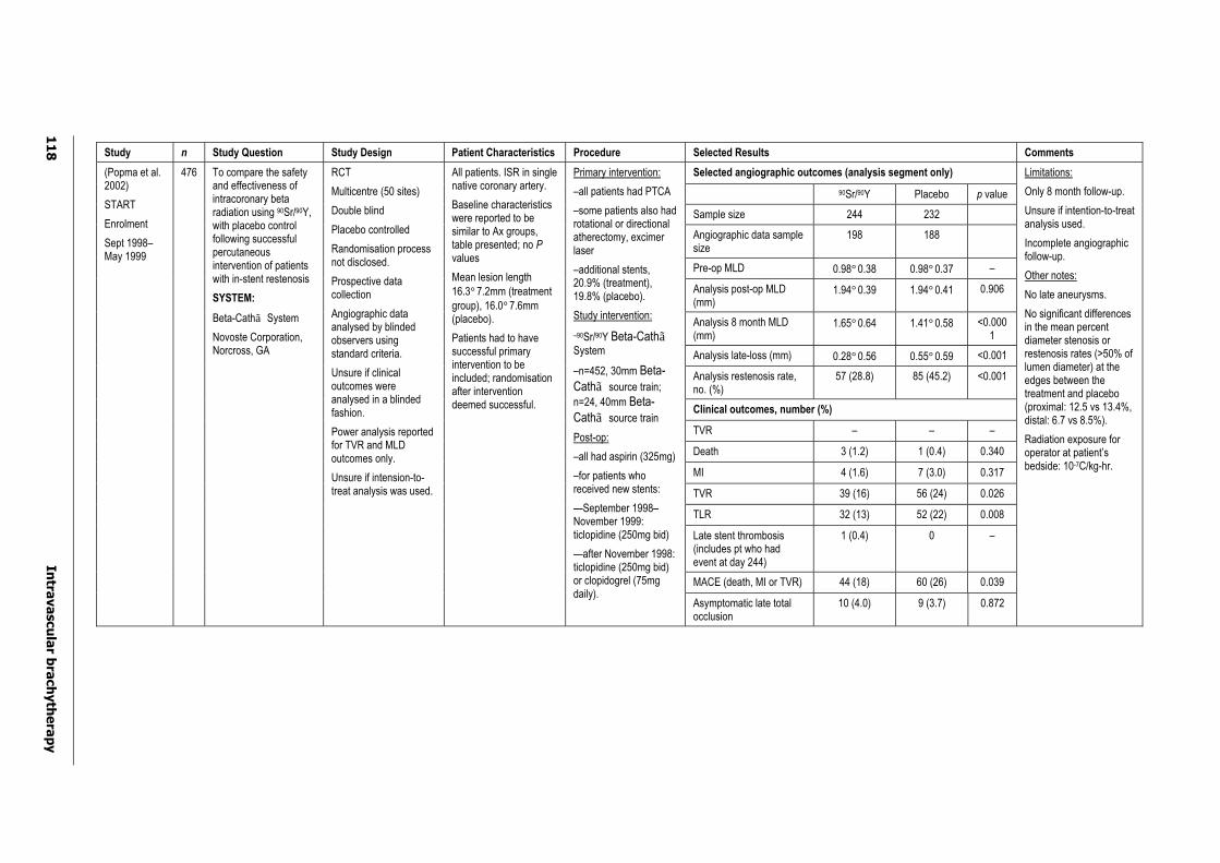
11
8
Intra
va
scu
lar b
rach
yth
era
py
Study n Study Question Study Design Patient Characteristics Procedure Selected Results Comments
Selected angiographic outcomes (analysis segment only)
90Sr/90Y Placebo p value
Sample size 244 232
Angiographic data sample size
198 188
Pre-op MLD 0.98°0.38 0.98°0.37 –
Analysis post-op MLD (mm)
1.94°0.39 1.94°0.41 0.906
Analysis 8 month MLD (mm)
1.65°0.64 1.41°0.58 <0.0001
Analysis late-loss (mm) 0.28°0.56 0.55°0.59 <0.001
Analysis restenosis rate, no. (%)
57 (28.8) 85 (45.2) <0.001
Clinical outcomes, number (%)
TVR – – –
Death 3 (1.2) 1 (0.4) 0.340
MI 4 (1.6) 7 (3.0) 0.317
TVR 39 (16) 56 (24) 0.026
TLR 32 (13) 52 (22) 0.008
Late stent thrombosis (includes pt who had event at day 244)
1 (0.4) 0 –
MACE (death, MI or TVR) 44 (18) 60 (26) 0.039
(Popma et al. 2002)
START
Enrolment
Sept 1998–May 1999
476 To compare the safety and effectiveness of intracoronary beta radiation using 90Sr/90Y,with placebo control following successful percutaneous intervention of patients with in-stent restenosis
SYSTEM:
Beta-Cathã System
Novoste Corporation, Norcross, GA
RCT
Multicentre (50 sites)
Double blind
Placebo controlled
Randomisation process not disclosed.
Prospective data collection
Angiographic data analysed by blinded observers using standard criteria.
Unsure if clinical outcomes were analysed in a blinded fashion.
Power analysis reported for TVR and MLD outcomes only.
Unsure if intension-to-treat analysis was used.
All patients. ISR in single native coronary artery.
Baseline characteristics were reported to be similar to Ax groups, table presented; no Pvalues
Mean lesion length 16.3°7.2mm (treatment group), 16.0°7.6mm (placebo).
Patients had to have successful primary intervention to be included; randomisation after intervention deemed successful.
Primary intervention:
–all patients had PTCA
–some patients also had rotational or directional atherectomy, excimer laser
–additional stents, 20.9% (treatment), 19.8% (placebo).
Study intervention:
–90Sr/90Y Beta-CathãSystem
–n=452, 30mm Beta-Cathã source train;
n=24, 40mm Beta-Cathã source train
Post-op:
–all had aspirin (325mg)
–for patients who received new stents:
—September 1998–November 1999: ticlopidine (250mg bid)
—after November 1998: ticlopidine (250mg bid) or clopidogrel (75mg daily).
Asymptomatic late total occlusion
10 (4.0) 9 (3.7) 0.872
Limitations:
Only 8 month follow-up.
Unsure if intention-to-treat analysis used.
Incomplete angiographic follow-up.
Other notes:
No late aneurysms.
No significant differences in the mean percent diameter stenosis or restenosis rates (>50% of lumen diameter) at the edges between the treatment and placebo (proximal: 12.5 vs 13.4%, distal: 6.7 vs 8.5%).
Radiation exposure for operator at patient’s bedside: 10-7C/kg-hr.

Intravascular brachytherapy 119
Appendix D Flow chart demonstrating clinical pathways for percutaneous intervention and IVB
Primary therapy
Angioplasty plus stent placement
Restenosis
Intravascular brachytherapy
No intravascular brachytherapy
Prolonged anti-platelet therapy
OutcomesAngiography, intravascular ultrasound, clinical
Angioplastyfollowed by
stenting
Atherectomy,angioplastyfollowed by
stenting
Atherectomyfollowed by angioplasty
Angioplasty
Other percutaneous intervention
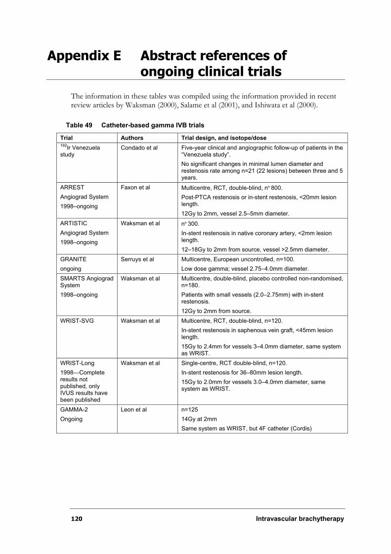
120 Intravascular brachytherapy
Appendix E Abstract references of ongoing clinical trials
The information in these tables was compiled using the information provided in recent review articles by Waksman (2000), Salame et al (2001), and Ishiwata et al (2000).
Table 49 Catheter-based gamma IVB trials
Trial Authors Trial design, and isotope/dose 192
Ir Venezuela study
Condado et al Five-year clinical and angiographic follow-up of patients in the “Venezuela study”.
No significant changes in minimal lumen diameter and restenosis rate among n=21 (22 lesions) between three and 5 years.
ARREST
Angiograd System
1998–ongoing
Faxon et al Multicentre, RCT, double-blind, nº800.
Post-PTCA restenosis or in-stent restenosis, <20mm lesion length.
12Gy to 2mm, vessel 2.5–5mm diameter.
ARTISTIC
Angiograd System
1998–ongoing
Waksman et al nº300.
In-stent restenosis in native coronary artery, <2mm lesion length.
12–18Gy to 2mm from source, vessel >2.5mm diameter.
GRANITE
ongoing
Serruys et al Multicentre, European uncontrolled, n=100.
Low dose gamma; vessel 2.75–4.0mm diameter.
SMARTS Angiograd System
1998–ongoing
Waksman et al Multicentre, double-blind, placebo controlled non-randomised, n=180.
Patients with small vessels (2.0–2.75mm) with in-stent restenosis.
12Gy to 2mm from source.
WRIST-SVG Waksman et al Multicentre, RCT, double-blind, n=120.
In-stent restenosis in saphenous vein graft, <45mm lesion length.
15Gy to 2.4mm for vessels 3–4.0mm diameter, same system as WRIST.
WRIST-Long
1998—Complete results not published, only IVUS results have been published
Waksman et al Single-centre, RCT double-blind, n=120.
In-stent restenosis for 36–80mm lesion length.
15Gy to 2.0mm for vessels 3.0–4.0mm diameter, same system as WRIST.
GAMMA-2
Ongoing
Leon et al n=125
14Gy at 2mm
Same system as WRIST, but 4F catheter (Cordis)
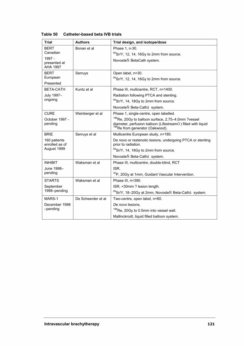
Intravascular brachytherapy 121
Table 50 Catheter-based beta IVB trials
Trial Authors Trial design, and isotope/dose
BERT Canadian
1997 - presented at AHA 1997
Bonan et al Phase 1, n-30. 90
Sr/Y, 12, 14, 16Gy to 2mm from source.
NovosteÑ BetaCath system.
BERT European
Presented
Serruys Open label, n=30. 90
Sr/Y, 12, 14, 16Gy to 2mm from source.
BETA-CATH
July 1997–ongoing
Kuntz et al Phase III, multicentre, RCT, n=1400.
Radiation following PTCA and stenting. 90
Sr/Y, 14, 18Gy to 2mm from source.
NovosteÑ Beta-Cathã system.
CURE
October 1997 - pending
Weinberger et al Phase 1, single-centre, open labelled. 188
Re, 20Gy to balloon surface, 2.75–4.0mm ?vessel
diameter, perfusion balloon (LifestreamÓ) filled with liquid 188
Re from generator (Oakwood).
BRIE
160 patients enrolled as of August 1999
Serruys et al Multicentre European study, n=180.
De novo or restenotic lesions, undergoing PTCA or stenting prior to radiation. 90
Sr/Y, 14, 18Gy to 2mm from source.
NovosteÑ Beta-Cathã system.
INHIBIT
June 1998–pending
Waksman et al Phase III, multicentre, double-blind, RCT
ISR.32
P, 20Gy at 1mm, Guidant Vascular Intervention.
STARTS
September 1998–pending
Waksman et al Phase III, n=390.
ISR, <30mm ? lesion length. 90
Sr/Y, 18–20Gy at 2mm, NovosteÑ Beta-Cathã system.
MARS-1
December 1998 –pending
De Scheerder et al Two-centre, open label, n=60.
De novo lesions. 188
Re, 20Gy to 0.5mm into vessel wall.
Mallinckrodt, liquid filled balloon system.
122 Intravascular brachytherapy
Appendix F Potential adverse events associated with percutaneous intervention and IVB
The following list has been adapted from the Food and Drug Administration’s (FDA) Health Technology Assessment of the safety and effectiveness of the Galileo Intravascular Radiation System. This list serves as a comprehensive list of adverse events potentially associated with percutaneous intervention and IVB.
¶ arteriovenous fistula;
¶ coronary artery aneurysm;
¶ coronary artery spasm;
¶ coronary vessel dissection, perforation, rupture or injury;
¶ delayed endothelialisation;
¶ drug reactions, or allergic reactions to contrast media;
¶ embolism;
¶ endocarditis;
¶ haemorrhage or haematoma;
¶ hypo/hypertension;
¶ infection;
¶ loss of vaso-reactivity immediately following treatment; and
¶ short-term hemodynamic deterioration.
Intravascular brachytherapy 123
Abbreviations
188Re 188-Rhenium
192Ir 192-Iridium
32P 32-Phosphorus
90Y 90-Yttrium
90Sr90Y 90-Strontium/ 90-Yttrium
Beta WRIST Beta-Washington Radiation for In-Stent Restenosis Trial
bid Bis in di’e (twice a day)
CABG Coronary artery bypass graft
FDA Food and Drug Administration
HD High dose
HIC Health Insurance Commission
ICER Incremental cost-effectiveness ratio
INHIBIT Intimal Hyperplasia Inhibition with Beta In-Stent Trial
ISAT Isolation and Transfer Device
ISR In-stent restenosis
IVB Intravascular brachytherapy
IVUS Intravascular ultrasound
MACE Major adverse cardiac events
MI Myocardial infarction
MLD Minimal luminal diameter
MSAC Medical Services Advisory Committee
NHMRC National Health and Medical Research Council
NHS National Health Service
NS Not significant
PBS Pharmaceutical Benefits Scheme
124 Intravascular brachytherapy
PCI Percutaneous intervention
Post-op Post-operative
POWER Prince of Wales Endovascular Radiation
Pre-op Pre-operative
PTCA Percutaneous transluminal coronary angioplasty
RCT Randomised controlled trial
SCRIPPS Scripps Clinic and Research Foundation
SD Standard deviation
START Stents and Radiation Therapy Trial
TGA Therapeutic Goods Administration
TLR Target lesion revascularisation
TVR Target vessel revascularisation
WRIST Washington Radiation for In-stent Restenosis Trial
YLD Year lived with disability
YLL Years of life lost
Intravascular brachytherapy 125
References
Ahmed, J. M., Mintz, G. S., Waksman, R., Lansky, A. J., Mehran, R., Wu, H., Weissman, N. J., Pichard, A. D., Satler, L. F., Kent, K. M., & Leon, M. B. 2001a, ‘Serial intravascular ultrasound analysis of edge recurrence after intracoronary gamma radiation treatment of native artery in-stent restenosis lesions’, American Journal of Cardiology, 87 (10), 1145–1149.
Ahmed, J. M., Mintz, G. S., Waksman, R., Mehran, R., Leiboff, B., Pichard, A. D., Satler, L. F., Kent, K. M., & Weissman, N. J. 2001b, ‘Serial intravascular ultrasound assessment of the efficacy of intracoronary gamma-radiation therapy for preventing recurrence in very long, diffuse, in-stent restenosis lesions’, Circulation,104 (8), 856–859.
Ahmed, J. M., Mintz, G. S., Waksman, R., Weissman, N. J., Leiboff, B., Pichard, A. D., Satler, L. F., Kent, K. M., Leon, M. B., Stents, Restenosis, & Imaging. 2001c, ‘Serial intravascular ultrasound analysis of the impact of lesion length on the efficacy of intracoronary gamma-irradiation for preventing recurrent in-stent restenosis’, Circulation, 103 (2), 188–191.
Ahmed, J. M., Mintz, G. S., Waksman, R., Weissman, N. J., Mehran, R., Pichard, A. D., Satler, L. F., Kent, K. M., & Leon, M. B. 2000, ‘Safety of intracoronary gamma-radiation on uninjured reference segments during the first 6 months after treatment of in-stent restenosis: a serial intravascular ultrasound study’, Circulation, 101 (19), 2227–2230.
Albiero, R., Adamian, M., Kobayashi, N., Amato, A., Vaghetti, M., Di Mario, C., & Colombo, A. 2000a, ‘Short- and intermediate-term results of (32)P radioactive beta-emitting stent implantation in patients with coronary artery disease: The Milan Dose-Response Study’, Circulation, 101), 18–26.
Albiero, R., Nishida, T., Adamian, M., Amato, A., Vaghetti, M., Corvaja, N., Di Mario, C., & Colombo, A. 2000b, ‘Edge restenosis after implantation of high activity (32)P radioactive beta-emitting stents’, Circulation, 101 (21), 2454–2457.
Australian Institute of Health and Welfare 1998, AIHW National hospital morbidity database [Internet] Available from: http://www.aihw.gov.au/hospitaldata/morbidity.html#nhms.[Accessed 5th February 2002].
Australian Institute of Health and Welfare 1999, Heart, stroke and vascular diseases, Australian facts, AIHW and the Heart Foundation of Australia, Canberra.
Australian Institute of Health and Welfare 2000a, Australia’s health 2000: the seventh biennial health report of the Australian Institute of Health and Welfare, AIHW, Canberra, AIHW.
Australian Institute of Health and Welfare 2000b, National cardiovascular disease database [Internet]. Available from: http//:www.aihw.gov.au/cvdhtml/cvd-menu.htm[Accessed 5th February 2002].
126 Intravascular brachytherapy
Bahrassa, F. & Datta, R. 1983, ‘Postoperative beta radiation treatment of pterygium’, International Journal of Radiation Oncology, Biology, Physics, 9 (5), 679–684.
Baim, D. S. & Grossman, W. 1998, ‘Coronary angioplasty and other therapeutic applications of cardiac catheterization,’ in Harrison’s principles of internal medicine,14th edn, A. S. Fauci et al, eds., McGraw-Hill, New York, 1375–1380.
Bass, B. G. 1999, ‘Radiation safety requirements for cardiovascular brachytherapy’, Cardiovascular Radiation Medicine, 1 (3), 297–306.
Bhargava, B., Mintz, G. S., Mehran, R., Lansky, A. J., Weissman, N. J., Walsh, C., Chan, R. C., & Waksman, R. 2000, ‘Serial volumetric intravascular ultrasound analysis of the efficacy of beta irradiation in preventing recurrent in-stent restenosis’, American Journal of Cardiology, 85 (5), 651–653.
Birdwell, S. H., Hancock, S. L., Varghese, A., Cox, R. S., & Hoppe, R. T. 1997, ‘Gastrointestinal cancer after treatment of Hodgkin’s disease’, International Journal of Radiation Oncology, Biology, Physics, 37 (1), 67–73.
Bonan, R. 2000, ‘Geographical miss: Edge effects’, Vascular Radiotherapy Monitor, 3 (1), 3–8.
Britt, H., Sayer G.P., Miller, G. C., & et al 1999, General practice activity in Australia 1998–1999, AIHW GPSCU, Canberra, General Practice Series No. 2. AIHW Cat. No. GEP 2.
Casscells, W. 1992, ‘Migration of smooth muscle and endothelial cells. Critical events in restenosis.’, Circulation, 86 (3), 723–729.
Commonwealth Department of Health and Aged Care 2001a, Medicare Benefits Schedule,Commonwealth Department of Health and Aged Care, Canberra.
Commonwealth Department of Health and Ageing 2002, Schedule of Pharmaceutical Benefits,Commonwealth Department of Health and Ageing, Canberra.
Commonwealth Department of Health and Aged Care, Acute and Coordinated Care Branch. 2001b, National Hospital Cost Data Collection, Round 4, 1999–2000 [Internet]. Commonwealth Department of Health and Aged Care, Australia. Available from: http://www.health.gov.au/casemix/costing/fc_r4.htm.[Accessed 25th October 2001].
Condado, J. A., Waksman, R., Calderas, C., Saucedo, J., & Lansky, A. 1999, ‘Two-year follow-up after intracoronary gamma radiation therapy’, Cardiovascular Radiation Medicine, 1 (1), 30–35.
Condado, J. A., Waksman, R., Gurdiel, O., Espinosa, R., Gonzalez, J., Burger, B., Villoria, G., Acquatella, H., Crocker, I. R., Seung, K. B., & Liprie, S. F. 1997, ‘Long-term angiographic and clinical outcome after percutaneous transluminal coronary angioplasty and intracoronary radiation therapy in humans’, Circulation,96 (3), 727–732.
Coplan, N. & Teirstein, P. S. 2001, ‘Radiation for restenosis’, Cardiovascular Reviews & Reports, 22 (6), 364–371+374.
Intravascular brachytherapy 127
Costa, M. A., Sabate, M., Serrano, P., van der Giessen, W. J., Kozuma, K., Kay, I. P., Coen, V. L., Ligthart, J. M., Wardeh, A., Levendag, P. C., & Serruys, P. W. 2000, ‘The effect of 32P beta-radiotherapy on both vessel remodeling and neointimal hyperplasia after coronary balloon angioplasty and stenting: a three-dimensional intravascular ultrasound investigation’, Journal of Invasive Cardiology, 12 (2), 113–120.
Coussement, P. K. & Robinson, K. A. 2000, ‘49th Annual Scientific sessions of the Americal College of Cardiology’, Vascular Radiotherapy Monitor, 3 (1), 25-27.
Davies, J. & Senes, S. 2001, Coronary angioplasty in Australia 1998, Australian Institute of Health and Welfare (AIHW) & National Heart Foundation of Australia, Canberra.
Department of Health and Aged Care & Australian Institute of Health and Welfare 1999, National Health Priority Areas report: cardiovascular health 1998, DHAC & AIHW, Canberra, AIHW Cat. No. PHE 10.
Enhamre, A. & Hammar, H. 1983, ‘Treatment of keloids with excision and postoperative X-ray irradiation’, Dermatologica, 167 (2), 90–93.
Fischell, T. A. 1998, ‘Radioactive stents’, Seminars in Interventional Cardiology, 3 (2), 51–56.
Fischman, D. L., Leon, M. B., Baim, D. S., Schatz, R. A., Savage, M. P., Penn, I., Detre, K., Veltri, L., Ricci, D., & Nobuyoshi, M. 1994, ‘A randomized comparison of coronary-stent placement and balloon angioplasty in the treatment of coronary artery disease. Stent Restenosis Study Investigators.’, New England Journal of Medicine, 331 (8), 496–501.
Food and Drug Administration (FDA) 2001, Summary of safety and effectiveness data (SSED) for the Galileo Intravascular Radiotherapy System [Internet] Food and Drug Administration, USA. Available at: http//:www.fda.gov/cdrh/pdf/P000052b.pdf[Accessed 12th December 2001].
Gennaro, A. R., Nora, A. H., Nora, J. J., Stander, R. W., & Weiss.L. 1984, Blakiston’s Gould Medical Dictionary, 4th edn, McGraw-Hill Publishing Company, New York.
Grise, M. A., Negoita, M., Mehran, R., Tripuraneni, P., Giap, J., Jani, S., Reilly, J. P., Moses, J. W., & Leon, M. B. 2002, ‘The impact of new stent implantation on outcomes following intracoronary brachytherapy’, (Unpublished Work).
Gruberg, L. & Waksman, R. 2001, ‘Intravascular radiation for the prevention of recurrence of restenosis in coronary arteries’, Expert Opinion on Investigational Drugs, 10 (5), 891–907.
Hancock, S. L., Tucker, M. A., & Hoppe, R. T. 1993a, ‘Breast cancer after treatment of Hodgkin’s disease’, Journal of the National Cancer Institute, 85 (1), 25–31.
Hancock, S. L., Tucker, M. A., & Hoppe, R. T. 1993b, ‘Factors affecting late mortality from heart disease after treatment of Hodgkin’s disease’, JAMA, 270 (16), 1949–1955.
128 Intravascular brachytherapy
Hausleiter, J., Li, A., Makkar, R., Berman, D., Robinson, A., Litvack, F., Eigler, N., & Whiting, J. 2000, ‘Leakage of a liquid 188Re-filled balloon system during intracoronary brachytherapy. A case report’, Cardiovascular Radiation Medicine, 2 (1), 7–10.
Holmes, D. R., Vlietstra, R. E., Smith, H. C., Vetrovec, G. W., Kent, K. M., Cowley, M. J., Faxon, D. P., Gruentzig, A. R., Kelsey, S. F., & Detre, K. M. 1984, ‘Restenosis after percutaneous transluminal coronary angioplasty (PTCA): a report from the PTCA Registry of the National Heart, Lung, and Blood Institute’, AmericanJournal of Cardiology, 53 (12), 77C–81C.
Hopewell, J. W., Campling, D., Calvo, W., Reinhold, H. S., Wilkinson, J. H., & Yeung, T. K. 1986, ‘Vascular irradiation damage: its cellular basis and likely consequences.’, British Journal of Cancer Supplement, 7), 181–191.
International Commission on Radiological Protection (ICRP) 1991, ‘1990 Recommendations of the International Commission on Radiological Protection —ICRP Publication 60’, Annals of the ICRP 21 no. 1–3.
Ip, J. H., Fuster, V., Israel, D., Badimon, L., Badimon, J., & Chesebro, J. H. 1991, ‘The role of platelets, thrombin and hyperplasia in restenosis after coronary angioplasty.’, Journal of the American College of Cardiology, 17 (6:Suppl B), 88B.
Ishiwata, S., Robinson, K., Chronos, N., Crocker, I. R., & King, S. B., III 2000, ‘Irradiation and postangioplasty restenosis: a recent overview’, Japanese Heart Journal, 41 (5), 541–570.
Jani, S. K. 1999, ‘Physics of Vascular Brachytherapy’, Journal of Invasive Cardiology, 11 (8), 517–523.
Kaluza, G. L., Ali, N. M., & Raizner, A. E. 2000, ‘Intracoronary radiotherapy for prevention of restenosis after percutaneous coronary interventions’, Annals of Medicine, 32 (9), 622–631.
Kim, H. S., Waksman, R., Cottin, Y., Kollum, M., Bhargava, B., Mehran, R., Chan, R. C., & Mintz, G. S. 2001, ‘Edge stenosis and geographical miss following intracoronary gamma radiation therapy for in-stent restenosis’, Journal of the American College of Cardiology, 37 (4), 1026–1030.
Lansky, A. J., Popma, J. J., Massullo, V., Jani, S., Russo, R. J., Schatz, R. A., Steuterman, S., Guarneri, E. M., Wu, H., Mehran, R., Mintz, G. S., Leon, M. B., & Teirstein, P. S. 1999, ‘Quantitative angiographic analysis of stent restenosis in the Scripps Coronary Radiation to Inhibit Intimal Proliferation Post Stenting (SCRIPPS) Trial’, American Journal of Cardiology, 84 (4), 410–414.
Leon, M. B., Teirstein, P. S., Moses, J. W., Tripuraneni, P., Lansky, A. J., Jani, S., Wong, S. C., Fish, D., Ellis, S., Holmes, D. R., Kerieakes, D., & Kuntz, R. E. 2001, ‘Localized intracoronary gamma-radiation therapy to inhibit the recurrence of restenosis after stenting’, New England Journal of Medicine, 344 (4), 250–256.
Lubbe, D. F. & Holmes, D. R., Jr 2001, ‘Prevention of restenosis after percutaneous coronary interventions’, ACC Current Journal Review, 10 (1), 39–42.
Intravascular brachytherapy 129
Medical Services Advisory Committee (MSAC) 2002, Coated stents for coronary arteries,Medical Services Advisory Committee, Commonwealth Department of Health and Ageing, Canberra.
Mathers, C. & Penm, R. 1999, Health system costs of cardiovascular diseases and diabetes in Australia 1993–1994, Australian Institute of Health and Welfare (AIHW), Health and Welfare Expenditure Series No. 5. Canberra.
Mathers, C., Vos, T., & Stevenson, C. 1999, The burden of disease and injury in Australia,Australian Institute of Health and Welfare (AIHW), Canberra.
Mehran, R., Dangas, G., Abizaid, A. S., Mintz, G. S., Lansky, A. J., Satler, L. F., Pichard, A. D., Kent, K. M., Stone, G. W., & Leon, M. B. 1999, ‘Angiographic patterns of in-stent restenosis: classification and implications for long-term outcome’, Circulation, 100 (18), 1872–1878.
Meijer, A., Verheugt, F. W., Werter, C. J., Lie, K. I., van der Pol, J. M., & van Eenige, M. J. 1993, ‘Aspirin versus coumadin in the prevention of reocclusion and recurrent ischemia after successful thrombolysis: a prospective placebo-controlled angiographic study. Results of the APRICOT Study.’, Circulation, 87 (5), 1524–1530.
Mintz, G. S., Popma, J. J., Hong, M. K., Pichard, A. D., Kent, K. M., Satler, L. F., & Leon, M. B. 1996, ‘Intravascular ultrasound to discern device-specific effects and mechanisms of restenosis’, American Journal of Cardiology, 78 (3A), 18–22.
Mintz, G. S., Weissman, N. J., Teirstein, P. S., Ellis, S. G., Waksman, R., Russo, R. J., Moussa, I., Tripuraneni, P., Jani, S., Kobayashi, Y., Giorgianni, J. A., Pappas, C., Kuntz, R. A., Moses, J., & Leon, M. B. 2000, ‘Effect of intracoronary gamma-radiation therapy on in-stent restenosis: An intravascular ultrasound analysis from the gamma-1 study’, Circulation, 102 (24), 2915–2918.
NHMRC 2000, How to use the evidence: assessment and application of scientific evidence, National Health and Medical Research Council, Canberra.
Popma, J. J., Suntharalingam, M., Lansky, A. J., Heuser, R. R., Speiser, B., Teirstein, P., Massullo, V., Bass, T., Henderson, R., Silber, S., von Rottkay, P., Bonan, R., Ho, K. K. L., & Kuntz, R. E. 2002, ‘A randomized trial of 90-Strontium/90-Yttrium beta radiation versus placebo control for the treatment of in-stent restenosis. Circulation’, (Unpublished Work).
Raizner, A. E., Oesterle, S. N., Waksman, R., Serruys, P. W., Colombo, A., Lim, Y. L., Yeung, A. C., Der Giessen, W. J., Vandertie, L., Chiu, J. K., White, L. R., Fitzgerald, P. J., Kaluza, G. L., & Ali, N. M. 2000, ‘Inhibition of restenosis with beta-emitting radiotherapy: Report of the Proliferation Reduction with Vascular Energy Trial (PREVENT)’, Circulation, 102 (9), 951–958.
Reitamo, J. J. 1983, ‘The desmoid tumor. IV. Choice of treatment, results, and complications’, Archives of Surgery, 118 (11), 1318–1322.
Robinson, N. M., West, P., & Rothman, M. T. 2001, ‘Coronary radiation therapy: A hypothesis for a cost analysis’, Vascular Radiotherapy Monitor, 3 (3), 64–69.
130 Intravascular brachytherapy
Sabate, M., Marijnissen, J. P., Carlier, S. G., Kay, I. P., van der Giessen, W. J., Coen, V. L., Ligthart, J. M., Boersma, E., Costa, M. A., Levendag, P. C., & Serruys, P. W. 2000, ‘Residual plaque burden, delivered dose, and tissue composition predict 6-month outcome after balloon angioplasty and beta-radiation therapy’, Circulation,101 (21), 2472–2477.
Sabate, M., Serruys, P. W., van der Giessen, W. J., Ligthart, J. M., Coen, V. L., Kay, I. P., Gijzel, A. L., Wardeh, A. J., den Boer, A., & Levendag, P. C. 1999, ‘Geometric vascular remodeling after balloon angioplasty and beta-radiation therapy: A three-dimensional intravascular ultrasound study’, Circulation, 100 (11), 1182–1188.
Salame, M. Y., Verheye, S., Crocker, I. R., Chronos, N. A., Robinson, K. A., & King, S. B., III 2001, ‘Intracoronary radiation therapy’, European Heart Journal, 22 (8), 629–647.
Schühlen, H., Eigler, N., Whiting, J. S., Haubner, R., Hausleiter, J., Dirschinger, J., Kastrati, A., Schwaiger, M., & Schomig, A. 2001, ‘Usefulness of intracoronary brachytherapy for in-stent restenosis with a 188Re liquid-filled balloon’, AmericanJournal of Cardiology, 87 (4), 463–466.
Serruys, P. W., de Jaegere, P., Kiemeneij, F., Macaya, C., Rutsch, W., Heyndrickx, G., Emanuelsson, H., Marco, J., Legrand, V., & Materne, P. 1994, ‘A comparison of balloon-expandable-stent implantation with balloon angioplasty in patients with coronary artery disease. Benestent Study Group.’, New England Journal of Medicine,331 (8), 489–495.
Seto, T. B. & Cohen, D. J. 2001, ‘Cost-effectiveness of adjunctive intracoronary brachytherapy for treatment of in-stent restenosis’, Vascular Radiotherapy Monitor,4), 9–15.
Sianos, G., Kay, I. P., Costa, M. A., Regar, E., Kozuma, K., de Feyter, P. J., Boersma, E., Disco, C., & Serruys, P. W. 2001, ‘Geographical miss during catheter-based intracoronary beta-radiation: Incidence and implications in the BRIE study’, Journal of the American College of Cardiology, 38 (2), 415–420.
Sousa, J. E., Costa, M. A., Abizaid, A. C., Rensing, B. J., Abizaid, A. S., Tanajura, L. F., Kozuma, K., Van Langenhove, G., Sousa, A. G., Falotico, R., Jaeger, J., Popma, J. J., & Serruys, P. W. 2001, ‘Sustained suppression of neointimal proliferation by sirolimus-eluting stents: one-year angiographic and intravascular ultrasound follow-up.’, Circulation, 104 (17), 2007–2011.
Stewart, J. R., Fajardo, L. F., Gillette, S. M., & Constine, L. S. 1995, ‘Radiation injury to the heart.’, International Journal of Radiation Oncology, Biology, Physics, 31 (5), 1205–1211.
Teirstein, P. S., Massullo, V., Jani, S., Popma, J. J., Mintz, G. S., Russo, R. J., Schatz, R. A., Guarneri, E. M., Steuterman, S., Cloutier, D. A., Leon, M. B., & Tripuraneni, P. 1998, ‘A subgroup analysis of the Scripps Coronary Radiation to Inhibit Proliferation Poststenting Trial’, International Journal of Radiation, Oncology, Biology and Physics, 42 (5), 1097–1104.
Intravascular brachytherapy 131
Teirstein, P. S., Massullo, V., Jani, S., Popma, J. J., Mintz, G. S., Russo, R. J., Schatz, R. A., Guarneri, E. M., Steuterman, S., Morris, N. B., Leon, M. B., & Tripuraneni, P. 1997, ‘Catheter-based radiotherapy to inhibit restenosis after coronary stenting’, New England Journal of Medicine, 336 (24), 1697–1703.
Teirstein, P. S., Massullo, V., Jani, S., Popma, J. J., Russo, R. J., Schatz, R. A., Guarneri, E. M., Steuterman, S., Sirkin, K., Cloutier, D. A., Leon, M. B., & Tripuraneni, P. 2000, ‘Three-year clinical and angiographic follow-up after intracoronary radiation : results of a randomized clinical trial’, Circulation, 101), 360–365.
Teirstein, P. S., Massullo, V., Jani, S., Russo, R. J., Cloutier, D. A., Schatz, R. A., Guarneri, E. M., Steuterman, S., Sirkin, K., Norman, S., & Tripuraneni, P. 1999, ‘Two-year follow-up after catheter-based radiotherapy to inhibit coronary restenosis’, Circulation, 99 (2), 243–247.
Vandergoten, P., Brosens, M., & Benit, E. 2000, ‘Coronary aneurysm five months after intracoronary beta-irradiation’, Acta Cardiologica, 55 (5), 313–315.
Waksman, R. 1998, ‘Endovascular brachytherapy: overcoming “practical” obstacles’, American Journal of Cardiology, 81 (7A), 21E–26E.
Waksman, R. 2000, ‘Vascular brachytherapy: update on clinical trials’, Journal of Invasive Cardiology, 12 Suppl A), 18A–28A.
Waksman, R., Ajani, A. E., White, R. L., Pinnow, E., Dieble, R., Bui, A. B., Taaffe, M., Gruberg, L., Mintz, G. S., Satler, L. F., Pichard, A. D., Kent, K. K., & Lindsay, J. 2001a, ‘Prolonged antiplatelet therapy to prevent late thrombosis after intracoronary gamma-radiation in patients with in-stent restenosis—Washington Radiation for In-Stent Restenosis Trial PLUS 6 months of clopidogrel (WRIST PLUS)’, Circulation, 103 (19), 2332–2335.
Waksman, R., Ajani, A. E., White, R. L., Pinnow, E., Mehran, R., Bui, A. B., Deible, R., Gruberg, L., Mintz, G. S., Satler, L. F., Pichard, A. D., Kent, K. M., & Lindsay, J. 2001b, ‘Two-year follow-up after beta and gamma intracoronary radiation therapy for patients with diffuse in-stent restenosis’, American Journal of Cardiology,88 (4), 425-428.
Waksman, R., Bhargava, B., Mintz, G. S., Mehran, R., Lansky, A. J., Satler, L. F., Pichard, A. D., Kent, K. M., & Leon, M. B. 2000a, ‘Late total occlusion after intracoronary brachytherapy for patients with in-stent restenosis’, Journal of the American College of Cardiology, 36 (1), 65–68.
Waksman, R., Bhargava, B., White, L., Chan, R. C., Mehran, R., Lansky, A. J., Mintz, G. S., Satler, L. F., Pichard, A. D., Leon, M. B., & Kent, K. K. 2000b, ‘Intracoronary beta-radiation therapy inhibits recurrence of in-stent restenosis’, Circulation, 101 (16), 1895–1898.
Waksman, R., Bhargava, B., White, R. L., Chan, R. C., Gierlach, L. M., Mehran, R., Lansky, A., Kent, K. M., Mintz, G. S., Satler, L. F., Pichard, A. D., & Leon, M. B. 1999, ‘Intracoronary radiation for patients with refractory in-stent restenosis: an analysis from the WRIST-Crossover Trial. Washington Radiation for In-stent Restenosis Trial’, Cardiovascular Radiation Medicine, 1 (4), 317–322.

132 Intravascular brachytherapy
Waksman, R., Kosinski, A. S., Klein, L., Boccuzzi, S. J., King, S. B., Ghazzal, Z. M., & Weintraub, W. S. 1996, ‘Relation of lumen size to restenosis after percutaneous transluminal coronary balloon angioplasty. Lovastatin Restenosis Trial Group’, American Journal of Cardiology, 78 (2), 221–224.
Waksman, R., Raizner, A. E., Yeung, A. C., Lansky AJ, & Vandertie, L. 2002, ‘Use of localised intracoronary beta radiation in treatment of in-stent restenosis: the INHIBIT randomised controlled trial’, The Lancet, 359), 551–557.
Waksman, R., Robinson, K. A., Crocker, I. R., Gravanis, M. B., Cipolla, G. D., & King, S. B. 1995a, ‘Endovascular low-dose irradiation inhibits neointima formation after coronary artery balloon injury in swine. A possible role for radiation therapy in restenosis prevention’, Circulation, 91 (5), 1533–1539.
Waksman, R., Robinson, K. A., Crocker, I. R., Wang, C., Gravanis, M. B., Cipolla, G. D., Hillstead, R. A., & King, S. B. 1995b, ‘Intracoronary low-dose beta-irradiation inhibits neointima formation after coronary artery balloon injury in the swine restenosis model’, Circulation, 92 (10), 3025–3031.
Waksman, R., Rodriguez, J. C., Robinson, K. A., Cipolla, G. D., Crocker, I. R., Scott, N. A., King, S. B., & Wilcox, J. N. 1997, ‘Effect of intravascular irradiation on cell proliferation, apoptosis, and vascular remodeling after balloon overstretch injury of porcine coronary arteries’, Circulation, 96 (6), 1944–1952.
Waksman, R., White, R. L., Chan, R. C., Bass, B. G., Geirlach, L., Mintz, G. S., Satler, L. F., Mehran, R., Serruys, P. W., Lansky, A. J., Fitzgerald, P., Bhargava, B., Kent, K. M., Pichard, A. D., & Leon, M. B. 2000c, ‘Intracoronary gamma-radiation therapy after angioplasty inhibits recurrence in patients with in-stent restenosis’, Circulation, 101 (18), 2165–2171.
Weintraub, W. S. 1996, ‘Evaluating the cost of therapy for restenosis: considerations for brachytherapy’, International Journal of Radiation Oncology, Biology, Physics, 36 (4), 949–958.
Wilson, S. H., Rihal, C. S., Bell, M. R., Velianou, J. L., Holmes, D. R., Jr, & Berger, P. B. 1999, ‘Timing of coronary stent thrombosis in patients treated with ticlopidine and aspirin’, American Journal of Cardiology, 83 (7), 1006–1011.
Related Documents