39 Chapter 2 Intrathymic Immune Modulation prevents Acute Rejection but not the Development of Graft Arteriosclerosis (Chronic Rejection) Jan-Luuk Hillebrands , Hans-Peter Raué , Flip A. Klatter , Machteld N. Hylkema , Inge Platteel, Auk Hardonk-Wubbena, Paul Nieuwenhuis and Jan Rozing Department of Cell Biology, section Histology & Immunology, Faculty of Medical Sciences, University of Groningen, Groningen, The Netherlands Transplantation 2001 71:914-92

Welcome message from author
This document is posted to help you gain knowledge. Please leave a comment to let me know what you think about it! Share it to your friends and learn new things together.
Transcript
39
Chapter 2
Intrathymic Immune Modulation prevents Acute Rejection
but not the Development of Graft Arteriosclerosis
(Chronic Rejection)
Jan-Luuk Hillebrands , Hans-Peter Raué , Flip A. Klatter , Machteld N. Hylkema ,
Inge Platteel, Auk Hardonk-Wubbena, Paul Nieuwenhuis and Jan Rozing
Department of Cell Biology, section Histology & Immunology,
Faculty of Medical Sciences, University of Groningen,
Groningen, The Netherlands
Transplantation 2001 71:914-92

40
Chapter 2
Abstract
Background. We showed previously that our intrathymic immune modulation proto-
col induces virtually permanent graft survival of simultaneously transplanted cardiac
allografts in MHC-incompatible rat strain combinations. It is, however, unknown
whether this procedure prevents the development of graft arterial disease (GAD).
Methods. Male AO recipient rats were intrathymically inoculated with 2.5x10⁷ PVG
splenocytes immediately followed by heterotopic transplantation of a PVG cardiac
allograft (day 0). Immunosuppression consisted of 1 ml of antilymphocyte serum i.p.
(day 0) and cyclosporine i.m. (15 mg/kg body weight) on days 1, 2, and 3
posttransplantation. Histological analysis, mixed lymphocyte reactions, and intragraft
cytokine mRNA expression were performed at several time points after engraftment.
Results. Histological analysis revealed that GAD was already present 14 days after
transplantation. At 200 days, virtually all vessels were affected and over 80% of the
vessels showed severe intimal lesions. Infiltrate analysis displayed massive paren-
chymatous infiltrates (CD8+ cells and ED1+ macrophages) 2 weeks after transplanta-
tion. At later time points, infiltrates became epicardial and/or blood vessel associated
and mainly consisted of CD4+, CD8+, and B cells. Mixed lymphocyte reactions showed
non-specifically decreased responses at 60 days but complete restoration of these
responses at later time points (120 to 280 days). Intragraft cytokine mRNA expres-
sion showed decreased interleukin-2/interferon-γ and sustained interleukin-10 expres-
sion 2 weeks after transplantation. Transforming growth factor-β mRNA expression
was increased >200 days after transplantation.
Conclusions. Intrathymic immune modulation does not abolish alloreactivity, and
despite induction of long-lasting graft survival, this procedure does not prevent and
may even facilitate the development of GAD.

41
Intrathymic immune modulation and transplant arteriosclerosis
Introduction
Despite the use of potent immunosuppres-
sive agents and the fact that nowadays
clinical organ transplantation has almost
become a routine procedure, the devel-
opment of chronic transplant dysfunction
(CTD) is the most common cause of long-
term allograft loss1,2. Three years after
transplantation, 4% to 17% of liver trans-
plants, 20% to 50% of renal transplants,
30% to 40% of cardiac transplants, and
>50% of lung transplants show functional
deterioration and histologic changes char-
acteristic of CTD3,4. The most important
risk factor in the pathogenesis of CTD is
histoincompatibility between donor and
recipient, which leads to alloreactivity
directed against the graft5. However, sev-
eral other risk factors in the pathogenesis
of CTD have been identified, e.g., pro-
longed cold ischemia time and cytome-
galovirus infection2. The most remarkable
feature of CTD is the development of graft
arterial disease (GAD), also referred to
as transplant arteriosclerosis. This form
of vasculopathy is characterized by peri-
vascular inflammation, thinning of the
vascular media (myocyte necrosis), focal
breaks in the internal elastic lamina (IEL),
and a generalized concentric intimal
thickening, consisting of vascular smooth
muscle (VSM) cells intermingled with T
cells and macrophages2.
Because alloreactivity directed against the
graft is the most important contributing
factor in the development of CTD, induc-
tion of specific hyporesponsiveness to
donor antigens would be the obvious so-
lution to overcome the problems associ-
ated with this pathology. Specific hypo-
responsiveness to allografts can be ob-
tained by intrathymic (IT) inoculation of
donor-type lymphoid cells. After Posselt
et al.6 showed for the first time that IT
inoculation of islets of Langerhans medi-
ates the acceptance of transplanted islets
120 days later under the kidney capsule,
several other groups demonstrated that
this procedure could also be used to in-
duce hyporesponsiveness to cardiac7,
liver8, renal9, and small bowel allografts10
in rats and mice. In all of these protocols,
a time lag between IT inoculation and the
actual transplantation was needed. With
a modification of the protocols described
above, we were able to start induction of
functional tolerance, i.e., prolonged graft
survival, and perform transplantation on
the same day. This protocol results in
long-lasting graft survival of cardiac al-
lografts in the MHC-incompatible PVG
to AO rat strain combination11. So, IT in-
oculation of donor-derived antigens pre-
vents (acute) rejection and prolongs graft
survival.
However, most studies in which IT im-
mune modulation protocols have been
used to prolong graft survival do not re-
port on the effect of IT inoculation on the
development of CTD and/or GAD after
allografting. As a result, few data are
available describing the inhibitory effect
of IT induction of donor-specific hypo-
responsiveness on the development of
GAD12-14. These studies have been per-
formed in the Lewis (Lew) to F344 rat
strain combination, which is an estab-
lished rat model, to study the pathogene-
sis of CTD and GAD after cardiac and
renal allografting. In this model, only
short-term treatment with CsA is needed
to prevent acute rejection of the allografts,
and vascular lesions characteristic of CTD
develop spontaneously15,16. Although the
authors claim that in this model GAD can
42
Chapter 2
be prevented by IT induction of donor-
specific hyporesponsiveness, note that the
Lew and F344 strains are MHC compat-
ible and only differ in minor histocom-
patibility antigens.
In this study, we investigated the effect of
IT immune modulation on the develop-
ment of GAD in a fully MHC-incompati-
ble rat strain combination. Our results
clearly show that despite induction of vir-
tually permanent graft survival, IT inocu-
lation of donor splenocytes does not pre-
vent, and may even facilitate, the devel-
opment of CTD-related vasculopathy in
the MHC-disparate PVG to AO rat strain
combination.
Materials and Methods
Animals
Specific pathogen-free male AO (RT-1u)
and female PVG (RT-1c) rats were ob-
tained from the Central Animal Facility
of the faculty of Medical Sciences of the
University of Groningen. PVG and AO
rats were age matched, 7 to 10 weeks of
age, and used as donors and recipients,
respectively. Rats were maintained under
clean conventional conditions and fed
with standard rat chow and acidified wa-
ter ad libitum. All animals received hu-
mane care in compliance with the Prin-
ciples of Laboratory Animal Care (Na-
tional Institutes of Health, publication
No.86-23, revised 1985) and the Dutch
law on experimental animal care.
Cardiac Transplantation and IT Immune
Modulation
Cardiac transplantation and IT immune
modulation were accomplished by a pro-
tocol developed in our laboratory as de-
scribed elsewhere11. Briefly, under hal-
othane inhalation anesthesia, partial
sternotomy was performed, and 2.5x10⁷
PVG splenocytes were injected into the
recipient’s (AO) thymus. Subsequently,
the PVG heart was heterotopically trans-
planted into the right side of the neck.
Total ischemic time was consistently less
than 20 minutes, during which time the
graft was kept on ice. All grafts started
beating promptly after revascularization,
and graft function was assessed regularly
by palpation. Graft rejection was defined
as complete cessation of palpable ven-
tricular contraction.
Immunosuppression
Immediately after cardiac transplantation
(day 0) 1 ml of rabbit anti-rat lympho-
cyte serum (ALS, Accurate Chemical
Corp., Westbury, NY) was injected intra-
peritoneally. Cyclosporine (CsA, Sandoz
Pharma AG, Basel, Switzerland) was in-
jected intramuscularly on days 1, 2, and
3 posttransplantation (15 mg/kg body
weight).
Experimental Design
Rats with a beating graft were killed at
several timepoints after transplantation to
sequentially investigate the histological
changes in cardiac transplants after IT in-
oculation of donor splenocytes. Control
animals consisted of rats that received a
cardiac isograft (only ALS and CsA treat-
ment, no IT inoculation) and rats that re-
ceived an allograft without further treat-
ment (acute rejection). The control rats
were killed 250 days (isografts) and 4
days posttransplantation (acute rejection).
Also, control grafts were beating at the
time of removal. The experimental groups
are listed in Table 1. In addition to evalua-

43
Intrathymic immune modulation and transplant arteriosclerosis
tion of cardiac histology, intragraft cyto-
kine mRNA expression was determined
in some of the grafts with semi-quantita-
tive reverse transcriptase-polymerase
chain reaction (RT-PCR) analysis. In ad-
dition, one-way mixed lymphocyte reac-
tions (MLR) using peripheral blood
mononuclear cells (PBMC) were per-
formed at two timepoints (60 days and
120 to 280 days) after IT inoculation to
assess sustained alloreactivity against
donor antigens in vitro.
Histological Analyses
At the time of euthanization, explanted
grafts were divided into four parts, two
were fixed in Bouin’s fixative and embed-
ded in paraffin and the other two were
embedded in Tissue-Tek (OCT com-
pound, Miles Inc., Elkhart, IN), snap-fro-
zen in liquid nitrogen, and stored at -80°C
until analysis. In addition, a tissue sample
of some grafts was immediately frozen
in liquid nitrogen and stored at -80°C un-
til RNA isolation and cytokine RT-PCR
was performed.
Presence of graft-infiltrating leukocytes
was determined with a routine hematoxy-
lin and eosin staining on paraffin sections.
Furthermore, the distribution of different
leukocyte subsets was determined with an
indirect immunoperoxidase-staining tech-
nique on cryostat sections. A panel of dif-
ferent monoclonal antibodies (Table 2)
was used to differentiate between the vari-
ous leukocyte subsets. Cryosections were
acetone fixed (10 min, room temperature),
air dried, and incubated with phosphate-
buffered saline (PBS) (0.05M, pH 7.6)
that contained 2% bovine serum albumin
for 10 min. Sections were rinsed in wash-
ing buffer (PBS/0.1% Tween 20) and in-
cubated with the primary antibody for 60
minutes. Subsequently, sections were
rinsed in washing buffer and incubated
with a second-step horseradish peroxi-
dase-conjugated rabbit anti-mouse anti-
body (DAKO A/S, Glostrup, Denmark)
for 30 min. After several rinses in wash-
ing buffer, the chromogen 3-amino-9-
ethyl carbazole (AEC) was applied for 30
min. Sections were counterstained with
Mayer’s hematoxylin, and coverslipped
with Kaiser’s glycerol gelatin (Merck,
Darmstadt, Germany).
To identify T and B cells in cardiac tissue
Table 1. Experimental groups used for histological evaluation of cardiac grafts for the
presence of graft-infiltrating cells and GAD
Group Graft Recipient ALS CsA IT N Euthanization
donor (days post-Tx)
1 PVG AO - - - 3 4
2 AO AO + + - 3 250
3 PVG AO + + + 3 14
4 PVG AO + + + 12 30 - 200
5 PVG AO + + + 10 > 200
(ALS: rabbit-anti-rat lymphocyte serum; CsA: Cyclosporin A; IT: intrathymic inocula-
tion of 2.5x10⁷ donor-type splenocytes; N: number of animals, Tx: transplantation)
44
Chapter 2
simultaneously, a double-staining tech-
nique was performed with alkaline phos-
phatase and horseradish peroxidase-con-
jugated isotype-specific goat anti-mouse
antibodies (Southern Biotechnology
Assoc., Inc., Birmingham, AL). OX19
(IgG1)-positive T cells stained blue (fast
blue), whereas HIS24 (IgG2b
)-positive B
cells stained red (AEC). The staining pro-
cedure was similar to the one described
for single staining. Double-stained sec-
tions were not counterstained with
Mayer’s hematoxylin. After immunohis-
tochemical staining, the number of posi-
tively stained graft-infiltrating cells was
scored in a semiquantitative fashion. Of
each graft, several tissue sections were
evaluated and the overall impression was
given a value that ranged from - to +++
(- = no infiltration; ± = few infiltration;
+ = mild infiltration; ++ = moderate infil-
tration; and +++ = severe infiltration).
Evaluation of GAD
GAD in the coronary system of cardiac
transplants was evaluated on paraffin sec-
tions stained with orcein (Gurr, BDH
Chemicals Ltd., UK) for elastin. Orcein
stains the IEL and permits easy detection
of cells at the luminal side of the vessel.
A double-staining technique was per-
formed to detect VSM cells at the lumi-
nal side of the arterioles (i.e. GAD).
Briefly, paraffin sections were deparaf-
finized, rehydrated, and incubated in
Lawson solution (Klinipath, Duiven, The
Netherlands) to stain the IEL. Subse-
quently, sections were stained immuno-
histochemically (as described above) with
a monoclonal antibody directed against
VSM cell α-actin17,18. Sections were
counterstained with Mayer’s hematoxy-
lin, and coverslipped with Kaiser’s glyce-
rol gelatin. Severity of GAD was scored
semiquantitatively, using a method de-
scribed by Adams et al.19. The different
grades of arterial damage varied from 0
(normal artery) to 5 (circumferential inti-
mal thickening, with more than 80% lu-
minal compromise).
All elastin-positive vessels of varying
sizes within an orcein stained section were
counted, and severity of GAD was ana-
lyzed as mentioned above. From each tis-
sue section, the percentage of affected
(grades 1 to 5) vessels of the total num-
ber of vessels counted was calculated. In
addition, the percentage of arteriosclerotic
vessels (i.e. grade >2; circumferential in-
timal thickening) was calculated. From
each cardiac graft, two different parts
were processed and analyzed histologi-
cally, and the mean of both parts was cal-
culated. The sections were scored in a
blind fashion, i.e., without prior knowl-
edge to which study group each section
belonged.
Semiquantitative Cytokine RT-PCR
Semiquantitative RT-PCR was used to
assess the intragraft expression of
interleukin (IL)-2, IL-4, IL-10, transform-
ing growth factor (TGF)-β, and interferon
(IFN)-γ mRNA 14 days and >200 days
after IT inoculation/ transplantation. To-
tal RNA was isolated from 100 to 200 mg
of homogenized cardiac tissue with
TRIzol Reagent (Gibco BRL, Life Tech-
nologies B.V., Breda, The Netherlands).
The isolated RNA was precipitated,
washed, and finally dissolved in 100 µl
of diethylpyrocarbonate (DEPC)-MilliQ.
An 11-µl volume of the RNA was reverse
transcribed into cDNA using Super-
script™ II reverse transcriptase (Gibco
BRL, Life Technologies B.V., Breda, The
45
Intrathymic immune modulation and transplant arteriosclerosis
Netherlands). The cDNA products were
amplified using primers specific for
IL-2, IL-4 20, IL-10 21, TGF-β 22, and
IFN-γ 23. In addition, rat β-actin primers
were used to amplify β-actin cDNA24. As
a constitutively expressed gene, β-actin
expression was used to control for the to-
tal amount of cDNA present in each
sample. The amplification reactions were
performed in a 25-µl mixture that con-
tained 1.25 U Taq polymerase, 2 mM
dNTP, 0.6 µM of each of the two primers
(forward and reverse), and 1.5 mM MgCl2
in PCR buffer (Gibco BRL, Life Tech-
nologies B.V., Breda, The Netherlands).
All amplifications involved an initial 3
min at 95°C denaturating step, followed
by 30 cycles, each consisting of 1 min at
94°C, 1 min at 55°C (IL-4 and IL-10),
58°C (β-actin), or 60°C (IL-2, TGF-β and
IFN-γ), and 1.2 min at 72°C. The ampli-
fications ended with a step of 7 min at
72°C. Amplifications were performed on
an Amplitron II Thermolyne thermocycler
(Barnstead/ Thermolyne, Dubuque, Iowa,
USA). The PCR amplifications were per-
formed on serial 2-fold dilutions of car-
diac cDNA samples and cytokine standard
cDNA. (IL-2, IL-10, TGF-β, IFN-γ, and
β-actin cDNA standards were obtained
from in vitro phorbol myristate acetate/
ionomycin-stimulated splenocytes,
whereas the IL-4 standard was obtained
from a rat IL-4 producing Chinese Ham-
ster Ovary cell line). All samples were
subjected to electrophoresis on a 1.2%
agarose gel and visualized by ethidium
bromide. Intensity of the visualized bands
that corresponded to the specific PCR
products was quantified using Leica Qwin
software (Leica, Cambridge, UK). Refe-
rence curves were constructed from the
standard β-actin and cytokine cDNA di-
lution series data, relating band intensity
to cDNA concentrations in arbitrary units
(AU). The standard curves were used to
calculate the concentration (in AU) of
β-actin and cytokine cDNA present in car-
diac samples. Finally, the amount of
cytokine cDNA was related to the amount
of β-actin for each tissue sample. This
type of analysis allows semiquantitative
comparison of mRNA transcript expres-
sion of one cytokine between different
samples but not of different cytokines
within one sample.
One-Way MLR
To evaluate the recipient (AO) PBMC’s
capacity to respond to donor-type (PVG)
and third-party (BN) splenocytes, in vitro
proliferative responses were measured
using a one-way MLR at two timepoints
(60 days and 120 to 280 days) after IT
inoculation/transplantation. Responder
cells (AO) were isolated from buffy coats.
Remaining erythrocytes were lysed with
NH4Cl buffer, after which PBMC’s were
washed in culture medium (RPMI 1640,
Gibco BRL, Life Technologies B.V.,
Breda, The Netherlands) supplemented
with 5x10⁻⁵ M β-mercaptoethanol, 2 mM
glutamine, 1 mM sodiumpyruvate, 40 µg/
ml gentamycin, and 10% fetal calf serum
(Gibco BRL, Life Technologies B.V.,
Breda, The Netherlands). Finally, cells
were resuspended in culture medium at a
final concentration of 2x10⁶ cells/ml.
Single-cell suspensions of stimulator cells
were prepared by teasing a spleen through
a 60-µm brass grid using scissors, after
which the cell suspension was filtered
over nylon gauze. Single cells were 50-
Gy γ-irradiated (137Cs, CIS Bio Interna-
tional IBL 673) and resuspended in cul-
ture medium at a final concentration of
46
Chapter 2
4x10⁶ cells/ml. Responder cells (2x10⁵)
and stimulator cells (4x10⁵) were mixed
in 96-well round bottom culture plates
(Nalge Nunc Int., Denmark) in quadru-
plicate and cultured at 37°C and 5% CO2
atmosphere for 4 days. Proliferation was
determined by adding 0.5 µCi of 3H-Thy-
midine to each well for the final 16 hr of
culture. Incorporation was measured on
a liquid-scintillation counter (Canberra
Packard), and results were obtained as
disintegrations per minute (dpm).
Statistical Analysis
Differences in graft survival rates were
analyzed statistically using the Student’s
t-test. Development of GAD and prolif-
erative responses were analyzed for sta-
tistical significance using the Kruskal-
Wallis test (One-way ANOVA). Post tests
(Dunn’s multiple comparisons test) were
performed if ANOVA reached signifi-
cance. All tests were performed using
GraphPad Instat, GraphPad software. Dif-
ferences with P<0.05 were considered
statistically significant.
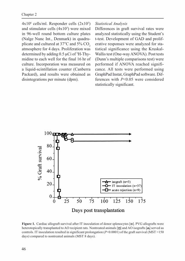
Figure 1. Cardiac allograft survival after IT inoculation of donor splenocytes [ ]. PVG allografts were
heterotopically transplanted to AO recipient rats. Nontreated animals [ ] and AO isografts [ ] served as
controls. IT inoculation resulted in significant prolongation (P<0.0001) of the graft survival (MST >150
days) compared to nontreated animals (MST 8 days).
47
Intrathymic immune modulation and transplant arteriosclerosis
Results
Graft Survival
Simultaneous transplantation and IT in-
oculation of donor splenocytes resulted
in significant prolongation of graft sur-
vival (median survival time [MST] >150
days, P<0.0001) compared to untreated
animals (MST 8 days). IT immune modu-
lation induces functional tolerance to
MHC-incompatible allografts because
survival rates at 150 days (indefinite graft
survival) are 90% (Fig. 1). Cardiac al-
lografts that did not survive indefinitely
were mostly rejected at timepoints com-
parable with animals treated with CsA and
ALS only (<25 days, not shown). AO
isografts (only ALS and CsA treatment;
no IT inoculation) showed indefinite graft
survival (MST >150 days, Fig.1) in all
animals.
GAD After IT Immune Modulation
Approximately 90% of the cardiac al-
lografts transplanted after IT inoculation
survived indefinitely (Fig. 1). In this
study, we investigated whether this pro-
tocol could also prevent the development
of GAD in these long-term surviving al-
lografts. Therefore, severity of arterial
lesions present in cardiac allografts at dif-
ferent timepoints after IT inoculation/
transplantation was analyzed as described
in Materials and Methods.
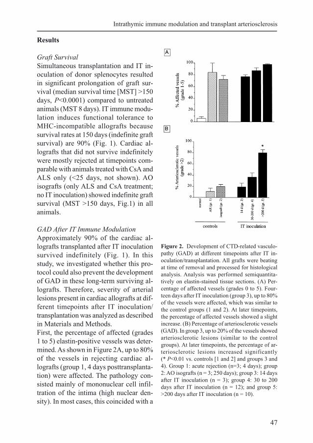
First, the percentage of affected (grades
1 to 5) elastin-positive vessels was deter-
mined. As shown in Figure 2A, up to 80%
of the vessels in rejecting cardiac al-
lografts (group 1, 4 days posttransplanta-
tion) were affected. The pathology con-
sisted mainly of mononuclear cell infil-
tration of the intima (high nuclear den-
sity). In most cases, this coincided with a
Figure 2. Development of CTD-related vasculo-
pathy (GAD) at different timepoints after IT in-
oculation/transplantation. All grafts were beating
at time of removal and processed for histological
analysis. Analysis was performed semiquantita-
tively on elastin-stained tissue sections. (A) Per-
centage of affected vessels (grades 0 to 5). Four-
teen days after IT inoculation (group 3), up to 80%
of the vessels were affected, which was similar to
the control groups (1 and 2). At later timepoints,
the percentage of affected vessels showed a slight
increase. (B) Percentage of arteriosclerotic vessels
(GAD). In group 3, up to 20% of the vessels showed
arteriosclerotic lesions (similar to the control
groups). At later timepoints, the percentage of ar-
teriosclerotic lesions increased significantly
(* P<0.01 vs. controls [1 and 2] and groups 3 and
4). Group 1: acute rejection (n=3; 4 days); group
2: AO isografts (n = 3; 250 days); group 3: 14 days
after IT inoculation (n = 3); group 4: 30 to 200
days after IT inoculation (n = 12); and group 5:
>200 days after IT inoculation (n = 10).

48
Chapter 2
acuterejection
b
e
normal
T/B cellsα-actin/elastin
IT inoc.14 days
c
NI
d
IEL
IT inoc.>200 days
NI
h
T
B
g
T
f
T B
a
IELM

49
Intrathymic immune modulation and transplant arteriosclerosis
perivascular infiltrate consisting of CD8+
and ED1+ cells. Most of the elastin-posi-
tive vessels were only slightly damaged
(grades 1 and 2), and few vessels (up to
10%) showed intimal thickening (grades
3 to 5, Fig. 2B) in which VSM cells were
barely present (as determined by absence
of α-actin-positive cells). Moreover, in
most vessels, the IEL was fragmented,
indicating severe vascular rejection (Fig.
3B).
In AO isografts (group 2), which were
explanted 250 days after transplantation,
up to 70% of the elastin positive vessels
were affected. However, the number of
intima-infiltrating cells and adhering
mononuclear cells to the vascular endo-
thelium was less pronounced compared
to acutely rejecting allografts. Up to 20%
of the elastin-positive vessels in AO
isografts showed intimal thickening (Fig.
2B). In contrast to rejecting allografts,
isografts contained more α-actin-positive
VSM cells in the intimal lesions, resem-
bling CTD-related vasculopathy (not
shown).After IT inoculation, the first
analysis was performed at 14 days after
transplantation (group 3). At this time
point approximately 80% of all elastin-
positive vessels counted were affected
(Fig. 2A). Again, most of the vessels were
only slightly affected (grades 1 and 2)
which was similar to rejecting grafts and
isografts. At this timepoint, 18% of the
vessels showed moderate intimal thick-
ening, which is similar to the percentages
observed in rejecting grafts (day 4, group
1) and isografts (day 250, group 2). At
later timepoints after transplantation, an
increase in the number of vessels that
showed moderate to severe intimal thick-
ening (grades 3 to 5) was observed (Fig.
2B). In group 4 (30 to 200 days), up to
36% of the elastin-positive vessels show-
ed moderate to severe intimal thickening,
which was not statistically different from
group 3 and the control groups (1 and 2).
However, the grafts in group 5 (>200
days) showed a dramatic increase in the
number of vessels that showed moderate
to severe intimal thickening. Virtually all
vessels were affected, and over 80% of
the vessels showed the presence of severe
intimal lesions. This increase differed sig-
nificantly from groups 3 and 4 and the
control groups (1 and 2) (P<0.01).
Figure 3. (facing page) Photomicrographs of GAD (double staining for VSM cell α-actin and elastin)
(A to D), and infiltration of T and B lymphocytes (double staining for T cells [MRC-OX19, blue color]
and B cells [HIS24, red color]) (E to H) in normal cardiac tissue and tissue obtained from acutely
rejecting allografts (group 1, 4 days posttransplantation) and grafts from intrathymically inoculated rats
(group 3, 14 days after IT inoculation, and group 5, >200 days after IT inoculation).
In normal (nontransplanted) cardiac tissue, no blood vessel pathology or infiltrating T and B cells were
present (A and E). Acutely rejecting allografts were heavily infiltrated and showed severe vascular
rejection with complete disruption of the vascular media and IEL (B). The parenchymatous infiltrates
mainly consisted of T cells (blue color) and only few B cells (red color) were present (F). Two weeks
after IT inoculation, mild GAD (grade 3) was observed in 18% of the elastin-positive vessels (C). At this
early timepoint after IT inoculation, a parenchymatous, T-cell dominated infiltrate was present (G). At
later timepoints after IT inoculation (>200 days), the vast majority of the blood vessels showed severe
GAD (grade 5) with (nearly) complete luminal occlusion (D). At this timepoint, the grafts were charac-
terized by pericardial infiltrates, which predominantly consisted of B cells (H). (Abbreviations: B: B
cell; IEL: internal elastic lamina; M: media; NI: neo-intima; and T: T cell). Immunoperoxidase staining,
magnification x400.
50
Chapter 2
In contrast to the intimal lesions at earlier
timepoints (groups 3 and 4), most of the
arteries in group 5 showed complete lu-
minal occlusion (grade 5) by intimal
thickening that contained large numbers
of α-actin-positive VSM cells. In addition,
fragmentation of the IEL was often ob-
served and sometimes the elastin fibers
(belonging to the IEL) had completely
disappeared. Figure 3, A to D, shows rep-
resentative micrographs of intimal thick-
ening observed at different timepoints
after IT inoculation and in nontrans-
planted cardiac tissue and acutely rejec-
ting allografts.
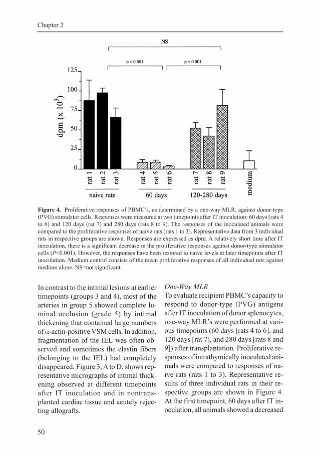
One-Way MLR
To evaluate recipient PBMC’s capacity to
respond to donor-type (PVG) antigens
after IT inoculation of donor splenocytes,
one-way MLR’s were performed at vari-
ous timepoints (60 days [rats 4 to 6], and
120 days [rat 7], and 280 days [rats 8 and
9]) after transplantation. Proliferative re-
sponses of intrathymically inoculated ani-
mals were compared to responses of na-
ive rats (rats 1 to 3). Representative re-
sults of three individual rats in their re-
spective groups are shown in Figure 4.
At the first timepoint, 60 days after IT in-
oculation, all animals showed a decreased
Figure 4. Proliferative responses of PBMC’s, as determined by a one-way MLR, against donor-type
(PVG) stimulator cells. Responses were measured at two timepoints after IT inoculation: 60 days (rats 4
to 6) and 120 days (rat 7) and 280 days (rats 8 to 9). The responses of the inoculated animals were
compared to the proliferative responses of naive rats (rats 1 to 3). Representative data from 3 individual
rats in respective groups are shown. Responses are expressed as dpm. A relatively short time after IT
inoculation, there is a significant decrease in the proliferative responses against donor-type stimulator
cells (P<0.001). However, the responses have been restored to naive levels at later timepoints after IT
inoculation. Medium control consists of the mean proliferative responses of all individual rats against
medium alone. NS=not significant.
51
Intrathymic immune modulation and transplant arteriosclerosis
ED1
ED2
OX8
OX35
normal acute rejection IT inoculation 14 days
IT inoculation >200 days
a b c d
e f g h
i j k l
m n o p
PC
MCMC
PC
MCMC
PC
MCMC
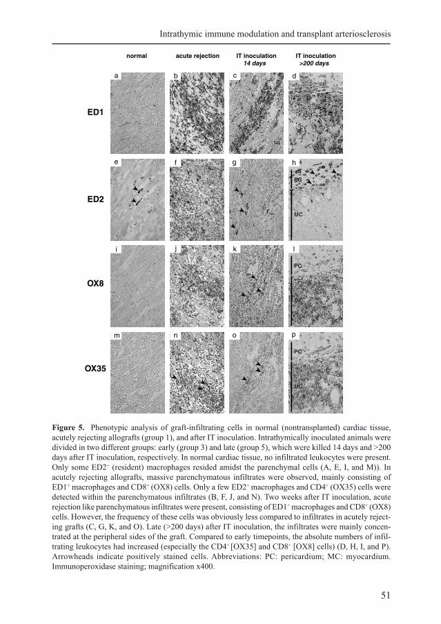
Figure 5. Phenotypic analysis of graft-infiltrating cells in normal (nontransplanted) cardiac tissue,
acutely rejecting allografts (group 1), and after IT inoculation. Intrathymically inoculated animals were
divided in two different groups: early (group 3) and late (group 5), which were killed 14 days and >200
days after IT inoculation, respectively. In normal cardiac tissue, no infiltrated leukocytes were present.
Only some ED2+ (resident) macrophages resided amidst the parenchymal cells (A, E, I, and M)). In
acutely rejecting allografts, massive parenchymatous infiltrates were observed, mainly consisting of
ED1+ macrophages and CD8+ (OX8) cells. Only a few ED2+ macrophages and CD4+ (OX35) cells were
detected within the parenchymatous infiltrates (B, F, J, and N). Two weeks after IT inoculation, acute
rejection like parenchymatous infiltrates were present, consisting of ED1+ macrophages and CD8+ (OX8)
cells. However, the frequency of these cells was obviously less compared to infiltrates in acutely reject-
ing grafts (C, G, K, and O). Late (>200 days) after IT inoculation, the infiltrates were mainly concen-
trated at the peripheral sides of the graft. Compared to early timepoints, the absolute numbers of infil-
trating leukocytes had increased (especially the CD4+ [OX35] and CD8+ [OX8] cells) (D, H, I, and P).
Arrowheads indicate positively stained cells. Abbreviations: PC: pericardium; MC: myocardium.
Immunoperoxidase staining; magnification x400.

52
Chapter 2
Tab
le 2
.S
emi-
quan
tita
tive
anal
ysi
s of
gra
ft i
nfi
ltra
ting l
eukocy
te s
ubse
ts a
fter
hem
atoxyli
n a
nd e
osi
n a
nd i
mm
unohis
toch
emic
al
stai
nin
g o
f ti
ssue
sect
ions
at d
iffe
rent
tim
epoin
ts a
fter
intr
athym
ic i
nocu
lati
on o
f donor
sple
nocy
tes.
S
tain
ing
S
pec
ific
ity
non
acute
isogra
ft14 d
ays
30-2
00 d
ays
>200 d
ays
tran
spla
nte
dre
ject
ion
(gro
up 2
)(g
roup 3
)(g
roup 4
)(g
roup 5
)
(gro
up 1
)
Mononucl
ear
Hem
atoxyli
n a
nd e
osi
n-
++
+±
++
++
+ce
ll i
nfi
ltra
te
mA
b M
RC
-OX
19 5
8T
cel
ls (
CD
5)
-+
++
nd
++
++
+
mA
b M
RC
-OX
35 5
9C
D4
-±
nd
++
++
mA
b M
RC
-OX
8 6
0C
D8 (α
-ch
ain
)-
++
+n
d+
++
++
mA
b H
IS24 6
1B
cel
ls (
CD
45R
)-
±nd
±+
++
++
mA
b E
D1 6
2M
acro
phag
es-
++
+nd
++
++
+
Res
iden
t (m
ature
)m
Ab E
D2 6
2±
+nd
++
++
mac
rophag
es
Sem
iquan
tita
tive
anal
ysi
s of
gra
ft-i
nfi
ltra
ting l
eukocy
tes:
- =
no i
nfi
ltra
tion;
± =
few
infi
ltra
ting c
ells
; +
= m
ild i
nfi
ltra
tion;
++
= m
oder
ate
infi
ltra
tion;
++
+ =
sev
ere
infi
ltra
tion. (m
Ab:
monocl
onal
anti
body,
nd:
not
det
erm
ined
)

53
Intrathymic immune modulation and transplant arteriosclerosis
proliferative response against donor-type
splenocytes compared to naive rats
(P<0.001). Responses against third-party
antigens were also decreased (P<0.001,
not shown). At later timepoints after IT
inoculation, however, proliferative re-
sponses against donor and third-party an-
tigens were restored towards levels of
naive rats (no significant differences). In
animals that acutely rejected their graft,
proliferative responses against donor-type
antigens were significantly elevated com-
pared to naive rats (not shown). By con-
trast, proliferative responses against third-
party antigens were not increased. The
results indicate that there is a non-specific
decrease in proliferative responsiveness
at 60 days after IT inoculation/transplan-
tation. At later timepoints, the allore-
sponse has been restored, indicating that
donor-reactive T cells are still present long
term after IT inoculation of donor spleno-
cytes.
Infiltration of Cardiac Allografts
In addition to the histological presence of
GAD, we determined the presence of
graft-infiltrating cells in control groups (1
and 2) and at different timepoints after IT
inoculation (groups 3, 4, and 5). General
morphology of cardiac tissue and the pres-
ence of infiltrates were evaluated by
hematoxylin and eosin staining (groups
1 to 5). Immunohistochemical staining
was performed to characterize the infil-
trating cells phenotypically in nontrans-
planted cardiac tissue, acutely rejecting
allografts (group 1) and different time-
points after IT inoculation (groups 3, 4,
and 5). The number of positively stained
infiltrating leukocytes was scored semi-
quantitatively as described in Materials
and Methods. In normal (nontransplanted)
cardiac tissue, no infiltrating leukocytes
could be observed. The only leukocytes
present were ED2+ macrophages, which
resided amidst the cardiac parenchymal
cells (Fig. 5E).
In contrast to AO isografts (group 2), in
which few infiltrating inflammatory cells
were present, rejecting allografts (group
1) were characterized by massive paren-
chymatous infiltrates, i.e., infiltrating cells
localized amidst the cardiac myocytes.
Immunohistochemical analysis revealed
that these infiltrates mainly consisted of
CD8+ cells (MRC-OX8) and ED1+ macro-
phages. Another subset of macrophages
stained positively for ED2, a marker
among others present on resident mac-
rophages. In contrast to ED1+ macro-
phages, ED2+ macrophages were local-
ized mostly at the periphery of the grafts.
Some CD4+ cells (MRC-OX35) were
present in rejecting allografts, although
these CD4+ cells were relatively few in
number compared to the number of CD8+
cells. According to their morphology,
many of the CD4+ cells present were pro-
bably not T cells but macrophages, which
were present in high numbers (as shown
by ED1 staining) (Fig. 5 B, F, J, and N).
Anti-CD45R staining (HIS24) revealed a
virtual absence of B cells in rejecting al-
lografts (Fig. 3F), whereas a monoclonal
antibody directed against IgM (HIS40)
sometimes showed IgM depositions on
endothelial cells in capillaries and along
the blood vessels, which indicated vas-
cular damage (not shown).
After IT inoculation, the first analysis was
performed 14 days after transplantation
(group 3). At this timepoint, the infiltrates
present were mostly parenchymatous and,
with the exception of lower absolute num-
bers of infiltrating cells, were qualitatively
54
Chapter 2
virtually indistinguishable from the infil-
trates observed in rejecting grafts. In ad-
dition, inoculated animals contained rela-
tively more CD4+ cells than the infiltrates
in rejecting animals. Again, similar to
acutely rejecting allografts, ED2+ macro-
phages were mostly localized at the pe-
riphery of the grafts, whereas ED1+ ma-
crophages were observed frequently in the
parenchymatous infiltrates (Fig. 5C, G, K,
and O).
At later timepoints (30 to 200 days) after
IT inoculation (group 4), the initially par-
enchymatous infiltrate had disappeared.
The most striking difference with group
3 (14 days) was a decrease in the abso-
lute numbers of infiltrating cells and the
appearance of peripheral and/or blood
vessel-associated infiltrates, which con-
tained relatively few T cells (equal num-
bers of CD4+ and CD8+ cells) and macro-
phages but did contain considerable num-
bers of B cells. Few T cells and ED1+
macrophages were present amidst the
graft parenchymal cells compared to the
numbers found in the graft periphery (not
shown).
In group 5 (> 200 days posttransplanta-
tion), the peripheral infiltrate increased in
size with a tendency to locally “spill over”
into the cardiac parenchyma. Double
staining for B and T cells revealed that
the peripheral infiltrates were now domi-
nated by B cells instead of T cells (Fig.
3H). CD4+ and CD8+ cells and ED1+
macrophages were still present and
seemed to infiltrate the graft parenchyma
again. The ED2+ macrophages remained
localized at the peripheral sides of the
grafts (Fig. 5D, H, L, and P). Results of
the semiquantitative analysis of numbers
of infiltrating leukocytes are represented
in Table 2. Figure 5 shows representative
β-actin
IFN-γ
IL-2
IL-10
norm
al
acut
e re
jectio
n
IT 1
4 da
ys
IT >
200
days
TGF-β
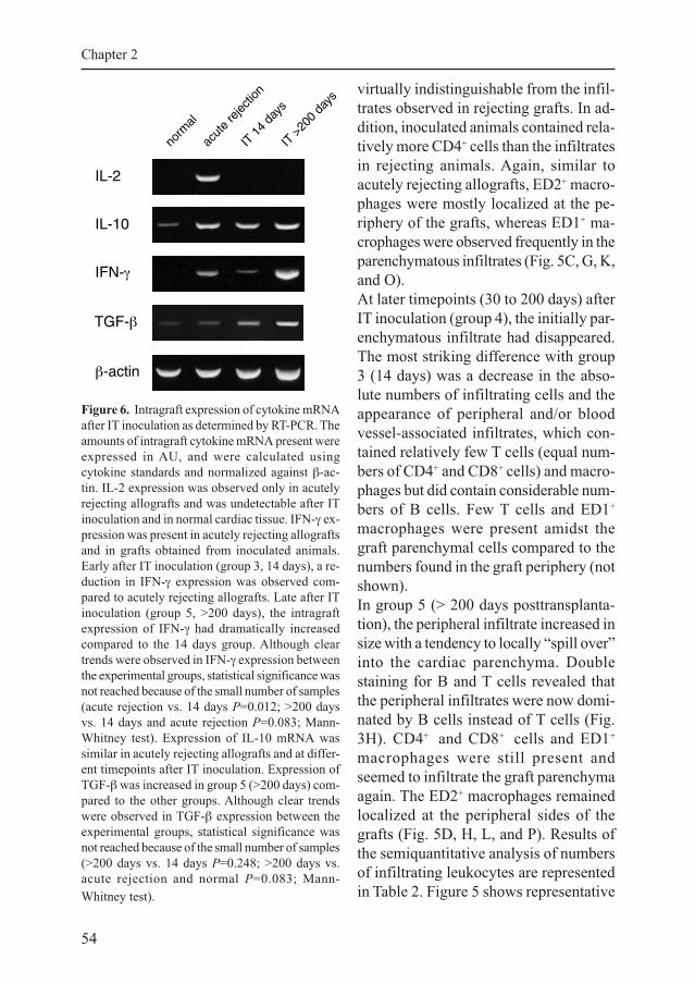
Figure 6. Intragraft expression of cytokine mRNA
after IT inoculation as determined by RT-PCR. The
amounts of intragraft cytokine mRNA present were
expressed in AU, and were calculated using
cytokine standards and normalized against β-ac-
tin. IL-2 expression was observed only in acutely
rejecting allografts and was undetectable after IT
inoculation and in normal cardiac tissue. IFN-γ ex-
pression was present in acutely rejecting allografts
and in grafts obtained from inoculated animals.
Early after IT inoculation (group 3, 14 days), a re-
duction in IFN-γ expression was observed com-
pared to acutely rejecting allografts. Late after IT
inoculation (group 5, >200 days), the intragraft
expression of IFN-γ had dramatically increased
compared to the 14 days group. Although clear
trends were observed in IFN-γ expression between
the experimental groups, statistical significance was
not reached because of the small number of samples
(acute rejection vs. 14 days P=0.012; >200 days
vs. 14 days and acute rejection P=0.083; Mann-
Whitney test). Expression of IL-10 mRNA was
similar in acutely rejecting allografts and at differ-
ent timepoints after IT inoculation. Expression of
TGF-β was increased in group 5 (>200 days) com-
pared to the other groups. Although clear trends
were observed in TGF-β expression between the
experimental groups, statistical significance was
not reached because of the small number of samples
(>200 days vs. 14 days P=0.248; >200 days vs.
acute rejection and normal P=0.083; Mann-
Whitney test).

55
Intrathymic immune modulation and transplant arteriosclerosis
micrographs of infiltrating leukocytes at
different timepoints after IT immune
modulation.
Semiquantitative Cytokine RT-PCR
Intragraft cytokine gene expression was
analyzed with a semiquantitative RT-PCR
as described in detail in Materials and
Methods. Representative results of RT-
PCR for IL-2, IFN-γ, IL-10 and TGF-βare shown in Figure 6. Because IL-4 ex-
pression was not detected in any of the
experimental groups tested, no IL-4 data
are shown. Cytokine gene expression was
analyzed in normal (nontransplanted) car-
diac tissue, acutely rejecting allografts
(group 1), 14 days after IT inoculation
(group 3), and >200 days after IT inocu-
lation (group 5). Normal cardiac tissue
showed undetectable levels of IL-2 and
IFN-γ expression, whereas little expres-
sion of IL-10 and TGF-β was observed
(possibly produced by resident macro-
phages and endothelial cells, respec-
tively). Acutely rejecting allografts (group
1) showed clearly detectable levels of
IL-2, IFN-γ, and IL-10 mRNA expression,
whereas TGF-β expression was not al-
tered compared to normal cardiac tissue.
In contrast to acute rejection, grafts ex-
planted 14 days (group 3) after IT inocu-
lation had undetectable levels of IL-2 and
decreased expression of IFN-γ compared
to acutely rejecting allografts. IL-10 and
TGF-β expression, however, was similar
compared to acutely rejecting grafts. In
group 5 (>200 days after IT inoculation),
IL-2 expression was still undetectable,
whereas the IFN-γ and TGF-β expression
showed a remarkable increase compared
to acutely rejecting allografts and IT in-
oculation after 14 days. Although clear
trends were observed in IFN-γ and TGF-
β expression between the different groups,
statistical significance was not reached
because of the small number of samples
(n = 2 to 3/ group; IFN-γ expression: acute
rejection vs. 14 days P=0.012, and >200
days vs. 14 days and acute rejection P=
0.083; TGF-β expression: >200 days vs.
14 days P=0.248 and 200 days vs. acute
rejection/normal P=0.083, Mann-Whit-
ney test). Expression of IL-10 mRNA was
similar to the expression observed in
acutely rejecting grafts and 14 days after
IT inoculation. The absence of IL-2
mRNA and decreased expression of
IFN-γ mRNA, together with sustained
IL-10 expression 14 days after IT inocu-
lation, suggest a down regulation of a
Th1-type cytokine response.
Discussion
Nowadays, CTD is the most significant
problem in clinical organ transplantation
after the first postoperative year1,2. Be-
cause histoincompatibility between donor
and recipient, and thereby alloreactivity
directed against the graft, is the major
contributing factor in the pathogenesis of
CTD5, induction of specific nonrespon-
sive-ness to donor antigens would be the
obvious solution to prevent the develop-
ment of this pathology.
Donor-specific hyporesponsiveness to
various organs can be induced by IT in-
oculation of donor lymphoid cells7-10 or
synthetic MHC allopeptides25. In all of
these studies, an interval between the in-
duction of hyporesponsiveness and the
actual transplantation was needed, ham-
pering clinical use of the protocol. There-
fore, we developed a method that allowed
the transplantation of a cardiac allograft

56
Chapter 2
simultaneous with the IT inoculation of
donor splenocytes. This procedure, as pre-
sented in our previous reports11,26 and in
this study, results in virtual permanent
acceptance (i.e., functional tolerance) of
cardiac allografts in >90% of the recipi-
ents in MHC-incompatible strain combi-
nations.
The present study was designed to evalu-
ate whether our IT immune modulation
protocol also prevents the development
of CTD-related vasculopathy, referred to
as GAD. Therefore, histological analyses
at different timepoints after IT inocula-
tion/transplantation were performed.
Also, in vitro proliferative responses of
inoculated/transplanted rats were mea-
sured to determine residual alloreactivity
against donor-type and third-party anti-
gens. In addition, intragraft cytokine ex-
pression was determined using RT-PCR.
Histological analysis revealed that 14
days after inoculation, the vast majority
(up to 80%) of the blood vessels showed
mononuclear cell infiltration of the intima
and adherence of leukocytes to the vas-
cular endothelium. At later timepoints,
comparable percentages of affected ves-
sels were observed. However, the pathol-
ogy at later timepoints differed qualita-
tively from the pathology observed at 14
days. The percentage of vessels that
showed GAD, and also the severity of
GAD, dramatically increased at later
timepoints. In addition, the cellular com-
position of the arteriosclerotic lesions was
different at early and later timepoints. Two
weeks after IT inoculation, the lesions
showed a high neointimal nuclear density
and contained only few a α-actin-positive
cells. This was similar to the lesions ob-
served in acutely rejecting grafts. At later
timepoints, however, the lesions con-
tained fewer cells (lower neointimal nu-
clear density and less inflammation), but
the number of α-actin-positive cells was
higher, indicating the presence of GAD.
Also isografts, which were explanted 250
days after transplantation, showed some
GAD. Development of chronic rejection
after isografting was reported previously
by Tullius et al.27 and is probably caused
by ischemia and reperfusion injury2.
Thus, despite the induction of virtual per-
manent acceptance of allogeneic grafts
through IT immune modulation, this pro-
cedure does not prevent the subsequent
development of GAD in our fully MHC-
incompatible model. Similar findings
have been reported by Shirwan et al.28
after IT inoculation with donor class I
allopeptides in the congenic PVG.R8 to
PVG.1U rat strain combination, which
differs at the MHC class I locus. Orloff et
al.13 and Blom et al.14, on the other hand,
showed that IT inoculation of donor bone
marrow cells in the Lew-to-F344 CTD
model prevented the development of
GAD (evaluated 120 days after IT inocu-
lation). However, these rat strains are
MHC compatible and only differ at the
level of non-MHC loci. Shin et al.12 re-
ported that even treatment with ALS
alone, without additional IT inoculation
of donor-type antigens, prevented the de-
velopment of GAD in the Lew-to-F344
strain combination. Results so far indicate
that the development of GAD cannot be
prevented by IT immune modulation in
fully or partial MHC-incompatible trans-
plant combinations.
It is intriguing to note that IT immune
modulation, on one hand, is capable in
preventing acute rejection, whereas, on
the other hand, the process of chronic re-
jection seems to be unaffected, although

57
Intrathymic immune modulation and transplant arteriosclerosis
both are believed to be primarily alloan-
tigen driven. We, therefore, analyzed the
nature of the residual alloreactivity after
IT immune modulation and transplanta-
tion to attempt to understand this ambi-
guity in alloresponsiveness.
The mechanism by which IT inoculation
in our protocol results in prolonged graft
survival is not completely understood, but
we hypothesized that it is mediated
through the induction of donor-specific
hyporesponsiveness because third-party
allografts are rejected within 20 days29.
However, in vitro experiments, described
in this study, revealed that IT inoculation
does not result in donor-specific unre-
sponsiveness. Sixty days after IT inocu-
lation, a non-specific decrease in prolif-
erative responses against donor and third-
party antigens was observed. However, at
later timepoints, the responses had been
restored to levels of naive animals. Ito et
al.30 and Chowdhury et al.31 also observed
suppressed proliferative responses after IT
inoculation, with complete long term res-
toration. The non-specific downregulation
of proliferative responses after IT inocu-
lation of donor splenocytes, which was
also noted in other studies32,33, might be
explained by the persistence of ALS-in-
duced immunosuppression. However, af-
ter the injection of 1 ml of ALS, T-cell
numbers returned to 30% of the initial
numbers and back to normal levels after
50 days and 120 days, respectively34.
Thus, despite induction of functional tol-
erance (i.e., prolonged graft survival), IT-
inoculated animals clearly had a prolif-
erative response to donor antigens, which
argues against clonal deletion and favors
immune deviation as the underlying
mechanism. This view is supported by our
findings that IT inoculation results in the
generation of recirculating allospe-cific
suppressor/regulatory CD4+ T cells,
which are able to adoptively transfer func-
tional tolerance35. In addition, transplan-
tation of a donor-specific skin graft 60 to
150 days after primary cardiac allograf-
ting and IT inoculation results in signifi-
cantly prolonged skin graft survival, sug-
gesting induction of a regulatory mecha-
nism by IT immune modulation29. Al-
though we only retransplanted skin grafts,
and could therefore only observe pro-
longed graft survival and were not able
to study the development of GAD, we
expect that after transplantation of a sec-
ond cardiac allograft, the development of
GAD would be ameliorated compared to
the primary graft. This is strengthened by
the observation reported by Tullius et al.
that, using a different retransplant model,
transplantation of a second (cardiac) al-
lograft resulted in diminished severity of
GAD in this second graft36.
The notion that immune deviation, and not
immune depletion, might be instrumen-
tal after IT inoculation is also strength-
ened by immunohistochemical analysis of
the cardiac allografts. At 14 days after IT
inoculation, grafts were heavily infiltrated
with mainly CD8+ cells and ED1+ cells
and were virtually indistinguishable from
the parenchymatous infiltrates present in
acutely rejecting cardiac allografts. Sur-
prisingly, the infiltrates caused little dam-
age to the cardiac tissue, as shown by the
presence of NADH-reductase activity in
healthy cardiac myocytes (not shown).
Other groups have also reported that the
composition of infiltrates present in al-
lografts early after induction of donor-
specific hyporesponsiveness is similar to
that observed after acute rejection23,37. At
later timepoints after IT inoculation (30
58
Chapter 2
to 200 days), the infiltrates were mostly
pericardially located and/or blood vessel
associated. Eventually (>200 days), pe-
ripheral infiltrates were still present and
seemed to spill over into the cardiac
parenchyma. A remarkable observation at
this stage was the presence of B cell domi-
nated pericardial infiltrates, whereas the
infiltrates at earlier timepoints were domi-
nated by T cells and macrophages. Thus,
not only in vitro but also in vivo, clear
signs of residual donor-directed allore-
activity can be observed after IT immune
modulation. The nature and localization
of the graft-infiltrating cells, however,
clearly differs in IT-treated rats as com-
pared to untreated (rejecting) recipients,
again supporting immune deviation as a
major element in the mechanism under-
lying long-term graft acceptance after IT
inoculation.
It has been suggested that successful ac-
ceptance of an allograft is dependent upon
immune deviation of CD4+ T cells to-
wards a Th2-type cytokine response (e.g.,
IL-4, IL-5, IL-10, and IL-13) and away
from the Th1-type cytokine response (e.g.,
IL-2 and IFN-γ)38. We, therefore, studied
intragraft cytokine mRNA expression at
different timepoints after IT inoculation/
transplantation.
Acutely rejecting cardiac allografts in
nontreated rats expressed detectable lev-
els of IL-2 and IFN-γ mRNA transcripts.
In addition, detectable levels of IL-10
were present, which might be produced
by macrophages that were abundantly
present in acutely rejecting allografts. RT-
PCR analysis revealed decreased Th1-
type (IL-2 and IFN-γ) and sustained Th2-
type (IL-10 and TGF-β) type cytokine
expression 14 days after IT inoculation
(compared to acutely rejecting allografts).
Long-surviving allografts expressed in-
creased levels of IL-10, IFN-γ, and
TGF-β, whereas IL-2 expression was still
undetectable. Increased expression of
IFN-γ in long-surviving allografts after IT
immune modulation was also recently re-
ported by Shirwan et al.22. Our cytokine
data thus strongly support immune devia-
tion as a major element that contributes
to the virtual permanent acceptance of
allografts after IT inoculation. At the same
time, however, this deviation and the as-
sociated change in cytokine profile may
be extremely detrimental to the graft39.
TGF-β has been linked to fibrotic pro-
cesses associated with chronic inflamma-
tory conditions, such as the development
of GAD40,41. In addition, in recent experi-
ments it has been shown that IFN-γ42 and
IL-1043 augment the development of GAD
after cardiac transplantation in mice.
Hancock et al.44 suggested that immune
deviation towards a Th2-type response
may be involved in the induction of hypo-
responsiveness to allografts but thereby
might enhance the development of CTD.
Indeed, Larsen et al.45 showed in their
model that long-term acceptance of al-
lografts and prevention of CTD requires
silencing of both Th1 and Th2 cytokine
programs.
Probably all tolerance-inducing protocols,
which do not eliminate alloreactivity com-
pletely but rather alter the immune re-
sponse against the graft instead (such as
our model), will fail to prevent the even-
tual development of GAD. According to
this view, only those interventions in
which antidonor reactivity is completely
blocked or deleted, e.g., the creation of
donor bone marrow chimeras46-50 or opti-
mal T-cell stimulation blockade using
CTLA4-Ig fusion protein51-53, are likely

59
Intrathymic immune modulation and transplant arteriosclerosis
to prevent the development of GAD after
solid organ transplantation. When
CTLA4-Ig treatment is suboptimal and
antidonor reactivity (partly) remains, the
development of GAD will not be pre-
vented54-56. To quote Sayegh and Carpen-
ter57: “The term “tolerance” should be
reserved to states where long-term survi-
ving grafts have normal function and do
not develop changes of chronic rejection.”
Taken together, our data show that IT in-
oculation of donor splenocytes prevents
acute rejection and results in significantly
prolonged graft survival rates of cardiac
allografts. However, the development of
GAD is not prevented by this treatment,
which is probably due to sustained
alloreactivity against the graft after IT ino-
culation (as shown by in vitro measured
alloreactivity and presence of lymphocyte
infiltrates). In addition, down-regulation
of Th1-type cytokine expression was ob-
served, suggesting a shift towards a Th2-
type cytokine response. This observation
is supported by the presence of massive
B cell dominated pericardial infiltrates.
Whether GAD is actually facilitated after
IT inoculation due to a shift to Th2- or
Tr1-type intragraft cytokine responses
remains to be elucidated.
Acknowledgements
The authors thank Michael Obrzut and
Ineke Bos for assistance in performing the
cytokine PCR analyses.
References
1. Azuma H and Tilney NL. Chronic graft rejection. Curr. Opin. Immunol. 1994; 6:770-
776.
2. Häyry P, Isoniemi H, Yilmaz S, Mennander A, Lemström K, Räisänen-Sokolowski A,
Koskinen P, Ustinov J, Lautenschlager I, Taskinen E, Krogerus L, Aho P, and Paavonen
T. Chronic allograft rejection. Immunol. Rev. 1993; 134:33-81.
3. Häyry P, Myllärniemi M, Calderon Ramirez L, Aavik E, Loubtchenkov M, Koskinen P,
Lemström K, and Räisänen-Sokolowski A. Immunobiology and pathology of chronic
rejection. Transplant. Proc. 1997; 29:77-78.
4. Sundaresan S, Trulock MP, Mohanakumar T, Cooper JD, Patterson GA, and The Wash-
ington University Lung Transplant Group. Prevalence and outcome of bronchiolitis oblit-
erans syndrome after lung transplantation. Ann. Thorac. Surg. 1995; 60:1341-1347.
5. Cramer AT, Chapman FA, Wu GD, Harnaha JB, Qian SQ, and Makowka L. Cardiac
transplantation in the rat. II. Alteration of the severity of donor graft arteriosclerosis by
modulation of the host immune response. Transplantation 1990; 50:554-558.
6. Posselt AM, Barker CF, Tomaszewski JE, Markmann JF, Choti MA, and Naji A. Induc-
tion of donor-specific unresponsiveness by intrathymic islet transplantation. Science 1990;
249:1293-1295.
7. Goss JA, Nakafusa Y, and Flye MW. Intrathymic injection of donor alloantigens induces
donor-specific vascularized allograft tolerance without immunosuppression. Ann. Surg.
1992; 216:409-414.
8. Campos L, Alfrey EJ, Posselt AM, Odorico JS, Barker CF, and Naji A. Prolonged sur-
vival of rat orthotopic liver allografts after intrathymic inoculation of donor-strain cells.
Transplantation 1993; 55:866-870.

60
Chapter 2
9. Remuzzi G, Rossini M, Imberti O, and Perico N. Kidney graft survival in rats without
immunosuppressants after intrathymic glomerular transplantation. Lancet 1991; 337:750-
752.
10. Goss JA, Nakafusa Y, and Flye MW. Prolongation of small bowel allografts after
intrathymic injection of donor alloantigen and ALS. J. Surg. Res. 1993; 54:494-498.
11. Klatter FA, Raué HP, Bartels HL, Pater JM, Groen H, Nieuwenhuis P, and Kampinga J.
Simultaneous transplantation and intrathymic tolerance induction; a method with clinical
potential. Transplantation 1995; 60:1208-1210.
12. Shin YT, Adams DH, Wyner LR, Akalin E, Sayegh MH, and Karnovsky MJ. Intrathymic
injection of donor splenocytes plus systemic antilymphocyte serum or antilymphocyte
serum alone prolongs cardiac allograft survival and inhibits graft arteriosclerosis in the
Lew-to-F344 chronic rejection model. Transplant. Proc. 1995; 27:2112-2114.
13. Orloff MS, DeMara EM, Coppage ML, Leong N, Fallon MA, Sickel J, Zuo XJ, Prehn J,
and Jordan SC. Prevention of chronic rejection and graft arteriosclerosis by tolerance
induction. Transplantation 1995; 59:282-288.
14. Blom D, Morrissey N, Mesonero C, Zuo XJ, Jordan S, Fisher T, Bronsther O, and Orloff
MS. Tolerance induction by intrathymic inoculation prevents chronic renal allograft re-
jection. Transplantation 1998; 65:272-275.
15. Adams DH, Russell ME, Hancock WW, Sayegh MH, Wyner LR, and Karnovsky MJ.
Chronic rejection in experimental cardiac transplantation: studies in the Lewis-F344 model.
Immunol. Rev. 1993; 134:5-19.
16. Diamond JR, Tilney NL, Frye J, Ding G, McElroy J, Pesek-Diamond I, and Yang H.
Progressive albuminuria and glomerulosclerosis in a rat model of chronic renal allograft
rejection. Transplantation 1992; 54:710-716.
17. Skalli O, Ropraz P, Trzeciak A, Benzonana G, Gillessen D, and Gabbiani G. A mono-
clonal antibody against alpha-smooth muscle actin: a new probe for smooth muscle dif-
ferentiation. J. Cell. Biol. 1986; 103:2787-2796.
18. Schürch W, Skalli O, Seemayer TA, and Gabbiani G. Intermediate filament proteins and
actin isoforms as markers for soft tissue tumor differentiation and origin. Am. J. Pathol.
1987; 128:91-103.
19. Adams DH, Tilney NL, Collins JJ, and Karnovsky MJ. Experimental graft arteriosclero-
sis. Transplantation 1992; 53:1115-1119.
20. Farges O, Morris PJ, and Dallman MJ. Spontaneous acceptance of rat liver allografts is
associated with an early downregulation of intragraft interleukin-4 messenger RNA ex-
pression. Hepatology 1995; 21:767-775.
21. Siegling A, Lehmann M, Platzer C, Emmrich F, and Volk HD. A novel multispecific
competitor fragment for quantitative PCR analysis of cytokine gene expression in rats. J.
Immunol. Methods 1994; 177:23-28.
22. Shirwan H, Barwari L, and Khan NS. Predominant expression of T helper 2 cytokines
and altered expression of T helper 1 cytokines in long-term allograft survival induced by
intrathymic immune modulation with donor class I major histocompatibility complex
peptides. Transplantation 1998; 66:1802-1809.
23. Josien R, Pannetier C, Douillard P, Cantarovich D, Menoret S, Bugeon L, Kourilsky P,
Soulillou JP, and Cuturi MC. Graft-infiltrating T helper cells, CD45RC phenotype, and
TH1/TH2-related cytokines in donor-specific transfusion induced tolerance in adult rats.
Transplantation 1995; 60:1131-1139.
61
Intrathymic immune modulation and transplant arteriosclerosis
24. Li YS, Hayakawa K, and Hardy RR. The regulated expression of B lineage associated
genes during B cell differentiation in bone marrow and fetal liver. J. Exp. Med. 1993;
178:951-960.
25. Mhoyan A, Cramer DV, Baquerizo A, and Shirwan H. Induction of allograft unrespon-
siveness after intrathymic inoculation with donor class I allopeptides. Transplantation
1997; 64:1665-1670.
26. Raué HP, Klatter FA, Hylkema MN, van der Deen M, Groen H, Pater J, Nieuwenhuis P,
Hardonk-Wubbena A, Hillebrands JL, and Rozing J. Efficiency of intrathymic tolerance
induction in various inbred rat strains: relationship with Th1/Th2 status of the recipient?
J. Exp. Anim. Sci. 2000; 41:82-86.
27. Tullius SG, Heemann U, Hancock WW, Azuma H, and Tilney NL. Long-term kidney
isografts develop functional and morphologic changes that mimic those of chronic al-
lograft rejection. Ann. Surg. 1994; 220:425-432.
28. Shirwan H, Wu GD, Barwari L, Liu A, and Cramer DV. Induction of allograft unrespon-
siveness after intrathymic inoculation with donor class I allopeptides. Transplantation
1997; 64:1671-1676.
29. Raué HP. Simultaneous transplantation and tolerance induction. (Thesis) University of
Groningen, Groningen, The Netherlands, 1998. (ISBN 90-367-0872-9)
30. Ito A, Ito T, Kamiike W, Moriguchi A, Ohkawa A, Uchikoshi F, Tanaka S, Nakata S, and
Matsuda H. Donor-specific tolerance by perioperative intrathymic injection of bone mar-
row cells in the rat cardiac alograft model. Transplantation 1997; 64:752-757.
31. Chowdhury NC, Saborio DV, Garrovillo M, Chandraker A, Magee CC, Waaga AM, Sayegh
MH, Jin MX, and Oluwole SF. Comparative studies of specific acquired systemic tole-
rance induced by intrathymic inoculation of a single synthetic Wistar-Furth (RT1(U))
allo-MHC class I (RT1.A(U)) peptide or WAG (RT1(U))-derived class I peptide. Trans-
plantation 1998; 66:1059-1066.
32. Oluwole SF, Chowdhury NC, and Fawwaz RA. Induction of donor-specific unrespon-
siveness to rat cardiac allografts by intrathymic injection of UV-B-irradiated donor spleen
cells. Transplantation 1993; 55:1389-1395.
33. Arima T, Goss JA, Walp LA, and Flye MW. Administration of anti-CD4 monoclonal
antibody with intrathymic injection of alloantigen results in rat cardiac allograft tole-
rance. Surgery 1995; 118:265-272.
34. Hylkema MN, Pater JM, Klatter FA, van der Deen M, van Asperen RM, Themmen M,
Raué HP, Groen H, and Nieuwenhuis P. Effect of variable doses of antilymphocyte se-
rum on T cell depletion and cardiac allograft survival. Transplant. Proc. 1997; 29:1719-
1720.
35. Kampinga J, Klatter F, Bartels HL, Pater J, Nieuwenhuis P, Roser B, and Groen H.
Intrathymic tolerance induction is based on suppressor cells. Transplant. Proc. 1994;
26:725-726.
36. Tullius SG, Nieminen M, Qun Y, Egermann F, Reutzer-Selke A, Jonas S, Pratschke J,
Bechstein WO, Volk HD, and Neuhaus P. Synergistic mechanisms of alloantigen-depen-
dent and independent events in chronic graft rejection. Transplant. Proc. 1998; 30:2412.
37. Plain KM, Fava L, Spinelli A, He XY, Chen JC, Boyd R, Davidson CL, and Hall BM.
Induction of tolerance with nondepleting anti-CD4 monoclonal antibodies is associated
with downregulation of TH2 cytokines. Transplantation 1997; 64:1559-1567.

62
Chapter 2
38. Nickerson P, Steiger J, Zheng XX, Steele AW, Steurer W, Roy-Chaudhury P, and Strom
TB. Manipulation of cytokine networks in transplantation. False hope or realistic oppor-
tunity for tolerance? Transplantation 1997; 63:489-494.
39. Shirwan H. Chronic allograft rejection. Transplantation 1999; 68:715-726.
40. Lemström K, Koskinen P, and Häyry P. Molecular mechanisms of chronic renal allograft
rejection. Kidney Int. 1995; 48:S2-S10.
41. Letterio JL and Roberts AB. Regulation of immune responses by TGF-β. Annu. Rev.
Immunol. 1998; 16:137-161.
42. Räisänen-Sokolowski A, Glysing-Jensen T, Koglin J, and Russell ME. Reduced trans-
plant arteriosclerosis in murine cardiac allografts placed in interferon-γ knockout recipi-
ents. Am. J. Pathol. 1998; 152:359-365.
43. Furukawa Y, Becker G, Stinn JL, Shimizu K, Libby P, and Mitchell RN. Interleukin-10
(IL-10) augments allograft arterial disease. Am. J. Pathol. 1999; 155:1929-1939.
44. Hancock WW, Shi C, Picard MH, Bianchi C, and Russell ME. LEW-to-F344 carotid
artery allografts; analysis of a rat modelof posttransplant vascular injury involving cell-
mediated and humoral responses. Transplantation 1995; 60:1565-1572.
45. Larsen CP, Elwood ET, Alexander DZ, Ritchie SC, Hendrix R, Tucker-Burden C, Cho
HR, Aruffo A, Hollenbauch D, Linsley PS, Winn KJ, and Pearson TC. Long-term accep-
tance of skin and cardiac allografts after blocking CD40 and CD28 pathways. Nature
1996; 381:434-438.
46. Colson YL, Zadach K, Nalesnik M, and Ildstad ST. Mixed allogeneic chimerism in the
rat. Transplantation 1995; 60:971-980.
47. Blom D, Morrissey N, Mesonero C, Coppage M, Fisher T, Zuo XJ, Jordan SC, and Orloff
MS. Induction of specific tolerance through mixed hematopoietic chimerism prevents
chronic renal allograft rejection in a rat model. Surgery 1996; 120:213-220.
48. Gammie JS, Li S, Kawaharada N, Colson YL, Yousem S, Ildstad ST, and Pham SM.
Mixed allogeneic chimerism prevents obstructive airway disease in a rat heterotopic tra-
cheal transplant model. J. Heart Lung Transplant. 1998; 17:801-808.
49. Hayamizu K, Lan F, Huie PSRK, and Strober S. Comparison of chimeric and non-chi-
meric tolerance using posttransplant total lymphoid irradiation. Transplantation 1999;
68:1036-1044.
50. Kawaharada N, Shears LL, Li S, and Pham SM. Mixed hematopoietic chimerism pre-
vents allograft vasculopathy. J. Heart Lung Transplant. 1999; 18:532-541.
51. Russell ME, Hancock WW, Akalin E, Wallace AF, Glysing-Jensen T, Willett TA, and
Sayegh MH. Chronic cardiac rejection in the LEW to F344 rat model. Blockade of
CD28-B7 costimulation by CTLA4Ig modulates T cell and macrophage activation and
attenuates arteriosclerosis. J. Clin. Invest. 1996; 97:833-838.
52. Azuma H, Chandraker A, Nadeau K, Hancock WW, Carpenter CB, Tilney NL, and Sayegh
MH. Blockade of T-cell costimulation prevents development of experimental chronic
renal allograft rejection. Proc. Natl. Acad. Sci. USA 1996; 93:12439-12444.
53. Chandraker A, Azuma H, Nadeau K, Carpenter CB, Tilney NL, Hancock WH, and Sayegh
MH. Late blockade of T cell costimulation interrupts progression of experimental chronic
allograft rejection. J. Clin. Invest. 1998; 101:2309-2318.
54. Chandraker A, Russell ME, Glysing-Jensen T, Willett TA, and Sayegh MH. T-cell
costimulatory blockade in experimental chronic cardiac allograft rejection. Transplanta-
tion 1997; 63:1053-1058.

63
Intrathymic immune modulation and transplant arteriosclerosis
55. Sun H, Subbotin V, Chen C, Aitouche A, Valdivia LA, Sayegh MH, Linsley PS, Fung JJ,
Starzl TE, and Rao AS. Prevention of chronic rejection in mouse aortic allografts by
combined treatment with CTLA4-Ig and anti-CD40 ligand monoclonal antibody. Trans-
plantation 1997; 64:1838-1843.
56. Glysing-Jensen T, Räisänen-Sokolowski A, Sayegh MH, and Russell ME. Chronic block-
ade of CD28-B7-mediated T-cell costimulation by CTLA4Ig reduces intimal thickening
in MHC class I and II incompatible mouse heart allografts. Transplantation 1997; 64:1641-
1645.
57. Sayegh MH and Carpenter CB. Tolerance and chronic rejection. Kidney Int. 1997; 51:S11-
S14.
58. Dallman MJ, Thomas ML, and Green JR. MRC OX-19: a monoclonal antibody that
labels rat T lymphocytes and augments in vitro proliferative responses. Eur. J. Immunol.
1984; 14:260-267.
59. Jefferies WA, Green JR, and Williams AF. Authentic T helper CD4 (W3/25) antigen on
rat peritoneal macrophages. J. Exp. Med. 1985; 162:117-127.
60. Brideau RJ, Carter PB, McMaster WR, Mason DW, and Williams AF. Two subsets of rat
T lymphocytes defined with monoclonal antibodies. Eur. J. Immunol. 1980; 10:609-615.
61. Kroese FG, Butcher EC, Lalor PA, Stall AM, and Herzenberg LA. The rat B cell system:
the anatomical localization of flow cytometry-defined B cell subpopulations. Eur. J.
Immunol. 1990; 20:1527-1534.
62. Dijkstra CD, Döpp EA, Joling P, and Kraal G. The heterogeneity of mononuclear phago-
cytes in lymphoid organs: distinct macrophage subpopulations in the rat recognized by
monoclonal antibodies ED1, ED2 and ED3. Immunology 1985; 54:589-599.

64
Related Documents