Intrarenal Schwannoma: A Report of Four Cases Including Three Cellular Variants Isabel Alvarado-Cabrero, M.D., Andrew L. Folpe, M.D., John R. Srigley, M.D., Paul Gaudin, M.D., Abraham T. Philip, M.D., Victor E. Reuter, M.D., Mahul B. Amin, M.D. Department of Pathology, Instituto Mexicano del Seguro Social, Colima, Colonia Roma, Mexico (IA-C); Department of Pathology and Laboratory Medicine, Emory University School of Medicine, Atlanta, Georgia (ALF, ATP, MBA); Department of Laboratory Medicine, Credit Valley Hospital, Mississauga, Ontario, Canada (JRS); and Department of Pathology, Memorial Sloan Kettering Cancer Center, New York, New York (PG, VER) Renal schwannomas are extraordinarily rare neo- plasms; only six have been reported, the majority of which occurred in the renal pelvis. We report the clinical and pathologic features of four additional cases. The resected kidney in all patients contained a well-demarcated, yellow-tan, smooth, and bulging intraparenchymal tumor (mean size, 9.7 cm; range, 4 to 16 cm). Microscopically, three cases were clas- sified as cellular schwannomas, and one was a usual-type schwannoma, with degenerative nuclear atypia. By immunohistochemistry, all tumors were strongly S-100 protein positive and negative for pan-cytokeratin, CD57, smooth muscle actin, desmin, and CD34. Epithelial elements were not noted in the tumors, and there was no history of any clinical syndromes in these patients. Analysis of the four cases showed the mean age at presentation to be 47 years (range, 18 to 84 years), with no sex predisposition (two men, two women). Most pa- tients were asymptomatic, and all received a diag- nosis of renal cell carcinoma and treated as having such. Recognition and awareness of these rare, be- nign tumors will assist in the differential diagnosis of spindle cell tumors of the kidney and prevent their misdiagnosis as sarcomatoid carcinomas of the kidney or renal sarcomas. Our study, the largest series to date of renal schwannomas, demonstrates a predilection for the cellular variant in the kidney, documents that these tumors may present in the nonhilar region of the kidney, and provides clinical evidence of their benign biologic behavior. KEY WORDS: Kidney neoplasms, Sarcomatoid renal cell carcinoma, Schwannoma. Mod Pathol 2000;13(8):851– 856 Schwannoma, or neurilemoma, is a benign neo- plasm of Schwann cells. The majority of schwanno- mas arise as solitary masses of either superficial or deep soft tissues, often in association with a nerve.(1, 2) Favored sites include cranial and spinal nerve roots, cervical nerves, and nerves in the flexor compartments of the forearm and lower leg.(1, 2) Although schwannomas arise not uncommonly in the retroperitoneum, usually in association with the lumbosacral nervous plexus, involvement of retro- peritoneal parenchymal organs is very uncommon. In particular, schwannomas of renal origin are very rare, with only six reported cases.(3–10) We report herein the clinicopathologic features of four addi- tional cases of renal schwannoma. METHODS All of the available hematoxylin and eosin– stained sections were reviewed for each case. For immunohistochemistry, deparaffinized sections were immunostained with antibodies to S-100 pro- tein (polyclonal, prediluted, Ventana, Tucson, AZ), CD34 (QBEND; 1:640; PharMingen, San Diego, CA), smooth muscle actin (1A4; 1:2; Ventana), desmin (D33, prediluted; Ventana), pan-cytokeratin (AE1/ AE3; 1:50; Dako, Carpinteria, CA), and CD57 (Leu7; 1:10; Becton-Dickinson, Mountain View, CA). Neg- ative controls consisted of substitutions of mouse ascites fluid for the primary antibody. Sections were subjected to heat-induced epitope retrieval, using a vegetable steamer. Antigens were localized using an avidin-biotin method with 3,39-diaminobenzidine as a chromogen. All immunohistochemical studies were independently evaluated by two pathologists Copyright © 2000 by The United States and Canadian Academy of Pathology, Inc. VOL. 13, NO. 8, P. 851, 2000 Printed in the U.S.A. Date of acceptance: February 23, 2000. Address correspondence to: Mahul B. Amin, M.D., Department of Pathol- ogy, G169, Emory University Hospital, 1364 Clifton Road NE, Atlanta, GA 30322; e-mail: [email protected]. 851

Welcome message from author
This document is posted to help you gain knowledge. Please leave a comment to let me know what you think about it! Share it to your friends and learn new things together.
Transcript
Intrarenal Schwannoma: A Report of Four CasesIncluding Three Cellular VariantsIsabel Alvarado-Cabrero, M.D., Andrew L. Folpe, M.D., John R. Srigley, M.D., Paul Gaudin, M.D.,Abraham T. Philip, M.D., Victor E. Reuter, M.D., Mahul B. Amin, M.D.
Department of Pathology, Instituto Mexicano del Seguro Social, Colima, Colonia Roma, Mexico (IA-C);Department of Pathology and Laboratory Medicine, Emory University School of Medicine, Atlanta,Georgia (ALF, ATP, MBA); Department of Laboratory Medicine, Credit Valley Hospital, Mississauga,Ontario, Canada (JRS); and Department of Pathology, Memorial Sloan Kettering Cancer Center, NewYork, New York (PG, VER)
Renal schwannomas are extraordinarily rare neo-plasms; only six have been reported, the majority ofwhich occurred in the renal pelvis. We report theclinical and pathologic features of four additionalcases. The resected kidney in all patients containeda well-demarcated, yellow-tan, smooth, and bulgingintraparenchymal tumor (mean size, 9.7 cm; range,4 to 16 cm). Microscopically, three cases were clas-sified as cellular schwannomas, and one was ausual-type schwannoma, with degenerative nuclearatypia. By immunohistochemistry, all tumors werestrongly S-100 protein positive and negative forpan-cytokeratin, CD57, smooth muscle actin,desmin, and CD34. Epithelial elements were notnoted in the tumors, and there was no history of anyclinical syndromes in these patients. Analysis of thefour cases showed the mean age at presentation tobe 47 years (range, 18 to 84 years), with no sexpredisposition (two men, two women). Most pa-tients were asymptomatic, and all received a diag-nosis of renal cell carcinoma and treated as havingsuch. Recognition and awareness of these rare, be-nign tumors will assist in the differential diagnosisof spindle cell tumors of the kidney and preventtheir misdiagnosis as sarcomatoid carcinomas ofthe kidney or renal sarcomas. Our study, the largestseries to date of renal schwannomas, demonstratesa predilection for the cellular variant in the kidney,documents that these tumors may present in thenonhilar region of the kidney, and provides clinicalevidence of their benign biologic behavior.
KEY WORDS: Kidney neoplasms, Sarcomatoid renalcell carcinoma, Schwannoma.
Mod Pathol 2000;13(8):851–856
Schwannoma, or neurilemoma, is a benign neo-plasm of Schwann cells. The majority of schwanno-mas arise as solitary masses of either superficial ordeep soft tissues, often in association with anerve.(1, 2) Favored sites include cranial and spinalnerve roots, cervical nerves, and nerves in the flexorcompartments of the forearm and lower leg.(1, 2)Although schwannomas arise not uncommonly inthe retroperitoneum, usually in association with thelumbosacral nervous plexus, involvement of retro-peritoneal parenchymal organs is very uncommon.In particular, schwannomas of renal origin are veryrare, with only six reported cases.(3–10) We reportherein the clinicopathologic features of four addi-tional cases of renal schwannoma.
METHODS
All of the available hematoxylin and eosin–stained sections were reviewed for each case. Forimmunohistochemistry, deparaffinized sectionswere immunostained with antibodies to S-100 pro-tein (polyclonal, prediluted, Ventana, Tucson, AZ),CD34 (QBEND; 1:640; PharMingen, San Diego, CA),smooth muscle actin (1A4; 1:2; Ventana), desmin(D33, prediluted; Ventana), pan-cytokeratin (AE1/AE3; 1:50; Dako, Carpinteria, CA), and CD57 (Leu7;1:10; Becton-Dickinson, Mountain View, CA). Neg-ative controls consisted of substitutions of mouseascites fluid for the primary antibody. Sections weresubjected to heat-induced epitope retrieval, using avegetable steamer. Antigens were localized using anavidin-biotin method with 3,39-diaminobenzidineas a chromogen. All immunohistochemical studieswere independently evaluated by two pathologists
Copyright © 2000 by The United States and Canadian Academy ofPathology, Inc.VOL. 13, NO. 8, P. 851, 2000 Printed in the U.S.A.Date of acceptance: February 23, 2000.Address correspondence to: Mahul B. Amin, M.D., Department of Pathol-ogy, G169, Emory University Hospital, 1364 Clifton Road NE, Atlanta, GA30322; e-mail: [email protected].
851
(IA-C, ALF). There were no disagreements in inter-pretation of these studies.
Gross FindingsThe four tumors presented as well-
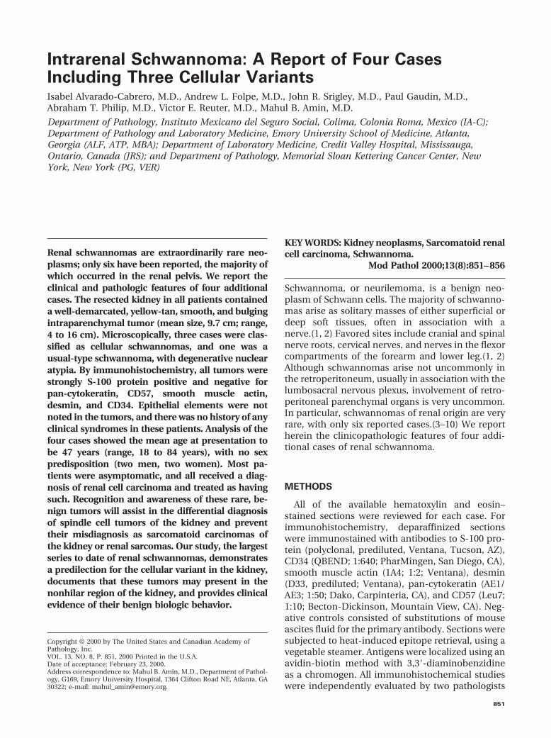
circumscribed, intrarenal masses that compressedthe adjacent renal parenchyma (Fig. 1). The tumorsmeasured from 4 to 16 cm (mean, 9.7 cm) and weretan to yellow in color. Cystic change was noted intwo cases, and one tumor contained scattered cal-cifications.
Microscopic FindingsCase 1 showed the typical features of a schwan-
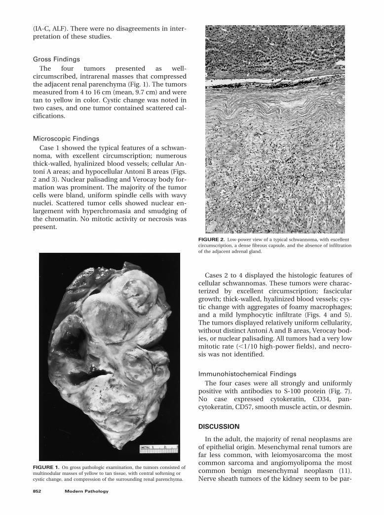
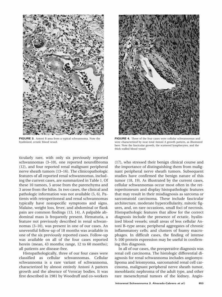
noma, with excellent circumscription; numerousthick-walled, hyalinized blood vessels; cellular An-toni A areas; and hypocellular Antoni B areas (Figs.2 and 3). Nuclear palisading and Verocay body for-mation was prominent. The majority of the tumorcells were bland, uniform spindle cells with wavynuclei. Scattered tumor cells showed nuclear en-largement with hyperchromasia and smudging ofthe chromatin. No mitotic activity or necrosis waspresent.
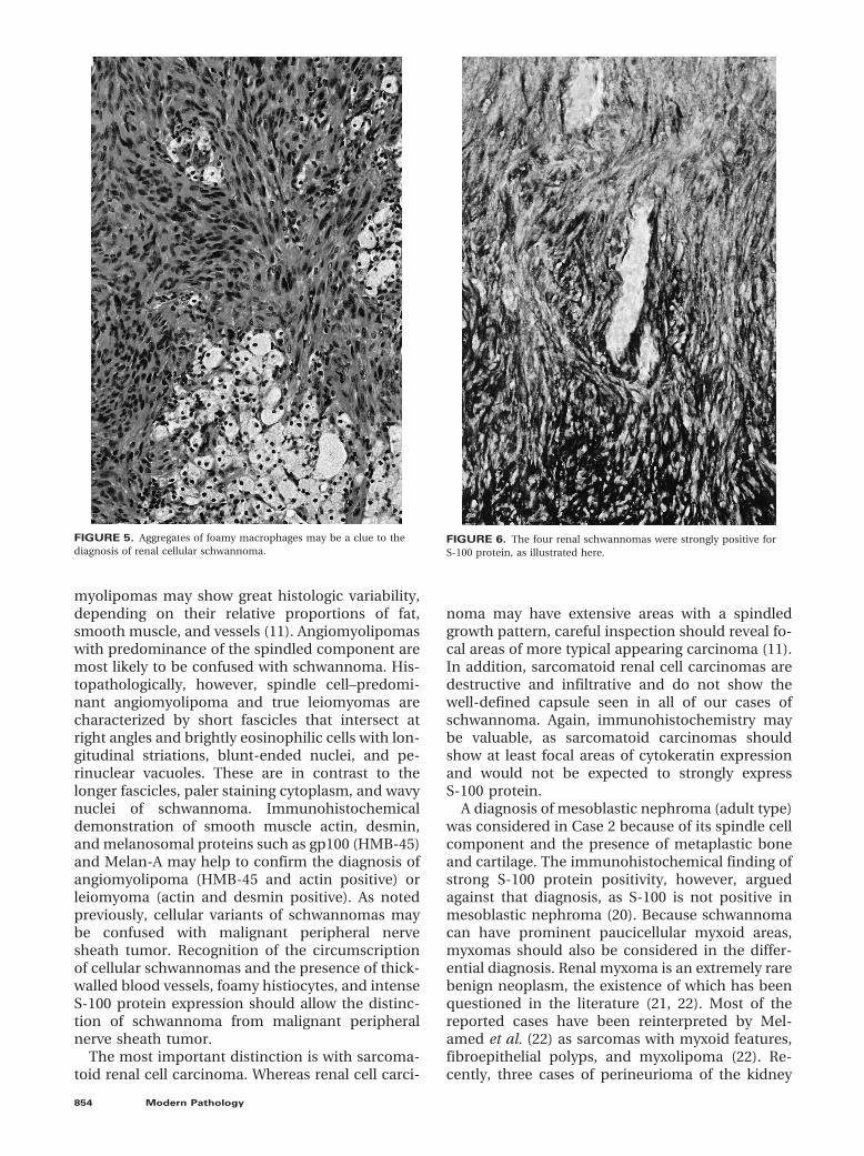
Cases 2 to 4 displayed the histologic features ofcellular schwannomas. These tumors were charac-terized by excellent circumscription; fasciculargrowth; thick-walled, hyalinized blood vessels; cys-tic change with aggregates of foamy macrophages;and a mild lymphocytic infiltrate (Figs. 4 and 5).The tumors displayed relatively uniform cellularity,without distinct Antoni A and B areas, Verocay bod-ies, or nuclear palisading. All tumors had a very lowmitotic rate (,1/10 high-power fields), and necro-sis was not identified.
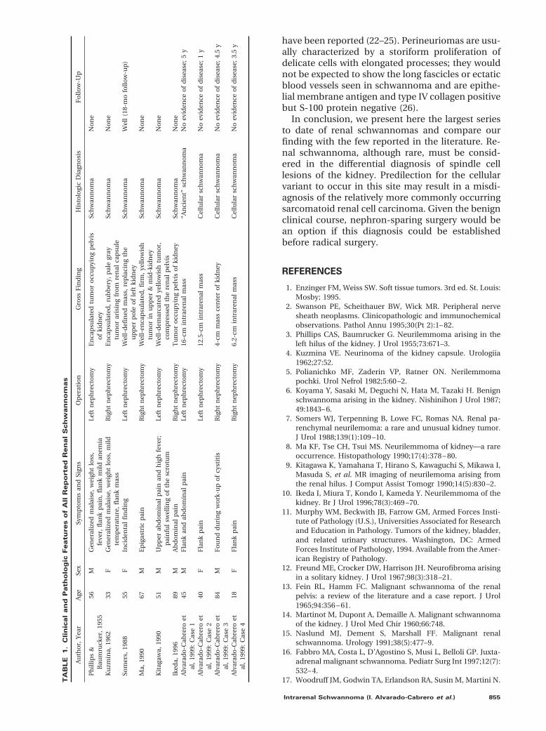
Immunohistochemical FindingsThe four cases were all strongly and uniformly
positive with antibodies to S-100 protein (Fig. 7).No case expressed cytokeratin, CD34, pan-cytokeratin, CD57, smooth muscle actin, or desmin.
DISCUSSION
In the adult, the majority of renal neoplasms areof epithelial origin. Mesenchymal renal tumors arefar less common, with leiomyosarcoma the mostcommon sarcoma and angiomyolipoma the mostcommon benign mesenchymal neoplasm (11).Nerve sheath tumors of the kidney seem to be par-
FIGURE 1. On gross pathologic examination, the tumors consisted ofmultinodular masses of yellow to tan tissue, with central softening orcystic change, and compression of the surrounding renal parenchyma.
FIGURE 2. Low-power view of a typical schwannoma, with excellentcircumscription, a dense fibrous capsule, and the absence of infiltrationof the adjacent adrenal gland.
852 Modern Pathology
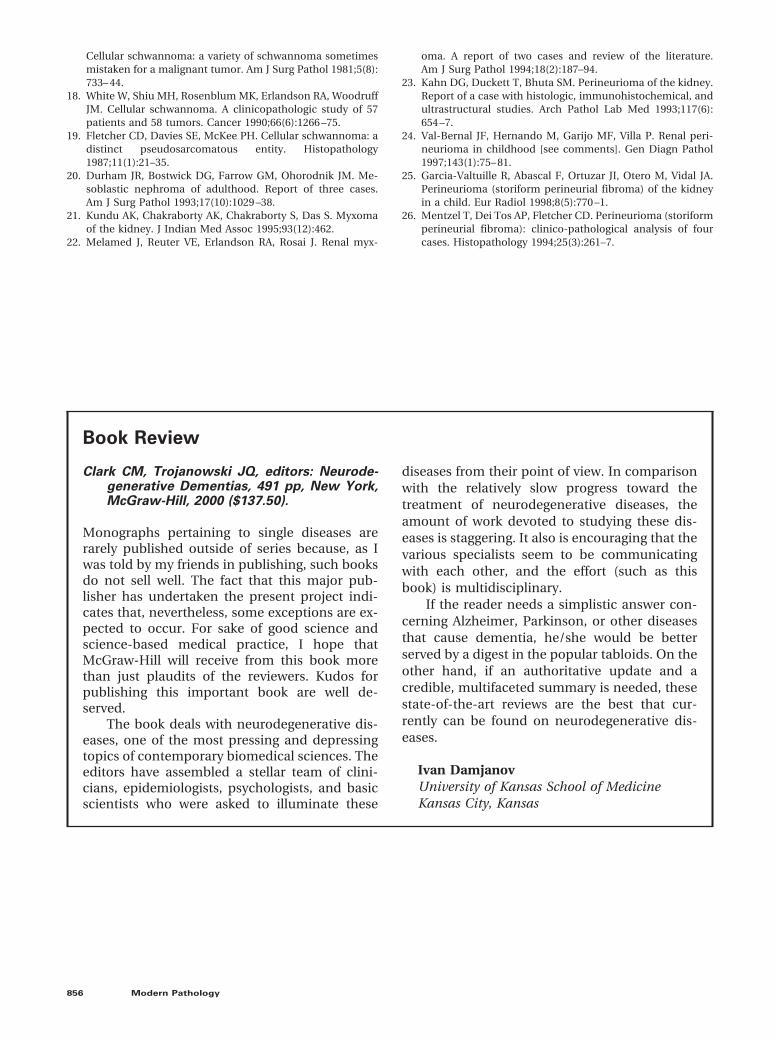
ticularly rare, with only six previously reportedschwannomas (3–10), one reported neurofibroma(12), and four reported renal malignant peripheralnerve sheath tumors (13–16). The clinicopathologicfeatures of all reported renal schwannomas, includ-ing the current cases, are summarized in Table 1. Ofthese 10 tumors, 5 arose from the parenchyma and3 arose from the hilus. In two cases, the clinical andpathologic information was not available (5, 6). Pa-tients with retroperitoneal and renal schwannomastypically have nonspecific symptoms and signs.Malaise, weight loss, fever, and abdominal or flankpain are common findings (13, 14). A palpable ab-dominal mass is frequently present. Hematuria, afeature not previously described in renal schwan-nomas (3–10), was present in one of our cases. Anuneventful follow-up of 18 months was available inone of the six previously reported cases. Follow-upwas available on all of the four cases reportedherein (mean, 43 months; range, 12 to 60 months);all patients are disease-free.
Histopathologically, three of our four cases wereclassified as cellular schwannomas. Cellularschwannoma is a rare variant of schwannoma,characterized by almost entirely Antoni A patterngrowth and the absence of Verocay bodies. It wasfirst described in 1981 by Woodruff and co-workers
(17), who stressed their benign clinical course andthe importance of distinguishing them from malig-nant peripheral nerve sheath tumors. Subsequentstudies have confirmed the benign nature of thistumor (18, 19). As illustrated by the current cases,cellular schwannomas occur most often in the ret-roperitoneum and display histopathologic featuresthat may result in their misdiagnosis as sarcoma orsarcomatoid carcinoma. These include fasciculararchitecture, moderate hypercellularity, mitotic fig-ures, and, on rare occasions, small foci of necrosis.Histopathologic features that allow for the correctdiagnosis include the presence of ectatic, hyalin-ized blood vessels; small areas of less cellular An-toni B–type areas; peripheral aggregates of chronicinflammatory cells; and clusters of foamy macro-phages. In difficult cases, the finding of intenseS-100 protein expression may be useful in confirm-ing this diagnosis.
In all of our cases, the preoperative diagnosis wasrenal cell carcinoma. The histologic differential di-agnosis for renal schwannoma includes angiomyo-lipoma and leiomyoma, sarcomatoid renal cell car-cinoma, malignant peripheral nerve sheath tumor,mesoblastic nephroma of the adult type, and otherrare mesenchymal tumors of the kidney. Angio-
FIGURE 3. Antoni B area from a typical schwannoma. Note thehyalinized, ectatic blood vessel.
FIGURE 4. Three of the four cases were cellular schwannomas andwere characterized by near-total Antoni A growth pattern, as illustratedhere. Note the fascicular growth, the scattered lymphocytes, and thethick-walled blood vessel.
Intrarenal Schwannoma (I. Alvarado-Cabrero et al.) 853
myolipomas may show great histologic variability,depending on their relative proportions of fat,smooth muscle, and vessels (11). Angiomyolipomaswith predominance of the spindled component aremost likely to be confused with schwannoma. His-topathologically, however, spindle cell–predomi-nant angiomyolipoma and true leiomyomas arecharacterized by short fascicles that intersect atright angles and brightly eosinophilic cells with lon-gitudinal striations, blunt-ended nuclei, and pe-rinuclear vacuoles. These are in contrast to thelonger fascicles, paler staining cytoplasm, and wavynuclei of schwannoma. Immunohistochemicaldemonstration of smooth muscle actin, desmin,and melanosomal proteins such as gp100 (HMB-45)and Melan-A may help to confirm the diagnosis ofangiomyolipoma (HMB-45 and actin positive) orleiomyoma (actin and desmin positive). As notedpreviously, cellular variants of schwannomas maybe confused with malignant peripheral nervesheath tumor. Recognition of the circumscriptionof cellular schwannomas and the presence of thick-walled blood vessels, foamy histiocytes, and intenseS-100 protein expression should allow the distinc-tion of schwannoma from malignant peripheralnerve sheath tumor.
The most important distinction is with sarcoma-toid renal cell carcinoma. Whereas renal cell carci-
noma may have extensive areas with a spindledgrowth pattern, careful inspection should reveal fo-cal areas of more typical appearing carcinoma (11).In addition, sarcomatoid renal cell carcinomas aredestructive and infiltrative and do not show thewell-defined capsule seen in all of our cases ofschwannoma. Again, immunohistochemistry maybe valuable, as sarcomatoid carcinomas shouldshow at least focal areas of cytokeratin expressionand would not be expected to strongly expressS-100 protein.
A diagnosis of mesoblastic nephroma (adult type)was considered in Case 2 because of its spindle cellcomponent and the presence of metaplastic boneand cartilage. The immunohistochemical finding ofstrong S-100 protein positivity, however, arguedagainst that diagnosis, as S-100 is not positive inmesoblastic nephroma (20). Because schwannomacan have prominent paucicellular myxoid areas,myxomas should also be considered in the differ-ential diagnosis. Renal myxoma is an extremely rarebenign neoplasm, the existence of which has beenquestioned in the literature (21, 22). Most of thereported cases have been reinterpreted by Mel-amed et al. (22) as sarcomas with myxoid features,fibroepithelial polyps, and myxolipoma (22). Re-cently, three cases of perineurioma of the kidney
FIGURE 6. The four renal schwannomas were strongly positive forS-100 protein, as illustrated here.
FIGURE 5. Aggregates of foamy macrophages may be a clue to thediagnosis of renal cellular schwannoma.
854 Modern Pathology
have been reported (22–25). Perineuriomas are usu-ally characterized by a storiform proliferation ofdelicate cells with elongated processes; they wouldnot be expected to show the long fascicles or ectaticblood vessels seen in schwannoma and are epithe-lial membrane antigen and type IV collagen positivebut S-100 protein negative (26).
In conclusion, we present here the largest seriesto date of renal schwannomas and compare ourfinding with the few reported in the literature. Re-nal schwannoma, although rare, must be consid-ered in the differential diagnosis of spindle celllesions of the kidney. Predilection for the cellularvariant to occur in this site may result in a misdi-agnosis of the relatively more commonly occurringsarcomatoid renal cell carcinoma. Given the benignclinical course, nephron-sparing surgery would bean option if this diagnosis could be establishedbefore radical surgery.
REFERENCES
1. Enzinger FM, Weiss SW. Soft tissue tumors. 3rd ed. St. Louis:Mosby; 1995.
2. Swanson PE, Scheithauer BW, Wick MR. Peripheral nervesheath neoplasms. Clinicopathologic and immunochemicalobservations. Pathol Annu 1995;30(Pt 2):1– 82.
3. Phillips CAS, Baumrucker G. Neurilemmoma arising in theleft hilus of the kidney. J Urol 1955;73:671–3.
4. Kuzmina VE. Neurinoma of the kidney capsule. Urologiia1962;27:52.
5. Polianichko MF, Zaderin VP, Ratner ON. Nerilemmomapochki. Urol Nefrol 1982;5:60 –2.
6. Koyama Y, Sasaki M, Deguchi N, Hata M, Tazaki H. Benignschwannoma arising in the kidney. Nishinihon J Urol 1987;49:1843– 6.
7. Somers WJ, Terpenning B, Lowe FC, Romas NA. Renal pa-renchymal neurilemoma: a rare and unusual kidney tumor.J Urol 1988;139(1):109 –10.
8. Ma KF, Tse CH, Tsui MS. Neurilemmoma of kidney—a rareoccurrence. Histopathology 1990;17(4):378 – 80.
9. Kitagawa K, Yamahana T, Hirano S, Kawaguchi S, Mikawa I,Masuda S, et al. MR imaging of neurilemoma arising fromthe renal hilus. J Comput Assist Tomogr 1990;14(5):830 –2.
10. Ikeda I, Miura T, Kondo I, Kameda Y. Neurilemmoma of thekidney. Br J Urol 1996;78(3):469 –70.
11. Murphy WM, Beckwith JB, Farrow GM, Armed Forces Insti-tute of Pathology (U.S.), Universities Associated for Researchand Education in Pathology. Tumors of the kidney, bladder,and related urinary structures. Washington, DC: ArmedForces Institute of Pathology, 1994. Available from the Amer-ican Registry of Pathology.
12. Freund ME, Crocker DW, Harrison JH. Neurofibroma arisingin a solitary kidney. J Urol 1967;98(3):318 –21.
13. Fein RL, Hamm FC. Malignant schwannoma of the renalpelvis: a review of the literature and a case report. J Urol1965;94:356 – 61.
14. Martinot M, Dupont A, Demaille A. Malignant schwannomaof the kidney. J Urol Med Chir 1960;66:748.
15. Naslund MJ, Dement S, Marshall FF. Malignant renalschwannoma. Urology 1991;38(5):477–9.
16. Fabbro MA, Costa L, D’Agostino S, Musi L, Belloli GP. Juxta-adrenal malignant schwannoma. Pediatr Surg Int 1997;12(7):532– 4.
17. Woodruff JM, Godwin TA, Erlandson RA, Susin M, Martini N.TA
BLE
1.
Clin
ical
an
dP
ath
olo
gic
Featu
res
of
All
Re
po
rte
dR
en
al
Sch
wa
nn
om
as
Au
tho
r,Y
ear
Age
Sex
Sym
pto
ms
and
Sign
sO
per
atio
nG
ross
Fin
din
gH
isto
logi
cD
iagn
osi
sF
ollo
w-U
p
Ph
illip
s&
Bau
mru
cker
,19
5556
MG
ener
aliz
edm
alai
se,
wei
ght
loss
,fe
ver,
flan
kp
ain
,fl
ank
mild
anem
iaLe
ftn
eph
rect
om
yE
nca
psu
late
dtu
mo
ro
ccu
pyi
ng
pel
vis
of
kid
ney
Sch
wan
no
ma
No
ne
Ku
zmin
a,19
6233
FG
ener
aliz
edm
alai
se,
wei
ght
loss
,m
ildte
mp
erat
ure
,fl
ank
mas
sR
igh
tn
eph
rect
om
yE
nca
psu
late
d,
rub
ber
y,p
ale
gray
tum
or
aris
ing
fro
mre
nal
cap
sule
Sch
wan
no
ma
No
ne
Som
ers,
1988
55F
Inci
den
tal
fin
din
gLe
ftn
eph
rect
om
yW
ell-
def
ined
mas
s,re
pla
cin
gth
eu
pp
erp
ole
of
left
kid
ney
Sch
wan
no
ma
Wel
l(1
8-m
ofo
llow
-up
)
Ma,
1990
67M
Ep
igas
tric
pai
nR
igh
tn
eph
rect
om
yW
ell-
enca
psu
late
d,
firm
,ye
llow
ish
tum
or
inu
pp
er&
mid
-kid
ney
Sch
wan
no
ma
No
ne
Kit
agaw
a,19
9051
MU
pp
erab
do
min
alp
ain
and
hig
hfe
ver;
pai
nfu
lsw
elli
ng
of
the
scro
tum
Left
nep
hre
cto
my
Wel
l-d
emar
cate
dye
llow
ish
tum
or,
com
pre
ssed
the
ren
alp
elvi
sSc
hw
ann
om
aN
on
e
Iked
a,19
9689
MA
bd
om
inal
pai
nR
igh
tn
eph
rect
om
yT
um
or
occ
up
yin
gp
elvi
so
fki
dn
eySc
hw
ann
om
aN
on
eA
lvar
ado
-Cab
rero
etal
,19
99:
Cas
e1
45M
Fla
nk
and
abd
om
inal
pai
nLe
ftn
eph
rect
om
y16
-cm
intr
aren
alm
ass
“An
cien
t”sc
hw
ann
om
aN
oev
iden
ceo
fd
isea
se;
5y
Alv
arad
o-C
abre
roet
al,
1999
:C
ase
240
FF
lan
kp
ain
Left
nep
hre
cto
my
12.5
-cm
intr
aren
alm
ass
Cel
lula
rsc
hw
ann
om
aN
oev
iden
ceo
fd
isea
se;
1y
Alv
arad
o-C
abre
roet
al,
1999
:C
ase
384
MF
ou
nd
du
rin
gw
ork
-up
of
cyst
itis
Rig
ht
nep
hre
cto
my
4-cm
mas
sce
nte
ro
fki
dn
eyC
ellu
lar
sch
wan
no
ma
No
evid
ence
of
dis
ease
;4.
5y
Alv
arad
o-C
abre
roet
al,
1999
:C
ase
418
FF
lan
kp
ain
Rig
ht
nep
hre
cto
my
6.2-
cmin
trar
enal
mas
sC
ellu
lar
sch
wan
no
ma
No
evid
ence
of
dis
ease
;3.
5y
Intrarenal Schwannoma (I. Alvarado-Cabrero et al.) 855
Cellular schwannoma: a variety of schwannoma sometimesmistaken for a malignant tumor. Am J Surg Pathol 1981;5(8):733– 44.
18. White W, Shiu MH, Rosenblum MK, Erlandson RA, WoodruffJM. Cellular schwannoma. A clinicopathologic study of 57patients and 58 tumors. Cancer 1990;66(6):1266 –75.
19. Fletcher CD, Davies SE, McKee PH. Cellular schwannoma: adistinct pseudosarcomatous entity. Histopathology1987;11(1):21–35.
20. Durham JR, Bostwick DG, Farrow GM, Ohorodnik JM. Me-soblastic nephroma of adulthood. Report of three cases.Am J Surg Pathol 1993;17(10):1029 –38.
21. Kundu AK, Chakraborty AK, Chakraborty S, Das S. Myxomaof the kidney. J Indian Med Assoc 1995;93(12):462.
22. Melamed J, Reuter VE, Erlandson RA, Rosai J. Renal myx-
oma. A report of two cases and review of the literature.Am J Surg Pathol 1994;18(2):187–94.
23. Kahn DG, Duckett T, Bhuta SM. Perineurioma of the kidney.Report of a case with histologic, immunohistochemical, andultrastructural studies. Arch Pathol Lab Med 1993;117(6):654 –7.
24. Val-Bernal JF, Hernando M, Garijo MF, Villa P. Renal peri-neurioma in childhood [see comments]. Gen Diagn Pathol1997;143(1):75– 81.
25. Garcia-Valtuille R, Abascal F, Ortuzar JI, Otero M, Vidal JA.Perineurioma (storiform perineurial fibroma) of the kidneyin a child. Eur Radiol 1998;8(5):770 –1.
26. Mentzel T, Dei Tos AP, Fletcher CD. Perineurioma (storiformperineurial fibroma): clinico-pathological analysis of fourcases. Histopathology 1994;25(3):261–7.
Book Review
Clark CM, Trojanowski JQ, editors: Neurode-generative Dementias, 491 pp, New York,McGraw-Hill, 2000 ($137.50).
Monographs pertaining to single diseases arerarely published outside of series because, as Iwas told by my friends in publishing, such booksdo not sell well. The fact that this major pub-lisher has undertaken the present project indi-cates that, nevertheless, some exceptions are ex-pected to occur. For sake of good science andscience-based medical practice, I hope thatMcGraw-Hill will receive from this book morethan just plaudits of the reviewers. Kudos forpublishing this important book are well de-served.
The book deals with neurodegenerative dis-eases, one of the most pressing and depressingtopics of contemporary biomedical sciences. Theeditors have assembled a stellar team of clini-cians, epidemiologists, psychologists, and basicscientists who were asked to illuminate these
diseases from their point of view. In comparisonwith the relatively slow progress toward thetreatment of neurodegenerative diseases, theamount of work devoted to studying these dis-eases is staggering. It also is encouraging that thevarious specialists seem to be communicatingwith each other, and the effort (such as thisbook) is multidisciplinary.
If the reader needs a simplistic answer con-cerning Alzheimer, Parkinson, or other diseasesthat cause dementia, he/she would be betterserved by a digest in the popular tabloids. On theother hand, if an authoritative update and acredible, multifaceted summary is needed, thesestate-of-the-art reviews are the best that cur-rently can be found on neurodegenerative dis-eases.
Ivan DamjanovUniversity of Kansas School of MedicineKansas City, Kansas
856 Modern Pathology
Related Documents