Intracortical inhibition and facilitation with unilateral dominant, unilateral nondominant and bilateral movement tasks in left and right handed adults Sandy McCombe Waller, PT, PhD, NCS, University of Maryland, School of Medicine, Department of Physical Therapy and Rehabilitation Science, 100 Penn Street, Baltimore, MD 21201 Larry Forrester, PhD, University of Maryland, School of Medicine, Department of Physical Therapy and Rehabilitation Science and VA Maryland Healthcare System, 100 Penn Street, Baltimore, MD 21201 Federico Villagra, PhD, and University of Maryland, School of Medicine, Department of Physical Therapy and Rehabilitation Science, 100 Penn Street, Baltimore, MD 21201 Jill Whitall, PhD University of Maryland, School of Medicine, Department of Physical Therapy and Rehabilitation Science, 100 Penn Street, Baltimore, MD 21201 Abstract Purpose—To investigate intracortical inhibition and facilitation in response to unilateral dominant, nondominant and bilateral biceps activation and short-term upper extremity training in right and left-handed adults. Methods—Paired-pulse transcranial magnetic stimulation was used to measure intracortical excitability in motor dominant and nondominant cortices of 26 nondisabled adults. Neural facilitation and inhibition were measured in each hemisphere during unilateral dominant, nondominant and bilateral arm activation and after training in each condition. Results—No differences were seen between right and left- handed subjects. Intracortical facilitation and decreased inhibition were seen in each hemisphere with unilateral activation/ training of contralateral muscles and bilateral muscle activation/training. Persistent intracortical inhibition was seen in each hemisphere with ipsilateral muscle activation/training. Inhibition was greater in the nondominant hemisphere during dominant hemisphere activation (dominant arm contraction). Conclusion—Strongly dominant individuals show no difference in intracortical responses given handedness. Intracortical activity with unilateral and bilateral arm activation and short-term training differs based on hemispheric dominance, with the motor dominant hemisphere exerting a © 2008 Elsevier B.V. All rights reserved. All correspondence should be addressed to: Sandy McCombe Waller PhD, PT, NCS, University of Maryland, School of Medicine, Department of Physical Therapy and Rehabilitation Science, 100 Penn Street, Baltimore, Maryland 21201, Email: [email protected] Telephone: 410-706-0787, Fax: 410-706-6387. Publisher's Disclaimer: This is a PDF file of an unedited manuscript that has been accepted for publication. As a service to our customers we are providing this early version of the manuscript. The manuscript will undergo copyediting, typesetting, and review of the resulting proof before it is published in its final citable form. Please note that during the production process errors may be discovered which could affect the content, and all legal disclaimers that apply to the journal pertain. NIH Public Access Author Manuscript J Neurol Sci. Author manuscript; available in PMC 2010 July 27. Published in final edited form as: J Neurol Sci. 2008 June 15; 269(1-2): 96–104. doi:10.1016/j.jns.2007.12.033. NIH-PA Author Manuscript NIH-PA Author Manuscript NIH-PA Author Manuscript

Welcome message from author
This document is posted to help you gain knowledge. Please leave a comment to let me know what you think about it! Share it to your friends and learn new things together.
Transcript
Intracortical inhibition and facilitation with unilateral dominant,unilateral nondominant and bilateral movement tasks in left andright handed adults
Sandy McCombe Waller, PT, PhD, NCS,University of Maryland, School of Medicine, Department of Physical Therapy and RehabilitationScience, 100 Penn Street, Baltimore, MD 21201
Larry Forrester, PhD,University of Maryland, School of Medicine, Department of Physical Therapy and RehabilitationScience and VA Maryland Healthcare System, 100 Penn Street, Baltimore, MD 21201
Federico Villagra, PhD, andUniversity of Maryland, School of Medicine, Department of Physical Therapy and RehabilitationScience, 100 Penn Street, Baltimore, MD 21201
Jill Whitall, PhDUniversity of Maryland, School of Medicine, Department of Physical Therapy and RehabilitationScience, 100 Penn Street, Baltimore, MD 21201
AbstractPurpose—To investigate intracortical inhibition and facilitation in response to unilateraldominant, nondominant and bilateral biceps activation and short-term upper extremity training inright and left-handed adults.
Methods—Paired-pulse transcranial magnetic stimulation was used to measure intracorticalexcitability in motor dominant and nondominant cortices of 26 nondisabled adults. Neuralfacilitation and inhibition were measured in each hemisphere during unilateral dominant,nondominant and bilateral arm activation and after training in each condition.
Results—No differences were seen between right and left- handed subjects. Intracorticalfacilitation and decreased inhibition were seen in each hemisphere with unilateral activation/training of contralateral muscles and bilateral muscle activation/training. Persistent intracorticalinhibition was seen in each hemisphere with ipsilateral muscle activation/training. Inhibition wasgreater in the nondominant hemisphere during dominant hemisphere activation (dominant armcontraction).
Conclusion—Strongly dominant individuals show no difference in intracortical responses givenhandedness. Intracortical activity with unilateral and bilateral arm activation and short-termtraining differs based on hemispheric dominance, with the motor dominant hemisphere exerting a
© 2008 Elsevier B.V. All rights reserved.All correspondence should be addressed to: Sandy McCombe Waller PhD, PT, NCS, University of Maryland, School of Medicine,Department of Physical Therapy and Rehabilitation Science, 100 Penn Street, Baltimore, Maryland 21201, Email:[email protected] Telephone: 410-706-0787, Fax: 410-706-6387.Publisher's Disclaimer: This is a PDF file of an unedited manuscript that has been accepted for publication. As a service to ourcustomers we are providing this early version of the manuscript. The manuscript will undergo copyediting, typesetting, and review ofthe resulting proof before it is published in its final citable form. Please note that during the production process errors may bediscovered which could affect the content, and all legal disclaimers that apply to the journal pertain.
NIH Public AccessAuthor ManuscriptJ Neurol Sci. Author manuscript; available in PMC 2010 July 27.
Published in final edited form as:J Neurol Sci. 2008 June 15; 269(1-2): 96–104. doi:10.1016/j.jns.2007.12.033.
NIH
-PA Author Manuscript
NIH
-PA Author Manuscript
NIH
-PA Author Manuscript
larger inhibitory influence over the nondominant hemisphere. Bilateral activation and training hasa disinhibitory effect in both dominant and nondominant hemispheres.
Keywordsmotor cortex; neuronal excitability; motor control
IntroductionUpper extremity hemiparesis presents a major challenge to rehabilitation after stroke.Differences in neural control mechanisms for unilateral and bilateral upper extremity tasksare of interest because of the use of novel active bilateral and unilateral training paradigmsin stroke rehabilitation. These training approaches have demonstrated a positive impact onreturn of paretic limb function (1–5) but little is known about the specific neuralmechanisms that underlie those functional changes. In this study we investigate a particularaspect of function that is uniquely developed in the upper extremity, namely handedness.After unilateral lesion several factors related to handedness may impact functional responseto upper extremity (UE) rehabilitation training. Among these are (a) differences between thedominant and nondominant hemispheres, (b) influences of interhemispheric connectionsduring unilateral vs. bilateral tasks, and (c) possible differences in neural controlmechanisms in left- vs. right-handed individuals.
The ability of the brain to adapt neural firing in relation to various unilateral muscleactivations and/or training has been documented using single and paired pulsed transcranialmagnetic stimulation (TMS). Increased excitability in homologous upper extremity muscleshas been demonstrated in response to 50% maximal voluntary contraction (6) and withcomplex motor sequence tasks (7). Inhibitory modulation has also been seen in theipsilateral cortex during tasks of increasing difficulty, indicating inhibition of muscles in thenonmoving hand. Liepert et al. reported that intracortical inhibitory and facilitorymodulation is related to the selective requirements of a motor task (2). For example, musclestargeted for activation were facilitated whereas intentional relaxation increased inhibition.Taken together these studies show motor effort of one arm can impact the excitability ofcontralateral and ipsilateral cortex affecting homologous arm muscles as well otheripsilateral muscles. Questions remain, however, as to whether these responses differ withdominant versus nondominant hemisphere activity and furthermore if handedness impactsthe degree of the response.
Tinazzi and Zanette reported the presence of hemispheric asymmetries of the ipsilateralcortex during unilateral tasks in which homologous muscle excitability was significantly lesswhen the dominant hand was the task hand (7). This indicates greater motor influence fromone hemisphere to another when the nondominant hand was moving; however, this studywas limited to right hand dominant subjects. Asymmetries in neural excitability have beenshown to differ based on hand dominance by Yahagi and Kasai who found that in right-handed subjects, motor evoked potential (MEP) amplitudes of the dominant hand weresignificantly greater than those in the nondominant hand while in left-handed subjects, therewas no significant difference between the dominant and nondominant hands (8).
In summary, these studies show the presence of interhemispheric neural firing (inhibitoryand facilitory) with unilateral activation, modulation of neural firing based on task and taskcomplexity, and asymmetry of neural firing depending on hand dominance. One aspect thathas not been investigated is the response of intracortical inhibition and excitation to bilateralactivation and following short-term bilateral training. This is of interest for post-strokerehabilitation where approaches to UE rehabilitation typically target the movement and use
Waller et al. Page 2
J Neurol Sci. Author manuscript; available in PMC 2010 July 27.
NIH
-PA Author Manuscript
NIH
-PA Author Manuscript
NIH
-PA Author Manuscript
of the paretic limb. In addition, compensatory training of the nonparetic limb is often used inacute care during periods of flaccidity or when UE paresis is severe and may contribute tolearned nonuse. Bilateral training incorporating the use of both limbs in therapy has alsoshown functional gains in patients with mild and moderate stroke severity (9–17). In thistraining paradigm the emphasis is on exercising the proximal muscles and targeting patientswith moderate levels of impairment. In bilateral training approaches in subjects with moresevere paresis that involved whole arm training (18) or training involving copying tasks witha digitized pen (19), the bilateral training has not shown a benefit over unilateral training.
In fact, there is some controversy regarding the benefit of bilateral arm training because, forexample, there is evidence of brief performance improvements in the contralateral hand ifthe other hand has reduced somatosensory input from cutaneous anesthesia. This occurs inhealthy adults (20) and in stroke patients when the intact hand is anesthetized (21) andargues against activating the non-paretic arm and hand while training the paretic arm andhand. Furthermore, there is evidence that some patients with chronic stroke and largerimpairments have an abnormally high interhemispheric inhibitory drive from M1 in thecontralesional hemisphere to M1 in the ipsilesional hemisphere (22). Therefore, down-regulation of activity in the ipsilateral intact motor cortex may reduce abnormal inhibitionfrom the contralesional to the ipsilesional hemisphere suggesting that increased activity ofthe non-paretic arm is potentially detrimental to paretic arm and hand (23,24) However,these studies have only involved unilateral activation. Cortical facilitation and inhibition inresponse to bilateral arm activation and training have not been investigated.
Examining the differences between left and right-handed subjects in terms of corticalprocesses, and comparing unilateral and bilateral motor task conditions, may provide afoundation for determining the underlying cortical control mechanisms and the influence ofhand dominance on these mechanisms. In this initial study, we examine intracorticalinhibition and excitation in healthy nondisabled adults to provide a comparison with futurestudies on individuals with stroke. Based on previous literature, we hypothesized that therewould be a neural firing asymmetry between the dominant and nondominant hemispheres inright-handed subjects for biceps muscles, seen to a lesser extent in the left-handed. Weanticipated that bilateral training would have a disinhibitory effect on intracortical inhibitionwhen compared to unilateral training of either arm individually in both right and left-handed. We selected the biceps as our target muscle for training to extend our previous workusing BATRAC (bilateral arm training with rhythmic auditory cueing) which targets moreproximal muscles used in reach and return tasks.
MethodsSubjects included 26 adults with no neurological impairment or impaired arm function. Ofthese, 10 had strong left-hand dominance (36± 12 yrs; 4 female) and 16 had strong right-hand dominance (42±15 yrs; 6 female). Strength of hand preference was determined byscores on the Edinburgh Inventory(25), with criteria for strong preference set between +85to +100 for right-handed and −85 to −100 for left-handed. Exclusion criteria included: 1)ambidexterity or forced change of hand dominance that precludes unilateral motordominance, 2) metal implants in the brain or skull, 3) history of seizures, 4) pregnancy, 5)medical history of neuromuscular disease including neuropathy /myopathy affecting thearms and/or report of numbness or tingling in the arms, 6) drug use that may influenceexcitability threshold (antispastics, anxiolytics, hypnotics, antiepileptics), and 7) activecardiac disease by patient report. All subjects signed informed consent approved by theUniversity of Maryland School of Medicine and Baltimore VAMC joint Internal ReviewBoard.
Waller et al. Page 3
J Neurol Sci. Author manuscript; available in PMC 2010 July 27.
NIH
-PA Author Manuscript
NIH
-PA Author Manuscript
NIH
-PA Author Manuscript
Preliminary work to establish testing and training parametersBecause the main investigation targeted proximal muscles used in a novel arm trainingprotocol, we conducted a pilot study with 3 subjects to define appropriate testing andtraining parameters. Specifically we identified 1) optimal interstimulus (ISI) settings forinhibition and facilitation of proximal arm muscles used in the training, 2) the necessary andsufficient training durations to elicit intracortical effects, and 3) the duration of the post-training period required for a return of MEP amplitudes to baseline values.
1. Determination of ISIs—It has been reported by some that the ideal ISI for inhibition is2.5– 5 (26) while other studies demonstrate that ISIs of 1 ms elicit maximal inhibition(27,28). We conducted a battery of tests with ISIs between 1 – 5 ms. The magnitudes of theconditioning and test stimuli were held constant throughout this testing. A 1ms ISIconsistently elicited the largest inhibitory responses, as described in other studies, and thuswas selected as the inhibitory ISI parameter. There is some debate that an ISI of 1 ms maynot elicit pure neuronal inhibition because the targeted neurons are still in a refractory state.However, Roshan, Paradiso and Chen have shown that intracortical inhibition (ICI) at 1 mscannot be fully explained by axonal refractoriness and conclude that synaptic inhibition islikely involved. ISIs of 10 – 15 ms were also compared to obtain ideal facilitoryresponses(29). Intracortical facilitation (ICF) from ISIs between 12 – 15 ms did not differ,but were greater than ISIs of 10 and 11 ms. Thus an ISI 15 ms was selected as the facilitoryISI parameter for the main investigation.
2. Necessary and sufficient training time—Each pilot subject participated in sessionsof unilateral dominant arm training of 5, 10, and 15 minute durations to determine thenecessary and sufficient training time to obtain a training effect. Inconsistent responses inMEP inhibition and facilitation were seen after 5 and 15 minute sessions. It is possible that 5minutes was not a sufficient amount of time to achieve a training effect and that fatigue wasa factor at the 15 minute training time. Because all subjects produced appropriate responsesafter 10 minutes of training, that interval was selected for all subsequent training conditions.
3. Post-training washout period—On a separate day each subject participated in 10minutes of training in both unilateral dominant and bilateral training with TMS post-testingat 5, 10, 15 and 30 minutes post training. All subjects returned to baseline values forfacilitory and inhibitory responses by the 10 minute testing point with no further changes atthe 15 and 30 minute time points. The need to minimize testing burden precluded use ofthese repeated tests to establish baseline with the larger subject sample. However, since allpilot subjects achieved complete washout after 10 minutes, this recovery interval was usedbetween training modes in the subsequent protocols.
TMS Testing: Unilateral dominant, unilateral nondominant and bilateral activationsSubjects were seated in a cushioned semi-reclining chair with elbows in 90° flexion andbipolar surface EMG electrodes spaced 1 cm apart on the belly of the biceps brachii. Theground electrode was placed on the right medial malleolous. Force transducers (TransducerTechniques, CA) were mounted on the armrests, and the subject’s arms were stabilized incuffs attached to the armrests. The head was stabilized in a support to standardize headorientation and minimize extraneous movement.
Baseline CNS corticospinal excitation was determined using single, suprathreshold magneticpulses delivered transcranially via a system with two magnetic stimulators connectedthrough a Bistim device (Magstim Company, Dyfed, UK) with a figure - 8 coil. For eachhemisphere the best stimulation site to elicit MEPs in the contralateral biceps wasdetermined by stimulating cortex in 1 cm coordinates relative to Cz and marked with a felt
Waller et al. Page 4
J Neurol Sci. Author manuscript; available in PMC 2010 July 27.
NIH
-PA Author Manuscript
NIH
-PA Author Manuscript
NIH
-PA Author Manuscript
tip pen on the subject’s head. Precise location and threshold values for eliciting MEPs weredetermined for each individual by convention with threshold defined as lowest intensityproducing MEP amplitudes exceeding 50µV in > 5 of 10 consecutive stimulations (30).Both active and passive threshold measurements were obtained. To standardize the musclecontraction for active threshold testing, subjects used real time visual feedback to generatethe force required to offset the resting weight of the forearm on the armrest. This was easilyaccomplished and provided a reproducible standardized low-force isometric contraction forfacilitated TMS.
Intracortical excitability was measured using a paired-pulsed paradigm as described byKujirai et al.(31) and Muelbacher et al.(6) In this TMS paradigm a subthresholdconditioning stimulus (CS-80% active motor threshold) was followed by a suprathresholdtest–stimulus (TS) at different ISIs. The intensity of the TS was 120 % of the threshold.Active threshold was used for all tests in which the contralateral biceps was activated (e.g.dominant hemisphere testing with dominant arm or bilateral arm activation). Although, ICIand ICF testing is usually done without muscle activation, Kujari et al. report that ICI andICF can still be seen with muscle activation though it may be suppressed (31) We chose touse an active threshold because we anticipated taking this paradigm to a stroke populationwhere an active threshold would likely be necessary for eliciting MEPS particularly forproximal biceps muscle. The TS was 120% of passive threshold for all tests in which thecontralateral biceps were inactive (e.g. dominant hemisphere testing with nondominant armactive). As previously described, ISIs of 1 (ICI) and 15 (ICF) ms were utilized to elicitinhibitory and facilitory responses respectively (31). A trial consisted of 5 stimulations foreach ISI setting and the single pulse suprathreshold stimulus. The inter-trial interval was setat 5 seconds. Single pulse recording ( to obtain control MEP) and paired-pulsed recording(at ISI 1 and 15) were completed for each hemisphere during low force activation of thetarget muscle in each of the following conditions; 1) contralateral biceps contraction, 2)ipsilateral biceps contraction and, 3) bilateral biceps contraction. The order of the ISI (1 msor 15 ms) and the single pulse (control MEP stimulus) was randomized to control for ordereffect. The order of hemispheres tested was counter-balanced across subjects. See Figure 1for clarification of the arm activation conditions schedule.
Short-term TrainingShort-term training consisted of 3 separate 10-minute bouts of arm exercise using a deviceemployed for bilateral arm training with auditory cueing (BATRAC see Whitall et al., (4)for details of the device) immediately followed by repeat paired-pulse TMS testing. Allsubjects completed the training timing their movements to a metronome set at the subject’spreferred rate of the first training condition. This same rate of movement was maintained forall 3 training sessions to ensure dose matching across training. A rest period was givenbetween testing and the subsequent training sessions. This resulted in a 10- minute breakbetween the end of one training session and the beginning of the next to avoid after affects.During training, the subject remained positioned in the testing chair and was brought to anupright position and asked to complete the 10-minute training exercise. Subjects were askedto move at a preferred speed, paced with an auditory cue, for all training sessions. All EMGelectrodes remained attached and were monitored during training to ensure biceps activityduring the training period. The training conditions were as follows: 1) repetitive reach andreturn movement of the dominant arm, 2) repetitive reach and return movement of thenondominant arm, 3) repetitive reach and return movement of both arms simultaneously(inphase movement). The order of the training conditions was randomized across subjects toavoid an order effect.
Waller et al. Page 5
J Neurol Sci. Author manuscript; available in PMC 2010 July 27.
NIH
-PA Author Manuscript
NIH
-PA Author Manuscript
NIH
-PA Author Manuscript
Post training testingPost training tests occurred immediately following each of the training bouts. The subjectwas returned to the semi-reclined position. The location of stimulation was the samelocation scouted and determined to be the hot spot during baseline testing. Post testingconsisted of the stimulation at 120 % active threshold with the single pulse TMS and theintracortical excitability testing (at ISIs 1 and 15 ms). Subjects were again instructed tocontract sufficiently to overcome the weight of the arm as done prior to training. Eachhemisphere received 5 stimulations for each single-pulse stimulation at 120% and paired –pulse stimulation at ISI 1 ms and 15 ms in the same order randomly assigned at the start oftraining. The exact order of all tests remained constant for pre and post training tests for agiven subject. The hemisphere tested first, alternated from one subject to the next. SeeFigure 2 for clarification of the protocol for the training conditions.
Data reduction and analysisEMG data were collected, amplified at a gain of 1000 and filtered using a band pass filter setbetween 30 and 1000 HZ (Bioamp, James Long Company, Caroga Lake, NY). Using datafrom the unconditioned MEP, the stimulus intensity, thresholds, locations required to elicitthe MEP, and MEP amplitudes (peak-to-peak in millivolts) were quantified. The mean MEPamplitude after single unconditioned stimulations during each activation condition wasdefined as 100% for that condition. MEP amplitudes after paired pulse stimulation wereexpressed as a percentage of the unconditioned MEP amplitude. The paired-pulse MEPpercentages were then compared for each interstimulus interval under each testing condition(unilateral dominant, unilateral nondominant, bilateral biceps activation) and before andafter each training condition (unilateral dominant, unilateral nondominant and bilateraltraining) to generate profiles of ICI and ICF responses.
Baseline data were initially analyzed using separate 3- way ANOVA 2 (dominance) × 2(hemisphere) × 3 (muscle activation) with repeated measures on the last two factors forinhibitory and facilitory responses. A separate 3-way ANOVA 2 (dominance) × 2(hemisphere) × 4 (pre and 3 post training conditions) with repeated measures on the last twofactors was used for inhibitory and facilitory responses on pre- post-training data. Nosignificant effects or interactions were found as a result of hand dominance (p> .51). Datawere then pooled for comparisons across handedness for both baseline muscle activationconditions and post training conditions and were subsequently analyzed using separate 2-way repeated measures ANOVA 2 (hemisphere) × 3 (muscle activation) and 2 (hemisphere)× 4 (training condition) for inhibition and facilitation. The adjusted Tukey test was used forpost hoc comparisons. Two- way analyses of covariance (ANCOVA), second effect repeated(hemisphere), were used to determine if potential variance in intergroup baseline measuresinfluenced the post training results.
ResultsComparison of Arm Activation Conditions
For inhibitory responses there were main effects for muscle activation (F = 51.7, p <.0001),hemisphere (F = 6.4, p < .01) and an interaction effect (F = 72.6, p <.0001). For facilitoryresponses there were main effects for muscle activation (F = 56.5, p < .0001), hemisphere (F= 6.2, p < .04) and an interaction effect (F = 83.4, p < .0001). Figure 3 illustrates the meanpercentages of control MEP amplitude (MEP ratio) and the standard deviations for bothinhibitory (a) and facilitatory (b) responses during each activation condition for bothdominant (DH) and nondominant hemisphere (NDH) testing. There were no significantdifferences in the ICI or ICF in both NDH and DH with contralateral or bilateral bicepsactivation. During ipsilateral biceps activation the MEP ratios for both NDH and DH were
Waller et al. Page 6
J Neurol Sci. Author manuscript; available in PMC 2010 July 27.
NIH
-PA Author Manuscript
NIH
-PA Author Manuscript
NIH
-PA Author Manuscript
significantly diminished for both ICI ( p<.0001) and ICF (p<.0001). Furthermore thesuppression of the MEP ratio was significantly greater in the ICF responses in the NDHduring dominant biceps contraction compared to those in the DH during nondominant bicepscontraction (p<. 004). This same trend was seen in the ICI responses but did not reachsignificance (p< .07).
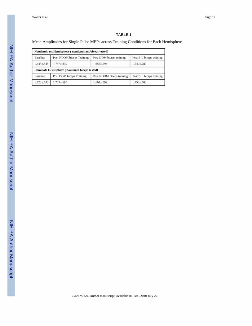
Post Training ComparisonsTable 1 shows the mean single pulse (or control MEP) average amplitude for eachhemisphere at baseline and across each training conditions. These MEP averages were usedto derive the inhibitory and facilitory ratios in the study. There were no significantdifferences among the mean single pulse MEP amplitudes at baseline or across trainingbouts Indicating that corticospinal excitability was stable across conditions.
For inhibitory responses there was a main effect for training group (F = 27.1, p < .0001), butnot hemisphere (F = 18.6, p < .08) and an interaction effect (F = 33.0, p <.0001). Forfacilitory responses there were main effects for training group (F = 16.4, p < .0001),hemisphere (F = 30.1, p < .0001) and an interaction effect (F = 30.0, p < .0001). Figure 4illustrates the mean percentages of control MEP amplitude (MEP ratio) and the standarddeviations for both inhibitory (a) and facilitatory (b) responses after each training session forboth dominant (DH) and nondominant hemisphere (NDH) testing. A suppression ordisinhibition in ICI responses (a) from baseline as well as a significant increase of the ICFresponses ( b) after bilateral and contralateral arm training were seen in both NDH and DH.Following ipsilateral biceps training, there was an increase in ICI (a) and a decrease in ICF(b) in both NDH and DH which was not significantly different from baseline responses butsignificantly different from responses seen after contralateral and bilateral training.Additionally, there was a significant difference between the effects seen on ICI and ICFafter ipsilateral training between the NDH and DH. Greater inhibition and suppressedfacilitation was seen in the NDH (ICI: p<. 05; ICF: p< .01) suggesting a differentialinhibition from the DH on the NDH. This differential effect was also seen after bilateraltraining for ICF only (p<.01), however both hemispheres still demonstrated a significantfacilitory training effect from baseline.
DiscussionIn this study we examined the neural ICI and ICF responses to unilateral dominant,unilateral nondominant and bilateral biceps muscle activation and training in the motordominant and nondominant hemispheres of healthy left and right hand dominant subjectsusing paired-pulsed TMS. No differences were seen in the responses based on handdominance at baseline or post training. After pooling data from all subjects, comparison atbaseline of unilateral dominant, unilateral nondominant and bilateral biceps activationshowed persistent inhibition and reduced facilitation in both the DH and NDH duringipsilateral arm activation (contralateral hemispheric activation). Furthermore, the DHactivation had a greater neural suppression effect on the NDH than vice versa. Neuralresponse with bilateral arm activation had a “disinhibitory” affect in comparison toipsilateral arm activation in both DH and NDH. Post training results showed a similarpattern of adaptive changes to those seen during the muscle activation conditions..
In terms of the original hypotheses, the lack of handedness effect was surprising givenprevious studies. Yahagi and Kasai reported differences in MEP amplitudes induced withmotor imagery in the distal muscles between right and left handed adults (8). Right- handedadults demonstrated greater neural asymmetry between dominant and nondominanthemispheres compared to left handed adults. Similarly Netz et al. (32) found that in righthanded adults the inhibition after stimulation of the dominant hemisphere was greater than
Waller et al. Page 7
J Neurol Sci. Author manuscript; available in PMC 2010 July 27.
NIH
-PA Author Manuscript
NIH
-PA Author Manuscript
NIH
-PA Author Manuscript
that seen in the nondominant hemisphere; however in left handed subjects this markedasymmetry in inhibition was not seen. Civardi et al.(33)demonstrated asymmetry betweendominant and nondominant hemispheres in right, but not in left-handed subjects. Their left-handed group however, contained only 6 subjects and all were not strongly left-handedwhich may explain the lack of similar asymmetry in this group. Recently, Ilic et al. reportedan asymmetry in motor threshold and short latency intracortical inhibition between dominantand nondominant hemispheres in right but not left-handers (34). However, the lateralityindex of the left-handers was much lower (68.7±3.9) compared to the right-handers(92.5±2.4). In our study we selected only subjects who were “strongly” left or right handedwith little to no ability to use their nondominant hand functionally in traditionally dominanthand functions. We speculate that the strong laterality of our left handed could explain thelack of differential response seen in our group of subjects. Another possibility is that ourstudy compared proximal muscles while previous studies showing differential effectsbetween left and right handed subjects investigated distal muscles. Distal muscles may bemore likely to show a difference in dominant versus nondominant function and in turnneural activation because there is a predominant contralateral neural innervation found indistal musculature compared to bilateral pathways that project to more proximal musculature(35).
Our results for the baseline muscle activation are consistent with previous studies showingfacilitation and disinhibition in the active cortex (2,36). An extension to these earlier studiesis the inclusion of a bilateral activation of the biceps indicating a disinhibitory effect ofbilateral activation compared to ipsilateral activation in which persistent inhibition wasexhibited. To our knowledge, this is the first demonstration of the neural intracorticalinhibitory and facilitory effects during bilateral task activation although Marchand-Pauvertet al., report asymmetrical MEP suppression on the dominant side with peripheralstimulation not found during a bilateral contraction (37). The persistent inhibition andsuppression of facilitation in each hemisphere during ipsilateral biceps activation isconsistent with findings of Liepert et al., in which non-active muscles in the same handunderwent inhibition. In our results non-active homologous muscles in the opposite limbshowed MEP inhibition as well as suppression of facilitation (2). These findings contradictthose of Muellbacher et al., who found that contraction of a distal hand muscle facilitated theMEP response (decreased cortical inhibition) in the contralateral homologous muscle(6) Intheir study, however, the contraction intensity was > 50 % of maximal voluntary contractionand in contrast to Liepert et al , they demonstrated changes in F-wave responses indicatingchanges in spinal level activity(2). In fact, Muellbacher et al. report that at that intensity ofcontraction, the homologous muscle did not remain at rest, which could also have influencedthe facilitation response(6). In our study, as in Liepert et al., the force of contraction isminimal and may not invoke such spinal excitatory paths(2).
Similarly, Lewis and Perrault (38) report increased excitability to passive paretic biceps withactivation of the ipsilateral nonparetic biceps when compared to rest stimulation intensitiesof 100 and 120% of resting threshold. In this single pulse TMS study, motor evokedpotentials were measured for the resting biceps at 80% , 100% and 120% of the restingthreshold. Given that they were able to elicit MEPs at 80% of resting threshold, the stimulusintensity used in their study may have been much larger than ours. It is possible that at acertain force level of contraction or stimulus intensity, excitatory pathways at the level of thespinal cord are recruited resulting in facilitation.
Our results are more consistent with activation of intrinsic inhibitory pathways related tointerhemispheric GABAergic connections previously reported in a number of animal andhuman studies(39–42) Based on the work of Kujirai et al.(31), Rothwell et al.(43), andLiepert et al.(2) who have reported ICF and ICI changes to similar TMS protocols with no
Waller et al. Page 8
J Neurol Sci. Author manuscript; available in PMC 2010 July 27.
NIH
-PA Author Manuscript
NIH
-PA Author Manuscript
NIH
-PA Author Manuscript
change in F waves, we suggest that our results are likely representing intracortical neuralactivity but we cannot absolutely rule out influence of spinal level changes.
Post training results after contralateral and bilateral arm training showed practice dependentplasticity as previously demonstrated by Liepert et al.(3), in a short term contralateraltraining paradigm using the distal hand muscles. In this study, muscles that were usedrepetitively also demonstrated decreased inhibition and increased facilitation. In addition toour use of proximal muscles, the novel finding is that bilateral training had a disinhibitoryeffect on the ipsilateral cortex particular in comparison to unilateral arm training with theipsilateral arm. The inhibitory effects seen in both DH and NDH with training of theipsilateral arm are not present when both arms are moving indicating differing mechanismsof neural control between unilateral and bilateral movements.
Our results of a differential inhibition seen with DH activation influencing the NDH in bothour baseline and post training results are consistent with previous findings of Ziemann andHallet (44). Using single pulse TMS they found that the left motor cortex exerts moreeffective inhibitory control over the right motor cortex than vice versa in healthy right-handed subjects. They suggested that this hemispheric asymmetry is one property of motordominance of the left motor cortex. Similarly, Ilic et al., found that the motor dominantcortex was controlled by less inhibitory tone than was the motor nondominant cortex inright-handed subjects only (34). We found the differential inhibition to be present for bothleft and right-handed adults suggesting that in strongly dominant subjects similarmechanisms of neural control are present. In fact the strength of dominance may actually bea contributing factor for the significant differences in the level of interhemispheric inhibitoryinfluence. Two studies support this assertion. Using function magnetic resonance imaging,Dassonville et al., demonstrated a linear relationship between the degree of handedness andthe amount of contralateral hemispheric activation in both left and right-handed healthysubjects (45). Triggs et al., found a lower MEP threshold for one hand in both right and left-handed subjects to be strongly associated with greater ability with that hand (46). Thesestudies suggest that asymmetries between dominant and nondominant hemispheres may be afunction of the degree of hand dominance, which would support our rationale for lack ofdominance effect in our study. In addition, our study showed that both ICI and ICFprocesses appear to be affected by this motor dominance asymmetry. Since ICI and ICFprocesses have been shown to function independently (2) this result is not predictable apriori.
Our results have potential implications for rehabilitation particularly in patients withunilateral brain lesions if similar neural mechanisms of motor control are retained afterlesion. For example, training of the paretic limb alone, as proposed in the Constraint InducedTraining methods (1,47–50,51) , would decrease the ICI and increase the ICF in the lesionedhemisphere both of which might be beneficial for use dependent plasticity. Compensatorytraining of the nonparetic arm may be detrimental to lesioned hemisphere plasticity sincetraining of the limb ipsilateral to the lesion would increase ICI and decrease ICF to thishemisphere. This could potentially explain the phenomenon of learned nonuse from a neuralperspective and is consistent with the findings of Murase(21) and Floel (22) On the otherhand, bilateral training has the advantage of involving both limbs in the therapeuticapproach with a disinhibition of ICI and an increase in ICF in both hemispheres.Theoretically, neural plasticity might be elicited in both hemispheres. In addition, thedifferential inhibitory effect of DH activation on the NDH may play a role in responsivenessto a particular treatment approach. In our previous work we found a response advantage tobilateral training in those patients with lesions of the motor dominant hemisphere{McCombe Waller, 2005 #164) (10). This group may have responded to bilateral training toa greater extent due to the loss of inhibition from the dominant hemisphere. These
Waller et al. Page 9
J Neurol Sci. Author manuscript; available in PMC 2010 July 27.
NIH
-PA Author Manuscript
NIH
-PA Author Manuscript
NIH
-PA Author Manuscript
applications to treatment are at this point hypothetical, as one cannot assume the sameresponses would be present in a subject with a brain lesion. Specific investigation of ICI andICF with unilateral and bilateral training in this population is warranted.
ConclusionsIn individuals with strong hand dominance our data suggest that there are no differences inthe neural mechanisms between those who are right and left handed. Bilateral activation andtraining has a disinhibitory and facilitory affect in both dominant and nondominanthemispheres to the same extent as contralateral limb activation and after short- term training.Ipsilateral arm activation and training however, result in persistent intracortical inhibitionlikely through interhemispheric inhibition with the dominant cortex having a greaterinhibitory affect on the nondominant cortex than vice versa. Our findings may have futurerelevance in the development of rehabilitation strategies for patients with upper extremityparesis. Further research is needed to compare the benefit of unilateral versus bilateral armtraining in individuals with unilateral hemispheric lesion to determine if subjects withparticular lesions (motor dominant versus motor nondominant) may benefit from oneapproach versus the other.
AcknowledgmentsThis study was funded by a Claude D. Pepper Center Pilot Grant awarded to the first author (National Institute onAging, Claude D. Pepper Older Americans Independence Center; #P60AG 12583). Support was also provided by aVeterans Affairs Rehabilitation Research & Development Career Development Award (#B3390K) to the secondauthor. We would like to thank our participants for their time and wish to recognize Doug Savin, Jennifer Sulin-Stair, and Wei Lui for their assistance in data collection.
References1. Wolf S, Lecraw D, Barton L, Jann B. Forced Use of Hemiplegic Extremities to Reverse the Effect
of Learned Nonuse among Chronic Stroke and Head Injured Patients. Experimental Neurology1989;104:125–132. [PubMed: 2707361]
2. Liepert J, Classen J, Cohen L, Hallett M. Task-dependent changes of intracortical inhibition.Experimental Brain Research 1998;118:421–426.
3. Liepert J, Miltner WHR, Bauder H, Sommer M, Dettmers C, Taub E, et al. Motor cortex plasticityduring constraint-induced movement therapy in stroke patients. Neuroscience Letters 1998;250:5–8.[PubMed: 9696052]
4. Whitall J, McCombe Waller S, Silver KH, Macko RF. Repetitive bilateral arm training withrhythmic auditory cueing improves motor function in chronic hemiparetic stroke. Stroke2000;31(10):2390–2395. [PubMed: 11022069]
5. Luft AR, McCombe-Waller S, Whitall J, Forrester LW, Macko R, Sorkin JD, et al. Repetitivebilateral arm training and motor cortex activation in chronic stroke: a randomized controlled trial.Jama 2004;292(15):1853–1861. [PubMed: 15494583]
6. Muellbacher W, Facchini S, Boroojerdi B, Hallett M. Changes in motor cortex excitability duringipsilateral hand muscle activation in humans. Clinical Neurophysiology 2000;111:344–349.[PubMed: 10680571]
7. Tinazzi M, Zanette G. Modulation of ipsilateral motor cortex in man during unimanual fingermovements of differenct complexities. Neuroscience Letters 1998;244:121–124. [PubMed:9593504]
8. Yahagi S, Tatsuya K. Motor evoked potentials induced by motor imagery reveal a functionalasymmetry of cortical motor control in left-and right-handed human subjects. Neuroscience Letters1999;276:185–188. [PubMed: 10612636]
9. McCombe Waller S, Whitall J. Fine motor control in adults with and without chronic hemiparesis:baseline comparison to nondisabled adults and effects of bilateral arm training. Arch Phys MedRehabil 2004;85(7):1076–1083. [PubMed: 15241753]
Waller et al. Page 10
J Neurol Sci. Author manuscript; available in PMC 2010 July 27.
NIH
-PA Author Manuscript
NIH
-PA Author Manuscript
NIH
-PA Author Manuscript
10. McCombe Waller S, Whitall J. Hand dominance and side of stroke affect rehabilitation in chronicstroke. Clin Rehabil 2005;19(5):544–551. [PubMed: 16119411]
11. Cauraugh JH, Kim S. Two coupled motor recovery protocols are better than one: electromyogram-triggered neuromuscular stimulation and bilateral movements. Stroke 2002;33(6):1589–1594.[PubMed: 12052996]
12. Cauraugh JH, Kim SB. Stroke motor recovery: active neuromuscular stimulation and repetitivepractice schedules. J Neurol Neurosurg Psychiatry 2003;74(11):1562–1566. [PubMed: 14617717]
13. Cauraugh JH. Coupled rehabilitation protocols and neural plasticity: upper extremityimprovements in chronic hemiparesis. Restor Neurol Neurosci 2004;22(3–5):337–347. [PubMed:15502274]
14. Cauraugh JH, Kim SB, Duley A. Coupled bilateral movements and active neuromuscularstimulation: intralimb transfer evidence during bimanual aiming. Neurosci Lett 2005;382(1–2):39–44. [PubMed: 15911118]
15. Hesse S, Werner C, Pohl M, Rueckriem S, Mehrholz J, Lingnau ML. Computerized arm trainingimproves the motor control of the severely affected arm after stroke: a single-blinded randomizedtrial in two centers. Stroke 2005;36(9):1960–1966. [PubMed: 16109908]
16. Mudie MH, Matyas TA. Can simultaneous bilateral movement involve the undamaged hemispherein reconstruction of neural networks damaged by stroke? Disabil Rehabil 2000;22(1–2):23–37.[PubMed: 10661755]
17. Summers JJ, Kagerer FA, Garry MI, Hiraga CY, Loftus A, Cauraugh JH. Bilateral and unilateralmovement training on upper limb function in chronic stroke patients: A TMS study. J Neurol Sci2007;252(1):76–82. [PubMed: 17134723]
18. Lewis GN, Byblow WD. Neurophysiological and behavioural adaptations to a bilateral trainingintervention in individuals following stroke. Clin Rehabil 2004;18(1):48–59. [PubMed: 14763719]
19. Tijs E, Matyas TA. Bilateral training does not facilitate performance of copying tasks in poststrokehemiplegia. Neurorehabil Neural Repair 2006;20(4):473–483. [PubMed: 17082503]
20. Werhahn KJ, Mortensen J, Kaelin-Lang A, Boroojerdi B, Cohen LG. Cortical excitability changesinduced by deafferentation of the contralateral hemisphere. Brain 2002;125(Pt 6):1402–1413.[PubMed: 12023328]
21. Floel A, Nagorsen U, Werhahn KJ, Ravindran S, Birbaumer N, Knecht S, et al. Influence ofsomatosensory input on motor function in patients with chronic stroke. Ann Neurol 2004;56(2):206–212. [PubMed: 15293272]
22. Murase N, Duque J, Mazzocchio R, Cohen LG. Influence of interhemispheric interactions onmotor function in chronic stroke. Ann Neurol 2004;55(3):400–409. [PubMed: 14991818]
23. Floel A, Cohen LG. Strategies in motor stroke rehabilitation. Stroke 2005;36(3):530. author reply530. [PubMed: 15738578]
24. Ward N, Brown M, Thompson AJ, Frackowiak R. The influence of time after stroke on brainactivations during a motor task. Ann. Neurol 2004;55:829–834. [PubMed: 15174016]
25. Oldfield RC. The assessment and analysis of handedness: the Edinburgh inventory.Neuropsychologia 1971;9(1):97–113. [PubMed: 5146491]
26. Chen R, Tam A, Butefisch C, Corwell B, Ziemann U, Rothwell JC, et al. Intracortical inhibitionand facilitation in different representations of the human motor cortex. J Neurophysiol 1998;80(6):2870–2881. [PubMed: 9862891]
27. Maeda F, Gangitano M, Thall M, Pascual-Leone A. Inter-and intra-individual variability of paired-pulse curves with transcranial magnetic stimulation (TMS). Clin Neurophysiol 2002;113(3):376–382. [PubMed: 11897538]
28. Nordstrom MA, Butler SL. Reduced intracortical inhibition and facilitation of corticospinalneurons in musicians. Exp Brain Res 2002;144(3):336–342. [PubMed: 12021815]
29. Roshan L, Paradiso GO, Chen R. Two phases of short-interval intracortical inhibition. Exp BrainRes 2003;151(3):330–337. [PubMed: 12802553]
30. Rossini PM, Barker AT, Berardelli A, Caramia MD, Caruso G, Cracco RQ, et al. Non-invasiveelectrical and magnetic stimulation of the brain, spinal cord and roots: basic principles andprocedures for routine clinical application. Report of an IFCN committee. Electroencephalogr ClinNeurophysiol 1994;91(2):79–92. [PubMed: 7519144]
Waller et al. Page 11
J Neurol Sci. Author manuscript; available in PMC 2010 July 27.
NIH
-PA Author Manuscript
NIH
-PA Author Manuscript
NIH
-PA Author Manuscript
31. Kujirai T, Caramia MD, Rothwell JC, Day BL, Thompson PD, Ferbert A, et al. Corticocorticalinhibition in human motor cortex. J Physiol 1993;471:501–519. [PubMed: 8120818]
32. Netz J, Lammers T, Homberg V. Reorganization and motor output in the non-affected hemisphereafter stroke. Brain 1997;120:1579–1586. [PubMed: 9313641]
33. Civardi C, Cavalli A, Naldi P, Varrasi C, Cantello R. Hemispheric asymmetries of cortico-corticalconnections in human hand motor areas. Clinical Neurophysiology 2000;111:624–629. [PubMed:10727913]
34. Ilic TV, Jung P, Ziemann U. Subtle hemispheric asymmetry of motor cortical inhibitory tone. ClinNeurophysiol 2004;115(2):330–340. [PubMed: 14744574]
35. Kuypers, H. Handbook of Physiology, Section I: Nervous System. In: Brooks, V., editor.Handbook of Physiology. Bethesda: American Physiological Society; 1981. p. 597-666.
36. Matsunaga K, Uozumi T, Tsuji S, Murai Y. Age-dependent changes in physiological thresholdasymmetries for the motor evoked potential and silent period following transcranial magneticstimulation. Electroencephalogr Clin Neurophysiol 1998;109(6):502–507. [PubMed: 10030682]
37. Marchand-Pauvert V, Mazevet D, Pierrot-Deseilligny E, Pol S, Pradat-Diehl P. Handedness-relatedasymmetry in transmission in a system of human cervical premotoneurones. Exp Brain Res1999;125(3):323–334. [PubMed: 10229023]
38. Lewis GN, Perreault EJ. Side of lesion influences bilateral activation in chronic, post-strokehemiparesis. Clin Neurophysiol 2007;118(9):2050–2062. [PubMed: 17644036]
39. Jacobs KM, Donoghue JP. Reshaping the cortical motor map by unmasking latent intracorticalconnections. Science 1991;251(4996):944–947. [PubMed: 2000496]
40. Keller A. Intrinsic synaptic organization of the motor cortex. Cereb Cortex 1993;3(5):430–441.[PubMed: 8260811]
41. Matsumura M, Sawaguchi T, Kubota K. GABAergic inhibition of neuronal activity in the primatemotor and premotor cortex during voluntary movement. J Neurophysiol 1992;68(3):692–702.[PubMed: 1432042]
42. Ziemann U, Lonnecker S, Paulus W. Inhibition of human motor cortex by ethanol. A transcranialmagnetic stimulation study. Brain 1995;118(Pt 6):1437–1446. [PubMed: 8595475]
43. Rothwell JC, Thompson PD, Day BL, Boyd S, Marsden CD. Stimulation of the human motorcortex through the scalp. Exp Physiol 1991;76(2):159–200. [PubMed: 2059424]
44. Ziemann U, Hallett M. Hemispheric asymmetry of ipsilateral motor cortex activation duringunimanual motor tasks: further evidence for motor dominance. Clinical Neurophysiology2001;112:107–113. [PubMed: 11137667]
45. Dassonville P, Lewis SM, Zhu XH, Ugurbil K, Kim SG, Ashe J. Effects of movementpredictability on cortical motor activation. Neurosci Res 1998;32(1):65–74. [PubMed: 9831253]
46. Triggs WJ, Calvanio R, Levine M. Transcranial magnetic stimulation reveals a hemisphericasymmetry correlate of intermanual differences in motor performance. Neuropsychologia1997;35(10):1355–1363. [PubMed: 9347481]
47. Taub E, Wolf SW. Constraint Induced Movement Techniques to Facilitate Upper Extremity Use inStroke Patients. Top Stroke Rehabilitation 1997;3:38–61.
48. Taub E, Crago JE, Uswatte G. Constraint-induced movement therapy: a new approach to treatmentin physical rehabilitation. Rehabil Psychol 1998;43:152–170.
49. Taub E, Uswatte G, Pidikiti R. Constraint-Induced Movement Therapy: a new family of techniqueswith broad application to physical rehabilitation--a clinical review. J Rehabil Res Dev 1999;36(3):237–251. [PubMed: 10659807]
50. Taub E, Uswatte G, King DK, Morris D, Crago JE, Chatterjee A. A placebo-controlled trial ofconstraint-induced movement therapy for upper extremity after stroke. Stroke 2006;37(4):1045–1049. [PubMed: 16514097]
51. Wolf SL, Winstein CJ, Miller JP, Taub E, Uswatte G, Morris D, et al. Effect of constraint-inducedmovement therapy on upper extremity function 3 to 9 months after stroke: the EXCITErandomized clinical trial. Jama 2006;296(17):2095–2104. [PubMed: 17077374]
Waller et al. Page 12
J Neurol Sci. Author manuscript; available in PMC 2010 July 27.
NIH
-PA Author Manuscript
NIH
-PA Author Manuscript
NIH
-PA Author Manuscript
Fig. 1.Schematic of TMS testing under unilateral dominant, unilateral non-dominant and bilateralmuscle activation conditions. Testing of each hemisphere was counterbalanced but the orderof SP, ICI and ICF stimulations were completely crossed and randomized with the threebiceps activation conditions.
Waller et al. Page 13
J Neurol Sci. Author manuscript; available in PMC 2010 July 27.
NIH
-PA Author Manuscript
NIH
-PA Author Manuscript
NIH
-PA Author Manuscript
Fig. 2.Schematic of TMS testing before and after short-term unilateral dominant, unilateral non-dominant and bilateral training. Testing of each hemisphere was counterbalanced and theorder of training conditions randomized. The order of SP, ICI and ICF was randomizedacross subjects for the pre-test and then maintained in the same order for a given subjectacross the remaining testing conditions.
Waller et al. Page 14
J Neurol Sci. Author manuscript; available in PMC 2010 July 27.
NIH
-PA Author Manuscript
NIH
-PA Author Manuscript
NIH
-PA Author Manuscript
Fig. 3.Inhibitory (a) and Facilitory(b) responses for dominant and nondominant hemispheresduring each muscle activation condition ( mean % of single pulse MEP, and SD). Control/single pulse MEP indicated by dashed line. CONTRA for dominant hemisphere = dominantbiceps activation, for nondominant hemisphere = nondominant biceps activation, IPSI fordominant hemisphere = non-dominant biceps activation; for non-dominant hemisphere =dominant biceps activation
Waller et al. Page 15
J Neurol Sci. Author manuscript; available in PMC 2010 July 27.
NIH
-PA Author Manuscript
NIH
-PA Author Manuscript
NIH
-PA Author Manuscript
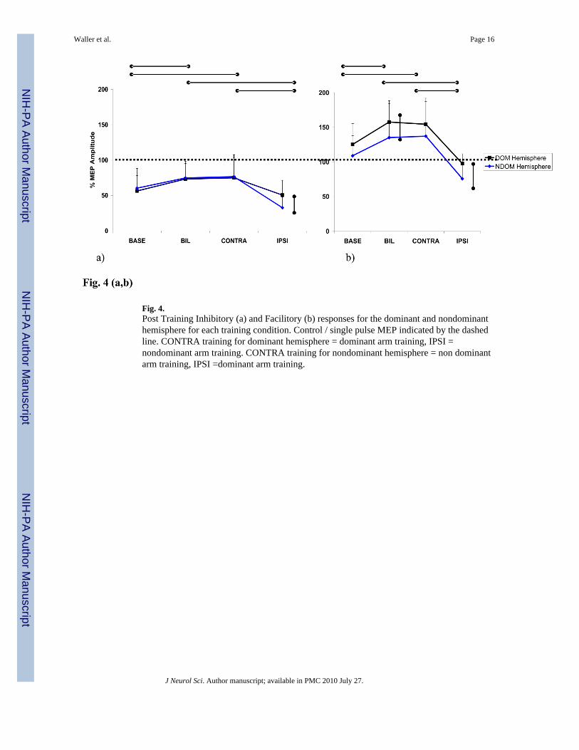
Fig. 4.Post Training Inhibitory (a) and Facilitory (b) responses for the dominant and nondominanthemisphere for each training condition. Control / single pulse MEP indicated by the dashedline. CONTRA training for dominant hemisphere = dominant arm training, IPSI =nondominant arm training. CONTRA training for nondominant hemisphere = non dominantarm training, IPSI =dominant arm training.
Waller et al. Page 16
J Neurol Sci. Author manuscript; available in PMC 2010 July 27.
NIH
-PA Author Manuscript
NIH
-PA Author Manuscript
NIH
-PA Author Manuscript
NIH
-PA Author Manuscript
NIH
-PA Author Manuscript
NIH
-PA Author Manuscript
Waller et al. Page 17
TABLE 1
Mean Amplitudes for Single Pulse MEPs across Training Conditions for Each Hemisphere
Nondominant Hemisphere ( nondominant biceps tested)
Baseline Post NDOM biceps Training Post DOM biceps training Post BIL biceps training
1.645±.845 1.747±.830 1.656±.594 1.740±.789
Dominate Hemisphere ( dominant biceps tested)
Baseline Post DOM biceps Training Post NDOM biceps training Post BIL biceps training
1.723±.743 1.783±.695 1.604±.592 1.758±.703
J Neurol Sci. Author manuscript; available in PMC 2010 July 27.
Related Documents


![core.ac.ukA brief overview of intracortical circuits Fran˘cois Grimbert To cite this version: Fran˘cois Grimbert. A brief overview of intracortical circuits. [Research Report] RR-6325,](https://static.cupdf.com/doc/110x72/5ec88e8da7755f043134db10/coreacuk-a-brief-overview-of-intracortical-circuits-francois-grimbert-to-cite.jpg)