662-668 Nucleic Acids Research, 1994, Vol. 22, No. 4 Intracellular oligonucleotide hybridization detected by fluorescence resonance energy transfer (FRET) S.Sixou, F.C.Szoka Jr, G.A.Green, B.Giusti1, G.Zon' and D.J.Chin2,* School of Pharmacy, University of California, San Francisco, CA 94143-0446, 1Lynx Therapeutics Inc., 465 Lincoln Centre Drive, Foster City, CA 94404 and 2The Agouron Institute, 505 Coast Blvd, La Jolla, CA 92037, USA Received August 25, 1993; Revised and Accepted December 31, 1993 ABSTRACT Fluorescence resonance energy transfer (FRET) was used to study hybrid formation and dissociation after microinjection of oligonucleotides (ODNs) into living cells. A 28-mer phosphodiester ODN (+ PD) was syn- thesized and labeled with a 3' rhodamine (+ PD-R). The complementary, antisense 5'-fluorescein labeled phos- phorothioate ODN (- PT-F) was specifically quenched by addition of the + PD-R. In solution, the - PT-F/ + PD-R hybrid had a denaturation temperature of 65 30C detected by both absorbance and FRET. Hybridization between the ODNs occurred within 1 minute at 17 /M and was not appreciably affected by the presence of non-specific DNA. The pre-formed hybrid slowly disso- ciated (T1,2 - 3 h) in the presence of a 300-fold excess of the unlabeled complementary ODN and could be de- graded by DNAse 1. Upon microinjection into the cyto- plasm of cells, pre-formed fluorescent hybrids disso- ciated with a half-time of 15 minutes, which is attributed to the degradation of the phosphodiester. Formation of the hybrid from sequentially injected ODNs was detected by FRET transiently in the cytoplasm and later in the cell nucleus, where nearly all injected ODNs accumulate. This suggests that antisense ODNs can hybridize to an intracellular target, of exogenous origin in these studies, in both the cytoplasm and the nucleus. INTRODUCTION Synthetic antisense ODNs have been proposed as therapeutic agents to treat viral infections or metastatic diseases (1, 2). This form of therapy is predicated on the formation of a hybrid between the antisense molecule and the message or gene sequence. In numerous studies, hybridization between the target and antisense molecule in cells has been inferred from either the loss of a specific function or from specific protein inhibition (3). In a lesser number of studies a decrease in message levels has been documented (4, 5). However only in oocytes has a message fragment, consistent with an RNAse H mediated degradation of an antisense-message duplex, been identified (6). Not surprisingly, there have been no reports on the detection of a hybrid between conjugate pairs in living cells. There has, however, been a number of groups that have used fluorescence resonance energy transfer (FRET) to detect the formation of hybrids between ODNs in solution (7-9). The formats for these studies have involved short complementary sequences where the labels were positioned on the 5' end (7, 9) as well as complementary sequences where one strand contained a donor fluorophore on the 5' end and the complementary strand was labeled with an appropriate acceptor on the 3' position (7, 8). These groups have established that fluorescence non-radiative energy transfer can detect ODN hybridization in solution and pointed the way for using this technique in living cells. In this report, we describe for the first time, the use of FRET in living cells to directly study the formation and dissolution of hybrids and their localization by quantitative confocal microscopy. EXPERIMENTAL PROCEDURES Oligonucleotides Three 28-mer Rev ODNs were used in these studies. A sense strand phosphodiester ODN spanning the translation initiation region (5' AGCAGCGAACAGAGGCGAAGAAGGACGG 3') and an antisense phosphorothioate anti-Rev ODN (5' TCGTCGC- TGTCTCCGCTTCTTCCTGCCA 3') labeled at the 5' end with fluorescein (-PT-F), were supplied by Lynx therapeutics Inc. (Foster City, CA). As a control, a 16-mer ODN, complementary to the murine ,B actin messenger RNA and rhodamine labeled at the 3' end, was used (Actin-Rho, 5' GGCGGCCCACG- ATGGA 3', Lynx therapeutics Inc.). The synthesis and purification of these ODNs were previously described (10). Briefly, an aminohexylphosphate linker spacer at the 5' or 3' end was used to attach the fluorescent derivative. The sense strand phosphodiester was also prepared with a 3'-labeled rhodamine (+PD-R). This ODN was synthesized with an amino group attached to the 3' end via a 6-carbon spacer (Synthecell, MD), purified, and labeled with rhodaminetetramethylisothiocyanate in 100 mM Na2CO3, pH 9 at room temperature overnight. The reaction was quenched with 3.4 mM NH4C1, the samples purified by exclusion column chromatography and electrophoresis in 7.0 M urea to separate the labeled from unlabeled ODN after visualization by UV shadowing. After precipitation and washing *To whom correspondence should be addressed . 1994 Oxford University Press

Welcome message from author
This document is posted to help you gain knowledge. Please leave a comment to let me know what you think about it! Share it to your friends and learn new things together.
Transcript
662-668 Nucleic Acids Research, 1994, Vol. 22, No. 4
Intracellular oligonucleotide hybridization detected byfluorescence resonance energy transfer (FRET)
S.Sixou, F.C.Szoka Jr, G.A.Green, B.Giusti1, G.Zon' and D.J.Chin2,*School of Pharmacy, University of California, San Francisco, CA 94143-0446, 1Lynx TherapeuticsInc., 465 Lincoln Centre Drive, Foster City, CA 94404 and 2The Agouron Institute, 505 Coast Blvd,La Jolla, CA 92037, USA
Received August 25, 1993; Revised and Accepted December 31, 1993
ABSTRACTFluorescence resonance energy transfer (FRET) wasused to study hybrid formation and dissociation aftermicroinjection of oligonucleotides (ODNs) into livingcells. A 28-mer phosphodiester ODN (+ PD) was syn-thesized and labeled with a 3' rhodamine (+ PD-R). Thecomplementary, antisense 5'-fluorescein labeled phos-phorothioate ODN (- PT-F) was specifically quenchedby addition of the + PD-R. In solution, the - PT-F/ + PD-Rhybrid had a denaturation temperature of 65 30Cdetected by both absorbance and FRET. Hybridizationbetween the ODNs occurred within 1 minute at 17 /Mand was not appreciably affected by the presence ofnon-specific DNA. The pre-formed hybrid slowly disso-ciated (T1,2 - 3 h) in the presence of a 300-fold excessof the unlabeled complementary ODN and could be de-graded by DNAse 1. Upon microinjection into the cyto-plasm of cells, pre-formed fluorescent hybrids disso-ciated with a half-time of 15 minutes, which is attributedto the degradation of the phosphodiester. Formation ofthe hybrid from sequentially injected ODNs was detectedby FRET transiently in the cytoplasm and later in the cellnucleus, where nearly all injected ODNs accumulate.This suggests that antisense ODNs can hybridize to anintracellular target, of exogenous origin in these studies,in both the cytoplasm and the nucleus.
INTRODUCTIONSynthetic antisense ODNs have been proposed as therapeuticagents to treat viral infections or metastatic diseases (1, 2). Thisform of therapy is predicated on the formation of a hybridbetween the antisense molecule and the message or genesequence. In numerous studies, hybridization between the targetand antisense molecule in cells has been inferred from either theloss of a specific function or from specific protein inhibition (3).In a lesser number of studies a decrease in message levels hasbeen documented (4, 5). However only in oocytes has a messagefragment, consistent with an RNAse H mediated degradation ofan antisense-message duplex, been identified (6). Notsurprisingly, there have been no reports on the detection of ahybrid between conjugate pairs in living cells.
There has, however, been a number of groups that have usedfluorescence resonance energy transfer (FRET) to detect theformation of hybrids between ODNs in solution (7-9). Theformats for these studies have involved short complementarysequences where the labels were positioned on the 5' end (7,9) as well as complementary sequences where one strandcontained a donor fluorophore on the 5' end and thecomplementary strand was labeled with an appropriate acceptoron the 3' position (7, 8). These groups have established thatfluorescence non-radiative energy transfer can detect ODNhybridization in solution and pointed the way for using thistechnique in living cells. In this report, we describe for the firsttime, the use of FRET in living cells to directly study theformation and dissolution of hybrids and their localization byquantitative confocal microscopy.
EXPERIMENTAL PROCEDURESOligonucleotidesThree 28-mer Rev ODNs were used in these studies. A sensestrand phosphodiester ODN spanning the translation initiationregion (5' AGCAGCGAACAGAGGCGAAGAAGGACGG 3')and an antisense phosphorothioate anti-Rev ODN (5' TCGTCGC-TGTCTCCGCTTCTTCCTGCCA 3') labeled at the 5' end withfluorescein (-PT-F), were supplied by Lynx therapeutics Inc.(Foster City, CA). As a control, a 16-mer ODN, complementaryto the murine ,B actin messenger RNA and rhodamine labeledat the 3' end, was used (Actin-Rho, 5' GGCGGCCCACG-ATGGA 3', Lynx therapeutics Inc.). The synthesis andpurification of these ODNs were previously described (10).Briefly, an aminohexylphosphate linker spacer at the 5' or 3'end was used to attach the fluorescent derivative. The sense strandphosphodiester was also prepared with a 3'-labeled rhodamine(+PD-R). This ODN was synthesized with an amino groupattached to the 3' end via a 6-carbon spacer (Synthecell, MD),purified, and labeled with rhodaminetetramethylisothiocyanatein 100 mM Na2CO3, pH 9 at room temperature overnight. Thereaction was quenched with 3.4 mM NH4C1, the samplespurified by exclusion column chromatography and electrophoresisin 7.0 M urea to separate the labeled from unlabeled ODN aftervisualization by UV shadowing. After precipitation and washing
*To whom correspondence should be addressed
. 1994 Oxford University Press
Nucleic Acids Research, 1994, Vol. 22, No. 4 663
in 70% ethanol, the ODNs were resuspended in TE buffer (10mM Tris-HCl, pH 8 and 1 mM EDTA) and quantified byabsorbance at 260 nm (Absorbance units at 260 nm or A units).
Fluorescence measurementsFluorescence measurements were made in a SPEX Fluorolog 2spectrophotometer (Spex Industries, Edison, NJ) equipped witha temperature controlled chamber. The excitation wavelengthfluorescein was 470 nm and 540 nm for rhodamine. The emissionwavelengths used for fluorescein and rhodamine were 522 and580 nm, respectively. In all experiments the backgroundfluorescence intensity of the phosphate buffer (10 mM phosphatebuffer and 100 mM NaCl, pH 7) was negligible in comparisonwith all the fluorescein or rhodamine derivative samples.
Fluoresence energy transfer characterization. ODNs were mixedin a 2 ml final volume of the phosphate buffer. The control samplewas 0.0166 A units/ml (about 0.5 ,Ag/ml, i.e. 17 /AM finalconcentration) of -PT-F (donor compound) without acceptorcompound. To this control sample were compared samplescontaining either the nonlabeled (+PD) or the labeled (+PD-R)acceptor at 1:0.25, 1:1 and 1:4 molar ratios. All the sampleswere denatured for 20 minutes at 80°C and gradually cooled toroom temperature. Emission spectra were recorded, at roomtemperature, between 500 and 600 nm with excitation at 470 nm.Fluorescein fluorescence quenching (Q) was calculated asfollows: Q = (1 -(F(donor+acceptor)/F(donor alone))) X 100 where F =fluorescence level at 522 nm. The rhodamine fluorescenceincrease value (I) was computed as I = (F'(donor+acceptor)/(F'(donoralone) + F' (acceptor alone))) X 100 where F' = fluorescence level at580 nm. All the results are expressed as percentages.
Fluorescence and absorbance melting curves. Samples containedthe labeled donor (-PT-F) and phosphate buffer only, ornonlabeled (+PD), or labeled (+PD-R) acceptors in 1: 1 molarratios. All samples were denatured as described above andallowed to equilibrate at 10°C. For each sample, the fluorescenceintensity at 522 nm was recorded (excitation at 470 nm) whilethe temperature was slowly increased (at 1 °C/minute) with gentlestirring, or the absorbance was measured at 260 nm while thetemperature was increased at 0.50C/minute.
DNase I action. A sample containing the labeled donor andacceptor in a 1:1 ratio (0.0 166 A units/ml) in 2 ml of phosphatebuffer containing 6 mM MgCl2, was heat denatured aspreviously described. When the temperature was equilibrated at370C, 5 ,1 of DNase I (10 UI4/,l, Boehringer Mannheim,Germany) were added. The excitation and emission wavelengthswere 470 nm and 522 nm respectively.
Further characterization: 37°C hybridization, displacement andssDNA additions. These experiments were performed withoutheat denaturation. Other experimental conditions were the sameas described above (1:1 molar ratios with excitation and emissionat 470 and 522 nm respectively). The 3.7 kb ssDNA (a humanliver Flavin-containing monooxygenase II cDNA fragmentsubcloned into Bluescript plasmid KS +) was kindly provided byN.Lomri from the J.Cashman laboratory at UCSF, CA. About0.5 ,tg of ssDNA in 2.5 ,u of water was added in the 2 ml sample.
Cell culture and micro'ujectionMouse (3T3) and African Green Monkey (BSC-1) cell lines wereobtained from the American Type Cell Culture collection(Bethesda, MD) and grown in either 10% newborn or fetal calfserum. The cell culture conditions and methods for microinjectionof ODNs were previously described (12). Briefly, cells wereseeded onto 25 mm diameter coverslips, grown 24-48 hrs andsubsequently microinjected with 50-100 fl of ODNs in distilledwater or 20 mM Hepes-HCI, pH 7.3, 30 mM KCI at 37°C.
Confocal laser scanning microscopy and image processingA confocal laser scanning module (Noran Instruments, WI)attached to an inverted microscope (Nikon, NY) was used tomonitor fluorescein fluorescence emission (500-540 nm) andrhodamine emission (605-665 nm, Omega, VT). A 60x PlanApo N.A. 1.4 lens (Nikon, NY) was used for both microinjectionand fluorescence. Illumination by the argon laser of the confocalmodule could be switched amongst band-pass filters for 488, 529,or 474/476 nm excitation lines. Band-pass filters and a secondarydichroic mirror (580 nm) were installed in the confocal moduleto maximize the specific detection of fluorophore emissions.To prevent photobleaching, the confocal microscope was
operated under conservative conditions (low illumination andintermediate slit positions, 15-25 microns, to maximize signalstrength). Changes in donor (-PT-F) fluorescence was not aresult of photobleaching since increasing the interval betweentimepoints (15-30 min) gave similar results.Custom software was employed to integrate control of the
confocal module's functions with a frame-grabbing card(Perceptics, TN) resident within a Macintosh Ilfx microcomputer.Images were stored on both hard and optical disk and analyzedby Image v. 1.45 (Wayne Rasband, NIH).
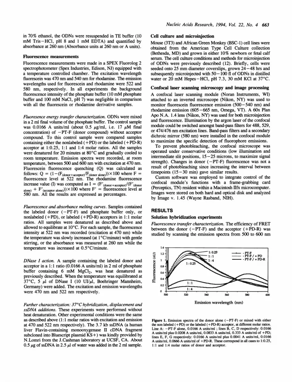
RESULTSSolution hybridization experimentsFluorescence transfer characterization. The efficiency ofFRETbetween the donor (-PT-F) and the acceptor (+PD-R) wasstudied by scanning the emission spectra from 500 to 600 nm
1.4
51.2- 1:0.~~~1025' PT-F¢
tD ~~~~~~1 :1 - PT-F /+ PD
88 t =0
0500 520 540 5so 580 600
Emission wavelength (nm)
Figure 1. Emission spectra of the donor alone (-PT-F) or mixed with eitherthe non labeled (+PD) or the labeled (+PD-R) acceptor, at different molar ratios.Line A: -PT-F alone, 0.0166 A units/ml; lines B, C, D respectively: 0.0166A units/ml plus 0.0208 A units/ml, 0.0833 A units/mi, 0.333 A units/mil of +PD;lines E, F, G respectively: 0.0166 A units/ml plus 0.0041 A units/ml, 0.0166A units/ml, 0.0666 A units/ml of +PD-R. These correspond in all cases to 1:0.25,1:1 and 1:4 molar ratios of donor and acceptor.

664 Nucleic Acids Research, 1994, Vol. 22, No. 4
(maxima for fluorescein and rhodamine were 522 and 580 nm,respectively) at various donor:acceptor molar ratios (1:0.25, 1:1and 1:4) with labeled (+PD-R) or unlabeled (+PD) acceptors,(Figure 1). With the non-labeled +PD acceptor, the fluoresceinemission decreased 8% while the rhodamine emission wasunchanged (Figure 1). This phenomenon represents a non-specificquenching of the donor fluorescence in the absence of energytransfer. Hybridization with the labeled acceptor (+PD-R),results in 85.3 4 1.8% (n=4) donor emission quenching (522nm) and up to a 70.7 19.6% (n=4) increase in the rhodamineemission (580 nm) that represents the energy transfer. The FRETresults of the 1:1 and 1:4 donor:acceptor ratios were alsoconfirmed by life-time measurements (not shown).
Displacement ofspecific hybridization. The stability of pre-formedhybrid complexes (1:1 molar ratio of either -PT-F/ +PD-R or-PT-F/+PD) was studied by the effects of a 300-fold excessof the appropriate acceptor. The +PD-R acceptor was unableto quench more than 10% of the fluorescein emission of a -PT-F/+PD hybrid after 3 hrs (not shown). Similarly, the +PDacceptor showed a 1St order decrease of a -PT-F/ +PD-Rhybrid's FRET with a T1/2 = 3 h (data not shown). Theseresults indicate that the hybrid is stable and the off-rate of thepre-formed complex is quite slow.
-Pr-F
2-
0
a)
A
Spontaneous hybridization below the Tm of the hybrid. The Tmof the +PD-R/-PT-F hybrid detected by absorbance andfluorescence was about 65°C in 10 mM phosphate buffer and100 mM NaCl, pH 7 (not shown). Since fluorescence quenchingat 20°C (85%) was maintained at 37°C (83%), we measuredthe extent of spontaneous hybridization of -PT-F and +PD-Rat 370 C. After mixing at 37°C, the fluorescein emission wasquenched by 75%. The spontaneous hybridization at 37°C occurswithin 1 minute and the extent of hybridization is the same asobserved after denaturation and reannealing during progressivecooling (Figure 2A).
Visualization ofhybrid degradation. When DNase I is added tothe solution containing the hybrid, total recovery of thefluorescein intensity was observed (75% recovery for the 1:1acceptor:donor molar ratio), (Figure 2B). This result is mostsimply explained by degradation of the hybrid by the nuclease.
Effect ofssDNA onfluorescence quenching. To approximate theconditions that an ODN might encounter within a cell, weassessed the potential interference of nonspecific nucleic acidson the hybridization reaction. The donor fluorescein-labeled ODN(-PT-F) was first mixed with ssDNA at 37°C, this resulted ina 3.5% non-specific quenching of the fluorescein emission. Thelabeled acceptor (+PD-R) was subsequently added and theexpected quenching (74%) was observed (1:1 molar ratio ofacceptor:donor, data not shown).
Microinjection experimentsSince hybridization is robust at near-physiological conditions inthe test tube, we microinjected the ODNs into living cells andexamined their distribution and hybridization behavior byconfocal microscopy.
Time (min) Temperature (QC)_s
B <._En
__
G476 R476 R529
Mean Nuclear Fluorescence
G476 R476 R529J
Peak Nuclear Fluorescence
Time (min)
Figure 2. A. Hybridization at 37°C of the donor (-PT-F) with the labeled acceptor(+PD-R ), followed by heating and slow cooling of the sample. A 1:1 molarratio of the ODNs (0.0166 A units/ml each) were mixed and the emission followedfor 50 minutes at 37°C whereupon the temperature was progressively raised andlowered. B. Degradation at 37°C of the pre-formed hybrid (-PT-F/+PD-R,1:1 molar ratio, 0.0166 A units/ml each). Two successive additions indicatedby the arrows, of DNase I were added (50 UI in a 2 ml sample).
Figure 3. Comparison of specific and non-specific energy transfer in the nucleusof mouse 3T3 cells. Cells were injected with either -PT-F/+PD-R annealedat a 2: 1 molar ratio (solid), -PT-F and Actin-Rho annealed at a 2:1 molar ratio(stippled), or -PT-F alone (white). A shows the mean nucleoplasmic intensityvalues of an 8 pixel wide line drawn across nucleus while avoiding any punctateregions. B shows the peak intensity values of a 4 pixel wide line drawn throughthe brightest punctate object within the nucleus. All values were backgroundsubtracted; the backgrounds were 13-14 for the G476 channel and 11-12 forthe R476 and R529 channels. The number of cells injected with -PT-F, -PT-F/+PD-R and -PT-F/Actin-Rho ODN were respectively 5, 12 and 10.

Nucleic Acids Research, 1994, Vol. 22, No. 4 665
R48810:08
..Al:m.-
:z4tt.M:~~~~~~~~~~~~~~~~~~~~~~~~~~~.
1:02:58.. ...... ...... ....................... .... . ............................................................................................................................... l.|...*
:: _i; -7^ rSS a .. .. a_ teS .........................* -; , ..... .: ^ .... , I.F e^bESK£w£2aF}... ...... .... , ,,, , , ........ . .. ...... . ... ........... .. llllllllw R, ,,,,,,,,,,,,,, ,,,,,,,,,,,_,,_ ............................ .. .... l____ . .. _ ___||_ . I . I I - - . ... I . . ..... . l W r___ ....... I I I I I
22:28
4..
::: .:. _. ':.. ...X..._.
;_I;:.
4:48:12
G488
Figure 4. Dissolution of a pre-formed -PT-F/+PD-R hybrid complex in BSC-l cells. A 1:2 molar ratio of the -PT-F/+PD-R ODNs was mixed, denatured andcooled prior to injection into the cytoplasm of BSC-1 cells. The left panels with indicated elapsed times correspond to the red emission channel (605-665 nm)excited by the 488 nm laser line. Elapsed times are relative to the start of injecting a line of cells. For the cell shown, the first elapsed time after injection is about1 minute. The right panels are the fluorescein (500-540 nm) emission images. Similar results are obtained with 3T3 cells (data not shown). The bar represents 10 Am.
A
3m
I
. 3 .
.a) M
._w
iw
1Z
0 50
Time (min)100
B C
10 100 1 10
Time (min) Time (min)
Figure 5. Profile plots of fluorescence values of BSC-l cells injected with pre-formed -PT-F /+PD-R hybrid complex (panel A) and serial injections (panels Band C). In panel A, the sharp spike of rhodamine emission corresponds to the filling of the nucleus, i.e. the first time point was taken before nuclear accumulationof the oligonucleotides. Mean (squares) and maximum (circles) values are represented for both fluorescein (closed symbols) and rhodamine emissions (open symbols),respectively. In panels B and C, images were taken for four different cells, after injection of the +PD-R oligonucleotide which was injected 20 min after -PT-Finjection. Panel B shows the fluorescein emission pattern and panel C shows the rhodamine signal.
Characterization ofthe confocal mnodule's spectral sensitivity. Weoptimized the use of the confocal laser scanning microscope forthe detection of fluorescence energy transfer by selecting thedichroic mirror and band-pass filter combinations to ensure
specific detection of donor quenching and energy transfer.Crossover of fluorescein emission in the red chanel (605 -665nm) with the 474/476 nm (or 488 nm) lines was 14% the intensityfound in the green channel (500-540 nm, not shown). Mostimportantly, excitation of the +PD-R ODN with the 474/476nm laser lines resulted in very little red emission (12% of thered emission from excitation at 529 nm, not shown). In some
experiments, the 488 nm line was used to monitor only
fluorescein quenching by the +PD-R ODN. The 474/476 nmlines enabled monitoring both fluorescein quenching and energytransfer to the rhodamine. As expected, intense red emission wasobserved when the -PT-F/+PD-R hybrid was illuminated witheither the 476 nm or 529 nm line (not shown).
Controls for non-hybridization mediated FRET in cells. FRETdepends upon the proximity between the donor and acceptorfluorophore. Since ODNs accumulate in distinct (12, 13) andpossibly specific sites in the nucleus (14, 15), FRET could occurbecause fluorescein and rhodamine ODNs bind to adjacent siteswithin the energy transfer distance. To examine for this
G488

666 Nucleic Acids Research, 1994, Vol. 22, No. 4
(-PT-F) injection (T= 20 min) (+PD-R) injection (T= 45 min)
DAM4J
:. s ~ ~~~~~~ ~ ~~~~~~~~~~~~~~~~~~~~~~~~~~~~~~~~~~~~~~~~~~~~--:
:..::b% r ~ L, . L _..... ~~~~~~~~~~~~_ fl . .. .. _ .. . . . .. ...
1 M C(. lnIIII
Figure 6. Top-Dissolution of a pre-formed -PT-F/+PD-R hybrid complexin mouse 3T3 cells excited by the 474/476 nm laser lines. A -PT-F/+PD-Rhybrid complex was formed at a 1: 1 molar ratio as described above and injectedinto 3T3 cells. The green fluorescein emission (G476, 500-540 nm) and redrhodamine emissions are shown. (R476): 605 -665 nm emission with Xex =
474/476, and (R529): 605 -665 nm emission with Xex = 529 line. Bottom-Profile plots through the cells shown. Background fluorescence values are shownat the bottom of the ordinate. The bar represents 10 itm.
possibility, we microinjected the -PT-F with a non-
complementary rhodamine 16-mer ODN (Actin-Rho) andmeasured the fluorescence behavior in single cells. For thespecific ODN pair, the mean nuclear green fluorescence was lessthan the -PT-F alone or the -PT-F/Actin-Rho pair (Figure 3A).The mean, nuclear red fluorescence excited by the 476 nm linewas significantly greater for the specific pair (indicative of FRET)than either the non-specific pair or the -F-PT alone (Figure 3A)even though both the specific and non-specific pairs gave similarred emission when irradiated by the 529 nm line. Both thefluorescein quenching and rhodamine fluorescence increase areconsistent with specific hybridization occuring in the nucleus.
In discrete regions, of high nuclear fluorescence (peak nuclearfluorescence), the fluorescein emission signal from the specificpair ofODNs was again less than that in the other two treatmentswhen the 476 nm line was used (Figure 3B). However there wasno difference between the emission in the red channel of thespecific and non-specific pairs. The rhodamine signal arising fromexcitation at 529 nm was the same, indicating a similar amountof material was injected in both experiments. The increase inthe rhodamine signal when excited at 476 un, may indicate thatFRET, unrelated to hybrid formation, may occur. This couldbe due to the binding to ODNs to structures that position thefluorophores within the energy transfer distance.
Figure 7. Serial injection of -PT-F and +PD-R ODNs. (A) The -PT-F ODNwas injected into 3T3 cells and incubated for 20 minutes to allow the nuclearaccumulation. In the left panels (-PT-F injection only), very little red emissionfor -PT-F was observed with Xex = 474/476. Panels on the right show thecells 25 minutes after injection of the +PD-R ODN. The -PT-F was not initiallyinjected in two neighboring cells. Note that only the double injected cell has aR476 signal. The labeling of fluorescein and rhodamine emissions with theappropriate excitation laser lines are as described in previous legends. The barrepresents 10 um.
Increased magnification (84 x - 120 X) of experiments thatinvolved serial injections of the ODNs (vide infra) revealedheterogeneous fluorescent regions within the nucleus. Punctateregions were observed in the red emission channel with Xex =474/476 nm, (data not shown). These were absent from both thered emission channel from 529 nm excitation and the greenemission channel from Xex = 474/476 nm. The nature andstability of these unique structures are the focus of further studies.
Dissolution ofpre-formed ODN hybrids. Microinjection of the-PT-F/+PD-R hybrid into the cytoplasm of living 3T3 or BSC-1cells enabled us to determine both the localization and decay rateof the hybrid. To obtain the maximal donor quenching, a 1:2molar ratio (10 A units/ml of -PT-F and 20 A units/ml of +PD-R in the injection needle) of the denatured and reannealed -PT-F/+PD-R hybrid was injected. As found previously (12), nuclearaccumulation of the rhodamine (605 -665 nm) emission rapidlyoccured (Figure 4). At the 1:2 molar ratio of -PT-F/ +PD-R,complete quenching of the fluorescein signal was observed atearly times. The intensity of fluorescein fluorescence slowlyincreased within the nucleus. A collection of experimentsperformed with the 488 nm laser line confirmed transientquenching of -PT-F in the nucleus (Figure 5).When both fluorescence quenching and exchange (at a 1:1
molar ratio with Xex = 474/476 nm) was monitored (Figure 6),the fluorescence signal started low (2 minutes) and increased withtime (10 to 53 minutes). The red signal (with Xex = 529 unor Xex = 474/476 nm lines) decreased with time from 10 to 53minutes (Figure 6). We interpret these findings to result fromthe loss of fluorescein quenching (i.e., increase in FITC emission)due to loss of energy exchange upon disruption of the hybrid.The non-uniform distribution of ODNs within the nucleusdescribed above, and (12), complicates the quantitation of energytransfer. Nonetheless, the loss of energy transfer over time canbe used to estimate the half-life of the duplex. From examination
IN-.
-,
I

Nucleic Acids Research, 1994, Vol. 22, No. 4 667
of over 100 cells in 8 experiments, the half-life of the hybrid(-PT-F/+PD-R) within injected cells was about 15 minutes.
Formation ofhybrids in living cells can be determined by serialmicroinjection. To determine whether the hybridization of the-PT-F and +PD-R ODNs could be detected within living cells,we performed serial injections, Figure 7. The -PT-F ODN wasinjected into the cytoplasm of a line of selected cells, and allowedto accumulate within the nucleus over a 20 minute period. Cellsinjected only with -PT-F showed a gradual decrease in overallfluorescence concomitant with nuclear accumulation (data notshown, 12). Subsequently, several of the same cells were injectedwith the +PD-R ODN. Neighboring cells, uninjected with the-PT-F ODN, were injected with the +PD-R ODN alone. FRETwas demonstrated in the nucleus by a transient loss of greenfluorescence emission (500-540 nm with Xex = 474/476 nm)and an increase in red fluorescence emission (605 -665 nm withXex = 474/476 nm). Note that in a neighboring cell, injectedwith the +PD-R ODN alone, only minimal rhodamine emissionwith Xex = 474/476 nm was found (Figure 7). In other serialinjection experiments, when the +PT-R ODN was injected only2-5 min after XPT-F injection, or when non-hybrid mixturesof -PT-F and +PD-R at 1:2 ratios were injected, transient FRETin the cytoplasm was revealed by quenching of the -PT-F emissionand an increase in the red emission (Xex=474/476 or 488 nm,not shown). These results suggest that hybrid complexes can formin the cytoplasm prior to nuclear accumulation.
In time course studies of serial injections, a transient reductionof the mean -PT-F ODN green emission coincided with a rapidrise in red emission from the +PD-R ODN which likely resultedfrom inefficient hybridization of the - 1:1 molar ratio (notshown). Thus, in situ hybridization is likely complicated bycellular factors including nucleases that can rapidly degrade thephosphodiester ODN, as suggested by the rapid loss of rhodamineemission. Additionally, cellular proteins may bind ssDNA andprevent hybridization while other proteins may disrupt double-stranded nucleic acid complexes (16).
DISCUSSIONWe have shown specific hybridization between complementaryODNs bearing donor and acceptor fluorophores by a number ofcriteria including gel retardation and fluorescence resonanceenergy transfer in solution. The stability of the complex wascharacterized by nuclease digestion, competition by unlabeledODN or nonspecific single-stranded DNA. Two results of thespectrofluorimetry studies stimulated us to extend these studiesto living cells. First, the energy transfer in vitro was relativelyefficient at a 1: 1 molar ratio of -PT-F/ +PD-R, a 74% quenchingof the fluorescein was correlated with a 53% increase in therhodamine emission. Second, in characterizing the annealingconditions, we found nearly complete hybridization inphysiological salts at 37°C.
Examination of hybrid behavior in living cells was exploredby microinjection of pre-formed hybrids into cells and byseparate, serial injection of the donor (-PT-F) and acceptor(+PD-R) ODNs. The former experiments allowed us to estimatethe dissolution rate of the complex. Since the fate of ODNs thatenter the cytoplasm is the nucleus (12,13), monitoring dissolutionwas potentially complicated by the redistribution of hybrids withincellular compartments. It is possible that proteins or cytoplasmicnucleases could disrupt the complex or degrade the +PD-R ODN
before nuclear accumulation. This is unlikely since (i) the bulkof the rhodamine signal accumulated into the nucleus at similarrates as free +PD-R (12), (ii) the previously quenched fluoresceinsignal reappeared in the nucleus with the same time course asthe diminution of the rhodamine signal and (iii) fluorescentmononucleotides diffuse from cells into medium within about 10minutes (12). Thus, complex dissolution may result fromhydrolysis of the rhodamine moiety, or unfavorable nuclear saltconcentrations, or binding to nuclear proteins. The limitedaqueous space in the nucleus might predispose such interactions(17). To determine if such conditions and the vast complexityof genome sequences and nuclear RNA could prevent theformation of complexes, we performed serial injectionexperiments.Both the formation and disruption of hybrid complexes were
revealed by serial injection experiments. The transient quenchingof the donor fluorophore was temporally matched by the rise andfall of the nuclear acceptor rhodamine emission. Strikingly, therate of hybrid dissolution was similar to the rate of disruptionof pre-formed complexes injected into cells. Together, theseresults suggest that a phosphodiester ODN can hybridize to atleast a phosphorothioate ODN, even if only for a short periodwithin cells.ODNs appear to inhibit gene expression in a sequence-specific
manner, but the problems of nonspecific effects on cells, ODNstability and inefficient delivery remain unsolved (for a review,18). The diverse effects of ODNs may be related to theirdistribution and degradation within cells. Our results indicatinga short intracellular life-time for phosphodiesters may help toexplain the diverse range of effects of previous studies employingphosphodiesters (19-24). Continuing these studies with a varietyof targets, chemical linkages and conditions should improve ourunderstanding of the parameters affecting efficient and long-livedhybrid complexes within cells. The ability to detect FRET fromhybridized ODNs in cells also opens the way to monitor thebinding of fluorescent antisense ODNs to specific RNA. In fact,we have detected FRET between a 5' fluorescein labeled anti-actin ODN and a 3' rhodamine labeled anti-actin ODN that bindto adjacent locations on actin mRNA (25). This technique maypermit the optical tracking of various RNA molecules inside livingcells and would augment immunohistochemical techniques (26)for the detection of RNA trafficking in cells.
ACKNOWLEDGEMENTSWe are grateful to helpful comments and suggestions from R.Stullin the selection of ODN sequences and to Dr A.Verkman andS.Bickness for the use of the time resolved fluorescencespectrometer. This work has been supported by grants from 'LaFondation pour la Recherche Therapeutique' (SS), NIHGM30163 (FCS) and NIH AI30880 (DJC).
REFERENCES1. Zamecnik, P.C. and Stephenson, M.L. (1978) Proc. Natl. Acad. Sci. USA,
75, 280-284.2. Helene, C. and Toulm6, J.J. (1990) Biochim. Biophys. Acta, 1049, 99-125.3. Leonetti, J.P., Clarenc, J.P. Degols, G., Mechti, N. and Lebleu, B. (1993)
Prog. Nucl. Ac. Res. Mol. Biol., 44, 143-166.4. Chiang, M.Y., Chan, H., Zounes, M.A., Freier S.M., Lima, W.F. and
Bennett C.F. (1991) J. Biol. Chem. 266, 27, 18162-18171.5. Simons, M., Edelman, E.R., DeKeyser, J.L., Langer, R. and Rosenberg,
R.D. (1992) Nature, 359, 67-70.
668 Nucleic Acids Research, 1994, Vol. 22, No. 4
6. Woolf, T.M., Melton, D.A. and Jennings, C.G.B. (1992) Proc. Natl. Acad.Sci. USA, 89, 7305-7309.
7. Cardullo, R.A., Agrawal, S., Flores, C., Zamecnik, P.C. and Wolf, D.E.(1988) Proc. Natl. Acad. Sci. USA , 85, 8790-8794.
8. Morrison, L.E., Halder, T.C. and Stols, L.M. (1989) Analytical Biochem.,183, 231-244.
9. Clegg, R.M., Murchie, A.L.H., Zechel, A. and Lilley, D.M. (1993) Proc.Natl. Acad. Sci. USA, 90, 2994-2998.
10. Zon, G. and Geiser T.G. (1991) Anti-Cancer Drug Design, 6, 539-568.11. Roe, J.N., Szoka, F.C. and Verkman, A.S. (1989) Biophys. Chem., 33,
295-302.12. Chin, D.J., Green, G.A., Zon, G., Szoka Jr, F.C. and Straubinger, R.M.
(1990) 7he New Biologist, 2, 1091-1100.13. Leonetti, J.P., Mechti, N., Degols, G., Gagnor, C. and Lebleu, B. (1991)
Proc. Nati. Acad. Sci. USA, 88, 2702-2706.14. Geselowitz, D.A. and Neckers, L.M. (1992) Antisense Res. Develop., 2,
17-25.15. Clarenc, J.P., Lebleu, B. and Leonetti, J.P., L. (1993) J. Biol. Chem., 268,
8, 5600-5604.16. Wagner, R.W. and Nishikura K. (1988) Mol. Cell Biol., 8, 2, 770-777.17. Paddy, M.R., Belmont, A.S., Saumweber, H., Agard, D.A. and Sedat, J.W.
(1990) Cell, 62, 1, 89-106.18. Cohen, J.S. (1992) Trends in Biotech., 10, 3, 87-91.19. Agrawal, S., Ikeuchi, T., Sun, D., Sarin, P.S., Konopka, A., Maizel, J.
and Zamecnik, P.C. (1989) Proc. Natl. Acad. Sci. USA, 86, 7790-7794.20. Burch, R.M. and Mahan, L.C. (1991) J. Clin. Invest., 88, 1190-1196.21. Goodchild, J., Agrawal, S., Civeira, M.P., Sarin, P.S., Sun, D. and
Zamecnik, P.C. (1988) Proc. NatI. Acad. Sci. USA, 85, 5507-5511.22. Letsinger, R.L., Zhang, G.R., Sun, D.K., Ikeuchi, T. and Sarin, P.S. (1989)
Proc. Natl. Acad. Sci. USA, 86, 6553-6556.23. Matsukura, M., Shinozuka, K., Zon, G., Mitsuya, H., Reitz, M., Cohen,
J.S. and Broder, S. (1987) Proc. Natl. Acad. Sci. USA, 84, 7706-7710.24. Smith, C.C., Aurelian, L., Reddy, M.P., Miller, P.S. and Ts'o, P.O. (1986)
Proc. Natl. Acad. Sci. USA, 83, 2787-2791.25. Sixou, S., Szoka Jr, F.C., Zon, F.C. and Chin, D.J., (unpublished)26. Wansink, D.G., Schul, W., van der Kraan, I., van Driel, R. and de Jong,
L. (1993) J. Cell Biol., 122, 2, 283-293.
Related Documents