INTRACELLULAR NAD LEVELS REGULATE TNF-α PROTEIN SYNTHESIS IN A SIRTUIN-DEPENDENT MANNER Frédéric Van Gool 1,6 , Mara Galli 1,6 , Cyril Gueydan 2 , Véronique Kruys 2 , Antonio Bedalov 3 , Raul Mostoslavsky 4,7 , Frederick W. Alt 4 , Thibaut De Smedt 5 , and Oberdan Leo 1 1 Laboratoire de Physiologie Animale, Institut de Biologie et Médecine Moléculaire IBMM, Université Libre de Bruxelles, Belgium 2 Laboratoire de Chimie Biologique, Institut de Biologie et Médecine Moléculaire IBMM, Université Libre de Bruxelles, Belgium 3 Fred Hutchinson Cancer Research Center, Clinical Research Division, Seattle, WA 98109, USA 4 The Children’s Hospital, HHMI, Harvard Medical School Boston, MA 02115 USA 5 Apoxis SA, Lausanne, Switzerland Abstract Uncontrolled TNF-α synthesis is known to play an important role in numerous inflammatory disorders, and multiple transcriptional and post-transcriptional regulatory mechanisms have therefore evolved to dampen the production of this important pro-inflammatory cytokine. By examining the anti-inflammatory properties of the vitamin B3 constituent nicotinamide, we have uncovered a novel regulatory pathway controlling TNF-α production. Exogenous nicotinamide inhibits TNF-α secretion through modulation of mRNA translation efficiency. Moreover, the capacity to produce TNF-α appears to be directly correlated with intracellular NAD levels, suggesting that a NAD-dependent biological event that can be inhibited by nicotinamide controls TNF-α synthesis in cells of the immune system. Sirtuins represent NAD-dependent deacetylases involved in regulation of gene expression in both mammals and yeasts, and are known to be inhibited by nicotinamide. We demonstrate herein that similarly to nicotinamide, structurally unrelated sirtuin inhibitors downregulate TNF-α secretion with minimal effect on TNF-α gene transcription. By over-expressing individual sirtuin members in cell lines transiently expressing TNF-α, we have identified SIRT6 as a sirtuin member able to upregulate TNF-α synthesis in vitro. In agreement with this finding, bone-marrow derived dendritic cells from SIRT6 KO mice display reduced TNF-α synthesis in response to CpG. Collectively, these data delineate a novel relationship between metabolism and the inflammatory response, by uncovering the role of SIRT6 in the control of TNF-α secretion. INTRODUCTION Nicotinamide adenine dinucleotide (NAD) is an essential coenzyme regulating numerous cellular metabolic pathways. In the last few decades, a growing number of studies have uncovered an additional, nonredox function of NAD in cellular physiology. In particular, NAD has been recognized as a substrate for a growing group of “NAD–dependent” enzymes with multiple roles in cellular regulation 1 . NAD-dependent protein modifications are catalyzed by enzymes broadly defined as ADP-ribosyl transferases. Two enzyme families, known respectively as mono-ADP-ribosyltransferases (mARTs) and poly(ADP-ribose) polymerases Corresponding author: Dr. Oberdan LEO, Laboratoire de Physiologie Animale, Université, Libre de Bruxelles – IBMM, 12, rue des Prof. Jeneer et Brachet, 6041 Gosselies, BELGIUM, tel: +32 2 650 9877; fax: +32 2 650 9860; [email protected]. 6 These authors contributed equally to this work 7 Present address: Department of Medicine, Massachusetts General Hospital Cancer Center Harvard Medical School, Boston, MA 02114 Competing financial interests: T. De Smedt is an Apoxis employee NIH Public Access Author Manuscript Nat Med. Author manuscript; available in PMC 2010 March 25. Published in final edited form as: Nat Med. 2009 February ; 15(2): 206–210. doi:10.1038/nm.1906. NIH-PA Author Manuscript NIH-PA Author Manuscript NIH-PA Author Manuscript

Welcome message from author
This document is posted to help you gain knowledge. Please leave a comment to let me know what you think about it! Share it to your friends and learn new things together.
Transcript
INTRACELLULAR NAD LEVELS REGULATE TNF-α PROTEINSYNTHESIS IN A SIRTUIN-DEPENDENT MANNER
Frédéric Van Gool1,6, Mara Galli1,6, Cyril Gueydan2, Véronique Kruys2, Antonio Bedalov3,Raul Mostoslavsky4,7, Frederick W. Alt4, Thibaut De Smedt5, and Oberdan Leo1
1 Laboratoire de Physiologie Animale, Institut de Biologie et Médecine Moléculaire IBMM, UniversitéLibre de Bruxelles, Belgium 2 Laboratoire de Chimie Biologique, Institut de Biologie et MédecineMoléculaire IBMM, Université Libre de Bruxelles, Belgium 3 Fred Hutchinson Cancer ResearchCenter, Clinical Research Division, Seattle, WA 98109, USA 4 The Children’s Hospital, HHMI,Harvard Medical School Boston, MA 02115 USA 5 Apoxis SA, Lausanne, Switzerland
AbstractUncontrolled TNF-α synthesis is known to play an important role in numerous inflammatorydisorders, and multiple transcriptional and post-transcriptional regulatory mechanisms have thereforeevolved to dampen the production of this important pro-inflammatory cytokine. By examining theanti-inflammatory properties of the vitamin B3 constituent nicotinamide, we have uncovered a novelregulatory pathway controlling TNF-α production. Exogenous nicotinamide inhibits TNF-α secretionthrough modulation of mRNA translation efficiency. Moreover, the capacity to produce TNF-αappears to be directly correlated with intracellular NAD levels, suggesting that a NAD-dependentbiological event that can be inhibited by nicotinamide controls TNF-α synthesis in cells of the immunesystem. Sirtuins represent NAD-dependent deacetylases involved in regulation of gene expressionin both mammals and yeasts, and are known to be inhibited by nicotinamide. We demonstrate hereinthat similarly to nicotinamide, structurally unrelated sirtuin inhibitors downregulate TNF-α secretionwith minimal effect on TNF-α gene transcription. By over-expressing individual sirtuin members incell lines transiently expressing TNF-α, we have identified SIRT6 as a sirtuin member able toupregulate TNF-α synthesis in vitro. In agreement with this finding, bone-marrow derived dendriticcells from SIRT6 KO mice display reduced TNF-α synthesis in response to CpG. Collectively, thesedata delineate a novel relationship between metabolism and the inflammatory response, byuncovering the role of SIRT6 in the control of TNF-α secretion.
INTRODUCTIONNicotinamide adenine dinucleotide (NAD) is an essential coenzyme regulating numerouscellular metabolic pathways. In the last few decades, a growing number of studies haveuncovered an additional, nonredox function of NAD in cellular physiology. In particular, NADhas been recognized as a substrate for a growing group of “NAD–dependent” enzymes withmultiple roles in cellular regulation 1. NAD-dependent protein modifications are catalyzed byenzymes broadly defined as ADP-ribosyl transferases. Two enzyme families, knownrespectively as mono-ADP-ribosyltransferases (mARTs) and poly(ADP-ribose) polymerases
Corresponding author: Dr. Oberdan LEO, Laboratoire de Physiologie Animale, Université, Libre de Bruxelles – IBMM, 12, rue des Prof.Jeneer et Brachet, 6041 Gosselies, BELGIUM, tel: +32 2 650 9877; fax: +32 2 650 9860; [email protected] authors contributed equally to this work7Present address: Department of Medicine, Massachusetts General Hospital Cancer Center Harvard Medical School, Boston, MA 02114Competing financial interests: T. De Smedt is an Apoxis employee
NIH Public AccessAuthor ManuscriptNat Med. Author manuscript; available in PMC 2010 March 25.
Published in final edited form as:Nat Med. 2009 February ; 15(2): 206–210. doi:10.1038/nm.1906.
NIH
-PA Author Manuscript
NIH
-PA Author Manuscript
NIH
-PA Author Manuscript
(PARPs) have in particular been recently identified in mammals. mARTs represent a familyof ectoenzymes including GPI-anchored membrane proteins and secreted forms, while PARPs(comprising 18 members to date 2) are intracellular proteins usually located within the nuclearcompartment. NAD also represents a substrate for members of a third evolutionarily conservedfamily of proteins collectively known as “sirtuins” 34. These proteins, (for which sevenmembers, SIRT1 to SIRT7 have been described) regulate a wide range of biological processesincluding gene silencing 5, aging 67, cell survival in response to stress 8,9, cell differentiation10 and metabolism 11,12. The founding member of this family, Sir2 in yeasts and its mammalianhomologue SIRT1, catalyzes a unique reaction in which the cleavage of NAD and thedeacetylation of substrate are coupled to the formation of O-acetyl-ADP-ribose 13
A common feature of all these “NAD-dependent” enzymes is the cleavage of the ADP-ribosemoiety of NAD with the concomitant release of nicotinamide (NAm), which therefore servesas an endogenous end product inhibitor 14. NAm, in conjunction with nicotinic acid, alsorepresents an essential vitamin (Vitamin B3 or niacin) required as an exogenous precursor ofNAD in most living organisms 15. Collectively, these observations have shifted interest in NAmfrom a simple nutrient (NAD precursor) to a pharmacological agent able to interfere with NAD-consuming enzymes. It is therefore presently believed that intracellular NAD and NAm levelsmay regulate numerous cellular reactions by modulating the activity of a large number of NAD-dependent enzymes 16. Recently, experiments performed in yeasts support this contention, andsuggest an important role for the NAm/NAD ratio in regulating several biological processes17.
Our own interest in the potential link between NAD biosynthesis and cell regulation derivesfrom studies performed in immune cells that led to the identification of the gene encoding forthe murine nicotinamide phosphoribosyltransferase (NAmPRT, EC 2.4.2.12) 18. This enzymecatalyzes the first step of the salvage pathway allowing NAD biosynthesis from NAm.NAmPRT levels were found upregulated upon stimulation of immune cells both in vivo andin vitro 19,20. Although elevated NAmPRT expression may simply represent a physiologicalresponse to meet the demand for increased metabolic resources in activated/proliferating cells,it may also represent a regulatory mechanism providing adequate intracellular NAD levelsrequired for the effector function of one or several NAD-consuming enzymes important forimmunoregulation.
Several lines of experimental evidence support a functional link between NAD metabolismand inflammation. NAm has been shown to inhibit the production of key inflammatorymediators such as NO 21 and cytokines 22,23. These observations have been generallyinterpreted in light of the recently uncovered role of PARP-1, in the regulation of aninflammatory response 24. Indeed (i) PARP-1 has been shown to act as a transcriptionalactivator of NF-kB 25 (ii) NAm and other structurally unrelated pharmacological PARP-1inhibitors are of remarkable therapeutic efficacy in experimental models of inflammatory-related diseases 24 and finally (iii) PARP-1 −/− mice are protected from endotoxic shock 26,27. These and other observations engendered the view that PARP-1 may represent the molecularlink between NAm, NAD biosynthesis and secretion of pro-inflammatory cytokines throughregulation of NF-kB transcriptional activity.
The purpose of the present study was to evaluate the functional link between NAD metabolismand inflammation. The observations described in this study indicate a contrasting role for NAmand NAD in regulating the secretion of TNF-α. While adequate intracellular NAD levels arerequired for optimal TNF-α production, exogenous NAm inhibits TNF-α suggesting animportant role for a NAD-dependent, NAm-inhibitable enzymatic activity in regulating theproduction of this potent pro-inflammatory mediator. However, and in contrast to a prevailingview, NAm appears to exert its anti-inflammatory properties in a PARP-1-independent fashion.
Van Gool et al. Page 2
Nat Med. Author manuscript; available in PMC 2010 March 25.
NIH
-PA Author Manuscript
NIH
-PA Author Manuscript
NIH
-PA Author Manuscript
Using a set of structurally unrelated pharmacological inhibitors and a genetic approach, weidentify herein an important role for SIRT6, a member of the sirtuin family, in the regulationof TNF-α production during an inflammatory response.
RESULTSNAm protects mice against an endotoxic shock
A well defined model of acute septic shock was used to evaluate the anti-inflammatoryproperties of NAm in vivo. Groups of mice were injected with a lethal dose of LPS (20 mg/kg) in combination with either saline or NAm (500 mg/kg) at various times before or afterendotoxin administration. As shown in Figure 1A, NAm was found to protect mice against alethal high dose of LPS even when injected 2 hours following endotoxin challenge.
D-galactosamine (D-Gal) sensitizes animals to LPS by causing severe liver damage secondaryto TNF-induced hepatocyte apoptosis. As shown in Figure 1A, NAm protected D-gal sensitizedmice against an LPS, but not against a TNF-α challenge, indicating that this vitamin did notaffect the late stages of the biological response to endotoxemia, but rather interfered with anearly event of the inflammatory response, as confirmed by the analysis of the peak responseof several cytokines released in the serum of treated animals. NAm administration led to anotable shift in the cytokine response, with marked reduced levels of TNF-α and IL-12, andincreased levels of the anti-inflammatory cytokine IL-10 (Figure 1B). Since endogenous IL-10is known to modulate the secretion of pro-inflammatory mediators during LPS challenge invivo 28, we repeated these experiments in mice genetically deficient for IL-10. As shown inFigure 1C, NAm failed to inhibit IL-12 secretion in the absence of endogenous IL-10, whileit strongly reduced LPS-induced, serum TNF-α levels in both mouse strains. To further confirmthat NAm inhibited TNF-α production in an IL-10-independent fashion, wt animals wereinjected with a combination of blocking antibodies to IL-10 and IL-10R before LPS challenge.As expected, in vivo blocking of IL-10 signalling led to enhanced TNF-α secretion that howeverretained sensitivity to the inhibitory effects of NAm (Figure 1C). Finally, based on the abilityof NAm to inhibit PARP-1 enzymatic activity (with an IC50 in the 30 μM range 29), we wishedto examine the potential role of PARP-1 in mediating the anti-inflammatory properties of thisvitamin. PARP-1 KO mice responded to LPS challenge by secreting control levels of serumTNF-α and were equally sensitive to the anti-inflammatory properties of NAm both in vivoand in vitro (Figure 1D). Collectively, these observations are best explained by assuming thatthe anti-inflammatory properties of NAm are due to the ability of this vitamin to block TNF-α secretion by activated cells in a PARP-1 independent fashion.
NAm inhibits TNF-α synthesis at the post-transcriptional levelTo better understand the mechanisms by which NAm affects TNF-α production, the welldescribed mouse RAW264.7 macrophage cell line was used as responder cell. TNF-α secretionby LPS-stimulated RAW 246.7 was inhibited by exogenous NAm with minimal effect onmRNA accumulation (Figure 2A). mRNA stability was not or only marginally inhibited byNAm, suggesting that this vitamin inhibited TNF-α protein production by interfering with apost-transcriptional step (Figure 2A). To further support this contention, RAW 246.7 cells werestimulated with LPS for 2 hours, washed, and subsequently incubated in the presence of mediaor graded doses of NAm for an additional 30 minutes. Again, NAm strongly inhibited TNF-α protein secretion, with minimal effect on TNF-α transcripts as judged by ELISA and northernblot analysis, respectively (Figure 2B). Cytoplasmic extracts from LPS or LPS/NAm treatedcells were than fractionated over sucrose gradients and TNF-α mRNA quantified by northernblot. LPS treatment led to the typical accumulation of TNF-α mRNA in the polysomecontaining, heavy fraction of the gradient (Figure 2C, fractions 10 to 13). NAm decreased theproportion of TNF-α transcripts that associate with polysomes, concomitant with a shift
Van Gool et al. Page 3
Nat Med. Author manuscript; available in PMC 2010 March 25.
NIH
-PA Author Manuscript
NIH
-PA Author Manuscript
NIH
-PA Author Manuscript
towards fractions containing smaller polysomes and monosomes (Figure 2C, fractions 1 to 4).Polysome containing GaPDH mRNA display the same profile in both control, and NAm-treated cells (Figure 2C, right panel). Finally, RAW 264.7 cells were incubated in mediumcontaining radioactive amino acids and the synthesis of TNF-α in response to LPS was followedby immunoprecipitation and polyacrylamide gel electrophoresis. As shown in Figure 4C, NAmsignificantly reduced the accumulation of TNF-α protein in cell extracts, in keeping with theconclusion that it affected the translation efficiency of TNF-α mRNA. (Figure 2D). Toeliminate any possible interference of NAm with the TLR signalling cascade regulating genetranscription and mRNA splicing, the human 293T cell line was transiently transfected withconstructs expressing the complete human TNF-α cDNA or the murine IL-2 ORF under thecontrol of a viral (CMV) promoter. As shown in Figure 2E, NAm selectively inhibited TNF-α secretion in this experimental setting with minimal effect on IL-2 production, stronglysuggesting that it did not interfere with the general transcriptional or translational machinery.Finally, an ELISA assay detecting both soluble and cell-associated forms of TNF-α was usedto exclude any interference of this vitamin with the intracellular traffic or maturation of thehigh molecular weight, transmembrane form of the TNF-α precursor protein. NAm inhibitedTNF-α protein accumulation in this assay, confirming the inhibitory effect of NAm on TNF-α protein synthesis (Supplementary Fig. 1). Collectively, these observations suggest a role forNAm modulating the translational efficiency of TNF-α mRNA
To evaluate the effect of exogenous NAm on TNF-α protein synthesis in a more physiologicalsetting, freshly isolated, splenic murine dendritic cells were used in vitro as responder cells.These cells respond to microbial stimulation by undergoing a complex biological response(referred to as maturation) characterized by secretion of pro-inflammatory soluble mediators(cytokines and chemokines) and upregulation of cell surface markers including the co-stimulatory ligands/receptors CD40, CD80, CD86 and MHC class II. In keeping with previousobservations, exogenous NAm inhibited TNF-α synthesis in response to CpG without affectingthe capacity of these cells to secrete the chemokine RANTES (Figure 3A) or to upregulateCD40, CD80, CD86 and MHC class II molecules expression (Figure 3B). Noteworthy, and inkeeping with previous studies, NAm preferentially affected protein synthesis over mRNAaccumulation, as shown in Figure 3C.
Endogenous NAD levels determine TNF-α protein synthesis efficacySince NAm represents an endogenous feedback inhibitor of all NAD-dependent enzymesdescribed to date, the previous observations suggest an important role for a NAD-dependentenzyme in regulating TNF-α protein synthesis. To directly evaluate this possibility, wedesigned an experiment aimed at manipulating the intracellular NAD levels in a TNF-αproducing cell line. Eukaryotic cells can synthesise NAD from four distinct precursorsincluding the amino acid tryptophan, the niacin (vitamin B3) components nicotinic acid (NA)and nicotinamide (NAm) and finally the recently identified intermediate nicotinamide riboside(NR 30, see Supplementary Fig. 2). It is noteworthy that only hepatocytes express high levelsof tryptophan 2,3 dioxygenase 15 (TDO, EC 1.13.11.11), the first enzyme of the enzymaticpathway leading to NAD biosynthesis from tryptophan, indicating thus that most cells in theorganism are dependent upon exogenous niacin (NAm or NA) for NAD biosynthesis. Thehuman THP-1 macrophage cell line has been shown to express both nicotinamidephosphoribosyltranferase (NAmPRT, EC 2.4.2.12) and nicotinic acid phosorphorybosyltransferase (NAPRT, EC 2.4.2.1) rendering this cell line able to use both niacin componentsas NAD precursors31. Since conventional media formulations do not contain nicotinic acid nornicotinamide riboside, NAm represents the only NAD precursor available under standardculture conditions (e.g. 8μM in RPMI and 33μM in DMEM). Moreover, NAD biosynthesisfrom NAm can be specifically inhibited by APO866, a recently described and potent lowmolecular weight inhibitor of NAmPRT (with no effect on NAPRT 32) with an IC50 in the nM
Van Gool et al. Page 4
Nat Med. Author manuscript; available in PMC 2010 March 25.
NIH
-PA Author Manuscript
NIH
-PA Author Manuscript
NIH
-PA Author Manuscript
range 31. THP-1 cells were incubated in control media (containing NAm as sole NADprecursor), or in media supplemented with graded doses of APO866. As expected, APO866was able to strongly inhibit intracellular NAD concentrations upon overnight incubation withan approximate IC50 of 0.1 nM (Figure 4A). This decrease in intracellular NAD levels wasmirrored by a reduced capacity to produce TNF-α in response to LPS (Figure 4A). Exogenousnicotinic acid (NA) restored intracellular NAD levels in APO866-treated cells, andconcomitantly allowed control-level TNF-α secretion in response to LPS despite continuousexposure to the NAmPRT inhibitor (Figure 4B). Similarly, addition of nicotinamidemononucleotide (NMN), the product of the NAmPRT-catalyzed reaction (see SupplementaryFig. 2), restored both intracellular NAD levels and TNF-α production in THP-1 cells,confirming both the specificity of action of APO866 and the role of intracellular NAD inregulating TNF-α production (Figure 4C). Accordingly, culture of THP-1 cells in the presenceof an exogenous source of NA led to a striking increase in intracellular NAD levels (400 %increase when compared to control cells) and supra-optimal TNF-α secretion capacity (Figure4D). Note that neither APO866 nor NAD precursors affected cell viability in this experimentalsetting (not shown). Collectively, these observations establish a close relationship betweenintracellular NAD levels and control of TNF-α synthesis.
SIRT6 regulates TNF-α productionThe previous observations suggested that TNF-α protein synthesis was under the control of aNAD-dependent, NAm-sensitive biological event. Recently, studies mostly performed inyeasts have identified sirtuins as important sensors of intracellular NAD and NAm levels,providing a molecular link between energy metabolism and signalling mediated by this familyof NAD-dependent enzymes. Since TNF-α protein synthesis appeared to be under the positiveand negative control of respectively NAD and NAm, we hypothesized that one or multiplemembers of the sirtuin family could exert a positive control on TNF-α production. To evaluatethis hypothesis, murine dendritic cells and cell lines were incubated in the presence of a panelof structurally unrelated sirtuin inhibitors 33,34 and stimulated by CpG. All sirtuin inhibitorsled to a marked reduction in TNF-α protein secretion, with minimal effect on RANTESsynthesis and on expression of cell surface associated markers (Figure 5A). Similarobservations have been performed using other responder cells lines and an additional sirtuininhibitor (dyihydrocoumarin 35, data not shown). These observations suggest a role for a sirtuinmember in regulating the efficacy of TNF-α synthesis in response to microbial stimulation,and led us to evaluate the possible anti-inflammatory properties of cambinol, known to displayreduced toxicity in vivo 34, in a mouse model of acute septic shock In keeping with the invitro data, cambinol displayed anti-inflammatory properties in vivo, as shown by its ability tosignificantly inhibit TNF-α secretion in response to acute LPS injection (Figure 5B). Note thatRANTES synthesis in vivo was used as a control for possible non specific effect of the drugon the innate immune response. Finally, and in keeping with previous observations related toNAm, reduction of intracellular NAD levels and incubation in the presence of sirtuin inhibitorsappeared to inhibit TNF-α protein synthesis with minimal effect on mRNA accumulation (seeSupplementary Fig. 3).
To positively identify the sirtuin member regulating TNF-α translation efficiency, weconstructed plasmids encoding all murine sirtuins and transiently co-transfected them with ahTNF-α expression vector and a murine IL-2 reporter vector (included to normalize for extractconcentrations, transfection efficiency and overall translation status) in the 293T human cellline. Despite some variations, all transfected cells lines expressed the corresponding sirtuinmember as shown in Figure 6A. Analysis of the relative expression of TNF-α vs IL-2 proteinsin the supernatants of transfected cells lines revealed a positive influence of SIRT6 on TNF-α protein synthesis in this experimental setting (Figure 6B). Since none of the mitochondrial(SIRT3, SIRT4 and SIRT5) or predominantly cytoplasmic (SIRT2) sirtuins (Figure 6B)
Van Gool et al. Page 5
Nat Med. Author manuscript; available in PMC 2010 March 25.
NIH
-PA Author Manuscript
NIH
-PA Author Manuscript
NIH
-PA Author Manuscript
affected TNF-α secretion in this assay, they were excluded from further analysis. The effectof nuclear sirtuins on TNF-α expression was analyzed in more details, as shown in Figures 6Cand 6D. In this experiment, cells were transiently transfected with plasmid encoding wild typeor catalytically inactive mutant versions of SIRT1, SIRT6 and SIRT7, and translationalefficiency evaluated following a previously described approach 36 by comparing TNF-α andIL-2 mRNA levels to their protein counterpart. Overexpression of SIRT6 consistently resultedin increased TNF-α protein secretion relative to its mRNA levels (Figure 6D). In keeping withthe pharmacological studies, the ability of SIRT6 to augment TNF-α translation efficiencyrequired its enzymatic activity, since a mutant SIRT6 construct lacking enzymatic activity (datanot shown) was unable to affect TNF-α production (Figure 6D). Finally, to confirm the role ofSIRT6 in regulating TNF-α synthesis, bone marrow-derived dendritic cells from wt and SIRT6KO mice 37 were stimulated in vitro with CpG and supernatants tested for TNF-α content. Lackof SIRT6 expression decreased the capacity of BMDC to produce TNF-α, confirming the roleof this sirtuin member in regulating the production of this important pro-inflammatory cytokine(Figure 6E).
DISCUSSIONInflammation represents an essential host response to microbial challenge, allowing therecruitment and activation of leukocytes at the site of infection 38. Infection-mediatedautoagression may however subsides even as the offending agents have been eliminated,stressing the importance of fine tuning the inflammatory response to achieve the appropriatelevel and duration of the response 39. Inadequate production of TNF-α is involved in numerouspathological situations 40 such as diabetes, Crohn’s disease and rheumatoid arthritis.Accordingly, numerous studies have revealed that expression of TNF-α is tightly controlled,both transcriptionally and post-transcriptionally by modulation of mRNA stability andtranslational efficiency 41.
The present study uncovers a close relationship between NAD metabolism and production ofTNF-α, identifying a novel regulatory pathway controlling TNF-α synthesis by cells of theimmune system. We demonstrate in this report that the TNF-α translation efficiency requiresadequate intracellular NAD levels and is inhibited by exogenous NAm, revealing a potentiallyimportant link between cellular metabolites and inflammation.
The anti-inflammatory properties of NAm have been known for decades 22. However, despitethe longstanding interest in the pharmacological properties of this vitamin, the mechanisms bywhich it affects an inflammatory response have not been clearly established. One the majordifficulties in interpreting the biological properties of NAm relates to its wide range of putativetargets 42. In general, the anti-inflammatory properties of NAm have been attributed to itsability to inhibit PARP-1, but experiments described herein (see Figure 1) and performed byothers 23,27 fail to support this hypothesis. Studies performed mostly in yeasts have led toconsider members of the sirtuin family as important sensors of the intracellular NAm/NADstatus 43,44,45. The present work concurs with this hypothesis and suggest a link between NADmetabolism, sirtuin activity and regulation of TNF-α secretion. Experiments performed usingAPO866 (a potent inhibitor of the salvage pathway of NAD biosynthesis from NAm 31) andexogenous NAD precursors demonstrate a clear link between intracellular NAD levels andTNF-α production. All sirtuin inhibitors tested in this study (and encompassing structurallydistinct molecules such as NAm, sirtinol and cambinol) displayed similar pharmacologicalproperties in vitro, inhibiting TNF-α synthesis at a post-transcriptional step (see SupplementaryFig. 3) with minimal effect on RANTES secretion and expression of cell surface markersinduced upon stimulation (Figures 3 and 5). To further strengthen the central role of sirtuinsin sensing intracellular NAD levels in this model, we have been able to demonstrate the abilityof APO866 to both reduce intracellular NAD levels (see Figure 4) and to modulate the activity
Van Gool et al. Page 6
Nat Med. Author manuscript; available in PMC 2010 March 25.
NIH
-PA Author Manuscript
NIH
-PA Author Manuscript
NIH
-PA Author Manuscript
of SIRT1 in a cellular model, as described in Supplementary Figure 5. Briefly, APO866treatment led to the accumulation of the acetylated form of p53, a well known target for SIRT146. The first strategy that was used to identify the sirtuin member involved in TNF-α synthesiswas based on a lentiviral-mediated short hairpin RNA system to individually knockdown allseven members of the sirtuin family. Although expression of each target gene was moderatelysuppressed using this approach, it did not lead to a reproducible and significant reduction inTNF-α synthesis (not shown). We therefore developed an in vitro assay allowing us to evaluatethe effect of each sirtuin member on TNF-α synthesis. SIRT6 was the only sirtuin memberfound to positively regulate TNF-α secretion when co-transfected with a TNF-α encodingplasmid. In keeping with our pharmacological observations, a mutant form of SIRT6 lackingenzymatic activity 47,37 was found ineffective in this assay (see Figure 6). The role of SIRT6in regulating TNF-α production was further confirmed using inflammatory cells derived fromSIRT6 KO mice (Figure 6D). Note however that an in depth analysis of the role of SIRT6 invivo awaits the development of a mouse strain expressing a conditional allele of SIRT6, sinceSIRT6 KO mice display multiple phenotypic abnormalities including uncontrolled cell deathand an altered immune system that may non-specifically affect an inflammatory response invivo 37.
Similarly to other sirtuin members, SIRT6 expresses both an ADP-ribosyltransferase andprotein deacetylase activity, although the physiological substrates for these enzymatic reactionsremain to be determined 37. In addition, SIRT6 is predominantly nuclear and furtherexperiments are therefore warranted using cells of the immune system to evaluate a possiblecytoplasmic location of this protein compatible with a role in translational regulation. Basedon the observations reported herein, we would suggest a role for SIRT6 in a NAD-dependent,post-translational modification of a cellular substrate involved in regulation of the translationalefficiency of selected mRNAs.
The present studies may also contribute to a better understanding of the in vivo pharmacologicalproperties of NAm. NAm represents a potentially valuable pharmacological agent due to itslow cost, non-proprietary status and well established safety profile in humans. The presentstudy raises however some concern about its efficacy in vivo as an anti-inflammatorycompound. Indeed, NAm represents both a feedback inhibitor of NAD-consuming reactionsand an endogenous NAD precursor. As such, NAm can both inhibit and promote the enzymaticactivity of NAD-requiring enzymes depending on dosage and timing of administration. Thus,while high doses of NAm in vivo inhibit TNF-α secretion when injected 1 hour before amicrobial stimulus, they could in fact increase an inflammatory response at a later stage, byreplenishing the intracellular NAD pool on immune cells. Collectively, these studies highlightthe possible “double-edged sword” type of response induced by NAm in vivo, with bothbeneficial and detrimental effects on the inflammatory response depending on dose andtreatment duration.
In marked contrast, the possibility to selectively inhibit sirtuin activity in vivo without alteringthe intracellular NAD status may represent a novel and promising strategy for treatinginflammatory disorders. APO866 has been shown to represent a potent NAmPRT inhibitorwith limited toxicity in vivo 31. Acute administration of this compound in vivo significantlyinhibits TNF-α secretion in response to LPS (see Supplementary Fig. 5 and Busso et al,submitted for publication). Similarly, chronic treatment with this compound has been recentlyshown to protect mice against a collagen-induced arthritis, confirming the possibility to inhibitan inflammatory response in vivo by modulating NAD metabolism (Busso et al, submitted forpublication). Based on the evidence provided herein, sirtuin inhibitors may represent novelvaluable drug candidates. Most inhibitors available to date appear to affect the enzymaticactivity of multiple sirtuin members and further studies are therefore warranted to identify aspecific SIRT6 inhibitor able to modulate an in vivo inflammatory response.
Van Gool et al. Page 7
Nat Med. Author manuscript; available in PMC 2010 March 25.
NIH
-PA Author Manuscript
NIH
-PA Author Manuscript
NIH
-PA Author Manuscript
In conclusion, the present study provides a novel example of “chemomodulation” in a complexorganism, a mechanism whereby small-molecule metabolites involved in “energy metabolism”act as molecular regulators of gene expression, and paves the way to novel anti-inflammatorystrategies of potential clinical value.
METHODSMice and in vivo experiments
C57BL/6 mice, were purchased form Harlan (Nederland). All mice were bred in our pathogen-free facility and used at 6–9 weeks of age. All experiments were performed in compliance withthe relevant laws and institutional guidelines and have been approved by the local committeefrom the Institut de Biologie et Médecine Moléculaires from the Université Libre de Bruxelles(Gosselies, Belgium). IL-10 knockout mice were purchased from Jackson Laboratories (BarHarbor, Maine). PARP-1 knockout mice were kindly provided by Dr. G. de Murcia (EcoleSupérieure de Biotechnologie de Strasbourg, Illkirch, France). Mice were injectedintravenously (i.v.) into the lateral tail vein or intraperitoneally (i.p.) with indicated doses ofLPS from E. coli (serotype 0111:B4), NAm (both from Sigma, St Louis, MO), APO866(previously identified as FK866 31 was synthesized and provided by Astellas Pharma GmbH,Munich, Germany) or Cambinol. Control animals were injected with the same volume of PBSor solvent. NAm, LPS, D-galactosamine (D-gal, Sigma), TNF-α (eBioscience, San Diego, CA)and the IL-10 antibodies cocktail (anti-IL-10R mAb 1B1.2, anti-IL-10 mAb JES5-2A5,produced in our own laboratory) were diluted in pyrogen-free saline and injected as described.
Purification of dendritic cellsSplenic dendritic cells were purified as follows. Mice were injected s.c. with 106 B16 melanomacells engineered to stably produce murine Fms-like tyrosine kinase 3 48 and spleens wereharvested 9 days later. Spleen cells were digested with collagenase type 3 (WorthingtonBiochemicals, New Jersey, USA), further dissociated in Ca2+-free medium, and separated intolow and high density fraction on a Nycodenz gradient (Nycomed, Oslo, Norway). The lowdensity fraction was separated according to CD11c expression by incubation with anti-CD11c–coupled microbeads followed by one passage over a MACS column (Miltenyi Biotec,Bergisch-Gladbach, Germany). The CD11c-positive cells were cultured in RPMI 1640containing 2% Ultroser HY (Life Technologies, Paisley, Scotland), and were induced to matureduring overnight culture in the presence of the indicated dose of CpG-ODN 1826 (Eurogentec,Belgium). Bone marrow-derived dendritic cells (BMDC) were generated as follows. Frozenbone marrow cells were plated at 2×10⁵ cells/ml in medium supplemented with 10 ng/ml GM-CSF in 6-well plates in a volume of 6 ml. At days 3 and 6, 4 ml of supernatant was replacedby 4 ml of fresh medium containing GM-CSF. At day 9, cells were collected, centrifuged andresuspended in fresh medium in the presence of the indicated dose of CpG-ODN.
Antibodies and Cytofluorometric analysisPurified DCs were analyzed by flow cytometry with a FACSort™ (Becton Dickinson). Thecells were labeled with PE-coupled N418 (anti-CD11c), 3/23 (anti-CD40), 16–10A1 (anti-CD80), GL-1 (anti-CD86), or 14.4.4 (anti-MHC II (I-Ed), all purchased from BD Biosciences(Mountain View, CA). The cells were gated based on characteristic forward and light scatterto eliminate dead cells and debris from analysis.
Cells lines, plasmids293T cells (ATCC) and B16 melanoma were maintained in Dulbecco’s Modified EagleMedium containing 10% heat-inactivated FBS, 1 mM sodium pyruvate and 0,05 Mm 2-ME.THP-1 and BMDC cells were cultured in RPMI 1640 medium containing 10% FBS, 1 mM
Van Gool et al. Page 8
Nat Med. Author manuscript; available in PMC 2010 March 25.
NIH
-PA Author Manuscript
NIH
-PA Author Manuscript
NIH
-PA Author Manuscript
sodium pyruvate, L-glutamine 2 mM, essential amino acids and 0.05 mM 2-ME. RAW264.7cells originally obtained from American Type Culture Collection (Rockville, MD) weremaintained in DMEM medium with 5% FBS and all additives.
All constructs were generated using a standard PCR-based cloning strategy, and the integrityof individual clones was verified through DNA sequencing. To construct C-terminal FLAG-tagged SIRTs expression vectors, the coding regions were amplified from different DNAtemplates using primer sets containing Flag sequence and cloned into pCMV-SPORT6(templates and primers sequences are available in the supplementary data/methods).
Point mutants of SIRT6-Flag vectors (G52A, H113Y and double mutants G52A/H113Y) andSIRT7-Flag vectors (H188Y) were generated by site-directed mutagenesis using a standardPCR-based protocol. DNA transfection to 293T cell lines was performed using Lipofectamine2000 (Invitrogen), according to the manufacturer’s instructions. Cells were harvested 48h aftertransfection and cells were plated on 96-well plates and cultured for an additional 6h in freshmedium. Culture supernatants were tested for TNF-α and IL-2 content by ELISA. Cells werelysed and FLAG-tagged SIRTs expression were analysed by Western blotting or total RNAwas extracted, and TNF/IL2 mRNA levels were analyzed by quantitative RT-PCR.
Cells lines Culture AssaysRAW264.7 cells were stimulated with LPS 100 ng/ml and were treated with the indicated doseof NAm. The culture supernatants were tested for TNF-α content by ELISA. THP-1 cells werecultured overnight with increasing doses of APO866, and then stimulated with LPS 100 ng/mlfor 2h. In the experiments aimed at manipulating the intracellular NAD levels, cells were co-incubated with the indicated dose of nicotinic acid (NA) and nicotinamide mononucleotide(NMN, both from Sigma). The culture supernatants were tested for TNF-α content by ELISAand intracellular NAD levels were measured by an enzymatic assay (see below).
NAD and cytokine determinationsIntracellular NAD concentrations were determined by an enzymatic cycling assay. Cells werelysed in NAD extraction buffer containing 100 mM Na2CO3 and 20 mM NaHCO3. Samples(20μl) were mixed with a cycling buffer (160μl) containing 125 mM Tris HCl pH 8.8, 1.25mM phenazine ethosulfate, 0.625 mM thiazolyl blue tetrazolium bromide (MTT), 0.25 mg/mlalcohol dehydrogenase and 1.25 % BSA (all from Sigma). The cycling reaction was initiatedby adding 20 μl of ethanol 6 M. The samples were incubated at 37°C and the optical densityat 570 nm was measured after 5, 10, 15 and 20 minutes using an ELISA plate reader. Serialdilutions of NAD were used as a standard. NAD concentrations were normalized to proteinconcentrations, determined for each sample using the micro-BCA kit (Pierce). Cytokines levelsin culture media and mouse serum were determined using ELISA kits and/or antibody pairsfrom eBioscience (TNF-α); R&D Systems (Minneapolis, MN) (RANTES) and BD Bioscience(IL-2, IL-10, and IL-12).
RNA extraction, Quantitative RT-PCR and Northern blottingTotal cellular RNA was isolated from cells with TRIZOL reagent (Invitrogen), according tothe manufacturer’s instructions. RNA was reverse-transcribed by M-MLV reversetranscriptase, using oligo(dT) primers. Specific mRNAs were quantitated by SYBR Green(Eurogentec, Belgium) based RT-PCR using ABI Prism 5700 sequence detection system(Applied Biosystems). Primers used for quantitative RT-PCR are listed in the supplementarydata. The results for TNF quantitative RT-PCR assays were normalized to those obtained forthe corresponding GaPDH or RPL-32 mRNA, providing a relative quantification value. ForNorthern blot, the quality of the RNA samples was verified by agarose gel electrophoresis. Ameasure of 5 μg of total RNA was loaded on a 1.5% agarose gel and Northern blot was
Van Gool et al. Page 9
Nat Med. Author manuscript; available in PMC 2010 March 25.
NIH
-PA Author Manuscript
NIH
-PA Author Manuscript
NIH
-PA Author Manuscript
performed as described 49. Blots were hybridized with antisense [α-32P]UTP labelledriboprobes.
TNF mRNA stabilityThe effect of NAm on the stability of TNF mRNAs was evaluated using the transcriptionalinhibitor, actinomycin D (5 μg/ml, Calbiochem). Actinomycin D was added 2 h after LPSstimulation with or without NAm. Cells were harvested at various time-points followingactinomycin D treatment, and RNA was isolated. Quantitative RT-PCR was performed todetermine the levels of TNF mRNAs. The house-keeping gene GAPDH was used as internalcontrol.
Polysome analysisRAW cells stimulated for 2h with 100 ng/ml LPS were washed and treated for 30 min with 15mM NAm. Cells were washed with ice-cold PBS supplemented with cycloheximide (150 μg/ml) and resuspended in polysome extraction buffer (10 mM Tris pH 7.2, 140 mM KCl, 10 mMMgCl2, 100 U/ml RNasin, Heparin 0.5 μg/ml, 10 mM dithiothreitol, cycloheximide 150 μg/ml, 0.5% NP-40). Nuclei and the majority of mitochondria were sedimented by centrifugationfor 10 min at 10,000g at 4°C. Extracts were separated over linear sucrose gradients (15%–50%w/w in 25 mM Tris-HCl (pH 7.5), 25 mM NaCl, 10 mM MgCl2, Heparin 0.5 μg/ml,cycloheximide 150 μg/ml, 10 mM dithiothreitol) by ultracentrifugation. (39,000 rpm for 2 hrat 4°C in a SW40 swing out rotor). Fractions of 0.5 ml were collected with continuousmonitoring of absorbance at 254 nm. For RNA isolation the fractions were mixed in Trizolreagent (Life Technologies Ltd, Paisley, UK), and RNA were extracted according to themanufacturer’s protocol. The RNA from each fraction was electrophoresed through 1.5 %agarose gels and subjected to Northern blot analysis as described above.
Metabolic labelingCells were stimulated with LPS (100 ng/ml) for 1h, washed and incubated in cysteine- andmethionine-free DMEM. 30 min later, the cells were incubated in DMEM supplemented with[35S]-labeled cysteine and methionine (Amersham Bioscience), brefeldin A (eBioscience) and15 mM NAm for the indicated time periods, followed by immunoprecipitation using polyclonalrabbit anti mouse TNF-α (ImmunoSource, Zoersel, Belgium). Immunoprecipitated sampleswere separated by SDS-PAGE and the gels were dried and analyzed by autoradiography after24h at −80°C
Western blottingCells were lysed in radioimmunoprecipitation lysis buffer (RIPA) containing proteaseinhibitors (Roche EDTA-free cocktail), centrifuged for 10 min at 10,000g and the insolubledebris were discarded. Samples (2 μg protein) were separated on 10% polyacrylamide gels andtransferred to a 0.45 μm PVDF membrane (Millipore, Bedford MA). The membrane wasblocked with TBS containing 5% BSA, 0.1% Tween-20. FLAG-tagged SIRTs were detectedusing anti-FLAG M2 mAb, while actin was detected using rabbit polyclonal antiserum to actin.Bound antibodies were revealed with horseradish peroxidase-conjugated anti-mouse or proteinA by ECL reagent (Amersham Bioscience, Amersham, UK). All antibodies and secondaryreagents were from Sigma.
Supplementary MaterialRefer to Web version on PubMed Central for supplementary material.
Van Gool et al. Page 10
Nat Med. Author manuscript; available in PMC 2010 March 25.
NIH
-PA Author Manuscript
NIH
-PA Author Manuscript
NIH
-PA Author Manuscript
AcknowledgmentsThe authors wish to thank Dr. G de Murcia for providing the PARP-1 KO mouse strain and Dr. V. Sartorelli forproviding the pHan-SIRT1 vector. This work was supported by The Belgian Program in Interuniversity Poles ofAttraction Initiated by the Belgian State, Prime Minister’s office, Science Policy Programming and by a ResearchConcerted Action of the Communauté française de Belgique and by APOXIS SA. F. V. G. and M.G. have beensupported by Research Grants from the FNRS, Belgium. The scientific responsibility is assumed by the authors.
References1. Ziegler M. New functions of a long-known molecule. Emerging roles of NAD in cellular signaling.
Eur J Biochem 2000;267:1550–64. [PubMed: 10712584]2. Ame JC, Spenlehauer C, de Murcia G. The PARP superfamily. Bioessays 2004;26:882–93. [PubMed:
15273990]3. Guarente L. Sirtuins as potential targets for metabolic syndrome. Nature 2006;444:868–74. [PubMed:
17167475]4. Michan S, Sinclair D. Sirtuins in mammals: insights into their biological function. Biochem J
2007;404:1–13. [PubMed: 17447894]5. Vaquero A, et al. Human SirT1 interacts with histone H1 and promotes formation of facultative
heterochromatin. Mol Cell 2004;16:93–105. [PubMed: 15469825]6. Haigis MC, Guarente LP. Mammalian sirtuins--emerging roles in physiology, aging, and calorie
restriction. Genes Dev 2006;20:2913–21. [PubMed: 17079682]7. Bordone L, et al. SIRT1 transgenic mice show phenotypes resembling calorie restriction. Aging Cell
Postprint. 20078. Yuan Z, Zhang X, Sengupta N, Lane WS, Seto E. SIRT1 regulates the function of the Nijmegen
breakage syndrome protein. Mol Cell 2007;27:149–62. [PubMed: 17612497]9. Outeiro TF, et al. Sirtuin 2 inhibitors rescue alpha-synuclein-mediated toxicity in models of Parkinson’s
disease. Science 2007;317:516–9. [PubMed: 17588900]10. Fulco M, et al. Sir2 regulates skeletal muscle differentiation as a potential sensor of the redox state.
Mol Cell 2003;12:51–62. [PubMed: 12887892]11. Rodgers JT, et al. Nutrient control of glucose homeostasis through a complex of PGC-1alpha and
SIRT1. Nature 2005;434:113–8. [PubMed: 15744310]12. Gerhart-Hines Z, et al. Metabolic control of muscle mitochondrial function and fatty acid oxidation
through SIRT1/PGC-1alpha. Embo J 2007;26:1913–23. [PubMed: 17347648]13. Tanner KG, Landry J, Sternglanz R, Denu JM. Silent information regulator 2 family of NAD-
dependent histone/protein deacetylases generates a unique product, 1-O-acetyl-ADP-ribose. ProcNatl Acad Sci U S A 2000;97:14178–82. [PubMed: 11106374]
14. Sanders BD, Zhao K, Slama JT, Marmorstein R. Structural basis for nicotinamide inhibition and baseexchange in Sir2 enzymes. Mol Cell 2007;25:463–72. [PubMed: 17289592]
15. Rongvaux A, Andris F, Van Gool F, Leo O. Reconstructing eukaryotic NAD metabolism. Bioessays2003;25:683–90. [PubMed: 12815723]
16. Yang T, Sauve AA. NAD metabolism and sirtuins: metabolic regulation of protein deacetylation instress and toxicity. Aaps J 2006;8:E632–43. [PubMed: 17233528]
17. Sauve AA, Moir RD, Schramm VL, Willis IM. Chemical activation of Sir2-dependent silencing byrelief of nicotinamide inhibition. Mol Cell 2005;17:595–601. [PubMed: 15721262]
18. Rongvaux A, et al. Pre-B-cell colony-enhancing factor, whose expression is up-regulated in activatedlymphocytes, is a nicotinamide phosphoribosyltransferase, a cytosolic enzyme involved in NADbiosynthesis. Eur J Immunol 2002;32:3225–34. [PubMed: 12555668]
19. Jia SH, et al. Pre-B cell colony-enhancing factor inhibits neutrophil apoptosis in experimentalinflammation and clinical sepsis. J Clin Invest 2004;113:1318–27. [PubMed: 15124023]
20. Ye SQ, et al. Pre-B-cell colony-enhancing factor as a potential novel biomarker in acute lung injury.Am J Respir Crit Care Med 2005;171:361–70. [PubMed: 15579727]
21. Chen CF, et al. The protective effect of niacinamide on ischemia-reperfusion-induced liver injury. JBiomed Sci 2001;8:446–52. [PubMed: 11702007]
Van Gool et al. Page 11
Nat Med. Author manuscript; available in PMC 2010 March 25.
NIH
-PA Author Manuscript
NIH
-PA Author Manuscript
NIH
-PA Author Manuscript
22. Fukuzawa M, et al. Inhibitory effect of nicotinamide on in vitro and in vivo production of tumornecrosis factor-alpha. Immunol Lett 1997;59:7–11. [PubMed: 9334851]
23. Ungerstedt JS, Blomback M, Soderstrom T. Nicotinamide is a potent inhibitor of proinflammatorycytokines. Clin Exp Immunol 2003;131:48–52. [PubMed: 12519385]
24. Cuzzocrea S. Shock, inflammation and PARP. Pharmacol Res 2005;52:72–82. [PubMed: 15911335]25. Hassa PO, Hottiger MO. The functional role of poly(ADP-ribose)polymerase 1 as novel coactivator
of NF-kappaB in inflammatory disorders. Cell Mol Life Sci 2002;59:1534–53. [PubMed: 12440774]26. Oliver FJ, et al. Resistance to endotoxic shock as a consequence of defective NF-kappaB activation
in poly (ADP-ribose) polymerase-1 deficient mice. Embo J 1999;18:4446–54. [PubMed: 10449410]27. Kuhnle S, Nicotera P, Wendel A, Leist M. Prevention of endotoxin-induced lethality, but not of liver
apoptosis in poly(ADP-ribose) polymerase-deficient mice. Biochem Biophys Res Commun1999;263:433–8. [PubMed: 10491311]
28. Gerard C, et al. Interleukin 10 reduces the release of tumor necrosis factor and prevents lethality inexperimental endotoxemia. J Exp Med 1993;177:547–50. [PubMed: 8426124]
29. Rankin PW, Jacobson EL, Benjamin RC, Moss J, Jacobson MK. Quantitative studies of inhibitors ofADP-ribosylation in vitro and in vivo. J Biol Chem 1989;264:4312–7. [PubMed: 2538435]
30. Bieganowski P, Brenner C. Discoveries of nicotinamide riboside as a nutrient and conserved NRKgenes establish a Preiss-Handler independent route to NAD+ in fungi and humans. Cell2004;117:495–502. [PubMed: 15137942]
31. Hasmann M, Schemainda I. FK866, a highly specific noncompetitive inhibitor of nicotinamidephosphoribosyltransferase, represents a novel mechanism for induction of tumor cell apoptosis.Cancer Res 2003;63:7436–42. [PubMed: 14612543]
32. Khan JA, Tao X, Tong L. Molecular basis for the inhibition of human NMPRTase, a novel target foranticancer agents. Nat Struct Mol Biol 2006;13:582–8. [PubMed: 16783377]
33. Grozinger CM, Chao ED, Blackwell HE, Moazed D, Schreiber SL. Identification of a class of smallmolecule inhibitors of the sirtuin family of NAD-dependent deacetylases by phenotypic screening.J Biol Chem 2001;276:38837–43. [PubMed: 11483616]
34. Heltweg B, et al. Antitumor activity of a small-molecule inhibitor of human silent informationregulator 2 enzymes. Cancer Res 2006;66:4368–77. [PubMed: 16618762]
35. Olaharski AJ, et al. The flavoring agent dihydrocoumarin reverses epigenetic silencing and inhibitssirtuin deacetylases. PLoS Genet 2005;1:e77. [PubMed: 16362078]
36. Vasudevan S, Steitz JA. AU-rich-element-mediated upregulation of translation by FXR1 andArgonaute 2. Cell 2007;128:1105–18. [PubMed: 17382880]
37. Mostoslavsky R, et al. Genomic instability and aging-like phenotype in the absence of mammalianSIRT6. Cell 2006;124:315–29. [PubMed: 16439206]
38. Nathan C. Points of control in inflammation. Nature 2002;420:846–52. [PubMed: 12490957]39. Henson PM. Dampening inflammation. Nat Immunol 2005;6:1179–81. [PubMed: 16369556]40. Feldmann M, Maini RN. Lasker Clinical Medical Research Award. TNF defined as a therapeutic
target for rheumatoid arthritis and other autoimmune diseases. Nat Med 2003;9:1245–50. [PubMed:14520364]
41. Stoecklin G, Anderson P. Posttranscriptional mechanisms regulating the inflammatory response. AdvImmunol 2006;89:1–37. [PubMed: 16682271]
42. Szabo C. Nicotinamide: a jack of all trades (but master of none?). Intensive Care Med 2003;29:863–6. [PubMed: 12712240]
43. Zhang J. Are poly(ADP-ribosyl)ation by PARP-1 and deacetylation by Sir2 linked? Bioessays2003;25:808–14. [PubMed: 12879452]
44. Gallo CM, Smith DL Jr, Smith JS. Nicotinamide clearance by Pnc1 directly regulates Sir2-mediatedsilencing and longevity. Mol Cell Biol 2004;24:1301–12. [PubMed: 14729974]
45. Revollo JR, Grimm AA, Imai S. The NAD biosynthesis pathway mediated by nicotinamidephosphoribosyltransferase regulates Sir2 activity in mammalian cells. J Biol Chem 2004;279:50754–63. [PubMed: 15381699]
46. Luo J, et al. Negative control of p53 by Sir2alpha promotes cell survival under stress. Cell2001;107:137–48. [PubMed: 11672522]
Van Gool et al. Page 12
Nat Med. Author manuscript; available in PMC 2010 March 25.
NIH
-PA Author Manuscript
NIH
-PA Author Manuscript
NIH
-PA Author Manuscript
47. Liszt G, Ford E, Kurtev M, Guarente L. Mouse Sir2 homolog SIRT6 is a nuclear ADP-ribosyltransferase. J Biol Chem 2005;280:21313–20. [PubMed: 15795229]
48. Mach N, et al. Differences in dendritic cells stimulated in vivo by tumors engineered to secretegranulocyte-macrophage colony-stimulating factor or Flt3-ligand. Cancer Res 2000;60:3239–46.[PubMed: 10866317]
49. Kruys V, Thompson P, Beutler B. Extinction of the tumor necrosis factor locus, and of genes encodingthe lipopolysaccharide signaling pathway. J Exp Med 1993;177:1383–90. [PubMed: 8478613]
Van Gool et al. Page 13
Nat Med. Author manuscript; available in PMC 2010 March 25.
NIH
-PA Author Manuscript
NIH
-PA Author Manuscript
NIH
-PA Author Manuscript
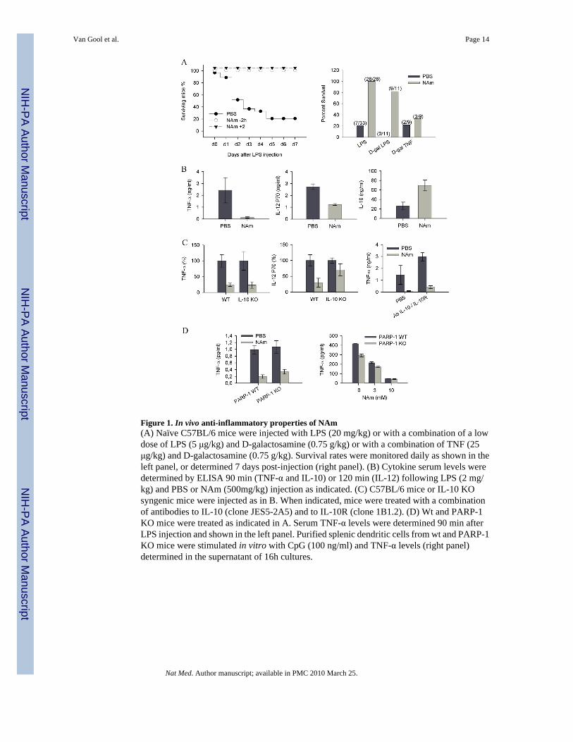
Figure 1. In vivo anti-inflammatory properties of NAm(A) Naïve C57BL/6 mice were injected with LPS (20 mg/kg) or with a combination of a lowdose of LPS (5 μg/kg) and D-galactosamine (0.75 g/kg) or with a combination of TNF (25μg/kg) and D-galactosamine (0.75 g/kg). Survival rates were monitored daily as shown in theleft panel, or determined 7 days post-injection (right panel). (B) Cytokine serum levels weredetermined by ELISA 90 min (TNF-α and IL-10) or 120 min (IL-12) following LPS (2 mg/kg) and PBS or NAm (500mg/kg) injection as indicated. (C) C57BL/6 mice or IL-10 KOsyngenic mice were injected as in B. When indicated, mice were treated with a combinationof antibodies to IL-10 (clone JES5-2A5) and to IL-10R (clone 1B1.2). (D) Wt and PARP-1KO mice were treated as indicated in A. Serum TNF-α levels were determined 90 min afterLPS injection and shown in the left panel. Purified splenic dendritic cells from wt and PARP-1KO mice were stimulated in vitro with CpG (100 ng/ml) and TNF-α levels (right panel)determined in the supernatant of 16h cultures.
Van Gool et al. Page 14
Nat Med. Author manuscript; available in PMC 2010 March 25.
NIH
-PA Author Manuscript
NIH
-PA Author Manuscript
NIH
-PA Author Manuscript
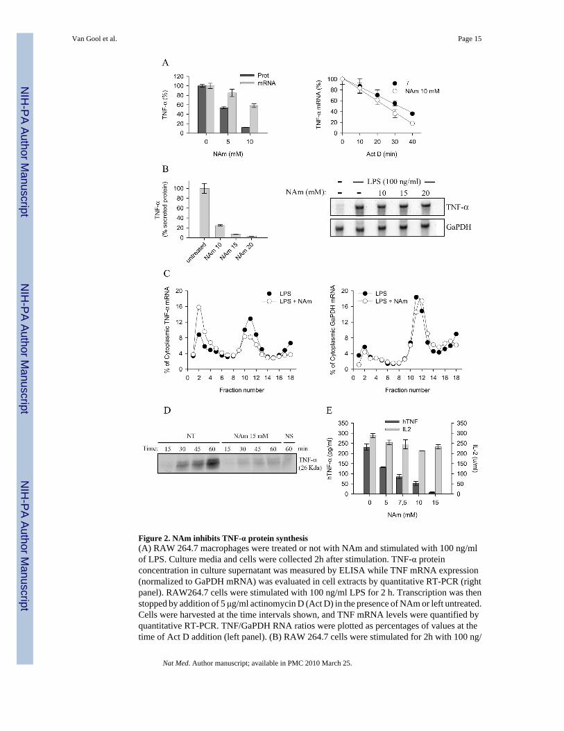
Figure 2. NAm inhibits TNF-α protein synthesis(A) RAW 264.7 macrophages were treated or not with NAm and stimulated with 100 ng/mlof LPS. Culture media and cells were collected 2h after stimulation. TNF-α proteinconcentration in culture supernatant was measured by ELISA while TNF mRNA expression(normalized to GaPDH mRNA) was evaluated in cell extracts by quantitative RT-PCR (rightpanel). RAW264.7 cells were stimulated with 100 ng/ml LPS for 2 h. Transcription was thenstopped by addition of 5 μg/ml actinomycin D (Act D) in the presence of NAm or left untreated.Cells were harvested at the time intervals shown, and TNF mRNA levels were quantified byquantitative RT-PCR. TNF/GaPDH RNA ratios were plotted as percentages of values at thetime of Act D addition (left panel). (B) RAW 264.7 cells were stimulated for 2h with 100 ng/
Van Gool et al. Page 15
Nat Med. Author manuscript; available in PMC 2010 March 25.
NIH
-PA Author Manuscript
NIH
-PA Author Manuscript
NIH
-PA Author Manuscript
ml LPS, washed and treated for 30 min with graded doses of NAm. Relative expression ofTNF-α protein levels (left panel) and mRNA (right panel) were determined respectively byELISA and northern blot analysis. GaPDH mRNA levels were determined as internal control.(C) Polysome profile analysis of RAW 264.7 cells treated with 15 mM NAm. Densitrometricquantification of the relative RNA northern blot signal intensities across the gradient for TNF-α (left panel) and GaPDH (right panel) transcripts. Data are from one of three representativeexperiments. (D) RAW 264.7 cells were stimulated with LPS (100 ng/ml) for 1h, washed andincubated in cysteine- and methionine-free DMEM. 30 min later the cells were incubated inDMEM supplemented with [35S]-labeled cysteine and methionine, brefeldin A and with 15mM NAm for the indicated time periods. Immunoprecipitated TNF-α was separated by SDS-PAGE and the gels dried and analyzed by autoradiography (NS: not stimulated by LPS). (E)293T cells were co-transfected with plasmid encoding human TNF-α and with a plasmidencoding murine IL-2. Secreted cytokines levels were determined in the supernatant oftransfected cells by ELISA. NAm was found to selectively inhibit the production of hTNF-α.
Van Gool et al. Page 16
Nat Med. Author manuscript; available in PMC 2010 March 25.
NIH
-PA Author Manuscript
NIH
-PA Author Manuscript
NIH
-PA Author Manuscript
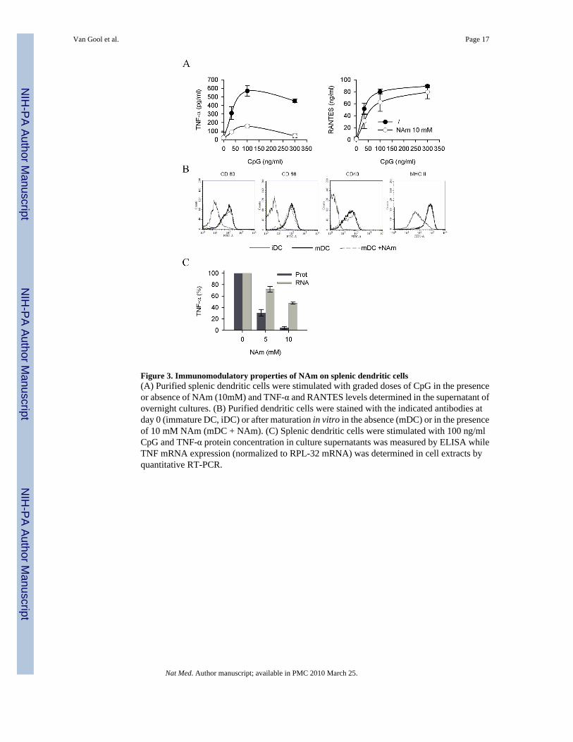
Figure 3. Immunomodulatory properties of NAm on splenic dendritic cells(A) Purified splenic dendritic cells were stimulated with graded doses of CpG in the presenceor absence of NAm (10mM) and TNF-α and RANTES levels determined in the supernatant ofovernight cultures. (B) Purified dendritic cells were stained with the indicated antibodies atday 0 (immature DC, iDC) or after maturation in vitro in the absence (mDC) or in the presenceof 10 mM NAm (mDC + NAm). (C) Splenic dendritic cells were stimulated with 100 ng/mlCpG and TNF-α protein concentration in culture supernatants was measured by ELISA whileTNF mRNA expression (normalized to RPL-32 mRNA) was determined in cell extracts byquantitative RT-PCR.
Van Gool et al. Page 17
Nat Med. Author manuscript; available in PMC 2010 March 25.
NIH
-PA Author Manuscript
NIH
-PA Author Manuscript
NIH
-PA Author Manuscript
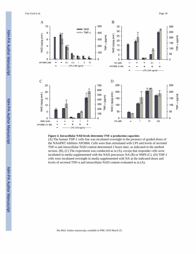
Figure 4. Intracellular NAD levels determine TNF-α production capacities(A) The human THP-1 cells line was incubated overnight in the presence of graded doses ofthe NAmPRT inhibitor APO866. Cells were than stimulated with LPS and levels of secretedTNF-α and intracellular NAD content determined 2 hours later, as indicated in the methodsection. (B), (C) The experiment was conducted as in (A), except that responder cells wereincubated in media supplemented with the NAD precursors NA (B) or NMN (C). (D) THP-1cells were incubated overnight in media supplemented with NA at the indicated doses andlevels of secreted TNF-α and intracellular NAD content evaluated as in (A).
Van Gool et al. Page 18
Nat Med. Author manuscript; available in PMC 2010 March 25.
NIH
-PA Author Manuscript
NIH
-PA Author Manuscript
NIH
-PA Author Manuscript
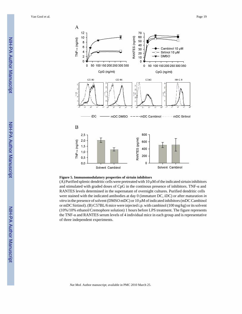
Figure 5. Immunomodulatory properties of sirtuin inhibitors(A) Purified splenic dendritic cells were pretreated with 10 μM of the indicated sirtuin inhibitorsand stimulated with graded doses of CpG in the continous presence of inhibitors. TNF-α andRANTES levels determined in the supernatant of overnight cultures. Purified dendritic cellswere stained with the indicated antibodies at day 0 (immature DC, iDC) or after maturation invitro in the presence of solvent (DMSO mDC) or 10 μM of indicated inhibitors (mDC Cambinolor mDC Sirtinol). (B) C57BL/6 mice were injected i.p. with cambinol (100 mg/kg) or its solvent(10%/10% ethanol/Cremophore solution) 1 hours before LPS treatment. The figure representsthe TNF-α and RANTES serum levels of 4 individual mice in each group and is representativeof three independent experiments.
Van Gool et al. Page 19
Nat Med. Author manuscript; available in PMC 2010 March 25.
NIH
-PA Author Manuscript
NIH
-PA Author Manuscript
NIH
-PA Author Manuscript
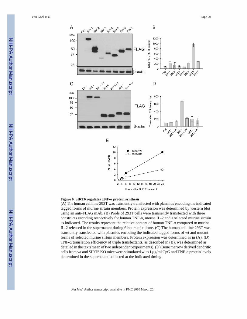
Figure 6. SIRT6 regulates TNF-α protein synthesis(A) The human cell line 293T was transiently transfected with plasmids encoding the indicatedtagged forms of murine sirtuin members. Protein expression was determined by western blotusing an anti-FLAG mAb. (B) Pools of 293T cells were transiently transfected with threeconstructs encoding respectively for human TNF-α, mouse IL-2 and a selected murine sirtuinas indicated. The results represent the relative content of human TNF-α compared to murineIL-2 released in the supernatant during 6 hours of culture. (C) The human cell line 293T wastransiently transfected with plasmids encoding the indicated tagged forms of wt and mutantforms of selected murine sirtuin members. Protein expression was determined as in (A). (D)TNF-α translation efficiency of triple transfectants, as described in (B), was determined asdetailed in the text (mean of two independent experiments). (D) Bone marrow derived dendriticcells from wt and SIRT6 KO mice were stimulated with 1 μg/ml CpG and TNF-α protein levelsdetermined in the supernatant collected at the indicated timing.
Van Gool et al. Page 20
Nat Med. Author manuscript; available in PMC 2010 March 25.
NIH
-PA Author Manuscript
NIH
-PA Author Manuscript
NIH
-PA Author Manuscript
Related Documents