Intracellular calcium changes trigger connexin 32 hemichannel opening Elke De Vuyst 1 , Elke Decrock 1 , Liesbet Cabooter 1 , George R Dubyak 2 , Christian C Naus 3 , W Howard Evans 4 and Luc Leybaert 1, * 1 Department of Physiology and Pathophysiology, Faculty of Medicine and Health Sciences, Ghent University, Ghent, Belgium, 2 Department of Physiology and Biophysics, School of Medicine, Case Western Reserve University, Cleveland, OH, USA, 3 Department of Cellular and Physiological Sciences, Faculty of Medicine, University of British Columbia, Vancouver, BC, Canada and 4 Department of Medical Biochemistry and Immunology, Cardiff University School of Medicine, Cardiff, UK Connexin hemichannels have been proposed as a diffusion pathway for the release of extracellular messengers like ATP and others, based on connexin expression models and inhibition by gap junction blockers. Hemichannels are opened by various experimental stimuli, but the physiolo- gical intracellular triggers are currently not known. We investigated the hypothesis that an increase of cytoplasmic calcium concentration ([Ca 2 þ ] i ) triggers hemichannel opening, making use of peptides that are identical to a short amino-acid sequence on the connexin subunit to specifically block hemichannels, but not gap junction channels. Our work performed on connexin 32 (Cx32)- expressing cells showed that an increase in [Ca 2 þ ] i trig- gers ATP release and dye uptake that is dependent on Cx32 expression, blocked by Cx32 (but not Cx43) mimetic peptides and a calmodulin antagonist, and critically de- pendent on [Ca 2 þ ] i elevation within a window situated around 500 nM. Our results indicate that [Ca 2 þ ] i elevation triggers hemichannel opening, and suggest that these channels are under physiological control. The EMBO Journal (2006) 25, 34–44. doi:10.1038/ sj.emboj.7600908; Published online 8 December 2005 Subject Categories: membranes & transport; signal transduction Keywords: connexin mimetic peptides; exocytosis; P 2 X 7 pores; purinergic receptors; vesicular release Introduction The basic fuel molecule of the cell, ATP, has gained a lot of interest over the last decade as a paracrine messenger in various cell and tissue types (Novak, 2003). Besides being a remarkably versatile molecule, nonexcitable cells can be invoked to release ATP by widely differing stimuli, including mechanical cell stimulation (Stout et al, 2002), shear stress (Cherian et al, 2005), hypotonic cell swelling (Boudreault and Grygorczyk, 2004), elevation of intracellular inositol trispho- sphate (InsP 3 ) (Braet et al, 2003b) or exposure to low or zero extracellular Ca 2 þ conditions (Arcuino et al, 2002). The ATP release mechanisms involved appear to be equally diverse (reviewed in Lazarowski et al, 2003), including vesicular release, active transport via ABC transporters and diffusion via stretch-activated channels, voltage-dependent anion channels, pores opened by P 2 X 7 receptors or connexin hemi- channels. Connexin hemichannels are hexameric high- conductance plasma membrane channels (single-channel conductance B90 and 220 pS for Cx32 and 43, respec- tively—Contreras et al, 2003; Gomez-Hernandez et al, 2003) that are normally closed and can act as a conduit for ATP, NAD þ , glutamate and prostaglandins when opened (Bruzzone et al, 2001; Bennett et al, 2003; Ebihara, 2003; Goodenough and Paul, 2003; Ye et al, 2003; Cherian et al, 2005). Hemichannels are closed at normal millimolar extra- cellular [Ca 2 þ ], but open when Ca 2 þ is lowered (Li et al, 1996; Pfahnl and Dahl, 1999; Quist et al, 2000; Muller et al, 2002; Ye et al, 2003; Thimm et al, 2005). A Ca 2 þ -binding site composed of aspartate residues facing the external side has been reported for Cx32 hemichannels and is thought to translate changes of extracellular [Ca 2 þ ] to changes in chan- nel gating (Gomez-Hernandez et al, 2003). Hemichannels also open in response to membrane depolarization and mechanical stimulation in a Xenopus oocyte expression sys- tem (Trexler et al, 1996; Bao et al, 2004), under conditions of metabolic inhibition in astrocytes, myocardial cells or renal epithelial cells (John et al, 1999; Kondo et al, 2000; Contreras et al, 2002; Vergara et al, 2003), after Shigella invasion of epithelial cells (Tran Van Nhieu et al, 2003) and in response to extracellular UTP in C6 glioma cells expressing Cx32 or 43 (Cotrina et al, 1998). The physiological intracellular signals controlling hemichannel opening are currently not known, but UTP-triggered ATP release via hemichannels was depen- dent on intracellular Ca 2 þ mobilization (Cotrina et al, 1998) and we demonstrated that photoactivation of InsP 3 in Cx43- expressing cells triggers Ca 2 þ -dependent ATP release that is blocked by gap junction blockers and peptides that mimic a short exposed sequence on the Cx43 subunit (Braet et al, 2003a, b), indicating that InsP 3 and downstream signals activate hemichannel opening (Leybaert et al, 2003). Recent work from the group of Mobbs and co-workers (Pearson et al, 2005) also points to intracellular Ca 2 þ changes triggering Cx43 hemichannel opening in native retinal pigment epithe- lium. The aim of the present work was to determine whether increases of cytoplasmic calcium concentration ([Ca 2 þ ] i ) are sufficient to trigger hemichannel opening as probed with connexin mimetic peptides and connexin expression systems. Our results obtained in Cx32-expressing cells demonstrate that direct elevation of [Ca 2 þ ] i by photoactivation of Ca 2 þ in the cytoplasm or stimulation of Ca 2 þ entry with a Ca 2 þ ionophore triggers ATP release and hemichannel-permeable dye uptake that was dependent on Cx32 expression and Received: 26 May 2005; accepted: 17 November 2005; published online: 8 December 2005 *Corresponding author. Department of Physiology and Pathophysiology, Faculty of Medicine and Health Sciences, Ghent University, De Pintelaan 185 (Block B, Room 306), 9000 Ghent, Belgium. Tel.: þ 32 9 240 33 66; Fax: þ 32 9 240 30 59; E-mail: [email protected] The EMBO Journal (2006) 25, 34–44 | & 2006 European Molecular Biology Organization | All Rights Reserved 0261-4189/06 www.embojournal.org The EMBO Journal VOL 25 | NO 1 | 2006 & 2006 European Molecular Biology Organization EMBO THE EMBO JOURNAL THE EMBO JOURNAL 34

Welcome message from author
This document is posted to help you gain knowledge. Please leave a comment to let me know what you think about it! Share it to your friends and learn new things together.
Transcript
Intracellular calcium changes trigger connexin 32hemichannel opening
Elke De Vuyst1, Elke Decrock1,Liesbet Cabooter1, George R Dubyak2,Christian C Naus3, W Howard Evans4
and Luc Leybaert1,*1Department of Physiology and Pathophysiology, Faculty of Medicineand Health Sciences, Ghent University, Ghent, Belgium, 2Department ofPhysiology and Biophysics, School of Medicine, Case Western ReserveUniversity, Cleveland, OH, USA, 3Department of Cellular andPhysiological Sciences, Faculty of Medicine, University of BritishColumbia, Vancouver, BC, Canada and 4Department of MedicalBiochemistry and Immunology, Cardiff University School of Medicine,Cardiff, UK
Connexin hemichannels have been proposed as a diffusion
pathway for the release of extracellular messengers like
ATP and others, based on connexin expression models and
inhibition by gap junction blockers. Hemichannels are
opened by various experimental stimuli, but the physiolo-
gical intracellular triggers are currently not known. We
investigated the hypothesis that an increase of cytoplasmic
calcium concentration ([Ca2þ ]i) triggers hemichannel
opening, making use of peptides that are identical to a
short amino-acid sequence on the connexin subunit to
specifically block hemichannels, but not gap junction
channels. Our work performed on connexin 32 (Cx32)-
expressing cells showed that an increase in [Ca2þ ]i trig-
gers ATP release and dye uptake that is dependent on Cx32
expression, blocked by Cx32 (but not Cx43) mimetic
peptides and a calmodulin antagonist, and critically de-
pendent on [Ca2þ ]i elevation within a window situated
around 500 nM. Our results indicate that [Ca2þ ]i elevation
triggers hemichannel opening, and suggest that these
channels are under physiological control.
The EMBO Journal (2006) 25, 34–44. doi:10.1038/
sj.emboj.7600908; Published online 8 December 2005
Subject Categories: membranes & transport; signal transduction
Keywords: connexin mimetic peptides; exocytosis; P2X7
pores; purinergic receptors; vesicular release
Introduction
The basic fuel molecule of the cell, ATP, has gained a lot of
interest over the last decade as a paracrine messenger in
various cell and tissue types (Novak, 2003). Besides being
a remarkably versatile molecule, nonexcitable cells can be
invoked to release ATP by widely differing stimuli, including
mechanical cell stimulation (Stout et al, 2002), shear stress
(Cherian et al, 2005), hypotonic cell swelling (Boudreault and
Grygorczyk, 2004), elevation of intracellular inositol trispho-
sphate (InsP3) (Braet et al, 2003b) or exposure to low or zero
extracellular Ca2þ conditions (Arcuino et al, 2002). The ATP
release mechanisms involved appear to be equally diverse
(reviewed in Lazarowski et al, 2003), including vesicular
release, active transport via ABC transporters and diffusion
via stretch-activated channels, voltage-dependent anion
channels, pores opened by P2X7 receptors or connexin hemi-
channels. Connexin hemichannels are hexameric high-
conductance plasma membrane channels (single-channel
conductance B90 and 220 pS for Cx32 and 43, respec-
tively—Contreras et al, 2003; Gomez-Hernandez et al, 2003)
that are normally closed and can act as a conduit for ATP,
NADþ , glutamate and prostaglandins when opened
(Bruzzone et al, 2001; Bennett et al, 2003; Ebihara, 2003;
Goodenough and Paul, 2003; Ye et al, 2003; Cherian et al,
2005). Hemichannels are closed at normal millimolar extra-
cellular [Ca2þ ], but open when Ca2þ is lowered (Li et al,
1996; Pfahnl and Dahl, 1999; Quist et al, 2000; Muller et al,
2002; Ye et al, 2003; Thimm et al, 2005). A Ca2þ -binding site
composed of aspartate residues facing the external side has
been reported for Cx32 hemichannels and is thought to
translate changes of extracellular [Ca2þ ] to changes in chan-
nel gating (Gomez-Hernandez et al, 2003). Hemichannels
also open in response to membrane depolarization and
mechanical stimulation in a Xenopus oocyte expression sys-
tem (Trexler et al, 1996; Bao et al, 2004), under conditions of
metabolic inhibition in astrocytes, myocardial cells or renal
epithelial cells (John et al, 1999; Kondo et al, 2000; Contreras
et al, 2002; Vergara et al, 2003), after Shigella invasion of
epithelial cells (Tran Van Nhieu et al, 2003) and in response
to extracellular UTP in C6 glioma cells expressing Cx32 or 43
(Cotrina et al, 1998). The physiological intracellular signals
controlling hemichannel opening are currently not known,
but UTP-triggered ATP release via hemichannels was depen-
dent on intracellular Ca2þ mobilization (Cotrina et al, 1998)
and we demonstrated that photoactivation of InsP3 in Cx43-
expressing cells triggers Ca2þ -dependent ATP release that is
blocked by gap junction blockers and peptides that mimic a
short exposed sequence on the Cx43 subunit (Braet et al,
2003a, b), indicating that InsP3 and downstream signals
activate hemichannel opening (Leybaert et al, 2003). Recent
work from the group of Mobbs and co-workers (Pearson et al,
2005) also points to intracellular Ca2þ changes triggering
Cx43 hemichannel opening in native retinal pigment epithe-
lium. The aim of the present work was to determine whether
increases of cytoplasmic calcium concentration ([Ca2þ ]i) are
sufficient to trigger hemichannel opening as probed with
connexin mimetic peptides and connexin expression systems.
Our results obtained in Cx32-expressing cells demonstrate
that direct elevation of [Ca2þ ]i by photoactivation of Ca2þ
in the cytoplasm or stimulation of Ca2þ entry with a Ca2þ
ionophore triggers ATP release and hemichannel-permeable
dye uptake that was dependent on Cx32 expression andReceived: 26 May 2005; accepted: 17 November 2005; publishedonline: 8 December 2005
*Corresponding author. Department of Physiology and Pathophysiology,Faculty of Medicine and Health Sciences, Ghent University, De Pintelaan185 (Block B, Room 306), 9000 Ghent, Belgium. Tel.: þ 32 9 240 33 66;Fax: þ 32 9 240 30 59; E-mail: [email protected]
The EMBO Journal (2006) 25, 34–44 | & 2006 European Molecular Biology Organization | All Rights Reserved 0261-4189/06
www.embojournal.org
The EMBO Journal VOL 25 | NO 1 | 2006 &2006 European Molecular Biology Organization
EMBO
THE
EMBOJOURNAL
THE
EMBOJOURNAL
34
completely blocked by Cx32 mimetic peptides and calmodu-
lin inhibition. Pores activated by P2X7 receptor activation
were excluded and vesicular ATP release contributed to a
limited extent, but hemichannel-mediated release was by far
the predominant component. Our work shows that hemi-
channels can be activated by physiological [Ca2þ ]i, opening
up a wide range of future investigations on hemichannel
involvement in both physiology and pathophysiology.
Results
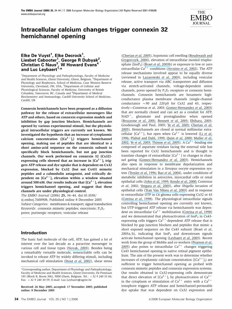
Divalent-free (DF) solutions trigger ATP release that is
dependent on [Ca2þ ]i in ECV304
Exposure of cells to solutions with lowered or zero Ca2þ and
Mg2þ triggers ATP and glutamate release and has been
applied in various cell types to investigate connexin hemi-
channel opening (Arcuino et al, 2002; Stout et al, 2002; Ye
et al, 2003). We used ECV304 cells (bladder cancer epithelial
cells) from which a subclone with prominent and sponta-
neous Cx32 expression (see Figure 3) was selected to
study the role of [Ca2þ ]i changes in triggering ATP release
via hemichannels. Incubating ECV304 cells in zero Ca2þ
and Mg2þ solutions (divalent-free [DF] solutions, applied
2.5 min) triggered ATP release that was significantly above
baseline (Figure 1A) and was associated with transient
and oscillatory [Ca2þ ]i changes as observed in fluo-3 imaging
experiments (Figure 1B). The ATP response to DF conditions
varied between experiments and ranged from approximately
175 to 500% of baseline. Buffering [Ca2þ ]i with BAPTA (5 mM
BAPTA-AM, 45 min loading time) or blocking the intracellular
Ca2þ store SERCA pumps with thapsigargin (1 mM, 10 min)
significantly reduced the ATP response (Figure 1A). Blocking
InsP3 receptors with xestospongin-C (2–10mm, 10 min),
ryanodine (Ry) receptors with dantrolene (25 mM, 10 min)
or pre-emptying Ry-sensitive Ca2þ stores with caffeine
(10 mM, 30 min) all significantly inhibited DF-triggered ATP
release (Figure 1C). InsP3- and Ry-sensitive Ca2þ stores are
thus involved in the ATP response, and, in line with this,
photoactivation of InsP3 in the cytoplasm (Braet et al, 2003b)
and exposure to the alkaloid Ry (10 mM, 15 min) or caffeine
(10 mM, 15 min) triggered significant ATP release (175738%
for Ry and 400781% for caffeine, compared to 100712%
baseline level, n¼ 15, Po0.05 and o0.0005, respectively).
Directly increasing [Ca2þ ]i triggers ATP release
in ECV304
Photoactivation of Ca2þ from ester-loaded NP-EGTA (5 mM
o-nitrophenyl EGTA acetoxymethyl ester (NP-EGTA-AM),
10 min; Ellis-Davies and Kaplan, 1994) triggered significant
ATP release (Figure 1D). However, prolonged ester loading
(1 h), applied to increase the amount of photoliberated Ca2þ ,
did not trigger significant ATP release. Stimulating Ca2þ entry
with the Ca2þ ionophore A23187 (1 mM, 4 min) triggered
significant ATP release, but its application at higher concen-
trations (10mM) or longer incubations (10 min) failed to do
so (Figure 1D). [Ca2þ ]i changes thus trigger ATP release, but
the response disappears with stronger stimulation.
DF- and Ca2þ -triggered ATP release are inhibited by gap
junction blockers and a Cx32 mimetic peptide in ECV304
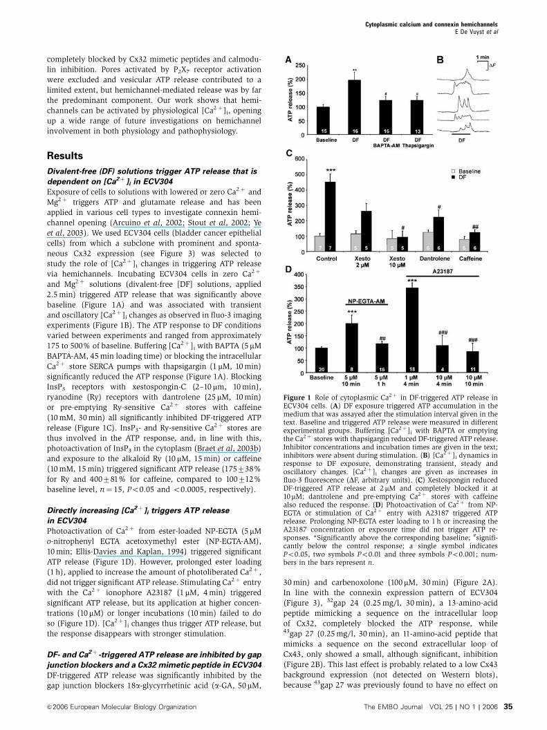
DF-triggered ATP release was significantly inhibited by the
gap junction blockers 18a-glycyrrhetinic acid (a-GA, 50mM,
30 min) and carbenoxolone (100 mM, 30 min) (Figure 2A).
In line with the connexin expression pattern of ECV304
(Figure 3), 32gap 24 (0.25 mg/l, 30 min), a 13-amino-acid
peptide mimicking a sequence on the intracellular loop
of Cx32, completely blocked the ATP response, while43gap 27 (0.25 mg/l, 30 min), an 11-amino-acid peptide that
mimicks a sequence on the second extracellular loop of
Cx43, only showed a small, although significant, inhibition
(Figure 2B). This last effect is probably related to a low Cx43
background expression (not detected on Western blots),
because 43gap 27 was previously found to have no effect on
Figure 1 Role of cytoplasmic Ca2þ in DF-triggered ATP release inECV304 cells. (A) DF exposure triggered ATP accumulation in themedium that was assayed after the stimulation interval given in thetext. Baseline and triggered ATP release were measured in differentexperimental groups. Buffering [Ca2þ ]i with BAPTA or emptyingthe Ca2þ stores with thapsigargin reduced DF-triggered ATP release.Inhibitor concentrations and incubation times are given in the text;inhibitors were absent during stimulation. (B) [Ca2þ ]i dynamics inresponse to DF exposure, demonstrating transient, steady andoscillatory changes. [Ca2þ ]i changes are given as increases influo-3 fluorescence (DF, arbitrary units). (C) Xestospongin reducedDF-triggered ATP release at 2mM and completely blocked it at10mM; dantrolene and pre-emptying Ca2þ stores with caffeinealso reduced the response. (D) Photoactivation of Ca2þ from NP-EGTA or stimulation of Ca2þ entry with A23187 triggered ATPrelease. Prolonging NP-EGTA ester loading to 1 h or increasing theA23187 concentration or exposure time did not trigger ATP re-sponses. *Significantly above the corresponding baseline; #signifi-cantly below the control response; a single symbol indicatesPo0.05, two symbols Po0.01 and three symbols Po0.001; num-bers in the bars represent n.
Cytoplasmic calcium and connexin hemichannelsE De Vuyst et al
&2006 European Molecular Biology Organization The EMBO Journal VOL 25 | NO 1 | 2006 35
Cx32 hemichannels (Braet et al, 2003b). Carbenoxolone and32gap 24 also significantly inhibited ATP release triggered by
Ca2þ photoactivation (Figure 2C) and A23187 (Figure 2D). In
some of the experiments, inhibitor substances caused signifi-
cant inhibition of the baseline signal (e.g. Figure 2A, B and
D), indicating involvement of hemichannels under basal
conditions. In all these cases, the net (trigger minus baseline)
and relative (trigger over baseline ratio) responses were
significantly below control (Po0.05) except for the carbenox-
olone experiment of Figure 2A, where the relative response
appeared to be increased (an atypical response not observed
in other experiments).
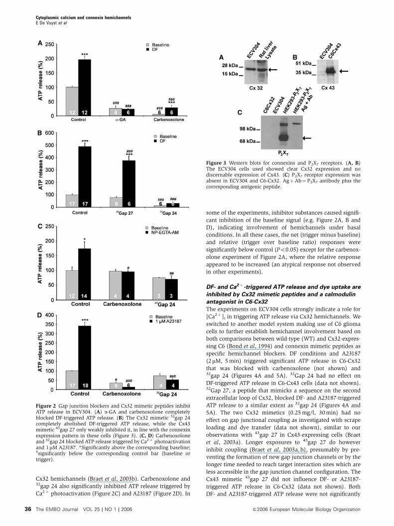
DF- and Ca2þ -triggered ATP release and dye uptake are
inhibited by Cx32 mimetic peptides and a calmodulin
antagonist in C6-Cx32
The experiments on ECV304 cells strongly indicate a role for
[Ca2þ ]i in triggering ATP release via Cx32 hemichannels. We
switched to another model system making use of C6 glioma
cells to further establish hemichannel involvement based on
both comparisons between wild-type (WT) and Cx32-expres-
sing C6 (Bond et al, 1994) and connexin mimetic peptides as
specific hemichannel blockers. DF conditions and A23187
(2 mM, 5 min) triggered significant ATP release in C6-Cx32
that was blocked with carbenoxolone (not shown) and32gap 24 (Figures 4A and 5A). 32Gap 24 had no effect on
DF-triggered ATP release in C6-Cx43 cells (data not shown).32Gap 27, a peptide that mimicks a sequence on the second
extracellular loop of Cx32, blocked DF- and A23187-triggered
ATP release to a similar extent as 32gap 24 (Figures 4A and
5A). The two Cx32 mimetics (0.25 mg/l, 30 min) had no
effect on gap junctional coupling as investigated with scrape
loading and dye transfer (data not shown), similar to our
observations with 43gap 27 in Cx43-expressing cells (Braet
et al, 2003a). Longer exposures to 43gap 27 do however
inhibit coupling (Braet et al, 2003a, b), presumably by pre-
venting the formation of new gap junction channels or by the
longer time needed to reach target interaction sites which are
less accessible in the gap junction channel configuration. The
Cx43 mimetic 43gap 27 did not influence DF- or A23187-
triggered ATP release in C6-Cx32 (data not shown). Both
DF- and A23187-triggered ATP release were not significantly
Figure 2 Gap junction blockers and Cx32 mimetic peptides inhibitATP release in ECV304. (A) a-GA and carbenoxolone completelyblocked DF-triggered ATP release. (B) The Cx32 mimetic 32gap 24completely abolished DF-triggered ATP release, while the Cx43mimetic 43gap 27 only weakly inhibited it, in line with the connexinexpression pattern in these cells (Figure 3). (C, D) Carbenoxoloneand 32gap 24 blocked ATP release triggered by Ca2þ photoactivationand 1mM A23187. *Significantly above the corresponding baseline;#significantly below the corresponding control bar (baseline ortrigger).
Figure 3 Western blots for connexins and P2X7 receptors. (A, B)The ECV304 cells used showed clear Cx32 expression and nodiscernable expression of Cx43. (C) P2X7 receptor expression wasabsent in ECV304 and C6-Cx32. AgþAb¼P2X7 antibody plus thecorresponding antigenic peptide.
Cytoplasmic calcium and connexin hemichannelsE De Vuyst et al
The EMBO Journal VOL 25 | NO 1 | 2006 &2006 European Molecular Biology Organization36
Figure 4 DF-triggered ATP release in C6-Cx32. (A) 32Gap 24 and32gap 27 drastically inhibited DF-triggered ATP release in C6-Cx32.DF-triggered ATP release was significantly lower in C6-WT ascompared to C6-Cx32 (inset). (B, C) Bafilomycin A1 (Bafilo) hadno effect, while botulinum toxin B (Botul) slightly but significantlyinhibited ATP release. (D) Calmodulin inhibition with W7 drasti-cally blocked ATP release. The P2X7 receptor antagonist oxidizedATP (Ox-ATP) had no effect, but KN62 displayed significant inhibi-tion. *Significantly above the corresponding baseline; #significantlybelow control.
Figure 5 A23187-triggered ATP release in C6-Cx32. (A) 32Gap 24and 32gap 27 blocked the ATP response. Bafilomycin A1 (Bafilo) hadno effect, while botulinum toxin B (Botul) showed slight butnonsignificant inhibition (triggered ATP release was however notsignificantly above baseline). The ATP responses were completelyblocked by adding 32gap 24 together with either of the toxins, andwas absent in C6-WT (inset). (B) W7 completely blocked theresponses, but Ox-ATP and KN62 had no significant effects. (C, D)Recovery of ATP responses following washout (WO) of 32gap 24 andW7, respectively. The effect of various washout periods wasassessed by experiments on different cultures. *Significantlyabove the corresponding baseline; #significantly below the corre-sponding control bar.
Cytoplasmic calcium and connexin hemichannelsE De Vuyst et al
&2006 European Molecular Biology Organization The EMBO Journal VOL 25 | NO 1 | 2006 37
above baseline in C6-WT and were significantly lower as
compared to C6-Cx32 (insets to Figures 4A and 5A). In
Figure 4A, DF exposure appears to trigger some ATP release
in C6-WT (nonsignificant with ANOVA, but significantly
above baseline with a t-test (Po0.05)), which may be related
to a low connexin background expression or the operation of
other DF-responsive ATP release mechanisms. Cx32 contains
two cytoplasmic calmodulin-binding domains (Torok et al,
1997) that may be involved in the Ca2þ -triggered ATP
responses. The calmodulin antagonist W7 (20 mM, 1 h) was
as potent as the Cx32 mimetics in blocking DF- and A23187-
triggered ATP release (Figures 4D and 5B). The inhibitory
effect of the peptides and W7 was reversible upon washout of
these substances: inhibition by W7 completely disappeared
after 30 min washout (Figure 5D), inhibition by 32gap 27
within 60 min (data not shown), and inhibition by 32gap 24
took almost 120 min to disappear upon washout (Figure 5C).
DF conditions and A23187 (2 mM, trigger solutions applied
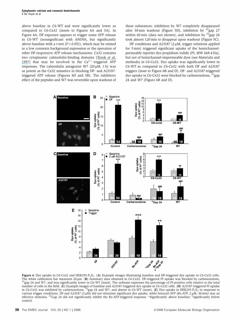
for 5 min) triggered significant uptake of the hemichannel-
permeable reporter dye propidium iodide (PI, MW 668.4 Da),
but not of hemichannel-impermeable dyes (see Materials and
methods) in C6-Cx32. Dye uptake was significantly lower in
C6-WT as compared to C6-Cx32 with both DF and A23187
triggers (inset to Figure 6B and D). DF- and A23187-triggered
dye uptake in C6-Cx32 were blocked by carbenoxolone, 32gap
24 and W7 (Figure 6B and D).
Figure 6 Dye uptake in C6-Cx32 and HEK293-P2X7. (A) Example images illustrating baseline and DF-triggered dye uptake in C6-Cx32 cells.The white calibration bar measures 20mm. (B) Summary data obtained in C6-Cx32. DF-triggered PI uptake was blocked by carbenoxolone,32gap 24 and W7, and was significantly lower in C6-WT (inset). The ordinate expresses the percentage of PI-positive cells relative to the totalnumber of cells in the field. (C) Example images of baseline and A23187-triggered dye uptake in C6-Cx32 cells. (D) A23187-triggered PI uptakein C6-Cx32 was inhibited by carbenoxolone, 32gap 24 and W7, and absent in C6-WT (inset). (E) Dye uptake in HEK293-P2X7 in response tovarious trigger conditions. DF and A23187 (2 mM) did not stimulate significant dye uptake, while benzoyl-ATP (Bz-ATP, 2 mM, 30 min) was aneffective stimulus. 32Gap 24 did not significantly inhibit the Bz-ATP-triggered response. *Significantly above baseline; #significantly belowcontrol.
Cytoplasmic calcium and connexin hemichannelsE De Vuyst et al
The EMBO Journal VOL 25 | NO 1 | 2006 &2006 European Molecular Biology Organization38
P2X7 receptors are not involved in ATP release
Activation of P2X7 receptors also triggers a pore-like ATP
release pathway (Duan et al, 2003) that should be distin-
guished from hemichannels (Parpura et al, 2004). ECV304
and C6-Cx32 cells did not express P2X7 protein (Figure 3C).
Oxidized ATP (100 mM, 1 h), an irreversible P2X7 receptor
antagonist that also blocks pore-forming P2X2 receptors
(North, 2002), did not inhibit DF- and A23187-triggered ATP
release in C6-Cx32 (Figures 4D and 5B). KN62, another P2X7
antagonist (North, 2002), significantly inhibited DF-triggered
ATP release (Figure 4D), but was without effect on ATP
release triggered by A23187 (Figure 5B). Experiments on
HEK293-P2X7 cells showed that DF conditions did not trigger
significant ATP release (not shown) or PI uptake (Figure 6E).
KN62 inhibition of DF-triggered ATP release is thus not
related to P2X7 receptor antagonism, but is probably the
result of inhibition of calmodulin-dependent kinases acting
on the connexins (Hidaka and Yokokura, 1996). HEK293-P2X7
cells did neither show PI uptake in response to A23187;
benzoyl-ATP triggered significant PI uptake in these cells,
but this response was not significantly affected by 32gap 24
(Figure 6E).
Vesicular release slightly contributes to Ca2þ -triggered
ATP release in C6-Cx32
Botulinum toxin B, a protease that cleaves the v-SNARE
protein synaptobrevin (Schiavo et al, 1992) (1.5 nM, applied
24 h in the culture medium), significantly reduced DF-trig-
gered ATP release in C6-Cx32, but the v-ATPase inhibitor
bafilomycin A1 (100 mM, 1 h), known to inhibit ATP storage
(Coco et al, 2003), had no effect (Figure 4B and C). Both
toxins displayed partial but nonsignificant inhibition of
A23187-triggered ATP release in C6-Cx32 (Figure 5A).
Addition of 32gap 24 together with botulinum toxin B or
bafilomycin A1 (the last 30 min) depressed the ATP response
to below baseline levels, indicating predominance of the
release component inhibited by the Cx32 mimetics
(Figure 5A).
Relation between [Ca2þ ]i and ATP release or dye uptake
in C6-Cx32
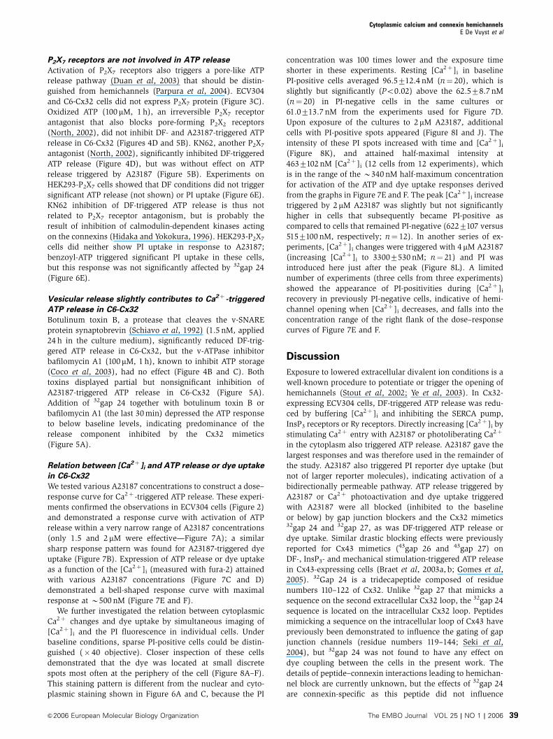
We tested various A23187 concentrations to construct a dose–
response curve for Ca2þ -triggered ATP release. These experi-
ments confirmed the observations in ECV304 cells (Figure 2)
and demonstrated a response curve with activation of ATP
release within a very narrow range of A23187 concentrations
(only 1.5 and 2 mM were effective—Figure 7A); a similar
sharp response pattern was found for A23187-triggered dye
uptake (Figure 7B). Expression of ATP release or dye uptake
as a function of the [Ca2þ ]i (measured with fura-2) attained
with various A23187 concentrations (Figure 7C and D)
demonstrated a bell-shaped response curve with maximal
response at B500 nM (Figure 7E and F).
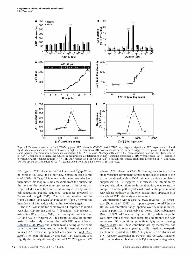
We further investigated the relation between cytoplasmic
Ca2þ changes and dye uptake by simultaneous imaging of
[Ca2þ ]i and the PI fluorescence in individual cells. Under
baseline conditions, sparse PI-positive cells could be distin-
guished (� 40 objective). Closer inspection of these cells
demonstrated that the dye was located at small discrete
spots most often at the periphery of the cell (Figure 8A–F).
This staining pattern is different from the nuclear and cyto-
plasmic staining shown in Figure 6A and C, because the PI
concentration was 100 times lower and the exposure time
shorter in these experiments. Resting [Ca2þ ]i in baseline
PI-positive cells averaged 96.5712.4 nM (n¼ 20), which is
slightly but significantly (Po0.02) above the 62.578.7 nM
(n¼ 20) in PI-negative cells in the same cultures or
61.0713.7 nM from the experiments used for Figure 7D.
Upon exposure of the cultures to 2 mM A23187, additional
cells with PI-positive spots appeared (Figure 8I and J). The
intensity of these PI spots increased with time and [Ca2þ ]i
(Figure 8K), and attained half-maximal intensity at
4637102 nM [Ca2þ ]i (12 cells from 12 experiments), which
is in the range of the B340 nM half-maximum concentration
for activation of the ATP and dye uptake responses derived
from the graphs in Figure 7E and F. The peak [Ca2þ ]i increase
triggered by 2mM A23187 was slightly but not significantly
higher in cells that subsequently became PI-positive as
compared to cells that remained PI-negative (6227107 versus
5157100 nM, respectively; n¼ 12). In another series of ex-
periments, [Ca2þ ]i changes were triggered with 4 mM A23187
(increasing [Ca2þ ]i to 33007530 nM; n¼ 21) and PI was
introduced here just after the peak (Figure 8L). A limited
number of experiments (three cells from three experiments)
showed the appearance of PI-positivities during [Ca2þ ]i
recovery in previously PI-negative cells, indicative of hemi-
channel opening when [Ca2þ ]i decreases, and falls into the
concentration range of the right flank of the dose–response
curves of Figure 7E and F.
Discussion
Exposure to lowered extracellular divalent ion conditions is a
well-known procedure to potentiate or trigger the opening of
hemichannels (Stout et al, 2002; Ye et al, 2003). In Cx32-
expressing ECV304 cells, DF-triggered ATP release was redu-
ced by buffering [Ca2þ ]i and inhibiting the SERCA pump,
InsP3 receptors or Ry receptors. Directly increasing [Ca2þ ]i by
stimulating Ca2þ entry with A23187 or photoliberating Ca2þ
in the cytoplasm also triggered ATP release. A23187 gave the
largest responses and was therefore used in the remainder of
the study. A23187 also triggered PI reporter dye uptake (but
not of larger reporter molecules), indicating activation of a
bidirectionally permeable pathway. ATP release triggered by
A23187 or Ca2þ photoactivation and dye uptake triggered
with A23187 were all blocked (inhibited to the baseline
or below) by gap junction blockers and the Cx32 mimetics32gap 24 and 32gap 27, as was DF-triggered ATP release or
dye uptake. Similar drastic blocking effects were previously
reported for Cx43 mimetics (43gap 26 and 43gap 27) on
DF-, InsP3- and mechanical stimulation-triggered ATP release
in Cx43-expressing cells (Braet et al, 2003a, b; Gomes et al,
2005). 32Gap 24 is a tridecapeptide composed of residue
numbers 110–122 of Cx32. Unlike 32gap 27 that mimicks a
sequence on the second extracellular Cx32 loop, the 32gap 24
sequence is located on the intracellular Cx32 loop. Peptides
mimicking a sequence on the intracellular loop of Cx43 have
previously been demonstrated to influence the gating of gap
junction channels (residue numbers 119–144; Seki et al,
2004), but 32gap 24 was not found to have any effect on
dye coupling between the cells in the present work. The
details of peptide–connexin interactions leading to hemichan-
nel block are currently unknown, but the effects of 32gap 24
are connexin-specific as this peptide did not influence
Cytoplasmic calcium and connexin hemichannelsE De Vuyst et al
&2006 European Molecular Biology Organization The EMBO Journal VOL 25 | NO 1 | 2006 39
DF-triggered ATP release in C6-Cx43 cells and 43gap 27 had
no effect in C6-Cx32- and other Cx32-expressing cells (Braet
et al, 2003a). If 32gap 24 interacts with the intracellular loop,
then either this loop must be accessible from the outside via
the pore or the peptide must get access to the cytoplasm
(32gap 24 does not, however, contain any currently known
cell-penetrating peptide sequence—sequences reviewed in
Zorko and Langel, 2005). The fact that washout of the32gap 24 effect took twice as long as for 32gap 27 favors the
hypothesis of interaction with an intracellular target.
The v-ATPase inhibitor bafilomycin A1, reported to inhibit
vesicular ATP storage and Ca2þ -dependent ATP release in
astrocytes (Coco et al, 2003), had no significant effect on
DF- and A23187-triggered ATP release in C6-Cx32. Botulinum
toxin B selectively cleaves the v-SNARE synaptobrevin
(Schiavo et al, 1992) and similar toxins acting at the same
target have been demonstrated to inhibit osmotic swelling-
induced ATP release in epithelial cells (van der Wijk et al,
2003). The toxin inhibited DF-triggered ATP release and also
slightly (but nonsignificantly) affected A23187-triggered ATP
release. ATP release in C6-Cx32 thus appears to involve a
small vesicular component. Exposing the cells to either of the
toxins combined with a Cx32 mimetic peptide completely
suppressed A23187-triggered ATP release. The inhibition by
the peptide, added alone or in combination, was so nearly
complete that the pathway blocked must be the predominant
ATP release pathway or the one located most upstream in a
cascade of ATP release signals or events.
An alternative ATP release pathway involves P2X7 recep-
tors (Duan et al, 2003) that, upon exposure to ATP in the
100 mM concentration range applied over several minutes,
opens a pore that is permeable to below 1 kDa substances
(North, 2002). ATP released by the cell, by whatever path-
way, may thus activate these receptors and amplify the ATP
responses. DF conditions potentiate P2X7 pore opening
(North, 2002), but these conditions are by themselves not
sufficient to initiate pore opening, as illustrated in the experi-
ments now reported with HEK293-P2X7 cells. The absence of
P2X7 receptor expression in ECV304 and C6-Cx32, together
with the evidence obtained with P2X7 receptor antagonists,
Figure 7 Dose–response curve for A23187-triggered ATP release in C6-Cx32. (A) A23187 only triggered significant ATP responses at 1.5 and2 mM, while responses were absent at lower or higher concentrations. (B) Dose–response curve for Ca2þ -triggered dye uptake, illustrating thesame narrow concentration dependence as observed for ATP release. *Significantly above the corresponding baseline. (C) Time courseof [Ca2þ ]i responses to increasing A23187 concentrations as determined in Ca2þ imaging experiments. (D) Average peak [Ca2þ ]i responseto various A23187 concentrations (n¼ 4). (E) ATP release as a function of [Ca2þ ]i (graph constructed from data presented in (A) and (D)).(F) Dye uptake as a function of [Ca2þ ]i (constructed from the data shown in (B) and (D)).
Cytoplasmic calcium and connexin hemichannelsE De Vuyst et al
The EMBO Journal VOL 25 | NO 1 | 2006 &2006 European Molecular Biology Organization40
the ineffectiveness of DF conditions as well as A23187 to
trigger dye uptake in HEK293-P2X7 and the absence of any
effect of Cx32 mimetics on dye uptake triggered in these cells
by benzoyl-ATP, excludes involvement of P2X7 receptors.
Faria et al (2005) recently reported that A23187 triggers
opening of a P2X7-related pore, presumably a pore activated
by maitotoxin, that was furthermore inhibited by W7 (Faria
et al, 2005). The identity of the maitotoxin receptor is
currently not known, but the pores activated by this toxin
are blocked by DF conditions rather than opened (Lundy
et al, 2004) and are virtually inactive at room temperature
(Schilling et al, 1999).
The present studies combine to show that increasing
[Ca2þ ]i triggers hemichannel-permeable dye uptake and
ATP release that is inhibited by gap junction blockers and
Cx32 mimetics, and is absent in cells not expressing connex-
ins. Vesicular ATP release contributes to a limited extent to
the responses, but the release pathway blocked by the pep-
tides is the most prominent or upstream one. The magnitude
of the Ca2þ stimulus is critical in order to trigger hemichan-
nel opening: both small and large stimuli were ineffective and
only [Ca2þ ]i changes in the range of above 200 nM and below
1000 nM were successful. [Ca2þ ]i changes passing through
the optimum concentration range towards a higher peak level
are ineffective, presumably because the time spent within the
trigger window is too short. The narrow bell-shaped response
curve probably explains some controversy in the literature
whether hemichannel-mediated ATP release is or is not
dependent on [Ca2þ ]i. Our work also indicates that base-
line hemichannel activity allows PI to enter the cell, while
having only small (although significant) effects on the resting
[Ca2þ ]i. In line with this, A23187-triggered [Ca2þ ]i changes
were slightly (but not significantly) larger in cells that
experienced PI uptake and hemichannel opening as com-
pared to those that remained PI-negative. Presumably, PI
influx through hemichannels is quite limited in the condi-
tions used for these experiments and only stains RNA in close
proximity to the channel. Ca2þ influx through hemichannels
is probably equally limited and/or effectively removed by
Ca2þ pumps or buffers.
The finding that [Ca2þ ]i increases trigger hemichannel
opening is not in contradiction with the widespread notion
that [Ca2þ ]i elevation closes gap junction channels. Recent
work from the group of Li and co-workers (Dakin et al, 2005)
Figure 8 Simultaneous imaging of [Ca2þ ]i and dye uptake in C6-Cx32. (A–F) Examples illustrating resting [Ca2þ ]i in baseline PI-positive cells.(A, D) Fura-2-loaded cells at 380 nm excitation. (B, E) Corresponding [Ca2þ ]i maps indicating 153 nM [Ca2þ ]i in (B) and 54 nM in (E). The PIspots are indicated with small circles on the fura-2 and [Ca2þ ]i images. (D–F) Two PI spots in the same cell. (G–J) Image series illustrating PIspots appearing in response to A23187. (G) Fura-2 image at 380 nm excitation. (H) Time series of [Ca2þ ]i maps (times as indicated in (J)). Thefirst image is before and subsequent images after exposure to 2mM A23187. (I) 380 nm image with indication of the PI spot (circle) observed inthe next sequence. (J) Time series of PI images acquired simultaneously with the [Ca2þ ]i maps. A small spot at the periphery of a cell becomesPI positive when switching to 2 mM A23187. The position of the PI spot is also indicated in (H). The intensity of the PI spot increased with timeand [Ca2þ ]i. The white calibration bars measure 20mm. (K) Time course of [Ca2þ ]i and PI intensity (open circles) in the point indicated in (H)and (I). The [Ca2þ ]i increase was associated with an increasing intensity of the PI spot (expressed in arbitrary units (AU)). Half-maximalintensity was reached at B800 nM in this experiment. (L) Experiment where PI was introduced after induction of a large [Ca2þ ]i increase with4mM A23187. In this case, a PI spot appeared during the [Ca2þ ]i recovery phase.
Cytoplasmic calcium and connexin hemichannelsE De Vuyst et al
&2006 European Molecular Biology Organization The EMBO Journal VOL 25 | NO 1 | 2006 41
has elegantly demonstrated that only capacitative Ca2þ entry
via store-operated channels is effective in blocking gap junc-
tional communication, while Ca2þ ionophores were without
effect. How [Ca2þ ]i changes are linked to hemichannel open-
ing is currently unknown. Direct interactions of Ca2þ at the
cytoplasmic side of the connexin subunit are unlikely
(Peracchia, 2004). Cx32 has two calmodulin interaction
sites, one in the N-terminal tail and the other close to the
C-terminal tail (Torok et al, 1997), while Cx43 has only
one on its N-terminal (reviewed in Peracchia, 2004). The
calmodulin inhibitor W7 blocked Ca2þ -triggered ATP release
and dye uptake as efficiently as the Cx32 mimetic, and the
[Ca2þ ]i successfully activating ATP release (500 nM) were
in the Kd range for Ca2þ–calmodulin interactions (500 nM–
5mM) (Chin and Means, 2000), strongly pointing to the
involvement of calmodulin in the signaling cascade.
Calmodulin may act either directly on calmodulin interaction
sites on the connexin subunit, or indirectly via calmodulin-
dependent kinases. Further work is under way to characterize
the Ca2þ dependency of Cx43 hemichannels, which contain a
single calmodulin interaction site. Preliminary work shows
that the Ca2þ dependency is much more smeared out over a
much broader range of Ca2þ concentrations.
Materials and methods
Cell culturesWe used ECV304 (bladder cancer epithelial cells—ECACC, Salis-bury, UK), C6 glioma wild type (C6-WT), C6 stably transfected withCx32 (C6-Cx32) or 43 (C6-Cx43) (Zhu et al, 1991; Bond et al, 1994)and HEK293 cells stably transfected with P2X7 receptors (HEK293-P2X7) (Humphreys et al, 1998). ECV304 was maintained inMedium-199 (Gibco, Merelbeke, Belgium), C6 in DMEM-Ham’sF12 (1:1) and HEK293-P2X7 in DMEM, all supplemented with 10%fetal bovine serum and 2 mM glutamine. Cells were seeded at adensity of 25 000 or 50 000 cells/cm2 (specified further) on eitherglass bottom microwells (MatTek Corporation, Ashwood, MA),Nunclon four-well plates (NUNC Brand Products, Denmark) or24-well plates (Falcon3047, Becton Dickinson, Erembodegem,Belgium) and used for experiments the next day (nonconfluentcultures). The experiments were carried out in Hanks’ balanced saltsolution buffered with 25 mM HEPES (HBSS-HEPES, pH 7.4).
AgentsFluo-3 acetoxymethyl ester (fluo-3-AM), fura-2-AM, NP-EGTA-AM,ethylenedioxybis(o-phenylenenitrilo)tetraacetic acid acetoxymethylester (BAPTA-AM), 4-bromo-A23187 (A23187), 6-carboxyfluores-cein (6-CF), dextran fluorescein conjugate (MW 10 kDa) and PIwere obtained from Molecular Probes (Leiden, The Netherlands).Thapsigargin, W7, KN62, oxidized ATP, 20-30-O-(4-benzoylbenzoyl)ATP (benzoyl-ATP), bafilomycin A1, botulinum toxin B, carben-oxolone and a-GA were from Sigma (Bornem, Belgium), dantrolenesodium salt and xestospongin-C from Calbiochem (Darmstadt,Germany) and ryanodine and caffeine from Alomone Labs(Jerusalem, Israel). The connexin mimetic peptides 32gap 24(GHGDPLHLEEVKC, intracellular loop, position 110–122), 32gap27 (SRPTEKTVFT, extracellular loop 2, position 182–191) and 43gap27 (SRPTEKTIFII, extracellular loop 2, position 201–210) weresynthesized by solid-phase chemistry and purified by HPLC to 95%purity. Monoclonal mouse anti-rat Cx43 antibody was obtainedfrom Transduction Laboratories (Becton Dickinson, Erembodegem,Belgium; 1/500), polyclonal rabbit anti-rat Cx32 antibody fromSigma (Bornem, Belgium; 1/1000) and polyclonal rabbit anti-ratP2X7 antibody plus the corresponding antigenic peptide (residue576–595) from Alomone Labs (Jerusalem, Israel; 1/1000).
Extracellular ATP measurementsCellular ATP release was determined with a luciferin/luciferaseassay kit (product no. FL-AA, Sigma, Bornem, Belgium) and wasmeasured either in a sample collected from the medium bathing the
cells (sample procedure) or directly in the medium above the cells(plate reader procedure). In the sample procedure, 100 ml of 200 mlbathing medium was transferred to 100 ml ATP assay mix solutionused at five-fold dilution, and the photon flux was counted witha photomultiplier luminometer (type 9924B, Thorn-Emi ElectronTubes, Middlesex, UK; 10 s counting time). In the plate readerprocedure, 75ml ATP assay mix prepared in HBSS-HEPES (at five-fold dilution) was added to 150ml medium above the cells andphoton flux was counted (Victor-3, type 1420 multilabel counter,Perkin-Elmer, Brussels, Belgium). ATP release was triggered with aDF HBSS-HEPES (Ca2þ and Mg2þ replaced with 4 mM EGTA), byphotoactivation of Ca2þ inside the cells (described below) or byapplying A23187 or other agonists mentioned in the text. Standardcell seeding density was 25 000 cells/cm2, but 50 000 cells/cm2 forDF stimulation and simultaneous Ca2þ/PI imaging. Cellular ATPrelease was accumulated over the period of trigger exposurespecified in the text; for Ca2þ photoactivation, a 2.5-min collectionperiod was included after the short photo-stimulus. Baselinemeasurements were carried out on separate cultures accordingto the same procedure, but with standard HBSS-HEPES vehicleinstead. The ATP assay was calibrated in the range of 5–100 pmol,with baseline corresponding to 23.871.88 pmol (n¼ 152) inECV304 and 10.171.63 pmol (n¼ 155) in C6-Cx32. The DF stimulusincreased ATP release to 351717.9% (n¼ 122), while 2 mM A23187increased it to 230712.4% (n¼ 186) in C6-Cx32. Taking intoaccount the number of cells that display dye uptake (see further)and 100�10�15 mol intracellular ATP contents per cell, both stimuliwere calculated to trigger the release of approximately 2% of thecellular ATP contents (in line with previous estimates—Braet et al,2004). All pharmacological or inhibitory agents were preincubatedfor the times indicated in HBSS-HEPES at room temperature or inculture medium at 371C for incubations lasting 30 min or longer,and were not present during stimulation. The same protocol appliesfor dye uptake experiments described further.
Photoactivation of Ca2þ
Ester loading with NP-EGTA was carried out with 5 mM NP-EGTA-AM in 1 ml HBSS-HEPES for the times indicated, followed by 30 minde-esterification, all performed at room temperature. UV fieldillumination during 2 s was used to photoliberate Ca2þ in a largezone of NP-EGTA-loaded cells on glass bottom microwells, asdescribed in detail in Braet et al (2004). Baseline measurementswere carried out in cultures that received the UV light, but were notloaded with NP-EGTA.
Ca2þ imagingCell cultures were loaded with fluo-3 or fura-2 by ester loading for1 h as described for NP-EGTA-AM. Imaging was performed on aninverted epifluorescence microscope (Nikon Eclipse TE 300, Analis,Ghent, Belgium) with an � 40 oil immersion objective and afilterswitch (Cairn, Kent, UK) providing 490 nm excitation for fluo-3and excitation alternating between 340 and 380 nm for fura-2 (eachapplied over 1 s, resulting in one Ca2þ image every 2 s). Fluo-3measurements were carried out with a standard FITC dichroicmirror and emission filter; for fura-2 the dichroic was a 430 nmlong-pass with emission bandpass filtering at 510 nm (40 nmbandwidth). Images were acquired with an intensified CCD(Extended Isis camera, Photonic Science, East Sussex, UK) andstored in a PC equipped with a frame grabber (Data Translation, DT3152, Marlboro, MA). Ratio images were generated with softwarewritten in Microsoft Visual Cþ þ 6.0, after standard backgroundand shade correction procedures. Fura-2 in situ calibrations werecarried out with Ca2þ -free and fura-2 saturating solutions contain-ing 10mM A23187; a Kd of 224 nM was used to convert ratios toCa2þ concentrations.
Simultaneous Ca2þ and PI imaging was performed with tripleexcitation wavelength switching (340, 380 and 560 nm, eachapplied over 1 s, resulting in a Ca2þ/PI image pair every 3 s) incombination with a triple band dichroic mirror and emissionbandpass filter (XF2050 and XF3063, respectively, Omega Optical,Brattleboro, VT). Separation of the three emission light channelswas excellent and the PI fluorescence had no discernable influenceon the fura-2 images (see Figure 8A and D). Cells on glass bottommicrowells were superfused on the stage of the microscope at a rateof 1 ml/min. Each experiment where PI-positive cells appeared inresponse to the Ca2þ trigger was concluded with an inspection ofthe PI channel at different Z objective positions to verify that the PI
Cytoplasmic calcium and connexin hemichannelsE De Vuyst et al
The EMBO Journal VOL 25 | NO 1 | 2006 &2006 European Molecular Biology Organization42
positivity was not located in a cell occasionally overlaying the celloriginally in focus. The [Ca2þ ]i time courses presented aremeasurements at the center of the PI-positive spot and arerepresentative for the time course at other locations within thesame cell.
Dye uptakeDye uptake was determined with the hemichannel permeablereporter dye PI (1 mM). In the combined Ca2þ/PI imagingexperiments, the PI concentration was lowered to 10mM tominimize fluorescence from the PI-containing superfusate duringrecording and to avoid saturation of the ICCD camera at PIpositivities. In all dye uptake experiments, except the combinedCa2þ/PI imaging experiments described before, the protocol wassuch that the cells were exposed for 5 min to the trigger solution andwere then washed four times with HBSS-HEPES. Images, nine foreach culture, were acquired on a Nikon TE300 inverted microscopein epifluorescence mode (TRITC excitation/emission) with an � 10objective and a Nikon DS-5M camera (Analis, Namur, Belgium).The number of PI-positive cells was counted in each image usingImageJ software after application of a threshold corresponding tothe upper level of the background signal. Cell counts wereexpressed in the graphs as a percentage relative to the total numberof cells in view counted after DAPI staining (6574.5 cells per � 10objective camera field for 25 000 cells/cm2 seeding density and12777.8 for 50 000 cells/cm2; n¼ 36). Overall, the number ofPI-positive cells was 1.270.1 (747 images from 83 experiments) inbaseline, 16.670.7 (333 images from 37 experiments) with DFand 7.670.4 (414 images from 46 experiments) with 2mM A23187,corresponding to a procentual increase of 1383% for DF and 633%for A23187. In the combined Ca2þ/PI experiments where imagingwas carried out with an � 40 objective (Figure 8), the chance offinding a cell responding to A23187 was in the order of one everytwo � 40 camera fields. A very large number of experiments ondifferent cultures (n¼ 120) was therefore necessary to obtainmeaningful data.
The selectivity of dye uptake was verified with the fluorescentreporters 6-CF (MW 376 Da) and dextran fluorescein (MW 10 kDa)(� 40 objective, FITC excitation/emission). DF-triggered dye uptakewas significant for the hemichannel-permeable dye 6-CF (baseline1.0470.496% of the cells, trigger 10.671.99%, n¼ 9, Po0.0003)and nonsignificant for the hemichannel-impermeable 10 kDadextran (baseline 0.8070.56%, trigger 2.3971.02%, n¼ 9).Experiments with the A23187 trigger (2mM) gave similar results(6-CF: baseline 3.7571.75% of the cells, trigger 20.976.07%,
n¼ 9, Po0.008; 10 kDa dextran: baseline 1.0371.03%, trigger3.7572.01%, n¼ 9).
Western blottingCell protein lysates were extracted with RIPA buffer (25 mM Tris,50 mM NaCl, 0.5% NP40, 0.5% deoxycholate, 0.1% SDS, 0.055 g/mlb-glycerophosphate, 1 mM DTT, 20ml/ml phosphatase inhibitorcocktail, 20ml/ml mini EDTA-free protease inhibitor cocktail) andsonicated by three 10-s pulses. Total protein was determined with aBioRad (Nazareth, Belgium) DC protein assay kit and a plate reader.Proteins were separated on a 10% Bis–Tris gel (Invitrogen,Merelbeke, Belgium) and transferred to a nitrocellulose membrane(Amersham Pharmacia Biotech, Buckinghamshire, UK). Blots wereprobed with antibodies, followed by horseradish peroxidase-conjugated goat anti-rabbit IgG or goat anti-mouse (Cell Signalling,Beverly) and detection was carried out with the ECL chemilumi-nescent reagent (Amersham Pharmacia Biotech, Buckinghamshire,UK).
Data analysis and statisticsThe data are expressed as mean7s.e.m., with ‘n’ denoting thenumber of independent experiments on different cultures. In dyeuptake experiments, n reflects the number of images. The variationsin baseline and triggered signals observed in some figures representnormal variability between different experimental groups; in somecases it may be related to different experimental conditions (e.g. thepresence of DMSO). Comparison of two groups was carried outusing a one-tailed unpaired t-test, with a P-value below 0.05indicating significance. Comparison of more than two groups wascarried out with one-way ANOVA and a Bonferroni post test.Statistical significance is indicated in the graphs with a singlesymbol (* or #) for Po0.05, two symbols for Po0.01 and threesymbols in case Po0.001.
Acknowledgements
Our work is supported by the Fund for Scientific Research Flanders,Belgium (FWO, grant nos. 3G023599, 3G001201, G.0335.03, anda long stay abroad grant to LL), the Belgian Society for ScientificResearch in Multiple Sclerosis (WOMS, grant no. 51F06700 to LL),Ghent University (BOF, grant nos. 01115099, 01107101 and 01113403to LL) and the Queen Elisabeth Medical Foundation (grant no.365B5602 to LL). We gratefully acknowledge the technical supportby Eric Tack, Cyriel Mabilde and Dirk De Gruytere.
References
Arcuino G, Lin JH, Takano T, Liu C, Jiang L, Gao Q, Kang J,Nedergaard M (2002) Intercellular calcium signaling mediatedby point-source burst release of ATP. Proc Natl Acad Sci USA 99:9840–9845
Bao L, Sachs F, Dahl G (2004) Connexins are mechanosensitive. AmJ Physiol Cell Physiol 287: C1389–C1395
Bennett MV, Contreras JE, Bukauskas FF, Saez JC (2003) New rolesfor astrocytes: Gap junction hemichannels have something tocommunicate. Trends Neurosci 26: 610–617
Bond SL, Bechberger JF, Khoo NK, Naus CC (1994) Transfection of C6glioma cells with connexin32: the effects of expression of a none-ndogenous gap junction protein. Cell Growth Differ 5: 179–186
Boudreault F, Grygorczyk R (2004) Cell swelling-induced ATPrelease is tightly dependent on intracellular calcium elevations.J Physiol 561: 499–513
Braet K, Aspeslagh S, Vandamme W, Willecke K, Martin PE, EvansWH, Leybaert L (2003a) Pharmacological sensitivity of ATPrelease triggered by photoliberation of inositol-1,4,5-trisphos-phate and zero extracellular calcium in brain endothelial cells.J Cell Physiol 197: 205–213
Braet K, Mabilde C, Cabooter L, Rapp G, Leybaert L (2004)Electroporation loading and photoactivation of caged InsP3:tools to investigate the relation between cellular ATP release inresponse to intracellular InsP3 elevation. J Neurosci Methods 132:81–89
Braet K, Vandamme W, Martin PE, Evans WH, Leybaert L (2003b)Photoliberating inositol-1,4,5-trisphosphate triggers ATP release
that is blocked by the connexin mimetic peptide gap 26. CellCalcium 33: 37–48
Bruzzone S, Guida L, Zocchi E, Franco L, De Flora A (2001)Connexin 43 hemi channels mediate Ca2+-regulated transmem-brane NAD+ fluxes in intact cells. FASEB J 15: 10–12
Cherian PP, Siller-Jackson AJ, Gu S, Wang X, Bonewald LF, SpragueE, Jiang JX (2005) Mechanical strain opens connexin 43 hemi-channels in osteocytes: a novel mechanism for the release ofprostaglandin. Mol Biol Cell 16: 3100–3106
Chin D, Means AR (2000) Calmodulin: a prototypical calciumsensor. Trends Cell Biol 10: 322–328
Coco S, Calegari F, Pravettoni E, Pozzi D, Taverna E, Rosa P,Matteoli M, Verderio C (2003) Storage and release of ATP fromastrocytes in culture. J Biol Chem 278: 1354–1362
Contreras JE, Saez JC, Bukauskas FF, Bennett MV (2003) Gating andregulation of connexin 43 (Cx43) hemichannels. Proc Natl AcadSci USA 100: 11388–11393
Contreras JE, Sanchez HA, Eugenin EA, Speidel D, Theis M,Willecke K, Bukauskas FF, Bennett MV, Saez JC (2002)Metabolic inhibition induces opening of unapposed connexin 43gap junction hemichannels and reduces gap junctional communi-cation in cortical astrocytes in culture. Proc Natl Acad Sci USA 99:495–500
Cotrina ML, Lin JH, Alves-Rodrigues A, Liu S, Li J, Azmi-GhadimiH, Kang J, Naus CC, Nedergaard M (1998) Connexins regulatecalcium signaling by controlling ATP release. Proc Natl Acad SciUSA 95: 15735–15740
Cytoplasmic calcium and connexin hemichannelsE De Vuyst et al
&2006 European Molecular Biology Organization The EMBO Journal VOL 25 | NO 1 | 2006 43
Dakin K, Zhao Y, Li WH (2005) LAMP, a new imaging assay of gapjunctional communication unveils that Ca2+ influx inhibits cellcoupling. Nat Methods 2: 55–62
Duan S, Anderson CM, Keung EC, Chen Y, Swanson RA (2003) P2X7receptor-mediated release of excitatory amino acids from astro-cytes. J Neurosci 23: 1320–1328
Ebihara L (2003) New roles for connexons. News Physiol Sci 18:100–103
Ellis-Davies GC, Kaplan JH (1994) Nitrophenyl-EGTA, a photolabilechelator that selectively binds Ca2+ with high affinity andreleases it rapidly upon photolysis. Proc Natl Acad Sci USA 91:187–191
Faria RX, Defarias FP, Alves LA (2005) Are second messengerscrucial for opening the pore associated with P2X7 receptor? Am JPhysiol Cell Physiol 288: C260–C271
Gomes P, Srinivas SP, Van Driessche W, Vereecke J, Himpens B(2005) ATP release through connexin hemichannels in cornealendothelial cells. Invest Ophthalmol Vis Sci 46: 1208–1218
Gomez-Hernandez JM, de Miguel M, Larrosa B, Gonzalez D, BarrioLC (2003) Molecular basis of calcium regulation in connexin-32hemichannels. Proc Natl Acad Sci USA 100: 16030–16035
Goodenough DA, Paul DL (2003) Beyond the gap: functions ofunpaired connexon channels. Nat Rev Mol Cell Biol 4: 285–294
Hidaka H, Yokokura H (1996) Molecular and cellular pharmacologyof a calcium/calmodulin-dependent protein kinase II (CaM kinaseII) inhibitor, KN-62, and proposal of CaM kinase phosphorylationcascades. Adv Pharmacol 36: 193–219
Humphreys BD, Virginio C, Surprenant A, Rice J, Dubyak GR (1998)Isoquinolines as antagonists of the P2X7 nucleotide receptor:high selectivity for the human versus rat receptor homologues.Mol Pharmacol 54: 22–32
John SA, Kondo R, Wang SY, Goldhaber JI, Weiss JN (1999)Connexin-43 hemichannels opened by metabolic inhibition.J Biol Chem 274: 236–240
Kondo RP, Wang SY, John SA, Weiss JN, Goldhaber JI (2000)Metabolic inhibition activates a non-selective current throughconnexin hemichannels in isolated ventricular myocytes. J MolCell Cardiol 32: 1859–1872
Lazarowski ER, Boucher RC, Harden TK (2003) Mechanisms ofrelease of nucleotides and integration of their action as P2X- andP2Y-receptor activating molecules. Mol Pharmacol 64: 785–795
Leybaert L, Braet K, Vandamme W, Cabooter L, Martin PE, EvansWH (2003) Connexin channels, connexin mimetic peptides andATP release. Cell Commun Adhes 10: 251–257
Li H, Liu TF, Lazrak A, Peracchia C, Goldberg GS, Lampe PD,Johnson RG (1996) Properties and regulation of gap junctionalhemichannels in the plasma membranes of cultured cells. J CellBiol 134: 1019–1030
Lundy PM, Nelson P, Mi L, Frew R, Minaker S, Vair C, Sawyer TW(2004) Pharmacological differentiation of the P2X7 receptor andthe maitotoxin-activated cationic channel. Eur J Pharmacol 487:17–28
Muller DJ, Hand GM, Engel A, Sosinsky GE (2002) Conformationalchanges in surface structures of isolated connexin 26 gap junc-tions. EMBO J 21: 3598–3607
North RA (2002) Molecular physiology of P2X receptors. Physiol Rev82: 1013–1067
Novak I (2003) ATP as a signaling molecule: the exocrine focus.News Physiol Sci 18: 12–17
Parpura V, Scemes E, Spray DC (2004) Mechanisms of glutamaterelease from astrocytes: gap junction ‘hemichannels’, purinergicreceptors and exocytotic release. Neurochem Int 45: 259–264
Pearson RA, Dale N, Llaudet E, Mobbs P (2005) ATP released viagap junction hemichannels from the pigment epithelium regu-lates neural retinal progenitor proliferation. Neuron 46: 731–744
Peracchia C (2004) Chemical gating of gap junction channels;roles of calcium, pH and calmodulin. Biochim Biophys Acta1662: 61–80
Pfahnl A, Dahl G (1999) Gating of cx46 gap junction hemichannelsby calcium and voltage. Pflugers Arch 437: 345–353
Quist AP, Rhee SK, Lin H, Lal R (2000) Physiological role of gap-junctional hemichannels. Extracellular calcium-dependent iso-smotic volume regulation. J Cell Biol 148: 1063–1074
Schiavo G, Benfenati F, Poulain B, Rossetto O, Polverino de LauretoP, DasGupta BR, Montecucco C (1992) Tetanus and botulinum-Bneurotoxins block neurotransmitter release by proteolyticcleavage of synaptobrevin. Nature 359: 832–835
Schilling WP, Wasylyna T, Dubyak GR, Humphreys BD, Sinkins WG(1999) Maitotoxin and P2Z/P2X(7) purinergic receptor stimu-lation activate a common cytolytic pore. Am J Physiol 277:C766–C776
Seki A, Duffy HS, Coombs W, Spray DC, Taffet SM, Delmar M(2004) Modifications in the biophysical properties of connexin43channels by a peptide of the cytoplasmic loop region. Circ Res 95:e22–e28
Stout CE, Costantin JL, Naus CC, Charles AC (2002) Intercellularcalcium signaling in astrocytes via ATP release through connexinhemichannels. J Biol Chem 14: 14
Thimm J, Mechler A, Lin H, Rhee S, Lal R (2005) Calcium-dependent open/closed conformations and interfacial energymaps of reconstituted hemichannels. J Biol Chem 280:10646–10654
Torok K, Stauffer K, Evans WH (1997) Connexin 32 of gap junctionscontains two cytoplasmic calmodulin-binding domains. BiochemJ 326 (Part 2): 479–483
Tran Van Nhieu G, Clair C, Bruzzone R, Mesnil M, Sansonetti P,Combettes L (2003) Connexin-dependent inter-cellular commu-nication increases invasion and dissemination of Shigella inepithelial cells. Nat Cell Biol 5: 720–726
Trexler EB, Bennett MV, Bargiello TA, Verselis VK (1996) Voltagegating and permeation in a gap junction hemichannel. Proc NatlAcad Sci USA 93: 5836–5841
van der Wijk T, Tomassen SF, Houtsmuller AB, de Jonge HR, TillyBC (2003) Increased vesicle recycling in response to osmotic cellswelling. Cause and consequence of hypotonicity-provoked ATPrelease. J Biol Chem 278: 40020–40025
Vergara L, Bao X, Cooper M, Bello-Reuss E, Reuss L (2003)Gap-junctional hemichannels are activated by ATP depletion inhuman renal proximal tubule cells. J Membr Biol 196: 173–184
Ye ZC, Wyeth MS, Baltan-Tekkok S, Ransom BR (2003) Functionalhemichannels in astrocytes: a novel mechanism of glutamaterelease. J Neurosci 23: 3588–3596
Zhu D, Caveney S, Kidder GM, Naus CC (1991) Transfection of C6glioma cells with connexin 43 cDNA: analysis of expression,intercellular coupling, and cell proliferation. Proc Natl Acad SciUSA 88: 1883–1887
Zorko M, Langel U (2005) Cell-penetrating peptides: mechanismand kinetics of cargo delivery. Adv Drug Deliv Rev 57: 529–545
Cytoplasmic calcium and connexin hemichannelsE De Vuyst et al
The EMBO Journal VOL 25 | NO 1 | 2006 &2006 European Molecular Biology Organization44
Related Documents