Interventional Neuroradiology APP: Anatomy, Pathology, Procedures David Pillar RT (R)(CV) Things you need to know and things that are nice to know

Welcome message from author
This document is posted to help you gain knowledge. Please leave a comment to let me know what you think about it! Share it to your friends and learn new things together.
Transcript
Interventional Neuroradiology APP:Anatomy, Pathology, ProceduresDavid Pillar RT (R)(CV)Things you need to know and things that are nice to know

Arches and Cows
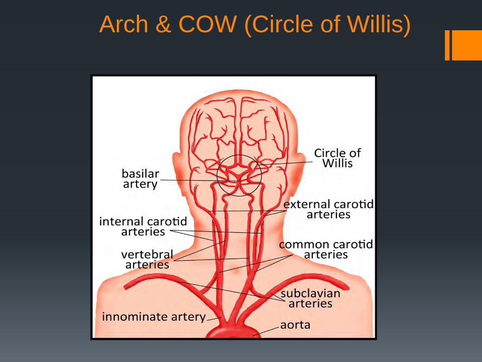
Arch & COW (Circle of Willis)
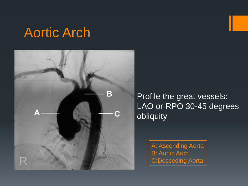
Aortic Arch
Profile the great vessels:LAO or RPO 30-45 degrees obliquity
A: Ascending AortaB: Aortic ArchC:Desceding Aorta
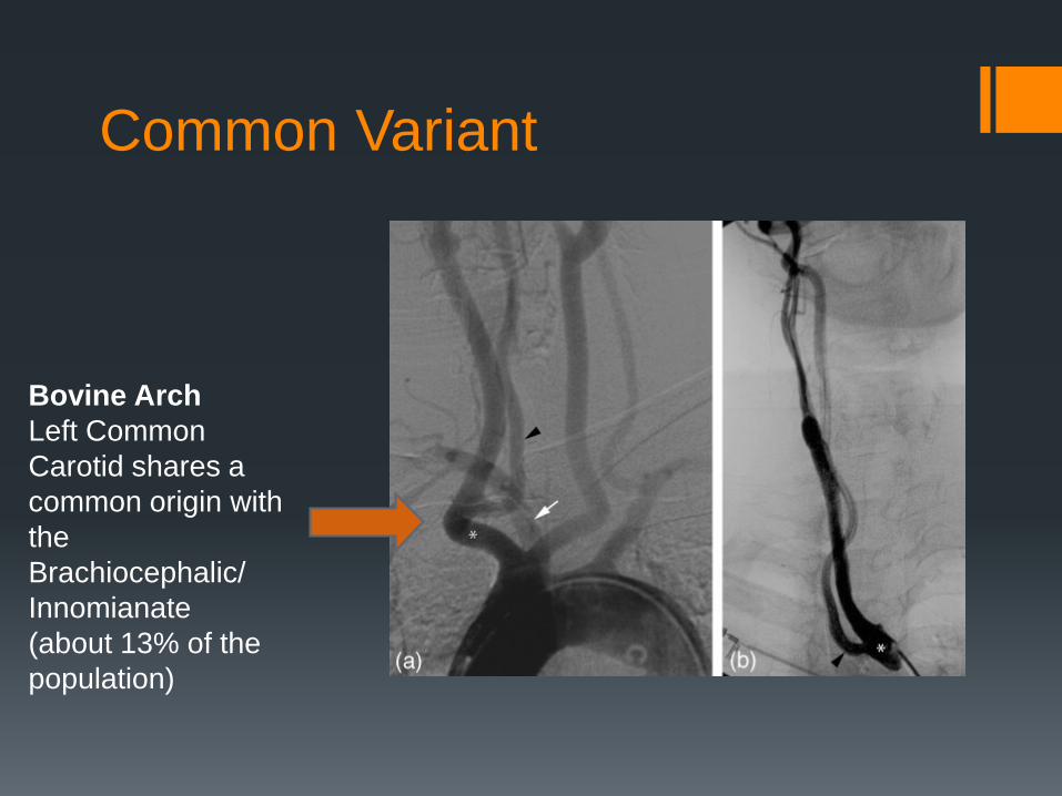
Common Variant
Bovine ArchLeft Common Carotid shares a common origin with the Brachiocephalic/Innomianate(about 13% of the population)
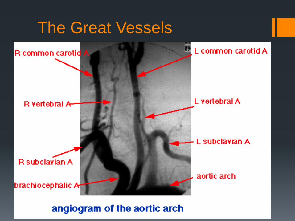
The Great Vessels

Complex Arches

Common Carotid Artery• Bifurcates into the
ECA and ICA at the level of the fourth cervical vertebrae.

Circle of Willis

The Circle of Willis “BI PAPA”Basilar TipInternal CarotidPosterior CerebralAnterior CerebralP Com(s)A Com
• The circle of Willis encircles the stalk of the pituitary gland and provides important communications between the blood supply of the forebrain and hindbrain
• A complete circle of Willis is present in most individuals, although a well-developed communication between each of its parts is identified in less than half of the population.

ACA

The Intracranial Circulation: Overview
• Composed of numerous blood vessels arising from bilateral Internal Carotid and Vertebral Arteries
• The anterior portion of the brain is supplied by the ICAs and is therefore called the anterior circulation
• The posterior portion of the brain is supplied by the VA’s and is therefore referred to as the posterior circulation
• The anterior and posterior circulations communicate via the Circle of Willis
Liebeskind D S Stroke 2003;34:2279-2284Copyright © American Heart Association
Lateral AP
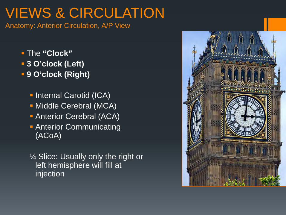
VIEWS & CIRCULATIONAnatomy: Anterior Circulation, A/P View
The “Clock” 3 O’clock (Left) 9 O’clock (Right)
Internal Carotid (ICA) Middle Cerebral (MCA) Anterior Cerebral (ACA) Anterior Communicating
(ACoA)
¼ Slice: Usually only the right or left hemisphere will fill at injection

The Anterior Circulation: OverviewAP views of the Anterior Circulation
ICA
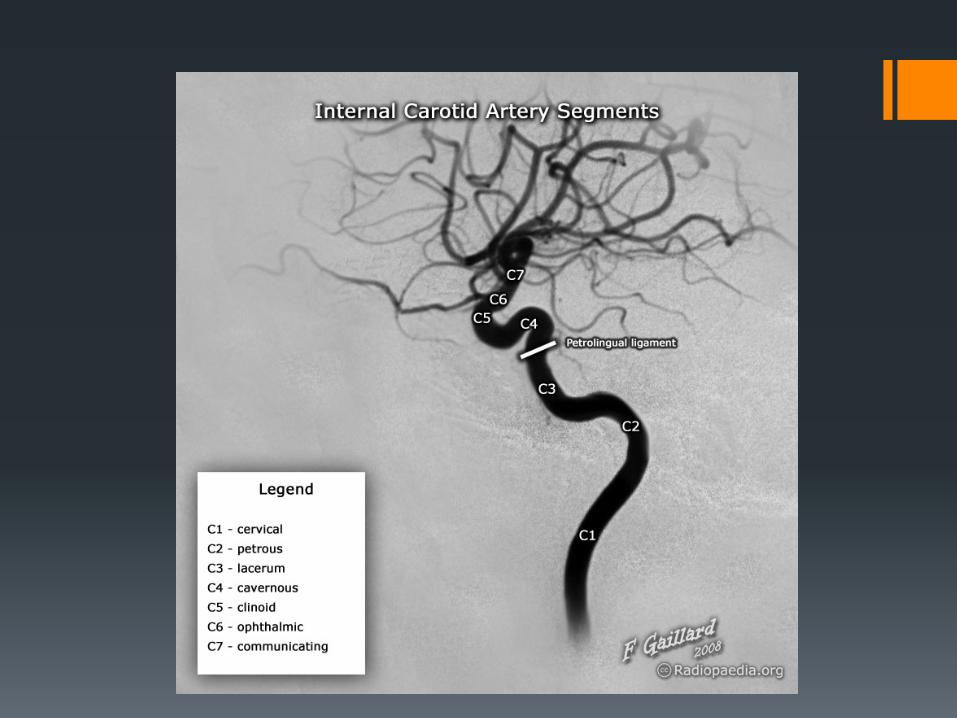

Internal Carotid Artery
1. Pericallosal artery
2. Callosomarginal artery
3. Anterior cerebral artery
4. Ophthalmic artery
5. Internal carotid artery
6. Anterior choroidal artery
7. Lenticulostriate arteries

Carotid Sinus• Where the common
carotid artery bifurcates.
• Contains specialized nerve end organs that produce a slight dilatation of the carotid artery which respond to changes in blood pressure by mediating changes in the heartbeat rate.
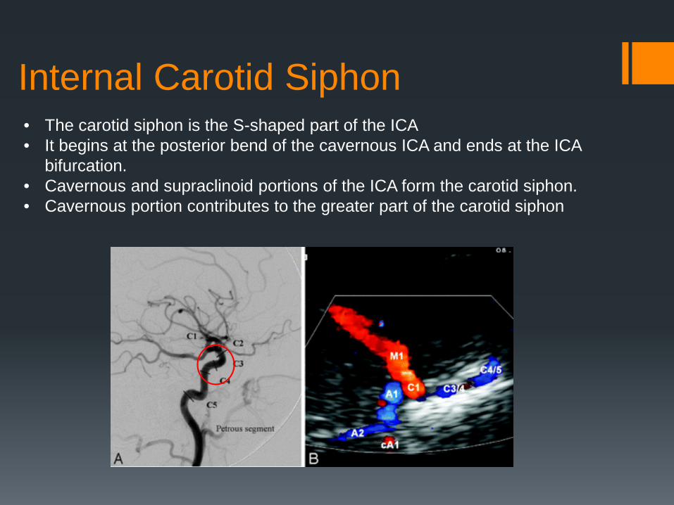
Internal Carotid Siphon• The carotid siphon is the S-shaped part of the ICA• It begins at the posterior bend of the cavernous ICA and ends at the ICA
bifurcation.• Cavernous and supraclinoid portions of the ICA form the carotid siphon. • Cavernous portion contributes to the greater part of the carotid siphon

MCA

The Posterior Circulation: Overview
The Vertebrobasilar System:
• Composed of the vertebral arteries which join intracranially to form the Basilar Artery
• The Basilar Artery terminates as the Posterior Cerebral Arteries (PCAs)
• Supplies the brainstem, cerebellum, and posterior cerebrum

Vertebral Artery
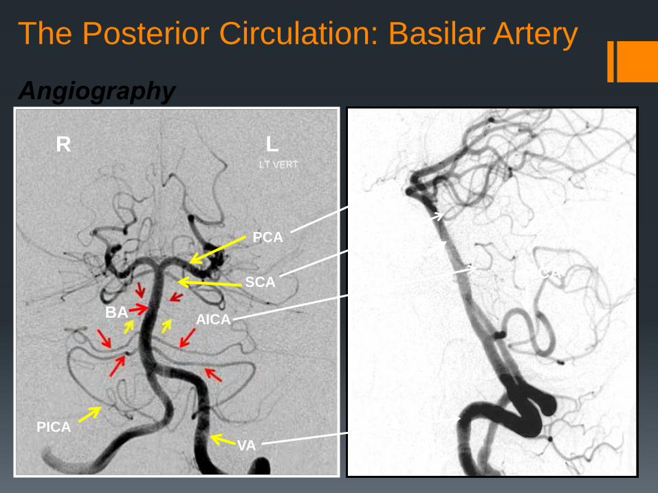
The Posterior Circulation: Basilar Artery
Angiography
PCA
SCA
AICA
PICA
BA
VA
R L
BAPICA

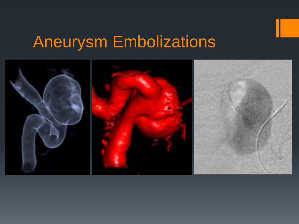
Aneurysm Embolizations

Onyx 500

Stent Coil of Basilar Tip Aneurysm
Enterprise through the Neuroform stent.

Left ICA Aneurysm

Before After
Images Provided by Ev3
6 months
Pipeline Embolization Device

Left ICA AVM

AVM Superior Cerebellar

Post Onyx

Meningioma Embolization
PVA used to embolize tumor preoperatively to reduce bleeding
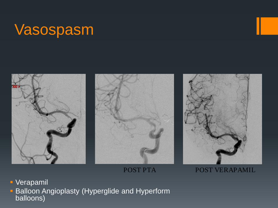
Vasospasm
Verapamil Balloon Angioplasty (Hyperglide and Hyperform
balloons)
POST PTA POST VERAPAMIL
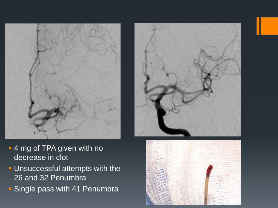
4 mg of TPA given with no decrease in clot Unsuccessful attempts with the
26 and 32 Penumbra Single pass with 41 Penumbra
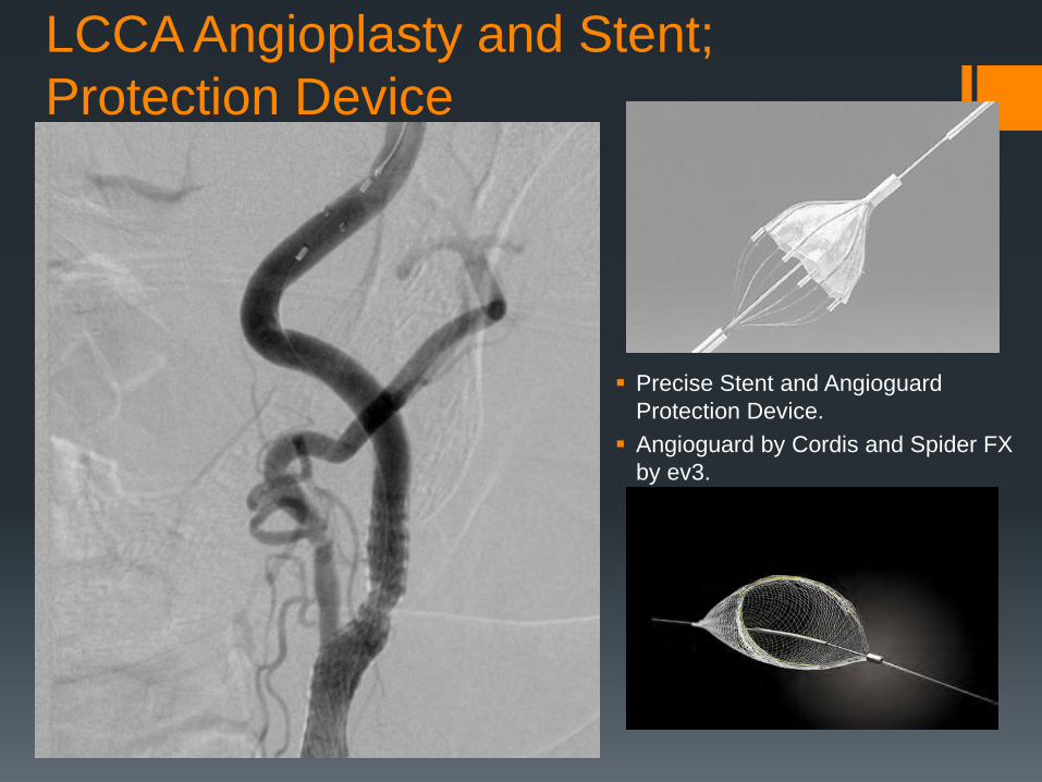
LCCA Angioplasty and Stent;Protection Device
Precise Stent and AngioguardProtection Device.
Angioguard by Cordis and Spider FX by ev3.

Subclavian Steal
The primary lesion causing vertebral artery flow reversal is proximal subclavian artery stenosis or occlusion, resulting in decreased blood pressure in the arm distal to the steno-occlusive disease.

Aneurysm Repair Understanding the history of coil embolization Familiarization with the causes of aneurysms Defining what an aneurysm is and where they tend to develop Fundamentals of treatment strategy

ACom ANEURYSM/OPTIC CHIASMA
A2
A1ACOM
OPTIC CHIASMA
ANEURYSM
TYPES / CLASSIFICATIONS OF ANEURYSMS
• SIDEWALL• BIFURCATIONS• TERMINAL• FUSIFORM• DISSECTING• MYCOTIC
(MOST COMMON)

FLOW PATTERNS
SIDE WALLS ANEURYSMS BIFURCATED ANEURYSMS

ETIOLOGY
• CONGENITAL ???• AQUIRED
CAUSATION
• HEMODYNAMICALLY INDUCED FLOW PATTERNS• DEGENERATIVE VASCULAR DISEASE• HYPERTENSION• CONNECTIVE TISSUE DISORDERS• ARTERIAL OCCLUSIVE LESIONS• IMBALANCE OF BLOOD FLOW AT ARTERIAL FORKS• ATHEROSCLEROSIS

CANINE SIDE WALL ANEURYSMS
FLOW PATTERNSI = inflowO = outflow

FLOW MODELS OF CADAVER ANEURYSMS
Bulbous PCom aneurysm, irregular flowIn the sac, highly disturbed flow.
Flow distal basilar aneurysm. Complexflow into sac. Swirls in basilar then hitsopposite wall in sac then out into PCA.
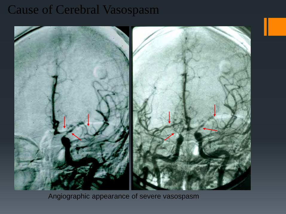
Angiographic appearance of severe vasospasm
Cause of Cerebral Vasospasm

Fusiform Basilar Aneurysm
Dilatation along a long segment of
a vessel
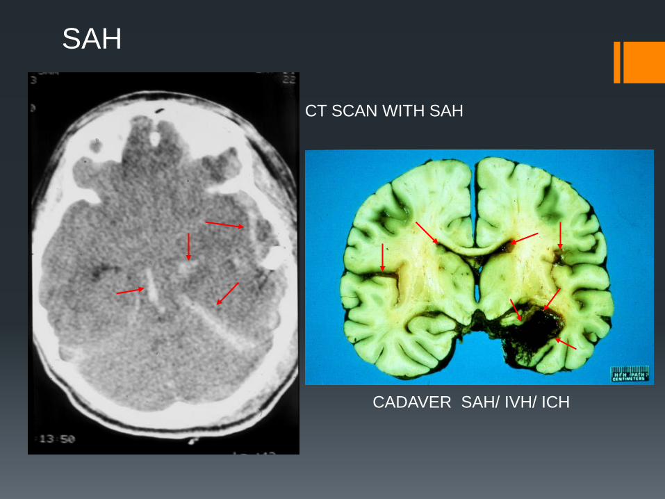
SAH
CT SCAN WITH SAH
CADAVER SAH/ IVH/ ICH

1. Anterior Communicating Artery: ACom2. Posterior Communicating Artery:PCom3. Middle Cerebral Artery: MCA4. Internal Carotid Artery:
(cavernous, supraclinoid, paraclinoid)5. Vertebrobasilar:
(basilar tip, PCA, SCA, PICA)6. Carotid-Ophthalmic
CIRCLE OF WILLIS
Site of Intracranial Aneurysms

1. Vertebrobasilar2. Cavernous carotid3. Carotid-ophthalmic/paraclinoid4. High Surgical risk5. Temporary protection for delayed
surgery
“Are all aneurysms are indicated for endovascular therapy?”
Aneurysms Indicated for Endovascular Tx.

GOAL OF ENDOVASCULAR TREATMENT
• FLOW DIVERSION AT THE INFLOW ZONE
• THROMBUS FORMATION
• TRANSFORMATION INTO CELLULAR TISSUE
• DEVELOPMENT OF ENDOTHIELIAL CELLS AT NECK
• DEVELOPMENT SMOOTH MUSCLE CELLS ALONG INFLOW
• COLLEGEN DEPOSITS AND FIBROCELLULAR OR GRANULATION TISSUE INSIDE SAC
Endovascular Treatment

Aneurysm Components
Neck Dome
Wall
Parent Artery
• Apex or dome• Wall• Ostium• Neck• Inflow/outflow zone• Parent artery junction
Aneurysm Characteristics
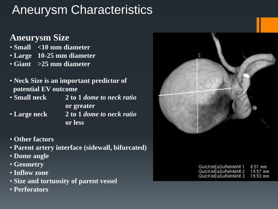
Aneurysm Size• Small <10 mm diameter• Large 10-25 mm diameter• Giant >25 mm diameter
• Neck Size is an important predictor ofpotential EV outcome
• Small neck 2 to 1 dome to neck ratioor greater
• Large neck 2 to 1 dome to neck ratioor less
• Other factors• Parent artery interface (sidewall, bifurcated)• Dome angle• Geometry• Inflow zone• Size and tortuosity of parent vessel• Perforators
What is an AVM?
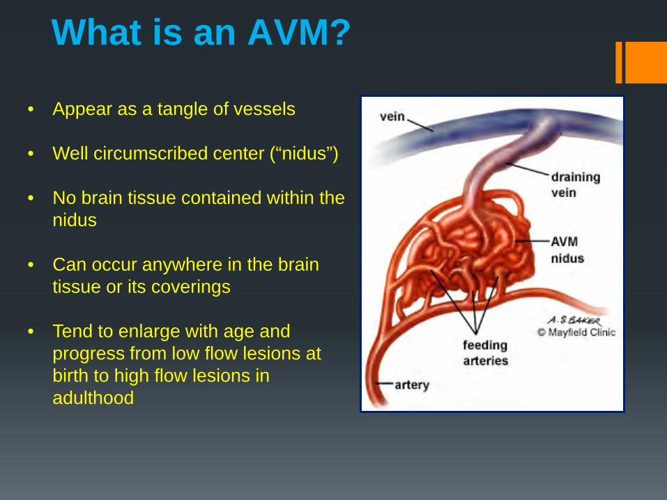
• Appear as a tangle of vessels
• Well circumscribed center (“nidus”)
• No brain tissue contained within the nidus
• Can occur anywhere in the brain tissue or its coverings
• Tend to enlarge with age and progress from low flow lesions at birth to high flow lesions in adulthood

AVM
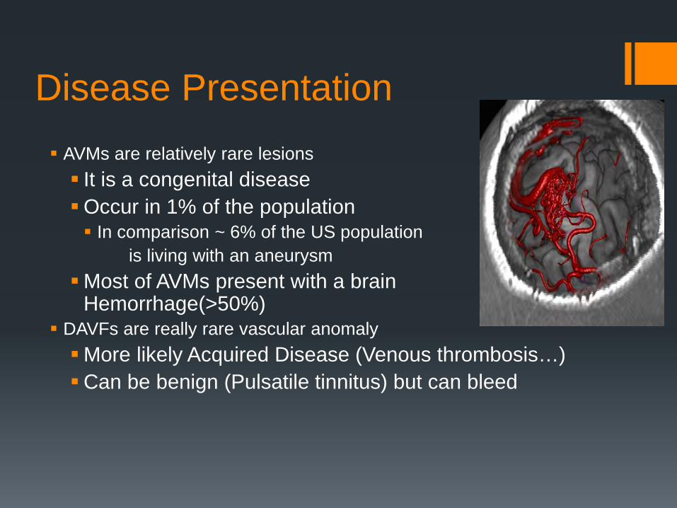
Disease Presentation AVMs are relatively rare lesions It is a congenital diseaseOccur in 1% of the population In comparison ~ 6% of the US population
is living with an aneurysmMost of AVMs present with a brain
Hemorrhage(>50%) DAVFs are really rare vascular anomalyMore likely Acquired Disease (Venous thrombosis…)Can be benign (Pulsatile tinnitus) but can bleed
Epidemiology
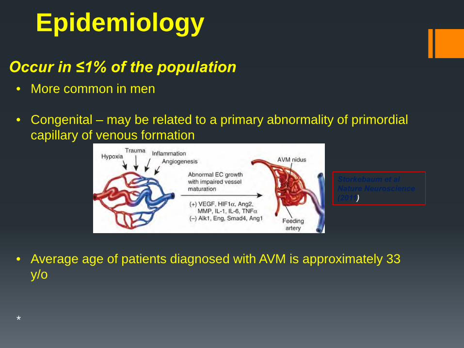
• More common in men
• Congenital – may be related to a primary abnormality of primordial capillary of venous formation
• Average age of patients diagnosed with AVM is approximately 33 y/o
Occur in ≤1% of the population
Storkebaum et al Nature Neuroscience (2011)
*

Presentation
1. Hemorrhage (>50%)2. Seizures (20-25%)3. Headaches (15%)4. Mass effect5. Ischemia & focal neuro deficit (due to
vascular steal)/less frequent
Whenever a young person presents with intracranial hemorrhage, vascular imaging is required to rollout vascular malformation

Presentation: Hemorrhage
• Most common presentation
• Peak age: 15-20 y/o
• 10% mortality and 30-50% morbidity with each bleed

Presentation: Hemorrhage Location of Hemorrhage 1. Intraparenchymal (ICH) – 82%
2. Intraventricular (IVH) • Usually accompanied by ICH• Pure IVH may indicate an
intraventricular AVM
3. Subarachnoid (SAH) – may be due to rupture of aneurysm on feeding artery
4. Subdural (SDH) – uncommon, but should be though of if SDH is spontaneous
ICH
IVH

Presentation: Hemorrhage Risk of Hemorrhage
• Average risk of hemorrhage is ~2-4% per year
• Risk of bleeding at least once in 25 years is ~ 53%
• After one hemorrhage the risk of rebleeding is ~ 6% per year
• Small AVMs are more likely to hemorrhage (higher pressure in feeding arteries)
Pollock, B et al Stroke 1996AVMs at high risk of bleeding:
• History of bleeding• Diffuse nidus• Only 1 draining vein
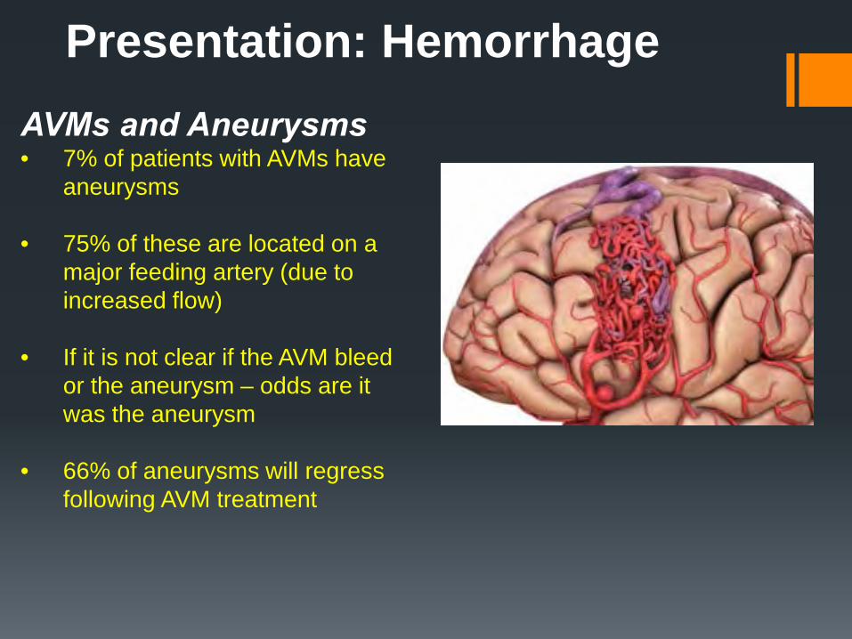
Presentation: Hemorrhage AVMs and Aneurysms• 7% of patients with AVMs have
aneurysms
• 75% of these are located on a major feeding artery (due to increased flow)
• If it is not clear if the AVM bleed or the aneurysm – odds are it was the aneurysm
• 66% of aneurysms will regress following AVM treatment

Consequences of Hemorrhage Symptoms of hemorrhage depend on location of the AVM and hemorrhage pattern
• Acute Symptoms:• Severe HA• Nausea/vomiting• LOC
• If IVH is present – may result in acute hydrocephalus
• Lasting neurological deficits due to damage to brain tissue from blood/blood break down products

Diagnosis/EvaluationCT Scan
• Usually the first study completed when the patient presents with acute symptoms
• Non-enhanced CT is the best study to r/o hemorrhage
• Can demonstrate calcifications within the lesion
nidus
Calcifications
ICH

Diagnosis/Evaluation MRI/MRA
• More sensitive than CT at diagnosing AVM
• Provides better information on exact location and surrounding structures
• Characteristic “serpentine” flow voids
• GRE sequences demonstrate surrounding hemorrhages which suggest a prior hemorrhage
• Useful for Stereotactic Radiosurgery planning and f/u
• MRI takes approx 1 hour so typically done once patient is stabilized

AVM AP

Diagnosis/Evaluation Angiography• Provides detailed angioarchitecture
• Can identify associated aneurysms
• Provides hemodynamic information including dominant arterial filling
• Note – draining veins are present in the arterial phase
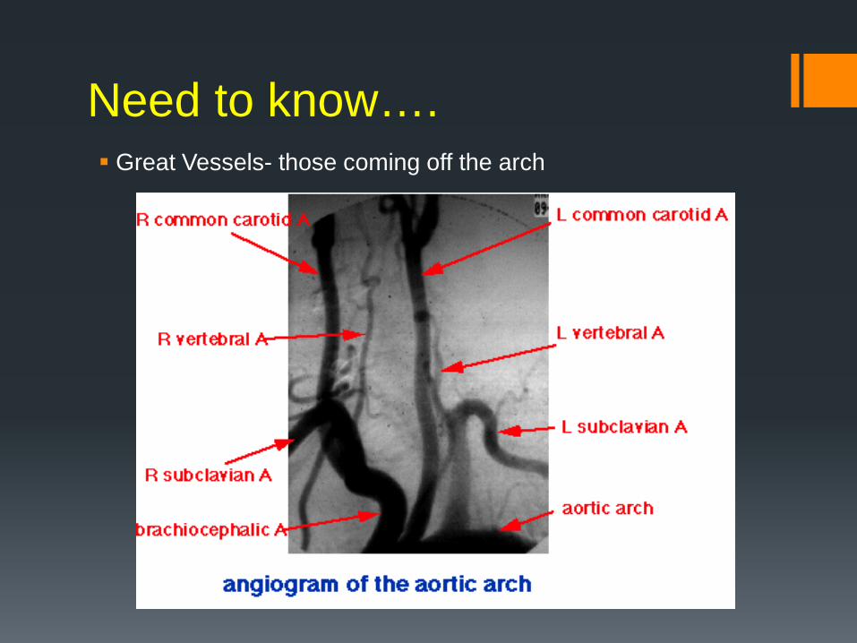
Need to know…. Great Vessels- those coming off the arch
COW Circle of Willis components Basilar (tip) Internal Carotid (tip) Posterior Cerebrals Anterior Cerebrals Pcomm(s) Acomm (1)

Circle of Willis

Know Basic Internal Carotid Artery

Nice to know… Variants Bovine Arch (Left Carotid off Innominate on the right) What happens with incomplete COWs
Diagnoses Vasospasm SAH on CT as predictor of aneurysm location
Carotid Siphon from Carotid SinusWhen to stop coiling

Thank you!
Related Documents