Interventional Cardiology

Welcome message from author
This document is posted to help you gain knowledge. Please leave a comment to let me know what you think about it! Share it to your friends and learn new things together.
Transcript

Interventional Cardiology

Amar S. Kapoor Editor
Interventional Cardiology
With 135 Figures
Springer-Verlag New York Berlin Heidelberg London Paris Tokyo
Amar S. Kapoor, MD, Kaiser Foundation Hospital, University of Southern California, Los Angeles, CA 90027, USA
Library of Congress Cataloging-in-Publication Data Interventional cardiology/ Amar S. Kapoor, editor.
p. cm. Includes bibliographies and index.
ISBN-13: 978-1-4612-8145-0 DOl: 10.1007/978-1-4612-3534-7
e-ISBN-13: 978-1-4612-3534-7
1. Heart-Diseases-Treatment. I. Kapoor, Amar S. [DNLM: 1. Cardiology-methods. 2. Heart Diseases-diagnosis.
3. Heart Diseases-therapy. 4. Heart Function Tests-methods. WG 200 163] RC683.8.157 1988 616.1 '2-dc19 DNLMIDLC for Library of Congress
Printed on acid-free paper
© 1989 by Springer-Verlag New York Inc. Softcover reprint of the hardcover 1st edition 1989
88-16016 CIP
All rights reserved. This work may not be translated or copied in whole or in part without the written permission of the publisher (Springer-Verlag, 175 Fifth Avenue, New York, NY 10010, USA), except for brief excerpts in connection with reviews or scholarly analysis. Use in connection with any form of information storage and retrieval, electronic adaptation, computer software, or by similar or dissimilar methodology now known or hereafter developed is forbidden. The use of general descriptive names, trade names, trademarks, etc. in this publication, even if the former are not especially identified, is not to be taken as a sign that such names, as understood by the Trade Marks and Merchandise Marks Act, may accordingly be used freely by anyone. While the advice and information in this book are believed to be true and accurate at the date of going to press, neither the authors nor the editors nor the publisher can accept any legal responsibility for any errors or omissions that may be made. The publisher makes no warranty, express or implied, with respect to the material contained herein.
Typeset by Bi-Comp, Inc., York, Pennsylvania.
9 8 7 6 5 4 3 2 1

Dedicated to dear Rinder for her infinite patience and
to the interventional cardiologists who perform complex therapeutic acts
of finger calisthenics with finesse
Preface
In the last decade, invasive procedures in cardiology have blossomed at an unprecedented rate. There is a sea of facts that has to be organized, assimilated, and applied for sound cardiac practice. We have come a long way from our conventional palliative treatment of acute myocardial infarction to a much more aggressive stance of contemporary interventional cardiac care. Patients with cardiovascular instability are not only monitored in a protective environment, but are treated with innovative approaches requiring aggressive interventions.
The traditional role of the cardiologist has also changed because of interventional cardiology. Interventional cardiology embraces the application of cardiac procedures and active intervention for diagnostic or therapeutic studies. For example, management of acute myocardial infarction could involve early drug therapy to preserve ischemic or stunned myocardium, thrombolytic therapy for clot dissolution, and acute revascularization by percutaneous transluminal coronary angioplasty. Some patients may need intra-aortic balloon counterpulsation for stabilization, whereas still a small number of patients may need electrophysiologic studies and implantation of antitachycardia devices or automatic defibrillators. Eventually, an occasional patient who develops end-stage ischemic cardiomyopathy may require cardiac assist devices and cardiac transplantation.
Interventions have become routine accepted practice. In this book, emphasis is placed on the indications, techniques, results, and merits of each procedure. Details of each procedure, instrumentation required, and the techniques are highlighted. This book is divided into five parts.
Part I discusses general principles of cardiac catheterization, hemodynamic measurements, cineangiographic views, and coronary angiography. Cardiac catheterization is fundamental for all invasive procedures, and one needs to have a solid background in this procedure before contemplating interventional cardiology.
Part II deals with diagnostic interventions. These are very important for precise and accurate determination of cardiac dysfunction. This kind of hemodynamic or electrophysiologic information is crucial for therapeutic decisions.
Part III details therapeutic interventions. This is an area where the
viii Preface
medical technology and complexity of cardiac procedures have grown exponentially. In this section, the latest technical and therapeutic information is provided in a practical format. All the interventional procedures in pediatric cardiology are discussed at length.
In Part IV the various facets of coronary angioplasty and its applications in different subsets of patients are discussed in depth. Coronary angioplasty is a highly technical procedure, requiring greater skills and care than routine coronary angiography. In this section on coronary interventions, there is also an explosion of information and technology with which we should become familiar. An attempt is made to address all these complex topics in a practical format.
Laser angioscopy and angioplasty are still investigational, but will get clinical application in the near future. In this field, there will be a starburst of information and innovations requiring updating. A glimpse into the future is provided.
Part V deals with cardiovascular crises and their management by acute pharmacologic interventions. In the setting of a cardiac intensive care unit, one must not only be knowledgeable about the pathophysiology of cardiovascular disease, but be well-versed in the pharmacology of cardiac drugs and their timely and appropriate use. In the management of acute myocardial infarction, we have come to know time is of the essence, and acute pharmacologic intervention becomes the "procedure" in the selected patient.
Thus, this book aspires to provide the guidelines for the modern cardiologist of today-one who uses modern techniques and technology and modern drugs for the management and prevention of cardiac disease"the interventional cardiologist."
AMAR S. KAPOOR

Contents
Preface ... Contributors
Part I Invasive Procedures
The Scope of Interventions in Cardiovascular Conditions AMAR S. KAPOOR ................ .
2 Techniques of Cardiac Catheterization and Coronary Angiography AMAR S. KAPOOR
3 Coronary Blood Flow and Coronary Vascular Reserve TERRENCE J.W. BARUCH, AMAR S. KAPOOR, and
vii xiii
3
10
PETER R. MAHRER . . . . . . . . . . . . . . . . . . .. 22
4 Quantitative Coronary Arteriography BUDGE H. SMITH, B. GREG BROWN, and HAROLD T. DODGE 35
5 Hemodynamic Monitoring by Pulmonary Artery Catheterization SURESH RAMAMURTI and AMAR S. KAPOOR.
6 Outpatient Cardiac Catheterization PETER R. MAHRER . . . . . . .
7 The Evolution of Coronary Artery Disease: New Definitions from Coronary Angioscopy JAMES S. FORRESTER, ANN HICKEY, FI~ANK LITVACK, and
48
63
WARREN GRUND FEST. . . . . . . . . . . . . . . . . 69

x Contents
Part II Diagnostic Interventions
8 Interventions in the Evaluation of Valvular Heart Disease INDUBALA N. VARDHAN and AMAR S. KAPOOR. . 81
9 Interventions for Evaluation of Myocardial Ischemia AMAR S. KAPOOR, DIANE SOBKOWICZ, and DAVID E. BLUMFIELD ............. .
10 Introduction to Clinical Electrophysiology DONALD G. RUBENSTEIN and CAROL ZAHER
11 Invasive Electrophysiologic Studies in the Evaluation and Treatment of Patients with Ventricular Arrhythmias
89
100
NICHOLAS J. STAMATO and MARK E. JOSEPHSON. . . . . 119
12 Electrophysiologic Approach to Patients with Supraventricular Tachycardia DONALD G. RUBENSTEIN and CAROL ZAHER
13 Technique of Pericardiocentesis and Intrapericardial Drainage
133
AMAR S. KAPOOR . . . . . . . . . . . . . . . . . . . . 146
14 Endomyocardial Biopsy Techniques and Interpretation AMAR S. KAPOOR and MIR ALI . . . . . . .
15 Dipyridamole Thallium for Evaluating Coronary Artery Disease DIANE SOBKOWICZ and DAVID E. BLUMFIELD.
Part III Therapeutic Interventions
16 Principles and Techniques of Intra-aortic Balloon Pump Counterpulsation
154
161
SHALE GORDON . . . . . . . . . . . . . . . . . . 171
17 Temporary and Permanent Pacemakers AMAR S. KAPOOR ........ .
18 Automatic Implantable Defibrillator: Six-Year Clinical Experience ENRICO P. VELTRI, MORTON M. MOWER, and MICHEL MIROWSKI . . . . . . . . . . . . .
19 Catheter Ablation Techniques for Treatment of Cardiac Arrhythmias
180
193
MELVIN M. SCHEINMAN . . . . . . . . . . . . . . . . . 201
Contents xi
20 Interventional Pediatric Cardiac Catheterization ZUHDI LABABIDI and IHAB ATTIA . . . . . . . 214
21 Balloon Aortic Valvuloplasty BRICE LET AC and ALAIN CRIBIER ........... 239
22 Peripheral Laser Thermal Angioplasty TIMOTHY A. SANBORN . . . . . . . . . . . . . . . . . . 254
Part IV Coronary Interventions
23 Practical Aspects of Coronary Angioplasty AMAR S. KAPOOR .......... .
24 Complex Coronary Angioplasty: The Outcome and Long-term Effect of Angioplasty in Multivessel Coronary Disease and Multiple Lesion Anigoplasty
267
GERALD DORROS, RUBEN F. LEWIN, and LYNNE MATHIAK. . 281
25 Management of Early and Late Complications of Coronary Angioplasty AMAR S. KAPOOR and PETER R. MAHRER. . . . .... 306
26 Percutaneous Transluminal Coronary Stenting: ANew Approach to Unresolved Problems in Coronary Angioplasty ULRICH SIGWART, SVEIN GOLF, URS KAUFMANN, LUKAS KAPPENBERGER, ADAM FISCHER, and HOSSEIN SADEGHI . . . . . . . . . . . . . . . . . . . . 314
27 Laser Angioplasty of the Coronary Arteries GARRETT LEE, REGINALD I. Low, AGUSTIN J. ARGENAL, ROLF G. SOMMERHAUG, MING C. CHAN, and DEAN T. MASON 319
Part V Acute Pharmacologic and Surgical Interventions
28 Platelet Inhibitor Drugs in Coronary Artery Disease and Coronary Intervention DOUGLAS H. ISRAEL, BERNARDO STEIN, and VALENTIN FUSTER . . . . . . . . . . . . . . . . . . . . 329
29 Thrombolysis in Acute Myocardial Infarction DAVID E. BLUMFIELD . . . . . . . . .. ....... 355
30 Interventional Approach in the Management of Cardiogenic Shock AMAR S. KAPOOR . . . . . . . . . . . . . . . . . . . . 368

xii
31 Emergency Coronary Artery Bypass Surgery for Acute Coronary Syndromes SAMUEL L. SELINGER, RALPH BERG JR, WILLIAM S. COLEMAN, JACK J. LEONARD, and
Contents
MARCUS A. DEWOOD. . . . . . . . . . . . . . . . . . . 377
32 Cardiac Transplantation DAVIS DRINKWATER, LYNNE WARNER STEVENSON, and HILLEL LAKS . . . . . . . . . . . . . . . . . . . . . . 387
Index . . . . . . . . . . . . . . . . . . . . . . . . . . . 401
Contributors
MIR ALI, MD
Fellow in Cardiology, Kaiser Foundation Hospital, Los Angeles, California, USA
AGUSTIN J. ARGENAL, MD, FACC
Director, Cardiac Catheterization Laboratory, Northern California Heart and Lung Institute, Concord, California, USA
IHAB ATTIA, MD
Fellow in Cardiology, University of Missouri Hospital and Clinics, Columbia, Missouri, USA
TERRENCE J. W. BARUCH, MD
Fellow in Cardiology, Kaiser Foundation Hospital, Los Angeles, California, USA
RALPH BERG JR, MD
Cardiovascular Surgery, Sacred Heart Medical Center, Spokane, Washington, USA
DAVID E. BLUMFIELD, MD, FACC
Director, Coronary Care Unit, Kaiser Foundation Hospital, Los Angeles, California, USA
B. GREG BROWN, MD, PHD
Professor of Medicine, University of Washington, School of Medicine, Seattle, Washington, USA
MING C. CHAN, MD
Professor of Medicine, Chairman, Department of Medicine, Beijing Medical University, Beijing, China
WILLIAM S. COLEMAN, MD, FACC, FACS
Caridovascular Surgery, Sacred Heart Medical Center, Spokane, Washington, USA
ALAIN CRISlER, MD
Associate Professor of Cardiology, Centre Hospitalo-U niversitaire de Rouen, Rouen, France

xiv Contributors
MARCUS A. DEWOOD, MD, FACC
Director, Cardiology Research, Sacred Heart Medical Center, Spokane, Washington, USA
HAROLD T. DODGE, MD, FACC
Professor of Medicine, University of Washington, Director, Division of Cardiology, Seattle, Washington, USA
GERALD DORROS, MD, FACC
Assistant Clinical Professor of Medicine, St. Luke's Medical Center, Milwaukee, Wisconsin, USA
DAVIS DRINKWATER, MD
Assistant Professor of Surgery, Cardiothoracic Surgery, University of California, Los Angeles, California, USA
ADAM FISCHER, MD
Staff Cardiovascular Surgeon, Centre Hospitalier Universitaire Vaudois, Lausanne, Switzerland
JAMES S. FORRESTER, MD, FACC
Professor of Medicine, University of California, Director of Cardiovascular Research, Cedars-Sinai Medical Center, Los Angeles, California, USA
VALENTIN FUSTER, MD, FACC
Arthur M. and Hilda A. Master Professor of Medicine, Mount Sinai School of Medicine, Chief of Cardiology, Mount Sinai Medical Center, New York, New York, USA
SVEIN GOLF, MD
Fellow in Interventional Cardiology, Centre Hospitalier U niversitaire Vaudois, Lausanne, Switzerland
SHALE GORDON, MD
Staff Cardiologist, Kaiser Medical Center, Bellflower, California, USA
WARREN GRUND FEST , MD
Department of Surgery, Cedars Sinai Medical Center, Los Angeles, California, USA
ANN HICKEY, MD
Staff Cardiologist, Cedars Sinai Medical Center, Los Angeles, California, USA
DOUGLAS ISRAEL, MD
Fellow in Cardiology, Mount Sinai School of Medicine, New York, New York, USA
MARK E. JOSEPHSON, MD, FACC
Director, Division of Cardiology, Robinette Foundation Professor of Medicine, University of Pennsylvania, School of Medicine, Philadelphia, Pennsylvania, USA
AMAR S. KAPOOR, MD, FACP, FACC
Director of Cardiovascular Research, Regional Medical Director of Heart Transplant Program, Kaiser Foundation Hospital, Clinical

Contributors
Associate Professor of Medicine, University of Southern California, Los Angeles, California, USA
LUKAS KAPPENBERGER, MD
Professor of Medicine, Chief of Cardiology, Centre Hospitalier Universitaire Vaudois, Lausanne, Switzerland
URS KAUFMANN, MD
xv
Staff Cardiologist, Centre Hospitalier Universitaire Vaudois, Lausanne, Switzerland
ZUHDI LABABIDI, MD, FACC
Director, Division of Pediatric Cardiology, Professor of Pediatric Cardiology, University of Missouri Hospital and Clinics, Columbia, Missouri, USA
HILLEL LAKS, MD, FACS
Director, Cardiovascular Surgery and Cardiac Transplantation, Professor of Surgery, University of California, Los Angeles, California, USA
GARRET T. LEE, MD, FACC
Director of Research, Northern California Heart and Lung Institute, Mount Diablo Hospital Medical Center, Concord, California, USA
JACK J. LEONARD, MD
Cardiovascular Surgery, Sacred Heart Medical Center, Spokane, Washington, USA
BRICE LETAC, MD
Professor of Cardiology, Chief of Cardiology, Centre Hospitalo-Universitaire de Rouen, Rouen, France
RUBEN F. LEWIN, MD
Isadore Feuer Fellow in Interventional Cardiology, St. Luke's Medical Center, Milwaukee, Wisconsin, USA
FRANK LITVACK, MD
Assistant Professor of Medicine, University of California, Director, Catheterization Laboratory, Cedars-Sinai Medical Center, Los Angeles, California, USA
REGINALD 1. Low, MD, FACC
Interventional Cardiologist, Diagnostic and Interventional Cardiology Consultants, Sacramento, California, USA
PETER R. MAHRER, MD, FACC
Director, Division of Cardiology, Director, Regional Cardiac Catheterization Laboratory, Kaiser Foundation Hospital, Clinical Professor of Medicine, University of Southern California, Los Angeles, California, USA
DEAN T. MASON, MD, FACC
Physician-in-Chief, Western Heart Institute, Chairman, Department of Cardiology, St. Mary's Hospital Medical Center, San Francisco, California, USA
xvi Contributors
LYNNE MA THIAK, RN St. Luke's Medical Center, Milwaukee, Wisconsin, USA
MICHEL MIROWSKI, MD, FACC Director, Coronary Care Unit, Professor of Medicine, John Hopkins School of Medicine, Sinai Hospital of Baltimore, Baltimore, Maryland, USA
MORTON M. MOWER, MD, FACC Director, Cardiology Division, Sinai Hospital of Baltimore, Baltimore, Maryland, USA
SURESH RAMAMURTI, MD Staff Cardiologist, Kaiser Medical Center, Panorama City, California, USA
DONALD G. RUBENSTEIN, MD Staff Cardiologist, Kaiser Medical Center, Panorama City, California, USA
HOSSEIN SADEGHI, MD Professor of Surgery, Chief of Cardiovascular Surgery, Centre Hospitalier Universitaire Vaudois, Lausanne, Switzerland
TIMOTHY A. SANBORN, MD Associate Professor of Medicine, Mount Sinai School of Medicine, Director, Interventional Cardiology Research and Laser Angioplasty Program, Mount Sinai Hospital, New York, New York, USA
MELVIN M. SCHEINMAN, MD, FACC Director, Electrophysiology Laboratory, Moffit Hospital, Professor of Medicine, University of California, San Francisco, California, USA
SAMUEL L. SELINGER, MD, FACS, FACC Cardiovascular Surgery, Sacred Heart Medical Center, Spokane, Washington, USA
ULRICH SIGWART, MD, FACC Associate Professor of Medicine, Centre Hospitalier Universitaire Vaudois, Division of Cardiology, Lausanne, Switzerland
BUDGE H. SMITH, MD Acting Instructor, University of Washington, Division of Cardiology, Seattle, Washington, USA
DIANE SOBKOWICZ, MD Fellow in Cardiology, Kaiser Foundation Hospital, Los Angeles, California, USA
ROLF G. SOMMERHAUG, MD Chief, Cardiac, Thoracic and Vascular Surgery, Northern California Heart and Lung Institute, Concord, California, USA
NICHOLAS J. STAMATO, MD, FACC Clinical Assistant Professor of Medicine, Loyola University, Stritch School of Medicine, Midwest Heart Specialists, Lombard, Illinois, USA
Contributors
BERNARDO STEIN, MD
Fellow in Cardiology, Mount Sinai School of Medicine, New York, New York, USA
LYNNE WARNER STEVENSON, MD
xvii
Assistant Professor of Medicine, University of California, Los Angeles, California, USA
INDUBALA N. V ARDHAN, MD
Fellow in Cardiology, Kaiser Foundation Hospital, Los Angeles, California, USA
ENRICO P. VELTRI, MD
Director, Sudden Death Prevention Program, Sinai Hospital of Baltimore, Baltimore, Maryland, USA
CAROL ZAHER, MD, FACC
Chief of Cardiology and Director of Electrophysiology, Kaiser Medical Center, Panorama City, California, USA

Part I Invasive Procedures

1 The Scope of Interventions in Cardiovascular Conditions Amar S. Kapoor
Introduction There have been extraordinary changes in our understanding of the pathophysiology of myocardial ischemia and acute myocardial infarction. The changes are so phenomenal that we have to change our evaluation and management of the patient afflicted with coronary artery disease. A decade ago, we believed that fixed atherosclerotic lesions were the main cause of a reduced blood supply to the myocardium. There is convincing evidence in humans that there can be dynamic shifts in luminal diameter with a resultant change in the vasomotor tone of the artery. Vasomotor changes affect epicardial, intramyocardial, and collateral vessels.
Recently, we have come to realize that patients with coronary artery disease may have frequent episodes of silent ischemia along with symptomatic ischemia or angina. The total sum of silent episodes and symptomatic episodes has been called the "total ischemic burden. "I This concept has propelled us to rethink our existing methods of detecting, estimating, classifying, and managing angina pectoris. Cohen 1 and others have brought to surface that not only patients with unstable angina, but even patients with stable angina pectoris, may have frequent episodes of silent ischemia at rest and low levels of activity. This concept will ultimately usher in newer methods of detecting and classifying ischemia. One classification, according to Cohen,2 includes primary, secondary, and mixed isch-
emia. Primary ischemia is due to decreased delivery of arterial blood or oxygen supply to the myocardium because of increased vasoconstrictor tone or segmental coronary artery spasm. Secondary ischemia is due to increased myocardial oxygen demand because of fixed atherosclerotic stenosis and is usually brought on by exertion. Mixed ischemia can be brought on by low-level activity, rest, or exertion and is due to a combination of segmental spasm occurring at the site of a fixed atherosclerotic lesion, and there may be increased vasomotor tone in a more diffuse form.1.2 There will be new technology for quantitating total ischemic burden. At the present time, there are no long-term studies to inform us of the significance, risk, and prognosis due to total ischemic burden.
There is a sequence of pathophysiologic events during the development of an ischemic event. After an imbalance in myocardial oxygen supply and demand, a chain of events is set off representing the ischemic cascade. 3 After the ischemic cascade begins, there is an overall decrease in left ventricular systolic function and a decrease in diastolic compliance with an increase in left ventricular enddiastolic pressure, ultimately culminating in a silent or symptomatic ischemic episode. When ischemic episodes are prolonged, they may affect myocardial function at the cellular level by altering biochemical processes and causing dysfunction of the myocardial ultrastructure resulting in a stunned myocardium. Repeated, prolonged postischemic episodes of stunning

4
may result in left ventricular dysfunction. The stunned left ventricular dysfunction recovers over hours and days. There is another concept of reversible chronic myocardial ischemia labeled "hibernating myocardium. "4 This concept was introduced by Rahimtoola. 5 Hibernation results from prolonged inadequate blood flow to a region of the myocardium. Hibernation can persist for weeks, months, or possibly years. It is possible that areas of stunned myocardium could coexist with areas that are hibernating. The fundamental mechanisms for both stunned and hibernating myocardium have not been worked out, but it seems they are protective mechanisms in that they reduce the oxygen supply of the impaired myocardium.
It is very plausible that interventions that improve oxygen supply and restore adequate blood supply may be therapeutic modalities for confronting total ischemic burden and stunned or hibernating myocardium.
More research and new technology will develop to quantify total ischemic burden and hibernating myocardium, although positron emission tomography may assess metabolic viability of the myocardium and predict reversibility of wall motion abnormalities. 6 In some cases with extensive stunned myocardium undergoing surgical revascularization, hemodynamic and pharmacologic support, along with intra-aortic balloon counterpulsation and a left ventricular assist device, may be necessary during the operative intervention, when the severely stunned myocardium is further exposed to prolonged periods of ischemia.7 These therapeutic interventions will improve patient survival, but further testing is necessary.
Interventions for Coronary Artery Disease
There have been rapid strides in the evaluation, quantification, and management of coronary artery disease states (Table 1.1). There have been unprecedented technologic advances in catheters, balloons, blades, intra-
A.S. Kapoor
TABLE 1.1. Interventions for coronary artery disease.
Diagnostic interventions Coronary angiography Coronary angioscopy Stress atrial pacing Stress echocardiography Ergonovine provocation test
Therapeutic interventions Coronary angioplasty Laser angioplasty Atherectomy Intracoronary thrombolytic agents Intracoronary prosthesis Surgical coronary revascularization
coronary prosthetic devices, and laser systems to deal with the atherosclerotic plaque and intracoronary thrombosis.
Indeed, there have been equally impressive feats on the pharmacologic front to lyse the clot with a variety of thrombolytic agents and other pharmacologic interventions to limit infarct size. Very early administration of intravenous streptokinase to patients with acute myocardial infarction has been shown conclusively to decrease morbidity and mortality when compared with conventional therapy as shown by GISSI study. 8
In the realm of diagnostic interventions, there has been a steady proliferation of techniques to better define coronary arterial lesions and attempts to quantitate acute symptomatic and silent ischemic episodes. Our understanding of the pathophysiology of coronary artery disease syndromes is beginning to unfold, and recent studies by coronary angioscopy will allow better understanding of the atherosclerotic plaque: how it ruptures and how the thrombus sets up the stage for various ischemic and arrhythmic cardiac events. At this stage of our learning, the pathophysiology of acute ischemic events is at a higher level of understanding, although somewhat speculative.
There have been new developments in the detection and quanti tat ion of coronary artery obstructions by quantitative coronary angiography, digital subtraction angiography, and coronary interventions such as stress atrial
1. Interventions in Cardiovascular Conditions
pacing and provocative ergonovine tests. These subjects will be covered in subsequent chapters.
The contemporary practice of cardiac catheterization is heavily dependent on modern catheterization technology to perform excellent selective coronary cineangio~raphy and for detailed evaluation of coronary morphology. Coronary angioplasty also has introduced a whole array of catheters, balloons, and accessories. The developments in this field are going to escalate at an exponential rate, and it is very difficult to predict at this time the optimal armamentarium.
In short, the scope of interventions in the detection and management of various coronary artery disease syndromes is wide open and expanding in the direction of innovation, feasibility, and safety. The cost and benefit of these procedures and interventions have not been evaluated properly in a systematic and controlled fashion.
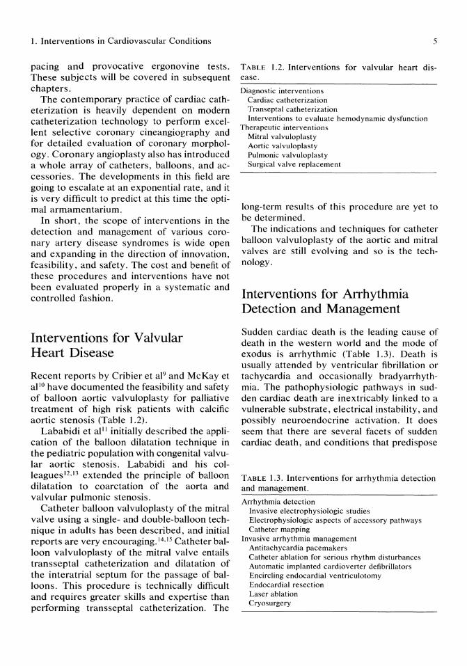
Interventions for Valvular Heart Disease
Recent reports by Cribier et al9 and McKay et al lO have documented the feasibility and safety of balloon aortic valvuloplasty for palliative treatment of high risk patients with calcific aortic stenosis (Table 1.2).
Lababidi et all 1 initially described the application of the balloon dilatation technique in the pediatric population with congenital valvular aortic stenosis. Lababidi and his colleagues I2 ,13 extended the principle of balloon dilatation to coarctation of the aorta and valvular pulmonic stenosis.
Catheter balloon valvuloplasty of the mitral valve using a single- and double-balloon technique in adults has been described, and initial reports are very encouraging. 14,15 Catheter balloon valvuloplasty of the mitral valve entails trans septal catheterization and dilatation of the interatrial septum for the passage of balloons. This procedure is technically difficult and requires greater skills and expertise than performing trans septal catheterization. The
5
TABLE 1.2. Interventions for valvular heart disease.
Diagnostic interventions Cardiac catheterization Transeptal catheterization Interventions to evaluate hemodynamic dysfunction
Therapeutic interventions Mitral valvuloplasty Aortic valvuloplasty Pulmonic valvuloplasty Surgical valve replacement
long-term results of this procedure are yet to be determined.
The indications and techniques for catheter balloon valvuloplasty of the aortic and mitral valves are still evolving and so is the technology.
Interventions for Arrhythmia Detection and Management
Sudden cardiac death is the leading cause of death in the western world and the mode of exodus is arrhythmic (Table 1.3). Death is usually attended by ventricular fibrillation or tachycardia and occasionally bradyarrhythmia. The pathophysiologic pathways in sudden cardiac death are inextricably linked to a vulnerable substrate, electrical instability, and possibly neuroendocrine activation. It does seem that there are several facets of sudden cardiac death, and conditions that predispose
TABLE 1.3. Interventions for arrhythmia detection and management.
Arrhythmia detection Invasive electrophysiologic studies Electrophysiologic aspects of accessory pathways Catheter mapping
Invasive arrhythmia management Antitachycardia pacemakers Catheter ablation for serious rhythm disturbances Automatic implanted cardioverter defibrillators Encircling endocardial ventriculotomy Endocardial resection Laser ablation Cryosurgery

6
to myocardial dysfunction, such as cardiomyopathies, left ventricular aneurysm, and ischemic syndromes, may very well form the sudden death substrate. However, ventricular arrhythmias may occur independent of left ventricular dysfunction.
It is very difficult to combat sudden cardiac death because it occurs within seconds to minutes with no warning of impending death. With the advent of cardiopulmonary resuscitation, many patients are taken to the hospital so that electrophysiologic and effective pharmacologic interventions can be instituted because empiric therapy has been a dismal failure. As a result of this, there have been remarkable developments in the techniques of programmed stimulation and endocardial catheter recording. 16,17 Electrophysiologic study can provide objective evidence for certain therapeutic modalities. One can assess the efficacy of pharmacologic therapy, pacemaker therapy, and guidance for surgical excision. Inability to initiate the tachycardia in the presence of an antiarrhythmic predicts that the drug will effectively prevent clinical recurrences. 18
An alternative to drug therapy is antitachycardia pacemakers, and a prerequisite to pacemaker therapy is that the arrhythmia can be terminated by pacing. Several specially designed antitachycardia pacing modalities are available that use underdrive pacing, automatic scanning, overdrive pacing, and burst pacing. Mirowski and co-workers l9 are credited with the development and implantation of the automatic implantable cardioverter defibrillator to be used as the electric intervention in patients with recalcitrant ventricular tachycardia and sudden cardiac death. This device is highly effective in candidates in whom drug therapy has failed and in survivors of sudden cardiac death. Future refinements of the device are expected and will include miniaturization of the generator with built-in programmable functions.
Some patients are candidates for intraoperative mapping and surgical procedures like subendocardial resection, cryosurgery, and laser ablation of ventricular foci of arrhythmias.
Scheinman and others20 described a very
A.S. Kapoor
important innovation in the management of drug-resistant cardiac arrhythmias by using catheter ablative techniques. This technique was used for ablation of the atrioventricular (A V) junction and more recently has been used in patients with accessory pathways and ventricular tachycardia.
It seems that electrical catheter ablation of the A V junction will supplant the need for cardiac surgical procedures to disrupt A V conduction. There are many management strategies for dealing with supraventricular and ventricular tachyarrhythmias, so one must carefully select patients for each therapeutic modality, and this can best be accomplished by experienced electrophysiologists.
Interventions to Evaluate and Treat Cardiomyopathies and End-Stage Heart Disease
Dysfunction of the myocardium, especially the dilated or primary cardiomyopathy, is characterized by a large, dilated heart with impairment of systolic pump function and is often associated with features of congestive heart failure. Radionuclide ventriculography and two-dimensional echocardiography can assist in establishing the diagnosis. Cardiac catheterization may reveal elevated left ventricular end-diastolic pressure, pulmonary capillary wedge pressure, and pulmonary arterial pressure. Pulmonary artery catheterization is extremely useful in assessing response to therapy (Table 1.4).
Endomyocardial biopsy is very useful in suspected myocarditis or secondary cardiomyopathies. Endomyocardial biopsy is also applicable in the evaluation of cardiac allograft rejection, adriamycin cardiotoxicity, and infilterative cardiomyopathies. The procedure can be performed in a fluoroscopic room on an outpatient basis. Endomyocardial biopsy is very good for analysis of endocardium at the cellular and subcellular level and has been used in research in the areas of receptor enzymology, immunology, and drug interactions. 22 ,23

1. Interventions in Cardiovascular Conditions
TABLE 1.4. Interventions to evaluate and treat cardiomyopathies and end-stage heart disease.
Interventions for evaluating cardiomyopathies Pulmonary artery catheterization Endomyocardial biopsy
Management strategies for end-stage heart disease Inotrope and vasodilatory pharmacologic support Intra-aortic balloon pump counterpulsation Left ventricular assist devices Total artificial heart Cardiac transplantation Cardiomyoplasty
Severe congestive heart failure will frequently develop secondary to coronary artery disease or idiopathic dilated cardiomyopathy. Patients with a catastrophic myocardial infarction can develop cardiogenic shock with irreversible myocardial dysfunction. Mechanical cardiac assistance and specific pharmacologic therapy may be necessary to restore adequate tissue perfusion. Optimal cardiac output could be restored with inotropic agents and vasodilators.
Mechanical assistance in the form of intraaortic balloon counterpulsation is useful in stabilizing patients when the underlying etiology is ischemic. There have been major advances in the use of mechanical devices to support cardiovascular circulation. Several ventricular assist devices are available as short-term circulatory supports. 24 Beside assisting patients with low output syndromes and cardiogenic shock, the devices are increasingly being used as a bridge to transplantation. Total artificial hearts have been used as a bridge to transplantation. 25 A temporary pneumatic artificial heart was first implanted by Cooley in 1969 and the patient lived 64 hours,26 but the total artificial heart implanted by DeVries, the Jarvik-7, was successful in sustaining life for 112 daysY These human experiments demonstrated the feasibility of the pneumatic heart as a temporary or even a permanent life-sustaining device for the patient awaiting definitive treatment, such as cardiac transplantation.
At present, the use of total artificial hearts for permanent heart replacement is deferred, but instead they are being frequently used
7
along with pulsatile ventricular assist devices as interim supports before cardiac transplantation. 28 Patients who have benefitted are those in cardiogenic shock, acute cardiac transplant rejection, and postcardiotomy patients who cannot be weaned from extracorporeal circulation.
The National Heart, Lung, and Blood Institute Artificial Heart Program is funding research on thermally powered ventricular assist devices and fully implantable electrical total artificial hearts. Complications that have emerged from use ofthe Jarvik-7 heart include strokes caused by thrombi forming at seams and valve mountings, infection, surgical bleeding, renal failure, and multiorgan failure.
Cardiac transplantation, on the other hand, has emerged as an excellent therapeutic modality for end-stage irreversible heart disease with 1 -year survival at 85% on cyclosporine immunosuppressive therapy. 29 Infection and rejection remain the principal complications in these patients. The donor supply is an important limiting factor. Because of the shortage of donors, various innovative techniques are in progress to augment cardiac output by cardiomyoplasty and other techniques.
Conclusion
Conventional modes of therapy have their own time honored place in the management of various cardiovascular conditions. The interventional approach refers to diagnostic and therapeutic interventions designed to achieve prompt and accurate diagnosis and immediate or timely results by nonsurgical and often surgical modes oftherapy. Clinical outcomes, initial and long-term improvement, and prognosis by these various interventions need to be studied by longitudinal, controlled trials. Interventions to limit the area of infarction in acute myocardial infarction have been extensively studied. It has become abundantly clear that there is a narrow window of time for acute myocardial infarction intervention for it to become effective. Thrombolytic therapy is a time-critical intervention, but in patients with initially successful thrombolysis, urgent coro-
8
nary angioplasty offers no clear advantage over delayed elective angioplasty. 31
Interventions in cardiology will be under scrutiny for several years before getting general acceptance. At the present time there is healthy skepticism for most of the recent diagnostic and therapeutic interventions, despite the fact that there is a tidal wave sweeping the frontiers of cardiology. The balloon and the catheter have added tremendously to our therapeutic armamentarium. The blade and laser are on the horizon.
The scope and future of interventions will be guided by the need for refinements of the procedure, the risk and safety to the patient, the efficacy and benefit of the intervention, and, most importantly, the ability of the medical dollar to justify the cost.
In brief, the scope and future role of interventions in cardiology are taking a giant leap forward to very complex and sophisticated technology requiring very specialized skills for the interventionist.
References
1. Cohen PF: Total ischemic burden: Pathophysiology and prognosis. Am J Cardiol 1987, 59:3C-6.
2. Cohen PF: Total ischemic burden: Definition, mechanisms, and therapeutic implications. Am J Med 1986; 81(4A):26.
3. Nesto RX, Kowalchuk GJ: The ischemic cascade: Temporal sequence of hemodynamic, electrocardiographic and symptomatic expression of ischemia. Am J Cardiol 1987; 57:23C-30C.
4. Braunwald E, Kloner RA: The stunned myocardium: Prolonged postischemic ventricular dysfunction. Circulation 1982; 66: 1146-1149.
5. Rahimtoola SH: A perspective on the three large multicenter randomized clinical trials of coronary bypass surgery for chronic stable angina. Circulation 1985; 72:V-123-35.
6. Tillisch J, et al: Reversibility of cardiac wall motion abnormalities predicted by positron tomography. N Engl J Med 1986; 314:884-8.
7. Ballantyne CM, Virani MS, Short BH, et al: Delayed recovery of severely stunned myocardium with the support of a left ventricular assist device after coronary artery bypass graft surgery. J Am Coil Cardiol1987; 10:710-712.
A.S. Kapoor
8. Gruppo Italiano per 10 Studio della Streptochinasi nell' Infarto Miocardico (GISSI). Effectiveness of intravenous thrombolytic treatment in acute myocardial infarction. Lancet 1986; 1:397.
9. Cribier A, et al: Percutaneous transluminal balloon valvuloplasty of adult aortic stenosis: Report of 92 cases. JAm Coli Cardiol1987; 9:381-386.
10. Mckay RG, et al: Balloon dilatation of calcific aortic stenosis in elderly patients: Postmortem, intraoperative, and percutaneous valvuloplasty studies. Circulation 1986; 74:119.
11. Lababidi Z, et al: Percutaneous balloon aortic valvuloplasty. Am J C ardiol 1984; 53: 194.
12. Lababidi Z, Wu J: Percutaneous balloon pulmonary valvuloplasty. Am J Cardiol 1983; 52:560.
13. Lababidi Z, et al: Transluminal balloon coarctation angioplasty: Experience with 27 patients. Am J Cardiol1984; 54:1288.
14. Mckay RG, et al: Percutaneous mitral valvuloplasty in an adult patient with calcific mitral stenosis. J Am Coli Cardiol 1986; 7:1410.
15. Mckay RG, et al: Catheter balloon valvuloplasty of the mitral valve in adults using a double-balloon technique. JAMA 1987; 257:1753.
16. Josephson ME, et al: Recurrent sustained ventricular tachycardia. 1. Mechanisms. Circulation 1978; 57:431.
17. Josephson ME, Horowitz LN: Electrophysiologic approach to therapy of recurrent sustained ventricular tachycardia. Am J Cardiol 1979; 43:631.
18. Mason JW, Winkle RA: Accuracy of the ventricular tachycardia-induction study for predicting long-term efficacy and inefficacy of antiarrhythmic drugs. N Engl J Med 1980; 303:1073.
19. Mirowski M, et al: Termination of malignant ventricular arrhythmias with an implanted automatic defibrillator in human beings. N Engl J Med 1980; 303:22.
20. Scheinman MM, et al: Catheter-induced ablation of the atrioventricular junction to control refractory supraventricular arrhythmias. JAMA 1982; 248:851.
21. Morady F, et al: Catheter ablation of ventricular tachycardia with intracardiac shocks: Results in 33 patients. Circulation 1987; 75:1037.
22. Billingham ME: The role of endomyocardial biopsy in the diagnosis and treatment of heart disease, in Silver MD (ed): Cardiovascular Pathology. New York, Churchill Livingstone, 1983.
23. Bristow MR, et al: Decreased catecholamine
1. Interventions in Cardiovascular Conditions
sensitivity and beta-adrenergic receptor density in failing human hearts. N Engl 1 Med 1980; 307:205.
24. Richenbacher WE, Pierce WS: Clinical spectrum of mechanical circulatory assistance. Heart Trans 1985; 4:481.
25. Copeland JG, et al: The total artificial heart as a bridge to transplantation. lAMA 1986; 256:2991.
26. Cooley DA, et al: Orthotopic cardiac prosthesis for two-staged cardiac replacement. Am 1 Cardial 1960; 24:730.
27. De Vries WC: The total artificial heart, in Sabiston DC (ed): Gibbon's Surgery of the Chest, ed 4. Philadelphia, W. B. Saunders Co, 1983, p 1629.
9
28. Hill JD, et al: Use of a prosthetic ventricle as a bridge to cardiac transplantation for postinfarction cardiogenic shock. N Engl 1 Med 1986; 314:616.
29. Copeland JG, et al: The total artificial heart as a bridge to transplantation. lAMA 1986; 256:2991.
30. Baldwin JC, et al: Technique of cardiac transplantation, in Hunst JW (ed): The Heart, ed 6. New York, McGraw-Hill Book Co, 1986, pp 2062-2068.
31. Topol EJ, et al: A randomized trial of immediate versus delayed elective angioplasty after intravenous tissue plasminogen activator in acute myocardial infarction. N Engl 1 Med 1987; 317:581-588.

2 Techniques of Cardiac Catheterization and Coronary Angiography Amar S. Kapoor
Historical Perspective
The cardiac catheter and the balloon are the two greatest assets to have revolutionized the practice of cardiology. They opened a new era of incredible accomplishments in the hands of innovative minds and propelled us to the current stage of sophistication and excellence that invasive cardiology enjoys today. Through invasive techniques with the cardiac catheter, we have discovered hemodynamic parameters, disordered cardiac function, the ravages of atherothrombosis, the effects of drugs on cardiac performance and, with the balloon, have ushered us to the current practice of diagnostic and therapeutic interventions.
In 1929, Werner Forssman conducted a remarkable experiment that, even by today's standards, should be considered a true classic, difficult to perform, and very revealing. With fluoroscopic guidance he performed a left anticubital cutdown on himself, advanced a 1929 catheter through the venous system into the right atrium, and walked down a flight of stairs to x-ray his heart.' This was truly incredible, believe it or not, for it demonstrated that catheterization of the human heart was possible, that a catheter in the heart was safe, and that resting and exercise hemodynamics could be studied. Forssman's objective in his catheterization studies was to develop a therapeutic technique for the direct delivery of drugs into the heart.'
In 1930, Klein performed right heart cathe-
terization, measuring cardiac output by Fick's principle. Richards2 and Cournard3 gave a scientific basis to the hemodynamic study of right heart in humans. Forssman, Cournard, and Richards were awarded the Nobel Prize for their pioneering work in cardiac catheterization in 1956.
There was an exponential rise in the discovery of new technologies between 1950 and 1960. Retrograde left heart catheterization was performed by Zimmerman and associates. 4 Seldinger5 introduced the percutaneous technique in 1953. Ross6 developed transseptal catheterization and Sones and co-workers7
introduced selective coronary arteriography in 1967. In 1967 , Judkins modified the technique with preformed catheters and used a percutaneous approach. Swan and Ganz8 discovered a balloon-tipped flow-guided catheter for right heart catheterization to be performed at bedside. In 1977, Gruntzig et al9 performed coronary balloon angioplasty.
Techniques using balloons, catheters, and lasers will blossom in the next decade, and we will witness manipulation, innovation, and exploitation of these new technologies. It sounds like a happy marriage of balloons and catheters and lasers.
Indications and Risks
Cardiac catheterization has become a routine, safe procedure for diagnostic and therapeutic purposes. The indications for the procedure

2. Cardiac Catheterization and Coronary Angiography II
have increased tremendously despite the availability of noninvasive technologies. This increase is mainly due to therapeutic interventions and characterization of hemodynamic and anatomic defects, rather than diagnostic studies. In some selected cases, cardiac surgery may be performed based on noninvasive data. JO Current indications are summarized in Table 2.1. In general terms, the need for the procedure should be established, and the information and benefit gained from the procedure should be weighed against the risk and complications of the procedure. The most common indication for the procedure in most laboratories is to determine the presence, extent, or absence of coronary artery obstructive disease. Conditions that were thought to be contraindications, such as acute myocardial infarction, cardiogenic shock, and malignant ventricular arrhythmia, have become indications in the appropriate setting. Indications for right heart catheterization are covered in another chapter.
Table 2.2 summarizes the risks and complications of cardiac catheterization and coronary arteriography. The major complications are death, myocardial infarction, arterial thrombosis, serious arrhythmias, and cerebrovascular accidents. In general, the complications of cardiac catheterization relate to the
TABLE 2.1. Indications for cardiac catherization and coronary angiography.
Coronary artery disease evaluation New onset or unstable angina Suspected angina Angina refractory to medical treatment Variant angina Recurrent angina after coronary bypass surgery or
angioplasty Myocardial infarction complicated by recurrent chest
pain, acute mitral regurgitation, or ventricular septal rupture
Silent ischemia in heart transplant patients Positive noninvasive tests in asymptomatic patients
Valvular heart disease Congenital heart disease for surgical correction Miscellaneous conditions
Restrictive cardomyopathy Constricutive pericarditis Aortic dissection
TABLE 2.2. Risks and complications of cardiac catheterization.
Death Myocardial infarction Cerebrovascular complications Vascular complications (thrombosis, hematoma, dissec-
tion, pseudoaneurysm) Pulmonary edema Ventricular tachycardia/fibrillation Cardiac tamponade Vasovagal reaction Contrast agent reactions and nephrotoxicity Retroperitoneal hemorrhage Phlebitis and infection Pyrogen reactions
experience of the cardiac catheterization team, and the caseload of high-risk, unstable patients. In large series and in the Registry report from the Society for Cardiac Angiography, morbidity was 1.2% and mortality was 0.1% to 0.2%.11,12 This low rate of complications is for diagnostic studies and these rates will be higher for interventional and therapeutic studies. So far, there is no collaborative effort to compile the complications of interventional studies.
Other complications include acute left ventricular failure, cardiac tamponade, contrast reaction, arterial dissection, hematoma, infection, and heart block or cardiac arrest. These can be minimized, identified, and treated promptly by the experienced team.
Catheterization Suite
A modern cardiac catheterization laboratory should have availability of modern x-ray equipment capable of cineangiography with a rotational device incorporating the parallelogram principle. Some of the requirements for a standard catheterization facility are contained in reports of the Intersociety Commission for Heart Disease. 13 Standard equipment includes fluoroscopy with video monitoring, multichannel physiologic recorder, power injector, cine film processor, viewer, computers for online analysis of data and preparation of the report, and oximetry equipment. A wide range of di-
12
agnostic catheters, guide wires , needles, introducers, transducers, cutdown trays, and emergency cart with drugs and defibrillators all should be available.
Digital subtraction angiography holds a very promising future and should be considered in setting up a new laboratory.
In laboratories where interventional units are mushrooming, there is almost a mandatory need for having in close proximity the immediate availability of cardiac surgical backup facility. This is in the best interest of the patient for expeditious and timely surgical recourse in the event of misadventure during the procedure.
The newer interventional units will be so designed so that they could be activated to be an operating suite instantly; the patient does not leave the unit but the operating team replaces the catheterization team.
Catheterization Protocol
In laboratories with a heavy case load, a welldesigned written protocol is essential to minimize mistakes and complications (Table 2.3). The protocol should address the plan for the study, patient preparation and premedication,
TABLE 2.3. Catherization protocol.
Patient preparation Informed consent Fasting after midnight Scrub and prepare right groin/anticubital fossa Patient to void before transferred to stretcher
Precatheterization medications Sedatives (valium or benadryl) Atropine 0.4 mg 1M
Precatheterization laboratory ECG, chest x-ray BUN, creatinine, electrolytes and hemoglobin, PT,
PTT Study plan
ECG and blood pressure monitoring Selection of catheters and vascular access Right heart hemodynamics and cardiac output mea
surements precede left heart catheterization Coronary angiographic views
1M = intramuscularly; ECG = electrocardiogram; BUN = blood urea nitrogen; PT = prothrombin time; PTT = partial thromboplastin time.
A.S. Kapoor
and laboratory preparation. The patient should be screened for pertinent physical findings, medical history, laboratory data, and the type and depth of information required from each study. The general principles of cardiac catheterization require arterial pressure measurement be available for continuous display, hemodynamic and saturation studies be done before angiographic studies, and pressure measurements with cardiac output determinations be performed at the same time, if possible. High-risk patients should be identified so that a specific, safe plan can be tailored to their needs. Patients with left main disease, high-grade, three-vessel coronary artery disease, critical aortic stenosis, and severe left ventricular dysfunction constitute a high-risk subset of catheterization case load. It is important to limit the number of contrast medium injections and the duration of the study in these patients. It may be necessary to perform limited but carefully selected views for coronary arteriography in patients with critical left main coronary artery disease. One may question the advisability of left ventriculography in patients with elevated left ventricular end-diastolic pressures and critical aortic stenosis. Patients with diabetes and renal failure should be carefully prepared for the study, and the volume of contrast material should be minimized. Newer contrast agents with the least nephrotoxicity are being developed.
The operator also has to select the approach (brachial or femoral) for the procedure and the type of catheters to be used. A well-designed protocol will obviate many mistakes and reduce the complication rate. The best principles and procedural details are found in textbooks of cardiac catheterization. 14,15
Techniques of Left Heart Catheterization
Catheterization is performed commonly by the percutaneous Seldinger technique using the femoral artery for access, Other percutaneous arterial access routes include the brachial or axillary artery. Many cardiologists are
2. Cardiac Catheterization and Coronary Angiography 13
trained to perform left heart catheterization by Sone's technique with brachial arteriotomy. Transseptal entry can be performed in some selected cases. With Sone's technique, the coronary arteriograms are usually performed first and then ventriculography.
Sone's Technique
The brachial artery is identified, local anesthesia is infiltrated in the skin, and subcutaneous and deeper tissues and the arteriotomy site should be rendered painless. Just proximal to the flexor crease a transverse incision is made, tissues are separated, and the appropriate vein and artery are exposed, isolated, and tagged. A transverse incision is made into the vein with small scissors, and the catheter is intro-
8
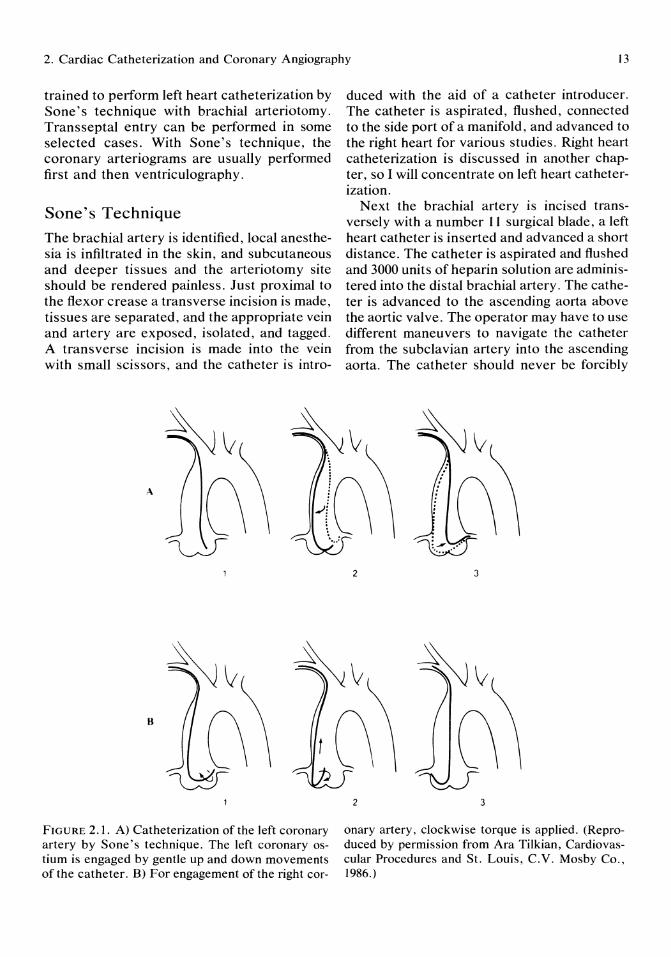
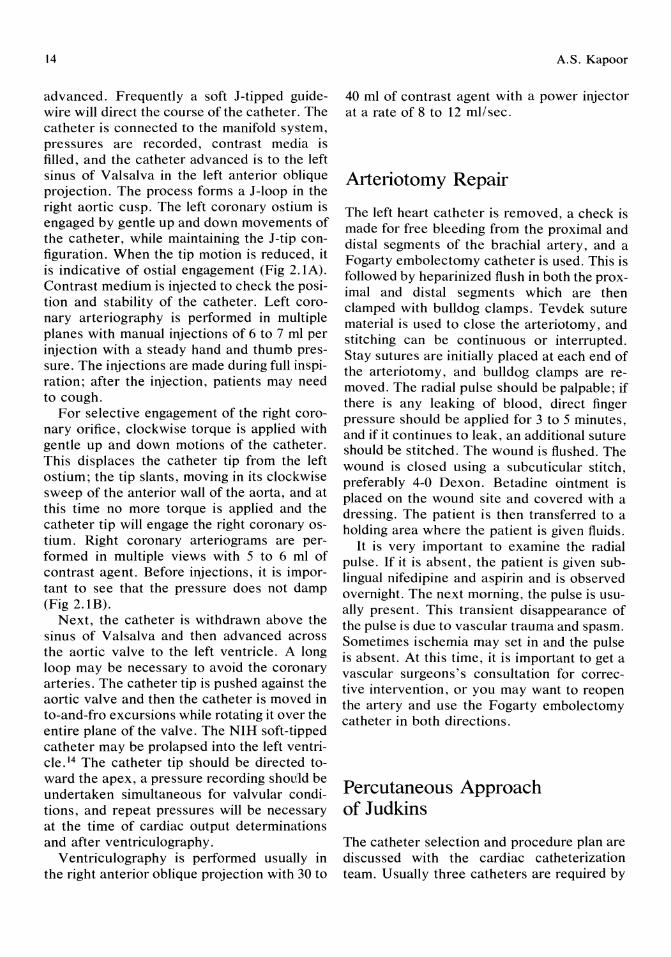
FIGURE 2.1. A) Catheterization of the left coronary artery by Sone's technique. The left coronary ostium is engaged by gentle up and down movements of the catheter. B) For engagement of the right cor-
duced with the aid of a catheter introducer. The catheter is aspirated, flushed, connected to the side port of a manifold, and advanced to the right heart for various studies. Right heart catheterization is discussed in another chapter, so I will concentrate on left heart catheterization.
Next the brachial artery is incised transversely with a number 11 surgical blade, a left heart catheter is inserted and advanced a short distance. The catheter is aspirated and flushed and 3000 units of heparin solution are administered into the distal brachial artery. The catheter is advanced to the ascending aorta above the aortic valve. The operator may have to use different maneuvers to navigate the catheter from the subclavian artery into the ascending aorta. The catheter should never be forcibly
2
2
C\ ~: .... ~r ~
3
3
onary artery, clockwise torque is applied. (Reproduced by permission from Ara Tilkian, Cardiovascular Procedures and St. Louis, C.Y. Mosby Co., 1986.)
14
advanced. Frequently a soft J-tipped guidewire will direct the course of the catheter. The catheter is connected to the manifold system, pressures are recorded, contrast media is filled, and the catheter advanced is to the left sinus of Valsalva in the left anterior oblique projection. The process forms a J-Ioop .in t~e right aortic cusp. The left coronary oshum IS
engaged by gentle up and down movements of the catheter, while maintaining the J-tip configuration. When the tip motion is reduced, it is indicative of ostial engagement (Fig 2.1A). Contrast medium is injected to check the position and stability of the catheter. Left coronary arteriography is performed in multiple planes with manual injections of 6 to 7 ml per injection with a steady hand and thumb pressure. The injections are made during full inspiration; after the injection, patients may need to cough.
For selective engagement of the right coronary orifice, clockwise torque is applied with gentle up and down motions of the catheter. This displaces the catheter tip from the left ostium; the tip slants, moving in its clockwise sweep of the anterior wall of the aorta, and at this time no more torque is applied and the catheter tip will engage the right coronary ostium. Right coronary arteriograms are performed in mUltiple views with 5 to 6 ml of contrast agent. Before injections, it is important to see that the pressure does not damp (Fig 2.1B).
Next, the catheter is withdrawn above the sinus of Valsalva and then advanced across the aortic valve to the left ventricle. A long loop may be necessary to avoid the coronary arteries. The catheter tip is pushed against the aortic valve and then the catheter is moved in to-and-fro excursions while rotating it over the entire plane of the valve. The NIH soft-tipped catheter may be prolapsed into the left ventricle. 14 The catheter tip should be directed toward the apex, a pressure recording should be undertaken simultaneous for valvular conditions, and repeat pressures will be necessary at the time of cardiac output determinations and after ventriculography.
Ventriculography is performed usually in the right anterior oblique projection with 30 to
A.S. Kapoor
40 ml of contrast agent with a power injector at a rate of 8 to 12 mllsec.
Arteriotomy Repair
The left heart catheter is removed, a check is made for free bleeding from the proximal and distal segments of the brachial artery, and a Fogarty embolectomy catheter is used. This is followed by heparinized flush in both the proximal and distal segments which are then clamped with bulldog clamps. Tevdek suture material is used to close the arteriotomy, and stitching can be continuous or interrupted. Stay sutures are initially placed at each end of the arteriotomy, and bulldog clamps are removed. The radial pulse should be palpable; if there is any leaking of blood, direct finger pressure should be applied for 3 to 5 minutes, and if it continues to leak, an additional suture should be stitched. The wound is flushed. The wound is closed using a subcuticular stitch, preferably 4-0 Dexon. Betadine ointment is placed on the wound site and covered with a dressing. The patient is then transferred to a holding area where the patient is given fluids.
It is very important to examine the radial pulse. If it is absent, the patient is given sublingual nifedipine and aspirin and is observed overnight. The next morning, the pulse is usually present. This transient disappearance of the pulse is due to vascular trauma and spasm. Sometimes ischemia may set in and the pulse is absent. At this time, it is important to get a vascular surgeons's consultation for corrective intervention, or you may want to reopen the artery and use the Fogarty embolectomy catheter in both directions.
Percutaneous Approach of Judkins
The catheter selection and procedure plan are discussed with the cardiac catheterization team. Usually three catheters are required by
2. Cardiac Catheterization and Coronary Angiography 15
the Judkins technique. They are preformed catheters and come in different sizes. Catheter size selection is based on the patient's chest xray, body size, and the aortic root dimension. Usually with a normal aorta, a size 4 catheter will suffice, but in a Marfanoid aortic root, large size catheters (7-9) may be necessary. In an uncomplicated patient, I normally perform left ventriculography followed by left coronary angiography and the right coronary study. However, in patients who are unstable or with suspected left main coronary artery disease, a left coronary study is performed first followed by right coronary study and then the left ventriculogram.
The femoral artery is punctured 2 cm below the inguinal ligament. After adequate local anesthesia is given, 10 to 15 ml of 1% xylocaine should be administered to the skin and subcutaneous and deeper tissues. One can use the Seldinger needle or disposable percutaneous Potts-Cournand or Cook needles. With the Seldinger technique, the needle is advanced to the periosteum, the obturator is removed, and the needle is withdrawn until it reaches the lumen of the artery and pulsatile blood gushes out. A J-guidewire is advanced slowly and cautiously into the needle and then if there is no resistance, the guidewire is advanced to the diaphragm. The needle is removed and a dilator is introduced or a 7-Fr or 8-Fr dilator sheath is introduced over the wire. The pigtail catheter or the left Judkins catheter is loaded over the wire. The wire is held fixed toward the left as the catheter is advanced. If the sheath is used, it is aspirated and flushed. The pigtail catheter is aspirated, 2000 to 3000 units of heparin is injected, and it is connected to the manifold system where pressures are recorded and the pigtail is then advanced across the aortic valve to the left ventricle. If the catheter does not cross the valve, a loop may have to be formed then the catheter withdrawn and it will fall across the valve with some pressure. If the catheter has no torque and pushability, use the guidewire to stiffen it. Occasionally a straight 0.038-inch guidewire is used to cross a stenotic valve. If the valve is very stenotic, different catheters may be used, for example, the right Judkins with a straight
wire. Once the catheter is in the ventricle, it is aspirated, flushed, and connected to the manifold for prompt pressure measurement. To avoid clotting in the catheter, the wire is timed for 2-minute intervals at which time it is removed, cleaned, and the catheter vigorously aspirated and flushed. This should be an obsession to prevent systemic embolization of formed clots in the catheter.
Before ventriculography, baseline pressure recordings, preferably simultaneous left ventricular and pulmonary capillary wedge, or femoral artery pressure in the case of aortic stenosis, should be recorded at different speeds. Ventriculography is performed in 30° right anterior oblique or 60° left anterior oblique, with cranial angulation, if needed, with a power injector. For a good quality ventriculogram, the pigtail catheter should be advanced toward the apex. Amount of contrast used need not exceed 40 ml at a rate of 8 to 12 mllsec. 16
Mter ventriculography, the pigtail is connected to the manifold and pullback pressures are recorded from left ventricle to aorta. The pigtail catheter is exchanged for the left Judkins' catheter. A similar method is used to advance the left Judkins' catheter to the ascending aorta. The catheter is filled with contrast medium. The catheter is advanced carefully down the medial wall of the ascending aorta and the catheter will seek the left coronary ostium without any manipUlation (Fig 2.2). Inject a small amount of contrast media to check catheter and tip position. Left coronary angiograms are performed in mUltiple views with 6 to 10 ml of contrast agent. The patient may be asked to cough to combat the hypotension and bradycardia that may accompany each injection. The left Judkins' catheter is removed and replaced with a right Judkins' catheter. This catheter is advanced to the ascending aorta above the level of the aortic valve. Then a gentle clockwise torque is applied to the catheter hub. As the catheter rotates, it will fall into the right sinus of Val salva (Fig 2.3). At this time, the rotation should be slowed and the catheter tip will drop into the right coronary ostium. Pressure is checked, contrast injected to ascertain tip position, and

16 A.S. Kapoor
Judkins' Type Amplatz'Type
-JV V
FIGURE 2.2. Catheterization of left coronary artery using Judkins' technique with Judkins' or Amplatz' type catheters.
coronary angiograms performed in multiple projections with 4 to 6 ml of contrast agent.
Selective engagement of the right coronary ostium may require manipulation or a change to a different size or different catheter. It will require experience to master right coronary ostial engagement with the Judkins' technique (Fig 2.4). A modified right Amplatz' catheter is an excellent choice for right coronary studIes.
According to Judkins, "No points are earned for coronary catheterization-the catheter knows where to go if not thwarted by the operator. "17 In most cases, the Judkins' technique is much easier than Sones and is the technique of choice in most centers performing high-volume coronary arteriograms. By the way, this technique is also possible via brachial or axillary artery approaches.
After completion of the study, the catheter and sheath are removed, hemostasis is established with 10 minutes of manual pressure,
and the patient is then transferred to a holding area for further observation.
Bypass Graft Catheterization
The right Judkins' catheter can be used for engagement of the saphenous vein bypass conduit or internal mammary artery. Often a modified right Amplatz catheter is successful for selective catheterization of vein grafts. There are also other special vein graft catheters.
It is important to know the aortic insertion of the grafts. The aortic insertion of the graft to the right coronary artery is most anterior and lowest. Above it in a posterolateral position is the origin of the graft to the left anterior descending, and above it is the graft to the obtuse, marginal, and diagonal arteries.
Many operators perform an aortic root angiogram to locate the origin of the grafts and
FIGURE 2.3. Selective engagement of right coronary ostium using Judkins' catheter.
2. Cardiac Catheterization and Coronary Angiography 17
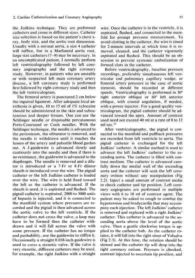
FIGURE 2.4. In patients with Shepherd's crook anomaly, a left Amplatz' catheter may be required.
then seek individual grafts. The catheter is slowly advanced or withdrawn until it engages in a graft ostium. Graft and native coronary angiography can be performed using a Schoonmaker catheter. 18
Selective catheterization of internal mammary artery grafts is achieved by a preformed left internal mammary artery catheter. The catheter is placed in the aortic arch with its tip pointing down and is rotated counterclockwise until it falls into the left subclavian artery. The tip is rotated anteriorly until it engages the origin of the left internal mammary
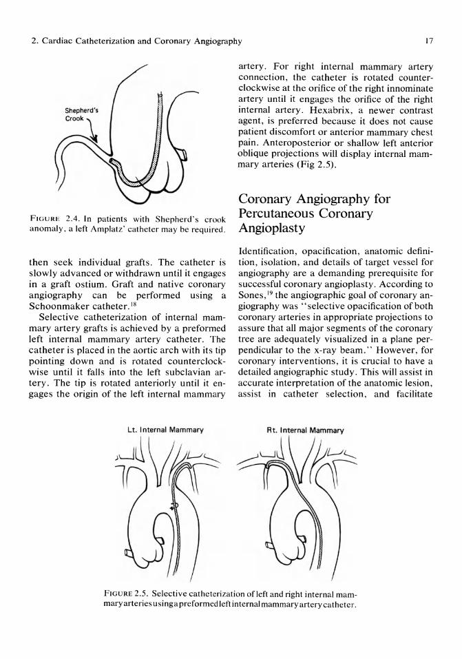
artery. For right internal mammary artery connection, the catheter is rotated counterclockwise at the orifice of the right innominate artery until it engages the orifice of the right internal artery. Hexabrix, a newer contrast agent, is preferred because it does not cause patient discomfort or anterior mammary chest pain. Anteroposterior or shallow left anterior oblique projections will display internal mammary arteries (Fig 2.5).
Coronary Angiography for Percutaneous Coronary Angioplasty
Identification, opacification, anatomic definition, isolation, and details of target vessel for angiography are a demanding prerequisite for successful coronary angioplasty. According to Sones,19 the angiographic goal of coronary angiography was "selective opacification of both coronary arteries in appropriate projections to assure that all major segments of the coronary tree are adequately visualized in a plane perpendicular to the x-ray beam." However, for coronary interventions, it is crucial to have a detailed angiographic study. This will assist in accurate interpretation of the anatomic lesion, assist in catheter selection, and facilitate
FIGURE 2.5. Selective catheterization of left and right internal mammary arteries usinga preformed left internal mammary artery catheter.
18
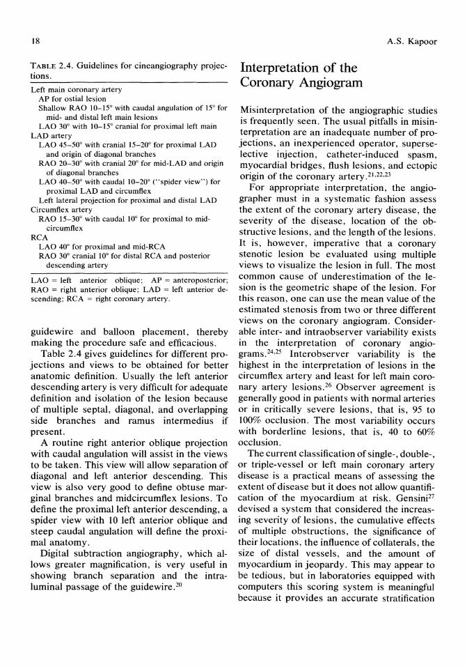
TABLE 2.4. Guidelines for cineangiography projections.
Left main coronary artery AP for ostial lesion Shallow RAO 10-15° with caudal angulation of 15° for
mid- and distal left main lesions LAO 30° with 10-15° cranial for proximal left main
LAD artery LAO 45-50° with cranial 15-20° for proximal LAD
and origin of diagonal branches RAO 20-30° with cranial 20° for mid-LAD and origin
of diagonal branches LAO 40-50° with caudal 10-20° ("spider view") for
proximal LAD and circumflex Left lateral projection for proximal and distal LAD
Circumflex artery RAO 15-30° with caudal 10° for proximal to mid
circumflex RCA
LAO 40° for proximal and mid-RCA RAO 30° cranial 10° for distal RCA and posterior
descending artery
LAO = left anterior oblique; AP = anteroposterior; RAO = right anterior oblique; LAD = left anterior descending; RCA = right coronary artery.
guidewire and balloon placement, thereby making the procedure safe and efficacious.
Table 2.4 gives guidelines for different projections and views to be obtained for better anatomic definition. Usually the left anterior descending artery is very difficult for adequate definition and isolation of the lesion because of multiple septal, diagonal, and overlapping side branches and ramus intermedius if present.
A routine right anterior oblique projection with caudal angulation will assist in the views to be taken. This view will allow separation of diagonal and left anterior descending. This view is also very good to define obtuse marginal branches and midcircumflex lesions. To define the proximal left anterior descending, a spider view with 10 left anterior oblique and steep caudal angulation will define the proximal anatomy.
Digital subtraction angiography, which allows greater magnification, is very useful in showing branch separation and the intraluminal passage of the guidewire. 20
Interpretation of the Coronary Angiogram
A.S. Kapoor
Misinterpretation of the angiographic studies is frequently seen. The usual pitfalls in misinterpretation are an inadequate number of projections, an inexperienced operator, superselective injection, catheter-induced spasm, myocardial bridges, flush lesions, and ectopic origin of the coronary artery.21,22,23
For appropriate interpretation, the angiographer must in a systematic fashion assess the extent of the coronary artery disease, the severity of the disease, location of the obstructive lesions, and the length of the lesions. It is, however, imperative that a coronary stenotic lesion be evaluated using multiple views to visualize the lesion in full. The most common cause of underestimation of the lesion is the geometric shape of the lesion. For this reason, one can use the mean value of the estimated stenosis from two or three different views on the coronary angiogram. Considerable inter- and intraobserver variability exists in the interpretation of coronary angiograms. 24 ,25 Interobserver variability is the highest in the interpretation of lesions in the circumflex artery and least for left main coronary artery lesions.26 Observer agreement is generally good in patients with normal arteries or in critically severe lesions, that is, 95 to 100% occlusion. The most variability occurs with borderline lesions, that is, 40 to 60% occlusion.
The current classification of single-, double-, or triple-vessel or left main coronary artery disease is a practical means of assessing the extent of disease but it does not allow quantification of the myocardium at risk. Gensini27 devised a system that considered the increasing severity of lesions, the cumulative effects of multiple obstructions, the significance of their locations, the influence of collaterals, the size of distal vessels, and the amount of myocardium in jeopardy. This may appear to be tedious, but in laboratories equipped with computers this scoring system is meaningful because it provides an accurate stratification

2. Cardiac Catheterization and Coronary Angiography
KAISER PERMANENTE REGIONAL CARDIAC CATHETERIZATION LABORATORY NAME: 10: Page: 1 REPORT: Coronary Angiography 10/27/1987 11:45:43 hrs PHYSICIAN:
Anatomy of native coronary arteries: CO.lnance: Rlght LAO branchae: Cx branches:
Right Coronary Artery:
01ag 1. ... s.all Oblolarg 1.. s.all
Mld RCA ... 1001 dlscrete stenosls Dlst RCA ... norael
Left Main Coronary Artery: LMCA ... 1001 dlscrete stenosls
Left Anter10r Descend1ng: Normal
Dlag 2 .... saall ObMarg 2 .. slla 11
Lsft C1rcumflex Artery: Normal Collateral Circulation: -->-->--> To From
Conus Conus ObMarg 1
---> D1st LAO ---> Dlst Cx ---> R PDA
Assessment of Vessels with Lesions > 50~ Sultablllty Of Dlstal
Vessel For Bypass RCA ............ Sult.ble LAD ............ Su1t.ble Cx ............. Sult.ble
Dlst LAO ..... dlu. OUt CX ..• sllall
FIGURE 2.6. Example of computer-generated tabular summary of coronary angiographic findings.
FIGURE 2.7. Computer-assisted printout of coronary diagram with associated lesions and collaterals.
19
20
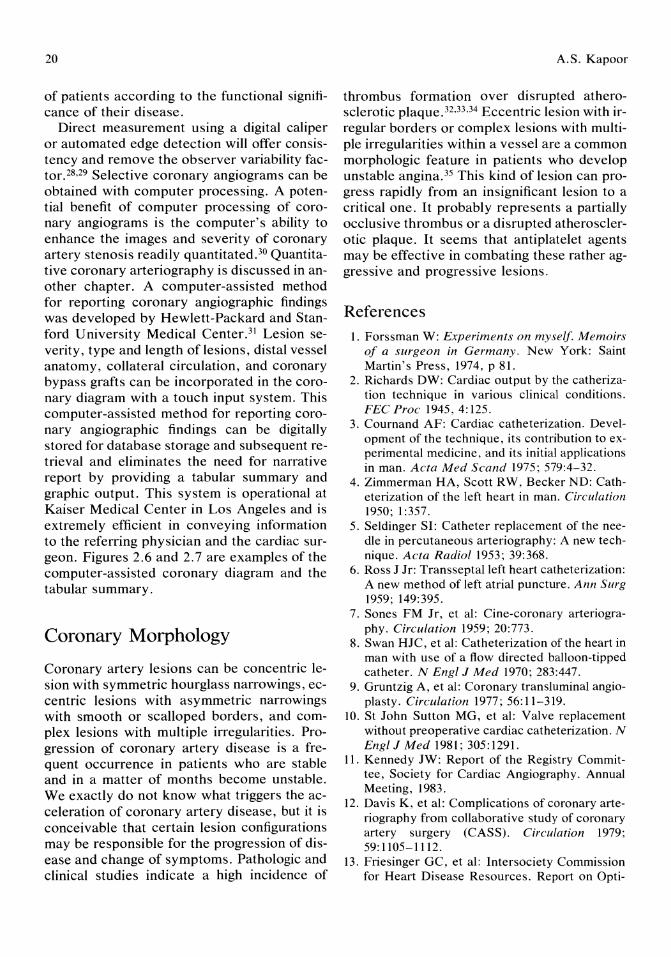
of patients according to the functional significance of their disease.
Direct measurement using a digital caliper or automated edge detection will offer consistency and remove the observer variability factor. 28,29 Selective coronary angiograms can be obtained with computer processing. A potential benefit of computer processing of coronary angiograms is the computer's ability to enhance the images and severity of coronary artery stenosis readily quantitated. 30 Quantitative coronary arteriography is discussed in another chapter. A computer-assisted method for reporting coronary angiographic findings was developed by Hewlett-Packard and Stanford University Medical Center. 31 Lesion severity, type and length of lesions, distal vessel anatomy, collateral circulation, and coronary bypass grafts can be incorporated in the coronary diagram with a touch input system. This computer-assisted method for reporting coronary angiographic findings can be digitally stored for database storage and subsequent retrieval and eliminates the need for narrative report by providing a tabular summary and graphic output. This system is operational at Kaiser Medical Center in Los Angeles and is extremely efficient in conveying information to the referring physician and the cardiac surgeon. Figures 2.6 and 2.7 are examples of the computer-assisted coronary diagram and the tabular summary.
Coronary Morphology
Coronary artery lesions can be concentric lesion with symmetric hourglass narrowings, eccentric lesions with asymmetric narrowings with smooth or scalloped borders, and complex lesions with multiple irregularities. Progression of coronary artery disease is a frequent occurrence in patients who are stable and in a matter of months become unstable. We exactly do not know what triggers the acceleration of coronary artery disease, but it is conceivable that certain lesion configurations may be responsible for the progression of disease and change of symptoms. Pathologic and clinical studies indicate a high incidence of
A.S. Kapoor
thrombus formation over disrupted atherosclerotic plaque. 32.33 ,34 Eccentric lesion with irregular borders or complex lesions with multiple irregularities within a vessel are a common morphologic feature in patients who develop unstable angina. 35 This kind of lesion can progress rapidly from an insignificant lesion to a critical one. It probably represents a partially occlusive thrombus or a disrupted atherosclerotic plaque. It seems that antiplatelet agents may be effective in combating these rather aggressive and progressive lesions.
References 1. Forssman W: Experiments on myself. Memoirs
of a surgeon in Germany. New York: Saint Martin's Press, 1974, p 81.
2. Richards DW: Cardiac output by the catherization technique in various clinical conditions. FEC Proc 1945,4:125.
3. Cournand AF: Cardiac catheterization. Development of the technique, its contribution to experimental medicine, and its initial applications in man. Acta Med Scand 1975; 579:4-32.
4. Zimmerman HA, Scott RW, Becker ND: Catheterization of the left heart in man. Circulation 1950; 1:357.
5. Seldinger SI: Catheter replacement of the needle in percutaneous arteriography: A new technique. Acta Radio11953; 39:368.
6. Ross J Jr: Transseptalleft heart catheterization: A new method of left atrial puncture. Ann Surg 1959; 149:395.
7. Sones FM Jr, et al: Cine-coronary arteriography. Circulation 1959; 20:773.
8. Swan HJC, et al: Catheterization of the heart in man with use of a flow directed balloon-tipped catheter. N Engl J Med 1970; 283:447.
9. Gruntzig A, et al: Coronary transluminal angioplasty. Circulation 1977; 56:11-319.
10. St John Sutton MG, et al: Valve replacement without preoperative cardiac catheterization. N Engl J Med 1981; 305: 1291.
11. Kennedy JW: Report of the Registry Committee, Society for Cardiac Angiography. Annual Meeting, 1983.
12. Davis K, et al: Complications of coronary arteriography from collaborative study of coronary artery surgery (CASS). Circulation 1979; 59: 1105-1112.
13. Friesinger GC, et al: Intersociety Commission for Heart Disease Resources. Report on Opti-
2. Cardiac Catheterization and Coronary Angiography 21
mal Resources for Examination of the Heart and Lungs: Cardiac catheterization and radiography facilities. Circulation 1983; 68:893-930A.
14. Grossman W: Cardiac catherization and angiography, ed 3. Philadelphia, Lea & Febiger, 1986.
15. Zimmerman HA (ed): Intravascular Catherization, ed 2. Springfield, IL, Charles C. Thomas, 1966.
16. Hildner FJ, et al: New principles for optimum left ventriculography. Cath Cardiovasc Diagn 1986; 12:266-273.
17. Judkins MP: Selective coronary arteriography. 1. A percutaneous transfemoral technique. Radiology 1967; 89:815.
18. Schoonmaker FW, King SB: Coronary arteriography by the single catheter percutaneous femoral technique, experience in 6,800 cases. Circulation 1974; 50:735.
19. Sones FM: Indications and value of coronary arteriography. Circulation 1972; 46: 1159.
20. Clark DA: Coronary Angioplasty. New York, Alan R. Liss, Inc, 1987.
21. Bloor CM, Lowman RM: Myocardial bridges in coronary angiography. Am Heart J 1975; 65:972.
22. Ballaxe H, Amplatz K, Levin D: Coronary Angiography. Springfield, Charles C. Thomas. 1973.
23. Conti CR: Coronary arteriography. Circulation 1977; 55:227-237.
24. DeRouen TA, Murray JA, Owen W: Variability in the analysis of coronary arteriograms. Circulation 1977; 55:324-328.
25. Galbraith JE, Murphy ML, deSouza N: Coro-
nary angiogram interpretation. Interobserver variability. JAMA 1978; 240:2053-2056.
26. Trask N, et al: Accuracy and interobserver variability of coronary cineangiography. J Am Coli Cardio!1984; 3:1145-1154.
27. Gensini GO: A more meaningful scoring system for determining the severity of coronary heart disease. Am J Cardiol 1983; 51:604.
28. Rafflenbend W, et al: Quantitative coronary arteriography. Am J Cardia! 1979; 43:699.
29. Spears JR, et al: Computerized image analysis for quantitative measurement of vessel diameter from cineangiograms. Circulation 1983; 68:453.
30. Tobis J, et al: Detection and quantitation of coronary artery stenosis from digital subtraction angiograms compared with 35-mm film cineangiograms. Am J Cardio! 1984; 54:489-496.
31. Alderman EL, et al: Anatomically flexible computer-assisted reporting system for coronary angiography. Am J Cardia! 1982; 49: 1208-1215.
32. Falk E: Plaque rupture with severe pre-existing stenosis precipitating coronary thrombosis. Br Heart J 1983; 50:127-1345.
33. Levin DC, Fallon JT: Significance of the angiographic morphology of localized coronary stenosis. Histopathologic correlations. Circulation 1982; 66:316-320.
34. Chapman I: Morphogenesis of occluding coronary artery thrombosis. Arch Pathol 1985; 80:256-261.
35. Ambrose JA, et al: Angographic evolution of coronary artery morphology in unstable angina. J Am Coli Cardia! 1986; 7:472-478.
3 Coronary Blood Flow and Coronary Vascular Reserve Terrence J.W. Baruch, Amar S. Kapoor, and Peter R. Mahrer
Introduction
The severity of coronary artery disease (CAD) has traditionally been evaluated by assessing coronary artery anatomy and left ventricular function during cardiac catheterization. The reduction in coronary artery blood flow caused by a particular stenosis has been inferred from its appearance on coronary arteriography.l It is now well known that the visual estimation of coronary artery stenosis is inaccurate and poorly reproducible because of inter- and intraobserver variation. It also has been shown that the physiologic effects of the majority of coronary obstructions cannot be determined accurately by conventional angiographic appearance. 2 Human coronary vascular reserve correlates poorly with percent diameter stenosis and geometry of the lesions.
Because of this, the hemodynamic effect of a coronary stenosis on coronary blood flow must be determined to assess the severity of a particular coronary obstruction. This has become even more important with the development of percutaneous coronary angioplasty as an effective treatment for coronary artery disease. Knowledge of the hemodynamic significance of a particular lesion is essential in deciding whether an intervention should be performed. There is a considerable amount of interest in measuring coronary blood flow in the baseline resting state and during vasodilatory provocation, that is, measuring the coronary flow reserve, with the intent of providing an independent means for assessing the hemo-
dynamic significance of a particular stenosis. Obtaining these data allows for more precise decision making regarding the choice of therapy for a particular patient.
We review the basic principles of coronary blood flow and traditional techniques which have been used to quantitate it. The concept of coronary flow reserve and its usefulness in evaluating the hemodynamic significance of coronary artery stenoses will also be discussed. Finally, the currently available techniques for determining the effectiveness of percutaneous transluminal coronary angioplasty in improving coronary flow reserve in a stenotic coronary artery are examined.
Coronary Artery Blood Flow
Normally, the coronary artery tree is made up of large epicardial blood vessels and smaller intramyocardial arterioles. Coronary artery blood flow may be expressed as the ratio between trans myocardial perfusion pressure and coronary vascular resistance. Transmyocardial perfusion pressure is equal to the pressure gradient between the coronary arteries and the coronary sinus. If left ventricular filling pressures are normal, then the coronary perfusion pressure can be approximated by the mean aortic pressure. 3 However, the use of the mean aortic pressure tends to overestimate the driving pressure when ventricular filling pressures are elevated. As left ventricular diastole pressure increases, coronary flow will
3. Coronary Blood Flow and Vascular Reserve
decrease because of a change in coronary perfusion pressure. As a consequence, the difference of mean arterial pressure and pulmonary capillary wedge pressure has been used to estimate coronary artery perfusion pressure in the face of increased left ventricular filling pressures. 3 There are three major factors that can affect transmyocardial perfusion pressure. First, there are factors that reduce aortic diastolic pressure, such as an arteriovenous shunt, a patent ductus arteriosus, or aortic regurgitation. Second, there are factors that raise ventricular diastolic pressure. This can occur with disorders that increase preload (cardiomyopathies) or increase afterload (hypertension with left ventricular dysfunction or with diminished left ventricular compliance). Third are circumstances that can increase left ventricular systolic pressure as in the case of aortic stenosis or obstructive hypertrophic cardiomyopathy. 4
Coronary vascular resistance is defined as the passive force exerted in opposition to coronary artery flow. The total coronary vascular resistance is made up of three major components, namely, the resistance of the epicardial vessels, the resistance of the intramyocardial arterioles, and the resistance that results from compression of the coronary vessels during systole. It is this systolic compressive force that is responsible for the marked decrease in flow in the left coronary artery during systole and expresses the phasic nature of coronary blood flow.
Under normal conditions, resistance in epicardial vessels is low and does not contribute in the regulation of coronary artery blood flow. However, in the presence of coronary artery disease with significant epidardial stenosis, the effect on coronary blood flow becomes crucial, as will be discussed later. The resistance in the intramyocardial arteries and arterioles is the major determinant of coronary vascular resistance and therefore coronary blood flow. The tone of these "resistance vessels" is affected by a number offactors, which are summarized in Table 3.1.
Neurologic control of coronary vascular tone is mediated through both the sympathetic and parasympathetic nervous systems. Alpha-
23
TABLE 3.1. Factors that control coronary vascular resistance.
I. Neural factors a) Sympathetic nerves
(i) 0<1 and 0<2 receptors cause vasoconstriction (ii) {31 and {3z receptors mediate vasodilation
b) Parasympathetic nerve stimulation via vagus nerve cause vasodilatation
II. Myogenic factors Augmentation of resistance via the Bayliss effect (only modest effect)
III. Metabolic Factors Adenosine Carbon dioxide Hydrogen ion Prostaglandins Other vasodilator metabolites
IV. Endothelial-mediated coronary vasodilatation Endothelial-derived relaxant factor
V. Systolic compression Induces vasoconstriction
receptor stimulation via sympathetic nerve fibers will result in coronary vasoconstriction, whereas beta-receptor stimulation will result in vasodilation. Stimulation of parasympathetic receptors also will lead to coronary artery dilatation. However, although the effect of autonomic innervation on coronary blood flow has been demonstrated in vitro and in animal preparations under physiologic conditions, the effects of this are probably not significant. 4
If the perfusion pressure is experimentally increased, a corresponding increase in coronary blood flow will occur. This augmented flow, however, is transient with an abrupt decline in blood flow back to baseline levels. This phenomenon is known as autoregulation and it is the main mechanism for modulating coronary artery blood flow at constant levels, despite variations in the driving pressure. 4
There are many factors that are thought to mediate autoregulation. If vascular smooth muscle is stretched as a result of increased blood flow, it will then contract causing blood flow to diminish. This phenomenon is known as the myogenic mechanism or the Bayliss effect and is thought to contribute to the process of autoregulation. Whereas myogenic factors have been shown to be important in regulating

24
blood flow in many vascular beds, its rate in the coronary circulation is still controversiaI.5
Autoregulation is mainly modulated by metabolic processes. An important metabolic regulation of vascular tone is oxygen. It has been well demonstrated that there is a close correlation between myocardial oxygen consumption and coronary artery blood flow. 6
Hypoxia has been shown to cause vasodilation in many different systemic arteries. 7 It is presumed to cause vasodilation by modifying the electrochemical potential of the smooth muscle cells.4
Another metabolic factor thought to play an important role in autoregulation is adenosine. This potent vasodilator is found when myocardial cells are unable to resynthesize ATP. By blocking the entry of calcium into the sarcolemma of myocardial cells, adenosine causes vasodilation which in turn will increase coronary blood flow. 6
There are other substances that are thought to be potential mediators of the autoregulatory process; among them are prostaglandins, kinases, hydrogen ions, and potassium. The relative contributions of these factors are still not completely known.
Traditional Methods for Evaluating Coronary Blood Flow
The realization of the importance of evaluating the hemodynamic effects of various disease states on the coronary blood flow has led to the development of a number of techniques for measuring blood flow in conscious humans.
The electromagnetic flow meter can be placed around an epicardial coronary artery and accurately measures phasic coronary artery blood flow velocity. The electromagnetic flowmeter is able to detect rapid changes in coronary blood flow (CBF) and has been used extensively in animal experiments investigating the hemodynamics of blood flow. It also is used at the time of coronary artery bypass surgery to determine the adequacy of flow in coronary bypass grafts. Its disadvantage is that it
T.J.W. Baruch, A.S. Kapoor, and P.R. Mahrer
must completely encircle a vessel requiring surgical dissection of the coronary artery, and therefore cannot be used to evaluate CBF in unanesthetized humans. s
In 1981, Marcus et al9 developed a safe and easy method for measuring phasic coronary velocity at the time of cardiac surgery. A Doppler probe attached to a silicone suction cup is used to assess CBF in native coronary arteries, as well as in bypass grafts. Measurement of CBF velocity with this technique showed a strong correlate when compared with established methods of CBF measurement, such as timed venous collection, the electromagnetic flowmeter, and vasoactive microsphere. Its major disadvantage is that it measures CBF velocity and not absolute CBF. Another disadvantage, as with the electromagnetic flowmeter, is that it can only be used at the time of coronary artery bypass surgery.
The two techniquesjust mentioned allow direct measurement of epicardial blood flow but are limited in that they cannot be used to assess CBF in conscious humans at the time of cardiac catheterization. During past 3 decades there has been a search for a technique that allows measurement of CBF in unanesthetized humans. The techniques now available for achieving this can be divided into the following four groups: First, there are methods that use diffusible gases. Second, there are methods using substances that actively enter the cell. Third, there is the measurement of coronary sinus blood flow by continuous thermodilution. Finally, there are methods to measure phasic coronary flow by means of videodensity or continuous wave Doppler. lo
The use of diffusible indicators for assessing CBF involves the injection of physiologically inert, freely diffusible substances into a coronary artery. Flow is then determined by myocardial uptake or washout of these indicators. The rate at which these substances are taken up or washed out can be determined by coronary sinus sampling or by external scintillation scanning. II A number of substances have been used, including inert gases such as helium, nitrous oxide, argon, and xenon and diffusible substances, such as H2015 and 1-131-antipyrine. 3
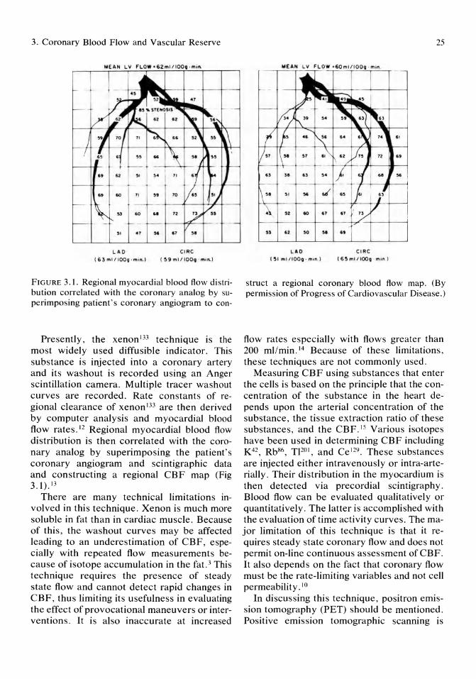
3. Coronary Blood Flow and Vascular Reserve
~I .. , H • .., ~.
LAD eiRe (63,mtl IOOq -min.) (59mIl IOOq · "un..1
FIGURE 3.1. Regional myocardial blood flow distribution correlated with the coronary analog by superimposing patient's coronary angiogram to con-
Presently, the xenon 133 technique is the most widely used diffusible indicator. This substance is injected into a coronary artery and its washout is recorded using an Anger scintillation camera. Multiple tracer washout curves are recorded. Rate constants of regional clearance of xenon l33 are then derived by computer analysis and myocardial blood flow rates. 12 Regional myocardial blood flow distribution is then correlated with the coronary analog by superimposing the patient's coronary angiogram and scintigraphic data and constructing a regional CBF map (Fig 3.1).13
There are many technical limitations involved in this technique. Xenon is much more soluble in fat than in cardiac muscle. Because of this, the washout curves may be affected leading to an underestimation of CBF, especially with repeated flow measurements because of isotope accumulation in the fat. 3 This technique requires the presence of steady state flow and cannot detect rapid changes in CBF, thus limiting its usefulness in evaluating the effect of provocational maneuvers or interventions. It is also inaccurate at increased
LAO ('5 1 rniIlOOq · min. )
e i Re (6~""/ IOOQ - m,n_ 1
25
struct a regional coronary blood flow map. (By permission of Progress of Cardiovascular Disease.)
flow rates especially with flows greater than 200 mllmin. 14 Because of these limitations, these techniques are not commonly used.
Measuring CBF using substances that enter the cells is based on the principle that the concentration of the substance in the heart depends upon the arterial concentration of the substance, the tissue extraction ratio of these substances, and the CBF.IS Various isotopes have been used in determining CBF including K42, Rb86 , T]201, and Ce 129. These substances are injected either intravenously or intra-arterially. Their distribution in the myocardium is then detected via precordial scintigraphy. Blood flow can be evaluated qualitatively or quantitatively. The latter is accomplished with the evaluation of time activity curves. The major limitation of this technique is that it requires steady state coronary flow and does not permit on-line continuous assessment ofCBF. It also depends on the fact that coronary flow must be the rate-limiting variables and not cell permeability. 10
In discussing this technique, positron emission tomography (PET) should be mentioned. Positive emission tomographic scanning is

26
based on the principle that unstable isotopes emit positrons on radioactive decay. When a positron encounters an electron, two photons are produced that are emitted in opposite directions. These photons are then detected simultaneously by positron cameras, which are positrons on opposite sides of the patient. These photons are detected and counted at many positions around the patient, and a computer is used to reconstruct an image that represents the distribution of the isotope injected. 16
Radioactive substances used as tracers in myocardial perfusion studies include Rb82 and N13. Rubidium is useful because of its short half-life, making it usable for mUltiple studies of perfusion after an intervention. 3
To quantitate the amount of isotope in the heart, the general principle stated earlier in reference to substances that enter the cell applies. That is, if the arterial input function is known, the concentration of an indicator in the tissue will depend upon tissue blood flow and the organ extraction ratio. 15
In animals there has been good correlation between estimation of CBF using PET scanning as compared with microspheresY
The advantage of this method is that it gives quantitative regional and transmural blood flow measurements. However, this is an extremely expensive technique that is not widely available and at present is used mainly as a research tool.
The technique for measurement of coronary sinus blood flow in humans by continuous thermodilution was developed by Ganz et al 18
in 1971. It is based on the principle that when a substance miscible with blood is infused into the coronary sinus at a constant rate, the downstream temperature of the mixture can be used to predict CBF. Computation of blood flow is based on the assumption that the heat lost from the system between the site of injection and the site of detection is negligible, and therefore the heat lost by the blood equals the heat gained by the indicator. 18 The fluid injected is infused at 35 to 55 mllmin through a specialized catheter for 20 to 80 seconds. Turbulence during this injection completely mixes the indicator with coronary venous blood.
T.J.W. Baruch, A.S. Kapoor, and P.R. Mahrer
Coronary sinus flow represents venous return from the left ventricular free wave and constitutes outflow from both the left anterior descending and circumflex artery. Flow in the great cardiac vein represents drainage primarily from the left anterior artery. 3
This technique has been widely used in conscious humans to evaluate resting blood flow and alterations in blood flow after different interventions, such as atrial pacing, administration of vasoactive drugs, or injection of contrast material. Its advantages are that it is a simple, inexpensive, and safe technique that has shown good correlation with the electromagnetic flowmeter. Measurements can be performed in approximately 20 seconds, and because the injection is hemodynamically inert, multiple measurements can be made at short periods of time. This allows assessment of changes in flow in response to intervention.
The major disadvantage of the technique is its inability to measure CBF in the right coronary artery and its inability to separate left anterior descending flow from left circumflex flow. Also, there is some difficulty in maintaining catheter position constant. Misleading changes in flow can be recorded if catheter position varies during the intervention.
Methods used to measure phasic coronary flow by means of videodensitometry or continuous wave Doppler will be discussed later in this chapter because they are techniques that are currently most useful in evaluating the effect of PTCA on CBF.
Coronary Flow Reserve
It was first thought that CBF would be reduced in patients with coronary artery disease. It also was believed that measurement of this decrease in CBF would give useful hemodynamic information in evaluating the severity of a stenosis demonstrated during angiography. However, with the development of methods to quantitate blood flow, it has been found that there is a great deal of overlap between flow in normal individuals and in those with coronary artery disease because the range of normal blood flow under basal conditions is

3. Coronary Blood Flow and Vascular Reserve
wide (65 to 100 mllmin per 100 g).19 Thus, patients with severe three-vessel disease can have resting CBF equal to normal.
Since 1939 it has been observed that after an intra-arterial injection of contrast medium, a significant increase in CBF occurred. 20 This increase in flow was called reactive hyperemia. In 1964 Mosher et aFI examined this hyperemia response in the coronary arteries of mongrel dogs. His preparation made it possible to change coronary perfusion pressure (AP) without changing aortic pressure. Thus, the effects of changing trans myocardial perfusion pressure on coronary flow could be examined while oxygen consumption and left ventricular work were kept constant. The results of this study on a normal heart are shown in Fig 3.2. A pressure flow diagram was formulated. Under basal conditions, it can be seen there is little change in coronary flow with changes in perfusion pressure. This is due to autoregulation, which was described earlier in
FLOW, ml/min o
500
400
300
200
A
100
O~~--~----~----~ a 50 100 150
MEAN PRESSURE, mm Hg
FIGURE 3.2. Pressure flow diagram. Under basal conditions, there is little change in coronary flow with changes in perfusion pressure. Line D represents the maximal flow after hyperemic response and is the coronary flow reserve. (By permission of American Heart Association.)
27
this chapter. Line D in the pressure flow diagram (Fig 3.2) represents flow after the induction of a hyperemia response. If this stimulus produces maximal vasodilation, then the increment of coronary flow above resting levels will also be maximal. This increment has been termed the coronary flow reserve.
Under normal conditions resistance in the large epicardial is low and changes in the coronary artery blood flow are regulated by changes in the diameter of the small intramyocardial vessels. As stated earlier, myocardial blood flow is inversely related to intramyocardial arteriolar resistance and directly to coronary driving pressure. Using autoregulation, these vessels are able to increase or decrease CBF to meet the metabolic demands of the myocardium by altering coronary vascular resistance. With exercise or another hyperemic stimulus, there is an increase in myocardial oxygen demand. As a result, the arterioles dilate to increase coronary flow. When the hyperemic stimuli is maximal, the intramyocardial vessels become maximally dilated and the increase in flow is the maximum coronary flow reserve. 22
When an epicardial vessel becomes stenotic, it causes resistance to flow. To compensate for this, the intramyocardial vessels will dilate to maintain adequate flow. As the epicardial stenosis progresses, the arterioles dilate fully and the maximum coronary flow reserve is attained. At this point, flow becomes pressure dependent, and further increases in the stenosis will result in diminished CBF (Fig 3.3).
Under baseline conditions, ischemia will occur with severe stenosis. With less severe obstruction, baseline CBF may be maintained at normal levels as a result of vasodilation of the resistance vessels, partially using their vascular reserve capacity. However, when a hyperemic response is induced, further dilation of these arterioles is compromised so the appropriate increase in coronary flow of four to five times baseline cannot be attained. This results in ischemia secondary to attenuation in the coronary flow reserve (Fig 3.4).23
From this, it is obvious that in patients with coronary artery disease and normal resting
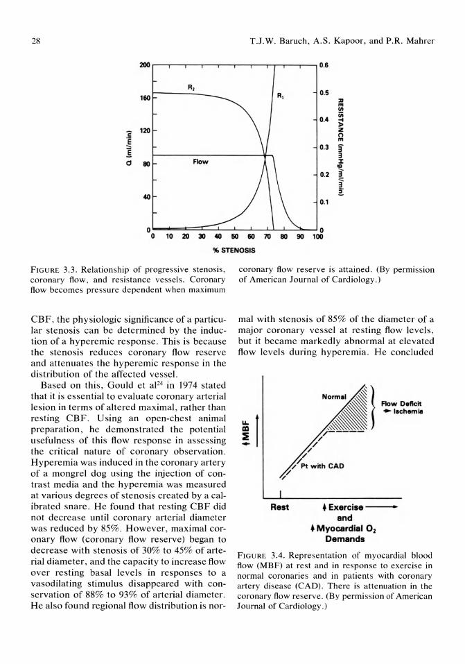
28 T.J.W. Baruch, A.S. Kapoor, and P.R. Mahrer
200 r---.-,-------,---,--..----.--,--.......,---,----, 0.6
160 R, 0.5 :D m (1\
0.4 1ii ~ l>
i: 120
! !
Z (") m
0.3 3' 3
d 80 Flow % III -...
0.2 ! 3' :s-
40 0.1
o 0 o 10 20 30 40 50 60 70 80 90 100
% STENOSIS
FIGURE 3.3. Relationship of progressive stenosis, coronary flow, and resistance vessels. Coronary flow becomes pressure dependent when maximum
CBP, the physiologic significance of a particular stenosis can be determined by the induction of a hyperemic response. This is because the stenosis reduces coronary flow reserve and attenuates the hyperemic response in the distribution of the affected vessel.
Based on this, Gould et aF4 in 1974 stated that it is essential to evaluate coronary arterial lesion in terms of altered maximal, rather than resting CBP. U sing an open-chest animal preparation, he demonstrated the potential usefulness of this flow response in assessing the critical nature of coronary observation. Hyperemia was induced in the coronary artery of a mongrel dog using the injection of contrast media and the hyperemia was measured at various degrees of stenosis created by a calibrated snare. He found that resting CBP did not decrease until coronary arterial diameter was reduced by 85%. However, maximal coronary flow (coronary flow reserve) began to decrease with stenosis of 30% to 45% of arterial diameter, and the capacity to increase flow over resting basal levels in responses to a vasodilating stimulus disappeared with conservation of 88% to 93% of arterial diameter. He also found regional flow distribution is nor-
coronary flow reserve is attained. (By permission of American Journal of Cardiology.)
mal with stenosis of 85% of the diameter of a major coronary vessel at resting flow levels, but it became markedly abnormal at elevated flow levels during hyperemia. He concluded
Rest
t Flow Deficit ~ -Ischemill
• Exercise --and
• Myocardial O2
Demands
FIGURE 3.4. Representation of myocardial blood flow (MBF) at rest and in response to exercise in normal coronaries and in patients with coronary artery disease (CAD). There is attenuation in the coronary flow reserve. (By permission of American Journal of Cardiology.)
3. Coronary Blood Flow and Vascular Reserve
that because resting coronary flow and distribution are unaffected by relatively severe arterial narrowing, it is essential to assess the effects of coronary stenosis in terms of altered maximal, rather than at rest, coronary flOW. 24
Thus, although coronary angiography is essential for visualizing the extent of coronary stenoses, other diagnostic tests which more clearly define the flow-limiting characteristics of specific coronary artery lesions would be of considerable help in evaluating patients with coronary artery disease.1O With the advent of percutaneous transluminal coronary angioplasty (PTCA), this concept becomes even more important. For high- and low-grade stenoses, prediction of hemodynamic significance is fairly reliable. However, for lesions of intermediate severity, there is great difficulty in assessing their hemodynamic significance. Deciding whether or not to perform PTCA and evaluating its efficacy has been traditionally done by viewing the reduction in percentage of stenosis and observing the change in the transtenotic gradient. It is well known that the measurement of these intermediate lesions by angiography is unreliable. They lack reproducibility and do not correlate well with pathologic and intraoperative findings. The use of gradients for assessing lesion severity is also limited because of the dependence of these gradients on the level of coronary flow. Also, gradients may be induced by the catheter itself and there are no data correlating the transluminal gradient with the physiologic significance of a lesion. 25 For these same reasons, evaluating the success of angiopIa sty cannot be determined by observing the percentage of reduction in the percentage of stenosis or by observing a decrease in transtenotic gradient. The decision to intervene on a particular lesion can be made only by assessing its compromise on coronary flow reserve. Likewise, the determination of the success of an intervention such as PTCA can only be done by assessing its effect on CBF and coronary flow reserve.
As a result, there has been considerable interest in developing techniques that can measure the coronary flow reserve of a selected coronary artery in conscious humans. This
29
technique must be performed easily in the cardiac catheterization laboratory at the time of diagnostic coronary angiography or at the time of PTCA. The technique must be able to measure rapid changes in regional CBF after a variety of interventions, such as induction of hyperemia. It must be able to measure flow selectively in any of the three major coronary epicardial vessels and cause no hemodynamic effect itself. 28 Finally, it must be a safe technique that does not increase the morbidity of the intervention.
Agents Used for Inducing the Hyperemic Response
Before discussing current techniques that evaluate the coronary flow reserve in conscious humans, the mechanics that produce the hyperemic response will be briefly overviewed (Table 3.2). Commonly used methods for producing vasodilation include transient arterial occlusion, exercise, atrial pacing or the use of pharmacologic agents such as isoproterenol infusion, dipyridamole, hyperosmolic iodinated contrast media, or papavenne.
To be useful in the cardiac catheterization laboratory, a hyperemia-inducing method must be short acting so that mUltiple measurements can be performed during a relatively short time. It also must produce maximal coronary artery vasodilation so that alterations in coronary flow reserve in stenotic lesions can be more precisely detected. Finally, it should not alter the systemic hemodynamics.
Arterial occlusion, atrial pacing and isopro-
TABLE 3.2. Agents that induce hyperemic coronary flow.
Time to peak Flow increase Agents hyperemia from baseline
Contrast media 10-15 sec 2-2.5 x (Renograffin-76)
Dipyridamole 5 min 4.8 x (infusion)
Papaverine 16 sec 4.8 x ATP 14 sec 6 x

30
terenol produce short-lived hyperemic responses and do not induce maximal coronary flow. 26 Contrast media has been used as a coronary vasodilating agent for the past 50 years. Its duration of action is brief, lasting only seconds, but its dilating effect is not maximal and only increases CBF 2 to 2.5 times resting values. 27
Intravenous dipyridamole produces maximal coronary vasodilation and increases CBF by as much as 5 times resting flow. Its main disadvantage is its long duration of action, which can be as long as 30 minutes.
The agent most recently studied is intracoronary papaverine. In a recent study by Wilson et al,26 this agent was compared with dipyridamole and contrast media in 10 patients with normal coronary arteries. The increase in CBF velocity after the administration of papaverine was 4.8 times the baseline flow. This compared favorably with dipyridamole, which also increases resting flow by approximately 4.8 times. The increase in flow was significantly greater than that of contrast media which increased flow by only 3.1 times baseline. The onset of maximal flow after papaverine was 16 seconds as compared with 15 seconds with contrast media. This was quite rapid as compared with dipyridamole whose peak effect was not reached until 4.8 minutes after its injection. The duration of maximal flow with papaverine was approximately 50 seconds as compared with 8 seconds with contrast media. With dipyridamole, duration of maximal flow was greater than 4 minutes. The use of papaverine also has been found to be quite safe. Thus, it appears that papaverine may be quite a useful agent in the study of coronary flow reserve.
The ability of a hyperemic agent to induce a maximal hyperemic response is important, in distinguishing between normal and diseased vessels. In a study by Foult and Nittenberg29 comparing dipyridamole and intracoronary injections of contrast medium, coronary flow reserve at maximum vasodilation was reduced in 80% of patients with coronary artery disease and dilated cardiomyopathy. Contrast-induced hyperemia only identified 52% of patients with abnormal coronary reserve. A
T.J.W. Baruch, A.S. Kapoor, and P.R. Mahrer
submaximal stimulus may not be appropriate in identifying patients with modest disturbances in coronary as one might expect in a patient with an intermediate coronary stenosis. However, by inducing a maximal response, identification of altered coronary flow reserve will be more sensitive. Along the same lines, the effect of an intervention in improving coronary flow reserve also will be easier to identify if maximal hyperemia is induced.
Current Techniques Used for the Evaluation of Percutaneous Transluminal Coronary Angioplasty
There are three techniques that recently have been developed to evaluate the efficacy of PTCA by measuring its effect on CBF and coronary flow reserve. These are contrast echocardiography, intracoronary Doppler probes, and digital subtraction angiography.
Contrast echo is a new method which has been developed to assess the presence of viable myocardium before and after interventional therapy. 30 Lang et apo reported the use of this technique in 7 patients who underwent PTCA. Before the procedure, 2.0 ml of Renograffin-76 containing sonication-generated microbubbles was injected into the culprit coronary artery. Echocardiography was performed and showed fully defects in the region supplied by the vessel. The PTCA was then performed. After the procedure, repeat injections of sonicated Renograffin-76 were performed. In 5 out of 7 of these patients, microbubble perfusion significantly increased with opacification of the region that previously demonstrated the defect. Advantages of this technique are that it is safe and does not require the use of fluoroscopy. Also, with the development of a medium of sonicated micro bubbles that are able to cross the lungs, it is conceivable that the study may be performed using intravenous injections.
The method, however, is just developing and there are no long-term studies with large patient populations to evaluate sensitivity and
3. Coronary Blood Flow and Vascular Reserve
specificity of this technique. There also have been no reports of the use of this technique in the evaluation of a hemodynamic effect on CBF.
In 1974 Cole and Hartley, 10 in an attempt to measure rapid changes in coronary artery blood flow, developed a system that measured phasic coronary artery velocity at the time of routine diagnostic coronary arteriography. The system consisted of a piezoelectric crystal placed at the tip of a sones catheter. There was a close correlation between flow velocities measured with this catheter and volume flow measured with other techniques. The major problem with this system was that the number SF Doppler catheter was too large to be placed subselectively into the coronary artery. Also, because the tip position was unstable, there was a problem with signal instability.
The most recent techniques that measure CBF and coronary flow reserve have been developed at the University ofIowa. It is a small (3F) Doppler catheter that can subselectively measure phasic CBF velocity. In a recent study from Wilson et al,31 changes in mean coronary blood flow velocity (CBFV) measured intraluminally by the catheter in the left anterior descending and circumflex were compared with simultaneously measured CBFV with an epicardial Doppler probe on the surface of the same vessel. There was a strong linear correlation between these two methods with an r value of .95. They also compared CBFV measured with the intracoronary Doppler with timed volume collection of coronary sinus flow. Again, there was a linear correlation with an r value of 97. Hyperemic provocation with both contrast media and dipyridamole was performed using this, with CBFV increasing by as much as fivefold. Hyperemic responses with the catheter present and absent were identical, showing that the catheter did not affect changes in flow. Histologic studies showed no problem with endothelial denucleation or thrombus formation.
This new technique offers several potential benefits. First, continuous on-line recording of instantaneous coronary flow velocity can be measured, as well as transient changes in
31
CBFV in response to various provocations. Secondly, because of its small dimension, it can be used to evaluate CBFV subselection into three major coronary vessels. 31 Theoretically, it can be used to assess the hemodynamic significance of a stenosis on CBF and coronary flow reserve and to determine the efficacy of PTCA in improvement of these parameters. However, studies showing this in regard to stenosis and PTCA have yet to be performed.
Limitations of this procedure seem to be minimal. The main problem appears to be with movement of the catheter, resulting in an angle change in the piezoelectric crystal. This would result in an artifactual change in measured CBFV.31
Digital subtraction angiography is a new method for quantitative analysis of coronary flow dynamics and reserve. Digital techniques convert the video output from an image intensifier into a number of small, discrete, boxlike compartments, referred to as picture elements or pixels. The brightness of each pixel is then expressed as a numerical value. In this way, an analog video image can be converted to a numerical map whose values can be measured or adjusted by standard mathematical methods. 32 Vogel et aP3 in 1985 used this digital approach to quantitate CBF and coronary flow reserve. They obtained selective arteriograms and displayed them on a projector that was equipped with a primary beam splitter coupled to a fixed frame videocamera. The first six consecutive end-diastolic frames of the arteriogram are digitalized using a 256 x 256 eightbit matrix. Image enhancement is attained through a process known as gated interval differency, which involves serial subtraction of each end-diastolic frame from the previous frame. A functional image is then generated with appearance time for each pixel defined as the maximal incremental increase in radiographic density between cycles for that pixel. From this functional image, the myocardial contrast appearance time is then calculated. It is defined as the time from onset of injection to maximal incremental appearance of contrast in a given myocardial region. These images are color coded to represent the time in cycles
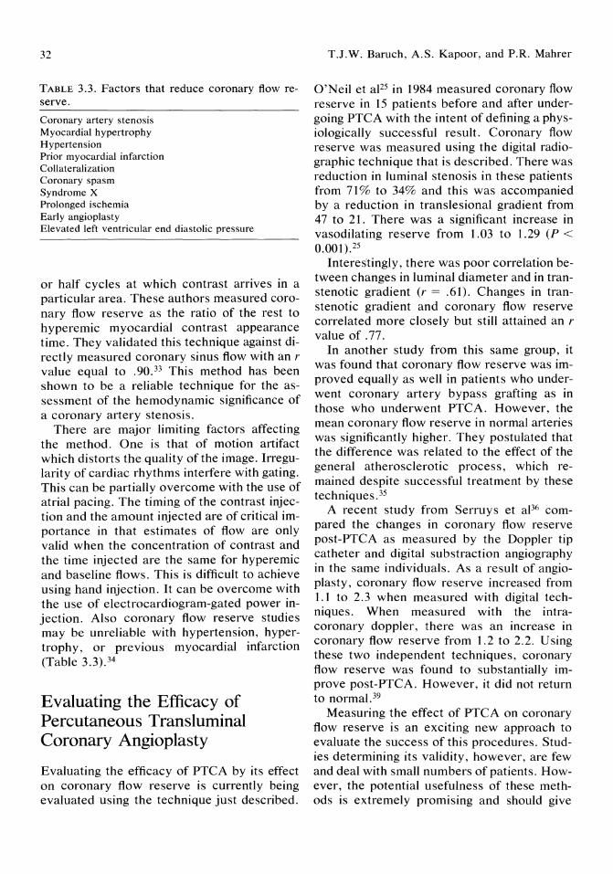
32
TABLE 3.3. Factors that reduce coronary flow reserve.
Coronary artery stenosis Myocardial hypertrophy Hypertension Prior myocardial infarction Collateralization Coronary spasm Syndrome X Prolonged ischemia Early angioplasty Elevated left ventricular end diastolic pressure
or half cycles at which contrast arrives in a particular area. These authors measured coronary flow reserve as the ratio of the rest to hyperemic myocardial contrast appearance time. They validated this technique against directly measured coronary sinus flow with an r value equal to .90. 33 This method has been shown to be a reliable technique for the assessment of the hemodynamic significance of a coronary artery stenosis.
There are major limiting factors affecting the method. One is that of motion artifact which distorts the quality of the image. Irregularity of cardiac rhythms interfere with gating. This can be partially overcome with the use of atrial pacing. The timing of the contrast injection and the amount injected are of critical importance in that estimates of flow are only valid when the concentration of contrast and the time injected are the same for hyperemic and baseline flows. This is difficult to achieve using hand injection. It can be overcome with the use of electrocardiogram-gated power injection. Also coronary flow reserve studies may be unreliable with hypertension, hypertrophy, or previous myocardial infarction (Table 3.3).34
Evaluating the Efficacy of Percutaneous Transluminal Coronary Angioplasty
Evaluating the efficacy of PTCA by its effect on coronary flow reserve is currently being evaluated using the technique just described.
T.J.W. Baruch, A.S. Kapoor, and P.R. Mahrer
O'Neil et aIl5 in 1984 measured coronary flow reserve in 15 patients before and after undergoing PTCA with the intent of defining a physiologically successful result. Coronary flow reserve was measured using the digital radiographic technique that is described. There was reduction in luminal stenosis in these patients from 71% to 34% and this was accompanied by a reduction in translesional gradient from 47 to 21. There was a significant increase in vasodilating reserve from 1.03 to 1.29 (P < 0.001).25
Interestingly, there was poor correlation between changes in luminal diameter and in transtenotic gradient (r = .61). Changes in transtenotic gradient and coronary flow reserve correlated more closely but still attained an r value of .77.
In another study from this same group, it was found that coronary flow reserve was improved equally as well in patients who underwent coronary artery bypass grafting as in those who underwent PTCA. However, the mean coronary flow reserve in normal arteries was significantly higher. They postulated that the difference was related to the effect of the general atherosclerotic process, which remained despite successful treatment by these techniques. 35
A recent study from Serruys et aP6 compared the changes in coronary flow reserve post-PTCA as measured by the Doppler tip ~atheter and digital substraction angiography III the same individuals. As a result of angioplasty, coronary flow reserve increased from 1.1 to 2.3 when measured with digital techniques. When measured with the intracoronary doppler, there was an increase in coronary flow reserve from 1.2 to 2.2. Using these two independent techniques, coronary flow reserve was found to substantially improve post-PTCA. However, it did not return to normal. 39
Measuring the effect of PTCA on coronary flow reserve is an exciting new approach to evaluate the success of this procedures. Studies determining its validity, however, are few and deal with small numbers of patients. However, the potential usefulness of these methods is extremely promising and should give
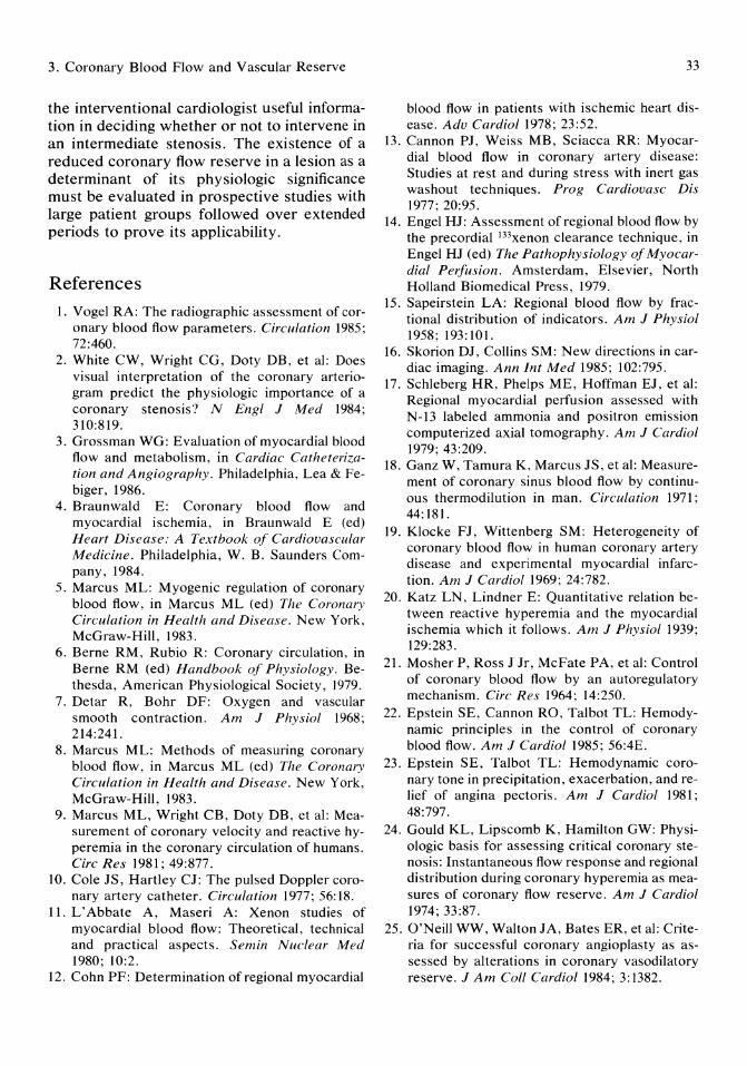
3. Coronary Blood Flow and Vascular Reserve
the interventional cardiologist useful information in deciding whether or not to intervene in an intermediate stenosis. The existence of a reduced coronary flow reserve in a lesion as a determinant of its physiologic significance must be evaluated in prospective studies with large patient groups followed over extended periods to prove its applicability.
References
1. Vogel RA: The radiographic assessment of coronary blood flow parameters. Circulation 1985; 72:460.
2. White CW, Wright CG, Doty DB, et al: Does visual interpretation of the coronary arteriogram predict the physiologic importance of a coronary stenosis? N Engl J Med 1984; 310:819.
3. Grossman WG: Evaluation of myocardial blood flow and metabolism, in Cardiac Catheterization and Angiography. Philadelphia, Lea & Febiger, 1986.
4. Braunwald E: Coronary blood flow and myocardial ischemia, in Braunwald E (ed) Heart Disease: A Textbook of Cardiovascular Medicine. Philadelphia, W. B. Saunders Company, 1984.
5. Marcus ML: Myogenic regulation of coronary blood flow, in Marcus ML (ed) The Coronary Circulation in Health and Disease. New York, McGraw-Hill, 1983.
6. Berne RM, Rubio R: Coronary circulation, in Berne RM (ed) Handbook of Physiology. Bethesda, American Physiological Society, 1979.
7. Detar R, Bohr DF: Oxygen and vascular smooth contraction. Am J Physiol 1968; 214:241.
8. Marcus ML: Methods of measuring coronary blood flow, in Marcus ML (ed) The Coronary Circulation in Health and Disease. New York, McGraw-Hill, 1983.
9. Marcus ML, Wright CB, Doty DB, et al: Measurement of coronary velocity and reactive hyperemia in the coronary circulation of humans. Circ Res 1981; 49:877.
10. Cole JS, Hartley CJ: The pulsed Doppler coronary artery catheter. Circulation 1977; 56: 18.
11. L'Abbate A, Maseri A: Xenon studies of myocardial blood flow: Theoretical, technical and practical aspects. Semin Nuclear Med 1980; 10:2.
12. Cohn PF: Determination of regional myocardial
33
blood flow in patients with ischemic heart disease. Adv Cardiol 1978; 23:52.
13. Cannon PJ, Weiss MB, Sciacca RR: Myocardial blood flow in coronary artery disease: Studies at rest and during stress with inert gas washout techniques. Prog Cardiovasc Dis 1977; 20:95.
14. Engel HJ: Assessment of regional blood flow by the precordial 133xenon clearance technique, in Engel HJ (ed) The Pathophysiology of Myocardial Perfusion. Amsterdam, Elsevier, North Holland Biomedical Press, 1979.
15. Sapeirstein LA: Regional blood flow by fractional distribution of indicators. Am J Physiol 1958; 193:101.
16. Skorion DJ, Collins SM: New directions in cardiac imaging. Ann Int Med 1985; 102:795.
17. Schleberg HR, Phelps ME, Hoffman EJ, et al: Regional myocardial perfusion assessed with N-13 labeled ammonia and positron emission computerized axial tomography. Am J Cardiol 1979; 43:209.
18. Ganz W, Tamura K, Marcus JS, et al: Measurement of coronary sinus blood flow by continuous thermodilution in man. Circulation 1971; 44: 181.
19. Klocke FJ, Wittenberg SM: Heterogeneity of coronary blood flow in human coronary artery disease and experimental myocardial infarction. Am J Cardiol 1969; 24:782.
20. Katz LN, Lindner E: Quantitative relation between reactive hyperemia and the myocardial ischemia which it follows. Am J Physiol 1939; 129:283.
21. Mosher P, Ross J Jr, McFate PA, et al: Control of coronary blood flow by an autoregulatory mechanism. Circ Res 1964; 14:250.
22. Epstein SE, Cannon RO, Talbot TL: Hemodynamic principles in the control of coronary blood flow. Am J Cardiol 1985; 56:4E.
23. Epstein SE, Talbot TL: Hemodynamic coronary tone in precipitation, exacerbation, and relief of angina pectoris. Am J Cardiol 1981; 48:797.
24. Gould KL, Lipscomb K, Hamilton GW: Physiologic basis for assessing critical coronary stenosis: Instantaneous flow response and regional distribution during coronary hyperemia as measures of coronary flow reserve. Am J Cardiol 1974; 33:87.
25. O'Neill WW, Walton JA, Bates ER, et al: Criteria for successful coronary angioplasty as assessed by alterations in coronary vasodilatory reserve. J Am Coli Cardiol1984; 3:1382.
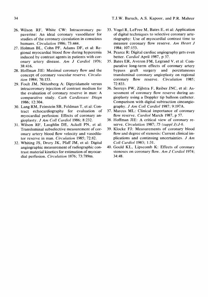
34
26. Wilson RF, White CW: Intracoronary papaverine: An ideal coronary vasodilator for studies of the coronary circulation in conscious humans. Circulation 1986; 73:444.
27. Holman BL, Cohn PF, Adams DF, et al: Regional myocardial blood flow during hyperemia induced by contrast agents in patients with coronary artery disease. Am J Cardiol 1976; 38:416.
28. Hoffman JIE: Maximal coronary flow and the concept of coronary vascular reserve. Circulation 1984; 70:153.
29. Foult JM, Nittenberg A: Dipyridamole versus intracoronary injection of contrast medium for the evaluation of coronary reserve in man: A comparative study. Cath Cardiovasc Diagn 1986; 12:304.
30. Lang RM, Feinstein SB, Feldman T, et al: Contract echocardiography for evaluation of myocardial perfusion: Effects of coronary angioplasty. J Am Coil Cardiol1986; 8:232.
31. Wilson RF, Laughlin DE, Ackell PN, et al: Transluminal subselective measurement of coronary artery blood flow velocity and vasodilator reserve in man. Circulation 1985; 72:82.
32. Whiting JS, Drury JK, Pfaff JM, et al: Digital angiographic measurement of radiographic contrast material kinetics for estimation of myocardial perfusion. Circulation 1876; 73:789m.
T.J.W. Baruch, A.S. Kapoor, and P.R. Mahrer
33. Vogel R, LeFree M, Bates E, et al: Application of digital techniques to selective coronary arteriography: Use of myocardial contrast time to measure coronary flow reserve. Am Heart J 1984; 107: 153.
34. Pearce R: Digital cardiac angiography gets even better. Cardiol April 1987, P 37.
35. Bates ER, Averon FM, Legrand V, et al: Comparative long-term effects of coronary artery bypass graft surgery and percutaneous transluminal coronary angioplasty on regional coronary flow reserve. Circulation 1985; 72:833.
36. Serruys PW, Zijlstra F, Reiber JNC, et al: Assessment of coronary flow reserve during angioplasty using a Doppler tip balloon catheter. Comparison with digital subtraction cineangiography. J Am Coil Cardiol1987; 9:197A.
37. Marcus ML: Clinical importance of coronary flow reserve. Cardiol March 1987, p 57.
38. Hoffman JIE: A critical view of coronary reserve. Circulation 1987; 75 (suppl J):J-6.
39. Klocke FJ: Measurements of coronary blood flow and degree of stenosis: Current clinical implications and continuing uncertainties. J Am Call Cardiol1983; 1:31.
40. Gould KL, Lipscomb K: Effects of coronary stenoses on coronary flow. Am J Cardiol1974; 34:48.
4 Quantitative Coronary Arteriography Budge H. Smith, B. Greg Brown, and Harold T. Dodge
Introduction
Recognizing the relationship between coronary artery disease and clinical events such as angina, myocardial infarction, and cardiac arrythmia, investigators have for 50 years been attempting to accurately describe the human coronary pathologic anatomy. Development of catheterization procedures and radiographic techniques used in the process of evaluating coronary artery disease has been driven by evolving invasive therapies and has resulted in the present high-quality arterial images. In this chapter, we describe the development of techniques now used to obtain and interpret coronary arteriograms and to relate the presence of atherosclerotic disease to the clinical state.
The coronary circulation was imaged first in animals) and then in humans ,2.3 using a transthoracic approach to radiographically image the opacified ascending thoracic aorta and, incidentally, the proximal segments of the coronary arteries. Seeking a safer route, Jonsson4 used a retrograde approach via the radial artery in humans to catheterize the ascending aorta and, with a bolus of contrast manually injected into the aorta, effectively imaged the coronary arteries in their entirety.
Selective imaging of the coronary arteries in humans resulted from the development of: 1) organic iodides that could be used safely, 2) the technique of cineradiography, and 3) a means of safely delivering contrast media to the coronary circulation.
As early as 1929, organic iodides were used to opacify structures in humans.s During the early 1930s, after early successes in opacifying the collecting system of the kidneys,S several iodinated compounds were produced that would provide adequate radiographic contrast of noncoronary vascular structures with minimal side effects. 6,7
In 1938, Robb and Steinberg8 used organic compounds as markers guiding the timing of successive x-ray exposures to document sequential opacification of the heart chambers, thus using serial x-rays to study circulation in the living patient. Technical advances in the 1940s, such as the development of a rapid film changer,9 biplane film capabilities, 10 and image amplification, II allowed for the practical application of cineradiography for the evaluation of contrast angiography in humans. Many of the aspects of cinefluorography in use today (i.e., the use of synchronized exposure of 35-mm film at 60 frames per second to allow for slowmotion analysis) were refined for clinical use in the cine program of the University of Rochester by Ramsey and co-workers.12 The cineangiographic techniques presently used in most investigative centers were first applied by Abrams 13 at Stanford University in 1958. Most modifications which have occurred since that time are a reflection of technical advances and improvements of apparati in use in 1958.
In parallel with the advances in contrast and radiographic technology, catheterization techniques were also developing in the early 1950s; thus, angiocardiography emerged as a
36
safe, effective means to determining the presence and severity of atherosclerotic coronary artery disease.
Retrograde left heart catheterization was first performed in the United States by Zimmerman and colleagues l4 in 1949 for evaluation of left heart and aortic pressures in syphilitic aortic insufficiency. The position of the catheter in this study was confirmed by plain radiograph. In the 1950s, single frame exposure of opacification of the coronary arteries became possible using a balloon occlusion technique of the aorta with acetylcholine-induced cardiac arrest, described by Dotter and Frische. l5 This technique provided, for the first time, well-detailed films of the coronary tree but at considerable risk.
The modern era of coronary arteriography was ushered in by Sones and Shirley l6 at the Cleveland Clinic in 1959 when, quite by chance, they discovered that by selective transbrachial coronary cannulation, one could safely opacify the coronaries in man. Other selective angiographic techniques quickly emerged, particularly pre-formed catheters for specific selective procedures '7 and the femoral artery approach described by Judkins ls in 1963.
It is upon the above technological and procedural foundation that computer-assisted modalities have, during the 1970s and 1980s, attempted to provide a more precise description of the coronary anatomy. Clinical decisions, both prognostic and therapeutic, are based in part on an objective interpretation of the distribution and severity of atherosclerotic coronary disease. Thus, accurate representation of the disease process has important clinical ramifications.
Visual Interpretation
Most clinical centers now rely on visual interpretation of the coronary angiogram. Segmental arterial narrowing is widely described in terms of "percent stenosis" relative to a nearby "normal" lumen diameter. However, there are certain limitations to this interpretation of disease.
B.H. Smith, B.G. Brown, and H.T. Dodge
Accurate definition of disease depends on the number and variety of viewing angles used. Significant stenoses can be completely missed if vessel overlap, foreshortening, or insufficient "panning" occur. To avoid these problems, sufficient viewing angles must be used to assess all coronary segments, optimally, at as close to right angles as possible. This basic doctrine, if implemented, would minimize errors in the estimate of maximal stenosis.
The "percent stenosis" estimate is dependent upon the selection of a truly "normal" reference lumen diameter. However, vascular segments near stenoses that are chosen as "normal" segments might be dilated by poststenotic turbulence or ectasia, normal, or diffusely narrowed by intimal disease, thereby making the denominator of the "percent stenosis" estimate unreliable.
Owing to these potential sources for error in visual estimates of coronary stenoses, significant inter- and intraobserver variability in estimates of disease can exist. l9- 2' DeRouen and colleagues20 reported an interobserver variability of 18% (1 SD) in visual estimates of the maximal stenosis of 12 coronary segments in 10 patients; the probability of misclassification of the number of significantly (:2: 70%) stenosed vessels in an individual case was 31%.
Lastly, the correlation between visual estimates of disease and coronary blood flow is poor22; thus, clinical decisions made using visual estimates of coronary stenosis are potentially flawed.
Recognizing these shortcomings, cardiologists and radiologists are now investigating computer-assisted image analysis as a means for more precise evaluation of coronary atherosclerosis.
Quantitative Angiography
The era of machine-assisted coronary artery quantitation was ushered in by Gensini et aJ23 in their study of coronary vasomotion in 1971. A cross-hair system to specify diametrically opposed image border points was used in pro-
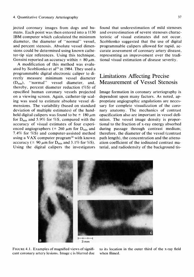
4. Quantitative Coronary Arteriography
jected coronary images from dogs and humans. Each point was then entered into a 1130 IBM computer which calculated the minimum diameter, the diameter of "normal" vessel, and percent stenosis. Absolute vessel dimensions could be determined using known catheter-tip size references. Using this technique, Gensini reported an accuracy within ± 80,um.
A modification of this method was evaluated by Scoblionko et aJ21 in 1984. They used a programmable digital electronic caliper to directly measure minimum vessel diameter (Dmin), "normal" vessel diameter, and, thereby, percent diameter reduction (%S) of opacified human coronary vessels projected on a viewing screen. Again, catheter-tip scaling was used to estimate absolute vessel dimensions. The variability (based on standard deviation of multiple estimates) of the handheld digital calipers was found to be ± 180,um for Dmin and 5.9% for %S, compared with the accuracy of visual estimates of four experienced angiographers (± 260 /Lm for Dmin and 7.4% for %S) and computer-assisted method using a V AX computer program24 with known accuracy (± 90 /Lm for Dmin and 3.1 % for %S). U sing the digital calipers the investigators
3mm
FIGURE 4.1. Examples of magnified views of significant coronary artery lesions. Image c is blurred due
37
found that underestimation of mild stenoses and overestimation of severe stenoses characteristic of visual estimates did not occur. Scoblionko suggested that the use of digital programmable calipers allowed for rapid, accurate assessment of coronary artery disease, representing an improvement over the traditional visual estimation of disease severity.
Limitations Affecting Precise Measurement of Vessel Stenosis
Image formation in coronary arteriography is dependent upon many factors. As noted, appropriate angiographic angulations are necessary for complete visualization of the coronary anatomy. The mechanics of contrast opacification also are important in vessel definition. The vessel image density is proportional to the fraction of x-ray energy absorbed during passage through contrast medium; therefore, the diameter of the vessel (contrast path length), the concentration and the attenuation coefficient of the iodinated contrast material, and radiodensity of the background tis-
to its location in the outer third of the x-ray field when filmed.
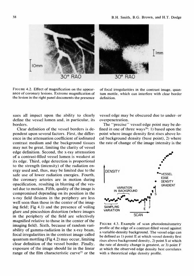
38
30° RAO
FIGURE 4.2. Effect of magnification on the appearance of coronary lesions. Extreme magnification of the lesion in the right panel documents the presence
sues all impact upon the ability to clearly define the vessel lumen and, in particular, its borders.
Clear definition of the vessel borders is dependent upon several factors. First, the difference in the attenuation coefficient of iodinated contrast medium and the background tissues may not be great, limiting the clarity of vessel edge definition. Second, the x-ray attenuation of a contrast-filled vessel lumen is weakest at its edge. Third, edge detection is proportional to the strength (intensity) of the radiation energy used and, thus, may be limited due to the safe use of lower radiation energies. Fourth, the coronary arteries are in motion during opacification, resulting in blurring of the vessel due to motion. Fifth, quality of the image is compromised depending on its position in the x-ray field (lesions in the periphery are less well seen than those in the center of the imaging field; Fig 4.1) and the presence of veiling glare and pincushion distortion (where images in the periphery of the field are selectively magnified relative to those in the center of the imaging field). Sixth, because of random variability of gamma-radiation in the x-ray beam, focal irregularities in the contrast image called quantum mottling (Fig 4.2) may occur, limiting clear definition of the vessel border. Finally, exposure of the image should lie in the linear range of the film characteristic curve25 or the
B.H. Smith, B.G. Brown, and H.T. Dodge
30° RAO
of focal irregularities in the contrast image, quantum mottle, which can interfere with clear border definition.
vessel edge may be obscured due to under- or overpenetration.
The "precise" vessel edge point may be defined in one of three ways26: 1) based upon the point where image density first rises above local background density (base point), 2) where the rate of change of the image intensity is the
lDENSITY
• ,1 VARIATION
IN BACKGROUND ~ t S .,. 'I .... -:", . .. ~ ..... ·f t
SAMPLING E F VARIATION
SCAN
• •
... . ~ • •
"'VESSEL EDGE DENSITY GRADIENT
FIGURE 4.3. Example of scan photodensitometry profile of the edge of a contrast-filled vessel against a variable-density background. The vessel edge can be defined as 1) point E at which vessel density first rises above background density, 2) point S at which the rate of density change is greatest, or 3) point F at which the observed edge density best correlates with a theoretical edge density profile.
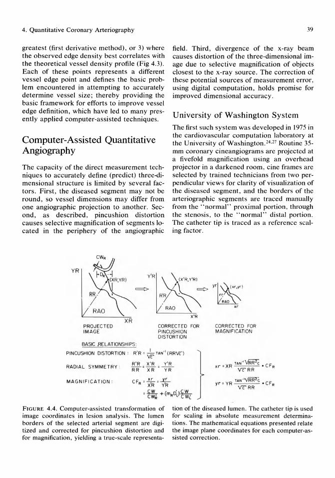
4. Quantitative Coronary Arteriography
greatest (first derivative method), or 3) where the observed edge density best correlates with the theoretical vessel density profile (Fig 4.3). Each of these points represents a different vessel edge point and defines the basic problem encountered in attempting to accurately determine vessel size; thereby providing the basic framework for efforts to improve vessel edge definition, which have led to many presently applied computer-assisted techniques.
Computer-Assisted Quantitative Angiography
The capacity of the direct measurement techniques to accurately define (predict) three-dimensional structure is limited by several factors. First, the diseased segment may not be round, so vessel dimensions may differ from one angiographic projection to another. Second, as described, pincushion distortion causes selective magnification of segments located in the periphery of the angiographic
YR ~CWR
rD. -i Y'R h(XR'YRl
. I => RR . / RAO
XR
39
field. Third, divergence of the x-ray beam causes distortion of the three-dimensional image due to selective magnification of objects closest to the x-ray source. The correction of these potential sources of measurement error, using digital computation, holds promise for improved dimensional accuracy.
University of Washington System
The first such system was developed in 1975 in the cardiovascular computation laboratory at the University of Washington. 24.27 Routine 35-mm coronary cineangiograms are projected at a fivefold magnification using an overhead projector in a darkened room, cine frames are selected by trained technicians from two perpendicular views for clarity of visualization of the diseased segment, and the borders of the arteriographic segments are traced manually from the "normal" proximal portion, through the stenosis, to the "normal" distal portion. The catheter tip is traced as a reference scaling factor.
X'R
PROJ EC TED IMAGE
CORRECTED FOR PINCUSHION DISTORT ION
CORREC TED FOR MAGNIFICATION
BASIC RELATIONSHIPS :
PINCUSHION DISTORTION : R'R = ~ TAN-' (RRVC)
R' R X'R y ' R RADIAL SYM METR Y : Rif = XR = """"YR XR TAN~~ CF
x. ... = • R VC RR
MAGNIF IC A TIO N :
FIGURE 4.4. Computer-assisted transformation of image coordinates in lesion analysis. The lumen borders of the selected arterial segment are digitized and corrected for pincushion distortion and for magnification, yielding a true-scale representa-
R TAN-'\"RR'lC C F yr = Y • R VCRR
tion of the diseased lumen. The catheter tip is used for scaling in absolute measurement determinations. The mathematical equations presented relate the image plane coordinates for each computer-assisted correction.
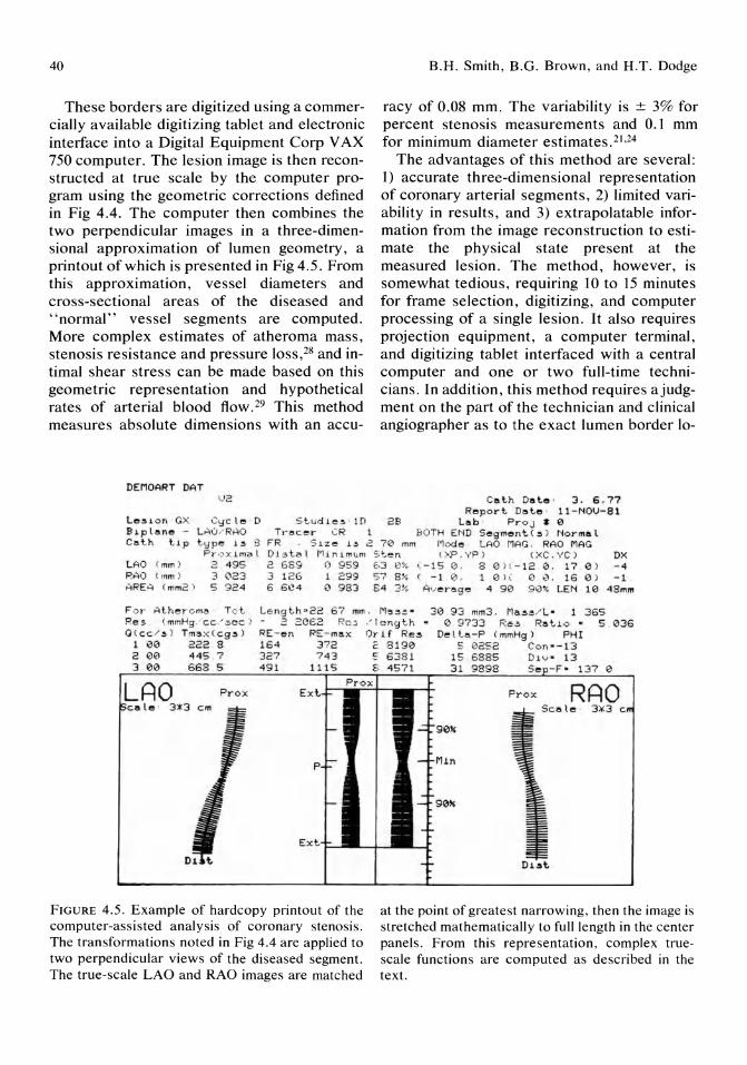
40
These borders are digitized using a commercially available digitizing tablet and electronic interface into a Digital Equipment Corp V AX 750 computer. The lesion image is then reconstructed at true scale by the computer program using the geometric corrections defined in Fig 4.4. The computer then combines the two perpendicular images in a three-dimensional approximation of lumen geometry, a printout of which is presented in Fig 4.5. From this approximation, vessel diameters and cross-sectional areas of the diseased and "normal" vessel segments are computed. More complex estimates of atheroma mass, stenosis resistance and pressure loss,28 and intimal shear stress can be made based on this geometric representation and hypothetical rates of arterial blood flow. 29 This method measures absolute dimensions with an accu-
DEMOART OAT l)2
B.H. Smith, B.G. Brown, and H.T. Dodge
racy of 0.08 mm. The variability is ± 3% for percent stenosis measurements and 0.1 mm for minimum diameter estimates. 21 ,24
The advantages of this method are several: 1) accurate three-dimensional representation of coronary arterial segments, 2) limited variability in results, and 3) extrapolatable information from the image reconstruction to estimate the physical state present at the measured lesion. The method, however, is somewhat tedious, requiring 10 to 15 minutes for frame selection, digitizing, and computer processing of a single lesion. It also requires projection equipment, a computer terminal, and digitizing tablet interfaced with a central computer and one or two full-time technicians. In addition, this method requires ajudgment on the part of the technician and clinical angiographer as to the exact lumen border 10-
Cat.h Dat.e · 3. 6 . 77 Report. Dat.e · 11-NOV- 81
Le~.on GX Cycle 0 S t. udie~ · I D 2B Lab ' Pro J ' 0 B.p lane - L-'lu / RAO T,-" c er CR 1 BOTH E~lD Segm"'nt. ( ~ ) Normal Cat.h Up t.Vpe i ~ g FR . S ue 1 ~ 2 70 mm 1100., · LAO MAG. RAO MA G
Pr"x lm",l Dl " t.a l l1inl mum Sf. en ( )\f' . VP ) ( XC.VC) OX LAO ( mm )
RAO (In ", )
AREG ( mm2 ',
2 495 2 689 0 959 63 0 % (-15 0. 8 0 )( - 12 0 . 17 0 ) -4 3 12123 3 126 1 299 57 8 % ( - 1 0. 1 01 ( 0 0. 16 0 ) -1 . 5 924 6 6e4 0 983 8 4 3% Average · 4 90 90% LEN 10 48mm
F",r At.he,·o m.!> Tc t. Lengt.h322 67 mm . ~1,,~~ · 30 9 3 mm3 . Ma~~/L ' 1 365 Re5 . ( mmHg .'cc ~5CC ) - 2 2062 R03 / le ng t.h · 0 9733 Re~ Ra tio · 5 036 a ( cc /~ ) Tmax( C9 a ) RE-en RE-max Orlf Re~ De lt.a-P (mmHg) PHI
1 00 222 8 164 3 7 2 ~ 8190 5 0252 Con - - 13 2 00 445 . 7 327 743 S 6 38 1 15 6885 Dl v · 13 3 00 668 5 49 1 111 5 8 457 1 31 9898 Se - 137 0
Prox
FIGURE 4.5. Example of hardcopy printout of the computer-assisted analysis of coronary stenosis. The transformations noted in Fig 4.4 are applied to two perpendicular views of the diseased segment. The true-scale LAO and RAO images are matched
P rox RAO Scale · 3)(3
Min
at the point of greatest narrowing, then the image is stretched mathematically to full length in the center panels. From this representation, complex truescale functions are computed as described in the text.

4. Quantitative Coronary Arteriography
cation based on visual interpretation of several local gradients of contrast density at the lumen edge. As human judgment is a necessary component of this method, it presents a significant source of potential error. At the University of Washington certain rules for frame selection are followed to minimize possible error: 1) some angiographic projections show arterial lesions more clearly than others, and the views with best image quality are therefore selected during an initial screening; 2) images having borders that are sharp and continuous through the lesions are preferred; 3) the sharpest images occur at moments of maximum intraluminal iodine concentration, usually in the middle third of the contrast injection; 4) the sharpest images occur at moments of least vessel motion (i.e., at end-systole, mid- to late-diastole, or at the peak of atrial contraction); 5) frames in which the lumen appears to be narrowest are probably more accurate because many artifacts (motion, vessel overlap, foreshortening) can serve to increase apparent lumen diameter but few (streaming, inadequate contrast injection) cause the lumen to appear more narrow than reality; and 6) selection of frames in which the segment of interest is obscured by other dense structures is avoided. Using these guidelines, it is possible to digitize the borders of a lesion in a selected frame in a fashion reproducible to within ± 0.09 mm.21
University of California at Irvine System
Tobis et apo have used computers to enhance the angiographic image by digital subtraction and, thereby, improve contrast. In their system, the incoming video signal from a 7-inch cesium iodide image intensifier is amplified and then converted from an analog to a digital format by the computer. For cardiac imaging, a 512 x 512 x 8 bit pixel matrix at 30 frames per second is near the technical limit of data transfer capability. The digital image may then be stored for subsequent retrieval or processed immediately. A .5- to 2-second precontrast image set provides "masks," which may be digitally subtracted from the images gener-
41
ated during contrast injection to provide enhanced vascular images. The advantages of this technique over conventional angiography include improved contrast imaging due to the removal by the computer of overlying and interfering soft tissue densities, the need for smaller volumes of contrast medium for vascular definition, rapid image processing for ready visualization, and the ability to postprocedurally alter the subtracted images for clinical visualization. This is accomplished by the computer converting the postprocedural stored image (which may be in either analog or digital form) to a digital format. The contrast or brightness of the image may then be altered or a new mask selected. In this format, the Irvine investigators also used an edge enhancement algorithm to scan the digital image border, select the points of most rapid gray-scale change, and sharpen the contrast at these points; thereby enhancing vessel border imaging. The major disadvantage of DSA is misregistration artifact, which develops if motion (body movement, panning, or respiration) occurs between the time the mask is generated and the contrast images performed. This is such a problem that many users of digital image systems prefer to work with the unsubtracted images.
The investigators at the University of California at Irvine, in a study of 19 patients with 32 arterial lesions, 31 found the digital subtraction technique to be as sensitive for clinical application as the film-based cineangiograms presently in wide use. Using a digitizing computer with a 512 x 512 matrix, filming at 8 frames per second (the authors believed that due to the subtraction process and contrast enhancement, the 30 frames per second used for conventional angiograms was not necessary), and measuring percent stenosis by using calipers, the investigators found no significant difference in the severity of stenosis measured by the two processes. The quality of the digitally subtracted films were believed by the group to be at least as good as that of the filmbased technique, and although spatial resolution may be slightly reduced in the digitally subtracted format, the ability to postprocedurally alter the final image was believed to
42
provide comparably useful image content. Thus, their initial experience using the DSA technique for routine clinical angiography has been encouraging.
Stanford University System
Alderman et aP2 and Sanders et aP3 at Stanford University have focused their efforts at improving contrast imaging resolution by developing a computer-assisted, operator-interactive method of defining the lumen borders of a coronary artery segment at clinical arteriography. End-diastolic cine frames that are projected on a monitor screen are selected by the angiographer as to visual clarity of the lesion to be measured. The lesion of interest is magnified using a turrent lens system (up to sevenfold magnification) and then digitized using a 480 x 512 x 8 bit pixel matrix. The computer program smooths the random fluctuation in gray scale due to quantum mottle, using a lowpass filter. The operator then, with a light pen, traces the best lumen border on the monitor screen. The computer then directs densitometric scan trajectories perpendicular to the manually defined border at multiple points. The point along each trajectory at which the first derivative of the gray-scale density profile peaks is defined by the computer as the vessel edge. Manual operator adjustment of the computer-generated image may then take place. Magnification correction factors are obtained as needed from tabular computer memory, being a defined function of the distance of the image intensifier from the coronary lesion at the x-ray isocenter. Using this method, absolute measurements of lumen diameter, segment lumen area and volume, and percent diameter reduction from single-plane angiographic views are possible, and the reported variability in repeat dimensional estimates is low, approaching that of manual tracing of lesions from highly magnified projections. 21 ,24,28
Thorax Centre-Erasmus University System
Reiber and colleagues34,35 in the Netherlands have developed an operator-interactive, computer-assisted method for automated edge de-
B.H. Smith, B.G. Brown, and H.T. Dodge
tection that uses a video display of the projected image of selected 35-mm cine frames. The cine film is mounted on a specially constructed cinevideo converter, which allows projection of the selected cine frame through one of six rotatable lenses, making possible six different magnification factors onto a video screen. The video camera is attached to a movable x-y stage so that an area of interest may be centered for analysis using the appropriate magnification factor. The center of the resulting video image is then digitized using a 512 x 512 x 8 bit matrix. A calibration factor is determined for the magnified image projected using the contrast-filled catheter as a scaling reference. This factor is expressed in terms of millimeters per pixel. The computer then adjusts the final image display to correct for the magnification factor, as well as for pincushion distortion. This allows for absolute vessel diameter measurements to be made.
To determine vessel contour, this system requires the user to make centerline determinations at several vessel segment points using a writing tablet. A smoothed version of the centerline is determined using a 96 x 96 pixel matrix by the computer's central processing unit, PDP 11/44, which is interfaced with the image digitizer. For edge detection, scanlines are generated perpendicular to the smoothed local centerline orientation. The edge of the vessel lumen is defined as the point representing the weighted sum of the first and second derivative functions applied to the digitized brightness information with the use of minimal-cost criteria. A smoothing procedure is again applied to the detected lumen border based on a computer-defined centerline. This centerline is determined by the computer to be the points midway between the detected and possibly corrected contours.
Using the user-interactive technique, absolute vessel dimensions, minimum diameter, mean diameter, and percent diameter reduction may be computed. The investigators at the Thorax Centre define the reference diameter (and thus the denominator in the percent stenosis determination) in two ways: 1) an average of 11 diameter values in a representative region around the user-defined reference position, and 2) based upon a computer-recon-

4. Quantitative Coronary Arteriography
structed prediction of the original vessel diameter at the site of stenosis, allowing for gradual vessel tapering. Using this technique, the investigators believe that an assessment of the atheromatous placque (but not mass) and its degree of eccentricity may be made (as a function of the detected lumen contour and the diameter references).
The investigators at the Thorax Centre report an accuracy and precision of the edge detection process to be - 30 /Lm and 90 /Lm, respectively. Variability in the determination of absolute vessel dimensions is less than 120/Lm and in percent stenosis estimates less than 2.74%. These reported sensitivities are well within the requirements of the clinical and research applications of quantitative angiography.
Photodensitometric Analysis
Estimation of arterial segment area may be made using the technique of photodensitometry which is based upon the Beer-Lambert law.
The Beer-Lambert principle states that a homogeneous bolus of contrast medium will fill a vessel in such a manner that the intensity of x-ray attenuation created, I r , will be directly proportional to the length, z, of the xray beam path through the vessel:
where Irb is the intensity observed without contrast and /L is the absorbance coefficient of the contrast medium. The light image signal generated from the bolus injection is linearly proportional to the photographic density of the contrast medium along the x-ray beam path. The image may then be digitized and stored or preserved on cine film. If the vessel is viewed perpendicular to its central longitudinal axis, the density of the contrast at this point would be proportional to the crosssectional area. For purposes of vessel area analysis, subtraction of background signals ("noise") is done to maximize contrast enhancement. The estimated cross-sectional area of a stenosed segment is then compared with estimates of nearby "normal" segment
43
area and a percent stenosis calculation is made.
Problems inherent to this technique are several. First, subtraction of background signals may produce misregistration artifact. Second, thoracic structures which comprise the background are not uniform in their density, resulting in a variable signal to noise ratio in the segment of interest. Third, this system requires that the vessel be viewed strictly perpendicular to its long axis. If the vessel curves, moves, or is viewed obliquely, the estimation of the cross-sectional area will be erroneous. These potential sources of error are particularly pertinent to the analysis of coronary vessels, which are frequently tortuous and in motion. Despite these limitations, certain investigators advocate the use of photodensitometry for quantitating coronary artery stenoses.
Harvard-Beth Israel System Sandor et aP6,37 at the Beth Israel hospital have developed an operator-interactive system applying the principles of photodensitometry. Using 35-mm cineangiograms, the frames of interest selected by an operator are directly digitized using a 175 x 175 pixel 8-bit gray-scale format. The digitized frames are analyzed by an interfaced PDP 11170 computer that generates densitometric data. The digitized image of interest is then displayed on a Tektronix 4014 viewing scope.
At this point, the operator may use "thresholding" of the gray scale and image magnification to improve the displayed image. U sing electronic cursors, the operator then identifies a short segment of stenosed and "normal" vessel. This is done by defining an "analysis window," a single window encompassing both diseased and normal vessel segments in straight arteries or two separate windows in curved vessels. The computer then defines multiple density profiles within the windows by scanning the vessel perpendiCUlar to its long axis. The operator then, using the cursors, manually defines the vessel borders. The computer will then calculate the area defined by the operator-determined vessel border and the computer-determined density pro-
44
file. The ratio of density summated area generated for the stenotic segment relative to that generated for the "normal" vessel represents a percent stenosis estimate.
Sandor and colleagues36,37 in phantom studies found the photodensitometric area estimation to be linear. In addition, in an analysis of clinically obtained cineangiograms reviewed by three independent observers, this system was found to have an intniobserver variability of 4.5% for a 50% vessel stenosis and 2.9% for a 90% stenosis. Testing the sensitivity of the system against the variable background of the dog thorax, the investigators found a variability (SD of multiple estimates) of 20% for tubes the size of the left main coronary artery but a variability of 30% for tubes -s: 1 mm in diameter.
This system is clinically applicable but has limitations in sensitivity due to background noise interference, the lack of absolute dimension estimation, and insensitivity of the system for vessels of small caliber, unfortunately the lumen area frequently associated with clinically active disease.
Columbia University System Nichols et aP8 at Columbia University also have developed a computer-assisted system of coronary stenosis quantitation applying the principles of photodensitometry. Using 35-mm cineangiograms obtained using a 14-inch cesium iodide intensifier (in the 6-inch mode), the investigators select a frame for analysis which is projected onto a video screen. The selected image is then digitized using a 512 x 512 pixel matrix by a Nova computer, which is interfaced with a vidicon camera.
The quantitation of arterial stenosis is then performed by placing indicator markers (' 'rectangular regions of interest," ROI) of 2 pixel width over the most narrow arterial region and over the "normal" reference segment proximal to the stenosis. A densitometric value for the column of contrast within the ROI is generated and corrected for background density. The corrected densitometry values for the "normal" artery and the point of greatest stenosis are linearly related to the cross-sectional
B.H. Smith, B.G. Brown, and H.T. Dodge
area of the sampled sites. In this manner, percent stenosis may be calculated.
To test the accuracy of this system, Nichols and colleagues38 used phantom models consisting of plexiglass rods of known dimensions inserted into columns filled with contrast. U sing the densitometric technique described, the investigators found a near perfect (r = .99) correlation between known rod size and crosssectional area reduction and densitometrically determined values. Good correlation also was seen between densitometric analysis of 10 lesions from four patients who died shortly after coronary arteriography and postmortem histologic planimetry of arterial area reduction (r = .97, SEE = 7.0%). Interobserver variability (r = .99, SEE = 4.3%) and intraobserver variability (r = .92, SEE = 7.7%) were acceptable.
As stated by the authors, this system is a rapid, reliable, and reproducible method of estimating the severity of coronary artery stenoses. However, the Columbia system, as with the Harvard system, does not generate absolute coronary lumen dimensions. Also, validation of the system was made using phantom models and postmortem vessels of 1.02 to 4.16 mm diameter. The clinical applicability of the system, therefore, may be limited in the evaluation of clinically relevant coronary lesions.
Measurement of Coronary Flow Reserve
The limitations of coronary lumen diameter quantitation make accurate definition of the three-dimensional coronary atheromatous plaque difficult. Clinical decisions are commonly made based on the estimated lumen diameter when, optimally, these decisions are best made based on the presence or absence of adequate coronary blood flow for myocardial needs under functional circumstances.
University of Michigan Approach to Perfusion Imaging
One system designed to evaluate the consequence of coronary artery stenosis on coronary blood flow has been developed by Vogel

4. Quantitative Coronary Arteriography
et aP9 at the University of Michigan. Using 35-mm cineangiograms viewed on a Vanguard projector, six consecutive end-diastolic frames are selected. These frames are then digitized using a 256 x 256 pixel matrix with an 8-bit gray scale with the final digitized image representing an average of eight video digitizations. Image enhancement is then performed using an interfaced PDP 11/34 computer by subtracting background noise over six consecutive electrocardiogram-gated cardiac cycles. Finally, the enhanced image is color and intensity modulated for functional analysis of coronary blood flow.
These investigators measured the myocardial contrast appearance time (MCAT) defined as the time from contrast injection to regional myocardial enhancement phase in various arterial distributions in humans. They found the MCAT parameter to be linearly reproducible and well correlated relative to independent measurements of coronary blood flow (coronary sinus and great cardiac vein thermodilution techniques). The MCAT measurement also was found to be predictively altered depending on the presence or absence of significant (> 70%) stenosis in the setting of contrast-induced hyperemia. The investigators therefore believed that MCAT determination in the face of hyperemic challenge could be a useful measure of regional coronary blood flow reserve.
The major drawbacks associated with this system include the dependence upon patient cooperation for serial-gated data collection and the dependence of regional coronary reserve on factors other than stenosis severity. The technique provides useful supportive information for the clinician interested in the regional coronary blood flow characteristics effected by proximal coronary artery stenosis.
University of Texas System
Gould and associates40,41 evaluated the ability to predict coronary blood flow reserve based upon coronary stenosis quantitation. Using an external constrictor to induce varying degrees of coronary lumen stenosis in dogs, the investigators implanted tygon catheters proximal and distal to the stenosis for purposes of con-
45
trast medium injection and pressure monitoring, respectively. Blood flow velocity across the induced stenosis was measured with a continuous-wave directional Doppler probe. Coronary stenosis quantitation was performed using a computer-directed border recognition system on-line with a VAX 111780 computer. Their system is a centerline technique for orthogonal cineangiograms to generate a threedimensional image of the stenotic coronary segment. Assuming classic fluid dynamics theory for flexible, stenotic coronary segments in vivo, the investigators found a good correlation between the degree of fixed coronary artery stenosis and coronary blood flow reserve.
Conclusion
Most investigators agree that visual estimates of coronary lumen percent stenosis in the setting of atherosclerotic coronary artery disease are highly variable and correlate poorly with other indices of clinical significance. Efforts have been made to improve the sensitivity of coronary artery quantitation by applying computer-assisted modalities. Several systems have been developed using operator-dependent and automated vessel border identification. Other systems circumvent the need for accurate border identification by generating videodensitometric values for diseased and normal segments and expressing relative stenosis in these terms. Photodensitometric systems, however, require independent calibration for absolute dimensions; to date, these approaches have proven difficult. Some investigators have attempted to assess the physiologic significance of arterial stenosis in terms of its impact on coronary blood flow reserve. As daily clinical decisions and prediction of patient prognosis are, in part, based upon estimates of vessel narrowing, continued progress in the quantitation of coronary artery disease should have considerable impact in the field of cardiology.
References
1. Rousthoi P: Uber Angiokardiographie. Acta Radio11933; 14:419.
2. Radner S: An attempt at the roentgenologic vi-
46
sualization of coronary blood vessels in man. Acta Radial 1945; 26:497.
3. Hoyos JM, Del Campo CG: Angiography of the thoracic aorta and coronary vessels. Radiology 1948; 50:211.
4. Jonsson G: Visualization of the coronary arteries. Acta Radial 1948; 29:536.
5. Swick N: Darstellung der Niere und Harnwege im Rontgenbild durch intravenose Einbringung eines newen Kontraststoffes, des Uroselectans. Klin Wochenschr 1929; 8:2087.
6. Pearse HE, Warren SL: The rontgenographic visualization of the arteries of the extremities in peripheral vascular disease. Ann S urg 1931; 94:1094.
7. Wohlleben T: Venographie. Klin Wochenschr 1932; 112:1786.
8. Robb GP, Steinberg I: Visualization of the chambers of the heart, the pulmonary circulation, and the great blood vessels in man: A practical method. AJR 1939; 41: 1.
9. Gidlund AS: A new apparatus for direct cineroentgenography, Acta Radial 1949; 32:81.
10. Axen 0, Lind J: Electrocardiographic recording in angiocardiography with synchronous serial photography at right-angled planes. Cardiologia 1950; 16:60.
11. Coltman JW: Fluoroscopic image brightening by electronic means. Radiology 1948; 51:359.
12. Ramsey GHS, Watson IS, Steinhausen TB, et al: Cinefluorography: A progress report on technical problems, dosage factors, and clinical impressions. Radiology 1949; 52:684.
13. Abrams HL: An approach to biplane cineangiocardiography. Radiology 1958; 72:735, 741.
14. Zimmerman HA, Scott RW, Becker NO: Catheterization of the left side of the heart in man. Circulation 1950; 1 :357.
15. Dotter CT, Frische LH: Visualization of the coronary circulation by occlusion aortography: A practical method. Radiology 1958; 71 :502.
16. Sones FM, Shirley EK: Cine coronary arteriography. Mod Concepts CV Dis 1962; 31:735.
17. Ricketts HJ, Abrams HL: Percutaneous selective coronary cinearteriography. J Am Med Assoc 1962; 181:620.
18. Judkins MP: Selective coronary arteriography: A percutaneous transfemoral technique. Radiology 1967; 89:815.
19. Detre KM, Wright E, Murphy ML, et al: Observer agreement in evaluating coronary angiography. Circulation 1975; 52:979.
20. DeRouen TA, Murray JA, Owen W: Variability in the analysis of coronary arteriograms. Circulation 1977; 55:324.
B.H. Smith, B.G. Brown, and H.T. Dodge
21. Scoblionko DP, Brown BG, Mitten S, et al: A new digital electronic caliper for measurement of coronary arterial stenosis: Comparison with visual estimates and computer-assisted measurement. Am J Cardiol 1984; 53:689.
22. Mates RE, Gupta ACB, Klocke FJ: Fluid dynamics of coronary artery stenosis. Circ Res 1978; 42:152.
23. Gensini GG, Kelly AE, DaCosta BCB, et al: Quantitative angiography: The measurement of coronary vasomobility in the intact animal and man. Chest 1971; 60:522.
24. Brown BG, Bolson E, Frimer M, et al: Quantitative coronary arteriography: Estimation of dimensions, hemodynamic resistance, and atheroma mass of coronary artery lesions using the arteriogram and digital computation. Circulation 1977; 55:329.
25. Kato W, Wong M: Optimizing cine film. Cathet Cardiovasc Diagn 1975; 1:97.
26. Spears lR, Sandor T, Als A V, et al: Computerized image analysis for quantitative measurement of vessel diameter from cineangiograms. Circulation 1983; 68:453.
27. Brown BG, Bolson EL, Dodge HT: Arteriographic assessment of coronary atherosclerosis: Review of current methods, their limitations, and clinical applications. Arteriosclerosis 1982; 2:2.
28. McMahon MM, Brown BG, Cukingnan R, et al: Quantitative coronary angiography: Measurement of the "critical" stenosis in patients with unstable angina and single-vessel disease without collaterals. Circulation 1979; 60: 106.
29. Ganz W, Tamura K, Marcus HS, et al: Measurement of coronary sinus blood flow by continuous thermodilution in man. Circulation 1971; 44: 181.
30. Tobis JM, Nalcioglu 0, Henry WL: Digital subtraction angiography. Chest 1983; 84:68.
31. Tobis J, Nalcioglu 0, Iseri L, et al: Detection and quantitation of coronary artery stenoses from digital subtraction angiograms compared with 35-millimeter film cineangiograms. Am J Cardiol 1984; 54:489.
32. Alderman E, Berte LE, Harrison DC, et al: Quantitation of coronary artery dimensions using digital image processing. Soc Photo-Optical Instrum Engineers-Digital Radiography 1981; 314:273.
33. Sanders WJ, Alderman EL, Harrison DC: Coronary artery quantitation using digital image processing techniques. IEEE Comput Cardiol 1979; 7:15.
34. Reiber JHC, Serruys PW, Kooijman CJ, et al:
4. Quantitative Coronary Arteriography
Assessment of short-, medium-, and long-term variations in arterial dimensions from computer-assisted quantitation of coronary cineangiograms. Circulation 1985; 71:280.
35. Wijns W, Serruys PW, Reiber JRC, et al: Quantitative angiography of the left anterior descending coronary artery: Correlations with pressure gradient and results of exercise thallium scintigraphy. Circulation 1985; 71:273.
36. Sandor T, Als A V, Paulin S: Cine-densitometric measurement of coronary arterial stenoses. Cath CV Diag 1979; 5:229.
37. Spears JR, Sandor T, Als A V, et al: Computerized image analysis for quantitative measurement of vessel diameter from cineangiograms. Circulation 1983; 68:453.
38. Nichols AB, Gabrieli CFO, Fenoglio JJ. et al: Quantification of relative coronary arterial stenosis by cinevideodensitometric analysis
47
of coronary arteriograms. Circilhlfion 1984; 69:512.
39. Vogel R, LeFree M, Bates E, et al: Application of digital techniques to selective coronary arteriography: Use of myocardial contrast appearance time to measure coronary flow reserve. Am Heart J 1984; 107: 154.
40. Gould KL, Kelley KO, Bolson EL: Experimental validation of quantitative coronary arteriography for determining pressure-flow characteristics of coronary stenosis. Circulation 1982; 66:930.
41. Kirkeeide RL, Gould KL, Parsel L: Assessment of coronary stenoses by myocardial perfusion imaging during pharmacologic coronary vasodilation. VII. Validation of coronary flow reserve as a single integrated functional measure of stenosis severity reflecting all its geometric dimensions. J Am Coli Cardiol 1986; 7:103.
5 Hemodynamic Monitoring by Pulmonary Artery Catheterization Suresh Ramamurti and Amar S. Kapoor
Introduction In the critically ill patient, changes in the cardiovascular system and in its control mechanisms are so sudden and their consequences may be so grave that direct measurement of the principle determinants and consequences of cardiac performance are frequently necessary for optimal care. I Hemodynamic monitoring with Swan-Ganz catheter (SGC) has provided the means to rationally treat those patients with significant hemodynamic abnormalities. Hemodynamic monitoring, therefore, is simply the application of the principles of cardiac catheterization at bedside and clinically as opposed to a specialized laboratory setting. The initial double lumen catheter was first described by Swan and associates2 in 1970.2 It has undergone many modifications and can be obtained in various forms from various manufacturers. Hemodynamic monitoring has little to offer a patient in the absence of an effective therapeutic plan,3 as the procedure is not innocuous and it entails a definite risk for the patient.
Hemodynamic monitoring should be performed by a physician who is proficient in its use, usually in an acute-care setting for the appropriate critically ill patient.
Indications
Inappropriate use of balloon-tipped flow-directed catheter and, conversely, the omission of catheterization when an indication exists
may lead to morbidity and mortality. 3 There are no prospective studies done to determine the specific indications of pulmonary artery catheterization. Hence, the physicians must weigh the potential benefits against the risks in each patient before performing a bedside pulmonary artery catheterization. The balloontipped catheter enables one to measure central ven~us pressure, pulmonary artery pressure, cardiaC output, mixed venous blood samples, and systemic and pulmonary vascular resistance.
The major indications for pulmonary artery catheterization are listed in Table 5.1.3 It represents the indications most frequently noted in the literature.
Most patients with acute myocardial infarction (AMI) do not require bedside catheterizations and do well despite the presence of tachycardia, hypertension, or pulmonary congestion. Certain complications require immediate catheterization. Patients with AMI may have desperate right and left ventricular function, and their pulmonary artery wedge pressure more accurately reflects left ventricular function than does the central venous pressure. 4 In these patients right heart catheterization. provides an accurate assessment of prognosIs and left ventricular function as reflected by both filling pressure and cardiac output. Therapy aimed at decreasing myocardial oxyge? demand and increasing oxygen delivery gUIded by continuous hemodynamic monitoring will hopefully salvage border zones of ischemia.3 The Forrester classification is useful

5. Pulmonary Artery Catheterization
TABLE 5.1. Indications for pulmonary artery catheterization.
Complicated myocardial infarction or myocardial ischemia
Hypotension Congestive heart failure Sinus tachycardia Hypertension Acute mitral regurgitation Ventricular septal defect Pericardial tamponade Right ventricular infarction Evaluate pharmocologic agents Assess interventions to decrease myocardial infarct
size Shock Pulmonary
Cardiogenic pulmonary edema Respiratory failure Respiratory distress of unknown cause
Assess intravascular volume Vasodilation Surgical
High-risk patient Proposed surgical procedure Postoperative open heart surgical patients
Pediatric Routine cardiac catheterization Management of critically ill surgical patients with car
diac disease
for triaging, assessing and managing patients. (Refer to Table 5.2.)
Hypotension
The majority of patients with AMI are hypovolemic and require volume infusion. Fluid must be administered cautiously to prevent pulmonary edema. If initial empirical attempts at fluid resuscitation fail to increase cardiac output and blood pressure, catheterization is performed. Optimal filling pressure in these patients ranges from 14 to 18 mm Hg as mea-
49
sured by pulmonary artery wedge pressure. 5
These patients are in subset III of Forrester classification and may require inotropic agents.
Congestive Heart Failure
Patients with clinical evidence of congestive heart failure (CHF) but without shock usually do not require catheterization. However if standard therapy fails catheterization is performed to assess further therapy. These patients may benefit from vasodilators.
Sinus Tachycardia
Heart rate is a major determinant of myocardial oxygen consumption. Hence, sinus tachycardia should be aggressively evaluated in the set up of AMI and treatable causes such as chest pain, anxiety, congestive failure, infection, pericarditis should be considered. Despite adequate treatment of persistent sinus tachycardia in the range of 120 to 150 bpm may be secondary to hypovolemia or extensive myocardial damage. Determining the exact cause may be difficult even for the most experienced clinician. Right heart catheterization will enable the physician to treat the patient appropriately.
Hypertension
Arterial blood pressure is also a determinant of myocardial oxygen consumption. Therefore hypertension (BP > 145/95 mm Hg) complicating AMI should be treated. If hypertension persists despite adequate treatment, therapy can be more precisely titrated by means of a pulmonary artery catheter. Use of beta-blockers and vasodilators can be tried and easily assessed.
TABLE 5.2. Forrester classification for patients with acute myocardial infarction.
Hemodynamic Cardiac index Mortality subset (Umin/m2) Wedge pressure Clinical class (%)
2.7 ± 0.5 12 ± 7 Normal 3 II 2.3 ± 0.4 23 ± 5 Left ventricular failure 9 III 1.9 ± 0.4 12 ± 5 Hypovolemia 23 IV 1.6 ± 0.6 27 ± 8 Cardiogenic shock 60

50
Acute Mitral Regurgitation and Ventricular Septal Defect
New murmur after an AMI should suggest either acute mitral regurgitation or rupture of the interventricular septum. The features of the murmur are often nonspecific and hence clinical diagnosis cannot be made. Pulmonary artery catheterization in these patients helps to differentiate these two entities. Blood samples are obtained sequentially from superior venae cava, right atrium, right ventricle, and pulmonary artery as the balloon catheter is advanced. The presence of significant "step up" in right ventricle or pulmonary artery oxygen saturation (a difference of greater than 1 vol% between the right atrium and right ventricle or pulmonary artery) is diagnostic of ventricular septal rupture. Large V waves in the pulmonary artery wedge pressure or retrograde V wave in the pulmonary pressure wave, on the other hand, reflect acute mitral regurgitation. 6
Pericardial Tamponade
Cardiac tamponade is commonly related to trauma, infection, or neoplastic disease. It also may occur in the set up of AMI, especially in patients who are anticoagulated. Cardiac tamponade is characterized by a raising venous pressure, falling arterial pressure, and a small quiet heart. 7 Catheterization is important in determining the hemodynamic significance of pericardial effusion in the set up of AMI or ischemia. The diastolic pressure of right atrium, right ventricle, pulmonary artery, and left ventricle (reflected by pulmonary artery wedge) are of equal magnitude and similar contour.
Right Ventricular Infarction
Right atrial pressure equal to or greater than left ventricular filling pressure has been noted to be the characteristic hemodynamic finding of right ventricular infarction.8 In addition, elevated systemic venous pressure, absence of pulmonary edema, low cardiac output, and frequently arterial hypotension are present. 8
s. Ramamurti and A.S. Kapoor
Evaluate Pharmacologic Agents
Patients with complicated AMI require diuretics, vasodilators, inotropes, and/or vasopressors. A pulmonary artery catheter is often required to monitor their response to the therapy.
Assess Interventions to Decrease Myocardial Infarction Size
Intra-aortic balloon counter pulsation and experimental modalities, may require evaluation by hemodynamic monitoring.
Shock
Shock is defined as a systolic pressure less than 90 mm Hg on successive determinations (or 50 mm Hg less than baseline systolic blood pressure in previously hypertensive patients) with signs of inadequate tissue perfusion. Various types of shock include hypovolemic, cardiogenic, septic, and obstructive etiologies. Volume infusion is often the initial treatment of various types of shock. If this does not work to quickly reverse the shocky state, pulmonary artery catheter is indicated. This will provide both diagnostic and therapeutic usefulness.
Differential Diagnosis of Severe Dyspnea
In patients who have co-existing cardiac and pulmonary failure, it may be impossible to distinguish them clinically. A pulmonary artery catheter helps to differentiate the cardiac (increased pulmonary artery wedge and decreased cardiac output) from severe pulmonary disease (increased pulmonary artery diastolic and pulmonary wedge pressure gradient).
Cardiogenic Pulmonary Edema
In addition to patients with AMI, patients with valvular heart disease, hypertensive cardiovascular disease, cardiomyopathy, myocar-
5. Pulmonary Artery Catheterization
dial ischemia, and tachyarrhythmia also may present with pulmonary edema. Pulmonary artery catheter is indicated not in all of them but in the few who do not respond to intensive treatment.
Respiratory Failure
A frequent cause of mortality in the intensive care unit is noncardiogenic pulmonary edema or the adult respiratory distress syndrome (ARDS). The diagnosis is based on the presence of radiologic evidence of bilateral pulmonary infiltrates consistant with edema, hypoxemia (Po2 /Fio2 < 160), normal left ventricular filling pressure (PWP) , and, if obtainable, increased edema fluid to serum protein or colloid osmotic pressure ratio (>.7).9 Pulmonary artery catheter is required in these patients to exclude cardiac edema and to guide the use of positive end-expiratory pressure (PEEP).
Respiratory Distress of Unknown Cause
These are situations when relative contribution of cardiac and pulmonary disease to respiratory distress is unclear by clinical examination. Typically, the patient exhibits rales and ronchi on auscultation. A pulmonary artery catheter can help differentiate CHF from pneumonia, pulmonary embolism, ARDS, and chronic pulmonary disease.
Assess Intravascular Volume
Many patients have heart, lung, or renal disease that does not allow accurate determination of volume status based on clinical criteria alone. These patients may have clinical or roentgenographic evidence of CHF or increased central venous pressure, but they require fluid therapy for hypotension, hyperalimentation, trauma, severe burns, or massive transfusion requirements. In this case a pulmonary artery catheter provides accurate assessment of left ventricular filling pressure, which allows appropriate treatment.
51
Vasodilators
Heart failure from ischemic, valvular heart disease, or cardiomyopathy manifests as decreased cardiac output and increased pulmonary systemic vascular resistance. Vasodilator therapy has become a standard form of treatment, and hemodynamic monitoring is frequently required in these patients.
Surgical
Hemodynamic monitoring is useful in compromised patients challenged with considerable stresses of anesthesia and surgery.
High-Risk Patient
Major operations may precipitate left ventricular failure, myocardial or mesenteric infarction, or acute tubular necrosis in patients with marginal cardiovascular reserve. In elderly patients hemodynamic monitoring revealed mild or moderate physiologic abnormalities in 64% of patients and advanced defects making patients unacceptable risk for major surgeries in 23% of patients. 10 Therefore, hemodynamic monitoring may be especially useful in the elderly and in patients with underlying cardiovascular or respiratory diseases.
Proposed Surgical Procedures
Patients undergoing extensive surgery associated with increased operative risk and mortality may benefit from hemodynamic monitoring. 11
Postoperative Complication
Postoperative complications like AMI, cardiogenic or septic shock, respiratory failure, cardiac tamponade, and others require a pulmonary artery catheter for proper management.
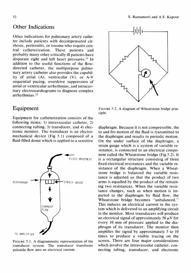
52
Other Indications
Other indications for pulmonary artery catheter include patients with decompensated cirrhosis, peritonitis, or trauma who require central catheterization. These patients and probably many other critically ill patients have disparate right and left heart pressures. II In addition to the useful functions of the flowdirected catheter, the multipurpose pulmonary artery catheter also provides the capability of atrial (A), ventricular (V), or A-V sequential pacing, overdrive suppression of atrial or ventricular arrhythmias, and intracavitary electrocardiograms to diagnose complex arrhythmias. 12
Equipment
Equipment for catheterization consists of the following items: 1) intravascular catheter, 2) connecting tubing, 3) transducer, and 4) electronic monitor. The transducer is an electromechanical device (Fig 5.1) composed of a fluid-filled dome which is applied to a sensitive
-+- I-,....-.-r=-'r DIAPHRAGM
TO AMPLI F I ER
CURRENT FLOW
~
FLUID MOVEMENT
STRAIN GAUGE
FIGURE 5.1. A diagrammatic representation of the transducer system. The transducer transforms pulsatile flow into an electrical current.
s. Ramamurti and A.S. Kapoor
FIGURE 5.2. A diagram of Wheatstone bridge principle.
diaphragm. Because it is not compressible, the to and fro motion of the fluid is transmitted to the diaphragm and results in periodic motion. On the under surface of the diaphragm, a strain gauge which is a system of variable resistance, is connected to an electrical component called the Wheatstone bridge (Fig 5.2). It is a rectangular structure consisting of three fixed electrical resistances and the variable resistance of the diaphragm. When a Wheatstone bridge is balanced the variable resistance is adjusted so that the product of two arms is equalled by the product of the remaining two resistances. When the variable resistance changes, such as when motion is imparted to the diaphragm by fluid flow, the Wheatstone bridge becomes "unbalanced." This induces an electrical current in the system which is delivered to an amplifying circuit in the monitor. Most transducers will produce an electrical signal of approximately 50 /LV for every 10 mm of pressure applied to the diaphragm of its transducer. The monitor then amplifies the signal by approximately 5 to 10 times to produce a visible tracing on the screen. There are four major considerations which involve the intravascular catheter, connecting tubing, transducer, and electronic
5. Pulmonary Artery Catheterization
monitor: 1) frequency response, 2) relative natural frequency, 3) damping, and 4) catheter whip artifact. Accurate reproduction of a biologic signal requires a system that can faithfully reproduce frequencies up to 20 Hz. The clinical implications, therefore, are that a monitor with an inadequate frequency response will display a pressure below the true physiologic signal. This will be found most frequently when heart rates are rapid and the waveform is particularly steep, that is in a rigid cardiovascular system, for example, elderly 'patients with hypertension and arteriosclerosis.
The connection between the patient and the transducer influence the frequency response curve. The fluid-filled system has a "natural frequency. " When the vibrations in the system approach the natural frequency there is a significant increment in the amplitude of the power signal. One of the major determinants of natural frequency of a monitoring system is the length of the tubing connecting the catheter to the transducer. As the length of the tubing increases the natural frequency decreases. Thus, an excessive length of tubing will make the natural frequency of the system occur in the physiologic range. The resulting amplification created by the overresponse of the amplifier circuits causes the displayed pressure to exceed the true physiologic signal. As long as the natural frequency occurs outside the range necessary to faithfully reproduce the biologic signal (20 Hz), the monitor reading will display the correct blood pressure. If an excessively long piece of connecting tube is used, however, the system response curve is shifted to the left so that at 20 Hz there is already overresponse or "ringing" in the system. The monitor reading in this situation would overestimate blood pressure because of the amplification in the connecting tubing. This potential must be considered in monitoring the pulmonary system and the length of the tubing limited to 4 ft or less.
The third consideration is that of "damping." Damping represents loss of physiologic signal in the transmission system. The creation of the electrical signal depends upon the motion of the diaphragm of the transducer. If
53
some of the physical motion is lost before its impact on the transducer membrane, the electrical signal will be diminished because the total physiologic pressure signal has not been applied. If compliant, that is, soft plastic, tubing is used to connect the catheter to transducer some of the to and fro motion will be lost in expanding the plastic tubing. We use this principle when palpating a peripheral pulse to detect a similar dispersion of physical energy into the expansion of the arterial wall. If low compliant tube is similarly "pulsatile," energy will be lost and there will be an inadequate representation of the original physiologic signal reaching the transducer diaphragm. The most common artifact to result in damping is an air bubble in the circuit. Fluid is noncompressible. Air, on the other hand, is quite compressible. Transmission of the pressure wave through the tubing to the transducer depends on the noncompressible nature of the fluid. In other words the exact amount of motion in the vascular system is transmitted to the diaphragm of the transducer. If, however, a bubble is introduced into the system, part of the energy of the fluid will compress air and thus would be lost at the transducer diaphragm. There will be more motion ofthe fluid column on the patient's side of the air bubble than at the diaphragm. Less movement of the diaphragm produces a less powerful electronic signal, and the displayed pressures is, therefore, lessened. The effect of damping is extremely important in measuring pulmonary arterial pressures because there are many high-frequency components to the pressure tracing, and the overall pressures are considerably lower than systemic pressures. Thus, damping will cause underestimation of the magnitude of the signal and also in removing the high-frequency components, it may make interpretation of the waveform impossible. A large air bubble in a pulmonary artery catheter system may make the pulmonary artery and pulmonary artery occlusion pressure tracings appear virtually indistinguishable.
The last type of error is known as catheter whip artifact. This occurs in pulmonary arterial circulation and may be found in systemic arterial pressure monitoring. It is the result of

54
the mechanical transmission of the force of the heart during its contraction. This contraction is so powerful that it imparts an acceleration to the catheter sitting within the pulmonary artery. This is a very high-frequency artifact and can be handled effectively by incorporating a filter into the system, much as static can be filtered out of a stereo record using a highfrequency filter.
A practical bedside approach to pressure monitoring has been devised which recognizes these electronic considerations and yet attempts to safeguard the equipment. The variable resistance in the transducer diaphragm is extremely delicate. Placing the transducer directly at the patient's bedside subjects it to damage in a busy intensive care unit. By permanently positioning the transducer on the wall behind the bed can reduce breakage of these delicate instruments. The connecting tubing in this position must not be longer than 4 ft. Finally, accurate calibration of the transducer monitor system must be performed to verify that a known pressure signal is accurately displayed.
Calibration
Introducing a zero reference point and creating an electrical signal to represent a known pressure are termed balancing and calibrating. Calibration of the monitor requires introduction of a known pressure signal. This can be done in one of two ways: internal or external calibration. A simple external calibration system which, in fact, tests the transmission tubing, transducer, and monitor seems more desirable because it should be more accurate. U sing this approach, a column of water equivalent to 20 mm Hg pressure is externally applied to the transducer. Because mercury weighs approximately 13.4 times as much as water this would require introduction of a water column 26.8 cm (268 mm H 20). This can be readily accomplished using an intravenous pole as a calibration tool. An alligator clip is fastened to the zero point, and the second clip is fastened at a measured distance of 26.8 cm above the zero reference point. The free end
S. Ramamurti and A.S. Kapoor
of the transmission tubing is attached to the zero clip. The end of the tubing is then elevated to the 26.8 cm or 20 mm Hg reference point and the gain or calibration control is adjusted so that 20 mm Hg are displayed on the monitor. In this system, an actual physical signal representing 20 mm Hg is applied to the transducer diaphragm. The free end of the tubing is then placed at the appropriate location on the patient's chest wall and a zero reference signal, or rebalancing, activated. This process seems easily understandable and in practice has been repetitively performed by nurses and technicians with great reliability.
The most commonly used catheter today is a four-lumen 7-Fr catheter incorporating a thermistor positioned approximately 5 to 6 cm proximal to the tip of the pulmonary artery catheter, and an additional port which is intended to permit pressure monitoring and blood sampling from the right atrium. The four-lumen catheter thus permits: 1) monitoring of pulmonary artery pressure (distal lumen, balloon deflated), 2) monitoring pulmonary artery occlusion pressure (distal lumen balloon inflated), 3) right atrial pressure monitoring (proximal lumen), 4) cardiac output by the thermodilution method (thermistor connected to external cardiac output computer), and 5) sampling of the mixed venous blood (sample aspirated through proximal lumen). The catheter is radio-opaque, 110 cm long, made of polyvinyl chloride, and marked with 1O-cm intervals from the tip. These markings help to determine when to inflate the balloon, when to suspect catheter looping, and once the catheter is positioned when to check for displacement, in the absence of fluoroscopy. A black ring thicker than the rest identifies the 50-cm mark from the tip.
Procedure
Access to the right atrium may be gained by percutaneous cannulation (using a modified Seldinger technique) of the subclavian, internal jugular, external jugular, antecubital, or femoral vein. Access also can be established through a cut down over a peripheral vein

5. Pulmonary Artery Catheterization
such as the median basilic vein in the antecubital fossa. This approach is better suited in patients who are on thrombolytic therapy or in patients who have an underlying coagulopathy. The cut down is done under aseptic precaution. The venous catheter is carefully passed through the exposed vein under fluoroscopic guidance or can be flow directed.
The internal jugular vein is posterior to the carotid artery at the base of the skull. As it descends through the neck, the internal jugular vein lies lateral and finally anterolaterally to the common carotid artery. The internal jugular vein also runs posterior to the middle of sternocleidomastoid muscle and subsequently lies behind the anterior border of that muscle's clavicular head. Just above the medial end of the clavicle, the internal jugular vein joins the subclavian vein to form the brachiocephalic vein. Because of its straighter path to the heart, the right internal jugular vein is preferred to the left. This also avoids damage to the thoracic duct and lessens the risk of pneumothorax due to the dome of the pleura being lower on the right side. The patient is usually placed in the Trendelenberg position. Local anesthesia with 2 to 4 ml of 1 % lidocaine is given. A 3-inch long thin-walled 18-gauge Cook needle is inserted bevel upward underneath the lateral border of the sternocleidomastoid muscle, about 5 cm above the clavicle. The needle is directed anteriorly toward the suprasternal notch at a steady 30° to 45° angulation to the sagittal and horizontal plane. The vein is usually entered within 5 to 7 cm. Once venous blood flows freely, the syringe is disconnected from the needle, the needle mouth is occluded with a finger tip to prevent air embolism, and a 40-cm long J-topped flexible guidewire is inserted through the needle into the vein. The guidewire should pass freely and smoothly; one should avoid any forceful advancement of the guidewire. If any difficulty is encountered advancing the guidewire it should be withdrawn in to the needle and twisted to change direction of the J-tip; the guidewire should be readvanced. Once the guidewire is well within the vein, the Cook needle is removed. The skin puncture site is enlarged with a #11 scalpel blade, and a dila-
55
tor sheath system is advanced over the guidewire into the vein. Care should be taken to ensure that the guide wire protrudes beyond the outer end of the dilator sheath assembly at all times. Once in the vein, the dilator and guidewire are removed and the sheath secured to the skin with a suture. The pulmonary artery catheter is then advanced through the sheath into the vein.
Subclavian Vein
This has been extensively used in the surgical intensive care unit. Though supra- and infraclavicular approaches are available, the infraclavicular approach is frequently used. Here the patient is positioned in Trendelenberg position. A roll 4 inches in diameter should be positioned vertically beneath the upper thoracic spine. This will permit the shoulder to be displaced posteriorly and ensure that the needle can be introduced horizontally. The needle is inserted at the site where the clavicle makes the curve to meet the sternum. A 14-gauge needle is used. A central venous catheter is introduced, and a guidewire is passed through the catheter and the needle is removed. The remainder of the procedure is similar to the internal jugular vein cannulation.
Femoral Vein
This site is infrequently used. The femoral vein lies immediately medial to the femoral artery. First the femoral artery is identified as it emerges from underneath the inguinal ligament. The femoral vein site is approximately 3 cm below this. After the vein has been punctured the technique is similar to the internal jugular vein and subclavian approaches.
Cardiac Hemodynamic Parameters
Cardiac hemodynamics provide greater diagnostic precision and furnish a safe means of assessing the results of therapy (Table 5.3). The following are the direct variables obtained
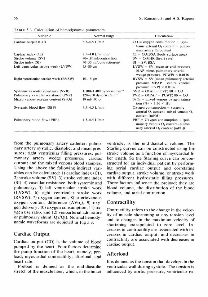
56 S. Ramamurti and A.S. Kapoor
TABLE 5.3. Calculation of hemodynamic parameters.
Variable Normal range Calculation
Cardiac output (CO) 3.5-6.5 Llmin CO = oxygen consumption 7 (systemic arterial 0, content - pulmonary artery 0, content
Cardiac index (Cl) Stroke volume (SV) Stroke index (SO Left ventricular stroke work (L VSW)
2.5-4.0 Llmin/m' 70-185 mil contraction 40-55 mllcontraction/m2
55-80 gm
Cl = CO/BSA (body surface area) SV = CO/HR (heart rate) Sl = SV/BSA L VSW = SV (mean arterial pressure.
Right ventricular stroke work (RVSW) 10-15 gm
MAP minus pulmonary arterial wedge pressure, PCWP) x 0.0136
RVSW = SV (mean pulmonary arterial pressure, MPAP - central venous pressure, CVP) x 0.0136
Systemic vascular resistance (SVR) Pulmonary vascular resistance (PVR) Mixed venous oxygen content (SvO,)
1,100-1,400 dyne/sec/cm-; 120-250 dyne/sec/cm 5
SVR = (MAP - CVP) 80 7 CO PVR = (MPAP - PCWP) 807 CO SvO, = mixed venous oxygen satura-18 mil 100 cc
Systemic blood flow (SBF) 4.5-6.5 Llmin
Pulmonary blood flow (PBF) 4.5-6.5 Llmin
from the pulmonary artery catheter: pulmonary artery systolic, diastolic, and mean pressures; right ventricular filling pressures; pulmonary artery wedge pressures; cardiac output; and the mixed venous blood samples. U sing the above the following indirect variables can be calculated: 1) cardiac index (CI), 2) stroke volume (SV), 3) stroke volume index (SI), 4) vascular resistance, both systemic and pulmonary, 5) left ventricular stroke work (LVSW), 6) right ventricular stroke work (RVSW), 7) oxygen content, 8) arteriovenous oxygen content difference (A Vo2), 9) oxygen delivery, 10) oxygen consumption, 11) oxygen use ratio, and 12) venoarterial admixture or pulmonary shunt (Qs/Qt). Normal hemodynamic waveforms are depicted in Fig 5.3.
Cardiac Output
Cardiac output (CO) is the volume of blood pumped by the heart. Four factors determine the pump function of the heart, namely, preload, myocardial contractility, afterload, and heart rate.
Preload is defined as the end-diastolic stretch of the muscle fiber, which, in the intact
tion (%) x 1.36 x Hb Oxygen consumption 7 systemic
arterial O2 content-mixed venous O2
content (mIlM) PBF = Oxygen consumption 7 (pul
monary venous O2 content-pulmonary arterial O2 content [milL])
ventricle, is the end-diastolic volume. The Starling curves can be constructed using the stroke volume as a function of myocardial fiber length. So the Starling curve can be constructed for an individual patient by performing serial cardiac output and correlating cardiac output, stroke volume, or stroke work with different hydrostatic filling pressures. Three factors influence the preload; they are blood volume, the distribution of the blood volume, and atrial contraction.
Contractility
Contractility refers to the change in the velocity of muscle shortening at any tension level and to changes in the maximum velocity of shortening extrapolated to zero level. Increases in contractility are associated with increases in cardiac output, and decreases in contractility are associated with decreases in cardiac output.
Afterload
It is defined as the tension that develops in the ventricular wall during systole. The tension is influenced by aortic pressure, ventricular ra-
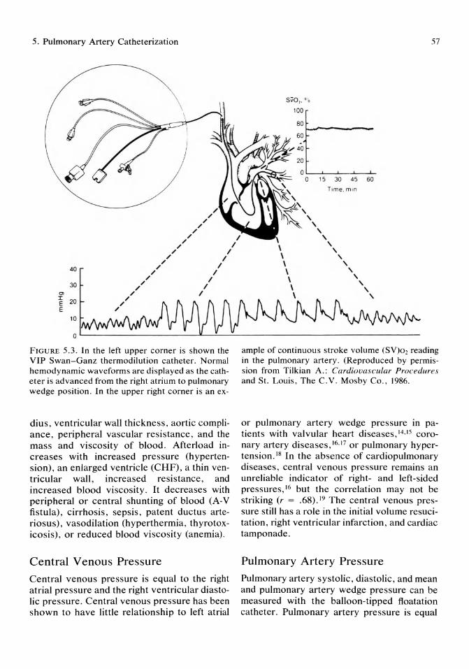
5. Pulmonary Artery Catheterization
S.o,.o., 100
80
~------
, , ,
15 30 45 60
Time. m in
,
57
/ /
/
/ /
/ I I
I
, \ \
, , 40
30
'" ~ 20 E
10
/ /
/ /
/
/ /
/ /
/ /
I
I I
I I
I \ \ \ \ \
, , , , , , ,
o L-------------------~--~~---------------------------------
FIGURE 5.3. In the left upper corner is shown the VIP Swan-Ganz thermodilution catheter. Normal hemodynamic waveforms are displayed as the catheter is advanced from the right atrium to pulmonary wedge position. In the upper right corner is an ex-
dius, ventricular wall thickness , aortic compliance, peripheral vascular resistance , and the mass and viscosity of blood. Afterload increases with increased pressure (hypertension), an enlarged ventricle (CHF), a thin ventricular wall, increased resistance , and increased blood viscosity . It decreases with peripheral or central shunting of blood (A-V fistula), cirrhosis, sepsis, patent ductus arteriosus), vasodilation (hyperthermia, thyrotoxicosis), or reduced blood viscosity (anemia).
Central Venous Pressure
Central venous pressure is equal to the right atrial pressure and the right ventricular diastolic pressure. Central venous pressure has been shown to have little relationship to left atrial
ample of continuous stroke volume (SV)02 reading in the pulmonary artery. (Reproduced by permission from Tilkian A.: Cardiovascular Procedures and St. Louis, The C.V. Mosby Co., 1986.
or pulmonary artery wedge pressure in patients with valvular heart diseases,14.15 coronary artery diseases,16.17 or pulmonary hypertension. 18 In the absence of cardiopulmonary diseases, central venous pressure remains an unreliable indicator of right- and left-sided pressures,16 but the correlation may not be striking (r = .68) . 19 The central venous pressure still has a role in the initial volume resucitation, right ventricular infarction, and cardiac tamponade .
Pulmonary Artery Pressure
Pulmonary artery systolic, diastolic, and mean and pulmonary artery wedge pressure can be measured with the balloon-tipped floatation catheter. Pulmonary artery pressure is equal
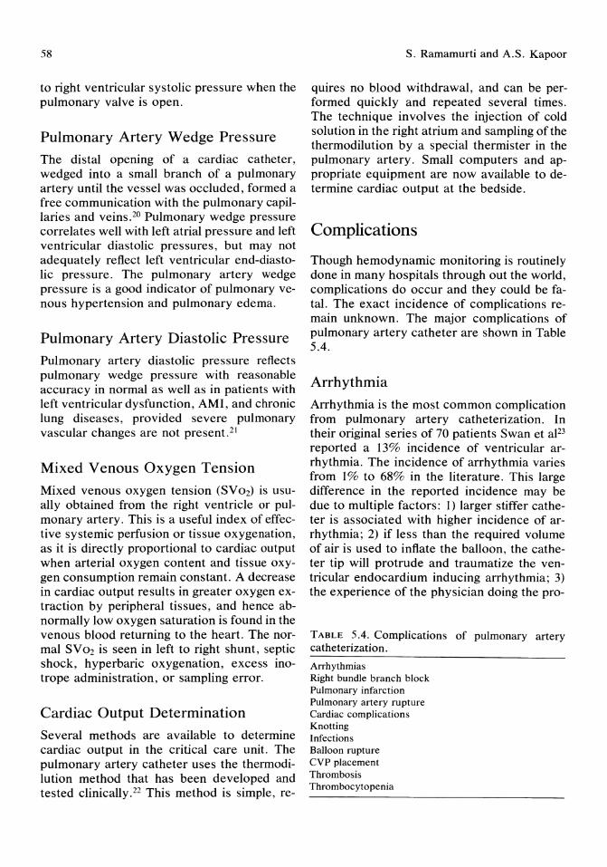
58
to right ventricular systolic pressure when the pulmonary valve is open.
Pulmonary Artery Wedge Pressure The distal opening of a cardiac catheter, wedged into a small branch of a pulmonary artery until the vessel was occluded, formed a free communication with the pulmonary capillaries and veins.20 Pulmonary wedge pressure correlates well with left atrial pressure and left ventricular diastolic pressures, but may not adequately reflect left ventricular end-diastolic pressure. The pulmonary artery wedge pressure is a good indicator of pulmonary venous hypertension and pulmonary edema.
Pulmonary Artery Diastolic Pressure Pulmonary artery diastolic pressure reflects pulmonary wedge pressure with reasonable accuracy in normal as well as in patients with left ventricular dysfunction, AMI, and chronic lung diseases, provided severe pulmonary vascular changes are not present. 21
Mixed Venous Oxygen Tension Mixed venous oxygen tension (SVo2) is usually obtained from the right ventricle or pulmonary artery. This is a useful index of effective systemic perfusion or tissue oxygenation, as it is directly proportional to cardiac output when arterial oxygen content and tissue oxygen consumption remain constant. A decrease in cardiac output results in greater oxygen extraction by peripheral tissues, and hence abnormally low oxygen saturation is found in the venous blood returning to the heart. The normal SVo2 is seen in left to right shunt, septic shock, hyperbaric oxygenation, excess inotrope administration, or sampling error.
Cardiac Output Determination Several methods are available to determine cardiac output in the critical care unit. The pulmonary artery catheter uses the thermodilution method that has been developed and tested clinically.22 This method is simple, re-
s. Ramamurti and A.S. Kapoor
quires no blood withdrawal, and can be performed quickly and repeated several times. The technique involves the injection of cold solution in the right atrium and sampling of the thermodilution by a special thermister in the pulmonary artery. Small computers and appropriate equipment are now available to determine cardiac output at the bedside.
Complications
Though hemodynamic monitoring is routinely done in many hospitals through out the world, complications do occur and they could be fatal. The exact incidence of complications remain unknown. The major complications of pulmonary artery catheter are shown in Table 5.4.
Arrhythmia Arrhythmia is the most common complication from pulmonary artery catheterization. In their original series of 70 patients Swan et aF3 reported a 13% incidence of ventricular arrhythmia. The incidence of arrhythmia varies from 1% to 68% in the literature. This large difference in the reported incidence may be due to multiple factors: 1) larger stiffer catheter is associated with higher incidence of arrhythmia; 2) if less than the required volume of air is used to inflate the balloon, the catheter tip will protrude and traumatize the ventricular endocardium inducing arrhythmia; 3) the experience of the physician doing the pro-
TABLE 5.4. Complications of pulmonary artery catheterization. Arrhythmias Right bundle branch block Pulmonary infarction Pulmonary artery rupture Cardiac complications Knotting Infections Balloon rupture CVP placement Thrombosis Thrombocytopenia

5. Pulmonary Artery Catheterization
cedure; 4) the length of time required to do the procedure, the longer the duration of the procedure the higher the incidence of arrhythmia; 5) the method of recording the arrhythmia; and 6) the underlying illness, critically ill patients are more prone to have arrhythmia.
Bundle Branch Block
The catheter-induced right bundle branch block, varies from 3% to 6% in different series. The most likely etiology is due to the mechanical irritation by the catheter. The most serious danger of developing right bundle branch block is in the patient who has preexisting left bundle branch block and who is at a risk of developing complete heart block.24 Some investigators have stressed the need for pacing before pulmonary artery catheterization, in patients with baseline left bundle branch block. 24,25 The exact incidence of this complication is not known at this time.
Thrombosis
Thrombosis occurs with any intravascular catheter. Recently, Chastre et aJ26 showed venographic or autopsy evidence of internal jugular vein thrombosis at the site of catheterization, particularly at the insertion in 22 out of 33 patients (66%). None of the patients had clinical evidence of thrombosis. More recently, Hoar et aF7 showed that using a heparin-bonded catheter prevented intraoperative thrombus formation.
Insertion of a pulmonary artery catheter also is associated with platelet consumption and decrease in the platelet count. 28 After removal of the catheter, platelet count begins to increase but remains depressed for 48 hours.
Pulmonary Damage
Pulmonary complications are predominately vascular and include pulmonary infarction and pulmonary artery rupture. 29,30 An early report of pulmonary infarction secondary to flotation catheters showed an incidence of 7.2%.29 Pulmonary infarction may result from29 1) thrombus developing around the catheter and
59
emboli passing through the pulmonary circulation; 2) embolization of thrombus formed within the catheter; 3) occlusion of a branch of the pulmonary artery due to permanent wedging of a distally positioned catheter or prolonged balloon occlusion; and 4) mechanical damage to the pulmonary endothelium by the catheter with subsequent formation of thrombus and embolization.
The prevention of pulmonary complications secondary to pulmonary artery catheterization requires meticulous attention to the catheter's insertion and maintenance. The incidence of pulmonary infarction should be decreased29 by 1) the use of continuous heparinized flush solution through the pulmonary artery catheter, 2) avoidance of persistent wedging ofthe catheter, 3) determining that the balloon is deflated, and 4) performing chest roentgenographs immediately after catheter insertion and frequently thereafter to verify the position of the catheter tip and to exclude the possibility of air in the balloon.
Pulmonary Artery Rupture
The incidence of pulmonary artery rupture is 0.2%.31 Most of the reported patients have been elderly, with evidence of valvular disease. Anticoagulation increases the severity of the pulmonary complications.
An immediate chest x-ray will grossly localize the bleeding site. The catheter should be withdrawn from the site of injury to prevent further bleeding caused by additional pulmonary artery damage by the catheter.
Cardiac Complications
These include lesion of the right atrial endocardium, tricuspid valve, chordae tendinae, and pulmonic valve with subsequent valvular insufficiency.32 Septic endocarditis and aseptic endocarditis have been reported. 32 The incidences varies from 3.4% to 21%.32 The prevention of this complication requires 1) catheter withdrawal with the balloon deflated, 2) never forcing the withdrawal of the catheter when resistance is encountered, and 3) avoid prolonged catheterization.
60
Knotting
Knotting of the pulmonary artery catheter can occur within the vascular space.33 Knotting is more common with small-bore floatation catheters. Dilated heart chambers and repeated catheter manipulation without fluoroscopy are predisposing factors. To prevent looping and knotting the following guidelines should be followed: 1) fully inflate the balloon in a large central vein before advancing the catheter to the right atrium; 2) do not use excessive catheter length for insertion, insert a maximum of 15 cm of catheter from the right atrium to pulmonary artery; 3) if possible use fluoroscopy in patients with dilated cardiac chambers; and 4) fully inflate balloon before insertion.
If knotting occurs one of several techniques can be used to remove the catheter. Usually less invasive methods are attempted initially: 1) gentle traction may allow the withdrawal of the catheter directly from the vein33 ; 2) under fluoroscopy the catheter can be manipulated into numerous positions to loosen and move the knot toward and over the catheter tip34; 3) if the knot is not tight a movable cord guidewire can be directed along the major lumen of the catheter, the adjustable inner core of the guidewire can be used to stiffen the distal part and the knot can be loosened or made to spring open34 ; 4) picking at the knot with a firm catheter also may be usefuP5; and 5) if the knot is wrapped around a cardiac structure, thoracotomy and cardiotomy may be required for its removal. 34
Infectious Complications
Factors predisposing to infection are: 1) increased duration of catheterization (>72 hours), and 2) increased catheter repositioning.
Strict sterile technique should be used routinely to prevent infection. For long-term monitoring, percutaneous catheter insertion is recommended, as more infections occur at cut down site. If the catheter is believed to be the source of infection. it should be removed.
S. Ramamurti and A.S. Kapoor
Balloon Rupture
The incidence of this complication is 1 % to 23% as reported in different series. The catheter and the balloon should be tested before catheterization, as 3% of catheters will have ruptured balloons.36 If no resistance to inflation is encountered it should be assumed that the balloon is ruptured and inflation is no longer performed.
Another potential complication of balloon rupture is fragmentation of the balloon and subsequent embolization. The following recommendations should help avoid balloon rupture: 1) test balloon before insertion, 2) use catheters only once, 3) do not exceed recommended inflation volume, 4) avoid multiple balloon inflations, and 5) do not withdraw the catheter through an introducer with the balloon inflated.
Miscellaneous
Other complications of pulmonary artery catheterization include 1) Bernard-Horner syndrome,37 2) placement of the catheter tip into the wall of the internal carotid artery, 38 3) massive hematuria,39 4) pneumoperitonium,40 and 5) separation of the hub and shaft of the introducer with the shaft disappearing into the venous system. 41 The true incidence of complications of pulmonary artery catheterization will remain unknown until a large perspective multicenter study is performed. One should weigh the risk and benefit when considering pulmonary artery catheterization.
New Developments
Newer catheters incorporate a fifth lumen containing fiberoptic bundles, which are used to measure mixed venous saturation when connected to the appropriate external instrument. Other catheters incorporate electrodes that can be used to monitor intracavitory electrocardiograms in both the atria and ventricles. In some instances these can be used for electrical pacing of the cardiac rate. An addi-
5. Pulmonary Artery Catheterization
tionallumen, which terminates in the region of the right ventricle, would permit the introduction of an electrode for intracavitory electrocardiogram or pacing if the situation warranted.
Another new catheter has a fifth lumen designed to end in the right atrium which can be used for fluid infusions. This can eliminate the necessity of interrupting vasoactive infusions during cardiac output determinations.
New advances in biomedical technology will be introduced for hemodynamic monitoring in the future. Some of the anticipated advances include coronary sinus retroperfusion of arterialized blood into the coronary venous system to treat myocardial ischemia and for myocardial salvage. This is a new system that includes a catheter for obtaining arterial blood and a balloon-occluding coronary sinus catheter for delivery of arterial blood only during diastole.
A new right ventricular ejection fraction thermodilution catheter will be introduced shortly. This new catheter measures the cardiac output, blood volume per heart beat, and end-systolic and diastolic volumes, and it computes right heart ejection fraction.
Bedside computer systems incorporating the new advances in sensor technology and fiberoptics will interface with pulmonary artery catheters for generating a complete and comprehensive hemodynamic profile for each patient.
References
I. Swan HJC: Techniques of monitoring the seriously ill patient with heart disease (including use of Swan-Ganz catheter), in The Heart, ed 6. New York, McGraw-Hill Inc, 1986, p 1998.
2. Swan HJC, et al: Catheterization of the heart in man with use of a flow-directed balloon-tipped catheter. N Engl Med 1970; 283:447-451.
3. Sprung CL, et al: Indications for pulmonary artery catheterization, in Pulmonary Artery Catheter. Baltimore, University Park Press, 1983, p 9.
4. Forrester JD, et al: Filling pressures in the right and left sides of the heart in acute myocardial infarction. N Engl J Med 1971; 285: 190-193.
61
5. Crexell C, et al: Optimal level of filling pressures in the left side of the heart in acute myocardial infarction. N Engl J Med 1973; 289: 1264-1266.
6. Meister SG, et al: Rapid bedside differentiation of ruptured interventricular septum for acute mitral insufficiency. N Engl J Med 1972; 287: 1024-1025.
7. Guberman BA, et al: Cardiac tamporade in medical patients. Circulation 1981; 64:633-640.
8. Lorell B, et al: Right ventricular infarction: Clinical diagnosis and differentiation from cardiac tamporade and pericardial constriction. Am J Cardiol 1979; 43:465-471.
9. Sprung CL, et al: Differentiation of cardiogenic, intermediate and noncardiogenic forms of pulmonary edema. Am Rev Respir Dis 1981; 124:718-722.
10. Delguercio LRM, et al: Monitoring operative risk in the elderly. J Am Med Assoc 1980; 243: 1350-1355.
II. Civetta JM, et al: Flow directed pulmonary artery catheterization in surgical patients: Indications and modifications of technic. Ann Surg 1972; 176:753-756.
12. Swan HJC, et al: Use of balloon flotation catheters in critically ill patients. Surg Clin North Am 1975; 55:501-520.
13. Swan HJC, et al: The Swan-Ganz catheter: Insertion technique. J Crit Illness 1986; 38:43.
14. Sarin CL, et al: The necessity for measurement of left atrial pressure after cardiac surgery. Thorax 1970; 25: 185-189.
15. Bell H, et al: Reliability of central venous pressure as an indicator of left atrial pressure: A study in patients with mitral valve disease. Chest 1971; 59:169-173.
16. Civetta JM, et al: Flow directed pulmonaryartery catheterization in surgical patients: Indications and modifications of technic. Ann Surg 1972; 176:753-756.
17. Byrick RJ et al: Influence of elevated pulmonary vascular resistance on the relationship between central venous pressure and pulmonary artery occluded pressure following cardiopulmonary bypass. Can Anaesth Soc J 1978; 25: 106-112.
18. DelGuercio LRM, et al: Monitoring: Methods and significance. Surg Clin North Am 1976; 56:977-994.
19. Toussaint GPM, et al: Central venous pressure and pulmonary wedge pressure in critical surgical illness. Arch Surg 1974; 109:265-269.
62
20. Hellems HK, et al: Pulmonary capillary pressure in animals estimated by venous and arterial catheterization. Am J Physiol1948; 155:98-105.
21. Falicov RE, et al: Relationship the pulmonary artery end-diastolic pressure to left ventricular end-diastolic and mean filling pressures in patients with and without left ventricular dysfunction. Circulation 1970; 42:65-73.
22. Ganz W, et al: Measurement of blood flow by thermodilution. Am J Cardiol 1972; 29:241-246.
23. Swan HJC, et al: Catheterization of the heart in man with use of a flow-directed balloontipped catheter. N Engl J Med 1970; 283:447-451.
24. Abernathy WS: Complete heart block caused by Swan-Ganz catheter. Chest 1974; 65:349.
25. Swan HJC: The role of hemodynamic monitoring in the management of the critically ill. Crit Care Med 1975; 3:85-89.
26. Chastre J, et al: Thrombosis as a complication of pulmonary-artery catheterization via the internal jugular vein. N Engl J Med 1982; 306:278-281.
27. Hoar PF, et al: Heparin bonding reduces thrombogenicity of pulmonary artery catheters. N Engl J Med 1981; 305:993-995.
28. Richman KA, et al: Thrombocytopenia and altered platelet kinetics associated with prolonged pulmonary artery catheterization in the dog. Anesthesiology 1980; 53:101-105.
29. Foote GA, et al: Pulmonary complications of the flow-directed balloon-tipped catheter. N Engl J Med 1974; 290:927-931.
30. Barash PG, et al: Catheter-induced pulmonary
S. Ramamurti and A.S. Kapoor
artery perforation. J Thorac Cardiovasc Surg 1981; 82:5-12.
31. McDaniel DD, et al: Catheter-induced pulmonary artery hemorrhage. J Thorac Cardiovasc Surg 1981; 82: 1-4.
32. Elliot CG, et al: Complications of pulmonary catheterization in the critically ill patients. Chest 1979; 76:647-652.
33. Lipp H, et al: Intracardiac knotting of a flowdirected balloon catheter. N Engl J Med 1971; 284:220.
34. Mond HG, et al: A technique for unknotting an intracardiac flow-directed balloon catheter. Chest 1975; 67:731-733.
35. Meister SG et al: Knotting of a flow-directed catheter about a cardiac structure. Cath Cardiovas Diagn 1977; 3:171-175.
36. Sise MJ, et al: Complications of the flow-directed pulmonary artery catheter: A prospective analysis in 219 patients. Crit Care Med 1981; 9:315-318.
37. Birrer RB, et al: Bernard-Horner syndrome associated with Swan-Ganz catheter. NY State J Med 1981; 81 :362-364.
38. McNabb TG, et al: A potentially serious complication with Swan-Ganz catheter placement by the percutaneous internal jugular route. Br J Anesth 1975; 47:895-897.
39. Katz SA, et al: Urologic complication associated with Swan-Ganz catheter. Urology 1975; 6:716-718.
40. Smith GB, et al: A hazard of Swan-Ganz catheterization. Anaesthesia 1981; 36:398-40l.
41. Gorden EP, et al: Hydromediastinum after placement of a thermodilution pulmonary artery catheter. Anesth Analg 1980; 59:159-160.
6 Outpatient Cardiac Catheterization Peter R. Mahrer
Introduction
Coronary artery surgery and, more recently, coronary balloon angioplasty have promoted an increase in the number of cardiac catheterizations performed in the United States each year. The admission of the patient to the hospital on the day before the study and an overnight stay after the catheterization is still the conventional procedure. With the ever increasing cost of hospitalization, the financial burden of this diagnostic procedure is staggering.
Our initial experience was on 308 preselected patients, I and the results were so encouraging and confirmed by other authors,2-5 that we continued to perform outpatient catheterization on a routine basis and recently reported our experience on 4,000 consecutive patients.
Methods
The Kaiser Permanente Regional Cardiac Catheterization Laboratory is directly adjacent to a tertiary-care hospital with a large cardiovascular surgery experience of approximately 900 cases per year. The laboratory is the referral center for the eight hospitals of the Southern California Region of the Kaiser-Permanente Health Plan which provide care to a popUlation of approximately 1.5 million patients.
Three distinct types of referrals constitute
the population base for studies at the catheterization laboratory (Table 6.4). Elective outpatients are referred from doctor's offices or after hospitalization for a stable cardiac problem. After the procedure, these patients are discharged home; hospitalization is required only for cardiac instability, procedural complications, or the discovery of threatening anatomy such as left main stenosis. The second group is comprised of patients hospitalized at other hospitals for known or suspected cardiac disease for whom catheterization is needed on an urgent or emergency basis. They are transferred to the regional cardiac lab as an urgent case and then return to their referring facility unless an acute intervention such as percutaneous transluminal coronary angiopia sty (PTCA) or cardiac surgery is carried out. The third group of patients are hospitalized at the on-site tertiary-care hospital.
Before arriving at the Regional Cardiac Catheterization Laboratory, each patient has had a full evaluation by his referring cardiologist with appropriate noninvasive evaluation. The patient and the referring cardiologist have reviewed the catheterization procedure and the expectations for future treatment. An information packet has been sent to the patient to further explain the details of the procedure and the function of the catheterization laboratory. Inpatients have instruction from the membership health education department and by videocassette over closed-circuit television.
Outpatients and transfer patients are admit-
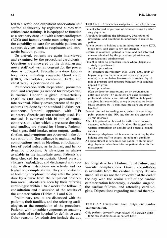
64
ted to a seven-bed outpatient observation unit staffed exclusively by registered nurses with critical care training. It is equipped to function as a coronary care unit with electrocardiogram (ECG) and hemodynamic monitoring and has the capability to care for unstable patients on support devices such as respirators and intraaortic balloon pumps.
On arrival, patients are again interviewed and examined by the procedural cardiologist. Questions are answered by the physician and nurse staff and written consent for the procedure is obtained. Precatheterization laboratory work including complete blood count (CBC), electrolytes, creatinine, ECG, and chest x-ray is performed on site.
Premedication with meperidine, promethazine, and atropine (as needed for bradycardia) is routine. Heparin is given intra-arterially (2,000 to 3,000 units) without protamine sulfate reversal. Ninety-seven percent of the procedures are done by the standard Judkins' percutaneous femoral approach with 7-Fr catheters. Sheaths are not routinely used. Hemostasis is achieved with 10 min of manual compression, after which a pressure dressing and sandbag are placed for 4 hours. Patients' vital signs, fluid intake, urine output, cardiac rhythm, and symptoms are observed in the observation unit. Surveillance is maintained for complications such as bleeding, embolization, loss of pedal pulses, arrhythmias, and hemodynamic problems. A physician is always available in the immediate area. Patients are then checked for orthostatic blood pressure changes, ambulated, and discharged with specific written instructions on activity and potentiallate complications. They are contacted at home by telephone the day after the procedure by a nurse from the outpatient observation area. Patients are seen by their referring cardiologist within 1 to 2 weeks for follow-up consultation and discussion of the results of the catheterization (Tables 6.1 and 6.2).
Preliminary results are discussed with the patients, their families, and the referring cardiologists at the completion of the procedure. Patients with unstable symptoms or anatomy are admitted to the hospital for definitive care. Other reasons for admission include therapy
P.R. Mahrer
TABLE 6.1. Protocol for outpatient catheterization.
Patient informed of purpose of catheterization by referring physician
A booklet describing the laboratory. description of procedure. and directions to laboratory is mailed to patient
Patient comes to holding area in laboratory where ECG, blood tests, and chest x-ray are obtained
Referral is reviewed, patient is examined and informed consent obtained by the procedural physician and premedications administered
Patient is taken to procedure room where diagnostic study is performed
Judkins' procedure: #7 catheters are routinely used, 2000-30000 units of heparin is given (heparin is not reversed by protamine); at completion hemostasis is attained by 10 min of pressure and pressure bandage with sandbag applied to groin Sones' procedure: (Can be done by arteriotomy or by percutaneous entry into artery) #7 catheters are used-frequently preformed catheters are utilized; 2000 units heparin are given intra-arterially; artery is repaired or hemostasis obtained by 10 min local pressure and pressure bandage
Patient returned to holding area; pulses beyond entry point, puncture site, BP, and rhythm are checked at 15-min intervals
After 4 hr patient is checked for orthostatic pressure changes, ambulated, and, if stable, discharged with written instructions on activity and potential complications
A follow-up telephone call is made the next day by the holding area staff to assess the patient's condition
An appointment is scheduled for patient with his referring physician who then informs patient about further management
for congestive heart failure, renal failure, and vascular complications. On-site consultation is available from the cardiac surgery department. All cases are then reviewed at the end of the day with the senior staff of the cardiac catheterization laboratory, a cardiac surgeon, the cardiac fellows, and attending cardiologists. Dispositions regarding medical therapy,
TABLE 6.2. Exclusions from outpatient cardiac catheterization.
Only patients currently hospitalized with cardiac symptoms are studied on an in-patient basis

6. Outpatient Cardiac Catheterization
additional workup, and interventions with PTCA or surgery are then made in consultation with the referring cardiologist.
Results
We reviewed 4,094 consecutive cardiac catheterizations in detail; 2,207 (54%) were performed on elective outpatients, 1,330 (32%) on patients from other hospitals, and 557 (14%) on inpatients at the tertiary-care hospital. The rate of normal studies was 11 %, left main disease (greater than 50% stenosis) was present in 5% of patients studied for coronary artery disease, and 55% of all patients studied ultimately required intervention with PTCA or surgery (Table 6.3).
The complications rate in this group of consecutive patients was low. There was one death in the elective outpatient group and four in the transfer patients. All deaths were related to left main disease. Complications of myocardial infarct, cerebral vascular accidents, vascular problems, and bleeding for all groups are given in Table 6.4. Ninety-one percent of the elective outpatients were discharged home on the day of the procedure, and there were no admissions for late bleeding (Table 6.5). The complication rate in our series is comparable with the other large series reported by Klinke et aF and Fierens. 3
Discussion
A special report by the Health and Public Policy Committee of the American College of Physicians in 1986 compiled a table of compli-
TABLE 6.3. Severity of illness.
Type
Intervention (surgery or PTCA)
Left main disease (50% or greater)
Normal studies
%
55%
5% of patients studied for CAD
11 % of patients studied for CAD
CAD = coronary artery disease.
TABLE 6.4. Diagnostic studies (November 1983-June 1986).
Type
Outpatients Transfer patients Inpatients
Total
No.
2207 1330 557
4094
%
54% 32% 14%
100%
65
TABLE 6.5. Reasons for admission after cardiac catheterization.
Demonstrated disease mandates urgent intervention, by PTCA or surgery
Congestive heart failure or threatening arrhythmia Renal failure or other metabolic abnormalities Orthostatic hypertension Bleeding or vascular complications
cation rates reported in 30 prior studies of cardiac catheterization from 1968 to 1982.6.7 Mortality rates ranged from 0.3% to 2.1%. Our overall complication rates among 4,094 patients for mortality (0.17%), myocardial infarction (0.02%), cerebral vascular accidents (0.08%), vascular problems (0.12%) were well within the rates quoted. We also noted a low rate of normal studies (11 %) and a high rate (55%) of patients needing surgery or PTCA. This reflects the multiple levels of screening before catheterization that involves primary care physicians, referring cardiologists, and the senior staff of the catheterization laboratory. Patients selected for an intervention usually had symptoms refractory to medical therapy or had threatening anatomy such as left main stenosis, high-grade three-vessel disease, critical aortic stenosis, and so on. We believe our study population is comprised of patients with disease of greater severity than reported in other studies, yet our complication rate remains quite low.
When we examined our outpatient series, we also found our mortality rates to be low compared with other series of similar size. Klinke et aF reported 0.13% mortality in 3,071 outpatients studied. Fierens3 had 0% mortality among 5,107 outpatients. Our study with 2,207 elective outpatients had only one mortality
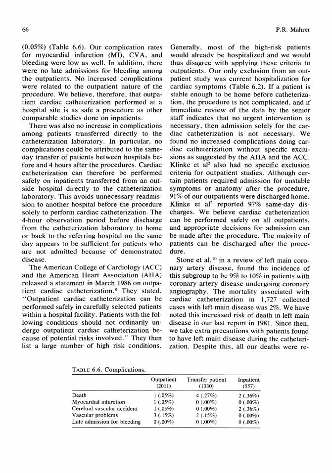
66
(0.05%) (Table 6.6). Our complication rates for myocardial infarction (MI), CV A, and bleeding were low as well. In addition, there were no late admissions for bleeding among the outpatients. No increased complications were related to the outpatient nature of the procedure. We believe, therefore, that outpatient cardiac catheterization performed at a hospital site is as safe a procedure as other comparable studies done on inpatients.
There was also no increase in complications among patients transferred directly to the catheterization laboratory. In particular, no complications could be attributed to the sameday transfer of patients between hospitals before and 4 hours after the procedures. Cardiac catheterization can therefore be performed safely on inpatients transferred from an outside hospital directly to the catheterization laboratory. This avoids unnecessary readmission to another hospital before the procedure solely to perform cardiac catheterization. The 4-hour observation period before discharge from the catheterization laboratory to home or back to the referring hospital on the same day appears to be sufficient for patients who are not admitted because of demonstrated disease.
The American College of Cardiology (ACC) and the American Heart Association (AHA) released a statement in March 1986 on outpatient cardiac catheterization. 8 They stated, "Outpatient cardiac catheterization can be performed safely in carefully selected patients within a hospital facility. Patients with the following conditions should not ordinarily undergo outpatient cardiac catheterization because of potential risks involved." They then list a large number of high risk conditions.
TABLE 6.6. Complications.
P.R. Mahrer
Generally, most of the high-risk patients would already be hospitalized and we would thus disagree with applying these criteria to outpatients. Our only exclusion from an outpatient study was current hospitalization for cardiac symptoms (Table 6.2). If a patient is stable enough to be home before catheterization, the procedure is not complicated, and if immediate review of the data by the senior staff indicates that no urgent intervention is necessary, then admission solely for the cardiac catheterization is not necessary. We found no increased complications doing cardiac catheterization without specific exclusions as suggested by the AHA and the ACC. Klinke et aF also had no specific exclusion criteria for outpatient studies. Although certain patients required admission for unstable symptoms or anatomy after the procedure, 91 % of our outpatients were discharged home. Klinke et aF reported 97% same-day discharges. We believe cardiac catheterization can be performed safely on all outpatients, and appropriate decisions for admission can be made after the procedure. The majority of patients can be discharged after the procedure.
Stone et al,1O in a review of left main coronary artery disease, found the incidence of this subgroup to be 9% to 10% in patients with coronary artery disease undergoing coronary angiography. The mortality associated with cardiac catheterization in 1,727 collected cases with left main disease was 2%. We have noted this increased risk of death in left main disease in our last report in 1981. Since then, we take extra precautions with patients found to have left main disease during the catheterization. Despite this, all our deaths were re-
Outpatient (2011)
Transfer patient (1330)
Inpatient (557)
Death Myocardial infarction Cerebral vascular accident Vascular problems Late admission for bleeding
1 (.05%) 1 (,05%) 1 (.05%) 3 (.15%) o (.00%)
4 (.27%) 0(,00%) 0(.00%) 2(.15%) 0(.00%)
2 (.36%) 0(.00%) 2 (.36%) o (.00%) o (.00%)
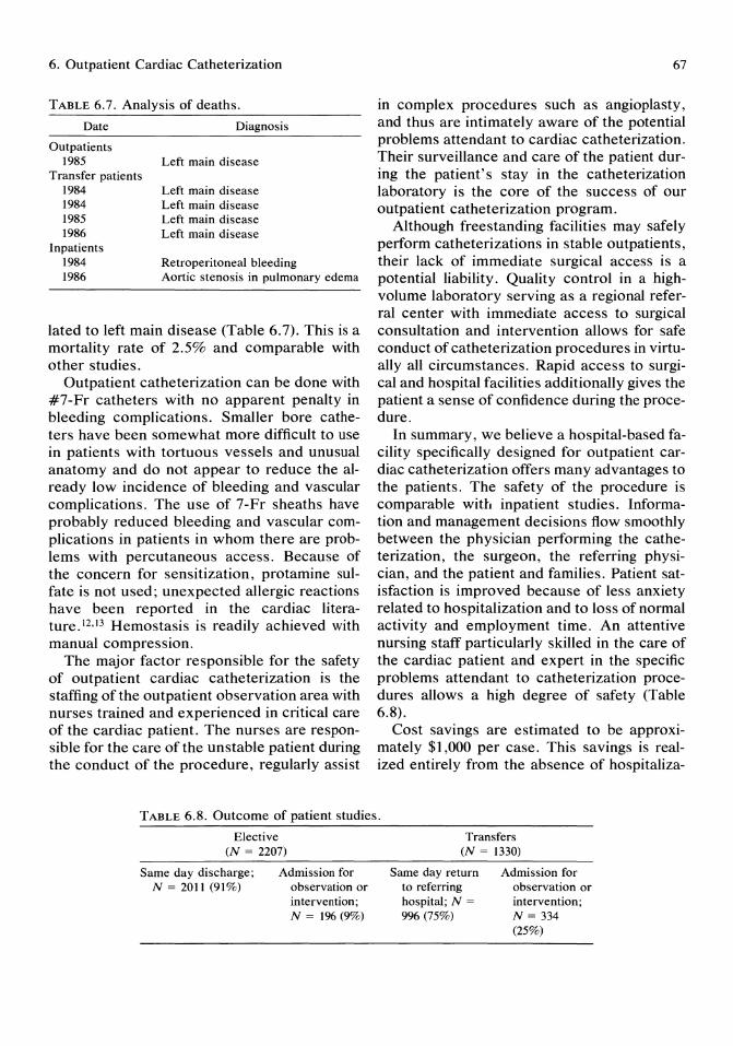
6. Outpatient Cardiac Catheterization
TABLE 6.7. Analysis of deaths.
Date
Outpatients 1985
Transfer patients 1984 1984 1985 1986
Inpatients 1984 1986
Diagnosis
Left main disease
Left main disease Left main disease Left main disease Left main disease
Retroperitoneal bleeding Aortic stenosis in pulmonary edema
lated to left main disease (Table 6.7). This is a mortality rate of 2.5% and comparable with other studies.
Outpatient catheterization can be done with #7-Fr catheters with no apparent penalty in bleeding complications. Smaller bore catheters have been somewhat more difficult to use in patients with tortuous vessels and unusual anatomy and do not appear to reduce the already low incidence of bleeding and vascular complications. The use of 7-Fr sheaths have probably reduced bleeding and vascular complications in patients in whom there are problems with percutaneous access. Because of the concern for sensitization, protamine sulfate is not used; unexpected allergic reactions have been reported in the cardiac literature.l2·!3 Hemostasis is readily achieved with manual compression.
The major factor responsible for the safety of outpatient cardiac catheterization is the staffing of the outpatient observation area with nurses trained and experienced in critical care of the cardiac patient. The nurses are responsible for the care of the unstable patient during the conduct of the procedure, regularly assist
67
in complex procedures such as angioplasty, and thus are intimately aware of the potential problems attendant to cardiac catheterization. Their surveillance and care of the patient during the patient's stay in the catheterization laboratory is the core of the success of our outpatient catheterization program.
Although freestanding facilities may safely perform catheterizations in stable outpatients, their lack of immediate surgical access is a potential liability. Quality control in a highvolume laboratory serving as a regional referral center with immediate access to surgical consultation and intervention allows for safe conduct of catheterization procedures in virtually all circumstances. Rapid access to surgical and hospital facilities additionally gives the patient a sense of confidence during the procedure.
In summary, we believe a hospital-based facility specifically designed for outpatient cardiac catheterization offers many advantages to the patients. The safety of the procedure is comparable with inpatient studies. Information and management decisions flow smoothly between the physician performing the catheterization, the surgeon, the referring physician, and the patient and families. Patient satisfaction is improved because of less anxiety related to hospitalization and to loss of normal activity and employment time. An attentive nursing staff particularly skilled in the care of the cardiac patient and expert in the specific problems attendant to catheterization procedures allows a high degree of safety (Table 6.8).
Cost savings are estimated to be approximately $1,000 per case. This savings is realized entirely from the absence of hospitaliza-
TABLE 6.8. Outcome of patient studies.
Elective (N = 2207)
Same day discharge; N = 2011 (91%)
Admission for observation or intervention; N = 196 (9%)
Transfers (N = 1330)
Same day return to referring hospital; N =
996 (75%)
Admission for observation or intervention; N = 334 (25%)

68
tion and the cost of inpatient care. This translates to approximately a $1,300,000 savings per year at our institution. Given a current US population of 230 million, this would translate to a savings of approximately $200 million dollars in the delivery of health care.
Acknowledgments. I thank L. Youngblood, RN, and L. Kirk, L VN, for their assistance in data retrieval.
References
1. Mahrer PR, Eshoo N: Outpatient cardiac catheterization and coronary angiography. Cathet Cardiovasc Diagn 1981; 7:355-360.
2. Klinke WP, Kubec G, Talibi T, et al: Safety of outpatient cardiac catheterization. Am ] Cardio11985; 56:639-641.
3. Fierens E: Outpatient coronary arteriography. Cathet Cardiovasc Diagn 1984; 10:27-32.
4. Fighali X, Krajcer Z, Camid FG, et al: Safety of outpatient cardiac catheterization. Chest 1985; 88:349-351.
5. Beauchamp PD: Ambulatory cardiac catheterization cuts costs for hospital and patients. Hospitals 1981; 55:62-63.
P.R. Mahrer
6. Mahrer PR, Magnusson PT, Young C: Cardiac catheterization and cardiovascular disease. In print, 1987.
7. Kahn KL: The safety and efficiency of ambulatory cardiac catheterization in the hospital and freestanding setting. Ann Int Med 1985, 103:294-298.
8. Kahn KL: The efficiency of ambulatory cardiac catheterization in the hospital and freestanding setting. Am Heart] 1986; 111:152-167.
9. American College of Cardiology: Statement on cardiac catheterization. Cardiology 1986; 15:3.
10. Stone PH, Goldschlager N: Left main coronary artery disease: Review and appraisal. Cardiovasc Med 1979; 4:165-182.
11. Brennan K: Outpatient cardiac catheterization. Cardiology 1986; Sept: 64.
12. Baird CL: Outpatient cardiac catheterization. Cathet Cardiovasc Diagn 1982; 8:647.
13. Stewart WJ, McSweeney Sm, Kellett MA, et al: Increased risk of severe protamine reactions in NPH insulin-dependent diabetics undergoing cardiac catheterization. Circulation 1984; 70:788-792.
14. Chung F, Miles J: Cardiac arrest following protamine administration. Can Anaesth Soc ] 1984; 31:314-318.

7 The Evolution of Coronary Artery Disease: New Definitions from Coronary Angioscopy James s. Forrester, Ann Hickey, Frank Litvack, and Warren Grundfest
Why does a healthy person have a myocardial infarction without prior warning? Why does a patient with stable coronary disease suddenly become unstable? For this century, as coronary heart disease became the nation's number one killer, these enigmas have remained unanswered. Even our most definitive diagnostic tools have been inadequate to answer these questions. Noninvasive imaging does not show us the coronary arteries; coronary angiography does not show the minute details of the blood vessel surface; autopsies are performed remote from the patient's initiating symptoms. The development of fiberoptics in the communications industry, recently applied to cardiology, has given us some intriguing answers to these questions. Fiberoptic angioscopy provides detailed information about the coronary endothelial surface at the time when the patient is symptomatic. This new information has clarified the cause of each of the four major unstable coronary chest pain syndromes (accelerated angina, unstable rest angina, myocardial infarction, ischemic sudden death). In this chapter we describe the technique of angioscopy, how each coronary syndrome has a specific endothelial cause, and how each endothelial condition is one phase of a repeating cycle of vascular injury and healing.
Method of Angioscopy The angioscopes we use range in external diameter from 0.5 to 2.8 mm. For examination
of small vessels, we use the 0.5 OD angioscope (Advanced Interventional Systems, Costa Mesa, CA) or a 0.7 mm OD devices (American Edwards Laboratory, Santa Ana, CA). These devices consist of approximately 5,000 individual fibers for transmitting the intravascular image. The imaging fibers are surrounded by a concentric ring of illumination fibers. For visualization of larger vessels, we have used angioscopes in the 1.4 to 1.8 mm OD range (Olympus Corporation of America, New Hyde Park, NY). These scopes have approximately 8,000 individual imaging fibers, and all the angioscopes are covered by flexible polyvinyl chloride catheter housings. We use a 1,000-watt xenon light source (Storz, Los Angles, CA) for illumination.
The image that emerges from the fiberoptic bundle is too small for direct visualization. Therefore, all images are relayed through a video coupler to a light-sensitive video camera (the Sharp professional 320 model and the Sony DXC 1850 camera are both suitable). Images from the camera are transmitted on line to a high-resolution video monitor (Sony PVM 1960) and permanent recordings are made on a i-inch videotape recorder (Sony 5880).
We have tested spatial resolution in our angioscopes using a standard imaging phantom consisting of 200-, 64-, and 34-fLm line pairs. The angioscopes we use have a line pair resolution between 200- and 64-fLm at 5 mm, and a minimum focus distance from 2.0 to 6.5 mm.
Different methods are used for peripheral bypass and coronary artery angioscopy. In pe-
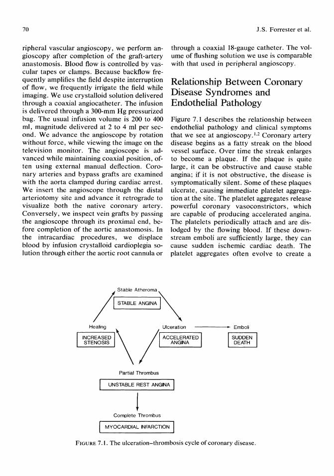
70
ripheral vascular angioscopy, we perform angioscopy after completion of the graft-artery anastomosis. Blood flow is controlled by vascular tapes or clamps. Because backflow frequently amplifies the field despite interruption of flow, we frequently irrigate the field while imaging. We use crystalloid solution delivered through a coaxial angiocatheter. The infusion is delivered through a 300-mm Hg pressurized bag. The usual infusion volume is 200 to 400 ml, magnitude delivered at 2 to 4 ml per second. We advance the angioscope by rotation without force, while viewing the image on the television monitor. The angioscope is advanced while maintaining coaxial position, often using external manual deflection. Coronary arteries and bypass grafts are examined with the aorta clamped during cardiac arrest. We insert the angioscope through the distal arteriotomy site and advance it retrograde to visualize both the native coronary artery. Conversely, we inspect vein grafts by passing the angioscope through its proximal end, before completion of the aortic anastomosis. In the intracardiac procedures, we displace blood by infusion crystalloid cardioplegia solution through either the aortic root cannula or
J.S. Forrester et al.
through a coaxial 18-gauge catheter. The volume of flushing solution we use is comparable with that used in peripheral angioscopy.
Relationship Between Coronary Disease Syndromes and Endothelial Pathology
Figure 7.1 describes the relationship between endothelial pathology and clinical symptoms that we see at angioscopy.I,2 Coronary artery disease begins as a fatty streak on the blood vessel surface. Over time the streak enlarges to become a plaque. If the plaque is quite large, it can be obstructive and cause stable angina; if it is not obstructive, the disease is symptomatically silent. Some of these plaques ulcerate, causing immediate platelet aggregation at the site. The platelet aggregates release powerful coronary vasoconstrictors, which are capable of producing accelerated angina. The platelets periodically attach and are dislodged by the flowing blood. If these downstream emboli are sufficiently large, they can cause sudden ischemic cardiac death. The platelet aggregates often evolve to create a
Ir Stable Atheroma ~
/ l STABLE ANO"" .I '"
Healing Ulceration -----. Emboli
INCREASED \ / ACCELERATED STENOSIS ANGINA
Partial Thrombus
UNSTABLE REST ANGINA I
! Complete Thrombus
MYOCARDIAL INFARCTION
FIGURE 7.1. The ulceration-thrombosis cycle of coronary disease.
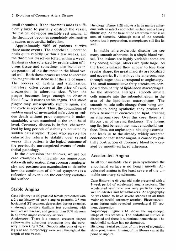
7. Evolution of Coronary Artery Disease
small thrombus. If the thrombus mass is sufficiently large to partially obstruct the vessel, the patient develops unstable rest angina. If the thrombus becomes completely obstructive it causes myocardial infarction.
Approximately 90% of patients survive these acute events. The endothelial ulceration heals quite rapidly (within a few weeks) and the thrombus dissolves (often within a week). Healing is characterized by proliferation of fibrous tissue and sometimes also includes incorporation of the thrombus in the blood vessel wall. Both these processes tend to increase the magnitude of stenosis at the site of injury. The process of healing and stabilization, therefore, often comes at the price of rapid progression in atheroma size. When the plaque becomes large enough to obstruct blood flow, it causes stable angina. This stable plaque may subsequently rupture again, and the cycle is repeated. Thus, the conundrum of sudden onset of myocardial infarction or sudden death without prior symptoms is understandable, when examined at the endothelial level. Coronary disease is, in fact, characterized by long periods of stability punctuated by sudden catastrophe. Those who survive the catastrophe return to stability over several weeks. This pattern is the logical outcome of the previously unrecognized events of endothelial pathology.
In the discussion that follows, we use our case examples to integrate our angioscopic data with information from coronary angiography and postmortem examination to describe how the continuum of clinical symptoms is a reflection of events on the coronary endothelial surface.
Stable Angina
Case History: A 65-year-old female presented with a 2-year history of stable angina pectoris, 2.5 mm horizontal ST segment depression during exercise, a strongly positive thallium test consistent with multivessel disease, and greater than 90% stenosis in all three major coronary arteries. Angioscopy: There is a smooth, crescent shaped yellow-white atheroma protruding into the coronary lumen (Fig 7.2A). Smooth atheroma of varying size and morphology were seen throughout the length of the vessel.
71
Histology: Figure 7.2B shows a large mature atheroma with an intact endothelial surface and a heavy fibrous cap. At the base of the atheroma there is an area of necrosis. Although most of the necrotic core is lost in preparation, macrophages still line its wall.
In stable atherosclerotic disease we see many smooth atheroma in a single blood vessel. The lesions are highly variable: some are tiny oblong bumps, others are quite large. As the lesions enlarge they appear to lose their regular shape; the great majority are localized and eccentric. By histology the atheroma pass through stages that correspond to angioscopy. The small nonocclusive fatty streaks are composed dominantly of lipid-laden macrophages. As the atheroma enlarges, smooth muscle cells migrate into the sub endothelium in the area of the lipid-laden macrophages. The smooth muscle cells change from being contractile to being synthetic; they produce fibrous tissue that encircles the lipid, creating an atheroma core. Over this core, there is a fibrous cap of varying thickness. The fibrous cap lies just beneath the intact endothelial surface. Thus, our angioscopic-histologic correlation leads us to the already widely accepted conclusion that stable angina is caused by partially obstruction of coronary blood flow created by smooth-surfaced atheroma.
Accelerated Angina
In all four unstable chest pain syndromes the endothelial surface is no longer smooth. Accelerated angina is the least severe of the unstable coronary syndromes.
Case History: A 66-year-old male presented with a 3-week period of accelerated angina pectoris. The accelerated syndrome was only partially responsive to nitrates and beta-blockers. At angiography he was found to have severe stenosis in all three major epicardial coronary arteries. Electrocardiogram during pain revealed anterolateral ST segment depression. Angioscopy: Figure 7.3A shows the angioscopic image of this stenosis. The endothelial surface is disrupted and there is subintimal hemorrhage. The endothelial surface has no thrombus. Histology: Serial sections of this type of ulceration show progressive thinning of the fibrous cap at the point of rupture.

A
B
72
FIGURE 7.2. A) A stable atheroma in the left descending coronary anterior of a patient with stable angina pectoris. B) An atheroma with a necrotic core covered by a fibrous cap. (Reprinted with permission from Friedman et al: Am J Pathal 1966; 48:19.)
I.S. Forrester et al.
Endothelial ulceration is the distinguishing feature between acute and stable coronary disease, as seen by angioscopy. All but one of our accelerated angina patients has had an endothelial disruption. There are probably two underlying causes. The first is rupture of the atheroma through the endothelial surface. Coronary endothelial ulcerations often bear a histologic resemblance to the inflammatory foreign body response, which is due to products released from activated macrophages. Many ulcerations, however, resemble a superficial crack, causing some authors to postulate that ulceration is a "stress fracture" induced by repetitive bending during cardiac contraction. 3 Willers on et al4 have proposed that the ulceration causes accelerated angina by platelet aggregation and subsequent release of vasoconstrictive compounds. Based on our angioscopic data, we believe that endothelial ulceration is the cause of accelerated angina.
Sudden Death
The fate of platelet aggregates may relate to rate of blood flow at the ulceration site. Some aggregates initiate thrombus formation, leading to unstable rest angina or myocardial infarction. Alternatively, the platelet-thrombus may embolize. Figure 7.3B, from Davies et al,5 shows such an embolus in a small intramyocardial coronary artery in a patient who had sudden ischemic cardiac death. Falk6
found micro emboli distal to coronary thrombi in 73% of sudden ischemic cardiac deaths, strongly suggesting that the cause of sudden ischemic cardiac death is embolus-induced fatal ventricular arrhythmias. These data lead us to infer that coronary emboli can cause sudden ischemic cardiac death.
Unstable Rest Angina
Accelerated angina frequently evolves to become unstable rest angina. We differentiate in the two conditions by the additional symptom of chest pain at rest in the latter.
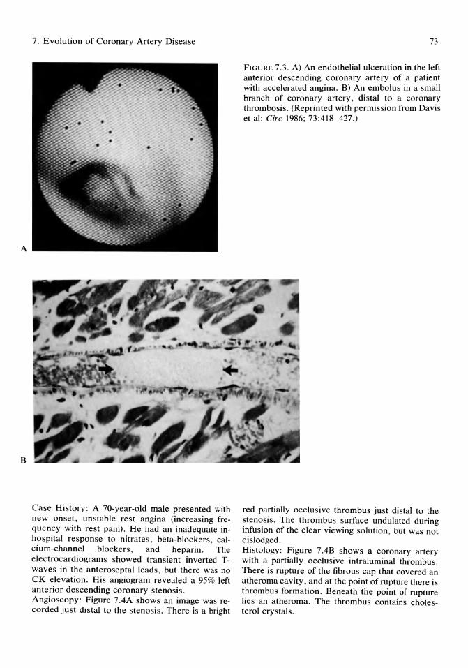
A
B
7. Evolution of Coronary Artery Disease
Case History : A 70-year-old male presented with new onset, unstable rest angina (increasing frequency with rest pain). He had an inadequate inhospital response to nitrates, beta-blockers, calcium-channel blockers, and heparin. The electrocardiograms showed transient inverted Twaves in the anteroseptal leads, but there was no CK elevation. His angiogram revealed a 95% left anterior descending coronary stenosis. Angioscopy: Figure 7.4A shows an image was recorded just distal to the stenosis. There is a bright
73
FIGURE 7.3. A) An endothelial ulceration in the left anterior descending coronary artery of a patient with accelerated angina. B) An embolus in a small branch of coronary artery, distal to a coronary thrombosis . (Reprinted with permission from Davis et al: Cire 1986; 73:418-427.)
red partially occlusive thrombus just distal to the stenosis . The thrombus surface undulated during infusion of the clear viewing solution, but was not dislodged. Histology: Figure 7.4B shows a coronary artery with a partially occlusive intraluminal thrombus. There is rupture of the fibrous cap that covered an atheroma cavity, and at the point of rupture there is thrombus formation. Beneath the point of rupture lies an atheroma. The thrombus contains cholesterol crystals.
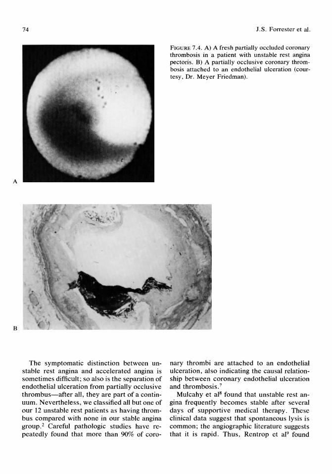
A
B
74
The symptomatic distinction between unstable rest angina and accelerated angina is sometimes difficult; so also is the separation of endothelial ulceration from partially occlusive thrombus-after all, they are part of a continuum. Nevertheless, we classified all but one of our 12 unstable rest patients as having thrombus compared with none in our stable angina group. 2 Careful pathologic studies have repeatedly found that more than 90% of coro-
J.S. Forrester et al.
FIGURE 7.4. A) A fresh partially occluded coronary thrombosis in a patient with unstable rest angina pectoris. B) A partially occlusive coronary thrombosis attached to an endothelial ulceration (courtesy, Dr. Meyer Friedman).
nary thrombi are attached to an endothelial ulceration, also indicating the causal relationship between coronary endothelial ulceration and thrombosis. 7
Mulcahy et al8 found that unstable rest angina frequently becomes stable after several days of supportive medical therapy. These clinical data suggest that spontaneous lysis is common; the angiographic literature suggests that it is rapid. Thus, Rentrop et al9 found
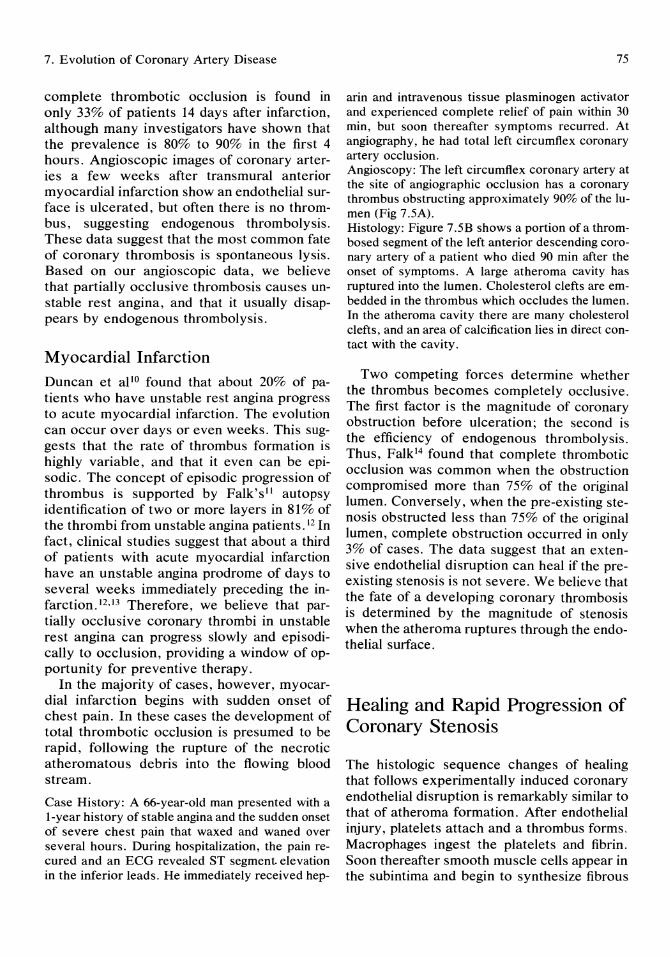
7. Evolution of Coronary Artery Disease
complete thrombotic occlusion is found in only 33% of patients 14 days after infarction, although many investigators have shown that the prevalence is 80% to 90% in the first 4 hours. Angioscopic images of coronary arteries a few weeks after transmural anterior myocardial infarction show an endothelial surface is ulcerated, but often there is no thrombus, suggesting endogenous thrombolysis. These data suggest that the most common fate of coronary thrombosis is spontaneous lysis. Based on our angioscopic data, we believe that partially occlusive thrombosis causes unstable rest angina, and that it usually disappears by endogenous thrombolysis.
Myocardial Infarction Duncan et al lO found that about 20% of patients who have unstable rest angina progress to acute myocardial infarction. The evolution can occur over days or even weeks. This suggests that the rate of thrombus formation is highly variable, and that it even can be episodic. The concept of episodic progression of thrombus is supported by Falk'sll autopsy identification of two or more layers in 81% of the thrombi from unstable angina patients. 12 In fact, clinical studies suggest that about a third of patients with acute myocardial infarction have an unstable angina prodrome of days to several weeks immediately preceding the infarction. 12 ,J3 Therefore, we believe that partially occlusive coronary thrombi in unstable rest angina can progress slowly and episodically to occlusion, providing a window of opportunity for preventive therapy.
In the majority of cases, however, myocardial infarction begins with sudden onset of chest pain. In these cases the development of total thrombotic occlusion is presumed to be rapid, following the rupture of the necrotic atheromatous debris into the flowing blood stream.
Case History: A 66-year-old man presented with a 1-year history of stable angina and the sudden onset of seVere chest pain that waxed and waned over several hours. During hospitalization, the pain recured and an ECG revealed ST segment elevation in the inferior leads. He immediately received hep-
75
arin and intravenous tissue plasminogen activator and experienced complete relief of pain within 30 min, but soon thereafter symptoms recurred. At angiography, he had total left circumflex coronary artery occlusion. Angioscopy: The left circumflex coronary artery at the site of angiographic occlusion has a coronary thrombus obstructing approximately 90% of the lumen (Fig 7.5A). Histology: Figure 7.5B shows a portion of a thrombosed segment of the left anterior descending coronary artery of a patient who died 90 min after the onset of symptoms. A large atheroma cavity has ruptured into the lumen. Cholesterol clefts are embedded in the thrombus which occludes the lumen. In the atheroma cavity there are many cholesterol clefts, and an area of calcification lies in direct contact with the cavity.
Two competing forces determine whether the thrombus becomes completely occlusive. The first factor is the magnitude of coronary obstruction before ulceration; the second is the efficiency of endogenous thrombolysis. Thus, Falk l4 found that complete thrombotic occlusion was common when the obstruction compromised more than 75% of the original lumen. Conversely, when the pre-existing stenosis obstructed less than 75% of the original lumen, complete obstruction occurred in only 3% of cases. The data suggest that an extensive endothelial disruption can heal if the preexisting stenosis is not severe. We believe that the fate of a developing coronary thrombosis is determined by the magnitude of stenosis when the atheroma ruptures through the endothelial surface.
Healing and Rapid Progression of Coronary Stenosis
The histologic sequence changes of healing that follows experimentally induced coronary endothelial disruption is remarkably similar to that of atheroma formation. After endothelial injury, platelets attach and a thrombus forms. Macrophages ingest the platelets and fibrin. Soon thereafter smooth muscle cells appear in the subintima and begin to synthesize fibrous
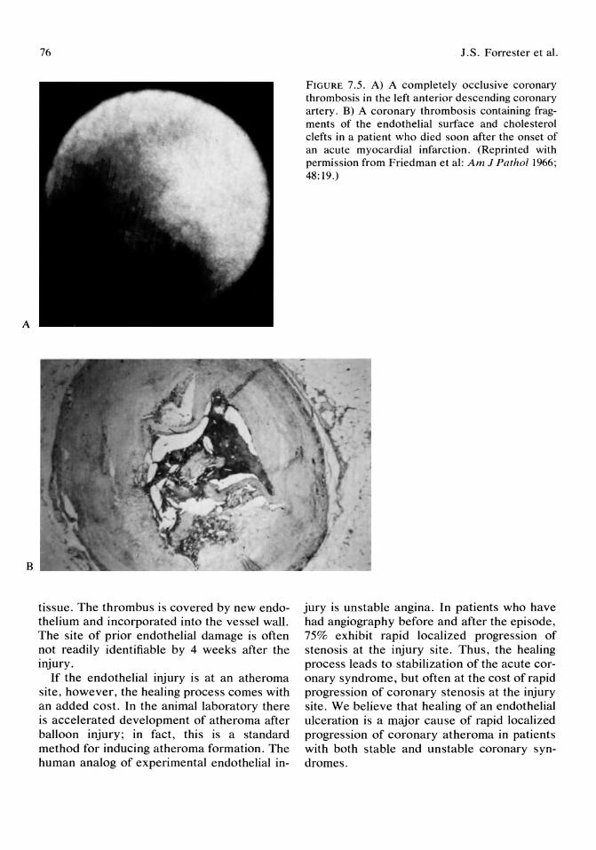
A
B
76
tissue. The thrombus is covered by new endothelium and incorporated into the vessel wall. The site of prior endothelial damage is often not readily identifiable by 4 weeks after the injury.
If the endothelial injury is at an atheroma site, however, the healing process comes with an added cost. In the animal laboratory there is accelerated development of atheroma after balloon injury; in fact, this is a standard method for inducing atheroma formation. The human analog of experimental endothelial in-
I.S. Forrester et al.
FIGURE 7.5. A) A completely occlusive coronary thrombosis in the left anterior descending coronary artery. B) A coronary thrombosis containing fragments of the endothelial surface and cholesterol clefts in a patient who died soon after the onset of an acute myocardial infarction. (Reprinted with permission from Friedman et al: Am J Patho11966; 48:19.)
jury is unstable angina. In patients who have had angiography before and after the episode, 75% exhibit rapid localized progression of stenosis at the injury site. Thus, the healing process leads to stabilization of the acute coronary syndrome, but often at the cost of rapid progression of coronary stenosis at the injury site. We believe that healing of an endothelial ulceration is a major cause of rapid localized progression of coronary atheroma in patients with both stable and unstable coronary syndromes.
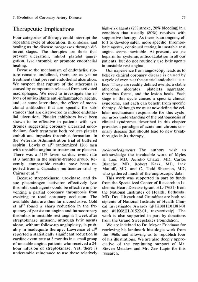
7. Evolution of Coronary Artery Disease
Therapeutic Implications Four categories of therapy could interrupt the repeating cycle of ulceration, thrombosis, and healing as the disease progresses through different stages. The therapies are those that prevent ulceration, inhibit platelet aggregation, lyse thrombi, or promote endothelial healing.
Because the mechanism of endothelial rupture remains undefined, there are as yet no treatments that prevent endothelial ulceration. We suspect that rupture of the atheroma is caused by compounds released from activated macrophages. We need to investigate the effects of antioxidants anti-inflammatory agents, and, at some later time, the effect of monoclonal antibodies that are specific for substances that are discovered to induce endothelial ulceration. Platelet inhibitors have been shown to be effective in patients with syndromes suggesting coronary ulcerated endothelium. Such treatment both reduces platelet emboli and impedes thrombus formation. In the Veterans Administration trial of buffered aspirin, Lewis et aIlS randomized 1266 men with unstable angina to treatment or placebo. There was a 51 % lower cardiac event rate at 3 months in the aspirin-treated group. Recently, comparable results have been reported from a Canadian multicenter trial by Cairns et al. 16
Because streptokinase, urokinase, and tissue plasminogen activator effectively lyse thrombi, such agents could be effective in preventing a partial coronary thrombosis from evolving to total coronary occlusion. The available data are thus far inconclusive. Gold et al 17 found a sharp reduction in the frequency of persistent angina and intracoronary thrombus in unstable rest angina 1 week after streptokinase infusion, although lytic agents alone, without follow-up angioplasty, is probably in inadequate therapy. Lawrence et aIlS reported a statistically significant reduction in cardiac event rate at 3 months in a small group of unstable angina patients who received a 24-hour infusion of streptokinase. Yet, there is understable reluctance to use these relatively
77
high-risk agents (2% stroke, 20% bleeding) in a condition that usually (80%) resolves with supportive therapy. As there is an ongoing effort to develop safer, more specific, thrombolytic agents, continued testing in unstable rest angina seems inevitable. At present, we use heparin for systemic anticoagulation in all our patients, but do not routinely use lytic agents in unstable rest angina.
Our experience from angioscopy leads us to believe clinical coronary disease is caused by a cycle of events at the arterial endothelial surface. These are readily defined events: a stable atheroma ulcerates, platelets aggregate, thrombus forms, and the lesion heals. Each stage in this cycle causes a specific clinical syndrome, and each can benefit from specific therapy. Although we must now define the cellular mechanisms responsible for this cycle, our gross understanding of the pathogenesis of clinical syndromes described in this chapter provides a paradigm of acute and chronic coronary disease that should lead to new breakthroughs in its therapy.
Acknowledgments. The authors wish to acknowledge the invaluable work of Myles E. Lee, MD, Aurelio Chaux, MD, Carlos Blanche, MD, Robert Kass, MD, Jack Matloff, MD, and C. Todd Sherman, MD, who gathered much of the angioscopic data.
This work was supported in part by funds from the Specialized Center of Research in Ischemic Heart Disease (grant HL-17651) from the National Institutes of Health, Bethesda, MD. Drs. Litvack and Grundfest are both recipients of National Institute of Health Clinical Investigator Awards (#lK08HL01381-01 and #lK08HL01522-01, respectively). The work is also supported in part by donations from the Grand Sweepstakes Foundation.
We are indebted to Dr. Meyer Friedman for retrieving his landmark histologic work from the 1960s and allowing us to republish four of his illustrations. We are also deeply appreciative of the continuing support of Mr. Steven Meadow and Mr. Zev Lapin for this research.
78
References 1. Forrester JS, Litvack F, Grundfest W, et al:
New insights into the role of thrombus in the pathogenesis of acute and chronic coronary heart disease (invited manuscript for Perspectives, Cire 1986).
2. Sherman CT, Litvack F, Grundfest WS, et al: Demonstration of thrombus and complex atheroma by in vivo angioscopy in patients with unstable angina pectoris. N Eng! J Med 1986; 315:913-919.
3. Davies MJ, Thomas A: Thrombosis and acutecoronary-artery lesions in sudden cardiac ischemic death. N Eng! J Med 1984, 310:1137-1140.
4. Willerson JT, Hillis D, Wiinniford M, et al: Speculation regarding mechanisms responsible for acute heart disease syndromes. J Am Call Cardia! 1986; 8:245.
5. Davies MJ, Thomas AC, Knapman PA, et al: Intramyocardial platelet aggregation in patients with unstable angina suffering sudden ischemic cardiac death. Cire 1986; 73:418-427.
6. Falk E: Unstable angina with fatal outcome: Dynamic coronary thrombosis leading to infarction and/or sudden death. Cire 1985; 71 :699-708.
7. Friedman M, Van den Bovenkamp GJ: Role of thrombus in plaque formation in the human diseased coronary artery. Br J Exp Patho/ 1966; 47:550.
8. Mulcahy R, Daly L, Graham I, et al: Unstable angina: Natural history and determinants of prognosis. Am J Cardio!1981; 48:525-528.
9. Rentrop KP, Frederick F, Blanke H, et al: Effects of intracoronary streptokinase and intracoronary nitroglycerin infusion on coronary angiographic patterns and mortality in patients
J.S. Forrester et al.
with acute myocardial infarction. N Engl J Med 1984; 311:1456-1463.
10. Duncan B, Fulton M, Morrison SL, et al: Prognosis of new and worsening angina pectoris. Br J Med 1976; 1:981-985.
11. Falk E: Plaque rupture with severe pre-existing stenosis precipitating coronary thrombosis: Characteristics of coronary atherosclerotic plaques underlying fatal occlusive thrombi. Br Heart J 1983; 50:127.
12. Solomon HA, Edwards AL, Killip T: Prodromata in acute myocardial infarction. Cire 1969; 40:463-471.
13. Stowers M, Short D: Warning symptoms before major myocardial infarction. Br Heart J 1970; 32:833-838.
14. Falk E: Plaque rupture with severe pre-existing stenosis precipitating coronary thrombosis: Characteristics of coronary atherosclerotic plaques underlying fatal-occlusive thrombi. Br Heart J 1983; 50:127-134.
15. Lewis DH, Davis JW, Archibald DG, et al: Protective effects of aspirin against acute myocardial infarction and death in men with unstable angina. N Engl J Med 1983; 309:396-403.
16. Cairns JA, Gent M, Singer J, et al: Aspirin, sulfinpyrazone, or both in unstable angina. Results of a Canadian multicenter trail. N Eng/ J Med 1985; 313:1369-1375.
17. Gold HK, Johns JA, Leinbach RC, et al: A randomized, blinded, placebo-controlled trial of recombinant human tissue-type plasminogen activator in patients with unstable angina pectoris. Cire 1987; 75:1192-1199.
18. Lawrence JR, Shepard JT, Bone I, et al: Fibrinolytic therapy in unstable angina pectoris. A controlled clinical trial. Thrombosis Res 1980; 17:767-777.

Part II Diagnostic Interventions
8 Interventions in the Evaluation of Valvular Heart Disease Indubala N. Vardhan and Amar S. Kapoor
Cardiac catheterization is usually peIformed with the patient at rest. Although cardiovascular function in many patients with clinical heart disease is apparently within the normal range at rest, the application of a standardized stress often will reveal abnormalities. Everyday activities entail largely dynamic muscular exercise and partly isometric exercise. The hemodynamic effects of dynamic exercise are complex and modulated through a closely integrated mechanical, neural, and humoral hemostasis. 1 Because dynamic exercise is the major form of exertion in everyday activity and most familiar to humans, it is the most commonly used cardiovascular test. Isometric (or static) exercise is described as sustained muscle contraction that occurs without joint or axial skeletal movement. Isometric exercise occurs repeatedly with most activities of daily living.
Dynamic and isometric exercises done in the cardiac catheterization laboratory assist us in evaluating valve gradients at high flow rates, left ventricular function, coronary blood flow, and other important data to derive a functional level of cardiac impairment.
Physiology of Dynamic Exercise
Oxygen consumption can increase up to 12-fold in normal sedentary subjects during maximal exercise. Oxygen consumption depends on the integration of cardiovascular, metabolic, and pulmonary reserves,2 and, thus,
maximal oxygen uptake is the highest amount of oxygen that an ambulatory person can extract. During exercise oxygen uptake rises rapidly and reaches a higher steady state level. This new higher level is directly proportional to the level of exercise.3,4 Increase in the arteriovenous oxygen occurs, which is due to the fall in the mixed venous oxygen saturation. The fall in the mixed venous saturation also is related to the degree of exercise. 3
Exercise causes increase in cardiac output. An important hemodynamic linear relationship exists between cardiac output and oxygen consumption during exercise. I Cardiac output increases by 590 mllmin per m2 for an increase of 100 mIl min per m2 of oxygen consumption. 3
This is an important concept for evaluating the cardiac output response to the intensity of exercise and the rapidity of oxygen uptake. An exercise factor has been described. I This is the increase in cardiac output with exercise divided by the corresponding increase in oxygen consumption. An exercise factor that is less than 600 mllmin per m2 divided by 100 mIl min oxygen consumption indicates insufficient cardiovascular reserve. Arterial blood pressure increases during sub maximal supine and upright exercise. This response is somewhat variable. 3 Peripheral vasodilation occurs during exercise, which causes a fall in the peripheral arteriolar resistance,S thus indicating that the increase in blood pressure is primarily due to increase in the cardiac output. There is an increase in the heart rate, which may be large in response to submaximal stress and cardiac
82
output with stroke volume changing very little.3,4,6-11 If the heart rate is kept constant, there is an increase in the cardiac output similar to the increase found when the heart rate is allowed to change. 12 This increase in cardiac output with the fixed heart rate is largely mediated from increased stroke volumes. 12
Dynamic Exercise and Left Ventricular Function
The relationship of end-diastolic and stroke volume or stroke work determines left ventricular performance during exercise. 13 Three major mechanisms have been described that may cause an increase in left ventricular performance in normal subjects. 14 They are increase in the heart rate, increase in inotropism of the heart, and Starling's law. During exercise cardiac systolic and diastolic dimensions decrease. This increases the speed at which the blood is ejected at any given level of exercise and also increases the volume of blood ejected. 15,16 Starling's law, that is, increase in the diastolic tension and size, occurs in a few patients which further increases the filling and emptying of the heart. 14, 17 Increases in left ventricular filling pressure during supine leg exercise has been attributed to the different methods of carrying out exercise. 18,19 Exercise in the sitting position causes both the mean pulmonary capillary wedge and left ventricular end-diastolic pressure to increase, although these pressures are lower in the sitting than in the supine position.17
Isometric Exercise
Isometric exercise is sustained muscle contraction that occurs without joint or axial skeletal movement. No external work is performed during isometric exercise. There is only a modest increase in the oxygen consumption (Vo2) compared with dynamic exercise.
LN. Vardhan and A.S. Kapoor
Cardiovascular Responses to Isometric Exercise
The first report on isometric exercise was published in 1920. Detailed studies on the topic were reported by Lind and colleagues. 19 They proposed that the mean arterial pressure (MAP) response was not related to muscle mass but was due to the relative (percent maximum) tension that developed in the muscle. Sustained isometric contraction results in marked increase in systolic, mean, and diastolic pressure regardless of whether it involves extension of the lower extremities (leg pressure) at the knee or the flexor groups at the elbow (sustained handgrip).20 The increase in arterial pressure was mainly due to an increase in heart rate with little change in the stroke volume or peripheral vascular resistance in normals. 20 The exact nature of this reflex is unclear and it may be related to suppression of vagal activity when afferent neural impulses are sent from the exercising muscles. 2o ,21
Isometric Exercise and Left Ventricular Function
Increase in myocardial contractility and Frank Starling mechanisms are responsible for the increase in the left ventricular performance during isometric exercise. 22 Left ventricular function is best described by the left ventricular performance and preload. Work and stroke work index increase during handgrip exercise in normal persons.23 There is little change in left ventricular end-diastolic pressure,23 and in some studies, left ventricular end-diastolic pressure was found to decrease in some patients, implying an increase in the contractile state. 24 In normal subjects, handgrip exercise results in a decrease in left ventricular endsystolic and end-diastolic volumes with a slight increase in ejection fraction.25 When studies are performed on myocardial mechanics in normal human beings during isometric exercise, there is an increase in V max, the theoretic maximal velocity of shortening of the
8. Interventions in the Evaluation of Valvular Heart Disease 83
muscle, that is, the contractile element at zero load and in left ventricular peak dp/dt. 22 ,26 An increase in the inotropic state of the left ventricle occurs as noted from intraventricular pressure recordings. Another way to demonstrate increased contractility would be to compare left ventricular stroke work and changes in left ventricular filling pressure. Stroke work expresses the external work of the left ventricle and is a measure of stroke volume and pressure. If filling pressure does not change and stroke volume is increased, it suggests increased inotropism Frank Starling law is used when, instead of the above, the filling pressure increases to increase stroke work. End-diastolic pressure is presumed to be equal to left ventricular fiber tension. l
Methods to Evaluate Impaired Left Ventricular Function
Methods used to assess left ventricular function in diseased states include exercise, atrial pacing, and isoproterenol infusion. The most common cause of left ventricular dysfunction is coronary artery disease, which causes regional dysfunction. A dramatic rise in left ventricular end-diastolic pressure occurs with exercise in patients with coronary artery disease. This is accompanied by a fall in ejection fraction. Both these changes occur even before the onset of angina or electrocardiographic evidence of ischemia.27 As stated earlier, ejection fraction in patients with minimal or no cardiac disease increases. In patients with previous myocardial infarction (scar) with no evidence of ongoing ischemia, the ejection fraction does not change and remains unchanged, whereas in patients with ischemia the ejection fraction tends to fall with exercise. 27 With continuation of exercise there is an increase in the heart rate, systolic pressure, and left ventricular dp/dt, in addition to the rise of the left ventricular end-diastolic pressure. 28 As exercise continues and pain increases electrocardiogram (ECG) changes occur, left ventricular dp/ dt falls slightly, with no change in heart rate or arterial pressure.
This is well demonstrated by a left ventricular diastolic pressure-volume relationship curve which does change significantly in patients with previous myocardial infarction or those with normal coronaries but shifts upward in patients with ischemia and usually occurs during episodes of ischemia suggestive of diastolic stiffness of the left ventricle. 27 ,29
Regional contractile abnormalities also occur in patients with coronary artery disease during exercise, which may be normal at rest. In patients with coronary artery disease, that is, ischemia, there is no improvement in the shortening velocity with exercise as compared with normals where shortening velocity improves with exerciseY
In patients with severe left ventricular failure, the heart is unable to increase cardiac output and oxygen delivery to the tissues adequately. With exercise the heart rate, oxygen consumption, stroke volume, and cardiac index rise, but this is associated with a rise in the mean capillary wedge pressure. 30 Along with this rise in the mean pulmonary capillary wedge pressure, right atrial pressure rises and systemic arterial oxygen content also increases with no change in the arterial carbon dioxide tension. 30 When compared with normal patients, anaerobic metabolism occurs at about half the normal capacity in patients with severe left heart failure. Thus, exercise provides an acute volume and pressure overload on the ventricle and easily brings out the underlying loss of cardiac reserve.
Exercise in Valve Diseases
Hemodynamic changes in valve diseases can be elucidated during exercise in patients who have the valvular stenosis or regurgitation of borderline physiologic significance.
Mitral Valve Disease
Mitral Stenosis
In patients with mitral stenosis the systemic arterial pressure did not change strikingly with exercise. 13 It is unclear whether the tachycar-
84
dia accompanying exercise may mask the rise in left ventricular end-diastolic pressure due to shortening of the diastolic filling period. However, the change and relationship of left ventricular end-diastolic pressure and stroke work index in patients with mitral stenosis with exercise is similar to normal patients. 13 When isometric exercise is conducted on a patient with mitral stenosis, a lesser increase in the heart rate and blood pressure is observed as compared with dynamic exercise. 31 When patients with mitral stenosis are compared with normal controls there are similar increases in left ventricular systolic pressure. 32 Left ventricular end-diastolic pressure is unchanged in normals, and in patients with mitral stenosis with good left ventricular function but increases significantly in those patients who may have diminished myocardial contractility32,34 due to associated disease such as coronary artery disease, The mean pulmonary capillary wedge pressure and the mean arterial pressure increases in patients with mitral stenosis, although the pulmonary vascular resistance does not change,32 The diastolic gradient across the mitral valve also increases with isometric exercise. The elevation in the left ventricular end-diastolic pressure may indicate which patients may not do as well as those with normal left ventricular end-diastolic pressure with exercise after surgery.
To summarize, isometric exercise in patients with mitral stenosis does not alter the inotropy or chronotropy of the heart's normal response unless left ventricular dysfunction and significant elevation of the pulmonary venous pressure is present. Associated mitral regurgitation and/or atrial fibrillation does not have any effect on the hemodynamic response to exercise in patients with mitral stenosis. 32
Mitral Insufficiency
Exercise testing in patients with mitral insufficiency is valuable in correlating symptoms with hemodynamic parameters. Volume overload, which occurs in patients with mitral insufficiency, may not be of consequence at rest but may unmask during exercise. Cardiac out-
LN. Vardhan and A,S, Kapoor
put may not increase appropriately in patients with left ventricular dysfunction, but in those patients in whom cardiac output increases there will be associated increase in pulmonary capillary wedge pressure and left atrial pressure, and the presence of "V" waves may be seen on the pulmonary wedge tracings. I The most important indication for dynamic exercise in patients with mitral regurgitation is its functional correlation with symptoms which the patient experiences only with exertion.
Aortic Valve Disease
Aortic Stenosis
Patients with aortic stenosis have higher left ventricular end-diastolic pressure than normals,I3,35 When dynamic exercise testing is done in patients with aortic stenosis there is an increase in left ventricular end-diastolic pressure, much greater than the increase in cardiac index. 13 ,35 When changes in left ventricular end-diastolic pressure are compared with stroke work index, those patients who had normal "exercise factor" I increased their stroke work index immensely, 13 This could be explained on the basis of one of two factors: 1) positive inotropic effect causing a changed end-diastolic volume, or 2) through the Frank Starling mechanism,
The aortic valve gradient and the aortic valve systolic flow are the two parameters used to calculate the aortic valve area. Some workers35 ,36 reported that the aortic valve gradient uniformly increases with exercise, whereas others37-39 have not found the average value of the aortic gradient to change significantly. Aortic systolic blood flow increases during exercise,35,39 and this increase is more as a consequence of increase in the cardiac output, which is much greater than the increase in the systolic ejection period.39
Change in the aortic valve area during exercise has been found in all reports. 13,35,37,39 The increase was in the calculated valve area, and the average valve area remained unchanged. Data suggest that the aortic valve area may not be a fixed orifice39 and found that changes
8. Interventions in the Evaluation of Valvular Heart Disease 85
in the aortic valve area correlated with the changes in the parameters which reflected the energy. Thus, leaflet excursion depends on the greater or lesser energy generated by the contracting left ventricle during exercise. This potential for orifice change during exercise depends on the underlying pathologic process involving the aortic valve and the change in left ventricular function, which in turn depends on associated aortic regurgitation, severity of stenosis, and coronary artery disease.
Aortic Regurgitation
Occurrence of irreversible myocardial dysfunction precludes patients with aortic regurgitation from doing well after successful valve replacement. To determine optimal time for surgery and identify the high-risk patient who may not do well after surgery, several prognostic indicators have been proposed.
Most patients with chronic aortic regurgitation have an abnormal ejection fraction during exercise.4O-42 Most patients who have symptoms due to aortic regurgitation and have depressed left ventricular functional reserve during exercise tend to have depressed left ventricular function even after surgery. 42 Fall in left ventricular ejection fraction during exercise has been thought to be an intermediate stage between normal left ventricular function at rest and clinical left ventricular dysfunction.42 Exercise in patients with chronic aortic regurgitation is usually a complex process involving preload, afterload, and contractility.44 Thus, the change in ejection fraction may be variable in these patients. Hence, it is impossible to determine whether a change in ejection fraction during exercise is due to change in loading conditions of the heart or due to left ventricular dysfunction due to myocardial degeneration.43.44 Left ventricular ejection fraction at rest and during peak exercise is a better correlate of myocardial contractility than the change in left ventricular ejection fraction with exercise due to the above reasons and also due to the fact that the amount of regurgitant flow also decreases during exercise. 44 ,45 Other markers for left ventricular dysfunction
are peak oxygen uptake and end-systolic volume, both of which correlate well with the pulmonary artery wedge pressure.44
Exercise may be a useful tool to identify those patients who have left ventricular dysfunction due to stress, but with normal left ventricular function at rest. This may also help to decide the optimal time for valve replacement in these patients.
Isoproterenol Test
Another method to determine cardiovascular reserve is isoproterenol loading. Isoproterenol is a beta-agonist, is rapid acting, and has similar actions on the myocardium as epinephrine and norepinephrine.
The responses seen with isoproterenol infusion and exercise are similar. Afterload decreases resulting in a change in mean left ventricular volume. Left ventricular end-diastolic volume and pressure remain unchanged and stroke volume is maintained. This occurs despite the tachycardia that occurs with isoprel infusion. Ejection fraction also increases. The increase in cardiac output is similar as with exercise by increasing heart rate. The response to isoproterenol infusion in patients with ischemic and valvular heart disease is similar to normal patients. This is the major drawback as it does not aid in differentiating normal patients from patients with diminished cardiac reserve or left ventricular dysfunction due to any cause.46
The only exception is in patients with idiopathic hypertrophic subaortic stenosis (IHSS). In these patients it causes a decrease in the end-systolic and end-diastolic dimensions of the left ventricle mainly due to its positive ionotropic effect and also aided by arteriolar dilatation. 47 The left ventricular enddiastolic pressure may increase, decrease, or remain unchanged. The above reasons are responsible for increasing left ventricular obstruction in patients with IHSS. In patients who have insignificant gradient at rest isoproterenol can result in a significant systolic pressure gradient. It also unmasks mitral regurgi-
86
tation in patients who do not appear to have mitral regurgitation in the basal stateY
Cold Pressor Test
The cold pressor test (CPT) was first described in 1932.48 This may be used as an alternate method for evaluation of left ventricular function and ischemia to exercise, as exercise is time consuming and cumbersome.49 This is a sympathetic reflex stimulus causing decrease coronary blood flow due to increased coronary resistance secondary to coronary vasoconstriction. 50 The hemodynamic effects of local stimulation include a rise in both systolic and diastolic blood pressure, increase in heart rate, which is variable, and an increase in the pulmonary and systemic vascular resistance.50 Left ventricular function is determined by the fall in ejection fraction and development of global and regional wall motion abnormalities by radionuclide angiography. The ejection fraction response in normals is variable. 52 In patients with coronary artery disease the mean ejection fraction decreases significantly. The sensitivity to detect coronary artery disease has varied in the literature from 55% to 94%.49,52,53 The specificity has been reported to be 100% in one study. 49 This has been questioned by others,52 and they state that it requires a long time to provoke global and regional wall motion abnormalities by cold stimulation, and the rapidity with which these return to baseline makes it difficult.
Wall motion analysis improves the sensitivity of the test. When compared with exercise the sensitivity, specificity, and predictive accuracy appears to be much lower with the cold pressor test. 52
To conclude, exercise appears to be the best test to evaluate left ventricular function and to correlate the physiologic significance of valve disease. The sensitivity of the cold pressor test has been reported to be 38% to 94%, with a specificity ranging between 90% and 100%. The sensitivity of exercise when wall motion is studied with radionuclide angiography has been reported to be 95% sensitive and 95%
I.N. Vardhan and A.S. Kapoor
specific. Atrial pacing has the drawback of not being reproducible, 54 and unless reproducibility is tested it may not be reliable.
References 1. Lorell BH, Grossman W: Dynamic and isomet
ric exercise during cardiac catheterization, in Grossman W (ed): Cardiac Catheterization and Angiography. Philadelphia, Lea & Febiger, 1986, pp 251-266.
2. Weiner DA: Normal hemodynamic, ventilatory and metabolic response to exercise. Arch Intern Med 1983; 143:2173.
3. Dexter L, et al: Effects of exercise on circulatory dynamics of normal individuals. J Appl Physiol 1951; 3:439.
4. Donald KW, et al: The effect of exercise on the cardiac output and circulatory dynamics of normal subjects. Clin Sci 1955; 14:37.
5. Simonson E, Enzer N: Physiology of muscular exercise and fatigue in disease. Medicine 1942; 21:345.
6. Barratt-Boyes BG, Wood EH: Hemodynamic response of healthy subjects to exercise in the supine position while breathing oxygen. J App Physiol1957; 11:129.
7. Chapman CB, Fisher IN, Sproule BT: Behaviour of stroke volume at rest and during exercise in human beings. J Clin Invest 1960; 39: 1208.
8. Wang Y, Marshall RJ, Shepherd JT: The effect of changes in posture and of graded exercise on stroke volume in man. J Clin Invest 1960; 39:1051.
9. Holmgren A, Jonsson B, Sjostrand T: Circulatory data in normal subjects at rest and during exercise in recumbent position, with special reference to the stroke volume at different work intensities. Acta Physiol Scandinav 1960; 49:343.
10. Chapman CB: On the nature of cardiac control. Ann Int Med 1960; 53:1272.
11. Bickelman AG, Lippschultz EJ, Weinstein L: The response of the normal and abnormal heart to exercise. A functional evaluation. Circulation 1963; 28:238.
12. Ross J Jr, Linhart JW, Braunwald E: Effects of changing heart rate in man by electrical stimulation of the right atrium. Circulation 1965; 32:549-553.
13. Ross J Jr, et al: Left ventricular performance during muscular exercise in patients with and
8. Interventions in the Evaluation of Valvular Heart Disease 87
without cardiac dysfunction. Circulation 1966; 34:597.
14. Gorlin R, et al: Effect of supine exercise on left ventricular volume and oxygen consumption in man. Circulation 1965; 32:361.
15. Rushmer R, Smith 0: Cardiac control. Physiol Rev 1955; 35:90-129.
16. Braunwald E, et al: Studies on cardiac dimension on intact unanesthetised man III. Effects of muscular exercise. Circ Res 1964; 13:448.
17. Slutsky R, et al: Peak systolic blood pressure/ end systolic volume in normal subjects and patients with coronary heart disease. Assessment at rest and during exercise. Am J Cardiol1980; 46:813.
18. Thadani U, et al: Hemodynamics at rest and during supine and sitting bicycle exercise in patients with coronary artery disease. Am J Cardiol 1977; 39:776.
19. Linda AR, et al: Circulatory effects of sustained voluntary muscle contraction. Clin Sci 1964; 27:229.
20. Donald KW, et al: Cardiovascular responses to sustained (static) contractions. Circ Res 1967; 20(suppl 1)15.
21. Freyschuss U: Cardiovascular adjustments to somatomotor activities. Acta Physiol Scand 1970; (suppl 2:342).
22. Grossman W, et al: Changes in the ionotropic state of the left ventricle during isometric exercise. Br Heart J 1973; 35:697.
23. Helfant RH, Devilla MA, Meister SG: Effect of sustained isometric handgrip exercise on left ventricular performance. Circulation 1971; 44:982.
24. Kovowitz C, et al: Effects of isometric exercise on cardiac performance: The grip test. Circulation 1971; 44:994.
25. Flessas A, et al: Effects of isometric exercise on the end-diastolic pressure, volumes and function of the left ventricle in man. Circulation 1976; 53:839.
26. Krayenbuehl HP, et al: Evaluation of left ventricular function from isovolumlic pressure measurements during isometric exercise. Am J Cardiol 1972; 29:323.
27. Caroll JD, et al: Dynamics of left ventricular filling at rest and during exercise. Circulation 1983; 68:59.
28. Wiener L, Dwyer EM Jr, Cox JW: Left ventricular hemodynamics in exercise induced angina pectoris. Circulation 1968; 38:240-249.
29. Grossman W, Barry WH: Diastolic pressure-
volume relations in the diseased heart. Fed Proc 1980; 148.
30. Franciosa JA, et al: Relation between hemodynamic and ventilatory responses in determining exercise capacity in severe congestive heart failure. Am J Cardiol 1984; 53: 127.
31. Fisher ML, et al: Hemodynamic responses to isometric exercise (handgrip) in patients with heart disease. Br Heart J 1973; 35:697-704.
32. Flessas AP, Ryan TJ: Cardiovascular responses to isometric exercise in patients with mitral stenosis. Arch Int Med 1982; 142: 1629-1633.
33. Huikuri HV, Takkunen JT: Valve of isometric exercise testing during cardiac catheterization in mitral stenosis. Am J Cardiol 1983; 52:540-543.
34. Horwitz L, Atkins J, Leshins S: Role of FrankStarling mechanism in exercise. Circ Res 1972; 31:861-874.
35. Bache RJ, Wang Y, Jorgenson CR: Hemodynamic effects of exercise in isolated valvular aortic stenosis. Circulation 1971; 44:1003.
36. Cueto L, Moller JH: Hemodynamics of exercise in children with isolated aortic valve disease. Br Heart J 1973; 35:93-98.
37. Anderson FL, et al: Hemodynamic effects of exercise in patients with aortic stenosis. Am J Med 1969; 46:872-885.
38. Kasalicky J, et al: The effect of exercise on left heart hemodynamics in patients with valvular aortic stenosis. Cor Vasa 1973; 15:81-91.
39. Richardson WJ, Anderson FL, Tsagaris TJ: Rest and exercise hemodynamic studies in patients with isolated aortic stenosis. Cardiology 1979; 64:1-11.
40. Borer JS, et al: Left ventricular function at rest and during exercise after aortic valve replacement in patients with aortic regurgitation. Circulation 1979; 60:647.
41. Bonow RO, et al: Preoperative exercise capacity in symptomatic patients with aortic regurgitation as a predictor of postoperative left ventricular function and long-term prognosis. Circulation 1980; 62: 1280.
42. Borer JS, et al: Exercise induced left ventricular dysfunction in symptomatic and asymptomatic patients with aortic regurgitation: Assessment with radionuclide cine angiography. Am J Cardiol1981; 48:17.
43. Gee DS, et al: Prognostic significance of exercise induced left ventricular dysfunction in chronic aortic regurgitation. Am J Cardiol 1985; 56:605-609.
88
45. Boucher N, et al: Exercise testing in asymptomatic or minimally symptomatic aortic regurgitation: Relationship of left ventricular ejection fraction to left ventricular filling pressure during exercise. Circulation 1983; 67:1091-1100.
45. Levinson GE, et al: The effect of rest and physical effort on the left ventricular burden in mitral and aortic regurgitation. Am Heart J 1970; 80:791.
46. Krasnov N, et al: Isoproterenol and cardiovascular performance. Am J Med 1964; 37:514.
47. Braunwald E, et al: Idiopathic hypertrophic subaortic stenosis. Circulation 1964; 291 30(suppl): 14.
48. Hines EA, Brown GE: Standard stimulus for measuring vasomotor reactions: Its application in study of hypertension. Mayo Clin Proc 1932; 7:332.
49. Wainwright RJ, et al: Cold pressor test in detection of coronary heart disease and cardiomy-
I.N. Vardhan and A.S. Kapoor
opathy using Tc 99m gated blood pool imaging. Lancet 1979; 2:320.
50. Mudge GH Jr, et al: Reflex increase in coronary vascular resistance in patients with ischemic heart disease. N Engl J Med 1976; 295: 1333.
51. Godden JO, et al: The changes in the intra arterial pressure during immersion of the hand in ice cold water. Circulation 1978; 12:813.
52. Manyari DE, et al: Comparative value of the cold pressor test and supine bicycle exercise to detect subjects with coronary artery disease using radionuclide ventriculography. Circulation 1982; 65:571.
53. Kurtz RG, et al: Cold-pressor radionuclide ventriculography (abstr). J Nuc Med 1980; 21:4.
54. Thadani U, et al: Reproducibility of clinical and hemodynamic parameters during pacing stress testing in patients with angina pectoris. Circulation 1979; 60:5.
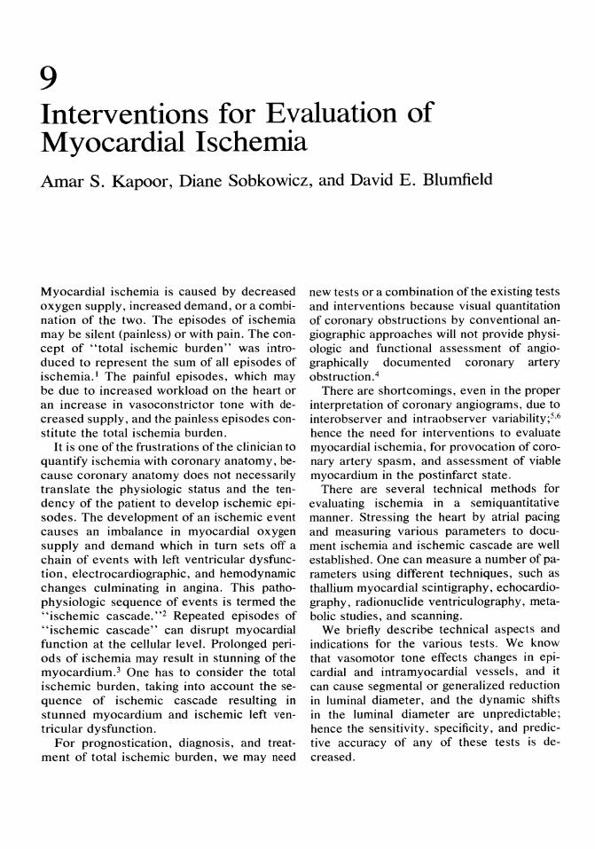
9 Interventions for Evaluation of Myocardial Ischemia Amar S. Kapoor, Diane Sobkowicz, and David E. Blumfield
Myocardial ischemia is caused by decreased oxygen supply, increased demand, or a combination of the two. The episodes of ischemia may be silent (painless) or with pain. The concept of "total ischemic burden" was introduced to represent the sum of all episodes of ischemia. l The painful episodes, which may be due to increased workload on the heart or an increase in vasoconstrictor tone with decreased supply, and the painless episodes constitute the total ischemia burden.
It is one of the frustrations of the clinician to quantify ischemia with coronary anatomy, because coronary anatomy does not necessarily translate the physiologic status and the tendency of the patient to develop ischemic episodes. The development of an ischemic event causes an imbalance in myocardial oxygen supply and demand which in turn sets off a chain of events with left ventricular dysfunction, electrocardiographic, and hemodynamic changes culminating in angina. This pathophysiologic sequence of events is termed the "ischemic cascade."2 Repeated episodes of "ischemic cascade" can disrupt myocardial function at the cellular level. Prolonged periods of ischemia may result in stunning of the myocardium. 3 One has to consider the total ischemic burden, taking into account the sequence of ischemic cascade resulting in stunned myocardium and ischemic left ventricular dysfunction.
For prognostication, diagnosis, and treatment of total ischemic burden, we may need
new tests or a combination of the existing tests and interventions because visual quantitation of coronary obstructions by conventional angiographic approaches will not provide physiologic and functional assessment of angiographically documented coronary artery obstruction.4
There are shortcomings, even in the proper interpretation of coronary angiograms, due to interobserver and intraobserver variability;5,6 hence the need for interventions to evaluate myocardial ischemia, for provocation of coronary artery spasm, and assessment of viable myocardium in the postinfarct state.
There are several technical methods for evaluating ischemia in a semiquantitative manner. Stressing the heart by atrial pacing and measuring various parameters to document ischemia and ischemic cascade are well established. One can measure a number of parameters using different techniques, such as thallium myocardial scintigraphy, echocardiography, radio nuclide ventriculography, metabolic studies, and scanning.
We briefly describe technical aspects and indications for the various tests. We know that vasomotor tone effects changes in epicardial and intramyocardial vessels, and it can cause segmental or generalized reduction in luminal diameter, and the dynamic shifts in the luminal diameter are unpredictable; hence the sensitivity. specificity, and predictive accuracy of any of these tests is decreased.
90
Stress Atrial Pacing
This procedure is performed with electrocardiographic monitoring with multiple leads, especially leads II, V I , and V s. A bipolar flared pacing catheter (Atria Pace I, Mansfield Scientific, Mansfield, MA) is inserted through a venous sheath percutaneously via the femoral, subclavian, or internal jugular, or through a venous cutdown. Under fluoroscopic guidance, the pacing leads are advanced into the right atrium. A bipolar catheter is more frequently used because it affords more stable electric capture with at least one atrial lead in contact with the right atrial wall at all times. If a unipolar catheter is used, the best location for atrial pacing is at the superior vena cava, right atrial junction, or the coronary sinus. The lateral right atrial wall also has been used but is commonly displaced by movement of the patient or respiration and may disrupt the study. Also, phrenic stimulation causing discomfort to the patient is more likely in this position.
Mter the pacing catheters are positioned, the bipolar pacing catheter is connected to the pacemaker unit directly. If necessary, extensor wires with alligator clamps may be used. Unipolar catheters need to be connected to a generator unit and appropriately grounded. The atrial pacing threshold and ventricular capture of the pacemaker at 2 to 3 mA are checked with the heart rate set 10 beats/min greater than the patient's resting heart rate. The pacing threshold can be increased as needed, up to 7 to 8 mAo
Once atrial capture with ventricular conduction is obtained, atrial pacing should be performed. This is done by increasing the pacing rate by 10 beats/min every 5 seconds up to 150 beats/min. If atrioventricular block occurs, atropine, 1 mg intravenously, should be given to facilitate conduction in the A-V node. When a heart rate of 150 beats/min is achieved, pacing may be stopped as this will conclude the pretesting phase.
Start the pacing stress test by pacing the patient 20 beats/min above the resting heart rate and increase the pacing rate by 20 beats/ min every 2 minutes until evidence of ische-
A.S. Kapoor, D. Sobkowicz, and D.E. Blumfield
mia by standard electrocardiogram (ECG) changes (1 min or more of horizontal or downs loping ST segment depression) or 85% maximal predicted heart rate is achieved.
Throughout the pacing stress test, as previously stated, leads II, VI, and Vs , as well as blood pressure, should be continuously monitored with 12-lead ECGs obtained before the prior procedure, at each pacing level, and immediately after pacing. If ECG changes and/or symptoms are preset immediately after pacing, a 12-lead ECG should be obtained every 2 minutes until changes and/or symptoms have resolved.
If chest discomfort occurs during the study, one may safely continue to pace at the same heart rate for 3 to 5 minutes to collect the appropriate data, as ischemic symptoms resolve rapidly after pacing is ceased. Rarely do symptoms or ECG changes persist for more than 1 to 2 minutes after the return to baseline rate.
Hemodynamic assessments can be made simultaneously with thermodilution balloon-tip flow-directed catheters, left heart catheters, and/or arterial lines in place to evaluate rightand left-sided pressures.
Chest Pain and Electrocardiogram Changes
When atrial pacing was first introduced as a diagnostic tool for ischemic heart disease by Sowton et aF in 1967, chest pain or heart rate of 160 beats/min was used as the ischemic threshold. Subsequently, Helfant et apo in 1970 reviewed atrial pacing with angina pectoris or heart rate of 160 beats/min as the endpoint of the pacing stress test, and found only 50% of the subjects with known coronary artery disease (CAD) had chest pain, although other markers of ischemia (metabolic abnormalities, ECG changes) were evident. Therefore, considerable lack of correlation between chest pain and objective evidence of ischemia was noted. Cokkinos et aF9 then recommended the use of atropine routinely to obtain higher pacing rates to increase testing sensitivity. However, Robson and colleagues13 subse-
9. Evaluation of Myocardial Ischemia
quently performed atrial pacing in subjects with and without CAD, and found chest pain in patients without CAD when heart rate was greater than or equal to 180 beats/min. These markedly high pacing rates also caused problems with pacemaker spike, obscuring ECG changes within the ST segment. 30 These studies, in addition to using high pacing rates and chest pain as an endpoint, did not routinely monitor V 5. Therefore, with a large percentage of false-negatives and false-positives, atrial pacing was not found to be useful in the diagnosis of CAD.
Heller et al,14 in 1984, re-evaluated the use of atrial pacing with a study protocol using the presently recommended guidelines of the endpoint of the study being 85% predicted maximal heart rate or ECG changes (including 12-lead ECG monitoring and II, VI, V5)
indicative of ischemia or chest pain only if associated with objective evidence of ischemia. Results were then compared with exercise testing and angiographic studies. The overall sensitivity and specificity of chest pain alone was reaffirmed to be low with either exerci~ ~ treadmill testing (44.% sensitivity, specificity 67%) or atrial pacing (50% sensitivity, specificity 67%). Overall sensitivity of atrial pacing is 94% with a specificity of 83% when ischemic changes are used as diagnostic of CAD and not chest pain alone. Limiting the rate obtained to 85% (maximum predicted heart rate) obviated the problem of ST change distortion by pacemaker spike of P-R prolongation and markedly reduced the problem of false-positive studies that Robson et aP3 encountered in 83% of his normal subjects with high pacing rates.
Therefore, it was concluded that right atrial pacing tachycardia was a useful and reliable tool in assessing the presence of CAD. It was also thought it might be especially useful in patients unable to complete exercise tolerance testing.
Rapid atrial pacing use also was evaluated as a prognostic indicator for future myocardial events (e.g., remyocardial infarct, cardiac death) versus treadmill testing by Tzivoni et alY After 16 months offollow-up, the predictive value of a positive right trial pacing re-
91
sponse was 20% compared with exercise treadmill testing (including submaximal studies in which the maximal heart rate obtainable was 116 beats/min), which was 13%.17 However, in postinfarction patients with a comparable pressure-rate product or exercise treadmill testing, the predictive value was not statistically different.
In clinically high-rate postinfarction patients (postinfarct angina pectoris, congestive heart failure, or more than 70%) in which predischarge exercise treadmill testing was not performed, rapid atrial pacing was safely conducted and identified a subset with poorer prognosis that was not evident by clinical symptomatology alone.
Other electrocardiographic markers for myocardial ischemia, such as Rivane amplitude,19 also have been studied and noted to be useful in conjunction with standard ischemic ECG changes.
Atrial Pacing With Thallium Perfusion Studies
To increase the sensitivity and specificity of graded right atrial pacing in the diagnosis of significant CAD, thallium 201 was used in conjunction with right atrial pacing. Weiss et aPI reported an overall sensitivity of 100%. Heller and associates l9 also used atrial pacing with thallium testing.
Atrial pacing stress test with two-dimensional echocardiography was then recommended as an alternative to exercise echocardiography with less technical difficulties because the echocardiogram can be monitored throughout the study in the same view.
Atrial Pacing With Radionuclide Ventriculography
Development of segmental wall abnormalities with rapid atrial pacing also has been studied. In conjunction with multigated radionuclide angiography (MUGA) in patients with CAD, graded atrial pacing resulted in new segmental
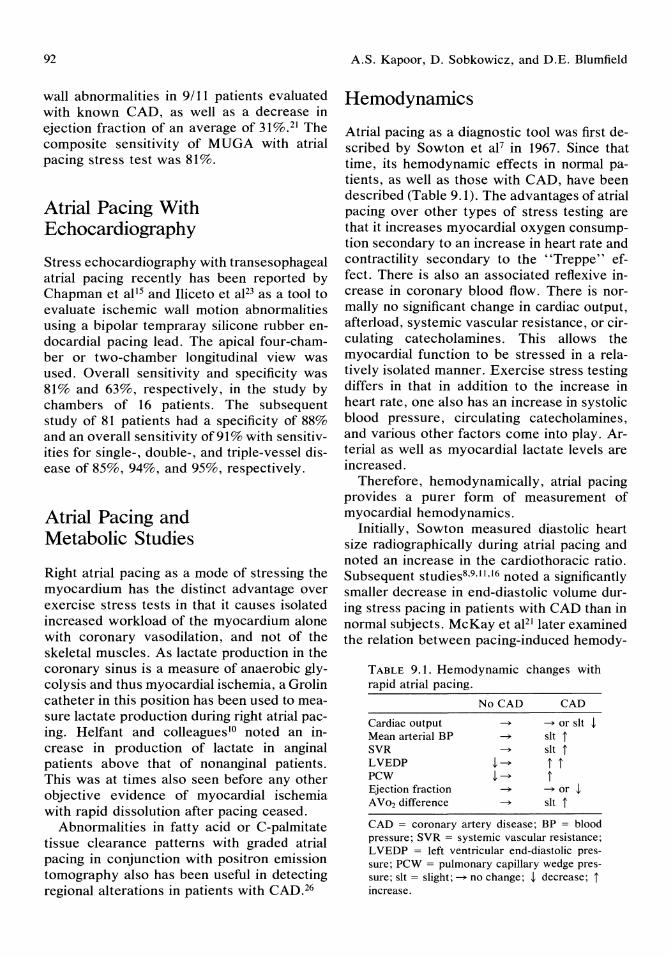
92
wall abnormalities in 9/11 patients evaluated with known CAD, as well as a decrease in ejection fraction of an average of 31 %.21 The composite sensitivity of MUGA with atrial pacing stress test was 81%.
Atrial Pacing With Echocardiography
Stress echocardiography with trans esophageal atrial pacing recently has been reported by Chapman et al15 and Iliceto et aP3 as a tool to evaluate ischemic wall motion abnormalities using a bipolar tempraray silicone rubber endocardial pacing lead. The apical four-chamber or two-chamber longitudinal view was used. Overall sensitivity and specificity was 81% and 63%, respectively, in the study by chambers of 16 patients. The subsequent study of 81 patients had a specificity of 88% and an overall sensitivity of 91 % with sensitivities for single-, double-, and triple-vessel disease of 85%, 94%, and 95%, respectively.
Atrial Pacing and Metabolic Studies
Right atrial pacing as a mode of stressing the myocardium has the distinct advantage over exercise stress tests in that it causes isolated increased workload of the myocardium alone with coronary vasodilation, and not of the skeletal muscles. As lactate production in the coronary sinus is a measure of anaerobic glycolysis and thus myocardial ischemia, a Grolin catheter in this position has been used to measure lactate production during right atrial pacing. Relfant and colleagueslO noted an increase in production of lactate in anginal patients above that of nonanginal patients. This was at times also seen before any other objective evidence of myocardial ischemia with rapid dissolution after pacing ceased.
Abnormalities in fatty acid or C-palmitate tissue clearance patterns with graded atrial pacing in conjunction with positron emission tomography also has been useful in detecting regional alterations in patients with CAD.26
A.S. Kapoor, D. Sobkowicz, and D.E. Blumfield
Hemodynamics
Atrial pacing as a diagnostic tool was first described by Sowton et aF in 1967. Since that time, its hemodynamic effects in normal patients, as well as those with CAD, have been described (Table 9.1). The advantages of atrial pacing over other types of stress testing are that it increases myocardial oxygen consumption secondary to an increase in heart rate and contractility secondary to the "Treppe" effect. There is also an associated reflexive increase in coronary blood flow. There is normally no significant change in cardiac output, afterload, systemic vascular resistance, or circulating catecholamines. This allows the myocardial function to be stressed in a relatively isolated manner. Exercise stress testing differs in that in addition to the increase in heart rate, one also has an increase in systolic blood pressure, circulating catecholamines, and various other factors come into play. Arterial as well as myocardial lactate levels are increased.
Therefore, hemodynamically, atrial pacing provides a purer form of measurement of myocardial hemodynamics.
Initially, Sowton measured diastolic heart size radiographically during atrial pacing and noted an increase in the cardiothoracic ratio. Subsequent studies8,9,11,16 noted a significantly smaller decrease in end-diastolic volume during stress pacing in patients with CAD than in normal subjects. McKay et aPl later examined the relation between pacing-induced hemody-
TABLE 9.1. Hemodynamic changes with rapid atrial pacing.
No CAD CAD
Cardiac output ~ ~ or sit ! Mean arterial BP ~ sit t SVR ~ sit t LVEDP !~ t t PCW !~ t Ejection fraction ~ ~ or ! A V02 difference ~ sit t
CAD = coronary artery disease; BP = blood pressure; SVR = systemic vascular resistance; L VEDP = left ventricular end-diastolic pressure; PCW = pulmonary capillary wedge pressure; sit = slight; ~ no change; ! decrease; t increase,
9. Evaluation of Myocardial Ischemia
namic changes and the extent of myocardial ischemia as quantified by thallium-20l imaging. The largest changes in left ventricular end-diastolic pressure and pulmonary capillary wedge pressure occurred in those patients with the largest amount of myocardial tissue at ischemic jeopardy. There was also a positive correlation between the postpacing increase in left ventricular end-diastolic pressures and number of diseased vessels on angiogram.
Pressure-volume relationships in detail were then studied during pacing-induced ischemia. 24.25 Normally, there is both an increase in contractility and a mild increase in distensibility shifting the pressure-volume diagram leftward and downward. With pacinginduced ischemia (which was demonstrated by ECG changes + / - chest pain), the left ventricular end-diastolic volume and end-systolic volume initially increased, then subsequently decreased during the period of ischemia without change in cardiac output but with a decrease in ejection fraction. With the associated increase in end-diastolic pressure in these patients, the pressure-volume loop shifted initially leftward, then upward in diastole.
Clinical Indications for Atrial Pacing in Coronary Artery Disease-Advantages and Disadvantages
Stress atrial pacing use has not been widespread, largely because of it being an invasive tool and there is discomfort to the patient with transesophageal pacing. However, advantages include the relatively "pure" myocardial stressing that it allows without the use of medications and thus no side effects from medications. It may be used in patients with musculoskeletal disorders, peripheral vascular disease, unstable angina, beta-blocker therapy, or chronic obstructive pulmonary disease with aminophylline therapy who cannot adequately perform on an exercise treadmill test or have contraindications to the use of dipyridamole. It also affords greater control over the development of ischemia because the ischemic episode is more controlled and more read-
93
ily reversible. As a research tool, atrial pacing continues to be invaluable, more recently in hemodynamic evaluation of myocardial ischemia. It is also being used as an objective measure of anginal threshold in antianginal medications, such as Diltiazem.27
Ergonovine Stimulation for Coronary Artery Spasm
Coronary angiography is the best technique for definitive diagnosis of coronary artery spasm when a patient with variant angina has an anginal attack and cineangiography is performed during the attack. This, however, is an uncommon occurrence. The second approach is to document electrocardiographic ST segment shifts during episodes of chest pain in a patient who presents with rest angina, if it occurs at night or in the early morning hours. A susceptible group of patients are middle-aged women with a history of smoking, emotional stress, and migraine headaches, or Reynaud's phenomenon. Such patients can be asked to transmit their electrocardiogram by transtelephonic monitoring before using sublingual nitroglycerin. This approach, according to Ginsburg et aP2, was helpful in documented ST segment shifts in 50% of patients. This, however, will not rule out severe occlusive coronary artery disease. Coronary artery spasm can be induced in the catheterization laboratory with provocation by ergonovine maleate. The mechanism of focal spasm induced by ergonovine is not completely understood. It may produce vasoconstriction in a susceptible arterial segment via alpha-receptors in the smooth muscle or via stimulation of serotonin membrane receptors.
Catheter-induced spasm is produced by mechanical irritations of the coronary intima.
Ergonovine Provocation Test
In a patient suspected of having Prinzmetal's angina or in a patient with normal thallium or exercise electrocardiographic stress test, this provocative test may be indicated for defini-
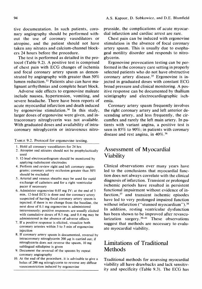
94
tive documentation. In such patients, coronary angiography should be performed without the use of coronary vasodilators or atropine, and the patient should not have taken any nitrates and calcium-channel blockers 24 hours before the procedure.
The test is performed as detailed in the protocol (Table 9.2). A positive test is comprised of chest pain with ECG changes of ischemia and focal coronary artery spasm as demonstrated by angiography with greater than 50% lumen reduction. 33 Patients also can have malignant arrhythmias and complete heart block.
Adverse side effects to ergonovine maleate include nausea, hypertension, vomiting, and severe headache. There have been reports of acute myocardial infarction and death induced by ergonovine stimulation. 34 In this study, larger doses of ergonovine were given, and intracoronary nitroglycerin was not available. With graduated doses and availability of intracoronary nitroglycerin or intravenous nitro-
TABLE 9.2. Protocol for ergonovine testing.
I. Hold all coronary vasodilators for 24 hrs 2. Atropine and nitrates should not be prophylactically
used 3. 12-lead electrocardiogram should be monitored by
applying radiolucent electrodes 4. Perform and review right and left coronary angio
grams; coronary artery occlusion greater than 50% should be excluded
5. Arterial and venous sheaths may be used for rapid exchange of catheters and for a right ventricular pacer if necessary
6. Administer ergonovine 0.05 mg IV; at the end of 3 min, 12-lead ECG is done and the coronary artery suspected of having focal coronary artery spasm is injected; if there is no change from the baseline, the next dose of 0.1 mg ergonovine is administered intravenously; positive responses are usually elicited with cumulative doses of 0.3 mg, and 0.4 mg may be administered in the absence of adverse effects
7. If a positive response is elicited, visualize both coronary arteries within 3 to 5 min of ergonovine injection
8. If coronary artery spasm is documented, reversal by intracoronary nitroglycerin 200 p.g is carried out; if nitroglycerin does not reverse the spasm, 10 mg sublingual nifedipine is given
9. Document the reversal of the spasms by repeat coronary angiography
10. At the end of the procedure, it is advisable to give a bolus of 200 mg nitroglycerin to reverse any diffuse vasoconstriction induced by ergonovine
A.S. Kapoor, D. Sobkowicz, and D.E. Blumfield
prusside, the complications of acute myocardial infarction and cardiac arrest are rare.
Chest pain can be induced with ergonovine stimulation in the absence of focal coronary artery spasm. This is usually due to esophageal motility disorder and responds to nitroglycerin.
Ergonovine provocation testing can be performed in the coronary care setting in properly selected patients who do not have obstructive coronary artery disease. 35 Ergonovine is injected in graduated doses with constant ECG broad pressure and clinical monitoring. A positive response can be documented by thallium scintigraphy and electrocardiographic ischemia.
Coronary artery spasm frequently involves the right coronary artery and left anterior descending artery, and less frequently, the circumflex and rarely the left main artery. In patients with variant angina, a positive test is seen in 85% to 90%; in patients with coronary disease and rest angina, in 40%.36
Assessment of Myocardial Viability
Clinical observations over many years have led to the conclusions that myocardial function does not always correlate with the clinical diagnosis of infarction. Transient or prolonged ischemic periods have resulted in persistent functional impairment without evidence of infarction,37 and transient ischemic episodes have led to very prolonged impaired function without infarction ("stunned myocardium").38 In addition, resting ventricular dysfunction has been shown to be improved after revascularization surgery. 39-41 These observations suggest that methods are necessary to evaluate myocardial viability.
Limitations of Traditional Methods
Traditional methods for assessing myocardial viability all have drawbacks and lack sensitivity and specificity (Table 9.3). The ECG has

9. Evaluation of Myocardial Ischemia
TABLE 9.3. Methods for assessing myocardial viability.
Interventions Exercise
ECG Thallium Echocardiography Radionuclide ventriculography
Nitrates Radionuclinde ventriculography Contrast ventriculography
Postextrasystolic potentiation Radionuclide ventriculography Contrast ventriculography
Dipyridamole Thallium Echocardiography
long been known to be a poor predictor of viability. The presence of a Q-wave infarction does not imply "transmural" nor does it necessarily imply irreversible injury (i.e., necrosis). Likewise, interventions used in conjunction with other studies lack sensitivity and specificity for cell death. Typically, this has involved interventions to predict improvement in left ventricular function after revascularization surgery. Clues as to the reversibility of abnormal wall motion have depended on using exercise ,42 nitroglycerin administration,43 or postextrasystolic potentiation44 in conjunction with contrast or radionuclide ventriculography. Reversible flow abnormalities on thallium-201 studies comparing postexercise and delayed scintigraphy also have been used to predict improvement after surgery. 40 Each of these methods depends on the estimation of blood flow or myocardial contractility, both of which can be abnormal without the presence of irreversible injury to the myocardium. Other methods that assess the myocardium on a more cellular basis are necessary.
Review of Cardiac Metabolism in Ischemia
Normal myocardium is characterized by aerobic metabolism; however, during ischemia, there is a shift to anerobic metabolism and away from oxidation of glucose, fatty acids, and lactate, resulting in increased lactate pro-
95
duction in the myocardium. When lactate levels rise sufficiently, glycolysis is inhibited, as is high energy phosphate production.45 It would seem logical to expect that the duration and extent of the ischemia would help define the reversibility or irreversibility or the resulting damage to the cells. It would also seem logical that if the flow changes and the changes in the metabolism could be evaluated noninvasively, one could not only evaluate the extent of the damage but also evaluate the viability of that myocardium.
Experimental Studies
Positron emission tomography (PET) has been used to evaluate these parameters of myocardial metabolism. The physical aspects and instrumentation of PET have been welldescribed elsewhere.46.47 These physical properties of PET allow for increased resolution and decreased interference from scatter, problems with traditional scintigraphy including single proton emission computed tomography (SPECT). With proper imaging agents, each of these parameters (flow, glucose metabolism, and fatty acid metabolism) can be studied by PET.
For evaluation of myocardial blood flow, an agent with very high first pass extraction and slow washout by the myocardium is needed. N-13 ammonia has these characteristics, as well as being well dissolved in blood (as ammonium ion) after intravenous injection.46•47 Clinical and animal studies48-50 have shown it to be a reliable agent for evaluation of perfusion and in predicting coronary obstructive disease. 49
Palmitate would seem to be the logical choice for evaluating fatty acid metabolism in the heart. It accounts for a majority of the fatty acid metabolism, which in turn is the preferred metabolic substrate of the heart. 45,46 Initial studies showed an increase in the concentration of C-l1 palmitate in ischemic myocardium; when this was further studied, it became clear that the uptake of palmitate was reduced, but the clearance, in addition, was markedly reduced. 51 .52 As the production of C-ll carbon dioxide was reduced as well, this
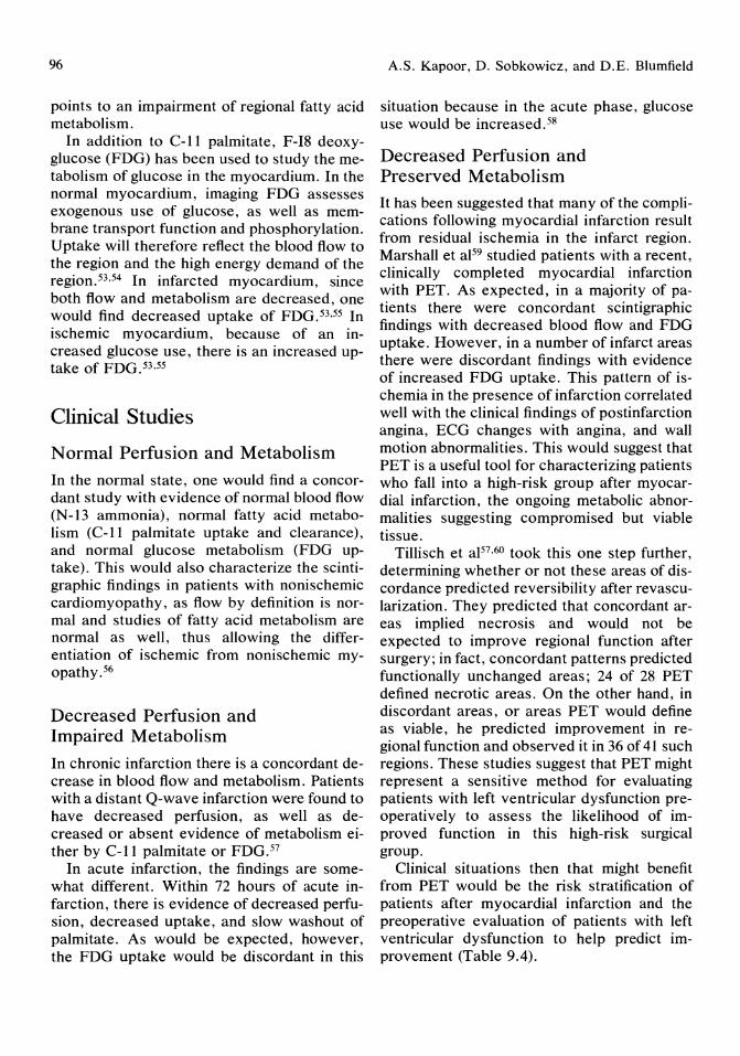
96
points to an impairment of regional fatty acid metabolism.
In addition to C-ll palmitate, F-I8 deoxyglucose (FDG) has been used to study the metabolism of glucose in the myocardium. In the normal myocardium, imaging FDG assesses exogenous use of glucose, as well as membrane transport function and phosphorylation. Uptake will therefore reflect the blood flow to the region and the high energy demand of the region. 53 ,54 In infarcted myocardium, since both flow and metabolism are decreased, one would find decreased uptake of FDG. 53,55 In ischemic myocardium, because of an increased glucose use, there is an increased uptake of FDG. 53,55
Clinical Studies
Normal Perlusion and Metabolism
In the normal state, one would find a concordant study with evidence of normal blood flow (N-13 ammonia), normal fatty acid metabolism (C-ll palmitate uptake and clearance), and normal glucose metabolism (FDG uptake). This would also characterize the scintigraphic findings in patients with nonischemic cardiomyopathy, as flow by definition is normal and studies of fatty acid metabolism are normal as well, thus allowing the differentiation of ischemic from nonischemic myopathy.56
Decreased Perfusion and Impaired Metabolism
In chronic infarction there is a concordant decrease in blood flow and metabolism. Patients with a distant Q-wave infarction were found to have decreased perfusion, as well as decreased or absent evidence of metabolism either by C-ll palmitate or FDG. 57
In acute infarction, the findings are somewhat different. Within 72 hours of acute infarction, there is evidence of decreased perfusion, decreased uptake, and slow washout of palmitate. As would be expected, however, the FDG uptake would be discordant in this
A.S. Kapoor, D. Sobkowicz, and D.E. Blumfield
situation because in the acute phase, glucose use would be increased. 58
Decreased Perfusion and Preserved Metabolism It has been suggested that many of the complications following myocardial infarction result from residual ischemia in the infarct region. Marshall et aP9 studied patients with a recent, clinically completed myocardial infarction with PET. As expected, in a majority of patients there were concordant scintigraphic findings with decreased blood flow and FDG uptake. However, in a number of infarct areas there were discordant findings with evidence of increased FDG uptake. This pattern of ischemia in the presence of infarction correlated well with the clinical findings of postinfarction angina, ECG changes with angina, and wall motion abnormalities. This would suggest that PET is a useful tool for characterizing patients who fall into a high-risk group after myocardial infarction, the ongoing metabolic abnormalities suggesting compromised but viable tissue.
Tillisch et aP7,60 took this one step further, determining whether or not these areas of discordance predicted reversibility after revascularization. They predicted that concordant areas implied necrosis and would not be expected to improve regional function after surgery; in fact, concordant patterns predicted functionally unchanged areas; 24 of 28 PET defined necrotic areas. On the other hand, in discordant areas, or areas PET would define as viable, he predicted improvement in regional function and observed it in 36 of 41 such regions. These studies suggest that PET might represent a sensitive method for evaluating patients with left ventricular dysfunction preoperatively to assess the likelihood of improved function in this high-risk surgical group.
Clinical situations then that might benefit from PET would be the risk stratification of patients after myocardial infarction and the preoperative evaluation of patients with left ventricular dysfunction to help predict improvement (Table 9.4).
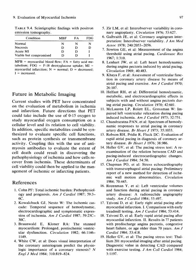
9. Evaluation of Myocardial Ischemia
TABLE 9.4. Scintigraphic findings with positron emission tomography.
Condition MBF FA FDG
Normal N N N Necrosis D D D Acute MI D D I Viable but compromised D D I
MFB = myocardial blood flow; FA = fatty acid metabolism; FDG = F-18 deoxyglucose uptake; MI =
myocardial infarction; N = normal; D = decreased; I = increased.
Future in Metabolic Imaging
Current studies with PET have concentrated on the evaluation of metabolism in ischemia and infarction. Future directions that PET could take include the use of 0-15 oxygen to study myocardial oxygen consumption on a cellular level and its relation to cell viability. In addition, specific metabolites could be synthesized to evaluate specific cell functions, such as protein synthesis or transmembrane activity. Coupling this with the use of antimyosin antibodies to evaluate the extent of cell death could result in discovering the pathophysiology of ischemia and how cells recover from ischemia. These determinants of cell viability could then be applied to the management of ischemic or infarcting patients.
References 1. Cohn PF: Total ischemic burden: Pathophysiol
ogy and prognosis. Am J Cardiol 1987; 59:3-6C.
2. Kowalchuck GJ, Nesto W: The ischemic cascade: Temporal sequence of hemodynamic, electrocardiographic and symptomatic expression of ischemia. Am J Cardiol 1987; 59:23C-30C.
3. Braunwald E, Kloner RA: The stunned myocardium: Prolonged, postischemic ventricular dysfunction. Circulation 1982; 66: 1146-1149.
4. White CW, et al: Does visual interpretation of the coronary anteriogram predict the physiologic importance of a coronary stenosis? N Engl J Med 1984; 310:819-824.
97
5. Zir LM, et al: Interobserver variability in coronary angioplasty. Circulation 1976; 53:627.
6. Galbraith JE, et al: Coronary angiogram interpretation: Interobserver variability. JAm Med Assoc 1978; 240:2053-2056.
7. Sowton GE, et al: Measurement of the angina threshold using atrial pacing. Cardiovasc Res 1967; 1:310.
8. Lenhart JW, et al: Left heart hemodynamics during angina pectoris induced by atrial pacing. Circulation 1969; 40:483.
9. Khaya F, et al: Assessment of ventricular function in coronary artery disease by means of atrial pacing and exercise. Am J Cardiol 1970; 26:107.
10. Helfant RH, et al: Differential hemodynamic, metabolic, and electrocardiographic effects in subjects with and without angina pectoris during atrial pacing. Circulation 1970; 42:601.
11. McLauren LP, Rolett EL, Grossman W: Impaired left ventricular relaxation during pacing induced ischemia. Am J Cardiol 1973; 32:751.
12. Chandraratna PAN, et al: Spectrum ofhemodynamic responses to atrial pacing in coronary artery disease. Br Heart J 1973; 35:1033.
13. Robson RH, Pridie R, Fluck DC: Evaluation of rapid atrial pacing in diagnosis of coronary artery disease. Br Heart J 1976; 38:986.
14. Heller GV, et al: The pacing stress test: A reexamination of the relation between CAD and pacing-induced electrocardiographic changes. Am J Cardiol 1984; 54:50.
15. Chapman PO, et al: Stress echocardiography with transesophageal atrial pacing: Preliminary report of a new method for detection of ischemic wall motion abnormalities. Circulation 1984; 70:445.
16. Rozenman Y, et al: Left ventricular volumes and function during atrial pacing in coronary artery disease: A radionuclide angiography study. Am J Cardiol 1984; 53:497.
17. Tzivoni D, et al: Early right atrial pacing after myocardial infarction. I. Comparison with early treadmill testing. Am J Cardiol 1984; 53:414.
18. Tzivoni D, et al: Early rapid atrial pacing after myocardial infarction. II. Results in 77 patients with predischarge angina pectoris, congestive heart failure, or age older than 70 years. Am J Cardiol 1984; 53:418.
19. Heller GV, et al: The pacing stress test: Thallium 201 myocardial imaging after atrial pacing. Diagnostic value in detecting CAD compared with exercise testing. J Am Coli Cardiol 1984; 3:1197.
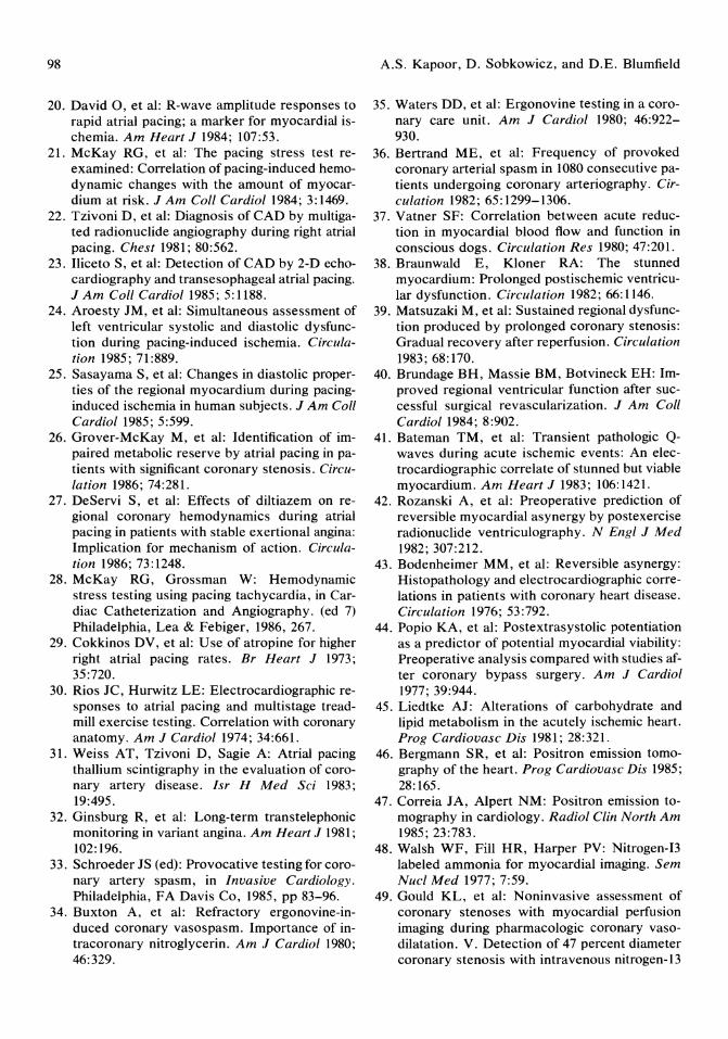
98
20. David 0, et al: R-wave amplitude responses to rapid atrial pacing; a marker for myocardial ischemia. Am Heart J 1984; 107:53.
21. McKay RG, et a1: The pacing stress test reexamined: Correlation of pacing-induced hemodynamic changes with the amount of myocardium at risk. J Am Coil Cardiol 1984; 3: 1469.
22. Tzivoni D, et al: Diagnosis of CAD by multigated radionuclide angiography during right atrial pacing. Chest 1981; 80:562.
23. Iliceto S, et al: Detection of CAD by 2-D echocardiography and transesophageal atrial pacing. J Am Coil Cardiol1985; 5:1188.
24. Aroesty JM, et al: Simultaneous assessment of left ventricular systolic and diastolic dysfunction during pacing-induced ischemia. Circulation 1985; 71:889.
25. Sasayama S, et al: Changes in diastolic properties of the regional myocardium during pacinginduced ischemia in human subjects. J Am Coil Cardiol1985; 5:599.
26. Grover-McKay M, et al: Identification of impaired metabolic reserve by atrial pacing in patients with significant coronary stenosis. Circulation 1986; 74:281.
27. DeServi S, et al: Effects of diltiazem on regional coronary hemodynamics during atrial pacing in patients with stable exertional angina: Implication for mechanism of action. Circulation 1986; 73:1248.
28. McKay RG, Grossman W: Hemodynamic stress testing using pacing tachycardia, in Cardiac Catheterization and Angiography. (ed 7) Philadelphia, Lea & Febiger, 1986, 267.
29. Cokkinos DV, et al: Use of atropine for higher right atrial pacing rates. Br Heart J 1973; 35:720.
30. Rios JC, Hurwitz LE: Electrocardiographic responses to atrial pacing and multistage treadmill exercise testing. Correlation with coronary anatomy. Am J Cardiol 1974; 34:661.
31. Weiss AT, Tzivoni D, Sagie A: Atrial pacing thallium scintigraphy in the evaluation of coronary artery disease. Isr H Med Sci 1983; 19:495.
32. Ginsburg R, et al: Long-term transtelephonic monitoring in variant angina. Am Heart J 1981; 102: 196.
33. Schroeder JS (ed): Provocative testing for coronary artery spasm, in Invasive Cardiology. Philadelphia, FA Davis Co, 1985, pp 83-96.
34. Buxton A, et al: Refractory ergonovine-induced coronary vasospasm. Importance of intracoronary nitroglycerin. Am J Cardiol 1980; 46:329.
A.S. Kapoor, D. Sobkowicz, and D.E. Blumfield
35. Waters DD, et al: Ergonovine testing in a coronary care unit. Am J CardioI 1980; 46:922-930.
36. Bertrand ME, et al: Frequency of provoked coronary arterial spasm in 1080 consecutive patients undergoing coronary arteriography. Circulation 1982; 65:1299-1306.
37. Vatner SF: Correlation between acute reduction in myocardial blood flow and function in conscious dogs. Circulation Res 1980; 47:201.
38. Braunwald E, Kloner RA: The stunned myocardium: Prolonged postischemic ventricular dysfunction. Circulation 1982; 66: 1146.
39. Matsuzaki M, et al: Sustained regional dysfunction produced by prolonged coronary stenosis: Gradual recovery after reperfusion. Circulation 1983; 68:170.
40. Brundage BH, Massie BM, Botvineck EH: Improved regional ventricular function after successful surgical revascularization. J Am Coli Cardiol 1984; 8:902.
41. Bateman TM, et al: Transient pathologic Qwaves during acute ischemic events: An electrocardiographic correlate of stunned but viable myocardium. Am Heart J 1983; 106:1421.
42. Rozanski A, et al: Preoperative prediction of reversible myocardial asynergy by postexercise radio nuclide ventriculography. N Engl J Med 1982; 307:212.
43. Bodenheimer MM, et al: Reversible asynergy: Histopathology and electrocardiographic correlations in patients with coronary heart disease. Circulation 1976; 53:792.
44. Popio KA, et al: Postextrasystolic potentiation as a predictor of potential myocardial viability: Preoperative analysis compared with studies after coronary bypass surgery. Am J Cardiol 1977; 39:944.
45. Liedtke AJ: Alterations of carbohydrate and lipid metabolism in the acutely ischemic heart. Prog Cardiovasc Dis 1981; 28:321.
46. Bergmann SR, et al: Positron emission tomography of the heart. Prog Cardiovasc Dis 1985; 28: 165.
47. Correia JA, Alpert NM: Positron emission tomography in cardiology. Radiol Clin North Am 1985; 23:783.
48. Walsh WF, Fill HR, Harper PV: Nitrogen-I3 labeled ammonia for myocardial imaging. Sem Nucl Med 1977; 7:59.
49. Gould KL, et al: Noninvasive assessment of coronary stenoses with myocardial perfusion imaging during pharmacologic coronary vasodilatation. V. Detection of 47 percent diameter coronary stenosis with intravenous nitrogen-13
9. Evaluation of Myocardial Ischemia
ammonia and emission computed tomography in intact dogs. Am J Cardiol 1979; 43:200.
50. Schelbert HR, et al: Regional myocardial perfusion assessed with N-13 labeled ammonia and positron emission computed axial tomography. Am J Cardiol 1979; 43:209.
51. Schelbert HR, et al: C-ll palmitic acid for the noninvasive evaluation of regional myocardial fatty acid metabolism with positron emission tomography. IV. In vivo demonstration of impaired fatty acid oxidation in acute myocardial ischemia. Am Heart J 1983; 106:736.
52. Schon HR, et al: C-ll palmitic acid for the noninvasive evaluation of regional myocardial fatty acid metabolism with positron emission tomography: II. Kinetics of C-ll palmitic acid in acutely ischemic myocardium. Am Heart J 1982; 103:548.
53. Schwaiger M, et al: Myocardial glucose utilization measured noninvasively in man by positron tomography. J Am Call Cardiol 1983; 1:688.
54. Ratib 0, et al: Positron tomography with deoxyglucose for estimating local myocardial glucose metabolism. J Nucl Med 1982; 23:577.
99
55. Cochavi S, Pohost GM, Elmaleh DR, et al: Transverse sectional imaging with NaF-18 in myocardial infarction. J Nucl Med 1979; 20:1013.
56. Eisenberg JD, Sobel BE, Geltman EM: Differentiation of ischemic from nonischemic cardiomyopathy with positron emission tomography. Am J Cardiol 1987; 59: 1410.
57. Bruken R, et al: Regional perfusion, glucose metabolism, and wall motion in patients with chronic electrocardiographic Q-wave infarctions: Evidence for persistence of viable tissue in some infarct regions by positron emission tomography. Circulation 1986; 73:951.
58. Schwaiger M, et al: Regional metabolism in patients with acute myocardial infarction determined by positron emission tomography (PET). Circulation 1984; 70: 1249.
59. Marshall RC, et al: Identification and differentiation of resting myocardial ischemia and infarction in man with positron computed tomography. Circulation 1981; 64:766.
60. Tillisch J, et al: Reversibility of cardiac wallmotion abnormalities predicted by positron tomography. N Engl J Med 1986; 314:884.
10 Introduction to Clinical Electrophysiology Donald G. Rubenstein and Carol Zaher
Historical Perspectives
Electrophysiology studies (EPS) began in the late 1960s in the dog laboratory where recording of the His bundle electrogram was accomplished. l The first recordings in the human heart occurred in a patient with atrial septal defect2; whereas in 1969, Scherlag et aP were the first investigators to percutaneously, by right heart cardiac catheterization, record a His bundle in humans by safely placing an electrode catheter across the tricuspid valve.
The early electrophysiologists concerned themselves with patterns of A V conduction and site of A V delay, as well as mechanisms of arrhythmias and impulse formation. The addition of programmed stimulation in the early 1970s transformed electrophysiology studies from an investigative technique into a dynamic study which could stress the conduction system, as well as allow induction and termination of tachyarrhythmias.4,5
Since those early years, electrophysiology studies have greatly expanded our knowledge, and now longitudinal studies can be performed in a systematic, reproducible, and safe manner to assist in clinical management of patients with a variety of conduction abnormalities and arrhythmias. Newer modalities of treatment including an increasing number of antiarrhythmic drugs, the use of the automatic implantable cardioverter defibrillator device (AICD), antitachycardia pacemakers, catheter ablation, and ablative surgery make programmed electrical stimulation an important technique
in managing an ever increasing number of survivors of lethal arrhythmias.
Indications for Programmed Electrical Stimulation
Clinical indications for EPS are still in evolution. 6 In 1984, the Health and Public Policy Committee of the American College of Physicians developed a set of acceptable indications for EPS.? Table 10.1 lists some common indications for EPS.
Generally Accepted Indications
Sustained Ventricular Tachycardia
Of patients with clinical sustained ventricular tachycardia 75% to 95% can have their arrhythmia reproduced by EPS.8 For those patients with inducible sustained ventricular tachycardia, interventions that prevent induction in the laboratory are also likely to prevent clinical recurrence.8 Interventions unsuccessful during EPS are likely to fail to control clinical ventricular tachycardia (VT) recurrence.9--l3 Efficacy of drugs, suitability for antitachycardia pacemaker therapy, preoperative and intraoperative mapping for surgical endocardial resection of arrhythmic focus, and consideration for AICD implantation all require preliminary EPS evaluation.
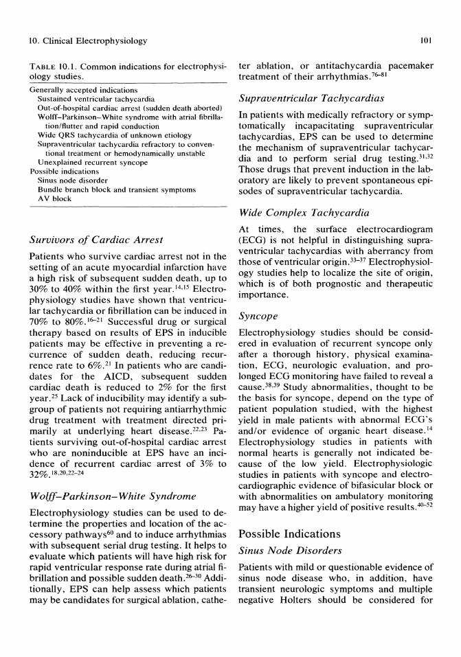
10. Clinical Electrophysiology
TABLE 10.1. Common indications for electrophysiology studies.
Generally accepted indications Sustained ventricular tachycardia Out-of-hospital cardiac arrest (sudden death aborted) Wolff-Parkinson-White syndrome with atrial fibrilla-
tionlflutter and rapid conduction Wide QRS tachycardia of unknown etiology Supraventricular tachycardia refractory to conven
tional treatment or hemodynamically unstable Unexplained recurrent syncope
Possible indications Sinus node disorder Bundle branch block and transient symptoms AV block
Survivors of Cardiac Arrest
Patients who survive cardiac arrest not in the setting of an acute myocardial infarction have a high risk of subsequent sudden death, up to 30% to 40% within the first year. 14.15 Electrophysiology studies have shown that ventricular tachycardia or fibrillation can be induced in 70% to 80%.16-21 Successful drug or surgical therapy based on results of EPS in inducible patients may be effective in preventing a recurrence of sudden death, reducing recurrence rate to 6%.21 In patients who are candidates for the AICD, subsequent sudden cardiac death is reduced to 2% for the first year.25 Lack of inducibility may identify a subgroup of patients not requiring antiarrhythmic drug treatment with treatment directed primarily at underlying heart disease. 22•23 Patients surviving out-of-hospital cardiac arrest who are noninducible at EPS have an incidence of recurrent cardiac arrest of 3% to 32%.18.20,22-24
Wolff-Parkinson- White Syndrome
Electrophysiology studies can be used to determine the properties and location of the accessory pathways60 and to induce arrhythmias with subsequent serial drug testing. It helps to evaluate which patients will have high risk for rapid ventricular response rate during atrial fibrillation and possible sudden death.26-30 Additionally, EPS can help assess which patients may be candidates for surgical ablation, cathe-
101
ter ablation, or antitachycardia pacemaker treatment of their arrhythmias.76-81
Supraventricular Tachycardias
In patients with medically refractory or symptomatically incapacitating supraventricular tachycardias, EPS can be used to determine the mechanism of supraventricular tachycardia and to perform serial drug testing. 31 .32 Those drugs that prevent induction in the laboratory are likely to prevent spontaneous episodes of supraventricular tachycardia.
Wide Complex Tachycardia
At times, the surface electrocardiogram (ECG) is not helpful in distinguishing supraventricular tachycardias with aberrancy from those of ventricular origin. 33-37 Electrophysiology studies help to localize the site of origin, which is of both prognostic and therapeutic importance.
Syncope
Electrophysiology studies should be considered in evaluation of recurrent syncope only after a thorough history, physical examination, ECG, neurologic evaluation, and prolonged ECG monitoring have failed to reveal a cause. 38,39 Study abnormalities, thought to be the basis for syncope, depend on the type of patient population studied, with the highest yield in male patients with abnormal ECG's and/or evidence of organic heart disease. 14 Electrophysiology studies in patients with normal hearts is generally not indicated because of the low yield. Electrophysiologic studies in patients with syncope and electrocardiographic evidence of bifasicular block or with abnormalities on ambulatory monitoring may have a higher yield of positive results. 4°-52
Possible Indications
Sinus Node Disorders
Patients with mild or questionable evidence of sinus node disease who, in addition, have transient neurologic symptoms and multiple negative Holters should be considered for
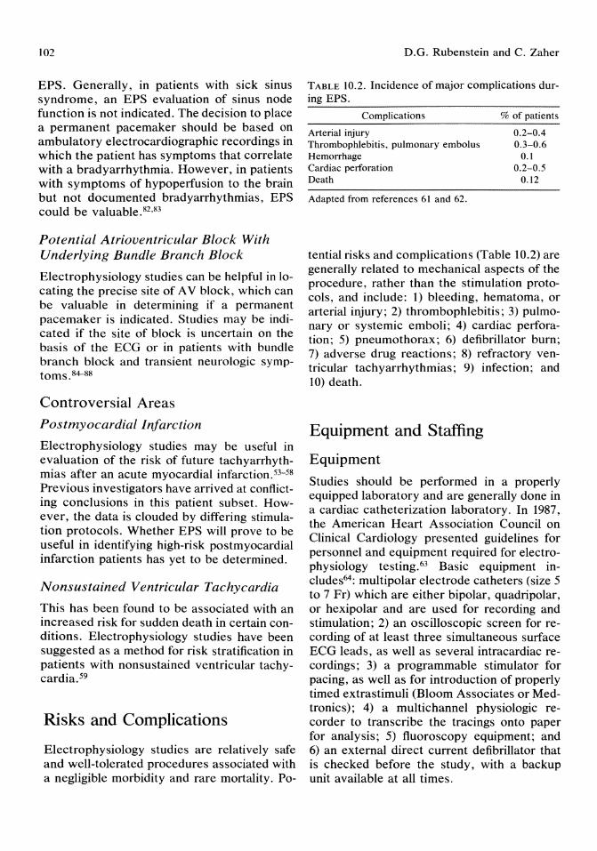
102
EPS. Generally, in patients with sick sinus syndrome, an EPS evaluation of sinus node function is not indicated. The decision to place a permanent pacemaker should be based on ambulatory electrocardiographic recordings in which the patient has symptoms that correlate with a bradyarrhythmia. However, in patients with symptoms of hypoperfusion to the brain but not documented bradyarrhythmias, EPS could be valuable. 82 •83
Potential Atrioventricular Block With Underlying Bundle Branch Block
Electrophysiology studies can be helpful in locating the precise site of A V block, which can be valuable in determining if a permanent pacemaker is indicated. Studies may be indicated if the site of block is uncertain on the basis of the ECG or in patients with bundle branch block and transient neurologic symptoms.84-88
Controversial Areas
Postmyocardial Infarction
Electrophysiology studies may be useful in evaluation of the risk of future tachyarrhythmias after an acute myocardial infarction. 53-58
Previous investigators have arrived at conflicting conclusions in this patient subset. However, the data is clouded by differing stimulation protocols. Whether EPS will prove to be useful in identifying high-risk postmyocardial infarction patients has yet to be determined.
Nonsustained Ventricular Tachycardia
This has been found to be associated with an increased risk for sudden death in certain conditions. Electrophysiology studies have been suggested as a method for risk stratification in patients with nonsustained ventricular tachycardia.59
Risks and Complications
Electrophysiology studies are relatively safe and well-tolerated procedures associated with a negligible morbidity and rare mortality. Po-
D.O. Rubenstein and C. Zaher
TABLE 10.2. Incidence of major complications during EPS.
Complications
Arterial injury Thrombophlebitis, pulmonary embolus Hemorrhage Cardiac perforation Death
Adapted from references 61 and 62.
% of patients
0.2-0.4 0.3-0.6
0.1 0.2-0.5
0.12
tential risks and complications (Table 10.2) are generally related to mechanical aspects of the procedure, rather than the stimulation protocols, and include: 1) bleeding, hematoma, or arterial injury; 2) thrombophlebitis; 3) pulmonary or systemic emboli; 4) cardiac perforation; 5) pneumothorax; 6) defibrillator burn; 7) adverse drug reactions; 8) refractory ventricular tachyarrhythmias; 9) infection; and 10) death.
Equipment and Staffing
Equipment
Studies should be performed in a properly equipped laboratory and are generally done in a cardiac catheterization laboratory. In 1987, the American Heart Association Council on Clinical Cardiology presented guidelines for personnel and equipment required for electrophysiology testing. 63 Basic equipment includes64 : multipolar electrode catheters (size 5 to 7 Fr) which are either bipolar, quadripolar, or hexipolar and are used for recording and stimulation; 2) an oscilloscopic screen for recording of at least three simultaneous surface ECG leads, as well as several intracardiac recordings; 3) a programmable stimulator for pacing, as well as for introduction of properly timed extrastimuli (Bloom Associates or Medtronics); 4) a multichannel physiologic recorder to transcribe the tracings onto paper for analysis; 5) fluoroscopy equipment; and 6) an external direct current defibrillator that is checked before the study, with a backup unit available at all times.

10. Clinical Electrophysiology
Staffing
Because programmed electrical stimulation has the potential for inducing life-threatening arrhythmias, it is very important that staffing is adequate and that all participants are comfortable working together as a team, with each assuming a specific role. If possible, staffing should include: 1) a trained electrophysiologist to place and manipulate the catheters during the study, to operate the stimulator, and to direct the study; 2) an advanced cardiac life support (ACLS)-trained nurse whose duties are to monitor vital signs, give medications during the study, and if necessary cardiovert the patient during an induced arrhythmia; 3) a catheterization technician; 4) an x-ray technician to provide assistance during fluoroscopy; 5) availability of a biomedical engineer; and 6) availability of an anesthesiologist.
Technique
The patient is admitted to a monitored bed in the hospital with all antiarrhythmic medications discontinued for at least five half-lives. Baseline coagulation studies, electrolytes, chest x-ray, and ECG are obtained, and patient venous access is maintained. The patient is studied in the postabsorptive state and sedated with intravenous Valium if necessary.
In the majority of cases, a transvenous route using the Seldinger technique is performed with placement of multiple pacing catheters into one or both femoral veins. In general, two #7 FR femoral sheaths are advanced over guidewires using only one femoral vein. If stimulation of the left ventricle is required, a #7 FR femoral artery sheath is advanced over a guidewire using the Seldinger technique. In those cases requiring coronary sinus pacing, the left antecubital vein, left subclavian vein, or left internal jugular vein are best suited for entrance, as well as stability, if further studies are required.
The decision to administer heparin is based on individual experience. In all patients requiring left ventricular stimulation, heparin must be given. In general, in anticipated pro-
103
longed studies, heparin is probably indicated. Heparin is probably not required in shorter studies.
Arterial blood pressure is monitored by a percutaneous femoral arterial catheter or by a Dynamap continuous arterial blood pressure cuff.
Once the study protocol is completed, the catheters and sheaths are removed (unless a coronary sinus catheter or right ventricular catheter is needed for further studies), and groin pressure is applied for 10 to 20 minutes. If drugs were administered during the study, blood levels should be drawn.
Intracardiac Recordings
Depending on the type of study, catheters can be advanced to the high right atrium, mid-right atrium, low right atrium, coronary sinus, His bundle, right ventricular apex, right ventricular outflow tract, pulmonary artery, or in the left ventricle (Fig 10.1).
High Right Atrium (HRA)
This is the most common site for atrial stimulation with the catheter placed as close to the sinus node as possible at the junction of the posterior atrial wall with the superior vena cava. A quadripolar catheter allows for pacing and recording. An additional catheter can be moved to various locations in the right atrium to perform mapping studies during tachycardias.
His Bundle Electrogram (HBE)
The catheter should be advanced into the right ventricle and pulled back with clockwise torque to the area of the septal leaflet of the tricuspid valve at the left border of the spinal cord on x-ray in order to obtain the most proximal His potential. It is important to make sure the His spike represents activation of the most proximal His bundle. The His spike is a sharp biphasic or triphasic deflection 15 to 25 msec in duration located between the atrial and ventricular spikes. Validation of the His potential may be obtained by several methods: 1) pacing
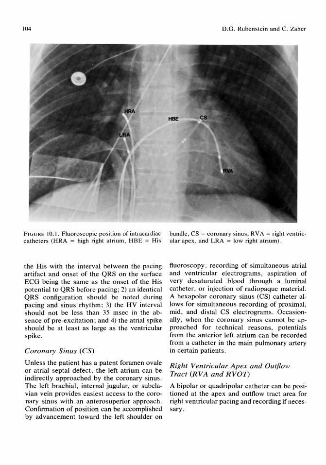
104
FIGURE 10.1. Fluoroscopic position of intracardiac catheters (HRA = high right atrium, HBE = His
the His with the interval between the pacing artifact and onset of the QRS on the surface ECG being the same as the onset of the His potential to QRS before pacing; 2) an identical QRS configuration should be noted during pacing and sinus rhythm; 3) the HV interval should not be less than 35 msec in the absence of pre-excitation; and 4) the atrial spike should be at least as large as the ventricular spike.
Coronary Sinus (CS)
Unless the patient has a patent foramen ovale or atrial septal defect, the left atrium can be indirectly approached by the coronary sinus. The left brachial, internal jugular, or subclavian vein provides easiest access to the coronary sinus with an anterosuperior approach. Confirmation of position can be accomplished by advancement toward the left shoulder on
D.G. Rubenstein and C. Zaher
bundle, CS = coronary sinus, RV A = right ventricular apex, and LRA = low right atrium).
fluoroscopy, recording of simultaneous atrial and ventricular electrograms, aspiration of very de saturated blood through a luminal catheter, or injection of radiopaque material. A hexapolar coronary sinus (CS) catheter allows for simultaneous recording of proximal, mid, and distal CS electrograms. Occasionally, when the coronary sinus cannot be approached for technical reasons, potentials from the anterior left atrium can be recorded from a catheter in the main pulmonary artery in certain patients.
Right Ventricular Apex and Outflow Tract (RVA and RVOT)
A bipolar or quadripolar catheter can be positioned at the apex and outflow tract area for right ventricular pacing and recording if necessary.

10. Clinical Electrophysiology
Left Ventricle (L V)
A bipolar catheter is placed at one or multiple sites of the left ventricle via femoral artery insertion, which allows pacing and/or mapping of the left ventricle.
Basic Electrophysiologic Study
Once the catheters are in place, baseline measurements in sinus rhythm should be obtained and include: sinus cycle length (SCL) and PR, PA, AH, HV, QRS, and QT intervals (Table 10.3). These measurements can be made from the surface ECG leads and from the intracardiac recordings simultaneously at a paper speed of 100 mm per second. The AH interval is measured from the earliest reproducible rapid deflection of the A spike to the first deflection of the His potential in the His bundle electrogram tracing and approximates primarily AV nodal conduction time. The HV interval is measured from the His spike to the earliest ventricular potential on the surface or intracardiac recordings (Fig 10.2). Normal values for AH are 60 to 125 msec65 •66 and for HV are 35 to 55 msec. 64
Once the baseline measurements have been obtained, programmed electrical stimulation is performed with atrial, ventricular, and occasionally coronary sinus pacing (Table 10.4). Both atrial and ventricular pacing should be performed at 2 to 3 times diastolic thresholds and 1 to 2 msec pulse width. In general, diastolic thresholds for the atrium should be less than 1.5 rnA and diastolic thresholds for the ventricle, less than 1.0 rnA. The pacing techniques include both incremental pacing from different sites, as well as introduction of extrastimuli during spontaneous or paced rhythms. A basic electrophysiologic study (EPS) includes the following evaluations with variations and special studies noted under specific topics.
Sinus Node Function
Sinus node function can be assessed using EPS with: 1) sinus node recovery time
105
TABLE 10.3. Guide to EPS abbreviations.
SCL PA
AH
HV
HBE SNRT CSNRT SACT SI SI
HRA LRA RVA RVOT LVA CS CSM SVT VT
Sinus cycle length Interval from the onset of the "P" wave on
the surface ECG to the onset of low atrial activity in the His bundle recording.
Interval from the onset of low atrial activity in the His bundle recording to the onset of the His spike.
Interval from the onset of the His deflection to the earliest onset of ventricular activation in any lead.
His bundle electrogram Sinus node recovery time Corrected sinus node recovery time Sinoatrial conduction time Stimulus to stimulus interval during continu
ous pacing Stimulus coupling interval between last
continuous paced beat and the first premature stimulus.
Stimulus coupling interval between the first and second premature stimuli.
Stimulus coupling interval between the second and third premature stimuli.
Preceding sinus cycle length. Interval from last sinus or paced atrial
complex in the atrial electrogram to the premature atrial complex.
Interval from premature atrial complex to the next sinus atrial complex.
Next sinus cycle length after the premature atrial depolarization.
Interval between the His deflection of the last paced or sinus beat to the His deflection of the premature stimulus.
High right atrial electrogram. Low right atrial electrogram. Right ventricular apical electrogram. Right ventricular outflow tract electrogram. Left ventricular apical electrogram. Coronary sinus electrogram. Carotid sinus massage. Supraventricular tachycardia. Ventricular tachycardia.
(SNRT), 2) sinoatrial conduction time (SACT), 3) carotid sinus massage; 4) atropine administration, and 5) intrinsic heart rate (IHR) determinations.
Sinus Node Recovery Time
Sinus node automaticity is evaluated by observing its response to atrial overdrive pacing. This is a measurement of suppression of spon-
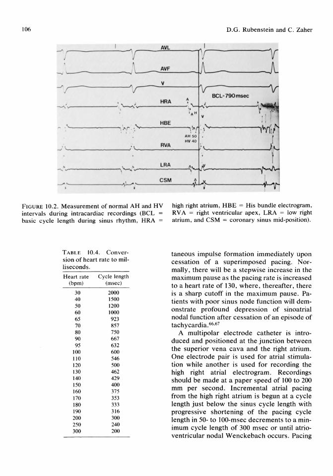
106 D.G. Rubenstein and C. Zaher
~ (---------1 ,...---.-:::;:.:...---""1"\ .---------1{
~ ~---------------~
..../' 1--------'"1 .-----=-------¥\ .-----------J\ BCL' 790msec --..... -----------~ ~ ..... ----------.
- . '-~----------. .'-...... ------.....
~ '-~---------~
--.. ' .... - -------.,"--.. ~ .... --------.;., --~~---------'.}'- .. '--------------1,~I'_-------O¥_""I,..., . ,
FIGURE 10.2. Measurement of normal AH and HV intervals during intracardiac recordings (BCL basic cycle length during sinus rhythm, HRA =
high right atrium, HBE = His bundle electrogram, RV A = right ventricular apex, LRA = low right atrium, and CSM = coronary sinus mid-position).
TABLE 10.4. Conver-sion of heart rate to mil-liseconds.
Heart rate Cycle length (bpm) (msec)
30 2000 40 1500 50 1200 60 1000 65 923 70 857 80 750 90 667 95 632
100 600 110 546 120 500 130 462 140 429 150 400 160 375 170 353 180 333 190 316 200 300 250 240 300 200
taneous impulse formation immediately upon cessation of a superimposed pacing. Normally, there will be a stepwise increase in the maximum pause as the pacing rate is increased to a heart rate of 130, where, thereafter, there is a sharp cutoff in the maximum pause. Patients with poor sinus node function will demonstrate profound depression of sinoatrial nodal function after cessation of an episode of tachycardia. 66,67
A multipolar electrode catheter is introduced and positioned at the junction between the superior vena cava and the right atrium. One electrode pair is used for atrial stimulation while another is used for recording the high right atrial electrogram. Recordings should be made at a paper speed of 100 to 200 mm per second. Incremental atrial pacing from the high right atrium is begun at a cycle length just below the sinus cycle length with progressive shortening of the pacing cycle length in 50- to 100-msec decrements to a minimum cycle length of 300 msec or until atrioventricular nodal Wenckebach occurs. Pacing

10. Clinical Electrophysiology
is maintained at each cycle length for 30 to 60 seconds. There are 45- to 60-second rest intervals between pacing runs.
Sinus node recovery time is measured as the time to recovery of the sinus node function after termination of overdrive suppression during atrial pacing (Fig 10.3A,B). It is the interval in milliseconds from the last paced high right atrium (HRA) complex to the onset of the first spontaneous HRA complex. This measurement is the total SNRT. In addition to recording the first postpacing cycle, one should also measure additional postpacing cycle lengths, as these cycles may be abnormal
v,
I
AVF
107
(secondary pauses). Sinus node recovery time must be interpreted in relation to SCL, because SNRT will, for example, normally be longer with slower heart rates. Therefore, corrected sinus node recovery time (CSNRT) is often used and can be calculated by subtracting the sinus cycle length from the longest sinus node recovery time. A value of more than 550 msec is considered abnormal.
Sinoatrial Conduction Time
This represents the time it takes the electrical impulse leaving the sinus node to conduct
~---'---'IV---
S A S A SNRT' 1020 msec ~
f~----------~ r~----~----- r--------------------~. Jr-----------
HRA
HBE
.'r--rtr-
/\,...1 , ,~
... ..i .- J.,--
..... ~ . - " , -.\ ........ ,
\. CSD , , .\. tv • \
\
,
" ,
H V
. - -..! A"-,,.j 'r r ,., v v .
S A H" A. H I
- If f-V ' ' ~------""'I'd.,.;., ,,----I ~ f .(
.......-'\,. ------------------------------------~~.------~-------~
----------""V~-~ . J_ SHIn'- 2800fMiK ,
~----------------------------------,~~------~;.'---~~~~\~,~ , . . " , . -... ,
. \. '--------------------------------......;... .. . ~ ~ "';I~' I~. _~-...... \ ;'"
I • .. ." I , •• I
"'---------------------------------~. " \.. ; ~ ...=-- '-----oi "-. '. . "I • " I -----------------------------------....... ~____l_______.....-
FIGURE 10.3. A) Normal total sinus node recovery time (TSNRT) after termination of atrial pacing (S]S]). B) Markedly abnormal sinus node recovery time (SNRT) of 2800 msec after termination of
atrial pacing (S]S]) (HRA = high right atrium, HBE = His bundle electrogram, CSD = coronary sinus distal, and CSP = coronary sinus proximal).
A
B
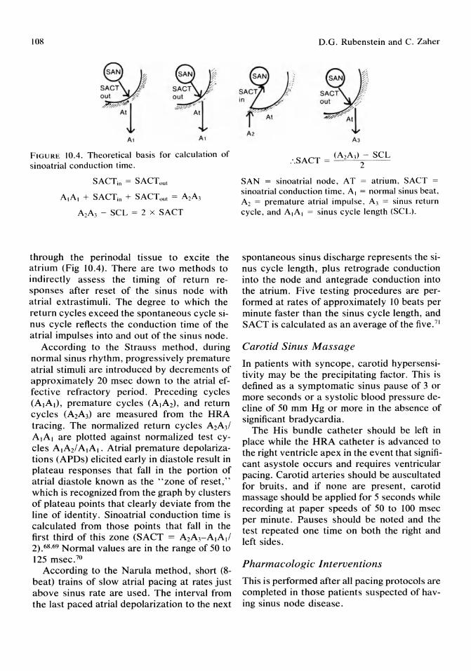
108
FIGURE 10.4. Theoretical basis for calculation of sinoatrial conduction time.
SACTin = SACTout
AlAI + SACTin + SACTout = A2A}
A2A3 - SCL = 2 x SACT
through the peri nodal tissue to excite the atrium (Fig 10.4). There are two methods to indirectly assess the timing of return responses after reset of the sinus node with atrial extrastimuli. The degree to which the return cycles exceed the spontaneous cycle sinus cycle reflects the conduction time of the atrial impulses into and out of the sinus node.
According to the Strauss method, during normal sinus rhythm, progressively premature atrial stimuli are introduced by decrements of approximately 20 msec down to the atrial effective refractory period. Preceding cycles (AlAI), premature cycles (AIA2)' and return cycles (A2A3) are measured from the HRA tracing. The normalized return cycles A2A3/ AlAI are plotted against normalized test cycles AIA2/ AlAI. Atrial premature depolarizations (APDs) elicited early in diastole result in plateau responses that fall in the portion of atrial diastole known as the "zone of reset," which is recognized from the graph by clusters of plateau points that clearly deviate from the line of identity. Sinoatrial conduction time is calculated from those points that fall in the first third of this zone (SACT = A2ArA,A,/ 2).68,69 Normal values are in the range of 50 to 125 msec. 70
According to the N arula method, short (8-beat) trains of slow atrial pacing at rates just above sinus rate are used . The interval from the last paced atrial depolarization to the next
D.G. Rubenstein and C. Zaher
e SACT :,;. out '-f'
-4~1 A2
A3
SAN = sinoatrial node, AT = atrium, SACT =
sinoatrial conduction time, AI = normal sinus beat, A2 = premature atrial impulse , A3 = sinus return cycle, and AlAI = sinus cycle length (SCL).
spontaneous sinus discharge represents the sinus cycle length, plus retrograde conduction into the node and antegrade conduction into the atrium. Five testing procedures are performed at rates of approximately 10 beats per minute faster than the sinus cycle length, and SACT is calculated as an average of the five.71
Carotid Sinus Massage
In patients with syncope, carotid hypersensitivity may be the precipitating factor. This is defined as a symptomatic sinus pause of 3 or more seconds or a systolic blood pressure decline of 50 mm Hg or more in the absence of significant bradycardia.
The His bundle catheter should be left in place while the HRA catheter is advanced to the right ventricle apex in the event that significant asystole occurs and requires ventricular pacing. Carotid arteries should be auscultated for bruits, and if none are present, carotid massage should be applied for 5 seconds while recording at paper speeds of 50 to 100 msec per minute. Pauses should be noted and the test repeated one time on both the right and left sides.
Pharmacologic Interventions
This is performed after all pacing protocols are completed in those patients suspected of having sinus node disease.

10. Clinical Electrophysiology
In patients with suspected sinus node disease by EPS, response to intravenous atropine can be measured. After .04 mg/kg of atropine is given intravenously, the heart rate should increase over control rate by at least 20%. Another expected change in normal sinoatrial node function includes a decrease in the "corrected sinus node recovery time" after atropine administration. Atropine can also be used to facilitate induction of supraventricular arrhythmias.
The conduction system is modified greatly by autonomic tone. In patients with abnormal sinus node function in whom autonomic tone is believed to playa role, the effects of the autonomic nervous system can be removed. The combination of atropine (.04 mg/kg) and propranolol (.2 mg/kg) are administered intravenously. The resulting sinus rate is called the intrinsic heart rate (lHR). Normal IHR is defined as 117. 1 - (.5 x age )for patients 15 to 70 years of age. An abnormal IHR will help to identify those patients with intrinsic abnormal sinus node function. 72 ,73
i HRA~
, A 5, A , \' --
HBE uo y vo y ,
, A H A H A
r L,~ r" ~\ r I ',,4 i \ I
. ,
109
Atrioventricular Conduction
Atrioventricular (A V) conduction is assessed in both sinus rhythm and during atrial pacing. An accurate His bundle recording allows localization of the level of A V block into A V nodal or infranodal. The AH interval (60 to 125 msec) represents primarily A V nodal conduction time and the HV interval (35 to 55 msec), infranodal conduction. The AH interval in the baseline state can be quite variable secondary to drugs or autonomic tone; however, the HV interval is generally fixed, The normal response to incremental atrial pacing is gradual AH prolongation with AV Wenckebach occurring at paced cycle lengths less than 430 msec (Fig 10,5). If no or minimal abnormalities are found in the baseline recordings, abnormal block occasionally can be precipitated by the stress of incremental atrial burst pacing or atrial pacing for 8 beats at a paced cycle length of 600 msec (S)S) with introduction of single premature atrial beats (S)S2)' Atrioventricular nodal, atrial, or occa-
PCl : 430msec
5,,( \A 5,
L-210 y uo y
f HtJ~~ .. ..
FIGURE 10.5, Demonstration of AV nodal Wencke- prolongation with ultimate failure of ventricular bach threshold with atrial pacing (S,S,) at a pacing capture. cycle length (PCL) of 430 msec. Note gradual AH
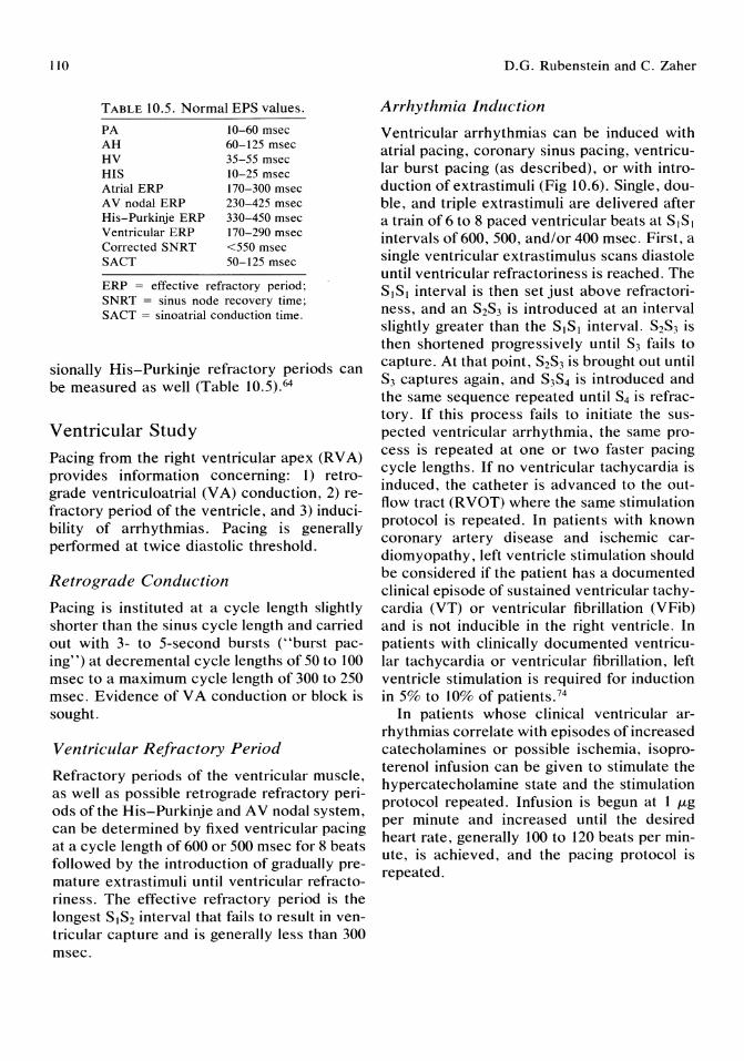
110
TABLE 10.5. Normal EPS values.
PA 10-60 msec AH 60-125 msec HV 35-55 msec HIS 10-25 msec Atrial ERP 170-300 msec AV nodal ERP 230-425 msec His-Purkinje ERP 330-450 msec Ventricular ERP 170-290 msec Corrected SNRT <550 msec SACT 50-125 msec
ERP = effective refractory period; SNRT = sinus node recovery time; SACT = sinoatrial conduction time.
sionally His-Purkinje refractory periods can be measured as well (Table 10.5).64
Ventricular Study
Pacing from the right ventricular apex (R V A) provides information concerning: 1) retrograde ventriculoatrial (V A) conduction, 2) refractory period of the ventricle, and 3) inducibility of arrhythmias. Pacing is generally performed at twice diastolic threshold.
Retrograde Conduction
Pacing is instituted at a cycle length slightly shorter than the sinus cycle length and carried out with 3- to 5-second bursts (' 'burst pacing") at decremental cycle lengths of 50 to 100 msec to a maximum cycle length of 300 to 250 msec. Evidence of V A conduction or block is sought.
Ventricular Refractory Period
Refractory periods of the ventricular muscle, as well as possible retrograde refractory periods of the His-Purkinje and A V nodal system, can be determined by fixed ventricular pacing at a cycle length of 600 or 500 msec for 8 beats followed by the introduction of gradually premature extrastimuli until ventricular refractoriness. The effective refractory period is the longest S]S2 interval that fails to result in ventricular capture and is generally less than 300 msec.
D.G. Rubenstein and C. Zaher
Arrhythmia Induction
Ventricular arrhythmias can be induced with atrial pacing, coronary sinus pacing, ventricular burst pacing (as described), or with introduction of extrastimuli (Fig 10.6). Single, double, and triple extrastimuli are delivered after a train of 6 to 8 paced ventricular beats at SIS] intervals of 600, 500, and/or 400 msec. First, a single ventricular extrastimulus scans diastole until ventricular refractoriness is reached. The SIS] interval is then set just above refractoriness, and an S2S3 is introduced at an interval slightly greater than the SIS] interval. S2S3 is then shortened progressively until S3 fails to capture. At that point, S2S3 is brought out until S3 captures again, and S3S4 is introduced and the same sequence repeated until S4 is refractory. If this process fails to initiate the suspected ventricular arrhythmia, the same process is repeated at one or two faster pacing cycle lengths. If no ventricular tachycardia is induced, the catheter is advanced to the outflow tract (RVOT) where the same stimulation protocol is repeated. In patients with known coronary artery disease and ischemic cardiomyopathy, left ventricle stimulation should be considered if the patient has a documented clinical episode of sustained ventricular tachycardia (VT) or ventricular fibrillation (VFib) and is not inducible in the right ventricle. In patients with clinically documented ventricular tachycardia or ventricular fibrillation, left ventricle stimulation is required for induction in 5% to 10% of patients.74
In patients whose clinical ventricular arrhythmias correlate with episodes of increased catecholamines or possible ischemia, isoproterenol infusion can be given to stimulate the hypercatecholamine state and the stimulation protocol repeated. Infusion is begun at 1 ILg
per minute and increased until the desired heart rate, generally 100 to 120 beats per minute, is achieved, and the pacing protocol is repeated.

10. Clinical Electrophysiology III
s
V1
AVL
A
--~--~--- ... - --,----_.------.'---- --\-..-----,"----'
FIGURE 10.6. Induction of ventricular tachycardia (VT). A) Introduction of two premature ventricular complexes (S2S}) fails to initiate VT. B) Introduc-
Specific Electrophysiologic Study Protocols
Syncope
In evaluating syncope, a complete EPS should be performed, including assessment of sinus node function, A V conduction, response to programmed atrial and ventricular stimulation, effects of drugs, and carotid sinus massage.
Sinus Node Function
Sinus node recovery time and SACT should be performed. If these results are abnormal, autonomic denervation with atropine plus pro-
tion of three premature ventricula r beats (S2S, S4) induces sustained VT of cycle length 250 msec .
pranolol should be performed to determine if the abnormality is primary or secondary to the influence of autonomic tone .
Atrioventricular Conduction
His bundle electrograms should be measured both in sinus rhythm and with atrial stimulation. Possible infranodal block can be evaluated with atrial pacing.
Refractory Periods
Duration of the atrial, AV nodal, and ventricular refractory periods are measured but generally do not aid in determining an etiology for syncope.
B
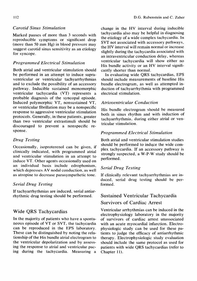
112
Carotid Sinus Stimulation
Marked pauses of more than 3 seconds with reproducible symptoms or significant drop (more than 50 mm Hg) in blood pressure may suggest carotid sinus sensitivity as an etiology for syncope.
Programmed Electrical Stimulation
Both atrial and ventricular stimulation should be performed in an attempt to induce supraventricular or ventricular tachyarrhythmias and to exclude the possibility of an accessory pathway. Inducible sustained monomorphic ventricular tachycardia (VT) represents a probable diagnosis of the syncopal episode. Induced polymorphic VT, nonsustained VT, or ventricular fibrillation may be a nonspecific response to aggressive ventricular stimulation protocols. Generally, in these patients, greater than two ventricular extrastimuli should be discouraged to prevent a nonspecific response.
Drug Testing
Occasionally, isoproterenol can be given, if clinically indicated, with programmed atrial and ventricular stimulation in an attempt to induce VT. Other agents occasionally used on an individual basis include edrophonium, which depresses A V nodal conduction, as well as atropine to decrease parasympathetic tone.
Serial Drug Testing
If tachyarrhythmias are induced, serial antiarrhythmic drug testing should be performed.
Wide QRS Tachycardias
In the majority of patients who have a spontaneous episode of VT or SVT, the tachycardia can be reproduced in the EPS laboratory. These can be distinguished by noting the relationship of the His bundle atrial electrogram to the ventricular depolarization and by assessing the response to atrial and ventricular pacing during the tachycardia. Measuring a
D.G. Rubenstein and C. Zaher
change in the HV interval during inducible tachycardia also may be helpful in diagnosing the etiology of a wide complex tachycardia. In SVT not associated with accessory pathways, the HV interval will remain normal or increase slightly during the tachycardia associated with an intraventricular conduction delay, whereas ventricular tachycardia will show either no His bundle activity or an HV interval significantly shorter than normal.
In evaluating wide QRS tachycardias, EPS should include measurements of baseline His bundle electrogram, as well as attempted induction of tachyarrhythmia with programmed electrical stimulation.
Atrioventricular Conduction
His bundle electrogram should be measured both in sinus rhythm and with induction of tachyarrhythmia, during either atrial or ventricular stimulation.
Programmed Electrical Stimulation
Both atrial and ventricular stimulation studies should be performed to indl.\ce the wide complex tachycardia. If an accessory pathway is strongly suspected, a W-P-W study should be performed.
Serial Drug Testing
If clinically relevant tachyarrhythmias are induced, serial drug testing should be performed.
Sustained Ventricular Tachycardia
Survivors of Cardiac Arrest
Ventricular arrhythmias can be induced in the electrophysiology laboratory in the majority of survivors of cardiac arrest unassociated with an acute myocardial infarction. Electrophysiologic study can be used for these patients to judge the efficacy of antiarrhythmic therapy. Electrophysiologic study evaluation should include the same protocol as used for patients with wide QRS tachycardias (refer to Chapter 11).
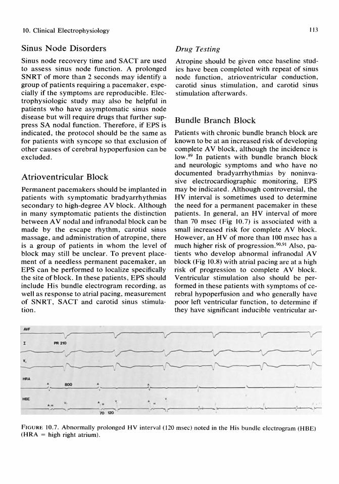
10. Clinical Electrophysiology
Sinus Node Disorders
Sinus node recovery time and SACT are used to assess sinus node function. A prolonged SNRT of more than 2 seconds may identify a group of patients requiring a pacemaker, especially if the symptoms are reproducible. Electrophysiologic study may also be helpful in patients who have asymptomatic sinus node disease but will require drugs that further suppress SA nodal function. Therefore, if EPS is indicated, the protocol should be the same as for patients with syncope so that exclusion of other causes of cerebral hypoperfusion can be excluded.
Atrioventricular Block
Permanent pacemakers should be implanted in patients with symptomatic bradyarrhythmias secondary to high-degree A V block. Although in many symptomatic patients the distinction between A V nodal and infranodal block can be made by the escape rhythm, carotid sinus massage, and administration of atropine, there is a group of patients in whom the level of block may still be unclear. To prevent placement of a needless permanent pacemaker, an EPS can be performed to localize specifically the site of block. In these patients, EPS should include His bundle electrogram recording, as well as response to atrial pacing, measurement of SNRT, SACT and carotid sinus stimulation.
NlF
'/ '/ '/ I PI! 210
113
Drug Testing
Atropine should be given once baseline studies have been completed with repeat of sinus node function, atrioventricular conduction, carotid sinus stimulation, and carotid sinus stimulation afterwards.
Bundle Branch Block
Patients with chronic bundle branch block are known to be at an increased risk of developing complete A V block, although the incidence is low.89 In patients with bundle branch block and neurologic symptoms and who have no documented bradyarrhythmias by noninvasive electrocardiographic monitoring, EPS may be indicated. Although controversial, the HV interval is sometimes used to determine the need for a permanent pacemaker in these patients. In general, an HV interval of more than 70 msec (Fig 10.7) is associated with a small increased risk for complete A V block. However, an HV of more than 100 msec has a much higher risk of progression.90,91 Also, patients who develop abnormal infranodal A V block (Fig 10.8) with atrial pacing are at a high risk of progression to complete A V block. Ventricular stimulation also should be performed in these patients with symptoms of cerebral hypoperfusion and who generally have poor left ventricular function, to determine if they have significant inducible ventricular ar-
v '/
--------"~ ~ ~v____"~v Y,
-----~.~.~.~~"--HR"
800' • __________ --" ______ ' .r-, ------'------- ..... ------'----
HBE ~
• M ' ---~ _--_~ _~ ----,."..----., ,_---• .' __ ... ~-----:.~ 1.-' ---'""'\1'------
FIGURE 10.7. Abnormally prolonged HV interval (120 msec) noted in the His bundle electrogram (HBE) (HRA = high right atrium).
114 D.G. Rubenstein and C. Zaher
AVl
-------~----------~----------------------~----------~--------------------~ I
~~. PCl - 600msec .m:
~~ ~~ . J ~A ~~ ~~ ~~ ~~
~~~---------~~~---------~r~---------'
y y
HBE v SAM SAH SAM SAH . ,
-_.: ............. ',"""---' ,'-.r-r----" .- ,'"'----.• , .... r,.Mi...--_.,"--' ......... _--~r------, ..... '" , , I
FIGURE 10.8. Demonstration of infra-Hisian block with atrial pacing (AlAI) at a pacing cycle length (PCL) of 600 msec.
rhythmias. Therefore, if EPS is indicated, the protocol should be the same as for patients with syncope.
See Chapter 12 for a discussion of supra ventricular tachycardias and accessory pathways.
Summary
Electrophysiology studies represent a sophisticated and highly technical approach to a variety of clinical conditions involving arrhythmias and conduction disturbances. They have helped make a significant impact on morbidity and mortality of properly selected patients. Clinical EPS continues to evolve, and with the advent of newer treatment modalities, such as surgery, ablation, and antitachycardia devices, it promises to assume an ever increasing role in the management of such patients.
References
1. Alanis J, Gonzalez H, Lopez E: Electrical activity of the bundle of His. J Physiol 1958; 142: 127.
2. Giraud G, Peuch P, Latour H: L'activite eJectrique physiologique du noeud de Tawara et du
Faisceveau de His chez l'homme. Acad Nat Med 1960; May:363.
3. Sherlag BJ, Lau SH, Helfant RH, et al: Catheter technique for recording His bundle activity in man. Circulation 1969; 39: 13.
4. Wellens HJJ: Electrical stimulation of the heart in the study and treatment of tachycardias. Leiden, Stenfert Kroese, 1971.
5. Wellens HJJ, Lie KI, Durrer D: Further observation on ventricular tachycardia as studied by electrical stimulation of the heart: Chronic recurrent ventricular tachycardia and ventricular tachycardia during acute myocardial infarction. Circulation 1971; 43: 15.
6. Health and Public Policy Committee, American College of Physicians: Diagnostic endocardial electrical recording and stimulation. Ann Intern Med 1984; 100:452.
7. State of the AA Consensus Conference on Electrophysiologic Testing in the diagnosis and treatment of patients with cardiac arrhythmias. Circulation 1987; 75(suppl):4.
8. Kastor JA, Horowitz LN, Hanken AH, et al: Clinical electrophysiology of ventricular tachycardia. N Eng/ J Med 1981; 304: 1004.
9. Rae AP, Greenspan AM, Spielman SR, et al: Antiarrhythmic drug efficacy for ventricular tachyarrhythmias associated with coronary artery disease as assessed by electrophysiologic studies. Am J Cardiol1985; 55:1494.
10. Mason JW, Winkle RA: Accuracy of ventricu-

10. Clinical Electrophysiology
lar tachycardic induction study for predicting long-term efficacy and inefficacy of antiarrhythmic drugs. N Engl J Med 1980; 303:1073.
11. Ruskin IN, Dimarco JP, Garan H: Out-of-hospital cardiac arrest. Electrophysiologic observations and selection of long-term antiarrhythmic therapy. N Engl J Med 1980; 303:607.
12. Horowitz LN, Josephson ME, Farshidi A, et al: Recurrent sustained VT. 3. Role of the electrophysiological study in selection of antiarrhythmic regimens. Circulation 1978; 58:986.
13. Horowitz LN, Spielman SR, Greenspan AM, et al: Role of programmed stimulation in assessing vulnerability to ventricular arrhythmias. Am Heart J 1982; 103:604.
14. Liberthson RR, Nagel EL, Hirshman JC, et al: Prehospital ventricular defibrillation: Prognosis and follow-up course. N Engl J Med 1974; 291:317.
15. Baum RS, Alvarez H, Cobb LA: Survival after rescitation from out-of-hospital ventricular fibrillation. Circulation 1974; 50: 1231.
16. Kehoe RF, Moran JM, Zheutlin T, et al: E1ectrophysiologic study to direct therapy in survivors of prehospital ventricular fibrillation. Am J Cardiol 1982; 49:928.
17. Morady F, Scheinman MM, Hess DS, et al: Electrophysiologic testing in the management of survivors of out-of-hospital cardiac arrest. Am J Cardiol 1983; 51:85.
18. Roy D, Waxman HL, Kienzle MG, et al: Clinical characteristics and long-term follow-up in 119 survivors of cardiac arrest: Relation to inducibility at electrophysiologic testing. Am J Cardiol 1983; 52:969.
19. Benditt DG, Benson DW, Klein GJ, et al: Prevention of recurrent sudden cardiac arrest: Role of provocative electropharmacologic testing. J Am Call Cardiol 1983; 2:418.
20. Skale BT, Miles WM, Heger JJ, et al: Survivors of cardiac arrest: Prevention of recurrence by drug therapy as predicted by electrophysiologic testing or electrocardiographic monitoring. Am J Cardiol 1986; 57: 113.
21. Wilber DJ, Garan H, Kelly E, et al: Out-ofhospital arrest: Role of electrophysiologic testing in prediction oflong-term outcome. Circulation 1986; 74(suppl 11):11-482.
22. Zheutlin TE, Steinmann R, Summers C, et al: Long-term outcome in survivors of cardiac arrest with noninducible ventricular tachycardia during programmed stimulation. Circulation 1984; 70(suppl 11):11-399.
115
23. Morady F, DiCarlo L, Winston S, et al: Clinical features and prognosis of patients with out of hospital cardiac arrest and a normal electrophysiologic study. J Am Call Cardiol 1984; 4:39.
24. Wellens HJ, Brugada P, Stevensen WG: Programmed electrical stimulation of the heart in patients with life-threatening ventricular arrhythmias. Circulation 1985; 72: 1.
25. Echt DS, Armstrong K, Schmidt P, et al: Clinical experience, complications, and survival in 70 patients with the automatic implantable cardioverter/defibrillator. Circulation 1985; 71:289.
26. Gallagher JJ, Pritchett ELC, Sealy WC, et al: The pre-excitation syndrome. Prog Cardiovasc Dis 1978; 20:285.
27. Wellens HJJ, Durrer D: Wolff-Parkinson-White syndrome and atrial fibrillation. Am J Cardiol 1974; 34:777.
28. Castellanos A, Myerberg RJ, Craparo K, et al: Factors regulating ventricular rates during atrial flutter and fibrillation in pre-excitation (Wolff-Parkinson-White) syndrome. Br Heart J 1973; 35:811.
29. Sledge C, Scheinman M, Morady F, et al: Medical therapy in Wolff-Parkins on-White syndrome and atrial fibrillation. Circulation 1982; 66(suppllI):II-269.
30. Bavernfeind RA, Swiryn SP, Strasberg B, et al: Electrophysiologic drug testing in prophylaxis of sporadic paroxysmal atrial fibrillation: Technique, applications, and efficacy in severely sumptomatic pre-excitation patients. Am Heart J 1982; 103:941.
31. Wu D, Denes P, Amat-y-Leon F, et al: Clinical, electrocardiographic, and electrophysiologic observations in patients with paroxysmal supraventricular tachycardia. Am J Cardiol1978; 41:1045.
32. Wu D, Amat-y-Leon F, Simpson RJ Jr, et al: Electrophysiological studies with mUltiple drugs in patients with atrioventriculoreentrant tachycardias utilizing an extranodal pathway. Circulation 1977; 56:727.
33. Wellens HJJ, Bar FWHM, Lie KI: The value of the electrocardiogram in the differential diagnosis of a tachycardia with a widened QRS complex. Am J Med 1978; 64:27.
34. Wellens HJJ, Bar FW, Vanagt EJ, et al: The differentiation between ventricular tachycardia and supraventricular tachycardia with aberrant conduction. The value of 12-lead electrocardiogram, in Wellens HJJ, Kulbertus HE (eds):
116
What's New in Electrocardiography? The Hague, Martinus Nijhoff, 1981, p 184.
35. Coumel P, Leclercq JF, Attuel P, et al: The QRS morphology in postmyocardial infarction ventricular tachycardia. A study of 100 tracings compared with 70 cases of idiopathic ventricular tachycardia. Eur Heart J 1984; 5:792.
36. Josephson ME, Horowitz LN, Waxman HL: Sustained ventricular trachcardia: Role of 12-lead electrocardiogram in localizing site of origin. Circulation 1981; 64:257.
37. Josephson ME, Horowitz LN, Farshidi A, et al: Recurrent sustained ventricular tachycardia. 2. Endocardial mapping. Circulation 1978; 57:440.
38. Silverstein MD, Singer DE, Mulley AG, et al: Patients with syncope admitted to medical intensive care units. J Am Med Assoc 1982; 248:1185.
39. Lipsitz LA: Syncope in the elderly. Ann Intern Med 1983; 99:92.
40. DiMarco JP, Garan H, Hawthorne JW, et al: Intracardiac electrophysiologic techniques in recurrent syncope of unknown cause. Ann Intern Med 1981; 95:542.
41. DiMarco JP, Garan H, Ruskin IN: Approach to the patient with recurrent syncope of unknown cause. Mod Concepts Cardiovasc Dis 1983; 52: 11.
42. Hess DS, Morady F, Scheinman MM: Electrophysiologic testing in the evaluation of patients with syncope of undetermined origin. Am J Cardio! 1982; 50: 1309.
43. Morady F, Shen E, Schwartz A, et al: Longterm follow-up of patients with recurrent unexplained syncope evaluated by electrophysiologic testing. J Am Coll Cardio! 1983; 2: 1053.
44. Morady F, Higgins J, Peters RW, et al: Electrophysiologic testing in bundle branch block and unexplained syncope. Am J Cardiol 1984; 54:587.
45. Akhtar M, Shenasa M, Denker S, et al: Role of cardiac electrophysiologic studies in patients with unexplained recurrent syncope. PACE 1983; 6:192.
46. Gulamhusein S, Naccarelli GV, Ko PT, et al: Value and limitations of clinical electrophysiologic study in assessment of patients with unexplained syncope. Am J Med 1982; 73:700.
47. Boudoulas H, Geleris P, Schanl SF, et al: Comparison between e1ectrophysiologic studies and ambulatory monitoring in patients with syncope. J Electrocardiol 1983; 16:91.
D.G. Rubenstein and C. Zaher
48. Ezri ME, Lerman BB, Marchlinski FE, et al: Electrophysiologic evaluation of syncope in patients with bifasicular block. Am Heart J 1982; 106:693.
49. Westvecr DC, Stewart J, Vandam D, et al: The role of electrophysiologic studies in the evaluation of recurrent, unexplained syncope. Cardiovasc Rev Rep 1984; 5:770.
50. Olshansky B, Mazuz M, Martins JB: Significance of inducible tachycardia in patients with syncope of unknown origin: A long-term follow-up. JAm Coll Cardiol1985; 5:216.
51. Doherty JV, Pembrook-Rogers D, Grogan EW, et al: Electrophysiologic evaluation and followup characteristics of patients with recurrent unexplained syncope and presyncope. Am J Cardio!1985; 55:703.
52. Denes P, Ezri MD: The role of electrophysiologic studies in the management of patients with unexplained syncope. PACE 1985; 8:424.
53. Hammer A, Vohra J, Hunt D, et al: Prediction of sudden death by electrophysiologic studies in high-risk patients surviving acute myocardial infarction. Am J Cardiol 1982; 50:223.
54. Richards DA, Cody DU, Denniss AR, et al: Ventricular electrical instability: A predictor of death after myocardial infarction. Am J Cardiol 1983; 51:75.
55. Marchlinkski FE, Buxton AE, Waxman HL, et al: Identifying patients at a risk of sudden death after myocardial infarction: Value of the response for programmed stimulation, degree of ventricular ectopic activity, and severity of left ventricular dysfunction. Am J Cardio! 1983; 52:1190.
56. Santarelli P, Bellucci F, Luperfido F, et al: Ventricular arrhythmia induced by programmed ventricular stimulation after acute myocardial infarction. Am J Cardiol 1985; 55:391.
57. Roy D, Marchand E, Theroux P, et al: Programmed ventricular stimulation in survivors of an acute myocardial infarction. Circulation 1985; 72:487.
58. Gonzales R, Arriagada D, Corbalan R, et al: Programmed electrical stimulation of the heart does not help to identify patients at high-risk postmyocardial infarction. Circulation 1984; 70(suppl 11):11-19.
59. Buxton AE: Nonsustained ventricular tachycardia in patients with coronary artery disease: Role of electrophysiologic study. Circulation 1987; 75(6):1178-1185.
10. Clinical Electrophysiology
60. Wellens HJJ, Farre J, Bar FW: Wolff-Parkinson-White syndrome. Value and limitations of programmed electrical stimulation, in Narula as (ed): Cardiac Arrhythmias. Baltimore, Williams & Wilkens, 1979, p 589.
61. Horowitz LN: Safety of electrophysiology studies. Learning Center Highlights 1986; 2(2):5.
62. Horowtiz LN, Kay HR, Kutalek SP, et al: Risks and complications of clinical cardiac electrophysiologic studies: A prospective analysis of 1000 consecutive patients. J Am Coli Cardiol 1987; 9:1261.
63. Gettes LS, Zipes DP, Gillette PC, et al: Personnel and equipment required for electrophysiologic testing. Report of the Committee on Electrocardiography and Cardiac Electrophysiology, Council on Clinical Cardiology, The American Heart Association. AHA files no. 72-023-A.
64. Josephson ME, Seides SF: Clinical Cardiac Electrophysiology. Philadelphia, Lea & Febiger, 1979.
65. Reddy CP, Damato AN, Akhtar M, et al: Time dependent changes in the functional properties of the atrioventricular conduction system in man. Circulation 1975; 52:1012.
66. Mandel WG, Hayakawa H, Danzig R, et al: Evaluation of sinoatrial node function in man by overdrive suppression. Circulation 1971; 44:59.
67. Narula as, Samet P, Xavier RP: Significance of the sinus node recovery time. Circulation 1972; 45:140.
68. Kerr CR, Grant AO, Wenger TL, et al: Sinus node dysfunction, in Zipes DP (ed): Cardiology Clinics: Arrhythmias II, Vol 1, No.2. Philadelphia, W.B. Saunders Co., 1983, pp 187-207.
69. Strauss HC, Bigger JT Jr, Saroff AL, et al: Electrophysiologic evaluation of sinus node function in patients with sinus node dysfunction. Circulation 1976; 53:763.
70. Breithardt G, Seipel L, Loogen F: Sinus node recovery time and calculated sinoatrial conduction time in normal subjects and patients with sinus node dysfunction. Circulation 1977; 56:43.
71. Narula as, Shartha N, Vasquez M, et al: A new method for measurement of sinoatrial conduction time. Circulation 1978; 58:706.
72. Jordan JL, Yamaguchi I, Mandel WJ: Studies on the mechanism of sinus node dysfunction in
117
the sick sinus syndrome. Circulation 1978; 57:217.
73. Narula as, Narula JT: Various techniques for electrophysiological evaluation of sinus node function, in Puel P (ed): Troubles du Rhythme et Electrostimulation Toulouse, Societe de la Nouvelle Imprimerie Fournie, 1978, p 15.
74. Robertson JF, Cain ME, Horowitz LN, et al: Anatomic and electrophysiologic correlates of ventricular tachycardia requiring left ventricular stimulation. Am J Cardiol1981; 48:263.
75. Freedman RA, Swerdlow DC, Echt DS, et al: Facilitation of ventricular tachyarrhythmia induction by isoproterenol. Am J Cardiol 1984; 54:765.
76. Weber H, Schmitz L: Catheter technique for closed-chest ablation of an accessory atrioventricular pathway. N Engl J Med 1983; 308:653.
77. Morady F, Scheinman MM: Transvenous catheter ablation of a posteroseptal accessory pathway in a patient with Wolff-Parkins on-White syndrome. N Engl J Med 1984; 310:705.
78. Morady F, Scheinman MM, Winston SA, et al: Efficacy and safety of trans catheter ablation of posteroseptal accessory pathways. Circulation 1985; 72:170.
79. Ward DE, Camm AJ: Treatment of tachycardias associated with the Wolff-Parkinson-White syndrome by trans venous electrical ablation of accessory pathways. Br Heart J 1985; 53:64.
80. Gallagher J, Svenson RH, Kassell JH, et al: Catheter technique for closed-chest ablation of the atrioventricular conduction system. N Engl J Med 1982; 4: 194.
81. Sharma AD, Klein GJ, Guiraudon GM, et al: Atrial fibrillation in the Wolff-Parkinson-White syndrome: Incidence after surgical ablation of the accessory pathway. Circulation 1985; 72: 161.
82. Reiffel JA, Bigger JT Jr, Cramer M, et al: Ability of Holter electrocardiographic recording and atrial stimulation to detect sinus nodal dysfunction in symptomatic and asymptomatic patients with sinus bradycardia. Am J Cardiol 1977; 40:189.
83. Strauss HC, Bigger JT Jr, Saroff AL, et al: Electrophysiologic evaluation of sinus node dysfunction. Circulation 1976; 53:763.
84. Narula as, Scherlag BJ, Samet P, et al: Atrioventricular block: Localization and classification by His bundle recordings. Am J Med 1971; 50:146.
85. Narula as, Scherlag BJ, Javier RR: Analysis of

118
the A V conduction defect in complete heart block utilizing His bundle electrograms. Circulation 1970 1970; 41:367.
86. Peuch P, Grolleau R, Guimond C: Incidence of different types of A V block and their localization by His Bundle recording, in Wellens HJJ, Lie K, Janse M, (eds): The Conduction System of the Heart. Leiden, Stenfert Kroese, 1976, p 467.
87. Dhingra RC, Wyndham C, Amat-y-Leon F, et al: Incidence and site of atrioventricular block in patients with chronic bifasicular block. Circulation 1979; 59:238.
88. Ezri M, Lerman BB, Marchlinski FE, et al:
D.G. Rubenstein and C. Zaher
Electrophysiologic evaluation of syncope in patients with bifasicular block. Am Heart J 1983; 106:693.
89. Kulbertus HE: Reevaluation of the prognosis of patients with LAD-RBB. Am Heart J 1976; 92:665.
90. Dhingra RC, Denes P, Wu D, et al: Prospective observations in patients with chronic bundle branch block and marked H -V prolongation. Circulation 1976; 53:600.
91. Me Annuity JH, Rahimtoola SH, Murphy ES, et al: A prospective study of sudden death in "high-risk" bundle branch block. N Engl J Med 1978; 299:209.
11 Invasive Electrophysiologic Studies in the Evaluation and Treatment of Patients with Ventricular Arrhythmias* Nicholas J. Stamato and Mark E. Josephson
Introduction
Invasive electrophysiologic studies have been used in the evaluation and treatment of patients with ventricular arrhythmias since Wellens et all reported their initial experience in 1972. The application of this procedure to the investigation of the mechanism of sustained ventricular tachycardia has led to major advances in our understanding of this arrhythmia. 2-5 A better understanding of the pathophysiologic basis of sustained uniform ventricular tachycardia has led to the development of treatment strategies, which include the use of programmed electrical stimulation to select pharmacologic agents, surgical resection, and catheter ablative techniques to destroy and/or isolate the substrate of the arrhythmia.6-9
Although much has been learned since 1972, there remain many questions and controversies regarding the use of programmed electrical stimulation in the evaluation and treatment of patients with ventricular arrhythmias. This procedure is clinically applicable to patients who present with recurrent sustained ventricular tachycardia,3 or with aborted sudden cardiac death in the absence of a new myocardial infarction 10 or in patients with recurrent syncope in whom a sustained ventricular tachy-
* Supported in part by grants HL00361 and HL24278 from the National Heart, Lung, and Blood Institute, Bethesda, MD, and grants from The American Heart Association, Southeastern Pennsylvania Chapter, Philadelphia, P A.
cardia is inducible. I I Whether programmed electrical stimulation can play a role in the evaluation and treatment of patients who are recently postmyocardial infarction l2 or in those presenting with asymptomatic nonsustained ventricular tachycardial3 remains to be proven and at present is an area of active investigation (Table 11.1).
This chapter reviews the technical and theoretical aspects of the performance of electrophysiologic studies in patients with ventricular arrhythmias (Table 11.2).
Technical Aspects
Personnel
As with any invasive medical procedure, the most important factor in the safe and successful performance of an electrophysiologic study is the ability and training of the physician performing and directing the study .14 The electrophysiologist performing clinical electrophysiologic studies should be well trained not only in the performance of these studies but also in the evaluation and treatment of patients with all types of cardiac arrhythmias. The American Heart Association 15 and the American College of Cardiology l6 have each issued reports on the recommended training of those cardiologists performing clinical cardiac electrophysiologic studies. Both have suggested that after the completion of 2 years of training in clinical cardiology, including experience in cardiac catheterization, a minimum of one and

120
TABLE 11.1. Indications for electrophysiologic testing in patients with ventricular arrhythmias.
Recurrent sustained ventricular tachycardia Cardiac arrest Recurrent syncope Before arrhythmia surgery Before catheter ablation Before placement of antitachycardia pacemakers or the
implantable defibrillator Nonsustained ventricular tachycardia in patients with
coronary artery disease (unciear) Prognostication postmyocardial infarction (unciear
unlikely to be of benefit)
preferably 2 years be spent training in -an active electrophysiologic laboratory under the supervision of a qualified clinical electrophysiologist. It is our belief that physicians responsible for electrophysiologic testing have the performance of these studies as their main responsibility.
The nursing and technical staff assisting at all electrophysiologic studies should be well versed in the performance and goals of each study. They should possess a high level of understanding of the physiology and pathophysiology of cardiac arrhythmias and the pharmacology of antiarrhythmic drugs. They should be familiar with the equipment used in the laboratory and especially well trained in the performance of cardioversion and cardiopulmonary resuscitation.
Electrophysiologic studies should be performed in hospitals having an anesthesiologist and cardiothoracic surgeon available if needed to help manage potential complications. Technical support should include those capable of maintenance of the fluoroscopy unit (which is
TABLE 11.2. Goals of electrophysiologic testing in patients with ventricular tachyarrhythmias.
Define the nature of the arrhythmia: sustained uniform ventricular tachycardia; polymorphic ventricular tachycardia; ventricular fibrillation
Define the substrate for the arrhythmia: normal, abnormal, or fractionated endocardial electrograms
Test the response to pharmacologic and pacing therapy Locate (map) the site of origin of a tachycardia before
surgical or catheter ablative therapy Prognosticate
N.J. Stamato and M.E. Josephson
preferably a C-arm unit) and a biomedical engineer capable of maintaining and checking the safety of the stimulating and recording equipment.
Equipment
The performance of clinical electrophysiologic studies requires a fluoroscopy unit, preferably a C-arm type, as well as a programmable stimulator and a recording system. The stimulator must be electrically isolated and be able to deliver precisely timed electrical impulses both synchronously and asynchronously. Whereas other adequate models are available, a custom designed unit manufactured by Bloom Associates, Ltd (Reading, PA) meets all these requirements.
The recording of the surface electrocardiogram along with intracardiac electrograms can be performed by a variety of commercially available systems. At least eight amplifiers allowing variable filtering and amplification are required. Electrograms are generally filtered to remove frequencies below 30 to 50 Hz and above 500 Hz. The recording system must provide hardcopy with a frequency response of greater than 500 Hz at a variable paper speed up to 250 mm per second. A tape recorder using either magnetic tape or recently available VHS tape is required to be able to recall events occurring during the study that were not recorded on hardcopy l4,15 (Fig 11.1).
A cardioverter-defibrillator capable of delivering at least 360 J must be present during all electrophysiologic studies. We have found that the use of anterioposterior pads (R-2 Corporation, Morton Grove, IL) aid in the rapid cardioversion of nontolerated arrhythmias and make the defibrillation procedure less traumatic. Also present in the electrophysiology laboratory should be equipment for full resuscitation, including drugs and materials for endotracheal intubation.
Catheters
A variety of electrode catheters exist that can be used for stimulation and recording. The woven Dacron catheter (USCI, Billerica, MA)
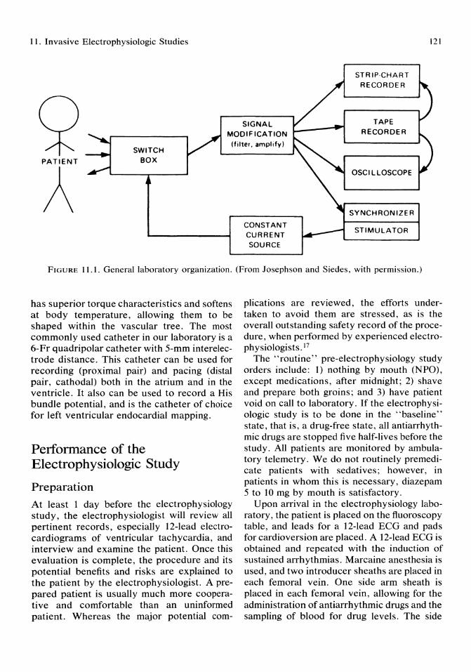
11. Invasive Electrophysiologic Studies
STRIP·CHART RECORDER
121
SWITCH BOX
SIGNAL MODIF ICATION
(filter. amplify)
TAPE RECORDER
PATIENT
A CONSTANT CURRENT SOURCE
OSCILLOSCOPE
SYNCHRONIZER
STIMULATOR
FIGURE 11.1. General laboratory organization. (From Josephson and Siedes, with permission.)
has superior torque characteristics and softens at body temperature, allowing them to be shaped within the vascular tree. The most commonly used catheter in our laboratory is a 6-Fr quadripolar catheter with 5-mm interelectrode distance. This catheter can be used for recording (proximal pair) and pacing (distal pair, cathodal) both in the atrium and in the ventricle. It also can be used to record a His bundle potential, and is the catheter of choice for left ventricular endocardial mapping.
Perl'ormance of the Electrophysiologic Study
Preparation
At least 1 day before the electrophysiology study, the electrophysiologist will review all pertinent records, especially 12-lead electrocardiograms of ventricular tachycardia, and interview and examine the patient. Once this evaluation is complete, the procedure and its potential benefits and risks are explained to the patient by the electrophysiologist. A prepared patient is usually much more cooperative and comfortable than an uninformed patient. Whereas the major potential com-
plications are reviewed, the efforts undertaken to avoid them are stressed, as is the overall outstanding safety record of the procedure, when performed by experienced electrophysiologists. 17
The "routine" pre-electrophysiology study orders include: 1) nothing by mouth (NPO), except medications, after midnight; 2) shave and prepare both groins; and 3) have patient void on call to laboratory. If the electrophysiologic study is to be done in the "baseline" state, that is, a drug-free state, all antiarrhythmic drugs are stopped five half-lives before the study. All patients are monitored by ambulatory telemetry. We do not routinely premedicate patients with sedatives; however, in patients in whom this is necessary, diazepam 5 to 10 mg by mouth is satisfactory.
Upon arrival in the electrophysiology laboratory, the patient is placed on the fluoroscopy table, and leads for a 12-lead ECG and pads for cardioversion are placed. A 12-lead ECG is obtained and repeated with the induction of sustained arrhythmias. Marcaine anesthesia is used, and two introducer sheaths are placed in each femoral vein. One side arm sheath is placed in each femoral vein, allowing for the administration of antiarrhythmic drugs and the sampling of blood for drug levels. The side
122
arm sheath is placed first and a catheter placed in it to ensure that the sheath is not punctured during the placement of the second sheath. If the left ventricle is to be mapped, a long side arm introducer sheath is placed via the right femoral artery using the Seldinger technique. The side arm allows the monitoring of arterial blood pressure.
The catheters are advanced to the heart under fluoroscopic guidance and positioned at the right ventricular apex, outflow tract, across the tricuspid valve to record a His potential, and in the high right atrium. The left ventricular catheter is passed retrograde up the aorta and across the aortic valve. Heparin is given after placement of the sheaths, 5,000 U is given if the left heart is entered and 2,500 U if only the right heart is entered. A continuous infusion of 1,000 U per hour is then maintained. Electrograms are recorded and pacing thresholds checked and should be less than 1 rnA at a pulse duration of 1 ms.
Stimulation Protocol
All studies performed for the evaluation of ventricular arrhythmias require the use of at least two right ventricular sites, usually the right ventricular apex and ouflow tract. 18 The use of one catheter is not optimal for pacing from two sites, for the protocol described below allows for induction of ventricular tachycardia using the least "vigorous" stimulation protocol.
Stimulation is performed at twice the diastolic threshold using alms pulse width. First at the right ventricular apex single ventricular extrastimuli are delivered after eight paced beats at a paced cycle length of 600 msec. A pause from 2 to 4 seconds is allowed between pacing runs. The coupling interval of the extrastimulus is placed in late diastole and decreased by 10 ms until either a sustained arrhythmia is induced or the local effective refractory period is reached (Fig 11.2). Single extrastimuli are then delivered from the outflow tract at a pacing drive of 600 ms in a similar manner. Next, single extrastimuli are delivered at a pacing drive of 400 ms, first at the apex and then at the outflow tract. If no
N.J. Stamato and M.E. Josephson
sustained arrhythmias are induced using single extrastimuli, double ventricular extrastimuli are delivered, alternating from the apex to the outflow tract, first at a pacing drive of 600 ms and then at a drive of 400 ms. The delivery of double extrastimuli is carried out with the initial coupling interval of the first extrastimulus set at 50 ms above the local effective refractory period at that pacing drive cycle length. The coupling interval between the first and second extrastimulus is initially set equal to that of the first. The coupling interval of the second extrastimulus is decreased in IO-ms steps until it is refractory, at which point the coupling interval of the first extrastimulus is decreased in 10-ms decrements until the second extrastimulus again captures. The coupling interval of the second extrastimulus is then decreased by 10 ms until it again fails to capture, at which point the coupling interval of the first extrastimulus is again decreased by 10 ms. This process is repeated until the first extrastimulus fails to capture. Triple ventricular extrastimuli are used if sustained arrhythmias have not been induced by single or double extrastimuli. 19 Triple extrastimuli are delivered in a manner similar to double extrastimuli, first at a pacing drive of 600 ms and then at 400 ms, alternating from apex to outflow tract. If sustained ventricular tachycardia or fibrillation has not been induced up to this point, rapid ventricular pacing is performed using synchronous bursts from 5 to 30 seconds at cycle lengths from 350 to 250 ms or until 2 : 1 capture is seen. If no sustained ventricular arrhythmias are induced to this point, single, double, and triple extrastimuli may be tried from both right ventricular sites during sinus rhythm. If the patient being studied has presented with recurrent sustained ventricular tachycardia, the above stimulation protocol will induce ventricular tachycardia in about 95% of patients. However, if it does not, stimulation from the left ventricular (Fig 11.3) or the delivery of quadruple extrastimuli may be required.
At the Hospital of the University of Pennsylvania, programmed stimulation has a sensitivity of 95% for sustained ventricular tachycardia using up to triple extrastimuli from two

LV-An : Al bgrdtr' I" LV-An ,~ ___ ~~ ~~
-~ Q~~ HBE ------1 : ~-----
IVA S\l ' J 700 f"!"" _______ 'l..i ·""""" '~\l.I ____ _
~V-~I......;~~-"'" 'V .....
LV-An \ .'!Il \ ~"MI • .i-' _____ --.~~y.. ___ _ T r •
c
FIGURE 11.2. Initiation of ventricular tachycardia with programmed stimulation. Panels A through C are arranged from top to bottom as follows: electrocardiographic leads II and VI; electrograms from the coronary sinus (CS), His bundle recording site (HBE), right ventricular apex (RV A), the border of a left ventricular aneurysm (LV-An, border) and in the aneurysm (LV-AN) and time lines (T) at 10 msec intervals. The left ventricular electrograms were recorded from a quadripolar catheter with a distal pair of electrodes in the left ventricular aneurysm and the proximal pair at its border. The ven-
tricles and atria were paced at a basic cycle length of 700 msec (SAl and VI-VI), and after every eighth paced complex progressively premature ventricular stimuli were delivered from the right ventricular apex (S and V2). In A and B ventricular extrastimuli delivered at 310 and 300 msec, respectively, produced fractionation of the electrogram in the aneurysm (arrows). At a critical coupling interval of 290 msec (C) fractionation of the electrogram in the aneurysm spanned diastole, and ventricular tachycardia ensued. (From Josephson et aI, with permission.)

124
A
FIGURE 11.3. Initiation of ventricular tachycardia by right or left ventricular stimulation. Both panels are organized from top to bottom: EeG leads I, a Vf, VIand electrograms from the high right atrium (HRA), His bundle (HBE), right ventricular apex (R V A), left ventricular apex (LV A). In panel A, two right ventricular premature stimuli (S2, S3) are introduced after the eighth RV paced ventricular
right ventricular sites, the use of left ventricular stimulation may add another 2%.20 The sensitivity of programmed stimulation in patients presenting with cardiac arrest is lower than in patients with recurrent sustained ventricular tachycardia. During the last 4 years, using the above protocol, 83% of patients presenting with cardiac arrest will have a ventricular arrhythmia induced. 19,20
The endpoints of stimulation are the completion of the protocol or the induction of a sustained ventricular arrhythmia, that is, one lasting more than 30 seconds or requiring termination in less time because of hemodynamic
N.J. Stamato and M.E. Josephson
paced complex (Sl), resulting in ventricular tachycardia, In panel B, two left ventricular stimuli (S2, S3) are delivered after the eighth LV paced complex (SI), resulting in ventricular tachycardia, Note that the coupling intervals of the premature stimuli are identicaL Stimulus artifacts are indicated by small arrows. (From Josephson et ai, with permission,)
collapse, All tachycardias are induced at least twice to ensure reproducibility, Studies of atrial, sinus node, atrioventricular node, and the His-Purkinje system can be carried out, when clinically indicated, before ventricular stimulation. 14
Left Ventricular Endocardial Activation Mapping
Whenever surgical therapy or catheter ablation are considered for ventricular tachycardia treatment, catheter activation mapping of the left ventricular endocardium during ventricu-
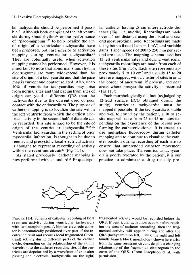
11. Invasive Electrophysiologic Studies
lar tachycardia should be performed if possible. 21 Although both mapping of the left ventricle during sinus rhythm22 or the performance of "pace-mapping"23 to help localize the site of origin of a ventricular tachycardia have been proposed, both are inferior to activation mapping during ventricular tachycardia. 24
They are potentially useful when activation mapping cannot be performed. However, it is important to note that abnormal sinus rhythm electrograms are more widespread than the site of origin of a tachycardia and that the pace map is current and contact related. Also, up to 10% of ventricular tachycardias may arise from normal sites and that pacing from sites of origin can yield a different QRS than the tachycardia due to the current used or poor contact with the endocardium. The purpose of catheter mapping is to localize the site within the left ventricle from which the earliest electrical activity in the second half of diastole can be recorded; this site is said to be the site of origin of the ventricular tachycardia. 21 ,25.26 Ventricular tachycardia, in the setting of prior myocardial infarction, is thought to be due to reentry and presystolic local electrical activity is thought to represent recording of activity within the reentrant circuit27 (Fig 11.4).
As stated previously, catheter mapping is now performed with a standard 6-Fr quadripo-
K><>=: '" I I
I
~ FIGURE 11.4. Schema of catheter recording of local reentrant activity during ventricular tachycardia with two morphologies. A bipolar electrode catheter is schematically positioned over part of the reentrant circuit and records local fragmented (Reentrant) activity during different parts of the cardiac cycle, depending on the relationship of the exiting wavefront to the catheter recording site. Ifthe ventricles are depolarized by a wavefront of exits after passing the electrode (tachycardia on the right)
125
lar catheter having .5 cm interelectrode distance (Fig 11.5, middle). Recordings are made over a 1 cm distance using the distal and second most proximal pole. Recordings are made using both a fixed (1 cm = 1 m V) and variable gains. Paper speeds of 200 to 250 mm per second are used. The mapping schema used has 12 left ventricular sites and during ventricular tachycardia recordings are made from each of these sites (Fig 11.6). Each site represents approximately 5 to 10 cm2 and usually 15 to 20 sites are mapped, with a cluster of sites in or at the border of aneurysm, if present, and near areas where presystolic activity is recorded (Fig 11.7).
Each morphologically distinct (as judged by 12-lead surface ECG obtained during the study) ventricular tachycardia must be mapped if possible. If the tachycardia is stable and well tolerated by the patient, a 10 to 15-site map will take from 25 to 45 minutes depending on the experience of the person performing the catheterization.21 It is crucial to use multiplane fluoroscopy during catheter mapping and to continue to visualize the catheter position during recording of each site to ensure that unintended catheter movement does not take place. If a ventricular tachycardia is poorly tolerated by the patient, it is our practice to administer a drug (usually pro-
I
~
fragmented activity would be recorded before the QRS. If ventricular activation occurs before reaching the area of catheter recording, then the fragmented activity will appear during and after the QRS (tachycardia on left). Thus, the right and left bundle branch block morphology shown here arise from the same reentrant circuit, despite a changing relationship of the fragmented electrogram to the onset of the QRS. (From Josephson et al, with permission.)
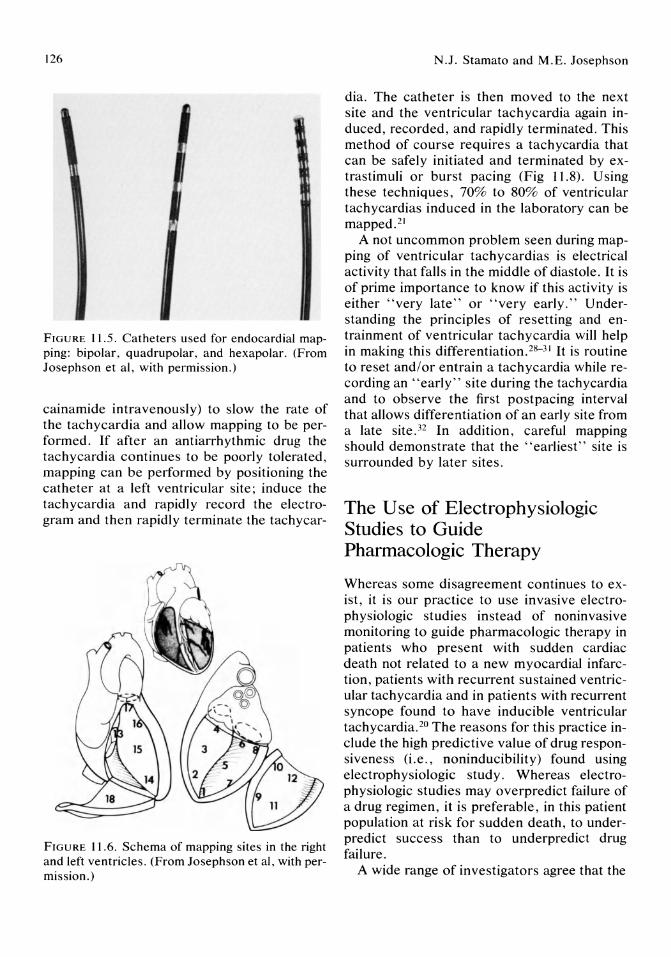
126
FIGURE 11.5. Catheters used for endocardial mapping: bipolar, quadrupolar, and hexapolar. (From Josephson et ai, with permission.)
cainamide intravenously) to slow the rate of the tachycardia and allow mapping to be performed. If after an antiarrhythmic drug the tachycardia continues to be poorly tolerated, mapping can be performed by positioning the catheter at a left ventricular site; induce the tachycardia and rapidly record the electrogram and then rapidly terminate the tachycar-
FIGURE 11.6. Schema of mapping sites in the right and left ventricles. (From Josephson et ai, with permission.)
N.J. Stamato and M.E. Josephson
dia. The catheter is then moved to the next site and the ventricular tachycardia again induced, recorded, and rapidly terminated. This method of course requires a tachycardia that can be safely initiated and terminated by extrastimuli or burst pacing (Fig 11.8). Using these techniques, 70% to 80% of ventricular tachycardias induced in the laboratory can be mapped. 21
A not uncommon problem seen during mapping of ventricular tachycardias is electrical activity that falls in the middle of diastole. It is of prime importance to know if this activity is either "very late" or "very early." U nderstanding the principles of resetting and entrainment of ventricular tachycardia will help in making this differentiation. 28- 31 It is routine to reset and/or entrain a tachycardia while recording an "early" site during the tachycardia and to observe the first postpacing interval that allows differentiation of an early site from a late site. 32 In addition, careful mapping should demonstrate that the "earliest" site is surrounded by later sites.
The Use of Electrophysiologic Studies to Guide Pharmacologic Therapy
Whereas some disagreement continues to exist, it is our practice to use invasive electrophysiologic studies instead of noninvasive monitoring to guide pharmacologic therapy in patients who present with sudden cardiac death not related to a new myocardial infarction, patients with recurrent sustained ventricular tachycardia and in patients with recurrent syncope found to have inducible ventricular tachycardia. 20 The reasons for this practice include the high predictive value of drug responsiveness (i.e., noninducibility) found using e1ectrophysiologic study. Whereas e1ectrophysiologic studies may overpredict failure of a drug regimen, it is preferable, in this patient population at risk for sudden death, to underpredict success than to underpredict drug failure.
A wide range of investigators agree that the
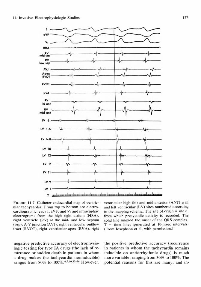
11. Invasive Electrophysiologic Studies 127
oVF--~
VI
HRA----------~--------------~~------------------
RV --------~r---------~~------+--'~--------~~---mid slip
RV ______ ~--------__ ~------~-Av---------~------low sep
AVJ --------~V~ .. ~------~I'~----~--~r----------~~--
:~~--------~!~·--------~Ir-----~~~Ir--------~~---
RVOT--------~lIr--------~Jlr------+_~~lr--------~~lr----
RVA --------~r----------IIr--------r~I~--------_1r_----
RV hi ant
RV mid ant 4
OM' LV 6 ~'~r ---------oN,
, LV 5-6 - ·./1,1--------....) "l ...
! LV 6 -8 --------.; ,..
I if.. I
LV 10 J
LV 12 ,/I
I LV 2 • LV 11
l I
LV 9 J
LV 1 ...
Ji ~.
'1 ~
1\,\ ~
1\ I ~. ~r..
I j j I
•• .III. 'r I 1" I ,
I
~ l 1 [ I I
j J r
A !I , l tytJ
T UII!!!I"""",I""",,,I,,,,,,,,,lu!!!!!!! I!!lfllIIII!II!! !!!!I!!! ,,,,,,i!l!!!!!I,1"""mi,!!!!!!ul!ll!!!!l!l!!1I"",lu" ,!!!!I""",,,I""'I!!,I""lIIul,!IIIIIIII"",,,,J,..
FIGURE 11.7. Catheter endocardial map of ventricular tachycardia. From top to bottom are electrocardiographic leads I, aVF, and VI and intracardiac electrograms from the high right atrium (HRA), right ventricle (RV) at the mid- and low septum (sep), A-V junction (A VJ), right ventricular outflow tract (RVOT), right ventricular apex (RVA), right
negative predictive accuracy of electrophysiologic testing for type IA drugs (the lack of recurrence or sudden death in patients in whom a drug makes the tachycardia non inducible) ranges from 80% to 100%.6,7,10,33-36 However,
ventricular high (hi) and mid-anterior (ANT) wall and left ventricular (LV) sites numbered according to the mapping schema. The site of origin is site 6, from which presystolic activity is recorded. The solid line marked the onset of the QRS complex. T = time lines generated at lO-msec intervals. (From Josephson et aI, with permission.)
the posItive predictive accuracy (recurrence in patients in whom the tachycardia remains inducible on antiarrhythmic drugs) is much more variable, ranging from 30% to 100%. The potential reasons for this are many, and in-

128 N.J. Stamato and M.E. Josephson
HRA A A A
~~4 " j ~ _ ~v I V
RV .1400 t ... 00 ... 00 t , • rs :.
T . . .. .. . . . . . . . . ... 300 .... .... -~ .... -..... --::-------.-.. ' ...... -' .. ,.. .. - .... ----:"':' ... " ........ --~ .. -... :-' .. ~"":--.~ .. -------.. ----.--.. ---••• " ! , •. . • '. , , ! •• r ,.
FIGURE 11.8. Termination of ventricular tachycardia (VT) by a single VPD. The figure is organized from top to bottom: ECG leads I, aVF, and VI and eIectrograms from the high right atrium (HRA), His
clude differences in stimulation protocol, definitions of inducibility and noninducibility, patient selection, and duration of clinical follow-up. There is much less data on the use of noninvasive testing in this group of patients. Our protocol for the use of programmed stimulation to guide pharmacologic therapy, begins with a "baseline" study. The patient is studied at least five half-lives after the last dose of antiarrhythmic drugs. Digoxin, betablockers, and calcium antagonists are continued if needed to control congestive heart failure or angina pectoris. Stimulation is performed as described earlier; it is routine to document reproducibility of sustained arrhythmias at least once. As previously stated, the endpoints of baseline study are the induction of a sustained arrhythmia or the completion of the protocol.
If a sustained ventricular arrhythmia can be induced reproducibly in the baseline state, procainamide is usually the first drug to be tested, during the same study as baseline testing. Procainamide is administered in a dose of 15 mg/kg at a rate of 50 mg/min followed by a continuous infusion at a rate of 0.1 mg/kg per minute. Five minutes after the loading dose is completed, a blood level is drawn, pacing thresholds are rechecked, and the stimulation protocol is repeated. Again, endpoints are the
bundle region (HBE), and right ventricle (RV). VT is terminated by a single VPD (s, arrow) delivered at a coupling interval of 250 msec. (From Josephson and Siedes, with permission.)
induction of a sustained arrhythmia or the completion of the protocol regardless of the number of stimuli needed to induce the ventricular arrhythmia in the baseline state. If a ventricular tachycardia of a morphology different from baseline is induced, this is considered a drug failure. If a sustained arrhythmia is induced a blood sample is drawn for a drug level; this is also done at the end of the protocol if no arrhythmia is induced.
If procainamide prevents the induction of a sustained tachycardia, the patient will be given an oral preparation of procainamide and after stable blood levels matching those obtained at the intravenous trial, the patient is returned to the laboratory for a follow-up study. If this is successful, the patient is discharged on this regimen. If intravenous procainamide fails to prevent the induction of ventricular tachycardia, oral procainamide is not tested. It is our experience that only 15% of such patients will have a successful regimen found when treatment is confined to other type lA, lB, or combinations of drugs. 38
Whether quinidine is tested in patients in whom ventricular tachyarrhythmias remain inducible on procainamide will depend on individual patient characteristics, but generally such a trial will be unsuccessful. If procainamide is successful, other drugs may be
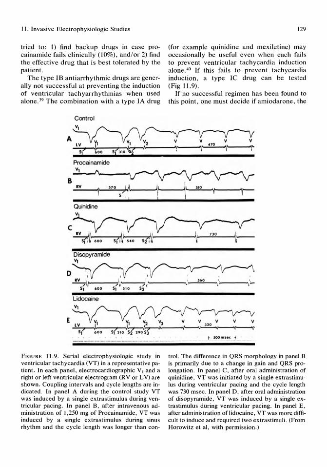
II. Invasive Electrophysiologic Studies
tried to: 1) find backup drugs in case procainamide fails clinically (10%), and/or 2) find the effective drug that is best tolerated by the patient.
The type IB antiarrhythmic drugs are generally not successful at preventing the induction of ventricular tachyarrhythmias when used alone. 39 The combination with a type IA drug
Control
Procainamide VI
129
(for example quinidine and mexiletine) may occasionally be useful even when each fails to prevent ventricular tachycardia induction alone. 40 If this fails to prevent tachycardia induction, a type Ie drug can be tested (Fig 11.9).
If no successful regimen has been found to this point, one must decide if amiodarone, the
B I _R~v ____ ~.~~s~70~~I~~~ ____ ~1~ _____ ~jl~~5~'o __ ~.----~v---
, s~ \- l l- ) , Quinidine
Disopyramide VI
o 1----'7' ~ __ ~ , ______ -..,', 560 \ "~ ----.,;1.-
Lidocaine
E
r soo msec , ----------------,-- -- - ------ _ .. - ---- -_.- --- - --
FIGURE 11.9. Serial electrophysiologic study in ventricular tachycardia (VT) in a representative patient. In each panel, electrocardiographic V I and a right or left ventricular electrogram (R V or LV) are shown. Coupling intervals and cycle lengths are indicated. In panel A during the control study VT was induced by a single extrastimulus during ventricular pacing. In panel B, after intravenous administration of 1,250 mg of Procainamide, VT was induced by a single extrastimulus during sinus rhythm and the cycle length was longer than con-
tro1. The difference in QRS morphology in panel B is primarily due to a change in gain and QRS prolongation. In panel C, after oral administration of quinidine, VT was initiated by a single extrastimuIus during ventricular pacing and the cycle length was 730 msec. In panel D, after oral administration of disopyramide, VT was induced by a single extrastimulus during ventricular pacing. In panel E, after administration oflidocaine, VT was more difficult to induce and required two extrastimuli. (From Horowitz et aI, with permission.)
130
implantable defibrillator, or arrhythmic surgery will be pursued next. Because of its serious potential side effects, amiodarone therapy is not advocated as the treatment of choice in patients who would be candidates for potentially curative arrhythmia surgery. However, if amiodarone is to be used, a follow-up electrophysiologic study is performed after the loading phase of therapy (7 days of 1400 mg/day followed by 400 mg/day). Follow-up studies are usually performed on day 10 of treatment. The ability of amiodarone to prevent induction of sustained tachyarrhythmias is uncommon (10%) but is usually associated with a good prognosis. Unfortunately, the failure to prevent induction is not necessarily associated with a poor outcome. However, if a tachycardia with a short cycle length that is not tolerated hemodynamically, is induced at amiodarone follow-up study either arrhythmia surgery or an implantable defibrillator is recommended,41 because such patients have a 25% incidence of sudden death in 1 year follow-up.
Because patients with idiopathic dilated cardiomyopathy have proven to be poor candidates for arrhythmia surgery, the automatic implantable defibrillator appears to be the best treatment option available in this group of patients in whom no successful drug regimen can be found or in patients who present with sudden cardiac death and who have no sustained arrhythmias induced at the baseline study.
The use of subendocardial resection to treat sustained ventricular tachycardia is best suited to those patients with coronary artery disease and prior myocardial infarction.8 Although this surgery is associated with an operative mortality of 10% to 12%, it is successful in 70% to 95% of surgical survivors, with the majority having no inducible arrhythmias induced at postoperative study without the use of antiarrhythmic drugs. In many patients with inducible arrhythmias postoperatively, drugs that were ineffective preoperatively will be effective postoperatively.
In patients in whom the automatic internal defibrillator is chosen for treatment, a predischarge electrophysiologic study is done to induce both ventricular tachycardia and ventric-
N.J. Stamato and M.E. Josephson
ular fibrillation and ensure the device senses and terminates the arrhythmia appropriately.
Summary
Invasive electrophysiologic studies have expanded our understanding of the pathophysiology of ventricular arrhythmias. Experimental studies have provided insights into the mechanisms and substrates for these arrhythmias. The clinical application of these studies has expanded the treatment options available to patients who previously faced tremendous risk of mortality. Electrophysiologically guided pharmacologic therapy, arrhythmia surgery, catheter ablation, and the implantable defibrillator have all added much to the care of this subgroup of patients.
References 1. Wellens HJJ, Schuilenburg RM, Durrer D:
Electrical stimulation of the heart in patients with ventricular tachycardia. Circulation 1972; 46:216-226.
2. Josephson ME, Horowitz LN, Farshidi A, et al: Recurrent sustained ventricular tachycardia. 1. Mechanisms. Circulation 1978; 57:431-440.
3. Josephson ME, Horowitz LN: Electrophysiologic approach to therapy of recurrent sustained ventricular tachycardia. Am J Cardiol 1979; 43:631-642.
4. Horowitz LN, Spielman SR, Greenspan AM, et al: Role of programmed stimulation in assessing vulnerability to ventricular arrhythmias. Am Heart J 1982; 103:604-610.
5. Josephson ME, Buxton AE, Marchlinski FE, et al: Sustained ventricular tachycardia in coronary artery disease-evidence for reentrant mechanism, in Zipes DP, Jalife J (eds): Cardiac Electrophysiology and Arrhythmias. Orlando, FL, Grune & Stratton, 1985, 409-418.
6. Mason JW, Winkle RA: Electrode-catheter arrhythmia induction in the selection and assessment of antiarrhythmic drug therapy for recurrent ventricular tachycardia. Circulation 1978; 58:971-985.
7. Horowitz LN, Josephson ME, Farshidi A, et al: Recurrent sustained ventricular tachycardia. 3. Role ofthe electrophysiologic study in selection of antiarrhythmic regimens. Circulation 1978; 58:986-997.
11. Invasive Electrophysiologic Studies
8. Josephson ME, Horowitz LN, Harken AH: Endocardial excision: A new surgical technique for the treatment of ventricular tachycardia. Circulation 1979; 60: 1430-1439.
9. Hartzler GO: Electrode catheter ablation of refractory focal ventricular tachycardia. J Am Coli Cardiol 1984; 2: 1107-1113.
10. Ruskin IN, DiMarco JP, Garan H: Out-of-hospital cardiac arrest: Electrophysiologic observations in selection of long-term antiarrhythmic therapy. N Engl J Med 1980; 303:607-613.
11. DiMarco JP, Garan H, Harthorne JW, et al: Intracardiac electrophysiologic techniques in recurrent syncope of unknown cause. Ann Intern Med 1981; 95:542-548.
12. Roy D, Marchand E, Theroux P, et al: Reproducibility and significance of ventricular arrhythmias induced after an acute myocardial infarction. J Am Coli Cardiol 1986; 8:32-39.
13. Buxton AE, Marchlinski FE, Flores BT, et al: Nonsustained ventricular tachycardia in patients with coronary artery disease: role of electrophysiologic study. Circulation 1987; 75:1178-1185.
14. Josephson ME, Siedes SF: Clinical Cardiac Electrophysiologic: Techniques and Interpretations. Philadelphia, Lea & Febiger, 1979.
15. Gettes LS, Zipes DP, Gillette PC, et al: Personnel and equipment required for electrophysiologic testing. Circulation 1984; 69: 1219A-1221A.
16. Ruskin IN, Flowers NC, Josephson ME, et al: Task force VII: Arrhythmias and specialized electrophysiologic studies. J Am Coli Cardiol 1986; 7:1215-1216.
17. DiMarco JP, Garan H, Ruskin IN: Complications in patients undergoing cardiac electrophysiologic procedures. Ann Int Med 1982; 97:490-493.
18. Doherty JU, Kienzle MG, Waxman HL, et al: Programmed ventricular stimulation at a second right ventricular site: An analysis of 100 patients, with special reference to sensitivity, specificity, and characteristics of patients with induced ventricular tachycardia. Am J Cardiol 1983; 52:1184-1189.
19. Buxton AE, Waxman HL, Marchlinski FE, et al: Role of triple extrastimuli during electrophysiologic study of patients with documented sustained ventricular tachyarrhythmias. Circulation 1984; 69:532-540.
20. Gottlieb C, Josephson ME: The preference of programmed stimulation guided therapy for
131
sustained ventricular arrhythmias, in Brugada P, Wellens HJJ (eds): Cardiac Arrhythmias: Where to go from here? Mount Kisco, NY, Futura, 1987, p 421-434.
21. Josephson ME, Horowitz LN, Spielman SR, et al: Role of catheter mapping in the preoperative evaluation of ventricular tachycardia. Am J Cardiol 1982; 49:207-220.
22. Cassidy DM, Vassallo JA, Buxton AE, et al: The value of sinus rhythm catheter mapping to localize ventricular tachycardia site of origin. Circulation 1984; 69:1103-1110.
23. Josephson ME, Waxman HL, Cain ME, et al: Ventricular activation during ventricular endocardial pacing. II. Role of pace mapping to localized origin of ventricular tachycardia. Am J Cardiol1982; 50:11-22.
24. Miller JM, Kienzle MG, Harken AH, et al: Subendocardial resection for ventricular tachycardia. Predictors of surgical success. Circulation 1984; 70:624-631.
25. Josephson ME, Horowitz LN, Farshidi A, et al: Recurrent sustained ventricular tachycardia. 2. Endocardial mapping. Circulation 1978; 57:440-447.
26. Josephson ME, Horowtiz LN, Spielman SR, et al: Comparison of endocardial catheter mapping with intraoperative mapping of ventricular tachycardia. Circulation 1980; 61 :395-404.
27. Josephson ME, Horowitz LN, Farshidi A: Continuous local electrical activity. A mecha· nism of recurrent ventricular tachycardia. Circulation 1978; 57:659-665.
28. MacLean WAH, Plumb VJ, Waldo AL: Transient entrainment and interruption of ventricular tachycardia. PACE 1981; 4:358-366.
29. Anderson KP, Swerdlow CD, Mason JW: Entrainment of ventricular tachycardia. Am J Cardiol 1984; 53:335-340.
30. Amendral J, Cohen M, Miller J, et al: Entrainment of ventricular tachycardia: Insights gained by electrogram recorded at the site of tachycardia origin (abstr). J Am Coli Cardiol 1985; 5:470A.
31. Mann DE, Lawne GM, Luck JC, et al: Importance of pacing site in entrainment of ventricular tachycardia. JAm Coli Cardioll985; 5:781-787.
32. Almendral JM, Marchlinski FE, Josephson ME: A constant first postpacing interval: New criterion for entrainment of ventricular tachycardia (abstr). Circulation 1985; 72:III-30.
33. Swerdlow CD, Winkle RA, Mason JW: Determinants of survival in patients with ventric-

132
ular tachyarrhythmias. N Engl J Med 1983; 308: 1436-1442.
34. Kim SG, Seiden FW, Felder SD, et al: Is programmed stimulation of value in predicting the long-term success of antiarrhythmic therapy for ventricular tachycardias? N Engl J Med 1986; 315:356-362.
35. Roy D, Waxman HL, Kienzle MG, et al: Clinical characteristics and long-term follow-up in 119 survivors of cardiac arrest: Relation to inducibility at electrophysiologic testing. Am J Cardiol 1983; 52:969-974.
36. Skale BT, Miles WM, Heger JJ, et al: Survivors of cardiac arrest: Prevention of recurrence by drug therapy as predicted by electrophysiologic testing or electrocardiographic monitoring. Am J Cardiol1986; 57:113-119.
37. Graboys TB, Lown B, Podrid PJ, et al: Longterm survival of patients with malignant ventricular arrhythmia treated with antiarrhythmic drugs. Am J Cardiol 1982; 50:438-443.
N.J. Stamato and M.E. Josephson
38. Waxman HL, Buxton AE, Sadowski LM, et al: The response to procainamide during electrophysiologic study for sustained ventricular tachyarrhythmias predicts the response to other medications. Circulation 1983; 67:30-37.
39. Waspe LE, Waxman HL, Buxton AE, et al: Mexiletine for control of drug resistant ventricular tachycardia: Clinical and electrophysiologic results in 44 patients. Am J Cardiol1983; 51:1175-1181.
40. Greenspan AM, Spielman SK, Webb CR, et al: Efficacy of combination therapy with mexiletine and type IA agent for inducible ventricular tachyarrhythmias secondary to coronary artery disease. Am J Cardiol 1985; 56:277-284.
41. Kadish AH, Buxton AE, Waxman HL, et al: Usefulness of electrophysiologic study to determine the clinical tolerance of arrhythmia recurrences during amiodarone therapy. J Am Coli Cardiol 1987; 10:90-96.

12 Electrophysiologic Approach to Patients with Supraventricular Tachycardia Donald G. Rubenstein and Carol Zaher
Supraventricular Tachycardia
Electrophysiology studies have contributed greatly to elucidating the underlying mechanisms of the varieties of supraventricular tachycardia (SVT). In the majority of cases, SVT can be managed successfully without invasive electrophysiologic studies (EPS), reserving EPS only for refractory or serious supraventricular arrhythmias. This chapter deals with the types of SVT most commonly encountered in the EPS laboratory, their classification, mechanisms, EPS approach, drug therapy, and surgical ablation. It is beyond the scope of this chapter to discuss all types of SVT in detail. 1
Classification of Supraventricular Tachycardia
The term supraventricular tachycardia is a "generic" term encompassing all arrhythmias originating above the ventricle. Supraventricular tachycardia can be classified into the following groups:
1. sinoatrial nodal, 2. intra-atrial, 3. atrioventricular nodal, 4. atrioventricular (using concealed bypass
tract), and 5. atrial flutter and atrial fibrillation.
Mechanism of Supraventricular Tachycardia
Both automaticity and re-entry are the mechanisms known to initiate and maintain SVT. Approximately 60% of patients with SVT have atrioventricular (A V) nodal re-entrant tachycardia, and 30% have A V re-entry involving bypass tracts.2--8 Electrophysiologic study cannot reliably initiate or terminate those arrhythmias associated with automaticity; however, those due to re-entry can be reproduced in the laboratory. For a re-entrant tachycardia to exist, several factors must be in operation. These include: 1) two functionally distinct pathways (alpha and beta) that join proximally and distally to complete a circuit; 2) unidirectional block in one pathway; and 3) slow conduction down one of the pathways. An appropriately timed premature beat blocks in the fast pathway, arriving at the distal end of the fast pathway when it is no longer refractory, allowing retrograde conduction to set up an SVT circuit (Fig 12.1).
Indications for Electrophysiologic Study
Most cases of SVT do not require EPS; however, the following are some generally accepted indications for EPS: 1) differentiation of SVT with aberrancy from ventricular tachy-

134
Slow (0<)
FIGURE 12.1. Mechanism of re-entrant arrhythmias. Two functionally distinct pathways are present with differing refractory periods and conduction properties (slow vs fast). A premature beat finds the fast pathway refractory and is blocked (a) and conducts slowly down the slow pathway (b ~). When the impulse arrives at the distal end of the pathway (c), the fast pathway is no longer refractory and the impUlse conducts retrograde via the fast pathway (-~-+-~~). When the impulse arrives at the slow pathway it again can conduct down this pathway maintaining the re-entrant circuit.
cardia (VT) or pre-excitation; 2) SVT associated with serious hemodynamic symptomatology such as syncope, congestive heart failure, or cardiac arrest; 3) symptomatic SVT resistant to empiric drug treatment; 4) arrhythmias associated with pre-excitation syndromes; in Wolff-Parkinson-White (W-P-W) syndrome, EPS is used to identify high-risk patients prone to rapid ventricular response during atrial flutter or atrial fibrillation; response to medications and suitability for surgical intervention are also evaluated9--12; and 5) refractory SVT in preparation for electrode catheter ablation, surgical ablation, or anti tachycardia pacemaker therapy.
D.G. Rubenstein and C. Zaher
Electrophysiologic Evaluation of Supraventricular Tachycardia
Because most SVT that occurs clinically is secondary to re-entry, EPS can be used to initiate, terminate, and localize these arrhythmias. Both incremental pacing and pacing with introduction of extra stimuli are used to initiate SVT.
Classification of the SVT is demonstrated by: 1) the manner in which the tachycardia is initiated and terminated; 2) the atrial activation sequence during the tachycardia, as well as the relationship of the P wave to the QRS complex on the surface electrocardiogram (ECG); 3) the requirement of the atrium and/ or ventricle in the initiation and sustenance of the tachycardia; 4) the effect of stimulation during the tachycardia; 5) the effect of bundle branch block on the rate of the tachycardia; and 6) the effects of drugs and/or physiologic maneuvers on the tachycardia.
In performing EPS in evaluation of SVT, multiple intracardiac catheters are placed. Quadripolar catheters are advanced to the high right atrium (HRA) , low right atrium (LRA), His bundle (HBE), and right ventricular apex (RVA), for recording and/or stimulation purposes (Fig 12.2). A hexapolar catheter is then advanced from the antecubital or subclavian vein to the coronary sinus (CS), where recording and pacing of the proximal, mid-, and distal coronary sinus can be initiated. It is important to determine the location, as well as the mode of initiation of SVT; therefore, HRA, LRA, RV A, and CS pacing with burst pacing followed by extrastimulus pacing is performed. Once all the information is obtained, burst pacing from the HRA should be performed at rapid cycle lengths to initiate atrial flutter or fibrillation in those patients suspected of having a bypass tract and who did not have these arrhythmias during previous pacing. Once the above information has been interpreted, drug testing with repeat stimulation at sites of initiation of the tachycardia should be repeated. At the completion of the study, the coronary sinus catheter can be left in place for several days to continue drug testing.

12. Supraventricular Tachycardia
FIGURE 12.2. Fluroscopic position of intracardiac catheters. HRA = high right atrium, HBE = His
Electrophysiologic Study of Specific Supraventricular Tachycardia
Sinus Node Re-entry
Sinus node re-entry is the underlying mechanism in approximately 5% of SVT.l,13 It is initiated after a properly timed premature atrial impulse. The P and A waves and atrial activation sequence are similar to those in sinus rhythm. High right atrium stimulation initiates the tachycardia, with initiation less likely during pacing at sites distant to the sinus node. Study criteria for this arrhythmia can be obtained in other texts.1.l 4 Propanolol and verapamil are sometimes effective in treating this arrhythmia.
Intra-atrial Re-entry
Intra-atrial re-entry is generally observed in patients with enlarged atria, in whom the atrial
135
bundle electrogram, CS = coronary sinus, RVA =
right ventricular apex, LRA = low right atrium.
effective and functional refractory periods are prolonged. Initiation and termination of SVT is similar to other re-entrant rhythms. However the P and A waves are different from those in normal sinus rhythm and the atrial activation sequence depends on the origin of the arrhythmia.
Atrioventricular Nodal Re-entry Tachycardia
Atrioventricular nodal re-entry tachycardia (AVNRT) accounts for 60% of cases of SVT and can be initiated by EPS in approximately 75% of cases. l Most patients with A VNRT can be identified with EPS. The underlying substrate for A VNRT involves longitudinal dissection of the A V node into two pathways (Fig 12.1). The alpha-pathway has slower conduction but a shorter refractory period compared with the faster conducting beta-path-
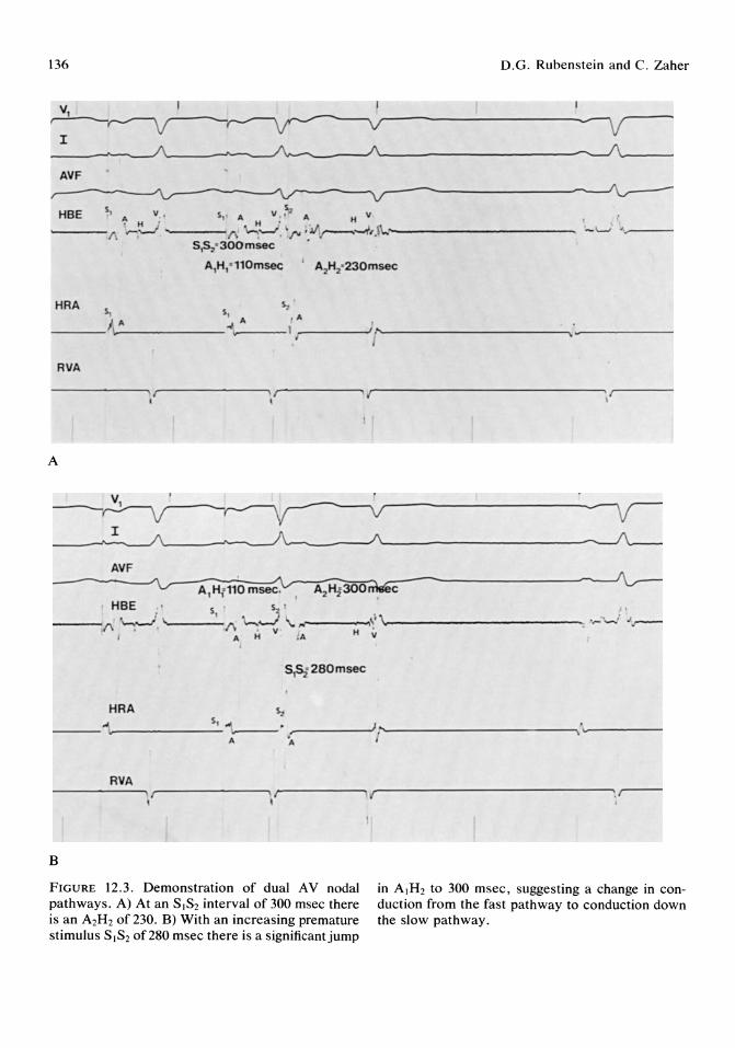
136 D.G. Rubenstein and C. Zaher
v ' ,.-J---~
I ______ --'-~/\ ~ __ ----A~--------------------~~ ____ _ AVF
HBE 5, v v s" ~ H ;' 5, A H • ' A H v
~---/\ """''-' -.... ---;, ~~. ,rw,Jr---~'.\'I.."'------------' S,S2"300msec .
A,H," 110msec ' A",H2"230msec
HRA ~ ~ ~ A
:--___ /I\.-.A _______ ~" ..... A--- I ,r"'-----..J1r ---- ----- ------. _------
RVA
--------~. ,r--------, Fr------,.{r----------------~;,r----, I
A
AVF
HBE ~' ,..-__ {\ '~. I.,-___ S,_ {\ '-,--.lv ,..,.. ___ ....,,1 ,1,. _____________ ...,... .. -.i
H H 'it • A . A
SlSf280msec
s.;. HRA
--i. S, "'1"". __ _
A ~------')r--------------------,;~· ------I A
RVA --------.\I--------~\ II'"-----..... , r----------------~',~
I
B
FIGURE 12.3. Demonstration of dual AV nodal pathways. A) At an SISZ interval of 300 msec there is an AzHz of 230. B) With an increasing premature stimulus SISZ of 280 msec there is a significant jump
in AIHz to 300 msec, suggesting a change in conduction from the fast pathway to conduction down the slow pathway.
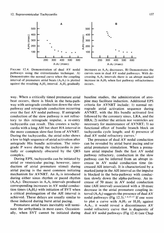
12. Supraventricular Tachycardia
A
500
U 400 Q) (J) .s 300 N
~ 200 «
100 . . . . . 200 300 400 500 600
A1A2 (msec)
FIGURE 12.4. Demonstration of dual A V nodal pathways using the extrastimulus technique. A) Demonstrates the normal curve when the coupling interval of premature atrial beats (A1A2) is plotted against the resulting A2H2 interval. A2H2 gradually
way. When a critically timed premature atrial beat occurs, there is block in the beta-pathway with antegrade conduction down the slow pathway and retrograde conduction occurring over the fast A V nodal pathway. If antegrade conduction of the slow pathway is not refractory to this retrograde impulse, a re-entry tachycardia can result. This creates a tachycardia with a long AH but short HA interval in the more common slow-fast form of AVNRT. During the tachycardia, the atrial echo shows a low to high sequence of atrial activation after antegrade His bundle activation. The retrograde P wave during the tachycardia is partially or completely obscured by the QRS complex.
During EPS, tachycardia can be initiated by atrial or ventricular pacing; however, introduction of atrial premature stimuli during atrial pacing is the most common initiating mechanism for AVNRT. An A2 is introduced during either sinus rhythm or paced rhythm (AlAI)' Decreases in AIA2 intervals produced corresponding increases in A V nodal conduction times (A2H2) with initiation of SVT when a critical prolongation of the AH interval is achieved. These delays are usually similar to those induced during burst atrial pacing.
Premature atrial beats inevitably will terminate the arrhythmia in most cases. Occasionally, when SVT cannot be initiated during
137
B
500 . . ~ 400 . u . Q) . (J) .s 300 N
~ 200 «
100 . . . 200 300 400 500 600
A1A2 (msec)
increases as AIA2 decreases. B) Demonstrates the curves seen in dual A V nodal pathways. With decreasing AIA2 intervals there is an abrupt marked increase in A2H2 when fast pathway refractoriness occurs.
baseline studies, the administration of atropine may facilitate induction. Additional EPS criteria for AVNRT include: 1) normal retrograde atrial activation sequence during A VNRT, with the His bundle activated first followed by the coronary sinus, LRA, and the HRA; 2) neither the atrium nor ventricles are necessary for maintenance of AVNRT; 3) no functional effect of bundle branch block on tachycardia cycle length; and 4) presence of dual AV nodal refractory curves. 1
The presence of dual A V nodal conduction can be revealed by atrial burst pacing and/or atrial premature stimulation. When a premature atrial impulse finds the fast A V nodal pathway refractory, conduction in the slow pathway can be inferred from an abrupt increase in A V nodal conduction time (increased AH interval). It is characterized by a markedjump in the AH interval as the impulse is blocked in the beta-pathway with conduction slowly down the alpha-pathway. An increase of 50 msec in A V nodal conduction time (AH interval) associated with a lO-msec decrease in the atrial premature coupling interval (A1A2) suggests the presence of dual A V nodal pathways (Fig 12.3).1-4,15-17 If one were to plot a curve with A2H2 or HIH2 against AIA2' it would reveal a discontinuous A V nodal refractory curve that is diagnostic of dual A V nodal pathways (Fig 12.4) (see Chap

138
10 p. 108). There is a consistent range of coupling intervals (SIS2) that can induce A VNRT and is reproducible.
An atypical A V nodal re-entrant tachycardia form also exists that uses a fast A V nodal pathway for antegrade conduction and slow A V nodal pathway for retrograde conduction (fast-slow).18,19 This form can usually be induced by EPS using programmed atrial or ventricular stimulation. The fast-slow form has smooth conduction curves in contrast to the slow-fast form. Fast-slow A V nodal tachycardia may present as a chronic incessant form of SVT.20,21 It can be initiated by sinus tachycardia with a critical PP interval inducing antegrade block in the slow pathway with initiation of the A V nodal tachycardia.
Atrioventricular Tachycardia
Pre-excitation occurs when the ventricular muscle is activated earlier than would be expected from A V nodal conduction due to the presence of an anomalous pathway. Bypass tracts are characterized by their anatomic location, with atrioventricular (A V) bypass tracts representing the most frequent form of pre-excitation and producing the classic W-P-W syndrome. Nodoventricular, fasiculoventricular, and A V nodal bypass tracts are more rare and discussed in detail in other texts. 1.53 Certain patients have concealed tracts that are capable only of retrograde conduction.
An A V bypass tract generally produces the classic ECG pattern with a short PR interval, a delta-wave, and a wide QRS complex. The majority of patients with these tracts have clinical arrhythmias. Fortunately, only a small number of these patients manifest atrial flutter or fibrillation, as these can represent life threatening arrhythmias with the potential for fast conduction down the accessory bypass tract resulting in ventricular tachycardia or fibrillation. 31 ,32 The presence of a bypass tract essentially negates the A V node as protection against rapid conduction to the ventricles. If the antegrade effective refractory period of the anomalous pathway is short, atrial impulses can conduct directly to the ventricles with rapid rates. The shorter the antegrade
D.G. Rubenstein and C. Zaher
ERP of the bypass tract, the more rapid ventricular response to atrial flutter or fibrillation.
Electrophysiologic studies are useful in these patients27-30,33,55 in order to: 1) map the accessory pathway location in preparation for surgical or ablative therapy; 2) define conduction characteristics of the accessory pathway; 3) determine the risk for atrial fibrillation; and 4) evaluate potential drug treatment.
Localization of Bypass Tracts
In general, the presence of a bypass tract that can conduct antegrade gives rise to a short HV interval. With atrial pacing or introduction of APDs, the degree of pre-excitation increases and the HV interval will decrease even further (Fig 12.SA-C). Atrial pacing from any site would induce pre-excitation, however, it will be maximal closest to the site of insertion of the bypass tract. The most commonly induced tachycardia in patients with W-P-W syndrome is the orthodromic type with narrow QRS complexes with antegrade conduction down the A V node and retrograde conduction via the bypass tract. To localize the bypass tract, the following characteristics should be observed:
1. Pacing from multiple atrial sites: The shortest P to delta interval localizes the site closest to the bypass tract.
2. Retrograde atrial activation during tachycardia: Evaluating the sequence of retrograde atrial activation during SVT is the primary method for localizing of bypass tracts involved during SVT. The site of earliest retrograde atrial activation identifies the atrial insertion of the bypass tract (Fig 12.SD). More than one bypass tract may exist, however, and may not be demonstrated if only one SVT is initiated.
3. Ventricular mapping: During full pre-excitation, mapping of the ventricles may help to localize the ventricular insertion site. The ventricular spike closest to the deltawave identifies the insertion site. The coronary sinus catheter is used to map left-sided tracts. Retrograde sequence of atrial activation during ventricular pacing with introduction of premature ventricular beats also helps locate bypass tracts capable of retro-
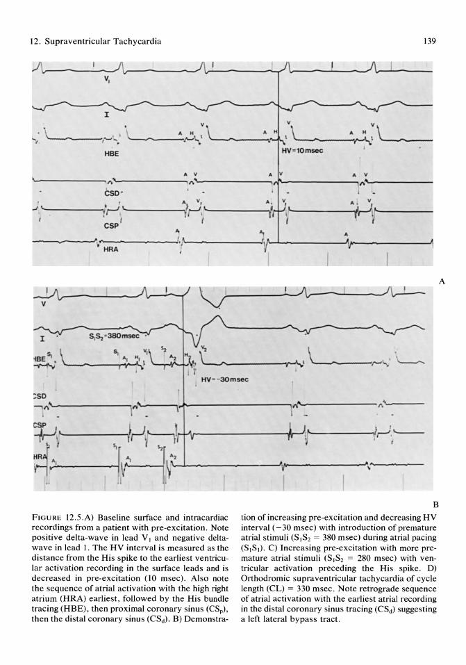
12. Supraventricular Tachycardia 139
~~-----~-------~~------~ l----~--__ A~--__ VI
• v
'i "-----/'-'.-: ~~\\ HBE HV=10msec
AV AV AV
.. -----------.. 1'\"""-----------, 1'\"""-------""'" ,..r----------.(\/I& ~-----__, cso-
V A V, ..J '-.... ______ '--! '-_______ ...,>-...1 L""--_____ -.!
~, - I ,I ,- A, , I \
L A'HV
~-----~~ ~------~ r j I CSp i I
"t A.
~~~------------~~f~---------~\r-~-------~~---------~ A
HRA
V ~~------L-~------___ ) l - '--
HV--30msec
FIGURE 12.5.A) Baseline surface and intracardiac recordings from a patient with pre-excitation. Note positive delta-wave in lead V, and negative deltawave in lead 1. The HV interval is measured as the distance from the His spike to the earliest ventricular activation recording in the surface leads and is decreased in pre-excitation (10 msec). Also note the sequence of atrial activation with the high right atrium (HRA) earliest, followed by the His bundle tracing (HBE), then proximal coronary sinus (CSp),
then the distal coronary sinus (CSd). B) Demonstra-
B
tion of increasing pre-excitation and decreasing HV interval (-30 msec) with introduction of premature atrial stimuli (S1S2 = 380 msec) during atrial pacing (S,S,). C) Increasing pre-excitation with more premature atrial stimuli (SIS2 = 280 msec) with ventricular activation preceding the His spike. D) Orthodromic supraventricular tachycardia of cycle length (CL) = 330 msec. Note retrograde sequence of atrial activation with the earliest atrial recording in the distal coronary sinus tracing (CSd) suggesting a left lateral bypass tract.
A
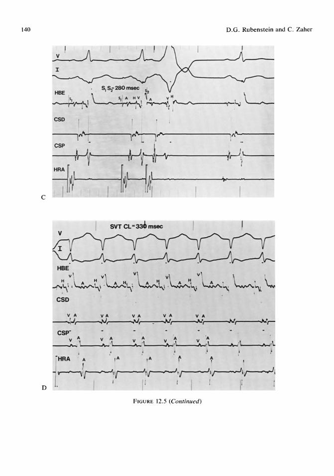
140 D.G. Rubenstein and C. Zaher
SVT CL -:Jab msec
HBE
..,..\:~~:lM~,VL\ ~~\,;L·"\; L:,. \... , , . .
FIGURE 12.5 (Continued)
12. Supraventricular Tachycardia
grade conduction by localizing the earliest site of atrial activity. Initially..- at longer V1V2 intervals, there may be fusion between retrograde conduction through the AV node and the bypass tract or tracts. This situation can be aided by administration of intravenous verapamil to block A V nodal retrograde conduction, thereby allowing retrograde activation of the atrium to occur primarily via bypass tracts.
4. Bundle branch block: An increase in the cycle length of the tachycardia and V A conduction by greater than 25 msec with development of bundle branch block identifies a free wall bypass tract ipsilateral to the conduction defect. 34
Identification of High-risk Groups With Wolff-Parkinson- White Syndrome
Up to 40% of patients with bypass tracts can develop atrial fibrillation, and a small percentage of patients with pre-excitation can develop atrial fibrillation with very rapid rates due to conduction down the bypass tract, and therefore are prone to ventricular fibrillation. 36
There is a good correlation between the shortest RR interval showing pre-excitation during atrial fibrillation and the ERP of the accessory pathway.II,12,31,35 Determination of the ERP of the accessory pathway in the antegrade direction should thus identify patients prone to life-threatening ventricular rates during atrial fibrillation.
The atrial extrastimulus technique is used to determine the refractory period and should be performed closest to the atrial insertion site. It is defined as the longest AIA2 at which the bypass tract fails to conduct. Those patients with an ERP of less than 220 have extremely rapid ventricular responses to atrial fibrillation.
Eiectrophysioiogic Study Characteristics
Initiation of A V reciprocating tachycardias depends on the difference in electrophysiologic properties of the normal A V pathway and anomalous A V bypass tract. The bypass tract usually has a faster conduction velocity and longer refractory period than the A V node. This is similar to dual pathways in the
141
AV node. In tachycardias associated with W-P-W syndrome, the bypass tract is usually the site of unidirectional block with the A V node having slow conduction. During EPS, an appropriately timed APB blocks in the accessory pathway and conducts down the A V node normally. An orthdromic tachycardia is initiated when an atrial echo beat leaves the AV node, conducts retrograde up the bypass, and returns to the A V node to initiate an SVT with a narrow QRS complex.
Rarely, an antidromic form of the tachycardia exists whereby the anomalous A V bypass tract will be used for antegrade conduction with the normal A V pathway used for retrograde conduction. During the tachycardia, the QRS is wide, reflecting complete ventricular pre-excitation.
Atrial Flutter and Fibrillation The origin of atrial flutter and fibrillation is believed to be secondary to atrial vulnerability in diseased atrial tissues. There can exist paroxysms of atrial flutter or fibrillation when an atrial premature beat is delivered close to the functional refractory period of the atrium. Atrial fibrillation also can be induced in the EPS laboratory by rapid atrial pacing with a high electrical current (usually 10 to 20 rnA at rapid rates of 250 to 400 bpm for 30 to 60 seconds). Induction of sustained atrial fibrillation is considered abnormal. 1,22-26
Electrophysiologic study is rarely needed in patients with these clinical arrhythmias. However, patients with paroxysmal atrial fibrillation refractory to medical treatment can occasionally be studied with serial drug testing. 27
Pharmacologic Treatment of Supraventricular Tachycardia
Mter induction of SVT, several antiarrhythmic medications can be administered.
Verapamil and Diltiazem These drugs depress conduction and increase refractoriness in the A V node in the antegrade and retrograde directions. Verapamil is used intravenously for terminating SVT secondary

142
to A V nodal re-entry, and to slow the rate of atrial flutter and fibrillation not associated with pre-excitation. These medications should be used carefully in patients with sinus node dysfunction or LV dysfunction.
In patients with W-P-W syndrome, verapamil can enhance anomalous A V bypass tract conduction and therefore is contraindicated in patients with atrial flutter or fibrillation with conduction down the bypass tract. 37,40
Digoxin
Digoxin depresses conduction and increases refractoriness of the A V node in the antegrade and occasionally the retrograde direction. It is used in SVT. Ouabain is used during EPS because of rapid distribution to cardiac tissues. The dose is .01 mg/kg.
When considering W-P-W syndrome, as with calcium channel blockers, digoxin can shorten the antegrade refractory period of the A V bypass tract in certain patients and is therefore contraindicated in patients with atrial fibrillation or flutter associated with preexcitation.38 •39
Beta-blockers
Beta-blockers depress A V conduction and increase AV nodal refractoriness. 44 It is effective in controlling paroxysmal supraventricular tachycardia (PSVT), and is given at a dose of .1 to.2 mg/kg at 1 mg per minute.
Type IA Anti-arrhythmic Agents
These agents (quinidine, procainamide, disopyramide) are effective in treatment of SVT. They function by suppressing APBs and VPBs, as well as increasing the refractoriness of the atrium and ventricles, His-Purkinje system, and anomalous bypass tracts. They also increase the refractoriness of the A V node in the retrograde direction. These drugs can therefore be used as treatment for both PSVT and W-P-W syndrome. 41 -43 Doses are: 1) procainamide: \0 to 15 mg/kg IV at 50 mg per minute with careful blood pressure monitoring; 2) quinidine: although this can be used intravenously, it can cause severe hypotension and therefore should be used carefully
D.G. Rubenstein and C. Zaher
only in those patients with normal left ventricular function; orally, 1.2 to 1.6 g are given in divided doses during 24 hours; and 3) disopyramide: this is given orally at doses of 150 to 200 mg every 6 hours.
Type IB Agents
Lidocaine, tocainide, and mexiletine all may increase refractoriness of the anomalous A V bypass tract and thus may prevent induction of the tachycardia; however, clinically, these drugs are not frequently used. 50
Type 1 C Agents
Encainide, flecainide, and propafenone are new agents and show promise in treating PSVT and W-P-W syndrome. They increase the refractoriness of the anomalous A V bypass tracts, slow conduction, and increase the refractoriness in the atrium, ventricles, HisPurkinje system, and anomalous A V pathways. 45-48,51,52
Amiodarone
Amiodarone is a very effective agent for control of PSVT and W-P-W syndrome. It prolongs refractoriness of the atrium, ventricle, A V node, His-Purkinje system, and anomalous A V bypass tracts. Unfortunately, although an extremely potent drug, it has a large number of side effects that could require termination or dose reduction. 49 Doses of 200 to 400 mg/day are usually required for control of PSVT and W-P-W syndrome.
Mapping and Surgery in Patients With Pre-excitation
Surgical intervention now offers an efficacious alternative therapy for patients with supraventricular arrhythmias associated with bypass tracts and in the majority of patients, can offer potential for a "cure," obviating the need for chronic antiarrhythmic therapy in certain cases. 56-58
Indications for surgery include tachyarrhythmias refractory to aggressive medical management and atrial fibrillation with rapid ventricular response due to conduction down

12. Supraventricular Tachycardia
the bypass tract that predisposes to ventricular fibrillation. Additionally, recently there has been an increase in referrals for surgery based on patient preference to achieve a curative procedure, preventing the need for lifelong drug therapy.
Preoperative assessment includes extensive evaluation for location and number of bypass tracts during electrophysiology studies. This involves atrial, ventricular, and coronary sinus pacing to initiate tachyarrhythmias followed by mapping during tachycardia, particularly noting the sequence of retrograde atrial activation, effect of bundle branch block during tachycardia, and retrograde atrial activation during ventricular pacing. This is important because occasionally during surgery with cooling of the heart and the effects of anesthesia, some bypass tracts will become nonfunctional and unable to be mapped accurately in the operating room. Also, the location is important because the surgical approach to resection of the tracts varies according to their locations, and multiple bypass tracts may be more difficult to approach surgically. 59-62
Operative mapping often allows for more exact localization of one or mUltiple bypass tracts to guide surgical resection and involves determination of both atrial and ventricular insertion of accessory pathways during sinus rhythm or pacing or during SVT. Ventricular insertion site may be determined by mapping the ventricular aspect of the A V ring during sinus rhythm or atrial pacing. The site of bypass is localized by the site with the shortest A V interval. Similarly, the atrial insertion site is shown by mapping the atrial side of the A V ring during ventricular pacing, represented by the shortest V A interval.
Once the accessory pathways are localized, they can be surgically divided by the endocardial or epicardial approach, sometimes with the aid of cryoablation as well. 63 ,64 Repeat postsurgical mapping is undertaken to ensure adequate success. Complications of the procedure are infrequent, but include recurrent bleeding, atrial arrhythmias, advanced or complete heart block requiring permanent pacing, myocardial infarction secondary to damage of the left circumflex artery, and postpericardiotomy syndrome. Surgical success
143
rates are reported up to 95% of cases at experienced centers.63 ,64
Summary
Most forms of SVT can be managed without resorting to EPS; however, with refractory or incapacitating symptomatic SVT, EPS can be useful in determining mechanism, appropriate drug therapy, as well as suitability for surgical, ablative, or antitachycardia procedures.
References
1. Josephson ME, Seides SF: Clinical Cardiac Electrophysiology. Philadelphia, Lea & Febiger, 1979.
2. Josephson ME, Kastor JA: Supraventricular tachycardia: Mechanisms and management. Ann Intern Med 1977; 87:346.
3. Wu D, Denes P: Mechanisms of paroxysmal supraventricular tachycardia. Arch Intern Med 1975; 135:437.
4. Wu D, Denes P, Amat-y-Leon F, et al: Clinical electrocardiographic and electrophysiological observations in patients with paroxysmal supraventricular tachycardia. Am J Cardiol1978; 41:1045.
5. Wellens HJJ, Wesdorp JC, Duren DR, et al: Diagnosis, incidence and significance of concealed accessory pathways in patients with supraventricular tachycardia. in P Puel (ed): Troubles du Rythme et Electrostimulation. Tourlouse, Society de la Nouvella Imprimerie Fournie, 1978, p 169.
6. Farshidi A, Josephson ME, Horowitz LN: Electrophysiologic characteristics of concealed bypass tracts: Clinical and electrocardiographic correlates. Am J Cardiol 1978; 41:1052.
7. Gillete PC: Concealed cardiac conduction pathways: A frequent cause of supraventricular tachycardia. Am J Cardiol 1977; 40:848.
8. Sung RJ, Gelband H, Castellanos A, et al: Clinical and electrophysiologic observations in patients with concealed accessory atrioventricular bypass tracts. Am J Cardiol 1977; 40:839.
9. Klein GJ, Bashore TM, Sellers TD, et al: Ventricular fibrillation in the Wolff-ParkinsonWhite syndrome. N EngLJ Med 1979; 301:1080.
10. Gallagher JJ, Pritchett ELC, Sealy WC, et al: The pre-excitation syndromes. Prog Cardiovase Dis 1978; 20:285.
11. Wellens HJ, Durrer D: Wolff-ParkinsonWhite syndrome and atrial fibrillation: Relation
144
between refractory period of accessory pathway and ventricular rate during atrial fibrillation. Am J Cardiol 1974; 34:777.
12. Castellanos A Jr, Myerburg RJ, Craparo K, et al: Factors regulating ventricular rates during atrial flutter and fibrillation in pre-excitation (Wolff-Parkinson-White) syndrome. Am J Cardiol 1982; 50:353.
13. Fischer JD: Role of electrophysiologic testing in diagnosis and treatment of patients with known and suspected bradycardias and tachycardias. Prog Cardiovasc Dis 1981; 24:25.
14. Narula as: Cardiac Arrhythmias: Electrophysiology, Diagnosis, and Management. Baltimore/London, Williams & Wilkins, 1979.
15. Denes P, Dhingra RC, Chuquimia R, et al: Demonstration of dual A-V nodal pathways in patients with paroxysmal supraventricular tachycardia. Circulation 1973; 43:549.
16. Rosen KM, Mehta A, Miller RA: Demonstration of dual atrioventricular nodal pathways in man. Am J Cardiol 1974; 33:291.
17. Wellens HJJ, Durrer D: The role of an accessory atrioventricular pathway in reciprocal tachycardia. Circulation 1975; 52:58.
18. Wu D, Denes P, Amat-y-Leon F, et al: Unusual variety of A V nodal re-entry due to retrograde dual A V nodal pathways. Circulation 1977; 56:50.
19. Sung RJ, Styperek JL, Myerburg RJ, et al: Initiation of two distinct forms of atrioventricular nodal re-entrant tachycardia during programmed ventricular stimulation in man. Am J Cardiol 1978; 42:404.
20. Coumel P: Junctional reciprocating tachycardia. The permanent and paroxysmal forms of A V nodal reciprocating tachycardia. J Electrocardiol 1975; 8:79.
21. Sung RJ: Incessant supraventricular tachycardia. Pace 1983; 6:1306.
22. Haft JI, Lau SH, Stein E, et al: Atrial fibrillation produced by atrial stimulation. Circulation 1968; 37:70.
23. Abildskov JA, Millar K, Burgess MJ: Atrial fibrillation. Am J Cardiol1971; 28:263.
24. Killip T, Gault JH: Mode of onset of atrial fibrillation in man. Am Heart J 1965; 70:172.
25. Andrus EC, Carter EP: The refractory period of the normally beating dog's auricle; with a note on the occurrence of auricular fibrillation following a single stimulus. J Exp Med 1930; 51:357.
26. Wyndham C, Amat-y-Leon F, Wu D, et al: Effects of cycle length on atrial vulnerability. Circulation 1977; 55:260.
D.G. Rubenstein and C. Zaher
27. Bauernfeind RA, Swiryn SP, Strasberg B, et al: Electrophysiologic drug testing in prophylaxis of sporadic paroxysmal atrial fibrillation: Technique, application, and efficacy in severely symptomatic pre-excitation patients. Am Heart J 1982; 103:941.
28. Morady F, Sledge C, Shen E, et al: Electrophysiologic testing in the management of patients with Wolff - Parkinson-White syndrome and atrial fibrillation. Am J Cardiol 1983; 51: 1623.
29. Gallagher J: Accessory pathway tachycardia: Techniques of electrophysiologic study and mechanisms. Circulation supplement. Monograph 3, part II, 1987; 75:III-31-III-36.
30. Bardy GH, Parker DL, German CD, et al: Preexcited reciprocating tachycardia in patients with Wolff-Parkinson-White syndrome: Incidence and mechanisms. Circulation 1984; 70:377.
31. Castellanos A, DeLuna AB, Zaman L, et al: Risk factors for ventricular fibrillation in the pre-excitation syndrome. Pract Cardiol 1983; 9:167.
32. Sharma AD, Klein GJ, Guiraudon GM, et al: Atrial fibrillation in patients with Wolff-Parkinson-White syndrome. Circulation 198572:161.
33. Wellens HJJ, Brugada P, Roy D, et al: Effect of isoproterenol on the anterograde refractory period of the accessory pathway in patients with Wolff-Parkinson-White syndrome. Am J Cardiol 1982; 50: 180.
34. Coumel P, Attuel P: Reciprocating tachycardia in overt and latent pre-excitation: Influence of bundle branch block on the rate of the tachycardia. Eur J Cardiol 1974; 1 :423.
35. Campbell RWF, Smioth RA, Gallagher JJ: Atrial fibrillation in the pre-excitation syndrome. Am J Cardiol 1977; 40:514.
36. Dreifus LS, Haiat R, Watanabe Y: Ventricular fibrillation: A possible mechanism of sudden death in patients with Wolff-Parkinson-White syndrome. Circulation 1971; 43:520.
37. Spurrell RA, Krikler DM, Sowton E: Effects of verapamil on electrophysiological properties of anomalous atrioventricular conduction in W-P-W syndrome. Br Heart J 1974; 36:256.
38. Wellens HJJ, Durrer D: Effects of digitalis on atrioventricular conduction and circus movement tachycardias in patients with WolffParkinson-White syndrome. Circulation 1973; 47: 1229.
39. Sellers TD, Bashore TM, Gallagher JJ: Digitalis in the pre-excitation syndrome: Analysis during atrial fibrillation. Circulation 1977; 56:260.
12. Supraventricular Tachycardia
40. Spurrell RAJ, Krikler DM, Sow ton E: Effects of verapamil on electrophysiological properties of anomalous atrioventricular connection in Wolff-Parkinson-White syndrome. Br Heart J 1974; 36:256.
41. Wu D, Amat-y-Leon F, Simpson RJ, et al: Electrophysiological studies with multiple drugs in patients with atrioventricular re-entrant tachycardias utilizing an extranodal pathway. Circulation 1977; 56:727.
42. Mandel WEJ, Laks MM, Obayashi K, et al: The Wolff-Parkinson-White syndrome: Pharmacologic effects of procainamide. Am Heart J 1975; 90:744.
43. Sellers TD, Campbell RFW, Bashore TM, et al: Effects of procainamide and quinidine sulfate in the Wolff-Parkinson-White syndrome. Circulation 1977; 55:15.
44. Denes P, Cummings JM, Simpson R, et al: Effects of propranolol on anomalous pathway refractoriness and circus movement tachycardias in patients with pre-excitation. Am J Cardiol 1978; 41:1061.
45. Pool PE: Treatment of supraventricular arrhythmias with Encainide. Am J Cardiol 1986; 58:55C.
46. Markel ML, Prystowsky EN, Heger JJ, et al: Encainide for treatment of supraventricular tachycardias associated with the Wolff-Parkinson-White syndrome. Am J Cardiol 1986; 58:41C.
47. Kappenberger LJ, Fromer MA, Shensa M, et al: Evaluation of flecainide acetate on rapid atrial fibrillation complicating Wolff-Parkinson-White syndrome. Clin Cardiol1985; 8:321.
48. Breithardt G, Borggrete M, Wiebringhaus E, et al: Effect of propafenone in the Wolff-Parkinson-White syndrome: Electrophysiologic findings and long-term follow-up. Am J Cardiol 1984; 54:29D.
49. Wellens HJJ, Lie K, Bar FW, et al: Effect of amiodarone in the Wolff-Parkinson-White syndrome. Am J Cardiol 1976; 38: 189.
50. Akhtan M, Gilbert CJ, Shensa M: Effect of lidocaine on atrioventricular response via the accessory pathway in patients with Wolf-Parkinson-White syndrome. Circulation 1981; 63:435.
51. Ludmer PL, McGowan NE, Antman EM, et al: Efficacy of propafenone in Wolff-ParkinsonWhite syndrome: Electrophysiologic findings and long-term follow-up. J Am Call Cardiol 1987; 9:1357.
52. Crozier IG, Ikram H, Kenealy M, et al: Flecainide acetate for conversion of acute supra-
145
ventricular tachycardia to sinus rhythm. Am J Cardiol 1987; 59:607.
53. Becker AE, Anderson RH, Durrer D, et al: The anatomical substrates of the W -P-W syndrome. Circulation 1978; 57:870.
54. Waspe LE, Brodman R, Kim SG, et al: Susceptibility to atrial fibrillation and ventricular tachyarrhythmia in the Wolff-ParkinsonWhite syndrome: Role of the accessory pathway. Am Heart J 1986; 112:1141.
55. Rinne CR, Klein GJ, Sharma Ad, et al: Relation between clinical presentation and induced arrhythmias in the Wolff-Parkinson-White syndrome. Am J Cardiol 1987; 60:576.
56. Gallagher JJ, Gilbert M, Svenson RH, et al: Wolff-Parkinson-White syndrome: The problem, evaluation and surgical correlation. Circulation 1975; 51:67.
57. Sealy WC, Hattler BG, Blumenschein SD, et al: Surgical treatment of Wolff-ParkinsonWhite syndrome. Ann Thorac Surg 1969; 8:1.
58. Holmes DR, Danielson GK, Gersh BJ, et al: Surgical treatment of accessory atrioventricular pathways and symptomatic tachycardia in children and young adults. Am J Cardial 1985; 55:1509.
59. Sealy WC, Gallagher JJ, Wallace AG: The surgical treatment of Wolff-Parkins on-White syndrome: Evolution of improved methods for identification and interruption of the Kent bundle. Ann Thorac Surg 1976; 22:443.
60. Sealy WC, Wallac AG, Ramming KP, et al: An improved operation for the definitive treatment of the Wolff-Parkinson-White syndrome. Ann Thorac Surg 1974; 17:107.
61. Guiraudon GM, Klein GJ, Gulamhusein S, et al: Surgical repair of Wolff-Parkinson-White syndrome: A new closed heart technique. Ann Tharac Surg 1984; 37:67.
62. Uther JB, Johnson DC, Baird DK, et al: Surgical section of accessory atrioventricular connections in 108 patients. Am J Cardial 1982; 49:995.
63. Gallagher JJ, Sealy WC, Cox JL, et al: Results of surgery for pre-excitation caused by accessory pathways in 267 consecutive cases, in Josephson ME, Wellens HJ (eds): Tachycardias: Mechanisms, Diagnosis, Treatment. Philadelphia, Lea & Febiger, 1984.
64. Fischell TA, Stinson EB, Derby GC, et al: Long-term follow-up after surgical correction of Wolff-Parkinson-White syndrome. J Am Call Cardiol 1987; 9:283.
13 Technique of Pericardiocentesis and Intrapericardial Drainage Amar S. Kapoor
Introduction
Diagnostic and therapeutic pericardiocentesis can be performed as a lifesaving procedure in patients with cardiac tamponade as a bedside procedure. Two-dimensional echocardiography has become the procedure of choice for detection and localization of pericardial effusion. 1,2 It is also an excellent tool for determining the position of the pericardiocentesis needle, thus making it a safe bedside procedure.
Pericardiocentesis is still a risky procedure and should be performed under the supervision of skillful operators. Pericardial drainage involves the insertion of a catheter into the pericardial space over a needle for removal of fluid over time. Pericardial drainage is being used more frequently in patients with recurring malignant pericardial effusions. Also used commonly is the instillation of a sclerosing agent intrapericardially. This procedure is used over partial pericardiectomy in patients with malignant effusions.
Pericardiocentesis is generally indicated for: 1) diagnostic studies, 2) relief of cardiac tamponade, 3) decompression of the pericardium before pericardiectomy, and 4) management of recurrent large pericardial effusions. Pericardiocentesis is especially useful in patients with recurrent tamponade, hemorrhagic uremic pericarditis, malignant pericardial effusion, purulent pericarditis, and for diagnosis of effusion-constrictive pericarditis (Table 13.1).
It is difficult to assess the hemodynamic significance of pericardial effusion by echocardiography. However, there are certain echocardiographic criteria that are helpful for the diagnosis of cardiac tamponade. In the presence of a moderately large effusion, if there is posterior right ventricular wall motion in early and mid-diastole, with anterior motion only in late diastole it represents collapse of the right ventricular cavity in early diastole. In addition if there is right atrial collapse in early diastole it becomes a very sensitive and specific sign for the diagnosis of cardiac tamponade. 5,6
Pathophysiology
Inflammation, injury, and neoplastic pericardial invasion generally lead to pericardial effusion, pericardial tamponade, subacute effusive-constrictive pericarditis, or constrictive pericarditis.7- 9 The degree of cardiac impairment produced by the effusion is dependent on several factors, namely the rapidity of fluid accumulation, the elasticity of pericardium, and the pre-existing cardiac status. lO If the rate of accumulation is slow, the pericardium is able to stretch gradually and the compliant elastic tissue stretches, accommodating a large pericardial effusion. Sometimes several liters of pericardial effusion can be accommodated without causing acute cardiac tamponade. ll On the other hand, if there is relentless accumulation of the fluid, the pericardium is stretched to its limit, compressing the heart. At that point, the compensatory mechanisms
13. Pericardiocentesis and Intrapericardial Drainage
TABLE 13.1. Cardiac conditions requiring peri cardiocentesis.
Infectious pericarditis with effusion Viral pericarditis Bacterial pericarditis Tuberculous pericarditis
Neoplastic pericardial effusion Uremic pericarditis Idiopathic pericarditis Penetrating and non penetrating trauma Iatrogenic causes
Drug induced Coumadin, procainamide, hydralazine, minoxidil Catheter or pacemaker perforation of the heart during
coronary angioplasty Any condition that causes cardiac tamponade
fail. This leads to a rapid increase in intrapericardial pressure, setting the stage for acute cardiac tamponade. Cardiac tamponade occurs when the accumulation of pericardial fluid compromises diastolic filling of the cardiac ventricles.lO The physiologic derangements caused by progressive deviation of intrapericardial pressure are countered by various compensatory mechanisms. One of the fundamentals of cardiac tamponade is that the filling pressures of the left and right ventricles are exactly equal to one another and to the pericardial pressure. A compensatory mechanism essential for cardiac function is a rise in venous pressure to equal the elevated pericardial pressure. There is a characteristic pressure plateau in which the right atrial and pulmonary wedge pressures and diastolic pressures in both right and left ventricles are all equal.
Malignant pericardial effusion has emerged as a common condition and requires further elaboration. Malignant pericardial effusion is seen in 5% to 10% of all patients with cancer, at autopsy. The heart is involved in approximately 10% of patients with malignant neoplasms, and of the patients with cardiac involvement, 85% have pericardial involvement. 13,14 Neoplastic cardiac tamponade may occur with mild, intermediate, atypical, and severe hemodynamic embarrassment. The mild form of tamponade of the heart represents a diagnostic challenge and may portend the development of a more severe, life-threat-
147
ening emergency. Neoplastic cardiac tamponade of intermediate severity occurs when the rate of fluid accumulation is slow, the pericardium retains its elastic properties, there is steady reabsorption, and the intrapericardial pressure does not rise to a critical stage. In this setting, the compensatory mechanisms maintain an effective cardiac output. On occasion, tamponade is so severe that it produces a shocklike state with severe hypotension, altered state of consciousness, and maximum venous pressure elevation. A critical state is reached and there is a breakdown of compensatory mechanisms to counterbalance a falling stroke volume. This may lead to circulatory collapse and death of the patient. A syndrome virtually indistinguishable from cardiac tamponade may develop when one cardiac chamber or more are compressed by something other than pericardial effusion. Tumor mass can directly compress the heart with consequent restriction of diastolic filling and without discernible pericardial effusion, and masquerade as cardiac tamponade. 12
Clinical Features and Diagnostic Evaluation
Symptoms may include pleuritic type of chest pain, fever, dyspnea, and generalized symptoms related to the underlying cause. Dyspnea is an early, frequent symptom, and moderate to large effusion may cause impairment of venous return and mechanical compression of the bronchi and pulmonary parenchyma.
The hallmarks of cardiac tamponade are: 1) rising venous pressure, 2) declining arterial pressure, 3) quietness of the heart on auscultation, 4) tachycardia, and 5) pulsus paradoxus. Rising venous pressure is assessed by measuring jugular venous distension, and in some chronic cases there may be ascites, heptatomegaly, and peripheral edema. Elevation of the jugular venous pressure, often with a prominent X and Y descent, is seen in 95% of patients. Inspiratory filling of the neck veins, Kussmaul's sign, mayor may not be present, depending on the intravascular volume, di-

148
uretic therapy, and underlying cardiac function.
Pulsus paradoxus, which is an inspiratory reduction in arterial pressure (::=:: 10 mm Hg), is present in 80% of patients with tamponade. 15 In some cases the pulse actually disappears during inspiration and can signify severe tamponade or tamponade with depleted intravascular volume.
Echocardiogram is of great help in semiquantitating the amount of effusion and the hemodynamic significance of the effusion. Two-dimensional echocardiogram will demonstrate pockets of loculated effusion, improved structural identification, and metastatic or primary pericardial tumors.16
The electrocardiogram (ECG) in patients with pericardial effusions may show low voltage, sinus tachycardia, various T-wave changes, and arrhythmias. A specific ECG finding associated with large pericardial effusion or with cardiac tamponade is electrical alternans. Total electrical alternans, which includes alternation of both atrial and ventricular complexes, is seen exclusively in cardiac tamponade.
Radiologic features of a large pericardial effusion are the so-called water-bottle heart with a globular cardiac silhouette and clear lung fields.
Cardiac catheterization may be necessary when clinical or noninvasive testing does not confirm the diagnosis, especially in patients with constrictive and restrictive heart disease. The clinical diagnosis of constrictive pericarditis is seldom secure without hemodynamic verification. Characteristic hemodynamic findings in constrictive pericarditis include rapid X and Y descents in the jugular venous pulse and diastolic equalization of left and right ventricles and pulmonary diastolic pressures. In cardiac tamponade the right atrial and ventricular end-diastolic pressure are high, that is, above the level of the elevated intrapericardial pressure. The pulmonary arterial wedge pressure is approximately equal to intrapericardial pressure. After pericardiocentesis, right atrial, and pulmonary arterial wedge pressure decline, cardiac output in-
A.S. Kapoor
creases and systemic arterial pulse pressure during inspiration increases markedly.
If the waveform is still characteristic of constrictive pericarditis after pericardiocentesis, then the diagnosis of effusive constrictive pericarditis should be entertained, and treatment will be different.
Pericardiocentesis Using Echocardiography
The subxiphoid and apical approaches are the two common puncture sites for pericardiocentesis. Two-dimensional echocardiography can direct the choice of puncture site. The subxiphoid approach is the preferred approach. Apical pericardiocentesis is used when the subxiphoid route is not successful or in patients with marked pulmonary hypertension.
Preliminary thrombin time, partial thromboplastin time, platelet count, hematocrit, and serum electrolytes should be obtained before the procedure. Procedure can be performed in the coronary care unit or cardiac catheterization laboratory. Before the procedure make sure the patient has an intravenous line, pericardiocentesis tray, and indwelling pericardial drainage equipment.
Pericardiocentesis tray should contain antiseptic solution, gauze sponges, towels, drapes, syringes, 1% lidocaine, #11 scalpel, 18-gauge thin-walled, 8-cm long percutaneous entry needle, Teflon coated, flexible-tip, Jcurved guide wire , dilators 6 to 8-Fr, pigtail catheter 8-Fr, connecting tube with three-way stopcock, and vacuum drainage catheter. Other equipment to include is sterile alligator clip cable and full resuscitative equipment with crash-cart and defibrillator. Electrocardiogram and hemodynamic monitoring should also be available.
The patient is placed in a semirecumbent position at a 30° to 45° angle. The procedure is performed under aseptic conditions after appropriate patient preparation. The left xiphosternal site is anesthetized with 1 % lidocaine with a 25-gauge needle. In some patients, the
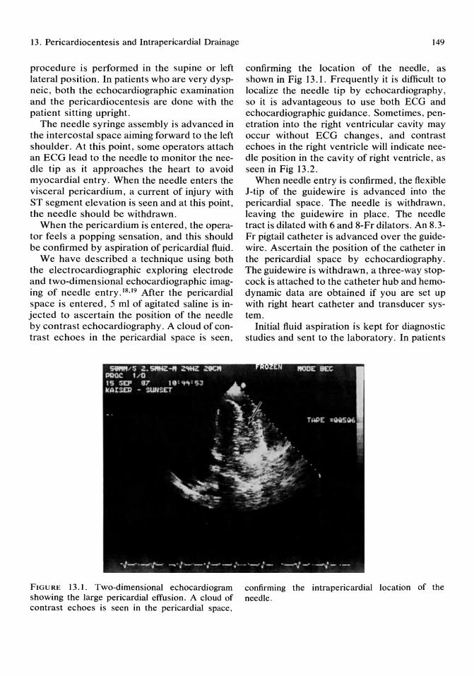
13. Pericardiocentesis and Intrapericardial Drainage
procedure is performed in the supine or left lateral position. In patients who are very dyspneic, both the echocardiographic examination and the pericardiocentesis are done with the patient sitting upright.
The needle syringe assembly is advanced in the intercostal space aiming forward to the left shoulder. At this point, some operators attach an ECG lead to the needle to monitor the needle tip as it approaches the heart to avoid myocardial entry. When the needle enters the visceral pericardium, a current of injury with ST segment elevation is seen and at this point, the needle should be withdrawn.
When the pericardium is entered, the operator feels a popping sensation, and this should be confirmed by aspiration of pericardial fluid.
We have described a technique using both the electrocardiographic exploring electrode and two-dimensional echocardiographic imaging of needle entry.18.19 After the pericardial space is entered, 5 ml of agitated saline is injected to ascertain the position of the needle by contrast echocardiography. A cloud of contrast echoes in the pericardial space is seen,
FIGURE 13.1. Two-dimensional echocardiogram showing the large pericardial effusion. A cloud of contrast echoes is seen in the pericardial space,
149
confirming the location of the needle, as shown in Fig 13.1. Frequently it is difficult to localize the needle tip by echocardiography, so it is advantageous to use both EeG and echocardiographic guidance. Sometimes, penetration into the right ventricular cavity may occur without ECG changes, and contrast echoes in the right ventricle will indicate needle position in the cavity of right ventricle, as seen in Fig 13.2.
When needle entry is confirmed, the flexible J-tip of the guidewire is advanced into the pericardial space. The needle is withdrawn, leaving the guide wire in place. The needle tract is dilated with 6 and 8-Fr dilators. An 8.3-Fr pigtail catheter is advanced over the guidewire. Ascertain the position of the catheter in the pericardial space by echocardiography. The guide wire is withdrawn, a three-way stopcock is attached to the catheter hub and hemodynamic data are obtained if you are set up with right heart catheter and transducer system.
Initial fluid aspiration is kept for diagnostic studies and sent to the laboratory. In patients
confirming the intrapericardial location of the needle.
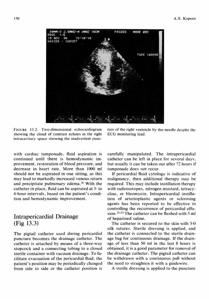
150
FIGURE 13.2. Two-dimensional echocardiogram showing the cloud of contrast echoes in the right intracavitary space showing the inadvertent punc-
with cardiac tamponade, fluid aspiration is continued until there is hemodynamic improvement, restoration of blood pressure, and decrease in heart rate. More than 1000 ml should not be aspirated in one sitting, as this may lead to markedly increased venous return and precipitate pulmonary edema.20 With the catheter in place, fluid can be aspirated at 3- to 4-hour intervals, based on the patient's condition and hemodynamic improvement.
Intrapericardial Drainage (Fig 13.3)
The pigtail catheter used during pericardial puncture becomes the drainage catheter. The catheter is attached by means of a three-way stopcock and a connecting tubing to a closed sterile container with vacuum drainage. To facilitate evacuation of the pericardial fluid, the patient's position may be periodically changed from side to side or the catheter position is
A.S. Kapoor
ture of the right ventricle by the needle despite the ECG monitoring lead.
carefully manipulated. The intrapericardial catheter can be left in place for several days, but usually it can be taken out after 72 hours if tamponade does not recur.
If pericardial fluid cytology is indicative of malignancy, then additional therapy may be required. This may include instillation therapy with radioisotopes, nitrogen mustard, tetracycline, or bleomycin. Intrapericardial instillation of arterioplastic agents or sclerosing agents has been reported to be effective in controlling the recurrence of pericardial effusion. 21- 23 The catheter can be flushed with 5 ml of heparined saline.
The catheter is secured to the skin with 3-0 silk sutures. Sterile dressing is applied, and the catheter is connected to the sterile drainage bag for continuous drainage. If the drainage of less than 50 ml in the last 8 hours is obtained, it is a good parameter for removal of the drainage catheter. The pigtail catheter can be withdrawn with a continuous pull without the need to straighten it with a guidewire.
A sterile dressing is applied to the puncture

13. Pericardiocentesis and Intrapericardial Drainage 151
Malignant Pericardial Effusion
or
Recurring Cardiac Tamponade
t Hemodynamic Support
1 + Volume expansion ~ Isuprel Infusion
PERICARDIOCENTESIS
(with ECG + Echocardiographic Guidance)
.~ Mal~gnant Process
1 Indwelling Catheter Drainage 1 (2-3 days)
Intrapericardial Instillation of a scleros~ng agent
(Bleomycin or Tetracycline)
Local Radiation
J
'l-Constrictive Physiology
t Effusive-Constrictive
Pericarditis
1 PERICARDIECTOMY
Recurrence '1-. Remlsslon
1 Partial 1 Pericardiectomy Follow-up
FIGURE 13.3. Scheme for managing malignant pericardial effusion or recurring cardiac tamponade.
site. In cases of emergent pericardiocentesis, hemodynamic measurement, and intrapericardial pressure measurements can be dispensed and pericardiocentesis at bedside can be performed for stabilizing the patient. In addition, rapid volume expansion should be instituted and dobutamine or isoproterenol infusion can be administered to improve cardiac output.
Pericardial Fluid Analysis
Normal pericardial fluid is an ultrafiltrate of blood serum with 1.7% to 3.5% protein, a colloid osmotic pressure 25% of that of serum,
and is usually 25 to 30 mI.24 The fluid should be analyzed for physical characteristics, like pH, volume, color, specific gravity, chemistry with assessment of protein, glucose content, and biochemistry to distinguish an exudate from a transudate. Gram stains, cultures of aerobic, anaerobic, acid-fast, and fungal elements should be ordered. Viral titers may be of help in patients with idiopathic or viral myocarditis. Hematologic studies like hemoglobin, hematocrit, white blood cells, and differential count should be routinely performed. For further immunologic or other studies, fluid can be redrawn as necessary from the drainage catheter.

152
Complications of Pericardiocentesis Complications and risks of pericardiocentesis should be anticipated, and management of complications should be planned. The major complications of pericardiocentesis include laceration of a coronary artery, puncture of a cardiac chamber, pneumothorax, infection, and ventricular fibrillation. Minor complications include vasovagal reactions, hypotension, bradycardia, and arrhythmias. Hemopericardium usually results from chamber perforation or coronary artery laceration. If cardiac puncture occurs, the needle and guidewire should be promptly withdrawn. The patient should be monitored for worsening cardiac tamponade. Immediate pericardiocentesis for relief of tamponade may have to be performed. A cardiothoracic surgeon should be alerted because if hemopericardium persists, prompt surgical exploration is necessary to locate the bleeding site.
For pneumothorax, which is rare, a pleural tube drainage may be necessary.
If ventricular fibrillation occurs, the needle should be withdrawn and the patient should be immediately defibrillated.
Pericardioscopy
To improve the yield from pericardiocentesis one can visualize the pericardial surfaces by a flexible fiberoptic pericardioscope. A biopsy of the pericardium also can be performed, guided by the pericardioscope. Pericardioscopy can reveal malignant involvement in the pericardium, especially in cases of malignant melanoma, lymphomas, or other large nodules on the surface.
The pericardioscopic system consists of a flexible fiberoptic bronchoscope with a biopsy and suctioning channel. The camera system consists of an Olympus television camera system for endoscopy and a beam splitter that allows one to observe the procedure. 25 The role of pericardioscopy in the assessment of pericardial disease needs to be further studied.
A.S. Kapoor
Development of new pericardioscopes using laser system may be the way to go for better definition and delineation of pericardial disease.
References
1. Engle PJ, Hon H, Fowler NO, et al: Echocardiographic study of right ventricular wall motion in cardiac tamponade. Am J Cardiol 1982; 50: 1018.
2. Settle HP, Adolph RJ, Fowler NO, et al: Echocardiographic study of cardiac tamponade. Circulation 1977; 56:951.
3. Chandraratna PAN, First J, Langeven E, et al: Echocardiographic contrast studies during pericardiocentesis. Ann Int Med 1977; 87: 199.
4. Callahan JA, Seward JB, Nishimura RA, et al: Two-dimensional echocardiographically guided pericardiocentesis: Experience in 117 consecutive patients. Am J Cardiol1985; 55:476-484.
5. Shijna A, Vaginuma T, Kordo K, et al: Echocardiographic evaluation of impending tamponade. J Cardiography 1979; 9:555.
6. Armstrong WF, Schilt BF, Helser DJ, et al: Diastolic collapse of the right ventricle with cardiac tamponade. Circulation 1982; 65: 1491.
7. Hancock EW: Cardiac tamponade, in Scheinman, MM (ed): Cardiac Emergencies. Philadelphia, W. B. Saunders Co, 1984.
8. Spodick DH: The normal and diseased pericardium: Current concepts of pericardial physiology, diagnosis and treatment. J Am Coll Cardiol 1983; 1:240-251.
9. Shabetei R: Changing concepts of cardiac tamponade. Mod Concepts Cardiovasc Dis 1983; 52: 19-23.
10. Spodick DH: Acute cardiac tamponade: Pathologic physiology, diagnosis and management. Diag Cardiovascul Dis 1964; 10:64.
11. Spodick DH: Observations in cardiac tamponade. Physiologic considerations. Bull TuftsN Engl Med Center 1956; 2: 191-197.
12. Wynne J, Markis JE, Grossman W: Extrinsic compression of the heart by tumor masquerading as cardiac tamponade. Cathet Cardiovasc Diagn 1978; 4:81-85.
13. Roberts WC, Ferrans VJ: A survey of the causes and consequences of pericardial heart disease, in Reddy PS (ed): Pericardial Disease. New York, Raven Press, 1981, pp 49-75.
14. DeLoach JF, Haynes JW: Secondary tumors of the heart and pericardium. Review of the sub-
13. Pericardiocentesis and Intrapericardial Drainage
ject and report of 137 cases. Arch Intern Med 1953; 91:224-249.
15. Shabetai R, Fowler NO, Fenton JC, et al: Pulsus paradoxus. J Clin Invest 1965; 44: 1882.
16. Chandraratna PA, Aronow WS: Detection of pericardial metastases by cross-sectional echocardiography. Circulation 1981; 63:197-199.
17. Rinkenberger RL, Polumbo RA, Bolten MR, et al: Mechanism of electrical alternans in patients with pericardial effusion. Cathet Cardiovasc Diagn 1978; 4:63-70.
18. Kapoor AS, Kraybill W, Reardon T: Improved technique for management of malignant effusion. Clin Res 1985; 33:293A.
19. Kapoor AS (ed): Malignant pericardial effusion and cardiac tamponade, in Kapoor AS (ed): Cancer and the Heart. New York, SpringerVerlag, 1986, pp 213-222.
20. Vandyke WH, Cure J, et al: Pulmonary edema
153
after pericardiocentesis for cardiac tamponade. N Engl J Med 1983; 309:595-596.
21. Smith FE, Lane M, Hudgins PT: Conservative management of malignant pericardial effusion. Cancer 1974; 33:47-57.
22. Clarke TH: Radioactive colloidal Au l98 in the treatment of neoplastic effusion. Northwest Univ Med School Bull 1952; 26:98-104.
23. Wei JY, Taylor GJ, Achuff SC: Recurrent tamponade and large pericardial effusions: Management with an indwelling pericardial catheter. Am J Cardiol 1978; 42:281.
24. Holt JP: The normal pericardium. Am J Cardiol 1970; 26:455-465.
25. Kondos GT, Rich S, Levitsky S: Flexible fiberoptic pericardioscopy for the diagnosis of pericardial disease. J Am Coli Cardiol 1986; 7:432-434.
14 Endomyocardial Biopsy Techniques and Interpretation Amar S. Kapoor and Mir Ali
Historical Perspective
Transvascular endomyocardial biopsy was initially performed by Sakakibara and Konno in 1962.1 Before the development of this technique, myocardial biopsy could only be performed by a limited thoracotomy and direct excision of a small myocardial tissue followed by suture repair. 2 Later on, a needle sample of myocardium after mediastinotomy was described, but this procedure was not practical and had many complications. 3 There were some other techniques using a percutaneous Silverman needle for left ventricular apical samples, but these had an unacceptably high morbidity. 4
Konno's bioptome revolutionized the myocardial biopsy technique because adequate multiple samples could be obtained and the procedure's safety was very acceptable. However, Konno's bioptome was rigid and required surgical venotomy for introduction. There were some modifications of this bioptome, namely, the instrument was made flexible and the jaw size was reduced for percutaneous entry. The Stanford bioptome, designed by Caves and Schultz,5,6 was developed for performing mUltiple serial biopsies for evaluating rejection in heart transplant patients. The Stanford bioptome (Fig 14.1) consists of a 9-Fr catheter measuring SO cm in length with one fixed and one mobile jaw attached proximally to surgical forceps.
There are other bioptomes with some modifications to allow left ventricular biopsy from
the femoral artery. 7 A transvascular endomyocardial biopsy technique specifically designed for infants and small children is a S.S-Fr bioptome designed by Lurie,8 which consists of a forceps with a soft plastic outer component and can be guided by a shaped catheter.
There have been technical advances in the design of the bioptome, but the principle features of Konno's bioptome have been retained. All heart transplant programs are founded on a bedrock of sequentially performed biopsies that have mostly subrogated the clinical dilemmas confounding the diagnosis of rejection in cardiac transplant recipients.
Endomyocardial biopsy has been a safe and simple technique and has been an important invasive technique in the evaluation of patients with suspected myocardial disease.
Indications for Endomyocardial Biopsy
One of the commonest indications for endomyocardial biopsy (EMB) is for detection and surveillance of cardiac transplant rejection (Table 14.1). Biopsy permits accurate detection of rejection and severity of histologic changes so that appropriate immunosuppressive therapy can be administered. With appropriate therapy, rejection may resolve within 72 hours.6 Serial biopsies would be needed in this setting.
Endomyocardial biopsy also is used in the
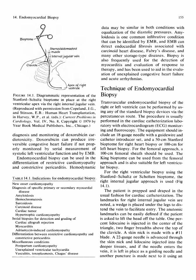
14. Endomyocardial Biopsy
Bioptome
&Iernocleidoma:;toid muscle
Internal jugular vein
FIGURE 14.1. Diagrammatic representation of the Stanford-Schultz bioptome in place at the right ventricular apex via the right internal jugular vein. (Reproduced with permission from Copeland, J.G., and Stinson, E.B.: Human Heart Transplantation, in Harvey, W.P., et al. (eds.): Currer.t Problems in Cardiology, Vol. IV, No.8, Copyright © 1979 by Year Book Medical Publishers, Inc., Chicago.)
diagnosis and monitoring of doxorubicin cardiotoxicity. Doxorubicin can produce irreversible congestive heart failure if not properly monitored by serial measurement of systolic left ventricular function and by EMB.
Endomyocardial biopsy can be used in the differentiation of restrictive cardiomyopathy and constrictive pericarditis. Hemodynamic
TABLE 14.1. Indications for endomyocardial biopsy.
New onset cardiomyopathy Diagnosis of specific primary or secondary myocardial
disease Amyloidosis Hemochromatosis Sarcoidosis Carcinoid disease Cardiac tumor Hypertrophic cardiomyopathy
Serial biopsies for detection and grading of Cardiac allograft rejection Myocarditis Adriamycin-induced cardiomyopathy
Differentiation between restrictive cardiomyopathy and constrictive pericarditis
Miscellaneous conditions Postpartum cardiomyopathy Unexplained ventricular tachycardia Vasculitis, toxoplasmosis, Chagas' disease
155
data may be similar in both conditions with equalization of the diastolic pressures. Amyloidosis is one common infiltrative condition that can be identified by EMB, and EMB can detect endocardial fibrosis associated with carcinoid heart disease, Fabry's disease, and many other storage-type diseases. Biopsy is also frequently used for the detection of myocarditis and evaluation of response to therapy, and has been used to aid in the evaluation of unexplained congestive heart failure and acute arrhythmias.
Technique of Endomyocardial Biopsy Transvascular endomyocardial biopsy of the right or left ventricle can be performed by using any of the standard biopsy devices via the percutanecus route. The procedure is usually performed in the cardiac catheterization laboratory with electrocardiogram (ECG) monitoring and fluoroscopy. The equipment should include an I8-gauge needle with a guidewire and catheter introducer set, and a 50-cm Stanford bioptome for right heart biopsy or IOO-cm for left heart biopsy. For the femoral approach, a IOO-cm femoral sheath should be used. The King bioptome can be used from the femoral approach and is also suitable for left ventricular biopsy.
For the right ventricular biopsy using the Stanford-Schultz or Scholten bioptome, the right internal jugular approach is used (Fig 14.1).
The patient is prepped and draped in the usual fashion for cardiac catheterization. The landmarks for right internal jugular vein are noted, a wedge is placed under the legs to distend the vein to facilitate entry. The anatomic landmarks can be easily defined if the patient is asked to lift the head off the table. One percent lidocaine is injected in the center of the triangle, two finger breadths above the top of the clavicle. A skin nick is made with a #11 blade. A 22-gauge needle is advanced through the skin nick and lidocaine injected into the deeper tissues, and if the needle enters the vein, it is left in place as a guiding needle and another puncture is made next to it using an
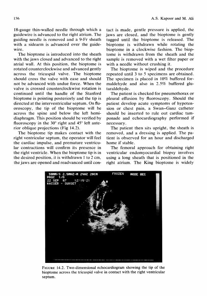
156
18-gauge thin-walled needle through which a guide wire is advanced to the right atrium. The guiding needle is removed and a 9-Fr sheath with a sidearm is advanced over the guidewire.
The bioptome is introduced into the sheath with the jaws closed and advanced to the right atrial wall. At this position, the bioptome is rotated counterclockwise and advanced gently across the tricuspid valve. The bioptome should cross the valve with ease and should not be advanced with undue force. When the valve is crossed counterclockwise rotation is continued until the handle of the Stanford bioptome is pointing posteriorly and the tip is directed at the interventricular septum. On fluoroscopy, the tip of the bioptome will be across the spine and below the left hemidiaphragm. This position should be verified by fluoroscopy in the 30° right and 45° left anterior oblique projections (Fig 14.2).
The bioptome tip makes contact with the right ventricular septum, the operator will feel the cardiac impulse, and premature ventricular contractions will confirm its presence in the right ventricle. When the bioptome tip is in the desired position, it is withdrawn 1 to 2 cm, the jaws are opened and readvanced until con-
A.S. Kapoor and M. Ali
tact is made, gentle pressure is applied, the jaws are closed, and the bioptome is gently tugged until the bioptome is released. The bioptome is withdrawn while rotating the bioptome in a clockwise fashion. The bioptome is withdrawn from the sheath and the sample is removed with a wet filter paper or with a needle without crushing it.
The bioptome is wiped and the procedure repeated until 3 to 5 specimens are obtained. The specimen is placed in 10% buffered formaldehyde and also in 2.5% buffered glutaraldehyde.
The patient is checked for pneumothorax or pleural effusion by fluoroscopy. Should the patient develop acute symptoms of hypotension or chest pain, a Swan-Ganz catheter should be inserted to rule out cardiac tamponade and echocardiography performed if necessary.
The patient then sits upright, the sheath is removed, and a dressing is applied. The patient is observed for an hour and discharged home if stable.
The femoral approach for obtaining right ventricular endomyocardial biopsy involves using a long sheath that is positioned in the right atrium. The King bioptome is widely
FIGURE 14.2. Two-dimensional echocardiogram showing the tip of the bioptome across the tricuspid valve in contact with the right ventricular septum.
14. Endomyocardial Biopsy
used in this position. This is a flexible modification of the stainless steel Olympus bronchoscopic biopsy forceps. The sheath is positioned over a cardiac catheter, which is withdrawn once the tip of the sheath is in the right atrium. The bioptome is introduced through the sheath until the bioptome exits the sheath and makes contact with the ventricular septum. Multiple biopsies are obtained by moving the sheath and bioptome together to different areas of the right ventricle.
The technique for performing left ventricular biopsy is slightly different and requires more meticulous care. For left ventricular biopsy, the Stanford left ventricular bioptome or the King bioptome can be used with the long sheath technique. lO,I1 The patient is prepared for percutaneous left heart catheterization using the Seldinger technique with the femoral artery. The pigtail catheter and long sheath assembly are set up with the pigtail end of the catheter out of the sheath. This assembly is advanced over the guidewire and advanced to the aortic valve position. The wire is removed and 5,000 U of heparin is given, and the assembly is advanced into the left ventricle in the standard fashion. Sometimes the guidewire may be necessary to cross the aortic valve. Once in the ventricle, the pigtail catheter is removed and the sheath is left in the ventricle. It is aspirated and flushed and the bioptome advanced to the left ventricular apex. Multiple specimens are obtained in the same manner as for right ventricular biopsy.
The King bioptome is disposable, but the Stanford bioptome is reusable and needs careful maintenance with saline cleansing followed by oil lubrication under pressure with a special instrument and gas sterilization. Periodically the jaws need sharpening.
Endomyocardial Biopsy Guided by Two-dimensional Echocardiography
The use of two-dimensional echocardiography to guide the bioptome is helpful in locating the biopsy site and in directing the bioptome tip to
157
the desired position. Echocardiography can be used as an adjunct to fluoroscopy to improve the safety of the technique and reduce radiation exposure.12
Before the biopsy, two-dimensional echocardiography is performed using the apical and subcostal windows. The bioptome is passed through the tricuspid valve with fluoroscopic guidance. Once in the ventricle, the tip can be identified by echocardiography and fluoroscopy is not required. The biopsy is performed in the standard manner after an optimal bioptome position is confirmed by echocardiography. 12, 13
Echocardiographic monitoring will aid in the immediate diagnosis of hemopericardium.
Echocardiography may replace fluoroscopy in the internal jugular approach for right ventricular biopsy.
Complications
Transvascular endomyocardial biopsy of the heart can be performed safely, but cardiac perforation occurs in 0.3% to 0.5% of cases with any of the techniques. 15 The diagnosis of cardiac tamponade can be confirmed by Swan-Ganz catheterization and echocardiography. Pericardiocentesis may be required if the patient becomes unstable. Rarely, thoracotomy may be required for continuous leaking from the perforation.
Various ventricular arrhythmias may be seen with catheter manipulation. These arrhythmias are self-limited or can be controlled by lidocaine or cardioversion. Atrial fibrillation may be precipitated by bioptome manipulation in the right atrium. Usually this can be controlled by further catheter manipulation or intravenous verapamil.
Systemic embolization is limited to left ventricular endomyocardial biopsy. The risk is reduced by frequent aspirations and flushings and use of heparin.
Other complications of vascular access, like pneumothorax or carotid artery puncture and hematoma, are also seen as with other procedures involving the vascular access from the internal jugular or subclavian vein.
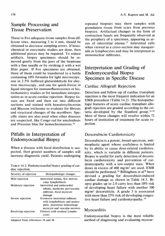
158
Sample Processing and Tissue Preservation
Three to five adequate tissue samples from different sites, measuring 2 to 4 mm, should be obtained to decrease sampling errors. If histochemical or enzymatic studies are done, then additional pieces may be required. To reduce artifacts, biopsy specimens should be removed gently from the jaws of the bioptome with a fine needle or by stroking it with a wet filter paper. If five specimens are obtained, three of them could be transferred to a bottle containing 10% formalin for light microscopy, one in 2.5% buffered glutaraldehyde for electron microscopy, and one for quick-freeze in liquid nitrogen for immunofluorescence or histochemistry studies or for immediate interpretation as in acute cardiac rejection. Biopsy tissues are fixed and then cut into different sections and stained with hematoxylin-eosin and Masson trichrome to evaluate for fibrosis and structural changes of the myofibers. Specific stains are also used when other diseases are suspected, like Congo red for amyloidosis and Prussian blue for hemochromatosis.16
Pitfalls in Interpretation of Endomyocardial Biopsy
When a disease with focal distribution is suspected, then greater numbers of samples will increase diagnostic yield. Patients undergoing
TABLE 14.2. Endomyocardial biopsy grading of cardiac rejection.
Severity of rejection Histopathologic changes
Mild rejection Interstitial edema, few perivas-cular Iymphoblasts
Moderate rejection Interstitial and endocardial edema, moderate perivascular infiltrate, focal myocyte damage
Severe rejection Extensive interstitial infiltrate with Iymphoblasts and neutrophils, interstitial hemorrhage myocyte and vascular necrosis
Resolving rejection Fibrosis with residual lympho-cytes
Adapted from references 16 and 18.
A.S. Kapoor and M. Ali
repeated biopsies may show samples with granulation tissue from scars from previous biopsies. Artifactual changes in the form of contraction bands are frequently observed at the periphery of a specimen, appearing as areas of interstitial edema. 17 Interstitial cells when viewed in a cross-section may masquerade as lymphocytes and may be interpreted as mononuclear infiltrates.
Interpretation and Grading of Endomyocardial Biopsy Specimen in Specific Disease
Cardiac Allograft Rejection
Detection and follow-up of cardiac transplant rejection is the most frequent indication for an EMB procedure (Table 14.2). The histopathologic features of acute cardiac transplant allograft rejection are graded according to the criteria developed by the Stanford group. 16, IS Most of these changes will resolve within 72 hours of institution of treatment for acute rejection.
Doxorubicin Cardiotoxicity
Doxorubicin is a potent, broad-spectrum, antineoplastic agent whose usefulness is limited by its ability to cause dose-related cardiotoxicity, which is variable in different patients. Biopsy is useful for early detection of doxorubicin cardiotoxicity and prevention of cardiomyopathy with a low-output state. When doses in excess of 400 mg/m2 are used, EMB should be performed. 18 Billingham et aIlS have devised a grading for doxorubicin-induced cardiac damage as shown in Table 14.3. Biopsy grades up to 2.0 carry less than 10% risk of developing heart failure with another 100 mg/m2 doxorubicin. A grade 3 is associated with more than 25% risk of developing congestive heart failure and cardiomyopathy.19
Myocarditis
Endomyocardial biopsy is the most reliable method of diagnosing and evaluating myocar-

14. Endomyocardial Biopsy
TABLE 14.3. Semiquantitative scale of doxorubicin myocardial damage.
Grade Histopathologic features
o No change from normal Less than 5% of cells per block showing myofi
brillar loss 1.5 5-10% of cells showing cytoplasmic vacuoliza-
tion-myofibrillar loss 2 Groups of cells (16-25%) showing myofibrillar
loss and cytoplasmic vacuolization 2.5 Groups of cells (26-35%) showing marked
myofibrillar loss and cytoplasmic vacuoliza· tion
3 Diffuse cell injury (35%) with total loss of contractile elements, mitochondria, and nuclear degeneration
Adapted from reference 18.
ditis. Until recently, the issue of myocarditis was befogged with incongruities in regard to its interpretation. A more rigid definition and classification of myocarditis was put forth by a team of experienced pathologists in conjunction with the American College of Cardiology, called the Dallas Classification System.20 Myocarditis is defined "as a process characterized by an inflammatory infiltrate of the myocardium with necrosis and/or degeneration of adjacent myocytes not typical of the ischemic damage associated with coronary artery disease. "20 Two working classifications were devised for the first biopsy and subsequent biopsies. The inflammatory infiltrate was subclassified as lymphocytic, eosinophilic, neutrophilic, giant cell, granulomatous, or mixed. The severity of the infiltrate was described as mild, moderate, or severe and focal, confluent, or diffuse, respectively.22 A lymphocytic infiltrate is found in the viral or postviral form of myocarditis. When eosinophils are present, a hypereosinophilic state should be suspected. In sarcoidosis, giant cells are seen frequently.
A frequently misleading lesion on biopsy is one induced by pressor agents. The myocarditis associated with pressor agents is characterized by areas of myocyte necrosis and mixed inflammatory infiltrate, including neutrophils. This is the catecholamine effect that produces the pressor lesion.22
159
There has been controversy in the management of myocarditis. In the experience of some, myocarditis may remit spontaneously;23 whereas in the experience of others,24,25 treatment of myocarditis with prednisone and azathioprine has shown from 40% to 100% clinical and hemodynamic improvement. There is an ongoing multicenter myocarditis trial which is designed to enter patients with biopsy-proven myocarditis determined by the Dallas criteria and to randomize treatment to one of the three arms: conventional, immunosuppressive arm with prednisone and azathioprine, and the third arm with low-dose prednisone and cyclosporine. This long-awaited study will provide us with information on the natural history of myocarditis and therapeutic recommendations.
There are several other conditions in which specific cardiac diagnosis can be rendered by EMB, like sarcoidosis, amyloidosis, endocardial fibrosis, and hemochromatosis of the heart. It is beyond the scope of this chapter to go into the details of each condition.
Concluding Remarks
With the current bioptomes, it is safe and feasible to perform multiple biopsies of both ventricles, and the diagnostic information obtained from the biopsy obtained from the interventricular septum of both ventricles is not very different. Right ventricular endomyocardial biopsy is done on an outpatient basis.
We have learned from the use of EMB the significant role of myocarditis in the development of dilated cardiomyopathy. It is the view of many leading experts that a subset of patients with dilated cardiomyopathy have histologic evidence of myocarditis. 26 Endomyocardial biopsy in conjunction with biochemical, pharmacologic, and cell culture techniques will assist us in obtaining definitive diagnostic information on patients with dilated cardiomyopathy.
Further advances in bioptome technology with steerable biopsy catheters and laser angioscopy will add a dimension to our understanding of the state of myocardial cells.
160
References
1. Sakakibara S, Konno S: Endomyocardia1 biopsy. Jpn Heart J 1962; 3:537-543.
2. Weinberg M, et al: Diagnostic biopsy of pericardium and myocardium. Arch Surg 1958; 176:825.
3. Sutton DC, Sutton GC: Needle biopsy of the human ventricular myocardium: Review of 54 consecutive cases. Am Heart J 1960; 50:364.
4. Shirey EK, et al: Percutaneous myocardial biopsy of the left ventricle: Experience in 198 patients. Circulation 1972; 66: 112.
5. Caves PK, et al: New instrument for transvenous cardiac biopsy. Am J Cardio! 1974; 33:264.
6. Caves PK, et al: Serial trans venous biopsy of the transplanted human heart: Improved management of acute rejection episodes. Lancet 1974; 1 :821.
7. Richardson PH: King's endomyocardial bioptome. Lancet 1974; 1:660.
8. Lurie PR: Revision of pediatric endomyocardial biopsy technique. Am J Cardiol 1987; 60:368.
9. Bristow MR, et al: Doxorubicin cardiomyopathy: Evaluation by phonocardiography, endomyocardial biopsy, and cardiac catheterization. Ann Intern Med 1978; 88: 168.
10. Mason JW: Techniques for right and left endomyocardial biopsy. Am J Cardiol1978; 41 :887-892.
11. Brooksby lAB, et al: Left ventricular endomyocardial biopsy. I. Description and evaluation of the technique. Cathet Cardiouasc Diagn 1977; 3:115-121.
12. French WJ, Popp RL, Pitlick PT: Cardiac localization of transvascular bioptome using two-dimensional echocardiography. Am J Cardia! 1983; 51:219-223.
13. Mortensen SA, Egeblad H: Endomyocardial biopsy guided by cross sectional echocardiography. Br Heart J 1983; 50:246.
A.S. Kapoor and M. Ali
14. Plerard L: Two dimensional echocardiography: Guiding of end om yo cardia 1 biopsy. Chest 1984; 85:759-762.
15. Sekiguchi M, Take M: World survey of catheter biopsy of the heart, in Sekiguchi M, Olson EGJ (eds): Cardiomyopathy. Baltimore, University Press, 1980, pp 217-225.
16. Mason JW, Billingham ME: Myocardial biopsy, in Yu PH, Goodwin JF (eds): Progress in Cardiology. Philadelphia, Lea & Febiger, 1980, pp 113-146.
17. Olmesdahl PH, et al: Ultrastructural artifacts in biopsied normal myocardium and their relevance to myocardial biopsy in man. Thorax 1979; 34:82.
18. Billingham ME, et al: Anthracycline cardiomyopathy monitored by morphologic changes. Cancer Treat Rep 1978; 62:865-872.
19. Fowles RE: Cardiac catheterization and endomyocardial biopsy, in Kapoor AS (ed): Cancer and the Heart. New York, Springer-Verlag, 1986, pp 42-50.
20. Ardz TH, et al: Myocarditis, a histopathologic definition and classification. Am J Cardiouasc Patho! 1986; 1:3-14.
21. Edwards WD: Myocarditis and endomyocardial biopsy. Cardiol Clin 1984; 2:647-656.
22. Haft 11: Cardiovascular injury induced by sympathetic catecholamines. Prog Cardiouasc Dis 1974; 17:73-86.
23. Sekiguchi M, et al: Natural history of 20 patients with biopsy-proven acute myocarditis. A ten-year follow-up study. Circulation 1985; 72:\09.
24. Fenoglio JJ, et al: Diagnosis and classification of myocarditis by endomyocardial biopsy. N Engl J Med 1983; 308:12
25. Hess ML, et al: Inflammatory myocarditis: Incidence and response to T-Iymphocyte depletion (abst). J Am Coli Cardiol 1984; 1:584.
26. O'Connell JB, et al: Dilated cardiomyopathy: Emerging role of endomyocardial biospy. Curr Prohl Cardiol 1986; 9(8):450-505.

15 Dipyridamole Thallium for Evaluating Coronary Artery Disease Diane Sobkowicz and David E. Blumfield
Historical Perspective Dipyridamole [2 ,6-bis( diethanolamino )-4,8-dipyperidne-pyrimido-(5 ,4-d)pyrimidine] was initially studied in the 1950s as a synthetic compound that produces coronary vasodilatation. Dipyridamole was initially studied as an antianginal agent that did not increase myocardial oxygen consumption. Early reports by Bretschneider and co-workers' noted an increase in coronary blood flow from 200% to 400% in anesthetized dogs without an increase in cardiac output. Other studies2 supported this but noted a lesser increase in coronary blood flow of only 30% to 90% with a decrease in blood pressure of 20 mm Hg and an associated increase in heart rate after injection of dipyridamole. Keise et aI,2 Fischer and FiegeI,3 and Junemann4 studied dipyridamole in patients with coronary insufficiency as well as congestive heart failure with encouraging results: Anginal symptoms were noted to decrease in some patients, and those with congestive heart failure were found to require lower dosages of digoxin to stabilize their heart failure when used in conjunction with dipyridamole. However, in 1962, Wendt et al6
noted that the decrease in coronary vascular resistance associated with dipyridamole also was associated with an increase in myocardial oxygen consumption and, in some patients, a decrease in left ventricular work. Therefore, it was postulated that the dissociation between cardiac work and myocardial oxygen consumption was secondary to the action of dipyridamole on cellular metabolism causing an
increase in the oxygen demand necessitating an increase in coronary blood flow.
In the 1960s and early 1970s the use of dipyridamole as an anti anginal agent declined and was largely replaced by nitrates. However, since the elucidation of the mechanism of action of dipyridamole on normal coronary blood flow, further studies were performed in animal models to determine its effects in the presence of regional flow abnormalities. Studies on canine models with acute and chronic ischemic heart disease were performed to determine the effect of dipyridamole on occluded coronary arteries. In 1973, Marshall and Parratt" studied canine hearts with one totally occluded coronary artery branch and observed coronary blood flow to the ischemic area. It was observed that dipyridamole decreased coronary blood flow to the region supplied by the occluded artery. This was further supported by Nakamura et aI'2 and Flameng et aI'3 using a different animal model, finding a reduction of coronary blood flow in the ischemic region. Dipyridamole was associated with a reduction of coronary blood flow in the region of an occluded vessel and was found to increase coronary blood flow to the normal regions, thus creating a "steal phenomenon."
Mechanism of Action
The mechanism of action of dipyridamole involves the production of coronary vasodilatation relates to the autoregulation of coronary blood flow.

162
Briefly, autoregulation of coronary blood flow is dependent on several mechanisms which include: 1) myogenic factors, 2) tissue factors, and 3) metabolic factors. Metabolic factors include decreased oxygen tension, CO2 concentration, and vasodilator metabolites. Adenosine, as a breakdown product of adenosine monophosphate (AMP), is believed to be a critical mediator in metabolically induced vasodilatation. 7 It also plays a key role in dipyridamole-induced coronary artery vasodilatation.
Adenosine, unlike ATP and AMP, can easily diffuse out of the myocyte and into the interstitium where two processes can occur. Adenosine can be taken up by red blood cells, endothelial cells, or myocytes and broken down by adenosine deaminase to inosine, or smooth muscle cells of the coronary vasculature can also take up adenosine by specific receptors. Theoretically, at the receptor level, adenosine blocks calcium entry into the smooth muscle cell. This results in cell relaxation and vasodilatation ensues. The mechanism of action of dipyridamole is, then, via an adenosine-sparing effect. As noted by Bunag et al,8 dipyridamole inhibits adenosine deaminase and, more importantly, has been shown to block uptake of adenosine by the myocyte, red blood cell, and endocardial cell resulting in more adenosine available to bind the smooth muscle cell of the coronary vasculature for maximal dilatation. The fact that the methyl xanthine derivative, aminophyline, has an antagonistic effect on the vasodilatation caused by dipyridamole is supportive evidence for this theory. Aminophyline acts on the smooth muscles of the coronary bed to antagonize the action of adenosine by behaving as a competitive inhibitor of adenosine at the smooth muscle receptor level when given in sufficient concentrations.9,10 Complete antagonism of adenosine results, reversing the coronary dilatory effects of dipyridamole.
Intravenous Dipyridamole Studies
In 1981, Feldman et aJl4 evaluated the acute coronary hemodynamic and metabolic effects of intravenous dipyridamole and also noted
D. Sobkowicz and D.E. Blumfield
that during dipyridamole-induced hyperemia, regional blood flow and metabolic responses depended on the status of the artery supplying that particular region, i.e., in regions supplied by an abnormal artery lactate production was increased, supporting the theory of redistribution of coronary blood flow.
These data suggested that dipyridamole-induced vasodilatation and the "steal phenomenon" might be useful in assessing the presence of human coronary narrowings. Gould and coworkers l7•18 performed several studies using intravenous dipyridamole coupled with thallium-201 myocardial perfusion imaging. These showed that in conjunction with dipyridamole infusion, thallium scanning could reliably detect and possibly localize coronary artery disease. Josephson et aJl9 also noted that coronary blood flow in response to dipryidamole was similar to or greater than that during exercise, but without the physiologic increase in myocardial oxygen demand.
Method
Initially, intravenous dipyridamole was used for these comparison studies (Table 15.1).12
TABLE 15.1. Procedure for intravenous dipyridamole imaging.
Materials needed: Intravenous dipyridamole Intravenous aminophyline Equipment to start and maintain intravenous line Thallous chloride Consent form (investigational)
Protocol Obtain baseline ECG, BP Start keep open IV; maintain access Administer dipyridamole; begin pharmacologic dilata
tion Monitor BP, ECG every 1 min If possible, exercise 4-6 min beginning Bruce stage 0;
reduce background Inject thallium Begin imaging within 5 min Obtain usual images; usual thallium protocol Administer aminophyline 125 mg IV; reverse dipyrid-
amole Observe patient; orthostatic changes Remove IV in nuclear department; avoid "hot"
tubing on wards Instruct patient to return (if necessary); delayed
images

15. Dipyridamole Thallium for Evaluating Coronary Artery Disease 163
The patient is instructed to fast before the test, and to avoid coffee, tea, and xanthine preparations for the 24 hours preceding the study. Under ECG monitoring, while supine, the patient receives intravenous dipyridamole at a rate of 0.14 mg/kg per minute over 4 minutes for a total of 0.56 mg/kg through a large antecubital vein. After the infusion, the patient is brought to the upright position and 3 minutes later, 2.0 to 3.0 mCi of intravenous thallium-201 is injected. (Bringing the patient to the upright position has been found to decrease the background uptake of thallium and therefore improve the myocardial to background ratio. This is believed to be due to the observation that pulmonary blood volume, transit time, and capillary surface area are decreased in the upright position, thereby decreasing the pulmonary uptake of thallium. 17)
One minute after the injection of thallium (4 minutes after the intravenous injection of dipyridamole at maximal hyperemia), myocardial images are acquired in the usual manner and delayed images are obtained at 150 to 180 minutes after thallium injection.
Physiologic effects of dipyridamole in humans include an average increase in coronary blood flow of 400% with an increase in heart rate of 23% to 38%, and a decrease in blood pressure of 10 to 20 mm Hg or lessY·19
Adverse effects (Table 15.2) of intravenous dipyridamole have been noted in about 40% of patients. Headache, nausea, dizziness, and chest pain are the most frequently described complaints. Only about twenty-five percent of the patients with chest pain were noted to have coronary artery disease. I9-21 When chest pain occurred, it was usually within 3 to 4 minutes of injection of dipyridamole and could be reversed within seconds by the intravenous injection of aminophyline (125 to 250 mg).17 These data suggest that the chest pain after dipyridamole is nonspecific and not helpful in the diagnosis of coronary artery disease.
Oral Dipyridamole Studies
Although intravenous dipyridamole has been determined to be safe and feasible, oral dipyridamole was evaluated largely because of its
TABLE 15.2. Procedure for oral dipyridamole thallium imaging.
Materials needed: Oral suspension of dipyridamole, 400 mg Intravenous aminophyline Equipment to start and maintain intravenous line Thallous chloride Consent form (if required by institution)
Protocol Obtain baseline ECG and BP Start keep open IV; maintain access Mix, dilute, and administer dipyridamole; begin phar
mocologic dilitation Monitor BP and ECG every 5 min At 45 min begin exercise, Bruce 0; reduce back
ground If cardiovasculare symptoms skip to thallium injec-
tion; exercise not needed Continue thru stage 1/2 Inject thallium at 6 min or fatigue or symptoms Begin imaging within 5 min Obtain usual images; usual thallium protocol Administer aminophyline 125 mg IV; reverse dipyrid-
amole Observe patient 5-10 min further; orthostatic changes Remove IV in nuclear department; avoid "'hot"
tubing on wards Instruct patient to return (if necessary); delayed
images
availability. In 1986, Taillefer et aJ20 performed a comparison study of oral and intravenous dipyridamole. Oral dipyridamole at a dose of 400 mg in tablet form, was shown to be a reliable alternative to intravenous dipyridamole in the evaluation of coronary artery disease with comparable sensitivity and specificity. Oral doses of 200 and 300 mg were tried but found to be less sensitive. However, the distinct disadvantage of oral dipyridamole is the delayed and somewhat variable absorption such that maximal hyperemia was delayed to approximately 45 minutes. It is somewhat more consistent with an oral suspension2o.21 made from standard tablets crushed and suspended in a standard sorbitol solution which is then diluted before administration (Table 15.2). Again, no dysrhythmias, infarctions, or deaths have been reported with oral dipyridamole and the side effects are similar to the intravenous form. Except for headache and nausea, the side effects were, however, less severe and somewhat less frequent with oral dipyridamole. In addition, oral dipyridamole
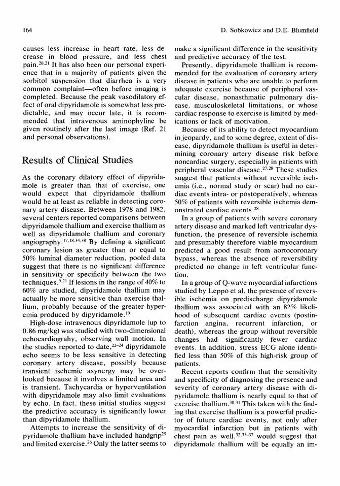
164
causes less increase in heart rate, less decrease in blood pressure, and less chest pain.20,21 It has also been our personal experience that in a majority of patients given the sorbitol suspension that diarrhea is a very common complaint-often before imaging is completed. Because the peak vasodilatory effect of oral dipyridamole is somewhat less predictable, and may occur late, it is recommended that intravenous aminophyline be given routinely after the last image (Ref. 21 and personal observations).
Results of Clinical Studies
As the coronary dilatory effect of dipyridamole is greater than that of exercise, one would expect that dipyridamole thallium would be at least as reliable in detecting coronary artery disease. Between 1978 and 1982, several centers reported comparisons between dipyridamole thallium and exercise thallium as well as dipyridamole thallium and coronary angiography.17,18,34,38 By defining a significant coronary lesion as greater than or equal to 50% luminal diameter reduction, pooled data suggest that there is no significant difference in sensitivity or specificity between the two techniques.9,21 Iflesions in the range of 40% to 60% are studied, dipyridamole thallium may actually be more sensitive than exercise thallium, probably because of the greater hyperemia produced by dipyridamole. 19
High-dose intravenous dipyridamole (up to 0.86 mg/kg) was studied with two-dimensional echocardiograhy, observing wall motion. In the studies reported to date,22-24 dipyridamole echo seems to be less sensitive in detecting coronary artery disease, possibly because transient ischemic asynergy may be overlooked because it involves a limited area and is transient. Tachycardia or hyperventilation with dipyridamole may also limit evaluations by echo. In fact, these initial studies suggest the predictive accuracy is significantly lower than dipyridamole thallium.
Attempts to increase the sensitivity of dipyridamole thallium have included handgrip25 and limited exercise.26 Only the latter seems to
D, Sobkowicz and D.E. Blumfield
make a significant difference in the sensitivity and predictive accuracy of the test.
Presently, dipyridamole thallium is recommended for the evaluation of coronary artery disease in patients who are unable to perform adequate exercise because of peripheral vascular disease, nonasthmatic pulmonary disease, musculoskeletal limitations, or whose cardiac response to exercise is limited by medications or lack of motivation,
Because of its ability to detect myocardium in jeopardy, and to some degree, extent of disease, dipyridamole thallium is useful in determining coronary artery disease risk before noncardiac surgery, especially in patients with peripheral vascular disease. 27 ,28 These studies suggest that patients without reversible ischemia (i.e., normal study or scar) had no cardiac events intra- or postoperatively, whereas 50% of patients with reversible ischemia demonstrated cardiac events. 28
In a group of patients with severe coronary artery disease and marked left ventricular dysfunction, the presence of reversible ischemia and presumably therefore viable myocardium predicted a good result from aortocoronary bypass, whereas the absence of reversibility predicted no change in left ventricular function.
In a group of Q-wave myocardial infarctions studied by Leppo et aI, the presence of reversible ischemia on predischarge dipyridamole thallium was associated with an 82% likelihood of subsequent cardiac events (postinfarction angina, recurrent infarction, or death), whereas the group without reversible changes had significantly fewer cardiac events. In addition, stress ECG alone identified less than 50% of this high-risk group of patients.
Recent reports confirm that the sensitivity and specificity of diagnosing the presence and severity of coronary artery disease with dipyridamole thallium is nearly equal to that of exercise thallium.30,31 This taken with the finding that exercise thallium is a powerful predictor of future cardiac events, not only after myocardial infarction but in patients with chest pain as well,32,35-37 would suggest that dipyridamole thallium will be equally an im-
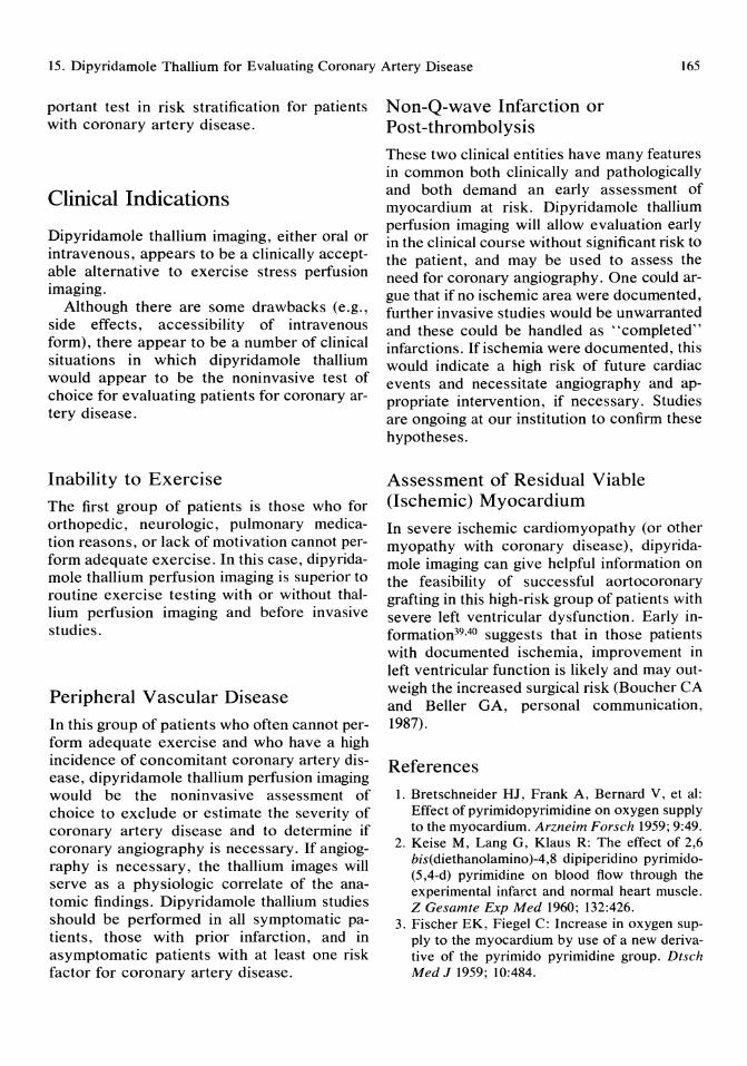
15. Dipyridamole Thallium for Evaluating Coronary Artery Disease 165
portant test in risk stratification for patients with coronary artery disease.
Clinical Indications
Dipyridamole thallium imaging, either oral or intravenous, appears to be a clinically acceptable alternative to exercise stress perfusion imaging.
Although there are some drawbacks (e.g., side effects, accessibility of intravenous form), there appear to be a number of clinical situations in which dipyridamole thallium would appear to be the noninvasive test of choice for evaluating patients for coronary artery disease.
Inability to Exercise
The first group of patients is those who for orthopedic, neurologic, pulmonary medication reasons, or lack of motivation cannot perform adequate exercise. In this case, dipyridamole thallium perfusion imaging is superior to routine exercise testing with or without thallium perfusion imaging and before invasive studies.
Peripheral Vascular Disease
In this group of patients who often cannot perform adequate exercise and who have a high incidence of concomitant coronary artery disease, dipyridamole thallium perfusion imaging would be the noninvasive assessment of choice to exclude or estimate the severity of coronary artery disease and to determine if coronary angiography is necessary. If angiography is necessary, the thallium images will serve as a physiologic correlate of the anatomic findings. Dipyridamole thallium studies should be performed in all symptomatic patients, those with prior infarction, and in asymptomatic patients with at least one risk factor for coronary artery disease.
Non-Q-wave Infarction or Post-thrombolysis These two clinical entities have many features in common both clinically and pathologically and both demand an early assessment of myocardium at risk. Dipyridamole thallium perfusion imaging will allow evaluation early in the clinical course without significant risk to the patient, and may be used to assess the need for coronary angiography. One could argue that if no ischemic area were documented, further invasive studies would be unwarranted and these could be handled as "completed" infarctions. If ischemia were documented, this would indicate a high risk of future cardiac events and necessitate angiography and appropriate intervention, if necessary. Studies are ongoing at our institution to confirm these hypotheses.
Assessment of Residual Viable (Ischemic) Myocardium
In severe ischemic cardiomyopathy (or other myopathy with coronary disease), dipyridamole imaging can give helpful information on the feasibility of successful aortocoronary grafting in this high-risk group of patients with severe left ventricular dysfunction. Early information39,4o suggests that in those patients with documented ischemia, improvement in left ventricular function is likely and may outweigh the increased surgical risk (Boucher CA and Beller GA, personal communication, 1987).
References
I. Bretschneider HJ, Frank A, Bernard V, et al: Effect of pyrimidopyrimidine on oxygen supply to the myocardium. Arzneim Forsch 1959; 9:49.
2. Keise M, Lang G, Klaus R: The effect of 2,6 bis(diethanolamino)-4,8 dipiperidino pyrimido(5,4-d) pyrimidine on blood flow through the experimental infarct and normal heart muscle. Z Gesamte Exp Med 1960; 132:426.
3. Fischer EK, Fiegel C: Increase in oxygen supply to the myocardium by use of a new derivative of the pyrimido pyrimidine group, Dtsch Med J 1959; 10:484.

166
4. Junemann C: Persantin: A new compound for the treatment of coronary insufficiency. Miinchen Med Wochenschr 1959; tOl:340.
5. Guyton AC, Textbook of Medical Physiology, ed 7. Philadelphia, W. B. Saunders, 1986, p 296.
6. Wendt VE, Sundermeyer JF, denBakkerPB, et al: The relationship between coronary blood flow, myocardial oxygen consumption and cardiac work as influenced by Persantin. Am J Cardiol 1962; 9:449.
7. Braunwald E, Sobel BE: Regulation of coronary blood flow, in Braunwald E (ed): Heart Disease ed 2. Philadelphia, W. B. Saunders Co., 1984, p 1238.
8. Bunag RD, Douglas CR, Imai S, et al: Influence of pyrimidopyrimidine derivative on deamination of adenosine by blood. Circ Res 1964; 15:83.
9. Rall TW: The methylxanthines, in Goodman P, Gillman (ed): The Pharmacologic Basis of Therapeutics, ed 7. New York, Macmillan Publishing Co, 1985, p 589.
to. Afonso S: Inhibition of coronary vasodilatory action of dipyridamole and adenosine by aminophylline in the dog. Circ Res 1970; 26:743.
11. Marshall RJ, Parratt JR: The effects of dipyridamole on blood flow and oxygen handling in the acutely ischemic canine myocardium. Br J Pharmacal 1973; 49:391.
12. Nakamura M, Nakagaki 0, Nose Y, et al: Effects of nitroglycerin and dipyridamole on regional myocardial blood flow. Basic Res Cardial 1978; 73:482.
13. Flameng W, Wuster B, Winkler B, et al: Influence of perfusion pressure and heart rate on local myocardial flow in the collateralized heart with chronic coronary occlusion. Am Heart J 1975; 89:51.
14. Feldman RL, Nichols WW, Pepine CJ, et al: Acute effect of intravenous dipyridamole on regional coronary hemodynamics and metabolism. Circulation 1981; 64:333.
15. Gould KL, Lipscomb K, Hamilton GW: Physiologic basis for assessing critical coronary stenosis. Am J Cardiol1974; 33:87.
16. Gould KL: Noninvasive assessment of coronary stenoses by myocardial perfusion imaging during pharmacologic vasodilatation I: Physiologic basis and experimental validation. Am J Cardiol1978; 41:267.
17. Gould LE, Westcott RJ, Albro PC, et al: Noninvasive assessment of coronary stenoses by myocardial imaging during pharmacologic cor-
D. Sobkowicz and D.E. Blumfield
onary vasodilatation II: Clinical methodology and feasability. Am J Cardiol1978; 41:279.
18. Albro PC, Gould KL, Westcott RJ, et al: Noninvasive assessment of coronary stenoses by myocardial imaging during pharmacologic coronary vasodilatation III: Clinical trials. Am J Cardiol1978; 42:751.
19. Josephson MA, Brown BG, Hecht HS, et al: Noninvasive detection and localization of coronary stenoses in patients: Comparison of resting dipyridamole and exercise thallium-20l myocardial perfusion imaging. Am Heart J 1982; t03: 1008.
20. Taillefer R, Lette J, PhaneufD, et al: Thallium-201 myocardial imaging during pharmacologic coronary vasodilatation: Comparison of oral and intravenous dipyridamole. J Am Call Cardial 1986; 8:76.
21. Homma S, Calahan RJ, Ameer B, et al: Usefulness of oral dipyridamole suspension for stress thallium imaging without exercise in the detection of coronary artery disease. Am J Cardiol 1986; 57:503.
22. Picano E, Lattanzi F, Masini M, et al: High dose dipyridamole echocardiography test in effort angina pectoris. J Am Call Cardiol 1986; 8:848.
23. Picano E, Distante A, Mainri M, et al: Dipyridamole echocardiography test in effort angina pectoris. Am J Cardiol 1985; 56:452.
24. Picano E, Masini M, Distante A, et al: Dipyridamole echocardiography test in patients with exercise induced ST segment elevation. Am J Cardiol 1986; 57:756.
25. Brown BG, Josephson MA, Peterson RB, et al: Intravenous dipyridamole combined with isometric handgrip for near maximal acute increase in coronary flow in patients with coronary artery disease. Am J Cardiol 1981; 48:t077.
26. Walker PR, James MA, Wilde RPH, et al: Dipyridamole combined with exercise for thallium-201 myocardial imaging. Br Heart J 1986; 55:321.
27. Boucher CA, Brewster DC, Darling RC, et al: Detection of cardiac risk by dipyridamole thallium imaging before periferal vascular surgery. N Engl J Med 1985; 312:389.
28. Leppo J, Playa J, Gionet M, et al: Noninvasive evaluation of cardiac risk before elective vascular surgery. J Am Call Cardiol 1987; 9:269.
29. Leppo JA, O'Brien J, Rothendler JA, et al: Dipyridamole thallium scintigraphy in the prediction of future cardiac events after acute

15. Dipyridamole Thallium for Evaluating Coronary Artery Disease 167
myocardial infarction. N Engl J Med 1984; 310:1014.
30. Gould KL, Sorenson SG, Alboro P, et al: Thallium-201 myocardial imaging during coronary vasodilatation induced by oral dipyridamole. J Nucl Med 1986; 27:31-36.
31. Borges-Neto S, Mahmarian JJ, Jain A, et al: Quantitative thallium-201 single photo emission computed tomography after oral dipyridamole for assessing the presence, anatomic location and severity of coronary artery disease. J Am Coli Cardiol 1987; 962-969.
32. Kaul S, Lilly DR, Gascho JA, et al: Prognostic utility of the exercise thallium-201 test in ambulatory patients with chest pain: Comparison with cardiac catheterization. Circulation 1988; 77:745-758.
33. Afonso S, O'Brien GS: Enhancement of coronary vasodilator action of A TP by dipyridamole. Circ Res 1967; 20:403.
34. Leppo J, Boucher CA, Okada RD, et al: Serial thallium-201 myocardial imaging after dipyridamole infusion: Diagnostic utility in detecting coronary stenosis and relationship to regional wall motion. Circulation 1982; 66:649.
35. Gibson RS, Taylor GT, Watson DD, et al: Predicting the extent and location of coronary artery disease during the early postinfarction period by quantitative thallium-201 scintigraphy. Am J Cardiol 1981; 47: 1010.
36. Reismer S, Berman D, Maddahi J, et al: The severe stress thallium defect: An indicator of critical coronary stenosis. Am Heart J 1985; 110: 128.
37. Iskadrian AS, Hakki A, Kane-Marsch S: Prognostic implications of exercise thallium-201 scintigraphy in patients with suspected or known coronary artery disease. Am Heart J 1985; 110: 135.
38. Sorenson SG, Groves BM, Horwitz LD, et al: Regional myocardial blood flow in man during dipyridamole coronary vasodilatation. Chest 1985; 87:735.
39. Boucher CA, Beller GA: Personal communication, March 1987.
40. Okada RD, Boucher CA: Differentiation of viable and nonviable myocardium after acute reperfusion using serial thallium-201 imaging. Am Heart J 1987; 113:241.

Part III Therapeutic Interventions
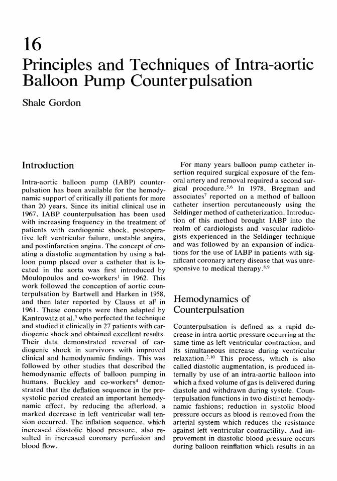
16 Principles and Techniques of Intra-aortic Balloon Pump Counterpulsation Shale Gordon
Introduction
Intra-aortic balloon pump (IABP) counterpulsation has been available for the hemodynamic support of critically ill patients for more than 20 years. Since its initial clinical use in 1967, IABP counterpulsation has been used with increasing frequency in the treatment of patients with cardiogenic shock, postoperative left ventricular failure, unstable angina, and postinfarction angina. The concept of creating a diastolic augmentation by using a balloon pump placed over a catheter that is located in the aorta was first introduced by Moulopoulos and co-workers) in 1962. This work followed the conception of aortic counterpulsation by Bartwell and Harken in 1958, and then later reported by Clauss et aF in 1961. These concepts were then adapted by Kantrowitz et al,3 who perfected the technique and studied it clinically in 27 patients with cardiogenic shock and obtained excellent results. Their data demonstrated reversal of cardiogenic shock in survivors with improved clinical and hemodynamic findings. This was followed by other studies that described the hemodynamic effects of balloon pumping in humans. Buckley and co-workers4 demonstrated that the deflation sequence in the presystolic period created an important hemodynamic effect, by reducing the afterload, a marked decrease in left ventricular wall tension occurred. The inflation sequence, which increased diastolic blood pressure, also resulted in increased coronary perfusion and blood flow.
For many years balloon pump catheter insertion required surgical exposure of the femoral artery and removal required a second surgical procedure.5,6 In 1978, Bregman and associates 7 reported on a method of balloon catheter insertion percutaneously using the Seldinger method of catheterization. Introduction of this method brought IABP into the realm of cardiologists and vascular radiologists experienced in the Seldinger technique and was followed by an expansion of indications for the use of IABP in patients with significant coronary artery disease that was unresponsive to medical therapy. 8,9
Hemodynamics of Counterpulsation
Counterpulsation is defined as a rapid decrease in intra-aortic pressure occurring at the same time as left ventricular contraction, and its simultaneous increase during ventricular relaxation. 2,10 This process, which is also called diastolic augmentation, is produced internally by use of an intra-aortic balloon into which a fixed volume of gas is delivered during diastole and withdrawn during systole. Counterpulsation functions in two distinct hemodynamic fashions; reduction in systolic blood pressure occurs as blood is removed from the arterial system which reduces the resistance against left ventricular contractility. And improvement in diastolic blood pressure occurs during balloon reinflation which results in an

172
increase in coronary artery perfusion and blood flow. 11,12 Counterpulsation devices are triggered by the electrocardiographic QRS complex, which rapidly decreases the blood volume in the aorta just before the onset of ventricular systole. The balloon pump console removes approximately 40 ml of gas from a balloon that is positioned in the descending aorta at the level of the left subclavian artery. This rapid decline in aortic blood volume causes the aortic blood pressure to fall just as ventricular systole begins, the left ventricle thus ejects blood against a decreased afterload and, subsequently, left ventricular systolic performance improves. As the aortic valve closes at end-systole, the aortic volume that has been removed is returned in diastole. This expansion of aortic volume causes an increase in diastolic aortic pressure and subsequent improvement in coronary perfusion and coronary flow. 11 Counterpulsation has been shown to decrease myocardial oxygen consumption,13 improve coronary and carotid blood flOW,14 and increase urinary output,15 whereas the mechanics of left ventricular contraction, wall stress, and ejection fraction change only slightly. 16
Equipment (Balloon Pump Device)
Intra-aortic-balloon pump consoles are made as cardiac assist devices that introduce a specific volume of gas through a pneumatic system into a balloon at a predetermined time followed by retrieval of that gas at a later time. These consoles contain a gas source (carbon dioxide or helium) and a physiologic monitor used for acquisition and display of arterial blood pressure and electrocardiogram. The console also contains a control unit that is used for the timing of balloon inflation and deflation.
Proper timing of balloon inflation and deflation is necessary for best results. Using the counterpulsation concept, the intra-aortic balloon remains deflated during systole, which coincides with the ST-T wave, and then is in-
S. Gordon
flated immediately after, commencing during the T-P interval, which coincides with diastole. The inflation is then maintained up until the R wave. Balloon deflation is triggered electrically within the PR interval just before the R wave. Balloon consoles provide an electronic signal that can be superimposed on the electrocardiogram or arterial pressure waveform to estimate the point of balloon inflation and deflation and to make adjustments for maximum hemodynamic effects. Proper timing of deflation is checked with reference to the arterial blood pressure tracing. The end-diastolic dip in arterial pressure caused by balloon deflation should reach a minimum value just before the arterial upstroke. The electronic signal for inflation is usually set at the peak of the electrocardiographic T wave. Proper setting of inflation is again checked with reference to the arterial blood pressure tracing, particularly the dicrotic notch, as diastolic augmentation should not occur before the dicrotic notch. Appropriate balloon inflation and deflation can be recognized by an augmented diastolic blood pressure, usually greater than peak systolic pressure; a diastolic blood pressure during augmentation that is less than the end-diastolic pressure without augmentation; and a sharp angle between the fall in systolic arterial blood pressure and augmentated diastolic blood pressure.
Currently, there are three balloon pump consoles available in the United States. An indepth analysis and comparison of these three balloon pump devices has been performed and the results have been published. 17 Datascope manufacturers System 83 is a console capable of powering a large number of pneumatic assist devices. This system uses carbon dioxide for balloon inflation and deflation and the volume of gas that enters the intra-aortic balloon is slightly lower than that of other balloon pumps. This system as well as other balloon pumps has minimal abilities for tracking serious atrial or ventricular arrhythmias, particularly atrial fibrillation. Kontron manufacturers Model 10 balloon pump console uses helium as its driving gas. Smec balloon console Model 13001 uses helium as its driving gas; this system features a built in pacemaker system that

16. Principles and Techniques of Intra-aortic Balloon Pump Counterpulsation 173
can be used in either VVI or DDD mode. This system also has a unique feature that is capable of tracking atrial and ventricular arrhythmias for safer balloon pumping. Balloon catheters are available in size 10.5 to 12 Fr for adults and size 7 to 9 Fr for children. Catheters sizes are usually smaller for percutaneous than surgical balloons.
Indications for Intra-aortic Balloon Pump
Initially IABP was used in patients with cardiogenic shock. 3,5,15 Mter this, IABP was used in patients with severe cardiac failure after open heart surgery. 18 The indications for use of IABP are predominantly twofold: transient support of the left ventricle due to cardiac failure secondary to myocardial infarction or intraoperative injury, and enhancement in the oxygen supply/demand balance in an attempt to decrease the extent of ischemia and to preserve myocardial viability. A 12-year experience at the University of Miami demonstrated that postcadiac surgery patients constituted 43% of IABP patients, followed by 23% of patients in cardiogenic shock, and 20% of patients for elective (preoperative) balloon pumping. This elective group included patients with cardiac ischemia and infarction and high-risk surgical patients. 19
Experimental studies have demonstrated the effectiveness of IABP in diminishing the severity of myocardial infarction secondary to coronary artery occlusion.20 In clinical settings, IABP has been shown to be effective during prolonged episodes of ischemia associated with preinfarctional angina,8,21 early acute myocardial infarction,22 acute myocardial infarction with impending extension ,23 and malignant ventricular arrhythmias. 24 Usually, IABP is indicated in ongoing cardiac ischemia before diagnostic cardiac catheterization and subsequent surgical intervention.
The use of IABP in patients with ongoing myocardial ischemia is based on evidence that it improves coronary blood flow, especially coronary collateral flow to areas of myocardial
ischemia and its border zones. 25 These results, however, are not uniformly confirmed.26 During early myocardial infarction, the effects of IABP include an increase in myocardial oxygen supply (coronary flow) as well as a decrease in factors that effect myocardial oxygen demand. 8
Indications for IABP in preinfarction angina before myocardial revascularization can be performed, including persistent angina that is unresponsive to medical management and associated with electrocardiographic changes and recurrent hemodynamic instability. 21 During the early phases of acute myocardial infarction, IABP has been used to attempt to decrease the eventual size of myocardial infarction, to prevent myocardial infarction extension, to decrease the complications associated with myocardial infarction, and to support cardiac function. 20,22,23 Experimental and clinical studies have shown contrasting results on the efficiency of IABP early after acute coronary artery occlusion on the size and area of myocardial infarciton.8,2,26 In patients with ventricular irritability after myocardial infarction, when there has been no response to drug therapy, IABP has shown significant decrease in ventricular irritability. 24 Complications that occur early in the course of acute myocardial infarction include papillary muscle rupture or dysfunction and rupture of the ventricular septum. Treatment for both of these complications includes afterload reduction and support of arterial blood pressure. Diastolic augmentation and systolic unloading with IABP is also a proposed therapy. 28,29
Contraindications for Intra-aortic Balloon Pump
Severe aortic insufficiency secondary to an incompetent aortic valve which can be seen in acute bacterial endocarditis or aortic dissection is a contraindication to the use of IABP. With incorrect balloon timing, inflation during diastolic augmentation may increase aortic insufficiency and cause significant left ventricular dilatation. In patients with mild aortic in-

174
sufficiency, IABP with careful attention to correct timing of the balloon inflation period may be used briefly without adverse effects.3o
Methods of Insertion of Intra-aortic Balloon Catheters
The most commonly used insertion route for an IABP catheter is the femoral artery. The left femoral artery is preferable as it then allows for the right femoral artery to be used for cardiac catheterization. Introduction of the balloon catheter initially required surgical entry of the femoral artery and placement of an end-to-side prosthetic graft. Since 1980, however, a series of technologic advances have permitted percutaneous insertion of intraaortic balloon catheters,5,?,31 and later percutaneous insertion of balloon catheters over a guidewire. 32 Even more recently, smaller diameter (10.5 versus 12-Fr) wire-guided balloon catheters have become available. The incidence of balloon related vascular complications, however, appears to remain quite high. Vascular complications have been reported in between 9% to 36% of patients, and long-term complications have been described in 7% of patients undergoing balloon counterpulsation.5,6,32,33 The development of leg ischemia is significantly related to the presence of diabetes (risk ratio 2.0), peripheral vascular disease (risk ratio 1.9), female gender (risk ratio 1.8), and the presence of a postinsertion ankel-brachial pressure index less than 0.8 (risk ratio 7.9).32
Surgical technique of balloon catheter introduction by the femoral artery approach is usually associated with a 95% success rate.5,18,19 The rate of successful catheter insertion is equal to or slightly higher than that for percutaneous balloon insertion using a guidewire,33 The surgical technique requires at least 60 minutes time in an operating room environment and also requires use of graft material. Percutaneous technique on the other hand takes between 10 to 15 minutes but leaves a large defect in the femoral artery. Fluoroscopy is usually helpful with the percutaneous
S. Gordon
approach, although a balloon may be inserted without it and the position later checked with a portable chest x-ray. Patients receiving percutaneous balloon catheters require full heparinization. After balloon insertion by surgical technique, low molecular weight dextran has been satisfactory. Before removal of a percutaneous balloon catheter, heparinization should be discontinued for 6 to 8 hours, whereas no change in dextran therapy is required. Catheter removal by the percutaneous method can be performed at bedside, but removal ofthe surgical technique requires use of an operating room where a prophylactic embolectomy is performed and arterial repair follows. The complication rate, especially leg ischemia for the two methods is slightly different. Initial studies suggested that the percutaneous technique was associated with a lower complication rate. 7,31 Nonrandomized studies have reported the percutaneous complication rate to be equal to or slightly higher than the surgical complication rate. 34 A recent randomized study found that the percutaneous technique for IABP was faster than the surgical technique and technically easy, but was associated with a higher incidence of vascular complications. 33
Physiology of Balloon Pumping (Counterpulsation)
Intra-aortic balloon pump uses the principles of counterpulsation during rapid inflation and deflation of the balloon, which is located in the descending aorta. The hemodynamic effects of IABP are mainly secondary to its effects on ventricular preload and afterload. 35 Intra-aortic balloon inflation occurs in diastole, which begins with aortic valve closure (the dicrotic notch on the arterial blood pressure curve), then balloon deflation takes place just before left ventricular ejection (the upstroke of the arterial blood pressure curve). The initiation of intra-aortic balloon deflation must coincide with the end of the isovolumetric phase of ventricular contraction, before the ejection phase to produce a negative intra-aortic pres-

16. Principles and Techniques of Intra-aortic Balloon Pump Counterpulsation 175
sure. 35.36 The timing of intra-aortic balloon deflation is extremely critical and must be followed closely during the course of balloon pumping. Because the period of isovolumetric ventricular contraction terminates after the opening of the aortic valve, with adequate balloon deflation the pressure required of the left ventricle to open the aortic valve will markedly decrease. Peak intraventricular pressure and rate of rise of left ventricular pressure during efficient balloon pumping frequently decreases by 10% to 20%.19 Balloon inflation results in a displacement of blood volume in the aorta, which is distributed into the vascular system, resulting in a rise in diastolic blood pressure (diastolic augmentation). The overall effect during balloon pumping is a change in the pattern of the arterial blood pressure curve, from a systolic rise and a diastolic fall to a "double hump" pattern indicating a systolic rise followed by a balloon inflation rise separated by two dips due to aortic valve closure and balloon deflation, respectively.
Optimal balloon pumping is best during normal sinus rhythm with heart rates between 90 to 100 beats per minute. Balloon pumping at rates greater than 120 beats per minute result in a decreased gas flow and volume,17 leading to a smaller augmentation pressure and, thus, ineffective systolic unloading. To improve aortic balloon augmentation at increased heart rates, the assist rate should be decreased to 1 : 2 or 1: 3. A difficult situation for balloon pumping is found in patients with atrial fibrillation and irregular heart rates. These irregular heart rates cause wide fluctuation in stroke volume and subsequently in balloon augmentation. Again, balloon pumping is more efficient in patients with atrial fibrillation and a rapid ventricular response if balloon augmentation rates are decreased to 1 : 2 or 1 : 3. Premature ventricular contractions also cause interruption of balloon inflation due to internally set safety intervals that prevent balloon inflation during systole. 17
The hemodynamic effects of IABP and its relationship to left ventricular function have been studied. Intraoperative studies have demonstrated a significant increase in left ventricular ejection fraction during IABP. 37 Dur-
ing the early phases of diastolic augmentation with IABP, an increase in cardiac index ranging from 10% to 40% has been described. 35,38 Left ventricular end-diastolic pressure decreases to about 10% to 15% of control values during IABP.39 Intra-aortic balloon pump counterpalsation usually results in a modest increase in mean arterial blood pressure. 39 Heart rate is usually decreased during IABP and is more marked in patients with normal sinus rhythm. Premature ventricular contractions and atrial arrhythmias are also usually suppressed during IABP.24
Patient Management
Patients on IABP are considerably restricted in their movements. However, diligent nursing care can reduce potential problems by the use of air mattresses, which can prevent the development of pressure areas and subsequent tissue breakdown; antiembolic stockings, which prevent venous stasis and promote venous return; and repositioning, which entails turning the patient on each side every 2 to 4 hours, keeping the balloon catheter insertion site as straight as possible.
Because vascular complications are the most commonly observed during IABP operation, the pulse in the leg through which the balloon catheter has been inserted must be carefully noted. Both motor and sensory function of the foot and leg should be compared with the opposite leg. Any loss in motor or sensory function or in the relative temperature of the foot should be carefully evaluated.
Cardiac Catheterization and Balloon Pumping
Intra-aortic balloon pumping can be useful in unstable patients undergoing catheterization studies. Patients who have severe congestive heart failure and pulmonary edema secondary to ischemic heart disease can be stabilized with IABP and then transferred to the laboratory where cardiac catheterization studies can
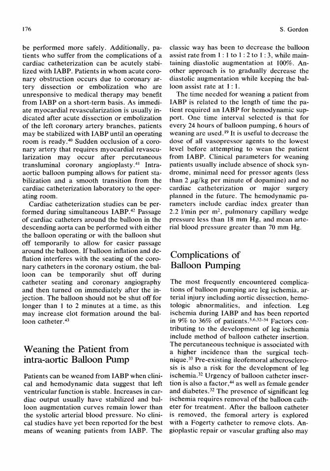
176
be performed more safely. Additionally, patients who suffer from the complications of a cardiac catheterization can be acutely stabilized with IABP. Patients in whom acute coronary obstruction occurs due to coronary artery dissection or embolization who are unresponsive to medical therapy may benefit from IABP on a short-term basis. As immediate myocardial revascularization is usually indicated after acute dissection or embolization of the left coronary artery branches, patients may be stabilized with IABP until an operating room is ready. 40 Sudden occlusion of a coronary artery that requires myocardial revascularization may occur after percutaneous transluminal coronary angioplasty. 41 Intraaortic balloon pumping allows for patient stabilization and a smooth transition from the cardiac catheterization laboratory to the operating room.
Cardiac catheterization studies can be performed during simultaneous IABP.42 Passage of cardiac catheters around the balloon in the descending aorta can be performed with either the balloon operating or with the balloon shut off temporarily to allow for easier passage around the balloon. If balloon inflation and deflation interferes with the seating of the coronary catheters in the coronary ostium, the balloon can be temporarily shut off during catheter seating and coronary angiography and then turned on immediately after the injection. The balloon should not be shut off for longer than 1 to 2 minutes at a time, as this may increase clot formation around the balloon catheter. 43
Weaning the Patient from intra-aortic Balloon Pump
Patients can be weaned from IABP when clinical and hemodynamic data suggest that left ventricular function is stable. Increases in cardiac output usually have stabilized and balloon augmentation curves remain lower than the systolic arterial blood pressure. No clinical studies have yet been reported for the best means of weaning patients from IABP. The
S. Gordon
classic way has been to decrease the balloon assist rate from 1 : 1 to 1 : 2 to 1 : 3, while maintaining diastolic augmentation at 100%. Another approach is to gradually decrease the diastolic augmentation while keeping the balloon assist rate at 1 : 1.
The time needed for weaning a patient from IABP is related to the length of time the patient required an IABP for hemodynamic support. One time interval selected is that for every 24 hours of balloon pumping, 6 hours of weaning are used. 19 It is useful to decrease the dose of all vasopressor agents to the lowest level before attempting to wean the patient from IABP. Clinical parameters for weaning patients usually include absence of shock syndrome, minimal need for pressor agents (less than 2 ILg/kg per minute of dopamine) and no cardiac catheterization or major surgery planned in the future. The hemodynamic parameters include cardiac index greater than 2.2 lImin per m2, pulmonary capillary wedge pressure less than 18 mm Hg, and mean arterial blood pressure greater than 70 mm Hg.
Complications of Balloon Pumping
The most frequently encountered complications of balloon pumping are leg ischemia, arterial injury including aortic dissection, hemotologic abnormalities, and infection. Leg ischemia during IABP and has been reported in 9% to 36% of patients.5,6,32-34 Factors contributing to the development of leg ischemia include method of balloon catheter insertion. The percutaneous technique is associated with a higher incidence than the surgical technique. 33 Pre-existing ileofemoral atherosclerosis is also a risk for the development of leg ischemia.32 Urgency of balloon catheter insertion is also a factor,44 as well as female gender and diabetes.32 The presence of significant leg ischemia requires removal of the balloon catheter for treatment. After the balloon catheter is removed, the femoral artery is explored with a Fogerty catheter to remove clots. Angioplastic repair or vascular grafting also may

16. Principles and Techniques of Intra-aortic Balloon Pump Counterpulsation 177
be required. If IABP is required for a further period, another balloon catheter may be placed in the opposite femoral artery. Alternatively, a femoral-femoral crossover graft to reestablish peripheral circulation and to continue uninterrupted IABP has been suggested as a method of treatment for severe leg ischemia.45
Acute aortic dissection has been reported in less than 5% of patients undergoing IABP. 44,46 Some patients may have acute aortic dissection without clinical evidence or apparent side effects. The incidence of aortic dissection in autopsies performed on patients who died during IABP is significantly higher than those that are clinically apparentY
Hematologic complications of IABP include thrombocytopenia and hemolytic anemia. 48 These complications appear at equal rates with either percutaneous or surgically introduced aortic balloons.
Peripheral embolization to the opposite leg or the arm may develop in patients with IABP.18,47 These complications appear more frequent in patients with depressed cardiac function. Embolic events may result in small bowel infarction, superior mesenteric artery obstruction, or cerebral embolization.48
Infections either systemic or localized occur at a rate of 1 % to 3% in patients after either percutaneous or surgical balloon insertion. Thus, all patients with IABP receive prophylactic antibiotics. 44 In obese and diabetic patients, the incidence of groin infection may be as high as 30%.49
Late complications of IABP may include claudication, peroneal nerve paresis, and pseudoaneurysm of the femoral artery. 49
Results of Intra-aortic Balloon Pump
Patients with cardiogenic shock who have received IABP usually show improved hemodynamic findings and reversal of clinical signs such as sweating and cold, clammy skin,50,51 These changes are due to an increase in systemic blood flow secondary to improved car-
diac output and improvement in cerebral, renal, coronary, mesenteric, and cutaneous blood flow. 51 ,52 The hemodynamic effects of IABP include an increase in diastolic aortic pressure, cardiac output, left ventricular stroke work index, and mild increase in ejection fraction; and with a decrease in systolic aortic pressure, diastolic left ventricular pressure, myocardial contractility, left ventricular wall tension, left ventricular volume, and pulmonary capillary wedge pressure. 19,37 After IABP, cerebral blood flow improves by 56%.53 Renal blood flow is not increased, although there is marked increase in urinary output. 53 Coronary blood flow may increase from 5% to 15%.26 This improvement in coronary blood flow appears to influence predominantly collateral coronary circulation entering an area of myocardial ischemia or infarction.25 This increase in collateral blood flow may be related to a decrease in eventual myocardial infarction size, as in both clinical and experimental studies, it has been demonstrated that balloon augmentation achieves a decrease in the size of myocardial infarction.20,22,54 Further studies have suggested that alterations in myocardial oxygen supply and demand are more important in improving cardiac function than simply an increase in coronary blood flow. 9 Intraaortic balloon pumping is also associated with a decrease in the number of episodes of angina pectoris as well as a decrease in the frequency of ventricular arrhythmias in patients with myocardial ischemia.9,21,24
References 1. Moulopoulos SD, Topaz S, KolffWJ: Diastolic
balloon pumping (with carbon dioxide) in the aorta: Mechanical assistance to the failing circulation. Am Heart J 1962; 63:669.
2. Clauss RH, Burtwell WC, Albertol G, et al: Assisted circulation. I The artificial counterpulsator. J Thorac Cardiovasc Surg 1961; 41:447.
3. Kantrowitz A, Krakauer JS, Rosenbaum A, et al: Phase shift balloon pumping in medical refractory cardiogenic shock. Results in 27 patients. Arch Surg 1969; 99:739.
4, Buckley MJ, Leinbach RL, Kastor, JA, et al: Hemodynamic evaluation of intraaortic balloon pumping in man. Circulation 1970; 41:11-130.

178
5. Kantrowitz A, Wasfie T, Freed PS, et al: Intraaortic balloon pumping 1967 through 1982: Analysis of complications in 733 patients. Am J Cardiol 1986; 57:976.
6. Alpert J, Bhaktan EK, Gie1chinsky I, et al: Vascular complications in intraaortic balloon pumping. Arch Surg 1976; 111: 1190.
7. Bregman D, Casarella WJ: Percutaneous intraaortic balloon pumping: initial clinical experiences. Ann Thorac Surg 1981; 29: 153.
8. Fuchs RM, Brin KP, Brinker JA, et al: Augmentation of regional coronary blood flow by intraaortic balloon counterpulsation in patients with unstable angina. Circulation 1983; 68:117.
9. Williams DO, Korr KS, Gewirtz H, et al: The effect of intraaortic balloon counterpulsation on regional myocardial blood flow and oxygen consumption in the presence of coronary artery stenosis in patients with unstable angina. Circulation 1982; 66:593.
10. Kantrowitz A, Kantrowitz A: Experimental augmentation of coronary flow by retardation of the arterial pressure pulse. Surgery 1953; 34:678.
11. Rosensweig J, Borromeo C, Chatterjee S, et al: Treatment of coronary insufficiency by counterpulsation: experimental studies. Ann Thorae Surg 1966; 2:706.
12. Hanloser PB, Gallow E, Schenk WG: Hemodynamics of counterpulsation. J Thorac Cardiovasc Surg 1966; 51 :366.
13. Parmley W: Hemodynamic effects of noninvasive systolic unloading (nitroprusside) and diastolic augmentation (external counterpulsation) in patients with acute myocardial infarction. Am J Cardiol 1974; 33:819.
14. Maroko PR, Bernstein EF, Libby P, et al: Effects of intraaortic balloon counterpulsation on the severity of myocardial ischemic injury following acute coronary occlusion. Circulation 1972; 45:1150.
15. Mueller H: Are intraaortic balloon pumping and external counterpulsation effective in treatment of cardiogenic shock? Cardiovasc Clin H 1977; 8:87.
16. Rose EA, Marrin CAS, Bregman D, et al: Left ventricular mechanics of counterpulsation and left heart bypass, individually and in combination. J Thorac Cardiovasc Surg 1979; 77:127.
17. ECRI: Evaluation: Intraaortic balloon pumps. Health Devices 1981; 11:3.
18. McEnany MT, Kay HR, Buckley MJ, et al: Clinical experience with intraaortic balloon support in 728 patients. Circulation 1978; 58:1-128.
S. Gordon
19. Bolooki H: Clinical Application of Intraaortic Balloon Pump. New York, Futura Publishing Company, 1984.
20. Maroko PR, Bernstein EF, Libby P, et al: Effects of intraaortic balloon counterpulsation on the severity of myocardial ischemic injury following acute coronary occlusion. Circulation 1972; 45:1150.
21. Weintraub RM, Voukydis PC, Aroesty JM, et al: Treatment of preinfarction angina with intraaortic balloon counterpulsation and surgery. Am J Cardiol1974; 34:809.
22. Rosensweig J, ChatteIjee S, Merino F: Treatment of acute myocardial infarction by counterpulsation. Experimental rational and clinical experience. J Thorac Cardiovasc Surg 1970; 59:243.
23. Mundth ED, Buckley MJ, Gold HK, et al: Intraaortic balloon pumping and emergency coronary arterial revascularization of acute myocardial infarction with impending extension. Ann Thorac Surg 1973; 5:435.
24. Willerson JT, Curry GC, Watson JT, et al: Intraaortic balloon counterpulsation in patients in cardiogenic shock, medically refractory left ventricular failure and/or recurrent ventricular tachycardia. Am J Med 1975; 58:183.
25. Watson JT, Willerson JT, Fixler DE, et al: Changes in collateral coronary blood flow distal to a coronary occlusion during intraaortic balloon pumping. Trans Am Soc Artif Int Organs 1973; 19:402.
26. Haston HH, McNamara JJ: The effects of intraaortic balloon counterpulsation on myocardial infarct size. Ann Thorac Surg 1979; 28:335.
27. Roberts A, Alonso DR, Combes JR, et al: Role of delayed intraaortic balloon pumping in treatment of experimental myocardial infarction. Am J Cardiol1978; 41:1202.
28. Cheng TO, Bashour T, Adkins PC: Acute severe mitral regurgitation from papillary dysfunction in acute myocardial infarction. Successful early surgical treatment by combined mitral valve replacement and aortocoronary saphenous vein bypass graft. Circulation 1972; 46:491.
29. Buckley MJ, Mundth ED, Daggett WM, et al: Surgical management of ventricular septal defects and mitral regurgitation complicating acute myocardial infarction. Ann Thorac Surg 1973; 16:598.
30. Yellin E, Levy L, Bregman D, et al: Hemodynamic effects of intraaortic balloon pumping in dogs with aortic incompetence. Trans Am Soc Artif Intern Organs 1973; 19:389.
16. Principles and Techniques of Intra-aortic Balloon Pump Counterpulsation 179
31. Subramanian VA, Goldstein JE, Sos T A, et al: Preliminary clinical experience with percutaneous intraaortic balloon pumping. Circulation 1981; 62: 123.
32. Alderman JD, Gabliani GI, McCabe CH, et al: Incidence and management of leg ischemia with percutaneous wire guided intraaortic balloon catheters. J Am Coli Cardiol 1987; 9:524.
33. Goldberg MJ, Rubenfire M, Kantrowitz A, et al: Intraaortic balloon pump insertion: A randomized study comparing percutaneous and surgical techniques. J Am Coli Cardiol 1987; 9:515.
34. Hauser AM, Gordon S, Gangadharan V, et al: Percutaneous intraaortic balloon counterpulsation. Clinical effectiveness and hazards. Chest 1982; 82:442.
35. Webber KT, Janicki JS: Intraaortic balloon counterpulsation. A review of physiologic principles, clinical results and device safety. A collective review. Ann Thorac Surg 1974; 17:602.
36. Feola M, Adachi M, Akers W, et al: Intraaortic balloon pumping in experimental animal: effects and problems. Am J Cardiol1971; 27: 129.
37. Maddoux G, Pappas G, Jenkin M, et al: Effect of pulsatile and nonpulsatile flow during cardiopulmonary bypass on left ventricular ejection fraction early after aortocoronary bypass surgery. Am J Cardiol1976; 37:1000.
38. Akyurekli Y, Taichman JC, Keon WJ: Effectiveness of intraaortic balloon counterpulsation and systolic unloading. Can J Surg 1980; 23: 122.
39. Mullins CB, Sugg WL, Kennelly BM, et al: Effect of arterial counterpulsation on left ventricular volume and pressure. Am J Physiol 1971; 220:694.
40. Bolooki H, Vargas A: Myocardial revascularization after myocardial infarction. Arch Surg 1976; 111:1216.
41. Murphy DA, Craver JM, Jones EL, et al: Surgical revascularization following unsucessful percutaneous transluminal coronary angioplasty. J Thorac Cardiouasc Surg 1982; 84:342.
42. Fernandez JJ, Feldman JJ, Schocket L, et al: Coronary arteriography by the percutaneous transfemoral technique in patients on intraaortic counterpulsation. Cathet Cardiouasc Diagn 1977; 3:87.
43. Rashid A, Hildner FJ, Fester A, et al: Coronary arteriography: prevention of thromboembolism and complications using a pressure drip flushing technique. J Cardiouasc Diagn 1975; 1:283.
44. Goldman BS, Hill TJ, Rosenthal GA, et al: Complications associated with the use of the intraaortic balloon pump. Can J Surg 1982; 25: 153.
45. Alpert J, Parsonnet V, Goldenkranz RJ, et al: Leg ischemia during intraaortic balloon pumping: Indication for femoral-femoral crossover graft. J Thorac Cardiouasc Surg 1980; 79:729.
46. McCabe JC, Abel RM, Subramanian VA, et al: Complications of intraaortic balloon insertion and counterpulsation. Circulation 1978; 57:769.
47. Isner JM, Cohen SR, Virmani R, et al: Complications of the intraaortic balloon counterpulsation device: Clinical and morphologic observations in 45 necropsy patients. Am J Cardiol 1980; 45:260.
48. Schneider MD, Kaye MP, Blatt SJ, et al: Safety of intraaortic balloon pumping. I. Biochemical and hematologic values influenced by use of balloon. Thromb Res 1974; 4:387.
49. Martin RS, Moncure AC, Buckley MJ, et al: Complications of percutaneous intraaortic balloon insertion. J Thorac Cardiouasc Surg 1983; 85:186.
50. Kantrowitz A, Tjonneland S, Krakauer JS, et al: Mechanical intraaortic cardiac assistance in cardiogenic shock: Hemodynamic effects. Arch Surg 1968; 97: 1000.
51. Housman, LB, Bernstein EF, Braunwald NS, et al: Counterpulsation for intraoperative cardiogenic shock. Successful use of intraaortic balloon. JAm Med Assoc 1973; 244:1131.
52. Ehrich DA, Biddle TL, Kronenburg MW, et al: The hemodynamic response to intraaortic balloon counterpulsation in patients with cardiogenic shock complicating acute myocardial infarction. Am Heart J 1977; 93:274.
53. Bhayana IN, Scott SM, Sethi GK, et al: Effects of intraaortic balloon pumping on organ perfusion in cardiogenic shock. J Surg Res 1979; 26:108.
54. Whittle JL, Feldman RL, Pepine JC, et al: Effects of intraaortic balloon pumping on regional and total coronary flow in patients with coronary artery disease. Am J Cardiol1980; 45:395.
17 Temporary and Permanent Pacemakers Amar S. Kapoor
Historical Perspective
Hyman,! in 1930, demonstrated that direct cardiac stimulation for emergency resuscitation was possible. He transmitted electric stimuli from an external pulse generator via a needle electrode through the chest wall to the right atrium. Clinically, this method was not successful. In 1952, ZolF successfully resuscitated two patients from ventricular standstill with external stimuli provided by electrodes attached to a large electric pulse generator.
In 1958, Furman and SchwedeP used a temporary endocardial electrode attached to an external pacemaker for temporary ventricular pacing. Since the 1960s, there has been dramatic development of pacemaker technology. The earliest pacemakers functioned as fixedrate pacemakers.
Since 1980 several types of demand and physiologic pacemakers have been developed, made possible by better battery chemistry and microcomputer technology. The evolution of pacemaker technology continues, and there are a mUltiplicity of uses of sophisticated pacing devices for control of tachyarrhythmias, bradyarrhythmias, and internal defibrillation.
The Intersociety Commission for Heart Disease Resources established initially a threeletter code, which has now increased to a 5-letter code, for characterizing pacemaker mode and function (Table 17.1).4 The first letter identifies the chamber(s) paced; the second letter, the chamber(s) sensed; the third letter, the modes of response to sensed native car-
diac activity; the fourth, the programmable functions; and the fifth letter identifies special antitachycardiac functions.
Clinical Electrophysiology of Pacing
A pacemaker is composed of a power source, a lead system through which sensing and pacing functions are achieved, and integrated circuitry to amplify time-detected depolarizations and pacing discharges. The threshold is the minimal electrical stimulus required to cause myocardial depolarization. It is important to determine both the voltage and current thresholds and to measure the impedance of the pacing system. Voltage threshold is the lowest threshold that stimulates the heart with a given pulse width. Voltage threshold is dependent on electrode surface area and on pulse width. Voltage threshold is measured using a pacing system analyzer that provides a constant voltage source. Threshold measurements are dynamic and change with physiologic and pharmacologic factors, as well as site of stimulation. A pacemaker wire has its lowest threshold at the time of implantation, called acute threshold, and it should be less than 1. 0 V. Over a period of 2 to 6 weeks, the threshold rises to 3 or 4 times its initial level and then stabilizes to a chronic threshold, which is usually less than 3.0 V. With lead placement or fracture, the voltage threshold

17. Temporary and Permanent Pacemakers
TABLE 17.1. Pacing modalities. *
Chambers paced
V-ventricle A-atrium D-dual
II
Chambers sensed
V-ventricle A-atrium D-dual O-none
III Modes
of response
T-triggered I-inhibited D-dual O-none R-reverse
IV
Programmed functions
P-programmable rate/output M-multiprogrammable C-communication O-none
V Special
181
anti tachycardia functions
B-bursts N-normal rate competition S-scanning
E-external
* Intersociety Commission for Heart Disease identification codes.
increases. A typical ventricular pacing system will have threshold currents of 0.5 to 0.9 rnA, voltage threshold of 0.4 to 1.0 Y, and a calculated impedance of 500.0 to 1000.0 n.5 The integrity of the lead can be evaluated by measuring lead impedance and should be measured from the simultaneous pulse signal of voltage and current with time.
Sensing
The input circuit will sense the patient's intrinsic endocardial electrical signal and thus inform the output circuit not to generate a pacer stimulus. The sensing threshold for the pacing system is the lowest potential difference measured by the sensing circuit that will inhibit the output circuit. The input circuit should be able to discriminate P waves, QRS signal, and T waves. Factors that influence the ability to sense are the electrogram amplitude on the QRS height, the configuration and flow rate, which is the rate of change in voltage in an electrogram. For proper sensing, a minimal amplitude acutely is 5.0 mY for the R wave and 2.0 mY for the P wave, and the minimal slew rate is 0.2 Y/sec.
Maturation of the electrode changes both the pacing thresholds and sensing thresholds by increasing the lead impedance.
Sensing failures are related to inadequate amplitude signal and flow rate at the time of implantation, or inadequate electrode surface contact and defective sensing circuitry. In these cases, repositioning the lead may help, and if there is intrinsic failure of the sensing circuitry, one must replace the pacer generator.
Temporary Transyenous Pacing
A temporary pacemaker essentially has a trans venous catheter electrode connected to an external pulse generator. The most common use of temporary pacemakers has been ventricular pacing in the setting of heart block complicating acute myocardial infarction. However, in recent years there has been increasing use of physiologic temporary pacing with atrial or atrioventricular pacing. Applications for temporary pacing have significantly increased its earlier introduction. Table 17.2 lists the current indications. Temporary pacemakers are increasingly used to treat or pre-
TABLE 17.2. Indications for temporary pacing.
Bradyarrhythmias Sinus node dysfunction
Sinus arrest Sinus bradycardia Atrial fibrillation with slow ventricular response
A V node dysfunction Second-degree block Third-degree block
Acute anterior myocardial infarction with new onset or fascicular block or bundle branch block
Tachyarrhythmias Atrial pacing
Supraventricular tachycardia Postcardioversion
A V sequential pacing Brady-tachycardia syndrome Atrial flutter Ventricular arrhythmias
Temporary ventricular overdrive pacing Ventricular arrhythmias Ventricular tachycardias Accelerated rate in the setting of torsade de pointes
with long Q-T interval
182
vent tachyarrhythmias. Tachyarrhythmias, atrial and ventricular, can frequently be converted by overdrive pacing of the atria or atria and ventricles in sequence.6
For bradyarrhythmias, temporary pacing is indicated for symptomatic patients or when the heart drops below 45 beats per minute.
Techniques for Temporary Pacemaker Placement Temporary pacing is accomplished by proper placement of a pacing catheter in the right atrium or ventricle introduced via a central vein. There are several venous access sites for the percutaneous technique. Commonly used are the internal jugular, external jugUlar, subclavian, femoral, and brachial veins. Central venous catheterization allows the easy insertion of one or more pacing electrodes. The advantages and risks of the various venous access sites for temporary pacing are listed in Table 17.3. The operator should use the site he is familiar with.
Subclavian vein access is favored by many operators despite the recognized complications of pneumothorax, hemothorax, air embolism, subclavian artery puncture, and brachial plexus trauma. The catheter placement is easy and stable. The right or left infraclavicular area is prepped and draped for aseptic technique. For easier access, a rolled sheet is placed between the shoulder blades and a foam wedge placed under the legs for distending the vein.
An 18-gauge needle is inserted after local anesthesia lateral to the ligament between the
A.S. Kapoor
clavicle and the first rib (Fig 17.1). As the needle is advanced aiming behind the cricoid cartilage, the operator may feel a "give in" as the vein is punctured and venous blood is drawn into the syringe. The needle is stabilized and a 50-cm flexible J-wire is advanced into the superior vena cava.
After a small incision with a blade, the peelaway sheath introducer system is passed over the guidewire into the vein. The dilator and guide wire are removed cautiously to prevent any air embolism, and the lead is inserted and advanced to the right atrium, the sheath is peeled away, and the lead is stabilized. For dual-chamber pacing, two-lead insertion is required, and here one may use a modified technique. The guide wire is left in place before peeling away the sheath. A second peel-away sheath introducer system is inserted. It is important to pinch the sheath when the dilator is removed and the lead is inserted. Occasionally, a larger 1O.5-Fr sheath can be used for inserting both electrodes through the same sheath.
Sometimes one may have to manipulate the electrode to transverse the angle between the subclavian vein and superior vena cava by turning the head toward the shoulder that is raised. Fluoroscopy is generally required for directing the electrode to the right ventricular apex.
Internal jugular cannulation is a much safer technique than subclavian vein puncture because the risk of pneumothorax is significantly lower. The internal jugular vein lies in the triangle formed by the heads of the sternocleidomastoid muscle. After identifying the land-
TABLE 17.3. Central venous access for temporary pacing.
Vein
Brachial
Femoral
Subclavian
Internal jugular
Advantage
Easily accessible, no risk of pneumothorax
Easily accessible, rapid insertion
Good stability, patient able to move freely
Rapid insertion, satisfactory stability, can be used without fluoroscopy, direct path to the right heart
Risk
Prone to cardiac perforation and high displace-ment rate
Increased thromboembolism, fluoroscopy necessary, patient unable to move freely
Risk of pneumothorax or hemothorax and subclavian artery puncture
Carotid artery puncture, with small risk of pneumo- or hemothorax, air embolism
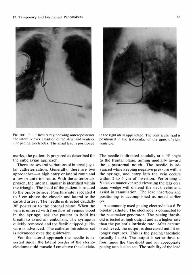
17. Temporary and Permanent Pacemakers
FIGURE 17.1. Chest x-ray showing anteroposterior and lateral views. Position of the atrial and ventricular pacing electrodes. The atrial lead is positioned
marks, the patient is prepared as described for the subclavian approach.
There are several variations of internal jugular catheterization. Generally, there are two approaches-a high entry or lateral route and a low or anterior route. With the anterior approach, the internal jugular is identified within the triangle. The head of the patient is rotated to the opposite side. Puncture site is located 4 to 5 cm above the clavicle and lateral to the carotid artery. The needle is directed caudally 30° posterior to the coronal plane. When the vein is entered with free flow of venous blood in the syringe, ask the patient to hold his breath to avoid air embolism. The syringe is quickly removed and the flexible tipped guidewire is advanced. The catheter introducer set is advanced over the guidewire.
For the lateral approach the needle is inserted under the lateral border of the sternocleidomastoid muscle 5 cm above the clavicle.
183
in the right atrial appendage. The ventricular lead is positioned in the trabeculae of the apex of right ventricle.
The needle is directed caudally at a 15° angle to the frontal plane, aiming medially toward the suprasternal notch. The needle is advanced while keeping negative pressure within the syringe, and entry into the vein occurs within 2 to 5 cm of insertion. Performing a Valsalva maneuver and elevating the legs on a foam wedge will distend the neck veins and assist in cannulation. The lead insertion and positioning is accomplished as noted earlier on.
A commonly used pacing electrode is a 6-Fr bipolar catheter. The electrode is connected to the pacemaker generator. The pacing threshold is tested at high output and at a higher rate than the patient's intrinsic rate. After capture is achieved, the output is decreased until it no longer captures. This is the pacing threshold (usually 1 rnA). The output is set at three to four times the threshold and an appropriate pacing rate is also set. The stability of the lead
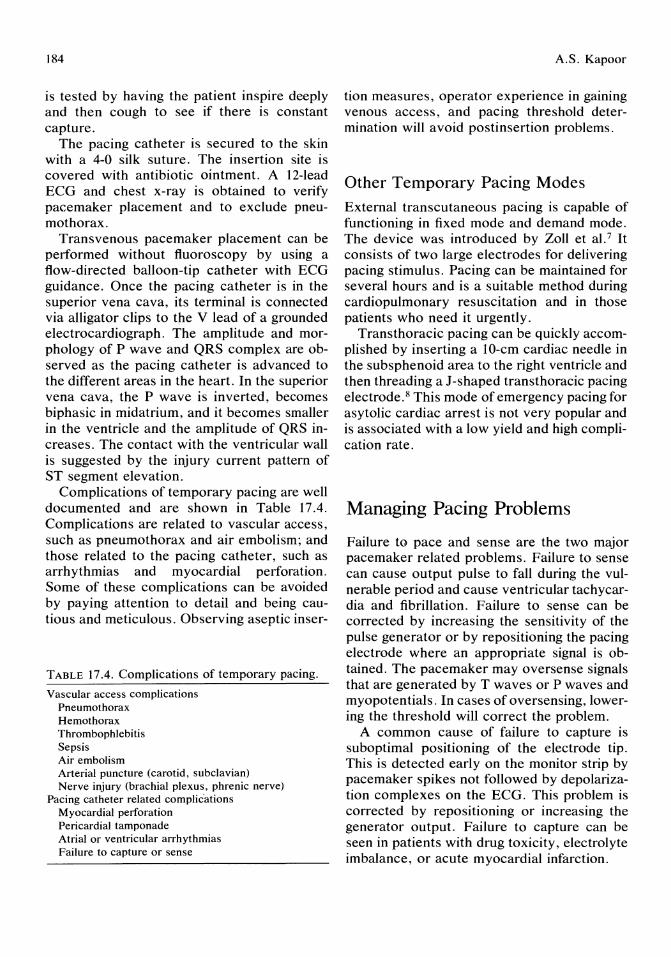
184
is tested by having the patient inspire deeply and then cough to see if there is constant capture.
The pacing catheter is secured to the skin with a 4-0 silk suture. The insertion site is covered with antibiotic ointment. A 12-lead ECG and chest x-ray is obtained to verify pacemaker placement and to exclude pneumothorax.
Transvenous pacemaker placement can be performed without fluoroscopy by using a flow-directed balloon-tip catheter with ECG guidance. Once the pacing catheter is in the superior vena cava, its terminal is connected via alligator clips to the V lead of a grounded electrocardiograph. The amplitude and morphology of P wave and QRS complex are observed as the pacing catheter is advanced to the different areas in the heart. In the superior vena cava, the P wave is inverted, becomes biphasic in midatrium, and it becomes smaller in the ventricle and the amplitude of QRS increases. The contact with the ventricular wall is suggested by the injury current pattern of ST segment elevation.
Complications of temporary pacing are well documented and are shown in Table 17.4. Complications are related to vascular access, such as pneumothorax and air embolism; and those related to the pacing catheter, such as arrhythmias and myocardial perforation. Some of these complications can be avoided by paying attention to detail and being cautious and meticulous. Observing aseptic inser-
TABLE 17.4. Complications of temporary pacing.
Vascular access complications Pneumothorax Hemothorax Thrombophlebitis Sepsis Air embolism Arterial puncture (carotid, subclavian) Nerve injury (brachial plexus, phrenic nerve)
Pacing catheter related complic'ations Myocardial perforation Pericardial tamponade Atrial or ventricular arrhythmias Failure to capture or sense
A.S. Kapoor
tion measures, operator experience in gaining venous access, and pacing threshold determination will avoid postinsertion problems.
Other Temporary Pacing Modes
External transcutaneous pacing is capable of functioning in fixed mode and demand mode. The device was introduced by Zoll et al. 7 It consists of two large electrodes for delivering pacing stimulus. Pacing can be maintained for several hours and is a suitable method during cardiopulmonary resuscitation and in those patients who need it urgently.
Transthoracic pacing can be quickly accomplished by inserting a lO-cm cardiac needle in the sub sphenoid area to the right ventricle and then threading a J-shaped transthoracic pacing electrode.8 This mode of emergency pacing for asytolic cardiac arrest is not very popular and is associated with a low yield and high complication rate.
Managing Pacing Problems
Failure to pace and sense are the two major pacemaker related problems. Failure to sense can cause output pulse to fall during the vulnerable period and cause ventricular tachycardia and fibrillation. Failure to sense can be corrected by increasing the sensitivity of the pulse generator or by repositioning the pacing electrode where an appropriate signal is obtained. The pacemaker may oversense signals that are generated by T waves or P waves and myopotentials. In cases of oversensing, lowering the threshold will correct the problem.
A common cause of failure to capture is suboptimal positioning of the electrode tip. This is detected early on the monitor strip by pacemaker spikes not followed by depolarization complexes on the ECG. This problem is corrected by repositioning or increasing the generator output. Failure to capture can be seen in patients with drug toxicity, electrolyte imbalance, or acute myocardial infarction.

17. Temporary and Permanent Pacemakers
Permanent Cardiac Pacing
Indications for Permanent Pacing
Initially, pacemakers were implanted for complete heart block and symptomatic bradycardias, but with technologic advances the indications for pacemaker therapy have expanded to include prevention and treatment of tachyarrythmias and provision for optimum physiologic pacing for augmentation of cardiac output. Guidelines for permanent cardiac pacemaker implantation were developed by a joint American College of Cardiology and American Heart Association Task Force. IO
The guidelines for permanent pacing are shown in Table 17.5. It is important to document the need for pacemaker implantation and also select a particular pacemaker model that is tailored to the hemodynamic needs of the patient.
Atrial pacing has the distinct advantage of maintaining atrioventricular synchrony, which by atrial systole will improve the cardiac output in the nonfailing heart by at least 20%. Atrial pacing can be used for symptomatic sinus node dysfunction and sick sinus syndrome when the integrity of A V conduction has been established. Atrial fibrillation or inadequate atrial conduction are contraindications to atrial pacing.
Single-chamber ventricular pacing has been successful in 80% of patients. 11 In general, single-chamber ventricular pacing may be used for symptomatic bradyarrhythmia in patients who may have atrial flutter or atrial fibrillation, but in the absence of pacemaker syndrome.
Dual-chamber pacemakers are indicated in active patients who require atrial kick to optimize cardiac output and in patients who develop a pacemaker syndrome due to singlechamber pacing.
Sensor-triggered physiologic pacing is a new modality of pacing indicated for patients who are unsuitable candidates for dual-chamber pacing because of unstable atrial activity, as in atrial fibrillation or other atrial tachyarrhythmias. They are a major advance in pace-
TABLE 17.5. Indications for permanent pacing.
Single chamber pacemakers Atrial pacing in the AAI mode
Symptomatic sinus node dysfunction with intact A V conduction
185
For termination of supraventricular or ventricular arrhythmias in appropriate selected cases
For augmentation of cardiac output by rate adjust· ment in patients with symptomatic bradycardia
Ventricular pacing in the VVI mode Symptomatic complete heart block Mobitz type II block with intermittent complete
heart block Any symptomatic brady arrhythmia in the absence
of known pacemaker syndrome Dual chamber pacemakers
Pacing of both chambers in the DVI and DDD modes In patients with symptomatic bradycardia who
require atrial contribution for hemodynamic benefit
Patients with known pacemaker syndrome Complete heart block or sick sinus syndrome with
stable atrial rates Hypersensitive carotid sinus syndrome
Sensor-triggered physiologic pacing Unsuitable candidates for dual chamber pacing with
inexcitable atrial or atrial tachyarrhythmias Active patients requiring a pacemaker with chrono
tropic incompetence
Partially based on recommendations in references 10, II. AAI = atrial pacing inhibited by sensed atrial activity, VVI = ventricular pacing inhibited by sensed ventricular activity, DVI = pacing of both chambers inhibited by ventricular activity, DDD = pacing of both chambers, sensing of both chambers, inhibition of atrial or ventricular output by sensed atrial or ventricular activity, triggering of ventricular output by sensed atrial activity
maker therapy because they provide chronologic response that matches the patient's metabolic needs by monitoring physiologic parameters. 12
Pacemaker Technology
Pacemaker technology is considered in the context of pulse generators, the power source, and the pacing leads. The pacemaker generator is a small device contained in a hermetically sealed metal can, usually powered by a lithium chemistry battery with a life span of 5 to 15 years.
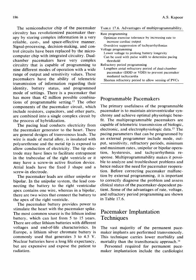
186
The semiconductor chip of the pacemaker circuitry has revolutionized pacemaker therapy by storing complex information in a very reliable, cost-, and space-effective manner. Signal-processing, decision-making, and control circuits have been replaced by the microcomputer chip with integrated circuitry. Dualchamber pacemakers have very complex circuitry that is capable of programming to nine different modes of operation with a wide range of output and sensitivity values. These pacemakers have the ability of telemetric transmission of information regarding their identity, battery status, and programmed mode of settings. There is a pacemaker that has more than 42 million possible combinations of programmable setting. 13 The other components of the pacemaker circuit, which include resistors, capacitors, and other parts, are combined into a single complex circuit by the process of hybridization.
The pacing lead conducts electricity from the pacemaker generator to the heart. There are general designs of transvenous leads. The wire is made of metal alloy, is insulated with polyurethrane and the metal tip is exposed to allow conduction of electricity. The tip electrode may have lines to facilitate entrapment in the trabeculae of the right ventricle or it may have a screw-in active fixation device. Atrial leads have the fixed J shape and a screw-in electrode.
The pacemaker leads are either unipolar or bipolar. In the unipolar system, the lead connecting the battery to the right ventricular apex contains one wire, whereas in a bipolar, there are two wires that connect the battery to the apex of the right ventricle.
The pacemaker battery provides power to stimulate the heart with the pacemaker spike. The most common source is the lithium iodine battery, which can last from 5 to 15 years. There are other lithium batteries with different voltages and end-of-life characteristics. In Europe, a lithium silver chromate battery is commonly used that generates 3 to 4.5 V. Nuclear batteries have a long life expectancy, but are expensive and expose the patient to radiation.
A.S. Kapoor
TABLE 17.6. Advantages of multiprogrammability.
Rate programming Optimize exercise tolerance by increasing rate to
increase cardiac output Overdrive suppression of tachyarrhythmias
Voltage programming Lower voltage to prolong battery longevity Can be IIsed with pulse width to determine pacing
threshold Refractory period programming
Lengthen atrial refractory period of dual-chamber pacemaker (DDD or VDD) to prevent pacemaker mediated tachycardia
Shorten refractory period to allow sensing of PVCs
Programmable Pacemakers The primary usefulness of the programmable pacemaker is to maintain atrioventricular synchrony and achieve optimal physiologic benefit. The multi programmable pacemakers are capable of telemetric transmission of biologic, electronic, and electrophysiologic data. 13 The pacing parameters that can be programmed by an external programmer include mode, output, sensitivity, refractory periods, minimum and maximum rates, unipolar or bipolar operation, hysteresis, and tachyarrhythmia response. Multiprogrammability makes it possible to analyze and troubleshoot problems and hence reduce the need for pacemaker reoperation. Before correcting pacemaker malfunction by external programming, it is important to correctly diagnose the problem and assess clinical status of the pacemaker-dependent patient. Some of the advantages of rate, voltage, and refractory period programming are shown in Table 17.6.
Pacemaker Implantation Techniques
The vast majority of the permanent pacemaker implants are performed transvenously. This technique carries lower morbidity and mortality than the transthoracic approach. 14
Personnel required for permanent pacemaker implantation include the cardiologist
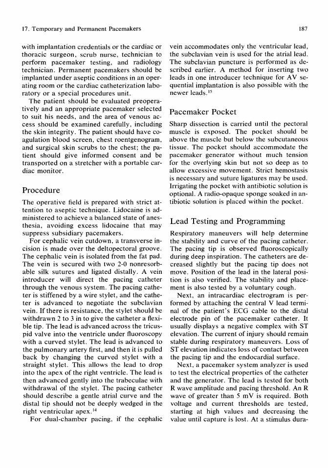
17. Temporary and Permanent Pacemakers
with implantation credentials or the cardiac or thoracic surgeon, scrub nurse, technician to perform pacemaker testing, and radiology technician. Permanent pacemakers should be implanted under aseptic conditions in an operating room or the cardiac catheterization laboratory or a special procedures unit.
The patient should be evaluated preoperatively and an appropriate pacemaker selected to suit his needs, and the area of venous access should be examined carefully, including the skin integrity. The patient should have coagulation blood screen, chest roentgenogram, and surgical skin scrubs to the chest; the patient should give informed consent and be transported on a stretcher with a portable cardiac monitor.
Procedure The operative field is prepared with strict attention to aseptic technique. Lidocaine is administered to achieve a balanced state of anesthesia, avoiding excess lidocaine that may suppress subsidiary pacemakers.
For cephalic vein cutdown, a transverse incision is made over the deltopectoral groove. The cephalic vein is isolated from the fat pad. The vein is secured with two 2-0 nonresorbable silk sutures and ligated distally. A vein introducer will direct the pacing catheter through the venous system. The pacing catheter is stiffened by a wire stylet, and the catheter is advanced to negotiate the subclavian vein. If there is resistance, the stylet should be withdrawn 2 to 3 in to give the catheter a flexible tip. The lead is advanced across the tricuspid valve into the ventricle under fluoroscopy with a curved stylet. The lead is advanced to the pulmonary artery first, and then it is pulled back by changing the curved stylet with a straight stylet. This allows the lead to drop into the apex of the right ventricle. The lead is then advanced gently into the trabeculae with withdrawal of the stylet. The pacing catheter should describe a gentle atrial curve and the distal tip should not be deeply wedged in the right ventricular apex. 14
For dual-chamber pacing, if the cephalic
187
vein accommodates only the ventricular lead, the subclavian vein is used for the atrial lead. The subclavian puncture is performed as described earlier. A method for inserting two leads in one introducer technique for A V sequential implantation is also possible with the newer leads. IS
Pacemaker Pocket Sharp dissection is carried until the pectoral muscle is exposed. The pocket should be above the muscle but below the subcutaneous tissue. The pocket should accommodate the pacemaker generator without much tension for the overlying skin but not so deep as to allow excessive movement. Strict hemostasis is necessary and suture ligatures may be used. Irrigating the pocket with antibiotic solution is optional. A radio-opaque sponge soaked in antibiotic solution is placed within the pocket.
Lead Testing and Programming
Respiratory maneuvers will help determine the stability and curve of the pacing catheter. The pacing tip is observed fluoroscopically during deep inspiration. The catheters are decreased slightly but the pacing tip does not move. Position of the lead in the lateral position is also verified. The stability and placement is also tested by a voluntary cough.
Next, an intracardiac electrogram is performed by attaching the central V lead terminal of the patient's ECG cable to the distal electrode pin of the pacemaker catheter. It usually displays a negative complex with ST elevation. The current of injury should remain stable during respiratory maneuvers. Loss of ST elevation indicates loss of contact between the pacing tip and the endocardial surface.
Next, a pacemaker system analyzer is used to test the electrical properties of the catheter and the generator. The lead is tested for both R wave amplitude and pacing threshold. An R wave of greater than 5 m V is required. Both voltage and current thresholds are tested, starting at high values and decreasing the value until capture is lost. At a stimulus dura-
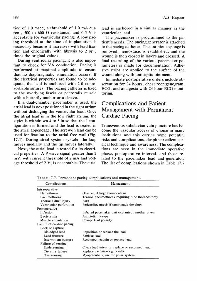
188
tion of 2.0 msec, a threshold of 1.0 rnA current, 500 to 600 n resistance, and 0.5 V is acceptable for ventricular pacing. A low pacing threshold at the time of implantation is necessary because it increases with lead fixation and chronically with fibrosis to 2 or 3 times the original values.
During ventricular pacing, it is also important to check for VA conduction. Pacing is performed at maximal amplitude to ensure that no diaphragmatic stimulation occurs. If the electrical properties are found to be adequate, the lead is anchored with 2-0 nonresorbable sutures. The pacing catheter is fixed to the overlying fascia or pectoralis muscle with a butterfly anchor or a sleeve.
If a dual-chamber pacemaker is used, the atrial lead is next positioned in the right atrium without dislodging the ventricular lead. Once the atrial lead is in the low right atrium, the stylet is withdrawn 4 to 5 in so that the J configuration is formed and the lead is seated in the atrial appendage. The screw-in lead can be used for fixation to the atrial free wall (Fig. 17.1). During atrial system systole, the loop moves medially and the tip moves laterally.
Next, the atrial lead is tested for its electrical properties. A P wave signal greater than 2 mY, with current threshold of 2 rnA and voltage threshold of 2 V, is acceptable. The atrial
A.S. Kapoor
lead is anchored in a similar manner as the ventricular lead.
The pacemaker is programmed to the patient's needs. The pacing generator is attached to the pacing catheter. The antibiotic sponge is removed, hemostasis is established, and the wound is then closed in layers and dressed. A final recording of the various pacemaker parameters is made for documentation. Adhesive strips are applied to the surface of the wound along with antiseptic ointment.
Immediate postoperative orders include observation for 24 hours, chest roentgenogram, ECG, and analgesia with 24-hour ECG monitoring.
Complications and Patient Management with Permanent Cardiac Pacing
Transvenous subclavian vein puncture has become the vascular access of choice in many institutions and this carries some potential risks and complications, despite excellent surgical technique and awareness. The complications are seen in the immediate operative phase, postoperative interval, and those related to the pacemaker lead and generator. The list of complications shown in Table 17.7
TABLE 17.7. Permanent pacing complications and management.
Complications
Intraoperative Hemothorax Pneumothorax Thoracic duct injury Ventricular perforation
Postoperative Infection Bacteremia Muscle stimulation
Failure of cardiac pacing Lack of capture
Dislodged lead Lead fracture Intermittent capture
Failure of sensing Undersensing Circuitry failure Oversensing
Management
Observe, if large thoracentesis Tension pneumothorax requiring tube thoracostomy Rare Pericardiocentesis if tamponade develops
Infected pacemaker unit explanted; another given Antibiotic therapy Change lead polarity
Reposition or replace the lead Replace lead Reconnect leadpin or replace lead
Check lead integrity; replace or reconnect lead Replace pacemaker generator Myopotentials, use for polar system
17. Temporary and Permanent Pacemakers
is not exhaustive, but it shows major complications and their management. For more details, refer to books on cardiac pacing and studies done by Furman et al. 17
Hemothroax may result from subclavian vein or artery puncture. The management depends on the extent of the pleural effusion. With moderate or massive effusion, a tube thoracostomy is required. Pneumothorax is also seen frequently in elderly females. Hakki 18 observed a 40% incidence of pneumothorax using the subclavian vein puncture technique in women 75 years of age or older. Moderate or tension pneumothorax requires a tube thoracostomy. Rarely, patients may develop subclavian vein thrombosis. If there is extensive thrombosis, thrombolytic therapy is the treatment of choice. Infection of the pacemaker generator is seen 2.5 weeks after implantation. 19 The most common infecting organisms are Staphylococcus aureus and epidermidis. The infected pacemaker unit should be explanted with insertion of a new unit in a different location. Antibiotic therapy alone is unsatisfactory. If there is a bacteremia without evidence of pocket infection, then appropriate antibiotics alone may be helpfuI.2°
Failure of capture and failure of sensing are related to lead dislodgement, lead fracture, and, very rarely, to circuitry failure.
Pacemaker Troubleshooting
Troubleshooting the pacing system in the patient can be performed in a systematic manner by reviewing the database of the patient, pacemaker telectronics, and lead mechanics. The patient's underlying rhythm, electrolyte status, drug regimen, and new medical problems, such as new myocardial infarction or congestive heart failure, are also important when evaluating pacemaker malfunction. The internal metabolic milieu of the patient is relevant to the troubleshooting of erratic pacing. Hypokalemia, hypercalcemia, and alkalosis can cause decreased sensitivity to pacing stimuli.
The most frequent causes of malfunction are patient factors, lead disruption, or improper programming of rate, refractory peri-
189
ods, sensitivity, and mode selection. The majority of rhythm problems of nonpacing or intermittent pacing are related to positional instability of the pacing electrode. The ECG ofa dislodged lead displays a chaotic pattern of pacing, nonpacing, and/or sensing. This problem is corrected by repositioning the lead or replacing it with one of the fixation type leads.
Intermittent pacing can be a manifestation of complete lead fracture. Sometimes the chest x-rays will show lead fracture. When insulation is intact, ECG will have no pacing and the pulse analyzer will display increased resistance. Usually that will require changing of the lead. The lead break usually occurs near the area of angulation and constriction.
Ventricular perforation can be a cause of intermittent pacing or nonpacing. Patients may present with hiccups, pericardial friction rub, and sometimes cardiac tamponade. This problem is seen with unipolar leads. The ECG shows erratic capture with proper sensing. It usually requires lead repositioning.
Other mechanical causes of nonpacing include disconnection between the lead and the generator and loose connections. This complication requires opening the pocket and making the connection tight.
Undersensing problems involve the deactivation of the sensing circuitry by cardiac electric events. Undersensing is most often due to subthreshold QRS complexes and a dislodged lead. Because undersensing causes inappropriate discharge of the pacemaker, this problem can be corrected by reprogramming to a higher setting if there is no lead displacement.
Oversensing occurs with signals other than the QRS complex such as skeletal myopotentials, T-wave sensing, and after potential sensing. The initial treatment consists of reprogramming the threshold of the sensing circuit or increasing the refractory period. Changing to a bipolar system will correct myoinhibition of the sensing circuitry.
Pacemaker Syndrome
The pacemaker syndrome is the symptom complex of dizziness, hypotension, fatigue, and often syncopeY This usually develops
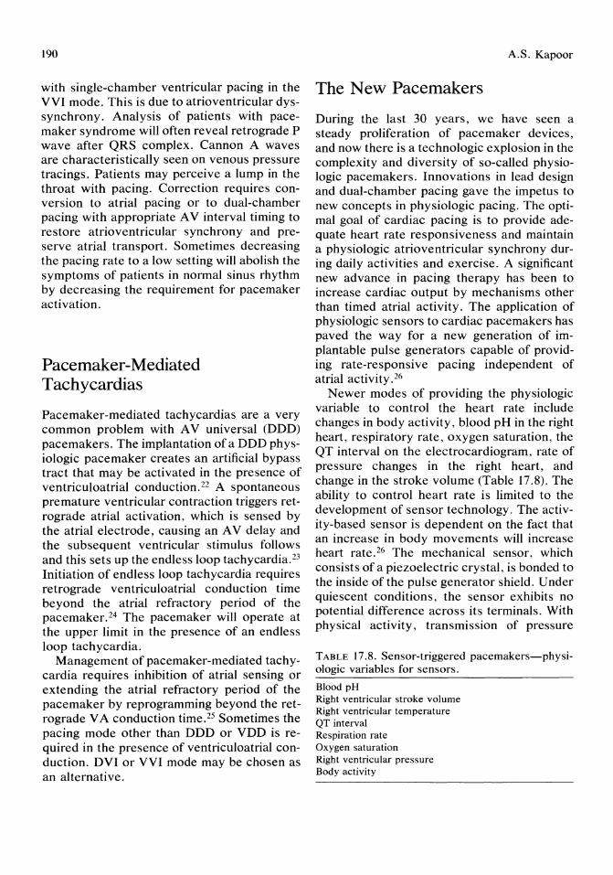
190
with single-chamber ventricular pacing in the VVI mode. This is due to atrioventricular dyssynchrony. Analysis of patients with pacemaker syndrome will often reveal retrograde P wave after QRS complex. Cannon A waves are characteristically seen on venous pressure tracings. Patients may perceive a lump in the throat with pacing. Correction requires conversion to atrial pacing or to dual-chamber pacing with appropriate A V interval timing to restore atrioventricular synchrony and preserve atrial transport. Sometimes decreasing the pacing rate to a low setting will abolish the symptoms of patients in normal sinus rhythm by decreasing the requirement for pacemaker activation.
Pacemaker-Mediated Tachycardias
Pacemaker-mediated tachycardias are a very common problem with A V universal (DDD) pacemakers. The implantation of a DDD physiologic pacemaker creates an artificial bypass tract that may be activated in the presence of ventriculoatrial conduction. 22 A spontaneous premature ventricular contraction triggers retrograde atrial activation, which is sensed by the atrial electrode, causing an A V delay and the subsequent ventricular stimulus follows and this sets up the endless loop tachycardia. 23
Initiation of endless loop tachycardia requires retrograde ventriculoatrial conduction time beyond the atrial refractory period of the pacemaker. 24 The pacemaker will operate at the upper limit in the presence of an endless loop tachycardia.
Management of pacemaker-mediated tachycardia requires inhibition of atrial sensing or extending the atrial refractory period of the pacemaker by reprogramming beyond the retrograde VA conduction time. 25 Sometimes the pacing mode other than DDD or VDD is required in the presence of ventriculoatrial conduction. DVI or VVI mode may be chosen as an alternative.
A.S. Kapoor
The New Pacemakers
During the last 30 years, we have seen a steady proliferation of pacemaker devices, and now there is a technologic explosion in the complexity and diversity of so-called physiologic pacemakers. Innovations in lead design and dual-chamber pacing gave the impetus to new concepts in physiologic pacing. The optimal goal of cardiac pacing is to provide adequate heart rate responsiveness and maintain a physiologic atrioventricular synchrony during daily activities and exercise. A significant new advance in pacing therapy has been to increase cardiac output by mechanisms other than timed atrial activity. The application of physiologic sensors to cardiac pacemakers has paved the way for a new generation of implantable pulse generators capable of providing rate-responsive pacing independent of atrial activity. 26
Newer modes of providing the physiologic variable to control the heart rate include changes in body activity, blood pH in the right heart, respiratory rate, oxygen saturation, the QT interval on the electrocardiogram, rate of pressure changes in the right heart, and change in the stroke volume (Table 17.8). The ability to control heart rate is limited to the development of sensor technology. The activity-based sensor is dependent on the fact that an increase in body movements will increase heart rate. 26 The mechanical sensor, which consists of a piezoelectric crystal, is bonded to the inside of the pulse generator shield. Under quiescent conditions, the sensor exhibits no potential difference across its terminals. With physical activity, transmission of pressure
TABLE 17.8. Sensor-triggered pacemakers-physiologic variables for sensors.
Blood pH Right ventricular stroke volume Right ventricular temperature QT interval Respiration rate Oxygen saturation Right ventricular pressure Body activity
17. Temporary and Permanent Pacemakers
waves originating from body movements are translated by the sensor into electrical signals that drive the pacemaker rate up or down. There is another parameter called the activity threshold (low, medium, and high), which determines the amount of mechanical energy that must be generated to make the pacing rate increase.
Pacemakers that are capable of providing chronotropic response adequate for a patient's metabolic needs by monitoring physiologic parameters may improve exercise tolerance and work capacity. 27-29 These sensor-triggered physiologic pacemakers will have a significant impact on the advancement of artificial pacemakers by providing a normal chronotropic response independent of atrial activity.
The technologic explosion will continue unabated, limited only to scientific proof of efficacy, patient safety, and improved quality of life for patients who need pacemaker therapy.
References 1. Hyman AS: Resuscitation of the stopped heart
by intracardial therapy. Arch Intern Med 1930; 46:553.
2. Zoll PM: Resuscitation of the heart in ventricular standstill by external electric stimulation. N Engl J Med 1952; 247:768.
3. Furman S, Schwedel B: An intracardiac pacemaker for Stoke-Adams seizures. N Engl J Med 1959; 261:943.
4. Joint American College of Cardiology/American Heart Association Task Force on Assessment of Cardiovascular Procedures. Guidelines for permanent cardiac pacemaker implantation. J Am Col Cardiol 1984; 4:434.
5. Barrold SS, Ong LS, Heinle RA: Stimulation and sensing thresholds by cardiac pacing: Electrophysiologic and technical aspects. Prog Cardiovasc Dis 1981; 24:2.
6. Dreifus LS, et al: Use of atrial and bifocal cardiac pacemakers for treating resistant dysrhythmias. Eur J Cardiol1976; 3:257.
7. Zoll PM, et al: External noninvasive temporary cardiac pacing. Clinical trials. Circulation 1985; 71 :937-944.
8. Roberts JR, et al: Successful use of emergency transthoracic pacing in bradysystolic cardiac arrest. Ann Emerg Med 1984; 13:277-283.
191
9. Morelli RL, Goldschlager N: Temporary transvenous pacing: Resolving postinsertion problems. J Crit III 1987; 4:73-80.
10. Parsonnet V, et al: Intersociety Commission for Heart Disease Resources (ICHD): Optimal resources for implantable cardiac pacemakers. Pacemaker Study Group. Circulation 1983; 68:227A.
11. Dreifus LS: Choosing the optimal cardiac pacemaker. Learning Center Highlights 1985; 1:1-6.
12. de Oro AG, et al: Rate-responsive pacing. Clinical experience. PACE 1985; 8:322-328.
13. Parsonnet V, Bernstein AD: Cardiac pacing in the 1980s. Treatment and techniques in transition. J Am Coli Cardiol1983; 1:339-354.
14. Parsonnet V: Technique for implantation and replacement of permanent pacemakers, in Modern Techniques in Cardiac-Thoracic Surgery. New York, Futura 1979, chapter 12.
15. Janss B: Two leads in one introducer. Technique for AV sequential implantations. PACE 1982; 5:217.
16. Mond HG: The cardiac pacemaker: Function and malfunction. New York, Grone and Stratton, 1983, pp 191-232.
17. Furman S, Hurzeler P, Mehra R: Cardiac pacing and pacemakers VI: Analysis of pacemaker malfunction. Am Heart J 1977; 94:378.
18. Hakki AH: Ideal cardiac pacing, Permanent cardiac pacing: Complications and management, in Hakki AH (ed): Ideal Cardiac Pacing. Philadelphia, W. B. Saunders Co, 1984, pp 160-175.
19. Kennelly BM, Piller LW: Management of infected trans venous permanent pacemakers. Br HeartJ 1974; 36: 1133.
20. Corman LC, Levinson MG: Sustained bacteremia and transvenous cardiac pacemakers. JAm Med Assoc 1975; 233:264.
21. Lewis ME, et al: Pacemaker-induced hypotension. Chest 1981; 79:359.
22. Nishimura RA, et al: Outcome of dual-chamber pacing for the pacemaker syndrome. Mayo Clin Proc 1983; 58:452.
23. Luceri RM, et al: The arrhythmias of dualchamber cardiac pacemakers and other management. Ann Int Med 1983; 99:354.
24. Furman S, Fisher J: Endless loop tachycardias in an AV universal (DDD) pacemaker. PACE 1982; 5:486.
25. Den DK, et al: Pacemaker related tachycardias. PACE 1982; 5:476.

192
26. Humen DP, Kostuk WJ, Klein GJ: Activitysensing, rate-responsive pacing: Improvement in myocardial performance with exercise. PACE 1985; 8:52.
A.S. Kapoor
27. Kristensson BI, et al: Physiological versus single-rate ventricular pacing: A double-blind crossover study. PACE 1985; 8:73.
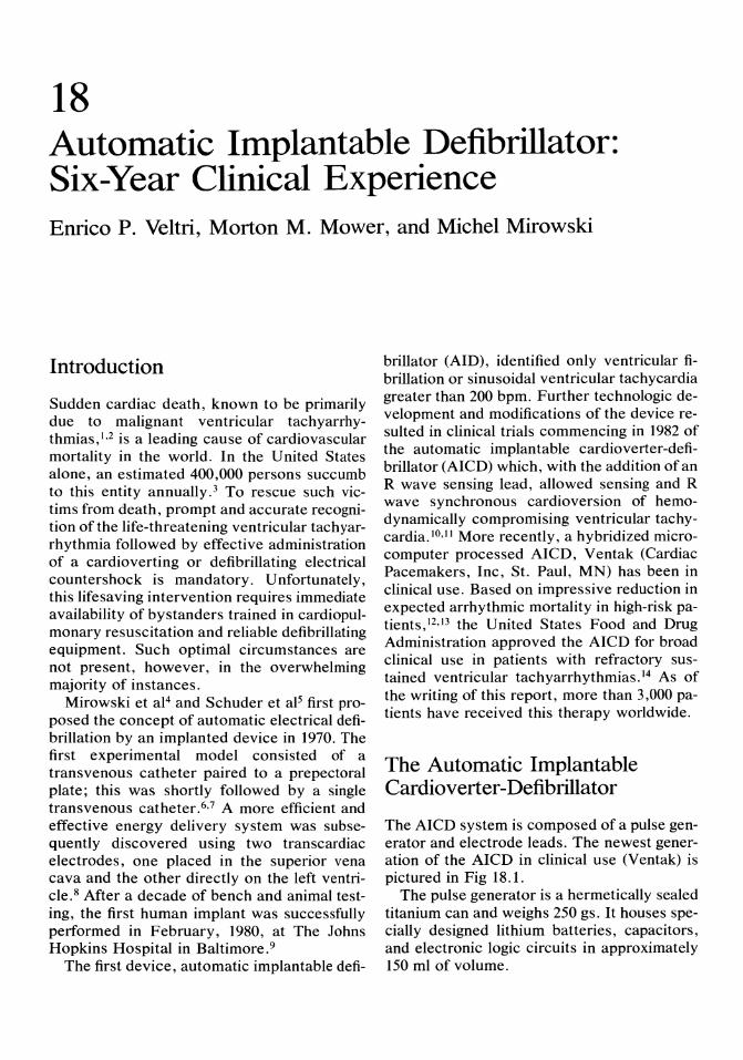
18 Automatic Implantable Defibrillator: Six-Year Clinical Experience Enrico P. Veltri, Morton M. Mower, and Michel Mirowski
Introduction
Sudden cardiac death, known to be primarily due to malignant ventricular tachyarrhythmias,I,2 is a leading cause of cardiovascular mortality in the world. In the United States alone, an estimated 400,000 persons succumb to this entity annually. 3 To rescue such victims from death, prompt and accurate recognition of the life-threatening ventricular tachyarrhythmia followed by effective administration of a cardioverting or defibrillating electrical countershock is mandatory. Unfortunately, this lifesaving intervention requires immediate availability of bystanders trained in cardiopulmonary resuscitation and reliable defibrillating equipment. Such optimal circumstances are not present, however, in the overwhelming majority of instances.
Mirowski et al4 and Schuder et aP first proposed the concept of automatic electrical defibrillation by an implanted device in 1970. The first experimental model consisted of a transvenous catheter paired to a prepectoral plate; this was shortly followed by a single trans venous catheter.6,7 A more efficient and effective energy delivery system was subsequently discovered using two transcardiac electrodes, one placed in the superior vena cava and the other directly on the left ventricle. 8 After a decade of bench and animal testing, the first human implant was successfully performed in February, 1980, at The Johns Hopkins Hospital in Baltimore. 9
The first device, automatic implantable defi-
brillator (AID), identified only ventricular fibrillation or sinusoidal ventricular tachycardia greater than 200 bpm. Further technologic development and modifications of the device resulted in clinical trials commencing in 1982 of the automatic implantable cardioverter-defibrillator (AICD) which, with the addition of an R wave sensing lead, allowed sensing and R wave synchronous cardioversion of hemodynamically compromising ventricular tachycardia.lO,ll More recently, a hybridized microcomputer processed AICD, Ventak (Cardiac Pacemakers, Inc, St. Paul, MN) has been in clinical use. Based on impressive reduction in expected arrhythmic mortality in high-risk patients,12,13 the United States Food and Drug Administration approved the AICD for broad clinical use in patients with refractory sustained ventricular tachyarrhythmias. 14 As of the writing of this report, more than 3,000 patients have received this therapy worldwide.
The Automatic Implantable Cardioverter -Defibrillator
The AICD system is composed of a pulse generator and electrode leads. The newest generation of the AICD in clinical use (Ventak) is pictured in Fig 18.1.
The pulse generator is a hermetically sealed titanium can and weighs 250 gs. It houses specially designed lithium batteries, capacitors, and electronic logic circuits in approximately 150 ml of volume.

194
FIGURE 18.1. The automatic implantable cardioverter-defibrillator with, left to right, its bipolar right
The electrode leads serve for sensmg the rate and morphology of trans cardiac electrical activity, and for delivery of R wave synchronized cardioverting or defibrillating electrical shocks. The rate sensing and R wave synchronizing functions are performed by either right ventricular endocardial (tined) or left ventricular epicardial (intramural screw-in) bipolar leads. The transcardiac morphology sensing function and delivery of electrical shock is performed by an anode-cathode pair. A titanium spring electrode (placed at the junction of the superior vena cava and right atrium) serves as an anode and a left ventricular patch (flexible rectangular titanium mesh) placed at the left ventricular apex serves as the cathode. Alternatively, two patches (right ventricular/ left ventricular or anterior/posterior left ventricular patches) may serve as anode-cathode pairs.
Arrhythmia Recognition
The device continuously monitors the cardiac electrical activity via the implanted electrode leads. The arrhythmia recognition algorithm is based on two parameters: signal morphology and rate. The signal morphology parameter, also known as the probability density function (PDF), is an index that samples the derivative
E.P. Veltri, M.M. Mower, and M. Mirowski
ventricular, superior vena cava, and apical patch electrodes.
of the input signal as a function of the amount oftime spent near a zero-potential (isoelectric) baseline. Ventricular fibrillation and most ventricular tachycardias are characterized by sinusoidal morphologic patterns, thereby spending relatively little time near the isoelectric potential. Supraventricular arrhythmias, without underlying intraventricular conduction delay, on the other hand spend a relatively greater amount of time near the isoelectric potential. The rate parameter allows recognition of arrhythmias above a predetermined rate level.
The implanting physician may choose arrhythmia recognition by both parameters (morphology and rate) or by rate alone. At present, however, such features are fixed by the given model of the device and are not programmable. The dual recognition parameter allows higher specificity for ventricular tachycardia/fibrillation, however "spiky" ventricular tachycardias, which are relatively nonsinusoidal and thus unlikely to satisfy morphology (PDF) criteria, may be missed. "Rate only" devices use only the rate recognition criteria to trigger the device. This model provides higher sensitivity and faster arrhythmia recognition time; however, its specificity is lower because any tachycardia, including sinus tachycardia, above the rate cutoff will satisfy arrhythmia detection criteria of the device.
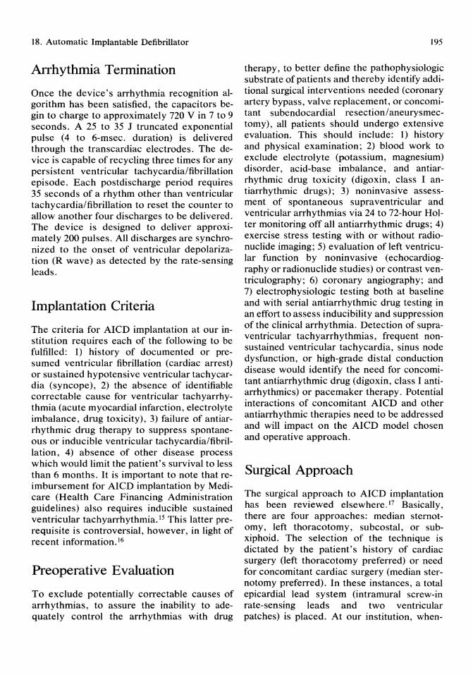
18. Automatic Implantable Defibrillator
Arrhythmia Termination
Once the device's arrhythmia recognition algorithm has been satisfied, the capacitors begin to charge to approximately 720 V in 7 to 9 seconds. A 25 to 35 J truncated exponential pulse (4 to 6-msec. duration) is delivered through the transcardiac electrodes. The device is capable of recycling three times for any persistent ventricular tachycardia/fibrillation episode. Each postdischarge period requires 35 seconds of a rhythm other than ventricular tachycardia/fibrillation to reset the counter to allow another four discharges to be delivered. The device is designed to deliver approximately 200 pulses. All discharges are synchronized to the onset of ventricular depolarization (R wave) as detected by the rate-sensing leads.
Implantation Criteria
The criteria for AICD implantation at our institution requires each of the following to be fulfilled: 1) history of documented or presumed ventricular fibrillation (cardiac arrest) or sustained hypotensive ventricular tachycardia (syncope), 2) the absence of identifiable correctable cause for ventricular tachyarrhythmia (acute myocardial infarction, electrolyte imbalance, drug toxicity), 3) failure of antiarrhythmic drug therapy to suppress spontaneous or inducible ventricular tachycardia/fibrillation, 4) absence of other disease process which would limit the patient's survival to less than 6 months. It is important to note that reimbursement for AICD implantation by Medicare (Health Care Financing Administration guidelines) also requires inducible sustained ventricular tachyarrhythmia. 15 This latter prerequisite is controversial, however, in light of recent information. 16
Preoperative Evaluation
To exclude potentially correctable causes of arrhythmias, to assure the inability to adequately control the arrhythmias with drug
195
therapy, to better define the pathophysiologic substrate of patients and thereby identify additional surgical interventions needed (coronary artery bypass, valve replacement, or concomitant subendocardial resection/aneurysmectomy), all patients should undergo extensive evaluation. This should include: 1) history and physical examination; 2) blood work to exclude electrolyte (potassium, magnesium) disorder, acid-base imbalance, and antiarrhythmic drug toxicity (digoxin, class I antiarrhythmic drugs); 3) noninvasive assessment of spontaneous supraventricular and ventricular arrhythmias via 24 to 72-hour Holter monitoring off all antiarrhythmic drugs; 4) exercise stress testing with or without radionuclide imaging; 5) evaluation ofleft ventricular function by noninvasive (echocardiography or radionuclide studies) or contrast ventriculography; 6) coronary angiography; and 7) electrophysiologic testing both at baseline and with serial antiarrhythmic drug testing in an effort to assess inducibility and suppression of the clinical arrhythmia. Detection of supraventricular tachyarrhythmias, frequent nonsustained ventricular tachycardia, sinus node dysfunction, or high-grade distal conduction disease would identify the need for concomitant antiarrhythmic drug (digoxin, class I antiarrhythmics) or pacemaker therapy. Potential interactions of concomitant AICD and other antiarrhythmic therapies need to be addressed and will impact on the AICD model chosen and operative approach.
Surgical Approach
The surgical approach to AICD implantation has been reviewed elsewhere. 17 Basically, there are four approaches: median sternotomy, left thoracotomy, subcostal, or subxiphoid. The selection of the technique is dictated by the patient's history of cardiac surgery (left thoracotomy preferred) or need for concomitant cardiac surgery (median sternotomy preferred). In these instances, a total epicardial lead system (intramural screw-in rate-sensing leads and two ventricular patches) is placed. At our institution, when-
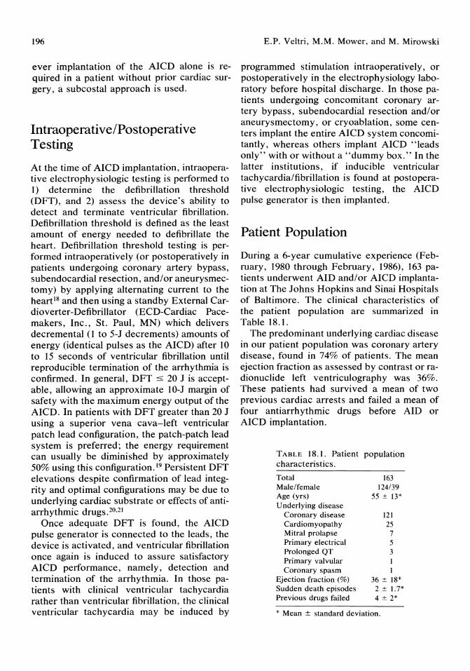
196
ever fmplantation of the AICD alone is required in a patient without prior cardiac surgery, a subcostal approach is used.
Intraoperative/Postoperative Testing
At the time of AICD implantation, intraoperative electrophysiologic testing is performed to 1) determine the defibrillation threshold (DFT) , and 2) assess the device's ability to detect and terminate ventricular fibrillation. Defibrillation threshold is defined as the least amount of energy needed to defibrillate the heart. Defibrillation threshold testing is performed intraoperatively (or postoperatively in patients undergoing coronary artery bypass, subendocardial resection, and/or aneurysmectomy) by applying alternating current to the heart18 and then using a standby External Cardioverter-Defibrillator (ECD-Cardiac Pacemakers, Inc., St. Paul, MN) which delivers decremental (1 to 5-J decrements) amounts of energy (identical pulses as the AICD) after 10 to 15 seconds of ventricular fibrillation until reproducible termination of the arrhythmia is confirmed. In general, DFT ::5 20 J is acceptable, allowing an approximate IO-J margin of safety with the maximum energy output of the AICD. In patients with DFT greater than 20 J using a superior vena cava-left ventricular patch lead configuration, the patch-patch lead system is preferred; the energy requirement can usually be diminished by approximately 50% using this configuration. 19 Persistent DFT elevations despite confirmation of lead integrity and optimal configurations may be due to underlying cardiac substrate or effects of antiarrhythmic drugS.20,21
Once adequate DFT is found, the AICD pulse generator is connected to the leads, the device is activated, and ventricular fibrillation once again is induced to assure satisfactory AICD performance, namely, detection and termination of the arrhythmia. In those patients with clinical ventricular tachycardia rather than ventricular fibrillation, the clinical ventricular tachycardia may be induced by
E.P. Veltri, M.M. Mower, and M. Mirowski
programmed stimulation intraoperatively, or postoperatively in the electrophysiology laboratory before hospital discharge. In those patients undergoing concomitant coronary artery bypass, subendocardial resection and/or aneurysmectomy, or cryoablation, some centers implant the entire AICD system concomitantly, whereas others implant AICD "leads only" with or without a "dummy box." In the latter institutions, if inducible ventricular tachycardia/fibrillation is found at postoperative electrophysiologic testing, the AICD pulse generator is then implanted.
Patient Population
During a 6-year cumulative experience (February, 1980 through February, 1986), 163 patients underwent AID and/or AI CD implantation at The Johns Hopkins and Sinai Hospitals of Baltimore. The clinical characteristics of the patient population are summarized in Table 18.1.
The predominant underlying cardiac disease in our patient popUlation was coronary artery disease, found in 74% of patients. The mean ejection fraction as assessed by contrast or radionuclide left ventriculography was 36%. These patients had survived a mean of two previous cardiac arrests and failed a mean of four antiarrhythmic drugs before AID or AICD implantation.
TABLE 18.1. Patient population characteristics. Total 163 Male/female 124/39 Age (yrs) 55 ± 13* Underlying disease
Coronary disease 121 Cardiomyopathy 25 Mitral prolapse 7 Primary electrical 5 Prolonged QT 3 Primary valvular Coronary spasm
Ejection fraction (%) 36 ± 18* Sudden death episodes 2 ± 1.7* Previous drugs failed 4 ± 2*
* Mean ± standard deviation.

18. Automatic Implantable Defibrillator
Of the 163 patients, 33 received the AID and 130 patients the AICD; there were 9 patients with later crossover from the AID to AICD when this technology became available. One hundred nineteen patients (73%) underwent AID or AICD implantation without other associated surgical procedures. Coronary artery bypass grafting was performed in 19 patients (12%). Twenty-five patients (15%) underwent subendocardial resection, and in 17 patients this was associated with concomitant coronary artery bypass grafting.
Operative Complications
Perioperative deaths (defined as deaths before hospital discharge) occurred in 8 (5%) patients. There were 4 deaths from incessant ventricular tachycardia/fibrillation and 1 death each from refractory heart failure, myocardial infarction, pulmonary embolus, and vascular tear. The latter death was not directly related to AICD lead insertion.
Table 18.2 summarizes the other major operative complications in our patient population. Infection was the predominant complication noted. The majority of infections involved the AICD pulse generator pocket. Automatic implantable cardioverter-defibrillator infection necessitated explanation in 5 patients. Blood transfusions were performed in 17 patients, pneumothorax occurred in 11 patients (predominantly involving the spring lead), pericardial tamponade necessitating pericardiocentesis in 3, and cardiogenic shock in 3. Minor miscellaneous untoward effects were
TABLE 18.2. Major operative complications.
Infection Generator pocket Pneumonia Thoracotomy site
Transfusion requirement Pneumothorax Pericardial tamponade Cardiogenic shock
N = 163 patients
24 (15%) 13 8 3
17 (10%) 11 (7%) 3 (2%) 3 (2%)
197
noted in 19 patients. Sixty-one percent of patients were free of any operative complication.
Long-Term Arrhythmic and Clinical Outcome
At a mean 21-month follow-up, 85 patients (53%) had at least 1 "appropriate" AID or AICD discharge. "Appropriate" discharge was defined as a discharge occurring during hypotensive symptoms or sleep. "Asymptomatic" discharges occurred in 38 patients (23%).
During the 6-year cumulative experience, 44 patients died (27%). These included the 8 perioperative deaths and 36 late deaths. Late deaths were defined as deaths occurring after hospital discharge and included documented or presumed ventricular tachycardia/fibrillation in 15 (9%), heart failure in 14 (9%), myocardial infarction in 3, bradyarrhythmia in I, and noncardiac deaths in 3.
Kaplan-Meier survival curves for the AID and AICD patient groups are depicted in Figs 18.2 and 18.3, respectively. "Actual" arrhythmic deaths are deaths due to documented or
1
.9
C .8
A I D
., "Actual" Jj .7
"0 .6 ~ . 5 ; ....... .. ......................... L. ......... ..... : ... ~.~!lected · ·
.Q .4
~ .3 Co
ct .2
.1
°0L---+---~--~----+---~--~60~--7~0 10 20 30 40 50
Months Following AI D Implantation
FIGURE 18.2. Kaplan-Meier life table analysis of "actual" versus "expected" arrhythmic mortality with the automatic implantable defibrillator (AID). "Actual" = actual death due to documented or presumed ventricular tachyarrhythmia. "Expected" = "actual" deaths plus patients who experienced an appropriate AID discharge (counted only once).
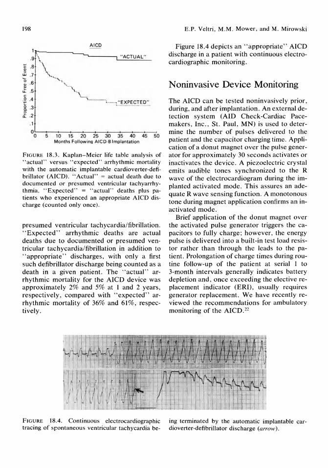
198
AICO 1 j:;--~ ___ ,---_
.9 '\ -.....---~"A~C~T~U~A~L·~· _
\ E .8 \ ", '" :> UJ .7 '0 '" .6 '" U: .5 co . Q .4
& .3 o .t .2
,1
... ." .. ·'--1.
" ... . \\. .. \
........ ................. :········ , .. EXPECTEO ..
O~~--+-~--~--~ __ --__ --____ ___ o 5 10 15 20 25 30 35 40 45 50
Months Following AICO·B Implantation
FIGURE 18.3. Kaplan-Meier life table analysis of "actual" versus "expected" arrhythmic mortality with the automatic implantable cardioverter-defibrillator (AICD). "Actual" = actual death due to documented or presumed ventricular tachyarrhythmia. "Expected" = "actual" deaths plus patients who experienced an appropriate AICD discharge (counted only once).
presumed ventricular tachycardia/fibrillation. "Expected" arrhythmic deaths are actual deaths due to documented or presumed ventricular tachycardia/fibrillation in addition to "appropriate" discharges, with only a first such defibrillator discharge being counted as a death in a given patient. The "actual" arrhythmic mortality for the AICD device was approximately 2% and 5% at 1 and 2 years, respectively, compared with "expected" arrhythmic mortality of 36% and 61%, respectively.
FIGURE 18.4. Continuous electrocardiographic tracing of s~ontaneous ventricular tachycardia be-
E.P. Veltri, M.M. Mower, and M. Mirowski
Figure 18.4 depicts an "appropriate" AICD discharge in a patient with continuous electrocardiographic monitoring.
Noninvasive Device Monitoring
The AICD can be tested noninvasively prior, during, and after implantation. An external detection system (AID Check-Cardiac Pacemakers, Inc., St. Paul, MN) is used to determine the number of pulses delivered to the patient and the capacitor charging time. Application of a donut magnet over the pulse generator for approximately 30 seconds activates or inactivates the device. A piezoelectric crystal emits audible tones synchronized to the R wave of the electrocardiogram during the implanted activated mode. This assures an adequate R wave sensing function. A monotonous tone during magnet application confirms an inactivated mode.
Brief application of the donut magnet over the activated pulse generator triggers the capacitors to fully charge; however, the energy pulse is delivered into a built-in test load resistor rather than through the leads to the patient. Prolongation of charge times during routine follow-up of the patient at serial 1 to 3-month intervals generally indicates battery depletion and, once exceeding the elective replacement indicator (ERI), usually requires generator replacement. We have recently reviewed the recommendations for ambulatory monitoring of the AICD.22
ing terminated by the automatic implantable cardioverter-defibrillator discharge (arrow).

18. Automatic Implantable Defibrillator
Summary
The AICD represents a significant advance in the management of patients at high risk of sudden cardiac death. Arrhythmic mortality from ventricular tachycardia/fibrillation in patients with drug refractory arrhythmia have been reduced from an expected 30% to 60% to 2% to 5% at 1 to 2 years with the currently available device. It is fair to say, however, that only a glimpse of the horizon has been achieved in non pharmacologic electrical therapy for ventricular tachyarrhythmias. Future directions will include the addition of pacing capability (both bradycardia and antitachycardia modes), programmability of rate and morphology sensing functions, electrical (atrial) or hemodynamic sensing function to better differentiate supraventricular from ventricular tachyarrhythmias, improved and more efficient modes of defibrillation (alternate lead configuration, sequential or multiple shocks), smaller sized units, and greater battery longevity. These advances will certainly enhance physician acceptance, patient comfort and management, and hopefully further improve upon long-term survival.
Acknowledgments. We are grateful to Ms. Toni Haase for her assistance in the preparation of this manuscript.
References 1. Nikolic G, Bishop RL, Singh JB: Sudden death
recorded during Holter monitoring. Circulation 1982; 66:218.
2. Kempf FC, Josephson ME: Cardiac arrest recorded on ambulatory electrocardiograms. Am J Cardiol1984; 53:1577.
3. Kuller L: Sudden death in arteriosclerotic heart disease: The case for preventive medicine. Am J Cardiol 1969; 24:617.
4. Mirowski M, Mower MM, Staewen WS, et al: Standby automatic defibrillator-an approach to prevention of sudden coronary death. Arch Int Med 1970; 126:158.
5. Schuder JC, Stoeckle H, Gold JH, et al: Experimental ventricular defibrillation with an automatic and completely implanted system. Trans Am Soc Artif Int Org 1970; 16:207.
199
6. Mirowski M, Mower MM, Staewen WS, et al: Ventricular defibrillation through a single intravascular catheter electrode system. Clin Res 1971; 19:328.
7. Mirowski M, Mower MM, Staewen WS, et al: The development of the trans venous automatic defibrillator. Arch Int Med 1972; 129:773.
8. Mirowski M, Mower MM, Langer A, et al: A chronically implanted system for automatic defibrillation in active conscious dogs: Experimental model for treatment of sudden death from ventricular fibrillation. Circulation 1978; 58:90.
9. Mirowski M, Reid PR, Mower MM, et al: Termination of malignant ventricular arrhythmias with an implanted automatic defibrillator in human beings. N Engl J Med 1980; 303:322.
10. Winkle RA, Bach SM, Echt DS, et al: The automatic implantable defibrillator: Local ventricular bipolar sensing to detect ventricular tachycardia and fibrillation. Am J Cardiol 1983; 52:265.
11. Reid PR, Mirowski M, Mower M, et al: Clinical evaluation of the internal automatic cardioverter-defibrillator in survivors of sudden cardiac death. Am J Cardiol1983; 51:1608.
12. Mirowski M, Reid PR, Winkle RA, et al: Mortality in patients with implanted automatic defibrillators. Ann Int Med 1983; 98:585.
13. Echt DS, Armstrong K, Schmidt P, et al: Clinical experience, complications and survival in 70 patients with the automatic implantable cardioverter-defibrillator. Circulation 1985; 71 :289.
14.50. Fed Reg 47276. November 15, 1985. 15. Department of Health and Human Services.
Health Care Financing Administration. Section 35-85, Implantation of Automatic Defibrillators. January, 1986.
16. Veltri EP, Mower MM, Mirowski M, et al: Clinical outcome of patients with noninducible ventricular tachyarrhythmias and the automatic implantable defibrillator. Circulation 1986; 74:11-109.
17. Watkins L Jr, Guarnieri T, Griffith LSC, et al: Implantation of the automatic defibrillator: Current surgical techniques. Clin Prog Electrophys Pacing 1986; 4:286.
18. Mower MM, Reid PR, Watkins L Jr, et al: Use of alternative current during diagnostic electrophysiologic studies. Circulation 1982; 67:69.
19. Troup PJ, Chapman P, Olinger GN, et al: The implanted defibrillator: Relation of defibrillating lead configuration and clinical variables to defibrillation threshold. J Am Call Cardiol 1985; 6:1315.

200
20. Haberman R, Veltri EP, Mower MM: The effect of amiodarone on defibrillation threshold. PACE 1987; 10:406.
21. Guarnieri T, Levine JH, Veltri EP, et al: Success of chronic defibrillation and the role of antiarrhythmic drugs with the automatic im-
E.P. Veltri, M.M. Mower, and M. Mirowski
plantable cardioverter-defibrillator. Am J Cardio11987; 60:1061.
22. Veltri EP, Mower MM, Mirowski M: Ambulatory monitoring of the automatic implantable cardioverter-defibrillator: A practical guide. PACE 1988; in press.

19 Catheter Ablation Techniques for Treatment of Cardiac Arrhythmias Melvin M. Scheinman
One of the most important innovations in the management of patients with drug-resistant cardiac arrhythmias is the use of catheter ablative techniques. The technique was initially used for ablation of the atrioventricular (A V) junction l and more recently extended for use in patients with accessory pathways2 or ventricular tachycardia. 3 The predominant experience is with use of high-energy electrical discharges through electrode catheters. The purpose of this chapter is to review the histologic changes, clinical indications, results, and complications of high-energy electrical catheter ablative procedures.
Bioelectric Effects of High-energy Discharges
A 200 to 300-J discharge through an electrode catheter results in a discharge of approximately 10 to 15 A associated with a 2,000 to 3,000-V output. The bioelectric effects of high-energy electrical shocks include production of light, pressure waves, and heat, and an intense electrical field is generated. 4 The electrical discharge initially results in parallel rises in both current flow and voltage, followed by a sharp rise in impedance.5 The latter is due to formation of a vapor globe caused by vaporization of water by the intense heat generated as well as by electrolysis with formation of hydrogen and oxygen. The formation and collapse of the vapor globe produces intense
pressure waves. The weight of current evidence suggests that tissue damage is largely a result of the intense electrical field generated with resultant disruption of the cell membrane. Jones et al6 found a characteristic response to increasingly intense stimulation of cultured myocardial pacemaker cells. Threshold stimuli caused a single activation, but stimulus intensities 24 times threshold values caused transient tachyarrhythmias. With stimuli 42 times threshold, there was arrest of rhythmic activity due to membrane depolarization to zero ("dielectric breakdown") and delayed repolarization. "Cellular fibrillation" or asynchronous contraction of sarcomeres followed application of stimuli 80 times the threshold value. Levine et aF have shown that electrical discharges of 5 to 20 J results in altered characteristics of the membrane action potential as far as 5 to 10 minutes from the shock site.
Catheter Ablation of the Atrioventricular Junction
Initial efforts to achieve disruption of A V conduction involved thoracotomy with direct dissection of the His bundle. 8,9 Other techniques involve direct injection of formalin 10 or cryoablation of the A V junction. II A catheter technique for direct injection of formalin achieved successful ablation in as many as 60% of the animals. 12,13 Beazell et aP4 were the first to use
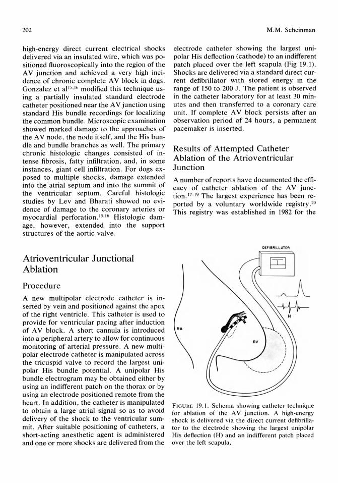
202
high-energy direct current electrical shocks delivered via an insulated wire, which was positioned fluoroscopically into the region of the A V junction and achieved a very high incidence of chronic complete A V block in dogs. Gonzalez et a1 15,16 modified this technique using a partially insulated standard electrode catheter positioned near the A V junction using standard His bundle recordings for localizing the common bundle. Microscopic examination showed marked damage to the approaches of the A V node, the node itself, and the His bundle and bundle branches as well. The primary chronic histologic changes consisted of intense fibrosis, fatty infiltration, and, in some instances, giant cell infiltration. For dogs exposed to mUltiple shocks, damage extended into the atrial septum and into the summit of the ventricular septum. Careful histologic studies by Lev and Bharati showed no evidence of damage to the coronary arteries or myocardial perforation. 15 , 16 Histologic damage, however, extended into the support structures of the aortic valve.
Atrioventricular Junctional Ablation
Procedure
A new multipolar electrode catheter is inserted by vein and positioned against the apex of the right ventricle. This catheter is used to provide for ventricular pacing after induction of A V block. A short cannula is introduced into a peripheral artery to allow for continuous monitoring of arterial pressure. A new multipolar electrode catheter is manipulated across the tricuspid valve to record the largest unipolar His bundle potential. A unipolar His bundle electrogram may be obtained either by using an indifferent patch on the thorax or by using an electrode positioned remote from the heart. In addition, the catheter is manipulated to obtain a large atrial signal so as to avoid delivery of the shock to the ventricular summit. After suitable positioning of catheters, a short-acting anesthetic agent is administered and one or more shocks are delivered from the
M.M. Scheinman
electrode catheter showing the largest unipolar His deflection (cathode) to an indifferent patch placed over the left scapula (Fig 19.1). Shocks are delivered via a standard direct current defibrillator with stored energy in the range of 150 to 200 J. The patient is observed in the catheter laboratory for at least 30 minutes and then transferred to a coronary care unit. If complete A V block persists after an observation period of 24 hours, a permanent pacemaker is inserted.
Results of Attempted Catheter Ablation of the Atrioventricular Junction
A number of reports have documented the efficacy of catheter ablation of the A V junction. 17- 19 The largest experience has been reported by a voluntary worldwide registry. 20
This registry was established in 1982 for the
DEF IBRILL ATOR
FIGURE 19.1. Schema showing catheter technique for ablation of the A V junction. A high-energy shock is delivered via the direct current defibrillator to the electrode showing the largest unipolar His deflection (H) and an indifferent patch placed over the left scapula.
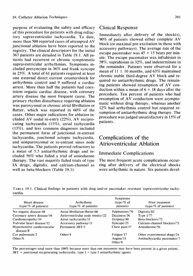
19. Catheter Ablation Techniques
purpose of evaluating the safety and efficacy of this procedure for patients with drug refractory supraventricular tachycardia. To date, more than 500 reported cases of attempted A V junctional ablation have been reported to the registry. The clinical descriptors for the initial 367 patients are detailed in Table 19.1. All patients had recurrent or chronic symptomatic supraventricular arrhythmias. Symptoms included presyncope in 36% and frank syncope in 25%. A total of 61 patients required at least one external direct current countershock for arrhythmia control and 9 suffered a cardiac arrest. More than half the patients had coexistent organic cardiac disease, with coronary artery disease the most frequent cause. The primary rhythm disturbance requiring ablation was paroxysmal or chronic atrial fibrillation or flutter, which was reported in 60% of the cases. Other major indications for ablation included A V nodal re-entry (22%), A V reciprocating tachycardia (11%), atrial tachycardia (13%), and less common diagnoses included the permanent form of junctional re-entrant tachycardia, junctional ectopic tachycardia, and nonparoxysmal or re-entrant sinus node tachycardia. The patients proved refractory to a mean of 5.5 antiarrhythmic drugs and included 56% who failed a trial of amiodarone therapy. The vast majority failed trials of type IA drugs, digitalis, and calcium-channel as well as beta-blockers (Table 19.1).
203
Clinical Response Immediately after delivery of the shock(s), 90% of patients showed either complete A V block (or maximal pre-excitation in those with accessory pathways). The average rate of the escape pacemaker was 45 ± 15 beats per minute. The escape pacemaker was infrahisian in 58%, suprahisian in 32%, and indeterminate in the remainder. Patients were observed for a mean of 11 ± 10 months, and 63% maintained chronic stable third-degree A V block and required no antiarrhythmic drugs. The remaining patients showed resumption of A V conduction within a mean of 6 ± 18 days after the procedure. Ten percent of patients who had resumption of A V conduction were asymptomatic without drug therapy, whereas another 12% had arrhythmia control but required resumption of antiarrhythmic drug therapy. The procedure was judged unsatisfactory in 15% of patients.
Complications of the Atrioventricular Ablation
Immediate Complications
The most frequent acute complications occurring after delivery of the electrical shocks were arrhythmic in nature. Six patients devel-
TABLE 19.1. Clinical findings in patients with drug and! or pacemaker resistant supraventricular tachycardia.
Heart disease (type/% of patients)
No organic disease/48 Coronary artery disease/16 Cardiomyopathy /14 Valvular heart disease/12 Hypertensive cardiovascular
disease/8 Cor pulmonale/2 Other/6
Arrhythmia (type/% of patients)
Atrial fibrillation/flutter/60 Atrioventricular node reentry /22 Atrial tachycardia/13 Accessory pathway/11 Permanent JRT/2
Other/4
Symptoms (type/% of patients)
Palpitations170 Dizziness/36 Dyspnea/40 Syncope/25 Chest pain/17
Fatigue/17 Angina/II Other/6
Prior treatment (type/% of patients)
Digitalis/82 Type 1177 Beta-blockers172 Calcium-channel blockers171 Amiodarone/56
Other experimental drugs/24 Antitachycardia pacemaker17
The percentages total more than 100% because more than one parameter may have been present in a given patient. JRT = junctional reciprocating tachycardia, type I = type I antiarrhythmic agents.
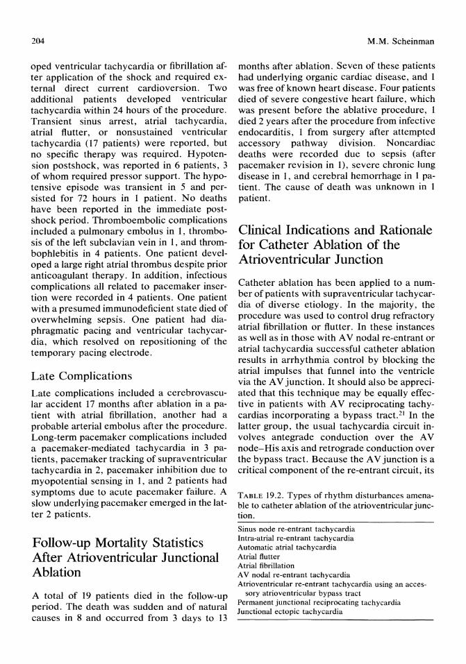
204
oped ventricular tachycardia or fibrillation after application of the shock and required external direct current cardioversion. Two additional patients developed ventricular tachycardia within 24 hours of the procedure. Transient sinus arrest, atrial tachycardia, atrial flutter, or nonsustained ventricular tachycardia (17 patients) were reported, but no specific therapy was required. Hypotension postshock, was reported in 6 patients, 3 of whom required pressor support. The hypotensive episode was transient in 5 and persisted for 72 hours in 1 patient. No deaths have been reported in the immediate postshock period. Thromboembolic complications included a pulmonary embolus in 1, thrombosis of the left subclavian vein in 1, and thrombophlebitis in 4 patients. One patient developed a large right atrial thrombus despite prior anticoagulant therapy. In addition, infectious complications all related to pacemaker insertion were recorded in 4 patients. One patient with a presumed immunodeficient state died of overwhelming sepsis. One patient had diaphragmatic pacing and ventricular tachycardia, which resolved on repositioning of the temporary pacing electrode.
Late Complications
Late complications included a cerebrovascular accident 17 months after ablation in a patient with atrial fibrillation, another had a probable arterial embolus after the procedure. Long-term pacemaker complications included a pacemaker-mediated tachycardia in 3 patients, pacemaker tracking of supraventricular tachycardia in 2, pacemaker inhibition due to myopotential sensing in 1, and 2 patients had symptoms due to acute pacemaker failure. A slow underlying pacemaker emerged in the latter 2 patients.
Follow-up Mortality Statistics After Atrioventricular Junctional Ablation
A total of 19 patients died in the follow-up period. The death was sudden and of natural causes in 8 and occurred from 3 days to 13
M.M. Scheinman
months after ablation. Seven of these patients had underlying organic cardiac disease, and 1 was free of known heart disease. Four patients died of severe congestive heart failure, which was present before the ablative procedure, 1 died 2 years after the procedure from infective endocarditis, 1 from surgery after attempted accessory pathway division. Noncardiac deaths were recorded due to sepsis (after pacemaker revision in 1), severe chronic lung disease in 1, and cerebral hemorrhage in 1 patient. The cause of death was unknown in 1 patient.
Clinical Indications and Rationale for Catheter Ablation of the Atrioventricular Junction
Catheter ablation has been applied to a number of patients with supraventricular tachycardia of diverse etiology. In the majority, the procedure was used to control drug refractory atrial fibrillation or flutter. In these instances as well as in those with A V nodal re-entrant or atrial tachycardia successful catheter ablation results in arrhythmia control by blocking the atrial impulses that funnel into the ventricle via the A V junction. It should also be appreciated that this technique may be equally effective in patients with A V reciprocating tachycardias incorporating a bypass tract. 21 In the latter group, the usual tachycardia circuit involves antegrade conduction over the A V node-His axis and retrograde conduction over the bypass tract. Because the A V junction is a critical component of the re-entrant circuit, its
TABLE 19.2. Types of rhythm disturbances amenable to catheter ablation of the atrioventricular junction.
Sinus node re-entrant tachycardia Intra-atrial re-entrant tachycardia Automatic atrial tachycardia Atrial flutter Atrial fibrillation A V nodal re-entrant tachycardia Atrioventricular re-entrant tachycardia using an acces
sory atrioventricular bypass tract Permanent junctional reciprocating tachycardia Junctional ectopic tachycardia
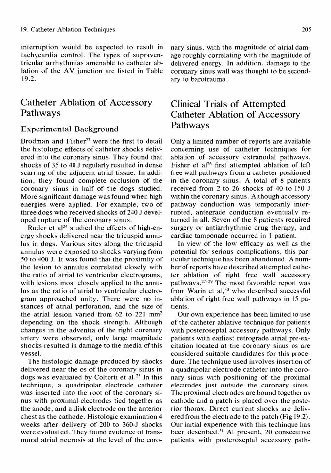
19. Catheter Ablation Techniques
interruption would be expected to result in tachycardia control. The types of supraventricular arrhythmias amenable to catheter ablation of the A V junction are listed in Table 19.2.
Catheter Ablation of Accessory Pathways
Experimental Background
Brodman and Fisher23 were the first to detail the histologic effects of catheter shocks delivered into the coronary sinus. They found that shocks of 35 to 40 J regularly resulted in dense scarring of the adjacent atrial tissue. In addition, they found complete occlusion of the coronary sinus in half of the dogs studied. More significant damage was found when high energies were applied. For example, two of three dogs who received shocks of 240 J developed rupture of the coronary sinus.
Ruder et aF4 studied the effects of high-energy shocks delivered near the tricuspid annulus in dogs. Various sites along the tricuspid annulus were exposed to shocks varying from 50 to 400 J. It was found that the proximity of the lesion to annulus correlated closely with the ratio of atrial to ventricular electrograms, with lesions most closely applied to the annulus as the ratio of atrial to ventricular electrogram approached unity. There were no instances of atrial perforation, and the size of the atrial lesion varied from 62 to 221 mm2 depending on the shock strength. Although changes in the adventia of the right coronary artery were observed, only large magnitude shocks resulted in damage to the media of this vessel.
The histologic damage produced by shocks delivered near the os of the coronary sinus in dogs was evaluated by Coltorti et al. 25 In this technique, a quadripolar electrode catheter was inserted into the root of the coronary sinus with proximal electrodes tied together as the anode, and a disk electrode on the anterior chest as the cathode. Histologic examination 4 weeks after delivery of 200 to 360-J shocks were evaluated. They found evidence of transmural atrial necrosis at the level of the coro-
205
nary sinus, with the magnitude of atrial damage roughly correlating with the magnitude of delivered energy. In addition, damage to the coronary sinus wall was thought to be secondary to barotrauma.
Clinical Trials of Attempted Catheter Ablation of Accessory Pathways
Only a limited number of reports are available concerning use of catheter techniques for ablation of accessory extranodal pathways. Fisher et aJ26 first attempted ablation of left free wall pathways from a catheter positioned in the coronary sinus. A total of 8 patients received from 2 to 26 shocks of 40 to 150 J within the coronary sinus. Although accessory pathway conduction was temporarily interrupted, antegrade conduction eventually returned in all. Seven of the 8 patients required surgery or antiarrhythmic drug therapy, and cardiac tamponade occurred in 1 patient.
In view of the low efficacy as well as the potential for serious complications, this particular technique has been abandoned. A number of reports have described attempted catheter ablation of right free wall accessory pathways.27-29 The most favorable report was from Warin et al,30 who described successful ablation of right free wall pathways in 15 patients.
Our own experience has been limited to use of the catheter ablative technique for patients with posteroseptal accessory pathways. Only patients with earliest retrograde atrial pre-excitation located at the coronary sinus os are considered suitable candidates for this procedure. The technique used involves insertion of a quadripolar electrode catheter into the coronary sinus with positioning of the proximal electrodes just outside the coronary sinus. The proximal electrodes are bound together as cathode and a patch is placed over the posterior thorax. Direct current shocks are delivered from the electrode to the patch (Fig 19.2). Our initial experience with this techinque has been described. 3l At present, 20 consecutive patients with posteroseptal accessory path-

206
DEFIBRILLATOR
" ,
-' --'
, , \ I I I I
I I
I
FIGURE 19.2. Schema depicting catheter technique for ablation of a posterior septal accessory pathway. In this method, shocks are delivered to the proximal electrode pair, which is positioned just outside the os of the coronary sinus.
ways (representing a combined series between the University of Michigan and our center) have undergone this procedure. Accessory pathway conduction was totally eliminated (14 patients) (Figs 19.3 and 19.4) or significantly modified (1 patient), so that antiarrhythmic therapy was no longer required (75% success rate). Two major complications occurred in our current series. One patient developed perforation of the coronary sinus and required emergency needle pericardiocentesis. An additional patient developed complete A V block and required permanent pacemaker insertion. The latter patient had the permanent form of junctional re-entrant tachycardia.
Several points should be emphasized when considering A V junctional ablation for patients with reciprocating tachycardias involving an extranodal bypass tract. Firstly, successfuljunctional ablation does not protect the patient against the possible hazard of rapid ventricular response secondary to atrial fibrillation. 21 Therefore, A V junctional ablation should not be used in patients with short effective refractory periods of the accessory path-
M.M. Scheinman
way. In addition, as the natural history of conduction over the accessory pathway is not defined, a permanent backup pacemaker is recommended for these patients. In choosing a pacemaker, it should be remembered that retrograde conduction may still occur over the accessory pathway. Therefore, dual chambered pacemakers should be appropriately programmed to avoid development of pacemaker-mediated tachycardias. Catheter ablation of the A V junction also has been applied to patients with reciprocating tachycardia incorporating a Mahaim tract. 22 Clear delineation of the tachycardia mechanism is of vital concern for these patients. It has been demonstrated that the tachycardia circuit may include antegrade conduction over the Mahaim tract and retrograde conduction over the normal pathway. Atrioventricular junctional ablation would be expected to result in tachycardia control for these patients. In contrast, if the tachycardia mechanism is A V nodal reentry with bystander participation of the Mahaim tract, then ablation of the A V junction distal to the takeoff of this tract will not result in tachycardia control.
Ventricular Tachycardia Ablation
Experimental Observations
A number of studies have documented the histologic and arrhythmogenic effects of high-energy electrical shocks applied to the ventricular endocardium. 32-34 In studies from our laboratory, we evaluated serial histologic changes after application of high-energy shocks across the ventricular septum. 35 Acute lesions (20 minutes) showed central areas of hemorrhage and coagulation necrosis. Acute inflammatory infiltrates were present by 1 to 2 days, and myocyte replacement by granulation tissue by 6 days. Numerous studies have documented the induction of malignant ventricular arrhythmias after delivery of the shocks and refractory ventricular fibrillation or electromechanical dissociation may be observed with larger shocks. Lerman et aP2 found a high incidence (8 of 11 dogs) of sudden
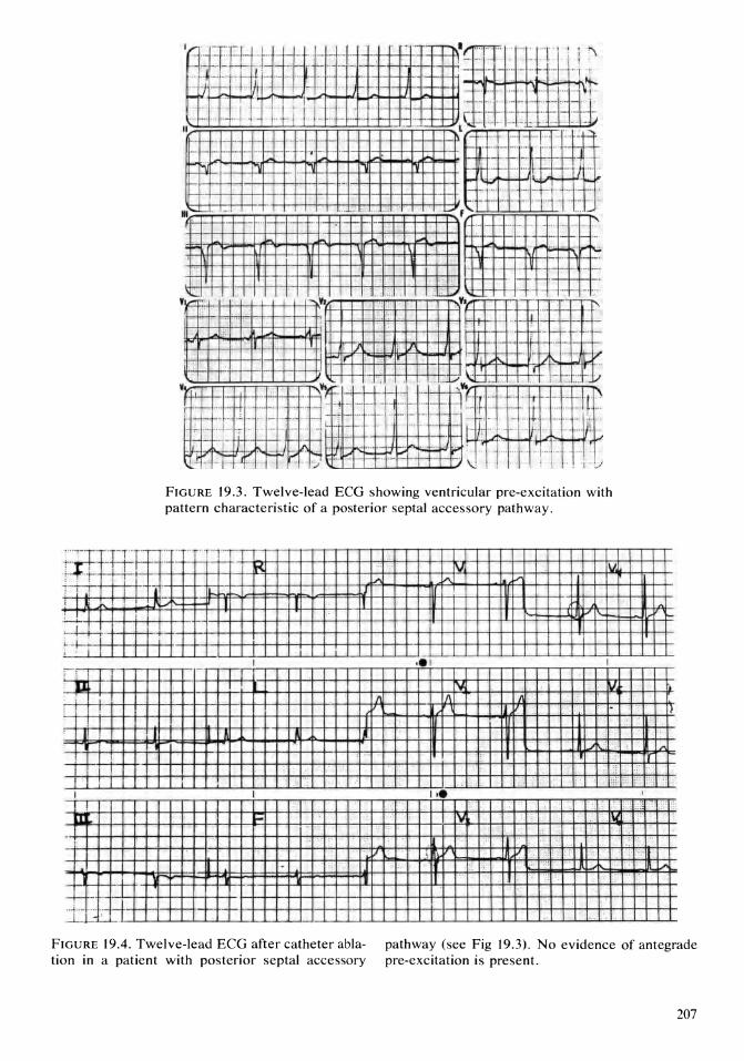
.LLI I
FIGURE 19.3. Twelve-lead ECG showing ventricular pre-excitation with pattern characteristic of a posterior septal accessory pathway .
; ... -W---:-. I I LW·-Hi-+-+-iH--+ -,H--l-+-l-++-H-+-HI--++-H-+-t-t-t·+t-i-++-H-++-t-t-t-t-i---r-rH--t-nH-; , i I I I
" I ' D I
I
I '
... . ," .. .. " = :: . .
[]l 1V .
. II J .:.:'::: ;-
++--ll-j-. I- II
FIGURE 19.4. Twelve-lead ECG after catheter abla- pathway (see Fig 19.3). No evidence of antegrade tion in a patient with posterior septal accessory pre-excitation is present.
207
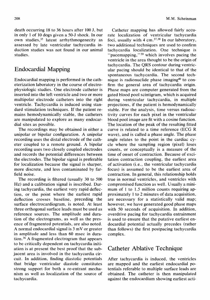
208
death occurring 18 to 36 hours after 100 1, but in only 1 of 10 dogs given a 50-1 shock. In our own studies,35 latent arrhythmogenecity as assessed by late ventricular tachycardia induction studies was not found in our animal studies.
Endocardial Mapping
Endocardial mapping is performed in the catheterization laboratory in the course of electrophysiologic studies. One electrode catheter is inserted into the left ventricle and two or more multipolar electrode catheters into the right ventricle. Tachycardia is induced using standard stimulation techniques. If the patient remains hemodynamically stable, the catheters are manipulated to explore as many endocardial sites as possible.
The recordings may be obtained in either a unipolar or bipolar configuration. A unipolar recording uses the distal electrode of the catheter coupled to a remote ground. A bipolar recording uses two closely coupled electrodes and records the potential differences between the electrodes. The bipolar signal is preferable for localization because the signal is sharper, more discrete, and less contaminated by farfield noise.
The recording is filtered (usually 30 to 500 Hz) and a calibration signal is inscribed. During tachycardia, the earliest very rapid deflection, or the point where the earliest rapid deflection crosses baseline, preceding the surface electrocardiogram, is noted. At least three orthogonal surface leads must be used as reference sources. The amplitude and duration of the electrograms, as well as the presence of fragmented potentials, are also noted. A normal endocardial signal is 3 m V or greater in amplitude and less than 60 msec in duration. 36 A fragmented electrogram that appears to be critically dependent on tachycardia initiation is at present the best proof that the subjacent area is involved in the tachycardia circuit. In addition, finding diastolic potentials that bridge ventricular diastole constitutes strong support for both a re-entrant mechanism as well as localization of the source of tachycardia.
M.M. Scheinman
Catheter mapping has allowed fairly accurate localization of ventricular tachycardia foci, usually with 4 cmy,38 In our laboratory, two additional techniques are used to confirm tachycardia localization. One technique is "pacemapping,"39 which involves pacing the ventricle in the area thought to be the origin of tachycardia. The QRS contour during ventricular pacing should be identical to that of the spontaneous tachycardia. The second technique is radionuclide phase imaging40 to confirm the general area of tachycardia origin. Phase maps are computer generated from the gated blood pool scintigram, which is acquired during ventricular tachycardia, in multiple projections, if the patient is hemodynamically stable. For the analysis, time versus radioactivity curves for each pixel in the ventricular blood pool image are fit with a cosine function. The location of the peak of the resulting fitted curve is related to a time reference (ECG R wave), and is called a phase angle. The phase angle relates to the point in the cardiac cycle where the sampling region (pixel) loses counts, or conceptually is a measure of the time of onset of contraction. Because of excitation contraction coupling, the earliest area of activation (i.e., the ventricular tachycardia focus) is assumed to be the earliest area of contraction. In general, this relationship holds true in normal ventricles, and ventricles with compromised function as well. Usually a minimum of 1 to 1.5 million counts requiring approximately 1 to 2 minutes of data acquisition, are necessary for a statistically valid map; however, we have generated good phase maps with 50 seconds of acquisition. In addition, overdrive pacing for tachycardia entrainment is used to ensure that the putative earliest endocardial potential actually precedes (rather than follows) the first postpacing tachycardia complex.
Catheter Ablative Technique
After tachycardia is induced, the ventricles are mapped and the earliest endocardial potentials referable to multiple surface leads are obtained. The catheter is then manipulated against the endocardium showing earliest acti-

19. Catheter Ablation Techniques
vation, and a patch lubricated with conducting gel is placed on the chest wall in closest approximation to the electrode catheter (Fig 19.5). A series of direct current shocks are delivered from the distal electrode on the catheter (current source) to the chest patch (current sink). For septal foci, a catheter to catheter arrangement is used (Fig 19.2). The patient must be anesthetized with a short-acting agent because the shocks are quite painful.
After stabilization, the patient is retested with the same stimulation protocol found to induce ventricular tachycardia in the control state. We avoid very aggressive stimulation protocols in the immediate postshock period as we have found that they may induce rapid
o
FIGURE 19.5. Schema depicting catheter technique for ventricular tachycardia ablation. The electrode catheter is manipulated as close as possible to the earliest endocardial area recorded during tachycardia. The shock is delivered from the catheter electrode to an indifferent patch placed on the chest wall.
209
nonclinical arrhythmias. We prefer to use the more aggressive stimulation protocols several days after attempted catheter ablation. If the clinical ventricular tachycardia is inducible, then serial drug testing is used to find an effective regimen. Patients who have failed a clinical drug trial may become drug responsive after catheter ablation.
Results of Catheter Ablation of Ventricular Tachycardia Foci
The clinical results of attempted catheter ablation of ventricular tachycardia foci have been somewhat variable. 41-46 Hartzler41 was the first to report successful use of this technique in humans. Fontaine et al44 reported excellent results using this technique in patients with diverse etiology for ventricular tachycardia. The largest experience to date has been accumulated from the worldwide registry.
Clinical Studies
As of December, 1986, a total of 141 patients who underwent attempted electrical ablation of ventricular tachycardia foci have been reported to the registry. The clinical data are summarized in Table 19.3. The mean age was 53 ± 15 years and there was a large predominance of males (86% of the group). The most frequent cardiac diagnosis included coronary artery disease (63%), cardiomyopathy (17%), and arrhythmogenic right ventricular dysplasia (12%). The most frequent symptoms included palpitations (68%), syncope or presyncope (66%), and 26% suffered one or more episodes of cardiac arrest. Patients proved unresponsive or intolerant to a variety of treatments including type I antiarrhythmic drugs (94%), amiodarone (80%), cardiac electrosurgery (5%), automatic internal defibrillator (2%), or antitachycardia pacing (2%). A total of 85 patients required one or more external direct current shocks for arrhythmia control.
Procedural Data
One ablative session was used for 78% of patients, whereas the remainder underwent two to four separate ablative procedures. Sixty-

210 M.M. Scheinman
TABLE 19.3. Clinical findings in patients with drug and/or pacemaker resistant ventricular tachycardia.
Heart disease (type!% of patients)
Symptoms (type!% of patients)
Prior treatment (type!% of patients)
Coronary artery disease!63 Cardiomyopathy! 16 Arrhythmogenic right ventricular
Palpitations 174 Dizziness!41 Syncope!36
Type I195 Amiodarone178 Other experimental drugs!55
dysplasia! 10 Valvular heart disease!6 Others!6 Hypertensive cardiovascular disease!2 No organic disease!6
Dyspnea!34 Cardiac arrest!25 Fatigue! 18 Angina! 18
Digitalis!36 Beta-blockers!33 Calcium-channel blockers!29 Cardiac electrosurgery!6 Antitachycardia pacemaker!4 Chest pain!lO
Other!7 Automatic internal cardioverter defibrillator!2
The percentages total more than 100% because more than one parameter may have been present in a given patient. Type I = type I antiarrhythmic drug.
five patients received one or two direct current shocks, and the remainder received more than two shocks. The mean cumulative stored energy used was 923 ± 680 J, ranging from 160 to 5200 J. For clarity of data analysis, only those patients (109) receiving shocks to a single ventricular site were analyzed. For these patients, the ventricular tachycardia was localized to the right ventricle in 35%, to the ventricular septum in 29%, and to the left ventricle in 36%. The time from earliest endocardial activation to onset of the surface QRS was -43 ± 27 msec. Data for ventricular pace mapping was available in 26 patients and was judged to be excellent (correspondence of paced and spontaneous ventricular tachycardia morphology in all 12 leads) in 13, good (correspondence in 9 of 12 leads) in 8, and poor (correspondence in less than 9 leads) in 5 patients.
Clinical Response
The patients were followed for a mean of 12 ± 10 months and their response to catheter ablation was varied. Thirty-four patients (24%) are currently asymptomatic without antiarrhythmic drugs, whereas 59 patients (42%) have arrhythmia control but require antiarrhythmic agents, and 48 patients (34%) failed to respond. There was no significant difference in clinical outcome between groups and the earliest endocardial activation found. Similarly,
there was no correlation between clinical outcome and whether one (109 patients) or more ventricular sites (33 patients) were shocked. There was a high incidence of excellent pacemaps (8 of 12) for patients showing an excellent response compared with the others (5 of 14), but the differences between the groups were not significant. Postablation ventricular tachycardia induction data were available in 118 patients and was correlated with the clinical outcome. The same ventricular tachycardia morphology was induced in 49 patients, a different morphology was induced in 25 and tachycardia was not inducible in 43 patients. There was a significantly higher incidence of an excellent clinical response for those whose tachycardia was not inducible after the ablative procedure.
Complications
A procedure-related death was defined as any death occurring within 24 hours of the ablative shocks. Seven procedure-related deaths were reported and consisted of electromechanical dissociation in 4, intractable ventricular fibrillation in 1, and severe low output state leading to death in 2 patients. New sustained ventricular arrhythmias occurred in 8 patients after shock. One patient had ventricular fibrillation 5 days after the ablative procedure. Other inhospital complications included hypotension in 12 patients, pericarditis in 4, systemic em-

19. Catheter Ablation Techniques
bolization in 3, myocardial infarction in 2, ventricular perforation in 1, and sepsis in 2 patients.
Mortality
During a mean follow-up of 12 months, 31 patients died. Seven patients had procedurerelated deaths, 14 died suddenly, and documented ventricular tachycardia was found in 9 of these 14. The sudden deaths occurred from 2 weeks to 23 months after ablation. Seven patients died of congestive heart failure and three noncardiac deaths (gastrointestinal hemorrhage in 1, cerebrovascular accident in 1, suicide in l).
Summary
The available data from the registry suggest that catheter ablation of the A V junction is associated with an excellent response in 74% of patients. Resumption of A V conduction was noted to occur early after ablation. For example, approximately 70% of those showing return of A V conduction did so within 36 hours of the ablation.
Although significant postshock complications including ventricular arrhythmias, hypotension, and myocardial perforation have been reported, no acute procedure-related deaths have been reported. Of concern is the 1.9% incidence of sudden death, which occurred from 3 days to 13 months after the ablative procedure. Seven of the 8 patients with sudden death had associated organic cardiac disease, but 1 had no obvious cardiac disease. Even if the sudden deaths are related to the ablative procedure, the mortality is still much lower than that reported from the largest surgical series. 47
Ventricular Tachycardia Ablation
In contrast to electrical ablation of the A V junction, ventricular tachycardia ablation was associated with significant procedure-related deaths and complications. There was no significant difference in the number of shocks or
211
amount of stored energy used for those with procedure-related deaths compared with those who survived the ablative procedure. Three of the 7 who died did so after delivery of one shock, which ranged from 140 to 300 J. Systemic emoblization occurred in 3 patients after attempted left ventricular ablation. The most severe was a dense hemispheric cerebrovascular accident. Embolization may occur as a result of bubbles generated or clots occurring after the ablative procedure. New sustained ventricular arrhythmias requiring emergent interruption also has been reported, as well as depression of left ventricular function after delivery of shocks to the ventricle.
The only variable that significantly predicted a beneficial response was the inability to induce a ventricular arrhythmia in the postablative ventricular tachycardia induction study. This study was performed 3 to 7 days after the attempted ablation. Induction of "nonclinical" ventricular arrhythmias was not predictive of a beneficial response. On the basis of available data, it would appear to be prudent to repeat ventricular tachycardia induction studies in all patients undergoing ventricular tachycardia ablation. If no arrhythmia is induced, then a follow-up trial without antiarrhythmic drugs would appear to be reasonable. If a ventricular arrhythmia is induced, then repeat drug testing or ablative procedures would appear to be indicated.
The reported experience with attempted catheter ablation of accessory pathways is still rather sparse. Successful ablation of right free wall pathways have been reported, but the danger of coronary artery spasm or obstruction and/or perforation of the right atrium are distinct risks. Attempted ablation of left free wall pathways via the coronary sinus have been abandoned, but successful ablative procedures have been reported using trans-septal approaches to the mitral annulus. Our own experience with attempted ablation of posteroseptal pathways has been especially promising, with 75% efficacy and an acceptably low incidence of major complications. A failed attempt at posteroseptal ablation does not preclude a subsequent surgical approach.
In summary, electrical catheter ablation of
212
the A V junction has supplanted the need for cardiac surgical procedures previously used to disrupt A V conduction. This procedure will remain of limited applicability as a pacemaker dependency state is produced and because of the small but definite risk of sudden death that may be related to the procedure. Catheter ablation of posteroseptal accessory pathways appears to be most promising and in our center is used as the primary approach in the management of these patients with drug refractory or malignant ventricular arrhythmias. At present, surgery appears to be preferable to catheter ablative techniques in the management of patients with free wall accessory pathways. Catheter ablation of ventricular tachycardia foci should at present be limited to patients with symptomatic ventricular arrhythmias who are not candidates for surgical intervention or of an automatic internal cardioverter-defibrillator.
References 1. Scheinman MM, Morady F, Hess DS, et al:
Catheter induced ablation of the atrioventricular function to control refractory supraventricular arrhythmias. J Am Med Assoc 1982; 248:851.
2. Morady F, Scheinman MM: Transvenous catheter ablation of posteroseptal accessory pathway in a patient with the Wolff-Parkins onWhite syndrome. N Engl J Med 1984; 310:705.
3. Hartzler GO: Electrode catheter ablation of refractory focal ventricular tachycardia. J Am Coli Cardiol1983; 2:1107.
4. Boyd EGCA, Hoh PM: An investigation into the electrical ablation technique and a method of electrode assessment. PACE 1985; 8:815.
5. Fontaine G, Volmer W, Nienaltowska E, et al: Approach to the physics of fulguration, in Fontaine G, Scheinman MM (eds): Ablation in Cardiac Arrhythmias. Mount Kisco, NY, Futura Publishing Company, 1987, pp 101-116.
6. Jones JL, Proskaver CC, Paul WK, et al: Ultrastructural injury to chick myocardial cells in vitro following "electric countershock." Circ Res 1980; 46:387.
7. Levine JH, Merillat JC, Stern M, et al: The cellular electrophysiologic changes induced by ablation: Comparison between argon laser photoablation and high energy electrical ablation. Circulation 1987; 76:217.
M.M. Scheinman
8. Starzl TE, Gaertner RA: Chronic heart block in dogs. A method for producing experimental heart failure. Circulation 1955; 12:259.
9. Starzl TE, Gaertner RA, Baker RR: Acute complete heart block in dogs. Circulation 1955; 12:82.
10. Steiner C, Kovalik TW: A simple technique for production of chronic complete heart block in dogs. J Appl Physiol1968; 25:631.
11. Harrison L, Gallagher JJ, Kasell J, et al: Cryosurgical ablation of the AV node-His bundle: A new method for producing A V block. Circulation 1977; 55:467.
12. Fisher VJ, Lee RJ, Christianson LC, et al: Production of chronic atrioventricular block in dogs without thoracotomy. J Appl Physiol 1966; 21: 1119.
13. Turnia M, Babotai I, Wegmann W: Production of chronic atrioventricular block in dogs without thoracotomy. Cardiovasc Res 1968; 4:389.
14. Beazell J, Tan K, Criley J, et al: The electrosurgical production of heart block without thoracotomy (abstract). Clin Res 1976; 24: 137A.
15. Gonzalez R, Scheinman M, Margaretten W, et al: Closed-chest electrode-catheter technique for His bundle ablation in dogs. Am J Physiol 1981; 241 :H283.
16. Gonzalez R, Schein man M, Bharati S, et al: Closed-chest permanent atrioventricular block in dogs. Am Heart J 1983; 105:461.
17. Gallagher JJ, Svenson RH, Kasell JH, et al: Catheter technique for closed-chest ablation of the atrioventricular conduction system: A therapeutic alternative for the treatment of refractory supraventricular tachycardia. N Engl J Med 1982; 306:194.
18. Wood DL, Hammill SC, Holmes DR Jr, et al: Catheter ablation of the atrioventricular conduction system in patients with supraventricular tachycardia. Mayo Clin Proc 1983; 58:791.
19. Nathan AW, Bennett DH, Ward DE, et al: Catheter ablation of atrioventricular conduction. Lancet 1984; 1:1280.
20. Scheinman M, Evans-Bell T: Catheter ablation of the atrioventricular junction: A report of the percutaneous mapping and ablation registry. Circulation 1984; 70:1024.
21. Eldar M, Griffin JC, Seger JJ, et al: Catheter atrioventricular junctional ablation in patients with accessory pathways. PACE 1986; 9:810.
22. Bhandari A, Morady F, Shen EN, et al: Catheter induced His bundle ablation in a patient with reentrant tachycardia associated with a nodoventricular tract. J Am Coli Cardiol 1984; 4:611.

19. Catheter Ablation Techniques
23. Brodman R, Fisher JD: Evaluation of a catheter technique for ablation of accessory pathways near the coronary sinus using a canine model. Circulation 1983; 67:923.
24. Ruder MA, Davis JC, Eldar M, et al: Effects of electrode catheter shocks delivered near the tricuspid annulus in dogs (abstract). J Am Call Cardiol 1986; 7:7A.
25. Coltorti F, Bardy GH, Reichenbach D, et al: Catheter-mediated electrical ablation of the posterior septum via the coronary sinus: Electrophysiologic and histologic observations in dogs. Circulation 1985; 72:612.
26. Fisher JD, Brodman R, Kim SG, et al: Attempted nonsurgical electrical ablation of accessory pathways via the coronary sinus in the Wolff-Parkinson-White syndrome. J Am Call Cardiol 1984; 4:685.
27. Kunze KP, Kuck KH: Transvenous ablation of accessory pathways in patients with incessant atrioventricular tachycardia (abst). Circulation 1984; 70(suppl 11): 164.
28. Weber H, Schmitz L: Catheter technique for closed-chest ablation of an accessory atrioventricular pathway. N Engl J Med 1983; 308:653.
29. Weber H, Schmitz L, Hellberg K: Pacemakermediated tachycardias: A new modality of treatment. PACE 1984; 7:1010.
30. Warin JF, Haissaguerre M, Lemetayer Ph, et al: Electrical catheter shock ablation of accessory pathways: Efficacy and safety using a direct approach in 24 patients (abst). PACE 1987; 10:760.
31. Morady F, Scheinman MM, Winston SA, et al: Efficacy and safety of transcatheter ablation of posteroseptal accessory pathways. Circulation 1985; 72:170.
32. Lerman BB, Weiss JL, Bulkley BH, et al: Myocardial injury and induction of arrhythmia by direct current shock delivered via endocardial catheters in dogs. Circulation 1984; 69: 1006.
33. Westveer DC, Nelson T, Stewart JR, et al: Sequelae of left ventricular electrical endocardial ablation. J Am Call Cardiol 1985; 5:956.
34. Kempf FC, Falcone RA, Iozzo RV, et al: Anatomic and hemodynamic effects of catheter delivered ablation energies in the ventricle. Am J Cardiol 1985; 56:373.
35. Davis JC, Finkebeiner W, Ruder MA, et al: Histologic changes and arrhythmogenicity after
213
discharge through trans septal catheter electrode. Circulation 1986; 74:637.
36. Josephson ME, Horowitz LN, Spielman SR, et al: Role of catheter mapping in the preoperative evaluation of ventricular tachycardia. Am J Cardiol 1982; 49:207.
37. Horowitz LN, Josephson ME, Harken AH: Ventricular resection guided by epicardial and endocardial mapping for treatment of recurrent ventricular tachycardia. N Engl J Med 1980; 302:589.
38. Josephson ME: Catheter ablation of arrhythmias. Ann Int Med 1984; 10:234.
39. Curry PVL, O'Keeffe DB, Pritcher D, et al: Localization of ventricular tachycardia by new technique-pace mapping. Circulation 1979; 60(suppl 11):11-25.
40. Botvinick E, Schechtman N, Dae M: Scintigraphy provides a thorough evaluation of "electrical" and mechanical events during ventricular tachycardia. J Am Call Cardiol 1986; 7(2):235A.
41. Hartzler GO: Electrode catheter ablation of refractory focal ventricular tachycardia. J Am Call Cardiol 1983; 2: 1107.
42. Steinhaus D, Whitford E, Stavens C, et al: Percutaneous transcatheter electrical ablation for recurrent sustained ventricular tachycardia (abst). Circulation 1984; 70(suppl 11):11-100.
43. Puech P, Gallay P, Grolleau R, et al: Traitement par electrofulguration endocavitaire d'une tachycardie ventriculaire recidivante par dysplasie ventriculaire droite. Arch Mal Coeur 1984; 77:826.
44. Fontaine G, Tonet JL, Frank R, et al: La fulguration endocavitaire: Une nouvelle methode de traitement des troubles du rhythme? Ann Cardiol Agneiol 1984; 33:543.
45. Belhassen B, Miller HI, Laniado S: Catheter ablation of icnessant ventricular tachycardia refractory to external cardioversion. Am J Cardial 1985; 55:1637.
46. Morady F, Scheinman MM, DiCarlo LA Jr, et al: Catheter ablation of ventricular tachycardia with intracardiac shocks: Results in 33 patients. Circulation 1987; 75: 1037.
47. German LD, Pressley J, Smith JS, et al: Comparison of cryoablation of the atrioventricular node versus catheter ablation of the His bundle (abst). Circulation 1984; 70:11-412.

20 Interventional Pediatric Cardiac Catheterization Zuhdi Lababidi and Ihab Attia
Interventional cardiology and invasive cardiovascular procedures have grown substantially over the last 2 decades as a result of the outstanding technical advances in catheter equipment and design. The remarkable evolution of new catheter designs resulted in the production of sophisticated and elaborate therapeutic instruments that can be used by the invasive cardiologist for the treatment of various congenital cardiac diseases. Interventional cardiology can no longer be ignored by the invasive pediatric cardiologist who does not perform therapeutic procedures. This is particularly true in neonates with certain congenital cardiac defects that require transcatheter treatment as soon as accurate diagnosis is established in the catheterization laboratory. A therapeutic procedure in such cases should be complementary to diagnostic cardiac catheterization in the same setting.
Patient safety during interventional procedures in the catheterization laboratory is of the utmost importance, thus only pediatric cardiologists with adequate interventional experience should perform such procedures. The catheterization laboratory must be fully equipped to provide proper monitoring and support of the patient's vital signs and hemodynamics throughout the procedure.
Interventional procedures for congenital heart diseases are a suitable alternative to surgery, as it can be applied to high surgical risk patients, as well as to lesions to which surgical access is difficult. In addition, the cost and hospitalization period are markedly reduced.
Interventional Procedures
There are several interventional procedures, some of which are still considered investigational, whereas others are considered the optimal form of therapy in certain congenital cardiac defects.
The current interventional cardiac procedures include:
1. Balloon I and blade2 atrial septostomy for creating interatrial shunts in neonates, infants, and children.
2. Balloon angioplasty3-5 for dilating stenotic arteries and veins.
3. Balloon valvuloplasty6,7 for dilating stenotic valves.
4. Transcatheter occlusion of pre-existing shunts8-IO (embolotherapy).
5. Transluminal catheter retrieval ll - 13 and resolution of intracardiac catheter knots.14,15
6. Transvenous insertion of temporary and permanent pacemakers. 16
7. Catheter ablation of refractory cardiac tachyarrhythmias. 17
8. Intra-aortic balloon pumping for improving cardiac output in refractory left ventricular failure and cardiogenic shock. 18
9. Pericardiocentesis and drainage. 19,20 10. Embolectomy by transcatheter aspira
tion. 39 11. Laser irradiation for treatment of congeni
tal heart disease, 22,23 which is still investigational.
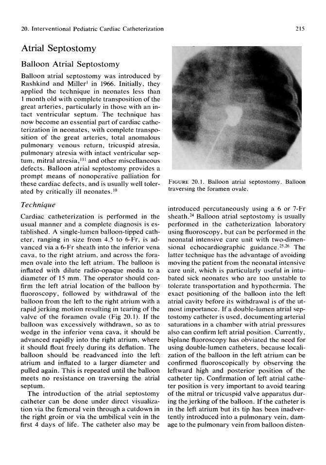
20. Interventional Pediatric Cardiac Catheterization
Atrial Septostomy
Balloon Atrial Septostomy Balloon atrial septostomy was introduced by Rashkind and Miller l in 1966. Initially, they applied the technique in neonates less than 1 month old with complete transposition of the great arteries, particularly in those with an intact ventricular septum. The technique has now become an essential part of cardiac catheterization in neonates, with complete transposition of the great arteries, total anomalous pulmonary venous return, tricuspid atresia, pulmonary atresia with intact ventricular septum, mitral atresia, III and other miscellaneous defects. Balloon atrial septostomy provides a prompt means of nonoperative palliation for these cardiac defects, and is usually well tolerated by critically ill neonates. 10
Technique
Cardiac catheterization is performed in the usual manner and a complete diagnosis is established. A single-lumen balloon-tipped catheter, ranging in size from 4.5 to 6-Fr, is advanced via a 6-Fr sheath into the inferior vena cava, to the right atrium, and across the foramen ovale into the left atrium. The balloon is inflated with dilute radio-opaque media to a diameter of 15 mm. The operator should confirm the left atrial location of the balloon by fluoroscopy, followed by withdrawal of the balloon from the left to the right atrium with a rapid jerking motion resulting in tearing of the valve of the foramen ovale (Fig 20.1). If the balloon was excessively withdrawn, so as to wedge in the inferior vena cava, it should be advanced rapidly into the right atrium, where it should float freely during its deflation. The balloon should be readvanced into the left atrium and inflated to a larger diameter and pulled again. This is repeated until the balloon meets no resistance on traversing the atrial septum.
The introduction of the atrial septostomy catheter can be done under direct visualization via the femoral vein through a cutdown in the right groin or via the umbilical vein in the first 4 days of life. The catheter also may be
215
FIGURE 20.1. Balloon atrial septostomy. Balloon traversing the foramen ovale.
introduced percutaneously using a 6 or 7-Fr sheath.24 Balloon atrial septostomy is usually performed in the catheterization laboratory using fluoroscopy, but can be performed in the neonatal intensive care unit with two-dimensional echocardiographic guidance. 25.26 The latter technique has the advantage of avoiding moving the patient from the neonatal intensive care unit, which is particularly useful in intubated sick neonates who are too unstable to tolerate transportation and hypothermia. The exact positioning of the balloon into the left atrial cavity before its withdrawal is of the utmost importance. If a double-lumen atrial septostomy catheter is used, documenting arterial saturations in a chamber with atrial pressures also can confirm left atrial position. Currently, biplane fluoroscopy has obviated the need for using double-lumen catheters, because localization of the balloon in the left atrium can be confirmed fluoroscopically by observing the leftward high and posterior position of the catheter tip. Confirmation of left atrial catheter position is very important to avoid tearing of the mitral or tricuspid valve apparatus during the jerking of the balloon. If the catheter is in the left atrium but its tip has been inadvertently introduced into a pulmonary vein, damage to the pulmonary vein from balloon dis ten-

216
tion can be avoided by slow inflation of the balloon, which results in gentle extrusion of the entire catheter tip back into the left atrial cavity.
Successful balloon atrial septostomy can be documented by a rise in arterial oxygen saturation, increased bidirectional shunting at the atrial level, and elimination of any pressure gradient between the atria.
Clinical Role
Balloon atrial septostomy improves the circulation and the clinical condition of the neonate by one of the following mechanisms:
1. By increased bidirectional shunting (mixing) at the atrial level, balloon atrial septostomy improves arterial oxygen saturations when the pulmonary and systemic circuits are operating in parallel rather than in series, that is, complete transposition of the great arteries. Palliation with balloon atrial septostomy has allowed the majority of patients with complete transposition of the great arteries to survive to age 6 months, which is optimal for venous switching. I
2. By increasing left-to-right shunting at the atrial level, balloon atrial septostomy decreases pulmonary venous congestion in patients with severe obstructive left-sided lesions, that is, hypoplastic left heart syndrome.
3. By increasing right-to-Ieft shunting at the atrial level, balloon atrial septostomy decreases systemic venous congestion in severe right-sided obstruction, that is, tricuspid atresia and pulmonary atresia with intact ventricular septum.
4. By increasing right-to-Ieft shunting at the atrial level, balloon atrial septostomy decreases pulmonary venous congestion in total anomalous pulmonary venous return.
Limitations and Complications
Balloon atrial septostomy has been shown to be a safe and effective means of immediate palliation in more than 70% of infants with complete transposition of the great arteries, 10
Z. Lababidi and I. Attia
but early favorable results after balloon atrial septostomy may not necessarily indicate successful long-term palliation. Close observation and continued medical management are essential, as considerable cummulative mortality while awaiting definitive surgery has been reported after balloon atrial septostomy for complete transposition of the great arteries. 27,28
There is little doubt that the use of larger balloons has contributed appreciably to improved survival,29 for smaller balloons usually do not create an adequate atrial communication. Balloons under 2 ml in volume may lead to stretching rather than tearing of the atrial septum. On the other hand, very large balloons can result in tears of the atrial wall or interatrial groove.30 The recommended balloon volume is 3 to 4 ml. The widely used balloon septostomy catheters are the Miller balloon atrial septostomy catheter (4 ml capacity, Americal Edwards Laboratories, Santa Ana, CA) and the Rashkind catheter with the recessed balloon (l.5 to 2 ml capacity, USCI, Billerica, MA). The complications of balloon atrial septostomy include: perforation of the right atrial appendage or pulmonary veins,31 failure to achieve adequate septostomy,27 femoral vein tearing,31 femoral vein thrombosis,32 inferior vena caval thrombosis,33 balloon deflation failure, 34 and balloon embolization.35
Blade Atrial Septostomy
In infants beyond 1 month of age, with a tougher atrial septum, Park et aF,36 introduced a new method of septostomy. This method involves the introduction of the Sang-Park blade catheter via the femoral vein to the inferior vena cava, to the right atrium and across the foramen ovale into the left atrium. When confirmation of the left atrial position of the catheter tip is achieved by biplane fluoroscopy, the blade is opened so that the edge points anteriorly, inferiorly, and to the left and then is gradually withdrawn across the atrial septum into the right atrium. This will result in a cut in the atrial septum, which is further enlarged by balloon septostomy.
20. Interventional Pediatric Cardiac Catheterization
Blade and Balloon Atrial Septostomy After Trans-septal Atrial Puncture Recently, Vick et aP7 described a modification of the standard septostomy technique. That technique starts with a standard atrial transseptal puncture followed by placing the blade catheter across the interatrial septum through the trans-septal sheath and pulling the blade to create a cut in the atrial septum, which is enlarged by balloon septostomy. This technique was performed in selected cases with complete transposition of the great arteries, left atrioventricular valve atresia or severe stenosis, double-outlet right ventricle with restrictive ventricular septal defect, tricuspid and/or pulmonary atresia, and pulmonary vascular obstructive disease. 37 The patients ranged in age from 1 day to 21 years with a mean of 2.8 yearsY The advantage of that technique is that it can be applied to patients with an intact atrial septum.
Balloon Angiopiasty for Dilating Stenotic Arteries and Veins
Balloon Coarctation Angioplasty Surgical repair of coarctation of the aorta has undergone several modifications. Crafoord75 and Gross76 were the first to report the classic resection with end-to-end anastomosis in 1945. To avoid restenosis, synthetic patch angioplasty was introduced by Vosschulte77 in 1957; and subclavian patch angioplasty was popularized by Waldhausen and Nahrwold78 in 1966. Although there have been dramatic improvements in the past 40 years, morbidity and mortality rates from surgical repair are still high in the neonates with coarcation of the aorta and congestive heart failure. For infants under 8 weeks of age, an operative mortality ranging from 24% to 67% has been observed.71-81 Recurrent obstruction (restenosis) is common (20% to 35%) after early coarctation repair, especially after end-to-end anastomosis. 82,83 Aneurysm formation at the site of surgical coarctation repair and aortic dissection also occur. Postoperative hypertension is
217
common in all age groups (but younger patients are less likely to have persistent hypertension at long-term follow-up).84,85 On the other hand, the outcomes of symptomatic neonates with coarctation treated medically rather than surgically are even worse; mortality ranges from 50% to 86%.86,87
In 1979, Sos and co-workers88 demonstrated the balloon dilatation of the restenosed coarcted segment in postmortem specimens of newborns who had undergone coarctation repair. In 1983, we reported neonatal transluminal balloon coarctation angioplasty in a neonate with native coarctation of the aorta. 3
Technique
Immediately after the diagnosis of coarctation is proven angiographically, balloon coarctation angioplasty can be performed via the femoral artery either through a cutdown or a percutaneous approach. Although, in older children with coarctation, balloon size is determined by the age of the patient and the diameter of the descending aorta, a 5-Fr catheter and a 4 to 6-mm balloon diameter are quite adequate in all neonates. Balloon coarctation angioplasty permits the neonate with coartation to grow to an age when either surgical repair can be performed at much lower risk or repeat balloon angioplasty using a larger balloon can be attempted.
Using a femoral arterial approach, a 5-Fr end-hole catheter is placed in the ascending aorta. A flexible tip 0.028-in guidewire is inserted, and the catheter is removed, leaving the wire in the ascending aorta. The balloon catheter is then introduced over the guidewire, and the middle of the deflated balloon is placed fluoroscopically at the level of the coarctation ridge. The balloon is then inflated with a diluted mixture of contrast medium (Fig 20.2). At the start of the inflation, the balloon assumes the shape of an hourglass. The indentation in the middle of the balloon disappears when the coarctation is fully dilated. A pressure of 80 to 100 psi is often needed to fully dilate the coarctation. Full inflations for more than 5 seconds are unnecessary, as are repeated inflations. The balloon is then deflated,
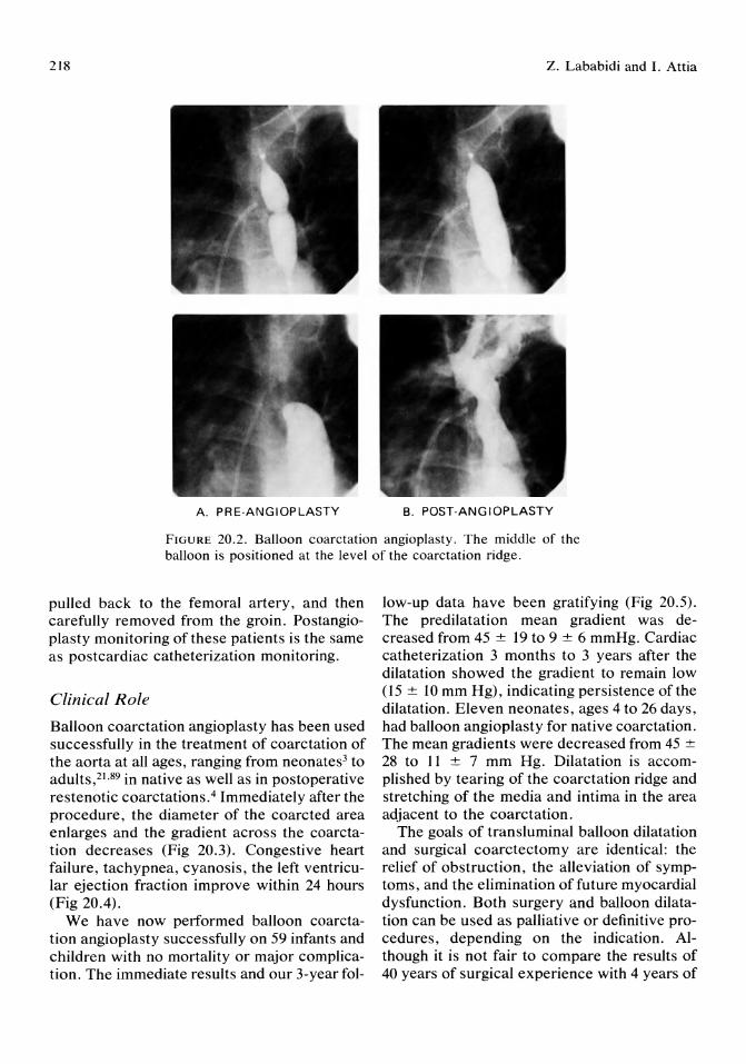
218 Z. Lababidi and L Attia
A. PRE·ANGIOPLASTY B. POST·ANGIOPLASTY
FIGURE 20.2. Balloon coarctation angioplasty. The middle of the balloon is positioned at the level of the coarctation ridge.
pulled back to the femoral artery, and then carefully removed from the groin. Postangioplasty monitoring of these patients is the same as postcardiac catheterization monitoring.
Clinical Role
Balloon coarctation angioplasty has been used successfully in the treatment of coarctation of the aorta at all ages, ranging from neonates3 to adults,21.89 in native as well as in postoperative restenotic coarctations.4 Immediately after the procedure, the diameter of the coarcted area enlarges and the gradient across the coarctation decreases (Fig 20.3). Congestive heart failure, tachypnea, cyanosis, the left ventricular ejection fraction improve within 24 hours (Fig 20.4).
We have now performed balloon coarctation angioplasty successfully on 59 infants and children with no mortality or major complication. The immediate results and our 3-year fol-
low-up data have been gratifying (Fig 20.5). The predilatation mean gradient was decreased from 45 ± 19 to 9 ± 6 mmHg. Cardiac catheterization 3 months to 3 years after the dilatation showed the gradient to remain low (15 ± 10 mm Hg), indicating persistence of the dilatation. Eleven neonates, ages 4 to 26 days, had balloon angioplasty for native coarctation. The mean gradients were decreased from 45 ± 28 to II ± 7 mm Hg. Dilatation is accomplished by tearing of the coarctation ridge and stretching of the media and intima in the area adjacent to the coarctation.
The goals of transluminal balloon dilatation and surgical coarctectomy are identical: the relief of obstruction, the alleviation of symptoms, and the elimination of future myocardial dysfunction. Both surgery and balloon dilatation can be used as palliative or definitive procedures, depending on the indication. Although it is not fair to compare the results of 40 years of surgical experience with 4 years of
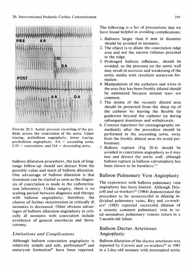
20. Interventional Pediatric Cardiac Catheterization
FIGURE 20.3. Aortic pressure recording of the gradient across the coarctation of the aorta: Upper tracing, preballoon angioplasty; lower tracing, postballoon angioplasty. AA = ascending aorta, C/O = coarctation, and DA = descending aorta.
balloon dilatation procedures, the lack oflongrange follow-up should not detract from the possible value and merit of balloon dilatation. One advantage of balloon dilatation is that treatment can be started as soon as the diagnosis of coarctation is made in the catheterization laboratory. Unlike surgery, there is no waiting period between diagnosis and therapy with balloon angioplasty; therefore, the chance of further deterioration in critically ill neonates is decreased. Other obvious advantages of balloon dilatation angioplasty in critically ill neonates with coarctation include avoidance of general anesthesia and thoracotomy.
Limitations and Complications
Although balloon coarctation angioplasty is relatively simple and safe, perforation90 and aneurysm formation91 have been reported.
219
The following is a list of precautions that we have found helpful in avoiding complications:
1. Balloons larger than 6 mm in diameter should be avoided in neonates.
2. The object is to dilate the coarctation ridge area and not the narrow isthmus proximal to the ridge.
3. Prolonged balloon inflations, should be avoided, as the pressure on the aortic wall may result in necrosis and weakening of the aortic media with resultant aneurysm formation.
4. Manipulation of the catheters and wires in the area that has been freshly dilated should be minimized because intimal tears are common.
5. The intima of the recently dilated area should be protected from the sharp tip of the catheter by leaving the flexible tip guidewire beyond the catheter tip during subsequent insertions and withdrawals.
6. Contrast injections for cineangiography immediately after the procedure should be performed in the ascending aorta, away from the freshly dilated area (to avoid perforation).
7. Balloon rupture (Fig 20.6) should be avoided in coarctation angioplasty as it may tear and dissect the aortic wall, although balloon rupture in balloon valvuloplasty has been shown to be harmless. 6,7
Balloon Pulmonary Vein Angioplasty
The experience with balloon pulmonary vein angioplasty has been limited. Although Driscoll and co-workers92 (1984) demonstrated the procedure to be unsuccessful in dilating individual pulmonary veins, Rey and co-workers5 (1985) reported successful dilation of a stenotic common pulmonary vein in total anomalous pulmonary venous return in a 3-month-old infant.
Balloon Ductus Arteriosus Angioplasty
Balloon dilatation of the ductus arteriosus was reported by Corwin and co-workers93 in 1981 in a 2-day-old neonate with interrupted aortic

RV
IVS
FIGURE 2004. M-mode echocardiogram demonstrated the improvement in left ventricular function after balloon coarctation angioplasty in a neonate in congestive heart failure. Upper panel, preballoon
1982
FIGURE 20.5. Aortography just before balloon coarctation angioplasty (1982) and 3 years later
220
angioplasty; lower panel, postballoon angioplasty. IVS = interventricular septum, L VPW = left ventricular posterior wall, MV = mitral valve, and RV = right ventricle.
1985
(1985), showing adequate dilatation with no evidence of aneurysm formation.
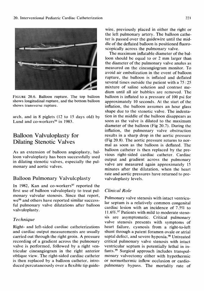
20. Interventional Pediatric Cardiac Catheterization
FIGURE 20.6. Balloon rupture. The top balloon shows longitudinal rupture, and the bottom balloon shows transverse rupture.
arch, and in 8 piglets (12 to 15 days old) by Lund and co-workers94 in 1983.
Balloon Valvuloplasty for Dilating Stenotic Valves
As an extension of balloon angioplasty, balloon valvuloplasty has been successfully used in dilating stenotic valves, especially the pulmonary and aortic valves.
Balloon Pulmonary Valvulcplasty
In 1982, Kan and co-workers95 reported the first use of balloon valvuloplasty to treat pulmonary valvular stenosis. Since that report, we96 and others have reported similar successful pulmonary valve dilatations after balloon valvuloplasty.
Technique
Right- and left-sided cardiac catheterizations and cardiac output measurements are usually carried out through the right groin. A pressure recording of a gradient across the pulmonary valve is performed, followed by a right ventricular cineangiogram in the right anterior oblique view. The right-sided cardiac catheter is then replaced by a balloon catheter, introduced percutaneously over a flexible tip guide-
221
wire, previously placed in either the right or the left pulmonary artery. The balloon catheter is passed over the guidewire until the middle of the deflated balloon is positioned fluoroscopically across the pulmonary valve.
The maximum inflatable diameter of the balloon should be equal to or 2 mm larger than the diameter of the pulmonary valve anulus as measured on the cineangiogram monitor. To avoid air embolization in the event of balloon rupture, the balloon is inflated and deflated several times outside the patient with a 75 : 25 mixture of saline solution and contrast medium until all air bubbles are removed. The balloon is inflated to a pressure of 100 psi for approximately 10 seconds. At the start of the inflation, the balloon assumes an hour glass shape due to the stenotic valve. The indentation in the middle of the balloon disappears as soon as the valve is dilated to the maximum diameter of the balloon (Fig 20.7). During the inflation, the pulmonary valve obstruction results in a sharp drop in the aortic pressure (Fig 20.8). The aortic pressure returns to normal as soon as the balloon is deflated. The balloon catheter is then replaced by the previous right-sided cardiac catheter. Cardiac output and gradient across the pulmonary valve are measured again approximately IS minutes after the dilatation, when the heart rate and aortic pressures have returned to prevalvuloplasty levels.
Clinical Role
Pulmonary valve stenosis with intact ventricular septum is a relatively common congenital cardiac lesion with an incidence of 7.5% to 11.6%.97 Patients with mild to moderate stenosis are asymptomatic. Critical pulmonary valve stenosis presents with symptoms of heart failure, cyanosis from a right-to-Ieft shunt through a patent foramen ovale or atrial septal defect, and severe hypoxia. 98 Untreated critical pulmonary valve stenosis with intact ventricular septum is potentially lethal in infants.99 Surgical approach includes transpulmonary valvectomy either with hypothermic or normothermic inflow occlusion or cardiopulmonary bypass. The mortality rate of
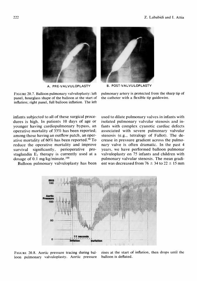
222
A. PRE·VALVULOPLASTY
FIGURE 20.7. Balloon pulmonary valvuloplasty: left panel, hourglass shape of the balloon at the start of inflation; right panel, full balloon inflation. The left
infants subjected to all of these surgical procedures is high. In patients 10 days of age or younger having cardiopulmonary bypass, an operative mortality of 33% has been reported; among those having an outflow patch, an operative mortality of 60% has been reported. 98 To reduce the operative mortality and improve survival significantly, perioperative prostaglandin EJ therapy is currently used at a dosage of 0.1 mg/kg/minute. IOO
Balloon pulmonary valvuloplasty has been
-Q 150
50
Z. Lababidi and I. Attia
B. POST·VALVULOPLASTY
pulmonary artery is protected from the sharp tip of the catheter with a flexible tip guidewire.
used to dilate pulmonary valves in infants with isolated pulmonary valvular stenosis and infants with complex cyanotic cardiac defects associated with severe pulmonary valvular stenosis (e.g., tetralogy of Fallot). The decrease in pressure gradient across the pulmonary valve is often dramatic. In the past 4 years, we have performed balloon pulmonar valvuloplasty on 75 infants and children with pulmonary valvular stenosis. The mean gradient was decreased from 76 ± 34 to 22 ± IS mm
. . ~!.~ .. O----i::'~ __ I .... - -~
FIGURE 20.8. Aortic pressure tracing during balloon pulmonary valvuloplasty. Aortic pressure
rises at the start of inflation, then drops until the balloon is deflated.
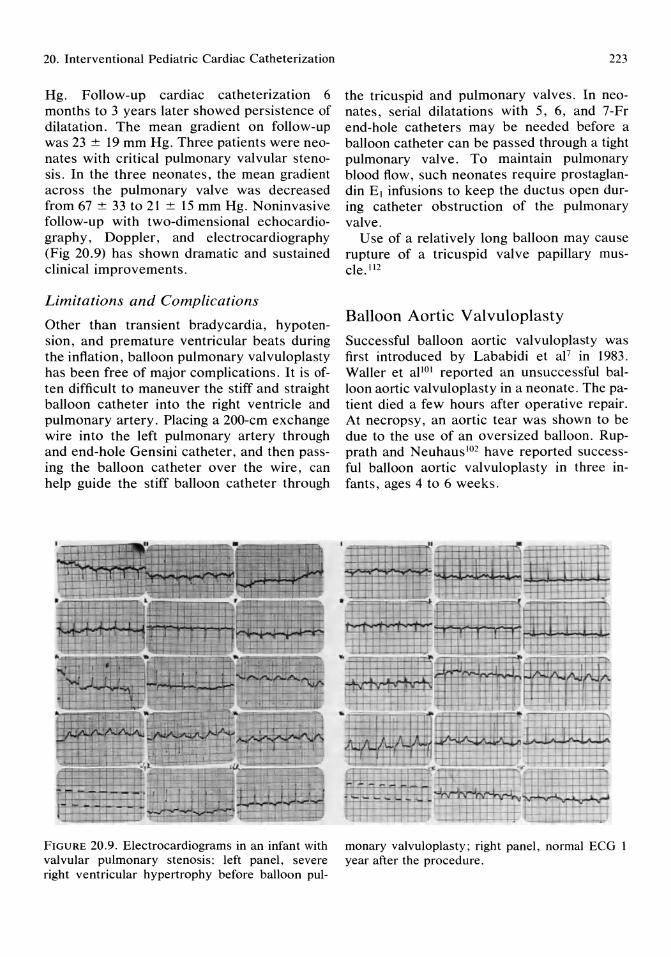
20. Interventional Pediatric Cardiac Catheterization
Hg. Follow-up cardiac catheterization 6 months to 3 years later showed persistence of dilatation. The mean gradient on follow-up was 23 ± 19 mm Hg. Three patients were neonates with critical pulmonary valvular stenosis. In the three neonates, the mean gradient across the pulmonary valve was decreased from 67 ± 33 to 21 ± 15 mm Hg. Noninvasive follow-up with two-dimensional echocardiography, Doppler, and electrocardiography (Fig 20.9) has shown dramatic and sustained clinical improvements.
Limitations and Complications
Other than transient bradycardia, hypotension, and premature ventricular beats during the inflation, balloon pulmonary valvuloplasty has been free of major complications. It is often difficult to maneuver the stiff and straight balloon catheter into the right ventricle and pulmonary artery. Placing a 200-cm exchange wire into the left pulmonary artery through and end-hole Gensini catheter, and then passing the balloon catheter over the wire, can help guide the stiff balloon catheter through
II •
FIGURE 20.9. Electrocardiograms in an infant with valvular pulmonary stenosis: left panel, severe right ventricular hypertrophy before balloon pul-
223
the tricuspid and pulmonary valves. In neonates, serial dilatations with 5, 6, and 7-Fr end-hole catheters may be needed before a balloon catheter can be passed through a tight pulmonary valve. To maintain pulmonary blood flow, such neonates require prostaglandin El infusions to keep the ductus open during catheter obstruction of the pulmonary valve.
Use of a relatively long balloon may cause rupture of a tricuspid valve papillary muscle. 1l2
Balloon Aortic Valvuloplasty
Successful balloon aortic valvuloplasty was first introduced by Lababidi et aF in 1983. Waller et al101 reported an unsuccessful balloon aortic valvuloplasty in a neonate. The patient died a few hours after operative repair. At necropsy, an aortic tear was shown to be due to the use of an oversized balloon. Rupprath and Neuhaus 102 have reported successful balloon aortic valvuloplasty in three infants, ages 4 to 6 weeks.
" , ...... ..,...,....,.I! ~~.-, . -~
I-I----I-H
' . = .
"-~ '"N' .,..
.i "-- ---"-- J' ~
:if-_I--=-. '.=-'-. I..L ,-----"---. ;.::. ~ ,,'J
f--- - .-TI+:-f-H:-I "-tffif~
~-.--r-i-i:1- .~
f-T4---""fl~ -l-.-- ~ ,I. ~
;"'.I~. I.J.. ' ~I
monary valvuloplasty; right panel, normal ECG I year after the procedure.
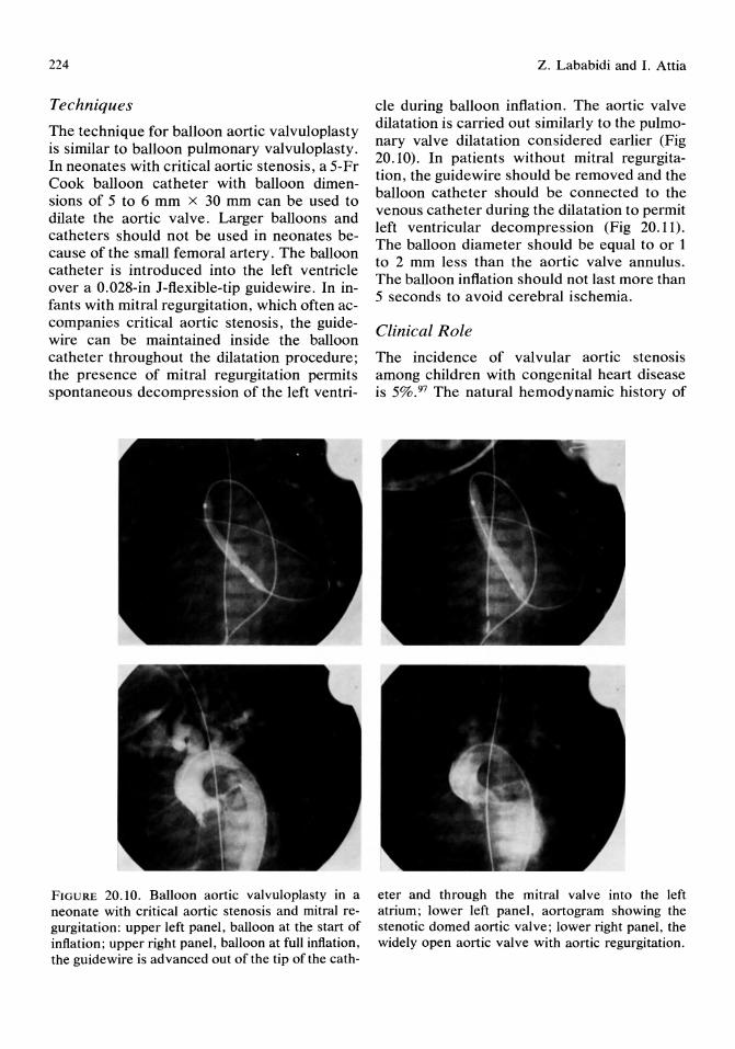
224
Techniques
The technique for balloon aortic valvuloplasty is similar to balloon pulmonary valvuloplasty. In neonates with critical aortic stenosis, a 5-Fr Cook balloon catheter with balloon dimensions of 5 to 6 mm x 30 mm can be used to dilate the aortic valve. Larger balloons and catheters should not be used in neonates because of the small femoral artery. The balloon catheter is introduced into the left ventricle over a 0.028-in I-flexible-tip guidewire. In infants with mitral regurgitation, which often accompanies critical aortic stenosis, the guidewire can be maintained inside the balloon catheter throughout the dilatation procedure; the presence of mitral regurgitation permits spontaneous decompression of the left ventri-
FIGURE 20.10. Balloon aortic valvuloplasty in a neonate with critical aortic stenosis and mitral regurgitation: upper left panel, balloon at the start of inflation; upper right panel, balloon at full inflation, the guidewire is advanced out of the tip of the cath-
z. Lababidi and I. Attia
cle during balloon inflation. The aortic valve dilatation is carried out similarly to the pulmonary valve dilatation considered earlier (Fig 20.10). In patients without mitral regurgitation, the guidewire should be removed and the balloon catheter should be connected to the venous catheter during the dilatation to permit left ventricular decompression (Fig 20.11). The balloon diameter should be equal to or 1 to 2 mm less than the aortic valve annulus. The balloon inflation should not last more than 5 seconds to avoid cerebral ischemia.
Clinical Role
The incidence of valvular aortic stenosis among children with congenital heart disease is 5%.97 The natural hemodynamic history of
eter and through the mitral valve into the left atrium; lower left panel, aortogram showing the stenotic domed aortic valve; lower right panel, the widely open aortic valve with aortic regurgitation.

20. Interventional Pediatric Cardiac Catheterization
FIGURE 20. II. The left ventricular-right atrial shunt when the inflated balloon occludes the aortic valve orifice. The arterial and venous catheters are connected outside the groin.
aortic stenosis is one of progressive obstruction, due usually to increased flow (as a result of somatic growth) across a fixed obstruction. In a few aortic stenosis patients, actual narrowing of the valve orifice contributes to progressive obstruction. \03 Critical valvular aortic stenosis usually presents in early infancy with congestive heart failure. In neonates, with critical valular aortic stenosis, medical treatment is only briefly effective. lo4 Newborns with symptomatic aortic stenosis require urgent or emergency valvulotomy, which carries a high risk. In infants 1 month of age or younger, the mortality has ranged from 29% to 86%. \05-\08 Aortic valvulotomy has been performed by an open method with and without cardiopulmonary bypass,19 by a closed method with a trans ventricular blunt dilator through an incision in the left ventricular apex, \09 and, more recently, by a transventricular balloon catheter after thoracotomy. 110
225
Transluminal balloon aortic valvuloplasty has been increasingly considered as an alternative to surgical aortic valvulotomy in severe congenital valvular aortic stenosis. 7 In the past 3 years we have successfully performed balloon aortic valvuloplasty in 48 children, two of whom were neonates (6 and 7 days of age). The mean gradient across the aortic valve in the 48 patients was decreased from 102 ± 44 to 26 ± 15 mm Hg; and on 3-month to 3-year follow-up, the gradient was still low (22 ± 21 mm Hg). Successful aortic valve dilatation is immediately evident by dramatic reduction in the pressure gradient across the valve (Fig 20.12).
Limitations and Complications
Although the small femoral artery in the neonate limits the use of larger balloons, we have found that 5 to 6 mm balloons create an adequate neonatal aortic valve opening. Larger balloons can be used during repeat valvulo-
FIGURE 20.12. Pressure tracing of the gradient across the aortic valve. Upper tracing, preballoon aortic valvuloplasty gradient; lower tracing, postballoon valvuloplasty with no gradient. AO =
aorta, and LV = left ventricle.
226
plasty when the patient is older and larger. Although balloon aortic valvuloplasty is performed percutaneously in older patients, open femoral arteriotomy is preferable in the neonate, so that the femoral artery can be carefully repaired after the procedure.
To avoid excessive increase in left ventricular pressure during balloon inflation, decompressing the left ventricle may be necessary in critical aortic stenosis,? particularly in neonates with severe cardiac dysfunction. Unlike balloon pulmonary valvuloplasty, during which the large and stiff balloon catheter probably makes the tricuspid valve insufficient (thus decompressing the right ventricle during balloon occlusion of the pulmonary valve), the catheter does not pass through the mitral valve during aortic valvuloplasty. Unless mitral regurgitation is present, we connect the arterial and venous catheters outside the body (thus creating a left ventricular-right atrial communication) during balloon inflation in aortic valvuloplasty, but the value of this approach remains unproven.
Aortic regurgitation, a common finding after surgical aortic commissurotomy, is also often seen after the balloon aortic valVUloplasty. Aortic regurgitation can be minimized or avoided by using a balloon smaller than the aortic valve anulus.
Advantages of Transluminal Balloon Dilatations
Transluminal balloon angioplasty and valvuloplasty offer an attractive alternative to open heart surgery for a rapidly growing list of congenital cardiac defects. Transluminal balloon dilatations are probably safer and definitely cheaper than surgery, and the long-term results are extremely promising. No general anesthesia is required; no blood products are needed to prime a heart lung machine. As neither sternotomy nor thoracotomy are required, no intrathoracic adhesions develop, which makes any future intrathoracic repair less complicated. In addition, morbidity and length of hospitalization after successful
Z. Lababidi and I. Attia
trans luminal dilatation are much less than after cardiac surgery. The emergence of therapeutic cardiac catheterization in the 1980s has placed a greater demand than ever on the pediatric cardiologist, who has an ongoing responsibility for the well-being of neonates and infants with congenital cardiac defects. The demands are greater because the therapeutic weapon at hand is powerful and, to some extent, dangerous in inexperienced hands. Angioplasty is not the same as angiography; the stress, manipulation, and risks are greater. Therefore, balloon dilatation in the neonate must be wisely and selectively used for the well-being of the patient.
Transcatheter Occlusion of Pre-existing Shunts
Transcatheter closure of cardiac defects with prosthetic devices has been in progress for more than 2 decades. The first successful technique was that for closure of patent ductus arteriosus. 63 This was followed by development of a transcatheter technique that was modified and used for transcatheter closure of atrial and ventricular septal defects in animals. 65 Further development in embolization techniques involved the use of injectable gel or foam, coils, ivalon particles, polyvinyl alcohol pellets, methyl methacrylate, as well as, detachable balloons to close Blalock-Taussig shunts,9,66 systemic-pulmonary connections, and pulmonary arteriovenous fistulas. 65
Transcatheter Closure of Patent Ductus Arteriosus
Initially, a polyurethane foam disc attached to three minihooks that were welded to a central hub was used for the procedure. The disc was delivered intravascularly through a special delivery catheter complex that terminated in a pin-sleeve mechanism. Lateral and posteroanterior aortograms are obtained to accurately locate the ductus, as well as to determine its size and shape. The patient is then heparinized and the delivery catheter complex containing
20. Interventional Pediatric Cardiac Catheterization
the disc is introduced through the femoral artery into the descending thoracic aorta and manipulated inside the ductus arteriosus. After the disc is extruded from the catheter, it expands inside the ductus, where slight traction on the carrying device implants the disc's minihooks into the wall of the ductus arteriosus. The pin-sleeve device is detached from the disc and removed from the femoral artery. Heparin is continued for 2 more days and the patient is discharged 3 days after the procedure, providing the disc is still in place as documented by lateral and posteroanterior roentgenograms.
Porstmann69 described a technique that uses both the venous and arterial routes. The diameter of the patent ductus arteriosus is carefully determined and a cone-shaped synthetic plug equal in size is selected. The synthetic plug is attached to a guidewire-catheter combination and inserted through the femoral artery to reach the aortic arch. A catheter-guidewiresnare combination, inserted via the femoral vein, is advanced through the patent ductus to grab the arterial wire. The synthetic plug is then pulled transvenously and pushed transarterially until the apex of the cone lies at the pulmonary end of the ductus arteriosus. The cone-shaped plug will be kept in place by the higher aortic systemic pressure. This procedure was generally limited to children older than 4 years of age because of the relatively large arterial catheter size used.
Rashkind 10 redesigned the disc occluder system to allow the introduction of a no-hook, double-disc system into the patent ductus arteriosus through the pulmonary artery using the transvenous route. The Rashkind doubledisc occluder device consists of two stainless steel opposing spring-rib sets, covered by a thin sheet of polyurethane open-pore foam, with individual discs hand sewn onto the skeleton. The initial pin-eye-sleeve release mechanism used in the delivery catheter of the single disc system was modified so that a "b"shaped, flanged knuckle replaced the pin. The aim of this modification was to prevent the occluder from sliding down the central wire and jamming in the sleeve. A Mullins' sheath is inserted through the femoral vein and
227
passed through the patent ductus arteriosus into the descending aorta. The catheter delivery occluder system is inserted through the sheath until the level of the tricuspid valve, where the occluder is advanced to the aortic end of the patent ductus arteriosus. The sheath is then carefully withdrawn until the distal disc opens at the aortic end of the patent ductus arteriosus. Further withdrawal of the sheath leads to opening of the proximal disc in the patent ductus near or at its pulmonary end. The occluder is then released and the delivery system and sheath removed.
Complications include embolization of the occluder devices to the pulmonary artery and aorta, but this problem could be managed by a catheter retrieval system such as the (Meditech) grasper device used by O'Laughlin et apo to retrieve two ductual occlusion devices that embolized to the right pulmonary artery.
Transcatheter Closure of Atrial Septal Defects
King et aFl were the first to successfully close an atrial septal defect (ASD) by a transcatheter technique. During cardiac catheterization, they sized up the ASD using a Fogarty balloon catheter. The balloon was inflated in the left atrium and pulled back against the ASD and deflated slowly until it just passed across the ASD. By comparing the amount of fluid left in the balloon to a nomogram, the ASD diameter was determined. They71 confirmed that the ASD was of the secundum type by obtaining an aortic root angiogram in the right anterior oblique view with the balloon inflated with contrast material and snug against the interatrial septum. The left atrial size also was assessed for its adequacy to allow safe opening of the umbrella device. The umbrella device consisted of a double umbrella system. Each umbrella is opened by a silicone rubber ring, and the umbrella stainless steel struts are covered by an intracardiac Dacron material of moderate porosity to allow tissue ingrowth. A guidewire was attached to the left atrial umbrella, and an inner catheter was threaded to the right atrial umbrella. A snap device was used to lock the two umbrellas across the
228
ASD. The size of the umbrella system was chosen to be 10 mm larger than the diameter of the ASD. Mills and King72 suggested that the technique should be used in patients with bidirectional shunts and elevated pulmonary vascular resistance associated with secundum ASDs, as well as in elderly patients with complicated ASDs that preclude surgery due to a high mortality rate. Unfortunately, they7l,72 discontinued their clinical studies.
Rashkind and Cuas065 have started further studies involving transcatheter closure of ASDs. They developed a six-rib three-hook prosthesis. Six arms (ribs), each alternate rib ending in a small hook, are attached to a stainless steel hub and are covered by an open-cell foam sheet disc. A 6-Fr catheter with a split collet locking device at its distal end, is used as the delivery system. The proximal end of the catheter has a locking collar, as well as a side arm with a Luer-Iock tip that allows constant flushing of the entire system. A centering device was fashioned to center the occlusion disc over the ASD so as to ensure proper closure. This device consisted of five side arms bent into outward gentle curves attached to a central stainless steel hub that is fixed IS-mm proximal to the locking tip of the delivery system. In the catheterization laboratory, a left atrial cineangiogram in a 30° left anterior oblique view was obtained to visualize the ASD. The size of the ASD was determined by the help of a balloon-tipped catheter. The catheter was passed across the ASD and the balloon inflated and retracted until it got impacted in the ASD. The balloon was gradually deflated until it just passed through the ASD. The amount of fluid left in the balloon was accurately determined and was used to reinflate the balloon outside the body. The diameter of the balloon was determined and an appropriate occlusion disc was chosen. The occlusion disc was then introduced into the left atrium and the centering mechanism extruded from the carrying pod catheter providing a funnel shape that funneled the occlusion disc, over the ASD, upon retraction, causing it to anchor its three hooks in the proper portion of the atrial septum. The patient was heparinizedjust before the procedure and for 48 hours after it. Serial chest roentgenograms were ob-
Z. Lababidi and I. Attia
tained for 3 portprocedure days, and the patient was discharged on the 4th day.
Rashkind et al73 modified the centering mechanism to improve the accuracy of anchoring the occlusion disc in the proper portion of the atrial septum. The new centering device consisted of three flattened arms that surround a triangular centering rod. Thus, the hooks remain flush with the faces of the triangular rod when the prosthesis is collapsed. The arms being flat and broad, avoid the interdigitation of the six arms of the disc inside the delivery pod.
Unfortunately, Rashkind et aF3 discontinued the clinical application of the technique consequent to four instances of postrelease embolization of the occlusion device into the left atrium. The cause of embolization, as determined after emergency surgical retrieval, was improper seating of the occlusion disc due to interdigitation of two or more arms.
The recent modifications of the system are being currently evaluated by the Food and Drug Administration in the hope of future application.
Transcatheter Closure of N onductal Systemic-Pulmonary Connections
Several investigators have used various embolization methods to occlude nonductal vessels. Detachable balloons, polyvinyl alcohol pellets, steel coils, as well as installation of methyl methacrylate have all been tried for that purpose. Unfortunately, the torrential flow passing through such connections caused the material used to pass through and result in pulmonary embolism. To avoid such a complication, White61 used a double-balloon technique. Two catheters are placed within the shunt with the proximal catheter of the nondetachable balloon type. The proximal balloon is inflated so that it effectively occludes the shunt. Sizing of the shunt occurs by angiography, balloon occlusion, or both. The detachable balloon or steel coil is passed through the distal introducer catheter and accurately placed to occlude the shunt. After the shunt is definitely occluded, the proximal balloon is deflated and the catheters withdrawn.
Successful percutaneous occlusion of Bla-
20. Interventional Pediatric Cardiac Catheterization
lock-Taussig shunts has been performed.9,66
Such a procedure is relatively easy and safe, and obviates the need for a difficult surgical procedure.
Morag et al66 occluded a Blalock-Taussig shunt by a spring coil after sizing the subclavian artery from an angiogram. They chose a spring coil because it becomes fixed within the lumen of the artery as soon as it emerges from the delivering catheter. They used the doubleballoon technique to avoid pulmonary embolization. Florentine et al9 used a detachable balloon in occluding a Blalock-Taussig shunt because a balloon can be repositioned or removed after deflation if its position is unsatisfactory unlike coils which cannot. In addition, balloons are more suitable for occluding small arteries and are less likely to produce an inflammatory reaction.
Gessner et aF4 occluded anomalous systemic arteries connecting with true pulmonary arteries in patients with tetralogy of Fallot by detachable fluid-filled latex balloons. Surgical obliteration of such arteries is difficult and involves a separate exposure other than the median sternotomy used for the total correction. The balloons were made before use according to the size of the vessel to be occluded. The advantages of their74 balloon system over other balloon systems are: 1) the balloon cannot detach itself prematurely, thus avoiding the complications resulting from premature balloon detachment; 2) the balloon can be constructed in size and shape according to the vessel to be occluded; and 3) partial inflation of the balloon, during its insertion, allows blood flow to direct it into the appropriate vessel.
Transluminal Catheter Retrieval and Resolution of Intracardiac Catheter Knots
Intravascular embolization may be caused by fragments of a diagnostic cardiac catheter, a pacemaker catheter, a ventriculovenous shunt catheter, a central venous catheter, or a guidewire. The results of such embolization are unpropitious leading to thrombosis, pulmonary
229
infarcts, infective endocarditis, serious dysrhythmias, cardiovascular perforation, and sometimes sudden death.
Transvascular extraction should be performed in a fully equipped cardiac catheterization laboratory with continuous electrocardiographic monitoring of the patient. Intravascular access would depend on the location of the intracardiac fragment, but generally the femoral, subclavian, or internal jugular routes are used.
There are several transvascular extraction techniques:
1. The loop/snare technique uses a long, small diameter guidewire that is doubled over a few centimeters from its mid portion forming a loop which is inserted into a thinwalled catheter. The size of the loop left outside the catheter would be adjusted by manipulating the folded guidewire through the catheter, leavIng a blunt non traumatic loop end outside the catheter. The Curry intravascular retrieval set is commercially available through Cook. After the loop and snare are introduced intravascularly, the loop snares the fragment at an angle of 90°, under fluoroscopic guidance. The catheter is then advanced over the wire to secure the fragment. With continuous tension on the snare, the catheter/snare combination is withdrawn from the vessel, removing the fragment.
2. The modified helical basket is a variant of the loop/snare and both techniques are basically similar.
3. The hook guidewire, despite being risky, is helpful in hooking fragments with no free ends.
4. The myocardial biopsy forceps with their cuplike hands are very helpful in retrieving fragments from the vena cavae, right atrium, and right ventricle.
5. The grasping forceps are a variant of the myocardial biopsy forceps and have similar uses.
Resolution of intravascular or intracardiac catheter knots can avoid unnecessary thoracotomies and cardiotomies. There are several manipulations to achieve this:

230
1. Simple manipulation by gently rotating, advancing, and then withdrawing the catheter is usually successful in undoing a loose incomplete knot. A standard guide wire can be advanced through the knot and close to the catheter tip to help uncoil it.
2. Manipulation with a deflecting guidewire can be more successful in undoing a catheter knot than a standard guidewire.
3. A second hookup catheter can be manipulated to engage the knot loop, while a standard guide wire is introduced into the knotted catheter to provide stiffness and help in undoing the knot.
Complications of transluminal catheter extraction and resolution of intracardiac catheter knots include vascular or cardiac chamber trauma, dislodgement and distal migration of a fragment, dysrrhythmias, thromboembolic complications, and hemothorax.
Transvenous Insertion of Temporary and Permanent Pacemakers
Pacemaker and pacemaker equipment have undergone tremendous progress and improvement since they were first introduced almost 30 years ago. 53
A temporary pacemaker unit is formed of a catheter electrode that is introduced transvenously and an external pulse generator. The bipolar pacing catheter may be introduced with or without fluoroscopic guidance. Commonly fluoroscopic guidance is used. Venous access depends on the patient's condition, as well as, the physician's preference. After the pacing catheter is introduced intravenously, it is advanced to the right atrium and, under fluoroscopic guidance, to the right ventricle where it is positioned in the right ventricular apex. Both anteroposterior and lateral fluoroscopic views should be obtained to confirm the catheter's position.
Alternatively, trans venous pacing catheters can be introduced without fluoroscopy, either with electrocardiographic guidance or blindly. In the former situation, the electrode catheter
z. Lababidi and I. Attia
will record the different intracardiac waves to guide the operator. When the right atrium is reached, a large P wave, usually biphasic, is recorded. As the right ventricle is entered, the P wave decreases in size and a large QRS complex appears. When the catheter is finally against the right ventricular wall, ST segment elevation will occur. This route is usually used in emergencies with no immediate access to a fluoroscopic screen.
After the pacing catheter is properly positioned by any of the previous routes, the positive and negative ends of the catheter are connected to the respective terminals of the external pulse generator. The pacing threshold is determined, and the output is set at two to three times that threshold. The pacing rate is set according to each patient's age and condition. Close continuous electrocardiographic monitoring is indispensable. Complications include arrhythmias, myocardial perforation, cardiac tamponade, and failure to sense or capture.
A permanent pacemaker implanted in a child is usually a dual-chamber pacemaker commonly a DDD (sensing in atrium and ventricle, pacing in atrium and/or ventricle, and responding by inhibition in atrium and ventricle or by ventricular pacing in response to intrinsic atrial beats) pacemaker. This aims to preserve the patient's ability to alter his heart rate in response to the various situations of life.
After adequate sterile preparation of the patient, surgical exposure or percutaneous puncture of the vein is done. The ventricular pacing lead is advanced to the apex of the right ventricle under fluoroscopic guidance. A curved or straight stylet is usually used to facilitate proper positioning of the catheter. The lead should then be tested for the pacing threshold as well as for the R wave amplitUde, which should exceed 5 m V. The lead is then firmly anchored by a suture sleeve around the lead. The atrial lead is then advanced, by the help of a stylet, till it reaches the right atrial appendage. The lead is tested and secured as the ventricular lead was. After adequate infiltration of the subcutaneous tissue overlying the pectoralis muscle with 1 % lidocaine, a pocket for the

20. Interventional Pediatric Cardiac Catheterization
pulse generator is formed by dissection. The leads are attached to the pulse generator and excess lead is coiled posterior to the generator which is inserted in the pocket. The incision is closed in multiple layers and the patient is closely monitored for 2 days.
Complications include failure to sense or capture, infection, myocardial perforation, cardiac tamponade, thromboembolism, pacemaker-induced arrhythmia, the pacemaker syndrome, and erosion of the skin by the pulse generator.
Catheter Ablation of Refractory Cardiac Tachyarrhythmias
Recent advances in interventional electrophysiology lead to the ability of electrode catheter ablation of foci for ectopic atrial,44 junctional,45 fascicular, 46 and ventricular47 tachycardia, as well as, ablation of accessory pathways.48,49
Catheter ablation was used successfully in treating patients with ectopic supraventricular arrhythmias. 50 Endocardial mapping of the right atrium and coronary sinus is done initially. Low-energy direct current shocks are delivered from the electrode catheter that shows the earliest atrial activity to a patch placed on the chest wall. This technique carries the risk of complete atrioventricular block and thus should be limited to patients with refractory supraventricular tachyarrhythmias.
Electrocoagulation of the His bundle is done by positioning a tripolar electrode catheter (6-Fr), with 10 mm interelectrode distance, across the tricuspid valve and manipulating it until it shows the greatest His bundle potential. This electrode serves as the cathode. The anode is a patch placed over the left scapula. A standard direct current defibrillator is used to deliver a few QRS-synchronized shocks (ranging from 1.5 to 3 J/kg of body weight) from the electrode to the patch. A pacing catheter is usually maintained in the right ventricular apex for demand pacing if necessary. This technique is used in patients with refractory atrial flutter or fibrillation, refractory ectopic
231
atrial tachycardia, as well as, in patients with the Wolff-Parkinson-White syndrome. The aim is to e1ectrocoagulate the atrioventricular junction, thus preventing rapid atrial impulses from reaching the ventricles and causing fatal ventricular tachyarrhythmias. Again, the disadvantage of the technique is that the patients will depend on a permanent pacemaker for life as a result of the complete atrioventricular block that occurs. Patients with the WolffParkinson-White syndrome with frequent episodes of tachycardia that is resistant to medical therapy can be treated by two methods: surgery or catheter ablation. If they are high surgical risks due to associated cardiac anomalies or if surgery is refused, then catheter ablation is a suitable alternative. Catheter ablation may be done by two different techniques. The first technique aims at interrupting the reentrant circuit through electrocoagulation of the atrioventricular junction in patients who do not have an accessory pathway with a short effective refractory period. Such patients will be left unprotected from atrial fibrillation, with the risk of developing fatal ventricular tachyarrhythmias. Thus, such patients should be treated by the second technique. After His bundle ablation, atrioventricular conduction can still take place through the accessory pathway; despite that fact, it is better to implant a permanent pacemaker because adequacy of long-term conduction through accessory pathways has not yet been proved. Complications of this technique include transient hypotension, ventricular dysrrhythmias, as well as unexplained sudden death that may occur anytime within 6 months after the His bundle ablation.
The second technique involves catheter ablation of the accessory pathway or pathways, aiming to interrupt the re-entrant circuit. This technique was particularly successful in ablating posteroseptal accessory pathyways.51.52 An electrode catheter is positioned with its distal electrodes placed within the root of the coronary sinus and the proximal electrodes outside the coronary sinus and a few high-energy direct current shocks are delivered from the proximal electrodes to a patch on the chest wall. Complications of this technique were

232
cardiac tamponade, as a result of coronary sinus rupture, requiring emergency pericardiocentesis, as well as, permanent pacemaker implantation.
Intra-aortic Balloon Pumping for Improving Cardiac Output in Refractory Left Ventricular Failure and Cardiogenic Shock
A special balloon catheter is introduced through the femoral artery and the balloon is positioned within the first few inches of the descending thoracic aorta. A pump inflates the balloon with carbon dioxide or helium during diastole; during the isovolumetric contraction phase, the balloon is rapidly deflated. This results in reduction of afterload and myocardial oxygen consumption leading to improvement of the patient's cardiac output and coronary perfusion. The technique was first introduced by Moulopoulos et al. 40 Initially the balloon catheter was introduced through a surgical cutdown, but recently percutaneous insertion was achieved successfully. 41-43
Before insertion of the catheter, the required length of catheter insertion should be estimated. This is done after the patient is covered by sterile drapes, by placing the tip of the catheter at the junction of the first rib and the clavicle and extending the catheter toward the umbilicus and then obliquely toward the site of femoral puncture. After the femoral artery is punctured and a dilator/sheath unit is introduced over a guidewire, heparin is administered. The intra-aortic balloon catheter may be inserted over the guidewire, after removing the inner stylet, or without the use of a guidewire. The balloon's position should be verified fluoroscopically, demonstrating that its distal end is just distal to the left subclavian artery and its proximal end is above the renal artery. The balloon is then unwrapped according to the manufacturer's directions and the catheter is connected to the pump console. Counterpulsation is then begun using the available intra-aortic balloon pump. The sheath is then
z. Lababidi and I. Attia
sutured to the skin to avoid inadvertent catheter dislodgement.
Complications of the technique include related limb ischemia, arterial damage, dissection of the aorta, difficulty in unwrapping the balloon, thromboembolism, introduction of infection, hematoma formation, and thrombocytopenia, which is usually transient as a result of prolonged counterpulsation.
Pericardiocentesis and Drainage-The Seldinger Technique for Catheterization
The Seldinger technique for catheterization has been modified successfully for catheter drainage of the pericardial space. The technique is performed preferably in a cardiac catheterization laboratory.
After confirming the diagnosis of pericardial effusion by two-dimensional echocardiography, and checking that the patient has no bleeding tendency, atropine 0.01 to 0.03 mg/ kg intramuscularly is given as a premedication to avoid vasovagal reactions causing bradycardia and hypotension. The patient is positioned with the thorax and head elevated at a 30° to 45° angle. The lower chest and upper abdominal area are well sterilized and draped. The angle between the xiphoid process and the left costal margin is well infiltrated by 1% lidocaine using a 25-gauge needle. A number 11 scalpel blade is used to make a 2-mm skin incision 5 mm below and to the left of the xiphoid process. A 6-cm, I8-gauge, short-bevel needle connected to a syringe containing 1% lidocaine is advanced at an angle of 30° to 40° to the frontal plane directed toward the left shoulder until it reaches the left costal margin. The needle is then tilted inferiorly to undermine the costal margin, pass through the membranous diaphragm, and finally enter the pericardial sac. The needle mayor may not be connected to a sterile electrode with an alligator-type clip for continuous electrocardiographic monitoring to avoid injuring the myocardium.
20. Interventional Pediatric Cardiac Catheterization
As soon as the pericardial sac is entered as evidenced by free aspiration of pericardial fluid, 20 to 30 ml of fluid is aspirated in a sterile syringe for diagnostic studies. A 0.038-in, 70-cm long, teflon-coated, flexible-tip, J-curved guide wire is well introduced into the pericardial sac. The needle is quickly withdrawn, leaving the guidewire in place. A 20-cm 6-Fr dilator, followed by an 8-Fr dilator, are advanced serially over the guidewire to dilate the tract adequately. An 8.3-Fr, 40-cm long, pigtail catheter is then advanced over the guidewire, which is then removed. After connecting a three-way stopcock to the catheter hub, a 50-ml syringe is used to aspirate the pericardial fluid. Usually, catheter repositioning and advancement under fluoroscopic guidance is required to ensure complete emptying. At the end of the procedure, the catheter is firmly pulled out and a sterile dressing is applied to the puncture site.
Recently, Berger38 performed simultaneous sub xiphoid periocardiocentesis and echocardiography using a new needle guide that is attached to the transducer head of a mechanical sector scanner.
Complications of pericardiocentesis include cardiac puncture, air embolism, coronary artery laceration, pneumothroax, peritoneal cavity puncture, arrhythmias, and acute pulmonary edema.
Embolectomy by Transcatheter Aspiration
Distal embolization is a known complication of catheterization and requires prompt intervention to avoid the loss of a limb. Sniderman et aP9 described a percutaneous transcatheter embolectomy technique. A suitable size arterial sheath is inserted via a femoral artery puncture. An untapered catheter is· inserted through this sheath until it is embedded in the embolus, then it is withdrawn while continuous suction is applied through the catheter lumen to keep the embolus attached to it. The catheter is withdrawn until the embolus is
233
trapped within the sheath. Suction is then applied through the sidearm of the sheath, while a straight guide wire is advanced through the sheath into the artery, and the sheath is removed over the wire. A new sheath is inserted over the wire and angiography is performed to document successful embolus aspiration. 39
Laser Irradiation for Treatment of Congenital Heart Disease
Laser energy was used successfully to achieve controlled injury of atherosclerotic plaque in vivo and in vitro,54-57 as well as vaporizing the conduction system. The major disadvantage of the technique was the high incidence of vascular perforation, which resulted from uncontrolled laser beams. Recent advances have lead to the development of safer laser probes with microscopically precise edges causing no or little thermal injury leaving the adjacent tissue unaffected. 58,59
Low-power continuous laser irradiation was used to relieve obstruction caused by pulmonary valvular stenosis, aortic valvular stenosis, pulmonary valvular dysplasia, pulmonary atresia, and coarctation of the aorta in postmortem hearts of children dying of unoperated congenital heart disease. 6o Laser irradiation of the interatrial septum was performed in dogs under two-dimensional echocardiographic guidance. 62 Laser is still investigational and may prove effective in the near future, as its application is being studied in various cardiac abnormalities. Laser irradiation of arrhythmogenic myocardial foci in infants and children is currently undergoing investigation.
Conclusion
Pediatric interventional cardiac catheterization is promising and is still in its early phases. The equipment and techniques are rapidly progressing and the list of indications is growing. However, complications of the procedures are not uncommonly encountered (Ta-
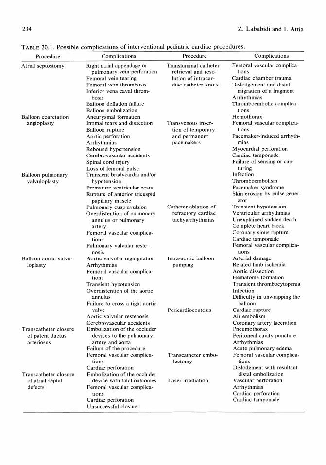
234 z. Lababidi and I. Attia
TABLE 20.1. Possible complications of interventional pediatric cardiac procedures.
Procedure
Atrial septostomy
Balloon coarctation angioplasty
Balloon pulmonary valvuloplasty
Balloon aortic valvuloplasty
Transcatheter closure of patent ductus arteriosus
Transcatheter closure of atrial septal defects
Complications
Right atrial appendage or pulmonary vein perforation
Femoral vein tearing Femoral vein thrombosis Inferior vena caval throm-
bosis Balloon deflation failure Balloon embolization Aneurysmal formation Intimal tears and dissection Balloon rupture Aortic perforation Arrhythmias Rebound hypertension Cerebrovascular accidents Spinal cord injury Loss of femoral pulse Transient bradycardia and/or
hypotension Premature ventricular beats Rupture of anterior tricuspid
papillary muscle Pulmonary cusp avulsion Overdistention of pulmonary
annulus or pulmonary artery
Femoral vascular complications
Pulmonary valvular reste-nosis
Aortic valvular regurgitation Arrhythmias Femoral vascular complica
tions Transient hypotension Overdistention of the aortic
annulus Failure to cross a tight aortic
valve Aortic valvular restenosis Cerebrovascular accidents Embolization of the occluder
devices to the pulmonary artery and aorta
Failure of the procedure Femoral vascular complica
tions Cardiac perforation Embolization of the occluder
device with fatal outcomes Femoral vascular complica
tions Cardiac perforation Unsuccessful closure
Procedure
Transluminal catheter retrieval and resolution of intracardiac catheter knots
Transvenous insertion of temporary and permanent pacemakers
Catheter ablation of refractory cardiac tachyarrhythmias
Intra-aortic balloon pumping
Pericardiocentesis
Transcatheter embolectomy
Laser irradiation
Complications
Femoral vascular complications
Cardiac chamber trauma Dislodgement and distal
migration of a fragment Arrhythmias Thromboembolic complica
tions Hemothorax Femoral vascular complica
tions Pacemaker-induced arrhyth-
mias Myocardial perforation Cardiac tamponade Failure of sensing or cap-
turing Infection Thromboembolism Pacemaker syndrome Skin erosion by pulse gener-
ator Transient hypotension Ventricular arrhythmias Unexplained sudden death Complete heart block Coronary sinus rupture Cardiac tamponade Femoral vascular complica-
tions Arterial damage Related limb ischemia Aortic dissection Hematoma formation Transient thrombocytopenia Infection Difficulty in unwrapping the
balloon Cardiac rupture Air embolism Coronary artery laceration Pneumothorax Peritoneal cavity puncture Arrhythmias Acute pulmonary edema Femoral vascular complica-
tions Dislodgment with resultant
distal embolization Vascular perforation Arrhythmias Cardiac perforation Cardiac tamponade

20. Interventional Pediatric Cardiac Catheterization
ble 20.1), and hopefully the incidence of these complications will decrease following the initial learning curve, together with future improvements in the equipment necessary for these procedures.
References
1. Rashkind WJ, Miller WW: Creation of an atrial septal defect without thoractomy: Palliative approach to complete transposition of the great arteries. JAm Med Assoc 1966; 196:991-992.
2. Park SC, Neches WH, Zuberbuhler JR, et al: Clinical use of blade atrial septostomy. Circulation 1978; 58:600-606.
3. Lababidi Z: Neonatal transluminal balloon coarctation angioplasty. Am Heart J 1983; 106:752-753.
4. Lababidi Z, Daskalopoulos D, Stoeckle H: Transluminal balloon coarctation angioplasty: Experience with 27 patients. Am J Cardiol 1984; 54:1288-1291.
5. Rey C, Marache P, Francart C, et al: Percutaneous balloon angioplasty in an infant with obstructed total anomalous pulmonary vein return. J Am Coli Cardiol 1985; 6:894-896.
6. Lababidi Z, Wu JR: Percutaneous balloon pulmonary valvuloplasty. Am J Cardiol 1983; 52:560-562.
7. Lababidi Z, Wu JR, Walls JT: Percutaneous balloon aortic valvuloplasty. Am J C ardial 1984; 53:194-197.
8. Reidy JF, Jones ODH, Tynan MJ et al: Embolization procedures in congenital heart disease. Br Heart J 1985; 54:184-192.
9. Florentine M, Wolfe RR, White RI: Balloon embolization to occlude a Blalock-Taussig shunt. J Am Coli Cardiol 1984; 3:200-202.
10. Rashkind WJ: Transcatheter treatment of congenital heart disease. Circulation 1983; 67:711-716.
11. Dotter CT, Rosch J, Bilbao MK: Transluminal extraction of catheter and guide fragments from the heart and great vessels: 29 collected cases. AJR 1971; 3:467-472.
12. Thomas J, Sinclair-Smith B, Bloomfield D, et al: Nonsurgical retrieval of a broken segment of steel spring guide from right atrium and inferior vena cava. Circulation 1964; 30: 106-108.
13. Hipona FA, Sciammar FD, Hublitz UF: Nonthoractomy retrieval of intraluminal cardio-
235
vascular foreign bodies. Radiol Clin North Am 1971; 9:583-595.
14. Thomas HA, Seivers RE: Nonsurgical reduction of arterial catheter knots. AJR 1979; 132:1018-1019.
15. Young DA, Maurer RM: Successful manipulation of a knotted intravascular catheter allowing nonsurgial removal. Radiology 1970; 94:155-156.
16. Kanpawich PP, Parker S, Anato JJ, et al: Emergency transvenous pacing in infants. Am Heart J 1985; 109:385-386.
17. Belhassen B, Miller HI, Laniado S: Catheter ablation of incessant ventricular tachycardia refractory to external cardioversions. Am J Cardiol1985; 55:1637-1639.
18. Veasy LG, Blalock RC, Orth JL, et al: Intraaortic balloon pumping in infants and children. Circulation 1983; 68:1095-1100.
19. Lababidi Z, Hakami N, Almond C: Chronic granulomatous disease associated with acute pericarditis with tamponade. Missouri Medicine 1977; 5: 170-171.
20. Lock JE, Bass JL, Kulk TJ et al: Chronic percutaneous pericardial drainage with modified pigtail catheter in children. Am J Cardiol1984; 53: 1179-1182.
21. Attia 1M, Lababidi ZA: Early results of balloon angioplasty of native aortic coarctation in young adults. Am J Cardiol1988; 61:930-931.
22. Gessman LJ, Reno CW, Chang KS, et al: Feasibility of laser catheter valvulotomy for aortic and mitral stenosis. Am J Cardiol 1984; 54: 1375-1377.
23. Lee G, Ikeda RM, Dwyer RM, et al: Feasibility of intravascular laser irradiation for in vivo visualization and therapy of cardiovascular disease. Am Heart J 1982; 103: 1076.
24. Hurwitz RA, Girod DA: Percutaneous balloon atrial septostomy in infants with transposition of the great arteries. Am Heart J 1976; 91:618-622.
25. Baker EJ, Allan LD, Tynan MJ, et al: Balloon atrial septostomy in the neonatal intensive care unit. Br Heart J 1984; 51:377-378.
26. Bullaboy CA, Jennings RB, Johnson DH, et al: Bedside balloon atrial septostomy using echocardiographic monitoring. Am J Cardia I 1984; 53:971.
27. Hawker RE, Krovetz LJ, Rowe RD: An analysis of prognostic factors in the outcome of balloon atrial septostomy for transposition of the great arteries. John Hopkins Med J 1974; 134:95-106.

236
28. Tynan M: Survival of infants with transposition of the great arteries after balloon atrial septostomy. Lancet 1971; 1:621-623.
29. Powell TG, Dewey M, West CR et al: Fate of infants with transposition of the great arteries in relation to balloon atrial septosomy. Br Heart J 1984; 51:371-376.
30. Sondheimer H, Havey RW, Blackman MS: Fatal over-distension of an atrioseptostomy catheter. Pediatr Cardiol 1982; 2:255-257.
31. Rashkind W: Palliative procedures for transposition of the great arteries. Br Heart J 1971; 33:69-72.
32. Keane JF, Lang P, Newburger J, et al: Iliac vein-inferior caval thrombosis after cardiac catheterization in infancy. Pediatr Cardiol 1980; 1:257-260.
33. Hawker RE, Celermajer JM, Cartmill TB, et al: Thrombosis of the inferior vena cava following balloon septostomy in transposition of the great arteries. Am Heart J 1971; 82:593-595.
34. Hohn AR, Webb HM: Balloon deflation failure. A hazard of medical atrial septostomy. Am Heart J 1972; 83:389-391.
35. Vogel JHK: Balloon embolization during atrial septostomy. Circulation 1970; 42: 155-156.
36. Park SC, Neches WH, Mullins CE, et al: Blade septostomy: Collaborative study. Circulation 1982; 66:258-266.
37. Vick GW III, Mullins CE, Nihill MR, et al: Blade and balloon atrial septostomy after trans-septal atrial puncture. J Am Coli Cardiol 1986; 7:117A.
38. Berger BC: Pericardiocentesis Using Echocardiography in Invasive Cardiology. Philadelphia, FA Davis Company, 1985, pp 269-279.
39. Sniderman KW, Bodner L, Saddekni S, et al: Percutaneous embolectomy by transcatheter aspiration. Radiology 1984; 150:357-361.
40. Moulopoulos S, Topaz S, Kloff WJ: Diastolic balloon pumping (with carbon dioxide) in the aorta-a mechanical assistance to the failing circulation. Am Heart J 1962; 63:669-675.
41. Bregman DA, Nichols AB, Weiss MB, et al: Percutaneous intraaortic balloon insertion. Am J Cardiol 1980; 46:261-264.
42. Subramanian VA, Goldstein JE, Sos TA, et al: Preliminary clinical experience with percutaneous intraaortic balloon pumping. Circulation 1980; 62(suppl I): 123-129.
43. Subramanian V A: Percutaneous intraaortic balloon pumping. Ann Thorac Surg 1980; 29:102.
z. Lababidi and I. Attia
44. Silka MJ, Gillette PC, Garson A Jr, et al: Transvenous catheter ablation of a right atrial automatic ectopic tachycardia. J Am Coli Cardio11985; 5:999.
45. Gillette PC, Garson A Jr, Porter CJ, et al: Junctional automatic ectopic tachycardia: New proposed treatment by transcatheter His bundle ablation. Am Heart J 1983; 106:619.
46. Ruffy R, Kim SS, Lal R: Paroxysmal fascicular tachycardia: Electrophysiologic characteristics and treatment by catheter ablation. JAm Coli C ardiol 1985; 5: 1008.
47. Hartzler GO: Electrode catheter ablation of refractory focal ventricular tachycardia. J Am Coli Cardiol1983; 2:1107.
48. Weber H, Schmitz L: Catheter technique for closed-chest ablation of an accessory atrioventricular pathway. N Engl J Med 1983; 308:653.
49. Ward DE, Camm AJ: Treatment of tachycardia associated with the Wolff-ParkinsonWhite syndrome by transvenous electrical ablation of accessory pathways. Br Heart J 1985; 53:64.
50. Scheinman MM: Catheter ablation techniques in patients with supraventricular tachycardia. Texas Heart Institute Journal 1986; B:427-432.
51. Morady F, Scheinman MM: Transvenous catheter ablation of a posteroseptal accessory pathway in a patient with the Wolff-Parkinson-White syndrome. N Engl J Med 1984; 310:705.
52. Bardy GH, Poole JE, Coltorti F, et al: Catheter ablation of a concealed accessory pathway. Am J Cardiol 1984; 54: 1366.
53. Elmquist R, Senning A: An implantable pacemaker for the heart. Proceedings of the Second International Conference of MedicalElectrical Engineers, London, Iliffe & Sons, Ltd, 1984.
54. Ginsburg R, Kim DS, Guthamer D, et al: Salvage of an ischemic limb by laser angioplasty: Description of a new technique. Clin Cardiol 1984; 7:56-58.
55. Gerschwind H, Boussignac G, Teisseire B, et al: Percutaneous transluminal laser angioplasty in man (letter). Lancet 1984; 2:866.
56. Choy DSJ, Stertzer SH, Myler RK, et al: Human coronary laser recanalization. Clin Cardiol 1984; 7:377-381.
57. Lee G, Ikeda R, Kozina J, et al: Laser dissolution of coronary atherosclerotic obstruction. Am Heart J 1981; 102:1074-1075.
58. Forrester JS, Litvack F, Grundfest WS: Laser
20. Interventional Pediatric Cardiac Catheterization
angioplasty and cardiovascular disease. Am J Cardiol 1986; 57:990-992.
59. Geschwind HJ, Blair JD, Mongkolsmai D, et al: Development and experimental application of contact probe catheter for laser angioplasty. J Am Coli Cardiol 1987; 9:101-107.
60. Riemenschneider T A, Lee G, Ikeda RM, et al: Laser irradiation of congenital heart diseases: Potential for palliation and correction of intracardiac and intravascular defects. Am Heart J 1983; 106:1389-1393.
61. White RI Jr, Ursic TA, Kaufman SL, et al: Therapeutic embolization with detachable balloons. Radiology 1978; 126:521.
62. Bommer WJ, Lee G, Rebeck K, et al: Atrial septostomy using argon-laser fiberoptic catheter and two-dimensional echography. Circulation 1983; 68(suppl III):III-90.
63. Porstmann W, Wierny L, Warnke H: Closure of persistent ductus arteriosus without thoractomy. Thoraxchirurgie 1967; 15:199-201.
64. King TD, Mills NL: Nonoperative closure of atrial septal defects. Surgery 1974; 75:383-388.
65. Rashkind WJ, Cuaso CC: Transcatheter closure of atrial septal and ventricular septal defects in the experimental animal. Proc Assoc Europ Pediatr Cardiol1976; 14:18.
66. Morag B, Rubinstein ZJ, Smolinsky A, et al: Percutaneous closure of a Blalock-Taussing shunt. Cardiovasc fntervent Radiol 1984; 7:218-220.
67. Grinnell VS, Mehringer CM, Hieshima GB, et al: Transaortic occlusion of collateral arteries to the lungs by detachable valved balloons in a patient with tetralogy of Fallot. Circulation 1982; 65:1276-1278.
68. Terry PB, Barth KH, Kaufman SL, et al: Balloon embolization for treatment of pulmonary arteriovenous fistulas. N Engl J Med 1980; 302: 1189-1190.
69. Ports mann W, Wierny L: Percutaneous transfemoral closure of the patent ductus arteriosus-an alternative to surgery. Semin Roentgenol 1981; 16:95-102.
70. O'Laughlin MP, Vick GW III, Nihill MR, et al: Foreign body retrieval: Transcatheter removal of embolized patent ductus arteriosus occlusion devices and catheter pieces. J Am Coli Cardiol 1987; 9(suppl A): 130A.
71. King TD, Thompson SL, Steiner C, et al: Secundum atrial septal defect: Nonoperative closure during cardiac catheterization. JAm Med Assoc 1976; 235:2506.
72. Mills NL, King TD: Nonoperative closure of
237
left-to-right shunts. J Thorac Cardiovasc Surg 1976; 72:371-372.
73. Rashkind WJ, Wagner HR, Tait MA: Historical aspects of interventional cardiology: Past, present and future. Texas Heart Institute Journal 1986; 13:363-367.
74. Gessner IH, Quisling RG, Mickle JP: Detachable latex balloon occlusion of anomalous systemic arteries in patients with tetralogy of Fallot. JAm Coli Cardiol1987; 9(suppl A):131A.
75. Crafoord C, Nylin G: Congenital coarctation of the aorta and its surgical treatment. J Thorac Surg 1945; 14:347-361.
76. Gross RE: Surgical correction for coarctation of the aorta. Surgery 1945; 18:347-352.
77. Vosschulte K: Surgical correction of coarctation of the aorta by an Isthmusplastik. Zur Beuhandlung Der Aorten Isthmusstenose. Thoraxchirugie 1957; 4:433-436.
78. Waldhausen JA, Nahrwold DL: Repair of coarctation of the aorta with a subclavian flap. J Thorac Cardiovasc Surg 1966; 51:532-533.
79. Gersony WM: Coarctation of the aorta, in Adams FH, Emmanouilides GC (eds): Moss' Heart Disease in Infants, Children and Adolescents. Baltimore, Williams & Wilkins, 1983, pp 188-199.
80. Hamilton DI, Di Eusanio G, Sandrasagra FA, et al: Early and late results of aortoplasty with a left subclavian flap for coarctatin of the aorta in infancy. J Thorae Cardiovasc Surg 1978; 75 :699-704.
81. Shinebourne EA, Tam ASY, Elseed AM, et al: Coarctation of the aorta in infancy and childhood. Br Heart J 1976; 38:375-380.
82. Hartman AF, Goldring D, Hernandez A, et al: Recurrent coarctation of the aorta after successful repair in infancy. Am J Cardiol 1970; 25:405-410.
83. Hesslein PS, Gutgessel HP, McNamara DG: Prognosis of symptomatic coarctation of the aorta in infancy. Am J Cardiol 1983; 51:299-303.
84. Clarkson PM, Nicholson MR, Barrett-Boyes BG, et al: Results after repair of coarctation of the aorta beyond infancy: A 10 to 18 year follow-up with particular reference to late systemic hypertension. Am J Cardiol 1983; 51: 1481-1488.
85. Nanton MA, Olley PM: Residual hypertension after coarctectomy in children. Am J Cardiol 1976; 37:769-772.
86. Hesslein PS, McNamara DG: Surgical intervention in infants with isolated coarctation of
238
the aorta. J Thorac Cardiovasc Surg 1981; 82:640-641.
87. Kilman JW, Williams TE Jr, Breza TS, et al: Reversal of infant mortality by early surgical correction of coarctation of the aorta. Arch Surg 1972; 105:865-868.
88. Sos T, Sniderman KW, Rettek-Sos B: Percutaneous transluminal dilation of coarctation of thoracic aorta postmortem. Lancet 1979; 2:970-971.
89. Lababidi Z, Madigan N, Wu JR, et al: Balloon coarctation angioplasty in an adult. Am J Cardiol 1984; 53:350-351.
90. Finley JP, Beaudien RG, Nanton MA, et al: Balloon catheter dilatation of coarctation of the aorta in young infants. Br Heart J 1983; 50:411-415.
91. Marvin WJ, Mahoney LT: Balloon angioplasty of unoperated coarctation of the aorta in young children (abst). J Am Coli Cardiol 1985; 5:405.
92. Driscoll DJ, Husslein PS, Mullins CE: Congenital stenosis of individual pulmonary veins: Clinical spectrum and unsuccessful treatment by transvenous balloon dilatation. Am J Cardio11982; 49:1767-1772.
93. Corwin RD, Singh AK, Karlson KE: Balloon dilatation of ductus arteriosus in a newborn with interrupted aortic arch and ventricular septal defect. Am Heart J 1981; 102:446-447.
94. Lund G, Rysavy J, Cragg A, et al: Long-term patency of the ductus arteriosus after balloon dilatation: An experimental study. Circulation 1984; 69:772-774.
95. Kan JS, White RI, Mitchell SE: Percutaneous balloon valvuloplasty: A new method for treating congenital pulmonary valve stenosis. New England J Med 1982; 370:540-542.
96. Kveselis DP, Rocchini AP: Long-term results of balloon valvuloplasty of the pulmonary valve. Pediatric Res 1984; 18:423.
97. Keith JD, Rowe RD, Vlad P: Heart Disease in Infancy and Childhood, ed 3. New York, McMillan, 1978, pp 3-13.
98. Awariefe SO, Clarke DR, Pappas G: Surgical approach to critical pulmonary valve stenosis in infants less than six months of age. J Thor Cardiovasc Surg 1964; 85:375-385.
Z. Lababidi and I. Attia
99. Engle MA, Ito T, Goldberg HP: The fate of the patient with pulmonic stenosis. Circulation 1964; 30:554-561.
100. Cole JG, Freedom RM, Olley PM, et al: Surgical management of critical pulmonary stenosis in the neonate. Ann Thor Surg 1984; 38:458-465.
101. Waller BF, Girod DA, Dillon JC: Transverse aortic wall tears in infants after balloon angioplasty for aortic valve stenosis. J Am Coli Cardio11984; 4:1235-1241.
102. Rupprath G, Neuhaus K: Percutaneous balloon valvuloplasty for aortic valve stenosis in infancy. Am J Cardiol1985; 55:1655-1656.
103. El-Said G, Galioto FM, Mullins CE, et al: Natural hemodynamic history of congenital aortic stenosis in childhood. Am J Cardiol 1972; 30:6-12.
104. Mody MR, Nadas AS, Bernhard WF: Aortic stenosis in infants. N England J Med 1967; 276:832-838.
105. Dobell ARC, Bloss RS, Gibbons JE, et al: Congenital valvular aortic stenosis. J Thorac Cardiovasc Surg 1981; 81:916-921.
106. Edmunds LV, Wagner HR, Heyman MA: Aortic valvulotomy in neonates, Circulation 1980; 61:421-427.
107. Keane JF, Bernhard WF, Nadas AS: Aortic stenosis in infancy. Circulation 1975; 52: 1138-1143.
108. Lakier JB, Lewis AB, Heyman MA, et al: Isolated aortic stenosis in the neonate. Circulation 1974; 50:801-806.
109. Trinkle JK, Grover FL, Arom KU: Closed aortic valvulotomy in infants. J Thorac Cardiovasc Surg 1978; 76:198-201.
110. Brow JW, Robinson RJ, Waller BF: Transventricular balloon catheter aortic valvulotomy in neonates. Ann Thorac Surg 1985; 39:376-378.
111. Mickell JJ, Mathews RA, Park SC, et al: Left atrioventricular valve atresia: Clinical management. Circulation 1980; 61:123.
112. Attia I. Weinhaus L, Walls J, et al: Rupture of tricuspid valve papillary muscle during balloon pulmonary valvuloplasty. Am Heart J 1987; 114:1233-1235.
21 Balloon Aortic Valvuloplasty Brice Letac and Alain Cribier
Balloon aortic valvuloplasty (BA V) in acquired aortic stenosis, a logical development of the growing field of inter ventiona I catheterization, was introduced by our group for the first time in September 1985 as a nonsurgical procedure for the treatment of adult aortic stenosis. I
Our first patient was a 78-year-old woman who had severe aortic stenosis with a 100 mm Hg peak-to-peak gradient. She absolutely refused surgery, although she was severely incapacitated with grade III dyspnea and angina. Fortunately, her aortic valve had minimal calcifications, allowing us to think that the risk for calcific embolism was proportionately low. The valvuloplasty procedure was surprisingly well tolerated. The patient had an excellent hemodynamic response with reduction of the gradient from 100 to 40 mm Hg, and, clinically, the patient was able to resume her normal active life. Within a span of a few weeks, the procedure was applied to several elderly patients who had definite contraindication to surgery due to associated illnesses and advanced age. This allowed us to increase our experience. Due to the low risk of the procedure and at the same time because of the overall good results obtained, we expanded the indications to include patients who were good surgical candidates. 2 For most of these cases, it was the patients themselves who requested that BA V be attempted first as a continuation of the diagnostic catheterization.
Balloon aortic valvuloplasty has been a rapidly expanding procedure and probably more
than 2000 cases have been performed in the world at the time ofthis writing. The following report will primarily be concerned with the experience of 204 patients analyzed in detail.
Technical Considerations
In the first two thirds of our series of patients, the valvuloplasty procedure was performed with the 9-Fr balloon catheters designed for dilatation of peripheral arteries and congenital pulmonic valve stenosis (Mansfield Inc., Mansfield, MA). In these patients, the steps taken to perform the aortic valvuloplasty procedure are described here.
After intravenous administration of 0.5 mg atropine, a Swan-Ganz thermodilution catheter was inserted into the pulmonary artery via the femoral vein for measuring the right heart pressures and cardiac output. A 7-Fr pigtail catheter was then positioned in the ascending aorta for continuous monitoring of the aortic pressure. This catheter also was used for performing a supravalvular aortogram to assess the severity of aortic insufficiency before and after valvuloplasty.
The aortic orifice was generally crossed with a 7-Fr Sones catheter through an 8-Fr introducer into the contralateral femoral artery, over a 0.035-in straight guidewire. A 7-Fr Amplatz left coronary artery catheter was preferred in cases with a large aortic root and vertical aortic orifice plane. The peak-to-peak and the mean pressure gradients across the

240
valve were then measured and the aortic valve area was calculated using Gorlin' s formula. Finally, a selective left ventriculogram was performed to assess the left ventricular function. The catheter used for this angiogram was then exchanged for the first balloon catheter over a 0.038-in, 270-cm guidewire.
After having been carefully purged of air, the balloon catheters were transcutaneously inserted into the femoral artery. During insertion, a strong negative pressure was delivered to the balloon in order to lower its profile. This transcutaneous femoral approach has been used in 90% of the cases. The brachial route after cutdown to the artery had to be used in the remaining patients in case of occlusion or major tortuosities of the femoroiliac vessels.
The effective length of the balloons used was 3 to 4 cm. We did not use longer balloons because such balloons take a longer time to inflate and are more difficult to maximally inflate. The inflated diameter was 15, 18, or 20 mm. Smaller sizes (8, 10, and 12 mm) were
FIGURE 21.1. Inflation of a 23-mm diameter balloon, 4 cm effective length, in a heavily calcified stenosed aortic valve. a) At the beginning of the inflation, there is a marked notch on the posterior side of the balloon due to the calcified border of the orifice of the valve. Note that the balloon and the shaft of the catheter inside of it are slightly curved by the orientation of the stenosed valve. b) When
B. Letac and A. Cribier
only used in our very first patients. The initial dilatation was performed with the IS-mm size, which always passed easily across the valve even in the case of severe stenosis. After two or three inflations, generally maintained for 60 seconds (but shortened in case of decreased aortic blood pressure to or below 60 mm Hg), this catheter was exchanged for an 18-mm and then a 20-mm, if necessary. Again, two to three inflations were performed with these sizes (Fig. 21.1).
The goal of the procedure was initially to reduce the peak-to-peak trans valvular gradient to or below 40 mm Hg (Fig. 21.2). However, we soon decided that the definition of the final result should be based on the increase in aortic valve area because of the many factors influencing the gradient, such as the loading conditions and contractility of the heart. Simply determining the final gradient led us to overestimate the quality of our early results, some patients remaining with a tight stenosis despite a marked decrease in gradient. The
the balloon is fully inflated, it becomes cylindrical and rigid and therefore it is no longer curved. The calcifications are pushed apart. In that case, the posterior calcification still makes a slight waist on the balloon, but this will disappear when the balloon becomes maximally inflated, just before bursting.

21. Balloon Aortic Valvuloplasty
200
160
120
80
40
mmHg 0
AVA (cm2) 0.68 0.95 1.20
BASE 15mm 20mm
FIGURE 21.2. Progressive decrease in pressure gradient and increase in aortic valve area with increasing balloon sizes. After the 20-mm diameter balloon, the peak-to-peak aortic gradient is 40 mm Hg and the valve area 1.20 cm2 •
aim of the procedure then became to achieve the highest possible aortic valve area. In the course of our experience, this led us to use more and more often a final balloon size of 20 mm (with a cross-sectional area of 3.14 cm2).
To obtain improvement of the final aortic valve area, more recently we also have attempted to increase the balloon cross-section by using the double balloon technique (two balloons inflated side by side): 15 + 15 mm, 18 + 15 mm, and 20 + 15 mm (cross-sections: 3.5,4.3, and 4.9 cm2, respectively); preferentially, in one third of our last 60 patients, we used a larger single balloon of 23 mm or in two cases of 25 mm (cross-section: 4.2 and 4.9 cm2). Our results have definitely been markedly improved by the use of larger balloon sizes.
A stable position of the inflated balloon across the valve and maximal inflation pressure delivery are the clues for optimal result. To better stabilize the balloon across the aortic valve orifice, we have used a 0.038-in extra stiff guidewire. The inflation pressure delivered to the balloon is not currently measured,
241
although it was done in our first cases. Maximal inflation pressure before rupture is approximately 4 to 6 atmospheres (manufacturer's specification), but actually it appears to be quite variable, probably because of the traumatic contact of the balloon with the calcific deposits. The main point is certainly to deliver the maximal pressure to get the highest dilatation efficacy, that is, maximal balloon size and rigidity. All of the waist of the inflated balloon must disappear and the balloon must appear perfectly cylindrical or even overdistended. To reach the maximal pressure, it is feasible, simple, and safe to push up the pressure to rupture the balloon. We try to burst the balloon at the end of the last inflation of each larger balloon size. This has no consequence because the balloon has been purged of air and the tear in the balloon has always been longitudinal. Bursting of the balloon occurred in at least three fourths of our cases. Only at the maximal inflation pressure does the balloon become a real rigid cylinder. It is possible that some of the unsatisfactory results of percutaneous BA V obtained by some investigators are due to an insufficient inflation of the balloon.
In an effort to further reduce the trauma and complications at the femoral artery and to make the technique easier, a new balloon catheter has been designed for the aortic valvuloplasty procedure (Fig 21.3). This new catheter has been used in the last fourth of our patients. This catheter possesses several original features. It is a 9-Fr catheter with a 7-Fr distal extremity, pre shaped in a large radius pigtail type curve that allows for both distal pressure measurements and injection of contrast media for angiography. A third lumen opens 10 cm above the balloon and allows continuous monitoring of the aortic blood pressure. The balloon has two sections: the distal portion (2 cm long) inflates to a 15-mm diameter and the proximal portion (3 cm long) to a 20-mm diameter. Thus, the dilatation can be performed without the need to exchange for balloon catheters of progressively increasing sizes. The trans valvular gradient and aortic pressures are monitored using this single catheter. Subvalvular and ventricular angiog-

242
FIGURE 21.3. Newly designed double-size balloon catheter. The distal part is 15 mm in diameter and 2 cm long, the proximal part is 20 mm in diameter and 3 cm long. The balloon is positioned on the 20-mm part and strongly inflated. The two distal markers are 2 cm apart. The segment of catheter between the two markers is semirigid and its size is 7-Fr. To measure the gradient without artifact, the balloon catheter is pulled back to a position with this segment straddling the aortic orifice. Beyond the distal markers, the catheter has a large pigtail tip with several side holes that permit measurement of the pressures and angiography.
raphies can also be performed with the same catheter.
In general, this new design catheter considerably simplifies and shortens the procedure, thus allowing valvuloplasty to be performed in 30 to 40 minutes, immediately after diagnostic cardiac catheterization, during the same session.
Results of Balloon Aortic Valvuloplasty
These results are based on a series of 204 patients whose mean age was 73 ± 11 with 64 patients 80 years old or above, 77 between 70 and 79, and 63 below 70 (Fig 21.4).
B. Letac and A. Cribier
A large majority of these patients were severely symptomatic with NYHA class III and IV dyspnea (N = 143), with angina (N = 107), and syncope (N = 56). Sixty-one had little dyspnea but they were symptomatic with either angina or syncope or both. Only seven were totally asymptomatic but these patients had signs of left ventricular hypertrophy on ECG and/or on echocardiogram and severe aortic stenosis had been confirmed either by Doppler or by a previous catheterization.
Seventy-one patients were good surgical candidates. Among them, 17 absolutely and definitely refused surgery. The others asked for balloon aortic valvuloplasty to be attempted first, although they would have accepted surgery in the case of failure of the procedure. It is easy to understand that when patients know there is another possibility to treat their aortic stenosis, they prefer to try it because it is far less invasive and followed by a prompt recovery.
The overall tolerance of the procedure was excellent. The act of inflating the balloon in the stenosed aortic valve was surprisingly well tolerated in most of the cases. In 65% of the cases, there was only a slight decrease in blood pressure, the patient remaining without any abnormal symptom allowing the inflation to be maintained for I minute or for several minutes; but in 35% of the cases, on the other hand, the blood pressure fell rapidly to below 60 mm Hg. In half of these cases, the drop in blood pressure was dramatic within seconds and the balloon had to be promptly deflated and withdrawn from the aortic valve to avoid pronounced dizziness, if not complete syncope. Although we usually tried to maintain the inflation of the balloon as long as possible, in that situation the effective inflation could not be maintained for more than 10 to 15 seconds. Although it could be construed that such short inflations would be less effective than those that were maintained for more than 1 minute, there was no real proof that it was so.
Apart from the drop in blood pressure, the actual balloon inflation itself was well tolerated.

21. Balloon Aortic Valvuloplasty 243
80
70
60
50
40
30
20
10
<50 50-60 60-70 70-80 80· 90 ~ 90
FIGURE 21.4. Distribution of the age of 204 patients studied.
Immediate Hemodynamic and Angiographic Data
Peak-to-peak systolic left ventriculoaortic gradient decreased from 72 ± 25 to 30 ± 14 mm Hg after valvuloplasty (Fig 21.5). The gradient was less than or equal to 30 mm Hg in 117 cases (57%).
The aortic valve area increased from 0.53 ± 0.18 to 0.93 ± 0.34 cm2. The increase was greater than 100% in 60 patients (29%). It was greater than 1 cm2 in 63 patients (31%) (Fig 21.5).
These results demonstrate on a large series
PRESSURE GRADIENT
mmHg
pc .DDt 80
72 60
0()
30 20
n=201
Base Post BAV
that percutaneous aortic balloon valvuloplasty in adult-acquired aortic stenosis is feasible and able to produce an appreciable increase in the aortic valve area despite the very considerable pathologic deformities and calcifications. However, the results are somewhat different from one patient to another and this can be easily expected from what is known of the pathological data concerning acquired aortic stenosis. There are significant differences in valve anatomy, particularly with respect to the fact that the valve may be bicuspid or tricuspid, with or without fusion of the commissures, or with more or less calcification or development of tissue fibrosis.
AORTIC VALVE AREA
cm2
1.2
~ 1,0 0 ,93 0,8
0,6 0.53 0,4
0 ,2 n=194
Base Post BAV
FIGURE 21.5. Left ventriculoaortic peak-to-peak pressure gradient in mm Hg (left) and aortic valve area in cm2 (right), initially and after dilatation.

244
In general, the increase in the aortic orifice area was satisfactory as it almost doubled. Although the area obtained of .93 cm2 is much less than the 3 to 4 cm2 of the normal valve, such an increase in area is able to remarkably improve the hemodynamic situation. Such an area is probably sufficient for normal activities, particularly in elderly patients. In those with an area greater than or equal to 1 cm2
(one third of the patients), the results can be considered as excellent. On the other hand, in 21 (10%) of the patients, the aortic area remained below .7 cm2 after dilatation. In these cases, we could consider that the balloon dilatation did not give a satisfactory result, as a .7 cm2 value is considered severe aortic stenosis. However, in half of these patients the improvement in valve area, although insufficient, was nonetheless appreciable as it increased by more than 50% from its initial value. For example, to have an aortic valve area of .6 or .7 cm2 , instead of .3 or .4 cm2 , represents a marked improvement for an individual and this is in accordance with the fact that these patients were clinically markedly improved. In a small number of patients, only six (3%), the balloon aortic dilatation could be considered as a complete failure because the aortic valve area did not change or increased by less than 10%. Undoubtedly, balloon valvuloplasty does not work at all in some patients, which is not surprising due to the tremendous changes in the valve structure.
Among the 168 patients who had an initial selective left ventricular angiogram, mean ejection fraction was 51 ± 17%. In 106, the angiogram was repeated immediately after the dilatation. Ejection fraction increased from 49 ± 17% to 52 ± 16% (P < 0.001). However, there was marked individual variation for the patient's ejection fraction changes after aortic dilatation. In 43 patients, there was a marked increase of the ejection fraction by more than 10% (42 ± 13% to 52 ± 13%). When comparing age and aortic valve areas in these 43 patients with the 63 patients who had no change or lesser increase in ejection fraction, there were no significant statistical differences.
Qne hundred sixty patients had a supravalvular angiogram before and after dilatation.
B. Letac and A. Cribier
One hundred fifteen patients had pre-existing aortic insufficiency (93 grade I, 20 grade II, 2 grade III). The regurgitation did not change after dilatation in 98 patients and increased slightly in 11 patients. In one case only, balloon dilatation resulted in a marked regurgitation from grade I to grade III. In five patients who had no previous regurgitation, a slight one appeared after the dilatation. In one case, the pre-existing regurgitation, which was mild, decreased to trace after dilatation.
Although aortic insufficiency was an anticipated complication at the beginning of our experience, surprisingly, creation of aortic insufficiency or aggravation of a pre-existing regurgitation by balloon inflation occurred in only a few cases and was only of moderate degree.
In-hospital Course After Dilatation
There was one death in the procedure room: a 92-year-old woman seen in extremis with mitral insufficiency and possibly coronary artery disease. No coronary angiogram was performed due to the very critical condition of the patient. The actual inflation was uneventful. The patient developed electromechanical dissociation a few minutes after withdrawal of the balloon catheter.
There was one case of a cerebrovascular accident during the procedure, which appeared as a progressive hemiplegia with residual sequelae.
After the procedure, three other cases of stroke were observed. One occurred 2 hours later with massive cerebral hemorrhage in a woman who had cerebral metastasis from breast cancer. The other two occurred 1 day and 2 days, respectively, after the procedure. These two strokes were transient. There was no specific evidence to suggest that these strokes were related to calcific emboli after thorough neurologic investigation was carried out. Such calcium embolism had been a concern during the first cases we performed; but with no emboli in this series of 204 patients,
21. Balloon Aortic Valvuloplasty
we can now consider that should calcific emboli occur, it would remain a very rare phenomenon that should not be held against the procedure.
One patient had a nonfatal myocardial in· farction on the 3rd day but he had very severe and diffuse inoperable coronary artery disease.
Six patients died within days after the procedure while still hospitalized. Their ages were 71, 76, 78, 79, 84, and 91. One death was due to an internal hemorrhage, probably through a vascular breach. This patient early in our series had marked arterial tortuosities and the manipulation of the balloon catheter had been very difficult. One patient died on day 3 with a gram-negative septicemia associated with lower limb ischemia. The other two cases died of pulmonary edema. In one of these cases, the aortic stenosis was extremely severe (an aortic valve area of .20 cm2) with several attacks of pulmonary edema in the preceding weeks. The gradient was 190 mm Hg and could be decreased only to 90 mm Hg, because only one IS-mm balloon could be used and only one inflation could be performed. There was no possibility for further exchanges of catheters due both to severe arterial tortuosities and to the critical condition of the patient. This case also was performed at the beginning of our experience. Very probably with the new balloon technology, at the present time, this patient could have been more successfully dilated. The manipulation of the new design catheter with a better profile would have been easier and could have allowed us to perform several good inflations with a 20-mm diameter balloon. In case offailure, we would have tried to go through the brachial artery. This is to say that very probably in our recent experience, this patient would not have left the procedure room with a 90 mm Hg residual gradient. Because she had a good left ventricular ejection fraction, a reasonable increase in the aortic valve area would have completely changed the situation. At the postmortem of this patient, we found a very tight aortic stenosis and three successive inflations of a 20-mm balloon performed on the postmortem specimen resulted in a valve area
245
of 0.96 cm2 • In retrospect, a similar good result could have been obtained during life using current techniques.
The total in-hospital mortality rate after aortic valvuloplasty was 3% (7 out of 204 cases). It must be pointed out that the mean age for these deaths was 81 years.
In direct relation with the dilatation procedure, there were femoral arterial complications in 26 cases with hematoma or thrombosis and one case of false aneurysm but surgery was required only in 8 patients.
Probably due to the large size of the balloon catheter used, the valvuloplasty procedure has a higher risk than a simple diagnostic cardiac catheterization, which still remains necessary before surgery. However, it must be emphasized that 64 of our 204 patients were 80 years or older; to our knowledge, no series of such elderly patients with catheterization has been published. It could be anticipated that the catheterization in itself in such elderly patients, in particular due to arterial stiffness and tortuosities, could have a higher risk than in younger patients. Although the rate of local arterial complications is relatively high (13%), it is of note that in case surgery is necessary, it remains a small surgical operation.
In the most incapacitated patients, improvement occurred during the in-hospital stay, sometimes in a very spectacular way. Among those 143 patients who were in class III/IV before the procedure, dyspnea improved dramatically in 44. There were no complaints of chest pain. Likewise, no syncope was observed.
For the patients who were not in critical condition and for those who did not have arterial complications, that is, for the majority of them, the mean hospital stay was 7 days.
Follow-up
Information on the clinical course after discharge was obtained for 144 patients whose valvuloplasty had been performed more than 3 months earlier with a mean of 8 months.
There were 24 deaths. Among them, only one, a 68-year-old woman, had been offered

246
surgery but she had refused it. None of the others were surgical candidates because of age or associated illnesses or both. One patient was 59 years old but he had liver cancer. The other 22 deaths were in patients 79 ± 8 years old. The causes of death were known in 14: terminal heart failure in 8, myocardial infarction in 2, and pulmonary infection in 4. It did not appear to us that these deaths could be directly related to the dilatation procedure itself. However, the dilatation resulted in an insufficient increase of the valve area in those who died in heart failure as the valve area had only increased from 0.41 ± 11 to 0.65 ± 18 cm2• These patients whose ejection fraction was 38 ± 8% probably died from persistent severe aortic stenosis. The two who died of myocardial infarction had severe coronary artery disease.
Of the 120 other cases, the overall clinical improvement was considered good in 101 (84%). Concerning dyspnea, whereas 87 patients were in NYHA class III/IV before, there were only 14 in such advanced classes at follow-up (Fig 21.6). No patient had recurrence of syncope. Angina pectoris disappeared in 24 of the 67 patients who had chest pain before (Fig 21.6). Among the 43 other cases with persistent angina, chest pain had markedly decreased in 26 cases. It had remained unchanged in 17 cases. It was severe (Canadian class III) in only 7. All of these 17
DYSPNEA
NYHA CLASS
IV
III
II
B. Letac and A. Cribier
patients had severe diffuse coronary disease, documented by coronary angiography, and could not be considered for angioplasty or for coronary bypass surgery. Only one patient had an angioplasty of the right coronary artery performed a few days after the aortic dilatation.
During the period of time that clinical follow-up was obtained, 11 patients came back to our department for recurrence or aggravation of symptoms, mainly dyspnea. These patients were recatheterized (mean delay, 6 ± 3 months after the initial valvuloplasty). The hemodynamic data before the valvuloplasty, just after, and at repeat catheterization were as shown in Table 21.1.
Clearly, the gradient and the aortic valve area at time of repeat catheterization had returned toward the initial value, and this confirms recurrence of stenosis. This probably also explains why there was no increase in ejection fraction of these patients.
It is difficult at the present time, due to the absence of sufficiently long-term follow-up, to evaluate the restenosis rate after percutaneous valvuloplasty. Some of the 24 patients who died after discharge may have died from restenosis, although there were several other possible explanations for their death: pronounced alteration in left ventricular function (8 had an ejection fraction lower than 35%), associated coronary disease, concomitant ill-
..AWiUtA
CANADIAN CLASS
IV (0 G) III G 0 II G 8
0 • 8 0 G 53
FIGURE 21.6. Improvement in dyspnea and angina after valvuloplasty, at follow-up (N = 120).
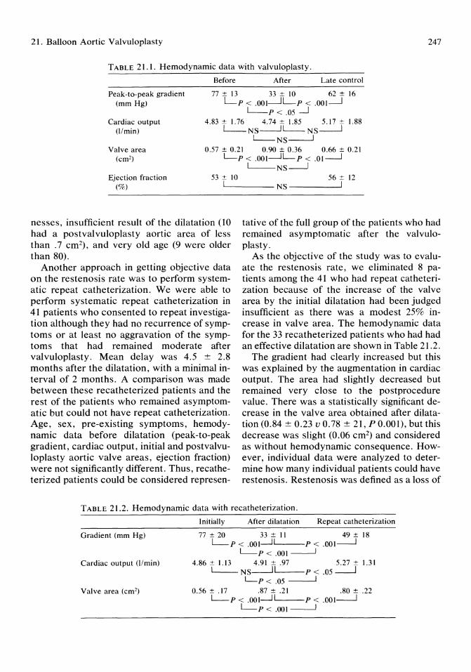
2l. Balloon Aortic Valvuloplasty 247
TABLE 2l.l. Hemodynamic data with valvuloplasty.
Before After Late control
Peak-to-peak gradient (mm Hg)
77 ± 13 33 ± 10 62 ± 16 Lp < .001--.JLp < .001--.J ~p < .05--.J
Cardiac output O/min)
4.83 ± 1.76 4.74 ± 1.85 5.17 ± 1.88 ~NS-.JL- NS---.l
L-NS----.J Valve area
(cm2)
0.57 ± 0.21 0.90 ± 0.36 0.66 ± 0.21 L-p < .001-.JL P < .Ol-.----J ~NS~
Ejection fraction (%)
53 ± 10 56 ± 12 LI ____ NS -------'1
nesses, insufficient result of the dilatation (10 had a postvalvuloplasty aortic area of less than .7 cm2), and very old age (9 were older than 80).
Another approach in getting objective data on the restenosis rate was to perform systematic repeat catheterization. We were able to perform systematic repeat catheterization in 41 patients who consented to repeat investigation although they had no recurrence of symptoms or at least no aggravation of the symptoms that had remained moderate after valvuloplasty. Mean delay was 4.5 ± 2.8 months after the dilatation, with a minimal interval of 2 months. A comparison was made between these recatheterized patients and the rest of the patients who remained asymptomatic but could not have repeat catheterization. Age, sex, pre-existing symptoms, hemodynamic data before dilatation (peak-to-peak gradient, cardiac output, initial and postvalvuloplasty aortic valve areas, ejection fraction) were not significantly different. Thus, recatheterized patients could be considered represen-
tative of the full group of the patients who had remained asymptomatic after the valvuloplasty.
As the objective of the study was to evaluate the restenosis rate, we eliminated 8 patients among the 41 who had repeat catheterization because of the increase of the valve area by the initial dilatation had been judged insufficient as there was a modest 25% increase in valve area. The hemodynamic data for the 33 recatheterized patients who had had an effective dilatation are shown in Table 21.2.
The gradient had clearly increased but this was explained by the augmentation in cardiac output. The area had slightly decreased but remained very close to the postprocedure value. There was a statistically significant decrease in the valve area obtained after dilatation (0.84 ± 0.23 v 0.78 ± 21, P 0.001), but this decrease was slight (0.06 cm2) and considered as without hemodynamic consequence. However, individual data were analyzed to determine how many individual patients could have restenosis. Restenosis was defined as a loss of
TABLE 2l.2. Hemodynamic data with recatheterization.
Initially After dilatation Repeat catheterization
Gradient (mm Hg) 77±20 33±11 49±IS L-p < .001.-J 1 P < .00I---..J
~P<.OOI~ Cardiac output (l/min) 4.86 ± 1.13 4.91 ± .97 5.27 ± 1.31
~NS.--li P < .05~ L-p < .05 ----'
Valve area (cm2) 0.56 ± .17 .87 ± .21 .SO ± .22 ~p < .001.-J 1 P < .OOI---.J
~p< .001---.1
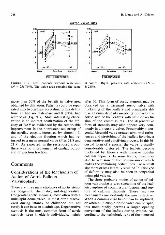
248 B. Letac and A. Cribier
AORTIC VALVE AREA
ca2r-------~----------_,
1.2
.8
.6 ._1
. 4 1
. 2
p<.OOl NS
Base PostBAV CDntr'ol
INO RESTENOSISI
FIGURE 21.7. Left, patients without restenosis (N = 25; 76%). The valve area remains the same
more than 50% of the benefit in valve area obtained by dilatation. Patients could be separated into two groups according to this definition: 25 had no restenosis and 8 (24%) had restenosis (Fig 21.7). Most interesting observation is an indirect confirmation of the efficacy of BA V as evidenced by the remarkable improvement in the nonrestenosed group of the cardiac output, increased by almost I L and of the ejection fraction which had returned to a mean normal value (Figs 21.8 and 21.9). As expected, in the restenosed group, there was no improvement of cardiac output and of ejection fraction.
Comments
Considerations of the Mechanism of Action of Aortic Balloon Valvuloplasty
There are three main etiologies of aortic stenosis: congenital, rheumatic, and degenerative. Congenital aortic stenosis, most often with a unicuspid dome valve, is most often discovered during infancy or childhood but not rarely it can be seen at adult age. Degenerative stenosis is the most common form of aortic stenosis, seen in elderly individuals, mainly
c.2r---------------------, 1.2
. 4
. 2
p<.OOl p<.OOt
Bsse PostBAV Contr'ol
IRESTENOSISJ
at control. Right, patients with restenosis (N 8; 24%).
after 70. This form of aortic stenosis may be observed on a tricuspid aortic valve with thickening of the leaflets and principally diffuse calcium deposits involving primarily the aortic side of the leaflets with little or no fusion of the commissures. The degenerative form of stenosis may also appear very commonly in a bicuspid valve. Presumably a congenital bicuspid valve creates abnormal turbulences and stretching of the leaflets favoring a degenerative and calcifying process. In this bicuspid form of stenosis, the valve is usually considerably distorted. The leaflets become thickened by fibrosis with massive nodular calcium deposits. In some forms, there may also be a fusion of the commissures, which makes the remaining orifice look like a small slot more or less laterally situated.3A This type of deformity may also be seen in congenital unicuspid valves.
The three probable modes of action of balloon valvuloplasty are: stretching of the leaflets, rupture of commissural fusions, and rupture of calcium deposits. These last two mechanisms are certainly the most effective. When a commissural fusion can be ruptured, or when a unicuspid dome valve can be split, this undoubtedly permits a larger opening movement of the leaflets during systole. According to the pathologic type of the stenosed
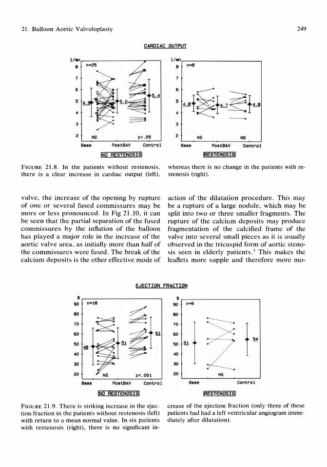
21. Balloon Aortic Valvuloplasty 249
CARpIAC OUTPUT
1/.0.----________ -.--_-,
S
7
5
4
3
2 NS p<.05
Sen POltSAV Control
INO RESTENOSISI
FIGURE 21.8. In the patients without restenosis, there is a clear increase in cardiac output (left),
valve, the increase of the opening by rupture of one or several fused commissures may be more or less pronounced. In Fig 21.10, it can be seen that the partial separation of the fused commissures by the inflation of the balloon has played a major role in the increase of the aortic valve area, as initially more than half of the commissures were fused. The break of the calcium deposits is the other effective mode of
l/.or---------_ _ --, S n-S
7
3
2 NS NS
SeBe P08tSAV Control
IRESTENOSISI
whereas there is no change in the patients with restenosis (right).
action of the dilatation procedure. This may be a rupture of a large nodule, which may be split into two or three smaller fragments. The rupture of the calcium deposits may produce fragmentation of the calcified frame of the valve into several small pieces as it is usually observed in the tricuspid form of aortic stenosis seen in elderly patients. 5 This makes the leaflets more supple and therefore more mo-
EJECTION FRACTION
S
90 n-1S
80 ~ 70
60
50
40
30
20 NS p< . OOt
BaBe POltBAV Control
INQ RgST~NOSISI
FIGURE 21.9. There is striking increase in the ejection fraction in the patients without restenosis (left) with return to a mean normal value. In six patients with restenosis (right), there is no significant in-
S
90 n-6
80
70
1 60
~ ~
50
40
30
20 NS
SIIBe Control
IAESTENOSISI
crease of the ejection fraction (only three of these patients had had a left ventricular angiogram immediately after dilatation).

250
FIGURE 21.10. Dilatation of a tricuspid form of calcified aortic stenosis on a fresh postmortem specimen. a) There are massive calcium deposits that maintain the leaflets rigid and immobile with marked commisural fusion. By planimetry the orifice is 0.63 cm2• b) A 20-mm diameter balloon is maximally inflated in the valve (second inflation); the large calcium nodule shown on the upper part of the picture is split apart. c) The orifice has been markedly increased, it is now 1.88 cm2 by planimetry (for the purpose of the photography, the leaflets, which are now more supple, are maintained opened by the light spring action of a forceps that is supposed to mimick the ejectional force of the blood). Note that the commissural fusion has been only partially opened.
B. Letac and A. Cribier
bile and more liable to be pushed apart by the ejectional flow during systole.
Because in most cases it seems that there is no real damage to the leaflets, and in particular no tear of the tissue of the leaflets, there is no increase of a pre-existing aortic regurgitation or creation of a new one. This is confirmed by the supravalvular angiography performed at the end of the dilatation procedure, which shows that in most cases there is no aortic regurgitation or at least no increase in a preexisting one. Even more, in some cases there is a decrease in the pre-existing aortic regurgitation, confirming that the leaflets have been made more supple and have improved their closure mechanism.
When there is nothing to break, neither a commissural fusion or calcium deposits, the only mode of action of the dilating balloon may be stretching of the tissue of the leaflets. This is probably the least effective action because due to elasticity of the leaflets, there may be a recoil phenomenon and a return toward the initial position. Even when a good opening of the stenosed valve is obtained as measured immediately at the end of the dilatation procedure, it is in the best cases in the range of 1.2 to 1.6 cm2 , whereas at the very moment of the full inflation of a 20-mm diameter balloon with total disappearance of the initial waist, the area occupied is 3.14 cm2• This difference is probably due to the action of the elasticity of the valve tissue. The recoil phenomenon due to the elasticity of the valve tissue is also probably responsible for restenosis when the only action of the valvuloplasty procedure is stretching of the valve. Such a recoil action may occur more or less rapidly. In a few cases, we were able to observe with Doppler and with repeat catheterization that the aortic valve area had returned to the initial valve area within less than 1 week. It could even be that the restenosis had occurred within a shorter time after the procedure, if not within minutes.
On the other hand, when a balloon inflation has broken the calcium deposits into fragments and/or has split fused commissures or a unicuspid valve, the result should be long-lasting, possibly as long as it has taken to produce the initial stenosis.
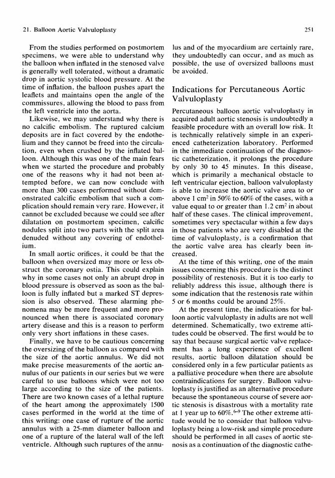
21. Balloon Aortic Valvuloplasty
From the studies performed on postmortem specimens, we were able to understand why the balloon when inflated in the stenosed valve is generally well tolerated, without a dramatic drop in aortic systolic blood pressure. At the time of inflation, the balloon pushes apart the leaflets and maintains open the angle of the commissures, allowing the blood to pass from the left ventricle into the aorta.
Likewise, we may understand why there is no calcific embolism. The ruptured calcium deposits are in fact covered by the endothelium and they cannot be freed into the circulation, even when crushed by the inflated balloon. Although this was one of the main fears when we started the procedure and probably one of the reasons why it had not been attempted before, we can now conclude with more than 300 cases performed without demonstrated calcific embolism that such a complication should remain very rare. However, it cannot be excluded because we could see after dilatation on postmortem specimen, calcific nodules split into two parts with the split area denuded without any covering of endothelium.
In small aortic orifices, it could be that the balloon when oversized may more or less obstruct the coronary ostia. This could explain why in some cases not only an abrupt drop in blood pressure is observed as soon as the balloon is fully inflated but a marked ST depression is also observed. These alarming phenomena may be more frequent and more pronounced when there is associated coronary artery disease and this is a reason to perform only very short inflations in these cases.
Finally, we have to be cautious concerning the oversizing of the balloon as compared with the size of the aortic annulus. We did not make precise measurements of the aortic annulus of our patients in our series but we were careful to use balloons which were not too large according to the size of the patients. There are two known cases of a lethal rupture of the heart among the approximately 1500 cases performed in the world at the time of this writing: one case of rupture of the aortic annulus with a 25-mm diameter balloon and one of a rupture of the lateral wall of the left ventricle. Although such ruptures of the annu-
251
Ius and of the myocardium are certainly rare, they undoubtedly can occur, and as much as possible, the use of oversized balloons must be avoided.
Indications for Percutaneous Aortic Valvuloplasty
Percutaneous balloon aortic valvuloplasty in acquired adult aortic stenosis is undoubtedly a feasible procedure with an overall low risk. It is technically relatively simple in an experienced catheterization laboratory. Performed in the immediate continuation of the diagnostic catheterization, it prolongs the procedure by only 30 to 45 minutes. In this disease, which is primarily a mechanical obstacle to left ventricular ejection, balloon valvuloplasty is able to increase the aortic valve area to or above 1 cm2 in 50% to 60% of the cases, with a value equal to or greater than 1.2 cm2 in about half of these cases. The clinical improvement, sometimes very spectacular within a few days in those patients who are very disabled at the time of valvuloplasty, is a confirmation that the aortic valve area has clearly been increased.
At the time of this writing, one of the main issues concerning this procedure is the distinct possibility of restenosis. But it is too early to reliably address this issue, although there is some indication that the restenosis rate within 5 or 6 months could be around 25%.
At the present time, the indications for balloon aortic valvuloplasty in adults are not well determined. Schematically, two extreme attitudes could be observed. The first would be to say that because surgical aortic valve replacement has a long experience of excellent results, aortic balloon dilatation should be considered only in a few particular patients as a palliative procedure when there are absolute contraindications for surgery. Balloon valvuloplasty is justified as an alternative procedure because the spontaneous course of severe aortic stenosis is disastrous with a mortality rate at 1 year up to 60%.6--9 The other extreme attitude would be to consider that balloon valvuloplasty being a low-risk and simple procedure should be performed in all cases of aortic stenosis as a continuation of the diagnostic cathe-

252
terization, to consider surgical valve replacement in cases of ineffectiveness of the dilatation. An argument for this second attitude is that it seems to be impossible to foresee which patient will have a good immediate and long-lasting result.
In our opinion, balloon aortic valvuloplasty is the treatment of choice in elderly patients aged 80 or older. In the literature, most of the cases performed are on elderly subjects. 10--13
There is no surgical series published with a substantial number of patients of this age with aortic valve replacement. There is not even published series on the risks of catheterization in such elderly patients, probably because they were not possible surgical candidates and hence were not even considered for hemodynamic investigation. In our series of 88 patients aged 80 or older treated by percutaneous valvuloplasty, there was only one death in the procedure room, which was the only procedure-related death among our 204 patients. There were 7 deaths during the hospital stay, which is a 3% mortality rate. Figures concerning mortality after cardiac surgery in elderly patients who, however, in the published series have an age lower than 80 (usual limit chosen: 70) are commonly found to be around or above 10%, taking into account only the series with patients operated after 1977-78, with myocardial protection. However, in recent years, there may have been a decrease in the surgical mortality of these elderly patients. 14-18
Another indisputable indication for balloon aortic valvuloplasty is a severe aortic stenosis occurring in a patient who has associated illness that is a definitive contraindication to surgery, such as lung disease or, more commonly, severe coronary artery disease not amendable by coronary dilatation or coronary bypass. Another situation, infrequent if the physician knows how to be convincing but nonetheless not exceptional, is the case of the patients who absolutely refuses surgery. In these situations, balloon aortic valvuloplasty offers a good alternative treatment.
At least in our group, we are presently faced with another situation: that of patients who are very reluctant to have surgery but who
B. Letac and A. Cribier
would probably finally accept it if there were no other possibility, and insist that valvuloplasty be attempted first as a continuation of the diagnostic catheterization. These patients hope to be among those 30% who will have a good result, that is, an aortic valve area of 1.2 cm2 or above, an aortic valve area comparable to many aortic prosthetic valves, and also be in the group who will have no restenosis. Such a preference for a less invasive form of treatment is not unreasonable. There is no real argument to advance against the demand formulated by these patients as we know that the risk for valvuloplasty is low and has no deleterious consequence: in case of failure of the procedure, the patient can be operated on for valve replacement.
Conclusion
The above considerations remain at the present time partly speculative due to the recent development of the technique. Further studies and longer follow-up, as for all new procedures, are necessary for better determination of the indications for balloon aortic valVUloplasty. In short, the indications for balloon aortic valvuloplasty are evolving rapidly. In due course, comparative studies with surgical valve replacement in age-matched patients will be conducted to evaluate the efficacy of both procedures.
References
1. Cribier A, Saoudi N, Berland J, et al: Percutaneous transluminal valvuloplasty of acquired aortic stenosis in elderly patients: An alternative to valve replacement? Lancet 1986; 1:63-67.
2. Cribier A, Savin T, Berland J, et al: Percutaneous transluminal balloon valvuloplasty of adult aortic stenosis: Report of 92 cases. JAm Coli Cardiol 1987; 9:381-386.
3. Roberts WC: The structure of the aortic valve in clinically isolated aortic stenosis. An autopsy study of 162 patients over 15 years of age. Circulation 1970; 42:91-97.
4. Roberts WC, Perloff JK, Costantino T: Severe valvular aortic stenosis in patients over 65
21. Balloon Aortic Valvuloplasty
years of age. A clinicopathologic study. Am J Cardiol 1971; 27:497-506.
5. Roberts WC: Good-bye to thoracotomy for cardiac valvulotomy. Am J Cardiol1987; 59:198-202.
6. Selzer A: Medical progress. Changing aspects of the natural history of valvular aortic stenosis. N Engl J Med 1987; 31:91-98.
7. Turina J, Hess 0, Sepulcri F, et al: Spontaneous course of aortic valve disease. Eur Heart J 1987; 8:471-483.
8. Frank S, Johnson A, Ross J: Natural history of valvular aortic stenosis. Br Heart J 1973; 35:41-46.
9. O'Keefe JH, Vlietstra RE, Bailey KR, et al: Natural history of candidates for balloon aortic valvuloplasty. Mayo Clin Proe 1987; 62:976-991.
10. McKay RG, Safian RD, Lock JE, et al: Balloon dilatation of calcific aortic stenosis in elderly patients: Postmortem, intraoperative, and percutaneous valvuloplasty studies. Circulation 1986; 74: 119-125.
11. Jackson G, Thomas S, Monaghan M, et al: Inoperable aortic stenosis in the elderly: Benefit from percutaneous transluminal valvuloplasty. Br Med J 1987; 294:83-86.
12. Isner JF, Salem DN, Desnoyers MR, et al:
253
Treatment of calcific aortic stenosis by balloon valvuloplasty. Am J Cardiol1987; 59:313-317.
13. Safian RD, Mandell WS, Thurer RE, et al: Postmortem and intraoperative balloon valvuloplasty of calcific aortic stenosis in elderly patients: Mechanisms of successful dilation. JAm Coli Cardiol 1987; 9:655-660.
14. Arom KV, Nicoloff DM, Lindsay WG, et al: Should valve replacement and related procedures be performed in elderly patients? Ann Thorae Surg 1984; 38:466-472.
15. Bergdahl L, Bjork va, Jonasso R: Aortic valve replacement in patients over 70 years. Seand J Thorae Cardiouase Surg 1981; 15:123-128.
16. Santiga JT, Flora J, Kirsh M, et al: Aortic valve replacement in the elderly. J Am Geriatr Soc 1983; 31:211-212.
17. Logeais Y, Leguerrier A, Delambre JF, et al: Resultats immediats et eloignes de la chirurgie du retrecissement aortique chez les sujets ages de soixante-dix ans et plus. Etude d'une serie consecutive de 229 operes. Ann Chir Chir Thorae Cardiouase 1986; 40:533-539.
18. Blakeman BM, Pifarre R, Sullivan HJ, et al: Aortic valve replacement in patients 75 years old and older. Ann Thorae Surg 1987; 44:637-639.

22 Peripheral Laser Thermal Angioplasty Timothy A. Sanborn
By partially removing obstructing atheroma or thrombus through vaporization of tissue rather than merely stretching or fracturing the plaque as in conventional balloon angiopia sty ,1 laser angioplasty or laser recanalization has the potential to serve as an aid or alternative to balloon angioplasty by: 1) increasing the initial success rate for lesions that are difficult or impossible to treat by conventional means or, 2) decreasing the incidence of restenosis after angioplasty.
However, in initial experimental studies and early clinical trial with bare argon or neodymium Y AG laser fiberoptics, the technique was limited by inadequate delivery systems resulting in an unacceptable high perforation rate2-6
and the creation of small recanalized channels that resulted in poor long-term patency.5
Initial Clinical Trials With Bare Fiberoptics
In early clinical trials of laser angioplasty, several studies were initiated using bare fiberoptics positioned inside angiographic or balloon catheters. Ginsberg et aF were the first to report a case of successful peripheral argon laser angioplasty. Subsequently, they reported success in 8 of 17 (47%) peripheral vessels, with three laser perforations.4 Cumberland et al,6 performing argon laser-assisted balloon angioplasty, noted luminal improvement after laser recanalization in 10 of 15 (67%) vessels with
two laser perforations of no clinical significance. In addition, Geschwind et al8 has reported successful percutaneous peripheral laser angioplasty using a neodymium Y AG laser positioned inside a balloon catheter in three patients; however, clinical or angiographic follow-up was not included in this brief report.
One ongoing clinical study uses an angioscope to visualize laser recanalization under direct vision during peripheral artery bypass surgery in an attempt to diminish the incidence of vessel perforation.9 Initial clinical attempts using the angioscope to direct a bare argon fiberoptic fiber were still plagued by perforation in 6 of 13 arteries; however, better results were obtained in later cases performed with a 2-mm laser-heated metallic-capped fiber similar to that to be discussed later. Whether or not angioscopy will improve the safety of laser angioplasty remains to be determined.
Thus, the key limitation in these early clinical trials of laser angioplasty was the lack of an adequate catheter system for safe and effective intravascular delivery of laser energy. The first, but certainly not the last, laser fiberoptic catheter system that shows promise in preliminary animal and clinical trials is a laser-heated metallic-capped device or laser probe. 10
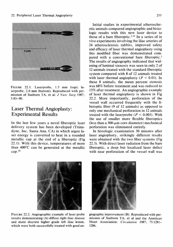
22. Peripheral Laser Thermal Angioplasty
1cm
FIGURE 22.1. Laserprobe, 1.5 mm (top); laserprobe, 2.0 mm (bottom). Reproduced with permission of Sanborn TA, et al: J Vasc Surg 1987; 5:83-90.
Laser Thermal Angioplasty: Experimental Results
In the last few years a novel fiberoptic laser delivery system has been developed (Trimedyne, Inc, Santa Ana, CA) in which argon laser energy is converted to heat in a rounded metallic cap at the end of a fiberoptic (Fig 22.1). With this device, temperatures of more than 400°C can be generated at the metallic cap. 10
FIGURE 22.2. Angiographic example of laser probe results demonstrating (A) diffuse right iliac disease and more discrete higher grade left iliac lesion, which were both successfully treated with good an-
255
Initial studies in experimental atherosclerotic animals compared angiographic and histologic results with this new laser device to those of a bare fiberoptic. 2•11 In a series of in vivo experiments involving the iliac arteries of 24 atherosclerosis rabbits, improved safety and efficacy of laser thermal angioplasty using this modified fiber was demonstrated compared with a conventional bare fiberoptic. 2
The results of angiography indicated that widening of luminal stenosis was seen in only 2 of 12 animals treated with the standard fiberoptic system compared with 8 of 12 animals treated with laser thermal angioplasty (P < 0.01). In these 8 animals, the mean percent stenosis was 68% before treatment and was reduced to 13% after treatment. An angiographic example of laser thermal angioplasty is shown in Fig 22.2. More importantly, perforation of the vessel wall occurred frequently with the fiberoptic fiber (9 of 12 animals) as opposed to only one mechanical perforation in 12 animals treated with the laserprobe (P < 0.001). With the use of smaller more flexible fiberoptics (less than a 300-lLm core diameter) mechanical perforation was eliminated entirely.
In histologic examination 30 minutes after laser angioplasty, strikingly different results were obtained with the two fiber systems (Fig 22.3). With direct laser radiation from the bare fiberoptic, a deep but localized laser defect with near perforation of the vessel wall was
giographic improvement (B). Reproduced with permission of Sanborn T A, et al and the American Heart Association: Circulation 1987; 75: 1281-1286.

256
FIGURE 22.3. Histologic specimens of iliac artery. Top, example of direct argon laser radiation resulting in a localized laser defect along one side of the vessel wall which extends through the neointima into the media. A gradient of thermal injury characterized by cell swelling and tissue edema is also noted. In addition, considerable thrombus is
noted along one side of the artery. There was associated charring, a gradient of thermal injury, and considerable thrombus formation. As is seen in Fig 22.3 (top), the majority of this eccentric lesion was not affected by the laser
T.A. Sanborn
present which fills the newly formed laser defect. Bottom, example of circumference. Hematoxylineosin stain, magnification x 80. Reproduced with permission for Sanborn TA, et al and the American College of Cardiology: J Am Call Cardial 1985; 5:934-938.
energy, thus, indicating the problem of aiming the laser beam. In contrast, those vessels treated with the laser-heated metal probe showed histologic evidence of thermal injury distributed evenly around the entire luminal
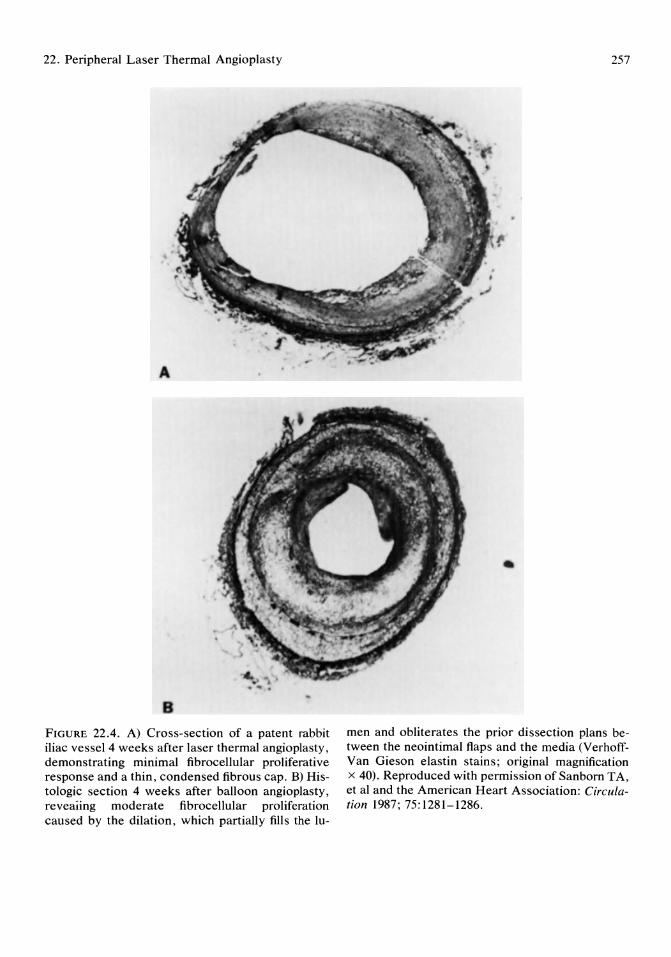
22. Peripheral Laser Thermal Angioplasty
A
B
FIGURE 22.4. A) Cross-section of a patent rabbit iliac vessel 4 weeks after laser thermal angioplasty, demonstrating minimal fibrocellular proliferative response and a thin, condensed fibrous cap. B) Histologic section 4 weeks after balloon angioplasty, reveaiing moderate fibrocellular proliferation caused by the dilation, which partially fills the lu-
257
•
men and obliterates the prior dissection plans between the neointimal flaps and the media (VerhoffVan Gieson elastin stains; original magnification x 40). Reproduced with permission of Sanborn T A, et al and the American Heart Association: Circulation 1987; 75:1281-1286.

258
circumference. This thermal effect was associated with minimal charring, a gradient of thermal injury, and thinner flatter thrombus formation. These histologic data suggest that circumferential rather than localized distribution of energy is a factor in these improved experimental results.
These results were confirmed in a series of postmortem human coronary artery xenografts transplanted into the canine femoral artery. II Angiography demonstrated recanalization in all five arteries treated with the laser-heated probe and three of five arteries treated with the bare fiberoptic. Only one perforation occurred with the metallic-capped fiber compared with three perforations using the bare fiberoptic. Interestingly, a larger 1.5-mm laser-heated probe was capable of creating a larger channel in the occluded arterial segment.
Recent follow-up angiographic and histologic studies in atherosclerotic rabbits demonstrated good long-term patency with minimal thrombogenesis and a very mild proliferative response to laser thermal angioplasty with a 1.5 to 2.0-mm laser-heated probe. 12 On histology, re-endothelialization of the luminal surface was noted as early as 2 weeks after laser thermal angioplasty. At 4 weeks the neointima was thin with a fibrous cap and minimal fibrocellular proliferation.
In a recent comparative study, laser thermal angioplasty was found to have less restenosis with a significantly larger luminal diameter 0.6 ± 0.5 v 1.0 ± 0.4 mm) when angiography was repeated 4 weeks postangioplasty.13 At that time histologic examination revealed less fibrocellular proliferation after laser thermal angioplasty, whereas those vessels treated with balloon angioplasty demonstrated evidence of prior fracture and dissection of the vessel wall with more of a fibrocellular proliferative response (Fig 22.4). Morphometer analysis of histologic cross-sections of these pressure perfused arteries confirmed a significantly large luminal area after laser thermal angioplasty compared with balloon angioplasty (1.24 ± 0.62 v 0.6 ± 0.45 mm2; p < 0.05). Thus, in rabbit iliac stenoses, laser thermal angioplasty was associated with less re-
T.A. Sanborn
stenosis and produced a significantly larger mean luminal diameter and mean luminal area than conventional balloon angioplasty. These results may be due to the different pathophysiologic mechanisms involved in these two techniques.
Laser-assisted Balloon Angioplasty in Peripheral Vessels
After demonstrating the safety and efficacy of this device in experimental animals,2,11-13 a collaborative clinical trial was initiated at Boston University Medical Center and Northern General Hospital, Sheffield, England, to first determine the safety and efficacy of this laserheated probe in performing percutaneous laser thermal angioplasty to recanalize lesions before conventional balloon angioplasty. 14-16
Patient Population
All patients had severe peripheral vascular disease and suffered from either limiting claudication or rest pain, gangrene, and threatened limb loss. Initial evaluation included a history and physical examination as well as a Doppler ankle-arm index (AAI), which was also used for follow-up after angioplasty. Patients were pretreated with oral aspirin (75 or 325 mg once a day).
Laser Equipment
The laser system at Boston University consisted of a 14-W argon laser system (Optilase, Model 900, Trimedyne Inc, Santa Ana, CA) coupled to a sterile disposable laser-heated metallic-capped fiberoptic that consisted of a 300-jLm diameter core fiberoptic fiber with a 1.0 to 2.5-mm metallic cap at the distal end of the fiber (Laserprobe-PLR, Trimedyne Inc, Santa Ana, CA). At Northern General Hospital, the laser-heated probe was coupled to an argon laser generator from Cooper Laser Sonics. 16

22. Peripheral Laser Thermal Angioplasty
Percutaneous Procedure
The majority of the procedures were performed via percutaneous arterial puncture of the ipsilateral femoral artery under local anesthesia. The equipment required (catheters, wires, etc.) was identical to that used in conventional arterial catheterization. After cannulation of the superficial femoral artery, 5,000 U of heparin was administered intra-arterially and initial angiography was performed to document the lesion. To introduce the laser probe into the artery, an introducer sheath with good sealing around the laser fiberoptic and guide wire (0.04 in "plus" wire) was necessary. We found the arterial sheath with the best prevention of back bleeding to be one manufactured by Cook (Model VCF-8.5-38, Cook, Inc, Bloomington, IN). The laserheated probe was inserted into this introducer sheath and advanced under fluoroscopic guidance to the proximal origin of the lesion until contact was made between the probe tip and the lesion as verified by angiography and tactile feedback. Five to to-second pulses of 8 to 13 W of argon laser energy were then delivered from the laser generator to the probe. After an initial warmup period of 2 to 3 seconds while maintaining gentle pressure on the probe to initiate advancement, the probe was then advanced through the lesion with a continuous motion. Care was taken to keep the tip moving as it cooled down after laser pulse delivery to avoid adherence to the arterial wall. If adherence was noted on gentle withdrawal of the probe, a repeat laser pulse was delivered to free the probe and a continuous motion was applied to the tip during the subsequent cooling period. Progress of the probe through the lesion was monitored fluoroscopically with several injections of 3 to 5 ml of contrast solution given through the arterial sheath to confirm the position of the device. After the lesions were crossed with the laser-heated probe, one final pulse was delivered on slow withdrawal of the probe through the lesion to maximize the luminal diameter. When the probe tip reached the proximal end of the lesion, laser power was discontinued and the probe moved back and forth for 5 seconds dur-
259
ing cooling. The laser probe was then removed and an angiogram was performed to document the luminal diameter produced by the procedure.
As the luminal diameter produced by laser thermal angioplasty with the current 1.0 to 2.5-mm diameter device was considered inadequate in these large 4.0 to 5.0-mm peripheral vessels, the laser procedure was followed in all cases by conventional balloon angioplasty to obtain a definitive lumen that was documented by a final angiogram. The arterial sheath was subsequently removed and systemic heparin was administered for 24 hours unless a hematoma was present. The patients were discharged within 24 to 48 hours on 75 or 325 mg of aspirin a day.
Local Femoral Cutdown Procedure
Rarely, either marked obesity or high-grade proximal superficial femoral artery disease precluded a safe percutaneous approach. In these cases, under local anesthesia and mild sedation, a small cutdown was made to expose the common femoral artery for artery for direct arterial puncture and subsequent laser and balloon angioplasty through an 8.5-Fr sheath. 15
Initial Results
In this initial series, laser recanalization was successful in 39 of 41 vessels for a 93% angiographic success with minimal complications and no vessel perforation. 14,15 In this study, the most commonly used laser probe size was the 2,0-mm diameter metal tip. The average laser wattage was to W (range, 4 to 13 W), Representative angiograms are shown in Figs 22.5 and 22.6.
Probe Detachment Early in this clinical trial, probe tip detachment from the fiberoptic occurred in two patients; one of these probes could be retrieved and removed. Subsequent to this early experience, a safety (anchor) wire was incorporated into the device to add stability to the joint be-
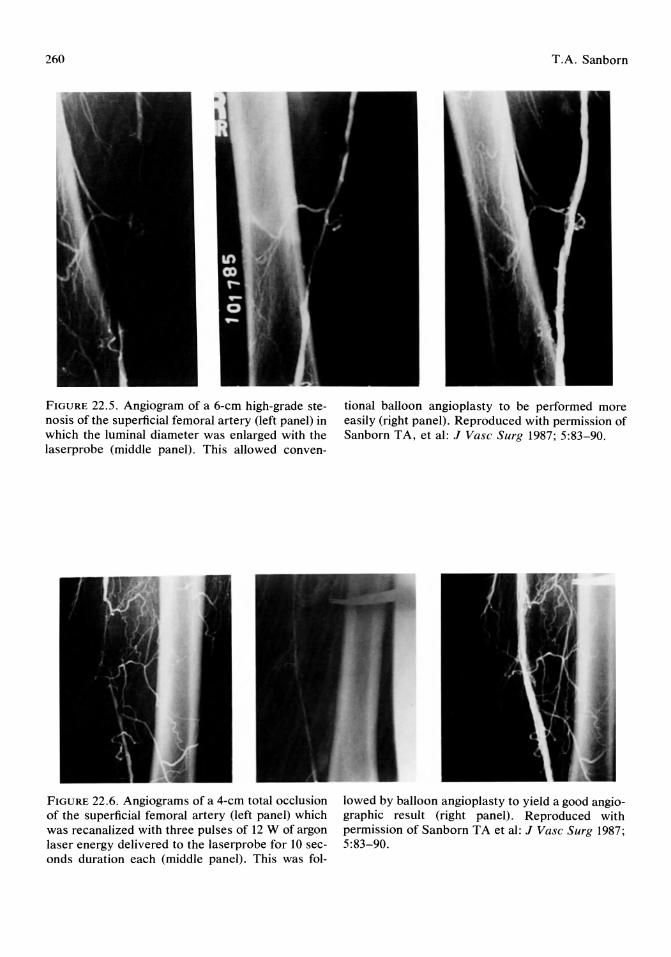
260
FIGURE 22.5. Angiogram of a 6-cm high-grade stenosis of the superficial femoral artery (left panel) in which the luminal diameter was enlarged with the laserprobe (middle panel). This allowed conven-
FIGURE 22.6. Angiograms of a 4-cm total occlusion of the superficial femoral artery (left panel) which was recanalized with three pulses of 12 W of argon laser energy delivered to the laserprobe for 10 seconds duration each (middle panel). This was fol-
T.A. Sanborn
tional balloon angioplasty to be performed more easily (right panel). Reproduced with permission of Sanborn TA, et al: J Vase Surg 1987; 5:83-90.
lowed by balloon angioplasty to yield a good angiographic result (right panel). Reproduced with permission of Sanborn TA et al: J Vase Surg 1987; 5:83-90.

22. Peripheral Laser Thermal Angioplasty
tween the fiberoptic and the metal tip and to prevent further probe detachment. In addition, by keeping the probe moving constantly during laser delivery and the cooling period, it was found that adherence to the vessel wall, one of the potential causes of probe detachment, was significantly reduced.
Use in Total Occlusion
Once the safety and efficacy of laser thermal angioplasty was demonstrated in peripheral vessels, the next step was to demonstrate a useful clinical role for the technique. A recent report of the combined experience at N orthern General Hospital and Boston University Medical Center was directed toward addressing one of these questions; whether the use of laser thermal angioplasty can increase the initial success rate in peripheral artery total occlusion. 16 In this initial series, 50 of 56 (89%) femoropopliteal occlusions were successfully recanalized by laser thermal angioplasty to provide an initial channel for subsequent balloon dilatation. Because there were two acute reocclusions, the overall initial clinical success rate in this series was 86%. These results compare quite favorably with recent large series of conventional balloon angioplasty, which report initial clinical success rates of 72% to 78%.17,18
Interestingly, if those lesions considered easy to treat by conventional means we examined separately, the initial success rate for these 17 lesion was 100%, Thus, despite the fact that more difficult lesions were attempted in this initial series, laser-assisted balloon angioplasty resulted in minimal, nonsignificant complications, a perforation rate of only 2%, and a success rate equal to or better than previously published results for balloon angioplasty alone.
Technique Development
These results represent the early stages of development of a new technique that will obviously be modified and adapted in the future, In addition to the techniques described, two ad-
261
ditional modifications are worth mentioning that related to the 0.014-in "plus" wire attached to the laser probe,
Tip Angulation by Wire Shaping
Occasionally, in tortuous lesions, or at bifurcations in the artery, some angulation of this straight but flexible fiberoptic and the rigid metal tip is necessary. By shaping a gentle curve in the 0.014-in wire alongside the fiberoptic, a curve can be maintained in the laser probe and the metal tip rotated in 3600 in a fashion similar to torque guidewires. Care must be taken, however, to be sure that this angulation is not too acute as the curve is fixed and may make further recanalization of a straight portion of the artery more difficult. Biplane fluoroscopy or use of multiple fluoroscopic views aids in the use of these angled probes. The ability to release the angulation, as in a tip-deflecting wire, would be a useful alternative to improve the steerability of the device in the future.
Balloon Advancement Over the Probe
In extremely difficult or tortuous lesions, another technique that has proven useful is the advancement of the balloon angioplasty catheter over the fiberoptic and the "plus" wire once the lesion has been crossed. In this situation, instead of recrossing the lesion with a guidewire, which can cause a dissection, the laser probe catheter actually serves as a guidewire for the balloon catheter. This technique has been quite helpful, particularly in diffusely diseased vessels. The sterile portion of the disposable fiberoptic has to be cut about 2 m from the distal end to disconnect it from the nonsterile proximal end, which is attached to the laser generator.
Follow-up Results
When examining long-term results of peripheral angioplasty, clinical patency rates and the respective recurrence rates may vary con sid-
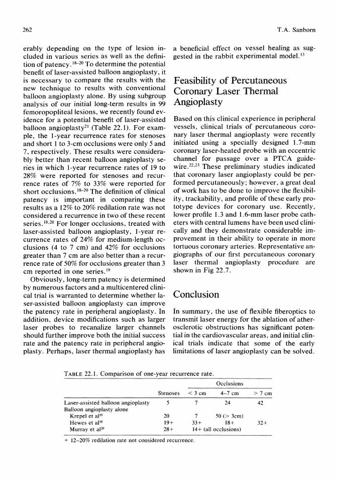
262
erably depending on the type of lesion included in various series as well as the definition of patency .18-20 To determine the potential benefit of laser-assisted balloon angioplasty, it is necessary to compare the results with the new technique to results with conventional balloon angioplasty alone. By using subgroup analysis of our initial long-term results in 99 femoropopliteallesions, we recently found evidence for a potential benefit of laser-assisted balloon angioplasty21 (Table 22.1). For example, the I-year recurrence rates for stenoses and short 1 to 3-cm occlusions were only 5 and 7, respectively. These results were considerably better than recent balloon angioplasty series in which I-year recurrence rates of 19 to 28% were reported for stenoses and recurrence rates of 7% to 33% were reported for short occlusions. 18-20 The definition of clinical patency is important in comparing these results as a 12% to 20% redilation rate was not considered a recurrence in two of these recent series. 18,20 For longer occlusions, treated with laser-assisted balloon angioplasty, I-year recurrence rates of 24% for medium-length occlusions (4 to 7 cm) and 42% for occlusions greater than 7 cm are also better than a recurrence rate of 50% for occlusions greater than 3 cm reported in one series. 19
Obviously, long-term patency is determined by numerous factors and a multicentered clinical trial is warranted to determine whether laser-assisted balloon angioplasty can improve the patency rate in peripheral angioplasty. In addition, device modifications such as larger laser probes to recanalize larger channels should further improve both the initial success rate and the patency rate in peripheral angioplasty. Perhaps, laser thermal angioplasty has
T.A. Sanborn
a beneficial effect on vessel healing as suggested in the rabbit experimental model. 13
Feasibility of Percutaneous Coronary Laser Thermal Angioplasty
Based on this clinical experience in peripheral vessels, clinical trials of percutaneous coronary laser thermal angioplasty were recently initiated using a specially designed 1.7-mm coronary laser-heated probe with an eccentric channel for passage over a PTCA guidewire. 22 ,23 These preliminary studies indicated that coronary laser angioplasty could be performed percutaneously; however, a great deal of work has to be done to improve the flexibility, trackability, and profile of these early prototype devices for coronary use, Recently, lower profile 1.3 and I.6-mm laser probe catheters with central lumens have been used clinically and they demonstrate considerable improvement in their ability to operate in more tortuous coronary arteries. Representative angiographs of our first percutaneous coronary laser thermal angioplasty procedure are shown in Fig 22.7.
Conclusion
In summary, the use of flexible fiberoptics to transmit laser energy for the ablation of atherosclerotic obstructions has significant potential in the cardiovascular areas, and initial clinical trials indicate that some of the early limitations of laser angioplasty can be solved.
TABLE 22.1. Comparison of one-year recurrence rate.
Occlusions
Stenoses < 3 cm 4-7 cm > 7 cm
Laser-assisted balloon angioplasty 5 7 24 42 Balloon angioplasty alone
Krepel et aP9 20 7 50 (> 3cm) Hewes et al 18 19+ 33+ 18+ 32+ Murray et apo 28+ 14+ (all occlusions)
+ 12-20% redilation rate not considered recurrence.
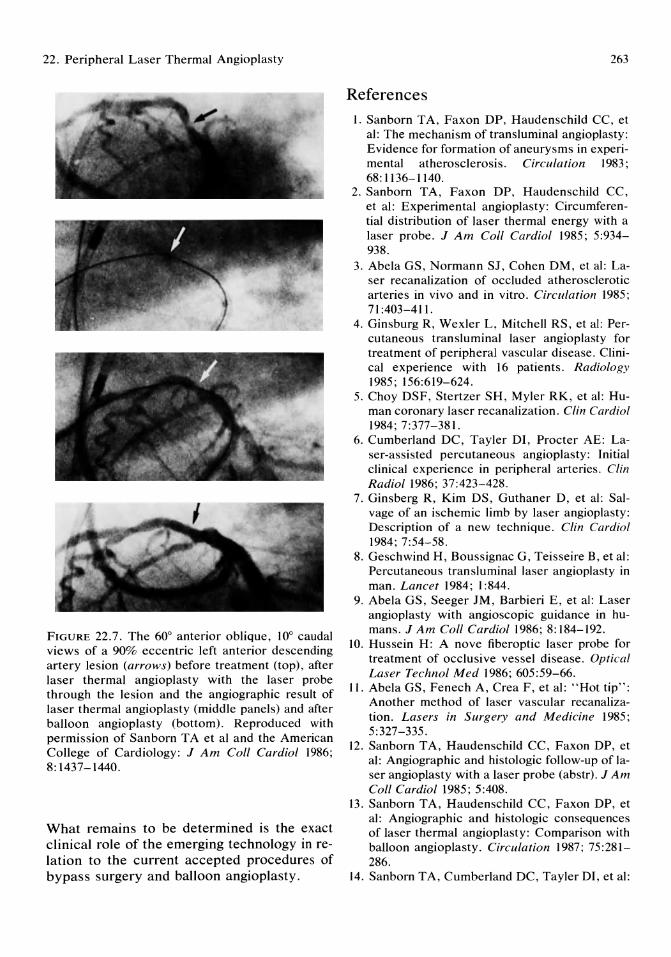
22. Peripheral Laser Thermal Angioplasty
FIGURE 22.7. The 60° anterior oblique, 10° caudal views of a 90% eccentric left anterior descending artery lesion (arrows) before treatment (top), after laser thermal angioplasty with the laser probe through the lesion and the angiographic result of laser thermal angioplasty (middle panels) and after balloon angioplasty (bottom). Reproduced with permission of Sanborn T A et al and the American College of Cardiology: J Am Call Cardiol 1986; 8: 1437-1440.
What remains to be determined is the exact clinical role of the emerging technology in relation to the current accepted procedures of bypass surgery and balloon angioplasty.
263
References
1. Sanborn TA, Faxon DP, Haudenschild CC, et al: The mechanism of transluminal angioplasty: Evidence for formation of aneurysms in experimental atherosclerosis. Circulation 1983; 68: 1136-1140.
2. Sanborn TA, Faxon DP, Haudenschild CC, et al: Experimental angioplasty: Circumferential distribution of laser thermal energy with a laser probe. J Am Call Cardiol 1985; 5:934-938.
3. Abela GS, Normann SJ, Cohen DM, et al: Laser recanalization of occluded atherosclerotic arteries in vivo and in vitro. Circulation 1985; 71:403-411.
4. Ginsburg R, Wexler L, Mitchell RS, et al: Percutaneous transluminal laser angioplasty for treatment of peripheral vascular disease. Clinical experience with 16 patients. Radiology 1985; 156:619-624.
5. Choy DSF, Stertzer SH, Myler RK, et al: Human coronary laser recanalization. Clin Cardiol 1984; 7:377-381.
6. Cumberland DC, Tayler DI, Procter AE: Laser-assisted percutaneous angioplasty: Initial clinical experience in peripheral arteries. Clin Radial 1986; 37:423-428.
7. Ginsberg R, Kim DS, Guthaner D, et al: Salvage of an ischemic limb by laser angioplasty: Description of a new technique. Clin Cardiol 1984; 7:54-58.
8. Geschwind H, Boussignac G, Teisseire B, et al: Percutaneous transluminal laser angioplasty in man. Lancet 1984; 1:844.
9. Abela GS, Seeger JM, Barbieri E, et al: Laser angioplasty with angioscopic guidance in humans. J Am Call Cardiol1986; 8:184-192.
10. Hussein H: A nove fiberoptic laser probe for treatment of occlusive vessel disease. Optical Laser Technol Med 1986; 605:59-66.
11. Abela GS, Fenech A, Crea F, et al: "Hot tip": Another method of laser vascular recanalization. Lasers in Surgery and Medicine 1985; 5:327-335.
12. Sanborn TA, Haudenschild CC, Faxon DP, et al: Angiographic and histologic follow-up of laser angioplasty with a laser probe (abstr). JAm Call Cardiol 1985; 5:408.
13. Sanborn TA, Haudenschild CC, Faxon DP, et al: Angiographic and histologic consequences of laser thermal angioplasty: Comparison with balloon angioplasty. Circulation 1987; 75:281-286.
14. Sanborn T A, Cumberland DC, Tayler DI, et al:
264
Human percutaneous laser thermal angioplasty (abstr). Circulation 1985; 72:III-303.
15. Sanborn TA, Greenfield AJ, Guben JK, et al: Human percutaneous and intraoperative laser thermal angioplasty: Initial clinical results as an adjunct to balloon angioplasty. J Vasc Surg 1987; 5:83-90.
16. Cumberland DC, Sanborn TA, Tayler DI, et al: Percutaneous laser thermal angioplasty: Initial clinical results with a laserprobe in total peripheral artery occlusions. Lancet 1986; 1:1457-1459.
17. Zietler E, Richter EI, Seyferth W: Femoropopliteal arteries, in Cotter CT, Gruentzig A, Schoop W, Zeitler E, (eds): Percutaneous Transluminal Angioplasty. Berlin, SpringerVerlag, 1983, pp 105-114.
18. Hewes RC, White RI, Murray RR, et al: Longterm results of superficial femoral artery angioplasty. Am J Radio11986; 146:1025-1029.
T .A. Sanborn
19. Krepel VM, van Andel GJ, van Erp WFM, et al: Percutaneous transluminal angioplasty of the femoropopliteal artery: Initial and long term results. Radiology 1985; 156:325-328.
20. Murray RR, Hewes RL, White RI, et al: Long segment femoropopliteal stenoses: Is angioplasty a boom or a bust? Radiology 1987; 162:473-476.
21. Sanborn TA, Cumberland DC, Welsh CL, et al: Laser thermal angioplasty as an adjunct to peripheral balloon angioplasty: One year followup results (abstr). Circulation 1987; 76:IV-30.
22. Sanborn TA, Faxon DP, Kellett MA, et al: Percutaneous coronary laser thermal angioplasty. J Am Coli Cardiol 1986; 8:1437-1440.
23. Cumberland DC, Starkey IR, Oakley GDG, et al: Percutaneous laser-assisted coronary angioplasty. Lancet 1986; 11:214.

Part IV Coronary Interventions

23 Practical Aspects of Coronary Angioplasty Amar S. Kapoor
Coronary angioplasty is a challenging innovation in the traditional practice of invasive cardiology and has been made possible by the availability of sophisticated technology of coronary balloon catheters, ancillary accessories, and imaging systems. Gruentzig and associates, I after gaining experience from balloon angioplasty of peripheral arteries, miniaturized the balloon catheter system for its application in coronary arteries. Then on September 16, 1977, the first successful coronary angioplasty in humans was performed by Gruentzig (Fig 23.1).2 His well-planned, thoughtful, and pioneering scientific human experiment was the single most compelling reason for pursuing this intervention of nonsurgical dilatation of coronary artery stenosis. Since that time, there have been unprecedented numbers of scientific publications, technological refinements, and widespread application and acceptance of the procedure.
Coronary angioplasty is also being applied to bypass procedures for dilating postoperative graft stenosis and intraoperative inaccessible lesions. The overwhelming acceptance of the procedure is due to its clinical effectiveness and its reversal of ischemic changes with improved left ventricular function.3,4 All the technical aspects of different catheter systems, accessories, and different techniques cannot be covered in this chapter. An attempt is made to discuss practical aspects of coronary angioplasty for patient selection and dilatation strategies.
Mechanisms of Dilatation
The mechanisms of action of coronary angioplasty are not clearly understood, and several mechanisms may be operative. Dotter and ludkins5 postulated that the lumen is enlarged by compression and redistribution of the atheromatous plaque. The plaque can be compressed into a smaller volume with release of fluid constituents. Experimentally, there is no support for this proposed mechanism. Recent studies have shown that there is disruption or splitting of the plaque with rupture of the intimal layer of the arterial segment and also stretching of the adventitial layer, so that the cross-sectional area of the lumen is expanded. 6,7 Rupture of the plaque occurs at its weakest portion, resulting in deep clefts between the plaque and media. This is seen as dissection tracks on the angiogram. Angiographically, one can frequently visualize perivascular haziness or so-called controlled injury, and this is a minidissection with splitting of intima and plaque.
Indications for Coronary Angioplasty
Coronary angioplasty is a therapeutic modality alternative to surgical revascularization in selected patients with symptomatic coronary artery disease (Table 23.1). The ideal candi-
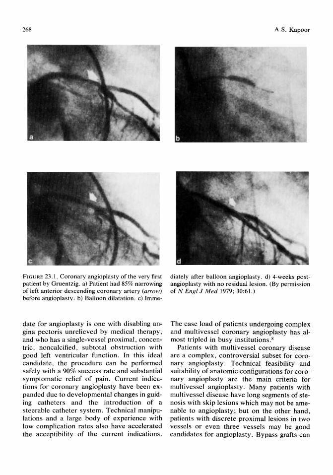
268
FIGURE 23.1. Coronary angiopiasty of the very first patient by Gruentzig. a) Patient had 85% narrowing of left anterior descending coronary artery (arrow) before angioplasty. b) Balloon dilatation. c) Imme-
date for angioplasty is one with disabling angina pectoris unrelieved by medical therapy, and who has a single-vessel proximal, concentric, noncalcified, subtotal obstruction with good left ventricular function. In this ideal candidate, the procedure can be performed safely with a 90% success rate and substantial symptomatic relief of pain. Current indications for coronary angioplasty have been expanded due to developmental changes in guiding catheters and the introduction of a steerable catheter system. Technical manipulations and a large body of experience with low complication rates also have accelerated the acceptibility of the current indications.
A.S. Kapoor
diately after balloon angioplasty. d) 4-weeks postangioplasty with no residual lesion. (By permission of N Engl J Med 1979; 30:61.)
The case load of patients undergoing complex and multivessel coronary angioplasty has almost tripled in busy institutions. 8
Patients with multi vessel coronary disease are a complex, controversial subset for coronary angioplasty. Technical feasibility and suitability of anatomic configurations for coronary angioplasty are the main criteria for multivessel angioplasty. Many patients with multivessel disease have long segments of stenosis with skip lesions which may not be amenable to angioplasty; but on the other hand, patients with discrete proximal lesions in two vessels or even three vessels may be good candidates for angioplasty. Bypass grafts can
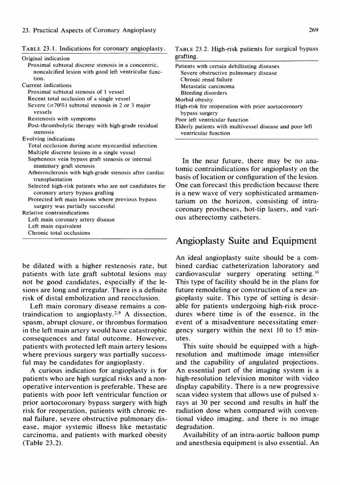
23. Practical Aspects of Coronary Angioplasty
TABLE 23.1. Indications for coronary angioplasty.
Original indication Proximal subtotal discrete stenosis in a concentric,
noncalcified lesion with good left ventricular function.
Current indications Proximal subtotal stenosis of 1 vessel Recent total occlusion of a single vessel Severe (:2:70%) subtotal stenosis in 2 or 3 major
vessels Restenosis with symptoms Post-thrombolytic therapy with high-grade residual
stenosis Evolving indications
Total occlusion during acute myocardial infarction Multiple discrete lesions in a single vessel Saphenous vein bypass graft stenosis or internal
mammary graft stenosis Atherosclerosis with high-grade stenosis after cardiac
transplantation Selected high-risk patients who are not candidates for
coronary artery bypass grafting Protected left main lesions where previous bypass
surgery was partially successful Relative contraindications
Left main coronary artery disease Left main equivalent Chronic total occlusions
be dilated with a higher restenosis rate, but patients with late graft subtotal lesions may not be good candidates, especially if the lesions are long and irregular. There is a definite risk of distal embolization and reocclusion.
Left main coronary disease remains a contraindication to angioplasty. 2,9 A dissection, spasm, abrupt closure, or thrombus formation in the left main artery would have catastrophic consequences and fatal outcome. However, patients with protected left main artery lesions where previous surgery was partially successful may be candidates for angioplasty.
A curious indication for angioplasty is for patients who are high surgical risks and a nonoperative intervention is preferable. These are patients with poor left ventricular function or prior aortocoronary bypass surgery with high risk for reoperation, patients with chronic renal failure, severe obstructive pulmonary disease, major systemic illness like metastatic carcinoma, and patients with marked obesity (Table 23.2).
269
TABLE 23.2, High-risk patients for surgical bypass grafting,
Patients with certain debilitating diseases Severe obstructive pulmonary disease Chronic renal failure Metastatic carcinoma Bleeding disorders
Morbid obesity High-risk for reoperation with prior aortocoronary
bypass surgery Poor left ventricular function Elderly patients with multivessel disease and poor left
ventricular function
In the near future, there may be no anatomic contraindications for angioplasty on the basis of location or configuration of the lesion. One can forecast this prediction because there is a new wave of very sophisticated armamentarium on the horizon, consisting of intracoronary prostheses, hot-tip lasers, and various atherectomy catheters.
Angioplasty Suite and Equipment
An ideal angioplasty suite should be a combined cardiac catheterization laboratory and cardiovascular surgery operating setting. 10
This type of facility should be in the plans for future remodeling or construction of a new angioplasty suite. This type of setting is desirable for patients undergoing high-risk procedures where time is of the essence, in the event of a misadventure necessitating emergency surgery within the next 10 to 15 minutes.
This suite should be equipped with a highresolution and multimode image intensifier and the capability of angulated projections. An essential part of the imaging system is a high-resolution television monitor with video display capability. There is a new progressive scan video system that allows use of pulsed xrays at 30 per second and results in half the radiation dose when compared with conventional video imaging, and there is no image degradation.
Availability of an intra-aortic balloon pump and anesthesia equipment is also essential. An
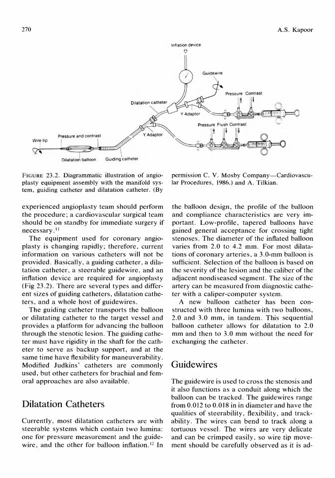
270
Wire tip
9'i
FIGURE 23.2. Diagrammatic illustration of angioplasty equipment assembly with the manifold system, guiding catheter and dilatation catheter. (By
experienced angioplasty team should perform the procedure; a cardiovascular surgical team should be on standby for immediate surgery if necessary. II
The equipment used for coronary angioplasty is changing rapidly; therefore, current information on various catheters will not be provided. Basically, a guiding catheter, a dilatation catheter, a steerable guidewire, and an inflation device are required for angioplasty (Fig 23.2). There are several types and different sizes of guiding catheters, dilatation catheters, and a whole host of guidewires.
The guiding catheter transports the balloon or dilatating catheter to the target vessel and provides a platform for advancing the balloon through the stenotic lesion. The guiding catheter must have rigidity in the shaft for the catheter to serve as backup support, and at the same time have flexibility for maneuverability. Modified Judkins' catheters are commonly used, but other catheters for brachial and femoral approaches are also available.
Dilatation Catheters
Currently, most dilatation catheters are with steerable systems which contain two lumina: one for pressure measurement and the guidewire, and the other for balloon inflation.12 In
A.S. Kapoor
Inflat Ion dpvice
o
permission C. V. Mosby Company-Cardiovascular Procedures, 1986.) and A. Tilkian.
the balloon design, the profile of the balloon and compliance characteristics are very important. Low-profile, tapered balloons have gained general acceptance for crossing tight stenoses. The diameter of the inflated balloon varies from 2.0 to 4.2 mm. For most dilatations of coronary arteries, a 3.0-mm balloon is sufficient. Selection of the balloon is based on the severity of the lesion and the caliber of the adjacent nondiseased segment. The size of the artery can be measured from diagnostic catheter with a caliper-computer system.
A new balloon catheter has been constructed with three lumina with two balloons, 2.0 and 3.0 mm, in tandem. This sequential balloon catheter allows for dilatation to 2.0 mm and then to 3.0 mm without the need for exchanging the catheter.
Guidewires
The guide wire is used to cross the stenosis and it also functions as a conduit along which the balloon can be tracked. The guidewires range from 0.012 to 0.018 in in diameter and have the qualities of steerability, flexibility, and trackability. The wires can bend to track along a tortuous vessel. The wires are very delicate and can be crimped easily, so wire tip movement should be carefully observed as it is ad-
23. Practical Aspects of Coronary Angioplasty
vanced. One may have to form bends on the tip of the wire to steer the balloon into the desired arterial segment. In addition to high torque floppy wire, there is a 300-cm exchange wire and a new extendible guidewire. One should check on dilatation catheter and guidewire compatibility, but 0.014-in wire is frequently used.
A controlled wire strategy goes a long way for wiring the vessel and the eventual success of the procedure. There are several mechanical inflation devices with pressure gauges to quantify the pressure applied to the syringe and the balloon.
Coronary Angioplasty Strategy
Angioplasty can be performed with equal efficacy with either a femoral or brachial approach. The operator should select the approach he is most familiar with. Before selecting the equipment, it is important to review the cineangiogram with careful analysis of the area of stenosis like: the angiographic estimation of the severity, morphology, length of the lesion, and proximity of side branches. One has to pay attention to the aortic root and origin of the coronary artery and diameter of the nondiseased segment of the artery to be dilated for proper selection of guiding and balloon catheter and the guidewire.
The need for pacing during angioplasty is not that frequent, but a venous access should be available for emergency pacing. Some operators use a right ventricular pacemaker routinely in dominant right coronary artery angioplasties. A trans arterial external pacemaker can also be used. The pacemaker can provide a reference point to mark the area of stenosis. In an acute emergency situation, the angioplasty guidewire could be used for emergency coronary pacing.
Preangioplasty Protocol
Patient preparation for angioplasty is similar to that for cardiac surgery, with an antiseptic soap shower, cross-matching of blood, and proscription of oral intake after midnight. All
271
necessary medications are continued. Betablockers may be stopped. A typical protocol is shown in Table 23.3. Patients are given enteric coated aspirin, 300 mg, and dipyridamole, 75 mg, three times a day, preferably 48 hours before the procedure. There are different protocols for giving aspirin or persantine. It is important to detail the patient on the nature and risks of the procedure, and the possibility of emergency bypass coronary artery grafting in the event of acute reclosure or other major complications.
The Procedure
Before the start of the procedure, check the angioplasty setup by your trained technician. Meticulous care should be taken for the preparation of the balloon. The solution used to fill the balloon should be a 50 : 50 mixture of contrast medium and normal saline. All the air should be evacuated from the balloon, and the balloon should be tested by inflating the balloon to 4 to 5 atm pressure. The guide wire is back-loaded into the dilatation catheter, protecting the wire tip. The stiff end of the guide-
TABLE 23.3. Preangioplasty orders.
Consent forms signed for coronary angioplasty and for emergency surgical bypass, if necessary
NPO, except for medications after midnight Groin preparation, antiseptic soap shower Group and cross-match for 3 U of blood Chest x-ray, ECG Continue Ecotrin 300 mg orally 3 times/day and Persan
tine 75 mg orally 3 times/day Preoperative medication on call
Demerol Atropine Valium Cimetidine Others
Laboratory data on electrolytes, creatinine, PT, PTT, and CBC
Send the patient on a stretcher when called by the catheterization laboratory staff
Have the patient void before transporting Start intravenous of DsW TKO/ mllhr with
angiocatheter
NPO = nothing by mouth; ECG electrocardiogram; PT = prothrombin time; PTT = partial thromboplastin time; CBC = complete blood count; TKO = to keep open.
272
wire is inserted through the Y-connector, and the dilatation catheter hub is locked on the rotator. The entire system should be air free and flushed with contrast solution. If the brachial approach is used, the brachial artery is exposed by cutdown and the guiding catheter is advanced after a horizontal arteriotomy.
The femoral artery is cannulated using standard Seldinger's percutaneous technique and a 9-Fr sheath is placed. The guiding catheter is advanced through this sheath. At this time, 10 mg sublingual nifedipine is given and a bolus of 10,000 U of heparin is administered. A venous sheath should be placed if there is need for a standby pacemaker. Angioplasty is performed to review the area of the lesion to be dilated, and the best view is selected for display on the video monitor.
After the coronary ostium is intubated, the dilating system with the wire is advanced into the coronary artery. The wire tip is kept intraluminal and buckling is to be avoided. The position and direction of the wire should be carefully manipulated by rotation and advancement of the wire, and this can be checked by test injections of the contrast medium through the guiding catheter or the dilating catheter.
Pressure gradients are measured. Many centers do not use gradients. Actually with some catheter systems, one cannot measure gradients. If gradients are measured, initial proximal and distal gradients should be recorded and then the final gradients at the end of the dilatation.
As soon as the distal vessel is wired, the balloon should be placed at the site of stenosis in rapid sequence. The first balloon inflation is carried out at 2 to 4 atm for 15 to 45 seconds. It is important to use low pressure inflations initially and ultimately increase the pressure to 1 atm higher than the pressure required for full balloon expansion. 14,15 The maximum balloon pressure and duration of inflation vary from lesion to lesion and also at different institutions. Meier et aJ16 used higher pressures, and they found a decreased rate of restenosis and lower trans-stenotic gradients. After balloon deflation, the pressure gradient is measured. Subsequent balloon inflations are carried out to remodel the area of stenosis with longer
A.S. Kapoor
TABLE 23.4. Postangioplasty orders.
Clear liquids then CCU diet Check blood pressure, distal pulses, and catheter site
for bleeding every 15 min x 4, then every 1 hr x 4 Continue intravenous of D5W at mllhr x ___ then change to heparin lock for 24 hrs
Resume preangioplasty medications Repeat ECG and ECG with any episode of chest pain Laboratory values: CBC, creatinine, PTT, blood sugar New medication orders
Calcium-channel blocker Procardia __ _ Diltiazem __ _
Start heparine drip at U Ihr. Check PTT 6 hrs later and inform on-call MD ( ) if less 2 times or greater than 4 times
Stop heparin drip on at __ _ Notify MD on call to remove sheath at __ _
Patient to remain in bed for 6 hrs after sheath removal Inform the primary operator of chest pain not relieved
by nitroglycerin or new ECG changes Schedule for treadmill test on __ _
CCU = critical care unit; ECG = electrocardiogram; CBC = complete blood count; PTT = partial thromboplastin time.
inflations. One may require several inflations to achieve a pressure gradient of less than 20 mm Hg. The balloon catheter is withdrawn into the guiding catheter and cineangiography is repeated to evaluate the results of angioplasty. The wire is left in place for 3 to 5 minutes at the end of the procedure. The wire and balloon are removed and postangioplasty angiograms are performed.
The sheath is sutured in place and heparin is continued for 24 hours. When a brachial approach is used, the artery is carefully sutured after removal of the catheter. Heparin drip is continued. Postangioplasty orders are written as shown in Table 23.4. Patients are continued on a calcium-channel blocker, aspirin, and persantine. A treadmill test can be performed 48 hours after the procedure. Patients are followed-up at 3 and 6 months for thallium stress test or electrocardiographic stress test.
Angioplasty Strategy for Right Coronary Artery
The selection of a guiding catheter will be based on superior or low takeoff of the right coronary artery. A Judkins' style guiding cath-
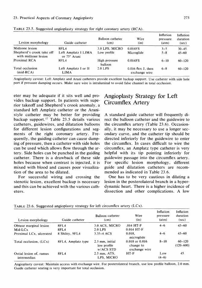
23. Practical Aspects of Coronary Angioplasty 273
TABLE 23.5. Suggested angioplasty strategy for right coronary artery (RCA).
Inflation Inflation Balloon catheter Wire pressure duration
Lesion morphology Guide catheter (em) (in) (atm) (sec)
Midzone lesion 8FL4 3.0 LPS, MICRO 0.014FS 3-5 30-45 Shepherd's crook take off Left Amplatz I LIMA Low profile Microglide 5-8 45-60
with midzone lesion or 75° Arani Proximal RCA 8FL4 High pressure 0.014FS 6-10 60-120
balloon Total occlusion Left Amplatz I or II 2.0 LPS 0.016 flex J, then 6-8 60-120
(mid-RCA) LIMA exchange wire
Angioplasty caveat: Left Amplatz and Arani catheters provide excellent backup support. Use catheter with side hole port if pressure damping occurs. Make sure wire is intraluminal to avoid false channel in total occlusion.
eter may be adequate if it sits well and provides backup support. In patients with superior takeoff and Shepherd's crook anomaly, a standard left Amplatz catheter or the Aranistyle catheter may be better for providing backup support.17 Table 23.5 details various catheters, guidewires, and dilatation balloons for different lesion configurations and segments of the right coronary artery. Frequently, the guiding catheter can cause damping of pressure, then a catheter with side holes can be used which allows flow through the artery. Side holes can be punched in the guiding catheter. There is a drawback of these side holes because when contrast is injected, it is mixed with blood and causes poor visualization of the area to be dilated.
For successful wiring and crossing the stenotic lesion, excellent backup is necessary and this can be achieved with the various catheters.
Angioplasty Strategy for Left Circumflex Artery
A standard guide catheter will frequently direct the balloon catheter and the guidewire to the circumflex artery (Table 23.6). Occasionally, it may be necessary to use a longer secondary curve, and the catheter tip should be directed inferiorly for the guide wire to enter the circumflex. In cases difficult to wire the circumflex, an Amplatz type catheter is very helpful with its tip pointing inferiorly for guide wire passage into the circumflex artery. For specific lesion morphology, different guide and dilatation catheters are recommended as indicated in Table 23.6.
One has to be very cautious in dilating a lesion in the posterolateral branch in a hyperdynamic heart. There is a higher incidence of dissection and other complications. A low
TABLE 23.6. Suggested angioplasty strategy for left circumflex artery (LCx).
Inflation Inflation Balloon catheter Wire pressure duration
Lesion morphology Guide catheter (em) (in) (atm) (sec)
Obtuse marginal lesion 8FL4 3.0 ACS, MICRO .014 HT-F 4-6 45-60 Mid-LCx 8FL4 2.0 LPS 0.014 HT-F Proximal LCx, ulcerated 8 Shiley, 9FL4 3.35-4 ACS 0.018, 4-6 45-60
microglide Total occlusion, (LCx) 8FL4, Amplatz type 2.5 mm, initial 0.018 or 0.016 8-10 60-120
low profile change to 020-600) w/ACS STD exchange wire
Ostial lesion of, ramus 8FL4 2.5 mm, ATS, HT-F Low 45 intermedius LPS, MICRO (4-6)
Angioplasty caveat: Maintain access with exchange wire. For posterolateral branch, use low profile balloon, 2.0 mm. Guide catheter seating is very important for total occlusion.

274
profile balloon is recommended. The recently introduced balloon-on-a-wire with an ultrathin balloon, introduced by USCI, appears to be an efficacious dilatation system. In a left proximal ulcerated, complex left circumflex lesion, a high-flow guide catheter like an 8 Shiley or a 9-Fr Judkins' type catheter with a large balloon catheter may be helpful for optimal visualization. A high torque floppy guidewire that can be steered could be used for advancing across the target stenosis.
To avoid major dissection on a tortuous segment of the artery to be dilated, it is advisable to use a smaller balloon than the caliber of the vessel to be dilated.
Angioplasty Strategy for Left Anterior Descending Artery
Usually, an 8FL4 curve guide catheter with a 3.0 balloon catheter and 0.014-in flexible steerable guidewire will be adequate for a single proximal discrete classical angioplasty of a left anterior descending lesion (Table 23.7). Lower inflation pressures of 4 to 6 atm may be sufficient to have a successful result. The guide catheter should cannulate the left main artery coaxially for providing backup support and for advancing the balloon catheter through the stenotic lesion. Occasionally, the guidewire will repeatedly enter the circumflex artery and in these cases, a shorter secondary
A.S. Kapoor
curve and the tip of the catheter should be rotated in a counterclockwise rotation to point in a superior position of the left anterior descending artery.
Frequently encountered is a very stenotic calcified fibrotic tortuous segment of artery difficult to cross despite excellent backup power. In this setting, a tapping with backand-forth successive motion of the balloon catheter maneuver can be attempted. 19 It is extremely important to have coaxial engagement at the coronary takeoff for better stability and transmission of power at the balloon lesion interface.
Many centers are very particular about measuring trans-stenotic pressure gradients to assess the severity of a stenosis and also use it as an indicator of the immediate and long-term results. 18 But recently, more and more centers are not measuring trans-stenotic pressure gradients, mainly because they are cumbersome to perform and there is artifactual overestimation of the pressure and at other times significant pressure damping.
Angioplasty Strategy for Bifurcation Lesions
Bifurcation lesions present a special problem because of side branch occlusion from a "snowplow" effect of angioplasty. 20 There is a 15% risk of side branch occlusion if the side
TABLE 23.7. Suggested angioplasty strategy for left anterior descending (LAD).
Inflation Inflation Balloon catheter Wire pressure duration
Lesion morphology Guide catheter (cm) (in) (atm) (sec)
Classical proximal 8FL4, or 3.0 mm, or 2.5 0.014FS flexible, 4-6 45 discrete LAD lesion 8FL3.5 LPS steerable
Bifurcating lesion; 8FL4 3.0 0.014FS 5-7 45 LAD-diagonal 8F4 2.5 LPS 0.012 (300 cm) 45
Two wire technique (exchange wire)
Diffuse distal LAD 8FL4 Low profile, 2.0 Microglide 4-6 30-45 6
100% LAD 8FL4 Standard, 3.0 0.018 HT-F, or 6-8 120-360 LPS 0.016 Flex J
Angioplasty caveat: USCI balloon on a wire appears to be a good dilatation system for diffuse distal disease. For a bifuraction lesion, dilate the important vessel supplying the largest amount of myocardium first and repeat it on withdrawal for therapeutic remodelling.

23. Practical Aspects of Coronary Angioplasty
branch has a pre-existing ostial lesion. 21 To deal with this complication, George et aFo introduced "kissing balloon angioplasty. " Since that time there have been variations in the technique of protecting the side branch by using a single guiding catheter and a double guidewire technique,22,23 and recently by using the kissing wire monorail balloon technique. 24
With the kissing balloon angioplasty (Fig 23.3), two guiding catheters are employed, using bilateral femoral approach or femoral and brachial approach.20 uscr 0.014 flexible steerable guidewire is advanced in the technically most difficult vessel to be dilated, and after wiring the distal vessel the guiding catheter is withdrawn from the coronary ostium. Then the second guiding catheter is advanced into the coronary ostium and the second steerable guidewire is positioned into the distal segment
FIGURE 23.3. Kissing balloon technique. Two guiding catheters are positioned in the coronary ostium. Guiding catheter A is engaged and its dilatation catheter inflated , and guide B is positioned below the ostium. Balloons A and B can be inflated simultaneously or sequentially. (Used with permission of Cathet and Cardiovasc Diagn 1986; 12: 124-138.)
275
of the bifurcation lesion. Then the dilatation catheter of the major branch is advanced over the wire and across the stenosis and dilated. The dilatation catheter is withdrawn into the guiding catheter leaving the wire. The dilatation catheter of the other guiding catheter is advanced across the stenosis and dilated. Finally the dilatation catheter of the major branch is repositioned across the lesion and redilated at low pressure inflation for therapeutic remodelling. If there is angiographic distortion of the bifurcation lesion then simultaneous inflation of both dilatation catheters is performed at low pressure of 2 atm.
To avoid confusion, each angioplasty system should be separated by sterile clothing . It is also important to avoid wrapping around wires , and frequent contrast injections should be made in multiple views to visualize the position of the wires.
In the technique with single guiding catheter, double wires are introduced; one of the
Guidewire
Gu id ing Catheter
FIGURE 23.4. Kissing wire technique with single guide and 2 long exchange wires . (Used with permission of Cathet and Cardiovasc Diagn 1986; 12:124-138.)
276
wires is a long 300-cm guidewire, and a second identical guide wire (0.012-in, 300-cm) is advanced into the major segment of the vessel (Fig 23.4). A smaller balloon is used for the side branch (2.5 cm ACS balloon catheter) and a larger balloon for the main segment of the artery. Dilatations are performed as described. Contrast injections are used with the wires in place to assess the results (Fig 23.5).
The third technique uses a standard 8-Fr Judkins' guide catheter and two standard (0.014-in, 175-cm long) guidewires that are positioned across the respective bifurcation branches. Then a monorail balloon catheter (30-mm Schnieder Shiley) is advanced to dilate the side branch, withdrawn completely, and then reintroduced over the second wire to dilate the other bifurcation lesion.
The efficacy of these different techniques has not been tested by a large number of procedures. According to BonzeJ25 the monorail catheter was used in more than 200 consecutive patients with a success rate of 92% for critical stenosis and 87% for totally occluded arteries.
Multivessel Angioplasty Strategy
The primary objective of multi vessel angiopia sty is to achieve complete revascularization (Table 23.8). Hence, patient selection should be based on feasibility of performing angioplasty on all lesions that are considered
TABLE 23.8. Strategy for multivessel disease.
Goal: complete revascularization First dilate
Lesion most difficult technically or The lesion with greatest myocardium at risk or Complex lesion or CUlprit lesion
For tandem lesions, cross all lesions with dilatation catheter and back dilate from distal to proximal lesion
In unstable patients, dilate the culprit lesion and perform staged angioplasty of other lesions
Equipment for each lesion should be preselected based on anatomy of the vessel
May need mUltiple exchanges of dilatation balloons and wires
A.S. Kapoor
surgically bypassable. A dilatation strategy should take into account the most stenotic artery supplying the largest zone of ischemic myocardium and technically difficult vessels to dilate. 26 Increased risk is introduced because of the need for multiple exchange of catheters and wires and multiple lesions to be dilated.
Myler and co-workers27 performed multivessel coronary angioplasty in 494 consecutive patients with a clinical success of 95%, emergency surgery in 2.8%, myocardial infarction in 3.0%, and hospital death in 0.4%. This is a high level of success with a relatively low complication rate. The restenosis rate appears to be similar as in patients with singlevessel disease, but long-term studies are needed for complete answers.
Angioplasty Strategy for Saphenous Vein Graft and Internal Mannnnary Artery
Bypass grafting by the use of saphenous veins or internal mammary arteries has a significant incidence of recurrence over a 10-year span. Significant occlusive disease can occur at the proximal or distal anastomotic site or in the body of the graft in about 30% of patients. 28
Guiding catheter selection will be dependent on the configuration of the graft and shape of the ascending aorta. The most commonly used femoral guiding catheters are the right Judkins' type catheter and the left internal mammary guiding catheter (Table 23.9). The balloon catheter is selected to be identical to the caliber of the saphenous vein or the internal mammary artery graft for proximal and mid-lesions in the graft. For a distal anastomotic lesion, the size of the balloon should match the diameter of the native artery. One study suggests that in bypass grafts, the dilatation catheter should be slightly larger than the diameter of the vessel with a balloon/graft ratio of 1.1 : I, and is associated with the lowest residual percent stenosis and lower incidence of restenosis. 29
The guidewire should have a very flexible
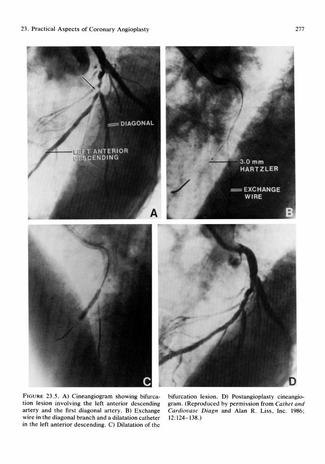
23. Practical Aspects of Coronary Angioplasty
FIGURE 23.5 . A) Cineangiogram showing bifurcation lesion involving the left anterior descending artery and the first diagonal artery. B) Exchange wire in the diagonal branch and a dilatation catheter in the left anterior descending. C) Dilatation of the
277
bifurcation lesion. D) Postangioplasty cineangiogram. (Reproduced by permission from Cather and Cardiouasc Diagn and Alan R. Liss , Inc. 1986; 12: 124-138.)

278 A.S. Kapoor
TABLE 23.9. Suggested angioplasty strategy for saphenous vein graft (SVG) and internal mammary artery (IMA) to left anterior descending artery (LAD).
Inflation Inflation Balloon catheter Guide wire pressure duration
Lesion morphology Guide catheter (em) (in) (atm) (sec)
SVG-LAD 8FR4, LIMA 2.5ACS, stan- 0.014 RTF, 0.018 RTF 7 60 left Amplatz I dard
IMA to LAD LIMA 2.5 LPS 0.014 3-6 45-60
Angioplasty caveat: Balloon size for bypass grafts for proximal and shaft lesion to be slightly larger than the exact size of the vessel. Extra care taken to cannulate IMA. Keep wire intraluminal.
soft tip to avoid embolization of a friable lesion. A very flexible Wholey wire can be used for a right internal mammary graft.
Great care should be taken to cannulate the internal mammary artery. It is prone to spasm and dissection.
Occasionally, the dilatation catheter can be advanced beyond the distal anastomotic site for angioplasty of a distal lesion in the native artery.
A success rate of 85% has been achieved in one study with emergency bypass surgery in 1.2%, myocardial infarction in 3.5%, and no hospital mortality. 30
Angioplasty Strategy in Acute Myocardial Infarction
The primary objective of coronary angioplasty in the setting of acute myocardial infarction is limitation of myocardial infarct size and restoration of flow with improvement in regional and global left ventricular function. Several studies have shown that acute coronary angiopia sty can be performed effectively with reperfusion rates of 84% to 100%.31-33
Patient selection for acute angioplasty is under active investigation. Patient selection should be based on the duration of chest pain because tipIe is of the essence in salvaging myocardium, and a cutoff period of less than 6 hours duration seems appropriate for direct acute coronary angioplasty. The other strategy would be to achieve reperfusion by intravenous thrombolytic agents followed by an elective angioplasty in selected patients. The
results of thrombolysis and angioplasty in myocardial infarction study groups indicate that in patients with successful thrombolysis and suitable coronary artery anatomy, immediate angioplasty offers no distinct advantage over delayed elective angioplasty. 34 The subset of patients who will benefit by immediate angioplasty will be patients who get an intervention within 4 hours, have ischemic pain at the time of the intervention, and have collaterals present.
The procedure for acute angioplasty is similar for routine angioplasty with some additional precautions and preparation. Both groins are prepared, one for a possible intraaortic balloon pump if necessary, and the other for the angioplasty catheter. A diagnosti<; angiogram of the noninvolved artery is performed to determine the presence or absence of collaterals. Then an angiogram of the infarct-related coronary artery is performed in multiple views. A left ventriculogram is not crucial. Noninvasive serial evaluation of the left ventricle can be performed.
A low-profile balloon is used with 0.016-in flexible steerable guidewire, or a soft-tipped guidewire is used to approach the lesion. Slow circular motions of the wire tip with contrast injections will enable the wire to cross the complete obstruction. The balloon can be inflated at 2 to 4 atm for 20 to 30 seconds and progressively inflated to 6 atm for 2 to 5 minutes. The balloon catheter is withdrawn and repeat cineangiography is performed to assess the results. If there is in situ thrombus, intracoronary streptokinase or tissue plasminogen activator is administered as adjunctive thrombolysis.
23. Practical Aspects of Coronary Angioplasty
It is important that angioplasty of other lesions should not be attempted during dilatation of the infarct artery. Angioplasty of other lesions should be staged, or patients may be candidates for bypass surgery. It is also very important to maintain systemic anticoagulation for at least 3 to 5 days at which time oral aspirin and persantine can be given. Success of the procedure can be semiquantitated by using the TIMI grading system to describe the degree of ante grade blood flow in the infarct -related artery. 35 Grade 0 is no perfusion with no ante grade flow, and grade 3 is complete perfusion with antegrade flow into the bed distal to the obstruction. Rentrop et aP6 used collateral perfusion criteria on a grade of o to 3 describing no visible filling of any collateral channel to complete collateral filling of the vessel being dilated.
Coronary angioplasty is safe and effective for the treatment of acute myocardial infarction. With direct angioplasty, there is a lower initial success rate and a higher reocclusion rate.
Conclusion
In general, the angioplasty strategy is dictated by the lesion morphology, the complex and bifurcating lesion requiring a different selection of balloon catheters and guidewires, and special technical expertise. The size of the balloon and the caliber of the vessel to be dilated should be precisely measured.
Coronary angioplasty has evolved significantly in the areas of patient selection, equipment development, and technical success. The intervention-focused specialists have become very much ischemic- and lesion-oriented and in other areas of expertise, have developed a complex technical procedural milieu requiring frequent updating and specializing. There is an incredible proliferation of high technology for dealing with the ravages of atherothrombosis and atherosclerosis. The primary success rate has increased to greater than 90%, and the complication rate has significantly reduced to 2% to 3%. We have also witnessed the safe application of this proce-
279
dure to multivessel coronary artery disease. Angioplasters are frequently called upon to deal with surgically high-risk patients and failures of surgical revascularization. Alternate or combined technologies will be refined to deal with the complications of coronary angioplasty.
It seems that the anatomic and functional success of coronary angioplasty is at least maintained for 3 years. 38
References
1. Gruentzig AR, et al: Coronary transluminal angioplasty. Circulation 1977; 56(suppl 3):84.
2. Gruentzig AR, et al: Nonoperative dilatation of coronary artery stenosis: Percutaneous transluminal coronary angioplasty. N Engl J M ed 1979; 301 :61.
3. Mabin TA, et al: Long-term follow-up after percutaneous transluminal coronary angioplasty. Circulation 1983; 68: 111-197.
4. Kent KM, et al: Improved myocardial function during exercise after successful percutaneous transluminal coronary angioplasty. N Engl J Med 1982; 306:441-446.
5. Dotter CT, Judkins MD: Transluminal treatment of arteriosclerotic obstruction: Description of a new technique and a preliminary report of its application. Circulation 1964; 30:654.
6. Zarins CK, et al: Arterial disruption and remodeling following balloon dilatation. Surgery 1982; 92: 1086.
7. Casteneda-Zuniga WR, et al: The mechanism of balloon angioplasty. Radiology 1980; 135:565.
8. Clark DA (ed): Selection of patients for PTCA, in Coronary Angioplasty. New York, Alan R. Liss Inc, 1987, pp 43-49.
9. Stertzer SR, et al: Percutaneous transluminal coronary angioplasty in left main stem coronary stenosis: A five-year appraisal. Int J Cardiol 1985; 9:149.
10. McAuley BJ, et al: Coronary angioplasty of high-risk patients in a combined catheterization laboratory-operating room setting. Circulation 1985; 72(suppl 111-217):868.
11. Williams DO, et al: Guidelines for the performance of percutaneous transluminal coronary angioplasty. Circulation 1982; 66:693-694.
12. Simpson JB, et al: A new catheter system for coronary angioplasty. Am J Cardiol 1982; 49:1216.
13. Meier B, Rutishauser W: Coronary pacing dur-
280
ing percutaneous transluminal coronary angioplasty. Circulation 1985; 71:557-581.
14. Kaltenbach M, et al: Prolonged application of pressure in transluminal coronary angioplasty. Cathet Cardiouasc Diagn 1984; 10:213.
15. Hartzler GO: Percutaneous transluminal coronary angioplasty in multivessel disease. Cathet Cardiouasc Diagn 1983; 9:537.
16. Meier B, et al: Higher balloon dilatation pressure in coronary angioplasty. Am Heart J 1984; 107:619.
17. Hidalgo BO: Coronary angioplasty catheters and accessories, in Clark (ed): Coronary Angioplasty. New York, Alan R. Liss, Inc., 1987, pp 9-27.
18. Redd DCB, et al: The transstenotic pressure gradient trend as a predictor of acute complications after percutaneous transluminal coronary angioplasty. Circulation 1987; 76:792-801.
19. Carr ML: The use of the guiding catheter in coronary angioplasty. Cathet Cardiouasc Diagn 1986; 12: 189-197.
20. George BS, et al: Balloon angioplasty of coronary bifurcation lesion: The kissing balloon technique. Cathet Cardiouasc Diagn 1986; 12:124-138.
21. Meier B, et al: Risk of side branch occlusion during coronary angioplasty. Am J Cardiol 1984; 53:10-14.
22. Meier B: Kissing balloon angioplasty on lesionassociated branches. Am J Cardiol 1984; 53:918-920.
23. Oesterle SN, et al: Angioplasty at coronary bifurcations: Single-guide, two-wire technique. Cathet Cardiouasc Diagn 1986; 12:57-63.
24. Finci L, Meier B, Divernois J: Percutaneous transluminal coronary angioplasty of a bifurcation narrowing using the kissing wire monorail balloon technique. Am J Cardiol1987; 60:375-376.
25. Bonzel R: Monorail balloon catheter for coronary angioplasty. Update in diagnostic and interventional techniques in cardiovascular medicine. Munich, Germany, 1987.
26. Roubin GS, et al: Angioplasty in multivessel coronary artery disease: Patient selection and
A.S. Kapoor
dilatation strategy. J Am Coil Cardiol 1985; 5:440-445.
27. Myler RK, et al: MUltiple vessel coronary angioplasty. Cathet Cardiouasc Diagn 1987; 13:1-15.
28. Campeau L, et al: The relationship of risk factors to the development of atherosclerosis in saphenous vein grafts and the progression of disease in the native circulation: A study 10 years after aortocoronary bypass surgery. N Engl J Med 1984; 311:1329-1332.
29. Clark DA (ed): Complex PTCA: Bypass Grafts in Coronary Angioplasty. New York, Alan R. Liss, Inc, 1987, pp 75-77.
30. Cote G, et al: Percutaneous transluminal angioplasty of stenotic coronary artery bypass grafts: 5 years experience. J Am Coil Cardiol 1987; 9:8-17.
31. Hartzler GO, et al: Percutaneous transluminal coronary angioplasty: Application for acute myocardial infarction. Am J Cardiol 1984; 53: 117C.
32. Kalbfleisch J, et al: A randomized trial of immediate coronary angioplasty following intracoronary thrombolysis in acute myocardial infarction. J Am Coil Cardiol 1984; 3:576.
33. Schwarz F, et al: Thrombolysis in acute myocardial infarction: Improved results by use of PTCA. Circulation 1983; 68(suppl 111): 140.
34. Topol EJ, et al: A randomized trial of immediate versus delayed elective angioplasty after intravenous tissue plasminogen activator in acute myocardial infarction. N Engl J Med 1987; 317:581-588.
35. TlMI Study Group: The Thrombolysis in Myocardial Infarction (TlMI) Trial. N Engl J Med 1985; 312:932.
36. Rentrop KP, et al: Changes in collateral channel filling immediately after controlled coronary artery occlusion by an angioplasty balloon in human subjects. JAm Coli Cardiol1987; 5:587.
37. Libow M, Gruentzig AR, Greene L: Percutaneous transluminal coronary angioplasty. Curr Probl C a rdio I 1985; 1: 3.
38. Rosing DR, et al: Three-year anatomic, functional and clinical follow-up after successful PTCA. J Am Coli Cardiol1987; 9:1-7.
24 Complex Coronary Angioplasty: The Outcome and Long-term Effect of Angioplasty in Multivessel Coronary Disease and Multiple Lesion Angioplasty Gerald Dorros, Ruben F. Lewin, and Lynne Mathiak
Introduction
Percutaneous trans luminal coronary angioplasty (PTCA) I is an accepted treatment for selected patients with isolated single-vessel obstructive coronary disease. 2-4 The use of PTCA has been successful in multi vessel coronary artery disease (MVD) patients. 5- 7 The demonstrated safety and efficacy of angioplasty in patients with isolated proximal coronary stenosis(es) permitted its evaluation in patients with extensive coronary disease. 7- 14
However, the continuous merging of words, which only ensure the reader, has led us to define the two major subgroups most often discussed so as to clarify what has happened. The acute outcome and follow-up of PTCA in MVD patients who underwent single- or multiple-lesion angioplasty (group I), and in patients (with single or multivessel coronary disease) who underwent multiple lesion angioplasty (MLA; group II) is discussed.
Methods
Patient Selection
Data were derived from two overlapping cohorts of patients: 752 consecutive MVD patients who underwent PTCA between February 1979 and September 1986 (91 months), and 428 patients in whom MLA was performed between February 1979 and April 1986 (86
months). All data were collected prospectively. All PTCA (MVD and MLA) patients were reported, including those with left main coronary disease [with or without prior bypass surgery (CABG)], mUltiple prior CABG procedures, multiple coronary artery occlusions, diffuse inoperable (refused by surgeons) coronary artery disease, cardiogenic shock, or severe, concomitant medical illness(es).
Patients had either significant angina pectoris or, with no or minimal angina pectoris, 15
had myocardial ischemia documented by noninvasive techniques. Subsequently, coronary cine arteriography demonstrated the sites of the obstructive coronary lesions.
Definitions
Multivessel coronary disease was defined as a 2:70% diameter stenosis in at least two major epicardial vessels in a right dominant system, and a 2:70% stenosis in at least the proximal left circumflex artery in a left dominant system. The coronary vessel segments were designated according to the CASS nomenclature. 16
Descriptions of angioplasty techniques, 17 as well as a discussion of their complications l8 ,19
have been published. A single dilatation (SD) procedure was de
fined as an attempted dilatation of solely the "culprit lesion." A mUltiple dilatation (MD) procedure was defined by the dilatation of two
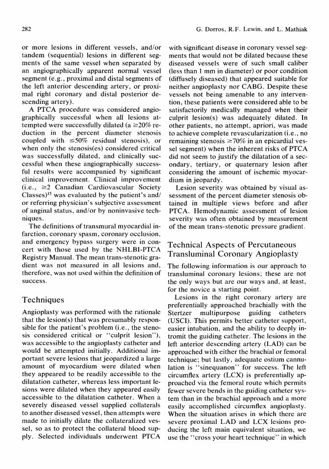
282
or more lesions in different vessels, and/or tandem (sequential) lesions in different segments of the same vessel when separated by an angiographically apparent normal vessel segment (e.g., proximal and distal segments of the left anterior descending artery, or proximal right coronary and distal posterior descending artery).
A PTCA procedure was considered angiographically successful when all lesions attempted were successfully dilated (a 2:20% reduction in the percent diameter stenosis coupled with ::;50% residual stenosis), or when only the stenosis(es) considered critical was successfully dilated, and clinically successful when these angiographically successful results were accompanied by significant clinical improvement. Clinical improvement (i.e., 2:2 Canadian Cardiovascular Society Classes)15 was evaluated by the patient's and/ or referring physician's subjective assessment of anginal status, and/or by noninvasive techniques.
The definitions of transmural myocardial infarction, coronary spasm, coronary occlusion, and emergency bypass surgery were in concert with those used by the NHLBI-PTCA Registry Manual. The mean trans-stenotic gradient was not measured in all lesions and, therefore, was not used within the definition of success.
Techniques
Angioplasty was performed with the rationale that the lesion(s) that was presumably responsible for the patient's problem (i.e., the stenosis considered critical or "culprit lesion"), was accessible to the angioplasty catheter and would be attempted initially. Additional important severe lesions that jeopardized a large amount of myocardium were dilated when they appeared to be readily accessible to the dilatation catheter, whereas less important lesions were dilated when they appeared easily accessible to the dilatation catheter. When a severely diseased vessel supplied collaterals to another diseased vessel, then attempts were made to initially dilate the collateralized vessel, so as to protect the collateral blood supply. Selected individuals underwent PTCA
G. Dorros, R.F. Lewin, and L. Mathiak
with significant disease in coronary vessel segments that would not be dilated because these diseased vessels were of such small caliber (less than 1 mm in diameter) or poor condition (diffusely diseased) that appeared suitable for neither angioplasty nor CABG. Despite these vessels not being amenable to any intervention, these patients were considered able to be satisfactorily medically managed when their culprit lesion(s) was adequately dilated. In other patients, no attempt, apriori, was made to achieve complete revascularization (i.e., no remaining stenosis 2:70% in an epicardial vessel segment) when the inherent risks of PTCA did not seem to justify the dilatation of a secondary, tertiary, or quaternary lesion after considering the amount of ischemic myocardium in jeopardy.
Lesion severity was obtained by visual assessment of the percent diameter stenosis obtained in multiple views before and after PTCA. Hemodynamic assessment of lesion severity was often obtained by measurement of the mean trans-stenotic pressure gradient.
Technical Aspects of Percutaneous Transluminal Coronary Angioplasty
The following information is our approach to transluminal coronary lesions; these are not the only ways but are our ways and, at least, for the novice a starting point.
Lesions in the right coronary artery are preferentially approached brachia\ly with the Stertzer multipurpose guiding catheters (USCI). This permits better catheter support, easier intubation, and the ability to deeply intromit the guiding catheter. The lesions in the left anterior descending artery (LAD) can be approached with either the brachial or femoral technique; but lastly, adequate ostium cannulation is "sinequanon" for success. The left circumflex artery (LCX) is preferentially approached via the femoral route which permits fewer severe bends in the guiding catheter system than in the brachial approach and a more easily accomplished circumflex angioplasty. When the situation arises in which there are severe proximal LAD and LCX lesions producing the left main equivalent situation, we use the" cross your heart technique" in which
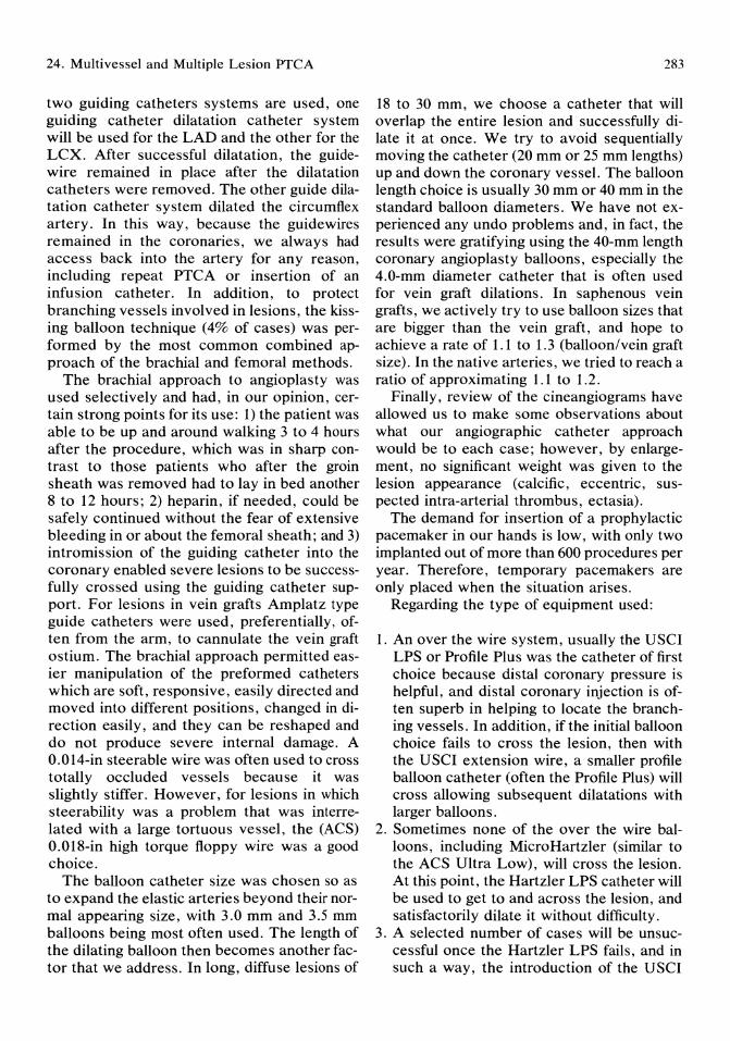
24. Multivessel and MUltiple Lesion PTCA
two guiding catheters systems are used, one guiding catheter dilatation catheter system will be used for the LAD and the other for the LCX. After successful dilatation, the guidewire remained in place after the dilatation catheters were removed. The other guide dilatation catheter system dilated the circumflex artery. In this way, because the guidewires remained in the coronaries, we always had access back into the artery for any reason, including repeat PTCA or insertion of an infusion catheter. In addition, to protect branching vessels involved in lesions, the kissing balloon technique (4% of cases) was performed by the most common combined approach of the brachial and femoral methods.
The brachial approach to angioplasty was used selectively and had, in our opinion, certain strong points for its use: 1) the patient was able to be up and around walking 3 to 4 hours after the procedure, which was in sharp contrast to those patients who after the groin sheath was removed had to lay in bed another 8 to 12 hours; 2) heparin, if needed, could be safely continued without the fear of extensive bleeding in or about the femoral sheath; and 3) intromission of the guiding catheter into the coronary enabled severe lesions to be successfully crossed using the guiding catheter support. For lesions in vein grafts Amplatz type guide catheters were used, preferentially, often from the arm, to cannulate the vein graft ostium. The brachial approach permitted easier manipUlation of the preformed catheters which are soft, responsive, easily directed and moved into different positions, changed in direction easily, and they can be reshaped and do not produce severe internal damage. A 0.014-in steerable wire was often used to cross totally occluded vessels because it was slightly stiffer. However, for lesions in which steerability was a problem that was interrelated with a large tortuous vessel, the (ACS) 0.018-in high torque floppy wire was a good choice.
The balloon catheter size was chosen so as to expand the elastic arteries beyond their normal appearing size, with 3.0 mm and 3.5 mm balloons being most often used. The length of the dilating balloon then becomes another factor that we address. In long, diffuse lesions of
283
18 to 30 mm, we choose a catheter that will overlap the entire lesion and successfully dilate it at once. We try to avoid sequentially moving the catheter (20 mm or 25 mm lengths) up and down the coronary vessel. The balloon length choice is usually 30 mm or 40 mm in the standard balloon diameters. We have not experienced any undo problems and, in fact, the results were gratifying using the 40-mm length coronary angioplasty balloons, especially the 4.0-mm diameter catheter that is often used for vein graft dilations. In saphenous vein grafts, we actively try to use balloon sizes that are bigger than the vein graft, and hope to achieve a rate of 1.1 to 1.3 (balloon/vein graft size). In the native arteries, we tried to reach a ratio of approximating 1.1 to 1.2.
Finally, review of the cineangiograms have allowed us to make some observations about what our angiographic catheter approach would be to each case; however, by enlargement, no significant weight was given to the lesion appearance (calcific, eccentric, suspected intra-arterial thrombus, ectasia).
The demand for insertion of a prophylactic pacemaker in our hands is low, with only two implanted out of more than 600 procedures per year. Therefore, temporary pacemakers are only placed when the situation arises.
Regarding the type of equipment used:
1. An over the wire system, usually the USCI LPS or Profile Plus was the catheter of first choice because distal coronary pressure is helpful, and distal coronary injection is often superb in helping to locate the branching vessels. In addition, if the initial balloon choice fails to cross the lesion, then with the USCI extension wire, a smaller profile balloon catheter (often the Profile Plus) will cross allowing subsequent dilatations with larger balloons.
2. Sometimes none of the over the wire balloons, including MicroHartzler (similar to the ACS Ultra Low), will cross the lesion. At this point, the Hartzler LPS catheter will be used to get to and across the lesion, and satisfactorily dilate it without difficulty.
3. A selected number of cases will be unsuccessful once the Hartzler LPS fails, and in such away, the introduction of the USCI

284
Probe ("wire-balloon" catheter) has successfully been used by us in nearly 90% of the cases in which the 20-mm Hartzler LPS catheter failed. This has been a technologic improvement, which in the near future will have the capability to be exchanged for an over the wire system for more extensive dilatation without having to recross the lesion.
A major problem exists when the standard diagnostic angiographic views are used solely. The standard problems include entering the LAD or LCX from the left main in foreshortened views, especially when vessels are of different sizes. The anteroposterior view with severe caudal angulation presents a beautiful view of the bifurcation of the left main, LAD, and LCX, and often, a ramus intermedius. In addition, this view provides a clear pathway for the guidewire to transverse the circumflex system. The anteroposterior provides a unique view of the LAD and its branches in a way that eliminates the problems of the RAO view, in which the LAD and diagonal branches overlap, and the LAO view in which the LAD overlaps the septal artery. The anteroposterior view in severe cranial (>30) shows the LAD taking its own course with the septals arising in 1800 of opposite direction from the diagonals. Thus, the wire can be easily and more rapidly transverse to the lesion.
Selected MVD patients preferentially underwent PTCA (single or mUltiple dilatations) rather than undergo myocardial revascularization surgery because of anticipated, prohibitively high surgical risks (Le., morbidity and/ or mortality). This risk assessment was based on the patient's medical history and clinical status, the presence of multiple prior CABGs, a functioning (one, two, or three) internal mammary graft(s), concomitant severe debilitating medical conditions (e.g., severe pulmonary disease, chronic renal failure, prior stroke, diabetes mellitus with significant endorgan damage), a recent myocardial infarction complicated by severe congestive heart failure, cardiogenic shock, and/or severe left ventricular dysfunction. These high-risk patients agreed to an attempted PTCA with the realization that, if necessary, intra-aortic balloon counter pulsation would be used, and immedi-
G. Dorros, R.F. Lewin, and L. Mathiak
ate myocardial revascularization would be avoided.
Prohibitively high-risk surgical patients (i.e., patients who have been refused by cardiovascular surgeons) were informed that revascularization surgery would not be performed under any circumstances if a complication of PTCA occurred (i.e., there was no surgical standby).
Each patient was maintained on an antiplatelet regimen of aspirin (324 mg/day) and dipyridamole (150 mg/day) before, and indefinitely after the procedure. If the patient was not on a calcium antagonist, then a calcium antagonist (nifedipine 30 mg or diltiazem 120 mg daily) was initiated the day before and continued for 1 week after angioplasty, unless clinically required longer. All patients received heparin (10,000 IU), nitroglycerin ointment, and sublingual nitroglycerin at the beginning of the procedure. The cardiovascular surgical team was aware of all patients.
If a problem arose during the procedure, or the angiographic success of a dilatation were less than desired, then the procedure was terminated. The patient was observed in an intensive care setting usually with the administration of intravenous heparin (1000 IU/hour) and intravenous nitroglycerin (10 to 50 p,g/ min) for 8 to 12 hours. The patient was then clinically assessed and, if indicated, scheduled for another procedure.
Follow-up An apparent symptom-related lesion recurrence was considered present when a patient, clinically improved after angioplasty, began to deteriorate and this worsening was associated with angiographic evidence of restenosis of one or more lesions, disease progression, or new lesion development. Cardiac-related death or subsequent CABG (without further angiograms) was also considered a restenosis, for practical purposes.
Follow-up data of all patients both successful and unsuccessful were obtained by periodic (within 1 week, and at 3 months, 6 months, and yearly) interviews, office visits, telephone calls, or written questionnaires. These interviews provided information re-

24. Multivessel and Multiple Lesion PTCA 285
garding the patient's vital status, anginal status, occurrence of a myocardial infarction, repeat hospitalizations, and/or subsequent PTCA or CABG. The patient's anginal status was evaluated by the absence of, or the presence of, angina and its severity during the last patient contact, and a comparison to the patient's anginal status reported before PTCA (classified: less, the same, or worse). Followup was obtained in 98% of the patients regarding vital status and subsequent CABG, and regarding anginal status in 93% of MVD patients (mean follow-up time: 30.7 ± 17.3 months) and 92.0% of MLA patients (mean follow-up time: 28.3 ± 16 months).
find differences between groups regarding long-term follow-up (univariate analysis: age, CABG, left ventricular function). A P value of < 0.05 was considered statistically significant. Life table analysis was performed according to published methods. 20
Statistical Analysis
Results: Angioplasty in Multivessel Coronary Disease Patients
Clinical Characteristics
All data were presented as the mean ± 1 standard deviation. Continuous variables were compared using unpaired Student t test. Chisquare test with Yates' correction was used to
Patients with MVD who underwent a singlelesion dilatation had a significantly higher (P < 0.05) incidence as compared with multiple dilatation patients, of impaired left ventricular function, prior bypass surgery, prior myocardial infarction, and more severe angina (Canadian Cardiovascular Society Class III to IV, Table 24.1).
TABLE 24.1. Clinical characteristics of multivessel disease pa-tients.
MVD SD MD
Procedures 752 338 (45%) 414 (55%) Patient data
Men 591 (79%) 265 (78%) 327 (79%) Women 161 (21%) 73 (22%) 87 (21%) Mean age (yrs) 57.6 ± 10.4 57.9 ± 10.1 57 ± 10.3
Prior MI 434 (58%) 228 (67%) 216 (52%)* Hypertension 320 (43%) 152 (45%) 168 (41%) Prior CABO 272 (36%) 143 (42%) 129 (31%)* LVEF :0;35% 60 (8%) 34 (10%) 26 (6.3%)* Anginal class (CCSC)
Class 0 (no angina) 99 (13%) 45 (13%) 54 (13%) Class I 81 (11%) 36 (11%) 45 (11%) Class II 241 (32%) 93 (27%) 148 (36%) Class III 186 (25%) 87 (26%) 99 (24%)* Class IV 145 (19%) 77 (23%) 68 (16%)
Diabetes mellitus 112 (15%) 55 (16%) 57 (14%) COPD 31 (4.1%) 19 (5.6%) 12 (3%) Prior CVA 25 (3.3%) 13 (4%) 12 (3%) CRF 18 (2.4%) 10 (3%) 8 (2%)
* P < 0.05, SD v MD. CABO = coronary artery bypass surgery; CCSC = Canadian Cardiovascular Society Class; COPD = chronic obstructive pulmonary disease; CRF = chronic renal failure; CVA = cerebrovascular accident; L VEF = left ventricular ejection fraction; MI = myocardial infarction; MD = mUltiple dilatation; MVD = multivessel disease patients; and SD = single dilatation.
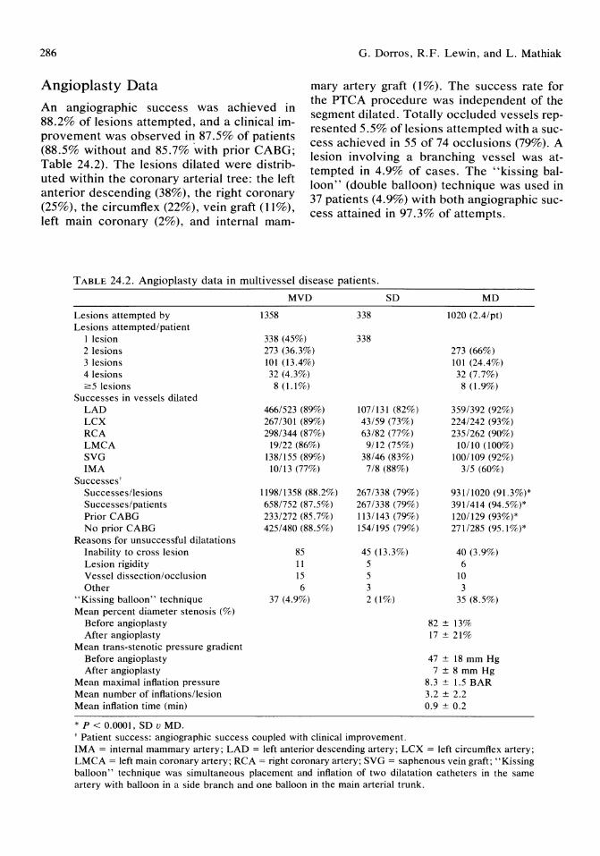
286 G. Dorros, R.F. Lewin, and L. Mathiak
Angioplasty Data mary artery graft (1%). The success rate for the PTCA procedure was independent of the segment dilated. Totally occluded vessels represented 5.5% of lesions attempted with a success achieved in 55 of 74 occlusions (79%). A lesion involving a branching vessel was attempted in 4.9% of cases. The "kissing balloon" (double balloon) technique was used in 37 patients (4.9%) with both angiographic success attained in 97.3% of attempts.
An angiographic success was achieved in 88.2% of lesions attempted, and a clinical improvement was observed in 87.5% of patients (88.5% without and 85.7% with prior CABG; Table 24.2). The lesions dilated were distributed within the coronary arterial tree: the left anterior descending (38%), the right coronary (25%), the circumflex (22%), vein graft (11%), left main coronary (2%), and internal mam-
TABLE 24.2. Angioplasty data in multivessel disease patients.
Lesions attempted by Lesions attempted/patient
I lesion 2 lesions 3 lesions 4 lesions ~5 lesions
Successes in vessels dilated LAD LCX RCA LMCA SVG IMA
Successes' Successes/lesions Successes/patients Prior CABG No prior CABG
Reasons for unsuccessful dilatations Inability to cross lesion Lesion rigidity Vessel dissection/ occlusion Other
"Kissing balloon" technique Mean percent diameter stenosis (%)
Before angioplasty After angioplasty
Mean trans-stenotic pressure gradient Before angioplasty After angioplasty
Mean maximal inflation pressure Mean number of inflations/lesion Mean inflation time (min)
* P < 0.0001, SD v MD.
MVD
1358
338 (45%) 273 (36.3%) 101 (13.4%) 32 (4.3%) 8 (1.1%)
466/523 (89%) 267/301 (89%) 298/344 (87%)
19/22 (86%) 138/155 (89%)
10113 (77%)
119811358 (88.2%) 6581752 (87.5%) 233/272 (85.7%) 425/480 (88.5%)
85 11 15 6
37 (4.9%)
SD
338
338
107/131 (82%) 43/59 (73%) 63/82 (77%) 9/12 (75%)
38/46 (83%) 7/8 (88%)
267/338 (79%) 267/338 (79%) 1131143 (79%) 1541195 (79%)
45 (13.3%) 5 5 3 2 (1%)
t Patient success: angiographic success coupled with clinical improvement.
MD
1020 (2.4/pt)
273 (66%) 101 (24.4%) 32 (7.7%) 8(1.9%)
359/392 (92%) 224/242 (93%) 235/262 (90%)
10/10 (100%) 1001109 (92%)
3/5 (60%)
93111020 (91.3%)* 3911414 (94.5%)* 120/129 (93%)* 2711285 (95.1%)*
40 (3.9%) 6
10 3
35 (8.5%)
82 ± 13% 17 ± 21%
47 ± 18 mm Hg 7 ± 8 mm Hg
8.3 ± 1.5 BAR 3.2 ± 2.2 0.9 ± 0.2
IMA = internal mammary artery; LAD = left anterior descending artery; LCX = left circumflex artery; LMCA = left main coronary artery; RCA = right coronary artery; SVG = saphenous vein graft; "Kissing balloon" technique was simultaneous placement and inflation of two dilatation catheters in the same artery with balloon in a side branch and one balloon in the main arterial trunk.

24. Multivessel and Multiple Lesion PTCA
TABLE 24.3. Complications* encountered during angioplasty in multivessel disease patients.
Complications
Transmural MI
Coronary spasm
Coronary occlusion/dissection
Mortality Emergency CABG CVA Summary
Patients with no complications
Patients with significant complications
Mortality with regard time of procedure Before 1983 After 1983
Total %
19
35
26
14 26
2
2.5%/patient 1.4%/lesion 4.6%/patient 2.6%/lesion 3.5%/patient 1.9%/lesion 1.9% 3.6%/patient 0.3%/patient
606 (80.5%)
39 (5.2%)
7/204 (3.4%)* 7/548 (1.3%)
* P < 0.05 mortality, before v after 1983. Abbreviations as in Table 24.2.
Complications
A significant complication (death, emergency CABO, or transmural myocardial infarction) occurred in 39 patients (5.2%). Complications encountered were not mutually exclusive, and multiple complications could be encountered in the same patient. No complication, whatsoever, occurred in 80.5% of cases (Tables 24.3 and 24.4).
The listed incidence of complications was similar in the single and multiple dilatation groups. There was no difference (Table 24.4) in the mortality between single and multiple dilatation groups when the cases were performed during the same time period (before or after 1983). Both groups had significantly lower mortality statistics when comparing procedures done before and after 1983. The steerable guidewire over the catheter system was introduced in early 1983.
The single dilatation (SD) group had a significantly higher incidence of significant complications (SD, 7.7% v MD, 3.3%; P < 0.01) when compared with the multiple dilatation (MD) group; however, no specific complica-
287
tion occurred more frequently (see discussion).
There were 14 in-hospital PTCA related deaths (mortality of 1.9%). The mortality was significantly decreased in cases done after 1983 (before 1983, 3.4% v after 1983, 1.3%; P < 0.05). Within the single dilatation group 3 of the 8 and within the mUltiple dilatation group 4 of the 6 deaths occurred before 1983. Thus, 7 mortalities occurred before 1983. In addition, 8 of the PTCA related mortalities occurred in patients with severe debilitating ischemic heart disease, who were not considered candidates for bypass surgery.
Eight mortalities (57%) had an angiographically documented coronary occlusion: 6 patients within 30 minutes, and 2 patients within 12 hours of the angioplasty procedure.
Seven patients (50%) had undergone emergency CABO. The seven patients who did not undergo emergency surgery developed electromechanical dissociation and death.
Follow-up
There were 356 successful patients alive and without subsequent bypass surgery who were more than 12 months remote from their initial PTCA procedure (Table 24.5, Fig 24.1). Follow-up was achieved in 92% (328 patients). Clinical data on the anginal status at the time of last contact revealed 65.5% had no angina. Sixty-four percent of patients with angina before PTCA had no angina, and 83% had less angina at the time of last contact.
There were 41 late deaths of which 26 were cardiovascular related; however, it is difficult to discern if these deaths, as determined from the death certificate, were related to a lesion recurrence, progression of disease, or other cardiovascular problems.
A first apparent symptom-related lesion recurrence (Fig 24.1) occurred at a mean time of 7.7 months in 233 patients (35%). A successful second PTCA was performed in 162/171 patients (94.7%). There were 9 failures: 1 death, 3 emergency and 2 elective surgeries, and 3 patients medically treated. A second apparent symptom-related lesion recurrence occurred
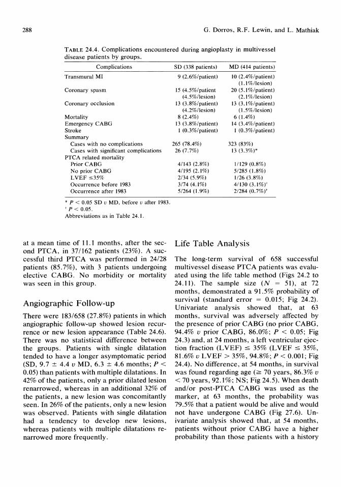
288 G. Dorros, R.F. Lewin, and L. Mathiak
TABLE 24.4. Complications encountered during angioplasty in multivessel disease patients by groups.
Complications
Transmural MI
Coronary spasm
Coronary occlusion
Mortality Emergency CABG Stroke Summary
Cases with no complications Cases with significant complications
PTCA related mortality Prior CABG No prior CABG LVEF :535% Occurrence before 1983 Occurrence after 1983
* P < 0.05 SD u MD, before u after 1983. t P < 0.05. Abbreviations as in Table 24.1.
at a mean time of 11.1 months, after the second PTCA, in 37/162 patients (23%). A successful third PTCA was performed in 24/28 patients (85.7%), with 3 patients undergoing elective CABG. No morbidity or mortality was seen in this group.
Angiographic Follow-up
There were 183/658 (27.8%) patients in which angiographic follow-up showed lesion recurrence or new lesion appearance (Table 24.6). There was no statistical difference between the groups. Patients with single dilatation tended to have a longer asymptomatic period (SD, 9.7 ± 4.4 v MD, 6.3 ± 4.6 months; P < 0.05) than patients with multiple dilatations. In 42% of the patients, only a prior dilated lesion renarrowed, whereas in an additional 32% of the patients, a new lesion was concomitantly seen. In 26% of the patients, only a new lesion was observed. Patients with single dilatation had a tendency to develop new lesions, whereas patients with multiple dilatations renarrowed more frequently.
SD (338 patients)
9 (2.6%/patient)
15 (4.5%/patient (4.5%/lesion)
13 (3.8%/patient) (4.2%/lesion)
8 (2.4%) 13 (3.8%/patient) I (0.3%/patient)
265 (78.4%) 26 (7.7%)
4/143 (2.8%) 4/195 (2.1%) 2/34 (5.9%) 3/74 (4.1%) 5/264 (1.9%)
MD (414 patients)
10 (2.4%/patient) (1.1 %l1esion)
20 (5.1 %/patient) (2.1 %l1esion)
13 (3.1 %/patient) (1.5%/lesion)
6(1.4%) 14 (3.4%/patient) I (0.3%/patient)
323 (83%) 13 (3.3%)*
11129 (0.8%) 5/285 (1.8%) 1126 (3.8%) 4/130 (3.1%)' 2/284 (0.7%)'
Life Table Analysis
The long-term survival of 658 successful multivessel disease PTCA patients was evaluated using the life table method (Figs 24.2 to 24.11). The sample size (N = 51), at 72 months, demonstrated a 91.5% probability of survival (standard error = 0.015; Fig 24.2). Univariate analysis showed that, at 63 months, survival was adversely affected by the presence of prior CABG (no prior CABG, 94.4% v prior CABG, 86.0%; P < 0.05; Fig 24.3) and, at 24 months, a left ventricular ejection fraction (L VEF) ::; 35% (L VEF ::; 35%, 81.6% v LVEF > 35%, 94.8%; P < 0.001; Fig 24.4). No difference, at 54 months, in survival was found regarding age (2: 70 years, 86.3% v < 70 years, 92.1%; NS; Fig 24.5). When death and/or post-PTCA CABG was used as the marker, at 63 months, the probability was 79.5% that a patient would be alive and would not have undergone CABG (Fig 27.6). Univariate analysis showed that, at 54 months, patients without prior CABG have a higher probability than those patients with a history
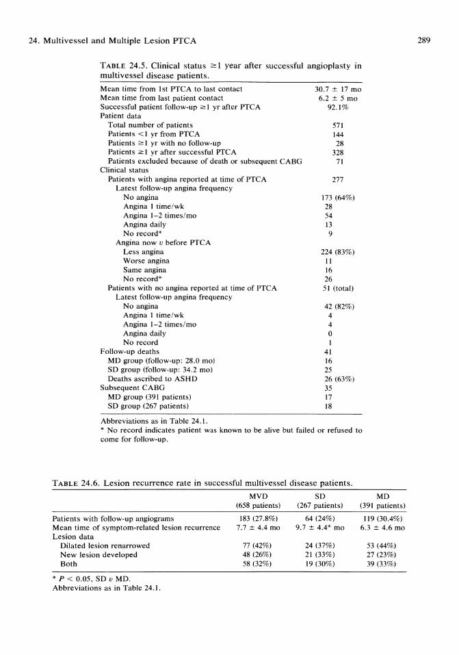
24. Multivessel and Multiple Lesion PTCA
TABLE 24.5. Clinical status 2:: 1 year after successful angioplasty in multivessel disease patients.
Mean time from 1st PTCA to last contact Mean time from last patient contact Successful patient follow-up 2:: 1 yr after PTCA Patient data
Total number of patients Patients < 1 yr from PTCA Patients 2:: 1 yr with no follow-up Patients 2:: 1 yr after successful PTCA Patients excluded because of death or subsequent CABG
Clinical status Patients with angina reported at time of PTCA
Latest follow-up angina frequency No angina Angina 1 time/wk Angina 1-2 times/mo Angina daily No record*
Angina now v before PTCA Less angina Worse angina Same angina No record*
Patients with no angina reported at time of PTCA Latest follow-up angina frequency
No angina Angina 1 time/wk Angina 1-2 times/mo Angina daily No record
Follow-up deaths MD group (follow-up: 28.0 mol SD group (follow-up: 34.2 mol Deaths ascribed to ASHD
Subsequent CABG MD group (391 patients) SD group (267 patients)
Abbreviations as in Table 24.1.
30.7 ± 17 mo 6.2 ± 5 mo
92.1%
571 144 28
328 71
277
173 (64%) 28 54 13 9
224 (83%) 11 16 26 51 (total)
42 (82%) 4 4 o I
41 16 25 26 (63%) 35 17 18
* No record indicates patient was known to be alive but failed or refused to come for follow-up.
TABLE 24.6. Lesion recurrence rate in successful multivessel disease patients.
MVD SD MD (658 patients) (267 patients) (391 patients)
Patients with follow-up angiograms Mean time of symptom-related lesion recurrence Lesion data
Dilated lesion renarrowed New lesion developed Both
* P < 0.05, SD v MD. Abbreviations as in Table 24.1.
183 (27.8%) 7.7 ± 4.4 mo
77 (42%) 48 (26%) 58 (32%)
64 (24%) 119 (30.4%) 9.7 ± 4.4* mo 6.3 ± 4.6 mo
24 (37%) 53 (44%) 21 (33%) 27 (23%) 19 (30%) 39 (33%)
289
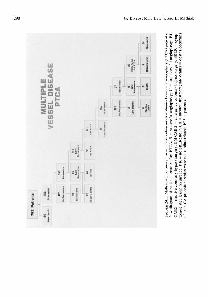
752
Pat
ien
ts
94
658
Una
uc,. .
.. I\
' SU
c,.. .
. ,.,
425
233
No
A.a
rron
c.
A.a
rron
c.
I 10
50
18
3 ...
. 1. D
o"',,
" u
t •
£M1y
A
.arr
tnlO
t A
oeun
onc
26
24
12
De
d ..
CA
IIO
Doa
lm
Ho
PT
CA
UL
TIP
L
ES
SE
L D
ISE
AS
T
171
2nd
PT
CA
~~
9 16
2 U
M..,.
IOt .
. 'UI
SU
C,. ...
,UI
125
37
No
A ....
. on
c.
Ate
wto
nc.
2 9
28
ut.
Dll
lhI
uto
A
.arr
....
..
~p~
4 5
4 E
Itc1
Ive
0._
~'"
CA
IIO
24
5ucI
O ..
. ",
FIG
UR
E 2
4.1.
Mul
tives
sel
coro
nary
dis
ease
in
perc
utan
eous
tra
nslu
min
el c
oron
ary
angi
opla
sty
(PT
CA
) pa
tien
ts:
flow
dia
gram
of
pati
ents
' co
urse
aft
er P
TC
A.
S =
su
cces
sful
ang
iopl
asty
; U
=
unsu
cces
sful
ang
iopl
asty
; E
L
CA
BG
=
elec
tive
cor
onar
y by
pass
sur
gery
; E
M C
AB
G =
em
erge
ncy
coro
nary
byp
ass
surg
ery;
SR
LR
=
sym
pto
m-r
elat
ed l
esio
n re
curr
ence
; N
R =
no
SR
LR
; no
PT
CA
=
med
ical
tre
atm
ent;
lat
e de
aths
=
deat
hs o
ccur
ring
af
ter
PTC
A p
roce
dure
whi
ch w
ere
not
card
iac
rela
ted;
PT
S =
pati
ents
.
N 8 Cl tI
o a '" :;c >n t""'
(1) :E s· § 0..
t""'
~
~
;:J" ~
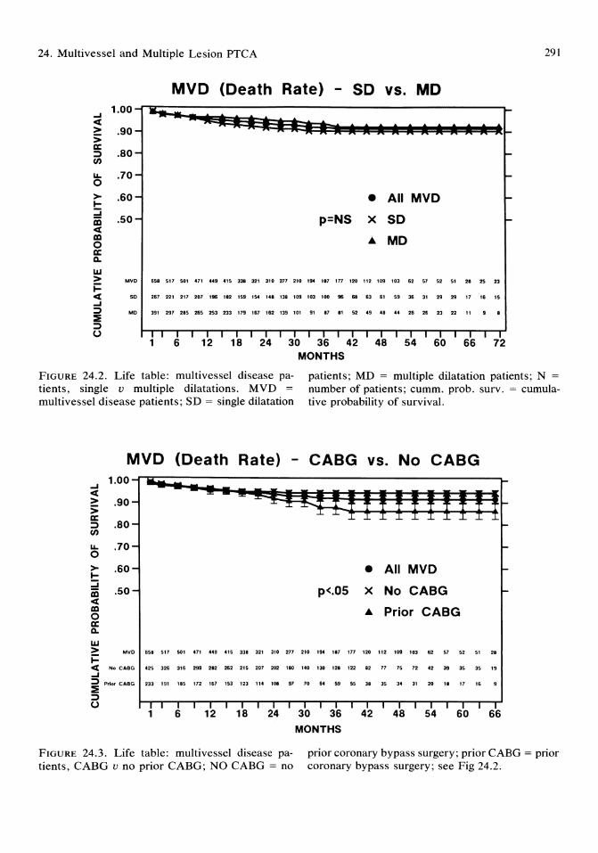
24. Multivessel and Multiple Lesion PTCA
MVD (Death Rate) - SO vs. MD
• All MVO
p=NS x SO
... MO
MVD 658 517 501 471 449 415 338 321 310 277 210 194 181 177 120 112 109 103 62 51 52 51 28 25 23
SO 267 221 217 207 196 182 159 154 148 138 109 103 100 96 68 63 61 59 36 31 29 29 17 16 15
MO 391 297 285 265 253 233 179 167 162 139 101 91 87 81 52 49 48 44 26 26 23 22 11
MONTHS
291
----
FIGURE 24.2. Life table: multivessel disease patients, single v multiple dilatations. MVD =
multivessel disease patients; SD = single dilatation
patients; MD = multiple dilatation patients; N = number of patients; cumm. prob. surv. = cumulative probability of survival.
MVD 1.00
....I « > .90 :> c:::
.80 ::) en LL .70 0 > .60 I-::::i iii .50 « co 0 c::: c.. w > MVO &S8 517
i= « No CABG '25 326
....I ::) ::E
Prior CABG 233 191
::) u
1
(Death Rate) - CABG vs. No CABG
501 471 .49 .,5 338 321
316 299 282 262 215 201
185 112 161 153 123 11.
6 12 18
• All MVO
p<.OS x No CABG
... Prior CABG
310 217 210 19. 181
202 180 140 130 128
108 91 10 6. 59
24 30 36 MONTHS
111 120
122 82
55 38
42
112 109 103 62 51 52
11 15 12 42 39 35
35 3. 31 20 18 17
48 54 60
51
35
16
28
19
66
FIGURE 24.3. Life table: multivessel disease patients, CABG v no prior CABG; NO CABG = no
prior coronary bypass surgery; prior CABG = prior coronary bypass surgery; see Fig 24.2.
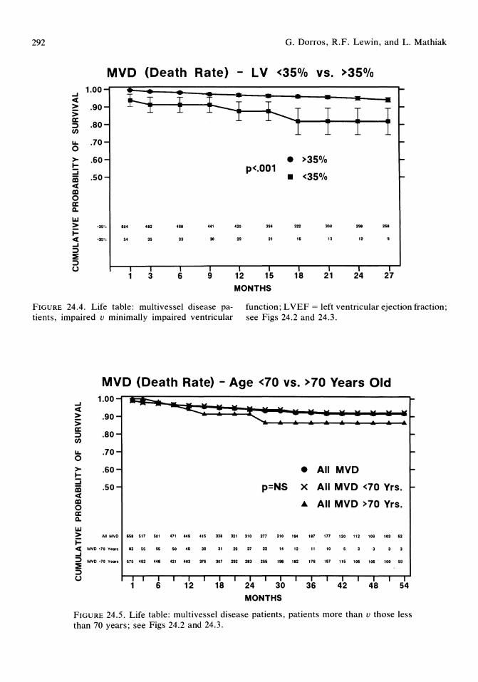
292
> I-:::i iii « III o a: Il.
IIJ > i= « ....I => :E => U
O. Dorros, R.F. Lewin, and L. Mathiak
MVD (Death Rate) - LV <35% vs. >35%
.60 • >35% p<.001
• <35% .50
>35'/, 604 • 82 ... 441 '20 39. 322 308 298 208
<35% 54 35 33 30 29 21 10 13 12
1 3 6 9 12 15 18 21 24 27
MONTHS
FIGURE 24.4. Life table: multivessel disease patients, impaired u minimally impaired ventricular
function; L YEF = left ventricular ejection fraction; see Figs 24.2 and 24.3.
MVD (Death Rate) - Age <70 vs. >70 Years Old
....I 1.00-
« ............ > .90- -:; a:
.80-=> -U)
u.. .70- -0 > .60-I- • All MVD -:::i
.50- p=NS X All MVD <70 Yrs. iii -« III • All MVD >70 Yrs . 0 a: Il.
IIJ > All MilD 058 517 501 471 449 415 338 321 310 277 210 194 187 117 120 112 109 103 02
i= « MilD >70 Years 83 55 55 50 .0 39 31 29 27 22 14 12 11 10 3 3
....I => :E
MVD <70 Years 575 .02 440 .21 '03 370 307 292 283 255 196 182 170 107 115 100 100 100 59
=> U I I I I I I I I I I I I I I I I I I I
1 6 12 18 24 30 36 42 48 54
MONTHS
FIGURE 24.5. Life table: multivessel disease patients, patients more than u those less than 70 years; see Figs 24.2 and 24.3.

24. Multivessel and Multiple Lesion PTCA
MVD (Death & CABG Rate) - SD vs. MD 1.00-r~--------------------------------------------------~
....I « > .90 :> !5 .80 en LI.. .70 o > .60 I-:::i iii .50 « a::I o a: c.. UJ
• All MVO
p=NS X SO
... MO
> MVD 658 517 501 471 449 415 338 321 310 217 210 194 '87 177 120 112 109 103 62 57 52 51 28 2S
i= <[ SO 267 221 217 207 196 182 159 154 148 138 109 103 100 96 68 63 61 59 36 31 29 29 17 16
....I :::> MD 391 297 285 265 253 233 179 167 162 139 101 91 87 81 52 49 48 44 26 26 23 22 11
:E => U
1 6 12 18 24 30 36 42 48 54 60 66 MONTHS
FIGURE 24.6. Life table: multivessel disease patients, death and/or coronary bypass surgery, single u multiple dilatations, see Figs 24.2 and 24.3.
MVD (Death + CABG Rate) No CABG VS. Prior CABG 1.00
....I 1=1, iii « J . -> .90-
:>
It<'m%"'~"t a: .80-=> en LI.. .70-0
> .60- • All MVO -I-::::i .50- p<O.OS X No CABG iii -« a::I ... Prior CABG 0 a: c.. UJ > All MVO .58 517 501 471 ... 415 "8 321 310 271
i= 210 1S< 187 177 120 112 109 103 .2 57 52 51
« No CABG .25 32. 31. 2" 282 2.2 215 201 202 180 140 130 128 122 82 11 15 12 .2 39 35 35 ....I => Prior CABG 233 191 185 112 161 153 123 114 108 91
:E 10 •• 59 55 38 35 34 31 20 11 11 "
=> U I I I I I I I I I I I I I I I I I I I I I I
1 6 12 18 24 30 36 42 48 54 60 MONTHS
FIGURE 24.7. Life table: multivesse1 disease patients, death and/or coronary bypass surgery, no CABG u prior CABG; see Figs 24.2 and 24.3.
293

294 G. Dorros, R.F. Lewin, and L. Mathiak
MVD (Death Rate) - LV <35%, SO vs. MD 1.00
....I .; <t
* ~ ~ : : : > .90 :; a: =:) .80 (J)
u.. .70 0 > .60 I- • All MVO ::::i
.50 p<.OO1 X SO iii <t a:I ... MO 0 a: Il.
UJ > MVO 54 35 33 30
i= 2. 21 I. 13 12
<t SO 22 17 I. 15 15 11
....I =:) MO 32 18 17 15 14 10 ::E =:) (.)
1 3 6 9 12 15 18 21 24 27 MONTHS
FIGURE 24.8. Life table: multivessel disease patients, impaired left ventricular function, single u multiple dilatations; see Figs 24.2 and 24.3.
MVD (Death Rate) - Age >70 Yrs., SO vs. MD 1.00-
....I ............... ~ <t > .90- -:; a:
.80-=:) -(J)
u.. .70- -0 > . 60- • All MVO -I-::::i
.50- p<.01 iii X SO r-<t a:I ... MO 0 a: Il.
UJ > MVO .3 55 55 50 •• 3'
i= 31 2 • 27 22 14 12 11 10
<t SO 35 2. 2. 25 2. ,. 15 14 " 11
....I =:)
::E MO •• 27 27 25 2' " I. 15 13 11 10
=:) (.) I I I I I I I I I I I I I I
1 3 6 9 12 15 18 21 24 27 30 33 36 39
MONTHS
FIGURE 24.9. Life table: multivessel disease patients, patients 2:70 years, single u multiple dilatations; see Figs 24.2 and 24.3.

24. Multivessel and Multiple Lesion PTCA
SO (Death Rate) - No CABG vs. Prior CABG
-I <C > :; a: :::» en I&. o > I::::; iii <C al o a: Q.
1&.1 > ~
.70-
.60-
.50-
AU SO
c:c No CABG. SO -I i Prior CABG, SO
:::» U
251 221
15. 13.
113 91
I I 1
211 201
". '.5
.9 •• I I 6
196 112 159 154 14. 138
116 I •• .. 9. 95 •• •• 14 .. 56 53 5.
I I I I I I 12 18 24
• All MVO ~
p=NS X SO, No CABG ~
• S0, Prior CABG
109 ,.3 , .. .. 6. 63 61 59 36 31 29 '9 17 16
1. 6 • .. 65 .6 •• .2 •• 2' 21 19 19 11 11
31 35 34 31 22 19 19 17 12 ,. ,. ,. I I I I I I I I I T I T T I
30 36 42 48 54 60 66 MONTHS
FIGURE 24.10. Life table: single dilatation patients, no CABG v prior CABG; see Figs 24.2 and 24.3.
MD (Death Rate) - No CABG vs. Prior CABG
-I <C > :;
1.001l~j;~~~;:~~=i;;~it~~~it~~~i:~;j~~~ a: :::» en I&. o > I::::; iii <C al o a: Q.
w
.90
.80
.70
. 60
.50
> AI""
~ <I: No CABG, ME)
-I i .... , CABG, Me
:::» U
391
.11
". 1
291 2.5 26' 25.
196 188 113 185
,., 91 91 .. 6 12
p<O.05
232 119 181 181 119 100 .. 153 115 109 108 •• 61 61
19 63 5. 55 41 33 29
18 24 30 MONTHS
• All MO
X MO, No CABG
• MO, Prior CABG
.. 19 51 .. 41 43 25 23
61 55 35 32 32 .. I. I •
25 2. 16 16 " 14
36 42 48 54
FIGURE 24.11. Life table: mUltiple dilatation patients, no CABG v prior CABG; see Figs 24.2 and 24.3.
295

296
of prior CABG of being alive and not having undergone CABG (no prior CABG, 82.7% v prior CABG, 73.1%; P < 0.05; Fig 24.7).
The long-term survival of successful PTCA patients who had one lesion dilated (N = 267) was compared with those patients who had multiple lesions dilated (N = 391; Fig 24.2). Survival, at 63 months, was not statistically different between these groups (SD, 90.3% v MD, 92.3%). Survival was statistically significantly different between the groups when univariate analysis was used to stratify patients according to left ventricular performance (LVEF :5 35%; SD, 73.6% v MD, 92.6%; P < 0.001, at 24 months; Fig 24.8), or the patient's age (2': 70 years; SD, 78.5% v MD, 92.2%; P < 0.01 at 39 months; Fig 24.9). At 63 months, a patient's history of prior bypass surgery did not affect the probability of survival in single dilatation patients (Fig 24.10; no prior CABG, 93.2% v prior CABG, 86.8). However, this was in contrast to the multiple dilatation patients relationship to bypass surgery (Fig 24.11), in which the probability of survival was adversely affected by the presence of prior bypass surgery (no prior CABG, 96.2% v prior CABG, 84.3%; P < 0.05).
Results of Multiple Lesion Transmural Coronary Angioplasty
Clinical Characteristics
Sixteen patients underwent angioplasty of different vessels during the same hospitalization, that is, staged procedure (Table 24.7).
Angioplasty Procedure
An angiographic success was achieved in 94% of lesions, and a clinical improvement occurred in 94% of patients (Tables 24.8 and 24.9). Table 24.9 lists the combinations of vessels with lesions attempted, successful dilata-
G. Dorros, R.F. Lewin, and L. Mathiak
TABLE 24.7. Characteristics of patients undergoing multiple lesion angioplasty.
Patients * Men Women Age
Extent of coronary disease Single vessel Multivessel
Left ventricular ejection fraction (:$35%) Prior bypass surgery Anginal class'
Class 0 (no angina) Class I Class II Class III Class IV
Prior myocardial infarction (documented) Diabetes mellitus Hypertension Prior stroke Chronic renal failure Chronic obstructive lung disease
428 333 (78%) 95 (22%) 58.8 yrs
69 (16%) 359 (84%)
22 (5%) 115 (27%)
63 (15%) 52 (12%)
150 (35%) 103 (24%) 61 (14%)
201 (47%) 57 (13%)
176 (41%) 12 (2.8%) 7(1.6%)
12 (2.8%)
* Sixteen patients underwent two procedures during the same hospitalization to achieve the desired revascularization goal with multiple dilatations. t Canadian Cardiovascular Society Classification.
tions, and patient successes. No statistically significant differences were found in success rates among the vessels attempted or different patient subgroups.
Complications
A significant complication (death, emergency surgery, or transmural myocardial infarction) occurred in 17 patients (4.0%; Table 24.10). There were 6 in-hospital PTCA-related deaths (mortality of 1.4%). No patient had had prior bypass surgery. All 6 patients underwent apparently successful angioplasty, that is, the angioplasty was carried out without incident, and the postangioplasty cinearteriograms showed successful dilatations. Five patients who died had a documented coronary occlusion: 4 patients, within 30 minutes of angioplasty, and 1 patient, 6 hours postangioplasty. Three patients who died underwent emergency bypass surgery.
The complications encountered were not mutually exclusive with 3 patients who died

24. Multivessel and Multiple Lesion PTCA
TABLE 24.8. Multiple lesion angioplasty procedure.
Angioplasty attempts Number of lesions attempted per patient
2 lesions 3 lesions 4 lesions 5 lesions
Vessel dilated Left anterior descending artery Left circumflex artery Right coronary artery Left main coronary artery Vein graft
Mean percent diameter stenosis (%) Before angioplasty After angioplasty
Mean trans-stenotic pressure gradient' (mm Hg) Before angioplasty After angioplasty
Mean maximal inflation pressure (atm) Mean number of inflationsllesion Mean inflation time (min) Successes
Successes' !total lesions Successes1/total patients In cases without prior surgery In cases with prior surgery In single-vessel coronary disease cases In multivessel coronary disease cases
Reasons for unsuccessful dilatations
1047 lesions
74% cases 21% cases
<5% cases <1% cases
446 (42%) 233 (22%) 258 (25%)
10 (1%) 100 (10%)
82 ± 13 17 ± 21
47 ± 18 7 ± 8
8.3 ± 1.5 3.2 ± 2.2 0.9 ± 0.2
985/1047 (94%) 404/428 (94%) 307/324 (95%) 1111120 (93%) 65/69 (94%) 353/375 (94%)
Inability to cross the lesion with guidewire or dilatation catheter Lesion rigidity
49 6 5 2
Vessel dissectionlocclusion before balloon inflation Other
, Not recorded during all angioplasties. , Angiographic success: ;0:20% decrease in percent diameter stenosis. I Patient success: angiographic success coupled with clinical improvement.
TABLE 24.9. Vessel combinations in multiple lesion angioplasty.
Vessels attempted
LAD + DIAG LAD + RCA LAD + LCX RCA + LCX LMCA + RCA/LCA/SVG' SVG + LCA/RCA/SVG' > 2 lesions dilated
Lesion successes per total lesion attempts
75180 (94%) 115/128 (90%) 103/106 (97%) 49152 (94%) 10/12 (83%) 72/88 (82%)
266/272 (98%)
Case successes per total cases
38/40 (95%) 60/64 (94%) 48/53 (91%) 25/26 (96%)
5/6 (83%) 38/44 (86%) 77/80 (96%)
* LMCA lesion and right coronary or left coronary or vein graft lesion. , SVG lesion and left coronary or right coronary or another vein graft lesion. DIAG = diagonal branch; LAD = left anterior descending artery; LCA =
left coronary artery; LCX = left circumflex coronary artery; LMCA = left main coronary artery; RCA = right coronary artery; SVG = saphenous vein graft; >2 lesions dilated = lesions in different vessels andlor in conjunction with lesions in different segments of the same vessel.
297

298
TABLE 24.10. Complications* encountered during multiple lesion angioplasty.
No. %
Transmural myocardial infarction 11 (2.5%/patient) (l.l%/Iesion)
Coronary spasm 26 (6.0%/patient) (2.5%/lesion)
Coronary occlusion 16 (3.7%/patient) (I.5%llesion)
Mortalityt 6 (1.4%) Emergency bypass surgery 9 (2.1 %/patient)
(0.9%/lesion)
* Complications listed were not mutually exclusive. Multiple complications often were encountered in the same patient; 361/428 cases (84%) experienced no complication whatsoever, and 17 patients (4.0%) experienced a significant complication. t All 6 deaths occurred in patients without prior bypass surgery.
having had evidence of an acute transmural myocardial infarction.
Follow-up
There were 250 successful patients alive and without subsequent bypass surgery who were more than 12 months remote from their initial angioplasty procedure (Table 24.11 and Fig 24.12). Follow-up of patients who had angina at the time of angioplasty showed that 68% had no and 83% had less angina at the time of the last contact. There were 14 late deaths of which 12 were ascribed (on the death certificate) to atherosclerotic heart disease.
A first apparent symptom-related lesion recurrence (Fig 24.12) occurred at a mean time of 6.6 months in 106 patients (26%). A second angioplasty was attempted in 89 patients. A successful second angioplasty was performed on 81/89 patients (91%). There were eight failures: 1 death, 1 emergency surgery,S elective surgeries, and 1 patient medically treated. A second apparent symptom-related lesion recurrence occurred at a mean time of 11.3 months in 15/81 second angioplasty patients (19%). A third angioplasty was successful in 13/15 patients. The two failed angioplasty patients underwent elective surgery.
O. Dorros, R.F. Lewin, and L. Mathiak
428 PTS
404 PTS (94%) ~ 24 PTS (6%)
NR~ 1STLR
/ """ 106 PTS (26%) 312 PTS (74%) 'CA~ 2NI
NO PT/ ~D PTCA
17 PTS 89 PTS
S~ UN
/ '" [1 DEATHJ 81 PTS (91%) 8 PTS 6 CABG
IR~2NI 1MED N;' ~LR 66 PTS 15 PTS (19%)
,,\RD PTCA
15 PTS
~ 13 PTS 2 PTS (2 CABG)
FIGURE 24.12. Follow-up of patients who underwent multiple lesion angioplasty. CAR = clinical apparent lesion recurrence, UN = unsuccessful, NR = no apparent symptom-related lesion recurrence, MED = medical treatment, CABO = coronary bypass operation. Other abbreviations as in Fig 24.1. (Reprinted with permission from the American College of Cardiology, J Am Coli Cardiol 1987; 10:1007-1013.)
Life Table Analysis
The long-term survival of 404 successful MLA patients was evaluated using the life table method (Figs 24.13 and 24.14). The longest patient follow-up is 87 months. At 51 months, there was a 0.93 probability of survival (standard error = 0.019). Univariate analysis, at 51 months, showed survival to be adversely affected by the presence of prior surgery (no prior CABG, 97% v prior CABG, 81%; P < 0.05). When death or postangioplasty CABG (Fig 24.14) was used as the marker, at 51 months, the probability was 88% that a successful PTCA patient would be alive and would not have had to undergo bypass surgery. Univariate analysis showed that the
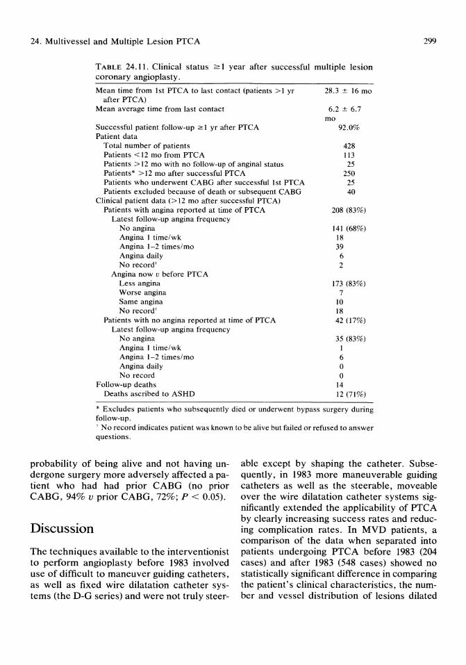
24. Multivessel and Multiple Lesion PTCA 299
TABLE 24.11. Clinical status 2: 1 year after successful multiple lesion coronary angioplasty.
Mean time from 1st PTCA to last contact (patients >1 yr after PTCA)
Mean average time from last contact
Successful patient follow-up 2: 1 yr after PTCA Patient data
Total number of patients Patients <12 mo from PTCA Patients> 12 mo with no follow-up of anginal status Patients* > 12 mo after successful PTCA Patients who underwent CABG after successful 1 st PTCA Patients excluded because of death or subsequent CABG
Clinical patient data (> 12 mo after successful PTCA) Patients with angina reported at time of PTCA
Latest follow-up angina frequency No angina Angina 1 time/wk Angina 1-2 times/mo Angina daily No record'
Angina now v before PTCA Less angina Worse angina Same angina No record'
Patients with no angina reported at time of PTCA Latest follow-up angina frequency
No angina Angina 1 time/wk Angina 1-2 times/mo Angina daily No record
Follow-up deaths Deaths ascribed to ASHD
28.3 ± 16 mo
6.2 ± 6.7 mo
92.0%
428 113 25
250 25 40
208 (83%)
141 (68%) 18 39
6 2
173 (83%) 7
10 18 42 (17%)
35 (83%) 1 6 o o
14 12 (71%)
* Excludes patients who subsequently died or underwent bypass surgery during follow-up. , No record indicates patient was known to be alive but failed or refused to answer questions.
probability of being alive and not having undergone surgery more adversely affected a patient who had had prior CABG (no prior CABG, 94% v prior CABG, 72%; P < 0.05).
able except by shaping the catheter. Subsequently, in 1983 more maneuverable guiding catheters as well as the steerable, moveable over the wire dilatation catheter systems significantly extended the applicability of PTCA by clearly increasing success rates and reducing complication rates. In MVD patients, a comparison of the data when separated into patients undergoing PTCA before 1983 (204 cases) and after 1983 (548 cases) showed no statistically significant difference in comparing the patient's clinical characteristics, the number and vessel distribution of lesions dilated
Discussion
The techniques available to the interventionist to perform angioplasty before 1983 involved use of difficult to maneuver guiding catheters, as well as fixed wire dilatation catheter systems (the D-G series) and were not truly steer-

300
1.0 "'~""::'! .. ~ .. ~ ... ~ .. ~ .. ~ ... ,." .. __ .:::-----:::::-____ __ _ . 8
.. .7
.3
..
. 1
"~~ .... '.
.... . ~ .. ~ .... .. ............ .. ... -....... --..
_ _ Overall N-428
- _ No Prior CABG N = 308
•...... Prior CABG N-120
1 6 12 18 24 30 36 42 48 54 60 66
MONTHS
FIGURE 24.13. Life table of patients who underwent successful mUltiple lesion angioplasty. CABO =
coronary bypass operation. (Reprinted with per· mission from the American College of Cardiology, J Am Call Cardiol1987 ; 10:1007-1013.)
1.0
..
.. • 7
. 1
~"" ..... :::-.. :::::.~ - -- - - - - - - --'. ' . .............
' . ........... ... . ........... .. .... . . . .. .....
__ Overall N-428
_ _ N a Prior CABG N=308
••••... Prior CABG N-120
1 6 1 2 18 24 30 36 42 48 54 60 66
MONTHS
FIGURE 24.14. Actuarial analysis of events (death or subsequent bypass surgery) in patients who un· derwent multiple lesion angioplasty.
O. Dorros, R.F. Lewin, and L. Mathiak
per patient, and the angiographic and clinical success rates. However, when comparing the patients within these two time periods, the mortality rate in single and multiple lesion angioplasty patients was significantly different. These differences may reflect the technologic advances (e.g., lower profile, higher pressure, more flexible and trackable dilatation catheters, as well as the softer and more steerable guidewire systems; and, of considerable importance, the improved guiding catheters) that enabled more difficult, unusual, and technically challenging cases to be successfully managed by the more knowledgeable interventionist.
The success rate reported herein for MVD and MLA patients is similar to published data. 3.6.7 The 88.2% angiographic success rate (91.3% in MD v 79% in SD patients) produced an immediate clinical improvement in 87.5% of patients (94.5% in MD v 79% in SD patients; P < 0.0001). The differences in success rates reflect our approach to PTCA, which inherently biases the success rates reported in the SD group. This inherent bias in our reporting must be expanded upon.
A MD procedure cannot occur by definition if the initial, first dilatation of the critical stenosis either is unsuccessful or results in a complication. Thus, our approach means that a second dilatation, which will remove the patient from the SD into the MD group, will only be undertaken after the most important lesion has been dilated. The patient's cardiovascular blood supply is theoretically significantly better after the first dilatation, which dilated the critical lesion. Thus, the patient may be sufficiently improved just by the first dilatation whether or not the second or third lesion was successfully dilated. Thus, a priori, the single dilatation group will have a lower success rate because it includes those patients who had only one lesion, which was planned to be dilated. In addition, the potential mUltiple dilatation group had its most critical lesion unsuccessfully dilated, which then placed this patient into the single dilatation group only. The result of our method is that the success in the single dilatation group represents all the single dilatation successes, whereas the fail-
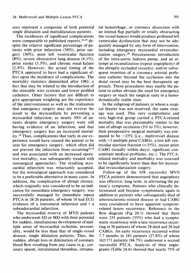
24. Multivessel and Multiple Lesion PTCA
ures represent a composite of both potential single dilatation and multidilatation patients.
The incidences of significant complications were comparable to published reports,18,19 despite the relative significant percentage of patients with prior infarction (58%), prior surgery (36%), poor left ventricular function (8%), severe obstructive lung disease (4.1%), prior stroke (3.3%), and chronic renal failure (2.4%). However, the actual timing of the PTCA appeared to have had a significant effect upon the incidence of complications. The mortality statistics diminished after 1983, a fact that may be related to the introduction of the steerable wire systems and lower profiled catheters. Other factors that are difficult to give appropriate weighting are the experience of the interventionist as well as the realization that emergency surgery may not reverse the insult to the myocardium by preventing a myocardial infarction, as nearly 50% of patients despite emergency surgery were still having evidence of an infarction, and that emergency surgery has an increased mortality .18 Thus, complications that early in our experience would have caused the patient to be sent for emergency surgery, which often did not prevent the infarction from occurringl8 ,19 and was associated with an increased operative mortality, was subsequently treated with nonsurgical approaches. The resulting myocardial infarction was reluctantly accepted, but the nonsurgical approach was considered to be a preferable alternative in many cases. In addition, the complication of abrupt closure, which originally was considered to be an indication for immediate emergency surgery, was successfully managed by immediate repeat PTCA in 18/26 patients, of whom 10 had ECG evidence of a transmural infarction and 1 a subendocardial infarction.
The myocardial reserve of MVD patients who underwent SD or MD with their potential for sudden, simultaneous development of multiple areas of myocardial ischemia, presumably, would be less than that of single-vessel disease, single dilatation patients. Thus, the sudden, abrupt loss or diminution of coronary blood flow resulting from any cause (e.g., coronary spasm, intraluminal thrombus, intramu-
301
ral hemorrhage, or coronary dissection with an intimal flap partially or totally obstructing the vessel lumen) would produce profound left ventricular dysfunction that may not be adequately managed by any form of intervention, including emergency myocardial revascularization surgery. 18 Percutaneous introduction of the intra-aortic balloon pump, and an attempt at recanalization (repeat angioplasty) of the abruptly occluded vessel(s), and/or subsequent insertion of a coronary arterial perfusion catheter beyond the occlusion into the distal vessel may be the best therapeutic approach. These procedures may enable the patient to either obviate the need for emergency surgery or reach the surgical suite in a hemodynamically stable state.
In the subgroup of patients in whom a surgical theater was not reserved, the same strategy was used. This very symptomatic and very high-risk group carried a PTCA-related mortality that was presumably similar to the rate of abrupt occlusion (3% to 5%); however, their preoperative surgical mortality was estimated to be >25% [i.e., multivessel disease with >3 mUltiple prior CABGs, a low left ventricular ejection fraction (::s35%), recent prior CABG (usually within days), significant concomitant medical problems, etc.]. The PTCArelated mortality and morbidity was assessed to be significantly lower than that for myocardial revascularization surgery.
Follow-up of the 658 successful MVD PTCA patients demonstrated that angioplasty was effective, long term, in alleviating the patient's symptoms. Patients who clinically deteriorated and became symptomatic again in addition to patients who subsequently died of arteriosclerotic-related disease or had CABG were considered to have apparent symptomrelated lesion recurrence. Reference to the flow diagram (Fig 24. 1) showed that there were 233 patients (35%) who had a symptomatic recurrence with a late recurrence occurring in 50 patients of whom 24 died and 26 had CABGs. An early recurrence occurred within 7.7 months in 183 patients (27.8%) of whom 162/171 patients (94.7%) underwent a second successful PTCA. Analysis of their angiograms (Table 24.6) showed that nearly 75% of
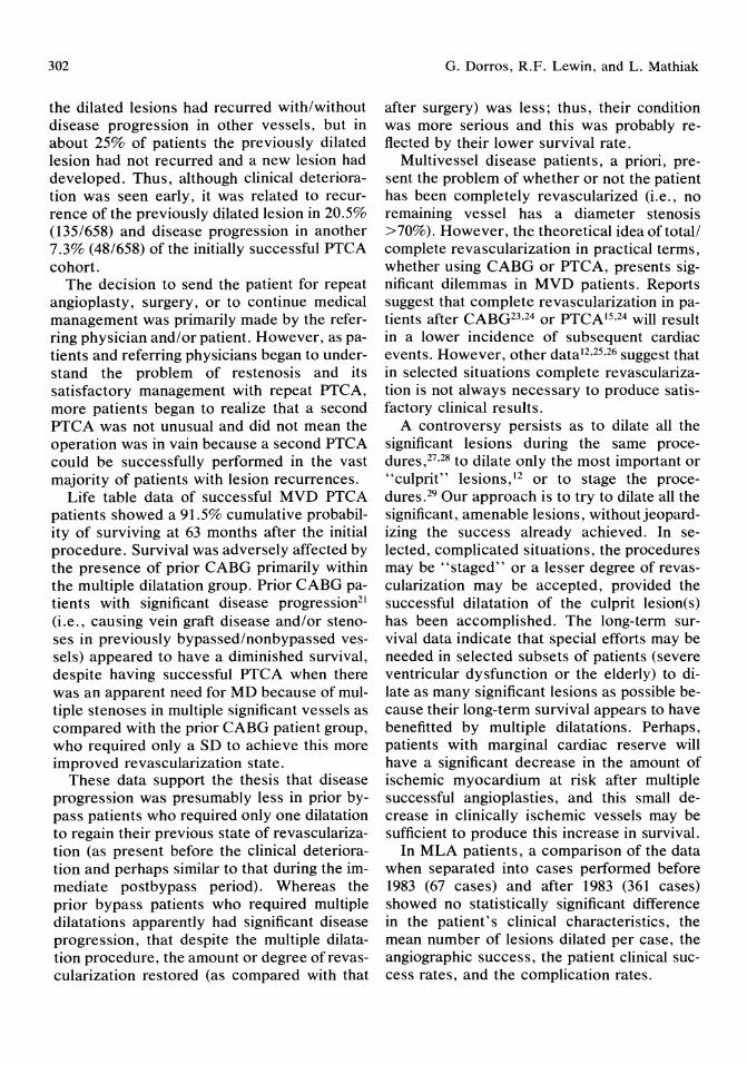
302
the dilated lesions had recurred with/without disease progression in other vessels, but in about 25% of patients the previously dilated lesion had not recurred and a new lesion had developed. Thus, although clinical deterioration was seen early, it was related to recurrence of the previously dilated lesion in 20.5% (135/658) and disease progression in another 7.3% (48/658) of the initially successful PTCA cohort.
The decision to send the patient for repeat angioplasty, surgery, or to continue medical management was primarily made by the referring physician and/or patient. However, as patients and referring physicians began to understand the problem of restenosis and its satisfactory management with repeat PTCA, more patients began to realize that a second PTCA was not unusual and did not mean the operation was in vain because a second PTCA could be successfully performed in the vast majority of patients with lesion recurrences.
Life table data of successful MVD PTCA patients showed a 91.5% cumulative probability of surviving at 63 months after the initial procedure. Survival was adversely affected by the presence of prior CABG primarily within the multiple dilatation group. Prior CABG patients with significant disease progression21 (i.e., causing vein graft disease and/or stenoses in previously bypassed/nonbypassed vessels) appeared to have a diminished survival, despite having successful PTCA when there was an apparent need for MD because of multiple stenoses in multiple significant vessels as compared with the prior CABG patient group, who required only a SD to achieve this more improved revascularization state.
These data support the thesis that disease progression was presumably less in prior bypass patients who required only one dilatation to regain their previous state of revascularization (as present before the clinical deterioration and perhaps similar to that during the immediate postbypass period). Whereas the prior bypass patients who required multiple dilatations apparently had significant disease progression, that despite the multiple dilatation procedure, the amount or degree of revascularization restored (as compared with that
G. Dorros, R.F. Lewin, and L. Mathiak
after surgery) was less; thus, their condition was more serious and this was probably reflected by their lower survival rate.
Multivessel disease patients, a priori, present the problem of whether or not the patient has been completely revascularized (i.e., no remaining vessel has a diameter stenosis >70%). However, the theoretical idea oftotall complete revascularization in practical terms, whether using CABG or PTCA, presents significant dilemmas in MVD patients. Reports suggest that complete revascularization in patients after CABG23,24 or PTCA 15,24 will result in a lower incidence of subsequent cardiac events. However, other data12 ,25,26 suggest that in selected situations complete revascularization is not always necessary to produce satisfactory clinical results.
A controversy persists as to dilate all the significant lesions during the same procedures,27,28 to dilate only the most important or "culprit" lesions,12 or to stage the procedures. 29 Our approach is to try to dilate all the significant, amenable lesions, without jeopardizing the success already achieved. In selected, complicated situations, the procedures may be "staged" or a lesser degree of revascularization may be accepted, provided the successful dilatation of the culprit lesion( s) has been accomplished. The long-term survival data indicate that special efforts may be needed in selected subsets of patients (severe ventricular dysfunction or the elderly) to dilate as many significant lesions as possible because their long-term survival appears to have benefitted by multiple dilatations. Perhaps, patients with marginal cardiac reserve will have a significant decrease in the amount of ischemic myocardium at risk after multiple successful angioplasties, and this small decrease in clinically ischemic vessels may be sufficient to produce this increase in survival.
In MLA patients, a comparison of the data when separated into cases performed before 1983 (67 cases) and after 1983 (361 cases) showed no statistically significant difference in the patient's clinical characteristics, the mean number of lesions dilated per case, the angiographic success, the patient clinical success rates, and the complication rates.
24. Multivessel and Multiple Lesion PTCA
The success rates reported herein were similar to some published data3,5,6,7 and higher than the NHLBI PTCA Registry report. 2 The clinical success appeared to be related to the successful dilatation of the culprit lesion(s), and not to the extent of coronary disease or the number of lesions dilated per case, except in two subsets of patients.
The follow-up of our 404 successful MLA patients showed that an apparent symptomrelated lesion recurrence occurred in 106 patients (26%) of which 17 had a late recurrence or death or CABG or medical management. The 81189 early recurrence patients (91%) were managed with successful PTCA with 1 treated with medication. Thus, 379/404 successful MLA patients (94%) (mean follow-up: 28.3 ± 16 months) were able to be managed with or without repeat PTCA and without the need for subsequent surgery.
Life table analysis of successful MLA patients showed a 93% probability of surviving 51 months after the initial procedure. Survival was adversely affected by the presence of prior surgery. Similarly, the presence of prior surgery increased the likelihood of postangioplasty surgery. Thus, patients with prior surgery, who have progression of their atherosclerotic process, have a significant problem in not only their survival, despite successful angioplasty, but also they are more likely to undergo repeat surgery rather than repeat angioplasty.
Conclusion
The use of static studies to evaluate a dynamically changing process is difficult. Technologic advances may be appreciated only when reviewing the statistics of a procedure with regard the timing of introduction of changes. Within these series, the introduction of the steerable wire and low profile catheter systems appeared to coincide with not only improved lesion success rates, despite attempting lesions that were previously inaccessible, but also resulted in improved patient success rates despite operating on patients who were at higher risk (not only from the anatomic
303
viewpoint but also because of significant concomitant medical problems) than those previously operated on.
Although the cumulative probability of survival was acceptable for all successful patients, selected subsets of patients appeared to do better after single dilatations (those with prior bypass surgery), whereas different subsets of patients appeared to benefit from multiple dilatations (impaired ventricular function or age ~70 years). The apparent benefits may be a reflection of the reduction of jeopardized myocardium in patients who have minimal amounts of cardiac reserve.
The completeness of follow-up data provides a glimpse of the results of PTCA not only short term regarding symptomatic lesion recurrence but also long term regarding survival as well as the incidence of subsequent bypass surgery and/or cardiovascular related deaths. Long-term follow-up also indicates that total relief of anginal symptoms may be achieved in nearly two thirds of patients and significant palliation of symptoms in more than 80% of the patients.
These data indicate that coronary angioplasty has an ever widening role in the treatment of coronary artery disease patients who may be considered excellent candidates for revascularization surgery, poor candidates for bypass surgery, and even in patients in which surgery is not believed to be a feasible or possible alternative.
Acknowledgments. We wish to express our sincere thanks to Carol Kreutzmann for secretarial assistance and manuscript typing, to Marla Engel for data management and patient follow-up, and to Michael Rohan for computer programming.
References 1. Gruentzig AR, Senning A, Siegenthaler WE:
Nonoperative dilatation of coronary artery stenosis, percutaneous transluminal angioplasty. N Engl J Med 1979; 301:61.
2. Kent KM, Bentivoglio LG, Block PC, et al: Percutaneous transluminal coronary angio-

304
plasty. Report from the Registry of the National Heart, Lung, and Blood Institute. Am J Cardiol 1982; 49:2011.
3. Anderson HV, Roubin GS, Leimgruber PP, et al: Primary angiographic success rates ofpercutaneous transluminal coronary angioplasty. Am J Cardiol 1985; 56:712.
4. Leimgruber PP, Roubin GS, Hollman J, et al: Restenosis after successful coronary angioplasty in patients with single vessel disease. Circulation 1986; 4:710.
5. Vandormael MG, Deligonul U, Kern MJ, et al: Multilesion coronary angioplasty clinical and angiographic follow-up. J Am Coll Cardiol 1987; 10:246-252.
6. Dorros G, Stertzer SH, Cowley MJ, et al: Complex coronary angioplasty: Multiple coronary dilatations. Am J Cardiol 1984; 53: 126C.
7. Cowley MJ, Vetrovec GW, DiSciascio G, et al: Coronary angioplasty of multiple vessels: Short-term outcome and long-term results. Circulation 1985; 72:1314.
8. Williams DO, Riley RS, Singh AK, et al: Evaluation of the role of coronary angioplasty in patients with unstable angina pectoris. Am Heart J 1981; 103: 1-9.
9. Faxon DR, Detre KM, McCabe CH, et al: Role of percutaneous transluminal coronary angioplasty in the treatment of unstable angina: Report from the National Heart, Lung and Blood Institute Percutaneous Transluminal Coronary Angioplasty and Coronary Artery Surgery Study registries. Am J Cardiol 1984; 53(suppl): 131C-135C.
10. Vliestra RE, Holmes DR, Reeder GS, et al: Balloon angioplasty in multivessel coronary artery disease. Mayo CUn Proc 1983; 58:563-567.
11. DeFeyter PJ, Serruys PW, Van den Brand M, et al: Emergency coronary angioplasty in refractory unstable angina. N Engl J Med 1985; 313:342-346.
12. Wohlgelernter D, Cleman M, Highman HA, et al: Percutaneous transluminal coronary angioplasty of the "culprit lesion" for management of unstable angina pectoris with multivessel coronary artery disease. Am J Cardiol 1986; 58:460-464.
13. Hartzler GO: Percutaneous transluminal coronary angioplasty in multivessel disease. Cathet Cardiovasc Diagn 1983; 9:537-541.
14. Vandormael MG, Chaitman BR, Ischinger T, et al: Immediate and short-term benefit of multilesion coronary angioplasty: Influence of degree
G. Dorros, R.F. Lewin, and L. Mathiak
of revascularization. J Am Coll Cardiol 1985; 6:983.
15. Campeau L: Grading of angina pectoris. Circulation 1976; 54:522-523.
16. The principal investigators of CASS and their Associates: The National Heart, Lung and Blood Institute Coronary Artery Surgery Study (CASS). Circulation 1981; 63(suppl. I): 1-1.
17. Cowley MJ, Vetrovec GW, Wolfgang TC: Efficacy of percutaneous transluminal coronary angioplasty: Technique, patient selection, salutory results, limitations and complications. Am Heart J 1981; 101:272.
18. Cowley MJ, Dorros G, Kelsey SF, et al: Emergency coronary artery bypass surgery following coronary angioplasty. Am J Cardiol 1984; 53:22C.
19. Dorros G, Cowley MJ, Simpson J, et al: Percutaneous transluminal coronary angioplasty: Report of complications from the National Heart, Lung, and Blood Institute PTCA Registry. Circulation 1983; 67:723.
20. Cutler J, Ederer F: Maximum utilization of the life table method in analyzing survival. J Chron Dis 1958; 8:699.
21. Campeau L, Enjelbert M, Lesperance J, et al: The relation of risk factors to the development of atherosclerosis in saphenous vein bypass grafts and the progression of disease in the native circulation: A study ten years after aortocoronary bypass surgery. N Engl J Med 1984; 311:1329.
22. Cuckingnan RA, Carey JS, Wittig SH, et al: Influence of complete coronary revascularization on relief of angina. J Thorac Cardiovasc Surg 1980; 79:188-193.
23. Jones EL, Craver JM, Guyton RA, et al: Importance of complete revascularization in performance ofthe coronary bypass operation. Am J Cardiol1983; 51:7-12.
24. De Puey G, De Pasquale E, Nody A, et al: Sequential multivessel coronary angioplasty assessed by thallium-20l tomography. Circulation 1985; 72:IV-369.
25. Bourassa MG, David PP, Costigan T, et al: Completion of revascularization early after coronary angioplasty in the NHLBI PTCA Registry. JAm Coll Cardiol1987; 9:19A.
26. Hartzler GO, Rutherford BD, McConahan DR: Percutaneous coronary angioplasty with and without prior streptokinase infusion for treatment of acute myocardial infarction. Am J Cardio11982; 49:1033.

24. Multivessel and Multiple Lesion PTCA
27. Mabin TA, Holmes DR, Smith HC, et al: Follow-up clinical results in patients undergoing percutaneous transluminal coronary angioplasty. Circulation 1985; 71 :754-760.
28. Hartzler GO, Rutherford BD, McConahay DR, et al: Simultaneous multiple lesion coronary
305
angioplasty-a preferred therapy for patients with multiple vessel disease. Circulation 1982; 66:115.
29. Coe G, Topol EJ, Stertzer SH, et al: Multiple vessel angioplasty: Definition, verification, and results. J Am Coli Cardiol 1986; 7:237A.
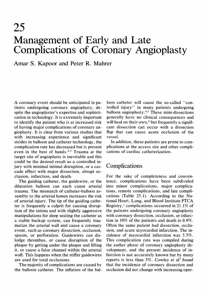
25 Management of Early and Late Complications of Coronary Angioplasty Amar S. Kapoor and Peter R. Mahrer
A coronary event should be anticipated in patients undergoing coronary angioplasty, despite the angioplaster's expertise and sophistication in technology. It is extremely important to identify the patient who is at increased risk of having major complications of coronary angioplasty. It is clear from various studies that with increasing experience and significant strides in balloon and catheter technology, the complication rate has decreased but is present even in the best of hands. I- 3 Trauma at the target site of angioplasty is inevitable and this could be the desired result as a controlled injury with minimal intimal disruption, or a cascade effect with major dissection, abrupt occlusion, infarction, and death.
The guiding catheter, the guidewire, or the dilatation balloon can each cause arterial trauma. The mismatch of catheter-balloon assembly to the arterial lumen increases the risk of arterial injury. The tip of the guiding catheter is frequently a culprit for causing disruption of the intima and with slightly aggressive manipulations for deep seating the catheter as a stable backup system, can frequently traumatize the arterial wall and cause a coronary event, such as coronary dissection, occlusion, spasm, or perforation. Guidewires can dislodge thrombus, or cause disruption of the plaque by getting under the plaque and lifting it, or cause a false channel within the arterial wall. This happens when the stiffer guidewires are used for total occlusions.
The majority of complications are caused by the balloon catheter. The inflation of the bal-
loon catheter will cause the so-called "controlled injury" in many patients undergoing balloon angioplasty. 4,5 These mini-dissections generally have no clinical consequences and will heal on their own,6 but frequently a significant dissection can occur with a dissection flap that can cause acute occlusion of the vessel.
In addition, these patients are prone to complications at the access site and other complications of cardiac catheterization.
Complications
For the sake of completeness and convenience, complications have been subdivided into minor complications, major complications, remote complications, and late complications (Table 25.1). According to the N ational Heart, Lung, and Blood Institute PTCA Registry, I complications occurred in 21.1 % of the patients undergoing coronary angioplasty with coronary dissection, occlusion, or infarction in 10% of the patients and death in 0.9%. Often the same patient had dissection, occlusion, and acute myocardial infarction. The incidence of myocardial infarction was 5.5%. This complication rate was compiled during the earlier phase of coronary angioplasty development, and the present incidence of infarction is not accurately known but by many reports is less than 5%. Cowley et aF found that the incidence of coronary dissection and occlusion did not change with increasing oper-
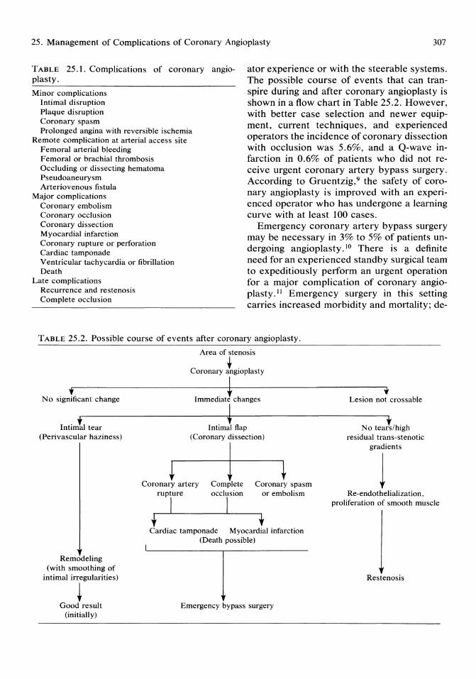
25. Management of Complications of Coronary Angioplasty 307
TABLE 25.1. Complications of coronary angioplasty.
ator experience or with the steerable systems. The possible course of events that can transpire during and after coronary angioplasty is shown in a flow chart in Table 25.2. However, with better case selection and newer equipment, current techniques, and experienced operators the incidence of coronary dissection with occlusion was 5.6%, and a Q-wave infarction in 0.6% of patients who did not receive urgent coronary artery bypass surgery. According to Gruentzig,9 the safety of coronary angioplasty is improved with an experienced operator who has undergone a learning curve with at least 100 cases.
Minor complications Intimal disruption Plaque disruption Coronary spasm Prolonged angina with reversible ischemia
Remote complication at arterial access site Femoral arterial bleeding Femoral or brachial thrombosis Occluding or dissecting hematoma Pseudoaneurysm Arteriovenous fistula
Major complications Coronary embolism Coronary occlusion Coronary dissection Myocardial infarction Coronary rupture or perforation Cardiac tamponade Ventricular tachycardia or fibrillation Death
Late complications Recurrence and restenosis Complete occlusion
Emergency coronary artery bypass surgery may be necessary in 3% to 5% of patients undergoing angioplasty. 10 There is a definite need for an experienced standby surgical team to expeditiously perform an urgent operation for a major complication of coronary angioplasty.11 Emergency surgery in this setting carries increased morbidity and mortality; de-
TABLE 25.2. Possible course of events after coronary angioplasty.
.. No significant change
I . i l ntlma tear (Perivascular haziness)
Remodeling (with smoothing of
intimal irregularities)
~ Good result
(initially)
J
Area of stenosis
Coronary igiOPlasty
d' * h
Intimal flap (Coronary dissection)
~ Coronary artery
1 Complete occlusion
Coronary spasm
I rupture
I t
or embolism
Cardiac tamponade Myocardial infarction (Death possible)
I Emergency bypass surgery
t Lesion not crossable
No teal/high residual trans-stenotic
gradients
1 Re-endothelialization,
proliferation of smooth muscle
Restenosis
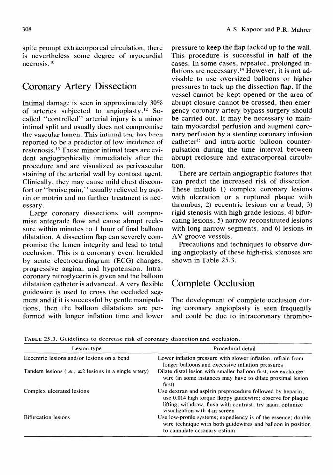
308
spite prompt extracorporeal circulation, there is nevertheless some degree of myocardial necrosis. 10
Coronary Artery Dissection
Intimal damage is seen in approximately 30% of arteries subjected to angioplasty.12 Socalled "controlled" arterial injury is a minor intimal split and usually does not compromise the vascular lumen. This intimal tear has been reported to be a predictor of low incidence of restenosis. 13 These minor intimal tears are evident angiographically immediately after the procedure and are visualized as perivascular staining of the arterial wall by contrast agent. Clinically, they may cause mild chest discomfort or "bruise pain," usually relieved by aspirin or motrin and no further treatment is necessary.
Large coronary dissections will compromise antegrade flow and cause abrupt reclosure within minutes to 1 hour of final balloon dilatation. A dissection flap can severely compromise the lumen integrity and lead to total occlusion. This is a coronary event heralded by acute electrocardiogram (ECG) changes, progressive angina, and hypotension. Intracoronary nitroglycerin is given and the balloon dilatation catheter is advanced. A very flexible guidewire is used to cross the occluded segment and if it is successful by gentle manipulations, then the balloon dilatations are performed with longer inflation time and lower
A.S. Kapoor and P.R. Mahrer
pressure to keep the flap tacked up to the wall. This procedure is successful in half of the cases. In some cases, repeated, prolonged inflations are necessary.J4 However, it is not advisable to use oversized balloons or higher pressures to tack up the dissection flap. If the vessel cannot be kept opened or the area of abrupt closure cannot be crossed, then emergency coronary artery bypass surgery should be carried out. It may be necessary to maintain myocardial perfusion and augment coronary perfusion by a stenting coronary infusion catheterl5 and intra-aortic balloon counterpulsation during the time interval between abrupt reclosure and extracorporeal circulation.
There are certain angiographic features that can predict the increased risk of dissection. These include 1) complex coronary lesions with ulceration or a ruptured plaque with thrombus, 2) eccentric lesions on a bend, 3) rigid stenosis with high grade lesions, 4) bifurcating lesions, 5) narrow reconstituted lesions with long narrow segments, and 6) lesions in A V groove vessels.
Precautions and techniques to observe during angioplasty of these high-risk stenoses are shown in Table 25.3.
Complete Occlusion
The development of complete occlusion during coronary angioplasty is seen frequently and could be due to intracoronary thrombo-
TABLE 25.3. Guidelines to decrease risk of coronary dissection and occlusion.
Lesion type Procedural detail
Eccentric lesions and/or lesions on a bend Lower inflation pressure with slower inflation; refrain from longer balloons and excessive inflation pressures
Tandem lesions (i.e., ~2 lesions in a single artery) Dilate distal lesion with smaller balloon first; use exchange wire (in some instances may have to dilate proximal lesion first)
Complex ulcerated lesions Use dextran and aspirin preprocedure followed by heparin; use 0.014 high torque floppy guidewire; observe for plaque lifting; withdraw, flush with contrast; try again; optimize visualization with 4-in screen
Bifurcation lesions Use low-profile systems; expediency is of the essence; double wire technique with both guidewires and balloon in position to cannulate coronary ostium
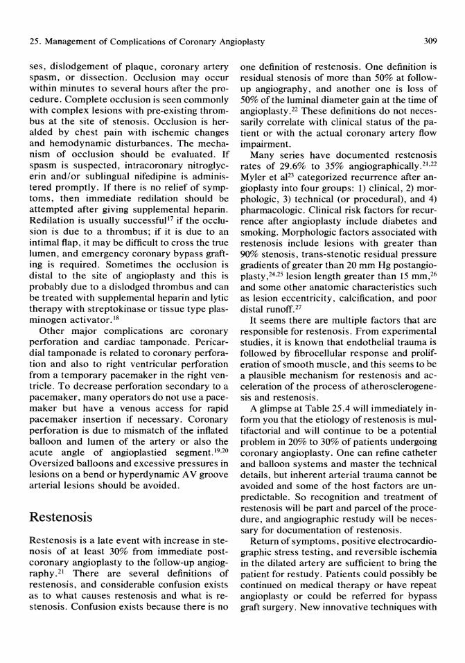
25. Management of Complications of Coronary Angioplasty 309
ses, dislodgement of plaque, coronary artery spasm, or dissection. Occlusion may occur within minutes to several hours after the procedure. Complete occlusion is seen commonly with complex lesions with pre-existing thrombus at the site of stenosis. Occlusion is heralded by chest pain with ischemic changes and hemodynamic disturbances. The mechanism of occlusion should be evaluated. If spasm is suspected, intracoronary nitroglycerin and/or sublingual nifedipine is administered promptly. If there is no relief of symptoms, then immediate redilation should be attempted after giving supplemental heparin. Redilation is usually successful17 if the occlusion is due to a thrombus; if it is due to an intimal flap, it may be difficult to cross the true lumen, and emergency coronary bypass grafting is required. Sometimes the occlusion is distal to the site of angioplasty and this is probably due to a dislodged thrombus and can be treated with supplemental heparin and lytic therapy with streptokinase or tissue type plasminogen activator. 18
Other major complications are coronary perforation and cardiac tamponade. Pericardial tamponade is related to coronary perforation and also to right ventricular perforation from a temporary pacemaker in the right ventricle. To decrease perforation secondary to a pacemaker, many operators do not use a pacemaker but have a venous access for rapid pacemaker insertion if necessary. Coronary perforation is due to mismatch of the inflated balloon and lumen of the artery or also the acute angle of angioplastied segment. 19,20 Oversized balloons and excessive pressures in lesions on a bend or hyperdynamic A V groove arterial lesions should be avoided.
Restenosis
Restenosis is a late event with increase in stenosis of at least 30% from immediate postcoronary angioplasty to the follow-up angiography.21 There are several definitions of restenosis, and considerable confusion exists as to what causes restenosis and what is restenosis. Confusion exists because there is no
one definition of restenosis. One definition is residual stenosis of more than 50% at followup angiography, and another one is loss of 50% of the luminal diameter gain at the time of angioplasty.22 These definitions do not necessarily correlate with clinical status of the patient or with the actual coronary artery flow impairment.
Many series have documented restenosis rates of 29.6% to 35% angiographically.2l,22 Myler et al23 categorized recurrence after angioplasty into four groups: 1) clinical, 2) morphologic, 3) technical (or procedural), and 4) pharmacologic. Clinical risk factors for recurrence after angioplasty include diabetes and smoking. Morphologic factors associated with restenosis include lesions with greater than 90% stenosis, trans-stenotic residual pressure gradients of greater than 20 mm Hg postangioplasty,24,25 lesion length greater than 15 mm,26 and some other anatomic characteristics such as lesion eccentricity, calcification, and poor distal runoff. 27
It seems there are mUltiple factors that are responsible for restenosis. From experimental studies, it is known that endothelial trauma is followed by fibrocellular response and proliferation of smooth muscle, and this seems to be a plausible mechanism for restenosis and acceleration of the process of atherosclerogenesis and restenosis.
A glimpse at Table 25.4 will immediately inform you that the etiology of restenosis is multifactorial and will continue to be a potential problem in 20% to 30% of patients undergoing coronary angioplasty. One can refine catheter and balloon systems and master the technical details, but inherent arterial trauma cannot be avoided and some of the host factors are unpredictable. So recognition and treatment of restenosis will be part and parcel of the procedure, and angiographic restudy will be necessary for documentation of restenosis.
Return of symptoms, positive electrocardiographic stress testing, and reversible ischemia in the dilated artery are sufficient to bring the patient for restudy. Patients could possibly be continued on medical therapy or have repeat angioplasty or could be referred for bypass graft surgery. New innovative techniques with
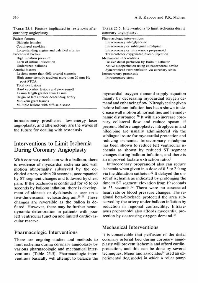
310
TABLE 25.4. Factors implicated in restenosis after coronary angioplasty.
Patient factors Diabetic females Continued smoking Long-standing angina and calcified arteries
Procedural factors High inflation pressure Lack of intimal dissection Undersized balloons
Arterial factors Lesions more than 90% arterial stenosis High trans-stenotic gradient more than 20 mm Hg
post-PTCA Total occlusions Hard eccentric lesions and poor runoff Lesion length greater than 15 mm Origin of left anterior descending artery Mid-vein graft lesions Multiple lesions with diffuse disease
intracoronary prostheses, low-energy laser angioplasty, and atherectomy are the waves of the future for dealing with restenosis.
Interventions to Limit Ischemia During Coronary Angioplasty
With coronary occlusion with a balloon, there is evidence of myocardial ischemia and wall motion abnormality sub served by the occluded artery within 20 seconds, accompanied by ST segment changes and followed by chest pain. If the occlusion is continued for 45 to 60 seconds by balloon inflation, there is development of akinesis or dyskinesis as seen on a two-dimensional echocardiogram. 28,29 These changes are reversible as the ballon is deflated. However, there may be further hemodynamic deterioration in patients with poor left ventricular function and limited cardiovascular reserve.
Pharmacologic Interventions There are ongoing studies and methods to limit ischemia during coronary angioplasty by various pharmacologic and mechanical interventions (Table 25.5). Pharmacologic interventions basically will attempt to balance the
A.S. Kapoor and P.R. Mahrer
TABLE 25.5. Interventions to limit ischemia during coronary angioplasty.
Pharmacologic interventions Intracoronary nitroglycerine Intracoronary or sublingual nifedipine Intracoronary or intravenous propranolol Transcatheter oxygenated fluosol injection
Mechanical interventions Passive distal perfusion by Bailout catheter Active autoperfusion using extracorporeal device Synchronized retroperfusion via coronary sinus
Intracoronary prosthesis Intracoronary stent
myocardial oxygen demand-supply equation mainly by decreasing myocardial oxygen demand and enhancing flow. Nitroglycerine given before balloon inflation has been shown to decrease wall motion abnormalities and hemodynamic disturbance. 3o It will also increase coronary collateral flow and reduce spasm, if present. Before angioplasty, nitroglycerin and nifedipine are usually administered via the sublingual route for myocardial protection and reducing ischemia. Intracoronary nifedipine has been shown to reduce left ventricular ischemia as shown by reduced ST segment changes during balloon inflation, and there is an improved lactate extraction ratio. 31
Intracoronary propranolol also can reduce ischemia when given in a dose of 0.5 to 2.0 mg via the dilatation catheter. 32 It delayed the onset of ischemia as indicated by prolonging the time to ST segment elevation from 19 seconds to 53 seconds.32 There were no associated heart rate or blood pressure changes. The regional beta-blockade protected the area subserved by the artery under balloon inflation by reduction in regional contractility. Intravenous propranolol also affords myocardial protection by decreasing oxygen demand. 33
Mechanical Interventions It is conceivable that perfusion of the distal coronary arterial bed during coronary angioplasty will prevent ischemia and afford cardioprotection, and this can be done by several techniques. Meier and associates34 used an experimental dog model in which a roller pump
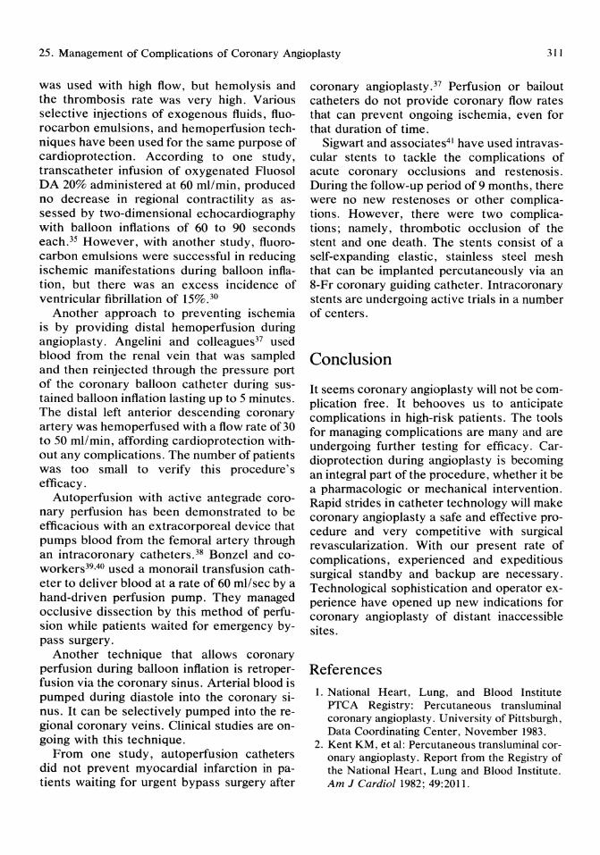
25. Management of Complications of Coronary Angioplasty 311
was used with high flow, but hemolysis and the thrombosis rate was very high. Various selective injections of exogenous fluids, fluorocarbon emulsions, and hemoperfusion techniques have been used for the same purpose of cardioprotection. According to one study, transcatheter infusion of oxygenated Fluosol DA 20% administered at 60 ml/min, produced no decrease in regional contractility as assessed by two-dimensional echocardiography with balloon inflations of 60 to 90 seconds each.35 However, with another study, fluorocarbon emulsions were successful in reducing ischemic manifestations during balloon inflation, but there was an excess incidence of ventricular fibrillation of 15%.30
Another approach to preventing ischemia is by providing distal hemoperfusion during angioplasty. Angelini and colleagues37 used blood from the renal vein that was sampled and then reinjected through the pressure port of the coronary balloon catheter during sustained balloon inflation lasting up to 5 minutes. The distal left anterior descending coronary artery was hemoperfused with a flow rate of 30 to 50 ml/min, affording cardioprotection without any complications. The number of patients was too small to verify this procedure's efficacy.
Autoperfusion with active antegrade coronary perfusion has been demonstrated to be efficacious with an extracorporeal device that pumps blood from the femoral artery through an intracoronary catheters. 38 Bonzel and coworkers39,40 used a monorail transfusion catheter to deliver blood at a rate of 60 ml/sec by a hand-driven perfusion pump. They managed occlusive dissection by this method of perfusion while patients waited for emergency bypass surgery.
Another technique that allows coronary perfusion during balloon inflation is retroperfusion via the coronary sinus. Arterial blood is pumped during diastole into the coronary sinus. It can be selectively pumped into the regional coronary veins. Clinical studies are ongoing with this technique.
From one study, autoperfusion catheters did not prevent myocardial infarction in patients waiting for urgent bypass surgery after
coronary angioplasty. 37 Perfusion or bailout catheters do not provide coronary flow rates that can prevent ongoing ischemia, even for that duration of time.
Sigwart and associates41 have used intravascular stents to tackle the complications of acute coronary occlusions and restenosis. During the follow-up period of9 months, there were no new restenoses or other complications. However, there were two complications; namely, thrombotic occlusion of the stent and one death. The stents consist of a self-expanding elastic, stainless steel mesh that can be implanted percutaneously via an 8-Fr coronary guiding catheter. Intracoronary stents are undergoing active trials in a number of centers.
Conclusion
It seems coronary angioplasty will not be complication free. It behooves us to anticipate complications in high-risk patients. The tools for managing complications are many and are undergoing further testing for efficacy. Cardioprotection during angioplasty is becoming an integral part of the procedure, whether it be a pharmacologic or mechanical intervention. Rapid strides in catheter technology will make coronary angioplasty a safe and effective procedure and very competitive with surgical revascularization. With our present rate of complications, experienced and expeditious surgical standby and backup are necessary. Technological sophistication and operator experience have opened up new indications for coronary angioplasty of distant inaccessible sites.
References 1. National Heart, Lung, and Blood Institute
PTCA Registry: Percutaneous transluminal coronary angioplasty. University of Pittsburgh, Data Coordinating Center, November 1983.
2. Kent KM, et al: Percutaneous trans luminal coronary angioplasty. Report from the Registry of the National Heart, Lung and Blood Institute. Am J Cardiol1982; 49:2011.

312
3. Dorros G, et al: Percutaneous transluminal coronary angioplasty: Report of complications from the National Heart, Lung, and Blood Institute PTCA Registry. Circulation 1983; 67:722.
4. Castaneda-Zunga WR, et al: The mechanism of balloon angioplasty. Radiology 1980; 135:565.
5. Holmes DR, et al: Angiographic changes produced by percutaneous transluminal coronary angioplasty. Am J Cardiol1983; 51:676.
6. Bain DS: Percutaneous transluminal angioplasty, in Braunwald E (ed): Harrison's Principles of Internal Medicine: Update VI. New York, McGraw-Hill, 1985, pp 133-146.
7. Cowley MJ, et al: Acute coronary events associated with percutaneous transluminal coronary angioplasty. Am J Cardiol1984; 53(suppl C):12.
8. Bredlau CE, et al: Acute complications of percutaneous transluminal coronary angioplasty. Initial experience in 3,000 consecutive patient attempts. Circulation 1984; 70(suppl 2):106.
9. Gruentzig AR: Results from coronary angioplasty and implications for the future. Am Heart J 1982; 103:779.
10. Reul GJ, et al: Coronary artery bypass for unsuccessful percutaneous transluminal coronary angioplasty. J Thorac Cardiouasc Surg 1984; 88:685.
11. Akins CW, Block PC: Surgical intervention for failed percutaneous transluminal coronary angioplasty. Am J Cardiol 1984; 53(suppl C): 108.
12. Mathews BJ, et al: Natural history of angioplasty-induced dissection: A predictor of restenosis. J Am Coli Cardiol1985; 5:1143.
13. Leimgruber P, et al: Influence of intimal dissection on restenosis after coronary angioplasty on restenosis rate. Circulation 1984; 70(suppl 2):175.
14. Hollman J, et al: Acute occlusion after percutaneous transluminal angioplasty-a new approach. Circulation 1983; 68:725.
15. Hinohara T, et al: Transluminal catheter reperfusion: A new technique to re-establish blood flow after coron~ry occlusion during percutaneous transluminal coronary angioplasty. Am J Cardiol 1986; 57:684.
16. Meier B, et al: Does length or eccentricity of coronary stenosis influence the outcome of transluminal dilatation? Circulation 1983; 67:497.
17. Hollman J, et al: Acute occlusion after percutaneous transluminal coronary angioplasty-a new approach. Circulation 1983; 68:725.
18. Schofer J, et al: Acute coronary artery occlu-
A.S. Kapoor and P.R. Mahrer
sion during percutaneous transluminal coronary angioplasty. Reopening by intracoronary streptokinase before emergency coronary artery surgery. Circulation 1982; 66:1325.
19. Saffitz JE, et al: Coronary artery rupture during coronary angioplasty. Am J Cardiol 1983; 51:902.
20. Kimbus D, et al: Transluminal coronary angioplasty complicated by coronary artery perforation. Cathet Cardiouasc Diagn 1982; 8:481.
21. Holmes DR Jr, et al: Restenosis after percutaneous transluminal coronary angioplasty: A report from the PTCA Registry of the National Heart, Lung, and Blood Institute. Am J Cardiol 1984; 53(suppl C):77.
22. Meier B, et al: Repeat coronary angioplasty. J Am Coli Cardiol1984; 4:463.
23. Myler RK, et al: Recurrence after coronary angioplasty. Cathet Cardiouasc Diagn 1987; 13:77-86.
24. Leimgruber PP, et al: Restenosis after successful coronary angioplasty in patients with singlevessel disease. Circulation 1986; 73:710-717.
25. Hoffmeister JM, et al: Analysis of anatomic and procedural factors related to restenosis after double lesion coronary angioplasty. Circulation 1985; 72(suppl II):II1-398.
26. Uebis R, et al: Recurrence rate after PTCA in relationship to the initial length of coronary artery narrowing. J Am Coli Cardiol1986; 7:62A.
27. Mata LA, et al: Clinical and angiographic assessment 6 months after double vessel PTCA. J Am Coli Cardiol1985; 6:1239.
28. Wohlgeternter D, et al: Regional myocardial dysfunction during coronary angioplasty: Evaluation by two-dimensional echocardiography and 12-lead electrocardiography. J Am Coli Cardiol 1986; 7: 1245.
29. Hauser AM, et al: Sequence of mechanical electrocardiographic and clinical effects of repeated coronary artery occlusion in human beings: Echocardiographic observations during coronary angioplasty. J Am Coli Cardiol 1985; 5:193.
30. Doorey AJ, et al: Amelioration by nitroglycerin of left ventricular ischemia induced by percutaneous transluminal coronary angioplasty: Assessment by hemodynamic variables and left ventriculography. JAm Coli Cardiol1985; 6:67.
31. Pop G, et al: Regional cardioprotection by intracoronary nifedipine is not due to enhanced collateral flow during coronary angioplasty. Circulation 1986; 74: 11-364.
32. Zalewski A, et al: Myocardial protection during
25. Management of Complications of Coronary Angioplasty 313
transient coronary artery occlusion in man: Beneficial effects on regional adrenergic blockade. Circulation 1986; 73:734.
33. Feldman RL, et al: Effect of propranolol on myocardial ischemia occurring during acute coronary occlusion. Circulation 1986; 73:727.
34. Meier B, Gruentzig AR, Brown JE: Percutaneous arterial perfusion of acutely occluded coronary arteries in dogs (abstr). J Am Coil Cardiol 1984; 3:505.
35. Cleman M, et al: Prevention of ischemia during percutaneous transluminal coronary angioplasty by transcath or infusion of oxygenated Fluosol DA 20%. Circulation 1986; 74:555.
36. Anderson HV, et al: Distal coronary artery perfusion during percutaneous transluminal coronary angioplasty. Am Heart J 1985; 110:720-726.
37. Angelini P, Heibig J, Leachman R: Distal hemoperfusion during percutaneous transluminal coronary angioplasty. Am J Cardiol 1986; 58:252-255.
38. Meier B, et al: Percutaneous perfusion of occluded coronary arteries with blood from the femoral artery: A dog study. Cathet Cardiouasc Diagn 1985; 11:81.
39. Bonzol T, et al: The steerable monorail catheter system-a new system for percutaneous transluminal coronary angioplasty. Circulation 1986; 74:11-459.
40. Bonzol T: Monorail balloon catheter for coronary dilatation and revascularization. PTCA Course IV, Geneva, Switzerland, 1987.
41. Sigwart U, et al: Intravascular stents to prevent occlusion and restenosis after transluminal angioplasty. N Engl J Med 1987; 316:701-706.

26 Percutaneous Transluminal Coronary Stenting: ANew Approach to Unresolved Problems in Coronary Angioplasty Ulrich Sigwart, Svein Golf, Urs Kaufmann, Lukas Kappenberger, Adam Fischer, and Hossein Sadeghi
Introduction
Percutaneous transluminal coronary angiopia sty (PTCA) has been established as a safe and effective procedure for improving blood flow in narrowed atherosclerotic arteries. The stenoses recur, however, in a certain percentage of initially successful cases. Also, PTCA may resolve in abrupt closure of the artery due to intimal dissection and formation of intimal flaps or thrombosis. The purpose of an intravascular endoprosthesis (stent) is to restore and maintain blood flow by nonsurgical implantation via a catheter after transluminal angioplasty.
According to recent publications l ,2 the restenosis rate per lesion is approximately 33%. The incidence of acute closure after angioplasty is smaller but still significant, requiring surgical standby. 3,4 Despite emergency revascularization, loss of myocardium cannot always be prevented and the operation carries a higher risk as compared with elective surgery.5 Therefore, there is great interest in methods that may be capable of preventing these important limitations of PTCA.
The ideal intravascular endoprosthesis (stent) that prevents restenosis as well as acute occlusion after angioplasty is nonthrombogenic, flexible along its long axis, and both compressible and expandable in diameter. Once positioned, it should remain in its position without migrating and produce neither
pressure necrosis of the arterial wall nor inflammatory response. It also should not produce excessive intimal proliferation. Such a stent should equally be mounted on small delivery catheters to be introduced into the target vessel via an angioplasty guiding catheter.
So far no such stent exists. A new design, however, has been developed in Lausanne and tested in animals as well as in humans (Medinvent SA, Lausanne, Switzerland). We report our initial experience with percutaneous transluminal coronary stenting (PTCS) in humans.
Description of the Stent
The stent is woven from a surgical grade stainless steel alloy formulated to International Standards Organization prescription. Due to its design (Fig 26.1) and process of fabrication, the prosthesis can be made geometrically stable, compliant, and self-expanding. The elastic and compliant properties of the prosthesis are such that by moderate longitudinal elongation, the prosthesis' diameter may be significantly reduced. It can thus be constrained on a small diameter delivery catheter, and as the constraining membrane is progressively withdrawn, the device will elastically return to its original unconstrained large diameter. The stent is flexible along its long axis, and for coronary implants its length varies between 15

26. Percutaneous Transluminal Coronary Stenting
FIGURE 26.1. The stent in its unconstrained form, liberated from the deployment catheter.
and 23 mm and its diameter between 3 and 5 mm in the fully expanded state. When implanted in a vessel that has a caliber smaller than that of the prosthesis unconstrained diameter, there will be a residually elastic radial force in the prosthesis that will keep the device firmly in place and exert a sufficient resistance to arterial contraction.
The delivery system used for coronary arteries has an outer diameter of 1.57 mm and can accommodate stents up to 6.5 mm in diameter. The constrained wire mesh is mounted on the distal end of the delivery catheter. Low friction between the two layers of the membrane is maintained by filling of the intermediate space with contrast medium at some 3 bars pressure. This allows for sufficient visualization of the retaining membrane during deployment.
Experimental Data
The prosthesis was placed into femoral and coronary arteries of dogs to assess the reaction of the endothelium and the thrombogenecity. Heparin was given only during the procedure and no anticoagulants or antiplate-
315
let agents were administrated afterward. The angiographic and histologic follow-up data showed patency and complete intimal coverage in almost all animals. The ostia of side branches that were covered by the stent remained widely patent, and no case of embolization or stent deplacement was observed. Thrombosis occurred only when there was an important mismatch between stent and vessel diameter. Histologic examination at various time intervals after implantation revealed a thin neointimal layer that covered the stent, thus isolating it from the blood stream. There were no signs of inflammatory of foreign body reaction around the prosthesis strands. Six months after implantation the status was unchanged, the thickness of the neointima varying between 150 and 450 /Lm.
Human Experience
Peripheral Arteries
The first experience with stenting of human arteries was ottained in peripheral arterial disease. Implantations were carried out immediately after balloon angioplasty of the femoral and iliac artery. Some lesions required more than one stent, the dimension of which ranged from 6 to 12 mm in diameter and from 4 to 8 cm in length. For most cases, antiplatelet drugs (aspirin 330 mg and dipyridamol 75 mg twice daily) were given together with oral anticoagulation therapy (acenocoumarol). This medication was continued for 3 months.
Coronary Implantations in Humans
During the past 18 months, 64 coronary stents were implanted in 53 patients. Indications were: 1) abrupt closure after transluminal coronary angioplasty (PTCA); 2) restenosis after PTCA, and 3) stenosis in coronary artery bypass graft.
The preoperative drug regimen consisted of platelet aggregation inhibitors, such as aspirin and persantine as well as sulfinpyrazone. During implantation the patient received a bolus injection of 10,000 to 15,000 U of heparin as
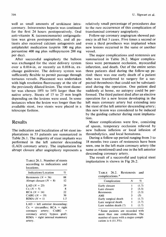
316
well as small amounts of urokinase intracoronary. Intravenous heparin was continued for the first 24 hours postoperatively. Oral anti-vitamin K (acenocoumarin) antigoagulation was given postoperatively, and all patients also received calcium antagonists and antiplatelet medication (aspirin 100 mg plus persantine 400 mg plus sulfinpyrazon 200 mg per day).
Mter successful angioplasty the balloon was exchanged for the stent delivery system over a 0.014-in, or recently an 0.018-in, exchange guidewire. The delivery system was sufficiently flexible to permit passage through tortuous vessels. Placement was undertaken with high resolution fluoroscopy at the site of the previously dilated lesion. The stent diameter was chosen 10% to 14% larger than the native artery. Stents of 15 to 23 mm length depending on the lesions were used. In some instances when the lesion was longer than the available stent, two stents were placed in a telescope fashion.
Results
The indication and localization of 64 stent implantations in 53 patients are summarized in Table 26.1. The majority of stent implants was performed in the left anterior descending (LAD) coronary artery. The implantation for abrupt closure after angioplasty represents a
TABLE 26.1. Number of stents according to indications and locations.
Indications/Location %
Restenosis (N = 56) 88 Abrupt closure (N = 8) 12
LAD (N = 25) 39 Cx (N = 5) 8 RCA (N = 14) 22 CABG (N = 19) 30 RIMA (N = 1) 1.5
LAD = left anterior descending; Cx = circumflex; RCA = right coronary artery; CABG coronary artery' bypass graft; RIMA = right internal mammary artery.
U. Sigwart et al.
relatively small percentage of procedures due to the rare occurrence of this complication of transluminal coronary angioplasty.
Follow-up coronary angiogram showed patency in all but 3 cases. Five times a second or even a third prosthesis was implanted when new lesions occurred in the same or another vessel.
The major complications and restenosis are summarized in Table 26.2. Major complications were permanent occlusion, myocardial infarction, and death. Out of the entire series three patients died during the follow-up period: there was one early death of a patient who was transferred to surgery for a suspected thrombosis that could not be substantiated during the operation. One patient died suddenly at home, no autopsy could be performed. The third patient died after an elective operation for a new lesion developing in the left main coronary artery but extending into the stent of the left anterior descending artery; this new lesion was considered to be induced by the guiding catheter during stent implantation.
Minor complications were few, consisting of spasm, temporary occlusion relieved by new balloon inflation or local infusion of thrombolytics, and local hematoma.
During a follow-up period ranging from 3 to 18 months two cases of restenosis have been seen, one in the left main coronary artery (the same as mentioned) and one in the left anterior descending coronary artery.
The result of a successful and typical stent implantation is shown in Fig 26.2.
TABLE 26.2. Restenosis and complications. *
Complication N %
Early closure 1 (1.7) Late closure 2 (3.3) Restenosis 2 (3.3) AMI 3 (5) Early surgical death (1.7) Late surgical death (1.7) Late sudden death (?) (1.7)
* Some patients are recorded with more than one complication. The number of cases with a major complication is 6 (11.3%).

26. Percutaneous Transluminal Coronary Stenting
C
FIGURE 26.2. The right coronary artery showing a significant restenosis (A) before dilatation and stent
Discussion The use of stents to prevent complications of trans luminal angioplasty has been proposed by several investigators.6-8 The risk of implantation of foreign bodies into coronary arteries consist of the induction of uncontrollable vasomotor activity, thrombosis, and the induction of intimal hyperplasia. 9 For these reasons human stent implants have not been performed until recently.1O The stent described herein was found to be sufficiently well accepted in animal arteries as well as in human leg arteries to allow implantation in human coronary vessels. Our initial results corroborate the hypothesis that intraluminal scaffold-
317
B
o
implantation (B) immediately after PTCA, and (C,D) after stent implantation.
ing devices may relieve occlusion after angioplasty and prevent restenosis in a significant proportion of patients.
With restenosis rate as high as 33% after coronary angioplasty and even higher in multivessel angioplasty4 the overall value of balloon angioplasty is significantly reduced even when one admits the relatively low morbidity of such a procedure, which is frequently repeated not once but several times. The socioeconomic implications of repeat angioplasty are important and counterbalance largely the initially low comparative cost of the intervention. II Experience has shown that the acute and chronic reocclusion rates after peripheral and coronary angioplasty are inde-
318
pendent of operator's skill and the quality of the materials used. Longer inflation times, high-dose slow calcium blockers, steroids, and other drugs have thus far failed to provide a major contribution to the prevention of this problem. The arterial occlusion and restenosis rate is probably largely related to the composition and structure of the plaque and the nature of the trauma applied to the vessel wall.
A number of different stent designs have been examined in animals.6-8,12 The rate of intimal thickening is relatively constant, and the time for covering the stent surface depends on the thickness of metal element. The relative high porosity of this stent design seems to be beneficial as it exposes rather small metal surfaces to the blood stream. Problems arise at the open ends of the stent, which may traumatize the artery and create a compliance mismatch that might induce intimal hyperplasia.
The fact that restenosis did not occur in the great majority of our patients may be due to a number of factors: the stent prevents elastic recoil of the artery after balloon angioplasty as well as contraction during the healing phase. More importantly, however, it creates an almost ideal lumen with favorable hemodynamics immediately after implantation. The optimal hemodynamics have been substantiated by pressure measurements, and it was noted that the residual gradient after balloon angioplasty was reduced to almost zero after the stent implantation. Small amounts of intimal hyperplasia, however, could theoretically also contribute to the lesser rate of restenosis because the tissue barrier thus formed may serve as a kind of protection against further cell ingrowth.
Despite the encouraging results of this series the disadvantages and potential risks of foreign material in coronary arteries must not be overlooked. The heavy anticoagulant treatment of the current stent model requires meticulous patient care and prolonged hospital stay after the procedure. Spasm may continue to pose problems even after the immediate postoperative period. Further work is necessary before definite recommendations can be given as to the use of intracoronary stents.
U. Sigwart et al.
References 1. Holmes DR Jr, Vliestra RE, Smith HL, et al:
Restenosis after percutaneous trans luminal coronary angioplasty (PTCA): A report from the PTCA registry of the National Heart, Lung and Blood Institute. Am J Cardiol 1984; 53:77-81.
2. Leimbruber PP, Roubin GS, Hollman J, et al: Restenosis after successful coronary angioplasty in patients with single-vessel disease. Circulation 1986; 73:710-717.
3. Sugrue DD, Holmes DR Jr, Smith HC, et al: Coronary artery thrombus, risk factor for acute vessel occlusion during percutaneous transluminal angioplasty. Br Heart J 1986; 56:62.
4. Finci L, Meier B, DeBrugne B, et al: Angiographic follow-up after multivessel percutaneous transluminal coronary angioplasty. Am J Cardiol 1987; 60:467-570.
5. Simpfendorfer C, Belardi J, Bellamy G, et al: Frequency, management and follow-up of patients with acute coronary occlusions after percutaneous transluminal angioplasty. Am J Cardial 1987; 59:267-269.
6. Maass D, Kropf L, Egloff L, et al: Transluminal implantations of intravascular "double helix" spiral prostheses: Technical and biological considerations. Proc Eur Soc Artif Organs 1982; 9:252-256.
7. Dotter C, Buschmann RW, McKinney MK, et al: Transluminal expandable nitinol coil stent grafting: Preliminary report. Radiology 1983; 147:259-260.
8. Palmaz JC, Windeler SA, Garcia F, et al: Atherosclerotic rabbit aortas: Expandable intraluminal grafting. Radiology 1986; 160:723-726.
9. Waller BF, Pinkerton LA, Foster LN: Morphologic evidence of accelerated left maincoronary artery stenosis: A late complication of percutaneous transluminal balloon angioplasty of the proximal left anterior descending coronary artery. J Am Call Cardiol1987; 9:1019-1023.
10. Sigwart U, Puel J, Mirkovitch V, et al: Intravascular stents to prevent occlusion and restenosis after transluminal angioplasty. N Engl J Med 1987; 316:701-706.
11. Reeder GS, Krishan J, Nobrega FF, et al: Is percutaneous coronary angioplasty less expensive than bypass surgery? N Engl J Med 1984; 311: 1157-1162.
12. Dotter CT: Transluminally placed coilspring endarterial tube grafts: Long-term patency in canine popliteal artery. Invest Radiol 1969; 4:329-332.

27 Laser Angioplasty of the Coronary Arteries Garrett Lee, Reginald I. Low, Agustin J. Argenal, Rolf G. Sommerhaug, Ming C. Chan, and Dean T. Mason
Since the laser was shown to be effective in removing coronary atherosclerotic obstruction in postmortem hearts,l much enthusiasm has been generated to rapidly apply this new technology to the clinical setting. It is hoped that the laser, once fully developed, will recanalize obstructed coronary arteries without the necessity of open-chest bypass graft surgery, thus avoiding the expense and the lengthy hospital recovery of the surgical procedure in those afflicted with the disease. Recognizing that any new technology requires research effort and time to develop, the purpose of this report is to review the current status of laser revascularization of the coronary arteries.
Applicable Lasers
Laser is the acronym for light amplification by stimulated emission of radiation. Several different lasers have been shown to be effective in removing atherosclerotic plaque obstructions.2-4 They are the argon, neodymium yttrium-aluminum-garnet (Nd: Y AG) and carbon dioxide (C02) lasers. The argon laser emits photons with wavelengths of 0.488 to 0.514 /Lm, within the blue-green portion of the electromagnetic spectrum. The Nd: YAG and the CO2 lasers emit wavelengths in the infrared invisible portion of the spectrum at 1.06 /Lm and 10.6 /Lm, respectively.
Another laser that also has been shown to ablate plaque produces photons in the ultravi-
olet range. The excimer (excited dimer) uses the rare gas halide as its lasing medium, that is, argon fluoride (0.193 /Lm), krypton fluoride (0.248 /Lm), xenon chloride (0.308 /Lm), and xenon fluoride (0.351 /Lm). The excimer laser, operating in the pulsed mode, transmits high energies in short, discrete pulses separated by a long emission-free interval (low repetition rates) and produces an effect on tissue that differs from the lasers that operate in the continuous-wave mode (e.g., argon); this is probably the result of heat dissipation between pulses or ablation via a nonthermal mechanism.5- 7 Other high-power lasers operating in the pulsed mode can also produce a lased channel without evidence of thermal damage to surrounding tissue. 8
Delivery of Laser Energies
The transmission of laser energies into the body is via optical fiber made of quartz silica. As laser light passes out of the fiber tip, the plane where the spot size is smallest is the location where the power intensity (watts) is greatest. The power density (watts/mm2) determines the effect of the energy delivered to the target obstruction. Power density varies directly with the energy and inversely with the surface area of the beam. As the distance away from the focal plane increases, the spot diameter or surface area enlarges and the power concentration falls.
The fiber used in the coronary artery must
320
be flexible and thin (i.e., less than .5 mm in outer diameter) and may be further protected by encasing the fiber within a catheter. Due to the refractive properties of the fiber, the beam is confined internally and the incident energy can be conducted to its target with very little loss of energies. Both argon and Nd: Y AG wavelengths can be transmitted in this manner. Although some ultraviolet energies (e.g., 0.351 JLm) from an excimer laser may also be delivered down the fiber; flexible optical fiber systems to transmit high-power pulsed lasers have not been developed. Although flexible waveguides for CO2 energies are available, these fibers made of silver chloride have an approximate 30% energy loss over aIm length.9
The optical fiber also can deliver argon and N d : Y AG laser energies to a metal cap mounted at its distal end.lO,Jl Activation by the laser heats up the metal cap, which can achieve temperatures high enough to instantaneously dissolve atherosclerotic lesions on physical contact. The laser-heated metal cap catheter system has advantages in that its use diminishes the inherent problems of a direct free beam inadvertently straying from the target area, particularly once the fiber tip has passed beyond an obstruction. It also minimizes hazards such as retinal damage to patients and medical staff personnel.
Laser Vaporization of the Coronary Plaque Obstruction
When the argon, Nd: YAG, or CO2 laser is directed on an area of plaque obstruction, the light energy is absorbed and is transformed to thermal energy, which attains temperatures exceeding well above 2000 C, and vaporization of the plaque obstruction results. The depth of the vaporized crater depends on the physical properties of the laser beam: the higher the power intensity, the longer the exposure; the more focused the beam, the deeper the crater. During lasing, solid or liquid matter is converted to gas; analysis of the gaseous products of irradiated plaque reveals water, carbon dioxide, nitrogen, hydrogen, and light hydrocar-
O. Lee et al.
bons. 12,I3 A carbonized or charred lining develops around the vaporized area, which generally is larger in proportion to the duration of laser exposure. Adjacent to and beyond the charred lining, there may be an area of acoustic injury, where cells and noncellular materials have boiled and been disrupted. Beyond this area, the tissue is intact.
In live atherosclerotic animal models, platelets, fibrin, and some inflammatory cells are deposited on the surface of the vaporized crater,14 Within 1 week, collagen is seen to infiltrate the area around the crater. This is followed by the start of re-endothelialization, and this process continues until the crater is fully re-endothelialized. Importantly, no serious thrombogenic complications occurred under these experimental conditions. Similar long-term effects of laser exposure occurred on the underlying normal vascular wall as well. 15
Argon and Nd: YAG laser energies carried by quartz fiber and directed coaxially along the central axis of atherosclerotic arteries can vaporize plaque adjacent to the stenotic lumen to widen the diameter of the channel (Fig 27.1). In completely obstructed arteries, the beam can be directed to clear a new passageway within the plaque obstruction. When laser energies are directed onto a metal cap, the depth of plaque penetration varies with contact duration and the physical characteristics of the obstruction. To ensure coaxial fiber alignment, the fiber tip or metal cap could be directed alongside a steerable guidewire initially inserted through and beyond the stenotic obstruction. 16
As mentioned, the excimer laser removes the atheroma by a different mechanism and produces little or no thermal damage. The depth of penetration of plaque varies with the cumulative number of ultraviolet pulses, whereas the diameter of the lased channel and its surroundings remain largely unchanged.
Potential Hazards
Potential hazards of laser energies in coronary arteries were demonstrated in live animal studiesY Under fluoroscopic guidance, a cor-
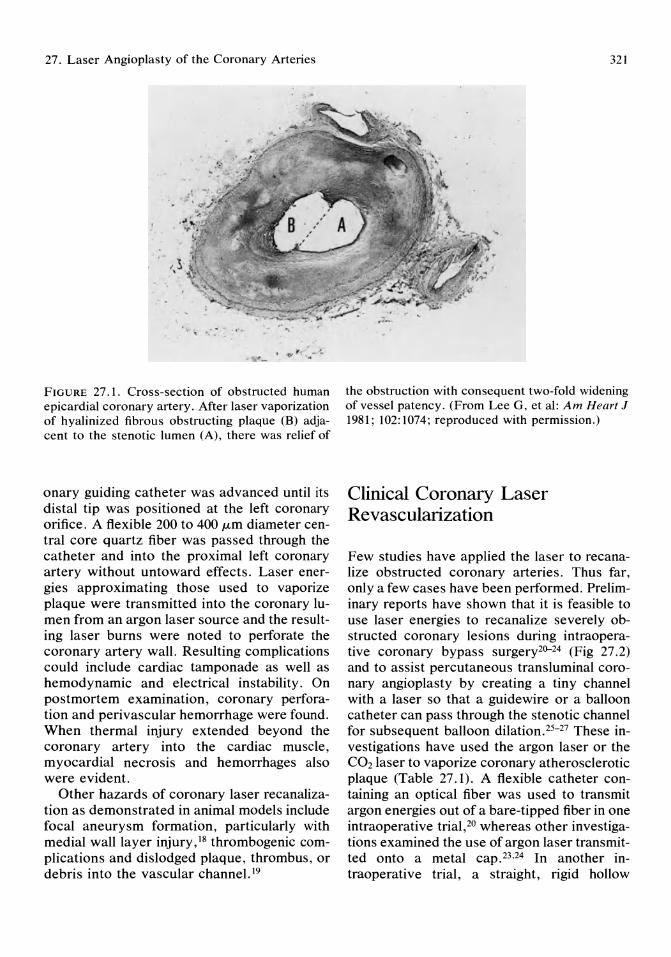
27. Laser Angioplasty of the Coronary Arteries
101 " r .. ... ,. ~ ,
' -r 'O. '-': .:-~
FIGURE 27.1. Cross-section of obstructed human epicardial coronary artery. After laser vaporization of hyalinized fibrous obstructing plaque (B) adjacent to the stenotic lumen (A), there was relief of
onary guiding catheter was advanced until its distal tip was positioned at the left coronary orifice. A flexible 200 to 400 J.Lm diameter central core quartz fiber was passed through the catheter and into the proximal left coronary artery without untoward effects. Laser energies approximating those used to vaporize plaque were transmitted into the coronary lumen from an argon laser source and the resulting laser burns were noted to perforate the coronary artery wall. Resulting complications could include cardiac tamponade as well as hemodynamic and electrical instability. On postmortem examination, coronary perforation and perivascular hemorrhage were found. When thermal injury extended beyond the coronary artery into the cardiac muscle, myocardial necrosis and hemorrhages also were evident.
Other hazards of coronary laser recanalization as demonstrated in animal models include focal aneurysm formation, particularly with medial wall layer injury, 18 thrombogenic complications and dislodged plaque, thrombus, or debris into the vascular channel. 19
321
the obstruction with consequent two-fold widening of vessel patency. (From Lee G, et al: Am Heart J 1981; 102:1074; reproduced with permission.)
Clinical Coronary Laser Revascularization
Few studies have applied the laser to recanalize obstructed coronary arteries. Thus far, only a few cases have been performed. Preliminary reports have shown that it is feasible to use laser energies to recanalize severely obstructed coronary lesions during intraoperative coronary bypass surgery2~24 (Fig 27.2) and to assist percutaneous transluminal coronary angioplasty by creating a tiny channel with a laser so that a guide wire or a balloon catheter can pass through the stenotic channel for subsequent balloon dilation. 25-27 These investigations have used the argon laser or the CO2 laser to vaporize coronary atherosclerotic plaque (Table 27.1). A flexible catheter containing an optical fiber was used to transmit argon energies out of a bare-tipped fiber in one intraoperative trial,2° whereas other investigations examined the use of argon laser transmitted onto a metal cap. 23,24 In another intraoperative trial, a straight, rigid hollow
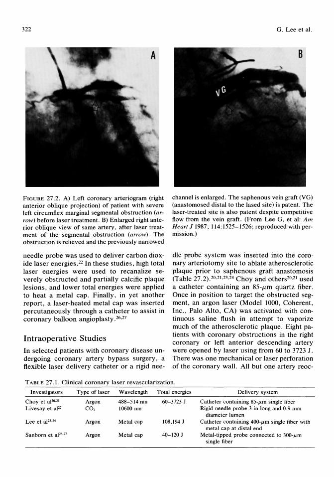
322
FIGURE 27.2. A) Left coronary arteriogram (right anterior oblique projection) of patient with severe left circumflex marginal segmental obstruction (arrow) before laser treatment. B) Enlarged right anterior oblique view of same artery, after laser treatment of the segmental obstruction (arrow). The obstruction is relieved and the previously narrowed
needle probe was used to deliver carbon dioxide laser energies. 22 In these studies, high total laser energies were used to recanalize severely obstructed and partially calcific plaque lesions, and lower total energies were applied to heat a metal cap. Finally, in yet another report, a laser-heated metal cap was inserted percutaneously through a catheter to assist in coronary balloon angioplasty. 26,27
Intraoperative Studies
In selected patients with coronary disease undergoing coronary artery bypass surgery, a flexible laser delivery catheter or a rigid nee-
G. Lee et al.
channel is enlarged. The saphenous vein graft (VG) (anastomosed distal to the lased site) is patent. The laser-treated site is also patent despite competitive flow from the vein graft. (From Lee G, et al: Am Heart J 1987; 114: 1525-1526; reproduced with permission.)
dIe probe system was inserted into the coronary arteriotomy site to ablate atherosclerotic plaque prior to saphenous graft anastomosis (Table 27.2).20.21.23.24 Choy and others20 ,21 used a catheter containing an 85-JLm quartz fiber. Once in position to target the obstructed segment, an argon laser (Model 1000, Coherent, Inc., Palo Alto, CA) was activated with continuous saline flush in attempt to vaporize much of the atherosclerotic plaque. Eight patients with coronary obstructions in the right coronary or left anterior descending artery were opened by laser using from 60 to 3723 J. There was one mechanical or laser perforation of the coronary wall. All but one artery reoc-
TABLE 27.1. Clinical coronary laser revascularization.
Investigators Type of laser Wavelength Total energies Delivery system
Choy et aFO,21 Argon 488-514 nm 60-3723 J Catheter containing 85-lLm single fiber Livesay et aJ22 CO2 10600 nm Rigid needle probe 3 in long and 0,9 mm
diameter lumen Lee et aJ23,24 Argon Metal cap 108,194 J Catheter containing 400-lLm single fiber with
metal cap at distal end Sanborn et aJ26,27 Argon Metal cap 40-120 J Metal-tipped probe connected to 300-lLm
single fiber
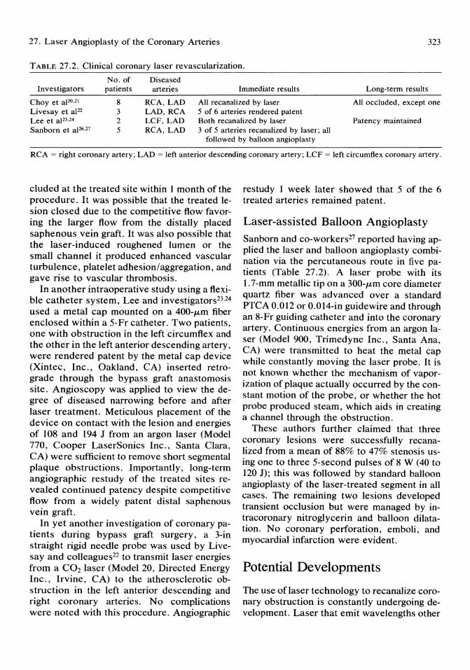
27. Laser Angioplasty of the Coronary Arteries 323
TABLE 27.2. Clinical coronary laser revascularization.
Investigators No. of
patients Diseased arteries Immediate results Long-term results
Choy et aJ20.2! Livesay et aJ22 Lee et aJ23,24 Sanborn et aJ26,27
8 3 2 5
RCA, LAD LAD, RCA LCF, LAD RCA,LAD
All recanalized by laser All ocduded, except one 5 of 6 arteries rendered patent Both recanalized by laser Patency maintained 3 of 5 arteries recanalized by laser; all
followed by balloon angioplasty
RCA = right coronary artery; LAD = left anterior descending coronary artery; LCF left circumflex coronary artery,
cluded at the treated site within 1 month of the procedure. It was possible that the treated lesion closed due to the competitive flow favoring the larger flow from the distally placed saphenous vein graft. It was also possible that the laser-induced roughened lumen or the small channel it produced enhanced vascular turbulence, platelet adhesion/aggregation, and gave rise to vascular thrombosis.
In another intraoperative study using a flexi-d · . t 2324 ble catheter system, Lee an mvestlga ors '
used a metal cap mounted on a 400-JLm fiber enclosed within a 5-Fr catheter. Two patients, one with obstruction in the left circumflex and the other in the left anterior descending artery, were rendered patent by the metal cap device (Xintec, Inc., Oakland, CA) inserted retrograde through the bypass graft anastomosis site. Angioscopy was applied to view the degree of diseased narrowing before and after laser treatment. Meticulous placement of the device on contact with the lesion and energies of 108 and 194 J from an argon laser (Model 770, Cooper LaserSonics Inc., Santa Clara, CA) were sufficient to remove short segmental plaque obstructions. Importantly, lo~g-term angiographic restudy of the treated SItes revealed continued patency despite competitive flow from a widely patent distal saphenous vein graft.
In yet another investigation of coronary patients during bypass graft surgery, a 3-in straight rigid needle probe was used by Livesay and colleagues22 to transmit laser energies from a CO2 laser (Model 20, Directed Energy Inc., Irvine, CA) to the atherosclerotic obstruction in the left anterior descending and right coronary arteries. No complications were noted with this procedure. Angiographic
restudy 1 week later showed that 5 of the 6 treated arteries remained patent.
Laser-assisted Balloon Angioplasty
Sanborn and co-workers27 reported having applied the laser and balloon angioplasty combination via the percutaneous route in five patients (Table 27.2). A laser probe with its 1.7 -mm metallic tip on a 300-JLm core diameter quartz fiber was advanced over a standard PTCA 0.012 or 0.014-in guidewire and through an 8-Fr guiding catheter and into the coronary artery. Continuous energies from an argon laser (Model 900, Trimedyne Inc., Santa Ana, CA) were transmitted to heat the metal cap while constantly moving the laser probe. It is not known whether the mechanism of vaporization of plaque actually occurred by the constant motion of the probe, or whether the hot probe produced steam, which aids in creating a channel through the obstruction.
These authors further claimed that three coronary lesions were successfully recanalized from a mean of 88% to 47% stenosis using one to three 5-second pulses of 8 W (40 to 120 J); this was followed by standard balloon angioplasty of the laser-treated segment in all cases. The remaining two lesions developed transient occlusion but were managed by intracoronary nitroglycerin and balloon dilatation. No coronary perforation, emboli, and myocardial infarction were evident.
Potential Developments
The use of laser technology to recanalize coronary obstruction is constantly undergoing development. Laser that emit wavelengths other
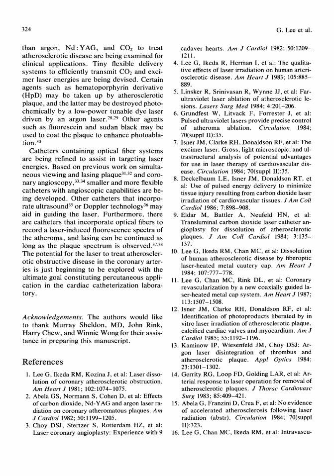
324
than argon, Nd: Y AG, and CO2 to treat atherosclerotic disease are being examined for clinical applications. Tiny flexible delivery systems to efficiently transmit CO2 and excimer laser energies are being devised. Certain agents such as hematoporphyrin derivative (HpD) may be taken up by atherosclerotic plaque, and the latter may be destroyed photochemically by a low-power tunable dye laser driven by an argon laser. 28,29 Other agents such as fluorescein and sudan black may be used to coat the plaque to enhance photoablation.30
Catheters containing optical fiber systems are being refined to assist in targeting laser energies. Based on previous work on simultaneous viewing and lasing plaque31 ,32 and coronary angioscopy, 33,34 smaller and more flexible catheters with angioscopic capabilities are being developed. Other catheters that incorporate ultrasound35 or Doppler technology36 may aid in guiding the laser. Furthermore, there are catheters that incorporate optical fibers to record a laser-induced fluorescence spectra of the atheroma, and lasing can be continued as long as the plaque spectrum is observedy,38 The potential for the laser to treat atherosclerotic obstructive disease in the coronary arteries is just beginning to be explored with the ultimate goal constituting percutaneous application in the cardiac catheterization laboratory.
Acknowledgements. The authors would like to thank Murray Sheldon, MD, John Rink, Harry Chew, and Winnie Wong for their assistance in preparing this manuscript.
References 1. Lee G, Ikeda RM, Kozina J, et al: Laser disso
lution of coronary atherosclerotic obstruction. Am Heart J 1981; 102:1074-1075.
2. Abela GS, Normann S, Cohen D, et al: Effects of carbon dioxide, Nd-Y AG and argon laser radiation on coronary atheromatous plaques. Am J Cardiol1982; 50:1199-1205.
3. Choy DSJ, Stertzer S, Rotterdam HZ, et al: Laser coronary angioplasty: Experience with 9
G. Lee et al.
cadaver hearts. Am J Cardiol 1982; 50: 1209-1211.
4. Lee G, Ikeda R, Herman I, et al: The qualitative effects of laser irradiation on human arteriosclerotic disease. Am Heart J 1983; 105:885-889.
5. Linsker R, Srinivasan R, Wynne n, et al: Farultraviolet laser ablation of atherosclerotic lesions. Lasers Surg Med 1984; 4:201-206.
6. Grundfest W, Litvack F, Forrester J, et al: Pulsed ultraviolet lasers provide precise control of atheroma ablation. Circulation 1984; 70(suppl 11):35.
7. Isner JM, Clarke RH, Donaldson RF, et al: The excimer laser: Gross, light microscopic, and ultrastructural analysis of potential advantages for use in laser therapy of cardiovascular disease. Circulation 1984; 70(suppl 11):35.
8. Deckelbaum LE, Isner JM, Donaldson RT, et al: Use of pulsed energy delivery to minimize tissue injury resulting from carbon dioxide laser irradiation of cardiovascular tissues. J Am Coli Cardiol 1986; 7:898-908.
9. Eldar M, Battler A, Neufeld HN, et al: Transluminal carbon dioxide laser catheter angioplasty for dissolution of atherosclerotic plaques. J Am Coli Cardiol 1984; 3: 135-137.
10. Lee G, Ikeda RM, Chan MC, et al: Dissolution of human atherosclerotic disease by fiberoptic laser-heated metal cautery cap. Am Heart J 1984; 107:777-778.
11. Lee G, Chan MC, Rink DL, et al: Coronary revascularization by a new coaxially guided laser-heated metal cap system. Am Heart J 1987; 113: 1507-1508.
12. Isner JM, Clarke RH, Donaldson RF, et al: Identification of photoproducts liberated by in vitro laser irradiation of atherosclerotic plaque, calcified cardiac valves and myocardium. Am J Cardiol1985; 55:1192-1196.
13. Kaminow IP, Wiesenfeld JM, Choy DSJ: Argon laser disintegration of thrombus and atherosclerotic plaque. Appl Optics 1984; 23: 1301-1302.
14. Gerrity RG, Loop FD, Golding LAR, et al: Arterial response to laser operation for removal of atherosclerotic plaques. J Thorac Cardiovasc Surg 1983; 85:409-421.
15. Abela G, Franzini D, Crea F, et al: No evidence of accelerated atherosclerosis following laser radiation (abstr). Circulation 1984; 70(suppl 11):323.
16. Lee G, Chan MC, Ikeda RM, et al: Intravascu-

27. Laser Angioplasty of the Coronary Arteries
lar steerable guidewire for fiberoptic laserheated metal cautery cap in dissolution of human atherosclerotic coronary disease. Am Heart J 1985; 110: 1304-1306.
17. Lee G, Seckinger D, Chan MC, et al: Potential complications of coronary laser angioplasty. Am Heart J 1984; 106:1577-1579.
18. Lee G, Ikeda RM, Theis JH, et al: Acute and chronic complications of laser angioplasty. Vascular wall damage and formation of aneurysms in the atherosclerotic rabbit. Am J Cardiol 1984; 53:290-293.
19. Lee G, Ikeda RM, Chan MC, et al: Limitations, risks and complications of laser recanalization: A cautious approach warranted. Am J Cardial 1985; 56:181-185.
20. Choy DSJ, Stertzer SH, Myler RK, et al: Human coronary laser recanalization. Clin Cardial 1984; 7:377-381.
21. Choy DSJ, Marco J, Fournial G, et al: Argon laser recanalization of three totally occluded human right coronary arteries. Clin Cardial 1986; 9:296-298.
22. Livesay JJ, Leachman DR, Hogan PJ, et al: Preliminary report on laser coronary endarterectomy in patients. Circulation 1985; 72(suppl I1I):302.
23. Lee G, Reis RL, Chan MC, et al: Clinical laser recanalization of coronary obstruction. Angioscopic and angiographic documentation. Chest 1986; 90:770-772.
24. Lee G, Garcia J, Chan MC, et al: Clinically successful long-term laser coronary recanalization. Am Heart J 1986; 112:1323-1325.
25. Lee G, Chan MC, Ikeda RM, et al: Applicability of laser to assist coronary balloon angioplasty. Am Heart J 1985; 110:1233-1236.
26. Sanborn TA, Faxon DP, Kellett MA, et al: Percutaneous coronary laser thermal angioplasty with a metallic capped fiber. JAm Coll Cardiol 1987; 9:104A.
27. Sanborn TA, Faxon DP, Kellett MA, et al: Percutaneous coronary laser thermal angioplasty. JAm Coll Cardiol1986; 8:1437-1440.
28. Spears JR, Serur J, Shropshire D, et al: Fluo-
325
rescence of experimental atheromatous plaques with hematoporphyrin derivative. J Clin Invest 1983; 71:395-399.
29. Hundley RF, Spears JR, Weinstein R: Photodynamic cytolysis of arterial smooth muscle cells in vitro: Implication for laser angioplasty. JAm Coli Cardiol 1985; 5:408.
30. Chan MC, Lee G, Seckinger DL, et al: Pretreatment with vital dyes to enhance or attenuate argon laser energy absorption in blood vessels. Circulation 1984; 70(suppl 11):298.
31. Lee G, Ikeda RM, Stobbe D, et al: Laser irradiation of human atherosclerotic obstructive disease: Simultaneous visualization and vaporization achieved by a dual fiberoptic catheter. Am Heart J 1983; 105:163-164.
32. Lee G, Ikeda RM, Stobbe D, et al: Intraoperative use of dual fiberoptic catheter for simultaneous in vivo visualization and laser vaporization of peripheral atherosclerotic obstructive disease. Cathet Cardiovasc Diagn 1984; 10: 11-16.
33. Lee G, Garcia JM, Corso PJ, et al: Correlation of coronary angioscopy to angiographic findings in patients with coronary artery disease. Am J Cardial 1986; 58:238-241.
34. Spears JR, Spokojny AM, Marais J: Coronary angioscopy during cardiac catheterization. J Am Call Cardioll985; 6:93-97.
35. Bommer WJ, Lee G, Rebeck K, et al: Twodimensional echocardiography of argon-laser vapor trails: Monitoring of catheter position and prevention of potential complications (abstr). Circulation 1983; 68:259.
36. Bommer WJ, Chan MC, Lee G, et al: Laser doppler angioplasty: A new technique. C[in Res 1987; 35:100A.
37. Kittrell C, Willett RL, de los Santos-Pacheo C, et al: Diagnosis of fibrous arterial atherosclerosis using fluorescence. Appl Optics 1985; 24:2280-2281.
38. Sartori M, Henry PD, Roberts R, et al: Estimation of arterial wall thickness and detection of atherosclerosis by laser induced argon fluorescence. J Am Coli Cardioll986; 7:207A.

Part V Acute Pharmacologic and Surgical Interventions

28 Platelet Inhibitor Drugs in Coronary Artery Disease and Coronary Intervention Douglas H. Israel, Bernardo Stein, and Valentin Fuster
Symptoms of coronary artery disease represent the culmination of 3 to 4 decades of progressive atherosclerosis. It is believed that atherosclerosis and most importantly its thrombotic complications occur in response to vascular injury. This injury may trigger three platelet responses which, depending on the nature of the injury, mayor may not occur together. These include:
1. platelet adhesion, 2. platelet aggregation, and 3. thrombogenesis.
The interplay of atherogenesis with these three responses shapes the clinical outcome.
This discussion will focus on:
1. the interplay of atherogenesis, platelets, and thrombosis;
2. the mechanism of action of platelet inhibitors;
3. the role of these agents in coronary artery disease; and
4. their role in coronary intervention.
Atherogenesis, Platelets, and Thrombosis
Figure 28.1 describes the natural history of atherosclerosis in five stages. Because the role of platelets and thrombosis is largely limited to
the first three stages, we will focus our attention in these areas.
Stages 1 and 2-Development and Growth of Early Lesions, Platelet Adhesion, and the Role of Risk Factors
Epidemiology
Stage 1 lesions are asymptomatic fatty streaks universally found in young persons. l -4 Analysis of more than 2,000 autopsy cases in the 1950s and 1960s showed that virtually all children over age 3 had fatty streaks in the aorta, and many children developed coronary fatty streaks by age 10, with increasing prevalence up to age 20 when they were nearly always present. The extensive International Atherosclerosis Project examined the aorta and coronary arteries of 23,000 autopsy cases from 14 countries and 19 racial groups. This study showed fatty streaks to be universally present in all groups evaluated, but further evolution into growing fibrous plaques, or stage 2 lesions, differed in incidence and severity among different racial and ethnic groups, and with the presence or absence of risk factors for atherosclerosis.
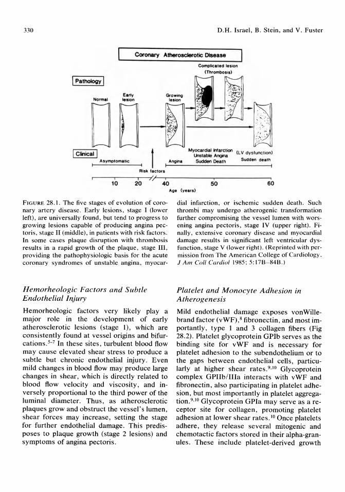
330 D.H. Israel, B. Stein, and V. Fuster
Coronary Atherosclerotic DIsease
1 Pathology I
early Normal Ie.ion
I Clinical I t Asymptomatic
RiSk lactors
Angina
Complicated lesion
(Thrombosis)
Myocardial Infarction (LV dysfunction) lklstable Angina Sudden Death
.-----ir-----rl--7)/~(~i~----------'i--------------~i
10 20 40 50 60
FIGURE 28 .1. The five stages of evolution of coronary artery disease. Early lesions, stage I (lower left) , are universally found, but tend to progress to growing lesions capable of producing angina pectoris, stage II (middle), in patients with risk factors. In some cases plaque disruption with thrombosis results in a rapid growth of the plaque , stage III, providing the pathophysiologic basis for the acute coronary syndromes of unstable angina, myocar-
Hemorheologic Factors and Subtle Endothelial Injury
Hemorheologic factors very likely play a major role in the development of early atherosclerotic lesions (stage 1), which are consistently found at vessel origins and bifurcations. 5- 7 In these sites, turbulent blood flow may cause elevated shear stress to produce a subtle but chronic endothelial injury. Even mild changes in blood flow may produce large changes in shear, which is directly related to blood flow velocity and viscosity, and inversely proportional to the third power of the luminal diameter. Thus, as atherosclerotic plaques grow and obstruct the vessel's lumen, shear forces may increase, setting the stage for further endothelial damage. This predisposes to plaque growth (stage 2 lesions) and symptoms of angina pectoris.
Age (ye.rs)
dial infarction , or ischemic sudden death . Such thrombi may undergo atherogenic transformation further compromising the vessel lumen with worsening angina pectoris , stage IV (upper right). Finally, extensive coronary disease and myocardial damage results in significant left ventricular dysfunction, stage V (lower right). (Reprinted with permission from The American College of Cardiology. J Am Coil Cardio/ 1985 ; 5: 17B-84B .)
Platelet and Monocyte Adhesion in Atherogenesis
Mild endothelial damage exposes von Willebrand factor (vWF),8 fibronectin, and most importantly, type 1 and 3 collagen fibers (Fig 28.2). Platelet glycoprotein GPIb serves as the binding site for vWF and is necessary for platelet adhesion to the subendothelium or to the gaps between endothelial cells, particularly at higher shear rates.9 •10 Glycoprotein complex GPIIb/IIIa interacts with vWF and fibronectin, also participating in platelet adhesion, but most importantly in platelet aggregation. 9•lo Glycoprotein GPIa may serve as a receptor site for collagen, promoting platelet adhesion at lower shear rates. IO Once platelets adhere, they release several mitogenic and chemotactic factors stored in their alpha-granules. These include platelet-derived growth
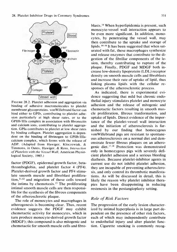
28. Platelet Inhibitor Drugs in Coronary Syndromes
FIGURE 28.2. Platelet adhesion and aggregation via binding of adhesive macromolecules to platelet membrane glycoproteins. vonWillebrand factor can bind either to GPIb, contributing to platelet adhesion particularly at high shear rates, or to the GPIIb/IIIa complex in association with fibronectin and calcium ions, contributing to platelet aggregation. GPIa contributes to platelet at low shear rates by binding collagen. Platelet aggregation is dependent on the binding of fibrinogen to GPIIb/IIIacalcium complex, which forms with the release of ADP. (Adapted from Hawiger, Kloczewiak, & Timmons, in Oates, Hawiger, & Ross, interaction of Platelets with the Vessel Wall, American Physiological Society, 1985.)
factor (PDGF), epidermal growth factor, betathromboglobin, and platelet factor 4 (PF4). Platelet-derived growth factor and PF4 stimulate smooth muscle and fibroblast proliferation, 11,12 and migration of these cells toward the intima by chemotaxis. 13 The proliferating intimal smooth muscle cells are then responsible for the synthesis of the fibrous components of the atherosclerotic plaque.
The role of monocytes and macrophages in atherogenesis is becoming clear. Thus, recent evidence suggests the PDGF may exhibit chemotactic activity for monocytes, which in turn produce monocyte-derived growth factor (MDGF); this compound is also mitogenic and chemotactic for smooth muscle cells and fibro-
331
blasts. 14 When hyperlipidemia is present, such monocyte-vessel wall interaction appears to be even more significant. In addition, monocytes, by penetrating the vessel wall, may then contribute to the uptake and storage of lipids. 14-16 It has been suggested that when saturated with fat, these macrophages synthesize and release enzymes that contribute to the digestion of the fibrillar components of the lesion, thereby contributing to rupture of the plaque. Finally, PDGF and MDGF both increase low-density lipoprotein (LDL) receptor density on smooth muscle cells and fibroblasts and increase their rate of uptake of lipid, thus linking plasma lipids with the cellular responses of the atherosclerotic process.
As indicated, there is experimental evidence suggesting that mild but chronic endothelial injury stimulates platelet and monocyte adhesion and the release of mitogenic and chemotactic factors resulting in smooth muscle proliferation, fibrous tranformation, and uptake of lipids. Direct evidence of the importance of the platelet-vessel wall interaction and the initiation of atherosclerosis is furnished by our finding that homozygous vonWiliebrand pigs are resistant to spontaneous atherosclerosis on a normal diet and demonstrate fewer fibrous plaques on an atherogenic dietY,ls Protection was demonstrated only in homozygous pigs with severely deficient platelet adhesion and a serious bleeding diathesis. Because platelet-inhibitor agents in current use do not inhibit platelet adhesion, they are incapable of preventing atherosclerosis, and only control its thrombotic manifestations. As will be discussed in detail, this is likely the reason why platelet inhibitor therapies have been disappointing in reducing restenosis in the postangioplasty setting.
Role of Risk Factors
The progression of the early lesion characterized by intimal hyperplasia is in large part dependent on the presence of other risk factors, each of which may independently contribute to endothelial injury and alter platelet function. Cigarette smoking is commonly recog-

332
nized to increase platelet reactivityl9-25; moreover, nicotine stimulates catecholamine release,26,27 which may increase platelet aggregation. 19 Finally, carbon monoxide in tobacco smoke may directly injure endothelium.28,29 Hyperlipoproteinemia is well established as a risk factor. Low-density lipoprotein cholesterol may directly injure endothelium,30 independently stimulates smooth muscle proliferation3! and increases platelet reactivity. 32,33 In addition, it may predispose to fatty plaques which are more prone to rupture and acute thrombosis, as discussed in the next section. Diabetes is likewise widely known to increase coronary risk. In diabetics, platelets are hyper-responsive to agonists in vitr034-36 and produce increased thromboxane A2,37 which stimulates platelet aggregation. Glycosylated collagen, likely to be present in the diabetic blood vessel, is an even more potent platelet agonist than normal collagen.38 Finally diabetic endothelium may synthesize lower than normal quantities of the platelet inhibitor prostacyclin. 39,40
Stage 3-Deep Atrial Damage: Plaque Rupture, Platelet Aggregation, and Thrombus Formation-Acute and Subacute Coronary Syndromes
In stage 3 the atherosclerotic plaque undergoes a sudden morphologic change due to plaque rupture or fissuring (Fig 28.3). Resultant deep arterial damage produces a change in the morphology of the plaque and a strong thrombogenic stimulus triggering platelet aggregation, activation of the coagulation system, and simultaneous activation of inhibitors of thrombosis. The change in the morphology and geometry of the plaque and thrombus deposition cause a rapid dramatic increase in the severity of the lesion, frequently producing unstable angina. Complete thrombotic occlusion may occur, resulting in myocardial infarction. During any of these acute pathologic events, acute ischemia may produce electrial instability and ischemic sudden death.
D.H. Israel, B. Stein, and V. Fuster
Plaque Rupture
There is now compelling evidence derived from angioscopic,41 angiographic,42 and pathologic studies43 ,44 to implicate plaque rupture in the occurrence of unstable angina, myocardial infarction, and ischemic sudden death, thereby providing a common pathogenetic link among the acute coronary syndromes.45-47 The exact mechanisms underlying plaque rupture are not yet clear, but it seems to occur in relatively soft, fatty areas of plaque, perhaps with a thin overlying fibrous cap. These areas may not be able to withstand hemodynamic stresses such as increased shear (related to the stenotic region or induced by vasoconstriction), blood pressure variations, or even the chronic pulsatile vibration of the cardiac cycle. Although all these factors may predispose to plaque rupture, a very important emerging concept is that the macrophages present in the fatty area may contribute to this process by releasing collagenase and elastase, which digest the fibril material.
Platelet Aggregation and Thrombogenesis
Deep arterial injury exposes underlying collagen fibers. Collagen, together with thrombin generated by tissue thromboplastin released from the vessel wall, are two potent agonists of platelet aggregation that appear to activate the so-called "third pathway" of platelet activation dependent on platelet activating factor (Figs 28.4 and 28.5). In addition, collagen and thrombin combine with specific membrane receptors and activate a secondary messenger system with a common final pathway-the hydrolysis of phosphatidylinositol by phospholipase C-Ieading to calcium mobilization from the dense tubular system.47,48 Such mobilization of calcium leads to the release of ADP and serotonin, and synthesis of thromboxane Az (TXA2), which represent the so-called first and second pathways of platelet aggregation, respectively.
Platelet aggregation and thrombogenesis depend on five lines of activation: collagen and thrombin dependent, ADP and serotonin de-

28. Platelet Inhibitor Drugs in Coronary Syndromes
FIGURE 28.3. Intracoronary thrombosis. Thrombus is anchored within a fissure in an atherosclerotic plaque. (Reproduced from Constantinides P: J
pendent, TXA2 dependent, the activation of the clotting system, and the activation of endogenous inhibitors of thrombosis.
As described, deep arterial injury results in a potent thrombogenic stimulus triggered by exposure of underlying collagen and tissue thromboplastin. These substances simultaneously activate both the intrinsic and extrinsic pathways of coagulation, generating thrombin, and thus resulting in further platelet aggregation, as described, and in fibrin formation. Whether or not lipid from the vessel wall is itself thrombogenic when exposed to the circulating blood is under active investigation.
Adenosine diphosphate is a potent platelet aggregation agonist. It is released both from platelet-dense granules during activation, and from red blood cells during lysis. It binds to a specific domain of the GPIIb/lIIa complex and induces a conformational change in the platelets. As a result, a receptor for fibrinogen and vW Factor is exposed. The fibrinogen and vWF molecules bind to receptors on neighboring platelets, forming bridges between them, stabilizing the growing aggregate (Fig 28.2).9,10,49 This mechanism appears central to platelet aggregation with all agonists. 49 ,5o Sero-
333
Atheroscler Res 1966; 6:9. Copyright by Elsevier Science Publishers Ireland, used with permission.)
tonin, also released from the platelet-dense granules during platelet activation may playa role in vasoconstriction.
Some of the intracytoplasmic calcium released by the action of phospholipase C, activates membrane phospholipase A2 and liberates arachidonic acid.48 This is metabolized by cyclo-oxygenase to TXA2 via the prostaglandin endoperoxide intermediates PGG2 and PGH2, and by lipooxygenase into other metabolites whose role is under active investigation. Thromboxane recruits neighboring platelets by activating their surface membranes probably exposing the fibrinogen and vWF receptor to fibrinogen bridging by these molecules resulting in platelet aggregation, as described. Thromboxane IS also a potent vasoconstricting substance.
Platelet function is modulated via the complex interactions of prostaglandins, cyclic AMP (cAMP), and calcium ions. The concentration of platelet cAMP is determined by the activity of its synthetic enzyme adenyl cyclase and by phosphodiesterase, which is responsible for its hydrolysis to adenosine triphosphate. An increase in cAMP inhibits both secretion and aggregation. 51 Basal levels of
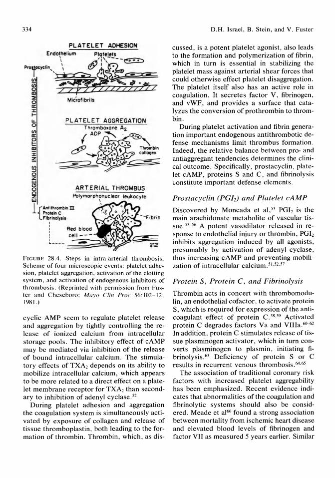
334
PL ATELET ADHESION Endothelium Plotelets
\ ' i \----~ Pro r \ t;1~
r~"'\o/_~c; ~ Microfi bri ls a: :I: ~
~ o (f)
a:
PLATELET AGGREGATION
g m :I: ~
~ o Z I.&J
8 AR T ERIAL THROMBUS c Z I.&J
Polymorphonuc leor leukocyte \
1 {Antithrombin m Protein C Fibr inolys is
I I I , , , , .. _---=========~~ - .., .'-
FIGURE 28.4. Steps in intra-arterial thrombosis. Scheme of four microscopic events: platelet adhesion, platelet aggregation, activation of the clotting system, and activation of endogenous inhibitors of thrombosis. (Reprinted with permission from Fuster and Cheseboro: Mayo Clin Pro(' 56: 102-12. 1981.)
cyclic AMP seem to regulate platelet release and aggregation by tightly controlling the release of ionized calcium from intracellular storage pools. The inhibitory effect of cAMP may be mediated via inhibition of the release of bound intracellular calcium. The stimulatory effects of TXA2 depends on its ability to mobilize intracellular calcium, which appears to be more related to a direct effect on a platelet membrane receptor for TXA2 than secondary to inhibition of adenyl cyclase. 52
During platelet adhesion and aggregation the coagulation system is simultaneously activated by exposure of collagen and release of tissue thromboplastin, both leading to the formation of thrombin. Thrombin, which, as dis-
D.H. Israel, B. Stein, and V. Fuster
cussed, is a potent platelet agonist, also leads to the formation and polymerization of fibrin, which in turn is essential in stabilizing the platelet mass against arterial shear forces that could otherwise effect platelet disaggregation. The platelet itself also has an active role in coagulation. It secretes factor V, fibrinogen, and vWF, and provides a surface that catalyzes the conversion of prothrombin to thrombin.
During platelet activation and fibrin generation important endogenous anti thrombotic defense mechanisms limit thrombus formation. Indeed, the relative balance between pro- and antiaggregant tendencies determines the clinical outcome. Specifically, prostacyclin, platelet cAMP, proteins Sand C, and fibrinolysis constitute important defense elements.
Prostacyclin (PGI2) and Platelet cAMP
Discovered by Moncada et al,53 PGI2 is the main arachidonate metabolite of vascular tissuey-56 A potent vasodilator released in response to endothelial injury or thrombin, PGI2 inhibits aggregation induced by all agonists, presumably by activation of adenyl cyclase, thus increasing cAMP and preventing mobilization of intracellular calcium.51.52 ,57
Protein S, Protein C, and Fibrinolysis
Thrombin acts in concert with thrombomodulin, an endothelial cofactor, to activate protein S, which is required for expression of the anticoagulant effect of protein C. 58,59 Activated protein C degrades factors Va and VIIIa.60-62 In addition, protein C stimulates release of tissue plasminogen activator, which in turn converts plasminogen to plasmin, initiating fibrinolysis. 63 Deficiency of protein S or C results in recurrent venous thrombosis. 64 ,65
The association of traditional coronary risk factors with increased platelet aggregability has been emphasized. Recent evidence indicates that abnormalities of the coagulation and fibrinolytic systems should also be considered. Meade et al66 found a strong association between mortality from ischemic heart disease and elevated blood levels of fibrinogen and factor VII as measured 5 years earlier. Similar
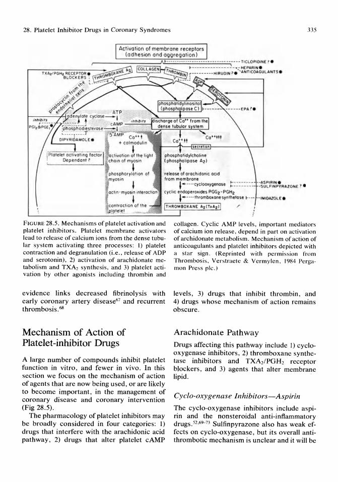
28. Platelet Inhibitor Drugs in Coronary Syndromes 335
Act ivation of membrane receptors (adhesion and aggregation)
:==-=~~\I~~~~"~~~-- - - - - - - -- ----- -------- ---- ------ TrCLOPIOINE ? ~ __ n_n __ nn - ~ - . - HEPARIN . r--r-X-A2-/ P-G-H-. R-E-CE-p-rO-R-.-- r----- ----HIRUOIN ? " ANTICOAGULANTS .
BLOCK ERS ; r:;;:;Ro"~S':':::':
,-~ ~~:~"'~ _,,_,\,0 v~ .. '-
"-~<:- ' o' -'-" ,O<'\,<:-~'
,' 0" tP- q' ,-~~ -" ,
phosphalidylcholine (phospholipase Az) , release al arachidonic aCid Irom membrane
SPIRIN . -SULFINPYRAlONE ? • ''''- ' - --CYCloalygenose ~------
cyc lic endoperol ides PGG2 - PGH2 , .... . '-- Ihrombolane synlhelose ~ - --r- IIII IDI\.ZOI_E.
FIGURE 28.5. Mechanisms of platelet activation and platelet inhibitors. Platelet membrane activators lead to release of calcium ions from the dense tubular system activating three processes: 1) platelet contraction and degranulation (i,e., release of ADP and serotonin), 2) activation of arachidonate metabolism and TXA2 synthesis, and 3) platelet activation by other agonists including thrombin and
evidence links decreased fibrinolysis with early coronary artery disease67 and recurrent thrombosis. 68
Mechanism of Action of Platelet-inhibitor Drugs
A large number of compounds inhibit platelet function in vitro, and fewer in vivo. In this section we focus on the mechanism of action of agents that are now being used, or are likely to become important, in the management of coronary disease and coronary intervention (Fig 28.5).
The pharmacology of platelet inhibitors may be broadly considered in four categories: 1) drugs that interfere with the arachidonic acid pathway, 2) drugs that alter platelet cAMP
/
collagen. Cyclic AMP levels, important mediators of calcium ion release , depend in part on activation of archidonate metabolism. Mechanism of action of anticoagulants and platelet inhibitors depicted with a star sign, (Reprinted with permission from Thrombosis, Verstraete & Vermylen, 1984 Pergamon Press pic,)
levels, 3) drugs that inhibit thrombin, and 4) drugs whose mechanism of action remains obscure.
Arachidonate Pathway
Drugs affecting this pathway include 1) cyclooxygenase inhibitors, 2) thromboxane synthetase inhibitors and TXA2 /PGH 2 receptor blockers, and 3) agents that alter membrane lipid.
Cyclo-oxygenase Inhibitors-Aspirin
The cyclo-oxygenase inhibitors include aspirin and the nonsteroidal anti-inflammatory drugs. 52 ,69---73 Sulfinpyrazone also has weak effects on cyclo-oxygenase, but its overall antithrombotic mechanism is unclear and it will be

336
discussed briefly later. Aspirin irreversibly acetylates platelet cyclo-oxygenase, thereby exerting its effects for the life of the platelet. The final effect is to block TXA2, blunting its proaggregant effects. Dense granule secretion of ADP and serotonin is not inhibited by aspirin. Furthermore, collagen and thrombin-dependent platelet aggregation is not significantly affected by aspirin. So thrombus formation may proceed via these pathways despite complete blockade of cyclo-oxygenase.74 Aspirin does not inhibit platelet adhesion75,76 or the secretion of alpha-granule contents, such as PDGF or PF4. Thus, it is unable to inhibit the early atherosclerotic lesions characterized by intimal hyperplasia and fibrous transformation. 77
Concern that aspirin's similar inhibitory effect on vascular cyclo-oxygenase and PGh synthesis would lead to thrombosis appears unfounded. Epidemiologic studies of rheumatoid arthritis patients on high-dose aspirin have shown a trend toward fewer thrombotic endpoints. 78 Low-dose aspirin (.5 to 1.0 mg/kg per day) confers 90% to 95% TXA2 inhibition and is sufficient to produce a maximal antiaggregating effect. 79,8o However, vascular PGI2 synthesis is also partially affected by the low doses. As beneficial effects have been obtained clinically in unstable angina with low to medium doses of aspirin (i.e., 325 mg/day)8J and because its gastrointestinal side effects occur with larger doses, the appropriate antithrombotic dose is probably 325 mg daily.82
Thromboxane Synthetase Inhibitors and TXA2/ PGH2 Receptor Blockers
Imidazole and several derivatives, including dazoxiben, block the conversion of the cyclic endoperoxides PGG2 and PGH2 into TXA2. 83 This approach may actually increase vascular PGI2 production by increasing the concentration of PGG2 and PGH2, some of which may serve as substrate for endothelial cyclo-oxygenase. 84 Despite this theoretical benefit, the antithrombotic potency is less than that of aspirin, perhaps because the cyclic endoperoxides that accumulate with the blockade of
D.H. Israel, B. Stein, and V. Fuster
TXA2 synthesis, may themselves serve as proaggregants. 85-87 Thromboxane receptor blockers prevent both TXA2 and cyclic endoperoxide binding but do not increase PGI2 synthesis.88 Combining TXA2 synthetase inhibitors with TXA2 receptor blockers may prove to be an effective anti thrombotic strategy.89
Agents That Alter Platelet Membrane Phospholipid
The observation that Eskimos in Greenland have prolonged bleeding times and little tendency to develop atherosclerosis led to the suggestion that their diet, rich in eicosapentaenoic acid (EPA) may be protective. Eicosapentaenoic acid is present in high concentration in most fish, and its incorporation into the diet alters the ratio of EPA to arachidonic acid in platelet membranes. 52,90-92 Eicosapentaenoic acid competes for a site on platelet cyclo-oxygenase, leading to production of TXA3, whereas vascular cyclo-oxygenase metabolizes EPA to PGI3; both of these compounds have antiaggregant properties. Hayet al93 found that supplementing the diet in 13 patients with 3.5 g of EPA for 5 weeks caused a 10% increase in platelet survival times, 15% fall in platelet count, a 75% fall in plasma PF4, and 30% fall in beta-thromboglobulin. This suggests that EPA may reduce platelet-vessel wall interaction. Current trials are investigating whether this effect could actually inhibit atherogenesis itself.
Drugs That Increase Platelet cAMP Levels
Prostacyclin
Prostacyclin increases platelet cAMP by activating adenylate cyclase. Prostacyclin, and to a lesser extent PGE2, strongly inhibit platelet aggregation and thrombosis in humans and experimental animals in a variety of clinical situations, both on artificial and biologic surfaces.94-IOI Despite its potency as an anti-
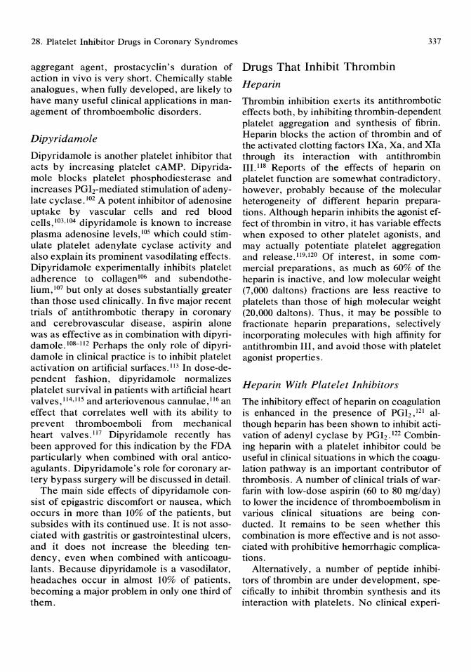
28. Platelet Inhibitor Drugs in Coronary Syndromes
aggregant agent, prostacyclin's duration of action in vivo is very short. Chemically stable analogues, when fully developed, are likely to have many useful clinical applications in management of thromboembolic disorders.
Dipyridamole
Dipyridamole is another platelet inhibitor that acts by increasing platelet cAMP. Dipyridamole blocks platelet phosphodiesterase and increases PGIrmediated stimulation of adenylate cyclase. 102 A potent inhibitor of adenosine uptake by vascular cells and red blood cells,103,104 dipyridamole is known to increase plasma adenosine levels , 105 which could stimulate platelet adenylate cyclase activity and also explain its prominent vasodilating effects. Dipyridamole experimentally inhibits platelet adherence to collagen 106 and subendothelium,107 but only at doses substantially greater than those used clinically. In five major recent trials of anti thrombotic therapy in coronary and cerebrovascular disease, aspirin alone was as effective as in combination with dipyridamole. I08-112 Perhaps the only role of dipyridamole in clinical practice is to inhibit platelet activation on artificial surfaces.1I3 In dose-dependent fashion, dipyridamole normalizes platelet survival in patients with artificial heart valves, 114,115 and arteriovenous cannulae, 116 an effect that correlates well with its ability to prevent thromboemboli from mechanical heart valves. 117 Dipyridamole recently has been approved for this indication by the FDA particularly when combined with oral anticoagulants. Dipyridamole's role for coronary artery bypass surgery will be discussed in detail.
The main side effects of dipyridamole consist of epigastric discomfort or nausea, which occurs in more than 10% of the patients, but subsides with its continued use. It is not associated with gastritis or gastrointestinal ulcers, and it does not increase the bleeding tendency, even when combined with anticoagulants. Because dipyridamole is a vasodilator, headaches occur in almost 10% of patients, becoming a major problem in only one third of them.
Drugs That Inhibit Thrombin
Heparin
337
Thrombin inhibition exerts its antithrombotic effects both, by inhibiting thrombin-dependent platelet aggregation and synthesis of fibrin. Heparin blocks the action of thrombin and of the activated clotting factors IXa, Xa, and XIa through its interaction with antithrombin III. 118 Reports of the effects of heparin on platelet function are somewhat contradictory, however, probably because of the molecular heterogeneity of different heparin preparations. Although heparin inhibits the agonist effect of thrombin in vitro, it has variable effects when exposed to other platelet agonists, and may actually potentiate platelet aggregation and release. 1I9,120 Of interest, in some commercial preparations, as much as 60% of the heparin is inactive, and low molecular weight (7,000 daltons) fractions are less reactive to platelets than those of high molecular weight (20,000 daltons). Thus, it may be possible to fractionate heparin preparations, selectively incorporating molecules with high affinity for antithrombin III, and avoid those with platelet agonist properties.
Heparin With Platelet Inhibitors
The inhibitory effect of heparin on coagulation is enhanced in the presence of PGI2 , 121 although heparin has been shown to inhibit activation of adenyl cyclase by PGI2 .122 Combining heparin with a platelet inhibitor could be useful in clinical situations in which the coagulation pathway is an important contributor of thrombosis. A number of clinical trials of warfarin with low-dose aspirin (60 to 80 mg/day) to lower the incidence of thromboembolism in various clinical situations are being conducted. It remains to be seen whether this combination is more effective and is not associated with prohibitive hemorrhagic complications.
Alternatively, a number of peptide inhibitors of thrombin are under development, specifically to inhibit thrombin synthesis and its interaction with platelets. No clinical experi-
338
ence is yet available, but there are promising experimental data with one of these agents, hirudin.
Drugs With Other Mechanisms
Su/finpyrazone
Sulfinpyrazone has been used widely experimentally in cardiovascular disease, yet no clear concensus exists as to its exact mechanism of action.71.123.124 Although sulfinpyrazone has been found to inhibit thrombus formation on subendotheliuml25 and may exert a weak protective effect on endothelium,126 it has not demonstrated consistent anti thrombotic effects on biologic surfaces. Despite a beneficial trend in decreasing vascular events after myocardial infarctionl27 and coronary bypass surgery, 112.128 no benefit was shown in the Canadian trials on strokel29 and unstable angina.130 Sulfinpyrazone has demonstrated a more reproducible effect in reducing thromboembolism from prosthetic surfaces, increasing platelet survival in patients with prosthetic valves,131 and reducing thrombotic events in arteriovenous cannulae. 132 Sulfinpyrazone may exacerbate peptic ulcer disease, increase the sensitivity to coumadin by prolonging the prothrombin time, and may produce hypoglycemia when combined with oral hypoglycemic agents. It may also precipitate uric acid stones.
Ticlopidine
Ticlopidine is an unusual drug capable of inhibiting aggregation induced by ADp133,134 and both aggregation and release induced by thrombin, collagen, arachidonate, and epinephrine. It prolongs bleeding time and improves platelet survival. 135 Chemically unrelated to other platelet inhibitors, its mechanism of action is unknown but appears to involve inhibition of fibrinogen receptors or perhaps binding of VWF.134 Clinical evaluation of the drug is currently underway.
D.H. Israel, B. Stein, and V. Fuster
Role of Platelet Inhibitors in Coronary Artery Disease
Stable Coronary Disease and Angina Pectoris
Unfortunately, there is no drug currently available that can prevent the adherence and activation of the initial monolayer of platelets, which may be all that is necessary to trigger intimal hyperplasia and coronary disease progression. Nevertheless, if platelet adhesion were to be completely prevented, as occurs in pigs with homozyous von Willebrand's disease, the risk of bleeding would prohibit its chronic use. As discussed, plaque rupture is apparently a random event capable of causing rapid growth of the lesion by thrombus, and its subsequent organization by connective tissue. Therefore a rationale exists for the prophylactic use of aspirin in stable angina pectoris to try to limit thrombosis, should plaque rupture occur. Results of a clinical investigation will be available soon, to elucidate whether aspirin is beneficial in preventing progression of coronary disease in patients with stable angina.
Unstable Angina
By contrast, the value of antiplatelet therapy in unstable angina has been conclusively demonstrated in two randomized, placebo-controlled, double-blind trials using aspirin (Fig 28.6 and Table 28.1). The Veteran's Administration Cooperative Study81 randomized 1,266 men with unstable angina to receive 324 mg of aspirin as Alka-Seltzer or placebo for 12 weeks after the diagnosis of unstable angina. During the treatment period death or acute myocardial infarction occurred in 10.1% of the placebo group u 5.0% of the treated patients (P = 0.0005). The risk of death was 51% less, this difference persisted at 1 year, with mortality still 43% less in the aspirin group. There was no significant increase in gastrointestinal side effects, occult fecal blood loss, or drop in hemoglobin between the groups,
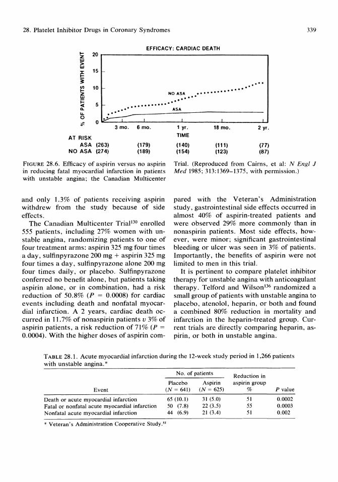
28. Platelet Inhibitor Drugs in Coronary Syndromes 339
EFFICACY: CARDIAC DEATH ~ 20 z w > w :r: 15 i-~
i en 10 i-~ Z W j::
"' 5 t- ........... Q. ... .. ' LL . 0 ~ I ::!!. 0 0
3 mo. 6 mo.
AT RISK ASA (263) (179)
NO ASA (274) (189)
FIGURE 28.6. Efficacy of aspirin versus no aspirin in reducing fatal myocardial infarction in patients with unstable angina; the Canadian Multicenter
and only 1.3% of patients receIvmg aspirin withdrew from the study because of side effects.
The Canadian Multicenter Trial130 enrolled 555 patients, including 27% women with unstable angina, randomizing patients to one of four treatment arms: aspirin 325 mg four times a day, sulfinpyrazone 200 mg + aspirin 325 mg four times a day, sulfinpyrazone alone 200 mg four times daily, or placebo. Sulfinpyrazone conferred no benefit alone, but patients taking aspirin alone, or in combination, had a risk reduction of 50.8% (P = 0.0008) for cardiac events including death and nonfatal myocardial infarction. A 2 years, cardiac death occurred in 11.7% of nonaspirin patients v 3% of aspirin patients, a risk reduction of 71% (P = 0.0004). With the higher doses of aspirin com-
.... . . .............. NO ASA .. , .. ' ..
ASA
I i
1 yr. 18 mo. 2 yr. TIME
(140) (111) (77) (154) (123) (87)
Trial. (Reproduced from Cairns, et al: N Eng/ J Med 1985; 313:1369-1375, with permission.)
pared with the Veteran's Administration study, gastrointestinal side effects occurred in almost 40% of aspirin-treated patients and were observed 29% more commonly than in nonaspirin patients. Most side effects, however, were minor; significant gastrointestinal bleeding or ulcer was seen in 3% of patients. Importantly, the benefits of aspirin were not limited to men in this trial.
It is pertinent to compare platelet inhibitor therapy for unstable angina with anticoagulant therapy. Telford and Wilson 136 randomized a small group of patients with unstable angina to placebo, atenolol, heparin, or both and found a combined 80% reduction in mortality and infarction in the heparin-treated group. Current trials are directly comparing heparin, aspirin, or both in unstable angina.
TABLE 28.1. Acute myocardial infarction during the 12-week study period in 1,266 patients with unstable angina. *
Event
Death or acute myocardial infarction Fatal or nonfatal acute myocardial infarction Nonfatal acute myocardial infarction
No. of patients
Placebo (N = 641)
65 (10.1) 50 (7.8) 44 (6.9)
Aspirin (N = 625)
31 (5.0) 22 (3.5) 21 (3.4)
* Veteran's Administration Cooperative Study.s1
Reduction in aspirin group
%
51 55 51
P value
0.0002 0.0003 0.002
340
Non-Q Wave Infarction and Reduction of Early Recurrence (Extension) of Myocardial Infarction
The pathophysiologic mechanisms leading to non-Q wave infarction remain unclear. Only 25% of patients have a completely occluded infarct related vessel with inadequate distal collaterals. 137 Transient occlusions resolving within the first few hours may be responsible for the ischemic event in those patients with patent arteries. In these cases, perhaps a major plaque disruption occurs with prolonged persistence of thrombus, with or without vasoconstriction, but of insufficient duration to produce Q wave infarction.
Extension or early recurrence of myocardial infarction occurs in 14% to 30% of cases of acute myocardial infarction as determined by serial quantitative measurements of creatine kinase MB isoenzyme. This is comparable to the incidence of 17% found at necropsy. 138-146 More than half of early recurrences occur within 10 days, and the remainder usually occur within 14 to 18 days after the initial infarction.142 The incidence of recurrence during hospitalization is much higher in patients with non-Q wave infarctionl42 and is higher in smaller infarctions (lower plasma enzyme levels) as compared with larger infarctions. 138 Chest pain and ST -T changes are sensitive (90% and 80%, respectively) but nonspecific (46% and 37%, respectively) indicators of reinfarction. The electrocardiographic site of early recurrence was the same as the initial site in 86% of patients, and thus early recurrence and extension of infarction are nearly synonomous. Extension or recurrence may not be clinically detected in up to 50% of cases. 139,141,143-146 The reason for such instability in the patient with non-Q wave infarction is unclear, but it may in part relate to the high incidence of subtotal occlusion of the infarctrelated artery. There may be a substantial amount of viable but jeopardized myocardium subtended by an artery with a high-grade complex lesion with superimposed dynamic thrombus. The degree of luminal obstruction, and thus the severity of ischemia of surviving
D,H, Israel, B, Stein, and v, Fuster
myocardium, may fluctuate with spontaneous changes in the thrombus. The early postinfarction period may confer particular susceptibility to reinfarction because of the apparent hypercoagulable state in the weeks after myocardial infarction. 147,148 Thus, the syndrome of non-Q wave infarction carries a great risk of subsequent reinfarction or sudden death, equal to that in unstable angina, Therefore, the role of antiplatelet therapy needs to be tested formally. It is interesting that in the Paris II study, treatment with aspirin and dipyridamole produced a 53% reduction in subsequent coronary events in patients with non-Q wave infarction. lo8 Platelet mediated vasoconstriction probably plays an additional role in reinfarction by increasing local shear rates thereby enhancing platelet deposition, This may explain why reinfarction within 2 weeks of a non-Q wave myocardial infarction was decreased from 9% to 5.2% in patients treated with diltiazem 360 mg per day in a double-blind, randomized, placebo-controlled triaL 149
Q Wave Infarction
Q wave myocardial infarction represents the end-stage of the pathogenic sequence of atherogenesis and thrombosis. The risk of death is greatest soon after the onset of symptoms and is primarily due to arrhythmia. Later deaths are usually due to left ventricular dysfunction itself, through the mechanism of pump failure or via its tendency to potentiate malignant ventricular arrhythmia. Thus, most deaths in the first year are not due to thrombotic events, and so very large studies using platelet inhibitors for secondary prevention would be needed to achieve statistical significance. Nevertheless, pooled data from all the large studies using aspirin or dipyridamole postmyocardial infarction suggests a 15% to 21 % lower reinfarction rate150,151 and a significantly lower death rate.
Recommendations-Antiplatelet therapy in coronary artery disease with native vessels:
1, Aspirin 325 mg/day is the preferred platelet inhibitor in unstable angina. Its benefit per-
28. Platelet Inhibitor Drugs in Coronary Syndromes
sists for at least 2 years; comparative effectiveness of heparin, coumadin, and these in combination with very low-dose aspirin is currently under study.
2. Given the low risk and overall beneficial results of aspirin postmyocardial infarction, a dose of 325 mg/day can be advised for these patients. Non-Q wave infarction in conceptually similar to unstable angina and reinfarction probably occurs largely via thrombotic mechanisms in many cases. Therefore, aspirin in a dose of 325 mg/day is advised after non-Q wave myocardial infarction.
3. The benefit of aspirin in chronic stable angina is currently being evaluated and firmer recommendations will be available within the next 2 years. Meanwhile its use for the prevention of myocardial infarction is not unjustified.
4. Some information on the role of aspirin in primary prevention has recently become available. Our view is to recommend it at present only for individuals with substantial risk factors for coronary artery disease pending further study.
Role of Platelet-inhibitor Drugs in Coronary Intervention
Prevention of Reocclusion After Thrombolysis As detailed, plaque rupture and fissuring may result in acute thrombotic obstruction of the coronary artery leading to myocardial infarction. Fibrinolytic therapy may be successful in restoring vessel patency by lysing the thrombus, but consequently it leaves residual thrombus, which is very thrombogenic or reexposes the underlying disrupted plaque, which retains its thrombogenic potential. In addition, reocclusion appears to be directly related to residual stenosis, the severity of which may be augmented by superimposed local vasoconstriction. Thus, a pathologic study showed that reocclusion is more frequent when the cross-sectional area of the residual
341
stenosis is more than 75%.152 An angiographic study showed that more than 50% of arteries will reocclude during hospitalization after successful thrombolytic therapy if the cross-sectional area of the residual stenosis is 0.4 mm2
or less. 153 Data from Thrombolysis in myocardial infarction trial, phase 1,154 (TIMI I) confirmed this association and showed a 28% incidence of reocclusion after thrombolysis, when the minimal residual diameter of the infarctrelated artery 90 minutes after thrombolysis was less than 0.6 mm.
Heparin is important in reducing the rate of new arterial thrombus formation during thrombolysis; experimentally, heparin enhances thrombolysis by streptokinase and urokinase. 155 In dogs, thrombus reaccumulates during the infusion of t-PA for lysis of coronary thrombosis if the heparin infusion is stopped while the t-PA infusion continues.156 High-dose heparin reduces thrombus formation after acute arterial injury in a dose-dependent fashion. 15? A retrospective study in patients suggests that at least three days of heparin therapy is necessary to minimize the risk of reocclusion after successful thrombolySiS. 158 In patients with unstable angina or subendocardial infarction, the infusion of heparin for 7 days markedly reduced the incidence of subsequent myocardial infarction. 136 After successful thrombolysis, angiographic vessel patency was maintained by the use of platelet inhibitor therapy and heparin. 159,160
Experimental in vivo studies of deep arterial injury suggest that low-dose aspirin at 1 mg/kg per day is an effective anti thrombotic agent161 ;
in patients with unstable angina (another type of deep arterial injury that appears to be caused by plaque rupture) aspirin alone has been beneficial. 81 In this context, aspirin may reduce the reocclusion rate after thrombolytic therapy, this hypothesis is now being tested clinically in a randomized trial. However, aspirin by itself may not be the optimal platelet inhibitor because it only inhibits one of the three pathways of platelet activation, the prostaglandin pathway via thromboxane A2 . 162
The combination of aspirin and heparin may be of more benefit. As the fibrinogen and vWF
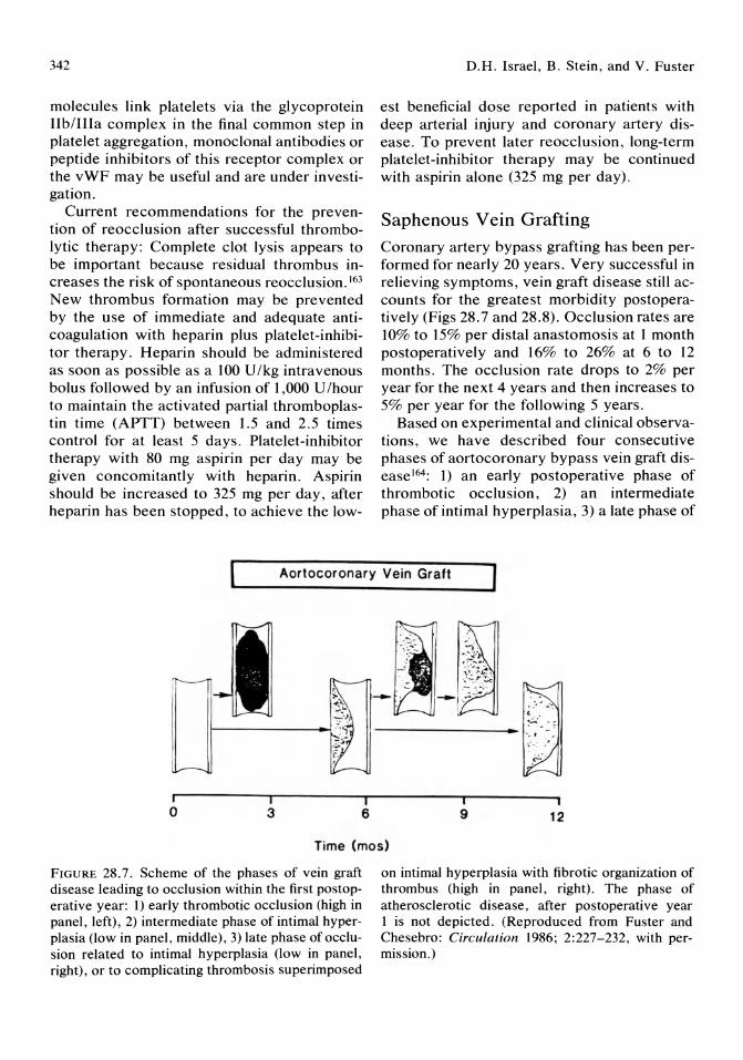
342
molecules link platelets via the glycoprotein lIb/IlIa complex in the final common step in platelet aggregation, monoclonal antibodies or peptide inhibitors of this receptor complex or the vWF may be useful and are under investigation.
Current recommendations for the prevention of reocclusion after successful thrombolytic therapy: Complete clot lysis appears to be important because residual thrombus increases the risk of spontaneous reocclusion. 163
New thrombus formation may be prevented by the use of immediate and adequate anticoagulation with heparin plus platelet-inhibitor therapy. Heparin should be administered as soon as possible as a 100 U/kg intravenous bolus followed by an infusion of 1,000 U /hour to maintain the activated partial thromboplastin time (APTT) between 1.5 and 2.5 times control for at least 5 days. Platelet-inhibitor therapy with 80 mg aspirin per day may be given concomitantly with heparin. Aspirin should be increased to 325 mg per day, after heparin has been stopped, to achieve the low-
D.H. Israel, B. Stein, and V. Fuster
est beneficial dose reported in patients with deep arterial injury and coronary artery disease. To prevent later reocclusion, long-term platelet-inhibitor therapy may be continued with aspirin alone (325 mg per day).
Saphenous Vein Grafting
Coronary artery bypass grafting has been performed for nearly 20 years. Very successful in relieving symptoms, vein graft disease still accounts for the greatest morbidity postoperatively (Figs 28.7 and 28.8). Occlusion rates are 10% to 15% per distal anastomosis at 1 month postoperatively and 16% to 26% at 6 to 12 months. The occlusion rate drops to 2% per year for the next 4 years and then increases to 5% per year for the following 5 years.
Based on experimental and clinical observations, we have described four consecutive phases of aortocoronary bypass vein graft disease l64 : 1) an early postoperative phase of thrombotic occlusion, 2) an intermediate phase of intimal hyperplasia, 3) a late phase of
Aortocoronary Vein Graft
o I 3
I 6
•
9 12
Time (mos)
FIGURE 28.7. Scheme of the phases of vein graft disease leading to occlusion within the first postoperative year: 1) early thrombotic occlusion (high in panel, left), 2) intermediate phase of intimal hyperplasia (low in panel, middle), 3) late phase of occlusion related to intimal hyperplasia (low in panel, right), or to complicating thrombosis superimposed
on intimal hyperplasia with fibrotic organization of thrombus (high in panel, right). The phase of atherosclerotic disease, after postoperative year 1 is not depicted. (Reproduced from Fuster and Chesebro: Circulation 1986; 2:227-232, with permission.)
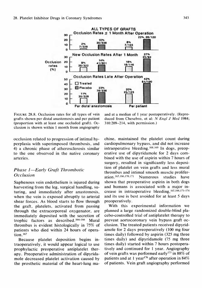
28. Platelet Inhibitor Drugs in Coronary Syndromes 343
ALL TYPES OF GRAFTS
30
20
10
Occlusion Rates ~ 1 Month After Operation 22"-. 25/120
2" 7/3211 o L-_-'=='--__
30
Occlusion 20
New Occlusion Rates After 1 Month 27"
rates ('Yo)
10 o L-_J..':';;"':;L-__
SO
40
30
20
10
Occlusion Rates Late Afler Operation 42"
o Treated mPlacebo
OL--....L:..~--Per distal anastomosis Per patient
FIGURE 28.8. Occlusion rates for all types of vein grafts shown per distal anastomosis and per patient (proportion with at least one occluded graft). Occlusion is shown within 1 month from angiography
occlusion related to progression of intimal hyperplasia with superimposed thrombosis, and 4) a chronic phase of atherosclerosis similar to the one observed in the native coronary arteries.
Phase I-Early Graft Thrombotic Occlusion
Saphenous vein endothelium is injured during harvesting from the leg, surgical handling, suturing, and immediately after anastomosis, when the vein is exposed abruptly to arterial shear forces. As blood starts to flow through the graft, platelets, activated from passing through the extracorporeal oxygenator, are immediately deposited with the secretion of trophic factors as described. 164-166 Mural thrombus is evident histologically in 75% of patients who died within 24 hours of operation. 167
Because platelet deposition begins intraoperatively, it would appear logical to use prophylactic preoperative antiplatelet therapy. Preoperative administration of dipyridamole decreased platelet activation caused by the prosthetic material of the heart-lung ma-
and at a median of 1 year postoperatively. (Reproduced from Chesebro, et al: N Engl J Med 1984; 310:209-214, with permission.)
chine, maintained the platelet count during cardiopulmonary bypass, and did not increase intraoperative bleeding.168.169 In dogs, preoperative use of dipyridamole for 2 days combined with the use of aspirin within 7 hours of surgery, resulted in significantly less deposition of platelet on vein grafts and less mural thrombus and intimal smooth muscle proliferation.165.166.170.171 Numerous studies have shown that preoperative aspirin in both dogs and humans is associated with a major increase in intraoperative bleeding,165,166,171-174 and its use is best avoided for at least 5 days preoperatively.
With this experimental information we planned a large randomized double-blind placebo-controlled trial of antiplatelet therapy to prevent aortocoronary vein bypass graft occlusion. The treated patients received dipyridamole for 2 days preoperatively (100 mg four times daily) followed by aspirin (325 mg three times daily) and dipyridamole (75 mg three times daily) started within 7 hours postoperatively and continued for 1 year. Angiography of vein grafts was performed early175 in 88% of patients and at 1 yearl76 after operation in 84% of patients. Vein graft angiography performed

344
within 1 month postoperatively showed a marked reduction in occlusions per patient, per distal anastamosis, and per graft compared with the placebo group. This was true in more than 50 subgroups, including patients at high risk for early graft occlusion as defined by blood flow in the graft, diameter of the distal coronary artery, or presence of endarterectomy.175 Treatment was safe, bleeding did not differ between groups, and discontinuation of platelet-inhibitor therapy because of side effects was only necessary in 6% of treated patients.
The importance of beginning anti platelet therapy preoperatively within 48 hours of surgery to reduce overall vein graft occlusion was demonstrated in our study175,176 and five other randomized trials. II 1,128,177-179 Beginning antiplatelet therapy more than 48 hours after operation was unsuccessful in reducing early vein graft occlusion.18°-183 Importantly, in studies that analyzed risk factors for vein graft occlusion,III,184 only preoperative antithrombotic therapy was able to reduce early occlusion in grafts deemed to be at high risk because of low vein graft blood flow (:::;80 mIl min) or small distal coronary artery (:::;1.5 mm).
Phase 2-Intimal Hyperplasia
This phase involves the proliferation of smooth muscle cells with both cellular migration from the media to the intima and mitogenesis. Its pathogenesis is probably related to chronic, mild endothelial damage caused by the high pressure pulsatile stress of the arterial blood flow on the venous endothelium. Subsequently, platelets deposit in areas of injury and release their growth factors. As discussed earlier, the available platelet-inhibitor drugs can not prevent platelet-vessel wall interaction nor the deposition of the monolayer of platelets, which is likely to be all that is required to stimulate the proliferation of smooth muscle cells and intimal hyperplasia. Indeed, in the Mayo study no significant difference in angiographic vein graft patency was seen in treated and placebo groups in terms of intimal hyperplasia. 175
D.H. Israel, B. Stein, and V. Fuster
Phase 3-Late Graft Occlusion
Because late occlusion, occurring within the first 6 to 12 months postoperatively appears to relate to thrombosis superimposed on intimal hyperplasia, we expect to see some benefit from platelet-inhibitor therapy. In fact, of all grafts patent up to 1 month after operation, the percent with late occlusion was 14% in the placebo group v 9% in the treated group (Fig 28.8).176 This slight benefit is presumably not related to the prevention of the primary occlusive proliferative process but rather of complicating superimposed thrombus.
Phase 4-Chronic Atherosclerosis
Beyond the first year after surgery, the vein graft can develop an atherosclerotic process similar to that of the native coronary arteries. Histologic changes progress slowly and occlusion may be caused by acute thrombus formation, such as occurs in the arterial system. Although no information is yet available on the effects of platelet-inhibitor drugs during this phase of graft disease, the results of an ongoing trial on platelet inhibitor therapy in the progression of native coronary atherosclerosis may be helpful in the determining whether therapy should be continued indefinitely in postoperative patients. However, as discussed with the presently available data in the section on coronary artery disease, the longterm use of aspirin in all patients with underlying coronary disease is not unjustified.
Recommendations-Saphenous vein bypass grafting:
1. Use of platelet-inhibitor therapy is mandatory to prevent saphenous vein bypass graft disease. Currently recommended is the use of dipyridamole beginning 48 hours preoperatively at doses of 100 mg four times daily, and aspirin 325 mg daily postoperatively for up to 1 year. Although new data will soon be available to determine if continued aspirin beyond 1 year is beneficial to maintain graft patency, its use is justified because of the underlying native coronary artery process and prevention of infarction.
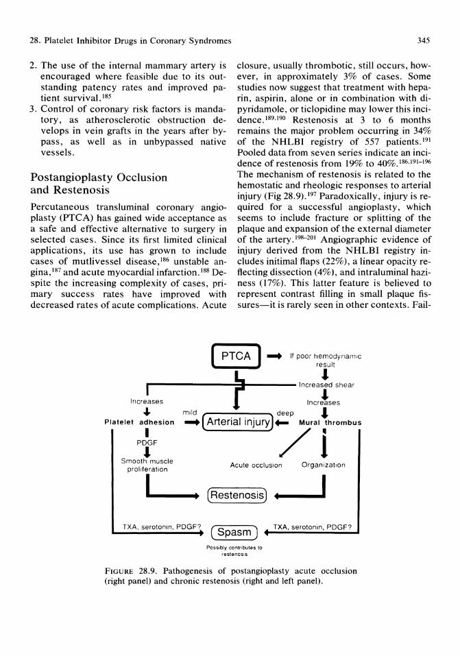
28. Platelet Inhibitor Drugs in Coronary Syndromes 345
2. The use of the internal mammary artery is encouraged where feasible due to its outstanding patency rates and improved patient survival. I85
3. Control of coronary risk factors is mandatory, as atherosclerotic obstruction develops in vein grafts in the years after bypass, as well as in unbypassed native vessels.
Postangioplasty Occlusion and Restenosis
Percutaneous trans luminal coronary angioplasty (PTCA) has gained wide acceptance as a safe and effective alternative to surgery in selected cases. Since its first limited clinical applications, its use has grown to include cases of mutlivessel disease,186 unstable angina, 187 and acute myocardial infarction. 188 Despite the increasing complexity of cases, primary success rates have improved with decreased rates of acute complications. Acute
closure, usually thrombotic, still occurs, however, in approximately 3% of cases. Some studies now suggest that treatment with heparin, aspirin, alone or in combination with dipyridamole, or ticlopidine may lower this incidence. 189,190 Restenosis at 3 to 6 months remains the major problem occurring in 34% of the NHLBI registry of 557 patients. 191 Pooled data from seven series indicate an incidence of restenosis from 19% to 40%.186,191-196 The mechanism of restenosis is related to the hemostatic and rheologic responses to arterial injury (Fig 28.9).197 Paradoxically, injury is required for a successful angioplasty, which seems to include fracture or splitting of the plaque and expansion of the external diameter of the artery. 198-201 Angiographic evidence of injury derived from the NHLBI registry includes initimal flaps (22%), a linear opacity reflecting dissection (4%), and intraluminal haziness (17%). This latter feature is believed to represent contrast filling in small plaque fissures-it is rarely seen in other contexts. Fail-
( PTCA ) ~ It poor hemodynamiC result
Increases
Platelet adhesion
I PDGF ...
Smooth muscle prol iteration
I
r '",ea1 ,he",
Incr!ses
mild deep'" ~ (Arterial injury )~ Mural thrombus
/i Acute occlusion Organization
(RestenoSiS) +1-_ .... 1
TXA, serotonin, PDGF? (Spasm) TXA, serotonin, PDGF?
Possibly contributes to restenOSIS
FIGURE 28.9. Pathogenesis of postangioplasty acute occlusion (right panel) and chronic restenosis (right and left panel).

346
ure to detect injury in other cases probably reflects the insensitivity of angiography for this purpose.
The response to arterial dilatation depends on the depth of injury. 202 Superficial injury results in endothelial denudation. A monolayer of platelets becomes adherent but thrombi do not form.202-204 As discussed, deep injury results in a powerful thrombogenic stimulus and macroscopic mural thrombus is found experimentally in more than 90% of deeply injured arteries within 1 hour, despite full systemic heparinization. 205 If dilatation is inadequate and there is a significant residual stenosis, local shear rates are elevated with consequences as detailed. Thus, a technically poor result is itself a major risk factor for restenosis. Other rheologic factors that may contribute include an intimal flap producing turbulent blood flow or vasoconstriction. Vasoconstriction is itself platelet mediated as it increases proportionally to platelet deposition,206 and is decreased by serotonin or TXA2 antagonists as well as the cyclo-oxygenase inhibitors aspirin and ibuprofen.206,207 Once platelet deposition or thrombosis occurs it contributes to restenosis by release of plateletderived trophic factors resulting in smooth muscle proliferation and fibrous transformation, as well as atherogenic organization of the thrombus itself.
Using the pig carotid angioplasty model, the addition of antiplatelet agents to full dose heparin reduced the incidence of macroscopic thrombus after deep injury from 80% to 90% to 30% to 35%.208,209 Partly effective regimens included aspirin (1 mg/kg per day), aspirin (20 mg/kg per day) and dipyridamole (2,5 mg/kg per day), intravenous ibuprofen and anagre-
I . h'b' 208209 lide, an experimental plate et III I ItOr. ' These partly efficacious regimens are unlikely to significantly reduce restenosis rates because no agent inhibits platelet-vessel wall interaction. Preliminary studies have not supported the role of currently available platelet inhibitors in combating restenosis. Recent randomized placebo-controlled trials using ticlopidine, combined aspirin and dipyridomole, and max-EPA, a fish oil concentrate, have failed to reduce restenosis. 210-211
D.H. Israel, B. Stein, and V. Fuster
Recommendations:
1. Aspirin 325 mg/ day is advised starting 1 day before PTCA, with a dose given 1 hour before the procedure, then daily thereafter for 6 months.
2. Patients should receive a bolus of heparin 100 U /kg at the start of the procedure followed by an intravenous infusion at 15 U I kg per hour. This is continued until the next day when the heparin is stopped and the sheath is removed. If a large intimal flap or dissection is present, or if angiographic evidence of thrombus is present, heparin may be continued for 24 more hours.
3. There is a need for developing therapies to interfere with the platelet-vessel wall interaction that eventually leads to restenosis. Possible strategies include monoclonal antibodies that block GPIIb/IIIa or GPIb receptors, or vWF. Pharmacologic antagonists of platelet-released mitogens or inhibitors of the release reaction are other possible approaches.
References
1. Holman RL, McGill HC Jr, Strong JP, et al: The natural history of atherosclerosis: The early aortic lesions as seen in New Orleans in the middle of the 20th century. Am J Pathol 1958; 34:209.
2. Strong JP, McGill HC Jr: The natural history of aortic atherosclerosis: Relationship to race, sex and coronary lesions in New Orleans. Exp Mol Patho11963; 2(suppl 1):15.
3. Strong JP, McGill HC Jr: The natural history of coronary atherosclerosis. Am J Pathol 1962; 40:37.
4. McGill HC Jr (ed): The Geographic Pathology of Atherosclerosis. Baltimore, Williams & Wilkins, 1968.
5. Caro CG, Fitzgerald JM, Schroter RC: Atheroma and arterial wall shear: Observation, correlation and proposal of a shear dependent mass transfer mechanism for atherogenesis. Proc R Soc Lond (BioI) 1971; 177:109.
6. Matsuda I, Nimi H, Moritake K, et al: The role of hemodynamic factors in arterial wall thickening in the rat. Atherosclerosis 1978; 29:363.
7. Reidy MA, Bowyer DE: Scanning electron mi-
28. Platelet Inhibitor Drugs in Coronary Syndromes
croscopy of arteries: The morphology of aortic endothelium in haemodynamically stressed areas associated with branches. Atherosclerosis 1977; 26: 181.
8. Bloom AL, Giddings JC, Wilus CJ: Factor VIII on the vascular intima: Possible importance in haemostasis and thrombosis. Nature (New Biol) 1973; 241:217-219.
9. Hawiger J, Kloczewiak, Timmons S: Platelet-receptor mechanisms for adhesive macromolecules, Oates JA, Hawiger J., Ross R (eds): in Interaction of Platelets With the Vessel Wall. Bethesda, American Physiological Society, 1985, pp 1-19.
10. Nieuwenhuis HK, Sixma JJ: Platelet Receptors. Proceeding of European Conference on Antiplatelet Therapy: A twenty-year experience. Florence, March 1987, in press.
11. Ross R, Glomset J, Kariya B, et al: A platelet dependent serum factor that stimulates the proliferation of arterial smooth muscle cells in vitro. Proc Natl Acad Sci USA 1974; 71:1207-1210.
12. Kohler N, Lipton R: Platelets as a source of fibroblast growth promoting activity. Exp Cell Res 1974; 87:297-301.
13. Grotendorst GR, Chang T, Seppa HEJ, et al: Platelet derived growth factor is chemoattractant for vascular smooth muscle cells. J Cell Physiol1982; 113:261-266.
14. Ross R: The pathogenesis of atherosclerosisan update. N Engl J Med 1986; 314:488-500.
15. Faggiotto A, Ross R, Harker L: Studies ofhypercholesterolemia in the nonhuman primate I. Changes that lead to fatty streak formation. Arteriosclerosis 1984; 4:323.
16. Faggiotto A, Ross R, Harker L: Studies ofhypercholesterolemia in the nonhuman primate II. Fatty streak conversion to fibrous plaque. Arteriosclerosis 1984; 4:341.
17. Fuster V, Fass DN, Kaye MP, et al: Arteriosclerosis in normal and von Willebrand pigs. Long-term prospective study and aortic transplantation study. Circ Res 1982; 51:587-593.
18. Fuster V, Griggs TR: Porcine von Willebrand disease: Implications for the pathophysiology of atherosclerosis and thrombosis. Prog Hemostasis Thromb 1986; 8: 159-183.
19. Mustard JF, Murphy EA: Effect of smoking on blood coagulation and platelet survival in man. Br Heart J 1963; 1:846.
20. Fuster V, Chesebro JH, Frye RL, et al: Platelet survival and the development of CAD in the young adult: Effects of cigarette smoking,
347
strong family history and medical therapy. Circulation 1981; 63:546.
21. Lewine PH: An acute effect of cigarette smoking in platelet function: A possible link between smoking and arterial thrombosis. Circulation 1973; 48:619.
22. Brinson K: Effect of nicotine on human blood platelet aggregation. Atherosclerosis 1974; 20:137.
23. Saba SR, Mason RG: Some effects of nicotine on platelets. Thromb Res 1975; 7:819.
24. Bierenbaum ML, Fleishman AI, Steier A, et al: Effect of cigarette smoking upon in vivo platelet function in man. Thromb Res 1978; 12: 1051.
25. Wennmalm A: Effects of nicotine on cardiac prostaglandin and platelet thromboxane synthesis. Br J Pharmacol 1978; 64:559.
26. Watts DT: The effect of nicotine and smoking on the secretion of epinephrine. NY Acad Sci 1960; 90:74-80.
27. Cryer PE, Haymond MW, Santiago JV, et al: Norepinephrine and epinephrine release and adrenergic mediation of smoking-associated hemodynamic and metabolic events. N Engl J Med 1976; 295:573.
28. Astrup P: Some physiological and pathological effects of moderate carbon monoxide exposure. Br Med J 1972; 4:447.
29. Davies RF, Topping AL, Turner DM: The effect of intermittent carbon monoxide exposure on experimental atherosclerosis in the rabbit. Atherosclerosis 1976; 24:527.
30. Ross R: Hyperlipidemic and atherosclerosis: Chronic hyperlipidemia initiates and maintains lesions by endothelial cell desquamation and lipid accumulation. Science 1976; 193:1094-1100.
31. Fisher-Dzoga K, Fraser R, Wissler RW: Stimulation of proliferation in stationary primary cultures of monkey and rabbit aortic smooth muscle cells I. Effect of lipoprotein fractions of hyperlipemic serum and lymph. Exp Mol Pathol 1976; 24:346.
32. Carvalho CA, Colman RW, Lees RS: Platelet function in hyperlipoproteinemia. N Engl J Med 1974; 290:434.
33. Shattil SJ, Anaya-Falindo R, Bennet J, et al: Platelet hypersensitivity induced by cholesterol incorporation. J Clin Invest 1975; 55:636.
34. Stuart MJ, Gerrard JM, White JG: Effect of cholesterol on production of thromboxane B2 in patients with diabetes mellitus. Ann Int Med 1977; 82:425.
348
35. Sagel J, Colwell JA, Crook L, et al: Increased platelet aggregation in early diabetes. Ann Int Med 1977; 82:425.
36. Peterson CM, Jones RL, Koenig RJ, et al: Reversible hematologic sequellae of diabetes. Ann Int Med 1977; 86:425.
37. Halushka PV, Rogers RC, Loadholt CB, et al: Increased platelet TXA2 synthesis in diabetes mellitus. J Lab Clin Med 1981; 97:87.
38. LePape J: Nonenzymatic glycosylation increases platelet aggregation potency of collagen from placenta of human diabetics. Biochem Biophys Res Commun 1983; 11:602.
39. Silberbauer K, Schernthaner G, Sinzinger H, et al: Decreased vascular PGIz in juvenile onset diabetes mellitus. N Engl J Med 1979; 293:366.
40. Harrison HE, Reece AH, Johnson M: Effect of insulin treatment on PGI2 in experimental diabetes mellitus. Diabetologia 1980; 18:65.
41. Sherman CT, Litvack F, Grundfest W, et al: Coronary angioscopy in patients with unstable angina pectoris. N Engl J Med 1986; 315:913.
42. Ambrose lA, Winter SL, Arora RR, et al: Angiographic evolution of coronary artery morphology in unstable angina. J Am Coli Cardiol 1986; 7:472.
43. Davies MJ, Thomas AC: Plaque fissuring-the cause of acute MI, sudden ischemic death and crescendo angina. Br Heart J 1985; 53:363.
44. Falk E: Unstable angina with fatal outcome, dynamic coronary thrombosis leading to infarction or sudden death. Autopsy evidence of recurrent mural thrombosis with peripheral embolization culminating in total vascular occlusion. Circulation 1985; 71:699.
45. Fuster V, Chesebro IH: Mechanisms ofunstable angina. N Engl J Med 1986; 315:1023.
46. Gorlin R, Fuster V, Ambrose JA: Anatomic physiologic links between acute coronary syndromes. Circulation 1986; 74:6.
47. Vermylen J, Verstraete M, Fuster V: Role of platelet activation and fibrin formation in thrombogensis. JAm Coli Cardiol1986; 8:2B-9B.
48. Lagaude M: Control mechanisms of platelet aggregation with special reference to eicosanoids and related compounds. Proc European Conference on Antiplatelet Therapy. A twenty-year experience. Florence, March 1987, in press.
49. Peerschke EIB: The platelet fibrinogen receptor. Semin Hematol 1985; 22:241-259.
50. Lucher EF: Mechanisms of platelet aggrega-
D.H. Israel, B. Stein, and V. Fuster
tion. Proc European Conference on Antiplatelet Therapy. A twenty-year experience. Florence, March 1987, in press.
51. Gerrad JM, White IF: Prostacyclin and thromboxanes "middlemen" modulating platelet function in hemostasis and thrombosis. Prog Haemost Thromb 1978; 4:87-125.
52. Cliveden PB, Salzman EW: Platelet metabolism and the effects of drugs, in Bowie EIW, Sharp AA (eds): Hemostasis and Thrombosis. London, Butterworth and Company Publishers, Ltd, 1985, pp 1-35.
53. Moncada S, Gryglewski R, Bunting S, et al: An enzyme isolated from arteries transforms prostaglandin endoperoxides into an unstable substance that inhibits platelet aggregation. Nature 1976; 263:663-665.
54. Moncada S, Vane JR: Arachidonate metabolites and the interactions between platelets and blood vessel walls. N Engl J Med 1979; 300: 1142-1147.
55. Marcus AI, Weksler BB, Iaffe EA: Synthesis of PGI2 (prostacyclin) by cultured human and bovine endothelial cells. Proc Natl Acad Sci USA 1977; 74:3922-3926.
56. Eldor A, Falcone DI, Hajjar DP, et al: Recovery ofprostacyclin production by de-endotheIialized rabbit aorta: Critical role of neointimal smooth muscle cells. J Clin Invest 1981; 67:735.
57. Tateson TE, Moncada S, Vane JR: Effects of prostacyc1in (PGI2) on cAMP concentration in human platelets. Prostaglandin 1977; 13:389-397.
58. Walker FI: Regulation of activated protein C by a new protein. J Bioi Chem 1980; 255:5521-5524.
59. Walker FJ: Regulation of activated protein C by protein S: The role of phospholipid in factor Va inactivation. J Bioi Chem 1981; 256: 1128-1131.
60. Marlar RA, Kleiss AI, Griffin IH: Mechanism of action of human activated protein C, a thrombin dependent anticoagulant enzyme. Blood 1982; 59:1067-1072.
61. Walker FJ, Sexton PW, Esman CT: The inhibition of blood coagulation by activated protein C through the selective inactivation of activated factor V. Biochem Biophys Acta 1979; 571 :333-342.
62. Fulcher CA, Gardiner IE, Griffin IH, et al: Proteolytic inactivation of human factor VIn procoagulant protein by activated human protein C and its analogy with factor V. Blood 1984; 63:486-489.

28. Platelet Inhibitor Drugs in Coronary Syndromes
63. Davie EW, Rathroff aD: Waterfall sequence for intrinsic blood clotting. Science 1964; 145:1310-1312.
64. Griffin JH, Evatt B, Zimmerman BS, et al: Deficiency of protein C in congenital thrombotic disease. J Clin Invest 1981; 68: 1370-1373.
65. Comp PC, Esman CT: Recurrent venous thromboembolism in patients with a partial deficiency of protein S. N Engl J Med 1984; 311:1525-1528.
66. Meade TW, Brozovic M, Chakvabarti RR, et al: Haemostotic function and ischemic heart disease: Principal results of the Northwick Park Heart Study. Lancet 1986; 2:533-537.
67. O'Conner NTJ, Cederholm-Williams S, Copper S, et al: Hypercoagulability and coronary artery disease. Br Heart J 1984; 52:614-616.
68. Dreyer NA, Pizo SV: Blood coagulation and idiopathic thromboembolism among fertile women. Contraception 1980; 22:123-135.
69. Genton E, Gent M, Hirsch J, et al: Platelet inhibiting drugs in the prevention of clinical thrombotic disease. N Engl J Med 1975; 293: 1174-1178.
70. Braunwald E, Fiendawould WT, Furberg CD: Proceeding of a workshop on platelet-active drugs in the secondary prevention of cardiovascular events. Circulation 1980; 62(suppl V):I-135.
71. Packham MA, Mustard JF: Pharmacology of platelet-affecting drugs. Circulation 1980; 62(suppl V):V26-41.
72. Fuster V, Chesebro JH: Pharmacologic effects of platelet inhibitor drugs (Part 11). Mayo Clin Proc 1981; 56:185-195.
73. Weiss HT. Platelets. Pathophysiology and Antiplatelet Drug Therapy. New York, Alan R. Liss, 1982.
74. Kinlough-Rathbone RL, Cazenave JP, Packham MA, et al: Effect of aspirin on prevention of arachidonate pathway on the release of granule contents from rabbit platelets adherent to collagen. Lab Invest 1980; 42:28-34.
75. Tschopp TB: Aspirin inhibits platelet aggregation on but not adhesion to collagen fibrils: An assessment of platelet adhesion and deposit platelet mass by morphometry and 51Cr labeling. Thromb Res 1977; 11:619-632.
76. Weiss HT, Tschopp TB, Baumgartner HR: Impaired interaction (adhesion-aggregation) of platelets with the subendothalium in storage pool disease and after aspirin ingestion. A comparison with VW disease. N Engl J Med 1975; 293:619-623.
349
77. Clowes A W, Karnovsky MJ: Failure of certain antiplatelet drugs to affect myointimal thickening following arterial endothelial injury in the rat. Lab Invest 1980; 42:28-34.
78. Linos A, Worthington JW, O'Fallow W, et al: Effect of aspirin on prevention of coronary and cerebrovascular disease in patients with rheumatoid arthritis. A long-term follow-up study. Mayo Clin Proc 1987; 53:581-586.
79. Patrignani P, Filabozzi P, Patrono C: Selective cumulative inhibition of platelet thromboxane production by low dose aspirin in healthy subjects. J Clin Invest 1982; 69: 1366.
80. DeCaterina R, Gianessi D, Bernini W, et al: Selective inhibition of TXA2 related platelet function of low dose aspirin in patients after MI. Am J Cardiol 1985; 55:589.
81. Lewis HD, Davis JW, Archibald DG, et al: Protective effects of aspirin against acute myocardial infarction and death in men with unstable angina. Results of a V A Cooperative Study. N Engl J Med 1983; 309:396.
82. Levy M: Aspirin use in patients with major upper gastrointestinal bleeding and peptic ulcer disease. N Engl J Med 1974; 290: 1158.
83. Needleman P, Raz A, Ferrendelli JA, et al: Application of imidazole as a selective inhibitor of thromboxane synthetase in human platelets. Proc Natl Acad Sci USA 1977; 74:1716.
84. Marcus AJ, Wexler BB, Jaffe EA, et al: Synthesis of prostacyclin from platelet derived endoperoxides by cultured human endothelial cells. J Clin Invest 1980; 66:979.
85. Vermylen J, Carreras CO, Van Schaeren J, et al: Thromboxane synthetase inhibition as an antithrombotic strategy. Lancet 1981; 1:1073.
86. Chiercha S, Patrono C: Role of platelet and vascular eicosanoids in the pathology of ischemic heart disease. Fed Proc 1987; 46:81-88.
87. Fitzgerald GA, Reilly lA, Pederson AK: The biochemical pharmacology of thromboxane synthase inhibition in man. Circulation 1985; 72: 1194-1201.
88. Gresele P, Deckmyn H, Arnout J, et al: BM 13,177, a selective blocker of platelet and vessel wall thromboxane receptors, inactive in man. Lancet 1984; 1:991.
89. Vermylen J: Thromboxane synthetuse inhibition combined with thromboxane receptor blockade. A step forward in antithrombotic strategy. Thromb Haemost 1984; 52:364.
90. Dyerberg J, Bang 40, Stoffersen E, et al: Eicosapentaencic acid and prevention of thrombosis and atherosclerosis. Lancet 1978; 2:117-119.
350
91. Kromhout D, Bosschieter EB, Coulander CDL: The inverse relation between fish consumption and 20-year mortality from coronary heart disease. N Engl J Med 1985; 312:1205-1209.
92. Seiss W, Scherer B, Bohlig B, et al: Platelet membrane fatty acids, platelet aggregation and thromboxane formation during a mackevel diet. Lancet 1980; 1 :441.
93. Hay CRM, Durber AP, Saynor R: Effect of fish oil on platelet kinetics in patients with ischemic heart disease. Lancet 1982; 1269-1272.
94. Lefer AM, Ogletree ML, Smith JB, et al: Prostacyclin: A potentially valuable agent for preserving myocardial tissue in acute myocardial ischemia. Science 1978; 200:52.
95. Aiken JW, Garman RR, Shebuski RJ: Prevention of blockade of partially obstructed coronary arteries with prostacyclin correlates with inhibition of platelet aggregation. Prostaglandins 1979; 17:483-494.
96. Gryglewski RJ, Szceklik A, Nizankowski R: Antiplatelet action of intravenous infusion of prostacyclin in man. Thromb Res 1978; 13:153-163.
97. Woods HF, Ash G, Weston MJ, et al: Prostacyclin can replace heparin in haemodialysis in dogs. Lancet 1978; 2:1075-1077.
98. Rosenblum WI, El Sabban F: Topical prostacyclin PGIz inhibits platelet aggregation in pial venules of the mouse. Stroke 1979; 10:399-401.
99. Bourgain RH: The inhibitory effect of PGIz on white platelet arterial thrombus formation. Haemostasis 1979; 8:117-119.
100. Gimson AES, Huges RD, Mellon PJ, et al: Prostacyclin to prevent platelet activation during charcoal haemoperfusion in fulminant hepatic failure. Lancet 1980; 1:173-175.
101. Smith MC, Danviriyasup K, Crow JW, et al: Prostacyclin substitution for heparin in longterm hemodialysis. Am J Med 1982; 73:669-678.
102. Moncada S, Korbut R: Dipyridamole and other phosphodiesteraie inhibitors acts as antithrombotic agents by potentiating endogenous prostcyclin. Lancet 1978; 1:1286-1289.
103. Crutchley DJ, Ryan US, Ryan JW: Effects of aspirin and dipyridamole on the degradation of ADP by cultured cells derived from bovine pulmonary artery. J Clin Invest 1980; 66:29-35.
104. Grescle P, Zoja C, Deckmyn H, et al: Dipy-
D.H. Israel, B. Stein, and V. Fuster
ridamole inhibits platelet aggregation in whole blood. Thromb Haemost 1983; 50:852-856.
105. Klabunde RE: Dipyridamole inhibition of adenosine metabolism in human blood. Eur J Pharmacol 1983; 93:21-26.
106. Cazenave JP, Packham MA, KinloughRathbone RL, et al: Platelet adherence to the vessel wall and to collagen-coated surfaces. Adv Exp Med BioI 1978; 102:31-49.
107. Kinlough-Rathbone RL, Groves HM, Cazenave JP, et al: Effect of dipyridamole and aspirin on platelet adherence to damaged rabbit aortas in vitro and in vivo (abstr). Fed Proc 1978; 37:260.
108. The Persantine-Aspirin Reinfarction Study Group. Persantine and aspirin in coronary heart disease. Circulation 1980; 62:449-461.
109. Bousser MG, Eschwege E, Hougenau M, et al: "AICLA" controlled trial of aspirin dipyridamole in the secondary prevention of atherothrombotic cerebral ischemia. Stroke 1983; 14:5-14.
110. American-Canadian Cooperative Study Group: Persantine-aspirin trial in cerebral ischemia. Part II: Endpoint results. Stroke 1985; 16:406-415.
111. Brown BG, Cukingman RA, DeRouen T, et al: Improved graft potency in patients treated with platelet inhibiting therapy after coronary bypass surgery. Circulation 1985; 72:138-146.
112. Goldman S, Copeland J, Moritz T, et al: Effect of antiplatelet therapy on early graft patency after coronary artery bypass grafting: V A Cooperative Study #207 (abstr). J Am Coli Cardio11987; 9:125A.
113. Harker LA, Hanson SR, Kirkman TR: Experimental arterial thromboembolism in baboons. Mechanism, Quantitation and Pharmacologic Prevention. J Clin Invest 1979; 64:559-569.
114. Harker LA, Slichter SJ: Studies of platelet and fibrinogen kinetics in patients with prosthetic heart valves. N Engl J Med 1970; 293:1302-1305.
115. Weilly HS, Steele PP, Davies M, et al: Platelet survival in patients with substitute heart valves. N Engl J Med 1974; 290:534-536.
116. Harker LA: Platelet survival time. Its measurement and use. Prog Haemost Thromb 1978; 4:321-347.
117. Sullivan JM, Harken DE, Gorlin R: Pharmacologic control of thromboembolic complications of cardiac valve replacement. N Engl J Med 1971; 284: 1391-1394.
118. Rosenberg RD: Heparin, antithrombin and ab-
28. Platelet Inhibitor Drugs in Coronary Syndromes
normal clotting. Ann Rev Med 1978; 29:367-378.
119. Salzman EW, Rosenberg RD, Smith MH, et al: Effect of heparin fractions on platelet aggregation. J Clin Invest 1980; 65:664-673.
120. Zucker MB: Effects of heparin on platelet function. Thromb Diath Haemorrh 1975; 33:63-65.
121. Miletich JP, Jackson CM, Majerus PW: Properties of Factor Xa binding site on human platelets. J Bioi Chem 1978; 253:6908-6916.
122. Saba HI, Saba SR, Blackburn CA, et al: Heparin neutralization of PGI2 : Effects upon platelets. Science 1979; 205:499-501.
123. Hanson SR, Harker LA, Bjornsson TD: Effect of platelet modifying drugs on arterial thromboembolism in baboons. Aspirin potentiates the antithrombotic actions of dipyridamole and sulfinpyrazone by mechanisms independent of cyclooxygenase inhibition. J Clin Invest 1987; 75:1591-1599.
124. Buchanan MR, Rosenfeld J, Hirsch J: The prolonged effect of sulfinpyrazone on collagen-induced platelet aggregation in vivo. Thromb Res 1978; 13:883-892.
125. Baumgartner MR: Effects of acetylsalicydic acid, sulfinypyrazone and dipyridamole on platelet adhesion and aggregation in flowing native and anticoagulated blood. Thromb Res 1978; 13:883-892.
126. Harker LA, Harlan JM, Ross R: Effect of sulfinpyrazone on homocysteine induced endothelial injury and arteriosclerosis in baboons. Orc Res 1983; 53:731-739.
127. Report from the Anturane Reinfarction Italian Study: Sulfinpyrazone in postmyocardial infarction. Lancet 1982; 1:237-242.
128. Baur HR, VanTassel RA, Pierach CA, et al: Effects of sulfinpyrazone on early graft closure after myocardial revascularization. Am J Cardiol 1982; 49:420-424.
129. Canadian Cooperative Study Group: A randomized trial of aspirin and sulfinpyrazone in threatened stroke. N Engl J Med 1978; 299:53-59.
130. Cairns JA, Gent M, Singer J, et al: Aspirin, sulfinpyrazone or both in unstable angina. N Engl J Med 1985; 313:1369-1375.
131. Steele P, Rainwater J, Vogel R: Platelet suppressive therapy in patients with prosthetic cardiac valves; relationship of clinical effectiveness to alteration of platelet survival time. Circulation 1979; 60:910-913.
132. Kaegi A, Pineo GF, Shimizu A, et al: Arte-
351
riovenous shunt thrombosis: Prevention by sulfinpyrazone. N Engl J Med 1974; 290:304-306.
133. O'Brien JR: Ticlopidine, a promise for the prevention and treatment of thrombosis and its complications. Haemostasis 1983; 13: 1-54.
134. O'Brien JR, Etherington MD, Shuttleworth RD: Ticlopidine-an antiplatelet drug: Effects in human volunteers. Thramb Res 1978; 18:245-254.
135. Wilkinson AR, Hawker RJ, Hawker JM: The influence of antiplatelet drugs on platelet survival after aortic damage or implantation of a dacron arterial prosthesis. Thramb Res 1979; 15: 181-189.
136. Telford AM, Wilson C: Trial of heparin versus atenolol in prevention of myocardial infarction in intermediate coronary syndrome. Lancet 1981; 1: 1225.
137. Dewood M, Stifer WF, Simpson CS, et al: Coronary arteriographic findings soon after non-Q wave myocardial infarction. N Engl J Med 1986; 315:417-422.
138. Marmor A, Sobel BE, Roberts R: Factors presaging early recurrent myocardial infarction ("extension"). Am J Cardial 1981; 48:603-610.
139. Rothkoph M, Boerner J, Stone MJ, et al: Detection of myocardial infarct extension by CKMB radioimmunoassay. Circulation 1979; 59:268-274.
140. Fraker TD Jr, Wagner GS, Rosati RA: Extension of myocardial infarction: Incidence and prognosis. Circulation 1979; 60: 1126-1129.
141. Nasser FN, Chesebro JH, Hombuoger HA, et al: Myocardial infarction externsion: Diagnosis by CK-MB radioimmunoassay and clinical significance (abstr). Clinic Res 1981; 29:226.
142. Marmor A, Geltman EM, Schechtman K, et al: Recurrent myocardial infarction: Clinical predictors and prognostic implications. Circulation 1982; 66:415-421.
143. Baker JT, Bramlet DA, Lester RM, et al: Myocardial infarct extension: Incidence and relationship to survival. Circulation 1982; 65:918-923.
144. Strauss HD: Myocardial infarction extension: Clinical significance. Primary Cardial 1982; 8:14.
145. Buda AJ, MacDonald IL, Dubbin JD, et al: Myocardial infarct externsion: Prevalence, clinical significance, and problems in diagnosis. Am Heart J 1983; 105:744-749.
146. Hutchins GM, Bulkley BH: Infarct expansion

352
versus extension: Two different complications of acute myocardial infarction. Am J Cardiol 1978; 41: 1127-1132.
147. Chesebro JH, Fuster V, Pumphrey CW, et al: Improvement of shortened platelet survival half-life from the early to the late phase of myocardial infarction (abstr). Circulation 1981; 64(suppl IV):197.
148. Tarhan S, Moffit EA, Taylor WF, et al: Myocardial infarction after general anesthesia. JAm Med Assoc 1972; 220:1451-1454.
149. Roberts R, Gibson RS, Boden WE, et al: Prophylactic therapy with diltiazem prevents early reinfarction: A multicentered randomized double-blind trial in patients recovering from non-Q wave infarction (abstr). J Am Coli Cardiol 1986; 7:68a.
150. Conner PL: Aspirin in coronary heart disease. Comparison of six clinical trials. Isr J Med Sci 1983; 19:413-423.
151. Editorial: Aspirin after myocardial infarction. Lancet 1980; 1:1172-1173.
152. Falk E: Plaque rupture with severe pre-existing stenosis in precipitating coronary thrombosis: Characteristics of coronary atherosclerotic plaques underlying fatal occlusive thrombi. Br Heart J 1983; 50:127-134.
153. Harrison DG, Ferguson DW, Collins SM, et al: Rethrombosis after reperfusion with streptokinase: Importance of geometry of residual lesions. Circulation 1984; 69:991-999.
154. Chesebro JH, Knatterud G, Roberts R, et al: Thrombolysis in myocardial infarction (TIMI) trial, phase I: A comparison between intravenous tissue plasminogen activator and intravenous streptokinase. Circulation 1987; 76: 142-154.
155. Cercek B, Lew AS, Hod H, et al: Enhancement of thrombolysis with tissue-type plasminogen activator by pretreatment with heparin. Circulation 1986; 74:583-587.
156. Collen D: Tissue plaminogen activator and its use in acute myocardial infarction. Presented at the Cardiac Society, Rochester, MN, N 0-
vember 1983. 157. Heras M, Chesebro JH, Penny WJ, et al: The
importance of adequate heparin dosage in arterial angioplasty (abstr). Circulation 1987; Suppl IV:213.
158. Chandler JW, Nath HP, Rogers WJ: Heparin and antiplatelet drugs following streptokinase in acute myocardial infarction: What are their effects on vessel patency (abstr)? J Am Coli Cardiol 1984; 3:600.
D.H. Israel, B. Stein, and V. Fuster
159. Anderson JL, Marshall HW, Bray BE, et al: A randomized trial of intracoronary streptokinase in the treatment of acute myocardial infarction. N Engl J Med 1983; 308:1312-1318.
160. Khaja F, Walton JA Jr, Brymer JF, et al: Intracoronary fibrinolytic therapy in acute myocardial infarction: Report of a prospective randomized trial. N Engl J Med 1983; 308: 1305-1311.
161. Steele PM, Chesebro JH, Fuster V: The natural history of arterial balloon angioplasty in pigs and intervention with platelet-inhibitor therapy: Implications for clinical trials (abstr). Clin Res 1984; 32:209A.
162. Simmons A V, Sheppard MA, Cox AF: Deep venous thrombosis after myocardial infarction: Predisposing factors. Br Heart J 1973; 35:623-625.
163. Mabin TA, Holmes DR, Smith HC, et al: Intracoronary thrombus: Role in coronary occlusion complicating percutaneous transluminal coronary-angioplasty. J Am Coil Cardio11985; 5:198.
164. Fuster V, Chesebro JH: Role of platelets and platelet inhibitors in aortocoronary vein graft disease. Circulation 1986; 2:227-232.
165. Josa M, Lie JT, Bianco KL, et al: Reduction of thrombosis in canine coronary bypass vein grafts with dipyridamole and aspirin. Am J Cardiol1981; 47:1248-1254.
166. Fuster V, Dewanjee MK, Kaye MP, et al: Noninvasive radioisotopic technique for detection of platelet deposition in coronary artery bypass grafts in dogs and its reduction with platelet inhibitors. Circulation 1979; 60: 1058-1062.
167. Bulkley BH, Hutchins GM: Pathology ofcoronary artery bypass graft surgery. Arch Pathol Lab Med 1978; 102:273-280.
168. Nuutinen LS, Pihlajaniemi R, Saarela E, et al: The effect of dipyridamole on the thrombocyte count and bleeding tendency in open heart surgery. J Thorne Cardiovasc Surg 1977; 74:295-298.
169. Becker RM, Smith MR, Dobell ARC: Effect of platelet inhibition phenomenon in cardiopulmonary bypass in pigs. Ann Surg 1974; 179:52-57.
170. Chesebro JH, Fuster V: Platelets and platelet inhibitor drugs in aortacoronary vein bypass operations. Int J Cardiol1983; 2:511-516.
171. Metke MP, Lie JT, Fuster V, et al: Reduction of intimal thickening in canine coronary by-
28. Platelet Inhibitor Drugs in Coronary Syndromes
pass vein grafts with dipyridamole and aspirin. Am J Cardiol1979; 43:1144-1148.
172. Torosian M, Michaelson EL, Morganroth J, et al: Aspirin and coumadin-related bleeding after coronary artery bypass surgery. Ann Int 1978; 89:325-328.
173. Weber M. Trial of anticoagulant v low-dose aspirin in aortocoronary bypass operation. Presented at the International Workshop on Antithrombotic Therapy in CAD. Munich, Germany, January 1986.
174. Weksler BB, Pett SB, Alonso D, et al: Differential inhibition by aspirin of vascular and platelet prostaglandia synthesis in atherosclerotic patients. N Engl J Med 1983; 308:800-805.
175. Chesebro JH, Clements IP, Fuster V, et al: A platelet inhibitor drug trial in coronary artery bypass operations. Benefit of perioperative dipyridamole and aspirin therapy on early postoperative vein graft patency. N Engl J Med 1982; 307:73-78.
176. Chesebro JH, Fuster V, Elveback LR, et al: Effect of dipyridamole and aspirin on late vein graft patency after coronary bypass operations. N Engl J Med 1984; 310:209-214.
177. Rajah SM, Salter MCP, Donaldson DR, et al: Acetylsalicyclic acid and dipyridamole improve the early patency of aortacoronary bypass grafts: A double-blind, placebo-controlled randomized trial. J Thorac Cardiovasc Surg 1982; 89:373-377.
178. Mayer JE, Lindsay WG, Castraneda W, et al: Influence of aspirin and dipyridamole on patency of coronary artery bypass grafts. Ann Thorac Surg 1981; 31:204-210.
179. Lorenz RL, Weber M, Kotzur J, et al: Improved aortocoronary bypass patency by lowdose aspirin (100 mg) Lancet 1984; 1: 1261-1264.
180. Brooks N, Wright J, Sturridge M, et al: Randomized placebo controlled trial of aspirin and dipyridamole in the prevention of coronary vein graft occlusion. Br Heart J 1985; 53:201-207.
181. McEnany MT, Salzman EW, Mundth ED, et al: The effect of antithrombotic therapy on patency rates of saphenous vein coronary artery bypass grafts. J Thorac Cardiovasc Surg 1982; 83:81-89.
182. Pantely GA, Goodnight SH Jr, Rahimtoola SH, et al: Failure of antiplatelet and anticoagulant therapy to improve patency of grafts af-
353
ter coronary artery bypass. N Engl J Med 1979; 301:962-966.
183. Sharma GVRK, Khuri SF, Josa M, et al: The effect of antiplatelet therapy on saphenous vein coronary artery bypass graft patency. Circulation 1983; 68(suppllI):II:218-221.
184. Gould KL, Wescott RJ, Albro PC, et al: Noninvasive assessment of coronary stenoses by myocardial perfusion imaging during pharmacologic coronary vasodilation II. Clinical methadology and feasibility. Am J Cardiol 1978; 41:279-287.
185. Loop FD, Lytle BW, Cosgrove DM, et al: Influence of the internal mammary artery graft on 10 year survival and other cardiac events. N Engl J Med 1986; 314:1-6.
186. Vlietstra RE, Holmes DR, Reeder GS, et al: Balloon angioplasty in multivessel CAD. Mayo Clin Proc 1983; 58:563-567.
187. De Feyter PJ, Serruys PW, vandenBrand M, et al: The emergency coronary angioplasty in unstable angina. New Engl J Med 1985; 313: 1369-1375.
188. O'Neill W, Timmis GC, Bourdillon PD, et al: A prospective randomized clinical trial of intracoronary streptolinase versus coronary angioplasty for acute Ml. N Engl J Med 1986; 314:812-818.
189. Barnathan ES, Schwartz JS, Taylor L, et al: Aspirin and dipyridamole in the prevention of acute coronary thrombosis complicating coronary angioplasty. Circulation 1987; 76:125-134.
190. White CW, Chaitman B, Lassar TA, et al: Antiplatelet agents are effective in reducing the immediate complications of PTCA: Results from the ticlopidine multicenter trial (abstr). Circulation 1987; 76(suppl IV): 1591.
191. Holmes DR, Vlictstra RE, Smith HC, et al: Restenosis after PTCA: A report from the PTCA registry of the National Heart, Lung and Blood Institute. Am J Cardiol 1984; 53:77c-81c.
192. Cowley MJ, Vetrovec GW, DiSciascio G, et al: Coronary angioplasty of mUltiple vessels: Short-term outcome and long-term results. Circulation 1985; 72:1314.
193. Mabin TA, Holmes DR, Smith HC, et al: Follow-up clinical results in patient undergoing PTCA. Circulation 1985; 71 :754.
194. Gruentzig AR, Senning A, Siegenthale WE: Nonoperative dilation of coronary artery stenosis-percutaneous transluminal coronary angioplasty. N Engl J Med 1979; 301:61.
354
195. Leimgruber PP, Roubin GS, Anderson HV, et al: Influence of intimal dissection on restenosis after successful coronary angioplasty. Circulation 1985; 72:530.
196. Vandormael MG, Chaitman BR, Ischinger T, et al: Immediate and short-term benefit of multilesion coronary angioplasty: Influence of degree of rev as cui ariz at ion. JAm Call Cardiol 1985; 6:983.
197. Chesebro JH, Lam JYT, Badimon L, et al: Restenosis after arterial angioplasty: A hemorrheologic response to injury. Am J Cardiol 1987; 60:IOB-16B.
198. Castaneda-Zuniga WR, Formenck A, Tadavarthy OR, et al: The mechanism of balloon angioplasty. Radiology 1980; 175:565-571.
199. Block PC, Myler RK, Stertzer S, et al: Morphology after transluminal angioplasty in human beings. New Engl J Med 1981; 305:382-385.
200. Sanborn TA, Faxon DP, Haudenfield C, et al: The mechanism of transluminal angioplasty: Evidence for formation of aneurysms in experimental atherosclerosis. Circulation 1983; 68: 1136-1140.
201. Pasternak RC, Boughman KL, Falloon JJ, et al: Scanning electron microscopy after coronary transluminal angioplasty of normal canine coronary arteries. Am J Cardiol 1980; 45:591-598.
202. Lam JYT, Chesebro JM, Steele PM, et al: Deep arterial injury during experimental angioplasty. Relationship to a positive indiumlabeled platelet scintigram, quantitative platelet deposition and mural thrombus. J Am Call Cardiol1986; 8:1380-1386.
203. Steele PM, Chesebro JH, Stanson AW, et al: Balloon angioplasty: Natural history of the
D.H. Israel, B. Stein, and V. Fuster
pathophysiology response to injury in a pig model. eirc Res 1985; 57:105.
204. Badimon L, Badimon JJ, Galucz A, et al: Influence of arterial damage and wall shear rate on platelet deposition. Arteriosclerosis 1986; 6:312-320.
205. Lam JYT, Chesebro JM, Heras M, et al: Deep and superficial arterial injury: Different affinity for thrombus formation at increasing shear rate (abstr). Circulation 1987; 76(suppl IV): IV-100.
206. Lam JYT, Chesebro JH, Steele PM, et al: Is vasospasm related to platelet deposition? In vivo relationship in a pig model of arterial injury. Circulation 1987; 75:213-248.
207. Lam JYT, Chesebro JH, Dewanjee MK, et al: Ibuprofen: A potent antithrombotic agent for arterial injury after balloon angioplasty (abstr). J Am Coil Cardiol 1987; 9:64A.
208. Steele PM, Chesebro JH, Badimon L, et al: Balloon angioplasty in pigs. Comparative effects of platelet inhibitor drugs (abstr). Circulation 1984; 70(suppl 11):361.
209. Lam JYT, Chesebro JH, Badimon L, et al: Serotonin and thromboxane A2 receptor blockade decrease vasoconstriction but not platelet deposition after deep arterial injury (abstr). Circulation 1986; 74(suppl 11):97.
210. White CW, Knudson D, Schmidt RJ, et al: Reitman Ticlopidine Study Group. Neither Ticlopidine, nor aspirin-dipyridamole prevents rastenosis post-PTCA: Results from a randomized, placebo controlled, multicenter trial (abstr). Circulation 1987; 76(suppl IV):213.
211. Grigg LE, Kay T, Manolas EG, et ·al: Does Max EPA lower the risk of restenosis postPTCA: A prospective randomized trial (abstr). Circulation 1987; 76(suppl IV):214.
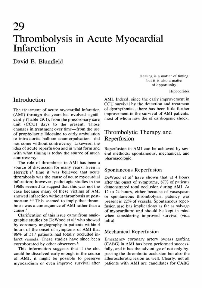
29 Thrombolysis in Acute Myocardial Infarction David E. Blumfield
Introduction
The treatment of acute myocardial infarction (AMI) through the years has evolved significantly (Table 29.1), from the precoronary care unit (CCU) days to the present. Those changes in treatment over time-from the use of prophylactic lidocaine to early ambulation to intra-aortic balloon counterpulsation-did not come without controversy. Likewise, the idea of acute reperfusion and in what form and with what timing is today the source of much controversy.
The role of thrombosis in AMI has been a source of discussion for many years. Even in Herrick'sl time it was believed that acute thrombosis was the cause of acute myocardial infarction; however, pathologic studies in the 1960s seemed to suggest that this was not the case because many of these victims of AMI showed infarction without thrombosis at postmortem. 2 ,3 This seemed to imply that thrombosis was a consequence of AMI rather than a cause.4
Clarification of this issue came from angiographic studies by DeWood et aP who showed by coronary angiography in patients within 4 hours of the onset of symptoms of AMI that 86% of 517 patients had totally occluded infarct vessels. These studies have since been corroborated by other observers.6
This information suggests that if the clot could be dissolved early enough in the course of AMI, it might be possible to preserve myocardium or even improve survival after
Healing is a matter of timing, but it is also a matter
of opportunity.
Hippocrates
AMI. Indeed, since the early improvement in CCU survival by the detection and treatment of dysrhythmias, there has been little further improvement in the survival of AMI patients, most of whom now die of cardiogenic shock.
Thrombolytic Therapy and Reperfusion
Reperfusion in AMI can be achieved by several methods: spontaneous, mechanical, and pharmacologic.
Spontaneous Reperfusion
DeWood et aP have shown that at 4 hours after the onset of symptoms, 87% of patients demonstrated total occlusion during AMI. At 12 to 24 hours, either because of vasospasm or spontaneous thrombolysis, patency was present in 22% of vessels. Spontaneous reperfusion also has implications as far as salvage of myocardium 7 and should be kept in mind when considering improved survival (vida infra).
Mechanical Reperfusion
Emergency coronary artery bypass grafting (CAB G) in AMI has been performed successfully, and it has the advantage of not only bypassing the thrombotic occlusion but also the atherosclerotic lesion as well. Clearly, not all patients with AMI are candidates for CABG
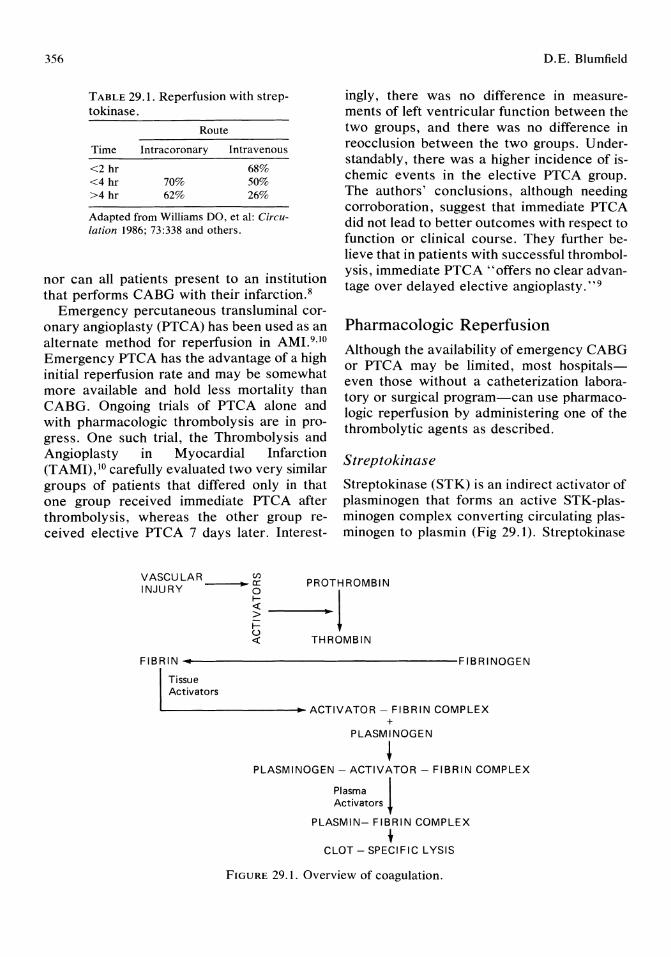
356
TABLE 29.1. Reperfusion with streptokinase.
Route
Time Intracoronary Intravenous
<2 hr 68% <4 hr 70% 50% >4 hr 62% 26%
Adapted from Williams DO, et a1: Circulation 1986; 73:338 and others.
nor can all patients present to an institution that performs CABG with their infarction.8
Emergency percutaneous transluminal coronary angioplasty (PTCA) has been used as an alternate method for reperfusion in AMJ.9,1O Emergency PTCA has the advantage of a high initial reperfusion rate and may be somewhat more available and hold less mortality than CABG. Ongoing trials of PTCA alone and with pharmacologic thrombolysis are in progress. One such trial, the Thrombolysis and Angioplasty in Myocardial Infarction (TAMI), 10 carefully evaluated two very similar groups of patients that differed only in that one group received immediate PTCA after thrombolysis, whereas the other group received elective PTCA 7 days later. Interest-
D.E. Blumfield
ingly, there was no difference in measurements of left ventricular function between the two groups, and there was no difference in reocclusion between the two groups. Understandably, there was a higher incidence of ischemic events in the elective PTCA group. The authors' conclusions, although needing corroboration, suggest that immediate PTCA did not lead to better outcomes with respect to function or clinical course. They further believe that in patients with successful thrombolysis, immediate PTCA "offers no clear advantage over delayed elective angioplasty. "9
Pharmacologic Reperfusion Although the availability of emergency CABG or PTCA may be limited, most hospitalseven those without a catheterization laboratory or surgical program-can use pharmacologic reperfusion by administering one of the thrombolytic agents as described.
Streptokinase
Streptokinase (STK) is an indirect activator of plasminogen that forms an active STK-plasminogen complex converting circulating plasminogen to plasmin (Fig 29.1). Streptokinase
vAScULAR_---.,~Vl PROTHROMBIN
INJURY • ~ ----;.~l i= u « THROMBIN
FIBRIN ...... t-------------------FI B RI NOGEN
Tissue Activators
L---------~ACTIVATOR - FIBRIN COMPLEX +
PLASMINOGEN
+ PLASMINOGEN - ACTIVATOR - FIBRIN COMPLEX
Plasma ! Activators
PLASMIN- FIBRIN COMPLEX
+ CLOT - SPECIFIC LYSIS
FIGURE 29.1. Overview of coagulation.
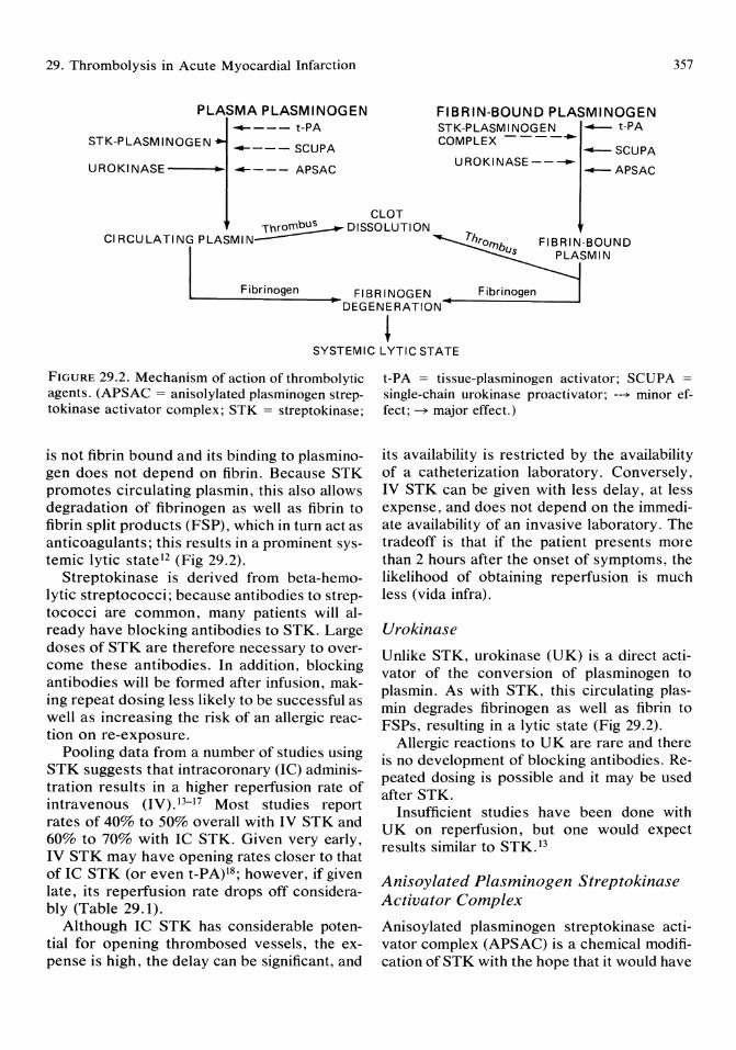
29. Thrombolysis in Acute Myocardial Infarction
PLASMA PLASMINOGEN __ --- t-PA
STK-PLASMINOGEN ----- SCUPA
UROKINASE---"~I ----- APSAC
FIBRIN-BOUND PLASMINOGEN STK-PLASMINOGEN -- t-PA COMPLEX-----
--SCUPA UROKINASE-----
--APSAC
357
CLOT \hrorn~ DISSOLUTION
CIRCULATING PLASMIN---
I FIBRINOGEN Fibrinogen • DEG ENE RA TION--------=:~---I
Fibrinogen
~ SYSTEMIC LYTIC STATE
FIGURE 29.2. Mechanism of action of thrombolytic agents. (APSAC = anisolylated plasminogen streptokinase activator complex; STK = streptokinase;
is not fibrin bound and its binding to plasminogen does not depend on fibrin. Because STK promotes circulating plasmin, this also allows degradation of fibrinogen as well as fibrin to fibrin split products (FSP), which in turn act as anticoagulants; this results in a prominent systemic lytic state l2 (Fig 29.2).
Streptokinase is derived from beta-hemolytic streptococci; because antibodies to streptococci are common, many patients will already have blocking antibodies to STK. Large doses of STK are therefore necessary to overcome these antibodies. In addition, blocking antibodies will be formed after infusion, making repeat dosing less likely to be successful as well as increasing the risk of an allergic reaction on re-exposure.
Pooling data from a number of studies using STK suggests that intracoronary (IC) administration results in a higher reperfusion rate of intravenous (IV). 13-17 Most studies report rates of 40% to 50% overall with IV STK and 60% to 70% with IC STK. Given very early, IV STK may have opening rates closer to that of Ie STK (or even t-PA)18; however, if given late, its reperfusion rate drops off considerably (Table 29.1).
Although IC STK has considerable potential for opening thrombosed vessels, the expense is high, the delay can be significant, and
t-PA = tissue-plasminogen activator; SCUPA =
single-chain urokinase proactivator; --~ minor effect; ~ major effect.)
its availability is restricted by the availability of a catheterization laboratory. Conversely, IV STK can be given with less delay, at less expense, and does not depend on the immediate availability of an invasive laboratory. The tradeoff is that if the patient presents more than 2 hours after the onset of symptoms, the likelihood of obtaining reperfusion is much less (vida infra).
Urokinase
Unlike STK, urokinase (UK) is a direct activator of the conversion of plasminogen to plasmin. As with STK, this circulating plasmin degrades fibrinogen as well as fibrin to FSPs, resulting in a lytic state (Fig 29.2).
Allergic reactions to UK are rare and there is no development of blocking antibodies. Repeated dosing is possible and it may be used after STK.
Insufficient studies have been done with UK on reperfusion, but one would expect results similar to STK.13
Anisoylated Plasminogen Streptokinase Activator Complex
Anisoylated plasminogen streptokinase activator complex (APSAC) is a chemical modification of STK with the hope that it would have

358
less systemic lytic effect by reacting preferentially with fibrin-bound plasminogen (Fig 29.2).19.20 The active site within the STK-plasminogen complex is acylated with p-anisoic acid making it catalytically inert and therefore prevents the reaction with circulating plasminogen and plasma inhibitors. After binding to the fibrin-plasminogen complex, the acylation is reversed and plasminogen is cleaved to plasmin. This results, theoretically, in less consumption of fibrinogen, therefore less risk of hemorrhage. Also the delayed activation results in a longer half-life of the drug, obviating the need for an IV infusion. The plasmin formation, however, is not confined to the site of the clot, and a systemic lytic state results. In one study, fibrinogen reduction was similar to that of STK and UK.
As would be expected, APSAC has the antigenic and antibody problems of STK as well.
A recently reported angiographic study with APSAC21 suggests that with an intravenous bolus, reperfusion rates are similar to those of STK: open vessels, 1) 90 minutes, APSAC 9/ 16 (56%), and placebo, 1/13 (7%); and 2) 3 days APSAC 8/9 (50%), and placebo 1/1 (7%).21
Single-chain Urokinase Proactivator
Single-chain urokinase proactivator (SCUPA) is a precursor of human UK. Animal studies suggested that SCUP A was more clot selective because of a circulating inhibitor to singlechain UK and its presumed action only when clot bound. However, recent studies have shown that SCUPA is not fibrin bound, and very preliminary patient studies22 suggest a systemic lytic state with this agent as well.
Insufficient data are available at present to comment on the reperfusion rates of SCUPA.
Tissue Type Plasminogen Activator
Tissue type plasminogen activator (t-PA) is one of the tissue activators of plasminogen produced after vascular injury (Fig 29.1). Circulating t-PA is minimally effective, but its ability to activate fibrin bound to plasminogen is significantly greater, resulting in plasmin on or at the site of the clot, suggesting less sys-
D.E. Blumfield
temic effect. However, this may be more theoretical than real as changes in the manufacturing process resulting in a single-chain form have necessitated higher doses and longer infusions to avoid the problem of reocclusion, both of which lead to a more systemic lytic state.
The problem of antigenicity does not exist with t-PA, and repeated dosing is possible.
Studies with t-PA have been hampered until very recently by small quantities of agent because of the genetic engineering involved and because of a change in agent from a doublechain to single-chain form. Because the reperfusion rates, infusion rates, and lytic states are different, it makes comparison of studies difficult.
The most recent reports out of the TIMI trial 13, 12,23-26 suggest that whereas the initial 80-mg infusion of t-PA resulted in a 66% opening rate, the longer lOO-mg infusion demonstrated reperfusion in as many as 85% of SUbjects. This should be compared with the overall IV STK rate of 36%. A similar study without pretreatment angiograms by the European Cooperative Study Group27 reported t-PA reperfusion rates of 70% with a shorter infusion compared with 55% for IV STK in their other treatment group.
One can conclude from these studies that tPA, although considerably more expensive and requiring a longer infusion, is approximately twice as effective as IV STK. However, if IV STK can be given very early «2 hours), the rates of opening are quite similar. Clinical data for SCUPA and APSAC suggest that thrombolysis with these agents is similar to STK; however, further studies will be necessary to evaluate claims that they have less bleeding complications.
Thrombolytic Therapy and Left Ventricular Function
As it is clear that thrombolytic agents can open acutely occluded vessels in a significant majority of cases (vida supra), the clinically relevant hypothesis that this reperfusion im-
29. Thrombolysis in Acute Myocardial Infarction
proves left ventricular (LV) function and patient survival must be addressed.
The effect of thrombolysis on infarct size, or its clinical correlate left ventricular function, is actively being investigated. Studies of changes in global ejection fraction have been confiicting.28-31 This possibly can be explained by the observed improvement in global LV function over time without intervention and by the initial hyperkinesis in noninfarcted areas of the ventricle. 33 To try to further study L V dysfunction, Sheehan et aP2 developed a new angiographic analysis model to look at regional change in function from contrast ventriculograms. Using this analysis, they found that reperfused patients showed an almost 40% improvement in regional function, whereas the control group showed no change during the same period. 33 Important to note is that in the treated group, the increase in regional LV function occurred in those patients treated at less than 3 hours after the initiation of pain. Mathey's data also suggested that patients who initially had totally occluded infarct vessels that were opened had the greatest improvement in function as opposed to those that were initially subtotally occluded. Improvement in function also depended on the degree of residual stenosis: those patients with greater than 80% residual stenosis showed much less improvement than those with less than 80% residual stenosis. Additionally, the worse the initial ischemic dysfunction, the more marked is the improvement.
The determinants of recovery of LV regional function are summarized in Table 29.2.
TABLE 29.2. Determinants of recovery of left ventricular function.
Positive correlations Initially totally occluded vessel Residual stenosis <80% Time to reperfusion <3 hr Greater initial ischemic dysfunction
Not correlated Method of reperfusion Site of infarction
Adapted from Sheehan FH, et al: Circulation 1985; 71: 1121 and others.
Thrombolytic Therapy and Survival
359
Important to the hypothesis that thrombolysis is beneficial to the patient is that there exists an improvement in survival after AMI with successful thrombolysis as well as an improvement in function.
Early studies dating back many years did not show any significant reduction in mortality, some even suggested a trend toward increased mortality. These have been well reviewed elsewhere34 and can be summarized as showing no benefit because time of onset of myocardial infarction to time of onset of thrombolysis was too long.
The Western Washington Trial was an early study of IC STK29,35 involving 250 randomized patients. They reported a 66% mortality reduction acutely, with a mortality rate of 11.2% in the control group and 3.7% in the treated group at 30 days. Although this difference was less at 1 year, there was a very high survival rate in the treated group (98%). Rentrop and Feit,36 in an ongoing study, found an increased, although not significant, mortality at 6 months. However, the average time from onset of symptoms to reperfusion was 350 minutes in their study. 36 In a more complicated study involving both IV STK and IC STK, the Netherlands Interuniversity Cardiology Group37,38 reported a 14-day mortality in the treated group of 5% v 9% in the group not assigned to STK. This difference was maintained at 8 months with mortality rates of 9% and 16%. Other trials have supported the hypothesis that thrombolysis improves mortality, including trials from IsraeP9 and Germany.40 However, the report of almost 12,000 patients randomized to IV STK or placebo in the trial reported by the Gruppo Italiano per 10 studio della streptochinasi nell'infarto miocardico (GISSI)41 is almost a "pure" IV STK study with fewer than 3% of all subjects receiving any attempt at mechanical reperfusion. This deserves further discussion because of the large number of subjects, its results, and its implications to clinical cardiology.
As can be seen in Table 29.3, overall mortality reduction was 18%, a figure that carries a
360
TABLE 29.3. Overall mortality.
Streptokinase Control P % change
10.7 13.0 0.0002 18
Adapted from GISSI: Lancet 1986; 1:397.
great deal of power because of the large sample size. Table 29.4 suggests that the overall reduction in mortality was significant only in patients under age 65; it should be noted that the magnitude of the differences are similar, but the small sample size may be the reason for the lack of statistical significance in this subgroup. The apparent benefit only in anterior myocardial infarction or mixed location myocardial infarction involving anterior wall may also be a function of smaller sample size (Table 29.5). Further studies will be needed to answer these questions.
The analysis of mortality reduction by subgroups by hours from onset of symptoms to onset of STK is demonstrated in Table 29.6 and makes a powerful statement that improvement in survival is strongly related to time from onset of symptoms. Clear benefit can be seen in those patients who have thrombolysis before 3 hours and especially within 1 hour. Although not statistically significant, the 6 to 9-hour group again shows a similar magnitude of difference but with smaller numbers. To be emphasized is that these are numbers falling along a continuum: mortality reduction at 1 hour is better than at 3 hours which is better than at 4 hours, etc. Data of similar magnitude appears to be developing in the ongoing TIMI II Trial (personal communication, D.O. Williams, August 22, 1986).
More recently, the ISIS-2 group41a reported on the randomization of nearly 20,000 patients to placebo or intravenous STK plus oral aspi-
TABLE 29.4. Overall mortality.
% Age Streptokinase Control P change
<65 5.7 7.7 0.0005 26 65-75 16.6 18.1 ns 8 >75 28.9 33.1 ns 13
Adapted from GISSI: Lancet 1986; 1 :397.
D.E. Blumfield
TABLE 29.5. Overall mortality.
MI location Streptokinase Control P
Anterior 14.5 18.4 0.0006 Inferior 6.8 7.2 ns Mixed 9.0 13.9 0.002
Adapted from GISSI: Lancet 1986; 1:397. MI = myocardial infarction.
% change
21 6
35
rin 180 mg daily for 30 days. Their results, although not completely reported, seem to confirm the GISSI results of almost 47% mortality reduction in patients with AMI.
Thrombolysis: Reocclusion and Angioplasty
Reocclusion has been a problem with all pharmacologic thrombolytic agents and may run as high as 30% at 14 days. Preliminary data from O'Neill et al42 suggest that thrombolysis plus PTCA has a reocclusion rate of about 7%. Even though reocclusion may be as a high as 30%, reinfarction, presumably because of recruitment of collaterals, is much lower at 5% to 10%.17,18 Reinfarction appears to be a function of time to treatment, the agent used, and its route of administration. A much more clinically significant problem is recurrent angina, which with STK may range from 35% to 60%.42,43 On treadmill testing at 2 weeks, Fung et al43 found an ischemic response in 60% of STK-treated patients but in only 9% of STKand PTCA-treated patients. Johns et al44 found that there was functional improvement in 70%
TABLE 29.6. Mortality reduction and time of onset of symptoms.
% Hours Streptokinase Control P change
<1* 8.2 15.4 0.0001 47 <3 9.2 12.0 0.0005 23 3-6 11.7 14.1 0.03 17 6-9 12.6 14.1 NS 11 9-12 15.8 13.6 NS (16)
* Included in <3 groups. Adapted from GISSI: Lancet 1986; 1:397.

29. Thrombolysis in Acute Myocardial Infarction
of patients, with no residual stenosis by angiogram after recombinant t-PA therapy, but in the remaining 30% with residual stenosis or reocclusion, revascularization was necessary in 22%. These studies suggest that the degree of improvement in L V function is dependent on the degree of residual stenosis as much as the time to reperfusion.
U sing data from the previously reviewed studies, one can construct a scheme that would suggest that for a sizable minority of patients, something is necessary instead of, or in addition to, thrombolysis to achieve reperfusion or overcome reocclusion. Table 29.6 suggests that as many as 40% of patients who reach the hospital in sufficient time may have contraindications to thrombolytic agents, or are hemodynamically too unstable to use thrombolytic agents, or are failures of ph arm acologic thrombolysis and may need to be considered for other forms of reperfusion. Percutaneous transluminal coronary angioplasty is the obvious choice of therapy in these patients. Although there are some centers that use PTCA as first-line emergent therapy in myocardial infarction, most centers consider pharmacologic thrombolysis as first-line therapy in acceptable patients. The TAMI Trial45
and TIM I trial46 have shown that in patients who have failed with t-PA therapy, as many as 90% can be left with a patent artery by PTCA.
These apparently beneficial effects of PTCA after thrombolysis must be tempered by the recent report by Holland et al47 that early PTCA after STK although successful in 58% of patients older than 75 years, also had a mortality of 48% compared with 6% in those younger than 75 years, suggesting that advanced age may be a contraindication to acute combined therapy. The Mayo group49 also reported that in 13% of patients with combined therapy, the patient was made worse either by totally occluding a partially patent vessel or interfering with collaterals. O'Neill et al49 also demonstrated a trend toward more emergent CABG with combined treatment as opposed to PTCA on an elective basis. This seems to suggest that successful thrombolysis should not be followed acutely by PTCA but instead, should be reserved for those patients with
361
contraindications to thrombolysis or unsuccessful thrombolysis. The currently running TIMI II Trial with acute, 18 to 24-hour, and elective PTCA after t-PA treatment should help resolve these issues.
Reports of benefits from acute coronary artery bypass grafting (CABG) also deserve discussion. In acute myocardial infarction CABG is neither accepted by the cardiology community as a whole nor shows an increasing trend, but the Spokane group50 have shown good evidence that CABG can be carried out at low mortality and with good results. The Spokane study, although not randomized and having a control group not comparable to the treatment group, does support the hypothesis that if the infarct-related artery is left patent, the patient does better, and often better than with medical management. This subject is discussed in a subsequent chapter.
Evaluating Thrombolytic Therapy
Evaluating the effectiveness of thrombolysis in AMI falls into two categories: First, assessment of efficacy in attaining reperfusion, and second, evaluating thrombolytic therapy as part of risk stratification in the patient after AMI.
The immediate clinical response of the patient with AMI is believed to be an important factor in assessing reperfusion. The prompt relief of pain and/or the resolution of electrocardiographic (ECG) changes is a good predictor of resumption of blood flow. In fact, most treatment protocols include lack of pain relief or resolution of ECG changes as indications to progress to immediate invasive studies for intracoronary thrombolysis, angioplasty, or both. Less helpful in assessing successful reperfusion is the early peaking of the creatine kinase myocardial band (CKMB) washout curve. 51-53 This information may be delayed and not available by the time decisions need be made regarding invasive studies.
Equally important as the initial assessment of efficacy is the risk stratification-evaluating for jeopardized myocardium-after thrombolysis.
362
Patients believed on clinical grounds not to have reperfused and to have completed an infarction should undergo risk stratification as is the standard of the institution for postmyocardial infarction patients.
Patients who undergo angioplasty as their form of reperfusion, should have the same postangioplasty evaluation performed as is the standard of the institution for other angioplasty patients.
Patients with clear evidence of reperfusion by clinical criteria, should proceed to angiography to assess the infarct related vessel for further intervention since there is no way from clinical evaluation to determine the lesion "left behind" after the clot has been removed.
Patients with equivocal evidence of successful reperfusion by IV route form a large group of patients for whom there has not yet been a standard approach defined. Although most centers proceed to angiography in these patients, and often angioplasty or surgery based on the results of that angiogram, it seems that some evaluation of myocardium at risk should be performed before an intervention.
There are effective noninvasive methods available to risk stratify these patients54-56 using myocardial perfusion imaging. A recommendation for evaluating these patients early in the course of their hospital stay with dipyridamole thallium myocardial perfusion imaging or later with symptom-limited treadmill thallium perfusion imaging can be made before proceeding to invasive studies (this is addressed in a separate chapter). It seems apparent that the patient without evidence of ischemia, regardless of the clinical evidence for or against reperfusion, does not need invasive evaluation. Conversely, the patient with evidence for ischemia must be evaluated invasively to address further intervention. 53 ,57--{j2
Expected Benefits of Thrombolysis
The expected benefits of thrombolytic reperfusion, with or without PTCA, must be improvement in LV function (presumably de-
D.E. Blumfield
creased infarct size) and improved survival at an acceptable risk. To put this into perspective, if one assumes 450 admissions to the "average" US CCU; that 2/3 of these admits have AMI and 18% of those die, or 450 admits/ year to rule out MI, 300 with MI, 150 eligible for thrombolysis by time of arrival and 54 in hospital deaths, 3 lives saved, minus 1 treatment death (CV A), then 150 patients are treated to save 2 lives. If, as should be the case, the actual death rate is lower, the number of patients treated for each life saved will increase.
The question this raises is a public health issue, namely what expense in dollars and what expense in patient risk is acceptable to save one life or to improve regional LV function by 8% to 10%? The answer to this is not readily available, and it is unlikely that the answer will come from any current studies. It is an issue that will have to be addressed because thrombolytic treatment with or without PTCA of all acceptable patients with AMI would take up an increasing amount of the medical care dollar.
The answers are not easy to come by since patients are now coming to hospital (or even on clinic visits) demanding thrombolysis after reading the following in the lay press:
Intravenous streptokinase must be regarded as the treatment of choice in patients admitted to general CCU's at least up to six hours from myocardial infarction.
GISSI: Lancet 1986; 1:397.
At least . . . 1.5 million heart attack victims will have access to the life saving drug t-PA ... It will save many lives and improve the quality of life. TPA: tip of the iceberg (editorial).
Wall Street Journal 1987; Dec 2:4.
Following are the guidelines that are used at our institution to help emergency physicians and house staff with decisions about thrombolysis.
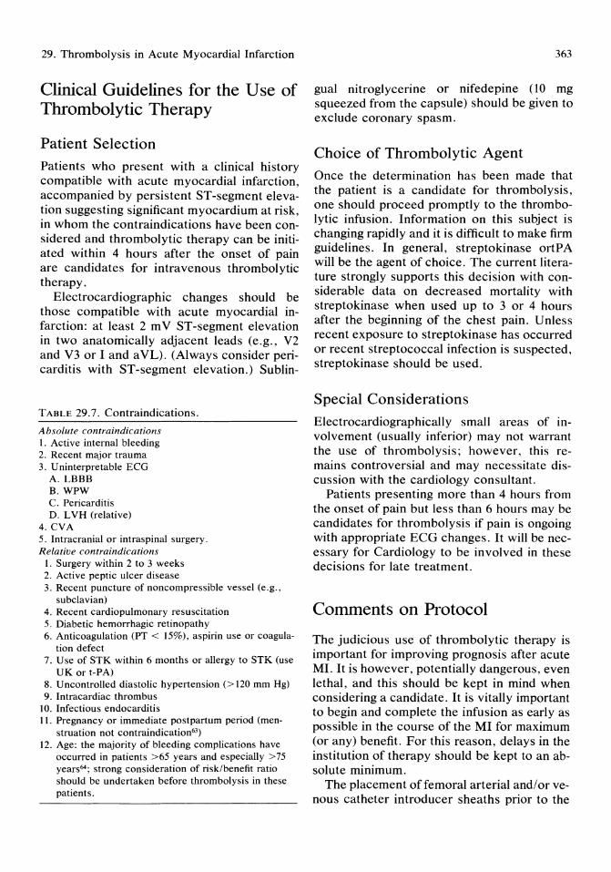
29. Thrombolysis in Acute Myocardial Infarction
Clinical Guidelines for the Use of Thrombolytic Therapy
Patient Selection
Patients who present with a clinical history compatible with acute myocardial infarction, accompanied by persistent ST-segment elevation suggesting significant myocardium at risk in whom the contraindications have been con~ sidered and thrombolytic therapy can be initiated within 4 hours after the onset of pain are candidates for intravenous thrombolytic therapy.
Electrocardiographic changes should be those compatible with acute myocardial infarction: at least 2 m V ST -segment elevation in two anatomically adjacent leads (e.g., V2 and V3 or I and aVL). (Always consider pericarditis with ST -segment elevation.) Sublin-
TABLE 29.7. Contraindications.
Absolute contra indications I. Active internal bleeding 2. Recent major trauma 3. U ninterpretable ECG
A.LBBB B. WPW C. Pericarditis D. LVH (relative)
4. CVA 5. Intracranial or intraspinal surgery. Relative contraindications
I. Surgery within 2 to 3 weeks 2. Active peptic ulcer disease 3. Recent puncture of noncom pres sible vessel (e.g.,
subclavian) 4. Recent cardiopulmonary resuscitation 5. Diabetic hemorrhagic retinopathy 6. Anticoagulation (PT < 15%), aspirin use or coagula
tion defect 7. Use of STK within 6 months or allergy to STK (use
UK or t-PA) 8. Uncontrolled diastolic hypertension (> 120 mm Hg) 9. Intracardiac thrombus
10. Infectious endocarditis 11. Pregnancy or immediate postpartum period (men
struation not contraindication63)
12. Age: the majority of bleeding complications have occurred in patients >65 years and especially >75 years64 ; strong consideration of risk/benefit ratio should be undertaken before thrombolysis in these patients.
363
gual nitroglycerine or nifedepine (10 mg squeezed from the capsule) should be given to exclude coronary spasm.
Choice of Thrombolytic Agent
Once the determination has been made that the patient is a candidate for thrombolysis, one should proceed promptly to the thrombolytic infusion. Information on this subject is changing rapidly and it is difficult to make firm guidelines. In general, streptokinase ortPA will be the agent of choice. The current literature strongly supports this decision with considerable data on decreased mortality with streptokinase when used up to 3 or 4 hours after the beginning of the chest pain. Unless recent exposure to streptokinase has occurred or recent streptococcal infection is suspected, streptokinase should be used.
Special Considerations
Electrocardiographically small areas of involvement (usually inferior) may not warrant the use of thrombolysis; however, this remains controversial and may necessitate discussion with the cardiology consultant.
Patients presenting more than 4 hours from the onset of pain but less than 6 hours may be candidates for thrombolysis if pain is ongoing with appropriate ECG changes. It will be necessary for Cardiology to be involved in these decisions for late treatment.
Comments on Protocol
The judicious use of thrombolytic therapy is important for improving prognosis after acute MI. It is however, potentially dangerous, even lethal, and this should be kept in mind when considering a candidate. It is vitally important to begin and complete the infusion as early as possible in the course of the MI for maximum (or any) benefit. For this reason, delays in the institution of therapy should be kept to an absolute minimum.
The placement offemoral arterial and/or venous catheter introducer sheaths prior to the

364
TABLE 29.8. Protocol.
Procedure 1. Place 18-gauge angiocath/heplock
Place 20-gauge angiocath/heplock 2. Consider placing femoral sheaths
3. Avoid all other punctures 4. Lidocaine bolus and drip as per CCU protocol 5. Preinfusion blood work: CBC, PT, PTT, fibrinogen,
CKMB, platelets, type and cross, etc., ECG 6. Heparin, 5,000 U IV 7. Diphenhydramine, 50 mg IV 8. Thrombolytic infusion (see Table 29.9) 9. ECG at end of infusion, hourly x2 then each morn
ing 10. CK/CKMB every 6 hours x4, then as indicated 11. Heparin, 1,000 U /hour beginning 90 minutes after
infusion 12. Maintain PTT between 80 to 120 seconds for 3 to 4
days 13. Consider need for warfarin, aspirin 14. Consider need for further diagnostic tests
infusion should be strongly considered especially if the MI appears to be extensive, there is any suggestion of conduction abnormality or congestive heart failure. Remember that any aborted puncture of either vessel will bleed. The infusion, however, should not be delayed for placement of sheaths: instead, rely on an experienced person to obtain access later (Tables 29.8 and 29.9).
TABLE 29.9. Doses of thrombolytic agents.
A. Intravenous streptokinase 1. consider aspirin, 180 mg, chewed 2. 1.5 million U over 30 to 60 minutes
B. Intracoronary streptokinase 1. 10,000 to 30,000 U as bolus 2. 4,000 U per minute 3. total average dose is 250,000 to 300,000 U
C. Intravenous urokinase 1. 1.5 million U over 30 to 60 minutes
D. Intravenous t-PA 1. patient 65 kg or greater:
a. 10 mg IV bolus b. 50 mg IV over 1 hour c. 20 mg IV/hour for 2 hours d. total dose 100 mg
2. if patient less than 65 kg: a. 6 mg IV bolus b. 0.75 mg/kg over 1 hour c. 0.25 mg/kg/hour for 2 hours
D.E. Blumfield
Rationale I. Blood sampling/infusion intravenous therapy
2. Access for pacemaker, pulmonary artery line, arte-rial line, or angiography
3. Every puncture site will bleed 4. High likelihood of reperfusion dysrhythmias 5. Baseline
6. Attempt to prevent thrombus from reforming 7. Possibly avoid allergic reaction (watch for BP drop) 8. Watch for hypotension, dysrhythmias bleeding 9. Follow course of MI
10. CK curve 11. See 6.
12. See 6.
13. See 6. 14. Individualize risk stratification for each patient.
In addition to the usual monitoring required in MI patients, careful observation for signs of bleeding is essential. All puncture sites should have pressure dressings and be observed closely. Any change in mental status or any neurologic finding should be carefully evaluated and if unexplained (e.g., lidocaine or sedatives) the infusion should be stopped. Back or leg pain (especially with sheaths in place) may indicate a retro-peritoneal bleed, and if suspected, this necessitates discontinuing the infusion. Hematemasis or hemoptysis (distinguish from gingival or nasal bleeding which is common) will also require infusion to be discontinued.
EKG monitoring for potentially life threatening reperfusion dysrhythmias is necessary. These are of sufficient frequency that prophylaxis with lidocaine is indicated.
References
1. Herrick JB: Clinical features of sudden obstruction of the coronary arteries. JAm Med Assoc 1912; 59:2015.
2. Spain DM, Bradess V A: The relationship of coronary thrombosis to coronary atherosclerotic and ischemic heart disease. Am J Med Sci 1960; 240:701.
29. Thrombolysis in Acute Myocardial Infarction
3. Erich JC, Shinohara Y: Low incidence of coronary thrombosis in myocardial infarction. Arch Pathol 1964; 78:432.
4. Roberts WC, Buja LM: The frequency and significance of coronary arterial thrombi and other observations in fatal acute myocardial infarction. Am J Med 1972; 52:425.
5. DeWood MA, Spores J, Notske R: Prevalence of total coronary occlusion during the early hours of transmural acute myocardial infarction. N Engl J Med 1980; 305:897.
6. Rentrop KP, Blanhe H, Karsch KR, et al: Initial experience with transluminal recanalization of the recently occluded infarct-related coronary artery in acute myocardial infarction: Comparison with conventionally treated patients. Clin Cardiol 1979; 2:92.
7. Defeyter PJ, vanEenige MJ, vanderWall EE, et al: Effects of spontaneous and streptokinaseinduced recanalization on left ventricular function after myocardial infarction. Circulation 1983; 6:1037.
8. DeWood MA, Spores J, Notske RN: Medical and surgical management of myocardial infarction. Am J Cardiol 1979; 44: 1356.
9. Rutherford BD, Hartzler GO, McConahay DR: Direct balloon angioplasty in acute myocardial infarction without prior use of streptokinase. J Am Coli Cardiol 1986; 7: 149A.
10. Holmes DR, Smith HC, Vliestra RE, et al: Percutaneous transluminal coronary angioplasty, alone and in combination with streptokinase therapy, during acute myocardial infarction. Mayo Clin Proc 1985; 60:449.
11. Topol EJ, Califf RM, George BS, et al: A randomized trial of immediate versus delayed elective angioplasty after intravenous tissue plasminogen activator in acute myocardial infarction. N Engl J Med 1987; 317:581.
12. Burket MW, Smith MR, Walsh TE, et al: Relation of effectiveness of intracoronary thrombolysis in acute myocardial infarction to systemic thrombolytic state. Am J Cardiol1985; 56:441.
13. Rentrop KP: Thrombolytic therapy in patients with acute myocardial infarction. Circulation 1985; 71:627.
14. The TlMI Study Group: The Thrombolysis in Myocardial Infarction (TlMI) Trial: Phase I findings. N Engl J Med 1985; 312:932.
15. Leiboff RH, Katz RJ, Wasserman AC, et al: A randomized, angiographically controlled trial of intracoronary streptokinase in acute myocardial infarction. Am J Cardiol 1984; 53:404.
16. The ISAM Study Group: A prospective trial of
365
intravenous streptokinase in acute myocardial infarction (ISAM): Mortality and infarct size at 21 days. N Engl J Med 1986; 314:1465.
17. Yusaf S, Collins R, Petro R, et al: Intravenous and intracoronary fibrinolytic therapy in acute myocardial infarction: Overview of results on mortality, reinfarction and side effects from 33 randomized controlled trials. Eur Heart J 1985; 6:556.
18. Williams DO, Borer J, Braunwald E, et al: Intravenous recombinant tissue type plasminogen activator in patients with acute myocardial infarction: A report from the NHLBI thrombolysis in myocardial infarction trial. Circulation 1986; 73:338.
19. Kasper W, Meinertz T, Wollschlager H, et al: Coronary thrombolysis during acute myocardial infarction by intravenous BRL 26921 a new anisoylated plasminogen-streptokinase activator complex. Am J Cardiol 1986; 58:418.
20. Verstraete M, Collen D: Pharmacology of thrombolytic drugs. J Am Coli Cardiol 1986; 8:33B.
21. Timmis AD, Griffin B, Crick JCP: Anisoylated plasminogen streptokinase activator complex in acute myocardial infarction: A placebo controlled arteriographic coronary recanalization study. J Am Coli Cardiol 1987; 10:205.
22. vandeWerf F, Nobwhara M, Collen D: Coronary thrombolysis with human single chain urokinase-type plasminogen activator (pro-urokinase) in patients with acute myocardial infarction. Ann Intern Med 1986; 104:345.
23. Garabedian HD, Gold HK, Leinbach RC, et al: Dose-dependent thrombolysis, pharmacokinetics and hemostatic effects of recombinant human tissue-type plasminogen activator for coronary thrombosis. Am J Cardiol 1986; 58:673.
24. Verstraete M, Su CA, Tanswell P, et al: Pharmacokinetics and effects on fibrinolytic and coagulation parameters of 2 doses of recombinant tissue-type plasminogen activator in healthy volunteers. Thromb Haemost 1986; 56: I.
25. Collen D, Topol EJ, Tiefenbaum AJ, et al: Coronary thrombolysis with recombinant tissuetype plasminogen activator: A prospective, randomized placebo controlled trial. Circulation 1984; 70:1012.
26. Gold HK, Leinbach RC, Garabedian RD, et al. Acute coronary reocclusion after thrombolysis with recombinant tissue-type plasminogen activator: Prevention by maintenance infusion. Circulation 1986; 73:347.
366
27. Verstraete M, Bernard R, Rory MS, et al: Randomized trial of intravenous recombinant tissue-type plasminogen activator u intravenous streptokinase in acute myocardial infarction. Lancet 1985; 1:842.
28. Andersen JL, Marshall HW, Bray BE, et al: A randomized trial of intracoronary streptokinase in the treatment of acute myocardial infarction. N Engl J Med 1985; 308:1312.
29. Kennedy JW, Ritchie JL, Davis KB, et al: Western Washington randomized trial of intracoronary streptokinase in acute myocardial infarction. N Engl J Med 1983; 309: 1477.
30. Khaja F, Walton JA, Brymer JF, et al: Intracoronary fibrinolytic treatment in acute myocardial infarction. Report of a prospective randomized trial. N Engl J Med 1983; 308:1305.
31. Serruys PW, Simoons ML, Suryapranata H, et al: Preservation of global and regional left ventricular function after early thrombolysis in acute myocardial infarction. J Am Call Cardial 1986; 7:729.
32. Sheehan FH, Dodge HT, Mathey DG, et al: Application of the center line method: Analysis of change in regional left ventricular wall motion in serial studies. IEEE Comput Cardiol 1982; 97.
33. Sheehan FH, Mathey DG, Schofer J, et al: Factors that determine recovery of left ventricular function after thrombolysis in patients with acute myocardial infarction. Circulation 1985; 71:1121.
34. Hugenholtz PG, Serruys PW, Simoons ML, et al: Thrombolytic therapy for acute coronary obstruction: Status in 1986. Texas Heart Institute Journal 1986; 13:433.
35. Kennedy JW, Ritchie JL, Davis KB, et al: The Western Washington randomized trial of intracoronary streptokinase in acute myocardial infarction. N Engl J Med 1985; 312:1073.
36. Rentrop P, Feit F: The reperfusion study group: The second Mt. Sinai-NYU reperfusion trial: Main endpoints. J Am Call Cardia! 1987; 9:239A.
37. Simoons ML, Serruys PW, vander Brand M, et al: Improved survival after early thrombolysis in acute myocardial infarction. Lancet 1985; 2:578.
38. Simoons ML, Serruys PW, vander Brand M, et al: Thrombolysis in acute myocardial infarction: Limitation of infarct size and improved survival. J Am Call Cardioll986; 7:717.
39. Fine DG, Weiss AT, Sapoznikov D, et al: Importance of early initiation of intravenous strep-
D.E. Blumfield
tokinase therapy for acute myocardial infarction. Am J Cardia! 1986; 58:411.
40. Schroder R, Neuhaus KL, Leizoravicz A, et al: A prospective placebo controlled double-blind multicenter trial of intravenous streptokinase in acute myocardial infarction (ISAM): Long-term mortality and morbidity. J Am Call Cardiol 1987; 9:197.
41. Gruppo italiano per 10 studio della streptochinasi nell' infarto miocardico (GISSI): Effectiveness of intravenous thrombolytic treatment in acute myocardial infarction. Lancet 1986; 1:397.
41a. ISIS Collaborative Group: Results of a large randomized trial of intravenous streptokinase and oral aspirin in acute myocardial infarction. J Am Call Cardia! 1988; 11 :232A.
42. O'Neill W, Timmis GC, Bourdillon PD, et al: A prospective randomized trial of intracoronary streptokinase u coronary angioplasty for acute myocardial infarction. N Eng! J Med 1986; 314:812.
43. Fung A Y, Lai P, Juni JE, et al: Prevention of subsequent exercise induced peri infarction ischemia by emergency coronary angioplasty in acute myocardial infarction: Comparison with intracoronary streptokinase. J Am Call Cardia! 1986; 8:496.
44. Johns JA, Yasuda T, Garabedian HD, et al: Improved left ventricular function following intravenous recombinant tissue-type plasminogen activator in acute myocardial infarction. J Am Call Cardia! 1987; 9:190A.
45. Topol EJ, Califf RM, Stack RS, et al: A randomized multicenter trial of intravenous tP A and emergency coronary angioplasty in acute myocardial infarction: Results from the T AMI study group. J Am Call Cardiall987; 9:231A.
46. Williams DO, Porman S, and the TIMI investigators: Coronary angioplasty following rt-PA in acute myocardial infarction: Report from the TIMI trial. J Am Call Cardia! 1987; 9:231A.
47. Holland K, Topol EJ, Walton JA, et al: Emergency coronary angioplasty therapy for elderly patients with acute myocardial infarction: Cautionary results. J Am Call Cardial 1987; 9:231A.
48. Holmes DR, Smith HC, Vliestra RE, et al: Percutaneous transluminal coronary angioplasty, alone and in combination with streptokinase therapy, during acute myocardial infarction. Mayo C!in Proc 1985; 60:449.
49. O'Neill WW, Kereiakes DJ, Stack RS, et al: Cardiothoracic surgical support is required dur-
29. Thrombolysis in Acute Myocardial Infarction
ing interventional therapy of myocardial infarction: Results from the TAM I study group. JAm Call Cardiol 1987; 9: 124A.
50. DeWood MA, Spores J, Berg R Jr, et al: Acute myocardial infarction: A decade of experience with surgical reperfusion in 701 patients. Circulation 1983; 68:11-8.
5l. Wei JY, Markis JE, Malagold M, et al: Time course of serum cardiac enzymes after intracoronary thrombolytic therapy. Arch Intern Med 1985; 145:1596.
52. Sobel BE, Bergmann SR: Coronary thrombolysis: Some unresolved issues. Am J Med 1982; 72:l.
53. Gore JM, Roberts R, Ball SP, et al: Peak creatine kinase as a measure of effectiveness of thrombolytic therapy in acute myocardial infarction. Am J Cardiol1987; 59:1234.
54. Gibson RS, Watson D, Crampton RS, et al: Predischarge thallium-201 scintigraphy to identify postinfarction patients at high risk for future cardiac events. Circulation 1983; 68:321.
55. Weiss AT, Maddahi J, Lew AS, et al: Reverse redistribution of thallium-201: A sign of nontransmural myocardial infarction with patency of the infarct-related artery. J Am Call Cardiol 1986; 7:6l.
56. Beller GA: Role of myocardial perfusion imaging in evaluating thrombolytic therapy for acute myocardial infarction. J Am Call Cardiol 1987; 9:661.
57. Leppo JA, O'Brien J, Roshender JA, et al: Dipyridamole thallium-201 scintigraphy in prediction of future cardiac events after acute
367
myocardial infarction. N Engl J Med 1984; 310: 1014.
58. Jones RH, Floyd RD, Austin EH, et al: The role of angiocardiography in the perioperative prediction of pain relief and survival following coronary artery bypass grafting. Ann Surg 1983; 197:743.
59. Gibson RS, Taylor GT, Watson DD, et al: Predicting extent and location of coronary artery disease during early postinfarction period by quantitative thallium-201 scintigraphy. Am J Cardiol 1981; 47: 1010.
60. Reisner S, Berman D, Maddahi J, et al: The severe stress thallium defect: An indicator of critical coronary stenosis. Am Heart J 1985; 110: 128.
6l. Turner JD, Schwartz KM, Logic JR, et al: Detection of jeopardized myocardium 3 weeks after myocardial infarction by exercise testing with thallium-201 myocardial scintigraphy. Circulation 1980; 61:729.
62. Okada RD, Boucher CA: Differentiation of viable and nonviable myocardium after acute reperfusion using serial thallium-201 imaging. Am Heart J 1987; 113:24l.
63. deGeorgio B, Goldstein J, Haft JI: Administration of intracoronary streptokinase during menstruation. Am Heart J 1985; 109:908.
64. Lew AS, Hod H, Cercek B, et al: Mortality and morbidity rates of patients older and younger than 75 years with acute myocardial infarction treated with intravenous streptokinase. Am J Cardiol 1987; 59: l.

30 Interventional Approach in the Management of Cardiogenic Shock Amar S. Kapoor
Cardiogenic shock is a complex pathophysiologic syndrome characterized by reduced systemic blood pressure, impaired tissue perfusion with a marked reduction in oxygen delivery capacity, and associated excessive stimulation of the sympathetic nervous system. l Cardiogenic shock results from extensive loss of contracting myocardium usually as a complication of acute myocardial infarction. Despite advances in pharmacologic therapy, mortality remains between 70% to 90%. This high mortality is chiefly related to at least 40% loss of contracting left ventricle by a recent myocardial infarction plus a previous myocardial scar.l
Pathophysiology of Cardiogenic Shock
The clinical picture of shock is dominated by hypoperfusion with systemic blood pressure less than 90 mm Hg. The spectrum of clinical manifestations include altered sensorium; cold, clammy, cyanotic skin; and oliguria or anuria. There may be associated multisystem decompensation (Table 30.1).
There is significant alteration of left ventricular performance. Due to extensive destruction of myocardium, there is significant impairment of systemic emptying, producing an excessive increase in the end-systolic volume so that subsequent diastolic fill~ng into a compromised ventricle with high end-systolic volume allows only a small dilatation of the ven-
tricle. 2 Because of the reduced systolic ejection volume and ejection fraction, there is a critical decrease in cardiac output and arterial pressure. 2 Compensatory mechanisms, which increase peripheral vascular resistance to maintain arterial pressure, further decrease cardiac output. The severity of hemodynamic dysfunction is ultimately related to the balance between oxygen demand and supply in the presence of extensive myocardial destruction and impaired mechanical performance of the left ventricle. 3,4
Cardiogenic shock is usually seen as a complication of acute myocardial infarction but may also be seen in the setting of end-stage cardiomyopathy and irreversible valvular heart disease as summarized in Table 30,2.
Assessment of Cardiogenic Shock
Early recognition of the impending shock state is extremely important for an aggressive management strategy, which may enhance survivability (Table 30.3). It is of paramount importance to obtain clinical, biochemical, and hemodynamic assessment in a timely and objective manner. With hemodynamic monitoring, one can objectively and reliably assess the problem, assess hemodynamic dysfunction, plan the treatment, and also prognosticate the patient's clinical outcome.
Radial or brachial artery cannulation is necessary for continuous arterial pressure monitoring and for frequent blood sampling for ar-

30. Interventional Approach in the Management of Cardiogenic Shock 369
TABLE 30.1. Clinical parameters of cardiogenic shock.
Peak systemic blood pressure (90 mm Hg) weak pulse Peripheral vasoconstriction; cold, clammy skin Cerebral hypoperfusion-confusional state, coma, agita-
tion Acute organ or multisystem decompensation; fall in
urine output, 20 ml/hr; rise in creatinine, bilirubin, SGOT
terial blood gases. Present-day technology is capable of providing continuous on-line blood gas analysis and several chemical measurements by special sensor technology.
Urethral catheterization by a Foley catheter is also mandatory for assessing hourly measurements of urine volume and the response of this measurement to fluid loading and pharmacotherapy.
Bedside echocardiogram is a very important noninvasive tool for quantification of pericardial effusions, left ventricular wall motion assessment, global ejection fraction, ruptured papillary muscle, and ventricular septal defects. Doppler echocardiography will assist in defining valvular dysfunction and estimation of cardiac output noninvasively.
Hemodynamic Monitoring
Hemodynamic monitoring is of paramount importance in patients with shock complicating myocardial infarction, in establishing the pathophysiologic mechanisms perpetuating
TABLE 30.2. Cardiogenic shock-different etiologies.
Acute myocardial infarction 40% or more destruction of left ventricle Extensive right ventricular infarction Acute papillary muscle rupture Acute rupture of interventricular septum Large ventricular aneurysm
End-stage cardiomyopathy Dilated cardiomyopathy Hypertrophic cardiomyopathy Restrictive cardiomyopathy
Irreversible valvular heart disease After open heart surgery with poor ventricles Congenital heart disease with Eisenmenger physiology
TABLE 30.3. Schedule for assessment of cardiogenic shock.
Arterial line-for blood sampling, arterial blood gases, and continuous arterial pressure
Flow-directed pulmonary arterial catheter For measurement of pulmonary arterial/wedge pres
sures, central venous/right atrial pressures, cardiac outputs
For diagnosis of mitral regurgitation and acute ventricular septal defect
For diagnosis of right ventricular infarction and cardiac tamponade
For hemodynamic manipUlation by pharmacotherapy Bedside echocardiographic study
For noninvasive evaluation of pericardial effusion and complications of myocardial infarction
MUGA scan-ejection fraction and wall motion abnormality
Cardiac catheterization-for definitive diagnostic assessment and surgical candidacy
the shock syndrome, and providing information for making definitive therapeutic decisions. The thermodilution pulmonary artery catheter permits measurements of left ventricular filling pressure, cardiac output, and pulmonary and systemic vascular resistance. 5 The catheter can be inserted through a central or peripheral vein or by basilic vein cutdown and advanced to the pulmonary artery with pressure monitoring and sometimes via fluoroscopy. There are multiple uses of the thermodilution pulmonary artery catheter. It measures left ventricular filling pressure, which is determined indirectly by measuring pulmonary capillary wedge pressure. In cardiogenic shock the pulmonary end-diastolic pressure correlates well with the left ventricular end-diastolic pressure.6 It is helpful to compare the pulmonary artery end-diastolic pressure with the pulmonary wedge pressure and also the right atrial pressure. In patients with acute left ventricular failure, there is significant elevation of left ventricular filling pressure and a normal or slightly elevated right ventricular filling pressure, whereas in patients with important right ventricular infarction, the right-sided pressure may be equal to or higher than the left heart filling pressure.
The thermodilution cardiac output and filling pressure permit an objective classification

370 A.S. Kapoor
TABLE 30.4. Clinical and hemodynamic subsets of patients with acute myocardial infarction and cardiogenic shock.
Subset Hemodynamic characterization Clinical manifestation
Normal CI > 2.0 L/min/m2 No pulmonary congestion or peripheral hypoperfusion PCWP 12-18 mm Hg L VSWI 20-22 g/mlm2
II CI < 2 Llmin/m2 Pulmonary congestion; no peripheral hypoperfusion PCWP < 18 mm Hg L VSWI < 20 g/mlm2
III CI > 2.0 Llmin/m2 No pulmonary congestion; peripheral hypoperfusion PCWP> 18 mm Hg L VSWI > 20 g/mlm2
IV CI < 2 Llmin/m2 Both pulmonary congestion and peripheral hypoperfusion PCWP> 18 mm Hg LWSWI < 20 g/mlm2
CI = cardiac index; PCWP = pulmonary wedge pressure; LVSWI = left ventricular stroke work index. PCWP = Pulmonary capillary wedge pressure.
of hemodynamic subjects as shown in Table 30.4. The four hemodynamic subsets identify patients with different clinical outcomes, with hospital mortality ranging from 20% to 100% when managed with conventional therapy.7,8
In clinical evaluation of left ventricular performance, three parameters are worthy of attention, namely, stroke work index, cardiac index, and filling pressure. Stroke work index may be more precise because it reflects the products of cardiac index, mean arterial pressure, and central venous pressure.9 The hemodynamic defects in these four subsets could be used for triaging patients who may need acute medical or surgical intervention. Generally, if pulmonary capillary wedge pressure is greater than 18 mm Hg, cardiac index less than 2 L min per m2, and mean left ventricular stroke work index less than 20 glm per m2, the prognosis is uniformly poor; but with circulatory assistance, survival may be improved.9
Cardiopulmonary Monitoring of Patients in Shock
Before administering different pharmacologic agents, it is essential to have continuous monitoring of heart rate, rhythm, respiratory rate, systemic arterial pressure, left ventricular filling pressure, cardiac output, systemic vascular resistance, blood gases, and tissue perfusion indices. 10
Pulmonary artery catheterization and monitoring of pulmonary artery wedge pressure are needed for correction of hemodynamic abnormalities. Mixed venous oxygen saturation in the pulmonary artery may be used as an index of the effectiveness of total body perfusion. II Generally an arteriovenous oxygen difference of 6 mIldl of blood indicates poor tissue perfusion. This value is invalid in the presence of intracardiac shunts.
The ability to measure and record mixed venous oxygen saturation continuously recently has become available in a fiberoptic reflectance oximetry system incorporated in a balloon-tipped thermodilution pulmonary artery catheter as discussed. II
The normal mixed venous saturation is 40 mm Hg, and the oxygen extraction is 75%. The mixed venous P02 is a very good indicator of the delivery of tissue oxygenation and may be better than cardiac output. When the mixed venous P02 drops to less than 30 mm Hg, it is indicative of low cardiac output with tissue hypoxia. The continuous monitoring of mixed venous P02 provides a good understanding of the inter-relationship between changes in cardiac and pulmonary function. A fall in mixed venous P02 indicates a worsening of cardiac function and is corrected by the appropriate therapeutic modality. The results will be displayed in a matter of minutes. Changes in saturation of less than 5% are not reliable, but
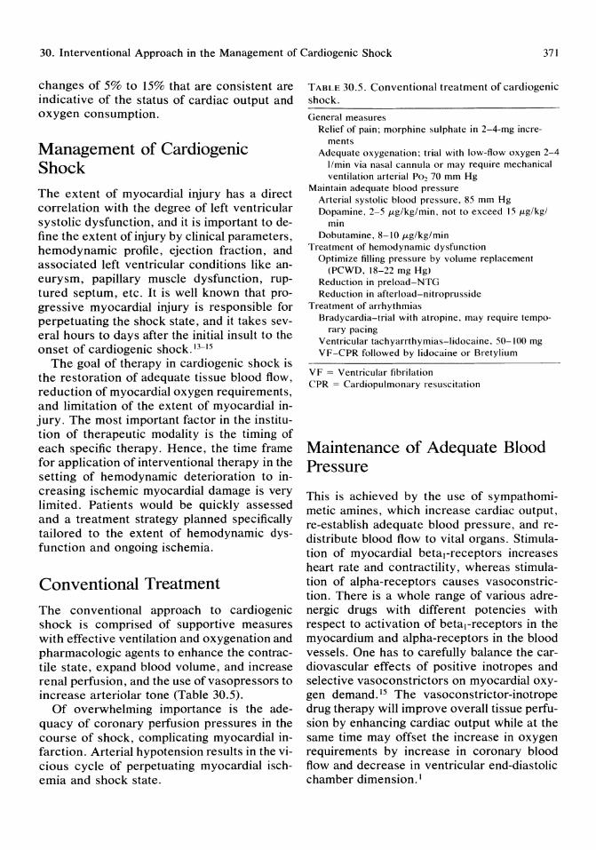
30. Interventional Approach in the Management of Cardiogenic Shock 371
changes of 5% to 15% that are consistent are indicative of the status of cardiac output and oxygen consumption.
Management of Cardiogenic Shock The extent of myocardial injury has a direct correlation with the degree of left ventricular systolic dysfunction, and it is important to define the extent of injury by clinical parameters, hemodynamic profile, ejection fraction, and associated left ventricular conditions like aneurysm, papillary muscle dysfunction, ruptured septum, etc. It is well known that progressive myocardial injury is responsible for perpetuating the shock state, and it takes several hours to days after the initial insult to the onset of cardiogenic shock. 13- 15
The goal of therapy in cardiogenic shock is the restoration of adequate tissue blood flow, reduction of myocardial oxygen requirements, and limitation of the extent of myocardial injury. The most important factor in the institution of therapeutic modality is the timing of each specific therapy. Hence, the time frame for application of interventional therapy in the setting of hemodynamic deterioration to increasing ischemic myocardial damage is very limited. Patients would be quickly assessed and a treatment strategy planned specifically tailored to the extent of hemodynamic dysfunction and ongoing ischemia.
Conventional Treatment
The conventional approach to cardiogenic shock is comprised of supportive measures with effective ventilation and oxygenation and pharmacologic agents to enhance the contractile state, expand blood volume, and increase renal perfusion, and the use of vasopressors to increase arteriolar tone (Table 30.5).
Of overwhelming importance is the adequacy of coronary perfusion pressures in the course of shock, complicating myocardial infarction. Arterial hypotension results in the vicious cycle of perpetuating myocardial ischemia and shock state.
TABLE 30.5. Conventional treatment of cardiogenic shock.
General measures Relief of pain; morphine sulphate in 2-4-mg incre
ments Adequate oxygenation; trial with low-flow oxygen 2-4
l/min via nasal cannula or may require mechanical ventilation arterial Po~ 70 mm Hg
Maintain adequate blood pressure Arterial systolic blood pressure, 85 mm Hg Dopamine, 2-5 JLg/kg/min, not to exceed 15 JLg/kgl
min Dobutamine, 8-10 JLg/kg/min
Treatment of hemodynamic dysfunction Optimize filling pressure by volume replacement
(PCWD, 18-22 mg Hg) Reduction in preload-NTG Reduction in afterload-nitroprusside
Treatment of arrhythmias Bradycardia-trial with atropine, may require tempo
rary pacing Ventricular tachyarrthymias-lidocaine. 50-100 mg VF-CPR followed by lidocaine or Bretylium
VF = Ventricular fibrilation CPR = Cardiopulmonary resuscitation
Maintenance of Adequate Blood Pressure
This is achieved by the use of sympathomimetic amines, which increase cardiac output, re-establish adequate blood pressure, and redistribute blood flow to vital organs. Stimulation of myocardial betal-receptors increases heart rate and contractility, whereas stimulation of alpha-receptors causes vasoconstriction. There is a whole range of various adrenergic drugs with different potencies with respect to activation of beta I-receptors in the myocardium and alpha-receptors in the blood vessels. One has to carefully balance the cardiovascular effects of positive inotropes and selective vasoconstrictors on myocardial oxygen demand. 15 The vasoconstrictor-inotrope drug therapy will improve overall tissue perfusion by enhancing cardiac output while at the same time may offset the increase in oxygen requirements by increase in coronary blood flow and decrease in ventricular end-diastolic chamber dimension. I
372
Vasoconstrictor-Inotropic Drug Therapy
Dopamine, which is a precursor of norepinephrine, is a selective vasoconstrictor and has dilator activity with favorable hemodynamic benefit. These pharmacologic effects are dose dependent. In low doses (2 to 5 mg/ kg per minute) it produces vasodilation of renal, mesenteric, coronary, and cerebrovascular beds. In moderate doses (6 to 15 mg/kg per minute) it increases contractility through beta I stimulation. In large doses (20 mg/kg per minute) there is purely alpha-mediated generalized vasoconstriction. So excessive doses of dopamine should not be administered. Instead, addition of dobutamine, which is a cardioselective catecholamine, may be complementary and of significant hemodynamic advantage. 16,17
Dobutamine acts directly on betal-adrenergic receptors in the myocardium to produce a dose-related increase in cardiac output with a modest decrease in systemic vascular resistance and left ventricular end-diastolic pressure. The goal in cardiogenic shock is to maintain an adequate coronary perfusion, which can be done by norepinephrine or dopamine.
Prolonged use of dobutamine over 48 hours is attendant with development of tolerance and down regulation of beta I receptors. In this setting, amrinone, which is a new inotropic drug, can be substituted or used in combination with dopamine. The exact mechanisms of action of amrinone have not yet been determined. The inotropic action of amrinone may be explained in part by the inhibition of phosphodiesterase activity and by increase in cellular levels of cyclic AMP. Amrinone also exerts a direct dilatory effect on vascular smooth muscle. IS
Amrinone is q well-balanced drug for reducing cardiac preload and afterload with a salutary increase in cardiac output with preservation of myocardial energetics. 'o The administration of amrinone is initiated with a 0.75-mg/kg bolus injection given slowly over 2 to 3 minutes. This is followed by a maintenance infusion of 5 to 10 mg/kg per
A.S. Kapoor
minute. An additional bolus injection of 0.75 mg/kg may be given 30 minutes after therapy is initiated, based on the patient's response.
The rationale for using vasodilator therapy in the setup of cardiogenic shock with generalized vasoconstriction is to break the deleterious vicious cycle of increasing vasoconstriction and decreasing cardiac output. Vasodilators cannot be recommended for routine management of cardiogenic shock but may be beneficial in patients with poor tissue perfusion but adequate systolic blood pressures (80 mm Hg). Nitroprusside is a very effective drug in reducing both preload and afterload. A dose of 15 JLg/minute could be initiated and increased to 200 JLg/minute, with constant monitoring of arterial pressure and pulmonary artery wedge pressure.
Occasionally intravenous nitroglycerine in combination with dopamine may allow a reduction in preload, an improvement in cardiac output, and maintain vasoconstriction with redistribution of blood flow to vital organs. Intravenous nitroglycerine should be started with an initial small dose of 10 to 15 JLg/minute and increased to tolerable effective doses. Nitroglycerine tolerance develops rapidly; hence, its hemodynamic efficacy should be monitored frequently to assess if more nitroglycerine is needed or if it should be stopped for a short while to give a nitrate-free interval.
The art of administering polypharmacy in cardiogenic shock has become very subtle, requiring computer sophistication for dose adjustments, addition of more and deletion of others, while keeping in mind the global and cardiac status of the patient. Application of polypharmacy has not demonstrated any significant decrease in mortality. Despite aggressive mechanical therapy combined with intraaortic balloon counterpulsation, the overall survival rate for patients with cardiogenic shock is only 30%.20
Interventional Approach
Any reduction in mortality associated with left ventricular failure and shock will require timely and specific interventions to salvage is-
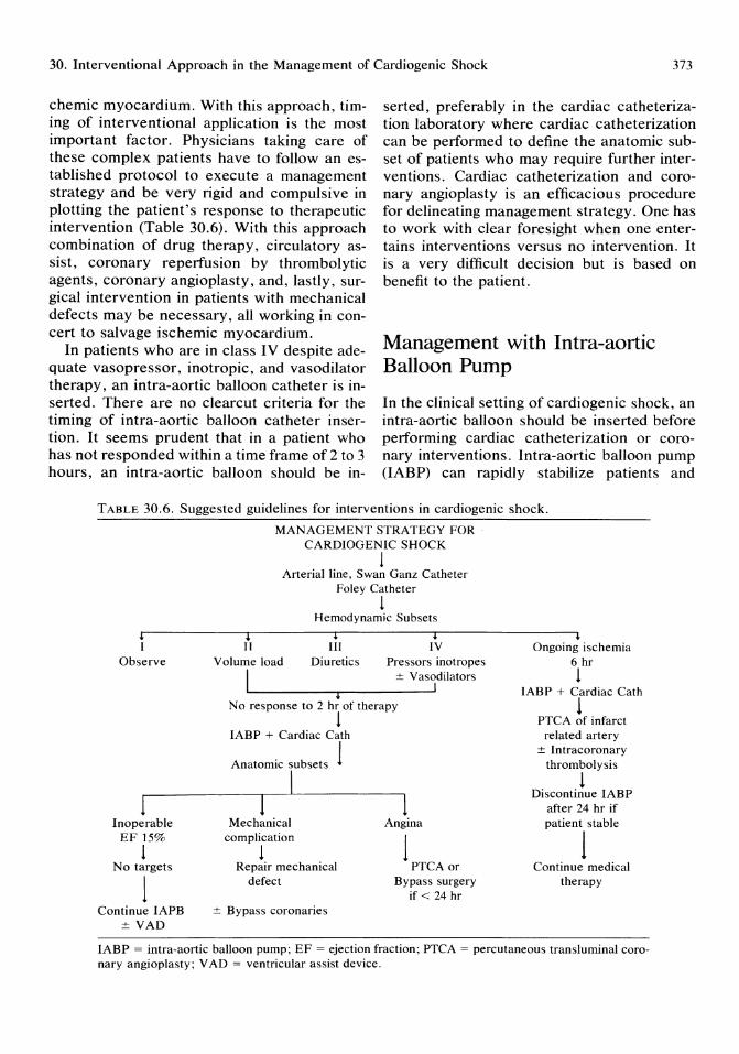
30. Interventional Approach in the Management of Cardiogenic Shock 373
chemic myocardium. With this approach, timing of interventional application is the most important factor. Physicians taking care of these complex patients have to follow an established protocol to execute a management strategy and be very rigid and compulsive in plotting the patient's response to therapeutic intervention (Table 30.6). With this approach combination of drug therapy, circulatory assist, coronary reperfusion by thrombolytic agents, coronary angioplasty, and, lastly, surgical intervention in patients with mechanical defects may be necessary, all working in concert to salvage ischemic myocardium.
In patients who are in class IV despite adequate vasopressor, inotropic, and vasodilator therapy, an intra-aortic balloon catheter is inserted. There are no clearcut criteria for the timing of intra-aortic balloon catheter insertion. It seems prudent that in a patient who has not responded within a time frame of 2 to 3 hours, an intra-aortic balloon should be in-
serted, preferably in the cardiac catheterization laboratory where cardiac catheterization can be performed to define the anatomic subset of patients who may require further interventions. Cardiac catheterization and coronary angioplasty is an efficacious procedure for delineating management strategy. One has to work with clear foresight when one entertains interventions versus no intervention. It is a very difficult decision but is based on benefit to the patient.
Management with Intra-aortic Balloon Pump
In the clinical setting of cardiogenic shock, an intra-aortic balloon should be inserted before performing cardiac catheterization or coronary interventions. Intra-aortic balloon pump (IABP) can rapidly stabilize patients and
TABLE 30.6. Suggested guidelines for interventions in cardiogenic shock.
, I
Observe
1 Inoperable
EF 15%
1 No targets
1 Continue IAPB
± VAD
MANAGEMENT STRATEGY FOR CARDIOGENIC SHOCK
1 Arterial line, Swan Ganz Catheter
Foley Catheter
1 Hemodynamic Subsets
i IT
Volume load
I
t III
Diuretics
i
t IV
Pressors inotropes ± Vasodilators
I
No response to 2 hr of therapy
1 IABP + Cardiac Cath
Anatomic subsets
I I
Mechanical complication
1 Repair mechanical
defect
± Bypass coronaries
1
1 Angina
1 PTCA or
Bypass surgery if < 24 hr
+ Ongoing ischemia
6 hr 1
IABP + Cardiac Cath
1 PTCA of infarct
related artery ± Intracoronary
thrombolysis
1 Discontinue IABP
after 24 hr if patient stable
1 Continue medical
therapy
IABP = intra-aortic balloon pump; EF = ejection fraction; PTCA = percutaneous transluminal coronary angioplasty; V AD = ventricular assist device.

374
change the class IV hemodynamic status to class 1. However, for patients who remain in class IV or deteriorate, mortality remains very high. In patients who are not candidates for further intervention, IABP should be left in place for varying intervals. Patients who become balloon dependent usually have an ejection fraction of 20% on balloon assist. These subset of patients will have a downhill course with progressive oliguria, and code status should be entered in the further management of these patients.
Details of insertion and balloon management have been discussed in an earlier chapter.
Percutaneous Transluminal Coronary Angioplasty in Cardiogenic Shock
Percutaneous trans luminal coronary angioplasty (PTCA) is an untested modality in the setting of cardiogenic shock. As primary therapy, PTCA in cardiogenic shock patients has been reported to achieve high reperfusion rates with prompt hemodynamic and clinical improvement. 21.22 However, there are no prospective randomized studies to attest to these claims. According to Hensen and colleagues,23 PTCA is the treatment of choice in cardiogenic shock patients with success rates of more than 90%. They considered the use of acute PTCA in any patient who presented in cardiogenic shock within 6 hours of the onset of chest pain, or later if ischemia was waxing and waning. After insertion of IABP, selective coronary angiography and ventriculography is performed; multiple injections are avoided. Least amount of nonionic contrast is used. Even during PTCA, the least amount of dye is given and the guiding catheter is kept in the ostium of the artery as briefly as possible. Only the acutely occluded vessel is approached.
If a coronary thrombus is visualized, intracoronary streptokinase is infused (100,000 to 200,000 U). In most cases, IABP can be removed 24 hours after successful PTCA.
A.S. Kapoor
Patients are then maintained on a postPTCA protocol as detailed in an earlier chapter. It is too early to say if this is the way to proceed with cardiogenic patients. This seems to be the procedure that may put a dent in the very high mortality.
Management with Emergency Surgical Intervention
Surgical intervention is indicated where there are correctable mechanical complications like papillary muscle rupture, ventricular septal defect, and pseudoventricular aneurysm. Surgical intervention is indicated in all patients with a significant mechanical complications who are in cardiogenic shock class IV.
Revascularization of an acute infarction area of more than 24 hours age in a patient in cardiogenic shock has been associated with high failure rates. 9 Blood flow to the infarct area cannot be maintained, and bleeding within the infarct area is possibly due to no reflow phenomenon.9 It is very desirable not to intervene surgically in this group of patients at this time, but they should be continued on IABP, and coronary revascularization can be performed 6 to 8 weeks after the acute event, if it is feasible.
Infarctectomy is rarely performed as an independent procedure. However, in a patient who is balloon dependent and a patient who has ventricular tachycardia, resection of the dyskinetic area with possible bypass grafting in a suitable vessel can be performed with acceptable results.
Ventricular Assist Devices
An occasional patient may be a candidate for a ventricular assist device. Pulsatile ventricular assist devices can be used as a bridge to cardiac transplantation for postinfarction cardiogenic shock.24
The National Heart, Lung and Blood Institute Artificial Heart Program has geared to the development of a fully implantable ventricular assist device. This device will have a 2-year
30. Interventional Approach in the Management of Cardiogenic Shock 375
reliability and require no anticoagulation, powered by a battery pack that can be recharged transcutaneously by electromagnetic induction from an external source.
Ventricular assist devices consist of a flexible polyurethane blood sac enclosed in a rigid chamber and perform like a single ventricle. For left ventricular assistance, blood in the left atrium or left ventricle enters a cannula and transports blood via an inlet valve into the blood sac. Flow from the sac passes to the thoracic or abdominal aorta via an outlet valve. The present pulsatile ventricular assist devices are pneumatically driven and can be placed intra-abdominally. At the present time, there is a wide range of ventricular assist devices in various phases of clinical evaluation.25
Conclusion
We have come a long way in the management of cardiogenic shock. We have come to realize that time is of the essence in therapeutic interventions in the setting of cardiogenic shock. The incidence of shock after acute infarction remains at 10% to 15% and carries a uniformly high mortality between 80% to 90%. Cardiogenic shock is a self-perpetuating vicious cycle of progressive tissue hypoxia and ischemic damage leading to an irreversible state of myocardial cellular dysfunction.
There are many options available for this unrelenting condition (Table 30.7). We have newer, potent, and safer inotropic agents that may have potential for producing hemodynamic stability and preventing progression of ischemic damage. Coronary interventions
TABLE 30.7. Treatment modalities for limiting infarct size.
Increasing oxygen delivery Coronary reperfusion
Intracoronary thrombocytics Intravenous tissue type plasminogen activator Emergency coronary angioplasty Emergency coronary bypass surgery
Decreasing oxygen requirements Intra-aortic balloon counter pulsation
have opened a new therapeutic modality that will have to undergo clinical trials to establish clinical effectiveness. Intuitively, they appear to be the treatment of choice, but are they really?
References 1. Weber KT, Ratshin RA, lanicks IS, et al: Left
ventricular dysfunction following acute myocardial infarction. A clinico-pathologic and hemodynamic profile of shock and failure. Am J Med 1973; 54:697.
2. Rackley CE, Russle RO, Mantal lA, et al: Cardiogenic shock, in Critical Care Cardiology. Philadelphia, FA Davis, 1986, pp 15-24.
3. Page DL, Caulfield IB, Kastor SA, et al: Myocardial changes associated with cardiogenic shock. N Engl J Med 1971; 285:133.
4. Alonso DR, Scheidt S, Past M, et al: Pathophysiology of cardiogenic shock: Quantification of myocardial necrosis, clinical, pathologic, and electrocardiographic correlations. Circulation 1973; 48:588.
5. Rackley CE, Russell RO, Mantle lA, et al: Recognition of acute myocardial infarction, in Rackley ED, Russell RO (eds): Coronary Artery Disease: Recognition and Management. Mt. Kisco, New York, Futura Publishing Company, 1979, p 315.
6. Sheinman M, Evans GT, Weit:s A, et al: Relationship between pulmonary artery end-diastolic pressure and left ventricular filling pressure in patients in shock. Circulation 1973; 47:317.
7. Forrester IS, Diamond G, Chatterjee K, et al: Medical therapy of acute myocardial infarction by application of hemodynamic subsets. N Engl J Med 1976; 294: 1356, 1040.
8. Scheidt SS, Ascheim R, Killip T III: Shock after acute myocardial infarction. A clinical and hemodynamic profile. Am J Cardiol 1970; 26:566.
9. Bolocki H (ed): Classification of cardiogenic shock, in Clinical Application of Intra-aortic Balloon Pump. New York, Futura Publishing Company, 1984, pp 159-170.
10. Wiedemann HD, Matthay MA, Matthay RA: Cardiovascular-pulmonary monitoring in the intensive care unit. Chest 1984; 85:537, 656.
II. Waller IL, Kaplan lA, Baumann DE, et al: Clinical evaluation of a new fiberoptic catheter oximeter during cardiac surgery. Anesth Analg 1982; 61:676-679.

376
12. Bae1e PL, McMichan JC, Marsh HM, et al: Continuous monitoring of mixed venous oxygen saturation in critically ill patients. Anesth Analg 1982; 61:513-517.
13. Rackley CE, Russell RO, Ratchin RA, et al: Cardiogenic shock in patients with myocardial infarction, in Rackley CE, Russell RO (eds): Hemodynamic Monitoring in a Coronary Intensive Care Unit. Mt. Kisco, New York, Futura Publishing Company, 1974, p 223.
14. Rackley CE, Russell RO, Mantle JA, et al: Clinical spectrum of left ventricular failure, in Bolocki H (ed): Clinical Application of Intraaortic Balloon Pump. Mt. Kisco, New York, Futura Publishing Company, 1977, p 181.
15. Leier CV, Heban PT, Huss P: Comparative systemic and regional hemodynamic effects of dopamine and dobutamine in patients with cardiomyopathic heart failure. Circulation 1978; 58:466.
16. Kapoor AS: Comprehensive management of acute myocardial infarction. Contin Educ 1985; 20:557-564.
17. Kapoor AS: Pharmacologic and technical approach in the management of cardiogenic shock. Submitted for publication.
A.S. Kapoor
18. Goldstein RA: Clinical effects of intravenous amrinone in patients with chronic congestive heart failure. Circulation 1986; 73:111-191.
19. Benotti JR, Grossman W, Branwald E, et al: Hemodynamic assessment of amrinone. N Engl J Med 1978; 299:1373.
20. Strobeck JE: Cardiogenic shock. Heart Failure 1985,1(6):\3-36.
21. Shani J, Rivera M, Greengart A, et al: Percutaneous transluminal coronary angioplasty in cardiogenic shock. JAm Coli Cardiol1986; 7:149.
22. O'Neill W, Erbel R, Lanfer N, et al: Coronary angioplasty therapy of cardiogenic shock complicating acute myocardial infarction. Circulation 1985; 72:111-209.
23. Hensen RR, Maddoux GL, Goss JE, et al: Coronary angioplasty in the treatment of cardiogenic shock: The therapy of choice. J Am Coil Cardiol 1986; 7:219A.
24. Hill JD: Use of a prosthetic ventricle as a bridge to cardiac transplantation for postinfarction cardiogenic shock. N Engl J Med 1986; 314:616.
25. Richenbacher WE, Pierce WS: Clinical spectrum of mechanical circulatory assistance. Heart Trans J 1985; 4:481.
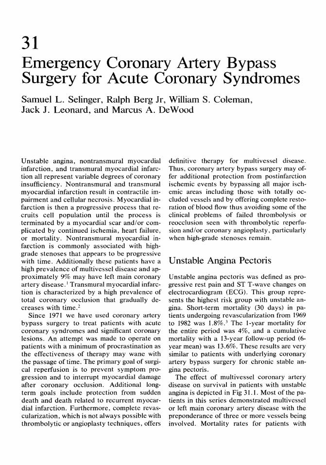
31 Emergency Coronary Artery Bypass Surgery for Acute Coronary Syndromes Samuel L. Selinger, Ralph Berg Jr, William S. Coleman, Jack J. Leonard, and Marcus A. DeWood
Unstable angina, nontransmural myocardial infarction, and transmural myocardial infarction all represent variable degrees of coronary insufficiency. N ontransmural and transmural myocardial infarction result in contractile impairment and cellular necrosis. Myocardial infarction is then a progressive process that recruits cell population until the process is terminated by a myocardial scar and/or complicated by continued ischemia, heart failure, or mortality. Nontransmural myocardial infa~ction is commonly associated with highgrade stenoses that appears to be progressive with time. Additionally these patients have a high prevalence of multivessel disease and approximately 9% may have left main coronary artery disease. l Transmural myocardial infarction is characterized by a high prevalence of total coronary occlusion that gradually decreases with time. 2
Since 1971 we have used coronary artery bypass surgery to treat patients with acute coronary syndromes and significant coronary lesions. An attempt was made to operate on patients with a minimum of procrastination as the effectiveness of therapy may wane with the passage of time. The primary goal of surgical reperfusion is to prevent symptom progression and to interrupt myocardial damage after coronary occlusion. Additional longterm goals include protection from sudden death and death related to recurrent myocardial infarction. Furthermore, complete revascularization, which is not always possible with thrombolytic or angioplasty techniques, offers
definitive therapy for multivessel disease. Thus, coronary artery bypass surgery may offer additional protection from postinfarction ischemic events by bypassing all major ischemic areas including those with totally occluded vessels and by offering complete restoration of blood flow thus avoiding some of the clinical problems of failed thrombolysis or reocclusion seen with thrombolytic reperfusion and/or coronary angioplasty, particularly when high-grade stenoses remain.
Unstable Angina Pectoris
Unstable angina pectoris was defined as progressive rest pain and ST T-wave changes on electrocardiogram (ECG). This group represents the highest risk group with unstable angina. Short-term mortality (30 days) in patients undergoing revascularization from 1969 to 1982 was 1.8%.3 The I-year mortality for the entire period was 4%, and a cumulative mortality with a 13-year follow-up period (6-year mean) was 13.6%. These results are very similar to patients with underlying coronary artery bypass surgery for chronic stable angina pectoris.
The effect of multivessel coronary artery disease on survival in patients with unstable angina is depicted in Fig 31.1. Most of the patients in this series demonstrated multivessel or left main coronary artery disease with the preponderance of three or more vessels being involved. Mortality rates for patients with
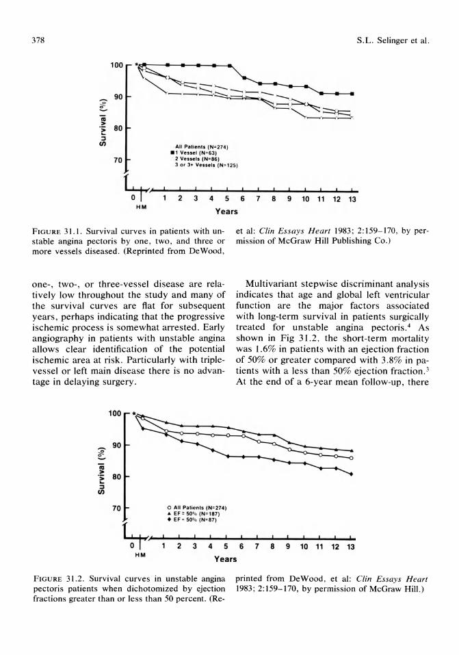
378
100
All Pa'i~nts (No 274) •• Vessel (N ~ 6J)
·. 2 Venets (N'86)
S.L. Selinger et al.
70 J or J+ Vessels (N - '25)
~~/~--~2--·3--·4--5~-6~~7~~8--~9--1·0--1·1--1·2--1~3 HM
Years
FIGURE 31.1. Survival curves in patients with unstable angina pectoris by one, two, and three or more vessels diseased. (Reprinted from DeWood,
one-, two-, or three-vessel disease are relatively low throughout the study and many of the survival curves are fiat for subsequent years, perhaps indicating that the progressive ischemic process is somewhat arrested. Early angiography in patients with unstable angina allows clear identification of the potential ischemic area at risk. Particularly with triplevessel or left main disease there is no advantage in delaying surgery.
100
.-. 90 ;f!. -ia > .; 80 ... ~
(/)
70 o All Patien •• (N-274) • EF ' 50'fo (N"187)
et al: Clin Essays Heart 1983; 2:159-170, by permission of McGraw Hill Publishing Co.)
Multivariant stepwise discriminant analysis indicates that age and global left ventricular function are the major factors associated with long-term survival in patients surgically treated for unstable angina pectoris.4 As shown in Fig 31.2, the short-term mortality was 1.6% in patients with an ejection fraction of 50% or greater compared with 3.8% in patients with a less than 50% ejection fraction. 3
At the end of a 6-year mean follow-up, there
• EF · 50'fo (N- 87)
~~f--1--~2--~3--~4--5---6--~7--~8--~9--1~0--1·1--1-2~1~3 HM
Years
FIGURE 31.2. Survival curves in unstable angina pectoris patients when dichotomized by ejection fractions greater than or less than 50 percent. (Re-
printed from DeWood, et al: Clin Essays Heart 1983; 2:159-170, by permission of McGraw Hill.)
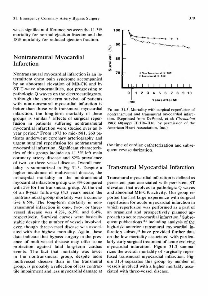
31. Emergency Coronary Artery Bypass Surgery
was a significant difference between the 11.3% mortality for normal ejection fraction and the 18% mortality for reduced ejection fraction.
N ontransmural Myocardial Infarction
Nontransmural myocardial infarction is an intermittent chest pain syndrome accompanied by an abnormal elevation of MB-CK and by ST T-wave abnormalities, not progressing to pathologic Q waves on the electrocardiogram. Although the short-term survival of patients with nontransmural myocardial infarction is better than those with transmural myocardial infarction, the long-term mortality of these groups is similar. 5 Effects of surgical reperfusion in patients suffering non transmural myocardial infarction were studied over an 8-year period. 6 From 1973 to mid-1981, 260 patients underwent coronary arteriography and urgent surgical reperfusion for nontransmural myocardial infarction. Significant characteristics of this group include an 11.5% left main coronary artery disease and 82% prevalence of two- or three-vessel disease. Overall mortality is summarized in Fig 31. 3. Despite a higher incidence of multivessel disease, the in-hospital mortality in the nontransmural myocardial infarction group was 3% compared with 5% for the transmural group. At the end of an 8-year follow-up (4.3 years mean) the nontransmural group mortality was a cumulative 6.5%. The long-term mortality in nontransmural infarction in one-, two-, or threevessel disease was 4.2%, 6.3%, and 8.4%, respectively. Survival curves were basically stable despite the number of vessels involved, even though three-vessel disease was associated with the highest mortality. Again, these data indicate that bypass surgery in the presence of multivessel disease may offer some protection against fatal long-term cardiac events. The fact that mortality was lower in the nontransmural group, despite more multivessel disease than in the transmural group, is probably a reflection of less contractile impairment and less myocardial damage at
100
90 ~ 0
III 80 > > .. :::2
en 70
I>
1,1 :
o 1
IHM
~ Non T,..nemur.1 I N 261 1 o Tr.n.mural jN -4401
379
2 3 4 5 6 7 8 9 10
Years after MI
FIGURE 31.3. Mortality with surgical reperfusion of nontransmural and transmural myocardial infarction. (Reprinted from DeWood, et al: Circulation 1983; 68(suppl 11):118-1116, by permission of the American Heart Association, Inc.)
the time of cardiac catheterization and subsequent revascularization.
Transmural Myocardial Infarction
Transmural myocardial infarction is defined as persistent pain associated with persistent ST elevation that evolves to pathologic Q waves and abnormal MB-CK activity. Our group reported the first large experience with surgical reperfusion for acute myocardial infarction in which reperfusion was performed as a part of an organized and prospectively planned approach to acute myocardial infarction. 7 Subsequent publications,s.9 including analysis of the high-risk anterior transmural myocardial infarction subset,1O have provided further data on the low mortality associated with particularly early surgical treatment of acute evolving myocardial infarction. Figure 31.3 summarizes the overall mortality of surgically reperfused transmural myocardial infarction. Figure 31.4 separates this group by number of vessels involved with a higher mortality associated with three-vessel disease.

380
100
90 ~ 0
ca 80 > 'S; .. :::s
en 70 ® 1 V .... I IN ' l271
10
'
• o 1
IHM
0 2 V .... I IN -lUI .3 V .... I IN · l321
2 3 4 5 6 7 8 9 10
Years after M I
FIGURE 31.4. Mortality for surgical reperfusion of transmural myocardial infarction by one, two, and three vessel disease. (Reprinted from DeWood, et al: Circulation 1983; 68(suppl 11):118-1116, by permission of the American Heart Association, Inc.)
Figure 31.5 emphasizes lower mortality associated with having a patient on cardiopulmonary bypass within 6 hours of the onset of chest pain. Not only was in-hospital mortality significantly lower in the early reperfusion group (3.8% v the late treatment group 8%) but that difference widened over a follow-up period of 10 years to 8.2% v 21%. A progressive sharp rise in the mortality rate of the late reperfusion group compared with the early reperfusion group implies that early reperfusion salvages myocardium that is important not only for short-term survival but also for survival over the next 10 years.
Left Ventricular Function
Patients who underwent emergency surgical reperfusion for anterior transmural myocardial infarction underwent follow-up left ventriculography to determine subsequent left ventricular function. I I Figure 31.6 depicts the ejection fraction of the patients who underwent revascularization within 6 hours of onset of symptoms (mean 4.8 hours) on the left and those who underwent late revascularization (range of 6.2 to 18 hours, with a mean of 9.2
S.L. Selinger et al.
100 * ., . .-.--...-.~ .~.-.--.-.-.
90
70 fe) ~ 6 Hour .. From Symptom On .U ( N ~291) 10 ~ 6 Hour. From Symptom On ... t , N~ 149 1
,! ! ! ! ! ! ! ! ! ! •
o 1 2 3 4 5 6 7 8 9 10
IHM Years after MI
FIGURE 31.5. Mortality for the transmural infarction group divided into subgroups receiving reperfusion within 6 hours or greater than 6 hours. (Reprinted from DeWood, et al: Circulation 1983; 68(suppl II):II8-1116, by permission of the American Heart Association, Inc.)
70
60 c 0 .~ 50 ~
LL
C 40 0 .~
&l iii
30 Iii .c .2 C!l 20
10
0
I EF(%)
Pre Post
P <0.05
70
60
50
40
10
o Pre Post
P= NS
FIGURE 31.6. Global ejection fraction from preoperative to postoperative state in early revascularization (left) and late revascularization (right). (PRE = prereperfusion, POST = postreperfusion, EF = ejection fraction.) (Reprinted with permission from The American College of Cardiology, J Am Coli Ca rdio I 1983; 1:1223-1229.)
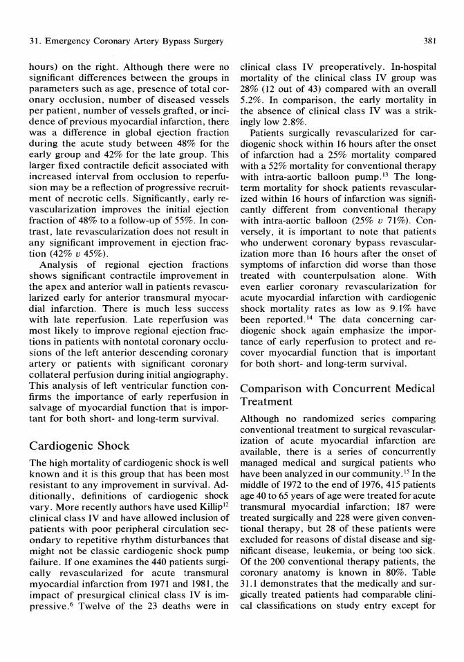
31. Emergency Coronary Artery Bypass Surgery
hours) on the right. Although there were no significant differences between the groups in parameters such as age, presence of total coronary occlusion, number of diseased vessels per patient, number of vessels grafted, or incidence of previous myocardial infarction, there was a difference in global ejection fraction during the acute study between 48% for the early group and 42% for the late group. This larger fixed contractile deficit associated with increased interval from occlusion to reperlusion may be a reflection of progressive recruitment of necrotic cells. Significantly, early revascularization improves the initial ejection fraction of 48% to a follow-up of 55%. In contrast, late revascularization does not result in any significant improvement in ejection fraction (42% v 45%).
Analysis of regional ejection fractions shows significant contractile improvement in the apex and anterior wall in patients revascularized early for anterior transmural myocardial infarction. There is much less success with late reperfusion. Late reperfusion was most likely to improve regional ejection fractions in patients with non total coronary occlusions of the left anterior descending coronary artery or patients with significant coronary collateral perfusion during initial angiography. This analysis of left ventricular function confirms the importance of early reperfusion in salvage of myocardial function that is important for both short- and long-term survival.
Cardiogenic Shock
The high mortality of cardiogenic shock is well known and it is this group that has been most resistant to any improvement in survival. Additionally, definitions of cardiogenic shock vary. More recently authors have used Killipl2 clinical class IV and have allowed inclusion of patients with poor peripheral circulation secondary to repetitive rhythm disturbances that might not be classic cardiogenic shock pump failure. If one examines the 440 patients surgically revascularized for acute transmural myocardial infarction from 1971 and 1981, the impact of presurgical clinical class IV is impressive. 6 Twelve of the 23 deaths were in
381
clinical class IV preoperatively. In-hospital mortality of the clinical class IV group was 28% (12 out of 43) compared with an overall 5.2%. In comparison, the early mortality in the absence of clinical class IV was a strikingly low 2.8%.
Patients surgically revascularized for cardiogenic shock within 16 hours after the onset of infarction had a 25% mortality compared with a 52% mortality for conventional therapy with intra-aortic balloon pump.13 The longterm mortality for shock patients revascularized within 16 hours of infarction was significantly different from conventional therapy with intra-aortic balloon (25% v 71%). Conversely, it is important to note that patients who underwent coronary bypass revascularization more than 16 hours after the onset of symptoms of infarction did worse than those treated with counterpulsation alone. With even earlier coronary revascularization for acute myocardial infarction with cardiogenic shock mortality rates as low as 9.1 % have been reported. 14 The data concerning cardiogenic shock again emphasize the importance of early reperfusion to protect and recover myocardial function that is important for both short- and long-term survival.
Comparison with Concurrent Medical Treatment
Although no randomized series comparing conventional treatment to surgical revascularization of acute myocardial infarction are available, there is a series of concurrently managed medical and surgical patients who have been analyzed in our community. 15 In the middle of 1972 to the end of 1976, 415 patients age 40 to 65 years of age were treated for acute transmural myocardial infarction; 187 were treated surgically and 228 were given conventional therapy, but 28 of these patients were excluded for reasons of distal disease and significant disease, leukemia, or being too sick. Of the 200 conventional therapy patients, the coronary anatomy is known in 80%. Table 31.1 demonstrates that the medically and surgically treated patients had comparable clinical classifications on study entry except for
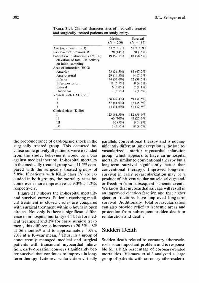
382 S. L. Selinger et al.
TABLE 31.1. Clinical characteristics of medically treated and surgically treated patients on study entry.
Age (yr) (mean ± SD) Incidence of previous MI Patients with abnormal (>90 IU)
elevation of total CK activity on initial sampling
Area of infarction (ECG) Anterior Anterolateral Inferior Inferoposterior Lateral Uncertain
Vessels with CAD (no.) 1 2 3
Clinical class (Killip) I II III IV
the preponderance of cardiogenic shock in the surgically treated group. This occurred because some gravely ill patients were excluded from the study, believing it would be a bias against medical therapy. In-hospital mortality in the medically treated group was 11.5% compared with the surgically treated groups of 5.8%. If patients with Killip class IV are excluded in both groups, the mortality rates become even more impressive at 9.3% v 1.2%, respectively.
Figure 31.7 shows the in-hospital mortality and survival curves. Patients receiving medical treatment in closed circles are compared with surgical treatment within 6 hours in open circles. Not only is there a significant difference in in-hospital mortality of 11.5% for medical treatment and 2% for early surgical treatment, this difference increases to 20.5% v 6% at 56 months l5 and to approximately 40% v 20% at a lO-year mean. 16 Thus, in a group of concurrently managed medical and surgical patients with transmural myocardial infarction, early operation conveys significantly better survival that continues to improve in longterm therapy. Late revascularization virtually
Medical (N = 200)
53.2 ± 8.1 28 (14%)
119 (59.5%)
73 (36.5%) 29 (14.5%) 74 (37.0%)
11 (5.5%) 6 (3.0%) 7 (3.5%)
38 (27.4%) 57 (41.0%) 44 (31.6%)
123 (61.5%) 60 (30%)
10 (5%) 7 (3.5%)
Surgical (N = 187)
52.7 ± 9.1 30 (16%)
110 (58.5%)
88 (47.0%) 14 (7.5%)
72 (38.5%) 8 (4.3%) 2 (1.1 %) 3 (1.6%)
59 (31.5%) 67 (35.8%) 61 (32.6%)
112 (59.9%) 48 (25.6%)
9 (4.8%) 18 (9.6%)
parallels conventional therapy and is not significantly different (an exception is the late revascularized anterior myocardial infarction group, which appears to have an in-hospital mortality similar to conventional therapy but a long-term survival significantly better than conventional therapy). Improved long-term survival in early revascularization may be a product of left ventricular muscle salvage and/ or freedom from subsequent ischemic events. We know that myocardial salvage will result in an improved ejection fraction and that higher ejection fractions have improved long-term survival. Additionally, total revascularization can also provide relief to ischemic areas and protection from subsequent sudden death or reinfarction and death.
Sudden Death
Sudden death related to coronary atherosclerosis is an important problem and is responsible for a high percentage of coronary-related mortalities. Vismara et aP7 analyzed a large group of patients with coronary atherosclero-
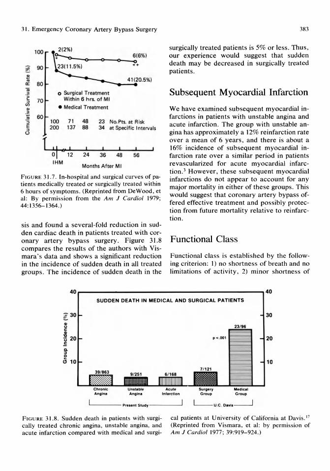
31. Emergency Coronary Artery Bypass Surgery
100
~ 90
~ '" IX: 80 -.; .~
~ 70 U)
CII > .;:;
..!!! :::J E :::J
U
60
~_-o-_o-_>-~6(6%) ++
41(20.5%) --...... -. o Surgical Treatment
Within 6 hrs. of MI
• Medical Treatment
100 71 48 23 No. Pts. at A isk 200 137 88 34 at Specific Intervals
~" o P12 24 36 48 56 IHM
Months After M I
FIGURE 31.7. In-hospital and surgical curves of patients medically treated or surgically treated within 6 hours of symptoms. (Reprinted from DeWood, et al: By permission from the Am J Cardial 1979; 44: 1356-1364.)
sis and found a several-fold reduction in sudden cardiac death in patients treated with coronary artery bypass surgery. Figure 31.8 compares the results of the authors with Vismara's data and shows a significant reduction in the incidence of sudden death in all treated groups. The incidence of sudden death in the
383
surgically treated patients is 5% or less. Thus, our experience would suggest that sudden death may be decreased in surgically treated patients.
Subsequent Myocardial Infarction
We have examined subsequent myocardial infarctions in patients with unstable angina and acute infarction. The group with unstable angina has approximately a 12% reinfarction rate over a mean of 6 years, and there is about a 16% incidence of subsequent myocardial infarction rate over a similar period in patients revascularized for acute myocardial infarction. 3 However, these subsequent myocardial infarctions do not appear to account for any major mortality in either of these groups. This would suggest that coronary artery bypass offered effective treatment and possibly protection from future mortality relative to reinfarction.
Functional Class
Functional class is established by the following criterion: 1) no shortness of breath and no limitations of activity, 2) minor shortness of
40r--------------------------------------------,40 SUDDEN DEATH IN MEDICAL AND SURGICAL PATIENTS
QI U c: QI
~ 20 .£ Do :::J
2 Cl 10
Unltobl. Anglne Inl.rcUon
P < .001
7/ 121
Surgery Group
Medico. Group
30
20
10
L..-_ ___ Preoent Study-----'
FIGURE 31.8. Sudden death in patients with surgically treated chronic angina, unstable angina, and acute infarction compared with medical and surgi-
cal patients at University of California at Davis. 17
(Reprinted from Vismara, et al: by permission of Am J Cardial 1977; 39:919-924.)
384
breath and limitation of activity with maximal exercise, 3) shortness of breath and limitation of exercise vastly with less than maximal exertion, 4) severe restriction in the ability to function normally based on cardiac disability. The unstable angina group is 48% without functional restriction; 28% were class II, 20% class III, and 4% class IV. 3 In patients with acute myocardial infarction, the classes were present in 45%, 26%, 22%, and 6.5%, respectively. Thus, the majority of patients in this series were not limited in functional capacity or limited only with maximum exercise. Very few patients classified themselves as having any disabling symptoms due to heart disease.
Implications for Myocardial Jeopardy with Coronary Angioplasty
Surgical reperfusion of coronary occlusion subsequent to angioplasty manipulation is now commonplace with superb survival statistics. The major risks are similar to surgical reperfusion of acute myocardial infarction. Potential advantage in surgical reperfusion of angioplasty occlusion is the ability to provide return of blood flow within the first hour of occlusion. This time interval is much shorter in the usual patient presenting with acute myocardial infarction. Furthermore, sometimes a small degree of flow can be maintained by using a "bale out" catheter or by keeping a wire across the unstable lesion. Mortality in these cases is usually related to time interval from occlusion, new myocardium in jeopardy, initial left ventricular function, and occasionally age. Patients suffering left main coronary artery occlusions secondary to ostial dissections still have a relatively high mortality and the promptest cardiopulmonary support and revascularization is required for survival. Also, patients with reduced ejection fractions who suffer an additional coronary occlusion are a higher risk than patients starting with normal ventricular function.
S.L. Selinger et al.
Operative Technique
Our technique7,IO.l4,18,19 has been reported in detail. Of utmost importance is a constant push to keep the time between diagnosis and cardiopulmonary bypass to a minimum to preserve as much myocardial function as possible. In general, intra-aortic balloon support is not used preoperatively unless the patient is in shock or there is an anticipated delay. A femoral arterial sheath is left in place at the time of cardiac catheterization in any high-risk cases. The patient is placed on cardiopulmonary bypass after heparinization, ascending aortic cannulation, and venous siphonage with a single two-stage atrial catheter placed through the right atrial appendage. The core temperature is reduced to 24°C, the heart is crossclamped, and 4°C cardioplegic solution is given into the aorta along with 4°C Ringer's solution into the pericardium. We vent the aorta through the cardioplegia needle when cardioplegia is not being delivered. The major coronary artery supplying the area of evolving infarction is grafted first distally then proximally, and additional cardioplegia is given. All major arteries with significant stenoses are then grafted first distally and then proximally with additional cardioplegia given after proximal anastomosis to deliver the cardioplegia beyond native stenoses. Temporary atrial and ventricular pacing wires are left as well as the left atrial line, if necessary. Rather than highdose inotropic agents, intra-aortic balloon pump is the treatment of choice if difficulty is encountered in weaning the patient from cardiopulmonary bypass.
Conclusion
This chapter has reviewed the results of surgical treatment for the acute coronary syndromes including unstable angina pectoris and acute nontransmural and transmural myocardial infarction. Early surgical revascularization can be performed with low mortality and morbidity. Long-term follow-up reveals an excellent functional result with low rates of reinfarction and death. Surgical revascularization
31. Emergency Coronary Artery Bypass Surgery
within the first 6 hours of acute evolving myocardial infarction seems to limit infarct size and reduce both short- and long-term mortality.
When this work was started in the early 1970s the only means of coronary revascularization was coronary artery bypass. Currently, thrombolytic therapy and angioplasty techniques are able to provide reperfusion but not always total revascularization without residual high-grade stenoses. Over the past years many centers have gained experience in surgical reperfusion for evolving infarction with good results. 20 In particular, the patients who have multivessel disease or who require the stability of cardiopulmonary bypass due to multiple arrhythmias can be treated with surgical revascularization with excellent longterm survival. As therapeutic choices increase, early quantification of left ventricular function and identification of coronary anatomy will help in determining clinical choices. With acute myocardial infarction, treatment choices will need to emphasize speed and completeness of revascularization.
References
I. DeWood MA, Stifter WF, Simpson CS, et al: Coronary arteriographic findings soon after non-Q wave myocardial infarction. N Eng! J Med 1986; 315:417-423.
2. DeWood MA, Spores J, Notske RN, et al: Prevalence of total coronary occlusion during the early hours of transmural myocardial infarction. N Engl J Med 1980; 303:897-902.
3. DeWood MA, Berg R Jr: Coronary artery bypass surgery: 13-years experience with chronic stable angina pectoris, unstable angina pectoris and acute myocardial infarction. Clin Essays Heart 1983; 2:159-170.
4. DeWood MA, Grunwald RP, Rudy LW, et al: Surgical treatment of progressive rest angina pectoris; multivariate stepwise discriminant analysis of factors influencing ten-year survival. J Am Coli Cardiol1987; 9:87A.
5. Hutter AM Jr, DeSanctis RW, Flynn T, et al: Nontransmural myocardial infarction: A comparison of hospital and late clinical course of patients with that of matched patients with transmural anterior and transmural inferior
385
myocardial infarction. Am J Cardiol 1981; 48:595-602.
6. DeWood MA, Spores J, Berg R Jr, et al: Acute myocardial infarction: A decade of experience with surgical reperfusion in 701 patients. Circulation 1983; 68(suppl 1I):1I8-IIl6.
7. Berg R, Kendall RW, Duvoisin GE, et al: Acute myocardial infarction. J Thorac Cardiovas Surg 1975; 70:432-439.
8. Berg R Jr, Selinger SL, Leonard n, et al: Immediate coronary artery bypass for acute evolving myocardial infarction (AEMI). J Thorac Cardiovasc Surg 1981; 81:493-497.
9. Selinger SL, Berg R Jr, Leonard n, et al: Surgical treatment of acute evolving anterior myocardial infarction (abstr). Circulation 1980; 62(part 11):4.
10. Selinger SL, Berg R Jr, Leonard n, et al: Surgical treatment of acute evolving anterior myocardial infarction. Circulation 1981 ; 64(suppl 1I):1l28-II33.
11. DeWood MA, Spores J, Heit J, et al: Anterior transmural myocardial infarction. Effects of surgical coronary reperfusion on global and regionalleft ventricular function. J Am Coli Cardio11983; 1:1223-1234.
12. Killip T III, Kimball JT: Treatment of myocardial infarction in a coronary care unit: A twoyear experience with 250 patients. Am J Cardiol 1967; 20:457.
13. DeWood MA, Notske RN, Hensley GR, et al: Intra-aortic balloon counterpulsation with and without reperfusion for myocardial infarction shock. Circulation 1980; 61: 1105-1112.
14. Berg RJr, Selinger SL, Leonard n, et al: Surgical management of acute myocardial infarction, in D McGoon (ed): Cardiovascular Clinics, Cardiac Surgery. Philadelphia, F.A. Davis Publishing Co., 1982; 2:61-74.
15. DeWood MA, Spores J, Notske RN, et al: Medical and surgical management of myocardial infarction. Am J Cardiol 1979; 44:1356-1364.
16. DeWood, et al: Personal communication. 17. Vismara LA, Miller RR, Price JE, et al: Im
proved longevity due to reduction of sudden death by aortocoronary bypass in coronary atherosclerosis. Prospective evaluation of medical versus surgical therapy in matched patients with multivessel disease. Am J Cardiol 1977; 39:919-924.
18. Selinger SL: Coronary revascularization during acute myocardial infarction. WG Austin (ed): Current Therapy in Cardiothoracic Surgery. B.C. Decker, Inc. Publisher, in press.

386
19. Selinger SL, Berg R Jr, Leonard 11, et al: Coronary bypass surgery in evolving acute myocardial infarction, in A. Roberts (ed): Difficult Problems in Adult Cardiac Surgery, New York, Year Book Publishing Co., 1985, pp 37-53.
S.L. Selinger et al.
20. DeWood MA, Selinger SL, Coleman WS, et al: Surgical coronary reperfusion during acute myocardial infarction, in D McGoon (ed): Cardiovascular Clinics, Cardiac Surgery, Philadelphia, F.A. Davis Publishing Co, 1987, pp 37-53.

32 Cardiac Transplantation Davis Drinkwater, Lynne Warner Stevenson, and Hillel Laks
Introduction
The number of cardiac transplants has increased dramatically in recent years as its clinical efficacy for the treatment of severe cardiac failure has been confirmed. According to the Registry of the International Society of Heart Transplantation (ISHT), 719 cardiac transplants were performed in 1984, in contrast to 365 in 1983. 1 As of 1985, the Registry listed a total of 2,577 cardiac transplants worldwide. The factors in this increase reflect a long-standing interest and work in transplantation in general and in cardiac transplantation in particular, beginning at the turn of the century. The recent discovery of cyclosporin as a safer, more reliable immunosuppressant has been a major factor in this increase, and has in large part been responsible for the greater than 80% I-year survival rate reported by the ISHT Registry. 1
Historical Perspective
The evolution of cardiac transplantation from the experimental to therapeutic modality can be attributed to advances made in organ transplantation and immunology in general and to particular contributions from individuals during the past century. In 1905 Carrel and Guthrie2 reported the transplantation of a heart into the neck of a dog, followed later in their career with the simultaneous transplantation of heart and lungs in the same position, albeit with only brief survival and no evident function. Carrel later received the Nobel Prize
for his contribution to vascular anastomses. In the 1930s Mann and colleagues3 at the Mayo Clinic were able to demonstrate function of a heterotopic transplanted heart for 4 days before what is now known as rejection occurred. Lance and Medawar4 later dissected the two arms of the immunologic response into the cellular and the humeral, for which Medawar received the Nobel Prize in 1961. These prior contributions laid the groundwork for renal transplantation in the 1950s as the first clinical organ transplantation program. 5 A decade later Lower and Shumway's6 research into surgical technique and immunosuppressive regimen for orthotopic cardiac transplantation was soon followed by the report from Capetown, South Africa of the first human cardiac transplantation. 7 This initial effort was followed by a burst of reports of transplantation around the world, but it soon became apparent that further research into rejection and infection was needed to improve uniformly poor results, with all but a few committed programs closing.8 By 1981 Shumway and co-workers at Stanford had established the clinical value of the procedure reporting a 63% I-year survival. 9 The introduction of cyclosporine in 1981 has yielded I-year survival results of 80% or more.! This has been followed by a tremendous increase in the number of cardiac transplant programs, more than 80 in North America and an additional 30 programs in Europe and abroad. Of the 2,577 transplants registered with the ISHT, more than 50% were performed in the last 2 years of the registry 1984-1985. 1 In 1984 The National Heart Transplantation Study, The Batelle Report, led to the formal approval by the Department
388
of Health and Human Services of cardiac transplantation as best therapy in eligible patients with end-stage cardiac disease. 10
Indications for Cardiac Transplantation
The most common indication for transplantation is end-stage dilated cardiomyopathy eventually resulting from biventricular enlargement. Microscopically there is extensive interstitial and perivascular fibrosis with myocardial cell degeneration. In North America the two major etiologies are idiopathic (40%) and ischemic (40%), whereas the remaining 20% are accounted for by viral myocarditis (more frequently in children) and by postpartum, valvar, and congenitally associated cardiomyopathies. ll The much less common restrictive cardiomyopathy secondary to amyloidosis as well as nonspecific etiology, and endomyocardial fibrosis are only rarely encountered in transplantation programs, particularly in North America. Table 32.1 is a list of the UCLA cardiac transplantation recipients by preoperative diagnosis.
Evaluation of Recipients for Cardiac Transplantation
During the past 2 decades, recipient criteria and characteristics have been collected in an attempt to select those candidates who will benefit most. With overall survival statistics improving, many criteria, particularly contraindications, have become relative. l2 •13
Patients with end-stage heart failure, New York Heart Association (NYHA) class III and
TABLE 32.1. Diagnosis UCLA cardiac transplant recipients.
Diagnosis
Ischemic Idiopathic Viral Valvar Congenital associated Postpartum Familial
Number (76)
38 (50%) 23 (30%) 7 (9%) 4 (5%) 2 (2.5%) 1 (1.3%) 1 (1.3%)
D. Drinkwater, L. Warner Stevenson, and H. Laks
IV, have both a decreased quality of life and an increased mortality. Eighty percent of these patients will die within 4 years of the onset of symptoms. 14 Indeed a recent report from UCLA and Stanford found that even in those patients in this group with only limited symptoms, only 50% survived 1 year without transplantation. 15 Poor prognostic factors (i.e., death < 1 year) in this popUlation include severe biventricular failure reflected by a markedly elevated RAP and a cardiac index < 2.0 L/m per m2 , even after intensive medical therapy. Another subset at higher risk for sudden death include those patients with a history of atrial and particularly ventricular arrhythmias. 15 Electrophysiologic studies are performed on potential recipients to aid in therapy and in prioritizing the patients on the waiting list according to the presence or absence of inducible arrhythmias. In general, these patients should have no other noncardiac disease which is life threatening or would impair their rehabilitation posttransplant. The following is a list of the current UCLA recipient criteria and contraindications:
1. NYHA class III to IV, not otherwise amenable to conventional medical and/or surgical procedures such as CABO or valve replacement.
2. Age greater than 60 years is a relative contraindication, which becomes absolute in the presence of other relative contraindications. Individual programs will establish their absolute contraindication in the 65 to 70 age range. Recent data from the Registry of the ISHT demonstrate that older recipients have similar if not improved survival and rehabilitation potential as younger patients. I At UCLA we have 11 recipients over 60 and one over 65, all of whom are in NYHA class I.
3. There must be no evidence of malignancy or other noncardiac systemic illness that would otherwise shorten life expectancy. However, insulin-dependent diabetes, in particular, without evidence of end -organ damage is considered a relative contraindication.
4. There must be an adequate psychosocial support system, both personal and familial. This factor is vital in maintaining medication compliance, in patients who may be taking multiple medications and be required to un-
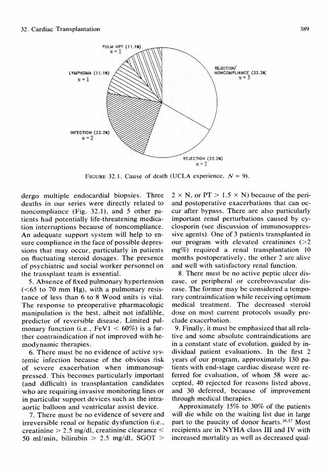
32. Cardiac Transplantation
REJECTI ctII NONCOMPLIANCE (33. 3111)
N = 3
REJECTION (22.21\1) N=2
389
FIGURE 32.1. Cause of death (UCLA experience, N = 9).
dergo multiple endocardial biopsies. Three deaths in our series were directly related to noncompliance (Fig. 32.1), and 5 other patients had potentially life-threatening medication interruptions because of noncompliance . An adequate support system will help to ensure compliance in the face of possible depressions that may occur, particularly in patients on fluctuating steroid dosages. The presence of psychiatric and social worker personnel on the transplant team is essential.
5. Absence of fixed pulmonary hypertension «65 to 70 mm Hg) , with a pulmonary resistance of less than 6 to 8 Wood units is vital. The response to preoperative pharmacologic manipulation is the best, albeit not infallible, predictor of reversible disease. Limited pulmonary function (i.e., FeV1 < 60%) is a further contraindication if not improved with hemodynamic therapies.
6. There must be no evidence of active systemic infection because of the obvious risk of severe exacerbation when immunosuppressed. This becomes particularly important (and difficult) in transplantation candidates who are requiring invasive monitoring lines or in particular support devices such as the intraaortic balloon and ventricular assist device.
7. There must be no evidence of severe and irreversible renal or hepatic dysfunction (i.e., creatinine> 2.5 mg/dl, creatinine clearance < 50 ml/min, bilirubin > 2.5 mg/dl, SGOT >
2 x N, or PT > 1.5 x N) because of the periand postoperative exacerbations that can occur after bypass. There are also particularly important renal perturbations caused by cyc1osporin (see discussion of immunosuppressive agents). One of 3 patients transplanted in our program with elevated creatinines (> 2 mg%) required a renal transplantation 10 months postoperatively, the other 2 are alive and well with satisfactory renal function.
8. There must be no active peptic ulcer disease, or peripheral or cerebrovascular disease. The former may be considered a temporary contraindication while receiving optimum medical treatment. The decreased steroid dose on most current protocols usually preclude exacerbation. 9. Finally , it must be emphasized that all rela
tive and some absolute contraindications are in a constant state of evolution, guided by individual patient evaluations. In the first 2 years of our program, approximately 130 patients with end-stage cardiac disease were referred for evaluation, of whom 58 were accepted, 40 rejected for reasons listed above, and 30 deferred, because of improvement through medical therapies.
Approximately 15% to 30% of the patients will die while on the waiting list due in large part to the paucity of donor hearts. 16, 17 Most recipients are in NYHA class III and IV with increased mortality as well as decreased qual-

390
ity of life. If patients are unable to be discharged from hospital on oral medications, they are maintained on intravenous medication and/or mechanical assistance and placed on urgent priority status for transplantation. Such prioritization is necessary given the shortfall of donors for recipients (see support and assist devices).
Diagnostic Evaluation and Therapy
All patients undergo a right heart catheterization with or without a left heart study to evaluate the left ventricular (L V) and the coronary arteries in particular. The latter is obtained depending on the age and clinical history of the patient. Other important studies obtained include an echocardiogram, ECG, MUGA scan, an EP study where indicated, and chemistry evaluation of hepatic and renal function. We further obtain a spec thallium and positron emission tomography (PET) in those patients being assessed for possible recoverable cardiac function through revascularization.
The aggressive use of afterload reducing agents and diuretics in conjunction with shortterm inotropes has resulted in 80% of patients at UCLA being stabilized and downgraded from an urgent priority to a more elective status. IS A small percentage of patients may require hospitalization up to the time of transplantation because of intravenous therapy. An even smaller percentage may require mechanical assistance because of massive infarction or inability to wean from cardiopulmonary bypass. Both groups are considered urgent transplant candidates, with the latter group representing the most urgent transplant. Suffice it to say, it is difficult to fully evaluate patients in these circumstances, but strict adherence to criteria of acceptability should be maintained.
Support and Assist Devices
Seven (approximately 10%) of our patients required intra-aortic balloon pumps preoperatively; all were removed immediately postop-
D. Drinkwater, L. Warner Stevenson, and H. Laks
eratively without difficulty. We have used both left (2) and biventricular (1) assist devices in the preoperative transplant patient, as a bridge to transplantation. The two survivors from this group received transplantations after 48 hours of support, whereas the third patient who was biventricularly assisted for 7 days died of overwhelming infection after transplantation.
Our limited experience parallels the findings of other groups who found that patients who underwent transplantation after less than 72 hours of support had a significantly better outcome than those who had to wait for longer periods. 19,20 The most important reasons for this are thought to be the increased risks from infection as well as the renal and hematologic complications that may occur with assist devices. All patients, especially those supported for more than 48 hours, should be well screened for infection and renal failure with consideration for exclusion from transplantation if positive. Needless to say, the patient on any type of assist or support device should be placed on an urgent priority status with regard to the organ procurement agencies.
Donor Selection
There is currently a shortfall in the number of donors available for the more than 14,000 people a year the National Heart Transplant Study estimated could benefit from cardiac transplantation.21 In the United States each year less than a quarter of an estimated 2 to 3,000 potential cardiac donors are used, largely due to the lack of public education and coordinated national and regional information systems. One response, the National Organ Transplantation Act authorizes federal grants to stimulate activity of regional organ procurement agencies to increase the availability of suitable organs.22 A national organ registry also has been established to match donors and recipients. Additionally, many states are enacting laws that make it mandatory for physicians caring for brain dead individuals to inform families of the potential for organ donation.
32. Cardiac Transplantation
Donor criteria (UCLA) are as follows:
1. Documented brain death in the absence of severe hypothermia or drug overdose. An appropriate informed consent from family members.
2. For males an age under 40 and for females an age under 45 is standard. However, depending on the urgency of the recipient matched, the age limit may be extended obtaining a prior coronary angiogram to rule out atherosclerosis.
3. There should be no history of cardiac disease, significant chronic hypertension, or a sustained cardiac arrest.
4. ABO compatibility and HIV testing are mandatory. Additional information concerning cytotoxic antibodies are obtained by many programs, particularly if the PRA is elevated.
5. An inotropic requirement less than 10 p,g/ kg per minute with a CVP in the 5 to 10-cm range is a useful guideline reflecting the myocardial function. We additionally obtain an echocardiogram on most donor hearts as able, and particularly those requiring higher inotropic support, as well as those with a history of chest trauma.
6. Donorlrecipient body size should be within 20% height and weight; this is particularly important for donor smaller than recipient. When sizing for heart-lung recipients, the thoracic measurements and match are even more specifically made.
7. No evidence of malignancy or significant infection. All donors are screened for human immunodeficiency virus.
8. Anticipated ischemic time for donor heart < 4 hours, variable presently between 4 to 6 hours depending on urgency of transplantation.
More than one half of the potential donors are excluded because of poor ventricular function. 23 This deterioration of function appears to be secondary to the acute systemic effects of CNS injuries. The recent reports of function preservation with hormonal treatment of donors if confirmed would increase the actual numbers of suitable donorsY Recognizing that the length of ischemic time may correlate
391
with long-term outcome,23 there must be close coordination with the donor cardiac team, which is generally at a site distant from the recipient hospital. At the appropriate time the donor heart is removed after administration of heparin followed by 1 I of cold crystalloid cardioplegia. Frequently multiple vital organs may be procured at the same time making proper planning and coordination even more important. The donor heart is transported in an ice cold sterile saline solution as rapidly as possible, usually including air transportation.
Recipient Management
Routine Immunosuppression
Currently a triple immunosuppressive regimen is used at many transplant centers, as it is at UCLA. This consists of cyclosporine (CsA), azathioprine (Aza), and prednisone. Regimens are constantly being reevaluated in the light of rejection episodes and complications and side effects of medications.
Cyclosporine
Cyclosporine, the mainstay of most regimens, is a fungal peptide that selectively inhibits Tcell function, probably at the IL 1 and IL2 levels. Its exact mechanism of action is not known, however. Cyclosporine is absorbed by the gastrointestinal tract, metabolized by the liver, and its metabolite excreted by the kidney. A bilirubin> 2.5 mg% may indicate decreased ability to metabolize CsA, and the half-life may vary from 8 to 24 hours. 25
The two most important side effects of CsA include hypertension and nephrotoxicity, which occur separately or together in 80% of patients in the first year. The most serious is renal failure, which may occur particularly at the time of surgery. Recognition of the highrisk patient for renal failure (i.e., creatinine> 2.0 mg%) and attendant manipulation of the immunosuppressive regimen may ameliorate the extent of failure and possibly avoid dialysis. Seven patients (9%) in our series required
392
short-term dialysis immediately postoperatively, and one (diabetic) required long-term dialysis and eventual renal transplantation. Attempts to discontinue CsA altogether in the early postoperative period, that is, "immunoconversion," because of renal failure, have in some programs led to a 50% increase in rejections in the first 3 months. 26 Consequently, most groups favor brief discontinuance of CsA or better a decreased dose through the use of a combination therapy. The mechanism of renal toxicity with cyclosporine is not yet known; however, it is thought to be secondary to stimulation of reninangiotensin with vasoconstriction and aldosterone secretion. 27 This stimulation is associated with arteriolar medial necrosis and may also account for the chronic hypertension that many of the transplant patients suffer from. Hypertension is generally well controlled with a combination of calcium channel blockers and captopril. Other side effects include CNS disturbances, frequently early in the course, and predominantly tremors although occasionally seizure activity may occur, particularly with low magnesium levels. Hirsutism is common over the long-term, as well as gingival hyperplasia in the pediatric age group.
Cyclosporine trough levels are obtained and measured daily during the immediate postoperative period. Two techniques are used: radioimmunoassay (RIA) measures serum levels of both the drug and its metabolites, and therefore in renal failure it may be spuriously elevated. A second test is the high pressure liquid chromatography (HPLC), which measures the active drug alone, which because of its sensitivity may be less reproducible than RIA. With normal renal function RIA levels should be up to twice that of the HPLC with a target range of 200 to 300 ng/ml. All drugs should be assessed for possible interaction with CsA metabolism and lor excretion. In particular, all anticonvulsant drugs increase hepatic metabolism, whereas ketoconazole inhibits CsA metabolism. 27 All nephrotoxic drugs have a synergistic effect with CsA on renal function. Finally, calcium channel blockers (e .g., diltiazem) may inhibit excretion of CsA causing an increase in levels.
D. Drinkwater, L. Warner Stevenson, and H. Laks
Azathioprine (Immuran)
Azathioprine (Aza) is a purine analogue that depresses cell-mediated immunity. But in contrast to CsA it may decrease the absolute number of both white cells and platelets. The immunosuppressive effect of Aza may not be evident for 1 to 4 weeks after initiation. Both platelets and WBC, which should be maintained above 5,000/mm3 , must be monitored regularly during the initial dosing. Allopurinol depresses Aza metabolism sufficiently to warrant a dosage reduction of 60% to 75%.28
Glucocorticoids
Glucocorticoids have been in use in transplantation immunology for a long time and their broad effects on both limbs of the immunologic system are well recognized. Side effects are frequent and may be life threatening; they include early and late effects. Early side effects are hyperglycemia, fluid retention, emotionallability; the more chronic effects are osteoporosis, skin friability, moon faces, and obesity.29 The most serious side effect for all groups is an increased susceptibility for infections, particularly opportunistic varieties. Consequently, prednisone is tapered over the first 4 weeks.
Alternative and Rescue Drugs
Antithymocyte globulin (ATG) both equine preparation ATGAM and rabbit preparation have been used as first-line and rescue therapies (Table 32.2). This drug directly reduces T -lymphocyte counts with effects noted within the first 24 hours. 30 All patients should receive a skin test before the administration of ATG to rule out potential anaphylaxis. The platelet count should be monitored daily and the ATG dose reduced for counts < 100,0001 mm3 • ATG is used at UCLA in the high-risk renal failure patient perioperatively and in the steroid resistant rejection as listed in the protocol.
OKT3 is a mouse monoclonal T3 (or CD3) analog that is used in most protocols as a rescue therapy for the steroid-resistant or poten-

32. Cardiac Transplantation
TABLE 32.2. Immunosuppressive therapy-UCLA. Standard therapy
Solumedrol 500 mg IV at time of surgery 125 mg IV q 12 H until PO
Prednisone 0.2 mg/kg PO, taper to 0.1 mg/kg at I month Cyc1osporine 8 mg/kg PO preop dose
9 mg/kg/day until first serum or blood trough level
393
Goal: serum levels of 200-300 ng/ml over the first 30 days, then decrease to obtain levels of 100-200 ng/ml Azathioprine 2 mg/kg PO preop dose
2 mg/kg/day maintenance dose (WBC, platelets monitored) High-risk renal failure patients protocol (i.e., patients with creatinine preop > 2mg%, patients requiring IV
inotropic agents) Solumedrol as above Prednisone as above Cyc1osporine no preop dose Azathioprine 2 mg/kg PO preop
H mg/kg po qd postop ATG (antithymocyte globulin equine or rabbit)
Skin test preop 15 mg/kg IV postop Discontinue when adequate CsA levels
Cyc1osporine after 36 hours adequate urine output CsA started at 6 mg/kg PO bid Acute rejection protocol
Mild to mod-CsA increased to level 2x baseline for 10 days or PO prednisone pulse Moderate to severe-Solumedrol 1 gm IV q day x 3 days Severe (life threatening) or resistant-ATG 15 mg/kg, or OKT3 5 mg/day
tially life-threatening rejections. Several programs have investigational protocols using it as a first-line therapy.31 The mode of action appears to be a direct effect on the T -lymphocytes by blocking T3 receptors, thus altering the suppressor/killer cell ratios.32 A dose of 5 mg intravenously is recommended for up to 5 to 7 days in the rescue protocols. Early side effects include allergic response, pulmonary edema, and fever. Because it is a murine monoclonal antibody (IgG) , chronic or repeated use may result in the development of antibodies to OKT3 in 50% to 60%, and also rarely in aseptic meningitis.
Postoperative Management
The immediate postoperative period is generally characterized by hemodynamic stability and is managed similarly to other cardiac surgical procedures. An initial period of some relative right ventricular dysfunction with tricuspid regurgitation is not uncommon but usually transient, except in those recipients whose
pulmonary artery pressures and resistance remain elevated. In these cases the use ofPGEI in combination with afterload reducing agents such as nitroprusside, nitroglycerin, and captopril has been very helpful in our experience. In general, this dysfunction is mild and well tolerated.
Arrhythmias are frequent immediately postoperatively, characterized largely by junctional rhythm because of sinus node dysfunction. This is generally transient and well tolerated, although pacing wires with backup pacing is maintained on these patients. Ventricular arrhythmias in distinction to atrial arrhythmias are uncommon and should initiate a search for other problems such as acidosis, hypoxia, electrolyte imbalance, or tamponade. An inotrope such as dopamine is generally used at low dose for the early postoperative period to help maintain rate support in these denervated hearts, which respond only to circulating catecholamines. One patient has required a pacemaker implantation, which was removed after 6 months on return of his sinus node function.

394
.... z w U
'" w c.
D. Drinkwater. L. Warner Stevenson. and H. Laks
30
28
26
24 18
22
20
18
16
14
1 Z
10
8
6
4
2
o ~~~~ __ ~~~~ __ ~~~~~~~~~~~~-L~~~~~ RENAL (Cr > 2.0) OIABETES ARRHYTHMIA SEIZURE PERICAROIAL EffUSION AGAS
FIGURE 32.2. Noninfectious complications (UCLA experience).
The electrocardiogram frequently has two P waves representing the native and the donor heart SA nodes. Right bundle branch block may occur in 50% of the patients, possibly secondary to an increased tension on the right side of the interventricular septum or by persistent elevated right-sided pressures. Q waves in the inferior leads as well as inverted T waves may occur without associated wall motion abnormalities. T wave changes are frequent in the early postoperative period and usually resolve spontaneously. Diffuse ST changes should, however, initiate a differential search to include infarction perhaps due to unrecognized donor disease, trauma and contusion effects, and more likely pericarditis. Seven patients (9%) in our series have received long-term therapy for atrial arrhythmias, none for ventricular (Fig. 32.2).
Renal Function
Renal function is closely monitored in the immediate postoperative period, including volume, daily creatinine, and BUN. All patients have a significant rise in chemistries usually peaking at day 4 and diminishing thereafter.
Inadequate outputs leading to fluid imbalance may require short-term hemodialysis while the renal function improves. Seven patients (9%) required hemodialysis, six of whom were short-term less than 2 weeks; one patient went on to have a renal transplantation. The subset at higher risk for renal failure is generally predictable by an elevated preoperative creatinine> 2.0 mg%, and the need for inotropic support. Because CsA has the most profound effects on renal function in the postoperative period, a concerted effort is made in these patients to diminish or avoid CsA altogether in the first 3 days postoperatively by using combination therapies or replacing CsA with ATG. Overall, 18 of 76 patients (24%) have developed some element of renal failure with a creatinine> 2.0 mg% (Fig. 32.2). It should, however, be noted that many of these patients have creatinine clearances above 55 mllmin.
Rejection of Allograft
Forty-three percent (33 of 74) patients in the UCLA series experienced some rejection in the first 4 weeks postoperatively. Overall, 70% of our patients have had one or more re-
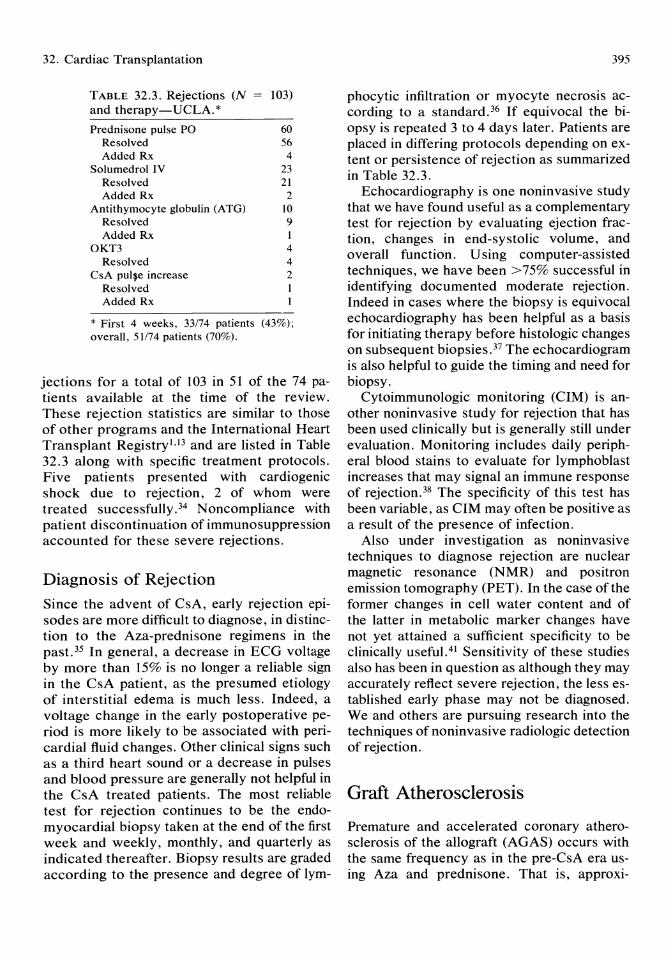
32. Cardiac Transplantation
TABLE 32.3. Rejections (N 103) and therapy-UCLA. * Prednisone pulse PO 60
Resolved 56 Added Rx 4
Solumedrol IV 23 Resolved 21 Added Rx 2
Antithymocyte globulin (ATG) 10 Resolved 9 Added Rx I
OKT3 4 Resolved 4
CsA pulfe increase 2 Resolved I Added Rx
* First 4 weeks, 33174 patients (43%); overall, 51174 patients (70%).
jections for a total of 103 in 51 of the 74 patients available at the time of the review. These rejection statistics are similar to those of other programs and the International Heart Transplant Registry!·!3 and are listed in Table 32.3 along with specific treatment protocols. Five patients presented with cardiogenic shock due to rejection, 2 of whom were treated successfully. 34 Noncompliance with patient discontinuation of immunosuppression accounted for these severe rejections.
Diagnosis of Rejection
Since the advent of CsA, early rejection episodes are more difficult to diagnose, in distinction to the Aza-prednisone regimens in the past. 35 In general, a decrease in ECG voltage by more than 15% is no longer a reliable sign in the CsA patient, as the presumed etiology of interstitial edema is much less. Indeed, a voltage change in the early postoperative period is more likely to be associated with pericardial fluid changes. Other clinical signs such as a third heart sound or a decrease in pulses and blood pressure are generally not helpful in the CsA treated patients. The most reliable test for rejection continues to be the endomyocardial biopsy taken at the end of the first week and weekly, monthly, and quarterly as indicated thereafter. Biopsy results are graded according to the presence and degree of lym-
395
phocytic infiltration or myocyte necrosis according to a standard. 36 If equivocal the biopsy is repeated 3 to 4 days later. Patients are placed in differing protocols depending on extent or persistence of rejection as summarized in Table 32.3.
Echocardiography is one noninvasive study that we have found useful as a complementary test for rejection by evaluating ejection fraction, changes in end-systolic volume, and overall function. Using computer-assisted techniques, we have been >75% successful in identifying documented moderate rejection. Indeed in cases where the biopsy is equivocal echocardiography has been helpful as a basis for initiating therapy before histologic changes on subsequent biopsies. 37 The echocardiogram is also helpful to guide the timing and need for biopsy.
Cytoimmunologic monitoring (CIM) is another noninvasive study for rejection that has been used clinically but is generally still under evaluation. Monitoring includes daily peripheral blood stains to evaluate for lymphoblast increases that may signal an immune response of rejection. 38 The specificity of this test has been variable, as CIM may often be positive as a result of the presence of infection.
Also under investigation as noninvasive techniques to diagnose rejection are nuclear magnetic resonance (NMR) and positron emission tomography (PET). In the case of the former changes in cell water content and of the latter in metabolic marker changes have not yet attained a sufficient specificity to be clinically useful. 4! Sensitivity of these studies also has been in question as although they may accurately reflect severe rejection, the less established early phase may not be diagnosed. We and others are pursuing research into the techniques of noninvasive radiologic detection of rejection.
Graft Atherosclerosis
Premature and accelerated coronary atherosclerosis of the allograft (AGAS) occurs with the same frequency as in the pre-CsA era using Aza and prednisone. That is, approxi-

396
mately one third of the transplanted hearts will have angiographic evidence of significant atherosclerosis by the third year posttransplant. 40
Indeed, there are recently reports of AGAS within the first 6 months posttransplant. 41 The study from Stanford identified the number of rejections and prednisone dosage as significant predictors of AGAS in the CsA group. However, other studies have not been able to find these associations, but rather have implicated cholesterol and LDL levels. 42 In distinction to the Aza group, donor age and HLA-2 mismatch have also not been significant predictors to date. 43
Due to denervation and the absence of anginal symptoms, the first sign of advanced coronary disease may be a silent infarction on ECG. Consequently, we and others obtain yearly stress tests and coronary angiograms additionally as indicated. Percutaneous balloon angioplasty is indicated in those coronary lesions that may be amenable; however, the pattern of disease is typically diffuse and present in both large and small vessels. 34 We have retransplanted 1 patient and are following 3 more patients (4.5%), all of whom are > 24 months posttransplant (Fig. 32.3).
100 ---a..-. 68/ 70
90
80
70 ..J « ~ 60 > (I:
::> ~ 50 ... Z w U 40 (I: w Q.
30
20
10 -
0
D. Drinkwater, L. Warner Stevenson, and H. Laks
Attempts to ameliorate this problem are unproven but include the early initiation of aspirin and persantine and better control of choIe sterol levels through diet and medicine. We have instituted both dietary changes and recently a protocol using the cholesterol-lowering drug mevenalin (Lovostatin) along with the cholesterol binding agent Cholestatin. Further investigation is needed to delineate whether AGAS is a result of lipid abnormalities versus a rejection phenomenon with immunologic injury to endothelial surfaces, or a combination of both.
Malignancy
Malignancies, in the form of skin cancers and lymphoproliferative disorders, have become an unwelcome aspect of most transplant series. 44 Most of the lymphomas are nonHodgkins of B-ceU type in extranodal sites in distinction to the nontransplant population. In the more established programs these patients can be a significant subgroup such as the some 30 (7%) in the Stanford series, with 25% occurring in the CsA era. 43 Lymphomas and sar-
33 37
0 2 3 4 5 6 7 8 9 10 11 12
MONTHS
FIGURE 32.3. One-year survival (UCLA experience).
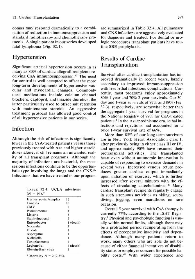
32. Cardiac Transplantation
comas may respond dramatically to a combination of reduction in immunosuppression and standard radiotherapy and chemotherapy protocols. A single patient in our series developed fatal lymphoma (Fig. 32.1).
Hypertension
Significant arterial hypertension occurs in as many as 80% of cardiac allograft recipients receiving CsA immunosuppression.45 The need for control is well accepted to offset the more long-term developments of hypertensive vascular and myocardial changes. Commonly used medications include calcium channel blockers, captopril, and thiazide diuretics, the latter particularly used to offset salt retention with maintenance steroids. An aggressive treatment protocol has allowed good control of all hypertensive patients in our series.
Infection
Although the risk of infections is significantly lower in the CsA-treated patients versus those previously treated with Aza and higher steroid doses alone, it still remains an unwanted reality of all transplant programs. Although the majority of infections are bacterial, the most serious infections continue to be the opportunistic type involving the lungs and the CNS.46 Infections that we have treated in our program
TABLE 32.4. UCLA infections (N = 94).*
Herpes zoster/simplex 16 Candida 10 CMV 8 Pseudomonas 4 Listeria 3 Staphylococcal 2 Enterobacter 1 (death) Nocardia 1 E. coli Aspergillus Klebsiella Toxoplasmosis Legionella Ebstein-Barr virus
* Mortality N = 2 (2.5%).
1 (death) 1
397
are summarized in Table 32.4. All pulmonary and CNS infections are aggressively evaluated for diagnosis and treated. For dental or urologic procedures transplant patients have routine SBE prophylaxis.
Results of Cardiac Transplantation
Survival after cardiac transplantation has improved dramatically in recent years, largely secondary to improved immunosuppression with less lethal infectious complications. Currently, most programs enjoy approximately 80% I-year and 70% 2-year survivals. Our 30-day and I-year survivals of 97% and 89% (Fig. 32.3), respectively, are somewhat better than the aggregate I-year survival for programs in the National Registry of 79% for CsA-treated patients. I In the Aza/prednisone era, lethal infections and rejections had accounted for a prior I-year survival rate of 66%.
More than 85% of our long-term survivors are in New York Heart Association class I, after previously being in either class III or IV, and approximately 90% have resumed their pretransplant activities. The transplanted heart even without autonomic innervation is capable of responding to exercise demands in several ways. Increased venous return produces greater cardiac output immediately upon initiation of exercise, which is further increased after several minutes with the effects of circulating catecholaminesY Many cardiac transplant recipients regularly engage in such strenuous activities as skiing, scuba diving, jogging, even marathons on rare occasion.
Overall 5-year survival with CsA therapy is currently 77%, according to the ISHT Registry. I Physical and psychologic function is usually within normal limits, although there may be a protracted period recuperating from the effects of preoperative inactivity and dependance. Although many patients return to work, many others who are able do not because of either financial incentives of disability status or employer concern for possible liability costS.48 With wider experience and

398
acceptance of the efficacy of cardiac transplantation, it should become possible for more recipients to enjoy full professional as well as physical rehabilitation.
Combined Heart and Lung Transplantation
In patients with combined cardiac and pulmonary disease, a heart-lung transplant may represent the best therapy. The presence of fixed pulmonary hypertension greater than 6 to 8 Wood units would preclude isolated heart transplantation, because of the rapidly dam~ging effects on the right ventricle. The routme criteria for acceptance for heart-lung transplantation are similar to those previously listed for isolated heart transplantation.
Since 1981 more than 100 heart-lung transplantations have been performed worldwid~, with an overall I-year survival of approxImately 50%.49 This decreased survival figure relative to isolated cardiac transplantation reflects a higher operative and hospital mortality of around 30%, as well as an overall increased incidence of infectious complications. 50 Presently, the best results are obtained in younger patients with either primary pulmonary hypertension or Eisenmenger's complex. 51
Three major problems remain to be solved before this field can expand significantly. First, because proper donor shortage is an even greater problem than with cardiac transplantation, donor organ retrieval and ~reservation for better distant procurement will have to be improved so that the donor pool can be enlarged. Currently, there are two basic techniques for preservation of the donor heartlung block: one is the cold flush cardiopulmonary plegia, and the second is a pump technique for constant perfusion. Similar results are obtained in both for up to 4 hours ischemic time, and because of the convenience for distant procurement the single flush technique is used by the majority of programs in the United States. More work will have to be performed to establish which of the two techniques is preferred, particularly for longer ischemic times. 51
D. Drinkwater, L. Warner Stevenson, and H. Laks
A second area for improvement is in the diagnosis of rejection. It is now well documented that isolated rejection of the lungs can occur in the absence of a significant endomyocardial biopsy,52 which had in the past been relied upon. A more specific diagnostic test is needed short of an open lung biopsy to complement the endomyocardial biopsy. Experimental and clinical experience is being gained with endobronchial monitoring of the macrophage population as one technique to identify isolated lung rejection. 51 U nfortunately, the presence of an infectious process may have a similar and confusing profile. The standard remains the endomyocardial biopsy along with a high degree of clinical suspicion. The latter also applies equally to possible infections that must be aggressively diagnosed and treated.
A third problem is that of obliterative bronchiolitis that has occurred in approximately 50% of the long-term survivors in the Stanford series. 53 It has also occurred as early as 3 months after transplantation in this series. This complication, resulting in severe pulmonary dysfunction, may represent chronic pulmonary rejection, although recently an infectious etiology, cytomegalovirus, has been associated.54 Overall, the problem of obliterative bronchiolitis may be the greatest impediment to more widespread applicability of this technique.
Both heart and heart-lung transplantation offer great hope for patients with severe endstage cardiopulmonary disease. Accelerated graft atherosclerosis with isolated cardiac and obliterative bronchiolitis with heart-lung transplants represent the most serious problems for future research and improvements.
References
1. Solis E, Kaye MP: Registry of the International Society for Heart transplantation; Third Official Report. J Heart Trans 1986; 5:2-5.
2. Carrel A, Guthrie CC: The transplantation of veins and organs. Am J Med 1905; 18:1101.
3. Mann FC, Priestly JT, Markowitz J: Transplantations of the intact mammalian heart. Arch Surg1933; 26:219.
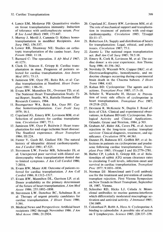
32. Cardiac Transplantation
4. Lance EM, Medawar PB: Quantitative studies on tissue transplantation immunity. Induction of tolerance with antilymphocyte serum. Proc R Soc Lond (BioI) 1969; 173:447.
5. Murray J, Merill J, Cammin G: Kidney homotransplantation in modified Recipients. Ann Surg 1962; 156:337.
6. Lower RR, Shumway NE: Studies on orthotopic transplantation of the canine heart. Surg Forum 1960; 11:18.
7. Barnard C: The operation. S Air Med J 1967; 41:1271.
8. Clark D, Stinson E, Griepp R: Cardiac transplantation in man. Prognosis of patients selected for cardiac transplantation. Ann Intern Med 1971; 75: 15.
9. Jamieson SW, Oyer PE, Reitz BA, et al: Cardiac transplantation at Stanford. Heart Transplant 1981; 1:86-91.
10. Evans RW, Mannihen DL, Overeast TD, et al: The National Heart Transplantation Study: Final Report. Seattle, WA, Battelle Human Affair Research Centers, 1984.
11. Baumgartner W A, Reitz BA, Oyer PF: Cardiac homotransplantation. Curr Prohl Surg 1979; 16: 1.
12. Copeland JG, Emery RW, Levinson MM, et al: Selection of patients for cardiac transplantation. Circulation 1987; 75: 1.
13. Jamieson SW, Oyer P, Baldwin J: Heart transplantation for end-stage ischemic heart disease: The Stanford experience. Heart Transplant 1984; III:224.
14. Fuster V, Gush BJ, Giuliani ER: The natural history of idiopathic dilated cardiomyopathy. Am J Cardiol 1981; 47:525.
15. Stevenson LW, Fowler MB, Schroeder JS, et al: Unexpected poor survival with dilated cardiomyopathy when transplantation denied due to limited symptoms. J Am Coli Cardiol 1986; 7:203A.
16. Evans RW, Maier AM: Outcome of patients referred for cardiac transplantation. J Am Call Cardiol1986; 8:1312-1317.
17. Evans RW, Mannihen DL, Garrison LP, et al: Donor availability as the primary determinant of the future of heart transplantation. JAm Med Assoc 1986; 255:1892-1898.
18. Stevenson LW, Donohue BC, Schulman B, et al: Therapy of urgent priority candidates for cardiac transplantation. J Heart Trans 1986; 5:391.
19. Medical News and Perspectives: Artificial heart recipients 1982 through November 1986. JAm Med Assoc 1986; 21:2924.
399
20. Copeland JC, Emery RW, Levinson MM, et al: The role of mechanical support and transplantation in treatment of patients with end-stage cardiomyopathy. Circulation 1985; 72(suppl 11):11-17.
21. Robertson JA: Supply and distribution of hearts for transplantation: Legal, ethical, and policy issues. Circulation 1987; 75: I.
22. Zaontz L: The national organ transplantation act. Bull Am Coll Surg 1985; 70:5:18.
23. Emery R, Cork R, Levinson M, et al: The cardiac donor: a six-year experience. Ann Thorac Surg 1986; 41:356-362.
24. Novitsky D, Wicomb WN, Copper DKC, et al: Electrocardiographic, hemodynamic, and endocrine changes occurring during experimental brain death in the Chacma baboon. J Heart Trans 1984; 4:63.
25. Kahan BD: Cyclosporine: The agents and its actions. Transplant Proc 1985; 17:5-18.
26. Schuler S, Warneebe H, Hetzer A: Prevention of toxic side effects of cyclosporine in heart transplantation. Transplant Proc 1987; 19:2518-2521.
27. Devineni R, McKenzie N, Duplin J: Renal effects of CSA: Clinical and experimental observations, in Kahane BD (ed): Cyclosporine, Biological Activity and Clinical Applications. Orlando, Grune and Stratton, 1984, p 503.
28. Graham AF, Rider AK, Caves PK, et al: Acute rejection in the long-term cardiac transplant survivor: Clinical diagnosis, treatment, and significance. Circulation 1974; 44:361.
29. Danner JS, Bahnson HT, Griffith BP, et al: Infections in patients on cyclosporine and prednisone following cardiac transplantation. Transplant Proc 1983; 15(suppl I and II):2779-2781.
30. Bieber CP, Lydick E, Griepp RB, et al: Relationships of rabbit ATG serum clearance rates to circulating T-cell levels, infection onset and survival in cardiac transplantation. Transplant Proc 1977; 9:1031-1036.
31. Norman DJ: Monoclonal anti-T-cell antibody use for the treatment and prevention of cardiac transplant rejection. First Vienna Symposium on New Trends in Heart Transplant, September 18, 1987, Vienna.
32. Schreiber RD, Hicks LJ, Celada A: Monoclonal antibodies to murine gamma-interferon which differentially moderated macrophage activation and antiviral activity. J Immunol 1985; 134:1609.
33. Colombani P, Robb A, Hess A: Cyclosporine A binding to calmodulin: A possible site of action on T lymphocytes. Science 1985; 228:337-339.
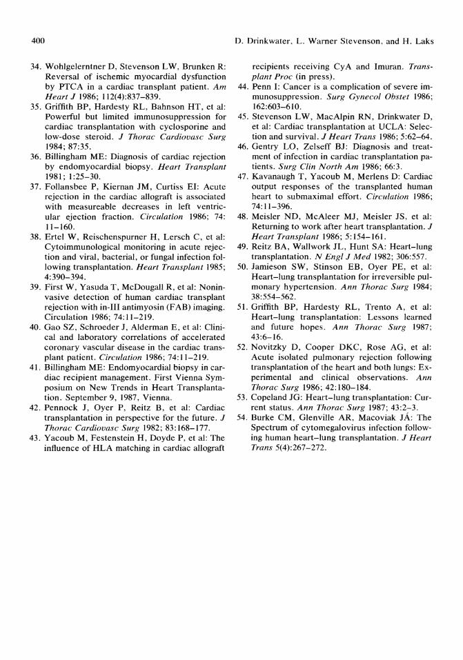
400
34. Wohlgelerntner D, Stevenson LW, Brunken R: Reversal of ischemic myocardial dysfunction by PTCA in a cardiac transplant patient. Am Heart J 1986; 112(4):837-839.
35. Griffith BP, Hardesty RL, Bahnson HT, et al: Powerful but limited immunosuppression for cardiac transplantation with cyclosporine and low-dose steroid. J Thorac Cardiouasc Surg 1984; 87:35.
36. Billingham ME: Diagnosis of cardiac rejection by endomyocardial biopsy. Heart Transplant 1981; 1:25-30.
37. Follansbee P, Kiernan JM, Curtiss EI: Acute rejection in the cardiac allograft is associated with measureable decreases in left ventricular ejection fraction. Circulation 1986; 74: 11-160.
38. Ertel W, Reischenspurner H, Lersch C, et al: Cytoimmunological monitoring in acute rejection and viral, bacterial, or fungal infection following transplantation. Heart Transplant 1985; 4:390-394.
39. First W, Yasuda T, McDougall R, et al: Noninvasive detection of human cardiac transplant rejection with in-III antimyosin (FAB) imaging. Circulation 1986; 74:11-219.
40. Gao SZ, Schroeder J, Alderman E, et al: Clinical and laboratory correlations of accelerated coronary vascular disease in the cardiac transplant patient. Circulation 1986; 74: 11-219.
41. Billingham ME: Endomyocardial biopsy in cardiac recipient management. First Vienna Symposium on New Trends in Heart Transplantation. September 9, 1987, Vienna.
42. Pennock J, Oyer P, Reitz B, et al: Cardiac transplantation in perspective for the future. J Thorac Cardiouasc Surg 1982; 83:168-177.
43. Yacoub M, Festenstein H, Doyde P, et al: The influence of HLA matching in cardiac allograft
D. Drinkwater, L. Warner Stevenson, and H. Laks
recIpIents receIvmg CyA and Imuran. Transplant Proc (in press).
44. Penn I: Cancer is a complication of severe immunosuppression. Surg Gynecol Obstet 1986; 162:603-610.
45. Stevenson LW, MacAlpin RN, Drinkwater D, et al: Cardiac transplantation at UCLA: Selection and survival. J Heart Trans 1986; 5:62-64.
46. Gentry La, Zelseff BJ: Diagnosis and treatment of infection in cardiac transplantation patients. Surg Clin North Am 1986; 66:3.
47. Kavanaugh T, Yacoub M, Merlens D: Cardiac output responses of the transplanted human heart to submaximal effort. Circulation 1986; 74:11-396.
48. Meisler ND, McAleer MJ, Meisler JS, et al: Returning to work after heart transplantation. J Heart Transplant 1986; 5:154-161.
49. Reitz BA, Wallwork JL, Hunt SA: Heart-lung transplantation. N Engl J Med 1982; 306:557.
50. Jamieson SW, Stinson EB, Oyer PE, et al: Heart-lung transplantation for irreversible pulmonary hypertension. Ann Thorae Surg 1984; 38:554-562.
51. Griffith BP, Hardesty RL, Trento A, et al: Heart-lung transplantation: Lessons learned and future hopes. Ann Thorac Surg 1987; 43:6-16.
52. Novitzky D, Cooper DKC, Rose AG, et al: Acute isolated pulmonary rejection following transplantation of the heart and both lungs: Experimental and clinical observations. Ann Thorac Surg 1986; 42:180-184.
53. Copeland JG: Heart-lung transplantation: Current status. Ann Thorac Surg 1987; 43:2-3.
54. Burke CM, Glenville AR, Macoviak JA: The Spectrum of cytomegalovirus infection following human heart-lung transplantation. J Heart Trans 5(4):267-272.
Index
Accessory pathways, catheter ablation of, 205-206, 207, 211,212,231-232
Acenocoumarin, 316 Acute myocardial infarction
(AMI), see Myocardial infarction, acute
Adenosine, 24, 162 in autoregulation, 24 dipyridamole vasodilatation
and, 162 Adenosine diphosphate, 333 Adult respiratory distress syn
drome (ARDS), pulmonary artery catheterization and, 51
Afterload definition of, 56 in pulmonary artery catheteri
zation assessment, 56-57 AICD, see Automatic implant
able cardioverter defibrillator device
Allograft rejection, 154, 158, 394-395, 398
Aminophyline, 162, 164 Amiodarone, 129-130, 142 Amrinone, in cardiogenic
shock,372 Anagrelide, 346 Angina, 71-75, 173; see also
Angina pectoris accelerated, 71-72, 73 angioscopy in, 71-75 preinfarction, intra-aortic
balloon pump counterpulsation and, 173
Prinzmetal's, 93 stable, 71
unstable, platelet inhibitors in, 338
unstable rest, 72-75 Angina pectoris, 338, 377-379
platelet inhibitors in, 338 unstable, 377-379
Angiography aortic root, 16-17 coronary, see Coronary angi
ography digital subtraction, 18,31-32,
41 interpretation of, 18-20
Angioplasty balloon, see Balloon angio
plasty caveat, 274 coronary, angiography for
percutaneous, 14, 17-18 laser thermal, see Laser ther-
mal angioplasty pediatric, 217-221 snowplow effect of, 274-275 urgent vs delayed, 7-8
Angioscopy, 69-77 in accelerated angina, 71-72,
73 of endothelial healing and
stenotic progression, 75-76
fiberoptic, 69 method of, 69-70 in myocardial infarction, 75 peripheral vascular, 69-70 in stable angina, 71 in sudden death, 72 therapeutic implications of,
77 in unstable rest angina, 73-75
Anisoylated plasminogen strep-
tokinase activator complex, 357-358
Antianginal agents, 93, 161 atrial pacing and, 93 dipyridamole as, 161
Antiarrhythmic drugs, 127, 129, 142
type lA drugs, 127, 142 type IB drugs, 129, 142 type lC drugs, 129, 142
Anticoagulant therapy, platelet inhibitor therapy vs, 339
Anti-inflammatory agents, 77 Antiplatelet therapy, see Plate
let inhibitor drugs Antithymocyte globulin, in
cardiac transplantation, 392
Anti-vitamin K (acenocoumarin), 316
Aorta aortic root angiogram, 16-17 balloon coarctation angio
plasty of, 217-219, 220 balloon valvuloplasty, 223-
226; see also Balloon aortic valvuloplasty
coarctation of, balloon coarc-tation angioplasty of, 217-219,220
dissection of, balloon pumping and, 177
stenosis, 84-85, 248 etiologies of, 248 exercise and, 84-85
valve disease, exercise and, 84-85
valvulotomy, 225
402
Aortocoronary bypass vein graft disease, 342-344
Arachidonate pathway, drugs affecting, 335-336
Arrhythmia after cardiac transplantation,
393-394 catheter ablation for, 204,
201-212 indications for, 204 techniques for, 201-212;
see also Catheter ablation techniques
interventions for, 5-6 pulmonary artery catheteriza
tion and, 58-59 ventricular, see Ventricular
arrhythmias Arteriography, coronary, see
Coronary arteriography Arteriotomy repair, 14-15 Artificial hearts, total, 7 Aspirin, 77, 335-336
in coronary angioplasty, 284 in non-Q wave infarction, 340 in percutaneous transluminal
coronary stenting, 315, 316
in Q wave infarction, 340-341
in reocclusion prevention after thrombolysis, 341-342
restenosis and, 346 in saphenous vein grafting,
343 in unstable angina, 338-339
Atherogenesis five stages in natural history
of, 329-335 platelets and thrombosis and,
329-335 Atheroma, 71,72,73
stable, 72, 73 Atherosclerosis
deep atrial damage in, 332-335
endothelial injury in, 330 five stages in natural history
of, 329-335 of graft, 395-396 hemorheologic factors in,
330 monocyte adhesion in, 330-
331
plaque rupture in, 332 platelet adhesion in, 329-331 platelet aggregation in, 332-
334 risk factors for, 331-332 stable atherosclerotic disease,
332-334 thrombogenesis and, 332-334
Atrial pacing, 90-93 advantages of, 92, 93 antianginal medications and,
93 chest pain and, 90-91 clinical indications for, 93 echocardiography with, 92 hemodynamic effects of, 92-
93 metabolic studies and, 92 radionuclide ventriculography
with, 91-92 stress, 90 thallium perfusion studies
with, 91 treadmill testing vs, 91
Atrial puncture, atrial septostomy after transseptal, 217
Atrial septal defects, transcatheter closure of, 227-228
Atrial septostomy, 215-217 balloon, 215-216, 217 blade, 216-217 pediatric, 215-217
Atrioventricular block, 102, 113-114
electrophysiologic studies of, 113-114
Atrioventricular conduction, 109-110, Ill, 112
Atrioventricular junction, catheter ablation of, 201-205, 231
complications of, 203-204 indications for, 204-205 mortality statistics after, 204 procedure, 202 results/efficacy of, 202-203
Atrioventricular tachycardia, 135-141
atrioventricular nodal re-entry tachycardia, 135-138
Atropine, 109 Automatic implantable car
dioverter defibrillator device (AICD), 100, 193-
Index
199; see also Automatic implantable defibrillator
Automatic implantable defibrillator, 193-199
arrhythmia recognition by, 194
arrhythmia termination by, 195
defibrillation threshold of, 196 electrocardiographic monitor-
ing of, 198 historical perspective on, 193 implantation criteria for, 195 intraoperative/postoperative
testing of, 196 Kaplan-Meier life table analy
sis for, 197-198 long-term outcome of, 197-
198 Medicare reimbursement for,
195 microcomputer processed,
193 mortality and, 199 noninvasive monitoring of,
198 operative complications of,
197 patient population for, 196-
197 preoperative evaluation for,
195-196 probability density function
criteria for, 194 surgical approach to, 195-196
Autoregulation of coronary blood flow, 162 factors in, 23 -24 in perfusion pressure, 23-24
Azathioprine, in cardiac transplantation, 392
Balloon angioplasty, 217-221, 254, 258-262, 275, 323
kissing, 275 laser-assisted, 258-262, 323 laser thermal angioplasty
with, 254 pediatric, 217-221
Balloon aortic valvuloplasty, 223-226, 239-252
angiographic data on, 243-248 for calcified aortic stenosis,
248, 249, 250
Index
clinical improvement after, 246
contraindication to, definitive, 252
follow-up for, 245-248 goal of, 240-241 hemodynamic effects of, 243-
248 indications for percutaneous,
251-252 in-hospital course after dilata
tion, 244-245 mechanism of action of, 248-
251 mortality and, 244, 245-246 new balloon design for, 241-
242 oversized balloon in, 251 pediatric, 223-226 recatheterization in, 247 restenosis rate after, 246-248,
249 results of, 242 technical considerations in,
239-242 tolerance of, 242
Balloon atrial septostomy, pediatric, 215-216,217
Balloon catheter, 270 Balloon coarctation angioplasty,
217-219, 220 Balloon ductus arteriosus angio
plasty, 219-221 Balloon pulmonary valvulo
plasty, 221-223 Balloon pulmonary vein angio
plasty, 219-221 Balloon pump counterpulsation,
intraaortic, see Intraaortic balloon pump counterpulsation
Balloon valvuloplasty, aortic, see Balloon aortic valvuloplasty
Beer-Lambert law, 43 Beta-blockers, in supraventricu
lar tachycardias, 142 Bifurcation lesions, angioplasty
for, 274-276, 277 Bioptomes, 154, 154-157, 155 Blade atrial septostomy, 216-
217 Blalock-Taussig shunt, 228-229 Blood pressure, 18, 371
in cardiogenic shock, 371
exercise and, 81 maintenance of, in shock, 371
Bradyarrhythmias, temporary pacing for, 182
Bundle branch block, 59, 102, 113-114
electrophysiology of, 102, 114 pulmonary artery catheteriza
tion-induced, 59 Bypass graft catheterization,
16-17 Bypass vein graft disease, 342-
344
C-11 palmitate in ischemia, 95-96
Calcium antagonists, 284, 316 in coronary angiop1asty, 284 in percutaneous transluminal
coronary stenting, 316 Calcium ions, 333-334 Cardiac arrest, survivors of,
101, 113 Cardiac catheterization, 5, 10-
20,63-68 in arteriotomy repair, 14-15 balloon pumping and, 175-
176 bypass graft catheterization,
16-17 catheter ablative techniques
and, 6 complications of, 11 in constrictive pericarditis,
148 for endocardial mapping, 126 historical perspective on, 10,
35-36 indications for, 10-11 in internal mammary artery
grafts, 17, 18 knotting of catheter in, 229-
230 laboratory catheterization
suite for, 11-12 left heart catheterization, 12-
14 outpatient, 63-68
admission, 64, 65 complications rate, 65, 66 cost savings, 67-68 laboratory work, 64 left main coronary artery
disease, 66-67
403
mortality rates, 65, 66-67 nurses, 67 premedication, 64 protocol for, 64 safety, 66, 67 surgical access, 67
pediatric, see Pediatric cardiac catheterization
percutaneous approach of Judkins, 14-16, 17
protocol, 12 risks of, 10-11 Seldinger technique, 232-233 Sone's technique, 13-14 in ventriculography, 14, 15
Cardiac complications, of pulmonary artery catheterization, 59
Cardiac death, sudden, 5-6; see also Sudden death
Cardiac defects, transcatheter closure of, 226-229
Cardiac donors, 390-391 Cardiac metabolism, in ische
mia, 95-97 Cardiac output, 56, 58, 81-82
exercise and, 81-82 pulmonary artery catheteriza
tion and, 56, 58 Cardiac tamponade, 147-148,
151 hallmarks of, 147-148 neoplastic, 147 recurring, 151
Cardiac transplantation, 7, 387-398
arrhythmias after, 393-394 contraindications to, 388-389 cyclosporine and, 387, 391-
392 diagnostic evaluation and
therapy before, 390 donor hearts, 389, 391, 398
paucity of, 389 retrieval and preservation
of, 391, 398 donor selection, 390-391 graft atherosclerosis, 395-396 historical perspective on, 387 hypertension after, 397 immunosuppression in, 391-
393 indications for, 388 infection after, 397 lung transplantation with, 398
404
Cardiac transplantation (cant.) malignancy and, 396-397 mortality after, see survival!
mortality after, below noninfectious complications
of,394 one-year survival of, 396 physical and psychologic
function after, 397-398 poor prognostic factors in,
388 postoperative management,
393-394 recipient criteria and charac
teristics, 388-390 rejection of allograft, 154,
158, 394-395, 398 in combined heart-lung
transplantation, 398 endomyocardial biopsy of,
154, 158 renal function after, 394 results of, 397-398 support and assist devices
for, 390 survival!mortality after, 387,
389, 397-398 Cardiogenic shock, 368-375,
381 assessment of, 368-371 balloon pumping and, 173,
177,232 blood pressure in, mainte
nance of, 371 cardiopulmonary monitoring
of, 370-371 clinical manifestations of,
368, 369 conventional treatment of,
371 in emergency coronary artery
bypass surgery, 381 etiology of, 368, 369 goal of therapy in, 371 guidelines for interventions
in, 373 hemodynamic monitoring in,
369-370 incidence of, after acute in
farction, 375 infarct size in, limiting, 375 interventional approach to,
372-373 intra-aortic balloon pump,
173,373-375
counter-pulsation in, 173 management of, 373-375
management of, 371-375 mortality in, 368, 375; see
also survival rate for, below
pathophysiology of, 368 pediatric, 232 percutaneous transluminal
coronary angioplasty in, 374
surgical intervention in, emergency, 374
survival rate for, 372 vasoconstrictor-inotropic drug
therapy for, 372 ventricular assist devices in,
374-375 Cardiomyopathy, 6-7
constrictive pericarditis vs restrictive, 155
end-stage dilated, cardiac transplantation for, 388
idiopathic dilated, 130 interventions for, 6-7 restrictive, 155, 388
cardiac transplantation for, 388
constrictive pericarditis vs, 155
Cardiovascular circulation, mechanical devices to support, 7
Cardiovascular interventions, 3-8
for arrhythmia, 5-6 for cardiomyopathies, 6-7 for coronary artery disease,
4-5 for end-stage heart disease,
6-7 kissing wire technique, 275 thrombolytic therapy, 7-8 time-critical intervention, 7-8 for valvular heart disease, 5
Carotid sinus massage, 108 Carotid sinus stimulation, 113 Catheter ablation techniques,
201-212 of accessory pathways, 205-
206,207,211,212,231-232
of atrioventricular junction, 201-205
Index
bioelectric effects of highenergy discharges in, 201
pediatric, 231-232 for supraventricular tachycar
dia, 203 for tachyarrythymias, 231-
232 for ventricular tachycardia,
206-212 Catheter whip artifact, 53-54 Catheterization, cardiac, see
Cardiac catheterization Central venous pressure, pul
monary artery catheterization assessment of, 57
Chest pain, 90-91 Cholesterol, atherogenesis and,
332 Cigarette smoking, atherogene
sis and, 331, 332 Cineangiography, guidelines for,
18 Cinefluorography, 35 Circumflex artery, angioplasty
for left, 273-274 Cold pressor test (CPT), 86 Computer-assisted angiography,
39-43 Columbia University System,
44 coronary, 19,20 digital subtraction angiogra
phy,41 frame selection to minimize
error, 41 Harvard-Beth Israel System,
43-44 photodensitometric analysis,
43-44,45 Stanford University System,
42 Thorax Center-Erasmus Uni
versity System, 42-43 University of California at
Irvine System, 41-42 University of Washington
System, 39-41 advantages of, 40-41
Computer-assisted coronary angiography, 19,20
Congestive heart failure, pulmonary artery catheterization and, 49
Index
Constrictive pericarditis, restrictive cardiomyopathy vs, 155
Continuous thermodilution, 26 Contractility, pulmonary artery
catheterization assessment, 56
Contrast echocardiography, 30-31
Coronargy angiography, 11, 14, 15, 17-20; see also Cardiac catheterization
cineangiography, guidelines for; 14
computer-assisted, 19, 20 indications for, 11 interpretation of, 18-20 for percutaneous coronary
angioplasty, 14, 17-18 ventriculography, 14, 15
Coronary angioplasty, 5, 266-279; see also Percutaneous transluminal coronary angioplasty
in acute myocardial infarction, 278-279
angioplasty suite for, 269-270 for bifurcation lesions, 274-
276,277 complications of, 279, 306-
311 classification of, 306, 307 complete occlusion, 308-
309 coronary artery dissection,
308 ischemia during angio-
plasty, 310-311 management of, 306-311 rate of, 279 restenosis after, 309-310
contraindication to, 269 dilatation catheters for, 270 equipment for, 269-270 guidewires for, 270-271 in high surgical risk patients,
269 ideal candidate for, 268 indications for, 266-269 for internal mammary artery,
276-278 kissing balloon angioplasty,
275 laser thermal percutaneous,
262,263
for left anterior descending artery, 274
for left circumflex artery, 273-274
left main coronary disease as contraindication to, 269
mechanisms of dilatation in, 266
monorail balloon catheter, 276
in multivessel coronary disease, 268-269
for multi vessel disease, 276 percutaneous, coronary angi-
ography for, 14, 17-18 postangioplasty orders, 272 preangioplasty protocol, 271 procedure, 271-272 for right coronary artery,
272-273 for saphenous vein graft, 276-
278 strategy for, 271 success of, 279
Coronary angioscopy, see Angioscopy
Coronary arteriography, 35-45 Columbia University System,
44 computer-assisted, 39-43 coronary flow reserve mea
surement, 44-45 in coronary stenosis, 36-39 digital subtraction angiogra
phy, 41 frame selection to minimize
error, 41 Harvard-Beth Israel System,
43-44 history of imaging coronary
arteries, 35-36 limitations on precision of,
37-39 perfusion imaging in, 44-45 photodensitometric analysis
in, 43-44, 45 quantitative angiography, 36-
37 Stanford University System,
42 Thorax Centre-Erasmus Uni
versity System, 42-43 University of California at
Irvine System, 41-42 University of Michigan ap-
405
proach to perfusion imaging, 44-45
University of Texas System, 45
University of Washington System, 39-41
visual interpretation of, 36 Coronary artery; see also en
tries beginning Coronary artery
angioplasty for right, 272-273 angioscopy of, 75-76 blood flow in, 22-24 dissection of, as complica-
tion, angioplasty for, 308 endothelial healing and pro
gression of, 75-76 imaging of, 35-36; see also
Coronary arteriography laser angioplasty of, see La
ser coronary artery angioplasty
morphology of, 20 spasm of, ergonovine stimula
tion for, 93 stenosis of, 22, 75-76; see
also Coronary arteriography
Coronary artery bypass grafting (CAB G)
in acute myocardial infarction, 355-356
benefits from, 361 emergency, in AMI, 355-356
Coronary artery bypass surgery, emergency, 377-385
in acute myocardial infarction, 355-356
in cardiogenic shock, 381 conventional therapy vs, 381-
382, 383 functional class and, 383-384 left ventricular function after,
380-381 medical treatment concurrent
with, 381-382, 383 mortality and, 384 myocardial infarction after,
383 in nontransmural myocardial
infarction, 377, 379 operative technique, 384 in sudden death, 382-383 in transmural myocardial
infarction, 377, 379-382
406
Coronary artery bypass surgery, emergency (cont.)
in unstable angina pectoris, 377-379
Coronary artery disease atrial pacing in, indications
for, 93 chest pain and, 90-91 diagnostic interventions for, 4 endothelial pathology and,
70-76 evolution of, angioscopic, 69-
77; see also Angioscopy five stages of evolution of,
329-335 interventions for, 4-5 left main, catheterization and,
66-67 platelet inhibitors in, 338-341 progress of, 20 therapy for, 77 ulceration-thrombosis cycle
of, 70 Coronary blood flow, 22-33
autoregulation in, 23-24, 162 continuous thermodilution, 26 continuous wave Doppler, 26 coronary artery blood flow,
22-24 coronary sinus blood flow, 26 coronary vascular reserve,
26-29 coronary vascular resistance,
23 diffusible indicators of, 24-25 hyperemic response, agents
that induce, 29-30 isotope measure of, 25 -26 percutaneous transluminal
coronary angioplasty and, 29, 30-33
perfusion pressure, 23 positive emission tomographic
(PET) scanning, 25-26 pressure flow diagram, 27 rubidium and, 26 stenosis and, 28 traditional methods for evalu
ating, 24-26 videodensitometry, 26 xenon technique, 25
Coronary emboli, 72, 73 Coronary flow reserve, 26-29,
32-33, 44-45 factors that reduce, 32
percutaneous transluminal coronary angioplasty (PTCA) and, 29, 32-33
Coronary sinus electrogram, 104
Coronary stenosis coronary flow and resistance
vessels and, 28 limitations of measurement
of,37-39 percent stenosis, 36 visual estimate of, 36
Coronary vascular reserve, 26-29
Coronary vascular resistance, 23
Coumadin, 41 Counterpulsation, 171
balloon pump, intra-aortic, see Intra-aortic balloon pump counterpulsation
definition of, 171 Cyclic AMP, 333-334, 336-337
drugs that increase platelet, 336-337
platelet, 334, 336-337 Cyclo-oxygenase inhibitors,
335-336 Cyclosporine, 387, 391-392 Cytoimmunologic monitoring
(CIM), in rejection of allograft, 395
Dazoxiben, 336 Diabetes, atherogenesis and,
332 Diastolic augumentation, 171 Digital subtraction angiography,
41,31-32 Digoxin, 142 Dilatation catheters, 270 Diltiazem, 141-142, 340
for myocardial infarction, 340 for supraventricular tachycar
dia, 141 Dipyridamole/ dipyridamole
thallium, 161-165,337, 340, 343, 346
adverse effects of, 163 as antianginal agent, 161 as exercise stress alternative,
165 historical perspective on, 161 as hyperemic agent, 30
Index
indications for, 165 intravenous, 162-163, 164 mechanism of action of, 161-
162 in non-Q-wave infarction, 165 oral, 163-164 in peripheral vascular disease,
165 post-thrombolysis, 165 in Q-wave myocardial infarc
tions, 164 residual viable (ischemic)
myocardium and, 165 results of clinical studies on,
164-165 sensitivity of, to increase, 164 side effects of, 163-164
Disopyramide, 142 Dobutamine, in cardiogenic
shock, 372 Dopamine, cardiogenic shock
and,372 Doppler, continuous wave, 26 Doppler probes, intracoronary,
31 Doxorubicin cardiotoxicity, 155,
158, 159 Ductus arteriosus, 219-221,
226-227 balloon dilatation of, 219-221 transcatheter closure of pat
ent, 226-227 Dynamic exercise, 80-81 Dyspnea, 50, 147
differential diagnosis of, 50 in pericardial effusion, 147
Echocardiography, 148-150 atrial pacing with, 92 contrast, 30-31 endomyocardial biopsy
guided by, 156, 157 pericardiocentesis using, 148-
150 in rejection of allograft, 395
Eicosapentaenoic acid, 336 Elderly patients, pulmonary
artery catheterization and, 51
Electrocardiogram, 90-91, 148 chest pain and, 90-91 in pericardial effusion, 148
Electromagnetic flow meter, 24-26

Index
Electrophysiology, 100-114, 119-130, 133-143
abbreviations used, 105 atrioventricular block, 113-
114 atrioventricular block with
bundle branch block, 102 atrioventricular conduction,
109-110, 111, 112 bundle branch block, 102,
113-114 of cardiac arrest survivors,
101, 113 carotid sinus stimulation, 112 complications of, 102 coronary sinus, 104 drug testing and, 112, 113 equipment, 102 of high right atrium, 103 His bundle electrogram, 103-
104 historical perspective on, 100 indications for programmed
electrical stimulation, 100-102
intracardiac recordings, 103-105
of left ventricle, 104-105 of nonsustained ventricular
tachycardia, 102 of pacing, 180-181 of postmyocardial infarction,
102 programmed electrical stimu-
lation, 100-102, 112 protocols, 111-114 of refractory periods, III of right ventricular apex, 104 of right ventricular outflow
tract, 104 risks of, 102 of sinus node disorders, 101-
102, 113 of sinus node function, 104-
110, 111 staffing/personnel for clinical,
103, 119-120 in supraventricular tachycar
dia, 101, 133-143 atrial flutter and fibrillation,
141 atrioventricular nodal re
entry, 135-138 atrioventricular tachycar
dia, 138-141
bypass tract localization, 138-141
classification of supraventricular tachycardia, 133, 134
complications of, 143 indications for, 133-134 indications for surgery,
142-143 intra-atrial re-entry, 135 mechanism of supraventric
ular tachycardia, 133, 134 performance of, 134 pharmacologic treatment
of, 141-142 with pre-excitation, 142-143 sinus node re-entry, 135 Wolff-Parkinson-White
syndrome, 141, 142 of syncope, 101, 111-113 technique, 103-105 in ventricular arrhythmias,
110, 119-130 catheters, 120-121 equipment, 120 goals of, 120 indications for, 120 laboratory organization for,
121 left ventricular endocardial
activation mapping, 124-126, 127, 128
performance of, 121-122 personnel for, 119-120 pharmacologic therapy
guided by, 126-130 preparation for, 121-122 stimulation protocol, 122-
124 ventricular, 11 0-111 of ventricular tachycardia,
sustained, 100, 113 of wide complex tachycardia,
101 of wide QRS tachycardias, 113 of Wolff-Parkins on-White
syndrome, 101 Embolectomy, 233 Embolization, balloon pumping
and peripheral, 177 Endocardial mapping, 208
left ventricular, 124-126, 127, 128
Endomyocardial biopsy, 6, 154-165
407
in cardiac allograft rejection, 158
complications of, 157 in doxorubicin cardiotoxicity,
158, 159 echocardiography guided,
156, 157 equipment for, 155 femoral approach to, 156-157 grading of specimen, 158-159 historical perspective on, 154 indications for, 154-155 interpretation of, 158 left ventricular biopsy, 157 in myocarditis, 158-159 procedure/technique for, 155-
157 right ventricular biopsy, 155-
156 sample processing, 158 tissue preservation, 158
Endothelium, 30, 70-76 in atherosclerosis, 30 in coronary artery disease,
70-76 End-stage heart disease, 6-7,
388 end-stage dilated cardiomy
opathy,388 interventions for, 6-7
Ergonovine provocation test, 93-94
Ergonovine maleate adverse side effects of, 94
Exercise/ exercise stress testing, 92, 165
aortic valve disease and, 84-85
blood pressure and, 81 cardiac output and, 81-82 dynamic, 80-81 heart rate and, 81-82 isometric, 81, 82-83; see also
Isometric exercise left ventricular function and,
82 mitral valve disease and, 83-
84 myocardial blood flow and,
28 normal vs coronary artery
disease and, 28 static, 81, 82-83 valvular heart disease and,
81-85

408
F-18 deoxy-glucose (FDG) in ischemia, 96
Fiberoptic angioscopy, 69 Fibrin, 334 Fibrinolysis/fibrinolytic therapy,
334-335, 341 Fluoro-carbon emulsions, 311 Fluosol DA 20%, 311 Forrester classification, 48-49
Glucocorticoids, in cardiac transplantation, 392
Guiding catheter, 270
Heart disease, end-stage, 6-7, 388
Heart rate, 81-82, 106, 175 conversion to milliseconds,
106 exercise and, 81-82 during intra-aortic balloon
pump counterpulsation, 175
Heart transplants, see Cardiac transplantation
Hemodynamic monitoring by pulmonary artery catheterization, 48-61; see also Pulmonary artery catheterization
Hemothorax, as pacemaker complication, 189
Heparin, 103,337,341 in coronary angioplasty, 284 in percutaneous transluminal
coronary stenting, 315 with platelet inhibitors, 337-
338 in reocclusion prevention
after thrombolysis, 341-342
restenosis and, 346 High-risk patients
percutaneous transluminal coronary angioplasty in, 284, 301
pulmonary artery catheterization in, 51
for surgical bypass grafting, 269
His bundle electrogram, 103-104
Hyperemic coronary flow, 29-30
Hypertension after cardiac transplantation,
397 pulmonary artery catheteriza
tion and, 49 Hypotension, pulmonary artery
catheterization and, 49
Ibuprofen, 346 Idiopathic dilated cardiomy
opathy, 130 Idiopathic hypertrophic subaor
tic stenosis (IHSS), 85 Imidazole, 336 Immunosuppression, in cardiac
transplantation, 391-393 Immuran, in cardiac transplan
tation, 392 Infectious complications, 60,
177 of intra-aortic balloon pump
counterpulsation, 177 of pacemakers, 189 of pulmonary artery catheteri
zation, 60 Interventional cardiac proce
dures, current, 214 Intra-aortic balloon pump
(IABP) counterpulsation, 171-177
cardiac catheterization and, 175-176
in cardiogenic shock, 173, 177,373-375
complications of, 174, 176-177
contraindications for, 173-174 equipment for, 172- I 73 hemodynamic effects of, 175,
177 hemodynamics of counter
pulsation, 171-172 indications for, 173 left ventricular function and,
175 methods of insertion of cathe-
ters for, 174 in myocardial ischemia, 173 patient management in, 175 pediatric, 232 physiology of, 174-175 results of, 177
Index
success rate for, 174 timing of, 172, 175 weaning patient from, 176
Intrapericardial drainage, 150-151
Intravascular volume, pulmonary artery catheterization and, 51
Ischemia, 89-97 C-ll palmitate in, 95-96 cardiac metabolism in, 95-97 chest pain and electrocardio-
gram changes in, 90-91 classification of, 3 during coronary angioplasty,
310-311 diagnostic interventions for,
89-97 echocardiography and, 92 ergonovine provocation test
and,93-94 ergonovine stimulation for
coronary artery spasm and,93
F-18 deoxy-glucose (FDG) in, 96
hemodynamics of, 92-93 intra-aortic balloon pump
counterpulsation and, 173
ischemic cascade, 89 mechanical interventions for,
310-311 metabolic studies of, 92 myocardial viability and, 94-
95 normal perfusion and metabo
lism, 96 painful and painless episodes
of,89 pharmacologic interventions
for, 310 positron emission tomography
(PET) in, 95, 96, 97 radio nuclide ventriculography
in, 91-92 residual viable myocardium
and, 165 reversible chronic, 4 semiquantitative methods for
evaluating, 89 sequence of pathophysiologic
events in, 3-4 silent, 3 stress atrial pacing and, 90-93
Index
thallium perfusion studies of, 91
total ischemic burden, 3, 89 Isometric exercise, 81, 82-83
cardiovascular responses to, 82
left ventricular function and, 82-83
mitral stenosis and, 83 Isoproterenol, 29-30, 85-86
as hyperemic agent, 29-30 isoproterenol loading test, 85-
86
Judkins technique, 14-16, 17
King bioptome, 157 Kissing balloon angioplasty, 275 Kissing wire monorail balloon
technique, 275 Konno's bioptome, 154
Laser, definition of acronym, 319
Laser-assisted balloon angioplasty, 323
Laser coronary artery angioplasty, 319-324
applicable lasers, 319 delivery of laser energies,
319-320 developments of, potential,
323-324 fiber used, 319-320 hazards/ complications of,
320-321 laser-assisted balloon angio
plasty, 323 laser-heated metal cap cathe
ter system, advantages of, 320
laser vaporization of plaque, 320-321
revascularization after, 321-323
Laser thermal angioplasty, 254-263
angiographic results of, 260 balloon advancement over
probe in, 261
balloon angioplasty with, 254 early clinical trials of, 254 equipment for, 255, 258 experimental results on, 255-
258 follow-up results of, 261-262 initial results of, 259, 260 laser-assisted baBoon angio-
pIa sty , 258-262 laserprobe, 255 local femoral cutdown proce
dure, 259 percutaneous coronary, 262,
263 percutaneous procedure, 259 peripheral, 254-263 probe detachment, 259-261 restenosis after, 258, 262 technique development, 261 tip angulation by wire shap-
ing, 261 in total occlusion, 261
Laser treatment, 233; see also Laser coronary artery angioplasty; Laser thermal angioplasty
of congenital heart disease, 233
pediatric, 233 Left anterior descending artery,
angioplasty for, 274 Left main coronary artery dis
ease, catheterization and, 66-67
Left main coronary disease, contraindication to angioplasty for, 269
Left ventricle aortic regurgitation and func-
tion of, 85 in cardiac shock, 370 cold pressor test and, 86 dynamic exercise and, 82 electrogram of, 104-105 endocardium of, catheter
activation mapping of, 124-126, 127, 128
failure of, baBoon pumping for pediatric, 232
intra-aortic balloon pump counterpulsation and, 175
isometric exercise and, 82-83 methods to assess, 83 thrombolytic therapy and,
358-359
409
transmural myocardial infarction and, after surgery, 380-381
Leg ischemia, balloon pumping and, 174, 176-177
Lung-heart transplantation, 398 Lytic agents, 77
Macrophages, in atherogenesis, 331
Malignancy, in cardiac transplantation, 396-397
Mammary artery, internal, 17, 276-278
angioplasty for, 276-278 graft, catheterization of, 17,
18 Max-EPA, 346 MeAT (myocardial contrast
appearance time), 45 Metabolic studies, atrial pacing
and,92 Mitral valve, 50, 83-86
disease, exercise and, 83-84 exercise and, 83-84 insufficiency, exercise and, 84 pulmonary artery catheteriza-
tion and, 50 regurgitation, 50, 85-86 stenosis, exercise and, 83-84
Mixed venous oxygen tension (SV02), pulmonary artery catheterization and, 58
Monocyte adhesion, in athero-sclerosis, 330-331
Mortality / survi val rate arrhythmia, 199 after atrioventricular junc
tional ablation, 204 balloon aortic valvuloplasty,
244, 245-246 cardiac transplantation, 387,
389, 396, 397-398 cardiogenic shock, 368, 372,
375 coronary artery bypass sur
gery,384 leading cause of cardiovascu
lar, 193 mUltiple lesion angioplasty,
298-299 multivessel disease, 377-378 nontransmural myocardial
infarction, 379
410
Mortality/survival rate (con t. ) percutaneous transluminal
coronary angioplasty for multivessel disease, 287, 291-295
sudden cardiac death, 5-6 surgical vs medical treated
group, 382 thrombolytic therapy in acute
myocardial infarction, 359-360
transmural myocardial infarction, 379-382
unstable angina pectoris, 377-379
ventricular tachycardia catheter ablation, 211
MUGA (multigated radio nuclide angiography), 91-92
Multigated radionuclide angiography (MUGA), 91-92
Multiple dilatation, definition, 281-282
Multiple lesion angioplasty, 281-303; see also Percutaneous transluminal coronary angioplasty
Multivessel coronary disease, 268-269, 281
coronary angioplasty for, 268-269, 281-303; see also Percutaneous transluminal coronary angioplasty
definition of, 281 Multivessel disease, 276, 285,
377-378; see also Multivessel coronary disease
angioplasty for, 276 clinical characteristics in,
285 mortality and, 377-378
Myocardial blood flow, 28 Myocardial contrast appearance
time (MeAT), 45 Myocardial infarction, see also
Myocardial infarction, acute
angioscopy in, 75 after coronary artery bypass
surgery, 383 after coronary angioplasty,
306
intra-aortic balloon pump counterpulsation and, 173
mortality and, 379-382 nontransmural, 379 transmural, 379-382
non-Qwave, 165,340 dipyridamole thallium in,
165 platelet inhibitors in, 340
nontransmural, 377, 379 post-infarction electrophysio
logy, 102 prevention of, intra-aortic
balloon pump for, 173 Qwave, 164,340-341
dipyridamole thallium in, 164
platelet inhibitors in, 340-341
transmural, 377, 379-382 Myocardial infarction, acute
(AMI), 48 angioplasty in, 278-279 aspirin in, 339 coronary artery bypass graft
ing in, 355-356 Forrester classification of,
48-49 hypertension in, 49 hypotension in, 49 mitral regurgitation after, 50 percutaneous transluminal
coronary angioplasty in, 356
pericardial tamponade and, 50 pharmacologic agents and, 50 reperfusion after, 355-358 sinus tachycardia in, 49 thrombolysis in, 355-364; see
also Thrombolysis in acute myocardial infarction
Myocardial ischemia, see Ischemia
Myocarditis, 158-159 endomyocardial biopsy in,
158-159 management of, 159 pressor agents associated
with, 159 Myocardium
hemodynamics of, atrial pacing measurement of, 92-93
Index
hibernating, 4 metabolism of, normal, 96 perfusion of, normal, 96 residual viable (ischemic), 165 stunned, 3-4 viability of, traditional assess
ment of, limitations of, 94-95
Narula method, 108-109 Nicotine, atherogenesis and,
332 Nifedipine,310 Nitroglycerine, 310, 372
in angioplasty, 310 in cardiogenic shock, 372
Nitroprusside, in cardiogenic shock, 372
Non-Q-wave infarction, 165, 340
dipyridamole thallium in, 165 platelet inhibitors in, 340
Nonsteroidal anti-inflammatory drugs, 335
Nuclear magnetic resonance (NMR) in rejection of allograft, 395
Nurses, in outpatient cardiac catheterization, 67
OKT3, in cardiac transplantation, 392-393
Oxygen consumption, in dynamic exercise, 80-81
Pacemaker syndrome, 189-190 Pacemakers/pacing, 6, 180-191
atrial pacing, 185 catheter ablation and compli
cations of, 204 clinical electrophysiology of,
180-181 code for characterizing, 180-
181 complications of, 184, 188-
189,204 catheter ablation and, 204 permanent, 188-189 temporary, 184
dual-chamber pacemakers, 185, 188
Index
ECG guidance in placement of, 184
external transcutaneous pacing, 184
historical perspective on, 180 indications for, 181, 185
permanent pacing, 185 temporary pacing, 181
intermittent pacing and, 189 load testing, 187-188 multiprogrammability of,
advantages of, 186 new, 190-191 nonpacing, 189 pacemaker pocket, 187 pacemaker syndrome, 189-
190 pacing electrode, commonly
used, 183 patient management with
permanent, 188-189 pediatric, 230-231 permanent pacing, 185-191 personnel required, 186-187 preoperative evaluation for,
187 problems with, managing, 184 procedure, operative, 187 programmable, 186 programming, 187-188 for reciprocating tachycar-
dias, 206 sensing threshold of, 181 sensor-triggered, 190, 191 sensor-triggered physiologic
pacing, 185 single-chamber ventricular
pacing, 185 tachycardias, pacemaker
mediated, 190 techniques for, 182-184, 186-
188 for permanent, 186-188 for placement of tempo
rary, 182-184 technology of, 185-186 temporary transvenous, 181-
184 transthoracic pacing, 184 troubleshooting, 189 undersensing problems of,
189 voltage threshold of, 180-
181 Pacemapping, 208
Papaverine, as hyperemic agent, 30
Pediatric cardiac catheterization, 214-235
for atrial septal defects, 227-228
for atrial septostomy, 215-217 balloon angioplasty, 217-221 balloon valvuloplasty, 221-
226 for cardiogenic shock, 232 catheter ablation for tachyar
rhythmias, 231-232 complications of, 234 for congenital heart disease,
233 for embolectomy by trans
catheter aspiration, 233 intra-aortic balloon pumping,
232 knots, resolution of catheter,
229-230 laser irradiation for congenital
heart disease, 233 for left ventricular failure,
refractory, 232 nonductal systemic-pulmo
nary connections in, 228-229
for pacemaker insertion, 230-231
for patent ductus arteriosus, 226-227
for pericardiocentesis, 232-233
procedures, 214 Seldinger technique for cathe
terization, 232-233 transcatheter occlusion of
pre-existing shunts, 226-229
transluminal balloon dilatations, advantages of, 226
Percent stenosis, 36 Percutaneous transluminal coro
nary angioplasty (PTCA), 29, 30-33, 281-303
in acute myocardial infarction, 356
angiographic success of, definition of, 282
angiographic views used, problem with standard, 284
in cardiogenic shock, 374
411
complications of, 301 definitions for, 281 discussion of, 299-303 emergency, in acute myocar-
dial infarction, 356 equipment for, 283-284 evaluating efficacy of, 32-33 follow-up, 284-285, 301-303
for multiple lesions angioplasty, 303
for mUltiple vessel disease, 301-302
in high-risk patients, 284, 301 life table analysis of, 302,
303 limitations of, 314 mortality and, 301 for multiple le'lions, 281-303 in multi vessel coronary dis-
ease, 281-303 patient selection for, 281 in prior bypass patients, 302 recurrence and, 287-288, 289,
301-302, 303 restenosis and, 284 results in multiple lesion
transmural coronary angioplasty, 296-299
clinical characteristics and, 266
clinical status after 1 year, 299
complications of, 296-298 follow-up of, 298 life table analysis and, 298-
299 procedure for, 296, 297 recurrence and, 298
results in multivessel coronary disease, 285-296
angiographic follow-up of, 288
angioplasty data on, 286 clinical characteristics and,
285 clinical status after 1 year,
281 complications of, 287, 288 follow-up of, 287-288 life table analysis and, 288-
296 mortalities and, 287, 291-
295 recurrence rate in, 287-288,
289

412
Percutaneous transluminal coronary angioplasty (cant.)
staged procedure vs all lesions, 302
success rate for, 300-301, 303 for multiple lesion angio
plasty, 303 technical aspects of, 282-284 techniques used for, 30-32,
282 after thrombolysis, 360, 361 total revascularization in, 302
Percutaneous trans luminal coronary stenting, 314-318
complications of, 316 coronary implantations in,
human, 315-316 description of stent, 314-315 experimental data on, 315 follow-up of, 316 of peripheral arteries, human,
315 restenosis after, 316, 317-318 risk of, 317 stent designs, 318
Perfusion pressure, 23-24 autoregulation in, 23-24 factors in transmyocardial, 23
Pericardial effusion, 146-147, 151
malignant, 147, 151 Pericardial fluid analysis, 151 Pericardial tamponade, pulmo
nary artery catheterization and, 50
Pericardiocentesis, 146-152 clinical features/symptoms of,
147-148 complications of, 152 diagnostic evaluation for,
147-148 echocardiography with, 148-
150 equipment for, 148 fluid aspiration in, 149-150 indications for, 146, 147 intrapericardial drainage in,
150-151 pathophysiology and, 146-147 pediatric, 232-233 pericardial fluid analysis, in,
151 pericardioscopy in, 152 procedure for, 148-149
Pericardioscopy, 152
Pericarditis, 148, 155 constrictive, 148 restrictive cardiomyopathy vs
constrictive, 155 Peripheral vascular disease,
dipyridamole thallium in, 165
Persantine, 315, 316 Pharmacologic therapy, electro
physiologic guide to, 126-130
Photodensitometric analysis, 43-44,45
Plasminogen, tissue activators of, 358, 361
Platelet adhesion, in atherosclerosis, 329-331
Platelet aggregation, in atherogenesis, 331-334
Platelet inhibitor drugs, 77, 329-346
in acute myocardial infarc-tion, 339
in angina pectoris, 338 anticoagulant therapy vs, 339 atherogenesis, platelets, and
thrombosis and, 329-335 categories of, 335 in coronary angioplasty, 284 in coronary artery disease,
338-341 in coronary intervention, 341-
346 cyclo-oxygenase inhibitors,
335-336 dipyridamole, 337 heparin, 337; see also Hepa
rin heparin with platelet inhibi
tors, 337-338 mechanism of action of, 335-
338 in non-Q-wave infarction, 340 in percutaneous transluminal
coronary stenting, 315, 316
platelet cAMP increase by, 336-337
platelet membrane phospholipid alteration by, 336
postangioplasty occlusion and, 345-346
prostacyclin, 336-337 in Q wave infarction, 340-
341
Index
reocclusion prevention after thrombolysis, 341-342
restenosis and, 345-346 in saphenous vein grafting,
342-345 in stable coronary disease,
338 sulfinpyrazone, 338 thrombin inhibitors, 337-338 thromboxane synthetase in-
hibitors, 336 ticlopidine, 338 TXA2/PGH2 receptor block
ers, 336 in unstable angina, 338-339
Platelet membrane phospholipid, drugs that alter, 336
Pneumothorax, as pacemaker complication, 189
Positive emission tomographic (PET) scanning, 25-26, 95, %, 97,395
coronary blood flow, 25-26 myocardial metabolism, 95,
96,97 in rejection of allograft, 395
Preload,56 Pressor agents, myocarditis
associated with, 159 Prinzmetal's angina, 93 Procainamide, 128-129, 142 Propranolol, 310 Prostacyclin, 334, 336-337 Protein C, 334-335 Protein S, 334-335 Pulmonary artery catheteriza-
tion, 48-61 afterload and, 56-57 artifacts in, 53-54 balloon rupture in, 60 calibration in, 54 cardiac hemodynamic param
eters and, 55-58 cardiac output and, 56, 58 central venous pressure and,
57 complications of, 58-60 in congestive heart failure, 49 contractility and, 56 damping and, 53 diastolic pressure and, 58 dyspnea and, differential
diagnosis of, 50 equipment for, 52-54 femoral vein site for, 55
Index
four-lumen catheter in, 54 in high-risk patients, 51 in hypertension, 49 in hypotension, 49 indications for, 48-52 in intravascular volume as
sessment, 51 knotting of catheter in, 60 in mitral regurgitation and
ventricular septal defect, 50
mixed venous oxygen tension and,58
in myocardial infarction inter-vention, monitoring, 50
new developments in, 60-61 in pericardial tamponade, 50 pharmacologic agent and,
monitoring, 50 pressure in pulmonary artery
and, assessment of, 57-58
procedure for, 54-55 pulmonary artery diastolic
pressure and, 58 pulmonary artery pressure
and, 57-58 pulmonary artery wedge pres
sure and, 58 pulmonary complications of,
59 pulmonary edema and, 50-51 in respiratory distress of
unknown cause, 51 in respiratory failure, 51 in right ventricular infarction,
50 rupture of pulmonary artery
and,59 for shock, 50 in sinus tachycardia, 49 subclavian vein site for, 55 surgical indications for, 51 vasodilators and, 51 wedge pressure in pulmonary
artery and, assessment of,58
Pulmonary edema, pulmonary artery catheterization and; 50-51
Pulmonary valve, balloon valvuloplasty for, 221-223
Pulmonary vein, balloon angioplasty for, 219-221
Pulsus paradoxus, 148
Quinidine, 142 Q-wave myocardial infarctions,
164, 340-341 dipyridamole thallium in, 164 platelet inhibitors and, 340-
341
Radionuclide phase imaging, 208
Radionuclide ventriculography, atrial pacing with, 91-92
Renal function, after cardiac transplantation, 394
Respiratory distress, pulmonary artery catheterization and, 51
Respiratory failure, pulmonary artery catheterization and, 51
Restenosis after balloon aortic valvulo
plasty, 246-248, 249 after coronary angioplasty,
309-310 definition of, 247-248, 309 after laser-assisted balloon
angioplasty, 262 after laser thermal angio
plasty, 258 after percutaneous
transluminal coronary angioplasty, 284
after percutaneous transluminal coronary stenting, 316, 317-318
platelet inhibitors and, 345-346
Restrictive cardiomyopathy, constrictive pericarditis vs, 155
Right ventricle electrogram, 104 Rivane amplitude, 91 Rubidium, 26
Saphenous vein grafting, 276-278, 342-345
angioplasty for, 276-278 platelet inhibitors in, 342-345
Scan photodensitometry, 38 Scholten bioptome, 155-156 Seldinger technique for cathe-
terization, 232-233 Serotonin, 333
413
Shepherd's crook anomaly, 17 Shock, see also Cardiogenic
shock definition of, 50 pulmonary artery catheteriza
tion and, 50 Silent ischemia, 3 Single-chain urokinase proac
tivator, 358 Sinoatrial conduction time, 107-
108 Sinus node, 101-102, 105-110,
112, 113 carotid sinus massage and,
109 disorders of, 10 1-102 electrophysiological study of,
105-110,113 function, 105-110, 111 pharmacologic intervention
for, 108-109 sinoatrial conduction time,
107-109 sinus node recovery time,
105-107 Sinus tachycardia, pulmonary
artery catheterization and, 49
Smoking in atherogenesis, 332 Sone's technique, 13-14 Stanford bioptome, 154, 155,
157 Stanford-Schultz bioptome,
155-156 Starling curve construction, 56 Starling's law, 82 Strauss method, 108 Streptokinase, 4, 77, 356-357,
360 Sudden death, 5-6, 72, 193,
382-383 angioscopy in, 72 emergency coronary artery
bypass surgery in, 382-383
Sulfinpyrazone, 315, 316, 335-336, 338, 339
Supraventricular tachycardia, 101, 133-143
catheter ablation for, 203 classification of, 133, 134 electrophysiology in, 133-
143; see also Electrophysiology in supraventricular tachycardia
414
Supraventricular tachycardia (cont. )
mechanism of, 133, 134 pharmacologic treatment of,
141-142 Survival rate, see Mortality/
survival rate Swan-Ganz catheter, 48 Syncope, 101, 111-113
electrophysiologic study of, 111-113
Tachycardia/tachyarrhythmias atrioventricular, 136-141 catheter ablation for refrac-
tory, 231-232 pacemaker-mediated, 190 pacing for, 182 reciprocating, pacemakers
for, 206 re-entrant, 133, 134 sinus, pulmonary artery cath
eterization and, 49 supraventricular, see Supra
ventricular tachycardia ventricular, see Ventricular
tachycardia wide complex, 101 wide QRS, 112
Tetralogy of Fallot, 229 Thallium perfusion studies,
atrial pacing with, 91 Thermodilution, continuous, 26 Thermodilution pulmonary
artery catheter, 369 Thrombin, 334 Thrombin inhibitors, 337-338 Thrombogenesis, 332-334 Thrombolysis/thrombolytic
therapy, 7-8 in acute myocardial infarc
tion, 355-364 benefits of, 362 choice of thrombolytic
agent, 363 clinical guidelines for, 363 contraindications to, 363 coronary artery bypass
grafting (CABG) in, 355-356, 361
dose of thrombolytic agents, 364
evaluating, 361-362 left ventricular function
and, 358-359
mechanism of action of, 357
patient selection for, 363 percutaneous transluminal
coronary angioplasty (PTCA) in, 356, 360, 361
pharmacologic reperfusion in, 356-358
protocol for, 363-364 reocclusion after, 360-361 reperfusion after, 355-358 risk stratification for, 361-
362 streptokinase in, 356-357,
360 survival/mortality rate and,
359-360 tissue type plasminogen
activator in, 358, 361 dipyridamole thallium after,
165 reocclusion after, prevention
using platelet inhibitors, 341-342
Thrombosis, 70-76 in acute myocardial infarc
tion,355 atherogenesis and platelets
and, 329-335 endothelial healing and, 76 in myocardial infarction, 75 pulmonary artery catheteriza-
tion and, 59 in sudden death, 72 in unstable rest angina, 73-74
Thromboxane, 333 Thromboxane synthetase inhibi
tors, 336 Ticlopidine, 338, 346 Tissue plasminogen activator,
77,358,361 Total artificial hearts, 7 Total ischemic burden, 3, 89 Transplantation, see Cardiac
transplantation Treadmill testing, atrial pacing
vs,91 TXA2/PGH2 receptor blockers,
336
Unstable angina, 338 Unstable angina pectoris, 377-
379 Unstable rest angina, 72-75 Urokinase, 77, 316, 357
Index
Valvular heart disease, 5, 81-86 aortic valve disease, 84-85 cold pressor test (CPT) and,
86 diagnostic interventions for,
81-86 exercise and, 81-85 interventions for, 5 isoproterenol test and, 85-86 mitral regurgitation, 85-86 mitral valve disease, 83-84
Valvuloplasty, balloon, see Balloon aortic valvuloplasty
Vasoconstriction, 333 Vasoconstrictor therapy, in
cardiogenic shock, 372 Vasodilators/vasodilator ther
apy in cardiogenic shock, 372 pulmonary artery catheteriza
tion and, 51 Ventricles, see also entries
beginning Ventricular electrophysiology of, 110-111 infarction of right, pulmonary
artery catheterization and, 50
left, see Left ventricle refractory period of, 110 right, electrogram, 104 septal defect of, pulmonary
artery catheterization and, 50
Ventricular arrhythmias, 110-III
electrophysiology in, see Electrophysiology in ventricular arrhythmias
tachycardia, see Ventricular tachycardia
Ventricular assist devices, 374-375
Ventricular tachycardia, 119-130; see also Electrophysiology in ventricular arrhythmias
catheter ablation for, 206-212 clinical findings on, 209-
210 complications of, 210-211 endocardial mapping and,
208,208 experimental observations
on, 206-208 mortality and, 211

Index
results of, 209-211 technique for, 208-
209 worldwide registry data on,
209 induction of, III localization of, 208 non sustained , 102
subendocardial resection in, 130
sustained, 100, 113 Ventriculography, 14, 15 Verapamil, 141-142 Videodensitometry, 26 VIP Swan-Ganz thermodilution
catheter, 57
415
Wall motion analysis, 86 Wheatstone bridge principle, 52 Wolff-Parkinson-White syn-
drome, 101, 141, 142,231
Xenon technique, 25
Related Documents