Interplay Between Exosomes, microRNAs and Toll-Like Receptors in Brain Disorders Vera Paschon 1,3 & Silvia Honda Takada 1 & Juliane Midori Ikebara 1 & Erica Sousa 1 & Reza Raeisossadati 1 & Henning Ulrich 2 & Alexandre Hiroaki Kihara 1,3 Received: 22 November 2014 /Accepted: 12 March 2015 # Springer Science+Business Media New York 2015 Abstract Extracellular vesicles (EVs), including exosomes, microvesicles and apoptotic bodies, participate in intercellular communication, and particularly, in paracrine and endocrine signalling. The EVs and their specific contents have been considered hallmarks of different diseases. It has been recently discovered that EVs can co-transport nucleic acids such as DNAs, ribosomal RNAs, circular RNAs (circRNAs), long noncoding RNAs (lnRNAs) and microRNAs (miRNAs). miRNAs are important regulators of gene expression at the post-transcriptional level, although they may also play other roles. Recent evidence supports the hypothesis that miRNAs can activate Toll-like receptors (TLRs) under certain circum- stances. TLRs belong to a multigene family of immune system receptors and have been recently described in the nervous system. In the immune system, TLRs are important for the recognition of the invading microorganisms, whereas in the nervous system, they recognise endogenous ligands released by undifferentiated or necrotic/injured cells. In the neuronal disease field, TLRs activity has been associated with amyo- trophic lateral sclerosis (ALS), stroke, Alzheimer ’s and Parkinson’ s disease. Herein, we reviewed the current knowl- edge of the relationship between miRNA release by EVs and the inflammation signalling triggered by TLRs in neighbouring cells or during long-distance cell-to-cell com- munication. We highlight novel aspects of this communica- tion mechanism, offering a valuable insight into such path- ways in health and disease. Keywords Extracellular vesicles . EVs . Diseases . Parkinson’ s disease . Alzheimer’ s disease . ALS . Stroke . Long-distancecommunication . miRNAs . TLRligands . TLR signalling . Neurodegeneration . Apoptosis . Central nervous system . CNS Introduction To defend themselves against injuries or diseases, organisms provide ordered responses. For maintaining homeostasis, cells should be in constant communication. Three different ways of cellular communication are widely used in the nervous sys- tem. The best-known method is signal transmission via chem- ical synapses, initiated by the release of neurotransmitters. The second mechanism, attracting increasing attention in the re- cent years, is the cell coupling provided by gap junction chan- nels [1–4]. The third form of communication is paracrine sig- nalling, which encompasses several distinct mechanisms [5, 6]. Recent evidence suggests that the extracellular vesicles (EVs), including exosomes, microvesicles and apoptotic bod- ies, could be the fourth form of communication, ensuring short- and long-range exchange of information [7–10]. * Vera Paschon [email protected] * Alexandre Hiroaki Kihara [email protected] 1 Centro de Matemática, Computação e Cognição, Universidade Federal do ABC, São Bernardo do Campo, SP, Brazil 2 Departamento de Bioquímica, Instituto de Química, Universidade de São Paulo, Av. Prof. Lineu Prestes 748, 05508-000 São Paulo, SP, Brazil 3 Laboratório de Neurogenética, Universidade Federal do ABC, 112, Bloco Delta R. Arcturus 3, 09606-070 São Bernardo do Campo, SP, Brazil Mol Neurobiol DOI 10.1007/s12035-015-9142-1

Welcome message from author
This document is posted to help you gain knowledge. Please leave a comment to let me know what you think about it! Share it to your friends and learn new things together.
Transcript
Interplay Between Exosomes, microRNAs and Toll-LikeReceptors in Brain Disorders
Vera Paschon1,3& Silvia Honda Takada1 & Juliane Midori Ikebara1 & Erica Sousa1 &
Reza Raeisossadati1 & Henning Ulrich2& Alexandre Hiroaki Kihara1,3
Received: 22 November 2014 /Accepted: 12 March 2015# Springer Science+Business Media New York 2015
Abstract Extracellular vesicles (EVs), including exosomes,microvesicles and apoptotic bodies, participate in intercellularcommunication, and particularly, in paracrine and endocrinesignalling. The EVs and their specific contents have beenconsidered hallmarks of different diseases. It has been recentlydiscovered that EVs can co-transport nucleic acids such asDNAs, ribosomal RNAs, circular RNAs (circRNAs), longnoncoding RNAs (lnRNAs) and microRNAs (miRNAs).miRNAs are important regulators of gene expression at thepost-transcriptional level, although they may also play otherroles. Recent evidence supports the hypothesis that miRNAscan activate Toll-like receptors (TLRs) under certain circum-stances. TLRs belong to amultigene family of immune systemreceptors and have been recently described in the nervoussystem. In the immune system, TLRs are important for therecognition of the invading microorganisms, whereas in thenervous system, they recognise endogenous ligands released
by undifferentiated or necrotic/injured cells. In the neuronaldisease field, TLRs activity has been associated with amyo-trophic lateral sclerosis (ALS), stroke, Alzheimer’s andParkinson’s disease. Herein, we reviewed the current knowl-edge of the relationship between miRNA release by EVs andthe inflammation signalling triggered by TLRs inneighbouring cells or during long-distance cell-to-cell com-munication. We highlight novel aspects of this communica-tion mechanism, offering a valuable insight into such path-ways in health and disease.
Keywords Extracellular vesicles . EVs . Diseases .
Parkinson’s disease . Alzheimer’s disease . ALS . Stroke .
Long-distancecommunication .miRNAs .TLRligands .TLRsignalling . Neurodegeneration . Apoptosis . Central nervoussystem . CNS
Introduction
To defend themselves against injuries or diseases, organismsprovide ordered responses. For maintaining homeostasis, cellsshould be in constant communication. Three different ways ofcellular communication are widely used in the nervous sys-tem. The best-known method is signal transmission via chem-ical synapses, initiated by the release of neurotransmitters. Thesecond mechanism, attracting increasing attention in the re-cent years, is the cell coupling provided by gap junction chan-nels [1–4]. The third form of communication is paracrine sig-nalling, which encompasses several distinct mechanisms [5,6]. Recent evidence suggests that the extracellular vesicles(EVs), including exosomes, microvesicles and apoptotic bod-ies, could be the fourth form of communication, ensuringshort- and long-range exchange of information [7–10].
* Vera [email protected]
* Alexandre Hiroaki [email protected]
1 Centro de Matemática, Computação e Cognição, UniversidadeFederal do ABC, São Bernardo do Campo, SP, Brazil
2 Departamento de Bioquímica, Instituto de Química, Universidade deSão Paulo, Av. Prof. Lineu Prestes 748, 05508-000 São Paulo, SP,Brazil
3 Laboratório de Neurogenética, Universidade Federal do ABC, 112,Bloco Delta R. Arcturus 3, 09606-070 São Bernardo do Campo, SP,Brazil
Mol NeurobiolDOI 10.1007/s12035-015-9142-1

EVs and Transport of miRNAs
EVs are small lipid-membrane microvesicles (30–100 nm indiameter), found in prokaryotic and eukaryotic cells [11]. The-se vesicles originate from different cellular compartmentssuch as membranes or endosomes, and are secreted into theextracellular medium [10, 12, 13]. The endosomes containingEVs move along microtubules to fuse with the plasma mem-brane and then release their microvesicles [14].
In the central nervous system (CNS), neurons, microglia,astrocytes and oligodendrocytes secrete microvesicles into theextracellular environment. Exosomes have been isolated fromprimary cultured neurons in vitro [15, 16].
EVswith different sizes, contents and fromdifferent sourcescan freely move through extracellular medium and are fre-quently found in diverse corporal fluids. EVs have been detect-ed in the blood [17], urine [18], sweat [19], interstitial liquid,lung fluid [20], semen [21], colostrum [22] and saliva [23].Notably, EV contents in the blood of cancer patients have beenused as an indicator of metastasis [7]. The encapsulation ofmolecules in EVs enhances the protection against degradationand dilution in the extracellular space, allowing long-distancedelivery through the bloodstream or interstitial fluid [17].
Interaction of EVs with target cells under physiologicalconditions is not well understood. Most of the empirical evi-dence has arisen from in vitro studies. According to recentdata, EV functions may be executed in three distinct modesof action: (i) internalisation by target cells and cargo retrieval,(ii) binding to the cell surface and triggering second messen-ger pathways and (iii) releasing the components into the ex-tracellular matrix [24].
Neuronal EVs are predominantly distributed within thesomatodendritic compartment, where they are 50 times moreabundant than in the axons [25]. It is well known that EVs cantransport proteins and lipids [26]. It has been recently discov-ered that EVs could shuttle noncoding nucleic acids such asgenomic DNAs [27], ribosomal RNAs (rRNAs), circularRNAs (circRNAs), long noncoding RNAs (lnRNAs) andmicroRNAs (miRNAs) [28].
Several research groups have shown a close relationshipbetween apoptosis process and the release of exosomes-containing miRNA [29, 30]. Studies about adipose tissue-derived from MSCs characterised the mRNA and miRNAcargo of EVs. Factors involved in functions associated withalternative splicing, apoptosis, and chromosome organizationwere found in released EVs. Furthermore, four miRNAs thattarget transcription factors, as well as genes that participate inseveral cellular pathways, including apoptosis and proteolysiswere also described [31].
It was recently proposed that some of the miRNAs areexpressed at higher levels in the exosomes than in the cells.In fact, around 30 % of released miRNAs do not reflect thepool of miRNAs in the source cell, suggesting that miRNA is
not distributed randomly and particular sequences are selectedto occupy a specific cellular microenvironment [32, 33].
Control of miRNA Specificity: New Playerson the Block
miRNAs are small noncoding RNAs of approximately 18–21nucleotides. They are important post-transcriptional regulatorsof gene expression, acting at the level of mRNA, usually pro-moting its destabilization or decreasing the translation rate[34–36]. These short oligonucleotides are evolutionarily wellconserved and are involved in many aspects of the biology ofmetazoans, from viral infection and replication [37] to cellproliferation, differentiation [38] and apoptosis [39]. Thenumber of miRNAs encoded in the genomes varies from afew to around a thousand in mammals [40, 41]. Computation-al predictions and genome-wide identification of miRNA tar-gets estimate that each miRNA regulates hundreds of differentmRNAs, suggesting that approximately 50 % of the humantranscriptome is subject to miRNA regulation [42, 43]. MostmiRNAs are processed from longer hairpin transcripts by theconsecutive actions of the RNase III-like enzymes Drosha andDicer [44]. One strand of the hairpin duplex is loaded into anArgonaute-family protein to form the core of miRNA-inducedsilencing complexes (RISCs). RISCs silence the expression oftarget genes, predominantly at the post-transcriptional level[43–45].
The specificity of miRNAs towards mRNAs depends onthe concentrations of both molecule types [46]. The copynumber of a particular miRNA depends not only on the bio-synthesis level, but also on the balance of stability and degra-dation. Some recent studies have described the participation ofan atypical RNA polymerase PAPD4 and exoribonucleaseXRN2 [47–50] in miRNA stability and degradation,respectively.
It has been suggested that miRNAs move between cells ofthe same organism via gap junction channels [51–53],exosomes [32, 54], apoptotic bodies [55] and in the synapticcleft, coupled to the enzyme Argonaute 2 [56]. MigratingmiRNAs are apparently stable and retain their activity in thetarget cells [57]. Figure 1 reviews the general mechanism ofmiRNA formation, maturation and uptake into exosomes.
Defective biogenesis or function of miRNAs have beenidentified under various physiological and pathological con-ditions, e.g., in neurodegeneration and autoimmunity disor-ders [58]. Several miRNAs are considered to belong to a new-ly defined class of mediators of inflammation [59, 60]. Acorrelation between miRNA-146a levels and the regulationof Toll-like and interleukin-1 receptor signalling and the con-sequent impact on immunity has been reported; it supportsthis hypothesis [61, 62].
Mol Neurobiol

New Insights Into TLR Pathways and TheirActivation
Several roles of TLRs have recently been postulated. Thesereceptors are classified as type I membrane-glycoproteins,mediating adaptive immune responses in the defence againstpathogens [63–65]. The Toll gene was first described in Dro-sophila melanogaster [66]. Since then, 13 members of theTLR family have been described in mice and 11 in humans[67, 68]. As illustrated in Fig. 2, TLRs1-2, TLRs4-6 andTLRs11-13 proteins are localized on the cell surface, whereasTLR3 and TLRs 7–9 accumulate in the endosome or lyso-some compartments and in the endoplasmic reticulum (ER)as shown in Fig. 3 [69]. Several cell types related to the im-mune system express TLRs, such as B-lymphocytes [70],mast cells [71], natural killer cells [72], T-lymphocytes [73],macrophages, monocytes, neutrophils [74], basophils and ep-ithelial [75] and endothelial cells [76].
During the last decade, these receptors were found in dif-ferent neural cells. Protein profiles for TLRs 3, 4, 7 and 9 weredocumented in human neuronal cells [77, 78], whereas TLRs2–4, 6–8 and 11–13 were detected in murine neurons [78–81].TLR2 protein has been detected only in human oligodendro-cytes [82]; however, expression of TLRs 2–4 has been report-ed in murine oligodendrocytes [83–85]. Human astrocytesshow TLR3- and TLR4-specific protein expression [86, 87],whereas TLRs2–5 and TLR9 have been detected in murineastrocytes [85, 88, 89]. Human microglia expresses TLR1–4
proteins [86, 87, 90], and murine microglia expresses TLR2, 4and TLR9 proteins [83, 91, 92].
The extracellular domain of TLRs contains leucine-richrepeat motif that recognises conserved pathogen-associatedmolecular patterns (PAMPs) of a broad spectrum of infectiousagents such as bacteria, viruses, yeasts, fungi and parasites[63]. TLR1 and TLR6 form heterodimers with TLR2, whichcan discriminate between triacylated and diacylated lipopro-teins. TLR2 and TLR4 also form oligomers which interactwith microbial motifs like peptidoglycan (PGN), lipoproteinsand lipopolysaccharide (LPS) [93]. TLR5 is known for sens-ing flagella of motile bacterial species. TLRs 3 and 7–9 rec-ognise intracellular pathogen-derived nucleic acid motifs,double-stranded RNA (dsRNA), single-stranded RNA(ssRNA) and DNA delivered to the intracellular compart-ments after the uptake of viruses, other pathogens or infectedcells [94]. TLR9 recognises non-methylated CpG motifs ofbacterial and viral DNA; TLR11 respond to pathogenic bac-teria such as uropathogenic E. coli, as well as a profilin-likeprotein from the parasite T. gondii. However, respectivePAMPs for TLR10, 12 and 13 are still unknown [95–97].
Apart from PAMP detection, recently reported evidencehas disclosed that another class of molecules may triggerTLRs. TLRs in the CNS are activated by endogenous ligandsreleased by necrotic cells in injured or stressed tissues [98, 99].Some of these released molecules act as pro-inflammatoryfactors, and are also known as damage-associated molecularpatterns (DAMPs). β-defensin 2, heat shock protein (HSP)
Fig. 1 microRNA (miRNA) biogenesis pathway and exosome uptake. amiRNAs are generated when primary miRNAs (pri-miRNA) aretranscribed by RNA polymerase II and cleaved by microprocessor (bluearrows), a multi-protein complex formed by Drosha and Pasha/DGCR8.This process generates a hairpin structure with approximately 70nucleotides, known as pre-miRNA. Within neuronal nuclei, pri- andpre-miRNA may be stabilized by 3′-terminal adenylation performed byPAPD4. Exportin 5 transports both pri- and pre-miRNAs to thecytoplasm. In the cytoplasm of the neuronal soma, pre-miRNA is
cleaved by Dicer, producing an RNA duplex whose strands areseparated, and one of them is incorporated into the RNA-inducedsilencing complex (RISC, green arrows). b Alternatively, pri-miRNAsandmiRNA processing proteins, such as Drosha andDGCR8/Pasha, maybe assembled with proteins of RNA transport granules. These moleculesare then transported to specific neuronal compartments, where mature orprecursor miRNAs are enveloped in vesicles or exosomes to be releasedelsewhere
Mol Neurobiol
60, HSP70, high-mobility group protein B1 (HMGB1), ox-ygen radicals and urate crystals are considered DAMPs forassociated TLR1/2 and/or TLR2/6, the TLR proteins thatform oligomers [100]. The ssRNA acts as DAMP forTLR3 [101]. Similarly, β-defensin 2, HSP60, HSP70,HSP72, HMGB1, fibrinogen/fibrin, surfactant protein, mini-mally modified LDL (cholesterol) and pancreatic elastaseactivate TLR4. An RNA-immune complex was identifiedas DAMP for TLR7 and 8, whereas CpG chromatin-IgGcomplexes, the DNA immune complexes, are possible li-gands for TLR9 [101–104]. The association of TLRs withtheir specific PAMPs or DAMPs leads to receptor activationand initiation of the cascade of intracellular signalling, cul-minating with NF-κβ activation and changes in geneexpression.
Several adapter proteins containing Toll-interleukin-1(TIR) domain associate with TLRs when activated. Most ofthe TLRs are coupled with myeloid-differentiation factor 88(MyD88), similar to MyD88 adapter. TLR3 is an exception; itis the only TLR coupled with an adapter-inducing IFNβ of theTIR domain (TRIF) [105, 106]. The binding of these proteinstriggers the signalling cascade that leads to activation of nu-clear factor kappaβ (with NF-κβ). As a result, genes encoding
pro-inflammatory tumour cytokines, such as tumour necrosisfactor (TNF), interleukin 1 (IL-1), IL-6, IL-8, IL-12 andchemokines, are overexpressed. Although cytokine produc-tion is critical for host defence, it can also lead to irreversibletissue damage [107].
Some new data suggest that miRNAs regulate the TLR-signalling pathway at several steps, including the regulationof TLR mRNA expression, direct activation of the receptor,binding to TLR or TLR-specific signalling pathway compo-nents and TLR-induced transcription factors and functionalcytokines [97, 101–104, 108].
Since miRNAs are short single-stranded RNA molecules,they can mimic viral RNA, and consequently, bind directly toTLRs. It has been reported that in the immune system, thenatural killer cells (NK) can detect miRNAs via TLR1 activa-tion [109, 110]. Specific miRNA sequences in miR-122 andmiR-15b have been identified as ligands of TLR1 that canactivate the transcription factor NF-κβ. The adapter proteinsinterleukin-1 receptor-associated kinase 1 (IRAK1) and TNFreceptor-associated factor 6 (TRAF6) are important compo-nents of the myeloid-differentiation primary response gene(MYD88)-dependent pathway. MYD88 is an adapter proteinused by almost all TLRs (except TLR3) to activate NF-κβ.
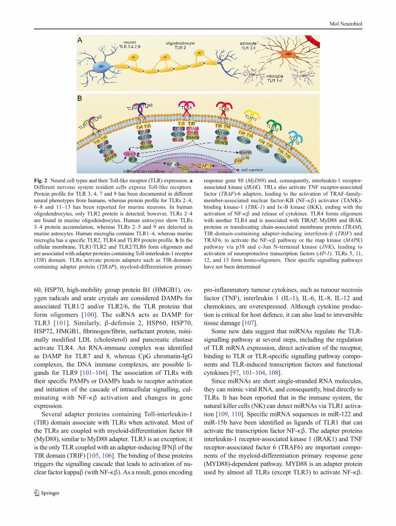
Fig. 2 Neural cell types and their Toll-like receptor (TLR) expression. aDifferent nervous system resident cells express Toll-like receptors.Protein profile for TLR 3, 4, 7 and 9 has been documented in differentneural phenotypes from humans, whereas protein profile for TLRs 2–4,6–8 and 11–13 has been reported for murine neurons. In humanoligodendrocytes, only TLR2 protein is detected; however, TLRs 2–4are found in murine oligodendrocytes. Human astrocytes show TLRs3–4 protein accumulation, whereas TLRs 2–5 and 9 are detected inmurine astrocytes. Human microglia contains TLR1–4, whereas murinemicroglia has a specific TLR2, TLR4 and TLR9 protein profile. b In thecellular membrane, TLR1/TLR2 and TLR2/TLR6 form oligomers andare associated with adapter proteins containing Toll-interleukin-1 receptor(TIR) domain. TLRs activate protein adapters such as TIR-domain-containing adapter protein (TIRAP), myeloid-differentiation primary
response gene 88 (MyD88) and, consequently, interleukin-1 receptor-associated kinase (IRAK). TRLs also activate TNF receptor-associatedfactor (TRAF)-6 adapters, leading to the activation of TRAF-family-member-associated nuclear factor-ΚB (NF-κβ) activator (TANK)-binding kinase-1 (TBK-1) and Iκ-B kinase (IKK), ending with theactivation of NF-κβ and release of cytokines. TLR4 forms oligomerswith another TLR4 and is associated with TIRAP, MyD88 and IRAKproteins or translocating chain-associated membrane protein (TRAM),TIR-domain-containing adapter-inducing interferon-β (TRIF) andTRAF6, to activate the NF-κβ pathway or the map kinase (MAPK)pathway via p38 and c-Jun N-terminal kinase (JNK), leading toactivation of neuroprotective transcription factors (AP-1). TLRs 5, 11,12, and 13 form homo-oligomers. Their specific signalling pathwayshave not been determined
Mol Neurobiol

IRAK1 and TRAF6 are also targets of miR-146. Taganovet al. have suggested that miR-146 downregulates the signal-ling pathway MyD88/NF-κβ after microbial infection [61,111]. miR-155 controls the expression of inhibitor of NF-κβkinase subunits beta (IKKβ) and epsilon (IKK ), reducingNF-κβ activity [112].
However, it has been recently discovered that TLRs 7–9recognise specific miRNAs as agonists in the CNS. For ex-ample, miRNA let-7 is an abundant regulator of gene expres-sion, highly expressed in microglia cells and in neurons,which interacts with TLRs [29]. miRNA-21 and 29a havebeen also described as agonists of TLRs 7–8 in rat and humanmacrophages. The binding of these miRNAs to TLRs inducesthe secretion of TNF-α and IL-6, leading to the activation ofNF-κβ signalling and secretion of pro-inflammatory cyto-kines [113]. Besides secretion of cytokines, the regulation ofTLR signalling by miRNAs occurs at different levels. Variousmolecules involved in the TLR pathway are targeted, such asTLR-signallingmolecules, TLR-induced transcription factors,regulators of the TLR-signalling pathway and the expressionof TLRs themselves [97, 114].
Considering the role of TLRs and assuming that exosomescarry miRNAs, we can hypothesise that miRNAs are signal-ling molecules with important functions in NS diseases(Fig. 3) [115].
miRNAs Activating TLRs in Neurological Diseases
Neurological diseases, including Alzheimer’s disease (AD),Parkinson’s disease (PD), amyotrophic lateral sclerosis (ALS),are characterised by neuronal cell loss. These diseases are ex-pected to become more common due to extended life expec-tancy. Despite significant research efforts, the primary causes ofneurodegeneration remain largely unknown. It has beenrecognised that these disorders emerge as a result of differentgenetic programming and environmental influences [116].
miRNAs have been associated with pathological alter-ations during the course of many neurological diseases, in-cluding AD, PD, ALS and stroke, suggesting that miRNAsmay be a contributing factor in neurodegeneration [116]. It hasbeen recently reported that miRNA levels are altered in theblood of AD, PD, ALS and stroke patients. These small RNAsmay be used as biomarkers to enable an early diagnosis andidentify new therapeutic targets [117].
It is not clear whether inflammation in the CNS contributesto the progress of neurological diseases. However, increasingevidence highlights the participation of TLR-dependent path-ways in neuronal diseases [118]. Neuroinflammation is ob-served as consequences of trauma, infections, tumours andneurodegenerative diseases and involves microglia, pericytesand reactive astrocytes as well as T-lymphocytes,
Fig. 3 Long-distance cell–cell communication: microRNAs (miRNAs)and Toll-like receptors (TLRs). a Neurons and glial cells can releaseexosomes to the extracellular space. These exosomes could shuttleproteins and miRNAs for long distances via the blood vessels or act inthe neighbouring cells. b In both types of cells, miRNAs are previouslyenveloped in exosomes or vesicles in order to be released. c When thevesicles fuse with the cell membrane, their content binds to the endosomeTLRs. TLR3, TLR7, TLR8 and TLR9 oligomerise with the samereceptors. TLRs 7–9 couple with myeloid-differentiation primary
response gene 88 (MyD88), which activates interleukin-1 receptor-associated kinase (IRAK) and TNF receptor-associated factor (TRAF6).These processes culminate in the activation of the nuclear factor-κβ (NF-κβ). The TLR3 is coupled with TIR-domain-containing adapter-inducinginterferon-β (TRIF), which activates TRAF3 and receptor-interactingserine-threonine kinase RIP1 protein, leading to apoptosis. Ourhypothesis is that these TLRs could recognise mature miRNAs,triggering inflammatory signalling under various conditions, includingneurogenesis and diseases
Mol Neurobiol
macrophages and dendritic cells crossing the brain-blood bar-rier, which is damaged in the inflamed brain (reviewed in[119]). Innate immunity providing an onset of the inflamma-tory response involves the actions of TLRs and the liberationof pro-inflammatory cytokines. Short neuroinflammatory re-sponses are considered to be neuroprotective and may contrib-ute neuronal development; however, when persisting they re-sult in neurodegeneration [120]. In this regard, crucial func-tions may be attributed to endogenous miRNAs as ligands ofTLR-promotion of neuroinflammation, as these are responsi-ble for fine-tuning activity levels of TLRs and subsequentkinetics of innate immune response.
In agreement with the hypothesis of a chronicneuroinflammatory process, the involvement of TLR activa-tion has been documented in AD [29, 121], PD [121, 122],ALS [123] and stroke [124]. The analysis of EVs is now anincreasingly popular topic in the field of neurodegeneration;these vesicles may transport pathogenic proteins such asalpha-synuclein (α-syn) and amyloid precursor protein(APP) that are involved in PD and AD, respectively.
Alzheimer’s Disease
AD is the most common cause of dementia in the modernworld [125]. The main characteristics of the disease are theaccumulation of extracellular senile plaques (composed ofamyloid-β peptide, Aβ), intracellular neurofibrillary tangles(NFTs) containing hyperphosphorylated tau protein, activatedmicroglia, astrocytes and degenerating neurons [126].
Several appraisals of AD pathogenesis have revealed thatthe catabolism of APP occurs in the endosome; the pathogenicproteins, such as Aβ and tau, are secreted from the exosomesinto the extracellular space [127–129]. TLR2, 4 and 9 areoverexpressed in an animal model of AD [130]. These recep-tors could be activated by Aβ as they mediate the microglialinflammatory response and are associated with Aβ-plaqueclearance from the brain [131–133].
Studies using blood samples from AD patients have iden-tified 60 miRNAs differentially expressed in these patientsin comparison with healthy individuals [134, 135]. miR-191has a regulatory role in cellular processes such as cell pro-liferation, differentiation, apoptosis and migration; it targetsimportant transcription factors, chromatin remodellers andcell cycle-associated genes [136]. It is likely that thismiRNA is a key player in the initiation and progression ofseveral diseases.
Type III RNase Dicer enzyme is responsible for thematuration of miRNA. Aberrant expression or malfunctionof this regulator in adult forebrain impairs the expression ofseveral miRNAs, ultimately causing pathologicalhyperphosphorylation of NFT-forming tau protein, leadingto neuronal death [137].
The levels of miRNA let-7 are enhanced in AD patients. Ithas been suggested that let-7 activates the RNA-sensingTLR7, and thus, induces neurodegeneration in these patients[29]. The results of experiments with TLR7-KO mice haveshown that these mice are resistant to neurodegenerative fac-tors [29]. It is not clear how the let-7 miRNA reaches theendosome TLR7 receptor in the CNS. However, studies ofthe metastatic gastric cancer have revealed that let-7 miRNAis secreted into the extracellular environment via exosomaltransport [133].
Inflammation has been held responsible for many neuro-logical diseases as it increases cell damage and causes neuro-nal death. Further studies of the receptors associated with the-se processes and molecules triggering the inflammation arenecessary to understand these serious disorders.
Parkinson’s Disease
PD is characterised by a selective degeneration of dopaminer-gic neurons in the substantia nigra pars compacta (SNpc)with various symptoms affecting the motor system such astremor, stiffness, bradykinesia and postural instability [138].The cellular hallmark of PD is the accumulation of proteina-ceous intracellular inclusions termed Lewy bodies (LB), pri-marily composed of fibrillar alpha-synuclein (α-syn) andubiquitinated proteins, in the surviving neurons [139].
The aggregation of α-syn activates microglia, increasingdopaminergic neurotoxicity [140, 141]. However, the precisemolecular mechanism of the process is still unclear. Increasedsecretion of exosomes is one mechanism for α-syn action.These activated exosomes express a high level of major his-tocompatibility complex (MHC) II and TNF-α, which thenpromote apoptosis in the recipient cells [142]. α-syn can alsobe encapsulated in exosomes released by neuroblastoma andcause neuronal cell death [129].
Some cancer studies report that protein-transportingexosomes can also transport miRNAs [117]. The levels ofmiR-205, miR-184 and let-7 are correlated with the expres-sion ofα-syn and leucine-rich repeat kinase2 (LRRK2), codedby the two main genes associated with PD [143]. A recentreport has also indicated that let-7 represses the expressionofα-syn and is downregulated in PDmodels [144]. Increasingevidence suggests the existence of a close relationship be-tween PD and TLRs. It has been recently shown that extracel-lular α-syn increases the expression of TLR1, TLR2, TLR3and TLR7 [145, 146].
Recent studies have described TLR2 as an endogenousreceptor for α-syn that is released from damaged neurons,responsible for microglial activation observed in PD [121].However, TLR4-KO mice are less vulnerable to 1-methyl-4-phenyl-1,2,3,6-tetrahydropyridine (MPTP) intoxication thanwild-type mice. After MPTP administration, these TLR4-
Mol Neurobiol
KO animals also have fewer ionised calcium-binding adaptormolecules 1 (Iba1)+ and MHC II+ activated microglial cellsand lower levels of microglia/macrophage-specific calcium-binding protein. These results suggest that the TLR4 pathwayis involved in PD [108].
The available experimental evidence points to a close rela-tionship between EVs release, miRNA and TLR signalling inPD. However, further studies in this area should be conductedto clarify the specific roles of these molecules in this disease.
Amyotrophic Lateral Sclerosis
ALS is a chronic neurodegenerative disease, characterised byprogressive loss of motor neurons, leading to muscle atrophy,paralysis and death usually within 3 to 5 years after diagnosis[147]. Several studies have demonstrated the involvement ofnon-neuronal cells in ALS pathogenesis, including microgliaand astrocytes, increasing the release of superoxide dismutase1 (SOD1), nitrate and nitrite [148].
SOD1 is secreted via exosomes frommouse motor neuron-like (NSC-34) cells overexpressing the wild-type and a mutantenzyme, used as in vitro model for ALS [149]. It has beendemonstrated that exosomes cargo may include several differ-ent classes of molecules [32]. As we have previously men-tioned, in addition to SOD1, exosomes may transportmiRNAs. Several miRNAs such as miR-146b, miR-29b, let-7a/b, miR-27b, miR-21, miR-210 and miR-155 have theirexpression upregulated in ALS [150, 151]. Furthermore, thelevels of miR-9 are enhanced in this disease in the ventral hornof the spinal cord, the locus of neurodegeneration [152].Among those miRNAs, miR-155, miR-146b and miR-125bare typical components of the innate immune system, andmost of them converge in NFΚB-mediated immune cell re-sponse [151].
The aetiology and pathogenesis of ALS still remain un-clear, although available evidence suggests that inflammationplays a critical role in this process [153]. Studies of high ex-pression of SOD1 in mice have shown elevated levels ofTLR1, 2, 7 and 9 [123]. TLR2 and TLR4 gene expressionlevels are upregulated in ALS patients. TLR2 is predominant-ly detected in the microglia, whereas the TLR4 is stronglyexpressed in astrocytes. The activation of TLRs may contrib-ute to the progression of inflammation and can explain theresultant motor neuron injury in ALS [154]. A study usingcombined inhibitory antibodies against TLR2 and TLR4 hasshown significant microglial suppression [155].
An effective therapy for this disease is still undiscovered.However, the results showing that in ALS patients both, neu-ronal and non-neuronal cells, release EVs concomitantly withthe activation of TLRs add to our knowledge of ALS andimmune responses.
Stroke
Stroke is one of the most common causes of adult disability,and its prevalence augments with ageing population, despitethe advances in prevention and acute interventions [156].Stroke injury mechanisms include the excitotoxicity, mito-chondrial dysfunction, oxidative stress [157] and inflamma-tion [158].
Molecular chaperones and some members of the Bcl-2family (apoptosis regulatory proteins) that protect mitochon-drial function have been suggested as miRNA targets [157].miRNA expression following stroke and other types of hyp-oxia-ischemia/reperfusion injuries varies regionally and tem-porally. The regional distribution of miR-181 and miR-121differs depending on the distribution of blood flow [157].
Altered expression of several miRNAs (miR-140, miR-145and miR-331) has been reported 3 days after ischemia/reperfusion; a progressive increase in the levels of miRNAshas been observed 3 h following reperfusion [159]. miR-200b,miR200c and miR-429 are elevated after 3 h of reperfusion ina model of ischemic preconditioning [160]. In a rat model ofstroke, the levels of miR-290 [161], miR-10a, miR-182, miR-200b and miR-298 [162] increase in the blood and brain 24 hafter ischemia/reperfusion; increased plasma levels of miR-124 are observed 6 h after reperfusion [163]. The level ofmiR-210, known as the major hypoxia-inducible miRNA orhypoxamir [164], is positively correlated with improved prog-nosis in stroke patients [165].
miRNAs are differentially expressed in the blood of pa-tients with acute ischemic stroke; the levels of miR-122,miR-148a, let-7i, miR-19a, miR-320d and miR-4429 de-crease, whereas miR-363 and miR-487b levels increase. The-se miRNAs are predicted to regulate several genes in path-ways previously identified by gene expression analyses, in-cluding TLR signalling and NF-κβ signalling [158]. Severalof these miRNAs have a known biological function. miRNAlet-7 regulates TLR signalling in monocytes and modulatesthe differentiation of dendritic cells [166]. miR-122 regulatesthe expression of peroxiredoxin 2, a DAMP involved in im-mune activation after stroke [167]. miR-148 fine-tunes theimmune response by altering cytokine production (IL6,TNF-a, IL-12, TNFSF7) [162, 168], although their biologicaleffects in neuronal cells are unknown.
Studies focusing on stroke therapies with multipotent mes-enchymal stromal cells (MSCs) have reported that these cellscan release exosomes-containing miR-133b. These exosomesare transferred to the adjacent astrocytes and neurons, wherethey regulate gene expression, with subsequent benefits forneurites remodelling and functional recovery after stroke[169]. However, several studies have indicated the participa-tion of TLRs in stroke [170, 171]. TLR9 gene expression isupregulated in ischemia-neuronal damage and may play acritical role in the induction of inflammatory response and
Mol Neurobiol

apoptosis [172, 173]. Studies using TLR7 and TLR9preconditioned with unmethylated cytosine-phosphate-guanine rich oligonucleotide (CpG) have shown some neuro-protective effects [174].
Other reports reveal a significant increase in TLR8 geneexpression 6 h post-ischemia. The levels of pro-inflammatorycytokines such as IL-6 and IL-1β also change along withTLR8 levels. Treatment with a TLR8 agonist activates pro-apoptotic c-Jun N-terminal kinases (JNK) and increases neu-ronal cell death after stroke [80].
TLR2 and TLR4 gene expression is also upregulated underthe stress or damage conditions such as ischemia or hypoxia[172, 173]. These oligomerised receptors can detect danger-ous proteins like HSP and low molecular weight hyaluronan.HMGB1 and fibrin/fibrinogen are predominantly detected byTLR4 [175]. Studies using LPS for preconditioning havefound that it re-programmes the cellular response (throughactivation of its receptor TLR4), possibly reflecting the endog-enous processes that protect the brain against additional injury[176]. Following a cerebral focal ischemia injury, TLR2- andTLR4-KO mice have smaller infarcts than wild-type animals[177, 178]. miR-19b negatively regulates inflammation inhumans and activates the expression of TLR2 and TLR4,promoting the inflammatory response in ischemic stroke [24,25]. In neonatal hypoxic-ischemic (HI) mice brain, the activa-tion of TLR3 can increase susceptibility to injury [124]. It isnow widely accepted that miRNAs activate TLRs in the im-mune system. However, more studies are needed to determinethe mechanisms of their action in the neuronal cells. We alsoneed to confirm the relationship between EVs and the trans-port of these miRNAs in stroke.
Conclusions and Future Directions
In cancer research, EVs have been considered important bio-markers for the detection of metastases. The informationtransfer by EVs may constitute a novel mechanism of inter-cellular shuttling of molecules related to apoptosis. It is pos-sible that EVs have similar roles in different systems, espe-cially in the nervous system. The recent discovery of the abil-ity of exosomes-containing miRNAs to reach TLRs in theendosomes of surrounding cells offers a new insight into var-ious regulation mechanisms employed under physiologicalcondition and in disease.
Investigation of the possible relationships betweenexosomes, miRNAs and TLRs in the nervous systems is stillin its infancy. However, we can hypothesise that miRNAsentering the cells via exosomes may regulate the activationof TLRs. Furthermore, TLR tolerance, a hyporesponsive stateof the receptor, characterised by reprogramming of TLR-mediated signal transduction [179], may achieved by intracel-lular delivery of miRNA using exosomes. Positive effects
based on TLR tolerance have been observed in an animalmodel of stroke. If this hypothesis is confirmed, it will providea new insight into the regulation of TLRs and new therapeuticstrategies for CNS inflammation-related diseases.
A recent study has demonstrated an effective delivery offunctional siRNA into mouse brain by systemic injection ofexosomes [180]. Systemic exosome administration could bean alternative way to deliver the active components of cell-based therapy to the CNS [181]. Further detailed investigationof cellular communications mediated by EVs holds greatpromise for drug delivery and interference-RNA applications.
Funding This work was supported by Fundação de Amparo à Pesquisado Estado de São Paulo (FAPESP, #2014/16711-6) and UniversidadeFederal do ABC.
References
1. Kihara AH, Santos TO, Osuna-Melo EJ, Paschon V, Vidal KS,Akamine PS, Castro LM, Resende RR, Hamassaki DE, Britto LR(2010) Connexin-mediated communication controls cell prolifer-ation and is essential in retinal histogenesis. Int J Dev Neurosci28(1):39–52
2. Paschon V, Higa GS, Resende RR, Britto LR, Kihara AH (2012)Blocking of connexin-mediated communication promotes neuro-protection during acute degeneration induced by mechanical trau-ma. PLoS One 7(9):e45449
3. Theis M, Sohl G, Eiberger J, Willecke K (2005) Emerging com-plexities in identity and function of glial connexins. TrendsNeurosci 28(4):188–195
4. Sohl G, Maxeiner S, Willecke K (2005) Expression and functionsof neuronal gap junctions. Nat Rev Neurosci 6(3):191–200
5. Corriden R, Insel PA (2010) Basal release of ATP: an autocrine-paracrine mechanism for cell regulation. Sci Signal 3(104):re1
6. Le-Corronc H, Rigo JM, Branchereau P, Legendre P (2011)GABA (A) receptor and glycine receptor activation byparacrine/autocrine release of endogenous agonists: more than asimple communication pathway. Mol Neurobiol 44(1):28–52
7. Ogorevc E, Kralj-Iglic V, Veranic P (2013) The role of extracellu-lar vesicles in phenotypic cancer transformation. Radiol Oncol47(3):197–205
8. Vader P, Breakefield XO, Wood MJ (2014) Extracellular vesicles:emerging targets for cancer therapy. Trends Mol Med 20(7):385–393
9. Gupta A, Pulliam L (2014) Exosomes as mediators of neuroin-flammation. J Neuroinflammation 11(1):68
10. Simons M, Raposo G (2009) Exosomes—vesicular carriers forintercellular communication. Curr Opin Cell Biol 21(4):575–581
11. Gyorgy B, Szabo TG, Pasztoi M, Pal Z, Misjak P, Aradi B, LaszloV, Pallinger E, Pap E, Kittel A et al (2011) Membrane vesicles,current state-of-the-art: emerging role of extracellular vesicles.Cell Mol Life Sci CMLS 68(16):2667–2688
12. Sustar V, Bedina-Zavec A, Stukelj R, Frank M, Ogorevc E, JansaR, Mam K, Veranic P, Kralj-Iglic V (2011) Post-prandial rise ofmicrovesicles in peripheral blood of healthy human donors. LipidsHealth Dis 10:47
13. Xin H, Li Y, Cui Y, Yang JJ, Zhang ZG, Chopp M (2013)Systemic administration of exosomes released frommesenchymalstromal cells promote functional recovery and neurovascular
Mol Neurobiol
plasticity after stroke in rats. J Cereb Blood Flow Metab Off J IntSoc Cereb Blood Flow and Metab 33(11):1711–1715
14. Record M, Carayon K, Poirot M, Silvente-Poirot S (2014)Exosomes as new vesicular lipid transporters involved in cell-cell communication and various pathophysiologies. BiochimBiophys Acta 1841(1):108–120
15. Faure C, Nallet F, Roux D, Milner ST, Gauffre F, Olea D, LambertO (2006) Modeling leakage kinetics from multilamellar vesiclesfor membrane permeability determination: application to glucose.Biophys J 91(12):4340–4349
16. Lachenal G, Pernet-Gallay K, Chivet M, Hemming FJ, Belly A,Bodon G, Blot B, Haase G, Goldberg Y, Sadoul R (2011) Releaseof exosomes from differentiated neurons and its regulation bysynaptic glutamatergic activity. Mol Cell Neurosci 46(2):409–418
17. Yang X, Weng Z, Mendrick DL, Shi Q (2014) Circulating extra-cellular vesicles as a potential source of new biomarkers of drug-induced liver injury. Toxicol Lett 225(3):401–406
18. Cheng L, Sun X, Scicluna BJ, Coleman BM, Hill AF (2013)Characterization and deep sequencing analysis of exosomal andnon-exosomal miRNA in human urine. Kidney Int 86(2):433–444
19. Graves LE, Ariztia EV, Navari JR, Matzel HJ, StackMS, FishmanDA (2004) Proinvasive properties of ovarian cancer ascites-derived membrane vesicles. Cancer Res 64(19):7045–7049
20. Admyre C, Grunewald J, Thyberg J, Gripenback S, Tornling G,Eklund A, Scheynius A, Gabrielsson S (2003) Exosomes withmajor histocompatibility complex class II and co-stimulatory mol-ecules are present in human BAL fluid. Eur Respir J 22(4):578–583
21. Vojtech L,Woo S, Hughes S, Levy C, Ballweber L, Sauteraud RP,Strobl J, Westerberg K, Gottardo R, Tewari M et al (2014)Exosomes in human semen carry a distinctive repertoire of smallnon-coding RNAs with potential regulatory functions. NucleicAcids Res 42(11):7290–7304
22. Melnik BC, John SM, Schmitz G (2014) Milk: an exosomalmicroRNA transmitter promoting thymic regulatory T cell matu-ration preventing the development of atopy? J Transl Med 12(1):43
23. Ogawa Y, Kanai-Azuma M, Akimoto Y, Kawakami H, YanoshitaR (2008) Exosome-like vesicles with dipeptidyl peptidase IV inhuman saliva. Biol Pharm Bull 31(6):1059–1062
24. Fruhbeis C, Frohlich D, Kramer-Albers EM (2012) Emergingroles of exosomes in neuron-glia communication. Front Physiol3:119
25. Von Bartheld CS, Altick AL (2011) Multivesicular bodies in neu-rons: distribution, protein content, and trafficking functions. ProgNeurobiol 93(3):313–340
26. Kramer-Albers EM, Bretz N, Tenzer S, Winterstein C, Mobius W,Berger H, Nave KA, Schild H, Trotter J (2007) Oligodendrocytessecrete exosomes containing major myelin and stress-protectiveproteins: Trophic support for axons? Proteomics Clin Appl1(11):1446–1461
27. Cai J, Han Y, Ren H, Chen C, He D, Zhou L, Eisner GM, AsicoLD, Jose PA, Zeng C (2013) Extracellular vesicle-mediated trans-fer of donor genomic DNA to recipient cells is a novel mechanismfor genetic influence between cells. J Mol Cell Biol 5(4):227–238
28. Valencia K, Luis-Ravelo D, Bovy N, Anton I, Martinez-CanariasS, Zandueta C, Ormazabal C, Struman I, Tabruyn S, Rebmann Vet al (2014) miRNA cargo within exosome-like vesicle transferinfluences metastatic bone colonization. Mol Oncol 8(3):689–703
29. Lehmann SM, Kruger C, Park B, Derkow K, Rosenberger K,Baumgart J, Trimbuch T, Eom G, Hinz M, Kaul D et al (2012)An unconventional role for miRNA: let-7 activates Toll-like re-ceptor 7 and causes neurodegeneration. Nat Neurosci 15(6):827–835
30. Lespagnol A, Duflaut D, Beekman C, Blanc L, Fiucci G, MarineJC, Vidal M, Amson R, Telerman A (2008) Exosome secretion,
including the DNA damage-induced p53-dependent secretorypathway, is severely compromised in TSAP6/Steap3-null mice.Cell Death Differ 15(11):1723–1733
31. Eirin A, Riester SM, Zhu XY, Tang H, Evans JM, O’Brien D, vanWijnen AJ, Lerman LO (2014) MicroRNA and mRNA cargo ofextracellular vesicles from porcine adipose tissue-derived mesen-chymal stem cells. Gene 551(1):55–64
32. Valadi H, Ekstrom K, Bossios A, Sjostrand M, Lee JJ, Lotvall JO(2007) Exosome-mediated transfer of mRNAs and microRNAs isa novel mechanism of genetic exchange between cells. Nat CellBiol 9(6):654–659
33. Pigati L, Yaddanapudi SC, Iyengar R, Kim DJ, Hearn SA,Danforth D, Hastings ML, Duelli DM (2010) Selective releaseof microRNA species from normal and malignant mammary ep-ithelial cells. PLoS One 5(10):e13515
34. Osman A (2012) MicroRNAs in health and disease—basic sci-ence and clinical applications. Clin Lab 58(5–6):393–402
35. Zhang Y, Liao Y, Wang D, He Y, Cao D, Zhang F, Dou K (2011)Altered expression levels of miRNAs in serum as sensitive bio-markers for early diagnosis of traumatic injury. J Cell Biochem112(9):2435–2442
36. Muro EM, Mah N, Andrade-Navarro MA (2011) Functional evi-dence of post-transcriptional regulation by pseudogenes.Biochimie 93(11):1916–1921
37. Hayes CN, Akamatsu S, Tsuge M, Miki D, Akiyama R, Abe H,Ochi H, Hiraga N, Imamura M, Takahashi S et al (2012) HepatitisB virus-specific miRNAs and Argonaute2 play a role in the virallife cycle. PLoS One 7(10):e47490
38. Decembrini S, Bressan D, Vignali R, Pitto L, Mariotti S, RainaldiG, Wang X, Evangelista M, Barsacchi G, Cremisi F (2009)MicroRNAs couple cell fate and developmental timing in retina.Proc Natl Acad Sci U S A 106(50):21179–21184
39. Song J, Hu B, Qu H, Bi C, Huang X, ZhangM (2012)Mechanicalstretch modulates microRNA 21 expression, participating in pro-liferation and apoptosis in cultured human aortic smooth musclecells. PLoS One 7(10):e47657
40. Meunier J, Lemoine F, Soumillon M, Liechti A, Weier M,Guschanski K, Hu H, Khaitovich P, Kaessmann H (2013) Birthand expression evolution of mammalian microRNA genes.Genome Res 23(1):34–45
41. Da Costa Martins PA, De Windt LJ (2012) Targeting microRNAtargets. Circ Res 111(5):506–508
42. Bartel DP (2009) MicroRNAs: target recognition and regulatoryfunctions. Cell 136(2):215–233
43. Voinnet O (2009) Origin, biogenesis, and activity of plantmicroRNAs. Cell 136(4):669–687
44. Krol J, Loedige I, FilipowiczW (2010) The widespread regulationof microRNA biogenesis, function and decay. Nat Rev Genet11(9):597–610
45. Carthew RW, Sontheimer EJ (2009) Origins and mechanisms ofmiRNAs and siRNAs. Cell 136(4):642–655
46. Zinovyev A, Morozova N, Gorban AN, Harel-Belan A (2013)Mathematical modeling of microRNA-mediated mechanisms oftranslation repression. Adv Exp Med Biol 774:189–224
47. Katoh T, Sakaguchi Y, Miyauchi K, Suzuki T, Kashiwabara S,Baba T, Suzuki T (2009) Selective stabilization of mammalianmicroRNAs by 3′ adenylation mediated by the cytoplasmic poly(A) polymerase GLD-2. Genes Dev 23(4):433–438
48. Kai ZS, Pasquinelli AE (2010) MicroRNA assassins: factors thatregulate the disappearance of miRNAs. Nat Struct Mol Biol 17(1):5–10
49. de Sousa E, Walter LT, Higa GS, Casado OA, Kihara AH (2013)Developmental and functional expression of miRNA-stability re-lated genes in the nervous system. PLoS One 8(5):e56908
Mol Neurobiol

50. Kinjo ER, Higa GS, de Sousa E, Casado OA, Damico MV, BrittoLR, Kihara AH (2013) A possible new mechanism for the controlof miRNA expression in neurons. Exp Neurol 248:546–558
51. Valiunas V, Polosina YY, Miller H, Potapova IA, Valiuniene L,Doronin S, Mathias RT, Robinson RB, Rosen MR, Cohen IS et al(2005) Connexin-specific cell-to-cell transfer of short interferingRNA by gap junctions. J Physiol 568(Pt 2):459–468
52. Brink PR, Valiunas V, Gordon C, Rosen MR, Cohen IS (2012)Can gap junctions deliver? Biochim Biophys Acta 1818(8):2076–2081
53. Higa GS, de Sousa E, Walter LT, Kinjo ER, Resende RR, KiharaAH (2014) MicroRNAs in neuronal communication. MolNeurobiol 49(3):1309–1326
54. Stoorvogel W (2012) Functional transfer of microRNA byexosomes. Blood 119(3):646–648
55. Zernecke A, Bidzhekov K, Noels H, Shagdarsuren E, Gan L,Denecke B, Hristov M, Koppel T, Jahantigh MN, Lutgens Eet al (2009) Delivery of microRNA-126 by apoptotic bodies in-duces CXCL12-dependent vascular protection. Sci Signal 2(100):ra81
56. Arroyo JD, Chevillet JR, Kroh EM, Ruf IK, Pritchard CC, GibsonDF,Mitchell PS, Bennett CF, Pogosova-Agadjanyan EL, StirewaltDL et al (2011) Argonaute2 complexes carry a population of cir-culating microRNAs independent of vesicles in human plasma.Proc Natl Acad Sci U S A 108(12):5003–5008
57. Kosaka N, Iguchi H, Yoshioka Y, Takeshita F, Matsuki Y, OchiyaT (2010) Secretory mechanisms and intercellular transfer ofmicroRNAs in living cells. J Biol Chem 285(23):17442–17452
58. Ksiazek-Winiarek DJ, Kacperska MJ, Glabinski A (2013)MicroRNAs as novel regulators of neuroinflammation.Mediators Inflamm 2013:172351
59. Gantier MP (2010) New perspectives in MicroRNA regulation ofinnate immunity. J Interf Cytokine Res off J Int Soc InterferonCytokine Res 30(5):283–289
60. Quinn SR, O’Neill LA (2011) A trio of microRNAs that controlToll-like receptor signalling. Int Immunol 23(7):421–425
61. Taganov KD, Boldin MP, Chang KJ, Baltimore D (2006) NF-kappaB-dependent induction of microRNAmiR-146, an inhibitortargeted to signaling proteins of innate immune responses. ProcNatl Acad Sci U S A 103(33):12481–12486
62. Cui JG, Li YY, Zhao Y, Bhattacharjee S, Lukiw WJ (2010)Differential regulation of interleukin-1 receptor-associatedkinase-1 (IRAK-1) and IRAK-2 by microRNA-146a and NF-kappaB in stressed human astroglial cells and in Alzheimer dis-ease. J Biol Chem 285(50):38951–38960
63. Medzhitov R (2001) Toll-like receptors and innate immunity. NatRev Immunol 1(2):135–145
64. Takeda K, Akira S (2004) TLR signaling pathways. SeminImmunol 16(1):3–9
65. Miyake K (2007) Innate immune sensing of pathogens and dangersignals by cell surface Toll-like receptors. Semin Immunol 19(1):3–10
66. Hashimoto C, Hudson KL, Anderson KV (1988) The Toll gene ofDrosophila, required for dorsal-ventral embryonic polarity, ap-pears to encode a transmembrane protein. Cell 52(2):269–279
67. Blasius AL, Beutler B (2010) Intracellular toll-like receptors.Immunity 32(3):305–315
68. Kawai T, Akira S (2010) The role of pattern-recognition receptorsin innate immunity: update on Toll-like receptors. Nat Immunol11(5):373–384
69. Benias PC, Gopal K, Bodenheimer H Jr, Theise ND (2012)Hepatic expression of toll-like receptors 3, 4, and 9 in primarybiliary cirrhosis and chronic hepatitis C. Clinics Res HepatolGastroenterol 36(5):448–454
70. Gerondakis S, Grumont RJ, Banerjee A (2007) Regulating B-cellactivation and survival in response to TLR signals. Immunol CellBiol 85(6):471–475
71. Iwamura C, Nakayama T (2008) Toll-like receptors in the respi-ratory system: their roles in inflammation. Curr Allergy AsthmaRep 8(1):7–13
72. ErikssonM, Meadows SK, Basu S,Mselle TF, Wira CR, SentmanCL (2006) TLRs mediate IFN-gamma production by human uter-ine NK cells in endometrium. J Immunol 176(10):6219–6224
73. Duan X, Kang E, Liu CY, Ming GL, Song H (2008) Developmentof neural stem cell in the adult brain. Curr Opin Neurobiol 18(1):108–115
74. Sabroe I, Whyte MK (2007) Toll-like receptor (TLR)-based net-works regulate neutrophilic inflammation in respiratory disease.Biochem Soc Trans 35(Pt 6):1492–1495
75. Yoshimoto T, Nakanishi K (2006) Roles of IL-18 in basophils andmast cells. Allergol Int Off J Jpn Soc Allergol 55(2):105–113
76. Gibson FC 3rd, Ukai T, Genco CA (2008) Engagement of specificinnate immune signaling pathways during Porphyromonasgingivalis induced chronic inflammation and atherosclerosis.Front Biosci J Virtual libr 13:2041–2059
77. Prehaud C, Megret F, Lafage M, Lafon M (2005) Virus infectionswitches TLR-3-positive human neurons to become strong pro-ducers of beta interferon. J Virol 79(20):12893–12904
78. Qi J, Buzas K, Fan H, Cohen JI, Wang K, Mont E, Klinman D,Oppenheim JJ, Howard OM (2011) Painful pathways induced byTLR stimulation of dorsal root ganglion neurons. J Immunol186(11):6417–6426
79. Mishra BB, Gundra UM, Teale JM (2008) Expression and distri-bution of Toll-like receptors 11–13 in the brain during murineneurocysticercosis. J Neuroinflammation 5:53
80. Tang SC, Arumugam TV, Xu X, Cheng A, Mughal MR, Jo DG,Lathia JD, Siler DA, Chigurupati S, Ouyang X et al (2007) Pivotalrole for neuronal Toll-like receptors in ischemic brain injury andfunctional deficits. Proc Natl Acad Sci U S A 104(34):13798–13803
81. Barajon I, Serrao G, Arnaboldi F, Opizzi E, Ripamonti G, BalsariA, Rumio C (2009) Toll-like receptors 3, 4, and 7 are expressed inthe enteric nervous system and dorsal root ganglia. J HistochemCytochem Off J Histochem Soc 57(11):1013–1023
82. Sloane JA, Batt C, Ma Y, Harris ZM, Trapp B, Vartanian T (2010)Hyaluronan blocks oligodendrocyte progenitor maturation andremyelination through TLR2. Proc Natl Acad Sci U S A107(25):11555–11560
83. Lehnardt S, Henneke P, Lien E, Kasper DL, Volpe JJ, Bechmann I,Nitsch R, Weber JR, Golenbock DT, Vartanian T (2006) A mech-anism for neurodegeneration induced by group B streptococcithrough activation of the TLR2/MyD88 pathway in microglia. JImmunol 177(1):583–592
84. Taylor DL, Pirianov G, Holland S, McGinnity CJ, Norman AL,Reali C, Diemel LT, Gveric D, Yeung D, Mehmet H (2010)Attenuation of proliferation in oligodendrocyte precursor cellsby activated microglia. J Neurosci Res 88(8):1632–1644
85. Bsibsi M, Nomden A, van Noort JM, Baron W (2012) Toll-likereceptors 2 and 3 agonists differentially affect oligodendrocytesurvival, differentiation, and myelin membrane formation. JNeurosci Res 90(2):388–398
86. Jack CS, Arbour N, Manusow J, Montgrain V, Blain M, McCreaE, Shapiro A, Antel JP (2005) TLR signaling tailors innate im-mune responses in human microglia and astrocytes. J Immunol175(7):4320–4330
87. Bsibsi M, Ravid R, Gveric D, van Noort JM (2002) Broad expres-sion of Toll-like receptors in the human central nervous system. JNeuropathol Exp Neurol 61(11):1013–1021
88. Carpentier PA, Begolka WS, Olson JK, Elhofy A, Karpus WJ,Miller SD (2005) Differential activation of astrocytes by innateand adaptive immune stimuli. Glia 49(3):360–374
89. El-Hage N, Podhaizer EM, Sturgill J, Hauser KF (2011) Toll-likereceptor expression and activation in astroglia: differential
Mol Neurobiol

regulation by HIV-1 Tat, gp120, and morphine. Immunol Invest40(5):498–522
90. Cassiani-Ingoni R, Cabral ES, Lunemann JD, Garza Z, Magnus T,Gelderblom H, Munson PJ, Marques A, Martin R (2006) Borreliaburgdorferi induces TLR1 and TLR2 in human microglia andperipheral blood monocytes but differentially regulates HLA-class II expression. J Neuropathol Exp Neurol 65(6):540–548
91. Lehnardt S, Lehmann S, Kaul D, Tschimmel K, Hoffmann O, ChoS, Krueger C, Nitsch R, Meisel A, Weber JR (2007) Toll-likereceptor 2 mediates CNS injury in focal cerebral ischemia. JNeuroimmunol 190(1–2):28–33
92. Yoon HJ, Jeon SB, Kim IH, Park EJ (2008) Regulation of TLR2expression by prostaglandins in brain glia. J Immunol 180(12):8400–8409
93. Suzuki M, Sugimoto Y, Ohsaki Y, Ueno M, Kato S, Kitamura Y,Hosokawa H, Davies JP, Ioannou YA, Vanier MT et al (2007)Endosomal accumulation of Toll-like receptor 4 causes constitu-tive secretion of cytokines and activation of signal transducers andactivators of transcription in Niemann-Pick disease type C (NPC)fibroblasts: a potential basis for glial cell activation in the NPCbrain. J Neurosci the Off J Soc Neurosci 27(8):1879–1891
94. Du M, Butchi NB, Woods T, Peterson KE (2011) Poly-thymidineoligonucleotides mediate activation of murine glial cells primarilythrough TLR7, not TLR8. PLoS One 6(7):e22454
95. Glezer I, Simard AR, Rivest S (2007) Neuroprotective role of theinnate immune system by microglia. Neuroscience 147(4):867–883
96. McCusker RH, Kelley KW (2013) Immune-neural connections:how the immune system’s response to infectious agents influencesbehavior. J Exp Biol 216(Pt 1):84–98
97. Olivieri F, Rippo MR, Prattichizzo F, Babini L, Graciotti L,Recchioni R, Procopio AD (2013) Toll like receptor signaling in“inflammaging”: microRNA as new players. Immun Ageing I A10(1):11
98. Tsai SY, Segovia JA, Chang TH, Morris IR, Berton MT, TessierPA, Tardif MR, Cesaro A, Bose S (2014) DAMP moleculeS100A9 acts as a molecular pattern to enhance inflammation dur-ing influenza A virus infection: role of DDX21-TRIF-TLR4-MyD88 pathway. PLoS Pathog 10(1):e1003848
99. Fang H, Ang B, Xu X, Huang X, Wu Y, Sun Y, Wang W, Li N,Cao X, Wan T (2014) TLR4 is essential for dendritic cell activa-tion and anti-tumor T-cell response enhancement by DAMPs re-leased from chemically stressed cancer cells. Cell Mol Immunol11(2):150–159
100. Peng Y, Zhang L (2014) Activation of the TLR1/2 pathway in-duces the shaping of the immune response status of peripheralblood leukocytes. Exp Ther Med 7(6):1708–1712
101. Marsh BJ,Williams-Karnesky RL, Stenzel-PooreMP (2009) Toll-like receptor signaling in endogenous neuroprotection and stroke.Neuroscience 158(3):1007–1020
102. Kaczorowski DJ, Mollen KP, Edmonds R, Billiar TR (2008) Earlyevents in the recognition of danger signals after tissue injury. JLeukoc Biol 83(3):546–552
103. Marsh BJ, Stevens SL, Hunter B, Stenzel-Poore MP (2009)Inflammation and the emerging role of the toll-like receptor sys-tem in acute brain ischemia. Stroke J Cereb Circ 40(3 Suppl):S34–S37
104. Chotirmall SH, Low TB, Hassan T, Branagan P, Kernekamp C,FlynnMG, GunaratnamC,McElvaney NG (2011) Cystic fibrosis,common variable immunodeficiency and Aspergers syndrome: animmunological and behavioural challenge. Ir J Med Sci 180(2):607–609
105. O’Neill LA, Bowie AG (2007) The family of five: TIR-domain-containing adaptors in Toll-like receptor signalling. Nat RevImmunol 7(5):353–364
106. Nada M, Ohnishi H, Tochio H, Kato Z, Kimura T, Kubota K,Yamamoto T, Kamatari YO, Tsutsumi N, Shirakawa M et al(2012) Molecular analys is of the binding mode ofToll/interleukin-1 receptor (TIR) domain proteins during TLR2signaling. Mol Immunol 52(3–4):108–116
107. Ko MK, Saraswathy S, Parikh JG, Rao NA (2011) The role ofTLR4 activation in photoreceptor mitochondrial oxidative stress.Invest Ophthalmol Vis Sci 52(8):5824–5835
108. He X, Jing Z, Cheng G (2014) MicroRNAs: new regulators ofToll-like receptor signalling pathways. Biomed Res Int 2014:945169
109. Fehniger TA (2013) Extracellular microRNAs turn on NK cellsvia TLR1. Blood 121(23):4612–4613
110. He S, Chu J, Wu LC, Mao H, Peng Y, Alvarez-Breckenridge CA,Hughes T, Wei M, Zhang J, Yuan S et al (2013) MicroRNAsactivate natural killer cells through Toll-like receptor signaling.Blood 121(23):4663–4671
111. Nahid MA, Satoh M, Chan EK (2011) Mechanistic role ofmicroRNA-146a in endotoxin-induced differential cross-regulation of TLR signaling. J Immunol 186(3):1723–1734
112. Ceppi M, Pereira PM, Dunand-Sauthier I, Barras E, Reith W,Santos MA, Pierre P (2009) MicroRNA-155 modulates theinterleukin-1 signaling pathway in activated human monocyte-derived dendritic cells. Proc Natl Acad Sci U S A 106(8):2735–2740
113. Fabbri M, Paone A, Calore F, Galli R, Gaudio E, Santhanam R,Lovat F, Fadda P, Mao C, Nuovo GJ et al (2012)MicroRNAs bindto Toll-like receptors to induce prometastatic inflammatory re-sponse. Proc Natl Acad Sci U S A 109(31):E2110–E2116
114. Li Y, Shi X (2013)MicroRNAs in the regulation of TLR and RIG-I pathways. Cell Mol Immunol 10(1):65–71
115. Fabbri M, Paone A, Calore F, Galli R, Croce CM (2013) A newrole for microRNAs, as ligands of Toll-like receptors. RNA Biol10(2):169–174
116. Tan JY, Marques AC (2014) The miRNA-mediated cross-talk be-tween transcripts provides a novel layer of posttranscriptional reg-ulation. Adv Genet 85:149–199
117. Grasso M, Piscopo P, Confaloni A, Denti MA (2014) CirculatingmiRNAs as biomarkers for neurodegenerative disorders.Molecules 19(5):6891–6910
118. Drouin-Ouellet J, Cicchetti F (2012) Inflammation and neurode-generation: the story ‘retolled’. Trends Pharmacol Sci 33(10):542–551
119. Ulrich H, do Nascimento IC, Bocsi J, Tarnok A (2014)Immunomodulation in stem cell differentiation into neurons andbrain repair. Stem Cell Rev (in press)
120. Shastri A, Bonifati DM, Kishore U (2013) Innate immunity andneuroinflammation. Mediators Inflamm 2013:342931
121. Hayward CP, Moffat KA, Graf L (2014) Technological advancesin diagnostic testing for von Willebrand disease: new approachesand challenges. Int J Lab Hematol 36(3):334–340
122. Noelker C, Morel L, Lescot T, Osterloh A, Alvarez-Fischer D,Breloer M, Henze C, Depboylu C, Skrzydelski D, Michel PPet al (2013) Toll like receptor 4 mediates cell death in a mouseMPTP model of Parkinson disease. Sci Rep 3:1393
123. Letiembre M, Liu Y, Walter S, Hao W, Pfander T, Wrede A,Schulz-Schaeffer W, Fassbender K (2009) Screening of innateimmune receptors in neurodegenerative diseases: a similar pattern.Neurobiol Aging 30(5):759–768
124. Stridh L, Mottahedin A, Johansson ME, Valdez RC, NorthingtonF, Wang X, Mallard C (2013) Toll-like receptor-3 activation in-creases the vulnerability of the neonatal brain to hypoxia-ische-mia. J Neurosci Off J Soc Neurosci 33(29):12041–12051
125. Guo L, Duggan J, Cordeiro MF (2010) Alzheimer’s disease andretinal neurodegeneration. Curr Alzheimer Res 7(1):3–14
Mol Neurobiol
126. Querfurth HW, LaFerla FM (2010) Alzheimer’s disease. N Engl JMed 362(4):329–344
127. Rajendran L, Honsho M, Zahn TR, Keller P, Geiger KD, VerkadeP, SimonsK (2006) Alzheimer’s disease beta-amyloid peptides arereleased in association with exosomes. Proc Natl Acad Sci U S A103(30):11172–11177
128. Vingtdeux V, Hamdane M, Loyens A, Gele P, Drobeck H, BegardS, Galas MC, Delacourte A, Beauvillain JC, Buee L et al (2007)Alkalizing drugs induce accumulation of amyloid precursor pro-tein by-products in luminal vesicles of multivesicular bodies. JBiol Chem 282(25):18197–18205
129. Emmanouilidou E, Melachroinou K, Roumeliotis T, Garbis SD,Ntzouni M, Margaritis LH, Stefanis L, Vekrellis K (2010) Cell-produced alpha-synuclein is secreted in a calcium-dependent man-ner by exosomes and impacts neuronal survival. J Neurosci30(20):6838–6851
130. Frank S, Copanaki E, Burbach GJ, Muller UC, Deller T (2009)Differential regulation of toll-like receptor mRNAs in amyloidplaque-associated brain tissue of aged APP23 transgenic mice.Neurosci Lett 453(1):41–44
131. Richard KL, Filali M, Prefontaine P, Rivest S (2008) Toll-likereceptor 2 acts as a natural innate immune receptor to clear amy-loid beta 1–42 and delay the cognitive decline in a mousemodel ofAlzheimer’s disease. J Neurosci 28(22):5784–5793
132. Yu X, Wang Q, Lin Y, Zhao J, Zhao C, Zheng J (2012) Structure,orientation, and surface interaction of Alzheimer amyloid-betapeptides on the graphite. Langmuir ACS J Surf colloid 28(16):6595–6605
133. Ohshima K, Inoue K, Fujiwara A, Hatakeyama K, Kanto K,Watanabe Y, Muramatsu K, Fukuda Y, Ogura S, Yamaguchi Ket al (2010) Let-7microRNA family is selectively secreted into theextracellular environment via exosomes in a metastatic gastriccancer cell line. PLoS One 5(10):e13247
134. Cogswell JP, Ward J, Taylor IA, Waters M, Shi Y, Cannon B,Kelnar K, Kemppainen J, Brown D, Chen C et al (2008)Identification of miRNA changes in Alzheimer’s disease brainand CSF yields putative biomarkers and insights into disease path-ways. J Alzheimer’s Dis JAD 14(1):27–41
135. Kumar P, Dezso Z, MacKenzie C, Oestreicher J, Agoulnik S,Byrne M, Bernier F, Yanagimachi M, Aoshima K, Oda Y (2013)Circulating miRNA biomarkers for Alzheimer’s disease. PLoSOne 8(7):e69807
136. Nagpal N, Kulshreshtha R (2014) miR-191: an emerging player indisease biology. Front Genet 5:99
137. Hebert SS, Papadopoulou AS, Smith P, Galas MC, Planel E,Silahtaroglu AN, Sergeant N, Buee L, De Strooper B (2010)Genetic ablation of Dicer in adult forebrain neurons results inabnormal tau hyperphosphorylation and neurodegeneration.Hum Mol Genet 19(20):3959–3969
138. Lang AE, Lozano AM (1998) Parkinson’s disease. Second of twoparts. N Engl J Med 339(16):1130–1143
139. Lees AJ (2009) The Parkinson chimera. Neurology 72(7 Suppl):S2–S11
140. Reynolds AD, Kadiu I, Garg SK, Glanzer JG, Nordgren T,Ciborowski P, Banerjee R, Gendelman HE (2008) Nitratedalpha-synuclein and microglial neuroregulatory activities. JNeuroimmune Pharmacol Off J Soc NeuroImmune Pharmacol3(2):59–74
141. Reynolds AD, Glanzer JG, Kadiu I, Ricardo-Dukelow M,Chaudhuri A, Ciborowski P, Cerny R, Gelman B, Thomas MP,Mosley RL et al (2008) Nitrated alpha-synuclein-activatedmicroglial profiling for Parkinson’s disease. J Neurochem104(6):1504–1525
142. Chang C, Lang H, Geng N, Wang J, Li N, Wang X (2013)Exosomes of BV-2 cells induced by alpha-synuclein: importantmediator of neurodegeneration in PD. Neurosci Lett 548:190–195
143. Maciotta S, Meregalli M, Torrente Y (2013) The involvement ofmicroRNAs in neurodegenerative diseases. Front Cell Neurosci 7:265
144. Junn E, Lee KW, Jeong BS, Chan TW, Im JY, Mouradian MM(2009) Repression of alpha-synuclein expression and toxicity bymicroRNA-7. Proc Natl Acad Sci U S A 106(31):13052–13057
145. Beraud D, TwomeyM, Bloom B,Mittereder A, Ton V, Neitzke K,Chasovskikh S, Mhyre TR, Maguire-zeiss KA (2011) Alpha-synuclein alters toll-like receptor expression. Front Neurosci 5:80
146. Beraud E, Chandy KG (2011) Therapeutic potential of peptidetoxins that target ion channels. Inflamm Allergy Drug Targets10(5):322–342
147. Lomen-Hoerth C (2008) Amyotrophic lateral sclerosis from benchto bedside. Semin Neurol 28(2):205–211
148. Beers DR, Henkel JS, Xiao Q, Zhao W, Wang J, Yen AA, SiklosL, McKercher SR, Appel SH (2006) Wild-type microglia extendsurvival in PU.1 knockout mice with familial amyotrophic lateralsclerosis. Proc Natl Acad Sci U S A 103(43):16021–16026
149. Gomes C, Keller S, Altevogt P, Costa J (2007) Evidence for se-cretion of Cu, Zn superoxide dismutase via exosomes from a cellmodel of amyotrophic lateral sclerosis. Neurosci Lett 428(1):43–46
150. Koval ED, Shaner C, Zhang P, du Maine X, Fischer K, Tay J,Chau BN, Wu GF, Miller TM (2013) Method for widespreadmicroRNA-155 inhibition prolongs survival in ALS-model mice.Hum Mol Genet 22(20):4127–4135
151. Parisi C, Arisi I, D’Ambrosi N, Storti AE, Brandi R, D’OnofrioM,Volonte C (2013) DysregulatedmicroRNAs in amyotrophic lateralsclerosis microglia modulate genes linked to neuroinflammation.Cell Death Dis 4:e959
152. Zhou F, Guan Y, Chen Y, Zhang C, Yu L, Gao H, Du H, Liu B,Wang X (2013) miRNA-9 expression is upregulated in the spinalcord of G93A-SOD1 transgenic mice. Int J Clin Exp Pathol 6(9):1826–1838
153. Sta M, Sylva-Steenland RM, Casula M, de Jong JM, Troost D,Aronica E, Baas F (2011) Innate and adaptive immunity in amyo-trophic lateral sclerosis: evidence of complement activation.Neurobiol Dis 42(3):211–220
154. Casula M, Iyer AM, Spliet WG, Anink JJ, Steentjes K, Sta M,Troost D, Aronica E (2011) Toll-like receptor signaling in amyo-trophic lateral sclerosis spinal cord tissue. Neuroscience 179:233–243
155. Zhao W, Beers DR, Henkel JS, Zhang W, Urushitani M, Julien JP,Appel SH (2010) Extracellular mutant SOD1 induces microglial-mediated motoneuron injury. Glia 58(2):231–243
156. Teasell R, Hussein N, McClure A, Meyer M (2014) Stroke: morethan a ‘brain attack’. Int J Stroke Off J Int Stroke Soc 9(2):188–190
157. Ouyang Q, Zhou W, Zhang CM (2007) The key of increasing thetherapeutic effect of scalp acupuncture on hemiplegia due tostroke. Zhongguo zhen jiu Chin Acupunct Moxibustion 27(10):773–776
158. Jickling GC, Ander BP, Zhan X, Noblett D, Stamova B, Liu D(2014) microRNA expression in peripheral blood cells followingacute ischemic stroke and their predicted gene targets. PLoS One9(6):e99283
159. Dharap A, Bowen K, Place R, Li LC, Vemuganti R (2009)Transient focal ischemia induces extensive temporal changes inrat cerebral microRNAome. J Cereb blood flow Metab Off J IntSoc Cereb Blood Flow Metab 29(4):675–687
160. Lee ST, Chu K, Jung KH, Yoon HJ, Jeon D, Kang KM, Park KH,Bae EK, KimM, Lee SK et al (2010) MicroRNAs induced duringischemic preconditioning. Stroke J Cereb Circulation 41(8):1646–1651
161. Jeyaseelan K, Lim KY, Armugam A (2008) MicroRNA expres-sion in the blood and brain of rats subjected to transient focal
Mol Neurobiol
ischemia by middle cerebral artery occlusion. Stroke J CerebCirculation 39(3):959–966
162. Liu DZ, Tian Y, Ander BP, Xu H, Stamova BS, Zhan X, TurnerRJ, Jickling G, Sharp FR (2010) Brain and blood microRNAexpression profiling of ischemic stroke, intracerebral hemorrhage,and kainate seizures. J Cereb Blood Flow Metab 30(1):92–101
163. Weng Q, Zhang J, Cao J, Xia Q, Wang D, Hu Y, Sheng R, Wu H,Zhu D, Zhu H et al (2011) Q39, a quinoxaline 1,4-Di-N-oxidederivative, inhibits hypoxia-inducible factor-1alpha expressionand the Akt/mTOR/4E-BP1 signaling pathway in human hepato-ma cells. Invest New Drugs 29(6):1177–1187
164. Chan YC, Banerjee J, Choi SY, Sen CK (2012) miR-210: themaster hypoxamir. Microcirculation 19(3):215–223
165. Zeng J, Sun Y, Jiang L (2011) On-line ‘automatic pilot’ trainingfor hand and arm motor rehabilitation after stroke. MedHypotheses 76(2):197–198
166. Zhang W, Winder T, Ning Y, Pohl A, Yang D, Kahn M, Lurje G,Labonte MJ, Wilson PM, Gordon MA et al (2011) A let-7microRNA-binding site polymorphism in 3′-untranslated regionof KRAS gene predicts response in wild-type KRAS patients withmetastatic colorectal cancer treated with cetuximab monotherapy.Ann Oncol Off J Eur Soc Med Oncol ESMO 22(1):104–109
167. Diao S, Zhang JF, Wang H, He ML, Lin MC, Chen Y, Kung HF(2010) Proteomic identification ofmicroRNA-122a target proteinsin hepatocellular carcinoma. Proteomics 10(20):3723–3731
168. Pan W, Zhu S, Yuan M, Cui H, Wang L, Luo X, Li J, Zhou H,Tang Y, Shen N (2010) MicroRNA-21 and microRNA-148a con-tribute to DNA hypomethylation in lupus CD4+ Tcells by directlyand indirectly targeting DNA methyltransferase 1. J Immunol184(12):6773–6781
169. Li Y, Liu Z, Xin H, Chopp M (2014) The role of astrocytes inmediating exogenous cell-based restorative therapy for stroke.Glia 62(1):1–16
170. Fadakar K, Dadkhahfar S, Esmaeili A, Rezaei N (2014) The roleof Toll-like receptors (TLRs) in stroke. Rev Neurosci 25(5):699–712
171. Gesuete R, Kohama SG, Stenzel-Poore MP (2014) Toll-like re-ceptors and ischemic brain injury. J Neuropathol Exp Neurol73(5):378–386
172. Arumugam TV, Okun E, Tang SC, Thundyil J, Taylor SM,Woodruff TM (2009) Toll-like receptors in ischemia-reperfusioninjury. Shock 32(1):4–16
173. Mkaddem SB, Bens M, Vandewalle A (2010) Differential activa-tion of Toll-like receptor-mediated apoptosis induced by hypoxia.Oncotarget 1(8):741–750
174. Leung PY, Stevens SL, Packard AE, Lessov NS, Yang T, ConradVK, van den Dungen NN, Simon RP, Stenzel-Poore MP (2012)Toll-like receptor 7 preconditioning induces robust neuroprotec-tion against stroke by a novel type I interferon-mediated mecha-nism. Stroke J Cereb Circ 43(5):1383–1389
175. Smiley ST, King JA, Hancock WW (2001) Fibrinogen stimulatesmacrophage chemokine secretion through toll-like receptor 4. JImmunol 167(5):2887–2894
176. Wang YC, Lin S, Yang QW (2011) Toll-like receptors in cerebralischemic inflammatory injury. J Neuroinflammation 8:134
177. Cao CX, YangQW, Lv FL, Cui J, FuHB,Wang JZ (2007) Reducedcerebral ischemia-reperfusion injury in Toll-like receptor 4 deficientmice. Biochem Biophys Res Commun 353(2):509–514
178. Ziegler G, Harhausen D, Schepers C, Hoffmann O, Rohr C, PrinzV, Konig J, LehrachH,NietfeldW, Trendelenburg G (2007) TLR2has a detrimental role in mouse transient focal cerebral ischemia.Biochem Biophys Res Commun 359(3):574–579
179. Vartanian K, Stenzel-Poore M (2010) Toll-like receptor toleranceas a mechanism for neuroprotection. Transl Stroke Res 1(4):252–260
180. Cooper JM, Wiklander PB, Nordin JZ, Al-Shawi R, Wood MJ,Vithlani M, Schapira AH, Simons JP, El-Andaloussi S, Alvarez-Erviti L (2014) Systemic exosomal siRNA delivery reducedalpha-synuclein aggregates in brains of transgenic mice. MovDisord 29(12):1476–1485
181. Kawikova I, Askenase PW (2014) Diagnostic and therapeutic po-tentials of exosomes in CNS diseases. Brain Res S0006-8993(14)01343–2
Mol Neurobiol
Related Documents